EP0163243A2 - Improved human T or B cell lines and a process for producing immunologically active substances using the same - Google Patents
Improved human T or B cell lines and a process for producing immunologically active substances using the same Download PDFInfo
- Publication number
- EP0163243A2 EP0163243A2 EP85106221A EP85106221A EP0163243A2 EP 0163243 A2 EP0163243 A2 EP 0163243A2 EP 85106221 A EP85106221 A EP 85106221A EP 85106221 A EP85106221 A EP 85106221A EP 0163243 A2 EP0163243 A2 EP 0163243A2
- Authority
- EP
- European Patent Office
- Prior art keywords
- cells
- human
- malignant
- immunologically active
- group
- Prior art date
- Legal status (The legal status is an assumption and is not a legal conclusion. Google has not performed a legal analysis and makes no representation as to the accuracy of the status listed.)
- Withdrawn
Links
- 210000001744 T-lymphocyte Anatomy 0.000 title claims abstract description 51
- 210000003719 b-lymphocyte Anatomy 0.000 title claims abstract description 35
- 239000013543 active substance Substances 0.000 title claims abstract description 31
- 238000000034 method Methods 0.000 title claims abstract description 29
- 210000004027 cell Anatomy 0.000 claims abstract description 110
- 238000012258 culturing Methods 0.000 claims abstract description 17
- 108090000623 proteins and genes Proteins 0.000 claims abstract description 13
- 239000012634 fragment Substances 0.000 claims abstract description 11
- 238000004519 manufacturing process Methods 0.000 claims abstract description 9
- 241000598171 Human adenovirus sp. Species 0.000 claims abstract description 8
- 230000012010 growth Effects 0.000 claims abstract description 3
- 239000000126 substance Substances 0.000 claims abstract 3
- 239000001963 growth medium Substances 0.000 claims abstract 2
- 230000002269 spontaneous effect Effects 0.000 claims abstract 2
- 241000701161 unidentified adenovirus Species 0.000 claims abstract 2
- 230000003211 malignant effect Effects 0.000 claims description 17
- 108010002350 Interleukin-2 Proteins 0.000 claims description 12
- 102000000588 Interleukin-2 Human genes 0.000 claims description 12
- 102000003996 Interferon-beta Human genes 0.000 claims description 3
- 108090000467 Interferon-beta Proteins 0.000 claims description 3
- 108010074328 Interferon-gamma Proteins 0.000 claims description 3
- 102000008070 Interferon-gamma Human genes 0.000 claims description 3
- 102000000589 Interleukin-1 Human genes 0.000 claims description 3
- 108010002352 Interleukin-1 Proteins 0.000 claims description 3
- 206010035226 Plasma cell myeloma Diseases 0.000 claims description 3
- 230000010261 cell growth Effects 0.000 claims description 3
- 239000003102 growth factor Substances 0.000 claims description 3
- 201000000050 myeloid neoplasm Diseases 0.000 claims description 3
- 229940044627 gamma-interferon Drugs 0.000 claims 2
- 108091003079 Bovine Serum Albumin Proteins 0.000 description 22
- 239000012091 fetal bovine serum Substances 0.000 description 22
- 239000002609 medium Substances 0.000 description 21
- 239000012980 RPMI-1640 medium Substances 0.000 description 20
- CURLTUGMZLYLDI-UHFFFAOYSA-N Carbon dioxide Chemical compound O=C=O CURLTUGMZLYLDI-UHFFFAOYSA-N 0.000 description 15
- 229910002092 carbon dioxide Inorganic materials 0.000 description 15
- LRFVTYWOQMYALW-UHFFFAOYSA-N 9H-xanthine Chemical compound O=C1NC(=O)NC2=C1NC=N2 LRFVTYWOQMYALW-UHFFFAOYSA-N 0.000 description 13
- 230000000694 effects Effects 0.000 description 13
- 239000000203 mixture Substances 0.000 description 13
- 108020004414 DNA Proteins 0.000 description 11
- 238000011534 incubation Methods 0.000 description 10
- 239000000872 buffer Substances 0.000 description 8
- 239000000725 suspension Substances 0.000 description 8
- 206010028980 Neoplasm Diseases 0.000 description 7
- 201000011510 cancer Diseases 0.000 description 7
- 239000001569 carbon dioxide Substances 0.000 description 7
- 239000000243 solution Substances 0.000 description 7
- 101001002657 Homo sapiens Interleukin-2 Proteins 0.000 description 6
- 239000000411 inducer Substances 0.000 description 6
- HPNSFSBZBAHARI-UHFFFAOYSA-N micophenolic acid Natural products OC1=C(CC=C(C)CCC(O)=O)C(OC)=C(C)C2=C1C(=O)OC2 HPNSFSBZBAHARI-UHFFFAOYSA-N 0.000 description 6
- 229940075420 xanthine Drugs 0.000 description 6
- RQFCJASXJCIDSX-UHFFFAOYSA-N 14C-Guanosin-5'-monophosphat Natural products C1=2NC(N)=NC(=O)C=2N=CN1C1OC(COP(O)(O)=O)C(O)C1O RQFCJASXJCIDSX-UHFFFAOYSA-N 0.000 description 5
- 102100040019 Interferon alpha-1/13 Human genes 0.000 description 5
- 238000010790 dilution Methods 0.000 description 5
- 239000012895 dilution Substances 0.000 description 5
- RQFCJASXJCIDSX-UUOKFMHZSA-N guanosine 5'-monophosphate Chemical compound C1=2NC(N)=NC(=O)C=2N=CN1[C@@H]1O[C@H](COP(O)(O)=O)[C@@H](O)[C@H]1O RQFCJASXJCIDSX-UUOKFMHZSA-N 0.000 description 5
- HPNSFSBZBAHARI-RUDMXATFSA-N mycophenolic acid Chemical compound OC1=C(C\C=C(/C)CCC(O)=O)C(OC)=C(C)C2=C1C(=O)OC2 HPNSFSBZBAHARI-RUDMXATFSA-N 0.000 description 5
- 229960000951 mycophenolic acid Drugs 0.000 description 5
- 239000000523 sample Substances 0.000 description 5
- DCTLYFZHFGENCW-UUOKFMHZSA-N 5'-xanthylic acid Chemical compound O[C@@H]1[C@H](O)[C@@H](COP(O)(O)=O)O[C@H]1N1C(NC(=O)NC2=O)=C2N=C1 DCTLYFZHFGENCW-UUOKFMHZSA-N 0.000 description 4
- 108010062580 Concanavalin A Proteins 0.000 description 4
- 102000053602 DNA Human genes 0.000 description 4
- 239000012228 culture supernatant Substances 0.000 description 4
- 210000004698 lymphocyte Anatomy 0.000 description 4
- 210000001519 tissue Anatomy 0.000 description 4
- IQFYYKKMVGJFEH-OFKYTIFKSA-N 1-[(2r,4s,5r)-4-hydroxy-5-(tritiooxymethyl)oxolan-2-yl]-5-methylpyrimidine-2,4-dione Chemical compound C1[C@H](O)[C@@H](CO[3H])O[C@H]1N1C(=O)NC(=O)C(C)=C1 IQFYYKKMVGJFEH-OFKYTIFKSA-N 0.000 description 3
- 239000006144 Dulbecco’s modified Eagle's medium Substances 0.000 description 3
- KCXVZYZYPLLWCC-UHFFFAOYSA-N EDTA Chemical compound OC(=O)CN(CC(O)=O)CCN(CC(O)=O)CC(O)=O KCXVZYZYPLLWCC-UHFFFAOYSA-N 0.000 description 3
- 108010050904 Interferons Proteins 0.000 description 3
- 102000014150 Interferons Human genes 0.000 description 3
- 108020004682 Single-Stranded DNA Proteins 0.000 description 3
- 238000002105 Southern blotting Methods 0.000 description 3
- 101710146079 Xanthine-guanine phosphoribosyltransferase Proteins 0.000 description 3
- 238000004113 cell culture Methods 0.000 description 3
- 239000006285 cell suspension Substances 0.000 description 3
- 230000001413 cellular effect Effects 0.000 description 3
- 238000007796 conventional method Methods 0.000 description 3
- 238000004925 denaturation Methods 0.000 description 3
- 230000036425 denaturation Effects 0.000 description 3
- 229940079322 interferon Drugs 0.000 description 3
- 239000013612 plasmid Substances 0.000 description 3
- 229920001223 polyethylene glycol Polymers 0.000 description 3
- 230000005855 radiation Effects 0.000 description 3
- YVSWPCCVTYEEHG-UHFFFAOYSA-N rhodamine B 5-isothiocyanate Chemical compound [Cl-].C=12C=CC(=[N+](CC)CC)C=C2OC2=CC(N(CC)CC)=CC=C2C=1C1=CC=C(N=C=S)C=C1C(O)=O YVSWPCCVTYEEHG-UHFFFAOYSA-N 0.000 description 3
- 230000009466 transformation Effects 0.000 description 3
- UXVMQQNJUSDDNG-UHFFFAOYSA-L Calcium chloride Chemical compound [Cl-].[Cl-].[Ca+2] UXVMQQNJUSDDNG-UHFFFAOYSA-L 0.000 description 2
- 241000588724 Escherichia coli Species 0.000 description 2
- LFQSCWFLJHTTHZ-UHFFFAOYSA-N Ethanol Chemical compound CCO LFQSCWFLJHTTHZ-UHFFFAOYSA-N 0.000 description 2
- 229920001917 Ficoll Polymers 0.000 description 2
- 108090001090 Lectins Proteins 0.000 description 2
- 102000004856 Lectins Human genes 0.000 description 2
- 239000000020 Nitrocellulose Substances 0.000 description 2
- 241000700605 Viruses Species 0.000 description 2
- BFNBIHQBYMNNAN-UHFFFAOYSA-N ammonium sulfate Chemical compound N.N.OS(O)(=O)=O BFNBIHQBYMNNAN-UHFFFAOYSA-N 0.000 description 2
- 229910052921 ammonium sulfate Inorganic materials 0.000 description 2
- 235000011130 ammonium sulphate Nutrition 0.000 description 2
- 230000015572 biosynthetic process Effects 0.000 description 2
- 229960001714 calcium phosphate Drugs 0.000 description 2
- 239000001506 calcium phosphate Substances 0.000 description 2
- 229910000389 calcium phosphate Inorganic materials 0.000 description 2
- 235000011010 calcium phosphates Nutrition 0.000 description 2
- 210000004748 cultured cell Anatomy 0.000 description 2
- 238000000502 dialysis Methods 0.000 description 2
- 229940079593 drug Drugs 0.000 description 2
- 239000003814 drug Substances 0.000 description 2
- 239000012737 fresh medium Substances 0.000 description 2
- 230000005484 gravity Effects 0.000 description 2
- UYTPUPDQBNUYGX-UHFFFAOYSA-N guanine Chemical compound O=C1NC(N)=NC2=C1N=CN2 UYTPUPDQBNUYGX-UHFFFAOYSA-N 0.000 description 2
- 238000010348 incorporation Methods 0.000 description 2
- PHTQWCKDNZKARW-UHFFFAOYSA-N isoamylol Chemical compound CC(C)CCO PHTQWCKDNZKARW-UHFFFAOYSA-N 0.000 description 2
- 239000002523 lectin Substances 0.000 description 2
- 239000007788 liquid Substances 0.000 description 2
- 239000012528 membrane Substances 0.000 description 2
- 229920001220 nitrocellulos Polymers 0.000 description 2
- 210000005259 peripheral blood Anatomy 0.000 description 2
- 239000011886 peripheral blood Substances 0.000 description 2
- 239000002504 physiological saline solution Substances 0.000 description 2
- 238000001556 precipitation Methods 0.000 description 2
- 239000011541 reaction mixture Substances 0.000 description 2
- 150000003839 salts Chemical class 0.000 description 2
- 238000013207 serial dilution Methods 0.000 description 2
- 210000004989 spleen cell Anatomy 0.000 description 2
- 239000006228 supernatant Substances 0.000 description 2
- 238000003786 synthesis reaction Methods 0.000 description 2
- 230000026683 transduction Effects 0.000 description 2
- 238000010361 transduction Methods 0.000 description 2
- QORWJWZARLRLPR-UHFFFAOYSA-H tricalcium bis(phosphate) Chemical compound [Ca+2].[Ca+2].[Ca+2].[O-]P([O-])([O-])=O.[O-]P([O-])([O-])=O QORWJWZARLRLPR-UHFFFAOYSA-H 0.000 description 2
- 238000005406 washing Methods 0.000 description 2
- QKNYBSVHEMOAJP-UHFFFAOYSA-N 2-amino-2-(hydroxymethyl)propane-1,3-diol;hydron;chloride Chemical compound Cl.OCC(N)(CO)CO QKNYBSVHEMOAJP-UHFFFAOYSA-N 0.000 description 1
- 102100036475 Alanine aminotransferase 1 Human genes 0.000 description 1
- 241000711404 Avian avulavirus 1 Species 0.000 description 1
- 108020004635 Complementary DNA Proteins 0.000 description 1
- 229920002271 DEAE-Sepharose Polymers 0.000 description 1
- 102000004594 DNA Polymerase I Human genes 0.000 description 1
- 108010017826 DNA Polymerase I Proteins 0.000 description 1
- 102000016911 Deoxyribonucleases Human genes 0.000 description 1
- 108010053770 Deoxyribonucleases Proteins 0.000 description 1
- 108090000790 Enzymes Proteins 0.000 description 1
- 102000004190 Enzymes Human genes 0.000 description 1
- 241000598436 Human T-cell lymphotropic virus Species 0.000 description 1
- 108010047761 Interferon-alpha Proteins 0.000 description 1
- 102000006992 Interferon-alpha Human genes 0.000 description 1
- 102000000646 Interleukin-3 Human genes 0.000 description 1
- 108010002386 Interleukin-3 Proteins 0.000 description 1
- 102000016943 Muramidase Human genes 0.000 description 1
- 108010014251 Muramidase Proteins 0.000 description 1
- 108010062010 N-Acetylmuramoyl-L-alanine Amidase Proteins 0.000 description 1
- VZUNGTLZRAYYDE-UHFFFAOYSA-N N-methyl-N'-nitro-N-nitrosoguanidine Chemical compound O=NN(C)C(=N)N[N+]([O-])=O VZUNGTLZRAYYDE-UHFFFAOYSA-N 0.000 description 1
- 239000008118 PEG 6000 Substances 0.000 description 1
- 229910019142 PO4 Inorganic materials 0.000 description 1
- 241001494479 Pecora Species 0.000 description 1
- 229920002584 Polyethylene Glycol 6000 Polymers 0.000 description 1
- 239000002202 Polyethylene glycol Substances 0.000 description 1
- 239000006146 Roswell Park Memorial Institute medium Substances 0.000 description 1
- 229920005654 Sephadex Polymers 0.000 description 1
- 239000012507 Sephadex™ Substances 0.000 description 1
- FAPWRFPIFSIZLT-UHFFFAOYSA-M Sodium chloride Chemical compound [Na+].[Cl-] FAPWRFPIFSIZLT-UHFFFAOYSA-M 0.000 description 1
- 229930006000 Sucrose Natural products 0.000 description 1
- CZMRCDWAGMRECN-UGDNZRGBSA-N Sucrose Chemical compound O[C@H]1[C@H](O)[C@@H](CO)O[C@@]1(CO)O[C@@H]1[C@H](O)[C@@H](O)[C@H](O)[C@@H](CO)O1 CZMRCDWAGMRECN-UGDNZRGBSA-N 0.000 description 1
- 241000711975 Vesicular stomatitis virus Species 0.000 description 1
- 208000036142 Viral infection Diseases 0.000 description 1
- 108010027570 Xanthine phosphoribosyltransferase Proteins 0.000 description 1
- 238000000246 agarose gel electrophoresis Methods 0.000 description 1
- 230000004520 agglutination Effects 0.000 description 1
- 238000013019 agitation Methods 0.000 description 1
- 239000003513 alkali Substances 0.000 description 1
- 229960000723 ampicillin Drugs 0.000 description 1
- AVKUERGKIZMTKX-NJBDSQKTSA-N ampicillin Chemical compound C1([C@@H](N)C(=O)N[C@H]2[C@H]3SC([C@@H](N3C2=O)C(O)=O)(C)C)=CC=CC=C1 AVKUERGKIZMTKX-NJBDSQKTSA-N 0.000 description 1
- 238000000211 autoradiogram Methods 0.000 description 1
- 238000010804 cDNA synthesis Methods 0.000 description 1
- AIYUHDOJVYHVIT-UHFFFAOYSA-M caesium chloride Chemical compound [Cl-].[Cs+] AIYUHDOJVYHVIT-UHFFFAOYSA-M 0.000 description 1
- 239000001110 calcium chloride Substances 0.000 description 1
- 229910001628 calcium chloride Inorganic materials 0.000 description 1
- 238000005119 centrifugation Methods 0.000 description 1
- 239000003795 chemical substances by application Substances 0.000 description 1
- 229960005091 chloramphenicol Drugs 0.000 description 1
- WIIZWVCIJKGZOK-RKDXNWHRSA-N chloramphenicol Chemical compound ClC(Cl)C(=O)N[C@H](CO)[C@H](O)C1=CC=C([N+]([O-])=O)C=C1 WIIZWVCIJKGZOK-RKDXNWHRSA-N 0.000 description 1
- 239000002299 complementary DNA Substances 0.000 description 1
- 239000012141 concentrate Substances 0.000 description 1
- 238000001962 electrophoresis Methods 0.000 description 1
- 238000010828 elution Methods 0.000 description 1
- 229940088598 enzyme Drugs 0.000 description 1
- 238000001976 enzyme digestion Methods 0.000 description 1
- 210000003743 erythrocyte Anatomy 0.000 description 1
- 230000001747 exhibiting effect Effects 0.000 description 1
- 238000000855 fermentation Methods 0.000 description 1
- 230000004151 fermentation Effects 0.000 description 1
- 239000000499 gel Substances 0.000 description 1
- 238000001502 gel electrophoresis Methods 0.000 description 1
- 238000002523 gelfiltration Methods 0.000 description 1
- 238000003306 harvesting Methods 0.000 description 1
- 238000009396 hybridization Methods 0.000 description 1
- 230000001939 inductive effect Effects 0.000 description 1
- 208000015181 infectious disease Diseases 0.000 description 1
- 230000005764 inhibitory process Effects 0.000 description 1
- 229940047124 interferons Drugs 0.000 description 1
- 230000004073 interleukin-2 production Effects 0.000 description 1
- 238000004255 ion exchange chromatography Methods 0.000 description 1
- 238000002955 isolation Methods 0.000 description 1
- 229960000274 lysozyme Drugs 0.000 description 1
- 239000004325 lysozyme Substances 0.000 description 1
- 235000010335 lysozyme Nutrition 0.000 description 1
- 238000002156 mixing Methods 0.000 description 1
- 239000002773 nucleotide Substances 0.000 description 1
- 125000003729 nucleotide group Chemical group 0.000 description 1
- 229920002113 octoxynol Polymers 0.000 description 1
- 210000002741 palatine tonsil Anatomy 0.000 description 1
- PHEDXBVPIONUQT-RGYGYFBISA-N phorbol 13-acetate 12-myristate Chemical compound C([C@]1(O)C(=O)C(C)=C[C@H]1[C@@]1(O)[C@H](C)[C@H]2OC(=O)CCCCCCCCCCCCC)C(CO)=C[C@H]1[C@H]1[C@]2(OC(C)=O)C1(C)C PHEDXBVPIONUQT-RGYGYFBISA-N 0.000 description 1
- 239000002644 phorbol ester Substances 0.000 description 1
- NBIIXXVUZAFLBC-UHFFFAOYSA-K phosphate Chemical compound [O-]P([O-])([O-])=O NBIIXXVUZAFLBC-UHFFFAOYSA-K 0.000 description 1
- 239000010452 phosphate Substances 0.000 description 1
- 239000008363 phosphate buffer Substances 0.000 description 1
- 229920003023 plastic Polymers 0.000 description 1
- 239000004033 plastic Substances 0.000 description 1
- 239000002244 precipitate Substances 0.000 description 1
- 238000002360 preparation method Methods 0.000 description 1
- 230000035755 proliferation Effects 0.000 description 1
- 238000000746 purification Methods 0.000 description 1
- 108091008146 restriction endonucleases Proteins 0.000 description 1
- 238000005185 salting out Methods 0.000 description 1
- 239000004017 serum-free culture medium Substances 0.000 description 1
- 230000000638 stimulation Effects 0.000 description 1
- 239000005720 sucrose Substances 0.000 description 1
- 230000002194 synthesizing effect Effects 0.000 description 1
- 238000012546 transfer Methods 0.000 description 1
- 230000001131 transforming effect Effects 0.000 description 1
- 230000009385 viral infection Effects 0.000 description 1
Images
Classifications
-
- C—CHEMISTRY; METALLURGY
- C07—ORGANIC CHEMISTRY
- C07K—PEPTIDES
- C07K14/00—Peptides having more than 20 amino acids; Gastrins; Somatostatins; Melanotropins; Derivatives thereof
- C07K14/435—Peptides having more than 20 amino acids; Gastrins; Somatostatins; Melanotropins; Derivatives thereof from animals; from humans
- C07K14/52—Cytokines; Lymphokines; Interferons
- C07K14/555—Interferons [IFN]
-
- C—CHEMISTRY; METALLURGY
- C07—ORGANIC CHEMISTRY
- C07K—PEPTIDES
- C07K14/00—Peptides having more than 20 amino acids; Gastrins; Somatostatins; Melanotropins; Derivatives thereof
- C07K14/435—Peptides having more than 20 amino acids; Gastrins; Somatostatins; Melanotropins; Derivatives thereof from animals; from humans
- C07K14/52—Cytokines; Lymphokines; Interferons
- C07K14/54—Interleukins [IL]
- C07K14/55—IL-2
-
- Y—GENERAL TAGGING OF NEW TECHNOLOGICAL DEVELOPMENTS; GENERAL TAGGING OF CROSS-SECTIONAL TECHNOLOGIES SPANNING OVER SEVERAL SECTIONS OF THE IPC; TECHNICAL SUBJECTS COVERED BY FORMER USPC CROSS-REFERENCE ART COLLECTIONS [XRACs] AND DIGESTS
- Y10—TECHNICAL SUBJECTS COVERED BY FORMER USPC
- Y10S—TECHNICAL SUBJECTS COVERED BY FORMER USPC CROSS-REFERENCE ART COLLECTIONS [XRACs] AND DIGESTS
- Y10S435/00—Chemistry: molecular biology and microbiology
- Y10S435/8215—Microorganisms
- Y10S435/948—Microorganisms using viruses or cell lines
Definitions
- human T and B cell lines are stimulated by immunologically active substance inducers such as lectin, etc. to produce soluble, immunologically active substances such as a-, S- or ⁇ -interferons, interleukin-2, interleukin-1, B cell growth factor, CSF, etc.
- immunologically active substance inducers such as phorbol ester, lectin, etc.
- somewhat complicated operations such as adding inducers to the medium have been hitherto required for the production of immunologically active substances.
- the production of immunologically active substances through the addition or incorporation of inducers to the medium was not proven stable.
- malignant human T cells include T cells isolated from T leukemic patients, T cells transformed by virus such as ATLV, HTLV, etc., fused cell lines of normal human T cells and myeloma cells or malignant T cells, cell lines obtained by transforming normal cells with drugs, radiation, etc.
- the selection of transformants is facilitated by and xanthine incorporating mycophenolic acid MPA into the selection medium.
- Mycophenolic acid inhibits the enzyme cycle from IMP to XMP in the GMP synthesis system .
- a spinner flask, a roller bottle, an incubation tank with agitation and other suitable containers can be used for culturing the cells.
- the amount of IL-2 produced in a solution containing Con A-stimulated (concanavalin A) rat spleen cells (1 x 10 6 cell/ml of spleen cells, supplemented with 5 pg/ml of Con A, cultured for 48 hours) is defined to be 1 unit/ml, and the activity unit is calculated from the relative value (cf., Gillis et al, J. Immunol., 120:2027 (1978)).
- a dilution magnification at which the cellular denaturation effect,is prevented by 50% is determined and the dilution magnification is taken as the interferon titer, Enders, J.F., et al., Proc. Natl. Acad. Sci. U.S. 45:385(1959)).
- Sample embodiments of the invention identified as Jurkat-MTE-23 and Jurkat-MTE-31-12 have been deposited at the Institute for Fermentation Osaka (IFO), Osaka, Japan, under the accession numbers of IFO 50052 and IFO 50053, respectively.
- IFO Institute for Fermentation Osaka
Landscapes
- Chemical & Material Sciences (AREA)
- Health & Medical Sciences (AREA)
- Life Sciences & Earth Sciences (AREA)
- Organic Chemistry (AREA)
- General Health & Medical Sciences (AREA)
- Gastroenterology & Hepatology (AREA)
- Biochemistry (AREA)
- Biophysics (AREA)
- Zoology (AREA)
- Genetics & Genomics (AREA)
- Medicinal Chemistry (AREA)
- Molecular Biology (AREA)
- Proteomics, Peptides & Aminoacids (AREA)
- Toxicology (AREA)
- Micro-Organisms Or Cultivation Processes Thereof (AREA)
- Preparation Of Compounds By Using Micro-Organisms (AREA)
Abstract
Description
- The present invention relates to improved human T or B cell lines and a process for producing immunologically active substances using the same.
- It has heretofore been known that human T and B cell lines are stimulated by immunologically active substance inducers such as lectin, etc. to produce soluble, immunologically active substances such as a-, S- or γ-interferons, interleukin-2, interleukin-1, B cell growth factor, CSF, etc. In most cases, however, these human T and B cell lines will not produce immunologically active substances unless they are stimulated by immunologically active substance inducers such as phorbol ester, lectin, etc. Accordingly, somewhat complicated operations such as adding inducers to the medium have been hitherto required for the production of immunologically active substances. However, the production of immunologically active substances through the addition or incorporation of inducers to the medium was not proven stable.
- Furthermore, the productivity of immunologically active substances by conventional human T or B cell lines was not always satisfactory in the past.
- This invention provides cells which spontaneously produce immunologically active substances with high efficiency and without requiring stimulation with inducers. The present cells produce the immunologically active substances in an extremely efficient manner.
- More particularly, the present invention relates to improved human T and B cell lines carrying at least one Ela gene derived from human adenovirus 12-type and capable of producing an immunologically active substance. This invention also relates to a process for producing an immunologically active substance by culturing the cells disclosed herein.
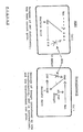
- Figure Scheme of host and transformant cell capability for synthesizing GMP.
- The present inventors have succeeded in introducing human adenovirus 12-type derived Ela genes into human T or B cells, thus obtaining human T or B cell lines capable of spontaneously producing immunologically active substances.
- The present invention is related to improved human T or B cell lines carrying at least one Ela gene derived from human adenovirus 12-type, and a process for producing immunologically active substances using the cells thereof.
- The human T or B cells useful in conjunction with the present invention include any type of human normal T or B cells and malignant T or B cells.
- Normal human T cells such as human peripheral blood T cells may be utilized herein. In order to obtain normal human T cells, human peripheral blood may be treated using the Ficoll gravity precipitation method to obtain lymphocytes. Human T cells can be obtained from the lymphocytes according to the E rossette method using sheep red cells and the like.
- To obtain T cells from other tissues, e.g., tonsil tissue, the tissue is loosened with tweezers in a medium such as RPMI-1640 medium, to obtain a cell suspension, from which lymphocytes are separated according to the Ficoll gravity precipitation method. Human T cells can be obtained from lymphocytes according to the E rossette method, etc.
- Examples of malignant human T cells include T cells isolated from T leukemic patients, T cells transformed by virus such as ATLV, HTLV, etc., fused cell lines of normal human T cells and myeloma cells or malignant T cells, cell lines obtained by transforming normal cells with drugs, radiation, etc.
- Specific examples of malignant T cells include: CCRF-CEM (ATCC.CCL 119, Cancer, 18:522-529 (1965)); HPB-MLT (Int. J. Cancer, 21:166 (1978)); HPB-ALL (Int. J. Cancer, 21:166 (1977)); TALL (Nature, 267, 843 (1977)); RPMI-8402 (J. Natl. Cancer Inst., 55:11 (1975)), etc.
- To obtain normal human B cells, any conventional method can be applied. For example, all methods described in "Techniques for Tissue Culture" edited by the Tissue Culture Association of Japan, can be used.
- Specific examples of normal human B cells include RPMI 1788 (ATCC CCL-156), J. Nat. Cancer Inst., 43:1119 (1969).
- . As malignant human B cells for use in conjunction with this invention there can be mentioned those isolated from leukemic patients, those obtained by transformation of normal B cells with virus, cells exposed to drugs, radiation, and other methods to render them malignant as well as malignant T cells.
- Specific examples include: CCRF-SB ((ATCC CCL-120), Cancer Res. 27:2479 (1967)); Daudi ((ATCC CCL-213), Cancer Res. 28:1300 (1968)); IM-9 ((ATCC CCL-159), Ann. N.Y. Acad. Sci. 190:221 (1972)); Namalwa ((ATCC CRL-1432), J. Clin. Microbiol. 1:116 (1975)).
- Ela genes derived from human adenovirus 12-type have the property of working upon other genes, thereby preventing the genes from expressing other genes (P.I. Schrier et al, Nature 305:771 (1983); D.L. Week et al, Mol. Cell. Biol. 3:1222(1983); Pelham et al, Cell 30:517 (1982)).
- Human adenovirus 12-type Ela genes for transduction can be obtained by culturing E. coli having the well known vector pSV2-Ecogpt-Ela disclosed by J. Virology 45:1076 (1983) and incorporated herein by reference, collecting the desired amounts of pSV2-Ecogpt-Ela from the culture and separating the Ecogpt-Ela genes thereof by conventional methods ordinarily used in the art. Thereafter, the thus obtained genes are introduced into cells also by methods well known in the art, e.g., in accordance with the calcium- phosphate method, whereby both the gene Ecogpt and Ela gene are introduced into the cell.
- The Ecogpt gene contains the gene coding for XGPRT. The following nomenclature will be used throughout this application.
- Xan = xanthine
- IMP = inosine-51-monophosphate
- XMP = xanthosine-5'-monophosphate
- GMP = guanosine-5'-monophosphate
- XGPRT = xanthine-phosphoribosyltransferase (or guanine)
- Usually, Ecogpt-Ela is not cut between Ecogpt and Ela during the course of transformation and both of them are introduced together into the cells.
- The selection of transformants is facilitated by and xanthine incorporating mycophenolic acid MPA into the selection medium. Mycophenolic acid inhibits the enzyme cycle from IMP to XMP in the GMP synthesis system .
- By the action of XGPRT activity of the cell having Ecogpt (and then Ela), GMP synthesis is effected from xanthine through XMP, whereby the growth of the transformed cells can be effected, while the non- transformed strains remain incapable of growing in the medium. Accordingly, only cells in which Ecogpt and then human adenovirus 12-type Ela genes have been introduced can grow (see, Figure 1 of the attached drawing).
- The presence of Ela genes can be confirmed by the Southern hybridization technique. Briefly, the Southern hybridization method comprises:
- cleaving DNA fragments extracted from transformed cells by restriction enzyme digestion;
- fractionating the DNA fragments on the basis of their size by means of agarose gel electrophoresis;
- modifying double-stranded DNA fragments into single-stranded DNA fragments in an alkali solution; and
- then placing a nitrocellulose filter into close contact with the gel to transfer the modified DNA segments into the filter in the presence of a high salt concentration solution.
- On the other hand, the Ela gene may be cleaved by DNase to generate a breakage site and then regenerated utilizing DNA polymerase I. When 32P-labelled nucleotide is used during the course of the experimental work, the resulting DNA segments are marked with radioactivity and may be utilized as a probe to detect complementary DNA fragments. The thus obtained probe is denatured by using heat to form single-stranded DNA fragments, followed by hybridization with a nitrocellulose filter having single-stranded DNA attached thereto and obtained from the aforesaid cells. The filter is rinsed and an autoradiogram is then obtained by contact thereof with a radioactivity sensitive film, whereby the Ela gene in the DNA fragments of the transduced cells is detected on an X-ray film in black.
- The thus transduced cells can now be cultured again in a fresh medium containing neither mycophenolic acid nor xanthine and the medium or supernate of the culture and the cells can be examined for the appearance of immunologically active substances as indicated in the Examples.
- A method for producing immunologically active substances using the thus obtained cells spontaneously producing an immunologically active substance is not particularly different from any conventional method for producing immunologically active substances using conventional cells having such capability. As media for culturing the cells of the present invention, there can be used, of course, serum-free media derived from conventional media, such as RITC 55-9 medium, RITC 56-1 medium, RITC 56-5 medium described in Published Unexamined Japanese Patent Application 74616/83, incorporated herein by reference in addition to RPMI-1640 medium, among others.
- A spinner flask, a roller bottle, an incubation tank with agitation and other suitable containers can be used for culturing the cells.
- The immunologically active substances obtained from the cell culture (see, e.g., the Examples) may be used in the form of a supernatant, depending upon their intended use. If necessary and/or desired, the immunologically active substances may be isolated from the culture supernatant or cells and purified.
- For the isolation and purification, various methods may be used, such as salting-out, concentration, vacuum dialysis, gel filtration, chromatrography, ion exchange chromatography, isoelectric point electrophoresis, gel electrophoresis and many other usual in this field. These methods may be used singly or in an appropriate combination.
- The activity of interleukin 2 (IL-2) may be measured as follows. 100 µl of a sample or specimen are placed in the first line of a 96-well microtiter plate. A two-fold dilution is carried out with Dulbecco's modified Eagle medium (DMEM) containing 5% Fetal Bovine Serum (FBS) to prepare a serial dilution of 100 µl each on the 96-well microplate. Activated T lymphocytes prepared in accordance with the method disclosed by Gillis et al (Nature 268:154(1977)) to a concentration of 4 x 10³ cell/100 µl are placed in each well. After culturing for 20 hours in an incubator containing 5% carbon dioxide in air at 37°C, 0.5 µCi of tritiated thymidine are added thereto followed by pulsing for 4 hours. The cells are then collected in a manner well known in the art and the radiation intake within the cells is measured. The higher the activity of IL-2 the culture supernatant has the larger is the tritiated thymidine intake into the activated T lymphocytes, from which the amount of IL-2 produced in the culture supernatant can easily be derived.
- In this case, the amount of IL-2 produced in a solution containing Con A-stimulated (concanavalin A) rat spleen cells (1 x 10⁶ cell/ml of spleen cells, supplemented with 5 pg/ml of Con A, cultured for 48 hours) is defined to be 1 unit/ml, and the activity unit is calculated from the relative value (cf., Gillis et al, J. Immunol., 120:2027 (1978)).
- The activity of IL-2 may also be measured as follows. The above-mentioned activated T lymphocyte culture is diluted in Dulbecco's modified Eagle's medium containing 5% FBS. 100 µl of a cell suspension thereof adjusted to 25 cell/well is placed in a 96-well microplate. Thereto, 100 µl of a sample or specimen are added, and incubation is conducted for 2 days in an atmosphere of air containing 5 vol % of carbon dioxide. Thereafter, the cells are counted. Since the higher the IL-2 activity is, the higher the activated T lymphocytes-proliferate, the IL-2 productivity in the culture supernatant can be noted.
- The activity of, e.g., interferon, may be carried out as follows. Detroit 550 cells (ATCC CCL 109) are cultured in RPMI-1640 medium supplemented with 10% fetal bovine serum (FBS). The cultured cells are then suspended in a medium having the same composition as described above and 2 x 10⁴ cells are seeded or inoculated per well in a 96-well microplate. This is followed by incubation for 3 days at 37°C in an atmosphere of air containing 5 vol % carbon dioxide. The supernate is then removed. Thereto are added 100 µl each of a specimen previously prepared by serial dilution by repeated 5-fold dilution using RPMI-1640 medium containing 2% FBS. Incubation is carried out for one more day. After the supernatant is removed, 100 µl of a suspension of vesicular stomatitis virus, which was previously adjusted with RPMI-1640 medium containing 2% FBS to a concentration exhibiting 50% inhibition, is added to cause infection in 20 to 24 hours. Cellular denaturation is observed and judged microscopically. The activity can be determined with reference to how the various interferon dilutions can prevent the "cellular denaturation effect" occurred when viral infection of the cells is effected. A dilution magnification at which the cellular denaturation effect,is prevented by 50% is determined and the dilution magnification is taken as the interferon titer, Enders, J.F., et al., Proc. Natl. Acad. Sci. U.S. 45:385(1959)).
- Cell culture may be conducted, e.g., as follows. The cells are suspended in RPMI-1640 medium plus from 2.5 to 10% FBS, so as to have an initial cell concentration of 1 x 10⁵ cell/ml. The suspension is separately poured into a flask in an amount of 25 ml/flask (No. 3024, Falcon Co., Ltd.) and allowed to settle at 37°C for 3 to 4 days in an atmosphere of air with 5 v/v% C02 for incubation. In the case of culturing the cells in a spinner flask, 175 ml of the suspension is charged in a spinner flask (No. 1967-00250, Belco Co., Ltd.) having a total volume of 600 ml at the above-mentioned cell concentration in the above-mentioned medium followed by incubation at 37°C for 3 to 4 days at 50 to 150 r.p.m. A spinner flask (No. 1967-15000, Belco Co., Ltd.) having a total volume of 20 liters is used, 5 liters of a suspension consisting of the cells and the medium are charged therein followed by incubation at 37°C for 3 to 4 days at 50 to 150 r.p.m.. In the case of culturing the cells in a roller bottle (No. 3027, Falcon Co., Ltd., No. 3027), 500 ml of the suspension are charged therein at the above-mentioned cell concentration and under the above-mentioned medium conditions followed by roll-culturing (rotating the bottles) at 37°C for 3 to 4 days at 2 to 30 r.p.m..
- The above culture supernate is filtered through a filter of 0.45 micron and ammonium sulfate is gradually added to reach 50% saturation. After agitating for 1 hour, the mixture is then centrifuged for 10 minutes at 10,000 x g to recover the supernate. Ammonium sulfate is supplemented to the supernate to reach 75% saturation. After allowing to stand for 2 hours, the mixture is again centrifuged for 20 minutes at 10,000 x g. The resulting precipitate is dissolved in 0.01 M Tris-HCI buffer, pH 7.4. The solution is dialyzed against the same buffer for 48 hours and the dialysate is developed onto a DEAE Sepharose column which was previously equilibrated with 0.01 M Tris-HC1 buffer, pH 7.4, followed by gradient elution using the same buffer and 0.1 M saline solution. The immunologically active substances are present in the fractions eluted at a salt concentration of about 0.07 M. The fractions are then collected and 10% polyethylene glycol (PEG-6000, Wako Junyaku Co., Ltd.) is added to a concentration of 1%. The mixture is concentrated using a Diaflow YM 5 membrane (Amicon Co., Ltd.). The thus obtained concentrate is developed onto a Sephadex 150 column (Pharmacia Col, Ltd.), which is eluted with phosphate-buffered physiological saline solution containing 0.1% PEG. The fractions containing the immunologically active substance are collected and a 10% PEG solution is again added thereto. The mixture is then concentrated with a Diaflow YM 5 membrane to obtain a purified specimen.
- Immunologically active substances can be produced by using the improved human T or B cell lines of the present invention without adding any inducers to the media. Further, immunologically active substances can be produced with higher efficiency by culturing the improved human T or B cell lines of the present invention. The immunologically active substances which can be produced in accordance with the process of the present invention include interleukin-1, -2 and -3, interferon-α, -β and -γ, CSF, BCGF, etc.
- E. coli having incorporated therein the pSV2-Ecogpt-Ela gene (J. Virology 45:1074 (1983)) was cultured at 37°C for 18 hours in 10 ml of L-broth supplemented with 50 pg/ml ampicillin. The culture was then transferred to 1 liter of a medium having the same composition described above and cell culturing was continued for 4 more hours using a rotary flask having a total volume of 5 liters. Thereafter, chloramphenicol was added to a concentration of 170 µg/ml and culturing continued for 18 more hours. The thus obtained culture supernate was centrifuged at 5,000 x g for 10 minutes at 4°C to harvest the cells. After washing the cells with 50 mM Tris-HCI buffer, pH 8.0, and 5 mM EDTA buffer, the cells were resuspended in the aforesaid EDTA buffer containing in addition 25% sucrose to a volume of 15 ml per 3 g of the cells. 30 mg of lysozyme were then added to the suspension and the cells were treated at 0°C for 20 minutes to obtain spheroplasts. 100 mM Tris-HCl buffer, pH 8.5, containing 0.5% Triton X 100 and 15 ml of 100 mM EDTA solution were added to the resulting spheroplasts. After maintaining the mixture at 0°C for 15 minutes, the mixture was centrifuged at 28,000 x g for 30 minutes at 4°C and the supernate separated thereof. After adjusting the refractivity of the thus obtained supernate to 1.395 by adding cesium chloride thereto, DNA was recovered by centrifugating for 40 hours at 40,000 x g. After decolorizing with isoamyl alcohol, calcium chloride was removed by dialysis. The plasmid pSV2-Ecogpt-Ela was then obtained by ethanol pecipitation. The thus obtained plasmid was used to transform a Jurkat-MT strain by the calcium phosphate method which is well known in the art.
- The Jurkat-MT strain used herein was obtained as follows. Jurkat-FHCRC strain cells (Gillis, S., et al, J. Exp. Med.152:1709(1980)) were obtained by cultivating in RPMI-1640 medium containing 10% fetal bovine serum (FBS). The cells were then suspended in fresh RPMI-1640 + 10% FBS medium at a concentration of 1 x 10⁵ cell/ml. In a plastic dish (No. 1007, Falcon Co., Ltd.) 5 ml of the suspension were placed and N-methyl-N'-nitro-N-nitrosoguanidine was added thereto to a final concentration of 1.5 µg/ml. After culturing at 37°C for 16 hours in an atmosphere of air containing 5 v/v% C02, the cells were harvested by centrifugation followed by washing with physiological saline twice or thrice. An initial cell concentration of 1 x 10⁵ cells/ml was obtained, again using fresh RPMI-1640 + 10% FBS medium, 25 ml of which were placed in a flask (No. 3024, Falcon Co., Ltd.) and cultured at 37°C for 4 days in an atmosphere of 5 v/v% C02 in air. The initial concentration of the thus mutated cells was adjusted to 1 x 10⁵ cells/ml using fresh RPMI-1640 5% FBS medium, placed in a 96-well microtiter plate (Falcon Co., Ltd.) and cultured at 37°C for 4 days in an atmosphere of 5 v/v% C02 in air. The cell count was obtained and a strain which grew to the level of 1 x 10⁶ cells/ml was separated as Jurkat-MT.
- Transformation was performed by mixing 200 pg of the obtained DNA plasmids with 2.5 ml of a 125 mM calcium chloride solution, gently adding the resulting liquid to 25 ml of phosphate buffer (Shen, Y.M., et al, Mol. Cell. Bio1.2:1145(1982)) and the mixture was allowed to react at 37°C for 1 hour. To the reaction mixture were added 1 x 10⁷ cells of the parent - Jurkat-MT strain cultured using RPMI-1640 medium containing 2.5% FBS, followed by cultivation at 37°C for 1 hour. The cells were then suspended in a medium having the same composition described above to a final concentration of 1 x 10⁵ cell/ml, and 2 ml each of the suspension were placed in a 24 well-plate (No. 3047, Falcon Co., Ltd.). After culturing at 37°C for 24 hours in an atmosphere of air containing 5 v/v% C02, 2 ml of a fresh medium having the same composition described above were exchanged for the old medium in the wells. Cultivation was then continued at 37°C for 3 days in an atmosphere of air containing 5 v/v% C02,
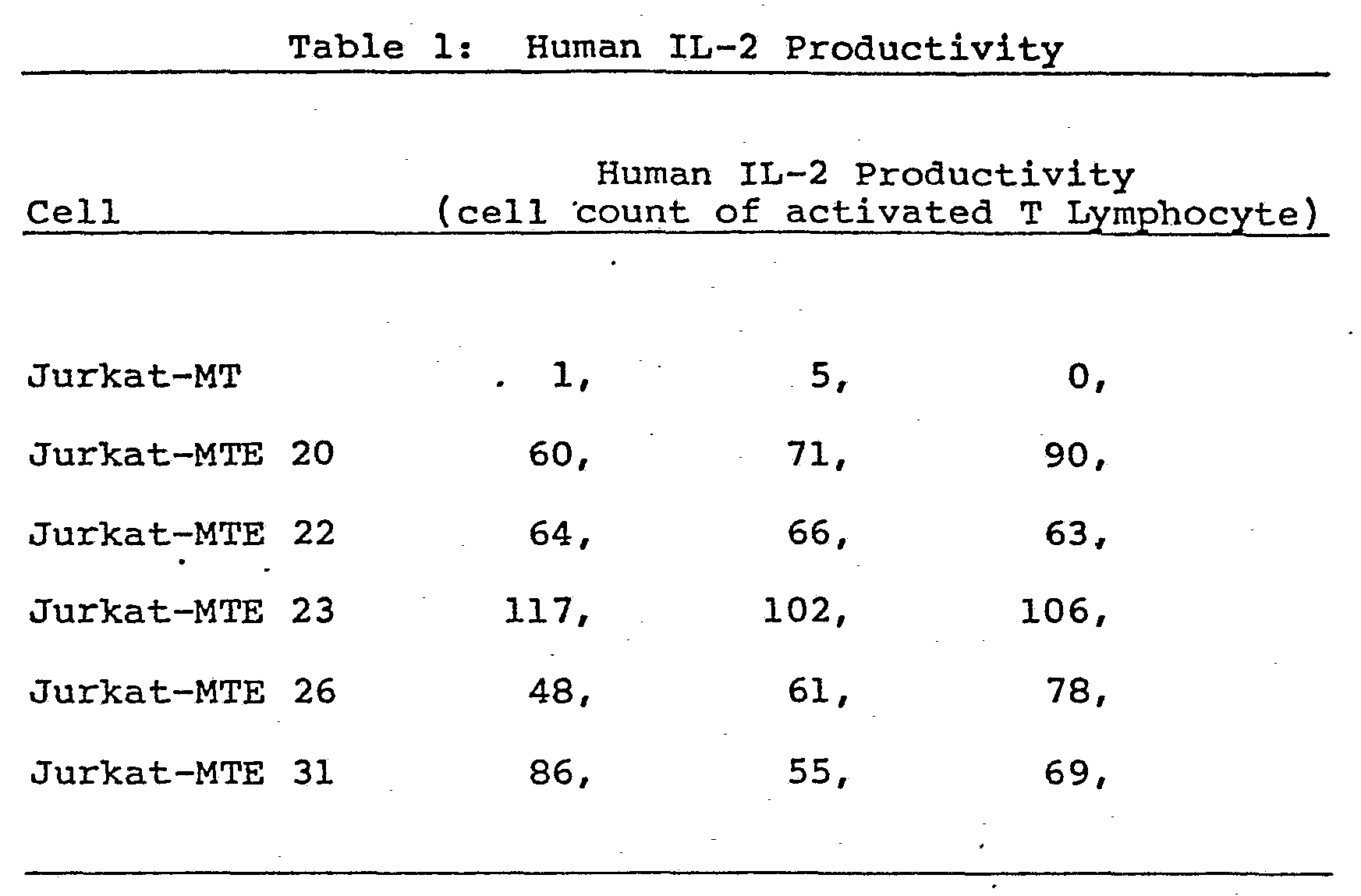
- From the thus transformed cells, the desired cells were obtained by the following method. Cell cultivation was performed in RPMI-1640 medium containing 0.5 pg/ml mycophenolic acid, 250 pg/ml xanthine and 2.5% FBS, at 37°C for 2 to 3 weeks in an atmosphere of air containing 5 v/v % CO2, During this time one half of the culture liquid was exchanged anywhere from every other day to every four days, whereby the 32 cell lines grown were harvested. The incorporation of the Ela gene.in the thus obtained cell lines was confirmed by the Southern hybridization method. The initial concentration of the cells was then adjusted to 1 x 10⁵ cell/ml using fresh RPMI-1640 medium containing 2.5% FBS, and 25 ml of which were placed in a flask (No. 3024, Falcon Co., Ltd.) and cultured at 37°C for 4 days in an atmosphere of air containing 5 vol% C02. The production of human IL-2 in the culture supernate was then determined by the method described above using the proliferation of activated T lymphocytes as a measure of human IL-2 productivity. As shown in Table 1, Jurkat-MTE 20, -MTE 22, -MTE 23, -MTE 26 and -MTE 31 cells, which constitutively produce human IL-2, were obtained.
- Cells from the cell line Jurkat-MET 31 obtained in Example 6 were adjusted with RPMI-1640 medium to an initial concentration of 1 x 10⁵ cell/ml, and 25 ml thereof were placed in a flask (No. 3024, Falcon Co., Ltd.). After culturing at 37°C for 3 days in an atmosphere of air containing 5 v/v% of carbon dioxide, the solution was diluted to 2.5 cells/ml using a medium having the same composition described above and 200 µl each of the diluted preparation were placed in a 96- well microplate (No. 3072, Falcon Co., Ltd.). This was followed by incubation at 37°C for 2 weeks in an atmosphere of air containing 5 v/v% of carbon dioxide. The 62 cell lines which were grown from the thus incubated 96-well plate were harvested. The thus obtained cells were adjusted with RPMI-1640 medium containing 2.5% FBS to an initial cell concentration of 1 x 10⁵ cells/ml, and 25 ml thereof were placed in a flask (No. 3024, Falcon Co., Ltd.), followed by incubation at 37°C for 3 days in an atmosphere of air containing 5 v/v% carbon dioxide. The thus cultured cells were adjusted with RPMI-1640 medium containing 1% FBS to an initial cell concentration of 2 x 10⁶ cell/ml. Thereafter, con A which is a human IL-2 production-inducing agent was added to a concentration of 50 pg/ml. 175 ml of the mixture were charged into a spinner flask (No. 1967-00250, Belco Co., Ltd.) and the cells were cultured at 37°C for 24 hours at 50 to 150 r.p.m.. The production of human IL-2 in the culture supernate was measured as the intake of tritiated thymidine using activated T lymphocytes as described hereinabove. Two cell lines (Jurkat-MTE 31-12 and Jurkat-MTE-31-45) evidencing higher human IL-2 productivity than the parent cells were obtained. The results are shown in Table 2.

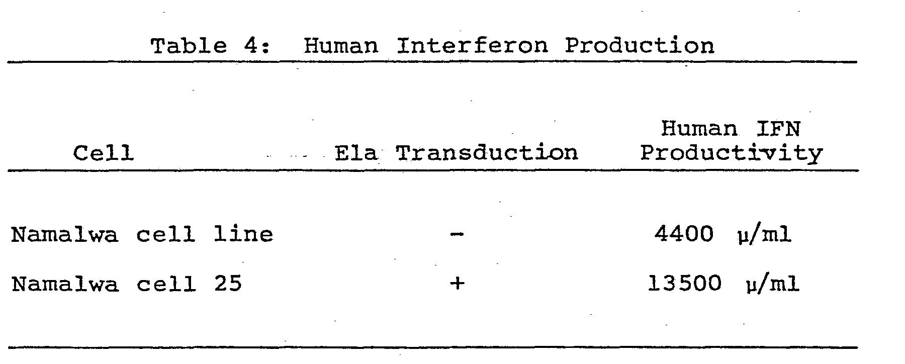
- In a manner similar to Example 6, the Ela gene was transduced into Namalwa cells (obtained from the Frederick Cancer Research Center, MD, U.S.A.) cultured in RPMI-1640 medium containing 10% FBS. The cells were subjected to transduction using as a selection medium a 10% FBS-containing RPMI-1640 medium supplemented with 5 µg/ml mycophenolic acid and 250 pg/ml xanthine and the desired 25 cell lines were obtained. The thus obtained cells were adjusted with 10% FBS-containing RPMI-1640 medium to an initial cell concentration of 1 x 10⁵ cell/ml and 25 ml of cells were placed in a flask (No. 3024, Falcon Co., Ltd.), and incubated at 37°C for 3 to 4 days in an atmosphere of air containing 5 v/v% carbon dioxide. The production of IFN in the culture supernate was determined by measuring human IFN activity as described hereinabove. The results are described in Table 3 below, wherein the productivities of each of 3 cell lines spontaneously producing human IFN are compared with the parent Namalwa cell line lacking a transduced Ela gene.

- Cells from the Namalwa cell line No. 25 obtained in Example 8 into which an Ela gene was transduced were adjusted with a 10% FBS-containing RPMI-1640 medium to an initial cell concentration of 1 x 10⁵ cell/ml.. 175 ml of the cell suspension were placed in a spinner flask (No. 1967-00250, Belco Co., Ltd.), and incubation was carried out for 3 to 4 days at 50 to 100 r.p.m. and 37°C. To the culture was added New Castle disease virus to attain an erythrocyte agglutination activity of 4 units/1 x 105 cells. After culturing for 20 to 30 hours at 35°C and 100 to 200 r.p.m., the reaction mixture was centrifuged for 10 minutes at 5000 r.p.m. to separate the supernate. The production of human IFN in the supernate was then determined by measuring human IFN activity as described hereinabove. The results are described in Table 4 as a comparison with the parent Namalwa cells carrying no transduced Ela gene.
- Sample embodiments of the invention identified as Jurkat-MTE-23 and Jurkat-MTE-31-12 have been deposited at the Institute for Fermentation Osaka (IFO), Osaka, Japan, under the accession numbers of IFO 50052 and IFO 50053, respectively.
Claims (10)
Applications Claiming Priority (2)
| Application Number | Priority Date | Filing Date | Title |
|---|---|---|---|
| JP102282/84 | 1984-05-21 | ||
| JP59102282A JPS60248177A (en) | 1984-05-21 | 1984-05-21 | Improved human t or b cell strain and preparation of immunoactivator using same |
Publications (2)
| Publication Number | Publication Date |
|---|---|
| EP0163243A2 true EP0163243A2 (en) | 1985-12-04 |
| EP0163243A3 EP0163243A3 (en) | 1988-09-28 |
Family
ID=14323248
Family Applications (1)
| Application Number | Title | Priority Date | Filing Date |
|---|---|---|---|
| EP85106221A Withdrawn EP0163243A3 (en) | 1984-05-21 | 1985-05-21 | Improved human t or b cell lines and a process for producing immunologically active substances using the same |
Country Status (3)
| Country | Link |
|---|---|
| US (1) | US4758513A (en) |
| EP (1) | EP0163243A3 (en) |
| JP (1) | JPS60248177A (en) |
Cited By (1)
| Publication number | Priority date | Publication date | Assignee | Title |
|---|---|---|---|---|
| EP0437516A4 (en) * | 1988-10-06 | 1991-09-25 | Dana Farber Cancer Institute | H. saimiri-htlv-x region vector |
Families Citing this family (1)
| Publication number | Priority date | Publication date | Assignee | Title |
|---|---|---|---|---|
| US5851816A (en) * | 1988-06-30 | 1998-12-22 | The United States Of America As Represented By The Administrator Of The National Aeronautics And Space Administration | Cultured high-fidelity three-dimensional human urogenital tract carcinomas and process |
Family Cites Families (1)
| Publication number | Priority date | Publication date | Assignee | Title |
|---|---|---|---|---|
| US4560655A (en) * | 1982-12-16 | 1985-12-24 | Immunex Corporation | Serum-free cell culture medium and process for making same |
-
1984
- 1984-05-21 JP JP59102282A patent/JPS60248177A/en active Pending
-
1985
- 1985-05-21 EP EP85106221A patent/EP0163243A3/en not_active Withdrawn
- 1985-05-21 US US06/736,376 patent/US4758513A/en not_active Expired - Fee Related
Non-Patent Citations (6)
| Title |
|---|
| ABSTRACTS OF THE ANNUAL MEETING OF THE AMERICA SOCIETY FOR MICROBIOLOGY, vol. 80, no. 0, 1980, page 504, reference E 48, US; M. GIBSON et al.: "Immuno stimulatory effects of adenovirus fiber protein and adenovirions on BLAB-C mice" * |
| ACTA MICROBIOLOGICA ACADEMIAE SCIENTIARUM HUNGARICAE, vol. 27, no. 3, 1980, page 231; R. PUSZTAI et al.: "Interferon induction by incomplete particles of adenovirus type 12" * |
| DISS. ABST. INT. B, vol. 40, no.9, 1980, page 4128, M.G. GIBSON: " Immunostimulatory functions of adenovirions and fiber protein" * |
| JOURNAL OF VIROLOGY, vol. 45, no. 3, March 1983, pages 1074-1082; K. SHIROKI et al.: "Expression of adenovirus type 12 early region 1 in KB cells transformed by recombinants containing the gene" * |
| MOLECULAR AND CELLULAR BIOLOGY, vol. 3, no. 7, July 1983, pages 1222-1234; D.L. WEEKS et al.: "E1A control of gene expression is mediated by sequences 5' to the transcriptional starts of the early viral genes" * |
| NATURE, vol. 305, 27th October 1983, pages 771-775, Macmillan Journals Ltd; P.I. SCHRIER et al.: "Expression of class I major histocompatibility antigens switched off by highly oncogenic adenovirus 12 in transformed rat cells" * |
Cited By (1)
| Publication number | Priority date | Publication date | Assignee | Title |
|---|---|---|---|---|
| EP0437516A4 (en) * | 1988-10-06 | 1991-09-25 | Dana Farber Cancer Institute | H. saimiri-htlv-x region vector |
Also Published As
| Publication number | Publication date |
|---|---|
| JPS60248177A (en) | 1985-12-07 |
| EP0163243A3 (en) | 1988-09-28 |
| US4758513A (en) | 1988-07-19 |
Similar Documents
| Publication | Publication Date | Title |
|---|---|---|
| US5541088A (en) | Recombinant process of producing non-glycosylated B-cell defferentiation factor | |
| Selinger et al. | Monokine-induced synthesis of serum amyloid A protein by hepatocytes | |
| Poiesz et al. | Isolation of a new type C retrovirus (HTLV) in primary uncultured cells of a patient with Sezary T-cell leukaemia | |
| JP2655591B2 (en) | A new race of primate hematopoietic growth factors | |
| Chen et al. | Cerebellar degeneration-related antigen: a highly conserved neuroectodermal marker mapped to chromosomes X in human and mouse. | |
| JP2633510B2 (en) | Animal interferon | |
| US4908433A (en) | Uses of interleukin-2 | |
| JPH06199897A (en) | Production and purification of lymphokine | |
| JPH07106144B2 (en) | Human immune interferon | |
| JPH0618778B2 (en) | Leukopenia treatment | |
| EP0159289B1 (en) | Immunosuppressant factor | |
| US4925919A (en) | Purified interleukin 2 | |
| CA1335717C (en) | Human granulocyte-macrophage colony stimulating factor and muteins thereof | |
| JPH0783717B2 (en) | Gene encoding human granulocyte colony-stimulating factor | |
| US4758513A (en) | Human T or B cell lines and a process for producing immunologically active substances using the same | |
| GB2121053A (en) | Production of interleukin and its analogous immune regulating factors | |
| Matsuo et al. | Production and purification of human leukocyte interferon | |
| EP0328061A2 (en) | Human colony-stimulating factors | |
| CA1202917A (en) | Process for producing subculturable lymphokine- producing human t cell hybridomas | |
| Gemsa et al. | Activation of macrophages by lymphokines from T-cell clones: evidence for different macrophage-activating factors | |
| JP2564245B2 (en) | Monoclonal antibody specific for leukoregulin cell surface receptor, method for producing the same, and use thereof | |
| US5824330A (en) | Highly purified interleukin-2 and method | |
| JPH0335795A (en) | Production of polypeptide having human interleukin 2 activity | |
| EP0114342B1 (en) | A method of producing reverse transcriptase | |
| US5216133A (en) | Human monocyte growth factor |
Legal Events
| Date | Code | Title | Description |
|---|---|---|---|
| PUAI | Public reference made under article 153(3) epc to a published international application that has entered the european phase |
Free format text: ORIGINAL CODE: 0009012 |
|
| AK | Designated contracting states |
Designated state(s): AT CH DE FR GB IT LI |
|
| PUAL | Search report despatched |
Free format text: ORIGINAL CODE: 0009013 |
|
| AK | Designated contracting states |
Kind code of ref document: A3 Designated state(s): AT CH DE FR GB IT LI |
|
| 17P | Request for examination filed |
Effective date: 19890307 |
|
| 17Q | First examination report despatched |
Effective date: 19910215 |
|
| STAA | Information on the status of an ep patent application or granted ep patent |
Free format text: STATUS: THE APPLICATION IS DEEMED TO BE WITHDRAWN |
|
| 18D | Application deemed to be withdrawn |
Effective date: 19911026 |
|
| RIN1 | Information on inventor provided before grant (corrected) |
Inventor name: TAKANO, SATOSHI Inventor name: MORIOKA, HAJIMU Inventor name: ONODERA, KAZUKIYO Inventor name: SHIBAI, HIROSHIRO |