EP0138377B2 - Lewis blood group phenotype assay - Google Patents
Lewis blood group phenotype assay Download PDFInfo
- Publication number
- EP0138377B2 EP0138377B2 EP84306210A EP84306210A EP0138377B2 EP 0138377 B2 EP0138377 B2 EP 0138377B2 EP 84306210 A EP84306210 A EP 84306210A EP 84306210 A EP84306210 A EP 84306210A EP 0138377 B2 EP0138377 B2 EP 0138377B2
- Authority
- EP
- European Patent Office
- Prior art keywords
- atcc
- antibodies
- monoclonal
- test
- fused cell
- Prior art date
- Legal status (The legal status is an assumption and is not a legal conclusion. Google has not performed a legal analysis and makes no representation as to the accuracy of the status listed.)
- Expired - Lifetime
Links
Images
Classifications
-
- C—CHEMISTRY; METALLURGY
- C07—ORGANIC CHEMISTRY
- C07K—PEPTIDES
- C07K16/00—Immunoglobulins [IGs], e.g. monoclonal or polyclonal antibodies
- C07K16/18—Immunoglobulins [IGs], e.g. monoclonal or polyclonal antibodies against material from animals or humans
- C07K16/34—Immunoglobulins [IGs], e.g. monoclonal or polyclonal antibodies against material from animals or humans against blood group antigens
Definitions
- the present invention is directed to an assay useful in the determination of the Lewis blood group phenotype of a test subject.
- the Lewis (Le) blood group antigens, a and b were originally defined by two anti-sera designated anti-Lea and anti-Le b . See Andersen (1948), Acta Path. Microbiol. Scand. 25: 728; Mourant, (1946) Nature 158: 237. Based on these anti-sera, four Lewis phenotypes were distinguished: Lea +b+ , Lea-b-, Le a+b- , and Lea- b+ . An individual, therefore, may posses one Lewis antigen (Le a+b- or Le a- b+ ), both Lewis antigens (Lea +b+ ), or neither Lewis antigen (Le a-b- ). See generally, R. Race & R. Sanger, Blood Groups in Man Chp. 9 (6th ed. 1975).
- Lea and Le b antigens occur as a terminal sugar sequence of glycolipids and glycoproteins in human saliva, serum, erythrocytes and other body fluids and tissues.
- the Lea antigen contains the terminal sugar sequence: and the Le b antigen contains the terminal sequence:
- the Le b antigen as can be seen, contains the same terminal sequence as the Lea antigen with the addition of a focusyl residue.
- the Lewis blood group antigens are also related to the gastrointestinal cancer-associated antigen (GICA), which is present on cells of adenocarcinoma of the colon, stomach, or pancreas, GICA's antigenic terminal is sialylated lacto-N-fucopentaose II, which is a sialylased Lea antigen. It is believed that an indivual must be able to synthesize the Lewis terminal sugar sequences in order to express GICA, the same enzymes being involved. See, e.g., Koprowski et al., Lancet, June 12, 1982, at 1332-1333. It is desirable, therefore, to know whether an individual is Le a- b- (approximately 5% of the population) if a diagnostic assay for the presence of GICA is negative.
- GICA gastrointestinal cancer-associated antigen
- anti-sera obtained from humans or animals.
- anti-sera are by necessity polyclonal in nature.
- Most anti-Lea sera contain some weak anti-Le b when obtained from Le a- b- donors.
- Precipitating and agglutinating anti-Lea sera has been obtained from animals, such as, chickens, rabbits and goats.
- Anti-Le b sera often contains anti-H antibodies which can result in a false indication of Le b+ .
- the H antigen is a precursor of the Lewis antigens. See R. Race & R. Sanger, supra, 338-39.
- Murine hybridomas have been reported that produce monoclonal antibodies directed against the Le b antigen. Brockhaus etal., (1981) J. Biol. Chem. 256: 13223. Some of these monoclonal antibodies, however, have been found to exhibit cross-specificity with the H blood antigen.
- Another object of the present invention is to provide an assay for Lewis blood group phenotype that employs monoclonal antibodies that selectively identify Lea antigen, but cross-type with neither Le b antigen nor other blood group antigens.
- Yet another object of the present invention is to provide an assay for Lewis blood group phenotype that employs monoclonal antibodies that selectively identify Le b antigen, but cross-type with neither Lea antigen nor other blood group antigens.
- Still another object of the present invention is to provide hybridomas and monoclonal antibodies useful in an assay for Lewis blood group phenotypes.
- the present invention provides a method of determining Lewis blood group phenotype comprising providing a test sample selected from the group consisting of human tissue and human body fluid, and determining whether monoclonal test antibodies selectively bind antigens in said test sample, said monoclonal test antibodies corresponding to monoclonal antibodies produced by (a) fused cell hybrid ATCC HB 8324 and (b) a fused cell hybrid selected from the group consisting of ATCC HB 8325 and ATCC HB 8326.
- the present invention provides a method of determining Lewis blood group phenotype comprising testing a sample selected from the group consisting of human saliva and human blood for the presence of antigenic determinants selectively bound by monoclonal antibodies produced by fused cell hybrid ATCC HB 8324 and a fused cell hybrid selected from the group consisting of ATCC HB 8325 and ATCC HB 8326.
- Yet another embodiment of the present invention is a method of determining Lewis blood group phenotype comprising: (a) providing at least two solid support members contacted with a human saliva sample, said solid support member having the ability to immobilize glycolipids or glycoproteins; (b) contacting a first said solid support member with monoclonal test antibodies corresponding to monoclonal antibodies produced by fused cell hybrid ATCC HB 8324; (c) contacting a second said solid support member with monoclonal test antibodies corresponding to monoclonal antibodies produced by a fused cell hybrid selected from the group consisting of ATCC HB 8325 and ATCC HB 8326; (d) after said monoclonal test antibody-contacting steps, contacting each of said solid support members with indicator antibodies that selectively bind said monoclonal test antibodies; and (e) determining whether said indicator antibodies selectively bind to monoclonal test antibodies on said solid support members.
- Additional embodiments of the present invention include the fused cell hybrids ATCC HB 8322, ATCC HB 8323, ATCC HB 8324, ATCC HB 8325 and ATCC HB 8326, as well as the monoclonal antibodies produced by the said fused cell hybrids.
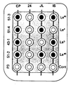
- the figure is an example of a typical reaction tray with test samples in an enzyme-linked immunoabsorbent assay preformed according to the present invention.
- the present invention provides an effective method for determining the Lewis blood phenotype of an individual.
- the assay of the present invention is fast, accurate and, in one embodiment, does not require the drawing of blood from a patient.
- the assay takes advantage of novel fused cell hybrids that produce monoclonal antibodies, as well as the antigenic determinants of those antibodies.
- Lea and Le b antigens are found in various human body fluids and tissues that contain glycolipids or glycoproteins. Examples include, but are not limited to, blood, saliva, urine, erythrocytes and other tissue orfluids. Withdrawing a glycoprotein- or glycolipid-containing test sample, therefore, provides the substrate for the present assay.
- the preferred test sample is human saliva since it does not require penetration of the skin.
- Lewis blood group antigens are expressed in the saliva whether the test subject is a secretor or nonsecretor phenotype.
- the present assay can readily be employed with, for example, blood, urine or other samples. Paraffin-embedded tissue samples can even be employed since glycolipids are not destroyed by fixatives such as formalin.
- the preferred method of determining the presence of the Lea and Le b antigens employs certain monoclonal test antibodies that correspond to antibodies produced by particular fused cell hybridomas. These fused cell hybrids produce monoclonal antibodies that selectively bind Lewis blood group antigens at particular antigenic determinants.
- the hybridomas were produced by immunizing mice with the colon carcinoma cell lines SW 1116 and SW 1222 described in Leibovitz et al., (1976) Cancer Res. 36: 562. Spleen cells from the immunized mice were fused with mouse myeloma P 3x63 Ag8 and hybridomas recovered according to methods known in the art. See, e.g., Koprowski, et al., U.S. Patent No. 4,196,265.
- Fused cell hybrid ATCC HB 8324 produces monoclonal antibody CO-514 (IgG 3 isotype) which selectively binds the lacto-N-fucopentaose II active terminal sequence of the Lea antigen.
- Fused cell hybrids ATCC HB 8325 and ATCC HB 8326 secrete monoclonal antibody CO-431 and CO-301, respectively, (IgM isotype) which selectively bind the lacto-N-difucohexaose I active terminal sequence of the Le b antigen.
- monoclonal antibodies from hybridoma 1116NS-10 reported in Brockhaus et al., (1981 ) J. Biol.
- Fused cell hybrids ATCC HB 8322 and ATCC HB 8323 produce monoclonal antibodies CO-512 (IgG 3 isotype) and CO-513 (IgG 3 isotype), respectively, which selectively bind both Lea and Le b antigens.
- the above fused cell hybrids were deposited on July 28, 1983 with the American Type Culture Collection (ATCC), 12301 Parklawn Drive, Rockville, Maryland 20852.
- a solution containing these monoclonal antibodies substantially free of other antibodies can be obtained by collecting supernatant from growing cultures of the fused cell hybrids.
- the preferred monoclonal test antibodies employed in the present invention are those produced by the above hybridomas. Monoclonal antibodies that correspond to these preferred test antibodies can also be employed. As used herein, a monoclonal test antibody will correspond to the antibody produced by a fused cell hybrid when a test antibody selectively bound to an antigen blocks the selective binding of the antibody produced by the fused cell hybrid, indicating that the antibodies share the same antigenic determinant. An antibody that corresponds to the fused cell hybrid antibody can be readily determined, for example, by a competitive binding assay. It is not necessary that the monoclonal test antibodies employed in the present assay be mouse antibodies. Rat, hamster, rabbit and human antibodies are only some of the types of antibodies that could also be used.
- a preferred test sample is saliva.
- Glycolipid or glycoprotein from the saliva can be immobilized on a solid support member with the ability to absorb glycolipids or glycoproteins.
- the solid support member is plastic (e.g., polystyrene, polypropylene or polyvinyl). By merely placing such a plastic bead momentarily in the mouth of a test subject, glycolipids or glycoproteins are immobilized on its surface.
- the presence of the Lewis blood group antigens on the solid support can be readily determined, for example, by contacting the solid support (or sample) with the monoclonal test antibodies.
- the test sample should be contacted with a monoclonal test antibody that selectively binds Lea antigen and another monoclonal test antibody that selectively binds Le b antigen. It is also desirable as a control, but not necessary, to contact a test sample with a monoclonal test antibody that selectively binds both Lea and Le b antigens to confirm results indicating Lea-b- individuals.
- the presence of Lea or Le b antigens, and thus Lewis phenotype, is determined by an assay that indicates whether monoclonal test antibodies selectively bind these antigens in a test sample.
- Various binding assay techniques are known in the art. See, e.g., U.S. Patent No. 4,380,580; U.S. Patent No. 4,375,972; U.S. Patent No. 4,376,110; U.S. Patent No. 4,376,165; U.S. Patent No. 3,791,932; U.S. Patent No. 3,654,090.
- these assays involve "tagging" either the monoclonal test antibody or an antibody that selectively binds the monoclonal test antibody (indicator antibodies).
- test sample which preferably contains antigen that has been immobilized, is then either (a) contacted with tagged test monoclonal antibodies, or (b) contacted first by untagged monoclonal test antibodies followed by contact with tagged indicator antibodies. Detection of the tag associated with the test sample indicates that monoclonal test antibodies have selectively bound an antigenic determinant in the test sample.
- radioisotopes radioimmunoassays
- enzymes enzymes
- fluorescent compounds immunofluorescence
- Othe methods for determining the binding of antibodies to antigens are well known in the art, for example, precipitin reactions, radial immunodiffusion, immunoelectrophoresis, agglutination and rosette formation. See. e.g., B. D. Davis et al., Microbiology 306-332, 335 (3rd ed. 1980); Monoclonal Antibodies 376-402 (R. H. Kennett, T. J. McKearn & K. B. Bechtol eds.
- test or indicator antibodies it may be desirable to immobilize either the test or indicator antibodies.
- Any of the common supports used in immunoassays may be employed. They include, but are not limited to, filter paper and plastic beads or vessels (e.g., polyvinyl, polystyrene, polypropylene, etc.). Polysaccharides polymers may also be used to bind the antibodies. See, e.g., U.S. Patent No. 3,645,852.
- a preferred assay is a radioimmunoassay which is well known in the art. See. e.g., C. W. Parker, Radioimmunoassay of Biologically Active Compounds (1976).
- a particularly preferred assay is an enzyme-linked immunoabsorbent assay (ELISA). See, e.g., U.S. Patent No. 3,791,932; U.S. Patent No. 3,654,090; U.S. Reissue 31,006.
- ELISA enzyme-linked immunoabsorbent assay
- Such an assay can be readily performed in, for example, a physician's office in only a few minutes employing simplified equipment and avoiding radioisotopes.
- the particular parameters employed in the assays can vary widely with the particular radioimmunoassay or ELISA employed. Optimal conditions can be readily established by those of average skill in the art. For example, it has been found that when saliva is employed it can be diluted up to 100,000-fold and remain a suitable substrate
- a 1 ml saliva sample was collected from 60 individuals of both sexes. Test subjects ranged in age from 25 to 75 years. Saliva samples were inactivated by heating for 30 minutes at 85°C in a water bath. The samples were then diluted 1:4 in phosphate-buffered saline (PBS) and stored at -20°C until the assay was run.
- PBS phosphate-buffered saline
- saliva samples were diluted 1:20 (or as specified in Table II).
- Polystyrene beads Precision Plastic Ball Co., Chicago, III., 5 beads/test subject
- saliva samples 200 ⁇ l/bead
- 1% gelatin in 0.01 M sodium borate (pH 8.0) 0.01 M sodium borate (pH 8.0) for 1 hour at ambient temperature.
- the washed beads were then placed in a reaction tray containing 5 wells for each test subject.
- the washed beads were then incubated with 125 1-labelled affinity-purified rabbit anti-mouse F(ab') 2 immunoglobulin at 1x10 5 cpm/bead for 30 minutes at room temperature. The beads were then washed 3 times with 0.5% BSA PBS and counted.
- Table I indicates the results obtained in the above radioimmunoassay.
- the assay indicates that test subject CP is Lea +b+ , test subject 24 is Le a- b- , test subject JL is Le a+b- , and test subject IS is Lea- b+ .
- Table II shows that the saliva samples of Lea+b- and Lea- b+ individuals can be diluted up to 100,000 times with clear positive selective binding demonstrated by the four monoclonal antibodies. Saliva of the Le a- b- individual did not selectively bind the monoclonal antibodies at any of the dilutions tested.
- Table III shows the proportion of various Lewis blood group phenotypes found among the 60 test subjects.
- the present assay was also performed using an enzyme-linked immunoabsorbent assay employing a peroxidase-antiperoxidase (PAP) system instead of the 125 1-labelled rabbit anti-mouse antibody.
- PAP peroxidase-antiperoxidase
- the polystyrene beads after incubation with saliva, were incubated with the test monoclonal antibodies in the reaction trays as in Example I and then exposed to biotinylated anti-mouse antibodies (biotin-avidin immunoperoxidase kit, Vector Labs, Burlingame, Calif.) for 30 minutes at ambient temperature. The beads were then washed 3 times with PBS, incubated with Avidin DH-Biotinylated Horseradish Peroxidase H for 30 minutes at ambient temperature and again washed 3 times with PBS.
- biotinylated anti-mouse antibodies biotin-avidin immunoperoxidase kit, Vector Labs, Burlingame, Calif.
- the peroxidase substrate (5 mg diaminobenzidine, 10 ⁇ l 30% H 2 0 2 , 100 ⁇ l 1M immidiazole in 10 ml of 0.1 M Tris buffer, pH 7.6) was then added to the wells in the reaction trays. Positive beads were indicated by clear, dark brown staining after incubation for 5-10 minutes.
- the figure shows a typical pattern of reactivity for the saliva samples of the four test subjects of Table I.
- 51-2,43-1,51-4, and 51-3 indicate the monoclonal antibodies produced by fused cell hybridomas ATCC HB 8322, ATCC HB 8325, ATCC HB 8324 and ATCC HB 8323, respectively.
- P3 indicates the control wells where supernatant from the mouse myeloma culture P 3x63 Ag8 was added.
- the figure shows that the Lewis blood group phenotypes for test subjects CP, 24, JL and IS are Lea +b+ , Le a- b- , Lea+b- and Lea- b +, respectively.
Abstract
Description
- The present invention is directed to an assay useful in the determination of the Lewis blood group phenotype of a test subject.
- The Lewis (Le) blood group antigens, a and b, were originally defined by two anti-sera designated anti-Lea and anti-Leb. See Andersen (1948), Acta Path. Microbiol. Scand. 25: 728; Mourant, (1946) Nature 158: 237. Based on these anti-sera, four Lewis phenotypes were distinguished: Lea+b+, Lea-b-, Lea+b-, and Lea- b+. An individual, therefore, may posses one Lewis antigen (Lea+b- or Lea- b+), both Lewis antigens (Lea+b+), or neither Lewis antigen (Lea-b-). See generally, R. Race & R. Sanger, Blood Groups in Man Chp. 9 (6th ed. 1975).
- Lea and Leb antigens occur as a terminal sugar sequence of glycolipids and glycoproteins in human saliva, serum, erythrocytes and other body fluids and tissues. The Lea antigen contains the terminal sugar sequence:


- It is important to screen for Lewis blood group phenotypes prior to blood transfusions. If the donor's Lewis phenotype does not match the recipient's, a heomolytic transfusion reaction can result.
- The Lewis blood group antigens are also related to the gastrointestinal cancer-associated antigen (GICA), which is present on cells of adenocarcinoma of the colon, stomach, or pancreas, GICA's antigenic terminal is sialylated lacto-N-fucopentaose II, which is a sialylased Lea antigen. It is believed that an indivual must be able to synthesize the Lewis terminal sugar sequences in order to express GICA, the same enzymes being involved. See, e.g., Koprowski et al., Lancet, June 12, 1982, at 1332-1333. It is desirable, therefore, to know whether an individual is Lea- b- (approximately 5% of the population) if a diagnostic assay for the presence of GICA is negative.
- Assays for Lewis phenotypes employ anti-sera obtained from humans or animals. Several problems exist with anti-sera. First, anti-sera are by necessity polyclonal in nature. Most anti-Lea sera contain some weak anti-Leb when obtained from Lea- b- donors. Precipitating and agglutinating anti-Lea sera has been obtained from animals, such as, chickens, rabbits and goats. Anti-Leb sera often contains anti-H antibodies which can result in a false indication of Leb+. The H antigen is a precursor of the Lewis antigens. See R. Race & R. Sanger, supra, 338-39.
- Murine hybridomas have been reported that produce monoclonal antibodies directed against the Leb antigen. Brockhaus etal., (1981) J. Biol. Chem. 256: 13223. Some of these monoclonal antibodies, however, have been found to exhibit cross-specificity with the H blood antigen.
- It would be desirable, therefore, to develop an assay that employs monoclonal antibodies that selectively bind either the Lea antigen orthe Leb antigen, but not both. Furthermore, it would be desirable to employ monoclonal antibodies that do not cross-type for other blood group antigens.
- It is an object of the present invention to provide an assay that determines Lewis blood group phenotype.
- It is also an object of the present invention to provide an assay employing monoclonal antibodies that determines Lewis blood group phenotype.
- Another object of the present invention is to provide an assay for Lewis blood group phenotype that employs monoclonal antibodies that selectively identify Lea antigen, but cross-type with neither Leb antigen nor other blood group antigens.
- Yet another object of the present invention is to provide an assay for Lewis blood group phenotype that employs monoclonal antibodies that selectively identify Leb antigen, but cross-type with neither Lea antigen nor other blood group antigens.
- Still another object of the present invention is to provide hybridomas and monoclonal antibodies useful in an assay for Lewis blood group phenotypes.
- These and other objects are achieved by one or more of the following embodiments of the present invention.
- In one embodiment, the present invention provides a method of determining Lewis blood group phenotype comprising providing a test sample selected from the group consisting of human tissue and human body fluid, and determining whether monoclonal test antibodies selectively bind antigens in said test sample, said monoclonal test antibodies corresponding to monoclonal antibodies produced by (a) fused cell hybrid ATCC HB 8324 and (b) a fused cell hybrid selected from the group consisting of ATCC HB 8325 and ATCC HB 8326.
- In another embodiment, the present invention provides a method of determining Lewis blood group phenotype comprising testing a sample selected from the group consisting of human saliva and human blood for the presence of antigenic determinants selectively bound by monoclonal antibodies produced by fused cell hybrid ATCC HB 8324 and a fused cell hybrid selected from the group consisting of ATCC HB 8325 and ATCC HB 8326.
- Yet another embodiment of the present invention is a method of determining Lewis blood group phenotype comprising: (a) providing at least two solid support members contacted with a human saliva sample, said solid support member having the ability to immobilize glycolipids or glycoproteins; (b) contacting a first said solid support member with monoclonal test antibodies corresponding to monoclonal antibodies produced by fused cell hybrid ATCC HB 8324; (c) contacting a second said solid support member with monoclonal test antibodies corresponding to monoclonal antibodies produced by a fused cell hybrid selected from the group consisting of ATCC HB 8325 and ATCC HB 8326; (d) after said monoclonal test antibody-contacting steps, contacting each of said solid support members with indicator antibodies that selectively bind said monoclonal test antibodies; and (e) determining whether said indicator antibodies selectively bind to monoclonal test antibodies on said solid support members.
- Additional embodiments of the present invention include the fused cell hybrids ATCC HB 8322, ATCC HB 8323, ATCC HB 8324, ATCC HB 8325 and ATCC HB 8326, as well as the monoclonal antibodies produced by the said fused cell hybrids.
- The figure is an example of a typical reaction tray with test samples in an enzyme-linked immunoabsorbent assay preformed according to the present invention.
- The present invention provides an effective method for determining the Lewis blood phenotype of an individual. The assay of the present invention is fast, accurate and, in one embodiment, does not require the drawing of blood from a patient. The assay takes advantage of novel fused cell hybrids that produce monoclonal antibodies, as well as the antigenic determinants of those antibodies.
- Lea and Leb antigens are found in various human body fluids and tissues that contain glycolipids or glycoproteins. Examples include, but are not limited to, blood, saliva, urine, erythrocytes and other tissue orfluids. Withdrawing a glycoprotein- or glycolipid-containing test sample, therefore, provides the substrate for the present assay. The preferred test sample is human saliva since it does not require penetration of the skin. Furthermore, Lewis blood group antigens are expressed in the saliva whether the test subject is a secretor or nonsecretor phenotype. The present assay, however, can readily be employed with, for example, blood, urine or other samples. Paraffin-embedded tissue samples can even be employed since glycolipids are not destroyed by fixatives such as formalin.
- The preferred method of determining the presence of the Lea and Leb antigens employs certain monoclonal test antibodies that correspond to antibodies produced by particular fused cell hybridomas. These fused cell hybrids produce monoclonal antibodies that selectively bind Lewis blood group antigens at particular antigenic determinants. The hybridomas were produced by immunizing mice with the colon carcinoma cell lines SW 1116 and SW 1222 described in Leibovitz et al., (1976) Cancer Res. 36: 562. Spleen cells from the immunized mice were fused with mouse myeloma P 3x63 Ag8 and hybridomas recovered according to methods known in the art. See, e.g., Koprowski, et al., U.S. Patent No. 4,196,265.
- Several hybridomas useful in the present invention have been developed by the above method. Fused cell hybrid ATCC HB 8324 produces monoclonal antibody CO-514 (IgG3 isotype) which selectively binds the lacto-N-fucopentaose II active terminal sequence of the Lea antigen. Fused cell hybrids ATCC HB 8325 and ATCC HB 8326 secrete monoclonal antibody CO-431 and CO-301, respectively, (IgM isotype) which selectively bind the lacto-N-difucohexaose I active terminal sequence of the Leb antigen. Unlike the monoclonal antibodies from hybridoma 1116NS-10 reported in Brockhaus et al., (1981 ) J. Biol. Chem. 256: 13223, these antibodies do not cross-type for other blood group antigens, such as the H antigen. Fused cell hybrids ATCC HB 8322 and ATCC HB 8323 produce monoclonal antibodies CO-512 (IgG3 isotype) and CO-513 (IgG3 isotype), respectively, which selectively bind both Lea and Leb antigens. The above fused cell hybrids were deposited on July 28, 1983 with the American Type Culture Collection (ATCC), 12301 Parklawn Drive, Rockville, Maryland 20852. A solution containing these monoclonal antibodies substantially free of other antibodies can be obtained by collecting supernatant from growing cultures of the fused cell hybrids.
- The preferred monoclonal test antibodies employed in the present invention are those produced by the above hybridomas. Monoclonal antibodies that correspond to these preferred test antibodies can also be employed. As used herein, a monoclonal test antibody will correspond to the antibody produced by a fused cell hybrid when a test antibody selectively bound to an antigen blocks the selective binding of the antibody produced by the fused cell hybrid, indicating that the antibodies share the same antigenic determinant. An antibody that corresponds to the fused cell hybrid antibody can be readily determined, for example, by a competitive binding assay. It is not necessary that the monoclonal test antibodies employed in the present assay be mouse antibodies. Rat, hamster, rabbit and human antibodies are only some of the types of antibodies that could also be used.
- The monoclonal test antibodies described above are readily employed in the preferred method of determining the presence of Lewis blood group antigens Lea and Leb in a test sample. A preferred test sample is saliva. Glycolipid or glycoprotein from the saliva can be immobilized on a solid support member with the ability to absorb glycolipids or glycoproteins. Any solid support member known in art may be employed. Preferably, the solid support member is plastic (e.g., polystyrene, polypropylene or polyvinyl). By merely placing such a plastic bead momentarily in the mouth of a test subject, glycolipids or glycoproteins are immobilized on its surface.
- The presence of the Lewis blood group antigens on the solid support (or in any other test sample) can be readily determined, for example, by contacting the solid support (or sample) with the monoclonal test antibodies. At a minimum, the test sample should be contacted with a monoclonal test antibody that selectively binds Lea antigen and another monoclonal test antibody that selectively binds Leb antigen. It is also desirable as a control, but not necessary, to contact a test sample with a monoclonal test antibody that selectively binds both Lea and Leb antigens to confirm results indicating Lea-b- individuals.
- The presence of Lea or Leb antigens, and thus Lewis phenotype, is determined by an assay that indicates whether monoclonal test antibodies selectively bind these antigens in a test sample. Various binding assay techniques are known in the art. See, e.g., U.S. Patent No. 4,380,580; U.S. Patent No. 4,375,972; U.S. Patent No. 4,376,110; U.S. Patent No. 4,376,165; U.S. Patent No. 3,791,932; U.S. Patent No. 3,654,090. Generally, these assays involve "tagging" either the monoclonal test antibody or an antibody that selectively binds the monoclonal test antibody (indicator antibodies). The test sample, which preferably contains antigen that has been immobilized, is then either (a) contacted with tagged test monoclonal antibodies, or (b) contacted first by untagged monoclonal test antibodies followed by contact with tagged indicator antibodies. Detection of the tag associated with the test sample indicates that monoclonal test antibodies have selectively bound an antigenic determinant in the test sample.
- Various tags are known in the art, including radioisotopes (radioimmunoassays), enzymes (ELISA or enzyme-linked immunoabsorbant assay) and fluorescent compounds (immunofluorescence). Othe methods for determining the binding of antibodies to antigens are well known in the art, for example, precipitin reactions, radial immunodiffusion, immunoelectrophoresis, agglutination and rosette formation. See. e.g., B. D. Davis et al., Microbiology 306-332, 335 (3rd ed. 1980); Monoclonal Antibodies 376-402 (R. H. Kennett, T. J. McKearn & K. B. Bechtol eds. 1980); David et al., (1974) Biochemistry 13: 1014-1021; Radioimmunoassay Methods (Kirkham & Hunter eds. 1970); Hunter et al., (1962) Nature 144: 945; U.S. Patent No. 3,940,475; U.S. Patent No. 3,867,517; U.S. Patent No. 3,645,090.
- In some assays, it may be desirable to immobilize either the test or indicator antibodies. Any of the common supports used in immunoassays may be employed. They include, but are not limited to, filter paper and plastic beads or vessels (e.g., polyvinyl, polystyrene, polypropylene, etc.). Polysaccharides polymers may also be used to bind the antibodies. See, e.g., U.S. Patent No. 3,645,852.
- A preferred assay is a radioimmunoassay which is well known in the art. See. e.g., C. W. Parker, Radioimmunoassay of Biologically Active Compounds (1976). A particularly preferred assay, however, is an enzyme-linked immunoabsorbent assay (ELISA). See, e.g., U.S. Patent No. 3,791,932; U.S. Patent No. 3,654,090; U.S. Reissue 31,006. Such an assay can be readily performed in, for example, a physician's office in only a few minutes employing simplified equipment and avoiding radioisotopes. The particular parameters employed in the assays can vary widely with the particular radioimmunoassay or ELISA employed. Optimal conditions can be readily established by those of average skill in the art. For example, it has been found that when saliva is employed it can be diluted up to 100,000-fold and remain a suitable substrate in the assay.
- The following examples are provided for illustrative purposes only. They are not intended to limit the scope of the invention which is defined solely by the claims.
- A 1 ml saliva sample was collected from 60 individuals of both sexes. Test subjects ranged in age from 25 to 75 years. Saliva samples were inactivated by heating for 30 minutes at 85°C in a water bath. The samples were then diluted 1:4 in phosphate-buffered saline (PBS) and stored at -20°C until the assay was run.
- Immediately prior to the assay, saliva samples were diluted 1:20 (or as specified in Table II). Polystyrene beads (Precision Plastic Ball Co., Chicago, III., 5 beads/test subject) were incubated with the saliva samples (200 µl/bead) and then washed with 1% gelatin in 0.01 M sodium borate (pH 8.0) for 1 hour at ambient temperature. The washed beads were then placed in a reaction tray containing 5 wells for each test subject. Supernatant from growing cell cultures containing fused cell hybrids ATCC HB 8324 (Lea), ATCC HB 8325 (Leb) ATCC HB 8322 (Leab), ATCC HB 8323 (Leab) and mouse myeloma cell P 3x63 Ag8 (control) were added to different wells containing saliva samples from test subjects (5 wells/test subject). The beads were allowed to incubate with the cell culture supernatants for 1 hour at room temperature and then washed 3 times in PBS containing 0.05% bovine serum albumin (BSA).
- The washed beads were then incubated with 1251-labelled affinity-purified rabbit anti-mouse F(ab')2 immunoglobulin at 1x10⁵ cpm/bead for 30 minutes at room temperature. The beads were then washed 3 times with 0.5% BSA PBS and counted.
- Table I indicates the results obtained in the above radioimmunoassay. The assay indicates that test subject CP is Lea+b+, test subject 24 is Lea- b-, test subject JL is Lea+b-, and test subject IS is Lea- b+. Table II shows that the saliva samples of Lea+b- and Lea- b+ individuals can be diluted up to 100,000 times with clear positive selective binding demonstrated by the four monoclonal antibodies. Saliva of the Lea- b- individual did not selectively bind the monoclonal antibodies at any of the dilutions tested. Table III shows the proportion of various Lewis blood group phenotypes found among the 60 test subjects.


- The present assay was also performed using an enzyme-linked immunoabsorbent assay employing a peroxidase-antiperoxidase (PAP) system instead of the 1251-labelled rabbit anti-mouse antibody.
- In the PAP assay, the polystyrene beads, after incubation with saliva, were incubated with the test monoclonal antibodies in the reaction trays as in Example I and then exposed to biotinylated anti-mouse antibodies (biotin-avidin immunoperoxidase kit, Vector Labs, Burlingame, Calif.) for 30 minutes at ambient temperature. The beads were then washed 3 times with PBS, incubated with Avidin DH-Biotinylated Horseradish Peroxidase H for 30 minutes at ambient temperature and again washed 3 times with PBS. The peroxidase substrate (5 mg diaminobenzidine, 10 µl 30% H202, 100 µl 1M immidiazole in 10 ml of 0.1 M Tris buffer, pH 7.6) was then added to the wells in the reaction trays. Positive beads were indicated by clear, dark brown staining after incubation for 5-10 minutes.
- The figure shows a typical pattern of reactivity for the saliva samples of the four test subjects of Table I. In the figure, 51-2,43-1,51-4, and 51-3 indicate the monoclonal antibodies produced by fused cell hybridomas ATCC HB 8322, ATCC HB 8325, ATCC HB 8324 and ATCC HB 8323, respectively. P3 indicates the control wells where supernatant from the mouse myeloma culture P 3x63 Ag8 was added. The figure shows that the Lewis blood group phenotypes for test subjects CP, 24, JL and IS are Lea+b+, Lea- b-, Lea+b- and Lea- b+, respectively. These results agree with the radioimmunoassay of Example I.
- While several specific examples of an assay within the scope of the present invention have been provided, variations on the above procedures are readily apparent to those in the art. The present invention, therefore, is to be limited only by the scope of the appended claims.
Claims (9)
Priority Applications (1)
| Application Number | Priority Date | Filing Date | Title |
|---|---|---|---|
| AT84306210T ATE42411T1 (en) | 1983-09-16 | 1984-09-10 | DETERMINING THE PHAENOTYPES OF LEWIS BLOOD TYPES. |
Applications Claiming Priority (2)
| Application Number | Priority Date | Filing Date | Title |
|---|---|---|---|
| US06/532,891 US4607009A (en) | 1983-09-16 | 1983-09-16 | Lewis blood group phenotype assay |
| US532891 | 1983-09-16 |
Publications (3)
| Publication Number | Publication Date |
|---|---|
| EP0138377A1 EP0138377A1 (en) | 1985-04-24 |
| EP0138377B1 EP0138377B1 (en) | 1989-04-19 |
| EP0138377B2 true EP0138377B2 (en) | 1993-05-19 |
Family
ID=24123617
Family Applications (1)
| Application Number | Title | Priority Date | Filing Date |
|---|---|---|---|
| EP84306210A Expired - Lifetime EP0138377B2 (en) | 1983-09-16 | 1984-09-10 | Lewis blood group phenotype assay |
Country Status (6)
| Country | Link |
|---|---|
| US (1) | US4607009A (en) |
| EP (1) | EP0138377B2 (en) |
| JP (1) | JP2527706B2 (en) |
| AT (1) | ATE42411T1 (en) |
| CA (1) | CA1211725A (en) |
| DE (1) | DE3477845D1 (en) |
Families Citing this family (16)
| Publication number | Priority date | Publication date | Assignee | Title |
|---|---|---|---|---|
| US4783420A (en) * | 1984-04-06 | 1988-11-08 | Centocor, Inc. | Immunoassay for carbohydrate antigenic determinant |
| US4873188A (en) * | 1985-05-28 | 1989-10-10 | Oncogen | Method, monoclonal antibody, and monoclonal antibody fragments for detecting human non-small cell lung carcinomas and cell line for producing such antibodies |
| JPH0792458B2 (en) * | 1986-03-14 | 1995-10-09 | 郁男 山科 | Sugar chain specific antibody assay plate and method for producing the same |
| US5086002A (en) * | 1987-09-07 | 1992-02-04 | Agen Biomedical, Ltd. | Erythrocyte agglutination assay |
| US5206086A (en) * | 1987-10-13 | 1993-04-27 | University Of Rochester | Particle-stabilized epitopes for standardization and control of immunoassays |
| US5075218A (en) * | 1987-12-29 | 1991-12-24 | Biomira, Inc. | Screening for antibodies which bind carbohydrate epitopes of tumor-associated antigens, and uses thereof |
| GB8903046D0 (en) * | 1989-02-10 | 1989-03-30 | Vale David R | Testing of liquids |
| CA2012745A1 (en) * | 1989-03-30 | 1990-09-30 | John D. Rosoff | Giardia lamblia-specific stool antigen, monospecific antibodies thereto, and method of diagnosis of giardiasis |
| US5583003A (en) * | 1989-09-25 | 1996-12-10 | Agen Limited | Agglutination assay |
| US5292641A (en) * | 1991-12-13 | 1994-03-08 | Sangstat Medical Corporation | Alloantigen testing by binding assay |
| US5583004A (en) * | 1994-12-23 | 1996-12-10 | Pincus; Mathew R. | Direct detection of unexpected alloantibodies in the serum of prospective transfusion recipients using a new hemagglutination inhibition assay |
| US5795961A (en) * | 1995-02-14 | 1998-08-18 | Ludwig Institute For Cancer Research | Recombinant human anti-Lewis b antibodies |
| GB9505447D0 (en) * | 1995-03-17 | 1995-05-03 | Common Services Agency | Competitive binding assay |
| WO2005078442A1 (en) * | 2004-02-10 | 2005-08-25 | Dantini Daniel C | Comprehensive food allergy test |
| US20110053889A1 (en) * | 2008-09-02 | 2011-03-03 | Joanne Szabo | Prenatal and postnatal screening and treatment of critical monosaccharide deficiencies for neurologic and immunologic function |
| US10428312B2 (en) | 2013-12-03 | 2019-10-01 | Agency For Science, Technology And Research | Cytotoxic antibody |
Family Cites Families (1)
| Publication number | Priority date | Publication date | Assignee | Title |
|---|---|---|---|---|
| GB2117789B (en) * | 1982-03-29 | 1985-08-29 | East Anglian Regional Health | Abo blood grouping reagent |
-
1983
- 1983-09-16 US US06/532,891 patent/US4607009A/en not_active Expired - Fee Related
-
1984
- 1984-08-23 CA CA000461678A patent/CA1211725A/en not_active Expired
- 1984-09-10 EP EP84306210A patent/EP0138377B2/en not_active Expired - Lifetime
- 1984-09-10 AT AT84306210T patent/ATE42411T1/en not_active IP Right Cessation
- 1984-09-10 DE DE8484306210T patent/DE3477845D1/en not_active Expired
- 1984-09-13 JP JP59192425A patent/JP2527706B2/en not_active Expired - Lifetime
Also Published As
| Publication number | Publication date |
|---|---|
| US4607009A (en) | 1986-08-19 |
| EP0138377B1 (en) | 1989-04-19 |
| EP0138377A1 (en) | 1985-04-24 |
| JP2527706B2 (en) | 1996-08-28 |
| ATE42411T1 (en) | 1989-05-15 |
| DE3477845D1 (en) | 1989-05-24 |
| CA1211725A (en) | 1986-09-23 |
| JPS60155976A (en) | 1985-08-16 |
Similar Documents
| Publication | Publication Date | Title |
|---|---|---|
| EP0138377B2 (en) | Lewis blood group phenotype assay | |
| US4550086A (en) | Monoclonal antibodies that recognize human T cells | |
| US4910133A (en) | Diagnostic test drug comprising monoclonal antibody to human copper.zinc-superoxide dismutase and diagnostic test method using the same | |
| US5670328A (en) | Monoclonal antibodies to human pulmonary surfactant apoprotein D and use thereof | |
| US4692405A (en) | Monoclonal antibodies to antigen on activated human B-cells and assay therefor, protein antigenic determinant therefor and method of making same | |
| Youinou et al. | Specificity of Plasma Cells in the Rheumatoid Synovium: I. Immunoglobulin Class of Antiglobulin‐Producing Cells | |
| US5316914A (en) | Method for determining human collagen peptides by way of enzyme immunoassay | |
| WO1983001739A1 (en) | Monoclonal antibodies against brugia malayi | |
| EP0226888A2 (en) | Monoclonal antibodies specific to galactosyltransferase isoenzyme II and their use in cancer immunoassays | |
| Tedder et al. | Production of monoclonal antibodies to hepatitis B surface and core antigens, and use in the detection of viral antigens in liver biopsies | |
| WO1994023302A1 (en) | Immunological assay of oxidatively modified human low density lipoproteins in plasma | |
| Drover et al. | Glutaraldehyde fixation of target cells to plastic for ELISA assays of monoclonal anti-HLA antibodies produces artefacts | |
| US5264351A (en) | Monoclonal antibodies against autoimmune RNA proteins | |
| US5158870A (en) | Diagnostic methods for mycoplasma genitalium infections | |
| EP0918218A2 (en) | Method for immunological assay | |
| Marhaug et al. | Monoclonal hybridoma antibodies to human amyloid related protein SAA. | |
| CA1287801C (en) | Method for determining human collagen peptides by way of enzyme immunoassay | |
| US4755459A (en) | Detection of gonococcal infections using monoclonal antibodies | |
| EP0145373A2 (en) | Purification of cancer-associated protein and preparation of antibody thereto | |
| PT690071E (en) | ANTIBODIES INHIBITORS AND ANTI-INHIBITORS SPECIFIC FOR RABANO PEROXIDASE | |
| Roda et al. | An enzyme-immunoassay method for detecting circulating immune complexes by inhibition of polyclonal rheumatoid factor | |
| WO1986003498A1 (en) | Monoclonal antibodies and their use | |
| CA2115171C (en) | Monoclonal antibodies to human pulmonary surfactant apoprotein d and use thereof | |
| WO1983001837A1 (en) | Monoclonal antibodies against schistosoma | |
| CA1307217C (en) | Monoclonal antibodies specific for eikenella corrodens |
Legal Events
| Date | Code | Title | Description |
|---|---|---|---|
| PUAI | Public reference made under article 153(3) epc to a published international application that has entered the european phase |
Free format text: ORIGINAL CODE: 0009012 |
|
| AK | Designated contracting states |
Designated state(s): AT BE CH DE FR GB IT LI |
|
| 17P | Request for examination filed |
Effective date: 19850419 |
|
| 17Q | First examination report despatched |
Effective date: 19860714 |
|
| R17C | First examination report despatched (corrected) |
Effective date: 19870330 |
|
| GRAA | (expected) grant |
Free format text: ORIGINAL CODE: 0009210 |
|
| AK | Designated contracting states |
Kind code of ref document: B1 Designated state(s): AT BE CH DE FR GB IT LI |
|
| REF | Corresponds to: |
Ref document number: 42411 Country of ref document: AT Date of ref document: 19890515 Kind code of ref document: T |
|
| REF | Corresponds to: |
Ref document number: 3477845 Country of ref document: DE Date of ref document: 19890524 |
|
| ITF | It: translation for a ep patent filed |
Owner name: JACOBACCI & PERANI S.P.A. |
|
| ET | Fr: translation filed | ||
| PG25 | Lapsed in a contracting state [announced via postgrant information from national office to epo] |
Ref country code: LI Effective date: 19890930 Ref country code: CH Effective date: 19890930 |
|
| PLBI | Opposition filed |
Free format text: ORIGINAL CODE: 0009260 |
|
| 26 | Opposition filed |
Opponent name: CHEMBIOMED LTD. Effective date: 19900119 |
|
| REG | Reference to a national code |
Ref country code: CH Ref legal event code: PL |
|
| BERE | Be: lapsed |
Owner name: THE WISTAR INSTITUTE Effective date: 19900930 |
|
| PGFP | Annual fee paid to national office [announced via postgrant information from national office to epo] |
Ref country code: FR Payment date: 19910918 Year of fee payment: 8 |
|
| PUAH | Patent maintained in amended form |
Free format text: ORIGINAL CODE: 0009272 |
|
| STAA | Information on the status of an ep patent application or granted ep patent |
Free format text: STATUS: PATENT MAINTAINED AS AMENDED |
|
| 27A | Patent maintained in amended form |
Effective date: 19930519 |
|
| AK | Designated contracting states |
Kind code of ref document: B2 Designated state(s): AT BE CH DE FR GB IT LI |
|
| ITF | It: translation for a ep patent filed |
Owner name: JACOBACCI & PERANI S.P.A. |
|
| PG25 | Lapsed in a contracting state [announced via postgrant information from national office to epo] |
Ref country code: FR Effective date: 19930528 |
|
| REG | Reference to a national code |
Ref country code: FR Ref legal event code: ST |
|
| ITTA | It: last paid annual fee | ||
| EN3 | Fr: translation not filed ** decision concerning opposition | ||
| PGFP | Annual fee paid to national office [announced via postgrant information from national office to epo] |
Ref country code: GB Payment date: 19940810 Year of fee payment: 11 |
|
| PGFP | Annual fee paid to national office [announced via postgrant information from national office to epo] |
Ref country code: AT Payment date: 19940823 Year of fee payment: 11 |
|
| PGFP | Annual fee paid to national office [announced via postgrant information from national office to epo] |
Ref country code: BE Payment date: 19940928 Year of fee payment: 11 |
|
| PGFP | Annual fee paid to national office [announced via postgrant information from national office to epo] |
Ref country code: DE Payment date: 19940929 Year of fee payment: 11 |
|
| PG25 | Lapsed in a contracting state [announced via postgrant information from national office to epo] |
Ref country code: GB Effective date: 19950910 Ref country code: AT Effective date: 19950910 |
|
| PG25 | Lapsed in a contracting state [announced via postgrant information from national office to epo] |
Ref country code: BE Effective date: 19950930 |
|
| BERE | Be: lapsed |
Owner name: THE WISTAR INSTITUTE Effective date: 19950930 |
|
| GBPC | Gb: european patent ceased through non-payment of renewal fee |
Effective date: 19950910 |
|
| PG25 | Lapsed in a contracting state [announced via postgrant information from national office to epo] |
Ref country code: DE Effective date: 19960601 |
|
| APAC | Appeal dossier modified |
Free format text: ORIGINAL CODE: EPIDOS NOAPO |
|
| APAC | Appeal dossier modified |
Free format text: ORIGINAL CODE: EPIDOS NOAPO |
|
| APAH | Appeal reference modified |
Free format text: ORIGINAL CODE: EPIDOSCREFNO |