EP0124352B1 - Protected binding assay - Google Patents
Protected binding assay Download PDFInfo
- Publication number
- EP0124352B1 EP0124352B1 EP84302813A EP84302813A EP0124352B1 EP 0124352 B1 EP0124352 B1 EP 0124352B1 EP 84302813 A EP84302813 A EP 84302813A EP 84302813 A EP84302813 A EP 84302813A EP 0124352 B1 EP0124352 B1 EP 0124352B1
- Authority
- EP
- European Patent Office
- Prior art keywords
- specific binding
- protein
- partner
- conjugate
- ligand
- Prior art date
- Legal status (The legal status is an assumption and is not a legal conclusion. Google has not performed a legal analysis and makes no representation as to the accuracy of the status listed.)
- Expired - Lifetime
Links
Images


Classifications
-
- G—PHYSICS
- G01—MEASURING; TESTING
- G01N—INVESTIGATING OR ANALYSING MATERIALS BY DETERMINING THEIR CHEMICAL OR PHYSICAL PROPERTIES
- G01N33/00—Investigating or analysing materials by specific methods not covered by groups G01N1/00 - G01N31/00
- G01N33/48—Biological material, e.g. blood, urine; Haemocytometers
- G01N33/50—Chemical analysis of biological material, e.g. blood, urine; Testing involving biospecific ligand binding methods; Immunological testing
- G01N33/53—Immunoassay; Biospecific binding assay; Materials therefor
- G01N33/543—Immunoassay; Biospecific binding assay; Materials therefor with an insoluble carrier for immobilising immunochemicals
- G01N33/544—Immunoassay; Biospecific binding assay; Materials therefor with an insoluble carrier for immobilising immunochemicals the carrier being organic
-
- G—PHYSICS
- G01—MEASURING; TESTING
- G01N—INVESTIGATING OR ANALYSING MATERIALS BY DETERMINING THEIR CHEMICAL OR PHYSICAL PROPERTIES
- G01N33/00—Investigating or analysing materials by specific methods not covered by groups G01N1/00 - G01N31/00
- G01N33/48—Biological material, e.g. blood, urine; Haemocytometers
- G01N33/50—Chemical analysis of biological material, e.g. blood, urine; Testing involving biospecific ligand binding methods; Immunological testing
- G01N33/74—Chemical analysis of biological material, e.g. blood, urine; Testing involving biospecific ligand binding methods; Immunological testing involving hormones or other non-cytokine intercellular protein regulatory factors such as growth factors, including receptors to hormones and growth factors
- G01N33/78—Thyroid gland hormones, e.g. T3, T4, TBH, TBG or their receptors
-
- Y—GENERAL TAGGING OF NEW TECHNOLOGICAL DEVELOPMENTS; GENERAL TAGGING OF CROSS-SECTIONAL TECHNOLOGIES SPANNING OVER SEVERAL SECTIONS OF THE IPC; TECHNICAL SUBJECTS COVERED BY FORMER USPC CROSS-REFERENCE ART COLLECTIONS [XRACs] AND DIGESTS
- Y10—TECHNICAL SUBJECTS COVERED BY FORMER USPC
- Y10S—TECHNICAL SUBJECTS COVERED BY FORMER USPC CROSS-REFERENCE ART COLLECTIONS [XRACs] AND DIGESTS
- Y10S435/00—Chemistry: molecular biology and microbiology
- Y10S435/81—Packaged device or kit
-
- Y—GENERAL TAGGING OF NEW TECHNOLOGICAL DEVELOPMENTS; GENERAL TAGGING OF CROSS-SECTIONAL TECHNOLOGIES SPANNING OVER SEVERAL SECTIONS OF THE IPC; TECHNICAL SUBJECTS COVERED BY FORMER USPC CROSS-REFERENCE ART COLLECTIONS [XRACs] AND DIGESTS
- Y10—TECHNICAL SUBJECTS COVERED BY FORMER USPC
- Y10S—TECHNICAL SUBJECTS COVERED BY FORMER USPC CROSS-REFERENCE ART COLLECTIONS [XRACs] AND DIGESTS
- Y10S435/00—Chemistry: molecular biology and microbiology
- Y10S435/962—Prevention or removal of interfering materials or reactants or other treatment to enhance results, e.g. determining or preventing nonspecific binding
-
- Y—GENERAL TAGGING OF NEW TECHNOLOGICAL DEVELOPMENTS; GENERAL TAGGING OF CROSS-SECTIONAL TECHNOLOGIES SPANNING OVER SEVERAL SECTIONS OF THE IPC; TECHNICAL SUBJECTS COVERED BY FORMER USPC CROSS-REFERENCE ART COLLECTIONS [XRACs] AND DIGESTS
- Y10—TECHNICAL SUBJECTS COVERED BY FORMER USPC
- Y10S—TECHNICAL SUBJECTS COVERED BY FORMER USPC CROSS-REFERENCE ART COLLECTIONS [XRACs] AND DIGESTS
- Y10S435/00—Chemistry: molecular biology and microbiology
- Y10S435/975—Kit
-
- Y—GENERAL TAGGING OF NEW TECHNOLOGICAL DEVELOPMENTS; GENERAL TAGGING OF CROSS-SECTIONAL TECHNOLOGIES SPANNING OVER SEVERAL SECTIONS OF THE IPC; TECHNICAL SUBJECTS COVERED BY FORMER USPC CROSS-REFERENCE ART COLLECTIONS [XRACs] AND DIGESTS
- Y10—TECHNICAL SUBJECTS COVERED BY FORMER USPC
- Y10S—TECHNICAL SUBJECTS COVERED BY FORMER USPC CROSS-REFERENCE ART COLLECTIONS [XRACs] AND DIGESTS
- Y10S436/00—Chemistry: analytical and immunological testing
- Y10S436/807—Apparatus included in process claim, e.g. physical support structures
- Y10S436/808—Automated or kit
-
- Y—GENERAL TAGGING OF NEW TECHNOLOGICAL DEVELOPMENTS; GENERAL TAGGING OF CROSS-SECTIONAL TECHNOLOGIES SPANNING OVER SEVERAL SECTIONS OF THE IPC; TECHNICAL SUBJECTS COVERED BY FORMER USPC CROSS-REFERENCE ART COLLECTIONS [XRACs] AND DIGESTS
- Y10—TECHNICAL SUBJECTS COVERED BY FORMER USPC
- Y10S—TECHNICAL SUBJECTS COVERED BY FORMER USPC CROSS-REFERENCE ART COLLECTIONS [XRACs] AND DIGESTS
- Y10S436/00—Chemistry: analytical and immunological testing
- Y10S436/825—Pretreatment for removal of interfering factors from sample
-
- Y—GENERAL TAGGING OF NEW TECHNOLOGICAL DEVELOPMENTS; GENERAL TAGGING OF CROSS-SECTIONAL TECHNOLOGIES SPANNING OVER SEVERAL SECTIONS OF THE IPC; TECHNICAL SUBJECTS COVERED BY FORMER USPC CROSS-REFERENCE ART COLLECTIONS [XRACs] AND DIGESTS
- Y10—TECHNICAL SUBJECTS COVERED BY FORMER USPC
- Y10T—TECHNICAL SUBJECTS COVERED BY FORMER US CLASSIFICATION
- Y10T436/00—Chemistry: analytical and immunological testing
- Y10T436/25—Chemistry: analytical and immunological testing including sample preparation
- Y10T436/25125—Digestion or removing interfering materials
Landscapes
- Health & Medical Sciences (AREA)
- Life Sciences & Earth Sciences (AREA)
- Immunology (AREA)
- Engineering & Computer Science (AREA)
- Molecular Biology (AREA)
- Biomedical Technology (AREA)
- Chemical & Material Sciences (AREA)
- Hematology (AREA)
- Urology & Nephrology (AREA)
- Food Science & Technology (AREA)
- Biochemistry (AREA)
- Cell Biology (AREA)
- Biotechnology (AREA)
- Medicinal Chemistry (AREA)
- Physics & Mathematics (AREA)
- Analytical Chemistry (AREA)
- Microbiology (AREA)
- General Health & Medical Sciences (AREA)
- General Physics & Mathematics (AREA)
- Pathology (AREA)
- Endocrinology (AREA)
- Investigating Or Analysing Biological Materials (AREA)
- Measuring Or Testing Involving Enzymes Or Micro-Organisms (AREA)
- Medicines Containing Antibodies Or Antigens For Use As Internal Diagnostic Agents (AREA)
- Peptides Or Proteins (AREA)
Description
- This invention relates to the field of specific binding assays, particularly to overcoming non-specific protein interference in agglutination assays of non-protein ligands. Agglutination assays are highly sensitive and used to determine a wide variety of substances. These assays have been embodied in commercially available test kits such as those used to detect triiodo-L-thyronine (T₃) and/or thyroxine (T₄).
- The development of specific binding assay techniques has provided extremely useful analytical methods for determining various organic substances of diagnostic, medical, environmental and industrial importance which appear in liquid media at very low concentrations. Specific binding assays are based on the specific interaction between a ligand, i.e. a bindable analyte under determination, and a binding partner therefor, i.e. receptor. Where one of the ligand and its binding partner is an antibody, and the other is a corresponding hapten or antigen, the assay is known as an immunoassay.
- These specific binding assays have been provided in a variety of solid state formats including analytical elements or test strips, coated tubes, particle-associated reagents and others. Agglutination assays are among the most widely used solid state specific binding assays, usually as immunoassays. They may be classified as direct, indirect (passive) or inhibition type agglutination assays. In a direct agglutination assay, particles having surface components which are one member of a specific binding pair (e.g., a receptor), are reacted with a sample to be assayed for the other member of the specific binding pair (e.g., ligand). In the indirect (passive) agglutination format, one member of a specific binding pair (e.g., receptor) is bound to a solid substrate particle, and this particle-bound member is reacted with a sample to be assayed for the other member of the pair (e.g., ligand). In inhibition-type agglutination assays, a sample to be tested for one binding pair member is first reacted with a solution containing the other member of the binding pair and this prereacted solution is then reacted with particles which contain (direct) or are bound with (indirect) the binding pair member suspected of being in the sample. Agglutination assays have been summarized in the literature. See, for example, Bellanti, Immunology, W. B. Saunders Co., Philadelphia (1971), pgs. 139 et seq; and Fudenberg, et al, Basic & Clinical Immunology, Lange Medical Publications, Los Altos, CA. (1976), pp. 308 et seq. Also, Sawai et al, U.S. Patent Nos. 4,118,192 and 4,208,185 relate to agglutination assays. Earlier references which are likewise relevant are Singer et al, J. Colloid and Interface Science, 45:608-614 (1973) and Faure et al, Protides of the Biological Fluids, Proceedings of the Colloquium, 20:589-593 (1972). A number of agglutination assay test kits for specific analytes or ligands are commercially available and have also been described in the literature. See, for example, Rose, et al (Eds.), Manual of Clinical Immunology, American Society for Microbiology, Washington, D.C. (1978).
- When assaying complex liquids, such as human serum, many non-specific proteins, such as lipoproteins and autoantibodies, inhibit the agglutination reaction. Therefore, these proteins must be destroyed to obtain accurate measurement of ligand concentrations. The prior art has thus far required a pretreatment procedure separate from the assay. For example, Kobayashi, et al, Steroids, 34:829-834 (1979), discloses a direct fluorescence polarization immunoassay of serum cortisol in which non-specific serum protein binding of fluorescent-labeled hapten (cortisol) was eliminated by sodium dodecyl sulfate (SDS). The SDS was not removed prior to performing the assay.
- Non-specific protein interference in assays for non-protein ligands can be overcome by first digesting the proteins using a proteolytic enzyme such as pepsin. The enzyme is then inactivated or destroyed prior to the assay. For example, Collet-Cassart, et al Clin.Chem., 27:1205-09 (1981) disclose a particle-counting immunoassay (PACIA) for digoxin in samples which were predigested with pepsin. The digestion was stopped by adding tris (hydroxymethyl) methylamine which inactivates the pepsin. See also Chau et al, J. Clin. Endocrinol. Metab., 42:189-192 (1976).
- Agglutination assays so far available, including those for the determination of T₃ and T₄, have suffered from non-specific protein interference. Invariably, it has been necessary to perform preliminary procedures to overcome this source of interference. Thus, despite the efforts reflected in the above references, no one has met the problem of providing a specific binding agglutination assay which avoids the effects of this interference without the need for pretreatment.
- In accordance with the present invention, specific binding assays are provided in which the effects of non-specific protein interference are avoided or overcome without the need for pretreatment steps. As such, it is now possible to provide a homogeneous immunoassay format in which this interference has been overcome and which is particularly suitable for use in automated analysis systems.
- These advantages are achieved by the specific binding non-protein ligand assay test material of the present invention, which material comprises (a) a solid phase incorporated with one partner of a specific binding pair comprising a ligand or a binding analog thereof and a specific binding protein therefor; (b) a conjugate comprising the other partner of said specific binding pair covalently bound to a substance which protects the specific binding protein of said pair from enzyme inactivation when bound with its partner; and (c) a proteolytic enzyme.
- The invention further provides a specific binding method of assaying for a non-protein ligand in a sample, which method consists essentially of the steps of: (i) combining said sample in a reaction mixture with (a) a solid phase incorporated with one partner of a specific binding pair comprising said ligand or a binding analog thereof and a specific binding protein therefor; (b) a conjugate comprising the other partner of said specific binding pair covalently bound to a substance which protects the specific binding protein of said pair from enzyme inactivation when bound with its partner; and (c) a proteolytic enzyme; and (ii) detecting any resultant binding in said same reaction mixture.
- Various embodiments, which include or use the method are also contemplated. For example, the test material can be provided as part of a test kit. The kit comprises the packaged combination of one or more containers of or devices incorporated with the components of the test material in any of a variety of physical formats.
- Fig. 1 is a graphical illustration of the protection of specific binding protein in an agglutination complex, achieved by the Protected Binding Assay (PBA) of the invention, based on the change in absorbence over time set forth in the experiments of Example I.
- Fig. 2 is a scatter plot of the correlation between the T₄ Protected Binding Assay of the invention and a reference radioimmunoassay method, based on the experiments of Example I.
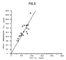
- Fig. 3 is a scatter plot of the correlation between a conventional T₄ agglutination assay which did not include trypsin and a reference radioimmunoassay method, also based on the experiments of Example I.
- Fig. 4 is a graphical illustration of the absorbence at various T₄ levels using the Protected Binding Assay of the invention incorporating either T₄-Ficoll® or T₄-Dextran conjugates, based on the experiments of Example II.
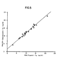
- Fig. 5 is a scatter plot of the correlation between the T₄ Protected Binding Assay of the invention using either trypsin or chymotrypsin as the only modification, as based on the experiments of Example III.
- Fig. 6 is a scatter plot of the correlation between the T₄ Protected Binding Assay of the invention using either trypsin or Pronase as the only modification, based on the experiments of Example IV.
- Fig. 7 is a scatter plot of the correlation between the theophylline Protected Binding Assay of the invention and a reference method, based on the experiments of Example V.
- Fig. 8 is a graphical illustration of the absorbence at various theophylline levels using the Protected Binding Assay of the invention incorporating either polyclonal or monoclonal antibody, based on the experiments in Example VI.
- Preferred embodiments of the present invention include a particle-associated agglutination assay reagent composition, a test kit comprised of containers or devices, each incorporated with one or more components of the test composition, in packaged combination with other components or materials and methods of using the test composition and kit of the invention. Specific terms in the following description which refer only to a particular embodiment are exemplary of all of the embodiments unless otherwise indicated.
- Sample fluids on which tests are performed include biological, physiological, industrial, environmental, and other types of liquids. Of particular interest are biological fluids such as serum, plasma, urine, cerebrospinal fluid, saliva, milk, broth and other culture media and supernatants as well as fractions of any of them. Physiological fluids of interest include infusion solutions, buffers, preservative or antimicrobial solutions and the like. Industrial liquids include fermentation media and other processing liquids used, for example, in the manufacture of pharmaceuticals, dairy products and malt beverages. Other sources of sample fluid which are tested by conventional methods are contemplated as within the meaning of this term as used and can, likewise, be assayed in accordance with the invention.
- In the context of this invention, the expression "non-protein ligand" refers to any substance, or class of related substances, which are not susceptible to protein-inactivating, i.e.proteolytic enzymes and whose presence is to be qualitatively or quantitatively determined in a sample fluid, such as those just described. The present assay can be applied to the detection of any such ligand for which there is a specific binding partner and, conversely, to the detection of the capacity of a liquid medium to bind such a ligand (usually due to the presence of a binding partner for the ligand in the sample). The ligand usually is an organic molecule for which a specific binding partner exists or can be developed. The ligand, in functional terms,is usually selected from antigens, haptens, hormones, vitamins, metabolites and pharmacological agents and their receptors and binding substances. Specific examples of non-protein ligands which can be detected using the present invention are hormones such as thyroxine (T₄) and triiodothyronine (T₃); antigens and haptens such as ferritin, bradykinin, prostaglandins, and tumor specific antigens; vitamins such as biotin, vitamin B₁₂ folic acid, vitamin E, vitamin A, and ascorbic acid; metabolites such as 3', 5'-guanosine monophosphate, pharmacological agents or drugs such as aminoglycoside antibiotics like gentamicin, amikacin and sisomicin, or drugs of abuse such as the opium alkaloids and ergot derivatives.
- The terminology "specific binding protein" or "receptor" refers to any substance, or class of substances, which has a specific binding affinity for the non-protein ligand to the exclusion of other substances. In the majority of embodiments, the present invention will incorporate specific binding assay reagents which interact with the ligand or its binding capacity in the sample in an immunochemical manner. That is, there will be an antigen-antibody or hapten-antibody relationship between reagents and/or the ligand or its binding capacity in the sample. Such assays therefore are termed immunoassays and the special interaction between the ligand and its receptor, or binding partner, is an immunochemical binding. The use of either polyclonal or monoclonal antibodies is contemplated. Additionally, it is well understood in the art that other binding interactions between the ligand and the binding partner serve as the basis of specific binding assays, including the binding interactions between hormones, vitamins, metabolites, and pharmacological agents, and their respective receptors and binding substances. For example, polypeptide hormone receptors as binding agents or partners are discussed in Langan, et al, (Eds.) Ligand Assay, Masson Publishing U.S.A. Inc., New York, pages 211 et seq. (1981).
- The proteolytic enzyme of the present invention is any enzyme which is effective to negate the chemical or biological properties of a protein, usually an endogenous protein, which causes it to produce non-specific interference effects on specific binding agglutination assay reactions. The use of such enzymes in reducing or eliminating these interfering effects has been described in pretreatment of samples apart from the actual assay procedures. Any of the enzymes so described or having a substantially similar effect can be used. These primarily include proteolytic enzymes such as trypsin, pepsin, chymotrypsin, carboxypeptidase and mixtures of proteases such as Pronase mixed enzyme preparation (Calbiochem-Behring Corp., LaJolla, CA]. Such protein inactivating enzymes are discussed at length in Perlmann & Lorand (Eds.), Methods in Enzymology -Proteolytic Enzymes (Vol. XIX), Academic Press, N.Y. (1970).
- The "solid state" or "solid phase" of the present invention can take on a multitude of forms, and is therefore intended as being broad in context. It can be mono- or multi-phasic, comprising one or more appropriate materials or mediums of similar or different absorptive or other physical characteristics. It can be hydrophobic or hydrophilic, bibulous or nonporous. In its most efficient embodiment the solid phase can be carefully tailored to suit the characteristics of the particular specific binding assay system to be employed.
- In one embodiment the solid phase is a matrix or surface capable of being incorporated with specific binding assay reagents. It can take on many known forms such as those utilized for chemical and enzymatic solution analysis. Solid phase test devices have been applied to specific binding assays. A commonly used solid phase device comprises a nonporous surface, such as the interior surface of a test tube or other vessel, to which antibody is affixed or coated by adsorption or covalent coupling. Likewise, devices for use in specific binding assays wherein the antibody reagent is fixed to a matrix held in a flow-through column are known (U.S. Patents Nos. 4,036,947; 4,039,652; 4,059,684; 4,153,675; and 4,166,102). U.S. Patents Nos. 3,826,619; 4,001,583; 4,017,597; and 4,105,410 relate to the use of antibody coated test tubes in radioimmunoassays. Solid phase test devices have also been used in enzyme immunoassays (U.S. Patents Nos. 4,016,043 and 4,147,752) and in fluorescent immunoassays (U.S. Patents Nos. 4,025,310 and 4,056,724; and British Patent Spec. No. 1,552,374). The use of such heterogeneous specific binding assay test devices is exemplified by the method of U.S. Patent No. 4,135,884. The test device is incorporated with the antibody reagent and is brought into contact with the liquid sample and with the remaining reagents of the reaction system. After an incubation period, the solid phase device is physically removed from the reaction solution and the label measured either in the solution or on the test device. In one preferred embodiment the element can be in the form of a test slide, made from a material such as polystyrene, which has been molded to have at least one reaction well, usually centrally disposed therein. The well usually has a diameter of from about 1.0 to about 2.5 centimeters (cm) and has a depth of from about 1.0 to about 10 millimeters (mm), preferably from about 2 mm to about 6 mm.
- The most preferred solid phase format is particulate. As previously noted, particle-associated agglutination assays include those assays using particles having specific binding pair members as surface components or substrate particles to which such components have been bound. These substrate particles preferably range in size from about 0.1 to about 5.0 microns in diameter, with bacterial substrate particles usually ranging from about 1 to about 3 microns in diameter. Substrate particles which have been used as carriers of specific binding pair members include eucaryotic red blood cells (unaltered or tanned), siliceous earth (e.g., bentonite), latex, and procaryotic particles (e.g., bacterial cells). The so-called latex particles consist normally of a synthetic polymeric material such as polystyrene. Other suitable organic polymers include butadiene, styrene-butadiene copolymers, acrylic polymers or mixtures thereof. Procaryotic particles such as bacterial or fungal cells or virus particles have also been widely used as substrate particles in agglutination assays. Among the bacteria used as substrate particles are those from the genus Staphylo coccus,particularly Staphylococcus aureus (S. aureus). For example, S. aureus (Cowan I type) has been bound, through protein A molecules extending from the cell wall surface to a binding partner.
- Many organic molecules will readily adhere by non-covalent adsorption to such substrate particles. For example, red blood cells (erythrocytes) readily adsorb many polysaccharides. Also, the protein A in S. aureus, Cowan I strain, specifically binds to the Fc locus of certain immunoglobulins. For the attachment of proteins, however, it is usually necessary to first treat the particles or covalently link the proteins, such as through linking groups. Linking groups which have been used include bis-diazo-benzidine, glutaraldehyde and 1, 3-difluoro-4, 6-dinitrobenzene. Others are discussed in Fudenberg, et al, (Eds.), Basic & Clinical Immunology, Lange Medical Publications, Los Altos, CA., pg. 310 (1976).
- The "substance which protects the specific binding protein" of the specific binding pair from enzyme inactivation when bound with its partner is contemplated as being any natural or synthetic molecule which is not, itself, suspectible to the proteolytic enzyme and can sequester, disguise, preferentially bind or otherwise associate with the specific binding protein to render it insusceptible to the proteolytic enzyme. Substances which can be used for this purpose include large molecules which sterically protect the binding protein from enzyme attack when bound. Exemplary of such large molecules are high molecular weight polymers like dextran or Ficoll®, (Pharmacia Fine Chemicals, Inc., New Market, NJ) which is a polymeric product of the reaction between epichlorohydrin and sucrose.
- The test material of the invention includes a conjugate comprising the binding partner for the species associated with the solid phase whether it be specific binding protein or ligand, covalently bound to a substance which protects the specific binding protein of the pair from enzyme inactivation when bound with its partner. The conjugate is formed using conventional organic synthesis techniques which do not impair or alter the specificity of the binding partner which it includes.
- Although it is not a theory on which the invention must be predicated, at least one mechanism for the unexpected agglutinated binding protein protection results achieved can be described. In all cases there is a solid phase incorporated with one partner of a specific binding pair comprising the ligand or a binding analog thereof and a specific binding protein therefor. In the mechanism postulated here, the conjugate comprises the other partner of said specific binding pair, which has an avidity and is present in a concentration which together provide a first association rate, covalently bound to a substance that protects the specific binding protein of said pair from enzyme inactivation when bound with its partner and the active protein-inactivating enzyme has an avidity and is present in a concentration which together provides a second association rate which is less than the first association rate.
- In accordance with this and other possible mechanisms the specific binding protein, e.g., antibody, is present in the reaction mixture in a concentration of at least about 0.04 percent (%) weight volume (w/v) , and preferably from about 0.04 percent (w/v) to about 0.07 percent (w/v). Likewise, the proteolytic enzyme is present in a concentration of not more than about 4.0 milligrams per milliliter (mg/ml), and preferably from about 2.0 to about 4.0 mg/ml. One example of this is a specific binding assay composition or material which includes (a) a particle-associated thyroxine or triiodothyronine antibody in a concentration of from about 0.04 to about 0.07 percent (w/v); (b) trypsin in a concentration of from about 2.0 to about 4.0 mg/ml; and (c) a conjugate as described above in a concentration of from about 0.22 to about 0.88 micrograms per milliliter (ug/ml).
- The following working Examples describe experiments which were performed in developing the present invention. Standard commercially available reagent grade chemicals were used whenever possible.
- Measurement of total serum thyroxine (T₄) is the single most important test for determining thyroid function. The normal T₄ range is 4.5 to 12.0 micrograms/deciliter (ug/dl); however, the test must be able to detect concentrations as low as 1.0 ug/dl and as high as 24.0 ug/dl. This is necesary to accurately identify those patients who have thyroid disorders. This Example reports experiments which demonstrate a nonisotopic, homogeneous T₄ immunoassay in accordance with the invention.
- Antibody to T₄ was induced in New Zealand white rabbits by an intradermal primary injection of 400 ug of a conjugate which was T₄ covalently coupled to bovine serum albumin (BSA) emulsified in an equal volume of Freund's complete adjuvant. Secondary booster immunizations contained 400 ug of T₄-BSA conjugate emulsified in an equal volume of incomplete Freund's adjuvant and were administered once a month. The animals were bled three times a week.
- Antibody specific for T₄ was isolated by immunoadsorbtion. The immunoadsorbent consisted of T₄ covalently bound to Sepharose® 4B (Pharmacia Fine Chemicals, Piscataway, NJ). This material was prepared using the bisoxirane method described by Sundberg and Porath in J. Chromatog ., 90:87-98 (1974). Five milliliters (ml of the immunoadsorbent were packed on top of 25 ml of Sephadex® G-25 (Pharmacia, supra) in a 2 x 20 centimeter (cm) glass chromatography column. Five ml of antisera, prepared as described above, were applied to the column and allowed to enter the column until the red or amber color reached the immunoadsorbent-Sephadex® interface. The antisera was allowed to remain in contact with the adsorbent for an additional 30 minutes at room temperature. The immunoadsorbent was then washed with two column volumes of barbital buffered saline (0.05M barbital, 0.15M
NaCl
, 0.1%
NaN₃
, pH 8.6) followed by a quantity of borate buffered saline (0.04M borate, 0.15M NaCl, 0.1% NaN₃,' pH 8.1) sufficient to bring the absorbence of the effluent at 280 nanometers (nm) to less than 0.01. At this point the adsorbed antibody was eluted with 1.0M acetic acid. The eluant was collected in 1 ml aliquots with a fraction collector. Those fractions with an absorbence at 280 nm greater than 0.1 and a pH greater than 7.0 were pooled and stored at -20° centigrade (C) until used. Typically 1-2 milligrams (mg) of purified antibody were isolated from 1 ml of antisera. - Purified antibody, described above, is coupled to chloromethyl styrene latex by a modification of the method described in Masson, et al, Methods In Enzymology, 79:106-139 (1981). A preparation using the above described antibodies in the method exactly as described by Masson, et al, ibid. can also be used.
- T₄ was covalently coupled to cyanogen bromide activated Ficoll® 400 (Pharmacia, supra). One hundred milligrams (mg) of Ficoll® was dissolved in 6 ml of 0.4M
K₂CO₃
and the pH was adjusted to 11.0. The solution was rapidly stirred and three, fifty ul additions of cyanogen bromide solution (333 mg/ml in N,N-dimethylformamide) were made. The pH of the Ficoll® solution was maintained at 11.0 by the addition of 2N
NaOH
. Each addition of cyanogen bromide was made within one minute of the previous addition and after the last addition the solution was incubated for four minutes. The pH was then dropped to 10.0 with 2N HCl and 0.5 ml of a T₄ solution was added (80 mg/ml in N,N-dimethyl-formamide made alkaline with 11-15 drops of 0.4M
K₂CO₃
). The reaction mixture was stirred for two hours at room temperature then dialyzed exhaustively against 0.01M
Na₂HPO₄
(pH 9.0). Typically, 60-70 moles of T₄ were incorporated into each mole of Ficoll. - T₄-Ficoll® conjugate, prepared as described above, polyethylene glycoll, 8000 (J. T. Baker Chemical Co., Phillipsburg, NJ), Tween®-20 (Technicon Instruments Corp., Tarrytown, NY),
CaCl₂
, and bovine pancreatic trypsin (Sigma Chemical Co., St. Louis, MO) were mixed such that the final concentrations were 1.2 ug/ml, 3.76% (w/v), 0.11% (v/v), 1mM, and 7.3 mg/ml, respectively, in barbital buffered saline (pH 8.6). - Antibody sensitized latex, prepared as described above, 8-anilino-1-naphthalenesulfonic acid (Eastman Kodak Co., Rochester, NY), Tween®-20 and Ficoll® 400 were mixed in a buffer consisting of 0.035M carbonate, 0.015M barbital, 0.15M sodium chloride and 0.1% sodium azide (pH 9.7), such that the final concentrations were 0.17% (w/v), 0.103 mg/ml, 0.11% (v/v/), and 0.86 mg/ml, respectively.
- The reference method was a solid phase coated tube Gamma CoatTM radioimmunoassay (RIA) (Clinical Assays, Division of Travenol Laboratories, Inc., Cambridge, MA] and was used according to the manufacturer's directions.
- The assay of the present invention may be performed using a spectrophotometer or an automated clinical chemistry analyzer. A Technicon RA-1000TM instrument system was used for most of the results reported here. In each of the experiments described below the assay was initiated by adding 26 ul of test serum, control or standard to 175 ul of T₄-Ficoll®/trypsin reagent. The reaction mixture was then incubated for two minutes at 37°C before adding 175 ul of antibody sensitized latex reagent. The complete reaction mixture was incubated for an additional minute, then the absorbence at 600 nm was recorded at 15 second intervals for two minutes. The rate of change of absorbence was determined by least squares fitting of the absorbence and time data. This slope was inversely proportional to the concentration of T₄ in the test sample. Log/logit transformation of the data produced linear standard curves which were then used to analyze unknowns, as described below.
- As an initial step, T₄-Ficoll® dependent protection of bound antibody was demonstrated. Antibody sensitized latex reagent, T₄-Ficoll®/trypsin reagent, and a serum-based standard without T₄ were mixed in a 0.5 centimeter (cm) quartz cuvette. The cuvette was placed in a Beckman DU-8 spectrophotometer (Beckman Instruments, Inc., Fullerton, CA) and the absorbence at 600 nm was monitored as a function of time. The rate of agglutination is directly proportional to the rate of change in the optical density, as noted in Faure, et al, Protides of the Bio logical Fluids, 20:589-593 (1972), and Singer, et al, Journal of Colloid and Interface Science, 45:608-614 (1973). The data shown in Fig. 1 demonstrates a rapid increase in optical density that begins to plateau after about a ten minute incubation period. These particles remained agglutinated even after overnight incubation.
- Next, one hundred three (103) human sera were analyzed by the method of the invention, automated with a Technicon RA-1000 instrument system, and by the RIA reference procedure described above to determine the degree with which the results from the two methods correlated. The data are shown in Fig. 2. The correlation data were analyzed by orthogonal linear regression and are summarized below.
-

- For purposes of comparison with the above, 21 human sera were analyzed as described above for the present invention with the exception that trypsin was not included in the reaction mixture. The data are shown in Fig. 3 and summarized below.
-

- The data shown in Fig. 1 demonstrate that specific antibody-latex/T₄-Ficoll®agglutinates will form and persist in the presence of high concentrations of trypsin. One possible explanation for this observation is that the combination of T₄-Ficoll®with the antibody-latex sterically inhibits the approach of the proteolytic enzyme and prevents the enzyme from degrading the antibody.
- The improvement in the method brought about by including a proteolytic enzyme, such as trypsin, in the reaction mixture is demonstrated by the difference between Figs. 2 and 3. As shown in Fig. 3, in the absence of trypsin the values produced by the latex immunoassay were about twice as high as the reference method. This was due to non-specific serum interference and resulted in serum samples having erroneously elevated values. This non-specific interference was destroyed by trypsin as demonstrated in Fig. 2, which shows excellent correlation between the latex immunoassay with trypsin and the reference method.
- The experiments reported here compare the use of T₄-Dextran with the use of T₄-Ficoll® conjugate. The antisera preparation, antibody purification, antibody sensitized latex and antibody sensitized latex reagent preparations, T₄-Ficoll® conjugate and T₄-Ficoll® conjugate/trypsin reagent preparations, analytical procedure used were identical to those described in Example I.
- T₄ was covalently coupled to cyanogen bromide activated Dextran, T500 (Pharmacia, supra) using exactly the same procedure as described for T₄-Ficoll® in Example I.
- T₄-Dextran, polyethylene Glycol 8000, Tween®-20,
CaCl₂
, and bovine pancreatic trypsin were mixed such that the final concentrations were 1.0 ug/ml, 3.76% (w/v), 0.11% (v/v), 1 mM, and 7.3 mg/ml, respectively. All of the materials were dissolved in barbital buffered saline (pH 8.6). - T₄ standard curves were measured using the Technicon RA-1000 instrument system and either T₄-Ficoll® or T₄ -Dextran. The data shown in Fig. 4 indicate that both T₄-Ficoll® and T₄-Dextran function as "protectors" in this T₄ binding protection latex immunoassay. Larger signal changes were observed with T₄-Ficoll®than with T₄-Dextran. However, the signal obtained with T₄-Dextran is increased by raising the polyethylene glycol concentration.
- Both T₄-Dextran and T₄-Ficoll® function in the antigen protection latex immunoassay and produce appropriate dose response curves. High molecular weight non-protein molecules constitute an effective class of materials for this purpose.
- The experiments reported here compare the use of chymotrypsin as the proteolytic enzyme with the use of trypsin. The antisera preparation, antibody purification, antibody sensitized latex and antibody sensitized latex reagent preparations, T₄-Ficoll® conjugate and T₄-Ficoll® conjugate/trypsin reagent preparations, and analytical procedure used were identical to those described in Example I.
-
- The reagent was prepared exactly as described in Example I for the T₄-Ficoll® conjugate/trypsin reagent, except that bovine pancreatic 2-chymotrypsin (Sigma Chemical Co., supra) was included in the reagent at a final concentration of 7.3 mg/ml.
- Twenty-one human sera were analyzed by the antigen protection latex immunoassay using the Technicon RA-1000 instrument system in the presence of either trypsin or chymotrypsin. The data are presented by the scatter plot in Fig. 5, and analyzed by orthogonal linear regression below.
-

- Chymotrypsin may be used in place of trypsin in the Protected Binding Assay of the invention. As shown in Fig. 5, equivalent results were obtained when either trypsin or chymotrypsin were included in the reaction mixture. The T₄-Ficoll®provided protection for bound antibody against the proteolytic effects of chymotrypsin, yet the chymotrypsin digested serum proteins in such a way that non-specific interactions with the latex particle were eliminated and correct T₄ values were produced.
- The experiments reported here compare the use of Pronase (B Grade, Calbiochem - Behring Corp., LaJolla, CA) proteolytic enzyme mixture with the use of trypsin. The antisera preparation, antibody purification, antibody sensitized latex and antibody sensitized latex reagent preparations, T₄-Ficoll® conjugate and T₄-Ficoll® conjugate/trypsin reagent preparations, and analytical procedure used were identical to those described in Example I.
- This reagent was prepared exactly as described in Example I for the T₄-Ficoll® conjugate/trypsin reagent, except that Pronase was included in the reagent at a final concentration of 7.3 mg/ml.
- Twenty-one human sera were analyzed by the antigen protection latex immunoassay, using a Technicon RA-1000 instrument system, in the presence of either trypsin or Pronase. The data are presented by the scatter plot in Fig. 6 and were analyzed by orthogonal linear regression below.
-

- Pronase may be used in place of trypsin in the Protected Binding Assay of the invention. As shown in Fig. 6, equivalent results were obtained when either trypsin or Pronase was used in the assay. Both enzymes produced about the same average value of T₄, 11.2 ug/dl for trypsin and 11.6 ug/dl for Pronase, and the slope of the correlation plot was 1.02. The T₄-Ficoll® in the reaction mixture protected the antibody from proteolytic degradation by trypsin, yet destroyed those proteins that interfered in the measurement of T₄.
- Theophylline (1,3-dimethylxanthine) is commonly used in the treatment of bronchial asthma. The serum concentration of the drug must be closely monitored since the drug has a narrow therapeutic range of 10-20 ug/ml while drug concentrations in excess of 20 ug/ml may be toxic. The experiments reported below demonstrate that the antigen protection latex immunoassay of the invention can be used to accurately and precisely quantitate serum levels of theophylline.
- Rabbit anti-theophylline antisera were purchased from Kallestad, Austin, TX.
- Antibody was isolated by ammonium sulfate precipitation. Antisera was mixed in equal volumes with saturated ammonium sulfate solution which had been previously adjusted to
pH 8. The solution was stirred at 4°C for two hours and the precipitated antibody was then isolated by centrifugation. The pellet was washed twice with a volume of 50% saturated ammonium suflate, pH 8.0, that was equal to the original volume of antiserum. The precipitate was then solublized in a volume of borate buffer saline (pH 8.1) that was equal to the original volume of antiserum. The antibody solution was then exhaustively dialyzed against borate buffered saline (pH 8.1) and clarified by centrifugation. The isolated antibody was stored at -20°C until used. - Purified antibody, described above, is coupled to chloromethyl styrene latex by a modification of the method described in Masson, et al, Methods in Enzymology, 74:106-139 (1981). A preparation using the above-described antibodies in the method exactly as described by Masson, et al, ibid. can also be used.
- 8-E carboxypropyl theophylline was synthesized exactly as described in Cook et al, Research Communications in Chemical Pathology and Pharmacology, 13:497-505 (1976).
- N-(2-aminoethyl) carbamylmethylated-Ficoll® (AECM-Ficoll) was synthesized exactly as described in Inman, J. Immunol. 114:704-709 (1975).
- 8-E carboxypropyltheophylline was covalently coupled to AECM-Ficoll® using a carbodiimide coupling. Twenty-three mg of 1-(3-Dimethyl amino-propyl)-3-ethylcarbodiimide hydrochloride (Aldrich Chemical Co., Metuchen, NJ) was added to 10 mg of 8-E-carboxypropyl-theophylline dissolved in 1 ml of N,N-dimethylformamide. The solution was allowed to activate for 40 minutes at room temperature, then 320 ul of this solution was added to 0.5 ml of a 10 mg/ml solution of AECM-Ficoll® dissolved in 0.1M carbonate buffer (pH 6.3). The reaction mixture was incubated overnight at 4°C. Excess reagents were removed by sequential dialysis against 2 liters of of 0.1M sodium acetate buffer (pH 4.5), 2 liters of 0.1M sodium carbonate buffer (pH 9.5), 2 liters of borate buffered saline (pH 8.1) and again against 2 liters of fresh borate buffered saline (pH 8.1). Sixty to seventy moles of theophylline were incorporated into each mole of AECM-Ficoll.®
- Theophylline-Ficoll® conjugate, prepared as described above, Tween®-20,
CaCl₂
and bovine pancreatic trypsin were mixed such that the final concentrations were 1.25 ug/ml, 0.1% (v/v), 1mM, and 6 mg/ml, respectively, in borate buffered saline (pH 8.1). - Anybody sensitized latex, prepared as described above, and Tween®-20 were mixed in borate buffered saline (pH 8.1) containing 0.1% (w/v) bovine serum albumin. The final concentration of the antibody sensitized latex was 0.12% (w/v) and the Tween®-20 was 0.1% (v/v).
- The reference method was the Emit-aad theophylline assay (Syva, Palo Alto, CA) and was used according to the manufacturer's direction.
- The assay of the present invention may be performed using either a spectrophotometer or an automated clinical chemistry analyzer. A Technicon RA-1000TM instrument system was used for all of the results reported here. The assay was initiated by adding 2 ul of test serum, control or standard to 200 ul of antibody sensitized latex reagent. The reaction mixture was then incubated for 15 seconds at 37°C before adding 200 ul of theophylline-Ficoll®/trypsin reagent. The complete reaction mixture was incubated for an additional 30 seconds at 37°C, then the absorbence at 600 nm was recorded at 15 second intervals for two minutes. The rate of change of absorbence was determined by least-squares fitting of the absorbence and time data. This slope was inversely proportional to the concentration of theophylline in the test sample. Log/logit transformation of the data produced linear standard curves which were then used to analyze unknowns.
- Eighty-one human sera were obtained from patients being treated with theophylline. These sera were analyzed by the method of the present invention employing the Kallestad polyclonal antibody and by the Syva Emit reference method to determine the degree with which the results from the two methods correlated. The data are shown in Fig. 7. The correlation data were analyzed by orthogonal linear regression and are summarized below.
- N Mean X Mean Y Slope Y-Intercept V
- 81 10.6ug/ml 11.5ug/ml 1.09 -0.06ug/ml 0.99
- The correlation data indicate excellent agreement between the theophylline protected binding immunoassay and the reference method. The average value produced by the present method, 11.5 ug/ml, is almost identical to the average value produced by the reference method, 10.6 ug/ml. The slope of the scatter plot was close to unity, the intercept negligible, and the correlation coefficient was 0.99, all of which indicate that the method produces results equivalent to the reference method.
- The experiments reported here compare polyclonal and monoclonal antibodies in the theophylline Protected Binding Assay. All of the materials, preparations and procedures were identical to those described in Example V, with the addition that monoclonal mouse anti-theophylline was also purchased from Kallestad, Austin, TX.
- The dose response curves obtained with the polyclonal and monoclonal antibodies are shown in Fig. 8. The two antibodies show similar dynamic range and sensitivities when used in the assay of the invention.
- Polyclonal and monoclonal antibodies may be used in the assay of the invention.
Claims (10)
- A specific binding assay test composition for determination of a non-protein ligand in a sample, which test composition comprises:(a) a solid phase incorporated with one partner of a specific binding pair comprising said ligand or a binding analog thereof and a specific binding protein therefor;(b) a conjugate comprising the other partner of said specific binding pair covalently bound to a substance which protects the specific binding protein of said pair from enzyme inactivation when bound with its partner; and(c) a proteolytic enzyme.
- A composition according to claim 1 wherein said solid phase incorporated with a specific binding partner comprises a particle having a specific binding protein surface moiety, or a substrate particle bound to a specific binding protein.
- A composition according to claim 2, wherein said substrate particle is selected from eucaryotic cells, procaryotic cells and synthetic particles such as latex.
- The composition of claim 1 wherein said specific binding protein is an antibody in a concentration of from 0.04 to 0.07 percent.
- A composition according to claim 1,2,3 or 4, wherein said conjugate of said non-protein ligand comprises a ligand or specific binding analog thereof covalently bound to a high molecular weight carrier.
- A composition according to claim 5, wherein said high molecular weight carrier is dextran, or a cross-linking product of epichlorohydrin and sucrose.
- A composition according to any of claims 1 to 6, wherein said proteolytic enzyme is trypsin or pepsin and is present in a concentration of not more than ` 4.0 milligrams per milliliter.
- A specific binding assay test composition for thyroxine or triiodothyronine, which composition comprises:(a) particle-associated thyroxine or triiodothyro ` nine antibody, preferably present in a concentration of from 0.04 to 0.07 percent(w/v);(b) trypsin, preferably present in a concentration of from 2.0 to 4.0 milligrams per milliliter; and(c) a conjugate comprising thyroxine or triiodothyronine covalently bound with a high molecular weight polymer, said conjugate preferably being present in a concentration of from 0.22 to 0.88 micrograms per milliliter.
- A specific binding method of assaying for a non-protein ligand in a sample, which method consists essentially of the steps of:i) combining said sample in a reaction mixture with a test composition as claimed in any preceding claim; andii) detecting any resultant binding in said same reaction mixture.
- A test kit for use in determining a non-protein ligand in a sample by a specific binding assay method, which kit comprises a packaged combination of one or more components incorporated with:(a) a solid phase incorporated with one partner of a specific binding pair comprising said non-protein ligand or a binding analog thereof and a specific binding protein therefor;(b) a conjugate comprising the other partner of said specific binding pair covalently bound to a substance which protects the specific binding protein of said pair from enzyme inactivation when bound with its partner; and(c) a proteolytic enzyme.
Applications Claiming Priority (2)
| Application Number | Priority Date | Filing Date | Title |
|---|---|---|---|
| US06/490,063 US4650751A (en) | 1983-04-29 | 1983-04-29 | Protected binding assay avoiding non-specific protein interference |
| US490063 | 1983-04-29 |
Publications (3)
| Publication Number | Publication Date |
|---|---|
| EP0124352A2 EP0124352A2 (en) | 1984-11-07 |
| EP0124352A3 EP0124352A3 (en) | 1986-09-17 |
| EP0124352B1 true EP0124352B1 (en) | 1991-03-06 |
Family
ID=23946476
Family Applications (1)
| Application Number | Title | Priority Date | Filing Date |
|---|---|---|---|
| EP84302813A Expired - Lifetime EP0124352B1 (en) | 1983-04-29 | 1984-04-26 | Protected binding assay |
Country Status (6)
| Country | Link |
|---|---|
| US (1) | US4650751A (en) |
| EP (1) | EP0124352B1 (en) |
| JP (1) | JPH068822B2 (en) |
| AU (1) | AU579999B2 (en) |
| CA (1) | CA1231049A (en) |
| DE (1) | DE3484209D1 (en) |
Families Citing this family (30)
| Publication number | Priority date | Publication date | Assignee | Title |
|---|---|---|---|---|
| US4870007A (en) * | 1987-12-18 | 1989-09-26 | Eastman Kodak Company | Immobilized biotinylated receptor in test device, kit and method for determining a ligand |
| US5362322A (en) * | 1990-12-17 | 1994-11-08 | C-Cure Chemical Company, Inc. | Color epoxy grout system and method for use |
| US5874216A (en) * | 1996-02-23 | 1999-02-23 | Ensys Environmental Products, Inc. | Indirect label assay device for detecting small molecules and method of use thereof |
| DE10064827A1 (en) | 2000-12-22 | 2002-06-27 | Dade Behring Marburg Gmbh | Sandwich assay for detecting analyte, useful e.g. for hormones, with detection or correction of the hook effect by measuring detection signals twice |
| US20040132092A1 (en) * | 2003-01-03 | 2004-07-08 | Stetson Christopher M. | Determining the density of functional moieties on polymer reagents |
| US20050079132A1 (en) * | 2003-04-08 | 2005-04-14 | Xingwu Wang | Medical device with low magnetic susceptibility |
| US20050025797A1 (en) * | 2003-04-08 | 2005-02-03 | Xingwu Wang | Medical device with low magnetic susceptibility |
| US8071134B2 (en) * | 2003-09-15 | 2011-12-06 | Ordway Research Institute, Inc. | Thyroid hormone analogs and methods of use |
| CA2539288C (en) | 2003-09-15 | 2015-05-12 | Shaker A. Mousa | Thyroid hormone analogs and methods of use |
| US8668926B1 (en) | 2003-09-15 | 2014-03-11 | Shaker A. Mousa | Nanoparticle and polymer formulations for thyroid hormone analogs, antagonists, and formulations thereof |
| US9198887B2 (en) | 2003-09-15 | 2015-12-01 | Nanopharmaceuticals Llc | Thyroid hormone analogs and methods of use |
| US20060057180A1 (en) * | 2004-02-20 | 2006-03-16 | Ashutosh Chilkoti | Tunable nonfouling surface of oligoethylene glycol |
| US7172906B2 (en) * | 2004-11-16 | 2007-02-06 | Dade Behring Inc. | Reduction of non-specific binding in assays |
| US20090022806A1 (en) * | 2006-12-22 | 2009-01-22 | Mousa Shaker A | Nanoparticle and polymer formulations for thyroid hormone analogs, antagonists and formulations and uses thereof |
| US9498536B2 (en) | 2005-09-15 | 2016-11-22 | Nanopharmaceuticals Llc | Method and composition of thyroid hormone analogues and nanoformulations thereof for treating anti-inflammatory disorders |
| US9220788B2 (en) | 2009-06-17 | 2015-12-29 | Nanopharmaceuticals Llc | Nanoparticle and polymer formulations for thyroid hormone analogs, antagonists, and formulations and uses thereof |
| US10130686B2 (en) | 2005-09-15 | 2018-11-20 | Nanopharmaceuticals Llc | Method and composition of thyroid hormone analogues and nanoformulations thereof for treating inflammatory disorders |
| WO2007035612A2 (en) | 2005-09-16 | 2007-03-29 | Ordway Research Institute, Inc. | Polyphenol conjugates as rgd-binding compounds and methods of use |
| WO2008140507A2 (en) * | 2006-12-22 | 2008-11-20 | Clf Medical Technology Acceleration Program, Inc. | Nanoparticle and polymer formulations for thyroid hormone analogs, antagonists, and formulations and uses thereof |
| WO2010075332A1 (en) * | 2008-12-23 | 2010-07-01 | Charitable Leadership Foundation | Small molecule ligands of the integrin rgd recognition site and methods of use |
| WO2010120506A1 (en) * | 2009-03-31 | 2010-10-21 | Ordway Research Institute, Inc. | Combination treatment of cancer with cetuximab and tetrac |
| US8802240B2 (en) | 2011-01-06 | 2014-08-12 | Nanopharmaceuticals Llc | Uses of formulations of thyroid hormone analogs and nanoparticulate forms thereof to increase chemosensitivity and radiosensitivity in tumor or cancer cells |
| WO2015074050A1 (en) | 2013-11-18 | 2015-05-21 | Nanopharmaceuticals Llc | Methods for screening patients for resistance to angioinhibition, treatment and prophylaxis thereof |
| CA3026504A1 (en) | 2016-06-07 | 2017-12-14 | Nanopharmaceuticals, Llc | Non-cleavable polymer conjugated with .alpha..nu..beta.3 integrin thyroid antagonists |
| CN106501535B (en) * | 2016-11-30 | 2018-11-09 | 迪瑞医疗科技股份有限公司 | A kind of kit of the compound releasing agent of ANS salt and detection Blood cortisol |
| US10328043B1 (en) | 2018-04-11 | 2019-06-25 | Nanopharmaceuticals, Llc. | Composition and method for dual targeting in treatment of neuroendocrine tumors |
| US11351137B2 (en) | 2018-04-11 | 2022-06-07 | Nanopharmaceuticals Llc | Composition and method for dual targeting in treatment of neuroendocrine tumors |
| US10961204B1 (en) | 2020-04-29 | 2021-03-30 | Nanopharmaceuticals Llc | Composition of scalable thyrointegrin antagonists with improved blood brain barrier penetration and retention into brain tumors |
| US11723888B2 (en) | 2021-12-09 | 2023-08-15 | Nanopharmaceuticals Llc | Polymer conjugated thyrointegrin antagonists |
| CN116930513B (en) * | 2023-09-19 | 2023-11-17 | 北京索莱宝科技有限公司 | Protein-free rapid blocking liquid for protein detection and application thereof |
Family Cites Families (18)
| Publication number | Priority date | Publication date | Assignee | Title |
|---|---|---|---|---|
| NL154599B (en) * | 1970-12-28 | 1977-09-15 | Organon Nv | PROCEDURE FOR DETERMINING AND DETERMINING SPECIFIC BINDING PROTEINS AND THEIR CORRESPONDING BINDABLE SUBSTANCES, AND TEST PACKAGING. |
| US3962039A (en) * | 1974-08-08 | 1976-06-08 | Center For Laboratory Medicine | Analytical procedure for thyroid hormones |
| US4171244A (en) * | 1975-02-20 | 1979-10-16 | Syva Company | Enzyme-bound-polyidothyronine |
| US4032626A (en) * | 1976-06-30 | 1977-06-28 | Corning Glass Works | Reagent and method for tbg assay |
| US4134792A (en) * | 1976-12-06 | 1979-01-16 | Miles Laboratories, Inc. | Specific binding assay with an enzyme modulator as a labeling substance |
| GB1595101A (en) * | 1977-03-15 | 1981-08-05 | Hoffmann La Roche | Enzyme modifier immunoassay |
| US4225576A (en) * | 1978-11-20 | 1980-09-30 | Miles Laboratories, Inc. | Combined radioimmunoassay for triiodothyronine and thyroxine |
| US4273866A (en) * | 1979-02-05 | 1981-06-16 | Abbott Laboratories | Ligand analog-irreversible enzyme inhibitor conjugates and methods for use |
| CA1121345A (en) * | 1979-03-05 | 1982-04-06 | Robert A. Yoshida | Method for competitive protein binding assays inhibiting non-specific interference |
| DE3006709A1 (en) * | 1980-02-22 | 1981-08-27 | Hans A. Dipl.-Chem. Dr. 8000 München Thoma | HOMOGENEOUS METHOD FOR COMPETITIVE DETERMINATION OF LIGANDS |
| AU543007B2 (en) * | 1980-04-15 | 1985-03-28 | Technicon Instruments Corportion | Agglutination immunoassay |
| US4341865A (en) * | 1980-07-21 | 1982-07-27 | Abbott Laboratories | Determination of thyroxine binding globulin |
| US4347058A (en) * | 1980-09-15 | 1982-08-31 | Beckman Instruments, Inc. | Thyroid polarization fluoroimmunoassay |
| DE3168691D1 (en) * | 1980-11-07 | 1985-03-14 | Technicon Instr | Immunoassay of proteins |
| EP0056254A1 (en) * | 1981-01-14 | 1982-07-21 | David Eldon Wood | Treatment of insoluble surfaces to inhibit nonspecific protein binding |
| US4427781A (en) * | 1981-03-16 | 1984-01-24 | International Institute Of Cellular And Molecular Pathology | Particle agglutination immunoassay with agglutinator for determining haptens; PACIA |
| AU552573B2 (en) * | 1981-06-26 | 1986-06-05 | State Of Queensland, The | 8-anilino naphthalene-1- sulphonic acid analogues |
| US4578350A (en) * | 1983-09-23 | 1986-03-25 | Syntex (U.S.A.) Inc. | Immunoassays employing protected labels |
-
1983
- 1983-04-29 US US06/490,063 patent/US4650751A/en not_active Expired - Lifetime
-
1984
- 1984-04-24 CA CA000452611A patent/CA1231049A/en not_active Expired
- 1984-04-26 EP EP84302813A patent/EP0124352B1/en not_active Expired - Lifetime
- 1984-04-26 DE DE8484302813T patent/DE3484209D1/en not_active Expired - Fee Related
- 1984-04-27 JP JP59084170A patent/JPH068822B2/en not_active Expired - Lifetime
- 1984-04-27 AU AU27420/84A patent/AU579999B2/en not_active Ceased
Also Published As
| Publication number | Publication date |
|---|---|
| EP0124352A3 (en) | 1986-09-17 |
| JPH068822B2 (en) | 1994-02-02 |
| DE3484209D1 (en) | 1991-04-11 |
| US4650751A (en) | 1987-03-17 |
| AU2742084A (en) | 1984-11-01 |
| AU579999B2 (en) | 1988-12-22 |
| EP0124352A2 (en) | 1984-11-07 |
| CA1231049A (en) | 1988-01-05 |
| JPS6035264A (en) | 1985-02-23 |
Similar Documents
| Publication | Publication Date | Title |
|---|---|---|
| EP0124352B1 (en) | Protected binding assay | |
| US5521102A (en) | Controlled sensitivity immunochromatographic assay | |
| CA1179940A (en) | Solid phase system for ligand assay | |
| US4680274A (en) | Particles for inhibiting non-specific immunoreaction | |
| US4469787A (en) | Immunoassay involving soluble complex of second antibody and labeled binding protein | |
| US4948726A (en) | Enzyme immunoassay based on membrane separation of antigen-antibody complexes | |
| US5958790A (en) | Solid phase transverse diffusion assay | |
| EP0303110A2 (en) | Immunodiagnostic device and method | |
| EP0223843A4 (en) | Method and article for detection of immune complexes. | |
| AU757629B2 (en) | Methods for assaying antibody and device for assaying antibody | |
| CA2041782A1 (en) | Conjugate recovery binding assays and devices | |
| EP0239318A1 (en) | Assay employing binding pair members on particles and on a filter or membrane | |
| AU592971B2 (en) | Solid phase diffusion assay | |
| JPH02110374A (en) | Visual discrimination of qualitative enzyme assay | |
| US6686167B2 (en) | Test device for detecting semen and method of use | |
| EP0902287B1 (en) | Method and test strip for reducing urea effect of immunochromatographic assays using urine samples | |
| EP0141620B1 (en) | Specific binding assay reagent | |
| AU775771B2 (en) | Methods for assaying antibody and device for assaying antibody | |
| CA1111764A (en) | Test set for the detection of antigen associated with hepatitis by "sandwich" method | |
| WO1986007152A1 (en) | Method and article for detection of immune complexes | |
| WO2001044499A9 (en) | Modified labeled complement components for immunoassays | |
| Mack | Development of an enzyme-linked immunosorbent assay for porcine relaxin | |
| EP0586693A1 (en) | Technique for prevention of false reactions in immunological testing | |
| JPH06103305B2 (en) | Specific binding assay |
Legal Events
| Date | Code | Title | Description |
|---|---|---|---|
| PUAI | Public reference made under article 153(3) epc to a published international application that has entered the european phase |
Free format text: ORIGINAL CODE: 0009012 |
|
| AK | Designated contracting states |
Designated state(s): BE CH DE FR GB IT LI NL SE |
|
| PUAL | Search report despatched |
Free format text: ORIGINAL CODE: 0009013 |
|
| AK | Designated contracting states |
Kind code of ref document: A3 Designated state(s): BE CH DE FR GB IT LI NL SE |
|
| 17P | Request for examination filed |
Effective date: 19870306 |
|
| 17Q | First examination report despatched |
Effective date: 19881205 |
|
| RAP1 | Party data changed (applicant data changed or rights of an application transferred) |
Owner name: TECHNICON INSTRUMENTS CORPORATION(A DELAWARE CORPO |
|
| GRAA | (expected) grant |
Free format text: ORIGINAL CODE: 0009210 |
|
| AK | Designated contracting states |
Kind code of ref document: B1 Designated state(s): BE CH DE FR GB IT LI NL SE |
|
| ITF | It: translation for a ep patent filed |
Owner name: JACOBACCI & PERANI S.P.A. |
|
| REF | Corresponds to: |
Ref document number: 3484209 Country of ref document: DE Date of ref document: 19910411 |
|
| ET | Fr: translation filed | ||
| PLBE | No opposition filed within time limit |
Free format text: ORIGINAL CODE: 0009261 |
|
| STAA | Information on the status of an ep patent application or granted ep patent |
Free format text: STATUS: NO OPPOSITION FILED WITHIN TIME LIMIT |
|
| 26N | No opposition filed | ||
| PGFP | Annual fee paid to national office [announced via postgrant information from national office to epo] |
Ref country code: SE Payment date: 19920323 Year of fee payment: 9 |
|
| PGFP | Annual fee paid to national office [announced via postgrant information from national office to epo] |
Ref country code: CH Payment date: 19920326 Year of fee payment: 9 |
|
| PGFP | Annual fee paid to national office [announced via postgrant information from national office to epo] |
Ref country code: BE Payment date: 19920409 Year of fee payment: 9 |
|
| PGFP | Annual fee paid to national office [announced via postgrant information from national office to epo] |
Ref country code: NL Payment date: 19920430 Year of fee payment: 9 |
|
| PG25 | Lapsed in a contracting state [announced via postgrant information from national office to epo] |
Ref country code: SE Effective date: 19930427 |
|
| PG25 | Lapsed in a contracting state [announced via postgrant information from national office to epo] |
Ref country code: LI Effective date: 19930430 Ref country code: CH Effective date: 19930430 Ref country code: BE Effective date: 19930430 |
|
| BERE | Be: lapsed |
Owner name: TECHNICON INSTRUMENTS CORP. (A DELAWARE CORP.) Effective date: 19930430 |
|
| PG25 | Lapsed in a contracting state [announced via postgrant information from national office to epo] |
Ref country code: NL Effective date: 19931101 |
|
| NLV4 | Nl: lapsed or anulled due to non-payment of the annual fee | ||
| REG | Reference to a national code |
Ref country code: CH Ref legal event code: PL |
|
| EUG | Se: european patent has lapsed |
Ref document number: 84302813.5 Effective date: 19931110 |
|
| PGFP | Annual fee paid to national office [announced via postgrant information from national office to epo] |
Ref country code: FR Payment date: 20000330 Year of fee payment: 17 |
|
| PGFP | Annual fee paid to national office [announced via postgrant information from national office to epo] |
Ref country code: DE Payment date: 20000331 Year of fee payment: 17 |
|
| PGFP | Annual fee paid to national office [announced via postgrant information from national office to epo] |
Ref country code: GB Payment date: 20000403 Year of fee payment: 17 |
|
| PG25 | Lapsed in a contracting state [announced via postgrant information from national office to epo] |
Ref country code: GB Free format text: LAPSE BECAUSE OF NON-PAYMENT OF DUE FEES Effective date: 20010426 |
|
| PG25 | Lapsed in a contracting state [announced via postgrant information from national office to epo] |
Ref country code: FR Free format text: THE PATENT HAS BEEN ANNULLED BY A DECISION OF A NATIONAL AUTHORITY Effective date: 20010430 |
|
| GBPC | Gb: european patent ceased through non-payment of renewal fee |
Effective date: 20010426 |
|
| PG25 | Lapsed in a contracting state [announced via postgrant information from national office to epo] |
Ref country code: DE Free format text: LAPSE BECAUSE OF NON-PAYMENT OF DUE FEES Effective date: 20020201 |
|
| REG | Reference to a national code |
Ref country code: FR Ref legal event code: ST |