EP0116860B1 - The cloning and utilization of aminotransferase genes - Google Patents
The cloning and utilization of aminotransferase genes Download PDFInfo
- Publication number
- EP0116860B1 EP0116860B1 EP84100521A EP84100521A EP0116860B1 EP 0116860 B1 EP0116860 B1 EP 0116860B1 EP 84100521 A EP84100521 A EP 84100521A EP 84100521 A EP84100521 A EP 84100521A EP 0116860 B1 EP0116860 B1 EP 0116860B1
- Authority
- EP
- European Patent Office
- Prior art keywords
- tyrb
- gene
- plasmid
- aminotransferase
- dna
- Prior art date
- Legal status (The legal status is an assumption and is not a legal conclusion. Google has not performed a legal analysis and makes no representation as to the accuracy of the status listed.)
- Expired - Lifetime
Links
Images





Classifications
-
- C—CHEMISTRY; METALLURGY
- C12—BIOCHEMISTRY; BEER; SPIRITS; WINE; VINEGAR; MICROBIOLOGY; ENZYMOLOGY; MUTATION OR GENETIC ENGINEERING
- C12N—MICROORGANISMS OR ENZYMES; COMPOSITIONS THEREOF; PROPAGATING, PRESERVING, OR MAINTAINING MICROORGANISMS; MUTATION OR GENETIC ENGINEERING; CULTURE MEDIA
- C12N9/00—Enzymes; Proenzymes; Compositions thereof; Processes for preparing, activating, inhibiting, separating or purifying enzymes
- C12N9/10—Transferases (2.)
- C12N9/1096—Transferases (2.) transferring nitrogenous groups (2.6)
-
- C—CHEMISTRY; METALLURGY
- C12—BIOCHEMISTRY; BEER; SPIRITS; WINE; VINEGAR; MICROBIOLOGY; ENZYMOLOGY; MUTATION OR GENETIC ENGINEERING
- C12P—FERMENTATION OR ENZYME-USING PROCESSES TO SYNTHESISE A DESIRED CHEMICAL COMPOUND OR COMPOSITION OR TO SEPARATE OPTICAL ISOMERS FROM A RACEMIC MIXTURE
- C12P13/00—Preparation of nitrogen-containing organic compounds
- C12P13/04—Alpha- or beta- amino acids
-
- C—CHEMISTRY; METALLURGY
- C12—BIOCHEMISTRY; BEER; SPIRITS; WINE; VINEGAR; MICROBIOLOGY; ENZYMOLOGY; MUTATION OR GENETIC ENGINEERING
- C12P—FERMENTATION OR ENZYME-USING PROCESSES TO SYNTHESISE A DESIRED CHEMICAL COMPOUND OR COMPOSITION OR TO SEPARATE OPTICAL ISOMERS FROM A RACEMIC MIXTURE
- C12P13/00—Preparation of nitrogen-containing organic compounds
- C12P13/04—Alpha- or beta- amino acids
- C12P13/22—Tryptophan; Tyrosine; Phenylalanine; 3,4-Dihydroxyphenylalanine
Definitions
- This invention involves the construction and use of genetically engineered plasmids containing the tyrB gene.
- These plasmids are constructed using restriction endonucleases to contain the genes coding for said aminotransferase enzyme used during the synthesis of amino acids.
- These plasmids promote the synthesis of aminotransferases in bacterial cells. This synthesis allows increased yields of aminotransferase enzymes and the elimination of the aminotransferase reaction as a rate-limiting step in amino acid biosynthesis.
- the aspC gene product will catalyze the transamination of amino acid precursors to produce aspartate, glutamate, phenylalanine and tyrosine.
- the tyrB gene product will catalyze the transamination of amino acid precursors to produce phenylalanine, tyrosine, glutamate, aspartate and leucine.
- the ilvE gene product will catalyze the transamination of the amino acid precursors to produce isoleucine, valine, leucine, phenylalanine, glutamate and alanine.
- the location of the aspC, tyrB and ilvE genes on the genetic map of E. coli K12 strain is disclosed and discussed by Bachmann et al. (Microbiological Reviews, 44: 1-56, March 1980).
- the aspC gene located at E. coli K12 map position 20 minutes codes for an enzyme here called aspartate aminotransferase and given the Enzyme Commission Number E.C. 2.6.1.1 (Bachmann et al, p. 4).
- the tyrB gene located at E. coli K12 map position 91 minutes codes for an enzyme called tyrosine or aromatic aminotransferase and given the Enzyme Commission Number E.C. 2.6.1.5 (Bachmann, et al. p. 21 ).
- the ilvE gene located at E. coli K12 map position 84 minutes codes for an enzyme here called branched- chain-amino-acid aminotransferase and given Enzyme Commission Number E.C. 2.6.1.42 (Bachmann et al. p. 10).
- Amino acid transamination by aspC and tyrB gene products are disclosed by Gelfand et al., (J. Bacteriology, 130: 429 ⁇ 440, April 1977) to be responsible for essentially all the aminotransferase activity required for the de novo biosynthesis of tyrosine, phenylalanine, and aspartate in E. coli K12.
- the presence of the ilvE gene product alone can reverse a phenylalanine requirement indicating it has the ability to transaminate a precursor of phenylalanine.
- Transducing phage lambda has been used to carry DNA fragments for E. coli in the aspC region (Christensen and Pedersen Mol. Gen Genet, 181: 548-551, 1981).
- the aspC region in bacteriophage lambda was used as a source of the gene for the ribosomal protein S1. Their S1 gene was closed onto plasmids for research purposes.
- the aspC gene was not cloned into a plasmid for the production of transaminases.
- the aspC region was used as a marker to facilitate isolation of a transducing phage in a tyrosine auxotroph lacking the aspC and tyrB genes. This transducing bacteriophage was not suitable for producing high levels of aminotransferases.
- the problem to be solved is the cloning of the tyrB gene from E coli and the use of the resulting plasmid in a process of increasing the rate of transamination of a substrate and thereby producing an amino acid.
- Applicant's invention describes the isolation of the gene coding for the tyrB aminotransferases use during the bacterial synthesis of amino acids and the construction of plasmids containing said aminotransferase gene.
- Applicant's plasmids contain one or more copies of the tyrB gene. Applicant's invention describes a use for this aminotransferase gene in the synthesis of increased enzyme concentrations of the aminotransferase in bacterial cells. Applicant's invention describes a method for the production of increased amounts of aminotransferases. Applicant's invention describes a method of improved amino acid synthesis where the aminotransferase catalyzed reaction is rate limiting. Applicant's invention describes a method for the increased synthesis of L-phenylalanine wherein the aminotransferase catalyzed transamination of phenylpyruvic acid to phenylalanine is rate-limiting.
- Applicant's invention describes the nucleotide sequence of the tyrB gene encoding the enzyme aromatic aminotransferase. Applicant's invention describes the amino acid sequence of the aromatic aminotransferase encoded by the gene tyrB.
- aminotransferase gene tyrB codes for the synthesis of the aminotransferase which catalyze the transamination of the carbonyl precursors of amino acids.
- the tyrB gene codes for tyrosine or aromatic aminotransferase (E.C. 2.6.1.5) and is catalytically active during the synthesis of phenylalanine, tyrosine, glutamate, aspartate and leucine.
- These plasmids are used to increase the total quantity of aminotransferase present in a cell and it may be used as follows.
- the transamination reaction may be used within the cell, in a cell extract, or the extract can be utilized for further purification of the specific aminotransferase and then used in a purified state.
- an extra-chromosomal element that is a plasmid, characterized in that the plasmid contains a tyrB gene coding for the synthesis of an aminotransferase of the following amino acid sequence and which upon transformation of a suitable host cell expresses said enzyme.
- Yet another object of the invention is a process of increasing the rate of transamination of a susceptible substrate by the tyrB gene product set forth above comprising:
- Another object of the invention is a method of increasing the rate of transamination of receptive amino acid precursor molecules accomplished by the insertion of a plasmid containing the tyrB gene into a microorganism, followed by the synthesis of a physiologically effective concentration of aminotransferase resulting in an increased rate of amino acid synthesis.
- Still another object of the invention is a method of increasing the transamination of phenylpyruvic acid by the aminotransferases codes for by plasmids containing the tyrB genes and expressed in a microorganism.
- Yet another object of the invention is a method of increasing the synthesis of an amino acid in a cell where the aminotransferase is a rate-limiting enzyme which is accomplished by the introduction and expression of a plasmid containing one or more tyrB aminotransferase genes.
- Still another object of the invention is the increased synthesis of phenylalanine from phenylpyruvic acid by the introduction and expression of a plasmid containing a tyrB aminotransferase gene in a microorganism wherein the wild type transamination of phenylpyruvic acid is a rate-limiting step in the synthesis of phenylalanine.
- Another object of the insertion is the increased synthesis of tyrosine from p-hydroxyphenylpyruvate by the introduction and expression of a plasmid containing a tyrB aminotransferase gene in a microorganism wherein the wild type transaminase of p-hydroxyphenylpyruvic acid is a rate-limiting step in the synthesis of tyrosine.
- Still yet another object of the invention is the introduction and expression of a plasmid containing a tyrB aminotransferase gene in an enteric microorganism, one such microorganism being Escherichia coli.
- the aminotransferases or transaminases are a family of enzymes that covalently transfer an amino group from a donor molecule, such as aspartate or glutamate, to an acceptor a-keto-acid precursor of an amino acid, such as oxaloacetic acid.
- a donor molecule such as aspartate or glutamate
- acceptor a-keto-acid precursor of an amino acid such as oxaloacetic acid.
- the reaction is freely reversible. Therefore, the aminotransferases can be utilized both in the synthesis and the degradation of amino acids.
- Amino acid biosynthesis often requires transamination as the final step in biosynthesis.
- An example of such a process is the transamination of phenylpyruvic acid to produce phenylalanine with the simultaneous conversion of glutamate to a-ketoglutarate.
- This is shown as step 10 in Figure 1.
- the biosynthesis of the aromatic amino acids phenylalanine and tyrosine both require the action of an aminoatransferase as the final step.
- Phenylpyruvate is converted to phenylalanine by an aminotransferase that transfers an amino group from glutamate (figure 1, step 10).
- an amino group from glutamate is transferred to 4-hydroxyphenylpyruvate to produce tyrosine (figure 1, step 12).
- Each aminotransferase has preferred substrates for the transamination reaction. However, residual activity is present on other substrates permitting each aminotransferase to participate in more than one amino acid's biosynthesis.
- the E. coli transaminase encoded by the tyrB gene is active on phenylalanine, tyrosine, glutamate, aspartate and leucine.
- each enzymatic step is under regulation by controlling the synthesis of the enzyme and/or by allosterically regulating the rate of enzymatic catalysis. Wild type microorganisms normally do not over-produce any one amino acid due to these controls. Mutant strains in which the controls on the synthesis of a particular amino acid have been inactivated or by-passed may be isolated and in general these strains tend to over-produce that particular amino acid.
- L-tyr L-tryptophane
- the first step in the pathway is catalysed by the three DAHP synthetase isozymes each of which is sensitive to one of the three final products.
- the first steps in the pathway after the branch point at chorismate are also sensitive to feed-back inhibition.
- Anthranilate synthetase is inhibited by L-trp and the two chorismate mutase isozymes are inhibited by L-phe and L-tyr.
- the synthesis of a number of the enzymes concerned is also subject to regulation.
- the tyrosine repressor controls expression of DAHP synthetase (L-tyr) and chorismate mutase (L-tyr).
- the genes for these last two enzymes together constitute the tyrosine operon.
- the tryptophan repressor the product of the trpR gene, controls expression of all the enzymes of the trp operon which convert chorismate to L-trp. This regulator molecule also controls the expression of DAHP synthetase (L-trp).
- expression of the pheA gene which codes for the chorismate mutase (L-phe) and the expression of the trp operon are rendered sensitive to the level of L-phe.
- the trp operon is rendered sensitive to the level of L-trp by an attenuator/leader peptide control system.
- Mutant strains that over-produce amino acids are often isolated by selecting for resistance to appropriate amino acid analogues.
- Mutants in which the chorismate mutase (L-phe) is resistant to L-phe may be isolated by selecting for 2-thienyl-alanine resistance.
- a DAHP synthetase (L-phe) that is resistant to feed-back inhibition may be isolated by first constructing a mutant strain lacking the other two DAHP synthetase isozymes. In such a strain, the synthesis of L-tyr and L-trp is sensitive to the amount of L-phe in the medium. Mutants able to grow in medium containing L-phe, but lacking L-tyr or L-trp, usually contain feed-back resistant forms of DAHP synthetase (L-phe).
- the gene could be cloned first into a low copy number plasmid or into a suitable bacteriophage vector and subsequently sub-cloned into the desired multi-copy plasmid.
- a strain carrying a suitable prophage near the gene of interest can be constructed and a specialized transducing phage carrying the desired gene isolated by inducing the lysogenic donor strain and using the resultant lysate to transduce the recipient to prototrophy.
- the gene of interest can then be sub-cloned from the DNA of this specialized transducing phage such as lambda.
- the cloning of such a gene can be effected by cloning nearby genes for which a selection exists and then screening these clones for the presence of the gene of interest by assaying for the product of the desired gene after introducing the clones into a recipient deficient in that enzyme.
- Such an approach is facilitated by using methods that allow the cloning of large DNA fragments, such as those involving bacterio-phages or cosmids.
- each strain designation is a summary of the bacterial genotype and source.
- the relationship of intermediate strains of E. coli leading to the production of the auxotroph HW159 which is both aspC- and tyrB- is shown in Figure 5.
- the process which converted one strain to another is also summarized on the Flowchart in Figure 5.
- the gene for the aspartate aminotransferase of E. coli has been cloned onto a high copy number plasmid.
- This enzyme also has L-phe aminotransferase activity and incorporation of this plasmid in the L-phe over-producing strain also enhances the efficiency of the process.
- Strains carrying plasmids with tyrB or aspC have added utility in that they over-produce the relevant enzyme to a considerable extent. They are therefore a novel and useful source of these enzymes.
- the tyrB or aspC genes are also useful on a plasmid, in a suitable genetic background, by providing a selective pressure for maintenance of that plasmid. This selective pressure has advantages over conventional antibiotic selection methods.
- Plasmid DNA was prepared as follows (adapted from the method of J. Burke and D. Ish-Horowitz) (Nucleic Acids Research 9: 2989-2998 (1981)).
- the desired bacterial strain was grown to saturation in 5 ml of L-Broth plus suitable antibiotics.
- the culture was transferred to a 15 ml corex tube and the cells deposited by centrifugation at 10,000 RPM for 1 minute.
- the supernatant was decanted and the pellet resuspended in 500 pl of fresh solution I, (50 mM D-glucose, 25 mM Tris pH 8.0, 10 mM EDTA and 2 mg Lysozyme ml-I) and then incubated at room temperature for 5 minutes.
- the cells were lysed by the addition of 1 ml fresh solution II (containing 0.2 M NaOH and 1% SDS). This was mixed in gently and then incubated on ice for 5 minutes. 750 ⁇ l of cold solution III (to 29 ml of glacial acetic acid add water to about 60 ml adjust the pH to 4.8 with 10 m KOH and make up to 100 ml) was then added and mixed in thoroughly. After a further 5 minutes on ice, the precipitated chromosonal DNA was pelleted by centrifugation at 10,000 rmp for 10 minutes. The supernatant was decanted to a fresh 15 ml corex tube and 3.75 ml of Ethanol was added. The tube was kept on ice for 15 minutes.
- the DNA was pelleted by centrifugation at 10,000 rpm for 10 minutes. The supernatant was then poured off and the pellet thoroughly resuspended in 10 mM Tris (ph 8.0), 1 mM EDTA, 20 ⁇ l of 3 M sodium acetate pH 7.0 was added and the DNA transferred to an Eppendorf micro-test tube.
- the DNA was extracted twice with 200 ul of ultra-pure phenol that contained 0.1% 8-hydroxy quinoline and had been pre-equilibrated twice against 1 mM Tris pH 8.0 and once against 100 mM Tris 10 mM EDTA pH 8.0. The residual phenol was removed by four extractions with 1 ml of diethyl ether.
- the DNA was then precipitated by the addition of 500 ⁇ l ethanol and incubation in a dry-ice/ethanol bath for 10 minutes.
- the DNA was pelleted by centrifugation at 10,000 rpm for 10 minutes.
- the supernatant was discarded and the pellet resuspended in 500 ⁇ l of 70% ethanol in water (v/v).
- the DNA was re-pelleted by centrifugation at 10,000 rpm for 10 minutes, the supernatant was discarded and the pellet containing the DNA was dried in a vacuum dessicator for 30 minutes.
- the DNA was redissolved finally in 50 ⁇ l of 10 mM Tris 1 mM EDTA and stored at -20°C.
- This preparation generally gives about 5 ug of plasmid DNA when the strains used are derived from MC1061.
- the preparations also contain considerable amounts of RNA and so all the restriction digests described in this patent also include 100 ⁇ g ml-I RNASE A that has been pre-treated at 90° for 15 minutes to destroy DNAses.
- the preparation can also be scaled up to cope with plasmid isolation from 50 mi cultures.
- Sau3A (New England Biolabs) was then serially diluted (2-fold steps) in Sau3A dilution buffer containing 50 mM KCI, 10 mM Tris-HCI pH 7.4, 0.1 mM EDTA (ethylene diamine tetra acetic acid, disodium salt), 1 mM dithiothreitol (DTT), 200 ⁇ g/ml bovine serum albumin (BSA) and 50% v/v glycerol. 5 ⁇ l of each of seven serial dilutions along with 5 ⁇ l of undiluted enzyme were then added to the DNA solution. The amount of enzyme added ranged from 2.5 to 0.02 u. The digestion was continued for 1 hour after which time the reactions were stopped by a five minute incubation at 65°C. The tubes were then cooled on ice.
- Sau3A dilution buffer containing 50 mM KCI, 10 mM Tris-HCI pH 7.4, 0.1 mM EDTA (ethylene diamine tetra ace
- Hind III digestion was carried out in 100 pl of Hind III digestion buffer containing 60 mM NaCl, 7 mM MgC1 2 , 7 mM Tris-HCI pH 7.4, 100 ⁇ g/ml gelatin and 15 units of Hind III (New England Biolabs). Incubation was at 37°C for one hour. The enzyme was then inactivated by heating to 65°C for five minutes.
- the SaIl digestion was carried out in 100 ⁇ l of Sa/l digestion buffer containing 150 mM NaCI, 6 mM Tris-HCI pH 7.9, 6 mM MgCl 2 , 6 mM 2-mercaptoethanol, 100 ⁇ g/ml gelatin and 25 units of Sail (New England Biolabs). Incubation was at 37°C for one hour. The enzyme was then inactivated by heating to 65°C for five minutes.
- the two aliquots were pooled and digested with 10 units calf intestinal phosphatase (PL Labs) at 37°C for 2 hours to remove 5' phosphates.
- the pooled pHC 79 was then adjusted to 0.3 M sodium acetate pH 7.0 and phenol extracted twice, followed by four ether extractions.
- the DNA was precipitated with 2.5 volumes of ethanol, washed with 70% vlv ethanol, dried and finally re-suspended in 10 mM Tris-HCI, 1 mM EDTA to a final concentration of 500 ⁇ g/ml.
- the ligation reactions contained 1 pg of prepared pHC 79 and 1 ⁇ g of Sau3A digested E.
- coli DNA in 8 ⁇ l of ligation buffer (10 mM Tris-HCI pH 7.9, 10 mM MgCl 2 , 20 mM dithiothreitol, 25 ⁇ g/ml gelatin and 1 mM ATP). The reaction mixture was incubated at 37°C for 5 minutes, cooled and the ligation initiated by the addition of T4 DNA ligase (E.C. 6.5..1.1).
- the two cultures were pooled, chilled to 4°C and harvested by centrifugation at 7000 rpm for 2 minutes.
- the pellet was washed once in 100 ml of cold M9 minimal medium (per liter: 6 g Na 2 HP0 4 , 3 g KH 2 P0 4 , 0.5 g NaCI, 1.0 NH 4 CI; adjusted to pH7 with 8 N NaOH, after autoclaving, sterile glucose added to 0.2% W/V, CaC1 2 added to 0.1 mM and MgS0 4 to 1 mM).
- the pellet was washed in 5 ml cold complementation buffer (40 mM Tris-HCI pH 8.0, 10 mM spermidine-HCI, 10 mM putrescine-HCI, 0.1% V/V 2-mercaptoethanol and 7% VN dimethyl sulfoxide) and pelleted at 5000 rpm for 30 seconds.
- the cells were finally resuspended in 0.25 ml cold complementation buffer and dispensed into 20 ⁇ l aliquots in micro test tubes. These were frozen immediately in liquid nitrogen and stored at -80°C.
- one 20 ⁇ l packaging mix was removed from the liquid nitrogen and to it was immediately added 1 ⁇ l of 30 mM ATP. The mix was then placed on ice for 2 minutes. The 4 ⁇ l aliquots of ligated DNA were then added and mixed thoroughly. This was then incubated at 37°C for 30 minutes. After 30 minutes, 1 ⁇ l of 1 mg/ml DNAase (Worthington) was added and mixed in until the sample had lost its viscosity. To this packaged DNA preparation was then added 200 ⁇ l of the strain E 107 which had been grown to an E 600 of about 1.0 in tryptone maltose broth. MgS0 4 was also added to a final concentration of 10 mM.
- the packaged cosmids were absorbed for 30 minutes at 30°C after which time 500 ⁇ l of L-broth (Luria broth: 1% WN bacto-tryptone, 0.5% WN bacto-yeast extract, 0.5% W/V NaCI, 1.2% WN glucose, pH 7) was added and the incubation continued for a further 30 minutes at 30°C.
- L-broth Lia broth: 1% WN bacto-tryptone, 0.5% WN bacto-yeast extract, 0.5% W/V NaCI, 1.2% WN glucose, pH 7
- the cells were then plated on L-agar containing 100 ⁇ g/ml carbenicillin (a-carboxy benzyl penicillin) and incubated at 30°C for 24 hours.
- Cosmid DNA from each of the dnaB clones was purified and packaged into bacteriophage A particles as described above.
- the packaged cosmids were then introduced into the transaminase deficient strain DG 44 which carries the mutations tyrB and aspC. These two mutations together bestow a Tyr- Asp- phenotype on DG 44.
- Introduction of an intact tyrB gene would be expected to restore the ability of this strain to grow in the absence of tyrosine and aspartate. Direct screening of clones for tyrB is not easy in this strain, since it will not maintain plasmids derived from colE1, such as pHC 79.
- the tyrB cosmid clones were of limited utility owing to the large size thereof which resulted in a low copy number. Therefore, restriction fragments carrying the tyrB gene were sub-cloned into the multi-copy plasmid pAT153, (see for example, Twigg, A. J., and Shenatt, D., (1980), Nature, 283,216-218) as follows: Two ⁇ g aliquots of dnaB cosmid number 5 which carries tyrB was digested in 20 ⁇ l reaction with the following restriction endonucleases BamHl, HindIII, Sall, Sphl, Clal, EcoRl and Bglll.
- the incubation was at 37°C for one hour and the enzyme was inactivated by heating to 65°C for five minutes.
- the reaction medium contained 150 mM NaCI, 6 mM Tris-HCI pH 7.9, 6 mM MgCl 2 , 100 ⁇ g/ml gelatin and 4 units of BamHl (New England Biolabs).
- Three (3) units of Hindlll (New England Biolabs) were used in 60 mM NaCI, 7 mM MgCl 1 , 7 mM Tris-HCI pH 7.4 and 100 ⁇ g/ml gelatin.
- the linearized vector was digested with 1 unit of calf intestinal phosphatase (PL Labs) for 1 hour, to remove 5' phosphates and prevent recircularization, incubated at 70°C for 10 minutes to inactivate the enzyme and then also run on a 1% w/v LGT agarose gel as described for the cosmid fragments. The band corresponding to plasmid linearized by each enzyme was then excised from the gel as described above.
- PL Labs calf intestinal phosphatase
- the cosmid fragments were then ligated to the appropriately-cut vector as follows.
- the gel slices were melted by incubation at 65°C for 10 minutes and then cooled to 37°C. The melted gel slices were all about 100 pl.
- Two ⁇ l of vector fragment and 8 ⁇ l of a particular cosmid fragment were then added to 40 ⁇ l of pre-warmed 1.25 x ligation buffer (62.5 mM Tris-HCI pH 7.8, 12.5 mM MgC1 2 , 25 mM dithiothreitol, 1.25 mM ATP and 62.5 mg/ml gelatin) and mixed thoroughly. Ligase was added and the reaction continued at 15°C overnight.
- the ligated samples were then re-melted at 65°C for 5 minutes, cooled to room temperature and added to 200 ⁇ l of competent cells of the E. coli strain HW 87 which had been prepared as follows.
- An overnight culture of HW 87 in L-broth was diluted 1:50 into 50 ml of fresh pre-warmed L-broth and incubated at 37°C with good aeration until the E 600 reached 0.6.
- the cells were then pelleted by centrifugation at 10,000 rpm for 5 seconds and resuspended in 25 ml of cold 50 mM CaCl 2 .
- the cells were left on ice for 10 minutes after which they were re-pelleted as above and re-suspended in 2 ml of cold 50 mM CaCI 2 . After a further 10 minute incubation at 0°C, the cells were competent for transformation.
- the cells were incubated at 0°C for a further 10 minutes, heat-shocked at 37°C for 2 minutes and finally mixed with 750 ⁇ l of pre-warmed L-broth.
- the cells were incubated for 30 minutes at 37°C to allow phenotypic expression of the plasmid encoded ⁇ -lactamase gene before plating suitable aliquots onto L-agar plates containing 200 ⁇ g/ml carbenicillin. The plates were incubated at 37°C overnight.
- Colonies containing recombinant plasmids were identified by the sensitivity thereof to tetracycline in the case of fragments cloned at the Hindlll, Sall, Sphl, BamHl and Clal sites of pAT 153.
- Recombinants isolated using the restriction endonuclease EcoRl were identified by examining the size of the plasmids in individual colonies. This was performed as follows. Potential recombinant colonies were patched onto L-agar plates containing 200 ⁇ g/ml carbenicillin. These were incubated overnight at 37°C. The bacteria from about 1 cm 2 of each patch were re-suspended in lytic mix containing 10 mM Tris-HCI pH 8, 1 mM EDTA, 1% w/v sodium dodecyl sulfate (SDS), 2% w/v Ficoll and 0.5% WN bromphenol blue (Sigma chemicals) 100 ⁇ g/ml RNAse.
- Bacteriophage P1 was grown on a mixed culture of strain HW 22 carrying random insertion of the tetracycline resistant transposon Tn 10 prepared as follows: A saturated culture of HW 22 was grown overnight in lambda YTM broth (10 g bacto tryptone, 2.5 g NaCI and 0.2% w/v maltose per litre). This was diluted x 1/100 into 100 ml of fresh pre-warmed lambda YM broth and incubated at 37°C until the OD 600 reached 0.6.
- the cells were deposited by centrifugation at 10,000 rmp for five seconds and resuspended in 5 ml of lambda YM broth.
- NK 55 a bacteriophage lambda derivative carrying the tetracycline transposon Tn10, was then added to a multiplicity of infection of 0.2.
- the bacteriophage was absorbed for 45 minutes at 37°C.
- 200 ml aliquots were spread onto L-agar plates containing tetracycline (20 pg/ml) and sodium pyrophosphate (2.5 mM). The plates were incubated at 37°C for 24 hours.
- Tet R colonies were pooled by washing the colonies off the plates using a total of 5 ml of L-broth. This was then diluted into 50 ml of L-broth containing 20 ⁇ g/ml tetracycline and grown overnight at 37°C. The Tn10 pool was stored at -20°C after adding sterile glycerol to bring the concentration to 20% w/v.
- This supernate was used to transduce DG 30 to permit growth on minimal medium lacking tyrosine and aspartate.
- the minimal medium used was that of Vogel and Bonner; 0.2 g/l MgSo 4 .7H 2 O, 2 g/I citric acid, 10 g/I K 2 HP0 2 , 3.5 g/l NaH 2 PO 4 .4H 2 0 and 10 mM FeCl 2 .
- glucose was added to a final concentration of 0.2% w/v along with necessary supplements of amino acids at 50 ⁇ g/ml.
- the Tyr + Asp+ transductants were then replica-plated to the same medium including 10 ⁇ g/ml tetracycline to detect those transductants that had simultaneously become Tet R .
- Tet R Tyr + Asp+ recombinants were purified and used to prepare new P1 lysates. Each of these eight P1 preparations was then used to transduce DG 30 to Tet R . From each experiment, 50 Tet R transductants were picked to minimal medium lacking tyrosine and aspartate to test for linkage between the site of the Tn 10 insertion involved and aspC or tyrB. From each of these experiments one Tet R colony which remained tyrB aspC was purified and again used to prepare a P1 lysate. These lysates were used to transduce MC 1061 to Tet R . Cell extracts from these transductants were then run on native polyacrylamide gels and stained for L-phe transaminase activity as described by Gelfand.
- the cell extracts were prepared by growing the desired transductants overnight in 10 ml of L-broth at 37°C. The cells were harvested by centrifugation at 10,000 rpm for fifteen seconds and re-suspended in 0.5-1.0 ml of sonication buffer depending upon the size of the pellet.
- the sonication buffer used was 25 mM Tris-HCI, 25 mM KH 2 PO 4 pH 6.9,0.2 mM pyridoxal phosphate, 0.5 mM dithiothreitol, 0.2 mM EDTA and 10% v/v glycerol.
- the cell suspensions were transferred to Eppendorf micro test tubes and kept on ice.
- Each suspension was sonicated with a Davies sonicator using four five-second bursts at setting 2.
- the sonicated suspensions were then centrifuged at 15,000 rpm for five minutes at 4°C to remove cell debris and the supernatants transferred to a fresh micro test tube.
- the cell extracts were then kept on ice until they could be loaded on a gel or stored at -20°C.
- the native gel electrophoresis was performed as follows.
- the gel consisted of 8.5% w/v acrylamide, 0.227% w/v bis-acrylamide in 375 mM Tris-HCI pH 8.3.
- This acrylamide stock was de-gassed and polymerized by the addition of 0.05% w/v ammonium persulfate and 0.016% w/v TEMED.
- the gels were pre-run in 37.5 mM Tris-HCI pH 8.3 for at least one hour at 4°C. Prior to loading the samples, the buffer was changed to running buffer consisting of 76.7 mM glycine, 1 mM Tris-HCI pH 8.3.
- the gel was washed briefly in 100 mM phosphate buffer pH 7.5 and then immersed in 500 ml of fresh staining solution containing 12.5 mM a-ketoglutarate, 0.2 mM pyridoxal phosphate, 0.6 mM nitro blue tetrazolium, 0.2 mM phenazine methosulphate, 30 mM L-phe and 100 mM K 2 HP0 4 pH 7.5 that had been pre-warmed to 37°C.
- the gel was shaken gently in the staining solution for one hour at 37°C in the dark.
- the gel was then washed in distilled water and left in distilled water until it could be photographed. (See figure 2.) This illustrates the presence or absence of detectable aminotransferases in various E coli strains.
- MC 1061 extracts prepared in this way gave three staining bands characteristic of the ilvE, aspC and tyrB activities.
- MC 1061 recombinants lacking the characteristic aspC activity By comparing the transaminase pattern of extracts prepared from the transductants with that of MC 1061 it was possible to detect MC 1061 recombinants lacking the characteristic aspC activity.
- HW 109 One mutant which was devoid of aspC activity was purified and named HW 109. This strain was then cured of the Tn10 transposon by the method of Bochner et al (J.Bact 143:926). HW 109 was grown overnight in L-broth. Approximately 10 6 cells per plate were spread onto Bochner selective medium which was prepared as follows: Solution A (Bacto-tryptone 10g, Bacto yeast extract 5 g, chlortetracycline HCI 50 mg, agar 15 g, water 500 ml) and solution B (NaCl 10 g, NaH 2 PO 4 .H 2 0 10 g, glucose 2 g, water 500 ml) were autoclaved separately for 20 min at 15 psi.
- Solution A Bo-tryptone 10g, Bacto yeast extract 5 g, chlortetracycline HCI 50 mg, agar 15 g, water 500 ml
- solution B NaCl 10 g, NaH 2 PO 4 .H
- P1 transductions were performed as follows.
- the recipient strain was grown in L-broth to an OD 600 of about 1.0.
- CaCl 2 was added to a final concentration of 2.5 mM.
- Phage P1 clear grown on the desired donor was then added at a multiplicity of insertion of between 0.2 and 1.0 (usually 10 8 pfu P1 clear per ml of recipient).
- the cells were incubated at 37°C for fifteen minutes, centrifuged at 10,000 rpm for five seconds, washed in the original volume of 0.1 M citrate buffer pH 7.0 and finally re-suspended in citrate buffer. Suitable aliquots were then plated on the selective medium.
- HW 158 a tetracycline sensitive derivative of HW 158 was isolated as described above for the isolation of HW 157. HW 159 will not grow on minimal medium supplemented with leucine since it requires aspartate and tyrosine for growth unlike the parental strain MC 1061 which only requires leucine.
- All the recombinant plasmids obtained by subcloning DNA fragments from the dnaB cosmid clone (described above) were introduced into strain HW 159 by transformation selecting on L-agar supplemented with 200 pg/ml carbenicillin. These transformants were then streaked onto minimal medium supplemented only with leucine to test for the ability of the presence of genes carried by the plasmid to complement the tyrB lesion in HW 159.
- One sub-clone carrying a Clal fragment from cosmid dnaB clone number 5 was found to restore the ability of HW 159 to grow on minimal medium supplemented with leucine and thus to carry the tyrB gene.
- the phage was prepared as follows. A culture of HW 76, which is a double lysogen carrying lambda aspC and lambda Cl 857 Sam 7, was grown to an OD 600 of 0.6 at 30°C. The culture was then incubated at 45°C for fifteen minutes to induce the prophage and then shaken vigorously at 37°C for three hours. Cell lysis was completed by the addition of 0.5 ml of CHCI 3 and the cell debris removed by centrifugation at 10,000 rpm for ten minutes.
- the phage was precipitated by the addition of NaCl to 2.4% w/v and polyethylene glycol (average molecular weight 6,000) to 10% w/v.
- the phage was precipitated overnight at 4°C and then pelleted by centrifugation at 5,000 rpm for ten minutes.
- the phage pellet was gently re-suspended in 10 ml of phage buffer consisting of 10 mM Tris-HCI pH 7.5, 10 mM MgS0 4 .
- the phage was pelleted by centrifugation for one hour at 40,000 rpm and finally re-suspended in 1 ml of phage buffer.
- the phage was further purified by running on a CsCI block gradient as follows.
- the 1 ml of phage was applied to a two-step block gradient in cellulose nitrate tubes.
- the gradient contained 1 ml of 5M CsCI, 10 mM MgS0 4 , 10 mM Tris-HCI pH 8.0 and 0.1 mM EDTA overlaid with 3 ml of 3M CsCI, 10 mM MgS0 4 , 10 mM Tris-HCI pH 8.0 and 0.1 mM EDTA.
- the gradient was centrifuged in a Beckmann SW 65 rotor for one hour at 30,000 rpm at 20°C.
- the phage band was removed in 0.5 ml from the side of the tube using a 1 ml syringe with a 5/8 inch (1.6 cm) 25 gauge needle.
- the 0.5 ml of phage was then mixed with 0.5 ml of saturated CsCI solution (25°C) in 10 mM MgSO 4 , 10 mM Tris-HCI pH 8.0 and 0.1 mM EDTA and mixed well in a cellulose nitrate centrifuge tube.
- the DNA was extracted from the phage particles as follows. To the 0.5 ml of phage in a 15 ml corex centrifuge tube was added 50pl of 2M Tris-HCI pH 8.5, 0.2 M EDTA and 0.5 ml of formamide was added. After 30 minutes, 0.5 ml of water was added and the DNA precipitated by the addition of 3 ml of ethanol. The DNA was pelleted by centrifugation for five minutes at 10,000 rpm. The supernate was discarded and the pellet rinsed with 70% v/v ethanol.
- the DNA was dried in vacuo and finally dissolved in TE (10 mM Tris-HCI and 1 mM EDTA pH 8.0) to give a DNA concentration of 500 ⁇ g/ml.
- TE 10 mM Tris-HCI and 1 mM EDTA pH 8.0
- the lambda aspC DNA was partially digested with the restriction endonuclease Sau3A as described above for E. coli DNA.
- the DNA from all of the digests were pooled and run on a 1% w/v low gelling temperature agarose gel in TBE. Also prepared and run on the same gel was 1 pg of pAT153 DNA completely digested with the restriction endonuclease BamHl and calf intestinal phosphatase as described above.
- the DNA was visualized under 366 nm U.V. light as described above.
- the bands corresponding to linearised pAT153 and the partially restricted lambda aspC DNA between 2.5 and 6kb were excised.
- the DNA was extracted from the gel as follows. The gel slices were melted at 65°C for five minutes in 15 ml convex tubes, cooled to 37°C and diluted with two volumes of water at 37°C. The agarose was then removed by the addition of an equal volume of phenol at 37°C and vortexing for three minutes. The phases were separated by centrifugation at 10,000 rpm for five minutes and the upper aqueous phases removed to clean tubes. Three phenol extractions were performed.
- the final supernate was then extracted four times by vortexing with an equal amount of diethyl ether, allowing the phases to separate and then removing the upper ether layer.
- the volume of the remaining aqueous phase was determined and 3M sodium acetate added to bring the salt concentration to 0.3 M.
- Yeast transfer RNA was added to a final concentration of 5 pg/ml as a carrier.
- the DNA was precipitated by the addition of 2.5 volumes of ethanol, overnight incubation at -20°C and centrifugation at 10,000 rpm for 30 minutes. The pellet was washed in 70% v/v ethanol, re-centrifuged at 10,000 rpm for 30 minutes and dried in vacuo.
- the DNA was then dissolved in TE to give a concentration of about 100 ⁇ g/ml.
- the partially restricted lambda aspC DNA was then ligated to BamHl cut pAT153 by adding 2 pl of prepared lambda aspC DNA to 2 ⁇ l of prepared pAT153 DNA in 4 ⁇ l of 2x ligation buffer (described above). This mixture was incubated at 37°C for five minutes, cooled to room temperature and the ligation started by the addition of 1 ⁇ l of T4 DNA ligase (New England Biolabs). The ligation was continued at 15°C for four hours. The entire ligation reaction mixture was then cooled on ice and added to 200 ⁇ l of competent cells prepared from strain HW 159 as described above.
- the transformation was performed as above and the cells plated on L-agar containing 200 ⁇ g/ml carbenicillin. The plates were incubated overnight at 37°C. About 5,000 colonies were obtained. These were then cooled, washed twice in 10 ml of 0.85% w/v NaCl and plated onto minimal agar containing leucine and carbenicillin, but lacking tyrosine and aspartate. Only HW 159 cells which carried a recombinant plasmid containing aspC could grow on this medium.
- the Clal insert carrying tyrB is approximately 4.5 kb.
- a preliminary restriction map was constructed by the commonly used technique of double restriction endonuclease digestion followed by gel electrophoresis to analyse the restriction fragments obtained.
- the Clal insert had EcoRl, BamHl and Nrul sites in positions that facilitated the reduction in plasmid size by in vitro deletion.
- the small EcoRl fragment was removed as follows. One pg of ptyrB DNA was digested to completion with EcoRl in a final volume of 20 ul. The enzyme was inactivated by incubation at 65° for 5 minutes.
- Plasmid DNA from several of these clones was isolated and the loss of the small EcoRl fragment confirmed by EcoRl digestion and analysis of these digests by electrophoresis on a 1% agarose gel. All the deleted plasmids gave just one band corresponding to the large EcoRl fragments of ptyrB. The parental plasmid gave the expected two bands.
- the EcoRl deleted ptyrB was then subjected to a similar series of manipulations to remove the BamHl fragment and this EcoRl BamHl deleted plasmid was further subjected to deletion using the enzyme Nrul in the following buffer. (100 mM NaCI 6 mM Tris pH 7.4 6 mM MgC1 2 5 mM ⁇ -mercaptoethanol 100 ⁇ g/ml gelatin) All three plasmids were purified and used to transform HW 225 (tyrB aspC recA) to carbenicillin resistance. All the transformants simultaneously acquired the ability to grow on minimal medium in the absence of L-tyrosine and L-aspartate. This demonstrates that the tyrB gene is still intact on all three deleted plasmids.
- the restriction map of the smallest aspC sub-clone (paspC 3 ⁇ 4) was elucidated by a combination of single and double restriction digests as described above see figure 7.
- the aspC sub-clones do not possess restriction sites that allow the insert to be excised cleanly since they were made by ligating Sau3A fragments into the BamHl site of pAT 153, a procedure which does not regenerate the BamHl site.
- the paspC3 ⁇ 4 (pME98) was used to transform the E. coli prototroph HW 519 and during fermentation produced levels of transaminase equivalent to or better than that of paspCI-1.
- the E. coli HW 519 carrying pME98 has the deposit number ATCC 39501.
- a plasmid of the present invention carrying the aspC, gyrB or ilvE gene is inserted into a bacterial cell by techniques well known in biochemistry.
- This inserted gene produces a messenger RNA which catalyzes the synthesis of increased levels of aminotransferases when the bacterial cell is grown in a suitable media.
- the presence of these increased levels of aminotransferases catalyzes increased levels of amino acids.
- the amino acids are then harvested from the cells and media by purification procedures well known in fermentation science.
Landscapes
- Chemical & Material Sciences (AREA)
- Organic Chemistry (AREA)
- Life Sciences & Earth Sciences (AREA)
- Engineering & Computer Science (AREA)
- Zoology (AREA)
- Wood Science & Technology (AREA)
- Health & Medical Sciences (AREA)
- Genetics & Genomics (AREA)
- Bioinformatics & Cheminformatics (AREA)
- General Health & Medical Sciences (AREA)
- Biochemistry (AREA)
- General Engineering & Computer Science (AREA)
- Microbiology (AREA)
- Biotechnology (AREA)
- Chemical Kinetics & Catalysis (AREA)
- General Chemical & Material Sciences (AREA)
- Medicinal Chemistry (AREA)
- Molecular Biology (AREA)
- Biomedical Technology (AREA)
- Micro-Organisms Or Cultivation Processes Thereof (AREA)
- Enzymes And Modification Thereof (AREA)
- Saccharide Compounds (AREA)
Description
- This invention involves the construction and use of genetically engineered plasmids containing the tyrB gene. These plasmids are constructed using restriction endonucleases to contain the genes coding for said aminotransferase enzyme used during the synthesis of amino acids. These plasmids promote the synthesis of aminotransferases in bacterial cells. This synthesis allows increased yields of aminotransferase enzymes and the elimination of the aminotransferase reaction as a rate-limiting step in amino acid biosynthesis.
- The transmination of amino acid precursor molecules by aminotransferase enzymes is reviewed by Umbarger (Ann. Rev. Biochemistry, 47: 533-606, 1978. Umbarger at p. 534 indicates he is reviewing principally the gram-negative bacteria Escherichia coli and Salmonella typhimurium, but that their pattern of amino acid synthesis and regulation is not always the universal pattern. Aminotransferase and transaminase are used as equivalent terms in this document. Present aminotransferase nomenclature defining the transaminases A, B and C and the evolution of this nomenclature is discussed by Umbarger at p. 550-552 and p. 581-582. To avoid the potential confusion of the transaminase A, B, C designation we will use the genetic terminology of aspc, tyrB and ilvE gene products as defined by Umbarger at p. 582. The aspC gene product will catalyze the transamination of amino acid precursors to produce aspartate, glutamate, phenylalanine and tyrosine. The tyrB gene product will catalyze the transamination of amino acid precursors to produce phenylalanine, tyrosine, glutamate, aspartate and leucine. The ilvE gene product will catalyze the transamination of the amino acid precursors to produce isoleucine, valine, leucine, phenylalanine, glutamate and alanine.
- The location of the aspC, tyrB and ilvE genes on the genetic map of E. coli K12 strain is disclosed and discussed by Bachmann et al. (Microbiological Reviews, 44: 1-56, March 1980). The aspC gene located at E. coli
K12 map position 20 minutes codes for an enzyme here called aspartate aminotransferase and given the Enzyme Commission Number E.C. 2.6.1.1 (Bachmann et al, p. 4). - The tyrB gene located at E. coli K12 map position 91 minutes codes for an enzyme called tyrosine or aromatic aminotransferase and given the Enzyme Commission Number E.C. 2.6.1.5 (Bachmann, et al. p. 21 ). The ilvE gene located at E. coli K12 map position 84 minutes codes for an enzyme here called branched- chain-amino-acid aminotransferase and given Enzyme Commission Number E.C. 2.6.1.42 (Bachmann et al. p. 10).
- These two publications by Umbarger and by Bachmann et al. are herein incorporated by reference.
- Amino acid transamination by aspC and tyrB gene products are disclosed by Gelfand et al., (J. Bacteriology, 130: 429―440, April 1977) to be responsible for essentially all the aminotransferase activity required for the de novo biosynthesis of tyrosine, phenylalanine, and aspartate in E. coli K12. However, the presence of the ilvE gene product alone can reverse a phenylalanine requirement indicating it has the ability to transaminate a precursor of phenylalanine.
- Transducing phage lambda has been used to carry DNA fragments for E. coli in the aspC region (Christensen and Pedersen Mol. Gen Genet, 181: 548-551, 1981). The aspC region in bacteriophage lambda was used as a source of the gene for the ribosomal protein S1. Their S1 gene was closed onto plasmids for research purposes. The aspC gene, however, was not cloned into a plasmid for the production of transaminases. The aspC region was used as a marker to facilitate isolation of a transducing phage in a tyrosine auxotroph lacking the aspC and tyrB genes. This transducing bacteriophage was not suitable for producing high levels of aminotransferases.
- The problem to be solved is the cloning of the tyrB gene from E coli and the use of the resulting plasmid in a process of increasing the rate of transamination of a substrate and thereby producing an amino acid.
- Applicant's invention describes the isolation of the gene coding for the tyrB aminotransferases use during the bacterial synthesis of amino acids and the construction of plasmids containing said aminotransferase gene.
- Applicant's plasmids contain one or more copies of the tyrB gene. Applicant's invention describes a use for this aminotransferase gene in the synthesis of increased enzyme concentrations of the aminotransferase in bacterial cells. Applicant's invention describes a method for the production of increased amounts of aminotransferases. Applicant's invention describes a method of improved amino acid synthesis where the aminotransferase catalyzed reaction is rate limiting. Applicant's invention describes a method for the increased synthesis of L-phenylalanine wherein the aminotransferase catalyzed transamination of phenylpyruvic acid to phenylalanine is rate-limiting. Applicant's invention describes the nucleotide sequence of the tyrB gene encoding the enzyme aromatic aminotransferase. Applicant's invention describes the amino acid sequence of the aromatic aminotransferase encoded by the gene tyrB.
- The aminotransferase gene tyrB codes for the synthesis of the aminotransferase which catalyze the transamination of the carbonyl precursors of amino acids.
- The tyrB gene codes for tyrosine or aromatic aminotransferase (E.C. 2.6.1.5) and is catalytically active during the synthesis of phenylalanine, tyrosine, glutamate, aspartate and leucine.
- When the transamination reaction in bacterial amino acid synthesis becomes rate-limiting, the presence of these plasmid-borne aminotransferase gene allows for the synthesis of additional aminotransferases. Aminotransferase synthesis sufficient to overcome the rate limitation results in an increased rate of amino acid biosynthesis.
- These plasmids are used to increase the total quantity of aminotransferase present in a cell and it may be used as follows. The transamination reaction may be used within the cell, in a cell extract, or the extract can be utilized for further purification of the specific aminotransferase and then used in a purified state.
- It is an object of this invention to construct an extra-chromosomal element, that is a plasmid, characterized in that the plasmid contains a tyrB gene coding for the synthesis of an aminotransferase of the following amino acid sequenceand which upon transformation of a suitable host cell expresses said enzyme.

- Yet another object of the invention is a process of increasing the rate of transamination of a susceptible substrate by the tyrB gene product set forth above comprising:
- a) inserting a plasmid containing tyrB gene into a microorganism, b) expressing the tyrB gene in the microorganism resulting in the synthesis of an active aminotransferase, c) catalyzing the transamination of the susceptible substrate by the aminotransferase.
- Another object of the invention is a method of increasing the rate of transamination of receptive amino acid precursor molecules accomplished by the insertion of a plasmid containing the tyrB gene into a microorganism, followed by the synthesis of a physiologically effective concentration of aminotransferase resulting in an increased rate of amino acid synthesis.
- Still another object of the invention is a method of increasing the transamination of phenylpyruvic acid by the aminotransferases codes for by plasmids containing the tyrB genes and expressed in a microorganism.
- Yet another object of the invention is a method of increasing the synthesis of an amino acid in a cell where the aminotransferase is a rate-limiting enzyme which is accomplished by the introduction and expression of a plasmid containing one or more tyrB aminotransferase genes.
- Still another object of the invention is the increased synthesis of phenylalanine from phenylpyruvic acid by the introduction and expression of a plasmid containing a tyrB aminotransferase gene in a microorganism wherein the wild type transamination of phenylpyruvic acid is a rate-limiting step in the synthesis of phenylalanine.
- Another object of the insertion is the increased synthesis of tyrosine from p-hydroxyphenylpyruvate by the introduction and expression of a plasmid containing a tyrB aminotransferase gene in a microorganism wherein the wild type transaminase of p-hydroxyphenylpyruvic acid is a rate-limiting step in the synthesis of tyrosine.
- Still yet another object of the invention is the introduction and expression of a plasmid containing a tyrB aminotransferase gene in an enteric microorganism, one such microorganism being Escherichia coli.
-
- Figure 1 describes the biosynthetic pathway for the synthesis of the aromatic amino acids tyrosine, phenylalanine and tryptophan.
- Figure 2 is a native protein gel electropherogram showing the presence or absence of the aminotransferases encoded by tyrB, aspC or ilvE genes in various bacterial strains.
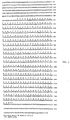
- Figure 3 describes the DNA nucleotide sequence of the EcoRl-BamHl fragment carrying the tyrB gene.
- Figure 4 describes the amino acid sequence of the aminotransferase encoded for by the tyrB gene.
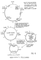
- Figure 5 illustrates an E. coli Flowchart showing intermediate strains and processes resulting in deposited strain HW159.
- Figure 6 illustrates the modification of the tyrB+ plasmid.
- Figure 7 illustrates the restriction map of the plasmid carrying the aspC gene.
- The aminotransferases or transaminases are a family of enzymes that covalently transfer an amino group from a donor molecule, such as aspartate or glutamate, to an acceptor a-keto-acid precursor of an amino acid, such as oxaloacetic acid. The reaction is freely reversible. Therefore, the aminotransferases can be utilized both in the synthesis and the degradation of amino acids.
- Amino acid biosynthesis often requires transamination as the final step in biosynthesis. An example of such a process is the transamination of phenylpyruvic acid to produce phenylalanine with the simultaneous conversion of glutamate to a-ketoglutarate. This is shown as
step 10 in Figure 1. The biosynthesis of the aromatic amino acids phenylalanine and tyrosine both require the action of an aminoatransferase as the final step. Phenylpyruvate is converted to phenylalanine by an aminotransferase that transfers an amino group from glutamate (figure 1, step 10). Similarly, an amino group from glutamate is transferred to 4-hydroxyphenylpyruvate to produce tyrosine (figure 1, step 12). Each aminotransferase has preferred substrates for the transamination reaction. However, residual activity is present on other substrates permitting each aminotransferase to participate in more than one amino acid's biosynthesis. - The E. coli transaminase encoded by the tyrB gene is active on phenylalanine, tyrosine, glutamate, aspartate and leucine.
- It is recognized that other pathways exist for synthesizing tyrosine and phenylalanine in other microorganisms. One such pathway is found in cyanobacteria and P. aeruginosa and involves the conversion of prephenate directly to pretyrosine. Pretyrosine is then a substrate for conversion directly to tyrosine orto phenylalanine. It is recognized that analogous cloned transaminase genes for the appropriate transaminase are similarly useful in increasing the reaction synthesizing pretyrosine when there are excess levels of prephenate. This transaminase would result in increasing the rate of synthesis of tyrosine and phenylalanine.
- The enzymatic steps in the synthesis of the aromatic amino acids are shown in figure 1, steps 1-20. Normally each enzymatic step is under regulation by controlling the synthesis of the enzyme and/or by allosterically regulating the rate of enzymatic catalysis. Wild type microorganisms normally do not over-produce any one amino acid due to these controls. Mutant strains in which the controls on the synthesis of a particular amino acid have been inactivated or by-passed may be isolated and in general these strains tend to over-produce that particular amino acid. In the case of L-phe, the regulation is of necessity complex, since part of its synthetic pathway is shared with the other aromatic amino acids L-tyrosine (L-tyr) and L-tryptophane (L-trp) (see Figure 1 of the accompanying drawings).
- In E. coli, as in many microorganisms, the first step in the pathway is catalysed by the three DAHP synthetase isozymes each of which is sensitive to one of the three final products. In addition, the first steps in the pathway after the branch point at chorismate are also sensitive to feed-back inhibition. Anthranilate synthetase is inhibited by L-trp and the two chorismate mutase isozymes are inhibited by L-phe and L-tyr. The synthesis of a number of the enzymes concerned is also subject to regulation. The tyrosine repressor, the product of the tyrR gene, controls expression of DAHP synthetase (L-tyr) and chorismate mutase (L-tyr). The genes for these last two enzymes together constitute the tyrosine operon. The tryptophan repressor, the product of the trpR gene, controls expression of all the enzymes of the trp operon which convert chorismate to L-trp. This regulator molecule also controls the expression of DAHP synthetase (L-trp). In addition, expression of the pheA gene which codes for the chorismate mutase (L-phe) and the expression of the trp operon are rendered sensitive to the level of L-phe. The trp operon is rendered sensitive to the level of L-trp by an attenuator/leader peptide control system.
- Mutant strains that over-produce amino acids are often isolated by selecting for resistance to appropriate amino acid analogues. In the case of L-phe production, one may isolate strains derepressed for DAHP (L-tyr) and DAHP (L-trp) expression by selecting 3-fluro-tyrosine and 5-methyl-tryptophan resistant mutants which generally have inactive tyrR and trpR repressors. Mutants in which the chorismate mutase (L-phe) is resistant to L-phe may be isolated by selecting for 2-thienyl-alanine resistance. A DAHP synthetase (L-phe) that is resistant to feed-back inhibition may be isolated by first constructing a mutant strain lacking the other two DAHP synthetase isozymes. In such a strain, the synthesis of L-tyr and L-trp is sensitive to the amount of L-phe in the medium. Mutants able to grow in medium containing L-phe, but lacking L-tyr or L-trp, usually contain feed-back resistant forms of DAHP synthetase (L-phe).
- Now, a combination of these approaches will result in strains that over-produce L-phe. Since the synthetic pathway is largely shared with that of L-tyr and L-trp, attention must be given to the prevention of L-tyr and L-trp build-up if one is interested in the optimisation of L-phe synthesis. One simple expedient is to incorporate a tyrA mutation into an L-phe over-producing strain so as to prevent L-tyr accumulation and to avoid the unwanted diversion of precursors to L-phe. Similarly, a deletion of the entire trp operon would prevent accumulation of L-trp. Such mutants would, of course, have to be cultured in media that included these two amino acids so as to permit growth.
- The combination of approaches outlined above results in a strain de-regulated for the synthesis of L-phe which accumulates that amino acid in significant amounts. Now, the rate limiting steps in a biosynthetic pathway are usually those at which control is exerted. In a de-regulated strain, it is likely that a new step will become rate-limiting. Thus, to increase the production of L-phe still further it is necessary to identify the new rate-limiting step and to increase the activity of the relevant enzyme. This may be done in one of three ways:
- (a) by isolating mutant strains in which the relevant enzyme is present in greater amounts;
- (b) by isolating mutant strains in which the relevant enzyme has a higher specific activity;
- (c) achieving the same end as in (a), but by cloning the gene for the enzyme of interest onto a suitable multi-copy plasmid and introducing this into the L-phe over-producing strain. If such an enzyme is not normally subject to regulation, isolating mutants of the first two classes may be difficult. The best way to achieve the increase in activity is to clone the gene for the relevant enzyme.
- This is achieved by ligating DNA from a suitable donor strain carrying the gene for the enzyme one wishes to clone to a suitably prepared DNA from an appropriate vector. One can then transform a mutant strain lacking the activity of the newly cloned gene. The problem experimentally is to isolate the gene. If the lack of this activity bestows a recognizable phenotype on this recipient strain, such as auxotrophy for a particular amino acid, then one can simply select for clones carrying the gene of interest by virtue of the ability of such clones to complement the lesion in the recipient strain. As an alternative, the gene could be cloned first into a low copy number plasmid or into a suitable bacteriophage vector and subsequently sub-cloned into the desired multi-copy plasmid. Alternatively a strain carrying a suitable prophage near the gene of interest can be constructed and a specialized transducing phage carrying the desired gene isolated by inducing the lysogenic donor strain and using the resultant lysate to transduce the recipient to prototrophy. The gene of interest can then be sub-cloned from the DNA of this specialized transducing phage such as lambda.
- Should the mutation of the gene of interest not confer any readily identifiable phenotype, such as auxotrophy, then the cloning of such a gene can be effected by cloning nearby genes for which a selection exists and then screening these clones for the presence of the gene of interest by assaying for the product of the desired gene after introducing the clones into a recipient deficient in that enzyme. Such an approach is facilitated by using methods that allow the cloning of large DNA fragments, such as those involving bacterio-phages or cosmids. If no readily selectable marker is known to be in the vicinity of the desired gene, then it may be possible to isolate a strain carrying the insertion of a readily selectable transposon, such as
Tn 10, which encodes tetracycline resistance, near the gene of interest. Large DNA fragments containing this marker can then be cloned and the resultant phage or cosmids screened for the presence of the gene of interest. The gene could then be sub-cloned into a suitable high copy number plasmid. - The following are the strain of E. coli used in the examples. Beside each strain designation is a summary of the bacterial genotype and source. The relationship of intermediate strains of E. coli leading to the production of the auxotroph HW159 which is both aspC- and tyrB- is shown in Figure 5. The process which converted one strain to another is also summarized on the Flowchart in Figure 5.
- Strain HW159, the aspC- and tyrB- mutant was deposited in The American Type Culture Collection, 12301 Parklawn Drive, Rockville, MD 20852, U.S.A. and has the number designation ATCC 39260.

- For the purpose of exemplification, a novel strain of E. coli that over-produces L-phe has been constructed. This was achieved by a multi-step process that involved systematically de-regulating the L-phe synthetic pathway. In such de-regulated strains, it has been found that the final step, namely the transamination of PPA to L-phe, becomes rate-limiting during fermentation and the PPA accumulates with detrimental effect. As a further example, therefore, the gene for the tyrosine (aromatic) amino-transferase from E. coli has been cloned onto a multi-copy plasmid. Introduction of this plasmid into the L-phe over-producing strain reduces this build-up of PPA and increases the efficiency of the fermentation. As an additional example, the gene for the aspartate aminotransferase of E. coli has been cloned onto a high copy number plasmid. This enzyme also has L-phe aminotransferase activity and incorporation of this plasmid in the L-phe over-producing strain also enhances the efficiency of the process.
- Strains carrying plasmids with tyrB or aspC have added utility in that they over-produce the relevant enzyme to a considerable extent. They are therefore a novel and useful source of these enzymes.
- The tyrB or aspC genes are also useful on a plasmid, in a suitable genetic background, by providing a selective pressure for maintenance of that plasmid. This selective pressure has advantages over conventional antibiotic selection methods.
- Plasmid DNA was prepared as follows (adapted from the method of J. Burke and D. Ish-Horowitz) (Nucleic Acids Research 9: 2989-2998 (1981)). The desired bacterial strain was grown to saturation in 5 ml of L-Broth plus suitable antibiotics. The culture was transferred to a 15 ml corex tube and the cells deposited by centrifugation at 10,000 RPM for 1 minute. The supernatant was decanted and the pellet resuspended in 500 pl of fresh solution I, (50 mM D-glucose, 25 mM Tris pH 8.0, 10 mM EDTA and 2 mg Lysozyme ml-I) and then incubated at room temperature for 5 minutes. The cells were lysed by the addition of 1 ml fresh solution II (containing 0.2 M NaOH and 1% SDS). This was mixed in gently and then incubated on ice for 5 minutes. 750 µl of cold solution III (to 29 ml of glacial acetic acid add water to about 60 ml adjust the pH to 4.8 with 10 m KOH and make up to 100 ml) was then added and mixed in thoroughly. After a further 5 minutes on ice, the precipitated chromosonal DNA was pelleted by centrifugation at 10,000 rmp for 10 minutes. The supernatant was decanted to a fresh 15 ml corex tube and 3.75 ml of Ethanol was added. The tube was kept on ice for 15 minutes. The DNA was pelleted by centrifugation at 10,000 rpm for 10 minutes. The supernatant was then poured off and the pellet thoroughly resuspended in 10 mM Tris (ph 8.0), 1 mM EDTA, 20 µl of 3 M sodium acetate pH 7.0 was added and the DNA transferred to an Eppendorf micro-test tube. The DNA was extracted twice with 200 ul of ultra-pure phenol that contained 0.1% 8-hydroxy quinoline and had been pre-equilibrated twice against 1 mM Tris pH 8.0 and once against 100
mM Tris 10 mM EDTA pH 8.0. The residual phenol was removed by four extractions with 1 ml of diethyl ether. The DNA was then precipitated by the addition of 500 µl ethanol and incubation in a dry-ice/ethanol bath for 10 minutes. The DNA was pelleted by centrifugation at 10,000 rpm for 10 minutes. The supernatant was discarded and the pellet resuspended in 500 µl of 70% ethanol in water (v/v). The DNA was re-pelleted by centrifugation at 10,000 rpm for 10 minutes, the supernatant was discarded and the pellet containing the DNA was dried in a vacuum dessicator for 30 minutes. The DNA was redissolved finally in 50 µl of 10mM Tris 1 mM EDTA and stored at -20°C. - This preparation generally gives about 5 ug of plasmid DNA when the strains used are derived from MC1061. The preparations also contain considerable amounts of RNA and so all the restriction digests described in this patent also include 100 µg ml-I RNASE A that has been pre-treated at 90° for 15 minutes to destroy DNAses. The preparation can also be scaled up to cope with plasmid isolation from 50 mi cultures.
- Isolation of cosmid clones carrying tyrB and DNA from the E. coli strain HW 22 (metE) was prepared according to the method of Marmur (PNAS 46:453,1960). Aliquots of this DNA were partially digested with the restriction endonuclease Sau3A as follows: The DNA (80 µg) was made up to 400 µl in Sau3A buffer containing 50 mM NaCI, 6 mM Tris-HCI (tris (hydroxymethyl) aminomethane hydrochloride) pH 7.5, 5 mM MgCl2, 100 µg/ml gelatin. The DNA solution was then split into 8 x 50 µl aliquots. Sau3A (New England Biolabs) was then serially diluted (2-fold steps) in Sau3A dilution buffer containing 50 mM KCI, 10 mM Tris-HCI pH 7.4, 0.1 mM EDTA (ethylene diamine tetra acetic acid, disodium salt), 1 mM dithiothreitol (DTT), 200 µg/ml bovine serum albumin (BSA) and 50% v/v glycerol. 5 µl of each of seven serial dilutions along with 5 µl of undiluted enzyme were then added to the DNA solution. The amount of enzyme added ranged from 2.5 to 0.02 u. The digestion was continued for 1 hour after which time the reactions were stopped by a five minute incubation at 65°C. The tubes were then cooled on ice.
- Two µl aliquots of each of the digestions were taken and were subjected to electrophoresis on a 0.7% w/v agarose gel in TBE (50 mM tris-HCI pH 8.3, 50 mM boric acid and 1 mM EDTA) along with suitable markers. After staining with ethidium bromide (1 µg/ml) for 15 minutes, the gel was observed under 254 nm illumination to visualise the DNA. In this way, the sample giving the greatest amount of material in the 45 kb size range could be estimated.
- The DNA from this sample was then ligated to DNA from the cosmid vector pHC79 which had been prepared as described below:
- Two 25 µg aliquots of pHC 79 DNA were completely digested one with the restriction endonuclease Hind III (E.C. 3.2.12.21) and the other with the restriction endonuclease SaIl (E.C. 3.1.23.37). The Hind III digestion was carried out in 100 pl of Hind III digestion buffer containing 60 mM NaCl, 7 mM MgC12, 7 mM Tris-HCI pH 7.4, 100 µg/ml gelatin and 15 units of Hind III (New England Biolabs). Incubation was at 37°C for one hour. The enzyme was then inactivated by heating to 65°C for five minutes. The SaIl digestion was carried out in 100 µl of Sa/l digestion buffer containing 150 mM NaCI, 6 mM Tris-HCI pH 7.9, 6 mM MgCl2, 6 mM 2-mercaptoethanol, 100 µg/ml gelatin and 25 units of Sail (New England Biolabs). Incubation was at 37°C for one hour. The enzyme was then inactivated by heating to 65°C for five minutes.
- The two aliquots were pooled and digested with 10 units calf intestinal phosphatase (PL Labs) at 37°C for 2 hours to remove 5' phosphates. The pooled pHC 79 was then adjusted to 0.3 M sodium acetate pH 7.0 and phenol extracted twice, followed by four ether extractions. The DNA was precipitated with 2.5 volumes of ethanol, washed with 70% vlv ethanol, dried and finally re-suspended in 10 mM Tris-HCI, 1 mM EDTA to a final concentration of 500 µg/ml. The ligation reactions contained 1 pg of prepared pHC 79 and 1 µg of Sau3A digested E. coli DNA in 8 µl of ligation buffer (10 mM Tris-HCI pH 7.9, 10 mM MgCl2, 20 mM dithiothreitol, 25 µg/ml gelatin and 1 mM ATP). The reaction mixture was incubated at 37°C for 5 minutes, cooled and the ligation initiated by the addition of T4 DNA ligase (E.C. 6.5..1.1).
- These reactions were continued for 4 hours at 15°C. Four µl aliquots of the ligation mixtures were then subjected to in vitro packaging into bacterio-phage λ particles. Strains BHB 2688 and BHB 2690 were inoculated from NZY agar plates into 50 ml of NZY broth (containing 10 g NZamine (from Humko Sheffield), 5 g yeast extract and 2.5 g NaCI/litre) and incubated with aeration at 30°C until the E600 reached 0.3. The cultures were then raised to 45°C by immersion in a 60°C water bath and incubated at 45°C for 20 minutes without aeration. The two cultures were then vigorously aerated at 37°C for 3 hours. The two cultures were pooled, chilled to 4°C and harvested by centrifugation at 7000 rpm for 2 minutes. The pellet was washed once in 100 ml of cold M9 minimal medium (per liter: 6 g Na2HP04, 3 g KH2P04, 0.5 g NaCI, 1.0 NH4CI; adjusted to pH7 with 8 N NaOH, after autoclaving, sterile glucose added to 0.2% W/V, CaC12 added to 0.1 mM and MgS04 to 1 mM). The pellet was washed in 5 ml cold complementation buffer (40 mM Tris-HCI pH 8.0, 10 mM spermidine-HCI, 10 mM putrescine-HCI, 0.1% V/V 2-mercaptoethanol and 7% VN dimethyl sulfoxide) and pelleted at 5000 rpm for 30 seconds. The cells were finally resuspended in 0.25 ml cold complementation buffer and dispensed into 20 µl aliquots in micro test tubes. These were frozen immediately in liquid nitrogen and stored at -80°C.
- For the packaging reaction, one 20 µl packaging mix was removed from the liquid nitrogen and to it was immediately added 1 µl of 30 mM ATP. The mix was then placed on ice for 2 minutes. The 4 µl aliquots of ligated DNA were then added and mixed thoroughly. This was then incubated at 37°C for 30 minutes. After 30 minutes, 1 µl of 1 mg/ml DNAase (Worthington) was added and mixed in until the sample had lost its viscosity. To this packaged DNA preparation was then added 200 µl of the strain E 107 which had been grown to an E600 of about 1.0 in tryptone maltose broth. MgS04 was also added to a final concentration of 10 mM. The packaged cosmids were absorbed for 30 minutes at 30°C after which time 500 µl of L-broth (Luria broth: 1% WN bacto-tryptone, 0.5% WN bacto-yeast extract, 0.5% W/V NaCI, 1.2% WN glucose, pH 7) was added and the incubation continued for a further 30 minutes at 30°C. The cells were then plated on L-agar containing 100 µg/ml carbenicillin (a-carboxy benzyl penicillin) and incubated at 30°C for 24 hours.
- Carbenicillin resistant colonies were then picked to L-agar carbenicillin plates and incubated overnight at 42°C. Strain E107 (3) carries the mutation dnaB which renders it unable to grow at 42°C. Thus, only those colonies which had acquired a cosmid clone carrying dnaB+ from the wild-type donor should be able to grow at this temperature. In this way, 8 cosmid clones carrying dnaB were isolated. Since dnaB maps close to tyrB, the gene for the aromatic transaminase, and since cosmid cloning results in plasmids containing large inserts of donor DNA, it was reasonable to assume that some of the cosmid clones would also carry tyrB. That some of the clones did indeed carry tyrB was shown in the following way.
- Cosmid DNA from each of the dnaB clones was purified and packaged into bacteriophage A particles as described above. The packaged cosmids were then introduced into the transaminase deficient strain DG 44 which carries the mutations tyrB and aspC. These two mutations together bestow a Tyr- Asp- phenotype on DG 44. Introduction of an intact tyrB gene would be expected to restore the ability of this strain to grow in the absence of tyrosine and aspartate. Direct screening of clones for tyrB is not easy in this strain, since it will not maintain plasmids derived from colE1, such as pHC 79. After introducing the cosmid clones into DG 44, therefore, 2 hours were allowed for recombination between the tyrB gene on the cosmid and the mutant allelle on the bacterial chromosome. The cells were then washed and plated on minimal medium lacking tyrosine and aspartate to screen for the occurrence of tyrB+ recombinants. Four of the original dnaB clones gave tyrB+ recombinants in this screen and must therefore carry at least some of the tyrB gene.
- The tyrB cosmid clones were of limited utility owing to the large size thereof which resulted in a low copy number. Therefore, restriction fragments carrying the tyrB gene were sub-cloned into the multi-copy plasmid pAT153, (see for example, Twigg, A. J., and Shenatt, D., (1980), Nature, 283,216-218) as follows: Two µg aliquots of dnaB
cosmid number 5 which carries tyrB was digested in 20 µl reaction with the following restriction endonucleases BamHl, HindIII, Sall, Sphl, Clal, EcoRl and Bglll. - In each case, the incubation was at 37°C for one hour and the enzyme was inactivated by heating to 65°C for five minutes. For the BamHl digestion, the reaction medium contained 150 mM NaCI, 6 mM Tris-HCI pH 7.9, 6 mM MgCl2, 100 µg/ml gelatin and 4 units of BamHl (New England Biolabs). Three (3) units of Hindlll (New England Biolabs) were used in 60 mM NaCI, 7 mM MgCl1, 7 mM Tris-HCI pH 7.4 and 100 µg/ml gelatin. In the case of Sa/l, 5 units of enzyme obtained from New England Biolabs were used in 150 mM NaCI, 6 mM Tris-HCI pH 7.9, 6 mM MgCl2, 7 mM 2-mercaptoethanol and 100 µg/ml gelatin. For Sphl, the reaction contained 50 mM NaCI, 6 mM Tris-HCI pH 7.4, 6 mM MgCl2, 10 mM 2-mercaptoethanol, 100 µg/ml gelatin and 1 unit of the Sphl enzyme (New England Biolabs). The reaction mixture for Clal digestion corresponded to that for Hindlll, digestion, except that the enzyme used was 3 units of Clal obtained from Boehringer Biochemicals. In the case of EcoRl 4.5 units of EcoRI (New England Biolabs) were used in 100 mM Tris-HCI pH 7.5, 50 mM NaCI, 5 mM MgC12 and 100 µg/ml gelatin. Lastly, the reaction medium for BglII digestion contained 60 mM NaCI, 10 mM Tris-HCI pH 7.6,10 mM MgCl2, 10 mM 2-mercaptoethanol and 100 µg/ml gelatin.
- These digested DNA preparations were then subjected to electrophoresis on a horizontal 1% w/v low gelling temperature (LGT) agarose gel in TBE for 6 hours at 5 V/cm. The DNA fragments were visualized by staining the gel for 15 minutes in a 1 µg/ml solution of ethidium bromide, followed by observation under a 366 nm UV light source. Individual bands were excised and stored at 4°C.
- One (1) µg aliquots of pAT153 DNA were digested with the same set of restriction endonucleases in 20. µl reactions, except for Bglll (which does not cut pAT153). Cosmid BglII fragments were sub-cloned into BamHl-cut pAT153 (BamHl and BglII give the same 5' overhangs). After digestion, the linearized vector was digested with 1 unit of calf intestinal phosphatase (PL Labs) for 1 hour, to remove 5' phosphates and prevent recircularization, incubated at 70°C for 10 minutes to inactivate the enzyme and then also run on a 1% w/v LGT agarose gel as described for the cosmid fragments. The band corresponding to plasmid linearized by each enzyme was then excised from the gel as described above.
- The cosmid fragments were then ligated to the appropriately-cut vector as follows. The gel slices were melted by incubation at 65°C for 10 minutes and then cooled to 37°C. The melted gel slices were all about 100 pl. Two µl of vector fragment and 8 µl of a particular cosmid fragment were then added to 40 µl of pre-warmed 1.25 x ligation buffer (62.5 mM Tris-HCI pH 7.8, 12.5 mM MgC12, 25 mM dithiothreitol, 1.25 mM ATP and 62.5 mg/ml gelatin) and mixed thoroughly. Ligase was added and the reaction continued at 15°C overnight. The ligated samples were then re-melted at 65°C for 5 minutes, cooled to room temperature and added to 200 µl of competent cells of the E. coli strain HW 87 which had been prepared as follows. An overnight culture of HW 87 in L-broth was diluted 1:50 into 50 ml of fresh pre-warmed L-broth and incubated at 37°C with good aeration until the E600 reached 0.6. The cells were then pelleted by centrifugation at 10,000 rpm for 5 seconds and resuspended in 25 ml of cold 50 mM CaCl2. The cells were left on ice for 10 minutes after which they were re-pelleted as above and re-suspended in 2 ml of cold 50 mM CaCI2. After a further 10 minute incubation at 0°C, the cells were competent for transformation.
- After addition of the ligated DNA, the cells were incubated at 0°C for a further 10 minutes, heat-shocked at 37°C for 2 minutes and finally mixed with 750µl of pre-warmed L-broth. The cells were incubated for 30 minutes at 37°C to allow phenotypic expression of the plasmid encoded β-lactamase gene before plating suitable aliquots onto L-agar plates containing 200 µg/ml carbenicillin. The plates were incubated at 37°C overnight. Colonies containing recombinant plasmids were identified by the sensitivity thereof to tetracycline in the case of fragments cloned at the Hindlll, Sall, Sphl, BamHl and Clal sites of pAT 153.
- Recombinants isolated using the restriction endonuclease EcoRl were identified by examining the size of the plasmids in individual colonies. This was performed as follows. Potential recombinant colonies were patched onto L-agar plates containing 200 µg/ml carbenicillin. These were incubated overnight at 37°C. The bacteria from about 1 cm2 of each patch were re-suspended in lytic mix containing 10 mM Tris-
HCI pH - It was decided to move the already characterised mutations in DG 44 into a background known to allow maintenance of the plasmids (that of strain MC 1061). Bacteriophage P1 was grown on a mixed culture of strain HW 22 carrying random insertion of the tetracycline
resistant transposon Tn 10 prepared as follows: A saturated culture of HW 22 was grown overnight in lambda YTM broth (10 g bacto tryptone, 2.5 g NaCI and 0.2% w/v maltose per litre). This was diluted x 1/100 into 100 ml of fresh pre-warmed lambda YM broth and incubated at 37°C until the OD600 reached 0.6. The cells were deposited by centrifugation at 10,000 rmp for five seconds and resuspended in 5 ml of lambda YM broth. NK 55, a bacteriophage lambda derivative carrying the tetracycline transposon Tn10, was then added to a multiplicity of infection of 0.2. The bacteriophage was absorbed for 45 minutes at 37°C. Then 200 ml aliquots were spread onto L-agar plates containing tetracycline (20 pg/ml) and sodium pyrophosphate (2.5 mM). The plates were incubated at 37°C for 24 hours. Approximately 5,000 TetR colonies were pooled by washing the colonies off the plates using a total of 5 ml of L-broth. This was then diluted into 50 ml of L-broth containing 20 µg/ml tetracycline and grown overnight at 37°C. The Tn10 pool was stored at -20°C after adding sterile glycerol to bring the concentration to 20% w/v. - In order to grow bacteriophage P1 on the Tn10 pool, a 200 ml aliquot of this glycerol stock was diluted with 5 ml of L-broth including 10 µg/ml tetracycline and incubated overnight with shaking at 37°C. To 0.2 ml of this overnight culture was added 2 x 10⁶ plaque forming units (pfu) of phage P1 clear and 10 µl of 50 mM CaCl2. The cells were incubated at 37°C for five minutes and then added to 5 ml of L-broth + 2.5 mM CaC12 pre-warmed to 37°C. The cells were then incubated at 37°C with vigorous aeration for four hours. The cell debris was removed by centrifugation at 10,000 rpm for five minutes and the phage-containing supernate stored over chloroform at 4°C.
- This supernate was used to transduce DG 30 to permit growth on minimal medium lacking tyrosine and aspartate. (The minimal medium used was that of Vogel and Bonner; 0.2 g/l MgSo4.7H2O, 2 g/I citric acid, 10 g/I K2HP02, 3.5 g/l NaH2PO4.4H20 and 10 mM FeCl2. After autoclaving, glucose was added to a final concentration of 0.2% w/v along with necessary supplements of amino acids at 50 µg/ml. The Tyr+ Asp+ transductants were then replica-plated to the same medium including 10 µg/ml tetracycline to detect those transductants that had simultaneously become TetR. A number of these TetR Tyr+ Asp+ recombinants were purified and used to prepare new P1 lysates. Each of these eight P1 preparations was then used to transduce DG 30 to TetR. From each experiment, 50 TetR transductants were picked to minimal medium lacking tyrosine and aspartate to test for linkage between the site of the
Tn 10 insertion involved and aspC or tyrB. From each of these experiments one TetR colony which remained tyrB aspC was purified and again used to prepare a P1 lysate. These lysates were used to transduce MC 1061 to TetR. Cell extracts from these transductants were then run on native polyacrylamide gels and stained for L-phe transaminase activity as described by Gelfand. - The cell extracts were prepared by growing the desired transductants overnight in 10 ml of L-broth at 37°C. The cells were harvested by centrifugation at 10,000 rpm for fifteen seconds and re-suspended in 0.5-1.0 ml of sonication buffer depending upon the size of the pellet. The sonication buffer used was 25 mM Tris-HCI, 25 mM KH2PO4 pH 6.9,0.2 mM pyridoxal phosphate, 0.5 mM dithiothreitol, 0.2 mM EDTA and 10% v/v glycerol. The cell suspensions were transferred to Eppendorf micro test tubes and kept on ice. Each suspension was sonicated with a Davies sonicator using four five-second bursts at setting 2. The sonicated suspensions were then centrifuged at 15,000 rpm for five minutes at 4°C to remove cell debris and the supernatants transferred to a fresh micro test tube. The cell extracts were then kept on ice until they could be loaded on a gel or stored at -20°C.
- The native gel electrophoresis was performed as follows. The gel consisted of 8.5% w/v acrylamide, 0.227% w/v bis-acrylamide in 375 mM Tris-HCI pH 8.3. This acrylamide stock was de-gassed and polymerized by the addition of 0.05% w/v ammonium persulfate and 0.016% w/v TEMED. After polymerization, the gels were pre-run in 37.5 mM Tris-HCI pH 8.3 for at least one hour at 4°C. Prior to loading the samples, the buffer was changed to running buffer consisting of 76.7 mM glycine, 1 mM Tris-HCI pH 8.3. About 50 µg protein from each cell extract was then loaded and subjected to electrophoresis at 10 v/cm for from four to six hours at 4°C. The protein concentration in the cell extracts was determined using the Bio-Rad protein reagent method. The gels were stained for L-phe transaminase activity as follows. The gel was washed briefly in 100 mM phosphate buffer pH 7.5 and then immersed in 500 ml of fresh staining solution containing 12.5 mM a-ketoglutarate, 0.2 mM pyridoxal phosphate, 0.6 mM nitro blue tetrazolium, 0.2 mM phenazine methosulphate, 30 mM L-phe and 100 mM K2HP04 pH 7.5 that had been pre-warmed to 37°C. The gel was shaken gently in the staining solution for one hour at 37°C in the dark. The gel was then washed in distilled water and left in distilled water until it could be photographed. (See figure 2.) This illustrates the presence or absence of detectable aminotransferases in various E coli strains.
- MC 1061 extracts prepared in this way gave three staining bands characteristic of the ilvE, aspC and tyrB activities. By comparing the transaminase pattern of extracts prepared from the transductants with that of MC 1061 it was possible to detect MC 1061 recombinants lacking the characteristic aspC activity.
- One mutant which was devoid of aspC activity was purified and named
HW 109. This strain was then cured of the Tn10 transposon by the method of Bochner et al (J.Bact 143:926).HW 109 was grown overnight in L-broth. Approximately 106 cells per plate were spread onto Bochner selective medium which was prepared as follows: Solution A (Bacto-tryptone 10g, Bacto yeast extract 5 g, chlortetracycline HCI 50 mg, agar 15 g, water 500 ml) and solution B (NaCl 10 g, NaH2PO4.H20 10 g, glucose 2 g, water 500 ml) were autoclaved separately for 20 min at 15 psi. The solutions were mixed and cooled to pouring temperature. 5 ml of ZnCl2 (20 mM) and 6 ml of 2 mg ml-1 fusaric acid were then added prior to pouring. The plates were incubated for 24 hr at 37° and the fusaric acid resistant colonies isolated and purified by re-streaking on the same medium. These isolates were then tested for sensitivity to tetracycline. One tetracycline sensitive derivative ofHW 109 was chosen and namedHW 157. - This approach did not, however, yield tyrB derivatives of HW 22. To isolate a strain with
Tn 10 linked to tyrB, the procedure was as follows. Strain ES 430 carries a mutation in malB that renders it unable to use maltose as a carbon source. The malB is reasonably closely linked with tyrB. Therefore, ES 430 was transduced with P1 grown on therandom Tn 10 pool in HW 22 described above, selecting for TetR on maltose Maconkey agar plates supplemented with 10 µg/ml tetracycline. - P1 transductions were performed as follows. The recipient strain was grown in L-broth to an OD600 of about 1.0. CaCl2 was added to a final concentration of 2.5 mM. Phage P1 clear grown on the desired donor was then added at a multiplicity of insertion of between 0.2 and 1.0 (usually 108 pfu P1 clear per ml of recipient). The cells were incubated at 37°C for fifteen minutes, centrifuged at 10,000 rpm for five seconds, washed in the original volume of 0.1 M citrate buffer pH 7.0 and finally re-suspended in citrate buffer. Suitable aliquots were then plated on the selective medium.
- Transductants which had simultaneously become TetR and Mal+ were picked and purified. P1 lysates were then prepared from these strains and used to transduce DG 44 to TetR. The TetR colonies derived from each of the eight P1 lysates were then patched onto minimal agar plates lacking aspartate and tyrosine. In one of these experiments, good linkage betweeen
Tn 10 and tyrB was obtained. Therefore, one TetR tyrB- recombinant from this experiment was isolated, a P1 lysate prepared from this recombinant and used to transduceHW 157 described above to TetR. Fifty of these transductants were then patched onto minimal agar supplemented with leucine, but lacking L-aspartate and L-phenylalanine. In this way, a tyrB- aspC- derivative of MC 1061 was detected. This strain was designatedHW 158. Finally,HW 159, a tetracycline sensitive derivative ofHW 158 was isolated as described above for the isolation ofHW 157.HW 159 will not grow on minimal medium supplemented with leucine since it requires aspartate and tyrosine for growth unlike the parental strain MC 1061 which only requires leucine. - All the recombinant plasmids obtained by subcloning DNA fragments from the dnaB cosmid clone (described above) were introduced into
strain HW 159 by transformation selecting on L-agar supplemented with 200 pg/ml carbenicillin. These transformants were then streaked onto minimal medium supplemented only with leucine to test for the ability of the presence of genes carried by the plasmid to complement the tyrB lesion inHW 159. One sub-clone carrying a Clal fragment from cosmiddnaB clone number 5 was found to restore the ability ofHW 159 to grow on minimal medium supplemented with leucine and thus to carry the tyrB gene. - Sequencing of the tyrB gene was performed by the method of Maxam and Gilbert. The sequence of the tyrB gene is shown in figure 3. Based upon this DNA sequence the amino acid sequence for the aminotransferase encoded for by tyrB could be determined using the genetic triplet codons. This amino acid sequence is shown in figure 4.
- As a starting point for the cloning of the aspC gene, it was decided to use the specialized transducing phage lambda aspC2 obtained from M. Ono. The phage was prepared as follows. A culture of HW 76, which is a double lysogen carrying lambda aspC and lambda Cl 857
Sam 7, was grown to an OD600 of 0.6 at 30°C. The culture was then incubated at 45°C for fifteen minutes to induce the prophage and then shaken vigorously at 37°C for three hours. Cell lysis was completed by the addition of 0.5 ml of CHCI3 and the cell debris removed by centrifugation at 10,000 rpm for ten minutes. The phage was precipitated by the addition of NaCl to 2.4% w/v and polyethylene glycol (average molecular weight 6,000) to 10% w/v. The phage was precipitated overnight at 4°C and then pelleted by centrifugation at 5,000 rpm for ten minutes. The phage pellet was gently re-suspended in 10 ml of phage buffer consisting of 10 mM Tris-HCI pH 7.5, 10 mM MgS04. The phage was pelleted by centrifugation for one hour at 40,000 rpm and finally re-suspended in 1 ml of phage buffer. - The phage was further purified by running on a CsCI block gradient as follows. The 1 ml of phage was applied to a two-step block gradient in cellulose nitrate tubes. The gradient contained 1 ml of 5M CsCI, 10 mM MgS04, 10 mM Tris-HCI pH 8.0 and 0.1 mM EDTA overlaid with 3 ml of 3M CsCI, 10 mM MgS04, 10 mM Tris-HCI pH 8.0 and 0.1 mM EDTA. The gradient was centrifuged in a Beckmann SW 65 rotor for one hour at 30,000 rpm at 20°C. The phage band was removed in 0.5 ml from the side of the tube using a 1 ml syringe with a 5/8 inch (1.6 cm) 25 gauge needle. The 0.5 ml of phage was then mixed with 0.5 ml of saturated CsCI solution (25°C) in 10 mM MgSO4, 10 mM Tris-HCI pH 8.0 and 0.1 mM EDTA and mixed well in a cellulose nitrate centrifuge tube. This was then overlaid with 3 ml of 5M CsCI in 10 mM MgS04, 10 mM Tris-HCI pH 8.0 and 0.1 mM EDTA and then 1 ml of 3M CsCI in 10 mM MGS04, 10 mM Tris-HCI pH 8.0 and 0.1 mM EDTA. This was again centrifuged at 30,000 rpm for one hour at 20°C. The phage was again removed from the side of the tube in 0.5 ml as before.
- The DNA was extracted from the phage particles as follows. To the 0.5 ml of phage in a 15 ml corex centrifuge tube was added 50pl of 2M Tris-HCI pH 8.5, 0.2 M EDTA and 0.5 ml of formamide was added. After 30 minutes, 0.5 ml of water was added and the DNA precipitated by the addition of 3 ml of ethanol. The DNA was pelleted by centrifugation for five minutes at 10,000 rpm. The supernate was discarded and the pellet rinsed with 70% v/v ethanol. The DNA was dried in vacuo and finally dissolved in TE (10 mM Tris-HCI and 1 mM EDTA pH 8.0) to give a DNA concentration of 500 µg/ml. The lambda aspC DNA was partially digested with the restriction endonuclease Sau3A as described above for E. coli DNA. The DNA from all of the digests were pooled and run on a 1% w/v low gelling temperature agarose gel in TBE. Also prepared and run on the same gel was 1 pg of pAT153 DNA completely digested with the restriction endonuclease BamHl and calf intestinal phosphatase as described above. The DNA was visualized under 366 nm U.V. light as described above.
- The bands corresponding to linearised pAT153 and the partially restricted lambda aspC DNA between 2.5 and 6kb were excised. The DNA was extracted from the gel as follows. The gel slices were melted at 65°C for five minutes in 15 ml convex tubes, cooled to 37°C and diluted with two volumes of water at 37°C. The agarose was then removed by the addition of an equal volume of phenol at 37°C and vortexing for three minutes. The phases were separated by centrifugation at 10,000 rpm for five minutes and the upper aqueous phases removed to clean tubes. Three phenol extractions were performed. The final supernate was then extracted four times by vortexing with an equal amount of diethyl ether, allowing the phases to separate and then removing the upper ether layer. The volume of the remaining aqueous phase was determined and 3M sodium acetate added to bring the salt concentration to 0.3 M. Yeast transfer RNA was added to a final concentration of 5 pg/ml as a carrier. The DNA was precipitated by the addition of 2.5 volumes of ethanol, overnight incubation at -20°C and centrifugation at 10,000 rpm for 30 minutes. The pellet was washed in 70% v/v ethanol, re-centrifuged at 10,000 rpm for 30 minutes and dried in vacuo. The DNA was then dissolved in TE to give a concentration of about 100 µg/ml. The partially restricted lambda aspC DNA was then ligated to BamHl cut pAT153 by adding 2 pl of prepared lambda aspC DNA to 2 µl of prepared pAT153 DNA in 4µl of 2x ligation buffer (described above). This mixture was incubated at 37°C for five minutes, cooled to room temperature and the ligation started by the addition of 1 µl of T4 DNA ligase (New England Biolabs). The ligation was continued at 15°C for four hours. The entire ligation reaction mixture was then cooled on ice and added to 200 µl of competent cells prepared from
strain HW 159 as described above. The transformation was performed as above and the cells plated on L-agar containing 200 µg/ml carbenicillin. The plates were incubated overnight at 37°C. About 5,000 colonies were obtained. These were then cooled, washed twice in 10 ml of 0.85% w/v NaCl and plated onto minimal agar containing leucine and carbenicillin, but lacking tyrosine and aspartate. OnlyHW 159 cells which carried a recombinant plasmid containing aspC could grow on this medium. - After incubation at 37°C for twenty-four hours, about 100 colonies were obtained. Some of these were purified by successive streakings on the same medium. That these contained a recombinant plasmid was confirmed by running single colony agarose gels as described above. That the recombinant plasmids in these strains indeed conferred the Asp+ Tyr+ phenotype was confirmed by isolating the plasmids, re- transforming into
HW 159 and showing that these transformants had become Asp+ Tyr+. That the transformants had been altered by the presence of cloned aspC as opposed to tyrB was shown by running native acrylamide gels on cell-free extracts and staining for transaminase activity as described above. These experiments confirmed that the plasmids indeed carried an intact aspC gene. - The actual level of aminotransferase activity is measured in arbitrary units indicating relative activity. Relative aminotransferase activity on various strains containing the tyrB and aspC genes on plasmid pAT153 is shown in table 2. The assay was performed using a sigma R aspartate transaminase assay kit. (Sigma Technical Bulletin No. 56-UV, revised August 1980)

- The Clal insert carrying tyrB is approximately 4.5 kb. A preliminary restriction map was constructed by the commonly used technique of double restriction endonuclease digestion followed by gel electrophoresis to analyse the restriction fragments obtained. In one orientation (see figure 6) the Clal insert had EcoRl, BamHl and Nrul sites in positions that facilitated the reduction in plasmid size by in vitro deletion. For example, the small EcoRl fragment was removed as follows. One pg of ptyrB DNA was digested to completion with EcoRl in a final volume of 20 ul. The enzyme was inactivated by incubation at 65° for 5 minutes. A 2 µl aliquot of this digest was then diluted into 50 µl of ligase buffer, one µl of T4 DNA ligase added and the DNA ligated for 16 hours at 15°. Ligation at this low DNA concentration leads predominantly to the re-circularisation of the plasmid without the small EcoRl fragement. The DNA was then transformed into HW 87 selecting for carbenicillin resistance as described above. Individual transformants were picked, purified and analysed on single colony lysate gels as described above. Most of the transformants contained plasmids that exhibited higher mobility than the parental plasmid ptyrB. Plasmid DNA from several of these clones was isolated and the loss of the small EcoRl fragment confirmed by EcoRl digestion and analysis of these digests by electrophoresis on a 1% agarose gel. All the deleted plasmids gave just one band corresponding to the large EcoRl fragments of ptyrB. The parental plasmid gave the expected two bands.
- The EcoRl deleted ptyrB was then subjected to a similar series of manipulations to remove the BamHl fragment and this EcoRl BamHl deleted plasmid was further subjected to deletion using the enzyme Nrul in the following buffer. (100
mM NaCI 6 mM Tris pH 7.4 6mM MgC1 2 5 mM β-mercaptoethanol 100 µg/ml gelatin) All three plasmids were purified and used to transform HW 225 (tyrB aspC recA) to carbenicillin resistance. All the transformants simultaneously acquired the ability to grow on minimal medium in the absence of L-tyrosine and L-aspartate. This demonstrates that the tyrB gene is still intact on all three deleted plasmids. - The BamHl-EcoRl fragment from the doble deleted ptyrB was subjected to sequence analysis following the protocols described by Maxam and Gilbert (Maxam, A. M., and W. Gilbert, 1977, PNAS 51: 382-389; Masam, A. M., and W. Gilbert, 1977, MIE 65: CH 57 499-559, Part I). One substantial open reading frame was discovered that could code for a protein of 380 or 396 amino acids in length. The precise start point for translation is not known. There is a typical ribosome binding site about 10 nucleotides upstream from an in phase GTG (valine codon). Alternatively there is an in phase ATG (methionine) but this is not preceded by a typical ribosome binding site. The DNA sequence of the EcoRl-BamHl fragment and the amino-acid sequence deduced from it are presented in figures 3 and 4.
- The restriction map of the smallest aspC sub-clone (
paspC 3―4) was elucidated by a combination of single and double restriction digests as described above see figure 7. The aspC sub-clones do not possess restriction sites that allow the insert to be excised cleanly since they were made by ligating Sau3A fragments into the BamHl site of pAT 153, a procedure which does not regenerate the BamHl site. - The paspC3―4 (pME98) was used to transform the E. coli prototroph HW 519 and during fermentation produced levels of transaminase equivalent to or better than that of paspCI-1. The E. coli HW 519 carrying pME98 has the deposit number ATCC 39501.
- In the reactions requiring aminotransferase enzymes in figure 1,
reactions # 10 and #13, presence of additional aminotransferase enzymes facilitates amino acid synthesis. This increased level of the aminotranferase enzymes synthesized by the aspC, tyrB or ilvE gene enables a cell to overcome a rate- limited reaction at the transamination step. - A plasmid of the present invention, carrying the aspC, gyrB or ilvE gene is inserted into a bacterial cell by techniques well known in biochemistry. This inserted gene produces a messenger RNA which catalyzes the synthesis of increased levels of aminotransferases when the bacterial cell is grown in a suitable media. The presence of these increased levels of aminotransferases catalyzes increased levels of amino acids. The amino acids are then harvested from the cells and media by purification procedures well known in fermentation science.
- The addition of the tyrB containing plasmid into E.Coli 13281 (U.S. Patent 2,973,304) resulted in an 11 % increase in L-phenylalanine production as shown in Table 3.
-
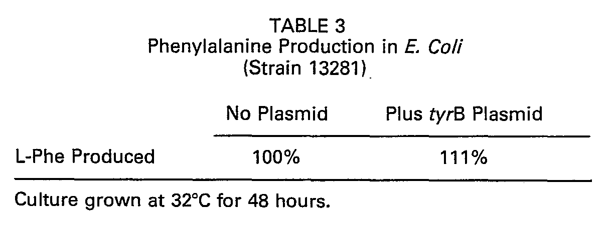
-
- 1. USP 2,973,304 Pfizer
- 2. USP 4,237,224 Cohen et al.
- 1. Umbarger, H. E. Annual Review of Biochemistry, 47: 533-606, 1978.
- 2. Christiansen, L. and S. Pedersen, Mol. Gen. Genet (1981) 181: 548-551.
- 3. Gelfand, D. H. and R. S. Steinberg, J. of Bacteriology, 130: 429-440, April 1977.
- 4. Bachmann, B. J. and K. B. Low, Microbiological Reviews, 44: 1-56, March, 1980.
- 5. Maxam, A. M. and W. Gilbert, P.N.A.S., 51: 382-389, 1977.
- 6. Maxam, A. M. and W. Gilbert, MIE 65: Chapter 57, 499-559 (Part I).
- 7. Sigma Technical Bulletin No. 56-UV, Revised August 1980 Sigma Chemical Co., St. Louis, MO 63178.
Claims (8)


Applications Claiming Priority (2)
| Application Number | Priority Date | Filing Date | Title |
|---|---|---|---|
| GB838301700A GB8301700D0 (en) | 1983-01-21 | 1983-01-21 | Cloning and utilisation of aminotransferase genes |
| GB8301700 | 1983-01-21 |
Related Child Applications (1)
| Application Number | Title | Priority Date | Filing Date |
|---|---|---|---|
| EP87116645.0 Division-Into | 1987-11-11 |
Publications (2)
| Publication Number | Publication Date |
|---|---|
| EP0116860A1 EP0116860A1 (en) | 1984-08-29 |
| EP0116860B1 true EP0116860B1 (en) | 1990-07-11 |
Family
ID=10536723
Family Applications (2)
| Application Number | Title | Priority Date | Filing Date |
|---|---|---|---|
| EP87116645A Expired - Lifetime EP0293514B1 (en) | 1983-01-21 | 1984-01-19 | The cloning and utilization of aminotransferase genes |
| EP84100521A Expired - Lifetime EP0116860B1 (en) | 1983-01-21 | 1984-01-19 | The cloning and utilization of aminotransferase genes |
Family Applications Before (1)
| Application Number | Title | Priority Date | Filing Date |
|---|---|---|---|
| EP87116645A Expired - Lifetime EP0293514B1 (en) | 1983-01-21 | 1984-01-19 | The cloning and utilization of aminotransferase genes |
Country Status (5)
| Country | Link |
|---|---|
| EP (2) | EP0293514B1 (en) |
| JP (1) | JPH0767389B2 (en) |
| CA (1) | CA1295568C (en) |
| DE (2) | DE3486040T2 (en) |
| GB (1) | GB8301700D0 (en) |
Families Citing this family (13)
| Publication number | Priority date | Publication date | Assignee | Title |
|---|---|---|---|---|
| GB8301700D0 (en) * | 1983-01-21 | 1983-02-23 | Searle & Co | Cloning and utilisation of aminotransferase genes |
| GB8403244D0 (en) * | 1984-02-07 | 1984-03-14 | Searle & Co | Aminoacids via bioconversion |
| WO1987000202A1 (en) | 1985-06-24 | 1987-01-15 | The Nutrasweet Company | Composite plasmids for amino acid synthesis |
| SE8603838L (en) * | 1985-09-19 | 1987-03-20 | Grace W R & Co | GENETICALLY PROCESSED MICRO-ORGANISMS FOR L-AMINO ACID PREPARATION |
| JPS62143691A (en) * | 1985-12-17 | 1987-06-26 | Kyowa Hakko Kogyo Co Ltd | Production of amino acid |
| DE3605035A1 (en) * | 1986-02-18 | 1987-08-20 | Strahlen Umweltforsch Gmbh | PLASMID PWS101, METHOD FOR ITS DETERMINATION AND USE |
| DE3631829A1 (en) * | 1986-09-19 | 1988-07-28 | Hoechst Ag | CLONING AND USE OF THE TRANSAMINASE GENE TYRB |
| DE3636722A1 (en) * | 1986-10-29 | 1988-05-05 | Hoechst Ag | CLONING AND USE OF THE TRANSAMINASE GENE ILVE |
| DE3713755A1 (en) * | 1987-04-24 | 1988-11-10 | Hoechst Ag | PRODUCTION OF L-PHENYLALANINE WITH THE AID OF RECOMBINANT BACTERIA |
| DE10063314A1 (en) * | 2000-12-20 | 2002-07-04 | Degussa | New nucleotide sequences coding for the ilvE gene |
| RU2244007C2 (en) * | 2002-02-27 | 2005-01-10 | Закрытое акционерное общество "Научно-исследовательский институт Аджиномото-Генетика" | Method for preparing l-threonine, strain escherichia coli as producer of threonine (variants) |
| RU2243260C2 (en) * | 2002-06-25 | 2004-12-27 | Закрытое акционерное общество "Научно-исследовательский институт Аджиномото-Генетика" | Method for preparing l-leucine (variants), strain escherichia coli 505/pacyc-tyr b as producer of l-leucine |
| WO2010092959A1 (en) * | 2009-02-10 | 2010-08-19 | 協和発酵バイオ株式会社 | Method for producing amino acid |
Family Cites Families (1)
| Publication number | Priority date | Publication date | Assignee | Title |
|---|---|---|---|---|
| GB8301700D0 (en) * | 1983-01-21 | 1983-02-23 | Searle & Co | Cloning and utilisation of aminotransferase genes |
-
1983
- 1983-01-21 GB GB838301700A patent/GB8301700D0/en active Pending
-
1984
- 1984-01-19 DE DE8787116645T patent/DE3486040T2/en not_active Expired - Fee Related
- 1984-01-19 EP EP87116645A patent/EP0293514B1/en not_active Expired - Lifetime
- 1984-01-19 CA CA000445610A patent/CA1295568C/en not_active Expired - Lifetime
- 1984-01-19 DE DE8484100521T patent/DE3482663D1/en not_active Expired - Lifetime
- 1984-01-19 EP EP84100521A patent/EP0116860B1/en not_active Expired - Lifetime
- 1984-01-21 JP JP59009380A patent/JPH0767389B2/en not_active Expired - Fee Related
Also Published As
| Publication number | Publication date |
|---|---|
| DE3486040D1 (en) | 1993-02-25 |
| EP0116860A1 (en) | 1984-08-29 |
| JPH0767389B2 (en) | 1995-07-26 |
| EP0293514B1 (en) | 1993-01-13 |
| GB8301700D0 (en) | 1983-02-23 |
| CA1295568C (en) | 1992-02-11 |
| JPS59192090A (en) | 1984-10-31 |
| EP0293514A1 (en) | 1988-12-07 |
| DE3482663D1 (en) | 1990-08-16 |
| DE3486040T2 (en) | 1993-07-29 |
Similar Documents
| Publication | Publication Date | Title |
|---|---|---|
| CA1335658C (en) | Process for preparing l-tertiary-leucine and l-phosphinothricine by transamination | |
| RU2207376C2 (en) | Method for preparing l-amino acid by fermentation method, strain of bacterium escherichia coli as producer of l-amino acid (variants) | |
| RU2111247C1 (en) | Method of preparing microorganism strain, method of tryptopham producing | |
| KR930007579B1 (en) | Process for integration of a chosen gene on the chromosome of a bacterium and bacterium obtained by the said process | |
| EP0332234B1 (en) | Process for preparing l-tyrosine | |
| US6319696B1 (en) | Process for producing L-amino acids | |
| EP0358940A1 (en) | DNA fragment coding for phosphoenolpyruvat corboxylase, recombinant DNA carrying said fragment, strains carrying the recombinant DNA and method for producing L-aminino acids using said strains | |
| KR900004426B1 (en) | Process for producing l-tyrosine | |
| EP0789073A2 (en) | Production of tryptophan by the bacterium Escherichia coli | |
| EP0293514B1 (en) | The cloning and utilization of aminotransferase genes | |
| EP0088166A2 (en) | Method for expressing a gene | |
| JPH0759199B2 (en) | Method for producing L-tryptophan by fermentation method | |
| KR20000048776A (en) | Preparation of d-amino acids by direct fermentative means | |
| JP2003204788A (en) | Method for producing target substance by fermentation | |
| EP0190921A2 (en) | Method for the overproduction of amino acids | |
| EP0561426A1 (en) | Novel plasmids useful for the cloning of DNA-fragments responsible for gene expression control in coryneform bacteria | |
| Grabau et al. | Molecular cloning of the gene (poxB) encoding the pyruvate oxidase of Escherichia coli, a lipid-activated enzyme | |
| EP0077196A2 (en) | Aromatic amino acid-producing microorganisms | |
| Chan et al. | Amplification of the tryptophan operon gene in Escherichia coli chromosome to increase l-tryptophan biosynthesis | |
| JP2760788B2 (en) | Cloning of aminotransferase gene tyrB and its use | |
| RU2243260C2 (en) | Method for preparing l-leucine (variants), strain escherichia coli 505/pacyc-tyr b as producer of l-leucine | |
| JP3009257B2 (en) | Genetic DNA encoding aspartase and use thereof | |
| JP3078312B2 (en) | Method of manufacturing substances | |
| US5120654A (en) | Cloning and use of the transaminase gene ilve | |
| CA1341352C (en) | Cloning and utilization of aminotransferase genes |
Legal Events
| Date | Code | Title | Description |
|---|---|---|---|
| PUAI | Public reference made under article 153(3) epc to a published international application that has entered the european phase |
Free format text: ORIGINAL CODE: 0009012 |
|
| AK | Designated contracting states |
Designated state(s): BE CH DE FR GB IT LI NL SE |
|
| 17P | Request for examination filed |
Effective date: 19850122 |
|
| 17Q | First examination report despatched |
Effective date: 19860513 |
|
| R17C | First examination report despatched (corrected) |
Effective date: 19870305 |
|
| RAP1 | Party data changed (applicant data changed or rights of an application transferred) |
Owner name: THE NUTRASWEET COMPANY |
|
| ITF | It: translation for a ep patent filed | ||
| GRAA | (expected) grant |
Free format text: ORIGINAL CODE: 0009210 |
|
| AK | Designated contracting states |
Kind code of ref document: B1 Designated state(s): BE CH DE FR GB IT LI NL SE |
|
| REF | Corresponds to: |
Ref document number: 3482663 Country of ref document: DE Date of ref document: 19900816 |
|
| ET | Fr: translation filed | ||
| ITTA | It: last paid annual fee | ||
| PLBE | No opposition filed within time limit |
Free format text: ORIGINAL CODE: 0009261 |
|
| STAA | Information on the status of an ep patent application or granted ep patent |
Free format text: STATUS: NO OPPOSITION FILED WITHIN TIME LIMIT |
|
| 26N | No opposition filed | ||
| EAL | Se: european patent in force in sweden |
Ref document number: 84100521.8 |
|
| PGFP | Annual fee paid to national office [announced via postgrant information from national office to epo] |
Ref country code: FR Payment date: 20000112 Year of fee payment: 17 |
|
| PGFP | Annual fee paid to national office [announced via postgrant information from national office to epo] |
Ref country code: SE Payment date: 20010105 Year of fee payment: 18 |
|
| PGFP | Annual fee paid to national office [announced via postgrant information from national office to epo] |
Ref country code: CH Payment date: 20010112 Year of fee payment: 18 |
|
| PGFP | Annual fee paid to national office [announced via postgrant information from national office to epo] |
Ref country code: DE Payment date: 20010115 Year of fee payment: 18 |
|
| PGFP | Annual fee paid to national office [announced via postgrant information from national office to epo] |
Ref country code: GB Payment date: 20010118 Year of fee payment: 18 |
|
| PGFP | Annual fee paid to national office [announced via postgrant information from national office to epo] |
Ref country code: NL Payment date: 20010131 Year of fee payment: 18 |
|
| PGFP | Annual fee paid to national office [announced via postgrant information from national office to epo] |
Ref country code: BE Payment date: 20010314 Year of fee payment: 18 |
|
| REG | Reference to a national code |
Ref country code: GB Ref legal event code: IF02 |
|
| PG25 | Lapsed in a contracting state [announced via postgrant information from national office to epo] |
Ref country code: GB Free format text: LAPSE BECAUSE OF NON-PAYMENT OF DUE FEES Effective date: 20020119 |
|
| PG25 | Lapsed in a contracting state [announced via postgrant information from national office to epo] |
Ref country code: SE Free format text: LAPSE BECAUSE OF NON-PAYMENT OF DUE FEES Effective date: 20020120 |
|
| PG25 | Lapsed in a contracting state [announced via postgrant information from national office to epo] |
Ref country code: LI Free format text: LAPSE BECAUSE OF NON-PAYMENT OF DUE FEES Effective date: 20020131 Ref country code: CH Free format text: LAPSE BECAUSE OF NON-PAYMENT OF DUE FEES Effective date: 20020131 Ref country code: BE Free format text: LAPSE BECAUSE OF NON-PAYMENT OF DUE FEES Effective date: 20020131 |
|
| BERE | Be: lapsed |
Owner name: THE NUTRASWEET CY Effective date: 20020131 |
|
| PG25 | Lapsed in a contracting state [announced via postgrant information from national office to epo] |
Ref country code: NL Free format text: LAPSE BECAUSE OF NON-PAYMENT OF DUE FEES Effective date: 20020801 Ref country code: DE Free format text: LAPSE BECAUSE OF NON-PAYMENT OF DUE FEES Effective date: 20020801 |
|
| EUG | Se: european patent has lapsed |
Ref document number: 84100521.8 |
|
| GBPC | Gb: european patent ceased through non-payment of renewal fee |
Effective date: 20020119 |
|
| REG | Reference to a national code |
Ref country code: CH Ref legal event code: PL |
|
| PG25 | Lapsed in a contracting state [announced via postgrant information from national office to epo] |
Ref country code: FR Free format text: LAPSE BECAUSE OF NON-PAYMENT OF DUE FEES Effective date: 20020930 |
|
| NLV4 | Nl: lapsed or anulled due to non-payment of the annual fee |
Effective date: 20020801 |
|
| REG | Reference to a national code |
Ref country code: FR Ref legal event code: ST |
|
| PG25 | Lapsed in a contracting state [announced via postgrant information from national office to epo] |
Ref country code: FR Free format text: LAPSE BECAUSE OF NON-PAYMENT OF DUE FEES Effective date: 20010131 |