EP0101617B1 - Recombinant dna containing a hepatitis b virus gene, mammalian cells transformed with the cloned viral dna, and production of hepatitis b virus proteins - Google Patents
Recombinant dna containing a hepatitis b virus gene, mammalian cells transformed with the cloned viral dna, and production of hepatitis b virus proteins Download PDFInfo
- Publication number
- EP0101617B1 EP0101617B1 EP83108220A EP83108220A EP0101617B1 EP 0101617 B1 EP0101617 B1 EP 0101617B1 EP 83108220 A EP83108220 A EP 83108220A EP 83108220 A EP83108220 A EP 83108220A EP 0101617 B1 EP0101617 B1 EP 0101617B1
- Authority
- EP
- European Patent Office
- Prior art keywords
- dna
- hbv
- hepatitis
- recombinant dna
- mammalian cells
- Prior art date
- Legal status (The legal status is an assumption and is not a legal conclusion. Google has not performed a legal analysis and makes no representation as to the accuracy of the status listed.)
- Expired - Lifetime
Links
Images
Classifications
-
- C—CHEMISTRY; METALLURGY
- C07—ORGANIC CHEMISTRY
- C07K—PEPTIDES
- C07K14/00—Peptides having more than 20 amino acids; Gastrins; Somatostatins; Melanotropins; Derivatives thereof
- C07K14/005—Peptides having more than 20 amino acids; Gastrins; Somatostatins; Melanotropins; Derivatives thereof from viruses
-
- C—CHEMISTRY; METALLURGY
- C12—BIOCHEMISTRY; BEER; SPIRITS; WINE; VINEGAR; MICROBIOLOGY; ENZYMOLOGY; MUTATION OR GENETIC ENGINEERING
- C12N—MICROORGANISMS OR ENZYMES; COMPOSITIONS THEREOF; PROPAGATING, PRESERVING, OR MAINTAINING MICROORGANISMS; MUTATION OR GENETIC ENGINEERING; CULTURE MEDIA
- C12N2730/00—Reverse transcribing DNA viruses
- C12N2730/00011—Details
- C12N2730/10011—Hepadnaviridae
- C12N2730/10111—Orthohepadnavirus, e.g. hepatitis B virus
- C12N2730/10122—New viral proteins or individual genes, new structural or functional aspects of known viral proteins or genes
Definitions
- the present invention relates to recombinant DNA molecules which permit the simultaneous production of the Hepatitis B virus surface antigen (HBsAg) and the Hepatitis B virus e antigen (HBeAg), to mammalian cells transformed with them and to a method for simultaneous production of HBsAg and HBeAg.
- HBsAg Hepatitis B virus surface antigen
- HBeAg Hepatitis B virus e antigen
- Hepatitis B is usually caused by transfusing blood of Hepatitis B virus (HBV) positive patients and there is no drug suitable for complete remedy.
- HBV Hepatitis B virus
- HBsAg The most suitable prophylaxis is provided by vaccines containing the HBsAg.
- the recombinant DNA used in this method is prepared by inserting HBV DNA into the E. coli plasmid pBR322.
- the site inhibiting the replication in mammalian cells is not deleted and no DNA which can replicate in mammalian cells (e.g. SV40 DNA) is inserted into said plasmid pBR322
- the obtained recombinant DNA when introduced into mammalian cells, for instance monkey cells (e.g. COS Cells; see Gluzman, Y.; Cell, 23, 175-182, 1981), does not effectively replicate in these cells.
- HbsAg HbsAg and HBcAg encoding genes which are arranged as follows:
- mice cells Due to this gene arrangement the transformed mouse cells only produce HBsAg but not HBcAg or HBeAg.
- the technical problem underlying the present invention is to provide recombinant DNA molecules which permit the simultaneous production of HBsAg and HBeAg (after the primary expression of HBcAg) in transformed mammalian host cells and which can stably repli- ate in these cells as well as in E. coli.
- the HBV proteins produced by the transformed mammalian host cells of the present invention have the same immunological properties as natural HBV proteins obtained from human blood serum and they can be produced in a large scale.
- An object of the present invention is to provide novel recombinant DNA molecules as specied in the claims which contain HBsAg and HBcAg genes in a particular arrangement and which are suitable for the stable transformation of mammalian cells.
- Another object of the invention is to provide mammalian cells transformed with the recombinant DNA, particularly transformed mouse L cells.
- a further object of the invention is to provide a method for simultaneously producing HBV proteins in a technical scale using said transformed mammalian cells.
- the novel recombinant DNA of the present invention can be prepared by inserting HBV genes into a shuttle vector, e.g. pXRIIG (cf. Lusky, M., Nature 293, 79, 1981). Said novel recombinant DNA can be used for transforming mammalian cells, preferably thymidine kinase deficient (TK-) mammalian cells (e.g. mouse LTK- cells).
- TK- thymidine kinase deficient mammalian cells
- the present invention is characterized in that the HBs antigen and the HBeAg are produced simultaneously by the transformed mammalian host cells.
- the HBV genome is preferably the genomic DNA of the HBV subtype adr which is frequently observed in Japan and also in countries of Southeast Asia and is cloned in E. coli.
- This HBV DNA contains a unique recognition site for the restriction enzymes Xhol and BamHl.
- a fragment having terminal BamHl sites, which is obtained by clearing the HBV genome with BamHl has the structure as shown in Figure 1, wherein the HBc gene and the HBs gene are present in the same orientation.
- a fragment having terminal Xhol sites is obtained by cleaving the HBV genome with Xhol.
- This fragment contains the HBs and the HBc gene.
- a fragment containing only the HBs gene may be obtained by cleaving the Xhol site- terminating fragment with BamHl.
- the vector used in the present invention is an E. coli plasmid containing the replication origin of SV40 DNA, which can replicate both in E. coli and in mammalian cells.
- the E. coli plasmid includes any conventional plasmids, for example, originating from ColEl, pMB1, (cf Hershfield, V., Proc. Natl. Acad. Sci, U.S.A., 71, 3455, 1974) and plasmids originating from p15A (cf. Chang, A.C.Y., J. Bact., 134, 1141, 1978).
- the origin of replication (0.31 kb) of SV40 DNA is inserted into the EcoRl cleavage site of pBR322, and the recombinant DNA is deficient in the region (1.426-2.521 kb) inhibiting the replication in mammalian cells and optionally deficient also in another unnecessary regions (e.g. 3.102-3.211 kb) in order to reduce the size of the vector.
- the recombinant DNA contains a tetracycline-resistance gene (Tc r ) and an ampicillin-resisitance gene (Ap'). These antibiotic-resistance genes are useful as selective markers in the preparation of transformed E. coli.
- Various other antibiotic-resistance genes e.g. a kanamycin-resistance gene or a chloramphenicol-resistance gene, may also be useful.
- the vector is used for the preparation of a recombinant DNA containing HBV DNA in the form of a fragment after being cleaved with an appropriate restriction enzyme, such as BamHl, Sall, Hindlll, etc.
- the fragment obtained by cleaving the above vector with an appropriate restriction enzyme (e.g. BamHl) is recombined with a fragment containing the HBV genome in an appropriate ratio to give the desired recombinant DNA.
- an appropriate restriction enzyme e.g. BamHl
- various recombinant DNAs can be obtained wherein one fragment of the vector is combined with one or more of the fragments of the HBV DNA.
- the vector fragment and the fragment of the HBV DNA are used in a molar ratio of 1:1 to 1:10.
- Preferred recombinant DNAs consist of 2 to 4 fragments of HBV DNA and one vector fragment.
- the recombinant DNAs have a structure as shown in Figure 3, wherein Figure 3A shows a recombinant DNA consisting of one BamHI fragment of the HBV DNA and one fragment of the vector (designated "pSHB1") and Figure 3B shows a recombinant DNA consisting of three BamHl fragments of HBV DNA and one fragment of the vector (designated "pSHB3"). As is shown in Figure 3, the HBV DNA fragments are inserted in the same orientation, i.e. in the head-to-tail tandem form.
- the above recombinant DNA containing the HBV DNA is introduced into mammalian cells together with DNA coding for TK. Then the transformed mammalian cells are cultivated.
- the mammalian cells are preferably TK cells. e.g. mouse LTK- cells, because in this way the desired transformed cells can selectively be isolated after the transformation is carried out by treating the cells with the recombinant DNA together with several ten to several hundred times of the DNA coding for TK.
- TK cells e.g. mouse LTK- cells
- Mouse LTK- cells are cultivated in Dulbecco's modified Eagle medium (hereinafter, referred to as "DMEM") supplemented with 10% calf serum (cf. Dulbecco, R. & Freeman, G.; Virology, 8, 396, 1959) and thereto is added a solution of the recombinant DNA and the DNA coding for TK in an aqueous calcium phosphate solution.
- DMEM Dulbecco's modified Eagle medium
- 10% calf serum cf. Dulbecco, R. & Freeman, G.; Virology, 8, 396, 1959
- the mixture is allowed to stand at room temperature for a few or several ten of minutes, usually for about 30 minutes.
- Additional DMEM is added to the resulting mixture, and the mixture is cultivated for 4 to 5 hours. After substituting by new DMEM, the mixture is further cultivated for 12 to 24 hours.
- the resulting culture is further cultivated in a medium containing hypoxanthine (15 ⁇ g/ml), aminopterin (1 pg/ml) and thymidine (5 ⁇ g/ml) (hereinafter, referred to as "HAT medium”) [cf. Littlefield; J. Proc. Natl. Acad. Sci. USA, 72, 3961-3965, 1963] to give the desired transformed mouse L cells.
- HAT medium hypoxanthine (15 ⁇ g/ml), aminopterin (1 pg/ml) and thymidine (5 ⁇ g/ml)
- DNA coding for TK is used in the above transformation of mammalian cells together with a recombinant DNA containing the HBV DNA.
- the DNA coding for TK is a recombinant DNA of an E. coli plasmid (e.g. pBR322) and a DNA coding for TK derived from Herpes simplex virus and has the structure as shown in Figure 4 [cf. Florence Colbere-Garapin, 3rd General Meeting of ESACT, Oxford 1979, Develop. biol. Standard, 46, 75-82, 1980].
- the transformed mammalian cells obtained above are cultivated in an appropriate medium in a usual manner to simultaneously produce large amounts of HBsAg and HBeAg within the cultivated cells.
- the proteins are released into the medium.
- a mixture of "s" antigen and "e” antigen is produced, but they can easily be separated by conventional protein purification methods, for instance, by passing the culture broth through a column containing an anti-HBs antibody and then eluting the "s" antigen with a 0.1 M HCI-glycine buffer, and likewise by passing the culture broth through a column containing an anti-HBe antibody and then eluting the "e” antigen therefrom with the same buffer as above.
- the HBV proteins thus obtained have the same immunological properties as the natural HBV proteins originated from human blood serum and can be used for the preparation of HBV vaccines and diagnostic reagents like the natural HBV proteins.
- the buffer solution is subjected to a reaction with HBV DNA polymerase by treating it in a mixture (500 pl) of 67 mM Tris-HCI (pH 7.5), 80 mM NH 4 CI, 25 mM MgC1 2 , 0.5% NP40 (Tergitol®, manufactured by Sigma Co.), 0.1 %.
- the precipitates obtained above are treated in a mixture (200 ⁇ l) of 1 mg/ml of Pronase E (manufactured by Kaken Kagaku K.K.) and 0.2% aqueous sodium laurate solution at 37°C for 2 hours.
- the resulting mixture is extracted with phenol (200 ⁇ l) twice, and the resulting DNA- containing extract is washed with ether to remove the phenol and to give a solution of HBV DNA.
- the DNA thus obtained has a specific radioactivity of 2.5x10 6 cpm/pg and can be used for digestion with restriction enzymes.
- the double-stranded circular HBV DNA obtained above is cloned by using ⁇ -phage Charon 16A DNA as a vector and is then subcloned by using the known plasmid pBR322 as a vector as follows.
- HBV DNA (20 ng) is treated with endonuclease Xho I in a mixture (20 ⁇ l) of 10 mM Tris-HCI (pH 7.4), 7 mM MgCl 2 , 100 mM NaCl and 7 mM 2-mercaptoethanol at 37°C for 2 hours.
- the resulting mixture is extracted with phenol (20 ⁇ l) and further with ether, and a double volume of cooled ethanol is added to the aqueous layer to precipitate the DNA.
- the mixture is kept at -70°C for one hour and then centrifuged at 10,000 r.p.m. for 5 minutes, and the precipitated DNA is recovered.
- the precipitate thus separated is dissolved in a mixture (5 pl) of 10 mM Tris-HCI (pH 7.4) and 1 mM EDTA.
- the HBV DNA and an equimolar amount of ⁇ -phage Charon 16 A DNA (having a unite Xho I recognition site) obtained by cleavage with endonuclease Xho I in the same manner as described above are reacted with T4 DNA ligase [a mixture of 50 mM Tris-HCI (pH 7.4), 10 mM MgCl 2 , 10 mM dithiothreitol, 100 ⁇ g/ml calf serum albumin, 0.5 mM ATP and 0.5 pl enzyme preparation (T4 DNA ligase, manufactured by Takara Biomedicals, 1-5x10 3 unit/ml)] at 4°C for 18 hours.
- the reaction mixture is extracted with phenol and ether and then subjected to precipitation with ethanol in the same manner as described above.
- the precipitates thus obtained are dissolved in a mixture (10 ⁇ l) of 10 mM Tris-HCI (pH 7.4) and 1 mM EDTA.
- plaques (10 4 ) are formed on an L-agar plate (23 cmx23 cm) by using said ⁇ -phages and E. coli DP50 SupF (cf. Blattner, F. R. et al, Science (196, 161, 1977) as an indicator.
- E. coli DP50 SupF cf. Blattner, F. R. et al, Science (196, 161, 1977
- plaques are subjected to a plaque hybridization using 32 P-labeled HBV DNA as prepared above as a probe (cf. Science, 196, 180, 1977) in order to select plaques formed by phases containing HBV DNA.
- a plurality of the desired phages are separated.
- a phage DNA is prepared from HBV DNA cloned in Charon 16 A as obtained in step (A) by using E. coli DP50-SupF as bacteria to be infected in the same manner as described in "Methods in Enzymology", 68, 245-378, 1979.
- the DNA thus obtained is digested with Xho I under the same conditions as described above for 2 hours, and the resulting reaction mixture is subjected to an electrophoresis with 0.75% agarose gel to isolate HBV DNA (3.2 kb).
- E. coli pBR322 having a single BamHl cleavage site within its tetracycline-resistance gene is cleaved with BamHl, and the product is purifiud by phenol extraction and ethanol precipitation in the same manner as described above.
- the thus obtained pBR322 cleaved with BamHl is mixed with the BamHl fragment of HBV DNA obtained above in a molar ratio of 1:5, and the mixture is annealed with T4 DNA ligase for 18 hours as described above.
- the annealed DNA preparation (10 ⁇ l) obtained above is added to a solution of E. coli (0.1 ml) which is prepared by treating a culture broth of E. coli X1776 (cf. R. Curtiss III et al, "Molecular Cloning of Recombinant DNA” ed. Scott, W. A. and Werner, R., page 99, Academic Press, 1977) by the procedure as described in Norgard, M. V., Gene, 3, 279 (1978), and the mixture is mixed well and allowed to stand at 0°C for 25 minutes.
- the mixture read out on an L-agar plate containing ampicillin (20 ⁇ g/ml), a-biotine (1 pg/ml), diaminopimelic acid (100 ⁇ g/ml) and thymine (20 pg/ml) and is incubated at 37°C overnight.
- the resulting colonies are spread out onto both an agar plate containing tetracyline (20 ⁇ g/ml) and an agar plate containing ampicillin (20 ⁇ /ml), and the colonies which grow only on the agar plate containing ampicillin are selected.
- pBR322 has an ampicillin-resistance gene and a tetracyline- resistance gene, but when HBV DNA is inserted into the BamHl site of the tetracycline-resistance gene, the tetracycline resistance gene is inactivated. Accordingly, the selected colonies contain a recombinant pBR322-HBV DNA. From the colonies thus selected, a plasmid is prepared by the procedure as described by K. Matsubara (cf. J. Virol., (16,479,1975). The plasmid thus obtained, i.e.
- the recombinant pBR322-HBV DNA (linked at the BamHl site), is treated with BamHl under the same conditions as described above, and the reaction mixture is subjected to an eletrophoresis with 0.75% agarose gel in the same manner as described above to give a BamHl fragment of HBV DNA.
- a vector pXRIIG (1 ⁇ g) is added to a mixture (20 ⁇ l) of 10 mM Tris-HCI (pH 8.0), 7 mM MgCl 2 , 100 mM NaCl and 2mM 2-mercaptoethanol, one unit of BamHl (one unit: an enzymatic activity being capable of completely cleaving 1 pg of ⁇ -DNA per one hour) is added, and the mixture is reacted at 30°C for one hour.
- the reaction mixture is extracted with phenol, the aqueous layer is extracted with ether and then subjected to ethanol precipitation. The precipitates are dissolved in water and the solution is used for the preparation of a recombinant DNA.
- a solution (50 ul) containing the BamHl fragment (150 ng), of HBV DNA BamHi fragment (50 ng) of pXRIIG is reacted with T4 DNA ligase at 16°C for 4 hours.
- E. coli X1776 is transformed with the obtained ligation mixture in the same manner as described above.
- Colonies which grow on an agar medium are selected from the resulting transformants after incubating them on L-agar plates for 12 hours in the same manner as described above in (1), (B).
- the colonies thus selected are spread out on an agar medium containing tetracycline (Tc) (10 pg/ml) and an agar medium containing ampicillin (Ap) (40 ⁇ g/ml).
- Tc tetracycline
- Ap ampicillin
- the colonies (clones) which can not grow on the Tc-containing agar medium but can grow on the Ap-containing agar medium are selected. These clones are each incubated in the above mentioned culture liquid for E.
- coli X1776 and the plasmids are extracted in the same manner as described above.
- various restriction enzymes e.g. BamHl, Xhol, Hind III, Sal I
- pSHB3 a recombinant DNA consisting of three HBV DNA BamHl fragments and one pXRIIG fragment is selected. It is designated as pSHB3.
- Liquid A a solution (1.25 ml, pH 7.1) consisting of 50 mM Hepes (i.e. N-2-hydroxyethylpiperazine-N-2-ethanesulfonic acid), 280 mM NaCI, and 15 mM Na 2 HP0 4 .12H 2 0.
- Hepes i.e. N-2-hydroxyethylpiperazine-N-2-ethanesulfonic acid
- 280 mM NaCI 280 mM NaCI
- Liquid B a solution containing a mixture of DNA (1.1 ml) consisting of pSHB3 (50 pg), pTK (2.5 ⁇ g) (cf. Colbere-Garapin, F., Proc. Natl. Acad. Scie. USA, 76, 3755, 1979), salmon sperm DNA (carrier DNA) (50 ⁇ g) and 0.24 M CaCl 2 .
- the liquid B is added dropwise with stirring to the liquid A, and the mixture is allowed to stand at room temperature for 30 minutes.
- the mixture 0.5 ml
- a single layer of mouse LTK- cells about 10 5 cells/flask
- the flask is kept at room temperature for 30 minutes in order to absorb the mixture into the cells.
- DMEM 5 ml
- the mixture is incubated under 5% C0 2 at 37°C for about 5 hours.
- the mixture is further incubated for about 24 hours, and then, the medium is replaced by HAT medium. The incubation is continued while the medium is replaced by a new HAT medium every two to three days.
- the colonies of TK + cells are collected to give the desired transformed cells.
- the culture liquid of the transformed mouse L cells (LTK + ) obtained in (4) above is assayed with a kit for detecting HBsAg, HBeAg and HBcAg (manufactured by Abbott, U.S.A.). As a result, HBsAg and HBeAg are detected, but HBcAg is not detected.
- the reactivity and amount of antigen are assumed in accordance with a parallel line assay using a kit for detecting HBsAg as above (cf. Finnery, D. J., "Statistical method in biological assay, 2nd edn. Griffin, London, 1964), wherein purified HBsAg obtained from human blood serum is used as control antigen.
- the results are shown in Figure 5.
- the amount of antigen in the culture liquid of the transformed mouse cells is 600 ng/mi.
- the HBsAg produced by the present invention has similar reactivities (antigenicity, immunogenicity, etc.) to the HBsAg present in human blood plasma.
- Cesium chloride (1.5 g) is added to the culture liquid (5 ml), obtained above, and the mixture is centrifuged at 4°C, 200,000xg for 60 hours. Fractions having a maximum antigenicity of HBsAg and HBeAg are separated. When the specific gravity of these fractions is measured, the fraction having a maximum antigenicity of HBsAg has a specific gravity of about 1.20 and the fraction of HBeAg of about 1.28. These specific gravities are almost the same as those of HBsAg and HBeAg present in human plasma.
- the transformed cells are set with carbon tetrachloride at 4°C for 30 minutes and reacted with rabbit anti-HBs anti-serum (as the primary antibody), followed by washing and drying, and then reacted with a fluorescent pigment- labeled anti-rabbit IgG antibody (as the secondary antibody), followed by washing and drying.
- the resulting cells are observed by fluorescent microscopy, As a result, there is observed a specific fluorescence within the cytoplasm of the transformed cells.
- the transformed mouse L cells produce and release HBsAg and HBeAg into the medium.
- the HBsAg and HBeAg thus produced are isolated from the culture liquid in the following manner.
- the culture liquid (100 ml) is passed through a column packed with guinea pig anti-HBs antibodies linked to Sepharose CL 4B (manufactured by Pharmacia, Sweden), by which only HBsAg is adsorbed, and hence, the HBsAg can be isolated by an elution with a 0.1 M HCI-glycine buffer (pH 2.5).
- the HBsAg thus obtained was subcutaneously inoculated into guinea pigs (female, 300-400 g, 10 animals) once a week for three weeks and once more after an additional month.
- the antibody in the blood plasma was measured with a kit for the detection of anti-HBs antibodies (AUSAB, manufactured by Abbott, U.S.A.). As a result, the same increased antibody value was observed in all animals as in a control animal which was positive for the antibody with the same kit.
- the liquid passed through the above column is passed through a column packed with guinea pig anti-HBe antibodies linked to Sepharose CL 4B, by which only HBeAg is adsorbed, and the HBeAg is isolated by elution with a 0.1 M HCI-glycine buffer (pH 2.5).
Abstract
Description
- The present invention relates to recombinant DNA molecules which permit the simultaneous production of the Hepatitis B virus surface antigen (HBsAg) and the Hepatitis B virus e antigen (HBeAg), to mammalian cells transformed with them and to a method for simultaneous production of HBsAg and HBeAg.
- Hepatitis B is usually caused by transfusing blood of Hepatitis B virus (HBV) positive patients and there is no drug suitable for complete remedy.
- The most suitable prophylaxis is provided by vaccines containing the HBsAg. However, it is very difficult to produce the HBsAg vaccine in a technical scale, because HBV is infectious only to humans and chimpanzees (one has never succeeded to infect a cell culture with HBV). Due to this specificity of HBV, HBsAg can be obtained only from human donors.
- It is known to prepare only HBsAg by recombinant DNA technique in E. coli as the host organism; see Japanese Patent Laid Open Application No. 104887/1980. However, according to this method using E. coli, it is still difficult to produce the desired HBsAg in a large scale, because the produced HBsAg is easily degraded within E. coli and the further growth of E. coli is inhibited by the HBsAg produced. This results in a low productivity of HBsAg.
- It is also known to transform certain animal cells with a cloned HBV DNA; see Japanese Patent Laid Open Application No. 39784/1982. The recombinant DNA used in this method is prepared by inserting HBV DNA into the E. coli plasmid pBR322. However, as the site inhibiting the replication in mammalian cells is not deleted and no DNA which can replicate in mammalian cells (e.g. SV40 DNA) is inserted into said plasmid pBR322, the obtained recombinant DNA when introduced into mammalian cells, for instance monkey cells (e.g. COS Cells; see Gluzman, Y.; Cell, 23, 175-182, 1981), does not effectively replicate in these cells.
- Liu and Levinson, "Eukaryotic Viral Vectors", Y. Gluzman ed., Cold Spring Harbor Laboratory (1982), p. 55 to 60 describe the expression of HBsAg in mammalian host cells. The DNA fragment which is contained in the expression vector is merely a part of the HBV genome which encodes HBsAg but not HBeAg. Furthermore, the disclosed expression system in which a lytic vector is used destroys the host cells during the production of HBsAg. Thus, it is not possible to subculture the transformed mammalian host cells and to continuously produce HBsAg. The disclosed expression system in which non-lytic vectors are used does not allow the continuous production of HBsAg either, because these vectors are lost when the transformed mammalian host cells are subcultured.
- Fried et al., "Eukaryotic Viral Vectors", Y. Gluzman ed., Cold Spring Harbor Laboratory (1982), p. 67 to 70, only described that recombinant plasmids containing a HSV TK gene, a polyoma virus replication initiation site and a promoter can be used for the transformation of animal cells.
- Mackay et al., J. Med. Virol. 8 (1981), 237 to 243 describe the production of the HBeAg by proteolytic degradation of the Hepatitis B core antigen (HBcAg) expressed in E. coli. The DNA fragment which is contained in the expression vector is merely a part of the HBV genome which encodes HBcAg but not HBsAg.
- Du Bois et al., Proc. Natl. Acad. Sci. USA 77 (1980), p. 4549―4553, describe the expression of HBsAg in mouse cells. The expression vector contains HbsAg and HBcAg encoding genes which are arranged as follows:
- HBs gene-HBc gene-HBs gene-HBc gene.
- Due to this gene arrangement the transformed mouse cells only produce HBsAg but not HBcAg or HBeAg.
- Accordingly, the technical problem underlying the present invention is to provide recombinant DNA molecules which permit the simultaneous production of HBsAg and HBeAg (after the primary expression of HBcAg) in transformed mammalian host cells and which can stably repli- ate in these cells as well as in E. coli.
- The HBV proteins produced by the transformed mammalian host cells of the present invention have the same immunological properties as natural HBV proteins obtained from human blood serum and they can be produced in a large scale.
- An object of the present invention is to provide novel recombinant DNA molecules as specied in the claims which contain HBsAg and HBcAg genes in a particular arrangement and which are suitable for the stable transformation of mammalian cells. Another object of the invention is to provide mammalian cells transformed with the recombinant DNA, particularly transformed mouse L cells. A further object of the invention is to provide a method for simultaneously producing HBV proteins in a technical scale using said transformed mammalian cells. These and other objects and advantages of the invention will become apparent to persons skilled in the art from the following description.
- The novel recombinant DNA of the present invention can be prepared by inserting HBV genes into a shuttle vector, e.g. pXRIIG (cf. Lusky, M., Nature 293, 79, 1981). Said novel recombinant DNA can be used for transforming mammalian cells, preferably thymidine kinase deficient (TK-) mammalian cells (e.g. mouse LTK- cells). The present invention is characterized in that the HBs antigen and the HBeAg are produced simultaneously by the transformed mammalian host cells.
- The production of the recombinant DNA, of the transformed mammalian cells, and of the HBV proteins is illustrated below in more detail.
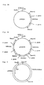
- The HBV genome is preferably the genomic DNA of the HBV subtype adr which is frequently observed in Japan and also in countries of Southeast Asia and is cloned in E. coli. This HBV DNA contains a unique recognition site for the restriction enzymes Xhol and BamHl. A fragment having terminal BamHl sites, which is obtained by clearing the HBV genome with BamHl has the structure as shown in Figure 1, wherein the HBc gene and the HBs gene are present in the same orientation.
- A fragment having terminal Xhol sites is obtained by cleaving the HBV genome with Xhol. This fragment contains the HBs and the HBc gene. Furthermore, a fragment containing only the HBs gene may be obtained by cleaving the Xhol site- terminating fragment with BamHl.
- The HBV DNA is prepared as follows:
- Viral particles (Dane particles) are isolated from blood of a person who is positive for HBsAg. The HBV DNA (3,200 bp) usually is a partly double-stranded circular DNA. Between 15 to 50% of the HBV DNA is single-stranded. Accordingly, the HBV DNA is treated with an endogenous DNA polymerase by the method of Sattler and Robinson (cf. F. Sattler & W. W. Robinson, Journal of Virology, 32, 226-233, 1979, "Hepatitis B viral DNA molecules have cohesive ends") in order to change the single-stranded region to a double-stranded DNA suitable for cloning the genome. After filling in the partially single stranded DNA the double-stranded DNA is extracted, and propagated by molecularly cloning in E. coli. This molecularly cloned double-stranded HBV DNA is treated with an appropriate restriction enzyme to give a fragment which is used for the construction of the desired plasmid.
- The HBV DNA used is preferably of subtype adr which is frequently observed in Japan and in other countries in Southeast Asia, but may be also HBV DNA of the subtype adw or ayw which is frequently observed in European countries and the U.S.A.
- The vector used in the present invention is an E. coli plasmid containing the replication origin of SV40 DNA, which can replicate both in E. coli and in mammalian cells. The E. coli plasmid includes any conventional plasmids, for example, originating from ColEl, pMB1, (cf Hershfield, V., Proc. Natl. Acad. Sci, U.S.A., 71, 3455, 1974) and plasmids originating from p15A (cf. Chang, A.C.Y., J. Bact., 134, 1141, 1978).
- The SV40 DNA to be inserted into the E. coli plasmid is a DNA of a virus which is well known as a mammalian cancer virus being capable of infecting and inducing cancer in monkeys. For use in recombinant DNA techniques the SV40 DNA is usually recombined with another DNA, for instance with DNA coding for (3-globin. The recombinant DNA thus obtained is introduced into cultured monkey cells in order to produce P-globin (cf. Mulligan, R. C., Nature 277, 108, 1979). In the present invention, the origin of replication of the SV40 DNA (it is usually referred to as "SV40 ori") is inserted into an E. coli plasmid which is then used for preparation of a recombinant DNA containing HBV genes. The recombinant DNA thus prepared can replicate in both E. coli and COS cells.
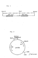
- The vector used in the present invention is preferably deficient in the replication-inhibiting region. A preferred example of such a vector DNA is a recombinant DNA consisting of E coli plasmid pBR 322 deficient in the region toxic to mammalian cells (i.e. region of inhibiting replication within COS cells; 1.426-2.521 kb) (said plasmid pBR322 being designed "pXf3") and of the replication origin (0.31 kb) of SV40 DNA. This recombinant plasmid is designated "pXRIIG" and has the structure shown in Figure 2. The origin of replication (0.31 kb) of SV40 DNA is inserted into the EcoRl cleavage site of pBR322, and the recombinant DNA is deficient in the region (1.426-2.521 kb) inhibiting the replication in mammalian cells and optionally deficient also in another unnecessary regions (e.g. 3.102-3.211 kb) in order to reduce the size of the vector. The recombinant DNA contains a tetracycline-resistance gene (Tcr) and an ampicillin-resisitance gene (Ap'). These antibiotic-resistance genes are useful as selective markers in the preparation of transformed E. coli. Various other antibiotic-resistance genes, e.g. a kanamycin-resistance gene or a chloramphenicol-resistance gene, may also be useful.
- The vector is used for the preparation of a recombinant DNA containing HBV DNA in the form of a fragment after being cleaved with an appropriate restriction enzyme, such as BamHl, Sall, Hindlll, etc.
- The fragment obtained by cleaving the above vector with an appropriate restriction enzyme (e.g. BamHl) is recombined with a fragment containing the HBV genome in an appropriate ratio to give the desired recombinant DNA. By varying the ratio of each fragment, various recombinant DNAs can be obtained wherein one fragment of the vector is combined with one or more of the fragments of the HBV DNA. Usually, the vector fragment and the fragment of the HBV DNA are used in a molar ratio of 1:1 to 1:10. Preferred recombinant DNAs consist of 2 to 4 fragments of HBV DNA and one vector fragment. The recombinant DNAs have a structure as shown in Figure 3, wherein Figure 3A shows a recombinant DNA consisting of one BamHI fragment of the HBV DNA and one fragment of the vector (designated "pSHB1") and Figure 3B shows a recombinant DNA consisting of three BamHl fragments of HBV DNA and one fragment of the vector (designated "pSHB3"). As is shown in Figure 3, the HBV DNA fragments are inserted in the same orientation, i.e. in the head-to-tail tandem form.
- The above recombinant DNA containing the HBV DNA is introduced into mammalian cells together with DNA coding for TK. Then the transformed mammalian cells are cultivated.
- The mammalian cells are preferably TK cells. e.g. mouse LTK- cells, because in this way the desired transformed cells can selectively be isolated after the transformation is carried out by treating the cells with the recombinant DNA together with several ten to several hundred times of the DNA coding for TK. There may also be used a recombinant DNA containing the DNA coding for TK instead of using the recombinant DNA and the DNA coding for TK separately.
- The transformation is more specifically illustrated below using mouse LTK- cells as an example of mammalian cells.
- Mouse LTK- cells are cultivated in Dulbecco's modified Eagle medium (hereinafter, referred to as "DMEM") supplemented with 10% calf serum (cf. Dulbecco, R. & Freeman, G.; Virology, 8, 396, 1959) and thereto is added a solution of the recombinant DNA and the DNA coding for TK in an aqueous calcium phosphate solution. The mixture is allowed to stand at room temperature for a few or several ten of minutes, usually for about 30 minutes. Additional DMEM is added to the resulting mixture, and the mixture is cultivated for 4 to 5 hours. After substituting by new DMEM, the mixture is further cultivated for 12 to 24 hours. The resulting culture is further cultivated in a medium containing hypoxanthine (15 µg/ml), aminopterin (1 pg/ml) and thymidine (5 µg/ml) (hereinafter, referred to as "HAT medium") [cf. Littlefield; J. Proc. Natl. Acad. Sci. USA, 72, 3961-3965, 1963] to give the desired transformed mouse L cells. In the above transformation procedure, the starting mouse LTK- cells are deficient in the DNA coding for TK and hence can not grow in HAT medium, but on the other hand, the transformed mouse L cells containing the DNA coding for TK are able to synthesize thymidine kinase and hence can grow in HAT medium. Accordingly, only the desired transformed cells are selectively isolated by cultivating in the above HAT medium.
- DNA coding for TK is used in the above transformation of mammalian cells together with a recombinant DNA containing the HBV DNA. The DNA coding for TK is a recombinant DNA of an E. coli plasmid (e.g. pBR322) and a DNA coding for TK derived from Herpes simplex virus and has the structure as shown in Figure 4 [cf. Florence Colbere-Garapin, 3rd General Meeting of ESACT, Oxford 1979, Develop. biol. Standard, 46, 75-82, 1980].
- The transformed mammalian cells obtained above are cultivated in an appropriate medium in a usual manner to simultaneously produce large amounts of HBsAg and HBeAg within the cultivated cells. The proteins are released into the medium. In this culture, a mixture of "s" antigen and "e" antigen is produced, but they can easily be separated by conventional protein purification methods, for instance, by passing the culture broth through a column containing an anti-HBs antibody and then eluting the "s" antigen with a 0.1 M HCI-glycine buffer, and likewise by passing the culture broth through a column containing an anti-HBe antibody and then eluting the "e" antigen therefrom with the same buffer as above.
- The HBV proteins thus obtained have the same immunological properties as the natural HBV proteins originated from human blood serum and can be used for the preparation of HBV vaccines and diagnostic reagents like the natural HBV proteins.
- The recombinant DNA, the transformed mammalian cells and the method for the simultaneous production of HBV proteins of the present invention are illustrated by the following examples.
- Pooled blood plasma (700 ml) obtained from ten patients who are positive in HBsAg (subtype adr) and HBcAg is centrifuged at 5,000 r.p.m. for 20 minutes to remove undissolved substances. The resulting solution is centrifuged at 4°C, 18,000 r.p.m. for 8 hours, and the resultant precipitate is re-dissolved in 10 ml of a buffer (pH 7.5) of 10 mM Tris-HCI, 0.1 M NaCI and 1 mM EDTA. The solution is added to the top of a centrifuge tube containing 30% sucrose, which is centrifuged at 4°C, 39,000 r.p.m. for 4 hours. The resultant precipitate is redissolved in the same buffer as above.
- In order to allow the following operations the buffer solution is subjected to a reaction with HBV DNA polymerase by treating it in a mixture (500 pl) of 67 mM Tris-HCI (pH 7.5), 80 mM NH4CI, 25 mM MgC12, 0.5% NP40 (Tergitol®, manufactured by Sigma Co.), 0.1 %.
- 2-mercaptoethanol, 330 pM dCTP (deoxycyti- dine triphosphate), dGTP (deoxyguanosine triphosphate), and dATP (deoxyadenosine triphosphate), 0.5 pM a-[32P]dTTP (deoxythymidine triphosphate) at 37°C for 3 hours. The same volume of a 100 mM EDTA solution is added to the reaction mixture. By the above DNA polymerase reaction, the single-stranded region of the DNA is repaired to a completely double-stranded structure which is [32P] labeled. This DNA is added to the top of a centrifuge tube wherein 30%, 20% and 10% aqueous solutions of sucrose are packed in layers in this order, and it is centrifuged at 4°C, 39,000 r.p.m. for 4.5 hours.
- In order to digest the proteins strongly bonded to DNA, the precipitates obtained above are treated in a mixture (200 µl) of 1 mg/ml of Pronase E (manufactured by Kaken Kagaku K.K.) and 0.2% aqueous sodium laurate solution at 37°C for 2 hours. The resulting mixture is extracted with phenol (200 µl) twice, and the resulting DNA- containing extract is washed with ether to remove the phenol and to give a solution of HBV DNA. The DNA thus obtained has a specific radioactivity of 2.5x10⁶ cpm/pg and can be used for digestion with restriction enzymes.
- The double-stranded circular HBV DNA obtained above is cloned by using λ-phage Charon 16A DNA as a vector and is then subcloned by using the known plasmid pBR322 as a vector as follows.
- HBV DNA (20 ng) is treated with endonuclease Xho I in a mixture (20 µl) of 10 mM Tris-HCI (pH 7.4), 7 mM MgCl2, 100 mM NaCl and 7 mM 2-mercaptoethanol at 37°C for 2 hours. The resulting mixture is extracted with phenol (20 µl) and further with ether, and a double volume of cooled ethanol is added to the aqueous layer to precipitate the DNA. The mixture is kept at -70°C for one hour and then centrifuged at 10,000 r.p.m. for 5 minutes, and the precipitated DNA is recovered. The precipitate thus separated is dissolved in a mixture (5 pl) of 10 mM Tris-HCI (pH 7.4) and 1 mM EDTA. The HBV DNA and an equimolar amount of λ-phage Charon 16 A DNA (having a unite Xho I recognition site) obtained by cleavage with endonuclease Xho I in the same manner as described above are reacted with T4 DNA ligase [a mixture of 50 mM Tris-HCI (pH 7.4), 10 mM MgCl2, 10 mM dithiothreitol, 100 µg/ml calf serum albumin, 0.5 mM ATP and 0.5 pl enzyme preparation (T4 DNA ligase, manufactured by Takara Biomedicals, 1-5x10³ unit/ml)] at 4°C for 18 hours. The reaction mixture is extracted with phenol and ether and then subjected to precipitation with ethanol in the same manner as described above. The precipitates thus obtained are dissolved in a mixture (10 µl) of 10 mM Tris-HCI (pH 7.4) and 1 mM EDTA.
- The thus annealed DNA is subjected to in vitro packaging to form λ-phages in the same manner as described in "Methods in Enzymology", 68, 299-309. Subsequently plaques (104) are formed on an L-agar plate (23 cmx23 cm) by using said λ-phages and E. coli DP50 SupF (cf. Blattner, F. R. et al, Science (196, 161, 1977) as an indicator. These plaques are subjected to a plaque hybridization using 32P-labeled HBV DNA as prepared above as a probe (cf. Science, 196, 180, 1977) in order to select plaques formed by phases containing HBV DNA. A plurality of the desired phages are separated.
- A phage DNA is prepared from HBV DNA cloned in Charon 16 A as obtained in step (A) by using E. coli DP50-SupF as bacteria to be infected in the same manner as described in "Methods in Enzymology", 68, 245-378, 1979. The DNA thus obtained is digested with Xho I under the same conditions as described above for 2 hours, and the resulting reaction mixture is subjected to an electrophoresis with 0.75% agarose gel to isolate HBV DNA (3.2 kb). The HBV DNA is absorbed onto DEAE (diethymaminoethyl cellulose) paper (manufactured by Toyo Roshi, Japan) in order to separate it from the vector DNA and then eluted with 1 M NaCl aqueous solution. The HBV DNA thus obtained is treated with T4 DNA ligase under the same conditions as described above to give a circular HBV DNA (3.2 kb). This HBV DNA is treated with BamHl to give a BamHl fragment of HBV DNA.
- Separately, E. coli pBR322 having a single BamHl cleavage site within its tetracycline-resistance gene is cleaved with BamHl, and the product is purifiud by phenol extraction and ethanol precipitation in the same manner as described above.
- The thus obtained pBR322 cleaved with BamHl is mixed with the BamHl fragment of HBV DNA obtained above in a molar ratio of 1:5, and the mixture is annealed with T4 DNA ligase for 18 hours as described above.
- The annealed DNA preparation (10 µl) obtained above is added to a solution of E. coli (0.1 ml) which is prepared by treating a culture broth of E. coli X1776 (cf. R. Curtiss III et al, "Molecular Cloning of Recombinant DNA" ed. Scott, W. A. and Werner, R., page 99, Academic Press, 1977) by the procedure as described in Norgard, M. V., Gene, 3, 279 (1978), and the mixture is mixed well and allowed to stand at 0°C for 25 minutes. The mixture read out on an L-agar plate containing ampicillin (20 µg/ml), a-biotine (1 pg/ml), diaminopimelic acid (100 µg/ml) and thymine (20 pg/ml) and is incubated at 37°C overnight. The resulting colonies are spread out onto both an agar plate containing tetracyline (20 µg/ml) and an agar plate containing ampicillin (20 µ/ml), and the colonies which grow only on the agar plate containing ampicillin are selected. pBR322 has an ampicillin-resistance gene and a tetracyline- resistance gene, but when HBV DNA is inserted into the BamHl site of the tetracycline-resistance gene, the tetracycline resistance gene is inactivated. Accordingly, the selected colonies contain a recombinant pBR322-HBV DNA. From the colonies thus selected, a plasmid is prepared by the procedure as described by K. Matsubara (cf. J. Virol., (16,479,1975). The plasmid thus obtained, i.e. the recombinant pBR322-HBV DNA (linked at the BamHl site), is treated with BamHl under the same conditions as described above, and the reaction mixture is subjected to an eletrophoresis with 0.75% agarose gel in the same manner as described above to give a BamHl fragment of HBV DNA.
- A vector pXRIIG (1 µg) is added to a mixture (20 µl) of 10 mM Tris-HCI (pH 8.0), 7 mM MgCl2, 100 mM NaCl and 2mM 2-mercaptoethanol, one unit of BamHl (one unit: an enzymatic activity being capable of completely cleaving 1 pg of λ-DNA per one hour) is added, and the mixture is reacted at 30°C for one hour. The reaction mixture is extracted with phenol, the aqueous layer is extracted with ether and then subjected to ethanol precipitation. The precipitates are dissolved in water and the solution is used for the preparation of a recombinant DNA.
- A solution (50 ul) containing the BamHl fragment (150 ng), of HBV DNA BamHi fragment (50 ng) of pXRIIG is reacted with T4 DNA ligase at 16°C for 4 hours.
- E. coli X1776 is transformed with the obtained ligation mixture in the same manner as described above. Colonies which grow on an agar medium are selected from the resulting transformants after incubating them on L-agar plates for 12 hours in the same manner as described above in (1), (B). The colonies thus selected are spread out on an agar medium containing tetracycline (Tc) (10 pg/ml) and an agar medium containing ampicillin (Ap) (40 µg/ml). The colonies (clones) which can not grow on the Tc-containing agar medium but can grow on the Ap-containing agar medium are selected. These clones are each incubated in the above mentioned culture liquid for E. coli X1776, and the plasmids are extracted in the same manner as described above. By analysis of the cleavage pattern with various restriction enzymes (e.g. BamHl, Xhol, Hind III, Sal I), a recombinant DNA consisting of three HBV DNA BamHl fragments and one pXRIIG fragment is selected. It is designated as pSHB3.
- The following liquids A and B are prepared.
- Liquid A: a solution (1.25 ml, pH 7.1) consisting of 50 mM Hepes (i.e. N-2-hydroxyethylpiperazine-N-2-ethanesulfonic acid), 280 mM NaCI, and 15 mM Na2HP04.12H20.
- Liquid B: a solution containing a mixture of DNA (1.1 ml) consisting of pSHB3 (50 pg), pTK (2.5 µg) (cf. Colbere-Garapin, F., Proc. Natl. Acad. Scie. USA, 76, 3755, 1979), salmon sperm DNA (carrier DNA) (50 µg) and 0.24 M CaCl2.
- The liquid B is added dropwise with stirring to the liquid A, and the mixture is allowed to stand at room temperature for 30 minutes. After pipetting sufficiently, the mixture (0.5 ml) is added dropwise to a single layer of mouse LTK- cells (about 105 cells/flask) in a flask. The flask is kept at room temperature for 30 minutes in order to absorb the mixture into the cells. Then DMEM (5 ml) is added and the mixture is incubated under 5% C02 at 37°C for about 5 hours. After exchanging with new DMEM, the mixture is further incubated for about 24 hours, and then, the medium is replaced by HAT medium. The incubation is continued while the medium is replaced by a new HAT medium every two to three days. After 4 weeks, the colonies of TK+ cells are collected to give the desired transformed cells.
- The culture liquid of the transformed mouse L cells (LTK+) obtained in (4) above is assayed with a kit for detecting HBsAg, HBeAg and HBcAg (manufactured by Abbott, U.S.A.). As a result, HBsAg and HBeAg are detected, but HBcAg is not detected.
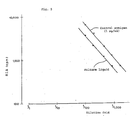
- As to the culture liquid obtained above, the reactivity and amount of antigen are assumed in accordance with a parallel line assay using a kit for detecting HBsAg as above (cf. Finnery, D. J., "Statistical method in biological assay, 2nd edn. Griffin, London, 1964), wherein purified HBsAg obtained from human blood serum is used as control antigen. The results are shown in Figure 5. As is clear from Figure 5, the amount of antigen in the culture liquid of the transformed mouse cells is 600 ng/mi. Moreover, based on the parallelism with the control antigen, it is also clear that the HBsAg produced by the present invention has similar reactivities (antigenicity, immunogenicity, etc.) to the HBsAg present in human blood plasma.
- Cesium chloride (1.5 g) is added to the culture liquid (5 ml), obtained above, and the mixture is centrifuged at 4°C, 200,000xg for 60 hours. Fractions having a maximum antigenicity of HBsAg and HBeAg are separated. When the specific gravity of these fractions is measured, the fraction having a maximum antigenicity of HBsAg has a specific gravity of about 1.20 and the fraction of HBeAg of about 1.28. These specific gravities are almost the same as those of HBsAg and HBeAg present in human plasma.
- Besides, the transformed cells are set with carbon tetrachloride at 4°C for 30 minutes and reacted with rabbit anti-HBs anti-serum (as the primary antibody), followed by washing and drying, and then reacted with a fluorescent pigment- labeled anti-rabbit IgG antibody (as the secondary antibody), followed by washing and drying. The resulting cells are observed by fluorescent microscopy, As a result, there is observed a specific fluorescence within the cytoplasm of the transformed cells.
- The culture liquid (100 µl) obtaiend above is reacted with rabbit anti-HBs antibody (antibody value PHA 28) (100 µl) at room temperature for 12 hours and the reaction mixture is centrifuged at 10,000xg for 30 minutes. The resulting precipitates are stained with uranyl acetate and observed with an electron microscope. As a result, there is observed a multitude of aggregated images of particles (particle size 22 nm) similar to HBsAg particles present in human plasma which are treated likewise. Tubular particles or Dane particles (HBV) are not observed.
- Thus, the transformed mouse L cells produce and release HBsAg and HBeAg into the medium.
- The HBsAg and HBeAg thus produced are isolated from the culture liquid in the following manner.
- The culture liquid (100 ml) is passed through a column packed with guinea pig anti-HBs antibodies linked to Sepharose CL 4B (manufactured by Pharmacia, Sweden), by which only HBsAg is adsorbed, and hence, the HBsAg can be isolated by an elution with a 0.1 M HCI-glycine buffer (pH 2.5).
- The HBsAg thus obtained was subcutaneously inoculated into guinea pigs (female, 300-400 g, 10 animals) once a week for three weeks and once more after an additional month. The antibody in the blood plasma was measured with a kit for the detection of anti-HBs antibodies (AUSAB, manufactured by Abbott, U.S.A.). As a result, the same increased antibody value was observed in all animals as in a control animal which was positive for the antibody with the same kit.
- Besides, the liquid passed through the above column is passed through a column packed with guinea pig anti-HBe antibodies linked to Sepharose CL 4B, by which only HBeAg is adsorbed, and the HBeAg is isolated by elution with a 0.1 M HCI-glycine buffer (pH 2.5).
Claims (8)
Priority Applications (1)
| Application Number | Priority Date | Filing Date | Title |
|---|---|---|---|
| AT83108220T ATE52108T1 (en) | 1982-08-20 | 1983-08-19 | RECOMBINANT DNA CONTAINING HEPATITIS B VIRUS GENE, MAMMALIAN CELLS TRANSFORMED WITH THE CLONED VIRAL DNA AND PRODUCTION OF HEPATITIS B VIRUS PROTEINS. |
Applications Claiming Priority (2)
| Application Number | Priority Date | Filing Date | Title |
|---|---|---|---|
| JP145092/82 | 1982-08-20 | ||
| JP57145092A JPS5936698A (en) | 1982-08-20 | 1982-08-20 | Recombinant dna recombined with gene of virus of hepatitis b, transformed animal cell, and preparation of protein of virus of hepatitis b |
Publications (3)
| Publication Number | Publication Date |
|---|---|
| EP0101617A2 EP0101617A2 (en) | 1984-02-29 |
| EP0101617A3 EP0101617A3 (en) | 1984-10-10 |
| EP0101617B1 true EP0101617B1 (en) | 1990-04-18 |
Family
ID=15377189
Family Applications (1)
| Application Number | Title | Priority Date | Filing Date |
|---|---|---|---|
| EP83108220A Expired - Lifetime EP0101617B1 (en) | 1982-08-20 | 1983-08-19 | Recombinant dna containing a hepatitis b virus gene, mammalian cells transformed with the cloned viral dna, and production of hepatitis b virus proteins |
Country Status (6)
| Country | Link |
|---|---|
| US (1) | US5024938A (en) |
| EP (1) | EP0101617B1 (en) |
| JP (1) | JPS5936698A (en) |
| AT (1) | ATE52108T1 (en) |
| CA (1) | CA1287311C (en) |
| DE (1) | DE3381472D1 (en) |
Families Citing this family (27)
| Publication number | Priority date | Publication date | Assignee | Title |
|---|---|---|---|---|
| EP0038765B1 (en) * | 1980-04-22 | 1987-09-02 | Institut Pasteur | Vaccine against viral hepatitis b, method and transformed eucaryotic cells for the preparation of this vaccine |
| JPS5974985A (en) * | 1982-10-19 | 1984-04-27 | Takeda Chem Ind Ltd | Novel dna, its preparation and transformant having the same |
| US5118627A (en) * | 1984-02-27 | 1992-06-02 | Amgen | Papova virus construction |
| US5324513A (en) * | 1984-03-07 | 1994-06-28 | Institut Pasteur | Composition useful for the fabrication of vaccines |
| FR2560890B1 (en) * | 1984-03-07 | 1987-10-16 | Grp Genie Genetique | COMPOSITION USEFUL FOR THE MANUFACTURE OF VACCINES CONTAINING PARTICLES CARRYING THE SURFACE ANTIGEN OF HEPATITIS B VIRUS AND THE POLYMERIZED HUMAN SERUM ALBUMIN RECEPTOR, ANIMAL CELLS CAPABLE OF PRODUCING SUCH PARTICLES |
| US4777240A (en) * | 1984-03-08 | 1988-10-11 | Scripps Clinic And Research Foundation | SV40 expression vector containing HBxAg as an expression marker |
| JPS60193925A (en) * | 1984-03-13 | 1985-10-02 | Chemo Sero Therapeut Res Inst | Lyophilized pharmaceutical preparation vaccine |
| US4624918A (en) * | 1984-07-09 | 1986-11-25 | Genentech, Inc. | Purification process for hepatitis surface antigen and product thereof |
| JPS61158798A (en) * | 1984-12-28 | 1986-07-18 | Japan Found Cancer Res | Preparation of surface antigen of hepatitis b virus |
| JP2515299B2 (en) * | 1986-06-03 | 1996-07-10 | 三井東圧化学株式会社 | Expression vector containing heat protein protein gene hsp83 and inducible expression using the same |
| IL79740A0 (en) * | 1986-08-17 | 1986-11-30 | Yeda Res & Dev | Hepatitis vaccine |
| WO1988010300A1 (en) * | 1987-06-22 | 1988-12-29 | Medico Labs Ag | Heterologous viral peptide particle immunogens |
| EP0491077A1 (en) * | 1990-12-19 | 1992-06-24 | Medeva Holdings B.V. | A composition used as a therapeutic agent against chronic viral hepatic diseases |
| US5612487A (en) * | 1991-08-26 | 1997-03-18 | Edible Vaccines, Inc. | Anti-viral vaccines expressed in plants |
| US6297048B1 (en) | 1992-02-04 | 2001-10-02 | Chiron Corporation | Hepatitis therapeutics |
| DE69738521T2 (en) | 1996-04-05 | 2009-05-07 | Novartis Vaccines and Diagnostics, Inc., Emeryville | ALPHAVIRUS VECTORS WITH A REDUCED INHIBITION OF THE SYNTHESIS OF CELL MACROMOLECULES |
| JP2001500738A (en) | 1996-09-17 | 2001-01-23 | カイロン コーポレイション | Compositions and methods for treating intracellular diseases |
| US6506559B1 (en) * | 1997-12-23 | 2003-01-14 | Carnegie Institute Of Washington | Genetic inhibition by double-stranded RNA |
| SK287538B6 (en) | 1998-03-20 | 2011-01-04 | Commonwealth Scientific And Industrial Research Organisation | Control of gene expression |
| AUPP249298A0 (en) | 1998-03-20 | 1998-04-23 | Ag-Gene Australia Limited | Synthetic genes and genetic constructs comprising same I |
| AU776150B2 (en) * | 1999-01-28 | 2004-08-26 | Medical College Of Georgia Research Institute, Inc. | Composition and method for (in vivo) and (in vitro) attenuation of gene expression using double stranded RNA |
| US20040138168A1 (en) * | 1999-04-21 | 2004-07-15 | Wyeth | Methods and compositions for inhibiting the function of polynucleotide sequences |
| EP2363478B1 (en) * | 1999-04-21 | 2019-07-24 | Alnylam Pharmaceuticals, Inc. | Methods and compositions for inhibiting the function of polynucleotide sequences |
| US6423885B1 (en) | 1999-08-13 | 2002-07-23 | Commonwealth Scientific And Industrial Research Organization (Csiro) | Methods for obtaining modified phenotypes in plant cells |
| EP1229134A3 (en) * | 2001-01-31 | 2004-01-28 | Nucleonics, Inc | Use of post-transcriptional gene silencing for identifying nucleic acid sequences that modulate the function of a cell |
| WO2005010144A2 (en) * | 2003-04-21 | 2005-02-03 | Wisconsin Alumni Research Foundation | Displacing a plasmid in a bacterial population |
| DE202005004135U1 (en) * | 2005-03-11 | 2005-05-19 | Klocke Verpackungs-Service Gmbh | Multi-component packaging with applicator |
Family Cites Families (3)
| Publication number | Priority date | Publication date | Assignee | Title |
|---|---|---|---|---|
| FR2458584A1 (en) * | 1979-06-08 | 1981-01-02 | Pasteur Institut | VECTORS FOR THE TRANSFER AND EXPRESSION OF GENETIC MATERIAL IN EUKARYOTE CELL AND METHOD FOR THE PRODUCTION OF A PROTEIN DETERMINED IN EUKARYOTIC CELLS |
| EP0038765B1 (en) * | 1980-04-22 | 1987-09-02 | Institut Pasteur | Vaccine against viral hepatitis b, method and transformed eucaryotic cells for the preparation of this vaccine |
| ZW18282A1 (en) * | 1981-08-31 | 1983-03-23 | Genentech Inc | Preparation of polypeptides in vertebrate cell culture |
-
1982
- 1982-08-20 JP JP57145092A patent/JPS5936698A/en active Granted
-
1983
- 1983-08-18 CA CA000434868A patent/CA1287311C/en not_active Expired - Fee Related
- 1983-08-19 DE DE8383108220T patent/DE3381472D1/en not_active Expired - Fee Related
- 1983-08-19 EP EP83108220A patent/EP0101617B1/en not_active Expired - Lifetime
- 1983-08-19 AT AT83108220T patent/ATE52108T1/en not_active IP Right Cessation
-
1986
- 1986-07-08 US US06/883,138 patent/US5024938A/en not_active Expired - Fee Related
Non-Patent Citations (2)
| Title |
|---|
| Journal of Medical Virology (1981) 8, p. 237-43. * |
| M. Fried & E. Ruley - Eukaryotic - Vectors ed. by Y Cluzman (1982) cold spring - Harbor Laboratory - pages 67-70 * |
Also Published As
| Publication number | Publication date |
|---|---|
| US5024938A (en) | 1991-06-18 |
| EP0101617A2 (en) | 1984-02-29 |
| EP0101617A3 (en) | 1984-10-10 |
| ATE52108T1 (en) | 1990-05-15 |
| DE3381472D1 (en) | 1990-05-23 |
| JPS6321476B2 (en) | 1988-05-07 |
| CA1287311C (en) | 1991-08-06 |
| JPS5936698A (en) | 1984-02-28 |
Similar Documents
| Publication | Publication Date | Title |
|---|---|---|
| EP0101617B1 (en) | Recombinant dna containing a hepatitis b virus gene, mammalian cells transformed with the cloned viral dna, and production of hepatitis b virus proteins | |
| US5591638A (en) | Method for the transformation of cells, particularly eukaryotes by a DNA originating from viruses of hepatitis, more particularly from virus of B viral hepatitis, and preparations containing the expression products of said DNAs | |
| Chang et al. | Production of hepatitis B virus in vitro by transient expression of cloned HBV DNA in a hepatoma cell line. | |
| Lamberts et al. | Precore-mediated inhibition of hepatitis B virus progeny DNA synthesis | |
| Yuh et al. | The genome of hepatitis B virus contains a second enhancer: cooperation of two elements within this enhancer is required for its function | |
| Cummings et al. | Isolation, characterization, and comparison of recombinant DNAs derived from genomes of human hepatitis B virus and woodchuck hepatitis virus. | |
| JP2511394B2 (en) | Production of hepatitis B surface antigen in yeast | |
| JP2634371B2 (en) | Vector expressing DNA encoding hepatitis B surface antigen in vertebrate cells | |
| JPS61158795A (en) | Recombinant dna containing nucleotide arrangement coding predetermined polypeptide under control of adenovirus promotor | |
| EP0105149B1 (en) | Recombinant plasmid containing hepatitis b virus gene, yeast transformed with said recombinant plasmid, and production of hepatitis b virus surface antigen | |
| JPH0314840B2 (en) | ||
| Will et al. | Expression of hepatitis B antigens with a simian virus 40 vector | |
| GB2102006A (en) | Deoxyribonucleic acids, their production and use | |
| EP0182442B1 (en) | Recombinant dna molecules and their method of production | |
| Wang et al. | Enhanced production of hepatitis B surface antigen in NIH 3T3 mouse fibroblasts by using extrachromosomally replicating bovine papillomavirus vector | |
| Jenson et al. | Palindromic structure and polypeptide expression of 36 kilobase pairs of heterogeneous Epstein-Barr virus (P3HR-1) DNA | |
| Stratowa et al. | Recombinant retroviral DNA yielding high expression of hepatitis B surface antigen. | |
| EP0062574A2 (en) | Virus protein synthesis | |
| Siddiqui | Expression of hepatitis B virus surface antigen gene in cultured cells by using recombinant plasmid vectors | |
| JPS5974985A (en) | Novel dna, its preparation and transformant having the same | |
| EP0288198A2 (en) | Production of Peptide | |
| Denniston et al. | Expression of hepatitis B virus surface and e antigen genes cloned in bovine papillomavirus vectors | |
| US4983520A (en) | DNA sequence encoding modified hepatitis B virus surface antigen P31 protein | |
| Meneguzzi et al. | The arrangement of integrated viral DNA is different in BK virus-transformed mouse and hamster cells | |
| PT85108B (en) | VARICELA-ZOSTER VIRUS USED AS A RECOMBINANT VACCINE |
Legal Events
| Date | Code | Title | Description |
|---|---|---|---|
| PUAI | Public reference made under article 153(3) epc to a published international application that has entered the european phase |
Free format text: ORIGINAL CODE: 0009012 |
|
| AK | Designated contracting states |
Kind code of ref document: A2 Designated state(s): AT BE CH DE FR GB IT LI NL SE |
|
| PUAL | Search report despatched |
Free format text: ORIGINAL CODE: 0009013 |
|
| AK | Designated contracting states |
Kind code of ref document: A3 Designated state(s): AT BE CH DE FR GB IT LI NL SE |
|
| 17P | Request for examination filed |
Effective date: 19841231 |
|
| 17Q | First examination report despatched |
Effective date: 19860618 |
|
| GRAA | (expected) grant |
Free format text: ORIGINAL CODE: 0009210 |
|
| STAA | Information on the status of an ep patent application or granted ep patent |
Free format text: STATUS: THE PATENT HAS BEEN GRANTED |
|
| AK | Designated contracting states |
Kind code of ref document: B1 Designated state(s): AT BE CH DE FR GB IT LI NL SE |
|
| REF | Corresponds to: |
Ref document number: 52108 Country of ref document: AT Date of ref document: 19900515 Kind code of ref document: T |
|
| ITF | It: translation for a ep patent filed |
Owner name: JACOBACCI & PERANI S.P.A. |
|
| REF | Corresponds to: |
Ref document number: 3381472 Country of ref document: DE Date of ref document: 19900523 |
|
| ET | Fr: translation filed | ||
| PLBE | No opposition filed within time limit |
Free format text: ORIGINAL CODE: 0009261 |
|
| 26N | No opposition filed | ||
| ITTA | It: last paid annual fee | ||
| EAL | Se: european patent in force in sweden |
Ref document number: 83108220.1 |
|
| PGFP | Annual fee paid to national office [announced via postgrant information from national office to epo] |
Ref country code: SE Payment date: 19950630 Year of fee payment: 13 |
|
| PGFP | Annual fee paid to national office [announced via postgrant information from national office to epo] |
Ref country code: BE Payment date: 19950713 Year of fee payment: 13 |
|
| PGFP | Annual fee paid to national office [announced via postgrant information from national office to epo] |
Ref country code: CH Payment date: 19950727 Year of fee payment: 13 |
|
| PGFP | Annual fee paid to national office [announced via postgrant information from national office to epo] |
Ref country code: GB Payment date: 19950809 Year of fee payment: 13 |
|
| PGFP | Annual fee paid to national office [announced via postgrant information from national office to epo] |
Ref country code: FR Payment date: 19950830 Year of fee payment: 13 Ref country code: AT Payment date: 19950830 Year of fee payment: 13 |
|
| PGFP | Annual fee paid to national office [announced via postgrant information from national office to epo] |
Ref country code: NL Payment date: 19950831 Year of fee payment: 13 |
|
| PGFP | Annual fee paid to national office [announced via postgrant information from national office to epo] |
Ref country code: DE Payment date: 19951030 Year of fee payment: 13 |
|
| PG25 | Lapsed in a contracting state [announced via postgrant information from national office to epo] |
Ref country code: GB Effective date: 19960819 Ref country code: AT Effective date: 19960819 |
|
| PG25 | Lapsed in a contracting state [announced via postgrant information from national office to epo] |
Ref country code: SE Effective date: 19960820 |
|
| PG25 | Lapsed in a contracting state [announced via postgrant information from national office to epo] |
Ref country code: LI Effective date: 19960831 Ref country code: CH Effective date: 19960831 Ref country code: BE Effective date: 19960831 |
|
| BERE | Be: lapsed |
Owner name: JURIDICAL FOUNDATION THE CHEMO-SERO-THERAPEUTIC R Effective date: 19960831 |
|
| PG25 | Lapsed in a contracting state [announced via postgrant information from national office to epo] |
Ref country code: NL Effective date: 19970301 |
|
| GBPC | Gb: european patent ceased through non-payment of renewal fee |
Effective date: 19960819 |
|
| REG | Reference to a national code |
Ref country code: CH Ref legal event code: PL |
|
| PG25 | Lapsed in a contracting state [announced via postgrant information from national office to epo] |
Ref country code: FR Effective date: 19970430 |
|
| NLV4 | Nl: lapsed or anulled due to non-payment of the annual fee |
Effective date: 19970301 |
|
| PG25 | Lapsed in a contracting state [announced via postgrant information from national office to epo] |
Ref country code: DE Effective date: 19970501 |
|
| EUG | Se: european patent has lapsed |
Ref document number: 83108220.1 |
|
| REG | Reference to a national code |
Ref country code: FR Ref legal event code: ST |