EP0089014A2 - Physiological implantable cardiac pacemaker in which the stimulation rate is regulated by the respiration rate of the patient - Google Patents
Physiological implantable cardiac pacemaker in which the stimulation rate is regulated by the respiration rate of the patient Download PDFInfo
- Publication number
- EP0089014A2 EP0089014A2 EP83102349A EP83102349A EP0089014A2 EP 0089014 A2 EP0089014 A2 EP 0089014A2 EP 83102349 A EP83102349 A EP 83102349A EP 83102349 A EP83102349 A EP 83102349A EP 0089014 A2 EP0089014 A2 EP 0089014A2
- Authority
- EP
- European Patent Office
- Prior art keywords
- pacemaker
- rate
- signal
- fact
- rized
- Prior art date
- Legal status (The legal status is an assumption and is not a legal conclusion. Google has not performed a legal analysis and makes no representation as to the accuracy of the status listed.)
- Granted
Links
- 230000000638 stimulation Effects 0.000 title claims abstract description 23
- 230000029058 respiratory gaseous exchange Effects 0.000 title claims description 9
- 230000001105 regulatory effect Effects 0.000 title claims description 3
- 230000000747 cardiac effect Effects 0.000 title abstract description 6
- 238000009423 ventilation Methods 0.000 claims abstract description 23
- 210000000115 thoracic cavity Anatomy 0.000 claims abstract description 20
- 230000000241 respiratory effect Effects 0.000 claims abstract description 9
- 230000005764 inhibitory process Effects 0.000 claims description 8
- 230000001746 atrial effect Effects 0.000 claims description 7
- 238000001514 detection method Methods 0.000 claims description 6
- 230000007704 transition Effects 0.000 claims description 6
- 238000006243 chemical reaction Methods 0.000 claims description 3
- 230000036387 respiratory rate Effects 0.000 claims description 3
- 230000035945 sensitivity Effects 0.000 claims description 3
- 230000002269 spontaneous effect Effects 0.000 claims description 3
- 230000003252 repetitive effect Effects 0.000 claims description 2
- 238000005070 sampling Methods 0.000 claims description 2
- 230000002861 ventricular Effects 0.000 claims description 2
- 238000005265 energy consumption Methods 0.000 claims 1
- 230000003068 static effect Effects 0.000 claims 1
- 230000006870 function Effects 0.000 description 10
- 235000014676 Phragmites communis Nutrition 0.000 description 7
- 239000003990 capacitor Substances 0.000 description 6
- 238000000034 method Methods 0.000 description 5
- 238000010276 construction Methods 0.000 description 3
- 230000000694 effects Effects 0.000 description 3
- QVGXLLKOCUKJST-UHFFFAOYSA-N atomic oxygen Chemical compound [O] QVGXLLKOCUKJST-UHFFFAOYSA-N 0.000 description 2
- 239000008280 blood Substances 0.000 description 2
- 210000004369 blood Anatomy 0.000 description 2
- 230000036760 body temperature Effects 0.000 description 2
- 230000008602 contraction Effects 0.000 description 2
- 238000010586 diagram Methods 0.000 description 2
- 238000012986 modification Methods 0.000 description 2
- 230000004048 modification Effects 0.000 description 2
- 229910052760 oxygen Inorganic materials 0.000 description 2
- 239000001301 oxygen Substances 0.000 description 2
- 241001611093 Stimula Species 0.000 description 1
- 230000009471 action Effects 0.000 description 1
- 238000013459 approach Methods 0.000 description 1
- 230000005540 biological transmission Effects 0.000 description 1
- 210000000038 chest Anatomy 0.000 description 1
- 238000004891 communication Methods 0.000 description 1
- 230000001276 controlling effect Effects 0.000 description 1
- 238000012937 correction Methods 0.000 description 1
- 230000002596 correlated effect Effects 0.000 description 1
- 230000004907 flux Effects 0.000 description 1
- 239000007943 implant Substances 0.000 description 1
- 230000003993 interaction Effects 0.000 description 1
- 230000003601 intercostal effect Effects 0.000 description 1
- 238000005259 measurement Methods 0.000 description 1
- 230000007246 mechanism Effects 0.000 description 1
- 238000013160 medical therapy Methods 0.000 description 1
- 230000002503 metabolic effect Effects 0.000 description 1
- 230000004660 morphological change Effects 0.000 description 1
- 210000003205 muscle Anatomy 0.000 description 1
- 230000037081 physical activity Effects 0.000 description 1
- 230000007180 physiological regulation Effects 0.000 description 1
- 230000036279 refractory period Effects 0.000 description 1
- 230000008439 repair process Effects 0.000 description 1
- 238000011160 research Methods 0.000 description 1
- 230000033764 rhythmic process Effects 0.000 description 1
- 230000008925 spontaneous activity Effects 0.000 description 1
- 238000007920 subcutaneous administration Methods 0.000 description 1
- 238000006467 substitution reaction Methods 0.000 description 1
- 230000001360 synchronised effect Effects 0.000 description 1
- 230000001960 triggered effect Effects 0.000 description 1
Images


Classifications
-
- A—HUMAN NECESSITIES
- A61—MEDICAL OR VETERINARY SCIENCE; HYGIENE
- A61B—DIAGNOSIS; SURGERY; IDENTIFICATION
- A61B5/00—Measuring for diagnostic purposes; Identification of persons
- A61B5/08—Measuring devices for evaluating the respiratory organs
- A61B5/085—Measuring impedance of respiratory organs or lung elasticity
- A61B5/086—Measuring impedance of respiratory organs or lung elasticity by impedance pneumography
-
- A—HUMAN NECESSITIES
- A61—MEDICAL OR VETERINARY SCIENCE; HYGIENE
- A61B—DIAGNOSIS; SURGERY; IDENTIFICATION
- A61B5/00—Measuring for diagnostic purposes; Identification of persons
- A61B5/08—Measuring devices for evaluating the respiratory organs
- A61B5/0816—Measuring devices for examining respiratory frequency
-
- A—HUMAN NECESSITIES
- A61—MEDICAL OR VETERINARY SCIENCE; HYGIENE
- A61N—ELECTROTHERAPY; MAGNETOTHERAPY; RADIATION THERAPY; ULTRASOUND THERAPY
- A61N1/00—Electrotherapy; Circuits therefor
- A61N1/18—Applying electric currents by contact electrodes
- A61N1/32—Applying electric currents by contact electrodes alternating or intermittent currents
- A61N1/36—Applying electric currents by contact electrodes alternating or intermittent currents for stimulation
- A61N1/362—Heart stimulators
- A61N1/365—Heart stimulators controlled by a physiological parameter, e.g. heart potential
- A61N1/36514—Heart stimulators controlled by a physiological parameter, e.g. heart potential controlled by a physiological quantity other than heart potential, e.g. blood pressure
- A61N1/36521—Heart stimulators controlled by a physiological parameter, e.g. heart potential controlled by a physiological quantity other than heart potential, e.g. blood pressure the parameter being derived from measurement of an electrical impedance
Definitions
- This invention concerns the type of artificial cardiac pacemakers to be implanted in the patient's body.
- pacemaker will substitute “artificial cardiac pacemaker” for the rest of this description, keeping in consideration that the above mentioned term is a registered trade-mark.
- the kind of pacemaker in which the ventricle's stimulation activity is driven and synchronized by the detection of the spontaneous bio-electrical atrial activity are commonly defined as ' physiological since the electrical impulses that effect the rate are not fixed but induced by the spontaneous atrial rate and are therefore always proportional to it.
- This kind of pacemaker can only be used in the cases in which the atrial activity can occur in a physiological manner and, therefore, it is only possible in 30 to 60% of the actual pacemaker wearer patients.
- patent number '3,593,718 is the pacemaker (external, and not implantable) driven by the ventilation rate in which the said function is detected by means of an impedance voltage converter that measures thoracic impedance variations by means of electrodes on the skin of the patient when the ventilation rate changes.
- the signal coming from this converter is sent directly to a continuous rate/current converter that drives an oscillator that supplies the impulses needed to send the electrical stimulation necessary for the heart, by means of a constant current source at a rate which varies automatically in relation to the ventilation rate.
- Such a device is impossible to achieve in practice since it lacks a unit capable of converting the voltage into rate and should operate between the first and the second converters just mentioned.
- the reference rate will be fixed by the constructor or programmed in programmable pacemakers at the minimum values which will coincide with the patient's heart beat when in a state of complete rest, normally at 70 beats per minute.
- the new method proposed by this invention will be able to transform the atrial or ventricular inhibition rate from a fixed parameter (more or less programmable) into a parameter that will automatically vary with the metabolic needs of the patient.
- a pacemaker when improved according to the new discoveries, that will constantly detect the patient's necessary optimal heart rate and will therefore act accordingly.
- the circuit that detects impedance variations in the human body is supplied with current impulses that are at a higher rate than those of the ventilation rate with the necessary intensity but of very short duration; for instance, impulses in which the relation between the width of each pulse and its period of repetition is approximately 1 to 1000.
- Programmable amplifiers in which it is possible to program the current drain are used in the circuit of such a pacemaker.
- a further problem that had to be solved was regarding the automatic correction of the basic impedance, or rather finding a system that will not be influenced by the basic impedance variations, this being an essential condition of any device which is to be implanted and therefore inaccessible for repair or calibration.
- the leads that detect the thoracic impedance variations present a basic impedance that is very different from case to case due to the difference between patients and the differences that arise in a patient through time.
- a yet further problem to solve was that of realizing a highly reliable, simple, compact device that could easily be realized into an integrated circuit.
- the improved pacemaker which can be implanted according to the invention is essentially composed of:
- Z indicates the variable physiological impedance between leads 1 and 2.
- Unit 10 will pick up the Z impedance variations at measured with current pulses of constant intensity I C .
- the voltage I O .Z that exists during each impulse on thoracic impedance is supplied to the subsequent unit 20.
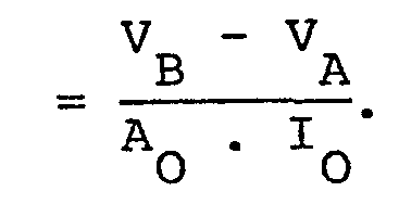
- This unit is the main innovation of the entire device. In this parte the I O .Z variation is amplifyed and then' compared with threshold levels V A and VB the latter is programmable from the outside. After the comparation stage follows a decision stage in which it eliminates errors in detection and discriminates between substantial and less substantial respiratory acts.
- the unit 20 output is a signal V OUT at logic levels that produces a number of positive transitions equal to the number of substantial respiratory acts.
- Unit 30, that follows unit 20, must establish a linear relation, within a field of limited and pre--established values, between the number of positive transitions in the time unit of the said signal V OUT and, for instance, a current I CH charging capacitor of a normal pacemaker 40 determining its stimulation rate.
- unit 30 realizes the ventilation rate/current conversion so as to allow, along with unit 40, a relation ventilation rate VR/heart rate HR according to the curve chosen and programmed from the curves in figure 3 (see further).
- Number 3 indicates the output from unit 40 that goes to the lead that stimulates the heart.
- Units 10-20-30 are driven by unit 50 that generates the A-C-D-K timing as will be described further on.
- the device can transmit information to the outside through unit 60.
- Unit 60 can transmit information regarding the ventilation acts through coil 61 towards the outside and by means of the coil 61 itself, when reed switch 62 is closed it is able to receive the programming signals coming from coil 63 and to send them to unit 70.
- Unit 64 will indicate the magnet by means of which it is possible to close reed switch 62 when programming the pacemaker.
- the registers that allow the control of the general working conditions of the entire pacemaker and are protected by a coded identification device are located in unit 70.
- Unit 50 is illustrated in figure 4. It includes a twelve stage counter U4/A that fixes the periods ⁇ . and ⁇ 1 at circa 120 ⁇ sec and 125 msec respectively. This can be seen in figure 5 whege ⁇ , indicates the period of the oscillator OSC that drives the U4/A. Unit 50 also contains a Johnson counter joined to a monostable (Q9 output of U4/B linked to a respective Clock) that creates the necessary phase relationship between signals A-C-D and K. Figure 5 shows the timing of outputs A-C-D and K when the H inputs coming from the external programming stage, is low. When H is high the whole blocks itself and disconnects the detection of the ventilation act (see further). V S indicates the supply voltage of the pacemaker that is represented by a negative sign since it is normally the positive pole of the battery that is connected to ground M.
- a current impulse I O for instance, of constant intensity, width of 120 usec, and period of 125 msec, is applied to the physiological load Z that varies during time.
- the current I O generator is made up of TS1-R4-D1 - -D2-R3 and is driven by signal A with the characteristics as shown in figure 5. Since Z is not a totally resistant load, the TS2 switch controlled by A short circuits it when the current generator is off and therefore when A is low.
- the resistor R5 placed in series with Z and with the decoupling capacitor C 2 has the task of traslating the level of the voltage peaks originating from Z in order to bring them to levels which will make them easily manageable by the subsequent components.
- a voltage V X formed by very short impulses comes from the circuit in figure 6; it varies in amplitude according to the ventilation rate of the patient.
- the envelope, of the V H impulse peaks, has a V A average value and profile similar to that of the envelope of V X peaks.
- the voltage V A for example, is chosen with the approximate value of V S /2.
- V H peak does not exceed the V A threshold the V J signal remains low and the output Q 3 is high since the U2/C bistable was previously set by D. In these conditions, when Q 3 , opens, the absolute value of V C increases and it will tend to increase the V H peak.
- V H of the V H peaks, is none other than the V X envelope multiplyed by A O and centered on V A . This holds true for rather rapid variations so that V C will not vary perceptibly. This conditions can be obtained by adopting a sufficiently high Rl-Cl time constant. This part of the circuit obtains the automatic adjustment of the device to the slow basic impedance variations and a signal V J that indicates when the rapid variations of the Z impedance are of a certain consistency and therefore can be attributed to a possible evolution of a respiratory act.
- the reset situation of said device can only be reached when the V H peak goes under the threshold V A or when, with a high C, there are no positive impulses at the output Q2 of U2/B.
- the circuit described therefore introduces a sort of hysteresis with a variable width, varying V B in regards to V , that has a double aim: eliminate improper transitions due to noise, create a sensitivity control that can descriminate between more or less consistent variations of the impedance Z.
- V B variable width
- R2 resistor to Rl the connections of points X - Y and the resulting connection in parallel of low value R2 resistor to Rl, is controlled by the programming circuit from the outside, as will be described further, so that the V C will rapidly reach the necessary values.
- the output signal V OUT from the circuit in figure 7, as can be seen in figure 8, has the same number of positive transitions as the number of respiratory acts and represents the patient's ventilation rate.
- Figure 9 illustrates an example of the possible use of the V OUT signal to construct a physiological pacemaker as explained in the introduction of this paper.
- the signal V OUT enters in U5/A and becomes correlated to the number of failing edges of the K signal that forms the U5/A clock, the latter is a monostable and for each failing edge of VOUT an impulse of approximately 1 msec is present at the output Q2.
- the output Q2 drives TS4 that terminates into the Ru - Cu integrator whose average voltage drives the TS5 base.
- This impulse is generated by a monostable circuit made up of TS8-TS9 R8-R9-R10-CX and triggered by the TS10-CY-R11. In the condition considered above the outputs S and T are kept high (see further).
- each suitable variation of the magnetic flux induced from the outside on coil 61 starts a monostable cycle and there is a 20 psec impulse on R9.
- a train of external magnetic impulses with an inferior or equal (20 ⁇ sec) duration starts off a similar train of impulses at output T also having a 20 ⁇ sec duration.
- output S are low.
- Unit 70 makes reference to figure 11. As already observed in figure 10, the impulses received from the outside are present in output T while S is low.
- the circuit in figure 11 can set some of the devices parameters through the number of impulses coming from the outside.
- unit 70 includes a protection for the information received. In order to program the pacemaker reed switch 62 must be closed and programming impulses must be preceded by an eight impulse key code.
- the counter U6/A reads the key--code and activates the FS function by means of U8 when the second key-code impulse is received and Ql goes high.
- the external magnet 64 (figure 2) is removed reed switch 62 opens while S goes high and Ql goes low, this will determine the end of FS.
- the output Q3 goes high when the eighth key-code impulse is received and reed switch 62 is closed for programming.
- the counter U6/A stops and the impulses T are switched to the counters U6/B, U6/C. The number of the impulses which is higher than that of the key-codes, will determine the output state of said U6/B and U6/C combination.
- FIG. 12 illustrates unit 40 that, in this example, corresponds to a demand heart pacemaker.
- Output 3 goes to the heart.
- the stimulation signals are sent and the EKG signals are received by this terminal.
- the latter filtered from R16-C3-C4-R17, is amplifyed by Tl and T 2 and sent to the circuit, made up of T3-T4-T5 and relative connected components, that creates the functions that form the refractory period after each stimulation impulse and that reject the external interference.
- T5 each EKG signal received discharges the capacitor C5. If heart signals are absent C5 will be charged by I CH and when the threshold which is determined by R18-R19 is reached T6 will discharge it in a short time which is determined by C6-R20. This is the width of the stimulation impulse.
- the impulse rate is therefore controlled by I CH -T7-C7-R21 that determines the maximum rate value that can be reached, indicated in figure 3 by HR .
- the stimulation impulses amplitude is duplicated by T8-T9 and sent to the heart by C8. It is to be understood that the various V , V and other currents were considered in their absolute value without indicating the negative sign that in reality marks them.
Landscapes
- Health & Medical Sciences (AREA)
- Life Sciences & Earth Sciences (AREA)
- Pulmonology (AREA)
- Heart & Thoracic Surgery (AREA)
- Public Health (AREA)
- Cardiology (AREA)
- Biophysics (AREA)
- Engineering & Computer Science (AREA)
- Biomedical Technology (AREA)
- Veterinary Medicine (AREA)
- Physiology (AREA)
- General Health & Medical Sciences (AREA)
- Animal Behavior & Ethology (AREA)
- Surgery (AREA)
- Molecular Biology (AREA)
- Medical Informatics (AREA)
- Physics & Mathematics (AREA)
- Pathology (AREA)
- Hematology (AREA)
- Nuclear Medicine, Radiotherapy & Molecular Imaging (AREA)
- Radiology & Medical Imaging (AREA)
- Electrotherapy Devices (AREA)
Abstract
Description
- This invention concerns the type of artificial cardiac pacemakers to be implanted in the patient's body.
- At the present state of knowledge, the artificial substitution of mechanisms of generation and conduction of the electrical signals which drive the contractions of the heart depend upon a vast range of artificial pacemakers which are identifiable by initials, accepted through an international agreement, according to the different conditions of action and interaction with the spontaneous activity of the heart. The term "pacemaker" will substitute "artificial cardiac pacemaker" for the rest of this description, keeping in consideration that the above mentioned term is a registered trade-mark. The kind of pacemaker in which the ventricle's stimulation activity is driven and synchronized by the detection of the spontaneous bio-electrical atrial activity are commonly defined as ' physiological since the electrical impulses that effect the rate are not fixed but induced by the spontaneous atrial rate and are therefore always proportional to it. This kind of pacemaker can only be used in the cases in which the atrial activity can occur in a physiological manner and, therefore, it is only possible in 30 to 60% of the actual pacemaker wearer patients.
- Thus, at present, there is a high percentage of patients needing artificial cardiac stimulation that cannot make use of a physiologically variable rate stimulation with the above mentioned technique.
- It is for this reason that individuals specialized in this field have been trying for some time to achieve an implantable pacemaker that can, respond to another parameter instead of the atrial activity parameter that can, vary in relation to the physiological needs of the patient, and can be used as a reference variable for automatic and physiological regulation of the electrical impulses that effect the heart rate.
- As of today, the pacemakers studied in order to solve the above mentioned problem are those we will describe below in which the stimulation rate is subject, by means of a specifica algorythm, to the variations of the following functions:
- - the hematic PH;
- - the body temperature;
- - the quantity of oxygen contained in the blood;
- - the QT interval observed in an endocardial EKG;
- - the respiratory rate.
- These methods have all had insufficient success due to the complexity and difficulty the pacemaker has in detecting the above mentioned physiological variations. The limits and faults of the above mentioned methods can be summarized in a more specific manner as will follow. As for the pacemaker with control of the hematic Ph, in addition to having the essential problem of safety and reliability which arises with any sensor that is inserted directly into the hematic flow, it was also observed that the sensor decays in too brief a period with respect to the average life of a pacemaker. As for the pacemaker controlled by the amount of 02 in the venous blood there remains the essential problem of reliability and the complexity of a lead which is inserted in the hematic flow and should function both as stimulator and oxygen sensor. Doubts and hesitations must also arise regarding the co-relation between the oxymetric parameter and the heart rate since it has not yet been thoroughly defined. As for the temperature controlled pacemaker, one must remember the limits resulting from the sensor as well as the impossibility, already proven by physiological research, of the body temperature to increase with a time constant similar to that of the heart rate during the patient's physical exercise. As for the QT controlled pacemaker, evident obstacles are found in the difficulty of detecting this data and in the possibility of other causes, deriving' for instance from medical therapies of natural physiolo= gical evolution, that can determine variations in the QT parameter independently of the potential variations of the heart rate. The hypothesis, which remained such, of pacemakers sensitive to the ventilation rate, was based on the detection of this parameter by means of a distorsion transducer placed in an intercostal or interdiaphragmatic area or by means of acoustic transducers placed in an intra-pleural area. Besides the difficulties presented in distinguishing the respiratory signal from that of other functions thus causing it to be unreliable, there would also be the problem of needing a specific surgical approach for setting the said sensors. Another well known attempt, described by an U.S.A. patent number '3,593,718 is the pacemaker (external, and not implantable) driven by the ventilation rate in which the said function is detected by means of an impedance voltage converter that measures thoracic impedance variations by means of electrodes on the skin of the patient when the ventilation rate changes. The signal coming from this converter is sent directly to a continuous rate/current converter that drives an oscillator that supplies the impulses needed to send the electrical stimulation necessary for the heart, by means of a constant current source at a rate which varies automatically in relation to the ventilation rate. Such a device is impossible to achieve in practice since it lacks a unit capable of converting the voltage into rate and should operate between the first and the second converters just mentioned.
- Such a unit would certainly be useless if the output signal from the first converter were to be of the sinusoidal type or perfectly repetitive but it becomes absolutely necessary, presenting quite a few problems in its realization, when one deals with physiological signals that are continuously variable both in width and periodi= city, and with continuous variations of the basic line, due to continuous morphological changes and overlaping of false signals. In the U.S.A. patent mentioned above another difficult task that is the problem of controlling the energy drain of the entire device, so as to be able to implant it, is not taken into consideration. As a result, one can confirm that the methods proposed up to now do not consent the concretization of the various theories into an easy to use totally implantable and reliable product.
- This invention proposes an implantable cardiac pacemaker in which the inhibition rate and/or the stimula= tion is regulated by the patient's ventilation rate which is detected by analyzing the impedance variations verified due to and accompanying the respiration itself within a specific distance in the human body running from the implanted pacemaker case to a reference lead which is to be inserted preferably subcutaneously in the thoracic area and at a suitable but not critical distance from the said pacemaker.
- In the many kinds of pacemakers and in all the different ways in which one must or can stimulate either the atrial or the ventricle or both,it is necessary to fix a minimum inhibition rate and a stimulation rate that does not necessarily coincide with the preceding but can be superior in the pacemakers with hysteresis, which are in any case less frequent. The said rate, called the reference rate, will be fixed by the constructor or programmed in programmable pacemakers at the minimum values which will coincide with the patient's heart beat when in a state of complete rest, normally at 70 beats per minute. As a consequence there is no pacemaker that can comply to patient's needs when the said patient is carrying out a physical activity requiring a different reference heart rate from the one that was set when the patient was at rest. The new method proposed by this invention will be able to transform the atrial or ventricular inhibition rate from a fixed parameter (more or less programmable) into a parameter that will automatically vary with the metabolic needs of the patient. A pacemaker, when improved according to the new discoveries, that will constantly detect the patient's necessary optimal heart rate and will therefore act accordingly.
- It became necessary to solve various problems in order to put into practice a pacemaker improved according to the afore mentioned intentions.
- The first and probably the most important of such problems was to analyze the impedance variations of the human body with the minimum electrical energy drain since the entire device was destined to be implanted and to supply an electrical cell and it has to have an average independent life of 4-5 years. In order to solve this problem the circuit that detects impedance variations in the human body is supplied with current impulses that are at a higher rate than those of the ventilation rate with the necessary intensity but of very short duration; for instance, impulses in which the relation between the width of each pulse and its period of repetition is approximately 1 to 1000. Programmable amplifiers in which it is possible to program the current drain are used in the circuit of such a pacemaker. Since the best features of these amplifiers such as slew rate and band width are obtained through a high current consumption the amplifiers receive the maximum supply during the phase in which the impulse detects the thoracic impedance variations, that is only at the moment when the amplifying signal is present. Another problem that had to be solved was how to pick up the ventilation rate from the output impedance signal that is noisy and whose amplitude and period vary continuously.
- A further problem that had to be solved was regarding the automatic correction of the basic impedance, or rather finding a system that will not be influenced by the basic impedance variations, this being an essential condition of any device which is to be implanted and therefore inaccessible for repair or calibration. To this purpose it is important to keep in mind that the leads that detect the thoracic impedance variations present a basic impedance that is very different from case to case due to the difference between patients and the differences that arise in a patient through time.
- A yet further problem to solve was that of realizing a highly reliable, simple, compact device that could easily be realized into an integrated circuit.
- These and other problems were solved with the solutions that will seem evident after the following description of the preferred embodiments, illustrated in the annexed four sheets of drawings merely as an example which should not be considered limiting.
- Figure 1 illustrates how the improved pacemaker is preferably placed in the patient's body;
- Figure 2 illustrates a block diagram of the improved pacemaker;
- Figure 3 illustrates the possible curves of the ventilation rate/heart rate relation at which the pacemaker can be programmed;
- Figure 4 illustrates with construction details the unit with the timing generator;
- Figure 5 illustrates the distinctive features of the output signals from the unit of the preceding figure 4;
- Figure 6 illustrates the current generator circuit that supplies the leads of the circuit that detects the" impedance variations of the human body;
- Figure 7 illustrates the circuit of the unit that elaborates the impedance variations of the human body, which will cause the automatic adjustment of the device to the slow variations of the basic impedance and then detect the ventilation rate;
- Figure 8 illustrates the various wave forms of the signals present in the circuits of figures 6 and 7;
- Figure 9 illustrates the circuit of the unit that establishes the relation between ventilation rate and the current that determines the stimulation rate of a possible pacemaker circuit at the final stage;
- Figure 10 illustrates the circuit of the unit that enables the dialogue with the outside and to program the pacemaker;
- Figure 11 illustrates the circuit of the registers which be able to determine the global working condition of the pacemaker;
- Figure 12 illustrates a possible realization of the circuit at the final stage of the heart stimulator.
- The improved pacemaker which can be implanted according to the invention is essentially composed of:
- a traditional and highly tested electro-stimulating circuit, a circuit that detects thoracic impedance variations and a processor circuit that links the two proceding circuits. According to the preferred embodiment of the invention (see figure 1), all the electronic components of the pacemaker are placed in container 1 whose surface will act as a common lead for, lead 3 which stimulates the heart, as well as,
lead 2 which detects the thoracic impedance variations.Parts 1 and 2 will be placed in two suitable subcutaneous areas of the thorax, maintaining a proper distance from each other (see figure 1). A current impulse of a rate intensity, and width that does not provoke contraction of the underlying muscles is passed betweenleads 1 and 2 at regular intervals. - The voltage drop of these current impulses on the electrodical body impedance is measured; and from its variations in time the respiration rhythm is obtained. In the block diagram seen in figure 2, Z indicates the variable physiological impedance between
leads 1 and 2.Unit 10 will pick up the Z impedance variations at measured with current pulses of constant intensity IC. - The voltage IO.Z that exists during each impulse on thoracic impedance is supplied to the
subsequent unit 20. This unit is the main innovation of the entire device. In this parte the IO.Z variation is amplifyed and then' compared with threshold levels VA and VB the latter is programmable from the outside. After the comparation stage follows a decision stage in which it eliminates errors in detection and discriminates between substantial and less substantial respiratory acts. Theunit 20 output is a signal VOUT at logic levels that produces a number of positive transitions equal to the number of substantial respiratory acts. -
Unit 30, that followsunit 20, must establish a linear relation, within a field of limited and pre--established values, between the number of positive transitions in the time unit of the said signal VOUT and, for instance, a current ICH charging capacitor of anormal pacemaker 40 determining its stimulation rate. In other words,unit 30 realizes the ventilation rate/current conversion so as to allow, along withunit 40, a relation ventilation rate VR/heart rate HR according to the curve chosen and programmed from the curves in figure 3 (see further). Number 3 indicates the output fromunit 40 that goes to the lead that stimulates the heart. Units 10-20-30 are driven byunit 50 that generates the A-C-D-K timing as will be described further on. The device can transmit information to the outside throughunit 60. The VOUT information and the D timing reach this unit.Unit 60 can transmit information regarding the ventilation acts throughcoil 61 towards the outside and by means of thecoil 61 itself, whenreed switch 62 is closed it is able to receive the programming signals coming fromcoil 63 and to send them tounit 70.Unit 64 will indicate the magnet by means of which it is possible to closereed switch 62 when programming the pacemaker. The registers that allow the control of the general working conditions of the entire pacemaker and are protected by a coded identification device are located inunit 70. Included inunit 70 are the means for generating the threshold voltages VA and VB and the programming values Rγ Rδ - PC along with the H signal that, when sent tounit 50, is responsible for the inhibition of the entire circuit of detection and elabora= tion of the ventilation rate thus letting the pacemaker work at its fixed reference rate. -
Unit 50 is illustrated in figure 4. It includes a twelve stage counter U4/A that fixes the periods τ. and τ1 at circa 120 µsec and 125 msec respectively. This can be seen in figure 5 whege τ, indicates the period of the oscillator OSC that drives the U4/A. Unit 50 also contains a Johnson counter joined to a monostable (Q9 output of U4/B linked to a respective Clock) that creates the necessary phase relationship between signals A-C-D and K. Figure 5 shows the timing of outputs A-C-D and K when the H inputs coming from the external programming stage, is low. When H is high the whole blocks itself and disconnects the detection of the ventilation act (see further). VS indicates the supply voltage of the pacemaker that is represented by a negative sign since it is normally the positive pole of the battery that is connected to ground M. - We make reference to figure 6 in order to describe the first part of
unit 10. A current impulse IO for instance, of constant intensity, width of 120 usec, and period of 125 msec, is applied to the physiological load Z that varies during time. - The current IO generator is made up of TS1-R4-D1- -D2-R3 and is driven by signal A with the characteristics as shown in figure 5. Since Z is not a totally resistant load, the TS2 switch controlled by A short circuits it when the current generator is off and therefore when A is low. The resistor R5 placed in series with Z and with the decoupling capacitor C2 has the task of traslating the level of the voltage peaks originating from Z in order to bring them to levels which will make them easily manageable by the subsequent components. The TS3 switch is closed when the current impulse IO is present, that is, when A becomes high and makes the bias current I POL of the opera= tional amplifiers Ul/A, Ul/B and Ul/C (figure 7) go from a very low rest value (circa 0,4 µA)_to a much higher value (circa 40 uA) in order to assure a fast slew rate of these components during the active phase of measurement of impedance Z and elaboration of the value detected. A voltage VX formed by very short impulses (see figure 8) comes from the circuit in figure 6; it varies in amplitude according to the ventilation rate of the patient. This voltage is sent to the circuit in figure 7 which has a modified delta modulator containing the components Ul/A, Ul/C, U2/C, Q3 and the integrator Rl-Cl. Figure 8 shows the slope of the main voltages that appear in the circuits taken into consideration here.
- In a hypothetical condition with V X at constant level, the capacitor C1 reaches a voltage VC slightly superior to the VX peak. In the circuit mentioned VH = = AO . |VC + VX| where AO is the gain of the amplifier Ul/A and therefore V H shows a trend of very short impulses. The envelope, of the VH impulse peaks, has a VA average value and profile similar to that of the envelope of VX peaks. The voltage VA, for example, is chosen with the approximate value of VS/2. When V H exceeds the threshold VA a high state of VJ is generated (figure 8) and the flip-flop U2/C reset. The output Q3 becomes low and there= fore when the buffer Q3, opens there is a slight decrease in the absolute value of VC which tends to reduce the VH peak. When the VH peak does not exceed the VA threshold the VJ signal remains low and the output Q3 is high since the U2/C bistable was previously set by D. In these conditions, when Q3, opens, the absolute value of VC increases and it will tend to increase the VH peak.
- If the Z load were to be constant the modulator would alternatively charge and discharge the capacitor C1. Let us now consider Z and therefore VX as variable values around the average values ZM and VXO respectively as shown in figure 8.
- The envelope V H of the VH peaks, is none other than the VX envelope multiplyed by AO and centered on VA. This holds true for rather rapid variations so that VC will not vary perceptibly. This conditions can be obtained by adopting a sufficiently high Rl-Cl time constant. This part of the circuit obtains the automatic adjustment of the device to the slow basic impedance variations and a signal VJ that indicates when the rapid variations of the Z impedance are of a certain consistency and therefore can be attributed to a possible evolution of a respiratory act.
- Figure 7 also shows that through the operational amplifier Ul/B the signal V H is also compared with a voltage threshold VB with a higher value than VA and there= fore the output V K of Ul/B signals the presence of variations of Z superior to the Z threshold value =
- The output signal VOUT from the circuit in figure 7, as can be seen in figure 8, has the same number of positive transitions as the number of respiratory acts and represents the patient's ventilation rate. Figure 9 illustrates an example of the possible use of the VOUT signal to construct a physiological pacemaker as explained in the introduction of this paper. In
unit 30 shown in figure 9, the signal VOUT enters in U5/A and becomes correlated to the number of failing edges of the K signal that forms the U5/A clock, the latter is a monostable and for each failing edge of VOUT an impulse of approximately 1 msec is present at the output Q2. The output Q2 drives TS4 that terminates into the Ru - Cu integrator whose average voltage drives the TS5 base. Until the TS5 base voltage remains higher than that of TS6 (reference voltage) the whole current I CH is determined by R E and by TS6 as well as by the reference voltage generated by TS7 and the R6 - R7 divider. In practice, therefore, when the absolute value of the TS5 base voltage increases there are no perceptible I CH variations. An increase of the current I CH occurs when the TS5 and TS6 base voltages become the same. When the TS5 base voltage becomes inferior to the TS6 one, the dependence of the 1CH from the TS5 base voltage becomes practically linear. This characteristic depends upon the load inserted between γ and δ and the PC (Constant Programming) state. - With a high or a low PC it is possible to choose, respectively, either the family of dotted curves, or the one indicated with a solid line in figure 3, while by acting on the γ and δ values it is possible to choose the desired curve from the five work curves indicated for each of the said families. The current I CH as already stated when referring on figure 2, charges the capacitor of the oscillator of the pacemaker's circuit thus determining a linear stimulation rate (see further).
Unit 60, as shown in figure 10, allows the communication of the system with the outside. Whenreed switch 62 is open the information regarding the respiratory act is sent to the outside by a train of 20 psec pulses with a 125 msec period between each impulse, when VOUT is high. This is obtained by sampling the VOUT signal through signal D and associating, at each failing edge of the sampled signal, a 20 psec pulse that charges the 61 coil. - This impulse is generated by a monostable circuit made up of TS8-TS9 R8-R9-R10-CX and triggered by the TS10-CY-R11. In the condition considered above the outputs S and T are kept high (see further).
- With
reed switch 62 closed, S goes low, the transmission is disabled since there is no trigger signal on TS10, while the output T is enabled for reception. - In this condition, each suitable variation of the magnetic flux induced from the outside on
coil 61 starts a monostable cycle and there is a 20 psec impulse on R9. A train of external magnetic impulses with an inferior or equal (20 µsec) duration starts off a similar train of impulses at output T also having a 20 µsec duration. In this situation output S are low. With the re-generative stage, as shown in figure 10, it is possible to eliminate improper transitions due to noise and undesired magnetic fields. - The description of
Unit 70 makes reference to figure 11. As already observed in figure 10, the impulses received from the outside are present in output T while S is low. The circuit in figure 11 can set some of the devices parameters through the number of impulses coming from the outside. - This portion of the circuit can generate the voltage VA which is equal to VS/2 as mentioned before; it can generate the variable V B threshold value through U7 and TS; it establishes the load between points γ and and therefore, along with the output PC, gives the possi= bility of selecting 16 different operative conditions of
stage 30; it can activate the switch located between X and Y creating the FS conditions, a rapid start of the circuit that detects the ventilation rate, and lastly can also exclude this function with a high H. As already mentionedunit 70 includes a protection for the information received. In order to program thepacemaker reed switch 62 must be closed and programming impulses must be preceded by an eight impulse key code. The counter U6/A reads the key--code and activates the FS function by means of U8 when the second key-code impulse is received and Ql goes high. When the external magnet 64 (figure 2) is removedreed switch 62 opens while S goes high and Ql goes low, this will determine the end of FS. Returning for a moment to U6/A, notice that the output Q3 goes high when the eighth key-code impulse is received andreed switch 62 is closed for programming. The counter U6/A stops and the impulses T are switched to the counters U6/B, U6/C. The number of the impulses which is higher than that of the key-codes, will determine the output state of said U6/B and U6/C combination. - QO-Q1-Q2 outputs of U6/B unit together with the R12-R13-R14-R15 resistors make up an analogical-digital converter whose output generated VB. The Q3 output of the U6/B unit and the QO-Ql-Q2 outputs of U6/C unit will determine the specifics of the conversion circuit that generates the current ICH (figure 9). Lastly the output Q3 of the U6/C can, if chosen, generate the impulse H and therefore disactivate the respiratory act detecting function. While the key-code is transmitted, at the fourth impulse, the Q2 output of the U6/A resets the contents of register U6/B and U6/C. By removing
magnet - Figure 12 illustrates
unit 40 that, in this example, corresponds to a demand heart pacemaker. Output 3 goes to the heart. The stimulation signals are sent and the EKG signals are received by this terminal. The latter, filtered from R16-C3-C4-R17, is amplifyed by Tl and T2 and sent to the circuit, made up of T3-T4-T5 and relative connected components, that creates the functions that form the refractory period after each stimulation impulse and that reject the external interference. Through T5 each EKG signal received discharges the capacitor C5. If heart signals are absent C5 will be charged by ICH and when the threshold which is determined by R18-R19 is reached T6 will discharge it in a short time which is determined by C6-R20. This is the width of the stimulation impulse. The impulse rate is therefore controlled by I CH -T7-C7-R21 that determines the maximum rate value that can be reached, indicated in figure 3 by HR . The stimulation impulses amplitude is duplicated by T8-T9 and sent to the heart by C8. It is to be understood that the various V , V and other currents were considered in their absolute value without indicating the negative sign that in reality marks them. - It also remains to be understood that the description refers to a possible or rather a preferred solution for the construction of the invention. Numerous variations and modifications can be made, especially in the construction aspect.
- According to a variant, other means from those illustrated may be used, linked to the significant part of the circuit that gets the V OUT signal which was described in figures 1, 6, 7, 3 in order to create a pacemaker which will self-adapt to the physiological need of the patient. If then remains to be understood that the Z impedance variation measures can be taken with a constant voltage system rather than with the constant current system. Other variations can be made by using different electronic components from the ones described but they must have the same of similar functions. The device described herein has a limited programming but this does not means that a programmable pacemaker cannot have many more functions by a more extended use of the C-MOS components in the traditional stimulation part and still keep the same qualities of the one discussed here. These and all other modifications which will be evident to the technicians in this field are not beyond the limits of the invention herein described, illustrated and as further claimed.
Claims (16)
Priority Applications (1)
| Application Number | Priority Date | Filing Date | Title |
|---|---|---|---|
| AT83102349T ATE34303T1 (en) | 1982-03-16 | 1983-03-10 | PHYSIOLOGICAL IMPLANTABLE PACEMAKER WHICH STIMULATION RATE IS CONTROLLED BY THE PATIENT'S RESPIRATORY RATE. |
Applications Claiming Priority (2)
| Application Number | Priority Date | Filing Date | Title |
|---|---|---|---|
| IT03369/82A IT1156564B (en) | 1982-03-16 | 1982-03-16 | IMPLANTABLE CARDIAC ELECTROSTIMULATOR, OF A PHYSIOLOGICAL TYPE, IN WHICH THE STIMULATION FREQUENCY IS REGULATED BY THE PATIENT'S RESPIRATORY FREQUENCY |
| IT336982 | 1982-03-16 |
Publications (3)
| Publication Number | Publication Date |
|---|---|
| EP0089014A2 true EP0089014A2 (en) | 1983-09-21 |
| EP0089014A3 EP0089014A3 (en) | 1984-12-05 |
| EP0089014B1 EP0089014B1 (en) | 1988-05-18 |
Family
ID=11105833
Family Applications (1)
| Application Number | Title | Priority Date | Filing Date |
|---|---|---|---|
| EP83102349A Expired EP0089014B1 (en) | 1982-03-16 | 1983-03-10 | Physiological implantable cardiac pacemaker in which the stimulation rate is regulated by the respiration rate of the patient |
Country Status (5)
| Country | Link |
|---|---|
| US (1) | US4567892A (en) |
| EP (1) | EP0089014B1 (en) |
| AT (1) | ATE34303T1 (en) |
| DE (1) | DE3376625D1 (en) |
| IT (1) | IT1156564B (en) |
Cited By (24)
| Publication number | Priority date | Publication date | Assignee | Title |
|---|---|---|---|---|
| DE3428975A1 (en) * | 1984-08-06 | 1986-02-13 | Michael S. 8113 Kochel Lampadius | BREATH-CONTROLLED HEART PACEMAKER |
| EP0218007A1 (en) * | 1985-10-04 | 1987-04-15 | Siemens Aktiengesellschaft | Heart pace maker |
| US4697591A (en) * | 1986-06-16 | 1987-10-06 | Siemens Aktiengesellschaft | Cardiac pacer for pacing a human heart and pacing method |
| EP0249824A1 (en) | 1986-06-16 | 1987-12-23 | Pacesetter AB | A cardiac pacer for pacing a heart |
| EP0249819A1 (en) | 1986-06-16 | 1987-12-23 | Pacesetter AB | Cardiac pacer for pacing a human heart |
| EP0249823A1 (en) * | 1986-06-16 | 1987-12-23 | Pacesetter AB | Device for the control of a heart pacer using impedance measurement at body tissues |
| WO1988003424A1 (en) * | 1986-11-11 | 1988-05-19 | Sbm Soc Brevetti Medicina | Improvement in implantations for cardiac stimulation by pacemaker |
| US4757815A (en) * | 1985-12-20 | 1988-07-19 | Siemens Aktiengesellschaft | Heart pacemaker |
| US4776338A (en) * | 1986-06-16 | 1988-10-11 | Siemens Aktiengesellschaft | Cardiac pacer for pacing a human heart and pacing method |
| US4817606A (en) * | 1987-11-24 | 1989-04-04 | Siemens Aktiengesellschaft | Body activity controlled heart pacer |
| DE3732640C1 (en) * | 1987-09-28 | 1989-05-18 | Alt Eckhard | Medical device for determining physiological functional parameters |
| EP0216725A3 (en) * | 1985-09-17 | 1989-06-07 | Biotronik Mess- Und Therapiegerate Gmbh & Co Ingenieurburo Berlin | Heart stimulator |
| EP0215731A3 (en) * | 1985-09-17 | 1989-06-07 | Biotronik Mess- Und Therapiegerate Gmbh & Co Ingenieurburo Berlin | Heart stimulator |
| EP0215730A3 (en) * | 1985-09-17 | 1989-06-07 | Biotronik Mess- Und Therapiegerate Gmbh & Co Ingenieurburo Berlin | Heart stimulator |
| EP0327292A1 (en) * | 1988-01-29 | 1989-08-09 | Telectronics N.V. | Minute volume rate-responsive pacemaker |
| US4886064A (en) * | 1987-11-25 | 1989-12-12 | Siemens Aktiengesellschaft | Body activity controlled heart pacer |
| US4966146A (en) * | 1988-01-14 | 1990-10-30 | Webb Stuart C | Rate-responsive pacemaker |
| US5044365A (en) * | 1988-02-17 | 1991-09-03 | Webb Stuart C | Rate-responsive pacemaker |
| US5058583A (en) * | 1990-07-13 | 1991-10-22 | Geddes Leslie A | Multiple monopolar system and method of measuring stroke volume of the heart |
| EP0555988A3 (en) * | 1992-02-10 | 1995-07-05 | Telectronics Nv | |
| EP0576114A3 (en) * | 1992-06-22 | 1997-07-16 | Telectronics Nv | |
| US5899927A (en) * | 1996-03-28 | 1999-05-04 | Medtronic, Inc. | Detection of pressure waves transmitted through catheter/lead body |
| EP1078650A1 (en) * | 1999-08-20 | 2001-02-28 | BIOTRONIK Mess- und Therapiegeräte GmbH & Co Ingenieurbüro Berlin | Rate adaptive pacemaker |
| EP2184011A1 (en) * | 2008-11-06 | 2010-05-12 | Ela Medical | Medical device such as an implantable artificial heart, including spirometric means for diagnosing pulmonary pathologies |
Families Citing this family (100)
| Publication number | Priority date | Publication date | Assignee | Title |
|---|---|---|---|---|
| US4702253A (en) * | 1985-10-15 | 1987-10-27 | Telectronics N.V. | Metabolic-demand pacemaker and method of using the same to determine minute volume |
| US4770177A (en) * | 1986-02-18 | 1988-09-13 | Telectronics N.V. | Apparatus and method for adjusting heart/pacer relative to changes in venous diameter during exercise to obtain a required cardiac output. |
| US4741341A (en) * | 1986-03-12 | 1988-05-03 | Siemens-Pacesetter, Inc. | Protection circuit and method for implanted ECG telemetry circuits |
| US4733667A (en) * | 1986-08-11 | 1988-03-29 | Cardiac Pacemakers, Inc. | Closed loop control of cardiac stimulator utilizing rate of change of impedance |
| US4771780A (en) * | 1987-01-15 | 1988-09-20 | Siemens-Pacesetter, Inc. | Rate-responsive pacemaker having digital motion sensor |
| US5010893A (en) * | 1987-01-15 | 1991-04-30 | Siemens-Pacesetter, Inc. | Motion sensor for implanted medical device |
| US4858611A (en) * | 1987-06-03 | 1989-08-22 | Dimed, Inc. | Sensing system and method for sensing minute ventilation |
| US4791931A (en) * | 1987-08-13 | 1988-12-20 | Pacesetter Infusion, Ltd. | Demand pacemaker using an artificial baroreceptor reflex |
| US5040534A (en) * | 1989-01-25 | 1991-08-20 | Siemens-Pacesetter, Inc. | Microprocessor controlled rate-responsive pacemaker having automatic rate response threshold adjustment |
| US5040535A (en) * | 1989-01-25 | 1991-08-20 | Siemens-Pacesetter, Inc. | Average amplitude controlled rate-responsive pacemaker having automatically adjustable control parameters |
| US4940053A (en) * | 1989-01-25 | 1990-07-10 | Siemens-Pacesetter, Inc. | Energy controlled rate-responsive pacemaker having automatically adjustable control parameters |
| CA2033765C (en) * | 1990-03-08 | 1999-10-19 | Brian D. Pederson | Variation in cardiac chamber volume or pressure as a controlling parameter |
| US5036849A (en) * | 1990-04-04 | 1991-08-06 | Cardiac Pacemakers, Inc. | Variable rate cardiac pacer |
| US5284136A (en) * | 1990-04-04 | 1994-02-08 | Cardiac Pacemakers, Inc. | Dual indifferent electrode pacemaker |
| US6662049B1 (en) | 1997-12-17 | 2003-12-09 | Pacesetter, Inc. | Implantable device with a programmable reset mode and method of operation |
| US6454719B1 (en) * | 2000-08-01 | 2002-09-24 | Pacesetter, Inc. | Apparatus and method for diagnosis of cardiac disease using a respiration monitor |
| US7191000B2 (en) * | 2001-07-31 | 2007-03-13 | Cardiac Pacemakers, Inc. | Cardiac rhythm management system for edema |
| US7226422B2 (en) | 2002-10-09 | 2007-06-05 | Cardiac Pacemakers, Inc. | Detection of congestion from monitoring patient response to a recumbent position |
| US7133718B2 (en) | 2003-06-19 | 2006-11-07 | Medtronic, Inc. | Method and apparatus for temporarily varying a parameter in an implantable medical device |
| FR2868323B1 (en) * | 2004-04-05 | 2006-06-02 | Ela Medical Sa | ACTIVE IMPLANTABLE MEDICAL DEVICE WITH DIAGNOSTIC MEANS FOR RESPIRATORY DISORDERS, WITH IMPROVED DETECTION OF ARTIFACT BREATHING CYCLES |
| US20080287754A1 (en) * | 2004-05-28 | 2008-11-20 | Wen Jea Whan | System for Processing Decaying Periodic Physiological Signals |
| US7387610B2 (en) * | 2004-08-19 | 2008-06-17 | Cardiac Pacemakers, Inc. | Thoracic impedance detection with blood resistivity compensation |
| US8024029B2 (en) * | 2004-11-02 | 2011-09-20 | Medtronic, Inc. | Techniques for user-activated data retention in an implantable medical device |
| EP1827213B1 (en) | 2004-11-02 | 2010-12-08 | Medtronic, Inc. | Apparatus for data retention in an implantable medical device |
| US8768446B2 (en) * | 2004-11-02 | 2014-07-01 | Medtronic, Inc. | Clustering with combined physiological signals |
| US7917199B2 (en) * | 2004-11-02 | 2011-03-29 | Medtronic, Inc. | Patient event marking in combination with physiological signals |
| US20070239060A1 (en) * | 2004-12-17 | 2007-10-11 | Medtronic, Inc. | System and method for regulating cardiac triggered therapy to the brain |
| US8112153B2 (en) * | 2004-12-17 | 2012-02-07 | Medtronic, Inc. | System and method for monitoring or treating nervous system disorders |
| US8108046B2 (en) * | 2004-12-17 | 2012-01-31 | Medtronic, Inc. | System and method for using cardiac events to trigger therapy for treating nervous system disorders |
| US8209009B2 (en) * | 2004-12-17 | 2012-06-26 | Medtronic, Inc. | System and method for segmenting a cardiac signal based on brain stimulation |
| US8214035B2 (en) | 2004-12-17 | 2012-07-03 | Medtronic, Inc. | System and method for utilizing brain state information to modulate cardiac therapy |
| US8108038B2 (en) * | 2004-12-17 | 2012-01-31 | Medtronic, Inc. | System and method for segmenting a cardiac signal based on brain activity |
| US20070239230A1 (en) * | 2004-12-17 | 2007-10-11 | Medtronic, Inc. | System and method for regulating cardiac triggered therapy to the brain |
| US8041419B2 (en) | 2004-12-17 | 2011-10-18 | Medtronic, Inc. | System and method for monitoring or treating nervous system disorders |
| US8485979B2 (en) * | 2004-12-17 | 2013-07-16 | Medtronic, Inc. | System and method for monitoring or treating nervous system disorders |
| US8209019B2 (en) * | 2004-12-17 | 2012-06-26 | Medtronic, Inc. | System and method for utilizing brain state information to modulate cardiac therapy |
| US8112148B2 (en) * | 2004-12-17 | 2012-02-07 | Medtronic, Inc. | System and method for monitoring cardiac signal activity in patients with nervous system disorders |
| US7603170B2 (en) * | 2005-04-26 | 2009-10-13 | Cardiac Pacemakers, Inc. | Calibration of impedance monitoring of respiratory volumes using thoracic D.C. impedance |
| US7907997B2 (en) * | 2005-05-11 | 2011-03-15 | Cardiac Pacemakers, Inc. | Enhancements to the detection of pulmonary edema when using transthoracic impedance |
| US9089275B2 (en) * | 2005-05-11 | 2015-07-28 | Cardiac Pacemakers, Inc. | Sensitivity and specificity of pulmonary edema detection when using transthoracic impedance |
| US7340296B2 (en) | 2005-05-18 | 2008-03-04 | Cardiac Pacemakers, Inc. | Detection of pleural effusion using transthoracic impedance |
| US8900154B2 (en) | 2005-05-24 | 2014-12-02 | Cardiac Pacemakers, Inc. | Prediction of thoracic fluid accumulation |
| US7644714B2 (en) * | 2005-05-27 | 2010-01-12 | Apnex Medical, Inc. | Devices and methods for treating sleep disorders |
| US8478399B2 (en) * | 2006-01-31 | 2013-07-02 | Paul J. Degroot | Method and apparatus for controlling arrhythmia detection and treatment based on patient posture |
| US7496409B2 (en) * | 2006-03-29 | 2009-02-24 | Medtronic, Inc. | Implantable medical device system and method with signal quality monitoring and response |
| US7894894B2 (en) | 2006-03-29 | 2011-02-22 | Medtronic, Inc. | Method and apparatus for detecting arrhythmias in a subcutaneous medical device |
| US7734333B2 (en) * | 2006-03-29 | 2010-06-08 | Medtronic, Inc. | Method and apparatus for detecting arrhythmias in a medical device |
| US7941214B2 (en) * | 2006-03-29 | 2011-05-10 | Medtronic, Inc. | Method and apparatus for detecting arrhythmias in a subcutaneous medical device |
| US7991471B2 (en) * | 2006-03-29 | 2011-08-02 | Medtronic, Inc. | Method and apparatus for detecting arrhythmias in a subcutaneous medical device |
| US8095205B2 (en) * | 2006-03-31 | 2012-01-10 | Medtronic, Inc. | Method and apparatus for verifying a determined cardiac event in a medical device based on detected variation in hemodynamic status |
| US7647095B2 (en) * | 2006-03-31 | 2010-01-12 | Medtronic, Inc. | Method and apparatus for verifying a determined cardiac event in a medical device based on detected variation in hemodynamic status |
| US7610083B2 (en) * | 2006-04-27 | 2009-10-27 | Medtronic, Inc. | Method and system for loop recording with overlapping events |
| US7764988B2 (en) | 2006-04-27 | 2010-07-27 | Medtronic, Inc. | Flexible memory management scheme for loop recording in an implantable device |
| US7359837B2 (en) * | 2006-04-27 | 2008-04-15 | Medtronic, Inc. | Peak data retention of signal data in an implantable medical device |
| US9744354B2 (en) | 2008-12-31 | 2017-08-29 | Cyberonics, Inc. | Obstructive sleep apnea treatment devices, systems and methods |
| ES2722849T3 (en) | 2006-10-13 | 2019-08-19 | Cyberonics Inc | Devices and systems for the treatment of obstructive sleep apnea |
| US8855771B2 (en) | 2011-01-28 | 2014-10-07 | Cyberonics, Inc. | Screening devices and methods for obstructive sleep apnea therapy |
| US9186511B2 (en) | 2006-10-13 | 2015-11-17 | Cyberonics, Inc. | Obstructive sleep apnea treatment devices, systems and methods |
| US9205262B2 (en) | 2011-05-12 | 2015-12-08 | Cyberonics, Inc. | Devices and methods for sleep apnea treatment |
| US9913982B2 (en) | 2011-01-28 | 2018-03-13 | Cyberonics, Inc. | Obstructive sleep apnea treatment devices, systems and methods |
| US8165662B2 (en) | 2007-02-28 | 2012-04-24 | Medtronic, Inc. | Implantable tissue perfusion sensing system and method |
| US8000788B2 (en) | 2007-04-27 | 2011-08-16 | Medtronic, Inc. | Implantable medical device for treating neurological conditions including ECG sensing |
| US8068901B2 (en) * | 2007-05-01 | 2011-11-29 | Medtronic, Inc. | Method and apparatus for adjusting a sensing parameter |
| US7937135B2 (en) * | 2007-05-01 | 2011-05-03 | Medtronic, Inc. | Method and apparatus for adjusting a sensing parameter |
| US7774049B2 (en) * | 2007-05-01 | 2010-08-10 | Medtronic, Inc. | Method and apparatus for determining oversensing in a medical device |
| US8095206B2 (en) * | 2007-05-01 | 2012-01-10 | Medtronic, Inc. | Method and apparatus for detecting arrhythmias in a medical device |
| US20090275998A1 (en) | 2008-04-30 | 2009-11-05 | Medtronic, Inc. | Extra-cardiac implantable device with fusion pacing capability |
| AU2009246179A1 (en) | 2008-05-15 | 2009-11-19 | Inspire Medical Systems, Inc. | Method and apparatus for sensing respiratory pressure in an implantable stimulation system |
| JP5547200B2 (en) | 2008-10-01 | 2014-07-09 | インスパイア・メディカル・システムズ・インコーポレイテッド | Transvenous treatment to treat sleep apnea |
| WO2010059839A2 (en) | 2008-11-19 | 2010-05-27 | Inspire Medical Systems, Inc. | Method of treating sleep disordered breathing |
| US9486628B2 (en) | 2009-03-31 | 2016-11-08 | Inspire Medical Systems, Inc. | Percutaneous access for systems and methods of treating sleep apnea |
| WO2010144668A1 (en) * | 2009-06-10 | 2010-12-16 | Medtronic, Inc. | Tissue oxygenation monitoring in heart failure |
| WO2010144649A1 (en) * | 2009-06-10 | 2010-12-16 | Medtronic, Inc. | Shock reduction using absolute calibrated tissue oxygen saturation and total hemoglobin volume fraction |
| US8666466B2 (en) * | 2009-06-10 | 2014-03-04 | Medtronic, Inc. | Device and method for monitoring of absolute oxygen saturation and tissue hemoglobin concentration |
| US8352008B2 (en) * | 2009-06-10 | 2013-01-08 | Medtronic, Inc. | Active noise cancellation in an optical sensor signal |
| US8346332B2 (en) * | 2009-06-10 | 2013-01-01 | Medtronic, Inc. | Absolute calibrated tissue oxygen saturation and total hemoglobin volume fraction |
| US8521245B2 (en) * | 2009-09-11 | 2013-08-27 | Medtronic, Inc. | Method and apparatus for post-shock evaluation using tissue oxygenation measurements |
| US8483833B2 (en) | 2011-05-09 | 2013-07-09 | Medtronic, Inc. | Techniques for modifying breathing rate using cardiac pacing |
| US8467870B2 (en) | 2011-05-09 | 2013-06-18 | Medtronic, Inc. | Techniques for modifying breathing rate using cardiac pacing |
| EP2741813B1 (en) | 2011-08-11 | 2022-03-09 | Inspire Medical Systems, Inc. | System for selecting a stimulation protocol based on sensed respiratory effort |
| US8934992B2 (en) | 2011-09-01 | 2015-01-13 | Inspire Medical Systems, Inc. | Nerve cuff |
| US9403016B2 (en) | 2012-03-27 | 2016-08-02 | The University Of Vermont And State Agricultural College | Cardiac pacemaker and uses thereof |
| US11383083B2 (en) | 2014-02-11 | 2022-07-12 | Livanova Usa, Inc. | Systems and methods of detecting and treating obstructive sleep apnea |
| JP6526702B2 (en) | 2014-04-01 | 2019-06-05 | メドトロニック,インコーポレイテッド | Method and apparatus for differentiating tachycardia events in a medical device |
| US9526908B2 (en) | 2014-04-01 | 2016-12-27 | Medtronic, Inc. | Method and apparatus for discriminating tachycardia events in a medical device |
| US10376705B2 (en) | 2014-04-01 | 2019-08-13 | Medtronic, Inc. | Method and apparatus for discriminating tachycardia events in a medical device |
| US9808640B2 (en) | 2014-04-10 | 2017-11-07 | Medtronic, Inc. | Method and apparatus for discriminating tachycardia events in a medical device using two sensing vectors |
| US9352165B2 (en) | 2014-04-17 | 2016-05-31 | Medtronic, Inc. | Method and apparatus for verifying discriminating of tachycardia events in a medical device having dual sensing vectors |
| JP6697397B2 (en) | 2014-04-24 | 2020-05-20 | メドトロニック,インコーポレイテッド | Method and apparatus for selecting a sensing vector configuration in a medical device |
| US9795312B2 (en) | 2014-04-24 | 2017-10-24 | Medtronic, Inc. | Method and apparatus for adjusting a blanking period for selecting a sensing vector configuration in a medical device |
| US10252067B2 (en) | 2014-04-24 | 2019-04-09 | Medtronic, Inc. | Method and apparatus for adjusting a blanking period during transitioning between operating states in a medical device |
| US10244957B2 (en) | 2014-04-24 | 2019-04-02 | Medtronic, Inc. | Method and apparatus for selecting a sensing vector configuration in a medical device |
| US10278601B2 (en) | 2014-04-24 | 2019-05-07 | Medtronic, Inc. | Method and apparatus for selecting a sensing vector configuration in a medical device |
| US9610025B2 (en) | 2014-07-01 | 2017-04-04 | Medtronic, Inc. | Method and apparatus for verifying discriminating of tachycardia events in a medical device having dual sensing vectors |
| EP3247455B1 (en) | 2015-01-23 | 2018-12-19 | Medtronic, Inc. | Method and apparatus for beat acquisition during template generation in a medical device having dual sensing vectors |
| CN113908438A (en) | 2015-03-19 | 2022-01-11 | 启迪医疗仪器公司 | Stimulation for treating sleep disordered breathing |
| AU2018316277B2 (en) | 2017-08-11 | 2023-12-07 | Inspire Medical Systems, Inc. | Cuff electrode |
| CN112292178A (en) * | 2018-06-14 | 2021-01-29 | 美敦力公司 | Delivery of cardiac pacing therapy for cardiac remodeling |
| GB2586987B (en) | 2019-09-10 | 2023-09-20 | Ceryx Medical Ltd | Pacemaker Device |
| US12508428B2 (en) | 2023-01-26 | 2025-12-30 | Medtronic, Inc. | Respiratory-based cardiac remodeling pacing therapy |
Family Cites Families (10)
| Publication number | Priority date | Publication date | Assignee | Title |
|---|---|---|---|---|
| US3593718A (en) * | 1967-07-13 | 1971-07-20 | Biocybernetics Inc | Physiologically controlled cardiac pacer |
| US3802419A (en) * | 1972-04-11 | 1974-04-09 | Us Air Force | Respiration monitor |
| SU683722A1 (en) * | 1976-09-06 | 1979-09-05 | Московский Ордена Трудового Красного Знамени Инженерно-Физический Институт | Cardiostimulation monitoring apparatus |
| US4291699A (en) * | 1978-09-21 | 1981-09-29 | Purdue Research Foundation | Method of and apparatus for automatically detecting and treating ventricular fibrillation |
| US4267843A (en) * | 1978-11-06 | 1981-05-19 | Medtronic, Inc. | Means to inhibit a digital cardiac pacemaker |
| US4310000A (en) * | 1980-01-23 | 1982-01-12 | Medtronic, Inc. | Implantable pulse generator having separate passive sensing reference electrode |
| US4303075A (en) * | 1980-02-11 | 1981-12-01 | Mieczyslaw Mirowski | Method and apparatus for maximizing stroke volume through atrioventricular pacing using implanted cardioverter/pacer |
| US4344437A (en) * | 1980-04-30 | 1982-08-17 | Medtronic, Inc. | Pacemaker triggering coupling circuit |
| US4466440A (en) * | 1981-11-12 | 1984-08-21 | Telectronics Pty. Ltd. | Heart pacer time-domain processing of internal physiological signals |
| US4428378A (en) * | 1981-11-19 | 1984-01-31 | Medtronic, Inc. | Rate adaptive pacer |
-
1982
- 1982-03-16 IT IT03369/82A patent/IT1156564B/en active
-
1983
- 1983-03-10 EP EP83102349A patent/EP0089014B1/en not_active Expired
- 1983-03-10 DE DE8383102349T patent/DE3376625D1/en not_active Expired
- 1983-03-10 US US06/474,241 patent/US4567892A/en not_active Expired - Lifetime
- 1983-03-10 AT AT83102349T patent/ATE34303T1/en not_active IP Right Cessation
Cited By (31)
| Publication number | Priority date | Publication date | Assignee | Title |
|---|---|---|---|---|
| DE3428975A1 (en) * | 1984-08-06 | 1986-02-13 | Michael S. 8113 Kochel Lampadius | BREATH-CONTROLLED HEART PACEMAKER |
| US4721110A (en) * | 1984-08-06 | 1988-01-26 | Lampadius Michael S | Respiration-controlled cardiac pacemaker |
| EP0215730A3 (en) * | 1985-09-17 | 1989-06-07 | Biotronik Mess- Und Therapiegerate Gmbh & Co Ingenieurburo Berlin | Heart stimulator |
| EP0215731A3 (en) * | 1985-09-17 | 1989-06-07 | Biotronik Mess- Und Therapiegerate Gmbh & Co Ingenieurburo Berlin | Heart stimulator |
| EP0216725A3 (en) * | 1985-09-17 | 1989-06-07 | Biotronik Mess- Und Therapiegerate Gmbh & Co Ingenieurburo Berlin | Heart stimulator |
| EP0218007A1 (en) * | 1985-10-04 | 1987-04-15 | Siemens Aktiengesellschaft | Heart pace maker |
| US4694830A (en) * | 1985-10-04 | 1987-09-22 | Siemens Aktiengesellschaft | Heart pacemaker with respiratory signal generation capability |
| US4757815A (en) * | 1985-12-20 | 1988-07-19 | Siemens Aktiengesellschaft | Heart pacemaker |
| US4730618A (en) * | 1986-06-16 | 1988-03-15 | Siemens Aktiengesellschaft | Cardiac pacer for pacing a human heart and pacing method |
| EP0249823A1 (en) * | 1986-06-16 | 1987-12-23 | Pacesetter AB | Device for the control of a heart pacer using impedance measurement at body tissues |
| US4776338A (en) * | 1986-06-16 | 1988-10-11 | Siemens Aktiengesellschaft | Cardiac pacer for pacing a human heart and pacing method |
| US4790318A (en) * | 1986-06-16 | 1988-12-13 | Siemens Aktiengesellschaft | Cardiac pacer for pacing a human heart |
| US4805621A (en) * | 1986-06-16 | 1989-02-21 | Siemens Aktiengesellschaft | Apparatus for measuring impedance of body tissue |
| EP0249819A1 (en) | 1986-06-16 | 1987-12-23 | Pacesetter AB | Cardiac pacer for pacing a human heart |
| EP0249824A1 (en) | 1986-06-16 | 1987-12-23 | Pacesetter AB | A cardiac pacer for pacing a heart |
| US4697591A (en) * | 1986-06-16 | 1987-10-06 | Siemens Aktiengesellschaft | Cardiac pacer for pacing a human heart and pacing method |
| WO1988003424A1 (en) * | 1986-11-11 | 1988-05-19 | Sbm Soc Brevetti Medicina | Improvement in implantations for cardiac stimulation by pacemaker |
| DE3732640C1 (en) * | 1987-09-28 | 1989-05-18 | Alt Eckhard | Medical device for determining physiological functional parameters |
| US4817606A (en) * | 1987-11-24 | 1989-04-04 | Siemens Aktiengesellschaft | Body activity controlled heart pacer |
| US4886064A (en) * | 1987-11-25 | 1989-12-12 | Siemens Aktiengesellschaft | Body activity controlled heart pacer |
| US4966146A (en) * | 1988-01-14 | 1990-10-30 | Webb Stuart C | Rate-responsive pacemaker |
| EP0327292A1 (en) * | 1988-01-29 | 1989-08-09 | Telectronics N.V. | Minute volume rate-responsive pacemaker |
| US5044365A (en) * | 1988-02-17 | 1991-09-03 | Webb Stuart C | Rate-responsive pacemaker |
| US5063927A (en) * | 1988-02-17 | 1991-11-12 | Webb Stuart C | Rate-responsive pacemaker |
| US5058583A (en) * | 1990-07-13 | 1991-10-22 | Geddes Leslie A | Multiple monopolar system and method of measuring stroke volume of the heart |
| EP0555988A3 (en) * | 1992-02-10 | 1995-07-05 | Telectronics Nv | |
| EP0576114A3 (en) * | 1992-06-22 | 1997-07-16 | Telectronics Nv | |
| US5899927A (en) * | 1996-03-28 | 1999-05-04 | Medtronic, Inc. | Detection of pressure waves transmitted through catheter/lead body |
| EP1078650A1 (en) * | 1999-08-20 | 2001-02-28 | BIOTRONIK Mess- und Therapiegeräte GmbH & Co Ingenieurbüro Berlin | Rate adaptive pacemaker |
| US6519494B1 (en) | 1999-08-20 | 2003-02-11 | Biotronik Mess-Und Therapiegeraete Gmbh & Co. Ingenieurbuero Berlin | Rate-adaptive cardiac pacemaker |
| EP2184011A1 (en) * | 2008-11-06 | 2010-05-12 | Ela Medical | Medical device such as an implantable artificial heart, including spirometric means for diagnosing pulmonary pathologies |
Also Published As
| Publication number | Publication date |
|---|---|
| IT8203369A0 (en) | 1982-03-16 |
| EP0089014B1 (en) | 1988-05-18 |
| US4567892A (en) | 1986-02-04 |
| EP0089014A3 (en) | 1984-12-05 |
| IT1156564B (en) | 1987-02-04 |
| ATE34303T1 (en) | 1988-06-15 |
| DE3376625D1 (en) | 1988-06-23 |
Similar Documents
| Publication | Publication Date | Title |
|---|---|---|
| EP0089014A2 (en) | Physiological implantable cardiac pacemaker in which the stimulation rate is regulated by the respiration rate of the patient | |
| AU611250B2 (en) | Minute volume rate-responsive pacemaker | |
| US5476484A (en) | Apparatus for sensing a physical condition in a living subject | |
| US5197467A (en) | Multiple parameter rate-responsive cardiac stimulation apparatus | |
| US4722342A (en) | Cardiac pacer for pacing a human heart and pacing method | |
| US5201808A (en) | Minute volume rate-responsive pacemaker employing impedance sensing on a unipolar lead | |
| US5172690A (en) | Automatic stimulus artifact reduction for accurate analysis of the heart's stimulated response | |
| US4860751A (en) | Activity sensor for pacemaker control | |
| US4773401A (en) | Physiologic control of pacemaker rate using pre-ejection interval as the controlling parameter | |
| US6324427B1 (en) | Implantable cardiac stimulation device having T-wave discrimination of fusion events during autocapture/autothreshold assessment | |
| EP0765671B1 (en) | Transvalvular impedance measurement | |
| US4674508A (en) | Low-power consumption cardiac pacer based on automatic verification of evoked contractions | |
| US6044294A (en) | Methods and apparatus for measuring impedance in the body | |
| US5076271A (en) | Rate-responsive pacing method and system employing minimum blood oxygen saturation as a control parameter and as a physical activity indicator | |
| US4697591A (en) | Cardiac pacer for pacing a human heart and pacing method | |
| US5645575A (en) | Cardiac pacemaker and pacing method using detection of physical stress for adjusting stimulation rate | |
| US5040535A (en) | Average amplitude controlled rate-responsive pacemaker having automatically adjustable control parameters | |
| EP0426775B1 (en) | Automatically adjustable energy controlled rate-responsive pacemaker | |
| US4730618A (en) | Cardiac pacer for pacing a human heart and pacing method | |
| US6745074B1 (en) | Implantable heart stimulator with automatic adjustment of the sensitivity setting | |
| Wilson et al. | Apparent undersensing due to oversensing of low amplitude pulses in a thoracic impedance‐sensing, rate‐responsive pacemaker |
Legal Events
| Date | Code | Title | Description |
|---|---|---|---|
| PUAI | Public reference made under article 153(3) epc to a published international application that has entered the european phase |
Free format text: ORIGINAL CODE: 0009012 |
|
| AK | Designated contracting states |
Designated state(s): AT BE CH DE FR GB LI LU NL SE |
|
| PUAL | Search report despatched |
Free format text: ORIGINAL CODE: 0009013 |
|
| AK | Designated contracting states |
Designated state(s): AT BE CH DE FR GB LI LU NL SE |
|
| 17P | Request for examination filed |
Effective date: 19841113 |
|
| 17Q | First examination report despatched |
Effective date: 19860421 |
|
| GRAA | (expected) grant |
Free format text: ORIGINAL CODE: 0009210 |
|
| AK | Designated contracting states |
Kind code of ref document: B1 Designated state(s): AT BE CH DE FR GB LI LU NL SE |
|
| REF | Corresponds to: |
Ref document number: 34303 Country of ref document: AT Date of ref document: 19880615 Kind code of ref document: T |
|
| REF | Corresponds to: |
Ref document number: 3376625 Country of ref document: DE Date of ref document: 19880623 |
|
| ET | Fr: translation filed | ||
| PLBE | No opposition filed within time limit |
Free format text: ORIGINAL CODE: 0009261 |
|
| STAA | Information on the status of an ep patent application or granted ep patent |
Free format text: STATUS: NO OPPOSITION FILED WITHIN TIME LIMIT |
|
| 26N | No opposition filed | ||
| EPTA | Lu: last paid annual fee | ||
| EAL | Se: european patent in force in sweden |
Ref document number: 83102349.4 |
|
| REG | Reference to a national code |
Ref country code: GB Ref legal event code: IF02 |
|
| PGFP | Annual fee paid to national office [announced via postgrant information from national office to epo] |
Ref country code: FR Payment date: 20020214 Year of fee payment: 20 |
|
| PGFP | Annual fee paid to national office [announced via postgrant information from national office to epo] |
Ref country code: BE Payment date: 20020218 Year of fee payment: 20 |
|
| PGFP | Annual fee paid to national office [announced via postgrant information from national office to epo] |
Ref country code: SE Payment date: 20020225 Year of fee payment: 20 |
|
| PGFP | Annual fee paid to national office [announced via postgrant information from national office to epo] |
Ref country code: GB Payment date: 20020308 Year of fee payment: 20 |
|
| PGFP | Annual fee paid to national office [announced via postgrant information from national office to epo] |
Ref country code: LU Payment date: 20020313 Year of fee payment: 20 |
|
| PGFP | Annual fee paid to national office [announced via postgrant information from national office to epo] |
Ref country code: CH Payment date: 20020320 Year of fee payment: 20 |
|
| PGFP | Annual fee paid to national office [announced via postgrant information from national office to epo] |
Ref country code: AT Payment date: 20020328 Year of fee payment: 20 |
|
| PGFP | Annual fee paid to national office [announced via postgrant information from national office to epo] |
Ref country code: NL Payment date: 20020329 Year of fee payment: 20 |
|
| PGFP | Annual fee paid to national office [announced via postgrant information from national office to epo] |
Ref country code: DE Payment date: 20020524 Year of fee payment: 20 |
|
| PG25 | Lapsed in a contracting state [announced via postgrant information from national office to epo] |
Ref country code: LI Free format text: LAPSE BECAUSE OF EXPIRATION OF PROTECTION Effective date: 20030309 Ref country code: GB Free format text: LAPSE BECAUSE OF EXPIRATION OF PROTECTION Effective date: 20030309 Ref country code: CH Free format text: LAPSE BECAUSE OF EXPIRATION OF PROTECTION Effective date: 20030309 |
|
| PG25 | Lapsed in a contracting state [announced via postgrant information from national office to epo] |
Ref country code: NL Free format text: LAPSE BECAUSE OF EXPIRATION OF PROTECTION Effective date: 20030310 Ref country code: LU Free format text: LAPSE BECAUSE OF EXPIRATION OF PROTECTION Effective date: 20030310 Ref country code: AT Free format text: LAPSE BECAUSE OF EXPIRATION OF PROTECTION Effective date: 20030310 |
|
| BE20 | Be: patent expired |
Owner name: *CANDUCCI GIANCARLO Effective date: 20030310 Owner name: *PLICCHI GIANNI Effective date: 20030310 |
|
| REG | Reference to a national code |
Ref country code: GB Ref legal event code: PE20 |
|
| REG | Reference to a national code |
Ref country code: CH Ref legal event code: PL |
|
| NLV7 | Nl: ceased due to reaching the maximum lifetime of a patent |
Effective date: 20030310 |
|
| EUG | Se: european patent has lapsed |