EP0075982B1 - Immunofluorescence reagents, and the method for their preparation - Google Patents
Immunofluorescence reagents, and the method for their preparation Download PDFInfo
- Publication number
- EP0075982B1 EP0075982B1 EP82201143A EP82201143A EP0075982B1 EP 0075982 B1 EP0075982 B1 EP 0075982B1 EP 82201143 A EP82201143 A EP 82201143A EP 82201143 A EP82201143 A EP 82201143A EP 0075982 B1 EP0075982 B1 EP 0075982B1
- Authority
- EP
- European Patent Office
- Prior art keywords
- protein
- dye
- reaction
- cross
- linking agent
- Prior art date
- Legal status (The legal status is an assumption and is not a legal conclusion. Google has not performed a legal analysis and makes no representation as to the accuracy of the status listed.)
- Expired
Links
- 239000003153 chemical reaction reagent Substances 0.000 title claims abstract description 14
- 238000010166 immunofluorescence Methods 0.000 title claims abstract 3
- 238000000034 method Methods 0.000 title claims description 15
- 238000002360 preparation method Methods 0.000 title description 2
- 239000003431 cross linking reagent Substances 0.000 claims abstract description 20
- 102000004169 proteins and genes Human genes 0.000 claims description 35
- 108090000623 proteins and genes Proteins 0.000 claims description 35
- 238000006243 chemical reaction Methods 0.000 claims description 20
- XLYOFNOQVPJJNP-UHFFFAOYSA-N water Substances O XLYOFNOQVPJJNP-UHFFFAOYSA-N 0.000 claims description 9
- 125000003277 amino group Chemical group 0.000 claims description 8
- 125000003118 aryl group Chemical group 0.000 claims description 6
- 125000000547 substituted alkyl group Chemical group 0.000 claims description 5
- 238000010521 absorption reaction Methods 0.000 claims description 4
- 230000001588 bifunctional effect Effects 0.000 claims description 4
- 125000002877 alkyl aryl group Chemical group 0.000 claims description 3
- 239000007864 aqueous solution Substances 0.000 claims description 3
- 229910052757 nitrogen Inorganic materials 0.000 claims description 3
- NINIDFKCEFEMDL-UHFFFAOYSA-N Sulfur Chemical group [S] NINIDFKCEFEMDL-UHFFFAOYSA-N 0.000 claims description 2
- 125000003710 aryl alkyl group Chemical group 0.000 claims description 2
- 238000004925 denaturation Methods 0.000 claims description 2
- 230000036425 denaturation Effects 0.000 claims description 2
- 125000000524 functional group Chemical group 0.000 claims description 2
- 125000004435 hydrogen atom Chemical group [H]* 0.000 claims description 2
- 239000007788 liquid Substances 0.000 claims description 2
- 125000001484 phenothiazinyl group Chemical class C1(=CC=CC=2SC3=CC=CC=C3NC12)* 0.000 claims description 2
- 150000003839 salts Chemical class 0.000 claims description 2
- 239000000758 substrate Substances 0.000 claims description 2
- QVGXLLKOCUKJST-UHFFFAOYSA-N atomic oxygen Chemical group [O] QVGXLLKOCUKJST-UHFFFAOYSA-N 0.000 claims 1
- VPKDCDLSJZCGKE-UHFFFAOYSA-N carbodiimide group Chemical group N=C=N VPKDCDLSJZCGKE-UHFFFAOYSA-N 0.000 claims 1
- 229910052760 oxygen Inorganic materials 0.000 claims 1
- 239000001301 oxygen Substances 0.000 claims 1
- 125000001644 phenoxazinyl group Chemical class C1(=CC=CC=2OC3=CC=CC=C3NC12)* 0.000 claims 1
- 239000000376 reactant Substances 0.000 claims 1
- 101710177166 Phosphoprotein Proteins 0.000 abstract 1
- 239000000975 dye Substances 0.000 description 27
- LMDZBCPBFSXMTL-UHFFFAOYSA-N 1-ethyl-3-(3-dimethylaminopropyl)carbodiimide Chemical compound CCN=C=NCCCN(C)C LMDZBCPBFSXMTL-UHFFFAOYSA-N 0.000 description 16
- SHXOKQKTZJXHHR-UHFFFAOYSA-N n,n-diethyl-5-iminobenzo[a]phenoxazin-9-amine;hydrochloride Chemical compound [Cl-].C1=CC=C2C3=NC4=CC=C(N(CC)CC)C=C4OC3=CC(=[NH2+])C2=C1 SHXOKQKTZJXHHR-UHFFFAOYSA-N 0.000 description 13
- 102000004576 Placental Lactogen Human genes 0.000 description 10
- 108010003044 Placental Lactogen Proteins 0.000 description 10
- 239000000381 Placental Lactogen Substances 0.000 description 10
- 239000000243 solution Substances 0.000 description 9
- SXRSQZLOMIGNAQ-UHFFFAOYSA-N Glutaraldehyde Chemical compound O=CCCCC=O SXRSQZLOMIGNAQ-UHFFFAOYSA-N 0.000 description 7
- 229950003937 tolonium Drugs 0.000 description 7
- 230000008033 biological extinction Effects 0.000 description 6
- 229940072221 immunoglobulins Drugs 0.000 description 6
- HNONEKILPDHFOL-UHFFFAOYSA-M tolonium chloride Chemical compound [Cl-].C1=C(C)C(N)=CC2=[S+]C3=CC(N(C)C)=CC=C3N=C21 HNONEKILPDHFOL-UHFFFAOYSA-M 0.000 description 6
- 108060003951 Immunoglobulin Proteins 0.000 description 5
- 102000018358 immunoglobulin Human genes 0.000 description 5
- 238000003756 stirring Methods 0.000 description 5
- IJGRMHOSHXDMSA-UHFFFAOYSA-N Atomic nitrogen Chemical compound N#N IJGRMHOSHXDMSA-UHFFFAOYSA-N 0.000 description 4
- 239000005556 hormone Substances 0.000 description 4
- 229940088597 hormone Drugs 0.000 description 4
- 230000005855 radiation Effects 0.000 description 4
- 238000001228 spectrum Methods 0.000 description 4
- 241000283973 Oryctolagus cuniculus Species 0.000 description 3
- 229920005654 Sephadex Polymers 0.000 description 3
- 239000012507 Sephadex™ Substances 0.000 description 3
- 239000000427 antigen Substances 0.000 description 3
- 102000036639 antigens Human genes 0.000 description 3
- 108091007433 antigens Proteins 0.000 description 3
- 150000001718 carbodiimides Chemical group 0.000 description 3
- 238000004587 chromatography analysis Methods 0.000 description 3
- 150000001875 compounds Chemical class 0.000 description 3
- 230000001900 immune effect Effects 0.000 description 3
- IQPQWNKOIGAROB-UHFFFAOYSA-N isocyanate group Chemical group [N-]=C=O IQPQWNKOIGAROB-UHFFFAOYSA-N 0.000 description 3
- XKRFYHLGVUSROY-UHFFFAOYSA-N Argon Chemical compound [Ar] XKRFYHLGVUSROY-UHFFFAOYSA-N 0.000 description 2
- LFQSCWFLJHTTHZ-UHFFFAOYSA-N Ethanol Chemical compound CCO LFQSCWFLJHTTHZ-UHFFFAOYSA-N 0.000 description 2
- JUJWROOIHBZHMG-UHFFFAOYSA-N Pyridine Chemical compound C1=CC=NC=C1 JUJWROOIHBZHMG-UHFFFAOYSA-N 0.000 description 2
- 241000191967 Staphylococcus aureus Species 0.000 description 2
- 125000002947 alkylene group Chemical group 0.000 description 2
- 125000000732 arylene group Chemical group 0.000 description 2
- 239000000872 buffer Substances 0.000 description 2
- 125000000853 cresyl group Chemical group C1(=CC=C(C=C1)C)* 0.000 description 2
- 238000000502 dialysis Methods 0.000 description 2
- 125000005442 diisocyanate group Chemical group 0.000 description 2
- 239000012153 distilled water Substances 0.000 description 2
- 238000000295 emission spectrum Methods 0.000 description 2
- 230000005284 excitation Effects 0.000 description 2
- 230000007935 neutral effect Effects 0.000 description 2
- 239000008057 potassium phosphate buffer Substances 0.000 description 2
- 239000000523 sample Substances 0.000 description 2
- 230000003595 spectral effect Effects 0.000 description 2
- MRAKLTZPBIBWFH-ARJAWSKDSA-N (z)-2-ethenylbut-2-enedioic acid Chemical compound OC(=O)\C=C(\C=C)C(O)=O MRAKLTZPBIBWFH-ARJAWSKDSA-N 0.000 description 1
- WDCYWAQPCXBPJA-UHFFFAOYSA-N 1,3-dinitrobenzene Chemical compound [O-][N+](=O)C1=CC=CC([N+]([O-])=O)=C1 WDCYWAQPCXBPJA-UHFFFAOYSA-N 0.000 description 1
- ATRRKUHOCOJYRX-UHFFFAOYSA-N Ammonium bicarbonate Chemical compound [NH4+].OC([O-])=O ATRRKUHOCOJYRX-UHFFFAOYSA-N 0.000 description 1
- 102000004506 Blood Proteins Human genes 0.000 description 1
- 108010017384 Blood Proteins Proteins 0.000 description 1
- 0 C*C(*)***C(*)CC(C(*)IC)N Chemical compound C*C(*)***C(*)CC(C(*)IC)N 0.000 description 1
- ZNZYKNKBJPZETN-WELNAUFTSA-N Dialdehyde 11678 Chemical compound N1C2=CC=CC=C2C2=C1[C@H](C[C@H](/C(=C/O)C(=O)OC)[C@@H](C=C)C=O)NCC2 ZNZYKNKBJPZETN-WELNAUFTSA-N 0.000 description 1
- DWAQJAXMDSEUJJ-UHFFFAOYSA-M Sodium bisulfite Chemical compound [Na+].OS([O-])=O DWAQJAXMDSEUJJ-UHFFFAOYSA-M 0.000 description 1
- QIRDPEPUXNCOLD-UHFFFAOYSA-N [9-(diethylamino)benzo[a]phenoxazin-5-ylidene]azanium;sulfate Chemical compound [O-]S([O-])(=O)=O.C1=CC=C2C3=NC4=CC=C(N(CC)CC)C=C4OC3=CC(=[NH2+])C2=C1.C1=CC=C2C3=NC4=CC=C(N(CC)CC)C=C4OC3=CC(=[NH2+])C2=C1 QIRDPEPUXNCOLD-UHFFFAOYSA-N 0.000 description 1
- 230000003213 activating effect Effects 0.000 description 1
- 230000004520 agglutination Effects 0.000 description 1
- 125000003172 aldehyde group Chemical group 0.000 description 1
- 239000001099 ammonium carbonate Substances 0.000 description 1
- 235000012501 ammonium carbonate Nutrition 0.000 description 1
- 229910052786 argon Inorganic materials 0.000 description 1
- 230000004071 biological effect Effects 0.000 description 1
- 239000012472 biological sample Substances 0.000 description 1
- 125000003178 carboxy group Chemical group [H]OC(*)=O 0.000 description 1
- 239000003795 chemical substances by application Substances 0.000 description 1
- 239000005515 coenzyme Substances 0.000 description 1
- 229920001577 copolymer Polymers 0.000 description 1
- 239000003814 drug Substances 0.000 description 1
- 229940079593 drug Drugs 0.000 description 1
- 210000003743 erythrocyte Anatomy 0.000 description 1
- 238000002474 experimental method Methods 0.000 description 1
- 150000002211 flavins Chemical class 0.000 description 1
- GNBHRKFJIUUOQI-UHFFFAOYSA-N fluorescein Chemical compound O1C(=O)C2=CC=CC=C2C21C1=CC=C(O)C=C1OC1=CC(O)=CC=C21 GNBHRKFJIUUOQI-UHFFFAOYSA-N 0.000 description 1
- 229940098197 human immunoglobulin g Drugs 0.000 description 1
- 238000005259 measurement Methods 0.000 description 1
- 239000002207 metabolite Substances 0.000 description 1
- 238000000386 microscopy Methods 0.000 description 1
- 239000000203 mixture Substances 0.000 description 1
- 239000005445 natural material Substances 0.000 description 1
- 239000003960 organic solvent Substances 0.000 description 1
- 150000004893 oxazines Chemical class 0.000 description 1
- 125000004430 oxygen atom Chemical group O* 0.000 description 1
- 150000002991 phenoxazines Chemical class 0.000 description 1
- 239000008363 phosphate buffer Substances 0.000 description 1
- 239000002244 precipitate Substances 0.000 description 1
- 239000000047 product Substances 0.000 description 1
- 239000011546 protein dye Substances 0.000 description 1
- UMJSCPRVCHMLSP-UHFFFAOYSA-N pyridine Natural products COC1=CC=CN=C1 UMJSCPRVCHMLSP-UHFFFAOYSA-N 0.000 description 1
- 239000011541 reaction mixture Substances 0.000 description 1
- 230000035484 reaction time Effects 0.000 description 1
- -1 rhodamine isothiocyanate Chemical class 0.000 description 1
- 230000035945 sensitivity Effects 0.000 description 1
- 210000002966 serum Anatomy 0.000 description 1
- 239000004289 sodium hydrogen sulphite Substances 0.000 description 1
- 235000010267 sodium hydrogen sulphite Nutrition 0.000 description 1
- 239000012064 sodium phosphate buffer Substances 0.000 description 1
- 239000000126 substance Substances 0.000 description 1
- 239000006228 supernatant Substances 0.000 description 1
- 239000000725 suspension Substances 0.000 description 1
Images


Classifications
-
- G—PHYSICS
- G01—MEASURING; TESTING
- G01N—INVESTIGATING OR ANALYSING MATERIALS BY DETERMINING THEIR CHEMICAL OR PHYSICAL PROPERTIES
- G01N33/00—Investigating or analysing materials by specific methods not covered by groups G01N1/00 - G01N31/00
- G01N33/48—Biological material, e.g. blood, urine; Haemocytometers
- G01N33/50—Chemical analysis of biological material, e.g. blood, urine; Testing involving biospecific ligand binding methods; Immunological testing
- G01N33/53—Immunoassay; Biospecific binding assay; Materials therefor
- G01N33/531—Production of immunochemical test materials
- G01N33/532—Production of labelled immunochemicals
- G01N33/533—Production of labelled immunochemicals with fluorescent label
-
- Y—GENERAL TAGGING OF NEW TECHNOLOGICAL DEVELOPMENTS; GENERAL TAGGING OF CROSS-SECTIONAL TECHNOLOGIES SPANNING OVER SEVERAL SECTIONS OF THE IPC; TECHNICAL SUBJECTS COVERED BY FORMER USPC CROSS-REFERENCE ART COLLECTIONS [XRACs] AND DIGESTS
- Y10—TECHNICAL SUBJECTS COVERED BY FORMER USPC
- Y10S—TECHNICAL SUBJECTS COVERED BY FORMER USPC CROSS-REFERENCE ART COLLECTIONS [XRACs] AND DIGESTS
- Y10S436/00—Chemistry: analytical and immunological testing
- Y10S436/80—Fluorescent dyes, e.g. rhodamine
Definitions
- the quantitative determination, by immunological methods, of small amounts of compounds such as hormones, drugs or metabolites, is extremely important.
- One of the methods for quantifying immunoreagents is to mark the antigens and antibodies with fluorophores such as fluorescein or rhodamine isothiocyanate.
- fluorophores such as fluorescein or rhodamine isothiocyanate.
- the main problem connected with the use of such method is the inherent fluorescence of the biological samples. In this respect, many substances, such as flavins, pyridine coenzymes and serum proteins fluoresce intensely in those regions of the spectrum in which also the fluorophores which are normally used as probes exhibit fluorescence.
- probes which are fluorescent in the red-near infrared region 600-900 nm
- 300-450 nm the inherent fluorescence of the samples being tested
- Many dyes which are free in solution are known to have these spectral properties, even though the intensity of their fluorescence is fairly weak, but what is by no way obvious is that they retain these properties if their structure is somehow modified by a covalent bond with another molecule.
- the fluorophore concentration is as low as 2 x 10- 6 M, and the excitation wavelength is 600 nm. These spectra had been obtained with a PERKIN-ELMER MPF-2A spectrofluorometer fitted with a HAMAMATSU R446 photomultiplier. In the plots, the abscissa is the wavelength, in nm, and the ordinate is the fluroescence, plotted in arbitrary units.
- the fluorophore concentration is 6 x 10- 6 M, and the abscissa is the wavelength in nm, and the ordinate is the fluorescence plotted in arbitrary units again.
- the bifunctional reagent as represented by the cross-linking agent, reacts directly both with the protein and with the dye, to form complexes having the general formula
- the agent is a carbo-diimide, than, since these reagents act by activating the carboxyl groups and the amino group of the dye, complexes are obtained, having the general formula:
- the protein moiety of these conjugates also retains its biological properties, such as the immunological ones, if it is an antigen or an antibody. It should be noted that the use of these immunofluorescent reagents does not cause any instrumental difficulties because all that is necessary is to connect an infrared-sensitive photomultiplier to a conventional spectrofluorometer. Particularly sensitive measurements can be taken by exciting the fluorophore involved'with a continuous, or a pulsed laser radiation of red light or near-infrared, such as a helium-laser radiation, a ruby-laser radiation, or, more easily, a dye-laser radiation.
- conjugates referred to above more particularly fluorescent antibodies, in microscopy, is by no means difficult, because an infrared sensitive film is all that is required. It becomes also possible, with advantage, to discriminate, in the same microscopic preparation, zones stained with antibodies which have been marked with conventional fluorophores, which fluoresce in the green-yellow region of the spectrum.
- the present invention therefore, relates to novel immunofluorescent reagents having the general formula: wherein:
- the reagents in question are prepared by reacting together a protein, P, a cross-linking agent, L, and a dye, the dye being a member selected from phenoxazines and phenothiazines having an absorption in the visible range with a ⁇ max between 550 and 800 nm, and having at least one free -NH 2 group and a fluorescence with emission between 600 and 900 nm, the cross-linking agent being a member selected from bifunctional reagents containing functional groups capable of reacting with the amino group of the dye concerned.
- the reaction between the protein, the cross-linking agent and the dye can either be carried out starting from the substrates alone, or can be carried out in the presence of a medium compatible with the compounds concerned, such as water, an aqueous solution of one or more salts, or a buffered liquid.
- a medium compatible with the compounds concerned such as water, an aqueous solution of one or more salts, or a buffered liquid.
- the pH of the medium may range from 4,5 to 9,5: moreover, small amounts of organic solvents may be added to increase the dyestuff solubility.
- the reaction temperature may generally range from 4°C to the protein denaturation temperature: more particularly it is in the ambient temperature range.
- a large excess of the dye over the protein is generally advisable to prevent interprotein reactions: the excess is from 4 to 50 times in terms of molar concentration.
- the immunoglobulin fraction of an anti human immunoglobulin G rabbit serum was marked both with Nile blue A (Cl 51180) and with toluidine blue (Cl 52040), using glutaraldehyde as the bifunctional reagent.
- Glutaraldehyde is added to a solution containing 2 mg of anti h IgG and 1.3 x 10- 4 mmoles of Nile blue A or toluidine blue in 1 ml of potassium phosphate buffer (0.01 M pH 7.1) until the final glutaraldehyde concentration is 0.05%.
- the reaction is allowed to proceed for 30 minutes under stirring at ambient temperature, and is then blocked by trapping the free aldehyde groups with sodium bisulphite.
- the unreacted dye is eliminated by chromatography over Sephadex° G25.
- a fluorescent anti h IgG - Nile blue A conjugate is obtained with a molar protein: dye ratio of 1 : 1.8, or an anti h IgG - toluidine blue conjugate with a molar ratio of 1 : 1.2.
- Both the conjugates significantly maintain the antibody property of binding human immunoglobulins.
- a conjugate between human chorionic somatomammotropin (HCS) and a dye of the oxazine series, namely brilliant cresyl blue (Cl 51010), is prepared using glutaraldehyde as the reagent.
- Glutaraldehyde is added to 1 ml of sodium phosphate buffer at pH 7.8, 0.05 M containing 2.5 mg of HCS and 5 x 10- 4 mmoles of dye, until the final glutaraldehyde concentration is 0.1 %.
- the solution is treated as in example 1 to obtain a conjugate with a molar HCS : brilliant cresyl blue ratio of 1 : 1.
- the molar protein-dye ratio of this conjugate is 1 : 1.5.
- Nile blue A (Cl 51180) is used for marking anti human immunoglobulin (anti h IgG), using as the bifunctional reagent toluylene-2,4-diisocyantate (TDIC), of which the structural formula is
- the reaction proceeds in two steps. In the first, the isocyanate group in position 4, which is less sterically committed, reacts with the NH 2 of the anti hlgG.
- Nile blue A is added at 37°C to the anti hlgG-TDIC conjugate containing no free TDIC, so that the second isocyanate group reacts with the amino group of the dye.
- 0.05 ml ofTDIC are added to a solution of 5 mg ofy-immunoglobulin in potassium phosphate buffer of pH 7.5, 0.05 M, cooled to 0°C.
- the reaction is allowed to proceed for about 30 minutes at 0°C under vigorous stirring, the mixture then being centrifuged to remove the unreacted diisocyanate which precipitates in aqueous solutions at 0°C.
- the supernatant is allowed to incubate again at 0°C for a further hour, in order to enable any dissolved TDIC to react.
- 0.1 ml of phosphate buffer containing 10- 4 mmoles of Nile blue A are added to the solution of step 1.
- the resultant solution is kept at 37°C for 1 hour under stirring, and is then dialysed against 0.1 M ammonium carbonate in order to destroy any unreacted isocyanate group.
- the free dye is removed by dialysis against distilled water or neutral buffer followed by chromatography over Sephadex° G25.
- the resultant conjugate has a molar protein: Nile blue A ratio of 1:2.4, calculated using the methods and extinction coefficients for protein and dye given in example 1.
- ECDI ECDI-N-(2-aminoethyl)-2-aminoethyl-N-(2-aminoethyl)-2-aminoethyl-N-(2-aminoethyl)-2-aminoethyl-N-(2-aminoethyl)-2-aminoethyl-N-(2-aminoethyl)-2-aminoethyl-N-N-N-(2-aminoethyl)-2-aminoethyl-N-(2-aminoethyl)-2-aminoethyl-N-(2-aminoethyl)-2-aminoethyl-N-(2-aminoethyl)-2-aminoethyl-N-(2-aminoethyl)-2-aminoethyl-N-(2-aminoethyl)-2-aminoethyl-N
- the Protein A marked in this manner maintains its property of binding immunoglobulins, as demonstrated by the passive hemoagglutination test, comprising agglutination of ram erythrocytes sensitised with rabbit G immunoglobulins by Protein A - Nile blue A.
- HCS human chorionic somatomammotropin
- the reagent concentrations are 2.5 mg of HCS, 10- 3 mmoles of Nile blue A dissolved in 0.05 ml of ethanol, and 45 mg of ECDI, all in 2 ml of water.
- the fluorescent conjugate obtained has a molar HCS: dye ratio of 1: 1, and the hormone maintains its antigen properties when incubated with an anti HCS antiserum.
- Toluidine blue (Cl 52040) is used to mark rabbit y-immunoglobulins using ECDI.
- the reaction conditions are as follows.
- the reaction mixture consisting of 3 mg of y-immunoglobulins and 2 x 10- 4 mmoles of toluidine blue in 2 ml of water is adjusted to pH 5.6 by dilute HCI. 25 mg of ECDI are added, and the reaction is allowed to proceed under stirring at ambient temperature. After about 2 hours, a further 25 mg of ECDI are added, and the reaction is allowed to proceed for a further 2 hours.
- the solution is finally dialysed against water or a neutral buffer of low ionic force for 48-72 hours with various changes.
- the y-immunoglobulin marked in this manner maintains its immunological properties in the presence of anti y-immunoglobulin.
Landscapes
- Health & Medical Sciences (AREA)
- Immunology (AREA)
- Life Sciences & Earth Sciences (AREA)
- Engineering & Computer Science (AREA)
- Molecular Biology (AREA)
- Biomedical Technology (AREA)
- Chemical & Material Sciences (AREA)
- Hematology (AREA)
- Urology & Nephrology (AREA)
- Food Science & Technology (AREA)
- General Physics & Mathematics (AREA)
- Cell Biology (AREA)
- Biotechnology (AREA)
- Medicinal Chemistry (AREA)
- Physics & Mathematics (AREA)
- Analytical Chemistry (AREA)
- Biochemistry (AREA)
- General Health & Medical Sciences (AREA)
- Microbiology (AREA)
- Pathology (AREA)
- Investigating Or Analysing Materials By The Use Of Chemical Reactions (AREA)
- Luminescent Compositions (AREA)
- Investigating Or Analysing Biological Materials (AREA)
- Steroid Compounds (AREA)
- Peptides Or Proteins (AREA)
- Measuring Or Testing Involving Enzymes Or Micro-Organisms (AREA)
Abstract
Description
- In the clinical diagnostics, the quantitative determination, by immunological methods, of small amounts of compounds such as hormones, drugs or metabolites, is extremely important. One of the methods for quantifying immunoreagents is to mark the antigens and antibodies with fluorophores such as fluorescein or rhodamine isothiocyanate. The main problem connected with the use of such method is the inherent fluorescence of the biological samples. In this respect, many substances, such as flavins, pyridine coenzymes and serum proteins fluoresce intensely in those regions of the spectrum in which also the fluorophores which are normally used as probes exhibit fluorescence. The use of probes which are fluorescent in the red-near infrared region (600-900 nm) and thus fairly distant from the inherent fluorescence of the samples being tested (300-450 nm), would enable the background due to natural substance to be eliminated, the sensitivity of the method being thus improved. Many dyes which are free in solution are known to have these spectral properties, even though the intensity of their fluorescence is fairly weak, but what is by no way obvious is that they retain these properties if their structure is somehow modified by a covalent bond with another molecule.
- An improvement has now been achieved, and is the subject matter of this invention, by covalently bonding dyes which fluroesce in the red-near infrared region to various proteins, by using bifunctional reagents which react with -NH2 groups of the fluorophores, without such dyes losing their spectral characteristics and facturally improving them by increasing their fluorescence by as much as 2 to 4 times, as can be appreciated when observing the accompanying drawings.
- In the drawings:
- Figure 1 shows the emission spectra of an anti-gammaimmunoglobulinantibody-Nile blue A conjugate (curve 2) and of a Nile blue A solution (curve 1), respectively.
- The fluorophore concentration is as low as 2 x 10⁻⁶M, and the excitation wavelength is 600 nm. These spectra had been obtained with a PERKIN-ELMER MPF-2A spectrofluorometer fitted with a HAMAMATSU R446 photomultiplier. In the plots, the abscissa is the wavelength, in nm, and the ordinate is the fluroescence, plotted in arbitrary units.
- Figure 2 shows the emission spectra of the Protein A-Nile blue A conjugate (curve 3) and of the HCS-Nile blue A (curve 2) conjugate, respectively, and
curve 1 shows the spectrum of the free Nile blue A, all obtained by excitation at X = 515 nm with an Argon laser. The fluorophore concentration is 6 x 10⁻⁶M, and the abscissa is the wavelength in nm, and the ordinate is the fluorescence plotted in arbitrary units again. - As a general rule, the bifunctional reagent, as represented by the cross-linking agent, reacts directly both with the protein and with the dye, to form complexes having the general formula
-

- If the agent is a carbo-diimide, than, since these reagents act by activating the carboxyl groups and the amino group of the dye, complexes are obtained, having the general formula:

- The protein moiety of these conjugates also retains its biological properties, such as the immunological ones, if it is an antigen or an antibody. It should be noted that the use of these immunofluorescent reagents does not cause any instrumental difficulties because all that is necessary is to connect an infrared-sensitive photomultiplier to a conventional spectrofluorometer. Particularly sensitive measurements can be taken by exciting the fluorophore involved'with a continuous, or a pulsed laser radiation of red light or near-infrared, such as a helium-laser radiation, a ruby-laser radiation, or, more easily, a dye-laser radiation.
- The use of the conjugates referred to above, more particularly fluorescent antibodies, in microscopy, is by no means difficult, because an infrared sensitive film is all that is required. It becomes also possible, with advantage, to discriminate, in the same microscopic preparation, zones stained with antibodies which have been marked with conventional fluorophores, which fluoresce in the green-yellow region of the spectrum.
- The present invention, therefore, relates to novel immunofluorescent reagents having the general formula:

- n is an integer from 1 to 4;
- m is an integer equal to n, unless the cross-linking agent is a carbo-diimide: if so, m is zero;
- P is a protein;
- L is a cross-linking agent residue;
- X is an oxygen atom or a sulphur atom;
- Y is a member selected from NH,
- ⊕⊕
- NH2, NR' and NR'R",
- At least one of the R1...R8 groups, most frequently the R3, must be an amino group which, when reacted with the cross-linking agent concerned, gives rise to an -NH-L, or an -N=L group. The reagents in question are prepared by reacting together a protein, P, a cross-linking agent, L, and a dye, the dye being a member selected from phenoxazines and phenothiazines having an absorption in the visible range with a λmax between 550 and 800 nm, and having at least one free -NH2 group and a fluorescence with emission between 600 and 900 nm, the cross-linking agent being a member selected from bifunctional reagents containing functional groups capable of reacting with the amino group of the dye concerned.
- A few examples of the compounds which can be used for preparing the reagents in question will be enumerated hereinafter by way of example only.
-
- 1) Protein A from Staphilococcus aureus
- 2) Protein hormones
- 3) Gamma-immunoglobulins (lg) of the various classes such as IgG, IgM, IgA, IgD and IgE.
-


- 1) a diisocyanate ahving the general formula O=C=N-R-N=C=O, wherein R is an optionally substituted alkylene, arylene, alkarylene or aralkylene radical;
- 2) a carbo-diimide having the general formula R-N=C=N-R', wherein R and R', which are the same or different, are optionally substituted alkyl, aryl, alkaryl or aralkyl radicals;
- 3) a dialdehyde having the general formula CHO-R-CHO wherein R is an optionally substituted alkylene, arylene, alkarylene or aralkylene;
- 4) an ethylene-maleic acid anhydride copolymer (EMA) having the general formula:

- 5) a dinitrobenzene having the general formula:

- The reaction between the protein, the cross-linking agent and the dye can either be carried out starting from the substrates alone, or can be carried out in the presence of a medium compatible with the compounds concerned, such as water, an aqueous solution of one or more salts, or a buffered liquid. The pH of the medium may range from 4,5 to 9,5: moreover, small amounts of organic solvents may be added to increase the dyestuff solubility. The reaction temperature may generally range from 4°C to the protein denaturation temperature: more particularly it is in the ambient temperature range.
- A large excess of the dye over the protein is generally advisable to prevent interprotein reactions: the excess is from 4 to 50 times in terms of molar concentration.
- Likewise, a large excess of the cross-linking agent over the total quantity of protein plus dye is recommendable for a smooth reaction run.
- The invention will be better understood from the ensuring non-limiting partical examples of its application.
- The immunoglobulin fraction of an anti human immunoglobulin G rabbit serum (anti h IgG) was marked both with Nile blue A (Cl 51180) and with toluidine blue (Cl 52040), using glutaraldehyde as the bifunctional reagent.
- Glutaraldehyde is added to a solution containing 2 mg of anti h IgG and 1.3 x 10⁻⁴ mmoles of Nile blue A or toluidine blue in 1 ml of potassium phosphate buffer (0.01 M pH 7.1) until the final glutaraldehyde concentration is 0.05%.
- The reaction is allowed to proceed for 30 minutes under stirring at ambient temperature, and is then blocked by trapping the free aldehyde groups with sodium bisulphite. The unreacted dye is eliminated by chromatography over Sephadex° G25.
- In a typical experiment, a fluorescent anti h IgG - Nile blue A conjugate is obtained with a molar protein: dye ratio of 1 : 1.8, or an anti h IgG - toluidine blue conjugate with a molar ratio of 1 : 1.2.
- The dye concentration was calculated using for the Nile blue A a molar extinction coefficient of 5.8 x 10⁴ at A = 635 nm, and for the toluidine blue a molar extinction coefficient of 3.1 x 10⁴ at \ = 620 nm.
- The protein concentration was calulated using a ε% = 14 at λ = 280 nm, and correcting for dye absorption in the ultraviolet, or by determining the nitrogen with the Kjeldahl micromethod.
- Both the conjugates significantly maintain the antibody property of binding human immunoglobulins.
- A conjugate between human chorionic somatomammotropin (HCS) and a dye of the oxazine series, namely brilliant cresyl blue (Cl 51010), is prepared using glutaraldehyde as the reagent. Glutaraldehyde is added to 1 ml of sodium phosphate buffer at pH 7.8, 0.05 M containing 2.5 mg of HCS and 5 x 10⁻⁴ mmoles of dye, until the final glutaraldehyde concentration is 0.1 %. After 1 hour of reaction at ambient temperature, the solution is treated as in example 1 to obtain a conjugate with a molar HCS : brilliant cresyl blue ratio of 1 : 1.
- The ratio was calulated using for the dye a molar extinction coefficient of 16500 at λ = 633, and forthe HCS a ε% = 8.3 at A = 278.
- Using glutaraldehyde as the cross-linking agent, then under the same experimental conditions as example 1 a conjugate is prepared between Nile blue A and Protein A from Staphylocccus aureus.
- The molar protein-dye ratio of this conjugate is 1 : 1.5. In this case, when not using the Kjeldahl micromethod, a ε% = 1.65 at λ = 275 was used for determining the Protein A.
- Nile blue A (Cl 51180) is used for marking anti human immunoglobulin (anti h IgG), using as the bifunctional reagent toluylene-2,4-diisocyantate (TDIC), of which the structural formula is
-
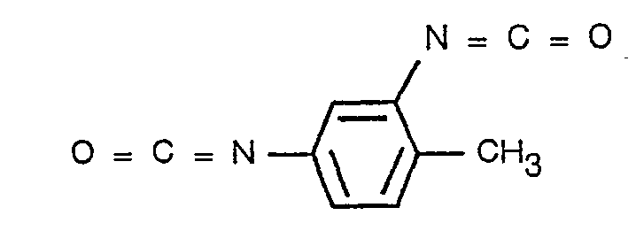
- The reaction proceeds in two steps. In the first, the isocyanate group in
position 4, which is less sterically committed, reacts with the NH2 of the anti hlgG. - In the second step, Nile blue A is added at 37°C to the anti hlgG-TDIC conjugate containing no free TDIC, so that the second isocyanate group reacts with the amino group of the dye.
- 0.05 ml ofTDIC are added to a solution of 5 mg ofy-immunoglobulin in potassium phosphate buffer of pH 7.5, 0.05 M, cooled to 0°C. The reaction is allowed to proceed for about 30 minutes at 0°C under vigorous stirring, the mixture then being centrifuged to remove the unreacted diisocyanate which precipitates in aqueous solutions at 0°C. The supernatant is allowed to incubate again at 0°C for a further hour, in order to enable any dissolved TDIC to react.
- 0.1 ml of phosphate buffer containing 10-4 mmoles of Nile blue A are added to the solution of
step 1. The resultant solution is kept at 37°C for 1 hour under stirring, and is then dialysed against 0.1 M ammonium carbonate in order to destroy any unreacted isocyanate group. The free dye is removed by dialysis against distilled water or neutral buffer followed by chromatography over Sephadex° G25. - The resultant conjugate has a molar protein: Nile blue A ratio of 1:2.4, calculated using the methods and extinction coefficients for protein and dye given in example 1.
- The fluorescence intensity of this conjugate (see Figure 1) is much higher than that obtained on exciting the free chromophore at the same concentration.
- Nile blue A (Cl. 51180) is conjugated with Protein A from Staphylococcus aureus using 1-ethyl-3-(3-dimethylaminopropyl)-carbodiimide (ECDI), of which the structural formula is CH3―CH2―N=C=N―CH2―CH2―CH2N―(CH3)2
- 50 mg of ECDI are added toa solution containing 5 mg of Protein A and an excess of Nile bluye A to the extent of about ten times molar in 1.2 ml of distilled water. The reaction is allowed to proceed under stirring for 7 hours at ambient temperature, while maintaining the pH between 6 and 6.8 by adding dilute HCI, after which the product is dialysed against 4-5 water changes over 48 hours. The colloidal suspension which can form in the dialysis bag is eliminated by centrifuging, and any free dye still in solution is removed by chromatography over SephadexO G25.
- The Nile blue A concentration in the conjugate is determined using a molar extinction coefficient in water of 5.8 x 10⁴ at λ = 635. The protein concentration is calculated using a ε%= 1.65 λ = 275 nm and correcting for the dye absorption in the ultraviolet or, in a more precise manner, by determining the nitrogen with the Kjeldahl micromethod.
- In a typical reaction, a conjugate is obtained with a molar Protein A: Neil blue A ratio of 1:1 = 1.5 (this radio can be varied by varying the reaction time and temperature) and a fluorescence intensity at λ = 680 nm which is greater than that obtained by exciting the free chromophore at the same concentration. The Protein A marked in this manner maintains its property of binding immunoglobulins, as demonstrated by the passive hemoagglutination test, comprising agglutination of ram erythrocytes sensitised with rabbit G immunoglobulins by Protein A - Nile blue A.
- A protein hormone, namely human chorionic somatomammotropin (HCS) is marked with Nile blue A in the presence of ECDI using the procedure illustrated in example 5, to which reference should be made for details.
- The reagent concentrations are 2.5 mg of HCS, 10-3 mmoles of Nile blue A dissolved in 0.05 ml of ethanol, and 45 mg of ECDI, all in 2 ml of water.
- The protein concentration is determined using a ε% = 8.3 at λ = 278.
- The fluorescent conjugate obtained has a molar HCS: dye ratio of 1: 1, and the hormone maintains its antigen properties when incubated with an anti HCS antiserum.
- Toluidine blue (Cl 52040) is used to mark rabbit y-immunoglobulins using ECDI.
- The reaction conditions are as follows.
- The reaction mixture, consisting of 3 mg of y-immunoglobulins and 2 x 10⁻⁴ mmoles of toluidine blue in 2 ml of water is adjusted to pH 5.6 by dilute HCI. 25 mg of ECDI are added, and the reaction is allowed to proceed under stirring at ambient temperature. After about 2 hours, a further 25 mg of ECDI are added, and the reaction is allowed to proceed for a further 2 hours.
- The solution is finally dialysed against water or a neutral buffer of low ionic force for 48-72 hours with various changes. Using a molar extinction coefficient of 3.1 x 10⁴ for the dye at X = 620 nm, and a ε% = 14 for the protein at λ = 280 nm, a molar protein: toluidine blue ratio of 1:1.1 is calculated for the prepared conjugate.
- The y-immunoglobulin marked in this manner maintains its immunological properties in the presence of anti y-immunoglobulin.
wherein R' and R", which may be the same or different, are optionally substituted alkyl, aryl, aralkyl or alkylaryl radicals, or a OCOR' grop wherein R' is as defined above; Ri, R2, R3, R4, R5, R7 and R8, which may be the same or different, are selected from hydrogen atoms, amino groups and optionally substituted alkyl, aryl, alkaryl or aralkyl radicals, and R1 and R2 taken together and/or R7 and R8 taken together can also represent a bivelent radical which forms a condensed ring structure with the aromatic nucleus.
Claims (9)

Priority Applications (1)
| Application Number | Priority Date | Filing Date | Title |
|---|---|---|---|
| AT82201143T ATE16643T1 (en) | 1981-09-25 | 1982-09-15 | IMMUNOFLUORESCENT REAGENTS AND METHOD FOR THEIR PREPARATION. |
Applications Claiming Priority (2)
| Application Number | Priority Date | Filing Date | Title |
|---|---|---|---|
| IT2416181 | 1981-09-25 | ||
| IT24161/81A IT1140209B (en) | 1981-09-25 | 1981-09-25 | IMMUNOLUORESCENCE REAGENTS AND METHOD FOR THEIR PREPARATION |
Publications (2)
| Publication Number | Publication Date |
|---|---|
| EP0075982A1 EP0075982A1 (en) | 1983-04-06 |
| EP0075982B1 true EP0075982B1 (en) | 1985-11-21 |
Family
ID=11212303
Family Applications (1)
| Application Number | Title | Priority Date | Filing Date |
|---|---|---|---|
| EP82201143A Expired EP0075982B1 (en) | 1981-09-25 | 1982-09-15 | Immunofluorescence reagents, and the method for their preparation |
Country Status (6)
| Country | Link |
|---|---|
| US (1) | US4463099A (en) |
| EP (1) | EP0075982B1 (en) |
| JP (1) | JPS5866851A (en) |
| AT (1) | ATE16643T1 (en) |
| DE (1) | DE3267616D1 (en) |
| IT (1) | IT1140209B (en) |
Families Citing this family (13)
| Publication number | Priority date | Publication date | Assignee | Title |
|---|---|---|---|---|
| US4582791A (en) * | 1983-10-07 | 1986-04-15 | Syntex (U.S.A.) Inc. | Reducing non-specific background in immunofluorescence techniques |
| US4918200A (en) * | 1984-07-16 | 1990-04-17 | Huls America Inc. | Chromogenic and fluorogenic silanes and using the same |
| US4666862A (en) * | 1984-08-14 | 1987-05-19 | Ortho Diagnostic Systems Inc. | Fluorescent energy transfer with phycobiliproteins |
| DE3526565A1 (en) * | 1985-07-25 | 1987-02-05 | Boehringer Mannheim Gmbh | RESORUFIN DERIVATIVES, METHOD FOR THE PRODUCTION THEREOF AND THEIR USE IN FLUORESCENT IMMUNOASSAYS |
| US4900686A (en) * | 1987-04-02 | 1990-02-13 | Polaroid Corporation | Fluorescent conjugates and biological diagnostic assay |
| US4882284A (en) * | 1987-04-13 | 1989-11-21 | Ortho Pharmaceutical Corporation | Method for quantitating and differentiating white blood cells |
| US5360739A (en) * | 1991-12-05 | 1994-11-01 | Miles Inc. | Methods for the identification and characterization of reticulocytes in whole blood |
| US5350695A (en) * | 1991-12-05 | 1994-09-27 | Miles Inc. | Methods for the identification and characterization of reticulocytes in whole blood |
| US7322927B2 (en) | 1993-09-24 | 2008-01-29 | Biosite, Inc. | Hybrid phthalocyanine derivatives and their uses |
| US5824799A (en) * | 1993-09-24 | 1998-10-20 | Biosite Diagnostics Incorporated | Hybrid phthalocyanine derivatives and their uses |
| US7083984B2 (en) | 1993-09-24 | 2006-08-01 | Biosite, Inc. | Hybrid phthalocyanine derivatives and their uses |
| US6238931B1 (en) * | 1993-09-24 | 2001-05-29 | Biosite Diagnostics, Inc. | Fluorescence energy transfer in particles |
| US6251687B1 (en) | 1993-09-24 | 2001-06-26 | Biosite Diagnostics, Inc. | Fluorescence energy transfer and intramolecular energy transfer in particles using novel compounds |
Family Cites Families (14)
| Publication number | Priority date | Publication date | Assignee | Title |
|---|---|---|---|---|
| US2937186A (en) * | 1958-04-29 | 1960-05-17 | Univ Kansas Res Foundation | Isothiocyanate compounds and means of producing the same |
| US3028397A (en) * | 1959-03-24 | 1962-04-03 | Borden Co | Isothiocyanates of phthaleins |
| GB1533410A (en) * | 1974-12-20 | 1978-11-22 | Block Engineering | Antigen detecting reagents |
| US4181650A (en) * | 1975-08-25 | 1980-01-01 | Maier Charles L Jr | Procedure for the assay of pharmacologically immunologically and biochemically active compounds in biological fluids |
| CA1070612A (en) * | 1975-10-09 | 1980-01-29 | Robert V. Dahlstrom | Solid phase immunofluorescent assay method |
| DE2824917A1 (en) * | 1977-07-13 | 1979-02-01 | Corning Glass Works | BIOLOGICALLY ACTIVE CONJUGATES |
| US4141688A (en) * | 1977-08-11 | 1979-02-27 | Miles Laboratories, Inc. | Composition, device and method for determining reducing agents |
| US4363759A (en) * | 1978-04-10 | 1982-12-14 | Miles Laboratories, Inc. | Chemiluminescent-labeled haptens and antigens |
| US4193983A (en) * | 1978-05-16 | 1980-03-18 | Syva Company | Labeled liposome particle compositions and immunoassays therewith |
| US4225485A (en) * | 1978-07-24 | 1980-09-30 | Miles Laboratories, Inc. | Chemiluminescent naphthalene-1,2-dicarboxylic acid hydrazide-labeled polypeptides and proteins |
| US4297273A (en) * | 1978-07-24 | 1981-10-27 | Miles Laboratories, Inc. | Chemiluminescent phthalhydrazide-labeled protein and polypeptide conjugates |
| US4238395A (en) * | 1978-07-24 | 1980-12-09 | Miles Laboratories, Inc. | Dimethyl 7-[ω-N-(phthalimido)alkyl]aminonaphthalene-1,2-dicarboxylates |
| DE2912845A1 (en) * | 1979-03-30 | 1980-10-09 | Frenzel Bernd | Antigen or antibody determination - using antibody or antigen labelled with a substance e.g. methylene blue which undergoes chemiluminescence on reaction with ozone |
| US4351760A (en) * | 1979-09-07 | 1982-09-28 | Syva Company | Novel alkyl substituted fluorescent compounds and polyamino acid conjugates |
-
1981
- 1981-09-25 IT IT24161/81A patent/IT1140209B/en active
-
1982
- 1982-09-15 AT AT82201143T patent/ATE16643T1/en not_active IP Right Cessation
- 1982-09-15 DE DE8282201143T patent/DE3267616D1/en not_active Expired
- 1982-09-15 EP EP82201143A patent/EP0075982B1/en not_active Expired
- 1982-09-16 US US06/419,053 patent/US4463099A/en not_active Expired - Fee Related
- 1982-09-24 JP JP57165174A patent/JPS5866851A/en active Granted
Non-Patent Citations (1)
| Title |
|---|
| ANGEWANDTE CHEMIE; Band 89, 1977 Weinheim Y. KANAOKA "Organische Fluoreszenzreagentien für die Untersuchung von Enzymen und Proteinen" pages 142 to 152 * |
Also Published As
| Publication number | Publication date |
|---|---|
| IT8124161A0 (en) | 1981-09-25 |
| US4463099A (en) | 1984-07-31 |
| DE3267616D1 (en) | 1986-01-02 |
| JPS5866851A (en) | 1983-04-21 |
| IT1140209B (en) | 1986-09-24 |
| ATE16643T1 (en) | 1985-12-15 |
| EP0075982A1 (en) | 1983-04-06 |
| JPH0261707B2 (en) | 1990-12-20 |
Similar Documents
| Publication | Publication Date | Title |
|---|---|---|
| JP3497704B2 (en) | Method for labeling or detecting substances using luminescent aryl sulfonate cyanine dye | |
| US5512446A (en) | Labeled complex | |
| EP0075982A1 (en) | Immunofluorescence reagents, and the method for their preparation | |
| Rodembusch et al. | Synthesis and spectroscopic characterisation of new ESIPT fluorescent protein probes | |
| SU1621811A3 (en) | Method of producing derivatives of resoruphine | |
| US20130084652A1 (en) | Homogeneous Chemiluminescence Assay Methods with Increased Sensitivity | |
| EP0224120A2 (en) | Protein label and drug delivery system | |
| CA1339782C (en) | Fluorescent conjugates and biological diagnostic assay | |
| US6277984B1 (en) | Monomethine cyanines rigidized by a two-carbon chain | |
| US5719027A (en) | Labeled complex and method of analysis therewith | |
| US20100009342A1 (en) | Control of chemical modification | |
| US4889916A (en) | Protein label and drug delivery system | |
| WO2016116111A1 (en) | Substituted acridine-like and xanthenium-like fluorescent dyes | |
| EP0794174B1 (en) | Labeled reagents for use in immunoassays and fluorescent compounds and complexes used therein | |
| US5055561A (en) | Protein label and drug delivery system | |
| NL8001972A (en) | Peroxidase determn. in enzyme immunoassay - using tetra:alkyl-benzidine as chromogenic hydrogen donor | |
| Moreland et al. | Preparation and characterization of 3-azido-2, 7-naphthalene disulfonate: a photolabile fluorescent precursor useful as a hydrophilic surface probe | |
| US4906749A (en) | Cyclic anhydride derivatives of chromophors | |
| JPS59101481A (en) | Substituted carboxyfluorescein | |
| Chen | Fluorescence properties of fluorescamine-protein conjugates | |
| US6307029B1 (en) | Dye-labeled protein conjugate and method for preparing the same | |
| Lim | The development of energy transfer immunoassay methods | |
| US6300480B1 (en) | Dye-labeled protein conjugate and method for preparing the same | |
| EP0968424A1 (en) | Diagnostic neodymium(iii), ytterbium(iii), or erbium(iii) ion-ligand complexes | |
| TSOU et al. | EVALUATION OF FLUORESCENCE POLARIZATION FOR CARCINOEMBRYONIC ANTIGEN (CEA) DETECTION1 |
Legal Events
| Date | Code | Title | Description |
|---|---|---|---|
| PUAI | Public reference made under article 153(3) epc to a published international application that has entered the european phase |
Free format text: ORIGINAL CODE: 0009012 |
|
| AK | Designated contracting states |
Designated state(s): AT BE CH DE FR GB LI LU NL SE |
|
| 17P | Request for examination filed |
Effective date: 19830913 |
|
| GRAA | (expected) grant |
Free format text: ORIGINAL CODE: 0009210 |
|
| AK | Designated contracting states |
Designated state(s): AT BE CH DE FR GB LI LU NL SE |
|
| REF | Corresponds to: |
Ref document number: 16643 Country of ref document: AT Date of ref document: 19851215 Kind code of ref document: T |
|
| REF | Corresponds to: |
Ref document number: 3267616 Country of ref document: DE Date of ref document: 19860102 |
|
| ET | Fr: translation filed | ||
| PLBE | No opposition filed within time limit |
Free format text: ORIGINAL CODE: 0009261 |
|
| STAA | Information on the status of an ep patent application or granted ep patent |
Free format text: STATUS: NO OPPOSITION FILED WITHIN TIME LIMIT |
|
| PG25 | Lapsed in a contracting state [announced via postgrant information from national office to epo] |
Ref country code: LU Free format text: LAPSE BECAUSE OF NON-PAYMENT OF DUE FEES Effective date: 19860930 |
|
| 26N | No opposition filed | ||
| REG | Reference to a national code |
Ref country code: CH Ref legal event code: PUE Owner name: ENICHEM S.P.A. |
|
| REG | Reference to a national code |
Ref country code: GB Ref legal event code: 732 |
|
| REG | Reference to a national code |
Ref country code: FR Ref legal event code: CD |
|
| NLS | Nl: assignments of ep-patents |
Owner name: ENICHEM S.P.A. TE MILAAN, ITALIE. |
|
| NLT1 | Nl: modifications of names registered in virtue of documents presented to the patent office pursuant to art. 16 a, paragraph 1 |
Owner name: ENICHEM ANIC S.P.A. TE PALERMO, ITALIE. |
|
| REG | Reference to a national code |
Ref country code: CH Ref legal event code: PUE Owner name: SCLAVO S.P.A. |
|
| REG | Reference to a national code |
Ref country code: GB Ref legal event code: 732 |
|
| NLS | Nl: assignments of ep-patents |
Owner name: SCLAVO S.P.A. TE SIENA, ITALIE. |
|
| REG | Reference to a national code |
Ref country code: FR Ref legal event code: TP |
|
| PGFP | Annual fee paid to national office [announced via postgrant information from national office to epo] |
Ref country code: FR Payment date: 19900731 Year of fee payment: 9 |
|
| PGFP | Annual fee paid to national office [announced via postgrant information from national office to epo] |
Ref country code: LU Payment date: 19900801 Year of fee payment: 9 |
|
| PGFP | Annual fee paid to national office [announced via postgrant information from national office to epo] |
Ref country code: GB Payment date: 19900904 Year of fee payment: 9 |
|
| PGFP | Annual fee paid to national office [announced via postgrant information from national office to epo] |
Ref country code: BE Payment date: 19900905 Year of fee payment: 9 |
|
| PGFP | Annual fee paid to national office [announced via postgrant information from national office to epo] |
Ref country code: SE Payment date: 19900907 Year of fee payment: 9 |
|
| PGFP | Annual fee paid to national office [announced via postgrant information from national office to epo] |
Ref country code: AT Payment date: 19900911 Year of fee payment: 9 |
|
| PGFP | Annual fee paid to national office [announced via postgrant information from national office to epo] |
Ref country code: DE Payment date: 19900928 Year of fee payment: 9 |
|
| PGFP | Annual fee paid to national office [announced via postgrant information from national office to epo] |
Ref country code: NL Payment date: 19900930 Year of fee payment: 9 |
|
| PGFP | Annual fee paid to national office [announced via postgrant information from national office to epo] |
Ref country code: CH Payment date: 19901213 Year of fee payment: 9 |
|
| PG25 | Lapsed in a contracting state [announced via postgrant information from national office to epo] |
Ref country code: GB Effective date: 19910915 Ref country code: AT Effective date: 19910915 |
|
| PG25 | Lapsed in a contracting state [announced via postgrant information from national office to epo] |
Ref country code: SE Effective date: 19910916 |
|
| PG25 | Lapsed in a contracting state [announced via postgrant information from national office to epo] |
Ref country code: LI Effective date: 19910930 Ref country code: CH Effective date: 19910930 Ref country code: BE Effective date: 19910930 |
|
| BERE | Be: lapsed |
Owner name: SCLAVO S.P.A. Effective date: 19910930 |
|
| PG25 | Lapsed in a contracting state [announced via postgrant information from national office to epo] |
Ref country code: NL Effective date: 19920401 |
|
| GBPC | Gb: european patent ceased through non-payment of renewal fee | ||
| NLV4 | Nl: lapsed or anulled due to non-payment of the annual fee | ||
| PG25 | Lapsed in a contracting state [announced via postgrant information from national office to epo] |
Ref country code: FR Effective date: 19920529 |
|
| REG | Reference to a national code |
Ref country code: CH Ref legal event code: PL |
|
| PG25 | Lapsed in a contracting state [announced via postgrant information from national office to epo] |
Ref country code: DE Effective date: 19920602 |
|
| REG | Reference to a national code |
Ref country code: FR Ref legal event code: ST |
|
| EUG | Se: european patent has lapsed |
Ref document number: 82201143.3 Effective date: 19920408 |