EP0044300B1 - Endoprosthetic bone joint devices - Google Patents
Endoprosthetic bone joint devices Download PDFInfo
- Publication number
- EP0044300B1 EP0044300B1 EP19800901726 EP80901726A EP0044300B1 EP 0044300 B1 EP0044300 B1 EP 0044300B1 EP 19800901726 EP19800901726 EP 19800901726 EP 80901726 A EP80901726 A EP 80901726A EP 0044300 B1 EP0044300 B1 EP 0044300B1
- Authority
- EP
- European Patent Office
- Prior art keywords
- component
- carbon
- bone
- porosity
- joint
- Prior art date
- Legal status (The legal status is an assumption and is not a legal conclusion. Google has not performed a legal analysis and makes no representation as to the accuracy of the status listed.)
- Expired
Links
Images
Classifications
-
- A—HUMAN NECESSITIES
- A61—MEDICAL OR VETERINARY SCIENCE; HYGIENE
- A61F—FILTERS IMPLANTABLE INTO BLOOD VESSELS; PROSTHESES; DEVICES PROVIDING PATENCY TO, OR PREVENTING COLLAPSING OF, TUBULAR STRUCTURES OF THE BODY, e.g. STENTS; ORTHOPAEDIC, NURSING OR CONTRACEPTIVE DEVICES; FOMENTATION; TREATMENT OR PROTECTION OF EYES OR EARS; BANDAGES, DRESSINGS OR ABSORBENT PADS; FIRST-AID KITS
- A61F2/00—Filters implantable into blood vessels; Prostheses, i.e. artificial substitutes or replacements for parts of the body; Appliances for connecting them with the body; Devices providing patency to, or preventing collapsing of, tubular structures of the body, e.g. stents
- A61F2/02—Prostheses implantable into the body
- A61F2/30—Joints
- A61F2/32—Joints for the hip
- A61F2/34—Acetabular cups
-
- A—HUMAN NECESSITIES
- A61—MEDICAL OR VETERINARY SCIENCE; HYGIENE
- A61F—FILTERS IMPLANTABLE INTO BLOOD VESSELS; PROSTHESES; DEVICES PROVIDING PATENCY TO, OR PREVENTING COLLAPSING OF, TUBULAR STRUCTURES OF THE BODY, e.g. STENTS; ORTHOPAEDIC, NURSING OR CONTRACEPTIVE DEVICES; FOMENTATION; TREATMENT OR PROTECTION OF EYES OR EARS; BANDAGES, DRESSINGS OR ABSORBENT PADS; FIRST-AID KITS
- A61F2/00—Filters implantable into blood vessels; Prostheses, i.e. artificial substitutes or replacements for parts of the body; Appliances for connecting them with the body; Devices providing patency to, or preventing collapsing of, tubular structures of the body, e.g. stents
- A61F2/02—Prostheses implantable into the body
- A61F2/30—Joints
- A61F2/32—Joints for the hip
- A61F2/36—Femoral heads ; Femoral endoprostheses
- A61F2/3601—Femoral heads ; Femoral endoprostheses for replacing only the epiphyseal or metaphyseal parts of the femur, e.g. endoprosthetic femoral heads or necks directly fixed to the natural femur by internal fixation devices
- A61F2/3603—Femoral heads ; Femoral endoprostheses for replacing only the epiphyseal or metaphyseal parts of the femur, e.g. endoprosthetic femoral heads or necks directly fixed to the natural femur by internal fixation devices implanted without ablation of the whole natural femoral head
-
- A—HUMAN NECESSITIES
- A61—MEDICAL OR VETERINARY SCIENCE; HYGIENE
- A61L—METHODS OR APPARATUS FOR STERILISING MATERIALS OR OBJECTS IN GENERAL; DISINFECTION, STERILISATION OR DEODORISATION OF AIR; CHEMICAL ASPECTS OF BANDAGES, DRESSINGS, ABSORBENT PADS OR SURGICAL ARTICLES; MATERIALS FOR BANDAGES, DRESSINGS, ABSORBENT PADS OR SURGICAL ARTICLES
- A61L27/00—Materials for grafts or prostheses or for coating grafts or prostheses
- A61L27/02—Inorganic materials
- A61L27/08—Carbon ; Graphite
-
- A—HUMAN NECESSITIES
- A61—MEDICAL OR VETERINARY SCIENCE; HYGIENE
- A61F—FILTERS IMPLANTABLE INTO BLOOD VESSELS; PROSTHESES; DEVICES PROVIDING PATENCY TO, OR PREVENTING COLLAPSING OF, TUBULAR STRUCTURES OF THE BODY, e.g. STENTS; ORTHOPAEDIC, NURSING OR CONTRACEPTIVE DEVICES; FOMENTATION; TREATMENT OR PROTECTION OF EYES OR EARS; BANDAGES, DRESSINGS OR ABSORBENT PADS; FIRST-AID KITS
- A61F2/00—Filters implantable into blood vessels; Prostheses, i.e. artificial substitutes or replacements for parts of the body; Appliances for connecting them with the body; Devices providing patency to, or preventing collapsing of, tubular structures of the body, e.g. stents
- A61F2/02—Prostheses implantable into the body
- A61F2/30—Joints
- A61F2/32—Joints for the hip
- A61F2/34—Acetabular cups
- A61F2002/3412—Acetabular cups with pins or protrusions, e.g. non-sharp pins or protrusions projecting from a shell surface
- A61F2002/3417—Acetabular cups with pins or protrusions, e.g. non-sharp pins or protrusions projecting from a shell surface the outer shell having protrusions on meridian lines, e.g. equidistant fins or wings around the equatorial zone
-
- A—HUMAN NECESSITIES
- A61—MEDICAL OR VETERINARY SCIENCE; HYGIENE
- A61F—FILTERS IMPLANTABLE INTO BLOOD VESSELS; PROSTHESES; DEVICES PROVIDING PATENCY TO, OR PREVENTING COLLAPSING OF, TUBULAR STRUCTURES OF THE BODY, e.g. STENTS; ORTHOPAEDIC, NURSING OR CONTRACEPTIVE DEVICES; FOMENTATION; TREATMENT OR PROTECTION OF EYES OR EARS; BANDAGES, DRESSINGS OR ABSORBENT PADS; FIRST-AID KITS
- A61F2310/00—Prostheses classified in A61F2/28 or A61F2/30 - A61F2/44 being constructed from or coated with a particular material
- A61F2310/00005—The prosthesis being constructed from a particular material
- A61F2310/00161—Carbon; Graphite
Definitions
- Endoprosthetic bone joint devices have been the subject of considerable development during the past few decades and are now widely employed in routine clinical practice.
- the development in question has given rise to improvements in many respects, among which important ones are the biocompatability of the materials used in the devices, the frictional and other mechanical properties of such materials whereby the articulatory and wear characteristics of the devices have been enhanced, and the techniques for securing the devices to bones.
- important ones are the biocompatability of the materials used in the devices, the frictional and other mechanical properties of such materials whereby the articulatory and wear characteristics of the devices have been enhanced, and the techniques for securing the devices to bones.
- considerable as the improvements have been, there is room for further improvement in most respects when viewed against the overall performance of natural healthy joints.
- the object of the present invention is to afford some further improvement, which improvement centres on the promotion of anastomosis, that is to say the growth of living tissue, into a component of an endoprosthetic bone joint device.
- Anastomosis can be achieved by the use of porous material in the component, as discussed in FR-A- 2 413 078 and FR-A-2 105 995, but yet further improvement is possible.
- an endoprosthetic bone joint device comprising a component of which one part is adapted for securement with bone, of which another part is adapted for mutual articulatory engagement with another integer of the joint, and which is substantially wholly porous, characterised in that the porosity in said one part is greater than that in said other part respectively to promote the formation of osseous and fibrocartilogenic material by way of anastomosis.
- the desired variation in porosity involved differences in pore size in a diametrical sense, with the pores being interlinked throughout the component rather than being of a closed cell form.
- tissue in-growth occurs in a fibrous form followed by ossification within the larger pores of the one part to provide secure anchorage of the component, while the tissue in the other part, and particularly at the outer surface, assumes a fibrocartilagenic form to provide enhanced articulatory properties.
- the anastomosis at the outer surface can be additionally promoted by the provision of grooving therein.
- Suitable ranges of pore size for the one and other parts of the component are respectively 10-250 fL m and from substantially zero, or 5 fLm say, to ⁇ 5 ⁇ ⁇ m, with the smaller regions of such ranges being preferred.
- the reason for this last preference is evident from the fact that the character of the component will progressively improve during a period of time following surgical implantation, but the component requires certain initial properties which, generally speaking, will be more satisfactorily achieved with higher density for the component.
- the component may well be adapted to afford securement of a mechanical nature to provide some measure of stability during the anastomotic period after implantation.
- a particularly important aspect of the present invention is that of the materials to be used for its implementation because the materials clearly must satisfy various requirements such as biocompatibility, strength, etc., as well as be capable of formation with the desired varying porosity.
- these materials be of so-called carbon-carbon or carbon-fibre-reinforced carbon (CFRC) form. Satisfaction by this preference of some of the materials requirements is immediately evident because carbon is of proven biocompatability in a variety of forms and is of proven high strength in fibre form composites. Other benefits of the use of CFRC are less immediately evident.
- the structure of bone in the region of the joints and elsewhere is not random or arbitrary but has a patterned form best designed to suit the functions of the bone. More specifically bone has a trabucellular structure which is generally orientated in the region of the joints to best sustain the loads transmitted therethrough.
- CFRC is produced, in very general terms, by impregnating carbon fibre material with a resin or some other binder and then pyrolising the resultant composite or by pyrolytic deposition on carbon fibre material of carbon from hydrocarbon gases.
- the fibrous reinforcement can be structured in a similar manner to bone to deploy the strength of the material in a correspondingly optimum manner.
- such a structure can and indeed normally will, provide a complementary pore pattern of a similar nature whereby the anastomosis provides a structure which further enhances the simulation of bone.
- CFRC is normally porous and its porosity can be controlled to a satisfactory extent for the present purpose.
- This control involves a suitable choice of initial fibrous material in respct of fibre size, form of weave or other collective fabrication, etc., and choice of in-fill procedure.
- production of a CFRC component according to the invention suitably involves both impregnation and deposition as mentioned above.
- the eventual component may involve production in separate parts with respectively different porosity attained by differing extents of isothermal carbon vapour deposition, which parts are then bonded with resin thereafter pyrolised, or a single component matrix of carbon fibre material and binder can be pyrolised and then subjected to thermal- gradient carbon vapour deposition.
- the acetabular component of Figures 1 and 2 comprises a substantially hemispherical cup body 10 with inner surface 11 and outer surface 12, the body having a key 13 formed by a projection of its outer surface. More particularly the key projection is longest at the rim of the cup where it extends radially outwardly parallel to the rim in a generally rectangular shape, as seen in Figure 2, the remainder of the key being convergently tapered partway towards the cup apex in tangential manner as seen in Figure 1.
- the cup is formed from CFRC as described above with greater porosity at the surface 12 than 11 where it is least.
- the cup In use the cup is located in the acetabulum, with the key in the acetabular notch, a suitable seating having been prepared for the component by reaming or other preparation whereby the cup seats in a close fit with the key acting against rotation of the cup relative to the bone.
- the femoral component of Figures 3 and 4 comprises a cap body 20 of an overall shape formed by a hemispherical portion leading smoothly at its rim into a right circular cylindrical projection.
- the inner and outer surfaces are denoted at 21 and 22, the former extending inwardly along an axially directed strip portion thereof to form a key 23 of circular arcuate transverse cross-sectional profile, and the latter being complementary in its hemispherical part with the cup 10 for mutual articulatory engagement therein.
- the cap is formed from CFRC as described above with greater porosity at the surface 21 than 22 where it is least. In use the cap is located over the head of the femur after suitable surgical shaping of the latter to receive the cap in a close fit with the key acting against rotation relative to the bone.
- the device formed by these components is similar in general geometry and intended application to others which are already available, but these others differ in the use of metal/plastics materials combinations which do not allow anastomosis.
- the present invention is in fact more generally applicable in other known configurations for various joints and is in no way intended to be limited by the illustrated example.
Abstract
Description
- Endoprosthetic bone joint devices have been the subject of considerable development during the past few decades and are now widely employed in routine clinical practice. The development in question has given rise to improvements in many respects, among which important ones are the biocompatability of the materials used in the devices, the frictional and other mechanical properties of such materials whereby the articulatory and wear characteristics of the devices have been enhanced, and the techniques for securing the devices to bones. However, considerable as the improvements have been, there is room for further improvement in most respects when viewed against the overall performance of natural healthy joints.
- The object of the present invention is to afford some further improvement, which improvement centres on the promotion of anastomosis, that is to say the growth of living tissue, into a component of an endoprosthetic bone joint device. Anastomosis can be achieved by the use of porous material in the component, as discussed in FR-A- 2 413 078 and FR-A-2 105 995, but yet further improvement is possible.
- According to the present invention there is provided an endoprosthetic bone joint device comprising a component of which one part is adapted for securement with bone, of which another part is adapted for mutual articulatory engagement with another integer of the joint, and which is substantially wholly porous, characterised in that the porosity in said one part is greater than that in said other part respectively to promote the formation of osseous and fibrocartilogenic material by way of anastomosis.
- Preferably the desired variation in porosity involved differences in pore size in a diametrical sense, with the pores being interlinked throughout the component rather than being of a closed cell form.
- The intended benefit of the presently proposed component is that tissue in-growth occurs in a fibrous form followed by ossification within the larger pores of the one part to provide secure anchorage of the component, while the tissue in the other part, and particularly at the outer surface, assumes a fibrocartilagenic form to provide enhanced articulatory properties. The anastomosis at the outer surface can be additionally promoted by the provision of grooving therein.
- Suitable ranges of pore size for the one and other parts of the component are respectively 10-250 fLm and from substantially zero, or 5 fLm say, to ↑ 5µµm, with the smaller regions of such ranges being preferred. The reason for this last preference is evident from the fact that the character of the component will progressively improve during a period of time following surgical implantation, but the component requires certain initial properties which, generally speaking, will be more satisfactorily achieved with higher density for the component. For the same reasons, the component may well be adapted to afford securement of a mechanical nature to provide some measure of stability during the anastomotic period after implantation.
- A particularly important aspect of the present invention is that of the materials to be used for its implementation because the materials clearly must satisfy various requirements such as biocompatibility, strength, etc., as well as be capable of formation with the desired varying porosity. In practice it is preferred that these materials be of so-called carbon-carbon or carbon-fibre-reinforced carbon (CFRC) form. Satisfaction by this preference of some of the materials requirements is immediately evident because carbon is of proven biocompatability in a variety of forms and is of proven high strength in fibre form composites. Other benefits of the use of CFRC are less immediately evident.
- The structure of bone in the region of the joints and elsewhere is not random or arbitrary but has a patterned form best designed to suit the functions of the bone. More specifically bone has a trabucellular structure which is generally orientated in the region of the joints to best sustain the loads transmitted therethrough. CFRC is produced, in very general terms, by impregnating carbon fibre material with a resin or some other binder and then pyrolising the resultant composite or by pyrolytic deposition on carbon fibre material of carbon from hydrocarbon gases. In either event, it will be appreciated that the fibrous reinforcement can be structured in a similar manner to bone to deploy the strength of the material in a correspondingly optimum manner. Moreover, such a structure can and indeed normally will, provide a complementary pore pattern of a similar nature whereby the anastomosis provides a structure which further enhances the simulation of bone.
- A further benefit of CFRC is that it is normally porous and its porosity can be controlled to a satisfactory extent for the present purpose. This control involves a suitable choice of initial fibrous material in respct of fibre size, form of weave or other collective fabrication, etc., and choice of in-fill procedure. In this last respect, production of a CFRC component according to the invention suitably involves both impregnation and deposition as mentioned above. For example, the eventual component may involve production in separate parts with respectively different porosity attained by differing extents of isothermal carbon vapour deposition, which parts are then bonded with resin thereafter pyrolised, or a single component matrix of carbon fibre material and binder can be pyrolised and then subjected to thermal- gradient carbon vapour deposition.
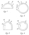
- The principal features of the invention having been described above, it is useful to illustrate the invention further, by way of example, with reference to the accompanying drawings which schematically illustrate an endoprosthetic hip joint device comprising two components according to the invention, and in Which:-
- Figures 1 and 2 are respective side and plan views of the acetabular component of the device; and
- Figures 3 and 4 are respective corresponding views of the humeral component of the device.
- The acetabular component of Figures 1 and 2 comprises a substantially
hemispherical cup body 10 withinner surface 11 andouter surface 12, the body having akey 13 formed by a projection of its outer surface. More particularly the key projection is longest at the rim of the cup where it extends radially outwardly parallel to the rim in a generally rectangular shape, as seen in Figure 2, the remainder of the key being convergently tapered partway towards the cup apex in tangential manner as seen in Figure 1. The cup is formed from CFRC as described above with greater porosity at thesurface 12 than 11 where it is least. In use the cup is located in the acetabulum, with the key in the acetabular notch, a suitable seating having been prepared for the component by reaming or other preparation whereby the cup seats in a close fit with the key acting against rotation of the cup relative to the bone. - The femoral component of Figures 3 and 4 comprises a
cap body 20 of an overall shape formed by a hemispherical portion leading smoothly at its rim into a right circular cylindrical projection. The inner and outer surfaces are denoted at 21 and 22, the former extending inwardly along an axially directed strip portion thereof to form a key 23 of circular arcuate transverse cross-sectional profile, and the latter being complementary in its hemispherical part with thecup 10 for mutual articulatory engagement therein. The cap is formed from CFRC as described above with greater porosity at thesurface 21 than 22 where it is least. In use the cap is located over the head of the femur after suitable surgical shaping of the latter to receive the cap in a close fit with the key acting against rotation relative to the bone. - The device formed by these components is similar in general geometry and intended application to others which are already available, but these others differ in the use of metal/plastics materials combinations which do not allow anastomosis. The present invention is in fact more generally applicable in other known configurations for various joints and is in no way intended to be limited by the illustrated example.
Claims (7)
Applications Claiming Priority (2)
| Application Number | Priority Date | Filing Date | Title |
|---|---|---|---|
| GB7933184 | 1979-09-25 | ||
| GB7933184 | 1979-09-25 |
Publications (2)
| Publication Number | Publication Date |
|---|---|
| EP0044300A1 EP0044300A1 (en) | 1982-01-27 |
| EP0044300B1 true EP0044300B1 (en) | 1984-03-14 |
Family
ID=10508054
Family Applications (1)
| Application Number | Title | Priority Date | Filing Date |
|---|---|---|---|
| EP19800901726 Expired EP0044300B1 (en) | 1979-09-25 | 1980-09-25 | Endoprosthetic bone joint devices |
Country Status (4)
| Country | Link |
|---|---|
| EP (1) | EP0044300B1 (en) |
| JP (1) | JPS56501155A (en) |
| DE (1) | DE3067003D1 (en) |
| WO (1) | WO1981000808A1 (en) |
Families Citing this family (3)
| Publication number | Priority date | Publication date | Assignee | Title |
|---|---|---|---|---|
| FR2565826A1 (en) * | 1984-06-19 | 1985-12-20 | Aerospatiale | METHOD FOR PRODUCING A SURGICAL IMPLANTABLE PIECE IN AN ORGANISM AND PIECE THUS OBTAINED |
| GB8419559D0 (en) * | 1984-08-01 | 1984-09-05 | Field R E | Endoprosthetic bone joint components |
| SE516282C2 (en) * | 2000-04-04 | 2001-12-10 | Nobel Biocare Ab | Implants provided with connection and hole insertion parts and the procedure for such implants |
Family Cites Families (6)
| Publication number | Priority date | Publication date | Assignee | Title |
|---|---|---|---|---|
| US3707006A (en) * | 1970-08-26 | 1972-12-26 | Gulf Oil Corp | Orthopedic device for repair or replacement of bone |
| US3877080A (en) * | 1972-10-30 | 1975-04-15 | Atlantic Res Corp | Acicular silicon carbide dispersion in pyrolytic graphite matrix for use in biomedical implants |
| DE2634954A1 (en) * | 1976-08-04 | 1978-02-09 | Sigri Elektrographit Gmbh | JOINT PROSTHESIS |
| JPS5441913A (en) * | 1977-09-09 | 1979-04-03 | Kanebo Ltd | Carbonncarbon composite material and method of making same |
| FR2413078A1 (en) * | 1978-01-03 | 1979-07-27 | Serole Michelle | Hip prosthesis made of composite materials - has low wt. which assists swimming and can also be used for other body joints |
| FR2427315A1 (en) * | 1978-05-29 | 1979-12-28 | Commissariat Energie Atomique | Continuous mfr. of profiles of fibre reinforced carbon - for structural or surgical applications, by co-extrusion of organic fibres and tar followed by heat treatment |
-
1980
- 1980-09-25 WO PCT/GB1980/000148 patent/WO1981000808A1/en active IP Right Grant
- 1980-09-25 EP EP19800901726 patent/EP0044300B1/en not_active Expired
- 1980-09-25 JP JP50206880A patent/JPS56501155A/ja active Pending
- 1980-09-25 DE DE8080901726T patent/DE3067003D1/en not_active Expired
Also Published As
| Publication number | Publication date |
|---|---|
| DE3067003D1 (en) | 1984-04-19 |
| WO1981000808A1 (en) | 1981-04-02 |
| JPS56501155A (en) | 1981-08-20 |
| EP0044300A1 (en) | 1982-01-27 |
Similar Documents
| Publication | Publication Date | Title |
|---|---|---|
| CA1057902A (en) | Composite prosthetic device with porous polymeric coating | |
| US3938198A (en) | Hip joint prosthesis | |
| US6087553A (en) | Implantable metallic open-celled lattice/polyethylene composite material and devices | |
| EP2349360B1 (en) | Porous surface layers with increased surface roughness and implants incorporating the same | |
| EP2637609B1 (en) | Orthopedic implant with porous polymer bone contacting surface | |
| US5176712A (en) | Endoprostheses with resorption preventing means | |
| US3526906A (en) | Prosthetic implants made from carbonaceous materials | |
| CA2084095C (en) | Metal/composite hybrid orthopedic implants | |
| US4454612A (en) | Prosthesis formation having solid and porous polymeric components | |
| US4714467A (en) | Reinforced fiber bone replacement implant having treated surfaces and a method for its manufacture | |
| US4007494A (en) | Bone cap | |
| EP0269745A1 (en) | Bone prosthesis | |
| US20030074077A1 (en) | Prosthesis | |
| CA2664560A1 (en) | Medical implant | |
| EP0297789A1 (en) | Method of making a prosthetic component and component made according to the method | |
| US11564799B2 (en) | Patellofemoral implant with porous ingrowth material and method of manufacturing same | |
| EP0044300B1 (en) | Endoprosthetic bone joint devices | |
| JPS6141453A (en) | Internal prosthese body for bone joint | |
| US11298236B2 (en) | Devices and methods for cementing insert bearing liner into acetabular cup component | |
| WO1989009580A1 (en) | Prosthetic finger joint | |
| GB2058575A (en) | Endoprosthetic bone joints | |
| Fabi et al. | Porous coatings on metallic implant materials | |
| GB2162753A (en) | Sleeve for metal implant into bone | |
| EP0344107A1 (en) | Artificial trachea and production process thereof | |
| GB2286146A (en) | Fibre reinforced composite artefact |
Legal Events
| Date | Code | Title | Description |
|---|---|---|---|
| PUAI | Public reference made under article 153(3) epc to a published international application that has entered the european phase |
Free format text: ORIGINAL CODE: 0009012 |
|
| 17P | Request for examination filed |
Effective date: 19811020 |
|
| AK | Designated contracting states |
Designated state(s): CH DE FR LI |
|
| GRAA | (expected) grant |
Free format text: ORIGINAL CODE: 0009210 |
|
| AK | Designated contracting states |
Designated state(s): CH DE FR LI |
|
| PG25 | Lapsed in a contracting state [announced via postgrant information from national office to epo] |
Ref country code: LI Effective date: 19840314 Ref country code: FR Free format text: THE PATENT HAS BEEN ANNULLED BY A DECISION OF A NATIONAL AUTHORITY Effective date: 19840314 Ref country code: CH Effective date: 19840314 |
|
| REF | Corresponds to: |
Ref document number: 3067003 Country of ref document: DE Date of ref document: 19840419 |
|
| REG | Reference to a national code |
Ref country code: CH Ref legal event code: PL |
|
| EN | Fr: translation not filed | ||
| PLBE | No opposition filed within time limit |
Free format text: ORIGINAL CODE: 0009261 |
|
| STAA | Information on the status of an ep patent application or granted ep patent |
Free format text: STATUS: NO OPPOSITION FILED WITHIN TIME LIMIT |
|
| 26N | No opposition filed | ||
| PG25 | Lapsed in a contracting state [announced via postgrant information from national office to epo] |
Ref country code: DE Effective date: 19850601 |