EP0018579B1 - Babesiosis antigen, vaccine and diagnostic reagent comprising it - Google Patents
Babesiosis antigen, vaccine and diagnostic reagent comprising it Download PDFInfo
- Publication number
- EP0018579B1 EP0018579B1 EP80102167A EP80102167A EP0018579B1 EP 0018579 B1 EP0018579 B1 EP 0018579B1 EP 80102167 A EP80102167 A EP 80102167A EP 80102167 A EP80102167 A EP 80102167A EP 0018579 B1 EP0018579 B1 EP 0018579B1
- Authority
- EP
- European Patent Office
- Prior art keywords
- babesia
- antigen
- specific
- vitro
- parasites
- Prior art date
- Legal status (The legal status is an assumption and is not a legal conclusion. Google has not performed a legal analysis and makes no representation as to the accuracy of the status listed.)
- Expired
Links
- 239000000427 antigen Substances 0.000 title claims description 85
- 102000036639 antigens Human genes 0.000 title claims description 85
- 108091007433 antigens Proteins 0.000 title claims description 85
- 201000008680 babesiosis Diseases 0.000 title claims description 51
- 229960005486 vaccine Drugs 0.000 title claims description 24
- 239000003153 chemical reaction reagent Substances 0.000 title claims description 7
- 241000223836 Babesia Species 0.000 claims description 42
- 210000003936 merozoite Anatomy 0.000 claims description 36
- 241001465754 Metazoa Species 0.000 claims description 31
- 210000002966 serum Anatomy 0.000 claims description 31
- 244000045947 parasite Species 0.000 claims description 27
- 238000000338 in vitro Methods 0.000 claims description 22
- 238000001727 in vivo Methods 0.000 claims description 14
- 241000223838 Babesia bovis Species 0.000 claims description 10
- 239000002671 adjuvant Substances 0.000 claims description 8
- 230000001681 protective effect Effects 0.000 claims description 8
- XLYOFNOQVPJJNP-UHFFFAOYSA-N water Substances O XLYOFNOQVPJJNP-UHFFFAOYSA-N 0.000 claims description 8
- BFNBIHQBYMNNAN-UHFFFAOYSA-N ammonium sulfate Chemical compound N.N.OS(O)(=O)=O BFNBIHQBYMNNAN-UHFFFAOYSA-N 0.000 claims description 7
- 229910052921 ammonium sulfate Inorganic materials 0.000 claims description 7
- 235000011130 ammonium sulphate Nutrition 0.000 claims description 7
- 150000001720 carbohydrates Chemical class 0.000 claims description 7
- 239000000126 substance Substances 0.000 claims description 7
- 208000015181 infectious disease Diseases 0.000 claims description 6
- 238000001556 precipitation Methods 0.000 claims description 6
- 241000223848 Babesia microti Species 0.000 claims description 4
- 230000015572 biosynthetic process Effects 0.000 claims description 4
- 241000223840 Babesia bigemina Species 0.000 claims description 3
- 241000223775 Babesia caballi Species 0.000 claims description 3
- 241000223846 Babesia canis Species 0.000 claims description 3
- 241001455947 Babesia divergens Species 0.000 claims description 3
- 241001466447 Babesia gibsoni Species 0.000 claims description 3
- 241000593848 Babesia major Species 0.000 claims description 3
- 241000223774 Babesia rodhaini Species 0.000 claims description 3
- 241000061220 Werneckiella equi Species 0.000 claims description 3
- 239000000470 constituent Substances 0.000 claims description 3
- 238000001514 detection method Methods 0.000 claims description 3
- 239000003085 diluting agent Substances 0.000 claims description 3
- 208000037265 diseases, disorders, signs and symptoms Diseases 0.000 claims description 3
- 239000003550 marker Substances 0.000 claims description 2
- 239000002552 dosage form Substances 0.000 claims 1
- 238000000034 method Methods 0.000 description 31
- 239000000463 material Substances 0.000 description 20
- 230000000890 antigenic effect Effects 0.000 description 19
- 210000004369 blood Anatomy 0.000 description 18
- 239000008280 blood Substances 0.000 description 18
- 210000003743 erythrocyte Anatomy 0.000 description 17
- 238000002360 preparation method Methods 0.000 description 16
- 241000283690 Bos taurus Species 0.000 description 14
- 102000004528 Mannose-Binding Protein-Associated Serine Proteases Human genes 0.000 description 11
- 108010042484 Mannose-Binding Protein-Associated Serine Proteases Proteins 0.000 description 11
- 239000001963 growth medium Substances 0.000 description 11
- 210000004027 cell Anatomy 0.000 description 10
- 239000002609 medium Substances 0.000 description 10
- 239000000243 solution Substances 0.000 description 10
- 230000000694 effects Effects 0.000 description 8
- CURLTUGMZLYLDI-UHFFFAOYSA-N Carbon dioxide Chemical compound O=C=O CURLTUGMZLYLDI-UHFFFAOYSA-N 0.000 description 6
- 208000030852 Parasitic disease Diseases 0.000 description 6
- 230000004071 biological effect Effects 0.000 description 6
- 239000012634 fragment Substances 0.000 description 6
- 208000009182 Parasitemia Diseases 0.000 description 5
- 238000010171 animal model Methods 0.000 description 5
- 238000011534 incubation Methods 0.000 description 5
- 239000006228 supernatant Substances 0.000 description 5
- 238000012360 testing method Methods 0.000 description 5
- 229920005654 Sephadex Polymers 0.000 description 4
- 239000012507 Sephadex™ Substances 0.000 description 4
- 229910002092 carbon dioxide Inorganic materials 0.000 description 4
- 238000005119 centrifugation Methods 0.000 description 4
- 239000012228 culture supernatant Substances 0.000 description 4
- 230000001524 infective effect Effects 0.000 description 4
- 230000008569 process Effects 0.000 description 4
- 235000018102 proteins Nutrition 0.000 description 4
- 102000004169 proteins and genes Human genes 0.000 description 4
- 108090000623 proteins and genes Proteins 0.000 description 4
- 241000894007 species Species 0.000 description 4
- 241000283973 Oryctolagus cuniculus Species 0.000 description 3
- 241000224016 Plasmodium Species 0.000 description 3
- FAPWRFPIFSIZLT-UHFFFAOYSA-M Sodium chloride Chemical compound [Na+].[Cl-] FAPWRFPIFSIZLT-UHFFFAOYSA-M 0.000 description 3
- 239000013543 active substance Substances 0.000 description 3
- 244000309466 calf Species 0.000 description 3
- 239000001569 carbon dioxide Substances 0.000 description 3
- 238000006243 chemical reaction Methods 0.000 description 3
- 238000011161 development Methods 0.000 description 3
- 238000010790 dilution Methods 0.000 description 3
- 239000012895 dilution Substances 0.000 description 3
- 230000036039 immunity Effects 0.000 description 3
- 238000002955 isolation Methods 0.000 description 3
- 230000007774 longterm Effects 0.000 description 3
- 238000006386 neutralization reaction Methods 0.000 description 3
- 239000002245 particle Substances 0.000 description 3
- 239000000843 powder Substances 0.000 description 3
- 230000004044 response Effects 0.000 description 3
- 239000000725 suspension Substances 0.000 description 3
- 239000004382 Amylase Substances 0.000 description 2
- 102000013142 Amylases Human genes 0.000 description 2
- 108010065511 Amylases Proteins 0.000 description 2
- 102000001554 Hemoglobins Human genes 0.000 description 2
- 108010054147 Hemoglobins Proteins 0.000 description 2
- UZQJVUCHXGYFLQ-AYDHOLPZSA-N [(2s,3r,4s,5r,6r)-4-[(2s,3r,4s,5r,6r)-4-[(2r,3r,4s,5r,6r)-4-[(2s,3r,4s,5r,6r)-3,5-dihydroxy-6-(hydroxymethyl)-4-[(2s,3r,4s,5s,6r)-3,4,5-trihydroxy-6-(hydroxymethyl)oxan-2-yl]oxyoxan-2-yl]oxy-3,5-dihydroxy-6-(hydroxymethyl)oxan-2-yl]oxy-3,5-dihydroxy-6-(hy Chemical compound O([C@H]1[C@H](O)[C@@H](CO)O[C@H]([C@@H]1O)O[C@H]1[C@H](O)[C@@H](CO)O[C@H]([C@@H]1O)O[C@H]1CC[C@]2(C)[C@H]3CC=C4[C@@]([C@@]3(CC[C@H]2[C@@]1(C=O)C)C)(C)CC(O)[C@]1(CCC(CC14)(C)C)C(=O)O[C@H]1[C@@H]([C@@H](O[C@H]2[C@@H]([C@@H](O[C@H]3[C@@H]([C@@H](O[C@H]4[C@@H]([C@@H](O[C@H]5[C@@H]([C@@H](O)[C@H](O)[C@@H](CO)O5)O)[C@H](O)[C@@H](CO)O4)O)[C@H](O)[C@@H](CO)O3)O)[C@H](O)[C@@H](CO)O2)O)[C@H](O)[C@@H](CO)O1)O)[C@@H]1O[C@H](CO)[C@@H](O)[C@H](O)[C@H]1O UZQJVUCHXGYFLQ-AYDHOLPZSA-N 0.000 description 2
- 235000019418 amylase Nutrition 0.000 description 2
- QVGXLLKOCUKJST-UHFFFAOYSA-N atomic oxygen Chemical compound [O] QVGXLLKOCUKJST-UHFFFAOYSA-N 0.000 description 2
- 239000000872 buffer Substances 0.000 description 2
- 239000007975 buffered saline Substances 0.000 description 2
- 239000000969 carrier Substances 0.000 description 2
- 230000001413 cellular effect Effects 0.000 description 2
- 238000012512 characterization method Methods 0.000 description 2
- 238000004587 chromatography analysis Methods 0.000 description 2
- 238000000502 dialysis Methods 0.000 description 2
- 230000003292 diminished effect Effects 0.000 description 2
- 238000010828 elution Methods 0.000 description 2
- 238000002270 exclusion chromatography Methods 0.000 description 2
- 239000011521 glass Substances 0.000 description 2
- 150000004676 glycans Chemical class 0.000 description 2
- 238000003018 immunoassay Methods 0.000 description 2
- 239000007788 liquid Substances 0.000 description 2
- 238000004519 manufacturing process Methods 0.000 description 2
- 239000000203 mixture Substances 0.000 description 2
- 229910052760 oxygen Inorganic materials 0.000 description 2
- 239000001301 oxygen Substances 0.000 description 2
- 239000008363 phosphate buffer Substances 0.000 description 2
- 229920001282 polysaccharide Polymers 0.000 description 2
- 239000005017 polysaccharide Substances 0.000 description 2
- 244000000040 protozoan parasite Species 0.000 description 2
- 230000009467 reduction Effects 0.000 description 2
- 239000000523 sample Substances 0.000 description 2
- 238000010186 staining Methods 0.000 description 2
- 238000002255 vaccination Methods 0.000 description 2
- 229940124856 vaccine component Drugs 0.000 description 2
- 241000238876 Acari Species 0.000 description 1
- 241000606646 Anaplasma Species 0.000 description 1
- 208000031295 Animal disease Diseases 0.000 description 1
- 108091003079 Bovine Serum Albumin Proteins 0.000 description 1
- 241000282472 Canis lupus familiaris Species 0.000 description 1
- 102000004190 Enzymes Human genes 0.000 description 1
- 108090000790 Enzymes Proteins 0.000 description 1
- 230000005526 G1 to G0 transition Effects 0.000 description 1
- 108060003951 Immunoglobulin Proteins 0.000 description 1
- 241000223834 Piroplasmida Species 0.000 description 1
- 102000007327 Protamines Human genes 0.000 description 1
- 108010007568 Protamines Proteins 0.000 description 1
- 241000238680 Rhipicephalus microplus Species 0.000 description 1
- 241000283984 Rodentia Species 0.000 description 1
- 241000220317 Rosa Species 0.000 description 1
- 239000006146 Roswell Park Memorial Institute medium Substances 0.000 description 1
- 239000011543 agarose gel Substances 0.000 description 1
- 230000004520 agglutination Effects 0.000 description 1
- 238000013019 agitation Methods 0.000 description 1
- 102000004139 alpha-Amylases Human genes 0.000 description 1
- 108090000637 alpha-Amylases Proteins 0.000 description 1
- 229940024171 alpha-amylase Drugs 0.000 description 1
- 208000006730 anaplasmosis Diseases 0.000 description 1
- 208000007502 anemia Diseases 0.000 description 1
- 239000007864 aqueous solution Substances 0.000 description 1
- 239000007982 barbital buffer Substances 0.000 description 1
- 229960000796 barbital sodium Drugs 0.000 description 1
- 239000011324 bead Substances 0.000 description 1
- 230000036760 body temperature Effects 0.000 description 1
- 239000007853 buffer solution Substances 0.000 description 1
- 230000015556 catabolic process Effects 0.000 description 1
- 238000004113 cell culture Methods 0.000 description 1
- 210000003850 cellular structure Anatomy 0.000 description 1
- 238000004320 controlled atmosphere Methods 0.000 description 1
- 238000012786 cultivation procedure Methods 0.000 description 1
- 238000012258 culturing Methods 0.000 description 1
- 230000007547 defect Effects 0.000 description 1
- 238000006731 degradation reaction Methods 0.000 description 1
- 230000003111 delayed effect Effects 0.000 description 1
- 238000001085 differential centrifugation Methods 0.000 description 1
- 201000010099 disease Diseases 0.000 description 1
- 231100000676 disease causative agent Toxicity 0.000 description 1
- 238000001962 electrophoresis Methods 0.000 description 1
- 238000005516 engineering process Methods 0.000 description 1
- 229940088598 enzyme Drugs 0.000 description 1
- 239000000284 extract Substances 0.000 description 1
- 238000000605 extraction Methods 0.000 description 1
- 239000012530 fluid Substances 0.000 description 1
- 238000004108 freeze drying Methods 0.000 description 1
- 239000012737 fresh medium Substances 0.000 description 1
- 239000000499 gel Substances 0.000 description 1
- 201000001505 hemoglobinuria Diseases 0.000 description 1
- 230000028993 immune response Effects 0.000 description 1
- 210000000987 immune system Anatomy 0.000 description 1
- 230000003053 immunization Effects 0.000 description 1
- 238000002649 immunization Methods 0.000 description 1
- 238000000760 immunoelectrophoresis Methods 0.000 description 1
- 230000002163 immunogen Effects 0.000 description 1
- 102000018358 immunoglobulin Human genes 0.000 description 1
- 230000016784 immunoglobulin production Effects 0.000 description 1
- 230000002779 inactivation Effects 0.000 description 1
- 230000002458 infectious effect Effects 0.000 description 1
- 230000005764 inhibitory process Effects 0.000 description 1
- 238000010255 intramuscular injection Methods 0.000 description 1
- 239000007927 intramuscular injection Substances 0.000 description 1
- 230000009545 invasion Effects 0.000 description 1
- 238000012423 maintenance Methods 0.000 description 1
- 201000004792 malaria Diseases 0.000 description 1
- 238000012986 modification Methods 0.000 description 1
- 230000004048 modification Effects 0.000 description 1
- 238000013365 molecular weight analysis method Methods 0.000 description 1
- QYSGYZVSCZSLHT-UHFFFAOYSA-N octafluoropropane Chemical compound FC(F)(F)C(F)(F)C(F)(F)F QYSGYZVSCZSLHT-UHFFFAOYSA-N 0.000 description 1
- 230000003071 parasitic effect Effects 0.000 description 1
- 238000007911 parenteral administration Methods 0.000 description 1
- 230000000704 physical effect Effects 0.000 description 1
- 239000002244 precipitate Substances 0.000 description 1
- 238000012545 processing Methods 0.000 description 1
- 230000001902 propagating effect Effects 0.000 description 1
- 229950008679 protamine sulfate Drugs 0.000 description 1
- 235000004252 protein component Nutrition 0.000 description 1
- 238000000746 purification Methods 0.000 description 1
- 239000001397 quillaja saponaria molina bark Substances 0.000 description 1
- 238000003127 radioimmunoassay Methods 0.000 description 1
- 230000009257 reactivity Effects 0.000 description 1
- 238000011160 research Methods 0.000 description 1
- 229930182490 saponin Natural products 0.000 description 1
- 150000007949 saponins Chemical class 0.000 description 1
- 229920006395 saturated elastomer Polymers 0.000 description 1
- 239000011780 sodium chloride Substances 0.000 description 1
- 238000003756 stirring Methods 0.000 description 1
- 230000003319 supportive effect Effects 0.000 description 1
- 238000003786 synthesis reaction Methods 0.000 description 1
- 238000005406 washing Methods 0.000 description 1
- 238000005303 weighing Methods 0.000 description 1
Images
Classifications
-
- A—HUMAN NECESSITIES
- A61—MEDICAL OR VETERINARY SCIENCE; HYGIENE
- A61K—PREPARATIONS FOR MEDICAL, DENTAL OR TOILETRY PURPOSES
- A61K39/00—Medicinal preparations containing antigens or antibodies
- A61K39/002—Protozoa antigens
- A61K39/015—Hemosporidia antigens, e.g. Plasmodium antigens
- A61K39/018—Babesia antigens, e.g. Theileria antigens
-
- G—PHYSICS
- G01—MEASURING; TESTING
- G01N—INVESTIGATING OR ANALYSING MATERIALS BY DETERMINING THEIR CHEMICAL OR PHYSICAL PROPERTIES
- G01N33/00—Investigating or analysing materials by specific methods not covered by groups G01N1/00 - G01N31/00
- G01N33/48—Biological material, e.g. blood, urine; Haemocytometers
- G01N33/50—Chemical analysis of biological material, e.g. blood, urine; Testing involving biospecific ligand binding methods; Immunological testing
- G01N33/53—Immunoassay; Biospecific binding assay; Materials therefor
- G01N33/569—Immunoassay; Biospecific binding assay; Materials therefor for microorganisms, e.g. protozoa, bacteria, viruses
- G01N33/56905—Protozoa
Definitions
- the present invention relates generally. to materials useful in immunization of animals against infection by protozoan parasites of the genus Babesia (Order Piroplasmida). More specifically, the present invention provides a novel soluble antigen, specific for Babesia parasites, isolatable from the growth medium of Babesia-infected erythrocytes in cell culture and/or from merozoite stages of the parasite in the medium. Also provided are diagnostic reagents comprising said soluble antigen.
- Babesia parasites are the causative agents of hemoclastic animal diseases on a world-wide basis. Tick-bome species of this genus are known to infect: cattle (B. bovis, B. bigemina, B. divergens, B. major); dogs (B. canis, B. gibsoni, B. vogalia); horses (B. equi, B. caballi); and rodents (B. rodhaini, B. microti). B. bovis and B. microti are also known to be infective in humans.
- a novel soluble Babesia antigen is isolated in quantity from the medium supporting in vitro growth of Babesia-infected erythocytes.
- the antigen is characterized by: solubility in water and normal saline solutions; insolubility in from 40% to 70% ammonium sulfate solutions; the presence of proteinaceous and carbohydrate components; co-elution with proteins having a molecular weight on the order of about 900,000 as determined by Sephadex G-200 chromatography (Sephadex is a trade mark of Pharmacia, Sweden for modified polysaccharides for liquid exclusion chromatography.); heat lability; inactivation upon exposure to amylase; and susceptibility to staining with standard carbohydrate stains.
- the antigen is further characterized by its formation, upon counterimmunoelectrophoresis and two-dimensional immunoelectrophoresis, of at least two and three precipitin lines, respectively, with serum immunoglobulin of an animal recently recovered from Babesia infection.
- the soluble antigen is uniquely characterized by its ability to inhibit the capacity of specific Babesia serum antibodies to neutralize infectivity of Babesia merozoites in vitro.
- Antigen preparations according to the present invention include the above-described antigen in a purified form, isolated either from the culture medium or from extracellular merozoites (themselves isolated and separated from host cellular materials by differential centrifugation). Also comprehended are antigenic preparations in the form of partially purified (e.g., by centrifugation) fractions of the total growth medium.
- Vaccines according to the invention may comprise the antigen preparations in aqueous or lyophilized form and may include immunologically acceptable carriers, diluents, adjuvants and the like. Parenteral administration of the vaccines gives rise to formation of protective antibodies in the vaccinated animal within a matter of days. The protection confirmed is of the most desired type, i.e., sterile immunity. Remarkably, although antibody titers may diminish over long periods of time, vaccinated animals display an anamnestic protective response to delayed challenge by the parasite.
- Purified antigen preparations may be employed according to the invention in preparation of diagnostic reagents such as antigen-sensitized particles, radiologically labeled antigen preparations and the like which are useful in conventional serodiagnostic and immunoassay techniques.
- the following illustrative examples relate to presently preferred methods of practice of the invention. More specifically, the examples describe: methods for in vitro propagation of Babesia which gives rise to an antigen enriched growth medium; methods for isolation of the Babesia antigen in crude and purified forms from growth medium and from merozoites; physical and biological characterization of the novel Babesia antigen; and, demonstration of in vivo efficacy of vaccine preparations of the invention which include the antigen.
- An antigen enriched growth medium, from which the soluble antigen of the invention is isolated may be obtained by a modified practice of the propagative methods described in Erp, et al., An. Jour. Trop. Med. Hyg., 27 (5): pp. 1061-1064 (1978) wherein the work of the coinventors and certain of their co-workers in successfully propagating Babesia bovis in bovine erythrocyte cultures is set out.
- splenectomized animals are inoculated intravenously with blood of a homologous species animal infected with a Babesia parasite.
- blood is collected and defibrinated by, e.g., shaking with glass beads.
- the blood is then centrifuged and the serum withdrawn and replaced with an equal volume of a mixture comprising 50 to 70 percent of a suitable medium supportive of erythrocyte growth (e.g., RPMI, MEM, or preferably Medium 199) and 30 to 50 percent serum from a non-infected heterologous species donor.
- a suitable medium supportive of erythrocyte growth e.g., RPMI, MEM, or preferably Medium 199
- Cultures so obtained are incubated at about 35°-38°C. in 100 ml spinner flasks in an atmosphere of about 5% carbon dioxide in air saturated with water vapor. Cells are kept in suspension by slow stirring with a magnetic stirrer. Subcultures are prepared, after 24-72 hours of incubation, by centrifugation of the cultures (5-10 minutes at 500xg) to collect the cells, and replacement of 50 to 70 percent of the cells with erythrocytes of a non-infected heterologous species donor, followed by resuspension in a fresh medium/serum mixture and continued incubation.
- the soluble antigen of the invention may be prepared according to the much preferred methods described in U.S. Patent Number 4,307,191 by Miodrag Ristic and Michael G. Levy and entitled “In Vitro Parasite Propagation". (Attorney Docket No. 5236). That application was filed concurrently with parent application Serial No. 34,664.
- Saved culture medium portions obtained according to the modified Erp, et al. method of Example 1 are pooled.
- Merozoite stages of parasite are isolated by 20-30 minutes of centrifugation at about 1,000xg and either discarded or saved for further processing as a source of soluble anitgen.
- the resulting supernatant medium, having parasites and parasitic fragments removed, may be lyophilized to provide a relatively crude-but immunologically active-reddish powder suitable for use as the active agent in vaccine preparations.
- the soluble antigen may be isolated in a more purified form by selective precipitation from the medium using 40-70 percent ammonium sulfate solution.
- the antigen Collected precipitates are resuspended in 0.02 M phosphate buffer, the pH is adjusted to 8.0 and the resulting solution is dialyzed against 0.02 phosphate buffer, pH 8.0, to yield an aqueous solution of the antigen. Lyophilization of the antigenic solution yields the purified antigen in the form of a whitish powder. In its purified, lyophilized form the antigen is substantially free of not only host erythrocyte cell fragments and cellular debris but also Babesia merozoite cell fragments and debris.
- Merozoites collected in the manner noted above may be processed to yield the soluble antigen by extraction in buffered saline (pH 7.2) at 4°C. with gentle agitation to solubilize-the surface coat.
- the resulting surface coat solution may then be purified by selective precipitation and dialysis as above to yield the antigen in pure form for use, e.g., as a vaccine component.
- non-extracted (whole) lyophilized merozoites may be employed as vaccine immunogen components. Owing to possibility of infection resulting from large merozoite fragments and overall dilution of the antigen, use of such merozoite preparations in vaccines is, of course, much less preferred to use of antigen isolated from the medium.
- the antigen is soluble in water and normal saline but insoluble in 40 to 70 percent solutions of ammonium sulfate.
- the antigen forms two distinctly characteristic precipitin lines with anti-serum, which lines are observed in the following procedure.
- Parallel wells are cut in a 1% agarose gel overlayed on a glass plate.
- Immune serum is placed in the well at the anodic side (+) and the antigen is placed in the cathodic side (-).
- carbohydrate component of the immunologically active antigen is provided by the reduction of specific activity with immune serum when pretreated with amylase. [e.g., incubation of 100 mg of crude antigen for 16 hours at 37°C. in 0.02 M buffered saline (pH 7.1) in the presence of 1 mg of alpha amylase].
- the soluble Babesia antigen is characterized by its in vivo biological activity, i.e., its ability to induce a protective response to Babesia infection.
- the antigen is further characterized by its unique in vitro biological activity, i.e., its ability to inhibit the capacity of specific Babesia serum antibodies to neutralize the infectivity of Babesia merozoites in vitro. The latter characteristic will be better understood upon consideration of the following examples.
- microaerophilous stationary phase (MASP) continuous cultivation procedures, are valuable not only for their capacity to produce large quantities of antigen in the growth medium but also for their capability in producing large quantities of host- cell-free merozoites (especially upon discontinuation of increased carbon dioxide tension).
- MASP microaerophilous stationary phase
- the extracellular merozoites are easily collected by centrifugation and stored.
- the merozoites display infectivity characteristics substantially identical to that of merozoites obtained from the blood of infected animals.
- the availability of infective merozoites in this essentially "concentrated” form allows for the easy determination of the presence or absence of protective antibodies in sera of animals through determination of the degree to which serum antibodies will neutralize the infectivity of merozoites.
- MASP cultures containing uninfected erythrocytes are first allowed to incubate (for, e.g., 1 hour at 37° in a C0 2 incubator) with varying dilutions of test and control (normal) serum. Following incubation, 20,ul of a cell-free merozoite suspension is added to 200 ⁇ l of the MASP cultures contained in microtiter plates. These are allowed to incubate for from 1 to 16 hours, after which thin blood films are prepared. The percent parasitemia is determined by examination of Giemsa-stained blood films.
- a 200 ,ul MASP culture preincubated with 80 ⁇ l of immune serum having an indirect fluorescent antibody "IFA" titer of 1:327,000 will completely neutralize the infectivity of 20 ,ul of cell-free merozoite suspension, i.e. samples will show no parasitemia.
- IFA indirect fluorescent antibody
- Modification of this merozoite neutralization ("MN") procedure provides an easy means for characterizing the soluble antigen of the invention and distinguishing it from, e.g., prior in vivo isolates.
- MN merozoite neutralization
- Antigen of the invention was prepared according to the MASP procedure.
- the specific quantity of antigen employed was that amount provided in 150 ,ul of MASP culture supernatant following 3 days of in vitro culturing and wherein the average observed parasitemia had reached 16.4%.
- the quantity of antigen so obtained will, upon incubation with immune serum, completely inhibit the activity of 80 ⁇ l of such serum (IFA titer 1:327,000) when the serum is thereafter employed in a merozoite neutralization procedure as described in Example 4.
- antigenic materials were prepared according to the procedures of Ristic et al. Am. J. Vet. Res., 25, pp. 1519-26 (1964). Briefly summarized, a splenectomized calf was infected with the same Babesia bovis isolate employed in the MASP procedures used to make the soluble antigen tested in Example 5. An in vivo antigenic isolate was obtained from the infected animal's blood by protamine sulfate precipitation with care being taken to secure antigenic material from a number of parasites approximately equal to that involved in the MASP production of the soluble antigen of the invention. The isolate, when employed according to the procedure of Example 5, failed completely to inhibit neutralization of merozoite activity by serum antibodies.
- the in vivo activity of vaccine preparations of the invention is demonstrated by the results obtained upon challenge of vaccinated and unvaccinated cattle with exposure to ticks (Boophilus microplus) infected with Babesia bovis.
- ticks Boophilus microplus
- Four female Holstein cattle (20 months old and weighing 225 to 450 kg) were selected for study.
- Two animals were vaccinated with a purified, lyophilized antigen preparation of Example 2 obtained from 10 ml of supernatant from a culture having about 5 percent infected cells. This quantity of antigen was suspended in 1 ml water.
- Vaccinated and unvaccinated controls were all then challenged with 2000 infected tick larvae.
- Non-immunized cattle developed typical signs of babesiosis including anemia and hemoglobinuria. Both control animals died.
- Vaccinated cattle showed no clinical signs of disease although the animals were infected with B. bovis, as demonstrated by microscopic examination of giesma-s
- Vaccine materials containing both corpuscular and soluble antigen fractions prepared according to the modified Erp, et al. procedure of Example 2 were employed in a field study as reported in Smith, et al., Am. J. Vet. Res., 40, pp. 1678-1682 (1979).
- the degree of protection afforded by the vaccines, which employed Freund's Complete and Incomplete Adjuvants, was less dramatic in the field study than in the previous Example 7, possibly due to defects in the collection, isolation and purification of antigen from the culture medium and perhaps due to the adjuvant employed.
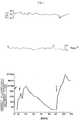
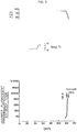
- Figures 1 and 2 illustrate in part the results of a further test of the in vivo biological activity characteristic of the soluble antigen of the invention.
- a vaccine was prepared from MASP culture supernatant. Each dose contained the lyophilized soluble antigen fraction isolated from 10 ml of MASP culture supernatant (wherein the average parasitemia following 3 days of in vitro propagation was about 16.4 percent) reconstituted with 1 ml water and 1 ml of saponin adjuvant (Quil-A). Two adult Holstein cows were used in the study and monitored for variations in Babesia serum antibodies (expressed as IFA titer), variations in rectal temperatures (expressed in °C) and variations in packed cell volume of blood (expressed as a percentage of total blood volume).
- Figure 1 provides data for the experimental animal and Figure 2 provides data for the control animal.
- the experimental cow was injected with a first dose of the vaccine on day 0 and a second dose on day 14 (noted by the unstarred arrows in Fig. 1).
- Antibody titers for the experimental animal rose to a peak at about day 35 and then gradually diminished. Temperature and packed cell volume data remained relatively constant.
- both the experimental and control animals were challenged by intramuscular injection of 1 x 10 8 virulent Babesia bovis (noted by the starred arrows in Figs. 1 and 2).
- Antibody titers rose precipitously in the experimental animal almost immediately, indicating a true anamnestic immune response. While body temperature rose, it returned to normal in a few days.
- the sterile immunity characteristics derived by the practice of vaccination methods of the invention are illustrated by the absence of infectivity of blood of vaccinated animals. More particularly, for example, 50 ml of blood of the experimental animal of Example 9 was withdrawn 28 days after challenge (i.e., about day 140) and injected into the circulatory system of a splenectomized calf. The calf failed to become infected even though it was later shown to be susceptible to infection by challenge with Babesia bovis.
- the soluble antigen of the invention may be obtained in quantity from an in vitro growth medium wherein merozoites are essentially constantly being formed and are constantly infecting erythrocytes.
- the antigen is also available by a rather gentle washing of the surfaces of such in vitro-propagated merozoites.
- the soluble antigen has its origins as a loosely-bound surface component of infective Babesia merozoites; and (2) the soluble antigen, after serving a "host recognition" function, is "shed” by the merozoite in the process of invading an erythrocyte.
- the physical and biological characteristics of the soluble antigen may be understood. If the antigen, when associated with the surface of a merozoite, serves to allow recognition of a proper host cell by the merozoite, it is likely that the non-infective substance alone would itself be "recognized” by the immune system of a vaccinated animal and give rise to protective antibody production without involvement of an infectious process.
- This model also serves to explain similarities and differences between the antigen of the present invention and prior in vivo isolates. If antigenic material is transitorily associated with and shed by merozoites upon invasion of erythrocytes, it would expectedly be available in at least small quantites from infected animal serum.
- the specific Babesia antigens of the invention may be used to sensitize immunologically inert particles of varying types well known in the art as useful in diagnostic, antigen-antibody reaction detection schemes.
- antigen preparations and antigen-sensitized particles of the invention may be used in combination with suitable "marker" substances (chemical and radiochemical) in the detection of antibodies by agglutination, radioimmunoassay, as well as fluorescent and enzyme immunoassay techniques.
- vaccine preparations of the invention may comprise the novel antigen in the form of crude or purified culture medium or merozoite isolates.
- precipitation and dialysis purified antigen in lyophilized, whitish powdery form
- a dosage amount of from 0.5 to 5.0 mg per adult animal is suitable for protecting animals susceptible to Babesia infection.
- immunologically acceptable carriers, diluents and adjuvants such as Quil-A and Freund's incomplete Adjuvant, with the former detergent-type adjuvant being preferred
Landscapes
- Health & Medical Sciences (AREA)
- Life Sciences & Earth Sciences (AREA)
- Immunology (AREA)
- Engineering & Computer Science (AREA)
- Tropical Medicine & Parasitology (AREA)
- Chemical & Material Sciences (AREA)
- Microbiology (AREA)
- Medicinal Chemistry (AREA)
- Urology & Nephrology (AREA)
- Molecular Biology (AREA)
- Hematology (AREA)
- General Health & Medical Sciences (AREA)
- Biomedical Technology (AREA)
- Mycology (AREA)
- Food Science & Technology (AREA)
- Veterinary Medicine (AREA)
- Public Health (AREA)
- Biotechnology (AREA)
- Cell Biology (AREA)
- Animal Behavior & Ethology (AREA)
- Epidemiology (AREA)
- Pharmacology & Pharmacy (AREA)
- Virology (AREA)
- Physics & Mathematics (AREA)
- Analytical Chemistry (AREA)
- Biochemistry (AREA)
- General Physics & Mathematics (AREA)
- Pathology (AREA)
- Medicines Containing Antibodies Or Antigens For Use As Internal Diagnostic Agents (AREA)
- Measuring Or Testing Involving Enzymes Or Micro-Organisms (AREA)
- Peptides Or Proteins (AREA)
Description
- The present invention relates generally. to materials useful in immunization of animals against infection by protozoan parasites of the genus Babesia (Order Piroplasmida). More specifically, the present invention provides a novel soluble antigen, specific for Babesia parasites, isolatable from the growth medium of Babesia-infected erythrocytes in cell culture and/or from merozoite stages of the parasite in the medium. Also provided are diagnostic reagents comprising said soluble antigen.
- Babesia parasites are the causative agents of hemoclastic animal diseases on a world-wide basis. Tick-bome species of this genus are known to infect: cattle (B. bovis, B. bigemina, B. divergens, B. major); dogs (B. canis, B. gibsoni, B. vogalia); horses (B. equi, B. caballi); and rodents (B. rodhaini, B. microti). B. bovis and B. microti are also known to be infective in humans.
- To date, no immunologically active agent has been available for use as a vaccine component in conferring immunity against infection by Babesia parasites. This is so despite very substantial attempts by those skilled in the art to isoate and purify antigenic materials by available in vivo and in vitro techniques.
- Many years of research directed toward isolation of antigenic materials from the blood of infected animals have yielded a number of serum isolates displaying some attributes of antigenicity. Such a preparation was described in the thesis of Sebinovic, as cited in Chemical Abstracts, Vol 66, No. 7, 13.2.1967, page 2579, No. 27381 z. In the thesis it was described how an antigenic material was isolated from the blood of animals infected with Babesia.
- Nevertheless, none of these isolates have been sucessfully employed to induce a protective, much less a long-term, anamnestic response in recipient animals. Moreover, animals inoculated with antigenic material derived from the blood of infected animals generally respond with production of iso blood group antibodies, prohibiting any practical application for such materials as vaccines. For the most part, the most successfull of in vivo preparative techniques have been useful only in providing rather crude diagnostic materials.
- Failures of in vitro preparative technology have been due in large part to difficulties attending quantitative, in vitro propagation of Babesia parasites, and especially to the difficulties inherent in separating suspected immunologically significant materials (e.g., killed, whole or fragmented antigenic merozoite stages of the parasite) from host erythrocyte cells and "contaminating" host cell components such as hemoglobin. Parenthetically, problems in quantitative in vitro propagation of hemo- tropic protozoan parasites effectively preclude development of vaccines for a wide variety of parasitic diseases including malaria (Plasmodium) and anaplasmosis (Anaplasma). Among the more pertinent recent advances in the art of parasite propagation are disclosed in Trager, et al., Science, 193: pp. 673-675 (1976) and Speer, et al., Z. Parasitenk, 50: pp. 237-244 (1976). These references, respectively, describe Plasmodium propagation in erythrocytes maintained under greatly reduced oxygen tension and continuous Plasmodium propagation in selected eukaryotic host cells. The focus of the two above-mentioned publications is the development of extracellular antigenic parasites and parasite fragments for use as vaccine components.
- The general unavailability of antigenic Babesia materials has correspondingly resulted in unavailability of specific diagnostic reagents for use in determining the presence of a Babesia disease state as well as the presence of Babesia antibodies in animal serum. Presently available reagents, as noted above, generally comprise crude preparations of antigenic materials obtained from the blood of infected animals.
- There exists, therefore, a substantial need in the art for antigenic Babesia materials in a substantially pure form, which materials may be used as components of vaccines and diagnostic reagents.
- According to the present invention, a novel soluble Babesia antigen is isolated in quantity from the medium supporting in vitro growth of Babesia-infected erythocytes. The antigen is characterized by: solubility in water and normal saline solutions; insolubility in from 40% to 70% ammonium sulfate solutions; the presence of proteinaceous and carbohydrate components; co-elution with proteins having a molecular weight on the order of about 900,000 as determined by Sephadex G-200 chromatography (Sephadex is a trade mark of Pharmacia, Sweden for modified polysaccharides for liquid exclusion chromatography.); heat lability; inactivation upon exposure to amylase; and susceptibility to staining with standard carbohydrate stains. The antigen is further characterized by its formation, upon counterimmunoelectrophoresis and two-dimensional immunoelectrophoresis, of at least two and three precipitin lines, respectively, with serum immunoglobulin of an animal recently recovered from Babesia infection. Finally, the soluble antigen is uniquely characterized by its ability to inhibit the capacity of specific Babesia serum antibodies to neutralize infectivity of Babesia merozoites in vitro.
- Antigen preparations according to the present invention include the above-described antigen in a purified form, isolated either from the culture medium or from extracellular merozoites (themselves isolated and separated from host cellular materials by differential centrifugation). Also comprehended are antigenic preparations in the form of partially purified (e.g., by centrifugation) fractions of the total growth medium.
- Vaccines according to the invention may comprise the antigen preparations in aqueous or lyophilized form and may include immunologically acceptable carriers, diluents, adjuvants and the like. Parenteral administration of the vaccines gives rise to formation of protective antibodies in the vaccinated animal within a matter of days. The protection confirmed is of the most desired type, i.e., sterile immunity. Remarkably, although antibody titers may diminish over long periods of time, vaccinated animals display an anamnestic protective response to delayed challenge by the parasite.
- Purified antigen preparations may be employed according to the invention in preparation of diagnostic reagents such as antigen-sensitized particles, radiologically labeled antigen preparations and the like which are useful in conventional serodiagnostic and immunoassay techniques.
- Other aspects and advantages of the invention will become apparent upon consideration of the following detailed description, wherein Figures 1 and 2 relate to vaccination test and challenge work involving the soluble antigen of the invention.
- The following illustrative examples relate to presently preferred methods of practice of the invention. More specifically, the examples describe: methods for in vitro propagation of Babesia which gives rise to an antigen enriched growth medium; methods for isolation of the Babesia antigen in crude and purified forms from growth medium and from merozoites; physical and biological characterization of the novel Babesia antigen; and, demonstration of in vivo efficacy of vaccine preparations of the invention which include the antigen.
- An antigen enriched growth medium, from which the soluble antigen of the invention is isolated, may be obtained by a modified practice of the propagative methods described in Erp, et al., An. Jour. Trop. Med. Hyg., 27 (5): pp. 1061-1064 (1978) wherein the work of the coinventors and certain of their co-workers in successfully propagating Babesia bovis in bovine erythrocyte cultures is set out.
- According to the procedure reported, splenectomized animals are inoculated intravenously with blood of a homologous species animal infected with a Babesia parasite. When routine microscopic examination of erythrocyte samples show that from 1 to 5 percent of the red blood cells of the inoculated animal are infected with the parasite, blood is collected and defibrinated by, e.g., shaking with glass beads. The blood is then centrifuged and the serum withdrawn and replaced with an equal volume of a mixture comprising 50 to 70 percent of a suitable medium supportive of erythrocyte growth (e.g., RPMI, MEM, or preferably Medium 199) and 30 to 50 percent serum from a non-infected heterologous species donor. Cultures so obtained are incubated at about 35°-38°C. in 100 ml spinner flasks in an atmosphere of about 5% carbon dioxide in air saturated with water vapor. Cells are kept in suspension by slow stirring with a magnetic stirrer. Subcultures are prepared, after 24-72 hours of incubation, by centrifugation of the cultures (5-10 minutes at 500xg) to collect the cells, and replacement of 50 to 70 percent of the cells with erythrocytes of a non-infected heterologous species donor, followed by resuspension in a fresh medium/serum mixture and continued incubation.
- The procedures of Erp, et al., are departed from in that, rather than discarding the supernatant medium obtained during the subculturing process, the medium is saved and provides a source of the novel soluble antigen.
- Alternatively, the soluble antigen of the invention may be prepared according to the much preferred methods described in U.S. Patent Number 4,307,191 by Miodrag Ristic and Michael G. Levy and entitled "In Vitro Parasite Propagation". (Attorney Docket No. 5236). That application was filed concurrently with parent application Serial No. 34,664.
- Saved culture medium portions obtained according to the modified Erp, et al. method of Example 1 are pooled. Merozoite stages of parasite are isolated by 20-30 minutes of centrifugation at about 1,000xg and either discarded or saved for further processing as a source of soluble anitgen. The resulting supernatant medium, having parasites and parasitic fragments removed, may be lyophilized to provide a relatively crude-but immunologically active-reddish powder suitable for use as the active agent in vaccine preparations. Alternatively, the soluble antigen may be isolated in a more purified form by selective precipitation from the medium using 40-70 percent ammonium sulfate solution. Collected precipitates are resuspended in 0.02 M phosphate buffer, the pH is adjusted to 8.0 and the resulting solution is dialyzed against 0.02 phosphate buffer, pH 8.0, to yield an aqueous solution of the antigen. Lyophilization of the antigenic solution yields the purified antigen in the form of a whitish powder. In its purified, lyophilized form the antigen is substantially free of not only host erythrocyte cell fragments and cellular debris but also Babesia merozoite cell fragments and debris.
- Merozoites collected in the manner noted above may be processed to yield the soluble antigen by extraction in buffered saline (pH 7.2) at 4°C. with gentle agitation to solubilize-the surface coat. The resulting surface coat solution may then be purified by selective precipitation and dialysis as above to yield the antigen in pure form for use, e.g., as a vaccine component. Alternately, non-extracted (whole) lyophilized merozoites may be employed as vaccine immunogen components. Owing to possibility of infection resulting from large merozoite fragments and overall dilution of the antigen, use of such merozoite preparations in vaccines is, of course, much less preferred to use of antigen isolated from the medium.
- The following determinations of characteristics of the soluble antigen of the invention were performed on a sample of purified (precipitated and dialyzed), lyophilized antigen of Babesia bovis prepared according to Example 2.
- Molecular weight analysis by Sephadex G 200 chromatography (Sephadex is a trade mark of Pharmacia, Sweden for modified polysaccharides for liquid exclusion chromatography.) showed co-elution with proteins having molecular weights on the order of 700,000 to 1,000,000.
- The presence of a carbohydrate component in the antigen is verified, in part, by a staining reaction using carbofuschin.
- As noted above, the antigen is soluble in water and normal saline but insoluble in 40 to 70 percent solutions of ammonium sulfate.
- Specific activity of the antigen is revealed by the results of counterimmunoelectrophoresis wherein the antigen is monitored for reactivity with homologous species anit-Babesia serum obtained from a recovered animal. More specifically, the antigen forms two distinctly characteristic precipitin lines with anti-serum, which lines are observed in the following procedure. Parallel wells are cut in a 1% agarose gel overlayed on a glass plate. Immune serum is placed in the well at the anodic side (+) and the antigen is placed in the cathodic side (-). Best resolution of precipitin lines is achieved with a discontinuous buffer system in that the reservoirs contain 0.05 M tris-barbital- sodium barbital buffer pH 8.8, whereas the gel contains a 0.01 M solution of the same buffer. Electrophoresis is conducted at 350 V for 45 minutes at 25°C. Precipitin lines then are visualized proximal to the cathodic side of the antibody well. Specific counterimmunoelectrophoretic activity with immune serum is also observed for the "crude", lyophilized supernatant preparations (obtained as a reddish powder) according to Example 2, as well as for saline extracts of soluble antigen derived from merozoites. It should be noted that the precise electrophoretic banding characteristics described above may vary somewhat depending on such procedural factors as the constitution of the buffer and the voltage applied.
- In another test, serum from rabbits immunized with B. bovis antigens derived from cultures which used rabbit erythrocytes and rabbit serum was reacted in a two-dimensional immunoelectrophoretic system with the supernatant fluid from positive cultures which employed bovine erythrocytes and serum. At least three antigen/antibody systems were revealed by this test. The reacting antigens showed fast mobility resembling bovine albumin. The use of normal, non-injected culture supernatant failed to demonstrate any of the above reactions.
- Further evidence of a carbohydrate component of the immunologically active antigen is provided by the reduction of specific activity with immune serum when pretreated with amylase. [e.g., incubation of 100 mg of crude antigen for 16 hours at 37°C. in 0.02 M buffered saline (pH 7.1) in the presence of 1 mg of alpha amylase].
- As discussed in greater detail, infra, the soluble Babesia antigen is characterized by its in vivo biological activity, i.e., its ability to induce a protective response to Babesia infection. The antigen is further characterized by its unique in vitro biological activity, i.e., its ability to inhibit the capacity of specific Babesia serum antibodies to neutralize the infectivity of Babesia merozoites in vitro. The latter characteristic will be better understood upon consideration of the following examples.
- It has been previously noted that the preferred methods for securing the antigen of the invention in quantity are set forth in copending U.S. Patent Number 4,307,191. Briefly summarized, these procedures call for propagation of Babesia parasites in erythrocytes in a medium comprising about 30 to 50 percent defibrinated serum, maintained in a controlled atmosphere having 3 to 6 percent carbon dioxide and under conditions of oxygen tension as will result in maintenance of erythro- cytic hemoglobin in its deoxy state. The above procedures, referred to as microaerophilous stationary phase ("MASP") continuous cultivation procedures, are valuable not only for their capacity to produce large quantities of antigen in the growth medium but also for their capability in producing large quantities of host- cell-free merozoites (especially upon discontinuation of increased carbon dioxide tension). The extracellular merozoites are easily collected by centrifugation and stored. When supplied to cultures of erythrocytes maintained under MASP conditions, the merozoites display infectivity characteristics substantially identical to that of merozoites obtained from the blood of infected animals. The availability of infective merozoites in this essentially "concentrated" form allows for the easy determination of the presence or absence of protective antibodies in sera of animals through determination of the degree to which serum antibodies will neutralize the infectivity of merozoites.
- According to such determinative procedures, MASP cultures containing uninfected erythrocytes are first allowed to incubate (for, e.g., 1 hour at 37° in a C02 incubator) with varying dilutions of test and control (normal) serum. Following incubation, 20,ul of a cell-free merozoite suspension is added to 200 µl of the MASP cultures contained in microtiter plates. These are allowed to incubate for from 1 to 16 hours, after which thin blood films are prepared. The percent parasitemia is determined by examination of Giemsa-stained blood films. By way of example, a 200 ,ul MASP culture preincubated with 80 µl of immune serum having an indirect fluorescent antibody "IFA" titer of 1:327,000 will completely neutralize the infectivity of 20 ,ul of cell-free merozoite suspension, i.e. samples will show no parasitemia.
- Modification of this merozoite neutralization ("MN") procedure provides an easy means for characterizing the soluble antigen of the invention and distinguishing it from, e.g., prior in vivo isolates. One need only ascertain whether the material possesses the ability to inhibit the ability of known of immune serum (containing specific Babesia antibodies) to neutralize infectivity of Babesia merozoites in an MN procedure such as described in the following procedure.
- Antigen of the invention was prepared according to the MASP procedure. The specific quantity of antigen employed was that amount provided in 150 ,ul of MASP culture supernatant following 3 days of in vitro culturing and wherein the average observed parasitemia had reached 16.4%. The quantity of antigen so obtained will, upon incubation with immune serum, completely inhibit the activity of 80µl of such serum (IFA titer 1:327,000) when the serum is thereafter employed in a merozoite neutralization procedure as described in Example 4. Dilution of this quantity of antigen by a factor of five results in an 80 percent reduction in inhibition of antibody activity, i.e., an 80 percent increase in the capacity of the antibody to neutralize the infectivity of Babesia antibodies as shown by a corresponding 80 percent drop in parasitemia.
- In order to illustrate the absence of the above-noted characteristic in vitro activity in prior in vivo isolates, antigenic materials were prepared according to the procedures of Ristic et al. Am. J. Vet. Res., 25, pp. 1519-26 (1964). Briefly summarized, a splenectomized calf was infected with the same Babesia bovis isolate employed in the MASP procedures used to make the soluble antigen tested in Example 5. An in vivo antigenic isolate was obtained from the infected animal's blood by protamine sulfate precipitation with care being taken to secure antigenic material from a number of parasites approximately equal to that involved in the MASP production of the soluble antigen of the invention. The isolate, when employed according to the procedure of Example 5, failed completely to inhibit neutralization of merozoite activity by serum antibodies.
- The in vivo activity of vaccine preparations of the invention is demonstrated by the results obtained upon challenge of vaccinated and unvaccinated cattle with exposure to ticks (Boophilus microplus) infected with Babesia bovis. Four female Holstein cattle (20 months old and weighing 225 to 450 kg) were selected for study. Two animals were vaccinated with a purified, lyophilized antigen preparation of Example 2 obtained from 10 ml of supernatant from a culture having about 5 percent infected cells. This quantity of antigen was suspended in 1 ml water. Vaccinated and unvaccinated controls were all then challenged with 2000 infected tick larvae. Non-immunized cattle developed typical signs of babesiosis including anemia and hemoglobinuria. Both control animals died. Vaccinated cattle showed no clinical signs of disease although the animals were infected with B. bovis, as demonstrated by microscopic examination of giesma-stained thin blood smears. Vaccinated cattle were long term survivors.
- Vaccine materials containing both corpuscular and soluble antigen fractions prepared according to the modified Erp, et al. procedure of Example 2 were employed in a field study as reported in Smith, et al., Am. J. Vet. Res., 40, pp. 1678-1682 (1979). The degree of protection afforded by the vaccines, which employed Freund's Complete and Incomplete Adjuvants, was less dramatic in the field study than in the previous Example 7, possibly due to defects in the collection, isolation and purification of antigen from the culture medium and perhaps due to the adjuvant employed.
- Figures 1 and 2 illustrate in part the results of a further test of the in vivo biological activity characteristic of the soluble antigen of the invention. A vaccine was prepared from MASP culture supernatant. Each dose contained the lyophilized soluble antigen fraction isolated from 10 ml of MASP culture supernatant (wherein the average parasitemia following 3 days of in vitro propagation was about 16.4 percent) reconstituted with 1 ml water and 1 ml of saponin adjuvant (Quil-A). Two adult Holstein cows were used in the study and monitored for variations in Babesia serum antibodies (expressed as IFA titer), variations in rectal temperatures (expressed in °C) and variations in packed cell volume of blood (expressed as a percentage of total blood volume). Figure 1 provides data for the experimental animal and Figure 2 provides data for the control animal. The experimental cow was injected with a first dose of the vaccine on day 0 and a second dose on day 14 (noted by the unstarred arrows in Fig. 1). Antibody titers for the experimental animal rose to a peak at about day 35 and then gradually diminished. Temperature and packed cell volume data remained relatively constant. At day 112, both the experimental and control animals were challenged by intramuscular injection of 1 x 10⁸ virulent Babesia bovis (noted by the starred arrows in Figs. 1 and 2). Antibody titers rose precipitously in the experimental animal almost immediately, indicating a true anamnestic immune response. While body temperature rose, it returned to normal in a few days. Similarly, packed cell volume diminished somewhat but soon returned to normal ranges. The control animal, on the other hand, showed no antibody development until nearly ten days following challenge at which time parasites were patent in blood smears, temperature had risen steadily and packed cell volume had dropped precipitously. On day 124 the control animal died. The vaccinated experimental animal was a long term survivor.
- The sterile immunity characteristics derived by the practice of vaccination methods of the invention are illustrated by the absence of infectivity of blood of vaccinated animals. More particularly, for example, 50 ml of blood of the experimental animal of Example 9 was withdrawn 28 days after challenge (i.e., about day 140) and injected into the circulatory system of a splenectomized calf. The calf failed to become infected even though it was later shown to be susceptible to infection by challenge with Babesia bovis.
- While not intended to be binding on the scope of the present invention, the following remarks may serve to explain in part the basis for its physical and biological properties vis-a-vis prior antigenic Babesia materials. It has been found that the soluble antigen of the invention may be obtained in quantity from an in vitro growth medium wherein merozoites are essentially constantly being formed and are constantly infecting erythrocytes. The antigen is also available by a rather gentle washing of the surfaces of such in vitro-propagated merozoites. These two facts lend substantial support to the propositions that: (1) the soluble antigen has its origins as a loosely-bound surface component of infective Babesia merozoites; and (2) the soluble antigen, after serving a "host recognition" function, is "shed" by the merozoite in the process of invading an erythrocyte.
- Within the context of such a model of events surrounding the infection process, the physical and biological characteristics of the soluble antigen may be understood. If the antigen, when associated with the surface of a merozoite, serves to allow recognition of a proper host cell by the merozoite, it is likely that the non-infective substance alone would itself be "recognized" by the immune system of a vaccinated animal and give rise to protective antibody production without involvement of an infectious process. This model also serves to explain similarities and differences between the antigen of the present invention and prior in vivo isolates. If antigenic material is transitorily associated with and shed by merozoites upon invasion of erythrocytes, it would expectedly be available in at least small quantites from infected animal serum. Indeed, some of the prior in vivo isolates are reported to include protein and non-protein components somewhat similar to those possessed by the soluble antigen of the invention. Others display similar solubility properties in ammonium sulfate solutions. That none share the in vivo and in vitro biological activity of the antigen of the invention may be attributable to a rapid degradation of "active" elements within the host's circulatory system, so that prior antigenic isolates may in fact comprise only fragments of their prior total and active constitution. It is certainly understandable that such antigenic "artifacts" would not display the biological activity needed for, e.g., an operative protective vaccine.
- It is expected that as a result of the present invention, many studies will be directed toward characterization of "active site(s)", if any, within the soluble antigen. Such studies, made possible by the availability for the first time of a truly immunologically active substance in quantity, are likely to lead to the future chemical or biological synthesis of perhaps drastically simpler, but nonetheless active, antigenic substances. While such chemically or biologically synthesized materials might no longer share the physical characteristics of the antigen of the invention in terms of molecular weight, ammonium sulfate solution solubility, or even protein/carbohydrate constituents would necessarily share the in vivo and in vitro biological characteristics noted above.
- The specific Babesia antigens of the invention may be used to sensitize immunologically inert particles of varying types well known in the art as useful in diagnostic, antigen-antibody reaction detection schemes. In this regard, antigen preparations and antigen-sensitized particles of the invention may be used in combination with suitable "marker" substances (chemical and radiochemical) in the detection of antibodies by agglutination, radioimmunoassay, as well as fluorescent and enzyme immunoassay techniques.
- As noted above, although not particularly preferred, vaccine preparations of the invention may comprise the novel antigen in the form of crude or purified culture medium or merozoite isolates. When the preferred, precipitation and dialysis purified antigen (in lyophilized, whitish powdery form) is employed, a dosage amount of from 0.5 to 5.0 mg per adult animal is suitable for protecting animals susceptible to Babesia infection. A variety of immunologically acceptable carriers, diluents and adjuvants (such as Quil-A and Freund's incomplete Adjuvant, with the former detergent-type adjuvant being preferred) may be employed in formulating vaccines according to the invention.
Claims (10)
Priority Applications (1)
| Application Number | Priority Date | Filing Date | Title |
|---|---|---|---|
| AT80102167T ATE4086T1 (en) | 1979-04-30 | 1980-04-23 | BABESIOSIS ANTIGEN, VACCINE AND DIAGNOSTIC REAGENT CONTAINING IT. |
Applications Claiming Priority (4)
| Application Number | Priority Date | Filing Date | Title |
|---|---|---|---|
| US3466479A | 1979-04-30 | 1979-04-30 | |
| US34664 | 1979-04-30 | ||
| US130481 | 1980-03-31 | ||
| US06/130,481 US4596707A (en) | 1979-04-30 | 1980-03-31 | Babesia parasite antigen useful in vaccine and diagnostic reagent |
Publications (3)
| Publication Number | Publication Date |
|---|---|
| EP0018579A2 EP0018579A2 (en) | 1980-11-12 |
| EP0018579A3 EP0018579A3 (en) | 1981-03-25 |
| EP0018579B1 true EP0018579B1 (en) | 1983-07-13 |
Family
ID=26711229
Family Applications (1)
| Application Number | Title | Priority Date | Filing Date |
|---|---|---|---|
| EP80102167A Expired EP0018579B1 (en) | 1979-04-30 | 1980-04-23 | Babesiosis antigen, vaccine and diagnostic reagent comprising it |
Country Status (8)
| Country | Link |
|---|---|
| US (1) | US4596707A (en) |
| EP (1) | EP0018579B1 (en) |
| AU (1) | AU531965B2 (en) |
| DE (1) | DE3064090D1 (en) |
| GR (1) | GR68201B (en) |
| IE (1) | IE49590B1 (en) |
| IL (1) | IL59841A (en) |
| PT (1) | PT71169A (en) |
Families Citing this family (17)
| Publication number | Priority date | Publication date | Assignee | Title |
|---|---|---|---|---|
| US4661348A (en) * | 1982-11-10 | 1987-04-28 | Commonwealth Scientific & Industrial Research Organization | Babesiosis antigen and method for preparation |
| US4767622A (en) * | 1983-08-19 | 1988-08-30 | University Of Illinois | Method and materials for development of immunological responses protective against malarial infection |
| FR2589063B1 (en) * | 1985-10-24 | 1988-08-26 | Rhone Merieux | PROCESS OF CULTIVATION OF BABESIA CANIS, APPLICATION TO THE PREPARATION OF ANTIGENS AND VACCINES, AND ANTIGENS AND VACCINES AGAINST PIROPLASMOSIS |
| US4721617A (en) * | 1986-08-14 | 1988-01-26 | Regents Of The University Of Minnesota | Vaccine against lyme disease |
| GB8630851D0 (en) * | 1986-12-24 | 1987-02-04 | Department Of Agriculture For | Babesia divergens antigen & vaccine |
| ATE122568T1 (en) * | 1989-01-25 | 1995-06-15 | Commw Scient Ind Res Org | POLYPEPTIDES, ANTIGENS OR VACCINES AGAINST BABESIOSIS. |
| FR2655853B1 (en) * | 1989-12-20 | 1994-06-10 | Centre Nat Rech Scient | PROCESS FOR THE INTENSIVE CULTURE, IN VITRO, OF STRAINS OF BABESIA DIVERGENS, PROCESS FOR THE PREPARATION OF EXOANTIGENS AND A VACCINE CONTAINING THESE ANTIGENS. |
| US5643737A (en) * | 1992-06-22 | 1997-07-01 | The United States Of America As Represented By The Secretary Of Agriculture | Merozoite proteins for use in detection of Babesia equi in horses using immunological techniques |
| EP0691131B1 (en) * | 1994-06-06 | 1999-10-06 | Akzo Nobel N.V. | Babesia vaccine |
| ES2141297T3 (en) * | 1994-06-06 | 2000-03-16 | Akzo Nobel Nv | BABESIOSIS VACCINE. |
| FR2724392B1 (en) * | 1994-09-08 | 1996-12-13 | Virbac Lab | METHODS OF CULTURE, MITIGATION OF VIRULENCE AND IN VITRO CLONING OF BABESIA PARASITES AND THEIR APPLICATIONS |
| US6569433B1 (en) | 1996-10-01 | 2003-05-27 | Corixa Corporation | Compounds and methods for the diagnosis and treatment of B. microti infection |
| US6451315B1 (en) | 1996-10-01 | 2002-09-17 | Corixa Corporation | Compounds and methods for the diagnosis and treatment of B. microti infection |
| US6306396B1 (en) | 1996-10-01 | 2001-10-23 | Corixa Corporation | Compounds and methods for the diagnosis and treatment of B. microti infection |
| US6214971B1 (en) * | 1996-10-01 | 2001-04-10 | Corixa Corporation | Compounds and methods for the diagnosis and treatment of Babesia microti infection |
| CN106978425B (en) * | 2017-02-17 | 2019-10-18 | 华中农业大学 | The albumen of Babesia orientalis 1-deoxy-D-xylulose -5- phosphoric acid reduction isomerase gene and its coding |
| WO2021097540A1 (en) * | 2019-11-21 | 2021-05-27 | Griffith University | Immunostimulatory compositions comprising soluble parasite extracts and uses thereof |
-
1980
- 1980-03-31 US US06/130,481 patent/US4596707A/en not_active Expired - Lifetime
- 1980-04-15 IL IL59841A patent/IL59841A/en unknown
- 1980-04-23 EP EP80102167A patent/EP0018579B1/en not_active Expired
- 1980-04-23 DE DE8080102167T patent/DE3064090D1/en not_active Expired
- 1980-04-24 AU AU57764/80A patent/AU531965B2/en not_active Ceased
- 1980-04-28 GR GR61796A patent/GR68201B/el unknown
- 1980-04-28 IE IE858/80A patent/IE49590B1/en not_active IP Right Cessation
- 1980-04-30 PT PT71169A patent/PT71169A/en not_active IP Right Cessation
Also Published As
| Publication number | Publication date |
|---|---|
| GR68201B (en) | 1981-11-10 |
| EP0018579A3 (en) | 1981-03-25 |
| AU531965B2 (en) | 1983-09-15 |
| EP0018579A2 (en) | 1980-11-12 |
| PT71169A (en) | 1980-05-01 |
| US4596707A (en) | 1986-06-24 |
| IL59841A (en) | 1983-10-31 |
| AU5776480A (en) | 1980-11-06 |
| DE3064090D1 (en) | 1983-08-18 |
| IE800858L (en) | 1980-10-30 |
| IE49590B1 (en) | 1985-10-30 |
| IL59841A0 (en) | 1980-06-30 |
Similar Documents
| Publication | Publication Date | Title |
|---|---|---|
| EP0018579B1 (en) | Babesiosis antigen, vaccine and diagnostic reagent comprising it | |
| Good et al. | Human T-cell recognition of the circumsporozoite protein of Plasmodium falciparum: immunodominant T-cell domains map to the polymorphic regions of the molecule. | |
| EP0749319A1 (en) | Immortalized cells and uses therefor | |
| US4767622A (en) | Method and materials for development of immunological responses protective against malarial infection | |
| US6855322B2 (en) | Isolation and purification of P. falciparum merozoite protein-142 vaccine | |
| US8211447B2 (en) | Recombinant P. falciparum merozoite protein-142 vaccine | |
| Ristic et al. | Exoantigens of Babesia | |
| US3911097A (en) | Antigenic preparation suitable for diagnosis of chronic chagas disease | |
| US3993743A (en) | Method for diagnosis of Chagas' disease | |
| US6458581B1 (en) | Process for the in vitro culture of different stages of tissue parasites | |
| EP0101505B1 (en) | Method for the purification of parasite antigenic factors | |
| XU et al. | Analysis of the Soluble and Membrane‐bound Immobilization Antigens of Ichthyophthirius multifiliis | |
| JPH0118885B2 (en) | ||
| Fruit et al. | Trypanosoma cruzi: location of a specific antigen on the surface of bloodstream trypomastigote and culture epimastigote forms | |
| Steinberger et al. | Leishmania tropica: protective response in C3H mice vaccinated with excreted factor crosslinked with the synthetic adjuvant, muramyl dipeptide | |
| Frankenburg et al. | Cell-mediated responses and protection elicited by a carbohydrate-lipid-containing fraction extracted from Leishmania major promastigotes | |
| US8298546B2 (en) | Isolation and purification of P. falciparum merozoite protein-142 vaccine | |
| Ristic et al. | Immunodiagnosis of the haemosporidial infections such as babesiosis | |
| Grothaus et al. | Plasmodium berghei: Characterization of Antigens and Their Role in Inducing Immunity to Infection | |
| WO1990015621A1 (en) | A malaria vaccine | |
| Wu et al. | Species-and infective stage-specific monoclonal antibodies to Leishmania major produced by an in vitro immunization method | |
| JPH03183472A (en) | Separation of salivary gland cell of mite and process for collecting sporozoite from the cell | |
| JPS60500862A (en) | Polypeptide fraction that induces anti-malarial parasite protective antibodies and immunogenic compositions containing the same |
Legal Events
| Date | Code | Title | Description |
|---|---|---|---|
| PUAI | Public reference made under article 153(3) epc to a published international application that has entered the european phase |
Free format text: ORIGINAL CODE: 0009012 |
|
| AK | Designated contracting states |
Designated state(s): AT BE CH DE FR GB IT NL |
|
| PUAL | Search report despatched |
Free format text: ORIGINAL CODE: 0009013 |
|
| AK | Designated contracting states |
Designated state(s): AT BE CH DE FR GB IT NL |
|
| 17P | Request for examination filed |
Effective date: 19810509 |
|
| ITF | It: translation for a ep patent filed |
Owner name: STUDIO TORTA SOCIETA' SEMPLICE |
|
| GRAA | (expected) grant |
Free format text: ORIGINAL CODE: 0009210 |
|
| AK | Designated contracting states |
Designated state(s): AT BE CH DE FR GB IT LI NL |
|
| REF | Corresponds to: |
Ref document number: 4086 Country of ref document: AT Date of ref document: 19830715 Kind code of ref document: T |
|
| REF | Corresponds to: |
Ref document number: 3064090 Country of ref document: DE Date of ref document: 19830818 |
|
| ET | Fr: translation filed | ||
| PLBE | No opposition filed within time limit |
Free format text: ORIGINAL CODE: 0009261 |
|
| STAA | Information on the status of an ep patent application or granted ep patent |
Free format text: STATUS: NO OPPOSITION FILED WITHIN TIME LIMIT |
|
| 26N | No opposition filed | ||
| ITTA | It: last paid annual fee | ||
| PGFP | Annual fee paid to national office [announced via postgrant information from national office to epo] |
Ref country code: AT Payment date: 19960411 Year of fee payment: 17 |
|
| PGFP | Annual fee paid to national office [announced via postgrant information from national office to epo] |
Ref country code: NL Payment date: 19960430 Year of fee payment: 17 |
|
| PGFP | Annual fee paid to national office [announced via postgrant information from national office to epo] |
Ref country code: CH Payment date: 19960507 Year of fee payment: 17 |
|
| PG25 | Lapsed in a contracting state [announced via postgrant information from national office to epo] |
Ref country code: AT Effective date: 19970423 |
|
| PG25 | Lapsed in a contracting state [announced via postgrant information from national office to epo] |
Ref country code: LI Free format text: LAPSE BECAUSE OF NON-PAYMENT OF DUE FEES Effective date: 19970430 Ref country code: CH Free format text: LAPSE BECAUSE OF NON-PAYMENT OF DUE FEES Effective date: 19970430 |
|
| PG25 | Lapsed in a contracting state [announced via postgrant information from national office to epo] |
Ref country code: NL Effective date: 19971101 |
|
| REG | Reference to a national code |
Ref country code: CH Ref legal event code: PL |
|
| NLV4 | Nl: lapsed or anulled due to non-payment of the annual fee |
Effective date: 19971101 |
|
| PGFP | Annual fee paid to national office [announced via postgrant information from national office to epo] |
Ref country code: FR Payment date: 19990409 Year of fee payment: 20 |
|
| PGFP | Annual fee paid to national office [announced via postgrant information from national office to epo] |
Ref country code: GB Payment date: 19990421 Year of fee payment: 20 |
|
| PGFP | Annual fee paid to national office [announced via postgrant information from national office to epo] |
Ref country code: DE Payment date: 19990430 Year of fee payment: 20 |
|
| PGFP | Annual fee paid to national office [announced via postgrant information from national office to epo] |
Ref country code: BE Payment date: 19990614 Year of fee payment: 20 |
|
| BE20 | Be: patent expired |
Free format text: 20000423 *UNIVERSITY OF *ILLINOIS FOUNDATION |
|
| PG25 | Lapsed in a contracting state [announced via postgrant information from national office to epo] |
Ref country code: GB Free format text: LAPSE BECAUSE OF EXPIRATION OF PROTECTION Effective date: 20000422 |
|
| REG | Reference to a national code |
Ref country code: GB Ref legal event code: PE20 Effective date: 20000422 |