DE102017217543B4 - Method and system for classifying materials using machine learning - Google Patents
Method and system for classifying materials using machine learning Download PDFInfo
- Publication number
- DE102017217543B4 DE102017217543B4 DE102017217543.5A DE102017217543A DE102017217543B4 DE 102017217543 B4 DE102017217543 B4 DE 102017217543B4 DE 102017217543 A DE102017217543 A DE 102017217543A DE 102017217543 B4 DE102017217543 B4 DE 102017217543B4
- Authority
- DE
- Germany
- Prior art keywords
- classification
- image
- materials
- spectral
- material index
- Prior art date
- Legal status (The legal status is an assumption and is not a legal conclusion. Google has not performed a legal analysis and makes no representation as to the accuracy of the status listed.)
- Active
Links
Images


Classifications
-
- G—PHYSICS
- G06—COMPUTING OR CALCULATING; COUNTING
- G06F—ELECTRIC DIGITAL DATA PROCESSING
- G06F18/00—Pattern recognition
- G06F18/20—Analysing
- G06F18/24—Classification techniques
-
- G—PHYSICS
- G06—COMPUTING OR CALCULATING; COUNTING
- G06V—IMAGE OR VIDEO RECOGNITION OR UNDERSTANDING
- G06V10/00—Arrangements for image or video recognition or understanding
- G06V10/70—Arrangements for image or video recognition or understanding using pattern recognition or machine learning
- G06V10/77—Processing image or video features in feature spaces; using data integration or data reduction, e.g. principal component analysis [PCA] or independent component analysis [ICA] or self-organising maps [SOM]; Blind source separation
- G06V10/776—Validation; Performance evaluation
-
- A—HUMAN NECESSITIES
- A61—MEDICAL OR VETERINARY SCIENCE; HYGIENE
- A61B—DIAGNOSIS; SURGERY; IDENTIFICATION
- A61B6/00—Apparatus or devices for radiation diagnosis; Apparatus or devices for radiation diagnosis combined with radiation therapy equipment
- A61B6/02—Arrangements for diagnosis sequentially in different planes; Stereoscopic radiation diagnosis
- A61B6/03—Computed tomography [CT]
- A61B6/032—Transmission computed tomography [CT]
-
- A—HUMAN NECESSITIES
- A61—MEDICAL OR VETERINARY SCIENCE; HYGIENE
- A61B—DIAGNOSIS; SURGERY; IDENTIFICATION
- A61B6/00—Apparatus or devices for radiation diagnosis; Apparatus or devices for radiation diagnosis combined with radiation therapy equipment
- A61B6/48—Diagnostic techniques
- A61B6/482—Diagnostic techniques involving multiple energy imaging
-
- A—HUMAN NECESSITIES
- A61—MEDICAL OR VETERINARY SCIENCE; HYGIENE
- A61B—DIAGNOSIS; SURGERY; IDENTIFICATION
- A61B6/00—Apparatus or devices for radiation diagnosis; Apparatus or devices for radiation diagnosis combined with radiation therapy equipment
- A61B6/52—Devices using data or image processing specially adapted for radiation diagnosis
- A61B6/5211—Devices using data or image processing specially adapted for radiation diagnosis involving processing of medical diagnostic data
- A61B6/5217—Devices using data or image processing specially adapted for radiation diagnosis involving processing of medical diagnostic data extracting a diagnostic or physiological parameter from medical diagnostic data
-
- A—HUMAN NECESSITIES
- A61—MEDICAL OR VETERINARY SCIENCE; HYGIENE
- A61B—DIAGNOSIS; SURGERY; IDENTIFICATION
- A61B6/00—Apparatus or devices for radiation diagnosis; Apparatus or devices for radiation diagnosis combined with radiation therapy equipment
- A61B6/52—Devices using data or image processing specially adapted for radiation diagnosis
- A61B6/5294—Devices using data or image processing specially adapted for radiation diagnosis involving using additional data, e.g. patient information, image labeling, acquisition parameters
-
- G—PHYSICS
- G06—COMPUTING OR CALCULATING; COUNTING
- G06F—ELECTRIC DIGITAL DATA PROCESSING
- G06F18/00—Pattern recognition
- G06F18/20—Analysing
- G06F18/21—Design or setup of recognition systems or techniques; Extraction of features in feature space; Blind source separation
- G06F18/214—Generating training patterns; Bootstrap methods, e.g. bagging or boosting
-
- G—PHYSICS
- G06—COMPUTING OR CALCULATING; COUNTING
- G06F—ELECTRIC DIGITAL DATA PROCESSING
- G06F18/00—Pattern recognition
- G06F18/20—Analysing
- G06F18/21—Design or setup of recognition systems or techniques; Extraction of features in feature space; Blind source separation
- G06F18/217—Validation; Performance evaluation; Active pattern learning techniques
-
- G—PHYSICS
- G06—COMPUTING OR CALCULATING; COUNTING
- G06N—COMPUTING ARRANGEMENTS BASED ON SPECIFIC COMPUTATIONAL MODELS
- G06N20/00—Machine learning
-
- G—PHYSICS
- G06—COMPUTING OR CALCULATING; COUNTING
- G06T—IMAGE DATA PROCESSING OR GENERATION, IN GENERAL
- G06T7/00—Image analysis
- G06T7/0002—Inspection of images, e.g. flaw detection
- G06T7/0012—Biomedical image inspection
- G06T7/0014—Biomedical image inspection using an image reference approach
-
- G—PHYSICS
- G06—COMPUTING OR CALCULATING; COUNTING
- G06T—IMAGE DATA PROCESSING OR GENERATION, IN GENERAL
- G06T7/00—Image analysis
- G06T7/10—Segmentation; Edge detection
- G06T7/11—Region-based segmentation
-
- G—PHYSICS
- G06—COMPUTING OR CALCULATING; COUNTING
- G06V—IMAGE OR VIDEO RECOGNITION OR UNDERSTANDING
- G06V10/00—Arrangements for image or video recognition or understanding
- G06V10/70—Arrangements for image or video recognition or understanding using pattern recognition or machine learning
- G06V10/77—Processing image or video features in feature spaces; using data integration or data reduction, e.g. principal component analysis [PCA] or independent component analysis [ICA] or self-organising maps [SOM]; Blind source separation
- G06V10/774—Generating sets of training patterns; Bootstrap methods, e.g. bagging or boosting
-
- G—PHYSICS
- G16—INFORMATION AND COMMUNICATION TECHNOLOGY [ICT] SPECIALLY ADAPTED FOR SPECIFIC APPLICATION FIELDS
- G16H—HEALTHCARE INFORMATICS, i.e. INFORMATION AND COMMUNICATION TECHNOLOGY [ICT] SPECIALLY ADAPTED FOR THE HANDLING OR PROCESSING OF MEDICAL OR HEALTHCARE DATA
- G16H30/00—ICT specially adapted for the handling or processing of medical images
- G16H30/20—ICT specially adapted for the handling or processing of medical images for handling medical images, e.g. DICOM, HL7 or PACS
-
- G—PHYSICS
- G16—INFORMATION AND COMMUNICATION TECHNOLOGY [ICT] SPECIALLY ADAPTED FOR SPECIFIC APPLICATION FIELDS
- G16H—HEALTHCARE INFORMATICS, i.e. INFORMATION AND COMMUNICATION TECHNOLOGY [ICT] SPECIALLY ADAPTED FOR THE HANDLING OR PROCESSING OF MEDICAL OR HEALTHCARE DATA
- G16H50/00—ICT specially adapted for medical diagnosis, medical simulation or medical data mining; ICT specially adapted for detecting, monitoring or modelling epidemics or pandemics
- G16H50/30—ICT specially adapted for medical diagnosis, medical simulation or medical data mining; ICT specially adapted for detecting, monitoring or modelling epidemics or pandemics for calculating health indices; for individual health risk assessment
-
- A—HUMAN NECESSITIES
- A61—MEDICAL OR VETERINARY SCIENCE; HYGIENE
- A61B—DIAGNOSIS; SURGERY; IDENTIFICATION
- A61B6/00—Apparatus or devices for radiation diagnosis; Apparatus or devices for radiation diagnosis combined with radiation therapy equipment
- A61B6/42—Arrangements for detecting radiation specially adapted for radiation diagnosis
- A61B6/4208—Arrangements for detecting radiation specially adapted for radiation diagnosis characterised by using a particular type of detector
- A61B6/4241—Arrangements for detecting radiation specially adapted for radiation diagnosis characterised by using a particular type of detector using energy resolving detectors, e.g. photon counting
-
- G—PHYSICS
- G06—COMPUTING OR CALCULATING; COUNTING
- G06F—ELECTRIC DIGITAL DATA PROCESSING
- G06F2218/00—Aspects of pattern recognition specially adapted for signal processing
- G06F2218/12—Classification; Matching
-
- G—PHYSICS
- G06—COMPUTING OR CALCULATING; COUNTING
- G06T—IMAGE DATA PROCESSING OR GENERATION, IN GENERAL
- G06T2207/00—Indexing scheme for image analysis or image enhancement
- G06T2207/10—Image acquisition modality
- G06T2207/10072—Tomographic images
- G06T2207/10081—Computed x-ray tomography [CT]
-
- G—PHYSICS
- G06—COMPUTING OR CALCULATING; COUNTING
- G06T—IMAGE DATA PROCESSING OR GENERATION, IN GENERAL
- G06T2207/00—Indexing scheme for image analysis or image enhancement
- G06T2207/20—Special algorithmic details
- G06T2207/20081—Training; Learning
-
- G—PHYSICS
- G06—COMPUTING OR CALCULATING; COUNTING
- G06V—IMAGE OR VIDEO RECOGNITION OR UNDERSTANDING
- G06V2201/00—Indexing scheme relating to image or video recognition or understanding
- G06V2201/03—Recognition of patterns in medical or anatomical images
- G06V2201/031—Recognition of patterns in medical or anatomical images of internal organs
Landscapes
- Engineering & Computer Science (AREA)
- Health & Medical Sciences (AREA)
- Life Sciences & Earth Sciences (AREA)
- Medical Informatics (AREA)
- Theoretical Computer Science (AREA)
- Physics & Mathematics (AREA)
- Computer Vision & Pattern Recognition (AREA)
- General Health & Medical Sciences (AREA)
- General Physics & Mathematics (AREA)
- Public Health (AREA)
- Radiology & Medical Imaging (AREA)
- Nuclear Medicine, Radiotherapy & Molecular Imaging (AREA)
- Pathology (AREA)
- Biomedical Technology (AREA)
- Artificial Intelligence (AREA)
- Data Mining & Analysis (AREA)
- Evolutionary Computation (AREA)
- High Energy & Nuclear Physics (AREA)
- Veterinary Medicine (AREA)
- Animal Behavior & Ethology (AREA)
- Surgery (AREA)
- Molecular Biology (AREA)
- Heart & Thoracic Surgery (AREA)
- Optics & Photonics (AREA)
- Biophysics (AREA)
- Software Systems (AREA)
- Computing Systems (AREA)
- General Engineering & Computer Science (AREA)
- Databases & Information Systems (AREA)
- Multimedia (AREA)
- Evolutionary Biology (AREA)
- Bioinformatics & Computational Biology (AREA)
- Bioinformatics & Cheminformatics (AREA)
- Mathematical Physics (AREA)
- Physiology (AREA)
- Pulmonology (AREA)
- Epidemiology (AREA)
- Primary Health Care (AREA)
- Quality & Reliability (AREA)
- Apparatus For Radiation Diagnosis (AREA)
Abstract
Verfahren zum Erstellen einer Klassifizierungseinheit (6) zur automatischen Klassifizierung von Materialien (M1, M2, M3) in einer spektralen medizintechnischen Bildaufnahme (B) eines Objekts (O), umfassend die Schritte:- Bereitstellung einer Lern-Rechenvorrichtung (7), wobei die Lern-Rechenvorrichtung (7) mittels eines Algorithmus dazu ausgestaltet ist, grafische Elemente in Bildaufnahmen zu erkennen,- Bereitstellung einer Start-Klassifizierungseinheit (6a) auf oder an der Lern-Rechenvorrichtung (7), welche dazu ausgestaltet ist, mittels maschinellem Lernen trainiert zu werden,- Bereitstellung eines Referenzaufnahmen-Satzes (RS) umfassend spektrale Referenzaufnahmen (RA) des Objekts (O), in denen die zu klassifizierenden Materialien (M1, M2, M3) annotiert sind,- Trainieren der Klassifizierungseinheit (6) gemäß dem Prinzip des maschinellen Lernens, unter Verwendung der in den spektralen Referenzaufnahmen (RA) annotierten Materialien (M1, M2, M3).Method for creating a classification unit (6) for the automatic classification of materials (M1, M2, M3) in a spectral medical image (B) of an object (O), comprising the steps: - providing a learning computing device (7), the Learning computing device (7) is designed by means of an algorithm to recognize graphic elements in image recordings, - providing a start classification unit (6a) on or on the learning computing device (7), which is designed to be trained by means of machine learning - Providing a reference record set (RS) comprising spectral reference recordings (RA) of the object (O) in which the materials to be classified (M1, M2, M3) are annotated, - Training the classification unit (6) according to the principle of machine learning, using the materials (M1, M2, M3) annotated in the spectral reference images (RA).
Description
Die Erfindung betrifft ein Verfahren zum Erstellen einer Klassifikationseinheit zur automatischen Klassifikation von Materialien in einer spektralen medizinischen Bildaufnahme bzw. eine Klassifikationseinheit sowie eine Lern-Rechenvorrichtung hierzu. Des Weiteren umfasst die Erfindung ein Klassifikations-Verfahren zur automatischen Klassifikation von Materialien in einer spektralen medizinischen Bildaufnahme unter Nutzung einer solchen Klassifikationseinheit, eine Steuereinrichtung zur Steuerung eines medizintechnischen bildgebenden Systems sowie ein entsprechendes medizintechnisches bildgebendes System. Eine Bildaufnahme ist in dem Rahmen der Erfindung ein digitales Bild, umfasst also Bilddaten bzw. besteht diese aus Bilddaten.The invention relates to a method for creating a classification unit for the automatic classification of materials in a spectral medical image recording or a classification unit and a learning computing device therefor. Furthermore, the invention comprises a classification method for the automatic classification of materials in a spectral medical image acquisition using such a classification unit, a control device for controlling a medical-technical imaging system and a corresponding medical-technical imaging system. In the context of the invention, an image recording is a digital image, that is to say comprises image data or consists of image data.
Bei bildgebenden Verfahren in der Medizin ist man zur Erstellung eines jeden Bildpunktes einer Bildaufnahme zumeist auf die Auswertung einer einzelnen Messgröße bzw. eines einzigen physikalischen Messprinzips angewiesen. So erfasst z.B. die Computertomographie („CT“) als einzige Messgröße die lokale Röntgenschwächung des Patienten. Chemisch unterschiedliche Materialien, die z.B. aufgrund unterschiedlicher Konzentrationen die gleiche lokale Röntgenschwächung aufweisen, werden in einem CT-Bild mit dem gleichen CT-Wert (üblicherweise in Hounsfield-Einheiten HU) dargestellt und können daher im Bild nicht voneinander unterschieden werden.In medical imaging processes, the creation of each image point of an image is usually dependent on the evaluation of a single measurement variable or a single physical measurement principle. For example, Computed tomography (“CT”) is the only measure of local x-ray attenuation of the patient. Chemically different materials, e.g. have the same local X-ray attenuation due to different concentrations, are shown in a CT image with the same CT value (usually in Hounsfield units HU) and therefore cannot be distinguished from one another in the image.
Das ist in vielen CT-Anwendungen problematisch. Ein Beispiel ist die Trennung von Knochen und kontrastmittelgefüllten Gefäßen in CT-Angiographien. Das Jod im Blut kann die gleiche Röntgenabsorption wie der umgebende Knochen aufweisen, so dass Gefäße und Knochen insbesondere in komplizierten anatomischen Situationen, z.B. in der Schädelbasis, nur schwer voneinander getrennt und separat dargestellt werden können. This is problematic in many CT applications. One example is the separation of bones and contrast medium-filled vessels in CT angiographies. The iodine in the blood can have the same X-ray absorption as the surrounding bone, so that vessels and bones, particularly in complicated anatomical situations, e.g. in the skull base, difficult to separate from each other and can be shown separately.
Ein weiteres Beispiel ist die genauere Charakterisierung von Nierensteinen. Kalziumhaltige Steine und harnsäurehaltige Steine können die gleiche Röntgenabsorption aufweisen. Sie sind daher im CT-Bild nicht zu unterscheiden, haben aber unterschiedliche Behandlungsoptionen. Ein weiteres Beispiel ist die Unterscheidung von Gichtkristallen von anderen Ablagerungen an den Gelenken.Another example is the more precise characterization of kidney stones. Calcium-containing stones and stones containing uric acid can have the same X-ray absorption. Therefore, they cannot be distinguished in the CT image, but they have different treatment options. Another example is the distinction of gout crystals from other deposits on the joints.
Zur Klassifikation von unterschiedlichen Stoffen („Materialien“) im Gewebe wird zuweilen mit Strahlung in zwei oder mehreren Energiebereichen gearbeitet. Beispielsweise kann eine Aufnahme von CT-Bildern mit zwei oder mehr unterschiedlichen Röntgenenergien („Dual Energy CT“, „Spectral CT“) zur Unterscheidung verschiedener Materialien genutzt werden.To classify different substances (“materials”) in tissue, radiation is sometimes used in two or more energy areas. For example, a recording of CT images with two or more different X-ray energies ("Dual Energy CT", "Spectral CT") can be used to differentiate between different materials.
Die Röntgenabsorption von im Körper vorkommenden Materialien im Energiebereich der CT-typischen Röntgenstrahlung (30 - 150 keV) wird durch zwei physikalische Mechanismen bestimmt, den Photoelektrischen- und den Comptoneffekt. Diese Mechanismen weisen unterschiedliche Energieabhängigkeit auf und ihr relativer Anteil an der gesamten Röntgenabsorption hängt vom jeweiligen Material ab, genauer von dessen Ordnungszahl und dessen Dichte.The X-ray absorption of materials occurring in the body in the energy range of CT-typical X-rays (30 - 150 keV) is determined by two physical mechanisms, the photoelectric and the Compton effect. These mechanisms have different energy dependencies and their relative share of the total X-ray absorption depends on the respective material, more precisely on its atomic number and its density.
Wegen des Vorhandenseins von zwei physikalischen Effekten mit unterschiedlicher Energieabhängigkeit können durch Aufnahmen mit zwei oder mehr unterschiedlichen Röntgenenergien zwei Materialien unterschieden werden. Nimmt man zusätzliche Materialien dazu, die eine K-Kante im relevanten Energiebereich aufweisen, z.B. Gadolinium, Gold oder Eisen, lassen sich mit Aufnahmen mit mehr als zwei Röntgenenergien auch mehr als zwei Materialien trennen.Because of the presence of two physical effects with different energy dependencies, two materials can be distinguished by recordings with two or more different X-ray energies. If you add additional materials that have a K-edge in the relevant energy range, e.g. Gadolinium, gold or iron, can be separated with more than two X-ray energies, more than two materials.
Eine Aufnahme von CT-Bildern mit mehreren unterschiedlichen Röntgenenergien ist beispielsweise mit Dual Source CT-Geräten mit einem Betrieb beider Röhren mit unterschiedlichen Spannungen, Single Source CT-Geräten mit Umschaltung der Röhrenspannung, CT-Geräten mit geteilter Vorfilterung („Twin Beam“), CT-Geräten mit Dual Layer Detektoren oder CT-Geräten mit photonenzählenden Detektoren möglich. Jedoch können auch andere medizintechnische bildgebende Geräte, die nicht auf dem Prinzip eines Computertomographen beruhen, mit zwei Aufnahmeenergien eine Materialklassifikation ermöglichen.A recording of CT images with several different X-ray energies is possible, for example, with dual source CT devices with operation of both tubes with different voltages, single source CT devices with switching of the tube voltage, CT devices with split pre-filtering (“twin beam”), CT devices with dual layer detectors or CT devices with photon counting detectors possible. However, other medical imaging devices that are not based on the principle of a computer tomograph can also enable material classification with two recording energies.
Die Trennung beruht in der Regel auf der Ermittlung einer „spektralen Größe“, die die materialabhängige Änderung der Röntgenabsorption bei den verschiedenen Röntgenenergien charakterisiert. Eine solche spektrale Größe ist z.B. der sogenannte Dual Energy Ratio, das ist der Röntgenschwächungswert bei einer niedrigen Röntgenenergie A dividiert durch den Röntgenschwächungswert bei einer höheren Energie B. Gäbe es keinerlei spektrale Effekte, wäre der Dual Energy Ratio gleich 1. Je ausgeprägter der spektrale Effekt für das jeweilige Material ist, desto mehr weicht der Dual Energy Ratio von 1 ab. In der Regel wird zur Materialtrennung von zwei Materialien A und B ein bestimmter konstanter Grenzwert der spektralen Größe verwendet. Für jedes Pixel im CT-Bild wird untersucht, ob die spektrale Größe an dieser Stelle unterhalb des Grenzwerts liegt, dann handelt es sich um Material A. Liegt die spektrale Größe oberhalb des Grenzwerts, handelt es sich um Material B.The separation is usually based on the determination of a “spectral variable” that characterizes the material-dependent change in the X-ray absorption at the different X-ray energies. Such a spectral quantity is e.g. the so-called dual energy ratio, which is the X-ray attenuation value at a low X-ray energy A divided by the X-ray attenuation value at a higher energy B. If there were no spectral effects, the Dual Energy Ratio would be 1. The more pronounced the spectral effect for the respective material, the more the dual energy ratio deviates from 1. As a rule, a certain constant limit value of the spectral quantity is used for the material separation of two materials A and B. For each pixel in the CT image, it is examined whether the spectral size at this point is below the limit value, then it is material A. If the spectral size is above the limit value, it is material B.
Nachteil des Standes der Technik ist, dass die Qualität der Materialtrennung in Aufnahmen mit mehreren Energien durch bestimmte Fehlerquellen beeinträchtigt wird. So sind z.B. die Bilder rauschbehaftet, und einzelne Pixel können durch das Vorliegen von Bildrauschen einen falschen Wert der spektralen Größe aufweisen, was zu einer falschen Materialklassifizierung führen kann. Das ist beispielsweise bei Applikationen zur automatischen Entfernung der Knochen aus CT-angiographischen Bildern der Fall, wo Bereiche des Knochens fälschlicherweise als Kontrastmittel klassifiziert werden und so im Bild stehen bleiben, während Bereiche der Gefäße, insbesondere bei niedriger Kontrastmitteldichte oder geringem Durchmesser des Gefäßes, fälschlicherweise als Knochen klassifiziert und so aus dem Bild entfernt werden.
Des Weiteren hängt die Qualität der Materialtrennung von der Ausprägung der spektralen Größe ab und die kann wiederum bei den einzelnen CT-Techniken zur Aufnahme spektraler Bilder stark schwanken. Insbesondere bei CT-Geräten mit geteilter Vorfilterung oder bei CT-Geräten mit Dual Layer Detektor ergeben sich nur geringere spektrale Effekte, so dass eine saubere Materialtrennung nicht immer gewährleistet ist.A disadvantage of the prior art is that the quality of the material separation in recordings with multiple energies is impaired by certain sources of error. For example, the images are noisy and individual pixels can have an incorrect value for the spectral size due to the presence of image noise, which can lead to an incorrect material classification. This is the case, for example, in applications for the automatic removal of bones from CT angiographic images, where areas of the bone are incorrectly classified as contrast agents and thus remain in the image, while areas of the vessels are erroneously, in particular when the contrast agent density is low or the vessel diameter is small classified as bones and thus removed from the image.
Furthermore, the quality of the material separation depends on the characteristics of the spectral size, which in turn can fluctuate greatly with the individual CT techniques for taking spectral images. In particular in the case of CT devices with divided prefiltering or in the case of CT devices with a dual layer detector, there are only minor spectral effects, so that a clean material separation is not always guaranteed.
Es ist eine Aufgabe der vorliegenden Erfindung, ein alternatives, komfortableres Verfahren zur Klassifizierung und eine entsprechende Klassifizierungseinheit sowie eine Steuereinrichtung zur automatischen Steuerung einer medizintechnischen bildgebenden Anlage zur Verfügung zu stellen, mit denen die oben beschriebenen Nachteile vermieden oder zumindest reduziert werden und unterschiedliche Materialien automatisiert und sicher erkannt werden können. Ebenso ist die Erstellung einer solchen Klassifizierungseinheit und entsprechende Rechenvorrichtungen Aufgabe dieser Erfindung.It is an object of the present invention to provide an alternative, more convenient classification method and a corresponding classification unit, as well as a control device for the automatic control of a medical imaging system, with which the disadvantages described above are avoided or at least reduced and different materials are automated and can be reliably recognized. The creation of such a classification unit and corresponding computing devices is also the object of this invention.
Diese Aufgabe wird durch ein Verfahren gemäß Patentanspruch 1, eine Klassifikationseinheit gemäß Patentanspruch 6, eine Lern-Rechenvorrichtung gemäß Patentanspruch 7, ein Klassifikations-Verfahren gemäß Patentanspruch 8, eine Steuereinrichtung gemäß Patentanspruch 12 sowie ein bildgebendes medizinisches System gemäß Patentanspruch 13 gelöst.This object is achieved by a method according to claim 1, a classification unit according to
Die Lösung des oben geschilderten Problems ist sehr komplex und eine Klassifizierung von Materialien nicht auf einfache Weise möglich. Auch ist eine erfindungsgemäße Klassifizierungseinheit nicht auf einfache Weise herstellbar. Daher umfasst diese Erfindung nicht nur die Klassifizierungseinheit bzw. ein Verfahren zur Klassifizierung von Materialien mit dieser Klassifizierungseinheit, sondern auch die Herstellung dieser Klassifizierungseinheit und die diesbezügliche Rechenvorrichtung. Auch bietet sich mit der Klassifizierung zugleich die Möglichkeit, Materialien zu eliminieren oder ein bildgebendes medizintechnisches Gerät zu steuern, dass es bei Erkennen einer falschen Materialklassifikation sofort eine zweite Bildaufnahme mit anderen Aufnahmeparametern macht, um eine korrekte Klassifizierung von unterschiedlichen Materialien zu erreichen. Auch dies ist Teil der Erfindung.The solution to the problem described above is very complex and it is not easy to classify materials. A classification unit according to the invention is also not easy to manufacture. Therefore, this invention includes not only the classification unit or a method for classifying materials with this classification unit, but also the production of this classification unit and the relevant computing device. Classification also offers the possibility of eliminating materials or controlling an imaging medical device that, if an incorrect material classification is recognized, it immediately takes a second picture with other recording parameters in order to achieve a correct classification of different materials. This is also part of the invention.
Das erfindungsgemäße Verfahren dient zum Erstellen einer Klassifikationseinheit zur automatischen Klassifikation von Materialien in einer spektralen medizintechnischen Bildaufnahme eines Objekts. Spektrale Bildaufnahmen sind im Rahmen der Medizin bekannt und bezeichnen Bildaufnahmen, die mit mindestens zwei Aufnahmeenergien erstellt worden sind. Sie umfassen zumeist Bilddaten zu zwei Teil-Bildern, einem Teil-Bild bei der ersten Aufnahmeenergie und eines bei einer zweiten Aufnahmeenergie. Oftmals werden diese Teil-Bilder entsprechend der Spannung an der Röntgenquelle zur Einstellung der Aufnahmeenergie als „High-kV“ (Hoch-Kilovolt zuweilen auch als „High-keV“: Hoch-Kiloelektronenvolt) und „Low-kV“ (Niedrig-Kilovolt; bzw. „Low-keV“) bezeichnet. Es können für die Bildaufnahmen sowohl rekonstruierte Aufnahmen als auch Rohdatensätze verwendet werden, beispielsweise spektrale CT-Rohdatensätze oder daraus rekonstruierte CT-Bilder.The method according to the invention serves to create a classification unit for the automatic classification of materials in a spectral medical image of an object. Spectral image recordings are known in the field of medicine and refer to image recordings that were made with at least two recording energies. They usually include image data for two partial images, one partial image for the first recording energy and one for a second recording energy. Often, these partial images are corresponding to the voltage at the x-ray source for setting the recording energy as “high-kV” (high-kilovolts sometimes also as “high-keV”: high-kiloelectronvolts) and “low-kV” (low-kilovolts; or “Low-keV”). Reconstructed recordings as well as raw data sets can be used for the image recordings, for example spectral raw CT data sets or reconstructed CT images.
Das Verfahren umfasst die im Folgenden beschriebenen Schritte:
- - Bereitstellung einer Lern-Rechenvorrichtung. Eine solche Lern-Rechenvorrichtung wird weiter unten genauer beschrieben. Die Lern-Rechenvorrichtung ist mittels eines Algorithmus (auch als „Erkennungs-Algorithmus“ bezeichnet) dazu ausgestaltet, grafische Elemente in der Bildaufnahme bzw. in Bilddaten der Bildaufnahmen zu erkennen. Unter graphischen Elementen sind hierbei beispielsweise Muster, graphische Primitive und/oder zusammenhängende Flächen oder Strukturen oder dergleichen zu verstehen. Z. B. können hierunter auch komplexere Strukturen wie beispielsweise Organe, Knochen, Gefäße etc. oder auch Materialien fallen.
- - Bereitstellung einer Start-Klassifikationseinheit. Die Start-Klassifikationseinheit ist die spätere Klassifikationseinheit, die jedoch noch nicht trainiert wurde bzw. noch nicht optimal trainiert wurde. Sie wird auf oder an der Lern-Rechenvorrichtung bereitgestellt und ist dazu ausgestaltet, mittels maschinellen Lernens (durch die Rechenvorrichtung) trainiert zu werden. Der Erkennungs-Algorithmus kann dabei auch beispielsweise Teil der (Start-)Klassifikationseinheit selber sein.
- - Bereitstellung eines Referenzaufnahmen-Satzes. Dieser Referenzaufnahmen-Satz kann mittels einer Bilddaten-Bibliothek bereitgestellt werden, die auch als „Referenz-Bibliothek“ bezeichnet werden kann. Der Referenzaufnahmen-Satz umfasst spektrale Referenzaufnahmen, die reale Bilder eines bildgebenden medizintechnischen Systems sein können oder auch künstlich generierte Bilder. Die Referenzaufnahmen sind Bildaufnahmen, umfassen also Bilddaten bzw. bestehen sie aus Bilddaten. Die Bilddaten-Bibliothek kann beispielsweise eine Datenbank mit einem Satz von Referenzaufnahmen sein, die mit der Lern-Rechenvorrichtung datentechnisch verbunden ist. Ein geeignetes medizintechnisches bildgebendes System zur Aufnahme von spektralen Referenzaufnahmen kann z.B. ein Computertomograph („CT“) sein.
- - Provision of a learning computing device. Such a learning computing device is described in more detail below. The learning computing device is designed by means of an algorithm (also referred to as a “recognition algorithm”) to recognize graphic elements in the image recording or in image data of the image recordings. In this context, graphic elements are understood to mean, for example, patterns, graphic primitives and / or contiguous surfaces or structures or the like. For example, this can also include more complex structures such as organs, bones, vessels, etc. or materials.
- - Provision of a start classification unit. The start classification unit is the later classification unit, which however has not yet been trained or has not yet been optimally trained. It is provided on or on the learning computing device and is designed to be trained by means of machine learning (by the computing device). The detection algorithm can also be part of the (start) classification unit itself, for example.
- - Provision of a set of reference images. This set of reference images can be provided by means of an image data library, which can also be referred to as a “reference library”. The set of reference images includes spectral reference images, which can be real images of an imaging medical technology system or artificially generated images. The reference recordings are image recordings, that is to say include image data or consist of image data. The image data library can be, for example, a database with a set of reference images, which is connected to the learning computing device in terms of data technology. A suitable medical imaging system for recording spectral reference images can be, for example, a computer tomograph (“CT”).
In den Referenzaufnahmen sind vorteilhafterweise die zu klassifizierenden Materialien und ggf. auch bereits schon Objektklassifikationen, wie z.B. die Objektklassifikation „Nierenstein“, annotiert. Die Referenzaufnahmen sind also betreffend Materialien „gelabelt“. Beispielsweise kann in Referenzaufnahmen eine Annotation von Knochen und Gefäßen oder eine Annotation von Steinen einschließlich des Steintyps, oder eine korrekte Annotation von Gichtkristallen vorliegen.
- - Trainieren der Klassifikationseinheit. Die Klassifikationseinheit wird dabei gemäß einem Prinzip des maschinellen Lernens basierend auf der Erkennung der Materialien der Referenzaufnahmen des Referenzaufnahmen-Satzes der Bilddaten-Bibliothek trainiert. Die Erkennung erfolgt mittels des Erkennungs-Algorithmus.
- - Training the classification unit. The classification unit is trained according to a principle of machine learning based on the recognition of the materials of the reference images of the reference image set of the image data library. The detection takes place by means of the detection algorithm.
Unter der oben und im Folgenden genutzten Formulierung „einer Bildaufnahme (eines medizintechnischen bildgebenden Systems)“ ist hierbei eine Bildaufnahme eines Objekts, beispielsweise eines Organs, Körperteils und/oder Bereichs eines Patienten, (auch als „Motiv“ bezeichnet) zu verstehen, welche mittels eines medizintechnischen bildgebenden Systems erstellt wurde. Hierbei kann es sich um zweidimensionale Bilder bzw. Bilddaten, Volumenbilddaten oder auch einen Bilddatensatz aus mehreren Bilddaten, z.B. einen Stapel von zweidimensionalen Bilddaten, handeln.The phrase “an image recording (of a medical-technical imaging system)” used above and below means an image recording of an object, for example an organ, body part and / or area of a patient (also referred to as a “motif”), which means: of a medical technology imaging system was created. This can be two-dimensional images or image data, volume image data or an image data set from several image data, e.g. a stack of two-dimensional image data.
Da Bildbereiche mit einem bestimmten Material einem Computer in der Regel nicht verständlich sind, wird diese Verständlichmachung durch die Bezeichnung „Datenobjekt“ hervorgehoben. Ein „Datenobjekt“ ist ein Bildbereich, der einem Computer als ein bestimmtes Material umfassend kenntlich ist.Since image areas with a certain material are generally not understandable to a computer, this clarification is emphasized by the term "data object". A “data object” is an image area that is comprehensively recognizable to a computer as a certain material.
Eine erfindungsgemäße Klassifikationseinheit zur automatischen Klassifikation von Materialien in einer spektralen medizintechnischen Bildaufnahme eines Objekts, in der Regel aufgenommen mittels eines medizintechnischen bildgebenden Systems, zeichnet sich dadurch aus, dass sie mit einem erfindungsgemäßen Verfahren hergestellt worden ist. Die erfindungsgemäße Klassifikationseinheit wurde also gemäß dem Prinzip des maschinellen Lernens aus einer Start-Klassifikationseinheit erzeugt, wobei das Training basierend auf der Erkennung von Materialien in Referenzaufnahmen eines bereitgestellten Referenzaufnahmen-Satzes erfolgte. Die Erkennung erfolgte dabei mittels des Erkennungs-Algorithmus durch eine bereitgestellte Lern-Rechenvorrichtung, die eine Start-Klassifikationseinheit aufwies, welche trainiert wurde.A classification unit according to the invention for the automatic classification of materials in a spectral medical image of an object, generally recorded by means of a medical imaging system, is characterized in that it has been produced using a method according to the invention. The classification unit according to the invention was thus generated according to the principle of machine learning from a start classification unit, the training being based on the recognition of materials in reference recordings of a set of reference recordings provided. The recognition took place by means of the recognition algorithm by a learning computing device provided, which had a start classification unit, which was trained.
Eine erfindungsgemäße Lern-Rechenvorrichtung umfasst einen Prozessor und einen Datenspeicher mit Instruktionen, welche dem Prozessor bei ihrer Ausführung ermöglichen, der Rechenvorrichtung bereitgestellte Referenzaufnahmen zu erfassen, Materialien in den Referenzaufnahmen (als computerverständliche Datenobjekte) zu erkennen, und eine Start-Klassifikationseinheit nach dem erfindungsgemäßen Verfahren zu trainieren.A learning computing device according to the invention comprises a processor and a data memory with instructions which, when executed, enable the processor to acquire reference recordings provided to the computing device, to recognize materials in the reference recordings (as computer-understandable data objects), and a start classification unit according to the inventive method to train.
Ein erfindungsgemäßes Klassifikations-Verfahren zur automatischen Klassifikation von Materialien in einer spektralen medizintechnischen Bildaufnahme, insbesondere CT-Aufnahmen oder Projektionsdaten, z.B. Dual Energy Topogrammen, umfasst die Schritte:
- - Bereitstellung einer Klassifikationseinheit. Diese Klassifikationseinheit ist wie oben beschrieben trainiert. Es kann in diesem Rahmen insbesondere zunächst auch ein Training einer Start-Klassifikationseinheit durchgeführt werden.
- - Bereitstellung einer Bildaufnahme. Diese Bildaufnahme ist eine spektrale medizintechnische Bildaufnahme, die z.B. mittels eines medizintechnischen bildgebenden Systems aufgenommen worden ist. Beispielsweise umfasst diese Bildaufnahme High-kV- und Low-kV-Daten eines Dual-Energy-CT, als Rohdaten oder in Form rekonstruierter Bilddaten.
- - Klassifizierung von Materialien. Diese Klassifizierung von Materialien in der Bildaufnahme erfolgt mittels der Klassifizierungseinheit.
- - Kennzeichnung der klassifizierten Materialien. Die Materialien können dabei einfach mit Markern gekennzeichnet werden. Es können aber auch Material-Objekte an die betreffenden Positionen eingefügt und dem resultierenden Bild damit computerverständliche Elemente hinzugefügt werden. Die Kennzeichnung kann direkt in der Bildaufnahme vorgenommen werden oder in einer zusätzlichen Darstellung, z.B. einer zusätzlichen Bildebene. Alternativ oder ergänzend dazu kann eine materialspezifische Bearbeitung der Bildaufnahme durchgeführt werden, z.B. eine Eliminierung oder zumindest Reduzierung von ermittelten Materialien. Beispielsweise könnte eine automatische Entfernung von Knochenmaterial aus CT-angiographischen Bildern durchgeführt werden.
- - Provision of a classification unit. This classification unit is trained as described above. In this context, training of a start classification unit can also be carried out first.
- - Provision of an image capture. This image recording is a spectral medical-technical image recording that has been recorded, for example, by means of a medical-technical imaging system. For example, this image recording includes high-kV and low-kV data from a dual energy CT, as raw data or in the form of reconstructed image data.
- - Classification of materials. This classification of materials in the image acquisition is carried out using the classification unit.
- - Labeling of the classified materials. The materials can easily be marked with markers. However, material objects can also be inserted at the relevant positions and computer-understandable elements can thus be added to the resulting image. The marking can be made directly in the image acquisition or in an additional representation, for example an additional image level. As an alternative or in addition to this, a material-specific processing of the image recording can be carried out, for example an elimination or at least a reduction in the determined materials. For example, automatic removal of bone material from CT angiographic images could be performed.
Auf diese Weise lassen sich z.B. Fehlerquellen durch Bildrauschen durch Training der Klassifizierungseinheit mit einer großen Menge geeigneter Datensätze reduzieren, und so die Qualität der Materialtrennung über das herkömmliche, mit rein spektralen Verfahren erreichte, Maß hinaus verbessern.In this way, e.g. Reduce sources of error caused by image noise by training the classification unit with a large amount of suitable data sets, and thus improve the quality of the material separation beyond the conventional purely spectral method.
Eine erfindungsgemäße Steuereinrichtung für ein medizintechnisches bildgebendes System ist zur Durchführung eines erfindungsgemäßen Klassifikations-Verfahrens ausgestaltet.A control device according to the invention for a medical imaging system is designed to carry out a classification method according to the invention.
Ein erfindungsgemäßes medizintechnisches bildgebendes System umfasst eine erfindungsgemäße Steuereinrichtung.A medical imaging system according to the invention comprises a control device according to the invention.
Ein Großteil der zuvor genannten Komponenten insbesondere die Klassifikationseinheit, können ganz oder teilweise in Form von Softwaremodulen in einem Prozessor einer entsprechenden Steuereinrichtung oder eines Rechensystems realisiert werden. Eine weitgehend softwaremäßige Realisierung hat den Vorteil, dass auch schon bisher verwendete Steuereinrichtungen bzw. Rechensysteme auf einfache Weise durch ein Software-Update nachgerüstet werden können, um auf die erfindungsgemäße Weise zu arbeiten. Insofern wird die Aufgabe auch durch ein entsprechendes Computerprogrammprodukt mit einem Computerprogramm gelöst, welches direkt in eine Speichereinrichtung einer Steuereinrichtung bzw. eines Rechensystems ladbar ist, mit Programmabschnitten, um alle Schritte der erfindungsgemäßen Verfahren auszuführen, wenn das Programm ausgeführt wird. Ein solches Computerprogrammprodukt kann neben dem Computerprogramm gegebenenfalls zusätzliche Bestandteile wie z. B. eine Dokumentation und/oder zusätzliche Komponenten auch Hardware-Komponenten, wie z.B. Hardware-Schlüssel (Dongles etc.) zur Nutzung der Software, umfassen.A large part of the aforementioned components, in particular the classification unit, can be implemented entirely or partially in the form of software modules in a processor of a corresponding control device or a computing system. A largely software-based implementation has the advantage that even previously used control devices or computing systems can be easily upgraded by a software update in order to work in the manner according to the invention. In this respect, the object is also achieved by a corresponding computer program product with a computer program which can be loaded directly into a memory device of a control device or a computing system with program sections in order to carry out all steps of the method according to the invention when the program is executed. Such a computer program product can, in addition to the computer program, optionally additional components such as. B. Documentation and / or additional components including hardware components, such as Hardware keys (dongles etc.) for using the software.
Zum Transport zur Steuereinrichtung bzw. zum Rechensystem und/oder zur Speicherung an oder in der Steuereinrichtung bzw. dem Rechensystem kann ein computerlesbares Medium, beispielsweise ein Memorystick, eine Festplatte oder ein sonstiger transportabler oder fest eingebauter Datenträger dienen, auf welchem die von einer Rechnereinheit einlesbaren und ausführbaren Programmabschnitte des Computerprogramms gespeichert sind. Die Rechnereinheit kann z.B. hierzu einen oder mehrere zusammenarbeitende Mikroprozessoren oder dergleichen aufweisen.A computer-readable medium, for example a memory stick, a hard disk or another portable or permanently installed data carrier on which the data that can be read by a computer unit can be used for transport to the control device or to the computer system and / or for storage on or in the control device or the computer system and executable program sections of the computer program are stored. The computing unit can e.g. for this purpose have one or more cooperating microprocessors or the like.
Bevorzugt ist daher auch eine Klassifikationseinheit in Form eines Computerprogrammprodukts mit einem Computerprogramm, welches direkt in eine Speichereinrichtung eines Rechensystems oder einer Steuereinrichtung eines medizintechnischen bildgebenden Systems ladbar ist, mit Programmabschnitten, um alle Schritte des erfindungsgemäßen Klassifikations-Verfahrens auszuführen, wenn das Computerprogramm in dem Rechensystem oder der Steuereinrichtung ausgeführt wird.A classification unit in the form of a computer program product with a computer program, which can be loaded directly into a storage device of a computer system or a control device of a medical imaging system, is therefore also preferred, with program sections, in order to carry out all steps of the classification method according to the invention when the computer program is in the computer system or the control device is executed.
Bevorzugt ist eine Klassifikationseinheit in Form eines computerlesbaren Mediums, auf welchem von einer Rechnereinheit einlesbare und ausführbare Programmabschnitte gespeichert sind, um alle Schritte eines erfindungsgemäßen Klassifikations-Verfahrens auszuführen, wenn die Programmabschnitte von der Rechnereinheit ausgeführt werden. Die Klassifikationseinheit kann in Form dieses computerlesbaren Mediums auch als Hardware vorliegen, z.B. als programmierter EPROM.A classification unit in the form of a computer-readable medium is preferred, on which program sections readable and executable by a computer unit are stored, in order to carry out all steps of a classification method according to the invention when the program sections are executed by the computer unit. The classification unit can also be in the form of this computer-readable medium as hardware, e.g. as a programmed EPROM.
Weitere, besonders vorteilhafte Ausgestaltungen und Weiterbildungen der Erfindung ergeben sich aus den abhängigen Ansprüchen sowie der nachfolgenden Beschreibung, wobei die Ansprüche einer Anspruchskategorie auch analog zu den Ansprüchen und Beschreibungsteilen zu einer anderen Anspruchskategorie weitergebildet sein können und insbesondere auch einzelne Merkmale verschiedener Ausführungsbeispiele bzw. Varianten zu neuen Ausführungsbeispielen bzw. Varianten kombiniert werden können. Insbesondere kann die erfindungsgemäße Klassifikationseinheit auch analog zu den abhängigen Verfahrensansprüchen oder Beschreibungsteilen weitergebildet sein. Further, particularly advantageous refinements and developments of the invention result from the dependent claims and the following description, the claims of one category of claims also being able to be developed analogously to the claims and parts of the description of another category of claims, and in particular also individual features of different exemplary embodiments or variants new embodiments or variants can be combined. In particular, the classification unit according to the invention can also be developed analogously to the dependent method claims or parts of the description.
Bevorzugt ist ein Verfahren, bei dem Materialindex-Karten mit ortsabhängigen Materialindex-Werten ermittelt oder bereitgestellt werden, insbesondere als Referenzaufnahmen. Diese Materialindex-Karten umfassen spezifische Materialindex-Werte MI für unterschiedliche Bereiche des aufgenommenen Objekts.A method is preferred in which material index maps with location-dependent material index values are determined or provided, in particular as reference images. These material index cards include specific material index values MI for different areas of the recorded object.
Im Grunde lässt sich ein geeigneter Materialindex-Wert auf viele verschiedene Weisen aus den spektralen Daten berechnen. Eine bevorzugte Möglichkeit wird im Folgenden als Beispiel beschrieben.Basically, a suitable material index value can be calculated from the spectral data in many different ways. A preferred option is described below as an example.
Liegen beispielsweise High-kV-Aufnahmen und Low-kV-Aufnahmen eines Objekts vor, wäre es möglich, den Materialindex-Wert für einzelne Bereiche (z.B. Pixel) gemäß der Formel MI = (HV-LV)/(HV+LV) zu berechnen, wobei „HV“ der Wert des betreffenden Bildbereichs (z.B. Pixels) bei einer High-kV-Aufnahme und „LV“ der Wert des betreffenden Bildbereichs (z.B. Pixels) bei einer entsprechenden Low-kV-Aufnahme ist. Die zur Berechnung einer Materialindex-Werte, bzw. Materialindex-Karte, herangezogenen spektralen Eingangsdaten sind wohlgemerkt nicht auf High-kV und Low-kV Aufnahmen beschränkt, sondern es können auch andere spektrale Bilddaten verwendet werden, die selber erst durch Berechnung aus den aufgenommenen Daten entstehen, also z.B. pseudo-monoenergetische Bilder bei verschiedenen Energien.For example, if there are high-kV recordings and low-kV recordings of an object, it would be possible to calculate the material index value for individual areas (e.g. pixels) according to the formula MI = (HV-LV) / (HV + LV) , where “HV” is the value of the relevant image area (eg pixels) for a high-kV picture and “LV” is the value of the relevant picture area (eg pixels) for a corresponding low-kV picture. The spectral input data used to calculate a material index values or material index card are of course not limited to high-kV and low-kV recordings, but other spectral image data can also be used, which itself are only calculated from the recorded data arise, e.g. pseudo-monoenergetic images at different energies.
Aus den Materialindex-Werten ergibt sich also eine Materialindex-Karte, bei der der Materialindex-Wert MI für jeden Bereich einen spezifischen numerischen Wert hat. Die Materialindex-Werte MI können damit zusätzlich eine Ortsinformation bezüglich der Position des Bereichs im Objekt aufweisen. Die Materialindex-Werte MI können beispielsweise ein Feld der Form MI(x,y,z) bilden, was eine bevorzugte Materialindex-Karte darstellen würde.The material index values thus result in a material index map in which the material index value MI has a specific numerical value for each area. The material index values MI can thus additionally have location information relating to the position of the area in the object. The material index values MI can, for example, form a field of the form MI (x, y, z), which would represent a preferred material index map.
In diesem Fall würde die Klassifizierungseinheit, bevorzugt gemäß dem Prinzip des maschinellen Lernens, unter Verwendung der ortsabhängigen Materialindex-Karten trainiert und/oder solche Karten können im Rahmen des Trainings ausgebildet werden. Erstellt man für eine Reihe von Referenzaufnahmen desselben Objekts Materialindex-Karten, kann man dadurch, z.B. nach Mittelwertbildung, eine einzige Materialindex-Karte erhalten, die fehlerkorrigierte ortsabhängige Materialindex-Werte für alle Teile des Objekts aufweist. Die aus den spektralen Referenzaufnahmen ermittelten ortsabhängigen Materialindex-Werte gehen damit als ein Parameter in einen lernbasierten Algorithmus zur Materialklassifikation („Klassifizierungseinheit“) ein.In this case, the classification unit, preferably according to the principle of machine learning, would be trained using the location-dependent material index cards and / or such cards could be developed as part of the training. If you create material index cards for a series of reference shots of the same object, you can, e.g. After averaging, a single material index card is obtained, which has error-corrected location-dependent material index values for all parts of the object. The location-dependent material index values determined from the spectral reference images are therefore included as a parameter in a learning-based algorithm for material classification (“classification unit”).
Vorzugsweise umfassen die spektralen Referenzaufnahmen des Referenzaufnahmen-Satzes die folgenden Aufnahmen:
- - Aufnahmen eines Objekts mit mindestens zwei verschiedenen Aufnahme-Energien, z.B. High-kV, Low-kV, insbesondere in Form von Zwei-Spektren-Aufnahmen oder Multi-Energy-Computertomographie-Aufnahmen, und/oder
- - materialspezifisch zerlegte Aufnahmen, z.B. zwei-Material-Zerlegung, drei-Material-Zerlegung, und/oder
- - Mischbilder, insbesondere mit Materialinformationen versehen, und/oder
- - Materialindex-Karten mit ortsabhängigen Materialindex-Werten und/oder
- - eine Aufnahme mit einer von einem Benutzer vorgenommenen Klassifizierung des Objektes nach den verschiedenen, zu trennenden Materialien, z.B. eine Kennzeichnung aller Knochen und aller Gefäße in den Computertomographiebildern, die er aufgrund seiner medizinischen Kenntnisse oder anderer Parameter vornimmt, z.B. Laborergebnisse bei der Klassifizierung von Nierensteinen oder Gicht, was als im Folgenden als „Classified Gold Standard“ bezeichnet wird.
- Recordings of an object with at least two different recording energies, for example high-kV, low-kV, in particular in the form of two-spectrum recordings or multi-energy computer tomography recordings, and / or
- - Material-specific disassembled recordings, eg two-material disassembly, three-material disassembly, and / or
- - Mixed images, in particular with material information, and / or
- - Material index cards with location-dependent material index values and / or
- - A recording with a classification of the object made by a user according to the different materials to be separated, e.g. a marking of all bones and all vessels in the computed tomography images, which he undertakes on the basis of his medical knowledge or other parameters, e.g. laboratory results in the classification of kidney stones or gout, which is referred to below as the "Classified Gold Standard".
Bevorzugt ist in mindestens einer der Referenzaufnahmen zusätzlich zu der Annotation der Materialien mindestens eine Objektklassifikation annotiert.In addition to the annotation of the materials, at least one object classification is preferably annotated in at least one of the reference recordings.
Vorzugsweise umfassen die Referenzaufnahmen Annotationen von Materialien der Gruppe Kalzium, Jod, Wasser und Harnsäure und/oder eine Objektklassifikation der Gruppe kalziumhaltiges und Jodhaltiges Kontrastmittel, kalzifizierte Plaques, Gichtkristalle, Harnsäurekristalle und Steintypen, z.B. kalziumhaltige Steine oder harnsäurehaltige Steine.The reference recordings preferably include annotations of materials from the group calcium, iodine, water and uric acid and / or an object classification from the group calcium-containing and iodine-containing contrast medium, calcified plaques, gout crystals, uric acid crystals and stone types, e.g. calcareous stones or stones containing uric acid.
Dies stellt einen Vorteil dar, da sich als klinische Anwendungen der Materialtrennung durch CT-Aufnahmen mit zwei oder mehr Röntgenenergien die Trennung von Kalzium und Jod in CT-angiographischen Aufnahmen etabliert hat, z.B. zur automatischen Entfernung der Knochen aus den CT-angiographischen Bildern oder zur automatischen Entfernung von kalzifizierten Plaques aus den kontrastmittelgefüllten Gefäßen. Des Weiteren ergibt sich ein Vorteil da z.B. die Klassifizierung von Nierensteinen, beispielsweise in kalziumhaltige Steine oder in harnsäurehaltige Steine, oder die Charakterisierung von Ablagerungen an den Gelenken, z.B. Gicht-, Harnsäurekristalle oder andere Ablagerungen klinisch genutzt wird.This is an advantage since the separation of calcium and iodine in CT angiographic images has been established as clinical applications for material separation by means of CT images with two or more X-ray energies, for example for automatically removing the bones from the CT angiographic images or for automatic Removal of calcified plaques from the contrast medium filled vessels. Furthermore, there is an advantage because, for example, the classification of kidney stones, for example in calcium-containing stones or in stones containing uric acid, or the characterization of deposits on the joints, for example gout, uric acid crystals or other deposits, is used clinically.
Bevorzugt ist auch, dass im Rahmen des o. g. erfindungsgemäßen Verfahrens eine Klassifizierung unter Verwendung der Materialindex-Karte erfolgt. Vorzugsweise wird dazu für einen Bildbereich der spektralen Bildaufnahme eine spektrale Größe SG berechnet, z.B. gemäß der Formel SG = (HV-LV)/(HV+LV), wobei „HV“ wieder der Wert des betreffenden Bildbereichs (z.B. Pixels) bei einer High-kV-Aufnahme und „LV“ der Wert des betreffenden Bildbereichs (z.B. Pixels) bei einer entsprechenden Low-kV-Aufnahme ist. Die berechnete spektrale Größe SG kann mit einem Materialindex-Wert der Materialindex-Karte an dem entsprechenden Bildbereich verglichen werden. Hier kann der Materialindex-Wert als Grenzwert dienen. Liegt die spektrale Größe SG darüber (SG>MI) wird ein anderes Material klassifiziert, als wenn SG darunter liegt (SG<MI).It is also preferred that within the scope of the above. The method according to the invention is classified using the material index card. For this purpose, a spectral variable SG is preferably calculated for an image area of the spectral image recording, e.g. according to the formula SG = (HV-LV) / (HV + LV), where “HV” is the value of the relevant image area (eg pixels) for a high-kV image and “LV” is the value of the relevant image area (eg pixels) ) with a corresponding low-kV absorption. The calculated spectral variable SG can be compared with a material index value of the material index card in the corresponding image area. The material index value can serve as a limit here. If the spectral size SG is above (SG> MI), a different material is classified than if SG is below (SG <MI).
Hier bietet die Ortsabhängigkeit des Materialindex-Wertes einen großen Vorteil, da die spektrale Größe von der Position des Bildpixels im Untersuchungsobjekt abhängen kann. Zum Beispiel können bei Abdomenaufnahmen unterschiedliche Grenzwerte notwendig sein, je nachdem ob eher das Zentrum des Abdomens, oder eher die Peripherie betrachtet wird. Für eine saubere Materialtrennung ist ein positionsabhängiger Grenzwert der spektralen Größe vorteilhaft, der stark vom Untersuchungsobjekt, dessen Größe, Form etc. abhängt.The location dependence of the material index value offers a great advantage here, since the spectral size can depend on the position of the image pixel in the examination object. For example, different limit values may be necessary for abdomen images, depending on whether the center of the abdomen or the periphery is considered. For a clean material separation, a position-dependent limit of the spectral size is advantageous, which strongly depends on the examination object, its size, shape, etc.
Als weitere bevorzugte Ausbildung der vorgenannten Ausführungsform des erfindungsgemäßen Klassifikations-Verfahrens, werden Materialien der Gruppe Kalzium, Jod, Wasser, Harnsäure Weichteile und Eisen klassifiziert.As a further preferred embodiment of the aforementioned embodiment of the classification method according to the invention, materials from the group calcium, iodine, water, uric acid, soft tissue and iron are classified.
Bevorzugt wird (gegebenenfalls zusätzlich) eine Objektklassifikation durchgeführt mit einer Einordnung in Klassifikationsobjekte der Gruppe kalziumhaltige und jodhaltige Kontrastmittel, kalzifizierte Plaques, Gichtkristalle, Harnsäurekristalle und Steintypen, z.B. kalziumhaltige Steine oder harnsäurehaltige Steine.An object classification is preferably (optionally additionally) carried out with a classification into classification objects from the group containing calcium-containing and iodine-containing contrast agents, calcified plaques, gout crystals, uric acid crystals and stone types, e.g. calcareous stones or stones containing uric acid.
Bevorzugt ist auch eine Ausführungsform bzw. Weiterbildung des erfindungsgemäßen Klassifikations-Verfahrens, die alternativ oder zusätzlich zur Steuerung eines medizintechnischen bildgebenden Systems dienen kann. Diese Ausführungsform umfasst die zusätzlichen Schritte:
- - Bereitstellung einer Steuerdaten-Bibliothek. In dieser Steuerdaten-Bibliothek sind Steuerdatensätze für ein medizintechnisches bildgebendes System hinterlegt, welche mit einer fehlerhaften Materialklassifikation, z.B. mit Fehlercodes, datentechnisch verknüpft sind. Die Steuerdatensätze können dabei insbesondere dermaßen konfiguriert sein, dass eine Wahrscheinlichkeit einer auf den damit erstellten Bildaufnahmen basierenden fehlerhaften Materialklassifikation bei Anwendung des Steuerdatensatzes reduziert, vorzugsweise minimiert, wird
- - Auswahl eines Steuerdatensatzes. Der Steuerdatensatz wird dabei basierend auf einer fehlerhaften Klassifikation eines Materials, insbesondere basierend auf dem ortsabhängigen Materialindex-Wert der Materialindex-Karte und/oder eines Fehlercodes ausgewählt.
- - Provision of a tax data library. In this control data library, control data records for a medical imaging system are stored, which are linked in terms of data technology with an incorrect material classification, for example with error codes. The control data records can in particular be configured such that the probability of an incorrect material classification based on the image recordings thus created is reduced, preferably minimized, when the control data record is used
- - Selection of a tax record. The control data record is selected based on an incorrect classification of a material, in particular based on the location-dependent material index value of the material index card and / or an error code.
Beispielsweise kann bei der Klassifizierung eines Materials, welches in dem untersuchten Bereich nicht vorkommen kann, oder wenn die Klassifikation in einem Bereich aufgrund von Bildfehlern (z.B. Bildrauschen) Werte aufweist, die von vorgegebenen Grenzwerten abweichen, ein Fehlercode erzeugt werden.For example, an error code can be generated when classifying a material that cannot occur in the area under investigation or if the classification in an area due to image errors (e.g. image noise) has values that deviate from specified limit values.
Mittels dieses Fehlercodes kann ein bestimmter Steuerdatensatz ausgewählt werden, der so konfiguriert ist, eine Aufnahme mit festgelegten Aufnahmeenergien durchzuführen, die speziell zur Klärung des auftretenden Problems ausgestaltet sind. Es kann aber auch ein Steuerdatensatz ausgewählt werden, der eine Aufnahme aus einem anderen Aufnahmewinkel durchführen lässt. Letztendlich kann aber auch einfach ein Steuerdatensatz ausgewählt werden, der eine neue Aufnahme des betreffenden Bereichs mit gleichen Aufnahmeparametern in die Wege leitet, in der Hoffnung, dass das Rauschen lediglich statistische Gründe hatte.
- - Verwendung des ausgewählten Steuerdatensatzes. Der ausgewählte Steuerdatensatz wird dabei zur erneuten Aufnahme des Motives (bzw. Untersuchungsobjekts) der überprüften Bildaufnahme verwendet. Damit wird der bei der ursprünglichen Bildaufnahme aufgenommene Bereich des Objekts noch einmal gemäß dem ausgewählten Steuerdatensatz aufgenommen, was eine erneute, ggf. verbesserte Bildaufnahme ergibt. Eine Wiederholung der kompletten Untersuchung wegen fehlerhafter Bildaufnahmen ist damit nicht notwendig, da eine Bildaufnahme sofort bei Feststellung einer verminderten Qualität noch einmal mit einer optimierten Steuerung aufgenommen wird.
- - Use of the selected tax record. The selected control data record is used to re-record the motif (or examination object) of the checked image recording. The area of the object recorded during the original image recording is thus recorded again in accordance with the selected control data record, which results in a new, possibly improved image recording. A repetition of the complete examination due to faulty image recordings is therefore not necessary, since an image recording is immediately taken again with an optimized control when a reduced quality is determined.
Die Erfindung wird im Folgenden unter Hinweis auf die beigefügten Figuren anhand von Ausführungsbeispielen noch einmal näher erläutert. Dabei sind in den verschiedenen Figuren gleiche Komponenten mit identischen Bezugsziffern versehen. Die Figuren sind in der Regel nicht maßstäblich. Es zeigen:
-
1 eine schematische Darstellung eines ersten Ausführungsbeispiels des erfindungsgemäßen Verfahrens zur Herstellung einer erfindungsgemäßen Klassifikationseinheit, -
2 eine schematische Darstellung eines zweiten Ausführungsbeispiels des erfindungsgemäßen Verfahrens zur Herstellung einer erfindungsgemäßen Klassifikationseinheit mit ortsabhängigen Materialindex-Werten, -
3 eine schematische Darstellung eines Ausführungsbeispiels einer bevorzugten Lern-Rechenvorrichtung, -
4 ein Ablaufplan für einen möglichen Ablauf eines Ausführungsbeispiels eines erfindungsgemäßen Verfahrens zur Klassifikation von Materialien, -
5 eine grob schematische Darstellung eines medizintechnischen bildgebenden Systems mit einem Ausführungsbeispiel einer erfindungsgemäßen Steuereinrichtung und Klassifikationseinheit zur Durchführung des Verfahrens.
-
1 1 shows a schematic illustration of a first exemplary embodiment of the method according to the invention for producing a classification unit according to the invention, -
2 1 shows a schematic representation of a second exemplary embodiment of the method according to the invention for producing a classification unit according to the invention with location-dependent material index values, -
3 1 shows a schematic illustration of an exemplary embodiment of a preferred learning computing device, -
4 1 shows a flowchart for a possible sequence of an exemplary embodiment of a method according to the invention for classifying materials, -
5 a rough schematic representation of a medical imaging system with an embodiment of a control device and classification unit for performing the method.
Bei den folgenden Erläuterungen wird davon ausgegangen, dass es sich bei dem medizintechnischen bildgebenden Systems bzw. bildgebenden Anlage um ein Computertomographiesystem handelt. Grundsätzlich ist das Verfahren aber auch an anderen bildgebenden Anlagen einsetzbar.The following explanations assume that the medical-technical imaging system or imaging system is a computer tomography system. In principle, however, the method can also be used in other imaging systems.
In Schritt
In Schritt
In Schritt
Der Kreis, in den die drei Pfeile mit den Bezeichnungen
In Schritt
Diese können beispielsweise direkt in den Referenzaufnahmen RA vorliegen oder aus den Bilddaten der Referenzaufnahmen berechnet werden.These can be present, for example, directly in the reference images RA or can be calculated from the image data of the reference images.
In Schritt
In dem Kreis der
In einem einfachen Fall könnte aus einem annotierten „Jod“ für ein Bildelement in einem Bildbereich
Die zunächst berechneten Werte
Das Trainieren der Klassifikationseinheit
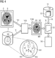
In Schritt V erfolgt eine Bereitstellung einer Klassifikationseinheit
In Schritt
Es erfolgt in Schritt VII eine Klassifizierung von Materialien
In Schritt
In step
Bis zu diesem Schritt wäre ein beispielhaftes Verfahren zur reinen Klassifikation von Materialien dargestellt. Mit der automatischen Klassifikation von Materialien
In dem folgenden (optionalen) Schritt
In Schritt
In Schritt
Das Computertomographiesystem
Ebenso sind bei der Steuereinrichtung
Ebenso kann die Erfindung auch an beliebigen anderen medizintechnischen bildgebenden Systemen genutzt werden.The invention can also be used in any other medical imaging systems.
Eine Kernkomponente der Steuereinrichtung
Über eine Steuerschnittstelle
Weiterhin weist die Steuereinrichtung
Eine Komponente auf dem Prozessor
In dem Fall, dass festgestellt wurde, dass ein klassifiziertes Material
Es wird abschließend noch einmal darauf hingewiesen, dass es sich bei den vorhergehend detailliert beschriebenen Verfahren sowie bei den dargestellten Vorrichtungen lediglich um Ausführungsbeispiele handelt, welche vom Fachmann in verschiedenster Weise modifiziert werden können, ohne den Bereich der Erfindung zu verlassen. Weiterhin schließt die Verwendung der unbestimmten Artikel „ein“ bzw. „eine“ nicht aus, dass die betreffenden Merkmale auch mehrfach vorhanden sein können. Ebenso schließen die Begriff „Einheit“ und „Modul“ nicht aus, dass die betreffenden Komponenten aus mehreren zusammenwirkenden Teil-Komponenten bestehen, die gegebenenfalls auch räumlich verteilt sein können.Finally, it is pointed out once again that the methods described in detail above and the devices shown are merely exemplary embodiments which can be modified in various ways by a person skilled in the art without departing from the scope of the invention. Furthermore, the use of the indefinite articles "a" or "an" does not exclude that the relevant features can also be present more than once. Likewise, the terms "unit" and "module" do not exclude that the components in question consist of several interacting sub-components, which may also be spatially distributed.
Claims (15)
Priority Applications (3)
| Application Number | Priority Date | Filing Date | Title |
|---|---|---|---|
| DE102017217543.5A DE102017217543B4 (en) | 2017-10-02 | 2017-10-02 | Method and system for classifying materials using machine learning |
| US16/133,889 US10824857B2 (en) | 2017-10-02 | 2018-09-18 | Method and system for the classification of materials by means of machine learning |
| CN201811162000.6A CN109598280B (en) | 2017-10-02 | 2018-09-30 | Method and system for classifying a plurality of materials by means of machine learning |
Applications Claiming Priority (1)
| Application Number | Priority Date | Filing Date | Title |
|---|---|---|---|
| DE102017217543.5A DE102017217543B4 (en) | 2017-10-02 | 2017-10-02 | Method and system for classifying materials using machine learning |
Publications (2)
| Publication Number | Publication Date |
|---|---|
| DE102017217543A1 DE102017217543A1 (en) | 2019-04-04 |
| DE102017217543B4 true DE102017217543B4 (en) | 2020-01-09 |
Family
ID=65727775
Family Applications (1)
| Application Number | Title | Priority Date | Filing Date |
|---|---|---|---|
| DE102017217543.5A Active DE102017217543B4 (en) | 2017-10-02 | 2017-10-02 | Method and system for classifying materials using machine learning |
Country Status (3)
| Country | Link |
|---|---|
| US (1) | US10824857B2 (en) |
| CN (1) | CN109598280B (en) |
| DE (1) | DE102017217543B4 (en) |
Families Citing this family (12)
| Publication number | Priority date | Publication date | Assignee | Title |
|---|---|---|---|---|
| US10572775B2 (en) * | 2017-12-05 | 2020-02-25 | X Development Llc | Learning and applying empirical knowledge of environments by robots |
| US10929973B2 (en) * | 2018-10-02 | 2021-02-23 | Siemens Healtcare Gmbh | Medical image pre-processing at the scanner for facilitating joint interpretation by radiologists and artificial intelligence algorithms |
| JP2022529624A (en) * | 2019-04-15 | 2022-06-23 | セキュリティ マターズ リミテッド | How and system to classify samples |
| DE102019111567A1 (en) | 2019-05-03 | 2020-11-05 | Wipotec Gmbh | Method and device for X-ray inspection of products, in particular food |
| JP7250331B2 (en) * | 2019-07-05 | 2023-04-03 | 株式会社イシダ | Image generation device, inspection device and learning device |
| CN114615946B (en) * | 2019-08-05 | 2024-05-17 | 捷锐士阿希迈公司(以奥林巴斯美国外科技术名义) | Signal coordinated delivery of laser therapy |
| US11195054B2 (en) | 2019-11-11 | 2021-12-07 | Sap Se | Automated determination of material identifiers for materials using machine learning models |
| DE102020200906B4 (en) * | 2020-01-27 | 2025-01-23 | Siemens Healthineers Ag | Control of a medical X-ray machine |
| JP7295333B2 (en) * | 2020-03-09 | 2023-06-20 | オリンパス株式会社 | IMAGE RECORDING SYSTEM, IMAGE RECORDING METHOD AND RAW IMAGE DATA CREATION PROGRAM |
| WO2021210618A1 (en) * | 2020-04-16 | 2021-10-21 | 浜松ホトニクス株式会社 | Radiographic image processing method, trained model, radiographic image processing module, radiographic image processing program, and radiographic image processing system |
| US20230125182A1 (en) * | 2020-04-16 | 2023-04-27 | Hamamatsu Photonics K.K. | Radiography method, trained model, radiography module, radiography program, radiography system, and machine learning method |
| TWI885189B (en) * | 2021-08-06 | 2025-06-01 | 日商濱松赫德尼古斯股份有限公司 | Radiographic image processing method, learning completion model, radiographic image processing module, radiographic image processing program, radiographic image processing system, and machine learning method |
Citations (2)
| Publication number | Priority date | Publication date | Assignee | Title |
|---|---|---|---|---|
| US20110188715A1 (en) | 2010-02-01 | 2011-08-04 | Microsoft Corporation | Automatic Identification of Image Features |
| US20160123904A1 (en) | 2014-11-04 | 2016-05-05 | Kabushiki Kaisha Toshiba | Method of, and apparatus for, material classification in multi-energy image data |
Family Cites Families (4)
| Publication number | Priority date | Publication date | Assignee | Title |
|---|---|---|---|---|
| US7295691B2 (en) | 2002-05-15 | 2007-11-13 | Ge Medical Systems Global Technology Company, Llc | Computer aided diagnosis of an image set |
| US9916538B2 (en) * | 2012-09-15 | 2018-03-13 | Z Advanced Computing, Inc. | Method and system for feature detection |
| CN104751429B (en) | 2015-01-27 | 2018-02-02 | 南方医科大学 | A kind of low dosage power spectrum CT image processing methods based on dictionary learning |
| US10037601B1 (en) * | 2017-02-02 | 2018-07-31 | International Business Machines Corporation | Systems and methods for automatic detection of architectural distortion in two dimensional mammographic images |
-
2017
- 2017-10-02 DE DE102017217543.5A patent/DE102017217543B4/en active Active
-
2018
- 2018-09-18 US US16/133,889 patent/US10824857B2/en active Active
- 2018-09-30 CN CN201811162000.6A patent/CN109598280B/en active Active
Patent Citations (2)
| Publication number | Priority date | Publication date | Assignee | Title |
|---|---|---|---|---|
| US20110188715A1 (en) | 2010-02-01 | 2011-08-04 | Microsoft Corporation | Automatic Identification of Image Features |
| US20160123904A1 (en) | 2014-11-04 | 2016-05-05 | Kabushiki Kaisha Toshiba | Method of, and apparatus for, material classification in multi-energy image data |
Also Published As
| Publication number | Publication date |
|---|---|
| US20190102621A1 (en) | 2019-04-04 |
| CN109598280B (en) | 2021-03-23 |
| DE102017217543A1 (en) | 2019-04-04 |
| CN109598280A (en) | 2019-04-09 |
| US10824857B2 (en) | 2020-11-03 |
Similar Documents
| Publication | Publication Date | Title |
|---|---|---|
| DE102017217543B4 (en) | Method and system for classifying materials using machine learning | |
| DE102017219307B4 (en) | Method and system for compensating motion artifacts by machine learning | |
| DE102016221684B4 (en) | Method and image data processing device for processing a multi-energy computed tomography image data set with basic material decomposition and image value tuple mapping | |
| DE10347971B3 (en) | Method and device for determining the liquid type of a liquid accumulation in an object | |
| DE102015226400A1 (en) | Automated determination of contours based on an iterative reconstruction | |
| DE102016204226A1 (en) | Apparatus and method for demarcating a metal object for artifact reduction in tomographic images | |
| DE102015207107A1 (en) | Method for generating a virtual X-ray projection on the basis of an image data set obtained by means of an X-ray image recording device, computer program, data carrier and X-ray image recording device | |
| DE102004043889B4 (en) | Method for generating a nuclear medical image | |
| WO2013098074A2 (en) | Control method and control system | |
| DE102020212089B3 (en) | Method and device for image de-noise as well as a control device, an imaging system, a computer program product and a computer-readable medium for this | |
| EP1325471A2 (en) | Method and device for representing an object by means of an irradiation, and for reconstructing said object | |
| DE102012212774B4 (en) | Procedure for correcting metal artifacts and X-ray equipment | |
| EP3379487A1 (en) | Contrast enhanced reproduction of spectral ct image data | |
| DE102012100067A1 (en) | Method and system for improved medical image analysis | |
| DE102016222093A1 (en) | Simultaneous use of different contrast agents in CT imaging procedures | |
| EP3552547B1 (en) | Method for providing conversion information about an imaging data set, x-ray device, computer program and electronically readable data carrier | |
| DE102020216306A1 (en) | Computer-implemented method for operating a medical imaging device, imaging device, computer program and electronically readable data carrier | |
| DE102019218589A1 (en) | Simultaneous image display of two different functional areas | |
| EP3384848B1 (en) | Flexible fitting of a clinical decision-making support system | |
| DE102008045633A1 (en) | Computer tomography (CT) method for improved display of multi-energy CT exposures/photographs applies contrast media along a system axis | |
| EP4033978B1 (en) | Contrast agent-based vascular imaging | |
| EP3637365A1 (en) | Method for rating a reliability of a ct volume image, computer program and computer readable storage medium | |
| DE102022211162A1 (en) | Providing a 3D image dataset | |
| DE102009015772A1 (en) | Method for reconstructing data sets of computerized tomography-image of e.g. human, from projection data sets, involves tomographically representing determined partial density of examination object | |
| EP3664036B1 (en) | Method and system for calculating an output of a scrollable stack of images provided by a tomographer |
Legal Events
| Date | Code | Title | Description |
|---|---|---|---|
| R012 | Request for examination validly filed | ||
| R016 | Response to examination communication | ||
| R018 | Grant decision by examination section/examining division | ||
| R020 | Patent grant now final | ||
| R081 | Change of applicant/patentee |
Owner name: SIEMENS HEALTHINEERS AG, DE Free format text: FORMER OWNER: SIEMENS HEALTHCARE GMBH, MUENCHEN, DE |