DE102012205629A1 - Method and magnetic resonance system for functional MR imaging of a predetermined volume portion of a brain of a living examination subject - Google Patents
Method and magnetic resonance system for functional MR imaging of a predetermined volume portion of a brain of a living examination subject Download PDFInfo
- Publication number
- DE102012205629A1 DE102012205629A1 DE102012205629A DE102012205629A DE102012205629A1 DE 102012205629 A1 DE102012205629 A1 DE 102012205629A1 DE 102012205629 A DE102012205629 A DE 102012205629A DE 102012205629 A DE102012205629 A DE 102012205629A DE 102012205629 A1 DE102012205629 A1 DE 102012205629A1
- Authority
- DE
- Germany
- Prior art keywords
- data
- frequency
- eeg data
- predetermined
- class
- Prior art date
- Legal status (The legal status is an assumption and is not a legal conclusion. Google has not performed a legal analysis and makes no representation as to the accuracy of the status listed.)
- Withdrawn
Links
Images


Classifications
-
- A—HUMAN NECESSITIES
- A61—MEDICAL OR VETERINARY SCIENCE; HYGIENE
- A61B—DIAGNOSIS; SURGERY; IDENTIFICATION
- A61B5/00—Measuring for diagnostic purposes; Identification of persons
- A61B5/24—Detecting, measuring or recording bioelectric or biomagnetic signals of the body or parts thereof
- A61B5/316—Modalities, i.e. specific diagnostic methods
-
- A—HUMAN NECESSITIES
- A61—MEDICAL OR VETERINARY SCIENCE; HYGIENE
- A61B—DIAGNOSIS; SURGERY; IDENTIFICATION
- A61B5/00—Measuring for diagnostic purposes; Identification of persons
- A61B5/0033—Features or image-related aspects of imaging apparatus classified in A61B5/00, e.g. for MRI, optical tomography or impedance tomography apparatus; arrangements of imaging apparatus in a room
- A61B5/004—Features or image-related aspects of imaging apparatus classified in A61B5/00, e.g. for MRI, optical tomography or impedance tomography apparatus; arrangements of imaging apparatus in a room adapted for image acquisition of a particular organ or body part
- A61B5/0042—Features or image-related aspects of imaging apparatus classified in A61B5/00, e.g. for MRI, optical tomography or impedance tomography apparatus; arrangements of imaging apparatus in a room adapted for image acquisition of a particular organ or body part for the brain
-
- A—HUMAN NECESSITIES
- A61—MEDICAL OR VETERINARY SCIENCE; HYGIENE
- A61B—DIAGNOSIS; SURGERY; IDENTIFICATION
- A61B5/00—Measuring for diagnostic purposes; Identification of persons
- A61B5/05—Detecting, measuring or recording for diagnosis by means of electric currents or magnetic fields; Measuring using microwaves or radio waves
- A61B5/055—Detecting, measuring or recording for diagnosis by means of electric currents or magnetic fields; Measuring using microwaves or radio waves involving electronic [EMR] or nuclear [NMR] magnetic resonance, e.g. magnetic resonance imaging
-
- A—HUMAN NECESSITIES
- A61—MEDICAL OR VETERINARY SCIENCE; HYGIENE
- A61B—DIAGNOSIS; SURGERY; IDENTIFICATION
- A61B5/00—Measuring for diagnostic purposes; Identification of persons
- A61B5/24—Detecting, measuring or recording bioelectric or biomagnetic signals of the body or parts thereof
- A61B5/316—Modalities, i.e. specific diagnostic methods
- A61B5/369—Electroencephalography [EEG]
- A61B5/372—Analysis of electroencephalograms
- A61B5/374—Detecting the frequency distribution of signals, e.g. detecting delta, theta, alpha, beta or gamma waves
-
- A—HUMAN NECESSITIES
- A61—MEDICAL OR VETERINARY SCIENCE; HYGIENE
- A61B—DIAGNOSIS; SURGERY; IDENTIFICATION
- A61B5/00—Measuring for diagnostic purposes; Identification of persons
- A61B5/72—Signal processing specially adapted for physiological signals or for diagnostic purposes
- A61B5/7235—Details of waveform analysis
- A61B5/7264—Classification of physiological signals or data, e.g. using neural networks, statistical classifiers, expert systems or fuzzy systems
-
- G—PHYSICS
- G01—MEASURING; TESTING
- G01R—MEASURING ELECTRIC VARIABLES; MEASURING MAGNETIC VARIABLES
- G01R33/00—Arrangements or instruments for measuring magnetic variables
- G01R33/20—Arrangements or instruments for measuring magnetic variables involving magnetic resonance
- G01R33/44—Arrangements or instruments for measuring magnetic variables involving magnetic resonance using nuclear magnetic resonance [NMR]
- G01R33/48—NMR imaging systems
- G01R33/54—Signal processing systems, e.g. using pulse sequences ; Generation or control of pulse sequences; Operator console
- G01R33/56—Image enhancement or correction, e.g. subtraction or averaging techniques, e.g. improvement of signal-to-noise ratio and resolution
- G01R33/5608—Data processing and visualization specially adapted for MR, e.g. for feature analysis and pattern recognition on the basis of measured MR data, segmentation of measured MR data, edge contour detection on the basis of measured MR data, for enhancing measured MR data in terms of signal-to-noise ratio by means of noise filtering or apodization, for enhancing measured MR data in terms of resolution by means for deblurring, windowing, zero filling, or generation of gray-scaled images, colour-coded images or images displaying vectors instead of pixels
-
- G—PHYSICS
- G16—INFORMATION AND COMMUNICATION TECHNOLOGY [ICT] SPECIALLY ADAPTED FOR SPECIFIC APPLICATION FIELDS
- G16H—HEALTHCARE INFORMATICS, i.e. INFORMATION AND COMMUNICATION TECHNOLOGY [ICT] SPECIALLY ADAPTED FOR THE HANDLING OR PROCESSING OF MEDICAL OR HEALTHCARE DATA
- G16H50/00—ICT specially adapted for medical diagnosis, medical simulation or medical data mining; ICT specially adapted for detecting, monitoring or modelling epidemics or pandemics
- G16H50/20—ICT specially adapted for medical diagnosis, medical simulation or medical data mining; ICT specially adapted for detecting, monitoring or modelling epidemics or pandemics for computer-aided diagnosis, e.g. based on medical expert systems
Abstract
Die Erfindung betrifft ein Verfahren und eine Magnetresonanzanlage (5) zur funktionalen MR-Bildgebung eines vorbestimmten Volumenabschnitts eines Gehirns eines lebenden Untersuchungsobjekts (O). Dabei werden folgende Schritte durchgeführt: Erfassen von MR-Daten (25) des vorbestimmten Volumenabschnitts. Erfassen von EEG-Daten (26) des Untersuchungsobjekts (O), wobei die Erfassung der EEG-Daten (26) gleichzeitig mit der Erfassung der MR-Daten (25) erfolgt. Auswerten der MR-Daten unter Berücksichtigung von den erfassten EEG-Daten (26).The invention relates to a method and a magnetic resonance system (5) for the functional MR imaging of a predetermined volume section of a brain of a living examination subject (O). The following steps are performed: acquisition of MR data (25) of the predetermined volume section. Acquiring EEG data (26) of the examination object (O), wherein the detection of the EEG data (26) occurs simultaneously with the acquisition of the MR data (25). Evaluation of the MR data taking into account the recorded EEG data (26).
Description
Die vorliegende Erfindung betrifft ein Verfahren und eine Magnetresonanzanlage zur funktionellen MR-Bildgebung (fMRI), bei welchem MR-Aufnahmen des Gehirns eines lebenden Untersuchungsobjekts (insbesondere eines Menschen) erstellt werden. The present invention relates to a method and a magnetic resonance system for functional MR imaging (fMRI), in which MR images of the brain of a living examination subject (in particular of a human being) are produced.
"Resting state fMRI" ist ein MR-Verfahren, bei welchem MR-Aufnahmen eines Patienten in Ruhe erstellt werden. Bei diesen MR-Aufnahmen wird, wie bei der klassischen fMRI, eine Signaländerung über den so genannten BOLD-Effekt ("Blood Oxygen Level Dependent") bestimmt, welcher ein Maß für die physiologische Aktivierung bestimmter Gehirnareale darstellt. "Resting state fMRI" is an MR procedure in which MR images of a patient are created at rest. In these MR images, as with the classical fMRI, a signal change via the so-called BOLD effect ("Blood Oxygen Level Dependent") is determined, which is a measure of the physiological activation of certain brain areas.
Im Gegensatz zur klassischen fMRI, bei welcher der Patient bestimmten Reizen ausgesetzt wird oder bei welcher dem Patienten bestimmte Aufgaben gestellt werden, werden bei der resting state fMRI die MR-Aufnahmen in Ruhe erstellt. Dabei zeigt sich eine zeitliche Korrelation der Aktivierung bestimmter Gehirnzentren, welche durch ein Ausmaß der Vernetzung dieser Zentren bestimmt wird, wodurch wiederum relevante diagnostische Informationen, beispielsweise über psychische Erkrankungen, gewonnen werden können. In contrast to the classic fMRI, in which the patient is exposed to certain stimuli or in which the patient is given specific tasks, the MR images are taken at rest at the resting state fMRI. This shows a temporal correlation of the activation of certain brain centers, which is determined by a degree of networking of these centers, which in turn can be obtained relevant diagnostic information, for example about mental illness.
Die MR-Messungen in Kombination mit morphologischen MR-Aufnahmen für resting state fMRI können 15 Minuten oder länger dauern. Dabei besteht die Gefahr, dass sich der Aktivierungszustand des Patienten ändert, beispielsweise da dieser einschläft, was negativerweise zu unrelevanten Aktivierungsmustern führt und die Ergebnisse verfälscht oder sogar falsche Diagnosen vortäuscht. The MR measurements in combination with morphological MR images for resting state fMRI may take 15 minutes or more. There is a risk that the activation state of the patient changes, for example, because he falls asleep, which leads to negative unrealistic activation patterns and falsifies the results or even pretends false diagnoses.
Daher stellt sich die vorliegende Erfindung die Aufgabe, diese Probleme nach dem Stand der Technik zumindest abzumildern. Therefore, the present invention has the object to at least mitigate these problems in the prior art.
Erfindungsgemäß wird diese Aufgabe durch ein Verfahren zur funktionalen MR-Bildgebung nach Anspruch 1, durch eine Magnetresonanzanlage nach Anspruch 11, durch ein Computerprogrammprodukt nach Anspruch 12 und durch einen elektronisch lesbaren Datenträger nach Anspruch 13 gelöst. Die abhängigen Ansprüche definieren bevorzugte und vorteilhafte Ausführungsformen der vorliegenden Erfindung. According to the invention, this object is achieved by a method for functional MR imaging according to
Erfindungsgemäß wird ein Verfahren zur funktionalen MR-Bildgebung eines vorbestimmten Volumenabschnitts eines Gehirns eines lebenden Untersuchungsobjekts bereitgestellt. Dabei umfasst das Verfahren folgende Schritte:
- • Erfassen von MR-Daten des vorbestimmten Volumenabschnitts.
- • Erfassen von EEG-Daten des Untersuchungsobjekts, wobei die Erfassung der EEG-Daten und die Erfassung der MR-Daten gleichzeitig erfolgt.
- • Auswerten der MR-Daten in Abhängigkeit von den erfassten EEG-Daten.
- • acquire MR data of the predetermined volume section.
- • Acquire EEG data of the examination subject, with simultaneous acquisition of the EEG data and acquisition of the MR data.
- • Evaluation of the MR data as a function of the recorded EEG data.
Durch die gleichzeitige Erfassung der MR-Daten und der EEG-Daten ist es möglich, anhand der EEG-Daten zu überprüfen, ob der gewünschte Aktivierungszustand des Patienten während der Erfassung der MR-Daten vorliegt. Dadurch ist es vorteilhafterweise möglich, die MR-Daten abhängig vom jeweils mittels der EEG-Daten festgestellten Aktivierungszustand auszuwerten oder nur diejenigen MR-Daten auszuwerten, welche während des gewünschten Aktivierungszustandes des Patienten erfasst wurden. By simultaneously recording the MR data and the EEG data, it is possible to check on the basis of the EEG data whether the desired activation state of the patient is present during the acquisition of the MR data. As a result, it is advantageously possible to evaluate the MR data as a function of the activation state determined in each case by means of the EEG data or to evaluate only those MR data which were acquired during the desired activation state of the patient.
Dabei kann eine spektrale Analyse der EEG-Daten durchgeführt werden, indem beispielsweise das Frequenzspektrum der erfassten EEG-Daten erstellt wird. Die Auswertung der MR-Daten kann dann abhängig von der spektralen Analyse oder abhängig von dem erfassten Frequenzspektrum erfolgen. In this case, a spectral analysis of the EEG data can be performed by, for example, the frequency spectrum of the acquired EEG data is created. The evaluation of the MR data can then take place depending on the spectral analysis or depending on the detected frequency spectrum.
Anhand der spektralen Analyse oder anhand des Frequenzspektrums kann der aktuelle Aktivierungszustand des Patienten ermittelt werden. Da die Auswertung der MR-Daten abhängig von der Spektralenanalyse oder abhängig von dem erfassten Frequenzspektrum erfolgt, können dann beispielsweise nur diejenigen MR-Daten ausgewertet werden, welche erfasst wurden, während der Patient einen erwünschten Aktivierungszustand aufwies. Based on the spectral analysis or on the basis of the frequency spectrum, the current activation state of the patient can be determined. Since the evaluation of the MR data takes place as a function of the spectral analysis or as a function of the detected frequency spectrum, then, for example, only those MR data can be evaluated which were acquired while the patient had a desired activation state.
Gemäß einer bevorzugten erfindungsgemäßen Ausführungsform erfolgt das gleichzeitige Erfassen der MR-Daten und der EEG-Daten in mehreren aufeinanderfolgenden Zeitintervallen oder Zeitscheiben. Dabei wird für jede dieser Zeitscheiben ein Frequenzspektrum der während dieser Zeitscheibe erfassten EEG-Daten bestimmt. Abhängig von dem für die jeweilige Zeitscheibe bestimmten Frequenzspektrum wird für die jeweilige Zeitscheibe eine Klasse bestimmt. Dieser Klasse werden dann auch die während der jeweiligen Zeitscheibe erfassten MR-Daten zugeordnet, so dass die während der mehreren Zeitscheiben erfassten MR-Daten unterschiedlichen Klassen zugeordnet werden. Zur Auswertung der MR-Daten werden die MR-Daten einer bestimmten Klasse abhängig von dieser Klasse ausgewertet, so dass die MR-Daten einer bestimmten Klasse anders ausgewertet werden als die MR-Daten einer anderen bestimmten Klasse. According to a preferred embodiment of the invention, the simultaneous acquisition of the MR data and the EEG data takes place in a plurality of successive time intervals or time slices. In each case, a frequency spectrum of the EEG data acquired during this time slice is determined for each of these time slices. Depending on the frequency spectrum determined for the respective time slice, a class is determined for the respective time slice. This class then also the MR data acquired during the respective time slice are assigned, so that the MR data acquired during the multiple time slices are assigned to different classes. For the evaluation of the MR data, the MR data of a specific class are evaluated as a function of this class so that the MR data of one particular class are evaluated differently than the MR data of another specific class.
Gemäß einer ersten Variante dieser bevorzugten Ausführungsform wird das Frequenzspektrum, welches die EEG-Daten aufweisen können, in eine vorbestimmte Anzahl von Frequenzbändern unterteilt. Ein Beispiel für diese Unterteilung ist die Unterteilung des Frequenzbandes in Delta-Wellen, Theta-Wellen, Alpha-Wellen, Beta-Wellen und Gamma-Wellen. Die Anzahl der Klassen entspricht dabei der Anzahl der Frequenzbänder, so dass jede Klasse einem dieser Frequenzbänder entspricht. Gemäß dieser ersten Variante wird bestimmt, in welchem dieser Frequenzbänder die EEG-Daten überwiegend liegen. Die diesem Frequenzband entsprechende Klasse ist dann auch die Klasse der jeweiligen Zeitscheibe, so dass die MR-Daten, welche während dieser Zeitscheibe erfasst wurden, dieser Klasse zugeordnet werden. According to a first variant of this preferred embodiment, the frequency spectrum, which may comprise the EEG data, in divided a predetermined number of frequency bands. An example of this subdivision is the subdivision of the frequency band into delta waves, theta waves, alpha waves, beta waves and gamma waves. The number of classes corresponds to the number of frequency bands, so that each class corresponds to one of these frequency bands. According to this first variant, it is determined in which of these frequency bands the EEG data predominantly lie. The class corresponding to this frequency band is then also the class of the respective time slice, so that the MR data acquired during this time slice is assigned to this class.
Mit anderen Worten wird für jede Zeitscheibe bestimmt, in welchem Frequenzband oder in welcher Frequenzklasse der größte Anteil des Frequenzspektrums der EEG-Wellen, welche während dieser Zeitscheibe erfasst wurden, liegen. Dieser Frequenzklasse werden dann auch die während dieser Zeitscheibe erfassten MR-Daten zugewiesen. Am Ende der erfindungsgemäßen MR-Messung existiert dann eine Anzahl von Datensätzen von MR-Daten, wobei die Anzahl dieser Datensätze von MR-Daten der Anzahl der Frequenzbänder oder Frequenzklassen entspricht, sofern für jede Frequenzklasse MR-Daten erfasst wurden (d.h. die Anzahl der Datensätze einer Frequenzklasse oder Klasse kann auch Null sein). In other words, it is determined for each time slice in which frequency band or in which frequency class the largest proportion of the frequency spectrum of the EEG waves which were acquired during this time slice is located. This frequency class is then also assigned the MR data acquired during this time slice. At the end of the MR measurement according to the invention, there then exists a number of data sets of MR data, the number of these data sets of MR data corresponding to the number of frequency bands or frequency classes, if MR data were acquired for each frequency class (ie the number of data records a frequency class or class can also be zero).
Wenn beispielsweise eine der Klassen (Frequenzklassen) der klassischen Alpha-Wellen-Frequenzklasse entspricht, existiert am Ende des erfindungsgemäßen Verfahrens ein Datensatz von MR-Daten, welche während denjenigen Zeitscheiben erfasst wurden, während welchen die EEG-Daten oder EEG-Wellen des Untersuchungsobjekts überwiegend so genannten Alpha-Wellen entsprachen. Dadurch ist es möglich, zur Auswertung nur die MR-Daten dieser Alpha-Wellen-Frequenzklasse heranzuziehen, und die anderen MR-Daten zu verwerfen. If, for example, one of the classes (frequency classes) corresponds to the classical alpha wave frequency class, at the end of the method according to the invention there is a data set of MR data acquired during those time slices during which the EEG data or EEG waves of the examination subject predominate so-called alpha waves corresponded. This makes it possible to use for evaluation only the MR data of this alpha wave frequency class, and to discard the other MR data.
Dadurch ist es vorteilhafterweise möglich, nur diejenigen MR-Daten auszuwerten, welche in einem Zeitraum erfasst wurden, während welchem das Untersuchungsobjekt einen vorbestimmten erwünschten Aktivierungszustand aufwies. Eine Verfälschung der MR-Daten durch die Aufnahme von MR-Daten während eines unerwünschten Aktivierungszustands kann dadurch nahezu ausgeschlossen werden. As a result, it is advantageously possible to evaluate only those MR data which were acquired in a period during which the examination object had a predetermined desired activation state. Falsification of the MR data by the acquisition of MR data during an undesired activation state can thus be almost ruled out.
Gemäß einer zweiten Variante der bevorzugten Ausführungsform wird wiederum das Frequenzspektrum, welches die EEG-Daten aufweisen, in eine vorbestimmte Anzahl von Frequenzbändern unterteilt. Auch dabei kann diese Unterteilung wieder der klassischen Unterteilung in die Frequenzbänder oder Frequenzklassen Alpha, Beta, Gamma, Delta, Theta entsprechen. Wie bei der ersten Variante existiert auch bei der zweiten Variante eine Anzahl von vorbestimmten Klassen, wobei die Anzahl der vorbestimmten Klassen bei der zweiten Variante nicht der Anzahl der Frequenzklassen entsprechen muss. Bei der zweiten Variante ist jede vorbestimmte Klasse durch Frequenzanteile der EEG-Daten innerhalb der definierten Frequenzbänder definiert. Mit anderen Worten ist jede vorbestimmte Klasse durch einen Frequenzanteil innerhalb des ersten Frequenzbandes, durch einen Frequenzanteil innerhalb des zweiten Frequenzbandes, ..., und durch einen Frequenzanteil innerhalb des letzten der vordefinierten Frequenzbänder definiert. Um nun die innerhalb einer bestimmten Zeitscheibe erfassten EEG-Daten einer dieser vorbestimmten Klassen zuzuweisen, werden die Frequenzanteile der erfassten EEG-Daten innerhalb der vorbestimmten Frequenzbänder ermittelt. Die Klasse der Zeitscheibe entspricht dann derjenigen der vorbestimmten Klassen, bei welcher die vordefinierten Frequenzanteile am besten den Frequenzanteilen der erfassten EEG-Daten entsprechen. According to a second variant of the preferred embodiment, in turn, the frequency spectrum comprising the EEG data is divided into a predetermined number of frequency bands. Again, this subdivision may again correspond to the classical subdivision into the frequency bands or frequency classes alpha, beta, gamma, delta, theta. As in the first variant, there are also a number of predetermined classes in the second variant, wherein the number of predetermined classes in the second variant does not have to correspond to the number of frequency classes. In the second variant, each predetermined class is defined by frequency components of the EEG data within the defined frequency bands. In other words, each predetermined class is defined by a frequency component within the first frequency band, by a frequency component within the second frequency band,..., And by a frequency component within the last of the predefined frequency bands. In order to assign the EEG data acquired within a certain time slice to one of these predetermined classes, the frequency components of the detected EEG data are determined within the predetermined frequency bands. The class of the time slice then corresponds to that of the predetermined classes, in which the predefined frequency components best correspond to the frequency components of the acquired EEG data.
Um dies zu ermitteln, kann beispielsweise für jede dieser vorbestimmten Klassen für jedes der definierten Frequenzbänder ein Sollwert bestimmt werden. Dann können für jede Klasse für jedes Frequenzband die Differenzen zwischen dem Frequenzanteil der erfassten EEG-Wellen in diesem Frequenzband und dem Sollwert dieses Frequenzbands dieser Klasse bestimmt werden. Die jeweilige Zeitscheibe wird derjenigen Klasse zugeordnet, bei welcher diese Differenzen am geringsten sind. Dazu kann beispielsweise für jede vorbestimmte Klasse die Summe der Beträge der Differenzen zwischen dem Frequenzanteil der erfassten EEG-Wellen in dem jeweiligen Frequenzband und dem Sollwert dieser Klasse für dieses Frequenzband bestimmt werden. Diejenige vorbestimmte Klasse, bei welcher diese Summe am kleinsten ist, wird dann der jeweiligen Zeitscheibe als Klasse zugewiesen. In order to determine this, a desired value can be determined, for example, for each of these predetermined classes for each of the defined frequency bands. Then, for each class for each frequency band, the differences between the frequency component of the detected EEG waves in that frequency band and the reference value of that frequency band of that class can be determined. The respective time slice is assigned to the class in which these differences are the lowest. For this purpose, for example, the sum of the amounts of the differences between the frequency component of the detected EEG waves in the respective frequency band and the desired value of this class for this frequency band can be determined for each predetermined class. The predetermined class at which this sum is smallest is then assigned to the respective time slice as a class.
Bei dieser zweiten Variante können EEG-Daten und damit die MR-Daten einer Zeitscheibe nach im Vergleich zur ersten Variante komplizierteren Schemata aufgeteilt werden. Dadurch können durch die Auswertung der EEG-Daten auch kompliziertere Aktivierungszustände (beispielsweise ein durch visuelle Reize hervorgerufener Aktivierungszustand, ein durch hörbare Reize hervorgerufener Aktivierungszustand oder ein Aktivierungszustand, bei welchem keine äußeren Reize vorliegen (resting state)) unterschieden werden und die erfassten MR-Daten in entsprechende Klassen unterteilt werden. Indem beispielsweise nur die MR-Daten, welche während eines 'resting state'-Aktivierungszustands erfasst wurden, ausgewertet werden, kann die Aktivität verschiedener Funktionsnetze im Gehirn bei diesem 'resting state'-Aktivierungszustand erfasst und dargestellt werden. Mit anderen Worten können die bei unterschiedlichen Aktivierungszuständen erfassten MR-Daten getrennt ausgewertet werden, um die Aktivität verschiedener Funktionsnetze (jeder Aktivierungszustand weist sein eigenes Funktionsnetz auf) gesondert zu erfassen. In this second variant, EEG data and thus the MR data of a time slice can be divided into more complicated schemas compared to the first variant. As a result, the evaluation of the EEG data can also be used to distinguish more complicated activation states (for example an activation state caused by visual stimuli, an activation state caused by audible stimuli or an activation state in which there are no external stimuli) and the acquired MR data be divided into appropriate classes. For example, by only evaluating the MR data acquired during a resting state activation state, the activity of various functional networks in the brain can be detected and displayed in this resting state activation state. In other words, they can be at different activation states recorded MR data are separately evaluated to separately capture the activity of different functional networks (each activation state has its own functional network).
Die Auswertung der MR-Daten umfasst dabei insbesondere die Erstellung von morphologischen MR-Bildern, auf welchen während der Erfassung der MR-Daten aktive Gehirnzentren des Untersuchungsobjekts als solche erkennbar dargestellt werden. The evaluation of the MR data comprises, in particular, the generation of morphological MR images on which active brain centers of the examination subject are displayed as such during the acquisition of the MR data.
Gemäß einer weiteren erfindungsgemäßen Ausführungsform werden die MR-Daten und die EEG-Daten in mehreren aufeinanderfolgenden Zeitintervallen erfasst. Dabei wird für jedes Zeitinterval entschieden, ob das Frequenzspektrum der in diesem Zeitinterval erfassten EEG-Daten überwiegend in einem erwünschten Frequenzband, welches vorher festgelegt wurde, liegt. Nur wenn dies der Fall ist, werden die MR-Daten des entsprechenden Zeitintervalls ausgewertet, sonst werden diese MR-Daten verworfen. Erst wenn die Summe der Zeitintervale, in welchen die MR-Daten der Auswertung zugeführt wurden (d.h. das Frequenzspektrum der in diesem Zeitinterval erfassten EEG-Daten lag überwiegend in dem erwünschten Frequenzband), größer als ein vorbestimmtes Zeitinterval ist, endet das Verfahren. According to a further embodiment of the invention, the MR data and the EEG data are acquired in several successive time intervals. In this case, it is decided for each time interval whether the frequency spectrum of the EEG data acquired in this time interval lies predominantly in a desired frequency band which was previously determined. Only if this is the case, the MR data of the corresponding time interval are evaluated, otherwise these MR data are discarded. Only when the sum of the time intervals in which the MR data has been fed to the evaluation (i.e., the frequency spectrum of the EEG data acquired in this time interval was predominantly in the desired frequency band) is greater than a predetermined time interval does the method end.
Diese Ausführungsform garantiert, dass insgesamt entsprechend der Dauer des vorbestimmten Zeitintervalls MR-Daten erfasst werden, wobei das Untersuchungsobjekts während der Erfassung dieser MR-Daten einen erwünschten Aktivierungszustand aufweist, welcher durch das Frequenzspektrum der erfassten EEG-Daten charakterisiert ist. This embodiment guarantees that, overall, MR data is acquired in accordance with the duration of the predetermined time interval, the examination object having a desired activation state during the acquisition of this MR data, which is characterized by the frequency spectrum of the acquired EEG data.
Erfindungsgemäß ist es auch möglich, dass abhängig von einem Frequenzspektrum der erfassten EEG-Daten eine Benutzerinformation ausgegeben wird. According to the invention, it is also possible for user information to be output as a function of a frequency spectrum of the recorded EEG data.
Dadurch kann der Bediener der Magnetresonanzanlage beispielsweise gewarnt werden, falls über einen bestimmten Zeitraum hinweg keine verwertbaren MR-Daten erzeugt bzw. erfasst werden konnten. Beispielsweise könnte der Bediener der Magnetresonanzanlage gewarnt werden, wenn eine bestimmte Zeitspanne lang keine Alpha-Wellen des Untersuchungsobjekts erfasst werden, was bedeutet, dass innerhalb der bestimmten Zeitspanne keine Zeitscheibe lag, in welcher der Frequenzanteil der EEG-Daten überwiegend in dem alpha-Frequenzband lag. As a result, the operator of the magnetic resonance system can be warned, for example, if no usable MR data could be generated or recorded over a certain period of time. For example, the operator of the magnetic resonance system could be warned if no alpha waves of the examination subject are detected for a certain period of time, which means that there was no time slice within the particular time period in which the frequency component of the EEG data was predominantly in the alpha frequency band ,
Mittels der Benutzerinformation kann erfindungsgemäß auch das Untersuchungsobjekt bzw. der Patient direkt informiert werden. Beispielsweise könnte eine entsprechende Benutzerinformation erzeugt werden, wenn ein vorbestimmtes Zeitinterval lang das Frequenzspektrum der erfassten EEG-Wellen hauptsächlich Delta-Wellen aufweist, was darauf hindeutet, dass der Patient eingeschlafen ist. In diesem Fall kann die entsprechende Benutzerinformation dazu verwendet werden, beispielsweise ein Geräusch über einen Kopfhörer, welcher von dem Patienten getragen wird, einzuspielen, um den Patienten zu wecken. Wenn dagegen im Frequenzspektrum der erfassten EEG-Wellen hauptsächlich Gamma-Wellen festgestellt werden, könnte der Patient durch die entsprechende Benutzerinformation aufgefordert werden, sich zu entspannen. Auch das Öffnen oder Schließen der Augen kann über eine entsprechende Benutzerinformation angeregt werden, wenn das Frequenzspektrum der erfassten EEG-Wellen überwiegend im Alpha- bzw. Beta-Frequenzband liegt. By means of the user information, according to the invention, the examination object or the patient can also be informed directly. For example, corresponding user information could be generated when the frequency spectrum of the detected EEG waves has mainly delta waves for a predetermined time interval, indicating that the patient has fallen asleep. In this case, the corresponding user information may be used, for example, to record a sound via a headset worn by the patient in order to wake the patient. If, on the other hand, gamma waves are mainly detected in the frequency spectrum of the detected EEG waves, the patient could be prompted to relax by the corresponding user information. Also, the opening or closing of the eyes can be stimulated via appropriate user information, if the frequency spectrum of the detected EEG waves is predominantly in the alpha or beta frequency band.
Gemäß einer anderen erfindungsgemäßen Ausführungsform werden die EEG-Daten einer bestimmten Zeitspanne Tiefpass-gefiltert, so dass nur EEG-Daten, deren Frequenz unterhalb eines Frequenzschwellenwerts liegt, durch das entsprechend Tiefpassfilter gelassen werden. Wenn der Anteil der Tiefpassgefilterten EEG-Daten (d.h. der Anteil der EEG-Daten, deren Frequenz unterhalb des Frequenzschwellenwerts liegt) oberhalb eines vorbestimmten Anteilschwellenwerts liegt, werden die MR-Daten dieser Zeitspanne verworfen. Dabei ist es möglich, den Patienten in diesem Fall (wenn der Anteil der Tiefpassgefilterten EEG-Daten oberhalb des vorbestimmten Anteilsschwellenwerts liegt) zu wecken, da er wahrscheinlich eingeschlafen ist. According to another embodiment of the invention, the EEG data is low-pass filtered for a certain period of time so that only EEG data whose frequency is below a frequency threshold is passed through the corresponding low-pass filter. If the proportion of low-pass filtered EEG data (i.e., the proportion of EEG data whose frequency is below the frequency threshold) is above a predetermined fractional threshold, then the MR data of that period will be discarded. In this case, it is possible to wake the patient (if the proportion of low-pass filtered EEG data is above the predetermined fraction threshold), since he is likely to have fallen asleep.
Mit dieser sehr einfachen erfindungsgemäßen Ausführungsform werden die MR-Daten von Zeitspannen, in denen hauptsächlich Delta- oder Theta-Wellen (d.h. EEG-Daten mit einer Frequenz unterhalb von 8 Hz) vorliegen, aus den schließlich auszuwertenden MR-Daten vorteilhafterweise eliminiert. Durch die Tiefpassfilterung werden darüber hinaus vorteilhafterweise höhere Frequenzstörungen durch die Magnetresonanzanlage verhindert. With this very simple embodiment of the invention, the MR data of periods in which delta or theta waves (i.e., EEG data having a frequency below 8 Hz) are presently advantageously eliminated from the MR data finally to be evaluated. In addition, the low-pass filtering advantageously prevents higher frequency disturbances by the magnetic resonance system.
Im Rahmen der vorliegenden Erfindung wird auch eine Magnetresonanzanlage zur Erstellung eines MR-Bildes eines Untersuchungsobjekts bereitgestellt. Dabei umfasst die Magnetresonanzanlage einen Grundfeldmagneten, ein Gradientenfeldsystem, mindestens eine HF-Sendeantenne, mindestens ein Empfangsspulenelement, eine Steuereinrichtung und einen Elektroenzephalograph. Die Steuereinrichtung dient zur Ansteuerung des Gradientenfeldsystems und der mindestens einen HF-Sendeantenne. Darüber hinaus ist die Steuereinrichtung ausgestaltet, um Messsignale zu empfangen, welche von dem mindestens einen Empfangsspulenelement erfasst worden sind, und um diese erfassten Messsignale auszuwerten und entsprechende MR-Daten zu erstellen. Schließlich ist die Magnetresonanzanlage in der Lage, mittels des Elektroenzephalographen EEG-Daten gleichzeitig mit den MR-Daten zu erfassen Die Steuereinrichtung wertet die MR-Daten dann abhängig von den gleichzeitig erfassten EEG-Daten aus. Within the scope of the present invention, a magnetic resonance system is also provided for producing an MR image of an examination subject. In this case, the magnetic resonance system comprises a basic field magnet, a gradient field system, at least one RF transmitting antenna, at least one receiving coil element, a control device and an electroencephalograph. The control device serves to control the gradient field system and the at least one RF transmitting antenna. In addition, the control device is configured to receive measurement signals which have been detected by the at least one receiver coil element, and to evaluate these detected measurement signals and to generate corresponding MR data. Finally, the magnetic resonance system is capable of using the electroencephalograph EEG data simultaneously with the MR data The controller then evaluates the MR data based on the simultaneously acquired EEG data.
Die Vorteile der erfindungsgemäßen Magnetresonanzanlage entsprechen im Wesentlichen den Vorteilen des erfindungsgemäßen Verfahrens, welche vorab im Detail beschrieben worden sind, so dass hier auf eine Wiederholung verzichtet wird. The advantages of the magnetic resonance system according to the invention essentially correspond to the advantages of the method according to the invention, which have been described in detail in advance, so that a repetition is dispensed with here.
Des Weiteren beschreibt die vorliegende Erfindung ein Computerprogrammprodukt, insbesondere eine Software, welche man in einen Speicher einer programmierbaren Steuereinrichtung bzw. einer Recheneinheit einer Magnetresonanzanlage laden kann. Mit diesem Computerprogrammprodukt können alle oder verschiedene vorab beschriebene Ausführungsformen des erfindungsgemäßen Verfahrens ausgeführt werden, wenn das Computerprogrammprodukt in der Steuereinrichtung läuft. Dabei benötigt das Computerprogrammprodukt eventuell Programmmittel, z.B. Bibliotheken und Hilfsfunktionen, um die entsprechenden Ausführungsformen des Verfahrens zu realisieren. Mit anderen Worten soll mit dem auf das Computerprogrammprodukt gerichteten Anspruch insbesondere eine Software unter Schutz gestellt werden, mit welcher eine der oben beschriebenen Ausführungsformen des erfindungsgemäßen Verfahrens ausgeführt werden kann bzw. welche diese Ausführungsform ausführt. Dabei kann es sich bei der Software um einen Quellcode (z.B. C++), der noch compiliert und gebunden oder der nur interpretiert werden muss, oder um einen ausführbaren Softwarecode handeln, der zur Ausführung nur noch in die entsprechende Recheneinheit bzw. Steuereinrichtung zu laden ist. Furthermore, the present invention describes a computer program product, in particular a software, which can be loaded into a memory of a programmable control device or a computing unit of a magnetic resonance system. With this computer program product, all or various previously described embodiments of the method according to the invention can be carried out when the computer program product is running in the control device. The computer program product may require program resources, e.g. Libraries and utility functions to implement the corresponding embodiments of the method. In other words, with the claim directed to the computer program product, in particular a software with which one of the above-described embodiments of the method according to the invention can be executed or which executes this embodiment should be protected. In this case, the software may be a source code (for example C ++) which still has to be compiled and bound or which only has to be interpreted, or an executable software code which is only to be loaded into the corresponding arithmetic unit or control device for execution.
Schließlich offenbart die vorliegende Erfindung einen elektronisch lesbaren Datenträger, z.B. eine DVD, ein Magnetband oder einen USB-Stick, auf welchem elektronisch lesbare Steuerinformationen, insbesondere Software (vgl. oben), gespeichert ist. Wenn diese Steuerinformationen (Software) von dem Datenträger gelesen und in eine Steuereinrichtung bzw. Recheneinheit einer Magnetresonanzanlage gespeichert werden, können alle erfindungsgemäßen Ausführungsformen des vorab beschriebenen Verfahrens durchgeführt werden. Finally, the present invention discloses an electronically readable medium, e.g. a DVD, a magnetic tape or a USB stick on which electronically readable control information, in particular software (see above), is stored. When this control information (software) is read from the data carrier and stored in a control unit or arithmetic unit of a magnetic resonance system, all embodiments according to the invention of the method described above can be carried out.
Die vorliegende Erfindung bietet im Vergleich zum Stand der Technik eine robustere und einfachere Untersuchung des Gehirns mittels einer Magnetresonanzanlage. The present invention offers a more robust and simpler brain examination by means of a magnetic resonance system compared to the prior art.
Die vorliegende Erfindung ist insbesondere für 'resting state'-fMRI-Verfahren geeignet. Selbstverständlich ist die vorliegende Erfindung nicht auf diesen bevorzugten Anwendungsbereich eingeschränkt, da die vorliegende Erfindung auch für fMRI-Verfahren eingesetzt werden kann, bei welchen gezielt andere Aktivierungszustände als resting state dargestellt oder untersucht werden. The present invention is particularly suitable for resting-state fMRI methods. Of course, the present invention is not limited to this preferred field of application, since the present invention can also be used for fMRI methods in which specifically different activation states are shown or examined as resting state.
Im Folgenden wird die vorliegende Erfindung anhand erfindungsgemäßer Ausführungsformen mit Bezug zu den Figuren im Detail beschrieben. In the following, the present invention will be described in detail with reference to inventive embodiments with reference to the figures.
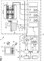
In
In
In den Grundfeldmagneten
Innerhalb des Gradientenfeldsystems
Die Umschaltung von Sende- auf Empfangsbetrieb erfolgt über eine Sende-Empfangsweiche
In
In
Die erfassten MR-Daten können nun entsprechend der mit
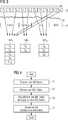
In
Bei dem in
Die Auswertung der MR-Daten kann nun abhängig von der jeweiligen Klasse MR1 bis MR3 erfolgen, so dass die Auswertung der MR-Daten der einen Klasse in einer anderen Weise erfolgt, als die Auswertung der MR-Daten einer anderen Klasse. The evaluation of the MR data can now take place as a function of the respective class MR 1 to MR 3 , so that the evaluation of the MR data of one class takes place in a different manner than the evaluation of the MR data of another class.
In
Im ersten Schritt S1 werden die MR-Daten erfasst, und im zweiten Schritt S2 werden die EEG-Daten erfasst. Dabei werden die Schritte S1 und S2 gleichzeitig durchgeführt, so dass die MR-Daten und die EEG-Daten des Untersuchungsobjekts gleichzeitig erfasst werden. In the first step S1, the MR data are acquired, and in the second step S2, the EEG data are acquired. In this case, the steps S1 and S2 are carried out simultaneously, so that the MR data and the EEG data of the examination subject are detected simultaneously.
Unter Berücksichtigung der EEG-Daten werden die gleichzeitig mit diesen EEG-Daten erfassten MR-Daten klassifiziert S3, was bedeutet, dass die MR-Daten abhängig von den EEG-Daten in verschiedene Klassen eingeteilt werden. Schließlich werden die klassifizierten MR-Daten abhängig von der jeweiligen Klasse ausgewertet S4. Taking into account the EEG data, the MR data acquired simultaneously with these EEG data are classified S3, which means that the MR data is divided into different classes depending on the EEG data. Finally, the classified MR data is evaluated depending on the respective class S4.
Claims (14)
Priority Applications (5)
| Application Number | Priority Date | Filing Date | Title |
|---|---|---|---|
| DE102012205629A DE102012205629A1 (en) | 2012-04-05 | 2012-04-05 | Method and magnetic resonance system for functional MR imaging of a predetermined volume portion of a brain of a living examination subject |
| JP2013076205A JP2013215569A (en) | 2012-04-05 | 2013-04-01 | Method and magnetic resonance system for functional magnetic resonance imaging of predetermined volume segment of brain of living examination subject |
| CN2013101162564A CN103356185A (en) | 2012-04-05 | 2013-04-03 | Method and magnetic resonance equipment for functional magnetic resonance imaging of predetermined volume segment of brain of living examination subject |
| KR1020130036762A KR20130113383A (en) | 2012-04-05 | 2013-04-04 | Method and magnetic resonance system for functional mr imaging of a predetermined volume segment of a brain of a living examination subject |
| US13/857,322 US20130267827A1 (en) | 2012-04-05 | 2013-04-05 | Method and magnetic resonance system for functional mr imaging of a predetermined volume segment of the brain of a living examination subject |
Applications Claiming Priority (1)
| Application Number | Priority Date | Filing Date | Title |
|---|---|---|---|
| DE102012205629A DE102012205629A1 (en) | 2012-04-05 | 2012-04-05 | Method and magnetic resonance system for functional MR imaging of a predetermined volume portion of a brain of a living examination subject |
Publications (1)
| Publication Number | Publication Date |
|---|---|
| DE102012205629A1 true DE102012205629A1 (en) | 2013-10-10 |
Family
ID=49209936
Family Applications (1)
| Application Number | Title | Priority Date | Filing Date |
|---|---|---|---|
| DE102012205629A Withdrawn DE102012205629A1 (en) | 2012-04-05 | 2012-04-05 | Method and magnetic resonance system for functional MR imaging of a predetermined volume portion of a brain of a living examination subject |
Country Status (5)
| Country | Link |
|---|---|
| US (1) | US20130267827A1 (en) |
| JP (1) | JP2013215569A (en) |
| KR (1) | KR20130113383A (en) |
| CN (1) | CN103356185A (en) |
| DE (1) | DE102012205629A1 (en) |
Families Citing this family (7)
| Publication number | Priority date | Publication date | Assignee | Title |
|---|---|---|---|---|
| US9739856B2 (en) * | 2013-06-20 | 2017-08-22 | Siemens Aktiengesellschaft | Magnetic resonance imaging method and apparatus with interleaved resting state functional magnetic resonance imaging sequences and morphological magnetic resonance imaging sequences |
| ES2549393B2 (en) * | 2014-04-25 | 2016-08-25 | Universidad Rey Juan Carlos | Procedure and device for the acquisition, processing and visualization of data obtained simultaneously from magnetic resonance imaging and electrophysiological signals |
| CN106355189B (en) * | 2015-07-13 | 2019-04-23 | 西北工业大学 | EEG-fMRI fusion method based on Motar transport |
| US10588561B1 (en) * | 2017-08-24 | 2020-03-17 | University Of South Florida | Noninvasive system and method for mapping epileptic networks and surgical planning |
| KR102158268B1 (en) * | 2018-11-15 | 2020-09-21 | 연세대학교 원주산학협력단 | Apparatus and method for brain metabolism analysis and brain network implementation |
| EP3785625A1 (en) | 2019-08-29 | 2021-03-03 | Koninklijke Philips N.V. | System for integrated eeg - functional magnetic resonance image data acquisition |
| US11263749B1 (en) | 2021-06-04 | 2022-03-01 | In-Med Prognostics Inc. | Predictive prognosis based on multimodal analysis |
Family Cites Families (6)
| Publication number | Priority date | Publication date | Assignee | Title |
|---|---|---|---|---|
| JP3120224B2 (en) * | 1997-08-27 | 2000-12-25 | 技術研究組合医療福祉機器研究所 | MRI equipment |
| EP1355571A2 (en) * | 2000-08-15 | 2003-10-29 | The Regents Of The University Of California | Method and apparatus for reducing contamination of an electrical signal |
| EP1624798A4 (en) * | 2003-05-06 | 2007-11-28 | George Mason Intellectual Prop | Phase and state dependent eeg and brain imaging |
| WO2010035167A1 (en) * | 2008-09-24 | 2010-04-01 | Koninklijke Philips Electronics, N.V. | Generation of standard protocols for review of 3d ultrasound image data |
| US20110046473A1 (en) * | 2009-08-20 | 2011-02-24 | Neurofocus, Inc. | Eeg triggered fmri signal acquisition |
| CN102293647B (en) * | 2011-06-08 | 2013-07-17 | 北京师范大学 | Feedback system combining electroencephalogram and functional magnetic resonance signals |
-
2012
- 2012-04-05 DE DE102012205629A patent/DE102012205629A1/en not_active Withdrawn
-
2013
- 2013-04-01 JP JP2013076205A patent/JP2013215569A/en active Pending
- 2013-04-03 CN CN2013101162564A patent/CN103356185A/en active Pending
- 2013-04-04 KR KR1020130036762A patent/KR20130113383A/en not_active Application Discontinuation
- 2013-04-05 US US13/857,322 patent/US20130267827A1/en not_active Abandoned
Non-Patent Citations (3)
| Title |
|---|
| MORNEBURG, Heinz: Bildgebende Systeme für die medizinische Diagnostik. 3. Auflage, Erlangen: Publicis MCD Verlag, 1995. S. 501-503 - ISBN 3-89578-002-2 * |
| MORNEBURG, Heinz: Bildgebende Systeme für die medizinische Diagnostik. 3. Auflage, Erlangen: Publicis MCD Verlag, 1995. S. 501-503 – ISBN 3-89578-002-2 |
| MUSSO, F.: Spontaneous brain activity and EEG microstates. A novel EEG/fMRI analysis approach to explore resting-state networks. In: NeuroImage, Vol. 52, 2010, S. 1149-1161. - ISSN 1053-8119 * |
Also Published As
| Publication number | Publication date |
|---|---|
| CN103356185A (en) | 2013-10-23 |
| JP2013215569A (en) | 2013-10-24 |
| US20130267827A1 (en) | 2013-10-10 |
| KR20130113383A (en) | 2013-10-15 |
Similar Documents
| Publication | Publication Date | Title |
|---|---|---|
| DE102015203385B4 (en) | Method for generating motion information to an at least partially moving examination area and magnetic resonance system and hybrid imaging modality | |
| DE102010026376B4 (en) | Creation of optimized MR images with regard to their generation time and their quality | |
| DE102012205629A1 (en) | Method and magnetic resonance system for functional MR imaging of a predetermined volume portion of a brain of a living examination subject | |
| DE102012215718B4 (en) | Method and magnetic resonance system for MR imaging of a predetermined volume section of a living examination subject by stimulating the examination subject | |
| DE102015224162B4 (en) | Method for determining a movement information and a magnetic resonance device describing a movement in an at least partially moved examination area | |
| DE102004051169A1 (en) | Method for cutting position plots of tomographic measurements using statistical images | |
| DE102012205626B4 (en) | Functional MR imaging of a predetermined volume portion of the brain of a living examination subject | |
| DE102007035176A1 (en) | Method for recording and processing a sequence of temporally successive image data records and magnetic resonance apparatus | |
| DE102012212198B4 (en) | Method for reducing movement artifacts in magnetic resonance images and magnetic resonance device therefor | |
| DE102007023251A1 (en) | Method for controlling a magnetic resonance system | |
| DE102015205694B3 (en) | MR saturation taking into account the anatomical structures to be imaged | |
| DE102014219320B4 (en) | Reconstruction of an MR image taking into account the chemical shift | |
| DE102015203938A1 (en) | Recording and evaluation of magnetic resonance signals of a functional magnetic resonance examination | |
| DE102010043956B4 (en) | Acquisition of MR data in a predetermined three-dimensional volume section while avoiding Einfaltungs- and tape artifacts | |
| DE102011078868A1 (en) | Method for frequency calibration of MRI system used for examining breast region of female, involves performing instrumental frequency setting depending on substance resonant frequency measured based on relaxation time per peak | |
| DE102013205576A1 (en) | Method for generating a movement correction for PET data, method for generating PET images as well as correspondingly configured MR system and PET system | |
| DE102016207641A1 (en) | Parallel Magnetic Resonance Acquisition Technique | |
| DE102011005614B3 (en) | Method and device for detecting interference signals in magnetic resonance spectroscopy signals, computer program product and data carrier | |
| DE102016207118A1 (en) | Automated creation of MR scan templates for a magnetic resonance system | |
| DE102012213549B3 (en) | Avoidance of artifacts when taking magnetic resonance data | |
| DE102013207438A1 (en) | Method for generating image data records of an examination subject by means of a magnetic resonance apparatus | |
| DE102016208094A1 (en) | Trigger-adapted MR data acquisition | |
| DE102014204995B4 (en) | Method and magnetic resonance system for fat saturation | |
| DE102012219926B4 (en) | Double-echo MR imaging with a different number of first and second echo signals | |
| DE102017211677A1 (en) | Movement-dependent reconstruction of magnetic resonance images |
Legal Events
| Date | Code | Title | Description |
|---|---|---|---|
| R012 | Request for examination validly filed | ||
| R016 | Response to examination communication | ||
| R081 | Change of applicant/patentee |
Owner name: SIEMENS AKTIENGESELLSCHAFT, DE Free format text: FORMER OWNER: FRIEDRICH-ALEXANDER-UNIVERSITAET, SIEMENS AKTIENGESELLSCHAFT, , DE Effective date: 20140110 Owner name: SIEMENS AKTIENGESELLSCHAFT, DE Free format text: FORMER OWNERS: FRIEDRICH-ALEXANDER-UNIVERSITAET ERLANGEN-NUERNBERG, 91054 ERLANGEN, DE; SIEMENS AKTIENGESELLSCHAFT, 80333 MUENCHEN, DE Effective date: 20140110 Owner name: SIEMENS HEALTHCARE GMBH, DE Free format text: FORMER OWNERS: FRIEDRICH-ALEXANDER-UNIVERSITAET ERLANGEN-NUERNBERG, 91054 ERLANGEN, DE; SIEMENS AKTIENGESELLSCHAFT, 80333 MUENCHEN, DE Effective date: 20140110 |
|
| R081 | Change of applicant/patentee |
Owner name: SIEMENS HEALTHCARE GMBH, DE Free format text: FORMER OWNER: SIEMENS AKTIENGESELLSCHAFT, 80333 MUENCHEN, DE |
|
| R119 | Application deemed withdrawn, or ip right lapsed, due to non-payment of renewal fee |