CN1209068A - Hyaluronic acid as DNA carrier for gene therapy and VEGF antisense DNA to treat abnormal retinal vascularization - Google Patents
Hyaluronic acid as DNA carrier for gene therapy and VEGF antisense DNA to treat abnormal retinal vascularization Download PDFInfo
- Publication number
- CN1209068A CN1209068A CN96199290A CN96199290A CN1209068A CN 1209068 A CN1209068 A CN 1209068A CN 96199290 A CN96199290 A CN 96199290A CN 96199290 A CN96199290 A CN 96199290A CN 1209068 A CN1209068 A CN 1209068A
- Authority
- CN
- China
- Prior art keywords
- compositions
- vegf
- cell
- virus
- sequence
- Prior art date
- Legal status (The legal status is an assumption and is not a legal conclusion. Google has not performed a legal analysis and makes no representation as to the accuracy of the status listed.)
- Pending
Links
Images


Classifications
-
- C—CHEMISTRY; METALLURGY
- C12—BIOCHEMISTRY; BEER; SPIRITS; WINE; VINEGAR; MICROBIOLOGY; ENZYMOLOGY; MUTATION OR GENETIC ENGINEERING
- C12N—MICROORGANISMS OR ENZYMES; COMPOSITIONS THEREOF; PROPAGATING, PRESERVING, OR MAINTAINING MICROORGANISMS; MUTATION OR GENETIC ENGINEERING; CULTURE MEDIA
- C12N15/00—Mutation or genetic engineering; DNA or RNA concerning genetic engineering, vectors, e.g. plasmids, or their isolation, preparation or purification; Use of hosts therefor
- C12N15/09—Recombinant DNA-technology
- C12N15/11—DNA or RNA fragments; Modified forms thereof; Non-coding nucleic acids having a biological activity
- C12N15/113—Non-coding nucleic acids modulating the expression of genes, e.g. antisense oligonucleotides; Antisense DNA or RNA; Triplex- forming oligonucleotides; Catalytic nucleic acids, e.g. ribozymes; Nucleic acids used in co-suppression or gene silencing
- C12N15/1136—Non-coding nucleic acids modulating the expression of genes, e.g. antisense oligonucleotides; Antisense DNA or RNA; Triplex- forming oligonucleotides; Catalytic nucleic acids, e.g. ribozymes; Nucleic acids used in co-suppression or gene silencing against growth factors, growth regulators, cytokines, lymphokines or hormones
-
- A—HUMAN NECESSITIES
- A61—MEDICAL OR VETERINARY SCIENCE; HYGIENE
- A61K—PREPARATIONS FOR MEDICAL, DENTAL OR TOILETRY PURPOSES
- A61K47/00—Medicinal preparations characterised by the non-active ingredients used, e.g. carriers or inert additives; Targeting or modifying agents chemically bound to the active ingredient
- A61K47/30—Macromolecular organic or inorganic compounds, e.g. inorganic polyphosphates
- A61K47/36—Polysaccharides; Derivatives thereof, e.g. gums, starch, alginate, dextrin, hyaluronic acid, chitosan, inulin, agar or pectin
-
- A—HUMAN NECESSITIES
- A61—MEDICAL OR VETERINARY SCIENCE; HYGIENE
- A61K—PREPARATIONS FOR MEDICAL, DENTAL OR TOILETRY PURPOSES
- A61K47/00—Medicinal preparations characterised by the non-active ingredients used, e.g. carriers or inert additives; Targeting or modifying agents chemically bound to the active ingredient
- A61K47/50—Medicinal preparations characterised by the non-active ingredients used, e.g. carriers or inert additives; Targeting or modifying agents chemically bound to the active ingredient the non-active ingredient being chemically bound to the active ingredient, e.g. polymer-drug conjugates
- A61K47/51—Medicinal preparations characterised by the non-active ingredients used, e.g. carriers or inert additives; Targeting or modifying agents chemically bound to the active ingredient the non-active ingredient being chemically bound to the active ingredient, e.g. polymer-drug conjugates the non-active ingredient being a modifying agent
- A61K47/56—Medicinal preparations characterised by the non-active ingredients used, e.g. carriers or inert additives; Targeting or modifying agents chemically bound to the active ingredient the non-active ingredient being chemically bound to the active ingredient, e.g. polymer-drug conjugates the non-active ingredient being a modifying agent the modifying agent being an organic macromolecular compound, e.g. an oligomeric, polymeric or dendrimeric molecule
- A61K47/61—Medicinal preparations characterised by the non-active ingredients used, e.g. carriers or inert additives; Targeting or modifying agents chemically bound to the active ingredient the non-active ingredient being chemically bound to the active ingredient, e.g. polymer-drug conjugates the non-active ingredient being a modifying agent the modifying agent being an organic macromolecular compound, e.g. an oligomeric, polymeric or dendrimeric molecule the organic macromolecular compound being a polysaccharide or a derivative thereof
-
- A—HUMAN NECESSITIES
- A61—MEDICAL OR VETERINARY SCIENCE; HYGIENE
- A61K—PREPARATIONS FOR MEDICAL, DENTAL OR TOILETRY PURPOSES
- A61K48/00—Medicinal preparations containing genetic material which is inserted into cells of the living body to treat genetic diseases; Gene therapy
-
- A—HUMAN NECESSITIES
- A61—MEDICAL OR VETERINARY SCIENCE; HYGIENE
- A61K—PREPARATIONS FOR MEDICAL, DENTAL OR TOILETRY PURPOSES
- A61K9/00—Medicinal preparations characterised by special physical form
- A61K9/0012—Galenical forms characterised by the site of application
- A61K9/0014—Skin, i.e. galenical aspects of topical compositions
-
- A—HUMAN NECESSITIES
- A61—MEDICAL OR VETERINARY SCIENCE; HYGIENE
- A61K—PREPARATIONS FOR MEDICAL, DENTAL OR TOILETRY PURPOSES
- A61K9/00—Medicinal preparations characterised by special physical form
- A61K9/0012—Galenical forms characterised by the site of application
- A61K9/0019—Injectable compositions; Intramuscular, intravenous, arterial, subcutaneous administration; Compositions to be administered through the skin in an invasive manner
-
- A—HUMAN NECESSITIES
- A61—MEDICAL OR VETERINARY SCIENCE; HYGIENE
- A61P—SPECIFIC THERAPEUTIC ACTIVITY OF CHEMICAL COMPOUNDS OR MEDICINAL PREPARATIONS
- A61P27/00—Drugs for disorders of the senses
-
- A—HUMAN NECESSITIES
- A61—MEDICAL OR VETERINARY SCIENCE; HYGIENE
- A61P—SPECIFIC THERAPEUTIC ACTIVITY OF CHEMICAL COMPOUNDS OR MEDICINAL PREPARATIONS
- A61P27/00—Drugs for disorders of the senses
- A61P27/02—Ophthalmic agents
-
- C—CHEMISTRY; METALLURGY
- C07—ORGANIC CHEMISTRY
- C07K—PEPTIDES
- C07K14/00—Peptides having more than 20 amino acids; Gastrins; Somatostatins; Melanotropins; Derivatives thereof
- C07K14/435—Peptides having more than 20 amino acids; Gastrins; Somatostatins; Melanotropins; Derivatives thereof from animals; from humans
- C07K14/52—Cytokines; Lymphokines; Interferons
-
- C—CHEMISTRY; METALLURGY
- C12—BIOCHEMISTRY; BEER; SPIRITS; WINE; VINEGAR; MICROBIOLOGY; ENZYMOLOGY; MUTATION OR GENETIC ENGINEERING
- C12N—MICROORGANISMS OR ENZYMES; COMPOSITIONS THEREOF; PROPAGATING, PRESERVING, OR MAINTAINING MICROORGANISMS; MUTATION OR GENETIC ENGINEERING; CULTURE MEDIA
- C12N15/00—Mutation or genetic engineering; DNA or RNA concerning genetic engineering, vectors, e.g. plasmids, or their isolation, preparation or purification; Use of hosts therefor
- C12N15/09—Recombinant DNA-technology
- C12N15/11—DNA or RNA fragments; Modified forms thereof; Non-coding nucleic acids having a biological activity
- C12N15/113—Non-coding nucleic acids modulating the expression of genes, e.g. antisense oligonucleotides; Antisense DNA or RNA; Triplex- forming oligonucleotides; Catalytic nucleic acids, e.g. ribozymes; Nucleic acids used in co-suppression or gene silencing
- C12N15/1137—Non-coding nucleic acids modulating the expression of genes, e.g. antisense oligonucleotides; Antisense DNA or RNA; Triplex- forming oligonucleotides; Catalytic nucleic acids, e.g. ribozymes; Nucleic acids used in co-suppression or gene silencing against enzymes
-
- C—CHEMISTRY; METALLURGY
- C12—BIOCHEMISTRY; BEER; SPIRITS; WINE; VINEGAR; MICROBIOLOGY; ENZYMOLOGY; MUTATION OR GENETIC ENGINEERING
- C12Y—ENZYMES
- C12Y304/00—Hydrolases acting on peptide bonds, i.e. peptidases (3.4)
- C12Y304/22—Cysteine endopeptidases (3.4.22)
- C12Y304/22027—Cathepsin S (3.4.22.27)
-
- C—CHEMISTRY; METALLURGY
- C12—BIOCHEMISTRY; BEER; SPIRITS; WINE; VINEGAR; MICROBIOLOGY; ENZYMOLOGY; MUTATION OR GENETIC ENGINEERING
- C12Y—ENZYMES
- C12Y304/00—Hydrolases acting on peptide bonds, i.e. peptidases (3.4)
- C12Y304/23—Aspartic endopeptidases (3.4.23)
- C12Y304/23005—Cathepsin D (3.4.23.5)
-
- A—HUMAN NECESSITIES
- A61—MEDICAL OR VETERINARY SCIENCE; HYGIENE
- A61K—PREPARATIONS FOR MEDICAL, DENTAL OR TOILETRY PURPOSES
- A61K38/00—Medicinal preparations containing peptides
-
- C—CHEMISTRY; METALLURGY
- C12—BIOCHEMISTRY; BEER; SPIRITS; WINE; VINEGAR; MICROBIOLOGY; ENZYMOLOGY; MUTATION OR GENETIC ENGINEERING
- C12N—MICROORGANISMS OR ENZYMES; COMPOSITIONS THEREOF; PROPAGATING, PRESERVING, OR MAINTAINING MICROORGANISMS; MUTATION OR GENETIC ENGINEERING; CULTURE MEDIA
- C12N2310/00—Structure or type of the nucleic acid
- C12N2310/10—Type of nucleic acid
- C12N2310/11—Antisense
- C12N2310/111—Antisense spanning the whole gene, or a large part of it
-
- C—CHEMISTRY; METALLURGY
- C12—BIOCHEMISTRY; BEER; SPIRITS; WINE; VINEGAR; MICROBIOLOGY; ENZYMOLOGY; MUTATION OR GENETIC ENGINEERING
- C12N—MICROORGANISMS OR ENZYMES; COMPOSITIONS THEREOF; PROPAGATING, PRESERVING, OR MAINTAINING MICROORGANISMS; MUTATION OR GENETIC ENGINEERING; CULTURE MEDIA
- C12N2310/00—Structure or type of the nucleic acid
- C12N2310/30—Chemical structure
- C12N2310/31—Chemical structure of the backbone
- C12N2310/315—Phosphorothioates
-
- C—CHEMISTRY; METALLURGY
- C12—BIOCHEMISTRY; BEER; SPIRITS; WINE; VINEGAR; MICROBIOLOGY; ENZYMOLOGY; MUTATION OR GENETIC ENGINEERING
- C12N—MICROORGANISMS OR ENZYMES; COMPOSITIONS THEREOF; PROPAGATING, PRESERVING, OR MAINTAINING MICROORGANISMS; MUTATION OR GENETIC ENGINEERING; CULTURE MEDIA
- C12N2799/00—Uses of viruses
- C12N2799/02—Uses of viruses as vector
- C12N2799/021—Uses of viruses as vector for the expression of a heterologous nucleic acid
- C12N2799/022—Uses of viruses as vector for the expression of a heterologous nucleic acid where the vector is derived from an adenovirus
-
- C—CHEMISTRY; METALLURGY
- C12—BIOCHEMISTRY; BEER; SPIRITS; WINE; VINEGAR; MICROBIOLOGY; ENZYMOLOGY; MUTATION OR GENETIC ENGINEERING
- C12N—MICROORGANISMS OR ENZYMES; COMPOSITIONS THEREOF; PROPAGATING, PRESERVING, OR MAINTAINING MICROORGANISMS; MUTATION OR GENETIC ENGINEERING; CULTURE MEDIA
- C12N2799/00—Uses of viruses
- C12N2799/02—Uses of viruses as vector
- C12N2799/021—Uses of viruses as vector for the expression of a heterologous nucleic acid
- C12N2799/025—Uses of viruses as vector for the expression of a heterologous nucleic acid where the vector is derived from a parvovirus
Abstract
The invention provides methods and compositions for gene therapy, including antisense therapy. In one embodiment, the compositions comprise hyaluronic acid to promote uptake of nucleic acid by the target cells. The invention is illustrated with reference to treatment of retinal diseases caused by neovascularisation.
Description
The present invention relates in the control of disease or treatment, use hyaluronic acid that active factors is led target gene to eliminate the target gene function.In one embodiment, the present invention relates to treat the method and composition that ophthalmic particularly relates to the retinal diseases of choroid and/or amphiblestroid neovascularization, it is to utilize the cytophagy feature of the specific cell in the eyes to provide a kind of effective method that active factors is transported to target cell, with short-term and the long-term treatment of carrying out neovascularization.Method and composition of the present invention can be used for DNA, RNA, antisense nucleotide, peptide or other therapeutic agent are transported to phagocyte or peripheral cell.
Background of invention
A) as the hyaluronic acid of adjuvant or directing agent
Hyaluronic acid (HA) finds in vivo that by reaching the big complex oligosaccharide that 5000 couples of alkaline disaccharide glucuronic acid-β (1-3) N-acetyl-glucosamine β (1-4) form it is the main component of extracellular matrix, and its tertiary structure is the random coil of the about 50nm of diameter.
HA has the ability in conjunction with big water gaging, and this makes it form the viscosity hydrated gel with viscoelasticity property in vivo.In the vitreous body of mammal eyes and extracellular matrix, all found this form.
Because HA has with the lead ability at disease or position, state place of active factors, so HA has been used for some disease and the state (International Patent Application WO 91/04058 and WO93/16733) of general and topical therapeutic human body.Shown HA for example at the carotid artery (with respect to unmarred offside tremulous pulse) of damage, and in the colorectum tumor of laboratory animal, formed storehouse (depot) and be retained in the skin of this animal.In all these situations, deposition site is high HA expression of receptor district, and the special tissue that is positioned to express these receptors of high level of prompting HA particularly just at the tissue of abnormality proliferation and migration, comprises and replys damage, inflammation, growth and tumorigenic tissue.
Is that it can be simultaneously in conjunction with other molecule and cell membrane to HA as the very important feature of the effect of potential adjuvant.Differentiate the cell surface receptor that is specific to HA, comprised some homologous protein in receptor (RHAMM), ISIS 2302 (ICAM) and the CD44 family of migration of histocompatibility antigen CD44, hyaluronic acid mediation.The virus that is promoted by HA will make the molten mechanism of born of the same parents of common virus picked-up more effective with combining of cell membrane.
B) ophthalmic
The feature of multiple ophthalmic such as macula degeneration and diabetic retinopathy all is choroid and/or amphiblestroid neovascularization, this process be suffer from main diseases that the patient of these diseases loses one's sight because of.
Treatment of the prior art
In relevant macula degeneration disease of age (ARMD), under the retina formation of neovascularization film (SRNVM) and hemorrhage cause central vision fast and most of forfeiture.Multiple therapy methods is arranged, but all be insecure.The photic coagulation of laser is the most acceptable therapy, but it still has following shortcoming, and promptly laser beam can cause fine and close permanent blind spot (Schachet, 1994; Ibanez etc., 1995 and Hudson etc., 1995), cause the temporary transient forfeiture of vision and can not prevent the development of the state of an illness because the new vessels film repeats to take place for a long time.
Therefore unique advantage of this therapy is to have prevented degree of depth visual loss.
Similarly, operation is removed under SRNVM or the retina blood or reorientated fovea centralis retinae by the rotation retina also is unsuccessful, because it exists the improvement of postoperative complication and vision very little or temporary.Therefore, these intrusion type therapies and corresponding complication head and shoulders above the benefit that obtains, thereby purposes is restricted.
Interferon-ALPHA 2a (Fung, 1991 have also been proved with some anti-angiogenesis activity; Guyer etc., 1992 and Engler etc., 1994) and to transplant the purposes of retinal pigment epithelium (RPE) cell (Algvere etc., 1994) very limited, and the initial result who organizes acquisition with the small-scale patient in fairly large test, be not confirmed.
Outside laser photocoagulation (as mentioned above, it has many shortcomings), the main method of another treatment diabetic retinopathy is blood sugar control and blood pressure.The effect of this therapy is subject to patient's motility and compliance.
Age surpasses among 75 years old the crowd about 30% and suffers from macula degeneration disease, and has 3 to suffer from diabetic retinopathy in 1000 individualities.Because along with aged tendency of population, these numerals will increase, and the occurrence probability of diabetes also will increase, so need a kind of more efficient methods to treat these and other ophthalmic diseases that is mediated by neovascularization.
Neovascularization mechanism
Vascular endothelial cell growth factor (VEGF) is a kind of glycoprotein of disulfide bridge connects of dimerization, and known its can synthesize justacrine by various normal cells and tumor cell.Nearest observation shows that VEGF detects (Malecaze etc. usually in the patient's who suffers from diabetes new vessels retina, 1994), and in the patient's who suffers from diabetic retinopathy or the locking of central retina vein eye liquid, detect (Aiello etc., 1994).Recent findings is induced vegf expression such as the central vein locking under the states such as retina shedding and intraocular tumour.In rabbit model, the level of VEGFmRNA after inducing the retinal vein locking, raise (Pe ' er etc., 1995) in the not enough district of amphiblestroid oxygen supply.The not enough vegf expression that stimulates of oxygen supply is also observed (Pierce etc., 1995 at other animal model; Miller etc., 1994), and in all types of cell cultures, also observe (Simorre-Pinatel etc., 1994; Hata etc., 1995 and Thiema etc., 1995).
C) antisense DNA in the disease treatment and gene therapy
Inhibition to the undesirable active proteic gene expression of coding mediation produces " antisense " DNA sequence and finishes in all cases by importing or original position in target cell.These antisense sequences are the DNA sequence that cause the antiparallel RNA of sequence of synthetic its sequence and encoding proteins when transcribing.In various viral diseases, tested this antisense sequences.In addition, antisense oligodeoxyribonucleotide can import target cell, and this short sequence itself is not transcribed, but suppresses that transcribing and/or translation subsequently of adopted DNA sequence should be arranged in the target cell mutually.
Think extensively still that up to date the required minmal sequence length of Antisense Suppression that realizes gene expression is 12-14 nucleotide (Wagner, 1994).Yet, now shown in conjunction with the specificity of target sequence can by use contain C-5 propine pyrimidine and thiophosphate internucleotide linkage modification oligonucleotide and effectively strengthen, and the inhibition that the sequence that is as short as 7 or 8 nucleotide also can provide effective genescreen, mispairing sensitivity, ribonuclease H to rely on, the flanking sequence of its RNA that hits is important (Wagner etc., 1996) for the decision specificity.
Yet, be subjected to the influence of following factor with the success that stops this application of expression of gene in the antisense nucleotide body, for example need special mutation inhibiting gene expression (Milan, 1993; McInnes andBascom, 1992) antisense nucleotide (Akhtar and Ivinson, 1993) that, perhaps needs high concentration.
At present, the treatment of this form much is the antisense sequences of application packages in liposome, perhaps directly Antisense cDNA or oligonucleotide is applied to disease location.Therefore not successful to the trial majority of the absorption of these sequences by antisense sequences being wrapped in the liposome to increase target cell, and the orientation of liposome is also very difficult, its absorption even also lower than virus.
Directed also can realize by for example utilizing Sendai virus to shift through virus-mediated DNA.Sendai virus is a kind of RNA viruses, has proved that it is (people such as Kaneda, 1987) more than 95% with DNA and protein delivery to the efficient in the cell.In this gene transfer system, the fusion activity by Sendai virus directly imports to the DNA nucleoprotein complex in liposome in the Cytoplasm, DNA can be imported nucleus fast with nucleoprotein.The gene transfer of Sendai virus mediation takes place by the fusion of virus with cell membrane, and walks around endocytic pathway.Recently, observe Sendai virus and can efficiently antisense or plasmid DNA be transported to target cell.Antisense and plasmid DNA are not only in culture but also all keep its activity (people such as Kaneda, 1987) in vivo.Yet the application of this virus is subjected to the restriction of this fact, does not promptly have suitable construct to can be used as carrier at present.In addition, the DNA of transfer only can express limited a period of time, because this gene transfer is by merging mediation.
Retrovirus retrovirus has been widely used in somatic tissue's gene therapy (Boris-Lawrie andTemin, 1993), and they can efficiently navigate to and infect the host cell of numerous species, and transgenic DNA is integrated into host genome.In theory, the integration of DNA will provide genetically modified permanent generation, and this can cause forever remedying of cell.But retrovirus retrovirus can not infect nondividing cell (Salmons and Gunzburg, 1993).In addition, counter-transcription-ing virus particle is unstable in vivo, and this makes and is difficult to reach high virus titer by inoculation.Moreover, the carcinogenecity of integrating virus also there is tangible worry.Retrovirus retrovirus can not infect not somatoblast and mean that they can not be as the candidate of the gene transfer of carrying out in eyes, because most important target cell such as light receptor and RPE cell all are somatoblasts not.
The application of herpes simplex virus vector is subjected to the restriction (people such as Culver, 1992) of its low efficiency of infection.Developed two types carrier, i.e. the amplicon of replication defect type recon and plasmid derivative, the latter needs helper virus.Although can overcome difficulties and remove virulent gene from herpes simplex virus, construct remains (people such as Johnson, 1992) of cellular toxicity.In addition, the long-term expression of insertion sequence remains unsuccessful at present, exists the regulation and control and the stability problem of construct.In gene therapy, the herpes simplex virus of modifying is applied to cause in the eyes very big worry, because their pathogenicity.The zoster herpesvirus infection can cause the ophthalmic severe infections, often causes losing one's sight, and needs corneal transplantation.
Adenovirus has been widely used in the gene transfer in the not somatoblast and proliferative cell.They can carry the DNA up to 7.5kb, and effective transfection and high virus titer can be provided.The major advantage of using these viruses rather than retrovirus retrovirus is that they can infect many target cells (Kozarsky and Wilson, 1993) of not dividing.Replication-defective adenoviral it is believed that it is comparatively safe, because these viruses are pathogen common among the mankind, causes that usually gentle relatively symptom is as flu.These carriers carry the oncogene with deletion mutation, have reduced the probability (Siegfried, 1993) that becomes carcinogenecity.In the first routine experimental gene therapy test, just be to use the recombinant adenovirus treatment to suffer from the patient of cystic fibrosis by the approval of U.S. National Institutes of Health recombinant DNA consultative committee.
Yet the major defect of adenovirus is their transient gene expression, and this is because due to transgenic is not incorporated in the cellular genome.In addition, only there is only a few that gene is delivered to the not successful trial of somatoblast.Reported in 1993 and use adenovirus for the first time gene successfully to be transferred to experiment in the brain of forming by somatoblast not people such as (, 1993) Le Gal La Salle.
These results show that gene therapy is a kind of approach for the treatment of disease in theory reliably, but need overcome the technical barrier of effective orientation and absorption, can utilize the virus that adheres to target cell and be absorbed to overcome these technical barriers.This process is not very effective, and uses the danger that virus may cause not wishing the medical condition that occurs.Yet announced that it is feasible that positive findings has been instructed with gene therapy adjusting biological process.
Therefore, need be used for the treatment of the improving one's methods of directed gene therapy of disease at present, and need suitable to contain hyaluronic compositions to be used for this treatment.
D) gene therapy and ocular disease
In Australian patent application 75168/94 (Hybridon Inc.), proved by with based on the insulation of 19 to the 21 aggressiveness antisense oligonucleotides of mice VEGF, can in COS-1 or NB41 cell, suppress the vivoexpression of Mus VEGF.Shown that a kind of 21 aggressiveness antisense nucleotide that navigate to the translation termination site are effective sequences.But do not disclose or the special any tissue that is directed in the eyes of prompting sequence, also do not have disclosure or prompting treatment any eye symptom except that diabetic retinopathy.
At United States Patent (USP) 5,324, in 654, a kind of method that stimulates the propagation of non-malignant cell is disclosed, this method comprise use corresponding to retinoblastoma (Rb) gene antisense nucleotide external treatment cell to suppress the expression of Rb gene outcome, cause cytostatic proteic expression to be suppressed.By this way, promoted cell proliferation.Can implant proliferating cells again if desired then, but and before implanting again pair cell carry out genetic engineering and operate to replace a kind of specific gene.Yet there is not document to point out that this antisense sequences can be used for treating ocular disease.The invention of US5324654 relates to foundation can long-term proliferating cells system and disease such as muscular dystrophy and the diabetes of treatment due to not expressed by gene.
Do not attempt as yet specific gene is directed to specific cell, and do not filter out the orientation of eye type.Expection is difficult to only to carry out special orientation with adenovirus because should virus can the many types of transfection cell.
For the treatment ocular disease, and other position great majority or all not infected in the body, hope be the target tissue that therapeutic agent optionally is delivered to ophthalmic.For antisense DNA, importantly this DNA should be able to be absorbed into these target cells.
Progress in the said gene treatment has caused transgenic is delivered to target cell and the further research of expression therein, as use recombinant adenovirus the beta galactosidase transgenic to be delivered into retina (Bennett as induction system, Deng the people, 1994, people such as Li, 1994 and people such as Mashmour, 1994).Retinal pigment epithelium (RPE) is the monolayer that ophthalmic one deck does not upgrade, and between neural retina and choroid, the RPE cell is engulfing a property neuroepithelial cell, and it has formed amphiblestroid outermost layer.For a long time with regard to the character of engulfing of known these cells, and existing summary (Bok and Young, 1979).Observing in growing animal had high-level transgene expression in 3 days in the RPE layer, high-level transgene expression is arranged in 2 weeks in the photoreceptor cells of neural retina.The expression of reporter gene is 9 weeks nearly.In than senior animal, retina down or intravitreal injection all can not induce the beta galactosidase transgenic in photoreceptor cells, to express people such as (, 1994) Li.
After Australian patent application 61444/94 was presented at and is injected into ante-chamber, vitreous body or spherical space, back, duplicate deficit type recombinant adenovirus was absorbed by the various tissues of ophthalmic, and the reporter gene beta galactosidase is expressed.Yet this document does not show whether the virus of this form successfully is incorporated into activating agent in target cell or the zone, and also disclosure or prompting VEGF not can be used for curing any eye symptom.
Using antisense nucleotide is that this nucleotide can not enter target cell as a special obstacle of eye treatment form success or not, and stable limited (Helene 1991) of phosphoric acid G 3139 for example of the oligonucleotide after modifying.These factors have greatly limited the success rate of the success rate, particularly long-term treatment of vivo gene treatment.In the treatment of retinal diseases, postpone the course of disease and make progress about 12 months ability and will greatly increase the value and the effective percentage of long-term treatment.
Observed with adenovirus as the transport vehicle of retina gene therapy with cytotoxicity, proved that this cytotoxicity is dose dependent (Mashmour, 1994), thereby drawn another difficulty of using this carrier.For the dosage that reduces given carrier but keep its transfer efficiency simultaneously, can use a kind of adjuvant, proved that adjuvant such as lipofectin reagent can increase the absorption of cell to the DNA of " exposing ".
Although HA is widely used as the substitute of vitreous body forfeiture in surgical procedures, we in the prior art and find no any prompting HA and can promote any medicament by any cell or tissue absorption in the eyes.Similarly, although in the Australian patent application 52274/93 of Norpharmco, pointed out HA can promote penetrating of medicament such as antibiotic or anticarcinogen, but this document does not point out HA can promote the absorption of which kind of cell to which kind of medicament, and saying nothing of has absorption to DNA or virus.Particularly this document is not instructed by intraocular injection and is used HA.
We have now found that engulfing character and increasing the molecule that is expelled in the body behind the vitreous body space such as the absorption of oligonucleotide and virus of RPE cell.These RPE cells demonstrate the absorption that increase is arranged than other cell type.Our discovery makes it possible to induce in retina or chorioidal epithelium cell the long-term and short-term of VEGF to express, and therefore can suppress the growth of amphiblestroid neovascularization or SRNVM.
Summary of the invention
According to an aspect of the present invention, the invention provides a kind of compositions, it comprises a kind of nucleic acid and a kind of hyaluronic acid or derivatives thereof, and a kind of materia medica acceptable carrier.
Described nucleic acid can be DNA or RNA, and/or can be the nucleotide sequence that is antisense orientation with target sequence, and target sequence is the generation of morbid state or worsens related nucleotide sequence.This target nucleic acid sequence can be genomic DNA, cDNA, messenger RNA or oligonucleotide.When target nucleic acid sequence was genomic DNA, it may reside in the coding region or is present in control region such as promoter sequence.
Perhaps, described nucleic acid may reside in a kind of comprising in the carrier that advances the nucleotide sequence in the target cell to be transferred, and this nucleotide sequence can be genomic DNA, cDNA, messenger RNA or oligonucleotide equally.Yet in this case, described nucleic acid can provide to the adopted sequence of having of target cell to cause a kind of function, perhaps can be antisense sequences to be used for suppressing being present in the function of the nucleic acid of target cell.
The carrier that comprises DNA to be transferred can be that virus is as adenovirus, adeno associated virus, herpesvirus or retrovirus retrovirus.All these viruses have been furtherd investigate in the prior art as the application that is used for Vectors in Gene Therapy.Described in addition carrier also can be a liposome.
The present invention also provides the method for treatment patient's pathological state, comprises the compositions of the present invention that gives patient's effective dose.
Be understood that dosage and approach depend on state to be treated, doctor or skilled person will be easy to determine proper dosage and approach, be understood that compositions of the present invention can give as passing through intravenous or subcutaneous injection all over the body, topical administration perhaps directly gives tissue to be treated as passing through ophthalmic or intravitreal injection as being adsorbed on gel or the sponge.
Patient to be treated can be the mankind or animal, and particularly domestic or mammal pet is as cattle, horse, sheep, goat, cat and Canis familiaris L..
In compositions of the present invention, nucleic acid or carrier can mix with hyaluronic acid simply, perhaps can be randomly and hyaluronic acid physics or chemical coupling.The method that DNA is linked to each other with hyaluronic acid is disclosed in " sulfonation Hyaluronan derivant synthetic that contains nucleic acid base ", chemical communication, 1994,2027-2030, and " the transfer character of the nucleoside that is undertaken by the nucleic acid base of puting together with Hyaluronan ", Chirachanchai, S., Wada, T., Inaki, Y. and Takemoto, K., chemical communication, 1995
2121-122.
In a preferred embodiment, of the present invention this provides the method that is used for the treatment of by the retinopathy due to the abnormal vascularization on the one hand, wherein said nucleic acid is the anti sense nucleotide sequence corresponding to the coded sequence of at least a portion VEGF (VEGF), and described nucleic acid is with hyaluronic acid administration as described below.
The HA of many forms is applicable to the present invention, and the HA of low-molecular-weight and high molecular form all can use, and it is medicinal that unique requirement is that the purity of HA and aseptic degree should be suitable for, and preferably, HA also is pyrogen-free.The high molecular prepared product of HA may need dilution before use.Particularly, be applicable to that commercially available HA product of the present invention is a Hyal drugmaker, the product that Mississauga provides, it is the solution that mean molecule quantity is about 225,000 HA 2%, also can be LifeCore
TMThe hyaluronate sodium that biological medicine company produces, and Pro Visc (Alcon laboratory) and " HEALON " (Pharmacia company, Uppsala).The derivant that is understood that the term HA in this description comprises the homologue of HA, analog, complex, ester and fragment and subunit.
The derivant that can be used for HA of the present invention comprises the acceptable salt of its materia medica, or the fragment of HA or subunit.Those skilled in the art can determine given HA prepared product, or whether the specific derivatives of HA, complex etc. are applicable to the present invention.
According to second aspect, the present invention relates to be used for the treatment of compositions by the retinopathy due to the abnormal vascularization, comprise anti sense nucleotide sequence corresponding at least a portion VEGF (VEGF) coded sequence, and a kind of pharmacological-acceptable carrier, randomly said composition comprises that also one or more adjuvant such as hyaluronic acid or dendritic compound absorb in order to increase cell.Disclose the use dendritic compound in the International Patent Application WO 95/24221 of Dendritech company etc. genetic stocks has been transported into target cell.
VEGF is human retina pigment epithelium (RPE) VEGF or choroid endothelium VEGF most preferably.
In each embodiment, of the present invention this relates to short-term (about 2 months) on the one hand, (about 1 year) and indefinite duration, (patient throughout one's life) treated this retinopathy for a long time.In first embodiment,, the invention provides one or more antisense oligonucleotide that has 100% complementarity with the respective regions of VEGF gene for short term therapy.This oligonucleotide should have 16-50 nucleotide, preferably has 16-22 nucleotide, more preferably has 16-19 nucleotide.Can use the oligonucleotide of the modification of people (1996) descriptions such as Wagner, thereby make the lower limit of sequence length be reduced to 7 nucleotide.
For long term inhibition, the invention provides a kind of recombinant virus, it comprises the VEGF DNA that is antisense orientation.This VEGF DNA is a kind of long sequence, is meant that in this manual length surpasses the VEGF sequence of 20 nucleotide, and preferred length surpasses 50 nucleotide, until total length VEGF sequence.In this embodiment, recombinant virus accumulates in the RPE cell, and original position generation antisense VEGF, thereby suppresses vegf expression in the RPE cell.
For suppressing indefinite duration, the invention provides a kind of virus, it comprises the VEGF DNA that is antisense orientation, wherein this virus can be integrated into antisense sequences the genome of target cell.Preferably, this virus is adeno associated virus or similar virus.As in relating to the embodiment of long-term treatment, the length of this VEGF DNA is at least 20 nucleotide, preferably surpasses 50 nucleotide.Adeno associated virus or similar virus can promote antisense VEGF DNA to be integrated into the RPE cellular genome, thereby as long as cell still has function just can keep the VEGF of antisence.
The ocular disease of available the compositions and methods of the invention treatment includes but not limited to macula degeneration disease (ARMD) and the diabetic retinopathy that the age is relevant.The eye morbid state and the tissue of neovascularization take place in other, as branch or all available the present invention's treatment of the locking of central retina vein, earliness lambing retinopathy (being also referred to as retrolental fibroplasia), rubeosis iridis diabetica or cornea rebirth blood vesselization.
On the other hand, the invention provides prevention or elimination, comprise that the anti sense nucleotide sequence at VEGF with effective dose gives in the eye, thereby suppress neovascularization by the method for the retinopathy due to the unusual neovascularization.
Antisense sequences can be carried in the replication defect type recombinant virus as carrier, this carrier preferably includes the replication-defective adenoviral that carries following promoter, described promoter is respiratory syncytial virus (RSV) promoter, cytomegalovirus (CMV) promoter, adenovirus major late protein (MLP) promoter, VAl polIII promoter or beta-actin promoter.Carrier also can comprise poly-adenosine signal sequence such as SV40 signal sequence.In a particularly preferred embodiment, carrier is pAd RSV, pAd.MLP or pAd.VAl.In a more preferred embodiment, carrier is Ad.RSV.aVEGF or Ad.VAl.aVEGF.
In an embodiment preferred, people VEGF is advanced carrier by sub-clone, inserts required restriction site to create, and forms and carries the VEGF of antisense orientation or the adenoviral plasmid of its partial sequence, and this plasmid can digest and linearisation through Restriction Enzyme subsequently.This linearizing plasmid can advance proper host cell with linearizing replication-defective adenoviral cotransfection subsequently, as kidney 293 cell is.
Compositions of the present invention can be in vitreous body or subretinal injection be delivered in the eye, preferably inject with suitable excipient or carrier.This medication and this injection used excipient or carrier are known in the art.Perhaps, by taking out the RPE cell from the patient, above-mentioned replication-defective adenoviral of cultured cell and external use or adeno associated virus pair cell are injected and are realized that the ex vivo of the present composition carries.To carry the retina lower floor of RPE injection cell in patient's eye of virus then.
Although the present invention has carried out special description with reference to the morbid state of eye, but those skilled in the art will appreciate that other pathological state that has many VEGF to play an important role, and those skilled in the art understand that antisense oligonucleotide of the present invention and recombinant virus are suitable for treating these other states.Similarly, although those of skill in the art understand that also the present invention has carried out special description with reference to VEGF.But methods described herein also are applicable to other albumen and use.
Brief description of the drawings
Fig. 1 a shows behind the single intravitreal injection, the genescan analysis result of persistence in the body of antisense oligonucleotide in retina.
Fig. 1 b shows behind injection CATSCF the amphiblestroid confocal microscopy image of RCS-rdy+ rat in the different time.
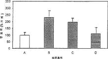
Fig. 2 is the pictorial representation of phagosome number in the RPE layer of Long-Evans rat.Dosage is as follows: low: 6.6 μ g CATSC antisense oligonucleotides; In: 66 μ g; High: 132 μ g.Each hurdle has shown in rat retina 5 average phagosome number and standard deviations in the zone of selecting at random.
Fig. 3 is the pictorial representation of phagosome number in the RPE of RCS-rdy+ rat layer.Laboratory animal has MODN (S1) and 66 μ g antisense oligonucleotides (CATSC) to inject with 66 μ g.
Fig. 4 illustrates the titre of increase adenovirus vector to expressing the influence of the genetically modified cell number of adenovirus.Temperature retention time is 16 hours in all cases.The RPE7 representative is from the human retina pigment epithelium cell of donor in 7 years old age; F2000C represents the F2000 fibroblast.The suffix C of F2000 represents that the counting of F2000 cellular expression is according to revising with the direct comparison of RPE7 cell.
Fig. 5 illustrates the temperature retention time of increase and adenovirus vector to expressing the influence of the genetically modified cell number of adenovirus.In all cases, the concentration of adenovirus vector is 2 * 10
6P.f.u/ml.The suffix C of F2000 represents that the counting of F2000 cellular expression is according to revising with the direct comparison of RPE7 cell.
Fig. 6 illustrates the influence of hyaluronic acid (HA) for the genetically modified RPE7 cell number of expression adenovirus of given virus titer.Three Nogata shapes are represented 0.001%HA, the effect of 0.005%HA and no HA (contrast).Error Nogata shape is represented standard deviation.
Fig. 8 is illustrated in the immunofluorescence dyeing of the HA receptor in RPE7 and the F2000 fibroblast.8a: the CD44 dyeing on RPE7; 8b: the ICAM dyeing on RPE7; The RHAMM dyeing of 8c on RPE7; 8d: the CD44 dyeing on the F2000 fibroblast; 8e: the ICAM dyeing on the F2000 fibroblast; 8f: the RHAMM dyeing on the F2000 fibroblast.
Fig. 9 demonstrates the microphotograph of isolated choriocapillary endotheliocyte from Oculus sus domestica; their characteristic apparent (going up a width of cloth most) is shown; Factor IX related antigen (a middle width of cloth), and the acetylation low density lipoprotein, LDL absorbed into cytoplasmic ability (next width of cloth).
Figure 10 illustrates various hyaluronic acid preparations form (tube formation) to choriocapillary endotheliocyte pipe effect.
Figure 11 illustrates the antigenic alkaline phosphatase staining of CD44 in the retinal pigment epithelium, and in each case, epithelium is positioned at the below of photo, and choroid up.
A. unbleached pigment epithelium layer.
B. the pigment epithelium layer of melanin granule is removed in bleaching.
C. use the pigment epithelium of bleaching of the anti-CD44 antibody staining of alkali phosphatase enzyme mark.
Figure 12 illustrates and uses VEGF
165The DNA PCR and the RT-PCR of the retinal pigment epithelium system of transfection analyze.
Figure 13 illustrates the VEGF by the RPE cell generation of transfection
165Effect to the formation of choriocapillary endotheliocyte pipe.
The detailed description of invention
Followingly the present invention is described with reference to non-limiting example.Among some embodiment, use the feasibility of the antisense oligonucleotide checking method therefor of the present invention that is complementary to cathepsin S (CATSC) therein.
The accumulation of embodiment 1 antisense oligonucleotide in the RPE cellular layer
Cultivate the human retina pigment epithelium cell, in experiment in vitro, use subculture for the third time.Cultivate the culture be paved with simulated in vivo environment with beef fillet skin (ROS).In the culture medium of these cells, add the fluorescein-labeled antisense oligonucleotide that is complementary to human cathepsin S (CATSCF), cultivate harvesting after 7 days.By the existence of the intracellular fluorescein-labeled oligonucleotide of fluorescence counting method (FACS) detection RPE, measure the existence and the stability of oligonucleotide in the cell with Gene Scan DNA analysis instrument.The fluorescence of the RPE cell of cultivating increases about 100 times, shows in the RPE cell to have antisense oligonucleotide.These the results are shown in Table 1.
Table 1
Cultivate or the not fluoremetry of the people RPE cell of cultivation with complementary CATSCF
| Sample | The FACS reading |
| ????RPE+ROS | ????5.94 |
| ????RPE+ROS+CATSC | ????8.50 |
| ????RPE+ROS+CATSCF | ????461.50 |
Still not knowing fluorescence is to discharge by total length CATSC or by the oligonucleotide of degrading.With Gene Scan proof fluorescence major part is that it is positioned at the position similar to CATSCF by due to the 19 aggressiveness oligonucleotide.Use similar program, observe that the CATSC oligonucleotide remains complete after cultivating 7 days.
With 1 nanomole CATSFC be injected into 6 age in week non-pigment RCS-rdy+ rat vitreous humor in, and, also injected 1 nanomole fluorescein in contrast with the migration of confocal fluorescence microscopy art tracking oligonucleotide.Inject back 2 hours, 3 days and 7,14 and 56 angel's animal euthanasias, take out the eye after the injection after the euthanasia, lyophilizing, section also are used for the confocal microscope art immediately without fixing.
In ganglion cell layer, observe penetrating of oligonucleotide after intravitreal injection CATSFC2 hour, in the time of 3 days, also observe penetrating of oligonucleotide at light receptor and pigment epithelium layer.Yet injected back 7 days, only the RPE layer has the CATSCF of significant quantity.At 14,28 and 56 days, still keep fluorescence signal at the RPE layer, in all other cell types, do not observe signal.These results demonstrate most of CATSCF and are absorbed by engulfing property RPE cell.
As mentioned above behind the intravitreal injection, dissect eyes, take out retina and also extract DNA, the DNA of purification is carried out GENE Scan analyze.As shown in Figure 1a, prove in 7,14,28 and 56 days the rat retina that after injection undegradable fluorescein-labeled oligonucleotide is arranged.Obviously disappear at 56 days signal intensitys.
Carry out the confocal microscope analysis after being expelled to the single agent of 10nmolCATSCF in the non-pigmented RCS-rdy+ rat.Check retina after the injection at certain intervals, the results are shown in Fig. 1 b, wherein g represents ganglion cell layer, and i represents inner nuclear layer, and o represents outer nuclear layer, and r represents layer of retina,pigment epithelium.Band illustrates 2 hours (B), 3 days (A) behind the injection 10nmol CATSCF, 7 days (C), 28 days (D) and 56 days (E) among the figure, and the retina of injecting 3 days (F) behind in contrast the FITC.
After these results showed intravitreal injection, oligonucleotide accumulated in the RPE cell.Oligonucleotide exists in the RPE layer and reaches 56 days, and remains the biologic activity form during this period.
The biologic activity of embodiment 3 antisense oligonucleotides
60 age in days non-pigment RCS-rdy
+Rat derives from our breeding.Before experiment animal is placed illumination/12 hour dark light circulation in 12 hours, average illuminance is 5lux, at least 10 days.
By peritoneal injection sodium pentobarbital (50mg/kg body weight) anesthetized animal.Use the 32gauge syringe needle to carry out intravitreal injection by pars plana.Left eye in contrast, right eye contains the saline injection of 6.6,66 or 132 μ g CATSC with 3 μ l150mM sodium chloride (saline) or 3 μ l, and (CATSC is the early stage antisense oligonucleotide (Rakoczy etc. that describe, 1994)), or with containing the complementary 3 μ l saline injections that MODN S1 is arranged of 60 μ g and CATSC100%.The animal of injection is recovered from anesthesia, and 1 week put to death with excessive pentobarbital sodium after injection, and was used for morphological examination.One day same time, promptly after the sunrise about 4 hours, in half an hour, kill all animals.Each dosage uses 2-3 animal.
Take out full eye and be immersed in 0.125M cacodylic acid sodium buffer, in 2.5% glutaraldehyde and 1% paraformaldehyde among the pH7.35.With cornea with crystalline lens is peeled off and eyecup is pruned with orientation.Be organized in 4 ℃ and fixedly spend the night, in 1% Osmic acid., after room temperature is carried out, fix 1 hour then.After the ethanol dehydration with organization embedding in epoxy resin.Preparation is used for the retina section (Kennedyt, 1994) of transmission electron microscope observation as previously mentioned.
Obtain histological data by optical microscope.Be cut into half thin 1 μ m section and use Toluidine blue staining with the LKB2088Ultratome that has diamond cutter (LKB-Produkter, Sweden).Measure with saline, low dosage CATSC (6.6 μ g), median dose CATSC (66 μ g) or high dose CATSC (132 μ g) and 66 μ gSl the phagosome number that accumulates in the RPE cell of every kind of sample that MODN injects is arranged.Obtain 5 cover counting and basis of calculation deviations under 40 times of amplifications from every eye, every cover counting is by becoming from the total phagosome array among the long RPE of 250 μ m in 6 different random choose zones.Analyzed contrast eye, low, in and the phagosome number that accumulates among the RPE of high dose CATSC and with pictorial representation.According to SAS
RThe general linear model program of (the 6th edition) statistics software kit (SAS institute company, the U.S.) compares with variation analysis.
The result shows that we have successfully tested antisense oligonucleotide (CATSC) in the rat of two strain systems.The number of the phagosome sample inclusion body that exists in contrast Long-Evans and RCS rdy+ rat is obviously not different, is respectively 35.8+11.6 and 47.29+14.8 (meansigma methods ± standard deviation).The intravitreal injection right and wrong are traumatic.The optical microscope of the eyes retina of saline injection detected show not infringement of ectoretina, and when with contrast the animal of injection is not compared the time, the number of the phagosome sample inclusion body in the RPE layer is increase not.Use Long-Evans rat is identified the biology of inducing in the RPE layer and changes required CATSC minimum flow.In contrast eye and the eye with low dosage (6.6 μ g) CATSC injection, the number of the phagosome sample inclusion body in the RPE cell is respectively 35.8+11.6 and 35.0+7.4.In the animal with higher dosage (66 μ g and 132 μ g) injection, the number of phagosome sample inclusion body is respectively 96.2+13.6 and 141.0+34.7, and when comparing with low-dosage sample with control sample, difference is tangible (Fig. 2) on the statistics.
Comparing also to demonstrate on the statistics with phagosome sample inclusion body number 204.20+39.3 in the RCS-rdy+ rat of 66 μ g CATSC injection and 47.20+14.8 in the contrast significantly increases.On the contrary, with 66 μ g have MODN (S1) injection not have increase be present in the RPE layer phagosome number (34.4+12.54) (Fig. 3).
The inclusion body of finding in the RPE of the Long-Evans of CATSC injection and RCS-rdy+ animal is spherical in shape, and can clearly make a distinction with the very dark little oval black particle that is present in the Long Evans rat.When having 66 μ g CATSC, the tip of outer layer segment demonstrates the sign of destructing, and has some vesicles in outer nuclear layer, does not observe but these change in the animal that S1 has MODN to inject.
Electron micrograph to the RPE layer of the eye of CATSC injection shows on the RPE morphocytology not have significant change.Because the area differentiation melanin granule diminishes and shoals, individual mitochondrion profile diminishes in the treatment group, but the mitochondrion number for the treatment of animal is more than not treating animal.Electron micrograph confirms the not structure and the phagosome similar of digesting material.Many phagosomes seen in the RPE layer of the rat of handling with CATSC are chondromitiome (paranuclear), and mainly contain fine and close immobilized artificial membrane, represent the outer layer segment (POS) of indigested light receptor and confirm that they derive from light receptor.In the POS layer, do not observe other morphological change, except the destructing performance is arranged in the outer rim of handling animal.
Detected character and the kinetics of using the gene transfer of adenovirus vector, also studied the effect of adjuvant for the absorption of adenovirus.
People RPE culture (HRPE7) derives from 7 years old Caucasia white man's donor and is prepared as described in (1992) such as Rakoczy.Adding 10%FBS (Multiser
TM, Trace Biosciences) and Eagles minimal medium (MEM, Multicel
TMTrace Biosciences, Australia) cultivate in, the people F2000 fibroblast of gathering in the crops and concentrate, wherein every 100ml culture medium contains 125 μ l gentamycins (Delta West, Bentley, Australia).The packed cells suspension of 1ml equal portions is added in each hole of 24 orifice plates, to guarantee the equivalent inoculation in each hole.Experimentize with the cell that is paved with, and obtain at least two cover panel datas of each experimental point.
The genetically modified expression of adenovirus
As Graham and Prevac, 1991 described cultivations and purification carry the replication-defective adenoviral 5 (Ad.RSV. β gal) (Stratford-Perricaudet, 1992) of RSV promoter and β one galactosidase gene.For the test based on the time, adding the concentration of 1ml in MEM in every hole is 4 * 10
6The Ad.RSV. β gal of p.f.u/ml, making it final concentration is 2 * 10
6P.f.u/ml.For the test based on titre, the concentration that Xiang Kongzhong adds the 1ml equal portions is 8 * 10
3, 4 * 10
4, 8 * 10
4, 2.4 * 10
5, 4 * 10
5The virus of p.f.u/ml, make cumulative volume in every hole be 2ml (final concentration for add concentration half).All these check that the test of the effect that increases virus titer included culture and 16 hours set time of viral suspension insulation.
By from every hole, removing culture medium and stopping experiment with 0.5ml 0.5% glutaraldehyde fixed cell, remove glutaraldehyde after 5 minutes, wash cell one time with phosphate-buffered saline (PBS).Adding the 0.5mlX-gal stain afterwards in every hole (contains in the 1ml solution: 25 μ lX-Gal (0.5mg/ml, BioRad, Hercules, California), 44 μ l HEPES buffer (44mM), 100 μ l K
4Fe (CN)
6(3mM), 100 μ l NaCl (15mM), 100 μ lMgCl
2(1.3mM), sterile distilled water adds to 1ml (531 μ l), shown in concentration be concentration in the whole solution) and in room temperature incubated overnight (about 16 hours).
Cell counting
Use 200 times of Olympus TO41 phase contrast microscopes (Olympus Optical Co., Ltd, Tokyo), counting is finished by single observation.Then by the 2nd the blind meter sample of observer 25% as counter looking into.When using meansigma methods, use the counting cross hairs in the microscope to limit the count block.
Count the cell of all X-Gal stained positive.When the low expression of transgenic (<about 2000 cells/well), count whole flat board.When cell counting is higher, use meansigma methods.Count in 5 standard areas cell and with the grand total in its each hole of mean value calculation.
In the test of relatively HRPE7 and the fibroblastic expression rate of F2000, proofread and correct the value of the F2000 cell number of expressing this gene, this corrected value has reflected different total cell number of every kind of cell in the confluent culture in 24 orifice plates.The counting of HRPE7 is 3 * 10
5/ hole, the counting of F2000 is 2 * 10
5/ hole.Figure (Fig. 4 and Fig. 5) also comprises gauged counting so that can directly contrast.When between the cell type without comparison the time, original count does not change.
In test based on titre, express curve in increment rate and definitely obviously different aspect the expression, for the HRPE7 cell, expression rate is exponential form, and in the F2000 fibroblast, curve is linearisation more.In the test of comparing titre is expressed, a wide interruption is arranged.Under higher virus titer, the high order of magnitude of HRPE7 expression ratio F2000 cell.For condition of testing in this experiment and titre, along with the increase of carrier titre, the express cell number has holistic and constant increase (Fig. 4).
In the research of temperature retention time to the transgene expression pattern, the constant concentration of Adv.RSV. β gal remains on 2 * 10
6P.f.u/ml.The expression pattern of transgenic in two kinds of cell types is obviously different, and be all different on the increment rate of the cell number of expressing gene and the order of magnitude.Between the unexpected increase of the HRPE7 of expressing gene and F2000 fibroblast number, also exist one significantly to postpone.For the HRPE7 cell, the raising of expression rate occurs in 4 hours, and the F2000 fibroblast occurs in 24 hours.Arranged, at this moment the HRPE7 expression ratio F2000 high order of magnitude of cell (Fig. 5) one " window " phase between 4 to 24 hours.
With HRPE7 and F2000 cell equal portions 24 orifice plates of packing into,, make it to reach 95% and be paved with as insulation cell as described in the embodiment 4.(1% hyaluronic acid is from cockscomb to prepare the hyaluronate sodium (HA) of 0.001%-0.005% with MEM; HEALON, Pharmacia company, Uppsala, Sweden) buffer solution.With concentration is 4 * 10
6The 10 μ l of p.f.u virus solution joins every kind of rare HA solution of 10ml and 10ml with among the MEM that compares, 25 ℃ of insulations 30 minutes, therebetween between or vibration gently.In each hole of 24 orifice plates, add every kind of test of 1ml and contrast solution.Each test concentrations and contrast respectively have 4 parallel sample, count and average.
Virus/HA solution and cell culture are incubated 16 hours, according to the program termination experiment of embodiment 4.
Table 2
Experiment 1: the expression in the HRPE7 cell
| ??1 | ??2 | ??3 | ??4 | On average | |
| RPE7/HA (0.001%) | 17114 | ?20776 | ?18730 | ?17998 | ?19168 |
| ?RPE7/HA (0.005%) | 17688 | ?22186 | ?20258 | ?22236 | ?20592 |
| The RPE7/ contrast | 10782 | ?15480 | ?16326 | ?15266 | ?14705 |
The average of expressing genetically modified HRPE7 cell in every hole of gland-containing virus only is 14705 (SD ± 2228), for the adenovirus that adds 0.001%HA, the average of express cell is 19168/ hole (SD1561), and for 0.005%HA, average be 20592 (SD2143) (Fig. 6).This shows that 0.001%HA makes the cell number of express transgenic increase by 30.4%, and 0.005%HA makes it to increase by 40.0%.
Use Student ' s t test evaluation, when using 0.005%HA, compared with the control, the significance probability that the HRPE7 cell number of expressing gene increases is 0.0097, shows significance level p<0.01.This significance has reflected the greatest differences (20592 (tests) are to 14705 (contrasts)) between the meansigma methods, and meansigma methods difference surpasses 2 standard deviations.
When using 0.001%HA, compared with the control, the t of the significance of the increase of the RPE7 cell number of expressing gene test probability is 0.02931, and it shows significance level p<0.05.The significance that descends has been reflected in the less difference (19168 (tests) are to 14705 (contrasts)) between the meansigma methods.
Table 3
Experiment 2: the expression in the F2000 cell
| ????1 | ????2 | ????3 | ????4 | On average | |
| ?F2000/HA ?(0.001%) | ??4358 | ??4620 | ??4195 | ??NA | ??4391 |
| ?F2000/HA ?(0.005%) | ??4506 | ??3914 | ??4759 | ??4332 | ??4378 |
| The F2000 contrast | ??3844 | ??3652 | ??3875 | ??3748 | ??3780 |
It is identical with the used scheme of HRPE7 to the scheme of the effect of the expression of transgenic in the F2000 fibroblast to detect HA.The cell number of express transgenic is starkly lower than HRPE7, and this result with embodiment 4 is consistent.The average of the cell of expressing in every hole of adenovirus is only arranged is 3780 (SD ± 100).For the adenovirus that adds 0.001%HA, the average of express cell is 4391/ hole (SD ± 214), and for 0.005%HA, average be 4378 (SD355) (Fig. 7).This shows with 0.001%HA makes the genetically modified cell number of expression adenovirus increase by 15.8%, uses 0.005%HA, makes the express cell number increase by 15.5%.
Two special Student ' s t test is used to estimate the significance of the difference between the meansigma methods of every cover experimental data.For each experiment, provide the standard error of difference of meansigma methods, meansigma methods and the p value of t test.In two experiments, HA provides the absorption (p<0.05) of the increase of highly significant.
Compared with the control, the t test probability of the significance that the fibroblastic cell number of F2000 of the express transgenic of use 0.005%HA increases is 0.0044, and it shows significance level p<0.01.Highly significant herein reflects greatest differences (4391 (tests) are to 3790 (contrasts)) between the meansigma methods and the little difference within two samples.Standard deviation is 214 (tests) and 111 (contrasts).
Compared with the control, the t test probability of the significance of the increase of the fibroblastic cell number of F2000 of the express transgenic of use 0.001%HA is 0.0195, and it shows significance level p<0.05%.Have in initial data than big-difference, standard deviation is higher than 0.005% sample (355 pairs 214), and higher p value is promptly arranged.
Also having carried out changeing amine reagent with chondroitin sulfate and fat is the preliminary test of adjuvant, and to estimate similar effects, these reagent are to not obviously effect of the gene expression in the HRPE7 cell.
Also used the adjuvant of following dosage:
Table 4
HA concentration
| The virus solution amount | ??0.05% | ??0.01% | ??0.005% | ??0.001% | Contrast | Contrast |
| ????5μl | ??176 a | ????318 | ????319 | ????316 | ????279 | ????282 |
| ????10μl | ??305 a | ????906 | ????802 | ????645 | ????623 | ????609 |
| ????25μl | ??- a | ????714 b | ????1682 | ????1822 | ????1478 | ????1184 |
| ????50μl | ??- a | ????2772 | ????2692 | ????3328 | ????2250 | ????1822 |
These digitized representations the effect of HA concentration to genetically modified absorption of β-gal and expression.The virus concentration that increases causes the increase of β-gal express cell number.These digitized representations in one 24 orifice plates in the presence of HA with RPE cell number (cc2 * 10 of virus insulation β-gal stained positive after 16 hours
11Pfu/ml).
The viscosity of these solution of a has hindered the suitable distribution of HA and has made it be difficult to operation.
B it be unclear that why this numeral is so big with other result's normal distribution gap.
In K293 human embryonic kidney cell line's cell, cultivate the adenovirus (AdV.RSV. β gal) of band beta galactosidase marker gene and RSV promoter.Collect supernatant, repeat to determine virus concentration through serial dilution and 4 of each dilution factors.Virus concentration is 5 * 10 as calculated
8Pfu/ml.Virus is suspended in the MEM culture medium that contains 10% hyclone (FBS) and 125 μ l/100ml gentamycins.
Human retina pigment epithelium cell (HRPE) was from 20 years old donor and cultivate in aforesaid culture medium.From same stock solution with in its five equilibrium to 24 orifice plate and make it to reach and be paved with.Use the 4th generation cell.
Tested following HA preparation:
1, Hyal (MW about 300000)
2, Provisc (MW about 1900000)
3, Healon GV (MW about 5000000)
With the MEM that does not add FBS every kind of preparation is diluted to 0.002% solution.
With 1: 1 ratio as mentioned above viral solution mix with assist agent solution, making final concentration is 2.5 * 10
8Pfu, HA concentration is 0.001%.With two kinds of solution in this mixture in room temperature vibrate gently the insulation 30 minutes.Contrast solution is the mixture of virus and the MEM that does not add FBS and does not have HA.
In 24 orifice plate cells, add 1ml virus/HA mixture, at 27 ℃ of CO
2Incubator (5%CO
2) in the insulation 24 hours.Stopped experiment in 5 minutes by removing virus removal/HA mixture and adding the 0.5ml0.5% glutaraldehyde to every hole.Once and with the Xgal stain react with the PBS hole flushing.
The omnidistance Olympus TO41 phase contrast microscope (Olympus Optical Co., Ltd, Tokyo) that uses 100 times.Count and count 1/4th of sample by the 2nd ignorant observer and check by single observer.Use the counting cross hairs in the microscope to limit the counting region.The useful X-gal stain positive dye blue cell and all count as the positive.The cell of counting in 5 standard regions is also with the grand total in the every hole of its mean value calculation.To table 9, result and statistical analysis have been listed at table 5.
Table 5
Statistics
| (only counting sample) | Contrast Hyal Provisc Healon GV 2,043 2,486 2,424 2756 |
| Express the cell number of β-gal |
Anova between all groups: single-factor
Table 6 is summed up
| Group | Counting | Add up to | On average | Variance |
| Contrast Hyal Provigc Healon GV | ????3 ????3 ????3 ????3 | ????6129 ????7458 ????7271 ????8268 | ????2043 ????2486 ????2423.667 ????2756 | ????15769 ????4225 ????36677.33 ????36928 |
Table 7
Anova: between the single-factor adjuvant
| Bias source | ????SS | ??df | ??????MS | ?????F |
| Amount in the group between group | ????777567.0 ????187198.7 ????964765.7 | ????3 ????8 ????11 | ?259189 ?23399.83 | ??11.07653 |
Table 8
| Group | Counting | Amount to | On average | Variance |
| ?Hyal ?Provisc ?Healon?GV | ????3 ????3 ????3 | ????7458 ????7271 ????8268 | ????2486 ????2423.667 ????2756 | ????4225 ????36677.33 ????36928 |
Table 9ANOVA
| Bias source | ????SS | ?df | ????MS | ??F | The P-value | ???Fcrit |
| Amount in the group between group | ????187230.9 ????155660.7 ????342891.6 | ??2 ??6 ??8 | ??93615.44 ??25943.44 | ??3.61 | ????0.094 | ????5.14 |
At all transgene expressions that contain in the HA sample increase (p<0.003) is arranged all compared with the control.The increase percent of Hyal, Provisc and Healon GV HA preparation is respectively 21.7%, 18.6% and 34.8%.Between the hyaluronic action effect of different molecular weight, there is not evident difference (p=0.09).
These results show that hyaluronic acid has increased the absorption of viral vector, illustrate that it has adjuvant effect.In addition, the hyaluronic molecular weight between the effect of adjuvant and the MW300000-5000000 is irrelevant.
The proof of the HA receptor on the cell membrane of embodiment 7 HRPE7 and F2000
Polyclone RHAMM (receptor of the animal migration of Hyaluronan mediation) antibody by Canadian Manitoba RESEARCH ON CELL-BIOLOGY doctor E.Turley be so kind as to give, this antibody uses with 1: 75 dilution factor.The working concentration of monoclonal intercellular adhesion molecule 1 (ICAM-1) antibody (Boehringer-Mannheim) is 4 μ g/ml, the working concentration of monoclonal homing receptor CD44 antibody (CD44) is 4 μ g/ml (Boehringer Mannheim Biochemica, Germany).Monoclonal anti human IgG antibody and rat NIS are so kind as to give by doctor M.Baines of the Lions eye institute of Australian Perth, and their working concentration is respectively 4 μ g/ml and 1: 75 dilution factor.Anti-mice IgG (Fab is special)-FITC puts together secondary antibody and uses with 1: 64 dilution factor, and anti-rabbit igg (full molecule)-FITC puts together secondary antibody and uses (Sigma immune chemical company, St. Louis, the Missouri State) with 1: 100 dilution factor.
Lab Tek 8 hole slide grooves (Nunc Inc.Naperville cultivates HRPE7 and F2000 fibroblast in Illinois), before immunofluorescence dyeing at-20 ℃ with methanol fixed cell culture 10 minutes.All trie primary antibody solutions insulations 1 hour are the monoclonal antibody ICAM-1 of test usefulness for each the used primary antibody in two kinds of cells, anti-CD44 and monoclonal antibody human IgG in contrast, anti-RHAMM of polyclone and non-immunize rabbit serum in contrast.After removing primary antibody, with PBS hole flushing three times and applied secondary antibody 1 hour.The secondary antibody of anti-monoclonal antibody is anti-mice IgG, and polyclonal antibody is anti-rabbit igg.The tissue that secondary antibody is added to no primary antibody is as further contrast.At last, remove secondary antibody after, hole flushing is three times before removing hole slot, slide with Immuno Fluroe film solid media (the biomedical product of ICN company, Aurora, Ohio) fixing.
Shown in Fig. 8 a and 8b, show HRPE7 cell and the equal positive staining of F2000 fibroblast at the immunohistochemical staining of CD44 with monoclonal antibody.Dye distribution is consistent with cell surface, because the dyeing pattern is identical with the cell outline of the tissue of cultivation.
The anti-IgG of a kind of monoclonal human is with comparing, and it all is negative for HRPE7 and F2000 fibroblast.Use second kind of contrast of the secondary fluorescent antibody that does not add primary antibody that two kinds of cell types also are negative.
The immunohistochemical staining of monoclonal antibody that uses ICAM-1 is to the dyeing that all is positive of HRPE7 and F2000 fibroblast, shown in Fig. 8 c and 8d.This dyeing and CD44 dyeing have similar distribution, just a little less than the signal pin.The painted identical contrast of CD44 is used in ICAM-1 dyeing, and it also is negative.
Shown in Fig. 8 e and 8f, use rabbit polyclonal antibody that the dyeing of RHAMM receptor all is positive for HRPE7 and F2000 fibroblast, yet the dye distribution in two kinds of cell types is but obviously different.The dyeing pattern mainly is a nucleus in the HRPE cell, and faint kytoplasm profile (Fig. 8 e) is only arranged.Dye distribution in the F2000 fibroblast and CD44 and ICAM-1 dye distribution are similar, do not observe tangible nuclear signal with respect to kytoplasm or cell outline pattern.
Control serum is a not immune serum of rabbit, and it is positive to HRPE7, but provides very weak signal in the F2000 fibroblast.In both cases, any that the secondary fluorescent antibody separately can not be from two kinds of cell types provides positive signal.
The effect that the hyaluronic acid preparation of embodiment 8 different molecular weights forms pipe
Reagent
Hank ' the s balanced salt solution of different calcium or magnesium (Hank ' s BSS), culture medium HamsF12, the minimum minimal medium (EMEM) with Earles salt, hyclone (FCS), penicillin-streptomycin, amphotericin B and trypsin-EDTA derive from Australian Bio Research, Inc (Perth, Western Australia).Collagenase A, endothelial cell growth fill-in (ECGF) derive from Boehringer Mannheim Australia company limited (Perth, Western Australia) at the mouse anti human monoclonal antibody of Factor IX related antigen and anti-mice Ig-fluorescein.Gelatin, heparin, ascorbic acid are available from Sigma chemical company (Sydney, AUS); acetylation low density lipoprotein, LDL (DiI-ac-LDL; 1; 1 '-octacosyl-3; 3; 3 '; 3 '-tetramethyl indole carbocyanine perchloric acid) from (Stoughton of biomedical technology company; the Massachusetts); Matrigel is from Collaborative Research (Bedford, Massachusetts), and recombined human vascular endothelial cell growth factor (VEGF) is from the Pepro Tech EC (RockyHill of company; the New Jersey), (MW 1.9 * 10 for ProVisc
6) from Alcon laboratory company, (MW 2.5 * 10 for Healon
6) and Healon GV (MW 5.0 * 10
6) from Pharmacia.
The separation and the cultivation of pig choriocapillary (choriocapillary) endotheliocyte
Oculus sus domestica obtained from local slaughterhouse after animal dead in 2-4 hour.Separate choriocapillary endotheliocyte (CEC) (Morse etc., 1990 as previously mentioned; Sakomoto etc., 1995).Briefly, with calcic not and magnesium but Hank ' the s balanced salt solution (Hank ' s BSS) that contains 0.1% Collagenase A discharges endotheliocytes 1 hour at 37 ℃.After in Hank ' s BSS, washing twice, cell is coated on the 75cm that 1% gelatin applies
2In the Tissue Culture Flask, this culture bottle places 37 ℃ of 5%CO
2, in 95% air.Growth medium adds 10% hyclone (FCS), 100U penicillin-100 μ g streptomycin/ml, 2.5 μ g/ml amphotericin Bs, 37.5 μ g/ml endothelial cell growth supplement (ECGS), heparin 100 μ g/ml and ascorbic acid 25 μ g/ml by Hams F12 and forms.Be coated with after 24 or 48 hours, the blood capillary segmentation shows flattening of endotheliocyte colony and diffusion that cobblestone is apparent.At the 3rd or the 4th day, discern non-endothelium colony and at 75cm
2Draw a circle with marking pen on the culture bottle.Use a kind of glass pipette that burns ballhead through flame to remove and smash to pieces any non-endothelium colony (Folkman etc., 1979) in circle.This technology is to carry out under phase contrast microscope (* 10 object lens) in a laminar flow cover.Change twice of culture medium to remove buoyant cell.Repeat this program 3-5 time so that the endotheliocyte primary cell obtains enrichment before being paved with.By typical cobblestone morphology, there is Factor IX related antigen (Sakomoto etc., 1995) and identifies that with the DiI-ac-LDL dyeing (absorption) that is positive these cells are vascular endothelial cell.
The effect that hyaluronic acid forms pipe
Manage as previously mentioned to form and analyze (Haralabopoulos etc., 1994), briefly, use Matrigel (the 16.1mg protein/ml) wrap for preparing from Engelbreth-Holm Swarm tumour according to the description of product by 24 holes bunch plate (250 μ l/ hole).At CO
2Make the Matrigel polyase 13 after 0 minute in 37 ℃ in the incubator, in the hole of Matrigel bag quilt, add the 0.5ml culture medium, this culture medium contains the hyaluronic acid preparation of 10 or 20 μ g/ml different molecular weights in adding the MEM of 10%FCS, and (Provisc, MW 1.9 * 10
6Hyal MW 2.5 * 10
6Healon GV MW 5.0 * 10
6).10%FCS in 0.5ml MEM is as the contrast of pipe area relative unit.From flask, disengage CEC (3-7 generation) by 0.25% trypsin-0.02%EDTA, be resuspended among the 5%MEM and add in the hole of bag quilt (50,000 cells/well are in the 0.5ml culture medium).For estimating the area of the tubular structure on the gel, take pictures with phase contrast microscope after 6 hours.In each hole, get 5-7 zone (* 10 object lens) at random and be used for quantitative study.
The choriocapillary endotheliocyte
It is apparent that the primary culture of capillary endothelial cell has the characteristic different with other cell type, and their feature also is the dyeing of Factor IX related antigen in addition, and the analysis of engulfing the ability of DiI-ac-LDL.Surpass 95% CEC and show with the Factor IX related antigen and be positive, as shown in Figure 9, almost each cell all shows DiI-ac-LDL is absorbed into kytoplasm.This shows that at least 95% cell is choriocapillary endotheliocyte (a CEC cell).
Quantitative and the statistical analysis that pipe forms
With (the Professional Image Processing for Windows of computer imaging analysis system, Matrox Inspector) mensuration is carried out computer with the slide photo scanning and is regulated background to obtain the best disparity between pipe and the Matrigel from the pipe area of double repeating hole.Form by house steward's area quantity tube of measuring each photo then, the meansigma methods and the standard error of the percentage ratio of the pipe area under the result exists with 7.5%FCS (final concentration) are represented, Student ' st analysis of experiments is carried out at least two experiments.
Pipe forms
Be seeded to last 1 hour of Matrigel, CEC begins to adhere to, and CEC moves rapidly in the square net of array cell within 2-3 hour, and CEC begins flattening and forms blood capillary tubulose structure on the Matrigel surface after 3 hours.Blood capillary tubulose structure is very obvious after 6 hours, demonstrates the anastomose network structure of similar blood vessel.The pipe of post-evaluation in 6 hours in the laboratory sample of control sample and the different hyaluronic acid preparations that contain biologic activity concentration forms, and the results are shown in Figure 10 and table 10-13.
Table 10
| ????Pro?VisK ????(5μg/ml) | ??Pro?Visk ??(10μg/ml) | ???Healon ??(5μg/ml) | ???Healon ??(10μg/ml) |
| ????60.3 ????49.64 ????90.03 ????41.78 ????59.04 ????129.2 | ????139.91 ????122.96 ????47.38 ????36.1 ????99.4 ????106.05 | ????119.37 ????134.01 ????39.96 ????37.12 ????142.32 ????72.63 | ????118.33 ????111.22 ????47.48 ????68.9 ????126.36 ????117.69 |
Table 11
| ????Healon?GV ????(5μg/ml) | ????Healon?GV ????(10μg/ml) | Contrast |
| ????115.22 ????111.57 ????114.42 ????105.06 ????22.64 ????104.08 | ????86.16 ????62.57 ????78.88 ????145.59 ????136.12 | ????155.09 ????118.71 ????85.08 ????43.34 ????94.91 ????102.94 |
Table 12 is summed up
| Group | Counting | Add up to | On average | Variance |
| ?Column?1 ?Column?2 ?Column?3 ?Column?4 ?Column?5 ?Column?6 ?Column?7 | ????6 ????6 ????6 ????6 ????6 ????5 ????6 | ????429.99 ????551.8 ????545.41 ????589.98 ????572.99 ????509.32 ????600.07 | ????71.67 ????91.97 ????90.90 ????98.33 ????95.50 ????101.86 ????100.01 | ??1062.86 ??1724.36 ??2226.80 ??1035.70 ??1295.74 ??1351.08 ??1370.50 |
Table 13
| Bias source | ????SS | ?df | ????MS | ??F | The P-value | ??F crit |
| Amount in the group between group | ????3655.25 ????48984.10 ????52639.35 | ??6 ??34 ??40 | ??609.21 ??1440.71 | ??0.42 | ????0.86 | ????2.38 |
Contrast and contain CEC pipe between the hyaluronic acid sample and form and do not have tangible significant difference shows that molecular weight can not induce neovascularization at the hyaluronic acid between the 300000-5000000 when not having another reagent.
There is the proof of CD44 HA receptor in embodiment 9 in human retina
The preparation human retina
Remove the eyes of dissecting people eye bank donor behind the cornea, discard skin, carefully remove vitreous body, stay the back neural retina some part and be attached to choroidal complete pigment epithelium layer.Eye cap fills 2.5% glutaraldehyde and fixes, and gets the section of fixing organization and carries out paraffin embedding.The cutting paraffin mass also is transferred to section on the histochemistry slide, dewaxes in dimethylbenzene and ethanol, washes in distilled water and Tris buffer saline pH7.2 (TBS).
The alkaline phosphatase staining of section
By then being incubated 5 minutes in 45 minutes in 50 μ l1.0% oxalic acid, eye section insulation in 50 μ l, 0.25% potassium permanganate removes melanin granule, with 50 μ l/, 10% notmal horse sera/TBS (federal serological labororatory of cutting into slices, Perth, Australia) insulation was bleached in 30 minutes.In TBS, wash twice of section then, each 5 minutes, and with 50 μ l mouse anti CD44 monoclonal antibody (Boehringer Mannheim Biochemica, Mannheim, Germany) or 50 μ l mouse anti 81-11 monoclonal antibodies (immunity contrast) insulation 60 minutes, after the insulation, in TBS, wash twice of section once more, each 5 minutes, with the 50 μ l anti-mice IgG of 1/250 horse (H+L) (secondary antibody) insulation 60 minutes, then wash twice in TBS, each 5 minutes, wherein the anti-mice IgG of horse (H+L) puted together with alkali phosphatase with polyclonal antibody.50 μ lFAST RED (Sigma Aldrich, St Louis, USA) in insulation section 20 minutes, wash twice with TBS, each 5 minutes, negative staining was 10 minutes in Meyer ' s hematoxylin, then in tap water 5 minutes.Dry sliced, fix with the glycerol jelly.
As shown in figure 10, successfully carry out melanic bleaching and do not damaged tissue slice.The dyeing of the fixed eye of glutaraldehyde being cut into slices with mice contrast monoclonal antibody and FAST RED causes the clear dyeing (Figure 10 A) of the RPE layer in unbleached tissue, and bleaches the clear dyeing (Figure 10 B) of back with 10% notmal horse sera insulation back RPE layer.In with the painted tissue of anti-CD44 monoclonal antibody, observe strong peak-to-peak signal, show that there is CD44 HA receptor (Figure 10 C) in specificity in retinal pigment epithelium.As in the choroid, in neural retina, do not detect signal.
These results show that retinal pigment epithelium preferentially expresses the HA receptor, and promoting the HA complex to absorb thus increases.
Embodiment 10 is just regulating in NIH 3T3 cell and the negative cathepsin D that regulates expresses
To have justice and antisense orientation that the 1620bp Hind III fragment sub-clone of human cathepsin D is advanced in the pHBAPr-l-neo carrier.Select positive colony, confirm segmental direction through the EcoRI restriction enzyme analysis.Be transfection NIH 3T3 cell, will carry clone's superchlorination calcium density gradient of the cathepsin D of antisense and sense orientation.
With 2 * 10 among the DMEM that adds 10% hyclone (FBS) at 2ml
5Concentration with NIH 3T3 cell inoculation in 6 hole tissue culturing plates, 37 ℃ of incubated overnight cells reach 70% until it and are paved with.After reaching degree of being paved with, wash cell twice with serum-free and antibiotic culture medium.The lipofectin reagent (10 μ l) that is blended in dilution among the 100 μ l OPTI-MEM (GIBCO-BRL) gently (GIBCO-BRL) and in room temperature is incubated 15 minutes.Insulation back adds 800 μ l OPTI-MEM in mixture, the mixture of this dilution is covered on the NIH 3T3 cell after the washing gently.The insulation cell was removed transfection media after 16-20 hour, replaced the DMEM that adds 10%FBS.Continue to cultivate after 48 hours, with the cell tryptase enzymology, and with the cultivation of in the culture medium that contains 10%FBS and 1ng/ml Geneticin 418 (GIBCO-BRL), going down to posterity in 1: 5.In the aforesaid successful cells transfected that keeps usefulness Geneticin 418 to select in the culture medium of FBS and Geneticin 418 of adding.The conversion culture that refrigerated storage is paved with and the cultivation of going down to posterity are with further analysis.Use traditional cytochemistry technology to detect the existence of the cathepsin D in the NIH3T3 cell that transforms through the secondary antibody of the polyclonal antibody of anti-cathepsin D and alkali phosphatase enzyme mark.
Confirm to have cathepsin D's fragment in the carrier with Hind III digestion, there is the 1620kb fragment in the positive colony demonstration.Determine direction through EcoRI restriction endonuclease digestion, antisense orientation obtains 5.7 and two fragments of 5.9kb, and sense orientation obtains 4.3 and the 7.3kb fragment.All select the NIH 3T3 cell of survival all to carry the clone of cathepsin D through Geneticin 418, and it is an antibiotic resistance.The contrast NIH 3T3 cell that transforms can not be survived in option program.The immunocytochemistry results suggest is carried the NIH 3T3 cell of the cathepsin D of sense orientation and is just being regulated cathepsin D's generation, and those carry the negative generation of regulating cathepsin D of cells of antisense orientation cathepsin D.
Embodiment 11 VEGF expression
165The generation of RPE cell line
Cell culture
Containing 5%CO
2Wet environment in keep people RPE cell line 407A (Davis etc. in 37 ℃, 1995), culture medium is by adding 10%FCS (Trace Biosciences, Sydney, New South Wales, Australia continent) and 100 IU/ml penicillins/100 μ g/ml streptomycin (P/S) (ICN drugmakers, Costa Mesa, CA, minimum minimal medium (MEM USA), Biosciences, New South Wales,Australia Sydney) form.Approximately every 5 days with 0.25% trypsin Trace Bioscienccs, Sydney, state, Australian southern Wales)/0.05%EDTA (BDH chemistry (Australia) company limited, Kilsyth, VTC, Australia) goes down to posterity 1 time.
With VEGF
165Be cloned in the expression vector
Mice VEGF in Bluescript KS
165Derive from Georg doctor Breier (Breier etc., 1992) of German Max Planck institute.With VEGF
165Insert the BamHI site (Fig. 1) (Gunning etc., 1987) of pH β Apr-l-neo, (WI USA) carries out with at VEGF this clone for Promega, Madison through pGem 7Zf (+)
1653 ' terminal interpolation-BamHI site.Differentiate with EcoRI digestion adopted VEGF-pH β Apr-l-neo clone is arranged (Promega, Madison, WI, USA).With Qiagen Plasmid Midi test kit (QiagenGmbH, Hilden, Germany) preparation VEGF-pH β Apr-l-neo DNA, carry out extracting as manufacturers protocol, with the precipitation that obtains be resuspended in 500 μ l TE buffer (10mM Tris HCL, pH8.0,1mMEDTA) in.
(MD USA) advances the 407A cell with VEGF-pH β Apr-l-neo DNA transfection for Gibco BRL, Gaithersburg with lipofectin reagent according to manufacturer's guidance.Briefly, will be at 100 μ l OPTI-MEM (Gibco BRL, Gaithersburg, MD, USA) the 2mgVEGF-pH β Apr-l-neo DNA in mixes in 100 μ l OPTI-MEM with 5 μ l lipofectin reagents, mixture was placed room temperature 15 minutes, add OPTI-MEM to 1ml then, and cover on the 60% 407A cell that is paved with.At 5%CO
2Wet environment in spend the night in 37 ℃ of insulation cells, add the 4ml MEM contain 10%FCS and P/S then, be incubated cell again 24 hours, in cell culture medium, add then the 1mg/ml Geneticin (Gibco BRL, Gaithersburg, MD, USA).The a series of independently colonies of one week back screening are grown in the 1mg/ml Geneticin then up to establishing cell line.Then Geneticin concentration is reduced to 300 μ g/ml cell culture mediums.
Also use lipofectin reagent to produce the control cells system (407A-pH β Apr-l-neo) that forms with the 407A cell of pH β Apr-l-neo transfection by only.Two cell lines all keep in the MEM that contains 10%FCS, P/S and 300 μ g/mnl Geneticins.
The selection of primers that is used for DNAPCR and RTPCR
Select the mice VEGF of primer with the specific amplification transfection
165, and nothing is from the people VEGF of people 407A cell line
165Background amplification.Compare listed mice VEGF among the Gen Bank data base with IBI Pustell analysis software (IBI company, Britain Camb)
165With people VEGF
165Sequence.From mice VEGF
165The middle selection and people VEGF
165Have 19 aggressiveness zones less than 70% homology.Primer sequence is " VEGFM01 ", is positioned at mice VEGF
165115-134bp, 5 '-AGG AGA GCA GAA GTC CCA T; " VEGFMO2 " is positioned at mice VEGF
165300-318bp, 5 '-CGT CAG AGA GCA ACA TCA C.With BasicLocal Alignment Search Tool (BLAST, NCBI, Bethesda, MD USA) only the analysis showed that with mice VEGF form homology is arranged to primer sequence.
DNAPCR
With 0.25% trypsin/0.05%EDTA harvesting.Collect 2 * 10
6The sample of individual cell is also washed with PBS, then at 100ng/ml E.C. 3.4.21.64 (Boehringer Mannheim, Mannheim, German) and 0.5%w/v sodium lauryl sulphate (SDS) (BDH ChemicalsAustralia Pty Ltd.Kilsyth, V1C, Australia) exist down in 37 ℃ of insulations 3 hours.Through phenol/chloroform extracting and sodium acetate/ethanol precipitation DNA isolation, the DNA precipitation is resuspended in the 100 μ l TE buffer.
All PCR reagent comprise that ultra-pure water all derives from Biotech International Ltd. (Bentley, WA, Australia).The PCR reactant mixture is by 5 μ l 5x polymerization buffer, 25mM MgCl
2, 1U Tth Plus archaeal dna polymerase, 50ng VEGFM01,50ng VEGFMO2 forms, and adds ultra-pure water to 25 μ l.Carry out PCR with every kind of DNA sample of 1 μ l.Each Series PC R reaction for being carried out includes the positive control that contains 20ng VEGF-pH β Apr-l-neo DNA and contains the negative control that ultra-pure water replaces DNA.(CT USA) carries out the PCR reaction for Perkin-Elmer company, Norwalk to use Perkin Elmer GeneAmpPCR system 2400 thermal cyclers.Used circulation is 92 ℃ 5 minutes, 55 ℃ 1 minute, 74 ℃ 1 circulation in 1 minute; 92 ℃ 1 minute, 55 ℃ 1 minute, 74 ℃ 35 circulations in 1 minute; 92 ℃ 1 minute, 55 ℃ 1 minute, 74 ℃ 1 circulation in 10 minutes.The PCR product is electrophoresis on 2% agarose gel, and ethidium bromide staining is observed.
Reverse transcription PCR (RTPCR)
With RNAzolB (Biotecx Laboratories Inc., Houston, Texas, USA) extracting RNA is carried out this program according to manufacturers protocol, directly the 25cm from being paved with
3The cell (4 * 10 of culture bottle
6Individual cell/bottle) extracting RNA in.The precipitation that obtains is resuspended in 50 μ l pyrocarbonic acid diethyl esters (DEPC), and (Dorset is England) in the water of Chu Liing for BDH Ltd, Poole.
(CT USA) carries out RT PCR for Perkin-Elmer company, Norwalk, instructs according to manufacturer and carries out reverse transcription and PCR reaction with the thermally-stabilised rTth reverse transcription of GeneAmp ribozyme PCR test kit.Each reaction 200ng RNA.The used water that responds is ultra-pure water.RT PCR positive control contains 20ng VEGF-pH β Apr-l-neo DNA.Negative control replaces RNA with ultra-pure water.The contrast that DNA pollutes is to add the rTth archaeal dna polymerase after the reverse transcription step and produce by finishing.With sodium acetate/ethanol precipitation RT PCR product.In 70% ethanol, wash sample and be resuspended in TE buffer to 1/5 PCR reaction volume.Electrophoresis PCR product and observe on 2% agarose gel through ethidium bromide staining.
VEGF expression
165The generation of retinal pigment epithelium system
With VEGF
165Successfully be cloned into the BamHI site of pH β Apr-l-neo, confirm the clone with BamHI digestion, described digestion produces 2 fragments, and the 10.0kb fragment is corresponding to pH β Apr-l-neo, and 656bp is corresponding to mice VEGF
165Produce two fragments with EcoRI digestion VEGF-pH β Apr-l-neo clone, 5.7kb and 5.0kb confirm that VEGF is a sense orientation.
With lipofectin reagent 407A cell line is advanced in VEGF-pH β Apr-l-neo transfection.Confirm in the 407A of transfection cell line, to have mice VEGF with DNA PCR
165DNA.Extracting DNA from the 407A colony of VEGF-pH β Apr-l-neo transfection, and from contrast 407A-pH β Apr-1-neo cell line extracting DNA.The 407A DNA of VEGF-pH β Apr-l-neo transfection is carried out PCR causes producing a 200bp in each test colony dna fragmentation, this fragment with by primer at mice VEGF
165The size of the position prediction on the gene is consistent, and is consistent with clip size that the VEGF positive control is produced.The colony of selecting the transfectional cell that a strain established is to carry out subsequent experimental (407A-VEGF).In the PCR of 407A-pH β Apr-l-neo, do not detect signal.The results are shown in Figure 11 A.DNA from the 407A cell of untransfected also produces the PCR signal, confirms that used primer specific is in mice VEGF
165
With the mice VEGF of RT PCR confirmation by the 407A-VEGF generation
165MRNA.The total RNA of 407A-VEGF is carried out RT PCR, produce a 200bp fragment, corresponding to by mice VEGF
165The clip size of the position prediction of primer.From the total RNA of 407A-pH β Apr-l-neo, do not receive signal.Two kinds of RNA samples all show there is not contaminating dna, and this is owing to omitted the cDNA synthesis step in RT PCR.The results are shown in Figure 11 B.Use the RT PCR of the 407A RNA of untransfected not produce any signal.
Pipe forms to be analyzed
As described in embodiment 8, carry out this analysis.CEC is attached on the Matrigel holder in inoculating 1 hour.Cultivate after 2-3 hour, CEC moves the square net that forms the array cell rapidly, begins subsequently to form capillary-like structures on the Matrigel surface.CEC has anastomose net outward appearance after 24 hours, and this is typical blood tubule.The quantitative analysis that the pipe that is obtained by computer imaging forms is seen shown in Figure 13.
See among the CEC that the most deep capillary network is cultivated when existing 100ng/ml people to recombinate VEGF (Figure 13 B).Amount that is formed by the inductive blood capillary pipe of 407-VEGF conditioned medium and the people VEGF that recombinates is inductive similar.On the contrary, significantly descend from the pipe formation level of the conditioned medium of contrast 407A-pH β Apr-l-neo cell line, similar with the control cultures that contains Ham ' the sF12 culture medium of only adding 5%FCS and P/S.
When comparing with 407A-pH β Apr-l-neo, the amount that the inductive pipe of 407A-VEGF conditioned medium forms has increased by 100%, finds that this difference is significant (p=0.009, Student ' s t test).In control cultures with to contain the difference that 100ng/ml people recombinates between the culture of VEGF also be significant (p=0.002, Student ' st test).
The clone and the evaluation of embodiment 12 people RPE VEGFs (RPE-VEGF)
The people RPE cell that this laboratory provides is cultivated in tissue culture.For just regulating vegf expression, cell culture is handled under the not enough condition of oxygen supply.Just adjusting with immunohistochemical method monitoring vegf expression.From 10
7Extracting mRNA in the individual RPE cell sets up the cDNA library that is carried at all genes of expressing in the RPE/ choroid with the prior art known method.
VEGF is a kind of molecule of high conservative, and it is the height homology between different plant species.Clone to differentiate total length people RPE-VEGF with the Mus VEGF cDNA colony screening people RPE cDNA library that this laboratory provides.By the restriction enzyme analysis positive colony, finally through the dna sequencing analysis.Analyze total length RPE-VEGF clone to illustrate its genome structure (exon of homing sequence, initial sum termination codon, supposition etc.).
Carry the expression of the clone of total length RPE-VEGF with external translation analysis to vegf protein.With the clone who the differentiates antisense molecule of deriving, be used to insert carrier system, and be used to select antisense oligonucleotide.
Embodiment 13 short-terms suppress the medicament of vegf expression
The antisense DNA technology makes it possible to sequence-specific and suppresses target molecule, and does not influence the non-target function of cell.As mentioned above, we prove all that with external antisense DNA can be effective to suppress antisense oligonucleotide and enter vitreous body in vivo.
From DNA sequence 5 ' and a series of and VEGF Gene Partial 100% complementary 16-19 aggressiveness oligonucleotide of 3 ' terminal selection.Justice is arranged and mix sequence with comparing.The oligonucleotide and the purification of synthetic thiophosphate protection on dna synthesizer.
Embodiment 14 is used for the antisense agents that long term inhibition VEGF produces
Preparation VEGF-pAd.RSV is used for homologous recombination
Be used in the VEGF among the Bluescript II (Stratagene)
165Produce the KpnI site, through sub-clone at VEGF
1655 ' and 3 ' end obtain KpnI Restriction Enzyme site.From BluescriptIIKS, remove VEGF with XbaI (5 ' cutting)/KpnI (3 ' cutting) restriction endonuclease digestion
165And it is cloned into pGem 7Zf (+) (Promega).Use HindIII (3 ' cutting)/XbaI (5 ' cutting) digestion with VEGF then
165The clone advances pGem 3Zf (+) (Promega) to add a KpnI site at 3 ' end.
From pGem 3Zf (+), remove VEGF and will clone unique KpnI site of pAd.RSV with the digestion of KpnI restriction endonuclease.This plasmid contains two adenoviral gene group sections, and these two sections are separated by the cloning site that is used to insert foreign DNA.Whether the clone that screening obtains has justice and antisense clone, to be used for homologous recombination (VEGF-pAd.RSV).Be presented at through restriction endonuclease digestion and sequencing analysis and have complete VEGF among the pAd.RSV
165
Instruct according to manufacturer, prepare VEGF-pAd.RSV with Qiagen Plasmid Midi test kit.Through XmnI restriction endonuclease digestion linearisation DNA,, and be resuspended in the TE buffer through sodium acetate/ethanol precipitation purification.Store DNA up to required at-20 ℃ then.
Produce Ad.RSV-VEGF or Ad.aVEGF through homologous recombination
Use adenovirus 5 type deletion mutant d1324 to produce the recombinant adenovirus that carries VEGF, owing to lacked the E1 district, d1324 is reproducible not, and it is in E3 district excalation in addition.For producing virion, this mutant of breeding in 293 cells, the trans E1 district that disappearance is provided of 293 cells.Linearizing plasmid DNA pAdRSV-VEGF or pAd.RSV-aVEGF are advanced 293 cells with d1324 viral DNA cotransfection, the high order end sequence of d1324 viral DNA is removed through ClaI digestion, and this has reduced the infectious of d1324 and has made easier evaluation recon.After the transfection of calcium phosphate precipitation method, the plaque that screening obtains, acquisition carries the reorganization AdRSV-VEGF virus of the VEGF of justice or antisense orientation.
Embodiment 15 is used for forever expressing the structure of the carrier of target molecule
The carrier of embodiment 14 is applicable to that long-term treatment is because it provides temporary transient (maximum 1 year) of antisense VEGF dna molecular to express, thereby the protection of anti-neovascularization is provided.For realizing the treatment in indefinite duration; we have used a kind of like this carrier system; it can use adeno associated virus (AAV) carrier that VEGF is integrated into the people's gene group that is present in the RPE cell with antisense orientation; this means as long as the RPE cell keeps function, the protective effect of anti-neovascularization then can be provided throughout one's life the patient.
The adeno associated virus right and wrong are pathogenic, can infect not somatoblast and have high integrating frequency.We use AAV-2, and it is the replication defect type parvovirus, can be easy to infect other cell such as RPE cell, and are integrated into the genome of host cell.Nearest evaluation discloses AAV-2 targeting human chromosome 19 long-armed specifically.
The AAV construct uses and the bonded various promoter sequences of reporter gene.The expression of reporter gene mRNA detects with pcr amplification or original position PCR, and the integration of reporter gene is identified by the chromosome analysis of RPE cell.
Use suitable restriction site, replace reporter gene with antisense VEGF DNA.Advance to use the kidney 293 cell of adenovirus 5 types infection with preparation rAAVaVEGF construct new construct and complementary plasmid (pAAV/ad) cotransfection.Infect the RPE cell with the construct that is produced, detect the expression of antisense VEGF by PCR.
Embodiment 16 is used for the inhibiting model system of testing in vitro
People VEGF is cloned into the COS cell to produce a kind of culture systems (VEGF-COS), the effective inhibitory action that wherein can test vegf expression.The inhibition of vegf expression is with Northern and Western engram analysis test and quantitative with immunoassay.
The increase of oligonucleotide concentration is to the toxicity of COS cell trypan blue assay.The concentration that increases or do not increase oligonucleotide is monitored the propagation of COS cell.Monitor the inhibition of vegf expression in the culture of the inhibition of vegf expression in the contrast and maintenance in the presence of antisense oligonucleotide by Northern and Western engram analysis, immunocytochemistry and quantitative immunoassay.
Under the not enough condition of oxygen supply, cultivate the RPE cell, the time of positive control one elongated segment of monitoring vegf expression when having the oligonucleotide that increases concentration.Carry out the monitoring of toxicity, proliferation assay and vegf expression as mentioned above.
Under normal and the not enough condition of oxygen supply, increase or do not increase oligonucleotide concentration and cultivate the CEC cell.Except toxicity, proliferation assay and VEGF detect, the inhibition to vegf expression of antisense oligonucleotide mediation is also analyzed for the effect that pipe forms.The two cultures of RPE/CEC that produce under normal and the not enough condition of oxygen supply will similarly be tested.Use identical model system to estimate long-term and permanent reagent of the present invention.
The body inner model of embodiment 17 SRNMs (SRNVM)
Except the foregoing description, also developed the specific inhibiting animal model that is used to study to the SRNV growth, this model uses the rat of laser treatment to be similar to observed symptom in people SRNV to induce.
Intramuscular injection xylazine hydrochloride (2mg/kg body weight), acetypromazin maleate (0.5mg/kg) and ketalar (100mg/kg body weight) and topical administration 0.5% proparacaine hydrochloride anaesthetize pigment rat between the about 175-250g of body weight (Dark Agouti, DA).With 2.5% meta-synephrine hydrochloride expansion pupil.
(Coherent model 920 Photocoagulator, Palo Alto Calif) carry out krypton laser irradiation (647nm) with having Zeiss slip lamp as the hand-held cover slip (22c30mm) of contact lens.Used laser parameter is as follows: spot size 100 μ m, power supply 150mW, time of contact 0.1s.Trial removes to destroy Bruch ' s film, clinically can be by having or not having in the retina or the central authorities of choroidal hemorrhage bubble forms proof.We find to treat power supply is this result of the most frequent generation of 150mW.
The animal that about 40% aforesaid way is handled can be taken place from choroid growth blood vessel to retina, and this growth is with the just adjusting of vegf expression, provides a kind of system of excellence to test our oligonucleotide and construct.
Embodiment 18 usefulness antisense oligonucleotide Ad.RSV.aVEGF and rAAVaVEGF suppress RPE-VEGF in the rat body expresses
Can induce neovascularization at choroid or retina lower floor with capsule formula transplant.The process that a shortcoming of these models is neovascularizations may be inequality with the biological steps of natural generation in suffering from the mankind of ARMD.For overcoming these difficulties, we have used a kind of animal model, are inductive by the VEGF overexpression in the RPE cell at this model median nexus film neovascularization.The recombinant adenovirus of VEGF is carried in use, and for example Ad.RSV.VEGF uses above-mentioned all animal models to offer our information widely.Test with the checking vegf expression above 1 year.Use negative adjusting of Northern and Western engram analysis monitoring VEGF, and verify that with immunohistochemistry the negative adjusting of vegf expression is the cell-specific mode.Use above-mentioned animal model, through histology and angiography monitoring choroidal neovascularizationization.These models are applicable to all embodiments of the present invention.
Be understood that the adenoviral gene by means of success is transferred to RPE, the present invention is specially adapted to research, treatment or the prevention of relevant macula degeneration disease of age.Do not wish to be subjected to the restriction of any hypothesis mechanism of observed advantage, compare with the F2000 cell, the gene expression of higher degree may point out the RPE cell can engulf macromole in the HRPE7 cell, has therefore increased the absorption of adenovirus.Transgene expression level also can be improved by increasing time of contact or virus titer.
Comparative study between HRPE7 cell and the F2000 fibroblast shows that expression pattern has obvious difference between the different under the same conditions cell types.These differences can be used to directed different cell, for example RPE.The RPE cell time/peak value of expression curve (Fig. 5) was at 4 hours, and the peak value of F2000 cell was at 24 hours.Therefore one window when absorbing Ad.RSV. β gal, the RPE cell is arranged, and with than the obvious high horizontal expression transgenic of F2000 fibroblast.The transfection that is shorter than 24 hours can utilize this window as directional orientation tool (for example can draw viral solution after 24 hours from retina basis bubble or vitreous body).Also there is a species diversity in titre/expression curve (Fig. 5) between the showed cell, the RPE cell promptly begins high expressed with low concentration.Equally, low concentration can be preferably used for targeting RPE cell.Low titre reaches and will make up two kinds of effects less than 24 hours combination, provides directed and carries.
As described in certain embodiments, the present invention also can use to realize gene transfer and to express required titre keeping viral toxic floor level by reducing with adjuvant.
We have proved and have used the positive effect of HA as the adjuvant of adenoviral gene transfer that this all is like this in phagocyte system and non-phagocytic cell system.The advantage of HA is that it is the normal components of people's vitreous body and extracellular matrix, and the acceptable existing very long phase of history of its treatment as operating viscoelasticity auxiliary agent.
HA is that it can be simultaneously in conjunction with cell membrane and other molecule at it as the key character aspect the potential adjuvant.We infer that therefore the HA molecule can utilize this mechanism to increase viral time of contact or virus concentration near cell membrane simultaneously in conjunction with adenovirus and cell membrane.We have differentiated the cell surface receptor that is specific to HA in F2000 and RPE7 cell because each the cell of surveying all be positive with respect to the existence of CD44, RHAMM and ICAM-1 receptor.Interesting is, the RHAMM receptor on the RPE demonstrates nucleus and distributes, and this may be the reason of a little higher than effect in F2000 of the adjuvant effect of HA in RPE.We are presented at about the preliminary study of immunofluorescence dyeing in the body of CD44 does not have signal in the neural retina, prompting HA also may be the interior potential orientation mechanism of RPE of body with combining of adenovirus.
Although it will be apparent to one skilled in the art that and described the present invention in an embodiment, can carry out various variations to embodiment as herein described and do not depart from the scope of the disclosed inventive concept of this description.
The list of references that this paper quoted from is listed in following several pages, and is incorporated herein for referencial use.
List of references:
People such as Aiello, New England Journal of Medicine, 1994
3311980-1987
Akhtar, S. and Ivinson, A.J., natural genetics, 1993
4215-217
Algvere,P.V.,Greafes?Arch.Clin.Exp.Ophthalmol.,1994?
232?707-716
People such as Bennett, IOVS, 1994
352535-2542
Bok.D. and Young, " retinal pigment epithelium ", editor Zina K and Marmor M (Harvard University Press, 1979) 148-174
Boris-Lawrie, K.A., heredity and growth, 1993
3102-109
Breier, G., Albrecht, U., Sterrer, S. and Risau, W. grows 1992
114521-532
Culver, people such as K.W., science, 1992
2561550-1552
Davis, A.A., Bemstein, P.S., Bok, D., Turner, J., Nachtigal, M. and Hunt, R.C., ophthalmology and vision research science, 1995
36955-964
Engler, people such as C.B., ophthalmology's magazine, 1994
7127-31
Folkman, J., Haudenschild, C.C. and ZeRer, B.R., PNAS, 1979
765217-5221.
Fung, W.E. ophthalmology, 1991
1101383-1384
Graham, people such as F.C., gene transfer and expressional scheme, Humane publishing company, USA New York Clifton 1991:101-128
Gunning, P., Leavitt, J., Muscat, G., Ng, S. and Kedes, L. PNAS, 1987
844831-4835
Guyer, people such as D.R., U.S. ophthalmology magazine, 1992
112349-350
Haralabopoulos, G.C., Grant, D.S., Kleinman, H.K., Lelkes, P.I., Papaioannou, S.P. and Maragoudakis, M.E., experimentation, 1994
71575-582
People such as Heta, Virchows Arch., 1995
426479-486
Helene, C. Europe cancer magazine, 1991
271466-1471
Hudson, H., Frambach, D. and Lopez, P., Britain ophthalmology magazine, 1995
79417-423
Ibanez, people such as H.E., ophthalmology's magazine, 1995
11362-69
Johnson, P.A., Journal of Virology, 1992
662952-2965
Kaneda, people such as Y., experimental cytology research, 1987
17356-69
Kennedy, people such as C.J., experimental cytology research, 1994
210209-214
Kozarsky, K.E. and Wilson, J.M., heredity and growth, 1993
3499-503
Le Gal La Salle, people such as G., science, 1993
259988-990
People such as Li, ophthalmology and vision research science, 1994
352543-2549
McInnes, R.R. and Bascom, natural genetics, 1992
1155-157
People such as Malecaze, ophthalmology's magazine 1994
1121476-1483
People such as Mashmour, 1994
1122-126
Milam, A.H., neurobiology, 1993
3797-804
People such as Miller, U.S. physiology magazine, 1994
45574-584
Morse, L.S. and Sidikaro, the current eye research of Y., 1990
9631-642
People such as Pe ' er, experimentation, 1995
72638-645
People such as Pierce, PNAS, 1959
92905-909
People such as Simorre-Pinatel, ophthalmology and vision research science, 1994
353393-3400
Rakoczy, P.E. ophthalmology and vision research science, 1994
354100
Rakoczy, people such as P.E., cytobiology, 1992
7649-54
Sakomoto, T., Sakomoto, H., Hinton, D.R., Spee, C., Ishibashi, T. and Ryan, S.J., current eye research, 1995
14621-627
Salmons, B. and Gunzburg, W.H., science, 1992
2561550-1552
Schachat A.P., ophthalmology's magazine, 1994
112459-461
Siegfried, W., experiment and clinical endocrinology, 1993
1017-11
People such as Stratford-Perricaudet LD, Journal of Clinical Investigation, 1992
90626-630
People such as Thiema, diabetes, 1995
4498-103
Wagner, R.W., nature, 1994
372333-335
Wagner, people such as R.W., natural biology engineering, 1996
14840-843
Claims (58)
1, a kind of compositions comprises a kind of nucleic acid and a kind of hyaluronic acid or derivatives thereof, and a kind of pharmacological-acceptable carrier.
2, the compositions of claim 1, wherein said nucleic acid are the nucleotide sequences that is antisense orientation with a kind of target sequence.
3, the compositions of claim 2, wherein said target nucleic acid sequence is a genomic DNA, cDNA, messenger RNA or oligonucleotide.
4, the compositions of claim 1, wherein said nucleic acid are present in a kind of carrier that comprises the nucleotide sequence that advances target cell to be transferred.
5, the compositions of claim 4, wherein said nucleotide sequence to be transferred is genomic DNA, cDNA, messenger RNA or oligonucleotide.
6, the compositions of claim 5, wherein said carrier comprise and a kind ofly offer target cell to produce the adopted sequence of having of a kind of function.
7, the compositions of claim 6, wherein said carrier comprise a kind of antisense sequences that offers target cell with the function of the nucleic acid that suppresses to exist in the target cell.
8, each described compositions of claim 1-7, wherein said carrier is a liposome.
9, each described compositions of claim 1-8, wherein said carrier are virus.
10, each described compositions of claim 1-9, wherein said virus is adenovirus, adeno associated virus, herpesvirus or retrovirus retrovirus.
11, the compositions of claim 9, wherein said virus is replication-defective adenoviral.
12, the compositions of claim 11, wherein said virus is replication-defective adenoviral, it comprises and is selected from respiratory syncytial virus promoter, cytomegalovirus promoter, adenovirus major late protein (MLP) that VAI po1III and beta-actin start molecular one group promoter.
13, the compositions of claim 11, wherein said carrier are pAd.RSV, pAd.MLP or pAd.VAl.
14, the compositions of claim 11, wherein said carrier are Ad.RSV.aVEGF or Ad.VAl.aVEGF.
15, each described compositions of claim 1O-14, wherein said carrier also comprise poly-adenosine signal sequence.
16, the compositions of claim 15, wherein poly-adenosine signal sequence is the SV40 signal sequence.
17, a kind of method for the treatment of patient's morbid state comprises the step of each described compositions among the claim 1-16 that gives described patient's effective dose.
18, the method for claim 17, wherein said compositions are through injection and the whole body administration.
19, the method for claim 17, wherein said compositions is through topical.
20, the method for claim 17, wherein said compositions is through directly giving tissue to be treated.
21, each described method for compositions of preparation claim 1-16 comprises nucleic acid or carrier are combined with the hyaluronic acid or derivatives thereof, and separates the step of the complex that forms thus.
22, a kind of compositions that is used for the treatment of by the retinopathy due to the abnormal vascularization comprises
A) a kind of at VEGF (VEGF) anti sense nucleotide sequence and
B) hyaluronic acid,
And a kind of pharmacological-acceptable carrier.
23, the compositions of claim 22, wherein anti sense nucleotide sequence is present in a kind of comprising in the carrier that advances the nucleotide sequence in the target cell to be transferred.
24, the compositions of claim 23, wherein said carrier are virus.
25, the compositions of claim 24, wherein said virus is adenovirus, adeno associated virus, herpesvirus or retrovirus retrovirus.
26, claim 24 or 25 compositions, wherein said viral vector is the replication defect type recombinant virus.
27, the compositions of claim 26, wherein said virus is replication-defective adenoviral, and it comprises that being selected from respiratory syncytial virus promoter, cytomegalovirus promoter, adenovirus major late protein (MLP), VAL polIII and beta-actin starts molecular one group promoter.
28, the compositions of claim 27, wherein said carrier is pAd.RSV, pAd.MLP or pAd.VAl.
29, the compositions of claim 27, wherein said carrier are Ad.RSV. α VEGF or Ad.VAl. α VEGF.
30, each described compositions of claim 1-29, wherein said carrier also comprise poly-adenosine signal sequence.
31, the compositions of claim 30, wherein poly-adenosine signal sequence is the SV40 signal sequence.
32, a kind of compositions that is used for the treatment of by the retinopathy due to the abnormal vascularization, comprise anti sense nucleotide sequence corresponding at least a portion VEGF coded sequence, and comprise also that randomly one or more is used to increase adjuvant and a kind of pharmacological-acceptable carrier that cell absorbs.
33, the compositions of claim 32, wherein said antisense sequences has 100% complementarity with the respective regions of the gene of coding VEGF.
34, be used for the claim 32 of short term therapy or 33 compositions, wherein antisense sequences length is 16-50 nucleotide.
35, the compositions that is used for short term therapy of claim 34, wherein antisense sequences length is 16-22 nucleotide.
36, the compositions that is used for short term therapy of claim 34, wherein antisense sequences length is 16-19 nucleotide.
37, the compositions of claim 33 has wherein been used the oligonucleotide of the modification of this paper definition, and antisense sequences length is 7-50 nucleotide.
38, each described compositions of claim 32-37, wherein said adjuvant is the hyaluronic acid or derivatives thereof.
39, a kind of compositions that is used for long-term treatment by the retinopathy due to the abnormal vascularization, comprise a kind of recombinant virus and a kind of materia medica acceptable carrier, described recombinant virus comprises the anti sense nucleotide sequence corresponding at least a portion VEGF coded sequence, wherein the length of this antisense sequences at 20 nucleotide between the total length VEGF coded sequence.
40, the compositions of claim 39, wherein the length of this antisense sequences at 50 nucleotide between the total length VEGF coded sequence.
41, each described compositions of claim 1-40, wherein this VEGF sequence is the sequence from the VEGF of human retina pigment epithelium cell or choroid endotheliocyte.
42, a kind of compositions that is used for the treatment of by the retinopathy due to the abnormal vascularization, wherein said treatment is that indefinite duration is effective, said composition comprises a kind of virus and a kind of materia medica acceptable carrier, this virus comprises the antisense DNA corresponding at least a portion VEGF coded sequence, and wherein said virus can be integrated into antisense sequences the genome of target cell.
43, the compositions of claim 42, wherein said virus is adeno associated virus.
44, claim 42 or 43 compositions, wherein the length of antisense sequences is that 20 nucleotide are between the total length VEGF sequence.
45, the compositions of claim 44, wherein the length of antisense sequences is that 50 nucleotide are between the total length VEGF sequence.
46, a kind of treatment is by the method for the retinopathy due to the unusual neovascularization, comprises that the anti sense nucleotide sequence corresponding at least a portion VEGF coded sequence with effective dose needs in patient's the eyes of this treatment, thus the inhibition neovascularization.
47, the method for claim 46, wherein antisense sequences length is 16-50 nucleotide.
48, the method for claim 46, wherein the length of antisense sequences is 16-22 nucleotide.
49, the method for claim 46, wherein the length of antisense sequences is 16-19 nucleotide.
50, the method for claim 46 has wherein been used the oligonucleotide of the modification of definition herein, and the length of antisense sequences is 7-50 nucleotide.
51, a kind of treatment comprises the patient who each described compositions of claim 22-45 of effective dose is needed this treatment by the method for the retinopathy due to the unusual neovascularization.
52, a kind of treatment is by the method for the retinopathy due to the unusual neovascularization, and comprising needs each described compositions of claim 39-41 in patient's the eyes of this treatment, thus the long term inhibition neovascularization.
53, a kind of treatment is by the method for the retinopathy due to the unusual neovascularization, comprises that each described compositions of claim 42-45 needs in patient's the eyes of this treatment, thereby suppresses neovascularization indefinite duration.
54, each described method of claim 46-53, wherein retinopathy is selected from relevant macula degeneration disease of age, diabetic retinopathy, branch or the locking of central retina vein, the earliness lambing retinopathy, rubeosis iridis diabetica and cornea rebirth blood vesselization.
55, a kind of method that promotes target cell to the absorption of exogenous nucleic acid sequences is included in the hyaluronic acid or derivatives thereof and exists down, and cell is contacted with nucleic acid or with virus that contains this nucleic acid or carrier.
56, the method for claim 55, wherein target cell is a phagocyte.
57, claim 55 or 56 method, its amplifying nucleic acid and hyaluronic acid are external together giving.
58, claim 55 or 56 method, its amplifying nucleic acid and hyaluronic acid are to give in the body together.
Applications Claiming Priority (4)
| Application Number | Priority Date | Filing Date | Title |
|---|---|---|---|
| AUPN6161A AUPN616195A0 (en) | 1995-10-23 | 1995-10-23 | Method and composition for treatment of ocular diseases |
| AUPN6161 | 1995-10-23 | ||
| AUPN9047 | 1996-04-01 | ||
| AUPN9047A AUPN904796A0 (en) | 1996-04-01 | 1996-04-01 | Method and composition for treatment of ocular diseases |
Publications (1)
| Publication Number | Publication Date |
|---|---|
| CN1209068A true CN1209068A (en) | 1999-02-24 |
Family
ID=25645043
Family Applications (1)
| Application Number | Title | Priority Date | Filing Date |
|---|---|---|---|
| CN96199290A Pending CN1209068A (en) | 1995-10-23 | 1996-10-22 | Hyaluronic acid as DNA carrier for gene therapy and VEGF antisense DNA to treat abnormal retinal vascularization |
Country Status (8)
| Country | Link |
|---|---|
| EP (1) | EP0859636A4 (en) |
| JP (1) | JP2000507915A (en) |
| KR (1) | KR19990066967A (en) |
| CN (1) | CN1209068A (en) |
| CA (1) | CA2235685A1 (en) |
| MY (1) | MY134778A (en) |
| NZ (1) | NZ320006A (en) |
| WO (1) | WO1997015330A1 (en) |
Cited By (1)
| Publication number | Priority date | Publication date | Assignee | Title |
|---|---|---|---|---|
| WO2007006232A1 (en) * | 2005-07-13 | 2007-01-18 | Peixue Ling | Hyaluronic acid phospholipid complex and preparing method thereof |
Families Citing this family (16)
| Publication number | Priority date | Publication date | Assignee | Title |
|---|---|---|---|---|
| WO1998017320A1 (en) * | 1991-07-03 | 1998-04-30 | Hyal Pharmaceutical Corporation | Use of hyaluronan in gene therapy |
| US6875753B1 (en) | 1996-03-14 | 2005-04-05 | The Governors Of The University Of Alberta | Methods for cell mobilization using in vivo treatment with hyaluronan (HA) |
| WO1998013024A2 (en) * | 1996-09-27 | 1998-04-02 | Hyal Pharmaceutical Corporation | Hyaluronic drug delivery system |
| GB9723780D0 (en) * | 1997-11-12 | 1998-01-07 | Univ Manchester | Regulation of ocular angiogenesis |
| SE9904121D0 (en) * | 1999-11-15 | 1999-11-15 | Gustaf Jederstroem | Hydrophobic biomolecular structure |
| NZ531527A (en) * | 1999-12-28 | 2005-10-28 | Novartis Ag | A composition consisting aqueous free nucleic acid and anti-inflammatory/immuno-suppressive agent for achieving persistent transgene expression |
| DK1295611T3 (en) * | 2000-06-20 | 2010-10-04 | Dainippon Sumitomo Pharma Co | Oligonucleotide Transforming Compounds |
| ES2324525T3 (en) | 2001-06-20 | 2009-08-10 | Dainippon Sumitomo Pharma Co., Ltd. | METHOD TO PROMOTE TRANSFER OF NUCLEIC ACIDS. |
| CA2476451A1 (en) * | 2002-02-15 | 2003-08-28 | Research Development Foundation | Hyaluronic acid mediated adenoviral transduction |
| PL1638591T3 (en) | 2003-05-29 | 2013-10-31 | Univ Manchester | Class iii slrp agonists for the reduction of blood vessel formation |
| JP5255277B2 (en) * | 2004-10-05 | 2013-08-07 | ユニバーシティ オブ ピッツバーグ オブ ザ コモンウェルス システム オブ ハイヤー エデュケーション | Screening device for retinal diseases |
| PT1869085E (en) | 2005-03-24 | 2012-06-01 | Vlaams Interuniv Inst Biotech Vzw | Novel anti-plgf antibody |
| US7867490B2 (en) | 2005-06-30 | 2011-01-11 | Vib Vzw | Treatment of liver cirrhosis and its complications |
| MX2011003428A (en) | 2008-10-02 | 2011-07-29 | Vib Vzw | Inhibition of plgf to treat philadelphia chromosome positive leukemia. |
| WO2013079713A1 (en) | 2011-12-01 | 2013-06-06 | Thrombogenics Nv | Improving trabeculectomy outcome |
| KR101420751B1 (en) * | 2011-12-12 | 2014-07-17 | 서강대학교산학협력단 | Methods for Screening Therapeutic Candidates of Retinopathy |
Family Cites Families (7)
| Publication number | Priority date | Publication date | Assignee | Title |
|---|---|---|---|---|
| FR2636339B1 (en) * | 1988-09-09 | 1992-07-17 | Auge Pier | AQUEOUS GEL BASED ON HYALURONIC ACID AND DEOXYRIBONUCLEIC ACID FOR USE IN COSMETICS, AND PREPARATION METHOD |
| CA1340994C (en) * | 1989-09-21 | 2000-05-16 | Rudolf Edgar Dr. Falk | Treatment of conditions and disease |
| AU1469292A (en) * | 1991-01-18 | 1992-08-27 | Oncogene Science, Inc. | Methods of transcriptionally modulating expression of growth factor genes and growth factor receptor genes |
| US5641756A (en) * | 1993-07-27 | 1997-06-24 | Hybridon, Inc. | Modified VEGF oligonucleotides |
| US6410322B1 (en) * | 1993-07-27 | 2002-06-25 | Hybridon Inc | Antisense oligonucleotide inhibition of vascular endothelial growth factor expression |
| US5731294A (en) * | 1993-07-27 | 1998-03-24 | Hybridon, Inc. | Inhibition of neovasularization using VEGF-specific oligonucleotides |
| US6150092A (en) * | 1994-06-27 | 2000-11-21 | Taogosei Company, Ltd. | Antisense nucleic acid compound targeted to VEGF |
-
1996
- 1996-10-22 MY MYPI96004371A patent/MY134778A/en unknown
- 1996-10-22 WO PCT/AU1996/000664 patent/WO1997015330A1/en not_active Application Discontinuation
- 1996-10-22 EP EP96934189A patent/EP0859636A4/en not_active Withdrawn
- 1996-10-22 KR KR1019980702900A patent/KR19990066967A/en not_active Application Discontinuation
- 1996-10-22 NZ NZ320006A patent/NZ320006A/en unknown
- 1996-10-22 JP JP9516137A patent/JP2000507915A/en active Pending
- 1996-10-22 CN CN96199290A patent/CN1209068A/en active Pending
- 1996-10-22 CA CA002235685A patent/CA2235685A1/en not_active Abandoned
Cited By (1)
| Publication number | Priority date | Publication date | Assignee | Title |
|---|---|---|---|---|
| WO2007006232A1 (en) * | 2005-07-13 | 2007-01-18 | Peixue Ling | Hyaluronic acid phospholipid complex and preparing method thereof |
Also Published As
| Publication number | Publication date |
|---|---|
| EP0859636A1 (en) | 1998-08-26 |
| JP2000507915A (en) | 2000-06-27 |
| KR19990066967A (en) | 1999-08-16 |
| NZ320006A (en) | 2001-12-21 |
| MY134778A (en) | 2007-12-31 |
| WO1997015330A1 (en) | 1997-05-01 |
| CA2235685A1 (en) | 1997-05-01 |
| EP0859636A4 (en) | 2002-05-02 |
Similar Documents
| Publication | Publication Date | Title |
|---|---|---|
| CN1209068A (en) | Hyaluronic acid as DNA carrier for gene therapy and VEGF antisense DNA to treat abnormal retinal vascularization | |
| IBARAKI et al. | Human lens epithelial cell line | |
| CN1602207A (en) | Methods for inhibiting ocular processes | |
| CN1934255A (en) | Combination therapy for the treatment of ocular neovascular disorders | |
| Klausner et al. | Corneal gene therapy | |
| JP7097398B2 (en) | Adeno-associated virus vector for treating myocillin (MYOC) glaucoma | |
| JP2020059737A (en) | Gene therapy for retinitis pigmentosa | |
| CN110234762A (en) | For treating the composition and method of myotonia atrophica | |
| CN110628730B (en) | Recombinant porcine pseudorabies virus for expressing GP protein of porcine reproductive and respiratory syndrome virus and application thereof | |
| CN1342206A (en) | Adenoviruses deleted in IVa2, 100K and/or preterminal protein sequences | |
| CN110337493A (en) | For treating the composition and method of myotonia atrophica | |
| JP2001509388A (en) | Nucleic acid sequences and methods for selectively expressing proteins in target cells and tissues | |
| CN1658901A (en) | Methods for treating TWEAK-related conditions | |
| CN1835971A (en) | Improved secretion of neublastin | |
| CN1832963A (en) | Specific inhibition of allograft rejection | |
| CN1929855A (en) | Method of inducing or modulating immune response | |
| Basche et al. | Sustained and widespread gene delivery to the corneal epithelium via in situ transduction of limbal epithelial stem cells, using lentiviral and adeno-associated viral vectors | |
| Kotterman et al. | Directed evolution of AAV targeting primate retina by intravitreal injection identifies R100, a variant demonstrating robust gene delivery and therapeutic efficacy in non-human primates | |
| CN113584082A (en) | CRISPR/Cas9 gene editing system and application thereof in preparation of medicine for treating hereditary sensorineural deafness | |
| CN1533242A (en) | Foot and mouth disease virus vaccine | |
| CN1948483A (en) | SiRNA for inhibiting human Rabj gene expression and its application | |
| KR20210086638A (en) | Codon-optimized transgene for the treatment of progressive familial intrahepatic cholestasis type 3 (PFIC3) | |
| CN1642560A (en) | Hyaluronic acid mediated adenoviral transduction | |
| Galileo et al. | Stable and efficient gene transfer into the mutant retinal pigment epithelial cells of the Mitfvit mouse using a lentiviral vector | |
| CN1191355C (en) | Process for preparing fowl paralysis virus using continuous avian cell line |
Legal Events
| Date | Code | Title | Description |
|---|---|---|---|
| C06 | Publication | ||
| PB01 | Publication | ||
| C10 | Entry into substantive examination | ||
| SE01 | Entry into force of request for substantive examination | ||
| REG | Reference to a national code |
Ref country code: HK Ref legal event code: WD Ref document number: 1018207 Country of ref document: HK |
|
| C02 | Deemed withdrawal of patent application after publication (patent law 2001) | ||
| WD01 | Invention patent application deemed withdrawn after publication |