CN116348107A - Methods of treating cancer by kidney protection - Google Patents
Methods of treating cancer by kidney protection Download PDFInfo
- Publication number
- CN116348107A CN116348107A CN202180070528.4A CN202180070528A CN116348107A CN 116348107 A CN116348107 A CN 116348107A CN 202180070528 A CN202180070528 A CN 202180070528A CN 116348107 A CN116348107 A CN 116348107A
- Authority
- CN
- China
- Prior art keywords
- iron
- approximately
- content
- protoporphyrin
- hepcidin
- Prior art date
- Legal status (The legal status is an assumption and is not a legal conclusion. Google has not performed a legal analysis and makes no representation as to the accuracy of the status listed.)
- Pending
Links
- 238000000034 method Methods 0.000 title claims abstract description 67
- 210000003734 kidney Anatomy 0.000 title claims abstract description 66
- 206010028980 Neoplasm Diseases 0.000 title claims abstract description 10
- 201000011510 cancer Diseases 0.000 title claims abstract description 10
- 230000004224 protection Effects 0.000 title claims description 19
- FWZTTZUKDVJDCM-CEJAUHOTSA-M disodium;(2r,3r,4s,5s,6r)-2-[(2s,3s,4s,5r)-3,4-dihydroxy-2,5-bis(hydroxymethyl)oxolan-2-yl]oxy-6-(hydroxymethyl)oxane-3,4,5-triol;iron(3+);oxygen(2-);hydroxide;trihydrate Chemical compound O.O.O.[OH-].[O-2].[O-2].[O-2].[O-2].[O-2].[O-2].[O-2].[O-2].[Na+].[Na+].[Fe+3].[Fe+3].[Fe+3].[Fe+3].[Fe+3].O[C@H]1[C@H](O)[C@@H](CO)O[C@@]1(CO)O[C@@H]1[C@H](O)[C@@H](O)[C@H](O)[C@@H](CO)O1 FWZTTZUKDVJDCM-CEJAUHOTSA-M 0.000 claims abstract description 35
- 229940032961 iron sucrose Drugs 0.000 claims abstract description 35
- 229950003776 protoporphyrin Drugs 0.000 claims abstract description 33
- KSFOVUSSGSKXFI-GAQDCDSVSA-N CC1=C/2NC(\C=C3/N=C(/C=C4\N\C(=C/C5=N/C(=C\2)/C(C=C)=C5C)C(C=C)=C4C)C(C)=C3CCC(O)=O)=C1CCC(O)=O Chemical compound CC1=C/2NC(\C=C3/N=C(/C=C4\N\C(=C/C5=N/C(=C\2)/C(C=C)=C5C)C(C=C)=C4C)C(C)=C3CCC(O)=O)=C1CCC(O)=O KSFOVUSSGSKXFI-GAQDCDSVSA-N 0.000 claims abstract description 30
- 239000002246 antineoplastic agent Substances 0.000 claims abstract description 21
- 229940127089 cytotoxic agent Drugs 0.000 claims abstract description 20
- -1 tin protoporphyrin Chemical compound 0.000 claims abstract description 11
- ATJFFYVFTNAWJD-UHFFFAOYSA-N Tin Chemical compound [Sn] ATJFFYVFTNAWJD-UHFFFAOYSA-N 0.000 claims abstract description 10
- XEEYBQQBJWHFJM-UHFFFAOYSA-N Iron Chemical compound [Fe] XEEYBQQBJWHFJM-UHFFFAOYSA-N 0.000 claims description 148
- 102000018511 hepcidin Human genes 0.000 claims description 96
- 108060003558 hepcidin Proteins 0.000 claims description 96
- XJOTXKZIRSHZQV-RXHOOSIZSA-N (3S)-3-amino-4-[[(2S,3R)-1-[[(2S)-1-[[(2S)-1-[(2S)-2-[[(2S,3S)-1-[[(1R,6R,12R,17R,20S,23S,26R,31R,34R,39R,42S,45S,48S,51S,59S)-51-(4-aminobutyl)-31-[[(2S)-6-amino-1-[[(1S,2R)-1-carboxy-2-hydroxypropyl]amino]-1-oxohexan-2-yl]carbamoyl]-20-benzyl-23-[(2S)-butan-2-yl]-45-(3-carbamimidamidopropyl)-48-(hydroxymethyl)-42-(1H-imidazol-4-ylmethyl)-59-(2-methylsulfanylethyl)-7,10,19,22,25,33,40,43,46,49,52,54,57,60,63,64-hexadecaoxo-3,4,14,15,28,29,36,37-octathia-8,11,18,21,24,32,41,44,47,50,53,55,58,61,62,65-hexadecazatetracyclo[32.19.8.26,17.212,39]pentahexacontan-26-yl]amino]-3-methyl-1-oxopentan-2-yl]carbamoyl]pyrrolidin-1-yl]-1-oxo-3-phenylpropan-2-yl]amino]-3-(1H-imidazol-4-yl)-1-oxopropan-2-yl]amino]-3-hydroxy-1-oxobutan-2-yl]amino]-4-oxobutanoic acid Chemical compound CC[C@H](C)[C@H](NC(=O)[C@@H]1CCCN1C(=O)[C@H](Cc1ccccc1)NC(=O)[C@H](Cc1cnc[nH]1)NC(=O)[C@@H](NC(=O)[C@@H](N)CC(O)=O)[C@@H](C)O)C(=O)N[C@H]1CSSC[C@H](NC(=O)[C@@H]2CSSC[C@@H]3NC(=O)[C@@H]4CSSC[C@H](NC(=O)[C@H](Cc5ccccc5)NC(=O)[C@@H](NC1=O)[C@@H](C)CC)C(=O)N[C@@H](CSSC[C@H](NC(=O)[C@H](CCCCN)NC(=O)[C@H](CO)NC(=O)[C@H](CCCNC(N)=N)NC(=O)[C@H](Cc1cnc[nH]1)NC3=O)C(=O)NCC(=O)N[C@@H](CCSC)C(=O)N2)C(=O)NCC(=O)N4)C(=O)N[C@@H](CCCCN)C(=O)N[C@@H]([C@@H](C)O)C(O)=O XJOTXKZIRSHZQV-RXHOOSIZSA-N 0.000 claims description 95
- 229940066919 hepcidin Drugs 0.000 claims description 95
- 229910052742 iron Inorganic materials 0.000 claims description 67
- 239000000203 mixture Substances 0.000 claims description 55
- DQLATGHUWYMOKM-UHFFFAOYSA-L cisplatin Chemical compound N[Pt](N)(Cl)Cl DQLATGHUWYMOKM-UHFFFAOYSA-L 0.000 claims description 46
- 229960004316 cisplatin Drugs 0.000 claims description 45
- 241000282414 Homo sapiens Species 0.000 claims description 22
- 239000002872 contrast media Substances 0.000 claims description 19
- 230000001681 protective effect Effects 0.000 claims description 17
- OKTJSMMVPCPJKN-UHFFFAOYSA-N Carbon Chemical compound [C] OKTJSMMVPCPJKN-UHFFFAOYSA-N 0.000 claims description 14
- 229910052799 carbon Inorganic materials 0.000 claims description 14
- 230000003204 osmotic effect Effects 0.000 claims description 13
- 238000002512 chemotherapy Methods 0.000 claims description 9
- 229910052718 tin Inorganic materials 0.000 claims description 9
- 231100000433 cytotoxic Toxicity 0.000 claims description 8
- 230000001472 cytotoxic effect Effects 0.000 claims description 8
- 229910052751 metal Inorganic materials 0.000 claims description 6
- 239000002184 metal Substances 0.000 claims description 6
- BVKZGUZCCUSVTD-UHFFFAOYSA-M Bicarbonate Chemical compound OC([O-])=O BVKZGUZCCUSVTD-UHFFFAOYSA-M 0.000 claims description 5
- 238000003384 imaging method Methods 0.000 claims description 4
- 238000002560 therapeutic procedure Methods 0.000 claims description 4
- ZPDFIIGFYAHNSK-CTHHTMFSSA-K 2-[4,10-bis(carboxylatomethyl)-7-[(2r,3s)-1,3,4-trihydroxybutan-2-yl]-1,4,7,10-tetrazacyclododec-1-yl]acetate;gadolinium(3+) Chemical compound [Gd+3].OC[C@@H](O)[C@@H](CO)N1CCN(CC([O-])=O)CCN(CC([O-])=O)CCN(CC([O-])=O)CC1 ZPDFIIGFYAHNSK-CTHHTMFSSA-K 0.000 claims 3
- ZCYVEMRRCGMTRW-UHFFFAOYSA-N 7553-56-2 Chemical group [I] ZCYVEMRRCGMTRW-UHFFFAOYSA-N 0.000 claims 3
- AMDBBAQNWSUWGN-UHFFFAOYSA-N Ioversol Chemical compound OCCN(C(=O)CO)C1=C(I)C(C(=O)NCC(O)CO)=C(I)C(C(=O)NCC(O)CO)=C1I AMDBBAQNWSUWGN-UHFFFAOYSA-N 0.000 claims 3
- FBOZXECLQNJBKD-ZDUSSCGKSA-N L-methotrexate Chemical compound C=1N=C2N=C(N)N=C(N)C2=NC=1CN(C)C1=CC=C(C(=O)N[C@@H](CCC(O)=O)C(O)=O)C=C1 FBOZXECLQNJBKD-ZDUSSCGKSA-N 0.000 claims 3
- 239000002253 acid Substances 0.000 claims 3
- YVPYQUNUQOZFHG-UHFFFAOYSA-N amidotrizoic acid Chemical group CC(=O)NC1=C(I)C(NC(C)=O)=C(I)C(C(O)=O)=C1I YVPYQUNUQOZFHG-UHFFFAOYSA-N 0.000 claims 3
- 229960000397 bevacizumab Drugs 0.000 claims 3
- 229960005395 cetuximab Drugs 0.000 claims 3
- 229960005423 diatrizoate Drugs 0.000 claims 3
- MXZROTBGJUUXID-UHFFFAOYSA-K gadobenic acid Chemical compound [H+].[H+].[Gd+3].[O-]C(=O)CN(CC([O-])=O)CCN(CC(=O)[O-])CCN(CC([O-])=O)C(C([O-])=O)COCC1=CC=CC=C1 MXZROTBGJUUXID-UHFFFAOYSA-K 0.000 claims 3
- 229960003411 gadobutrol Drugs 0.000 claims 3
- 229960005063 gadodiamide Drugs 0.000 claims 3
- HZHFFEYYPYZMNU-UHFFFAOYSA-K gadodiamide Chemical compound [Gd+3].CNC(=O)CN(CC([O-])=O)CCN(CC([O-])=O)CCN(CC([O-])=O)CC(=O)NC HZHFFEYYPYZMNU-UHFFFAOYSA-K 0.000 claims 3
- SDUQYLNIPVEERB-QPPQHZFASA-N gemcitabine Chemical compound O=C1N=C(N)C=CN1[C@H]1C(F)(F)[C@H](O)[C@@H](CO)O1 SDUQYLNIPVEERB-QPPQHZFASA-N 0.000 claims 3
- 229960005277 gemcitabine Drugs 0.000 claims 3
- HOMGKSMUEGBAAB-UHFFFAOYSA-N ifosfamide Chemical compound ClCCNP1(=O)OCCCN1CCCl HOMGKSMUEGBAAB-UHFFFAOYSA-N 0.000 claims 3
- 229960001101 ifosfamide Drugs 0.000 claims 3
- 239000011630 iodine Substances 0.000 claims 3
- 229910052740 iodine Inorganic materials 0.000 claims 3
- 229960004359 iodixanol Drugs 0.000 claims 3
- NBQNWMBBSKPBAY-UHFFFAOYSA-N iodixanol Chemical compound IC=1C(C(=O)NCC(O)CO)=C(I)C(C(=O)NCC(O)CO)=C(I)C=1N(C(=O)C)CC(O)CN(C(C)=O)C1=C(I)C(C(=O)NCC(O)CO)=C(I)C(C(=O)NCC(O)CO)=C1I NBQNWMBBSKPBAY-UHFFFAOYSA-N 0.000 claims 3
- 229960001025 iohexol Drugs 0.000 claims 3
- NTHXOOBQLCIOLC-UHFFFAOYSA-N iohexol Chemical compound OCC(O)CN(C(=O)C)C1=C(I)C(C(=O)NCC(O)CO)=C(I)C(C(=O)NCC(O)CO)=C1I NTHXOOBQLCIOLC-UHFFFAOYSA-N 0.000 claims 3
- 229960004647 iopamidol Drugs 0.000 claims 3
- XQZXYNRDCRIARQ-LURJTMIESA-N iopamidol Chemical compound C[C@H](O)C(=O)NC1=C(I)C(C(=O)NC(CO)CO)=C(I)C(C(=O)NC(CO)CO)=C1I XQZXYNRDCRIARQ-LURJTMIESA-N 0.000 claims 3
- 229960002603 iopromide Drugs 0.000 claims 3
- DGAIEPBNLOQYER-UHFFFAOYSA-N iopromide Chemical compound COCC(=O)NC1=C(I)C(C(=O)NCC(O)CO)=C(I)C(C(=O)N(C)CC(O)CO)=C1I DGAIEPBNLOQYER-UHFFFAOYSA-N 0.000 claims 3
- 229960004537 ioversol Drugs 0.000 claims 3
- 229960002611 ioxilan Drugs 0.000 claims 3
- UUMLTINZBQPNGF-UHFFFAOYSA-N ioxilan Chemical compound OCC(O)CN(C(=O)C)C1=C(I)C(C(=O)NCCO)=C(I)C(C(=O)NCC(O)CO)=C1I UUMLTINZBQPNGF-UHFFFAOYSA-N 0.000 claims 3
- 229960000485 methotrexate Drugs 0.000 claims 3
- 229960005079 pemetrexed Drugs 0.000 claims 3
- QOFFJEBXNKRSPX-ZDUSSCGKSA-N pemetrexed Chemical compound C1=N[C]2NC(N)=NC(=O)C2=C1CCC1=CC=C(C(=O)N[C@@H](CCC(O)=O)C(O)=O)C=C1 QOFFJEBXNKRSPX-ZDUSSCGKSA-N 0.000 claims 3
- 150000003839 salts Chemical class 0.000 claims 3
- 230000003013 cytotoxicity Effects 0.000 abstract 1
- 231100000135 cytotoxicity Toxicity 0.000 abstract 1
- 210000002381 plasma Anatomy 0.000 description 39
- 208000020832 chronic kidney disease Diseases 0.000 description 30
- 241000699670 Mus sp. Species 0.000 description 27
- 239000007924 injection Substances 0.000 description 26
- 238000002347 injection Methods 0.000 description 26
- 101000588302 Homo sapiens Nuclear factor erythroid 2-related factor 2 Proteins 0.000 description 24
- 229940035081 venofer Drugs 0.000 description 24
- 102100031701 Nuclear factor erythroid 2-related factor 2 Human genes 0.000 description 23
- DDRJAANPRJIHGJ-UHFFFAOYSA-N creatinine Chemical compound CN1CC(=O)NC1=N DDRJAANPRJIHGJ-UHFFFAOYSA-N 0.000 description 22
- 230000014509 gene expression Effects 0.000 description 21
- 229940090044 injection Drugs 0.000 description 21
- 102000002737 Heme Oxygenase-1 Human genes 0.000 description 20
- 108010018924 Heme Oxygenase-1 Proteins 0.000 description 20
- 230000004044 response Effects 0.000 description 19
- 210000004185 liver Anatomy 0.000 description 17
- 201000011040 acute kidney failure Diseases 0.000 description 16
- 230000000694 effects Effects 0.000 description 15
- 108020004999 messenger RNA Proteins 0.000 description 14
- 101100338774 Mus musculus Hamp gene Proteins 0.000 description 12
- 101150091711 hamp1 gene Proteins 0.000 description 12
- 238000004519 manufacturing process Methods 0.000 description 12
- 229940109239 creatinine Drugs 0.000 description 11
- 230000001404 mediated effect Effects 0.000 description 11
- 108050000784 Ferritin Proteins 0.000 description 10
- 102000008857 Ferritin Human genes 0.000 description 10
- 238000008416 Ferritin Methods 0.000 description 10
- 210000004027 cell Anatomy 0.000 description 10
- 230000037361 pathway Effects 0.000 description 10
- PEDCQBHIVMGVHV-UHFFFAOYSA-N Glycerine Chemical compound OCC(O)CO PEDCQBHIVMGVHV-UHFFFAOYSA-N 0.000 description 9
- 238000009472 formulation Methods 0.000 description 9
- 108090000623 proteins and genes Proteins 0.000 description 9
- 241000699666 Mus <mouse, genus> Species 0.000 description 8
- 208000033626 Renal failure acute Diseases 0.000 description 8
- 230000007246 mechanism Effects 0.000 description 8
- 208000009304 Acute Kidney Injury Diseases 0.000 description 7
- 206010029155 Nephropathy toxic Diseases 0.000 description 7
- 102100028255 Renin Human genes 0.000 description 7
- 108090000783 Renin Proteins 0.000 description 7
- 230000007694 nephrotoxicity Effects 0.000 description 7
- 231100000417 nephrotoxicity Toxicity 0.000 description 7
- 230000002485 urinary effect Effects 0.000 description 7
- 238000010521 absorption reaction Methods 0.000 description 6
- 230000001120 cytoprotective effect Effects 0.000 description 6
- 238000002474 experimental method Methods 0.000 description 6
- 230000013632 homeostatic process Effects 0.000 description 6
- 238000010186 staining Methods 0.000 description 6
- 210000002700 urine Anatomy 0.000 description 6
- 108060006698 EGF receptor Proteins 0.000 description 5
- 102000004338 Transferrin Human genes 0.000 description 5
- 108090000901 Transferrin Proteins 0.000 description 5
- 230000006378 damage Effects 0.000 description 5
- 239000003223 protective agent Substances 0.000 description 5
- 239000012581 transferrin Substances 0.000 description 5
- 238000002965 ELISA Methods 0.000 description 4
- 102100039696 Glutamate-cysteine ligase catalytic subunit Human genes 0.000 description 4
- 101710109147 Glutamate-cysteine ligase catalytic subunit Proteins 0.000 description 4
- 230000007423 decrease Effects 0.000 description 4
- 230000000302 ischemic effect Effects 0.000 description 4
- 238000011068 loading method Methods 0.000 description 4
- WEXRUCMBJFQVBZ-UHFFFAOYSA-N pentobarbital Chemical compound CCCC(C)C1(CC)C(=O)NC(=O)NC1=O WEXRUCMBJFQVBZ-UHFFFAOYSA-N 0.000 description 4
- 230000000770 proinflammatory effect Effects 0.000 description 4
- 235000018102 proteins Nutrition 0.000 description 4
- 102000004169 proteins and genes Human genes 0.000 description 4
- 238000012353 t test Methods 0.000 description 4
- 210000001835 viscera Anatomy 0.000 description 4
- RYGMFSIKBFXOCR-UHFFFAOYSA-N Copper Chemical compound [Cu] RYGMFSIKBFXOCR-UHFFFAOYSA-N 0.000 description 3
- 101001021253 Homo sapiens Hepcidin Proteins 0.000 description 3
- 108010066420 Iron-Regulatory Proteins Proteins 0.000 description 3
- 108091006764 Organic cation transporters Proteins 0.000 description 3
- 206010061481 Renal injury Diseases 0.000 description 3
- FAPWRFPIFSIZLT-UHFFFAOYSA-M Sodium chloride Chemical compound [Na+].[Cl-] FAPWRFPIFSIZLT-UHFFFAOYSA-M 0.000 description 3
- 238000000692 Student's t-test Methods 0.000 description 3
- 238000000540 analysis of variance Methods 0.000 description 3
- 230000001093 anti-cancer Effects 0.000 description 3
- 230000008512 biological response Effects 0.000 description 3
- 210000004369 blood Anatomy 0.000 description 3
- 239000008280 blood Substances 0.000 description 3
- 239000003153 chemical reaction reagent Substances 0.000 description 3
- 229940105442 cisplatin injection Drugs 0.000 description 3
- 208000034404 cisplatin toxicity Diseases 0.000 description 3
- 150000001875 compounds Chemical class 0.000 description 3
- 229910052802 copper Inorganic materials 0.000 description 3
- 239000010949 copper Substances 0.000 description 3
- 230000006698 induction Effects 0.000 description 3
- 238000001802 infusion Methods 0.000 description 3
- 210000000231 kidney cortex Anatomy 0.000 description 3
- 208000037806 kidney injury Diseases 0.000 description 3
- 210000002540 macrophage Anatomy 0.000 description 3
- 238000005259 measurement Methods 0.000 description 3
- 230000035479 physiological effects, processes and functions Effects 0.000 description 3
- 210000000512 proximal kidney tubule Anatomy 0.000 description 3
- 230000009467 reduction Effects 0.000 description 3
- 238000011160 research Methods 0.000 description 3
- 239000000243 solution Substances 0.000 description 3
- 230000009885 systemic effect Effects 0.000 description 3
- 210000001519 tissue Anatomy 0.000 description 3
- 230000032258 transport Effects 0.000 description 3
- FWBHETKCLVMNFS-UHFFFAOYSA-N 4',6-Diamino-2-phenylindol Chemical compound C1=CC(C(=N)N)=CC=C1C1=CC2=CC=C(C(N)=N)C=C2N1 FWBHETKCLVMNFS-UHFFFAOYSA-N 0.000 description 2
- 102000000546 Apoferritins Human genes 0.000 description 2
- 108010002084 Apoferritins Proteins 0.000 description 2
- VYZAMTAEIAYCRO-UHFFFAOYSA-N Chromium Chemical compound [Cr] VYZAMTAEIAYCRO-UHFFFAOYSA-N 0.000 description 2
- 208000035473 Communicable disease Diseases 0.000 description 2
- 108091004554 Copper Transport Proteins Proteins 0.000 description 2
- 108091006566 Copper transporters Proteins 0.000 description 2
- 102000004190 Enzymes Human genes 0.000 description 2
- 108090000790 Enzymes Proteins 0.000 description 2
- 102100031181 Glyceraldehyde-3-phosphate dehydrogenase Human genes 0.000 description 2
- 208000018565 Hemochromatosis Diseases 0.000 description 2
- 102000001554 Hemoglobins Human genes 0.000 description 2
- 108010054147 Hemoglobins Proteins 0.000 description 2
- 102100036284 Hepcidin Human genes 0.000 description 2
- 101000973778 Homo sapiens NAD(P)H dehydrogenase [quinone] 1 Proteins 0.000 description 2
- UQSXHKLRYXJYBZ-UHFFFAOYSA-N Iron oxide Chemical compound [Fe]=O UQSXHKLRYXJYBZ-UHFFFAOYSA-N 0.000 description 2
- 102100032877 Multidrug and toxin extrusion protein 1 Human genes 0.000 description 2
- 102100022365 NAD(P)H dehydrogenase [quinone] 1 Human genes 0.000 description 2
- PXHVJJICTQNCMI-UHFFFAOYSA-N Nickel Chemical compound [Ni] PXHVJJICTQNCMI-UHFFFAOYSA-N 0.000 description 2
- 102000007999 Nuclear Proteins Human genes 0.000 description 2
- 108010089610 Nuclear Proteins Proteins 0.000 description 2
- 108010089503 Organic Anion Transporters Proteins 0.000 description 2
- 102000007990 Organic Anion Transporters Human genes 0.000 description 2
- 102100035591 POU domain, class 2, transcription factor 2 Human genes 0.000 description 2
- 206010063897 Renal ischaemia Diseases 0.000 description 2
- 206010063837 Reperfusion injury Diseases 0.000 description 2
- 101001109714 Rhizobium meliloti (strain 1021) NAD(P)H dehydrogenase (quinone) 1 Proteins 0.000 description 2
- 108091006735 SLC22A2 Proteins 0.000 description 2
- 108091006976 SLC40A1 Proteins 0.000 description 2
- 206010040047 Sepsis Diseases 0.000 description 2
- 229930006000 Sucrose Natural products 0.000 description 2
- CZMRCDWAGMRECN-UGDNZRGBSA-N Sucrose Chemical compound O[C@H]1[C@H](O)[C@@H](CO)O[C@@]1(CO)O[C@@H]1[C@H](O)[C@@H](O)[C@H](O)[C@@H](CO)O1 CZMRCDWAGMRECN-UGDNZRGBSA-N 0.000 description 2
- 208000002903 Thalassemia Diseases 0.000 description 2
- 102000002933 Thioredoxin Human genes 0.000 description 2
- 230000004913 activation Effects 0.000 description 2
- 230000001154 acute effect Effects 0.000 description 2
- 238000004458 analytical method Methods 0.000 description 2
- 208000007502 anemia Diseases 0.000 description 2
- 239000000427 antigen Substances 0.000 description 2
- 108091007433 antigens Proteins 0.000 description 2
- 102000036639 antigens Human genes 0.000 description 2
- 230000003078 antioxidant effect Effects 0.000 description 2
- 230000015572 biosynthetic process Effects 0.000 description 2
- 230000015556 catabolic process Effects 0.000 description 2
- 229910052804 chromium Inorganic materials 0.000 description 2
- 239000011651 chromium Substances 0.000 description 2
- 238000011260 co-administration Methods 0.000 description 2
- 238000012303 cytoplasmic staining Methods 0.000 description 2
- 230000007123 defense Effects 0.000 description 2
- 238000006731 degradation reaction Methods 0.000 description 2
- 235000005911 diet Nutrition 0.000 description 2
- 230000000378 dietary effect Effects 0.000 description 2
- 235000014113 dietary fatty acids Nutrition 0.000 description 2
- 231100000673 dose–response relationship Toxicity 0.000 description 2
- 239000002158 endotoxin Substances 0.000 description 2
- 238000005516 engineering process Methods 0.000 description 2
- 239000000284 extract Substances 0.000 description 2
- 229930195729 fatty acid Natural products 0.000 description 2
- 239000000194 fatty acid Substances 0.000 description 2
- 210000002816 gill Anatomy 0.000 description 2
- 108020004445 glyceraldehyde-3-phosphate dehydrogenase Proteins 0.000 description 2
- 230000008889 homeostatic pathway Effects 0.000 description 2
- 238000003364 immunohistochemistry Methods 0.000 description 2
- 230000005764 inhibitory process Effects 0.000 description 2
- 230000003993 interaction Effects 0.000 description 2
- 150000002505 iron Chemical class 0.000 description 2
- 230000010438 iron metabolism Effects 0.000 description 2
- 208000028867 ischemia Diseases 0.000 description 2
- 208000017169 kidney disease Diseases 0.000 description 2
- 229920006008 lipopolysaccharide Polymers 0.000 description 2
- 230000007774 longterm Effects 0.000 description 2
- VZCYOOQTPOCHFL-UPHRSURJSA-N maleic acid Chemical compound OC(=O)\C=C/C(O)=O VZCYOOQTPOCHFL-UPHRSURJSA-N 0.000 description 2
- 210000000885 nephron Anatomy 0.000 description 2
- 210000000056 organ Anatomy 0.000 description 2
- 229960001412 pentobarbital Drugs 0.000 description 2
- 230000035790 physiological processes and functions Effects 0.000 description 2
- 239000003075 phytoestrogen Substances 0.000 description 2
- BASFCYQUMIYNBI-UHFFFAOYSA-N platinum Chemical compound [Pt] BASFCYQUMIYNBI-UHFFFAOYSA-N 0.000 description 2
- 150000003254 radicals Chemical class 0.000 description 2
- 230000002829 reductive effect Effects 0.000 description 2
- 238000003757 reverse transcription PCR Methods 0.000 description 2
- 230000035945 sensitivity Effects 0.000 description 2
- 239000011780 sodium chloride Substances 0.000 description 2
- 239000011550 stock solution Substances 0.000 description 2
- 239000000126 substance Substances 0.000 description 2
- 239000005720 sucrose Substances 0.000 description 2
- 238000003786 synthesis reaction Methods 0.000 description 2
- 238000012360 testing method Methods 0.000 description 2
- 230000001225 therapeutic effect Effects 0.000 description 2
- 108060008226 thioredoxin Proteins 0.000 description 2
- 239000011135 tin Substances 0.000 description 2
- 239000003053 toxin Substances 0.000 description 2
- 231100000765 toxin Toxicity 0.000 description 2
- 108700012359 toxins Proteins 0.000 description 2
- VZCYOOQTPOCHFL-UHFFFAOYSA-N trans-butenedioic acid Natural products OC(=O)C=CC(O)=O VZCYOOQTPOCHFL-UHFFFAOYSA-N 0.000 description 2
- 238000013518 transcription Methods 0.000 description 2
- 230000035897 transcription Effects 0.000 description 2
- 230000007723 transport mechanism Effects 0.000 description 2
- 210000003462 vein Anatomy 0.000 description 2
- XLYOFNOQVPJJNP-UHFFFAOYSA-N water Substances O XLYOFNOQVPJJNP-UHFFFAOYSA-N 0.000 description 2
- 229910000859 α-Fe Inorganic materials 0.000 description 2
- OJCKRNPLOZHAOU-RSXXJMTFSA-N (1r,2r)-2-[(2s,4e,6e,8r,9s,11r,13s,15s,16s)-7-cyano-8,16-dihydroxy-9,11,13,15-tetramethyl-18-oxo-1-oxacyclooctadeca-4,6-dien-2-yl]cyclopentane-1-carboxylic acid Chemical compound O1C(=O)C[C@H](O)[C@@H](C)C[C@@H](C)C[C@@H](C)C[C@H](C)[C@@H](O)\C(C#N)=C\C=C\C[C@H]1[C@H]1[C@H](C(O)=O)CCC1 OJCKRNPLOZHAOU-RSXXJMTFSA-N 0.000 description 1
- 108091032973 (ribonucleotides)n+m Proteins 0.000 description 1
- 101000588395 Bacillus subtilis (strain 168) Beta-hexosaminidase Proteins 0.000 description 1
- 206010005003 Bladder cancer Diseases 0.000 description 1
- 101150072730 Bmp6 gene Proteins 0.000 description 1
- OJCKRNPLOZHAOU-BNXNOGCYSA-N Borrelidin Natural products CC1CC(C)CC(C)C(O)C(=C/C=C/CC(OC(=O)CC(O)C(C)C1)C2CCCC2C(=O)O)C#N OJCKRNPLOZHAOU-BNXNOGCYSA-N 0.000 description 1
- 102100021943 C-C motif chemokine 2 Human genes 0.000 description 1
- 101710155857 C-C motif chemokine 2 Proteins 0.000 description 1
- 244000025254 Cannabis sativa Species 0.000 description 1
- 230000000970 DNA cross-linking effect Effects 0.000 description 1
- 230000005778 DNA damage Effects 0.000 description 1
- 231100000277 DNA damage Toxicity 0.000 description 1
- 241001522296 Erithacus rubecula Species 0.000 description 1
- 241000282412 Homo Species 0.000 description 1
- 101000891649 Homo sapiens Transcription elongation factor A protein-like 1 Proteins 0.000 description 1
- 108090001005 Interleukin-6 Proteins 0.000 description 1
- 102000004889 Interleukin-6 Human genes 0.000 description 1
- 102000018434 Iron-Regulatory Proteins Human genes 0.000 description 1
- 108010051335 Lipocalin-2 Proteins 0.000 description 1
- 102000013519 Lipocalin-2 Human genes 0.000 description 1
- 241001465754 Metazoa Species 0.000 description 1
- 101710132465 Multidrug and toxin extrusion protein 1 Proteins 0.000 description 1
- 108091007573 Multidrug and toxin extrusion transporters Proteins 0.000 description 1
- 101000596402 Mus musculus Neuronal vesicle trafficking-associated protein 1 Proteins 0.000 description 1
- 101000800539 Mus musculus Translationally-controlled tumor protein Proteins 0.000 description 1
- 101100257637 Neurospora crassa (strain ATCC 24698 / 74-OR23-1A / CBS 708.71 / DSM 1257 / FGSC 987) trf-2 gene Proteins 0.000 description 1
- 206010033128 Ovarian cancer Diseases 0.000 description 1
- 206010061535 Ovarian neoplasm Diseases 0.000 description 1
- 102000004316 Oxidoreductases Human genes 0.000 description 1
- 108090000854 Oxidoreductases Proteins 0.000 description 1
- 108700020962 Peroxidase Proteins 0.000 description 1
- 102000003992 Peroxidases Human genes 0.000 description 1
- 229920001213 Polysorbate 20 Polymers 0.000 description 1
- 208000001647 Renal Insufficiency Diseases 0.000 description 1
- 206010039020 Rhabdomyolysis Diseases 0.000 description 1
- 101000781972 Schizosaccharomyces pombe (strain 972 / ATCC 24843) Protein wos2 Proteins 0.000 description 1
- BQCADISMDOOEFD-UHFFFAOYSA-N Silver Chemical compound [Ag] BQCADISMDOOEFD-UHFFFAOYSA-N 0.000 description 1
- 102100032008 Solute carrier family 40 member 1 Human genes 0.000 description 1
- 101710111423 Solute carrier family 40 member 1 Proteins 0.000 description 1
- 208000024313 Testicular Neoplasms Diseases 0.000 description 1
- 206010057644 Testis cancer Diseases 0.000 description 1
- 101001009610 Toxoplasma gondii Dense granule protein 5 Proteins 0.000 description 1
- 102100040250 Transcription elongation factor A protein-like 1 Human genes 0.000 description 1
- 239000007983 Tris buffer Substances 0.000 description 1
- 208000007097 Urinary Bladder Neoplasms Diseases 0.000 description 1
- 208000027418 Wounds and injury Diseases 0.000 description 1
- HCHKCACWOHOZIP-UHFFFAOYSA-N Zinc Chemical compound [Zn] HCHKCACWOHOZIP-UHFFFAOYSA-N 0.000 description 1
- 210000000683 abdominal cavity Anatomy 0.000 description 1
- 238000009825 accumulation Methods 0.000 description 1
- 230000009471 action Effects 0.000 description 1
- 238000007792 addition Methods 0.000 description 1
- 230000002411 adverse Effects 0.000 description 1
- 230000003321 amplification Effects 0.000 description 1
- 230000000845 anti-microbial effect Effects 0.000 description 1
- 239000003963 antioxidant agent Substances 0.000 description 1
- 230000006907 apoptotic process Effects 0.000 description 1
- BHKICZDKIIDMNR-UHFFFAOYSA-L azane;cyclobutane-1,1-dicarboxylate;platinum(4+) Chemical compound N.N.[Pt+4].[O-]C(=O)C1(C([O-])=O)CCC1 BHKICZDKIIDMNR-UHFFFAOYSA-L 0.000 description 1
- 238000004166 bioassay Methods 0.000 description 1
- 230000004071 biological effect Effects 0.000 description 1
- 230000033228 biological regulation Effects 0.000 description 1
- 230000000903 blocking effect Effects 0.000 description 1
- 230000036772 blood pressure Effects 0.000 description 1
- 239000000872 buffer Substances 0.000 description 1
- 229960004562 carboplatin Drugs 0.000 description 1
- 239000005018 casein Substances 0.000 description 1
- BECPQYXYKAMYBN-UHFFFAOYSA-N casein, tech. Chemical compound NCCCCC(C(O)=O)N=C(O)C(CC(O)=O)N=C(O)C(CCC(O)=N)N=C(O)C(CC(C)C)N=C(O)C(CCC(O)=O)N=C(O)C(CC(O)=O)N=C(O)C(CCC(O)=O)N=C(O)C(C(C)O)N=C(O)C(CCC(O)=N)N=C(O)C(CCC(O)=N)N=C(O)C(CCC(O)=N)N=C(O)C(CCC(O)=O)N=C(O)C(CCC(O)=O)N=C(O)C(COP(O)(O)=O)N=C(O)C(CCC(O)=N)N=C(O)C(N)CC1=CC=CC=C1 BECPQYXYKAMYBN-UHFFFAOYSA-N 0.000 description 1
- 235000021240 caseins Nutrition 0.000 description 1
- 230000003197 catalytic effect Effects 0.000 description 1
- 230000001413 cellular effect Effects 0.000 description 1
- 230000004640 cellular pathway Effects 0.000 description 1
- 238000006243 chemical reaction Methods 0.000 description 1
- 239000003795 chemical substances by application Substances 0.000 description 1
- 239000012829 chemotherapy agent Substances 0.000 description 1
- 229910017052 cobalt Inorganic materials 0.000 description 1
- 239000010941 cobalt Substances 0.000 description 1
- GUTLYIVDDKVIGB-UHFFFAOYSA-N cobalt atom Chemical compound [Co] GUTLYIVDDKVIGB-UHFFFAOYSA-N 0.000 description 1
- 230000002860 competitive effect Effects 0.000 description 1
- 230000001086 cytosolic effect Effects 0.000 description 1
- 230000002950 deficient Effects 0.000 description 1
- 230000001419 dependent effect Effects 0.000 description 1
- 230000003831 deregulation Effects 0.000 description 1
- 238000011161 development Methods 0.000 description 1
- 230000009699 differential effect Effects 0.000 description 1
- 239000012153 distilled water Substances 0.000 description 1
- MNQDKWZEUULFPX-UHFFFAOYSA-M dithiazanine iodide Chemical compound [I-].S1C2=CC=CC=C2[N+](CC)=C1C=CC=CC=C1N(CC)C2=CC=CC=C2S1 MNQDKWZEUULFPX-UHFFFAOYSA-M 0.000 description 1
- 239000003814 drug Substances 0.000 description 1
- 230000036267 drug metabolism Effects 0.000 description 1
- 230000002121 endocytic effect Effects 0.000 description 1
- 238000011156 evaluation Methods 0.000 description 1
- 230000007717 exclusion Effects 0.000 description 1
- 230000029142 excretion Effects 0.000 description 1
- 150000004665 fatty acids Chemical class 0.000 description 1
- 229940102709 ferumoxytol Drugs 0.000 description 1
- 238000001914 filtration Methods 0.000 description 1
- 235000013305 food Nutrition 0.000 description 1
- 210000004907 gland Anatomy 0.000 description 1
- 230000001434 glomerular Effects 0.000 description 1
- 230000024924 glomerular filtration Effects 0.000 description 1
- PCHJSUWPFVWCPO-UHFFFAOYSA-N gold Chemical compound [Au] PCHJSUWPFVWCPO-UHFFFAOYSA-N 0.000 description 1
- 229910052737 gold Inorganic materials 0.000 description 1
- 239000010931 gold Substances 0.000 description 1
- CBMIPXHVOVTTTL-UHFFFAOYSA-N gold(3+) Chemical class [Au+3] CBMIPXHVOVTTTL-UHFFFAOYSA-N 0.000 description 1
- 201000010536 head and neck cancer Diseases 0.000 description 1
- 208000014829 head and neck neoplasm Diseases 0.000 description 1
- 230000002440 hepatic effect Effects 0.000 description 1
- 210000003494 hepatocyte Anatomy 0.000 description 1
- UCNNJGDEJXIUCC-UHFFFAOYSA-L hydroxy(oxo)iron;iron Chemical compound [Fe].O[Fe]=O.O[Fe]=O UCNNJGDEJXIUCC-UHFFFAOYSA-L 0.000 description 1
- 238000010166 immunofluorescence Methods 0.000 description 1
- 238000002991 immunohistochemical analysis Methods 0.000 description 1
- 230000001939 inductive effect Effects 0.000 description 1
- 230000002757 inflammatory effect Effects 0.000 description 1
- 230000000977 initiatory effect Effects 0.000 description 1
- 208000014674 injury Diseases 0.000 description 1
- 230000003834 intracellular effect Effects 0.000 description 1
- 230000019948 ion homeostasis Effects 0.000 description 1
- 235000000396 iron Nutrition 0.000 description 1
- VRIVJOXICYMTAG-IYEMJOQQSA-L iron(ii) gluconate Chemical compound [Fe+2].OC[C@@H](O)[C@@H](O)[C@H](O)[C@@H](O)C([O-])=O.OC[C@@H](O)[C@@H](O)[C@H](O)[C@@H](O)C([O-])=O VRIVJOXICYMTAG-IYEMJOQQSA-L 0.000 description 1
- MVZXTUSAYBWAAM-UHFFFAOYSA-N iron;sulfuric acid Chemical compound [Fe].OS(O)(=O)=O MVZXTUSAYBWAAM-UHFFFAOYSA-N 0.000 description 1
- 210000003292 kidney cell Anatomy 0.000 description 1
- 201000006370 kidney failure Diseases 0.000 description 1
- WPBNNNQJVZRUHP-UHFFFAOYSA-L manganese(2+);methyl n-[[2-(methoxycarbonylcarbamothioylamino)phenyl]carbamothioyl]carbamate;n-[2-(sulfidocarbothioylamino)ethyl]carbamodithioate Chemical compound [Mn+2].[S-]C(=S)NCCNC([S-])=S.COC(=O)NC(=S)NC1=CC=CC=C1NC(=S)NC(=O)OC WPBNNNQJVZRUHP-UHFFFAOYSA-L 0.000 description 1
- 239000012528 membrane Substances 0.000 description 1
- 150000002739 metals Chemical class 0.000 description 1
- 230000005012 migration Effects 0.000 description 1
- 238000013508 migration Methods 0.000 description 1
- 230000017074 necrotic cell death Effects 0.000 description 1
- 231100000637 nephrotoxin Toxicity 0.000 description 1
- 229910052759 nickel Inorganic materials 0.000 description 1
- 208000002154 non-small cell lung carcinoma Diseases 0.000 description 1
- 230000005937 nuclear translocation Effects 0.000 description 1
- 238000003199 nucleic acid amplification method Methods 0.000 description 1
- 239000011022 opal Substances 0.000 description 1
- 229960001756 oxaliplatin Drugs 0.000 description 1
- DWAFYCQODLXJNR-BNTLRKBRSA-L oxaliplatin Chemical compound O1C(=O)C(=O)O[Pt]11N[C@@H]2CCCC[C@H]2N1 DWAFYCQODLXJNR-BNTLRKBRSA-L 0.000 description 1
- 230000003647 oxidation Effects 0.000 description 1
- 238000007254 oxidation reaction Methods 0.000 description 1
- 230000036542 oxidative stress Effects 0.000 description 1
- 244000052769 pathogen Species 0.000 description 1
- 230000000144 pharmacologic effect Effects 0.000 description 1
- 239000002504 physiological saline solution Substances 0.000 description 1
- 229910052697 platinum Inorganic materials 0.000 description 1
- 150000003057 platinum Chemical class 0.000 description 1
- 229920000642 polymer Polymers 0.000 description 1
- 239000000256 polyoxyethylene sorbitan monolaurate Substances 0.000 description 1
- 235000010486 polyoxyethylene sorbitan monolaurate Nutrition 0.000 description 1
- 230000003334 potential effect Effects 0.000 description 1
- 230000035935 pregnancy Effects 0.000 description 1
- 238000002360 preparation method Methods 0.000 description 1
- 230000002265 prevention Effects 0.000 description 1
- 230000008569 process Effects 0.000 description 1
- 230000009979 protective mechanism Effects 0.000 description 1
- 230000017854 proteolysis Effects 0.000 description 1
- 210000005234 proximal tubule cell Anatomy 0.000 description 1
- 108020003175 receptors Proteins 0.000 description 1
- 238000004064 recycling Methods 0.000 description 1
- 230000001105 regulatory effect Effects 0.000 description 1
- 230000013878 renal filtration Effects 0.000 description 1
- 210000005084 renal tissue Anatomy 0.000 description 1
- 230000000979 retarding effect Effects 0.000 description 1
- 238000012552 review Methods 0.000 description 1
- 229920006395 saturated elastomer Polymers 0.000 description 1
- 238000012216 screening Methods 0.000 description 1
- 210000002966 serum Anatomy 0.000 description 1
- 229910052709 silver Inorganic materials 0.000 description 1
- 239000004332 silver Substances 0.000 description 1
- 238000006467 substitution reaction Methods 0.000 description 1
- 230000000153 supplemental effect Effects 0.000 description 1
- 230000001629 suppression Effects 0.000 description 1
- 230000002459 sustained effect Effects 0.000 description 1
- 230000009044 synergistic interaction Effects 0.000 description 1
- 230000001839 systemic circulation Effects 0.000 description 1
- 201000003120 testicular cancer Diseases 0.000 description 1
- 231100000331 toxic Toxicity 0.000 description 1
- 230000002588 toxic effect Effects 0.000 description 1
- 230000001988 toxicity Effects 0.000 description 1
- 231100000419 toxicity Toxicity 0.000 description 1
- LENZDBCJOHFCAS-UHFFFAOYSA-N tris Chemical compound OCC(N)(CO)CO LENZDBCJOHFCAS-UHFFFAOYSA-N 0.000 description 1
- 208000037978 tubular injury Diseases 0.000 description 1
- 230000010024 tubular injury Effects 0.000 description 1
- 210000005239 tubule Anatomy 0.000 description 1
- 210000004881 tumor cell Anatomy 0.000 description 1
- 208000029729 tumor suppressor gene on chromosome 11 Diseases 0.000 description 1
- 238000012762 unpaired Student’s t-test Methods 0.000 description 1
- 201000005112 urinary bladder cancer Diseases 0.000 description 1
- 210000001631 vena cava inferior Anatomy 0.000 description 1
- 229910052725 zinc Inorganic materials 0.000 description 1
- 239000011701 zinc Substances 0.000 description 1
- FUTVBRXUIKZACV-UHFFFAOYSA-J zinc;3-[18-(2-carboxylatoethyl)-8,13-bis(ethenyl)-3,7,12,17-tetramethylporphyrin-21,24-diid-2-yl]propanoate Chemical class [Zn+2].[N-]1C2=C(C)C(CCC([O-])=O)=C1C=C([N-]1)C(CCC([O-])=O)=C(C)C1=CC(C(C)=C1C=C)=NC1=CC(C(C)=C1C=C)=NC1=C2 FUTVBRXUIKZACV-UHFFFAOYSA-J 0.000 description 1
Images


Classifications
-
- A—HUMAN NECESSITIES
- A61—MEDICAL OR VETERINARY SCIENCE; HYGIENE
- A61K—PREPARATIONS FOR MEDICAL, DENTAL OR TOILETRY PURPOSES
- A61K31/00—Medicinal preparations containing organic active ingredients
- A61K31/33—Heterocyclic compounds
- A61K31/395—Heterocyclic compounds having nitrogen as a ring hetero atom, e.g. guanethidine or rifamycins
- A61K31/40—Heterocyclic compounds having nitrogen as a ring hetero atom, e.g. guanethidine or rifamycins having five-membered rings with one nitrogen as the only ring hetero atom, e.g. sulpiride, succinimide, tolmetin, buflomedil
- A61K31/409—Heterocyclic compounds having nitrogen as a ring hetero atom, e.g. guanethidine or rifamycins having five-membered rings with one nitrogen as the only ring hetero atom, e.g. sulpiride, succinimide, tolmetin, buflomedil having four such rings, e.g. porphine derivatives, bilirubin, biliverdine
-
- A—HUMAN NECESSITIES
- A61—MEDICAL OR VETERINARY SCIENCE; HYGIENE
- A61K—PREPARATIONS FOR MEDICAL, DENTAL OR TOILETRY PURPOSES
- A61K31/00—Medicinal preparations containing organic active ingredients
- A61K31/33—Heterocyclic compounds
- A61K31/395—Heterocyclic compounds having nitrogen as a ring hetero atom, e.g. guanethidine or rifamycins
- A61K31/495—Heterocyclic compounds having nitrogen as a ring hetero atom, e.g. guanethidine or rifamycins having six-membered rings with two or more nitrogen atoms as the only ring heteroatoms, e.g. piperazine or tetrazines
- A61K31/505—Pyrimidines; Hydrogenated pyrimidines, e.g. trimethoprim
- A61K31/519—Pyrimidines; Hydrogenated pyrimidines, e.g. trimethoprim ortho- or peri-condensed with heterocyclic rings
-
- A—HUMAN NECESSITIES
- A61—MEDICAL OR VETERINARY SCIENCE; HYGIENE
- A61K—PREPARATIONS FOR MEDICAL, DENTAL OR TOILETRY PURPOSES
- A61K31/00—Medicinal preparations containing organic active ingredients
- A61K31/33—Heterocyclic compounds
- A61K31/555—Heterocyclic compounds containing heavy metals, e.g. hemin, hematin, melarsoprol
-
- A—HUMAN NECESSITIES
- A61—MEDICAL OR VETERINARY SCIENCE; HYGIENE
- A61K—PREPARATIONS FOR MEDICAL, DENTAL OR TOILETRY PURPOSES
- A61K31/00—Medicinal preparations containing organic active ingredients
- A61K31/66—Phosphorus compounds
- A61K31/675—Phosphorus compounds having nitrogen as a ring hetero atom, e.g. pyridoxal phosphate
-
- A—HUMAN NECESSITIES
- A61—MEDICAL OR VETERINARY SCIENCE; HYGIENE
- A61K—PREPARATIONS FOR MEDICAL, DENTAL OR TOILETRY PURPOSES
- A61K31/00—Medicinal preparations containing organic active ingredients
- A61K31/70—Carbohydrates; Sugars; Derivatives thereof
- A61K31/7042—Compounds having saccharide radicals and heterocyclic rings
- A61K31/7052—Compounds having saccharide radicals and heterocyclic rings having nitrogen as a ring hetero atom, e.g. nucleosides, nucleotides
- A61K31/706—Compounds having saccharide radicals and heterocyclic rings having nitrogen as a ring hetero atom, e.g. nucleosides, nucleotides containing six-membered rings with nitrogen as a ring hetero atom
- A61K31/7064—Compounds having saccharide radicals and heterocyclic rings having nitrogen as a ring hetero atom, e.g. nucleosides, nucleotides containing six-membered rings with nitrogen as a ring hetero atom containing condensed or non-condensed pyrimidines
- A61K31/7068—Compounds having saccharide radicals and heterocyclic rings having nitrogen as a ring hetero atom, e.g. nucleosides, nucleotides containing six-membered rings with nitrogen as a ring hetero atom containing condensed or non-condensed pyrimidines having oxo groups directly attached to the pyrimidine ring, e.g. cytidine, cytidylic acid
-
- A—HUMAN NECESSITIES
- A61—MEDICAL OR VETERINARY SCIENCE; HYGIENE
- A61K—PREPARATIONS FOR MEDICAL, DENTAL OR TOILETRY PURPOSES
- A61K33/00—Medicinal preparations containing inorganic active ingredients
- A61K33/24—Heavy metals; Compounds thereof
- A61K33/243—Platinum; Compounds thereof
-
- A—HUMAN NECESSITIES
- A61—MEDICAL OR VETERINARY SCIENCE; HYGIENE
- A61K—PREPARATIONS FOR MEDICAL, DENTAL OR TOILETRY PURPOSES
- A61K33/00—Medicinal preparations containing inorganic active ingredients
- A61K33/24—Heavy metals; Compounds thereof
- A61K33/26—Iron; Compounds thereof
-
- A—HUMAN NECESSITIES
- A61—MEDICAL OR VETERINARY SCIENCE; HYGIENE
- A61K—PREPARATIONS FOR MEDICAL, DENTAL OR TOILETRY PURPOSES
- A61K38/00—Medicinal preparations containing peptides
- A61K38/16—Peptides having more than 20 amino acids; Gastrins; Somatostatins; Melanotropins; Derivatives thereof
- A61K38/17—Peptides having more than 20 amino acids; Gastrins; Somatostatins; Melanotropins; Derivatives thereof from animals; from humans
- A61K38/19—Cytokines; Lymphokines; Interferons
- A61K38/21—Interferons [IFN]
-
- A—HUMAN NECESSITIES
- A61—MEDICAL OR VETERINARY SCIENCE; HYGIENE
- A61K—PREPARATIONS FOR MEDICAL, DENTAL OR TOILETRY PURPOSES
- A61K39/00—Medicinal preparations containing antigens or antibodies
- A61K39/395—Antibodies; Immunoglobulins; Immune serum, e.g. antilymphocytic serum
- A61K39/39533—Antibodies; Immunoglobulins; Immune serum, e.g. antilymphocytic serum against materials from animals
- A61K39/3955—Antibodies; Immunoglobulins; Immune serum, e.g. antilymphocytic serum against materials from animals against proteinaceous materials, e.g. enzymes, hormones, lymphokines
-
- A—HUMAN NECESSITIES
- A61—MEDICAL OR VETERINARY SCIENCE; HYGIENE
- A61K—PREPARATIONS FOR MEDICAL, DENTAL OR TOILETRY PURPOSES
- A61K45/00—Medicinal preparations containing active ingredients not provided for in groups A61K31/00 - A61K41/00
- A61K45/06—Mixtures of active ingredients without chemical characterisation, e.g. antiphlogistics and cardiaca
-
- A—HUMAN NECESSITIES
- A61—MEDICAL OR VETERINARY SCIENCE; HYGIENE
- A61K—PREPARATIONS FOR MEDICAL, DENTAL OR TOILETRY PURPOSES
- A61K49/00—Preparations for testing in vivo
- A61K49/06—Nuclear magnetic resonance [NMR] contrast preparations; Magnetic resonance imaging [MRI] contrast preparations
- A61K49/08—Nuclear magnetic resonance [NMR] contrast preparations; Magnetic resonance imaging [MRI] contrast preparations characterised by the carrier
- A61K49/10—Organic compounds
- A61K49/101—Organic compounds the carrier being a complex-forming compound able to form MRI-active complexes with paramagnetic metals
- A61K49/106—Organic compounds the carrier being a complex-forming compound able to form MRI-active complexes with paramagnetic metals the complex-forming compound being cyclic, e.g. DOTA
- A61K49/108—Organic compounds the carrier being a complex-forming compound able to form MRI-active complexes with paramagnetic metals the complex-forming compound being cyclic, e.g. DOTA the metal complex being Gd-DOTA
-
- A—HUMAN NECESSITIES
- A61—MEDICAL OR VETERINARY SCIENCE; HYGIENE
- A61P—SPECIFIC THERAPEUTIC ACTIVITY OF CHEMICAL COMPOUNDS OR MEDICINAL PREPARATIONS
- A61P13/00—Drugs for disorders of the urinary system
- A61P13/12—Drugs for disorders of the urinary system of the kidneys
-
- A—HUMAN NECESSITIES
- A61—MEDICAL OR VETERINARY SCIENCE; HYGIENE
- A61K—PREPARATIONS FOR MEDICAL, DENTAL OR TOILETRY PURPOSES
- A61K2300/00—Mixtures or combinations of active ingredients, wherein at least one active ingredient is fully defined in groups A61K31/00 - A61K41/00
Abstract
The present invention relates to a novel method of treating cancer involving co-administering iron sucrose and/or a protoporphyrin, such as tin protoporphyrin, in an amount sufficient to protect the patient's kidneys from cytotoxicity of a chemotherapeutic agent.
Description
Background
Cisplatin (cispratin) is a common chemotherapeutic agent because its anticancer activity was discovered more than 50 years ago. Currently, cisplatin II (cispratin) is widely used clinically as an anticancer agent for testicular cancer, ovarian cancer, head and neck cancer, bladder cancer, and non-small cell lung cancer. However, the use of cisplatin has been limited because it accumulates intensively in the kidneys to damage the kidneys, resulting in serious toxic side effects.
Kidney protection has previously been demonstrated by administration of FeS and SnPP. See U.S. patent No. 9,844,563 to Zager et al. However, effective prevention of kidney damage during chemotherapy requires selective protection, wherein the protective agent does not detract from the efficacy of the chemotherapeutic agent on the tumor cells. Additional research is required to develop effective protectants for use during chemotherapy.
Drawings
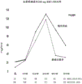
FIG. 1 shows that RBT-3 (360 mg) induces a rapid and significant increase in plasma hepcidin (hepcidin) levels in human subjects.
FIG. 2 shows that RBT-3 (240 mg) induces a rapid and significant increase in plasma hepcidin levels.
FIG. 3 shows that RBT-3 (120 mg) induces an increase in plasma hepcidin in Healthy Volunteers (HV) and CKD individuals.
Figure 4 shows an increase in plasma hepcidin levels relative to baseline values.
FIG. 5 shows the Nrf2 pathway in RBT-3 injection activated CD-1 mice.
FIGS. 6A-6B show that RBT-3 induces a significant increase in heme oxygenase 1 (HO-1) expression in the proximal tubules of mice, as assessed using immunohistochemistry.
Detailed Description
The present invention relates to the use of kidney protecting agents to selectively protect the kidney and/or liver (relative to cancer cells), and methods of using such kidney protecting agents, using chemotherapeutic agents to treat cancer, or using radiocontrast media to screen for cancer. In one aspect, the kidney protecting agent is an iron composition, such as iron sucrose (FeS). In another aspect, the kidney protecting agent is protoporphyrin (e.g., tin protoporphyrin SnPP). In another aspect, the invention relates to the administration of an iron composition in combination with a protoporphyrin during cancer chemotherapy or radiocontrast agent imaging. For example, iron sucrose and tin protoporphyrin are administered along with a chemotherapeutic agent.
THE present invention uses iron compositions, such as iron sucrose, for example RBT-3 as described in U.S. patent application No. 16/805,223 filed on even 20 in month 2020, entitled "novel iron composition and method of making and USING SAME (NOVEL IRON COMPOSITIONS AND METHODS OF MAKING AND USING SAME)" and claims priority from U.S. provisional application No. 62/812,028 filed on even 20 in month 2019, THE disclosure of which is incorporated herein in its entirety. Protoporphyrins include metalloporphyrins, such as tin protoporphyrin (SnPP). Various synthetic analogues of iron protoporphyrin IX are known. These compounds are commercially available and/or can be readily synthesized by known methods. It includes, for example, platinum, zinc, nickel, cobalt, copper, silver, manganese, chromium, and tin protoporphyrin IX. For convenience, these compounds may be generally referred to as Me-protoporphyrins or mepps, wherein Me represents a metal, and in particular, chemical symbols are used to represent metals such as Cr-protoporphyrin (CrPP), sn-protoporphyrin (SnPP), zn-protoporphyrin (ZnPP) representing chromium, tin, and zinc protoporphyrin compounds, respectively.
Chemotherapeutic agents that may be used in connection with the present invention include compounds within the class of platinum analogs, including cisplatin, as well as carboplatin, laxadine, or oxaliplatin.
Kidney protection during chemotherapy of iron compositions
Hepcidin is a well-established iron regulatory protein that is produced primarily in hepatocytes in response to macrophage iron content and pro-inflammatory conditions. With reference to non-R (Coffey R), granz T (Ganz T), iron homeostasis: human home view angle (ion homeostasis: an anthropocentric perspective), journal of biochemistry (J Biol chem.) 2017;292:12727-12734. Although it was originally thought to have antimicrobial properties (Michels K, namons E, ganz T, mirad B, mehrad B, hepcidin and host defense against infectious diseases (Hepcidin and host defense against infectious diseases), public science library pathogens (PLoS Patlog) 2015;11 (8): E1004998), it has been demonstrated in recent years to have acute kidney protective effects. Fan Shiwei m RP (Van Swelm RP), witzerland JF (Wetzels JF), fretzerland VG (Verweij VG) et al, treatment of circulating and kidney synthesized hepcidin by the kidney and its protective effect on hemoglobin-mediated kidney damage (Renal handling of circulating and renal-synthesized hepcidin and Its protective effects against hemoglobin-mediated kidney Injury) & journal of the American society of renal diseases (J Am Soc nephrol.) & 2016;27:2720-2732; shi Wangmi Nasen S (Swaminathan S.) the iron homeostasis pathway serves as a therapeutic target for acute kidney injury (Iron homeostasis pathways as therapeutic targets in acute kidney injury) & nephron (Nephron.) & 2018;140:156-159; xin Dier Y (Scindia Y), lazolone E (Wlazlo E), litz J (Leeds J) et al, the protective effect of hepcidin on polymicrobial sepsis and acute kidney injury (Protective role of hepcidin in polymicrobial sepsis and acute kidney injury.), pharmacological frontier (Front Pharmacol.) 2019;10:615 published on 6 th.6 th.2019, doi:10.3389/fphar.2019.00615; xin Dier Y (Scindia Y), dai Yi P (Dey P), sailurui A (Thirunagari A), H force level (Liping H), robin DL (Rosin DL), fu Luo Ruisai M (Floris M), mark D (Mark D.), grass MD (Okusa MD), shi Wangmi Nasen S (Swaminathan S), hepcidin slowed down renal ischemia-reperfusion injury by modulating systemic iron homeostasis (Hepcidin mitigates renal ischemia-reperfusion Injury by modulating systemic iron homeostasis.) "J.Am society of renal diseases (J Am Soc Nephrrol.)" 2015;26:2800-2814; king X (Wang X), zheng X (Zheng X), zhang J (Zhang J), zhao S (Zhao S), king Z (Wang Z), king F (Wang F), shang W, barach J (barach J), a (Qiu a), transport of ferrite, physiological functions in regulating renal iron recirculation and ischemic acute kidney injury (Physiological functions of ferroportin in the regulation of renal iron recycling and ischemic acute kidney injury.), "journal of physiology of the united states (Am J physiol.)" 2018;315:F1042-F1057. For example, administration of recombinant hepcidin has been shown to slow down experimental ischemic AKI.
In contrast, hepcidin-deficient mice are very prone to ischemic kidney injury (6). The mechanism by which hepcidin exerts its protective effect remains to be speculated. However, most of the attention has focused on the following potential paths (6, 7): i) Because of its small size (25 kDa), hepcidin undergoes rapid glomerular filtration, followed by endocytic uptake by the proximal tubular; ii) hepcidin binds to the iron export factor transferrin, causing its redistribution in cells and is subsequently destroyed by proteolysis; iii) The loss of ferrite can increase the content of catalytic iron in cells; and iv) elevation of cytosolic iron stimulates ferritin synthesis, which imparts a well-known cytoprotective/antioxidant effect. Johnson AC (Johnson AC), golley T (golley T), gilles JA (Guillem Keyser JA), plas Mu Sen H (Rasmussen H), singing B (Singh B), zager RA (Zager RA), parenteral sucrose iron-induced kidney preconditioning: differential expression of ferritin heavy and light chains in plasma, urine and viscera (Parenteral iron sucrose-induced renal preconditioning: differential ferritin heavy and light chain expression in plasma, shine, and internal organs.), "journal of physiology (Am J physiol.))" 2019;317:F1563-F1571; zarjiu a, borrelidin S (Bolisetty S), joseph R, et al, proximal tubular H ferritin mediated iron migration in acute kidney injury (Proximal tubule H-ferritin mediates iron trafficking in acute kidney injuriy.), "journal of clinical research (J Clin invest.)" 2013;123:4423-4434. However, other protection mechanisms are also possible. By way of example only, there may be a synergistic interaction between hepcidin and the cytoprotective Nrf2 pathway (10-13). Rimm PJ (Lim PJ), du Adi TL (Duarte TL), arez J (Arezes J), et al, nrf2 controls iron homeostasis via Bmp6 and hepcidin in hemochromatosis and thalassemia (Nrf 2 controls iron homeostasis in haemochromatosis and thalassaemia via Bmp and hepcidin.), "nature-metabolism (Nat metab.))" 2019;1:519-531; bei Shele HK (Bayele HK), baseres S (Balesaria S), silk Rayleigh SK (Srai SK), phytoestrogens regulate the expression of hepcidin by Nrf2: effects of dietary control on iron absorption (Phytoestrogens modulate hepcidin expression by Nrf2: implications for dietary control of iron absorption.), "Free radical biology and medicine (Free radical Biol med.))," 2015;89:1192-1202; field Y (Tanaka Y), pool T (Ikeda T), yamamoto K (Yamamoto K), smalls H (Ogawa H), sanding T (Kamisako T), deregulation of expression of fatty acid oxidase and iron regulatory genes in the liver of Nrf 2-free mice (Dysregulated expression of fatty acid oxidation enzymes and iron-regulatory genes in livers of Nrf2-null mice.), "journal of gastroenterology and hepatology (J gamcoelol hepatol.)" 2012;27:1711-1717; protofield N (Harada N), jinshan M (Kanayama M), boshan a (Maruyama a) et al, nrf2 regulates transferrin 1-mediated iron efflux in macrophages and counteracts lipopolysaccharide-induced transferrin 1mRNA inhibition (Nrf 2 regulates ferroportin 1-mediated iron efflux and counteracts lipopolysaccharide-induced ferroportin 1mRNA suppression in macrophages.), (Arch Biochem biophysics) 2011;508:101-109.
Given the emerging evidence that recombinant hepcidin may confer kidney protection against AKI, our question is whether the newly developed novel IV iron sucrose formulation (RBT-3), which has been demonstrated to be kidney safe when administered to healthy volunteers and CKD patients, can severely stimulate hepcidin production and thereby increase the renin content. RBT-3 is described in U.S. patent application No. 16/805,223, filed on even 20, 2, 2020, entitled "novel iron composition and method of making and USING SAME (NOVEL IRON COMPOSITIONS AND METHODS OF MAKING AND USING SAME)" and claims priority from U.S. provisional application 62/812,028 filed on even 20, 2, 2019, THE disclosure of which is incorporated herein in its entirety.
To explore these possibilities, we have administered different doses of RBT3 to Healthy Volunteers (HV) and individuals with stage 3 to stage 4 CKD and measured plasma hepcidin concentrations over a 72 hour period. Urinary hepcidin content was also analyzed in order to confirm renin delivery. In a supplemental mouse experiment, the potential effect of RBT-3 on hepcidin production in the liver and kidney cortex was probed. In view of the current concern over hepcidin-Nrf 2 interactions, the effect of RBT-3 on Nrf2 activity was evaluated. Finally, RBT-3 was tested for its ability to confer protection against clinically relevant AKI models, cisplatin nephrotoxicity and underlying mechanisms of action. The results of these additional clinical and experimental studies form the basis of this report.
It has been found that the cytotoxic effects of chemotherapeutic agents on the kidneys can be reduced by co-administration of chemotherapy with an iron composition. Co-administration of the radiocontrast agent with the iron composition may also reduce the cytotoxic effects of the radiocontrast agent. The iron composition is preferably an iron sucrose composition. The iron sucrose composition is preferably an iron sucrose composition comprising bicarbonate. In one example, the iron sucrose composition is RBT-3. The iron composition may have one or more of the following characteristics, including a fe2+/fe3+ ratio of about 1-10%, 2-5%, 3-4%, or about 3.4%; total iron content of 5-19mg/ml, 8-18mg/ml, 10-15mg/ml or about 12 mg/ml; and an organic carbon content of 4-11%, 6-9%, or about 7.7%; an osmolality of 1100-1600mOsm/Kg, 1400-1580mOsm/Kg, or about 1540 mOsm/Kg; and a core size of about 1-3nm, 2-2.8nm, or about 2.39 nm; a Na content of between 0.8% and 3%, 1% and 2%, or about 1.26%; and an average molecular weight of 10,000 to 30,000 daltons, 20,000 to 25,000 daltons, or about 23,881 daltons.
Hepcidin is a key regulator of systemic and intracellular iron homeostasis, and has recently been proposed to have tubular cytoprotective effects. As previously discussed, administration of recombinant hepcidin results in renal filtration, proximal tubular absorption and subsequent transferral degradation. Since transferrin is the only known cellular iron export factor, its degradation is believed to increase proximal tubular iron content, which then stimulates ferritin production. Given that ferritin is a strong antioxidant, it may be a key dominant player of the cytoprotective effect of hepcidin.
In two mice studies reported previously, experiments demonstrated that kidney protection against multiple forms of AKI (glycerol, maleate or ischemia-reperfusion) was produced within 18 to 24 hours of Venofer FeS administration. Johnson ACM (Johnson ACM), zager RA (Zager RA), oxidant-induced kidney preconditioning mechanism and outcome: nrf2-dependent, P21-independent anti-aging pathways (Mechanisms and consequences of oxidant-induced renal preconditioning: an Nrf2-dependent, P21-independent, anti-sendence pathway.), "kidney disease dialysis and transplantation (Nephrol Dial transfer.)" 2018;33:1927-1941; johnson AC (Johnson AC), becker K (Becker K), zager RA (Zager RA.), parenteral iron formulations differentially affect MCP-1, HO-1, and NGAL gene expression and kidney injury response (Parenteral iron formulations differentially affect MCP-1, HO-1,and NGAL gene expression and renal responses to injury.), "journal of physiology (Am JPhysiol)," 2010; 299F 426-F435.
This is believed to be due in part to the increase in FeS-driven proximal tubular ferritin content. Johnson AC (Johnson AC), golley T (golley T), gilles JA (Guillem Keyser JA), plas Mu Sen H (Rasmussen H), singing B (Singh B), zager RA (Zager RA), parenteral sucrose iron-induced kidney preconditioning: differential expression of ferritin heavy and light chains in plasma, urine and viscera (Parenteral iron sucrose-induced renal preconditioning: differential ferritin heavy and light chain expression in plasma, shine, and internal organs.), "journal of physiology (Am J physiol.))" 2019;317:F1563-F1571. However, venofer FeS has difficulty accessing the proximal tubular lumen. Zager RA (Zager RA), johnson AC (Johnson AC), hansen SY (Hanson SY.), parenteral iron nephrotoxicity: potential mechanisms and outcomes (Parenteral iron nephrotoxicity: potential mechanisms and sequences.), "international renal science (Kidney int.))" 2004;66:144-156. This suggests an increased substitution possibility for FeS driven ferritin: feS may stimulate hepcidin synthesis, which then triggers renin production via the above-mentioned pathway. However, it is not clear whether FeS can trigger rapid and sustained hepcidin production (i.e., over a 24 hour period), such as is necessary to facilitate the FeS-induced preconditioning state we previously mentioned.
To address this problem, we have evaluated the effect of novel FeS "RBT-3" on hepcidin production in healthy human individuals and patients with advanced CKD. Based on preliminary evidence from experiments (i.e., RBT-3 induced cytoprotective effect is more robust than equimolar Venofer), we selected this iron formulation for study. To gain more insight, the effect of RBT-3 on hepcidin expression in normal mice was assessed. As shown in fig. 1-3, a rapid, dose-dependent increase in plasma hepcidin was observed in both healthy and CKD individuals following RBT-3 injection. This response peaked at 24 hours and both groups of individuals showed an approximately 15-fold increase in plasma hepcidin in response to 240 or 360mg RBT-3 injections. The baseline plasma hepcidin levels of CKD individuals were elevated compared to healthy volunteers, which may reflect the pro-inflammatory state of CKD. Honda H (Honda H), fine N (Hosaka N), galnz T (Ganz T), futian T (Shibata T), iron metabolism of chronic kidney disease patients (Iron metabolism in chronic kidney disease components), contribution to nephrology (control nephrol.) "2019; 198:103-111; weiss G (Weiss G), galnz T (Ganz T), goldenafv LT (Goodnough LT), inflammatory anaemia (Anemia of infusion.), blood 2019;133:40-50.
This gives theoretical possibilities: in CKD patients, hepcidin increases have been maximized, preventing RBT-3 from further mediating hepcidin increases. However, this is clearly not the case given that the rise in absolute hepcidin from baseline value is almost identical in HV and CKD study groups (fig. 4). Of interest, the 24 hour peak drops by about 50% within 48 to 72 hours after RBT-3 injection. This may reflect that a decline in hepcidin production occurs in conjunction with rapid renin absorption and urinary hepcidin excretion. Indeed, the latter evidence is that at 24 hours after RBT-3 injection, an approximately 4-fold increase in urinary hepcidin concentrations occurred in both CKD and healthy volunteer groups.
Hepcidin production in response to iron loading is thought to be exclusively due to increased gene transcription, initiated by binding of saturated transferrin to its liver receptors (Tfr 1, trf 2). The BMP-SMAD pathway is then activated and then upregulates HAMP1 gene transcription. To confirm that RBT-3 injection activated this pathway, we measured HAMP1 mRNA in the liver and kidney of mice at 24 hours after RBT-3 administration. Surprisingly, a 10-fold greater response was observed in the kidneys, although a significant increase in HAMP1 mRNA was observed in both organs. To our knowledge, renal HAMP1 induction has not been previously reported to be preferred over hepatic HAMP1 induction as a response to Fe. This gives rise to interesting possibilities: feS/RBT-3 may trigger renin loading through both indirect (liver production) and direct (kidney derived) mechanisms.
To widen our understanding of the protective scope of FeS/RBT-3, we now tested whether it could be expressed against experimental cisplatin-induced ARF. The model was chosen for study based on four considerations: first, unlike the AKI model (ischemia, maleate, glycerol-induced rhabdomyolysis) that we previously tested was completely evident within about 24 hours, cisplatin nephrotoxicity developed slowly, requiring at least 3 days for complete manifestation of renal failure. Thus, it is unclear whether FeS/RBT-3 mediated protection can be exhibited over this extended period of time. Second, cisplatin adducts induce early significant DNA damage (e.g., DNA cross-linking), ultimately leading to apoptosis or necrotic cell death. Miller RP (Miller RP), tadalady RK (Tadagavadi RK), la Mi Shen G (ramish G), li Wei WB (Reeves WB.), cisplatin toxicity mechanism (Mechanisms of cisplatin toxicity level), toxin (toxins level) 2010;11:2490-2518.
In view of this unique injury initiation event, it is unclear whether RBT-3 can still confer protection. Third, cisplatin remains a widely used chemotherapeutic agent at a clinical AKI ratio of 25-30%. Latcha S, jimex EA, partel S, lattice Lei Zeman IG (Glezerman IG), mei Da S, mehta S, fu Long Bang CD (Flombaum CD), long-term renal outcome after cisplatin treatment (Long-term renal outcomes after cisplatin treatment.), "J.S. Clin J Am Soc Nephrol.)" 2016;11:1173-1179. Thus, a protective mechanism that slows cisplatin toxicity was identified that meets the currently unmet medical need; and, fourth, cisplatin administration is a predetermined clinical event. Accordingly, RBT-3 can be administered about 18 to 24 hours prior to cisplatin infusion, thereby providing the necessary time for the full development of FeS-mediated cell resistance status. Indeed, when RBT-3 was administered to mice at 18 hours prior to cisplatin injection, significant kidney protection was observed as evidenced by a dramatic drop in BUN, plasma creatinine, and plasma NAG concentrations (table 3). Thus, these findings add further support to the notion that FeS/RBT-3 preconditioning can broadly exert kidney protective effects based on potential clinical applicability.
The completely unexpected fourth protective effect induced by RBT-3 preconditioning is a significant inhibition of renal cisplatin uptake. Since cisplatin nephrotoxicity is highly dependent on proximal tubular cell uptake, a 40% reduction in renal cortex cisplatin content was observed to have to dominate the protective effect of RBT-3. Cisplatin uptake by proximal tubular cells is mediated via organic cation transporters (e.g., OCT2, MATE 1) located in the basal-lateral membrane. Although OCT2 transport is the primary determinant of cisplatin uptake by the proximal tubular, MATE1 ("multidrug and toxin extrusion protein 1") has recently been proposed to also be able to cause luminal cisplatin efflux (24). How RBT-3 affects the uptake and outflow pathways of these cells is not known. However, one possible hypothesis is that the hepcidin-MATE-1 interaction may increase proximal tubular cisplatin outflow. Stereimer S (Spreckelmeyer S), fan Deze M (van der Zee M), bertrand B, bert euro E (Bodio E), st Lu Pu S (St rup S), casini a (Casini a), as compared to cisplatin, correlation of copper and organic cation transporter in the activity and transport mechanisms of anticancer cyclometallated gold (III) compounds (Relevance of copper and organic cation transporters in the activity and transport mechanisms of an anticancer cyclometallated gold (III) compound in comparison to cispratin.), "chemical Front (Front chem.)" 2018;6:377; linkene TTG (Nieskens TTG), ties JGP (Peters JGP), dabaghie D et al, expression of organic anion transporter 1or 3in human kidney proximal tubular cells reduces cisplatin sensitivity (Expression of organic anion transporter 1or 3in human kidney proximal tubule cells reduces cisplatin sensitivity.), (Drug metabolism and treatment) 2018;46:592-599.
In many previous publications, our experiments have demonstrated that there are significant differences in the biological responses of the kidneys to different parenteral iron formulations, including iron dextran, iron gluconate, iron sucrose injections (venofer) and nano iron oxide (ferumoxytol). These differences include varying degrees of toxicity, oxidative stress severity, and potential pro-inflammatory effects. Most notably, only FeS (Venofer and now RBT-3) has been shown to induce kidney preconditioning effects. Indeed, in unpublished studies, we have found that RBT-3 confers about 35% greater kidney protection, although Venofer and RBT3 each slow down the glycerol AKI model. We have now demonstrated that RBT-3 induces a 5-fold greater renal HAMP1 response against RBT3 than does Venofer. In addition, RBT3 induced a kidney HO-1mRNA response twice that of Venofer. Thus, venofer and RBT3 clearly exert kidney cell protective protein responses that differ in number. It is not clear which of the physicochemical differences between Venofer and RBT-3 are responsible for these differences. However, these data clearly indicate that not all Fe formulations are similar and RBT3 appears to provide the greatest cytoprotective effect by this point.
We have demonstrated that IV RBT-3 injection can significantly (15 x) and rapidly (< 24 hours) induce renin loading. However, it should be noted that RBT-3 exerts an additional protective effect in addition to affecting hepcidin, which is not reproducible by recombinant hepcidin injection. The two most promising roles are: 1) The first recorded Nrf2 pathway in the proximal tubular in this study was activated; and 2) RBT-3 is capable of retarding renal tubular nephrotoxin absorption (or accumulation), at least in the case of cisplatin. In view of these considerations, it appears clear that RBT-3 induced preconditioning is mediated via a variety of cellular pathways above and beyond hepcidin loading. FeS, which has been shown to have excellent clinical safety profiles in humans, further demonstrates potential clinical value as a kidney protection/preconditioning agent. Finally, the data confirm that not all iron sucrose formulations exert the same biological effect. Based on the data collected so far, RBT3 appears to be the most promising in inducing a kidney preconditioning state.
Clinical study
Nine healthy volunteers (HV; eGFR >70ml/min/1.73m 2) and 9 patients with 3-to 4-phase CKD (15-59 ml/min/1.73m 2) were enrolled in this study. Individuals forming the basis of this study were also the basis of previous studies evaluating the differential effects of FeS on heavy and light chain ferritin expression (8). The study obtained IRB approval by Advarra IRB (Columbia, MD) and informed consent was obtained for each individual. The study was in compliance with the declaration of helsinki (Helsinki Declaration). Exclusion criteria included pregnancy, any overt medical condition other than the presence of CKD, iron administration for the first 30 days, or plasma ferritin concentrations >500ng/ml. Specific demographics, screening of eGFR (CKD-EPI formula), BUN, serum creatinine and blood pressure for both study groups are summarized in Table 1. More detailed information has been previously presented. Johnson AC (Johnson AC), golley T (golley T), gilles JA (Guillem Keyser JA), plas Mu Sen H (Rasmussen H), singing B (Singh B), zager RA (Zager RA), parenteral sucrose iron-induced kidney preconditioning: differential expression of ferritin heavy and light chains in plasma, urine and viscera (Parenteral iron sucrose-induced renal preconditioning: differential ferritin heavy and light chain expression in plasma, shine, and internal organs.), "journal of physiology (Am J physiol.))" 2019;317:F1563-F1571. The test has been entered into clinical.
For the purposes of this study, a novel FeS formulation "RBT-3" manufactured by Cascade Custom Chemistry (Portland, OR) of oregon was used. There are a number of physicochemical differences between RBT-3 and the widely used FeS formulation "Venofer" (data not disclosed). These differences include the following (RBT 3 versus Venofer, respectively): lower fe2+/fe3+ ratio: 3.4% versus 15.8%; lower total Fe content: 12mg/ml vs. 20mg/ml; lower organic carbon content: 7.7% vs. 12.14%; lower osmotic pressure: 1540 pair 1681mOsm/Kg; smaller core size: 2.39 to 3.88nm; higher Na content: 1.26% to 0.5%; lower average molecular weight: 23,881 for 31,335 daltons. In addition to these structural differences, quantitative differences in the selective biological response of RBT-3 to Venofer have also been noted (see discussion).
The HV and CKD groups were each divided into 3 equal groups (n is 3 each) each receiving 120, 240 or 360mg RBT-3 (12 mg/ml stock solution). RBT-3 doses (10, 20 or 30ml stock solutions) were infused with 100ml saline IV for 2 hours. Individuals were left overnight at the study site (riverside clinical institute (Riverside Clinical Research), FL of florida at edge Ji Wote (Edgewater) to screen for potential adverse events. Timed heparinized plasma samples were collected at baseline (0) and at 4, 12, 24 and 72 hours. On site urine samples were obtained 24 hours after baseline and RBT-3 infusion. Samples were assayed for hepcidin in duplicate using a commercially available ELISA (R & D Systems; minneapolis, minn.; kit #DY 8307). The hepcidin value of urine is decomposed according to the concentration of creatinine in urine. ELISA standard curve samples were provided by the manufacturer.
Mouse experiment
Rbt-3 induces hepcidin expression in mice. Male CD-1 mice (35-40 g; charles river laboratory (Charles River Labs), wilmington, mass.) were used for all animal studies approved by institutional IACUC. Mice were injected via tail vein with 1mg RBT-3 or vehicle (n is 5 each). Eighteen hours later, it was deeply anesthetized using pentobarbital (40 to 50 mg/Kg), the abdominal cavity was opened, blood samples were obtained from the vena cava, and then kidneys and liver were rapidly resected, frozen and total RNA and proteins were extracted (14). The renal cortex and hepcidin (HAMP 1) mRNA content was measured by competitive RT-PCR using the following primer pair: left: cagcagaacagaaggcatga; right: agatgcagatggggaagttg. mRNA values were resolved as the concurrently assayed GAPDH products (14). Plasma, liver and kidney cortex hepcidin levels were also determined by ELISA (14).
Effect of rbt-3 on mouse kidney Nrf2 expression. In addition to the effect of RBT-3 on hepcidin, kidney samples were collected from 5 control mice and 5 RBT-3 treated mice 4 hours post-injection in order to assess whether RBT-3 activated the Nrf2 pathway. Total mRNA was extracted and analyzed for 4 Nrf 2-activated genes using RT-PCR: 1) Heme oxygenase 1 (HO-1); 2) NAD (P) H quinone dehydrogenase 1 (NQO 1); 3) Thioredoxin-1 (SRXN 1); and 4) a glutamate-cysteine-ligase catalytic subunit (GCLC), as previously described (15). In addition, nrf2 nuclear translocation was assessed by extracting nuclear proteins and analyzing Nrf2 using ELISA, as previously performed in the present laboratory (15).
Immunohistochemical analysis of the effect of RBT-3 on proximal tubular HO-1 expression. To confirm that RBT 3-mediated Nrf2 activation causes an increase in Nrf 2-sensitive protein content in proximal tubular cells, HO-1 protein expression in kidneys obtained 18 hours after RBT-3 or vehicle injection was assessed. Formalin-fixed, paraffin-embedded tissues were cut into 4 micron sections on positively charged slides and baked at 60 ℃ for 1 hour. Slides were then dewaxed and stained using a Leica BOND dewaxing reagent (dewaxing solution) on a Leica BOND Rx staining instrument (Leica, buffalo Grove, IL), antigen retrieval was performed (epitope retrieval solution 2) and each step was followed by a rinse (BOND wash solution). Antigen retrieval was performed at 100 ℃ for 20 minutes, all other steps were performed at ambient temperature. Endogenous peroxidases were blocked with 3% H2O2 for 5 min, followed by blocking of the protein with TCT buffer (0.05M Tris, 0.15M NaCl, 0.25% casein, 0.1% Tween 20, 0.05% ProClin300 pH 7.6) for 10 min. Primary antibody HO-1 (Abcam ab 189491) was applied for 60 minutes followed by Leica anti-rabbit HRP polymer for 10 minutes and tertiary TSA amplification reagent (Perkinelmer OPAL 650, 1:100) for 10 minutes. Slides were removed from the staining apparatus and stained with DAPI for 5 minutes, rinsed, and blocked with Prolong Gold anti-fade reagents (Invitrogen/life technologies (Life Technologies)) in gland Island, NY.
Effect of rbt-3 preconditioning on cisplatin nephrotoxicity and renal cisplatin absorption. Mice were injected via the tail vein with 1mg RBT-3 or physiological saline vehicle (n is 5 each). After eighteen hours, all mice were injected with cisplatin (15 mg/Kg; IP). The whole process can take food and water at will. On the third day after injection, mice were deeply anesthetized with pentobarbital (50 mg/Kg IP), and blood samples were obtained from the inferior vena cava to determine BUN and creatinine concentrations. The plasma was also assessed for proximal tubular enzyme (N-acetylglucosaminidase ("NAG")) content (Bioassay Systems #DNAG-100; hayward, calif.). In addition, kidney cortex extracts were prepared in distilled water and cisplatin concentrations were determined according to the manufacturer's instructions (ProFoldin; gottingen, germany).
Comparison between rbt3 and Venofer. To demonstrate functional differences between RBT3 and Venofer in addition to structural differences, we compared hepcidin mRNA and HO-1mRNA responses between the two iron sucrose formulations. After injection of 1mg of RBT3 or Venofer for 24 hours into six mice, liver and kidney mRNA samples were obtained. Six normal samples provided control values.
And (5) statistics data. Each sample was analyzed in duplicate and the average was used for data presentation and analysis. Human hepcidin data are shown as mean ± 1 SD. Unpaired Situ Dengshi t test (unpaired Student's t test) was used to compare baseline plasmatic hepcidin values for HV and CKD groups. Comparison of hepcidin content over time using ANOVA analysis showed repeated measurements with 95% confidence intervals. Unpaired Situ Dengshi t-tests were used to compare baseline demographics. Mouse data are provided as mean ± 1 SEM. Significance was judged based on p-value < 0.05.
Baseline individual information. Selected demographics of the study population have been previously shown in detail. Table 1 provides an overview.
Table 1. Reviews of healthy individuals and individuals with stage 3 to stage 4 CKD (from reference 8).

Table 1 is a legend. Baseline characteristics of 9 Healthy Volunteers (HV) and 9 individuals with stage 3 to stage 4 CKD form the basis of the study. Mean and 95% confidence interval range = mean ± numbers in parentheses. P values were obtained using unpaired Situ Dengshi t-test.
CKD individuals are significantly older than HV and CKD men are fewer than HV groups. The average eGFR of the CKD group was 38.+ -. 8ml/min/1.73m 2 (about half of the HV group, average eGFR of the HV group is>70ml/min./1.73m 2 ). Lower eGFR in CKD individuals is manifested by elevated baseline and plasma creatinine concentrations (relative to those seen in HV).
Baseline and post RBT-3 plasma hepcidin concentrations in HV and CKD individuals. According to literature (16, 17), the baseline plasma hepcidin levels of CKD individuals were elevated relative to HV (26±25ng/ml versus 4±5ng/ml; p=0.001). Presumably, this reflects the pro-inflammatory state associated with CKD, and the decrease in GFR associated with CKD may lead to a decrease in renin clearance.
All individuals showed significant dose-dependent increases in plasma hepcidin on RBT-3 injections (fig. 1-3). An increase was observed as early as 4 hours after RBT-3 injection and reached a peak at 24 hours (fig. 1-3). At 240 and 360mg doses, the plasma hepcidin content at 24 hours was about 15 times higher than its baseline value. After 24 hours, a rapid decrease in plasma hepcidin was observed. However, at the last evaluation time point (72 hours), an approximately 2 to 4 fold increase in plasma hepcidin relative to baseline value was still observed (p <0.001 for all groups).
Despite the fact that CKD correlates with an increase in baseline hepcidin levels, these increases did not affect the extent to which RBT-3 caused an increase in plasma hepcidin. For example, the increase in the basal value was almost identical for HV and CKD groups at 24 hours, regardless of whether the individuals received 120, 240, or 360mg RBT-3 doses (fig. 4).
Results
Human urinary hepcidin concentrations in response to RBT-3 injections. At baseline, there was no significant difference in urinary hepcidin concentration between HV and CKD groups (47±36 versus 51±72ng/mg urinary creatinine, respectively). At 24 hours post RBT-3 injection, both groups showed an approximately 4-fold increase in urinary hepcidin relative to baseline (HV 219±104, p=0.04 relative to baseline; CKD 176±93, p=0.014 relative to baseline). Thus, these urine increases are consistent with increased filtration of circulating hepcidin content or possibly hepcidin outflow from the renal tubules.
Mice experiments.
1. Mouse HAMP1 gene expression; plasma, kidney and hepcidin response to RBT-3.
Baseline mouse liver HAMP1 mRNA expression (table 2) was approximately 25-fold higher than observed in the renal cortex, consistent with the liver being the primary site of hepcidin production (1). Although liver and kidney each respond to RBT-3 injections with an increase in HAMP1 mRNA, the degree of HAMP1 mRNA increase in the kidney was significantly greater than in the liver (multiplied by a factor of 10). However, the baseline and RBT-3 posthepcidin protein levels were comparable for both organs (about 5-fold increase over baseline; see Table 2). These increases are consistent with approximately 3-fold additions of RBT-3 induced plasma hepcidin levels (table 2).
Table 2 hepcidin expression in mouse liver, kidney and plasma at 18 hours after RBT-3 administration under control conditions.

Table 2 is a legend. Two groups contained 5 mice/group. HAMP 1mRNA was multiplied by GAPDH. Values are mean ± 1SEM. P values were obtained using unpaired Situ Dengshi test.
2.In response to RBT-3, the mouse renal cortex Nrf2 gene is activated.After RBT-3 injection, each of the 4 tested Nrf2 response genes showed a significant increase in its mRNA (see figure 5). Considering that HO-1mRNA was increased about 20 times the control level, the most responsive gene appeared to be HO-1. In addition, a significant increase in nuclear Nrf2 protein content was observed in RBT-3 treated groups compared to control groups (p=0.017).
3.HO-1 immunofluorescence method.Focal proximal tubular HO-1 staining was observed in control kidney tissue (FIG. 6). At 18 hours after RBT-3 injection, the HO-1 staining level and intensity increased greatly. The increase appears to be mainly limited to proximal tubular cells, which are manifested by strong cytoplasmic staining. An insignificant increase in glomerular or medullary HO-1 staining in response to RBT-3 injection was observed.
4.Effect of RBT-3 on cisplatin nephrotoxicity.On day 3 post cisplatin injection, control mice showed significant severe kidney injury as indicated by significant BUN and plasma creatinine elevation (table 3). RBT-3 preconditioning reduced cisplatin-induced damage by about 50% as assessed by BUN and creatinine levels. This protective effect, reflecting a reduction in proximal tubular injury, means that the release of NAG into the systemic circulation is almost completely blocked (table 3). Of interest, RBT-3 mediated protection was associated with (and likely attributable at least in part to) 40% reduction in renal cortex cisplatin concentration (table 3).
Table 3. Cisplatin-induced AKI: effect of RBT-3 preconditioning.
| Group of | BUN(mg/dl) | Creatinine (mg/dl) | Plasma NAG | Tissue cisplatin |
| Cisplatin (cisplatin) | 105±11 | 0.7±0.1 | 19.5±0.8 | 227±35 |
| cisplatin/RBT-3 | 48±5 | 0.43±0.03 | 9.8±1.2 | 142±9 |
| P value | 0.001 | 0.001 | =0.0025 | 0.048 |
| Normal value | 24±1 | 0.3±0.03 | 8.9±2.0 | 0 |
Table 3 is a legend. Values are shown as mean ± 1 SEM. BUN and creatinine values are mg/dL. The NAG value in plasma is the active unit per liter. Cisplatin values are μg/g wet tissue weight. Statistical comparisons were performed using unpaired Situ Dengshi t-test.
Venofer and RBT3 Comparison between FeS formulations.After RBT-3 injection, both liver and kidney had a significant increase in HAMP1mRNA values. However, the increase in kidney HAMP1mRNA using RBT3 was much greater (RBT-3 vs Venofer was 11-fold vs. 2-fold plus (p=0.015; table 4). In contrast, although both irons increased hepcidin mRNA, there was no difference in hepcidin protein concentration observed in the liver between Venofer vs. RBT-3. This indicates an increase in kidney-specific HAMP1 response. HO-1mRNA in kidney samples was also detected and there was a two-fold higher increase in RBT3 vs. Venofer therapy. These data clearly demonstrate the difference in biological response between the two FeS preparations.
TABLE 4 comparison between Venofer FeS versus RBT-3FeS

Table 4 is a legend. Mice were injected with 1mf Venofer FeS or RBT-3FeS and subsequently assessed 24 post-treatment for HAMP1mRNA and HO-1mRNA. RBT-3 induced an 11-fold increase in HAMP1 (hepcidin) mRNA compared to a 2.2-fold increase in Venofer induction. RBT-3 also induced HO-1mRNA multiplication compared to Venofer. P=0.015 (after log conversion of the data base 10). If not converted, p=0.037.
FIG. 1 RBT-3 (360 mg) induces a rapid and significant increase in plasma hepcidin levels in human subjects. An increase in plasma hepcidin was observed within 4 hours of RBT-3 injection (360 mg) and reached a peak at 24 hours (about 15 times baseline). The responses of Healthy Volunteers (HV) and CKD groups were observed to be almost identical. The 95% confidence intervals (±) for the HV and CKD groups are provided in the table at the bottom of the figure. P values were derived from ANOVA of duplicate measurements. HV = healthy volunteer; CKD = chronic kidney disease.
FIG. 2 RBT-3 (240 mg) induces a rapid and significant increase in plasma hepcidin levels. As with the 360mg dose, an increase in hepcidin was observed at 4 hours and peaked at the 24-hour time point. No significant differences were observed between CKD group and Healthy Volunteer (HV) group. The 95% confidence intervals (±) for Healthy Volunteers (HV) and CKD groups are provided in the table at the bottom of the figure. P values were derived from ANOVA of duplicate measurements.
FIG. 3 RBT-3 (120 mg) induces an increase in plasma hepcidin in Healthy Volunteers (HV) and CKD individuals. CKD individuals have plasma hepcidin levels higher than HV throughout the 72 hour period. Presumably, this reflects a higher baseline (initial) hepcidin content for the CKD group (see text). This was true in the 360mg, 240mg and 120mg RBT-3 treated groups (see text and right hand side panels of FIG. 4).
Fig. 4. Increase in plasma hepcidin levels relative to baseline values. Since baseline plasma hepcidin levels were elevated in CKD groups compared to healthy volunteers (HV; see right-hand side panels), RBT-3-induced increases relative to these baseline values (24-hour peak-baseline values) were calculated. Thereby allowing a comparison between CKD and hepcidin responses in healthy volunteers. Clearly, both the HV and CKD groups showed highly similar increases in hepcidin at each iron dose. Values are mean ± 95% confidence intervals. The 240mg and 360mg RBT-3 dose induced increases in hepcidin were similar, both greater than the observed for the 120mg RBT-3 dose group.
FIG. 5. Nrf2 pathway in CD-1 mice activated by RBT-3 injection. At 4 hours after RBT-3 injection into mice, a significant increase in each of the 4 tested Nrf2 response gene mrnas was observed (compared to control mice studied simultaneously). Another evidence of activation of the Nrf2 pathway is the discovery of increased nuclear Nrf2 protein binding in nuclear protein extracts. Heme oxygenase 1 (HO-1); NAD (P) H quinone dehydrogenase 1 (NQO 1); thioredoxin-1 (SRXN 1); and a glutamate-cysteine-ligase catalytic subunit (GCLC).
FIG. 6 RBT-3 induces a significant increase in heme oxygenase 1 (HO-1) expression in the proximal tubules of mice, as assessed by immunohistochemistry. The control kidney shown in the left hand side panel exhibited variable cytoplasmic staining in the proximal tubular section. In contrast, kidneys (right hand plot) collected at 18 hours post FeS injection showed a significant, nearly confluent increase in proximal tubular HO-1. In contrast, increased HO-1 staining in glomeruli was not evident, as depicted by the asterisks. Thus, these findings confirm that the HO-1mRNA changes depicted in FIG. 5 are reflected by: i) Increased HO-1 protein content, and ii) these increases occur in the proximal tubular.
Kidney protection of protoporphyrin during chemotherapy
SnPP (1. Mu. Mol) or vehicle (V) was administered on day zero, 15mg/kg cisplatin was administered on day 1, and the mRNA of BUN, creatinine, renal cortex LDH, and NGAL, MCP-1, IL-6, HO-1, and p21 was measured on day 3 after cisplatin injection. Compared to cisplatin alone, snPP confers statistically significant protection (P < 0.01) to all cisplatin-treated mice.

Other embodiments and uses of the invention will be apparent to those skilled in the art from consideration of the specification and practice of the invention disclosed herein. All references cited herein, including all U.S. and foreign patents and patent applications, are specifically and entirely incorporated by reference herein. It is intended that the specification and examples be considered as exemplary only, with a true scope and spirit of the invention being indicated by the following claims.
Claims (52)
1. A method for protecting the kidney during cancer chemotherapy, comprising:
administering a chemotherapeutic agent and an amount of an iron composition to a human patient, wherein the amount of the iron composition imparts a protective effect to the patient's kidneys against the cytotoxic effect of the chemotherapeutic agent.
2. The method of claim 1, wherein the chemotherapeutic agent is bevacizumab, gemcitabine, IFN therapy, cisplatin, ifosfamide, pemetrexed, cetuximab, or methotrexate, or a combination thereof.
3. The method of any one of the preceding claims, wherein the chemotherapeutic agent is cisplatin.
4. The method of any one of the preceding claims, wherein the method induces an increase in hepcidin in the human patient.
5. The method of any one of the preceding claims, wherein the iron composition is an iron sucrose composition.
6. The method of claim 5, wherein the iron sucrose composition comprises bicarbonate.
7. The method of claim 5, wherein the iron sucrose composition comprises a fe2+/fe3+ ratio of about 1 to 10%, a total iron content of 5 to 19mg/ml, an organic carbon content of 4 to 11%, an osmotic pressure of 1100 to 1600mOsM/Kg, an iron core size of about 1 to 3nm, a Na content of between 0.8% and 3%, and an average molecular weight of 10,000 to 30,000 daltons.
8. The method of claim 5, wherein the iron sucrose composition comprises a fe2+/fe3+ ratio of about 3 to 4%, a total iron content of 10 to 15mg/ml, an organic carbon content of 6 to 9%, an osmotic pressure of 1400 to 1580mOsm/Kg, an iron core size of about 2 to 2.8nm, a Na content of between 1% to 2%, and an average molecular weight of 20,000 to 25,000 daltons.
9. The method of claim 5, wherein the iron sucrose composition comprises a fe2+/fe3+ ratio of approximately 3.4%, a total iron content of approximately 12mg/ml, an organic carbon content of approximately 7.7%, an osmotic pressure of approximately 1540mOsm/Kg, an iron core size of approximately 2.39nm, a Na content of approximately 1.26%, and an average molecular weight of approximately 23,881 daltons.
10. A method for protecting kidneys during radiocontrast imaging comprising:
the radiocontrast agent and an amount of iron composition are administered to a human patient, wherein the amount of iron sucrose imparts a protective effect to the patient's kidneys against the cytotoxic effects of the radiocontrast agent.
11. The method of claim 10, wherein the radiocontrast agent is an iodine-containing radiocontrast agent.
12. The method of any one of claims 10 to 11, wherein the radiocontrast agent is a diatrizoate, a methodiatrizoate, an ionic iophthalic acid salt, an ionic ioxadate, iopamidol, iohexol, ioxilan, iopromide, iodixanol, ioversol, gadofacic, gadobenate, gadodiamide, gadofacic, gadofacitol, gadobutrol, or gadofacic, or a combination thereof.
13. The method of any one of claims 10-12, wherein the method induces an increase in hepcidin in the human patient.
14. The method of any one of claims 10-13, wherein the iron composition comprises an iron sucrose composition.
15. The method of claim 14, wherein the iron sucrose composition comprises bicarbonate.
16. The method of claim 14, wherein the iron sucrose composition comprises a fe2+/fe3+ ratio of about 1 to 10%, a total iron content of 5 to 19mg/ml, an organic carbon content of 4 to 11%, an osmotic pressure of 1100 to 1600mOsM/Kg, an iron core size of about 1 to 3nm, a Na content of between 0.8% and 3%, and an average molecular weight of 10,000 to 30,000 daltons.
17. The method of claim 14, wherein the iron sucrose composition comprises a fe2+/fe3+ ratio of about 3 to 4%, a total iron content of 10 to 15mg/ml, an organic carbon content of 6 to 9%, an osmotic pressure of 1400 to 1580mOsm/Kg, an iron core size of about 2 to 2.8nm, a Na content of between 1% to 2%, and an average molecular weight of 20,000 to 25,000 daltons.
18. The method of claim 14, wherein the iron sucrose composition comprises a fe2+/fe3+ ratio of approximately 3.4%, a total iron content of approximately 12mg/ml, an organic carbon content of approximately 7.7%, an osmotic pressure of approximately 1540mOsm/Kg, an iron core size of approximately 2.39nm, a Na content of approximately 1.26%, and an average molecular weight of approximately 23,881 daltons.
19. A method for protecting the kidney during cancer chemotherapy, comprising:
administering a chemotherapeutic agent and an amount of protoporphyrin to a human patient, wherein the amount of protoporphyrin imparts a protective effect to the patient's kidneys against the cytotoxic effect of the chemotherapeutic agent.
20. The method of claim 19, wherein the chemotherapeutic agent is bevacizumab, gemcitabine, IFN therapy, cisplatin, ifosfamide, pemetrexed, cetuximab, or methotrexate, or a combination thereof.
21. The method of any one of claims 19 to 20, wherein the chemotherapeutic agent is cisplatin.
22. The method of any one of claims 19-21, wherein the method induces an increase in hepcidin in the human patient.
23. The method of any one of claims 19-22, wherein the protoporphyrin is a metalloporphyrin.
24. The method of claim 23, wherein the metal protoporphyrin is tin protoporphyrin.
25. A method for protecting kidneys during radiocontrast imaging comprising:
the radiocontrast agent and an amount of protoporphyrin are administered to a human patient, wherein the amount of protoporphyrin imparts a protective effect to the patient's kidneys against the cytotoxic effects of the radiocontrast agent.
26. The method of claim 25, wherein the radiocontrast agent is an iodine-containing radiocontrast agent.
27. The method of any one of claims 25-26, wherein the radiocontrast agent is a diatrizoate, a methodiatrizoate, an ionic iophthalic acid salt, an ionic ioxadate, iopamidol, iohexol, ioxilan, iopromide, iodixanol, ioversol, gadofacic, gadobenate, gadodiamide, gadofacic, gadofacitol, gadobutrol, or gadofacic, or a combination thereof.
28. The method of any one of claims 25-27, wherein the method induces an increase in hepcidin in the human patient.
29. The method of any one of claims 25-28, wherein the protoporphyrin is a metalloporphyrin.
30. The method of claim 29, wherein the metal protoporphyrin is tin protoporphyrin.
31. A method for protecting the kidney during cancer chemotherapy, comprising:
administering a chemotherapeutic agent and an amount of protoporphyrin and an amount of iron composition to a human patient, wherein the amount of protoporphyrin and the amount of iron composition confer protection to the kidney of the patient against the cytotoxic effect of the chemotherapeutic agent.
32. The method of claim 31, wherein the chemotherapeutic agent is bevacizumab, gemcitabine, IFN therapy, cisplatin, ifosfamide, pemetrexed, cetuximab, or methotrexate, or a combination thereof.
33. The method of any one of claims 31-32, wherein the chemotherapeutic agent is cisplatin.
34. The method of any one of claims 31-33, wherein the method induces an increase in hepcidin in the human patient.
35. The method of any one of claims 31-34, wherein the protoporphyrin is a metalloporphyrin.
36. The method of claim 35, wherein the metal protoporphyrin is tin protoporphyrin.
37. The method of any one of claims 31-36, wherein the iron composition is an iron sucrose composition.
38. The method of claim 37, wherein the iron sucrose comprises bicarbonate.
39. The method of claim 37, wherein the iron sucrose composition comprises a fe2+/fe3+ ratio of about 1 to 10%, a total iron content of 5 to 19mg/ml, an organic carbon content of 4 to 11%, an osmotic pressure of 1100 to 1600mOsM/Kg, an iron core size of about 1 to 3nm, a Na content of between 0.8% and 3%, and an average molecular weight of 10,000 to 30,000 daltons.
40. The method of claim 37, wherein the iron sucrose composition comprises a fe2+/fe3+ ratio of about 3 to 4%, a total iron content of 10 to 15mg/ml, an organic carbon content of 6 to 9%, an osmotic pressure of 1400 to 1580mOsm/Kg, an iron core size of about 2 to 2.8nm, a Na content of between 1% to 2%, and an average molecular weight of 20,000 to 25,000 daltons.
41. The method of claim 37, wherein the iron sucrose composition comprises a fe2+/fe3+ ratio of approximately 3.4%, a total iron content of approximately 12mg/ml, an organic carbon content of approximately 7.7%, an osmotic pressure of approximately 1540mOsm/Kg, an iron core size of approximately 2.39nm, a Na content of approximately 1.26%, and an average molecular weight of approximately 23,881 daltons.
42. A method for protecting kidneys during radiocontrast imaging comprising:
administering the radiocontrast agent and an amount of protoporphyrin and an amount of iron composition to a human patient, wherein the amount of protoporphyrin and the amount of iron composition confer protection to the patient's kidneys against the cytotoxic effects of the radiocontrast agent.
43. The method of claim 42, wherein the radiocontrast agent is an iodine-containing radiocontrast agent.
44. The method of any one of claims 42 to 43, wherein the radiocontrast agent is a diatrizoate, a methodiatrizoate, an ionic iophthalic acid salt, an ionic ioxadate, iopamidol, iohexol, ioxilan, iopromide, iodixanol, ioversol, gadofacic, gadobenate, gadodiamide, gadofacic, gadofacitol, gadobutrol, or gadofacic, or a combination thereof.
45. The method of any one of claims 42-44, wherein the method induces an increase in hepcidin in the human patient.
46. The method of any one of claims 42 to 45, wherein the protoporphyrin is a metalloporphyrin.
47. The method of claim 46, wherein the metal protoporphyrin is tin protoporphyrin.
48. The method of any one of claims 42-47, wherein the iron composition is an iron sucrose composition.
49. The method of claim 48, wherein the iron sucrose composition comprises bicarbonate.
50. The method of claim 48, wherein the iron sucrose composition comprises a fe2+/fe3+ ratio of about 1 to 10%, a total iron content of 5 to 19mg/ml, an organic carbon content of 4 to 11%, an osmotic pressure of 1100 to 1600mOsM/Kg, an iron core size of about 1 to 3nm, a Na content of between 0.8% and 3%, and an average molecular weight of 10,000 to 30,000 daltons.
51. The method of claim 48, wherein the iron sucrose composition comprises a fe2+/fe3+ ratio of about 3 to 4%, a total iron content of 10 to 15mg/ml, an organic carbon content of 6 to 9%, an osmotic pressure of 1400 to 1580mOsm/Kg, an iron core size of about 2 to 2.8nm, a Na content of between 1% and 2%, and an average molecular weight of 20,000 to 25,000 daltons.
52. The method of claim 48, wherein the iron sucrose composition comprises a fe2+/fe3+ ratio of approximately 3.4%, a total iron content of approximately 12mg/ml, an organic carbon content of approximately 7.7%, an osmotic pressure of approximately 1540mOsm/Kg, an iron core size of approximately 2.39nm, a Na content of approximately 1.26%, and an average molecular weight of approximately 23,881 daltons.
Applications Claiming Priority (5)
| Application Number | Priority Date | Filing Date | Title |
|---|---|---|---|
| US202062706829P | 2020-09-11 | 2020-09-11 | |
| US62/706,829 | 2020-09-11 | ||
| US202163158803P | 2021-03-09 | 2021-03-09 | |
| US63/158,803 | 2021-03-09 | ||
| PCT/US2021/050064 WO2022056378A1 (en) | 2020-09-11 | 2021-09-13 | Method for treating cancer with kidney protection |
Publications (1)
| Publication Number | Publication Date |
|---|---|
| CN116348107A true CN116348107A (en) | 2023-06-27 |
Family
ID=80626052
Family Applications (1)
| Application Number | Title | Priority Date | Filing Date |
|---|---|---|---|
| CN202180070528.4A Pending CN116348107A (en) | 2020-09-11 | 2021-09-13 | Methods of treating cancer by kidney protection |
Country Status (11)
| Country | Link |
|---|---|
| US (1) | US20220079984A1 (en) |
| EP (1) | EP4210691A1 (en) |
| JP (1) | JP2023541606A (en) |
| KR (1) | KR20230112608A (en) |
| CN (1) | CN116348107A (en) |
| AU (1) | AU2021342291A1 (en) |
| BR (1) | BR112023004583A2 (en) |
| CA (1) | CA3192411A1 (en) |
| IL (1) | IL301305A (en) |
| MX (1) | MX2023002929A (en) |
| WO (1) | WO2022056378A1 (en) |
Family Cites Families (5)
| Publication number | Priority date | Publication date | Assignee | Title |
|---|---|---|---|---|
| US7674780B2 (en) * | 2004-03-16 | 2010-03-09 | Navinta Llc | Iron sucrose complexes and method of manufacture thereof |
| CN106975070A (en) * | 2008-02-26 | 2017-07-25 | 康奈尔大学 | Method for preventing and treating acute injury of kidney |
| FI3200582T3 (en) * | 2014-09-29 | 2024-02-14 | Fred Hutchinson Cancer Center | Compositions, kits, and methods to induce acquired cytoresistance using stress protein inducers |
| US20200330603A1 (en) * | 2015-12-11 | 2020-10-22 | Fred Hutchinson Cancer Research Center | Peptides for renal therapy |
| FI3930686T3 (en) * | 2019-02-28 | 2023-09-01 | Renibus Therapeutics Inc | Novel iron compositions and methods of making and using the same |
-
2021
- 2021-09-13 JP JP2023516137A patent/JP2023541606A/en active Pending
- 2021-09-13 CA CA3192411A patent/CA3192411A1/en active Pending
- 2021-09-13 KR KR1020237011800A patent/KR20230112608A/en unknown
- 2021-09-13 CN CN202180070528.4A patent/CN116348107A/en active Pending
- 2021-09-13 MX MX2023002929A patent/MX2023002929A/en unknown
- 2021-09-13 US US17/472,954 patent/US20220079984A1/en active Pending
- 2021-09-13 WO PCT/US2021/050064 patent/WO2022056378A1/en unknown
- 2021-09-13 EP EP21867758.1A patent/EP4210691A1/en active Pending
- 2021-09-13 IL IL301305A patent/IL301305A/en unknown
- 2021-09-13 AU AU2021342291A patent/AU2021342291A1/en active Pending
- 2021-09-13 BR BR112023004583A patent/BR112023004583A2/en unknown
Also Published As
| Publication number | Publication date |
|---|---|
| JP2023541606A (en) | 2023-10-03 |
| WO2022056378A1 (en) | 2022-03-17 |
| BR112023004583A2 (en) | 2023-04-11 |
| IL301305A (en) | 2023-05-01 |
| EP4210691A1 (en) | 2023-07-19 |
| US20220079984A1 (en) | 2022-03-17 |
| CA3192411A1 (en) | 2022-03-17 |
| AU2021342291A1 (en) | 2023-04-20 |
| KR20230112608A (en) | 2023-07-27 |
| MX2023002929A (en) | 2023-05-22 |
Similar Documents
| Publication | Publication Date | Title |
|---|---|---|
| Su et al. | Ferroptosis, a novel pharmacological mechanism of anti-cancer drugs | |
| Heemskerk et al. | Selective iNOS inhibition for the treatment of sepsis-induced acute kidney injury | |
| Kaplan et al. | The effect of renal insufficiency on mycophenolic acid protein binding | |
| Lee et al. | Blockade of RAGE ameliorates elastase-induced emphysema development and progression via RAGE-DAMP signaling | |
| R Rosenstock et al. | Mitochondrial-associated metabolic changes and neurodegeneration in Huntington's disease-from clinical features to the bench | |
| Sessa et al. | Clinical and pharmacological phase I study with accelerated titration design of a daily times five schedule of BBR3464, a novel cationic triplatinum complex | |
| JP6894837B2 (en) | Compositions, Kits and Methods for Inducing Acquired Cell Resistance Using Stress Protein Inducers | |
| Yildirim et al. | Effects of aminoguanidine and antioxidant erdosteine on bleomycin-induced lung fibrosis in rats | |
| Samal et al. | Administration of nitrite after chlorine gas exposure prevents lung injury: effect of administration modality | |
| Erikson et al. | Glutamate/aspartate transporter (GLAST), taurine transporter and metallothionein mRNA levels are differentially altered in astrocytes exposed to manganese chloride, manganese phosphate or manganese sulfate | |
| Fão et al. | Mitochondrial and redox-based therapeutic strategies in Huntington's disease | |
| Arora et al. | Amifostine analog, DRDE-30, alleviates radiation induced lung damage by attenuating inflammation and fibrosis | |
| Wu et al. | Cholecalciferol pretreatment ameliorates ischemia/reperfusion-induced acute kidney injury through inhibiting ROS production, NF-κB pathway and pyroptosis | |
| Shiels et al. | Pharmacokinetics of bilirubin-10-sulfonate and biliverdin in the rat | |
| Chitambar et al. | Evaluation of transferrin and gallium-pyridoxal isonicotinoyl hydrazone as potential therapeutic agents to overcome lymphoid leukemic cell resistance to gallium nitrate. | |
| CN116348107A (en) | Methods of treating cancer by kidney protection | |
| Mora et al. | Intestinal uptake and toxicity evaluation of acetazolamide and its multicomponent complexes with hidroxypropyl-β-cyclodextrin in rats | |
| AU2011210756B2 (en) | Method For Treating Liver Disorders With Receptor Associated Protein (RAP) Peptide-Fucosidase Inhibitor Conjugates | |
| Gao et al. | The bs-YHEDA peptide protects the brains of senile mice and thus recovers intelligence by reducing iron and free radicals | |
| Dariushnejad et al. | Sodium butyrate and voluntary exercise through activating VEGF-A downstream signaling pathway improve heart angiogenesis in type 2 diabetes | |
| Embaby et al. | The combined effect of zinc oxide nanoparticles and milrinone on acute renal ischemia/reperfusion injury in rats: Potential underlying mechanisms | |
| AU2009201920A1 (en) | Prognostic method | |
| Johnson et al. | Parenterial iron sucrose-induced renal preconditioning: Differential ferritin heavy and light chain expression in plasma, urine, and internal organs | |
| Yamashita et al. | Inhibitory effect of α-tocopherol on methylmercury-induced oxidative steress | |
| Galgamuwa Arachchige | An Investigation Into The Prevention Of Cisplatin-Induced Nephrotoxicity By Dichloroacetate |
Legal Events
| Date | Code | Title | Description |
|---|---|---|---|
| PB01 | Publication | ||
| PB01 | Publication | ||
| SE01 | Entry into force of request for substantive examination | ||
| SE01 | Entry into force of request for substantive examination | ||
| REG | Reference to a national code |
Ref country code: HK Ref legal event code: DE Ref document number: 40095700 Country of ref document: HK |