CN116096756A - Antibodies against ILT2 and uses thereof - Google Patents
Antibodies against ILT2 and uses thereof Download PDFInfo
- Publication number
- CN116096756A CN116096756A CN202180056995.1A CN202180056995A CN116096756A CN 116096756 A CN116096756 A CN 116096756A CN 202180056995 A CN202180056995 A CN 202180056995A CN 116096756 A CN116096756 A CN 116096756A
- Authority
- CN
- China
- Prior art keywords
- seq
- antibody
- ilt2
- antibodies
- acid sequence
- Prior art date
- Legal status (The legal status is an assumption and is not a legal conclusion. Google has not performed a legal analysis and makes no representation as to the accuracy of the status listed.)
- Pending
Links
- 102000004569 Leukocyte Immunoglobulin-like Receptor B1 Human genes 0.000 title claims description 337
- 108010017736 Leukocyte Immunoglobulin-like Receptor B1 Proteins 0.000 title claims description 337
- 230000027455 binding Effects 0.000 claims abstract description 287
- 239000000427 antigen Substances 0.000 claims abstract description 239
- 102000036639 antigens Human genes 0.000 claims abstract description 239
- 108091007433 antigens Proteins 0.000 claims abstract description 239
- 206010028980 Neoplasm Diseases 0.000 claims abstract description 223
- 239000012634 fragment Substances 0.000 claims abstract description 192
- 201000011510 cancer Diseases 0.000 claims abstract description 158
- 238000000034 method Methods 0.000 claims abstract description 124
- 239000008194 pharmaceutical composition Substances 0.000 claims abstract description 23
- 210000004027 cell Anatomy 0.000 claims description 216
- 125000003275 alpha amino acid group Chemical group 0.000 claims description 148
- 239000003795 chemical substances by application Substances 0.000 claims description 111
- 108010024164 HLA-G Antigens Proteins 0.000 claims description 94
- 230000001965 increasing effect Effects 0.000 claims description 92
- 108090000765 processed proteins & peptides Proteins 0.000 claims description 91
- 102100028967 HLA class I histocompatibility antigen, alpha chain G Human genes 0.000 claims description 82
- 241000282414 Homo sapiens Species 0.000 claims description 82
- 230000014509 gene expression Effects 0.000 claims description 82
- 102000004196 processed proteins & peptides Human genes 0.000 claims description 78
- 210000002540 macrophage Anatomy 0.000 claims description 77
- 230000000694 effects Effects 0.000 claims description 73
- 206010057249 Phagocytosis Diseases 0.000 claims description 58
- 230000008782 phagocytosis Effects 0.000 claims description 58
- 230000003993 interaction Effects 0.000 claims description 56
- 108010081355 beta 2-Microglobulin Proteins 0.000 claims description 52
- 238000002560 therapeutic procedure Methods 0.000 claims description 52
- 102000015736 beta 2-Microglobulin Human genes 0.000 claims description 51
- 150000007523 nucleic acids Chemical class 0.000 claims description 50
- 210000004443 dendritic cell Anatomy 0.000 claims description 49
- 210000000822 natural killer cell Anatomy 0.000 claims description 46
- 229920001184 polypeptide Polymers 0.000 claims description 36
- 238000011282 treatment Methods 0.000 claims description 36
- 108091028043 Nucleic acid sequence Proteins 0.000 claims description 34
- 230000035772 mutation Effects 0.000 claims description 34
- 238000004519 manufacturing process Methods 0.000 claims description 33
- 230000003013 cytotoxicity Effects 0.000 claims description 30
- 231100000135 cytotoxicity Toxicity 0.000 claims description 30
- 101000984190 Homo sapiens Leukocyte immunoglobulin-like receptor subfamily B member 1 Proteins 0.000 claims description 19
- 230000002757 inflammatory effect Effects 0.000 claims description 19
- 102000039446 nucleic acids Human genes 0.000 claims description 17
- 108020004707 nucleic acids Proteins 0.000 claims description 17
- 108090000623 proteins and genes Proteins 0.000 claims description 16
- 230000003750 conditioning effect Effects 0.000 claims description 15
- 239000003814 drug Substances 0.000 claims description 15
- 102000054957 human LILRB1 Human genes 0.000 claims description 15
- 238000009169 immunotherapy Methods 0.000 claims description 14
- 230000002401 inhibitory effect Effects 0.000 claims description 14
- 102000004169 proteins and genes Human genes 0.000 claims description 14
- 230000003247 decreasing effect Effects 0.000 claims description 13
- 230000005764 inhibitory process Effects 0.000 claims description 11
- 230000005867 T cell response Effects 0.000 claims description 7
- 230000002708 enhancing effect Effects 0.000 claims description 7
- 239000000546 pharmaceutical excipient Substances 0.000 claims description 7
- 230000020411 cell activation Effects 0.000 claims description 6
- 230000006052 T cell proliferation Effects 0.000 claims description 5
- 210000001185 bone marrow Anatomy 0.000 claims description 5
- 229960005395 cetuximab Drugs 0.000 claims description 5
- 229940121647 egfr inhibitor Drugs 0.000 claims description 5
- 229960002621 pembrolizumab Drugs 0.000 claims description 5
- 230000000259 anti-tumor effect Effects 0.000 claims description 4
- 230000001939 inductive effect Effects 0.000 claims description 3
- 238000012258 culturing Methods 0.000 claims description 2
- 239000013604 expression vector Substances 0.000 claims description 2
- 230000001662 opsonic effect Effects 0.000 claims 1
- 239000000203 mixture Substances 0.000 abstract description 19
- 238000010187 selection method Methods 0.000 abstract description 2
- 101710089372 Programmed cell death protein 1 Proteins 0.000 description 51
- 102100040678 Programmed cell death protein 1 Human genes 0.000 description 51
- 210000001744 T-lymphocyte Anatomy 0.000 description 47
- 210000002865 immune cell Anatomy 0.000 description 47
- 230000000903 blocking effect Effects 0.000 description 39
- 102000008096 B7-H1 Antigen Human genes 0.000 description 37
- 108010074708 B7-H1 Antigen Proteins 0.000 description 37
- 108091054437 MHC class I family Proteins 0.000 description 35
- 102000043129 MHC class I family Human genes 0.000 description 35
- 230000028327 secretion Effects 0.000 description 34
- 108010047041 Complementarity Determining Regions Proteins 0.000 description 33
- 230000001506 immunosuppresive effect Effects 0.000 description 30
- 241000699670 Mus sp. Species 0.000 description 28
- 206010062016 Immunosuppression Diseases 0.000 description 27
- 210000003819 peripheral blood mononuclear cell Anatomy 0.000 description 25
- 235000001014 amino acid Nutrition 0.000 description 24
- 230000001404 mediated effect Effects 0.000 description 24
- 238000012360 testing method Methods 0.000 description 22
- 150000001413 amino acids Chemical class 0.000 description 21
- 210000004881 tumor cell Anatomy 0.000 description 21
- 210000001616 monocyte Anatomy 0.000 description 18
- 102000004127 Cytokines Human genes 0.000 description 17
- 108090000695 Cytokines Proteins 0.000 description 17
- 241000880493 Leptailurus serval Species 0.000 description 17
- 231100000673 dose–response relationship Toxicity 0.000 description 17
- 108091033319 polynucleotide Proteins 0.000 description 17
- 102000040430 polynucleotide Human genes 0.000 description 17
- 239000002157 polynucleotide Substances 0.000 description 17
- 108010073969 valyllysine Proteins 0.000 description 17
- WHUUTDBJXJRKMK-UHFFFAOYSA-N Glutamic acid Natural products OC(=O)C(N)CCC(O)=O WHUUTDBJXJRKMK-UHFFFAOYSA-N 0.000 description 16
- 108010021625 Immunoglobulin Fragments Proteins 0.000 description 16
- 102000008394 Immunoglobulin Fragments Human genes 0.000 description 16
- 108010061238 threonyl-glycine Proteins 0.000 description 16
- BVLIJXXSXBUGEC-SRVKXCTJSA-N Asn-Asn-Tyr Chemical compound [H]N[C@@H](CC(N)=O)C(=O)N[C@@H](CC(N)=O)C(=O)N[C@@H](CC1=CC=C(O)C=C1)C(O)=O BVLIJXXSXBUGEC-SRVKXCTJSA-N 0.000 description 14
- DNAZKGFYFRGZIH-QWRGUYRKSA-N Gly-Tyr-Ser Chemical compound OC[C@@H](C(O)=O)NC(=O)[C@@H](NC(=O)CN)CC1=CC=C(O)C=C1 DNAZKGFYFRGZIH-QWRGUYRKSA-N 0.000 description 14
- 108060003951 Immunoglobulin Proteins 0.000 description 14
- 238000003556 assay Methods 0.000 description 14
- 210000003690 classically activated macrophage Anatomy 0.000 description 14
- 208000037265 diseases, disorders, signs and symptoms Diseases 0.000 description 14
- 108010050848 glycylleucine Proteins 0.000 description 14
- 102000018358 immunoglobulin Human genes 0.000 description 14
- 108010051242 phenylalanylserine Proteins 0.000 description 14
- 230000000770 proinflammatory effect Effects 0.000 description 14
- YTJFXEDRUOQGSP-DCAQKATOSA-N Lys-Pro-Ser Chemical compound [H]N[C@@H](CCCCN)C(=O)N1CCC[C@H]1C(=O)N[C@@H](CO)C(O)=O YTJFXEDRUOQGSP-DCAQKATOSA-N 0.000 description 13
- 210000004322 M2 macrophage Anatomy 0.000 description 13
- 241000699666 Mus <mouse, genus> Species 0.000 description 13
- YUJLIIRMIAGMCQ-CIUDSAMLSA-N Ser-Leu-Ser Chemical compound [H]N[C@@H](CO)C(=O)N[C@@H](CC(C)C)C(=O)N[C@@H](CO)C(O)=O YUJLIIRMIAGMCQ-CIUDSAMLSA-N 0.000 description 13
- MNYNCKZAEIAONY-XGEHTFHBSA-N Thr-Val-Ser Chemical compound C[C@@H](O)[C@H](N)C(=O)N[C@@H](C(C)C)C(=O)N[C@@H](CO)C(O)=O MNYNCKZAEIAONY-XGEHTFHBSA-N 0.000 description 13
- 108010068265 aspartyltyrosine Proteins 0.000 description 13
- 108010078144 glutaminyl-glycine Proteins 0.000 description 13
- 239000013641 positive control Substances 0.000 description 13
- 108010003137 tyrosyltyrosine Proteins 0.000 description 13
- 108010017213 Granulocyte-Macrophage Colony-Stimulating Factor Proteins 0.000 description 12
- 102100039620 Granulocyte-macrophage colony-stimulating factor Human genes 0.000 description 12
- YOOAQCZYZHGUAZ-KATARQTJSA-N Thr-Leu-Ser Chemical compound [H]N[C@@H]([C@@H](C)O)C(=O)N[C@@H](CC(C)C)C(=O)N[C@@H](CO)C(O)=O YOOAQCZYZHGUAZ-KATARQTJSA-N 0.000 description 12
- XBGGUPMXALFZOT-UHFFFAOYSA-N glycyl-L-tyrosine hemihydrate Natural products NCC(=O)NC(C(O)=O)CC1=CC=C(O)C=C1 XBGGUPMXALFZOT-UHFFFAOYSA-N 0.000 description 12
- 108010031719 prolyl-serine Proteins 0.000 description 12
- 235000018102 proteins Nutrition 0.000 description 12
- SNDBKTFJWVEVPO-WHFBIAKZSA-N Asp-Gly-Ser Chemical compound [H]N[C@@H](CC(O)=O)C(=O)NCC(=O)N[C@@H](CO)C(O)=O SNDBKTFJWVEVPO-WHFBIAKZSA-N 0.000 description 11
- LINKCQUOMUDLKN-KATARQTJSA-N Leu-Thr-Cys Chemical compound C[C@H]([C@@H](C(=O)N[C@@H](CS)C(=O)O)NC(=O)[C@H](CC(C)C)N)O LINKCQUOMUDLKN-KATARQTJSA-N 0.000 description 11
- SIEBDTCABMZCLF-XGEHTFHBSA-N Ser-Val-Thr Chemical compound [H]N[C@@H](CO)C(=O)N[C@@H](C(C)C)C(=O)N[C@@H]([C@@H](C)O)C(O)=O SIEBDTCABMZCLF-XGEHTFHBSA-N 0.000 description 11
- MTCFGRXMJLQNBG-UHFFFAOYSA-N Serine Natural products OCC(N)C(O)=O MTCFGRXMJLQNBG-UHFFFAOYSA-N 0.000 description 11
- IGROJMCBGRFRGI-YTLHQDLWSA-N Thr-Ala-Ala Chemical compound C[C@@H](O)[C@H](N)C(=O)N[C@@H](C)C(=O)N[C@@H](C)C(O)=O IGROJMCBGRFRGI-YTLHQDLWSA-N 0.000 description 11
- 102000004142 Trypsin Human genes 0.000 description 11
- 108090000631 Trypsin Proteins 0.000 description 11
- 108010092854 aspartyllysine Proteins 0.000 description 11
- 239000012588 trypsin Substances 0.000 description 11
- UPAGTDJAORYMEC-VHWLVUOQSA-N Asn-Trp-Ile Chemical compound CC[C@H](C)[C@@H](C(=O)O)NC(=O)[C@H](CC1=CNC2=CC=CC=C21)NC(=O)[C@H](CC(=O)N)N UPAGTDJAORYMEC-VHWLVUOQSA-N 0.000 description 10
- MNQMTYSEKZHIDF-GCJQMDKQSA-N Asp-Thr-Ala Chemical compound [H]N[C@@H](CC(O)=O)C(=O)N[C@@H]([C@@H](C)O)C(=O)N[C@@H](C)C(O)=O MNQMTYSEKZHIDF-GCJQMDKQSA-N 0.000 description 10
- 102100025137 Early activation antigen CD69 Human genes 0.000 description 10
- MIIVFRCYJABHTQ-ONGXEEELSA-N Gly-Leu-Val Chemical compound [H]NCC(=O)N[C@@H](CC(C)C)C(=O)N[C@@H](C(C)C)C(O)=O MIIVFRCYJABHTQ-ONGXEEELSA-N 0.000 description 10
- 101000934374 Homo sapiens Early activation antigen CD69 Proteins 0.000 description 10
- WCNWGAUZWWSYDG-SVSWQMSJSA-N Ile-Thr-Ser Chemical compound CC[C@H](C)[C@@H](C(=O)N[C@@H]([C@@H](C)O)C(=O)N[C@@H](CO)C(=O)O)N WCNWGAUZWWSYDG-SVSWQMSJSA-N 0.000 description 10
- DPWGZWUMUUJQDT-IUCAKERBSA-N Leu-Gln-Gly Chemical compound CC(C)C[C@H](N)C(=O)N[C@@H](CCC(N)=O)C(=O)NCC(O)=O DPWGZWUMUUJQDT-IUCAKERBSA-N 0.000 description 10
- SKRGVGLIRUGANF-AVGNSLFASA-N Lys-Leu-Glu Chemical compound [H]N[C@@H](CCCCN)C(=O)N[C@@H](CC(C)C)C(=O)N[C@@H](CCC(O)=O)C(O)=O SKRGVGLIRUGANF-AVGNSLFASA-N 0.000 description 10
- 241001529936 Murinae Species 0.000 description 10
- KDGARKCAKHBEDB-NKWVEPMBSA-N Ser-Gly-Pro Chemical compound C1C[C@@H](N(C1)C(=O)CNC(=O)[C@H](CO)N)C(=O)O KDGARKCAKHBEDB-NKWVEPMBSA-N 0.000 description 10
- SVGAWGVHFIYAEE-JSGCOSHPSA-N Trp-Gly-Gln Chemical compound C1=CC=C2C(C[C@H](N)C(=O)NCC(=O)N[C@@H](CCC(N)=O)C(O)=O)=CNC2=C1 SVGAWGVHFIYAEE-JSGCOSHPSA-N 0.000 description 10
- FZADUTOCSFDBRV-RNXOBYDBSA-N Tyr-Tyr-Trp Chemical compound C([C@H](N)C(=O)N[C@@H](CC=1C=CC(O)=CC=1)C(=O)N[C@@H](CC=1C2=CC=CC=C2NC=1)C(O)=O)C1=CC=C(O)C=C1 FZADUTOCSFDBRV-RNXOBYDBSA-N 0.000 description 10
- 108010087924 alanylproline Proteins 0.000 description 10
- 108010050025 alpha-glutamyltryptophan Proteins 0.000 description 10
- 230000009918 complex formation Effects 0.000 description 10
- 238000000684 flow cytometry Methods 0.000 description 10
- 108010064235 lysylglycine Proteins 0.000 description 10
- MTCFGRXMJLQNBG-REOHCLBHSA-N (2S)-2-Amino-3-hydroxypropansäure Chemical compound OC[C@H](N)C(O)=O MTCFGRXMJLQNBG-REOHCLBHSA-N 0.000 description 9
- OKEWAFFWMHBGPT-XPUUQOCRSA-N Ala-His-Gly Chemical compound OC(=O)CNC(=O)[C@@H](NC(=O)[C@@H](N)C)CC1=CN=CN1 OKEWAFFWMHBGPT-XPUUQOCRSA-N 0.000 description 9
- QPTAGIPWARILES-AVGNSLFASA-N Asn-Gln-Phe Chemical compound [H]N[C@@H](CC(N)=O)C(=O)N[C@@H](CCC(N)=O)C(=O)N[C@@H](CC1=CC=CC=C1)C(O)=O QPTAGIPWARILES-AVGNSLFASA-N 0.000 description 9
- GMUOCGCDOYYWPD-FXQIFTODSA-N Asn-Pro-Ser Chemical compound [H]N[C@@H](CC(N)=O)C(=O)N1CCC[C@H]1C(=O)N[C@@H](CO)C(O)=O GMUOCGCDOYYWPD-FXQIFTODSA-N 0.000 description 9
- MFDPBZAFCRKYEY-LAEOZQHASA-N Asp-Val-Gln Chemical compound [H]N[C@@H](CC(O)=O)C(=O)N[C@@H](C(C)C)C(=O)N[C@@H](CCC(N)=O)C(O)=O MFDPBZAFCRKYEY-LAEOZQHASA-N 0.000 description 9
- 108020004414 DNA Proteins 0.000 description 9
- WLRJHVNFGAOYPS-HJPIBITLSA-N Ile-Ser-Tyr Chemical compound CC[C@H](C)[C@@H](C(=O)N[C@@H](CO)C(=O)N[C@@H](CC1=CC=C(C=C1)O)C(=O)O)N WLRJHVNFGAOYPS-HJPIBITLSA-N 0.000 description 9
- 108010002350 Interleukin-2 Proteins 0.000 description 9
- 102000000588 Interleukin-2 Human genes 0.000 description 9
- BGZCJDGBBUUBHA-KKUMJFAQSA-N Leu-Lys-Leu Chemical compound CC(C)C[C@H](N)C(=O)N[C@@H](CCCCN)C(=O)N[C@@H](CC(C)C)C(O)=O BGZCJDGBBUUBHA-KKUMJFAQSA-N 0.000 description 9
- ROHFNLRQFUQHCH-UHFFFAOYSA-N Leucine Natural products CC(C)CC(N)C(O)=O ROHFNLRQFUQHCH-UHFFFAOYSA-N 0.000 description 9
- DNEJSAIMVANNPA-DCAQKATOSA-N Lys-Asn-Arg Chemical compound [H]N[C@@H](CCCCN)C(=O)N[C@@H](CC(N)=O)C(=O)N[C@@H](CCCNC(N)=N)C(O)=O DNEJSAIMVANNPA-DCAQKATOSA-N 0.000 description 9
- 102000009571 Macrophage Inflammatory Proteins Human genes 0.000 description 9
- 108010009474 Macrophage Inflammatory Proteins Proteins 0.000 description 9
- BCRQJDMZQUHQSV-STQMWFEESA-N Met-Gly-Tyr Chemical compound [H]N[C@@H](CCSC)C(=O)NCC(=O)N[C@@H](CC1=CC=C(O)C=C1)C(O)=O BCRQJDMZQUHQSV-STQMWFEESA-N 0.000 description 9
- KZNQNBZMBZJQJO-UHFFFAOYSA-N N-glycyl-L-proline Natural products NCC(=O)N1CCCC1C(O)=O KZNQNBZMBZJQJO-UHFFFAOYSA-N 0.000 description 9
- IQPWNQRRAJHOKV-KATARQTJSA-N Thr-Ser-Lys Chemical compound C[C@@H](O)[C@H](N)C(=O)N[C@@H](CO)C(=O)N[C@H](C(O)=O)CCCCN IQPWNQRRAJHOKV-KATARQTJSA-N 0.000 description 9
- YOPQYBJJNSIQGZ-JNPHEJMOSA-N Thr-Tyr-Tyr Chemical compound C([C@H](NC(=O)[C@@H](N)[C@H](O)C)C(=O)N[C@@H](CC=1C=CC(O)=CC=1)C(O)=O)C1=CC=C(O)C=C1 YOPQYBJJNSIQGZ-JNPHEJMOSA-N 0.000 description 9
- MDXLPNRXCFOBTL-BZSNNMDCSA-N Tyr-Ser-Tyr Chemical compound [H]N[C@@H](CC1=CC=C(O)C=C1)C(=O)N[C@@H](CO)C(=O)N[C@@H](CC1=CC=C(O)C=C1)C(O)=O MDXLPNRXCFOBTL-BZSNNMDCSA-N 0.000 description 9
- MWUYSCVVPVITMW-IGNZVWTISA-N Tyr-Tyr-Ala Chemical compound C([C@@H](C(=O)N[C@@H](C)C(O)=O)NC(=O)[C@@H](N)CC=1C=CC(O)=CC=1)C1=CC=C(O)C=C1 MWUYSCVVPVITMW-IGNZVWTISA-N 0.000 description 9
- 230000004913 activation Effects 0.000 description 9
- 238000004458 analytical method Methods 0.000 description 9
- 108010047857 aspartylglycine Proteins 0.000 description 9
- 210000004369 blood Anatomy 0.000 description 9
- 239000008280 blood Substances 0.000 description 9
- 201000010099 disease Diseases 0.000 description 9
- 108010087823 glycyltyrosine Proteins 0.000 description 9
- 238000001727 in vivo Methods 0.000 description 9
- 239000003446 ligand Substances 0.000 description 9
- 230000004224 protection Effects 0.000 description 9
- 238000010186 staining Methods 0.000 description 9
- 239000000126 substance Substances 0.000 description 9
- 238000006467 substitution reaction Methods 0.000 description 9
- 108010072986 threonyl-seryl-lysine Proteins 0.000 description 9
- NFGXHKASABOEEW-UHFFFAOYSA-N 1-methylethyl 11-methoxy-3,7,11-trimethyl-2,4-dodecadienoate Chemical compound COC(C)(C)CCCC(C)CC=CC(C)=CC(=O)OC(C)C NFGXHKASABOEEW-UHFFFAOYSA-N 0.000 description 8
- LLZXKVAAEWBUPB-KKUMJFAQSA-N Arg-Gln-Phe Chemical compound [H]N[C@@H](CCCNC(N)=N)C(=O)N[C@@H](CCC(N)=O)C(=O)N[C@@H](CC1=CC=CC=C1)C(O)=O LLZXKVAAEWBUPB-KKUMJFAQSA-N 0.000 description 8
- XHVONGZZVUUORG-WEDXCCLWSA-N Gly-Thr-Lys Chemical compound NCC(=O)N[C@@H]([C@H](O)C)C(=O)N[C@H](C(O)=O)CCCCN XHVONGZZVUUORG-WEDXCCLWSA-N 0.000 description 8
- 101001023379 Homo sapiens Lysosome-associated membrane glycoprotein 1 Proteins 0.000 description 8
- HJDZMPFEXINXLO-QPHKQPEJSA-N Ile-Thr-Ile Chemical compound CC[C@H](C)[C@@H](C(=O)N[C@@H]([C@@H](C)O)C(=O)N[C@@H]([C@@H](C)CC)C(=O)O)N HJDZMPFEXINXLO-QPHKQPEJSA-N 0.000 description 8
- AIQWYVFNBNNOLU-RHYQMDGZSA-N Leu-Thr-Val Chemical compound [H]N[C@@H](CC(C)C)C(=O)N[C@@H]([C@@H](C)O)C(=O)N[C@@H](C(C)C)C(O)=O AIQWYVFNBNNOLU-RHYQMDGZSA-N 0.000 description 8
- 102100035133 Lysosome-associated membrane glycoprotein 1 Human genes 0.000 description 8
- UIMCLYYSUCIUJM-UWVGGRQHSA-N Pro-Gly-Lys Chemical compound NCCCC[C@@H](C(O)=O)NC(=O)CNC(=O)[C@@H]1CCCN1 UIMCLYYSUCIUJM-UWVGGRQHSA-N 0.000 description 8
- QEDMOZUJTGEIBF-FXQIFTODSA-N Ser-Arg-Asp Chemical compound [H]N[C@@H](CO)C(=O)N[C@@H](CCCNC(N)=N)C(=O)N[C@@H](CC(O)=O)C(O)=O QEDMOZUJTGEIBF-FXQIFTODSA-N 0.000 description 8
- ZOHGLPQGEHSLPD-FXQIFTODSA-N Ser-Gln-Glu Chemical compound [H]N[C@@H](CO)C(=O)N[C@@H](CCC(N)=O)C(=O)N[C@@H](CCC(O)=O)C(O)=O ZOHGLPQGEHSLPD-FXQIFTODSA-N 0.000 description 8
- 108010013768 glutamyl-aspartyl-proline Proteins 0.000 description 8
- 210000004408 hybridoma Anatomy 0.000 description 8
- 210000004072 lung Anatomy 0.000 description 8
- 108010054155 lysyllysine Proteins 0.000 description 8
- 239000012528 membrane Substances 0.000 description 8
- 108010077112 prolyl-proline Proteins 0.000 description 8
- NCXTYSVDWLAQGZ-ZKWXMUAHSA-N Asn-Ser-Val Chemical compound CC(C)[C@@H](C(O)=O)NC(=O)[C@H](CO)NC(=O)[C@@H](N)CC(N)=O NCXTYSVDWLAQGZ-ZKWXMUAHSA-N 0.000 description 7
- 208000000461 Esophageal Neoplasms Diseases 0.000 description 7
- SITLTJHOQZFJGG-UHFFFAOYSA-N N-L-alpha-glutamyl-L-valine Natural products CC(C)C(C(O)=O)NC(=O)C(N)CCC(O)=O SITLTJHOQZFJGG-UHFFFAOYSA-N 0.000 description 7
- 206010030155 Oesophageal carcinoma Diseases 0.000 description 7
- PRKWBYCXBBSLSK-GUBZILKMSA-N Pro-Ser-Val Chemical compound [H]N1CCC[C@H]1C(=O)N[C@@H](CO)C(=O)N[C@@H](C(C)C)C(O)=O PRKWBYCXBBSLSK-GUBZILKMSA-N 0.000 description 7
- FQPDRTDDEZXCEC-SVSWQMSJSA-N Thr-Ile-Ser Chemical compound [H]N[C@@H]([C@@H](C)O)C(=O)N[C@@H]([C@@H](C)CC)C(=O)N[C@@H](CO)C(O)=O FQPDRTDDEZXCEC-SVSWQMSJSA-N 0.000 description 7
- IEZVHOULSUULHD-XGEHTFHBSA-N Thr-Ser-Val Chemical compound [H]N[C@@H]([C@@H](C)O)C(=O)N[C@@H](CO)C(=O)N[C@@H](C(C)C)C(O)=O IEZVHOULSUULHD-XGEHTFHBSA-N 0.000 description 7
- SSYBNWFXCFNRFN-GUBZILKMSA-N Val-Pro-Ser Chemical compound CC(C)[C@H](N)C(=O)N1CCC[C@H]1C(=O)N[C@@H](CO)C(O)=O SSYBNWFXCFNRFN-GUBZILKMSA-N 0.000 description 7
- 229960002685 biotin Drugs 0.000 description 7
- 239000011616 biotin Substances 0.000 description 7
- 238000002648 combination therapy Methods 0.000 description 7
- 230000001419 dependent effect Effects 0.000 description 7
- 201000004101 esophageal cancer Diseases 0.000 description 7
- VPZXBVLAVMBEQI-UHFFFAOYSA-N glycyl-DL-alpha-alanine Natural products OC(=O)C(C)NC(=O)CN VPZXBVLAVMBEQI-UHFFFAOYSA-N 0.000 description 7
- 239000000463 material Substances 0.000 description 7
- 201000001441 melanoma Diseases 0.000 description 7
- 230000004048 modification Effects 0.000 description 7
- 238000012986 modification Methods 0.000 description 7
- 230000004044 response Effects 0.000 description 7
- 230000002195 synergetic effect Effects 0.000 description 7
- 230000001225 therapeutic effect Effects 0.000 description 7
- AOHKLEBWKMKITA-IHRRRGAJSA-N Arg-Phe-Ser Chemical compound C1=CC=C(C=C1)C[C@@H](C(=O)N[C@@H](CO)C(=O)O)NC(=O)[C@H](CCCN=C(N)N)N AOHKLEBWKMKITA-IHRRRGAJSA-N 0.000 description 6
- 238000002965 ELISA Methods 0.000 description 6
- 238000012413 Fluorescence activated cell sorting analysis Methods 0.000 description 6
- AQPZYBSRDRZBAG-AVGNSLFASA-N Gln-Phe-Asn Chemical compound C1=CC=C(C=C1)C[C@@H](C(=O)N[C@@H](CC(=O)N)C(=O)O)NC(=O)[C@H](CCC(=O)N)N AQPZYBSRDRZBAG-AVGNSLFASA-N 0.000 description 6
- PEDCQBHIVMGVHV-UHFFFAOYSA-N Glycerine Chemical compound OCC(O)CO PEDCQBHIVMGVHV-UHFFFAOYSA-N 0.000 description 6
- 108010065920 Insulin Lispro Proteins 0.000 description 6
- 208000008839 Kidney Neoplasms Diseases 0.000 description 6
- ROHFNLRQFUQHCH-YFKPBYRVSA-N L-leucine Chemical compound CC(C)C[C@H](N)C(O)=O ROHFNLRQFUQHCH-YFKPBYRVSA-N 0.000 description 6
- QLFAPXUXEBAWEK-NHCYSSNCSA-N Lys-Val-Asp Chemical compound [H]N[C@@H](CCCCN)C(=O)N[C@@H](C(C)C)C(=O)N[C@@H](CC(O)=O)C(O)=O QLFAPXUXEBAWEK-NHCYSSNCSA-N 0.000 description 6
- GILLQRYAWOMHED-DCAQKATOSA-N Lys-Val-Ser Chemical compound OC[C@@H](C(O)=O)NC(=O)[C@H](C(C)C)NC(=O)[C@@H](N)CCCCN GILLQRYAWOMHED-DCAQKATOSA-N 0.000 description 6
- 241001465754 Metazoa Species 0.000 description 6
- JDMKQHSHKJHAHR-UHFFFAOYSA-N Phe-Phe-Leu-Tyr Natural products C=1C=C(O)C=CC=1CC(C(O)=O)NC(=O)C(CC(C)C)NC(=O)C(NC(=O)C(N)CC=1C=CC=CC=1)CC1=CC=CC=C1 JDMKQHSHKJHAHR-UHFFFAOYSA-N 0.000 description 6
- ULWBBFKQBDNGOY-RWMBFGLXSA-N Pro-Lys-Pro Chemical compound C1C[C@H](NC1)C(=O)N[C@@H](CCCCN)C(=O)N2CCC[C@@H]2C(=O)O ULWBBFKQBDNGOY-RWMBFGLXSA-N 0.000 description 6
- WDXYVIIVDIDOSX-DCAQKATOSA-N Ser-Arg-Leu Chemical compound CC(C)C[C@@H](C(O)=O)NC(=O)[C@@H](NC(=O)[C@@H](N)CO)CCCN=C(N)N WDXYVIIVDIDOSX-DCAQKATOSA-N 0.000 description 6
- VHIZXDZMTDVFGX-DCAQKATOSA-N Val-Ser-Leu Chemical compound CC(C)C[C@@H](C(=O)O)NC(=O)[C@H](CO)NC(=O)[C@H](C(C)C)N VHIZXDZMTDVFGX-DCAQKATOSA-N 0.000 description 6
- JVGDAEKKZKKZFO-RCWTZXSCSA-N Val-Val-Thr Chemical compound C[C@H]([C@@H](C(=O)O)NC(=O)[C@H](C(C)C)NC(=O)[C@H](C(C)C)N)O JVGDAEKKZKKZFO-RCWTZXSCSA-N 0.000 description 6
- KOSRFJWDECSPRO-UHFFFAOYSA-N alpha-L-glutamyl-L-glutamic acid Natural products OC(=O)CCC(N)C(=O)NC(CCC(O)=O)C(O)=O KOSRFJWDECSPRO-UHFFFAOYSA-N 0.000 description 6
- 125000000539 amino acid group Chemical group 0.000 description 6
- 230000001093 anti-cancer Effects 0.000 description 6
- 108010052670 arginyl-glutamyl-glutamic acid Proteins 0.000 description 6
- 239000000872 buffer Substances 0.000 description 6
- 230000029087 digestion Effects 0.000 description 6
- 239000012636 effector Substances 0.000 description 6
- 238000002474 experimental method Methods 0.000 description 6
- 108010055341 glutamyl-glutamic acid Proteins 0.000 description 6
- 108010000434 glycyl-alanyl-leucine Proteins 0.000 description 6
- 108010050475 glycyl-leucyl-tyrosine Proteins 0.000 description 6
- 208000014829 head and neck neoplasm Diseases 0.000 description 6
- 108010073472 leucyl-prolyl-proline Proteins 0.000 description 6
- 108010024654 phenylalanyl-prolyl-alanine Proteins 0.000 description 6
- 230000009467 reduction Effects 0.000 description 6
- 230000003614 tolerogenic effect Effects 0.000 description 6
- 210000004981 tumor-associated macrophage Anatomy 0.000 description 6
- 210000003171 tumor-infiltrating lymphocyte Anatomy 0.000 description 6
- 108010051110 tyrosyl-lysine Proteins 0.000 description 6
- XLYOFNOQVPJJNP-UHFFFAOYSA-N water Substances O XLYOFNOQVPJJNP-UHFFFAOYSA-N 0.000 description 6
- MEFILNJXAVSUTO-JXUBOQSCSA-N Ala-Leu-Thr Chemical compound C[C@H](N)C(=O)N[C@@H](CC(C)C)C(=O)N[C@@H]([C@@H](C)O)C(O)=O MEFILNJXAVSUTO-JXUBOQSCSA-N 0.000 description 5
- CREYEAPXISDKSB-FQPOAREZSA-N Ala-Thr-Tyr Chemical compound [H]N[C@@H](C)C(=O)N[C@@H]([C@@H](C)O)C(=O)N[C@@H](CC1=CC=C(O)C=C1)C(O)=O CREYEAPXISDKSB-FQPOAREZSA-N 0.000 description 5
- WQLJRNRLHWJIRW-KKUMJFAQSA-N Asn-His-Tyr Chemical compound C1=CC(=CC=C1C[C@@H](C(=O)O)NC(=O)[C@H](CC2=CN=CN2)NC(=O)[C@H](CC(=O)N)N)O WQLJRNRLHWJIRW-KKUMJFAQSA-N 0.000 description 5
- NYGILGUOUOXGMJ-YUMQZZPRSA-N Asn-Lys-Gly Chemical compound [H]N[C@@H](CC(N)=O)C(=O)N[C@@H](CCCCN)C(=O)NCC(O)=O NYGILGUOUOXGMJ-YUMQZZPRSA-N 0.000 description 5
- HPBNLFLSSQDFQW-WHFBIAKZSA-N Asn-Ser-Gly Chemical compound NC(=O)C[C@H](N)C(=O)N[C@@H](CO)C(=O)NCC(O)=O HPBNLFLSSQDFQW-WHFBIAKZSA-N 0.000 description 5
- KBQOUDLMWYWXNP-YDHLFZDLSA-N Asn-Val-Phe Chemical compound CC(C)[C@@H](C(=O)N[C@@H](CC1=CC=CC=C1)C(=O)O)NC(=O)[C@H](CC(=O)N)N KBQOUDLMWYWXNP-YDHLFZDLSA-N 0.000 description 5
- QNFRBNZGVVKBNJ-PEFMBERDSA-N Asp-Ile-Gln Chemical compound CC[C@H](C)[C@@H](C(=O)N[C@@H](CCC(=O)N)C(=O)O)NC(=O)[C@H](CC(=O)O)N QNFRBNZGVVKBNJ-PEFMBERDSA-N 0.000 description 5
- 108020004705 Codon Proteins 0.000 description 5
- 206010009944 Colon cancer Diseases 0.000 description 5
- SMEYEQDCCBHTEF-FXQIFTODSA-N Cys-Pro-Ala Chemical compound [H]N[C@@H](CS)C(=O)N1CCC[C@H]1C(=O)N[C@@H](C)C(O)=O SMEYEQDCCBHTEF-FXQIFTODSA-N 0.000 description 5
- ALTQTAKGRFLRLR-GUBZILKMSA-N Cys-Val-Val Chemical compound CC(C)[C@@H](C(=O)N[C@@H](C(C)C)C(=O)O)NC(=O)[C@H](CS)N ALTQTAKGRFLRLR-GUBZILKMSA-N 0.000 description 5
- KCJJFESQRXGTGC-BQBZGAKWSA-N Gln-Glu-Gly Chemical compound [H]N[C@@H](CCC(N)=O)C(=O)N[C@@H](CCC(O)=O)C(=O)NCC(O)=O KCJJFESQRXGTGC-BQBZGAKWSA-N 0.000 description 5
- HMIXCETWRYDVMO-GUBZILKMSA-N Gln-Pro-Glu Chemical compound [H]N[C@@H](CCC(N)=O)C(=O)N1CCC[C@H]1C(=O)N[C@@H](CCC(O)=O)C(O)=O HMIXCETWRYDVMO-GUBZILKMSA-N 0.000 description 5
- YHOJJFFTSMWVGR-HJGDQZAQSA-N Glu-Met-Thr Chemical compound [H]N[C@@H](CCC(O)=O)C(=O)N[C@@H](CCSC)C(=O)N[C@@H]([C@@H](C)O)C(O)=O YHOJJFFTSMWVGR-HJGDQZAQSA-N 0.000 description 5
- AAJHGGDRKHYSDH-GUBZILKMSA-N Glu-Pro-Gln Chemical compound C1C[C@H](N(C1)C(=O)[C@H](CCC(=O)O)N)C(=O)N[C@@H](CCC(=O)N)C(=O)O AAJHGGDRKHYSDH-GUBZILKMSA-N 0.000 description 5
- WGYHAAXZWPEBDQ-IFFSRLJSSA-N Glu-Val-Thr Chemical compound [H]N[C@@H](CCC(O)=O)C(=O)N[C@@H](C(C)C)C(=O)N[C@@H]([C@@H](C)O)C(O)=O WGYHAAXZWPEBDQ-IFFSRLJSSA-N 0.000 description 5
- JSLVAHYTAJJEQH-QWRGUYRKSA-N Gly-Ser-Phe Chemical compound NCC(=O)N[C@@H](CO)C(=O)N[C@H](C(O)=O)CC1=CC=CC=C1 JSLVAHYTAJJEQH-QWRGUYRKSA-N 0.000 description 5
- 101000914484 Homo sapiens T-lymphocyte activation antigen CD80 Proteins 0.000 description 5
- BKTXKJMNTSMJDQ-AVGNSLFASA-N Leu-His-Gln Chemical compound CC(C)C[C@@H](C(=O)N[C@@H](CC1=CN=CN1)C(=O)N[C@@H](CCC(=O)N)C(=O)O)N BKTXKJMNTSMJDQ-AVGNSLFASA-N 0.000 description 5
- UHNQRAFSEBGZFZ-YESZJQIVSA-N Leu-Phe-Pro Chemical compound CC(C)C[C@@H](C(=O)N[C@@H](CC1=CC=CC=C1)C(=O)N2CCC[C@@H]2C(=O)O)N UHNQRAFSEBGZFZ-YESZJQIVSA-N 0.000 description 5
- DPURXCQCHSQPAN-AVGNSLFASA-N Leu-Pro-Pro Chemical compound CC(C)C[C@H](N)C(=O)N1CCC[C@H]1C(=O)N1[C@H](C(O)=O)CCC1 DPURXCQCHSQPAN-AVGNSLFASA-N 0.000 description 5
- XOWMDXHFSBCAKQ-SRVKXCTJSA-N Leu-Ser-Leu Chemical compound CC(C)C[C@H](N)C(=O)N[C@@H](CO)C(=O)N[C@H](C(O)=O)CC(C)C XOWMDXHFSBCAKQ-SRVKXCTJSA-N 0.000 description 5
- WSXTWLJHTLRFLW-SRVKXCTJSA-N Lys-Ala-Lys Chemical compound NCCCC[C@H](N)C(=O)N[C@@H](C)C(=O)N[C@@H](CCCCN)C(O)=O WSXTWLJHTLRFLW-SRVKXCTJSA-N 0.000 description 5
- PDIDTSZKKFEDMB-UWVGGRQHSA-N Lys-Pro-Gly Chemical compound [H]N[C@@H](CCCCN)C(=O)N1CCC[C@H]1C(=O)NCC(O)=O PDIDTSZKKFEDMB-UWVGGRQHSA-N 0.000 description 5
- 102000007651 Macrophage Colony-Stimulating Factor Human genes 0.000 description 5
- 108010046938 Macrophage Colony-Stimulating Factor Proteins 0.000 description 5
- UZVWDRPUTHXQAM-FXQIFTODSA-N Met-Asp-Ala Chemical compound CSCC[C@H](N)C(=O)N[C@@H](CC(O)=O)C(=O)N[C@@H](C)C(O)=O UZVWDRPUTHXQAM-FXQIFTODSA-N 0.000 description 5
- SPSSJSICDYYTQN-HJGDQZAQSA-N Met-Thr-Gln Chemical compound CSCC[C@H](N)C(=O)N[C@@H]([C@@H](C)O)C(=O)N[C@H](C(O)=O)CCC(N)=O SPSSJSICDYYTQN-HJGDQZAQSA-N 0.000 description 5
- 101001043827 Mus musculus Interleukin-2 Proteins 0.000 description 5
- WUGMRIBZSVSJNP-UHFFFAOYSA-N N-L-alanyl-L-tryptophan Natural products C1=CC=C2C(CC(NC(=O)C(N)C)C(O)=O)=CNC2=C1 WUGMRIBZSVSJNP-UHFFFAOYSA-N 0.000 description 5
- 108010079364 N-glycylalanine Proteins 0.000 description 5
- HQVPQXMCQKXARZ-FXQIFTODSA-N Pro-Cys-Ser Chemical compound C1C[C@H](NC1)C(=O)N[C@@H](CS)C(=O)N[C@@H](CO)C(=O)O HQVPQXMCQKXARZ-FXQIFTODSA-N 0.000 description 5
- FMLRRBDLBJLJIK-DCAQKATOSA-N Pro-Leu-Ala Chemical compound OC(=O)[C@H](C)NC(=O)[C@H](CC(C)C)NC(=O)[C@@H]1CCCN1 FMLRRBDLBJLJIK-DCAQKATOSA-N 0.000 description 5
- 208000006265 Renal cell carcinoma Diseases 0.000 description 5
- UEJYSALTSUZXFV-SRVKXCTJSA-N Rigin Chemical compound NCC(=O)N[C@@H](CCC(N)=O)C(=O)N1CCC[C@H]1C(=O)N[C@@H](CCCN=C(N)N)C(O)=O UEJYSALTSUZXFV-SRVKXCTJSA-N 0.000 description 5
- UGJRQLURDVGULT-LKXGYXEUSA-N Ser-Asn-Thr Chemical compound [H]N[C@@H](CO)C(=O)N[C@@H](CC(N)=O)C(=O)N[C@@H]([C@@H](C)O)C(O)=O UGJRQLURDVGULT-LKXGYXEUSA-N 0.000 description 5
- VQBCMLMPEWPUTB-ACZMJKKPSA-N Ser-Glu-Ser Chemical compound [H]N[C@@H](CO)C(=O)N[C@@H](CCC(O)=O)C(=O)N[C@@H](CO)C(O)=O VQBCMLMPEWPUTB-ACZMJKKPSA-N 0.000 description 5
- UIGMAMGZOJVTDN-WHFBIAKZSA-N Ser-Gly-Ser Chemical compound OC[C@H](N)C(=O)NCC(=O)N[C@@H](CO)C(O)=O UIGMAMGZOJVTDN-WHFBIAKZSA-N 0.000 description 5
- BMKNXTJLHFIAAH-CIUDSAMLSA-N Ser-Ser-Leu Chemical compound [H]N[C@@H](CO)C(=O)N[C@@H](CO)C(=O)N[C@@H](CC(C)C)C(O)=O BMKNXTJLHFIAAH-CIUDSAMLSA-N 0.000 description 5
- HNDMFDBQXYZSRM-IHRRRGAJSA-N Ser-Val-Phe Chemical compound [H]N[C@@H](CO)C(=O)N[C@@H](C(C)C)C(=O)N[C@@H](CC1=CC=CC=C1)C(O)=O HNDMFDBQXYZSRM-IHRRRGAJSA-N 0.000 description 5
- FAPWRFPIFSIZLT-UHFFFAOYSA-M Sodium chloride Chemical compound [Na+].[Cl-] FAPWRFPIFSIZLT-UHFFFAOYSA-M 0.000 description 5
- 208000005718 Stomach Neoplasms Diseases 0.000 description 5
- 230000006044 T cell activation Effects 0.000 description 5
- 102100027222 T-lymphocyte activation antigen CD80 Human genes 0.000 description 5
- LAFLAXHTDVNVEL-WDCWCFNPSA-N Thr-Gln-Lys Chemical compound C[C@H]([C@@H](C(=O)N[C@@H](CCC(=O)N)C(=O)N[C@@H](CCCCN)C(=O)O)N)O LAFLAXHTDVNVEL-WDCWCFNPSA-N 0.000 description 5
- XHWCDRUPDNSDAZ-XKBZYTNZSA-N Thr-Ser-Glu Chemical compound C[C@H]([C@@H](C(=O)N[C@@H](CO)C(=O)N[C@@H](CCC(=O)O)C(=O)O)N)O XHWCDRUPDNSDAZ-XKBZYTNZSA-N 0.000 description 5
- SSSDKJMQMZTMJP-BVSLBCMMSA-N Trp-Tyr-Val Chemical compound C([C@@H](C(=O)N[C@@H](C(C)C)C(O)=O)NC(=O)[C@@H](N)CC=1C2=CC=CC=C2NC=1)C1=CC=C(O)C=C1 SSSDKJMQMZTMJP-BVSLBCMMSA-N 0.000 description 5
- QUILOGWWLXMSAT-IHRRRGAJSA-N Tyr-Gln-Gln Chemical compound [H]N[C@@H](CC1=CC=C(O)C=C1)C(=O)N[C@@H](CCC(N)=O)C(=O)N[C@@H](CCC(N)=O)C(O)=O QUILOGWWLXMSAT-IHRRRGAJSA-N 0.000 description 5
- PWKMJDQXKCENMF-MEYUZBJRSA-N Tyr-Thr-Leu Chemical compound [H]N[C@@H](CC1=CC=C(O)C=C1)C(=O)N[C@@H]([C@@H](C)O)C(=O)N[C@@H](CC(C)C)C(O)=O PWKMJDQXKCENMF-MEYUZBJRSA-N 0.000 description 5
- BGTDGENDNWGMDQ-KJEVXHAQSA-N Val-Tyr-Thr Chemical compound C[C@H]([C@@H](C(=O)O)NC(=O)[C@H](CC1=CC=C(C=C1)O)NC(=O)[C@H](C(C)C)N)O BGTDGENDNWGMDQ-KJEVXHAQSA-N 0.000 description 5
- LLJLBRRXKZTTRD-GUBZILKMSA-N Val-Val-Ser Chemical compound CC(C)[C@@H](C(=O)N[C@@H](C(C)C)C(=O)N[C@@H](CO)C(=O)O)N LLJLBRRXKZTTRD-GUBZILKMSA-N 0.000 description 5
- 230000035508 accumulation Effects 0.000 description 5
- 238000009825 accumulation Methods 0.000 description 5
- 230000000996 additive effect Effects 0.000 description 5
- 238000012217 deletion Methods 0.000 description 5
- 230000037430 deletion Effects 0.000 description 5
- 208000035475 disorder Diseases 0.000 description 5
- 239000003937 drug carrier Substances 0.000 description 5
- 238000001943 fluorescence-activated cell sorting Methods 0.000 description 5
- 230000006870 function Effects 0.000 description 5
- 206010017758 gastric cancer Diseases 0.000 description 5
- 108010049041 glutamylalanine Proteins 0.000 description 5
- 108010077515 glycylproline Proteins 0.000 description 5
- 108010037850 glycylvaline Proteins 0.000 description 5
- 201000010536 head and neck cancer Diseases 0.000 description 5
- 230000002601 intratumoral effect Effects 0.000 description 5
- 208000020816 lung neoplasm Diseases 0.000 description 5
- 210000005259 peripheral blood Anatomy 0.000 description 5
- 239000011886 peripheral blood Substances 0.000 description 5
- 108010070643 prolylglutamic acid Proteins 0.000 description 5
- 102000005962 receptors Human genes 0.000 description 5
- 108020003175 receptors Proteins 0.000 description 5
- 230000002829 reductive effect Effects 0.000 description 5
- 208000019465 refractory cytopenia of childhood Diseases 0.000 description 5
- 238000012216 screening Methods 0.000 description 5
- 239000002904 solvent Substances 0.000 description 5
- 125000006850 spacer group Chemical group 0.000 description 5
- 201000011549 stomach cancer Diseases 0.000 description 5
- 210000001519 tissue Anatomy 0.000 description 5
- 108700004896 tripeptide FEG Proteins 0.000 description 5
- 108010044292 tryptophyltyrosine Proteins 0.000 description 5
- YBJHBAHKTGYVGT-ZKWXMUAHSA-N (+)-Biotin Chemical compound N1C(=O)N[C@@H]2[C@H](CCCCC(=O)O)SC[C@@H]21 YBJHBAHKTGYVGT-ZKWXMUAHSA-N 0.000 description 4
- 108091032973 (ribonucleotides)n+m Proteins 0.000 description 4
- VHEVVUZDDUCAKU-FXQIFTODSA-N Ala-Met-Asp Chemical compound [H]N[C@@H](C)C(=O)N[C@@H](CCSC)C(=O)N[C@@H](CC(O)=O)C(O)=O VHEVVUZDDUCAKU-FXQIFTODSA-N 0.000 description 4
- QQJSJIBESHAJPM-IHRRRGAJSA-N Arg-Cys-Tyr Chemical compound NC(N)=NCCC[C@H](N)C(=O)N[C@@H](CS)C(=O)N[C@H](C(O)=O)CC1=CC=C(O)C=C1 QQJSJIBESHAJPM-IHRRRGAJSA-N 0.000 description 4
- LRPZJPMQGKGHSG-XGEHTFHBSA-N Arg-Ser-Thr Chemical compound C[C@H]([C@@H](C(=O)O)NC(=O)[C@H](CO)NC(=O)[C@H](CCCN=C(N)N)N)O LRPZJPMQGKGHSG-XGEHTFHBSA-N 0.000 description 4
- XYBJLTKSGFBLCS-QXEWZRGKSA-N Asp-Arg-Val Chemical compound NC(N)=NCCC[C@@H](C(=O)N[C@@H](C(C)C)C(O)=O)NC(=O)[C@@H](N)CC(O)=O XYBJLTKSGFBLCS-QXEWZRGKSA-N 0.000 description 4
- 208000001333 Colorectal Neoplasms Diseases 0.000 description 4
- OHLLDUNVMPPUMD-DCAQKATOSA-N Cys-Leu-Val Chemical compound CC(C)C[C@@H](C(=O)N[C@@H](C(C)C)C(=O)O)NC(=O)[C@H](CS)N OHLLDUNVMPPUMD-DCAQKATOSA-N 0.000 description 4
- KSMSFCBQBQPFAD-GUBZILKMSA-N Cys-Pro-Pro Chemical compound SC[C@H](N)C(=O)N1CCC[C@H]1C(=O)N1[C@H](C(O)=O)CCC1 KSMSFCBQBQPFAD-GUBZILKMSA-N 0.000 description 4
- JILRMFFFCHUUTJ-ACZMJKKPSA-N Gln-Ser-Ser Chemical compound [H]N[C@@H](CCC(N)=O)C(=O)N[C@@H](CO)C(=O)N[C@@H](CO)C(O)=O JILRMFFFCHUUTJ-ACZMJKKPSA-N 0.000 description 4
- ZFBBMCKQSNJZSN-AUTRQRHGSA-N Gln-Val-Gln Chemical compound [H]N[C@@H](CCC(N)=O)C(=O)N[C@@H](C(C)C)C(=O)N[C@@H](CCC(N)=O)C(O)=O ZFBBMCKQSNJZSN-AUTRQRHGSA-N 0.000 description 4
- XCLCVBYNGXEVDU-WHFBIAKZSA-N Gly-Asn-Ser Chemical compound NCC(=O)N[C@@H](CC(N)=O)C(=O)N[C@@H](CO)C(O)=O XCLCVBYNGXEVDU-WHFBIAKZSA-N 0.000 description 4
- GBYYQVBXFVDJPJ-WLTAIBSBSA-N Gly-Tyr-Thr Chemical compound C[C@H]([C@@H](C(=O)O)NC(=O)[C@H](CC1=CC=C(C=C1)O)NC(=O)CN)O GBYYQVBXFVDJPJ-WLTAIBSBSA-N 0.000 description 4
- GLYJPWIRLBAIJH-UHFFFAOYSA-N Ile-Lys-Pro Natural products CCC(C)C(N)C(=O)NC(CCCCN)C(=O)N1CCCC1C(O)=O GLYJPWIRLBAIJH-UHFFFAOYSA-N 0.000 description 4
- 206010061598 Immunodeficiency Diseases 0.000 description 4
- 108090001005 Interleukin-6 Proteins 0.000 description 4
- 102000004889 Interleukin-6 Human genes 0.000 description 4
- WHUUTDBJXJRKMK-VKHMYHEASA-N L-glutamic acid Chemical compound OC(=O)[C@@H](N)CCC(O)=O WHUUTDBJXJRKMK-VKHMYHEASA-N 0.000 description 4
- TYYLDKGBCJGJGW-UHFFFAOYSA-N L-tryptophan-L-tyrosine Natural products C=1NC2=CC=CC=C2C=1CC(N)C(=O)NC(C(O)=O)CC1=CC=C(O)C=C1 TYYLDKGBCJGJGW-UHFFFAOYSA-N 0.000 description 4
- PVMPDMIKUVNOBD-CIUDSAMLSA-N Leu-Asp-Ser Chemical compound CC(C)C[C@H](N)C(=O)N[C@@H](CC(O)=O)C(=O)N[C@@H](CO)C(O)=O PVMPDMIKUVNOBD-CIUDSAMLSA-N 0.000 description 4
- QVFGXCVIXXBFHO-AVGNSLFASA-N Leu-Glu-Leu Chemical compound CC(C)C[C@H](N)C(=O)N[C@@H](CCC(O)=O)C(=O)N[C@@H](CC(C)C)C(O)=O QVFGXCVIXXBFHO-AVGNSLFASA-N 0.000 description 4
- CHJKEDSZNSONPS-DCAQKATOSA-N Leu-Pro-Ser Chemical compound [H]N[C@@H](CC(C)C)C(=O)N1CCC[C@H]1C(=O)N[C@@H](CO)C(O)=O CHJKEDSZNSONPS-DCAQKATOSA-N 0.000 description 4
- 102100025584 Leukocyte immunoglobulin-like receptor subfamily B member 1 Human genes 0.000 description 4
- 206010058467 Lung neoplasm malignant Diseases 0.000 description 4
- AIRZWUMAHCDDHR-KKUMJFAQSA-N Lys-Leu-Leu Chemical compound [H]N[C@@H](CCCCN)C(=O)N[C@@H](CC(C)C)C(=O)N[C@@H](CC(C)C)C(O)=O AIRZWUMAHCDDHR-KKUMJFAQSA-N 0.000 description 4
- 206010027476 Metastases Diseases 0.000 description 4
- YBAFDPFAUTYYRW-UHFFFAOYSA-N N-L-alpha-glutamyl-L-leucine Natural products CC(C)CC(C(O)=O)NC(=O)C(N)CCC(O)=O YBAFDPFAUTYYRW-UHFFFAOYSA-N 0.000 description 4
- 206010061902 Pancreatic neoplasm Diseases 0.000 description 4
- SJRQWEDYTKYHHL-SLFFLAALSA-N Phe-Tyr-Pro Chemical compound C1C[C@@H](N(C1)C(=O)[C@H](CC2=CC=C(C=C2)O)NC(=O)[C@H](CC3=CC=CC=C3)N)C(=O)O SJRQWEDYTKYHHL-SLFFLAALSA-N 0.000 description 4
- 239000004698 Polyethylene Substances 0.000 description 4
- IHCXPSYCHXFXKT-DCAQKATOSA-N Pro-Arg-Glu Chemical compound [H]N1CCC[C@H]1C(=O)N[C@@H](CCCNC(N)=N)C(=O)N[C@@H](CCC(O)=O)C(O)=O IHCXPSYCHXFXKT-DCAQKATOSA-N 0.000 description 4
- YDTUEBLEAVANFH-RCWTZXSCSA-N Pro-Val-Thr Chemical compound C[C@@H](O)[C@@H](C(O)=O)NC(=O)[C@H](C(C)C)NC(=O)[C@@H]1CCCN1 YDTUEBLEAVANFH-RCWTZXSCSA-N 0.000 description 4
- ONIBWKKTOPOVIA-UHFFFAOYSA-N Proline Natural products OC(=O)C1CCCN1 ONIBWKKTOPOVIA-UHFFFAOYSA-N 0.000 description 4
- 108010076504 Protein Sorting Signals Proteins 0.000 description 4
- 206010038389 Renal cancer Diseases 0.000 description 4
- OYEDZGNMSBZCIM-XGEHTFHBSA-N Ser-Arg-Thr Chemical compound [H]N[C@@H](CO)C(=O)N[C@@H](CCCNC(N)=N)C(=O)N[C@@H]([C@@H](C)O)C(O)=O OYEDZGNMSBZCIM-XGEHTFHBSA-N 0.000 description 4
- GHPQVUYZQQGEDA-BIIVOSGPSA-N Ser-Asp-Pro Chemical compound C1C[C@@H](N(C1)C(=O)[C@H](CC(=O)O)NC(=O)[C@H](CO)N)C(=O)O GHPQVUYZQQGEDA-BIIVOSGPSA-N 0.000 description 4
- UQFYNFTYDHUIMI-WHFBIAKZSA-N Ser-Gly-Ala Chemical compound OC(=O)[C@H](C)NC(=O)CNC(=O)[C@@H](N)CO UQFYNFTYDHUIMI-WHFBIAKZSA-N 0.000 description 4
- ZOPISOXXPQNOCO-SVSWQMSJSA-N Ser-Ile-Thr Chemical compound CC[C@H](C)[C@@H](C(=O)N[C@@H]([C@@H](C)O)C(=O)O)NC(=O)[C@H](CO)N ZOPISOXXPQNOCO-SVSWQMSJSA-N 0.000 description 4
- GJFYFGOEWLDQGW-GUBZILKMSA-N Ser-Leu-Gln Chemical compound CC(C)C[C@@H](C(=O)N[C@@H](CCC(=O)N)C(=O)O)NC(=O)[C@H](CO)N GJFYFGOEWLDQGW-GUBZILKMSA-N 0.000 description 4
- XJDMUQCLVSCRSJ-VZFHVOOUSA-N Ser-Thr-Ala Chemical compound [H]N[C@@H](CO)C(=O)N[C@@H]([C@@H](C)O)C(=O)N[C@@H](C)C(O)=O XJDMUQCLVSCRSJ-VZFHVOOUSA-N 0.000 description 4
- GXUWHVZYDAHFSV-FLBSBUHZSA-N Thr-Ile-Thr Chemical compound [H]N[C@@H]([C@@H](C)O)C(=O)N[C@@H]([C@@H](C)CC)C(=O)N[C@@H]([C@@H](C)O)C(O)=O GXUWHVZYDAHFSV-FLBSBUHZSA-N 0.000 description 4
- BEZTUFWTPVOROW-KJEVXHAQSA-N Thr-Tyr-Arg Chemical compound C[C@H]([C@@H](C(=O)N[C@@H](CC1=CC=C(C=C1)O)C(=O)N[C@@H](CCCN=C(N)N)C(=O)O)N)O BEZTUFWTPVOROW-KJEVXHAQSA-N 0.000 description 4
- QOIKZODVIPOPDD-AVGNSLFASA-N Tyr-Cys-Gln Chemical compound [H]N[C@@H](CC1=CC=C(O)C=C1)C(=O)N[C@@H](CS)C(=O)N[C@@H](CCC(N)=O)C(O)=O QOIKZODVIPOPDD-AVGNSLFASA-N 0.000 description 4
- RMRFSFXLFWWAJZ-HJOGWXRNSA-N Tyr-Tyr-Tyr Chemical compound C([C@H](N)C(=O)N[C@@H](CC=1C=CC(O)=CC=1)C(=O)N[C@@H](CC=1C=CC(O)=CC=1)C(O)=O)C1=CC=C(O)C=C1 RMRFSFXLFWWAJZ-HJOGWXRNSA-N 0.000 description 4
- 230000002378 acidificating effect Effects 0.000 description 4
- 239000013543 active substance Substances 0.000 description 4
- 238000007792 addition Methods 0.000 description 4
- 108010086434 alanyl-seryl-glycine Proteins 0.000 description 4
- 108010005233 alanylglutamic acid Proteins 0.000 description 4
- 108010047495 alanylglycine Proteins 0.000 description 4
- 239000005557 antagonist Substances 0.000 description 4
- 235000018417 cysteine Nutrition 0.000 description 4
- 125000000151 cysteine group Chemical group N[C@@H](CS)C(=O)* 0.000 description 4
- 238000001514 detection method Methods 0.000 description 4
- LOKCTEFSRHRXRJ-UHFFFAOYSA-I dipotassium trisodium dihydrogen phosphate hydrogen phosphate dichloride Chemical compound P(=O)(O)(O)[O-].[K+].P(=O)(O)([O-])[O-].[Na+].[Na+].[Cl-].[K+].[Cl-].[Na+] LOKCTEFSRHRXRJ-UHFFFAOYSA-I 0.000 description 4
- 238000009826 distribution Methods 0.000 description 4
- 238000009472 formulation Methods 0.000 description 4
- 108010063718 gamma-glutamylaspartic acid Proteins 0.000 description 4
- XKUKSGPZAADMRA-UHFFFAOYSA-N glycyl-glycyl-glycine Chemical compound NCC(=O)NCC(=O)NCC(O)=O XKUKSGPZAADMRA-UHFFFAOYSA-N 0.000 description 4
- 230000028993 immune response Effects 0.000 description 4
- 230000037451 immune surveillance Effects 0.000 description 4
- 230000005847 immunogenicity Effects 0.000 description 4
- 238000003364 immunohistochemistry Methods 0.000 description 4
- 238000011534 incubation Methods 0.000 description 4
- 238000001990 intravenous administration Methods 0.000 description 4
- 201000010982 kidney cancer Diseases 0.000 description 4
- 210000000265 leukocyte Anatomy 0.000 description 4
- 208000014018 liver neoplasm Diseases 0.000 description 4
- 201000005202 lung cancer Diseases 0.000 description 4
- 210000004698 lymphocyte Anatomy 0.000 description 4
- 208000015486 malignant pancreatic neoplasm Diseases 0.000 description 4
- 239000003550 marker Substances 0.000 description 4
- 239000011159 matrix material Substances 0.000 description 4
- 230000035800 maturation Effects 0.000 description 4
- 239000002609 medium Substances 0.000 description 4
- 230000009401 metastasis Effects 0.000 description 4
- 210000004985 myeloid-derived suppressor cell Anatomy 0.000 description 4
- 231100000252 nontoxic Toxicity 0.000 description 4
- 230000003000 nontoxic effect Effects 0.000 description 4
- 239000002773 nucleotide Substances 0.000 description 4
- 125000003729 nucleotide group Chemical group 0.000 description 4
- 201000002528 pancreatic cancer Diseases 0.000 description 4
- 208000008443 pancreatic carcinoma Diseases 0.000 description 4
- 108010073101 phenylalanylleucine Proteins 0.000 description 4
- 239000002953 phosphate buffered saline Substances 0.000 description 4
- 238000002360 preparation method Methods 0.000 description 4
- 230000007115 recruitment Effects 0.000 description 4
- 125000003607 serino group Chemical group [H]N([H])[C@]([H])(C(=O)[*])C(O[H])([H])[H] 0.000 description 4
- 108010026333 seryl-proline Proteins 0.000 description 4
- 230000011664 signaling Effects 0.000 description 4
- 238000009097 single-agent therapy Methods 0.000 description 4
- 239000007787 solid Substances 0.000 description 4
- 239000000243 solution Substances 0.000 description 4
- 230000009870 specific binding Effects 0.000 description 4
- 238000011269 treatment regimen Methods 0.000 description 4
- 108010029599 tyrosyl-glutamyl-tryptophan Proteins 0.000 description 4
- COEXAQSTZUWMRI-STQMWFEESA-N (2s)-1-[2-[[(2s)-2-amino-3-(4-hydroxyphenyl)propanoyl]amino]acetyl]pyrrolidine-2-carboxylic acid Chemical compound C([C@H](N)C(=O)NCC(=O)N1[C@@H](CCC1)C(O)=O)C1=CC=C(O)C=C1 COEXAQSTZUWMRI-STQMWFEESA-N 0.000 description 3
- MNZHHDPWDWQJCQ-YUMQZZPRSA-N Ala-Leu-Gly Chemical compound C[C@H](N)C(=O)N[C@@H](CC(C)C)C(=O)NCC(O)=O MNZHHDPWDWQJCQ-YUMQZZPRSA-N 0.000 description 3
- ARHJJAAWNWOACN-FXQIFTODSA-N Ala-Ser-Val Chemical compound [H]N[C@@H](C)C(=O)N[C@@H](CO)C(=O)N[C@@H](C(C)C)C(O)=O ARHJJAAWNWOACN-FXQIFTODSA-N 0.000 description 3
- IOFVWPYSRSCWHI-JXUBOQSCSA-N Ala-Thr-Leu Chemical compound CC(C)C[C@@H](C(O)=O)NC(=O)[C@H]([C@@H](C)O)NC(=O)[C@H](C)N IOFVWPYSRSCWHI-JXUBOQSCSA-N 0.000 description 3
- SLKLLQWZQHXYSV-CIUDSAMLSA-N Asn-Ala-Lys Chemical compound NC(=O)C[C@H](N)C(=O)N[C@@H](C)C(=O)N[C@@H](CCCCN)C(O)=O SLKLLQWZQHXYSV-CIUDSAMLSA-N 0.000 description 3
- OLGCWMNDJTWQAG-GUBZILKMSA-N Asn-Glu-Lys Chemical compound NCCCC[C@@H](C(O)=O)NC(=O)[C@H](CCC(O)=O)NC(=O)[C@@H](N)CC(N)=O OLGCWMNDJTWQAG-GUBZILKMSA-N 0.000 description 3
- OLVIPTLKNSAYRJ-YUMQZZPRSA-N Asn-Gly-Lys Chemical compound C(CCN)C[C@@H](C(=O)O)NC(=O)CNC(=O)[C@H](CC(=O)N)N OLVIPTLKNSAYRJ-YUMQZZPRSA-N 0.000 description 3
- WSGVTKZFVJSJOG-RCOVLWMOSA-N Asp-Gly-Val Chemical compound [H]N[C@@H](CC(O)=O)C(=O)NCC(=O)N[C@@H](C(C)C)C(O)=O WSGVTKZFVJSJOG-RCOVLWMOSA-N 0.000 description 3
- ODNWIBOCFGMRTP-SRVKXCTJSA-N Asp-His-Lys Chemical compound NCCCC[C@@H](C(O)=O)NC(=O)[C@@H](NC(=O)[C@@H](N)CC(O)=O)CC1=CN=CN1 ODNWIBOCFGMRTP-SRVKXCTJSA-N 0.000 description 3
- KTTCQQNRRLCIBC-GHCJXIJMSA-N Asp-Ile-Ala Chemical compound [H]N[C@@H](CC(O)=O)C(=O)N[C@@H]([C@@H](C)CC)C(=O)N[C@@H](C)C(O)=O KTTCQQNRRLCIBC-GHCJXIJMSA-N 0.000 description 3
- AHWRSSLYSGLBGD-CIUDSAMLSA-N Asp-Pro-Glu Chemical compound OC(=O)C[C@H](N)C(=O)N1CCC[C@H]1C(=O)N[C@@H](CCC(O)=O)C(O)=O AHWRSSLYSGLBGD-CIUDSAMLSA-N 0.000 description 3
- WMLFFCRUSPNENW-ZLUOBGJFSA-N Asp-Ser-Ala Chemical compound [H]N[C@@H](CC(O)=O)C(=O)N[C@@H](CO)C(=O)N[C@@H](C)C(O)=O WMLFFCRUSPNENW-ZLUOBGJFSA-N 0.000 description 3
- YUELDQUPTAYEGM-XIRDDKMYSA-N Asp-Trp-Leu Chemical compound CC(C)C[C@@H](C(=O)O)NC(=O)[C@H](CC1=CNC2=CC=CC=C21)NC(=O)[C@H](CC(=O)O)N YUELDQUPTAYEGM-XIRDDKMYSA-N 0.000 description 3
- 206010006187 Breast cancer Diseases 0.000 description 3
- 208000026310 Breast neoplasm Diseases 0.000 description 3
- 206010008342 Cervix carcinoma Diseases 0.000 description 3
- BYALSSDCQYHKMY-XGEHTFHBSA-N Cys-Arg-Thr Chemical compound C[C@H]([C@@H](C(=O)O)NC(=O)[C@H](CCCN=C(N)N)NC(=O)[C@H](CS)N)O BYALSSDCQYHKMY-XGEHTFHBSA-N 0.000 description 3
- NDUSUIGBMZCOIL-ZKWXMUAHSA-N Cys-Asn-Val Chemical compound CC(C)[C@@H](C(=O)O)NC(=O)[C@H](CC(=O)N)NC(=O)[C@H](CS)N NDUSUIGBMZCOIL-ZKWXMUAHSA-N 0.000 description 3
- LFQSCWFLJHTTHZ-UHFFFAOYSA-N Ethanol Chemical compound CCO LFQSCWFLJHTTHZ-UHFFFAOYSA-N 0.000 description 3
- XKBASPWPBXNVLQ-WDSKDSINSA-N Gln-Gly-Asn Chemical compound [H]N[C@@H](CCC(N)=O)C(=O)NCC(=O)N[C@@H](CC(N)=O)C(O)=O XKBASPWPBXNVLQ-WDSKDSINSA-N 0.000 description 3
- ITYRYNUZHPNCIK-GUBZILKMSA-N Glu-Ala-Leu Chemical compound [H]N[C@@H](CCC(O)=O)C(=O)N[C@@H](C)C(=O)N[C@@H](CC(C)C)C(O)=O ITYRYNUZHPNCIK-GUBZILKMSA-N 0.000 description 3
- HNVFSTLPVJWIDV-CIUDSAMLSA-N Glu-Glu-Gln Chemical compound OC(=O)CC[C@H](N)C(=O)N[C@@H](CCC(O)=O)C(=O)N[C@@H](CCC(N)=O)C(O)=O HNVFSTLPVJWIDV-CIUDSAMLSA-N 0.000 description 3
- OAGVHWYIBZMWLA-YFKPBYRVSA-N Glu-Gly-Gly Chemical compound OC(=O)CC[C@H](N)C(=O)NCC(=O)NCC(O)=O OAGVHWYIBZMWLA-YFKPBYRVSA-N 0.000 description 3
- NNQDRRUXFJYCCJ-NHCYSSNCSA-N Glu-Pro-Val Chemical compound [H]N[C@@H](CCC(O)=O)C(=O)N1CCC[C@H]1C(=O)N[C@@H](C(C)C)C(O)=O NNQDRRUXFJYCCJ-NHCYSSNCSA-N 0.000 description 3
- ALMBZBOCGSVSAI-ACZMJKKPSA-N Glu-Ser-Asn Chemical compound C(CC(=O)O)[C@@H](C(=O)N[C@@H](CO)C(=O)N[C@@H](CC(=O)N)C(=O)O)N ALMBZBOCGSVSAI-ACZMJKKPSA-N 0.000 description 3
- QOXDAWODGSIDDI-GUBZILKMSA-N Glu-Ser-Lys Chemical compound C(CCN)C[C@@H](C(=O)O)NC(=O)[C@H](CO)NC(=O)[C@H](CCC(=O)O)N QOXDAWODGSIDDI-GUBZILKMSA-N 0.000 description 3
- MFYLRRCYBBJYPI-JYJNAYRXSA-N Glu-Tyr-Lys Chemical compound C1=CC(=CC=C1C[C@@H](C(=O)N[C@@H](CCCCN)C(=O)O)NC(=O)[C@H](CCC(=O)O)N)O MFYLRRCYBBJYPI-JYJNAYRXSA-N 0.000 description 3
- ZALGPUWUVHOGAE-GVXVVHGQSA-N Glu-Val-His Chemical compound CC(C)[C@@H](C(=O)N[C@@H](CC1=CN=CN1)C(=O)O)NC(=O)[C@H](CCC(=O)O)N ZALGPUWUVHOGAE-GVXVVHGQSA-N 0.000 description 3
- IXKRSKPKSLXIHN-YUMQZZPRSA-N Gly-Cys-Leu Chemical compound [H]NCC(=O)N[C@@H](CS)C(=O)N[C@@H](CC(C)C)C(O)=O IXKRSKPKSLXIHN-YUMQZZPRSA-N 0.000 description 3
- JUGQPPOVWXSPKJ-RYUDHWBXSA-N Gly-Gln-Phe Chemical compound [H]NCC(=O)N[C@@H](CCC(N)=O)C(=O)N[C@@H](CC1=CC=CC=C1)C(O)=O JUGQPPOVWXSPKJ-RYUDHWBXSA-N 0.000 description 3
- BUEFQXUHTUZXHR-LURJTMIESA-N Gly-Gly-Pro zwitterion Chemical compound NCC(=O)NCC(=O)N1CCC[C@H]1C(O)=O BUEFQXUHTUZXHR-LURJTMIESA-N 0.000 description 3
- AFWYPMDMDYCKMD-KBPBESRZSA-N Gly-Leu-Tyr Chemical compound NCC(=O)N[C@@H](CC(C)C)C(=O)N[C@H](C(O)=O)CC1=CC=C(O)C=C1 AFWYPMDMDYCKMD-KBPBESRZSA-N 0.000 description 3
- HAOUOFNNJJLVNS-BQBZGAKWSA-N Gly-Pro-Ser Chemical compound NCC(=O)N1CCC[C@H]1C(=O)N[C@@H](CO)C(O)=O HAOUOFNNJJLVNS-BQBZGAKWSA-N 0.000 description 3
- BMWFDYIYBAFROD-WPRPVWTQSA-N Gly-Pro-Val Chemical compound CC(C)[C@@H](C(O)=O)NC(=O)[C@@H]1CCCN1C(=O)CN BMWFDYIYBAFROD-WPRPVWTQSA-N 0.000 description 3
- NVTPVQLIZCOJFK-FOHZUACHSA-N Gly-Thr-Asp Chemical compound [H]NCC(=O)N[C@@H]([C@@H](C)O)C(=O)N[C@@H](CC(O)=O)C(O)=O NVTPVQLIZCOJFK-FOHZUACHSA-N 0.000 description 3
- 102000001398 Granzyme Human genes 0.000 description 3
- 108060005986 Granzyme Proteins 0.000 description 3
- 102000006354 HLA-DR Antigens Human genes 0.000 description 3
- 108010058597 HLA-DR Antigens Proteins 0.000 description 3
- 206010073073 Hepatobiliary cancer Diseases 0.000 description 3
- ZHHLTWUOWXHVQJ-YUMQZZPRSA-N His-Ser-Gly Chemical compound C1=C(NC=N1)C[C@@H](C(=O)N[C@@H](CO)C(=O)NCC(=O)O)N ZHHLTWUOWXHVQJ-YUMQZZPRSA-N 0.000 description 3
- ALPXXNRQBMRCPZ-MEYUZBJRSA-N His-Thr-Phe Chemical compound [H]N[C@@H](CC1=CNC=N1)C(=O)N[C@@H]([C@@H](C)O)C(=O)N[C@@H](CC1=CC=CC=C1)C(O)=O ALPXXNRQBMRCPZ-MEYUZBJRSA-N 0.000 description 3
- 101001002657 Homo sapiens Interleukin-2 Proteins 0.000 description 3
- 101000984200 Homo sapiens Leukocyte immunoglobulin-like receptor subfamily A member 3 Proteins 0.000 description 3
- JODPUDMBQBIWCK-GHCJXIJMSA-N Ile-Ser-Asn Chemical compound [H]N[C@@H]([C@@H](C)CC)C(=O)N[C@@H](CO)C(=O)N[C@@H](CC(N)=O)C(O)=O JODPUDMBQBIWCK-GHCJXIJMSA-N 0.000 description 3
- 108010074328 Interferon-gamma Proteins 0.000 description 3
- 108090000978 Interleukin-4 Proteins 0.000 description 3
- QNAYBMKLOCPYGJ-REOHCLBHSA-N L-alanine Chemical compound C[C@H](N)C(O)=O QNAYBMKLOCPYGJ-REOHCLBHSA-N 0.000 description 3
- USTCFDAQCLDPBD-XIRDDKMYSA-N Leu-Asn-Trp Chemical compound CC(C)C[C@@H](C(=O)N[C@@H](CC(=O)N)C(=O)N[C@@H](CC1=CNC2=CC=CC=C21)C(=O)O)N USTCFDAQCLDPBD-XIRDDKMYSA-N 0.000 description 3
- KAFOIVJDVSZUMD-DCAQKATOSA-N Leu-Gln-Gln Chemical compound [H]N[C@@H](CC(C)C)C(=O)N[C@@H](CCC(N)=O)C(=O)N[C@@H](CCC(N)=O)C(O)=O KAFOIVJDVSZUMD-DCAQKATOSA-N 0.000 description 3
- KAFOIVJDVSZUMD-UHFFFAOYSA-N Leu-Gln-Gln Natural products CC(C)CC(N)C(=O)NC(CCC(N)=O)C(=O)NC(CCC(N)=O)C(O)=O KAFOIVJDVSZUMD-UHFFFAOYSA-N 0.000 description 3
- HPBCTWSUJOGJSH-MNXVOIDGSA-N Leu-Glu-Ile Chemical compound [H]N[C@@H](CC(C)C)C(=O)N[C@@H](CCC(O)=O)C(=O)N[C@@H]([C@@H](C)CC)C(O)=O HPBCTWSUJOGJSH-MNXVOIDGSA-N 0.000 description 3
- FEHQLKKBVJHSEC-SZMVWBNQSA-N Leu-Glu-Trp Chemical compound C1=CC=C2C(C[C@H](NC(=O)[C@H](CCC(O)=O)NC(=O)[C@@H](N)CC(C)C)C(O)=O)=CNC2=C1 FEHQLKKBVJHSEC-SZMVWBNQSA-N 0.000 description 3
- AUNMOHYWTAPQLA-XUXIUFHCSA-N Leu-Met-Ile Chemical compound [H]N[C@@H](CC(C)C)C(=O)N[C@@H](CCSC)C(=O)N[C@@H]([C@@H](C)CC)C(O)=O AUNMOHYWTAPQLA-XUXIUFHCSA-N 0.000 description 3
- BRTVHXHCUSXYRI-CIUDSAMLSA-N Leu-Ser-Ser Chemical compound CC(C)C[C@H](N)C(=O)N[C@@H](CO)C(=O)N[C@@H](CO)C(O)=O BRTVHXHCUSXYRI-CIUDSAMLSA-N 0.000 description 3
- YQFZRHYZLARWDY-IHRRRGAJSA-N Leu-Val-Lys Chemical compound CC(C)C[C@H](N)C(=O)N[C@@H](C(C)C)C(=O)N[C@H](C(O)=O)CCCCN YQFZRHYZLARWDY-IHRRRGAJSA-N 0.000 description 3
- 102100025556 Leukocyte immunoglobulin-like receptor subfamily A member 3 Human genes 0.000 description 3
- UWKNTTJNVSYXPC-CIUDSAMLSA-N Lys-Ala-Ser Chemical compound OC[C@@H](C(O)=O)NC(=O)[C@H](C)NC(=O)[C@@H](N)CCCCN UWKNTTJNVSYXPC-CIUDSAMLSA-N 0.000 description 3
- NTSPQIONFJUMJV-AVGNSLFASA-N Lys-Arg-Val Chemical compound [H]N[C@@H](CCCCN)C(=O)N[C@@H](CCCNC(N)=N)C(=O)N[C@@H](C(C)C)C(O)=O NTSPQIONFJUMJV-AVGNSLFASA-N 0.000 description 3
- YKIRNDPUWONXQN-GUBZILKMSA-N Lys-Asn-Gln Chemical compound C(CCN)C[C@@H](C(=O)N[C@@H](CC(=O)N)C(=O)N[C@@H](CCC(=O)N)C(=O)O)N YKIRNDPUWONXQN-GUBZILKMSA-N 0.000 description 3
- KWUKZRFFKPLUPE-HJGDQZAQSA-N Lys-Asp-Thr Chemical compound [H]N[C@@H](CCCCN)C(=O)N[C@@H](CC(O)=O)C(=O)N[C@@H]([C@@H](C)O)C(O)=O KWUKZRFFKPLUPE-HJGDQZAQSA-N 0.000 description 3
- NKKFVJRLCCUJNA-QWRGUYRKSA-N Lys-Gly-Lys Chemical compound NCCCC[C@H](N)C(=O)NCC(=O)N[C@H](C(O)=O)CCCCN NKKFVJRLCCUJNA-QWRGUYRKSA-N 0.000 description 3
- PBLLTSKBTAHDNA-KBPBESRZSA-N Lys-Gly-Phe Chemical compound [H]N[C@@H](CCCCN)C(=O)NCC(=O)N[C@@H](CC1=CC=CC=C1)C(O)=O PBLLTSKBTAHDNA-KBPBESRZSA-N 0.000 description 3
- AFLBTVGQCQLOFJ-AVGNSLFASA-N Lys-Pro-Arg Chemical compound NCCCC[C@H](N)C(=O)N1CCC[C@H]1C(=O)N[C@@H](CCCN=C(N)N)C(O)=O AFLBTVGQCQLOFJ-AVGNSLFASA-N 0.000 description 3
- WQDKIVRHTQYJSN-DCAQKATOSA-N Lys-Ser-Arg Chemical compound C(CCN)C[C@@H](C(=O)N[C@@H](CO)C(=O)N[C@@H](CCCN=C(N)N)C(=O)O)N WQDKIVRHTQYJSN-DCAQKATOSA-N 0.000 description 3
- DIBZLYZXTSVGLN-CIUDSAMLSA-N Lys-Ser-Ser Chemical compound [H]N[C@@H](CCCCN)C(=O)N[C@@H](CO)C(=O)N[C@@H](CO)C(O)=O DIBZLYZXTSVGLN-CIUDSAMLSA-N 0.000 description 3
- DLCAXBGXGOVUCD-PPCPHDFISA-N Lys-Thr-Ile Chemical compound [H]N[C@@H](CCCCN)C(=O)N[C@@H]([C@@H](C)O)C(=O)N[C@@H]([C@@H](C)CC)C(O)=O DLCAXBGXGOVUCD-PPCPHDFISA-N 0.000 description 3
- CAVRAQIDHUPECU-UVOCVTCTSA-N Lys-Thr-Thr Chemical compound [H]N[C@@H](CCCCN)C(=O)N[C@@H]([C@@H](C)O)C(=O)N[C@@H]([C@@H](C)O)C(O)=O CAVRAQIDHUPECU-UVOCVTCTSA-N 0.000 description 3
- 241000124008 Mammalia Species 0.000 description 3
- 108091007491 NSP3 Papain-like protease domains Proteins 0.000 description 3
- BQVUABVGYYSDCJ-UHFFFAOYSA-N Nalpha-L-Leucyl-L-tryptophan Natural products C1=CC=C2C(CC(NC(=O)C(N)CC(C)C)C(O)=O)=CNC2=C1 BQVUABVGYYSDCJ-UHFFFAOYSA-N 0.000 description 3
- CDNPIRSCAFMMBE-SRVKXCTJSA-N Phe-Asn-Ser Chemical compound [H]N[C@@H](CC1=CC=CC=C1)C(=O)N[C@@H](CC(N)=O)C(=O)N[C@@H](CO)C(O)=O CDNPIRSCAFMMBE-SRVKXCTJSA-N 0.000 description 3
- GNRMAQSIROFNMI-IXOXFDKPSA-N Phe-Thr-Ser Chemical compound [H]N[C@@H](CC1=CC=CC=C1)C(=O)N[C@@H]([C@@H](C)O)C(=O)N[C@@H](CO)C(O)=O GNRMAQSIROFNMI-IXOXFDKPSA-N 0.000 description 3
- OOLOTUZJUBOMAX-GUBZILKMSA-N Pro-Ala-Val Chemical compound [H]N1CCC[C@H]1C(=O)N[C@@H](C)C(=O)N[C@@H](C(C)C)C(O)=O OOLOTUZJUBOMAX-GUBZILKMSA-N 0.000 description 3
- WFHYFCWBLSKEMS-KKUMJFAQSA-N Pro-Glu-Phe Chemical compound N([C@@H](CCC(=O)O)C(=O)N[C@@H](CC=1C=CC=CC=1)C(O)=O)C(=O)[C@@H]1CCCN1 WFHYFCWBLSKEMS-KKUMJFAQSA-N 0.000 description 3
- FKVNLUZHSFCNGY-RVMXOQNASA-N Pro-Ile-Pro Chemical compound CC[C@H](C)[C@@H](C(=O)N1CCC[C@@H]1C(=O)O)NC(=O)[C@@H]2CCCN2 FKVNLUZHSFCNGY-RVMXOQNASA-N 0.000 description 3
- FDMKYQQYJKYCLV-GUBZILKMSA-N Pro-Pro-Ser Chemical compound OC[C@@H](C(O)=O)NC(=O)[C@@H]1CCCN1C(=O)[C@H]1NCCC1 FDMKYQQYJKYCLV-GUBZILKMSA-N 0.000 description 3
- KBUAPZAZPWNYSW-SRVKXCTJSA-N Pro-Pro-Val Chemical compound CC(C)[C@@H](C(O)=O)NC(=O)[C@@H]1CCCN1C(=O)[C@H]1NCCC1 KBUAPZAZPWNYSW-SRVKXCTJSA-N 0.000 description 3
- MKGIILKDUGDRRO-FXQIFTODSA-N Pro-Ser-Ser Chemical compound OC[C@@H](C(O)=O)NC(=O)[C@H](CO)NC(=O)[C@@H]1CCCN1 MKGIILKDUGDRRO-FXQIFTODSA-N 0.000 description 3
- DNIAPMSPPWPWGF-UHFFFAOYSA-N Propylene glycol Chemical compound CC(O)CO DNIAPMSPPWPWGF-UHFFFAOYSA-N 0.000 description 3
- 108020004511 Recombinant DNA Proteins 0.000 description 3
- YQHZVYJAGWMHES-ZLUOBGJFSA-N Ser-Ala-Ser Chemical compound OC[C@H](N)C(=O)N[C@@H](C)C(=O)N[C@@H](CO)C(O)=O YQHZVYJAGWMHES-ZLUOBGJFSA-N 0.000 description 3
- MOVJSUIKUNCVMG-ZLUOBGJFSA-N Ser-Cys-Ser Chemical compound C([C@@H](C(=O)N[C@@H](CS)C(=O)N[C@@H](CO)C(=O)O)N)O MOVJSUIKUNCVMG-ZLUOBGJFSA-N 0.000 description 3
- ULVMNZOKDBHKKI-ACZMJKKPSA-N Ser-Gln-Asp Chemical compound [H]N[C@@H](CO)C(=O)N[C@@H](CCC(N)=O)C(=O)N[C@@H](CC(O)=O)C(O)=O ULVMNZOKDBHKKI-ACZMJKKPSA-N 0.000 description 3
- XXXAXOWMBOKTRN-XPUUQOCRSA-N Ser-Gly-Val Chemical compound [H]N[C@@H](CO)C(=O)NCC(=O)N[C@@H](C(C)C)C(O)=O XXXAXOWMBOKTRN-XPUUQOCRSA-N 0.000 description 3
- DJACUBDEDBZKLQ-KBIXCLLPSA-N Ser-Ile-Glu Chemical compound [H]N[C@@H](CO)C(=O)N[C@@H]([C@@H](C)CC)C(=O)N[C@@H](CCC(O)=O)C(O)=O DJACUBDEDBZKLQ-KBIXCLLPSA-N 0.000 description 3
- XNCUYZKGQOCOQH-YUMQZZPRSA-N Ser-Leu-Gly Chemical compound [H]N[C@@H](CO)C(=O)N[C@@H](CC(C)C)C(=O)NCC(O)=O XNCUYZKGQOCOQH-YUMQZZPRSA-N 0.000 description 3
- ADJDNJCSPNFFPI-FXQIFTODSA-N Ser-Pro-Ala Chemical compound OC(=O)[C@H](C)NC(=O)[C@@H]1CCCN1C(=O)[C@@H](N)CO ADJDNJCSPNFFPI-FXQIFTODSA-N 0.000 description 3
- AZWNCEBQZXELEZ-FXQIFTODSA-N Ser-Pro-Ser Chemical compound OC[C@H](N)C(=O)N1CCC[C@H]1C(=O)N[C@@H](CO)C(O)=O AZWNCEBQZXELEZ-FXQIFTODSA-N 0.000 description 3
- CKDXFSPMIDSMGV-GUBZILKMSA-N Ser-Pro-Val Chemical compound [H]N[C@@H](CO)C(=O)N1CCC[C@H]1C(=O)N[C@@H](C(C)C)C(O)=O CKDXFSPMIDSMGV-GUBZILKMSA-N 0.000 description 3
- VGQVAVQWKJLIRM-FXQIFTODSA-N Ser-Ser-Val Chemical compound [H]N[C@@H](CO)C(=O)N[C@@H](CO)C(=O)N[C@@H](C(C)C)C(O)=O VGQVAVQWKJLIRM-FXQIFTODSA-N 0.000 description 3
- OQSQCUWQOIHECT-YJRXYDGGSA-N Ser-Tyr-Thr Chemical compound [H]N[C@@H](CO)C(=O)N[C@@H](CC1=CC=C(O)C=C1)C(=O)N[C@@H]([C@@H](C)O)C(O)=O OQSQCUWQOIHECT-YJRXYDGGSA-N 0.000 description 3
- LGIMRDKGABDMBN-DCAQKATOSA-N Ser-Val-Lys Chemical compound CC(C)[C@@H](C(=O)N[C@@H](CCCCN)C(=O)O)NC(=O)[C@H](CO)N LGIMRDKGABDMBN-DCAQKATOSA-N 0.000 description 3
- HSWXBJCBYSWBPT-GUBZILKMSA-N Ser-Val-Val Chemical compound CC(C)[C@H](NC(=O)[C@@H](NC(=O)[C@@H](N)CO)C(C)C)C(O)=O HSWXBJCBYSWBPT-GUBZILKMSA-N 0.000 description 3
- 208000000453 Skin Neoplasms Diseases 0.000 description 3
- NJEMRSFGDNECGF-GCJQMDKQSA-N Thr-Ala-Asp Chemical compound C[C@@H](O)[C@H](N)C(=O)N[C@@H](C)C(=O)N[C@H](C(O)=O)CC(O)=O NJEMRSFGDNECGF-GCJQMDKQSA-N 0.000 description 3
- ASJDFGOPDCVXTG-KATARQTJSA-N Thr-Cys-Leu Chemical compound [H]N[C@@H]([C@@H](C)O)C(=O)N[C@@H](CS)C(=O)N[C@@H](CC(C)C)C(O)=O ASJDFGOPDCVXTG-KATARQTJSA-N 0.000 description 3
- NCXVJIQMWSGRHY-KXNHARMFSA-N Thr-Leu-Pro Chemical compound C[C@H]([C@@H](C(=O)N[C@@H](CC(C)C)C(=O)N1CCC[C@@H]1C(=O)O)N)O NCXVJIQMWSGRHY-KXNHARMFSA-N 0.000 description 3
- JLNMFGCJODTXDH-WEDXCCLWSA-N Thr-Lys-Gly Chemical compound [H]N[C@@H]([C@@H](C)O)C(=O)N[C@@H](CCCCN)C(=O)NCC(O)=O JLNMFGCJODTXDH-WEDXCCLWSA-N 0.000 description 3
- LVRFMARKDGGZMX-IZPVPAKOSA-N Thr-Tyr-Thr Chemical compound C[C@@H](O)[C@H](N)C(=O)N[C@H](C(=O)N[C@@H]([C@@H](C)O)C(O)=O)CC1=CC=C(O)C=C1 LVRFMARKDGGZMX-IZPVPAKOSA-N 0.000 description 3
- FYBFTPLPAXZBOY-KKHAAJSZSA-N Thr-Val-Asp Chemical compound [H]N[C@@H]([C@@H](C)O)C(=O)N[C@@H](C(C)C)C(=O)N[C@@H](CC(O)=O)C(O)=O FYBFTPLPAXZBOY-KKHAAJSZSA-N 0.000 description 3
- QNXZCKMXHPULME-ZNSHCXBVSA-N Thr-Val-Pro Chemical compound C[C@H]([C@@H](C(=O)N[C@@H](C(C)C)C(=O)N1CCC[C@@H]1C(=O)O)N)O QNXZCKMXHPULME-ZNSHCXBVSA-N 0.000 description 3
- WURLIFOWSMBUAR-SLFFLAALSA-N Tyr-Phe-Pro Chemical compound C1C[C@@H](N(C1)C(=O)[C@H](CC2=CC=CC=C2)NC(=O)[C@H](CC3=CC=C(C=C3)O)N)C(=O)O WURLIFOWSMBUAR-SLFFLAALSA-N 0.000 description 3
- YYLHVUCSTXXKBS-IHRRRGAJSA-N Tyr-Pro-Ser Chemical compound [H]N[C@@H](CC1=CC=C(O)C=C1)C(=O)N1CCC[C@H]1C(=O)N[C@@H](CO)C(O)=O YYLHVUCSTXXKBS-IHRRRGAJSA-N 0.000 description 3
- MQGGXGKQSVEQHR-KKUMJFAQSA-N Tyr-Ser-Leu Chemical compound CC(C)C[C@@H](C(O)=O)NC(=O)[C@H](CO)NC(=O)[C@@H](N)CC1=CC=C(O)C=C1 MQGGXGKQSVEQHR-KKUMJFAQSA-N 0.000 description 3
- 208000008385 Urogenital Neoplasms Diseases 0.000 description 3
- 208000006105 Uterine Cervical Neoplasms Diseases 0.000 description 3
- SCBITHMBEJNRHC-LSJOCFKGSA-N Val-Asp-Val Chemical compound CC(C)[C@@H](C(=O)N[C@@H](CC(=O)O)C(=O)N[C@@H](C(C)C)C(=O)O)N SCBITHMBEJNRHC-LSJOCFKGSA-N 0.000 description 3
- RKIGNDAHUOOIMJ-BQFCYCMXSA-N Val-Glu-Trp Chemical compound C1=CC=C2C(C[C@H](NC(=O)[C@H](CCC(O)=O)NC(=O)[C@@H](N)C(C)C)C(O)=O)=CNC2=C1 RKIGNDAHUOOIMJ-BQFCYCMXSA-N 0.000 description 3
- FTKXYXACXYOHND-XUXIUFHCSA-N Val-Ile-Leu Chemical compound CC(C)[C@H](N)C(=O)N[C@@H]([C@@H](C)CC)C(=O)N[C@@H](CC(C)C)C(O)=O FTKXYXACXYOHND-XUXIUFHCSA-N 0.000 description 3
- HGJRMXOWUWVUOA-GVXVVHGQSA-N Val-Leu-Gln Chemical compound CC(C)C[C@@H](C(=O)N[C@@H](CCC(=O)N)C(=O)O)NC(=O)[C@H](C(C)C)N HGJRMXOWUWVUOA-GVXVVHGQSA-N 0.000 description 3
- KTEZUXISLQTDDQ-NHCYSSNCSA-N Val-Lys-Asp Chemical compound CC(C)[C@@H](C(=O)N[C@@H](CCCCN)C(=O)N[C@@H](CC(=O)O)C(=O)O)N KTEZUXISLQTDDQ-NHCYSSNCSA-N 0.000 description 3
- SBJCTAZFSZXWSR-AVGNSLFASA-N Val-Met-His Chemical compound CC(C)[C@@H](C(=O)N[C@@H](CCSC)C(=O)N[C@@H](CC1=CN=CN1)C(=O)O)N SBJCTAZFSZXWSR-AVGNSLFASA-N 0.000 description 3
- GBIUHAYJGWVNLN-UHFFFAOYSA-N Val-Ser-Pro Natural products CC(C)C(N)C(=O)NC(CO)C(=O)N1CCCC1C(O)=O GBIUHAYJGWVNLN-UHFFFAOYSA-N 0.000 description 3
- PGBMPFKFKXYROZ-UFYCRDLUSA-N Val-Tyr-Phe Chemical compound CC(C)[C@@H](C(=O)N[C@@H](CC1=CC=C(C=C1)O)C(=O)N[C@@H](CC2=CC=CC=C2)C(=O)O)N PGBMPFKFKXYROZ-UFYCRDLUSA-N 0.000 description 3
- 239000004480 active ingredient Substances 0.000 description 3
- 239000000654 additive Substances 0.000 description 3
- 235000004279 alanine Nutrition 0.000 description 3
- 108010044940 alanylglutamine Proteins 0.000 description 3
- 238000013459 approach Methods 0.000 description 3
- 108010068380 arginylarginine Proteins 0.000 description 3
- 108010093581 aspartyl-proline Proteins 0.000 description 3
- 238000013357 binding ELISA Methods 0.000 description 3
- 230000037396 body weight Effects 0.000 description 3
- 239000000969 carrier Substances 0.000 description 3
- 239000002771 cell marker Substances 0.000 description 3
- 239000006285 cell suspension Substances 0.000 description 3
- 201000010881 cervical cancer Diseases 0.000 description 3
- 238000006243 chemical reaction Methods 0.000 description 3
- 238000011260 co-administration Methods 0.000 description 3
- 238000003501 co-culture Methods 0.000 description 3
- 238000011284 combination treatment Methods 0.000 description 3
- 239000002299 complementary DNA Substances 0.000 description 3
- 239000000539 dimer Substances 0.000 description 3
- 229940082789 erbitux Drugs 0.000 description 3
- 230000001747 exhibiting effect Effects 0.000 description 3
- 235000013922 glutamic acid Nutrition 0.000 description 3
- 239000004220 glutamic acid Substances 0.000 description 3
- 108010074027 glycyl-seryl-phenylalanine Proteins 0.000 description 3
- 108010015792 glycyllysine Proteins 0.000 description 3
- 230000016784 immunoglobulin production Effects 0.000 description 3
- 238000012744 immunostaining Methods 0.000 description 3
- 238000000338 in vitro Methods 0.000 description 3
- 108010044374 isoleucyl-tyrosine Proteins 0.000 description 3
- 108010053037 kyotorphin Proteins 0.000 description 3
- 125000001909 leucine group Chemical group [H]N(*)C(C(*)=O)C([H])([H])C(C([H])([H])[H])C([H])([H])[H] 0.000 description 3
- 108010057821 leucylproline Proteins 0.000 description 3
- 230000000670 limiting effect Effects 0.000 description 3
- 201000007270 liver cancer Diseases 0.000 description 3
- 108010038320 lysylphenylalanine Proteins 0.000 description 3
- 238000013507 mapping Methods 0.000 description 3
- 238000004949 mass spectrometry Methods 0.000 description 3
- 108020004999 messenger RNA Proteins 0.000 description 3
- 208000002154 non-small cell lung carcinoma Diseases 0.000 description 3
- 230000003571 opsonizing effect Effects 0.000 description 3
- 238000007254 oxidation reaction Methods 0.000 description 3
- 108010070409 phenylalanyl-glycyl-glycine Proteins 0.000 description 3
- 239000013612 plasmid Substances 0.000 description 3
- 108010014614 prolyl-glycyl-proline Proteins 0.000 description 3
- 108010053725 prolylvaline Proteins 0.000 description 3
- 201000000849 skin cancer Diseases 0.000 description 3
- 208000024891 symptom Diseases 0.000 description 3
- 238000004885 tandem mass spectrometry Methods 0.000 description 3
- 108010071097 threonyl-lysyl-proline Proteins 0.000 description 3
- 238000001890 transfection Methods 0.000 description 3
- 208000029729 tumor suppressor gene on chromosome 11 Diseases 0.000 description 3
- 239000013598 vector Substances 0.000 description 3
- QTBSBXVTEAMEQO-UHFFFAOYSA-N Acetic acid Natural products CC(O)=O QTBSBXVTEAMEQO-UHFFFAOYSA-N 0.000 description 2
- TTXMOJWKNRJWQJ-FXQIFTODSA-N Ala-Arg-Ser Chemical compound OC[C@@H](C(O)=O)NC(=O)[C@@H](NC(=O)[C@@H](N)C)CCCN=C(N)N TTXMOJWKNRJWQJ-FXQIFTODSA-N 0.000 description 2
- CZPAHAKGPDUIPJ-CIUDSAMLSA-N Ala-Gln-Pro Chemical compound C[C@H](N)C(=O)N[C@@H](CCC(N)=O)C(=O)N1CCC[C@H]1C(O)=O CZPAHAKGPDUIPJ-CIUDSAMLSA-N 0.000 description 2
- VBRDBGCROKWTPV-XHNCKOQMSA-N Ala-Glu-Pro Chemical compound C[C@@H](C(=O)N[C@@H](CCC(=O)O)C(=O)N1CCC[C@@H]1C(=O)O)N VBRDBGCROKWTPV-XHNCKOQMSA-N 0.000 description 2
- WGDNWOMKBUXFHR-BQBZGAKWSA-N Ala-Gly-Arg Chemical compound C[C@H](N)C(=O)NCC(=O)N[C@H](C(O)=O)CCCN=C(N)N WGDNWOMKBUXFHR-BQBZGAKWSA-N 0.000 description 2
- DPNZTBKGAUAZQU-DLOVCJGASA-N Ala-Leu-His Chemical compound C[C@@H](C(=O)N[C@@H](CC(C)C)C(=O)N[C@@H](CC1=CN=CN1)C(=O)O)N DPNZTBKGAUAZQU-DLOVCJGASA-N 0.000 description 2
- OINVDEKBKBCPLX-JXUBOQSCSA-N Ala-Lys-Thr Chemical compound [H]N[C@@H](C)C(=O)N[C@@H](CCCCN)C(=O)N[C@@H]([C@@H](C)O)C(O)=O OINVDEKBKBCPLX-JXUBOQSCSA-N 0.000 description 2
- BFMIRJBURUXDRG-DLOVCJGASA-N Ala-Phe-Asp Chemical compound OC(=O)C[C@@H](C(O)=O)NC(=O)[C@@H](NC(=O)[C@@H](N)C)CC1=CC=CC=C1 BFMIRJBURUXDRG-DLOVCJGASA-N 0.000 description 2
- IORKCNUBHNIMKY-CIUDSAMLSA-N Ala-Pro-Glu Chemical compound C[C@H](N)C(=O)N1CCC[C@H]1C(=O)N[C@@H](CCC(O)=O)C(O)=O IORKCNUBHNIMKY-CIUDSAMLSA-N 0.000 description 2
- WQKAQKZRDIZYNV-VZFHVOOUSA-N Ala-Ser-Thr Chemical compound [H]N[C@@H](C)C(=O)N[C@@H](CO)C(=O)N[C@@H]([C@@H](C)O)C(O)=O WQKAQKZRDIZYNV-VZFHVOOUSA-N 0.000 description 2
- GIVATXIGCXFQQA-FXQIFTODSA-N Arg-Ala-Ser Chemical compound OC[C@@H](C(O)=O)NC(=O)[C@H](C)NC(=O)[C@@H](N)CCCN=C(N)N GIVATXIGCXFQQA-FXQIFTODSA-N 0.000 description 2
- ZEAYJGRKRUBDOB-GARJFASQSA-N Arg-Gln-Pro Chemical compound C1C[C@@H](N(C1)C(=O)[C@H](CCC(=O)N)NC(=O)[C@H](CCCN=C(N)N)N)C(=O)O ZEAYJGRKRUBDOB-GARJFASQSA-N 0.000 description 2
- JEOCWTUOMKEEMF-RHYQMDGZSA-N Arg-Leu-Thr Chemical compound [H]N[C@@H](CCCNC(N)=N)C(=O)N[C@@H](CC(C)C)C(=O)N[C@@H]([C@@H](C)O)C(O)=O JEOCWTUOMKEEMF-RHYQMDGZSA-N 0.000 description 2
- HGKHPCFTRQDHCU-IUCAKERBSA-N Arg-Pro-Gly Chemical compound NC(N)=NCCC[C@H](N)C(=O)N1CCC[C@H]1C(=O)NCC(O)=O HGKHPCFTRQDHCU-IUCAKERBSA-N 0.000 description 2
- KMFPQTITXUKJOV-DCAQKATOSA-N Arg-Ser-Leu Chemical compound [H]N[C@@H](CCCNC(N)=N)C(=O)N[C@@H](CO)C(=O)N[C@@H](CC(C)C)C(O)=O KMFPQTITXUKJOV-DCAQKATOSA-N 0.000 description 2
- ZJBUILVYSXQNSW-YTWAJWBKSA-N Arg-Thr-Pro Chemical compound C[C@H]([C@@H](C(=O)N1CCC[C@@H]1C(=O)O)NC(=O)[C@H](CCCN=C(N)N)N)O ZJBUILVYSXQNSW-YTWAJWBKSA-N 0.000 description 2
- ULBHWNVWSCJLCO-NHCYSSNCSA-N Arg-Val-Glu Chemical compound OC(=O)CC[C@@H](C(O)=O)NC(=O)[C@H](C(C)C)NC(=O)[C@@H](N)CCCN=C(N)N ULBHWNVWSCJLCO-NHCYSSNCSA-N 0.000 description 2
- 206010003445 Ascites Diseases 0.000 description 2
- NUHQMYUWLUSRJX-BIIVOSGPSA-N Asn-Ala-Pro Chemical compound C[C@@H](C(=O)N1CCC[C@@H]1C(=O)O)NC(=O)[C@H](CC(=O)N)N NUHQMYUWLUSRJX-BIIVOSGPSA-N 0.000 description 2
- XSGBIBGAMKTHMY-WHFBIAKZSA-N Asn-Asp-Gly Chemical compound [H]N[C@@H](CC(N)=O)C(=O)N[C@@H](CC(O)=O)C(=O)NCC(O)=O XSGBIBGAMKTHMY-WHFBIAKZSA-N 0.000 description 2
- SRUUBQBAVNQZGJ-LAEOZQHASA-N Asn-Gln-Val Chemical compound CC(C)[C@@H](C(=O)O)NC(=O)[C@H](CCC(=O)N)NC(=O)[C@H](CC(=O)N)N SRUUBQBAVNQZGJ-LAEOZQHASA-N 0.000 description 2
- HDHZCEDPLTVHFZ-GUBZILKMSA-N Asn-Leu-Glu Chemical compound [H]N[C@@H](CC(N)=O)C(=O)N[C@@H](CC(C)C)C(=O)N[C@@H](CCC(O)=O)C(O)=O HDHZCEDPLTVHFZ-GUBZILKMSA-N 0.000 description 2
- JWQWPRCDYWNVNM-ACZMJKKPSA-N Asn-Ser-Gln Chemical compound C(CC(=O)N)[C@@H](C(=O)O)NC(=O)[C@H](CO)NC(=O)[C@H](CC(=O)N)N JWQWPRCDYWNVNM-ACZMJKKPSA-N 0.000 description 2
- HNXWVVHIGTZTBO-LKXGYXEUSA-N Asn-Ser-Thr Chemical compound C[C@@H](O)[C@@H](C(O)=O)NC(=O)[C@H](CO)NC(=O)[C@@H](N)CC(N)=O HNXWVVHIGTZTBO-LKXGYXEUSA-N 0.000 description 2
- JZLFYAAGGYMRIK-BYULHYEWSA-N Asn-Val-Asp Chemical compound [H]N[C@@H](CC(N)=O)C(=O)N[C@@H](C(C)C)C(=O)N[C@@H](CC(O)=O)C(O)=O JZLFYAAGGYMRIK-BYULHYEWSA-N 0.000 description 2
- CUQDCPXNZPDYFQ-ZLUOBGJFSA-N Asp-Ser-Asp Chemical compound [H]N[C@@H](CC(O)=O)C(=O)N[C@@H](CO)C(=O)N[C@@H](CC(O)=O)C(O)=O CUQDCPXNZPDYFQ-ZLUOBGJFSA-N 0.000 description 2
- JSNWZMFSLIWAHS-HJGDQZAQSA-N Asp-Thr-Leu Chemical compound C[C@H]([C@@H](C(=O)N[C@@H](CC(C)C)C(=O)O)NC(=O)[C@H](CC(=O)O)N)O JSNWZMFSLIWAHS-HJGDQZAQSA-N 0.000 description 2
- QOJJMJKTMKNFEF-ZKWXMUAHSA-N Asp-Val-Ser Chemical compound OC[C@@H](C(O)=O)NC(=O)[C@H](C(C)C)NC(=O)[C@@H](N)CC(O)=O QOJJMJKTMKNFEF-ZKWXMUAHSA-N 0.000 description 2
- KEBJBKIASQVRJS-WDSKDSINSA-N Cys-Gln-Gly Chemical compound C(CC(=O)N)[C@@H](C(=O)NCC(=O)O)NC(=O)[C@H](CS)N KEBJBKIASQVRJS-WDSKDSINSA-N 0.000 description 2
- OZHXXYOHPLLLMI-CIUDSAMLSA-N Cys-Lys-Ala Chemical compound [H]N[C@@H](CS)C(=O)N[C@@H](CCCCN)C(=O)N[C@@H](C)C(O)=O OZHXXYOHPLLLMI-CIUDSAMLSA-N 0.000 description 2
- BNCKELUXXUYRNY-GUBZILKMSA-N Cys-Lys-Glu Chemical compound C(CCN)C[C@@H](C(=O)N[C@@H](CCC(=O)O)C(=O)O)NC(=O)[C@H](CS)N BNCKELUXXUYRNY-GUBZILKMSA-N 0.000 description 2
- NDNZRWUDUMTITL-FXQIFTODSA-N Cys-Ser-Val Chemical compound [H]N[C@@H](CS)C(=O)N[C@@H](CO)C(=O)N[C@@H](C(C)C)C(O)=O NDNZRWUDUMTITL-FXQIFTODSA-N 0.000 description 2
- KXHAPEPORGOXDT-UWJYBYFXSA-N Cys-Tyr-Ala Chemical compound [H]N[C@@H](CS)C(=O)N[C@@H](CC1=CC=C(O)C=C1)C(=O)N[C@@H](C)C(O)=O KXHAPEPORGOXDT-UWJYBYFXSA-N 0.000 description 2
- BOMGEMDZTNZESV-QWRGUYRKSA-N Cys-Tyr-Gly Chemical compound SC[C@H](N)C(=O)N[C@H](C(=O)NCC(O)=O)CC1=CC=C(O)C=C1 BOMGEMDZTNZESV-QWRGUYRKSA-N 0.000 description 2
- 102000001301 EGF receptor Human genes 0.000 description 2
- 108060006698 EGF receptor Proteins 0.000 description 2
- 238000012286 ELISA Assay Methods 0.000 description 2
- LLQPHQFNMLZJMP-UHFFFAOYSA-N Fentrazamide Chemical compound N1=NN(C=2C(=CC=CC=2)Cl)C(=O)N1C(=O)N(CC)C1CCCCC1 LLQPHQFNMLZJMP-UHFFFAOYSA-N 0.000 description 2
- DTMLKCYOQKZXKZ-HJGDQZAQSA-N Gln-Arg-Thr Chemical compound [H]N[C@@H](CCC(N)=O)C(=O)N[C@@H](CCCNC(N)=N)C(=O)N[C@@H]([C@@H](C)O)C(O)=O DTMLKCYOQKZXKZ-HJGDQZAQSA-N 0.000 description 2
- FJAYYNIXQNERSO-ACZMJKKPSA-N Gln-Cys-Asp Chemical compound C(CC(=O)N)[C@@H](C(=O)N[C@@H](CS)C(=O)N[C@@H](CC(=O)O)C(=O)O)N FJAYYNIXQNERSO-ACZMJKKPSA-N 0.000 description 2
- RBWKVOSARCFSQQ-FXQIFTODSA-N Gln-Gln-Ser Chemical compound NC(=O)CC[C@H](N)C(=O)N[C@@H](CCC(N)=O)C(=O)N[C@@H](CO)C(O)=O RBWKVOSARCFSQQ-FXQIFTODSA-N 0.000 description 2
- CGVWDTRDPLOMHZ-FXQIFTODSA-N Gln-Glu-Asp Chemical compound [H]N[C@@H](CCC(N)=O)C(=O)N[C@@H](CCC(O)=O)C(=O)N[C@@H](CC(O)=O)C(O)=O CGVWDTRDPLOMHZ-FXQIFTODSA-N 0.000 description 2
- JXFLPKSDLDEOQK-JHEQGTHGSA-N Gln-Gly-Thr Chemical compound C[C@@H](O)[C@@H](C(O)=O)NC(=O)CNC(=O)[C@@H](N)CCC(N)=O JXFLPKSDLDEOQK-JHEQGTHGSA-N 0.000 description 2
- OSCLNNWLKKIQJM-WDSKDSINSA-N Gln-Ser-Gly Chemical compound [H]N[C@@H](CCC(N)=O)C(=O)N[C@@H](CO)C(=O)NCC(O)=O OSCLNNWLKKIQJM-WDSKDSINSA-N 0.000 description 2
- HGBHRZBXOOHRDH-JBACZVJFSA-N Gln-Tyr-Trp Chemical compound [H]N[C@@H](CCC(N)=O)C(=O)N[C@@H](CC1=CC=C(O)C=C1)C(=O)N[C@@H](CC1=CNC2=C1C=CC=C2)C(O)=O HGBHRZBXOOHRDH-JBACZVJFSA-N 0.000 description 2
- PAQUJCSYVIBPLC-AVGNSLFASA-N Glu-Asp-Phe Chemical compound OC(=O)CC[C@H](N)C(=O)N[C@@H](CC(O)=O)C(=O)N[C@H](C(O)=O)CC1=CC=CC=C1 PAQUJCSYVIBPLC-AVGNSLFASA-N 0.000 description 2
- CKOFNWCLWRYUHK-XHNCKOQMSA-N Glu-Asp-Pro Chemical compound C1C[C@@H](N(C1)C(=O)[C@H](CC(=O)O)NC(=O)[C@H](CCC(=O)O)N)C(=O)O CKOFNWCLWRYUHK-XHNCKOQMSA-N 0.000 description 2
- WATXSTJXNBOHKD-LAEOZQHASA-N Glu-Asp-Val Chemical compound [H]N[C@@H](CCC(O)=O)C(=O)N[C@@H](CC(O)=O)C(=O)N[C@@H](C(C)C)C(O)=O WATXSTJXNBOHKD-LAEOZQHASA-N 0.000 description 2
- RFDHKPSHTXZKLL-IHRRRGAJSA-N Glu-Gln-Phe Chemical compound C1=CC=C(C=C1)C[C@@H](C(=O)O)NC(=O)[C@H](CCC(=O)N)NC(=O)[C@H](CCC(=O)O)N RFDHKPSHTXZKLL-IHRRRGAJSA-N 0.000 description 2
- HVYWQYLBVXMXSV-GUBZILKMSA-N Glu-Leu-Ala Chemical compound [H]N[C@@H](CCC(O)=O)C(=O)N[C@@H](CC(C)C)C(=O)N[C@@H](C)C(O)=O HVYWQYLBVXMXSV-GUBZILKMSA-N 0.000 description 2
- GJBUAAAIZSRCDC-GVXVVHGQSA-N Glu-Leu-Val Chemical compound [H]N[C@@H](CCC(O)=O)C(=O)N[C@@H](CC(C)C)C(=O)N[C@@H](C(C)C)C(O)=O GJBUAAAIZSRCDC-GVXVVHGQSA-N 0.000 description 2
- HRBYTAIBKPNZKQ-AVGNSLFASA-N Glu-Lys-Lys Chemical compound NCCCC[C@@H](C(O)=O)NC(=O)[C@H](CCCCN)NC(=O)[C@@H](N)CCC(O)=O HRBYTAIBKPNZKQ-AVGNSLFASA-N 0.000 description 2
- MFNUFCFRAZPJFW-JYJNAYRXSA-N Glu-Lys-Phe Chemical compound OC(=O)CC[C@H](N)C(=O)N[C@@H](CCCCN)C(=O)N[C@H](C(O)=O)CC1=CC=CC=C1 MFNUFCFRAZPJFW-JYJNAYRXSA-N 0.000 description 2
- JWNZHMSRZXXGTM-XKBZYTNZSA-N Glu-Ser-Thr Chemical compound [H]N[C@@H](CCC(O)=O)C(=O)N[C@@H](CO)C(=O)N[C@@H]([C@@H](C)O)C(O)=O JWNZHMSRZXXGTM-XKBZYTNZSA-N 0.000 description 2
- DMYACXMQUABZIQ-NRPADANISA-N Glu-Ser-Val Chemical compound [H]N[C@@H](CCC(O)=O)C(=O)N[C@@H](CO)C(=O)N[C@@H](C(C)C)C(O)=O DMYACXMQUABZIQ-NRPADANISA-N 0.000 description 2
- GPSHCSTUYOQPAI-JHEQGTHGSA-N Glu-Thr-Gly Chemical compound [H]N[C@@H](CCC(O)=O)C(=O)N[C@@H]([C@@H](C)O)C(=O)NCC(O)=O GPSHCSTUYOQPAI-JHEQGTHGSA-N 0.000 description 2
- BPCLDCNZBUYGOD-BPUTZDHNSA-N Glu-Trp-Glu Chemical compound C1=CC=C2C(C[C@H](NC(=O)[C@H](CCC(O)=O)N)C(=O)N[C@@H](CCC(O)=O)C(O)=O)=CNC2=C1 BPCLDCNZBUYGOD-BPUTZDHNSA-N 0.000 description 2
- DXMOIVCNJIJQSC-QEJZJMRPSA-N Glu-Trp-Ser Chemical compound C1=CC=C2C(=C1)C(=CN2)C[C@@H](C(=O)N[C@@H](CO)C(=O)O)NC(=O)[C@H](CCC(=O)O)N DXMOIVCNJIJQSC-QEJZJMRPSA-N 0.000 description 2
- WQZGKKKJIJFFOK-GASJEMHNSA-N Glucose Natural products OC[C@H]1OC(O)[C@H](O)[C@@H](O)[C@@H]1O WQZGKKKJIJFFOK-GASJEMHNSA-N 0.000 description 2
- QIZJOTQTCAGKPU-KWQFWETISA-N Gly-Ala-Tyr Chemical compound [NH3+]CC(=O)N[C@@H](C)C(=O)N[C@H](C([O-])=O)CC1=CC=C(O)C=C1 QIZJOTQTCAGKPU-KWQFWETISA-N 0.000 description 2
- WKJKBELXHCTHIJ-WPRPVWTQSA-N Gly-Arg-Val Chemical compound CC(C)[C@@H](C(O)=O)NC(=O)[C@@H](NC(=O)CN)CCCN=C(N)N WKJKBELXHCTHIJ-WPRPVWTQSA-N 0.000 description 2
- KTSZUNRRYXPZTK-BQBZGAKWSA-N Gly-Gln-Glu Chemical compound NCC(=O)N[C@@H](CCC(N)=O)C(=O)N[C@@H](CCC(O)=O)C(O)=O KTSZUNRRYXPZTK-BQBZGAKWSA-N 0.000 description 2
- BYYNJRSNDARRBX-YFKPBYRVSA-N Gly-Gln-Gly Chemical compound NCC(=O)N[C@@H](CCC(N)=O)C(=O)NCC(O)=O BYYNJRSNDARRBX-YFKPBYRVSA-N 0.000 description 2
- STVHDEHTKFXBJQ-LAEOZQHASA-N Gly-Glu-Ile Chemical compound [H]NCC(=O)N[C@@H](CCC(O)=O)C(=O)N[C@@H]([C@@H](C)CC)C(O)=O STVHDEHTKFXBJQ-LAEOZQHASA-N 0.000 description 2
- KMSGYZQRXPUKGI-BYPYZUCNSA-N Gly-Gly-Asn Chemical compound NCC(=O)NCC(=O)N[C@H](C(O)=O)CC(N)=O KMSGYZQRXPUKGI-BYPYZUCNSA-N 0.000 description 2
- QPTNELDXWKRIFX-YFKPBYRVSA-N Gly-Gly-Gln Chemical compound NCC(=O)NCC(=O)N[C@H](C(O)=O)CCC(N)=O QPTNELDXWKRIFX-YFKPBYRVSA-N 0.000 description 2
- UQJNXZSSGQIPIQ-FBCQKBJTSA-N Gly-Gly-Thr Chemical compound C[C@@H](O)[C@@H](C(O)=O)NC(=O)CNC(=O)CN UQJNXZSSGQIPIQ-FBCQKBJTSA-N 0.000 description 2
- HAXARWKYFIIHKD-ZKWXMUAHSA-N Gly-Ile-Ser Chemical compound NCC(=O)N[C@@H]([C@@H](C)CC)C(=O)N[C@@H](CO)C(O)=O HAXARWKYFIIHKD-ZKWXMUAHSA-N 0.000 description 2
- NNCSJUBVFBDDLC-YUMQZZPRSA-N Gly-Leu-Ser Chemical compound NCC(=O)N[C@@H](CC(C)C)C(=O)N[C@@H](CO)C(O)=O NNCSJUBVFBDDLC-YUMQZZPRSA-N 0.000 description 2
- LOEANKRDMMVOGZ-YUMQZZPRSA-N Gly-Lys-Asp Chemical compound NCCCC[C@H](NC(=O)CN)C(=O)N[C@@H](CC(O)=O)C(O)=O LOEANKRDMMVOGZ-YUMQZZPRSA-N 0.000 description 2
- GMTXWRIDLGTVFC-IUCAKERBSA-N Gly-Lys-Glu Chemical compound [H]NCC(=O)N[C@@H](CCCCN)C(=O)N[C@@H](CCC(O)=O)C(O)=O GMTXWRIDLGTVFC-IUCAKERBSA-N 0.000 description 2
- GGAPHLIUUTVYMX-QWRGUYRKSA-N Gly-Phe-Ser Chemical compound OC[C@@H](C([O-])=O)NC(=O)[C@@H](NC(=O)C[NH3+])CC1=CC=CC=C1 GGAPHLIUUTVYMX-QWRGUYRKSA-N 0.000 description 2
- OOCFXNOVSLSHAB-IUCAKERBSA-N Gly-Pro-Pro Chemical compound NCC(=O)N1CCC[C@H]1C(=O)N1[C@H](C(O)=O)CCC1 OOCFXNOVSLSHAB-IUCAKERBSA-N 0.000 description 2
- OHUKZZYSJBKFRR-WHFBIAKZSA-N Gly-Ser-Asp Chemical compound [H]NCC(=O)N[C@@H](CO)C(=O)N[C@@H](CC(O)=O)C(O)=O OHUKZZYSJBKFRR-WHFBIAKZSA-N 0.000 description 2
- ZLCLYFGMKFCDCN-XPUUQOCRSA-N Gly-Ser-Val Chemical compound CC(C)[C@H](NC(=O)[C@H](CO)NC(=O)CN)C(O)=O ZLCLYFGMKFCDCN-XPUUQOCRSA-N 0.000 description 2
- SYOJVRNQCXYEOV-XVKPBYJWSA-N Gly-Val-Glu Chemical compound [H]NCC(=O)N[C@@H](C(C)C)C(=O)N[C@@H](CCC(O)=O)C(O)=O SYOJVRNQCXYEOV-XVKPBYJWSA-N 0.000 description 2
- FULZDMOZUZKGQU-ONGXEEELSA-N Gly-Val-His Chemical compound CC(C)[C@@H](C(=O)N[C@@H](CC1=CN=CN1)C(=O)O)NC(=O)CN FULZDMOZUZKGQU-ONGXEEELSA-N 0.000 description 2
- 102100028966 HLA class I histocompatibility antigen, alpha chain F Human genes 0.000 description 2
- 102000012153 HLA-B27 Antigen Human genes 0.000 description 2
- 108010061486 HLA-B27 Antigen Proteins 0.000 description 2
- AWHJQEYGWRKPHE-LSJOCFKGSA-N His-Ala-Arg Chemical compound [H]N[C@@H](CC1=CNC=N1)C(=O)N[C@@H](C)C(=O)N[C@@H](CCCNC(N)=N)C(O)=O AWHJQEYGWRKPHE-LSJOCFKGSA-N 0.000 description 2
- DCRODRAURLJOFY-XPUUQOCRSA-N His-Ala-Gly Chemical compound [H]N[C@@H](CC1=CNC=N1)C(=O)N[C@@H](C)C(=O)NCC(O)=O DCRODRAURLJOFY-XPUUQOCRSA-N 0.000 description 2
- TTYKEFZRLKQTHH-MELADBBJSA-N His-Lys-Pro Chemical compound C1C[C@@H](N(C1)C(=O)[C@H](CCCCN)NC(=O)[C@H](CC2=CN=CN2)N)C(=O)O TTYKEFZRLKQTHH-MELADBBJSA-N 0.000 description 2
- 241000282412 Homo Species 0.000 description 2
- 101000986080 Homo sapiens HLA class I histocompatibility antigen, alpha chain F Proteins 0.000 description 2
- 101000984189 Homo sapiens Leukocyte immunoglobulin-like receptor subfamily B member 2 Proteins 0.000 description 2
- 101000984192 Homo sapiens Leukocyte immunoglobulin-like receptor subfamily B member 3 Proteins 0.000 description 2
- 101000818543 Homo sapiens Tyrosine-protein kinase ZAP-70 Proteins 0.000 description 2
- MKWSZEHGHSLNPF-NAKRPEOUSA-N Ile-Ala-Val Chemical compound CC[C@H](C)[C@@H](C(=O)N[C@@H](C)C(=O)N[C@@H](C(C)C)C(=O)O)N MKWSZEHGHSLNPF-NAKRPEOUSA-N 0.000 description 2
- QYZYJFXHXYUZMZ-UGYAYLCHSA-N Ile-Asn-Asn Chemical compound CC[C@H](C)[C@@H](C(=O)N[C@@H](CC(=O)N)C(=O)N[C@@H](CC(=O)N)C(=O)O)N QYZYJFXHXYUZMZ-UGYAYLCHSA-N 0.000 description 2
- DFJJAVZIHDFOGQ-MNXVOIDGSA-N Ile-Glu-Lys Chemical compound CC[C@H](C)[C@@H](C(=O)N[C@@H](CCC(=O)O)C(=O)N[C@@H](CCCCN)C(=O)O)N DFJJAVZIHDFOGQ-MNXVOIDGSA-N 0.000 description 2
- GLYJPWIRLBAIJH-FQUUOJAGSA-N Ile-Lys-Pro Chemical compound CC[C@H](C)[C@@H](C(=O)N[C@@H](CCCCN)C(=O)N1CCC[C@@H]1C(=O)O)N GLYJPWIRLBAIJH-FQUUOJAGSA-N 0.000 description 2
- 108010054477 Immunoglobulin Fab Fragments Proteins 0.000 description 2
- 102000001706 Immunoglobulin Fab Fragments Human genes 0.000 description 2
- 102000008070 Interferon-gamma Human genes 0.000 description 2
- -1 K250 Substances 0.000 description 2
- ONIBWKKTOPOVIA-BYPYZUCNSA-N L-Proline Chemical group OC(=O)[C@@H]1CCCN1 ONIBWKKTOPOVIA-BYPYZUCNSA-N 0.000 description 2
- SITWEMZOJNKJCH-UHFFFAOYSA-N L-alanine-L-arginine Natural products CC(N)C(=O)NC(C(O)=O)CCCNC(N)=N SITWEMZOJNKJCH-UHFFFAOYSA-N 0.000 description 2
- HXWALXSAVBLTPK-NUTKFTJISA-N Leu-Ala-Trp Chemical compound C[C@@H](C(=O)N[C@@H](CC1=CNC2=CC=CC=C21)C(=O)O)NC(=O)[C@H](CC(C)C)N HXWALXSAVBLTPK-NUTKFTJISA-N 0.000 description 2
- DXYBNWJZJVSZAE-GUBZILKMSA-N Leu-Gln-Cys Chemical compound CC(C)C[C@@H](C(=O)N[C@@H](CCC(=O)N)C(=O)N[C@@H](CS)C(=O)O)N DXYBNWJZJVSZAE-GUBZILKMSA-N 0.000 description 2
- CQGSYZCULZMEDE-UHFFFAOYSA-N Leu-Gln-Pro Natural products CC(C)CC(N)C(=O)NC(CCC(N)=O)C(=O)N1CCCC1C(O)=O CQGSYZCULZMEDE-UHFFFAOYSA-N 0.000 description 2
- IEWBEPKLKUXQBU-VOAKCMCISA-N Leu-Leu-Thr Chemical compound [H]N[C@@H](CC(C)C)C(=O)N[C@@H](CC(C)C)C(=O)N[C@@H]([C@@H](C)O)C(O)=O IEWBEPKLKUXQBU-VOAKCMCISA-N 0.000 description 2
- KIZIOFNVSOSKJI-CIUDSAMLSA-N Leu-Ser-Cys Chemical compound CC(C)C[C@@H](C(=O)N[C@@H](CO)C(=O)N[C@@H](CS)C(=O)O)N KIZIOFNVSOSKJI-CIUDSAMLSA-N 0.000 description 2
- SVBJIZVVYJYGLA-DCAQKATOSA-N Leu-Ser-Val Chemical compound [H]N[C@@H](CC(C)C)C(=O)N[C@@H](CO)C(=O)N[C@@H](C(C)C)C(O)=O SVBJIZVVYJYGLA-DCAQKATOSA-N 0.000 description 2
- UCRJTSIIAYHOHE-ULQDDVLXSA-N Leu-Tyr-Arg Chemical compound CC(C)C[C@@H](C(=O)N[C@@H](CC1=CC=C(C=C1)O)C(=O)N[C@@H](CCCN=C(N)N)C(=O)O)N UCRJTSIIAYHOHE-ULQDDVLXSA-N 0.000 description 2
- VUBIPAHVHMZHCM-KKUMJFAQSA-N Leu-Tyr-Ser Chemical compound CC(C)C[C@H](N)C(=O)N[C@H](C(=O)N[C@@H](CO)C(O)=O)CC1=CC=C(O)C=C1 VUBIPAHVHMZHCM-KKUMJFAQSA-N 0.000 description 2
- 102100025583 Leukocyte immunoglobulin-like receptor subfamily B member 2 Human genes 0.000 description 2
- IRNSXVOWSXSULE-DCAQKATOSA-N Lys-Ala-Val Chemical compound CC(C)[C@@H](C(O)=O)NC(=O)[C@H](C)NC(=O)[C@@H](N)CCCCN IRNSXVOWSXSULE-DCAQKATOSA-N 0.000 description 2
- NTBFKPBULZGXQL-KKUMJFAQSA-N Lys-Asp-Tyr Chemical compound NCCCC[C@H](N)C(=O)N[C@@H](CC(O)=O)C(=O)N[C@H](C(O)=O)CC1=CC=C(O)C=C1 NTBFKPBULZGXQL-KKUMJFAQSA-N 0.000 description 2
- RFQATBGBLDAKGI-VHSXEESVSA-N Lys-Gly-Pro Chemical compound C1C[C@@H](N(C1)C(=O)CNC(=O)[C@H](CCCCN)N)C(=O)O RFQATBGBLDAKGI-VHSXEESVSA-N 0.000 description 2
- LOGFVTREOLYCPF-RHYQMDGZSA-N Lys-Pro-Thr Chemical compound C[C@@H](O)[C@@H](C(O)=O)NC(=O)[C@@H]1CCCN1C(=O)[C@@H](N)CCCCN LOGFVTREOLYCPF-RHYQMDGZSA-N 0.000 description 2
- DZTDEZSHBVRUCQ-FXQIFTODSA-N Met-Asp-Cys Chemical compound CSCC[C@@H](C(=O)N[C@@H](CC(=O)O)C(=O)N[C@@H](CS)C(=O)O)N DZTDEZSHBVRUCQ-FXQIFTODSA-N 0.000 description 2
- TZLYIHDABYBOCJ-FXQIFTODSA-N Met-Asp-Ser Chemical compound CSCC[C@H](N)C(=O)N[C@@H](CC(O)=O)C(=O)N[C@@H](CO)C(O)=O TZLYIHDABYBOCJ-FXQIFTODSA-N 0.000 description 2
- SJDQOYTYNGZZJX-SRVKXCTJSA-N Met-Glu-Leu Chemical compound CSCC[C@H](N)C(=O)N[C@@H](CCC(O)=O)C(=O)N[C@@H](CC(C)C)C(O)=O SJDQOYTYNGZZJX-SRVKXCTJSA-N 0.000 description 2
- RKIIYGUHIQJCBW-SRVKXCTJSA-N Met-His-Glu Chemical compound [H]N[C@@H](CCSC)C(=O)N[C@@H](CC1=CNC=N1)C(=O)N[C@@H](CCC(O)=O)C(O)=O RKIIYGUHIQJCBW-SRVKXCTJSA-N 0.000 description 2
- FWAHLGXNBLWIKB-NAKRPEOUSA-N Met-Ile-Ser Chemical compound OC[C@@H](C(O)=O)NC(=O)[C@H]([C@@H](C)CC)NC(=O)[C@@H](N)CCSC FWAHLGXNBLWIKB-NAKRPEOUSA-N 0.000 description 2
- 230000006051 NK cell activation Effects 0.000 description 2
- 206010033128 Ovarian cancer Diseases 0.000 description 2
- 206010061535 Ovarian neoplasm Diseases 0.000 description 2
- YYKZDTVQHTUKDW-RYUDHWBXSA-N Phe-Gly-Gln Chemical compound C1=CC=C(C=C1)C[C@@H](C(=O)NCC(=O)N[C@@H](CCC(=O)N)C(=O)O)N YYKZDTVQHTUKDW-RYUDHWBXSA-N 0.000 description 2
- QTVUPXHPSXZJKH-ULQDDVLXSA-N Phe-Met-His Chemical compound CSCC[C@@H](C(=O)N[C@@H](CC1=CN=CN1)C(=O)O)NC(=O)[C@H](CC2=CC=CC=C2)N QTVUPXHPSXZJKH-ULQDDVLXSA-N 0.000 description 2
- QARPMYDMYVLFMW-KKUMJFAQSA-N Phe-Pro-Glu Chemical compound C([C@H](N)C(=O)N1[C@@H](CCC1)C(=O)N[C@@H](CCC(O)=O)C(O)=O)C1=CC=CC=C1 QARPMYDMYVLFMW-KKUMJFAQSA-N 0.000 description 2
- UNBFGVQVQGXXCK-KKUMJFAQSA-N Phe-Ser-Leu Chemical compound [H]N[C@@H](CC1=CC=CC=C1)C(=O)N[C@@H](CO)C(=O)N[C@@H](CC(C)C)C(O)=O UNBFGVQVQGXXCK-KKUMJFAQSA-N 0.000 description 2
- 241000276498 Pollachius virens Species 0.000 description 2
- JFNPBBOGGNMSRX-CIUDSAMLSA-N Pro-Gln-Ala Chemical compound [H]N1CCC[C@H]1C(=O)N[C@@H](CCC(N)=O)C(=O)N[C@@H](C)C(O)=O JFNPBBOGGNMSRX-CIUDSAMLSA-N 0.000 description 2
- MGDFPGCFVJFITQ-CIUDSAMLSA-N Pro-Glu-Asp Chemical compound [H]N1CCC[C@H]1C(=O)N[C@@H](CCC(O)=O)C(=O)N[C@@H](CC(O)=O)C(O)=O MGDFPGCFVJFITQ-CIUDSAMLSA-N 0.000 description 2
- VPEVBAUSTBWQHN-NHCYSSNCSA-N Pro-Glu-Val Chemical compound [H]N1CCC[C@H]1C(=O)N[C@@H](CCC(O)=O)C(=O)N[C@@H](C(C)C)C(O)=O VPEVBAUSTBWQHN-NHCYSSNCSA-N 0.000 description 2
- HFNPOYOKIPGAEI-SRVKXCTJSA-N Pro-Leu-Glu Chemical compound OC(=O)CC[C@@H](C(O)=O)NC(=O)[C@H](CC(C)C)NC(=O)[C@@H]1CCCN1 HFNPOYOKIPGAEI-SRVKXCTJSA-N 0.000 description 2
- GOMUXSCOIWIJFP-GUBZILKMSA-N Pro-Ser-Arg Chemical compound [H]N1CCC[C@H]1C(=O)N[C@@H](CO)C(=O)N[C@@H](CCCNC(N)=N)C(O)=O GOMUXSCOIWIJFP-GUBZILKMSA-N 0.000 description 2
- FDMCIBSQRKFSTJ-RHYQMDGZSA-N Pro-Thr-Leu Chemical compound [H]N1CCC[C@H]1C(=O)N[C@@H]([C@@H](C)O)C(=O)N[C@@H](CC(C)C)C(O)=O FDMCIBSQRKFSTJ-RHYQMDGZSA-N 0.000 description 2
- FZXSYIPVAFVYBH-KKUMJFAQSA-N Pro-Tyr-Glu Chemical compound [H]N1CCC[C@H]1C(=O)N[C@@H](CC1=CC=C(O)C=C1)C(=O)N[C@@H](CCC(O)=O)C(O)=O FZXSYIPVAFVYBH-KKUMJFAQSA-N 0.000 description 2
- KHRLUIPIMIQFGT-AVGNSLFASA-N Pro-Val-Leu Chemical compound [H]N1CCC[C@H]1C(=O)N[C@@H](C(C)C)C(=O)N[C@@H](CC(C)C)C(O)=O KHRLUIPIMIQFGT-AVGNSLFASA-N 0.000 description 2
- FIODMZKLZFLYQP-GUBZILKMSA-N Pro-Val-Ser Chemical compound [H]N1CCC[C@H]1C(=O)N[C@@H](C(C)C)C(=O)N[C@@H](CO)C(O)=O FIODMZKLZFLYQP-GUBZILKMSA-N 0.000 description 2
- 239000012979 RPMI medium Substances 0.000 description 2
- 102100037422 Receptor-type tyrosine-protein phosphatase C Human genes 0.000 description 2
- HZWAHWQZPSXNCB-BPUTZDHNSA-N Ser-Arg-Trp Chemical compound [H]N[C@@H](CO)C(=O)N[C@@H](CCCNC(N)=N)C(=O)N[C@@H](CC1=CNC2=C1C=CC=C2)C(O)=O HZWAHWQZPSXNCB-BPUTZDHNSA-N 0.000 description 2
- VAUMZJHYZQXZBQ-WHFBIAKZSA-N Ser-Asn-Gly Chemical compound OC[C@H](N)C(=O)N[C@@H](CC(N)=O)C(=O)NCC(O)=O VAUMZJHYZQXZBQ-WHFBIAKZSA-N 0.000 description 2
- RDFQNDHEHVSONI-ZLUOBGJFSA-N Ser-Asn-Ser Chemical compound OC[C@H](N)C(=O)N[C@@H](CC(N)=O)C(=O)N[C@@H](CO)C(O)=O RDFQNDHEHVSONI-ZLUOBGJFSA-N 0.000 description 2
- SFZKGGOGCNQPJY-CIUDSAMLSA-N Ser-Asp-His Chemical compound C1=C(NC=N1)C[C@@H](C(=O)O)NC(=O)[C@H](CC(=O)O)NC(=O)[C@H](CO)N SFZKGGOGCNQPJY-CIUDSAMLSA-N 0.000 description 2
- BQWCDDAISCPDQV-XHNCKOQMSA-N Ser-Gln-Pro Chemical compound C1C[C@@H](N(C1)C(=O)[C@H](CCC(=O)N)NC(=O)[C@H](CO)N)C(=O)O BQWCDDAISCPDQV-XHNCKOQMSA-N 0.000 description 2
- HVKMTOIAYDOJPL-NRPADANISA-N Ser-Gln-Val Chemical compound [H]N[C@@H](CO)C(=O)N[C@@H](CCC(N)=O)C(=O)N[C@@H](C(C)C)C(O)=O HVKMTOIAYDOJPL-NRPADANISA-N 0.000 description 2
- BPMRXBZYPGYPJN-WHFBIAKZSA-N Ser-Gly-Asn Chemical compound [H]N[C@@H](CO)C(=O)NCC(=O)N[C@@H](CC(N)=O)C(O)=O BPMRXBZYPGYPJN-WHFBIAKZSA-N 0.000 description 2
- MUJQWSAWLLRJCE-KATARQTJSA-N Ser-Leu-Thr Chemical compound [H]N[C@@H](CO)C(=O)N[C@@H](CC(C)C)C(=O)N[C@@H]([C@@H](C)O)C(O)=O MUJQWSAWLLRJCE-KATARQTJSA-N 0.000 description 2
- QJKPECIAWNNKIT-KKUMJFAQSA-N Ser-Lys-Tyr Chemical compound [H]N[C@@H](CO)C(=O)N[C@@H](CCCCN)C(=O)N[C@@H](CC1=CC=C(O)C=C1)C(O)=O QJKPECIAWNNKIT-KKUMJFAQSA-N 0.000 description 2
- SRSPTFBENMJHMR-WHFBIAKZSA-N Ser-Ser-Gly Chemical compound OC[C@H](N)C(=O)N[C@@H](CO)C(=O)NCC(O)=O SRSPTFBENMJHMR-WHFBIAKZSA-N 0.000 description 2
- CUXJENOFJXOSOZ-BIIVOSGPSA-N Ser-Ser-Pro Chemical compound C1C[C@@H](N(C1)C(=O)[C@H](CO)NC(=O)[C@H](CO)N)C(=O)O CUXJENOFJXOSOZ-BIIVOSGPSA-N 0.000 description 2
- XQJCEKXQUJQNNK-ZLUOBGJFSA-N Ser-Ser-Ser Chemical compound OC[C@H](N)C(=O)N[C@@H](CO)C(=O)N[C@@H](CO)C(O)=O XQJCEKXQUJQNNK-ZLUOBGJFSA-N 0.000 description 2
- ZSDXEKUKQAKZFE-XAVMHZPKSA-N Ser-Thr-Pro Chemical compound C[C@H]([C@@H](C(=O)N1CCC[C@@H]1C(=O)O)NC(=O)[C@H](CO)N)O ZSDXEKUKQAKZFE-XAVMHZPKSA-N 0.000 description 2
- SNXUIBACCONSOH-BWBBJGPYSA-N Ser-Thr-Ser Chemical compound OC[C@H](N)C(=O)N[C@@H]([C@H](O)C)C(=O)N[C@@H](CO)C(O)=O SNXUIBACCONSOH-BWBBJGPYSA-N 0.000 description 2
- ZKOKTQPHFMRSJP-YJRXYDGGSA-N Ser-Thr-Tyr Chemical compound [H]N[C@@H](CO)C(=O)N[C@@H]([C@@H](C)O)C(=O)N[C@@H](CC1=CC=C(O)C=C1)C(O)=O ZKOKTQPHFMRSJP-YJRXYDGGSA-N 0.000 description 2
- OSFZCEQJLWCIBG-BZSNNMDCSA-N Ser-Tyr-Tyr Chemical compound [H]N[C@@H](CO)C(=O)N[C@@H](CC1=CC=C(O)C=C1)C(=O)N[C@@H](CC1=CC=C(O)C=C1)C(O)=O OSFZCEQJLWCIBG-BZSNNMDCSA-N 0.000 description 2
- 108010003723 Single-Domain Antibodies Proteins 0.000 description 2
- BSNZTJXVDOINSR-JXUBOQSCSA-N Thr-Ala-Leu Chemical compound [H]N[C@@H]([C@@H](C)O)C(=O)N[C@@H](C)C(=O)N[C@@H](CC(C)C)C(O)=O BSNZTJXVDOINSR-JXUBOQSCSA-N 0.000 description 2
- AMXMBCAXAZUCFA-RHYQMDGZSA-N Thr-Leu-Arg Chemical compound [H]N[C@@H]([C@@H](C)O)C(=O)N[C@@H](CC(C)C)C(=O)N[C@@H](CCCNC(N)=N)C(O)=O AMXMBCAXAZUCFA-RHYQMDGZSA-N 0.000 description 2
- VRUFCJZQDACGLH-UVOCVTCTSA-N Thr-Leu-Thr Chemical compound [H]N[C@@H]([C@@H](C)O)C(=O)N[C@@H](CC(C)C)C(=O)N[C@@H]([C@@H](C)O)C(O)=O VRUFCJZQDACGLH-UVOCVTCTSA-N 0.000 description 2
- BIBYEFRASCNLAA-CDMKHQONSA-N Thr-Phe-Gly Chemical compound C[C@@H](O)[C@H](N)C(=O)N[C@H](C(=O)NCC(O)=O)CC1=CC=CC=C1 BIBYEFRASCNLAA-CDMKHQONSA-N 0.000 description 2
- JMBRNXUOLJFURW-BEAPCOKYSA-N Thr-Phe-Pro Chemical compound C[C@H]([C@@H](C(=O)N[C@@H](CC1=CC=CC=C1)C(=O)N2CCC[C@@H]2C(=O)O)N)O JMBRNXUOLJFURW-BEAPCOKYSA-N 0.000 description 2
- SGAOHNPSEPVAFP-ZDLURKLDSA-N Thr-Ser-Gly Chemical compound [H]N[C@@H]([C@@H](C)O)C(=O)N[C@@H](CO)C(=O)NCC(O)=O SGAOHNPSEPVAFP-ZDLURKLDSA-N 0.000 description 2
- AHERARIZBPOMNU-KATARQTJSA-N Thr-Ser-Leu Chemical compound [H]N[C@@H]([C@@H](C)O)C(=O)N[C@@H](CO)C(=O)N[C@@H](CC(C)C)C(O)=O AHERARIZBPOMNU-KATARQTJSA-N 0.000 description 2
- ZESGVALRVJIVLZ-VFCFLDTKSA-N Thr-Thr-Pro Chemical compound C[C@H]([C@@H](C(=O)N[C@@H]([C@@H](C)O)C(=O)N1CCC[C@@H]1C(=O)O)N)O ZESGVALRVJIVLZ-VFCFLDTKSA-N 0.000 description 2
- LECUEEHKUFYOOV-ZJDVBMNYSA-N Thr-Thr-Val Chemical compound CC(C)[C@@H](C(O)=O)NC(=O)[C@H]([C@@H](C)O)NC(=O)[C@@H](N)[C@@H](C)O LECUEEHKUFYOOV-ZJDVBMNYSA-N 0.000 description 2
- OGOYMQWIWHGTGH-KZVJFYERSA-N Thr-Val-Ala Chemical compound C[C@@H](O)[C@H](N)C(=O)N[C@@H](C(C)C)C(=O)N[C@@H](C)C(O)=O OGOYMQWIWHGTGH-KZVJFYERSA-N 0.000 description 2
- 208000024770 Thyroid neoplasm Diseases 0.000 description 2
- VISUNEBASWEMCU-SZMVWBNQSA-N Trp-Glu-His Chemical compound C1=CC=C2C(=C1)C(=CN2)C[C@@H](C(=O)N[C@@H](CCC(=O)O)C(=O)N[C@@H](CC3=CN=CN3)C(=O)O)N VISUNEBASWEMCU-SZMVWBNQSA-N 0.000 description 2
- YDTKYBHPRULROG-LTHWPDAASA-N Trp-Ile-Thr Chemical compound CC[C@H](C)[C@@H](C(=O)N[C@@H]([C@@H](C)O)C(=O)O)NC(=O)[C@H](CC1=CNC2=CC=CC=C21)N YDTKYBHPRULROG-LTHWPDAASA-N 0.000 description 2
- XGFGVFMXDXALEV-XIRDDKMYSA-N Trp-Leu-Asn Chemical compound CC(C)C[C@@H](C(=O)N[C@@H](CC(=O)N)C(=O)O)NC(=O)[C@H](CC1=CNC2=CC=CC=C21)N XGFGVFMXDXALEV-XIRDDKMYSA-N 0.000 description 2
- UIDJDMVRDUANDL-BVSLBCMMSA-N Trp-Tyr-Arg Chemical compound [H]N[C@@H](CC1=CNC2=C1C=CC=C2)C(=O)N[C@@H](CC1=CC=C(O)C=C1)C(=O)N[C@@H](CCCNC(N)=N)C(O)=O UIDJDMVRDUANDL-BVSLBCMMSA-N 0.000 description 2
- UOXPLPBMEPLZBW-WDSOQIARSA-N Trp-Val-Lys Chemical compound C1=CC=C2C(C[C@H](N)C(=O)N[C@@H](C(C)C)C(=O)N[C@@H](CCCCN)C(O)=O)=CNC2=C1 UOXPLPBMEPLZBW-WDSOQIARSA-N 0.000 description 2
- 108060008682 Tumor Necrosis Factor Proteins 0.000 description 2
- 102100040247 Tumor necrosis factor Human genes 0.000 description 2
- SEFNTZYRPGBDCY-IHRRRGAJSA-N Tyr-Arg-Cys Chemical compound C1=CC(=CC=C1C[C@@H](C(=O)N[C@@H](CCCN=C(N)N)C(=O)N[C@@H](CS)C(=O)O)N)O SEFNTZYRPGBDCY-IHRRRGAJSA-N 0.000 description 2
- QYSBJAUCUKHSLU-JYJNAYRXSA-N Tyr-Arg-Val Chemical compound [H]N[C@@H](CC1=CC=C(O)C=C1)C(=O)N[C@@H](CCCNC(N)=N)C(=O)N[C@@H](C(C)C)C(O)=O QYSBJAUCUKHSLU-JYJNAYRXSA-N 0.000 description 2
- AZGZDDNKFFUDEH-QWRGUYRKSA-N Tyr-Gly-Ser Chemical compound OC[C@@H](C(O)=O)NC(=O)CNC(=O)[C@@H](N)CC1=CC=C(O)C=C1 AZGZDDNKFFUDEH-QWRGUYRKSA-N 0.000 description 2
- NSGZILIDHCIZAM-KKUMJFAQSA-N Tyr-Leu-Ser Chemical compound CC(C)C[C@@H](C(=O)N[C@@H](CO)C(=O)O)NC(=O)[C@H](CC1=CC=C(C=C1)O)N NSGZILIDHCIZAM-KKUMJFAQSA-N 0.000 description 2
- GZUIDWDVMWZSMI-KKUMJFAQSA-N Tyr-Lys-Cys Chemical compound NCCCC[C@@H](C(=O)N[C@@H](CS)C(O)=O)NC(=O)[C@@H](N)CC1=CC=C(O)C=C1 GZUIDWDVMWZSMI-KKUMJFAQSA-N 0.000 description 2
- XJPXTYLVMUZGNW-IHRRRGAJSA-N Tyr-Pro-Asp Chemical compound [H]N[C@@H](CC1=CC=C(O)C=C1)C(=O)N1CCC[C@H]1C(=O)N[C@@H](CC(O)=O)C(O)=O XJPXTYLVMUZGNW-IHRRRGAJSA-N 0.000 description 2
- SZEIFUXUTBBQFQ-STQMWFEESA-N Tyr-Pro-Gly Chemical compound [H]N[C@@H](CC1=CC=C(O)C=C1)C(=O)N1CCC[C@H]1C(=O)NCC(O)=O SZEIFUXUTBBQFQ-STQMWFEESA-N 0.000 description 2
- QFHRUCJIRVILCK-YJRXYDGGSA-N Tyr-Thr-Cys Chemical compound C[C@H]([C@@H](C(=O)N[C@@H](CS)C(=O)O)NC(=O)[C@H](CC1=CC=C(C=C1)O)N)O QFHRUCJIRVILCK-YJRXYDGGSA-N 0.000 description 2
- NUQZCPSZHGIYTA-HKUYNNGSSA-N Tyr-Trp-Gly Chemical compound C1=CC=C2C(=C1)C(=CN2)C[C@@H](C(=O)NCC(=O)O)NC(=O)[C@H](CC3=CC=C(C=C3)O)N NUQZCPSZHGIYTA-HKUYNNGSSA-N 0.000 description 2
- 102100021125 Tyrosine-protein kinase ZAP-70 Human genes 0.000 description 2
- ZLFHAAGHGQBQQN-GUBZILKMSA-N Val-Ala-Pro Natural products CC(C)[C@H](N)C(=O)N[C@@H](C)C(=O)N1CCC[C@H]1C(O)=O ZLFHAAGHGQBQQN-GUBZILKMSA-N 0.000 description 2
- BMGOFDMKDVVGJG-NHCYSSNCSA-N Val-Asp-Lys Chemical compound CC(C)[C@@H](C(=O)N[C@@H](CC(=O)O)C(=O)N[C@@H](CCCCN)C(=O)O)N BMGOFDMKDVVGJG-NHCYSSNCSA-N 0.000 description 2
- OACSGBOREVRSME-NHCYSSNCSA-N Val-His-Asn Chemical compound CC(C)[C@H](N)C(=O)N[C@@H](Cc1cnc[nH]1)C(=O)N[C@@H](CC(N)=O)C(O)=O OACSGBOREVRSME-NHCYSSNCSA-N 0.000 description 2
- UMPVMAYCLYMYGA-ONGXEEELSA-N Val-Leu-Gly Chemical compound CC(C)[C@H](N)C(=O)N[C@@H](CC(C)C)C(=O)NCC(O)=O UMPVMAYCLYMYGA-ONGXEEELSA-N 0.000 description 2
- YMTOEGGOCHVGEH-IHRRRGAJSA-N Val-Lys-Lys Chemical compound CC(C)[C@H](N)C(=O)N[C@@H](CCCCN)C(=O)N[C@@H](CCCCN)C(O)=O YMTOEGGOCHVGEH-IHRRRGAJSA-N 0.000 description 2
- QTPQHINADBYBNA-DCAQKATOSA-N Val-Ser-Lys Chemical compound CC(C)[C@H](N)C(=O)N[C@@H](CO)C(=O)N[C@H](C(O)=O)CCCCN QTPQHINADBYBNA-DCAQKATOSA-N 0.000 description 2
- SDHZOOIGIUEPDY-JYJNAYRXSA-N Val-Ser-Trp Chemical compound C1=CC=C2C(C[C@H](NC(=O)[C@H](CO)NC(=O)[C@@H](N)C(C)C)C(O)=O)=CNC2=C1 SDHZOOIGIUEPDY-JYJNAYRXSA-N 0.000 description 2
- NZYNRRGJJVSSTJ-GUBZILKMSA-N Val-Ser-Val Chemical compound CC(C)[C@H](N)C(=O)N[C@@H](CO)C(=O)N[C@@H](C(C)C)C(O)=O NZYNRRGJJVSSTJ-GUBZILKMSA-N 0.000 description 2
- HTONZBWRYUKUKC-RCWTZXSCSA-N Val-Thr-Val Chemical compound CC(C)[C@H](N)C(=O)N[C@@H]([C@@H](C)O)C(=O)N[C@@H](C(C)C)C(O)=O HTONZBWRYUKUKC-RCWTZXSCSA-N 0.000 description 2
- 241000251539 Vertebrata <Metazoa> Species 0.000 description 2
- 239000002671 adjuvant Substances 0.000 description 2
- 108010041407 alanylaspartic acid Proteins 0.000 description 2
- 230000008485 antagonism Effects 0.000 description 2
- 230000030741 antigen processing and presentation Effects 0.000 description 2
- 108010009111 arginyl-glycyl-glutamic acid Proteins 0.000 description 2
- 108010069926 arginyl-glycyl-serine Proteins 0.000 description 2
- WQZGKKKJIJFFOK-VFUOTHLCSA-N beta-D-glucose Chemical compound OC[C@H]1O[C@@H](O)[C@H](O)[C@@H](O)[C@@H]1O WQZGKKKJIJFFOK-VFUOTHLCSA-N 0.000 description 2
- 230000004071 biological effect Effects 0.000 description 2
- 229960000074 biopharmaceutical Drugs 0.000 description 2
- 230000015572 biosynthetic process Effects 0.000 description 2
- 235000020958 biotin Nutrition 0.000 description 2
- 210000001124 body fluid Anatomy 0.000 description 2
- OSGAYBCDTDRGGQ-UHFFFAOYSA-L calcium sulfate Chemical compound [Ca+2].[O-]S([O-])(=O)=O OSGAYBCDTDRGGQ-UHFFFAOYSA-L 0.000 description 2
- 230000008859 change Effects 0.000 description 2
- 238000010367 cloning Methods 0.000 description 2
- 208000029742 colonic neoplasm Diseases 0.000 description 2
- 238000003271 compound fluorescence assay Methods 0.000 description 2
- 238000010276 construction Methods 0.000 description 2
- 238000007796 conventional method Methods 0.000 description 2
- 230000009260 cross reactivity Effects 0.000 description 2
- XUJNEKJLAYXESH-UHFFFAOYSA-N cysteine Natural products SCC(N)C(O)=O XUJNEKJLAYXESH-UHFFFAOYSA-N 0.000 description 2
- 108010069495 cysteinyltyrosine Proteins 0.000 description 2
- 210000001151 cytotoxic T lymphocyte Anatomy 0.000 description 2
- 238000012377 drug delivery Methods 0.000 description 2
- 230000006862 enzymatic digestion Effects 0.000 description 2
- MMXKVMNBHPAILY-UHFFFAOYSA-N ethyl laurate Chemical compound CCCCCCCCCCCC(=O)OCC MMXKVMNBHPAILY-UHFFFAOYSA-N 0.000 description 2
- 239000012530 fluid Substances 0.000 description 2
- 230000002496 gastric effect Effects 0.000 description 2
- 210000004602 germ cell Anatomy 0.000 description 2
- 108010079413 glycyl-prolyl-glutamic acid Proteins 0.000 description 2
- 108010010147 glycylglutamine Proteins 0.000 description 2
- 230000036541 health Effects 0.000 description 2
- TUJKJAMUKRIRHC-UHFFFAOYSA-N hydroxyl Chemical compound [OH] TUJKJAMUKRIRHC-UHFFFAOYSA-N 0.000 description 2
- 238000003119 immunoblot Methods 0.000 description 2
- 230000003308 immunostimulating effect Effects 0.000 description 2
- 230000028709 inflammatory response Effects 0.000 description 2
- 239000004615 ingredient Substances 0.000 description 2
- 238000002347 injection Methods 0.000 description 2
- 239000007924 injection Substances 0.000 description 2
- 229960003130 interferon gamma Drugs 0.000 description 2
- 230000003834 intracellular effect Effects 0.000 description 2
- 150000002500 ions Chemical class 0.000 description 2
- 230000003902 lesion Effects 0.000 description 2
- 108010091871 leucylmethionine Proteins 0.000 description 2
- 208000032839 leukemia Diseases 0.000 description 2
- 238000004895 liquid chromatography mass spectrometry Methods 0.000 description 2
- 208000037841 lung tumor Diseases 0.000 description 2
- 108010057952 lysyl-phenylalanyl-lysine Proteins 0.000 description 2
- HQKMJHAJHXVSDF-UHFFFAOYSA-L magnesium stearate Chemical compound [Mg+2].CCCCCCCCCCCCCCCCCC([O-])=O.CCCCCCCCCCCCCCCCCC([O-])=O HQKMJHAJHXVSDF-UHFFFAOYSA-L 0.000 description 2
- 238000005259 measurement Methods 0.000 description 2
- 108091005601 modified peptides Proteins 0.000 description 2
- 238000010369 molecular cloning Methods 0.000 description 2
- 239000000178 monomer Substances 0.000 description 2
- 210000000066 myeloid cell Anatomy 0.000 description 2
- 238000006386 neutralization reaction Methods 0.000 description 2
- 238000010606 normalization Methods 0.000 description 2
- QIQXTHQIDYTFRH-UHFFFAOYSA-N octadecanoic acid Chemical compound CCCCCCCCCCCCCCCCCC(O)=O QIQXTHQIDYTFRH-UHFFFAOYSA-N 0.000 description 2
- 230000003647 oxidation Effects 0.000 description 2
- 230000000242 pagocytic effect Effects 0.000 description 2
- 210000000680 phagosome Anatomy 0.000 description 2
- 230000000144 pharmacologic effect Effects 0.000 description 2
- 239000008363 phosphate buffer Substances 0.000 description 2
- 229920000642 polymer Polymers 0.000 description 2
- 229920005862 polyol Polymers 0.000 description 2
- 150000003077 polyols Chemical class 0.000 description 2
- 230000002265 prevention Effects 0.000 description 2
- 108010029020 prolylglycine Proteins 0.000 description 2
- 108010015796 prolylisoleucine Proteins 0.000 description 2
- 238000011002 quantification Methods 0.000 description 2
- 238000009877 rendering Methods 0.000 description 2
- 238000011160 research Methods 0.000 description 2
- CXMXRPHRNRROMY-UHFFFAOYSA-N sebacic acid Chemical compound OC(=O)CCCCCCCCC(O)=O CXMXRPHRNRROMY-UHFFFAOYSA-N 0.000 description 2
- 108010069117 seryl-lysyl-aspartic acid Proteins 0.000 description 2
- 239000011780 sodium chloride Substances 0.000 description 2
- 241000894007 species Species 0.000 description 2
- 206010041823 squamous cell carcinoma Diseases 0.000 description 2
- 238000007619 statistical method Methods 0.000 description 2
- 235000000346 sugar Nutrition 0.000 description 2
- 150000008163 sugars Chemical class 0.000 description 2
- 108010031491 threonyl-lysyl-glutamic acid Proteins 0.000 description 2
- 201000002510 thyroid cancer Diseases 0.000 description 2
- 230000007704 transition Effects 0.000 description 2
- 108010080629 tryptophan-leucine Proteins 0.000 description 2
- 108010017949 tyrosyl-glycyl-glycine Proteins 0.000 description 2
- 108010071635 tyrosyl-prolyl-arginine Proteins 0.000 description 2
- 108010052774 valyl-lysyl-glycyl-phenylalanyl-tyrosine Proteins 0.000 description 2
- 238000005406 washing Methods 0.000 description 2
- 239000000080 wetting agent Substances 0.000 description 2
- BRPMXFSTKXXNHF-IUCAKERBSA-N (2s)-1-[2-[[(2s)-pyrrolidine-2-carbonyl]amino]acetyl]pyrrolidine-2-carboxylic acid Chemical compound OC(=O)[C@@H]1CCCN1C(=O)CNC(=O)[C@H]1NCCC1 BRPMXFSTKXXNHF-IUCAKERBSA-N 0.000 description 1
- FSWYUDLVKBSHDX-UHFFFAOYSA-N 1,4,5,8-tetrahydronaphthalene Chemical compound C1C=CCC2=C1CC=CC2 FSWYUDLVKBSHDX-UHFFFAOYSA-N 0.000 description 1
- YHOPXCAOTRUGLV-XAMCCFCMSA-N Ala-Ala-Asp-Asp Chemical compound C[C@H](N)C(=O)N[C@@H](C)C(=O)N[C@@H](CC(O)=O)C(=O)N[C@@H](CC(O)=O)C(O)=O YHOPXCAOTRUGLV-XAMCCFCMSA-N 0.000 description 1
- CXRCVCURMBFFOL-FXQIFTODSA-N Ala-Ala-Pro Chemical compound C[C@H](N)C(=O)N[C@@H](C)C(=O)N1CCC[C@H]1C(O)=O CXRCVCURMBFFOL-FXQIFTODSA-N 0.000 description 1
- JAMAWBXXKFGFGX-KZVJFYERSA-N Ala-Arg-Thr Chemical compound [H]N[C@@H](C)C(=O)N[C@@H](CCCNC(N)=N)C(=O)N[C@@H]([C@@H](C)O)C(O)=O JAMAWBXXKFGFGX-KZVJFYERSA-N 0.000 description 1
- NHCPCLJZRSIDHS-ZLUOBGJFSA-N Ala-Asp-Ala Chemical compound [H]N[C@@H](C)C(=O)N[C@@H](CC(O)=O)C(=O)N[C@@H](C)C(O)=O NHCPCLJZRSIDHS-ZLUOBGJFSA-N 0.000 description 1
- WXERCAHAIKMTKX-ZLUOBGJFSA-N Ala-Asp-Asp Chemical compound [H]N[C@@H](C)C(=O)N[C@@H](CC(O)=O)C(=O)N[C@@H](CC(O)=O)C(O)=O WXERCAHAIKMTKX-ZLUOBGJFSA-N 0.000 description 1
- KUDREHRZRIVKHS-UWJYBYFXSA-N Ala-Asp-Tyr Chemical compound [H]N[C@@H](C)C(=O)N[C@@H](CC(O)=O)C(=O)N[C@@H](CC1=CC=C(O)C=C1)C(O)=O KUDREHRZRIVKHS-UWJYBYFXSA-N 0.000 description 1
- WCBVQNZTOKJWJS-ACZMJKKPSA-N Ala-Cys-Glu Chemical compound [H]N[C@@H](C)C(=O)N[C@@H](CS)C(=O)N[C@@H](CCC(O)=O)C(O)=O WCBVQNZTOKJWJS-ACZMJKKPSA-N 0.000 description 1
- BLGHHPHXVJWCNK-GUBZILKMSA-N Ala-Gln-Leu Chemical compound [H]N[C@@H](C)C(=O)N[C@@H](CCC(N)=O)C(=O)N[C@@H](CC(C)C)C(O)=O BLGHHPHXVJWCNK-GUBZILKMSA-N 0.000 description 1
- WKOBSJOZRJJVRZ-FXQIFTODSA-N Ala-Glu-Glu Chemical compound [H]N[C@@H](C)C(=O)N[C@@H](CCC(O)=O)C(=O)N[C@@H](CCC(O)=O)C(O)=O WKOBSJOZRJJVRZ-FXQIFTODSA-N 0.000 description 1
- FBHOPGDGELNWRH-DRZSPHRISA-N Ala-Glu-Phe Chemical compound [H]N[C@@H](C)C(=O)N[C@@H](CCC(O)=O)C(=O)N[C@@H](CC1=CC=CC=C1)C(O)=O FBHOPGDGELNWRH-DRZSPHRISA-N 0.000 description 1
- ZVFVBBGVOILKPO-WHFBIAKZSA-N Ala-Gly-Ala Chemical compound C[C@H](N)C(=O)NCC(=O)N[C@@H](C)C(O)=O ZVFVBBGVOILKPO-WHFBIAKZSA-N 0.000 description 1
- WMYJZJRILUVVRG-WDSKDSINSA-N Ala-Gly-Gln Chemical compound C[C@H](N)C(=O)NCC(=O)N[C@H](C(O)=O)CCC(N)=O WMYJZJRILUVVRG-WDSKDSINSA-N 0.000 description 1
- BTBUEVAGZCKULD-XPUUQOCRSA-N Ala-Gly-His Chemical compound C[C@H](N)C(=O)NCC(=O)N[C@H](C(O)=O)CC1=CN=CN1 BTBUEVAGZCKULD-XPUUQOCRSA-N 0.000 description 1
- NIZKGBJVCMRDKO-KWQFWETISA-N Ala-Gly-Tyr Chemical compound C[C@H](N)C(=O)NCC(=O)N[C@H](C(O)=O)CC1=CC=C(O)C=C1 NIZKGBJVCMRDKO-KWQFWETISA-N 0.000 description 1
- IVKWMMGFLAMMKJ-XVYDVKMFSA-N Ala-His-Asn Chemical compound C[C@@H](C(=O)N[C@@H](CC1=CN=CN1)C(=O)N[C@@H](CC(=O)N)C(=O)O)N IVKWMMGFLAMMKJ-XVYDVKMFSA-N 0.000 description 1
- QCTFKEJEIMPOLW-JURCDPSOSA-N Ala-Ile-Phe Chemical compound C[C@H](N)C(=O)N[C@@H]([C@@H](C)CC)C(=O)N[C@H](C(O)=O)CC1=CC=CC=C1 QCTFKEJEIMPOLW-JURCDPSOSA-N 0.000 description 1
- DYJJJCHDHLEFDW-FXQIFTODSA-N Ala-Pro-Cys Chemical compound C[C@@H](C(=O)N1CCC[C@H]1C(=O)N[C@@H](CS)C(=O)O)N DYJJJCHDHLEFDW-FXQIFTODSA-N 0.000 description 1
- XWFWAXPOLRTDFZ-FXQIFTODSA-N Ala-Pro-Ser Chemical compound C[C@H](N)C(=O)N1CCC[C@H]1C(=O)N[C@@H](CO)C(O)=O XWFWAXPOLRTDFZ-FXQIFTODSA-N 0.000 description 1
- KLALXKYLOMZDQT-ZLUOBGJFSA-N Ala-Ser-Asn Chemical compound C[C@H](N)C(=O)N[C@@H](CO)C(=O)N[C@H](C(O)=O)CC(N)=O KLALXKYLOMZDQT-ZLUOBGJFSA-N 0.000 description 1
- MSWSRLGNLKHDEI-ACZMJKKPSA-N Ala-Ser-Glu Chemical compound [H]N[C@@H](C)C(=O)N[C@@H](CO)C(=O)N[C@@H](CCC(O)=O)C(O)=O MSWSRLGNLKHDEI-ACZMJKKPSA-N 0.000 description 1
- RTZCUEHYUQZIDE-WHFBIAKZSA-N Ala-Ser-Gly Chemical compound C[C@H](N)C(=O)N[C@@H](CO)C(=O)NCC(O)=O RTZCUEHYUQZIDE-WHFBIAKZSA-N 0.000 description 1
- HOVPGJUNRLMIOZ-CIUDSAMLSA-N Ala-Ser-Leu Chemical compound CC(C)C[C@@H](C(O)=O)NC(=O)[C@H](CO)NC(=O)[C@H](C)N HOVPGJUNRLMIOZ-CIUDSAMLSA-N 0.000 description 1
- YCTIYBUTCKNOTI-UWJYBYFXSA-N Ala-Tyr-Asp Chemical compound C[C@@H](C(=O)N[C@@H](CC1=CC=C(C=C1)O)C(=O)N[C@@H](CC(=O)O)C(=O)O)N YCTIYBUTCKNOTI-UWJYBYFXSA-N 0.000 description 1
- GCTANJIJJROSLH-GVARAGBVSA-N Ala-Tyr-Ile Chemical compound CC[C@H](C)[C@@H](C(=O)O)NC(=O)[C@H](CC1=CC=C(C=C1)O)NC(=O)[C@H](C)N GCTANJIJJROSLH-GVARAGBVSA-N 0.000 description 1
- YJHKTAMKPGFJCT-NRPADANISA-N Ala-Val-Glu Chemical compound [H]N[C@@H](C)C(=O)N[C@@H](C(C)C)C(=O)N[C@@H](CCC(O)=O)C(O)=O YJHKTAMKPGFJCT-NRPADANISA-N 0.000 description 1
- XCIGOVDXZULBBV-DCAQKATOSA-N Ala-Val-Lys Chemical compound CC(C)[C@H](NC(=O)[C@H](C)N)C(=O)N[C@@H](CCCCN)C(O)=O XCIGOVDXZULBBV-DCAQKATOSA-N 0.000 description 1
- GUBGYTABKSRVRQ-XLOQQCSPSA-N Alpha-Lactose Chemical compound O[C@@H]1[C@@H](O)[C@@H](O)[C@@H](CO)O[C@H]1O[C@@H]1[C@@H](CO)O[C@H](O)[C@H](O)[C@H]1O GUBGYTABKSRVRQ-XLOQQCSPSA-N 0.000 description 1
- PEFFAAKJGBZBKL-NAKRPEOUSA-N Arg-Ala-Ile Chemical compound [H]N[C@@H](CCCNC(N)=N)C(=O)N[C@@H](C)C(=O)N[C@@H]([C@@H](C)CC)C(O)=O PEFFAAKJGBZBKL-NAKRPEOUSA-N 0.000 description 1
- JTKLCCFLSLCCST-SZMVWBNQSA-N Arg-Arg-Trp Chemical compound C1=CC=C2C(C[C@H](NC(=O)[C@H](CCCN=C(N)N)NC(=O)[C@H](CCCN=C(N)N)N)C(O)=O)=CNC2=C1 JTKLCCFLSLCCST-SZMVWBNQSA-N 0.000 description 1
- TTXYKSADPSNOIF-IHRRRGAJSA-N Arg-Asp-Phe Chemical compound [H]N[C@@H](CCCNC(N)=N)C(=O)N[C@@H](CC(O)=O)C(=O)N[C@@H](CC1=CC=CC=C1)C(O)=O TTXYKSADPSNOIF-IHRRRGAJSA-N 0.000 description 1
- JUWQNWXEGDYCIE-YUMQZZPRSA-N Arg-Gln-Gly Chemical compound NC(N)=NCCC[C@H](N)C(=O)N[C@@H](CCC(N)=O)C(=O)NCC(O)=O JUWQNWXEGDYCIE-YUMQZZPRSA-N 0.000 description 1
- HPKSHFSEXICTLI-CIUDSAMLSA-N Arg-Glu-Ala Chemical compound [H]N[C@@H](CCCNC(N)=N)C(=O)N[C@@H](CCC(O)=O)C(=O)N[C@@H](C)C(O)=O HPKSHFSEXICTLI-CIUDSAMLSA-N 0.000 description 1
- PNQWAUXQDBIJDY-GUBZILKMSA-N Arg-Glu-Glu Chemical compound [H]N[C@@H](CCCNC(N)=N)C(=O)N[C@@H](CCC(O)=O)C(=O)N[C@@H](CCC(O)=O)C(O)=O PNQWAUXQDBIJDY-GUBZILKMSA-N 0.000 description 1
- WVNFNPGXYADPPO-BQBZGAKWSA-N Arg-Gly-Ser Chemical compound NC(N)=NCCC[C@H](N)C(=O)NCC(=O)N[C@@H](CO)C(O)=O WVNFNPGXYADPPO-BQBZGAKWSA-N 0.000 description 1
- YBIAYFFIVAZXPK-AVGNSLFASA-N Arg-His-Arg Chemical compound [H]N[C@@H](CCCNC(N)=N)C(=O)N[C@@H](CC1=CNC=N1)C(=O)N[C@@H](CCCNC(N)=N)C(O)=O YBIAYFFIVAZXPK-AVGNSLFASA-N 0.000 description 1
- GNYUVVJYGJFKHN-RVMXOQNASA-N Arg-Ile-Pro Chemical compound CC[C@H](C)[C@@H](C(=O)N1CCC[C@@H]1C(=O)O)NC(=O)[C@H](CCCN=C(N)N)N GNYUVVJYGJFKHN-RVMXOQNASA-N 0.000 description 1
- WMEVEPXNCMKNGH-IHRRRGAJSA-N Arg-Leu-His Chemical compound CC(C)C[C@@H](C(=O)N[C@@H](CC1=CN=CN1)C(=O)O)NC(=O)[C@H](CCCN=C(N)N)N WMEVEPXNCMKNGH-IHRRRGAJSA-N 0.000 description 1
- YVTHEZNOKSAWRW-DCAQKATOSA-N Arg-Lys-Ala Chemical compound [H]N[C@@H](CCCNC(N)=N)C(=O)N[C@@H](CCCCN)C(=O)N[C@@H](C)C(O)=O YVTHEZNOKSAWRW-DCAQKATOSA-N 0.000 description 1
- ISJWBVIYRBAXEB-CIUDSAMLSA-N Arg-Ser-Glu Chemical compound [H]N[C@@H](CCCNC(N)=N)C(=O)N[C@@H](CO)C(=O)N[C@@H](CCC(O)=O)C(O)=O ISJWBVIYRBAXEB-CIUDSAMLSA-N 0.000 description 1
- BECXEHHOZNFFFX-IHRRRGAJSA-N Arg-Ser-Tyr Chemical compound [H]N[C@@H](CCCNC(N)=N)C(=O)N[C@@H](CO)C(=O)N[C@@H](CC1=CC=C(O)C=C1)C(O)=O BECXEHHOZNFFFX-IHRRRGAJSA-N 0.000 description 1
- INOIAEUXVVNJKA-XGEHTFHBSA-N Arg-Thr-Ser Chemical compound [H]N[C@@H](CCCNC(N)=N)C(=O)N[C@@H]([C@@H](C)O)C(=O)N[C@@H](CO)C(O)=O INOIAEUXVVNJKA-XGEHTFHBSA-N 0.000 description 1
- ZUVMUOOHJYNJPP-XIRDDKMYSA-N Arg-Trp-Gln Chemical compound [H]N[C@@H](CCCNC(N)=N)C(=O)N[C@@H](CC1=CNC2=C1C=CC=C2)C(=O)N[C@@H](CCC(N)=O)C(O)=O ZUVMUOOHJYNJPP-XIRDDKMYSA-N 0.000 description 1
- WAEWODAAWLGLMK-OYDLWJJNSA-N Arg-Trp-Trp Chemical compound C1=CC=C2C(=C1)C(=CN2)C[C@@H](C(=O)N[C@@H](CC3=CNC4=CC=CC=C43)C(=O)O)NC(=O)[C@H](CCCN=C(N)N)N WAEWODAAWLGLMK-OYDLWJJNSA-N 0.000 description 1
- QMQZYILAWUOLPV-JYJNAYRXSA-N Arg-Tyr-Arg Chemical compound NC(N)=NCCC[C@H](N)C(=O)N[C@H](C(=O)N[C@@H](CCCN=C(N)N)C(O)=O)CC1=CC=C(O)C=C1 QMQZYILAWUOLPV-JYJNAYRXSA-N 0.000 description 1
- CPTXATAOUQJQRO-GUBZILKMSA-N Arg-Val-Ser Chemical compound [H]N[C@@H](CCCNC(N)=N)C(=O)N[C@@H](C(C)C)C(=O)N[C@@H](CO)C(O)=O CPTXATAOUQJQRO-GUBZILKMSA-N 0.000 description 1
- XYOVHPDDWCEUDY-CIUDSAMLSA-N Asn-Ala-Leu Chemical compound [H]N[C@@H](CC(N)=O)C(=O)N[C@@H](C)C(=O)N[C@@H](CC(C)C)C(O)=O XYOVHPDDWCEUDY-CIUDSAMLSA-N 0.000 description 1
- GXMSVVBIAMWMKO-BQBZGAKWSA-N Asn-Arg-Gly Chemical compound NC(=O)C[C@H](N)C(=O)N[C@H](C(=O)NCC(O)=O)CCCN=C(N)N GXMSVVBIAMWMKO-BQBZGAKWSA-N 0.000 description 1
- HUZGPXBILPMCHM-IHRRRGAJSA-N Asn-Arg-Phe Chemical compound [H]N[C@@H](CC(N)=O)C(=O)N[C@@H](CCCNC(N)=N)C(=O)N[C@@H](CC1=CC=CC=C1)C(O)=O HUZGPXBILPMCHM-IHRRRGAJSA-N 0.000 description 1
- JRVABKHPWDRUJF-UBHSHLNASA-N Asn-Asn-Trp Chemical compound C1=CC=C2C(=C1)C(=CN2)C[C@@H](C(=O)O)NC(=O)[C@H](CC(=O)N)NC(=O)[C@H](CC(=O)N)N JRVABKHPWDRUJF-UBHSHLNASA-N 0.000 description 1
- HCAUEJAQCXVQQM-ACZMJKKPSA-N Asn-Glu-Asp Chemical compound [H]N[C@@H](CC(N)=O)C(=O)N[C@@H](CCC(O)=O)C(=O)N[C@@H](CC(O)=O)C(O)=O HCAUEJAQCXVQQM-ACZMJKKPSA-N 0.000 description 1
- WONGRTVAMHFGBE-WDSKDSINSA-N Asn-Gly-Gln Chemical compound C(CC(=O)N)[C@@H](C(=O)O)NC(=O)CNC(=O)[C@H](CC(=O)N)N WONGRTVAMHFGBE-WDSKDSINSA-N 0.000 description 1
- BKFXFUPYETWGGA-XVSYOHENSA-N Asn-Phe-Thr Chemical compound [H]N[C@@H](CC(N)=O)C(=O)N[C@@H](CC1=CC=CC=C1)C(=O)N[C@@H]([C@@H](C)O)C(O)=O BKFXFUPYETWGGA-XVSYOHENSA-N 0.000 description 1
- RBOBTTLFPRSXKZ-BZSNNMDCSA-N Asn-Phe-Tyr Chemical compound [H]N[C@@H](CC(N)=O)C(=O)N[C@@H](CC1=CC=CC=C1)C(=O)N[C@@H](CC1=CC=C(O)C=C1)C(O)=O RBOBTTLFPRSXKZ-BZSNNMDCSA-N 0.000 description 1
- MKJBPDLENBUHQU-CIUDSAMLSA-N Asn-Ser-Leu Chemical compound [H]N[C@@H](CC(N)=O)C(=O)N[C@@H](CO)C(=O)N[C@@H](CC(C)C)C(O)=O MKJBPDLENBUHQU-CIUDSAMLSA-N 0.000 description 1
- HPNDKUOLNRVRAY-BIIVOSGPSA-N Asn-Ser-Pro Chemical compound C1C[C@@H](N(C1)C(=O)[C@H](CO)NC(=O)[C@H](CC(=O)N)N)C(=O)O HPNDKUOLNRVRAY-BIIVOSGPSA-N 0.000 description 1
- JBDLMLZNDRLDIX-HJGDQZAQSA-N Asn-Thr-Leu Chemical compound [H]N[C@@H](CC(N)=O)C(=O)N[C@@H]([C@@H](C)O)C(=O)N[C@@H](CC(C)C)C(O)=O JBDLMLZNDRLDIX-HJGDQZAQSA-N 0.000 description 1
- LGCVSPFCFXWUEY-IHPCNDPISA-N Asn-Trp-Tyr Chemical compound C1=CC=C2C(=C1)C(=CN2)C[C@@H](C(=O)N[C@@H](CC3=CC=C(C=C3)O)C(=O)O)NC(=O)[C@H](CC(=O)N)N LGCVSPFCFXWUEY-IHPCNDPISA-N 0.000 description 1
- JNCRAQVYJZGIOW-QSFUFRPTSA-N Asn-Val-Ile Chemical compound [H]N[C@@H](CC(N)=O)C(=O)N[C@@H](C(C)C)C(=O)N[C@@H]([C@@H](C)CC)C(O)=O JNCRAQVYJZGIOW-QSFUFRPTSA-N 0.000 description 1
- XOQYDFCQPWAMSA-KKHAAJSZSA-N Asn-Val-Thr Chemical compound [H]N[C@@H](CC(N)=O)C(=O)N[C@@H](C(C)C)C(=O)N[C@@H]([C@@H](C)O)C(O)=O XOQYDFCQPWAMSA-KKHAAJSZSA-N 0.000 description 1
- HPNDBHLITCHRSO-WHFBIAKZSA-N Asp-Ala-Gly Chemical compound [H]N[C@@H](CC(O)=O)C(=O)N[C@@H](C)C(=O)NCC(O)=O HPNDBHLITCHRSO-WHFBIAKZSA-N 0.000 description 1
- SOYOSFXLXYZNRG-CIUDSAMLSA-N Asp-Arg-Gln Chemical compound [H]N[C@@H](CC(O)=O)C(=O)N[C@@H](CCCNC(N)=N)C(=O)N[C@@H](CCC(N)=O)C(O)=O SOYOSFXLXYZNRG-CIUDSAMLSA-N 0.000 description 1
- YNQIDCRRTWGHJD-ZLUOBGJFSA-N Asp-Asn-Ala Chemical compound OC(=O)[C@H](C)NC(=O)[C@H](CC(N)=O)NC(=O)[C@@H](N)CC(O)=O YNQIDCRRTWGHJD-ZLUOBGJFSA-N 0.000 description 1
- LKIYSIYBKYLKPU-BIIVOSGPSA-N Asp-Asp-Pro Chemical compound C1C[C@@H](N(C1)C(=O)[C@H](CC(=O)O)NC(=O)[C@H](CC(=O)O)N)C(=O)O LKIYSIYBKYLKPU-BIIVOSGPSA-N 0.000 description 1
- XAJRHVUUVUPFQL-ACZMJKKPSA-N Asp-Glu-Asp Chemical compound OC(=O)C[C@H](N)C(=O)N[C@@H](CCC(O)=O)C(=O)N[C@@H](CC(O)=O)C(O)=O XAJRHVUUVUPFQL-ACZMJKKPSA-N 0.000 description 1
- HSWYMWGDMPLTTH-FXQIFTODSA-N Asp-Glu-Gln Chemical compound [H]N[C@@H](CC(O)=O)C(=O)N[C@@H](CCC(O)=O)C(=O)N[C@@H](CCC(N)=O)C(O)=O HSWYMWGDMPLTTH-FXQIFTODSA-N 0.000 description 1
- QCLHLXDWRKOHRR-GUBZILKMSA-N Asp-Glu-His Chemical compound C1=C(NC=N1)C[C@@H](C(=O)O)NC(=O)[C@H](CCC(=O)O)NC(=O)[C@H](CC(=O)O)N QCLHLXDWRKOHRR-GUBZILKMSA-N 0.000 description 1
- VIRHEUMYXXLCBF-WDSKDSINSA-N Asp-Gly-Glu Chemical compound [H]N[C@@H](CC(O)=O)C(=O)NCC(=O)N[C@@H](CCC(O)=O)C(O)=O VIRHEUMYXXLCBF-WDSKDSINSA-N 0.000 description 1
- TVIZQBFURPLQDV-DJFWLOJKSA-N Asp-His-Ile Chemical compound CC[C@H](C)[C@@H](C(=O)O)NC(=O)[C@H](CC1=CN=CN1)NC(=O)[C@H](CC(=O)O)N TVIZQBFURPLQDV-DJFWLOJKSA-N 0.000 description 1
- RKNIUWSZIAUEPK-PBCZWWQYSA-N Asp-His-Thr Chemical compound C[C@H]([C@@H](C(=O)O)NC(=O)[C@H](CC1=CN=CN1)NC(=O)[C@H](CC(=O)O)N)O RKNIUWSZIAUEPK-PBCZWWQYSA-N 0.000 description 1
- KYQNAIMCTRZLNP-QSFUFRPTSA-N Asp-Ile-Val Chemical compound [H]N[C@@H](CC(O)=O)C(=O)N[C@@H]([C@@H](C)CC)C(=O)N[C@@H](C(C)C)C(O)=O KYQNAIMCTRZLNP-QSFUFRPTSA-N 0.000 description 1
- UJGRZQYSNYTCAX-SRVKXCTJSA-N Asp-Leu-Leu Chemical compound CC(C)C[C@@H](C(O)=O)NC(=O)[C@H](CC(C)C)NC(=O)[C@@H](N)CC(O)=O UJGRZQYSNYTCAX-SRVKXCTJSA-N 0.000 description 1
- UZFHNLYQWMGUHU-DCAQKATOSA-N Asp-Lys-Arg Chemical compound OC(=O)C[C@H](N)C(=O)N[C@@H](CCCCN)C(=O)N[C@@H](CCCNC(N)=N)C(O)=O UZFHNLYQWMGUHU-DCAQKATOSA-N 0.000 description 1
- DPNWSMBUYCLEDG-CIUDSAMLSA-N Asp-Lys-Ser Chemical compound [H]N[C@@H](CC(O)=O)C(=O)N[C@@H](CCCCN)C(=O)N[C@@H](CO)C(O)=O DPNWSMBUYCLEDG-CIUDSAMLSA-N 0.000 description 1
- YRZIYQGXTSBRLT-AVGNSLFASA-N Asp-Phe-Gln Chemical compound [H]N[C@@H](CC(O)=O)C(=O)N[C@@H](CC1=CC=CC=C1)C(=O)N[C@@H](CCC(N)=O)C(O)=O YRZIYQGXTSBRLT-AVGNSLFASA-N 0.000 description 1
- USNJAPJZSGTTPX-XVSYOHENSA-N Asp-Phe-Thr Chemical compound [H]N[C@@H](CC(O)=O)C(=O)N[C@@H](CC1=CC=CC=C1)C(=O)N[C@@H]([C@@H](C)O)C(O)=O USNJAPJZSGTTPX-XVSYOHENSA-N 0.000 description 1
- YFGUZQQCSDZRBN-DCAQKATOSA-N Asp-Pro-Leu Chemical compound [H]N[C@@H](CC(O)=O)C(=O)N1CCC[C@H]1C(=O)N[C@@H](CC(C)C)C(O)=O YFGUZQQCSDZRBN-DCAQKATOSA-N 0.000 description 1
- XXAMCEGRCZQGEM-ZLUOBGJFSA-N Asp-Ser-Asn Chemical compound [H]N[C@@H](CC(O)=O)C(=O)N[C@@H](CO)C(=O)N[C@@H](CC(N)=O)C(O)=O XXAMCEGRCZQGEM-ZLUOBGJFSA-N 0.000 description 1
- ZVGRHIRJLWBWGJ-ACZMJKKPSA-N Asp-Ser-Gln Chemical compound [H]N[C@@H](CC(O)=O)C(=O)N[C@@H](CO)C(=O)N[C@@H](CCC(N)=O)C(O)=O ZVGRHIRJLWBWGJ-ACZMJKKPSA-N 0.000 description 1
- VNXQRBXEQXLERQ-CIUDSAMLSA-N Asp-Ser-Lys Chemical compound C(CCN)C[C@@H](C(=O)O)NC(=O)[C@H](CO)NC(=O)[C@H](CC(=O)O)N VNXQRBXEQXLERQ-CIUDSAMLSA-N 0.000 description 1
- YIDFBWRHIYOYAA-LKXGYXEUSA-N Asp-Ser-Thr Chemical compound [H]N[C@@H](CC(O)=O)C(=O)N[C@@H](CO)C(=O)N[C@@H]([C@@H](C)O)C(O)=O YIDFBWRHIYOYAA-LKXGYXEUSA-N 0.000 description 1
- XYPJXLLXNSAWHZ-SRVKXCTJSA-N Asp-Ser-Tyr Chemical compound [H]N[C@@H](CC(O)=O)C(=O)N[C@@H](CO)C(=O)N[C@@H](CC1=CC=C(O)C=C1)C(O)=O XYPJXLLXNSAWHZ-SRVKXCTJSA-N 0.000 description 1
- MJJIHRWNWSQTOI-VEVYYDQMSA-N Asp-Thr-Arg Chemical compound OC(=O)C[C@H](N)C(=O)N[C@@H]([C@H](O)C)C(=O)N[C@@H](CCCN=C(N)N)C(O)=O MJJIHRWNWSQTOI-VEVYYDQMSA-N 0.000 description 1
- SQIARYGNVQWOSB-BZSNNMDCSA-N Asp-Tyr-Phe Chemical compound [H]N[C@@H](CC(O)=O)C(=O)N[C@@H](CC1=CC=C(O)C=C1)C(=O)N[C@@H](CC1=CC=CC=C1)C(O)=O SQIARYGNVQWOSB-BZSNNMDCSA-N 0.000 description 1
- 206010005003 Bladder cancer Diseases 0.000 description 1
- 206010005949 Bone cancer Diseases 0.000 description 1
- 208000018084 Bone neoplasm Diseases 0.000 description 1
- 108700031361 Brachyury Proteins 0.000 description 1
- 102100021943 C-C motif chemokine 2 Human genes 0.000 description 1
- 101710155857 C-C motif chemokine 2 Proteins 0.000 description 1
- 201000009030 Carcinoma Diseases 0.000 description 1
- SZQCDCKIGWQAQN-FXQIFTODSA-N Cys-Arg-Ala Chemical compound [H]N[C@@H](CS)C(=O)N[C@@H](CCCNC(N)=N)C(=O)N[C@@H](C)C(O)=O SZQCDCKIGWQAQN-FXQIFTODSA-N 0.000 description 1
- YZFCGHIBLBDZDA-ZLUOBGJFSA-N Cys-Asp-Ser Chemical compound [H]N[C@@H](CS)C(=O)N[C@@H](CC(O)=O)C(=O)N[C@@H](CO)C(O)=O YZFCGHIBLBDZDA-ZLUOBGJFSA-N 0.000 description 1
- HHABWQIFXZPZCK-ACZMJKKPSA-N Cys-Gln-Ser Chemical compound C(CC(=O)N)[C@@H](C(=O)N[C@@H](CO)C(=O)O)NC(=O)[C@H](CS)N HHABWQIFXZPZCK-ACZMJKKPSA-N 0.000 description 1
- VIRYODQIWJNWNU-NRPADANISA-N Cys-Glu-Val Chemical compound CC(C)[C@@H](C(=O)O)NC(=O)[C@H](CCC(=O)O)NC(=O)[C@H](CS)N VIRYODQIWJNWNU-NRPADANISA-N 0.000 description 1
- SSNJZBGOMNLSLA-CIUDSAMLSA-N Cys-Leu-Asn Chemical compound [H]N[C@@H](CS)C(=O)N[C@@H](CC(C)C)C(=O)N[C@@H](CC(N)=O)C(O)=O SSNJZBGOMNLSLA-CIUDSAMLSA-N 0.000 description 1
- SWJYSDXMTPMBHO-FXQIFTODSA-N Cys-Pro-Ser Chemical compound [H]N[C@@H](CS)C(=O)N1CCC[C@H]1C(=O)N[C@@H](CO)C(O)=O SWJYSDXMTPMBHO-FXQIFTODSA-N 0.000 description 1
- ZKAUCGZIIXXWJQ-BZSNNMDCSA-N Cys-Tyr-Tyr Chemical compound C1=CC(=CC=C1C[C@@H](C(=O)N[C@@H](CC2=CC=C(C=C2)O)C(=O)O)NC(=O)[C@H](CS)N)O ZKAUCGZIIXXWJQ-BZSNNMDCSA-N 0.000 description 1
- FBPFZTCFMRRESA-FSIIMWSLSA-N D-Glucitol Natural products OC[C@H](O)[C@H](O)[C@@H](O)[C@H](O)CO FBPFZTCFMRRESA-FSIIMWSLSA-N 0.000 description 1
- FBPFZTCFMRRESA-KVTDHHQDSA-N D-Mannitol Chemical compound OC[C@@H](O)[C@@H](O)[C@H](O)[C@H](O)CO FBPFZTCFMRRESA-KVTDHHQDSA-N 0.000 description 1
- FBPFZTCFMRRESA-JGWLITMVSA-N D-glucitol Chemical compound OC[C@H](O)[C@@H](O)[C@H](O)[C@H](O)CO FBPFZTCFMRRESA-JGWLITMVSA-N 0.000 description 1
- 206010061818 Disease progression Diseases 0.000 description 1
- 238000008157 ELISA kit Methods 0.000 description 1
- LVGKNOAMLMIIKO-UHFFFAOYSA-N Elaidinsaeure-aethylester Natural products CCCCCCCCC=CCCCCCCCC(=O)OCC LVGKNOAMLMIIKO-UHFFFAOYSA-N 0.000 description 1
- 241000283074 Equus asinus Species 0.000 description 1
- 101000867232 Escherichia coli Heat-stable enterotoxin II Proteins 0.000 description 1
- 241000277306 Esocidae Species 0.000 description 1
- 108010087819 Fc receptors Proteins 0.000 description 1
- 102000009109 Fc receptors Human genes 0.000 description 1
- 230000005526 G1 to G0 transition Effects 0.000 description 1
- 208000022072 Gallbladder Neoplasms Diseases 0.000 description 1
- 206010062878 Gastrooesophageal cancer Diseases 0.000 description 1
- 201000010915 Glioblastoma multiforme Diseases 0.000 description 1
- WUAYFMZULZDSLB-ACZMJKKPSA-N Gln-Ala-Asn Chemical compound NC(=O)C[C@@H](C(O)=O)NC(=O)[C@H](C)NC(=O)[C@@H](N)CCC(N)=O WUAYFMZULZDSLB-ACZMJKKPSA-N 0.000 description 1
- HHWQMFIGMMOVFK-WDSKDSINSA-N Gln-Ala-Gly Chemical compound OC(=O)CNC(=O)[C@H](C)NC(=O)[C@@H](N)CCC(N)=O HHWQMFIGMMOVFK-WDSKDSINSA-N 0.000 description 1
- RGXXLQWXBFNXTG-CIUDSAMLSA-N Gln-Arg-Ala Chemical compound [H]N[C@@H](CCC(N)=O)C(=O)N[C@@H](CCCNC(N)=N)C(=O)N[C@@H](C)C(O)=O RGXXLQWXBFNXTG-CIUDSAMLSA-N 0.000 description 1
- XOKGKOQWADCLFQ-GARJFASQSA-N Gln-Arg-Pro Chemical compound C1C[C@@H](N(C1)C(=O)[C@H](CCCN=C(N)N)NC(=O)[C@H](CCC(=O)N)N)C(=O)O XOKGKOQWADCLFQ-GARJFASQSA-N 0.000 description 1
- CRRFJBGUGNNOCS-PEFMBERDSA-N Gln-Asp-Ile Chemical compound [H]N[C@@H](CCC(N)=O)C(=O)N[C@@H](CC(O)=O)C(=O)N[C@@H]([C@@H](C)CC)C(O)=O CRRFJBGUGNNOCS-PEFMBERDSA-N 0.000 description 1
- DHNWZLGBTPUTQQ-QEJZJMRPSA-N Gln-Asp-Trp Chemical compound C1=CC=C2C(=C1)C(=CN2)C[C@@H](C(=O)O)NC(=O)[C@H](CC(=O)O)NC(=O)[C@H](CCC(=O)N)N DHNWZLGBTPUTQQ-QEJZJMRPSA-N 0.000 description 1
- ALUBSZXSNSPDQV-WDSKDSINSA-N Gln-Cys-Gly Chemical compound NC(=O)CC[C@H](N)C(=O)N[C@@H](CS)C(=O)NCC(O)=O ALUBSZXSNSPDQV-WDSKDSINSA-N 0.000 description 1
- UVAOVENCIONMJP-GUBZILKMSA-N Gln-Cys-Leu Chemical compound [H]N[C@@H](CCC(N)=O)C(=O)N[C@@H](CS)C(=O)N[C@@H](CC(C)C)C(O)=O UVAOVENCIONMJP-GUBZILKMSA-N 0.000 description 1
- LOJYQMFIIJVETK-WDSKDSINSA-N Gln-Gln Chemical compound NC(=O)CC[C@H](N)C(=O)N[C@@H](CCC(N)=O)C(O)=O LOJYQMFIIJVETK-WDSKDSINSA-N 0.000 description 1
- NVEASDQHBRZPSU-BQBZGAKWSA-N Gln-Gln-Gly Chemical compound [H]N[C@@H](CCC(N)=O)C(=O)N[C@@H](CCC(N)=O)C(=O)NCC(O)=O NVEASDQHBRZPSU-BQBZGAKWSA-N 0.000 description 1
- PNENQZWRFMUZOM-DCAQKATOSA-N Gln-Glu-Leu Chemical compound [H]N[C@@H](CCC(N)=O)C(=O)N[C@@H](CCC(O)=O)C(=O)N[C@@H](CC(C)C)C(O)=O PNENQZWRFMUZOM-DCAQKATOSA-N 0.000 description 1
- JHPFPROFOAJRFN-IHRRRGAJSA-N Gln-Glu-Tyr Chemical compound C1=CC(=CC=C1C[C@@H](C(=O)O)NC(=O)[C@H](CCC(=O)O)NC(=O)[C@H](CCC(=O)N)N)O JHPFPROFOAJRFN-IHRRRGAJSA-N 0.000 description 1
- FGYPOQPQTUNESW-IUCAKERBSA-N Gln-Gly-Leu Chemical compound CC(C)C[C@@H](C(=O)O)NC(=O)CNC(=O)[C@H](CCC(=O)N)N FGYPOQPQTUNESW-IUCAKERBSA-N 0.000 description 1
- SMLDOQHTOAAFJQ-WDSKDSINSA-N Gln-Gly-Ser Chemical compound [H]N[C@@H](CCC(N)=O)C(=O)NCC(=O)N[C@@H](CO)C(O)=O SMLDOQHTOAAFJQ-WDSKDSINSA-N 0.000 description 1
- NXPXQIZKDOXIHH-JSGCOSHPSA-N Gln-Gly-Trp Chemical compound C1=CC=C2C(=C1)C(=CN2)C[C@@H](C(=O)O)NC(=O)CNC(=O)[C@H](CCC(=O)N)N NXPXQIZKDOXIHH-JSGCOSHPSA-N 0.000 description 1
- IULKWYSYZSURJK-AVGNSLFASA-N Gln-Leu-Lys Chemical compound NC(=O)CC[C@H](N)C(=O)N[C@@H](CC(C)C)C(=O)N[C@@H](CCCCN)C(O)=O IULKWYSYZSURJK-AVGNSLFASA-N 0.000 description 1
- XZLLTYBONVKGLO-SDDRHHMPSA-N Gln-Lys-Pro Chemical compound C1C[C@@H](N(C1)C(=O)[C@H](CCCCN)NC(=O)[C@H](CCC(=O)N)N)C(=O)O XZLLTYBONVKGLO-SDDRHHMPSA-N 0.000 description 1
- OZEQPCDLCDRCGY-SOUVJXGZSA-N Gln-Phe-Pro Chemical compound C1C[C@@H](N(C1)C(=O)[C@H](CC2=CC=CC=C2)NC(=O)[C@H](CCC(=O)N)N)C(=O)O OZEQPCDLCDRCGY-SOUVJXGZSA-N 0.000 description 1
- ZVQZXPADLZIQFF-FHWLQOOXSA-N Gln-Phe-Tyr Chemical compound C([C@H](NC(=O)[C@H](CCC(N)=O)N)C(=O)N[C@@H](CC=1C=CC(O)=CC=1)C(O)=O)C1=CC=CC=C1 ZVQZXPADLZIQFF-FHWLQOOXSA-N 0.000 description 1
- FQCILXROGNOZON-YUMQZZPRSA-N Gln-Pro-Gly Chemical compound NC(=O)CC[C@H](N)C(=O)N1CCC[C@H]1C(=O)NCC(O)=O FQCILXROGNOZON-YUMQZZPRSA-N 0.000 description 1
- XQDGOJPVMSWZSO-SRVKXCTJSA-N Gln-Pro-Leu Chemical compound CC(C)C[C@@H](C(=O)O)NC(=O)[C@@H]1CCCN1C(=O)[C@H](CCC(=O)N)N XQDGOJPVMSWZSO-SRVKXCTJSA-N 0.000 description 1
- VNTGPISAOMAXRK-CIUDSAMLSA-N Gln-Pro-Ser Chemical compound [H]N[C@@H](CCC(N)=O)C(=O)N1CCC[C@H]1C(=O)N[C@@H](CO)C(O)=O VNTGPISAOMAXRK-CIUDSAMLSA-N 0.000 description 1
- RONJIBWTGKVKFY-HTUGSXCWSA-N Gln-Thr-Phe Chemical compound C[C@H]([C@@H](C(=O)N[C@@H](CC1=CC=CC=C1)C(=O)O)NC(=O)[C@H](CCC(=O)N)N)O RONJIBWTGKVKFY-HTUGSXCWSA-N 0.000 description 1
- ZZLDMBMFKZFQMU-NRPADANISA-N Gln-Val-Ala Chemical compound [H]N[C@@H](CCC(N)=O)C(=O)N[C@@H](C(C)C)C(=O)N[C@@H](C)C(O)=O ZZLDMBMFKZFQMU-NRPADANISA-N 0.000 description 1
- FITIQFSXXBKFFM-NRPADANISA-N Gln-Val-Ser Chemical compound [H]N[C@@H](CCC(N)=O)C(=O)N[C@@H](C(C)C)C(=O)N[C@@H](CO)C(O)=O FITIQFSXXBKFFM-NRPADANISA-N 0.000 description 1
- RUFHOVYUYSNDNY-ACZMJKKPSA-N Glu-Ala-Ala Chemical compound OC(=O)[C@H](C)NC(=O)[C@H](C)NC(=O)[C@@H](N)CCC(O)=O RUFHOVYUYSNDNY-ACZMJKKPSA-N 0.000 description 1
- FYBSCGZLICNOBA-XQXXSGGOSA-N Glu-Ala-Thr Chemical compound [H]N[C@@H](CCC(O)=O)C(=O)N[C@@H](C)C(=O)N[C@@H]([C@@H](C)O)C(O)=O FYBSCGZLICNOBA-XQXXSGGOSA-N 0.000 description 1
- GLWXKFRTOHKGIT-ACZMJKKPSA-N Glu-Asn-Asn Chemical compound [H]N[C@@H](CCC(O)=O)C(=O)N[C@@H](CC(N)=O)C(=O)N[C@@H](CC(N)=O)C(O)=O GLWXKFRTOHKGIT-ACZMJKKPSA-N 0.000 description 1
- PCBBLFVHTYNQGG-LAEOZQHASA-N Glu-Asn-Val Chemical compound CC(C)[C@@H](C(=O)O)NC(=O)[C@H](CC(=O)N)NC(=O)[C@H](CCC(=O)O)N PCBBLFVHTYNQGG-LAEOZQHASA-N 0.000 description 1
- XXCDTYBVGMPIOA-FXQIFTODSA-N Glu-Asp-Glu Chemical compound OC(=O)CC[C@H](N)C(=O)N[C@@H](CC(O)=O)C(=O)N[C@@H](CCC(O)=O)C(O)=O XXCDTYBVGMPIOA-FXQIFTODSA-N 0.000 description 1
- IESFZVCAVACGPH-PEFMBERDSA-N Glu-Asp-Ile Chemical compound CC[C@H](C)[C@@H](C(O)=O)NC(=O)[C@H](CC(O)=O)NC(=O)[C@@H](N)CCC(O)=O IESFZVCAVACGPH-PEFMBERDSA-N 0.000 description 1
- CLROYXHHUZELFX-FXQIFTODSA-N Glu-Gln-Asp Chemical compound [H]N[C@@H](CCC(O)=O)C(=O)N[C@@H](CCC(N)=O)C(=O)N[C@@H](CC(O)=O)C(O)=O CLROYXHHUZELFX-FXQIFTODSA-N 0.000 description 1
- PXHABOCPJVTGEK-BQBZGAKWSA-N Glu-Gln-Gly Chemical compound [H]N[C@@H](CCC(O)=O)C(=O)N[C@@H](CCC(N)=O)C(=O)NCC(O)=O PXHABOCPJVTGEK-BQBZGAKWSA-N 0.000 description 1
- QQLBPVKLJBAXBS-FXQIFTODSA-N Glu-Glu-Asn Chemical compound [H]N[C@@H](CCC(O)=O)C(=O)N[C@@H](CCC(O)=O)C(=O)N[C@@H](CC(N)=O)C(O)=O QQLBPVKLJBAXBS-FXQIFTODSA-N 0.000 description 1
- KASDBWKLWJKTLJ-GUBZILKMSA-N Glu-Glu-Met Chemical compound [H]N[C@@H](CCC(O)=O)C(=O)N[C@@H](CCC(O)=O)C(=O)N[C@@H](CCSC)C(O)=O KASDBWKLWJKTLJ-GUBZILKMSA-N 0.000 description 1
- PHONAZGUEGIOEM-GLLZPBPUSA-N Glu-Glu-Thr Chemical compound [H]N[C@@H](CCC(O)=O)C(=O)N[C@@H](CCC(O)=O)C(=O)N[C@@H]([C@@H](C)O)C(O)=O PHONAZGUEGIOEM-GLLZPBPUSA-N 0.000 description 1
- OGNJZUXUTPQVBR-BQBZGAKWSA-N Glu-Gly-Glu Chemical compound OC(=O)CC[C@H](N)C(=O)NCC(=O)N[C@@H](CCC(O)=O)C(O)=O OGNJZUXUTPQVBR-BQBZGAKWSA-N 0.000 description 1
- OPAINBJQDQTGJY-JGVFFNPUSA-N Glu-Gly-Pro Chemical compound C1C[C@@H](N(C1)C(=O)CNC(=O)[C@H](CCC(=O)O)N)C(=O)O OPAINBJQDQTGJY-JGVFFNPUSA-N 0.000 description 1
- VXQOONWNIWFOCS-HGNGGELXSA-N Glu-His-Ala Chemical compound C[C@@H](C(=O)O)NC(=O)[C@H](CC1=CN=CN1)NC(=O)[C@H](CCC(=O)O)N VXQOONWNIWFOCS-HGNGGELXSA-N 0.000 description 1
- VGOFRWOTSXVPAU-SDDRHHMPSA-N Glu-His-Pro Chemical compound C1C[C@@H](N(C1)C(=O)[C@H](CC2=CN=CN2)NC(=O)[C@H](CCC(=O)O)N)C(=O)O VGOFRWOTSXVPAU-SDDRHHMPSA-N 0.000 description 1
- XTZDZAXYPDISRR-MNXVOIDGSA-N Glu-Ile-Lys Chemical compound CC[C@H](C)[C@@H](C(=O)N[C@@H](CCCCN)C(=O)O)NC(=O)[C@H](CCC(=O)O)N XTZDZAXYPDISRR-MNXVOIDGSA-N 0.000 description 1
- KRRFFAHEAOCBCQ-SIUGBPQLSA-N Glu-Ile-Tyr Chemical compound [H]N[C@@H](CCC(O)=O)C(=O)N[C@@H]([C@@H](C)CC)C(=O)N[C@@H](CC1=CC=C(O)C=C1)C(O)=O KRRFFAHEAOCBCQ-SIUGBPQLSA-N 0.000 description 1
- YGLCLCMAYUYZSG-AVGNSLFASA-N Glu-Lys-His Chemical compound OC(=O)CC[C@H](N)C(=O)N[C@@H](CCCCN)C(=O)N[C@H](C(O)=O)CC1=CN=CN1 YGLCLCMAYUYZSG-AVGNSLFASA-N 0.000 description 1
- QDMVXRNLOPTPIE-WDCWCFNPSA-N Glu-Lys-Thr Chemical compound [H]N[C@@H](CCC(O)=O)C(=O)N[C@@H](CCCCN)C(=O)N[C@@H]([C@@H](C)O)C(O)=O QDMVXRNLOPTPIE-WDCWCFNPSA-N 0.000 description 1
- ZQYZDDXTNQXUJH-CIUDSAMLSA-N Glu-Met-Ala Chemical compound C[C@@H](C(=O)O)NC(=O)[C@H](CCSC)NC(=O)[C@H](CCC(=O)O)N ZQYZDDXTNQXUJH-CIUDSAMLSA-N 0.000 description 1
- JDUKCSSHWNIQQZ-IHRRRGAJSA-N Glu-Phe-Glu Chemical compound [H]N[C@@H](CCC(O)=O)C(=O)N[C@@H](CC1=CC=CC=C1)C(=O)N[C@@H](CCC(O)=O)C(O)=O JDUKCSSHWNIQQZ-IHRRRGAJSA-N 0.000 description 1
- QNJNPKSWAHPYGI-JYJNAYRXSA-N Glu-Phe-Leu Chemical compound OC(=O)CC[C@H](N)C(=O)N[C@H](C(=O)N[C@@H](CC(C)C)C(O)=O)CC1=CC=CC=C1 QNJNPKSWAHPYGI-JYJNAYRXSA-N 0.000 description 1
- CBWKURKPYSLMJV-SOUVJXGZSA-N Glu-Phe-Pro Chemical compound C1C[C@@H](N(C1)C(=O)[C@H](CC2=CC=CC=C2)NC(=O)[C@H](CCC(=O)O)N)C(=O)O CBWKURKPYSLMJV-SOUVJXGZSA-N 0.000 description 1
- CQAHWYDHKUWYIX-YUMQZZPRSA-N Glu-Pro-Gly Chemical compound OC(=O)CC[C@H](N)C(=O)N1CCC[C@H]1C(=O)NCC(O)=O CQAHWYDHKUWYIX-YUMQZZPRSA-N 0.000 description 1
- DCBSZJJHOTXMHY-DCAQKATOSA-N Glu-Pro-Pro Chemical compound OC(=O)CC[C@H](N)C(=O)N1CCC[C@H]1C(=O)N1[C@H](C(O)=O)CCC1 DCBSZJJHOTXMHY-DCAQKATOSA-N 0.000 description 1
- VNCNWQPIQYAMAK-ACZMJKKPSA-N Glu-Ser-Ser Chemical compound [H]N[C@@H](CCC(O)=O)C(=O)N[C@@H](CO)C(=O)N[C@@H](CO)C(O)=O VNCNWQPIQYAMAK-ACZMJKKPSA-N 0.000 description 1
- MWTGQXBHVRTCOR-GLLZPBPUSA-N Glu-Thr-Gln Chemical compound [H]N[C@@H](CCC(O)=O)C(=O)N[C@@H]([C@@H](C)O)C(=O)N[C@@H](CCC(N)=O)C(O)=O MWTGQXBHVRTCOR-GLLZPBPUSA-N 0.000 description 1
- NTHIHAUEXVTXQG-KKUMJFAQSA-N Glu-Tyr-Arg Chemical compound C1=CC(=CC=C1C[C@@H](C(=O)N[C@@H](CCCN=C(N)N)C(=O)O)NC(=O)[C@H](CCC(=O)O)N)O NTHIHAUEXVTXQG-KKUMJFAQSA-N 0.000 description 1
- YQPFCZVKMUVZIN-AUTRQRHGSA-N Glu-Val-Gln Chemical compound [H]N[C@@H](CCC(O)=O)C(=O)N[C@@H](C(C)C)C(=O)N[C@@H](CCC(N)=O)C(O)=O YQPFCZVKMUVZIN-AUTRQRHGSA-N 0.000 description 1
- ZYRXTRTUCAVNBQ-GVXVVHGQSA-N Glu-Val-Lys Chemical compound CC(C)[C@@H](C(=O)N[C@@H](CCCCN)C(=O)O)NC(=O)[C@H](CCC(=O)O)N ZYRXTRTUCAVNBQ-GVXVVHGQSA-N 0.000 description 1
- PYTZFYUXZZHOAD-WHFBIAKZSA-N Gly-Ala-Ala Chemical compound OC(=O)[C@H](C)NC(=O)[C@H](C)NC(=O)CN PYTZFYUXZZHOAD-WHFBIAKZSA-N 0.000 description 1
- VSVZIEVNUYDAFR-YUMQZZPRSA-N Gly-Ala-Leu Chemical compound CC(C)C[C@@H](C(O)=O)NC(=O)[C@H](C)NC(=O)CN VSVZIEVNUYDAFR-YUMQZZPRSA-N 0.000 description 1
- QSDKBRMVXSWAQE-BFHQHQDPSA-N Gly-Ala-Thr Chemical compound C[C@@H](O)[C@@H](C(O)=O)NC(=O)[C@H](C)NC(=O)CN QSDKBRMVXSWAQE-BFHQHQDPSA-N 0.000 description 1
- JRDYDYXZKFNNRQ-XPUUQOCRSA-N Gly-Ala-Val Chemical compound CC(C)[C@@H](C(O)=O)NC(=O)[C@H](C)NC(=O)CN JRDYDYXZKFNNRQ-XPUUQOCRSA-N 0.000 description 1
- GWCRIHNSVMOBEQ-BQBZGAKWSA-N Gly-Arg-Ser Chemical compound [H]NCC(=O)N[C@@H](CCCNC(N)=N)C(=O)N[C@@H](CO)C(O)=O GWCRIHNSVMOBEQ-BQBZGAKWSA-N 0.000 description 1
- XUORRGAFUQIMLC-STQMWFEESA-N Gly-Arg-Tyr Chemical compound C1=CC(=CC=C1C[C@@H](C(=O)O)NC(=O)[C@H](CCCN=C(N)N)NC(=O)CN)O XUORRGAFUQIMLC-STQMWFEESA-N 0.000 description 1
- GRIRDMVMJJDZKV-RCOVLWMOSA-N Gly-Asn-Val Chemical compound [H]NCC(=O)N[C@@H](CC(N)=O)C(=O)N[C@@H](C(C)C)C(O)=O GRIRDMVMJJDZKV-RCOVLWMOSA-N 0.000 description 1
- PABFFPWEJMEVEC-JGVFFNPUSA-N Gly-Gln-Pro Chemical compound C1C[C@@H](N(C1)C(=O)[C@H](CCC(=O)N)NC(=O)CN)C(=O)O PABFFPWEJMEVEC-JGVFFNPUSA-N 0.000 description 1
- JLJLBWDKDRYOPA-RYUDHWBXSA-N Gly-Gln-Tyr Chemical compound NCC(=O)N[C@@H](CCC(N)=O)C(=O)N[C@H](C(O)=O)CC1=CC=C(O)C=C1 JLJLBWDKDRYOPA-RYUDHWBXSA-N 0.000 description 1
- DHDOADIPGZTAHT-YUMQZZPRSA-N Gly-Glu-Arg Chemical compound NCC(=O)N[C@@H](CCC(O)=O)C(=O)N[C@H](C(O)=O)CCCN=C(N)N DHDOADIPGZTAHT-YUMQZZPRSA-N 0.000 description 1
- FIQQRCFQXGLOSZ-WDSKDSINSA-N Gly-Glu-Asp Chemical compound [H]NCC(=O)N[C@@H](CCC(O)=O)C(=O)N[C@@H](CC(O)=O)C(O)=O FIQQRCFQXGLOSZ-WDSKDSINSA-N 0.000 description 1
- BIRKKBCSAIHDDF-WDSKDSINSA-N Gly-Glu-Cys Chemical compound NCC(=O)N[C@@H](CCC(O)=O)C(=O)N[C@@H](CS)C(O)=O BIRKKBCSAIHDDF-WDSKDSINSA-N 0.000 description 1
- LHRXAHLCRMQBGJ-RYUDHWBXSA-N Gly-Glu-Phe Chemical compound C1=CC=C(C=C1)C[C@@H](C(=O)O)NC(=O)[C@H](CCC(=O)O)NC(=O)CN LHRXAHLCRMQBGJ-RYUDHWBXSA-N 0.000 description 1
- FSPVILZGHUJOHS-QWRGUYRKSA-N Gly-His-Leu Chemical compound CC(C)C[C@@H](C(O)=O)NC(=O)[C@@H](NC(=O)CN)CC1=CNC=N1 FSPVILZGHUJOHS-QWRGUYRKSA-N 0.000 description 1
- BHPQOIPBLYJNAW-NGZCFLSTSA-N Gly-Ile-Pro Chemical compound CC[C@H](C)[C@@H](C(=O)N1CCC[C@@H]1C(=O)O)NC(=O)CN BHPQOIPBLYJNAW-NGZCFLSTSA-N 0.000 description 1
- TWTPDFFBLQEBOE-IUCAKERBSA-N Gly-Leu-Gln Chemical compound [H]NCC(=O)N[C@@H](CC(C)C)C(=O)N[C@@H](CCC(N)=O)C(O)=O TWTPDFFBLQEBOE-IUCAKERBSA-N 0.000 description 1
- ULZCYBYDTUMHNF-IUCAKERBSA-N Gly-Leu-Glu Chemical compound NCC(=O)N[C@@H](CC(C)C)C(=O)N[C@@H](CCC(O)=O)C(O)=O ULZCYBYDTUMHNF-IUCAKERBSA-N 0.000 description 1
- UUYBFNKHOCJCHT-VHSXEESVSA-N Gly-Leu-Pro Chemical compound CC(C)C[C@@H](C(=O)N1CCC[C@@H]1C(=O)O)NC(=O)CN UUYBFNKHOCJCHT-VHSXEESVSA-N 0.000 description 1
- JJGBXTYGTKWGAT-YUMQZZPRSA-N Gly-Pro-Glu Chemical compound NCC(=O)N1CCC[C@H]1C(=O)N[C@@H](CCC(O)=O)C(O)=O JJGBXTYGTKWGAT-YUMQZZPRSA-N 0.000 description 1
- SSFWXSNOKDZNHY-QXEWZRGKSA-N Gly-Pro-Ile Chemical compound CC[C@H](C)[C@@H](C(O)=O)NC(=O)[C@@H]1CCCN1C(=O)CN SSFWXSNOKDZNHY-QXEWZRGKSA-N 0.000 description 1
- GLACUWHUYFBSPJ-FJXKBIBVSA-N Gly-Pro-Thr Chemical compound C[C@@H](O)[C@@H](C(O)=O)NC(=O)[C@@H]1CCCN1C(=O)CN GLACUWHUYFBSPJ-FJXKBIBVSA-N 0.000 description 1
- CSMYMGFCEJWALV-WDSKDSINSA-N Gly-Ser-Gln Chemical compound NCC(=O)N[C@@H](CO)C(=O)N[C@H](C(O)=O)CCC(N)=O CSMYMGFCEJWALV-WDSKDSINSA-N 0.000 description 1
- SOEGEPHNZOISMT-BYPYZUCNSA-N Gly-Ser-Gly Chemical compound NCC(=O)N[C@@H](CO)C(=O)NCC(O)=O SOEGEPHNZOISMT-BYPYZUCNSA-N 0.000 description 1
- ABPRMMYHROQBLY-NKWVEPMBSA-N Gly-Ser-Pro Chemical compound C1C[C@@H](N(C1)C(=O)[C@H](CO)NC(=O)CN)C(=O)O ABPRMMYHROQBLY-NKWVEPMBSA-N 0.000 description 1
- WCORRBXVISTKQL-WHFBIAKZSA-N Gly-Ser-Ser Chemical compound NCC(=O)N[C@@H](CO)C(=O)N[C@@H](CO)C(O)=O WCORRBXVISTKQL-WHFBIAKZSA-N 0.000 description 1
- LCRDMSSAKLTKBU-ZDLURKLDSA-N Gly-Ser-Thr Chemical compound C[C@@H](O)[C@@H](C(O)=O)NC(=O)[C@H](CO)NC(=O)CN LCRDMSSAKLTKBU-ZDLURKLDSA-N 0.000 description 1
- TVTZEOHWHUVYCG-KYNKHSRBSA-N Gly-Thr-Thr Chemical compound [H]NCC(=O)N[C@@H]([C@@H](C)O)C(=O)N[C@@H]([C@@H](C)O)C(O)=O TVTZEOHWHUVYCG-KYNKHSRBSA-N 0.000 description 1
- FOKISINOENBSDM-WLTAIBSBSA-N Gly-Thr-Tyr Chemical compound [H]NCC(=O)N[C@@H]([C@@H](C)O)C(=O)N[C@@H](CC1=CC=C(O)C=C1)C(O)=O FOKISINOENBSDM-WLTAIBSBSA-N 0.000 description 1
- CUVBTVWFVIIDOC-YEPSODPASA-N Gly-Thr-Val Chemical compound CC(C)[C@@H](C(O)=O)NC(=O)[C@H]([C@@H](C)O)NC(=O)CN CUVBTVWFVIIDOC-YEPSODPASA-N 0.000 description 1
- KOYUSMBPJOVSOO-XEGUGMAKSA-N Gly-Tyr-Ile Chemical compound [H]NCC(=O)N[C@@H](CC1=CC=C(O)C=C1)C(=O)N[C@@H]([C@@H](C)CC)C(O)=O KOYUSMBPJOVSOO-XEGUGMAKSA-N 0.000 description 1
- LYZYGGWCBLBDMC-QWHCGFSZSA-N Gly-Tyr-Pro Chemical compound C1C[C@@H](N(C1)C(=O)[C@H](CC2=CC=C(C=C2)O)NC(=O)CN)C(=O)O LYZYGGWCBLBDMC-QWHCGFSZSA-N 0.000 description 1
- VPZXBVLAVMBEQI-VKHMYHEASA-N Glycyl-alanine Chemical compound OC(=O)[C@H](C)NC(=O)CN VPZXBVLAVMBEQI-VKHMYHEASA-N 0.000 description 1
- JBCLFWXMTIKCCB-UHFFFAOYSA-N H-Gly-Phe-OH Natural products NCC(=O)NC(C(O)=O)CC1=CC=CC=C1 JBCLFWXMTIKCCB-UHFFFAOYSA-N 0.000 description 1
- 102100028972 HLA class I histocompatibility antigen, A alpha chain Human genes 0.000 description 1
- 108010075704 HLA-A Antigens Proteins 0.000 description 1
- MAABHGXCIBEYQR-XVYDVKMFSA-N His-Asn-Ala Chemical compound C[C@@H](C(=O)O)NC(=O)[C@H](CC(=O)N)NC(=O)[C@H](CC1=CN=CN1)N MAABHGXCIBEYQR-XVYDVKMFSA-N 0.000 description 1
- JWTKVPMQCCRPQY-SRVKXCTJSA-N His-Asn-Leu Chemical compound [H]N[C@@H](CC1=CNC=N1)C(=O)N[C@@H](CC(N)=O)C(=O)N[C@@H](CC(C)C)C(O)=O JWTKVPMQCCRPQY-SRVKXCTJSA-N 0.000 description 1
- HIAHVKLTHNOENC-HGNGGELXSA-N His-Glu-Ala Chemical compound [H]N[C@@H](CC1=CNC=N1)C(=O)N[C@@H](CCC(O)=O)C(=O)N[C@@H](C)C(O)=O HIAHVKLTHNOENC-HGNGGELXSA-N 0.000 description 1
- AIPUZFXMXAHZKY-QWRGUYRKSA-N His-Leu-Gly Chemical compound [H]N[C@@H](CC1=CNC=N1)C(=O)N[C@@H](CC(C)C)C(=O)NCC(O)=O AIPUZFXMXAHZKY-QWRGUYRKSA-N 0.000 description 1
- SKOKHBGDXGTDDP-MELADBBJSA-N His-Leu-Pro Chemical compound CC(C)C[C@@H](C(=O)N1CCC[C@@H]1C(=O)O)NC(=O)[C@H](CC2=CN=CN2)N SKOKHBGDXGTDDP-MELADBBJSA-N 0.000 description 1
- TVMNTHXFRSXZGR-IHRRRGAJSA-N His-Lys-Val Chemical compound [H]N[C@@H](CC1=CNC=N1)C(=O)N[C@@H](CCCCN)C(=O)N[C@@H](C(C)C)C(O)=O TVMNTHXFRSXZGR-IHRRRGAJSA-N 0.000 description 1
- ZVKDCQVQTGYBQT-LSJOCFKGSA-N His-Pro-Ala Chemical compound [H]N[C@@H](CC1=CNC=N1)C(=O)N1CCC[C@H]1C(=O)N[C@@H](C)C(O)=O ZVKDCQVQTGYBQT-LSJOCFKGSA-N 0.000 description 1
- XJFITURPHAKKAI-SRVKXCTJSA-N His-Pro-Gln Chemical compound C([C@H](N)C(=O)N1[C@@H](CCC1)C(=O)N[C@@H](CCC(N)=O)C(O)=O)C1=CN=CN1 XJFITURPHAKKAI-SRVKXCTJSA-N 0.000 description 1
- YEKYGQZUBCRNGH-DCAQKATOSA-N His-Pro-Ser Chemical compound C1C[C@H](N(C1)C(=O)[C@H](CC2=CN=CN2)N)C(=O)N[C@@H](CO)C(=O)O YEKYGQZUBCRNGH-DCAQKATOSA-N 0.000 description 1
- FHKZHRMERJUXRJ-DCAQKATOSA-N His-Ser-Arg Chemical compound NC(N)=NCCC[C@@H](C(O)=O)NC(=O)[C@H](CO)NC(=O)[C@@H](N)CC1=CN=CN1 FHKZHRMERJUXRJ-DCAQKATOSA-N 0.000 description 1
- PZAJPILZRFPYJJ-SRVKXCTJSA-N His-Ser-Leu Chemical compound [H]N[C@@H](CC1=CNC=N1)C(=O)N[C@@H](CO)C(=O)N[C@@H](CC(C)C)C(O)=O PZAJPILZRFPYJJ-SRVKXCTJSA-N 0.000 description 1
- PFOUFRJYHWZJKW-NKIYYHGXSA-N His-Thr-Gln Chemical compound C[C@H]([C@@H](C(=O)N[C@@H](CCC(=O)N)C(=O)O)NC(=O)[C@H](CC1=CN=CN1)N)O PFOUFRJYHWZJKW-NKIYYHGXSA-N 0.000 description 1
- WSXNWASHQNSMRX-GVXVVHGQSA-N His-Val-Gln Chemical compound CC(C)[C@@H](C(=O)N[C@@H](CCC(=O)N)C(=O)O)NC(=O)[C@H](CC1=CN=CN1)N WSXNWASHQNSMRX-GVXVVHGQSA-N 0.000 description 1
- 101000937544 Homo sapiens Beta-2-microglobulin Proteins 0.000 description 1
- 101100220044 Homo sapiens CD34 gene Proteins 0.000 description 1
- 101001009603 Homo sapiens Granzyme B Proteins 0.000 description 1
- 101001033249 Homo sapiens Interleukin-1 beta Proteins 0.000 description 1
- 101000984198 Homo sapiens Leukocyte immunoglobulin-like receptor subfamily A member 1 Proteins 0.000 description 1
- 101000984197 Homo sapiens Leukocyte immunoglobulin-like receptor subfamily A member 2 Proteins 0.000 description 1
- 101000984186 Homo sapiens Leukocyte immunoglobulin-like receptor subfamily B member 4 Proteins 0.000 description 1
- 101000581981 Homo sapiens Neural cell adhesion molecule 1 Proteins 0.000 description 1
- 101000738771 Homo sapiens Receptor-type tyrosine-protein phosphatase C Proteins 0.000 description 1
- 101000914514 Homo sapiens T-cell-specific surface glycoprotein CD28 Proteins 0.000 description 1
- 101000611183 Homo sapiens Tumor necrosis factor Proteins 0.000 description 1
- 101100048372 Human cytomegalovirus (strain AD169) H301 gene Proteins 0.000 description 1
- 101100048373 Human cytomegalovirus (strain Merlin) UL18 gene Proteins 0.000 description 1
- 108010073807 IgG Receptors Proteins 0.000 description 1
- 102000009490 IgG Receptors Human genes 0.000 description 1
- GAZGFPOZOLEYAJ-YTFOTSKYSA-N Ile-Leu-Ile Chemical compound CC[C@H](C)[C@@H](C(=O)N[C@@H](CC(C)C)C(=O)N[C@@H]([C@@H](C)CC)C(=O)O)N GAZGFPOZOLEYAJ-YTFOTSKYSA-N 0.000 description 1
- PHRWFSFCNJPWRO-PPCPHDFISA-N Ile-Leu-Thr Chemical compound CC[C@H](C)[C@@H](C(=O)N[C@@H](CC(C)C)C(=O)N[C@@H]([C@@H](C)O)C(=O)O)N PHRWFSFCNJPWRO-PPCPHDFISA-N 0.000 description 1
- NZGTYCMLUGYMCV-XUXIUFHCSA-N Ile-Lys-Arg Chemical compound CC[C@H](C)[C@@H](C(=O)N[C@@H](CCCCN)C(=O)N[C@@H](CCCN=C(N)N)C(=O)O)N NZGTYCMLUGYMCV-XUXIUFHCSA-N 0.000 description 1
- QQFSKBMCAKWHLG-UHFFFAOYSA-N Ile-Phe-Pro-Pro Chemical compound C1CCC(C(=O)N2C(CCC2)C(O)=O)N1C(=O)C(NC(=O)C(N)C(C)CC)CC1=CC=CC=C1 QQFSKBMCAKWHLG-UHFFFAOYSA-N 0.000 description 1
- FGBRXCZYVRFNKQ-MXAVVETBSA-N Ile-Phe-Ser Chemical compound CC[C@H](C)[C@@H](C(=O)N[C@@H](CC1=CC=CC=C1)C(=O)N[C@@H](CO)C(=O)O)N FGBRXCZYVRFNKQ-MXAVVETBSA-N 0.000 description 1
- BATWGBRIZANGPN-ZPFDUUQYSA-N Ile-Pro-Gln Chemical compound CC[C@H](C)[C@@H](C(=O)N1CCC[C@H]1C(=O)N[C@@H](CCC(=O)N)C(=O)O)N BATWGBRIZANGPN-ZPFDUUQYSA-N 0.000 description 1
- CAHCWMVNBZJVAW-NAKRPEOUSA-N Ile-Pro-Ser Chemical compound CC[C@H](C)[C@@H](C(=O)N1CCC[C@H]1C(=O)N[C@@H](CO)C(=O)O)N CAHCWMVNBZJVAW-NAKRPEOUSA-N 0.000 description 1
- JHNJNTMTZHEDLJ-NAKRPEOUSA-N Ile-Ser-Arg Chemical compound CC[C@H](C)[C@H](N)C(=O)N[C@@H](CO)C(=O)N[C@@H](CCCN=C(N)N)C(O)=O JHNJNTMTZHEDLJ-NAKRPEOUSA-N 0.000 description 1
- FBGXMKUWQFPHFB-JBDRJPRFSA-N Ile-Ser-Cys Chemical compound CC[C@H](C)[C@@H](C(=O)N[C@@H](CO)C(=O)N[C@@H](CS)C(=O)O)N FBGXMKUWQFPHFB-JBDRJPRFSA-N 0.000 description 1
- AGGIYSLVUKVOPT-HTFCKZLJSA-N Ile-Ser-Ile Chemical compound CC[C@H](C)[C@@H](C(=O)N[C@@H](CO)C(=O)N[C@@H]([C@@H](C)CC)C(=O)O)N AGGIYSLVUKVOPT-HTFCKZLJSA-N 0.000 description 1
- VGSPNSSCMOHRRR-BJDJZHNGSA-N Ile-Ser-Lys Chemical compound CC[C@H](C)[C@@H](C(=O)N[C@@H](CO)C(=O)N[C@@H](CCCCN)C(=O)O)N VGSPNSSCMOHRRR-BJDJZHNGSA-N 0.000 description 1
- NAFIFZNBSPWYOO-RWRJDSDZSA-N Ile-Thr-Gln Chemical compound CC[C@H](C)[C@@H](C(=O)N[C@@H]([C@@H](C)O)C(=O)N[C@@H](CCC(=O)N)C(=O)O)N NAFIFZNBSPWYOO-RWRJDSDZSA-N 0.000 description 1
- KWHFUMYCSPJCFQ-NGTWOADLSA-N Ile-Thr-Trp Chemical compound CC[C@H](C)[C@@H](C(=O)N[C@@H]([C@@H](C)O)C(=O)N[C@@H](CC1=CNC2=CC=CC=C21)C(=O)O)N KWHFUMYCSPJCFQ-NGTWOADLSA-N 0.000 description 1
- HQLSBZFLOUHQJK-STECZYCISA-N Ile-Tyr-Arg Chemical compound CC[C@H](C)[C@@H](C(=O)N[C@@H](CC1=CC=C(C=C1)O)C(=O)N[C@@H](CCCN=C(N)N)C(=O)O)N HQLSBZFLOUHQJK-STECZYCISA-N 0.000 description 1
- 102000009786 Immunoglobulin Constant Regions Human genes 0.000 description 1
- 108010009817 Immunoglobulin Constant Regions Proteins 0.000 description 1
- 108010067060 Immunoglobulin Variable Region Proteins 0.000 description 1
- 102000017727 Immunoglobulin Variable Region Human genes 0.000 description 1
- 102100037850 Interferon gamma Human genes 0.000 description 1
- 102000000589 Interleukin-1 Human genes 0.000 description 1
- 108010002352 Interleukin-1 Proteins 0.000 description 1
- 102100039065 Interleukin-1 beta Human genes 0.000 description 1
- 208000005016 Intestinal Neoplasms Diseases 0.000 description 1
- PMGDADKJMCOXHX-UHFFFAOYSA-N L-Arginyl-L-glutamin-acetat Natural products NC(=N)NCCCC(N)C(=O)NC(CCC(N)=O)C(O)=O PMGDADKJMCOXHX-UHFFFAOYSA-N 0.000 description 1
- RCFDOSNHHZGBOY-UHFFFAOYSA-N L-isoleucyl-L-alanine Natural products CCC(C)C(N)C(=O)NC(C)C(O)=O RCFDOSNHHZGBOY-UHFFFAOYSA-N 0.000 description 1
- SENJXOPIZNYLHU-UHFFFAOYSA-N L-leucyl-L-arginine Natural products CC(C)CC(N)C(=O)NC(C(O)=O)CCCN=C(N)N SENJXOPIZNYLHU-UHFFFAOYSA-N 0.000 description 1
- GUBGYTABKSRVRQ-QKKXKWKRSA-N Lactose Natural products OC[C@H]1O[C@@H](O[C@H]2[C@H](O)[C@@H](O)C(O)O[C@@H]2CO)[C@H](O)[C@@H](O)[C@H]1O GUBGYTABKSRVRQ-QKKXKWKRSA-N 0.000 description 1
- WNGVUZWBXZKQES-YUMQZZPRSA-N Leu-Ala-Gly Chemical compound CC(C)C[C@H](N)C(=O)N[C@@H](C)C(=O)NCC(O)=O WNGVUZWBXZKQES-YUMQZZPRSA-N 0.000 description 1
- CNNQBZRGQATKNY-DCAQKATOSA-N Leu-Arg-Cys Chemical compound CC(C)C[C@@H](C(=O)N[C@@H](CCCN=C(N)N)C(=O)N[C@@H](CS)C(=O)O)N CNNQBZRGQATKNY-DCAQKATOSA-N 0.000 description 1
- UCOCBWDBHCUPQP-DCAQKATOSA-N Leu-Arg-Ser Chemical compound CC(C)C[C@H](N)C(=O)N[C@@H](CCCNC(N)=N)C(=O)N[C@@H](CO)C(O)=O UCOCBWDBHCUPQP-DCAQKATOSA-N 0.000 description 1
- DBVWMYGBVFCRBE-CIUDSAMLSA-N Leu-Asn-Asn Chemical compound [H]N[C@@H](CC(C)C)C(=O)N[C@@H](CC(N)=O)C(=O)N[C@@H](CC(N)=O)C(O)=O DBVWMYGBVFCRBE-CIUDSAMLSA-N 0.000 description 1
- OIARJGNVARWKFP-YUMQZZPRSA-N Leu-Asn-Gly Chemical compound [H]N[C@@H](CC(C)C)C(=O)N[C@@H](CC(N)=O)C(=O)NCC(O)=O OIARJGNVARWKFP-YUMQZZPRSA-N 0.000 description 1
- KTFHTMHHKXUYPW-ZPFDUUQYSA-N Leu-Asp-Ile Chemical compound [H]N[C@@H](CC(C)C)C(=O)N[C@@H](CC(O)=O)C(=O)N[C@@H]([C@@H](C)CC)C(O)=O KTFHTMHHKXUYPW-ZPFDUUQYSA-N 0.000 description 1
- CLVUXCBGKUECIT-HJGDQZAQSA-N Leu-Asp-Thr Chemical compound [H]N[C@@H](CC(C)C)C(=O)N[C@@H](CC(O)=O)C(=O)N[C@@H]([C@@H](C)O)C(O)=O CLVUXCBGKUECIT-HJGDQZAQSA-N 0.000 description 1
- QKIBIXAQKAFZGL-GUBZILKMSA-N Leu-Cys-Gln Chemical compound [H]N[C@@H](CC(C)C)C(=O)N[C@@H](CS)C(=O)N[C@@H](CCC(N)=O)C(O)=O QKIBIXAQKAFZGL-GUBZILKMSA-N 0.000 description 1
- NHHKSOGJYNQENP-SRVKXCTJSA-N Leu-Cys-Lys Chemical compound CC(C)C[C@@H](C(=O)N[C@@H](CS)C(=O)N[C@@H](CCCCN)C(=O)O)N NHHKSOGJYNQENP-SRVKXCTJSA-N 0.000 description 1
- LOLUPZNNADDTAA-AVGNSLFASA-N Leu-Gln-Leu Chemical compound CC(C)C[C@H](N)C(=O)N[C@@H](CCC(N)=O)C(=O)N[C@@H](CC(C)C)C(O)=O LOLUPZNNADDTAA-AVGNSLFASA-N 0.000 description 1
- GPICTNQYKHHHTH-GUBZILKMSA-N Leu-Gln-Ser Chemical compound CC(C)C[C@H](N)C(=O)N[C@@H](CCC(N)=O)C(=O)N[C@@H](CO)C(O)=O GPICTNQYKHHHTH-GUBZILKMSA-N 0.000 description 1
- WQWSMEOYXJTFRU-GUBZILKMSA-N Leu-Glu-Ser Chemical compound [H]N[C@@H](CC(C)C)C(=O)N[C@@H](CCC(O)=O)C(=O)N[C@@H](CO)C(O)=O WQWSMEOYXJTFRU-GUBZILKMSA-N 0.000 description 1
- BABSVXFGKFLIGW-UWVGGRQHSA-N Leu-Gly-Arg Chemical compound CC(C)C[C@H](N)C(=O)NCC(=O)N[C@H](C(O)=O)CCCNC(N)=N BABSVXFGKFLIGW-UWVGGRQHSA-N 0.000 description 1
- HYIFFZAQXPUEAU-QWRGUYRKSA-N Leu-Gly-Leu Chemical compound CC(C)C[C@H](N)C(=O)NCC(=O)N[C@H](C(O)=O)CC(C)C HYIFFZAQXPUEAU-QWRGUYRKSA-N 0.000 description 1
- APFJUBGRZGMQFF-QWRGUYRKSA-N Leu-Gly-Lys Chemical compound CC(C)C[C@H](N)C(=O)NCC(=O)N[C@H](C(O)=O)CCCCN APFJUBGRZGMQFF-QWRGUYRKSA-N 0.000 description 1
- YFBBUHJJUXXZOF-UWVGGRQHSA-N Leu-Gly-Pro Chemical compound CC(C)C[C@H](N)C(=O)NCC(=O)N1CCC[C@H]1C(O)=O YFBBUHJJUXXZOF-UWVGGRQHSA-N 0.000 description 1
- HYMLKESRWLZDBR-WEDXCCLWSA-N Leu-Gly-Thr Chemical compound CC(C)C[C@H](N)C(=O)NCC(=O)N[C@@H]([C@@H](C)O)C(O)=O HYMLKESRWLZDBR-WEDXCCLWSA-N 0.000 description 1
- BTNXKBVLWJBTNR-SRVKXCTJSA-N Leu-His-Asn Chemical compound [H]N[C@@H](CC(C)C)C(=O)N[C@@H](CC1=CNC=N1)C(=O)N[C@@H](CC(N)=O)C(O)=O BTNXKBVLWJBTNR-SRVKXCTJSA-N 0.000 description 1
- DBSLVQBXKVKDKJ-BJDJZHNGSA-N Leu-Ile-Ala Chemical compound [H]N[C@@H](CC(C)C)C(=O)N[C@@H]([C@@H](C)CC)C(=O)N[C@@H](C)C(O)=O DBSLVQBXKVKDKJ-BJDJZHNGSA-N 0.000 description 1
- ORWTWZXGDBYVCP-BJDJZHNGSA-N Leu-Ile-Cys Chemical compound SC[C@@H](C(O)=O)NC(=O)[C@H]([C@@H](C)CC)NC(=O)[C@@H](N)CC(C)C ORWTWZXGDBYVCP-BJDJZHNGSA-N 0.000 description 1
- KUIDCYNIEJBZBU-AJNGGQMLSA-N Leu-Ile-Leu Chemical compound [H]N[C@@H](CC(C)C)C(=O)N[C@@H]([C@@H](C)CC)C(=O)N[C@@H](CC(C)C)C(O)=O KUIDCYNIEJBZBU-AJNGGQMLSA-N 0.000 description 1
- IAJFFZORSWOZPQ-SRVKXCTJSA-N Leu-Leu-Asn Chemical compound CC(C)C[C@H](N)C(=O)N[C@@H](CC(C)C)C(=O)N[C@@H](CC(N)=O)C(O)=O IAJFFZORSWOZPQ-SRVKXCTJSA-N 0.000 description 1
- QNBVTHNJGCOVFA-AVGNSLFASA-N Leu-Leu-Glu Chemical compound CC(C)C[C@H](N)C(=O)N[C@@H](CC(C)C)C(=O)N[C@H](C(O)=O)CCC(O)=O QNBVTHNJGCOVFA-AVGNSLFASA-N 0.000 description 1
- VCHVSKNMTXWIIP-SRVKXCTJSA-N Leu-Lys-Ser Chemical compound [H]N[C@@H](CC(C)C)C(=O)N[C@@H](CCCCN)C(=O)N[C@@H](CO)C(O)=O VCHVSKNMTXWIIP-SRVKXCTJSA-N 0.000 description 1
- YRRCOJOXAJNSAX-IHRRRGAJSA-N Leu-Pro-Lys Chemical compound CC(C)C[C@@H](C(=O)N1CCC[C@H]1C(=O)N[C@@H](CCCCN)C(=O)O)N YRRCOJOXAJNSAX-IHRRRGAJSA-N 0.000 description 1
- IRMLZWSRWSGTOP-CIUDSAMLSA-N Leu-Ser-Ala Chemical compound CC(C)C[C@H](N)C(=O)N[C@@H](CO)C(=O)N[C@@H](C)C(O)=O IRMLZWSRWSGTOP-CIUDSAMLSA-N 0.000 description 1
- AKVBOOKXVAMKSS-GUBZILKMSA-N Leu-Ser-Gln Chemical compound [H]N[C@@H](CC(C)C)C(=O)N[C@@H](CO)C(=O)N[C@@H](CCC(N)=O)C(O)=O AKVBOOKXVAMKSS-GUBZILKMSA-N 0.000 description 1
- AMSSKPUHBUQBOQ-SRVKXCTJSA-N Leu-Ser-Lys Chemical compound CC(C)C[C@@H](C(=O)N[C@@H](CO)C(=O)N[C@@H](CCCCN)C(=O)O)N AMSSKPUHBUQBOQ-SRVKXCTJSA-N 0.000 description 1
- LCNASHSOFMRYFO-WDCWCFNPSA-N Leu-Thr-Gln Chemical compound CC(C)C[C@H](N)C(=O)N[C@@H]([C@@H](C)O)C(=O)N[C@H](C(O)=O)CCC(N)=O LCNASHSOFMRYFO-WDCWCFNPSA-N 0.000 description 1
- LJBVRCDPWOJOEK-PPCPHDFISA-N Leu-Thr-Ile Chemical compound [H]N[C@@H](CC(C)C)C(=O)N[C@@H]([C@@H](C)O)C(=O)N[C@@H]([C@@H](C)CC)C(O)=O LJBVRCDPWOJOEK-PPCPHDFISA-N 0.000 description 1
- QWWPYKKLXWOITQ-VOAKCMCISA-N Leu-Thr-Leu Chemical compound CC(C)C[C@H](N)C(=O)N[C@@H]([C@@H](C)O)C(=O)N[C@H](C(O)=O)CC(C)C QWWPYKKLXWOITQ-VOAKCMCISA-N 0.000 description 1
- DAYQSYGBCUKVKT-VOAKCMCISA-N Leu-Thr-Lys Chemical compound CC(C)C[C@H](N)C(=O)N[C@@H]([C@@H](C)O)C(=O)N[C@@H](CCCCN)C(O)=O DAYQSYGBCUKVKT-VOAKCMCISA-N 0.000 description 1
- CNWDWAMPKVYJJB-NUTKFTJISA-N Leu-Trp-Ala Chemical compound C1=CC=C2C(C[C@H](NC(=O)[C@@H](N)CC(C)C)C(=O)N[C@@H](C)C(O)=O)=CNC2=C1 CNWDWAMPKVYJJB-NUTKFTJISA-N 0.000 description 1
- YLMIDMSLKLRNHX-HSCHXYMDSA-N Leu-Trp-Ile Chemical compound [H]N[C@@H](CC(C)C)C(=O)N[C@@H](CC1=CNC2=C1C=CC=C2)C(=O)N[C@@H]([C@@H](C)CC)C(O)=O YLMIDMSLKLRNHX-HSCHXYMDSA-N 0.000 description 1
- RIHIGSWBLHSGLV-CQDKDKBSSA-N Leu-Tyr-Ala Chemical compound [H]N[C@@H](CC(C)C)C(=O)N[C@@H](CC1=CC=C(O)C=C1)C(=O)N[C@@H](C)C(O)=O RIHIGSWBLHSGLV-CQDKDKBSSA-N 0.000 description 1
- BTEMNFBEAAOGBR-BZSNNMDCSA-N Leu-Tyr-Lys Chemical compound CC(C)C[C@@H](C(=O)N[C@@H](CC1=CC=C(C=C1)O)C(=O)N[C@@H](CCCCN)C(=O)O)N BTEMNFBEAAOGBR-BZSNNMDCSA-N 0.000 description 1
- 102100025587 Leukocyte immunoglobulin-like receptor subfamily A member 1 Human genes 0.000 description 1
- 102100025586 Leukocyte immunoglobulin-like receptor subfamily A member 2 Human genes 0.000 description 1
- 102100025578 Leukocyte immunoglobulin-like receptor subfamily B member 4 Human genes 0.000 description 1
- 208000019693 Lung disease Diseases 0.000 description 1
- 206010025323 Lymphomas Diseases 0.000 description 1
- MPGHETGWWWUHPY-CIUDSAMLSA-N Lys-Ala-Asp Chemical compound OC(=O)C[C@@H](C(O)=O)NC(=O)[C@H](C)NC(=O)[C@@H](N)CCCCN MPGHETGWWWUHPY-CIUDSAMLSA-N 0.000 description 1
- SWWCDAGDQHTKIE-RHYQMDGZSA-N Lys-Arg-Thr Chemical compound [H]N[C@@H](CCCCN)C(=O)N[C@@H](CCCNC(N)=N)C(=O)N[C@@H]([C@@H](C)O)C(O)=O SWWCDAGDQHTKIE-RHYQMDGZSA-N 0.000 description 1
- FLCMXEFCTLXBTL-DCAQKATOSA-N Lys-Asp-Arg Chemical compound C(CCN)C[C@@H](C(=O)N[C@@H](CC(=O)O)C(=O)N[C@@H](CCCN=C(N)N)C(=O)O)N FLCMXEFCTLXBTL-DCAQKATOSA-N 0.000 description 1
- MWVUEPNEPWMFBD-SRVKXCTJSA-N Lys-Cys-Lys Chemical compound NCCCC[C@H](N)C(=O)N[C@@H](CS)C(=O)N[C@H](C(O)=O)CCCCN MWVUEPNEPWMFBD-SRVKXCTJSA-N 0.000 description 1
- GCMWRRQAKQXDED-IUCAKERBSA-N Lys-Glu-Gly Chemical compound [NH3+]CCCC[C@H]([NH3+])C(=O)N[C@@H](CCC([O-])=O)C(=O)NCC([O-])=O GCMWRRQAKQXDED-IUCAKERBSA-N 0.000 description 1
- ODUQLUADRKMHOZ-JYJNAYRXSA-N Lys-Glu-Tyr Chemical compound C1=CC(=CC=C1C[C@@H](C(=O)O)NC(=O)[C@H](CCC(=O)O)NC(=O)[C@H](CCCCN)N)O ODUQLUADRKMHOZ-JYJNAYRXSA-N 0.000 description 1
- XNKDCYABMBBEKN-IUCAKERBSA-N Lys-Gly-Gln Chemical compound NCCCC[C@H](N)C(=O)NCC(=O)N[C@H](C(O)=O)CCC(N)=O XNKDCYABMBBEKN-IUCAKERBSA-N 0.000 description 1
- CTBMEDOQJFGNMI-IHPCNDPISA-N Lys-His-Trp Chemical compound C1=CC=C2C(=C1)C(=CN2)C[C@@H](C(=O)O)NC(=O)[C@H](CC3=CN=CN3)NC(=O)[C@H](CCCCN)N CTBMEDOQJFGNMI-IHPCNDPISA-N 0.000 description 1
- YUAXTFMFMOIMAM-QWRGUYRKSA-N Lys-Lys-Gly Chemical compound [H]N[C@@H](CCCCN)C(=O)N[C@@H](CCCCN)C(=O)NCC(O)=O YUAXTFMFMOIMAM-QWRGUYRKSA-N 0.000 description 1
- WGILOYIKJVQUPT-DCAQKATOSA-N Lys-Pro-Asp Chemical compound [H]N[C@@H](CCCCN)C(=O)N1CCC[C@H]1C(=O)N[C@@H](CC(O)=O)C(O)=O WGILOYIKJVQUPT-DCAQKATOSA-N 0.000 description 1
- MIROMRNASYKZNL-ULQDDVLXSA-N Lys-Pro-Tyr Chemical compound NCCCC[C@H](N)C(=O)N1CCC[C@H]1C(=O)N[C@H](C(O)=O)CC1=CC=C(O)C=C1 MIROMRNASYKZNL-ULQDDVLXSA-N 0.000 description 1
- IOQWIOPSKJOEKI-SRVKXCTJSA-N Lys-Ser-Leu Chemical compound [H]N[C@@H](CCCCN)C(=O)N[C@@H](CO)C(=O)N[C@@H](CC(C)C)C(O)=O IOQWIOPSKJOEKI-SRVKXCTJSA-N 0.000 description 1
- PLOUVAYOMTYJRG-JXUBOQSCSA-N Lys-Thr-Ala Chemical compound [H]N[C@@H](CCCCN)C(=O)N[C@@H]([C@@H](C)O)C(=O)N[C@@H](C)C(O)=O PLOUVAYOMTYJRG-JXUBOQSCSA-N 0.000 description 1
- YKBSXQFZWFXFIB-VOAKCMCISA-N Lys-Thr-Lys Chemical compound NCCCC[C@H](N)C(=O)N[C@@H]([C@H](O)C)C(=O)N[C@@H](CCCCN)C(O)=O YKBSXQFZWFXFIB-VOAKCMCISA-N 0.000 description 1
- YFQSSOAGMZGXFT-MEYUZBJRSA-N Lys-Thr-Tyr Chemical compound [H]N[C@@H](CCCCN)C(=O)N[C@@H]([C@@H](C)O)C(=O)N[C@@H](CC1=CC=C(O)C=C1)C(O)=O YFQSSOAGMZGXFT-MEYUZBJRSA-N 0.000 description 1
- PELXPRPDQRFBGQ-KKUMJFAQSA-N Lys-Tyr-Asn Chemical compound C1=CC(=CC=C1C[C@@H](C(=O)N[C@@H](CC(=O)N)C(=O)O)NC(=O)[C@H](CCCCN)N)O PELXPRPDQRFBGQ-KKUMJFAQSA-N 0.000 description 1
- XATKLFSXFINPSB-JYJNAYRXSA-N Lys-Tyr-Gln Chemical compound [H]N[C@@H](CCCCN)C(=O)N[C@@H](CC1=CC=C(O)C=C1)C(=O)N[C@@H](CCC(N)=O)C(O)=O XATKLFSXFINPSB-JYJNAYRXSA-N 0.000 description 1
- RQILLQOQXLZTCK-KBPBESRZSA-N Lys-Tyr-Gly Chemical compound [H]N[C@@H](CCCCN)C(=O)N[C@@H](CC1=CC=C(O)C=C1)C(=O)NCC(O)=O RQILLQOQXLZTCK-KBPBESRZSA-N 0.000 description 1
- XABXVVSWUVCZST-GVXVVHGQSA-N Lys-Val-Gln Chemical compound NC(=O)CC[C@@H](C(O)=O)NC(=O)[C@H](C(C)C)NC(=O)[C@@H](N)CCCCN XABXVVSWUVCZST-GVXVVHGQSA-N 0.000 description 1
- HMZPYMSEAALNAE-ULQDDVLXSA-N Lys-Val-Tyr Chemical compound [H]N[C@@H](CCCCN)C(=O)N[C@@H](C(C)C)C(=O)N[C@@H](CC1=CC=C(O)C=C1)C(O)=O HMZPYMSEAALNAE-ULQDDVLXSA-N 0.000 description 1
- 229930195725 Mannitol Natural products 0.000 description 1
- 102000018697 Membrane Proteins Human genes 0.000 description 1
- 108010052285 Membrane Proteins Proteins 0.000 description 1
- MCNGIXXCMJAURZ-VEVYYDQMSA-N Met-Asp-Thr Chemical compound C[C@H]([C@@H](C(=O)O)NC(=O)[C@H](CC(=O)O)NC(=O)[C@H](CCSC)N)O MCNGIXXCMJAURZ-VEVYYDQMSA-N 0.000 description 1
- VOOINLQYUZOREH-SRVKXCTJSA-N Met-Gln-Leu Chemical compound CC(C)C[C@@H](C(=O)O)NC(=O)[C@H](CCC(=O)N)NC(=O)[C@H](CCSC)N VOOINLQYUZOREH-SRVKXCTJSA-N 0.000 description 1
- NDJSSFWDYDUQID-YTWAJWBKSA-N Met-Thr-Pro Chemical compound C[C@H]([C@@H](C(=O)N1CCC[C@@H]1C(=O)O)NC(=O)[C@H](CCSC)N)O NDJSSFWDYDUQID-YTWAJWBKSA-N 0.000 description 1
- 101100508544 Mus musculus Il2 gene Proteins 0.000 description 1
- 101001034843 Mus musculus Interferon-induced transmembrane protein 1 Proteins 0.000 description 1
- AUEJLPRZGVVDNU-UHFFFAOYSA-N N-L-tyrosyl-L-leucine Natural products CC(C)CC(C(O)=O)NC(=O)C(N)CC1=CC=C(O)C=C1 AUEJLPRZGVVDNU-UHFFFAOYSA-N 0.000 description 1
- XMBSYZWANAQXEV-UHFFFAOYSA-N N-alpha-L-glutamyl-L-phenylalanine Natural products OC(=O)CCC(N)C(=O)NC(C(O)=O)CC1=CC=CC=C1 XMBSYZWANAQXEV-UHFFFAOYSA-N 0.000 description 1
- AJHCSUXXECOXOY-UHFFFAOYSA-N N-glycyl-L-tryptophan Natural products C1=CC=C2C(CC(NC(=O)CN)C(O)=O)=CNC2=C1 AJHCSUXXECOXOY-UHFFFAOYSA-N 0.000 description 1
- 102100027347 Neural cell adhesion molecule 1 Human genes 0.000 description 1
- 241001028048 Nicola Species 0.000 description 1
- 238000000636 Northern blotting Methods 0.000 description 1
- 108700022034 Opsonin Proteins Proteins 0.000 description 1
- 241000283973 Oryctolagus cuniculus Species 0.000 description 1
- 108090000526 Papain Proteins 0.000 description 1
- 208000037273 Pathologic Processes Diseases 0.000 description 1
- 102000057297 Pepsin A Human genes 0.000 description 1
- 108090000284 Pepsin A Proteins 0.000 description 1
- 241000009328 Perro Species 0.000 description 1
- HXSUFWQYLPKEHF-IHRRRGAJSA-N Phe-Asn-Arg Chemical compound C1=CC=C(C=C1)C[C@@H](C(=O)N[C@@H](CC(=O)N)C(=O)N[C@@H](CCCN=C(N)N)C(=O)O)N HXSUFWQYLPKEHF-IHRRRGAJSA-N 0.000 description 1
- CSYVXYQDIVCQNU-QWRGUYRKSA-N Phe-Asp-Gly Chemical compound [H]N[C@@H](CC1=CC=CC=C1)C(=O)N[C@@H](CC(O)=O)C(=O)NCC(O)=O CSYVXYQDIVCQNU-QWRGUYRKSA-N 0.000 description 1
- CUMXHKAOHNWRFQ-BZSNNMDCSA-N Phe-Asp-Tyr Chemical compound C([C@H](N)C(=O)N[C@@H](CC(O)=O)C(=O)N[C@@H](CC=1C=CC(O)=CC=1)C(O)=O)C1=CC=CC=C1 CUMXHKAOHNWRFQ-BZSNNMDCSA-N 0.000 description 1
- FSPGBMWPNMRWDB-AVGNSLFASA-N Phe-Cys-Gln Chemical compound C1=CC=C(C=C1)C[C@@H](C(=O)N[C@@H](CS)C(=O)N[C@@H](CCC(=O)N)C(=O)O)N FSPGBMWPNMRWDB-AVGNSLFASA-N 0.000 description 1
- FIRWJEJVFFGXSH-RYUDHWBXSA-N Phe-Glu-Gly Chemical compound OC(=O)CNC(=O)[C@H](CCC(O)=O)NC(=O)[C@@H](N)CC1=CC=CC=C1 FIRWJEJVFFGXSH-RYUDHWBXSA-N 0.000 description 1
- BIYWZVCPZIFGPY-QWRGUYRKSA-N Phe-Gly-Ser Chemical compound [H]N[C@@H](CC1=CC=CC=C1)C(=O)NCC(=O)N[C@@H](CO)C(O)=O BIYWZVCPZIFGPY-QWRGUYRKSA-N 0.000 description 1
- BWTKUQPNOMMKMA-FIRPJDEBSA-N Phe-Ile-Phe Chemical compound C([C@H](N)C(=O)N[C@@H]([C@@H](C)CC)C(=O)N[C@@H](CC=1C=CC=CC=1)C(O)=O)C1=CC=CC=C1 BWTKUQPNOMMKMA-FIRPJDEBSA-N 0.000 description 1
- RSPUIENXSJYZQO-JYJNAYRXSA-N Phe-Leu-Gln Chemical compound NC(=O)CC[C@@H](C(O)=O)NC(=O)[C@H](CC(C)C)NC(=O)[C@@H](N)CC1=CC=CC=C1 RSPUIENXSJYZQO-JYJNAYRXSA-N 0.000 description 1
- JLLJTMHNXQTMCK-UBHSHLNASA-N Phe-Pro-Ala Chemical compound OC(=O)[C@H](C)NC(=O)[C@@H]1CCCN1C(=O)[C@@H](N)CC1=CC=CC=C1 JLLJTMHNXQTMCK-UBHSHLNASA-N 0.000 description 1
- IIEOLPMQYRBZCN-SRVKXCTJSA-N Phe-Ser-Cys Chemical compound N[C@@H](CC1=CC=CC=C1)C(=O)N[C@@H](CO)C(=O)N[C@@H](CS)C(=O)O IIEOLPMQYRBZCN-SRVKXCTJSA-N 0.000 description 1
- MVIJMIZJPHQGEN-IHRRRGAJSA-N Phe-Ser-Val Chemical compound CC(C)[C@@H](C([O-])=O)NC(=O)[C@H](CO)NC(=O)[C@@H]([NH3+])CC1=CC=CC=C1 MVIJMIZJPHQGEN-IHRRRGAJSA-N 0.000 description 1
- RAGOJJCBGXARPO-XVSYOHENSA-N Phe-Thr-Asp Chemical compound OC(=O)C[C@@H](C(O)=O)NC(=O)[C@H]([C@H](O)C)NC(=O)[C@@H](N)CC1=CC=CC=C1 RAGOJJCBGXARPO-XVSYOHENSA-N 0.000 description 1
- KLYYKKGCPOGDPE-OEAJRASXSA-N Phe-Thr-Leu Chemical compound [H]N[C@@H](CC1=CC=CC=C1)C(=O)N[C@@H]([C@@H](C)O)C(=O)N[C@@H](CC(C)C)C(O)=O KLYYKKGCPOGDPE-OEAJRASXSA-N 0.000 description 1
- BAONJAHBAUDJKA-BZSNNMDCSA-N Phe-Tyr-Asp Chemical compound C([C@H](N)C(=O)N[C@@H](CC=1C=CC(O)=CC=1)C(=O)N[C@@H](CC(O)=O)C(O)=O)C1=CC=CC=C1 BAONJAHBAUDJKA-BZSNNMDCSA-N 0.000 description 1
- JTKGCYOOJLUETJ-ULQDDVLXSA-N Phe-Val-Leu Chemical compound CC(C)C[C@@H](C(O)=O)NC(=O)[C@H](C(C)C)NC(=O)[C@@H](N)CC1=CC=CC=C1 JTKGCYOOJLUETJ-ULQDDVLXSA-N 0.000 description 1
- 229920001054 Poly(ethylene‐co‐vinyl acetate) Polymers 0.000 description 1
- 229920002732 Polyanhydride Polymers 0.000 description 1
- 239000002202 Polyethylene glycol Substances 0.000 description 1
- 241000288906 Primates Species 0.000 description 1
- LNLNHXIQPGKRJQ-SRVKXCTJSA-N Pro-Arg-Arg Chemical compound NC(N)=NCCC[C@@H](C(O)=O)NC(=O)[C@H](CCCN=C(N)N)NC(=O)[C@@H]1CCCN1 LNLNHXIQPGKRJQ-SRVKXCTJSA-N 0.000 description 1
- SSSFPISOZOLQNP-GUBZILKMSA-N Pro-Arg-Asp Chemical compound [H]N1CCC[C@H]1C(=O)N[C@@H](CCCNC(N)=N)C(=O)N[C@@H](CC(O)=O)C(O)=O SSSFPISOZOLQNP-GUBZILKMSA-N 0.000 description 1
- ZSKJPKFTPQCPIH-RCWTZXSCSA-N Pro-Arg-Thr Chemical compound [H]N1CCC[C@H]1C(=O)N[C@@H](CCCNC(N)=N)C(=O)N[C@@H]([C@@H](C)O)C(O)=O ZSKJPKFTPQCPIH-RCWTZXSCSA-N 0.000 description 1
- TUYWCHPXKQTISF-LPEHRKFASA-N Pro-Cys-Pro Chemical compound C1C[C@H](NC1)C(=O)N[C@@H](CS)C(=O)N2CCC[C@@H]2C(=O)O TUYWCHPXKQTISF-LPEHRKFASA-N 0.000 description 1
- PZSCUPVOJGKHEP-CIUDSAMLSA-N Pro-Gln-Asp Chemical compound [H]N1CCC[C@H]1C(=O)N[C@@H](CCC(N)=O)C(=O)N[C@@H](CC(O)=O)C(O)=O PZSCUPVOJGKHEP-CIUDSAMLSA-N 0.000 description 1
- SNIPWBQKOPCJRG-CIUDSAMLSA-N Pro-Gln-Cys Chemical compound C1C[C@H](NC1)C(=O)N[C@@H](CCC(=O)N)C(=O)N[C@@H](CS)C(=O)O SNIPWBQKOPCJRG-CIUDSAMLSA-N 0.000 description 1
- UPJGUQPLYWTISV-GUBZILKMSA-N Pro-Gln-Glu Chemical compound [H]N1CCC[C@H]1C(=O)N[C@@H](CCC(N)=O)C(=O)N[C@@H](CCC(O)=O)C(O)=O UPJGUQPLYWTISV-GUBZILKMSA-N 0.000 description 1
- UAYHMOIGIQZLFR-NHCYSSNCSA-N Pro-Gln-Val Chemical compound [H]N1CCC[C@H]1C(=O)N[C@@H](CCC(N)=O)C(=O)N[C@@H](C(C)C)C(O)=O UAYHMOIGIQZLFR-NHCYSSNCSA-N 0.000 description 1
- LGSANCBHSMDFDY-GARJFASQSA-N Pro-Glu-Pro Chemical compound C1C[C@H](NC1)C(=O)N[C@@H](CCC(=O)O)C(=O)N2CCC[C@@H]2C(=O)O LGSANCBHSMDFDY-GARJFASQSA-N 0.000 description 1
- UUHXBJHVTVGSKM-BQBZGAKWSA-N Pro-Gly-Asn Chemical compound [H]N1CCC[C@H]1C(=O)NCC(=O)N[C@@H](CC(N)=O)C(O)=O UUHXBJHVTVGSKM-BQBZGAKWSA-N 0.000 description 1
- DXTOOBDIIAJZBJ-BQBZGAKWSA-N Pro-Gly-Ser Chemical compound [H]N1CCC[C@H]1C(=O)NCC(=O)N[C@@H](CO)C(O)=O DXTOOBDIIAJZBJ-BQBZGAKWSA-N 0.000 description 1
- PEYNRYREGPAOAK-LSJOCFKGSA-N Pro-His-Ala Chemical compound C([C@@H](C(=O)N[C@@H](C)C([O-])=O)NC(=O)[C@H]1[NH2+]CCC1)C1=CN=CN1 PEYNRYREGPAOAK-LSJOCFKGSA-N 0.000 description 1
- AUQGUYPHJSMAKI-CYDGBPFRSA-N Pro-Ile-Val Chemical compound CC(C)[C@@H](C(O)=O)NC(=O)[C@H]([C@@H](C)CC)NC(=O)[C@@H]1CCCN1 AUQGUYPHJSMAKI-CYDGBPFRSA-N 0.000 description 1
- CLJLVCYFABNTHP-DCAQKATOSA-N Pro-Leu-Asp Chemical compound [H]N1CCC[C@H]1C(=O)N[C@@H](CC(C)C)C(=O)N[C@@H](CC(O)=O)C(O)=O CLJLVCYFABNTHP-DCAQKATOSA-N 0.000 description 1
- FKYKZHOKDOPHSA-DCAQKATOSA-N Pro-Leu-Ser Chemical compound [H]N1CCC[C@H]1C(=O)N[C@@H](CC(C)C)C(=O)N[C@@H](CO)C(O)=O FKYKZHOKDOPHSA-DCAQKATOSA-N 0.000 description 1
- ZLXKLMHAMDENIO-DCAQKATOSA-N Pro-Lys-Asp Chemical compound [H]N1CCC[C@H]1C(=O)N[C@@H](CCCCN)C(=O)N[C@@H](CC(O)=O)C(O)=O ZLXKLMHAMDENIO-DCAQKATOSA-N 0.000 description 1
- WIPAMEKBSHNFQE-IUCAKERBSA-N Pro-Met-Gly Chemical compound CSCC[C@@H](C(=O)NCC(=O)O)NC(=O)[C@@H]1CCCN1 WIPAMEKBSHNFQE-IUCAKERBSA-N 0.000 description 1
- HWLKHNDRXWTFTN-GUBZILKMSA-N Pro-Pro-Cys Chemical compound C1C[C@H](NC1)C(=O)N2CCC[C@H]2C(=O)N[C@@H](CS)C(=O)O HWLKHNDRXWTFTN-GUBZILKMSA-N 0.000 description 1
- PCWLNNZTBJTZRN-AVGNSLFASA-N Pro-Pro-Lys Chemical compound NCCCC[C@@H](C(O)=O)NC(=O)[C@@H]1CCCN1C(=O)[C@H]1NCCC1 PCWLNNZTBJTZRN-AVGNSLFASA-N 0.000 description 1
- GMJDSFYVTAMIBF-FXQIFTODSA-N Pro-Ser-Asp Chemical compound [H]N1CCC[C@H]1C(=O)N[C@@H](CO)C(=O)N[C@@H](CC(O)=O)C(O)=O GMJDSFYVTAMIBF-FXQIFTODSA-N 0.000 description 1
- SEZGGSHLMROBFX-CIUDSAMLSA-N Pro-Ser-Gln Chemical compound [H]N1CCC[C@H]1C(=O)N[C@@H](CO)C(=O)N[C@@H](CCC(N)=O)C(O)=O SEZGGSHLMROBFX-CIUDSAMLSA-N 0.000 description 1
- SNGZLPOXVRTNMB-LPEHRKFASA-N Pro-Ser-Pro Chemical compound C1C[C@H](NC1)C(=O)N[C@@H](CO)C(=O)N2CCC[C@@H]2C(=O)O SNGZLPOXVRTNMB-LPEHRKFASA-N 0.000 description 1
- RMJZWERKFFNNNS-XGEHTFHBSA-N Pro-Thr-Ser Chemical compound [H]N1CCC[C@H]1C(=O)N[C@@H]([C@@H](C)O)C(=O)N[C@@H](CO)C(O)=O RMJZWERKFFNNNS-XGEHTFHBSA-N 0.000 description 1
- XNJVJEHDZPDPQL-BZSNNMDCSA-N Pro-Trp-Arg Chemical compound NC(=N)NCCC[C@H](NC(=O)[C@H](Cc1c[nH]c2ccccc12)NC(=O)[C@@H]1CCCN1)C(O)=O XNJVJEHDZPDPQL-BZSNNMDCSA-N 0.000 description 1
- YHUBAXGAAYULJY-ULQDDVLXSA-N Pro-Tyr-Leu Chemical compound [H]N1CCC[C@H]1C(=O)N[C@@H](CC1=CC=C(O)C=C1)C(=O)N[C@@H](CC(C)C)C(O)=O YHUBAXGAAYULJY-ULQDDVLXSA-N 0.000 description 1
- QKWYXRPICJEQAJ-KJEVXHAQSA-N Pro-Tyr-Thr Chemical compound C[C@H]([C@@H](C(=O)O)NC(=O)[C@H](CC1=CC=C(C=C1)O)NC(=O)[C@@H]2CCCN2)O QKWYXRPICJEQAJ-KJEVXHAQSA-N 0.000 description 1
- IMNVAOPEMFDAQD-NHCYSSNCSA-N Pro-Val-Glu Chemical compound [H]N1CCC[C@H]1C(=O)N[C@@H](C(C)C)C(=O)N[C@@H](CCC(O)=O)C(O)=O IMNVAOPEMFDAQD-NHCYSSNCSA-N 0.000 description 1
- FHJQROWZEJFZPO-SRVKXCTJSA-N Pro-Val-Val Chemical compound CC(C)[C@@H](C(O)=O)NC(=O)[C@H](C(C)C)NC(=O)[C@@H]1CCCN1 FHJQROWZEJFZPO-SRVKXCTJSA-N 0.000 description 1
- 206010060862 Prostate cancer Diseases 0.000 description 1
- 208000000236 Prostatic Neoplasms Diseases 0.000 description 1
- 239000004365 Protease Substances 0.000 description 1
- 102000001708 Protein Isoforms Human genes 0.000 description 1
- 108010029485 Protein Isoforms Proteins 0.000 description 1
- 238000001190 Q-PCR Methods 0.000 description 1
- 241000700159 Rattus Species 0.000 description 1
- 101710138747 Receptor-type tyrosine-protein phosphatase C Proteins 0.000 description 1
- 208000015634 Rectal Neoplasms Diseases 0.000 description 1
- 206010070308 Refractory cancer Diseases 0.000 description 1
- MMGJPDWSIOAGTH-ACZMJKKPSA-N Ser-Ala-Gln Chemical compound [H]N[C@@H](CO)C(=O)N[C@@H](C)C(=O)N[C@@H](CCC(N)=O)C(O)=O MMGJPDWSIOAGTH-ACZMJKKPSA-N 0.000 description 1
- DWUIECHTAMYEFL-XVYDVKMFSA-N Ser-Ala-His Chemical compound OC[C@H](N)C(=O)N[C@@H](C)C(=O)N[C@H](C(O)=O)CC1=CN=CN1 DWUIECHTAMYEFL-XVYDVKMFSA-N 0.000 description 1
- BRKHVZNDAOMAHX-BIIVOSGPSA-N Ser-Ala-Pro Chemical compound C[C@@H](C(=O)N1CCC[C@@H]1C(=O)O)NC(=O)[C@H](CO)N BRKHVZNDAOMAHX-BIIVOSGPSA-N 0.000 description 1
- GXXTUIUYTWGPMV-FXQIFTODSA-N Ser-Arg-Ala Chemical compound [H]N[C@@H](CO)C(=O)N[C@@H](CCCNC(N)=N)C(=O)N[C@@H](C)C(O)=O GXXTUIUYTWGPMV-FXQIFTODSA-N 0.000 description 1
- QVOGDCQNGLBNCR-FXQIFTODSA-N Ser-Arg-Ser Chemical compound [H]N[C@@H](CO)C(=O)N[C@@H](CCCNC(N)=N)C(=O)N[C@@H](CO)C(O)=O QVOGDCQNGLBNCR-FXQIFTODSA-N 0.000 description 1
- OOKCGAYXSNJBGQ-ZLUOBGJFSA-N Ser-Asn-Asn Chemical compound [H]N[C@@H](CO)C(=O)N[C@@H](CC(N)=O)C(=O)N[C@@H](CC(N)=O)C(O)=O OOKCGAYXSNJBGQ-ZLUOBGJFSA-N 0.000 description 1
- UBRXAVQWXOWRSJ-ZLUOBGJFSA-N Ser-Asn-Asp Chemical compound C([C@@H](C(=O)N[C@@H](CC(=O)O)C(=O)O)NC(=O)[C@H](CO)N)C(=O)N UBRXAVQWXOWRSJ-ZLUOBGJFSA-N 0.000 description 1
- VGNYHOBZJKWRGI-CIUDSAMLSA-N Ser-Asn-Lys Chemical compound NCCCC[C@@H](C(O)=O)NC(=O)[C@H](CC(N)=O)NC(=O)[C@@H](N)CO VGNYHOBZJKWRGI-CIUDSAMLSA-N 0.000 description 1
- ICHZYBVODUVUKN-SRVKXCTJSA-N Ser-Asn-Tyr Chemical compound [H]N[C@@H](CO)C(=O)N[C@@H](CC(N)=O)C(=O)N[C@@H](CC1=CC=C(O)C=C1)C(O)=O ICHZYBVODUVUKN-SRVKXCTJSA-N 0.000 description 1
- KNZQGAUEYZJUSQ-ZLUOBGJFSA-N Ser-Asp-Ala Chemical compound C[C@@H](C(=O)O)NC(=O)[C@H](CC(=O)O)NC(=O)[C@H](CO)N KNZQGAUEYZJUSQ-ZLUOBGJFSA-N 0.000 description 1
- FTVRVZNYIYWJGB-ACZMJKKPSA-N Ser-Asp-Glu Chemical compound [H]N[C@@H](CO)C(=O)N[C@@H](CC(O)=O)C(=O)N[C@@H](CCC(O)=O)C(O)=O FTVRVZNYIYWJGB-ACZMJKKPSA-N 0.000 description 1
- QPFJSHSJFIYDJZ-GHCJXIJMSA-N Ser-Asp-Ile Chemical compound CC[C@H](C)[C@@H](C(O)=O)NC(=O)[C@H](CC(O)=O)NC(=O)[C@@H](N)CO QPFJSHSJFIYDJZ-GHCJXIJMSA-N 0.000 description 1
- BGOWRLSWJCVYAQ-CIUDSAMLSA-N Ser-Asp-Leu Chemical compound [H]N[C@@H](CO)C(=O)N[C@@H](CC(O)=O)C(=O)N[C@@H](CC(C)C)C(O)=O BGOWRLSWJCVYAQ-CIUDSAMLSA-N 0.000 description 1
- XWCYBVBLJRWOFR-WDSKDSINSA-N Ser-Gln-Gly Chemical compound OC[C@H](N)C(=O)N[C@@H](CCC(N)=O)C(=O)NCC(O)=O XWCYBVBLJRWOFR-WDSKDSINSA-N 0.000 description 1
- VMVNCJDKFOQOHM-GUBZILKMSA-N Ser-Gln-Lys Chemical compound C(CCN)C[C@@H](C(=O)O)NC(=O)[C@H](CCC(=O)N)NC(=O)[C@H](CO)N VMVNCJDKFOQOHM-GUBZILKMSA-N 0.000 description 1
- BRIZMMZEYSAKJX-QEJZJMRPSA-N Ser-Glu-Trp Chemical compound C1=CC=C2C(=C1)C(=CN2)C[C@@H](C(=O)O)NC(=O)[C@H](CCC(=O)O)NC(=O)[C@H](CO)N BRIZMMZEYSAKJX-QEJZJMRPSA-N 0.000 description 1
- MIJWOJAXARLEHA-WDSKDSINSA-N Ser-Gly-Glu Chemical compound OC[C@H](N)C(=O)NCC(=O)N[C@H](C(O)=O)CCC(O)=O MIJWOJAXARLEHA-WDSKDSINSA-N 0.000 description 1
- YMTLKLXDFCSCNX-BYPYZUCNSA-N Ser-Gly-Gly Chemical compound OC[C@H](N)C(=O)NCC(=O)NCC(O)=O YMTLKLXDFCSCNX-BYPYZUCNSA-N 0.000 description 1
- GZFAWAQTEYDKII-YUMQZZPRSA-N Ser-Gly-Leu Chemical compound CC(C)C[C@@H](C(O)=O)NC(=O)CNC(=O)[C@@H](N)CO GZFAWAQTEYDKII-YUMQZZPRSA-N 0.000 description 1
- SFTZWNJFZYOLBD-ZDLURKLDSA-N Ser-Gly-Thr Chemical compound C[C@@H](O)[C@@H](C(O)=O)NC(=O)CNC(=O)[C@@H](N)CO SFTZWNJFZYOLBD-ZDLURKLDSA-N 0.000 description 1
- OQPNSDWGAMFJNU-QWRGUYRKSA-N Ser-Gly-Tyr Chemical compound OC[C@H](N)C(=O)NCC(=O)N[C@H](C(O)=O)CC1=CC=C(O)C=C1 OQPNSDWGAMFJNU-QWRGUYRKSA-N 0.000 description 1
- FUMGHWDRRFCKEP-CIUDSAMLSA-N Ser-Leu-Ala Chemical compound [H]N[C@@H](CO)C(=O)N[C@@H](CC(C)C)C(=O)N[C@@H](C)C(O)=O FUMGHWDRRFCKEP-CIUDSAMLSA-N 0.000 description 1
- ZIFYDQAFEMIZII-GUBZILKMSA-N Ser-Leu-Glu Chemical compound [H]N[C@@H](CO)C(=O)N[C@@H](CC(C)C)C(=O)N[C@@H](CCC(O)=O)C(O)=O ZIFYDQAFEMIZII-GUBZILKMSA-N 0.000 description 1
- UBRMZSHOOIVJPW-SRVKXCTJSA-N Ser-Leu-Lys Chemical compound OC[C@H](N)C(=O)N[C@@H](CC(C)C)C(=O)N[C@@H](CCCCN)C(O)=O UBRMZSHOOIVJPW-SRVKXCTJSA-N 0.000 description 1
- KCGIREHVWRXNDH-GARJFASQSA-N Ser-Leu-Pro Chemical compound CC(C)C[C@@H](C(=O)N1CCC[C@@H]1C(=O)O)NC(=O)[C@H](CO)N KCGIREHVWRXNDH-GARJFASQSA-N 0.000 description 1
- PPNPDKGQRFSCAC-CIUDSAMLSA-N Ser-Lys-Asp Chemical compound NCCCC[C@H](NC(=O)[C@@H](N)CO)C(=O)N[C@@H](CC(O)=O)C(O)=O PPNPDKGQRFSCAC-CIUDSAMLSA-N 0.000 description 1
- QPPYAWVLAVXISR-DCAQKATOSA-N Ser-Pro-His Chemical compound C1C[C@H](N(C1)C(=O)[C@H](CO)N)C(=O)N[C@@H](CC2=CN=CN2)C(=O)O QPPYAWVLAVXISR-DCAQKATOSA-N 0.000 description 1
- XGQKSRGHEZNWIS-IHRRRGAJSA-N Ser-Pro-Tyr Chemical compound N[C@@H](CO)C(=O)N1CCC[C@H]1C(=O)N[C@@H](Cc1ccc(O)cc1)C(O)=O XGQKSRGHEZNWIS-IHRRRGAJSA-N 0.000 description 1
- HHJFMHQYEAAOBM-ZLUOBGJFSA-N Ser-Ser-Ala Chemical compound [H]N[C@@H](CO)C(=O)N[C@@H](CO)C(=O)N[C@@H](C)C(O)=O HHJFMHQYEAAOBM-ZLUOBGJFSA-N 0.000 description 1
- KQNDIKOYWZTZIX-FXQIFTODSA-N Ser-Ser-Arg Chemical compound OC[C@H](N)C(=O)N[C@@H](CO)C(=O)N[C@H](C(O)=O)CCCNC(N)=N KQNDIKOYWZTZIX-FXQIFTODSA-N 0.000 description 1
- PPCZVWHJWJFTFN-ZLUOBGJFSA-N Ser-Ser-Asp Chemical compound [H]N[C@@H](CO)C(=O)N[C@@H](CO)C(=O)N[C@@H](CC(O)=O)C(O)=O PPCZVWHJWJFTFN-ZLUOBGJFSA-N 0.000 description 1
- JCLAFVNDBJMLBC-JBDRJPRFSA-N Ser-Ser-Ile Chemical compound [H]N[C@@H](CO)C(=O)N[C@@H](CO)C(=O)N[C@@H]([C@@H](C)CC)C(O)=O JCLAFVNDBJMLBC-JBDRJPRFSA-N 0.000 description 1
- OZPDGESCTGGNAD-CIUDSAMLSA-N Ser-Ser-Lys Chemical compound NCCCC[C@@H](C(O)=O)NC(=O)[C@H](CO)NC(=O)[C@@H](N)CO OZPDGESCTGGNAD-CIUDSAMLSA-N 0.000 description 1
- NADLKBTYNKUJEP-KATARQTJSA-N Ser-Thr-Leu Chemical compound [H]N[C@@H](CO)C(=O)N[C@@H]([C@@H](C)O)C(=O)N[C@@H](CC(C)C)C(O)=O NADLKBTYNKUJEP-KATARQTJSA-N 0.000 description 1
- PCJLFYBAQZQOFE-KATARQTJSA-N Ser-Thr-Lys Chemical compound C[C@H]([C@@H](C(=O)N[C@@H](CCCCN)C(=O)O)NC(=O)[C@H](CO)N)O PCJLFYBAQZQOFE-KATARQTJSA-N 0.000 description 1
- AXKJPUBALUNJEO-UBHSHLNASA-N Ser-Trp-Asn Chemical compound [H]N[C@@H](CO)C(=O)N[C@@H](CC1=CNC2=C1C=CC=C2)C(=O)N[C@@H](CC(N)=O)C(O)=O AXKJPUBALUNJEO-UBHSHLNASA-N 0.000 description 1
- FHXGMDRKJHKLKW-QWRGUYRKSA-N Ser-Tyr-Gly Chemical compound OC[C@H](N)C(=O)N[C@H](C(=O)NCC(O)=O)CC1=CC=C(O)C=C1 FHXGMDRKJHKLKW-QWRGUYRKSA-N 0.000 description 1
- LLSLRQOEAFCZLW-NRPADANISA-N Ser-Val-Gln Chemical compound [H]N[C@@H](CO)C(=O)N[C@@H](C(C)C)C(=O)N[C@@H](CCC(N)=O)C(O)=O LLSLRQOEAFCZLW-NRPADANISA-N 0.000 description 1
- JZRYFUGREMECBH-XPUUQOCRSA-N Ser-Val-Gly Chemical compound [H]N[C@@H](CO)C(=O)N[C@@H](C(C)C)C(=O)NCC(O)=O JZRYFUGREMECBH-XPUUQOCRSA-N 0.000 description 1
- MFQMZDPAZRZAPV-NAKRPEOUSA-N Ser-Val-Ile Chemical compound CC[C@H](C)[C@@H](C(=O)O)NC(=O)[C@H](C(C)C)NC(=O)[C@H](CO)N MFQMZDPAZRZAPV-NAKRPEOUSA-N 0.000 description 1
- RCOUFINCYASMDN-GUBZILKMSA-N Ser-Val-Met Chemical compound [H]N[C@@H](CO)C(=O)N[C@@H](C(C)C)C(=O)N[C@@H](CCSC)C(O)=O RCOUFINCYASMDN-GUBZILKMSA-N 0.000 description 1
- DBMJMQXJHONAFJ-UHFFFAOYSA-M Sodium laurylsulphate Chemical compound [Na+].CCCCCCCCCCCCOS([O-])(=O)=O DBMJMQXJHONAFJ-UHFFFAOYSA-M 0.000 description 1
- 208000032383 Soft tissue cancer Diseases 0.000 description 1
- 235000021355 Stearic acid Nutrition 0.000 description 1
- 108010090804 Streptavidin Proteins 0.000 description 1
- CZMRCDWAGMRECN-UGDNZRGBSA-N Sucrose Chemical compound O[C@H]1[C@H](O)[C@@H](CO)O[C@@]1(CO)O[C@@H]1[C@H](O)[C@@H](O)[C@H](O)[C@@H](CO)O1 CZMRCDWAGMRECN-UGDNZRGBSA-N 0.000 description 1
- 229930006000 Sucrose Natural products 0.000 description 1
- 102100027213 T-cell-specific surface glycoprotein CD28 Human genes 0.000 description 1
- 101150037769 TRX2 gene Proteins 0.000 description 1
- 208000024313 Testicular Neoplasms Diseases 0.000 description 1
- 206010057644 Testis cancer Diseases 0.000 description 1
- TYVAWPFQYFPSBR-BFHQHQDPSA-N Thr-Ala-Gly Chemical compound [H]N[C@@H]([C@@H](C)O)C(=O)N[C@@H](C)C(=O)NCC(O)=O TYVAWPFQYFPSBR-BFHQHQDPSA-N 0.000 description 1
- DWYAUVCQDTZIJI-VZFHVOOUSA-N Thr-Ala-Ser Chemical compound C[C@@H](O)[C@H](N)C(=O)N[C@@H](C)C(=O)N[C@@H](CO)C(O)=O DWYAUVCQDTZIJI-VZFHVOOUSA-N 0.000 description 1
- GLQFKOVWXPPFTP-VEVYYDQMSA-N Thr-Arg-Asp Chemical compound [H]N[C@@H]([C@@H](C)O)C(=O)N[C@@H](CCCNC(N)=N)C(=O)N[C@@H](CC(O)=O)C(O)=O GLQFKOVWXPPFTP-VEVYYDQMSA-N 0.000 description 1
- MQBTXMPQNCGSSZ-OSUNSFLBSA-N Thr-Arg-Ile Chemical compound CC[C@H](C)[C@@H](C(O)=O)NC(=O)[C@@H](NC(=O)[C@@H](N)[C@@H](C)O)CCCN=C(N)N MQBTXMPQNCGSSZ-OSUNSFLBSA-N 0.000 description 1
- VXMHQKHDKCATDV-VEVYYDQMSA-N Thr-Asp-Arg Chemical compound [H]N[C@@H]([C@@H](C)O)C(=O)N[C@@H](CC(O)=O)C(=O)N[C@@H](CCCNC(N)=N)C(O)=O VXMHQKHDKCATDV-VEVYYDQMSA-N 0.000 description 1
- ODSAPYVQSLDRSR-LKXGYXEUSA-N Thr-Cys-Asn Chemical compound [H]N[C@@H]([C@@H](C)O)C(=O)N[C@@H](CS)C(=O)N[C@@H](CC(N)=O)C(O)=O ODSAPYVQSLDRSR-LKXGYXEUSA-N 0.000 description 1
- DSLHSTIUAPKERR-XGEHTFHBSA-N Thr-Cys-Val Chemical compound [H]N[C@@H]([C@@H](C)O)C(=O)N[C@@H](CS)C(=O)N[C@@H](C(C)C)C(O)=O DSLHSTIUAPKERR-XGEHTFHBSA-N 0.000 description 1
- VUVCRYXYUUPGSB-GLLZPBPUSA-N Thr-Gln-Glu Chemical compound C[C@H]([C@@H](C(=O)N[C@@H](CCC(=O)N)C(=O)N[C@@H](CCC(=O)O)C(=O)O)N)O VUVCRYXYUUPGSB-GLLZPBPUSA-N 0.000 description 1
- UHBPFYOQQPFKQR-JHEQGTHGSA-N Thr-Gln-Gly Chemical compound [H]N[C@@H]([C@@H](C)O)C(=O)N[C@@H](CCC(N)=O)C(=O)NCC(O)=O UHBPFYOQQPFKQR-JHEQGTHGSA-N 0.000 description 1
- GKWNLDNXMMLRMC-GLLZPBPUSA-N Thr-Glu-Gln Chemical compound C[C@H]([C@@H](C(=O)N[C@@H](CCC(=O)O)C(=O)N[C@@H](CCC(=O)N)C(=O)O)N)O GKWNLDNXMMLRMC-GLLZPBPUSA-N 0.000 description 1
- CYVQBKQYQGEELV-NKIYYHGXSA-N Thr-His-Gln Chemical compound C[C@H]([C@@H](C(=O)N[C@@H](CC1=CN=CN1)C(=O)N[C@@H](CCC(=O)N)C(=O)O)N)O CYVQBKQYQGEELV-NKIYYHGXSA-N 0.000 description 1
- YUOCMLNTUZAGNF-KLHWPWHYSA-N Thr-His-Pro Chemical compound C[C@H]([C@@H](C(=O)N[C@@H](CC1=CN=CN1)C(=O)N2CCC[C@@H]2C(=O)O)N)O YUOCMLNTUZAGNF-KLHWPWHYSA-N 0.000 description 1
- JRAUIKJSEAKTGD-TUBUOCAGSA-N Thr-Ile-His Chemical compound CC[C@H](C)[C@@H](C(=O)N[C@@H](CC1=CN=CN1)C(=O)O)NC(=O)[C@H]([C@@H](C)O)N JRAUIKJSEAKTGD-TUBUOCAGSA-N 0.000 description 1
- MEJHFIOYJHTWMK-VOAKCMCISA-N Thr-Leu-Leu Chemical compound CC(C)C[C@@H](C(O)=O)NC(=O)[C@H](CC(C)C)NC(=O)[C@@H](N)[C@@H](C)O MEJHFIOYJHTWMK-VOAKCMCISA-N 0.000 description 1
- FIFDDJFLNVAVMS-RHYQMDGZSA-N Thr-Leu-Met Chemical compound [H]N[C@@H]([C@@H](C)O)C(=O)N[C@@H](CC(C)C)C(=O)N[C@@H](CCSC)C(O)=O FIFDDJFLNVAVMS-RHYQMDGZSA-N 0.000 description 1
- ISLDRLHVPXABBC-IEGACIPQSA-N Thr-Leu-Trp Chemical compound [H]N[C@@H]([C@@H](C)O)C(=O)N[C@@H](CC(C)C)C(=O)N[C@@H](CC1=CNC2=C1C=CC=C2)C(O)=O ISLDRLHVPXABBC-IEGACIPQSA-N 0.000 description 1
- BDGBHYCAZJPLHX-HJGDQZAQSA-N Thr-Lys-Asn Chemical compound [H]N[C@@H]([C@@H](C)O)C(=O)N[C@@H](CCCCN)C(=O)N[C@@H](CC(N)=O)C(O)=O BDGBHYCAZJPLHX-HJGDQZAQSA-N 0.000 description 1
- SPVHQURZJCUDQC-VOAKCMCISA-N Thr-Lys-Leu Chemical compound [H]N[C@@H]([C@@H](C)O)C(=O)N[C@@H](CCCCN)C(=O)N[C@@H](CC(C)C)C(O)=O SPVHQURZJCUDQC-VOAKCMCISA-N 0.000 description 1
- XSEPSRUDSPHMPX-KATARQTJSA-N Thr-Lys-Ser Chemical compound [H]N[C@@H]([C@@H](C)O)C(=O)N[C@@H](CCCCN)C(=O)N[C@@H](CO)C(O)=O XSEPSRUDSPHMPX-KATARQTJSA-N 0.000 description 1
- KKPOGALELPLJTL-MEYUZBJRSA-N Thr-Lys-Tyr Chemical compound C[C@@H](O)[C@H](N)C(=O)N[C@@H](CCCCN)C(=O)N[C@H](C(O)=O)CC1=CC=C(O)C=C1 KKPOGALELPLJTL-MEYUZBJRSA-N 0.000 description 1
- DXPURPNJDFCKKO-RHYQMDGZSA-N Thr-Lys-Val Chemical compound CC(C)[C@H](NC(=O)[C@H](CCCCN)NC(=O)[C@@H](N)[C@@H](C)O)C(O)=O DXPURPNJDFCKKO-RHYQMDGZSA-N 0.000 description 1
- WNQJTLATMXYSEL-OEAJRASXSA-N Thr-Phe-Leu Chemical compound [H]N[C@@H]([C@@H](C)O)C(=O)N[C@@H](CC1=CC=CC=C1)C(=O)N[C@@H](CC(C)C)C(O)=O WNQJTLATMXYSEL-OEAJRASXSA-N 0.000 description 1
- XKWABWFMQXMUMT-HJGDQZAQSA-N Thr-Pro-Glu Chemical compound [H]N[C@@H]([C@@H](C)O)C(=O)N1CCC[C@H]1C(=O)N[C@@H](CCC(O)=O)C(O)=O XKWABWFMQXMUMT-HJGDQZAQSA-N 0.000 description 1
- MROIJTGJGIDEEJ-RCWTZXSCSA-N Thr-Pro-Pro Chemical compound C[C@@H](O)[C@H](N)C(=O)N1CCC[C@H]1C(=O)N1[C@H](C(O)=O)CCC1 MROIJTGJGIDEEJ-RCWTZXSCSA-N 0.000 description 1
- GVMXJJAJLIEASL-ZJDVBMNYSA-N Thr-Pro-Thr Chemical compound C[C@@H](O)[C@H](N)C(=O)N1CCC[C@H]1C(=O)N[C@@H]([C@@H](C)O)C(O)=O GVMXJJAJLIEASL-ZJDVBMNYSA-N 0.000 description 1
- IWAVRIPRTCJAQO-HSHDSVGOSA-N Thr-Pro-Trp Chemical compound [H]N[C@@H]([C@@H](C)O)C(=O)N1CCC[C@H]1C(=O)N[C@@H](CC1=CNC2=C1C=CC=C2)C(O)=O IWAVRIPRTCJAQO-HSHDSVGOSA-N 0.000 description 1
- FWTFAZKJORVTIR-VZFHVOOUSA-N Thr-Ser-Ala Chemical compound [H]N[C@@H]([C@@H](C)O)C(=O)N[C@@H](CO)C(=O)N[C@@H](C)C(O)=O FWTFAZKJORVTIR-VZFHVOOUSA-N 0.000 description 1
- RVMNUBQWPVOUKH-HEIBUPTGSA-N Thr-Ser-Thr Chemical compound [H]N[C@@H]([C@@H](C)O)C(=O)N[C@@H](CO)C(=O)N[C@@H]([C@@H](C)O)C(O)=O RVMNUBQWPVOUKH-HEIBUPTGSA-N 0.000 description 1
- UQCNIMDPYICBTR-KYNKHSRBSA-N Thr-Thr-Gly Chemical compound [H]N[C@@H]([C@@H](C)O)C(=O)N[C@@H]([C@@H](C)O)C(=O)NCC(O)=O UQCNIMDPYICBTR-KYNKHSRBSA-N 0.000 description 1
- ZMYCLHFLHRVOEA-HEIBUPTGSA-N Thr-Thr-Ser Chemical compound C[C@@H](O)[C@H](N)C(=O)N[C@@H]([C@@H](C)O)C(=O)N[C@@H](CO)C(O)=O ZMYCLHFLHRVOEA-HEIBUPTGSA-N 0.000 description 1
- KHTIUAKJRUIEMA-HOUAVDHOSA-N Thr-Trp-Asp Chemical compound C1=CC=C2C(C[C@H](NC(=O)[C@@H](N)[C@H](O)C)C(=O)N[C@@H](CC(O)=O)C(O)=O)=CNC2=C1 KHTIUAKJRUIEMA-HOUAVDHOSA-N 0.000 description 1
- XGUAUKUYQHBUNY-SWRJLBSHSA-N Thr-Trp-Glu Chemical compound [H]N[C@@H]([C@@H](C)O)C(=O)N[C@@H](CC1=CNC2=C1C=CC=C2)C(=O)N[C@@H](CCC(O)=O)C(O)=O XGUAUKUYQHBUNY-SWRJLBSHSA-N 0.000 description 1
- LXXCHJKHJYRMIY-FQPOAREZSA-N Thr-Tyr-Ala Chemical compound [H]N[C@@H]([C@@H](C)O)C(=O)N[C@@H](CC1=CC=C(O)C=C1)C(=O)N[C@@H](C)C(O)=O LXXCHJKHJYRMIY-FQPOAREZSA-N 0.000 description 1
- KAJRRNHOVMZYBL-IRIUXVKKSA-N Thr-Tyr-Gln Chemical compound [H]N[C@@H]([C@@H](C)O)C(=O)N[C@@H](CC1=CC=C(O)C=C1)C(=O)N[C@@H](CCC(N)=O)C(O)=O KAJRRNHOVMZYBL-IRIUXVKKSA-N 0.000 description 1
- 208000034841 Thrombotic Microangiopathies Diseases 0.000 description 1
- 208000003721 Triple Negative Breast Neoplasms Diseases 0.000 description 1
- PXYJUECTGMGIDT-WDSOQIARSA-N Trp-Arg-Leu Chemical compound C1=CC=C2C(C[C@H](N)C(=O)N[C@@H](CCCN=C(N)N)C(=O)N[C@@H](CC(C)C)C(O)=O)=CNC2=C1 PXYJUECTGMGIDT-WDSOQIARSA-N 0.000 description 1
- TWJDQTTXXZDJKV-BPUTZDHNSA-N Trp-Arg-Ser Chemical compound [H]N[C@@H](CC1=CNC2=C1C=CC=C2)C(=O)N[C@@H](CCCNC(N)=N)C(=O)N[C@@H](CO)C(O)=O TWJDQTTXXZDJKV-BPUTZDHNSA-N 0.000 description 1
- OFCKFBGRYHOKFP-IHPCNDPISA-N Trp-Asp-Tyr Chemical compound C1=CC=C2C(=C1)C(=CN2)C[C@@H](C(=O)N[C@@H](CC(=O)O)C(=O)N[C@@H](CC3=CC=C(C=C3)O)C(=O)O)N OFCKFBGRYHOKFP-IHPCNDPISA-N 0.000 description 1
- UDCHKDYNMRJYMI-QEJZJMRPSA-N Trp-Glu-Ser Chemical compound [H]N[C@@H](CC1=CNC2=C1C=CC=C2)C(=O)N[C@@H](CCC(O)=O)C(=O)N[C@@H](CO)C(O)=O UDCHKDYNMRJYMI-QEJZJMRPSA-N 0.000 description 1
- NLWCSMOXNKBRLC-WDSOQIARSA-N Trp-Lys-Val Chemical compound [H]N[C@@H](CC1=CNC2=C1C=CC=C2)C(=O)N[C@@H](CCCCN)C(=O)N[C@@H](C(C)C)C(O)=O NLWCSMOXNKBRLC-WDSOQIARSA-N 0.000 description 1
- VOCHZIJXPRBVSI-XIRDDKMYSA-N Trp-Met-Gln Chemical compound CSCC[C@@H](C(=O)N[C@@H](CCC(=O)N)C(=O)O)NC(=O)[C@H](CC1=CNC2=CC=CC=C21)N VOCHZIJXPRBVSI-XIRDDKMYSA-N 0.000 description 1
- FBGDDUKYOBNZJL-WDSOQIARSA-N Trp-Met-Lys Chemical compound CSCC[C@@H](C(=O)N[C@@H](CCCCN)C(=O)O)NC(=O)[C@H](CC1=CNC2=CC=CC=C21)N FBGDDUKYOBNZJL-WDSOQIARSA-N 0.000 description 1
- BOBZBMOTRORUPT-XIRDDKMYSA-N Trp-Ser-Leu Chemical compound C1=CC=C2C(C[C@H](N)C(=O)N[C@@H](CO)C(=O)N[C@@H](CC(C)C)C(O)=O)=CNC2=C1 BOBZBMOTRORUPT-XIRDDKMYSA-N 0.000 description 1
- ZGTKIODEJMUQOT-WIRXVTQYSA-N Trp-Trp-Tyr Chemical compound C([C@H](NC(=O)[C@H](CC=1C2=CC=CC=C2NC=1)NC(=O)[C@H](CC=1C2=CC=CC=C2NC=1)N)C(O)=O)C1=CC=C(O)C=C1 ZGTKIODEJMUQOT-WIRXVTQYSA-N 0.000 description 1
- PKZIWSHDJYIPRH-JBACZVJFSA-N Trp-Tyr-Gln Chemical compound [H]N[C@@H](CC1=CNC2=C1C=CC=C2)C(=O)N[C@@H](CC1=CC=C(O)C=C1)C(=O)N[C@@H](CCC(N)=O)C(O)=O PKZIWSHDJYIPRH-JBACZVJFSA-N 0.000 description 1
- 102000000852 Tumor Necrosis Factor-alpha Human genes 0.000 description 1
- KSVMDJJCYKIXTK-IGNZVWTISA-N Tyr-Ala-Tyr Chemical compound C([C@H](N)C(=O)N[C@@H](C)C(=O)N[C@@H](CC=1C=CC(O)=CC=1)C(O)=O)C1=CC=C(O)C=C1 KSVMDJJCYKIXTK-IGNZVWTISA-N 0.000 description 1
- HTHCZRWCFXMENJ-KKUMJFAQSA-N Tyr-Arg-Glu Chemical compound [H]N[C@@H](CC1=CC=C(O)C=C1)C(=O)N[C@@H](CCCNC(N)=N)C(=O)N[C@@H](CCC(O)=O)C(O)=O HTHCZRWCFXMENJ-KKUMJFAQSA-N 0.000 description 1
- ADBDQGBDNUTRDB-ULQDDVLXSA-N Tyr-Arg-Leu Chemical compound [H]N[C@@H](CC1=CC=C(O)C=C1)C(=O)N[C@@H](CCCNC(N)=N)C(=O)N[C@@H](CC(C)C)C(O)=O ADBDQGBDNUTRDB-ULQDDVLXSA-N 0.000 description 1
- CKKFTIQYURNSEI-IHRRRGAJSA-N Tyr-Asn-Arg Chemical compound NC(N)=NCCC[C@@H](C(O)=O)NC(=O)[C@H](CC(N)=O)NC(=O)[C@@H](N)CC1=CC=C(O)C=C1 CKKFTIQYURNSEI-IHRRRGAJSA-N 0.000 description 1
- VTFWAGGJDRSQFG-MELADBBJSA-N Tyr-Asn-Pro Chemical compound C1C[C@@H](N(C1)C(=O)[C@H](CC(=O)N)NC(=O)[C@H](CC2=CC=C(C=C2)O)N)C(=O)O VTFWAGGJDRSQFG-MELADBBJSA-N 0.000 description 1
- JWHOIHCOHMZSAR-QWRGUYRKSA-N Tyr-Asp-Gly Chemical compound OC(=O)CNC(=O)[C@H](CC(O)=O)NC(=O)[C@@H](N)CC1=CC=C(O)C=C1 JWHOIHCOHMZSAR-QWRGUYRKSA-N 0.000 description 1
- VFJIWSJKZJTQII-SRVKXCTJSA-N Tyr-Asp-Ser Chemical compound [H]N[C@@H](CC1=CC=C(O)C=C1)C(=O)N[C@@H](CC(O)=O)C(=O)N[C@@H](CO)C(O)=O VFJIWSJKZJTQII-SRVKXCTJSA-N 0.000 description 1
- QHEGAOPHISYNDF-XDTLVQLUSA-N Tyr-Gln-Ala Chemical compound C[C@@H](C(=O)O)NC(=O)[C@H](CCC(=O)N)NC(=O)[C@H](CC1=CC=C(C=C1)O)N QHEGAOPHISYNDF-XDTLVQLUSA-N 0.000 description 1
- HZZKQZDUIKVFDZ-AVGNSLFASA-N Tyr-Gln-Ser Chemical compound C1=CC(=CC=C1C[C@@H](C(=O)N[C@@H](CCC(=O)N)C(=O)N[C@@H](CO)C(=O)O)N)O HZZKQZDUIKVFDZ-AVGNSLFASA-N 0.000 description 1
- SLCSPPCQWUHPPO-JYJNAYRXSA-N Tyr-Glu-Lys Chemical compound NCCCC[C@@H](C(O)=O)NC(=O)[C@H](CCC(O)=O)NC(=O)[C@@H](N)CC1=CC=C(O)C=C1 SLCSPPCQWUHPPO-JYJNAYRXSA-N 0.000 description 1
- OSMTVLSRTQDWHJ-JBACZVJFSA-N Tyr-Glu-Trp Chemical compound C([C@H](N)C(=O)N[C@@H](CCC(O)=O)C(=O)N[C@@H](CC=1C2=CC=CC=C2NC=1)C(O)=O)C1=CC=C(O)C=C1 OSMTVLSRTQDWHJ-JBACZVJFSA-N 0.000 description 1
- CNLKDWSAORJEMW-KWQFWETISA-N Tyr-Gly-Ala Chemical compound [H]N[C@@H](CC1=CC=C(O)C=C1)C(=O)NCC(=O)N[C@@H](C)C(O)=O CNLKDWSAORJEMW-KWQFWETISA-N 0.000 description 1
- FNWGDMZVYBVAGJ-XEGUGMAKSA-N Tyr-Gly-Ile Chemical compound CC[C@H](C)[C@@H](C(=O)O)NC(=O)CNC(=O)[C@H](CC1=CC=C(C=C1)O)N FNWGDMZVYBVAGJ-XEGUGMAKSA-N 0.000 description 1
- WSFXJLFSJSXGMQ-MGHWNKPDSA-N Tyr-Ile-Lys Chemical compound CC[C@H](C)[C@@H](C(=O)N[C@@H](CCCCN)C(=O)O)NC(=O)[C@H](CC1=CC=C(C=C1)O)N WSFXJLFSJSXGMQ-MGHWNKPDSA-N 0.000 description 1
- QARCDOCCDOLJSF-HJPIBITLSA-N Tyr-Ile-Ser Chemical compound CC[C@H](C)[C@@H](C(=O)N[C@@H](CO)C(=O)O)NC(=O)[C@H](CC1=CC=C(C=C1)O)N QARCDOCCDOLJSF-HJPIBITLSA-N 0.000 description 1
- YMUQBRQQCPQEQN-CXTHYWKRSA-N Tyr-Ile-Tyr Chemical compound CC[C@H](C)[C@@H](C(=O)N[C@@H](CC1=CC=C(C=C1)O)C(=O)O)NC(=O)[C@H](CC2=CC=C(C=C2)O)N YMUQBRQQCPQEQN-CXTHYWKRSA-N 0.000 description 1
- HSBZWINKRYZCSQ-KKUMJFAQSA-N Tyr-Lys-Asp Chemical compound [H]N[C@@H](CC1=CC=C(O)C=C1)C(=O)N[C@@H](CCCCN)C(=O)N[C@@H](CC(O)=O)C(O)=O HSBZWINKRYZCSQ-KKUMJFAQSA-N 0.000 description 1
- SINRIKQYQJRGDQ-MEYUZBJRSA-N Tyr-Lys-Thr Chemical compound C[C@@H](O)[C@@H](C(O)=O)NC(=O)[C@H](CCCCN)NC(=O)[C@@H](N)CC1=CC=C(O)C=C1 SINRIKQYQJRGDQ-MEYUZBJRSA-N 0.000 description 1
- LMKKMCGTDANZTR-BZSNNMDCSA-N Tyr-Phe-Asp Chemical compound C([C@H](N)C(=O)N[C@@H](CC=1C=CC=CC=1)C(=O)N[C@@H](CC(O)=O)C(O)=O)C1=CC=C(O)C=C1 LMKKMCGTDANZTR-BZSNNMDCSA-N 0.000 description 1
- AUZADXNWQMBZOO-JYJNAYRXSA-N Tyr-Pro-Arg Chemical compound C([C@H](N)C(=O)N1[C@@H](CCC1)C(=O)N[C@@H](CCCN=C(N)N)C(O)=O)C1=CC=C(O)C=C1 AUZADXNWQMBZOO-JYJNAYRXSA-N 0.000 description 1
- RWOKVQUCENPXGE-IHRRRGAJSA-N Tyr-Ser-Arg Chemical compound [H]N[C@@H](CC1=CC=C(O)C=C1)C(=O)N[C@@H](CO)C(=O)N[C@@H](CCCNC(N)=N)C(O)=O RWOKVQUCENPXGE-IHRRRGAJSA-N 0.000 description 1
- RIVVDNTUSRVTQT-IRIUXVKKSA-N Tyr-Thr-Gln Chemical compound C[C@H]([C@@H](C(=O)N[C@@H](CCC(=O)N)C(=O)O)NC(=O)[C@H](CC1=CC=C(C=C1)O)N)O RIVVDNTUSRVTQT-IRIUXVKKSA-N 0.000 description 1
- LDKDSFQSEUOCOO-RPTUDFQQSA-N Tyr-Thr-Phe Chemical compound [H]N[C@@H](CC1=CC=C(O)C=C1)C(=O)N[C@@H]([C@@H](C)O)C(=O)N[C@@H](CC1=CC=CC=C1)C(O)=O LDKDSFQSEUOCOO-RPTUDFQQSA-N 0.000 description 1
- WQOHKVRQDLNDIL-YJRXYDGGSA-N Tyr-Thr-Ser Chemical compound [H]N[C@@H](CC1=CC=C(O)C=C1)C(=O)N[C@@H]([C@@H](C)O)C(=O)N[C@@H](CO)C(O)=O WQOHKVRQDLNDIL-YJRXYDGGSA-N 0.000 description 1
- YMZYSCDRTXEOKD-IHPCNDPISA-N Tyr-Trp-Asn Chemical compound C1=CC=C2C(=C1)C(=CN2)C[C@@H](C(=O)N[C@@H](CC(=O)N)C(=O)O)NC(=O)[C@H](CC3=CC=C(C=C3)O)N YMZYSCDRTXEOKD-IHPCNDPISA-N 0.000 description 1
- QRCBQDPRKMYTMB-IHPCNDPISA-N Tyr-Trp-Ser Chemical compound C1=CC=C2C(=C1)C(=CN2)C[C@@H](C(=O)N[C@@H](CO)C(=O)O)NC(=O)[C@H](CC3=CC=C(C=C3)O)N QRCBQDPRKMYTMB-IHPCNDPISA-N 0.000 description 1
- GZWPQZDVTBZVEP-BZSNNMDCSA-N Tyr-Tyr-Asn Chemical compound [H]N[C@@H](CC1=CC=C(O)C=C1)C(=O)N[C@@H](CC1=CC=C(O)C=C1)C(=O)N[C@@H](CC(N)=O)C(O)=O GZWPQZDVTBZVEP-BZSNNMDCSA-N 0.000 description 1
- ANHVRCNNGJMJNG-BZSNNMDCSA-N Tyr-Tyr-Cys Chemical compound C1=CC(=CC=C1C[C@@H](C(=O)N[C@@H](CC2=CC=C(C=C2)O)C(=O)N[C@@H](CS)C(=O)O)N)O ANHVRCNNGJMJNG-BZSNNMDCSA-N 0.000 description 1
- 208000007097 Urinary Bladder Neoplasms Diseases 0.000 description 1
- JFAWZADYPRMRCO-UBHSHLNASA-N Val-Ala-Phe Chemical compound CC(C)[C@H](N)C(=O)N[C@@H](C)C(=O)N[C@H](C(O)=O)CC1=CC=CC=C1 JFAWZADYPRMRCO-UBHSHLNASA-N 0.000 description 1
- ZLFHAAGHGQBQQN-AEJSXWLSSA-N Val-Ala-Pro Chemical compound C[C@@H](C(=O)N1CCC[C@@H]1C(=O)O)NC(=O)[C@H](C(C)C)N ZLFHAAGHGQBQQN-AEJSXWLSSA-N 0.000 description 1
- SLLKXDSRVAOREO-KZVJFYERSA-N Val-Ala-Thr Chemical compound C[C@H]([C@@H](C(=O)O)NC(=O)[C@H](C)NC(=O)[C@H](C(C)C)N)O SLLKXDSRVAOREO-KZVJFYERSA-N 0.000 description 1
- JLFKWDAZBRYCGX-ZKWXMUAHSA-N Val-Asn-Ser Chemical compound CC(C)[C@@H](C(=O)N[C@@H](CC(=O)N)C(=O)N[C@@H](CO)C(=O)O)N JLFKWDAZBRYCGX-ZKWXMUAHSA-N 0.000 description 1
- QHDXUYOYTPWCSK-RCOVLWMOSA-N Val-Asp-Gly Chemical compound CC(C)[C@@H](C(=O)N[C@@H](CC(=O)O)C(=O)NCC(=O)O)N QHDXUYOYTPWCSK-RCOVLWMOSA-N 0.000 description 1
- ZQGPWORGSNRQLN-NHCYSSNCSA-N Val-Asp-His Chemical compound CC(C)[C@@H](C(=O)N[C@@H](CC(=O)O)C(=O)N[C@@H](CC1=CN=CN1)C(=O)O)N ZQGPWORGSNRQLN-NHCYSSNCSA-N 0.000 description 1
- YODDULVCGFQRFZ-ZKWXMUAHSA-N Val-Asp-Ser Chemical compound CC(C)[C@H](N)C(=O)N[C@@H](CC(O)=O)C(=O)N[C@@H](CO)C(O)=O YODDULVCGFQRFZ-ZKWXMUAHSA-N 0.000 description 1
- LHADRQBREKTRLR-DCAQKATOSA-N Val-Cys-Leu Chemical compound CC(C)C[C@@H](C(=O)O)NC(=O)[C@H](CS)NC(=O)[C@H](C(C)C)N LHADRQBREKTRLR-DCAQKATOSA-N 0.000 description 1
- AGKDVLSDNSTLFA-UMNHJUIQSA-N Val-Gln-Pro Chemical compound CC(C)[C@@H](C(=O)N[C@@H](CCC(=O)N)C(=O)N1CCC[C@@H]1C(=O)O)N AGKDVLSDNSTLFA-UMNHJUIQSA-N 0.000 description 1
- OXVPMZVGCAPFIG-BQFCYCMXSA-N Val-Gln-Trp Chemical compound CC(C)[C@@H](C(=O)N[C@@H](CCC(=O)N)C(=O)N[C@@H](CC1=CNC2=CC=CC=C21)C(=O)O)N OXVPMZVGCAPFIG-BQFCYCMXSA-N 0.000 description 1
- MHAHQDBEIDPFQS-NHCYSSNCSA-N Val-Glu-Met Chemical compound CSCC[C@@H](C(O)=O)NC(=O)[C@H](CCC(O)=O)NC(=O)[C@@H](N)C(C)C MHAHQDBEIDPFQS-NHCYSSNCSA-N 0.000 description 1
- OQWNEUXPKHIEJO-NRPADANISA-N Val-Glu-Ser Chemical compound CC(C)[C@@H](C(=O)N[C@@H](CCC(=O)O)C(=O)N[C@@H](CO)C(=O)O)N OQWNEUXPKHIEJO-NRPADANISA-N 0.000 description 1
- UEHRGZCNLSWGHK-DLOVCJGASA-N Val-Glu-Val Chemical compound CC(C)[C@H](N)C(=O)N[C@@H](CCC(O)=O)C(=O)N[C@@H](C(C)C)C(O)=O UEHRGZCNLSWGHK-DLOVCJGASA-N 0.000 description 1
- ZIGZPYJXIWLQFC-QTKMDUPCSA-N Val-His-Thr Chemical compound C[C@H]([C@@H](C(=O)O)NC(=O)[C@H](CC1=CN=CN1)NC(=O)[C@H](C(C)C)N)O ZIGZPYJXIWLQFC-QTKMDUPCSA-N 0.000 description 1
- UKEVLVBHRKWECS-LSJOCFKGSA-N Val-Ile-Gly Chemical compound CC[C@H](C)[C@@H](C(=O)NCC(=O)O)NC(=O)[C@H](C(C)C)N UKEVLVBHRKWECS-LSJOCFKGSA-N 0.000 description 1
- XTDDIVQWDXMRJL-IHRRRGAJSA-N Val-Leu-His Chemical compound CC(C)C[C@@H](C(=O)N[C@@H](CC1=CN=CN1)C(=O)O)NC(=O)[C@H](C(C)C)N XTDDIVQWDXMRJL-IHRRRGAJSA-N 0.000 description 1
- SYSWVVCYSXBVJG-RHYQMDGZSA-N Val-Leu-Thr Chemical compound C[C@H]([C@@H](C(=O)O)NC(=O)[C@H](CC(C)C)NC(=O)[C@H](C(C)C)N)O SYSWVVCYSXBVJG-RHYQMDGZSA-N 0.000 description 1
- HJSLDXZAZGFPDK-ULQDDVLXSA-N Val-Phe-Leu Chemical compound CC(C)C[C@@H](C(=O)O)NC(=O)[C@H](CC1=CC=CC=C1)NC(=O)[C@H](C(C)C)N HJSLDXZAZGFPDK-ULQDDVLXSA-N 0.000 description 1
- VCIYTVOBLZHFSC-XHSDSOJGSA-N Val-Phe-Pro Chemical compound CC(C)[C@@H](C(=O)N[C@@H](CC1=CC=CC=C1)C(=O)N2CCC[C@@H]2C(=O)O)N VCIYTVOBLZHFSC-XHSDSOJGSA-N 0.000 description 1
- AJNUKMZFHXUBMK-GUBZILKMSA-N Val-Ser-Arg Chemical compound CC(C)[C@@H](C(=O)N[C@@H](CO)C(=O)N[C@@H](CCCN=C(N)N)C(=O)O)N AJNUKMZFHXUBMK-GUBZILKMSA-N 0.000 description 1
- RYHUIHUOYRNNIE-NRPADANISA-N Val-Ser-Gln Chemical compound CC(C)[C@@H](C(=O)N[C@@H](CO)C(=O)N[C@@H](CCC(=O)N)C(=O)O)N RYHUIHUOYRNNIE-NRPADANISA-N 0.000 description 1
- TVGWMCTYUFBXAP-QTKMDUPCSA-N Val-Thr-His Chemical compound C[C@H]([C@@H](C(=O)N[C@@H](CC1=CN=CN1)C(=O)O)NC(=O)[C@H](C(C)C)N)O TVGWMCTYUFBXAP-QTKMDUPCSA-N 0.000 description 1
- LCHZBEUVGAVMKS-RHYQMDGZSA-N Val-Thr-Leu Chemical compound CC(C)C[C@H](NC(=O)[C@@H](NC(=O)[C@@H](N)C(C)C)[C@@H](C)O)C(O)=O LCHZBEUVGAVMKS-RHYQMDGZSA-N 0.000 description 1
- GVNLOVJNNDZUHS-RHYQMDGZSA-N Val-Thr-Lys Chemical compound [H]N[C@@H](C(C)C)C(=O)N[C@@H]([C@@H](C)O)C(=O)N[C@@H](CCCCN)C(O)=O GVNLOVJNNDZUHS-RHYQMDGZSA-N 0.000 description 1
- YLBNZCJFSVJDRJ-KJEVXHAQSA-N Val-Thr-Tyr Chemical compound CC(C)[C@H](N)C(=O)N[C@@H]([C@@H](C)O)C(=O)N[C@@H](Cc1ccc(O)cc1)C(O)=O YLBNZCJFSVJDRJ-KJEVXHAQSA-N 0.000 description 1
- DFQZDQPLWBSFEJ-LSJOCFKGSA-N Val-Val-Asn Chemical compound CC(C)[C@@H](C(=O)N[C@@H](C(C)C)C(=O)N[C@@H](CC(=O)N)C(=O)O)N DFQZDQPLWBSFEJ-LSJOCFKGSA-N 0.000 description 1
- ZLNYBMWGPOKSLW-LSJOCFKGSA-N Val-Val-Asp Chemical compound CC(C)[C@H](N)C(=O)N[C@@H](C(C)C)C(=O)N[C@@H](CC(O)=O)C(O)=O ZLNYBMWGPOKSLW-LSJOCFKGSA-N 0.000 description 1
- ZHWZDZFWBXWPDW-GUBZILKMSA-N Val-Val-Cys Chemical compound CC(C)[C@H](N)C(=O)N[C@@H](C(C)C)C(=O)N[C@@H](CS)C(O)=O ZHWZDZFWBXWPDW-GUBZILKMSA-N 0.000 description 1
- 238000002835 absorbance Methods 0.000 description 1
- 108010081404 acein-2 Proteins 0.000 description 1
- UELITFHSCLAHKR-UHFFFAOYSA-N acibenzolar-S-methyl Chemical compound CSC(=O)C1=CC=CC2=C1SN=N2 UELITFHSCLAHKR-UHFFFAOYSA-N 0.000 description 1
- 230000003213 activating effect Effects 0.000 description 1
- 230000006978 adaptation Effects 0.000 description 1
- 230000003044 adaptive effect Effects 0.000 description 1
- 230000009824 affinity maturation Effects 0.000 description 1
- 230000008484 agonism Effects 0.000 description 1
- 239000000556 agonist Substances 0.000 description 1
- 108010070944 alanylhistidine Proteins 0.000 description 1
- 108010011559 alanylphenylalanine Proteins 0.000 description 1
- 108010070783 alanyltyrosine Proteins 0.000 description 1
- 230000005888 antibody-dependent cellular phagocytosis Effects 0.000 description 1
- 238000011319 anticancer therapy Methods 0.000 description 1
- 239000003963 antioxidant agent Substances 0.000 description 1
- 239000012736 aqueous medium Substances 0.000 description 1
- 108010008355 arginyl-glutamine Proteins 0.000 description 1
- 108010060035 arginylproline Proteins 0.000 description 1
- 108010038633 aspartylglutamate Proteins 0.000 description 1
- 230000004888 barrier function Effects 0.000 description 1
- 210000000013 bile duct Anatomy 0.000 description 1
- 102000023732 binding proteins Human genes 0.000 description 1
- 108091008324 binding proteins Proteins 0.000 description 1
- 229920000249 biocompatible polymer Polymers 0.000 description 1
- 238000007622 bioinformatic analysis Methods 0.000 description 1
- 230000005540 biological transmission Effects 0.000 description 1
- 239000010839 body fluid Substances 0.000 description 1
- 210000000481 breast Anatomy 0.000 description 1
- 210000004899 c-terminal region Anatomy 0.000 description 1
- 235000011132 calcium sulphate Nutrition 0.000 description 1
- 125000003178 carboxy group Chemical group [H]OC(*)=O 0.000 description 1
- 238000004113 cell culture Methods 0.000 description 1
- 230000003915 cell function Effects 0.000 description 1
- 230000004663 cell proliferation Effects 0.000 description 1
- 238000004587 chromatography analysis Methods 0.000 description 1
- 201000010897 colon adenocarcinoma Diseases 0.000 description 1
- 238000004440 column chromatography Methods 0.000 description 1
- 238000009096 combination chemotherapy Methods 0.000 description 1
- 230000000052 comparative effect Effects 0.000 description 1
- 230000002860 competitive effect Effects 0.000 description 1
- 230000000295 complement effect Effects 0.000 description 1
- 230000000536 complexating effect Effects 0.000 description 1
- 150000001875 compounds Chemical class 0.000 description 1
- 238000012790 confirmation Methods 0.000 description 1
- 238000013270 controlled release Methods 0.000 description 1
- 230000008878 coupling Effects 0.000 description 1
- 238000010168 coupling process Methods 0.000 description 1
- 238000005859 coupling reaction Methods 0.000 description 1
- 238000004132 cross linking Methods 0.000 description 1
- 239000012228 culture supernatant Substances 0.000 description 1
- 108010016616 cysteinylglycine Proteins 0.000 description 1
- 108010060199 cysteinylproline Proteins 0.000 description 1
- 230000001086 cytosolic effect Effects 0.000 description 1
- 230000002939 deleterious effect Effects 0.000 description 1
- 229910052805 deuterium Inorganic materials 0.000 description 1
- 239000008121 dextrose Substances 0.000 description 1
- 238000003745 diagnosis Methods 0.000 description 1
- 238000002405 diagnostic procedure Methods 0.000 description 1
- 238000010586 diagram Methods 0.000 description 1
- 230000004069 differentiation Effects 0.000 description 1
- 239000003085 diluting agent Substances 0.000 description 1
- 150000002009 diols Chemical class 0.000 description 1
- 230000005750 disease progression Effects 0.000 description 1
- 239000002552 dosage form Substances 0.000 description 1
- 229940079593 drug Drugs 0.000 description 1
- 238000004836 empirical method Methods 0.000 description 1
- 239000003995 emulsifying agent Substances 0.000 description 1
- 238000005516 engineering process Methods 0.000 description 1
- 231100000655 enterotoxin Toxicity 0.000 description 1
- 150000002148 esters Chemical class 0.000 description 1
- LVGKNOAMLMIIKO-QXMHVHEDSA-N ethyl oleate Chemical compound CCCCCCCC\C=C/CCCCCCCC(=O)OCC LVGKNOAMLMIIKO-QXMHVHEDSA-N 0.000 description 1
- 229940093471 ethyl oleate Drugs 0.000 description 1
- 208000024519 eye neoplasm Diseases 0.000 description 1
- 239000000945 filler Substances 0.000 description 1
- BDAGIHXWWSANSR-UHFFFAOYSA-N formic acid Substances OC=O BDAGIHXWWSANSR-UHFFFAOYSA-N 0.000 description 1
- 230000005714 functional activity Effects 0.000 description 1
- 201000010175 gallbladder cancer Diseases 0.000 description 1
- 201000006974 gastroesophageal cancer Diseases 0.000 description 1
- 239000000499 gel Substances 0.000 description 1
- 208000005017 glioblastoma Diseases 0.000 description 1
- 239000008103 glucose Substances 0.000 description 1
- 229930195712 glutamate Natural products 0.000 description 1
- 230000013595 glycosylation Effects 0.000 description 1
- 238000006206 glycosylation reaction Methods 0.000 description 1
- 108010051307 glycyl-glycyl-proline Proteins 0.000 description 1
- 108010089804 glycyl-threonine Proteins 0.000 description 1
- 239000003102 growth factor Substances 0.000 description 1
- 201000005787 hematologic cancer Diseases 0.000 description 1
- 206010073071 hepatocellular carcinoma Diseases 0.000 description 1
- 108010040030 histidinoalanine Proteins 0.000 description 1
- 108010025306 histidylleucine Proteins 0.000 description 1
- 108010085325 histidylproline Proteins 0.000 description 1
- 108010018006 histidylserine Proteins 0.000 description 1
- 102000047279 human B2M Human genes 0.000 description 1
- 238000011577 humanized mouse model Methods 0.000 description 1
- 239000000017 hydrogel Substances 0.000 description 1
- 229910052739 hydrogen Inorganic materials 0.000 description 1
- 239000001257 hydrogen Substances 0.000 description 1
- 229920003063 hydroxymethyl cellulose Polymers 0.000 description 1
- 229940031574 hydroxymethyl cellulose Drugs 0.000 description 1
- 230000005934 immune activation Effects 0.000 description 1
- 230000001900 immune effect Effects 0.000 description 1
- 230000036039 immunity Effects 0.000 description 1
- 230000002163 immunogen Effects 0.000 description 1
- 238000001114 immunoprecipitation Methods 0.000 description 1
- 230000006872 improvement Effects 0.000 description 1
- 238000007901 in situ hybridization Methods 0.000 description 1
- 210000004969 inflammatory cell Anatomy 0.000 description 1
- 238000001802 infusion Methods 0.000 description 1
- 239000003112 inhibitor Substances 0.000 description 1
- 238000011081 inoculation Methods 0.000 description 1
- 201000002313 intestinal cancer Diseases 0.000 description 1
- 238000001361 intraarterial administration Methods 0.000 description 1
- 238000007918 intramuscular administration Methods 0.000 description 1
- 238000010253 intravenous injection Methods 0.000 description 1
- 108010078274 isoleucylvaline Proteins 0.000 description 1
- 238000012004 kinetic exclusion assay Methods 0.000 description 1
- 238000002372 labelling Methods 0.000 description 1
- 239000008101 lactose Substances 0.000 description 1
- 108010044311 leucyl-glycyl-glycine Proteins 0.000 description 1
- 108010000761 leucylarginine Proteins 0.000 description 1
- 239000002502 liposome Substances 0.000 description 1
- 239000007788 liquid Substances 0.000 description 1
- 230000004807 localization Effects 0.000 description 1
- 239000000314 lubricant Substances 0.000 description 1
- 108010009298 lysylglutamic acid Proteins 0.000 description 1
- 108010017391 lysylvaline Proteins 0.000 description 1
- 235000019359 magnesium stearate Nutrition 0.000 description 1
- 239000000594 mannitol Substances 0.000 description 1
- 235000010355 mannitol Nutrition 0.000 description 1
- 238000002705 metabolomic analysis Methods 0.000 description 1
- 230000001431 metabolomic effect Effects 0.000 description 1
- 230000001394 metastastic effect Effects 0.000 description 1
- 206010061289 metastatic neoplasm Diseases 0.000 description 1
- 239000004530 micro-emulsion Substances 0.000 description 1
- 238000002493 microarray Methods 0.000 description 1
- 230000002906 microbiologic effect Effects 0.000 description 1
- 239000003094 microcapsule Substances 0.000 description 1
- 239000004005 microsphere Substances 0.000 description 1
- 238000007799 mixed lymphocyte reaction assay Methods 0.000 description 1
- 239000003607 modifier Substances 0.000 description 1
- 238000001565 modulated differential scanning calorimetry Methods 0.000 description 1
- 238000003032 molecular docking Methods 0.000 description 1
- 238000012544 monitoring process Methods 0.000 description 1
- 238000010172 mouse model Methods 0.000 description 1
- 239000013642 negative control Substances 0.000 description 1
- 230000025020 negative regulation of T cell proliferation Effects 0.000 description 1
- 201000011682 nervous system cancer Diseases 0.000 description 1
- 230000003472 neutralizing effect Effects 0.000 description 1
- 238000001668 nucleic acid synthesis Methods 0.000 description 1
- OQCDKBAXFALNLD-UHFFFAOYSA-N octadecanoic acid Natural products CCCCCCCC(C)CCCCCCCCC(O)=O OQCDKBAXFALNLD-UHFFFAOYSA-N 0.000 description 1
- 201000008106 ocular cancer Diseases 0.000 description 1
- 238000011275 oncology therapy Methods 0.000 description 1
- 230000000174 oncolytic effect Effects 0.000 description 1
- 229940043515 other immunoglobulins in atc Drugs 0.000 description 1
- 230000001590 oxidative effect Effects 0.000 description 1
- 239000006179 pH buffering agent Substances 0.000 description 1
- 229940055729 papain Drugs 0.000 description 1
- 235000019834 papain Nutrition 0.000 description 1
- 238000007911 parenteral administration Methods 0.000 description 1
- 230000036961 partial effect Effects 0.000 description 1
- 239000002245 particle Substances 0.000 description 1
- 244000052769 pathogen Species 0.000 description 1
- 230000001717 pathogenic effect Effects 0.000 description 1
- 230000001575 pathological effect Effects 0.000 description 1
- 230000009054 pathological process Effects 0.000 description 1
- 230000035515 penetration Effects 0.000 description 1
- 229940111202 pepsin Drugs 0.000 description 1
- 210000004976 peripheral blood cell Anatomy 0.000 description 1
- 230000002093 peripheral effect Effects 0.000 description 1
- 229960002087 pertuzumab Drugs 0.000 description 1
- 238000002823 phage display Methods 0.000 description 1
- 230000003285 pharmacodynamic effect Effects 0.000 description 1
- 108010073025 phenylalanylphenylalanine Proteins 0.000 description 1
- 108010025488 pinealon Proteins 0.000 description 1
- 229920001200 poly(ethylene-vinyl acetate) Polymers 0.000 description 1
- 229920001223 polyethylene glycol Polymers 0.000 description 1
- 239000003755 preservative agent Substances 0.000 description 1
- 125000002924 primary amino group Chemical group [H]N([H])* 0.000 description 1
- 230000008569 process Effects 0.000 description 1
- 239000000047 product Substances 0.000 description 1
- 238000004393 prognosis Methods 0.000 description 1
- 230000035755 proliferation Effects 0.000 description 1
- 108010087846 prolyl-prolyl-glycine Proteins 0.000 description 1
- 108010090894 prolylleucine Proteins 0.000 description 1
- 230000001737 promoting effect Effects 0.000 description 1
- 230000000069 prophylactic effect Effects 0.000 description 1
- 238000000159 protein binding assay Methods 0.000 description 1
- 238000012514 protein characterization Methods 0.000 description 1
- 230000004853 protein function Effects 0.000 description 1
- 238000001742 protein purification Methods 0.000 description 1
- 238000010223 real-time analysis Methods 0.000 description 1
- 206010038038 rectal cancer Diseases 0.000 description 1
- 201000001275 rectum cancer Diseases 0.000 description 1
- 208000016691 refractory malignant neoplasm Diseases 0.000 description 1
- 230000000717 retained effect Effects 0.000 description 1
- 238000012552 review Methods 0.000 description 1
- 229960004641 rituximab Drugs 0.000 description 1
- 210000002966 serum Anatomy 0.000 description 1
- 235000019333 sodium laurylsulphate Nutrition 0.000 description 1
- 239000000600 sorbitol Substances 0.000 description 1
- 239000003381 stabilizer Substances 0.000 description 1
- 239000007858 starting material Substances 0.000 description 1
- 239000008117 stearic acid Substances 0.000 description 1
- 238000007920 subcutaneous administration Methods 0.000 description 1
- 238000010254 subcutaneous injection Methods 0.000 description 1
- 239000007929 subcutaneous injection Substances 0.000 description 1
- 239000005720 sucrose Substances 0.000 description 1
- 239000006228 supernatant Substances 0.000 description 1
- 230000001629 suppression Effects 0.000 description 1
- 238000002198 surface plasmon resonance spectroscopy Methods 0.000 description 1
- 201000003120 testicular cancer Diseases 0.000 description 1
- 229940124597 therapeutic agent Drugs 0.000 description 1
- 239000004308 thiabendazole Substances 0.000 description 1
- 125000003396 thiol group Chemical group [H]S* 0.000 description 1
- 210000001685 thyroid gland Anatomy 0.000 description 1
- 230000000699 topical effect Effects 0.000 description 1
- 231100000331 toxic Toxicity 0.000 description 1
- 230000002588 toxic effect Effects 0.000 description 1
- 239000012096 transfection reagent Substances 0.000 description 1
- 206010044412 transitional cell carcinoma Diseases 0.000 description 1
- 238000002054 transplantation Methods 0.000 description 1
- 208000022679 triple-negative breast carcinoma Diseases 0.000 description 1
- 108010045269 tryptophyltryptophan Proteins 0.000 description 1
- 230000005740 tumor formation Effects 0.000 description 1
- 230000004614 tumor growth Effects 0.000 description 1
- 230000005760 tumorsuppression Effects 0.000 description 1
- 108010078580 tyrosylleucine Proteins 0.000 description 1
- 201000005112 urinary bladder cancer Diseases 0.000 description 1
- 238000009736 wetting Methods 0.000 description 1
- 108010027345 wheylin-1 peptide Proteins 0.000 description 1
- 238000002424 x-ray crystallography Methods 0.000 description 1
Images


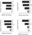


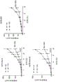


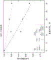


Classifications
-
- C—CHEMISTRY; METALLURGY
- C07—ORGANIC CHEMISTRY
- C07K—PEPTIDES
- C07K16/00—Immunoglobulins [IGs], e.g. monoclonal or polyclonal antibodies
- C07K16/18—Immunoglobulins [IGs], e.g. monoclonal or polyclonal antibodies against material from animals or humans
- C07K16/28—Immunoglobulins [IGs], e.g. monoclonal or polyclonal antibodies against material from animals or humans against receptors, cell surface antigens or cell surface determinants
- C07K16/2803—Immunoglobulins [IGs], e.g. monoclonal or polyclonal antibodies against material from animals or humans against receptors, cell surface antigens or cell surface determinants against the immunoglobulin superfamily
-
- A—HUMAN NECESSITIES
- A61—MEDICAL OR VETERINARY SCIENCE; HYGIENE
- A61P—SPECIFIC THERAPEUTIC ACTIVITY OF CHEMICAL COMPOUNDS OR MEDICINAL PREPARATIONS
- A61P35/00—Antineoplastic agents
-
- C—CHEMISTRY; METALLURGY
- C07—ORGANIC CHEMISTRY
- C07K—PEPTIDES
- C07K16/00—Immunoglobulins [IGs], e.g. monoclonal or polyclonal antibodies
- C07K16/18—Immunoglobulins [IGs], e.g. monoclonal or polyclonal antibodies against material from animals or humans
- C07K16/28—Immunoglobulins [IGs], e.g. monoclonal or polyclonal antibodies against material from animals or humans against receptors, cell surface antigens or cell surface determinants
- C07K16/2803—Immunoglobulins [IGs], e.g. monoclonal or polyclonal antibodies against material from animals or humans against receptors, cell surface antigens or cell surface determinants against the immunoglobulin superfamily
- C07K16/2818—Immunoglobulins [IGs], e.g. monoclonal or polyclonal antibodies against material from animals or humans against receptors, cell surface antigens or cell surface determinants against the immunoglobulin superfamily against CD28 or CD152
-
- C—CHEMISTRY; METALLURGY
- C07—ORGANIC CHEMISTRY
- C07K—PEPTIDES
- C07K16/00—Immunoglobulins [IGs], e.g. monoclonal or polyclonal antibodies
- C07K16/18—Immunoglobulins [IGs], e.g. monoclonal or polyclonal antibodies against material from animals or humans
- C07K16/28—Immunoglobulins [IGs], e.g. monoclonal or polyclonal antibodies against material from animals or humans against receptors, cell surface antigens or cell surface determinants
- C07K16/2863—Immunoglobulins [IGs], e.g. monoclonal or polyclonal antibodies against material from animals or humans against receptors, cell surface antigens or cell surface determinants against receptors for growth factors, growth regulators
-
- G—PHYSICS
- G01—MEASURING; TESTING
- G01N—INVESTIGATING OR ANALYSING MATERIALS BY DETERMINING THEIR CHEMICAL OR PHYSICAL PROPERTIES
- G01N33/00—Investigating or analysing materials by specific methods not covered by groups G01N1/00 - G01N31/00
- G01N33/48—Biological material, e.g. blood, urine; Haemocytometers
- G01N33/50—Chemical analysis of biological material, e.g. blood, urine; Testing involving biospecific ligand binding methods; Immunological testing
- G01N33/68—Chemical analysis of biological material, e.g. blood, urine; Testing involving biospecific ligand binding methods; Immunological testing involving proteins, peptides or amino acids
- G01N33/6854—Immunoglobulins
-
- A—HUMAN NECESSITIES
- A61—MEDICAL OR VETERINARY SCIENCE; HYGIENE
- A61K—PREPARATIONS FOR MEDICAL, DENTAL OR TOILETRY PURPOSES
- A61K39/00—Medicinal preparations containing antigens or antibodies
- A61K2039/505—Medicinal preparations containing antigens or antibodies comprising antibodies
-
- A—HUMAN NECESSITIES
- A61—MEDICAL OR VETERINARY SCIENCE; HYGIENE
- A61K—PREPARATIONS FOR MEDICAL, DENTAL OR TOILETRY PURPOSES
- A61K39/00—Medicinal preparations containing antigens or antibodies
- A61K2039/505—Medicinal preparations containing antigens or antibodies comprising antibodies
- A61K2039/507—Comprising a combination of two or more separate antibodies
-
- C—CHEMISTRY; METALLURGY
- C07—ORGANIC CHEMISTRY
- C07K—PEPTIDES
- C07K2317/00—Immunoglobulins specific features
- C07K2317/30—Immunoglobulins specific features characterized by aspects of specificity or valency
- C07K2317/31—Immunoglobulins specific features characterized by aspects of specificity or valency multispecific
-
- C—CHEMISTRY; METALLURGY
- C07—ORGANIC CHEMISTRY
- C07K—PEPTIDES
- C07K2317/00—Immunoglobulins specific features
- C07K2317/30—Immunoglobulins specific features characterized by aspects of specificity or valency
- C07K2317/34—Identification of a linear epitope shorter than 20 amino acid residues or of a conformational epitope defined by amino acid residues
-
- C—CHEMISTRY; METALLURGY
- C07—ORGANIC CHEMISTRY
- C07K—PEPTIDES
- C07K2317/00—Immunoglobulins specific features
- C07K2317/50—Immunoglobulins specific features characterized by immunoglobulin fragments
- C07K2317/52—Constant or Fc region; Isotype
-
- C—CHEMISTRY; METALLURGY
- C07—ORGANIC CHEMISTRY
- C07K—PEPTIDES
- C07K2317/00—Immunoglobulins specific features
- C07K2317/70—Immunoglobulins specific features characterized by effect upon binding to a cell or to an antigen
- C07K2317/71—Decreased effector function due to an Fc-modification
-
- C—CHEMISTRY; METALLURGY
- C07—ORGANIC CHEMISTRY
- C07K—PEPTIDES
- C07K2317/00—Immunoglobulins specific features
- C07K2317/70—Immunoglobulins specific features characterized by effect upon binding to a cell or to an antigen
- C07K2317/73—Inducing cell death, e.g. apoptosis, necrosis or inhibition of cell proliferation
-
- C—CHEMISTRY; METALLURGY
- C07—ORGANIC CHEMISTRY
- C07K—PEPTIDES
- C07K2317/00—Immunoglobulins specific features
- C07K2317/70—Immunoglobulins specific features characterized by effect upon binding to a cell or to an antigen
- C07K2317/76—Antagonist effect on antigen, e.g. neutralization or inhibition of binding
-
- C—CHEMISTRY; METALLURGY
- C07—ORGANIC CHEMISTRY
- C07K—PEPTIDES
- C07K2317/00—Immunoglobulins specific features
- C07K2317/90—Immunoglobulins specific features characterized by (pharmaco)kinetic aspects or by stability of the immunoglobulin
- C07K2317/92—Affinity (KD), association rate (Ka), dissociation rate (Kd) or EC50 value
Landscapes
- Health & Medical Sciences (AREA)
- Chemical & Material Sciences (AREA)
- Immunology (AREA)
- Organic Chemistry (AREA)
- Life Sciences & Earth Sciences (AREA)
- Medicinal Chemistry (AREA)
- General Health & Medical Sciences (AREA)
- Molecular Biology (AREA)
- Proteomics, Peptides & Aminoacids (AREA)
- Biochemistry (AREA)
- Genetics & Genomics (AREA)
- Biophysics (AREA)
- Engineering & Computer Science (AREA)
- Animal Behavior & Ethology (AREA)
- Hematology (AREA)
- Public Health (AREA)
- Pharmacology & Pharmacy (AREA)
- Nuclear Medicine, Radiotherapy & Molecular Imaging (AREA)
- General Chemical & Material Sciences (AREA)
- Chemical Kinetics & Catalysis (AREA)
- Biomedical Technology (AREA)
- Urology & Nephrology (AREA)
- Veterinary Medicine (AREA)
- Microbiology (AREA)
- Cell Biology (AREA)
- Biotechnology (AREA)
- Food Science & Technology (AREA)
- Physics & Mathematics (AREA)
- Analytical Chemistry (AREA)
- General Physics & Mathematics (AREA)
- Pathology (AREA)
- Medicines Containing Antibodies Or Antigens For Use As Internal Diagnostic Agents (AREA)
- Peptides Or Proteins (AREA)
- Acyclic And Carbocyclic Compounds In Medicinal Compositions (AREA)
- Preparation Of Compounds By Using Micro-Organisms (AREA)
- Micro-Organisms Or Cultivation Processes Thereof (AREA)
Abstract
The present disclosure provides monoclonal anti-ILT 2 antibodies or antigen-binding fragments thereof, as well as pharmaceutical compositions comprising the monoclonal anti-ILT 2 antibodies or antigen-binding fragments thereof and methods of producing the monoclonal anti-ILT 2 antibodies or antigen-binding fragments thereof. Methods of treating cancer using the antibodies or compositions of the present disclosure are also provided. Patient selection methods are also provided.
Description
Technical Field
The present invention is in the field of monoclonal antibodies and modulating immune responses against cancer.
Cross Reference to Related Applications
The present application claims priority from: PCT patent application PCT/IL2020/050889 filed 8/12 2020; U.S. provisional patent application 63/145,604 filed on day 2 and day 4 of 2021; U.S. provisional patent application 63/149,371 filed on day 15 of 2.2021. The disclosures of those priority applications are incorporated herein by reference in their entirety.
Sequence listing
The present application contains a sequence listing that has been electronically submitted in ASCII format and is hereby incorporated by reference in its entirety. An electronic copy of the sequence listing created at 8.10 of 2021 was named 022548_TW091_SL.txt and was 105,080 bytes in size.
Background
Immunoglobulin-like transcript 2 (ILT 2), also known as leukocyte immunoglobulin-like receptor superfamily B member 1 (LILRB 1), LIR1 and CD85j, is a cell surface protein expressed on immune cells and is known to inhibit immune responses. The protein contains 4 IgC domains in the extracellular region and 4 intracellular ITIM domains. It is a member of the ILT family consisting of ILT1, ILT2, ILT3 and ILT 4. ILT2 is most similar to ILT4, with about 80% homology. Known ligands for ILT2 include MHC-I as well as non-classical MHC molecules such as HLA-F, HLA-G, HLA-B27 and UL18 (human CMV). The strongest known interactor for ILT2 in humans is HLA-G1.
HLA-G1 is widely expressed on the surface of a variety of cancer cells including breast, cervical, CRC (colorectal), lung, gastric, pancreatic, thyroid and ovarian cancer cells, as well as glioblastoma multiforme cells and melanoma cells. Its expression is associated with poor clinical outcome. Furthermore, ILT2 expression on immune cells in the tumor microenvironment is associated with poor clinical response to oncolytic immunotherapy, even in the absence of HLA-G1. The use of immune responses as a weapon against cancer and for cancer supervision is a promising approach for cancer prevention and treatment. ILT2, however, presents a barrier to effective immunotherapy. There is a great need for therapeutic modalities that can circumvent the non-HLA-G1 dependent functions of ILT2-HLA-G1 axis.
Disclosure of Invention
The present disclosure provides monoclonal antibodies that bind to ILT2 and inhibit ILT 2-mediated immunosuppression, and pharmaceutical compositions comprising the monoclonal antibodies. Also provided are methods of treating cancer comprising administering the compositions described herein, methods of producing the antibodies, antigen binding fragments, and compositions described herein, and methods of increasing the efficacy of PD-1/PD-L1-based therapies.
According to one aspect, there is provided a monoclonal antibody or antigen binding fragment which binds to a sequence selected from the group consisting of human immunoglobulin-like receptor superfamily B member 1 (ILT 2) of SEQ ID NOs 41-44 and 68-70.
According to some embodiments, the sequence is selected from SEQ ID NOS.68-70.
According to some embodiments, the sequence is SEQ ID NO:71 or 72.
According to some embodiments, the antibody or antigen binding fragment binds to SEQ ID NOS 71 and 72.
According to some embodiments, an antibody or antigen binding fragment of the disclosure binds ILT2 and inhibits direct interaction between ILT2 and beta-2-microglobulin (B2M).
According to some embodiments, the antibody or antigen binding fragment inhibits the interaction of ILT2 with HLA proteins or MHC-I proteins by inhibiting direct interaction of ILT2 with B2M.
According to some embodiments, the HLA is HLA-G.
According to some embodiments, the antibody is an IgG4 antibody and comprises the heavy chain constant region of a human IgG4 antibody comprising S228P and L235E mutations (Eu numbering).
According to one aspect, there is provided a monoclonal anti-ILT 2 IgG4 antibody comprising three heavy chain CDRs (CDR-H1-3) and three light chain CDRs (CDR-L1-3), the CDRs comprising:
a. SEQ ID NOS 13-18, respectively,
b. SEQ ID NOS 1-6, respectively, or
c. SEQ ID NOS 7-12, respectively, and
wherein the antibody comprises a heavy chain constant region of a human IgG4 antibody and comprises one or both of S228P and L235E mutations (Eu numbering) in the heavy chain constant region.
According to some embodiments, the antibodies of the present disclosure comprise a heavy chain variable domain comprising an amino acid sequence selected from the group consisting of SEQ ID NOs 19, 21 and 23 or an amino acid sequence at least 95% identical thereto.
According to some embodiments, the antibodies of the present disclosure comprise a light chain variable domain comprising an amino acid sequence selected from the group consisting of SEQ ID NOs 20, 22, 24 and 45 or an amino acid sequence at least 95% identical thereto.
According to some embodiments, X in SEQ ID NO. 15 is A and the heavy chain comprises a variable domain sequence selected from SEQ ID NO. 28 and 56-59 or an amino acid sequence at least 95% identical thereto.
According to some embodiments, the light chain comprises a variable domain sequence selected from SEQ ID NOS: 24 and 60-62 or an amino acid sequence at least 95% identical thereto.
According to some embodiments, the heavy chain constant region comprises the amino acid sequence of SEQ ID NO. 55 or at least 95% identical thereto.
According to some embodiments, the heavy chain comprises the amino acid sequence of SEQ ID NO. 48 or at least 95% identical thereto, and the light chain comprises the amino acid sequence of SEQ ID NO. 49 or at least 95% identical thereto.
According to some embodiments, the heavy chain comprises the amino acid sequence of SEQ ID No. 51 or at least 95% identical thereto, and the light chain comprises the amino acid sequence of SEQ ID No. 49 or at least 95% identical thereto.
According to some embodiments, the heavy chain comprises the amino acid sequence of SEQ ID NO. 52 or at least 95% identical thereto, and the light chain comprises the amino acid sequence of SEQ ID NO. 49 or at least 95% identical thereto.
According to some embodiments, the heavy chain comprises the amino acid sequence of SEQ ID No. 64 or at least 95% identical thereto, and the light chain comprises the amino acid sequence of SEQ ID No. 49 or at least 95% identical thereto.
According to some embodiments, the heavy chain comprises the amino acid sequence of SEQ ID No. 65 or an amino acid sequence at least 95% identical thereto, and the light chain comprises the amino acid sequence of SEQ ID No. 66 or an amino acid sequence at least 95% identical thereto.
According to some embodiments, the heavy chain comprises the amino acid sequence of SEQ ID No. 67 or at least 95% identical thereto, and the light chain comprises the amino acid sequence of SEQ ID No. 49 or at least 95% identical thereto.
According to some embodiments, the heavy chain comprises the amino acid sequence of SEQ ID NO. 53 or at least 95% identical thereto, and the light chain comprises the amino acid sequence of SEQ ID NO. 54 or at least 95% identical thereto.
According to one aspect, a monoclonal antibody is provided comprising heavy and light chains comprising SEQ ID NOs 48 and 49, respectively.
According to some embodiments, the antibodies or antigen binding fragments of the disclosure are used for at least one of: binding ILT2, inducing/enhancing anti-tumor T cell responses, increasing T cell proliferation, decreasing cancer-induced inhibitory bone marrow activity, increasing natural killer cell cytotoxicity, increasing macrophage phagocytosis, increasing production of M1 inflammatory macrophages, decreasing production of M2-inhibitory macrophages, increasing the number of dendritic cells in the tumor microenvironment, increasing dendritic cell activation, treating HLA-G expressing cancers, and treating MHC-I expressing cancers.
According to some embodiments, the antibodies or antigen binding fragments of the disclosure are used in combination with an opsonizing agent to treat cancers that express HLA-G or MHC-I.
According to some embodiments, the antibodies or antigen binding fragments of the disclosure are used in combination with anti-PD-L1/PD-1 based therapies (e.g., immunotherapy) for the treatment of cancers that express HLA-G or MHC-I. In certain embodiments, the anti-PD-L1/PD-1 based therapy is pembrolizumab therapy.
According to one aspect, there is provided a pharmaceutical composition comprising an antibody or antigen-binding fragment of the disclosure.
According to one aspect, there is provided a method of treating an HLA-G or MHC-I expressing cancer in a subject in need thereof, the method comprising administering to the subject a pharmaceutical composition of the present disclosure or an antibody or antigen binding fragment of the present disclosure.
According to one aspect, there is provided a method of increasing the efficacy of an anti-PD-L1/PD-1 based therapy against a cancer cell expressing HLA-G, MHC-I or both in a subject in need thereof, the method comprising administering to a subject receiving an anti-PD-L1/PD-1 based therapy a pharmaceutical composition of the present disclosure or an antibody or antigen-binding fragment of the present disclosure.
According to some embodiments, the method further comprises administering a conditioning agent to the subject.
According to some embodiments, the conditioning agent is an EGFR inhibitor, optionally wherein the EGFR inhibitor is cetuximab.
According to some embodiments, the method further comprises administering an anti-PD-L1/PD-1 based therapy (e.g., immunotherapy) to the subject. In certain embodiments, the anti-PD-L1/PD-1 based immunotherapy is pembrolizumab therapy.
According to one aspect, there is provided a method of identifying an antibody that competes for binding to ILT2 with reference antibodies comprising SEQ ID NOs 48 and 49 for heavy and light chains, respectively, the method comprising: contacting the library of antibodies with a polypeptide sequence comprising an ILT2 sequence selected from the group consisting of SEQ ID NOs 41-44 and 68-70, and selecting antibodies from said library that bind to said ILT2 sequence, thereby obtaining antibodies that compete with said reference antibodies for binding to ILT 2.
According to one aspect, there is provided a method of producing an agent (e.g., an ILT2 binding protein or molecule), the method comprising: obtaining an agent that binds to a human ILT2 sequence selected from the group consisting of SEQ ID NOs 41-44 and 68-70; or obtaining a host cell comprising one or more nucleotide sequences encoding an agent that binds to a human ILT2 sequence selected from the group consisting of SEQ ID NOs 41-44 and 68-70, and culturing the host cell under conditions allowing expression of the agent, thereby producing the agent. According to some embodiments, the agent binds to a sequence selected from SEQ ID NOS.68-70. According to some embodiments, the agent binds to one or both of SEQ ID NOS: 71 and 72. According to some embodiments, the agent binds to SEQ ID NOS 71 and 72.
According to one aspect, there is provided a medicament produced by the method of the present disclosure.
According to one aspect, there is provided an isolated nucleic acid molecule encoding an antibody or antigen binding fragment of the disclosure. In some embodiments, the nucleic acid molecule is an expression vector.
According to one aspect, there is provided a host cell comprising an isolated nucleic acid molecule of the present disclosure.
Other embodiments and full scope of applicability of the present disclosure will become apparent from the detailed description given hereinafter. However, it should be understood that the detailed description and specific examples, while indicating preferred embodiments of the disclosure, are given by way of illustration only, since various changes and modifications within the spirit and scope of the disclosure will become apparent to those skilled in the art from this detailed description.
Drawings
FIG. 1 depicts a histogram of ILT2 expression on lymphocytes. Commercial antibody # 1 was used at a final concentration of 5 μg/mL. Binding is depicted as a black histogram, while isotype control staining is shown as a light gray histogram.
Fig. 2 depicts histograms depicting the expression of ILT2 on various immune cells. Commercial antibody # 1 was used at a final concentration of 5 μg/mL. Binding is depicted as a black histogram, while isotype control staining is shown as a light gray histogram.
Fig. 3A-3 c. (3A) tables of cancer indications from TCGA database in which ILT2 RNA was overexpressed. (3B) Dot plots of the association between MDSC (myeloid derived suppressor cells) enrichment and ILT2 expression in tumors. A bar graph depicting the association between M2 enrichment and ILT2 expression is also presented. (3C) Scatter plots of the percentage of various immune cells expressing ILT2 in different tumors.
FIGS. 4A-4B (4A) are bar graphs of the percentage of cases of various cancers that are HLA-G positive as determined by Immunohistochemistry (IHC). (4B) A scatter plot of HLA-G IHC scores for various cancers.
FIG. 5 is a scatter plot of soluble HLA-G levels in various cancers.
FIGS. 6A-6H (6A) sequences of heavy and light chains of three anti-ILT 2 antibodies. CDRs as determined by the KABAT system are underlined. (6B) Line drawing of binding of humanized 15G8 antibody to ILT2 expressed on the surface of BW cells transfected with human ILT 2. The graph indicates the fold-over-background (FAB) levels of the different antibodies tested compared to the secondary antibody alone. (6C-6F) for (6C) A375-HLA-G, (6D) COLO-320-WT, (6E) COLO-320-WT, and (6F) COLO-320-HLA-G cancer cells co-cultured with macrophages in the presence of 15G8 antibody, phagocytosis was measured as a bar graph of the Mean Fluorescence Intensity (MFI) of phagocytosis events. (6G-6H) bar graph of percent cytotoxicity from NK cell line cells co-cultured with various cancer cell lines expressing (6G) A375-HLA-G and (6H) A253 WT in the presence of 15G8 antibody.
Figures 7A-7E (7A) tables of antibody binding values to ILT2 and ILT2 family members. (7B) Histogram of binding to antibodies to ILT2 on the cell surface of BW cells transfected with human ILT 2. (7C) Line graphs of binding of chimeric and humanized 19E3 (left panel) and chimeric and humanized 15G8 (right panel) to ILT2 expressed on the surface of BW cells transfected with human ILT 2. (7D) immunostaining of gastric cancer samples with 19E3 antibody. (7E) Scatter plots of the percentage of various immune cells expressing ILT2 in PBMC samples from healthy controls and cancer patients using 15G8 humanized antibodies.
Figures 8A-8 p. (8A) bar graphs of percent blockage for each ILT2 antibody and positive control (PC, GHI/75 antibody). (8B) Histogram of binding of ILT 2-biotin to HLA-G expressing cells in the presence of ILT2 blocking antibodies. Binding of ILT 2-biotin to cells was determined by flow cytometry analysis using streptavidin-PE. No antibody (grey line), 15G8 (light grey line), isotype control (black line). (8C) Line graphs of blocking activity of 15G8 humanized antibodies as determined by binding of ILT 2-biotin to HLA-G expressing cells. (8D) Line graphs of blocking activity of chimeric and humanized 19E3 (left panel) and chimeric and humanized 15G8 (right panel) as determined by binding of ILT 2-biotin to HLA-G expressing cells in the presence of antibodies. (8E) Bar graph of mouse IL-2 secretion from cells expressing ILT2 signaling reporter constructs in the presence of HLA-G expressing cells and in the presence or absence of ILT2 blocking antibodies. PC = positive control (GHI/75 antibody). (8F) Line graphs of blocking activity of 15G8 humanized antibodies as determined by reporter assays. (8G) Bar graph of mouse IL-2 secretion from cells expressing ILT2 signaling reporter constructs in the presence or absence of ILT2 blocking antibodies and positive control antibodies. Bar graph of human IL-2 secretion of (8H) in a co-culture with (8I) a375 cancer cells with MHC-I expression alone or (8J-8K) both MHC-I and exogenous HLA-G in the presence or absence of ILT2 blocking antibodies and positive controls (8I-8J) pan-HLA antibodies or (8K) HLA-G specific antibodies (8H) in the absence of ILT2 or (8I-8K) ILT2 expressing Jurkat cells. (8L-8N) bar graph of human IL-2 secretion from ILT 2-expressing Jurkat cells cultured with HLA-G-expressing A375 cancer cells in the presence or absence of (8L) 15G8 antibody, (8M) GHI/75 antibody and (8N) HP-F1 antibody. (8O-8P) dot patterns of expression of activation-labeled (8O) phosphorylated ZAP70 and (8P) phosphorylated Syk in TIL cells and NK cells incubated with HLA-G positive cancer cells in the presence and absence of 15G8 antibody, respectively.
FIGS. 9A-9 D. (9A) bar graph of the percentage of HLA-G expressing cancer cells co-cultured with macrophages in the presence of ILT2 antibodies relative to control, as determined by FACS-based methods. (9B) Such as by The system determines a line graph of the real-time phagocytosis of cancer cells by macrophages in the presence of ILT2 antibodies. (9C) Phagocytosis was measured as a bar graph of the percentage of various HLA-G and MHC-I expressing cancer cells co-cultured with macrophages in the presence of ILT2 antibody 15G8 relative to control. (9D) In ILT2 antibody, cetuximab +.>
 Bar graph of phagocytosis of macrophages co-cultured with a253-HLA-G cells in the presence of an igg control or a combination thereof.
FIGS. 10A-10B are bar graphs of IFN gamma secretion and granzyme B secretion from activated CD 8T cells co-cultured with (10A) wild-type 721.221 cells or HLA-G expressing 721.221 cells or (10B) HLA-G expressing A375 cells in the presence of ILT2 antibodies.
FIGS. 11A-11H (11A-11B) are bar graphs of percent cytotoxicity of NK cell line cells from co-culture with various cancer cell lines expressing (11A) HLA-G and (11B) MHC-I in the presence of ILT2 antibodies. (11C-11D) (11C) bar graph of granzyme B and (11D) IFN gamma secretion from NK cell line cells co-cultured with H & N cancer cells and melanoma cells, respectively, in the presence of 15G8 ILT2 antibody. Bar graph of (11E) ifnγ expression and (11F) CD107A expression in ILT2 positive primary NK cells incubated with target cancer cells in the presence of ILT2 antibodies (11E-11F). A scatter plot of (11G-11H) expression alone showing the association of ILT2 positive cells with (11G) ifnγ expression and (11H) CD107A expression in response to ILT2 antibodies.
FIG. 12 is a line graph of HLA-DR and CD80 expression (MFI) as determined by flow cytometry in macrophages differentiated from monocytes isolated from healthy donors into M0, M1 or M2 macrophages in the presence of IgG or anti-ILT 2 antibodies. The number of patients exhibiting increased expression of the specified marker compared to the control IgG is indicated for each condition tested.
FIGS. 13A-13C are bar graphs of phagocytosis of macrophages co-cultured with various primary tumor cells (13A). (13B-13C) bar graph of dose-dependent phagocytosis of primary tumor cells isolated from (13B) RCC patients and (13C) H & N patients by autologous macrophages in the presence of humanized antibodies of the disclosure.
FIGS. 14A-14 L. (14A) are dot plots of ILT2 and PD-1 expression in tumor cells (left panel) and PBMC (right panel) from RCC and esophageal cancer patients. (14B-14C) A box plot of (14B) PD-1 and (14C) ILT2 RNA expression in a CD 8T cell population in TME of CRC patients. (14D-14E) (14D) dot patterns of ILT2 expression in CD 8T cells from peripheral blood of healthy donors and (14E) ILT2 and PD-1 expression in TIL from esophageal cancer. (14F) Scatter plots of membrane CD107a increase on PBMCs from 10 healthy donors activated with Staphylococcal Enterotoxin B (SEB) in the presence of 15G8, anti-PD-1 antibodies or a combination of both. (14G) Bar graph of increased CD107a expression in exemplary PBMCs from 3 donors. (14H-14J) bar graph of inflammatory cytokine (14H) IFNγ, (14I) TNFα, (14J) GM-CSF secretion levels from activated PBMC co-cultured with various primary cancer cells in the presence of anti-PD-1 antibodies, humanized anti-ILT 2 antibodies, or both. (14K-14L) bar graph of IFNγ secretion levels from T cells co-cultured with (14K) dendritic cells or (14L) macrophages in mixed lymphocyte reactions.
FIGS. 15A-15F (15A) are line graphs of tumor volumes of HLA-G and MHC-I expressing tumors grown in immunocompromised mice supplemented with human macrophages and anti-ILT 2 antibodies. (15B) Graphical illustration of a mouse treatment regimen for preventing lung tumors. (15C) Lung photographs of immunocompromised mice vaccinated with HLA-G positive cancer cells with or without human PBMC and ILT2 antibodies. (15D) summarizing a scatter plot of the data from 15C. (15E) Illustration of a mouse treatment regimen for the treatment of an established lung tumor. (15F) box plot of tumor weight.
Fig. 16A-16F (16A-16F) the following box line diagrams: (16A) CD107A expression in Total CD 8T cells, (16B) T EMRA CD107A expression in cells, (16C) CD69 expression in NK cells, (16D) CD69 expression in total CD 8T cells, in mice with low or high ILT2 levels receiving PBMCs from donor, respectively in their T EMRA In cells or NK cells, (16E) T EMRA CD107 expression in cells and CD69 expression in NK cells treated with the combination of (16F). * Is P<0.005.* Is P<0.0005.* Is P<0.0001。
FIGS. 17A-17F (17A) are illustrations of treatment regimens for humanized NSG mice vaccinated with head and neck cancer and treated with anti-ILT 2 or control antibodies. (17B) Line graphs of tumor weights from IgG and anti-ILT 2 treated mice. (17C-17E) (17C) bar graph of baseline ILT2 levels in peripheral CD 8T cells in mice that were either responsive (R) or non-responsive (NR) to BND-22 treatment. Intratumoral treatment in four mice treated with anti-ILT 2 antibody followed by (17D) CD107A expression, (17E) M1/M2 ratio and (17F) total CD80 positive dendritic cell number.
Fig. 18A-18 f. (18A) partial sequence of ILT2, which shows residues with significant predicted binding. These residues are classified into four categories based on their original probability of belonging to an epitope. Asterisks indicate the location of the selected mutation. (18B-18C) 3D rendering of ILT2 surface structures showing (18B) the positions of residues from 18A and (18C) four major interaction regions on ILT 2. A 3D bar graph or surface map of (18D-18F) ILT2 showing (18D) the interaction of the epitope of the 15G8 antibody and the epitopes of the 3H5, 12D12 and 27H5 antibodies (upper left circle) from WO 2020/136145, and the secondary epitope of the 3H5 antibody (upper right circle), (18E-18F) and the 15G8 epitope on ILT2 with B2M in complexes with (18E) HLA-A or (18F) HLa-G.
FIGS. 19A-19G distribution of protection ratio of free ILT2 relative to ILT2-15G8 complex. (19A) Number of tryptic digests derived from ILT2 proteinsA bar graph of the data. The median value of the distribution was 1.5 and the average value was 1.37. (19B) Bar graph of trypsin and Asp-N digestion data derived from ILT2 protein. The median value of the distribution was 1.58 and the average value was 1.48. The selected protected area is marked. Dose-response plots of (19C-19G) free ILT2 peptide versus complex. The lines and squares and circles represent control and complex, respectively. (19C) peptide 56-71: free ilt2k=9.09 s -1 Complex k=2.31 s -1 Indicating a 3.94-fold reduction due to complex formation. (19D) peptide 57-71: free ilt2k=6.93 s -1 Complex k=1.97 s -1 Indicating a 3.52-fold reduction due to complex formation. (19E) peptide 84-100: free ilt2k=1.26 s -1 Complex k=0.37 s -1 Indicating a 3.41-fold reduction due to complex formation. (19F) peptides 57-66: free ilt2k=4.32 s -1 Complex k=0.72 s -1 Indicating a 6-fold reduction due to complex formation. (19G) peptides 91-100: free ilt2k=0.7 s -1 Complex k=0.14 s -1 Indicating a 5-fold decrease due to complex formation.
FIG. 20 is a graph of ILT2-Fc binding to human B2M in the presence of 15G8 or an unrelated antibody in an ELISA assay.
FIGS. 21A-21D (21A-21B) bar graphs of% increased phagocytosis of (21A) A375-HLA-G and (21B) SKMEL28-HLA-G cancer cells co-cultured with macrophages in the presence of various anti-ILT 2 antibodies as compared to IgG controls. Line drawing of a competitive ILT2 binding ELISA using biotinylated 15G8 antibody in the presence of competing non-biotinylated (21C) GHI/75, HP-F1, and (21D) MAB20172 and 15G8 antibodies (21C-21D).
Detailed Description
The present disclosure relates to monoclonal antibodies or antigen-binding fragments and pharmaceutical compositions that bind ILT2 and inhibit ILT 2-mediated immunosuppression. Methods of treating cancer and enhancing PD-1/PD-L1 immunotherapy are also provided.
The present disclosure is based, at least in part, on the following surprising findings: ILT2 antagonism works synergistically with PD-1 and PD-L1 based immunotherapy to combat cancer cells. In particular, combinations of ILT2 blocking antibodies with anti-PD-1 antibodies were found to increase pro-inflammatory cytokine secretion by immune cells. The increase is not only additive, but exceeds the sum of the effects of each agent alone. In fact, de novo increases were observed for at least one cytokine, where none of the agents alone had any effect. This combination therapy allows for the conversion of cancer from PD-1/PD-L1 refractory to PD-1/PD-L1 reactive.
It was further surprisingly found that the level of ILT2 expression in immune cells of a patient is related to the effectiveness of ILT2 blocking therapy. The responders to the therapy had high ILT2 levels, while non-responders had low ILT2 levels. Specifically, the level of ILT2 on circulating CD8 positive T cells is predictive of treatment outcome.
Finally, antibodies of the present disclosure were found to bind unique epitopes within the ILT2 inter-domain (inter domain) between the D1 and D2 domains. This region is known as the interaction domain between ILT2 and beta-2-microglobulin (B2M). The antibodies of the present disclosure are the first known antibodies to directly block this interaction. Furthermore, the antibodies of the present disclosure were found to have no reported immunostimulatory effect with respect to other anti-ILT 2 antibodies. Antibodies of the invention are capable of modulating immune surveillance of T cells, NK cells, dendritic cells and macrophages against cancer cells expressing MHC-I (e.g., HLA-G). In particular, for the first time, anti-ILT 2 antibodies used as monotherapy were shown to enhance phagocytosis of cancer cells.
Antibodies to
In a first aspect, the present disclosure provides an antibody comprising three heavy chain CDRs (CDR-H) and three light chain CDRs (CDR-L), wherein: CDR-H1 comprises the amino acid sequence shown in SEQ ID NO. 1 (DHTIH), CDR-H2 comprises the amino acid sequence shown in SEQ ID NO. 2 (YIYPRDGSTKYNEKFKG), CDR-H3 comprises the amino acid sequence shown in SEQ ID NO. 3 (TWDYFDY), CDR-L1 comprises the amino acid sequence shown in SEQ ID NO. 4 (RASESVDSYGNSFMH), CDR-L2 comprises the amino acid sequence shown in SEQ ID NO. 5 (RASNLES), and CDR-L3 comprises the amino acid sequence shown in SEQ ID NO. 6 (QQSNEDPYT). In some embodiments, the antibody further comprises a heavy chain constant region of an IgG4 antibody (e.g., human IgG 4).
In another aspect, the present disclosure provides an antibody or antigen binding fragment comprising three heavy chain CDRs (CDR-H) and three light chain CDRs (CDR-L), wherein: CDR-H1 comprises the amino acid sequence shown in SEQ ID NO. 7 (GYTFTSYGIS), CDR-H2 comprises the amino acid sequence shown in SEQ ID NO. 8 (EIYPGSGNSYYNEKFKG), CDR-H3 comprises the amino acid sequence shown in SEQ ID NO. 9 (SNDGYPDY), CDR-L1 comprises the amino acid sequence shown in SEQ ID NO. 10 (KASDHINNWLA), CDR-L2 comprises the amino acid sequence shown in SEQ ID NO. 11 (GATSLET), and CDR-L3 comprises the amino acid sequence shown in SEQ ID NO. 12 (QQYWSTPWT). In some embodiments, the antibody further comprises a heavy chain constant region of an IgG4 antibody (e.g., human IgG 4).
In another aspect, the present disclosure provides an antibody or antigen binding fragment comprising three heavy chain CDRs (CDR-H) and three light chain CDRs (CDR-L), wherein: CDR-H1 comprises the amino acid sequence shown in SEQ ID NO. 13 (SGYYWN), CDR-H2 comprises the amino acid sequence shown in SEQ ID NO. 14 (YISYDGSYNNYNPSLKN), CDR-H3 comprises the amino acid sequence shown in SEQ ID NO. 15 (GYSYYAMDX), wherein X is selected from A, C and S, CDR-L1 comprises the amino acid sequence shown in SEQ ID NO. 16 (RTSQDISNYLN), CDR-L2 comprises the amino acid sequence shown in SEQ ID NO. 17 (YSRLHS), and CDR-L3 comprises the amino acid sequence shown in SEQ ID NO. 18 (QQGNTLPT). In some embodiments, the antibody further comprises a heavy chain constant region of an IgG4 antibody (e.g., human IgG 4).
In some embodiments, SEQ ID NO. 15 is GYSYYYAMDA (SEQ ID NO: 25). In some embodiments, SEQ ID NO. 15 is SEQ ID NO. 25 and the antibody or antigen binding fragment is a humanized antibody. In some embodiments, SEQ ID NO. 15 is GYSYYYAMDS (SEQ ID NO: 26). In some embodiments, SEQ ID NO. 15 is SEQ ID NO. 26 and the antibody or antigen binding fragment is a humanized antibody. In some embodiments, SEQ ID NO. 15 is GYSYYYAMDC (SEQ ID NO: 27). In some embodiments, SEQ ID NO. 16 is SEQ ID NO. 27 and the antibody or antigen binding fragment is a murine antibody.
In another aspect, an antibody or antigen binding fragment is provided that binds to the human leukocyte immunoglobulin-like receptor superfamily B member 1 (ILT 2) interdomain between domains D1 and D2.
In another aspect, there is provided an antibody or antigen binding fragment that binds to an ILT2 sequence selected from the group consisting of: VKKGQFPIPSITWEH (SEQ ID NO: 41), LELVVTGAYIKPTLS (SEQ ID NO: 42), VILQCDSQVAFDGFS (SEQ ID NO: 43), WYRCYAYD SNSPYEW (SEQ ID NO: 44), KGQFPIPSITWEHAGR (SEQ ID NO: 68), GQFPIPSITWEHAGR (SEQ ID NO: 69) and SESSDPLELVVTGAYIK (SEQ ID NO: 70). In some embodiments, an antibody or antigen binding fragment may bind to 1, 2, 3, 4, 5, 6, or all 7 of the sequences.
In another aspect, an antibody or antigen binding fragment is provided that binds ILT2 and inhibits interaction between ILT2 and B2M.
In some embodiments, the antibody is a monoclonal antibody. In some embodiments, the antibody is a polyclonal antibody. In some embodiments, the antibody is a murine antibody. In some embodiments, the antibody is a humanized antibody. As used herein, "humanized" antibodies refer to antibodies having a human backbone, but having CDRs derived or obtained from a non-human antibody. In some embodiments, during humanization, the CDRs may be altered, but generally still derived from CDRs of the non-human parent antibody. In some embodiments, the antigen binding fragment is a single chain antibody. In some embodiments, the antigen binding fragment is a single domain antibody.
In some embodiments, the antibody comprises an IgG4 constant region. In some embodiments, the antibody is a humanized antibody comprising human IgG4 constant regions. In some embodiments, the IgG4 constant region is an engineered IgG4 constant region and contains a mutation relative to a wild-type IgG4 constant region. In some embodiments, the human IgG4 constant region comprises the amino acid sequence:
ASTKGPSVFP LAPCSRSTSE STAALGCLVK DYFPEPVTVS WNSGALTSGV
HTFPAVLQSS GLYSLSSVVT VPSSSLGTKT YTCNVDHKPS NTKVDKRVES
KYGPPCPSCP APEFLGGPSV FLFPPKPKDT LMISRTPEVT CVVVDVSQED
PEVQFNWYVD GVEVHNAKTK PREEQFNSTY RVVSVLTVLH QDWLNGKEYK
CKVSNKGLPS SIEKTISKAK GQPREPQVYT LPPSQEEMTK NQVSLTCLVK
GFYPSDIAVE WESNGQPENN YKTTPPVLDS DGSFFLYSRL TVDKSRWQEG
NVFSCSVMHE ALHNHYTQKS LSLSLGK(SEQ ID NO:50)。
in some embodiments, the human IgG4 constant region consists of SEQ ID NO. 50. In some embodiments, the human IgG4 constant region comprises at least 80%, 85%, 90%, 91%, 92%, 93%, 94%, 95%, 96%, 97%, 98%, 99%, or 100% identity to SEQ ID NO. 50. Each possibility represents a separate embodiment of the present disclosure. In some embodiments, the human IgG4 constant region comprises at least 95% identity to SEQ ID NO. 50. In some embodiments, the human IgG4 constant region comprises at least 97% identity to SEQ ID NO. 50. In some embodiments, the human IgG4 constant region comprises at least 99% identity to SEQ ID NO. 50.
In some embodiments, the IgG4 constant region comprises at least one mutation. In some embodiments, the mutation reduces binding to an Fc receptor, such as an fcγreceptor (fcγr), or reduces Fab-arm exchange of an IgG4 antibody. In some embodiments, the mutation is located at serine 108. In some embodiments, serine is mutated to proline. In some embodiments, serine 108 is serine 108 of SEQ ID NO:50 (or S228 according to Eu numbering). Mutation of serine 108 to proline is also known as the S228P mutation. In some embodiments, the mutation is located at leucine 115 (or L235 according to Eu numbering). In some embodiments, leucine is mutated to glutamic acid. In some embodiments, leucine 115 is leucine 115 of SEQ ID NO. 50. The leucine 115 mutation to glutamate is also known as the L235E mutation. The skilled artisan will appreciate that the exact digital position of any amino acid in the heavy chain constant region will depend on the length of the heavy chain variable region. Thus, the position of serine is denoted as 108 within the constant region and the position of leucine is denoted as 115 (see, e.g., SEQ ID NO: 50). It is also understood that modifications to the constant region (e.g., additions or deletions of bases) may also alter the digital position, but any analog or derivative of IgG4 is also contemplated, so long as it includes the mutation. In some embodiments, the IgG4 constant region comprises a plurality of mutations. In some embodiments, the IgG4 constant region comprises mutations of serine 108 and leucine 115. In some embodiments, the IgG4 constant region comprises S108P and L115E mutations (or S228P and L235E mutations according to Eu numbering).
In some embodiments, the IgG4 constant region comprises the following amino acid sequences, wherein mutations to the wild-type human IgG4 constant region (S228P and L235E; eu numbering) are shown in the boxes:

in some embodiments, the human IgG4 constant region comprises or consists of SEQ ID NO. 55. In some embodiments, the antibodies of the present disclosure comprise SEQ ID NO. 55. In some embodiments, the heavy chain constant region of an antibody of the present disclosure comprises SEQ ID NO. 55. In some embodiments, the heavy chain constant region of an antibody of the present disclosure consists of SEQ ID NO. 55. In some embodiments, the IgG4 constant region is an analog or derivative of SEQ ID NO. 55, e.g., containing other mutations that improve antibody manufacturability or reduce immunogenicity. In some embodiments, the IgG4 constant region comprises at least 80%, 85%, 90%, 91%, 92%, 93%, 94%, 95%, 96%, 97%, 98%, 99%, or 100% identity to SEQ ID NO. 55. Each possibility represents a separate embodiment of the present disclosure. In some embodiments, the IgG4 constant region comprises at least 95% identity to SEQ ID NO. 55. In some embodiments, the IgG4 constant region comprises at least 97% identity to SEQ ID NO. 55. In some embodiments, the IgG4 constant region comprises at least 99% identity to SEQ ID NO. 55.
In some embodiments, analogs of the IgG4 constant region further comprise at least one additional mutation. In some embodiments, at least one other mutation is in SEQ ID NO. 55. In some embodiments, at least one other mutation is not at position 108. In some embodiments, at least one other mutation is not at position 115. In some embodiments, at least one other mutation is not at position 108 or 115. Other mutations of IgG4 are well known in the art, and any of these mutations may be employed. Examples of other mutations include, but are not limited to, deletions of E233 234 235 237 239 243 250, 254 256 259 264 296 307 308 311 317 370 370 370 380 428 428 434 434 434 434 434 434 435 439 445 236, deletions of G446, and deletions of K447 (numbering given according to Eu numbering). Again, the numerical positions given for these mutations are the usual notation (Eu numbering), but will depend on the length of the heavy chain variable region. Within SEQ ID NO. 50, these positions correspond to E113, F114, L115, G117, P119, F123, T130, M132, S134, T136, E138, D139, V144, D145, F176, T187, V188, Q191, K197, A210, E236, K250, E260, R289, V307, M308, H313, N314, H315, Y316, K319, L325, G326 and K327. It will also be appreciated that specific combinations of mutations may be employed, such as E233P/F234A/L235A/G236del/G237A or S228P/F234A/L235A/G237A/P238S.
In some embodiments, the antibody or antigen binding fragment binds ILT2. In some embodiments, ILT2 is human ILT2. In some embodiments, ILT2 is mammalian ILT2. In some embodiments, the ILT2 is a primate ILT2 (e.g., cynomolgus ILT 2). In some embodiments, ILT2 is murine ILT2. In some embodiments, the antibody or antigen binding fragment binds to an extracellular domain of ILT2. In some embodiments, the antibody or antigen binding fragment binds to a ligand pocket of ILT2. In some embodiments, the ligand is B2M. In some embodiments, the ligand is not HLA. In some embodiments, the ligand is HLA. In some embodiments, the HLA is HLA-G. In some embodiments, the ligand is not MHC. In some embodiments, the ligand is MHC. In some embodiments, the MHC is MHC class I (MHC-I). In some embodiments, the antibody or antigen binding fragment binds to the ILT2 interdomain. In some embodiments, the interdomain is an interface between the D1 and D2 domains. In some embodiments, the mid-domain is a hinge domain between the D1 and D2 domains. In some embodiments, the mid-domain does not comprise the N-terminal domain of D1. In some embodiments, the spacer is amino acids 54-184 of SEQ ID NO. 31. In some embodiments, amino acids 54-184 of SEQ ID NO. 31 comprise a spacer domain. In some embodiments, the spacer is amino acids 90-184 of SEQ ID NO. 31. In some embodiments, amino acids 90-184 comprise a spacer domain. In some embodiments, the antibody or antigen binding fragment binds to an epitope within the compartment. In some embodiments, an epitope comprises at least 25%, 30%, 35%, 40%, 45%, 50%, 55%, 60%, 65%, 70%, 75%, 80%, 85%, 90%, 95%, 97%, 99% or 100% of the spacer domain. Each possibility represents a separate embodiment of the present disclosure. In some embodiments, the epitope is within D2. In some embodiments, the antibody or antigen binding domain binds to an epitope in D2. In some embodiments, the epitope is at least partially in D2. In some embodiments, the antibody or antigen binding domain binds to at least a portion of an epitope in D2. In some embodiments, the epitope spans D1 and D2. In some embodiments, the antibody or antigen binding fragment does not bind to an ILT2 domain that interacts with the α3 domain of HLA-G.
In some embodiments, ILT2 is mammalian ILT2. In some embodiments, ILT2 is human ILT2. In some embodiments, ILT2 has the NCBI reference sequence: amino acid sequence provided in NP 006660.4. In some embodiments, ILT2 has the following amino acid sequence:
MTPILTVLIC LGLSLGPRTH VQAGHLPKPT LWAEPGSVIT QGSPVTLRCQ
GGQETQEYRL YREKKTALWI TRIPQELVKK GQFPIPSITW EHAGRYRCYY
GSDTAGRSES SDPLELVVTG AYIKPTLSAQ PSPVVNSGGN VILQCDSQVA
FDGFSLCKEG EDEHPQCLNS QPHARGSSRA IFSVGPVSPS RRWWYRCYAY
DSNSPYEWSL PSDLLELLVL GVSKKPSLSV QPGPIVAPEE TLTLQCGSDA
GYNRFVLYKD GERDFLQLAG AQPQAGLSQA NFTLGPVSRS YGGQYRCYGA
HNLSSEWSAP SDPLDILIAG QFYDRVSLSV QPGPTVASGE NVTLLCQSQG
WMQTFLLTKE GAADDPWRLR STYQSQKYQA EFPMGPVTSA HAGTYRCYGS
QSSKPYLLTH PSDPLELVVS GPSGGPSSPT TGPTSTSGPE DQPLTPTGSD
PQSGLGRHLG VVIGILVAVI LLLLLLLLLF LILRHRRQGK HWTSTQRKAD
FQHPAGAVGP EPTDRGLQWR SSPAADAQEE NLYAAVKHTQ PEDGVEMDTR
SPHDEDPQAV TYAEVKHSRP RREMASPPSP LSGEFLDTKD RQAEEDRQMD
TEAAASEAPQ DVTYAQLHSL TLRREATEPP PSQEGPSPAV PSIYATLAIH
(SEQ ID NO:31)。
in some embodiments, ILT2 has the NCBI reference sequence: amino acid sequence provided in NP 001075106.2. In some embodiments, ILT2 has the NCBI reference sequence: amino acid sequence provided in NP 001075107.2. In some embodiments, ILT2 has the NCBI reference sequence: amino acid sequence provided in NP 001075108.2. In some embodiments, ILT2 has the NCBI reference sequence: amino acid sequence provided in NP 001265328.2.
In some embodiments, the D1 domain of ILT2 comprises or consists of the amino acid sequence
GHLPKPTLWA EPGSVITQGS PVTLRCQGGQ ETQEYRLYRE KKTALWITRI
PQELVKKGQF PIPSITWEHA GRYRCYYGSD TAGRSESSDP LELVVTGA
(SEQ ID NO:46)。
In some embodiments, the D1 domain of ILT2 comprises or consists of amino acids 24-121 of SEQ ID NO. 31. In some embodiments, the D2 domain of ILT2 comprises or consists of the amino acid sequence
YIKPTLSAQP SPVVNSGGNV ILQCDSQVAF DGFSLCKEGE DEHPQCLNSQ
PHARGSSRAI FSVGPVSPSR RWWYRCYAYD SNSPYEWSLP SDLLELLVLG
V(SEQ ID NO:47)。
In some embodiments, the D2 domain of ILT2 comprises or consists of amino acids 122-222 of SEQ ID NO. 31. In some embodiments, the interdomain of ILT2 comprises amino acids Gln41, lys65, trp90, gly120, ala121, val122, ile123, gln148, val149, ala150, phe151, asp201, asn203 and Glu207 of SEQ ID NO. 31. In some embodiments, the epitope comprises amino acids Gln41, lys65, trp90, gly120, ala121, val122, ile123, gln148, val149, ala150, phe151, asp201, asn203 and Glu207 of SEQ ID NO. 31. In some embodiments, the epitope comprises at least 5, 6, 7, 8, 9, 10, 11, 12, 13, or 14 amino acids selected from the group consisting of amino acids Gln41, lys65, trp90, gly120, ala121, val122, ile123, gln148, val149, ala150, phe151, asp201, asn203, and Glu207 of SEQ ID NO. 31.
In some embodiments, the epitope comprises at least 10 amino acids selected from the group consisting of amino acids Gln41, lys65, trp90, gly120, ala121, val122, ile123, gln148, val149, ala150, phe151, asp201, asn203 and Glu207 of SEQ ID NO. 31. In some embodiments, the antibody or antigen binding fragment binds to the ILT2 sequence provided in SEQ ID NO. 41. In some embodiments, the antibody or antigen binding fragment binds to the ILT2 sequence provided in SEQ ID NO. 42. In some embodiments, the antibody or antigen binding fragment binds to the ILT2 sequence provided in SEQ ID NO. 43. In some embodiments, the antibody or antigen binding fragment binds to the ILT2 sequence provided in SEQ ID NO. 44. In some embodiments, the antibody or antigen binding fragment binds to a three-dimensional epitope comprising residues from at least two of SEQ ID NOS: 41, 42, 43, and 44 (e.g., a three-dimensional epitope comprising at least two of SEQ ID NOS: 41, 42, 43, and 44). In some embodiments, the three-dimensional epitope comprises residues from at least 3 of SEQ ID NOs 41, 42, 43, and 44 (e.g., a three-dimensional epitope comprising at least 3 of SEQ ID NOs 41, 42, 43, and 44). In some embodiments, the three-dimensional epitope comprises residues from SEQ ID NOs 41, 42, 43 and 44 (e.g., a three-dimensional epitope comprising SEQ ID NOs 41, 42, 43 and 44).
In some embodiments, the antibody or antigen binding fragment binds to SEQ ID NO. 41 (e.g., binds to an ILT2 epitope comprising or within SEQ ID NO. 41). In some embodiments, the antibody or antigen binding fragment binds to SEQ ID NO. 42 (e.g., binds to an ILT2 epitope comprising or within SEQ ID NO. 42). In some embodiments, the antibody or antigen binding fragment binds to SEQ ID NO. 43 (e.g., binds to an ILT2 epitope comprising or within SEQ ID NO. 43). In some embodiments, the antibody or antigen binding fragment binds to SEQ ID NO. 44 (e.g., binds to an ILT2 epitope comprising or within SEQ ID NO. 44). In some embodiments, the antibody or antigen binding fragment binds to SEQ ID NO. 68 (e.g., binds to an ILT2 epitope comprising or within SEQ ID NO. 68). In some embodiments, the antibody or antigen binding fragment binds to SEQ ID NO:69 (e.g., binds to an ILT2 epitope comprising or within SEQ ID NO: 69). In some embodiments, the antibody or antigen binding fragment binds to SEQ ID NO. 70 (e.g., binds to an ILT2 epitope comprising or within SEQ ID NO. 70). In some embodiments, the antibody or antigen binding fragment binds to a three-dimensional epitope comprising residues from at least two of SEQ ID NOS: 41-44 and 68-70 (e.g., binds to a three-dimensional epitope comprising at least two of SEQ ID NOS: 41-44 and 68-70). In some embodiments, the three-dimensional epitope comprises residues from at least 3 of SEQ ID NOS: 41-44 and 68-70 (e.g., the three-dimensional epitope comprises at least 3 of SEQ ID NOS: 41-44 and 68-70). In some embodiments, the three-dimensional epitope comprises residues from at least 4 of SEQ ID NOS: 41-44 and 68-70 (e.g., the three-dimensional epitope comprises at least 4 of SEQ ID NOS: 41-44 and 68-70). In some embodiments, the three-dimensional epitope comprises residues from SEQ ID NOS: 41, 42, and 68-70 (e.g., the three-dimensional epitope comprises SEQ ID NOS: 41, 42, and 68-70). In some embodiments, the antibody or antigen binding fragment binds to a sequence selected from SEQ ID NO:68-70 (e.g., binds to an epitope comprising or within any one of SEQ ID NO: 68-70).
In some embodiments, the antibody or antigen binding fragment binds to sequence GQFPIPSITW (SEQ ID NO: 71) (e.g., binds to an ILT2 epitope comprising or within the sequence). In some embodiments, the antibody or antigen binding fragment binds to sequence ELVVTGAYIK (SEQ ID NO: 72) (e.g., binds to an ILT2 epitope comprising or within the sequence). In certain embodiments, the antibody or antigen binding fragment binds to SEQ ID NOS: 71 and 72 (e.g., binds to an epitope comprising or within SEQ ID NOS: 71 and 72). In some embodiments, the antibody or antigen binding fragment binds to (e.g., binds to an epitope comprising or within) a sequence selected from SEQ ID NOS: 68-72. In some embodiments, the antibody or antigen binding fragment binds to (e.g., binds to an epitope comprising or within) a sequence selected from the group consisting of SEQ ID NOS: 41-44 and 68-72. In some embodiments, SEQ ID NO:71 is an epitope within SEQ ID NO: 41. In some embodiments, SEQ ID NO:71 is an epitope within SEQ ID NO: 68. In some embodiments, SEQ ID NO:71 is an epitope within SEQ ID NO: 69. In some embodiments, SEQ ID NO:72 is an epitope within SEQ ID NO: 42. In some embodiments, SEQ ID NO:72 is an epitope within SEQ ID NO: 70.
In some embodiments, the antibody or antigen binding fragment binds to an ILT2 epitope, which ILT2 epitope comprises an ILT2 residue selected from Q18, G19, K42, L45, S64, I65, T66, W67, E68, G97, a98, Y99, I100, Q125, V126, a127, F128, D178, N180, S181, and E184. In some embodiments, the antibody or antigen binding fragment binds an ILT2 epitope, the ILT2 epitope comprising an ILT2 residue selected from G97, a98, Y99, I100, Q125, and V126. In some embodiments, the antibody or antigen binding fragment binds to an ILT2 epitope, the ILT2 epitope comprising a plurality of ILT2 residues selected from Q18, G19, K42, L45, S64, I65, T66, W67, E68, G97, a98, Y99, I100, Q125, V126, a127, F128, D178, N180, S181, and E184. In some embodiments, the antibody or antigen binding fragment binds an ILT2 epitope, the ILT2 epitope comprising a plurality of ILT2 residues selected from G97, a98, Y99, I100, Q125 and V126. In some embodiments, the antibody or antigen binding fragment binds at least 1, 2, 3, 4, 5, 6, 7, 8, 9, 10, 11, 12, 13, 14, 15, 16, 17, 18, 19, 20, or 21 residues selected from Q18, G19, K42, L45, S64, I65, T66, W67, E68, G97, a98, Y99, I100, Q125, V126, a127, F128, D178, N180, S181, and E184. Each possibility represents a separate embodiment of the present disclosure. In some embodiments, the antibody or antigen binding fragment binds 1, 2, 3, 4, 5, or 6 residues selected from G97, a98, Y99, I100, Q125, and V126. Each possibility represents a separate embodiment of the present disclosure. In some embodiments, the antibody or antigen binding fragment binds G97, a98, Y99, I100, Q125, and V126. It is understood that ILT2 residue numbering as used herein is with respect to SEQ ID NO. 31.
In some embodiments, the antibody or antigen binding fragment is an ILT2 antagonist. In some embodiments, the antibody or antigen binding fragment is not an ILT2 agonist. In some embodiments, antagonism is due to ILT 2-mediated immunosuppression. In some embodiments, the antibody or antigen binding fragment inhibits ILT 2-mediated immunosuppression. In some embodiments, the antibody or antigen binding fragment inhibits ILT2 signaling.
In some embodiments, the antibody or antigen binding fragment inhibits an interaction between ILT2 and B2M. In some embodiments, the interaction is a direct interaction. In some embodiments, the antibody or antigen binding fragment inhibits the contact of ILT2 with B2M. In some embodiments, the contacting is direct contact. In some embodiments, the antibody or antigen binding fragment inhibits interactions between ILT2 and HLA. In some embodiments, the antibody or antigen binding fragment inhibits interactions between ILT2 and MHC. In some embodiments, the MHC is an HLA. In some embodiments, the HLA is HLA-G. In some embodiments, the antibody or antigen binding fragment inhibits interaction between ILT2 and HLA, MHC, or both. In some embodiments, the antibody or antigen binding fragment inhibits the interaction between ILT2 and HLA, MHC, or both by inhibiting the interaction of ILT2 with B2M. In some embodiments, the interaction is mediated through B2M. In some embodiments, the antibody indirectly inhibits interaction with HLA, MHC, or both by inhibiting interaction with B2M. In some embodiments, the interaction is a B2M mediated interaction. In some embodiments, the antibody or antigen binding fragment inhibits interactions between ILT2 and B2M/HLA complex. In some embodiments, the antibody or antigen binding fragment inhibits interactions between ILT2 and B2M/MHC complex. In some embodiments, the complex comprises a B2M monomer. In some embodiments, the complex comprises HLA or MHC monomers. In some embodiments, the complex comprises a B2M dimer. In some embodiments, the complex comprises HLA or MHC dimers.
In some embodiments, ILT2 mediated immunosuppression is inhibition of immune cells. In some embodiments, the immune cells are selected from T cells, macrophages, dendritic cells, and Natural Killer (NK) cells. In some embodiments, ILT2 mediated immunosuppression is inhibition of T cells, macrophages, dendritic cells, and NK cells. In some embodiments, ILT2 mediated immunosuppression is inhibition of T cells, macrophages and NK cells. In some embodiments, the T cell is a CD8 positive T cell. In some embodiments, the T cell is T EMRA Cells (terminally differentiated effector memory cells re-expressing CD45 RA). In some embodiments, the immune cells are selected from the group consisting of CD8 positive T cells, T cells EMRA Cells, dendritic cells, macrophages and Natural Killer (NK) cells. In some embodiments, the immune cell is a T cell. In some embodiments, the immune cell is an NK cell. In some embodiments, the immune cell is a macrophage. In some embodiments, the macrophage is a tumor-associated macrophage (TAM). In some embodiments, the immune cell is a dendritic cell. In some embodiments, the dendritic cell is a tolerogenic dendritic cell. In some embodiments, the immune cells are peripheral blood immune cells. In some embodiments, the immune cells are Peripheral Blood Mononuclear Cells (PBMCs). In some embodiments, the immune cell is an intratumoral immune cell. In some embodiments, the immune cells are immune cells in a Tumor Microenvironment (TME). In some embodiments, the ILT2 mediated immunosuppression is an inhibition of macrophage phagocytosis. In some embodiments, ILT2 mediated immunosuppression is inhibition of NK cell cytotoxicity. In some embodiments of the present invention, in some embodiments, ILT2 mediated immunosuppression is inhibition of T cell cytotoxicity. In some embodiments, the ILT2 mediated immunosuppression is an inhibition of T cell proliferation. In some embodiments, ILT2 mediated immunosuppression is inhibition of immune cell proliferation.
In some embodiments, the antibody or antigen binding fragment does not bind a member of leukocyte immunoglobulin-like receptor superfamily B other than ILT 2. In some embodiments, the antibody or antigen binding fragment is specific for ILT 2. At K D Antibodies are said to bind specifically to antigen at 1. Mu.M, preferably 100nM or 10 nM. In some embodiments, the antibody or antigen binding fragment preferentially binds to ILT 2. In some embodiments, the antibody or antigen binding fragment does not inhibit a member other than ILT2 in leukocyte immunoglobulin-like receptor superfamily B.
As used herein, "increased binding efficacy" refers to specific binding to a target or antigen that is greater than the binding of an isotype control. In some embodiments, the increased binding is an increase of at least 10%, 20%, 30%, 40%, 50%, 60%, 70%, 80%, 90%, 100%, 200%, 300%, 400%, 500%, 600%, 700%, 800%, 900% or 1000% of the binding efficacy. Each possibility represents a separate embodiment of the present disclosure. In some embodiments, increased binding is the presence of binding as compared to an isotype control without binding. Binding of antibodies to specific domains will be well known to those skilled in the art. Antibody binding may be determined in any manner known to those skilled in the art, including, but not limited to: x-ray crystallography, immunoprecipitation, immunoblotting, competition assays, surface plasmon resonance and kinetic exclusion assays. In some embodiments, the increased binding efficacy is specific binding.
Antibodies or antigen binding fragments, variants or derivatives disclosed herein may be said to be greater than or equal to 10 3 M -1 sec -1 、5X 10 3 M -1 sec -1 、10 4 M -1 sec -1 Or 5X 10 4 M -1 sec -1 Is the association rate of (2)(k (association)) binds to a target antigen (e.g., ILT 2). Each possibility represents a separate embodiment of the present disclosure. The antibodies or antigen binding fragments, variants or derivatives disclosed herein may be referred to as 10 -6 M or greater K D Binding to target antigen, whereas most antibodies are typically K D At least 10 -9 M. In some embodiments, K D Is a measure of affinity. The skilled artisan will appreciate that the stronger binding is of higher affinity, at lower K D And (5) combining. In some embodiments, the antibodies or antigen binding fragments, variants, or derivatives disclosed herein are at 10 -6 M and 10 -12 K between M D Binds to the target antigen. In some embodiments, the antibodies or antigen binding fragments, variants, or derivatives disclosed herein are at 10 -6 M and 10 -11 K between M D Binds to the target antigen. In some embodiments, the antibodies or antigen binding fragments, variants, or derivatives disclosed herein are at 10 -6 M and 10 -10 K between M D Binds to the target antigen. In some embodiments, the antibodies or antigen binding fragments, variants, or derivatives disclosed herein are at 10 -6 M and 10 -9 K between M D Binds to the target antigen. In some embodiments, the antibodies or antigen binding fragments, variants, or derivatives disclosed herein are at 10 -7 M and 10 -12 K between M D Binds to the target antigen. In some embodiments, the antibodies or antigen binding fragments, variants, or derivatives disclosed herein are at 10 -8 M and 10 -12 K between M D Binds to the target antigen. In some embodiments, the antibodies or antigen binding fragments, variants, or derivatives disclosed herein are at 10 -9 M and 10 -12 K between M D Binds to the target antigen. In some embodiments, the antibodies or antigen binding fragments, variants, or derivatives disclosed herein are at 10 -7 M and 10 -11 K between M D Binds to the target antigen. In some embodiments, the antibodies or antigen binding fragments, variants, or derivatives disclosed herein are at 10 -8 M and 10 -11 K between M D Binds to the target antigen. In some embodiments, the antibodies or antigens disclosed hereinBinding fragments, variants or derivatives to 10 -9 M and 10 -11 K between M D Binds to the target antigen. In some embodiments, the antibodies or antigen binding fragments, variants, or derivatives disclosed herein are at 10 -7 M and 10 -10 K between M D Binds to the target antigen. In some embodiments, the antibodies or antigen binding fragments, variants, or derivatives disclosed herein are at 10 -8 M and 10 - 10 K between M D Binds to the target antigen. In some embodiments, the antibodies or antigen binding fragments, variants, or derivatives disclosed herein are at 10 -9 M and 10 -10 K between M D Binds to the target antigen. In some embodiments, the antibodies or antigen binding fragments, variants, or derivatives disclosed herein are at 10 -7 M and 10 -9 K between M D Binds to the target antigen. In some embodiments, the antibodies or antigen binding fragments, variants, or derivatives disclosed herein are at 10 -8 M and 10 -9 K between M D Binds to the target antigen.
In some embodiments, the antibody or antigen binding fragment comprises a heavy chain comprising the amino acid sequence of SEQ ID NO. 19 (QVQLQQSDAELVKPGASVKISCKVSGYTFTDHTIHWMKQRPEQGLEWIGYIYPRDGSTKYNEKFKGKATLTADKSSSTAYMQLNSLTSEDSAVYFCARTWDYFDYWGQGTTLTVSS). In some embodiments, the heavy chain comprises an amino acid sequence that is at least 80%, 85%, 90%, 91%, 92%, 93%, 94%, 95%, 96%, 97%, 98%, 99% or 100% identical to SEQ ID No. 19. Each possibility represents a separate embodiment of the present disclosure. In some embodiments, the heavy chain comprises an amino acid sequence that is at least 95% identical to SEQ ID NO. 19. In some embodiments, the antibody or antigen binding fragment comprises a heavy chain comprising the amino acid sequence of SEQ ID NO. 21 (QVQLQQSGAELARPGASVKLSCKASGYTFTSYGISWVKQRTGQGLEWVGEIYPGSGNSYYNEKFKGKATLTADKSSSTAYMELRSLTSEDSAVYFCARSNDGYPDYWGQGTTLTVSS). In some embodiments, the heavy chain comprises an amino acid sequence that is at least 80%, 85%, 90%, 91%, 92%, 93%, 94%, 95%, 96%, 97%, 98%, 99% or 100% identical to SEQ ID No. 21. Each possibility represents a separate embodiment of the present disclosure. In some embodiments, the heavy chain comprises an amino acid sequence that is at least 95% identical to SEQ ID NO. 21. In some embodiments, the antibody or antigen binding fragment comprises a heavy chain comprising the amino acid sequence of SEQ ID NO:23 amino acid sequence? DVQLQGSGPVKPSETLSLTCSVYSITSGYWWNWIRQFPGKKLEWMGYISHISYNGSYNNPSLKNITISRDTSKNQQQQQQLKLNSVTAADTTYCAGYSYYYAMDXWGQGTSVSS), wherein X is selected from A, C and S. In some embodiments, the heavy chain comprises an amino acid sequence that is at least 80%, 85%, 90%, 91%, 92%, 93%, 94%, 95%, 96%, 97%, 98%, 99% or 100% identical to SEQ ID No. 23. Each possibility represents a separate embodiment of the present disclosure. In some embodiments, the heavy chain comprises an amino acid sequence that is at least 95% identical to SEQ ID NO. 23.
In some embodiments, the antibody or antigen binding fragment comprises a light chain comprising the amino acid sequence of SEQ ID NO. 20 (DIVLTQSPASLAVSLGQRATISCRASESVDSYGNSFMHWYQQKPGQPPKLLIYRASNLESGIPARFSGSGSRTDFTLTINPVEADDVATYYCQQSNEDPYTFGGGTKLEIK). In some embodiments, the light chain comprises an amino acid sequence that is at least 80%, 85%, 90%, 91%, 92%, 93%, 94%, 95%, 96%, 97%, 98%, 99% or 100% identical to SEQ ID No. 20. Each possibility represents a separate embodiment of the present disclosure. In some embodiments, the light chain comprises an amino acid sequence that is at least 95% identical to SEQ ID NO. 20. In some embodiments, the antibody or antigen binding fragment comprises a light chain comprising the amino acid sequence of SEQ ID NO. 22 (DIQMTQSSSYLSVSLGGRVTITCKASDHINNWLAWYQQKPGNAPRLLISGATSLETGVPSRFSGSGSGKDYTLSITSLQTEDVATYYCQQYWSTPWTFGGGTKLEIK). In some embodiments, the light chain comprises an amino acid sequence that is at least 80%, 85%, 90%, 91%, 92%, 93%, 94%, 95%, 96%, 97%, 98%, 99% or 100% identical to SEQ ID No. 22. Each possibility represents a separate embodiment of the present disclosure. In some embodiments, the light chain comprises an amino acid sequence that is at least 95% identical to SEQ ID NO. 22. In some embodiments, the antibody or antigen binding fragment comprises a light chain comprising the amino acid sequence of SEQ ID NO. 24 (DIQMTQSPSSLSASVGDRVTITCRTSQDISNYLNWYQQKPGKAVKLLISYTSRLHSGVPSRFSGSGSGTDYTLTISSLQPEDFATYYCQQGNTLPTFGQGTKLEIK). In some embodiments, the light chain comprises an amino acid sequence that is at least 80%, 85%, 90%, 91%, 92%, 93%, 94%, 95%, 96%, 97%, 98%, 99% or 100% identical to SEQ ID No. 24. Each possibility represents a separate embodiment of the present disclosure. In some embodiments, the light chain comprises an amino acid sequence that is at least 95% identical to SEQ ID NO. 24. In some embodiments, the antibody or antigen binding fragment comprises a light chain comprising the amino acid sequence of SEQ ID NO. 45 (DIQMTQTTSSLSASLGDRVTISCRTSQDISNYLNWYQQKPDGTVKLLISYTSRLHSGVPSRFSGSGSGTDYSLTISNLEQEDIATYFCQQGNTLPTFGSGTKLEIK). In some embodiments, the light chain comprises an amino acid sequence that is at least 80%, 85%, 90%, 91%, 92%, 93%, 94%, 95%, 96%, 97%, 98%, 99% or 100% identical to SEQ ID No. 45. Each possibility represents a separate embodiment of the present disclosure. In some embodiments, the light chain comprises an amino acid sequence that is at least 95% identical to SEQ ID NO. 45.
In some embodiments, SEQ ID NO. 23 is DVQLQGSGPGLVKPSETLSLTCSVTGYSITSGYYWNWIRQFPGKKLEWMGYISYDGSNNYNPSLKNRITISRDTSKNQFSLKLNSVTAADTATYYCAHGYSYYYAMDAWGQGTSVTVSS (SEQ ID NO: 28). In some embodiments, SEQ ID NO. 23 is SEQ ID NO. 28 and the antibody or antigen binding fragment is humanized. In some embodiments, the heavy chain comprises an amino acid sequence that is at least 80%, 85%, 90%, 91%, 92%, 93%, 94%, 95%, 96%, 97%, 98%, 99% or 100% identical to SEQ ID No. 28. Each possibility represents a separate embodiment of the present disclosure. In some embodiments, the heavy chain comprises an amino acid sequence that is at least 95% identical to SEQ ID NO. 28. In some embodiments, SEQ ID NO. 23 is DVQLQGSGPGLVKPSETLSLTCSVTGYSITSGYYWNWIRQFPGKKLEWMGYISYDGSNNYNPSLKNRITISRDTSKNQFSLKLNSVTAADTATYYCAHGYSYYYAMDSWGQGTSVTVSS (SEQ ID NO: 29). In some embodiments, SEQ ID NO. 23 is SEQ ID NO. 29 and the antibody or antigen binding fragment is humanized. In some embodiments, the heavy chain comprises an amino acid sequence that is at least 80%, 85%, 90%, 91%, 92%, 93%, 94%, 95%, 96%, 97%, 98%, 99% or 100% identical to SEQ ID No. 29. Each possibility represents a separate embodiment of the present disclosure. In some embodiments, the heavy chain comprises an amino acid sequence that is at least 95% identical to SEQ ID NO. 29. In some embodiments, SEQ ID NO. 23 is DVQLQGSGPGLVKPSQSLSLTCSVTGYSITSGYYWNWIRQFPGNKLEWMGYISYDGSNNYNPSLKNRISITRDTSKNQFFLKLNSVTSEDTATYYCAHGYSYYYAMDCWGQGTSVTVSS (SEQ ID NO: 30). In some embodiments, SEQ ID NO. 23 is SEQ ID NO. 30 and the antibody or antigen binding fragment is murine. In some embodiments, the heavy chain comprises an amino acid sequence that is at least 80%, 85%, 90%, 91%, 92%, 93%, 94%, 95%, 96%, 97%, 98%, 99% or 100% identical to SEQ ID No. 30. Each possibility represents a separate embodiment of the present disclosure. In some embodiments, the heavy chain comprises an amino acid sequence that is at least 95% identical to SEQ ID NO. 30.
In some embodiments, SEQ ID NO. 15 is SEQ ID NO. 25 and the heavy chain comprises or consists of SEQ ID NO. 28. In some embodiments, SEQ ID NO. 15 is SEQ ID NO. 25 and the heavy chain comprises DVQLQGSGPGLVKPSETLSLTCSVTGYSITSGYYWNWIRQFPGKKLEWMGYISYDGSNNYNPSLKNRITISRDTSKNQFSLKLSSVTAADTATYYCAHGYSYYYAMDAWGQGTTVTVSS (SEQ ID NO: 56). In some embodiments, the heavy chain comprises an amino acid sequence that is at least 80%, 85%, 90%, 91%, 92%, 93%, 94%, 95%, 96%, 97%, 98%, 99% or 100% identical to SEQ ID No. 56. Each possibility represents a separate embodiment of the present disclosure. In some embodiments, the heavy chain comprises an amino acid sequence that is at least 95% identical to SEQ ID NO. 56. In some embodiments, the heavy chain variable region consists of SEQ ID NO. 56. In some embodiments, the heavy chain variable region consists of a sequence having at least 95% identity to SEQ ID NO. 56. In some embodiments, SEQ ID NO. 15 is SEQ ID NO. 25 and the heavy chain comprises DVQLQGSGPGLVKPSETLSLTCSVTGYSITSGYYWNWIRQPPGKGLEWMGYISYDGSNNYNPSLKNRITISRDTSKNQFSLKLSSVTAADTATYYCAHGYSYYYAMDAWGQGTTVTVSS. (SEQ ID NO: 57). In some embodiments, the heavy chain comprises an amino acid sequence that is at least 80%, 85%, 90%, 91%, 92%, 93%, 94%, 95%, 96%, 97%, 98%, 99% or 100% identical to SEQ ID No. 57. Each possibility represents a separate embodiment of the present disclosure. In some embodiments, the heavy chain comprises an amino acid sequence that is at least 95% identical to SEQ ID NO. 57. In some embodiments, the heavy chain variable region consists of SEQ ID NO. 57. In some embodiments, the heavy chain variable region consists of a sequence at least 95% identical to SEQ ID NO. 57. In some embodiments, SEQ ID NO. 15 is SEQ ID NO. 25 and the heavy chain comprises QVQLQGSGPGLVKPSETLSLTCSVTGYSITSGYYWNWIRQPPGKGLEWMGYISYDGSNNYNPSLKNRVTISRDTSKNQFSLKLSSVTAADTATYYCAHGYSYYYAMDAWGQGTTVTVSS (SEQ ID NO: 58). In some embodiments, the heavy chain comprises an amino acid sequence that is at least 80%, 85%, 90%, 91%, 92%, 93%, 94%, 95%, 96%, 97%, 98%, 99% or 100% identical to SEQ ID No. 58. Each possibility represents a separate embodiment of the present disclosure. In some embodiments, the heavy chain comprises an amino acid sequence that is at least 95% identical to SEQ ID NO. 58. In some embodiments, the heavy chain variable region consists of SEQ ID NO. 58. In some embodiments, the heavy chain variable region consists of a sequence at least 95% identical to SEQ ID NO. 58. In some embodiments, SEQ ID NO. 15 is SEQ ID NO. 25 and the heavy chain comprises QVQLQGSGPGLVKPSETLSLTCTVTGYSITSGYYWNWIRQPPGKGLEWIGYISYDGSNNYNPSLKNRVTISRDTSKNQFSLKLSSVTAADTATYYCAHGYSYYYAMDAWGQGTTVTVSS (SEQ ID NO: 59). In some embodiments, the heavy chain comprises an amino acid sequence that is at least 80%, 85%, 90%, 91%, 92%, 93%, 94%, 95%, 96%, 97%, 98%, 99% or 100% identical to SEQ ID No. 59. Each possibility represents a separate embodiment of the present disclosure. In some embodiments, the heavy chain comprises an amino acid sequence that is at least 95% identical to SEQ ID NO 59. In some embodiments, the heavy chain variable region consists of SEQ ID NO. 59. In some embodiments, the heavy chain variable region consists of a sequence that is at least 95% identical to SEQ ID NO 59. In some embodiments, the heavy chain comprises a sequence selected from SEQ ID NOS 28 and 56-59. In some embodiments, the heavy chain comprises a sequence selected from SEQ ID NOS: 56-59. In some embodiments, the heavy chain variable region consists of a sequence selected from SEQ ID NOS 28 and 56-59. In some embodiments, the heavy chain variable region consists of a sequence selected from SEQ ID NOS: 56-59.
In some embodiments, the heavy chain comprises DVQLQGSGPGLVKPSETLSLTCSVTGYSITSGYYWNWIRQFPGKKLEWMGYISYDGSNNYNPSLKNRITISRDTSKNQFSLKLNSVTAADTATYYCAHGYSYYYAMDAWGQGTSVTVSSASTKGPSVFPLAPCSRSTSESTAALGCLVKDYFPEPVTVSWNSGALTSGVHTFPAVLQSSGLYSLSSVVTVPSSSLGTKTYTCNVDHKPSNTKVDKRVESKYGPPCPPCPAPEFEGGPSVFLFPPKPKDTLMISRTPEVTCVVVDVSQEDPEVQFNWYVDGVEVHNAKTKPREEQFNSTYRVVSVLTVLHQDWLNGKEYKCKVSNKGLPSSIEKTISKAKGQPREPQVYTLPPSQEEMTKNQVSLTCLVKGFYPSDIAVEWESNGQPENNYKTTPPVLDSDGSFFLYSRLTVDKSRWQEGNVFSCSVMHEALHNHYTQKSLSLSLGK (SEQ ID NO: 48). In some embodiments, the heavy chain comprises an amino acid sequence that is at least 80%, 85%, 90%, 91%, 92%, 93%, 94%, 95%, 96%, 97%, 98%, 99% or 100% identical to SEQ ID No. 48. Each possibility represents a separate embodiment of the present disclosure. In some embodiments, the heavy chain comprises an amino acid sequence that is at least 95% identical to SEQ ID NO. 48. In some embodiments, the heavy chain consists of SEQ ID NO. 48. In some embodiments, the heavy chain consists of a sequence at least 95% identical to SEQ ID NO. 48. In some embodiments, the heavy chain comprises DVQLQGSGPGLVKPSETLSLTCSVTGYSITSGYYWNWIRQFPGKKLEWMGYISYDGSNNYNPSLKNRITISRDTSKNQFSLKLNSVTAADTATYYCAHGYSYYYAMDSWGQGTSVTVSSASTKGPSVFPLAPCSRSTSESTAALGCLVKDYFPEPVTVSWNSGALTSGVHTFPAVLQSSGLYSLSSVVTVPSSSLGTKTYTCNVDHKPSNTKVDKRVESKYGPPCPPCPAPEFEGGPSVFLFPPKPKDTLMISRTPEVTCVVVDVSQEDPEVQFNWYVDGVEVHNAKTKPREEQFNSTYRVVSVLTVLHQDWLNGKEYKCKVSNKGLPSSIEKTISKAKGQPREPQVYTLPPSQEEMTKNQVSLTCLVKGFYPSDIAVEWESNGQPENNYKTTPPVLDSDGSFFLYSRLTVDKSRWQEGNVFSCSVMHEALHNHYTQKSLSLSLGK (SEQ ID NO: 51). In some embodiments, the heavy chain comprises an amino acid sequence that is at least 80%, 85%, 90%, 91%, 92%, 93%, 94%, 95%, 96%, 97%, 98%, 99% or 100% identical to SEQ ID No. 51. Each possibility represents a separate embodiment of the present disclosure. In some embodiments, the heavy chain comprises an amino acid sequence that is at least 95% identical to SEQ ID NO. 51. In some embodiments, the heavy chain consists of SEQ ID NO. 51. In some embodiments, the heavy chain consists of a sequence at least 95% identical to SEQ ID NO. 51. In some embodiments, the heavy chain comprises DVQLQGSGPGLVKPSETLSLTCSVTGYSITSGYYWNWIRQFPGKKLEWMGYISYDGSNNYNPSLKNRITISRDTSKNQFSLKLNSVTAADTATYYCAHGYSYYYAMDCWGQGTSVTVSSASTKGPSVFPLAPCSRSTSESTAALGCLVKDYFPEPVTVSWNSGALTSGVHTFPAVLQSSGLYSLSSVVTVPSSSLGTKTYTCNVDHKPSNTKVDKRVESKYGPPCPPCPAPEFEGGPSVFLFPPKPKDTLMISRTPEVTCVVVDVSQEDPEVQFNWYVDGVEVHNAKTKPREEQFNSTYRVVSVLTVLHQDWLNGKEYKCKVSNKGLPSSIEKTISKAKGQPREPQVYTLPPSQEEMTKNQVSLTCLVKGFYPSDIAVEWESNGQPENNYKTTPPVLDSDGSFFLYSRLTVDKSRWQEGNVFSCSVMHEALHNHYTQKSLSLSLGK (SEQ ID NO: 52). In some embodiments, the heavy chain comprises an amino acid sequence that is at least 80%, 85%, 90%, 91%, 92%, 93%, 94%, 95%, 96%, 97%, 98%, 99% or 100% identical to SEQ ID No. 52. Each possibility represents a separate embodiment of the present disclosure. In some embodiments, the heavy chain comprises an amino acid sequence that is at least 95% identical to SEQ ID NO. 52. In some embodiments, the heavy chain consists of SEQ ID NO. 52. In some embodiments, the heavy chain consists of a sequence at least 95% identical to SEQ ID NO. 52. In some embodiments, the heavy chain comprises DVQLQGSGPGLVKPSETLSLTCSVTGYSITSGYYWNWIRQFPGKKLEWMGYISYDGSNNYNPSLKNRITISRDTSKNQFSLKLSSVTAADTATYYCAHGYSYYYAMDAWGQGTTVTVSSASTKGPSVFPLAPCSRSTSESTAALGCLVKDYFPEPVTVSWNSGALTSGVHTFPAVLQSSGLYSLSSVVTVPSSSLGTKTYTCNVDHKPSNTKVDKRVESKYGPPCPPCPAPEFEGGPSVFLFPPKPKDTLMISRTPEVTCVVVDVSQEDPEVQFNWYVDGVEVHNAKTKPREEQFNSTYRVVSVLTVLHQDWLNGKEYKCKVSNKGLPSSIEKTISKAKGQPREPQVYTLPPSQEEMTKNQVSLTCLVKGFYPSDIAVEWESNGQPENNYKTTPPVLDSDGSFFLYSRLTVDKSRWQEGNVFSCSVMHEALHNHYTQKSLSLSLGK (SEQ ID NO: 64). In some embodiments, the heavy chain comprises an amino acid sequence that is at least 80%, 85%, 90%, 91%, 92%, 93%, 94%, 95%, 96%, 97%, 98%, 99% or 100% identical to SEQ ID No. 64. Each possibility represents a separate embodiment of the present disclosure. In some embodiments, the heavy chain comprises an amino acid sequence that is at least 95% identical to SEQ ID NO. 64. In some embodiments, the heavy chain consists of SEQ ID NO. 64. In some embodiments, the heavy chain consists of a sequence at least 95% identical to SEQ ID NO. 64. In some embodiments, the heavy chain comprises DVQLQGSGPGLVKPSETLSLTCSVTGYSITSGYYWNWIRQPPGKGLEWMGYISYDGSNNYNPSLKNRITISRDTSKNQFSLKLSSVTAADTATYYCAHGYSYYYAMDAWGQGTTVTVSSASTKGPSVFPLAPCSRSTSESTAALGCLVKDYFPEPVTVSWNSGALTSGVHTFPAVLQSSGLYSLSSVVTVPSSSLGTKTYTCNVDHKPSNTKVDKRVESKYGPPCPPCPAPEFEGGPSVFLFPPKPKDTLMISRTPEVTCVVVDVSQEDPEVQFNWYVDGVEVHNAKTKPREEQFNSTYRVVSVLTVLHQDWLNGKEYKCKVSNKGLPSSIEKTISKAKGQPREPQVYTLPPSQEEMTKNQVSLTCLVKGFYPSDIAVEWESNGQPENNYKTTPPVLDSDGSFFLYSRLTVDKSRWQEGNVFSCSVMHEALHNHYTQKSLSLSLGK (SEQ ID NO: 65). In some embodiments, the heavy chain comprises an amino acid sequence that is at least 80%, 85%, 90%, 91%, 92%, 93%, 94%, 95%, 96%, 97%, 98%, 99% or 100% identical to SEQ ID No. 65. Each possibility represents a separate embodiment of the present disclosure. In some embodiments, the heavy chain comprises an amino acid sequence that is at least 95% identical to SEQ ID NO. 65. In some embodiments, the heavy chain consists of SEQ ID NO. 65. In some embodiments, the heavy chain consists of a sequence at least 95% identical to SEQ ID NO. 65. In some embodiments, the heavy chain comprises QVQLQGSGPGLVKPSETLSLTCTVTGYSITSGYYWNWIRQPPGKGLEWIGYISYDGSNNYNPSLKNRVTISRDTSKNQFSLKLSSVTAADTATYYCAHGYSYYYAMDAWGQGTTVTVSSASTKGPSVFPLAPCSRSTSESTAALGCLVKDYFPEPVTVSWNSGALTSGVHTFPAVLQSSGLYSLSSVVTVPSSSLGTKTYTCNVDHKPSNTKVDKRVESKYGPPCPPCPAPEFEGGPSVFLFPPKPKDTLMISRTPEVTCVVVDVSQEDPEVQFNWYVDGVEVHNAKTKPREEQFNSTYRVVSVLTVLHQDWLNGKEYKCKVSNKGLPSSIEKTISKAKGQPREPQVYTLPPSQEEMTKNQVSLTCLVKGFYPSDIAVEWESNGQPENNYKTTPPVLDSDGSFFLYSRLTVDKSRWQEGNVFSCSVMHEALHNHYTQKSLSLSLGK (SEQ ID NO: 67). In some embodiments, the heavy chain comprises an amino acid sequence that is at least 80%, 85%, 90%, 91%, 92%, 93%, 94%, 95%, 96%, 97%, 98%, 99% or 100% identical to SEQ ID No. 67. Each possibility represents a separate embodiment of the present disclosure. In some embodiments, the heavy chain comprises an amino acid sequence that is at least 95% identical to SEQ ID NO. 67. In some embodiments, the heavy chain consists of SEQ ID NO. 67. In some embodiments, the heavy chain consists of a sequence at least 95% identical to SEQ ID NO. 67. In some embodiments, the heavy chain comprises QVQLQGSGPGLVKPSETLSLTCSVTGYSITSGYYWNWIRQPPGKGLEWMGYISYDGSNNYNPSLKNRVTISRDTSKNQFSLKLSSVTAADTATYYCAHGYSYYYAMDAWGQGTTVTVSSASTKGPSVFPLAPCSRSTSESTAALGCLVKDYFPEPVTVSWNSGALTSGVHTFPAVLQSSGLYSLSSVVTVPSSSLGTKTYTCNVDHKPSNTKVDKRVESKYGPPCPPCPAPEFEGGPSVFLFPPKPKDTLMISRTPEVTCVVVDVSQEDPEVQFNWYVDGVEVHNAKTKPREEQFNSTYRVVSVLTVLHQDWLNGKEYKCKVSNKGLPSSIEKTISKAKGQPREPQVYTLPPSQEEMTKNQVSLTCLVKGFYPSDIAVEWESNGQPENNYKTTPPVLDSDGSFFLYSRLTVDKSRWQEGNVFSCSVMHEALHNHYTQKSLSLSLGK (SEQ ID NO: 106). In some embodiments, the heavy chain comprises an amino acid sequence that is at least 80%, 85%, 90%, 91%, 92%, 93%, 94%, 95%, 96%, 97%, 98%, 99% or 100% identical to SEQ ID No. 106. Each possibility represents a separate embodiment of the present disclosure. In some embodiments, the heavy chain comprises an amino acid sequence that is at least 95% identical to SEQ ID NO. 106. In some embodiments, the heavy chain consists of SEQ ID NO. 106. In some embodiments, the heavy chain consists of a sequence at least 95% identical to SEQ ID NO. 106. In some embodiments, the heavy chain comprises QVQLQQSGAELARPGASVKLSCKASGYTFTSYGISWVKQRTGQGLEWVGEIYPGSGNSYYNEKFKGKATLTADKSSSTAYMELRSLTSEDSAVYFCARSNDGYPDYWGQGTTLTVSSASTKGPSVFPLAPCSRSTSESTAALGCLVKDYFPEPVTVSWNSGALTSGVHTFPAVLQSSGLYSLSSVVTVPSSSLGTKTYTCNVDHKPSNTKVDKRVESKYGPPCPPCPAPEFEGGPSVFLFPPKPKDTLMISRTPEVTCVVVDVSQEDPEVQFNWYVDGVEVHNAKTKPREEQFNSTYRVVSVLTVLHQDWLNGKEYKCKVSNKGLPSSIEKTISKAKGQPREPQVYTLPPSQEEMTKNQVSLTCLVKGFYPSDIAVEWESNGQPENNYKTTPPVLDSDGSFFLYSRLTVDKSRWQEGNVFSCSVMHEALHNHYTQKSLSLSLGK (SEQ ID NO: 53). In some embodiments, the heavy chain comprises an amino acid sequence that is at least 80%, 85%, 90%, 91%, 92%, 93%, 94%, 95%, 96%, 97%, 98%, 99% or 100% identical to SEQ ID No. 53. Each possibility represents a separate embodiment of the present disclosure. In some embodiments, the heavy chain comprises an amino acid sequence that is at least 95% identical to SEQ ID NO. 53. In some embodiments, the heavy chain consists of SEQ ID NO. 53. In some embodiments, the heavy chain consists of a sequence at least 95% identical to SEQ ID NO. 53.
In some embodiments, the antibody comprises a kappa light chain. In some embodiments, the constant region of the light chain is a kappa constant region. In some embodiments, the kappa constant region comprises RTVAAPSVFIFPPSDEQLKSGTASVVCLLNNFYPREAKVQWKVDNALQSGNSQESVTEQDSKDSTYSLSSTLTLSKADYEKHKVYACEVTHQGLSSPVTKSFNRGEC (SEQ ID NO: 63). In some embodiments, the kappa constant region consists of SEQ ID NO. 63. In some embodiments, the kappa constant region is an analog or derivative of SEQ ID NO. 63. In some embodiments, the kappa constant region comprises at least 80%, 85%, 90%, 91%, 92%, 93%, 94%, 95%, 96%, 97%, 98%, 99% or 100% identity to SEQ ID NO. 63. Each possibility represents a separate embodiment of the present disclosure. In some embodiments, the kappa constant region comprises at least 95% identity to SEQ ID NO. 63. In some embodiments, the kappa constant region comprises at least 97% identity to SEQ ID NO. 63. In some embodiments, the kappa constant region comprises at least 99% identity to SEQ ID NO. 63.
In some embodiments, the light chain comprises DIQMTQSTSSLSASVGDRVTITCRTSQDISNYLNWYQQKPGKAVKLLISYTSRLHSGVPSRFSGSGSGTDYTLTISSLQQEDFATYFCQQGNTLPTFGQGTKLEIK (SEQ ID NO: 60). In some embodiments, the light chain comprises an amino acid sequence that is at least 80%, 85%, 90%, 91%, 92%, 93%, 94%, 95%, 96%, 97%, 98%, 99% or 100% identical to SEQ ID No. 60. Each possibility represents a separate embodiment of the present disclosure. In some embodiments, the light chain comprises an amino acid sequence that is at least 95% identical to SEQ ID NO. 60. In some embodiments, the light chain variable region consists of SEQ ID NO. 60. In some embodiments, the light chain variable region consists of a sequence at least 95% identical to SEQ ID NO. 60. In some embodiments, the light chain comprises DIQMTQSPSSLSASVGDRVTITCRTSQDISNYLNWYQQKPGKAVKLLISYTSRLHSGVPSRFSGSGSGTDYTLTISSLQPEDFATYFCQQGNTLPTFGQGTKLEIK (SEQ ID NO: 61). In some embodiments, the light chain comprises an amino acid sequence that is at least 80%, 85%, 90%, 91%, 92%, 93%, 94%, 95%, 96%, 97%, 98%, 99% or 100% identical to SEQ ID No. 61. Each possibility represents a separate embodiment of the present disclosure. In some embodiments, the light chain comprises an amino acid sequence that is at least 95% identical to SEQ ID NO. 61. In some embodiments, the light chain variable region consists of SEQ ID NO. 61. In some embodiments, the light chain variable region consists of a sequence at least 95% identical to SEQ ID NO. 61. In some embodiments, the light chain comprises DIQMTQSPSSLSASVGDRVTITCRTSQDISNYLNWYQQKPGKAVKLLISYTSRLHSGVPSRFSGSGSGTDYTLTISSLQPEDFATYYCQQGNTLPTFGQGTKLEIK (SEQ ID NO: 24). In some embodiments, the light chain comprises an amino acid sequence that is at least 80%, 85%, 90%, 91%, 92%, 93%, 94%, 95%, 96%, 97%, 98%, 99% or 100% identical to SEQ ID No. 24. Each possibility represents a separate embodiment of the present disclosure. In some embodiments, the light chain comprises an amino acid sequence that is at least 95% identical to SEQ ID NO. 24. In some embodiments, the light chain variable region consists of SEQ ID NO. 24. In some embodiments, the light chain variable region consists of a sequence at least 95% identical to SEQ ID NO. 24. In some embodiments, the light chain comprises DIQMTQSPSSLSASVGDRVTITCRTSQDISNYLNWYQQKPGKAPKLLISYTSRLHSGVPSRFSGSGSGTDYTLTISSLQPEDFATYFCQQGNTLPTFGQGTKLEIK (SEQ ID NO: 62). In some embodiments, the light chain comprises an amino acid sequence that is at least 80%, 85%, 90%, 91%, 92%, 93%, 94%, 95%, 96%, 97%, 98%, 99% or 100% identical to SEQ ID No. 62. Each possibility represents a separate embodiment of the present disclosure. In some embodiments, the light chain comprises an amino acid sequence that is at least 95% identical to SEQ ID NO. 62. In some embodiments, the light chain variable region consists of SEQ ID NO. 62. In some embodiments, the light chain variable region consists of a sequence at least 95% identical to SEQ ID NO. 62. In some embodiments, the light chain comprises a sequence selected from SEQ ID NOS.24 and 60-62. In some embodiments, the light chain comprises a sequence selected from SEQ ID NOS: 60-62. In some embodiments, the light chain variable region consists of a sequence selected from SEQ ID NOS: 24 and 60-62. In some embodiments, the light chain comprises a sequence selected from SEQ ID NOS: 60-62. In some embodiments, the light chain variable region consists of a sequence selected from SEQ ID NOS: 60-62.
In some embodiments, the light chain comprises DIQMTQSPSSLSASVGDRVTITCRTSQDISNYLNWYQQKPGKAVKLLISYTSRLHSGVPSRFSGSGSGTDYTLTISSLQPEDFATYYCQQGNTLPTFGQGTKLEIKRTVAAPSVFIFPPSDEQLKSGTASVVCLLNNFYPREAKVQWKVDNALQSGNSQESVTEQDSKDSTYSLSSTLTLSKADYEKHKVYACEVTHQGLSSPVTKSFNRGEC (SEQ ID NO: 49). In some embodiments, the light chain comprises an amino acid sequence that is at least 80%, 85%, 90%, 91%, 92%, 93%, 94%, 95%, 96%, 97%, 98%, 99% or 100% identical to SEQ ID No. 49. Each possibility represents a separate embodiment of the present disclosure. In some embodiments, the light chain comprises an amino acid sequence that is at least 95% identical to SEQ ID NO. 49. In some embodiments, the light chain consists of SEQ ID NO. 49. In some embodiments, the light chain consists of a sequence that is at least 95% identical to SEQ ID NO. 49. In some embodiments, the light chain comprises DIQMTQSPSSLSASVGDRVTITCRTSQDISNYLNWYQQKPGKAVKLLISYTSRLHSGVPSRFSGSGSGTDYTLTISSLQPEDFATYFCQQGNTLPTFGQGTKLEIKRTVAAPSVFIFPPSDEQLKSGTASVVCLLNNFYPREAKVQWKVDNALQSGNSQESVTEQDSKDSTYSLSSTLTLSKADYEKHKVYACEVTHQGLSSPVTKSFNRGEC (SEQ ID NO: 66). In some embodiments, the light chain comprises an amino acid sequence that is at least 80%, 85%, 90%, 91%, 92%, 93%, 94%, 95%, 96%, 97%, 98%, 99% or 100% identical to SEQ ID No. 66. Each possibility represents a separate embodiment of the present disclosure. In some embodiments, the light chain comprises an amino acid sequence that is at least 95% identical to SEQ ID NO. 66. In some embodiments, the light chain consists of SEQ ID NO. 66. In some embodiments, the light chain consists of a sequence at least 95% identical to SEQ ID NO. 66. In some embodiments, the light chain comprises DIQMTQSSSYLSVSLGGRVTITCKASDHINNWLAWYQQKPGNAPRLLISGATSLETGVPSRFSGSGSGKDYTLSITSLQTEDVATYYCQQYWSTPWTFGGGTKLEIKRTVAAPSVFIFPPSDEQLKSGTASVVCLLNNFYPREAKVQWKVDNALQSGNSQESVTEQDSKDSTYSLSSTLTLSKADYEKHKVYACEVTHQGLSSPVTKSFNRGEC (SEQ ID NO: 54). In some embodiments, the light chain comprises an amino acid sequence that is at least 80%, 85%, 90%, 91%, 92%, 93%, 94%, 95%, 96%, 97%, 98%, 99% or 100% identical to SEQ ID No. 54. Each possibility represents a separate embodiment of the present disclosure. In some embodiments, the light chain comprises an amino acid sequence that is at least 95% identical to SEQ ID NO. 54. In some embodiments, the light chain consists of SEQ ID NO. 54. In some embodiments, the light chain consists of a sequence at least 95% identical to SEQ ID NO. 54. In some embodiments, the light chain comprises DIQMTQSTSSLSASVGDRVTITCRTSQDISNYLNWYQQKPGKAVKLLISYTSRLHSGVPSRFSGSGSGTDYTLTISSLQQEDFATYFCQQGNTLPTFGQGTKLEIKRTVAAPSVFIFPPSDEQLKSGTASVVCLLNNFYPREAKVQWKVDNALQSGNSQESVTEQDSKDSTYSLSSTLTLSKADYEKHKVYACEVTHQGLSSPVTKSFNRGEC (SEQ ID NO: 107). In some embodiments, the light chain comprises an amino acid sequence that is at least 80%, 85%, 90%, 91%, 92%, 93%, 94%, 95%, 96%, 97%, 98%, 99% or 100% identical to SEQ ID No. 107. Each possibility represents a separate embodiment of the present disclosure. In some embodiments, the light chain comprises an amino acid sequence that is at least 95% identical to SEQ ID NO. 107. In some embodiments, the light chain consists of SEQ ID NO. 107. In some embodiments, the light chain consists of a sequence at least 95% identical to SEQ ID NO. 107. In some embodiments, the light chain comprises DIQMTQSPSSLSASVGDRVTITCRTSQDISNYLNWYQQKPGKAPKLLISYTSRLHSGVPSRFSGSGSGTDYTLTISSLQPEDFATYFCQQGNTLPTFGQGTKLEIKRTVAAPSVFIFPPSDEQLKSGTASVVCLLNNFYPREAKVQWKVDNALQSGNSQESVTEQDSKDSTYSLSSTLTLSKADYEKHKVYACEVTHQGLSSPVTKSFNRGEC (SEQ ID NO: 108). In some embodiments, the light chain comprises an amino acid sequence that is at least 80%, 85%, 90%, 91%, 92%, 93%, 94%, 95%, 96%, 97%, 98%, 99% or 100% identical to SEQ ID No. 108. Each possibility represents a separate embodiment of the present disclosure. In some embodiments, the light chain comprises an amino acid sequence that is at least 95% identical to SEQ ID NO. 108. In some embodiments, the light chain consists of SEQ ID NO. 108. In some embodiments, the light chain consists of a sequence at least 95% identical to SEQ ID NO. 108.
In some embodiments, the antibody comprises a heavy chain comprising SEQ ID NO. 48 and a light chain comprising SEQ ID NO. 49. In some embodiments, the antibody comprises a heavy chain consisting of SEQ ID NO. 48 and a light chain consisting of SEQ ID NO. 49. In some embodiments, the antibody consists of a heavy chain comprising SEQ ID NO. 48 and a light chain comprising SEQ ID NO. 49. In some embodiments, the antibody consists of a heavy chain consisting of SEQ ID NO. 48 and a light chain consisting of SEQ ID NO. 49. In some embodiments, the antibody is 15G8-13.
In some embodiments, the antibody comprises a heavy chain comprising SEQ ID NO. 51 and a light chain comprising SEQ ID NO. 49. In some embodiments, the antibody comprises a heavy chain consisting of SEQ ID NO. 51 and a light chain consisting of SEQ ID NO. 49. In some embodiments, the antibody consists of a heavy chain comprising SEQ ID NO. 51 and a light chain comprising SEQ ID NO. 49. In some embodiments, the antibody consists of a heavy chain consisting of SEQ ID NO. 51 and a light chain consisting of SEQ ID NO. 49. In some embodiments, the antibody is 15G8-13 with serine at position 108.
The present disclosure encompasses antibodies having any combination of the heavy and light chains described above.
In some embodiments, the antibody comprises a heavy chain comprising SEQ ID NO. 52 and a light chain comprising SEQ ID NO. 49. In some embodiments, the antibody comprises a heavy chain consisting of SEQ ID NO. 52 and a light chain consisting of SEQ ID NO. 49. In some embodiments, the antibody consists of a heavy chain comprising SEQ ID NO. 52 and a light chain comprising SEQ ID NO. 49. In some embodiments, the antibody consists of a heavy chain consisting of SEQ ID NO. 52 and a light chain consisting of SEQ ID NO. 49. In some embodiments, the antibody is 15G8-13 with a cysteine at position 108.
In some embodiments, the antibody comprises a heavy chain comprising SEQ ID NO. 64 and a light chain comprising SEQ ID NO. 49. In some embodiments, the antibody comprises a heavy chain consisting of SEQ ID NO. 64 and a light chain consisting of SEQ ID NO. 49. In some embodiments, the antibody consists of a heavy chain comprising SEQ ID NO. 64 and a light chain comprising SEQ ID NO. 49. In some embodiments, the antibody consists of a heavy chain consisting of SEQ ID NO. 64 and a light chain consisting of SEQ ID NO. 49. In some embodiments, the antibody is 15G8-23.
In some embodiments, the antibody comprises a heavy chain comprising SEQ ID NO. 65 and a light chain comprising SEQ ID NO. 66. In some embodiments, the antibody comprises a heavy chain consisting of SEQ ID NO. 65 and a light chain consisting of SEQ ID NO. 66. In some embodiments, the antibody consists of a heavy chain comprising SEQ ID NO. 65 and a light chain comprising SEQ ID NO. 66. In some embodiments, the antibody consists of a heavy chain consisting of SEQ ID NO. 65 and a light chain consisting of SEQ ID NO. 66. In some embodiments, the antibody is 15G8-32.
In some embodiments, the antibody comprises a heavy chain comprising SEQ ID NO. 67 and a light chain comprising SEQ ID NO. 49. In some embodiments, the antibody comprises a heavy chain consisting of SEQ ID NO. 67 and a light chain consisting of SEQ ID NO. 49. In some embodiments, the antibody consists of a heavy chain comprising SEQ ID NO. 67 and a light chain comprising SEQ ID NO. 49. In some embodiments, the antibody consists of a heavy chain consisting of SEQ ID NO. 67 and a light chain consisting of SEQ ID NO. 49. In some embodiments, the antibody is 15G8-53.
In some embodiments, the antibody comprises a heavy chain comprising SEQ ID NO. 53 and a light chain comprising SEQ ID NO. 54. In some embodiments, the antibody comprises a heavy chain consisting of SEQ ID NO. 53 and a light chain consisting of SEQ ID NO. 54. In some embodiments, the antibody consists of a heavy chain comprising SEQ ID NO. 53 and a light chain comprising SEQ ID NO. 54. In some embodiments, the antibody consists of a heavy chain consisting of SEQ ID NO. 53 and a light chain consisting of SEQ ID NO. 54. In some embodiments, the antibody is 19E3.
In some embodiments, the antibodies or antigen binding fragments of the disclosure are used to treat or ameliorate cancer in a subject in need thereof. In some embodiments, the cancer is an HLA-G positive cancer. In some embodiments, the cancer is an MHC-I positive cancer. In some embodiments, the cancer is an HLA-G expressing cancer. In some embodiments, the cancer is an MHC-I expressing cancer. In some embodiments, the cancer is a solid cancer. In some embodiments, the cancer is a tumor. In some embodiments, the cancer is selected from the group consisting of hepatobiliary cancer, cervical cancer, genitourinary cancer (e.g., urothelial cancer), testicular cancer, prostate cancer, thyroid cancer, ovarian cancer, nervous system cancer, eye cancer, lung cancer, soft tissue cancer, bone cancer, pancreatic cancer, bladder cancer, skin cancer, intestinal cancer, liver cancer, rectal cancer, colorectal cancer, esophageal cancer, stomach cancer, gastroesophageal cancer, breast cancer (e.g., triple negative breast cancer), kidney cancer (e.g., renal tumor), head and neck cancer, leukemia, and lymphoma. In some embodiments, the cancer is selected from breast cancer, hepatobiliary cancer, cervical cancer, colorectal cancer, esophageal cancer, gastric cancer, head and neck cancer, liver cancer, lung cancer (e.g., non-small cell lung cancer), kidney cancer, skin cancer (e.g., melanoma or squamous cell carcinoma), genitourinary cancer, and pancreatic cancer. In some embodiments, the cancer is selected from breast cancer, hepatobiliary cancer (e.g., hepatocellular cancer, gallbladder cancer, bile duct epithelial cancer, etc.), cervical cancer, colorectal cancer (e.g., KRAS wild-type colorectal cancer), esophageal cancer, gastric cancer, head and neck cancer, liver cancer, lung cancer, kidney cancer, skin cancer, genitourinary cancer, pancreatic cancer, and leukemia.
In some embodiments, the antibodies or antigen binding fragments of the disclosure are used to transition the tumor microenvironment from immunosuppressive to immunostimulatory. In some embodiments, the transitional tumor microenvironment comprises one or more of the following: inducing/enhancing an anti-tumor T cell response, increasing T cell proliferation, decreasing cancer-induced inhibitory bone marrow activity, increasing Dendritic Cell (DC) activation, increasing dendritic cell homing to tumors, increasing macrophage phagocytosis, increasing M1 macrophage production, decreasing M2 macrophage production, and increasing NK cell activity. In some embodiments, the antibodies or antigen binding fragments of the disclosure are used to increase T cell responses against cancer cells. In some embodiments, the T cell response comprises increased pro-inflammatory cytokine secretion. In some embodiments, the T cell response comprises increased cytotoxicity. In some embodiments, the T cell response comprises increased T cell proliferation. In some embodiments, the antibodies or antigen binding fragments of the disclosure are used to increase macrophage phagocytosis of cancer cells. In some embodiments, the antibodies or antigen binding fragments of the disclosure are used to increase homing of dendritic cells to tumors or cancers. In some embodiments, the antibodies or antigen binding fragments of the disclosure are used to increase macrophage phagocytosis. In some embodiments, the antibodies or antigen binding fragments of the disclosure are used to increase macrophage phagocytosis of cancer. In some embodiments, the antibodies or antigen binding fragments of the disclosure are used to increase M1 macrophage production. In some embodiments, the antibodies or antigen binding fragments of the disclosure are used to reduce the production of M2 macrophages. In some embodiments, the antibodies or antigen binding fragments of the disclosure are used to increase the cytotoxicity of NK cells against cancer cells. In some embodiments, the antibodies or antigen binding fragments of the disclosure are used to reduce cancer-induced inhibitory bone marrow activity. In some embodiments, the antibodies or antigen binding fragments of the disclosure are used to reduce tolerogenic Dendritic Cell (DC) activity. In some embodiments, the antibodies or antigen binding fragments of the disclosure are used to increase M1 monocyte activity or number. In some embodiments, the antibodies or antigen binding fragments of the disclosure are used to reduce M2 monocyte activity or number. In some embodiments, the antibodies or antigen binding fragments of the disclosure are used to increase M1 macrophage production. In some embodiments, the antibodies or antigen binding fragments of the disclosure are used to reduce the production of M2 macrophages. In some embodiments, the M1 monocytes/macrophages are inflammatory macrophages/monocytes. In some embodiments, the M2 monocytes/macrophages are inhibitory macrophages/monocytes. In some embodiments, the antibodies or antigen binding fragments of the disclosure are used to increase the number of DCs in a tumor. In some embodiments, the antibodies or antigen binding fragments of the disclosure are used to increase recruitment of DCs to a tumor. In some embodiments, the antibodies or antigen binding fragments of the disclosure are used to increase recruitment of DCs to a tumor. In some embodiments, the tumor-to-tumor is a tumor-to-microenvironment (TME). In some embodiments, the antibodies or antigen binding fragments of the disclosure are used to increase DC activation. In some embodiments, increasing DC activation comprises decreasing tolerogenic dendritic cell activity. In some embodiments, the antibodies or antigen binding fragments of the disclosure are used to increase antigen presentation.
In some embodiments, the antibody or antigen binding fragment induces at least 1, 2, 3, 4, 5, 6, 7, 8, 9, or 10 anti-cancer effects in the subject. Each possibility represents a separate embodiment of the present disclosure. In some embodiments, the antibody or antigen binding fragment induces at least 2 effects in the subject. In some embodiments, the antibody or antigen binding fragment induces at least 3 effects in the subject. In some embodiments, the antibody or antigen binding fragment induces at least 4 effects in the subject. In some embodiments, the effect is selected from: increased NK cell cytotoxicity, increased T cell proliferation, increased macrophage phagocytosis, increased M1 macrophage production, decreased M2 macrophage production, increased dendritic cell-to-cancer tumor homing, and increased dendritic cell activation. In some embodiments, the effect is selected from: a) Increased cytotoxicity of NK cells; b) Increased cytotoxicity, proliferation, or both of T cells; c) Increased phagocytosis of macrophages, increased production of M1 macrophages, decreased production of M2 macrophages, or a combination thereof; and d) increased homing of dendritic cells to tumors of the cancer, increased dendritic cell activation, and combinations thereof. In some embodiments, the cytotoxicity is cytotoxicity against cancer. In some embodiments, phagocytosis is of cancer or cancer cells. In some embodiments, the antibody or antigen binding fragment induces an anti-cancer effect on T cells, NK cells, dendritic cells, and macrophages in the subject. In some embodiments, the antibody or antigen binding fragment induces an anti-cancer effect on at least 3 of T cells, NK cells, dendritic cells, and macrophages in the subject. In some embodiments, the antibody or antigen binding fragment induces the effect as a monotherapy. In some embodiments, the antibody or antigen binding fragment induces the effect without combination.
In some embodiments, the increased cytotoxicity comprises increased secretion of a proinflammatory cytokine. Proinflammatory cytokines are well known in the art and include, but are not limited to: IL-1, IL-1B, IL-6, TNF alpha, IFN gamma, MCP-1, IL-12, IL-18, IL-2, IL-15, IL-17, IL-21 and granulocyte-macrophage colony-stimulating factor (GM-CSF). In some embodiments, the pro-inflammatory cytokine is selected from the group consisting of IL-6, interferon gamma (IFN gamma), and GM-CSF. In some embodiments, the proinflammatory cytokine is GM-CSF.
An "anti-ILT 2 antibody", "an antibody recognizing ILT 2" or "an antibody against ILT 2" is an antibody that binds ILT2 with sufficient affinity and specificity. In some embodiments, the anti-ILT 2 antibody has ILT2 as an antigen to which it binds.
An "antigen" is a molecule or portion of a molecule that is capable of eliciting antibody formation and being bound by an antibody. An antigen may have one or more than one epitope. The specific reactions mentioned above are intended to indicate that an antigen will react in a highly selective manner with its corresponding antibody but not with a variety of other antibodies that may be induced by other antigens.
The term "epitope" or "epitope" according to the present disclosure refers to a region of an antigen molecule that specifically reacts with a particular antibody. The epitope-derived peptide sequences, alone or in combination with a carrier moiety, may be used to immunize animals and generate additional polyclonal or monoclonal antibodies using methods known in the art. An IMGT information system (www:// IMGT. Cines. Fr /). May also be used  V-Quest) analysis of immunoglobulin variable domains to identify variable region segments (including CDRs). See, e.g., brochet et al, nucleic Acids Res. (2008) J6:W503-508.
Kabat et al also define a numbering system for variable domain sequences that is applicable to any antibody. One of ordinary skill in the art can explicitly assign this "Kabat numbering" system to any variable domain sequence without relying on any experimental data beyond the sequence itself. As used herein, "Kabat numbering" refers to the numbering system described in Kabat et al, U.S. department of health and public service, "Sequence of Proteins of Immunological Interest" (1983).
In some embodiments, the antibody or antigen binding fragment is for use in combination with another agent. In some embodiments, the use in combination with another agent is for treating cancers that express HLA-G and/or MHC-I. In some embodiments, the agent is a conditioning agent. In some embodiments, the agent is an anti-PD-1 and/or anti-PD-L1 agent. In some embodiments, the antibody or antigen binding fragment is for use in combination with an anti-PD-1/PD-L1-based therapy.
As used herein, a "conditioning agent" is any agent that can bind to and condition a target cell (e.g., a cancer cell, a cell with an intracellular pathogen, etc.). For example, any antibody that can bind to a target cell is considered to be an agent that opsonizes the target cell, wherein the antibody has an Fc region. In some embodiments, the opsonizing agent is an antibody that induces antibody-dependent cell phagocytosis (ADCP). Examples of opsonizing agents include, but are not limited to, anti-CD 47 antibodies, anti-CD 20 antibodies, anti-HER 2 antibodies, anti-EGFR antibodies, anti-CD 52 antibodies, and anti-CD 30 antibodies. In some embodiments, the conditioning agent is selected from rituximab  Trastuzumab->
 Pertuzumab ∈ ->
 Cetuximab
 And panitumumab->
 In some embodiments, the modulator is an anti-EGFR antibody. In some embodiments, the conditioning agent is cetuximab.
As used herein, "anti-PD-1/PD-L1 therapy" and "PD-1/PD-L1 therapy" are synonymous and are used interchangeably and refer to a treatment regimen that includes blocking the PD-1 and PD-L1 signaling axes. In some embodiments, the cancerIs a PD-L1 positive cancer. In some embodiments, the PD-1/PD-L1 therapy is PD-1/PD-L1 immunotherapy. In some embodiments, the PD-1/PD-L1 therapy is PD-1/PD-L1 blocking. In some embodiments, PD-1/PD-L1 therapy is an agent that blocks PD-1-based immunosuppression. In some embodiments, the PD-1/PD-L1 therapy comprises an anti-PD-1 blocking antibody (e.g., selected from nivolumab Pembrolizumab +.>
 And cimipu Li Shan are anti->
 In some embodiments, the PD-1/PD-L1 therapy comprises an anti-PD-L1 blocking antibody (e.g., selected from alemtuzumab
 Avermectin->
 Sundaku MAN->
 In some embodiments, PD-1/PD-L1 therapy increases immune surveillance. In some embodiments, the PD-1/PD-L1 therapy is an anti-cancer therapy. In some embodiments, PD-1/PD-L1 therapy increases tumor immune surveillance. Unless otherwise indicated, the term "antibody" (also referred to as "immunoglobulin") encompasses monoclonal antibodies and antibody fragments (also referred to herein as antibody portions) so long as they exhibit the desired biological activity. In certain embodiments, the disclosure also encompasses the use of chimeric or humanized antibodies. In the case of an "antibody or antigen binding fragment thereof," the term "antibody" refers to an intact antibody having two heavy chains and two light chains. / >
The basic unit of a naturally occurring antibody structure is an iso-tetralin complex of about 150,000 daltons, which consists of two identical light (L) chains and two identical heavy (H) chains, which are associated non-covalently and linked together by disulfide bonds. Each heavy and light chain also has regularly spaced intrachain disulfide bridges. Five classes of human antibodies (IgG, igA, igM, igD and IgE) exist, and within these classes, multiple subclasses (e.g., igG1, igG2, igG3, and IgG 4) are recognized based on structural differences, such as the number of immunoglobulin units in a single antibody molecule, the disulfide bridge structure of individual units, and differences in chain length and sequence. The class and subclass of antibodies are its isotypes.
The amino terminal regions of the heavy and light chains are more diverse in sequence than the carboxy terminal regions and are therefore referred to as variable domains. This portion of the antibody structure confers antigen binding specificity to the antibody. The heavy chain Variable (VH) domain and the light chain Variable (VL) domain together form a single antigen binding site, and thus the base immunoglobulin unit has two antigen binding sites. It is believed that specific amino acid residues form an interface between the light and heavy chain variable domains (Chothia et al, J Mol biol. (1985) 186,651-63; novotny and Haber, proc Natl Acad Sci USA (1985) 82:4592-6).
The carboxy-terminal portions of the heavy and light chains form constant domains, i.e., CH1, CH2, CH3, CL. Although the diversity in these domains is significantly lower, there is a difference between one animal species and another, and furthermore, there are several different antibody isoforms within the same body, each with different functions.
The term "framework region" or "FR" refers to amino acid residues in the variable domain of an antibody that differ from the hypervariable region amino acid residues as defined herein. The term "hypervariable region" as used herein refers to amino acid residues in the variable domain of an antibody that are responsible for antigen binding. The hypervariable region comprises amino acid residues from a "complementarity determining region" or "CDR". CDRs are mainly responsible for binding to the epitope. The range of FRs and CDRs has been precisely defined (see, kabat et al). In some embodiments, the CDRs are determined using the KABAT system. In some embodiments, the CDRs are determined using the Chothia system. In some embodiments, the Chothia system is an enhanced Chothia system (Martin system).
Monoclonal antibodies herein expressly include "chimeric" antibodies in which a portion of the heavy and/or light chain is identical or homologous to corresponding sequences in antibodies derived from a particular species or belonging to a particular antibody class or subclass, and the remainder of the one or more chains is identical or homologous to corresponding sequences in antibodies derived from another species or belonging to another antibody class or subclass, as well as fragments of such antibodies, so long as they exhibit the desired biological activity (U.S. Pat. No. 4,816,567; and Morrison et al, proc Natl Acad Sci USA (1984) 57:6851-5). In addition, complementarity Determining Region (CDR) grafting may be performed to alter certain characteristics of the antibody molecule, including affinity or specificity. Non-limiting examples of CDR grafting are disclosed in U.S. Pat. No. 5,225,539.
Chimeric antibodies are molecules, different portions of which are derived from different animal species, such as those having a variable region derived from a murine mAb and a human immunoglobulin constant region. Antibodies having variable region framework residues substantially from a human antibody (referred to as the recipient antibody) and complementarity determining regions substantially from a mouse antibody (referred to as the donor antibody) are also referred to as humanized antibodies. Chimeric antibodies are mainly used to reduce immunogenicity in applications and to increase production in production, e.g., where murine mabs have higher hybridoma production but higher human immunogenicity, thus using human/murine chimeric mabs. Chimeric antibodies and methods of producing the same are known in the art (e.g., PCT patent applications WO 86/01533, WO 97/02671, WO 90/07861, WO 92/22653, and U.S. Pat. Nos. 5,693,762, 5,693,761, 5,585,089, 5,530,101, and 5,225,539). As used herein, the term "humanized antibody" refers to an antibody that comprises a framework region from a human antibody and one or more CDRs from a non-human (typically mouse or rat) immunoglobulin. The parts of the humanized immunoglobulin (possibly except the CDRs) are substantially identical to the corresponding parts of the native human immunoglobulin sequence. However, in some cases, specific amino acid residues in, for example, the framework regions may be modified to optimize the performance of the humanized antibody. Importantly, humanized antibodies are expected to bind to the same antigen as the donor antibody that provides the CDRs. For additional details see, for example, U.S. Pat. No. 5,225,539 assigned to British medical research Association (Medical Research Council). The terms "framework region from a recipient human immunoglobulin" and "framework region derived from a recipient human immunoglobulin" and similar grammatical expressions are used interchangeably herein to refer to framework regions or portions thereof having the same amino acid sequence of a recipient human immunoglobulin.
The term "monoclonal antibody" or "mAb" as used herein refers to an antibody obtained from a population of substantially homogeneous antibodies, i.e., the individual antibodies comprising the population are identical and/or bind to the same epitope, except for possible variants that may occur during production of the monoclonal antibody, which are typically present in minor amounts. In contrast to polyclonal antibody preparations, which typically include different antibodies directed against different determinants (epitopes), each monoclonal antibody is directed against a single determinant on the antigen. In addition to their specificity, monoclonal antibodies are advantageous in that they are not contaminated with other immunoglobulins. The modifier "monoclonal" indicates the character of the antibody as being obtained from a substantially homogeneous population of antibodies. Monoclonal antibodies to be used according to the methods provided herein may be prepared by the hybridoma method described for the first time by Kohler et al, nature (1975) 256:495, or may be prepared by recombinant DNA methods (see, e.g., U.S. Pat. No. 4,816,567). Monoclonal antibodies can also be isolated from phage antibody libraries using techniques such as those described in the following documents: clackson et al, nature (1991) 352:624-8; and Marks et al, J Mol biol. (1991) 222:581-97.
The mabs of the present disclosure can belong to any immunoglobulin class, including IgG, igM, igE or IgA. mAb-producing hybridomas can be cultivated in vitro or in vivo. High titers of mAb can be obtained from in vivo production, where cells from individual hybridomas are injected intraperitoneally into naive Balb/c mice to produce ascites fluid containing the desired mAb at high concentrations. Mabs of isotype IgM or IgG can be purified from such ascites fluids using column chromatography methods well known to those skilled in the art, or from culture supernatants.
"antibody fragment", "antigen binding fragment" and "antigen binding portion" are used synonymously and comprise a portion of an intact antibody, preferably an antigen binding region thereof. Examples of antibody fragments include Fab, fab ', F (ab') 2 And Fv fragments, scFv, diabodies, tandem diabodies (taDb), linear antibodies (e.g., U.S. Pat. No. 5,641,870, example 2; zapata et al, protein Eng. (1995) 8 (10): 1057-62); single armed antibodies, single variable domain antibodies, minibodies, single chain antibody molecules, multispecific antibodies formed from antibody fragments (e.g., including but not limited to Db-Fc, taDb-CH3, (scFV) 4-Fc, di-scFV, or tandem (di, tri) -scFV), and bispecific T cell engagers (bites).
Papain digestion of antibodies produces two identical antigen binding fragments, termed "Fab" fragments, each having a single antigen binding site; and a residual "Fc" fragment, the name of which reflects its ability to crystallize readily. Pepsin treatment to produce F (ab') 2 Fragments, which have two antigen binding sites and are still capable of cross-linking antigens.
"Fv" is the smallest antibody fragment that contains the complete antigen recognition and antigen binding site. This region consists of a dimer of one heavy chain variable domain and one light chain variable domain in close non-covalent association. In summary, six hypervariable regions confer antigen binding specificity to the antibody. However, even though a single variable domain (or half Fv comprising only three hypervariable regions specific for an antigen) has the ability to recognize and bind antigen, its affinity is lower than the entire binding site.
The Fab fragment also contains the constant domain of the light chain and the first constant domain of the heavy chain (CH 1). Fab' fragments differ from Fab fragments in that several residues are added at the carboxy terminus of the heavy chain CH1 domain, including one or more cysteines from the antibody hinge region. Fab '-SH is the name of Fab' herein as follows: wherein one or more cysteine residues of the constant domain bear at least one free thiol group. F (ab') 2 Antibody fragments were initially produced as pairs of Fab 'fragments, the Fab' fragmentsThe segments have hinge cysteines between them. Other chemical conjugates of antibody fragments are also known.
The "light chain" of an antibody (immunoglobulin) from any vertebrate species can be assigned to one of two distinct types (called kappa and lambda) based on the amino acid sequence of its constant domain.
Antibodies can be assigned to different classes depending on the amino acid sequence of the constant domain of their heavy chain. There are five main classes of intact antibodies: igA, igD, igE, igG and IgM, and several of these classes can be further divided into subclasses (isotypes), e.g., igG 1 IgG2, igG3, igG4, igA, and IgA2. The heavy chain constant domains corresponding to different antibody classes are called a, delta, e, gamma and mu, respectively. Subunit structures and three-dimensional configurations of different immunoglobulin classes are well known.
"Single chain Fv" or "scFv" antibody fragments comprise the VH and VL domains of an antibody, wherein these domains are present in a single polypeptide chain. In some embodiments, the Fv polypeptide further comprises a polypeptide linker between the VH and VL domains that enables the scFv to form the desired structure for antigen binding. For reviews of scFv see Pluckthun in The Pharmacology of Monoclonal Antibodies, volume 113, rosenburg and Moore editions, springer-Verlag, new York, pages 269-315 (1994).
The term "diabody" refers to a small antibody fragment having two antigen-binding sites, said fragment comprising a heavy chain variable domain (VH) (VH-VL) linked to a light chain variable domain (VL) in the same polypeptide chain. By using a linker that is too short to allow pairing between two domains on the same strand, the domains are forced to pair with the complementary domain of the other strand and two antigen binding sites are created. The diabodies are Natl. Acad. Sci. USA,90:6444-6448 (1993).
The term "multispecific antibody" is used in the broadest sense and specifically covers antibodies with multiple epitope specificities. Such multispecific antibodies include, but are not limited to, antibodies comprising a heavy chain variable domain (VH) and a light chain variable domain (VL), wherein the VH/VL units have polyepitopic specificity; an antibody having two or more VL and VH domains, wherein each VH/VL unit binds to a different epitope; an antibody having two or more single variable domains, wherein each single variable domain binds to an epitope; full-length antibodies; antibody fragments, such as Fab, fv, dsFv, scFv, diabodies, bispecific diabodies, triabodies, trifunctional antibodies, antibody fragments that have been covalently or non-covalently linked. "polyepitopic specificity" refers to the ability to specifically bind to two or more different epitopes on one or more identical or different targets.
The disclosure also provides multispecific antibodies (e.g., bispecific antibodies) having the binding specificities (e.g., comprising antigen-binding moieties such as six CDRs or VH and VL) of the anti-ILT 2 antibodies described herein. In some embodiments, the multispecific antibody additionally has the binding specificity of another, different antibody that may target ILT2 or a different protein, such as a cancer antigen or another cell surface molecule whose activity mediates a condition, such as cancer. Multispecific antibodies and their preparation are known in the art. In some embodiments, the multispecific antibodies described herein are used in place of the anti-ILT 2 antibodies described herein in the therapeutic methods, kits, or articles of manufacture described herein.
Monoclonal antibodies of the present disclosure can be prepared using methods well known in the art. Examples include a variety of techniques such as those in the following documents: kohler, G. And Milstein, C, nature 256:495-497 (1975); kozbor et al, immunology Today (1983) 4:72; cole et al, pages 77-96, monoclonal Antibodies and Cancer Therapy, alan R.Lists, inc. (1985).
In addition to conventional methods of producing antibodies in vivo, phage display techniques can be used to produce antibodies in vitro. This production of recombinant antibodies is significantly faster than conventional antibody production and recombinant antibodies can be produced against a very large number of antigens. Furthermore, many antigens have proven to be non-immunogenic or extremely toxic when using conventional methods and thus cannot be used to generate antibodies in animals. In addition, affinity maturation (i.e., increasing affinity and specificity) of recombinant antibodies is very simple and relatively fast. Finally, a large number of different antibodies to a particular antigen can be generated in one selection procedure. To generate recombinant monoclonal antibodies, a large library of antibodies with different antigen recognition sites can be generated using a variety of methods all based on a display library. This library can be prepared in several ways: the synthetic repertoire can be generated by cloning synthetic CDR3 regions in a heavy chain germline gene library, thereby generating a large repertoire of antibodies from which recombinant antibody fragments with various specificities can be selected. Human lymphocyte libraries can be used as starting materials for the construction of antibody libraries. An initial repertoire of human IgM antibodies can be constructed, resulting in a repertoire of humans with high diversity. This approach has been successfully used extensively to select a large number of antibodies against different antigens. Protocols for phage library construction and recombinant antibody selection are provided in the well known reference text: current Protocols in Immunology, colligan et al (eds.), john Wiley & Sons, inc. (1992-2000), chapter 17, section 17.1.
The non-human antibodies may be humanized by any method known in the art. In one approach, non-human Complementarity Determining Regions (CDRs) are inserted into human antibodies or consensus antibody framework sequences. Other changes can then be introduced into the antibody framework to modulate affinity or immunogenicity.
In some embodiments, the antibody as described herein is a neutralizing antibody. Neutralization, as discussed herein, is defined as reducing protein function by the antibodies of the present disclosure. In one embodiment, "neutralization" as discussed herein is the binding of antibodies to the surface of immune cells, preferably immature and mature myeloid-derived cells, T cells and NK cells, thereby blocking the transmission of inhibitory signals inside these cells and conferring a less inhibitory phenotype and function.
In some embodiments, the antibody or antigen binding fragment of the disclosure is an agent of the disclosure.
In some embodiments, the disclosure provides nucleic acid sequences encoding antibodies of the disclosure. In one embodiment, an antibody as described herein is encoded by a nucleic acid molecule comprising a nucleotide sequence having at least 75% identity to a nucleotide sequence as described herein. In one embodiment, an antibody as described herein is encoded by a nucleic acid molecule comprising a nucleic acid sequence having at least 80% identity to a nucleic acid sequence as described herein. In one embodiment, an antibody as described herein is encoded by a nucleic acid molecule comprising a nucleic acid sequence having at least 85% identity to a nucleic acid sequence as described herein. In one embodiment, an antibody as described herein has a nucleic acid encoding a nucleic acid comprising a nucleic acid sequence having at least 90% identity to a nucleic acid sequence as described herein. In one embodiment, an antibody as described herein has a nucleic acid encoding a nucleic acid comprising a nucleic acid sequence having at least 95% identity to a nucleic acid sequence as described herein.
According to another aspect, there is provided a nucleic acid sequence encoding an antibody or antigen binding fragment of the disclosure.
According to another aspect, there is provided a nucleic acid molecule encoding an antibody or antigen binding fragment of the disclosure.
In some embodiments, the nucleic acid sequence encoding the heavy chain variable region of an antibody or antigen binding fragment of the present disclosure comprises a nucleic acid sequence selected from the group consisting of:
caggttcagctgcagcagtctggagctgagctggcgaggcctggggcttcagtgaagctgtcctgcaaggcttctggctacaccttcacaagctatggtataagctgggtgaagcagagaactggacagggccttgagtgggttggagagatttatcctggaagtggtaattcttactacaatgagaagttcaagggcaaggccacactgactgcagacaaatcctccagcacagcgtacatggagctccgcagcctgacatctgaggactctgcggtctatttctgtgcaagatcgaatgatggttaccctgactactggggccaaggcaccactctcacagtctcctca(SEQ ID NO:32)、
gatgtacagcttcaggggtcaggacctggcctcgtgaaaccttctcagtctctgtctctcacctgctctgtcactggctactccatcaccagtggttattactggaactggatccggcagtttccaggaaacaaactggaatggatgggctacataagctacgatggtagcaataactacaacccatctctcaaaaatcgaatctccatcactcgtgacacatctaagaaccagtttttcctgaagttgaattctgtgacttctgaggacacagccacatattactgtgcccatggttactcatattactatgctatggactgctggggtcaaggaacctcagtcaccgtctcctca(SEQ ID NO:33)、
gatgtccagctgcaaggctctggccctggactggttaagccttccgagacactgtccctgacctgctctgtgaccggctactctatcacctccggctactactggaactggatcagacagttccccggcaagaaactggaatggatgggctacatctcctacgacggctccaacaactacaaccccagcctgaagaaccggatcaccatctctcgggacacctccaagaaccagttctccctgaagctgaactccgtgaccgctgccgataccgctacctactactgtgctcacggctactcctactactacgccatggatgcttggggccagggcacatctgtgacagtgtcctct (SEQ ID NO: 34) and
caggttcagctgcaacagtctgacgctgagttggtgaaacctggagcttcagtgaagatatcctgcaaggtttctggctacaccttcactgaccatactattcactggatgaagcagaggcctgaacagggcctggaatggattggatatatttatcctagagatggtagtactaagtacaatgagaagttcaagggcaaggccacattgactgcagacaaatcctccagcacagcctacatgcagctcaacagcctgacatctgaggactctgcagtctatttctgtgcaagaacctgggactactttgactactggggccaaggcaccactctcacagtctcctca(SEQ ID NO:35)。
in some embodiments, the nucleic acid sequence encoding the light chain variable region of an antibody or antigen binding fragment of the present disclosure comprises a nucleic acid sequence selected from the group consisting of:
gacattgtgctgacccaatctccagcttctttggctgtgtctctagggcagagggccaccatatcctgcagagccagtgaaagtgttgatagttatggcaatagttttatgcactggtaccagcagaaaccaggacagccacccaaactcctcatctatcgtgcatccaacctagaatctgggatccctgccaggttcagtggcagtgggtctaggacagacttcaccctcaccattaatcctgtggaggctgatgatgttgcaacctattactgtcagcaaagtaatgaggatccgtacacgttcggaggggggaccaagctggaaataaaa(SEQ ID NO:36)、
gatatccagatgacacagactacatcctccctgtctgcctctctgggagacagagtcaccatcagttgcaggacaagtcaggacattagcaattatttaaactggtatcagcagaaaccagatggaactgttaaactcctgatctcctacacatcaagattgcactcaggagtcccatcaaggttcagtggcagtgggtctggaacagattattctctcaccattagcaacctggagcaagaagatattgccacttacttttgccaacagggtaatacgcttcccacgttcggctcggggacaaagttggaaataaaa(SEQ ID NO:37)、
gacatccagatgacccagtctccatcctctctgtctgcctctgtgggcgacagagtgaccatcacctgtcggacctctcaggacatctccaactacctgaactggtatcagcagaaacccggcaaggccgtgaagctgctgatctcctacacctccagactgcactctggcgtgccctccagattttctggctctggatctggcaccgactacaccctgaccatcagttctctgcagcctgaggacttcgccacctactactgtcagcagggcaacaccctgcctacctttggccagggcaccaagctggaaatcaag (SEQ ID NO: 38) and
gacatccagatgacacaatcttcatcctacttgtctgtatctctaggaggcagagtcaccattacttgcaaggcaagtgaccacattaataattggttagcctggtatcagcagaaaccaggaaatgctcctaggctcttaatatctggtgcaaccagtttggaaactggggttccttcaagattcagtggcagtggatctggaaaggattacactctcagcattaccagtcttcagactgaagatgttgctacttattactgtcaacagtattggagtactccgtggacgttcggtggaggcaccaagctggaaatcaaa(SEQ ID NO:39)。
In some embodiments, the nucleic acid sequence encoding the heavy chain of an antibody or antigen binding fragment of the disclosure comprises or consists of the following sequences:
gatgtccagctgcaaggctctggccctggactggttaagccttccgagacactgtccctgacctgctctgtgaccggctactctatcacctccggctactactggaactggatcagacagttccccggcaagaaactggaatggatgggctacatctcctacgacggctccaacaactacaaccccagcctgaagaaccggatcaccatctctcgggacacctccaagaaccagttctccctgaagctgaactccgtgaccgctgccgataccgctacctactactgtgctcacggctactcctactactacgccatggatgcttggggccagggcacatctgtgacagtgtcctctgcttccaccaagggaccctctgtgttccctctggctccttgctccagatccacctctgagtctaccgctgctctgggctgcctggtcaaggattactttcctgagcctgtgaccgtgtcttggaactctggtgctctgacctccggcgtgcacacatttccagctgtgctgcagtcctccggcctgtactctctgtcctctgtcgtgaccgtgccttctagctctctgggcaccaagacctacacctgtaacgtggaccacaagccttccaacaccaaggtggacaagcgcgtggaatctaagtacggccctccttgtcctccatgtcctgctccagaattcgaaggcggcccttccgtgttcctgtttcctccaaagcctaaggacaccctgatgatctctcggacccctgaagtgacctgcgtggtggtggatgtgtctcaagaggaccccgaggtgcagttcaattggtacgtggacggcgtggaagtgcacaacgccaagaccaagcctagagaggaacagttcaactccacctacagagtggtgtccgtgctgaccgtgctgcaccaggattggctgaacggcaaagagtacaagtgcaaggtgtccaacaagggcctgcctagctccatcgaaaagaccatctccaaggctaagggccagcctcgggaacctcaggtttacaccctgcctccaagccaagaggaaatgaccaagaatcaggtgtcactgacatgcctcgtgaagggcttctacccctccgatatcgccgtggaatgggagtctaatggccagccagagaacaattacaagacaacccctcctgtgctggactccgacggctctttcttcctgtattcccgcctgaccgtggacaagtccagatggcaagagggcaacgtgttctcctgctccgtgatgcacgaggccctgcacaatcactacacccagaagtccctgtctctgtccctgggcaaa (SEQ ID NO: 109). In some embodiments, the nucleic acid sequence encoding the heavy chain of an antibody or antigen binding fragment of the disclosure further comprises a sequence encoding a signal peptide. In some embodiments, the sequence encoding the signal peptide is an N-terminal sequence. In some embodiments, the sequence encoding the signal peptide comprises or consists of the sequence of seq id no: atggatctgctgcacaagaacatgaagcacctgtggttctttctgctgctggtggccgctcctagatgggtgttgtct (SEQ ID NO: 110). In some embodiments, the sequence encoding the heavy chain of an antibody or antigen binding fragment of the disclosure further comprises a stop codon. In some embodiments, the stop codon is a plurality of stop codons. In some embodiments, the stop codon is selected from tga, tag, and taa.
In some embodiments, the nucleic acid sequence encoding the light chain of an antibody or antigen binding fragment of the disclosure comprises or consists of the following sequences:
gacatccagatgacccagtctccatcctctctgtctgcctctgtgggcgacagagtgaccatcacctgtcggacctctcaggacatctccaactacctgaactggtatcagcagaaacccggcaaggccgtgaagctgctgatctcctacacctccagactgcactctggcgtgccctccagattttctggctctggatctggcaccgactacaccctgaccatcagttctctgcagcctgaggacttcgccacctactactgtcagcagggcaacaccctgcctacctttggccagggcaccaagctggaaatcaagagaaccgtggctgccccttccgtgttcatcttcccaccatctgacgagcagctgaagtccggcacagcttctgtcgtgtgcctgctgaacaacttctaccctcgggaagccaaggtgcagtggaaggtggacaatgccctgcagtccggcaactcccaagagtctgtgaccgagcaggactccaaggactctacctacagcctgtcctccacactgaccctgtctaaggccgactacgagaagcacaaggtgtacgcctgtgaagtgacccaccagggactgtctagccccgtgaccaagtctttcaacagaggcgagtgc (SEQ ID NO: 111). In some embodiments, the nucleic acid sequence encoding the light chain of an antibody or antigen binding fragment of the disclosure further comprises a sequence encoding a signal peptide. In some embodiments, the sequence encoding the light chain of an antibody or antigen binding fragment of the disclosure further comprises a stop codon.
In some embodiments, the antibody or antigen binding fragment is murine and the sequence encoding the heavy chain is selected from the group consisting of SEQ ID NOs 32, 33 and 35. In some embodiments, the antibody or antigen binding fragment is murine and the sequence encoding the light chain is selected from the group consisting of SEQ ID NOs 36, 37 and 39. In some embodiments, the antibody or antigen binding fragment is humanized and the sequence encoding the heavy chain is SEQ ID NO 34. In some embodiments, the antibody or antigen binding fragment is humanized and the sequence encoding the light chain is SEQ ID NO. 38.
"Polynucleotide" or "nucleic acid" as used interchangeably herein refers to a polymer of nucleotides of any length, and includes DNA and RNA.
Polynucleotides encoding polypeptides may be obtained from any source including, but not limited to, cDNA libraries prepared from tissues believed to have polypeptide mRNA and express the polypeptide mRNA at detectable levels. Thus, polynucleotides encoding polypeptides may be conveniently obtained from cDNA libraries prepared from human tissues. Genes encoding polypeptides may also be obtained from genomic libraries or by known synthetic procedures (e.g., automated nucleic acid synthesis).
For example, a polynucleotide may encode an entire immunoglobulin molecular chain, such as a light chain or a heavy chain. The complete heavy chain includes not only the heavy chain variable region (VH), but also the heavy chain constant region (CH), which will typically comprise three constant domains: CH1, CH2 and CH3; and a "hinge" region. In some cases, the presence of a constant region is desirable.
Other polypeptides that may be encoded by polynucleotides include antigen-binding antibody fragments, such as single domain antibodies ("dabs"), fv, scFv, fab', and CHI, and have excised CK or CL domains. Because miniantibodies are smaller than conventional antibodies, they can achieve better tissue penetration in clinical/diagnostic applications, and in the case of bivalent they can retain a higher binding affinity than monovalent antibody fragments (e.g., dabs). Thus, unless the context requires otherwise, the term "antibody" as used herein encompasses not only whole antibody molecules, but also antigen-binding antibody fragments of the above-described types. Each framework region present in the encoded polypeptide may comprise at least one amino acid substitution relative to the corresponding human acceptor framework. Thus, for example, a framework region can comprise a total of three, four, five, six, seven, eight, nine, ten, eleven, twelve, thirteen, fourteen or fifteen amino acid substitutions relative to the acceptor framework region. The skilled artisan will recognize some reasonable substitutions in view of the nature of the individual amino acids comprising the disclosed protein products. Amino acid substitutions, i.e. "conservative substitutions" may be made, for example, based on similarity in polarity, charge, solubility, hydrophobicity, hydrophilicity, and/or the amphipathic nature of the residues involved.
Suitably, the polynucleotides described herein may be isolated and/or purified. In some embodiments, the polynucleotide is an isolated polynucleotide.
As used herein, the term "non-naturally occurring" substance, composition, entity and/or any combination of substances, compositions or entities, or any grammatical variation thereof, is a conditional term that explicitly excludes (but excludes only) those forms of the substance, composition, entity and/or any combination of substances, compositions or entities as follows: are well known to those of ordinary skill in the art as "naturally occurring" or are determined or interpreted by (or possibly at any time) a judge or an administrative or judicial authority as "naturally occurring".
Therapeutic and diagnostic methods
According to another aspect, there is provided a method of treating a cancer expressing HLA, MHC-I, or both in a subject in need thereof, the method comprising administering to the subject an antibody or antigen binding fragment of the disclosure.
According to another aspect, there is provided a method of treating cancer in a subject in need thereof, the method comprising confirming that expression of ILT2 in the subject is above a predetermined threshold, and administering to the subject an agent that inhibits ILT 2-based immunosuppression, thereby treating cancer in the subject.
According to another aspect, there is provided a method of treating cancer in a subject in need thereof, the method comprising: administering to the subject an agent that inhibits ILT 2-mediated immunosuppression; and administering a PD-1/PD-L1-based therapy to the subject; thereby treating the cancer in the subject.
According to another aspect, there is provided a method of increasing the efficacy of a PD-1/PD-L1-based therapy against a cancer cell, the method comprising contacting the cancer cell with an agent that inhibits ILT 2-mediated immunosuppression.
According to another aspect, there is provided an agent that binds ILT2 and inhibits ILT 2-mediated immune cell suppression for use in combination with an anti-PD-L1/PD-1 based therapy in the treatment of a subject with cancer.
As used herein, the term "treating" or "treating" a disease, disorder or condition encompasses alleviating at least one symptom thereof, reducing the severity thereof or inhibiting the progression thereof (e.g., cancer metastasis). Treatment does not necessarily mean that the disease, disorder or condition is completely cured. For effective treatment, the useful compositions herein need only reduce the severity of the disease, disorder or condition, reduce the severity of symptoms associated therewith, or provide an improvement in the quality of life of the patient or subject.
The term "treatment" as used herein refers to clinical interventions that attempt to alter the course of a disease in a treated individual, and may be performed prophylactically or during a clinical pathological process. Desirable effects of treatment include preventing the occurrence or recurrence of a disease, alleviating symptoms, reducing the pathological consequences of a disease, reducing the rate of disease progression, improving the disease state, alleviating or improving prognosis. The term "treatment" may also encompass ex vivo procedures that affect cells or tissues in culture.
In some embodiments, the antibody or antigen binding fragment is administered as a monotherapy. In some embodiments, the antibody or antigen binding fragment is administered with PD-1/PD-L1 therapy. In some embodiments, the antibody or antigen binding fragment is administered with an opsonin. In some embodiments, the conditioning agent is not an anti-CD 47 agent. In some embodiments, the anti-CD 47 agent is an anti-CD 47 antibody. In some embodiments, the antibody or antigen binding fragment is not administered with an anti-CD 47 agent or therapy. In some embodiments, the antibody or antigen binding fragment is not combined with an anti-CD 47 agent or therapy.
In some embodiments, the treatment comprises increasing immune surveillance. In some embodiments, the treatment comprises increasing an immune response. In some embodiments, the treatment comprises reducing tumor burden. In some embodiments, the treatment comprises reducing cancer metastasis. In some embodiments, the treatment comprises increasing cytotoxicity against the cancer. In some embodiments, the treatment comprises increasing an inflammatory response to the cancer. In some embodiments, the treatment comprises increasing phagocytosis of the cancer.
The term "subject" as used herein refers to an individual or patient, which is a vertebrate, e.g., a mammal, including in particular a human. In some embodiments, the subject is a human. In some embodiments, the subject is a mammal (e.g., a mouse, rat, dog, rabbit, or non-human primate). In some embodiments, the subject has cancer.
In some embodiments, the cancer is an HLA-expressing cancer. In some embodiments, the HLA is HLA-G, such as HLA-G1 and other subtypes of HLA-G. In some embodiments, the cancer is an MHC-I expressing cancer. In some embodiments, the cancer is a PD-L1 expressing cancer. In some embodiments, the cancer is a solid cancer. In some embodiments, the cancer is a hematologic cancer. In some embodiments, the cancer is refractory to PD-1 and/or PD-L1-based therapies. In some embodiments, the cancer has never responded to PD-1 and/or PD-L1 based therapies. In some embodiments, the cancer is responsive to PD-1 and/or PD-L1-based therapies, but becomes refractory. In some embodiments, the methods of the present disclosure convert refractory cancer to reactive cancer.
In some embodiments, the cancer is unresectable, metastatic, or a candidate disease refractory to standard approved therapies or not standard approved therapies, or any combination of the above.
In some embodiments, the method comprises confirming that the cancer expresses HLA, MHC-I, or both. In some embodiments, the method comprises confirming that the cancer expresses HLA. In some embodiments, the method comprises confirming that the cancer expresses MHC-I. In some embodiments, the method comprises confirming that the cancer expresses MHC-I (i.e., HLA class I, in a human). In some embodiments, the confirming comprises measuring expression in the cancer. In some embodiments, the confirming comprises measuring expression on the surface of the cancer. In some embodiments, the cancer is and/or is on a cancer cell. In some embodiments, the identifying comprises measuring HLA-G secreted by the cancer. In some embodiments, the confirming comprises measuring soluble HLA-G. In some embodiments, the soluble HLA-G is in a body fluid. In some embodiments, the bodily fluid is blood.
In some embodiments, the method comprises confirming expression of ILT2 in the subject. In some embodiments, the method comprises confirming that expression of ILT2 in the subject is above a predetermined threshold. In some embodiments, the confirming comprises measuring expression of ILT2 in the subject. In some embodiments, the confirmation is prior to administration. In some embodiments, the measurement is prior to administration. In some embodiments, expression of ILT2 is expression in immune cells. In some embodiments, expression of ILT2 is expression in immune cells of the subject. In some embodiments, the immune cells are peripheral blood immune cells. In some embodiments, the immune cells are Peripheral Blood Mononuclear Cells (PBMCs). In some embodiments, the immune cell is an intratumoral immune cell. In some embodiments, the immune cells are immune cells in a Tumor Microenvironment (TME). In some embodiments, the immune cells are selected from the group consisting of CD8 positive T cells, macrophages, NK cells, and T EMRA And (3) cells. In some embodiments, the immune cell is a CD8 positive T cell. In some embodiments, the immune cells are peripheral blood CD8 positive T cells.
In some embodiments, administering an antibody or antigen-binding fragment of the present disclosure comprises administering a pharmaceutical composition comprising an antibody or antigen-binding fragment of the present disclosure. In some embodiments, a therapeutically effective amount of the antibody or antigen binding fragment is administered. In some embodiments, the pharmaceutical composition further comprises a carrier, excipient, or adjuvant. In some embodiments, the carrier is a pharmaceutically acceptable carrier.
As used herein, the term "carrier," "excipient," or "adjuvant" refers to any component of a pharmaceutical composition that is not an active agent. As used herein, the term "pharmaceutically acceptable carrier" refers to a non-toxic, inert solid, semi-solid liquid filler, diluent, encapsulating material, formulation aid of any type, or simply a sterile aqueous medium, such as saline. Some examples of materials that may be used as pharmaceutically acceptable carriers are sugars such as lactose, glucose, and sucrose; diols such as propylene glycol; polyols such as glycerol, sorbitol, mannitol and polyethylene glycol; esters such as ethyl oleate and ethyl laurate; non-thermal raw water; isotonic saline, ringer's solution; ethanol and phosphate buffer solutions, and other non-toxic compatible substances used in pharmaceutical formulations. Some non-limiting examples of substances that may be used as carriers herein include sugars, stearic acid, magnesium stearate, calcium sulfate, polyols, pyrogen-free water, isotonic saline, phosphate buffer solutions, and other non-toxic pharmaceutically compatible substances used in other pharmaceutical formulations. Wetting agents and lubricants (e.g., sodium lauryl sulfate) and excipients, stabilizers, antioxidants, and preservatives may also be present. Any nontoxic, inert, and effective carrier can be employed to formulate the compositions contemplated herein.
The carrier may comprise a total of about 0.1% to about 99.99999% by weight of the pharmaceutical composition presented herein.
The term "therapeutically effective amount" refers to an amount of a drug effective to treat a disease or disorder in a mammal. The term "therapeutically effective amount" refers to an amount effective to achieve a desired therapeutic or prophylactic result at a desired dose and time period. The exact dosage form and regimen will be determined by the physician in light of the patient's condition.
In some embodiments, the method further comprises administering a conditioning agent to the subject. In some embodiments, the method further comprises contacting the cells with a conditioning agent. In some embodiments, the conditioning agent is an Epidermal Growth Factor Receptor (EGFR) inhibitor. In some embodiments, the EGFR inhibitor is cetuximab. In some embodiments, the conditioning agent is not an anti-CD 47 agent. In some embodiments, the method further comprises administering PD-1/PD-L1-based therapy to the subject. In some embodiments, the method further comprises contacting the cell with a PD-1/PD-L1-based therapy. In some embodiments, the methods further comprise growing the cells in the presence of a PD-1/PD-L1-based therapy. In some embodiments, the PD-1/PD-L1 based therapy is PD-1 or a PD-L1 blocking antibody (e.g., pembrolizumab). In some embodiments, the method does not include administering an anti-CD 47 agent or therapy. In some embodiments, the method does not administer an anti-CD 47 agent or therapy. In some embodiments, the method further comprises administering an anti-CD 47 agent or therapy.
In some embodiments, an agent that inhibits ILT 2-based immunosuppression binds to ILT 2. In some embodiments, the agent binds to an ILT2 extracellular domain. In some embodiments, the agent is an ILT2 antagonist. In some embodiments, the agent is an ILT2 blocking antibody. In some embodiments, the agent inhibits the interaction of ILT2 with B2M. In some embodiments, the agent is an antibody of the present disclosure.
In some embodiments, the agent that inhibits ILT 2-based immunosuppression is administered before, after, or simultaneously with the conditioning agent. In some embodiments, the agent that inhibits ILT 2-based immunosuppression and the conditioning agent are administered in a single composition. In some embodiments, the agent and conditioner that inhibits ILT 2-based immunosuppression are administered in separate compositions.
In some embodiments, the agent that inhibits ILT 2-based immunosuppression is administered before, after, or simultaneously with PD-1/PD-L1 therapy. In some embodiments, the agent that inhibits ILT 2-based immunosuppression and the PD-1/PD-L1 therapy are administered in a single composition. In some embodiments, the agent that inhibits ILT 2-based immunosuppression and the PD-1/PD-L1 therapy are administered in separate compositions. In some embodiments, at least one of the agents or therapies is suitable for co-administration.
The term "suitable for co-administration" as used herein refers to the presence of an antibody in a form such that it can be safely and easily administered to a subject. In some non-limiting embodiments, co-administration may be by injection (i.e., intratumoral injection, intravenous injection or infusion or subcutaneous injection) or by other known methods (e.g., oral administration or inhalation). In some embodiments, the antibody will be contained within a pharmaceutical composition, such as may be safely and easily administered to a subject. In some embodiments, the pharmaceutical composition comprises an antibody and a pharmaceutically acceptable carrier or excipient.
In some embodiments, the HLA is HLA-G. In some embodiments, the HLA is a non-canonical HLA. In some embodiments, the HLA is a canonical HLA. In some embodiments, mRNA expression is confirmed. In some embodiments, protein expression is confirmed. In some embodiments, surface expression of the protein is confirmed. Methods for measuring expression are well known in the art and include PCR, Q-PCR, northern blotting, immunoblotting, in situ hybridization, immunostaining, and FACS. In some embodiments, the method comprises FACS analysis of cancer to confirm surface expression.
It will be appreciated that the antibodies or antigen binding fragments and pharmaceutical compositions of the present disclosure may be used in methods of treatment as described herein, may be used in treatment as described herein, and/or may be used in the manufacture of a medicament for use in treatment as described herein. The disclosure also provides kits and articles of manufacture comprising the antibodies or antigen-binding fragments or pharmaceutical compositions described herein.
Formulation preparation
The present disclosure also contemplates pharmaceutical formulations for human medical use comprising at least one antibody recognizing ILT2 as active agent for the manufacture of therapeutic compositions for the treatment, diagnosis or prevention of the disorders variously described herein.
In such medicaments and pharmaceutical formulations, the active agent is preferably used together with one or more pharmaceutically acceptable carriers and optionally any other therapeutic ingredients. The carrier or carriers must be pharmaceutically acceptable in the sense of being compatible with the other ingredients of the formulation and not overly deleterious to the recipient thereof. The active agent is provided in an amount effective to achieve the desired pharmacological effect as described above, as well as in an amount suitable to achieve the desired daily dose.
Typically, the molecules of the present disclosure comprising the antigen binding portion of the antibody will be suspended in a sterile saline solution for therapeutic use. The pharmaceutical composition may alternatively be formulated to control the release of the active ingredient (the molecule comprising the antigen-binding portion of the antibody) or to prolong its presence in the patient system. A variety of suitable drug delivery systems are known and include, for example, implantable drug delivery systems, hydrogels, hydroxymethyl cellulose, microcapsules, liposomes, microemulsions, microspheres, and the like. Controlled release formulations can be prepared by complexing or adsorbing molecules according to the present disclosure using polymers. For example, biocompatible polymers include a matrix of poly (ethylene-co-vinyl acetate) and a matrix of a polyanhydride copolymer of stearic acid dimer and sebacic acid. The release rate of a molecule (i.e., an antibody or antibody fragment) according to the present disclosure from such a matrix depends on the molecular weight of the molecule, the amount of the molecule within the matrix, and the size of the dispersed particles.
The pharmaceutical compositions of the invention may be administered by any suitable means, such as oral, topical, intranasal, subcutaneous, intramuscular, intravenous, intraarterial, intra-articular, intralesional or parenteral. In general, intravenous (IV) or intra-articular administration will be preferred.
It will be apparent to one of ordinary skill in the art that a therapeutically effective amount of a molecule according to the present disclosure will depend upon, inter alia, the administration regimen, the unit dose of the molecule administered, whether the molecule is administered in combination with other therapeutic agents, the immune status and health of the patient, the therapeutic activity of the molecule administered, and the discretion of the treating physician.
Although the appropriate dosage of the molecules of the present disclosure (antibodies or fragments thereof) will vary depending on the route of administration, the type of molecule (polypeptide, polynucleotide, organic molecule, etc.), the age, weight, sex, or condition of the patient, and should ultimately be decided by a physician, in the case of oral administration, the daily dosage may generally be between about 0.01mg to about 500mg, preferably about 0.01mg to about 50mg, more preferably about 0.1mg to about 10mg per kg body weight. In the case of parenteral administration, the daily dose may generally be between about 0.001mg to about 100mg, preferably about 0.001mg to about 10mg, more preferably about 0.01mg to about 1mg per kg body weight. Daily doses may be administered, for example, on a regimen of individual administration, typically 1-4 times per day. Other preferred methods of administration include intra-articular administration of about 0.01mg to about 100mg per kg body weight. Various considerations in achieving an effective amount are described, for example, in the following documents: goodman and Gilman's The Pharmacological Bases of Therapeutics, 8 th edition, pergamon Press,1990; and Remington's Pharmaceutical Sciences, 17 th edition, mack Publishing co., islton, pennsylvania, 1990.
Suitable dosing regimens for combination chemotherapy are known in the art and are described, for example, in the following documents: saltz et al, proc ASCO (1999) 18:233a and Douillard et al, lancet (2000) 355:1041-7.
The molecules of the present disclosure as active ingredients are dissolved, dispersed or mixed in well known pharmaceutically acceptable excipients that are compatible with the active ingredients. Suitable excipients are, for example, water, saline, phosphate Buffered Saline (PBS), dextrose, glycerol, and the like, and combinations thereof. Other suitable carriers are well known to those skilled in the art. In addition, if desired, the composition may contain minor amounts of auxiliary substances such as wetting or emulsifying agents, pH buffering agents, and the like.
Method of production
According to another aspect, there is provided a method of producing a medicament, the method comprising:
obtaining an agent that binds to an extracellular domain of ILT2 or a fragment thereof, testing the agent for its ability to inhibit an interaction between ILT2 and B2M, and selecting at least one agent that inhibits an interaction between ILT2 and B2M; thereby producing a medicament. As used herein, an agent may be, for example, a molecule or a protein.
According to another aspect, there is provided a method of producing a medicament, the method comprising:
Cultivating a host cell comprising one or more vectors comprising a nucleic acid sequence encoding an agent, wherein the nucleic acid sequence is a nucleic acid sequence of an agent selected by:
i. obtaining an agent that binds to an ILT2 extracellular domain or fragment thereof;
testing the ability of the agent to inhibit the interaction between ILT2 and B2M; and
selecting at least one agent that inhibits the interaction between ILT2 and B2M;
thereby producing a medicament.
According to another aspect, there is provided a method of producing a medicament, the method comprising: obtaining an agent that binds to a human ILT2 sequence selected from the group consisting of SEQ ID NOs 41-44 and 68-70; thereby producing a medicament.
According to a further aspect there is provided a method of identifying an antibody that competes for binding to ILT2 with a reference antibody comprising SEQ ID NOS: 48 and 49 for heavy and light chains, respectively, the method comprising contacting a library of antibodies with a polypeptide sequence comprising an ILT2 sequence selected from the group consisting of SEQ ID NOS: 41-44 and 68-70, and selecting from the library an antibody that binds to the ILT2 sequence, thereby obtaining an antibody that competes for binding to ILT2 with the reference antibody.
According to another aspect, there is provided a method of producing a medicament, the method comprising: cultivating a host cell comprising one or more vectors comprising a nucleic acid sequence encoding an agent, wherein the nucleic acid sequence is a nucleic acid sequence of an agent selected by obtaining an agent that binds to a human ILT2 sequence selected from the group consisting of SEQ ID NOs 41-44 and 68-70; thereby producing a medicament.
In some embodiments, the method comprises obtaining an agent that binds to a sequence selected from the group consisting of SEQ ID NOS: 41-44 and 68-70. In some embodiments, the nucleic acid sequence is a nucleic acid sequence of an agent selected by obtaining an agent that binds to a sequence selected from the group consisting of SEQ ID NOS: 41-44 and 68-70. In some embodiments, the method comprises obtaining an agent that binds to a sequence selected from SEQ ID NOS: 68-70. In some embodiments, the nucleic acid sequence is a nucleic acid sequence of an agent selected by obtaining an agent that binds to a sequence selected from the group consisting of SEQ ID NOS: 68-70. In some embodiments, the method comprises obtaining an agent that binds to a sequence selected from SEQ ID NOS: 71 and 72. In some embodiments, the nucleic acid sequence is a nucleic acid sequence of an agent selected by obtaining an agent that binds to a sequence selected from the group consisting of SEQ ID NOS: 71 and 72. In some embodiments, the method comprises obtaining an agent that binds to the sequences of SEQ ID NOS: 71 and 72. In some embodiments, the nucleic acid sequence is the nucleic acid sequence of an agent selected by obtaining an agent that binds to the sequences of SEQ ID NOS: 71 and 72. In some embodiments, the reference antibody is 15G8. In some embodiments, the reference antibody is 15G8-13.
In some embodiments, the method further comprises testing the ability of the agent to inhibit ILT 2-mediated immunosuppression, and selecting at least one agent that inhibits ILT 2-mediated immunosuppression. In some embodiments, the nucleic acid sequence belongs to an agent selected by: agents are tested for their ability to inhibit ILT 2-mediated immunosuppression, and agents that inhibit ILT 2-mediated immunosuppression are selected. In some embodiments, the method comprises testing the agent for its ability to induce at least three of: increased phagocytosis of cancer cells by macrophages, increased T cell activity against cancer cells, increased production of M1 macrophages, decreased production of M2 macrophages, increased recruitment of dendritic cells to the tumor microenvironment, increased dendritic cell activation, and increased cytotoxicity against Natural Killer (NK) cells; and selecting at least one agent that induces the at least three items. In some embodiments, the method comprises testing the ability of the agent to induce an effect in at least three of: t cells, NK cells, dendritic cells and macrophages. In some embodiments, the methods comprise testing the ability of an agent to induce in T cells, NK cells, dendritic cells, and macrophages.
In some embodiments, the muscle-increasing efficacy comprises a synergistic increase in anticancer effects. In some embodiments, the anti-cancer effect is pro-inflammatory cytokine secretion. In some embodiments, the proinflammatory cytokine is selected from the group consisting of GM-CSF, IL-6, and IFNγ. In some embodiments, the pro-inflammatory cytokine is GM-CSF, IL-6, or IFNγ. Each possibility represents a separate embodiment of the present disclosure. In some embodiments, the proinflammatory cytokine is GM-CSF. In some embodiments, the increased efficacy comprises a synergistic increase in T cell activation. In some embodiments, the increased efficacy comprises a synergistic increase in cytotoxicity of T cells. In some embodiments, the increased efficacy includes a synergistic increase in both T cell activation and cytotoxicity. In some embodiments, the increase comprises increased membrane CD107a expression. In some embodiments, the increase is characterized by increased membrane CD107a expression. In some embodiments, the increase is compared to efficacy when no agent is administered or contacted. In some embodiments, increasing efficacy comprises converting a cancer refractory to a PD-1/PD-L1-based therapy to a cancer responsive to the therapy. In some embodiments, the cancer expresses HLA. In some embodiments, the cancer expresses MHC-I.
In some embodiments, the increased inflammatory activity of macrophages comprises increased phagocytosis of cancer cells by macrophages. In some embodiments, increasing macrophage inflammatory activity comprises increasing production of M1 macrophages. In some embodiments, increasing macrophage inflammatory activity comprises decreasing M2 macrophage production. In some embodiments, increasing macrophage inflammatory activity comprises increasing an M1 phenotype on macrophages. In some embodiments, increasing macrophage inflammatory activity comprises decreasing an M2 phenotype on macrophages.
In some embodiments, the dendritic cell activity comprises dendritic cell activation. In some embodiments, dendritic cell activity includes recruitment of dendritic cells to the tumor. In some embodiments, the dendritic cell activity is activity against cancer cells. In some embodiments, the activity against cancer cells is activity in TME. In some embodiments, the tumor is TME. In some embodiments, the tumor comprises TME. In some embodiments, the tumor includes a tumor and TME thereof. In some embodiments, dendritic cell activity includes antigen presentation.
In some embodiments, the ability to test the agent comprises the ability of the agent to increase by at least 1, 2, 3, 4, 5, or all of the following: t cell activity against cancer cells, macrophage inflammatory activity, dendritic cell activity, and Natural Killer (NK) cell cytotoxicity against cancer cells. Each possibility represents a separate embodiment of the present disclosure. In some embodiments, selecting at least one agent comprises selecting an agent that increases at least 1, 2, 3, 4, 5, or all of the following: t cell activity against cancer cells, macrophage inflammatory activity, dendritic cell activity, and Natural Killer (NK) cell cytotoxicity against cancer cells. In some embodiments, increasing macrophage inflammatory activity is increasing M1 macrophage production and/or increasing phagocytosis of cancer cells by macrophages. In some embodiments, increasing macrophage inflammatory activity is decreasing M2 macrophage production. In some embodiments, testing the ability of the agent comprises the ability of the agent to increase macrophage inflammatory activity. In some embodiments, the ability to test an agent comprises the ability of the agent to increase dendritic cell activity. In some embodiments, the tumor is TME. In some embodiments, testing the ability of the agent comprises the ability of the agent to increase the cytotoxicity of NK cells against cancer cells.
In some embodiments, the method further comprises testing the ability of the agent to inhibit the interaction of ILT2 with B2M. In some embodiments, the interaction is a direct interaction. In some embodiments, the method further comprises testing the ability of the agent to inhibit contact of ILT2 with B2M. In some embodiments, the interaction is binding. In some embodiments, the contacting is a combination. In some embodiments, the method further comprises testing the ability of the agent to bind to the epitope.
The following examples are intended to illustrate how to make and use the compounds and methods of the present invention and should in no way be considered limiting. While the present disclosure will now be described in conjunction with specific embodiments, it is evident that many modifications and variations will be apparent to those skilled in the art. Accordingly, it is intended to embrace all such modifications and variations as fall within the spirit and broad scope of the appended claims.
Examples
Generally, the nomenclature used herein and the laboratory procedures utilized in the present disclosure include molecular, biochemical, microbiological, and recombinant DNA techniques. Such techniques are well explained in the literature. See, for example, "Molecular Cloning: alaboratory Manual" Sambrook et al, (1989); "Current Protocols in Molecular Biology" volume I-III Ausubel, R.M., editions (1994); ausubel et al, "Current Protocols in Molecular Biology," John Wiley and Sons, barlmo, maryland (1989); perbal, "APractical Guide to Molecular Cloning," John Wiley & Sons, new york (1988); watson et al, "Recombinant DNA," Scientific American Books, new York; birren et al (eds.) "Genome analysis: ALaboratory Manual Series," volumes 1-4, cold Spring Harbor Laboratory Press, new York (1998); such as U.S. patent No. 4,666,828;4,683,202;4,801,531;5,192,659 and 5,272,057; "Cell Biology: ALaboratory Handbook," volumes I-III Cellis, J.E., editions (1994); "Culture of Animal Cells-A Manual of Basic Technique", freshney, wiley-Lists, new York (1994), third edition; "Current Protocols in Immunology" volumes I-III Coligan J.E., editions (1994); stites et al (eds.), "Basic and Clinical Immunology" (8 th edition), appleton & Lange, norwalk, CT (1994); mishell and Shiigi (editions), "Strategies for Protein Purification and Characterization-A Laboratory Course Manual" CSHL Press (1996); monoclonal Antibodies Methods and protocols, "Vincent Ossipow, nicolas Fischer. Humana Press (2014); "Monoclonal Antibodies Methods and protocols," mah albitar. Spring Science & Business Media (2007), all of which are incorporated by reference. Other general references are provided throughout this document.
Materials and methods
Antibodies toThe commercial anti-ILT 2 mabs were: clone #1-GHI/75 (BioLegend, catalog number 333704), clone #2-HP-F1 (eBioscience, catalog number 16-5129). Additional mAb used: HLA-G (MEM-G/9; abcam, catalog number ab7758; G-0031,) ILT4 (42D 1, bioleged, catalog number338704 ILT6 (Sino Biological, catalog number 13549-MM 06), LILRA1 (R)&Dsystems, catalog number MAB 30851), pan HLA (W6/22; eBioscience, catalog number 16-9983-85) and His (Proteintech, catalog number 10001-0-AP).
Flow cytometryTypically, the cells are kept on ice or at 4 ℃ during all steps. 5X10 before staining 5 Individual cells were blocked with 50 μg/mL human IgG (Sigma, catalog I4506) in FACS buffer (PBS containing 0.1% bsa) for 15min. Antibodies were used at the concentrations recommended by the manufacturer and incubated in the dark for 30min. Incubation was performed at 100 μl in 96 well U-bottom plates, cells were washed twice with 200 μl LFACS buffer and transferred to FACS tube in 150 μl FACS buffer for analysis. Cells were analyzed on a Gallios flow cytometer (Beckman coulter) using kaliza for Gallios flow cytometry acquisition software.
Myeloid cell differentiationMonocytes were isolated by negative selection using EasySep TM Human monocyte enrichment kit (stem gel, catalog No. 19059) was isolated from fresh blood samples from healthy donors. Different cell populations were tested for the indicated phenotype by FACS analysis of relevant markers and by analysis of characteristic cytokine secretion. For maturation, at 0.8X10 6 Density of/mL monocytes were cultured in RPMI medium containing growth factors, medium was changed on day 3 and day 6. Inflammatory M1 macrophages were matured in the presence of 50ng/mL GM-CSF (M1 phenotype) for 6 days, followed by maturation in the presence of 20ng/mL IFN-gamma and 50ng/mL LPS for 48hr. Inhibitory M2 macrophages were differentiated using 50ng/mL M-CSF for 6 days, followed by 10ng/mL M-CSF and 20ng/mL IL-4 and IL-10 for 48hr. Dendritic cells were induced by 50ng/mL GM-CSF and 20ng/mL IL-4 for 6 days and further differentiated into mature (100 ng/mL LPS) or tolerogenic (IL-10 100U/mL and IFN-a2b 1000U/mL) dendritic cells.
TransfectionGenerating an HLA-G1 (encoding a full length HLA-G transcript) plasmid by cloning the HLA-G1 cDNA into a PCDNA3.1 vector. Transfection is performed using Transfection reagent (PolyPlus Transfections). The ILT2/CD3z plasmid was generated by combining the extracellular portion of the human ILT2 protein with the transmembrane and cytoplasmic residues of the mouse CD3 gene in-frame. Plasmid nuclei were transfected into the mouse BW 5417.3T cell line using Nucleofector II (Lonza) as described by the manufacturer. Stable transfectants were selected in medium containing G418.
NK and cancer cell line co-culture assay-incubating NK cells with the indicated cell line in the presence of anti-ILT 2 antibody and matched isotype control for 5 hours at 37 ℃. Cytotoxicity levels were measured using a fluorescence assay LDH detection kit (Promega).
Flow cytometry blocking assay-conjugation of recombinant human ILT2 protein fused at the N-terminus to the Fc portion of human IgG1 to biotin (Innova bioscience). Will total 5X10 5 Individual A375/HLA-G1 cells were incubated at room temperature for 30 minutes in the presence of anti-ILT 2 clone # 1 or isotype matched control mAb and biotin-conjugated ILT2-Fc (10. Mu.g/mL) at a volume of 100. Mu.L. After several washing steps, streptavidin-PE was added at a final concentration of 0.2. Mu.g/mL and incubated on ice for 30min, after which FACS analysis was performed.
BW ILT2/CD3z chain chimeric assay3X10 4 BW/ILT2z was mixed with an equivalent number of A375/WT or A375/HLA-G1 cells for 24hr. The indicated concentrations of functional mAb and matched isotype controls were used. The amount of secreted mouse IL2 was assessed by a commercial ELISA kit (BioLegend).
Phagocytosis assay-isolating monocytes from buffy coat samples obtained from healthy blood pool donors using a human monocyte enrichment kit. Monocytes were grown in RPMI medium supplemented with 10% human serum and M-CSF (50 ng/ml) for 6-7 days to produce macrophages. Mature macrophages were detached and reseeded in 96-well plates (15K cells/well) and incubated at 37c and 5% co2 at O.N. Target primary cancer cells or cell lines from various indications were labeled with a pHrodo red cell labeling dye, washed and added to macrophages (75K cells/well to achieve an effector: target ratio of 1:5). Using an IncuCyte S3 instrument The measurement plate is measured.
The fluorescence of the IncuCyte pHrodo red cell marker dye increases in an acidic environment (such as the acidic environment inherent in phagosome), thereby enabling quantification of phagocytic events by measuring fluorescence. The incuCyte instrument samples the assay plate every 30min (4 images/well, X10 magnification) for fluorescent red signal intensity and phase map. Phagocytosis events are reflected in the accumulation of red fluorescent signal, and phagocytosis rate is reflected from the kinetics of red fluorescent signal accumulation.
Example 1: ILT2 and HLA-G discovery on cancer cells and cancer-associated immune cells
ILT2 is a known immunosuppressive molecule found on the surface of healthy immune cells and many tumor cells. ILT2 has been shown to bind to MHC-1 and HLA class molecules (HLA-G, and HLA-F and HLA-B27), and compete with CD8 and thereby inhibit T cell activation. To further understand the range of ILT2 expressing cells, flow cytometry analysis was performed on a variety of immune cells using a commercial antibody (antibody # 1). As reported in the literature, cytotoxic T Cells (CTLs) derived from melanoma patients and Natural Killer (NK) cells were positive for the surface expression of ILT2 (fig. 1). Monocytes from healthy donor blood were also examined and found to be highly expressed ILT2 (fig. 2, leftmost panel). ILT2 expression was retained after differentiation of monocytes into different myeloid cell populations (dendritic cells and macrophages), whether immature, inflammatory or tolerogenic (fig. 2, right panel).
ILT2 expression in different cancer indications was examined by bioinformatic analysis of the TCGA database (fig. 3A). Interestingly, a correlation was observed between the ILT2 RNA expression levels and the presence of myelogenous suppressor cells (MDSCs) and inhibitory M2 tumor-associated macrophages (TAMs) in the sample tumors represented in TCGA (fig. 3B). Analysis of fresh tumor samples from different solid tumors by flow cytometry confirmed the ILT2 expression of innate and adaptive immune cells in the Tumor Microenvironment (TME). Tumor samples were collected from non-small cell lung cancer (NSCLC), kidney cancer (RCC), head and neck cancer, esophageal cancer, and colon cancer patients and single cell suspensions were generated by enzymatic digestion. The percentage of ILT2 positive cells is presented in figure 3C for total immune cells, tumor Associated Macrophages (TAMs), CD4 positive T cells, CD8 positive T cells, and natural killer cells (NK). Thus, it is apparent that ILT2 is expressed on both cells with anti-cancer activity (inflammatory cells) and on cells with cancer promoting and immunosuppressive activity (tolerogenic and MDSC).
HLA-G expression was also studied in various cancers. Tissue Microarrays (TMA) from cancer samples of different indications were stained by immunohistochemistry with commercial polyclonal HLA-G antibodies. The percentage of sample cases for each cancer type is indicated (fig. 4A). In addition, extended TMAs were checked for several indications. The score of HLA-G staining was calculated by multiplying the staining intensity by the percentage of positive cells. High scores of HLA-G staining above 100 were detected in a high percentage of esophageal, gastric, head and neck and renal cancers (fig. 4B). The percentage of positive cases in each indication is shown in table 1.
Table 1: percentage of cases positive for HLA-G by tumor type


HLA-G has a soluble secreted form and more commonly a membrane form. To examine the expression level of soluble HLA-G in cancer patients, plasma samples were examined for the presence of HLA-G using a commercial ELISA. HLA-G was found to be overexpressed in several cancer indications as compared to normal (healthy) controls (fig. 5). Furthermore, in certain cancer types, patient populations with significantly higher levels may be detected.
Example 2: ILT2 blocking antibody production
Monoclonal ILT2 antagonist antibodies were generated using hybridoma technology. 69 ILT 2-specific hybridomas were initially generated. The 3 leader antibodies were selected according to their preferred binding, cross-reactivity profile and functional activity in the various assays examined. The antibodies selected were 19E3, 15G8 and 17F2. These antibodies were sequenced using common methods. The sequence of the variable region of the selected antibody is shown in fig. 6A. The CDRs were determined by the KABAT system. 15G8 and 19E3 were humanized using the usual CDR-grafting methods. Briefly, the necessary CDR and framework residues from the original hybridoma-derived antibody are identified and grafted into the variable and constant regions of a germline human antibody. The final humanized antibody was an IgG4 antibody.
The IgG4 heavy chain constant region used for transplantation contains two point mutations that are known to reduce binding to fcγr. These mutations are conventionally referred to as S228P and L235E, but their exact positions depend on the length of the heavy chain variable region. In the case of the 15G8 antibody, serine at position 227 was mutated to proline and leucine at position 234 was mutated to glutamic acid. In the case of 19E3, serine at position 225 is mutated to proline and leucine at position 232 is mutated to glutamic acid. The final humanized 15G8 also contained a single amino acid change that removed the cysteine in CDR-H3 and replaced it with alanine or serine. This variation was made to improve developability. Binding of the two resulting antibodies was confirmed and the 15G8 antibody with alanine was selected for further testing. All humanized 15G8 mentioned below refer to alanine variants.
During 15g8 CDR grafting, five heavy chains and four kappa light chains were generated. These chains were designated VH1-5 and Vk1-4. The sequences of these chains are provided in table 2. By combining each heavy chain with each light chain, twenty different resulting antibodies are possible. All twenty possible antibodies were transiently expressed in HEK EBNA cells and supernatants were tested for binding to recombinant ILT2 peptide using Biacore T200. Chimeric 15G8 antibody was used as a control. The binding results are summarized in Table 3.
Table 2: humanized 15G8 chain

SEQ:SEQ ID NO.
Table 3: results of binding assays for all twenty antibody combinations


Seven of the twenty combinations were selected according to further study. The antibody containing light chain Vk1 consistently produced the lowest antibody yield regardless of heavy chain, and therefore the antibody containing Vk1 was not selected. Vk4 consistently produced the highest antibody production, so a combination of VH3, VH4 and VH5 was selected. Although Vk4 in combination with VH1 and VH2 also produced high expression, the KD of these antibodies was found to be worse than the control antibodies and worse for all antibodies containing VH1 and VH 2. All VH1/Vk3, VH2/Vk3 and VH3/Vk2 were chosen because they had the lowest relative KD values. VH5/Vk3 was also selected due to the combination of high expression and low relative KD values.
The seven antibodies were purified by protein a chromatography and their concentrations were calculated. Several assays were performed to characterize the functional capacity of the selected antibodies.
First, seven 15G8 antibodies were tested for binding to membrane ILT 2. BW cells were transfected with human ILT2 and 5X10 5 Each BW-ILT2 was incubated with seven antibodies or with control IgG at 10 μg/ml in staining buffer (0.05% bsa in PBS) for 30 min, followed by washing in staining buffer and incubation with secondary PE conjugated donkey anti-mouse antibody. Cells were then washed twice with staining buffer and analyzed using a Cytoflex flow cytometer (Beckman Coulter) and data analyzed using cyt experert software (version 2.3). As can be seen in fig. 6B, the overall binding kinetics were similar between the seven antibodies, but four of the antibodies outperformed the other three when EC50 values were calculated (table 4). The four antibodies were further tested for functionality.
Table 4: EC50 value of binding surface ILT2


Humanized 15G8 antibodies were tested for their ability to enhance phagocytosis of tumor cells for several cancer types. Real-time monitoring of phagocytosis of tumor cell lines by macrophages was performed (see materials and methods). As can be seen in fig. 6C-6D, the different humanized ILT2 blocking antibodies tested may enhance phagocytosis of both HLA-G positive tumor cells (fig. 6C) and MHC-I positive tumor cells only (fig. 6D). Variants 15G8-13, 15G8-23 and 15G8-34 showed slightly higher efficacy than the other variants in phagocytosis by A375-HLA-G (FIG. 6C). 15G8-13 and 15G8-23 were thus further evaluated in additional phagocytosis experiments. As shown in fig. 6E-6F, both antibodies can enhance phagocytosis of the tested cell lines, but 15G8-13 showed slightly higher activity, especially at low concentrations.
Thereafter, the ability of the humanized 15G8 variants to enhance NK cell effector activity was tested in a system in which NK cells were incubated with a target cancer cell line, followed by assessment of cytotoxicity by measuring LDH levels. As can be seen in fig. 6G-6H, the different variants can all significantly enhance the cytotoxicity of NK cells against both HLA-G positive (fig. 6G) and MHC-I-only cells (fig. 6H) in a dose dependent manner. Again, variants 15G8-13 showed slightly higher activity, this time in terms of enhancing NK cell cytotoxicity. Due to its slight but consistent superiority, 15G8-13 was selected for all subsequent analyses and is hereinafter abbreviated as 15G8 humanized antibody.
Example 3: comparison of ILT2 blocking antibodies
Three different systems were used to test the ability of three CDR-different anti-ILT 2 antibodies to bind ILT 2. Binding to recombinant ILT2 was tested using ELISA (fig. 7A), and binding to membrane ILT2 was tested using BW cells transfected with ILT2 (fig. 7B-7C). Chimeric and humanized antibodies showed similar binding (fig. 7C-7D). A commercial mouse anti-human ILT2 antibody (Biolegend; clone GHI/75) was used as a positive control. Three test antibodies successfully bound ILT2, whether ILT2 was in solution (fig. 7A) or on the cell surface (fig. 7B). Cross-reactivity with several like ILT family members (PIRB, ILT6 and LILRA 1) was also examined using binding ELISA (fig. 7A). Antibodies against these proteins were used as positive controls. None of the antibodies cross-reacted with PIRB, ILT6 and LILRA 1. The antibodies were also effective for immunostaining (fig. 7D). Interestingly, when PBMCs were isolated from the blood of cancer patients, ILT2 was found to be expressed on more T cells and NK cells in cancer patients than in healthy controls (fig. 7E).
Example 4: ILT2 antibodies block ILT2-HLA-G interactions
Four different assays were used to test the ability of the generated anti-ILT 2 antibodies to block the interaction between HLA-G and ILT 2. First, a block flow cytometry assay was performed. HLA-G transfected A375 cells were incubated with biotinylated ILT2 in the presence of antibodies of the present disclosure and positive control antibodies. As a positive control, a commercially available anti-ILT 2 antibody GHI/75 (BioLegend, catalog number 333704) was used. Binding of ILT 2-biotin to cells was determined by flow cytometry analysis using streptavidin-PE (fig. 8A). Percent blocking was determined by normalization against negative control (ILT 2 binding in the presence of control IgG). Representative FACS analysis is presented in fig. 8B, which shows ILT2 binding in the absence of antibodies (grey line), in the presence of 15G8 (light grey line), and in the presence of isotype control (black line). Percent blocking was calculated at different antibody concentrations (fig. 8C). Chimeric murine and humanized antibodies showed similar blocking ability (fig. 8D).
The ability of ILT2 antibodies to functionally block the interaction between HLA-G and ILT2 was also examined in BW ILT 2/mouse Z chain chimeric reporter assay. BW cells (BW-ILT 2) were transfected with human ILT2 fused to the zeta chain of mouse T cells. The cells were then incubated with A375-HLA-G cells in the presence of the selected ILT2 antibodies. After functional ILT2-HLA-G interactions, BW cells secrete reporter cytokines, i.e., mouse IL-2. Blocking the interaction reduces secretion of the reporter cytokine. Secretion of mouse IL-2 was determined by ELISA after 24 hours of incubation. Results represent the mean of the mIL-2 levels.+ -. SE from triplicate wells for each treatment (FIG. 8E). A commercial mouse anti-human ILT2 antibody (Biolegend; clone GHI/75) was used as Positive Control (PC) for both assays. Percent blocking was calculated at different antibody concentrations (fig. 8F). The use of this same BW ILT 2/mouse Z chain chimeric reporter assay precludes the possibility that the new antibody may have ILT2 activation by itself. Cells were incubated with ILT2 antibodies in the absence of cancer cells and mouse IL-2 secretion was measured again (fig. 8G). The new ILT2 antibody was found to have no agonism, but other antibodies (1G 7) produced by the same hybridoma method could bind ILT2 and induce its activity.
Functional blockade was also examined in human Jurkat cells (T cells). Jurkat cells were incubated with or without exogenous HLA-G-expressing and single-chain anti-CD 3 (OKT 3) a375 cancer cells. The secretion of pro-inflammatory human IL-2 was measured. When unmodified Jurkat cells (cells negative for ILT 2) were used, high levels of IL-2 were secreted when Jurkat cells were co-cultured with cancer cells (fig. 8H). Not surprisingly, the addition of 15G8 antibody had no effect on IL-2 secretion, as no ILT2 was present to block. Jurkat cells were therefore transfected to express human ILT2. First, ILT2 positive Jurkat cells were cultured with and without exogenous OKT3 expressing a375 cancer cells. These cancer cells are naturally MHC-I positive. MHC-I from cancer cells strongly inhibited IL-2 secretion (FIG. 8I). In this case, the addition of 15G8 antibody blocked ILT2/MHC-I interactions and increased IL-2 secretion in a dose dependent manner. pan-HLA antibody was used as positive control, and at equal concentrations, 15G8 antibody was comparable to pan-HLA antibody (fig. 8I). To enhance inhibition, A375 cells were also transfected with HLA-G, rendering them MHC-I and HLA-G positive. These cells produced an even stronger inhibition of ILT2 positive cells, reducing IL-2 secretion to that of Jurkat cells cultured alone (fig. 8J). Dose-dependent effects were again observed upon administration of 15G8 antibody, and at equal doses, 15G8 antibody and pan-HLA antibody were again equally effective (fig. 8J). Notably, when only HLA-G specific antibodies were used instead of pan-HLA, the effect was significantly reduced and comparable to the 15G8 antibody used at 1/100 concentration (fig. 8K).
This Jurkat system was also used to compare the 15G8 antibody to two commercially available antibodies: GHI/75 and HP-F1. Jurkat cells expressing human ILT2 were cultured with A375 cells expressing HLA-G/OKT3 in the presence and absence of different concentrations of 15G8, GHI/75 and HP-F1. As has been observed, 15G8 caused a statistically significant, dose-dependent increase in IL-2 secretion (fig. 8L). GHI/75 had no effect on IL2 secretion as compared to medium alone, but resulted in a small increase as compared to IgG control (fig. 8M). HP-F1 produced a small but significant increase that reached stationary phase and did not increase with increasing dosing (fig. 8N). HP-F1 was inferior even at 20. Mu.g/ml as compared to 15G8 of only 4. Mu.g/ml.
Finally, activation was measured directly in TIL and NK cells. TIL was incubated with A375-HLA-G-OKT3 cells for 5 minutes before detection of the T cell activation marker, i.e.phosphorylated ZAP70. NK cells were incubated with A253-HLA-G cells for 2 min before detection of NK cell activation markers, i.e., phosphorylated Syk. Activation was observed in both cell types when co-cultured with cancer cells, but this activation was enhanced in the presence of ILT2 antibodies (fig. 8O-8P). These results demonstrate that ILT2 antibodies can effectively block ILT2-HLA-G interactions, resulting in enhanced T cell and NK cell activation.
Example 5: ILT2 antibodies enhance phagocytosis of HLA-G and MHC-I positive tumor cells
Two different systems were used to test the ability of the generated anti-ILT 2 antibodies to enhance phagocytosis of tumor cells. Monocytes were isolated from the blood of healthy donors and incubated for 6-7 days in the presence of M-CSF to produce macrophages. First, flow cytometry-based assays were employed. Different cancer cell lines stained with PKH67-FITC were incubated with macrophages stained with eFluor 670-APC in the presence of the indicated antibodies. Phagocytosis levels were determined by the percentage of double stained macrophages, which indicated engulfment of the target cells. Phagocytosis levels are presented as a percentage relative to the control (medium only). As in fig. 9AIt was shown that different ILT2 blocking antibodies could enhance phagocytosis of HLA-G positive a375 cells by macrophages. In addition, use real time The assay system examined macrophages for their ability to enhance phagocytosis of tumor cells. The target cell line is treated with pHrodo TM Red cell marker dye markers, washed, and added to macrophages along with repeated various treatments. />
 pHrodo TM The fluorescence of the red cell marker dye increases in an acidic environment (such as the acidic environment inherent in phagosome), thereby enabling quantification of phagocytic events by measuring fluorescence. / >
 The instrument samples the assay plate every 30min for fluorescent red signal intensity and phase map. Phagocytosis events are reflected in the accumulation of red fluorescent signal, and phagocytosis rate is reflected from the kinetics of red fluorescent signal accumulation. Using this real-time system, the ability of humanized
 anti-ILT 2 antibodies to enhance phagocytosis of HLA-G positive A375 cells was confirmed (FIG. 9B). In addition, use-> The system demonstrated that the generated blocking ILT2 antibodies could enhance phagocytosis of both HLA-G positive as well as various MHC-I positive (WT) cancer cell lines (fig. 9C).
Using the above The effect of combining generated ILT2 antibodies with Antibody Dependent Cellular Phagocytosis (ADCP) -induced antibody Erbitux on phagocytosis of cancer cells was examined in real-time. The combination of ILT2 blocking antibodies with Erbitux significantly increased phagocytosis of HLA-G overexpressing cancer cell lines compared to the activity of each antibody alone (fig. 9D). Indeed, combination of Erbitux with 15G8 humanized antibodiesHas a synergistic effect in which the combined treatment is greater than additive to the increase in phagocytosis.
Example 6: selected ILT2 antibodies restore HLA-G inhibited T cell activity
To examine the ability of the generated anti-ILT 2 antibodies to restore HLA-G inhibited T cell activity, human CD 8T cells were co-incubated with wild type 721.221 cells (221 WT) or 721.221 cells overexpressing soluble HLA-G5 (221-HLA-G). Ifnγ secretion levels of T cells were measured after 5 days using a standard ELISA. The results are shown as a multiple percentage higher than the effect of 221-HLA-G alone and represent the average of 4 independent experiments. The results shown in FIG. 10A demonstrate that several ILT2 antibodies can restore HLA-G inhibited T cell activity. This was also tested by incubation with A375-HLA-G-OKT3 cells. After 72 hours, secretion of human granzyme B was also measured and found to increase in a dose-dependent manner in the presence of 15G8 antibody (fig. 10B).
Example 7: the selected ILT2 antibodies may enhance NK cytotoxicity against HLA-G and MHC-I positive tumor cells
The generated anti-ILT 2 antibodies were tested in a system for their ability to enhance NK cell effector activity, wherein NK cells were incubated with various target cancer cell lines. Cells were incubated for 5 hours at an effector to target ratio of 7.5:1, after which cytotoxicity levels were detected using a fluorescence assay LDH detection kit. The percentage of specific cytotoxicity was calculated as follows:

as shown in fig. 11A, ILT2 antibodies of the present disclosure can significantly enhance the cytotoxicity of NK cells against both HLA-G positive cells and various MHC-I positive cancer cell lines in a dose-dependent manner (fig. 11B). Granzyme B (fig. 11C) and interferon gamma (fig. 11D) secretion were also measured and found to increase in a dose-dependent manner. Primary NK cells were co-cultured with target HLA-g+ melanoma cells, followed by FACS analysis of ifnγ, ILT2, CD56 and CD107A expression. The ILT 2-positive, CD 56-positive NK cell populations were specifically analyzed, and dose-dependent increases in ifnγ expression and membrane CD107A expression were observed (fig. 11E-11F). The correlation between% ILT2 positive cells and increased ifnγ and CD107A expression was clearly clear when plotted separately for each experiment (fig. 11G-11H).
Example 8: ILT2 antibodies increase inflammatory macrophage production
The effect of blocking ILT2 on macrophage maturation was examined in vitro. Monocytes isolated from healthy donors were differentiated in the presence of M-CSF (50 mg/mL) for 5 days in the presence of humanized blocking ILT2 antibodies or control IgG to produce mature macrophages (M0). Macrophages were further differentiated in the presence of LPS (50 ng/mL) to produce M1 macrophages, or with IL-4 (25 ng/mL) to produce M2 macrophages. As shown in fig. 12, the presence of ILT2 blocking antibodies increased the expression of HLA-DR (a marker of M1 inflammatory macrophages) on most donor macrophages tested, whether they differentiated into M0, M1 or M2 macrophages, during the macrophage maturation process. In addition, macrophages differentiated into M1 macrophages also have increased CD80 levels in most donors tested. Taken together, these results demonstrate that selected ILT2 antagonist antibodies can induce macrophages exhibiting higher HLA-DR and CD80 levels, which are indicative of macrophages with a higher inflammatory M1 phenotype.
Example 9: ILT2 blocking antibodies enhance the activity of immune cells against tumor cells from a patient
In ex vivo systems with cancer derived patients (RCC and H &N) tumor samples were examined for the activity of the generated anti-ILT 2 antibodies. To test the ability of antibodies to increase phagocytosis of tumor cells from a patient, macrophages generated from monocytes are incubated with tumor cells isolated from a tumor sample. Use as described in detail above The real-time analysis system examines phagocytosis levels. As shown in fig. 13A, ILT2 antibodies can enhance phagocytosis of tumor cells from patients with different cancer indications. Furthermore, the effect is dose-dependent and even if autologous is usedMacrophages were also present and for RCC (FIG. 13B) and from H&Squamous cell carcinoma of N (13C) was seen. In addition, the effect of ILT2 antibodies on enhancing PBMC activity was examined. Single cell suspensions of tumor samples from patients were incubated with PBMCs isolated from the same patient in the presence of IL-2 (activated PBMCs). As shown in FIG. 14G, PBMC secretion of pro-inflammatory TNF-a cytokines was elevated in the presence of tumor cells in the presence of ILT2 antibodies. Taken together, these results demonstrate the ability of blocking ILT2 antibodies to increase the activity of immune cells against tumor cells from various cancer indications.
Example 10: ILT2 blocking antibodies may be combined with PD-1/PD-L1 therapy
For the most part, ILT2 and PD-1 were expressed on different immune cells, including both peripheral blood cells and tumor microenvironment resident immune cells (fig. 14A). Analysis of ILT2 and PD-1 expression in intratumoral CD8 positive T cells from CRC patients found that both T central memory cells (Tcm) and depleted T cells (Tex) expressed high levels of PD-1 (fig. 14B), but low levels of ILT2 (fig. 14C). CD45RA re-expressing T cells (T) EMRA ) Shows a completely opposite pattern, which expresses high levels of ILT2 and low levels of PD-1. This dichotomy is not a cancer-specific phenomenon, and a large portion (83%) of T from healthy donor blood was found EMRA Cells were ILT2 positive, whereas only a small fraction (17%) of total CD8 positive T cells were positive (fig. 14D). However, in TME, ILT2 expression is enhanced in T cells. Single cell suspensions were generated by enzymatic digestion of tumors isolated from esophageal cancer patients. FACS analysis showed that most of the CD8 positive Tumor Infiltrating Lymphocytes (TILs) were T EMRA Cells (50%), and these T' s EMRA Cells were 100% ILT2 positive, but almost completely PD-1 negative (95%) (fig. 14E).
The effect of the combination of anti-ILT 2 antibodies of the present disclosure with anti-PD-1 was tested in SEB activated (10 ng/ml) PBMCs from 10 healthy donors. The expression of membrane CD107a was used as a marker for increased cytotoxicity. Overall, the 15G8 antibody produced a small increase in surface CD107a on average, while the anti-PD-1 produced a slightly larger response that was donor-dependent (fig. 14F). The combination of the two antibodies produces on average increased CD107a levels; however, these variations are variable based on the particular donor sample. Fig. 14G presents three exemplary samples. The first donor sees additive effects when combining anti-PD-1 with 15G8, where the total CD107a level is approximately equal to the sum of the effects of each antibody alone. The second donor reacted more strongly against PD-1 than against ILT2, but unexpectedly the combination of the two antibodies had more than additive effect. anti-PD-1 produced a 19% increase in expression, anti-ILT 2 produced a 3.7% increase, but the combined treatment resulted in a 33.2% increase. This synergy was even more pronounced in the cells of donor # 3. In donor # 3, 15G8 was more effective than anti-PD-1 (13.1% increase versus 9.3% increase), and the combination therapy was significantly more effective (41%), yielding nearly twice the effect predicted from additive combination alone.
Next, the combined treatment of patient tumor cells with PD-1 blocking antibodies and generated ILT2 antibodies was evaluated. Various patient cancer cells are incubated with autologous PBMCs in the presence of anti-PD 1 antibodies, antibodies of the disclosure, and combinations thereof. IgG was used as a control and secretion of pro-inflammatory molecules was measured as readout. Enhanced secretion of pro-inflammatory cytokines was observed in the combination treatment (fig. 14H-14J). Treatment of colon adenocarcinoma cells from the first patient by humanized antibody 15G8 did not enhance ifnγ secretion at all, as compared to IgG control, whereas anti-PD-1 produced a robust increase in cytokine secretion (fig. 14H). Unexpectedly, however, the combination of anti-PD-1 and ILT2 antibodies increased secretion by more than 50%. The second patient showed a similar trend, with little increase induced by ILT2 antibodies or anti-PD-1, and there was an enhanced synergistic increase when the two antibodies were used in combination (fig. 14I). Neither antibody alone altered GM-CSF expression as compared to the control, however, surprisingly, the combination of the two antibodies produced a near 100% robust increase in control GM-CSF levels (figure 14J).
Next, the combination therapy was evaluated using mixed lymphocyte responses. Dendritic cells and CD8 positive T cells were isolated from different healthy donors and macrophages were generated from monocytes isolated from H & N cancer patients. Cells were combined with indicated treatments (20 ug/mg each) at a 5:1 ratio of effector cells to target. Ifnγ secretion by T cells was enhanced in the presence of anti-ILT 2 antibodies or anti-PD-1 antibodies, and this effect was increased in the case of using both antibodies in combination (fig. 14K-14L). As compared to dendritic cell cultures (fig. 14K), a stronger accumulation was observed in macrophage cultures (fig. 14L). These results clearly show that anti-ILT 2 and anti-PD-1 therapies have synergistic and de novo effects on enhancing immune cell inflammatory responses.
Example 11: ILT2 blocking antibodies reduce tumor burden in vivo
The efficacy of anti-ILT 2 antibodies was examined in an in vivo xenograft model. Immunocompromised SCID-NOD or NSG mice were vaccinated with a cancer cell line (A375-HLA-G, A375-WT, COLO-320-HLA-G) and human macrophages generated from healthy donor blood were injected into the mice in the presence of ILT2 antibodies. As shown in fig. 15A, administration of the generated ILT2 antibodies in this model resulted in significant tumor suppression, most likely mediated by human macrophage activity in this system. In addition, anti-tumor efficacy was observed in HLA-G positive and MHC-I positive tumor cells.
Efficacy of anti-ILT 2 antibodies was also examined in lung disease focal melanoma xenograft in vivo models. Immunocompromised SCID-NOD mice were vaccinated with melanoma cells (MEL 526-HLA-G). Human PBMCs isolated from healthy donor blood were injected into mice in the presence of selected ILT2 antibodies, starting one day after inoculation, and repeated on day 2, 10, 18 (fig. 15B). ILT2 antibodies were administered on day 1, day 4, day 8, day 11, day 15, day 18, day 22 and day 25. As shown in fig. 15C, administration of the generated ILT2 antibodies resulted in a significant reduction in metastasis of tumor cells, which is represented by the formation of black lesions in the mouse lung. Mice lungs treated with ILT2 antibodies had few such lesions compared to mice treated with control IgG. This effect was also demonstrated by a decrease in lung weight in these mice (fig. 15D), and was most likely mediated by human lymphocytes administered to the mice, in combination with inhibition of ILT2 by the administered antibodies. Thus, anti-ILT 2 antibodies are effective in preventing metastasis and tumor formation.
Next, the effectiveness of the new antibodies in treating the formed tumors was tested in the same in vivo mouse model. SCID-NOD mice were engrafted by IV administration of MEL526-HLA-G cells as above. After 15 days, human PBMCs isolated from healthy donors were administered to the relevant mouse group, and this administration was repeated on days 25, 35 and 51 (see fig. 15E). Antibodies (ILT 2 antibodies, anti-PD-1 antibodies, or a combination of both) were administered on days 14, 17, 20, 24, 27, 30, 34, 37, and 50 (see fig. 15E). Mice were sacrificed on day 53 and lungs were weighed. Tumor weight was calculated by subtracting the lung weight of the non-experimental mice from the lung weight of the test mice. anti-PD-1 antibodies reduced tumor weight but were not significant, whereas ILT2 antibodies and combination treatment had significant effects (fig. 15F).
Testing tumor derived CD 8T cells, T EMRA CD107A and CD69 expression by cells and NK cells. In total CD 8T cells, anti-PD-1 antibodies induced an insignificant increase in CD107A expression, while ILT2 antibodies induced significant changes instead of combination therapy (fig. 16A). At T EMRA Both ILT2 antibodies and combination therapies induced significant increases in cells (fig. 16B). In NK cells, both anti-PD 1 and anti-ILT 2 antibodies significantly increased the percentage of CD69 positive cells, but surprisingly the combination therapy had a significantly enhanced effect, with the total percentage of CD69 positive cells being greater than the combination of either therapy alone (fig. 16C). Surprisingly, neither anti-PD 1 nor anti-ILT 2 increased expression when examined for CD69 expression in CD 8T cells, however, the combination treatment induced a very significant increase in CD69 expression (fig. 16D). In addition, the effect of the ILT2 antibodies was determined to be related to ILT2 expression. Significant differences in activation markers were observed when the experiments were broken down into PBMC-receiving mice with low or high ILT2 expression. At T EMRA In cells, high ILT2 expressing PBMCs included more than one fold CD107A expression as compared to low ILT2 expressing PBMCs (fig. 16E). Similarly, when NK cells were examined, high ILT2 expressing PBMCs induced nearly 90% of cells to express CD69 when administered in combination therapy; whereas low ILT2 expression PBMC induced less40% of NK cells expressed CD69 (FIG. 16F). Thus, the expression level of ILT2 in PBMCs is essential for the strongest effect of the antibodies.
Example 12: in vivo humanized H & N model
In the second in vivo model, humanized mice (mice with human CD34+ engrafted) were vaccinated with A253-HLA-G cells. When the tumors reached a size of 80 cubic millimeters, mice were treated with control IgG or ILT2 antibodies (15G 8, both 10 mg/kg). The treatment was repeated twice a week (fig. 17A) until day 43 and tumors were measured by calipers at different time points to determine tumor size. ILT2 antibodies completely prevented tumor growth in 2 out of 4 mice (mice #23 and 28), with tumors eradicated by day 43 (fig. 17B). To determine if the different responses to treatment were due to different expression levels of ILT2 in the immune cells of the mice, the expression of ILT2 from CD 8T cells from peripheral blood was determined at baseline. Indeed, T cells from both mice with complete response had high ILT2 expression, with significantly lower expression levels from the other two mice (fig. 17C). In addition, by examining TME after treatment, three other pharmacodynamic response markers were shown that distinguish responders from non-responders, namely CD107A expression in T cells (fig. 17D), M1/M2 macrophage ratio (fig. 17E), and total CD80 positive dendritic cells (fig. 17F). These results indicate the following facts: anti-ILT 2 generates a transition in the bone marrow and lymphoid compartments of the tumor microenvironment and may also increase the ability of dendritic cells to present antigen and recruit more T cells to the tumor.
Example 13: epitope mapping of 15G8 humanized antibodies
The 15G8 antibody was sent for epitope mapping to determine its binding position on ILT 2. Positioning was performed by MAbSilico corporation. The structure of ILT2 used was modeled using the following structure: 6AEE (four Ig-like domains, some loops lost), 1VDG (not disclosed, domains 1 and 2), 1G0X (domains 1 and 2), and 4LL9 (domains 3 and 4). The 6AEE and 1G0X structures were obtained from Wang et al, cell Mol immunol (2020) 17 (9): 966-75, and the 4LL9 structure was obtained from Chapman et al, immunity (2000) 13 (5): 727-36. Region D1 is defined as residues 24-121 of ILT 2. Region D2 is defined as residues 122-222 of ILT 2. Region D3 is defined as residues 223-321 of ILT 2. Region D4 is defined as residues 322-409 of ILT 2. A 3D model of the antibody was constructed using modeler.
Based on the top 30 docking poses, residues of the target are scored for their probability of belonging to an epitope. Residues that may belong to an epitope are shown on the sequence of fig. 18A and on the structure of the target in fig. 18B. From these residues, four major interaction regions are defined on the target (fig. 18C). All four of these interaction regions are found in the inter-domain portion of ILT2, which is the hinge portion between D1 and D2.
However, the binding epitopes of most ILT2 antibodies are unknown and international patent publication WO2020/136145 does disclose epitope information for a variety of antibodies. Two universal binding regions were found, one in the D1 region and one in the D4 region. In particular, the three antibodies designated 3H5, 12D12 and 27H5 are characterized by a loss of binding to the mutants with substitutions at E34, R36, Y76, a82 and R84 in D1. One of those antibodies 3H5 showed reduced binding to mutations with substitutions at G29, Q30, T32, Q33 and D80 of D1. These residues are located only in the D1 region and are all outside (all within the compartment) the 4 regions defined as the binding epitope of the 15G8 antibody (note that in fig. 18A the sequence starts one amino acid later, such that E34 of e.g. WO2020/136145 is E33 in fig. 18A). Thus, antibody 15G8 bound to a different three-dimensional epitope than the one of the antibodies disclosed in WO2020/136145 (FIG. 18D).
Next, the specific epitope bound by the 15G8 antibody was empirically tested. Empirical localization was performed by neothrombics inc. Using Hydroxyl Radical Footprint (HRF) and mass spectrometry techniques (see materials and methods). The overall sequence coverage of the ILT2 protein obtained from both trypsin digestion and double trypsin and Asp-N digestion was about 90.7%. For the ILT2 protein digested by trypsin, a total of 23 peptides were detected by LCMS and MS/MS analysis. Of these 23 peptides, 20 were observed to be tagged (Table 5; highest normalized protection ratio (NR) values are bolded. Peptide positions and corresponding sequences are shown in columns 1 and 2). For ILT2 protein digested by trypsin and Asp-N, a total of 15 peptides were detected by LCMS and MS/MS analysis. Of these 15 peptides, 14 were observed to be labeled (Table 6; highest normalized protection ratio (NR) values were bolded. Confirming that bolded residues were modified).
Table 5: rate constant of modified peptides in ILT2 from trypsin digestion.


Mean-1.37, median-1, normalized ratio- (1.37+1.50)/2=1.44, no label- "0", "potential glycosylation sites".
Table 6: rate constant of modified peptides in ILT2 from trypsin/Asp-N digestion

Mean-1.48, median-1.58, normalized ratio- (1.48+1.58)/2=1.53, no sign- "0";
the HRF method introduces stable side chain oxidative modifications, resulting in specific mass drift, as identified from tandem mass spectrometry data. Selection Ion Chromatograms (SIC) were extracted and integrated for the unoxidized and all the oxidized forms of the peptide ions (with specific m/z). These peak area values were used to characterize the kinetics of the reaction in the form of a Dose Response (DR) plot, which measures the loss of unmodified peptide as a function of hydroxyl radical exposure. The solvent protected region in the complex exhibits reduced oxidation reactions compared to the same region in the free protein. The difference in oxidation rate (referred to as rate constant, K) indicates the potential location of the binding interface.
The K-value for each peptide and specific residue was calculated using MS data from one repeat experiment. The overall fitting results and errors for all peptides tested are shown in tables 5 and 6. The third and fourth columns represent the K values of free ILT2 and its complexes, respectively. Error bars representing fitting errors are displayed immediately adjacent to each K value. The fifth column shows the ratio R (=kfree/kcomplex). The sixth column shows the Normalized Ratio (NR), which is calculated as R/((mean + median)/2). If the R value of a given peptide is less than 1, this indicates that the corresponding region experiences an increase in solvent accessibility due to structural changes introduced during complex formation. An R value close to 1 indicates that the solvent accessibility of the region remains unchanged, while R >1 indicates that the corresponding region exhibits a protection against solvents that varies with complex formation. However, the R-value for most peptides (fifth column in table 5) falls between 0.49 and 3.94, with an average of 1.37 and a median of 1.5 (values of 3.41, 3.52 and 3.94 are excluded from statistical analysis). The R-value for most peptides (fifth column in table 6) falls between 0.78 and 6, with an average of 1.48 and a median of 1.58 (values of 5 and 6 excluded from statistical analysis). In addition, FIGS. 19A and 19B, which show the distribution of R values for all trypsin peptides and trypsin/Asp-N peptides, respectively, within ILT2, also demonstrate that most peptides of ILT2 exhibit a modification change of greater than 1 upon complex formation. The ratio was normalized to a value of 1 using the mean of the mean and median using a strategy similar to that used in metabolomics to correct for non-biological differences between samples (average scale divided by central trend). The ratio (NR) was normalized to the R values derived from trypsin and trypsin/Asp-N experiments, respectively, using normalization factors of 1.44 and 1.53. In these studies, NR of 2+ is considered as an important protection in oxidation upon complex formation.
Overall, table 5 shows 3 peptides from ILT2, which cover the ILT2 regions of 56-71, 57-71, and 84-100, and exhibit the highest protections of 2.74, 2.44, and 2.35, respectively, in the ILT2-15G8 complex relative to ILT2 alone. Table 6 shows the two peptides covering amino acids 57-66 and 91-100, which show the highest protection of 3.92 and 3.27, respectively. These regions are part of the binding interface with 15g8 mAb.
Individual DR plots for the five important peptides are shown in fig. 19C-19G. These illustrate comparative DR plots of the five most protected peptides as a result of complex formation. DR plots for the free ILT2 form and ILT2-15G8 complex are shown in blue and red, respectively. The red and blue solid lines show the best fit to the theoretical first order equation. Tables 5 and 6 and FIGS. 19C-19G show the maximum reduction/protection of solvent accessibility by five peptides covering residues 56-71 (SEQ ID NO: 3), 57-71 (SEQ ID NO: 4), 84-100 (SEQ ID NO: 5), 57-66 (SEQ ID NO: 6) and 91-100 (SEQ ID NO: 7) of ILT 2. The overall protection levels of the 57-66 (nr=3.92), 91-100 (nr=3.27), 56-71 (nr=2.74), 57-71 (nr=2.44) and 84-100 (nr=2.35) peptides indicate that these regions are part of the binding interface with 15G 8.
Interestingly, the region defined as the 15G8 epitope, which is the interdomain between D1 and D2, has been identified as the primary interaction region of ILT2, which binds to beta-2-microglobulin (B2M) when complexed with HLA (see Kuroki et al, J Immuno. (2019) 203 (12): 3386-94). (FIGS. 18E-18F). In fact, residues G97, a98, Y99, I100, Q125 and V126 were clearly identified by Kuroki et al (supplementary drawing S2 in Kuroki) as interacting with B2M. Residues 97-100 of Kuroki (which correspond to residues 96-99 in tables 5 and 6) fall within the two interacting peptides directed against 15G8 and are therefore residues of the 15G8 epitope. This strongly suggests that 15G8 inhibits the binding of ILT2 to HLA in a B2M-dependent manner and actually blocks direct binding of ILT2 to B2M. In contrast, other antibodies reported (3H 5, 12D12 and 27H 5) bound to the N-terminal D1 region of ILT2, which D1 region interacted with the α3 domain of HLA-G (see supplementary figure S2 in Kuroki). This is very important because Kuroki et al found that the primary interaction site for ILT2 is the B2M site and binding to the a 3 domain is extra and flexible. This may explain the unique ability of 15G8 to fulfill T cell, NK cell and macrophage/dendritic cell functions: it blocks the primary interaction site of ILT2 but not the secondary site.
To test whether 15G8 can block the interaction between ILT2 and B2M, a B2M blocking ELISA was performed. 96 Kong Nieban was coated overnight with recombinant human B2M-His tag (3. Mu.g/ml; sino Biologics, 11976-H08H). Biotinylated ILT2-Fc (20. Mu.g/ml; sino Biologics,1614-H02H; innova Biosciences, 370-0010) was added with or without titrated BND-22 or an unrelated antibody (anti-human CD28, biolegend, 302934) (Ab range 80-0.1. Mu.g/ml, X3 fold) and held at 37℃for 2 hours, allowing ILT2 to bind to B2M. Binding of ILT2 to B2M was detected by HRP conjugated streptavidin (R & D Systems, DY 998). Absorbance was measured using an ELISA reader (Biotek Synergy-H1 plate reader).
As shown in fig. 20, 15G8 blocked ILT2-B2M interactions, especially at higher antibody concentrations. In contrast, irrelevant antibodies that have no effect in this system do not block this interaction. The binding between ILT2 and B2M was disrupted by direct demonstration that 15G8, these results demonstrated previous observations: the 15G8 epitope includes residues known to be specifically involved in the interaction of ILT2 with B2M.
The only ILT2 antibody identified as having any effect on phagocytosis was GHI/75, which was shown to enhance anti-CD 47 block-mediated phagocytosis of cancer cells, but was not shown to have an effect on itself (see barkl et al, nat immunol. (2018) 19 (1): 76-84). The combined GHI/75 and anti-CD 47 effects were found to be not B2M dependent, as the deletion of B2M had no effect on increased phagocytosis. Thus, the effect of 15G8 alone on phagocytosis (fig. 13A-13C) was B2M dependent, which could explain the unique ability of this antibody. The 15G8 antibody was tested directly for superiority in this respect. A375 or SKMEL28 cancer cells expressing exogenous HLA-G were co-cultured with macrophages in the presence of an IgG control, 15G8 or GHI/75. HP-F1 antibodies were also tested in A375 cells. Cancer cell lines stained with PKH67-FITC were incubated with macrophages stained with eFluor 670-APC in the presence of the indicated antibodies. Phagocytosis levels were determined by the percentage of double stained macrophages, which indicated engulfment of the target cells. The% increase in phagocytosis compared to IgG control was calculated. In both cell types, 15G8 increased phagocytosis as compared to the control (fig. 21A-21B). As expected, neither GHI/75 nor HP-F1 had any effect on phagocytosis. Thus, 15G8 was identified as the first anti-ILT 2 antibody that can enhance phagocytosis as monotherapy.
This poses a problem for GHI/75 and other commercial antibody epitopes. Although no epitope was disclosed for these antibodies, competition ELISA assays were performed to see if 15G8 and the commercially available antibodies GHI/75, HP-F1 and MAB20172 (R & D Systems, clone 292319) could bind simultaneously to ILT2. Biotinylated 15G8 antibody was used at a constant concentration (1. Mu.g/mL) in the ILT2 binding ELISA. GHI/75, HP-F1, or MAB20172 were added at increasing concentrations and competition was assessed. Regardless of the amount of these three antibodies added, they did not compete with 15G8 for binding to ILT2 (fig. 21C-21D). In contrast, upon addition of naked (non-biotinylated) 15G8, binding was reduced in a dose-dependent manner as expected. This suggests that GHI/75, HP-F1 and MAB20172 bind to different epitopes than 15G 8. This makes 15G8 the first anti-ILT 2 antibody that has been identified as binding to this epitope, specifically blocking interactions with B2M, and capable of simultaneously activating/recruiting T cells, NK cells, and macrophages/dendritic cells against cancer.
Example 14: screening methods for identifying antibodies capable of inhibiting the interaction between ILT2 and B2M
B2M is a protein that forms part of HLA-G and other ILT2 ligands. As shown in example 13, 15G8 has unique properties of binding to ILT2 in a specific region responsible for the interaction between ILT2 and B2M. This binding allows 15G8 to inhibit or block ILT2 binding to its ligand (e.g., HLA-G), thereby producing immune activation. As shown in example 13 above, the specific binding region is characterized as residing within the interface of the ILT 2D 1 and D2 domains and is further determined to include residues within SEQ ID NOS: 68, 69, 70, 71 and 72 using empirical methods. The 15G8 binding region was also validated using computer methods (fig. 18A).
It is contemplated that other antibodies characterized by binding to similar epitopes and having the ability to specifically block interactions between ILT2 and B2M may be produced. Such antibodies may be raised by a method comprising: animals are immunized with intact recombinant ILT2, or with only the D1 and D2 domains including the D1-D2 interface region, or with a linear peptide similar to (e.g., contained within, or identical to) SEQ ID NO:68, SEQ ID NO:69, SEQ ID NO:70, SEQ ID NO:71, and/or SEQ ID NO: 72. In all cases, elicited antibodies were further screened for their ability to block ILT2-B2M binding. Elicited antibodies may be screened for specific binding to the B2M binding region in ILT2, for example, using empirical techniques, such as Hydroxy Radical Footprinting (HRF) and mass spectrometry techniques ("materials and methods") as described above, or other similar known techniques for epitope binding determination, such as hydrogen deuterium exchange (HDX) for further coupling with mass spectrometry.
Similarly, the initial antibody library may be screened for antibodies that bind to recombinant ILT2 or to ILT2 expressed on cells. For example, screening may be performed for binding to an ILT2 fragment consisting of D1 and D2 domains comprising a D1-D2 interface region. The screening may also be performed for binding to a linear peptide similar to (e.g., contained within, or identical to) SEQ ID NO. 68, SEQ ID NO. 69, SEQ ID NO. 70, SEQ ID NO. 71 and/or SEQ ID NO. 72 or to a polypeptide consisting of both peptides and amino acids residing between the peptides in the ILT2 sequence. Antibodies exhibiting binding to these sequences may be further screened using different empirical epitope mapping techniques as described above.
In some cases, screening may involve evaluating the ability of a candidate antibody to specifically block ILT2-B2M interactions using an assay for detecting blocking of binding of ILT2 to different proteins that are recombinant or cell expressed and include B2M or B2M moieties. In some cases, the screening may involve evaluating the ability of the candidate antibody to specifically compete with the 15G8 antibody, which 15G8 antibody has been shown to block binding of ILT2 to B2M.
The foregoing description of the specific embodiments will so fully reveal the general nature of the disclosure that others can, by applying current knowledge, readily modify and/or adapt for various applications such specific embodiments without undue experimentation and without departing from the generic concept, and, therefore, such adaptations and modifications should and are intended to be comprehended within the meaning and range of equivalents of the disclosed embodiments. It is to be understood that the phraseology or terminology employed herein is for the purpose of description and not of limitation. The means, materials, and steps for performing the various functions disclosed may take a variety of alternative forms without departing from the disclosure.
Throughout this specification and the embodiments, it will be understood that the words "have" and "comprise" or variants such as "have", "comprise" or "comprise" are intended to include the stated integer or group of integers but not to exclude any other integer or group of integers. When referring to measurable values (e.g., amount, duration, etc.), the term "about" is intended to encompass a variation of ±20%, or in some cases ±10%, or in some cases ±5%, or in some cases ±1%, or in some cases ±0.1%, relative to the specified value, as such variation is suitable for carrying out the disclosed methods. Furthermore, unless the context requires otherwise, singular terms shall include the plural and plural terms shall include the singular.
Table 7: antibody sequence (SEQ ID NO)

* Unless otherwise indicated, sequences are amino acid sequences (nt: nucleotide)
Sequence listing
<110> Bion biological preparation Co
<120> antibodies against ILT2 and uses thereof
<130> 022548.TW091
<140>
<141>
<150> 63/149,371
<151> 2021-02-15
<150> 63/145,604
<151> 2021-02-04
<150> PCT/IL2020/050889
<151> 2020-08-12
<160> 116
<170> patent In version 3.5
<210> 1
<211> 5
<212> PRT
<213> artificial sequence
<220>
<221> Source
<223 >/annotation = "description of artificial sequence: synthetic peptides'
<400> 1
Asp His Thr Ile His
1 5
<210> 2
<211> 17
<212> PRT
<213> artificial sequence
<220>
<221> Source
<223 >/annotation = "description of artificial sequence: synthetic peptides'
<400> 2
Tyr Ile Tyr Pro Arg Asp Gly Ser Thr Lys Tyr Asn Glu Lys Phe Lys
1 5 10 15
Gly
<210> 3
<211> 7
<212> PRT
<213> artificial sequence
<220>
<221> Source
<223 >/annotation = "description of artificial sequence: synthetic peptides'
<400> 3
Thr Trp Asp Tyr Phe Asp Tyr
1 5
<210> 4
<211> 15
<212> PRT
<213> artificial sequence
<220>
<221> Source
<223 >/annotation = "description of artificial sequence: synthetic peptides'
<400> 4
Arg Ala Ser Glu Ser Val Asp Ser Tyr Gly Asn Ser Phe Met His
1 5 10 15
<210> 5
<211> 7
<212> PRT
<213> artificial sequence
<220>
<221> Source
<223 >/annotation = "description of artificial sequence: synthetic peptides'
<400> 5
Arg Ala Ser Asn Leu Glu Ser
1 5
<210> 6
<211> 9
<212> PRT
<213> artificial sequence
<220>
<221> Source
<223 >/annotation = "description of artificial sequence: synthetic peptides'
<400> 6
Gln Gln Ser Asn Glu Asp Pro Tyr Thr
1 5
<210> 7
<211> 10
<212> PRT
<213> artificial sequence
<220>
<221> Source
<223 >/annotation = "description of artificial sequence: synthetic peptides'
<400> 7
Gly Tyr Thr Phe Thr Ser Tyr Gly Ile Ser
1 5 10
<210> 8
<211> 17
<212> PRT
<213> artificial sequence
<220>
<221> Source
<223 >/annotation = "description of artificial sequence: synthetic peptides'
<400> 8
Glu Ile Tyr Pro Gly Ser Gly Asn Ser Tyr Tyr Asn Glu Lys Phe Lys
1 5 10 15
Gly
<210> 9
<211> 8
<212> PRT
<213> artificial sequence
<220>
<221> Source
<223 >/annotation = "description of artificial sequence: synthetic peptides'
<400> 9
Ser Asn Asp Gly Tyr Pro Asp Tyr
1 5
<210> 10
<211> 11
<212> PRT
<213> artificial sequence
<220>
<221> Source
<223 >/annotation = "description of artificial sequence: synthetic peptides'
<400> 10
Lys Ala Ser Asp His Ile Asn Asn Trp Leu Ala
1 5 10
<210> 11
<211> 7
<212> PRT
<213> artificial sequence
<220>
<221> Source
<223 >/annotation = "description of artificial sequence: synthetic peptides'
<400> 11
Gly Ala Thr Ser Leu Glu Thr
1 5
<210> 12
<211> 9
<212> PRT
<213> artificial sequence
<220>
<221> Source
<223 >/annotation = "description of artificial sequence: synthetic peptides'
<400> 12
Gln Gln Tyr Trp Ser Thr Pro Trp Thr
1 5
<210> 13
<211> 6
<212> PRT
<213> artificial sequence
<220>
<221> Source
<223 >/annotation = "description of artificial sequence: synthetic peptides'
<400> 13
Ser Gly Tyr Tyr Trp Asn
1 5
<210> 14
<211> 16
<212> PRT
<213> artificial sequence
<220>
<221> Source
<223 >/annotation = "description of artificial sequence: synthetic peptides'
<400> 14
Tyr Ile Ser Tyr Asp Gly Ser Asn Asn Tyr Asn Pro Ser Leu Lys Asn
1 5 10 15
<210> 15
<211> 10
<212> PRT
<213> artificial sequence
<220>
<221> Source
<223 >/annotation = "description of artificial sequence: synthetic peptides'
<220>
<221> variant
<222> (10)..(10)
<223 >/substitution= "Cys" or "Ser"
<220>
<221> site
<222> (1)..(10)
<223 >/annotation= "those in the annotation of variant residues given in the sequence with respect to variant position have no priority"
<400> 15
Gly Tyr Ser Tyr Tyr Tyr Ala Met Asp Ala
1 5 10
<210> 16
<211> 11
<212> PRT
<213> artificial sequence
<220>
<221> Source
<223 >/annotation = "description of artificial sequence: synthetic peptides'
<400> 16
Arg Thr Ser Gln Asp Ile Ser Asn Tyr Leu Asn
1 5 10
<210> 17
<211> 7
<212> PRT
<213> artificial sequence
<220>
<221> Source
<223 >/annotation = "description of artificial sequence: synthetic peptides'
<400> 17
Tyr Thr Ser Arg Leu His Ser
1 5
<210> 18
<211> 8
<212> PRT
<213> artificial sequence
<220>
<221> Source
<223 >/annotation = "description of artificial sequence: synthetic peptides'
<400> 18
Gln Gln Gly Asn Thr Leu Pro Thr
1 5
<210> 19
<211> 116
<212> PRT
<213> artificial sequence
<220>
<221> Source
<223 >/annotation = "description of artificial sequence: synthetic polypeptide'
<400> 19
Gln Val Gln Leu Gln Gln Ser Asp Ala Glu Leu Val Lys Pro Gly Ala
1 5 10 15
Ser Val Lys Ile Ser Cys Lys Val Ser Gly Tyr Thr Phe Thr Asp His
20 25 30
Thr Ile His Trp Met Lys Gln Arg Pro Glu Gln Gly Leu Glu Trp Ile
35 40 45
Gly Tyr Ile Tyr Pro Arg Asp Gly Ser Thr Lys Tyr Asn Glu Lys Phe
50 55 60
Lys Gly Lys Ala Thr Leu Thr Ala Asp Lys Ser Ser Ser Thr Ala Tyr
65 70 75 80
Met Gln Leu Asn Ser Leu Thr Ser Glu Asp Ser Ala Val Tyr Phe Cys
85 90 95
Ala Arg Thr Trp Asp Tyr Phe Asp Tyr Trp Gly Gln Gly Thr Thr Leu
100 105 110
Thr Val Ser Ser
115
<210> 20
<211> 111
<212> PRT
<213> artificial sequence
<220>
<221> Source
<223 >/annotation = "description of artificial sequence: synthetic polypeptide'
<400> 20
Asp Ile Val Leu Thr Gln Ser Pro Ala Ser Leu Ala Val Ser Leu Gly
1 5 10 15
Gln Arg Ala Thr Ile Ser Cys Arg Ala Ser Glu Ser Val Asp Ser Tyr
20 25 30
Gly Asn Ser Phe Met His Trp Tyr Gln Gln Lys Pro Gly Gln Pro Pro
35 40 45
Lys Leu Leu Ile Tyr Arg Ala Ser Asn Leu Glu Ser Gly Ile Pro Ala
50 55 60
Arg Phe Ser Gly Ser Gly Ser Arg Thr Asp Phe Thr Leu Thr Ile Asn
65 70 75 80
Pro Val Glu Ala Asp Asp Val Ala Thr Tyr Tyr Cys Gln Gln Ser Asn
85 90 95
Glu Asp Pro Tyr Thr Phe Gly Gly Gly Thr Lys Leu Glu Ile Lys
100 105 110
<210> 21
<211> 117
<212> PRT
<213> artificial sequence
<220>
<221> Source
<223 >/annotation = "description of artificial sequence: synthetic polypeptide'
<400> 21
Gln Val Gln Leu Gln Gln Ser Gly Ala Glu Leu Ala Arg Pro Gly Ala
1 5 10 15
Ser Val Lys Leu Ser Cys Lys Ala Ser Gly Tyr Thr Phe Thr Ser Tyr
20 25 30
Gly Ile Ser Trp Val Lys Gln Arg Thr Gly Gln Gly Leu Glu Trp Val
35 40 45
Gly Glu Ile Tyr Pro Gly Ser Gly Asn Ser Tyr Tyr Asn Glu Lys Phe
50 55 60
Lys Gly Lys Ala Thr Leu Thr Ala Asp Lys Ser Ser Ser Thr Ala Tyr
65 70 75 80
Met Glu Leu Arg Ser Leu Thr Ser Glu Asp Ser Ala Val Tyr Phe Cys
85 90 95
Ala Arg Ser Asn Asp Gly Tyr Pro Asp Tyr Trp Gly Gln Gly Thr Thr
100 105 110
Leu Thr Val Ser Ser
115
<210> 22
<211> 107
<212> PRT
<213> artificial sequence
<220>
<221> Source
<223 >/annotation = "description of artificial sequence: synthetic polypeptide'
<400> 22
Asp Ile Gln Met Thr Gln Ser Ser Ser Tyr Leu Ser Val Ser Leu Gly
1 5 10 15
Gly Arg Val Thr Ile Thr Cys Lys Ala Ser Asp His Ile Asn Asn Trp
20 25 30
Leu Ala Trp Tyr Gln Gln Lys Pro Gly Asn Ala Pro Arg Leu Leu Ile
35 40 45
Ser Gly Ala Thr Ser Leu Glu Thr Gly Val Pro Ser Arg Phe Ser Gly
50 55 60
Ser Gly Ser Gly Lys Asp Tyr Thr Leu Ser Ile Thr Ser Leu Gln Thr
65 70 75 80
Glu Asp Val Ala Thr Tyr Tyr Cys Gln Gln Tyr Trp Ser Thr Pro Trp
85 90 95
Thr Phe Gly Gly Gly Thr Lys Leu Glu Ile Lys
100 105
<210> 23
<211> 119
<212> PRT
<213> artificial sequence
<220>
<221> Source
<223 >/annotation = "description of artificial sequence: synthetic polypeptide'
<220>
<221> variant
<222> (108)..(108)
<223 >/substitution= "Cys" or "Ser"
<220>
<221> site
<222> (1)..(119)
<223 >/annotation= "those in the annotation of variant residues given in the sequence with respect to variant position have no priority"
<400> 23
Asp Val Gln Leu Gln Gly Ser Gly Pro Gly Leu Val Lys Pro Ser Glu
1 5 10 15
Thr Leu Ser Leu Thr Cys Ser Val Thr Gly Tyr Ser Ile Thr Ser Gly
20 25 30
Tyr Tyr Trp Asn Trp Ile Arg Gln Phe Pro Gly Lys Lys Leu Glu Trp
35 40 45
Met Gly Tyr Ile Ser Tyr Asp Gly Ser Asn Asn Tyr Asn Pro Ser Leu
50 55 60
Lys Asn Arg Ile Thr Ile Ser Arg Asp Thr Ser Lys Asn Gln Phe Ser
65 70 75 80
Leu Lys Leu Asn Ser Val Thr Ala Ala Asp Thr Ala Thr Tyr Tyr Cys
85 90 95
Ala His Gly Tyr Ser Tyr Tyr Tyr Ala Met Asp Ala Trp Gly Gln Gly
100 105 110
Thr Ser Val Thr Val Ser Ser
115
<210> 24
<211> 106
<212> PRT
<213> artificial sequence
<220>
<221> Source
<223 >/annotation = "description of artificial sequence: synthetic polypeptide'
<400> 24
Asp Ile Gln Met Thr Gln Ser Pro Ser Ser Leu Ser Ala Ser Val Gly
1 5 10 15
Asp Arg Val Thr Ile Thr Cys Arg Thr Ser Gln Asp Ile Ser Asn Tyr
20 25 30
Leu Asn Trp Tyr Gln Gln Lys Pro Gly Lys Ala Val Lys Leu Leu Ile
35 40 45
Ser Tyr Thr Ser Arg Leu His Ser Gly Val Pro Ser Arg Phe Ser Gly
50 55 60
Ser Gly Ser Gly Thr Asp Tyr Thr Leu Thr Ile Ser Ser Leu Gln Pro
65 70 75 80
Glu Asp Phe Ala Thr Tyr Tyr Cys Gln Gln Gly Asn Thr Leu Pro Thr
85 90 95
Phe Gly Gln Gly Thr Lys Leu Glu Ile Lys
100 105
<210> 25
<211> 10
<212> PRT
<213> artificial sequence
<220>
<221> Source
<223 >/annotation = "description of artificial sequence: synthetic peptides'
<400> 25
Gly Tyr Ser Tyr Tyr Tyr Ala Met Asp Ala
1 5 10
<210> 26
<211> 10
<212> PRT
<213> artificial sequence
<220>
<221> Source
<223 >/annotation = "description of artificial sequence: synthetic peptides'
<400> 26
Gly Tyr Ser Tyr Tyr Tyr Ala Met Asp Ser
1 5 10
<210> 27
<211> 10
<212> PRT
<213> artificial sequence
<220>
<221> Source
<223 >/annotation = "description of artificial sequence: synthetic peptides'
<400> 27
Gly Tyr Ser Tyr Tyr Tyr Ala Met Asp Cys
1 5 10
<210> 28
<211> 119
<212> PRT
<213> artificial sequence
<220>
<221> Source
<223 >/annotation = "description of artificial sequence: synthetic polypeptide'
<400> 28
Asp Val Gln Leu Gln Gly Ser Gly Pro Gly Leu Val Lys Pro Ser Glu
1 5 10 15
Thr Leu Ser Leu Thr Cys Ser Val Thr Gly Tyr Ser Ile Thr Ser Gly
20 25 30
Tyr Tyr Trp Asn Trp Ile Arg Gln Phe Pro Gly Lys Lys Leu Glu Trp
35 40 45
Met Gly Tyr Ile Ser Tyr Asp Gly Ser Asn Asn Tyr Asn Pro Ser Leu
50 55 60
Lys Asn Arg Ile Thr Ile Ser Arg Asp Thr Ser Lys Asn Gln Phe Ser
65 70 75 80
Leu Lys Leu Asn Ser Val Thr Ala Ala Asp Thr Ala Thr Tyr Tyr Cys
85 90 95
Ala His Gly Tyr Ser Tyr Tyr Tyr Ala Met Asp Ala Trp Gly Gln Gly
100 105 110
Thr Ser Val Thr Val Ser Ser
115
<210> 29
<211> 119
<212> PRT
<213> artificial sequence
<220>
<221> Source
<223 >/annotation = "description of artificial sequence: synthetic polypeptide'
<400> 29
Asp Val Gln Leu Gln Gly Ser Gly Pro Gly Leu Val Lys Pro Ser Glu
1 5 10 15
Thr Leu Ser Leu Thr Cys Ser Val Thr Gly Tyr Ser Ile Thr Ser Gly
20 25 30
Tyr Tyr Trp Asn Trp Ile Arg Gln Phe Pro Gly Lys Lys Leu Glu Trp
35 40 45
Met Gly Tyr Ile Ser Tyr Asp Gly Ser Asn Asn Tyr Asn Pro Ser Leu
50 55 60
Lys Asn Arg Ile Thr Ile Ser Arg Asp Thr Ser Lys Asn Gln Phe Ser
65 70 75 80
Leu Lys Leu Asn Ser Val Thr Ala Ala Asp Thr Ala Thr Tyr Tyr Cys
85 90 95
Ala His Gly Tyr Ser Tyr Tyr Tyr Ala Met Asp Ser Trp Gly Gln Gly
100 105 110
Thr Ser Val Thr Val Ser Ser
115
<210> 30
<211> 119
<212> PRT
<213> artificial sequence
<220>
<221> Source
<223 >/annotation = "description of artificial sequence: synthetic polypeptide'
<400> 30
Asp Val Gln Leu Gln Gly Ser Gly Pro Gly Leu Val Lys Pro Ser Gln
1 5 10 15
Ser Leu Ser Leu Thr Cys Ser Val Thr Gly Tyr Ser Ile Thr Ser Gly
20 25 30
Tyr Tyr Trp Asn Trp Ile Arg Gln Phe Pro Gly Asn Lys Leu Glu Trp
35 40 45
Met Gly Tyr Ile Ser Tyr Asp Gly Ser Asn Asn Tyr Asn Pro Ser Leu
50 55 60
Lys Asn Arg Ile Ser Ile Thr Arg Asp Thr Ser Lys Asn Gln Phe Phe
65 70 75 80
Leu Lys Leu Asn Ser Val Thr Ser Glu Asp Thr Ala Thr Tyr Tyr Cys
85 90 95
Ala His Gly Tyr Ser Tyr Tyr Tyr Ala Met Asp Cys Trp Gly Gln Gly
100 105 110
Thr Ser Val Thr Val Ser Ser
115
<210> 31
<211> 650
<212> PRT
<213> Homo sapiens (Homo sapiens)
<400> 31
Met Thr Pro Ile Leu Thr Val Leu Ile Cys Leu Gly Leu Ser Leu Gly
1 5 10 15
Pro Arg Thr His Val Gln Ala Gly His Leu Pro Lys Pro Thr Leu Trp
20 25 30
Ala Glu Pro Gly Ser Val Ile Thr Gln Gly Ser Pro Val Thr Leu Arg
35 40 45
Cys Gln Gly Gly Gln Glu Thr Gln Glu Tyr Arg Leu Tyr Arg Glu Lys
50 55 60
Lys Thr Ala Leu Trp Ile Thr Arg Ile Pro Gln Glu Leu Val Lys Lys
65 70 75 80
Gly Gln Phe Pro Ile Pro Ser Ile Thr Trp Glu His Ala Gly Arg Tyr
85 90 95
Arg Cys Tyr Tyr Gly Ser Asp Thr Ala Gly Arg Ser Glu Ser Ser Asp
100 105 110
Pro Leu Glu Leu Val Val Thr Gly Ala Tyr Ile Lys Pro Thr Leu Ser
115 120 125
Ala Gln Pro Ser Pro Val Val Asn Ser Gly Gly Asn Val Ile Leu Gln
130 135 140
Cys Asp Ser Gln Val Ala Phe Asp Gly Phe Ser Leu Cys Lys Glu Gly
145 150 155 160
Glu Asp Glu His Pro Gln Cys Leu Asn Ser Gln Pro His Ala Arg Gly
165 170 175
Ser Ser Arg Ala Ile Phe Ser Val Gly Pro Val Ser Pro Ser Arg Arg
180 185 190
Trp Trp Tyr Arg Cys Tyr Ala Tyr Asp Ser Asn Ser Pro Tyr Glu Trp
195 200 205
Ser Leu Pro Ser Asp Leu Leu Glu Leu Leu Val Leu Gly Val Ser Lys
210 215 220
Lys Pro Ser Leu Ser Val Gln Pro Gly Pro Ile Val Ala Pro Glu Glu
225 230 235 240
Thr Leu Thr Leu Gln Cys Gly Ser Asp Ala Gly Tyr Asn Arg Phe Val
245 250 255
Leu Tyr Lys Asp Gly Glu Arg Asp Phe Leu Gln Leu Ala Gly Ala Gln
260 265 270
Pro Gln Ala Gly Leu Ser Gln Ala Asn Phe Thr Leu Gly Pro Val Ser
275 280 285
Arg Ser Tyr Gly Gly Gln Tyr Arg Cys Tyr Gly Ala His Asn Leu Ser
290 295 300
Ser Glu Trp Ser Ala Pro Ser Asp Pro Leu Asp Ile Leu Ile Ala Gly
305 310 315 320
Gln Phe Tyr Asp Arg Val Ser Leu Ser Val Gln Pro Gly Pro Thr Val
325 330 335
Ala Ser Gly Glu Asn Val Thr Leu Leu Cys Gln Ser Gln Gly Trp Met
340 345 350
Gln Thr Phe Leu Leu Thr Lys Glu Gly Ala Ala Asp Asp Pro Trp Arg
355 360 365
Leu Arg Ser Thr Tyr Gln Ser Gln Lys Tyr Gln Ala Glu Phe Pro Met
370 375 380
Gly Pro Val Thr Ser Ala His Ala Gly Thr Tyr Arg Cys Tyr Gly Ser
385 390 395 400
Gln Ser Ser Lys Pro Tyr Leu Leu Thr His Pro Ser Asp Pro Leu Glu
405 410 415
Leu Val Val Ser Gly Pro Ser Gly Gly Pro Ser Ser Pro Thr Thr Gly
420 425 430
Pro Thr Ser Thr Ser Gly Pro Glu Asp Gln Pro Leu Thr Pro Thr Gly
435 440 445
Ser Asp Pro Gln Ser Gly Leu Gly Arg His Leu Gly Val Val Ile Gly
450 455 460
Ile Leu Val Ala Val Ile Leu Leu Leu Leu Leu Leu Leu Leu Leu Phe
465 470 475 480
Leu Ile Leu Arg His Arg Arg Gln Gly Lys His Trp Thr Ser Thr Gln
485 490 495
Arg Lys Ala Asp Phe Gln His Pro Ala Gly Ala Val Gly Pro Glu Pro
500 505 510
Thr Asp Arg Gly Leu Gln Trp Arg Ser Ser Pro Ala Ala Asp Ala Gln
515 520 525
Glu Glu Asn Leu Tyr Ala Ala Val Lys His Thr Gln Pro Glu Asp Gly
530 535 540
Val Glu Met Asp Thr Arg Ser Pro His Asp Glu Asp Pro Gln Ala Val
545 550 555 560
Thr Tyr Ala Glu Val Lys His Ser Arg Pro Arg Arg Glu Met Ala Ser
565 570 575
Pro Pro Ser Pro Leu Ser Gly Glu Phe Leu Asp Thr Lys Asp Arg Gln
580 585 590
Ala Glu Glu Asp Arg Gln Met Asp Thr Glu Ala Ala Ala Ser Glu Ala
595 600 605
Pro Gln Asp Val Thr Tyr Ala Gln Leu His Ser Leu Thr Leu Arg Arg
610 615 620
Glu Ala Thr Glu Pro Pro Pro Ser Gln Glu Gly Pro Ser Pro Ala Val
625 630 635 640
Pro Ser Ile Tyr Ala Thr Leu Ala Ile His
645 650
<210> 32
<211> 351
<212> DNA
<213> artificial sequence
<220>
<221> Source
<223 >/annotation = "description of artificial sequence: synthetic Polynucleotide'
<400> 32
caggttcagc tgcagcagtc tggagctgag ctggcgaggc ctggggcttc agtgaagctg 60
tcctgcaagg cttctggcta caccttcaca agctatggta taagctgggt gaagcagaga 120
actggacagg gccttgagtg ggttggagag atttatcctg gaagtggtaa ttcttactac 180
aatgagaagt tcaagggcaa ggccacactg actgcagaca aatcctccag cacagcgtac 240
atggagctcc gcagcctgac atctgaggac tctgcggtct atttctgtgc aagatcgaat 300
gatggttacc ctgactactg gggccaaggc accactctca cagtctcctc a 351
<210> 33
<211> 357
<212> DNA
<213> artificial sequence
<220>
<221> Source
<223 >/annotation = "description of artificial sequence: synthetic Polynucleotide'
<400> 33
gatgtacagc ttcaggggtc aggacctggc ctcgtgaaac cttctcagtc tctgtctctc 60
acctgctctg tcactggcta ctccatcacc agtggttatt actggaactg gatccggcag 120
tttccaggaa acaaactgga atggatgggc tacataagct acgatggtag caataactac 180
aacccatctc tcaaaaatcg aatctccatc actcgtgaca catctaagaa ccagtttttc 240
ctgaagttga attctgtgac ttctgaggac acagccacat attactgtgc ccatggttac 300
tcatattact atgctatgga ctgctggggt caaggaacct cagtcaccgt ctcctca 357
<210> 34
<211> 357
<212> DNA
<213> artificial sequence
<220>
<221> Source
<223 >/annotation = "description of artificial sequence: synthetic Polynucleotide'
<400> 34
gatgtccagc tgcaaggctc tggccctgga ctggttaagc cttccgagac actgtccctg 60
acctgctctg tgaccggcta ctctatcacc tccggctact actggaactg gatcagacag 120
ttccccggca agaaactgga atggatgggc tacatctcct acgacggctc caacaactac 180
aaccccagcc tgaagaaccg gatcaccatc tctcgggaca cctccaagaa ccagttctcc 240
ctgaagctga actccgtgac cgctgccgat accgctacct actactgtgc tcacggctac 300
tcctactact acgccatgga tgcttggggc cagggcacat ctgtgacagt gtcctct 357
<210> 35
<211> 348
<212> DNA
<213> artificial sequence
<220>
<221> Source
<223 >/annotation = "description of artificial sequence: synthetic Polynucleotide'
<400> 35
caggttcagc tgcaacagtc tgacgctgag ttggtgaaac ctggagcttc agtgaagata 60
tcctgcaagg tttctggcta caccttcact gaccatacta ttcactggat gaagcagagg 120
cctgaacagg gcctggaatg gattggatat atttatccta gagatggtag tactaagtac 180
aatgagaagt tcaagggcaa ggccacattg actgcagaca aatcctccag cacagcctac 240
atgcagctca acagcctgac atctgaggac tctgcagtct atttctgtgc aagaacctgg 300
gactactttg actactgggg ccaaggcacc actctcacag tctcctca 348
<210> 36
<211> 333
<212> DNA
<213> artificial sequence
<220>
<221> Source
<223 >/annotation = "description of artificial sequence: synthetic Polynucleotide'
<400> 36
gacattgtgc tgacccaatc tccagcttct ttggctgtgt ctctagggca gagggccacc 60
atatcctgca gagccagtga aagtgttgat agttatggca atagttttat gcactggtac 120
cagcagaaac caggacagcc acccaaactc ctcatctatc gtgcatccaa cctagaatct 180
gggatccctg ccaggttcag tggcagtggg tctaggacag acttcaccct caccattaat 240
cctgtggagg ctgatgatgt tgcaacctat tactgtcagc aaagtaatga ggatccgtac 300
acgttcggag gggggaccaa gctggaaata aaa 333
<210> 37
<211> 318
<212> DNA
<213> artificial sequence
<220>
<221> Source
<223 >/annotation = "description of artificial sequence: synthetic Polynucleotide'
<400> 37
gatatccaga tgacacagac tacatcctcc ctgtctgcct ctctgggaga cagagtcacc 60
atcagttgca ggacaagtca ggacattagc aattatttaa actggtatca gcagaaacca 120
gatggaactg ttaaactcct gatctcctac acatcaagat tgcactcagg agtcccatca 180
aggttcagtg gcagtgggtc tggaacagat tattctctca ccattagcaa cctggagcaa 240
gaagatattg ccacttactt ttgccaacag ggtaatacgc ttcccacgtt cggctcgggg 300
<210> 38
<211> 318
<212> DNA
<213> artificial sequence
<220>
<221> Source
<223 >/annotation = "description of artificial sequence: synthetic Polynucleotide'
<400> 38
gacatccaga tgacccagtc tccatcctct ctgtctgcct ctgtgggcga cagagtgacc 60
atcacctgtc ggacctctca ggacatctcc aactacctga actggtatca gcagaaaccc 120
ggcaaggccg tgaagctgct gatctcctac acctccagac tgcactctgg cgtgccctcc 180
agattttctg gctctggatc tggcaccgac tacaccctga ccatcagttc tctgcagcct 240
gaggacttcg ccacctacta ctgtcagcag ggcaacaccc tgcctacctt tggccagggc 300
<210> 39
<211> 321
<212> DNA
<213> artificial sequence
<220>
<221> Source
<223 >/annotation = "description of artificial sequence: synthetic Polynucleotide'
<400> 39
gacatccaga tgacacaatc ttcatcctac ttgtctgtat ctctaggagg cagagtcacc 60
attacttgca aggcaagtga ccacattaat aattggttag cctggtatca gcagaaacca 120
ggaaatgctc ctaggctctt aatatctggt gcaaccagtt tggaaactgg ggttccttca 180
agattcagtg gcagtggatc tggaaaggat tacactctca gcattaccag tcttcagact 240
gaagatgttg ctacttatta ctgtcaacag tattggagta ctccgtggac gttcggtgga 300
ggcaccaagc tggaaatcaa a 321
<210> 40
<211> 393
<212> PRT
<213> Homo sapiens (Homo sapiens)
<400> 40
His Leu Pro Lys Pro Thr Leu Trp Ala Glu Pro Gly Ser Val Ile Thr
1 5 10 15
Gln Gly Ser Pro Val Thr Leu Arg Cys Gln Gly Gly Gln Glu Thr Gln
20 25 30
Glu Tyr Arg Leu Tyr Arg Glu Lys Lys Thr Ala Leu Trp Ile Thr Arg
35 40 45
Ile Pro Gln Glu Leu Val Lys Lys Gly Gln Phe Pro Ile Pro Ser Ile
50 55 60
Thr Trp Glu His Ala Gly Arg Tyr Arg Cys Tyr Tyr Gly Ser Asp Thr
65 70 75 80
Ala Gly Arg Ser Glu Ser Ser Asp Pro Leu Glu Leu Val Val Thr Gly
85 90 95
Ala Tyr Ile Lys Pro Thr Leu Ser Ala Gln Pro Ser Pro Val Val Asn
100 105 110
Ser Gly Gly Asn Val Ile Leu Gln Cys Asp Ser Gln Val Ala Phe Asp
115 120 125
Gly Phe Ser Leu Cys Lys Glu Gly Glu Asp Glu His Pro Gln Cys Leu
130 135 140
Asn Ser Gln Pro His Ala Arg Gly Ser Ser Arg Ala Ile Phe Ser Val
145 150 155 160
Gly Pro Val Ser Pro Ser Arg Arg Trp Trp Tyr Arg Cys Tyr Ala Tyr
165 170 175
Asp Ser Asn Ser Pro Tyr Glu Trp Ser Leu Pro Ser Asp Leu Leu Glu
180 185 190
Leu Leu Val Leu Gly Val Ser Lys Lys Pro Ser Leu Ser Val Gln Pro
195 200 205
Gly Pro Ile Val Ala Pro Glu Glu Thr Leu Thr Leu Gln Cys Gly Ser
210 215 220
Asp Ala Gly Tyr Asn Arg Phe Val Leu Tyr Lys Asp Gly Glu Arg Asp
225 230 235 240
Phe Leu Gln Leu Ala Gly Ala Gln Pro Gln Ala Gly Leu Ser Gln Ala
245 250 255
Asn Phe Thr Leu Gly Pro Val Ser Arg Ser Tyr Gly Gly Gln Tyr Arg
260 265 270
Cys Tyr Gly Ala His Asn Leu Ser Ser Glu Trp Ser Ala Pro Ser Asp
275 280 285
Pro Leu Asp Ile Leu Ile Ala Gly Gln Phe Tyr Asp Arg Val Ser Leu
290 295 300
Ser Val Gln Pro Gly Pro Thr Val Ala Ser Gly Glu Asn Val Thr Leu
305 310 315 320
Leu Cys Gln Ser Gln Gly Trp Met Gln Thr Phe Leu Leu Thr Lys Glu
325 330 335
Gly Ala Ala Asp Asp Pro Trp Arg Leu Arg Ser Thr Tyr Gln Ser Gln
340 345 350
Lys Tyr Gln Ala Glu Phe Pro Met Gly Pro Val Thr Ser Ala His Ala
355 360 365
Gly Thr Tyr Arg Cys Tyr Gly Ser Gln Ser Ser Lys Pro Tyr Leu Leu
370 375 380
Thr His Pro Ser Asp Pro Leu Glu Leu
385 390
<210> 41
<211> 15
<212> PRT
<213> Homo sapiens (Homo sapiens)
<400> 41
Val Lys Lys Gly Gln Phe Pro Ile Pro Ser Ile Thr Trp Glu His
1 5 10 15
<210> 42
<211> 15
<212> PRT
<213> Homo sapiens (Homo sapiens)
<400> 42
Leu Glu Leu Val Val Thr Gly Ala Tyr Ile Lys Pro Thr Leu Ser
1 5 10 15
<210> 43
<211> 15
<212> PRT
<213> Homo sapiens (Homo sapiens)
<400> 43
Val Ile Leu Gln Cys Asp Ser Gln Val Ala Phe Asp Gly Phe Ser
1 5 10 15
<210> 44
<211> 15
<212> PRT
<213> Homo sapiens (Homo sapiens)
<400> 44
Trp Tyr Arg Cys Tyr Ala Tyr Asp Ser Asn Ser Pro Tyr Glu Trp
1 5 10 15
<210> 45
<211> 106
<212> PRT
<213> artificial sequence
<220>
<221> Source
<223 >/annotation = "description of artificial sequence: synthetic polypeptide'
<400> 45
Asp Ile Gln Met Thr Gln Thr Thr Ser Ser Leu Ser Ala Ser Leu Gly
1 5 10 15
Asp Arg Val Thr Ile Ser Cys Arg Thr Ser Gln Asp Ile Ser Asn Tyr
20 25 30
Leu Asn Trp Tyr Gln Gln Lys Pro Asp Gly Thr Val Lys Leu Leu Ile
35 40 45
Ser Tyr Thr Ser Arg Leu His Ser Gly Val Pro Ser Arg Phe Ser Gly
50 55 60
Ser Gly Ser Gly Thr Asp Tyr Ser Leu Thr Ile Ser Asn Leu Glu Gln
65 70 75 80
Glu Asp Ile Ala Thr Tyr Phe Cys Gln Gln Gly Asn Thr Leu Pro Thr
85 90 95
Phe Gly Ser Gly Thr Lys Leu Glu Ile Lys
100 105
<210> 46
<211> 98
<212> PRT
<213> Homo sapiens (Homo sapiens)
<400> 46
Gly His Leu Pro Lys Pro Thr Leu Trp Ala Glu Pro Gly Ser Val Ile
1 5 10 15
Thr Gln Gly Ser Pro Val Thr Leu Arg Cys Gln Gly Gly Gln Glu Thr
20 25 30
Gln Glu Tyr Arg Leu Tyr Arg Glu Lys Lys Thr Ala Leu Trp Ile Thr
35 40 45
Arg Ile Pro Gln Glu Leu Val Lys Lys Gly Gln Phe Pro Ile Pro Ser
50 55 60
Ile Thr Trp Glu His Ala Gly Arg Tyr Arg Cys Tyr Tyr Gly Ser Asp
65 70 75 80
Thr Ala Gly Arg Ser Glu Ser Ser Asp Pro Leu Glu Leu Val Val Thr
85 90 95
Gly Ala
<210> 47
<211> 101
<212> PRT
<213> Homo sapiens (Homo sapiens)
<400> 47
Tyr Ile Lys Pro Thr Leu Ser Ala Gln Pro Ser Pro Val Val Asn Ser
1 5 10 15
Gly Gly Asn Val Ile Leu Gln Cys Asp Ser Gln Val Ala Phe Asp Gly
20 25 30
Phe Ser Leu Cys Lys Glu Gly Glu Asp Glu His Pro Gln Cys Leu Asn
35 40 45
Ser Gln Pro His Ala Arg Gly Ser Ser Arg Ala Ile Phe Ser Val Gly
50 55 60
Pro Val Ser Pro Ser Arg Arg Trp Trp Tyr Arg Cys Tyr Ala Tyr Asp
65 70 75 80
Ser Asn Ser Pro Tyr Glu Trp Ser Leu Pro Ser Asp Leu Leu Glu Leu
85 90 95
Leu Val Leu Gly Val
100
<210> 48
<211> 446
<212> PRT
<213> artificial sequence
<220>
<221> Source
<223 >/annotation = "description of artificial sequence: synthetic polypeptide'
<400> 48
Asp Val Gln Leu Gln Gly Ser Gly Pro Gly Leu Val Lys Pro Ser Glu
1 5 10 15
Thr Leu Ser Leu Thr Cys Ser Val Thr Gly Tyr Ser Ile Thr Ser Gly
20 25 30
Tyr Tyr Trp Asn Trp Ile Arg Gln Phe Pro Gly Lys Lys Leu Glu Trp
35 40 45
Met Gly Tyr Ile Ser Tyr Asp Gly Ser Asn Asn Tyr Asn Pro Ser Leu
50 55 60
Lys Asn Arg Ile Thr Ile Ser Arg Asp Thr Ser Lys Asn Gln Phe Ser
65 70 75 80
Leu Lys Leu Asn Ser Val Thr Ala Ala Asp Thr Ala Thr Tyr Tyr Cys
85 90 95
Ala His Gly Tyr Ser Tyr Tyr Tyr Ala Met Asp Ala Trp Gly Gln Gly
100 105 110
Thr Ser Val Thr Val Ser Ser Ala Ser Thr Lys Gly Pro Ser Val Phe
115 120 125
Pro Leu Ala Pro Cys Ser Arg Ser Thr Ser Glu Ser Thr Ala Ala Leu
130 135 140
Gly Cys Leu Val Lys Asp Tyr Phe Pro Glu Pro Val Thr Val Ser Trp
145 150 155 160
Asn Ser Gly Ala Leu Thr Ser Gly Val His Thr Phe Pro Ala Val Leu
165 170 175
Gln Ser Ser Gly Leu Tyr Ser Leu Ser Ser Val Val Thr Val Pro Ser
180 185 190
Ser Ser Leu Gly Thr Lys Thr Tyr Thr Cys Asn Val Asp His Lys Pro
195 200 205
Ser Asn Thr Lys Val Asp Lys Arg Val Glu Ser Lys Tyr Gly Pro Pro
210 215 220
Cys Pro Pro Cys Pro Ala Pro Glu Phe Glu Gly Gly Pro Ser Val Phe
225 230 235 240
Leu Phe Pro Pro Lys Pro Lys Asp Thr Leu Met Ile Ser Arg Thr Pro
245 250 255
Glu Val Thr Cys Val Val Val Asp Val Ser Gln Glu Asp Pro Glu Val
260 265 270
Gln Phe Asn Trp Tyr Val Asp Gly Val Glu Val His Asn Ala Lys Thr
275 280 285
Lys Pro Arg Glu Glu Gln Phe Asn Ser Thr Tyr Arg Val Val Ser Val
290 295 300
Leu Thr Val Leu His Gln Asp Trp Leu Asn Gly Lys Glu Tyr Lys Cys
305 310 315 320
Lys Val Ser Asn Lys Gly Leu Pro Ser Ser Ile Glu Lys Thr Ile Ser
325 330 335
Lys Ala Lys Gly Gln Pro Arg Glu Pro Gln Val Tyr Thr Leu Pro Pro
340 345 350
Ser Gln Glu Glu Met Thr Lys Asn Gln Val Ser Leu Thr Cys Leu Val
355 360 365
Lys Gly Phe Tyr Pro Ser Asp Ile Ala Val Glu Trp Glu Ser Asn Gly
370 375 380
Gln Pro Glu Asn Asn Tyr Lys Thr Thr Pro Pro Val Leu Asp Ser Asp
385 390 395 400
Gly Ser Phe Phe Leu Tyr Ser Arg Leu Thr Val Asp Lys Ser Arg Trp
405 410 415
Gln Glu Gly Asn Val Phe Ser Cys Ser Val Met His Glu Ala Leu His
420 425 430
Asn His Tyr Thr Gln Lys Ser Leu Ser Leu Ser Leu Gly Lys
435 440 445
<210> 49
<211> 213
<212> PRT
<213> artificial sequence
<220>
<221> Source
<223 >/annotation = "description of artificial sequence: synthetic polypeptide'
<400> 49
Asp Ile Gln Met Thr Gln Ser Pro Ser Ser Leu Ser Ala Ser Val Gly
1 5 10 15
Asp Arg Val Thr Ile Thr Cys Arg Thr Ser Gln Asp Ile Ser Asn Tyr
20 25 30
Leu Asn Trp Tyr Gln Gln Lys Pro Gly Lys Ala Val Lys Leu Leu Ile
35 40 45
Ser Tyr Thr Ser Arg Leu His Ser Gly Val Pro Ser Arg Phe Ser Gly
50 55 60
Ser Gly Ser Gly Thr Asp Tyr Thr Leu Thr Ile Ser Ser Leu Gln Pro
65 70 75 80
Glu Asp Phe Ala Thr Tyr Tyr Cys Gln Gln Gly Asn Thr Leu Pro Thr
85 90 95
Phe Gly Gln Gly Thr Lys Leu Glu Ile Lys Arg Thr Val Ala Ala Pro
100 105 110
Ser Val Phe Ile Phe Pro Pro Ser Asp Glu Gln Leu Lys Ser Gly Thr
115 120 125
Ala Ser Val Val Cys Leu Leu Asn Asn Phe Tyr Pro Arg Glu Ala Lys
130 135 140
Val Gln Trp Lys Val Asp Asn Ala Leu Gln Ser Gly Asn Ser Gln Glu
145 150 155 160
Ser Val Thr Glu Gln Asp Ser Lys Asp Ser Thr Tyr Ser Leu Ser Ser
165 170 175
Thr Leu Thr Leu Ser Lys Ala Asp Tyr Glu Lys His Lys Val Tyr Ala
180 185 190
Cys Glu Val Thr His Gln Gly Leu Ser Ser Pro Val Thr Lys Ser Phe
195 200 205
Asn Arg Gly Glu Cys
210
<210> 50
<211> 327
<212> PRT
<213> Homo sapiens (Homo sapiens)
<400> 50
Ala Ser Thr Lys Gly Pro Ser Val Phe Pro Leu Ala Pro Cys Ser Arg
1 5 10 15
Ser Thr Ser Glu Ser Thr Ala Ala Leu Gly Cys Leu Val Lys Asp Tyr
20 25 30
Phe Pro Glu Pro Val Thr Val Ser Trp Asn Ser Gly Ala Leu Thr Ser
35 40 45
Gly Val His Thr Phe Pro Ala Val Leu Gln Ser Ser Gly Leu Tyr Ser
50 55 60
Leu Ser Ser Val Val Thr Val Pro Ser Ser Ser Leu Gly Thr Lys Thr
65 70 75 80
Tyr Thr Cys Asn Val Asp His Lys Pro Ser Asn Thr Lys Val Asp Lys
85 90 95
Arg Val Glu Ser Lys Tyr Gly Pro Pro Cys Pro Ser Cys Pro Ala Pro
100 105 110
Glu Phe Leu Gly Gly Pro Ser Val Phe Leu Phe Pro Pro Lys Pro Lys
115 120 125
Asp Thr Leu Met Ile Ser Arg Thr Pro Glu Val Thr Cys Val Val Val
130 135 140
Asp Val Ser Gln Glu Asp Pro Glu Val Gln Phe Asn Trp Tyr Val Asp
145 150 155 160
Gly Val Glu Val His Asn Ala Lys Thr Lys Pro Arg Glu Glu Gln Phe
165 170 175
Asn Ser Thr Tyr Arg Val Val Ser Val Leu Thr Val Leu His Gln Asp
180 185 190
Trp Leu Asn Gly Lys Glu Tyr Lys Cys Lys Val Ser Asn Lys Gly Leu
195 200 205
Pro Ser Ser Ile Glu Lys Thr Ile Ser Lys Ala Lys Gly Gln Pro Arg
210 215 220
Glu Pro Gln Val Tyr Thr Leu Pro Pro Ser Gln Glu Glu Met Thr Lys
225 230 235 240
Asn Gln Val Ser Leu Thr Cys Leu Val Lys Gly Phe Tyr Pro Ser Asp
245 250 255
Ile Ala Val Glu Trp Glu Ser Asn Gly Gln Pro Glu Asn Asn Tyr Lys
260 265 270
Thr Thr Pro Pro Val Leu Asp Ser Asp Gly Ser Phe Phe Leu Tyr Ser
275 280 285
Arg Leu Thr Val Asp Lys Ser Arg Trp Gln Glu Gly Asn Val Phe Ser
290 295 300
Cys Ser Val Met His Glu Ala Leu His Asn His Tyr Thr Gln Lys Ser
305 310 315 320
Leu Ser Leu Ser Leu Gly Lys
325
<210> 51
<211> 446
<212> PRT
<213> artificial sequence
<220>
<221> Source
<223 >/annotation = "description of artificial sequence: synthetic polypeptide'
<400> 51
Asp Val Gln Leu Gln Gly Ser Gly Pro Gly Leu Val Lys Pro Ser Glu
1 5 10 15
Thr Leu Ser Leu Thr Cys Ser Val Thr Gly Tyr Ser Ile Thr Ser Gly
20 25 30
Tyr Tyr Trp Asn Trp Ile Arg Gln Phe Pro Gly Lys Lys Leu Glu Trp
35 40 45
Met Gly Tyr Ile Ser Tyr Asp Gly Ser Asn Asn Tyr Asn Pro Ser Leu
50 55 60
Lys Asn Arg Ile Thr Ile Ser Arg Asp Thr Ser Lys Asn Gln Phe Ser
65 70 75 80
Leu Lys Leu Asn Ser Val Thr Ala Ala Asp Thr Ala Thr Tyr Tyr Cys
85 90 95
Ala His Gly Tyr Ser Tyr Tyr Tyr Ala Met Asp Ser Trp Gly Gln Gly
100 105 110
Thr Ser Val Thr Val Ser Ser Ala Ser Thr Lys Gly Pro Ser Val Phe
115 120 125
Pro Leu Ala Pro Cys Ser Arg Ser Thr Ser Glu Ser Thr Ala Ala Leu
130 135 140
Gly Cys Leu Val Lys Asp Tyr Phe Pro Glu Pro Val Thr Val Ser Trp
145 150 155 160
Asn Ser Gly Ala Leu Thr Ser Gly Val His Thr Phe Pro Ala Val Leu
165 170 175
Gln Ser Ser Gly Leu Tyr Ser Leu Ser Ser Val Val Thr Val Pro Ser
180 185 190
Ser Ser Leu Gly Thr Lys Thr Tyr Thr Cys Asn Val Asp His Lys Pro
195 200 205
Ser Asn Thr Lys Val Asp Lys Arg Val Glu Ser Lys Tyr Gly Pro Pro
210 215 220
Cys Pro Pro Cys Pro Ala Pro Glu Phe Glu Gly Gly Pro Ser Val Phe
225 230 235 240
Leu Phe Pro Pro Lys Pro Lys Asp Thr Leu Met Ile Ser Arg Thr Pro
245 250 255
Glu Val Thr Cys Val Val Val Asp Val Ser Gln Glu Asp Pro Glu Val
260 265 270
Gln Phe Asn Trp Tyr Val Asp Gly Val Glu Val His Asn Ala Lys Thr
275 280 285
Lys Pro Arg Glu Glu Gln Phe Asn Ser Thr Tyr Arg Val Val Ser Val
290 295 300
Leu Thr Val Leu His Gln Asp Trp Leu Asn Gly Lys Glu Tyr Lys Cys
305 310 315 320
Lys Val Ser Asn Lys Gly Leu Pro Ser Ser Ile Glu Lys Thr Ile Ser
325 330 335
Lys Ala Lys Gly Gln Pro Arg Glu Pro Gln Val Tyr Thr Leu Pro Pro
340 345 350
Ser Gln Glu Glu Met Thr Lys Asn Gln Val Ser Leu Thr Cys Leu Val
355 360 365
Lys Gly Phe Tyr Pro Ser Asp Ile Ala Val Glu Trp Glu Ser Asn Gly
370 375 380
Gln Pro Glu Asn Asn Tyr Lys Thr Thr Pro Pro Val Leu Asp Ser Asp
385 390 395 400
Gly Ser Phe Phe Leu Tyr Ser Arg Leu Thr Val Asp Lys Ser Arg Trp
405 410 415
Gln Glu Gly Asn Val Phe Ser Cys Ser Val Met His Glu Ala Leu His
420 425 430
Asn His Tyr Thr Gln Lys Ser Leu Ser Leu Ser Leu Gly Lys
435 440 445
<210> 52
<211> 446
<212> PRT
<213> artificial sequence
<220>
<221> Source
<223 >/annotation = "description of artificial sequence: synthetic polypeptide'
<400> 52
Asp Val Gln Leu Gln Gly Ser Gly Pro Gly Leu Val Lys Pro Ser Glu
1 5 10 15
Thr Leu Ser Leu Thr Cys Ser Val Thr Gly Tyr Ser Ile Thr Ser Gly
20 25 30
Tyr Tyr Trp Asn Trp Ile Arg Gln Phe Pro Gly Lys Lys Leu Glu Trp
35 40 45
Met Gly Tyr Ile Ser Tyr Asp Gly Ser Asn Asn Tyr Asn Pro Ser Leu
50 55 60
Lys Asn Arg Ile Thr Ile Ser Arg Asp Thr Ser Lys Asn Gln Phe Ser
65 70 75 80
Leu Lys Leu Asn Ser Val Thr Ala Ala Asp Thr Ala Thr Tyr Tyr Cys
85 90 95
Ala His Gly Tyr Ser Tyr Tyr Tyr Ala Met Asp Cys Trp Gly Gln Gly
100 105 110
Thr Ser Val Thr Val Ser Ser Ala Ser Thr Lys Gly Pro Ser Val Phe
115 120 125
Pro Leu Ala Pro Cys Ser Arg Ser Thr Ser Glu Ser Thr Ala Ala Leu
130 135 140
Gly Cys Leu Val Lys Asp Tyr Phe Pro Glu Pro Val Thr Val Ser Trp
145 150 155 160
Asn Ser Gly Ala Leu Thr Ser Gly Val His Thr Phe Pro Ala Val Leu
165 170 175
Gln Ser Ser Gly Leu Tyr Ser Leu Ser Ser Val Val Thr Val Pro Ser
180 185 190
Ser Ser Leu Gly Thr Lys Thr Tyr Thr Cys Asn Val Asp His Lys Pro
195 200 205
Ser Asn Thr Lys Val Asp Lys Arg Val Glu Ser Lys Tyr Gly Pro Pro
210 215 220
Cys Pro Pro Cys Pro Ala Pro Glu Phe Glu Gly Gly Pro Ser Val Phe
225 230 235 240
Leu Phe Pro Pro Lys Pro Lys Asp Thr Leu Met Ile Ser Arg Thr Pro
245 250 255
Glu Val Thr Cys Val Val Val Asp Val Ser Gln Glu Asp Pro Glu Val
260 265 270
Gln Phe Asn Trp Tyr Val Asp Gly Val Glu Val His Asn Ala Lys Thr
275 280 285
Lys Pro Arg Glu Glu Gln Phe Asn Ser Thr Tyr Arg Val Val Ser Val
290 295 300
Leu Thr Val Leu His Gln Asp Trp Leu Asn Gly Lys Glu Tyr Lys Cys
305 310 315 320
Lys Val Ser Asn Lys Gly Leu Pro Ser Ser Ile Glu Lys Thr Ile Ser
325 330 335
Lys Ala Lys Gly Gln Pro Arg Glu Pro Gln Val Tyr Thr Leu Pro Pro
340 345 350
Ser Gln Glu Glu Met Thr Lys Asn Gln Val Ser Leu Thr Cys Leu Val
355 360 365
Lys Gly Phe Tyr Pro Ser Asp Ile Ala Val Glu Trp Glu Ser Asn Gly
370 375 380
Gln Pro Glu Asn Asn Tyr Lys Thr Thr Pro Pro Val Leu Asp Ser Asp
385 390 395 400
Gly Ser Phe Phe Leu Tyr Ser Arg Leu Thr Val Asp Lys Ser Arg Trp
405 410 415
Gln Glu Gly Asn Val Phe Ser Cys Ser Val Met His Glu Ala Leu His
420 425 430
Asn His Tyr Thr Gln Lys Ser Leu Ser Leu Ser Leu Gly Lys
435 440 445
<210> 53
<211> 444
<212> PRT
<213> artificial sequence
<220>
<221> Source
<223 >/annotation = "description of artificial sequence: synthetic polypeptide'
<400> 53
Gln Val Gln Leu Gln Gln Ser Gly Ala Glu Leu Ala Arg Pro Gly Ala
1 5 10 15
Ser Val Lys Leu Ser Cys Lys Ala Ser Gly Tyr Thr Phe Thr Ser Tyr
20 25 30
Gly Ile Ser Trp Val Lys Gln Arg Thr Gly Gln Gly Leu Glu Trp Val
35 40 45
Gly Glu Ile Tyr Pro Gly Ser Gly Asn Ser Tyr Tyr Asn Glu Lys Phe
50 55 60
Lys Gly Lys Ala Thr Leu Thr Ala Asp Lys Ser Ser Ser Thr Ala Tyr
65 70 75 80
Met Glu Leu Arg Ser Leu Thr Ser Glu Asp Ser Ala Val Tyr Phe Cys
85 90 95
Ala Arg Ser Asn Asp Gly Tyr Pro Asp Tyr Trp Gly Gln Gly Thr Thr
100 105 110
Leu Thr Val Ser Ser Ala Ser Thr Lys Gly Pro Ser Val Phe Pro Leu
115 120 125
Ala Pro Cys Ser Arg Ser Thr Ser Glu Ser Thr Ala Ala Leu Gly Cys
130 135 140
Leu Val Lys Asp Tyr Phe Pro Glu Pro Val Thr Val Ser Trp Asn Ser
145 150 155 160
Gly Ala Leu Thr Ser Gly Val His Thr Phe Pro Ala Val Leu Gln Ser
165 170 175
Ser Gly Leu Tyr Ser Leu Ser Ser Val Val Thr Val Pro Ser Ser Ser
180 185 190
Leu Gly Thr Lys Thr Tyr Thr Cys Asn Val Asp His Lys Pro Ser Asn
195 200 205
Thr Lys Val Asp Lys Arg Val Glu Ser Lys Tyr Gly Pro Pro Cys Pro
210 215 220
Pro Cys Pro Ala Pro Glu Phe Glu Gly Gly Pro Ser Val Phe Leu Phe
225 230 235 240
Pro Pro Lys Pro Lys Asp Thr Leu Met Ile Ser Arg Thr Pro Glu Val
245 250 255
Thr Cys Val Val Val Asp Val Ser Gln Glu Asp Pro Glu Val Gln Phe
260 265 270
Asn Trp Tyr Val Asp Gly Val Glu Val His Asn Ala Lys Thr Lys Pro
275 280 285
Arg Glu Glu Gln Phe Asn Ser Thr Tyr Arg Val Val Ser Val Leu Thr
290 295 300
Val Leu His Gln Asp Trp Leu Asn Gly Lys Glu Tyr Lys Cys Lys Val
305 310 315 320
Ser Asn Lys Gly Leu Pro Ser Ser Ile Glu Lys Thr Ile Ser Lys Ala
325 330 335
Lys Gly Gln Pro Arg Glu Pro Gln Val Tyr Thr Leu Pro Pro Ser Gln
340 345 350
Glu Glu Met Thr Lys Asn Gln Val Ser Leu Thr Cys Leu Val Lys Gly
355 360 365
Phe Tyr Pro Ser Asp Ile Ala Val Glu Trp Glu Ser Asn Gly Gln Pro
370 375 380
Glu Asn Asn Tyr Lys Thr Thr Pro Pro Val Leu Asp Ser Asp Gly Ser
385 390 395 400
Phe Phe Leu Tyr Ser Arg Leu Thr Val Asp Lys Ser Arg Trp Gln Glu
405 410 415
Gly Asn Val Phe Ser Cys Ser Val Met His Glu Ala Leu His Asn His
420 425 430
Tyr Thr Gln Lys Ser Leu Ser Leu Ser Leu Gly Lys
435 440
<210> 54
<211> 214
<212> PRT
<213> artificial sequence
<220>
<221> Source
<223 >/annotation = "description of artificial sequence: synthetic polypeptide'
<400> 54
Asp Ile Gln Met Thr Gln Ser Ser Ser Tyr Leu Ser Val Ser Leu Gly
1 5 10 15
Gly Arg Val Thr Ile Thr Cys Lys Ala Ser Asp His Ile Asn Asn Trp
20 25 30
Leu Ala Trp Tyr Gln Gln Lys Pro Gly Asn Ala Pro Arg Leu Leu Ile
35 40 45
Ser Gly Ala Thr Ser Leu Glu Thr Gly Val Pro Ser Arg Phe Ser Gly
50 55 60
Ser Gly Ser Gly Lys Asp Tyr Thr Leu Ser Ile Thr Ser Leu Gln Thr
65 70 75 80
Glu Asp Val Ala Thr Tyr Tyr Cys Gln Gln Tyr Trp Ser Thr Pro Trp
85 90 95
Thr Phe Gly Gly Gly Thr Lys Leu Glu Ile Lys Arg Thr Val Ala Ala
100 105 110
Pro Ser Val Phe Ile Phe Pro Pro Ser Asp Glu Gln Leu Lys Ser Gly
115 120 125
Thr Ala Ser Val Val Cys Leu Leu Asn Asn Phe Tyr Pro Arg Glu Ala
130 135 140
Lys Val Gln Trp Lys Val Asp Asn Ala Leu Gln Ser Gly Asn Ser Gln
145 150 155 160
Glu Ser Val Thr Glu Gln Asp Ser Lys Asp Ser Thr Tyr Ser Leu Ser
165 170 175
Ser Thr Leu Thr Leu Ser Lys Ala Asp Tyr Glu Lys His Lys Val Tyr
180 185 190
Ala Cys Glu Val Thr His Gln Gly Leu Ser Ser Pro Val Thr Lys Ser
195 200 205
Phe Asn Arg Gly Glu Cys
210
<210> 55
<211> 327
<212> PRT
<213> artificial sequence
<220>
<221> Source
<223 >/annotation = "description of artificial sequence: synthetic polypeptide'
<400> 55
Ala Ser Thr Lys Gly Pro Ser Val Phe Pro Leu Ala Pro Cys Ser Arg
1 5 10 15
Ser Thr Ser Glu Ser Thr Ala Ala Leu Gly Cys Leu Val Lys Asp Tyr
20 25 30
Phe Pro Glu Pro Val Thr Val Ser Trp Asn Ser Gly Ala Leu Thr Ser
35 40 45
Gly Val His Thr Phe Pro Ala Val Leu Gln Ser Ser Gly Leu Tyr Ser
50 55 60
Leu Ser Ser Val Val Thr Val Pro Ser Ser Ser Leu Gly Thr Lys Thr
65 70 75 80
Tyr Thr Cys Asn Val Asp His Lys Pro Ser Asn Thr Lys Val Asp Lys
85 90 95
Arg Val Glu Ser Lys Tyr Gly Pro Pro Cys Pro Pro Cys Pro Ala Pro
100 105 110
Glu Phe Glu Gly Gly Pro Ser Val Phe Leu Phe Pro Pro Lys Pro Lys
115 120 125
Asp Thr Leu Met Ile Ser Arg Thr Pro Glu Val Thr Cys Val Val Val
130 135 140
Asp Val Ser Gln Glu Asp Pro Glu Val Gln Phe Asn Trp Tyr Val Asp
145 150 155 160
Gly Val Glu Val His Asn Ala Lys Thr Lys Pro Arg Glu Glu Gln Phe
165 170 175
Asn Ser Thr Tyr Arg Val Val Ser Val Leu Thr Val Leu His Gln Asp
180 185 190
Trp Leu Asn Gly Lys Glu Tyr Lys Cys Lys Val Ser Asn Lys Gly Leu
195 200 205
Pro Ser Ser Ile Glu Lys Thr Ile Ser Lys Ala Lys Gly Gln Pro Arg
210 215 220
Glu Pro Gln Val Tyr Thr Leu Pro Pro Ser Gln Glu Glu Met Thr Lys
225 230 235 240
Asn Gln Val Ser Leu Thr Cys Leu Val Lys Gly Phe Tyr Pro Ser Asp
245 250 255
Ile Ala Val Glu Trp Glu Ser Asn Gly Gln Pro Glu Asn Asn Tyr Lys
260 265 270
Thr Thr Pro Pro Val Leu Asp Ser Asp Gly Ser Phe Phe Leu Tyr Ser
275 280 285
Arg Leu Thr Val Asp Lys Ser Arg Trp Gln Glu Gly Asn Val Phe Ser
290 295 300
Cys Ser Val Met His Glu Ala Leu His Asn His Tyr Thr Gln Lys Ser
305 310 315 320
Leu Ser Leu Ser Leu Gly Lys
325
<210> 56
<211> 119
<212> PRT
<213> artificial sequence
<220>
<221> Source
<223 >/annotation = "description of artificial sequence: synthetic polypeptide'
<400> 56
Asp Val Gln Leu Gln Gly Ser Gly Pro Gly Leu Val Lys Pro Ser Glu
1 5 10 15
Thr Leu Ser Leu Thr Cys Ser Val Thr Gly Tyr Ser Ile Thr Ser Gly
20 25 30
Tyr Tyr Trp Asn Trp Ile Arg Gln Phe Pro Gly Lys Lys Leu Glu Trp
35 40 45
Met Gly Tyr Ile Ser Tyr Asp Gly Ser Asn Asn Tyr Asn Pro Ser Leu
50 55 60
Lys Asn Arg Ile Thr Ile Ser Arg Asp Thr Ser Lys Asn Gln Phe Ser
65 70 75 80
Leu Lys Leu Ser Ser Val Thr Ala Ala Asp Thr Ala Thr Tyr Tyr Cys
85 90 95
Ala His Gly Tyr Ser Tyr Tyr Tyr Ala Met Asp Ala Trp Gly Gln Gly
100 105 110
Thr Thr Val Thr Val Ser Ser
115
<210> 57
<211> 119
<212> PRT
<213> artificial sequence
<220>
<221> Source
<223 >/annotation = "description of artificial sequence: synthetic polypeptide'
<400> 57
Asp Val Gln Leu Gln Gly Ser Gly Pro Gly Leu Val Lys Pro Ser Glu
1 5 10 15
Thr Leu Ser Leu Thr Cys Ser Val Thr Gly Tyr Ser Ile Thr Ser Gly
20 25 30
Tyr Tyr Trp Asn Trp Ile Arg Gln Pro Pro Gly Lys Gly Leu Glu Trp
35 40 45
Met Gly Tyr Ile Ser Tyr Asp Gly Ser Asn Asn Tyr Asn Pro Ser Leu
50 55 60
Lys Asn Arg Ile Thr Ile Ser Arg Asp Thr Ser Lys Asn Gln Phe Ser
65 70 75 80
Leu Lys Leu Ser Ser Val Thr Ala Ala Asp Thr Ala Thr Tyr Tyr Cys
85 90 95
Ala His Gly Tyr Ser Tyr Tyr Tyr Ala Met Asp Ala Trp Gly Gln Gly
100 105 110
Thr Thr Val Thr Val Ser Ser
115
<210> 58
<211> 119
<212> PRT
<213> artificial sequence
<220>
<221> Source
<223 >/annotation = "description of artificial sequence: synthetic polypeptide'
<400> 58
Gln Val Gln Leu Gln Gly Ser Gly Pro Gly Leu Val Lys Pro Ser Glu
1 5 10 15
Thr Leu Ser Leu Thr Cys Ser Val Thr Gly Tyr Ser Ile Thr Ser Gly
20 25 30
Tyr Tyr Trp Asn Trp Ile Arg Gln Pro Pro Gly Lys Gly Leu Glu Trp
35 40 45
Met Gly Tyr Ile Ser Tyr Asp Gly Ser Asn Asn Tyr Asn Pro Ser Leu
50 55 60
Lys Asn Arg Val Thr Ile Ser Arg Asp Thr Ser Lys Asn Gln Phe Ser
65 70 75 80
Leu Lys Leu Ser Ser Val Thr Ala Ala Asp Thr Ala Thr Tyr Tyr Cys
85 90 95
Ala His Gly Tyr Ser Tyr Tyr Tyr Ala Met Asp Ala Trp Gly Gln Gly
100 105 110
Thr Thr Val Thr Val Ser Ser
115
<210> 59
<211> 119
<212> PRT
<213> artificial sequence
<220>
<221> Source
<223 >/annotation = "description of artificial sequence: synthetic polypeptide'
<400> 59
Gln Val Gln Leu Gln Gly Ser Gly Pro Gly Leu Val Lys Pro Ser Glu
1 5 10 15
Thr Leu Ser Leu Thr Cys Thr Val Thr Gly Tyr Ser Ile Thr Ser Gly
20 25 30
Tyr Tyr Trp Asn Trp Ile Arg Gln Pro Pro Gly Lys Gly Leu Glu Trp
35 40 45
Ile Gly Tyr Ile Ser Tyr Asp Gly Ser Asn Asn Tyr Asn Pro Ser Leu
50 55 60
Lys Asn Arg Val Thr Ile Ser Arg Asp Thr Ser Lys Asn Gln Phe Ser
65 70 75 80
Leu Lys Leu Ser Ser Val Thr Ala Ala Asp Thr Ala Thr Tyr Tyr Cys
85 90 95
Ala His Gly Tyr Ser Tyr Tyr Tyr Ala Met Asp Ala Trp Gly Gln Gly
100 105 110
Thr Thr Val Thr Val Ser Ser
115
<210> 60
<211> 106
<212> PRT
<213> artificial sequence
<220>
<221> Source
<223 >/annotation = "description of artificial sequence: synthetic polypeptide'
<400> 60
Asp Ile Gln Met Thr Gln Ser Thr Ser Ser Leu Ser Ala Ser Val Gly
1 5 10 15
Asp Arg Val Thr Ile Thr Cys Arg Thr Ser Gln Asp Ile Ser Asn Tyr
20 25 30
Leu Asn Trp Tyr Gln Gln Lys Pro Gly Lys Ala Val Lys Leu Leu Ile
35 40 45
Ser Tyr Thr Ser Arg Leu His Ser Gly Val Pro Ser Arg Phe Ser Gly
50 55 60
Ser Gly Ser Gly Thr Asp Tyr Thr Leu Thr Ile Ser Ser Leu Gln Gln
65 70 75 80
Glu Asp Phe Ala Thr Tyr Phe Cys Gln Gln Gly Asn Thr Leu Pro Thr
85 90 95
Phe Gly Gln Gly Thr Lys Leu Glu Ile Lys
100 105
<210> 61
<211> 106
<212> PRT
<213> artificial sequence
<220>
<221> Source
<223 >/annotation = "description of artificial sequence: synthetic polypeptide'
<400> 61
Asp Ile Gln Met Thr Gln Ser Pro Ser Ser Leu Ser Ala Ser Val Gly
1 5 10 15
Asp Arg Val Thr Ile Thr Cys Arg Thr Ser Gln Asp Ile Ser Asn Tyr
20 25 30
Leu Asn Trp Tyr Gln Gln Lys Pro Gly Lys Ala Val Lys Leu Leu Ile
35 40 45
Ser Tyr Thr Ser Arg Leu His Ser Gly Val Pro Ser Arg Phe Ser Gly
50 55 60
Ser Gly Ser Gly Thr Asp Tyr Thr Leu Thr Ile Ser Ser Leu Gln Pro
65 70 75 80
Glu Asp Phe Ala Thr Tyr Phe Cys Gln Gln Gly Asn Thr Leu Pro Thr
85 90 95
Phe Gly Gln Gly Thr Lys Leu Glu Ile Lys
100 105
<210> 62
<211> 106
<212> PRT
<213> artificial sequence
<220>
<221> Source
<223 >/annotation = "description of artificial sequence: synthetic polypeptide'
<400> 62
Asp Ile Gln Met Thr Gln Ser Pro Ser Ser Leu Ser Ala Ser Val Gly
1 5 10 15
Asp Arg Val Thr Ile Thr Cys Arg Thr Ser Gln Asp Ile Ser Asn Tyr
20 25 30
Leu Asn Trp Tyr Gln Gln Lys Pro Gly Lys Ala Pro Lys Leu Leu Ile
35 40 45
Ser Tyr Thr Ser Arg Leu His Ser Gly Val Pro Ser Arg Phe Ser Gly
50 55 60
Ser Gly Ser Gly Thr Asp Tyr Thr Leu Thr Ile Ser Ser Leu Gln Pro
65 70 75 80
Glu Asp Phe Ala Thr Tyr Phe Cys Gln Gln Gly Asn Thr Leu Pro Thr
85 90 95
Phe Gly Gln Gly Thr Lys Leu Glu Ile Lys
100 105
<210> 63
<211> 107
<212> PRT
<213> Homo sapiens (Homo sapiens)
<400> 63
Arg Thr Val Ala Ala Pro Ser Val Phe Ile Phe Pro Pro Ser Asp Glu
1 5 10 15
Gln Leu Lys Ser Gly Thr Ala Ser Val Val Cys Leu Leu Asn Asn Phe
20 25 30
Tyr Pro Arg Glu Ala Lys Val Gln Trp Lys Val Asp Asn Ala Leu Gln
35 40 45
Ser Gly Asn Ser Gln Glu Ser Val Thr Glu Gln Asp Ser Lys Asp Ser
50 55 60
Thr Tyr Ser Leu Ser Ser Thr Leu Thr Leu Ser Lys Ala Asp Tyr Glu
65 70 75 80
Lys His Lys Val Tyr Ala Cys Glu Val Thr His Gln Gly Leu Ser Ser
85 90 95
Pro Val Thr Lys Ser Phe Asn Arg Gly Glu Cys
100 105
<210> 64
<211> 446
<212> PRT
<213> artificial sequence
<220>
<221> Source
<223 >/annotation = "description of artificial sequence: synthetic polypeptide'
<400> 64
Asp Val Gln Leu Gln Gly Ser Gly Pro Gly Leu Val Lys Pro Ser Glu
1 5 10 15
Thr Leu Ser Leu Thr Cys Ser Val Thr Gly Tyr Ser Ile Thr Ser Gly
20 25 30
Tyr Tyr Trp Asn Trp Ile Arg Gln Phe Pro Gly Lys Lys Leu Glu Trp
35 40 45
Met Gly Tyr Ile Ser Tyr Asp Gly Ser Asn Asn Tyr Asn Pro Ser Leu
50 55 60
Lys Asn Arg Ile Thr Ile Ser Arg Asp Thr Ser Lys Asn Gln Phe Ser
65 70 75 80
Leu Lys Leu Ser Ser Val Thr Ala Ala Asp Thr Ala Thr Tyr Tyr Cys
85 90 95
Ala His Gly Tyr Ser Tyr Tyr Tyr Ala Met Asp Ala Trp Gly Gln Gly
100 105 110
Thr Thr Val Thr Val Ser Ser Ala Ser Thr Lys Gly Pro Ser Val Phe
115 120 125
Pro Leu Ala Pro Cys Ser Arg Ser Thr Ser Glu Ser Thr Ala Ala Leu
130 135 140
Gly Cys Leu Val Lys Asp Tyr Phe Pro Glu Pro Val Thr Val Ser Trp
145 150 155 160
Asn Ser Gly Ala Leu Thr Ser Gly Val His Thr Phe Pro Ala Val Leu
165 170 175
Gln Ser Ser Gly Leu Tyr Ser Leu Ser Ser Val Val Thr Val Pro Ser
180 185 190
Ser Ser Leu Gly Thr Lys Thr Tyr Thr Cys Asn Val Asp His Lys Pro
195 200 205
Ser Asn Thr Lys Val Asp Lys Arg Val Glu Ser Lys Tyr Gly Pro Pro
210 215 220
Cys Pro Pro Cys Pro Ala Pro Glu Phe Glu Gly Gly Pro Ser Val Phe
225 230 235 240
Leu Phe Pro Pro Lys Pro Lys Asp Thr Leu Met Ile Ser Arg Thr Pro
245 250 255
Glu Val Thr Cys Val Val Val Asp Val Ser Gln Glu Asp Pro Glu Val
260 265 270
Gln Phe Asn Trp Tyr Val Asp Gly Val Glu Val His Asn Ala Lys Thr
275 280 285
Lys Pro Arg Glu Glu Gln Phe Asn Ser Thr Tyr Arg Val Val Ser Val
290 295 300
Leu Thr Val Leu His Gln Asp Trp Leu Asn Gly Lys Glu Tyr Lys Cys
305 310 315 320
Lys Val Ser Asn Lys Gly Leu Pro Ser Ser Ile Glu Lys Thr Ile Ser
325 330 335
Lys Ala Lys Gly Gln Pro Arg Glu Pro Gln Val Tyr Thr Leu Pro Pro
340 345 350
Ser Gln Glu Glu Met Thr Lys Asn Gln Val Ser Leu Thr Cys Leu Val
355 360 365
Lys Gly Phe Tyr Pro Ser Asp Ile Ala Val Glu Trp Glu Ser Asn Gly
370 375 380
Gln Pro Glu Asn Asn Tyr Lys Thr Thr Pro Pro Val Leu Asp Ser Asp
385 390 395 400
Gly Ser Phe Phe Leu Tyr Ser Arg Leu Thr Val Asp Lys Ser Arg Trp
405 410 415
Gln Glu Gly Asn Val Phe Ser Cys Ser Val Met His Glu Ala Leu His
420 425 430
Asn His Tyr Thr Gln Lys Ser Leu Ser Leu Ser Leu Gly Lys
435 440 445
<210> 65
<211> 446
<212> PRT
<213> artificial sequence
<220>
<221> Source
<223 >/annotation = "description of artificial sequence: synthetic polypeptide'
<400> 65
Asp Val Gln Leu Gln Gly Ser Gly Pro Gly Leu Val Lys Pro Ser Glu
1 5 10 15
Thr Leu Ser Leu Thr Cys Ser Val Thr Gly Tyr Ser Ile Thr Ser Gly
20 25 30
Tyr Tyr Trp Asn Trp Ile Arg Gln Pro Pro Gly Lys Gly Leu Glu Trp
35 40 45
Met Gly Tyr Ile Ser Tyr Asp Gly Ser Asn Asn Tyr Asn Pro Ser Leu
50 55 60
Lys Asn Arg Ile Thr Ile Ser Arg Asp Thr Ser Lys Asn Gln Phe Ser
65 70 75 80
Leu Lys Leu Ser Ser Val Thr Ala Ala Asp Thr Ala Thr Tyr Tyr Cys
85 90 95
Ala His Gly Tyr Ser Tyr Tyr Tyr Ala Met Asp Ala Trp Gly Gln Gly
100 105 110
Thr Thr Val Thr Val Ser Ser Ala Ser Thr Lys Gly Pro Ser Val Phe
115 120 125
Pro Leu Ala Pro Cys Ser Arg Ser Thr Ser Glu Ser Thr Ala Ala Leu
130 135 140
Gly Cys Leu Val Lys Asp Tyr Phe Pro Glu Pro Val Thr Val Ser Trp
145 150 155 160
Asn Ser Gly Ala Leu Thr Ser Gly Val His Thr Phe Pro Ala Val Leu
165 170 175
Gln Ser Ser Gly Leu Tyr Ser Leu Ser Ser Val Val Thr Val Pro Ser
180 185 190
Ser Ser Leu Gly Thr Lys Thr Tyr Thr Cys Asn Val Asp His Lys Pro
195 200 205
Ser Asn Thr Lys Val Asp Lys Arg Val Glu Ser Lys Tyr Gly Pro Pro
210 215 220
Cys Pro Pro Cys Pro Ala Pro Glu Phe Glu Gly Gly Pro Ser Val Phe
225 230 235 240
Leu Phe Pro Pro Lys Pro Lys Asp Thr Leu Met Ile Ser Arg Thr Pro
245 250 255
Glu Val Thr Cys Val Val Val Asp Val Ser Gln Glu Asp Pro Glu Val
260 265 270
Gln Phe Asn Trp Tyr Val Asp Gly Val Glu Val His Asn Ala Lys Thr
275 280 285
Lys Pro Arg Glu Glu Gln Phe Asn Ser Thr Tyr Arg Val Val Ser Val
290 295 300
Leu Thr Val Leu His Gln Asp Trp Leu Asn Gly Lys Glu Tyr Lys Cys
305 310 315 320
Lys Val Ser Asn Lys Gly Leu Pro Ser Ser Ile Glu Lys Thr Ile Ser
325 330 335
Lys Ala Lys Gly Gln Pro Arg Glu Pro Gln Val Tyr Thr Leu Pro Pro
340 345 350
Ser Gln Glu Glu Met Thr Lys Asn Gln Val Ser Leu Thr Cys Leu Val
355 360 365
Lys Gly Phe Tyr Pro Ser Asp Ile Ala Val Glu Trp Glu Ser Asn Gly
370 375 380
Gln Pro Glu Asn Asn Tyr Lys Thr Thr Pro Pro Val Leu Asp Ser Asp
385 390 395 400
Gly Ser Phe Phe Leu Tyr Ser Arg Leu Thr Val Asp Lys Ser Arg Trp
405 410 415
Gln Glu Gly Asn Val Phe Ser Cys Ser Val Met His Glu Ala Leu His
420 425 430
Asn His Tyr Thr Gln Lys Ser Leu Ser Leu Ser Leu Gly Lys
435 440 445
<210> 66
<211> 213
<212> PRT
<213> artificial sequence
<220>
<221> Source
<223 >/annotation = "description of artificial sequence: synthetic polypeptide'
<400> 66
Asp Ile Gln Met Thr Gln Ser Pro Ser Ser Leu Ser Ala Ser Val Gly
1 5 10 15
Asp Arg Val Thr Ile Thr Cys Arg Thr Ser Gln Asp Ile Ser Asn Tyr
20 25 30
Leu Asn Trp Tyr Gln Gln Lys Pro Gly Lys Ala Val Lys Leu Leu Ile
35 40 45
Ser Tyr Thr Ser Arg Leu His Ser Gly Val Pro Ser Arg Phe Ser Gly
50 55 60
Ser Gly Ser Gly Thr Asp Tyr Thr Leu Thr Ile Ser Ser Leu Gln Pro
65 70 75 80
Glu Asp Phe Ala Thr Tyr Phe Cys Gln Gln Gly Asn Thr Leu Pro Thr
85 90 95
Phe Gly Gln Gly Thr Lys Leu Glu Ile Lys Arg Thr Val Ala Ala Pro
100 105 110
Ser Val Phe Ile Phe Pro Pro Ser Asp Glu Gln Leu Lys Ser Gly Thr
115 120 125
Ala Ser Val Val Cys Leu Leu Asn Asn Phe Tyr Pro Arg Glu Ala Lys
130 135 140
Val Gln Trp Lys Val Asp Asn Ala Leu Gln Ser Gly Asn Ser Gln Glu
145 150 155 160
Ser Val Thr Glu Gln Asp Ser Lys Asp Ser Thr Tyr Ser Leu Ser Ser
165 170 175
Thr Leu Thr Leu Ser Lys Ala Asp Tyr Glu Lys His Lys Val Tyr Ala
180 185 190
Cys Glu Val Thr His Gln Gly Leu Ser Ser Pro Val Thr Lys Ser Phe
195 200 205
Asn Arg Gly Glu Cys
210
<210> 67
<211> 446
<212> PRT
<213> artificial sequence
<220>
<221> Source
<223 >/annotation = "description of artificial sequence: synthetic polypeptide'
<400> 67
Gln Val Gln Leu Gln Gly Ser Gly Pro Gly Leu Val Lys Pro Ser Glu
1 5 10 15
Thr Leu Ser Leu Thr Cys Thr Val Thr Gly Tyr Ser Ile Thr Ser Gly
20 25 30
Tyr Tyr Trp Asn Trp Ile Arg Gln Pro Pro Gly Lys Gly Leu Glu Trp
35 40 45
Ile Gly Tyr Ile Ser Tyr Asp Gly Ser Asn Asn Tyr Asn Pro Ser Leu
50 55 60
Lys Asn Arg Val Thr Ile Ser Arg Asp Thr Ser Lys Asn Gln Phe Ser
65 70 75 80
Leu Lys Leu Ser Ser Val Thr Ala Ala Asp Thr Ala Thr Tyr Tyr Cys
85 90 95
Ala His Gly Tyr Ser Tyr Tyr Tyr Ala Met Asp Ala Trp Gly Gln Gly
100 105 110
Thr Thr Val Thr Val Ser Ser Ala Ser Thr Lys Gly Pro Ser Val Phe
115 120 125
Pro Leu Ala Pro Cys Ser Arg Ser Thr Ser Glu Ser Thr Ala Ala Leu
130 135 140
Gly Cys Leu Val Lys Asp Tyr Phe Pro Glu Pro Val Thr Val Ser Trp
145 150 155 160
Asn Ser Gly Ala Leu Thr Ser Gly Val His Thr Phe Pro Ala Val Leu
165 170 175
Gln Ser Ser Gly Leu Tyr Ser Leu Ser Ser Val Val Thr Val Pro Ser
180 185 190
Ser Ser Leu Gly Thr Lys Thr Tyr Thr Cys Asn Val Asp His Lys Pro
195 200 205
Ser Asn Thr Lys Val Asp Lys Arg Val Glu Ser Lys Tyr Gly Pro Pro
210 215 220
Cys Pro Pro Cys Pro Ala Pro Glu Phe Glu Gly Gly Pro Ser Val Phe
225 230 235 240
Leu Phe Pro Pro Lys Pro Lys Asp Thr Leu Met Ile Ser Arg Thr Pro
245 250 255
Glu Val Thr Cys Val Val Val Asp Val Ser Gln Glu Asp Pro Glu Val
260 265 270
Gln Phe Asn Trp Tyr Val Asp Gly Val Glu Val His Asn Ala Lys Thr
275 280 285
Lys Pro Arg Glu Glu Gln Phe Asn Ser Thr Tyr Arg Val Val Ser Val
290 295 300
Leu Thr Val Leu His Gln Asp Trp Leu Asn Gly Lys Glu Tyr Lys Cys
305 310 315 320
Lys Val Ser Asn Lys Gly Leu Pro Ser Ser Ile Glu Lys Thr Ile Ser
325 330 335
Lys Ala Lys Gly Gln Pro Arg Glu Pro Gln Val Tyr Thr Leu Pro Pro
340 345 350
Ser Gln Glu Glu Met Thr Lys Asn Gln Val Ser Leu Thr Cys Leu Val
355 360 365
Lys Gly Phe Tyr Pro Ser Asp Ile Ala Val Glu Trp Glu Ser Asn Gly
370 375 380
Gln Pro Glu Asn Asn Tyr Lys Thr Thr Pro Pro Val Leu Asp Ser Asp
385 390 395 400
Gly Ser Phe Phe Leu Tyr Ser Arg Leu Thr Val Asp Lys Ser Arg Trp
405 410 415
Gln Glu Gly Asn Val Phe Ser Cys Ser Val Met His Glu Ala Leu His
420 425 430
Asn His Tyr Thr Gln Lys Ser Leu Ser Leu Ser Leu Gly Lys
435 440 445
<210> 68
<211> 16
<212> PRT
<213> Homo sapiens (Homo sapiens)
<400> 68
Lys Gly Gln Phe Pro Ile Pro Ser Ile Thr Trp Glu His Ala Gly Arg
1 5 10 15
<210> 69
<211> 15
<212> PRT
<213> Homo sapiens (Homo sapiens)
<400> 69
Gly Gln Phe Pro Ile Pro Ser Ile Thr Trp Glu His Ala Gly Arg
1 5 10 15
<210> 70
<211> 17
<212> PRT
<213> Homo sapiens (Homo sapiens)
<400> 70
Ser Glu Ser Ser Asp Pro Leu Glu Leu Val Val Thr Gly Ala Tyr Ile
1 5 10 15
Lys
<210> 71
<211> 10
<212> PRT
<213> Homo sapiens (Homo sapiens)
<400> 71
Gly Gln Phe Pro Ile Pro Ser Ile Thr Trp
1 5 10
<210> 72
<211> 10
<212> PRT
<213> Homo sapiens (Homo sapiens)
<400> 72
Glu Leu Val Val Thr Gly Ala Tyr Ile Lys
1 5 10
<210> 73
<211> 20
<212> PRT
<213> Homo sapiens (Homo sapiens)
<400> 73
Pro Thr Leu Trp Ala Glu Pro Gly Ser Val Ile Thr Gln Gly Ser Pro
1 5 10 15
Val Thr Leu Arg
20
<210> 74
<211> 11
<212> PRT
<213> Homo sapiens (Homo sapiens)
<400> 74
Cys Gln Gly Gly Gln Glu Thr Gln Glu Tyr Arg
1 5 10
<210> 75
<211> 7
<212> PRT
<213> Homo sapiens (Homo sapiens)
<400> 75
Thr Ala Leu Trp Ile Thr Arg
1 5
<210> 76
<211> 7
<212> PRT
<213> Homo sapiens (Homo sapiens)
<400> 76
Ile Pro Gln Glu Leu Val Lys
1 5
<210> 77
<211> 10
<212> PRT
<213> Homo sapiens (Homo sapiens)
<400> 77
Cys Tyr Tyr Gly Ser Asp Thr Ala Gly Arg
1 5 10
<210> 78
<211> 34
<212> PRT
<213> Homo sapiens (Homo sapiens)
<400> 78
Pro Thr Leu Ser Ala Gln Pro Ser Pro Val Val Asn Ser Gly Gly Asn
1 5 10 15
Val Ile Leu Gln Cys Asp Ser Gln Val Ala Phe Asp Gly Phe Ser Leu
20 25 30
Cys Lys
<210> 79
<211> 17
<212> PRT
<213> Homo sapiens (Homo sapiens)
<400> 79
Glu Gly Glu Asp Glu His Pro Gln Cys Leu Asn Ser Gln Pro His Ala
1 5 10 15
Arg
<210> 80
<211> 12
<212> PRT
<213> Homo sapiens (Homo sapiens)
<400> 80
Ala Ile Phe Ser Val Gly Pro Val Ser Pro Ser Arg
1 5 10
<210> 81
<211> 13
<212> PRT
<213> Homo sapiens (Homo sapiens)
<400> 81
Ala Ile Phe Ser Val Gly Pro Val Ser Pro Ser Arg Arg
1 5 10
<210> 82
<211> 5
<212> PRT
<213> Homo sapiens (Homo sapiens)
<400> 82
Arg Trp Trp Tyr Arg
1 5
<210> 83
<211> 28
<212> PRT
<213> Homo sapiens (Homo sapiens)
<400> 83
Cys Tyr Ala Tyr Asp Ser Asn Ser Pro Tyr Glu Trp Ser Leu Pro Ser
1 5 10 15
Asp Leu Leu Glu Leu Leu Val Leu Gly Val Ser Lys
20 25
<210> 84
<211> 30
<212> PRT
<213> Homo sapiens (Homo sapiens)
<400> 84
Lys Pro Ser Leu Ser Val Gln Pro Gly Pro Ile Val Ala Pro Glu Glu
1 5 10 15
Thr Leu Thr Leu Gln Cys Gly Ser Asp Ala Gly Tyr Asn Arg
20 25 30
<210> 85
<211> 30
<212> PRT
<213> Homo sapiens (Homo sapiens)
<220>
<221> MOD_RES
<222> (22)..(22)
<223 >/annotation = "potential glycosylation site"
<400> 85
Asp Gly Glu Arg Asp Phe Leu Gln Leu Ala Gly Ala Gln Pro Gln Ala
1 5 10 15
Gly Leu Ser Gln Ala Asn Phe Thr Leu Gly Pro Val Ser Arg
20 25 30
<210> 86
<211> 26
<212> PRT
<213> Homo sapiens (Homo sapiens)
<220>
<221> MOD_RES
<222> (18)..(18)
<223 >/annotation = "potential glycosylation site"
<400> 86
Asp Phe Leu Gln Leu Ala Gly Ala Gln Pro Gln Ala Gly Leu Ser Gln
1 5 10 15
Ala Asn Phe Thr Leu Gly Pro Val Ser Arg
20 25
<210> 87
<211> 26
<212> PRT
<213> Homo sapiens (Homo sapiens)
<400> 87
Asp Phe Leu Gln Leu Ala Gly Ala Gln Pro Gln Ala Gly Leu Ser Gln
1 5 10 15
Ala Asn Phe Thr Leu Gly Pro Val Ser Arg
20 25
<210> 88
<211> 7
<212> PRT
<213> Homo sapiens (Homo sapiens)
<400> 88
Ser Tyr Gly Gly Gln Tyr Arg
1 5
<210> 89
<211> 29
<212> PRT
<213> Homo sapiens (Homo sapiens)
<400> 89
Cys Tyr Gly Ala His Asn Leu Ser Ser Glu Trp Ser Ala Pro Ser Asp
1 5 10 15
Pro Leu Asp Ile Leu Ile Ala Gly Gln Phe Tyr Asp Arg
20 25
<210> 90
<211> 9
<212> PRT
<213> Homo sapiens (Homo sapiens)
<400> 90
Glu Gly Ala Ala Asp Asp Pro Trp Arg
1 5
<210> 91
<211> 19
<212> PRT
<213> Homo sapiens (Homo sapiens)
<400> 91
Tyr Gln Ala Glu Phe Pro Met Gly Pro Val Thr Ser Ala His Ala Gly
1 5 10 15
Thr Tyr Arg
<210> 92
<211> 8
<212> PRT
<213> Homo sapiens (Homo sapiens)
<400> 92
Cys Tyr Gly Ser Gln Ser Ser Lys
1 5
<210> 93
<211> 20
<212> PRT
<213> Homo sapiens (Homo sapiens)
<400> 93
Pro Thr Leu Trp Ala Glu Pro Gly Ser Val Ile Thr Gln Gly Ser Pro
1 5 10 15
Val Thr Leu Arg
20
<210> 94
<211> 15
<212> PRT
<213> Homo sapiens (Homo sapiens)
<400> 94
Glu Pro Gly Ser Val Ile Thr Gln Gly Ser Pro Val Thr Leu Arg
1 5 10 15
<210> 95
<211> 7
<212> PRT
<213> Homo sapiens (Homo sapiens)
<400> 95
Thr Ala Leu Trp Ile Thr Arg
1 5
<210> 96
<211> 7
<212> PRT
<213> Homo sapiens (Homo sapiens)
<400> 96
Ile Pro Gln Glu Leu Val Lys
1 5
<210> 97
<211> 15
<212> PRT
<213> Homo sapiens (Homo sapiens)
<400> 97
Glu Asp Glu His Pro Gln Cys Leu Asn Ser Gln Pro His Ala Arg
1 5 10 15
<210> 98
<211> 12
<212> PRT
<213> Homo sapiens (Homo sapiens)
<400> 98
Ala Ile Phe Ser Val Gly Pro Val Ser Pro Ser Arg
1 5 10
<210> 99
<211> 6
<212> PRT
<213> Homo sapiens (Homo sapiens)
<400> 99
Glu Trp Ser Leu Pro Ser
1 5
<210> 100
<211> 12
<212> PRT
<213> Homo sapiens (Homo sapiens)
<400> 100
Asp Leu Leu Glu Leu Leu Val Leu Gly Val Ser Lys
1 5 10
<210> 101
<211> 9
<212> PRT
<213> Homo sapiens (Homo sapiens)
<400> 101
Glu Leu Leu Val Leu Gly Val Ser Lys
1 5
<210> 102
<211> 7
<212> PRT
<213> Homo sapiens (Homo sapiens)
<400> 102
Ser Tyr Gly Gly Gln Tyr Arg
1 5
<210> 103
<211> 9
<212> PRT
<213> Homo sapiens (Homo sapiens)
<400> 103
Asp Ile Leu Ile Ala Gly Gln Phe Tyr
1 5
<210> 104
<211> 19
<212> PRT
<213> Homo sapiens (Homo sapiens)
<400> 104
Tyr Gln Ala Glu Phe Pro Met Gly Pro Val Thr Ser Ala His Ala Gly
1 5 10 15
Thr Tyr Arg
<210> 105
<211> 16
<212> PRT
<213> Homo sapiens (Homo sapiens)
<400> 105
Glu Phe Pro Met Gly Pro Val Thr Ser Ala His Ala Gly Thr Tyr Arg
1 5 10 15
<210> 106
<211> 446
<212> PRT
<213> artificial sequence
<220>
<221> Source
<223 >/annotation = "description of artificial sequence: synthetic polypeptide'
<400> 106
Gln Val Gln Leu Gln Gly Ser Gly Pro Gly Leu Val Lys Pro Ser Glu
1 5 10 15
Thr Leu Ser Leu Thr Cys Ser Val Thr Gly Tyr Ser Ile Thr Ser Gly
20 25 30
Tyr Tyr Trp Asn Trp Ile Arg Gln Pro Pro Gly Lys Gly Leu Glu Trp
35 40 45
Met Gly Tyr Ile Ser Tyr Asp Gly Ser Asn Asn Tyr Asn Pro Ser Leu
50 55 60
Lys Asn Arg Val Thr Ile Ser Arg Asp Thr Ser Lys Asn Gln Phe Ser
65 70 75 80
Leu Lys Leu Ser Ser Val Thr Ala Ala Asp Thr Ala Thr Tyr Tyr Cys
85 90 95
Ala His Gly Tyr Ser Tyr Tyr Tyr Ala Met Asp Ala Trp Gly Gln Gly
100 105 110
Thr Thr Val Thr Val Ser Ser Ala Ser Thr Lys Gly Pro Ser Val Phe
115 120 125
Pro Leu Ala Pro Cys Ser Arg Ser Thr Ser Glu Ser Thr Ala Ala Leu
130 135 140
Gly Cys Leu Val Lys Asp Tyr Phe Pro Glu Pro Val Thr Val Ser Trp
145 150 155 160
Asn Ser Gly Ala Leu Thr Ser Gly Val His Thr Phe Pro Ala Val Leu
165 170 175
Gln Ser Ser Gly Leu Tyr Ser Leu Ser Ser Val Val Thr Val Pro Ser
180 185 190
Ser Ser Leu Gly Thr Lys Thr Tyr Thr Cys Asn Val Asp His Lys Pro
195 200 205
Ser Asn Thr Lys Val Asp Lys Arg Val Glu Ser Lys Tyr Gly Pro Pro
210 215 220
Cys Pro Pro Cys Pro Ala Pro Glu Phe Glu Gly Gly Pro Ser Val Phe
225 230 235 240
Leu Phe Pro Pro Lys Pro Lys Asp Thr Leu Met Ile Ser Arg Thr Pro
245 250 255
Glu Val Thr Cys Val Val Val Asp Val Ser Gln Glu Asp Pro Glu Val
260 265 270
Gln Phe Asn Trp Tyr Val Asp Gly Val Glu Val His Asn Ala Lys Thr
275 280 285
Lys Pro Arg Glu Glu Gln Phe Asn Ser Thr Tyr Arg Val Val Ser Val
290 295 300
Leu Thr Val Leu His Gln Asp Trp Leu Asn Gly Lys Glu Tyr Lys Cys
305 310 315 320
Lys Val Ser Asn Lys Gly Leu Pro Ser Ser Ile Glu Lys Thr Ile Ser
325 330 335
Lys Ala Lys Gly Gln Pro Arg Glu Pro Gln Val Tyr Thr Leu Pro Pro
340 345 350
Ser Gln Glu Glu Met Thr Lys Asn Gln Val Ser Leu Thr Cys Leu Val
355 360 365
Lys Gly Phe Tyr Pro Ser Asp Ile Ala Val Glu Trp Glu Ser Asn Gly
370 375 380
Gln Pro Glu Asn Asn Tyr Lys Thr Thr Pro Pro Val Leu Asp Ser Asp
385 390 395 400
Gly Ser Phe Phe Leu Tyr Ser Arg Leu Thr Val Asp Lys Ser Arg Trp
405 410 415
Gln Glu Gly Asn Val Phe Ser Cys Ser Val Met His Glu Ala Leu His
420 425 430
Asn His Tyr Thr Gln Lys Ser Leu Ser Leu Ser Leu Gly Lys
435 440 445
<210> 107
<211> 213
<212> PRT
<213> artificial sequence
<220>
<221> Source
<223 >/annotation = "description of artificial sequence: synthetic polypeptide'
<400> 107
Asp Ile Gln Met Thr Gln Ser Thr Ser Ser Leu Ser Ala Ser Val Gly
1 5 10 15
Asp Arg Val Thr Ile Thr Cys Arg Thr Ser Gln Asp Ile Ser Asn Tyr
20 25 30
Leu Asn Trp Tyr Gln Gln Lys Pro Gly Lys Ala Val Lys Leu Leu Ile
35 40 45
Ser Tyr Thr Ser Arg Leu His Ser Gly Val Pro Ser Arg Phe Ser Gly
50 55 60
Ser Gly Ser Gly Thr Asp Tyr Thr Leu Thr Ile Ser Ser Leu Gln Gln
65 70 75 80
Glu Asp Phe Ala Thr Tyr Phe Cys Gln Gln Gly Asn Thr Leu Pro Thr
85 90 95
Phe Gly Gln Gly Thr Lys Leu Glu Ile Lys Arg Thr Val Ala Ala Pro
100 105 110
Ser Val Phe Ile Phe Pro Pro Ser Asp Glu Gln Leu Lys Ser Gly Thr
115 120 125
Ala Ser Val Val Cys Leu Leu Asn Asn Phe Tyr Pro Arg Glu Ala Lys
130 135 140
Val Gln Trp Lys Val Asp Asn Ala Leu Gln Ser Gly Asn Ser Gln Glu
145 150 155 160
Ser Val Thr Glu Gln Asp Ser Lys Asp Ser Thr Tyr Ser Leu Ser Ser
165 170 175
Thr Leu Thr Leu Ser Lys Ala Asp Tyr Glu Lys His Lys Val Tyr Ala
180 185 190
Cys Glu Val Thr His Gln Gly Leu Ser Ser Pro Val Thr Lys Ser Phe
195 200 205
Asn Arg Gly Glu Cys
210
<210> 108
<211> 213
<212> PRT
<213> artificial sequence
<220>
<221> Source
<223 >/annotation = "description of artificial sequence: synthetic polypeptide'
<400> 108
Asp Ile Gln Met Thr Gln Ser Pro Ser Ser Leu Ser Ala Ser Val Gly
1 5 10 15
Asp Arg Val Thr Ile Thr Cys Arg Thr Ser Gln Asp Ile Ser Asn Tyr
20 25 30
Leu Asn Trp Tyr Gln Gln Lys Pro Gly Lys Ala Pro Lys Leu Leu Ile
35 40 45
Ser Tyr Thr Ser Arg Leu His Ser Gly Val Pro Ser Arg Phe Ser Gly
50 55 60
Ser Gly Ser Gly Thr Asp Tyr Thr Leu Thr Ile Ser Ser Leu Gln Pro
65 70 75 80
Glu Asp Phe Ala Thr Tyr Phe Cys Gln Gln Gly Asn Thr Leu Pro Thr
85 90 95
Phe Gly Gln Gly Thr Lys Leu Glu Ile Lys Arg Thr Val Ala Ala Pro
100 105 110
Ser Val Phe Ile Phe Pro Pro Ser Asp Glu Gln Leu Lys Ser Gly Thr
115 120 125
Ala Ser Val Val Cys Leu Leu Asn Asn Phe Tyr Pro Arg Glu Ala Lys
130 135 140
Val Gln Trp Lys Val Asp Asn Ala Leu Gln Ser Gly Asn Ser Gln Glu
145 150 155 160
Ser Val Thr Glu Gln Asp Ser Lys Asp Ser Thr Tyr Ser Leu Ser Ser
165 170 175
Thr Leu Thr Leu Ser Lys Ala Asp Tyr Glu Lys His Lys Val Tyr Ala
180 185 190
Cys Glu Val Thr His Gln Gly Leu Ser Ser Pro Val Thr Lys Ser Phe
195 200 205
Asn Arg Gly Glu Cys
210
<210> 109
<211> 1338
<212> DNA
<213> artificial sequence
<220>
<221> Source
<223 >/annotation = "description of artificial sequence: synthetic Polynucleotide'
<400> 109
gatgtccagc tgcaaggctc tggccctgga ctggttaagc cttccgagac actgtccctg 60
acctgctctg tgaccggcta ctctatcacc tccggctact actggaactg gatcagacag 120
ttccccggca agaaactgga atggatgggc tacatctcct acgacggctc caacaactac 180
aaccccagcc tgaagaaccg gatcaccatc tctcgggaca cctccaagaa ccagttctcc 240
ctgaagctga actccgtgac cgctgccgat accgctacct actactgtgc tcacggctac 300
tcctactact acgccatgga tgcttggggc cagggcacat ctgtgacagt gtcctctgct 360
tccaccaagg gaccctctgt gttccctctg gctccttgct ccagatccac ctctgagtct 420
accgctgctc tgggctgcct ggtcaaggat tactttcctg agcctgtgac cgtgtcttgg 480
aactctggtg ctctgacctc cggcgtgcac acatttccag ctgtgctgca gtcctccggc 540
ctgtactctc tgtcctctgt cgtgaccgtg ccttctagct ctctgggcac caagacctac 600
acctgtaacg tggaccacaa gccttccaac accaaggtgg acaagcgcgt ggaatctaag 660
tacggccctc cttgtcctcc atgtcctgct ccagaattcg aaggcggccc ttccgtgttc 720
ctgtttcctc caaagcctaa ggacaccctg atgatctctc ggacccctga agtgacctgc 780
gtggtggtgg atgtgtctca agaggacccc gaggtgcagt tcaattggta cgtggacggc 840
gtggaagtgc acaacgccaa gaccaagcct agagaggaac agttcaactc cacctacaga 900
gtggtgtccg tgctgaccgt gctgcaccag gattggctga acggcaaaga gtacaagtgc 960
aaggtgtcca acaagggcct gcctagctcc atcgaaaaga ccatctccaa ggctaagggc 1020
cagcctcggg aacctcaggt ttacaccctg cctccaagcc aagaggaaat gaccaagaat 1080
caggtgtcac tgacatgcct cgtgaagggc ttctacccct ccgatatcgc cgtggaatgg 1140
gagtctaatg gccagccaga gaacaattac aagacaaccc ctcctgtgct ggactccgac 1200
ggctctttct tcctgtattc ccgcctgacc gtggacaagt ccagatggca agagggcaac 1260
gtgttctcct gctccgtgat gcacgaggcc ctgcacaatc actacaccca gaagtccctg 1320
tctctgtccc tgggcaaa 1338
<210> 110
<211> 78
<212> DNA
<213> artificial sequence
<220>
<221> Source
<223 >/annotation = "description of artificial sequence: synthetic oligonucleotides "
<400> 110
atggatctgc tgcacaagaa catgaagcac ctgtggttct ttctgctgct ggtggccgct 60
cctagatggg tgttgtct 78
<210> 111
<211> 639
<212> DNA
<213> artificial sequence
<220>
<221> Source
<223 >/annotation = "description of artificial sequence: synthetic Polynucleotide'
<400> 111
gacatccaga tgacccagtc tccatcctct ctgtctgcct ctgtgggcga cagagtgacc 60
atcacctgtc ggacctctca ggacatctcc aactacctga actggtatca gcagaaaccc 120
ggcaaggccg tgaagctgct gatctcctac acctccagac tgcactctgg cgtgccctcc 180
agattttctg gctctggatc tggcaccgac tacaccctga ccatcagttc tctgcagcct 240
gaggacttcg ccacctacta ctgtcagcag ggcaacaccc tgcctacctt tggccagggc 300
accaagctgg aaatcaagag aaccgtggct gccccttccg tgttcatctt cccaccatct 360
gacgagcagc tgaagtccgg cacagcttct gtcgtgtgcc tgctgaacaa cttctaccct 420
cgggaagcca aggtgcagtg gaaggtggac aatgccctgc agtccggcaa ctcccaagag 480
tctgtgaccg agcaggactc caaggactct acctacagcc tgtcctccac actgaccctg 540
tctaaggccg actacgagaa gcacaaggtg tacgcctgtg aagtgaccca ccagggactg 600
tctagccccg tgaccaagtc tttcaacaga ggcgagtgc 639
<210> 112
<211> 357
<212> DNA
<213> artificial sequence
<220>
<221> Source
<223 >/annotation = "description of artificial sequence: synthetic Polynucleotide'
<400> 112
gatgttcagc tgcaaggctc tggacctggc ctggtcaagc cttccgaaac actgtccctg 60
acctgctccg tgaccggcta ctctatcacc tccggctact actggaactg gatcagacag 120
ttccccggca agaaactgga atggatgggc tacatctcct acgacggctc caacaactac 180
aaccccagcc tgaagaaccg gatcaccatc tctcgggaca cctccaagaa ccagttctcc 240
ctgaagctgt ccagcgtgac cgctgctgat accgccacct actactgtgc tcacggctac 300
tcctactact acgccatgga tgcttggggc cagggaacaa ccgttacagt ctcctca 357
<210> 113
<211> 357
<212> DNA
<213> artificial sequence
<220>
<221> Source
<223 >/annotation = "description of artificial sequence: synthetic Polynucleotide'
<400> 113
gatgttcagc tgcaaggctc tggacctggc ctggtcaagc cttccgaaac actgtccctg 60
acctgctccg tgaccggcta ctctatcacc tccggctact actggaactg gatcagacag 120
cctcctggca agggccttga gtggatgggc tacatctcct acgacggctc caacaactac 180
aaccccagcc tgaagaaccg gatcaccatc tctcgggaca cctccaagaa ccagttctcc 240
ctgaagctgt ccagcgtgac cgctgctgat accgccacct actactgtgc tcacggctac 300
tcctactact acgccatgga tgcttggggc cagggaacaa ccgttacagt ctcctca 357
<210> 114
<211> 357
<212> DNA
<213> artificial sequence
<220>
<221> Source
<223 >/annotation = "description of artificial sequence: synthetic Polynucleotide'
<400> 114
caagttcagc tgcaaggctc tggacctggc ctggtcaagc cttccgaaac actgtctctg 60
acctgcaccg tgaccggcta ctctatcacc tccggctact actggaactg gatcagacag 120
cctcctggca agggcctcga gtggatcggc tacatctctt acgacggctc caacaactac 180
aaccccagcc tgaagaacag agtgaccatc agccgggaca cctccaagaa ccagttctcc 240
ctgaagctgt cctccgtgac cgctgctgat accgccacct actactgtgc tcacggctac 300
tcctactact acgccatgga tgcttggggc cagggaacaa ccgttacagt ctcctca 357
<210> 115
<211> 318
<212> DNA
<213> artificial sequence
<220>
<221> Source
<223 >/annotation = "description of artificial sequence: synthetic Polynucleotide'
<400> 115
gacattcaga tgacccagtc tccatccagc ctgtccgcct ctgtgggcga cagagtgacc 60
atcacctgtc ggacctctca ggacatctcc aactacctga actggtatca gcagaaaccc 120
ggcaaggccg tgaagctgct gatctcctac acctctagac tgcactccgg cgtgccctcc 180
agattttctg gctctggatc tggcaccgac tataccctga caatctccag cctgcagcct 240
gaggacttcg ctacctactt ctgccagcaa ggcaacaccc tgcctacctt tggccagggc 300
<210> 116
<211> 357
<212> DNA
<213> artificial sequence
<220>
<221> Source
<223 >/annotation = "description of artificial sequence: synthetic Polynucleotide'
<400> 116
gatgtacagc ttcaggggtc aggacctggc ctcgtgaaac cttctcagtc tctgtctctc 60
acctgctctg tcactggcta ctccatcacc agtggttatt actggaactg gatccggcag 120
tttccaggaa acaaactgga atggatgggc tacataagct acgatggtag caataactac 180
aacccatctc tcaaaaatcg aatctccatc actcgtgaca catctaagaa ccagtttttc 240
ctgaagttga attctgtgac ttctgaggac acagccacat attactgtgc ccatggttac 300
tcatattact atgctatgga cagctggggt caaggaacct cagtcaccgt ctcctca 357
Claims (42)
1. A monoclonal antibody, or antigen-binding fragment thereof, that binds to a human ILT2 sequence selected from the group consisting of SEQ ID NOs 41-44 and 68-70.
2. The antibody or antigen-binding fragment of claim 1, wherein the sequence is selected from the group consisting of SEQ ID NOs 68-70.
3. A monoclonal antibody, or antigen-binding fragment thereof, that binds to a human ILT2 sequence selected from the group consisting of SEQ ID NOs 71 and 72.
4. The antibody or antigen-binding fragment of claim 3, wherein the antibody or antigen-binding fragment binds to SEQ ID NOs 71 and 72.
5. The antibody or antigen-binding fragment of any one of claims 1 to 4, wherein the antibody or antigen-binding fragment binds ILT2 and inhibits direct interaction between the ILT2 and beta-2-microglobulin (B2M).
6. The antibody or antigen-binding fragment of claim 5, wherein the antibody or antigen-binding fragment inhibits interaction of ILT2 with HLA proteins or MHC-I proteins by the inhibition of direct interaction of ILT2 with B2M.
7. The antibody or antigen-binding fragment of claim 6, wherein the HLA is HLA-G.
8. The antibody of any one of claims 1 to 7, wherein the antibody is an IgG4 antibody and comprises a heavy chain constant region of a human IgG4 antibody comprising S228P and L235E mutations (Eu numbering).
9. A monoclonal anti-ILT 2 IgG4 antibody comprising three heavy chain CDRs (CDR-H1-3) and three light chain CDRs (CDR-L1-3), wherein the CDR-H1-3 and CDR-L1-3 comprise
a. SEQ ID NOS 13-18, respectively,
b. SEQ ID NOS 1-6, respectively, or
c. SEQ ID NOS 7-12, respectively, and
wherein the antibody comprises a heavy chain constant region of a human IgG4 antibody and comprises one or both of S228P and L235E mutations (Eu numbering) in the heavy chain constant region.
10. The IgG4 antibody of claim 9, comprising a heavy chain variable domain comprising an amino acid sequence selected from the group consisting of SEQ ID NOs 19, 21 and 23 or an amino acid sequence at least 95% identical thereto.
11. The IgG4 antibody according to claim 9 or 10, comprising a light chain variable domain comprising an amino acid sequence selected from the group consisting of SEQ ID NOs 20, 22, 24 and 45 or an amino acid sequence at least 95% identical thereto.
12. The IgG4 antibody according to any one of claims 9 to 11, wherein
The CDR-H1-3 and CDR-L1-3 comprise SEQ ID NOS 13-18, respectively,
x in SEQ ID NO. 15 is A and
the heavy chain comprises a variable domain sequence selected from SEQ ID NOS 28 and 56-59 or an amino acid sequence at least 95% identical thereto.
13. The IgG4 antibody of claim 12, wherein said light chain comprises a variable domain sequence selected from the group consisting of SEQ ID NOs 24 and 60-62 or an amino acid sequence at least 95% identical thereto.
14. The IgG4 antibody according to any one of claims 9 to 13, wherein the heavy chain constant region comprises SEQ ID No. 55 or an amino acid sequence at least 95% identical thereto.
15. The IgG4 antibody of claim 14, wherein said heavy chain comprises the amino acid sequence of SEQ ID No. 48 or at least 95% identical thereto and said light chain comprises the amino acid sequence of SEQ ID No. 49 or at least 95% identical thereto.
16. The IgG4 antibody of claim 14, wherein said heavy chain comprises SEQ ID No. 51 or an amino acid sequence at least 95% identical thereto and said light chain comprises SEQ ID No. 49 or an amino acid sequence at least 95% identical thereto.
17. The IgG4 antibody of claim 14, wherein said heavy chain comprises the amino acid sequence of SEQ ID No. 52 or at least 95% identical thereto and said light chain comprises the amino acid sequence of SEQ ID No. 49 or at least 95% identical thereto.
18. The IgG4 antibody of claim 14, wherein said heavy chain comprises the amino acid sequence of SEQ ID No. 64 or at least 95% identical thereto and said light chain comprises the amino acid sequence of SEQ ID No. 49 or at least 95% identical thereto.
19. The IgG4 antibody of claim 14, wherein said heavy chain comprises the amino acid sequence of SEQ ID No. 65 or an amino acid sequence at least 95% identical thereto and said light chain comprises the amino acid sequence of SEQ ID No. 66 or an amino acid sequence at least 95% identical thereto.
20. The IgG4 antibody of claim 14, wherein said heavy chain comprises the amino acid sequence of SEQ ID No. 67 or at least 95% identical thereto and said light chain comprises the amino acid sequence of SEQ ID No. 49 or at least 95% identical thereto.
21. The IgG4 antibody of claim 14, wherein said heavy chain comprises the amino acid sequence of SEQ ID No. 53 or at least 95% identical thereto and said light chain comprises the amino acid sequence of SEQ ID No. 54 or at least 95% identical thereto.
22. A monoclonal antibody comprising a heavy chain and a light chain, wherein the heavy chain and the light chain comprise SEQ ID NOs 48 and 49, respectively.
23. The antibody or antigen binding fragment of any one of claims 1 to 22 for use in at least one of: binding ILT2, inducing/enhancing anti-tumor T cell responses, increasing T cell proliferation, decreasing cancer-induced inhibitory bone marrow activity, increasing natural killer cell cytotoxicity, increasing macrophage phagocytosis, increasing production of M1 inflammatory macrophages, decreasing production of M2-inhibitory macrophages, increasing the number of dendritic cells in the tumor microenvironment, increasing dendritic cell activation, treating HLA-G expressing cancers, and treating MHC-I expressing cancers.
24. The antibody or antigen-binding fragment of any one of claims 1 to 23, for use in combination with an opsonic agent in the treatment of a cancer expressing HLA-G or MHC-I.
25. The antibody or antigen binding fragment of any one of claims 1 to 24 for use in combination with an anti-PD-L1/PD-1 based immunotherapy for the treatment of HLA-G or MHC-I expressing cancer, optionally wherein the anti-PD-L1/PD-1 based immunotherapy is pembrolizumab immunotherapy.
26. A pharmaceutical composition comprising the antibody or antigen-binding fragment of any one of claims 1 to 25 and a pharmaceutically acceptable excipient.
27. A method of treating an HLA-G or MHC-I expressing cancer in a subject in need thereof, the method comprising administering to the subject the pharmaceutical composition of claim 26 or the antibody or antigen binding fragment of any one of claims 1-25.
28. The method of claim 27, further comprising administering an anti-PD-L1/PD-1 based immunotherapy to the subject, optionally wherein the anti-PD-L1/PD-1 based immunotherapy is pembrolizumab immunotherapy.
29. A method of increasing the efficacy of an anti-PD-L1/PD-1 based therapy against HLA-G or MHC-I expressing cancer cells in a subject in need thereof, the method comprising administering to the subject the pharmaceutical composition of claim 26 or the antibody or antigen binding fragment of any one of claims 1-25 to a subject receiving an anti-PD-L1/PD-1 based therapy.
30. The method of any one of claims 27-29, further comprising administering a conditioning agent to the subject.
31. The method of claim 30, wherein the conditioning agent is an EGFR inhibitor, optionally wherein the EGFR inhibitor is cetuximab.
32. Use of an antibody or antigen binding fragment according to any one of claims 1 to 22 for the manufacture of a medicament for use in any one of claims 23 to 25 or for use in a method according to any one of claims 27 to 31.
33. The antibody or antigen binding fragment according to any one of claims 1 to 22 for use in a method according to any one of claims 27 to 31.
34. A method of identifying an antibody that competes for binding to ILT2 with a reference antibody comprising SEQ ID NOs 48 and 49 for heavy and light chains, respectively, the method comprising:
contacting the antibody library with a polypeptide sequence comprising an ILT2 sequence selected from the group consisting of SEQ ID NOS: 41-44 and 68-70, and
selecting antibodies from the library that bind to the ILT2 sequence, thereby obtaining antibodies that compete with the reference antibody for binding to ILT 2.
35. A method of producing an ILT2 binding molecule, the method comprising:
obtaining a host cell comprising one or more nucleotide sequences encoding a molecule that binds to a human ILT2 sequence selected from the group consisting of SEQ ID NOS 41-44 and 68-70,
Culturing the host cell under conditions that allow expression of the molecule, thereby producing the ILT2 binding molecule.
36. The method of claim 35, wherein the molecule binds to a sequence selected from the group consisting of SEQ ID NOs 68-70.
37. The method of any one of claims 34 to 36, wherein the molecule binds to one or both of SEQ ID NOs 71 and 72.
38. The method of claim 37, wherein the molecule binds to SEQ ID NOs 71 and 72.
39. An ILT2 binding molecule produced by the method of any one of claims 35 to 38.
40. An isolated nucleic acid molecule encoding the antibody or antigen binding fragment of any one of claims 1 to 25.
41. The isolated nucleic acid molecule of claim 40, wherein the nucleic acid molecule is an expression vector.
42. A host cell comprising the isolated nucleic acid molecule of claim 40 or 41.
Applications Claiming Priority (7)
| Application Number | Priority Date | Filing Date | Title |
|---|---|---|---|
| ILPCT/IL2020/050889 | 2020-08-12 | ||
| PCT/IL2020/050889 WO2021028921A1 (en) | 2019-08-12 | 2020-08-12 | Antibodies against ilt2 and use thereof |
| US202163145604P | 2021-02-04 | 2021-02-04 | |
| US63/145,604 | 2021-02-04 | ||
| US202163149371P | 2021-02-15 | 2021-02-15 | |
| US63/149,371 | 2021-02-15 | ||
| PCT/IB2021/057414 WO2022034524A2 (en) | 2020-08-12 | 2021-08-11 | Antibodies against ilt2 and use thereof |
Publications (1)
| Publication Number | Publication Date |
|---|---|
| CN116096756A true CN116096756A (en) | 2023-05-09 |
Family
ID=80223922
Family Applications (1)
| Application Number | Title | Priority Date | Filing Date |
|---|---|---|---|
| CN202180056995.1A Pending CN116096756A (en) | 2020-08-12 | 2021-08-11 | Antibodies against ILT2 and uses thereof |
Country Status (13)
| Country | Link |
|---|---|
| US (1) | US20220048991A1 (en) |
| EP (1) | EP4196227A2 (en) |
| JP (1) | JP2023537396A (en) |
| KR (1) | KR20230058074A (en) |
| CN (1) | CN116096756A (en) |
| AU (1) | AU2021325430A1 (en) |
| BR (1) | BR112023002301A2 (en) |
| CA (1) | CA3190634A1 (en) |
| CO (1) | CO2023002375A2 (en) |
| IL (1) | IL300588A (en) |
| MX (1) | MX2023001776A (en) |
| TW (1) | TW202221035A (en) |
| WO (1) | WO2022034524A2 (en) |
Families Citing this family (2)
| Publication number | Priority date | Publication date | Assignee | Title |
|---|---|---|---|---|
| WO2023170434A1 (en) | 2022-03-11 | 2023-09-14 | Macomics Limited | Compositions and methods for modulation of macrophage activity |
| CN116082500B (en) * | 2022-12-09 | 2023-08-22 | 珠海重链生物科技有限公司 | anti-SARS-CoV-2 antibodies nCoV1 and nCoV2 and uses thereof |
Family Cites Families (17)
| Publication number | Priority date | Publication date | Assignee | Title |
|---|---|---|---|---|
| US4816567A (en) | 1983-04-08 | 1989-03-28 | Genentech, Inc. | Recombinant immunoglobin preparations |
| US4666828A (en) | 1984-08-15 | 1987-05-19 | The General Hospital Corporation | Test for Huntington's disease |
| GB8422238D0 (en) | 1984-09-03 | 1984-10-10 | Neuberger M S | Chimeric proteins |
| US4683202A (en) | 1985-03-28 | 1987-07-28 | Cetus Corporation | Process for amplifying nucleic acid sequences |
| US4801531A (en) | 1985-04-17 | 1989-01-31 | Biotechnology Research Partners, Ltd. | Apo AI/CIII genomic polymorphisms predictive of atherosclerosis |
| US5225539A (en) | 1986-03-27 | 1993-07-06 | Medical Research Council | Recombinant altered antibodies and methods of making altered antibodies |
| US5272057A (en) | 1988-10-14 | 1993-12-21 | Georgetown University | Method of detecting a predisposition to cancer by the use of restriction fragment length polymorphism of the gene for human poly (ADP-ribose) polymerase |
| IL162181A (en) | 1988-12-28 | 2006-04-10 | Pdl Biopharma Inc | A method of producing humanized immunoglubulin, and polynucleotides encoding the same |
| US5530101A (en) | 1988-12-28 | 1996-06-25 | Protein Design Labs, Inc. | Humanized immunoglobulins |
| US5192659A (en) | 1989-08-25 | 1993-03-09 | Genetype Ag | Intron sequence analysis method for detection of adjacent and remote locus alleles as haplotypes |
| DK0590058T3 (en) | 1991-06-14 | 2004-03-29 | Genentech Inc | Humanized heregulin antibody |
| US5641870A (en) | 1995-04-20 | 1997-06-24 | Genentech, Inc. | Low pH hydrophobic interaction chromatography for antibody purification |
| US5719867A (en) | 1995-06-30 | 1998-02-17 | Scientific-Atlanta, Inc. | Plural telephony channel baseband signal demodulator for a broadband communications system |
| CA2959821A1 (en) * | 2014-10-24 | 2016-04-28 | The Board Of Trustees Of The Leland Stanford Junior University | Compositions and methods for inducing phagocytosis of mhc class i positive cells and countering anti-cd47/sirpa resistance |
| KR20180101549A (en) * | 2016-01-21 | 2018-09-12 | 이나뜨 파르마 | Neutralization of the inhibitory pathway in lymphocytes |
| CN111867614A (en) * | 2018-01-18 | 2020-10-30 | 艾达奈特公司 | anti-LILRB antibodies and uses thereof |
| JP2022516161A (en) * | 2018-12-26 | 2022-02-24 | インネート・ファルマ | Compounds and Methods for Treating Head and Neck Cancer |
-
2021
- 2021-08-11 CA CA3190634A patent/CA3190634A1/en active Pending
- 2021-08-11 KR KR1020237008527A patent/KR20230058074A/en unknown
- 2021-08-11 IL IL300588A patent/IL300588A/en unknown
- 2021-08-11 BR BR112023002301A patent/BR112023002301A2/en unknown
- 2021-08-11 EP EP21763407.0A patent/EP4196227A2/en active Pending
- 2021-08-11 WO PCT/IB2021/057414 patent/WO2022034524A2/en active Application Filing
- 2021-08-11 US US17/399,971 patent/US20220048991A1/en active Pending
- 2021-08-11 TW TW110129661A patent/TW202221035A/en unknown
- 2021-08-11 MX MX2023001776A patent/MX2023001776A/en unknown
- 2021-08-11 JP JP2023509679A patent/JP2023537396A/en active Pending
- 2021-08-11 AU AU2021325430A patent/AU2021325430A1/en active Pending
- 2021-08-11 CN CN202180056995.1A patent/CN116096756A/en active Pending
-
2023
- 2023-02-28 CO CONC2023/0002375A patent/CO2023002375A2/en unknown
Also Published As
| Publication number | Publication date |
|---|---|
| CA3190634A1 (en) | 2022-02-17 |
| KR20230058074A (en) | 2023-05-02 |
| JP2023537396A (en) | 2023-08-31 |
| EP4196227A2 (en) | 2023-06-21 |
| BR112023002301A2 (en) | 2023-10-03 |
| CO2023002375A2 (en) | 2023-03-27 |
| IL300588A (en) | 2023-04-01 |
| WO2022034524A2 (en) | 2022-02-17 |
| WO2022034524A3 (en) | 2022-04-28 |
| TW202221035A (en) | 2022-06-01 |
| AU2021325430A1 (en) | 2023-04-20 |
| MX2023001776A (en) | 2023-03-10 |
| US20220048991A1 (en) | 2022-02-17 |
Similar Documents
| Publication | Publication Date | Title |
|---|---|---|
| US20230119066A1 (en) | Anti-ccr8 antibodies for treating cancer | |
| TWI833724B (en) | Antibodies to lilrb2 | |
| CN107530428B (en) | Antibodies to ICOS | |
| JP7213186B2 (en) | Anti-TGFbeta Antibodies and Their Use | |
| KR20190039421A (en) | Anti-TIGIT antibodies, anti-PVRIG antibodies, and combinations thereof | |
| EP3907240A1 (en) | Anti-tnfr2 antibody and use thereof | |
| KR102433076B1 (en) | Antibodies to ILT2 and uses thereof | |
| CN113330033A (en) | Leukocyte immunoglobulin-like receptor 2 neutralizing antibodies | |
| CN113164520A (en) | Methods of treating immunotherapy-related toxicity using GM-CSF antagonists | |
| CN116096756A (en) | Antibodies against ILT2 and uses thereof | |
| CN111148996A (en) | Predictive peripheral blood biomarkers for checkpoint inhibitors | |
| JP2022553927A (en) | Treatment of cancer with ILT-2 inhibitors | |
| US20220306752A1 (en) | Epha2 antibodies | |
| CN114616245B (en) | anti-CD 38 antibody and application thereof | |
| CN111704668B (en) | anti-CCR 4 antibodies and their use in treating cancer | |
| JP2024514255A (en) | Enhanced anti-HVEM antibodies and their uses | |
| CN116199777A (en) | anti-hNKG 2D antibody and application thereof | |
| JP2022523145A (en) | Anti-TREM1 antibody and related methods | |
| CN114907478A (en) | LAG-3 binding antibodies and uses thereof |
Legal Events
| Date | Code | Title | Description |
|---|---|---|---|
| PB01 | Publication | ||
| PB01 | Publication | ||
| SE01 | Entry into force of request for substantive examination | ||
| SE01 | Entry into force of request for substantive examination |