CN115917009A - Detection of colorectal tumors - Google Patents
Detection of colorectal tumors Download PDFInfo
- Publication number
- CN115917009A CN115917009A CN202080102436.5A CN202080102436A CN115917009A CN 115917009 A CN115917009 A CN 115917009A CN 202080102436 A CN202080102436 A CN 202080102436A CN 115917009 A CN115917009 A CN 115917009A
- Authority
- CN
- China
- Prior art keywords
- methylation
- dna
- subject
- methylated
- colorectal
- Prior art date
- Legal status (The legal status is an assumption and is not a legal conclusion. Google has not performed a legal analysis and makes no representation as to the accuracy of the status listed.)
- Pending
Links
- 208000001333 Colorectal Neoplasms Diseases 0.000 title claims abstract description 263
- 238000001514 detection method Methods 0.000 title abstract description 32
- 238000007069 methylation reaction Methods 0.000 claims abstract description 344
- 230000011987 methylation Effects 0.000 claims abstract description 343
- 206010009944 Colon cancer Diseases 0.000 claims abstract description 189
- 238000000034 method Methods 0.000 claims abstract description 174
- 238000012216 screening Methods 0.000 claims abstract description 99
- 208000003200 Adenoma Diseases 0.000 claims abstract description 95
- 238000012163 sequencing technique Methods 0.000 claims abstract description 34
- 238000007481 next generation sequencing Methods 0.000 claims abstract description 21
- 239000000523 sample Substances 0.000 claims description 131
- 208000037265 diseases, disorders, signs and symptoms Diseases 0.000 claims description 114
- 208000035475 disorder Diseases 0.000 claims description 66
- 208000015768 polyposis Diseases 0.000 claims description 59
- 241000282414 Homo sapiens Species 0.000 claims description 57
- 206010001233 Adenoma benign Diseases 0.000 claims description 55
- LSNNMFCWUKXFEE-UHFFFAOYSA-M Bisulfite Chemical group OS([O-])=O LSNNMFCWUKXFEE-UHFFFAOYSA-M 0.000 claims description 44
- 108091034117 Oligonucleotide Proteins 0.000 claims description 43
- 238000003753 real-time PCR Methods 0.000 claims description 36
- 210000004369 blood Anatomy 0.000 claims description 22
- 239000008280 blood Substances 0.000 claims description 22
- 238000006243 chemical reaction Methods 0.000 claims description 17
- 230000006607 hypermethylation Effects 0.000 claims description 17
- 108060007759 SLC6A1 Proteins 0.000 claims description 13
- 238000009396 hybridization Methods 0.000 claims description 11
- 239000002751 oligonucleotide probe Substances 0.000 claims description 11
- 108020005187 Oligonucleotide Probes Proteins 0.000 claims description 10
- 108091008146 restriction endonucleases Proteins 0.000 claims description 9
- 101000697611 Homo sapiens BarH-like 1 homeobox protein Proteins 0.000 claims description 7
- 101000892047 Homo sapiens CUB and sushi domain-containing protein 2 Proteins 0.000 claims description 7
- 239000013074 reference sample Substances 0.000 claims description 7
- 230000015572 biosynthetic process Effects 0.000 claims description 6
- 238000002360 preparation method Methods 0.000 claims description 6
- 238000003786 synthesis reaction Methods 0.000 claims description 6
- 239000011324 bead Substances 0.000 claims description 5
- 239000000839 emulsion Substances 0.000 claims description 5
- 238000000338 in vitro Methods 0.000 claims description 5
- 238000007672 fourth generation sequencing Methods 0.000 claims description 4
- 239000000090 biomarker Substances 0.000 abstract description 76
- 239000000203 mixture Substances 0.000 abstract description 57
- 238000011529 RT qPCR Methods 0.000 abstract description 2
- 108020004414 DNA Proteins 0.000 description 188
- 102000053602 DNA Human genes 0.000 description 188
- 206010028980 Neoplasm Diseases 0.000 description 120
- 150000007523 nucleic acids Chemical class 0.000 description 104
- 102000039446 nucleic acids Human genes 0.000 description 98
- 108020004707 nucleic acids Proteins 0.000 description 98
- 201000011510 cancer Diseases 0.000 description 78
- 210000001519 tissue Anatomy 0.000 description 61
- 108090000623 proteins and genes Proteins 0.000 description 58
- 125000003729 nucleotide group Chemical group 0.000 description 51
- 201000010099 disease Diseases 0.000 description 48
- 238000012360 testing method Methods 0.000 description 44
- 238000011282 treatment Methods 0.000 description 44
- 239000013615 primer Substances 0.000 description 40
- 239000002773 nucleotide Substances 0.000 description 39
- 238000003745 diagnosis Methods 0.000 description 38
- 230000003321 amplification Effects 0.000 description 29
- OPTASPLRGRRNAP-UHFFFAOYSA-N cytosine Chemical group NC=1C=CNC(=O)N=1 OPTASPLRGRRNAP-UHFFFAOYSA-N 0.000 description 29
- 238000003199 nucleic acid amplification method Methods 0.000 description 29
- 239000003795 chemical substances by application Substances 0.000 description 27
- 230000035945 sensitivity Effects 0.000 description 26
- 101000782147 Homo sapiens WD repeat-containing protein 20 Proteins 0.000 description 25
- 102100036561 WD repeat-containing protein 20 Human genes 0.000 description 25
- 239000003155 DNA primer Substances 0.000 description 23
- 239000003153 chemical reaction reagent Substances 0.000 description 23
- 108091093088 Amplicon Proteins 0.000 description 18
- 210000001072 colon Anatomy 0.000 description 18
- 230000000295 complement effect Effects 0.000 description 18
- 210000004027 cell Anatomy 0.000 description 17
- 239000003814 drug Substances 0.000 description 17
- 238000003752 polymerase chain reaction Methods 0.000 description 17
- 230000008859 change Effects 0.000 description 15
- 210000002381 plasma Anatomy 0.000 description 15
- 238000002271 resection Methods 0.000 description 15
- 108091026890 Coding region Proteins 0.000 description 14
- 239000000306 component Substances 0.000 description 14
- 238000012790 confirmation Methods 0.000 description 14
- 238000004458 analytical method Methods 0.000 description 13
- 230000000694 effects Effects 0.000 description 12
- 238000005259 measurement Methods 0.000 description 12
- -1 C-5-propynyl-uridine Chemical compound 0.000 description 11
- 208000037062 Polyps Diseases 0.000 description 11
- 238000002052 colonoscopy Methods 0.000 description 11
- 210000001165 lymph node Anatomy 0.000 description 11
- 102000005028 SLC6A1 Human genes 0.000 description 10
- 238000001369 bisulfite sequencing Methods 0.000 description 10
- 239000012634 fragment Substances 0.000 description 10
- 229940124597 therapeutic agent Drugs 0.000 description 10
- GHASVSINZRGABV-UHFFFAOYSA-N Fluorouracil Chemical compound FC1=CNC(=O)NC1=O GHASVSINZRGABV-UHFFFAOYSA-N 0.000 description 9
- 230000000875 corresponding effect Effects 0.000 description 9
- 229940127089 cytotoxic agent Drugs 0.000 description 9
- 229960002949 fluorouracil Drugs 0.000 description 9
- 210000000664 rectum Anatomy 0.000 description 9
- 208000011580 syndromic disease Diseases 0.000 description 9
- ISAKRJDGNUQOIC-UHFFFAOYSA-N Uracil Chemical compound O=C1C=CNC(=O)N1 ISAKRJDGNUQOIC-UHFFFAOYSA-N 0.000 description 8
- 230000002159 abnormal effect Effects 0.000 description 8
- 239000002246 antineoplastic agent Substances 0.000 description 8
- 239000012472 biological sample Substances 0.000 description 8
- 238000012321 colectomy Methods 0.000 description 8
- 201000010989 colorectal carcinoma Diseases 0.000 description 8
- 230000002550 fecal effect Effects 0.000 description 8
- 230000000977 initiatory effect Effects 0.000 description 8
- 239000003550 marker Substances 0.000 description 8
- 239000000463 material Substances 0.000 description 8
- 108090000765 processed proteins & peptides Proteins 0.000 description 8
- 238000011144 upstream manufacturing Methods 0.000 description 8
- VVIAGPKUTFNRDU-UHFFFAOYSA-N 6S-folinic acid Natural products C1NC=2NC(N)=NC(=O)C=2N(C=O)C1CNC1=CC=C(C(=O)NC(CCC(O)=O)C(O)=O)C=C1 VVIAGPKUTFNRDU-UHFFFAOYSA-N 0.000 description 7
- 108091028043 Nucleic acid sequence Proteins 0.000 description 7
- 238000003556 assay Methods 0.000 description 7
- VVIAGPKUTFNRDU-ABLWVSNPSA-N folinic acid Chemical compound C1NC=2NC(N)=NC(=O)C=2N(C=O)C1CNC1=CC=C(C(=O)N[C@@H](CCC(O)=O)C(O)=O)C=C1 VVIAGPKUTFNRDU-ABLWVSNPSA-N 0.000 description 7
- 235000008191 folinic acid Nutrition 0.000 description 7
- 239000011672 folinic acid Substances 0.000 description 7
- 230000014509 gene expression Effects 0.000 description 7
- 230000001976 improved effect Effects 0.000 description 7
- 229960001691 leucovorin Drugs 0.000 description 7
- 229920001184 polypeptide Polymers 0.000 description 7
- 230000002265 prevention Effects 0.000 description 7
- 102000004196 processed proteins & peptides Human genes 0.000 description 7
- 239000000047 product Substances 0.000 description 7
- 230000001105 regulatory effect Effects 0.000 description 7
- 208000024891 symptom Diseases 0.000 description 7
- 239000012503 blood component Substances 0.000 description 6
- 210000001124 body fluid Anatomy 0.000 description 6
- 150000001875 compounds Chemical class 0.000 description 6
- 238000002591 computed tomography Methods 0.000 description 6
- 229940104302 cytosine Drugs 0.000 description 6
- 239000012530 fluid Substances 0.000 description 6
- 230000003211 malignant effect Effects 0.000 description 6
- DWAFYCQODLXJNR-BNTLRKBRSA-L oxaliplatin Chemical compound O1C(=O)C(=O)O[Pt]11N[C@@H]2CCCC[C@H]2N1 DWAFYCQODLXJNR-BNTLRKBRSA-L 0.000 description 6
- 229960001756 oxaliplatin Drugs 0.000 description 6
- 230000008569 process Effects 0.000 description 6
- 238000004393 prognosis Methods 0.000 description 6
- 230000009467 reduction Effects 0.000 description 6
- 239000000126 substance Substances 0.000 description 6
- 230000001225 therapeutic effect Effects 0.000 description 6
- 210000002700 urine Anatomy 0.000 description 6
- 230000000007 visual effect Effects 0.000 description 6
- 102100027902 BarH-like 1 homeobox protein Human genes 0.000 description 5
- 206010006187 Breast cancer Diseases 0.000 description 5
- 208000026310 Breast neoplasm Diseases 0.000 description 5
- 206010058314 Dysplasia Diseases 0.000 description 5
- 210000000349 chromosome Anatomy 0.000 description 5
- 230000007423 decrease Effects 0.000 description 5
- 238000007847 digital PCR Methods 0.000 description 5
- 239000003937 drug carrier Substances 0.000 description 5
- 230000002255 enzymatic effect Effects 0.000 description 5
- 230000006872 improvement Effects 0.000 description 5
- 239000007788 liquid Substances 0.000 description 5
- 210000004072 lung Anatomy 0.000 description 5
- 238000007855 methylation-specific PCR Methods 0.000 description 5
- 239000008194 pharmaceutical composition Substances 0.000 description 5
- 235000018102 proteins Nutrition 0.000 description 5
- 102000004169 proteins and genes Human genes 0.000 description 5
- 239000011541 reaction mixture Substances 0.000 description 5
- 230000002441 reversible effect Effects 0.000 description 5
- 238000013518 transcription Methods 0.000 description 5
- 230000035897 transcription Effects 0.000 description 5
- LRSASMSXMSNRBT-UHFFFAOYSA-N 5-methylcytosine Chemical compound CC1=CNC(=O)N=C1N LRSASMSXMSNRBT-UHFFFAOYSA-N 0.000 description 4
- 108700028369 Alleles Proteins 0.000 description 4
- 102100040785 CUB and sushi domain-containing protein 2 Human genes 0.000 description 4
- GAGWJHPBXLXJQN-UORFTKCHSA-N Capecitabine Chemical compound C1=C(F)C(NC(=O)OCCCCC)=NC(=O)N1[C@H]1[C@H](O)[C@H](O)[C@@H](C)O1 GAGWJHPBXLXJQN-UORFTKCHSA-N 0.000 description 4
- GAGWJHPBXLXJQN-UHFFFAOYSA-N Capecitabine Natural products C1=C(F)C(NC(=O)OCCCCC)=NC(=O)N1C1C(O)C(O)C(C)O1 GAGWJHPBXLXJQN-UHFFFAOYSA-N 0.000 description 4
- 230000007067 DNA methylation Effects 0.000 description 4
- 201000006107 Familial adenomatous polyposis Diseases 0.000 description 4
- 101710163270 Nuclease Proteins 0.000 description 4
- 206010061902 Pancreatic neoplasm Diseases 0.000 description 4
- IQFYYKKMVGJFEH-XLPZGREQSA-N Thymidine Chemical compound O=C1NC(=O)C(C)=CN1[C@@H]1O[C@H](CO)[C@@H](O)C1 IQFYYKKMVGJFEH-XLPZGREQSA-N 0.000 description 4
- OIRDTQYFTABQOQ-KQYNXXCUSA-N adenosine Chemical compound C1=NC=2C(N)=NC=NC=2N1[C@@H]1O[C@H](CO)[C@@H](O)[C@H]1O OIRDTQYFTABQOQ-KQYNXXCUSA-N 0.000 description 4
- 210000000481 breast Anatomy 0.000 description 4
- 229960004117 capecitabine Drugs 0.000 description 4
- 238000002512 chemotherapy Methods 0.000 description 4
- 208000029664 classic familial adenomatous polyposis Diseases 0.000 description 4
- 208000029742 colonic neoplasm Diseases 0.000 description 4
- 201000002758 colorectal adenoma Diseases 0.000 description 4
- VFLDPWHFBUODDF-FCXRPNKRSA-N curcumin Chemical compound C1=C(O)C(OC)=CC(\C=C\C(=O)CC(=O)\C=C\C=2C=C(OC)C(O)=CC=2)=C1 VFLDPWHFBUODDF-FCXRPNKRSA-N 0.000 description 4
- 238000011161 development Methods 0.000 description 4
- 230000029087 digestion Effects 0.000 description 4
- 238000005516 engineering process Methods 0.000 description 4
- 238000002474 experimental method Methods 0.000 description 4
- 238000009472 formulation Methods 0.000 description 4
- 230000012010 growth Effects 0.000 description 4
- 230000001965 increasing effect Effects 0.000 description 4
- 208000020816 lung neoplasm Diseases 0.000 description 4
- 210000000056 organ Anatomy 0.000 description 4
- 238000000926 separation method Methods 0.000 description 4
- 210000001599 sigmoid colon Anatomy 0.000 description 4
- 239000007787 solid Substances 0.000 description 4
- 238000010200 validation analysis Methods 0.000 description 4
- BXTJCSYMGFJEID-XMTADJHZSA-N (2s)-2-[[(2r,3r)-3-[(2s)-1-[(3r,4s,5s)-4-[[(2s)-2-[[(2s)-2-[6-[3-[(2r)-2-amino-2-carboxyethyl]sulfanyl-2,5-dioxopyrrolidin-1-yl]hexanoyl-methylamino]-3-methylbutanoyl]amino]-3-methylbutanoyl]-methylamino]-3-methoxy-5-methylheptanoyl]pyrrolidin-2-yl]-3-met Chemical compound C([C@H](NC(=O)[C@H](C)[C@@H](OC)[C@@H]1CCCN1C(=O)C[C@H]([C@H]([C@@H](C)CC)N(C)C(=O)[C@@H](NC(=O)[C@H](C(C)C)N(C)C(=O)CCCCCN1C(C(SC[C@H](N)C(O)=O)CC1=O)=O)C(C)C)OC)C(O)=O)C1=CC=CC=C1 BXTJCSYMGFJEID-XMTADJHZSA-N 0.000 description 3
- ZDTFMPXQUSBYRL-UUOKFMHZSA-N 2-Aminoadenosine Chemical compound C12=NC(N)=NC(N)=C2N=CN1[C@@H]1O[C@H](CO)[C@@H](O)[C@H]1O ZDTFMPXQUSBYRL-UUOKFMHZSA-N 0.000 description 3
- 102000004190 Enzymes Human genes 0.000 description 3
- 108090000790 Enzymes Proteins 0.000 description 3
- LFQSCWFLJHTTHZ-UHFFFAOYSA-N Ethanol Chemical compound CCO LFQSCWFLJHTTHZ-UHFFFAOYSA-N 0.000 description 3
- MPJKWIXIYCLVCU-UHFFFAOYSA-N Folinic acid Natural products NC1=NC2=C(N(C=O)C(CNc3ccc(cc3)C(=O)NC(CCC(=O)O)CC(=O)O)CN2)C(=O)N1 MPJKWIXIYCLVCU-UHFFFAOYSA-N 0.000 description 3
- PEDCQBHIVMGVHV-UHFFFAOYSA-N Glycerine Chemical compound OCC(O)CO PEDCQBHIVMGVHV-UHFFFAOYSA-N 0.000 description 3
- 101000653374 Homo sapiens Methylcytosine dioxygenase TET2 Proteins 0.000 description 3
- 208000026350 Inborn Genetic disease Diseases 0.000 description 3
- 206010058467 Lung neoplasm malignant Diseases 0.000 description 3
- 229930126263 Maytansine Natural products 0.000 description 3
- 241001465754 Metazoa Species 0.000 description 3
- 102100030803 Methylcytosine dioxygenase TET2 Human genes 0.000 description 3
- DNIAPMSPPWPWGF-UHFFFAOYSA-N Propylene glycol Chemical compound CC(O)CO DNIAPMSPPWPWGF-UHFFFAOYSA-N 0.000 description 3
- 208000005718 Stomach Neoplasms Diseases 0.000 description 3
- 230000009471 action Effects 0.000 description 3
- 239000013543 active substance Substances 0.000 description 3
- 229940049595 antibody-drug conjugate Drugs 0.000 description 3
- 238000001574 biopsy Methods 0.000 description 3
- 238000002648 combination therapy Methods 0.000 description 3
- 125000000151 cysteine group Chemical group N[C@@H](CS)C(=O)* 0.000 description 3
- 239000000539 dimer Substances 0.000 description 3
- 229960004679 doxorubicin Drugs 0.000 description 3
- 238000011304 droplet digital PCR Methods 0.000 description 3
- 229940079593 drug Drugs 0.000 description 3
- 230000004049 epigenetic modification Effects 0.000 description 3
- 210000003743 erythrocyte Anatomy 0.000 description 3
- 238000011156 evaluation Methods 0.000 description 3
- 238000001914 filtration Methods 0.000 description 3
- 208000016361 genetic disease Diseases 0.000 description 3
- 230000005017 genetic modification Effects 0.000 description 3
- 238000002955 isolation Methods 0.000 description 3
- 210000000265 leukocyte Anatomy 0.000 description 3
- 150000002632 lipids Chemical class 0.000 description 3
- 201000005202 lung cancer Diseases 0.000 description 3
- 208000015486 malignant pancreatic neoplasm Diseases 0.000 description 3
- 238000004949 mass spectrometry Methods 0.000 description 3
- 239000011159 matrix material Substances 0.000 description 3
- WKPWGQKGSOKKOO-RSFHAFMBSA-N maytansine Chemical compound CO[C@@H]([C@@]1(O)C[C@](OC(=O)N1)([C@H]([C@@H]1O[C@@]1(C)[C@@H](OC(=O)[C@H](C)N(C)C(C)=O)CC(=O)N1C)C)[H])\C=C\C=C(C)\CC2=CC(OC)=C(Cl)C1=C2 WKPWGQKGSOKKOO-RSFHAFMBSA-N 0.000 description 3
- 210000000214 mouth Anatomy 0.000 description 3
- 201000002528 pancreatic cancer Diseases 0.000 description 3
- 208000008443 pancreatic carcinoma Diseases 0.000 description 3
- 230000000737 periodic effect Effects 0.000 description 3
- 150000004713 phosphodiesters Chemical class 0.000 description 3
- 239000002987 primer (paints) Substances 0.000 description 3
- 238000012545 processing Methods 0.000 description 3
- 238000000746 purification Methods 0.000 description 3
- 238000001959 radiotherapy Methods 0.000 description 3
- 230000004044 response Effects 0.000 description 3
- 210000003296 saliva Anatomy 0.000 description 3
- 238000002579 sigmoidoscopy Methods 0.000 description 3
- 239000000243 solution Substances 0.000 description 3
- 210000002784 stomach Anatomy 0.000 description 3
- 235000000346 sugar Nutrition 0.000 description 3
- 238000001356 surgical procedure Methods 0.000 description 3
- 238000011269 treatment regimen Methods 0.000 description 3
- 239000000107 tumor biomarker Substances 0.000 description 3
- 229940035893 uracil Drugs 0.000 description 3
- YBJHBAHKTGYVGT-ZKWXMUAHSA-N (+)-Biotin Chemical compound N1C(=O)N[C@@H]2[C@H](CCCCC(=O)O)SC[C@@H]21 YBJHBAHKTGYVGT-ZKWXMUAHSA-N 0.000 description 2
- ZAYHVCMSTBRABG-JXOAFFINSA-N 5-methylcytidine Chemical compound O=C1N=C(N)C(C)=CN1[C@H]1[C@H](O)[C@H](O)[C@@H](CO)O1 ZAYHVCMSTBRABG-JXOAFFINSA-N 0.000 description 2
- 208000004998 Abdominal Pain Diseases 0.000 description 2
- 229930024421 Adenine Natural products 0.000 description 2
- 102100035886 Adenine DNA glycosylase Human genes 0.000 description 2
- 201000009030 Carcinoma Diseases 0.000 description 2
- 208000035984 Colonic Polyps Diseases 0.000 description 2
- 206010052358 Colorectal cancer metastatic Diseases 0.000 description 2
- 208000012609 Cowden disease Diseases 0.000 description 2
- 201000002847 Cowden syndrome Diseases 0.000 description 2
- 108091029430 CpG site Proteins 0.000 description 2
- 238000001712 DNA sequencing Methods 0.000 description 2
- SHIBSTMRCDJXLN-UHFFFAOYSA-N Digoxigenin Natural products C1CC(C2C(C3(C)CCC(O)CC3CC2)CC2O)(O)C2(C)C1C1=CC(=O)OC1 SHIBSTMRCDJXLN-UHFFFAOYSA-N 0.000 description 2
- AOJJSUZBOXZQNB-TZSSRYMLSA-N Doxorubicin Chemical compound O([C@H]1C[C@@](O)(CC=2C(O)=C3C(=O)C=4C=CC=C(C=4C(=O)C3=C(O)C=21)OC)C(=O)CO)[C@H]1C[C@H](N)[C@H](O)[C@H](C)O1 AOJJSUZBOXZQNB-TZSSRYMLSA-N 0.000 description 2
- KCXVZYZYPLLWCC-UHFFFAOYSA-N EDTA Chemical compound OC(=O)CN(CC(O)=O)CCN(CC(O)=O)CC(O)=O KCXVZYZYPLLWCC-UHFFFAOYSA-N 0.000 description 2
- 208000000321 Gardner Syndrome Diseases 0.000 description 2
- NYHBQMYGNKIUIF-UUOKFMHZSA-N Guanosine Chemical compound C1=NC=2C(=O)NC(N)=NC=2N1[C@@H]1O[C@H](CO)[C@@H](O)[C@H]1O NYHBQMYGNKIUIF-UUOKFMHZSA-N 0.000 description 2
- 208000017095 Hereditary nonpolyposis colon cancer Diseases 0.000 description 2
- 241000282412 Homo Species 0.000 description 2
- 101001000351 Homo sapiens Adenine DNA glycosylase Proteins 0.000 description 2
- 108091092195 Intron Proteins 0.000 description 2
- 206010025323 Lymphomas Diseases 0.000 description 2
- 241000124008 Mammalia Species 0.000 description 2
- 206010027476 Metastases Diseases 0.000 description 2
- 208000008770 Multiple Hamartoma Syndrome Diseases 0.000 description 2
- 238000012408 PCR amplification Methods 0.000 description 2
- 206010034764 Peutz-Jeghers syndrome Diseases 0.000 description 2
- 206010039491 Sarcoma Diseases 0.000 description 2
- DRTQHJPVMGBUCF-XVFCMESISA-N Uridine Chemical compound O[C@@H]1[C@H](O)[C@@H](CO)O[C@H]1N1C(=O)NC(=O)C=C1 DRTQHJPVMGBUCF-XVFCMESISA-N 0.000 description 2
- ZSTCHQOKNUXHLZ-PIRIXANTSA-L [(1r,2r)-2-azanidylcyclohexyl]azanide;oxalate;pentyl n-[1-[(2r,3r,4s,5r)-3,4-dihydroxy-5-methyloxolan-2-yl]-5-fluoro-2-oxopyrimidin-4-yl]carbamate;platinum(4+) Chemical compound [Pt+4].[O-]C(=O)C([O-])=O.[NH-][C@@H]1CCCC[C@H]1[NH-].C1=C(F)C(NC(=O)OCCCCC)=NC(=O)N1[C@H]1[C@H](O)[C@H](O)[C@@H](C)O1 ZSTCHQOKNUXHLZ-PIRIXANTSA-L 0.000 description 2
- JLCPHMBAVCMARE-UHFFFAOYSA-N [3-[[3-[[3-[[3-[[3-[[3-[[3-[[3-[[3-[[3-[[3-[[5-(2-amino-6-oxo-1H-purin-9-yl)-3-[[3-[[3-[[3-[[3-[[3-[[5-(2-amino-6-oxo-1H-purin-9-yl)-3-[[5-(2-amino-6-oxo-1H-purin-9-yl)-3-hydroxyoxolan-2-yl]methoxy-hydroxyphosphoryl]oxyoxolan-2-yl]methoxy-hydroxyphosphoryl]oxy-5-(5-methyl-2,4-dioxopyrimidin-1-yl)oxolan-2-yl]methoxy-hydroxyphosphoryl]oxy-5-(6-aminopurin-9-yl)oxolan-2-yl]methoxy-hydroxyphosphoryl]oxy-5-(6-aminopurin-9-yl)oxolan-2-yl]methoxy-hydroxyphosphoryl]oxy-5-(6-aminopurin-9-yl)oxolan-2-yl]methoxy-hydroxyphosphoryl]oxy-5-(6-aminopurin-9-yl)oxolan-2-yl]methoxy-hydroxyphosphoryl]oxyoxolan-2-yl]methoxy-hydroxyphosphoryl]oxy-5-(5-methyl-2,4-dioxopyrimidin-1-yl)oxolan-2-yl]methoxy-hydroxyphosphoryl]oxy-5-(4-amino-2-oxopyrimidin-1-yl)oxolan-2-yl]methoxy-hydroxyphosphoryl]oxy-5-(5-methyl-2,4-dioxopyrimidin-1-yl)oxolan-2-yl]methoxy-hydroxyphosphoryl]oxy-5-(5-methyl-2,4-dioxopyrimidin-1-yl)oxolan-2-yl]methoxy-hydroxyphosphoryl]oxy-5-(6-aminopurin-9-yl)oxolan-2-yl]methoxy-hydroxyphosphoryl]oxy-5-(6-aminopurin-9-yl)oxolan-2-yl]methoxy-hydroxyphosphoryl]oxy-5-(4-amino-2-oxopyrimidin-1-yl)oxolan-2-yl]methoxy-hydroxyphosphoryl]oxy-5-(4-amino-2-oxopyrimidin-1-yl)oxolan-2-yl]methoxy-hydroxyphosphoryl]oxy-5-(4-amino-2-oxopyrimidin-1-yl)oxolan-2-yl]methoxy-hydroxyphosphoryl]oxy-5-(6-aminopurin-9-yl)oxolan-2-yl]methoxy-hydroxyphosphoryl]oxy-5-(4-amino-2-oxopyrimidin-1-yl)oxolan-2-yl]methyl [5-(6-aminopurin-9-yl)-2-(hydroxymethyl)oxolan-3-yl] hydrogen phosphate Polymers Cc1cn(C2CC(OP(O)(=O)OCC3OC(CC3OP(O)(=O)OCC3OC(CC3O)n3cnc4c3nc(N)[nH]c4=O)n3cnc4c3nc(N)[nH]c4=O)C(COP(O)(=O)OC3CC(OC3COP(O)(=O)OC3CC(OC3COP(O)(=O)OC3CC(OC3COP(O)(=O)OC3CC(OC3COP(O)(=O)OC3CC(OC3COP(O)(=O)OC3CC(OC3COP(O)(=O)OC3CC(OC3COP(O)(=O)OC3CC(OC3COP(O)(=O)OC3CC(OC3COP(O)(=O)OC3CC(OC3COP(O)(=O)OC3CC(OC3COP(O)(=O)OC3CC(OC3COP(O)(=O)OC3CC(OC3COP(O)(=O)OC3CC(OC3COP(O)(=O)OC3CC(OC3COP(O)(=O)OC3CC(OC3COP(O)(=O)OC3CC(OC3CO)n3cnc4c(N)ncnc34)n3ccc(N)nc3=O)n3cnc4c(N)ncnc34)n3ccc(N)nc3=O)n3ccc(N)nc3=O)n3ccc(N)nc3=O)n3cnc4c(N)ncnc34)n3cnc4c(N)ncnc34)n3cc(C)c(=O)[nH]c3=O)n3cc(C)c(=O)[nH]c3=O)n3ccc(N)nc3=O)n3cc(C)c(=O)[nH]c3=O)n3cnc4c3nc(N)[nH]c4=O)n3cnc4c(N)ncnc34)n3cnc4c(N)ncnc34)n3cnc4c(N)ncnc34)n3cnc4c(N)ncnc34)O2)c(=O)[nH]c1=O JLCPHMBAVCMARE-UHFFFAOYSA-N 0.000 description 2
- 206010000059 abdominal discomfort Diseases 0.000 description 2
- 230000006978 adaptation Effects 0.000 description 2
- 238000007792 addition Methods 0.000 description 2
- 229960000643 adenine Drugs 0.000 description 2
- GFFGJBXGBJISGV-UHFFFAOYSA-N adenyl group Chemical group N1=CN=C2N=CNC2=C1N GFFGJBXGBJISGV-UHFFFAOYSA-N 0.000 description 2
- 239000011543 agarose gel Substances 0.000 description 2
- 235000001014 amino acid Nutrition 0.000 description 2
- 150000001413 amino acids Chemical class 0.000 description 2
- 239000000611 antibody drug conjugate Substances 0.000 description 2
- 238000002869 basic local alignment search tool Methods 0.000 description 2
- 239000010836 blood and blood product Substances 0.000 description 2
- 229940125691 blood product Drugs 0.000 description 2
- 210000000988 bone and bone Anatomy 0.000 description 2
- 150000001720 carbohydrates Chemical class 0.000 description 2
- 235000014633 carbohydrates Nutrition 0.000 description 2
- 210000001175 cerebrospinal fluid Anatomy 0.000 description 2
- 238000012512 characterization method Methods 0.000 description 2
- 230000000052 comparative effect Effects 0.000 description 2
- 230000002596 correlated effect Effects 0.000 description 2
- 239000006071 cream Substances 0.000 description 2
- 229940109262 curcumin Drugs 0.000 description 2
- 235000012754 curcumin Nutrition 0.000 description 2
- 239000004148 curcumin Substances 0.000 description 2
- 230000009089 cytolysis Effects 0.000 description 2
- 230000009615 deamination Effects 0.000 description 2
- 238000006481 deamination reaction Methods 0.000 description 2
- 230000034994 death Effects 0.000 description 2
- 231100000517 death Toxicity 0.000 description 2
- 238000012217 deletion Methods 0.000 description 2
- 230000037430 deletion Effects 0.000 description 2
- 230000001419 dependent effect Effects 0.000 description 2
- VFLDPWHFBUODDF-UHFFFAOYSA-N diferuloylmethane Natural products C1=C(O)C(OC)=CC(C=CC(=O)CC(=O)C=CC=2C=C(OC)C(O)=CC=2)=C1 VFLDPWHFBUODDF-UHFFFAOYSA-N 0.000 description 2
- QONQRTHLHBTMGP-UHFFFAOYSA-N digitoxigenin Natural products CC12CCC(C3(CCC(O)CC3CC3)C)C3C11OC1CC2C1=CC(=O)OC1 QONQRTHLHBTMGP-UHFFFAOYSA-N 0.000 description 2
- SHIBSTMRCDJXLN-KCZCNTNESA-N digoxigenin Chemical compound C1([C@@H]2[C@@]3([C@@](CC2)(O)[C@H]2[C@@H]([C@@]4(C)CC[C@H](O)C[C@H]4CC2)C[C@H]3O)C)=CC(=O)OC1 SHIBSTMRCDJXLN-KCZCNTNESA-N 0.000 description 2
- 239000003085 diluting agent Substances 0.000 description 2
- 229930013356 epothilone Natural products 0.000 description 2
- 150000003883 epothilone derivatives Chemical class 0.000 description 2
- MMXKVMNBHPAILY-UHFFFAOYSA-N ethyl laurate Chemical compound CCCCCCCCCCCC(=O)OCC MMXKVMNBHPAILY-UHFFFAOYSA-N 0.000 description 2
- 230000029142 excretion Effects 0.000 description 2
- 239000007850 fluorescent dye Substances 0.000 description 2
- 230000006870 function Effects 0.000 description 2
- 206010017758 gastric cancer Diseases 0.000 description 2
- 239000000499 gel Substances 0.000 description 2
- 238000001502 gel electrophoresis Methods 0.000 description 2
- UYTPUPDQBNUYGX-UHFFFAOYSA-N guanine Chemical compound O=C1NC(N)=NC2=C1N=CN2 UYTPUPDQBNUYGX-UHFFFAOYSA-N 0.000 description 2
- 230000036541 health Effects 0.000 description 2
- 239000004615 ingredient Substances 0.000 description 2
- 239000003112 inhibitor Substances 0.000 description 2
- 238000007689 inspection Methods 0.000 description 2
- 229960004768 irinotecan Drugs 0.000 description 2
- UWKQSNNFCGGAFS-XIFFEERXSA-N irinotecan Chemical compound C1=C2C(CC)=C3CN(C(C4=C([C@@](C(=O)OC4)(O)CC)C=4)=O)C=4C3=NC2=CC=C1OC(=O)N(CC1)CCC1N1CCCCC1 UWKQSNNFCGGAFS-XIFFEERXSA-N 0.000 description 2
- 201000008632 juvenile polyposis syndrome Diseases 0.000 description 2
- 210000000867 larynx Anatomy 0.000 description 2
- 210000004185 liver Anatomy 0.000 description 2
- 210000002751 lymph Anatomy 0.000 description 2
- 230000005291 magnetic effect Effects 0.000 description 2
- 230000036210 malignancy Effects 0.000 description 2
- 201000001441 melanoma Diseases 0.000 description 2
- 238000002844 melting Methods 0.000 description 2
- 230000008018 melting Effects 0.000 description 2
- 230000001394 metastastic effect Effects 0.000 description 2
- 206010061289 metastatic neoplasm Diseases 0.000 description 2
- 208000022499 mismatch repair cancer syndrome Diseases 0.000 description 2
- 230000004048 modification Effects 0.000 description 2
- 238000012986 modification Methods 0.000 description 2
- 230000035772 mutation Effects 0.000 description 2
- 230000001613 neoplastic effect Effects 0.000 description 2
- 230000000683 nonmetastatic effect Effects 0.000 description 2
- 230000036961 partial effect Effects 0.000 description 2
- 239000000546 pharmaceutical excipient Substances 0.000 description 2
- 230000000144 pharmacologic effect Effects 0.000 description 2
- BASFCYQUMIYNBI-UHFFFAOYSA-N platinum Chemical compound [Pt] BASFCYQUMIYNBI-UHFFFAOYSA-N 0.000 description 2
- 102000040430 polynucleotide Human genes 0.000 description 2
- 108091033319 polynucleotide Proteins 0.000 description 2
- 239000002157 polynucleotide Substances 0.000 description 2
- 208000014081 polyp of colon Diseases 0.000 description 2
- 239000000843 powder Substances 0.000 description 2
- 239000002243 precursor Substances 0.000 description 2
- 230000035935 pregnancy Effects 0.000 description 2
- 230000000069 prophylactic effect Effects 0.000 description 2
- 210000002307 prostate Anatomy 0.000 description 2
- 238000011002 quantification Methods 0.000 description 2
- 238000011084 recovery Methods 0.000 description 2
- WUILNKCFCLNXOK-CFBAGHHKSA-N salirasib Chemical compound CC(C)=CCC\C(C)=C\CC\C(C)=C\CSC1=CC=CC=C1C(O)=O WUILNKCFCLNXOK-CFBAGHHKSA-N 0.000 description 2
- 238000011896 sensitive detection Methods 0.000 description 2
- 210000002966 serum Anatomy 0.000 description 2
- 230000019491 signal transduction Effects 0.000 description 2
- 210000003491 skin Anatomy 0.000 description 2
- 150000003384 small molecules Chemical class 0.000 description 2
- 201000011549 stomach cancer Diseases 0.000 description 2
- 238000006467 substitution reaction Methods 0.000 description 2
- 150000008163 sugars Chemical class 0.000 description 2
- 239000000829 suppository Substances 0.000 description 2
- 238000013268 sustained release Methods 0.000 description 2
- 239000012730 sustained-release form Substances 0.000 description 2
- 210000001138 tear Anatomy 0.000 description 2
- 238000010998 test method Methods 0.000 description 2
- 238000002560 therapeutic procedure Methods 0.000 description 2
- RWQNBRDOKXIBIV-UHFFFAOYSA-N thymine Chemical compound CC1=CNC(=O)NC1=O RWQNBRDOKXIBIV-UHFFFAOYSA-N 0.000 description 2
- 210000001685 thyroid gland Anatomy 0.000 description 2
- 229960000896 tipepidine Drugs 0.000 description 2
- JWIXXNLOKOAAQT-UHFFFAOYSA-N tipepidine Chemical compound C1N(C)CCCC1=C(C=1SC=CC=1)C1=CC=CS1 JWIXXNLOKOAAQT-UHFFFAOYSA-N 0.000 description 2
- 210000004291 uterus Anatomy 0.000 description 2
- XLYOFNOQVPJJNP-UHFFFAOYSA-N water Substances O XLYOFNOQVPJJNP-UHFFFAOYSA-N 0.000 description 2
- 238000012070 whole genome sequencing analysis Methods 0.000 description 2
- RIFDKYBNWNPCQK-IOSLPCCCSA-N (2r,3s,4r,5r)-2-(hydroxymethyl)-5-(6-imino-3-methylpurin-9-yl)oxolane-3,4-diol Chemical compound C1=2N(C)C=NC(=N)C=2N=CN1[C@@H]1O[C@H](CO)[C@@H](O)[C@H]1O RIFDKYBNWNPCQK-IOSLPCCCSA-N 0.000 description 1
- YXTKHLHCVFUPPT-YYFJYKOTSA-N (2s)-2-[[4-[(2-amino-5-formyl-4-oxo-1,6,7,8-tetrahydropteridin-6-yl)methylamino]benzoyl]amino]pentanedioic acid;(1r,2r)-1,2-dimethanidylcyclohexane;5-fluoro-1h-pyrimidine-2,4-dione;oxalic acid;platinum(2+) Chemical compound [Pt+2].OC(=O)C(O)=O.[CH2-][C@@H]1CCCC[C@H]1[CH2-].FC1=CNC(=O)NC1=O.C1NC=2NC(N)=NC(=O)C=2N(C=O)C1CNC1=CC=C(C(=O)N[C@@H](CCC(O)=O)C(O)=O)C=C1 YXTKHLHCVFUPPT-YYFJYKOTSA-N 0.000 description 1
- RKSLVDIXBGWPIS-UAKXSSHOSA-N 1-[(2r,3r,4s,5r)-3,4-dihydroxy-5-(hydroxymethyl)oxolan-2-yl]-5-iodopyrimidine-2,4-dione Chemical compound O[C@@H]1[C@H](O)[C@@H](CO)O[C@H]1N1C(=O)NC(=O)C(I)=C1 RKSLVDIXBGWPIS-UAKXSSHOSA-N 0.000 description 1
- QLOCVMVCRJOTTM-TURQNECASA-N 1-[(2r,3r,4s,5r)-3,4-dihydroxy-5-(hydroxymethyl)oxolan-2-yl]-5-prop-1-ynylpyrimidine-2,4-dione Chemical compound O=C1NC(=O)C(C#CC)=CN1[C@H]1[C@H](O)[C@H](O)[C@@H](CO)O1 QLOCVMVCRJOTTM-TURQNECASA-N 0.000 description 1
- PISWNSOQFZRVJK-XLPZGREQSA-N 1-[(2r,4s,5r)-4-hydroxy-5-(hydroxymethyl)oxolan-2-yl]-5-methyl-2-sulfanylidenepyrimidin-4-one Chemical compound S=C1NC(=O)C(C)=CN1[C@@H]1O[C@H](CO)[C@@H](O)C1 PISWNSOQFZRVJK-XLPZGREQSA-N 0.000 description 1
- ZOHXWSHGANNQGO-DSIKUUPMSA-N 1-amino-4-[[5-[[(2S)-1-[[(1S,2R,3S,5S,6S,16E,18E,20R,21S)-11-chloro-21-hydroxy-12,20-dimethoxy-2,5,9,16-tetramethyl-8,23-dioxo-4,24-dioxa-9,22-diazatetracyclo[19.3.1.110,14.03,5]hexacosa-10,12,14(26),16,18-pentaen-6-yl]oxy]-1-oxopropan-2-yl]-methylamino]-2-methyl-5-oxopentan-2-yl]disulfanyl]-1-oxobutane-2-sulfonic acid Chemical compound CO[C@@H]([C@@]1(O)C[C@H](OC(=O)N1)[C@@H](C)[C@@H]1O[C@@]1(C)[C@@H](OC(=O)[C@H](C)N(C)C(=O)CCC(C)(C)SSCCC(C(N)=O)S(O)(=O)=O)CC(=O)N1C)\C=C\C=C(C)\CC2=CC(OC)=C(Cl)C1=C2 ZOHXWSHGANNQGO-DSIKUUPMSA-N 0.000 description 1
- UHDGCWIWMRVCDJ-UHFFFAOYSA-N 1-beta-D-Xylofuranosyl-NH-Cytosine Natural products O=C1N=C(N)C=CN1C1C(O)C(O)C(CO)O1 UHDGCWIWMRVCDJ-UHFFFAOYSA-N 0.000 description 1
- VSNHCAURESNICA-NJFSPNSNSA-N 1-oxidanylurea Chemical compound N[14C](=O)NO VSNHCAURESNICA-NJFSPNSNSA-N 0.000 description 1
- VOXZDWNPVJITMN-ZBRFXRBCSA-N 17β-estradiol Chemical compound OC1=CC=C2[C@H]3CC[C@](C)([C@H](CC4)O)[C@@H]4[C@@H]3CCC2=C1 VOXZDWNPVJITMN-ZBRFXRBCSA-N 0.000 description 1
- YKBGVTZYEHREMT-KVQBGUIXSA-N 2'-deoxyguanosine Chemical compound C1=NC=2C(=O)NC(N)=NC=2N1[C@H]1C[C@H](O)[C@@H](CO)O1 YKBGVTZYEHREMT-KVQBGUIXSA-N 0.000 description 1
- CKTSBUTUHBMZGZ-SHYZEUOFSA-N 2'‐deoxycytidine Chemical compound O=C1N=C(N)C=CN1[C@@H]1O[C@H](CO)[C@@H](O)C1 CKTSBUTUHBMZGZ-SHYZEUOFSA-N 0.000 description 1
- JRYMOPZHXMVHTA-DAGMQNCNSA-N 2-amino-7-[(2r,3r,4s,5r)-3,4-dihydroxy-5-(hydroxymethyl)oxolan-2-yl]-1h-pyrrolo[2,3-d]pyrimidin-4-one Chemical compound C1=CC=2C(=O)NC(N)=NC=2N1[C@@H]1O[C@H](CO)[C@@H](O)[C@H]1O JRYMOPZHXMVHTA-DAGMQNCNSA-N 0.000 description 1
- RHFUOMFWUGWKKO-XVFCMESISA-N 2-thiocytidine Chemical compound S=C1N=C(N)C=CN1[C@H]1[C@H](O)[C@H](O)[C@@H](CO)O1 RHFUOMFWUGWKKO-XVFCMESISA-N 0.000 description 1
- ODADKLYLWWCHNB-UHFFFAOYSA-N 2R-delta-tocotrienol Natural products OC1=CC(C)=C2OC(CCC=C(C)CCC=C(C)CCC=C(C)C)(C)CCC2=C1 ODADKLYLWWCHNB-UHFFFAOYSA-N 0.000 description 1
- OTXNTMVVOOBZCV-UHFFFAOYSA-N 2R-gamma-tocotrienol Natural products OC1=C(C)C(C)=C2OC(CCC=C(C)CCC=C(C)CCC=C(C)C)(C)CCC2=C1 OTXNTMVVOOBZCV-UHFFFAOYSA-N 0.000 description 1
- NDMPLJNOPCLANR-UHFFFAOYSA-N 3,4-dihydroxy-15-(4-hydroxy-18-methoxycarbonyl-5,18-seco-ibogamin-18-yl)-16-methoxy-1-methyl-6,7-didehydro-aspidospermidine-3-carboxylic acid methyl ester Natural products C1C(CC)(O)CC(CC2(C(=O)OC)C=3C(=CC4=C(C56C(C(C(O)C7(CC)C=CCN(C67)CC5)(O)C(=O)OC)N4C)C=3)OC)CN1CCC1=C2NC2=CC=CC=C12 NDMPLJNOPCLANR-UHFFFAOYSA-N 0.000 description 1
- AOJJSUZBOXZQNB-VTZDEGQISA-N 4'-epidoxorubicin Chemical compound O([C@H]1C[C@@](O)(CC=2C(O)=C3C(=O)C=4C=CC=C(C=4C(=O)C3=C(O)C=21)OC)C(=O)CO)[C@H]1C[C@H](N)[C@@H](O)[C@H](C)O1 AOJJSUZBOXZQNB-VTZDEGQISA-N 0.000 description 1
- JHSXDAWGLCZYSM-UHFFFAOYSA-N 4-(4-chloro-2-methylphenoxy)-N-hydroxybutanamide Chemical compound CC1=CC(Cl)=CC=C1OCCCC(=O)NO JHSXDAWGLCZYSM-UHFFFAOYSA-N 0.000 description 1
- LMMLLWZHCKCFQA-UGKPPGOTSA-N 4-amino-1-[(2r,3r,4s,5r)-3,4-dihydroxy-5-(hydroxymethyl)-2-prop-1-ynyloxolan-2-yl]pyrimidin-2-one Chemical compound C1=CC(N)=NC(=O)N1[C@]1(C#CC)O[C@H](CO)[C@@H](O)[C@H]1O LMMLLWZHCKCFQA-UGKPPGOTSA-N 0.000 description 1
- XXSIICQLPUAUDF-TURQNECASA-N 4-amino-1-[(2r,3r,4s,5r)-3,4-dihydroxy-5-(hydroxymethyl)oxolan-2-yl]-5-prop-1-ynylpyrimidin-2-one Chemical compound O=C1N=C(N)C(C#CC)=CN1[C@H]1[C@H](O)[C@H](O)[C@@H](CO)O1 XXSIICQLPUAUDF-TURQNECASA-N 0.000 description 1
- ZAYHVCMSTBRABG-UHFFFAOYSA-N 5-Methylcytidine Natural products O=C1N=C(N)C(C)=CN1C1C(O)C(O)C(CO)O1 ZAYHVCMSTBRABG-UHFFFAOYSA-N 0.000 description 1
- NMUSYJAQQFHJEW-KVTDHHQDSA-N 5-azacytidine Chemical compound O=C1N=C(N)N=CN1[C@H]1[C@H](O)[C@H](O)[C@@H](CO)O1 NMUSYJAQQFHJEW-KVTDHHQDSA-N 0.000 description 1
- AGFIRQJZCNVMCW-UAKXSSHOSA-N 5-bromouridine Chemical compound O[C@@H]1[C@H](O)[C@@H](CO)O[C@H]1N1C(=O)NC(=O)C(Br)=C1 AGFIRQJZCNVMCW-UAKXSSHOSA-N 0.000 description 1
- FHIDNBAQOFJWCA-UAKXSSHOSA-N 5-fluorouridine Chemical compound O[C@@H]1[C@H](O)[C@@H](CO)O[C@H]1N1C(=O)NC(=O)C(F)=C1 FHIDNBAQOFJWCA-UAKXSSHOSA-N 0.000 description 1
- WYWHKKSPHMUBEB-UHFFFAOYSA-N 6-Mercaptoguanine Natural products N1C(N)=NC(=S)C2=C1N=CN2 WYWHKKSPHMUBEB-UHFFFAOYSA-N 0.000 description 1
- UEHOMUNTZPIBIL-UUOKFMHZSA-N 6-amino-9-[(2r,3r,4s,5r)-3,4-dihydroxy-5-(hydroxymethyl)oxolan-2-yl]-7h-purin-8-one Chemical compound O=C1NC=2C(N)=NC=NC=2N1[C@@H]1O[C@H](CO)[C@@H](O)[C@H]1O UEHOMUNTZPIBIL-UUOKFMHZSA-N 0.000 description 1
- STQGQHZAVUOBTE-UHFFFAOYSA-N 7-Cyan-hept-2t-en-4,6-diinsaeure Natural products C1=2C(O)=C3C(=O)C=4C(OC)=CC=CC=4C(=O)C3=C(O)C=2CC(O)(C(C)=O)CC1OC1CC(N)C(O)C(C)O1 STQGQHZAVUOBTE-UHFFFAOYSA-N 0.000 description 1
- HCAJQHYUCKICQH-VPENINKCSA-N 8-Oxo-7,8-dihydro-2'-deoxyguanosine Chemical compound C1=2NC(N)=NC(=O)C=2NC(=O)N1[C@H]1C[C@H](O)[C@@H](CO)O1 HCAJQHYUCKICQH-VPENINKCSA-N 0.000 description 1
- 229920001817 Agar Polymers 0.000 description 1
- GUBGYTABKSRVRQ-XLOQQCSPSA-N Alpha-Lactose Chemical compound O[C@@H]1[C@@H](O)[C@@H](O)[C@@H](CO)O[C@H]1O[C@@H]1[C@@H](CO)O[C@H](O)[C@H](O)[C@H]1O GUBGYTABKSRVRQ-XLOQQCSPSA-N 0.000 description 1
- 206010003445 Ascites Diseases 0.000 description 1
- 241000416162 Astragalus gummifer Species 0.000 description 1
- 208000023275 Autoimmune disease Diseases 0.000 description 1
- DWRXFEITVBNRMK-UHFFFAOYSA-N Beta-D-1-Arabinofuranosylthymine Natural products O=C1NC(=O)C(C)=CN1C1C(O)C(O)C(CO)O1 DWRXFEITVBNRMK-UHFFFAOYSA-N 0.000 description 1
- 108010006654 Bleomycin Proteins 0.000 description 1
- 239000002126 C01EB10 - Adenosine Substances 0.000 description 1
- 208000009458 Carcinoma in Situ Diseases 0.000 description 1
- 206010008342 Cervix carcinoma Diseases 0.000 description 1
- 208000011359 Chromosome disease Diseases 0.000 description 1
- 229920002261 Corn starch Polymers 0.000 description 1
- MIKUYHXYGGJMLM-GIMIYPNGSA-N Crotonoside Natural products C1=NC2=C(N)NC(=O)N=C2N1[C@H]1O[C@@H](CO)[C@H](O)[C@@H]1O MIKUYHXYGGJMLM-GIMIYPNGSA-N 0.000 description 1
- CMSMOCZEIVJLDB-UHFFFAOYSA-N Cyclophosphamide Chemical compound ClCCN(CCCl)P1(=O)NCCCO1 CMSMOCZEIVJLDB-UHFFFAOYSA-N 0.000 description 1
- UHDGCWIWMRVCDJ-CCXZUQQUSA-N Cytarabine Chemical compound O=C1N=C(N)C=CN1[C@H]1[C@@H](O)[C@H](O)[C@@H](CO)O1 UHDGCWIWMRVCDJ-CCXZUQQUSA-N 0.000 description 1
- UHDGCWIWMRVCDJ-PSQAKQOGSA-N Cytidine Natural products O=C1N=C(N)C=CN1[C@@H]1[C@@H](O)[C@@H](O)[C@H](CO)O1 UHDGCWIWMRVCDJ-PSQAKQOGSA-N 0.000 description 1
- FBPFZTCFMRRESA-FSIIMWSLSA-N D-Glucitol Natural products OC[C@H](O)[C@H](O)[C@@H](O)[C@H](O)CO FBPFZTCFMRRESA-FSIIMWSLSA-N 0.000 description 1
- FBPFZTCFMRRESA-KVTDHHQDSA-N D-Mannitol Chemical compound OC[C@@H](O)[C@@H](O)[C@H](O)[C@H](O)CO FBPFZTCFMRRESA-KVTDHHQDSA-N 0.000 description 1
- FBPFZTCFMRRESA-JGWLITMVSA-N D-glucitol Chemical compound OC[C@H](O)[C@@H](O)[C@H](O)[C@H](O)CO FBPFZTCFMRRESA-JGWLITMVSA-N 0.000 description 1
- NYHBQMYGNKIUIF-UHFFFAOYSA-N D-guanosine Natural products C1=2NC(N)=NC(=O)C=2N=CN1C1OC(CO)C(O)C1O NYHBQMYGNKIUIF-UHFFFAOYSA-N 0.000 description 1
- HMFHBZSHGGEWLO-SOOFDHNKSA-N D-ribofuranose Chemical class OC[C@H]1OC(O)[C@H](O)[C@@H]1O HMFHBZSHGGEWLO-SOOFDHNKSA-N 0.000 description 1
- 108090000323 DNA Topoisomerases Proteins 0.000 description 1
- 102000003915 DNA Topoisomerases Human genes 0.000 description 1
- 102000016928 DNA-directed DNA polymerase Human genes 0.000 description 1
- 108010014303 DNA-directed DNA polymerase Proteins 0.000 description 1
- 102000004163 DNA-directed RNA polymerases Human genes 0.000 description 1
- 108090000626 DNA-directed RNA polymerases Proteins 0.000 description 1
- 108010092160 Dactinomycin Proteins 0.000 description 1
- CKTSBUTUHBMZGZ-UHFFFAOYSA-N Deoxycytidine Natural products O=C1N=C(N)C=CN1C1OC(CO)C(O)C1 CKTSBUTUHBMZGZ-UHFFFAOYSA-N 0.000 description 1
- 238000002965 ELISA Methods 0.000 description 1
- LVGKNOAMLMIIKO-UHFFFAOYSA-N Elaidinsaeure-aethylester Natural products CCCCCCCCC=CCCCCCCCC(=O)OCC LVGKNOAMLMIIKO-UHFFFAOYSA-N 0.000 description 1
- 208000005189 Embolism Diseases 0.000 description 1
- 206010014759 Endometrial neoplasm Diseases 0.000 description 1
- HTIJFSOGRVMCQR-UHFFFAOYSA-N Epirubicin Natural products COc1cccc2C(=O)c3c(O)c4CC(O)(CC(OC5CC(N)C(=O)C(C)O5)c4c(O)c3C(=O)c12)C(=O)CO HTIJFSOGRVMCQR-UHFFFAOYSA-N 0.000 description 1
- 239000001856 Ethyl cellulose Substances 0.000 description 1
- ZZSNKZQZMQGXPY-UHFFFAOYSA-N Ethyl cellulose Chemical compound CCOCC1OC(OC)C(OCC)C(OCC)C1OC1C(O)C(O)C(OC)C(CO)O1 ZZSNKZQZMQGXPY-UHFFFAOYSA-N 0.000 description 1
- 108700024394 Exon Proteins 0.000 description 1
- 241000287828 Gallus gallus Species 0.000 description 1
- 108010010803 Gelatin Proteins 0.000 description 1
- WQZGKKKJIJFFOK-GASJEMHNSA-N Glucose Natural products OC[C@H]1OC(O)[C@H](O)[C@@H](O)[C@@H]1O WQZGKKKJIJFFOK-GASJEMHNSA-N 0.000 description 1
- 102000001554 Hemoglobins Human genes 0.000 description 1
- 108010054147 Hemoglobins Proteins 0.000 description 1
- 208000008051 Hereditary Nonpolyposis Colorectal Neoplasms Diseases 0.000 description 1
- 206010051922 Hereditary non-polyposis colorectal cancer syndrome Diseases 0.000 description 1
- 102000003964 Histone deacetylase Human genes 0.000 description 1
- 108090000353 Histone deacetylase Proteins 0.000 description 1
- 101000632056 Homo sapiens Septin-9 Proteins 0.000 description 1
- XDXDZDZNSLXDNA-TZNDIEGXSA-N Idarubicin Chemical compound C1[C@H](N)[C@H](O)[C@H](C)O[C@H]1O[C@@H]1C2=C(O)C(C(=O)C3=CC=CC=C3C3=O)=C3C(O)=C2C[C@@](O)(C(C)=O)C1 XDXDZDZNSLXDNA-TZNDIEGXSA-N 0.000 description 1
- XDXDZDZNSLXDNA-UHFFFAOYSA-N Idarubicin Natural products C1C(N)C(O)C(C)OC1OC1C2=C(O)C(C(=O)C3=CC=CC=C3C3=O)=C3C(O)=C2CC(O)(C(C)=O)C1 XDXDZDZNSLXDNA-UHFFFAOYSA-N 0.000 description 1
- 229930010555 Inosine Natural products 0.000 description 1
- UGQMRVRMYYASKQ-KQYNXXCUSA-N Inosine Chemical compound O[C@@H]1[C@H](O)[C@@H](CO)O[C@H]1N1C2=NC=NC(O)=C2N=C1 UGQMRVRMYYASKQ-KQYNXXCUSA-N 0.000 description 1
- 206010061252 Intraocular melanoma Diseases 0.000 description 1
- FBOZXECLQNJBKD-ZDUSSCGKSA-N L-methotrexate Chemical compound C=1N=C2N=C(N)N=C(N)C2=NC=1CN(C)C1=CC=C(C(=O)N[C@@H](CCC(O)=O)C(O)=O)C=C1 FBOZXECLQNJBKD-ZDUSSCGKSA-N 0.000 description 1
- 239000005517 L01XE01 - Imatinib Substances 0.000 description 1
- 239000002138 L01XE21 - Regorafenib Substances 0.000 description 1
- GUBGYTABKSRVRQ-QKKXKWKRSA-N Lactose Natural products OC[C@H]1O[C@@H](O[C@H]2[C@H](O)[C@@H](O)C(O)O[C@@H]2CO)[C@H](O)[C@@H](O)[C@H]1O GUBGYTABKSRVRQ-QKKXKWKRSA-N 0.000 description 1
- 206010051589 Large intestine polyp Diseases 0.000 description 1
- 241001435619 Lile Species 0.000 description 1
- 201000005027 Lynch syndrome Diseases 0.000 description 1
- 230000027311 M phase Effects 0.000 description 1
- 229930195725 Mannitol Natural products 0.000 description 1
- 102000029749 Microtubule Human genes 0.000 description 1
- 108091022875 Microtubule Proteins 0.000 description 1
- 208000014767 Myeloproliferative disease Diseases 0.000 description 1
- GXCLVBGFBYZDAG-UHFFFAOYSA-N N-[2-(1H-indol-3-yl)ethyl]-N-methylprop-2-en-1-amine Chemical compound CN(CCC1=CNC2=C1C=CC=C2)CC=C GXCLVBGFBYZDAG-UHFFFAOYSA-N 0.000 description 1
- ZDZOTLJHXYCWBA-VCVYQWHSSA-N N-debenzoyl-N-(tert-butoxycarbonyl)-10-deacetyltaxol Chemical compound O([C@H]1[C@H]2[C@@](C([C@H](O)C3=C(C)[C@@H](OC(=O)[C@H](O)[C@@H](NC(=O)OC(C)(C)C)C=4C=CC=CC=4)C[C@]1(O)C3(C)C)=O)(C)[C@@H](O)C[C@H]1OC[C@]12OC(=O)C)C(=O)C1=CC=CC=C1 ZDZOTLJHXYCWBA-VCVYQWHSSA-N 0.000 description 1
- 108091005461 Nucleic proteins Proteins 0.000 description 1
- 108700020796 Oncogene Proteins 0.000 description 1
- 102000043276 Oncogene Human genes 0.000 description 1
- 108700026244 Open Reading Frames Proteins 0.000 description 1
- 229930012538 Paclitaxel Natural products 0.000 description 1
- 235000019483 Peanut oil Nutrition 0.000 description 1
- 108091093037 Peptide nucleic acid Proteins 0.000 description 1
- OAICVXFJPJFONN-UHFFFAOYSA-N Phosphorus Chemical compound [P] OAICVXFJPJFONN-UHFFFAOYSA-N 0.000 description 1
- 206010035226 Plasma cell myeloma Diseases 0.000 description 1
- 229920002732 Polyanhydride Polymers 0.000 description 1
- 239000002202 Polyethylene glycol Substances 0.000 description 1
- 206010036790 Productive cough Diseases 0.000 description 1
- 208000000236 Prostatic Neoplasms Diseases 0.000 description 1
- 208000015634 Rectal Neoplasms Diseases 0.000 description 1
- 208000006265 Renal cell carcinoma Diseases 0.000 description 1
- PYMYPHUHKUWMLA-LMVFSUKVSA-N Ribose Natural products OC[C@@H](O)[C@@H](O)[C@@H](O)C=O PYMYPHUHKUWMLA-LMVFSUKVSA-N 0.000 description 1
- 101150064359 SLC6A1 gene Proteins 0.000 description 1
- 235000019485 Safflower oil Nutrition 0.000 description 1
- KQXDHUJYNAXLNZ-XQSDOZFQSA-N Salinomycin Chemical compound O1[C@@H]([C@@H](CC)C(O)=O)CC[C@H](C)[C@@H]1[C@@H](C)[C@H](O)[C@H](C)C(=O)[C@H](CC)[C@@H]1[C@@H](C)C[C@@H](C)[C@@]2(C=C[C@@H](O)[C@@]3(O[C@@](C)(CC3)[C@@H]3O[C@@H](C)[C@@](O)(CC)CC3)O2)O1 KQXDHUJYNAXLNZ-XQSDOZFQSA-N 0.000 description 1
- 239000004189 Salinomycin Substances 0.000 description 1
- FAPWRFPIFSIZLT-UHFFFAOYSA-M Sodium chloride Chemical compound [Na+].[Cl-] FAPWRFPIFSIZLT-UHFFFAOYSA-M 0.000 description 1
- 229920002472 Starch Polymers 0.000 description 1
- CZMRCDWAGMRECN-UGDNZRGBSA-N Sucrose Chemical compound O[C@H]1[C@H](O)[C@@H](CO)O[C@@]1(CO)O[C@@H]1[C@H](O)[C@@H](O)[C@H](O)[C@@H](CO)O1 CZMRCDWAGMRECN-UGDNZRGBSA-N 0.000 description 1
- 229930006000 Sucrose Natural products 0.000 description 1
- 229940123237 Taxane Drugs 0.000 description 1
- 229920001615 Tragacanth Polymers 0.000 description 1
- 239000007984 Tris EDTA buffer Substances 0.000 description 1
- 102000044209 Tumor Suppressor Genes Human genes 0.000 description 1
- 108700025716 Tumor Suppressor Genes Proteins 0.000 description 1
- 102000007537 Type II DNA Topoisomerases Human genes 0.000 description 1
- 108010046308 Type II DNA Topoisomerases Proteins 0.000 description 1
- 208000007097 Urinary Bladder Neoplasms Diseases 0.000 description 1
- 208000008385 Urogenital Neoplasms Diseases 0.000 description 1
- 208000006105 Uterine Cervical Neoplasms Diseases 0.000 description 1
- 208000002495 Uterine Neoplasms Diseases 0.000 description 1
- 201000005969 Uveal melanoma Diseases 0.000 description 1
- JXLYSJRDGCGARV-WWYNWVTFSA-N Vinblastine Natural products O=C(O[C@H]1[C@](O)(C(=O)OC)[C@@H]2N(C)c3c(cc(c(OC)c3)[C@]3(C(=O)OC)c4[nH]c5c(c4CCN4C[C@](O)(CC)C[C@H](C3)C4)cccc5)[C@@]32[C@H]2[C@@]1(CC)C=CCN2CC3)C JXLYSJRDGCGARV-WWYNWVTFSA-N 0.000 description 1
- 229940122803 Vinca alkaloid Drugs 0.000 description 1
- 208000027418 Wounds and injury Diseases 0.000 description 1
- IEDXPSOJFSVCKU-HOKPPMCLSA-N [4-[[(2S)-5-(carbamoylamino)-2-[[(2S)-2-[6-(2,5-dioxopyrrolidin-1-yl)hexanoylamino]-3-methylbutanoyl]amino]pentanoyl]amino]phenyl]methyl N-[(2S)-1-[[(2S)-1-[[(3R,4S,5S)-1-[(2S)-2-[(1R,2R)-3-[[(1S,2R)-1-hydroxy-1-phenylpropan-2-yl]amino]-1-methoxy-2-methyl-3-oxopropyl]pyrrolidin-1-yl]-3-methoxy-5-methyl-1-oxoheptan-4-yl]-methylamino]-3-methyl-1-oxobutan-2-yl]amino]-3-methyl-1-oxobutan-2-yl]-N-methylcarbamate Chemical compound CC[C@H](C)[C@@H]([C@@H](CC(=O)N1CCC[C@H]1[C@H](OC)[C@@H](C)C(=O)N[C@H](C)[C@@H](O)c1ccccc1)OC)N(C)C(=O)[C@@H](NC(=O)[C@H](C(C)C)N(C)C(=O)OCc1ccc(NC(=O)[C@H](CCCNC(N)=O)NC(=O)[C@@H](NC(=O)CCCCCN2C(=O)CCC2=O)C(C)C)cc1)C(C)C IEDXPSOJFSVCKU-HOKPPMCLSA-N 0.000 description 1
- 230000001594 aberrant effect Effects 0.000 description 1
- 238000010521 absorption reaction Methods 0.000 description 1
- DPXJVFZANSGRMM-UHFFFAOYSA-N acetic acid;2,3,4,5,6-pentahydroxyhexanal;sodium Chemical compound [Na].CC(O)=O.OCC(O)C(O)C(O)C(O)C=O DPXJVFZANSGRMM-UHFFFAOYSA-N 0.000 description 1
- 239000002253 acid Substances 0.000 description 1
- 150000007513 acids Chemical class 0.000 description 1
- 229930183665 actinomycin Natural products 0.000 description 1
- 208000009956 adenocarcinoma Diseases 0.000 description 1
- 229960005305 adenosine Drugs 0.000 description 1
- 230000001919 adrenal effect Effects 0.000 description 1
- 229960002833 aflibercept Drugs 0.000 description 1
- 108010081667 aflibercept Proteins 0.000 description 1
- 239000008272 agar Substances 0.000 description 1
- 239000000783 alginic acid Substances 0.000 description 1
- 235000010443 alginic acid Nutrition 0.000 description 1
- 229920000615 alginic acid Polymers 0.000 description 1
- 229960001126 alginic acid Drugs 0.000 description 1
- 150000004781 alginic acids Chemical class 0.000 description 1
- 229940100198 alkylating agent Drugs 0.000 description 1
- 239000002168 alkylating agent Substances 0.000 description 1
- SHGAZHPCJJPHSC-YCNIQYBTSA-N all-trans-retinoic acid Chemical compound OC(=O)\C=C(/C)\C=C\C=C(/C)\C=C\C1=C(C)CCCC1(C)C SHGAZHPCJJPHSC-YCNIQYBTSA-N 0.000 description 1
- HMFHBZSHGGEWLO-UHFFFAOYSA-N alpha-D-Furanose-Ribose Natural products OCC1OC(O)C(O)C1O HMFHBZSHGGEWLO-UHFFFAOYSA-N 0.000 description 1
- WNROFYMDJYEPJX-UHFFFAOYSA-K aluminium hydroxide Chemical compound [OH-].[OH-].[OH-].[Al+3] WNROFYMDJYEPJX-UHFFFAOYSA-K 0.000 description 1
- 238000000137 annealing Methods 0.000 description 1
- 229940045799 anthracyclines and related substance Drugs 0.000 description 1
- 230000001028 anti-proliverative effect Effects 0.000 description 1
- 239000007864 aqueous solution Substances 0.000 description 1
- 239000007900 aqueous suspension Substances 0.000 description 1
- PYMYPHUHKUWMLA-WDCZJNDASA-N arabinose Chemical class OC[C@@H](O)[C@@H](O)[C@H](O)C=O PYMYPHUHKUWMLA-WDCZJNDASA-N 0.000 description 1
- PYMYPHUHKUWMLA-UHFFFAOYSA-N arabinose Natural products OCC(O)C(O)C(O)C=O PYMYPHUHKUWMLA-UHFFFAOYSA-N 0.000 description 1
- 238000003491 array Methods 0.000 description 1
- 210000003567 ascitic fluid Anatomy 0.000 description 1
- 108010044540 auristatin Proteins 0.000 description 1
- 229960002756 azacitidine Drugs 0.000 description 1
- 229960002170 azathioprine Drugs 0.000 description 1
- LMEKQMALGUDUQG-UHFFFAOYSA-N azathioprine Chemical compound CN1C=NC([N+]([O-])=O)=C1SC1=NC=NC2=C1NC=N2 LMEKQMALGUDUQG-UHFFFAOYSA-N 0.000 description 1
- 230000009286 beneficial effect Effects 0.000 description 1
- 230000008901 benefit Effects 0.000 description 1
- SRBFZHDQGSBBOR-UHFFFAOYSA-N beta-D-Pyranose-Lyxose Natural products OC1COC(O)C(O)C1O SRBFZHDQGSBBOR-UHFFFAOYSA-N 0.000 description 1
- WQZGKKKJIJFFOK-VFUOTHLCSA-N beta-D-glucose Chemical compound OC[C@H]1O[C@@H](O)[C@H](O)[C@@H](O)[C@@H]1O WQZGKKKJIJFFOK-VFUOTHLCSA-N 0.000 description 1
- IQFYYKKMVGJFEH-UHFFFAOYSA-N beta-L-thymidine Natural products O=C1NC(=O)C(C)=CN1C1OC(CO)C(O)C1 IQFYYKKMVGJFEH-UHFFFAOYSA-N 0.000 description 1
- DRTQHJPVMGBUCF-PSQAKQOGSA-N beta-L-uridine Natural products O[C@H]1[C@@H](O)[C@H](CO)O[C@@H]1N1C(=O)NC(=O)C=C1 DRTQHJPVMGBUCF-PSQAKQOGSA-N 0.000 description 1
- 229960000397 bevacizumab Drugs 0.000 description 1
- 210000000013 bile duct Anatomy 0.000 description 1
- 239000013060 biological fluid Substances 0.000 description 1
- 230000033228 biological regulation Effects 0.000 description 1
- 239000005082 bioluminescent agent Substances 0.000 description 1
- 229960002685 biotin Drugs 0.000 description 1
- 235000020958 biotin Nutrition 0.000 description 1
- 239000011616 biotin Substances 0.000 description 1
- 229960001561 bleomycin Drugs 0.000 description 1
- OYVAGSVQBOHSSS-UAPAGMARSA-O bleomycin A2 Chemical compound N([C@H](C(=O)N[C@H](C)[C@@H](O)[C@H](C)C(=O)N[C@@H]([C@H](O)C)C(=O)NCCC=1SC=C(N=1)C=1SC=C(N=1)C(=O)NCCC[S+](C)C)[C@@H](O[C@H]1[C@H]([C@@H](O)[C@H](O)[C@H](CO)O1)O[C@@H]1[C@H]([C@@H](OC(N)=O)[C@H](O)[C@@H](CO)O1)O)C=1N=CNC=1)C(=O)C1=NC([C@H](CC(N)=O)NC[C@H](N)C(N)=O)=NC(N)=C1C OYVAGSVQBOHSSS-UAPAGMARSA-O 0.000 description 1
- 210000000601 blood cell Anatomy 0.000 description 1
- 210000001772 blood platelet Anatomy 0.000 description 1
- 238000009534 blood test Methods 0.000 description 1
- 239000010839 body fluid Substances 0.000 description 1
- 210000001185 bone marrow Anatomy 0.000 description 1
- GXJABQQUPOEUTA-RDJZCZTQSA-N bortezomib Chemical compound C([C@@H](C(=O)N[C@@H](CC(C)C)B(O)O)NC(=O)C=1N=CC=NC=1)C1=CC=CC=C1 GXJABQQUPOEUTA-RDJZCZTQSA-N 0.000 description 1
- 229960001467 bortezomib Drugs 0.000 description 1
- 210000004556 brain Anatomy 0.000 description 1
- 239000000872 buffer Substances 0.000 description 1
- 239000007853 buffer solution Substances 0.000 description 1
- 239000006172 buffering agent Substances 0.000 description 1
- 238000004422 calculation algorithm Methods 0.000 description 1
- 238000004364 calculation method Methods 0.000 description 1
- 208000035269 cancer or benign tumor Diseases 0.000 description 1
- 239000002775 capsule Substances 0.000 description 1
- 229960004562 carboplatin Drugs 0.000 description 1
- 190000008236 carboplatin Chemical compound 0.000 description 1
- 239000001768 carboxy methyl cellulose Substances 0.000 description 1
- 231100000504 carcinogenesis Toxicity 0.000 description 1
- 239000000969 carrier Substances 0.000 description 1
- 238000004113 cell culture Methods 0.000 description 1
- 239000002458 cell surface marker Substances 0.000 description 1
- 239000001913 cellulose Substances 0.000 description 1
- 229920002678 cellulose Polymers 0.000 description 1
- 229920002301 cellulose acetate Polymers 0.000 description 1
- 201000010881 cervical cancer Diseases 0.000 description 1
- 210000003679 cervix uteri Anatomy 0.000 description 1
- 229960005395 cetuximab Drugs 0.000 description 1
- 239000007795 chemical reaction product Substances 0.000 description 1
- 239000005081 chemiluminescent agent Substances 0.000 description 1
- WIIZWVCIJKGZOK-RKDXNWHRSA-N chloramphenicol Chemical compound ClC(Cl)C(=O)N[C@H](CO)[C@H](O)C1=CC=C([N+]([O-])=O)C=C1 WIIZWVCIJKGZOK-RKDXNWHRSA-N 0.000 description 1
- 229960005091 chloramphenicol Drugs 0.000 description 1
- 208000024971 chromosomal disease Diseases 0.000 description 1
- 229960004316 cisplatin Drugs 0.000 description 1
- DQLATGHUWYMOKM-UHFFFAOYSA-L cisplatin Chemical compound N[Pt](N)(Cl)Cl DQLATGHUWYMOKM-UHFFFAOYSA-L 0.000 description 1
- 229940110456 cocoa butter Drugs 0.000 description 1
- 235000019868 cocoa butter Nutrition 0.000 description 1
- 238000004891 communication Methods 0.000 description 1
- 239000013068 control sample Substances 0.000 description 1
- 235000005687 corn oil Nutrition 0.000 description 1
- 239000002285 corn oil Substances 0.000 description 1
- 239000008120 corn starch Substances 0.000 description 1
- 235000012343 cottonseed oil Nutrition 0.000 description 1
- 239000002385 cottonseed oil Substances 0.000 description 1
- 230000001186 cumulative effect Effects 0.000 description 1
- 208000030381 cutaneous melanoma Diseases 0.000 description 1
- 229960004397 cyclophosphamide Drugs 0.000 description 1
- 208000031513 cyst Diseases 0.000 description 1
- 229960000684 cytarabine Drugs 0.000 description 1
- UHDGCWIWMRVCDJ-ZAKLUEHWSA-N cytidine Chemical compound O=C1N=C(N)C=CN1[C@H]1[C@H](O)[C@@H](O)[C@H](CO)O1 UHDGCWIWMRVCDJ-ZAKLUEHWSA-N 0.000 description 1
- 230000003436 cytoskeletal effect Effects 0.000 description 1
- 239000000824 cytostatic agent Substances 0.000 description 1
- 239000002254 cytotoxic agent Substances 0.000 description 1
- 231100000599 cytotoxic agent Toxicity 0.000 description 1
- 230000006378 damage Effects 0.000 description 1
- 229960000975 daunorubicin Drugs 0.000 description 1
- STQGQHZAVUOBTE-VGBVRHCVSA-N daunorubicin Chemical compound O([C@H]1C[C@@](O)(CC=2C(O)=C3C(=O)C=4C=CC=C(C=4C(=O)C3=C(O)C=21)OC)C(C)=O)[C@H]1C[C@H](N)[C@H](O)[C@H](C)O1 STQGQHZAVUOBTE-VGBVRHCVSA-N 0.000 description 1
- 230000003247 decreasing effect Effects 0.000 description 1
- 230000003111 delayed effect Effects 0.000 description 1
- BTNBMQIHCRIGOU-UHFFFAOYSA-N delta-tocotrienol Natural products CC(=CCCC(=CCCC(=CCCOC1(C)CCc2cc(O)cc(C)c2O1)C)C)C BTNBMQIHCRIGOU-UHFFFAOYSA-N 0.000 description 1
- 229950008925 depatuxizumab mafodotin Drugs 0.000 description 1
- 238000013461 design Methods 0.000 description 1
- 238000003795 desorption Methods 0.000 description 1
- 206010012601 diabetes mellitus Diseases 0.000 description 1
- 238000010586 diagram Methods 0.000 description 1
- 235000005911 diet Nutrition 0.000 description 1
- 230000037213 diet Effects 0.000 description 1
- 238000009792 diffusion process Methods 0.000 description 1
- WBZKQQHYRPRKNJ-UHFFFAOYSA-L disulfite Chemical compound [O-]S(=O)S([O-])(=O)=O WBZKQQHYRPRKNJ-UHFFFAOYSA-L 0.000 description 1
- 239000003534 dna topoisomerase inhibitor Substances 0.000 description 1
- 229960003668 docetaxel Drugs 0.000 description 1
- ZWAOHEXOSAUJHY-ZIYNGMLESA-N doxifluridine Chemical compound O[C@@H]1[C@H](O)[C@@H](C)O[C@H]1N1C(=O)NC(=O)C(F)=C1 ZWAOHEXOSAUJHY-ZIYNGMLESA-N 0.000 description 1
- 229950005454 doxifluridine Drugs 0.000 description 1
- 230000005684 electric field Effects 0.000 description 1
- 238000002330 electrospray ionisation mass spectrometry Methods 0.000 description 1
- 210000000750 endocrine system Anatomy 0.000 description 1
- 210000004696 endometrium Anatomy 0.000 description 1
- 210000002889 endothelial cell Anatomy 0.000 description 1
- 230000007613 environmental effect Effects 0.000 description 1
- 230000006862 enzymatic digestion Effects 0.000 description 1
- 229960001904 epirubicin Drugs 0.000 description 1
- 210000002919 epithelial cell Anatomy 0.000 description 1
- 210000003238 esophagus Anatomy 0.000 description 1
- 150000002148 esters Chemical class 0.000 description 1
- 229960005309 estradiol Drugs 0.000 description 1
- 229930182833 estradiol Natural products 0.000 description 1
- 235000019325 ethyl cellulose Nutrition 0.000 description 1
- 229920001249 ethyl cellulose Polymers 0.000 description 1
- LVGKNOAMLMIIKO-QXMHVHEDSA-N ethyl oleate Chemical compound CCCCCCCC\C=C/CCCCCCCC(=O)OCC LVGKNOAMLMIIKO-QXMHVHEDSA-N 0.000 description 1
- 229940093471 ethyl oleate Drugs 0.000 description 1
- 229960005420 etoposide Drugs 0.000 description 1
- VJJPUSNTGOMMGY-MRVIYFEKSA-N etoposide Chemical compound COC1=C(O)C(OC)=CC([C@@H]2C3=CC=4OCOC=4C=C3[C@@H](O[C@H]3[C@@H]([C@@H](O)[C@@H]4O[C@H](C)OC[C@H]4O3)O)[C@@H]3[C@@H]2C(OC3)=O)=C1 VJJPUSNTGOMMGY-MRVIYFEKSA-N 0.000 description 1
- 230000001747 exhibiting effect Effects 0.000 description 1
- 239000000284 extract Substances 0.000 description 1
- 210000001508 eye Anatomy 0.000 description 1
- 210000003608 fece Anatomy 0.000 description 1
- 239000000945 filler Substances 0.000 description 1
- 238000007667 floating Methods 0.000 description 1
- 238000001917 fluorescence detection Methods 0.000 description 1
- 239000006260 foam Substances 0.000 description 1
- 238000013467 fragmentation Methods 0.000 description 1
- 238000006062 fragmentation reaction Methods 0.000 description 1
- 125000000524 functional group Chemical group 0.000 description 1
- 210000000232 gallbladder Anatomy 0.000 description 1
- 230000002496 gastric effect Effects 0.000 description 1
- 210000005095 gastrointestinal system Anatomy 0.000 description 1
- 210000001035 gastrointestinal tract Anatomy 0.000 description 1
- 239000008273 gelatin Substances 0.000 description 1
- 229920000159 gelatin Polymers 0.000 description 1
- 235000019322 gelatine Nutrition 0.000 description 1
- 235000011852 gelatine desserts Nutrition 0.000 description 1
- SDUQYLNIPVEERB-QPPQHZFASA-N gemcitabine Chemical compound O=C1N=C(N)C=CN1[C@H]1C(F)(F)[C@H](O)[C@@H](CO)O1 SDUQYLNIPVEERB-QPPQHZFASA-N 0.000 description 1
- 229960005277 gemcitabine Drugs 0.000 description 1
- 229960000578 gemtuzumab Drugs 0.000 description 1
- 230000002068 genetic effect Effects 0.000 description 1
- 239000008103 glucose Substances 0.000 description 1
- 150000002334 glycols Chemical class 0.000 description 1
- 239000008187 granular material Substances 0.000 description 1
- 229940029575 guanosine Drugs 0.000 description 1
- 210000003128 head Anatomy 0.000 description 1
- 208000035861 hematochezia Diseases 0.000 description 1
- 201000005787 hematologic cancer Diseases 0.000 description 1
- 208000019691 hematopoietic and lymphoid cell neoplasm Diseases 0.000 description 1
- 208000024200 hematopoietic and lymphoid system neoplasm Diseases 0.000 description 1
- 230000002440 hepatic effect Effects 0.000 description 1
- 150000002402 hexoses Chemical class 0.000 description 1
- 238000012165 high-throughput sequencing Methods 0.000 description 1
- 230000001744 histochemical effect Effects 0.000 description 1
- 229940121372 histone deacetylase inhibitor Drugs 0.000 description 1
- 239000003276 histone deacetylase inhibitor Substances 0.000 description 1
- 206010020718 hyperplasia Diseases 0.000 description 1
- 230000002390 hyperplastic effect Effects 0.000 description 1
- 229960000908 idarubicin Drugs 0.000 description 1
- 238000010191 image analysis Methods 0.000 description 1
- KTUFNOKKBVMGRW-UHFFFAOYSA-N imatinib Chemical compound C1CN(C)CCN1CC1=CC=C(C(=O)NC=2C=C(NC=3N=C(C=CN=3)C=3C=NC=CC=3)C(C)=CC=2)C=C1 KTUFNOKKBVMGRW-UHFFFAOYSA-N 0.000 description 1
- 229960002411 imatinib Drugs 0.000 description 1
- 230000000984 immunochemical effect Effects 0.000 description 1
- 201000004933 in situ carcinoma Diseases 0.000 description 1
- 238000001727 in vivo Methods 0.000 description 1
- 238000011065 in-situ storage Methods 0.000 description 1
- 230000001939 inductive effect Effects 0.000 description 1
- 208000015181 infectious disease Diseases 0.000 description 1
- 238000001802 infusion Methods 0.000 description 1
- 238000002347 injection Methods 0.000 description 1
- 239000007924 injection Substances 0.000 description 1
- 208000014674 injury Diseases 0.000 description 1
- 229960003786 inosine Drugs 0.000 description 1
- 238000009830 intercalation Methods 0.000 description 1
- 230000003834 intracellular effect Effects 0.000 description 1
- 238000007918 intramuscular administration Methods 0.000 description 1
- 238000001990 intravenous administration Methods 0.000 description 1
- 150000002500 ions Chemical class 0.000 description 1
- 230000002262 irrigation Effects 0.000 description 1
- 238000003973 irrigation Methods 0.000 description 1
- 210000003734 kidney Anatomy 0.000 description 1
- 229940043355 kinase inhibitor Drugs 0.000 description 1
- 239000008101 lactose Substances 0.000 description 1
- 230000003902 lesion Effects 0.000 description 1
- 208000032839 leukemia Diseases 0.000 description 1
- 239000003446 ligand Substances 0.000 description 1
- 238000007834 ligase chain reaction Methods 0.000 description 1
- 208000014018 liver neoplasm Diseases 0.000 description 1
- 230000004807 localization Effects 0.000 description 1
- VTHJTEIRLNZDEV-UHFFFAOYSA-L magnesium dihydroxide Chemical compound [OH-].[OH-].[Mg+2] VTHJTEIRLNZDEV-UHFFFAOYSA-L 0.000 description 1
- 239000000347 magnesium hydroxide Substances 0.000 description 1
- 229910001862 magnesium hydroxide Inorganic materials 0.000 description 1
- 239000000594 mannitol Substances 0.000 description 1
- 235000010355 mannitol Nutrition 0.000 description 1
- HAWPXGHAZFHHAD-UHFFFAOYSA-N mechlorethamine Chemical class ClCCN(C)CCCl HAWPXGHAZFHHAD-UHFFFAOYSA-N 0.000 description 1
- 229960004961 mechlorethamine Drugs 0.000 description 1
- GLVAUDGFNGKCSF-UHFFFAOYSA-N mercaptopurine Chemical compound S=C1NC=NC2=C1NC=N2 GLVAUDGFNGKCSF-UHFFFAOYSA-N 0.000 description 1
- 229960001428 mercaptopurine Drugs 0.000 description 1
- 210000005033 mesothelial cell Anatomy 0.000 description 1
- 239000002207 metabolite Substances 0.000 description 1
- 229910052751 metal Inorganic materials 0.000 description 1
- 239000002184 metal Substances 0.000 description 1
- 229910021645 metal ion Inorganic materials 0.000 description 1
- 239000002082 metal nanoparticle Substances 0.000 description 1
- 230000009401 metastasis Effects 0.000 description 1
- 229960000485 methotrexate Drugs 0.000 description 1
- 238000012164 methylation sequencing Methods 0.000 description 1
- 210000004688 microtubule Anatomy 0.000 description 1
- KKZJGLLVHKMTCM-UHFFFAOYSA-N mitoxantrone Chemical compound O=C1C2=C(O)C=CC(O)=C2C(=O)C2=C1C(NCCNCCO)=CC=C2NCCNCCO KKZJGLLVHKMTCM-UHFFFAOYSA-N 0.000 description 1
- 229960001156 mitoxantrone Drugs 0.000 description 1
- 230000008450 motivation Effects 0.000 description 1
- 201000000050 myeloid neoplasm Diseases 0.000 description 1
- 208000010125 myocardial infarction Diseases 0.000 description 1
- 210000003928 nasal cavity Anatomy 0.000 description 1
- 210000001989 nasopharynx Anatomy 0.000 description 1
- 210000003739 neck Anatomy 0.000 description 1
- 210000000653 nervous system Anatomy 0.000 description 1
- 231100000252 nontoxic Toxicity 0.000 description 1
- 230000003000 nontoxic effect Effects 0.000 description 1
- 239000002777 nucleoside Substances 0.000 description 1
- 150000003833 nucleoside derivatives Chemical class 0.000 description 1
- 201000002575 ocular melanoma Diseases 0.000 description 1
- 239000003921 oil Substances 0.000 description 1
- 235000019198 oils Nutrition 0.000 description 1
- 239000002674 ointment Substances 0.000 description 1
- 239000004006 olive oil Substances 0.000 description 1
- 235000008390 olive oil Nutrition 0.000 description 1
- 231100000590 oncogenic Toxicity 0.000 description 1
- 230000002246 oncogenic effect Effects 0.000 description 1
- 210000001672 ovary Anatomy 0.000 description 1
- 230000001590 oxidative effect Effects 0.000 description 1
- 229960001592 paclitaxel Drugs 0.000 description 1
- 210000000496 pancreas Anatomy 0.000 description 1
- 229960001972 panitumumab Drugs 0.000 description 1
- 208000003154 papilloma Diseases 0.000 description 1
- 230000005298 paramagnetic effect Effects 0.000 description 1
- 230000000849 parathyroid Effects 0.000 description 1
- 238000007911 parenteral administration Methods 0.000 description 1
- 239000006072 paste Substances 0.000 description 1
- 230000001575 pathological effect Effects 0.000 description 1
- 239000000312 peanut oil Substances 0.000 description 1
- 229960005079 pemetrexed Drugs 0.000 description 1
- QOFFJEBXNKRSPX-ZDUSSCGKSA-N pemetrexed Chemical compound C1=N[C]2NC(N)=NC(=O)C2=C1CCC1=CC=C(C(=O)N[C@@H](CCC(O)=O)C(O)=O)C=C1 QOFFJEBXNKRSPX-ZDUSSCGKSA-N 0.000 description 1
- 210000003899 penis Anatomy 0.000 description 1
- GJVFBWCTGUSGDD-UHFFFAOYSA-L pentamethonium bromide Chemical compound [Br-].[Br-].C[N+](C)(C)CCCCC[N+](C)(C)C GJVFBWCTGUSGDD-UHFFFAOYSA-L 0.000 description 1
- 239000008177 pharmaceutical agent Substances 0.000 description 1
- 210000003800 pharynx Anatomy 0.000 description 1
- XEBWQGVWTUSTLN-UHFFFAOYSA-M phenylmercury acetate Chemical compound CC(=O)O[Hg]C1=CC=CC=C1 XEBWQGVWTUSTLN-UHFFFAOYSA-M 0.000 description 1
- 125000002467 phosphate group Chemical group [H]OP(=O)(O[H])O[*] 0.000 description 1
- 239000003757 phosphotransferase inhibitor Substances 0.000 description 1
- 230000001817 pituitary effect Effects 0.000 description 1
- 229910052697 platinum Inorganic materials 0.000 description 1
- 210000004910 pleural fluid Anatomy 0.000 description 1
- 229920001481 poly(stearyl methacrylate) Polymers 0.000 description 1
- 229920002401 polyacrylamide Polymers 0.000 description 1
- 229920000515 polycarbonate Polymers 0.000 description 1
- 239000004417 polycarbonate Substances 0.000 description 1
- 229920000728 polyester Polymers 0.000 description 1
- 229920001223 polyethylene glycol Polymers 0.000 description 1
- 229920000642 polymer Polymers 0.000 description 1
- 229920005862 polyol Polymers 0.000 description 1
- 150000003077 polyols Chemical class 0.000 description 1
- 239000003910 polypeptide antibiotic agent Substances 0.000 description 1
- 229920001282 polysaccharide Polymers 0.000 description 1
- 239000005017 polysaccharide Substances 0.000 description 1
- 150000004804 polysaccharides Chemical class 0.000 description 1
- 229920001592 potato starch Polymers 0.000 description 1
- 239000003755 preservative agent Substances 0.000 description 1
- 230000037452 priming Effects 0.000 description 1
- 230000000861 pro-apoptotic effect Effects 0.000 description 1
- 230000002062 proliferating effect Effects 0.000 description 1
- 230000035755 proliferation Effects 0.000 description 1
- 230000001737 promoting effect Effects 0.000 description 1
- 230000002685 pulmonary effect Effects 0.000 description 1
- DDWJFSYHYPDQEL-UHFFFAOYSA-N pyrimidine;1h-pyrrole Chemical compound C=1C=CNC=1.C1=CN=CN=C1 DDWJFSYHYPDQEL-UHFFFAOYSA-N 0.000 description 1
- 239000002096 quantum dot Substances 0.000 description 1
- 230000002285 radioactive effect Effects 0.000 description 1
- 229960002633 ramucirumab Drugs 0.000 description 1
- 230000006798 recombination Effects 0.000 description 1
- 238000005215 recombination Methods 0.000 description 1
- 230000007115 recruitment Effects 0.000 description 1
- 206010038038 rectal cancer Diseases 0.000 description 1
- 201000001275 rectum cancer Diseases 0.000 description 1
- 230000000306 recurrent effect Effects 0.000 description 1
- 230000002829 reductive effect Effects 0.000 description 1
- 229960004836 regorafenib Drugs 0.000 description 1
- FNHKPVJBJVTLMP-UHFFFAOYSA-N regorafenib Chemical compound C1=NC(C(=O)NC)=CC(OC=2C=C(F)C(NC(=O)NC=3C=C(C(Cl)=CC=3)C(F)(F)F)=CC=2)=C1 FNHKPVJBJVTLMP-UHFFFAOYSA-N 0.000 description 1
- 238000011160 research Methods 0.000 description 1
- 230000000717 retained effect Effects 0.000 description 1
- 210000001525 retina Anatomy 0.000 description 1
- 229930002330 retinoic acid Natural products 0.000 description 1
- 150000004492 retinoid derivatives Chemical class 0.000 description 1
- 238000010839 reverse transcription Methods 0.000 description 1
- 229920002477 rna polymer Polymers 0.000 description 1
- RHFUOMFWUGWKKO-UHFFFAOYSA-N s2C Natural products S=C1N=C(N)C=CN1C1C(O)C(O)C(CO)O1 RHFUOMFWUGWKKO-UHFFFAOYSA-N 0.000 description 1
- 229950000143 sacituzumab govitecan Drugs 0.000 description 1
- ULRUOUDIQPERIJ-PQURJYPBSA-N sacituzumab govitecan Chemical compound N([C@@H](CCCCN)C(=O)NC1=CC=C(C=C1)COC(=O)O[C@]1(CC)C(=O)OCC2=C1C=C1N(C2=O)CC2=C(C3=CC(O)=CC=C3N=C21)CC)C(=O)COCC(=O)NCCOCCOCCOCCOCCOCCOCCOCCOCCN(N=N1)C=C1CNC(=O)C(CC1)CCC1CN1C(=O)CC(SC[C@H](N)C(O)=O)C1=O ULRUOUDIQPERIJ-PQURJYPBSA-N 0.000 description 1
- 235000005713 safflower oil Nutrition 0.000 description 1
- 239000003813 safflower oil Substances 0.000 description 1
- 229960001548 salinomycin Drugs 0.000 description 1
- 235000019378 salinomycin Nutrition 0.000 description 1
- 210000003079 salivary gland Anatomy 0.000 description 1
- 201000000306 sarcoidosis Diseases 0.000 description 1
- 238000007790 scraping Methods 0.000 description 1
- 230000028327 secretion Effects 0.000 description 1
- 239000004054 semiconductor nanocrystal Substances 0.000 description 1
- 239000008159 sesame oil Substances 0.000 description 1
- 235000011803 sesame oil Nutrition 0.000 description 1
- 201000003708 skin melanoma Diseases 0.000 description 1
- 210000000813 small intestine Anatomy 0.000 description 1
- 201000002314 small intestine cancer Diseases 0.000 description 1
- 235000019812 sodium carboxymethyl cellulose Nutrition 0.000 description 1
- 229920001027 sodium carboxymethylcellulose Polymers 0.000 description 1
- 239000002904 solvent Substances 0.000 description 1
- 239000000600 sorbitol Substances 0.000 description 1
- 239000003549 soybean oil Substances 0.000 description 1
- 235000012424 soybean oil Nutrition 0.000 description 1
- 238000001228 spectrum Methods 0.000 description 1
- 239000007921 spray Substances 0.000 description 1
- 210000003802 sputum Anatomy 0.000 description 1
- 208000024794 sputum Diseases 0.000 description 1
- 206010041823 squamous cell carcinoma Diseases 0.000 description 1
- 239000003381 stabilizer Substances 0.000 description 1
- 235000019698 starch Nutrition 0.000 description 1
- 239000008174 sterile solution Substances 0.000 description 1
- 238000012029 structural testing Methods 0.000 description 1
- 238000007920 subcutaneous administration Methods 0.000 description 1
- 239000005720 sucrose Substances 0.000 description 1
- CSABAZBYIWDIDE-UHFFFAOYSA-N sulfino hydrogen sulfite Chemical compound OS(=O)OS(O)=O CSABAZBYIWDIDE-UHFFFAOYSA-N 0.000 description 1
- WQDSRJBTLILEEK-UHFFFAOYSA-N sulfurous acid Chemical compound OS(O)=O.OS(O)=O WQDSRJBTLILEEK-UHFFFAOYSA-N 0.000 description 1
- 239000000725 suspension Substances 0.000 description 1
- 210000004243 sweat Anatomy 0.000 description 1
- 230000009885 systemic effect Effects 0.000 description 1
- 239000000454 talc Substances 0.000 description 1
- 229910052623 talc Inorganic materials 0.000 description 1
- 230000008685 targeting Effects 0.000 description 1
- DKPFODGZWDEEBT-QFIAKTPHSA-N taxane Chemical class C([C@]1(C)CCC[C@@H](C)[C@H]1C1)C[C@H]2[C@H](C)CC[C@@H]1C2(C)C DKPFODGZWDEEBT-QFIAKTPHSA-N 0.000 description 1
- RCINICONZNJXQF-MZXODVADSA-N taxol Chemical compound O([C@@H]1[C@@]2(C[C@@H](C(C)=C(C2(C)C)[C@H](C([C@]2(C)[C@@H](O)C[C@H]3OC[C@]3([C@H]21)OC(C)=O)=O)OC(=O)C)OC(=O)[C@H](O)[C@@H](NC(=O)C=1C=CC=CC=1)C=1C=CC=CC=1)O)C(=O)C1=CC=CC=C1 RCINICONZNJXQF-MZXODVADSA-N 0.000 description 1
- NRUKOCRGYNPUPR-QBPJDGROSA-N teniposide Chemical compound COC1=C(O)C(OC)=CC([C@@H]2C3=CC=4OCOC=4C=C3[C@@H](O[C@H]3[C@@H]([C@@H](O)[C@@H]4O[C@@H](OC[C@H]4O3)C=3SC=CC=3)O)[C@@H]3[C@@H]2C(OC3)=O)=C1 NRUKOCRGYNPUPR-QBPJDGROSA-N 0.000 description 1
- 229960001278 teniposide Drugs 0.000 description 1
- 210000001550 testis Anatomy 0.000 description 1
- RYYWUUFWQRZTIU-UHFFFAOYSA-K thiophosphate Chemical compound [O-]P([O-])([O-])=S RYYWUUFWQRZTIU-UHFFFAOYSA-K 0.000 description 1
- 229940104230 thymidine Drugs 0.000 description 1
- 229940113082 thymine Drugs 0.000 description 1
- 210000001541 thymus gland Anatomy 0.000 description 1
- 229960003087 tioguanine Drugs 0.000 description 1
- MNRILEROXIRVNJ-UHFFFAOYSA-N tioguanine Chemical compound N1C(N)=NC(=S)C2=NC=N[C]21 MNRILEROXIRVNJ-UHFFFAOYSA-N 0.000 description 1
- 230000000699 topical effect Effects 0.000 description 1
- 229940044693 topoisomerase inhibitor Drugs 0.000 description 1
- 229960000303 topotecan Drugs 0.000 description 1
- UCFGDBYHRUNTLO-QHCPKHFHSA-N topotecan Chemical compound C1=C(O)C(CN(C)C)=C2C=C(CN3C4=CC5=C(C3=O)COC(=O)[C@]5(O)CC)C4=NC2=C1 UCFGDBYHRUNTLO-QHCPKHFHSA-N 0.000 description 1
- 239000000196 tragacanth Substances 0.000 description 1
- 235000010487 tragacanth Nutrition 0.000 description 1
- 229940116362 tragacanth Drugs 0.000 description 1
- 230000005026 transcription initiation Effects 0.000 description 1
- 238000002054 transplantation Methods 0.000 description 1
- 230000017105 transposition Effects 0.000 description 1
- 229960000575 trastuzumab Drugs 0.000 description 1
- 229960003962 trifluridine Drugs 0.000 description 1
- VSQQQLOSPVPRAZ-RRKCRQDMSA-N trifluridine Chemical compound C1[C@H](O)[C@@H](CO)O[C@H]1N1C(=O)NC(=O)C(C(F)(F)F)=C1 VSQQQLOSPVPRAZ-RRKCRQDMSA-N 0.000 description 1
- 230000004614 tumor growth Effects 0.000 description 1
- 239000000439 tumor marker Substances 0.000 description 1
- DRTQHJPVMGBUCF-UHFFFAOYSA-N uracil arabinoside Natural products OC1C(O)C(CO)OC1N1C(=O)NC(=O)C=C1 DRTQHJPVMGBUCF-UHFFFAOYSA-N 0.000 description 1
- 229940045145 uridine Drugs 0.000 description 1
- 210000003932 urinary bladder Anatomy 0.000 description 1
- 210000001215 vagina Anatomy 0.000 description 1
- 229960000653 valrubicin Drugs 0.000 description 1
- ZOCKGBMQLCSHFP-KQRAQHLDSA-N valrubicin Chemical compound O([C@H]1C[C@](CC2=C(O)C=3C(=O)C4=CC=CC(OC)=C4C(=O)C=3C(O)=C21)(O)C(=O)COC(=O)CCCC)[C@H]1C[C@H](NC(=O)C(F)(F)F)[C@H](O)[C@H](C)O1 ZOCKGBMQLCSHFP-KQRAQHLDSA-N 0.000 description 1
- 239000003981 vehicle Substances 0.000 description 1
- 229960003048 vinblastine Drugs 0.000 description 1
- JXLYSJRDGCGARV-XQKSVPLYSA-N vincaleukoblastine Chemical compound C([C@@H](C[C@]1(C(=O)OC)C=2C(=CC3=C([C@]45[C@H]([C@@]([C@H](OC(C)=O)[C@]6(CC)C=CCN([C@H]56)CC4)(O)C(=O)OC)N3C)C=2)OC)C[C@@](C2)(O)CC)N2CCC2=C1NC1=CC=CC=C21 JXLYSJRDGCGARV-XQKSVPLYSA-N 0.000 description 1
- 229960004528 vincristine Drugs 0.000 description 1
- OGWKCGZFUXNPDA-XQKSVPLYSA-N vincristine Chemical compound C([N@]1C[C@@H](C[C@]2(C(=O)OC)C=3C(=CC4=C([C@]56[C@H]([C@@]([C@H](OC(C)=O)[C@]7(CC)C=CCN([C@H]67)CC5)(O)C(=O)OC)N4C=O)C=3)OC)C[C@@](C1)(O)CC)CC1=C2NC2=CC=CC=C12 OGWKCGZFUXNPDA-XQKSVPLYSA-N 0.000 description 1
- OGWKCGZFUXNPDA-UHFFFAOYSA-N vincristine Natural products C1C(CC)(O)CC(CC2(C(=O)OC)C=3C(=CC4=C(C56C(C(C(OC(C)=O)C7(CC)C=CCN(C67)CC5)(O)C(=O)OC)N4C=O)C=3)OC)CN1CCC1=C2NC2=CC=CC=C12 OGWKCGZFUXNPDA-UHFFFAOYSA-N 0.000 description 1
- 229960004355 vindesine Drugs 0.000 description 1
- UGGWPQSBPIFKDZ-KOTLKJBCSA-N vindesine Chemical compound C([C@@H](C[C@]1(C(=O)OC)C=2C(=CC3=C([C@]45[C@H]([C@@]([C@H](O)[C@]6(CC)C=CCN([C@H]56)CC4)(O)C(N)=O)N3C)C=2)OC)C[C@@](C2)(O)CC)N2CCC2=C1N=C1[C]2C=CC=C1 UGGWPQSBPIFKDZ-KOTLKJBCSA-N 0.000 description 1
- GBABOYUKABKIAF-GHYRFKGUSA-N vinorelbine Chemical compound C1N(CC=2C3=CC=CC=C3NC=22)CC(CC)=C[C@H]1C[C@]2(C(=O)OC)C1=CC([C@]23[C@H]([C@]([C@H](OC(C)=O)[C@]4(CC)C=CCN([C@H]34)CC2)(O)C(=O)OC)N2C)=C2C=C1OC GBABOYUKABKIAF-GHYRFKGUSA-N 0.000 description 1
- 229960002066 vinorelbine Drugs 0.000 description 1
- 238000002609 virtual colonoscopy Methods 0.000 description 1
- 238000012800 visualization Methods 0.000 description 1
- 239000001993 wax Substances 0.000 description 1
- 230000037303 wrinkles Effects 0.000 description 1
- 239000011729 δ-tocotrienol Substances 0.000 description 1
- ODADKLYLWWCHNB-LDYBVBFYSA-N δ-tocotrienol Chemical compound OC1=CC(C)=C2O[C@@](CC/C=C(C)/CC/C=C(C)/CCC=C(C)C)(C)CCC2=C1 ODADKLYLWWCHNB-LDYBVBFYSA-N 0.000 description 1
- 235000019144 δ-tocotrienol Nutrition 0.000 description 1
Images
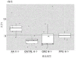


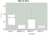


Classifications
-
- C—CHEMISTRY; METALLURGY
- C12—BIOCHEMISTRY; BEER; SPIRITS; WINE; VINEGAR; MICROBIOLOGY; ENZYMOLOGY; MUTATION OR GENETIC ENGINEERING
- C12Q—MEASURING OR TESTING PROCESSES INVOLVING ENZYMES, NUCLEIC ACIDS OR MICROORGANISMS; COMPOSITIONS OR TEST PAPERS THEREFOR; PROCESSES OF PREPARING SUCH COMPOSITIONS; CONDITION-RESPONSIVE CONTROL IN MICROBIOLOGICAL OR ENZYMOLOGICAL PROCESSES
- C12Q1/00—Measuring or testing processes involving enzymes, nucleic acids or microorganisms; Compositions therefor; Processes of preparing such compositions
- C12Q1/68—Measuring or testing processes involving enzymes, nucleic acids or microorganisms; Compositions therefor; Processes of preparing such compositions involving nucleic acids
- C12Q1/6876—Nucleic acid products used in the analysis of nucleic acids, e.g. primers or probes
- C12Q1/6883—Nucleic acid products used in the analysis of nucleic acids, e.g. primers or probes for diseases caused by alterations of genetic material
- C12Q1/6886—Nucleic acid products used in the analysis of nucleic acids, e.g. primers or probes for diseases caused by alterations of genetic material for cancer
-
- C—CHEMISTRY; METALLURGY
- C12—BIOCHEMISTRY; BEER; SPIRITS; WINE; VINEGAR; MICROBIOLOGY; ENZYMOLOGY; MUTATION OR GENETIC ENGINEERING
- C12Q—MEASURING OR TESTING PROCESSES INVOLVING ENZYMES, NUCLEIC ACIDS OR MICROORGANISMS; COMPOSITIONS OR TEST PAPERS THEREFOR; PROCESSES OF PREPARING SUCH COMPOSITIONS; CONDITION-RESPONSIVE CONTROL IN MICROBIOLOGICAL OR ENZYMOLOGICAL PROCESSES
- C12Q2600/00—Oligonucleotides characterized by their use
- C12Q2600/112—Disease subtyping, staging or classification
-
- C—CHEMISTRY; METALLURGY
- C12—BIOCHEMISTRY; BEER; SPIRITS; WINE; VINEGAR; MICROBIOLOGY; ENZYMOLOGY; MUTATION OR GENETIC ENGINEERING
- C12Q—MEASURING OR TESTING PROCESSES INVOLVING ENZYMES, NUCLEIC ACIDS OR MICROORGANISMS; COMPOSITIONS OR TEST PAPERS THEREFOR; PROCESSES OF PREPARING SUCH COMPOSITIONS; CONDITION-RESPONSIVE CONTROL IN MICROBIOLOGICAL OR ENZYMOLOGICAL PROCESSES
- C12Q2600/00—Oligonucleotides characterized by their use
- C12Q2600/154—Methylation markers
Abstract
The present disclosure provides, inter alia, methods for colorectal tumor detection (e.g., screening), and compositions related thereto. In various embodiments, the present disclosure provides methods and compositions related thereto for detection (e.g., screening) of adenomas and/or early colorectal cancer. In various embodiments, the present disclosure provides screening methods comprising analyzing the methylation status of one or more methylation biomarkers, and compositions related thereto. In various embodiments, the present disclosure provides methods for detecting (e.g., screening) comprising detecting (e.g., screening) the methylation status of one or more methylation biomarkers in cfDNA (e.g., in ctDNA). In various embodiments, the present disclosure provides methods for screening, comprising detecting (e.g., screening) the methylation status of one or more methylation biomarkers in cfDNA (e.g., in ctDNA) using MSRE-qPCR and/or using massively parallel sequencing (e.g., next generation sequencing).
Description
Technical Field
The present invention relates generally to methods and kits for detecting and/or pre-screening colorectal tumors. In some embodiments, the methods and kits described herein utilize identified differentially methylated regions of the human genome as markers to determine the presence and/or risk of colorectal tumors.
Background
Screening for colorectal tumors (e.g., colorectal cancer) is an important component of cancer prevention, diagnosis and treatment. According to some reports, colorectal cancer (CRC) has been identified as the third most common cancer type and the second leading cause of cancer death worldwide. According to some reports, there are over 180 new cases of colorectal cancer each year, and about 881,000 people die from colorectal cancer, accounting for about one-tenth of cancer deaths. Regular colorectal cancer screening is recommended, especially for individuals over the age of 50. Furthermore, the incidence of colorectal cancer in individuals under 50 years of age increases over time. Statistical data indicate that current colorectal cancer screening techniques are inadequate.
In addition, early detection of colon cancer can reduce mortality. Detection and removal of precursors to colon cancer, including but not limited to colonic polyps with advanced features, will reduce the incidence of CRC, as these polyps are considered the greatest risk for malignant progression. Resection of advanced colon polyps will reduce the risk of cancer. Typically, about 9-16% of asymptomatic patients over the age of 50 will find advanced adenomas.
Accordingly, there is a need for methods, compositions, and systems that can provide for the classification and/or diagnosis of colorectal tumors. In particular, there is a need to diagnose and/or classify colorectal tumors at an early stage.
Disclosure of Invention
The present disclosure provides, inter alia, methods for detecting (e.g., screening) colorectal tumors, and compositions related thereto. In various embodiments, the present disclosure provides methods for classifying subjects with and/or without pre-malignant and/or malignant colorectal tumors, including but not limited to advanced adenomas, polyposis, colorectal cancer (e.g., stage 0, I, II, III, or undifferentiated), and/or various combinations thereof. In various embodiments, the present disclosure provides methods for colorectal tumor screening, including determining the methylation status (e.g., the amount, frequency, or pattern of methylation) at one or more methylation sites found within one or more markers within a sample (e.g., a blood sample, a blood product sample, a stool sample, a colorectal tissue sample) from a subject (e.g., a human subject), and compositions related thereto. For example, a marker may include a methylation locus, such as a Differentially Methylated Region (DMR) of deoxyribonucleic acid (DNA) of a human subject. In various embodiments, the present disclosure provides methods of classifying a subject as having and/or not having advanced adenoma, polyposis, colorectal cancer, and/or any combination thereof, the methods comprising determining the methylation status of each of one or more methylated loci in cfDNA (cell-free DNA), e.g., ctDNA (circulating tumor DNA). In various embodiments, the present disclosure provides methods for colorectal tumor screening, the methods comprising determining the methylation status of each of one or more methylated loci in cfDNA (e.g., ctDNA), e.g., massively parallel sequencing (e.g., next generation sequencing), e.g., sequencing by synthesis, real-time (e.g., single molecule) sequencing, bead emulsion sequencing, nanopore sequencing, quantitative polymerase chain reaction (qPCR) (e.g., methylation sensitive restriction enzyme quantitative polymerase chain reaction, MSRE-qPCR), using the following methods. The various compositions and methods provided herein provide sufficient sensitivity and specificity for clinical use to screen for disorders including, but not limited to, advanced adenomas, polyposis, and/or early colorectal cancer. Various compositions and methods provided herein can be used for advanced adenoma, polyposis, and/or colorectal cancer screening by analyzing a subject's accessible tissue sample, such as a tissue sample that is blood or a blood component (e.g., cfDNA, e.g., ctDNA) or stool.
In one aspect, the invention relates to a method of detecting (e.g., screening) a colorectal neoplasm in a human subject [ e.g., (I) classifying a subject as having advanced adenoma, (II) classifying a subject as having polyposis, (III) classifying a subject as having colorectal cancer (e.g., 0, I, II, III, or undifferentiated stage), (iv) classifying a subject as having at least one of advanced adenoma, polyposis, and colorectal carcinoma, where it is determined or not determined that the subject has which of the following disorders, or (v) classifying a subject as having at least one of advanced adenoma and colorectal carcinoma, where it is determined or not determined that the subject has which of the following disorders ], the method comprising:
-determining the methylation status of each of one or more of the following in the human subject DNA sample:
(i) A methylation locus in the gene SLC6A1 having SEQ ID NO 6, and
(ii) A methylation locus within SEQ ID NO 51, and
comparing the obtained data with reference values obtained from healthy individuals, and
a colorectal tumor of a subject can be diagnosed if hypermethylation is detected in one or more loci as compared to a reference sample.
In another aspect, the invention relates to a method of detecting (e.g., screening) a colorectal neoplasm in a human subject [ e.g., (I) classifying a subject as having advanced adenoma, (II) classifying a subject as having polyposis, (III) classifying a subject as having colorectal cancer (e.g., 0, I, II, III, or undifferentiated stage), (iv) classifying a subject as having at least one of advanced adenoma, polyposis, and colorectal carcinoma, where it is determined or not determined which of the following conditions is present in the subject, or (v) classifying a subject as having at least one of advanced adenoma and colorectal carcinoma, where it is determined or not determined which of the following conditions is present in the subject ], the method comprising: determining a methylation state of each of one or more markers identified in a sample obtained from the subject (e.g., a blood sample, a blood product sample, a stool sample, a colorectal tissue sample), and determining whether the subject has a colorectal tumor based at least in part on the determined methylation state of each of the one or more markers, wherein each of the one or more markers is a methylation locus comprising at least a portion (e.g., at least 5%, at least 10%, at least 15%, at least 20%, at least 25%, at least 30%, at least 35%, at least 40%, at least 45%, at least 50%, at least 55%, at least 60%, at least 65%, at least 70%, at least 75%, at least 80%, at least 85%, at least 90%, or at least 95%) of a Differentially Methylated Region (DMR) selected from the DMRs listed in table 1.
In another aspect, the invention relates to a method of detecting (e.g., screening) a colorectal tumor in a human subject [ e.g., (I) classifying the subject as having advanced adenoma, (II) classifying the subject as having polyposis, (III) classifying the subject as having colorectal cancer (e.g., 0, I, II, III, or undifferentiated stage), (iv) classifying the subject as having at least one of advanced adenoma, polyposis, and colorectal cancer, either where it is determined or uncertain which of the following conditions the subject is, or (v) classifying the subject as having at least one of advanced adenoma and colorectal cancer where it is determined or uncertain which of the following conditions the subject is ], the method comprising: determining a methylation state of each of one or more of deoxyribonucleic acids (DNAs) of a human subject: (i) A methylated locus in the SLC6A1 gene having SEQ ID No. 6; (ii) (ii) a methylated locus within gene F13A1 having SEQ ID NO 23; (iii) A methylated locus within the gene BARHL1 having SEQ ID NO 35; and diagnosing a colorectal tumor in the human subject based at least on the determined methylation status.
In some embodiments, the polypeptide having the sequence of SEQ id no:6 comprises at least a portion (e.g., at least 5%, at least 10%, at least 15%, at least 20%, at least 25%, at least 30%, at least 35%, at least 40%, at least 45%, at least 50%, at least 55%, at least 60%, at least 65%, at least 70%, at least 75%, at least 80%, at least 85%, at least 90%, or at least 95%) of SLC6A1 having SEQ ID No. 6.
In some embodiments, a methylated locus within gene F13A1 having SEQ ID No. 23 comprises at least a portion (e.g., at least 5%, at least 10%, at least 15%, at least 20%, at least 25%, at least 30%, at least 35%, at least 40%, at least 45%, at least 50%, at least 55%, at least 60%, at least 65%, at least 70%, at least 75%, at least 80%, at least 85%, at least 90%, or at least 95%) of F13A1 having SEQ ID No. 23.
In some embodiments, the methylated locus within BARHL1 of the gene having SEQ ID No. 35 comprises at least a portion (e.g., at least 5%, at least 10%, at least 15%, at least 20%, at least 25%, at least 30%, at least 35%, at least 40%, at least 45%, at least 50%, at least 55%, at least 60%, at least 65%, at least 70%, at least 75%, at least 80%, at least 85%, at least 90%, or at least 95%) of BARHL1 having SEQ ID No. 35.
In some embodiments, the method further comprises determining the methylation status of a methylation locus comprising at least a portion (e.g., at least 5%, at least 10%, at least 15%, at least 20%, at least 25%, at least 30%, at least 35%, at least 40%, at least 45%, at least 50%, at least 55%, at least 60%, at least 65%, at least 70%, at least 75%, at least 80%, at least 85%, at least 90%, or at least 95%) of SEQ ID No. 51 in deoxyribonucleic acid (DNA) of the human subject, and wherein the diagnosing step comprises diagnosing colorectal tumor in the human subject based at least on the methylation status determined for the methylation locus comprising the at least a portion of SEQ ID No. 51. In another aspect, the invention relates to a method of detecting (e.g., screening) colorectal neoplasm in a human subject [ e.g., (I) classifying a subject as having advanced adenoma, (II) classifying a subject as having polyposis, (III) classifying a subject as having colorectal cancer (e.g., 0, I, II, III, or undifferentiated stage), (iv) classifying a subject as having at least one of advanced adenoma, polyposis, and colorectal carcinoma, where it is determined or not determined which of the following conditions is present in the subject, or (v) classifying a subject as having at least one of advanced adenoma and colorectal carcinoma, where it is determined or not determined which of the following conditions is present in the subject ], the method comprising: determining the methylation state of each of one or both of deoxyribonucleic acids (DNAs) of a human subject: (i) (ii) a methylated locus within the gene CSMD2 having SEQ ID NO 1; and (ii) a methylation locus within the gene SLC6A1 having SEQ ID NO 6; and diagnosing a colorectal tumor in the human subject based at least on the determined methylation status.
In some embodiments, a methylated locus within gene CSMD2 having SEQ ID No. 1 comprises at least a portion (e.g., at least 5%, at least 10%, at least 15%, at least 20%, at least 25%, at least 30%, at least 35%, at least 40%, at least 45%, at least 50%, at least 55%, at least 60%, at least 65%, at least 70%, at least 75%, at least 80%, at least 85%, at least 90%, or at least 95%) of CSMD2 having SEQ ID No. 1.
In some embodiments, the methylated locus within SLC6A1 of the gene having SEQ ID No.6 comprises at least a portion (e.g., at least 5%, at least 10%, at least 15%, at least 20%, at least 25%, at least 30%, at least 35%, at least 40%, at least 45%, at least 50%, at least 55%, at least 60%, at least 65%, at least 70%, at least 75%, at least 80%, at least 85%, at least 90%, or at least 95%) of SLC6A1 having SEQ ID No. 6.
In some embodiments, the method further comprises determining the methylation state of a methylation locus comprising at least a portion (e.g., at least 5%, at least 10%, at least 15%, at least 20%, at least 25%, at least 30%, at least 35%, at least 40%, at least 45%, at least 50%, at least 55%, at least 60%, at least 65%, at least 70%, at least 75%, at least 80%, at least 85%, at least 90%, or at least 95%) of SEQ ID No. 25 in deoxyribonucleic acid (DNA) of the human subject, and wherein the diagnosing step comprises diagnosing colorectal tumor in the human subject based at least on the methylation state determined for the methylation locus comprising the at least a portion of SEQ ID No. 25.
In some embodiments, the method further comprises determining the methylation state of a methylation locus comprising at least a portion (e.g., at least 5%, at least 10%, at least 15%, at least 20%, at least 25%, at least 30%, at least 35%, at least 40%, at least 45%, at least 50%, at least 55%, at least 60%, at least 65%, at least 70%, at least 75%, at least 80%, at least 85%, at least 90%, or at least 95%) of SEQ ID No. 51 in deoxyribonucleic acid (DNA) of the human subject, and wherein the diagnosing step comprises diagnosing colorectal tumor in the human subject based at least on the methylation state determined for the methylation locus comprising the at least a portion of SEQ ID No. 51.
In some embodiments, the DNA is isolated from blood or plasma of a human subject.
In some embodiments, the DNA is cell-free DNA of a human subject.
In some embodiments, the methylation status is determined using quantitative polymerase chain reaction (qPCR).
In some embodiments, methylation status is determined using massively parallel sequencing (e.g., next generation sequencing) [ e.g., sequencing by synthesis, real-time (e.g., single molecule) sequencing, bead emulsion sequencing, nanopore sequencing, etc. ].
In some embodiments, each methylated locus has a length equal to or less than 5000bp, 4,000bp, 3,000bp, 2,000bp, 1,000bp, 950bp, 900bp, 850bp, 800bp, 750bp, 700bp, 650bp, 600bp, 550bp, 500bp, 450bp, 400bp, 350bp, 300bp, 250bp, 200bp, 150bp, 100bp, 50bp, 40bp, 30bp, 20bp, or 10bp.
In some embodiments, the method comprises determining the methylation status of each of the one or more markers using Next Generation Sequencing (NGS). In some embodiments, the methods include the use of one or more oligonucleotide capture decoys (e.g., biotinylated oligonucleotide probes) that enrich for the target region to capture one or more corresponding methylated loci (e.g., followed by library preparation and sequencing, e.g., where the sample is bisulfite converted or enzymatically converted prior to capture).
In another aspect, the invention relates to a kit for use in a method (e.g., a method as described herein), the kit comprising one or more oligonucleotide primer pairs for amplifying one or more corresponding methylated loci.
In another aspect, the invention relates to a diagnostic qPCR reaction for detecting (e.g., screening) colorectal cancer (e.g., in the methods described herein), the diagnostic qPCR reaction comprising human DNA, a polymerase, one or more oligonucleotide primer pairs for amplifying one or more corresponding methylated loci, and optionally at least one methylation sensitive restriction enzyme.
In another aspect, the invention relates to a kit (e.g., for use in the methods described herein) comprising one or more oligonucleotide capture baits (e.g., one or more biotinylated oligonucleotide probes) for capturing one or more corresponding methylated loci (e.g., for hybridization to a region/region of interest).
In various aspects, the methods and compositions of the present invention can be used in combination with biomarkers known in the art, for example, as disclosed in U.S. patent No. 10,006,925 and U.S. patent No. 63,011,970.
Definition of
One or more of: the articles "a" and "an" are used herein to refer to one or more (i.e., to at least one) of the grammatical object of the article. For example, "an element" means one element or more than one element.
About: the term "about," when used herein in reference to a value, refers to a value that is similar in context to the recited value. In general, those skilled in the art familiar with the context will understand the relative degree of difference encompassed by "about" in that context. For example, in some embodiments, the term "about" can encompass a range of values that is 25%, 20%, 19%, 18%, 17%, 16%, 15%, 14%, 13%, 12%, 11%, 10%, 9%, 8%, 7%, 6%, 5%, 4%, 3%, 2%, 1%, or a few percent of the referenced value, e.g., as described herein.
Advanced adenomas: as used herein, the term "advanced adenoma" generally refers to a cell that exhibits initial signs of relatively abnormal, uncontrolled, and/or autonomous growth but has not been classified as a cancerous change. In the context of colon tissue, "advanced adenoma" refers to tumor growth that shows: high grade dysplasia signs, and/or size > =10mm, and/or villous histological type, and/or jagged histological type are accompanied by any type of dysplasia.
Application: as used herein, the term "administering" generally refers to administering a composition to a subject or system, e.g., to effect delivery of an agent included in or otherwise delivered by the composition.
Reagent: as used herein, the term "agent" refers to an entity (e.g., a small molecule, peptide, polypeptide, nucleic acid, lipid, polysaccharide, complex, combination, mixture, system, or phenomenon, such as heat, electric current, electric field, magnetic force, magnetic field, etc.).
The improvement is as follows: as used herein, the term "improving" refers to the prevention, reduction, alleviation or amelioration of the condition of a subject. Improvement includes, but does not require, complete recovery or complete prevention of the disease, disorder or condition.
Amplicon or amplicon molecule: as used herein, the term "amplicon" or "amplicon molecule" refers to a nucleic acid molecule produced by transcription from a template nucleic acid molecule, or a nucleic acid molecule having a sequence complementary thereto, or a double-stranded nucleic acid, including any such nucleic acid molecule. Transcription may begin with a primer.
Amplification: as used herein, the term "amplifying" refers to the use of a template nucleic acid molecule in combination with various reagents to produce additional nucleic acid molecules from the template nucleic acid molecule, which additional nucleic acid molecules may be identical or identical (e.g., at least 70% identical, e.g., at least 75%, 80%, 85%, 90%, 91%, 92%, 93%, 94%, 95%, 96%, 97%, 98%, 99%, or 100% identical) and/or sequences complementary thereto to fragments of the template nucleic acid molecule.
Amplification reaction mixture: as used herein, the term "amplification reaction mixture" or "amplification reaction" refers to a template nucleic acid molecule along with reagents sufficient to amplify the template nucleic acid molecule.
Biological sample: as used herein, the term "biological sample" generally refers to a sample obtained or derived from a biological source of interest (e.g., a tissue or organism or cell culture), as described herein. In some embodiments, for example, as described herein, the biological source is or includes an organism, such as an animal or human. In some embodiments, for example, as described herein, the biological sample is or includes a biological tissue or fluid. In some embodiments, for example, as described herein, a biological sample can be or include a cell, a tissue, or a bodily fluid. In some embodiments, for example, as described herein, a biological sample can be or include blood, blood cells, cell-free DNA, free-floating nucleic acids, ascites, biopsy samples, surgical specimens, cell-containing bodily fluids, sputum, saliva, stool, urine, cerebrospinal fluid, peritoneal fluid, pleural fluid, lymph, gynecological fluid, secretions, excretions, skin swabs, vaginal swabs, oral swabs, nasal swabs, irrigation fluids, or lavage fluids, e.g., ductal or bronchoalveolar lavage fluids, aspirates, scrapings, bone marrow. In some embodiments, for example, as described herein, a biological sample is or includes cells obtained from a single subject or multiple subjects. The sample may be a "raw sample" obtained directly from a biological source, or may be a "processed sample". A biological sample may also be referred to as a "sample".
Biomarkers: as used herein, the term "biomarker" is consistent with its use in the art and refers to an entity whose presence, level, or form is associated with a particular biological event or state of interest, and thus is considered to be a "marker" for that event or state. One skilled in the art will appreciate that, for example, in the context of DNA biomarkers, the biomarkers can be or include loci (e.g., one or more methylation loci) and/or states of loci (e.g., states of one or more methylation loci). To name a few biomarkers, in some embodiments, for example, as described herein, a biomarker can be or include a marker of a particular disease, disorder, or condition, or can be a qualitative marker of a quantitative probability that a particular disease, disorder, or condition can develop, occur, or relapse, for example, in a subject. In some embodiments, for example, as described herein, a biomarker may be or include a marker of a particular treatment outcome, or a qualitative of its quantitative probability. Thus, in various embodiments, for example, as described herein, a biomarker can predict, prognose, and/or diagnose a related biological event or state of interest. The biomarker may be an entity of any chemical class. For example, in some embodiments, a biomarker can be or include a nucleic acid, a polypeptide, a lipid, a carbohydrate, a small molecule, an inorganic agent (e.g., a metal or ion), or a combination thereof, e.g., as described herein. In some embodiments, for example, as described herein, the biomarker is a cell surface marker. In some embodiments, for example, as described herein, the biomarker is intracellular. In some embodiments, for example, as described herein, a biomarker is found extracellularly (e.g., secreted or otherwise produced or present extracellularly, e.g., in a bodily fluid, e.g., blood, urine, tears, saliva, cerebrospinal fluid, etc.). In some embodiments, for example, as described herein, a biomarker is the methylation state of a methylation locus. In certain instances, for example, as described herein, a biomarker may be referred to as a "marker.
To name just one example of a biomarker, in some embodiments, e.g., as described herein, the term refers to the expression of a product encoded by a gene, which expression is characteristic of a particular tumor, tumor subclass, tumor stage, etc. Alternatively or additionally, in some embodiments, for example, as described herein, the presence or level of a particular marker can be correlated with the activity (or level of activity) of a particular signaling pathway, e.g., a signaling pathway whose activity is characteristic of a particular class of tumor.
One skilled in the art will appreciate that a biomarker may be solely responsible for determining a particular biological event or state of interest, or may represent or contribute to determining a statistical probability of a particular biological event or state of interest. One skilled in the art will appreciate that markers may differ in their specificity and/or sensitivity with respect to a particular biological event or state of interest.
Blood components: as used herein, the term "blood component" refers to any component of whole blood, including red blood cells, white blood cells, plasma, platelets, endothelial cells, mesothelial cells, epithelial cells, and cell-free DNA. Blood components also include plasma components, including proteins, metabolites, lipids, nucleic acids, and carbohydrates, as well as any other cells that may be present in the blood, for example, due to pregnancy, organ transplantation, infection, injury, or disease.
Cancer: as used herein, the terms "cancer," "malignancy," "neoplasm," "tumor," and "carcinoma" are used interchangeably to refer to a disease, disorder, or condition in which cells exhibit or exhibit relatively abnormal, uncontrolled, and/or autonomous growth, such that they exhibit or exhibit an abnormally elevated proliferation rate and/or an abnormal growth phenotype. In some embodiments, for example, as described herein, a cancer can include one or more tumors. In some embodiments, for example, as described herein, a cancer can be or include a precancerous (e.g., benign), malignant, pre-metastatic, and/or non-metastatic cell. In some embodiments, for example, as described herein, the cancer may be or comprise a solid tumor. In some embodiments, for example, as described herein, the cancer may be or include a hematological tumor. In general, examples of different types of cancers known in the art include, for example, colorectal cancers, hematopoietic cancers including leukemias, lymphomas (hodgkins and non-hodgkins), myelomas, and myeloproliferative diseases; sarcomas, melanomas, adenomas, solid tissue cancers, squamous cell carcinomas of the mouth, throat, larynx and lung, liver cancers, genitourinary cancers such as prostate, cervical, bladder, uterus and endometrial cancers, as well as renal cell carcinomas, bone, pancreatic, skin or intraocular melanomas, cancers of the endocrine system, thyroid, parathyroid, head and neck, breast, gastrointestinal and nervous system, benign lesions, and the like, such as papillomas, and the like.
Chemotherapeutic agents: as used herein, the term "chemotherapeutic agent," consistent with its use in the art, refers to one or more agents known to be useful for or contributing to the treatment of cancer or having known characteristics for use in or contributing to the treatment of cancer. In particular, chemotherapeutic agents include pro-apoptotic agents, cytostatic agents, and/or cytotoxic agents. In some embodiments, for example, as described herein, a chemotherapeutic agent can be or include an alkylating agent, an anthracycline, a cytoskeletal disrupting agent (e.g., a microtubule targeting moiety, such as a taxane, maytansine, and analogs thereof), an epothilone, a histone deacetylase inhibitor HDAC), a topoisomerase inhibitor (e.g., inhibitor topoisomerase I and/or topoisomerase II), a kinase inhibitor, a nucleotide analog or nucleotide precursor analog, a peptide antibiotic, a platinum drug, a retinoid, a vinca alkaloid, and/or an analog having related antiproliferative activity. In certain particular embodiments, for example, as described herein, the chemotherapeutic agent may be or include actinomycin, all-trans retinoic acid, auristatin, azacitidine, azathioprine, bleomycin, bortezomib, carboplatin, capecitabine, cisplatin, chloramphenicol, cyclophosphamide, curcumin, cytarabine, daunorubicin, docetaxel, doxifluridine, doxorubicin, epirubicin, epothilone, etoposide, fluorouracil, gemcitabine, hydroxyurea, idarubicin, imatinib, irinotecan, maytansine and/or analogs thereof (such as DM 1), nitrogen mustard, mercaptopurine, methotrexate, mitoxantrone, maytansine, oxaliplatin, paclitaxel, pemetrexed, teniposide, thioguanine, topotecan, valrubicin, vinblastine, vincristine, vindesine, vinorelbine, or combinations thereof. In some embodiments, for example, as described herein, chemotherapeutic agents may be used in the context of antibody-drug conjugates. In some embodiments, for example, as described herein, the chemotherapeutic agent is one found in an antibody-drug conjugate selected from the group consisting of: hLL 1-doxorubicin, hRS7-SN-38, hMN-14-SN-38, hLL2-SN-38, hA20-SN-38, hPAM4-SN-38, hLL1-SN-38, hRS7-Pro-2-P-Dox, hMN-14-Pro-2-P-Dox, hLL2-Pro-2-P-Dox, hA20-Pro-2-P-Dox, hPAM4-Pro-2-P-Dox, hLL1-Pro-2-P-Dox, P4/D10-doxorubicin, gemtuzumab ozotacin, bentuximab, trastuzumab, otuzumab, gemumab, and glytumomab (glytumomab vedotin) SAR3419, SAR566658, BIIB015, BT062, SGN-75, SGN-CD19A, AMG-172, AMG-595, BAY-94-9343, ASG-5ME, ASG-22ME, ASG-16M8F, MDX-1203, MLN-0264, anti-PSMA ADC, RG-7450, RG-7458, RG-7593, RG-7596, RG-7598, RG-7599, RG-7600, RG-7636, ABT-414, IMGN-853, IMGN-529, martin-Wauterotuzumab (voretuzumab) and Moxing-Lovoruzumab (lorvovatuzumab). In some embodiments, for example, as described herein, the chemotherapeutic agent may be or comprise farnesyl-thiosalicylic acid (FTS), 4- (4-chloro-2-methylphenoxy) -N-hydroxybutyramide (CMH), estradiol (E2), tetramethoxystilbene (TMS), delta-tocotrienol, salinomycin, or curcumin.
Combination therapy: the term "combination therapy" as used herein refers to the administration of two or more agents or regimens to a subject such that the two or more agents or regimens together treat a disease, disorder or condition in the subject. In some embodiments, for example, as described herein, two or more therapeutic agents or regimens can be administered simultaneously, sequentially, or in overlapping dosing regimens. One skilled in the art will appreciate that combination therapy includes, but does not require, that both agents or regimens be administered together in a single composition, nor simultaneously.
The comparison results are as follows: as used herein, the term "comparable" refers to a group of two or more conditions, environments, reagents, entities, populations, etc., which may not be identical to one another, but which are sufficiently similar to allow comparisons to be made therebetween such that one of skill in the art will understand that a reasonable conclusion may be drawn based on the observed differences or similarities. In some embodiments, for example, as described herein, a comparable set of conditions, environments, reagents, entities, populations, etc., typically features a plurality of substantially identical features and zero, one, or more different features. One of ordinary skill in the art will understand, in this context, what degree of identity is needed to make members of a group comparable. For example, one of ordinary skill in the art will understand that when features that are substantially identical in sufficient number and type to warrant a reasonable conclusion that the observed differences may be attributed, in whole or in part, to their non-identical features.
A detectable moiety: the term "detectable moiety" as used herein refers to any element, molecule, functional group, compound, fragment, or other moiety that is detectable. In some embodiments, for example, as described herein, the detectable moiety is provided or used alone. In some embodiments, for example, as described herein, a detectable agent associated with (e.g., linked to) another agent is provided and/or utilizedAnd (4) partial. Examples of detectable moieties include, but are not limited to, various ligands, radioisotopes (e.g., as in 3 H、 14 C、 18 F、 19 F、 32 P、 35 S、 135 I、 125 I、 123 I、 64 Cu、 187 Re、 111 In、 90 Y、 99m Tc、 177 Lu、 89 Zr, etc.), fluorescent dyes, chemiluminescent agents, bioluminescent agents, spectrally resolvable inorganic fluorescent semiconductor nanocrystals (i.e., quantum dots), metal nanoparticles, nanoclusters, paramagnetic metal ions, enzymes, colorimetric labels, biotin, digoxigenin (digoxigenin), haptens, and proteins from which antisera or monoclonal antibodies are derived.
And (3) diagnosis: as used herein, the term "diagnosis" refers to the qualitative determination of whether a subject has or will develop a disease, disorder, condition, or state, and/or the quantitative probability. For example, in the diagnosis of cancer, the diagnosis may include a determination as to the risk, type, stage, malignancy, or other classification of the cancer. In certain instances, for example, as described herein, a diagnosis may be or include a determination related to prognosis and/or likely response to one or more general or specific therapeutic agents or regimens.
Diagnosis information: as used herein, the term "diagnostic information" refers to information that can be used to provide a diagnosis. The diagnostic information may include, but is not limited to, biomarker status information.
Differential methylation: as used herein, the term "differentially methylated" describes a methylation site whose methylation state differs between a first condition and a second condition. Differentially methylated methylation sites can be referred to as differentially methylated sites. In certain instances, for example, as described herein, a DMR is defined by an amplicon produced by amplification using oligonucleotide primers, e.g., a pair of oligonucleotide primers selected for amplification of the DMR or amplification of a DNA region of interest present in the amplicon. In certain instances, for example, as described herein, a DMR is defined as a region of DNA amplified by a pair of oligonucleotide primers, including a region having the sequence of an oligonucleotide primer or a sequence complementary to an oligonucleotide primer. In certain instances, for example, as described herein, a DMR is defined as a region of DNA amplified by a pair of oligonucleotide primers, excluding regions having the sequence of the oligonucleotide primers or sequences complementary to the oligonucleotide primers. As used herein, a DMR specifically provided may be unambiguously identified by the name of the relevant gene followed by a three digit number of the starting position, such that, for example, a DMR beginning at position 29921434 of ALK may be identified as ALK'434. As used herein, a specifically provided DMR can be unambiguously identified by chromosome numbering followed by the beginning and end positions of the DMR. For example, a DMR identified in Table 1 as having a uid 9_132579614 _U132579683 may also be identified as chr9:132579614-132579683.
Differential methylation region: as used herein, the term "differentially methylated region" (DMR) refers to a region of DNA that comprises one or more differentially methylated sites. DMRs that include a greater number or frequency of methylation sites under selected conditions of interest, such as a cancer state, may be referred to as hypermethylated DMRs. A DMR that includes a lesser number or frequency of methylation sites under selected conditions of interest, such as a cancer state, may be referred to as a hypomethylated DMR. DMR as a biomarker for colorectal cancer methylation may be referred to as colorectal cancer DMR. In certain instances, for example, as described herein, a DMR can be a single nucleotide that is a methylation site. In certain cases, for example, as described herein, a DMR has a length of at least 10, at least 15, at least 20, at least 30, at least 50, or at least 75 base pairs. In certain instances, for example, as described herein, the DMR has a length equal to or less than 5000bp, 4,000bp, 3,000bp, 2,000bp, 1,000bp, 950bp, 900bp, 850bp, 800bp, 750bp, 700bp, 650bp, 600bp, 550bp, 500bp, 450bp, 400bp, 350bp, 300bp, 250bp, 200bp, 150bp, 100bp, 50bp, 40bp, 30bp, 20bp, or 10bp (e.g., using quantitative polymerase chain reaction (qPCR) to determine methylation status, e.g., methylation sensitive restriction enzyme quantitative polymerase chain reaction (MSRE-qPCR)). In certain instances, DMR as a methylation biomarker of advanced adenomas can also be used to identify colorectal cancer, e.g., as described herein.
DNA region: as used herein, "DNA region" refers to any contiguous portion of a larger DNA molecule. One skilled in the art will be familiar with techniques for determining whether a first DNA region and a second DNA region correspond, e.g., based on sequence similarity (e.g., sequence identity or homology) and/or context (e.g., sequence identity or homology of nucleic acids upstream and/or downstream of the first DNA region and the second DNA region) of the first DNA region and the second DNA region.
Unless otherwise indicated herein, sequences found in humans or associated with humans (e.g., sequences that hybridize to Human DNA) are found in, based on, and/or derived from example representative Human genomic sequences commonly referred to and known to those of skill in the art as homo sapiens (Human) genome assembly GRCh38, hg38, and/or genome reference association Human Build 38. One skilled in the art will further appreciate that the DNA region of hg38 can be referred to by known systems that involve identifying a particular nucleotide position or range thereof according to the number specified.
The administration scheme is as follows: as used herein, the term "dosing regimen" may refer to a set of one or more identical or different unit doses administered to a subject, typically including multiple unit dose administrations, wherein each unit dose administration is separated from the other unit dose administrations by a period of time. In various embodiments, for example, as described herein, one or more or all of the unit doses of a dosing regimen may be the same or may vary (e.g., increase over time, decrease over time, or be adjusted at the discretion of the subject and/or the physician). In various embodiments, one or more or all of the time periods between each dose can be the same or can vary (e.g., increase over time, decrease over time, or adjust at the discretion of the subject and/or physician). In some embodiments, for example, as described herein, a given therapeutic agent has a recommended dosing regimen, which may involve one or more doses. Generally, at least one recommended dosing regimen for commercially available drugs is known to those skilled in the art. In some embodiments, for example, as described herein, a dosing regimen is associated with a desired or beneficial result (i.e., is a therapeutic dosing regimen) when administered across a relevant population.
Downstream: the term "downstream" as used herein refers to the first DNA region being closer to the C-terminus of the nucleic acid comprising the first DNA region and the second DNA region relative to the second DNA region.
Gene: as used herein, the term "gene" refers to a single DNA region, e.g., in a chromosome, that includes a coding sequence that encodes a product (e.g., an RNA product and/or a polypeptide product), along with all, some, or none of the DNA sequences that contribute to the regulation of the expression of the coding sequence. In some embodiments, for example, as described herein, a gene includes one or more non-coding sequences. In some particular embodiments, for example, as described herein, a gene includes exon and intron sequences. In some embodiments, for example, as described herein, a gene includes one or more regulatory elements that, for example, can control or affect one or more aspects of gene expression (e.g., cell-type specific expression, inducible expression, etc.). In some embodiments, for example, as described herein, a gene includes a promoter. In some embodiments, for example, as described herein, a gene comprises one or both of (i) DNA nucleotides that are extended a predetermined number of nucleotides upstream of the coding sequence and (ii) DNA nucleotides that are extended a predetermined number of nucleotides downstream of the coding sequence. In various embodiments, for example, the predetermined number of nucleotides can be 500bp, 1kb, 2kb, 3kb, 4kb, 5kb, 10kb, 20kb, 30kb, 40kb, 50kb, 75kb, or 100kb, as described herein.
Homology: as used herein, the term "homology" refers to the overall relatedness between polymeric molecules, such as between nucleic acid molecules (e.g., DNA molecules and/or RNA molecules) and/or between polypeptide molecules. Those skilled in the art will appreciate that homology can be defined as, for example, percent identity or percent homology (sequence similarity). In some embodiments, for example, a polymeric molecule is considered "homologous" if its sequence is at least 25%, 30%, 35%, 40%, 45%, 50%, 55%, 60%, 65%, 70%, 75%, 80%, 85%, 90%, 95%, or 99% identical, as described herein. In some embodiments, for example, a polymer molecule is considered "homologous" if its sequence has at least 25%, 30%, 35%, 40%, 45%, 50%, 55%, 60%, 65%, 70%, 75%, 80%, 85%, 90%, 95%, or 99% similarity, as described herein.
And (3) hybridization: as used herein, "hybridization" refers to the binding of a first nucleic acid to a second nucleic acid to form a double-stranded structure, the binding occurring through complementary pairing of nucleotides. One skilled in the art will recognize that complementary sequences may hybridize, among other things. In various embodiments, for example, as described herein, hybridization can occur, e.g., between nucleotide sequences having at least 70% complementarity, e.g., at least 75%, 80%, 85%, 90%, 91%, 92%, 93%, 94%, 95%, 96%, 97%, 98%, 99%, or 100% complementarity. One skilled in the art will further appreciate that whether hybridization of the first nucleic acid and the second nucleic acid occurs can depend on various reaction conditions. Conditions under which hybridization can occur are known in the art.
Low methylation: as used herein, the term "hypomethylation" refers to a state of a methylated locus in which at least one less methylated nucleotide is present in a state of interest as compared to a reference state (e.g., at least one less methylated nucleotide is present in colorectal cancer as compared to a healthy control).
Hypermethylation: as used herein, the term "hypermethylation" refers to a state of a methylated locus at which at least one more methylated nucleotide is present in a state of interest as compared to a reference state (e.g., at least one more methylated nucleotide is present in colorectal cancer as compared to a healthy control).
Identity, identity: as used herein, the terms "identity" and "identical" refer to the overall relatedness between polymeric molecules, e.g., between nucleic acid molecules (e.g., DNA molecules and/or RNA molecules) and/or between polypeptide molecules. Methods of calculating percent identity between two provided sequences are known in the art. Calculation of percent identity of two nucleic acid or polypeptide sequences, for example, can be achieved by aligning the two sequences (or the complement of one or both sequences) for optimal comparison purposes (e.g., gaps can be introduced in one or both of the first and second sequences for optimal alignment, and non-identical sequences can be disregarded for comparison purposes). The nucleotides or amino acids at the corresponding positions are then compared. When a position in the first sequence is occupied by the same residue (e.g., nucleotide or amino acid) as the corresponding position in the second sequence, then the molecules are identical at that position. The percent identity between two sequences is a function of the number of identical positions shared by the sequences, and optionally taking into account the number of gaps and the length of each gap, which may need to be introduced to achieve optimal alignment of the two sequences. Comparison of sequences and determination of percent identity between two sequences can be accomplished using a computational algorithm, such as BLAST (basic local alignment search tool).
"improve", "increase" or "decrease": as used herein, these terms or grammatically comparable comparative terms refer to values measured relative to comparable references. For example, in some embodiments, the evaluation value achieved using the agent of interest may be "improved," e.g., as described herein, relative to an evaluation value obtained using a comparable reference agent or no agent. Alternatively or additionally, in some embodiments, for example, as described herein, an assessment value in a subject or system of interest can be "improved" relative to an assessment value obtained in the same subject or system under different conditions or at different points in time (e.g., before or after an event such as administration of an agent of interest), or in subjects compared at different ratios (e.g., in comparable subjects or systems that differ from the subject or system of interest by the presence of one or more particular diseases, disorders or conditions of interest, or prior exposure to a certain condition or agent, etc.). In some embodiments, for example, as described herein, comparative terms refer to statistically relevant differences (e.g., differences of sufficient prevalence and/or magnitude to achieve statistical relevance). Those skilled in the art will recognize or will be able to readily determine the degree and/or prevalence of differences that are needed or sufficient to achieve such statistical significance in a given context.
Methylation: as used herein, the term "methylated" includes at (i) the C5 position of cytosine; (ii) the N4 position of cytosine; (iii) Methylation at any of the N6 positions of adenine. Methylation also includes (iv) other types of nucleotide methylation. Methylated nucleotides can be referred to as "methylated nucleotides" or "methylated nucleotide bases". In some embodiments, for example, as described herein, methylation refers specifically to methylation of cytosine residues. In certain instances, methylation refers specifically to the methylation of cytosine residues present in CpG sites.
Methylation test: as used herein, the term "methylation test" refers to any technique that can be used to determine the methylation state of a methylated locus.
Methylation biomarkers: as used herein, the term "methylation biomarker" refers to a biomarker that is or includes at least one methylation locus and/or the methylation state of at least one methylation locus, e.g., a hypermethylated locus. In particular, a methylation biomarker is a biomarker characterized by a change in the methylation state of one or more nucleic acid loci between a first state and a second state (e.g., between a cancerous state and a non-cancerous state).
Methylation loci: as used herein, the term "methylation locus" refers to a region of DNA that comprises at least one differentially methylated region. Under selected conditions of interest, such as a cancer state, a methylated locus that includes a greater number or frequency of methylated sites can be referred to as a hypermethylated locus. Under selected conditions of interest, such as a cancer state, a methylation locus that includes a lesser number or frequency of methylation sites can be referred to as a hypomethylated locus. In certain instances, for example, as described herein, a methylated locus has a length of at least 10, at least 15, at least 20, at least 30, at least 50, or at least 75 base pairs. In certain instances, for example, as described herein, the methylated locus is less than 5000bp, 4,000bp, 3,000bp, 2,000bp, 1,000bp, 950bp, 900bp, 850bp, 800bp, 750bp, 700bp, 650bp, 600bp, 550bp, 500bp, 450bp, 400bp, 350bp, 300bp, 250bp, 200bp, 150bp, 100bp, 50bp, 40bp, 30bp, 20bp, or 10bp in length (e.g., the methylation status is determined using quantitative polymerase chain reaction (qPCR), e.g., methylation sensitive restriction enzyme quantitative polymerase chain reaction (MSRE-qPCR)).
Methylation site: as used herein, a methylation site refers to a nucleotide or nucleotide position that is methylated under at least one condition. In its methylated state, the methylated site can be referred to as a methylated site.
Methylation state: as used herein, "methylation state," "methylation state," or "methylation profile" refers to the amount, frequency, or pattern of methylation at a methylation site within a methylation locus. Thus, a change in methylation state between a first state and a second state can be or include an increase in the number, frequency, or pattern of methylation sites, or can be or include a decrease in the number, frequency, or pattern of methylation sites. In each case, the change in methylation state is a change in methylation value.
Methylation value: as used herein, the term "methylation value" refers to a numerical representation of the methylation state, e.g., in numerical form representing the frequency or ratio of methylation at a methylation locus. In certain instances, for example, as described herein, methylation values can be generated by a method that includes quantifying the amount of intact nucleic acid present in a sample after restriction digestion of the sample with a methylation dependent restriction enzyme. In certain instances, for example, as described herein, methylation values can be generated by a method that includes comparing amplification spectra after bisulfite reaction of a sample. In certain instances, methylation values can be generated by comparing the sequences of bisulfite treated and untreated nucleic acids, e.g., as described herein. In certain instances, for example, as described herein, the methylation value is or comprises or is based on quantitative PCR results.
Nucleic acid: as used herein, in its broadest sense, the term "nucleic acid" refers to any compound and/or substance that is or can be introduced into an oligonucleotide chain. In some embodiments, for example, as described herein, a nucleic acid is a compound and/or substance that is or can be introduced into an oligonucleotide chain via a phosphodiester linkage. From the context, it will be clear that in some embodiments, e.g., as described herein, the term nucleic acid refers to a monomeric nucleic acid residue (e.g., a nucleotide and/or nucleoside), and in some embodiments, e.g., as described herein, refers to a polynucleotide chain comprising a plurality of monomeric nucleic acid residues. The nucleic acid may be or include DNA, RNA, or a combination thereof. Nucleic acids can include natural nucleic acid residues, nucleic acid analogs, and/or synthetic residues. In some embodiments, for example, as described herein, a nucleic acid comprises natural nucleotides (e.g., adenosine, thymidine, guanosine, cytidine, uridine, deoxyadenosine, deoxythymidine, deoxyguanosine, and deoxycytidine). In some embodiments, for example, as described herein, a nucleic acid is or includes one or more nucleotide analogs (e.g., 2-aminoadenosine, 2-thiothymidine, inosine, pyrrole-pyrimidine, 3-methyladenosine, 5-methylcytidine, C-5 propynyl-cytidine, C-5-propynyl-uridine, 2-aminoadenosine, C5-bromouridine, C5-fluorouridine, C5-iodouridine, C5-propynyl-uridine, C5-propynyl-cytidine, C5-methylcytidine, 2-aminoadenosine, 7-deadenosine, 7-deazaguanosine 8-oxoadenosine, 8-oxoguanosine, 0 (6) -methylguanine, 2-thiocytidine, methylated bases, intercalating bases, and combinations thereof).
In some embodiments, for example, as described herein, a nucleic acid has a nucleotide sequence that encodes a functional gene product, e.g., an RNA or a protein. In some embodiments, for example, as described herein, a nucleic acid comprises one or more introns. In some embodiments, for example, as described herein, a nucleic acid comprises one or more genes. In some embodiments, for example, as described herein, the nucleic acid is prepared by one or more of isolation from a natural source, by complementary template-based polymeric enzymatic synthesis (in vivo or in vitro), propagation in a recombinant cell or system, and chemical synthesis.
In some embodiments, for example, as described herein, a nucleic acid analog differs from a nucleic acid in that it does not utilize a phosphodiester backbone. For example, in some embodiments, for example, as described herein, a nucleic acid can include one or more peptide nucleic acids that are known in the art and have peptide bonds in the backbone rather than phosphodiester bonds. Alternatively or additionally, in some embodiments, for example, as described herein, the nucleic acid has one or more phosphorothioate and/or 5' -N-phosphoramidite linkages rather than phosphodiester linkages. In some embodiments, for example, as described herein, a nucleic acid comprises one or more modified sugars (e.g., 2 '-fluororibose, ribose, 2' -deoxyribose, arabinose, and hexose) as compared to those in a native nucleic acid.
In some embodiments, for example, a nucleic acid is or comprises at least 3, 4, 5, 6, 7, 8, 9, 10, 15, 20, 25, 30, 35, 40, 45, 50, 55, 60, 65, 70, 75, 80, 85, 90, 95, 100, 110, 120, 130, 140, 150, 160, 170, 180, 190, 20, 225, 250, 275, 300, 325, 350, 375, 400, 425, 450, 475, 500, 600, 700, 800, 900, 1000, 1500, 2000, 2500, 3000, 3500, 4000, 4500, 5000, or more residues, as described herein. In some embodiments, for example, as described herein, the nucleic acid is partially or completely single stranded, or partially or completely double stranded.
Nucleic acid detection assay: as used herein, the term "nucleic acid detection test" refers to any method of determining the nucleotide composition of a nucleic acid of interest. Nucleic acid detection assays include, but are not limited to, DNA sequencing methods, polymerase chain reaction-based methods, probe hybridization methods, ligase chain reaction, and the like.
Nucleotide: as used herein, the term "nucleotide" refers to a structural component or building block of a polynucleotide, such as a DNA and/or RNA polymer. Nucleotides include bases (e.g., adenine, thymine, uracil, guanine, or cytosine) and sugar molecules and at least one phosphate group. As used herein, a nucleotide may be a methylated nucleotide or an unmethylated nucleotide. One of skill in the art will appreciate that nucleic acid terms, such as "locus" or "nucleotide" can refer to a locus or nucleotides of a single nucleic acid molecule and/or to a cumulative population of loci or nucleotides in multiple nucleic acids (e.g., multiple nucleic acids in a representation of a sample and/or subject) representing a locus or nucleotide (e.g., having the same nucleic acid sequence and/or nucleic acid sequence background, or having substantially the same nucleic acid sequence and/or nucleic acid background).
Oligonucleotide primers: as used herein, the term oligonucleotide primer or primer refers to a nucleic acid molecule that is used, capable of being used, or is used to generate an amplicon from a template nucleic acid molecule. The oligonucleotide primer can provide a transcription initiation point from a template to which the oligonucleotide primer hybridizes under conditions that permit transcription (e.g., in the presence of nucleotides and a DNA polymerase, and at a suitable temperature and pH). Typically, oligonucleotide primers are single-stranded nucleic acids of 5 to 200 nucleotides in length. One skilled in the art will appreciate that the optimal primer length for generating an amplicon from a template nucleic acid molecule can vary with conditions including temperature parameters, primer composition, and the method of transcription or amplification. As used herein, a pair of oligonucleotide primers refers to a set of two oligonucleotide primers that are complementary to a first strand and a second strand, respectively, of a template double-stranded nucleic acid molecule. With respect to a template nucleic acid strand, the first member and the second member of a pair of oligonucleotide primers may be referred to as a "forward" oligonucleotide primer and a "reverse" oligonucleotide primer, respectively, because the forward oligonucleotide primer is capable of hybridizing to a nucleic acid strand that is complementary to the template nucleic acid strand, the reverse oligonucleotide primer is capable of hybridizing to the template nucleic acid strand, and the position of the forward oligonucleotide primer relative to the template nucleic acid strand is 5' of the position of the reverse oligonucleotide primer sequence relative to the template nucleic acid strand. One skilled in the art will appreciate that the identification of the first and second oligonucleotide primers as forward and reverse oligonucleotide primers, respectively, is arbitrary, as these identifiers depend on whether a given nucleic acid strand or its complement is used as a template nucleic acid molecule.
Overlapping: the term "overlap" is used herein to refer to two regions of DNA, each region comprising a subsequence that is substantially identical to a subsequence of the same length in the other region (e.g., two regions of DNA have a subsequence in common). By "substantially identical" is meant that two subsequences of the same length differ by less than a given number of base pairs. In certain instances, for example, as described herein, each subsequence has a length of at least 20 base pairs and differs from each other by less than 4, 3, 2, or 1 base pair (e.g., two subsequences have at least 80%, at least 85%, at least 90%, at least 95% similarity, at least 97% similarity, at least 98% similarity, at least 99% similarity, or at least 99.5% similarity). In certain cases, for example, as described herein, each subsequence has a length of at least 24 base pairs that differ by less than 5, 4, 3, 2, or 1 base pair (e.g., two subsequences have at least 80%, at least 85%, at least 90%, at least 95% similarity, at least 97% similarity, at least 98% similarity, at least 99% similarity, or at least 99.5% similarity). In certain cases, for example, as described herein, each subsequence has a length of at least 50 base pairs that differ by less than 10, 9, 8, 7, 6, 5, 4, 3, 2, or 1 base pair (e.g., two subsequences have at least 80%, at least 85%, at least 90%, at least 95% similarity, at least 97% similarity, at least 98% similarity, at least 99% similarity, or at least 99.5% similarity). In certain cases, for example, as described herein, each subsequence has a length of at least 100 base pairs that differ by less than 20, 15, 10, 9, 8, 7, 6, 5, 4, 3, 2, or 1 base pair (e.g., two subsequences have at least 80%, at least 85%, at least 90%, at least 95% similarity, at least 97% similarity, at least 98% similarity, at least 99% similarity, or at least 99.5% similarity). In certain cases, e.g., as described herein, each subsequence has a length of at least 200 base pairs that differ by less than 40, 30, 20, 15, 10, 9, 8, 7, 6, 5, 4, 3, 2, or 1 base pair (e.g., two subsequences have at least 80%, at least 85%, at least 90%, at least 95% similarity, at least 97% similarity, at least 98% similarity, at least 99% similarity, or at least 99.5% similarity). In certain cases, e.g., as described herein, each subsequence has a length of at least 250 base pairs that differ by less than 50, 40, 30, 20, 15, 10, 9, 8, 7, 6, 5, 4, 3, 2, or 1 base pair (e.g., two subsequences have at least 80%, at least 85%, at least 90%, at least 95% similarity, at least 97% similarity, at least 98% similarity, at least 99% similarity, or at least 99.5% similarity). In certain cases, for example, as described herein, each subsequence has a length of at least 300 base pairs that differ by less than 60, 50, 40, 30, 20, 15, 10, 9, 8, 7, 6, 5, 4, 3, 2, or 1 base pair (e.g., two subsequences have at least 80%, at least 85%, at least 90%, at least 95% similarity, at least 97% similarity, at least 98% similarity, at least 99% similarity, or at least 99.5% similarity). In certain cases, for example, as described herein, each subsequence has a length of at least 500 base pairs that differ by less than 100, 60, 50, 40, 30, 20, 15, 10, 9, 8, 7, 6, 5, 4, 3, 2, or 1 base pair (e.g., two subsequences have at least 80%, at least 85%, at least 90%, at least 95% similarity, at least 97% similarity, at least 98% similarity, at least 99% similarity, or at least 99.5% similarity). In certain cases, for example, as described herein, each subsequence has a length of at least 1000 base pairs that differ by less than 200, 100, 60, 50, 40, 30, 20, 15, 10, 9, 8, 7, 6, 5, 4, 3, 2, or 1 base pair (e.g., two subsequences have at least 80%, at least 85%, at least 90%, at least 95% similarity, at least 97% similarity, at least 98% similarity, at least 99% similarity, or at least 99.5% similarity). In certain instances, for example, as described herein, a subsequence of a first region of two DNA regions may comprise all of a second region of the two DNA regions (or vice versa) (e.g., a common subsequence may comprise all of one or both regions).
The pharmaceutical composition comprises: as used herein, the term "pharmaceutical composition" refers to a composition in which an active agent is formulated with one or more pharmaceutically acceptable carriers. In some embodiments, for example, as described herein, the active agent is present in a unit dosage amount suitable for administration to a subject, e.g., in a treatment regimen that shows a statistically significant probability of achieving a predetermined therapeutic effect when administered to a relevant population. In some embodiments, for example, as described herein, a pharmaceutical composition may be formulated for administration in a particular form (e.g., in a solid form or a liquid form), and/or may be particularly suitable for use, for example: oral administration (e.g., as a liquid medicine) (aqueous or non-aqueous solution or suspension), tablets, capsules, boluses (bolus), powders, granules, pastes, etc., which may be specially formulated for buccal, sublingual, or systemic absorption, for example); parenteral administration (e.g., by subcutaneous, intramuscular, intravenous, or epidural injection, e.g., as a sterile solution or suspension, or sustained release formulation, etc.); topical application (e.g., as a cream, ointment, patch, or spray for application to, e.g., the skin, lungs, or oral cavity); intravaginal or intrarectal administration (e.g., as a pessary, suppository, cream, or foam); ophthalmic administration; nasal or pulmonary administration, and the like.
Pharmaceutically acceptable: as used herein, the term "pharmaceutically acceptable" as applied to one or more or all of the components used to formulate the compositions disclosed herein means that each component must be compatible with the other ingredients of the composition. The composition is not harmful to its recipient.
A pharmaceutically acceptable carrier: as used herein, the term "pharmaceutically acceptable carrier" refers to a pharmaceutically acceptable material, composition or vehicle, such as a liquid or solid filler, diluent, excipient or solvent encapsulating material, that facilitates formulation and/or alters the bioavailability of an agent, such as a pharmaceutical agent. Some examples of materials that can be used as pharmaceutically acceptable carriers include: sugars such as lactose, glucose and sucrose; starches, such as corn starch and potato starch; cellulose and its derivatives, such as sodium carboxymethyl cellulose, ethyl cellulose and cellulose acetate; tragacanth powder; malt; gelatin; talc; excipients, such as cocoa butter and suppository waxes; oils such as peanut oil, cottonseed oil, safflower oil, sesame oil, olive oil, corn oil and soybean oil; glycols, such as propylene glycol; polyols such as glycerol, sorbitol, mannitol and polyethylene glycol; esters, such as ethyl oleate, ethyl laurate; agar; buffering agents such as magnesium hydroxide and aluminum hydroxide; alginic acid; pyrogen-free water; isotonic saline; ringer's solution; ethanol; a pH buffer solution; polyesters, polycarbonates and/or polyanhydrides; and other non-toxic compatible materials for use in pharmaceutical formulations.
Polyposis syndrome: the terms "polyposis" and "polyposis syndrome" as used herein refer to genetic diseases including, but not limited to, familial Adenomatous Polyposis (FAP), hereditary nonpolyposis colorectal cancer (HNPCC)/linger syndrome, gardner syndrome, turcot syndrome, MUTYH polyposis, peutz-Jeghers syndrome, cowden disease, familial juvenile polyposis and proliferative polyposis. In some embodiments, the polyposis comprises sarcoidosis syndrome. Jagged polyposis is classified by subjects having more than 5 jagged polyps at the proximal sigmoid colon, two or more of which are at least 10mm in size, one jagged polyp at the proximal sigmoid colon in the context of family history of jagged polyposis, and/or more than 20 jagged polyps throughout the colon.
Preventing or preventing: the terms "prevent" and "prevention" as used herein in relation to the occurrence of a disease, disorder or condition refer to reducing the risk of the occurrence of the disease, disorder or condition; delaying the onset of a disease, disorder, or condition; delaying the onset of one or more characteristics or symptoms of a disease, disorder, or condition; and/or reducing the frequency and/or severity of one or more features or symptoms of a disease, disorder or condition. Prevention may refer to prevention for a particular subject or statistical effect on a population of subjects. Prevention may be considered complete when the onset of the disease, disorder or condition is delayed for a predetermined period of time.
And (3) probe: as used herein, the term "probe" refers to a single-or double-stranded nucleic acid molecule capable of hybridizing to a complementary target and comprising a detectable moiety. In some embodiments, for example, as described herein, a probe is a restriction digest or a synthetically produced nucleic acid, e.g., a nucleic acid produced by recombination or amplification. In certain instances, for example, as described herein, a probe is a capture probe that can be used to detect, identify and/or isolate a target sequence, e.g., a gene sequence. In various instances, for example, as described herein, the detectable moiety of the probe can be, for example, an enzyme (e.g., ELISA, as well as enzyme-based histochemical assays), a fluorescent moiety, a radioactive moiety, or a moiety that correlates with a luminescent signal.
Prognosis: as used herein, the term "prognosis" refers to the qualitative determination of the quantitative probability of at least one possible future outcome or event. As used herein, prognosis can be a determination of the likely course of a disease, disorder, or condition, e.g., cancer, in a subject, a determination of the life expectancy of a subject, or a determination of the response to a treatment (e.g., a particular therapy).
Prognosis information: as used herein, the term "prognostic information" refers to information that can be used to provide a prognosis. Prognostic information can include, but is not limited to, biomarker status information.
A promoter: as used herein, "promoter" may refer to a regulatory region of DNA that binds, directly or indirectly (e.g., through a protein or substance to which the promoter binds), to RNA polymerase and is involved in the initiation of transcription of a coding sequence.
Reference: as used herein, standards or controls are described with respect to which comparisons are made. For example, in some embodiments, an agent, subject, animal, individual, population, sample, sequence, or value of interest is compared to a reference or control agent, subject, animal, individual, population, sample, sequence, or value, e.g., as described herein. In some embodiments, a reference or feature thereof is tested and/or determined substantially simultaneously with the testing or determination of the feature in the sample of interest, e.g., as described herein. In some embodiments, for example, as described herein, the reference is a history reference, optionally embodied in a tangible medium. Typically, as will be understood by those skilled in the art, the reference is determined or characterized under conditions or circumstances comparable to those in the assessment, e.g., with respect to the sample. Those skilled in the art will appreciate when there is sufficient similarity to justify a dependency and/or comparison on a particular possible reference or control.
Risk: as used herein, the term "risk" with respect to a disease, disorder or condition refers to the qualitative, whether expressed in percentage or otherwise, of the quantitative probability that a particular individual will develop the disease, disorder or condition. In some embodiments, for example, as described herein, risk is expressed as a percentage. In some embodiments, for example, as described herein, a risk is qualitative with a quantitative probability equal to or greater than 0, 1, 2, 3, 4, 5, 6, 7, 8, 9, 10, 20, 30, 40, 50, 60, 70, 80, 90, or 100%. In some embodiments, for example, as described herein, risk is expressed as a qualitative of a quantitative level of risk relative to a reference risk or level or risk due to the same outcome of the reference. In some embodiments, the relative risk is increased or decreased by a factor of 1.1, 1.2, 1.3, 1.4, 1.5, 1.6, 1.7, 1.8, 1.9, 2, 3, 4, 5, 6, 7, 8, 9, 10 or more as compared to a reference sample, e.g., as described herein.
Sample preparation: as used herein, the term "sample" generally refers to an aliquot of a material obtained or derived from a source of interest. In some embodiments, for example, as described herein, the source of interest is a biological or environmental source. In some embodiments, for example, as described herein, a sample is a "raw sample" obtained directly from a source of interest. In some embodiments, for example, as described herein, it will be clear from the context that the term "sample" refers to a preparation obtained by processing an original sample (e.g., by removing one or more components and/or by adding one or more reagents to the original sample). Such "processed samples" may include, for example, cells, nucleic acids or proteins extracted from a sample or obtained by subjecting an original sample to techniques such as amplification or reverse transcription of nucleic acids, isolation and/or purification of certain components, and the like.
In certain instances, for example, as described herein, the treated sample can be a DNA sample that has been amplified (e.g., pre-amplified). Thus, in various instances, for example, as described herein, an identified sample can refer to either the original form of the sample or a processed form of the sample. In certain instances, for example, as described herein, an enzymatically digested DNA sample may refer to primary enzymatically digested DNA (the direct product of enzymatic digestion) or a further processed sample, e.g., enzymatically digested DNA, which has been subjected to an amplification step (e.g., an intermediate amplification step, e.g., pre-amplification) and/or a filtration step, a purification step, or a step of modifying the sample to facilitate further steps, e.g., in determining the methylation state (e.g., the methylation state of an original sample of DNA and/or the methylation state of an original sample of DNA present in the context of its original source).
Screening: as used herein, the term "screening" refers to any method, technique, process, or task directed to generating diagnostic and/or prognostic information. Thus, one skilled in the art will appreciate that the term screening encompasses methods, techniques, processes, or tasks of determining whether an individual has, is likely to have, or is at risk of having or developing a disease, disorder, or condition, e.g., colorectal cancer.
Specificity: as used herein, "specificity" of a biomarker refers to the percentage of a sample characterized by the absence of an event or state of interest, for which measurement of the biomarker accurately indicates the absence of an event or state of interest (true negative rate). In various embodiments, for example, as described herein, characterization of a negative sample is independent of a biomarker and can be achieved by any relevant measurement, such as any relevant measurement known to one of skill in the art. Thus, specificity reflects the probability that a biomarker detects the absence of an event or state of interest when measured in a sample that does not characterize the event or state of interest. In particular embodiments where the event or state of interest is colorectal cancer, for example, as described herein, specificity refers to the probability that the biomarker detects the absence of colorectal cancer in a subject without colorectal cancer. For example, the absence of colorectal cancer can be determined histologically.
Sensitivity: as used herein, "sensitivity" of a biomarker refers to the percentage of a sample characterized by the presence of an event or state of interest, for which measurement of the biomarker accurately indicates the presence of the event or state of interest (true positive rate). In various embodiments, for example, as described herein, characterization of a positive sample is independent of biomarkers and can be achieved by any relevant measurement, such as any relevant measurement known to one of skill in the art. Thus, the sensitivity reflects the probability that a biomarker will detect the presence of an event or state of interest when measured in a sample characterized by the presence of the event or state of interest. In particular embodiments, wherein the event or state of interest is colorectal cancer, for example, as described herein, sensitivity refers to the probability of a biomarker detecting the presence or absence of colorectal cancer in a subject having colorectal cancer. The presence of colorectal cancer can be determined, for example, by histology.
Solid tumors: as used herein, the term "solid tumor" refers to an abnormal tissue mass including cancer cells. In various embodiments, for example, as described herein, a solid tumor is or includes an abnormal tissue mass that does not contain cysts or liquid areas. In some embodiments, for example, as described herein, a solid tumor can be benign; in some embodiments, for example, as described herein, a solid tumor can be malignant. Examples of solid tumors include carcinomas, lymphomas, and sarcomas. In some embodiments, for example, a solid tumor can be or include an adrenal, bile duct, bladder, bone, brain, breast, cervix, colon, endometrium, esophagus, eye, gall bladder, gastrointestinal tract, kidney, larynx, liver, lung, nasal cavity, nasopharynx, oral cavity, ovary, penis, pituitary, prostate, retina, salivary gland, skin, small intestine, stomach, testis, thymus, thyroid, uterus, vagina, and/or vulval tumor, as described herein.
Staging of cancer: as used herein, the term "cancer stage" refers to a qualitative or quantitative assessment of the level of cancer progression. In some embodiments, for example, as described herein, criteria for determining the stage of a cancer may include, but are not limited to, the location of the cancer in the body, the size of the tumor, whether the cancer has spread to lymph nodes, whether the cancer has spread to one or more different sites in the body, and the like. In some embodiments, for example, as described herein, the cancer may be staged using the so-called TNM system, according to which T refers to the size and extent of the primary tumor, often referred to as the primary tumor; n refers to the number of lymph nodes with cancer in the vicinity; m refers to whether the cancer has metastasized. In some embodiments, for example, as described herein, a cancer may be referred to as stage 0 (abnormal cells are present but have not spread to nearby tissues, also known as carcinoma in situ or CIS; CIS is not a cancer, but it may become a cancer), stage I-III (cancer is present; the greater the number, the greater the tumor, the more spread to nearby tissues), or stage IV (cancer has spread to distant sites in the body). In some embodiments, for example, as described herein, a cancer may be specified as a stage selected from the group consisting of: in situ (presence of abnormal cells but not diffusion to nearby tissues); localized (cancer is limited to where it begins, with no signs of spread); regional (cancer has spread to nearby lymph nodes, tissues or organs): distant (cancer has spread to distant parts of the body); and unknown (not enough information to identify the cancer stage).
For \8230, the product is easy to feel: an individual "susceptible" to a disease, disorder or condition is at risk for developing the disease, disorder or condition. In some embodiments, for example, as described herein, an individual susceptible to a disease, disorder, or condition does not exhibit any symptoms of the disease, disorder, or condition. In some embodiments, for example, as described herein, an individual susceptible to a disease, disorder, or condition has not been diagnosed with the disease, disorder, and/or condition. In some embodiments, for example, as described herein, an individual susceptible to a disease, disorder, or condition is an individual that has been exposed to or exhibits a biomarker state (e.g., methylation state) associated with the development of the disease, disorder, or condition. In some embodiments, for example, as described herein, the risk of developing a disease, disorder, and/or condition is based on the risk of a population (e.g., a family member of an individual having a disease, disorder, or condition).
Subject: as used herein, the term "subject" refers to an organism, typically a mammal (e.g., a human). In some embodiments, for example, as described herein, the subject has a disease, disorder, or condition. In some embodiments, for example, as described herein, the subject is susceptible to a disease, disorder, or condition. In some embodiments, for example, as described herein, the subject exhibits one or more symptoms or features of a disease, disorder, or condition. In some embodiments, for example, as described herein, the subject does not have a disease, disorder, or condition. In some embodiments, for example, as described herein, the subject does not exhibit any symptoms or features of a disease, disorder, or condition. In some embodiments, for example, as described herein, the subject is a human having one or more characteristics characterized by a susceptibility or risk to a disease, disorder, or condition. In some embodiments, for example, as described herein, the subject is a patient. In some embodiments, for example, as described herein, a subject is an individual who has been diagnosed and/or who has been treated. In certain instances, for example, as described herein, a human subject may be interchangeably referred to as an "individual".
Therapeutic agent(s): as used herein, the term "therapeutic agent" refers to any agent that, when administered to a subject, elicits a desired pharmacological effect. In some embodiments, an agent is considered a therapeutic agent if it exhibits a statistically significant effect in a suitable population, e.g., as described herein. In some embodiments, for example, as described herein, a suitable population may be a model organism population or a human population. In some embodiments, for example, as described herein, a suitable population may be defined by various criteria, such as a particular age group, gender, genetic background, pre-existing clinical condition, and the like. In some embodiments, for example, as described herein, a therapeutic agent is a substance that can be used to treat a disease, disorder, or condition. In some embodiments, for example, as described herein, a therapeutic agent is an agent that has been or requires approval by a governmental agency before it can be marketed for administration to a human. In some embodiments, for example, as described herein, a therapeutic agent is an agent that requires a medical prescription to be administered to a human.
A therapeutically effective amount of: as used herein, the term "therapeutically effective amount" refers to an amount that produces the desired effect for administration. In some embodiments, for example, as described herein, the term refers to an amount sufficient to treat a disease, disorder, or condition when administered to a population suffering from or susceptible to the disease, disorder, or condition according to a therapeutic dosing regimen. One of ordinary skill in the art will appreciate that the term therapeutically effective amount does not actually require successful treatment in a particular individual. Conversely, a therapeutically effective amount may be an amount that provides a particular desired pharmacological response in a large number of subjects when administered to an individual in need of such treatment. In some embodiments, for example, as described herein, reference to a therapeutically effective amount may be reference to an amount measured in one or more specific tissues (e.g., tissues affected by a disease, disorder, or condition) or fluids (e.g., blood, saliva, serum, sweat, tears, urine, etc.). One of ordinary skill in the art will appreciate that, in some embodiments, a therapeutically effective amount of a particular agent may be formulated and/or administered in a single dose. In some embodiments, for example, as described herein, a therapeutically effective agent may be formulated and/or administered in multiple doses, e.g., as part of a multiple dose dosing regimen.
Treatment: as used herein, the term "treating" (also referred to as "treating" or "treatment") refers to administering to partially or completely alleviate, ameliorate, alleviate, inhibit, delay the onset of, reduce the severity of, and/or reduce the incidence of one or more symptoms, features, and/or causes of a particular disease, disorder, or condition, or for the purpose of achieving any such result. In some embodiments, such treatment may be for a subject who does not exhibit signs of the associated disease, disorder, or condition and/or a subject who exhibits only early signs of a disease, disorder, or condition, e.g., as described herein. Alternatively or additionally, such treatment may be directed to a subject exhibiting one or more defined signs of the associated disease, disorder, and/or condition. In some embodiments, for example, as described herein, a treatment may be directed to a subject who has been diagnosed with an associated disease, disorder, and/or condition. In some embodiments, for example, as described herein, treatment may be directed to a subject known to have one or more susceptibility factors that are statistically correlated with an increased risk of developing the associated disease, disorder, or condition. In various examples, the treatment is for cancer.
Upstream: the term "upstream" as used herein refers to the first DNA region being closer to the N-terminus of a nucleic acid comprising the first DNA region and the second DNA region relative to the second DNA region.
Unit dose: as used herein, the term "unit dose" refers to an amount administered as a single dose and/or in physically discrete units of a pharmaceutical composition. In many embodiments, for example, as described herein, a unit dose contains a predetermined amount of active agent. In some embodiments, for example, as described herein, a unit dose comprises the entire single dose of an agent. In some embodiments, for example, as described herein, more than one unit dose is administered to achieve a total single dose. In some embodiments, for example, as described herein, it is necessary or desirable to administer multiple unit doses to achieve a specified effect. A unit dose can be, for example, a volume of liquid (e.g., an acceptable carrier) containing a predetermined amount of one or more therapeutic moieties, a predetermined amount of one or more therapeutic moieties in solid form, a sustained release formulation or delivery device containing a predetermined amount of one or more therapeutic moieties, or the like. It will be understood that a unit dose may be present in a formulation comprising any of a variety of components in addition to the therapeutic agent. For example, acceptable carriers (e.g., pharmaceutically acceptable carriers), diluents, stabilizers, buffers, preservatives and the like can be included. One skilled in the art will appreciate that in many embodiments, for example, as described herein, the total appropriate daily dose of a particular therapeutic agent may comprise a fraction or multiple unit doses, and may be determined, for example, by a medical practitioner within the scope of sound medical judgment. In some embodiments, for example, as described herein, the particular effective dose level for any particular subject or organism may depend on a variety of factors, including the disorder being treated and the severity of the disorder; the activity of the particular active compound used; the specific ingredients employed; the age, weight, general health, sex, and diet of the subject; time of administration, rate of excretion of the particular active compound used; the duration of the treatment; drugs and/or other therapies used in combination or concomitantly with the particular compound employed, and similar factors well known in the medical arts.
Non-methylation: as used herein, the terms "unmethylated" and "unmethylated" are used interchangeably to mean that the identified region of DNA does not include methylated nucleotides.
Variants: as used herein, the term "variant" refers to an entity that exhibits significant structural identity to a reference entity but that differs structurally from the reference entity in the presence, absence, or level of one or more chemical moieties as compared to the reference entity. In some embodiments, for example, as described herein, the variants are also functionally different from their reference entities. In general, whether a particular entity is properly considered a "variant" of a reference entity depends on the degree to which it shares structural identity with the reference entity. A variant may be a molecule that is equivalent to, but not identical to, a reference. For example, a variant nucleic acid may differ from a reference nucleic acid at one or more differences in nucleotide sequence. In some embodiments, for example, a variant nucleic acid exhibits at least 85%, 86%, 87%, 88%, 89%, 90%, 91%, 92%, 93%, 94%, 95%, 96%, 97%, or 99% overall sequence identity to a reference nucleic acid, as described herein. In many embodiments, a nucleic acid of interest is considered a "variant" of a reference nucleic acid if it has a sequence that is identical to the reference sequence, but with a small amount of sequence change at a particular position, e.g., as described herein. In some embodiments, for example, as described herein, a variant has 10, 9, 8, 7, 6, 5, 4, 3, 2, or 1 substituted residues as compared to a reference. In some embodiments, for example, as described herein, a variant has no more than 5, 4, 3, 2, or 1 residue additions, substitutions, or deletions compared to the reference. In various embodiments, for example, as described herein, the number of additions, substitutions, or deletions is less than about 25, about 20, about 19, about 18, about 17, about 16, about 15, about 14, about 13, about 10, about 9, about 8, about 7, about 6, and typically less than about 5, about 4, about 3, or about 2 residues.
Description of the drawings
The above and other objects, aspects, features and advantages of the present invention will become more apparent from the following description when taken in conjunction with the accompanying drawings, in which:
FIG. 1 is a box plot showing the distribution of disease group samples based on 4-region combinations. The horizontal line across the graph represents the 90% specificity line.
Fig. 2 is a graph showing the sensitivity of detection of the indicated condition.
Figure 3 is a box plot showing the distribution of disease group samples based on 4DMR combinations. The horizontal line across the graph represents the 90% specificity line.
FIGS. 4A-F are boxplots showing the percent methylation of the DMR region in samples corresponding to the indicated conditions.
Detailed Description
It is contemplated that the systems, architectures, devices, methods, and processes of the claimed invention include variations and adaptations developed using information from the embodiments described herein. As contemplated by this specification, adaptations and/or modifications of the systems, architectures, devices, methods, and processes described herein may be performed.
Throughout the specification, if articles, devices, systems and architectures are described as having, including, or containing specific components, or processes and methods are described as having, including, or containing specific steps, it is contemplated that the articles, devices, systems and architectures of the present invention additionally consist essentially of, or consist of, the recited components, and that there are processes and methods according to the present invention that consist essentially of, or consist of, the recited process steps.
It should be understood that the order of steps or order for performing certain actions is immaterial so long as the invention remains operable. Further, more than two steps or actions may be performed simultaneously.
Any publication mentioned herein, for example, in the background section, is not admitted to be prior art to any claims presented herein. The background section is presented for clarity purposes and is not meant as a description of prior art with respect to any claim.
Colorectal tumor screening
There is a need for improved methods of detecting (e.g., screening) colorectal tumors, including but not limited to advanced adenomas, polyposis, and/or colorectal cancer. This includes the need to screen for early stage colorectal cancer. Despite the recommendations for screening individuals (e.g., over the age of 45), colorectal cancer screening programs are often ineffective or unsatisfactory. Improved colorectal tumor screening can improve diagnosis and reduce colorectal cancer mortality.
DNA methylation (e.g., hypermethylation or hypomethylation) can activate or inactivate genes, including genes that affect the development of tumors, including cancer. Thus, for example, hypermethylation may inactivate one or more genes that normally function to inhibit cancer, resulting in or promoting the development of cancer in the sample or subject. Thus, the hypermethylation (increased methylation) status of one or more markers from tables 1, 5 and/or 6 measured in a DNA sample of a human subject is indicative of a precancerous tumor (e.g. advanced adenoma and/or polyposis).
The present disclosure includes the following findings: determining the methylation state of one or more methylation loci provided herein and/or the methylation state of one or more DMR provided herein provides for the detection (e.g., screening) of colorectal masses including, but not limited to, advanced adenomas, polyposis, and/or colorectal cancer (e.g., early colorectal cancer). In some embodiments, screening can classify a subject as having a colorectal tumor with high sensitivity and/or specificity, i.e., the subject has one or more disorders or does not have one or more disorders. The present disclosure provides compositions and methods comprising or relating to colorectal tumor biomarkers, including advanced adenoma, polyposis, and/or colorectal cancer methylation biomarkers, that provide a screen for advanced adenoma, polyposis, and/or colorectal cancer (e.g., early colorectal cancer), either alone or in various groups comprising two or more methylation biomarkers.
In various embodiments, the methylation biomarkers of the present disclosure for detecting colorectal masses including advanced adenoma, polyposis, and/or colorectal cancer are selected from methylation loci that are the DMRs listed in table 1 or that include at least a portion of the DMRs listed in table 1. Table 1 lists the DNA regions in which the DMR was found, including chromosome number (chr), the location of the DMR's start and stop on the chromosome, and the genes known to be associated with the region, if any. If no gene is currently known to be associated with this region, the term "NA" is listed in the relevant gene column. Each DMR is also provided with a unique identification number (uid) that can be used to unambiguously identify the DMR. In addition, the size (e.g., length) of the DMR region ("size of region") is listed.
Table 1: list of DMR with significantly altered methylation patterns in blood and/or tissues of patients with colorectal cancer and/or advanced adenoma compared to control group.


Preferred combinations of DMRs to distinguish adenomas from control subjects (Table 5)
TABLE 5 combination 1 of four DMRs.
| SEQ ID NO | Gene | chr | Initiation of | Terminate |
| SEQ ID NO:6 | SLC6A1 | 3 | 10993689 | 10993900 |
| SEQ ID NO:51 | NA | 19 | 20052466 | 20053193 |
| SEQ ID NO:23 | F13A1 | 6 | 6320205 | 6320663 |
| SEQ ID NO:35 | BARHL1 | 9 | 132579614 | 132579683 |
Preferred combinations of DMR to differentiate colorectal tumors from control subjects (Table 6)
Table 6 combination 2 of four DMR.
| SEQ ID NO | Gene | chr | Initiation of | Terminate |
| SEQ ID NO:1 | CSMD2 | 1 | 34165443 | 34165675 |
| SEQ ID NO:6 | SLC6A1 | 3 | 10993689 | 10993900 |
| SEQ ID NO:25 | NA | 6 | 27670532 | 27670614 |
| SEQ ID NO:51 | NA | 19 | 20052466 | 20053193 |
For the avoidance of any doubt, any methylation biomarker provided herein may be, or be included in, a colorectal tumor marker, among others. Furthermore, any methylation biomarker herein can be or be included in an advanced adenoma, polyposis, and/or colorectal cancer (e.g., early colorectal cancer) methylation biomarker.
In some embodiments, the methylation biomarker can be or include a single methylation locus. In some embodiments, a methylation biomarker can be or include two or more methylation loci. In some embodiments, a methylation biomarker may be or include a single Differentially Methylated Region (DMR) (e.g., (i) selected from DMRs listed in table 1, (ii) comprising a DMR selected from DMRs listed in table 1, (iii) a DMR that overlaps with one or more DMR selected from DMR listed in table 1, or (iv) a DMR that is part of a DMR selected from those listed in table 1.
In some cases, the methylation locus is or includes a gene, such as a gene provided in table 1. In some cases, the methylation locus is or includes a portion of a gene, such as a portion of a gene provided in table 1. In some cases, a methylated locus includes, but is not limited to, a nucleic acid boundary that identifies a gene.
In some cases, a methylation locus is or includes a coding region of a gene, such as a coding region of a gene provided in table 1. In some cases, a methylation locus is or includes a portion of a gene coding region, e.g., a portion of a gene coding region provided in table 1. In some cases, a methylated locus includes, but is not limited to, nucleic acid boundaries that identify the coding region of a gene.
In some cases, the methylation locus is or includes a promoter and/or other regulatory region of a gene, such as a promoter and/or other regulatory region of a gene provided in table 1. In some cases, a methylated locus is or includes a portion of a promoter and/or other regulatory region of a gene, such as a portion of a promoter and/or regulatory region of a gene provided in table 1. In some cases, a methylated locus includes, but is not limited to, a nucleic acid border that identifies a promoter and/or other regulatory region of a gene. In some embodiments, the methylated locus is or comprises a high CpG density promoter or a portion thereof.
In some embodiments, a methylated locus is or comprises a non-coding sequence. In some embodiments, a methylation locus is or comprises one or more exons and/or one or more introns.
In some embodiments, a methylated locus comprises a region of DNA that extends a predetermined number of nucleotides upstream of a coding sequence and/or a region of DNA that extends a predetermined number of nucleotides downstream of a coding sequence. In each case, the predetermined number of nucleotides is upstream and/or downstream and is or includes, for example, 500bp, 1kb, 2kb, 3kb, 4kb, 5kb, 10kb, 20kb, 30kb, 40kb, 50kb, 75kb, or 100kb. One skilled in the art will appreciate that methylation biomarkers that can affect expression of a coding sequence can generally be within any of these distances upstream and/or downstream of the coding sequence.
One skilled in the art will appreciate that methylation loci identified as methylation biomarkers do not necessarily need to be determined in a single experiment, reaction, or amplicon. A single methylation locus identified as a methylation biomarker for colorectal cancer can be analyzed, for example, in a method that includes separately amplifying (or providing oligonucleotide primers and conditions sufficient for amplification) one or more distinct or overlapping DNA regions (e.g., one or more distinct or overlapping DMR) within the methylation locus. One skilled in the art will further appreciate that the methylation status of each nucleotide of a methylation locus identified as a methylation biomarker need not be analyzed, nor need each CpG present within a methylation locus be analyzed. In contrast, methylation loci can be analyzed as methylation biomarkers, e.g., by analyzing individual DNA regions within a methylation locus, e.g., by analyzing individual DMR within a methylation locus.
The DMR of the present disclosure can be or include a portion of a methylation locus. In some cases, a DMR is a region of DNA with a methylated locus, e.g., 1 to 5,000bp in length. In various embodiments, the DMR is a DNA region of a methylated locus equal to or less than 5000bp, 4,000bp, 3,000bp, 2,000bp, 1,000bp, 950bp, 900bp, 850bp, 800bp, 750bp, 700bp, 650bp, 600bp, 550bp, 500bp, 450bp, 400bp, 350bp, 300bp, 250bp, 200bp, 150bp, 100bp, 50bp, 40bp, 30bp, 20bp, or 10bp in length. In some embodiments, the DMR is 1, 2, 3, 4, 5, 6, 7, 8, or 9bp in length.
Methylation biomarkers including, but not limited to, the methylation loci and DMR provided herein can include at least one methylation site as a colorectal tumor biomarker.
For clarity, those skilled in the art will understand that the term methylation biomarker is used broadly, such that a methylation locus can be a methylation biomarker comprising one or more DMR, wherein each DMR is itself also a methylation biomarker, and each said DMR can comprise one or more methylation sites, each said methylation site being itself also a methylation biomarker. In addition, a methylation biomarker can include two or more methylation loci. Thus, the status as a methylation biomarker does not depend on the continuity of the nucleic acids comprised in the biomarker, but on the presence of a change in methylation status of the comprised DNA region between a first and a second status, as between colorectal cancer and a control.
As provided herein, a methylation locus can be any of one or more methylation loci, wherein each methylation locus is, includes, or is part of a gene identified in table 1 (or a particular DMR). In some embodiments, the colorectal tumor methylation biomarker (e.g., advanced adenoma, polyposis, and/or colorectal cancer (e.g., early colorectal cancer) methylation biomarker) comprises a single methylation locus that is, includes, or is a portion of a gene identified in table 1.
In some embodiments, the methylation biomarker comprises two or more methylation loci, each methylation locus being, comprising, or being a portion of a gene identified in table 1. In some embodiments, the colorectal tumor methylation biomarker (e.g., advanced adenoma, polyposis, and/or colorectal carcinoma methylation biomarker) comprises a plurality of methylation loci, each methylation locus being, comprising, or being a portion of a gene identified in table 1.
In various embodiments, a methylation biomarker can be or include one or more individual nucleotides (e.g., a single individual cysteine residue in the case of CpG) or a plurality of individual cysteine residues (e.g., a plurality of individual cysteine residues of a plurality of CpG) present in one or more methylation loci (e.g., one or more DMRs) provided herein. Thus, in some embodiments, a methylation biomarker is or comprises the methylation status of a plurality of individual methylation sites.
In various embodiments, a methylation biomarker is or comprises or is characterized by a change in methylation state that is a change in methylation of one or more methylation sites within one or more methylation loci (e.g., one or more DMR). In various embodiments, a methylation biomarker is or comprises a change in methylation state, which is a change in the number of methylation sites within one or more methylation loci (e.g., one or more DMR). In various embodiments, a methylation biomarker is or comprises a change in methylation state that is a change in methylation site frequency within one or more methylation loci (e.g., one or more DMR). In various embodiments, a methylation biomarker is or comprises a change in methylation state that is a change in methylation site pattern within one or more methylation loci (e.g., one or more DMR).
In various embodiments, the methylation state of one or more methylation loci (e.g., one or more DMR) is expressed as a fraction or percentage of one or more methylation loci (e.g., one or more DMR) present in the sample that is methylated, e.g., as a fraction of the number of individual DNA strands of DNA in the sample that are methylated at one or more particular methylation loci (e.g., one or more particular DMR). One skilled in the art will appreciate that in certain instances, the fraction or percentage of methylation can be calculated from, for example, the ratio of methylated DMR to unmethylated DMR of one or more analyzed DMR within a sample.
In various embodiments, the methylation state of one or more methylation loci (e.g., one or more DMR) is compared to a reference methylation state value and/or to the methylation state of one or more methylation loci (e.g., one or more DMR) in a reference sample. In certain instances, the reference is a non-contemporaneous sample from the same source, e.g., a previous sample from the same source, e.g., from the same subject. In certain instances, a reference to the methylation state of one or more methylation loci (e.g., one or more DMR) is the methylation state of one or more methylation loci (e.g., one or more DMR) in a sample (e.g., a sample from a subject) or multiple samples known to be representative of a particular state (e.g., a cancer state or a non-cancer state). Thus, the reference may be or include one or more predetermined thresholds, which may be quantitative (e.g. methylation values) or qualitative. In some cases, a reference to the methylation state of a DMR is the methylation state of a nucleotide or nucleotides (e.g., a plurality of consecutive oligonucleotides) present in the same sample that do not include the nucleotide of the DMR. Those skilled in the art will appreciate that the reference measurement is typically generated by taking a measurement using the same, similar or equivalent method as taking a non-reference measurement.
Advanced adenoma
In some embodiments, the methods and compositions provided herein can be used to screen for advanced adenomas. Advanced adenomas include, but are not limited to: neoplastic adenomatous growth within the colon and/or rectum, adenoma located proximal to the colon, adenoma located distal to the colon and/or rectum, low grade dysplastic adenoma, high grade dysplastic adenoma, neoplastic growth of colorectal tissue showing signs of high grade dysplasia of any size, neoplastic growth of colorectal tissue of any histological and/or dysplastic grade of size greater than or equal to 10mm, neoplastic growth of colorectal tissue of any type dysplasia and any size villous histological type, and serrated histological types of colorectal tissue of any dysplastic grade and/or size.
Cancer treatment
In some embodiments, the methods and compositions of the present disclosure can be used for colorectal cancer screening. Colorectal cancer includes, but is not limited to, colon cancer, rectal cancer, and combinations thereof. Colorectal cancer includes metastatic colorectal cancer and non-metastatic colorectal cancer. Colorectal cancer includes cancer located proximal to colon cancer and cancer located distal to colon.
Colorectal cancer includes colorectal cancer of any of the various possible stages known in the art, including, for example, stage I, II, III, and IV colorectal cancers (e.g., stages 0, I, IIA, IIB, IIC, IIIA, IIIB, IIIC, IVA, IVB, and IVC). Colorectal cancer includes all stages of the tumor/lymph node/metastasis (TNM) staging system. For colorectal cancer, T may refer to whether the tumor grows into the colon wall or into the rectum wall, if so, how many layers; n may refer to whether the tumor has spread to lymph nodes, and if so, how many lymph nodes and where they are located; and M may refer to whether the cancer has spread to other parts of the body, and if so, to which parts, to what extent. The specific stages of T, N and M are known in the art. The T phase may include TX, T0, tis, T1, T2, T3, T4a, and T4b; the N stage may include NX, N0, N1a, N1b, N1c, N2a, and N2b; the M-phase may include M0, M1a, and M1b. In addition, the grade of colorectal cancer may include GX, G1, G2, G3, and G4. Various means of staging cancer, particularly colorectal cancer, are well known in the art, as summarized, for example, on the world wide web, net/cancer-types/color-cancers/locations.
In certain instances, the present disclosure includes screening for early stage colorectal cancer. Early stage colorectal cancers may include, for example, colorectal cancers that are located within the subject, e.g., because they have not spread to the subject's lymph nodes, e.g., lymph nodes near the cancer (stage N0), and have not spread to distant sites (stage M0). Early stage cancers include colorectal cancers corresponding to, for example, stages 0 to II C.
Accordingly, colorectal cancer of the present disclosure includes, inter alia, pre-malignant colorectal cancer and malignant colorectal cancer. The methods and compositions of the present disclosure are useful for screening for all forms and stages of colorectal cancer, including but not limited to those named herein or known in the art, as well as all subgroups thereof. Thus, one skilled in the art will understand that all references to colorectal cancer provided herein include, but are not limited to, colorectal cancer in all its forms and stages, including but not limited to those named herein or known in the art, as well as all its subgroups.
Polyposis (polyposis)
In some embodiments, the methods and compositions of the present disclosure can be used to screen for polyposis (e.g., polyposis syndrome).
Polyposis includes genetic disorders that cause an individual to more easily develop multiple polyps (e.g., more than 10 polyps). These polyps are typically present in the colon and/or rectum of an individual. A number of genetic disorders may be classified as polyposis syndromes, including, but not limited to: familial Adenomatous Polyposis (FAP), hereditary nonpolyposis colorectal cancer (HNPCC)/Lynch syndrome, gardner syndrome, turcot syndrome, MUTYH polyposis, peutz-Jeghers syndrome, cowden disease, familial juvenile polyposis, jagged polyposis syndrome (SPS), and hyperplastic polyposis.
The toothed polyposis syndrome (SPS) is a polyposis syndrome that can be identified by individuals having the following characteristics: more than 5 jagged polyps near the sigmoid colon, two or more of which have a size of at least 10mm, have jagged polyps near the sigmoid colon in the context of family history of jagged polyps, and/or more than 20 jagged polyps throughout the colon and rectum.
Test subject and sample
The sample analyzed using the methods and compositions provided herein can be any biological sample and/or any sample, including nucleic acids. In various particular embodiments, the sample analyzed using the methods and compositions provided herein can be a sample from a mammal. In various particular embodiments, the sample analyzed using the methods and compositions provided herein can be a sample from a human subject. In various particular embodiments, the sample analyzed using the methods and compositions provided herein can be a sample from a mouse, rat, pig, horse, chicken, or cow.
In various instances, a human subject is a subject diagnosed with or seeking diagnosis of a colorectal tumor (e.g., colorectal cancer), and/or a subject diagnosed with or seeking diagnosis of a colorectal tumor (e.g., colorectal cancer) at direct risk. In various instances, the human subject is one identified as in need of colorectal tumor (e.g., colorectal cancer) screening. In certain instances, the human subject is a subject identified as in need of colorectal cancer screening by a medical practitioner. In various instances, the human subject is identified as in need of colorectal cancer screening due to age, e.g., due to an age equal to or greater than 45 years old, e.g., equal to or greater than 45, 50, 55, 60, 65, 70, 75, 80, 85, or 90 years old. Although in some cases, a subject over 18 years of age may be identified as at risk for and/or in need of screening for a colorectal tumor (e.g., colorectal cancer). In various instances, a human subject is identified as being at high risk and/or in need of colorectal tumor (e.g., colorectal cancer) screening based on, but not limited to, family history, past diagnosis, and/or medical assessment by a medical practitioner. In various instances, a human subject is a subject that has not been diagnosed as having cancer (e.g., colorectal cancer), a subject that is not at risk of having cancer (e.g., colorectal cancer), or a subject that is not at direct risk of having cancer (e.g., colorectal cancer), a subject that has not been diagnosed as having cancer (e.g., colorectal cancer), and/or a subject that has not attempted to diagnose cancer (e.g., colorectal cancer), or any combination thereof.
A sample from a subject, e.g., a human or other mammalian subject, can be a sample of, e.g., blood components (e.g., plasma, buffy coat), cfDNA (cell-free DNA), ctDNA (circulating tumor DNA), stool, or advanced adenoma and/or colorectal tissue. In certain particular embodiments, the sample is a tissue sample of a fecal or bodily fluid (e.g., stool, blood, plasma, lymph or urine of a subject) or a colorectal tumor of a subject, such as a tissue sample of a colonic polyp, an advanced adenoma, and/or a colorectal cancer. The sample from the subject may be a cell or tissue sample, for example a cell or tissue sample having cancer or comprising cancer cells (e.g. having a tumor or metastatic tissue). In various embodiments, a sample from a subject, such as a human or other mammalian subject, can be obtained by biopsy (e.g., colonoscopy, fine needle aspiration, or tissue biopsy) or surgery.
In various particular embodiments, the sample is a cell-free DNA (cfDNA) sample. cfDNA is typically present in biological fluids (e.g., plasma, serum, or urine) in the form of short double-stranded fragments. The concentration of cfDNA is typically low, but increases significantly under certain conditions, including but not limited to pregnancy, autoimmune diseases, myocardial infarction, and cancer. Circulating tumor DNA (ctDNA) is a component of circulating DNA that is specifically derived from cancer cells. ctDNA may be present in human body fluids. For example, in certain instances ctDNA may be found associated with and/or associated with white blood cells and red blood cells. In some cases, ctDNA may be found not to bind and/or associate with white blood cells and red blood cells. Various tests for detecting tumor-derived cfDNA are based on the detection of genetic or epigenetic modifications of the characteristics of a cancer (e.g., a related cancer). Genetic or epigenetic modifications of a cancer characteristic may include, but are not limited to, oncogenic or cancer-related mutations in tumor suppressor genes, activated oncogenes, hypermethylation, and/or chromosomal disorders. Detecting genetic or epigenetic modifications of the cancer or precancerous features can confirm that the cfDNA detected is ctDNA.
cfDNA and ctDNA can provide a real-time or near real-time indication of the methylation status of the source tissue. cfDNA and ctDNA have half-lives in blood of about 2 hours, so samples taken at a given time can reflect the status of the source tissue relatively in time.
Various methods of isolating nucleic acids from a sample (e.g., isolating cfDNA from blood or plasma) are known in the art. Nucleic acids can be isolated by, for example, but not limited to, standard DNA purification techniques, by direct gene capture (e.g., by clarifying the sample to remove test inhibitors and capturing target nucleic acids (if present) from the clarified sample with a capture agent to produce a capture complex, and isolating the capture complex to recover the target nucleic acid).
Method for measuring methylation status
Methylation status can be measured by a variety of methods known in the art and/or by the methods provided herein. One skilled in the art will appreciate that the methods for measuring methylation status can generally be applied to samples from any source and of any kind, and will further understand the processing steps that can be used to modify the sample into a form suitable for measurement by the following methods. Methods of measuring methylation status include, but are not limited to, methods including whole genome bisulfite sequencing, targeted enzymatic methylation sequencing, methylation status specific Polymerase Chain Reaction (PCR), methods including mass spectrometry, methylation arrays, methods including methylation specific nucleases, methods including mass-based separation, methods including target-specific capture, and methods including methylation specific oligonucleotide primers. Certain specific tests for methylation use bisulfite reagents (e.g., bisulfite ions) or enzymatic conversion reagents (e.g., tet methylcytosine dioxygenase 2).
Bisulfite reagents may include, inter alia, bisulfite (bisulphite), metabisulfite (disulphite), bisulfite (hydrogen sulfite), combinations thereof, and the like, which can be used to distinguish between methylated and unmethylated nucleic acid. Bisulfite interacts differently with cytosine and 5-methylcytosine. In a typical bisulfite-based method, contacting DNA with bisulfite deaminates unmethylated cytosine to uracil, while methylated cytosine is unaffected; methylated cytosines are selectively retained, rather than unmethylated cytosines. Thus, in the bisulfite-treated sample uracil residues replace unmethylated cytosine residues and thus provide a recognition signal for unmethylated cytosine residues, whereas the remaining (methylated) cytosine residues thus provide a recognition signal for methylated cytosine residues. The bisulfite treated sample can be analyzed, for example, by Next Generation Sequencing (NGS).
The enzymatic conversion reagent may include Tet methylcytosine dioxygenase 2 (Tet 2). TET2 oxidizes 5-methylcytosine, thereby protecting it from the continuous deamination of APOBEC. APOBEC deaminates unmethylated cytosine to uracil while oxidizing 5-methylcytosine unaffected. Thus, in the TET 2-treated sample uracil residues replace unmethylated cytosine residues, thereby providing a recognition signal for unmethylated cytosine residues, while the remaining (methylated) cytosine residues thereby provide a recognition signal for methylated cytosine residues. The TET 2-treated sample can be analyzed, for example, by Next Generation Sequencing (NGS).
Methods of measuring methylation status can include, but are not limited to, massively parallel sequencing (e.g., next generation sequencing) to determine methylation status, e.g., sequencing by synthesis, real-time (e.g., single molecule) sequencing, bead emulsion sequencing, nanopore sequencing, or other sequencing techniques known in the art. In some embodiments, the method of measuring methylation state can include whole genome sequencing, e.g., measuring whole genome methylation state from bisulfite or enzymatically treated material at base pair resolution.
In some embodiments, the method of measuring methylation status comprises reduced representation of bisulfite sequencing, e.g., measuring methylation status of high CpG content regions from bisulfite or enzymatically treated material with restriction enzymes at base pair resolution.
In some embodiments, the method of measuring methylation state can include targeted sequencing, e.g., measuring methylation state at base pair resolution for a preselected genomic location from bisulfite or enzymatically treated material.
In some embodiments, preselection (capture) of a region of interest can be performed by complementary in vitro synthetic oligonucleotide sequences (baits, primers or probes).
In some embodiments, the method for measuring methylation status can include Illumina methylation assays, e.g., quantitative measurement of more than 850,000 methylation sites in a genome at single nucleotide resolution.
Various methylation test procedures can be used in conjunction with bisulfite treatment to determine the methylation state of a target sequence, such as a DMR. Such tests may include, among others, methylation specific restriction enzyme qPCR, sequencing of bisulfite treated nucleic acids, PCR (e.g., using sequence specific amplification), methylation specific nuclease assisted mini-allele enrichment PCR, and methylation sensitive high resolution lysis. In some embodiments, DMR is amplified from bisulfite treated DNA samples and a DNA sequencing library is prepared for sequencing according to, for example, illumina protocol or transposition-based Nextera XT protocol. In some embodiments, high throughput and/or next generation sequencing technologies are used to achieve base pair level resolution of DNA sequences, allowing analysis of methylation status.
Another method that can be used for methylation detection includes methods of PCR amplification using methylation specific oligonucleotide primers (MSP method), for example applied to bisulfite treated samples (see e.g. Herman 1992proc, natl.acad.sci.usa 93, which is incorporated herein by reference for methods of determining methylation status. Amplification of bisulfite treated DNA using methylation state specific oligonucleotide primers can distinguish between methylated and unmethylated nucleic acid. Oligonucleotide primer pairs for use in MSP methods include at least one oligonucleotide primer that is capable of hybridizing to a sequence that includes a methylation site (e.g., cpG). Oligonucleotide primers containing a T residue at a position complementary to a cytosine residue will selectively hybridize to a template in which the cytosine was unmethylated prior to bisulfite treatment, while oligonucleotide primers containing a G residue at a position complementary to a cytosine residue will selectively hybridize to a template in which the cytosine is methylated cytosine prior to bisulfite treatment. MSP results can be obtained with or without sequencing amplicons, e.g., using gel electrophoresis. MSP (methylation specific PCR) allows for highly sensitive detection of site-specific DNA methylation (detection level of 0.1% allele with full specificity) using PCR amplification of bisulfite converted DNA.
Another method that can be used to determine the methylation state of a sample after bisulfite treatment is methylation-sensitive high-resolution lysis (MS-HRM) PCR (see, e.g., hussmann 2018Methods Mol biol.1708, which is incorporated herein by reference with respect to the method of determining methylation state. MS-HRM is a PCR-based in-tube method that can detect methylation levels at specific loci of interest based on melting of hybridization. Bisulfite treatment of DNA prior to performing MS-HRM may ensure different base compositions between methylated and unmethylated DNA, which is used to separate the resulting amplicons by high resolution melting. The unique primer design facilitates high sensitivity of the assay, enabling detection of methylated alleles as low as 0.1-1% in an unmethylated background. Oligonucleotide primers for MS-HRM testing are designed to be complementary to methylated alleles, and specific annealing temperatures enable these primers to anneal to methylated and unmethylated alleles, thereby increasing the sensitivity of the test.
Another method that can be used to determine methylation status after bisulfite treatment of a sample is to quantify the amount of multiple methylationHeterologous PCR (QM-MSP). QM-MSP uses methylation specific primers for sensitive quantification of DNA methylation (see, e.g., fackler 2018methods Mol biol.1708, incorporated herein by reference for methods of determining methylation status. QM-MSP is a two-step PCR method in which, in the first step, a pair of gene-specific primers (forward and reverse) simultaneously and multiplex amplify both methylated and unmethylated copies of the same gene in a PCR reaction. This methylation independent amplification step can yield up to 10 per μ L after 36 PCR cycles 9 Individual copies of the amplicon. In the second step, methylated/unmethylated DNA (e.g., 6FAM and VIC) of each gene in the same well is detected using real-time PCR and two independent fluorophores, and the amplicons of the first reaction are quantified using a standard curve. One methylated copy was detectable among 100,000 reference gene copies.
Another method that can be used to determine methylation status after bisulfite treatment of a sample is methylation-specific nuclease assisted mini-allele enrichment (MS-Name) (see, e.g., liu 2017nucleic Acids Res.45 (6): e39, which is incorporated herein by reference for methods of determining methylation status). Ms-NaME is based on selective hybridization of a probe to a target sequence in the presence of a DNA nuclease specific for double-stranded (ds) DNA (DSN), whereby hybridization produces a region of double-stranded DNA that is subsequently digested by DSN. Thus, oligonucleotide probes that target unmethylated sequences create local double-stranded regions, resulting in digestion of the unmethylated target; oligonucleotide probes that are capable of hybridizing to methylated sequences will create a localized double-stranded region that results in digestion of the methylated target, leaving the methylated target intact. Furthermore, the oligonucleotide probes can simultaneously direct DSN activity to multiple targets in bisulfite treated DNA. Subsequent amplification can enrich for undigested sequences. Ms-NaME may be used alone or in combination with other techniques provided herein.
Another method that can be used to determine methylation status after bisulfite treatment of a sample is methylation sensitive single nucleotide primer extension (Ms-SNuPE) TM ) (see, e.g., gonzalgo 2007Nat Protoc.2 (8): 1931-6, incorporated by reference for all purposes and for methods of determining methylation statusHerein incorporated). In Ms-SNuPE, strand-specific PCR is performed to generate DNA templates for quantitative methylation analysis using Ms-SNuPE. SNuPE is then performed with oligonucleotides designed to hybridize immediately upstream of the interrogated CpG site. The reaction products can be electrophoresed on polyacrylamide gels for visualization and quantification by phosphor image analysis. The amplicons may also carry a directly or indirectly detectable label, such as a fluorescent label, a radioisotope, or a detachable molecular fragment or other entity having a mass distinguishable by mass spectrometry. Detection can be performed and/or visualized, for example, by matrix-assisted laser desorption/ionization mass spectrometry (MALDI) or using electrospray mass spectrometry (ESI).
Certain methods that can be used to determine methylation state after bisulfite treatment of a sample utilize a first oligonucleotide primer, a second oligonucleotide primer, and an oligonucleotide probe in an amplification-based method. For example, oligonucleotide primers and probes may be used in methods of real-time Polymerase Chain Reaction (PCR) or droplet digital PCR (ddPCR). In each case, the first oligonucleotide primer, the second oligonucleotide primer, and/or the oligonucleotide probe selectively hybridizes to methylated and/or unmethylated DNA such that the amplification or probe signal is indicative of the methylation state of the sample.
Other bisulfite-based methods for detecting methylation status (e.g., the presence of 5-methylcytosine levels) are disclosed, for example, in Frommer (1992Proc Natl Acad Sci U S a.1 (5): 1827-31, which is incorporated herein by reference for methods of determining methylation status.
In certain MSRE-qPCR embodiments, the amount of total DNA is measured in an aliquot of the sample in native (e.g., undigested) form using, for example, real-time PCR or digital PCR.
Various amplification techniques can be used alone or in combination with other techniques described herein to detect methylation status. One skilled in the art, upon reading this specification, will understand how to combine the various amplification techniques known in the art and/or described herein with various other techniques for methylation state determination known in the art and/or provided herein. Amplification techniques include, but are not limited to, PCR, such as quantitative PCR (qPCR), real-time PCR, and/or digital PCR. One skilled in the art will appreciate that polymerase amplification can multiply amplify multiple targets in a single reaction. The length of the PCR amplicon is typically 100 to 2000 base pairs. In each case, amplification techniques are sufficient to determine the methylation status.
Digital PCR (dPCR) -based methods involve dispensing and distributing samples in wells of a plate having 96, 384, or more wells, or in single emulsion droplets (ddPCR), e.g., using a microfluidic device, such that some wells include one or more template copies and others do not. Thus, the average number of template molecules per well prior to amplification is less than 1. The number of wells in which template amplification occurs provides a measure of the template concentration. If the sample has been contacted with MSRE, the number of wells in which template amplification has occurred provides a measure of the concentration of methylated template.
In various embodiments, fluorescence-based real-time PCR assays, such as MethyLight TM Useful for measuring methylation status (see, e.g., campan 2018methods Mol biol.1708, the methylation status test methods of which are incorporated herein by reference). MethyLight is a fluorescence-based quantitative real-time PCR method that can sensitively detect and quantify DNA methylation of genomic candidate regions. MethyLight is particularly suitable for detecting low frequency methylated DNA regions in a high background of unmethylated DNA because it combines methylation specific priming with methylation specific fluorescence detection. Furthermore, methyLight can be used in conjunction with digital PCR for highly sensitive detection of individual methylated molecules for disease detection and screening.
Real-time PCR-based methods for determining methylation status typically include the step of generating a standard curve of unmethylated DNA based on analysis of external standards. A standard curve may be constructed from at least two points and the real-time Ct value of digested DNA and/or undigested DNA may be compared to known quantitative standards. In particular cases, the sample Ct values of MSRE digested and/or undigested samples or sample aliquots can be determined and the genomic equivalents of DNA can be calculated from the standard curve. Ct values for MSRE digested and undigested DNA can be evaluated to identify digested amplicons (e.g., efficient digestion; e.g., generating a Ct value of 45). Amplicons that are not amplified under digested or undigested conditions can also be identified. The corrected Ct values for the amplicons of interest can then be directly compared across the conditions to determine the relative differences in methylation state between the conditions. Alternatively or additionally, the delta difference between Ct values for digested and undigested DNA can be used to determine the relative difference in methylation status between conditions.
In certain particular embodiments, in other technologies, whole genome bisulfite sequencing can be used to determine the methylation status of a colorectal tumor (e.g., advanced adenoma, polyposis, and/or colorectal cancer (e.g., early colorectal cancer)) methylation biomarker that is or includes a single methylation locus. In certain particular embodiments, whole genome bisulfite sequencing and other techniques can be used to determine the methylation state of a methylation biomarker that is or includes two or more methylation loci.
Those skilled in the art will further appreciate that methods, reagents and protocols for whole genome bisulfite sequencing are well known in the art. Unlike traditional whole genome sequencing, whole genome bisulfite sequencing is able to detect the methylation state of cytosine nucleotides due to deamination by bisulfite reagents.
One skilled in the art will appreciate that in embodiments where the methylation state of multiple methylation loci (e.g., multiple DMR) is analyzed in the colorectal cancer screening methods provided herein, the methylation state of each methylation locus can be measured or represented in any of a variety of forms, and the methylation states of multiple methylation loci (preferably each measured and/or represented in the same, similar, or comparable manner) are analyzed or represented together or cumulatively in any of a variety of forms. In various embodiments, the methylation status of each methylated locus can be measured as a methylated portion. In various embodiments, the methylation state of each methylation locus can be expressed as a percentage value of methylation reads in the total sequencing reads compared to a reference sample. In various embodiments, the methylation status of each methylated locus can be represented as a qualitative comparison to a reference, for example, by identifying each methylated locus as hypermethylated or hypomethylated.
In some embodiments in which a single methylated locus is analyzed, hypermethylation of the single methylated locus constitutes a diagnosis that the subject has or may have a disorder (e.g., advanced adenoma, polyposis, and/or colorectal cancer, e.g., early colorectal cancer), while absence of hypermethylation of the single methylated locus constitutes a diagnosis that the subject may not have the disorder. In some embodiments, hypermethylation of a single methylation locus (e.g., a single DMR) of a plurality of analyzed methylation loci constitutes a diagnosis that the subject has or may have the disorder, while absence of hypermethylation at any methylation locus of a plurality of analyzed methylation loci constitutes a diagnosis that the subject may not have the disorder. In some embodiments, a determined percentage (e.g., a predetermined percentage) of the methylated loci in the plurality of analyzed methylated loci (e.g., at least 10%, at least 20%, at least 30%, at least 40%, at least 50%, at least 60%, at least 70%, at least 80%, at least 90%, or 100%)) of the hypermethylation constitutes a diagnosis that the subject has or may have the disorder, while an absence of a determined percentage (e.g., a predetermined percentage) of the methylated loci in the plurality of analyzed methylated loci (e.g., at least 10%, at least 20%, at least 30%, at least 40%, at least 50%, at least 60%, at least 70%, at least 80%, at least 90%, or 100%)) of the hypermethylation constitutes a diagnosis that the subject is unlikely to have the disorder. In some embodiments, a determined number of the plurality of analyzed methylated loci (e.g., 2, 3, 4, 5, 6, 7, 8, 9, 10, 11, 12, 13, 14, 15, 16, 17, 18, 19, 20, 21, 22, 23, 24, 25, 26, 27, 28, 29, 30, 31, 32, 33, 34, 35, 36, 37, 38, 39, 40, 41, 42, 43, 44, 45, 46, 47, 48, 49, 50, 51, 52, 53, or 54 DMR) (e.g., a predetermined number) of methylated loci (e.g., at least 1, 2, 3, 4, 5, 6, 7, 8, 9, 10, 11, 12, 13, 14, 15, 16, 17, 18, 19, 20, 21, 22, 23, 24, 25, 26, 27, 28, 29, 30, 31, 32, 33, 34, 35, 36, 37, 38, 39, 40, 41, 42, 43, 44, 45, 46, 47, 48, 49, 50, 51, 52, 53, or 54 DMRs) constitutes a diagnosis that the subject has or is likely to have a disorder, and a determined (e.g., predetermined) number (e.g., at least 1, 2, 3, 4, 5, 6, 7, 8, 9, 10, 11, 12, 13, 14, 15, 16, 17, 18, 19, 20, 21, 22, 23, 24, 25, 26, 27, 28, 29, 30, 31, 32, 33, 34, 35, 36, 37, 38, 39, 40, 41, 42, 43, 44, 45, 46, 47, 48, 49, 50, 51, 52, 53, or 54 DMRs) of methylated loci (e.g., at least 1, 2, 3, 4, 5, 6, 7, 8, 9, 10, 11, 12, 13, 14, 15, 16, 17, 18, 19, 20, 21, a, 22. 23, 24, 25, 26, 27, 28, 29, 30, 31, 32, 33, 34, 35, 36, 37, 38, 39, 40, 41, 42, 43, 44, 45, 46, 47, 48, 49, 50, 51, 52, 53, or 54 DMR) constitutes a diagnosis that the subject is unlikely to suffer from a disorder.
In some embodiments, the methylation status of a plurality of methylation loci (e.g., a plurality of DMR) is measured qualitatively or quantitatively, and the measurements of each of the plurality of methylation loci are combined to provide a diagnosis. In some embodiments, the qualitative of the quantitatively measured methylation state for each of the plurality of methylation loci is individually weighted, and the weighted values are combined to provide a single value that can be compared to a reference to provide a diagnosis.
Applications of
The methods and compositions of the present disclosure may be used in any of a variety of applications. For example, the methods and compositions of the present disclosure can be used to screen or aid in screening for colorectal tumors, e.g., advanced adenoma, polyposis, and/or colorectal cancer (e.g., early colorectal cancer). In various instances, screening using the methods and compositions of the present disclosure can detect colorectal cancer at any stage, including but not limited to early stage colorectal cancer. In some embodiments, screening using the methods and compositions of the present disclosure is suitable for individuals over the age of 45 years, e.g., over the age of 45, 50, 55, 60, 65, 70, 75, 80, 85, or 90 years. In some embodiments, screening using the methods and compositions of the present disclosure is suitable for individuals over the age of 20 years, e.g., over the age of 20, 25, 30, 35, 40, 45, 50, 55, 60, 65, 70, 75, 80, 85, or 90 years. In some embodiments, screening using the methods and compositions of the present disclosure is suitable for individuals between 20 and 50 years of age, e.g., between 20 and 30 years of age, between 20 and 40 years of age, between 20 and 50 years of age, between 30 and 40 years of age, between 30 and 50 years of age, or between 40 and 50 years of age. In various embodiments, screening using the methods and compositions of the present disclosure is suitable for individuals experiencing abdominal pain or discomfort, e.g., individuals experiencing undiagnosed or incompletely diagnosed abdominal pain or discomfort. In various embodiments, screening using the methods and compositions of the present disclosure is applicable to individuals without symptoms that may be associated with colorectal tumors such as advanced adenoma, polyposis, and/or colorectal cancer. Thus, in some embodiments, screening using the methods and compositions of the present disclosure is completely or partially prophylactic or preventative, at least for advanced or non-early colorectal cancer.
In various embodiments, colorectal tumor screening using the methods and compositions of the present disclosure may be applied to asymptomatic human subjects. As used herein, a subject may be referred to as "asymptomatic" if the subject does not report and/or demonstrate sufficient characteristics of the disorder by non-invasively observable markers (e.g., without one, several, or all of device-based probing, tissue sample analysis, bodily fluid analysis, surgery, or colorectal cancer screening) to support a medically sound suspicion that the subject may have the disorder. Colorectal tumors, such as advanced adenomas, polyposis, and/or early colorectal cancer, are particularly likely to be detected in asymptomatic individuals screened according to the methods and compositions of the present disclosure.
One skilled in the art will appreciate that periodic, prophylactic and/or preventative screening of colorectal tumors, such as advanced adenoma and/or colorectal carcinoma, improves diagnosis. As noted above, early stage cancer includes stages 0 to IIC of colorectal cancer, according to at least one cancer staging system. Thus, the present disclosure provides, among other things, methods and compositions for the diagnosis and treatment of colorectal tumors, including advanced adenomas, polyposis, and/or early colorectal cancer. In general, and particularly in embodiments where screening is performed annually in accordance with the present disclosure and/or where the subject is asymptomatic at the time of screening, the methods and compositions of the present invention are particularly likely to detect early stage colorectal cancer.
In various embodiments, colorectal cancer screening according to the present disclosure is performed once or multiple times on a given subject. In various embodiments, colorectal cancer screening is performed according to the present disclosure on a regular basis, such as every six months, every year, every two years, every three years, every four years, every five years, or every ten years.
In various embodiments, screening using the methods and compositions disclosed herein will provide a diagnosis of the condition (e.g., type or category of colorectal tumor). In other instances, colorectal tumor screening using the methods and compositions disclosed herein will indicate one or more conditions, but not a definitive diagnosis of the particular condition. For example, screening can be used to classify a subject as having one or more disorders or combinations of disorders including, but not limited to, advanced adenoma, polyposis, and/or colorectal cancer. Screening can also be used to classify a subject as having a colorectal tumor without identifying which condition the subject has. In various instances, screening using the methods and compositions of the present disclosure may be followed by further diagnostic confirmation tests that may confirm, support, destroy, or reject a diagnosis resulting from a previous screening (e.g., screening in accordance with the present disclosure).
As used herein, a diagnostic confirmation test may be a colorectal cancer test that provides a diagnosis that is confirmed by a medical practitioner as definitive, e.g., a colonoscopy-based diagnosis, or a colorectal cancer test that significantly increases or decreases the likelihood of a previous diagnosis is correct, e.g., a diagnosis resulting from a screening according to the present disclosure. Diagnostic confirmation tests may include existing screening techniques that generally require improvement in one or more of sensitivity, specificity and non-invasiveness, particularly in detecting early colorectal cancer.
In some cases, a diagnostic confirmation test is a test that is or includes a visual or structural examination of the subject's tissue, such as by colonoscopy. In some embodiments, the colonoscopy includes or is followed by histological analysis. Visual and/or structural testing of colorectal cancer may include examining the structure of the colon and/or rectum for any abnormal tissue and/or structure. For example, visual and/or structural examinations may be performed rectally using an endoscope or by CT scanning. In some cases, the diagnostic confirmation test is a colonoscopy, e.g., including or followed by histological analysis. According to some reports, colonoscopy is currently the primary and/or most dependent on diagnostic validation testing.
Another Computed Tomography (CT) based visual and/or structural diagnostic validation test is CT colonography, sometimes also referred to as virtual colonoscopy. CT scans utilize a large number of X-ray images of the colon and/or rectum to generate a size representation of the colon. While useful as a diagnostic confirmation test, some reports indicate that CT colonography is insufficient to replace colonoscopy, at least in part because the physician does not actually touch the colon of the subject to obtain tissue for histological analysis.
Another diagnostic confirmation test may be sigmoidoscopy. In sigmoidoscopy, a sigmoidoscope is used to image a portion of the colon and/or rectum through the rectum. Sigmoidoscopy has not been widely used according to some reports.
In some cases, the diagnostic confirmation test is a stool-based test. Generally, when stool-based testing is used instead of visual or structural inspection, it is recommended to use it more frequently than is necessary to use visual or structural inspection. In some cases, the diagnostic confirmation test is a guiac-based fecal occult blood test or fecal immunochemical test (gFOBTs/FITs) (see, e.g., navrro 2017world J gastroenterol.23 (20): 3632-3642, which is incorporated herein by reference for colorectal cancer testing). FOBTs and FITs are sometimes used to diagnose colorectal cancer (see, e.g., nakamura 2010J Diabetes investig.10 months 19; 1 (5): 208-11, incorporated herein by reference for colorectal cancer testing). FIT is based on the detection of occult blood in the stool, the presence of which is usually predictive of colorectal cancer, but is usually not sufficient to be recognized by the naked eye. For example, in a typical FIT, the test utilizes a hemoglobin specific reagent to test for occult blood in a fecal sample. In each case, the FIT kit is suitable for use by an individual at home. When used without other diagnostic confirmation tests, it is recommended that FIT be used once a year. FIT is generally not relied upon to provide sufficient diagnostic information for conclusive diagnosis of colorectal cancer.
Diagnostic confirmation tests also include gFOBT, which is designed to detect occult blood in stool by a chemical reaction. As with FIT, it may be recommended to use gfobet annually when used without other diagnostic confirmation tests. Gfobet is generally not relied upon to provide sufficient diagnostic information for conclusive diagnosis of colorectal cancer.
Diagnostic confirmation tests may also include fecal DNA detection. Fecal DNA testing for colorectal cancer can be designed to identify DNA sequence features of cancer in fecal samples. If used without other diagnostic confirmation tests, it is recommended to use the fecal DNA test every three years. Stool DNA detection is generally not relied upon to provide sufficient diagnostic information for conclusive diagnosis of colorectal cancer.
A particular screening technique is a stool-based screening test ( (Exact Sciences Corporation, madison, wis., united States) which combines FIT analysis with analysis of aberrant modifications (e.g., mutation and methylation) of DNA. />
 The test showed improved sensitivity compared to FIT detection alone, but may be clinically impractical or ineffective due to low compliance rates, at leastIn part, because subjects dislike using stool-based detection (see, e.g., doi:10.1056/NEJMc1405215 (e.g., 2014N Engl J Med.371 (2): 184-188)). />
 The tests appear to exclude almost half of the qualified population from the screening program (see, e.g., van der Vlugt 2017Br J cancer.116 (1): 44-49). Use of screening as provided herein (e.g., by a blood-based assay) will increase the number of individuals selected for screening for colorectal cancer (see, e.g., adler 2014BMC gastroenterol.14, liles 2017cancer Treatment and
 Research Communications 10. To date, only one of the existing colorectal cancer screening technologies, epiprocolon, has received FDA approval and CE-IVD approval and is blood-based. Epiprocolon is based on hypermethylation of the SEPT9 gene. The accuracy of colorectal cancer detection by Epiprocolon test is low, with a sensitivity of 68% and an sensitivity for advanced adenomas of only 22% (see, e.g., potter 2014Clin chem.60 (9): 1183-91). There is a particular need in the art for a non-invasive colorectal cancer screening that can achieve high subject compliance, and that has high and/or improved specificity and/or sensitivity.
In various embodiments, screening according to the methods and compositions of the present disclosure reduces colorectal cancer mortality, e.g., by early colorectal cancer diagnosis. Data support colorectal cancer screening to reduce colorectal cancer mortality, an effect that persists for over 30 years (see, e.g., shaukat 2013N Engl J Med.369 (12): 1106-14). Furthermore, colorectal cancer is particularly difficult to treat, at least in part because colorectal cancer that has not been screened in time may not be detected until the cancer has passed an early stage. For at least this reason, the treatment of colorectal cancer is often unsuccessful. To maximize the population-wide improvement in colorectal cancer outcome, the utilization of screening according to the present disclosure may be paired with, for example, recruitment of eligible subjects to ensure widespread screening.
In various embodiments, colorectal tumor screening, including one or more of the methods and/or compositions disclosed herein, is followed by treatment of colorectal cancer, e.g., treatment of early stage colorectal cancer. In various embodiments, treatment of colorectal cancer, e.g., early stage colorectal cancer, comprises administering a treatment regimen comprising one or more of surgery, radiation therapy, and chemotherapy. In various embodiments, treatment of colorectal cancer, e.g., early stage colorectal cancer, comprises administering a treatment regimen comprising one or more of the treatments provided herein for treating stage 0 colorectal cancer, stage I colorectal cancer, and/or stage II colorectal cancer.
In various embodiments, the treatment of colorectal cancer comprises treating early stage colorectal cancer, such as stage 0 colorectal cancer or stage I colorectal cancer, by one or more of: the cancerous tissue is surgically removed (e.g., by partial resection (e.g., by colonoscopy), segmental colectomy, or total colectomy).
In various embodiments, the treatment of colorectal cancer comprises treating early stage colorectal cancer, such as stage II colorectal cancer, by one or more of: surgical resection of cancerous tissue (e.g., by local resection (e.g., by colonoscopy), segmental colectomy, or total colectomy), surgical resection of lymph nodes near identified colorectal cancer tissue, and chemotherapy (e.g., administration of 5-FU and one or more of leucovorin, oxaliplatin, or capecitabine).
In various embodiments, the treatment of colorectal cancer comprises treating stage III colorectal cancer by one or more of: surgical resection of cancerous tissue (e.g., by local resection (e.g., by colonoscopy-based resection), segmental colectomy, or total colectomy), surgical resection of lymph nodes near identified colorectal cancer tissue, and chemotherapy (e.g., administration of 5-FU and one or more of folinic acid, oxaliplatin, or capecitabine, e.g., (i) 5-FU and folinic acid, (ii) 5-FU, folinic acid, and oxaliplatin (e.g., lffox), or (iii) capecitabine and oxaliplatin (e.g., CAPEOX)) and radiation therapy.
In various embodiments, the treatment of colorectal cancer comprises treating stage IV colorectal cancer by one or more of: surgical resection of cancerous tissue (e.g., by local resection (e.g., by colonoscopy-based resection), segmental colectomy, or total colectomy), surgical resection of lymph nodes near identified colorectal cancer tissue, surgical resection of metastases, chemotherapy (e.g., administration of one or more of 5-FU, leucovorin, oxaliplatin, capecitabine, irinotecan, VEGF-targeted therapeutics (e.g., bevacizumab, aflibercept, or ramucirumab), EGFR-targeted therapeutics (e.g., cetuximab or panitumumab), regorafenib, trifluridine, and tipepidine, e.g., a combination of (i) 5-FU and leucovorin, (ii) 5-FU, leucovorin, and oxaliplatin (e.g., FOLFOX), (iii) capecitabine and oxaliplatin (e.g., eox), and (v) fluorouracil and tipepidine (lonf)), radiotherapy, hepatic arterial infusion (e.g., if ablated cancer has metastasized to the liver, tumor, embolus, resection of the colon, e.g., colostomy, and colostomy (e.g., colostomy), and colostomy).
The treatment of colorectal cancer provided herein by those of skill in the art may be used, for example, as determined by a medical practitioner, alone or in any combination, in any order, regimen, and/or treatment procedure. The skilled artisan will further appreciate that advanced treatment options may be applicable to early stage cancer in subjects previously having cancer or colorectal cancer, e.g., subjects diagnosed as having recurrent colorectal cancer.
In some embodiments, the methods and compositions for colorectal tumor screening provided herein can inform treatment and/or payment (e.g., reimbursement or reduction of cost of healthcare, such as screening or treatment) decisions and/or actions made by, for example, an individual, a healthcare facility, a healthcare practitioner, a healthcare insurance provider, a government facility, or other parties interested in healthcare costs.
In some embodiments, the methods and compositions for colorectal tumor screening provided herein can inform decisions related to whether health insurance providers reimburse healthcare fee payers or recipients (or not), e.g., for (1) screening itself (e.g., reimbursement screening, only for periodic/periodic screening unless not available, or only for screening for temporary and/or casual motivation); and/or for (2) treatment, including, for example, initiating, maintaining, and/or altering treatment based on screening results. For example, in some embodiments, the methods and compositions for colorectal tumor screening provided herein serve as a basis, aid, or support for determining whether reimbursement or cost reduction will be provided to a healthcare fee payer or recipient. In some cases, a party seeking reimbursement or cost reduction may provide results of screening conducted in accordance with the present description and a request for such reimbursement or cost reduction of healthcare expenses. In some cases, a party making a decision as to whether to provide reimbursement for medical expenses or cost reduction will make the decision based in whole or in part on receiving and/or reviewing the results of a screening conducted in accordance with the present description.
For the avoidance of any doubt, the skilled person will understand from the present disclosure that the methods and compositions of the present specification for the diagnosis of colorectal cancer are at least for in vitro use. Thus, all aspects and embodiments of the present disclosure may be performed and/or used at least in vitro.
Reagent kit
The present disclosure includes, inter alia, kits comprising one or more compositions for screening as provided herein, optionally in combination with instructions for their use in screening (e.g., screening for colorectal cancer tumors, e.g., advanced adenoma and/or colorectal cancer (e.g., early colorectal cancer)). In various embodiments, a kit for colorectal tumor screening can include one or more oligonucleotide capture baits (e.g., one or more biotinylated oligonucleotide probes). In some embodiments, a kit for screening optionally includes one or more bisulfite reagents as disclosed herein. In some embodiments, the kit for screening optionally comprises one or more enzymatic conversion reagents as disclosed herein.
Oligonucleotide trapping decoys can be used in Next Generation Sequencing (NGS) technology to target specific DNA regions of interest. In some embodiments, the one or more decoys are targeted to capture DNA regions of interest corresponding to one or more methylation loci (e.g., a methylation locus comprising one or more DMRs, e.g., at least a portion of a DMR as shown in table 1, 5, and/or 6). Oligonucleotide decoys are intended to enrich for target DNA regions and to facilitate the preparation of DNA libraries. The enriched target regions will then be sequenced using, for example, NGS sequencing techniques discussed herein.
In various embodiments, a kit for screening may include one or more of the following: instructions for use of one or more oligonucleotide primers (e.g., one or more oligonucleotide primer pairs), one or more MSREs, one or more reagents for qPCR (e.g., reagents sufficient to complete a qPCR reaction mixture, including but not limited to dntps and polymerase), and one or more components of a kit for colorectal tumor screening. In various embodiments, a kit for colorectal tumor screening may include one or more of the following: one or more oligonucleotide primers (e.g., one or more oligonucleotide primer pairs), one or more bisulfite reagents, one or more reagents for qPCR (e.g., reagents sufficient to complete a qPCR reaction mixture, including but not limited to dntps and polymerase), and instructions for use of one or more components of a kit for colorectal cancer screening.
In some embodiments, a kit of the present disclosure includes at least one oligonucleotide primer pair for amplifying a methylated locus and/or DMR as disclosed herein (e.g., in tables 1, 5, and/or 6).
In certain instances, a kit of the disclosure includes one or more oligonucleotide primer pairs for amplifying one or more methylated loci of the disclosure. In certain instances, kits of the disclosure include one or more oligonucleotide primer pairs for amplifying one or more methylated loci that are or include all or part of one or more genes identified in tables 1, 5, and/or 6. In certain particular instances, kits of the disclosure include oligonucleotide primer pairs for a plurality of methylation loci, each methylation locus being or including all or part of a gene identified in table 1, the plurality of methylation loci including, for example, 2, 3, 4, 5, 6, 7, 8, 9, 10, 11, 12, 13, 14, 15, 16, 17, 18, 19, 20, 21, 22, 23, 24, 25, 26, 27, 28, 29, 30, 31, 32, 33, 34, 35, 36, 37, 38, 39, 40, 41, 42, 43, 44, 45, 46, 47, 48, 49, 50, 51, 52, 53, or 54 methylation loci, e.g., as provided in tables 1, 5, and/or 6.
In certain instances, a kit of the disclosure includes one or more oligonucleotide primer pairs for amplifying one or more DMRs of the disclosure. In certain instances, kits of the disclosure include one or more oligonucleotide primer pairs for amplifying one or more DMRs that are or include all or part of or within the genes identified in tables 1, 5, and/or 6. In certain instances, the kits of the present disclosure include one or more oligonucleotide primer pairs for amplifying one or more DMRs unrelated to currently known genes. In certain particular embodiments, a kit of the disclosure includes oligonucleotide primer pairs for a plurality of DMRs, e.g., 2, 3, 4, 5, 6, 7, 8, 9, 10, 11, 12, 13, 14, 15, 16, 17, 18, 19, 20, 21, 22, 23, 24, 25, 26, 27, 28, 29, 30, 31, 32, 33, 34, 35, 36, 37, 38, 39, 40, 41, 42, 43, 44, 45, 46, 47, 48, 49, 50, 51, 52, 53, or 54 DMRs, e.g., as provided in tables 1, 5, and/or 6.
The kits of the present disclosure may also include one or more MSREs, either alone or in a single solution. In various embodiments, the one or more MSREs are selected from the group of MSREs comprising AciI, hin6I, hpyCH4IV, and HpaII (e.g., such that the kit comprises AciI, hin6I, and HpyCH4IV, alone or in a single solution). In some embodiments, the kits of the present disclosure include one or more reagents for qPCR (e.g., reagents sufficient to complete a qPCR reaction mixture, including but not limited to dntps and polymerase).
Examples
Example 1 identification of colorectal tumor-associated markers
This example includes the identification of markers associated with the diagnosis and/or classification of colorectal tumors (e.g., colorectal cancer, advanced adenoma, polyposis). This identification was performed using Whole Genome Bisulfite Sequencing (WGBS) of genomic DNA (gDNA) samples obtained from tissue and blood components as described herein. As also explained herein, whole Genome Bisulfite Sequencing (WGBS) allows determination of methylation status for various loci within gDNA. A difference in methylation state of a locus in gDNA obtained from a tissue sample from a subject with a colorectal tumor as compared to the same locus in gDNA from a control subject is an indication of a Differentially Methylated Region (DMR) useful in determining whether a subject has a colorectal tumor.
In addition, the identified DMR can be used to classify diagnostic colorectal tumors. Identification of DMR by WGBS also allows further development of more targeted kits and detection methods to determine methylation status. For example, methylation status can be determined using quantitative polymerase chain reaction (qPCR), methylation sensitive restriction enzyme quantitative polymerase chain reaction (MSRE-qPCR), targeted Next Generation Sequencing (NGS) techniques, or other equivalent techniques as will be understood by those skilled in the art.
Tissue samples with different histological backgrounds were used as well as buffy coat samples to obtain gDNA for whole genome bisulfite sequencing. The sources of the obtained DNA are shown in Table 2 below. Samples from advanced adenoma colon tissue are considered positive for this condition. Other tissue and sample types are considered controls to assess the methylation status of advanced adenoma tissue. Other tissue types ("other controls") include tissue samples from other organs of the body, such as the lungs, breast, pancreas, and stomach. Other cancers include tissue samples of cancerous tissue from other organs of the body, including breast, lung, pancreatic and gastric cancers. The buffy coat samples included buffy coats extracted from the blood of 2 patients with advanced adenomas and 19 patients with non-neoplastic disease.
TABLE 2 analysis of sample sources.
| Type of patient | Buffy coat | Normal tissue | Pathological tissue |
| Colorectal adenoma | 2 | 57 | 90 |
| Breast cancer | 4 | 10 | |
| |
10 | 10 | |
| Pancreatic cancer | 9 | 10 | |
| Stomach cancer | 11 | 12 | |
| Small intestine cancer | 2 | ||
| Colorectal cancer | 85 | ||
| Non-tumorous nature | 19 | 1 |
Genomic DNA (gDNA) was extracted from Tissue and buffy coat samples using the DNeasy Blood & Tissue kit (Qiagen) according to the manufacturer's protocol. The extracted gDNA is then processed to fragment it. The gDNA was fragmented into fragments of approximately 400bp in length using a Covaris S220 sonicator. An exemplary set of sonicators for gDNA fragmentation is as follows: peak incident power 140; duty cycle 5%; 200 cycles per burst; the processing time is 55s.
Extracted and minced gDNA (genomic DNA) was subjected to bisulfite conversion using EZ DNA Methylation-Lightning kit (ZymoResearch). Sequencing libraries were prepared from bisulfite converted DNA using the Accel-NGS methyl sequence DNA library kit (Swift Biosciences) and then sequenced using NovaSeq6000 (Illumina) equipment at an average depth of 37.5x using paired-end sequencing (2x150 bp). The sequencing reads were aligned to the bisulfite converted human genome (Ensembl 91 assembly) using a bisulfite read mapper with Bowtie 2. The following steps were used to align the sequencing reads to the bisulfite converted human genome:
evaluation of sequencing quality
Alignment with the reference genome (hG 38)
Deduplication and cleanup of adaptor dimers
Methylation calls (e.g., identification of methylated nucleic acids)
DMRSeq analysis package was used to identify regions of differential methylation of advanced adenoma tissue DNA compared to other control samples (e.g., healthy colon tissue, other cancer tissue, other control tissue, and buffy coat samples). The q-value of this region, i.e. the p-value corrected by the intergroup tag replacement test, is evaluated to select regions of DNA from subjects with advanced adenomas that are significantly different in degree of methylation from the same region in DNA obtained from control subjects. The q values for 69740 regions were found to be less than 0.05. A q value of less than 0.05 indicates a high statistical significance for differentially methylated regions.
A percent methylation matrix was constructed for the CpG found in each different methylation region. At this stage, the percentage of methylation at each CpG position is calculated. For each sample, the percent methylation of a particular CpG in the DMR is calculated by calculating the percentage of methylation reads ("M") to the total reads (M + UM, where "UM" is the number of unmethylated reads). Each layer was separated using a subset of the complete data, as shown in table 3 below.
Table 3. Data subsets.
| Colorectal adenoma _ PAT | Colorectal adenoma _ NORM | Colorectal cancer _ NORM | |
| 57 | 44 | 33 | |
| Lung cancer _ NORM | Stomach cancer _ PAT | Stomach _ |
|
| 10 | 12 | 11 | |
| Breast cancer _ PAT | Breast cancer _ NORM | Lung cancer _ |
|
| 10 | 4 | 10 | |
| Pancreatic cancer _ PAT | Pancreatic cancer _ | Buffy coat | |
| 10 | 9 | 21 |
The mean (μ) and standard deviation (σ) of methylation percentage (e.g., percentage of methylated CpG per DMR) for each DMR of breast cancer is calculated as a vector, without taking into account the order information (e.g., location, localization) of CpG in the DMR. That is, a methylation percentage matrix (rows are CpG, columns are samples) is taken and converted to a vector, and then the mean and standard deviation are calculated. This index is calculated for each layer individually, which means that it is calculated for each subgroup in the group of advanced adenomas and the control group, i.e. between samples from all subgroups of non-colorectal adenoma _ PAT as shown in table 3. This information is used as the next filtering condition.
Further filtration was performed to select regions that were not highly methylated in the control sample compared to the advanced adenoma sample. Use was found to have μ in control samples (e.g., all non-advanced adenoma samples) Control <0.2 (average percent methylation less than 20%). To ensure that the percentage of methylation of the regions in all control samples is low, the standard deviation (. Sigma.) (S. Control of ) The upper limit (upperbound) of (d) is defined as E (σ) Control of ,μ Control of <0.2 I.e. using only the satisfying μ Control of <Mean of standard deviations in the control for the 0.2 area.
Finally, the selected area has a sufficiently large magnitude of effect (β) of the difference between the advanced adenoma and all other tissue samples, with a β value of less than-0.4. In Table 1 below, 54 DMRs were identified which satisfied μ Control <0.2 and beta<-all predefined criteria of 0.4.
Table 1: list of DMR with significantly altered methylation patterns in the blood of patients with colorectal cancer and/or advanced adenoma compared to control group.


Example 2 validation of selected differentially methylated regions on plasma samples
This example includes experimental validation that plasma samples from subjects using Whole Genome Bisulfite Sequencing (WGBS) can be used to identify DMRs (regions of differential methylation). For screening purposes, it is important to be able to use readily available samples, such as blood, urine or feces, to facilitate easy determination of a colorectal tumor. Prior to the experiments discussed herein, it was not known whether the colorectal tumor biomarkers found in example 1 could be adequately analyzed from cfDNA to successfully capture the ctDNA fraction, allowing the subject and/or sample to be identified and/or classified as colorectal tumor positive.
An exemplary method of extracting cell-free DNA (cfDNA) from a subject's plasma is described below. 20mL plasma was collected from 33 participants attending the Spanish colorectal cancer screening center and tumor clinic during 2018-2019. Samples were obtained from subjects with advanced adenoma, colorectal cancer, polyposis, or control subjects not diagnosed with colorectal tumors. Table 4 below further describes the subdivision of the sample groups.
Table 4 plasma sample cohort subdivision.
| Advanced adenoma | Colorectal cancer | Polyposis (polyposis) | Control | |
| Feature(s) | n=10 | n=11 | n=2 | n=10 |
| Age (age, average (IQR)) | 64.5(52-71) | 65.6(52-77) | 65.5(60-71) | 67.6(53-76) |
| Female with a view to preventing the formation of |
5 | 6 | 1 | 5 |
| |
5 | 5 | 1 | 5 |
| | ||||
| Stage | ||||
| 0 | 2 | |||
| Stage I | 3 | |||
| Stage II | 3 | |||
| Stage III | 3 | |||
| Adenocarcinoma features | ||||
| |
5 | |||
| >= |
5 | |||
| Tubular | 4 | |||
| Tubular pile | 4 | |||
| Serrated | 2 |
According to the manufacturer's protocol ( Circulating
 Nucleic Acid Handbook 10/2019, qiagen) using +> The Circulating Nucleic Acid kit (Qiagen) extracts cfDNA (cell free DNA) from 20ml plasma samples. Subsequently, the extracted cfDNA was directly subjected to bisulfite conversion using EZ DNA Methylation-Lightning kit (ZymoResearch).
Library preparation was performed using Accel-NGS methyl sequence DNA library kit (Swift Biosciences). In short, about 25ng of bisulfite converted cfDNA in each sample was denatured. A small tail with a first truncated linker ("truncated linker 1") was added to the 3' end of the DNA fragment. Following the primer extension reaction used to synthesize the complementary bottom DNA strand, a second truncated adaptor ("truncated adaptor 2") is ligated to the complementary bottom DNA strand. Then, qPCR was performed to determine the optimal number of PCR cycles. By indexed PCR, double-indexed full-length linkers were incorporated and yield was improved. The adaptor-ligated DNA fragments were cleaned by means of Agencourt AMPure XP beads (Beckman Coulter) and the DNA was eluted in a low EDTA (ethylenediaminetetraacetic acid) TE buffer.
Size selection and efficient removal of small fragments (e.g., linker dimers) were performed using 2% agarose gel (thermolfisher) for gel electrophoresis and subsequent excision of DNA fragments of approximately 280 to 320 base pairs. The DNA was eluted from the agarose gel using a Zymoclean gel DNA recovery kit (ZymoResearch) in RNase-DNase-free water.
Sequencing was performed on Illumina Novaseq6000 PE150, 25% phix per lane.
The sequencing reads were aligned to the bisulfite converted human genome (Ensembl 91 assembly) using a bisulfite read mapper with Bowtie 2, following the following steps:
assessing sequencing quality
Alignment with reference genome (hG 38)
De-duplication and cleanup of linker dimers
Methylation calling
All 54 DMR in table 1 were evaluated in plasma samples from subjects, as shown in table 4 above. By evaluating all 54 DMR in table 1, an AUC of 90% was achieved for the separation of advanced adenomas from controls. This result confirms that all these regions are useful and contribute to the detection of advanced adenomas in plasma.
However, only a small fraction of DMR is required to detect advanced adenomas. Further analysis showed that combination 1 using four DMR regions as shown in table 5 below provided a surprisingly good distinction between advanced adenomas and control samples. The four DMR regions of table 5 are also provided with alternative identifiers by which DMRs can be unambiguously identified based on the genes associated with the DMR (if available) and the last three digits of the starting position. A second identifier is provided that identifies the DMR based on the chromosome number of the found DMR along with the DMR's start and stop locations.
TABLE 5 combination 1 of four DMRs.
| SEQ ID NO | Gene | chr | Initiation of | Terminate |
| SEQ ID NO:6 | SLC6A1 | 3 | 10993689 | 10993900 |
| SEQ ID NO:51 | NA | 19 | 20052466 | 20053193 |
| SEQ ID NO:23 | F13A1 | 6 | 6320205 | 6320663 |
| SEQ ID NO:35 | BARHL1 | 9 | 132579614 | 132579683 |
Combination 1 is able to distinguish samples of subjects with advanced adenomas from control subjects with a sensitivity of 90% (9/10) and a specificity of 90% (9/10). Combination 1 can also be used to distinguish control patients from other colorectal tumors. For example, when combination 1 was used, the sensitivity for polyposis was 50% and the sensitivity for colorectal cancer detection was 27%. A box representation of patient separation based on 4-zone combinations can be seen in fig. 1. In fig. 1, the following abbreviations are used: AA for advanced adenomas, CNTRL for controls, CRC for colorectal cancer and PPS for polyposis. The term "single" as used in figure 1 means that a sample from an individual subject is used in an experiment, rather than a pooled sample. The term "N above" (y-axis) refers to the number of reads above the average (μ) methylation calculated in the group of advanced adenomas.
The second four DMR combination (combination 2) shown in table 6 was also used to detect colorectal tumors.
TABLE 6 combination 2 of four DMRs.
| SEQ ID NO | Gene | chr | Initiation of | Terminate |
| SEQ ID NO:1 | CSMD2 | 1 | 34165443 | 34165675 |
| SEQ ID NO:6 | SLC6A1 | 3 | 10993689 | 10993900 |
| SEQ ID NO:25 | NA | 6 | 27670532 | 27670614 |
| SEQ ID NO:51 | NA | 19 | 20052466 | 20053193 |
Combination 2 was used to detect advanced adenomas with a sensitivity of 80%, polyposis of 50% and colorectal cancer of 54.5%. When looking at the sub-classification of colorectal cases, combination 2 preferentially detects stage 0 and stage I colorectal cancer (CRC), as shown in fig. 2. The sensitivity of the isolation of high and low grade dysplastic adenomas is the same, further confirming that this marker combination is particularly useful for early detection.
A box diagram representation of patient separation based on combination 2 can be seen in fig. 3. The term "single" as used in the figures means that a sample from an individual subject is used in the experiment, rather than a pooled sample. The term "μ% above" (y-axis) refers to the percentage of reads above the mean (μ) methylation calculated in the group of advanced adenomas.
Reducing the specificity to 80% will ensure the correct classification of 100% of patients with advanced adenomas, 100% of patients with polyps and 64% of patients with colorectal cancer. These results again indicate that colorectal tumors can be detected and classified using this combination of four DMR.
For subjects with Advanced Adenomas (AA), the statistics of each DMR in each of combination 1 and combination 2 are presented in tables 7 and 8 below, respectively.
Table 7. DMR performance of DMR in combination 1 of advanced adenomas compared to control based on "μ% above" values.
| SEQ ID NO | Gene | c hr | Initiation of | Terminate | Sensitivity of the probe | Specificity of |
| SEQ ID NO:6 | SLC6A1 | 3 | 10993689 | 10993900 | 60% | 90% |
| SEQ ID NO:51 | NA | 19 | 20052466 | 20053193 | 40% | 90% |
| SEQ ID NO:23 | F13A1 | 6 | 6320205 | 6320663 | 10% | 90% |
| SEQ ID NO:35 | BARHL1 | 9 | 132579614 | 132579683 | 30% | 90% |
Table 8 individual DMR performance of the region in combination 2 in advanced adenomas compared to controls based on "μ% above" values.
| SEQ ID NO | Gene | chr | Initiation of | Terminate | Sensitivity of the probe | Specificity of |
| SEQ ID NO:1 | CSMD2 | 1 | 34165443 | 34165675 | 30% | 90% |
| SEQ ID NO:6 | SLC6A1 | 3 | 10993689 | 10993900 | 60% | 90% |
| SEQ ID NO:25 | NA | 6 | 27670532 | 27670614 | 60% | 90% |
| SEQ ID NO:51 | NA | 19 | 20052466 | 20053193 | 40% | 90% |
As can be seen from the above table, two DMRs (SLC 6A1 with SEQ ID NO:6, and SEQ ID NO: 25) achieved a sensitivity of 60% and a specificity of 90%. However, the combination of DMR can improve classification and detection of advanced adenomas compared to the use of a single DMR alone. In combination 1, advanced adenoma detection and classification achieved a sensitivity of 90% with a specificity of 90%. In combination 2, advanced adenoma detection and classification achieved 80% sensitivity with a specificity of 90%. Furthermore, both combinations 1 and 2 possess SLC6A1 and SEQ ID NO:51 with SEQ ID NO:6, indicating their importance in classifying and detecting advanced adenomas. The complexity of advanced adenomas and other colorectal tumors is evidenced by the improvement in detection of advanced adenomas with multiple DMRs.
Individual box plots representing the percentage of reads above the mean (μ) methylation calculated in the advanced adenoma group for each DMR in combinations 1 and 2 are shown in figures 4A-F. For each subject classified as having Advanced Adenoma (AA), colorectal cancer (CRC) or polyposis (PPS) or as a control subject (CNTRL), a percentage of readings higher than the mean methylation of this region in advanced adenomas is shown. As can be seen from the box plot and tables 7 and 8, the individual markers did not achieve as high a level of sensitivity and specificity as combinations 1 and 2 in classifying and detecting the identified colorectal tumors.
Sequence listing
<110> general diagnostic
<120> detection of colorectal tumors
<130> AX200123WO
<150> US63/046,510
<151> 2020-06-30
<160> 54
<170> BiSSAP 1.3.6
<210> 1
<211> 233
<212> DNA
<213> Intelligent
<400> 1
gccacctcca cctccagata aaccacaaat tacatctaaa gggttgttta tccgtgtctg 60
ttttgcaatt gaccagtttc ttttaagttc agtcctcctg ttttcattta taacatcacc 120
cattaataca cccccctctc cacacacaca cacacaaaca cacacacaca cacacagtga 180
cagagacaca cgcactcaca cacacaggca catacacgca cacctcttcc acg 233
<210> 2
<211> 88
<212> DNA
<213> Intelligent people
<400> 2
gattcacgtc tactttctag gatgacttcc atgtgctcca tctcgcgcgt ccctgagcat 60
gttgaatttc caaatcctaa ataagccg 88
<210> 3
<211> 267
<212> DNA
<213> Intelligent people
<400> 3
gtcgtacttc tctgaccaaa atcaacggaa cccccgaacc cacagagagc atccattttg 60
gattccccga agacccttcg ttcctgcatt ctcttctgct ctccttttct caacctcttc 120
tgctggaagc tgagtcctgc tccagatcta ggcaagtgct agcgcagaaa aaagacctgc 180
ctcgctcagg gctatgagcc gcgccctgaa gcacggaaag ctaattgtgt cactggtttc 240
aaatcaacct caattttttt ggagacg 267
<210> 4
<211> 31
<212> DNA
<213> Intelligent people
<400> 4
cggagaccag ctccgccacg aagaaggcga c 31
<210> 5
<211> 232
<212> DNA
<213> Intelligent people
<400> 5
cggtagccct tggcacgtat tcttagagga gaaaacggag gctcacaaag gtcagatcac 60
agagccggcc agtgttggag cacaggcggc ccggggtgag cgccagaggt gggctttctt 120
ccctcactga aagccgggag ggagagagag agagagaacg ggggccggcg gagaagaggg 180
cgagacgaaa gtaagcaaag ggacattaga agggaaggca gagccgaggg ac 232
<210> 6
<211> 212
<212> DNA
<213> Intelligent people
<400> 6
ggtagcctcg ggcagtgccc attgggttct gagcacacgt ccccacgggt ggcacccaca 60
gatgtcctgt tctaggcttg gctcggtctt cagacaagaa actcagaccg ggcagtcccc 120
tattgaggct ctgagctaat atcctcccaa aatagacatg aaccacaagg agaatttttt 180
aaaagccaaa tgataacacc acgttctttc cg 212
<210> 7
<211> 62
<212> DNA
<213> Intelligent people
<400> 7
cgcccagatt cacggtcccc acgtgttgct ggagttggcg cagacgcgtg tgcgggcata 60
gc 62
<210> 8
<211> 268
<212> DNA
<213> Intelligent people
<400> 8
cgaaatagac aatggactcc atcccactga ggaccgtaag ttcactttaa ctgtttctct 60
gctaaccctg actacatatc cacctcttgg tctaaataac acacatatac tttgtggcca 120
gtgagacaag ttaaaaattt atagcttgtt atgcaaaagt gagaagcact tgaagaaaga 180
tggaggtttc aaagttattt ctgtaacgta cataatgggt tgaatcatat caaatcgtca 240
atatttgact gttttgagct acataacc 268
<210> 9
<211> 104
<212> DNA
<213> Intelligent people
<400> 9
cgtgtctaaa taagtacatg acactaaatt tccttttaaa tccacctttt acaccatggc 60
cagtcgcttg tttaactccc gttcaaggga cacggttttc aaac 104
<210> 10
<211> 33
<212> DNA
<213> Intelligent
<400> 10
cgtaactttc cacgaaaagg cggctctcgg atc 33
<210> 11
<211> 35
<212> DNA
<213> Intelligent people
<400> 11
cgcctgggcg ccgagtggaa acttttgtcg gagac 35
<210> 12
<211> 130
<212> DNA
<213> Intelligent people
<400> 12
gacccaaacc acgtttctta cctctgcaga tgtatccact tattccagcg ctttaacaga 60
acactgatac taagttgagt caaatctgcg gagaaaatcc aagataatgc agttccataa 120
atcatgctcg 130
<210> 13
<211> 67
<212> DNA
<213> Intelligent
<400> 13
cgttccgata aacccgggtt cttgccaaat gtaagaggga tttggcttta ctgccactta 60
<210> 14
<211> 226
<212> DNA
<213> Intelligent
<400> 14
cgtgccgcct gaggaaggtg ccctgtggca gggggtggcc gctgggagat gcctgcctct 60
aaccgacgtc caggcgtgac taaacctcga cgccaccccc attcacaagc tcaactcagg 120
gattcccaag caaccgcaga ccggaggtgg cgcagaggca gtgaccgagg tcgctgatta 180
ggggccgaga ggctggcaaa taataatttt aaaaaaaaaa aaaacc 226
<210> 15
<211> 136
<212> DNA
<213> Intelligent people
<400> 15
cgagccctgg tggggctggc ggcccacaga gcccccacct gccccgagct cccacagcga 60
ggagtggccg cgccgcccgc cagtgcgccg ggctccgaga ccggcagggg agcacgcggg 120
cgaaggaggg gccgcc 136
<210> 16
<211> 261
<212> DNA
<213> Intelligent people
<400> 16
cgatatcagc ctgttagaag catatcccta taaagattta atatccctgt ctctgcatct 60
tggcacctgt gaatatgaaa caacagcata aatatgattt tgaacgttgc attgtcacag 120
atgaaaaaat gcaccaacat gtcaaatgca gcgctgaaaa aggaaatcgg gcttattttt 180
gtcgttgttt actgtaccaa agcatttttg aaaacccaaa tcgaggagat aaccgttttt 240
gaatgaacgg cagtgcaaag c 261
<210> 17
<211> 492
<212> DNA
<213> Intelligent people
<400> 17
cggcgcattg gcccctcctc ccctcgcgcg ccgcgcgcat tgttgtcctt tagcgattgg 60
ttgttggacc agaaacagct gtgcagagcc gtgccatcta aagagctgtg gacctgaatg 120
cagcgtagcg ggctggcggt gacttacacc gggactccag agggagagag gaagcgctgc 180
aggccacttg cattgcgtct tccaggctgc gtggacccgg cgccccggcg tgtgcggttg 240
tgggggagct cgccgtggcc tcccctccct ctggctttag cttcctttgg ggttggcgca 300
ggtgggccag gcagcgcacc gcagatctcc ccgttcccac gaaggctggc tcgctgtctc 360
tctccgagcg ggagggacca tcctaaaaat atgtaaatat ccaagcgctg gctccaggct 420
ggggcagctg ccaaggtccc cgcgccgccg ccgggtgttt tacatgaaaa tgagaagcct 480
gatgggaacc gc 492
<210> 18
<211> 443
<212> DNA
<213> Intelligent people
<400> 18
cgggaggacg gtctcttctg ccgagcagac cacgatgtgg tggagagggc cagtctaggc 60
gctggcgacc cgctcagtcc cctgcatcca gcgcggccac tgcaaatggc aggtactcct 120
ctgcccggct cgggtaggca ggcgccaggt taagccagcc tgtgtgccag cggccacaac 180
aactatggta gctacagggg tggtcgtagt gtttgcctgc agttaaatga agtgttctgt 240
atgcaatttg cgctgtgctc tgctcctttg cagcaaggtt caatgcactc actgtctccc 300
ttgattcccc gagcacacct acaccgtctg tgtgtctcta tatggttaca cataaatgta 360
caccacttgt gtacacgtgt atacacacgc ccaaacatta cttccagttc gctctggcct 420
ccaaaccttg gcttgctgaa aac 443
<210> 19
<211> 180
<212> DNA
<213> Intelligent people
<400> 19
cggcatttgc ttattcgagt taaaatgctt tgcggaggag acagcgatca actctattcc 60
acaaaatgag tctacaagta gggaaatgca gaatccggct tcaccgagtc ctataaaaaa 120
tgagttcgct ggtcatttca ctcatgtcct cctcgacact cagggagagc caggtaactc 180
<210> 20
<211> 116
<212> DNA
<213> Intelligent people
<400> 20
cgagactcgt tttaggatac tcttttccct ttcccagcgg caacaaatgt gactgcagag 60
gcgtgccggg atggaagggt gggaaagaac tagacaagcg gaggtggtcc agcccc 116
<210> 21
<211> 226
<212> DNA
<213> Intelligent
<400> 21
cgtgccgcct gaggaaggtg ccctgtggca gggggtggcc gctgggagat gcctgcctct 60
aaccgacgtc caggcgtgac taaacctcga cgccaccccc attcacaagc tcaactcagg 120
gattcccaag caaccgcaga ccggaggtgg cgcagaggca gtgaccgagg tcgctgatta 180
ggggccgaga ggctggcaaa taataatttt aaaaaaaaaa aaaacc 226
<210> 22
<211> 47
<212> DNA
<213> Intelligent
<400> 22
cgcgacggcc ccaattccag caacgctaga gggcgcccgt gccaagc 47
<210> 23
<211> 459
<212> DNA
<213> Intelligent people
<400> 23
ggggttccag tctagcaggc tgttctcact tggccccact ccctccacct tttgtgttcc 60
agctccataa tctgctccct gaggaaaggg ggctcgtccc ttggggaagc acctccaact 120
cccccatccc catttggtgg cattctaagc aagcaacggc ttcgggagag ctgcctcgag 180
agcctgagag aagtcccgct tagaagctgg gctgggcagg tgcggagttg ggggcgggaa 240
gccaggattg ggcaagtgga gctgcctgtg accggcgcca cagggcccag agcaagccgc 300
ttgctggttc aaccaggaaa ccgaggtgca gaaggtggac gcagcgggcc ctggctcata 360
gggtgcaggg tcggtggctt acctgcaggc gctcccctcc agaggtgccc tcgcgtgggc 420
ttgctctgtg cgcctcgggg acttcctcaa acggactcg 459
<210> 24
<211> 495
<212> DNA
<213> Intelligent people
<400> 24
cgtggagggg gtggggtgga aagaagtagg gaatggagaa gattactaag aaaagtttcc 60
tgtctggaac tgcggcagat ctctttggat agagatgact acttaacctc actctgcttt 120
ccttcgccgc ggttgcggcc gcgaccctgt tcttaccacc agcaattcct ccagggactt 180
ggtcagcagc ccaacttgat ctgcgtctct ctgctaaggt gtttccgcaa cagggtcaac 240
tccaagtctc acctttctag gaatcccggg cgcagcgcgg gggtcgggac tccgacctgt 300
atttccaggc ggaggtttcc ctgggtcagg cggccactct ctgccagaga ttgtcagtta 360
tccaactgtc aatagagccg ccgctccagc gagtttaatt taggcacaga aaagtcctgc 420
ctgggttgag gtgggcttag gatgagttta cttgagtgtg tgatttagaa atagatctat 480
gggacagaca gacac 495
<210> 25
<211> 83
<212> DNA
<213> Intelligent people
<400> 25
cggctttgaa gaaggaaaaa gtgagagcac aagcgagcca gccaggagtc gaacctagaa 60
tcttctgatc cgtagtcaga cgc 83
<210> 26
<211> 256
<212> DNA
<213> Intelligent people
<400> 26
gagacagagc tttactatct cgctccctct cgcgcctccc tcctcgctgg gcattcaaac 60
agctttccga catcaccagc caaggatttt tttccccgct ctccttagtc gccgtccgtc 120
catcagtacc tgcagggggg aggaggagga gggaggaaag cggaaagagg aaaaagcata 180
agcttgagcc ttccgatccg accacgaata ctcctgtaat aaacccaccg ccccaacaaa 240
tctgccatag cagccg 256
<210> 27
<211> 106
<212> DNA
<213> Intelligent people
<400> 27
cggaaggacc gggctgaagc gtggccacga ggagggggat acccgtgcga gcgctgagcc 60
ggcagagcgg ctgcagccca cgggctcctc ggacccccgc tgctgc 106
<210> 28
<211> 237
<212> DNA
<213> Intelligent people
<400> 28
gcacccccca cctctgtgct ttgcgcgctg gtttcagatt ctctgaggag gcagcacaac 60
cccaatccag tcctcagccc tagtgaccct ggagccggcg tgcagccacc agaaaaagat 120
tttcacattt caaatcagtc ttcagaagcc catccctcca caattgaaca tcaagcaaat 180
ctcacaagcc agcaagagcc gctccaggcc acttacattc aggctcccgc tcctccg 237
<210> 29
<211> 105
<212> DNA
<213> Intelligent people
<400> 29
gcggtctgct ctggagtctg catccccgag tccccgcggg cacagctcaa tcctcatttc 60
cctccatcgc agattagggg ctgagccagc agccaaaacg ctacg 105
<210> 30
<211> 345
<212> DNA
<213> Intelligent people
<400> 30
gctgtgcggg tccgggactc agggttcccg gctgctatca aggctgcgta gcttccccct 60
cccctcctcc ttaggtggca acttgtggac acacccatta agcggtcagg cgtcaggttt 120
cctcccgaga ggtgggaggc gccctggcct tgattcatcg tgaagctagg caggagattt 180
cccagccacg gagggtggaa agcttgcctt gacctcagca ggtcatgtca ctccgtgtgc 240
acaaggcctc gagaggcact ttttaaaaat tttttgaggc ggttttcttt tttgtctttg 300
tcttttcttt tttttctgag acagagacag agtctcgctc tgtcg 345
<210> 31
<211> 1133
<212> DNA
<213> Intelligent people
<400> 31
cgagttcaac tcacccagga gcaaacaaac gacagcaaga caaatcagcc accgcactcg 60
cggcttccca gaaagggcct catgaatgag aatgggttgc taggtttcct tccctctctc 120
ctgacaatcg cttcccacaa gacttccacc gccgaaagaa tacaggccgg gcctggtgac 180
tgcggagtga gggaaccgcg ccaggcccac gaggccgctc gcgaccgctc ccgccttcag 240
gaccctggag agcggccgcc gcgcccctgg gacccacgcc caacccagaa cacccgcgct 300
cgcctcccgc gcctcaccac cccagcactt tattcgctgc ttcccgcctc caccttcatt 360
ttttttggca accaccgctc ttgtttttca cagacagatt aaattgtttt ttgtttggca 420
tgagggggtg tttggattgg ccgagctaca ttccggtttg taactacact aaacgttagt 480
gtaccaagaa actaagagcg tctcaaagat gacagtctga acgtggcagg tgaccttaaa 540
aagccactga gttcaacccc tttataaatg aactcaaaaa ataaagagac attcccaagg 600
ttagccatta agttagtggc aaggttgagt gtagaaccgt gggtttctga cttctaaact 660
agtgcttccc tccactttcc aaaagaaaaa aaatatctca ttgcttaggt ctcctaagca 720
acaagtggaa ggtggaaagg ccggtgttcc cgggaataag aaggacacat ggagtttgga 780
aaggaggtct gcagagggtt cacttgtagt agcagcccta acatggaatc ccggtgtccc 840
cagctccacg caacatacct acagccgcac tggccagtcc ctttttccaa ccagcctgtg 900
gagggtctga ggcccttcag ccaccttcag ccactatctg gaccttccat agacaacaat 960
cccaatagaa tctagtagcc aaggccaact ctgtgcctct ggcaggcaga gccctgggca 1020
cttagggaga tgggaaaggg cagggggaac aagggaagac cagagaaggg aggggaagag 1080
caacagttac aaaagtgaac taagattttc atccaaattc tgaagaacca ggc 1133
<210> 32
<211> 232
<212> DNA
<213> Intelligent people
<400> 32
cgctgggaag ggtaaggaga cagagcctcc tggaatcgta gcgcctcctt ttaggagaag 60
tgcaaccagg gcaggggcac cgaggggcag ggtgaggaag tggacgcccc acgcgtggac 120
cctagaagac cgactaggta tgggcgttca ctcggagcct ctgttcacgc tcttgaaaac 180
cgaatgaaca gagaagtgcc ctccaccctt cgcctgcgcc agggcagtta ac 232
<210> 33
<211> 169
<212> DNA
<213> Intelligent
<400> 33
gggactgcct tagacgtgct gggctttggc ctcagtgatt cggatgggcg ctgtccgcgg 60
tgctgaagcg cctggggagc ggggaaaggg ggcgcctgca gggctccgca cctaagctca 120
tgcgctcacc ggccaggagt gacccttctt tctagactct gaaaggacg 169
<210> 34
<211> 100
<212> DNA
<213> Intelligent people
<400> 34
ggcgggcggg gtggggcggg gcggggcgct cccggagcat cccgggagtt gtaggccagg 60
ggcggtccct gcatccctcc tgtctctgtc tcctcccacg 100
<210> 35
<211> 70
<212> DNA
<213> Intelligent
<400> 35
cgaaatcgga ataaaacgat gttattgaga gagggctaaa tcccagagta aatttcaaac 60
<210> 36
<211> 40
<212> DNA
<213> Intelligent
<400> 36
ggcgccggcg cggccaccgg gagggcagag ccagccgccg 40
<210> 37
<211> 191
<212> DNA
<213> Intelligent people
<400> 37
cgcggggtgc agcatgagtc ttcctttgtg gcgtgcggct ccatcggaac gcgcgttgcg 60
acgacaaatt ccttttttcc cccccgcagt taacagttct ggggcagagg ctggtggaga 120
ggtccagagc ccactcagac cgagatgaag atgaggaaaa gcatgagcag gaagaggctg 180
gcggctgcgg c 191
<210> 38
<211> 261
<212> DNA
<213> Intelligent people
<400> 38
cggagccgag tgctcggtct ctttccccgg gacgggacag ggagtggagt tcctagcccc 60
ttgggtggga aaagccccgc gcacgtcacc gggtcccgtc tgctcactgc ttctgcatat 120
tttaagcctg gacacagcct cctttaggat agaaaggcat ttcccaaaca acaccgattc 180
tgggggtgta gtgggcctgg cgctgggtcc tggagagaag gttcagcccc ccttctcatc 240
cctgtacttt ggggatggct c 261
<210> 39
<211> 214
<212> DNA
<213> Intelligent people
<400> 39
cgctggaatg tggcaaaaac aaaacactcc ccacccatgc acatcaccat ctcctgactg 60
cgggctgggg ggaagggagt tcagggccaa tgtgtcccag acttcagcgt tccccacggc 120
tgtgtcaggg ctggggtggc ttatccccct acagacgaaa atcaagattt aaaagcatac 180
tcttactgtg gtttctctat caaagcccat caac 214
<210> 40
<211> 817
<212> DNA
<213> Intelligent
<400> 40
gcataaatta acaacttctc agcctcggtt tcccaacctg tgcaatgcag ctcatattac 60
tttgcccact tgagccaggt gatctcttga agagtaggaa ataatgttgg actggagttg 120
gaattctggt tccaaaagga ggggattggg gggcatttgg gtctcctcct acctttctgc 180
aagctctgaa aatcgtccat ctcctcagga gaagcccaca gaacaaacga tttcccaagc 240
atcagaggag ggcagacata acataacaga gcagggaatc gggttagaac gggttatgct 300
ctgatttttt ggaaaatggg caaacggggc ggggactagg gaggattccg ccagccggga 360
gttgggaggc cgcgcgcgcc tctgcggagc gtgacggcca cggtcgccta gcaacgagcg 420
gggatgcgcg gggcgcctgg gtgggcgagg ggtgctcgcc gccgccgccg cgcgccctcg 480
ggcccccggg accgcgggaa aacttcgcgg ctcctcggcg ctggggcgtc ggcgccaggc 540
ccggcagaca gagcaatgcg ccccgcgggc tgagggcaga ggatgcggcc gcggcccagc 600
gcccggccgg gggagccgcg gggtggcacg cggggaaagt tggcgcgcgc ctgaccgcgc 660
ctggaagccg cgcggtgcca gggccgagtt gtcccccaag tttctgcggc gatttgtcac 720
tccctgggga tctggcggtc tgaatcccgc ggggcctccg gctcagggat tctgagcgct 780
gggagagaga agcccgcgct tttcccgggg acctgcg 817
<210> 41
<211> 159
<212> DNA
<213> Intelligent people
<400> 41
gtttgcgagt tgctgggctg cggtcgtggg tgggactcgc cgcagaagca agtgccagtg 60
gcccggcggg ggtctcctca ctcgcgctcg ctccgactag cggcggaggg actgcggcag 120
gacgcgagct gagcccggcc aaggccgctg cgctcagcg 159
<210> 42
<211> 362
<212> DNA
<213> Intelligent people
<400> 42
gtcggtgatg ccctgagcag aagctgtggg gctgatttaa gtgtgtgctc tgacagctcc 60
cgggctgctc cggcgcttat ctcttctaat cttactttca ttgacttaaa acagctgccg 120
gtttgccagg ggaaaaaaat cctattaaat ctcaaaccag ggtggggcgg gggcgggggt 180
tcctgacagt gatcttccca gagctagtgt ccagaaaaga gcagggggaa gggagagcat 240
tcacgggggt ggctgcgtgt tgggaagggg gtgatggaaa ggggactaag gaaacaattc 300
gagcaacacg tttcttttcc tcgctgtagc cttctctcct tttgctttta tgccgaggtg 360
cg 362
<210> 43
<211> 43
<212> DNA
<213> Intelligent people
<400> 43
cggcgacagg ggaatggggc gaggcggcgc aggactccac tgc 43
<210> 44
<211> 161
<212> DNA
<213> Intelligent people
<400> 44
ggtgcctatc tgctcgaatc catcccaaag attttcttct cgatacactt gggttttagt 60
ggtgggagtc tgagacctag ggagggatcc ccgggtggcc tattgtgaga aatatgagac 120
tacatgtgca tctcctggaa aagcactttg cacagcgccc g 161
<210> 45
<211> 103
<212> DNA
<213> Intelligent
<400> 45
cgatcccgaa cccctgtagc taaagcggat tgagcgcacc cccgatgccc tcgaccttat 60
ctggatacat ttcttgcttc agaaacttcc tctcatgacc cac 103
<210> 46
<211> 473
<212> DNA
<213> Intelligent people
<400> 46
gttttcatac gggaactgga tggaatgact ccccaaaaaa tacagcttta tttctcaaat 60
actgaccccc aaagcactat ctagtaatat atttgattga tctttcaaag tcagtaaacc 120
acaaaggttt gtgtaatggc ttgtacttaa cgcctgatac ctgagtaaag tttgaagcat 180
taacatagga acagttcact tggaacaaaa atttattttc tgaatgacct ataaaggttg 240
tcagagaagg tcttagtaca tgattgaata acttggtcta acttacagga agaataaagg 300
gcatttattt taaacctgtg tgttccccat tctataatgg gctgcctagg cattaaaggc 360
tcttgacata cttaagctct tgactgaggg cgtgttggag attacccctt gtttttgagt 420
acaatagttt tgtttgcttt ttttcttttt taagtaacat ttgtttttag acg 473
<210> 47
<211> 743
<212> DNA
<213> Intelligent people
<400> 47
gggctgctca aaccgggcag ctgaagtcct tagtgacctc agatcgtcaa gtcaaaacgc 60
tgaatttcca ccagcctctg tctgcttttt gccaaataac tggtggatgg atgaaaagca 120
ttttgcagat atcttagaac atcacagttt cgatacgttg aggaattact attttcttat 180
gattttcaag ctgtagaagt gagggttttt acttacactg aaatgaacac atttaaataa 240
atttgagcat tggcaaaggg ggaaaaaaag aggcgcaaat taccacgctc attatataga 300
aggagctttt tcagttcaga gccagacatt ccctttgctg agtctaagtt agaatctgtg 360
gtgaattata agcctacttt tctatccttg ttacttcttc cttcttttcc agaactcctt 420
aatttgttaa tcaatgaata gagagcgact gtccccacag ctccttaagt ttcttaactc 480
tccttctcct ttgtctactg ttatttcatt ctttttaatt aattgataag gatcagcttc 540
gctttttttt ttccctcccc aaatctcagg gaattcaact ttttaaaagg tttatagtat 600
ggatcacttc ttctaggaac tttttctcct ttaatctggg ctttttcaaa cggtatcttt 660
taagcacaca agatatttcc caacatgatt tcagataaga tgtctcaaag aagaaaatag 720
ttaggtattt ttaaattgct tcg 743
<210> 48
<211> 38
<212> DNA
<213> Intelligent people
<400> 48
gcctgccgac ttagggctcc cggagctcgc cggccgcg 38
<210> 49
<211> 27
<212> DNA
<213> Intelligent people
<400> 49
cgaatattcc cagtcgcccg tggcgac 27
<210> 50
<211> 587
<212> DNA
<213> Intelligent people
<400> 50
cgcagagaga gagaaaggag gaaggcctgc agctctagac ttagcacctg tgaacttagt 60
tggaaataac tcctgacaga catcaaccat aacaccttct aacagaaaca tgctgatagg 120
caggatgttc aaacggggca gaagagggag caggcaaccc tgtgaaagta acagcagcag 180
aaacaaacac accattcctt ctccccgcaa cccacttgga gcccctcctc agatcgcccc 240
tcccaacccc tctgctctgg ccacgatcgc acctggcctc ccggtccccc taacttccct 300
cccacctctc ccgcagcctg cgcctgagcc tgagccaggt cgcggagttt gagactgacg 360
caaaggaggc acccccgcag cagagatgct cgtctttctg ccacacaccc tggaggaccc 420
gacagactgg cagcagaaac taaagactgt tcctgccgtc ctctttccaa cctctgcatg 480
ccccaagagg ggtctgaccc cggaatcctg gagtccaggg tgccccgccg gggcgcagga 540
aggaaactca ggtagctgac aagttcaggc ggccgtcctt ctccagc 587
<210> 51
<211> 728
<212> DNA
<213> Intelligent people
<400> 51
cgccccgttg ccagggagag tgaatacagc tagggacgtg aagggtaggt ctgggctggg 60
cattgaggag ggtattaggc taggaagtat acctcacctt tgtccaaatc gggtggttcg 120
gcctcctctc attggcccta tgcagctggg ttgcctctct cacccgcacc caagggtctt 180
tccagagtgt tgcgtcattt ccagcccagg gagctgcctt ctttcctaaa ctgcaatgga 240
aactgtcctg atgtctgaga caatgtccgt tgtgccgcag ccctctgcct tctctagcca 300
gagcgcgagc tcagctgctt ttgagagaaa tcagccacct ggcccacctg tgcacaactt 360
cagagctttg tagggggtga caaggactgt gtcttcaggg aaatgtcacg ggcattagcg 420
cctttttcgc aaatgtgaaa gttgagaaat caagaaggtt aattattggg ttgcccaaga 480
tctgctaaga gcaaaggaga aaactccgtt tcccaggcat gtgtcttgtg agccattttt 540
aaatcaaccc tcttaagtgg acaagctcca gaacacaaca tgaagctgat gatgacttag 600
gcaatttatg cttgaactca ttggcctcat ctcaagtcag tgtctcagag acacaggtgg 660
gacctgatcc ccaaggaaca gatagcattc cagattcatg ggagcaactt ttgagatgtg 720
gagcaccc 728
<210> 52
<211> 78
<212> DNA
<213> Intelligent people
<400> 52
gccaggaacc gcaggcgtgg ggacccaaac gtcacccgtg gcctgatcct agataagaag 60
tcccttgaag gcctgtcg 78
<210> 53
<211> 57
<212> DNA
<213> Intelligent
<400> 53
ggtggaaaga atcgatttca aaattcaagc tcaccgctgc tcaacaaggc gcgcacg 57
<210> 54
<211> 75
<212> DNA
<213> Intelligent people
<400> 54
ggtcacatac gctaacaaga cacggtgaaa agtctcttct catcggcttg gtgtgctgct 60
ctcttctctc tctcg 75
Claims (14)
1. A method of in vitro screening for a colorectal tumor in a human subject, the method comprising:
(II) classifying the subject as having advanced adenoma, (III) classifying the subject as having colorectal cancer at stage 0, I, II, III or undifferentiated, (iv) classifying the subject as having at least one of advanced adenoma, polyposis and colorectal cancer disorders where it is determined or not determined that the subject has which of the disorders, or (v) classifying the subject as having at least one of advanced adenoma and colorectal cancer disorders where it is determined or not determined that the subject has which of the disorders, the method comprising:
-determining the methylation status of each of one or more of the following in a human subject DNA sample:
(i) A methylated locus in the gene SLC6A1 having SEQ ID NO 6, and
(ii) A methylation locus within SEQ ID NO 51, and
comparing the obtained data with reference values obtained from healthy individuals, and
diagnosing a colorectal tumor in the subject if hypermethylation is detected in one or more loci as compared to a reference sample.
2. The method of claim 1, the method further comprising: determining the methylation status in a DNA sample of a human subject of:
(i) (ii) a methylated locus within gene F13A1 having SEQ ID NO 23; and
(ii) The methylated locus within the gene BARHL1 having SEQ ID NO 35; and is
Comparing the obtained data with reference values obtained from healthy individuals, and
diagnosing a colorectal tumor in a human subject if hypermethylation is detected in one or more of the loci as compared to a reference sample.
3. The method of claim 1, the method further comprising: determining the methylation state in a DNA sample of a human subject of:
(i) Has a methylated locus within the gene CSMD2 of SEQ ID NO. 1; and
(ii) A methylation locus within SEQ ID NO 25; and is
Comparing the obtained data with reference values obtained from healthy individuals, and
diagnosing a colorectal tumor in a human subject if hypermethylation is detected in one or more of the loci as compared to a reference sample.
4. The method of any one of the preceding claims, wherein each of one or more markers is a methylated locus comprising at least 5%, at least 10%, at least 15%, at least 20%, at least 25%, at least 30%, at least 35%, at least 40%, at least 45%, at least 50%, at least 55%, at least 60%, at least 65%, at least 70%, at least 75%, at least 80%, at least 85%, at least 90%, or at least 95% of the portion of the Differentially Methylated Region (DMR) set forth in any one of claims 1 to 3.
5. The method of any one of the preceding claims, wherein the DNA is isolated from blood, plasma, stool, or colorectal tissue of a human subject.
6. The method of any one of the preceding claims, wherein the DNA is cell-free DNA of a human subject.
7. The method of any one of the preceding claims, wherein the methylation status is determined using quantitative polymerase chain reaction (qPCR).
8. The method of any one of the preceding claims, wherein methylation status is determined using: massively parallel sequencing, e.g., next generation sequencing, which includes sequencing by synthesis, real-time sequencing, such as single molecule sequencing, bead emulsion sequencing, and nanopore sequencing.
9. The method of any one of the preceding claims, wherein each methylated locus is equal to or less than 5000bp, 4,000bp, 3,000bp, 2,000bp, 1,000bp, 950bp, 900bp, 850bp, 800bp, 750bp, 700bp, 650bp, 600bp, 550bp, 500bp, 450bp, 400bp, 350bp, 300bp, 250bp, 200bp, 150bp, 100bp, 50bp, 40bp, 30bp, 20bp, or 10bp in length.
10. A kit for use in the method of any one of the preceding claims, the kit comprising one or more oligonucleotide primer pairs for amplifying one or more corresponding methylated loci.
11. A diagnostic qPCR reaction for screening for colorectal cancer in the method of any one of claims 1 to 9, comprising: human DNA, a polymerase, one or more oligonucleotide primer pairs for amplifying one or more corresponding methylated loci, and optionally, at least one methylation-sensitive restriction enzyme.
12. A kit for use in a method according to any one of claims 1 to 9, the kit comprising one or more oligonucleotide capture decoys, such as one or more biotinylated oligonucleotide probes, for capturing one or more corresponding methylated loci for hybridization to a region of interest.
13. The method of any one of claims 1 to 9, comprising determining the methylation status of each of the one or more markers using Next Generation Sequencing (NGS).
14. The method of claim 13, comprising using one or more oligonucleotide capture baits, such as biotinylated oligonucleotide probes enriched for target regions, to capture one or more corresponding methylated loci followed by library preparation and sequencing, wherein the sample is bisulfite or enzymatically converted prior to capture.
Applications Claiming Priority (3)
| Application Number | Priority Date | Filing Date | Title |
|---|---|---|---|
| US202063046510P | 2020-06-30 | 2020-06-30 | |
| US63/046,510 | 2020-06-30 | ||
| PCT/EP2020/076225 WO2022002423A1 (en) | 2020-06-30 | 2020-09-21 | Detection of colorectal neoplasms |
Publications (1)
| Publication Number | Publication Date |
|---|---|
| CN115917009A true CN115917009A (en) | 2023-04-04 |
Family
ID=72752876
Family Applications (1)
| Application Number | Title | Priority Date | Filing Date |
|---|---|---|---|
| CN202080102436.5A Pending CN115917009A (en) | 2020-06-30 | 2020-09-21 | Detection of colorectal tumors |
Country Status (4)
| Country | Link |
|---|---|
| US (2) | US20210404010A1 (en) |
| EP (1) | EP4158068A1 (en) |
| CN (1) | CN115917009A (en) |
| WO (1) | WO2022002423A1 (en) |
Families Citing this family (5)
| Publication number | Priority date | Publication date | Assignee | Title |
|---|---|---|---|---|
| US11396679B2 (en) | 2019-05-31 | 2022-07-26 | Universal Diagnostics, S.L. | Detection of colorectal cancer |
| US11898199B2 (en) | 2019-11-11 | 2024-02-13 | Universal Diagnostics, S.A. | Detection of colorectal cancer and/or advanced adenomas |
| EP4150121A1 (en) | 2020-06-30 | 2023-03-22 | Universal Diagnostics, S.A. | Systems and methods for detection of multiple cancer types |
| CN112725456B (en) * | 2021-03-08 | 2022-06-17 | 温州医科大学 | Tumor marker related to colorectal cancer, application thereof and corresponding kit |
| US20240003888A1 (en) | 2022-05-17 | 2024-01-04 | Guardant Health, Inc. | Methods for identifying druggable targets and treating cancer |
Family Cites Families (8)
| Publication number | Priority date | Publication date | Assignee | Title |
|---|---|---|---|---|
| US20120196827A1 (en) * | 2006-06-12 | 2012-08-02 | Oncomethylome Sciences S.A. | Methylation markers for early detection and prognosis of colon cancers |
| WO2010074924A1 (en) * | 2008-12-23 | 2010-07-01 | University Of Utah Research Foundation | Identification and regulation of a novel dna demethylase system |
| US9957570B2 (en) * | 2010-10-04 | 2018-05-01 | The Johns Hopkins University | DNA hypermethylation diagnostic biomarkers for colorectal cancer |
| GB201102014D0 (en) * | 2011-02-04 | 2011-03-23 | Ucl Business Plc | Method for predicting risk of developing cancer |
| EP2899275B1 (en) * | 2012-09-19 | 2018-06-27 | Sysmex Corporation | Method for obtaining information about endometrial cancer, and marker and kit for obtaining information about endometrial cancer |
| EP3099822A4 (en) * | 2014-01-30 | 2017-08-30 | The Regents of the University of California | Methylation haplotyping for non-invasive diagnosis (monod) |
| US10006925B2 (en) | 2016-05-30 | 2018-06-26 | Universal Diagnostics, S. L. | Methods and systems for metabolite and/or lipid-based detection of colorectal cancer and/or adenomatous polyps |
| CA3111887A1 (en) * | 2018-09-27 | 2020-04-02 | Grail, Inc. | Methylation markers and targeted methylation probe panel |
-
2020
- 2020-09-21 US US17/027,110 patent/US20210404010A1/en not_active Abandoned
- 2020-09-21 WO PCT/EP2020/076225 patent/WO2022002423A1/en unknown
- 2020-09-21 CN CN202080102436.5A patent/CN115917009A/en active Pending
- 2020-09-21 EP EP20786471.1A patent/EP4158068A1/en active Pending
-
2022
- 2022-12-23 US US18/088,298 patent/US20230183815A1/en active Pending
Also Published As
| Publication number | Publication date |
|---|---|
| US20210404010A1 (en) | 2021-12-30 |
| WO2022002423A1 (en) | 2022-01-06 |
| US20230183815A1 (en) | 2023-06-15 |
| EP4158068A1 (en) | 2023-04-05 |
Similar Documents
| Publication | Publication Date | Title |
|---|---|---|
| CN115917009A (en) | Detection of colorectal tumors | |
| EP3963111B1 (en) | Detection of colorectal cancer | |
| US11396679B2 (en) | Detection of colorectal cancer | |
| US11530453B2 (en) | Systems and methods for detection of multiple cancer types | |
| CN114651073A (en) | Detection of colorectal cancer and/or progressive adenoma | |
| CN115702251A (en) | Detection of advanced adenomas and/or early colorectal cancer | |
| US20210230707A1 (en) | Detection of colorectal cancer | |
| JP2022516889A (en) | Tumor marker STAMP-EP3 based on methylation modification | |
| JP2022515666A (en) | Tumor marker STAMP-EP5 based on methylation modification | |
| JP2022516891A (en) | Tumor marker STAMP-EP6 based on methylation modification | |
| JP2022516890A (en) | Tumor marker STAMP-EP4 based on methylation modification | |
| US20220411878A1 (en) | Methods for disease detection | |
| JP2022531263A (en) | Tumor marker STAMP-EP9 based on methylation modification and its applications | |
| EP4299764A1 (en) | Methods for detecting pancreatic cancer using dna methylation markers | |
| WO2022238559A1 (en) | Methods for disease detection | |
| WO2022238560A1 (en) | Methods for disease detection |
Legal Events
| Date | Code | Title | Description |
|---|---|---|---|
| PB01 | Publication | ||
| PB01 | Publication | ||
| WD01 | Invention patent application deemed withdrawn after publication | ||
| WD01 | Invention patent application deemed withdrawn after publication |
Application publication date: 20230404 |