CN115516105A - Analysis of target molecules within a sample via a hybridization chain reaction - Google Patents
Analysis of target molecules within a sample via a hybridization chain reaction Download PDFInfo
- Publication number
- CN115516105A CN115516105A CN202180033401.5A CN202180033401A CN115516105A CN 115516105 A CN115516105 A CN 115516105A CN 202180033401 A CN202180033401 A CN 202180033401A CN 115516105 A CN115516105 A CN 115516105A
- Authority
- CN
- China
- Prior art keywords
- hcr
- initiator
- probe
- sample
- segmented
- Prior art date
- Legal status (The legal status is an assumption and is not a legal conclusion. Google has not performed a legal analysis and makes no representation as to the accuracy of the status listed.)
- Pending
Links
- 238000006243 chemical reaction Methods 0.000 title abstract description 40
- 238000009396 hybridization Methods 0.000 title description 32
- 238000004458 analytical method Methods 0.000 title description 31
- 238000009739 binding Methods 0.000 claims abstract description 421
- 230000027455 binding Effects 0.000 claims abstract description 420
- 238000000034 method Methods 0.000 claims abstract description 317
- 238000001514 detection method Methods 0.000 claims abstract description 135
- 230000008021 deposition Effects 0.000 claims abstract description 18
- 239000000523 sample Substances 0.000 claims description 2789
- 239000003999 initiator Substances 0.000 claims description 1199
- 239000000178 monomer Substances 0.000 claims description 279
- 239000000758 substrate Substances 0.000 claims description 264
- 238000005406 washing Methods 0.000 claims description 238
- 108091093088 Amplicon Proteins 0.000 claims description 235
- 230000000295 complement effect Effects 0.000 claims description 142
- 229920000642 polymer Polymers 0.000 claims description 133
- 238000013467 fragmentation Methods 0.000 claims description 104
- 238000006062 fragmentation reaction Methods 0.000 claims description 104
- 150000007523 nucleic acids Chemical group 0.000 claims description 103
- 102000039446 nucleic acids Human genes 0.000 claims description 90
- 108020004707 nucleic acids Proteins 0.000 claims description 90
- 102000004190 Enzymes Human genes 0.000 claims description 67
- 108090000790 Enzymes Proteins 0.000 claims description 67
- 108090000623 proteins and genes Proteins 0.000 claims description 53
- 102000004169 proteins and genes Human genes 0.000 claims description 52
- 238000002866 fluorescence resonance energy transfer Methods 0.000 claims description 29
- 239000002773 nucleotide Substances 0.000 claims description 27
- 125000003729 nucleotide group Chemical group 0.000 claims description 27
- 230000008685 targeting Effects 0.000 claims description 18
- 230000004807 localization Effects 0.000 claims description 12
- -1 dinitrophenyl Chemical group 0.000 claims description 11
- 108091028043 Nucleic acid sequence Proteins 0.000 claims description 9
- YBJHBAHKTGYVGT-ZKWXMUAHSA-N (+)-Biotin Chemical compound N1C(=O)N[C@@H]2[C@H](CCCCC(=O)O)SC[C@@H]21 YBJHBAHKTGYVGT-ZKWXMUAHSA-N 0.000 claims description 8
- 229910052761 rare earth metal Inorganic materials 0.000 claims description 8
- 150000001875 compounds Chemical class 0.000 claims description 7
- OAICVXFJPJFONN-UHFFFAOYSA-N Phosphorus Chemical compound [P] OAICVXFJPJFONN-UHFFFAOYSA-N 0.000 claims description 4
- 229960002685 biotin Drugs 0.000 claims description 4
- 235000020958 biotin Nutrition 0.000 claims description 4
- 239000011616 biotin Substances 0.000 claims description 4
- 230000002285 radioactive effect Effects 0.000 claims description 4
- LTMHDMANZUZIPE-AMTYYWEZSA-N Digoxin Natural products O([C@H]1[C@H](C)O[C@H](O[C@@H]2C[C@@H]3[C@@](C)([C@@H]4[C@H]([C@]5(O)[C@](C)([C@H](O)C4)[C@H](C4=CC(=O)OC4)CC5)CC3)CC2)C[C@@H]1O)[C@H]1O[C@H](C)[C@@H](O[C@H]2O[C@@H](C)[C@H](O)[C@@H](O)C2)[C@@H](O)C1 LTMHDMANZUZIPE-AMTYYWEZSA-N 0.000 claims description 3
- LTMHDMANZUZIPE-PUGKRICDSA-N digoxin Chemical compound C1[C@H](O)[C@H](O)[C@@H](C)O[C@H]1O[C@@H]1[C@@H](C)O[C@@H](O[C@@H]2[C@H](O[C@@H](O[C@@H]3C[C@@H]4[C@]([C@@H]5[C@H]([C@]6(CC[C@@H]([C@@]6(C)[C@H](O)C5)C=5COC(=O)C=5)O)CC4)(C)CC3)C[C@@H]2O)C)C[C@@H]1O LTMHDMANZUZIPE-PUGKRICDSA-N 0.000 claims description 3
- 229960005156 digoxin Drugs 0.000 claims description 3
- LTMHDMANZUZIPE-UHFFFAOYSA-N digoxine Natural products C1C(O)C(O)C(C)OC1OC1C(C)OC(OC2C(OC(OC3CC4C(C5C(C6(CCC(C6(C)C(O)C5)C=5COC(=O)C=5)O)CC4)(C)CC3)CC2O)C)CC1O LTMHDMANZUZIPE-UHFFFAOYSA-N 0.000 claims description 3
- 239000000203 mixture Substances 0.000 abstract description 31
- 230000003197 catalytic effect Effects 0.000 abstract description 10
- 230000003321 amplification Effects 0.000 description 186
- 238000003199 nucleic acid amplification method Methods 0.000 description 186
- 230000000875 corresponding effect Effects 0.000 description 135
- 238000006116 polymerization reaction Methods 0.000 description 62
- 108020004414 DNA Proteins 0.000 description 58
- 229940088598 enzyme Drugs 0.000 description 58
- 108091032973 (ribonucleotides)n+m Proteins 0.000 description 51
- 108020004999 messenger RNA Proteins 0.000 description 47
- 239000012634 fragment Substances 0.000 description 46
- 238000003776 cleavage reaction Methods 0.000 description 41
- 230000007017 scission Effects 0.000 description 41
- 238000003384 imaging method Methods 0.000 description 40
- 238000003556 assay Methods 0.000 description 37
- 230000011664 signaling Effects 0.000 description 34
- 238000011065 in-situ storage Methods 0.000 description 29
- 230000009849 deactivation Effects 0.000 description 25
- 238000007837 multiplex assay Methods 0.000 description 25
- 239000003153 chemical reaction reagent Substances 0.000 description 19
- 150000001413 amino acids Chemical class 0.000 description 18
- 210000004027 cell Anatomy 0.000 description 18
- 238000010899 nucleation Methods 0.000 description 18
- 230000008569 process Effects 0.000 description 18
- 230000006911 nucleation Effects 0.000 description 17
- 238000007901 in situ hybridization Methods 0.000 description 16
- KRKNYBCHXYNGOX-UHFFFAOYSA-N citric acid Chemical compound OC(=O)CC(O)(C(O)=O)CC(O)=O KRKNYBCHXYNGOX-UHFFFAOYSA-N 0.000 description 15
- 108020005004 Guide RNA Proteins 0.000 description 14
- 108020004711 Nucleic Acid Probes Proteins 0.000 description 14
- 230000012010 growth Effects 0.000 description 14
- 230000000977 initiatory effect Effects 0.000 description 14
- 239000002853 nucleic acid probe Substances 0.000 description 14
- 230000001629 suppression Effects 0.000 description 14
- 210000002257 embryonic structure Anatomy 0.000 description 13
- ZHNUHDYFZUAESO-UHFFFAOYSA-N Formamide Chemical compound NC=O ZHNUHDYFZUAESO-UHFFFAOYSA-N 0.000 description 12
- 239000000975 dye Substances 0.000 description 12
- 238000002474 experimental method Methods 0.000 description 12
- 239000000463 material Substances 0.000 description 12
- 230000001404 mediated effect Effects 0.000 description 12
- 150000003384 small molecules Chemical class 0.000 description 12
- 108091093037 Peptide nucleic acid Proteins 0.000 description 11
- 210000001519 tissue Anatomy 0.000 description 11
- 230000001960 triggered effect Effects 0.000 description 11
- 230000008901 benefit Effects 0.000 description 10
- 238000011534 incubation Methods 0.000 description 10
- 238000001228 spectrum Methods 0.000 description 10
- 241000699666 Mus <mouse, genus> Species 0.000 description 9
- 239000012491 analyte Substances 0.000 description 9
- 230000008859 change Effects 0.000 description 9
- 230000008823 permeabilization Effects 0.000 description 9
- 108091023037 Aptamer Proteins 0.000 description 8
- 102100035176 Coiled-coil alpha-helical rod protein 1 Human genes 0.000 description 8
- WSFSSNUMVMOOMR-UHFFFAOYSA-N Formaldehyde Chemical compound O=C WSFSSNUMVMOOMR-UHFFFAOYSA-N 0.000 description 8
- 108010085220 Multiprotein Complexes Proteins 0.000 description 8
- 102000007474 Multiprotein Complexes Human genes 0.000 description 8
- 210000004556 brain Anatomy 0.000 description 8
- 239000000872 buffer Substances 0.000 description 8
- 238000000684 flow cytometry Methods 0.000 description 8
- 230000014509 gene expression Effects 0.000 description 8
- 238000003364 immunohistochemistry Methods 0.000 description 8
- 238000000338 in vitro Methods 0.000 description 8
- 238000005259 measurement Methods 0.000 description 8
- 238000001338 self-assembly Methods 0.000 description 8
- 239000000243 solution Substances 0.000 description 8
- 239000000126 substance Substances 0.000 description 8
- 238000006073 displacement reaction Methods 0.000 description 7
- 238000001727 in vivo Methods 0.000 description 7
- 230000003993 interaction Effects 0.000 description 7
- 241000894007 species Species 0.000 description 7
- 102000002260 Alkaline Phosphatase Human genes 0.000 description 6
- 108020004774 Alkaline Phosphatase Proteins 0.000 description 6
- 239000011324 bead Substances 0.000 description 6
- 230000007547 defect Effects 0.000 description 6
- 238000013461 design Methods 0.000 description 6
- 230000008676 import Effects 0.000 description 6
- 230000010354 integration Effects 0.000 description 6
- 230000009871 nonspecific binding Effects 0.000 description 6
- 239000011534 wash buffer Substances 0.000 description 6
- HTTJABKRGRZYRN-UHFFFAOYSA-N Heparin Chemical compound OC1C(NC(=O)C)C(O)OC(COS(O)(=O)=O)C1OC1C(OS(O)(=O)=O)C(O)C(OC2C(C(OS(O)(=O)=O)C(OC3C(C(O)C(O)C(O3)C(O)=O)OS(O)(=O)=O)C(CO)O2)NS(O)(=O)=O)C(C(O)=O)O1 HTTJABKRGRZYRN-UHFFFAOYSA-N 0.000 description 5
- 241000282414 Homo sapiens Species 0.000 description 5
- 108010001336 Horseradish Peroxidase Proteins 0.000 description 5
- 108020005198 Long Noncoding RNA Proteins 0.000 description 5
- 230000015556 catabolic process Effects 0.000 description 5
- 238000006731 degradation reaction Methods 0.000 description 5
- 239000000499 gel Substances 0.000 description 5
- 229920000669 heparin Polymers 0.000 description 5
- 229960002897 heparin Drugs 0.000 description 5
- 230000007246 mechanism Effects 0.000 description 5
- 238000005457 optimization Methods 0.000 description 5
- 230000037452 priming Effects 0.000 description 5
- LMDZBCPBFSXMTL-UHFFFAOYSA-N 1-ethyl-3-(3-dimethylaminopropyl)carbodiimide Chemical compound CCN=C=NCCCN(C)C LMDZBCPBFSXMTL-UHFFFAOYSA-N 0.000 description 4
- 239000003298 DNA probe Substances 0.000 description 4
- MHAJPDPJQMAIIY-UHFFFAOYSA-N Hydrogen peroxide Chemical compound OO MHAJPDPJQMAIIY-UHFFFAOYSA-N 0.000 description 4
- 108090001007 Interleukin-8 Proteins 0.000 description 4
- 108010090804 Streptavidin Proteins 0.000 description 4
- 238000013459 approach Methods 0.000 description 4
- 239000003398 denaturant Substances 0.000 description 4
- 238000002372 labelling Methods 0.000 description 4
- 239000002679 microRNA Substances 0.000 description 4
- 238000002493 microarray Methods 0.000 description 4
- 230000005012 migration Effects 0.000 description 4
- 238000013508 migration Methods 0.000 description 4
- 239000012188 paraffin wax Substances 0.000 description 4
- 229920000136 polysorbate Polymers 0.000 description 4
- 238000000926 separation method Methods 0.000 description 4
- 239000007787 solid Substances 0.000 description 4
- 238000003860 storage Methods 0.000 description 4
- QTBSBXVTEAMEQO-UHFFFAOYSA-N Acetic acid Chemical compound CC(O)=O QTBSBXVTEAMEQO-UHFFFAOYSA-N 0.000 description 3
- 241000894006 Bacteria Species 0.000 description 3
- 230000004544 DNA amplification Effects 0.000 description 3
- LFQSCWFLJHTTHZ-UHFFFAOYSA-N Ethanol Chemical compound CCO LFQSCWFLJHTTHZ-UHFFFAOYSA-N 0.000 description 3
- 108091092584 GDNA Proteins 0.000 description 3
- OKKJLVBELUTLKV-UHFFFAOYSA-N Methanol Chemical compound OC OKKJLVBELUTLKV-UHFFFAOYSA-N 0.000 description 3
- 108700011259 MicroRNAs Proteins 0.000 description 3
- 239000002253 acid Substances 0.000 description 3
- 238000002820 assay format Methods 0.000 description 3
- 239000012472 biological sample Substances 0.000 description 3
- 230000000903 blocking effect Effects 0.000 description 3
- 238000000701 chemical imaging Methods 0.000 description 3
- 239000003795 chemical substances by application Substances 0.000 description 3
- 230000000593 degrading effect Effects 0.000 description 3
- 230000000368 destabilizing effect Effects 0.000 description 3
- 230000002349 favourable effect Effects 0.000 description 3
- 230000002401 inhibitory effect Effects 0.000 description 3
- 230000000670 limiting effect Effects 0.000 description 3
- 210000001161 mammalian embryo Anatomy 0.000 description 3
- 238000004519 manufacturing process Methods 0.000 description 3
- 238000004949 mass spectrometry Methods 0.000 description 3
- 230000004048 modification Effects 0.000 description 3
- 238000012986 modification Methods 0.000 description 3
- 229910052757 nitrogen Inorganic materials 0.000 description 3
- 108091027963 non-coding RNA Proteins 0.000 description 3
- 102000042567 non-coding RNA Human genes 0.000 description 3
- 230000004850 protein–protein interaction Effects 0.000 description 3
- 238000011002 quantification Methods 0.000 description 3
- 238000010791 quenching Methods 0.000 description 3
- 230000000171 quenching effect Effects 0.000 description 3
- 239000000376 reactant Substances 0.000 description 3
- 230000007115 recruitment Effects 0.000 description 3
- 230000001105 regulatory effect Effects 0.000 description 3
- 238000012772 sequence design Methods 0.000 description 3
- 238000010561 standard procedure Methods 0.000 description 3
- 102000040650 (ribonucleotides)n+m Human genes 0.000 description 2
- KZMAWJRXKGLWGS-UHFFFAOYSA-N 2-chloro-n-[4-(4-methoxyphenyl)-1,3-thiazol-2-yl]-n-(3-methoxypropyl)acetamide Chemical compound S1C(N(C(=O)CCl)CCCOC)=NC(C=2C=CC(OC)=CC=2)=C1 KZMAWJRXKGLWGS-UHFFFAOYSA-N 0.000 description 2
- QRXMUCSWCMTJGU-UHFFFAOYSA-N 5-bromo-4-chloro-3-indolyl phosphate Chemical compound C1=C(Br)C(Cl)=C2C(OP(O)(=O)O)=CNC2=C1 QRXMUCSWCMTJGU-UHFFFAOYSA-N 0.000 description 2
- 241000251468 Actinopterygii Species 0.000 description 2
- 238000012935 Averaging Methods 0.000 description 2
- 241000272201 Columbiformes Species 0.000 description 2
- 102000053602 DNA Human genes 0.000 description 2
- 241000252212 Danio rerio Species 0.000 description 2
- SHIBSTMRCDJXLN-UHFFFAOYSA-N Digoxigenin Natural products C1CC(C2C(C3(C)CCC(O)CC3CC2)CC2O)(O)C2(C)C1C1=CC(=O)OC1 SHIBSTMRCDJXLN-UHFFFAOYSA-N 0.000 description 2
- 241000257465 Echinoidea Species 0.000 description 2
- 241000287828 Gallus gallus Species 0.000 description 2
- 108010021625 Immunoglobulin Fragments Proteins 0.000 description 2
- 102000008394 Immunoglobulin Fragments Human genes 0.000 description 2
- 241001465754 Metazoa Species 0.000 description 2
- 108091034117 Oligonucleotide Proteins 0.000 description 2
- 229910019142 PO4 Inorganic materials 0.000 description 2
- 108091005804 Peptidases Proteins 0.000 description 2
- 102000035195 Peptidases Human genes 0.000 description 2
- ISWSIDIOOBJBQZ-UHFFFAOYSA-N Phenol Chemical compound OC1=CC=CC=C1 ISWSIDIOOBJBQZ-UHFFFAOYSA-N 0.000 description 2
- 229920001213 Polysorbate 20 Polymers 0.000 description 2
- 239000004365 Protease Substances 0.000 description 2
- 108091027967 Small hairpin RNA Proteins 0.000 description 2
- 108020004459 Small interfering RNA Proteins 0.000 description 2
- PXIPVTKHYLBLMZ-UHFFFAOYSA-N Sodium azide Chemical compound [Na+].[N-]=[N+]=[N-] PXIPVTKHYLBLMZ-UHFFFAOYSA-N 0.000 description 2
- XSQUKJJJFZCRTK-UHFFFAOYSA-N Urea Chemical compound NC(N)=O XSQUKJJJFZCRTK-UHFFFAOYSA-N 0.000 description 2
- 241000251539 Vertebrata <Metazoa> Species 0.000 description 2
- 229960000074 biopharmaceutical Drugs 0.000 description 2
- 238000001574 biopsy Methods 0.000 description 2
- 230000015572 biosynthetic process Effects 0.000 description 2
- 239000008364 bulk solution Substances 0.000 description 2
- 238000005251 capillar electrophoresis Methods 0.000 description 2
- 210000003837 chick embryo Anatomy 0.000 description 2
- 230000008045 co-localization Effects 0.000 description 2
- 230000003247 decreasing effect Effects 0.000 description 2
- 230000001419 dependent effect Effects 0.000 description 2
- 229960000633 dextran sulfate Drugs 0.000 description 2
- 238000010586 diagram Methods 0.000 description 2
- QONQRTHLHBTMGP-UHFFFAOYSA-N digitoxigenin Natural products CC12CCC(C3(CCC(O)CC3CC3)C)C3C11OC1CC2C1=CC(=O)OC1 QONQRTHLHBTMGP-UHFFFAOYSA-N 0.000 description 2
- SHIBSTMRCDJXLN-KCZCNTNESA-N digoxigenin Chemical compound C1([C@@H]2[C@@]3([C@@](CC2)(O)[C@H]2[C@@H]([C@@]4(C)CC[C@H](O)C[C@H]4CC2)C[C@H]3O)C)=CC(=O)OC1 SHIBSTMRCDJXLN-KCZCNTNESA-N 0.000 description 2
- LOKCTEFSRHRXRJ-UHFFFAOYSA-I dipotassium trisodium dihydrogen phosphate hydrogen phosphate dichloride Chemical compound P(=O)(O)(O)[O-].[K+].P(=O)(O)([O-])[O-].[Na+].[Na+].[Cl-].[K+].[Cl-].[Na+] LOKCTEFSRHRXRJ-UHFFFAOYSA-I 0.000 description 2
- VUSHQLWDOJFSGF-UHFFFAOYSA-L disodium 3-carboxy-3,5-dihydroxy-5-oxopentanoate chloride Chemical compound [Na+].[Na+].Cl.[O-]C(=O)CC(O)(C(=O)O)CC([O-])=O VUSHQLWDOJFSGF-UHFFFAOYSA-L 0.000 description 2
- 238000002296 dynamic light scattering Methods 0.000 description 2
- 238000000295 emission spectrum Methods 0.000 description 2
- 238000005516 engineering process Methods 0.000 description 2
- 230000007613 environmental effect Effects 0.000 description 2
- 235000019688 fish Nutrition 0.000 description 2
- 239000000834 fixative Substances 0.000 description 2
- 239000007850 fluorescent dye Substances 0.000 description 2
- 230000007274 generation of a signal involved in cell-cell signaling Effects 0.000 description 2
- 230000002779 inactivation Effects 0.000 description 2
- 230000007774 longterm Effects 0.000 description 2
- 229910044991 metal oxide Inorganic materials 0.000 description 2
- 150000004706 metal oxides Chemical class 0.000 description 2
- 108091070501 miRNA Proteins 0.000 description 2
- 244000005700 microbiome Species 0.000 description 2
- 238000000386 microscopy Methods 0.000 description 2
- 210000000056 organ Anatomy 0.000 description 2
- 230000036961 partial effect Effects 0.000 description 2
- NBIIXXVUZAFLBC-UHFFFAOYSA-K phosphate Chemical compound [O-]P([O-])([O-])=O NBIIXXVUZAFLBC-UHFFFAOYSA-K 0.000 description 2
- 239000010452 phosphate Substances 0.000 description 2
- 239000002953 phosphate buffered saline Substances 0.000 description 2
- 239000000256 polyoxyethylene sorbitan monolaurate Substances 0.000 description 2
- 235000010486 polyoxyethylene sorbitan monolaurate Nutrition 0.000 description 2
- 150000002910 rare earth metals Chemical class 0.000 description 2
- 230000002829 reductive effect Effects 0.000 description 2
- 125000006853 reporter group Chemical group 0.000 description 2
- 238000011160 research Methods 0.000 description 2
- 230000004044 response Effects 0.000 description 2
- 230000000717 retained effect Effects 0.000 description 2
- 239000002924 silencing RNA Substances 0.000 description 2
- 239000004055 small Interfering RNA Substances 0.000 description 2
- 125000006850 spacer group Chemical group 0.000 description 2
- 238000010186 staining Methods 0.000 description 2
- 230000002195 synergetic effect Effects 0.000 description 2
- 238000012360 testing method Methods 0.000 description 2
- 238000012800 visualization Methods 0.000 description 2
- VCESGVLABVSDRO-UHFFFAOYSA-L 2-[4-[4-[3,5-bis(4-nitrophenyl)tetrazol-2-ium-2-yl]-3-methoxyphenyl]-2-methoxyphenyl]-3,5-bis(4-nitrophenyl)tetrazol-2-ium;dichloride Chemical compound [Cl-].[Cl-].COC1=CC(C=2C=C(OC)C(=CC=2)[N+]=2N(N=C(N=2)C=2C=CC(=CC=2)[N+]([O-])=O)C=2C=CC(=CC=2)[N+]([O-])=O)=CC=C1[N+]1=NC(C=2C=CC(=CC=2)[N+]([O-])=O)=NN1C1=CC=C([N+]([O-])=O)C=C1 VCESGVLABVSDRO-UHFFFAOYSA-L 0.000 description 1
- IVLXQGJVBGMLRR-UHFFFAOYSA-N 2-aminoacetic acid;hydron;chloride Chemical compound Cl.NCC(O)=O IVLXQGJVBGMLRR-UHFFFAOYSA-N 0.000 description 1
- 241001455214 Acinonyx jubatus Species 0.000 description 1
- 241000972773 Aulopiformes Species 0.000 description 1
- 241000271566 Aves Species 0.000 description 1
- 108091032955 Bacterial small RNA Proteins 0.000 description 1
- 241000282472 Canis lupus familiaris Species 0.000 description 1
- 241000408659 Darpa Species 0.000 description 1
- 241000255581 Drosophila <fruit fly, genus> Species 0.000 description 1
- KCXVZYZYPLLWCC-UHFFFAOYSA-N EDTA Chemical compound OC(=O)CN(CC(O)=O)CCN(CC(O)=O)CC(O)=O KCXVZYZYPLLWCC-UHFFFAOYSA-N 0.000 description 1
- 108010067770 Endopeptidase K Proteins 0.000 description 1
- 241000282326 Felis catus Species 0.000 description 1
- 239000004366 Glucose oxidase Substances 0.000 description 1
- 108010015776 Glucose oxidase Proteins 0.000 description 1
- 241000282412 Homo Species 0.000 description 1
- 241000692870 Inachis io Species 0.000 description 1
- 241000256602 Isoptera Species 0.000 description 1
- 241000270322 Lepidosauria Species 0.000 description 1
- 241000124008 Mammalia Species 0.000 description 1
- 241000244206 Nematoda Species 0.000 description 1
- 206010028980 Neoplasm Diseases 0.000 description 1
- 238000000636 Northern blotting Methods 0.000 description 1
- 241000283973 Oryctolagus cuniculus Species 0.000 description 1
- 241000282579 Pan Species 0.000 description 1
- 229930040373 Paraformaldehyde Natural products 0.000 description 1
- 241000283080 Proboscidea <mammal> Species 0.000 description 1
- 206010036790 Productive cough Diseases 0.000 description 1
- 108010029485 Protein Isoforms Proteins 0.000 description 1
- 102000001708 Protein Isoforms Human genes 0.000 description 1
- 241000282806 Rhinoceros Species 0.000 description 1
- 241000270295 Serpentes Species 0.000 description 1
- 108010003723 Single-Domain Antibodies Proteins 0.000 description 1
- 238000002105 Southern blotting Methods 0.000 description 1
- PZBFGYYEXUXCOF-UHFFFAOYSA-N TCEP Chemical compound OC(=O)CCP(CCC(O)=O)CCC(O)=O PZBFGYYEXUXCOF-UHFFFAOYSA-N 0.000 description 1
- JLCPHMBAVCMARE-UHFFFAOYSA-N [3-[[3-[[3-[[3-[[3-[[3-[[3-[[3-[[3-[[3-[[3-[[5-(2-amino-6-oxo-1H-purin-9-yl)-3-[[3-[[3-[[3-[[3-[[3-[[5-(2-amino-6-oxo-1H-purin-9-yl)-3-[[5-(2-amino-6-oxo-1H-purin-9-yl)-3-hydroxyoxolan-2-yl]methoxy-hydroxyphosphoryl]oxyoxolan-2-yl]methoxy-hydroxyphosphoryl]oxy-5-(5-methyl-2,4-dioxopyrimidin-1-yl)oxolan-2-yl]methoxy-hydroxyphosphoryl]oxy-5-(6-aminopurin-9-yl)oxolan-2-yl]methoxy-hydroxyphosphoryl]oxy-5-(6-aminopurin-9-yl)oxolan-2-yl]methoxy-hydroxyphosphoryl]oxy-5-(6-aminopurin-9-yl)oxolan-2-yl]methoxy-hydroxyphosphoryl]oxy-5-(6-aminopurin-9-yl)oxolan-2-yl]methoxy-hydroxyphosphoryl]oxyoxolan-2-yl]methoxy-hydroxyphosphoryl]oxy-5-(5-methyl-2,4-dioxopyrimidin-1-yl)oxolan-2-yl]methoxy-hydroxyphosphoryl]oxy-5-(4-amino-2-oxopyrimidin-1-yl)oxolan-2-yl]methoxy-hydroxyphosphoryl]oxy-5-(5-methyl-2,4-dioxopyrimidin-1-yl)oxolan-2-yl]methoxy-hydroxyphosphoryl]oxy-5-(5-methyl-2,4-dioxopyrimidin-1-yl)oxolan-2-yl]methoxy-hydroxyphosphoryl]oxy-5-(6-aminopurin-9-yl)oxolan-2-yl]methoxy-hydroxyphosphoryl]oxy-5-(6-aminopurin-9-yl)oxolan-2-yl]methoxy-hydroxyphosphoryl]oxy-5-(4-amino-2-oxopyrimidin-1-yl)oxolan-2-yl]methoxy-hydroxyphosphoryl]oxy-5-(4-amino-2-oxopyrimidin-1-yl)oxolan-2-yl]methoxy-hydroxyphosphoryl]oxy-5-(4-amino-2-oxopyrimidin-1-yl)oxolan-2-yl]methoxy-hydroxyphosphoryl]oxy-5-(6-aminopurin-9-yl)oxolan-2-yl]methoxy-hydroxyphosphoryl]oxy-5-(4-amino-2-oxopyrimidin-1-yl)oxolan-2-yl]methyl [5-(6-aminopurin-9-yl)-2-(hydroxymethyl)oxolan-3-yl] hydrogen phosphate Polymers Cc1cn(C2CC(OP(O)(=O)OCC3OC(CC3OP(O)(=O)OCC3OC(CC3O)n3cnc4c3nc(N)[nH]c4=O)n3cnc4c3nc(N)[nH]c4=O)C(COP(O)(=O)OC3CC(OC3COP(O)(=O)OC3CC(OC3COP(O)(=O)OC3CC(OC3COP(O)(=O)OC3CC(OC3COP(O)(=O)OC3CC(OC3COP(O)(=O)OC3CC(OC3COP(O)(=O)OC3CC(OC3COP(O)(=O)OC3CC(OC3COP(O)(=O)OC3CC(OC3COP(O)(=O)OC3CC(OC3COP(O)(=O)OC3CC(OC3COP(O)(=O)OC3CC(OC3COP(O)(=O)OC3CC(OC3COP(O)(=O)OC3CC(OC3COP(O)(=O)OC3CC(OC3COP(O)(=O)OC3CC(OC3COP(O)(=O)OC3CC(OC3CO)n3cnc4c(N)ncnc34)n3ccc(N)nc3=O)n3cnc4c(N)ncnc34)n3ccc(N)nc3=O)n3ccc(N)nc3=O)n3ccc(N)nc3=O)n3cnc4c(N)ncnc34)n3cnc4c(N)ncnc34)n3cc(C)c(=O)[nH]c3=O)n3cc(C)c(=O)[nH]c3=O)n3ccc(N)nc3=O)n3cc(C)c(=O)[nH]c3=O)n3cnc4c3nc(N)[nH]c4=O)n3cnc4c(N)ncnc34)n3cnc4c(N)ncnc34)n3cnc4c(N)ncnc34)n3cnc4c(N)ncnc34)O2)c(=O)[nH]c1=O JLCPHMBAVCMARE-UHFFFAOYSA-N 0.000 description 1
- 230000009471 action Effects 0.000 description 1
- 238000000246 agarose gel electrophoresis Methods 0.000 description 1
- WLDHEUZGFKACJH-UHFFFAOYSA-K amaranth Chemical compound [Na+].[Na+].[Na+].C12=CC=C(S([O-])(=O)=O)C=C2C=C(S([O-])(=O)=O)C(O)=C1N=NC1=CC=C(S([O-])(=O)=O)C2=CC=CC=C12 WLDHEUZGFKACJH-UHFFFAOYSA-K 0.000 description 1
- 210000004102 animal cell Anatomy 0.000 description 1
- 238000000149 argon plasma sintering Methods 0.000 description 1
- 238000000376 autoradiography Methods 0.000 description 1
- 238000005415 bioluminescence Methods 0.000 description 1
- 230000029918 bioluminescence Effects 0.000 description 1
- 239000008280 blood Substances 0.000 description 1
- 210000004369 blood Anatomy 0.000 description 1
- 239000004202 carbamide Substances 0.000 description 1
- 210000000170 cell membrane Anatomy 0.000 description 1
- 239000013043 chemical agent Substances 0.000 description 1
- 239000003086 colorant Substances 0.000 description 1
- 238000007398 colorimetric assay Methods 0.000 description 1
- 238000003271 compound fluorescence assay Methods 0.000 description 1
- 230000006835 compression Effects 0.000 description 1
- 238000007906 compression Methods 0.000 description 1
- 238000004624 confocal microscopy Methods 0.000 description 1
- 230000008878 coupling Effects 0.000 description 1
- 238000010168 coupling process Methods 0.000 description 1
- 238000005859 coupling reaction Methods 0.000 description 1
- 238000004132 cross linking Methods 0.000 description 1
- 238000004925 denaturation Methods 0.000 description 1
- 230000036425 denaturation Effects 0.000 description 1
- 239000003599 detergent Substances 0.000 description 1
- 238000009792 diffusion process Methods 0.000 description 1
- 230000009977 dual effect Effects 0.000 description 1
- 238000004520 electroporation Methods 0.000 description 1
- 230000007515 enzymatic degradation Effects 0.000 description 1
- 238000001317 epifluorescence microscopy Methods 0.000 description 1
- ZMMJGEGLRURXTF-UHFFFAOYSA-N ethidium bromide Chemical compound [Br-].C12=CC(N)=CC=C2C2=CC=C(N)C=C2[N+](CC)=C1C1=CC=CC=C1 ZMMJGEGLRURXTF-UHFFFAOYSA-N 0.000 description 1
- 229960005542 ethidium bromide Drugs 0.000 description 1
- 238000000799 fluorescence microscopy Methods 0.000 description 1
- 238000001506 fluorescence spectroscopy Methods 0.000 description 1
- 210000001035 gastrointestinal tract Anatomy 0.000 description 1
- 238000001502 gel electrophoresis Methods 0.000 description 1
- 229940116332 glucose oxidase Drugs 0.000 description 1
- 235000019420 glucose oxidase Nutrition 0.000 description 1
- 230000036541 health Effects 0.000 description 1
- 238000010438 heat treatment Methods 0.000 description 1
- 238000000265 homogenisation Methods 0.000 description 1
- 210000005260 human cell Anatomy 0.000 description 1
- 230000003100 immobilizing effect Effects 0.000 description 1
- 230000003053 immunization Effects 0.000 description 1
- 238000002649 immunization Methods 0.000 description 1
- 238000011503 in vivo imaging Methods 0.000 description 1
- 230000005764 inhibitory process Effects 0.000 description 1
- 108010013510 initiatorin Proteins 0.000 description 1
- 210000000936 intestine Anatomy 0.000 description 1
- 210000003734 kidney Anatomy 0.000 description 1
- 210000004185 liver Anatomy 0.000 description 1
- 230000014759 maintenance of location Effects 0.000 description 1
- 239000003550 marker Substances 0.000 description 1
- 238000012083 mass cytometry Methods 0.000 description 1
- 239000011159 matrix material Substances 0.000 description 1
- 239000012528 membrane Substances 0.000 description 1
- WSFSSNUMVMOOMR-NJFSPNSNSA-N methanone Chemical compound O=[14CH2] WSFSSNUMVMOOMR-NJFSPNSNSA-N 0.000 description 1
- 238000001531 micro-dissection Methods 0.000 description 1
- 238000007814 microscopic assay Methods 0.000 description 1
- 238000010369 molecular cloning Methods 0.000 description 1
- 229920002113 octoxynol Polymers 0.000 description 1
- 239000011368 organic material Substances 0.000 description 1
- 230000008520 organization Effects 0.000 description 1
- 229920002866 paraformaldehyde Polymers 0.000 description 1
- 230000001575 pathological effect Effects 0.000 description 1
- 230000037361 pathway Effects 0.000 description 1
- 230000021715 photosynthesis, light harvesting Effects 0.000 description 1
- 238000002264 polyacrylamide gel electrophoresis Methods 0.000 description 1
- 229920001184 polypeptide Polymers 0.000 description 1
- 229920000036 polyvinylpyrrolidone Polymers 0.000 description 1
- 239000001267 polyvinylpyrrolidone Substances 0.000 description 1
- 235000013855 polyvinylpyrrolidone Nutrition 0.000 description 1
- 108090000765 processed proteins & peptides Proteins 0.000 description 1
- 102000004196 processed proteins & peptides Human genes 0.000 description 1
- 238000004445 quantitative analysis Methods 0.000 description 1
- 230000009467 reduction Effects 0.000 description 1
- 230000003252 repetitive effect Effects 0.000 description 1
- 235000019515 salmon Nutrition 0.000 description 1
- 150000003839 salts Chemical class 0.000 description 1
- 238000012216 screening Methods 0.000 description 1
- 230000011218 segmentation Effects 0.000 description 1
- 230000035945 sensitivity Effects 0.000 description 1
- 230000007781 signaling event Effects 0.000 description 1
- 230000009870 specific binding Effects 0.000 description 1
- 238000010183 spectrum analysis Methods 0.000 description 1
- 210000003802 sputum Anatomy 0.000 description 1
- 208000024794 sputum Diseases 0.000 description 1
- 239000004094 surface-active agent Substances 0.000 description 1
- 229920001059 synthetic polymer Polymers 0.000 description 1
- 239000012096 transfection reagent Substances 0.000 description 1
- 210000002700 urine Anatomy 0.000 description 1
- 238000000196 viscometry Methods 0.000 description 1
- 238000001262 western blot Methods 0.000 description 1
Images


Classifications
-
- C—CHEMISTRY; METALLURGY
- C12—BIOCHEMISTRY; BEER; SPIRITS; WINE; VINEGAR; MICROBIOLOGY; ENZYMOLOGY; MUTATION OR GENETIC ENGINEERING
- C12Q—MEASURING OR TESTING PROCESSES INVOLVING ENZYMES, NUCLEIC ACIDS OR MICROORGANISMS; COMPOSITIONS OR TEST PAPERS THEREFOR; PROCESSES OF PREPARING SUCH COMPOSITIONS; CONDITION-RESPONSIVE CONTROL IN MICROBIOLOGICAL OR ENZYMOLOGICAL PROCESSES
- C12Q1/00—Measuring or testing processes involving enzymes, nucleic acids or microorganisms; Compositions therefor; Processes of preparing such compositions
- C12Q1/68—Measuring or testing processes involving enzymes, nucleic acids or microorganisms; Compositions therefor; Processes of preparing such compositions involving nucleic acids
- C12Q1/6813—Hybridisation assays
- C12Q1/6816—Hybridisation assays characterised by the detection means
- C12Q1/682—Signal amplification
-
- C—CHEMISTRY; METALLURGY
- C12—BIOCHEMISTRY; BEER; SPIRITS; WINE; VINEGAR; MICROBIOLOGY; ENZYMOLOGY; MUTATION OR GENETIC ENGINEERING
- C12Q—MEASURING OR TESTING PROCESSES INVOLVING ENZYMES, NUCLEIC ACIDS OR MICROORGANISMS; COMPOSITIONS OR TEST PAPERS THEREFOR; PROCESSES OF PREPARING SUCH COMPOSITIONS; CONDITION-RESPONSIVE CONTROL IN MICROBIOLOGICAL OR ENZYMOLOGICAL PROCESSES
- C12Q1/00—Measuring or testing processes involving enzymes, nucleic acids or microorganisms; Compositions therefor; Processes of preparing such compositions
- C12Q1/68—Measuring or testing processes involving enzymes, nucleic acids or microorganisms; Compositions therefor; Processes of preparing such compositions involving nucleic acids
- C12Q1/6813—Hybridisation assays
- C12Q1/6816—Hybridisation assays characterised by the detection means
- C12Q1/6825—Nucleic acid detection involving sensors
-
- C—CHEMISTRY; METALLURGY
- C12—BIOCHEMISTRY; BEER; SPIRITS; WINE; VINEGAR; MICROBIOLOGY; ENZYMOLOGY; MUTATION OR GENETIC ENGINEERING
- C12Q—MEASURING OR TESTING PROCESSES INVOLVING ENZYMES, NUCLEIC ACIDS OR MICROORGANISMS; COMPOSITIONS OR TEST PAPERS THEREFOR; PROCESSES OF PREPARING SUCH COMPOSITIONS; CONDITION-RESPONSIVE CONTROL IN MICROBIOLOGICAL OR ENZYMOLOGICAL PROCESSES
- C12Q1/00—Measuring or testing processes involving enzymes, nucleic acids or microorganisms; Compositions therefor; Processes of preparing such compositions
- C12Q1/68—Measuring or testing processes involving enzymes, nucleic acids or microorganisms; Compositions therefor; Processes of preparing such compositions involving nucleic acids
- C12Q1/6813—Hybridisation assays
- C12Q1/6832—Enhancement of hybridisation reaction
-
- C—CHEMISTRY; METALLURGY
- C12—BIOCHEMISTRY; BEER; SPIRITS; WINE; VINEGAR; MICROBIOLOGY; ENZYMOLOGY; MUTATION OR GENETIC ENGINEERING
- C12Q—MEASURING OR TESTING PROCESSES INVOLVING ENZYMES, NUCLEIC ACIDS OR MICROORGANISMS; COMPOSITIONS OR TEST PAPERS THEREFOR; PROCESSES OF PREPARING SUCH COMPOSITIONS; CONDITION-RESPONSIVE CONTROL IN MICROBIOLOGICAL OR ENZYMOLOGICAL PROCESSES
- C12Q1/00—Measuring or testing processes involving enzymes, nucleic acids or microorganisms; Compositions therefor; Processes of preparing such compositions
- C12Q1/68—Measuring or testing processes involving enzymes, nucleic acids or microorganisms; Compositions therefor; Processes of preparing such compositions involving nucleic acids
- C12Q1/6813—Hybridisation assays
- C12Q1/6841—In situ hybridisation
-
- C—CHEMISTRY; METALLURGY
- C12—BIOCHEMISTRY; BEER; SPIRITS; WINE; VINEGAR; MICROBIOLOGY; ENZYMOLOGY; MUTATION OR GENETIC ENGINEERING
- C12Q—MEASURING OR TESTING PROCESSES INVOLVING ENZYMES, NUCLEIC ACIDS OR MICROORGANISMS; COMPOSITIONS OR TEST PAPERS THEREFOR; PROCESSES OF PREPARING SUCH COMPOSITIONS; CONDITION-RESPONSIVE CONTROL IN MICROBIOLOGICAL OR ENZYMOLOGICAL PROCESSES
- C12Q2525/00—Reactions involving modified oligonucleotides, nucleic acids, or nucleotides
- C12Q2525/30—Oligonucleotides characterised by their secondary structure
- C12Q2525/301—Hairpin oligonucleotides
-
- C—CHEMISTRY; METALLURGY
- C12—BIOCHEMISTRY; BEER; SPIRITS; WINE; VINEGAR; MICROBIOLOGY; ENZYMOLOGY; MUTATION OR GENETIC ENGINEERING
- C12Q—MEASURING OR TESTING PROCESSES INVOLVING ENZYMES, NUCLEIC ACIDS OR MICROORGANISMS; COMPOSITIONS OR TEST PAPERS THEREFOR; PROCESSES OF PREPARING SUCH COMPOSITIONS; CONDITION-RESPONSIVE CONTROL IN MICROBIOLOGICAL OR ENZYMOLOGICAL PROCESSES
- C12Q2543/00—Reactions characterised by the reaction site, e.g. cell or chromosome
- C12Q2543/10—Reactions characterised by the reaction site, e.g. cell or chromosome the purpose being "in situ" analysis
- C12Q2543/101—Reactions characterised by the reaction site, e.g. cell or chromosome the purpose being "in situ" analysis in situ amplification
-
- C—CHEMISTRY; METALLURGY
- C12—BIOCHEMISTRY; BEER; SPIRITS; WINE; VINEGAR; MICROBIOLOGY; ENZYMOLOGY; MUTATION OR GENETIC ENGINEERING
- C12Q—MEASURING OR TESTING PROCESSES INVOLVING ENZYMES, NUCLEIC ACIDS OR MICROORGANISMS; COMPOSITIONS OR TEST PAPERS THEREFOR; PROCESSES OF PREPARING SUCH COMPOSITIONS; CONDITION-RESPONSIVE CONTROL IN MICROBIOLOGICAL OR ENZYMOLOGICAL PROCESSES
- C12Q2563/00—Nucleic acid detection characterized by the use of physical, structural and functional properties
- C12Q2563/107—Nucleic acid detection characterized by the use of physical, structural and functional properties fluorescence
-
- C—CHEMISTRY; METALLURGY
- C12—BIOCHEMISTRY; BEER; SPIRITS; WINE; VINEGAR; MICROBIOLOGY; ENZYMOLOGY; MUTATION OR GENETIC ENGINEERING
- C12Q—MEASURING OR TESTING PROCESSES INVOLVING ENZYMES, NUCLEIC ACIDS OR MICROORGANISMS; COMPOSITIONS OR TEST PAPERS THEREFOR; PROCESSES OF PREPARING SUCH COMPOSITIONS; CONDITION-RESPONSIVE CONTROL IN MICROBIOLOGICAL OR ENZYMOLOGICAL PROCESSES
- C12Q2600/00—Oligonucleotides characterized by their use
- C12Q2600/16—Primer sets for multiplex assays
Landscapes
- Chemical & Material Sciences (AREA)
- Organic Chemistry (AREA)
- Life Sciences & Earth Sciences (AREA)
- Zoology (AREA)
- Proteomics, Peptides & Aminoacids (AREA)
- Health & Medical Sciences (AREA)
- Engineering & Computer Science (AREA)
- Wood Science & Technology (AREA)
- Analytical Chemistry (AREA)
- Microbiology (AREA)
- Physics & Mathematics (AREA)
- Molecular Biology (AREA)
- Immunology (AREA)
- Biotechnology (AREA)
- Biophysics (AREA)
- Biochemistry (AREA)
- Bioinformatics & Cheminformatics (AREA)
- General Engineering & Computer Science (AREA)
- General Health & Medical Sciences (AREA)
- Genetics & Genomics (AREA)
- Chemical Kinetics & Catalysis (AREA)
- Measuring Or Testing Involving Enzymes Or Micro-Organisms (AREA)
Abstract
Provided herein are methods of analyzing a sample using a Hybrid Chain Reaction (HCR). Some embodiments relate to one, two, or all three of the following: 1) repeated signal detection, 2) overlapping binding sites, and 3) catalytic reporter deposition (CARD). Compositions and kits relating to these are also provided. Compositions and kits relating to these are also provided.
Description
Statement regarding federally sponsored R & D
The invention was made with the assistance of an approval number R01EB006192 granted by the National Institutes of Health with government support and an approval HR0011-17-2-0008 from DARPA. The government has certain rights in this invention.
CROSS-REFERENCE TO PRIORITY AND RELATED APPLICATIONS
This application claims benefit of U.S. provisional application 62/986436 filed on 6/3/2020, which is hereby incorporated by reference in its entirety.
Reference to electronic sequence Listing
This application is filed with an electronic sequence listing. The electronic sequence listing is provided as a 1,341 byte file created 2021, 3, month, 4, named moins 007wosequilist. The information in the electronic sequence listing is incorporated by reference herein in its entirety.
Background
Technical Field
The present invention relates generally to compositions and methods relating to hybrid strand reactions.
Description of the Related Art
Hybrid strand reaction (HCR) is a method for triggered hybridization of nucleic acid molecules starting from metastable hairpin monomers or other metastable nucleic acid structures. HCR does not require any enzymes and can operate isothermally.
The HCR may involve two or more metastable hairpin monomers. The hairpin monomers each have at least one single-stranded toehold, a single-stranded loop, and a double-stranded stem. The energy driving the self-assembly cascade is stored in the single-stranded loop and the toehold segment of the hairpin.
Each monomer is trapped in a kinetic trap (kinetic trap) preventing the system from equilibrating rapidly. That is, in the absence of an initiator, the monomer pairs cannot hybridize to each other. The introduction of the initiator chain causes the monomer to undergo a chain reaction of a hybridization event to form a nicked double-stranded polymer. HCR can be used to detect the presence of an analyte of interest in a sample, for example, by detecting the analyte with an initiator-labeled probe carrying an HCR initiator, which in turn triggers HCR signal amplification. HCR signal amplification makes it possible to increase the signal-to-noise ratio for molecular detection and imaging applications by amplifying the signal above the background derived from the sample.
Disclosure of Invention
Some embodiments provided herein provide even greater signal-to-noise ratios using probe units comprising two or more segmented initiator probes that each comprise a segmented initiator. Individual segmented initiator probes do not trigger HCR if they bind non-specifically in the sample. However, if they bind specifically to their cognate target, the segmented initiators within the probe unit co-localize to form a full HCR initiator, enabling the triggering of HCR signal amplification. In some embodiments, separation of the initiator into two or more segmented initiators (which are effectively co-localized only in the presence of the target) provides automatic background suppression during the detection step. This process provides automatic background suppression of the entire protocol when bound to the automatic background suppression provided by HCR during amplification (e.g., if a reagent (either probe alone or hairpin monomer alone) binds in the sample, it does not result in the generation of amplified background).
In some embodiments presented herein, a probe unit comprises two segmented initiator probes, each comprising a target-binding region and a segmented initiator, wherein the target-binding regions within the probe unit are configured to bind to adjacent binding sites on the target so as to co-localize full HCR initiators, and wherein the segmented initiators within each probe unit are configured to bind to adjacent binding sites on HCR hairpins so as to trigger HCR signal amplification. In some embodiments, the linkage between the target, the segmented initiator probe, and/or the HCR hairpin is energetically unfavorable. In some embodiments, ligation can be relaxed to a more energetically favorable conformation by configuring the target-binding region within a probe unit to bind to an overlapping binding site on a target and/or by configuring a fragmentation initiator within a probe unit to hybridize to an overlapping binding site on an HCR hairpin, increasing the intensity of HCR signal amplification.
In some embodiments provided herein, one or more targets within a sample can be analyzed by generating one or more HCR signals, detecting one or more HCR signals, removing one or more HCR signals, and repeating one or more of these steps.
In some embodiments provided herein, HCR signal amplification is used to mediate catalytic reporter deposition (CARD), resulting in even higher signal gains, and allowing long-term archival storage of stained samples for regulatory purposes.
In some embodiments, methods of repeated signal detection of hairpins labeled with a reporter are provided. In some embodiments, the method comprises providing a sample that may comprise up to N targets and may comprise other molecules that are not targets; providing N sets of probes (each targeting one of the N target types), each comprising either: i) One or more HCR initiator-labeled probes, or ii) one or more probe units, each probe unit comprising two or more HCR segmented initiator probes, optionally washing the sample, providing M HCR amplicons corresponding to M of the N probe sets (for M ≦ N; each labeled with a different reporter), optionally washing the sample, detecting M signals corresponding to the M reporters, and removing the M signals from the sample; and optionally repeating one or more of the above steps until signal detection has been performed for all N targets. The probe set comprises either: one or more HCR initiator-labeled probes, or one or more probe units. The HCR initiator-labeled probe comprises one or more target-binding regions and one or more initiators. The probe unit comprises two or more HCR segmented initiator probes. The HCR segmented initiator probe comprises a target binding region and a segmented initiator. The HCR amplicon comprises two or more HCR hairpins. The HCR hairpin comprises an input domain comprising a single-stranded toehold and a stem section (stem section). The HCR hairpin further comprises an export domain comprising the complement of the single-stranded loop and the stem segment. The HCR hairpin further comprises a reporter.
In some embodiments, a method for repeated signal detection of hairpins labeled with a reporter is provided. In some embodiments, the method comprises providing a sample that may comprise one or more targets and may comprise other molecules that are not targets, providing one or more sets of probes, each comprising either: i) One or more HCR initiator-labeled probes, or ii) one or more probe units, each probe unit comprising two or more HCR segmented initiator probes, optionally washing the sample, providing one or more HCR amplicons (each labeled with one or more reporters), optionally washing the sample, detecting one or more signals from the one or more reporters, optionally removing one or more probe sets from the sample, optionally removing one or more HCR amplicons from the sample, optionally removing one or more reporters from the sample, and optionally removing one or more signals from the sample. The probe set comprises either: one or more HCR initiator-labeled probes, or one or more probe units. The HCR initiator-labeled probe comprises one or more target-binding regions and one or more initiators. The probe unit comprises two or more HCR segmented initiator probes. The HCR segmented initiator probe comprises a target binding region and a segmented initiator. The HCR amplicons comprise two or more HCR hairpins. The HCR hairpin comprises: an import domain comprising a single-stranded toehold and a stem segment. The HCR hairpin further comprises an export domain comprising the complement of the single-stranded loop and the stem segment. The HCR hairpin further comprises one or more reporters.
In some embodiments, a signal detection method using a substrate-labeled hairpin repeat is provided. In some embodiments, the method comprises a) providing a sample that may contain up to N targets and may contain other molecules that are not targets; b) Providing N sets of probes, each comprising one of: i) One or more HCR initiator-labeled probes, or ii) one or more probe units, each probe unit comprising two or more HCR segmented initiator probes, c) optionally washing the sample, d) providing N HCR amplicons (each labeled with a different substrate) corresponding to the N probe sets, e) optionally washing the sample, f) providing M tagging probes corresponding to M of the N different substrates (for M ≦ N; each conjugated to a different reporter), g) optionally washing the sample, h) detecting M signals corresponding to M different reporters, i) removing M signals from the sample, and j) optionally repeating one or more of steps f-i until signal detection has been performed on all N targets. The probe set comprises either: one or more HCR initiator-labeled probes, or one or more probe units. The HCR initiator-labeled probe comprises one or more target-binding regions and one or more initiators. The probe unit comprises two or more HCR segmented initiator probes. The HCR segmented initiator probe comprises a target binding region and a segmented initiator. The HCR amplicons comprise two or more HCR hairpins. The HCR hairpin comprises an input domain comprising a single-chain toehold and a stem segment. The HCR hairpin further comprises an export domain comprising the complement of the single-stranded loop and the stem segment. The HCR hairpin further comprises a substrate. The label probe comprises a substrate binding region and a reporter.
In some embodiments, a method for repeated signal detection of hairpins labeled with a substrate is provided. In some embodiments, the method comprises a) providing a sample that may contain one or more targets and may contain other molecules that are not targets, b) providing one or more sets of probes, each comprising either: i) One or more HCR initiator-labeled probes, or ii) one or more probe units, each probe unit comprising two or more HCR segmented initiator probes, c) optionally washing the sample, d) providing one or more HCR amplicons (each labeled with a substrate) corresponding to the one or more probe sets, e) optionally washing the sample, f) providing one or more labeled probes (each conjugated to a reporter) corresponding to the one or more substrates, g) optionally washing the sample, h) detecting one or more signals corresponding to the one or more reporters, i) removing the one or more signals from the sample, and j) optionally repeating any of steps b-i one or more times in any order. The probe set comprises either: one or more HCR initiator-labeled probes, or one or more probe units. The HCR initiator-labeled probe comprises one or more target-binding regions and one or more initiators. The probe unit comprises two or more HCR segmented initiator probes. The HCR segmented initiator probe comprises a target binding region and a segmented initiator. The HCR amplicons comprise two or more HCR hairpins. The HCR hairpin comprises an import domain comprising a single-stranded toehold and a stem segment. The HCR hairpin further comprises an export domain comprising the complement of the single-stranded loop and the stem segment. The HCR hairpin further comprises a substrate. The label probe comprises a substrate binding region and a reporter.
In some embodiments, a method of repeated signal detection of hairpins labeled with a reporter and/or a substrate is provided. In some embodiments, the method comprises providing a sample that may contain one or more targets and may contain other molecules that are not targets, providing one or more HCR probe sets, each comprising either: i) One or more HCR initiator-labeled probes, or ii) one or more probe units, each probe unit comprising two or more HCR segmented initiator probes, providing one or more HCR amplicons (each labeled with one or more reporters and/or one or more substrates) corresponding to the one or more probe sets, optionally providing one or more tag probes (each conjugated to one or more reporters) corresponding to the one or more substrates, detecting the one or more signals, optionally washing the sample, optionally removing the one or more signals from the sample, optionally removing the one or more reporters from the sample, optionally removing the one or more tag probes from the sample, optionally removing the one or more HCR amplicons from the sample, optionally removing the one or more probe sets from the sample, and optionally repeating any of the above steps in any order. The probe set comprises either: one or more HCR initiator-labeled probes, or one or more probe units. The HCR initiator-labeled probe comprises one or more target-binding regions and one or more initiators. The probe unit comprises two or more HCR segmented initiator probes. The HCR segmented initiator probe comprises a target binding region and a segmented initiator. The HCR amplicons comprise two or more HCR hairpins. The HCR hairpin comprises an input domain comprising a single-chain toehold and a stem segment. The HCR hairpin further comprises an export domain comprising the complement of the single-stranded loop and the stem segment. The HCR hairpin further comprises one or more reporters and/or one or more substrates. The label probe comprises a substrate binding region and one or more reporters.
In some embodiments, a method of repeated signal detection of hairpins labeled with a reporter and/or a substrate is provided. In some embodiments, the method comprises providing a sample that may comprise one or more targets and may comprise other molecules that are not targets, performing any one or more of steps c-g one or more times in any order, providing one or more HCR probe sets, each comprising either: i) One or more HCR initiator-labeled probes, or ii) one or more probe units, each probe unit comprising two or more HCR segmented initiator probes, providing one or more HCR amplicons that directly or indirectly generate one or more signals, optionally washing the sample, detecting the one or more signals, and optionally removing the one or more signals, wherein the probe set comprises either: one or more HCR initiator-labeled probes, or one or more probe units. The HCR initiator-labeled probe comprises one or more target-binding regions and one or more initiators. The probe unit comprises two or more HCR segmented initiator probes. The HCR segmented initiator probe comprises a target binding region and a segmented initiator. The HCR amplicons comprise two or more HCR hairpins. The HCR hairpin comprises an input domain comprising a single-chain toehold and a stem segment. The HCR hairpin further comprises an export domain comprising the complement of the single-stranded loop and the stem segment. The HCR hairpin further comprises one or more reporters and/or one or more substrates.
In some embodiments, a HCR method involving overlapping binding sites is provided. In some embodiments, the method comprises providing a sample that may comprise a target and may comprise other molecules that are not a target, providing a probe set comprising one or more probe units, each probe unit comprising two or more HCR segmented initiator probes, wherein a target binding region on a probe within each probe unit is configured to bind to overlapping binding sites on the target, optionally washing the sample, providing an HCR amplicon labeled with a reporter and/or a substrate, optionally washing the sample, optionally providing a labeled probe corresponding to the substrate (conjugated to the reporter), optionally washing the sample, and detecting a signal from the reporter. A probe set comprises one or more probe units. The probe unit comprises two or more HCR segmented initiator probes. The HCR segmented initiator probe comprises a target binding region and a segmented initiator. The target binding regions on the probes within each probe unit are configured to bind to overlapping binding sites on the target. The HCR amplicons comprise two or more HCR hairpins. The HCR hairpin comprises an input domain comprising a single-chain toehold and a stem segment. The HCR hairpin further comprises an export domain comprising the complement of the single-stranded loop and the stem segment. The HCR hairpin further comprises a reporter and/or a substrate. The label probe comprises a substrate binding region and a reporter.
In some embodiments, a HCR method involving overlapping binding sites is provided. In some embodiments, the method comprises providing a sample that may comprise a target and may comprise other molecules that are not a target, providing a probe set comprising one or more probe units, each probe unit comprising two or more HCR segmented initiator probes, wherein the segmented initiator on the probe within each probe unit is configured to bind to overlapping binding sites on the HCR hairpin, optionally washing the sample, providing an HCR amplifier labeled with a reporter and/or a substrate, optionally washing the sample, optionally providing a labeled probe corresponding to the substrate (conjugated to the reporter), optionally washing the sample, and detecting a signal from the reporter. A probe set comprises one or more probe units. The probe unit comprises two or more HCR segmented initiator probes. The HCR segmented initiator probe comprises a target binding region and a segmented initiator. The fragmentation initiator on the probe within each probe unit is configured to bind to overlapping binding sites on the HCR hairpin. The HCR amplicons comprise two or more HCR hairpins. The HCR hairpin comprises an input domain comprising a single-chain toehold and a stem segment. The HCR hairpin further comprises an export domain comprising the complement of the single-stranded loop and the stem segment. The HCR hairpin further comprises a reporter and/or a substrate. The label probe comprises a substrate binding region and a reporter.
In some embodiments, a HCR method involving overlapping binding sites detected with repeated signals is provided. In some embodiments, the method comprises providing a sample that may contain one or more targets and may contain other molecules that are not targets, providing one or more sets of probes, each comprising either: i) One or more HCR trigger-labeled probes, or ii) one or more probe units, each probe unit comprising two or more HCR segmented trigger probes, wherein the target-binding region on the probes within each probe unit is configured to bind to overlapping or non-overlapping binding sites on the target, and wherein the segmented triggers on the probes within each probe unit are configured to bind to overlapping or non-overlapping binding sites on the HCR hairpin, optionally washing the sample, providing one or more HCR amplicons each labeled with one or more reporters and/or substrates, optionally washing the sample, optionally providing one or more labeled probes (each conjugated to one or more reporters) corresponding to one or more substrates, optionally washing the sample, detecting signals from the one or more reporters, optionally removing one or more signals from the sample, optionally removing one or more reporters from the sample, optionally removing one or more labeled probes from the sample, optionally removing one or more amplicons from the sample, optionally removing one or more probe sets from the sample, and optionally repeating any of the above steps in any order. The probe set comprises either: one or more HCR initiator-labeled probes, or one or more probe units. The HCR initiator-labeled probe comprises one or more target-binding regions and one or more initiators. The probe unit comprises two or more HCR segmented initiator probes. The HCR segmented initiator probe comprises a target binding region and a segmented initiator. The target-binding regions within the probe units are configured to bind to overlapping or non-overlapping binding sites on the target. The fragmentation initiator on the probe within each probe unit is configured to bind to overlapping or non-overlapping binding sites on the HCR hairpin. The HCR amplicon comprises two or more HCR hairpins. The HCR hairpin comprises an input domain comprising a single-chain toehold and a stem segment. The HCR hairpin further comprises an export domain comprising the complement of the single-stranded loop and the stem segment. The HCR hairpin further comprises one or more reporters and/or one or more substrates. The label probe comprises a substrate binding region and one or more reporters.
In some embodiments, the method comprises providing a first fragmentation initiator probe comprising a first fragmentation initiator, a second fragmentation initiator probe comprising a second fragmentation initiator, a first hairpin monomer comprising a first input domain comprising a first toehold and a first stem segment, a first output domain comprising a complement of the first hairpin loop and the first stem segment, and a first hapten molecule, a second hairpin monomer comprising a second input domain comprising a second toehold and a second stem segment, a second output domain comprising a complement of the second hairpin loop and the second stem segment, and a second hapten molecule, a target molecule, and incubating the first fragmentation initiator probe and the second fragmentation initiator probe with the target.
In some embodiments, a method comprises providing at least one initiator-labeled probe comprising at least one initiator, a first hairpin monomer comprising a first input domain comprising a first toehold and a first stem segment, a first output domain comprising the complement of the first hairpin loop and the first stem segment, and a first hapten molecule, a second hairpin monomer comprising a second input domain comprising a second toehold and a second stem segment, a second output domain comprising the complement of the second hairpin loop and the second stem segment, and a second hapten molecule, a target molecule, and incubating the at least one initiator-labeled probe comprising at least one initiator with the target.
In some embodiments, a method includes providing a first fragmentation initiator probe comprising a first fragmentation initiator, a second fragmentation initiator probe comprising a second fragmentation initiator, a first hairpin monomer comprising a first input domain comprising a first toehold and a first stem segment, a first output domain comprising a complement of the first hairpin loop and the first stem segment, and a substrate, a second hairpin monomer comprising a second input domain comprising a second toehold and a second stem segment, a second output domain comprising a complement of the second hairpin loop and the second stem segment, and a substrate, a target molecule, and incubating the first fragmentation initiator probe and the second fragmentation initiator probe with a target.
In some embodiments, a method comprises providing a first fragmentation initiator probe comprising a first fragmentation initiator, a second fragmentation initiator probe comprising a second fragmentation initiator, a first hairpin monomer comprising a first input domain comprising a first toehold and a first stem segment, a first output domain comprising a complement of the first hairpin loop and the first stem segment, and a first fragmentation substrate, a second hairpin monomer comprising a second input domain comprising a second toehold and a second stem segment, a second output domain comprising a complement of the second hairpin loop and the second stem segment, and a second fragmentation substrate, a target molecule, and incubating the first fragmentation initiator probe and the second fragmentation initiator probe with the target molecule.
Drawings
FIGS. 1A and 1B depict some embodiments of in situ amplification via hybrid strand reaction (HCR).
Fig. 2A and 2B depict schematic diagrams of in situ HCR using either a two-stage or a three-stage protocol.
Fig. 3A and 3B depict some arrangements of two adjacent target segments and two segmented initiator probes that hybridize to these adjacent target segments to co-localize the two segmented initiators.
FIGS. 4A-4D depict some embodiments of segmented initiator probes co-localized by target molecules.
FIGS. 5A-5E depict some embodiments of segmented initiator probes co-localized by target complexes.
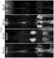
FIG. 6 depicts imaging of target mRNA in whole (whole-mount) chicken embryos using unoptimized standard probes and segmented initiator probes.
Figures 7A-7C depict some embodiments of segmented initiator probes co-localized by target and show cuvette data demonstrating the use of segmented initiator probes co-localized by target to trigger HCR.
FIGS. 8A-8B depict some embodiments of in situ HCR using segmented initiator probes.
FIGS. 9A-9D depict background and signal-to-noise ratios using standard probes and segmented initiator probes.
10A-10D depict multiplexed imaging of mRNA expression with high signal-to-noise ratio in fixed whole chick embryos using segmented initiator probes without probe set optimization.
FIGS. 11A-11B depict quantitative imaging of mRNA expression at sub-cellular resolution in immobilized whole chick embryos using segmented initiator probes.
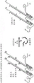
FIG. 12 depicts some embodiments of hybridizing a first and second segmented initiator probe to a target molecule.
Fig. 13 depicts some embodiments of triggering HCR amplification polymer self-assembly from hairpin monomers upon initiation by HCR initiators. 1050 in fig. 13 depicts the full initiator from both moieties. Only a portion of the segmented initiator probe is depicted. I1 (1050) represents a full initiator formed by two segmented initiator probes co-localized by the target.
Figure 14 depicts some embodiments of HCR amplification using hairpin monomers triggered by HCR initiators. Only a portion of the segmented initiator probe is depicted. I1 represents the full initiator formed by two segmented initiator probes co-localized by the target.
Figure 15 depicts some embodiments of HCR mechanisms using simplified HCR hairpin monomers.
Figures 16A-16D depict some embodiments of a probe set comprising one or more probe units and optionally one or more helper probes.
17A-17C depict some embodiments of probe units comprising a segmented initiator probe.
FIGS. 18A-18F depict some embodiments of HCR amplicons.
FIGS. 19A-19B depict some embodiments of HCR amplicons comprising four HCR hairpins.
Figures 20A-20F depict some embodiments of label probes.
FIGS. 21A-21B depict some embodiments of segmented initiator probes designed to be complementary to overlapping regions of HCR hairpins.
FIG. 22 depicts some embodiments of segmented initiator probes designed to be complementary to overlapping regions of a target.
Fig. 23A-23O depict some embodiments for removing HCR signals from a sample. In some embodiments, any one or more steps in the process may be combined with other methods of FIGS. 23A-23O and 40A-40N. In some embodiments, any one or more of the steps in the processes of FIGS. 23A-23O may be combined with the other methods of FIGS. 26A-26T and 40A-40N.
FIGS. 24A-24B depict some embodiments of increasing HCR signal intensity using segmented initiator probes designed to be complementary to overlapping regions of HCR hairpins.
Figures 25A-25B depict some embodiments of multiplex in situ hybridization in cultured human cells via repeated reporter detection.
Fig. 26A-26T depict some embodiments of various methods of detecting one or more targets in a sample using HCR initiator-labeled probes and/or HCR segmented initiator probes in combination with HCR amplicons. In some embodiments, any one or more steps in the process can be used in combination with other methods of fig. 26A-26T.
FIG. 27 depicts some embodiments of a segmented initiator probe designed to be complementary to an overlapping region of an HCR hairpin and to be complementary to an overlapping region of a target.
Fig. 28A-28C depict some embodiments of examples of in vitro optimization of cooperative probe ligation to enhance fragmentation initiator HCR inhibition (off state) and switching (on state).
Fig. 29A-29D depict some embodiments of multiplex HCR immunohistochemistry using initiator labeled primary antibody probes imaging protein targets in formalin fixed paraffin embedded mouse brain sections.
Figures 30A-30D depict some embodiments of multiplex HCR immunohistochemistry using unlabeled primary and initiator-labeled secondary antibody probes to image protein targets in formalin-fixed paraffin-embedded mouse brain sections.
Fig. 31A-31C depict some embodiments of simultaneous HCR RNA in situ hybridization and protein immunohistochemistry in formalin fixed paraffin embedded mouse brain sections using DNA fragmentation initiator probes for mRNA targets and initiator labeled primary anti probes for protein targets, with HCR signal amplification for all targets simultaneously.
Figures 32A-32C depict some embodiments of simultaneous HCR RNA in situ hybridization and protein immunohistochemistry in formalin fixed paraffin embedded mouse brain sections using DNA fragmentation initiator probes for mRNA targets and unlabeled primary and initiator-labeled secondary probes for protein targets, while HCR signal amplification is performed on all targets.
Figures 33A-33E depict some embodiments of using HCRs to mediate CARD signal amplification of a wide variety of target types.
Figures 34A-34C depict some embodiments of CARD signal amplification using HCR mediated universal target molecules and target complexes.
Figure 35 depicts some embodiments for placement of haptens in the context of HCR CARD signal amplification.
FIGS. 36A-36B depict some embodiments of HCR CARD signal amplification using substrate-labeled or segmented substrate HCR hairpins.
Figures 37A-37B depict some embodiments of imaging RNA targets in formalin fixed paraffin embedded human kidney and liver sections using HCR mediated amplification of CARD signals.
FIG. 38 depicts some embodiments of segmented initiator probes co-localized, either directly or indirectly, by either a target molecule or a target complex.
FIGS. 39A-39N depict some embodiments of initiator-labeled probes that bind directly or indirectly to either a target molecule or a target complex.
FIGS. 40A-40N depict some embodiments of removing HCR signal from a sample. In some embodiments, any one or more steps in the process may be combined with other methods of FIGS. 23A-23O and 40A-40N. In some embodiments, any one or more steps in the processes of FIGS. 40A-40N may be combined with other methods of FIGS. 23A-23O and 26A-26T.
Figures 41A-41C depict some embodiments of quantitative imaging of mRNA expression at subcellular resolution in formalin-fixed paraffin-embedded mouse brain sections using a segmented initiator probe.
FIGS. 42A-42F depict some embodiments of initiator-labeled probes comprising one or more HCR initiators.
FIGS. 43A-43C depict some embodiments of imaging target microRNAs and mRNAs in whole zebrafish embryos using initiator-labeled probes.
FIGS. 44A-44Z depict some embodiments of masked initiator-labeled probes comprising one or more masked HCR initiators.
45A-45B depict some embodiments of examples using masked initiator-labeled probes to reduce background.
FIGS. 46A-46M depict some embodiments of masked initiators in the context of probes for masked initiator labeling.
FIG. 47 shows an embodiment of the sequence of FIG. 25A for de-hybridizing HCR hairpins from HCR polymers (Table 2).
Detailed Description
Hybrid strand reaction (HCR) is a method for triggering the hybridization of nucleic acid molecules starting from a metastable hairpin monomer or other metastable nucleic acid structure. See, e.g., dirks, r, and Pierce, n.proc.natl.acad.sci.usa 101 (43): 15275-15278 (2004), and U.S. patent application nos. 11/087,937 filed on 3/22, 2005, U.S. patent nos. 8,105,778 filed on 31/2012, and 8,507,204 filed on 13/8/2013; and us patent publication No. 2018/0010166, filed 2017, 6, month 30, each of which is incorporated herein by reference in its entirety. In a simple version of this process, the metastable hairpin monomer undergoes a chain reaction of hybridization events to form a nicked double-stranded polymer when triggered by a nucleic acid initiator chain. Hairpin monomers store energy to drive the polymerization process in their single-stranded loops and toehold.
HCR may involve two or more metastable hairpin monomers. Hairpin monomers each have at least one single-stranded toehold, a single-stranded loop, and a double-stranded stem. The energy driving the self-assembly cascade is stored in the single-stranded loop and the toehold segment of the hairpin.
Each monomer is trapped in a kinetic trap (kinetic trap) preventing the system from equilibrating rapidly. That is, in the absence of an initiator, the monomer pairs cannot hybridize to each other. The introduction of the initiator chain causes the monomer to undergo a chain reaction of a hybridization event to form a nicked double-stranded polymer. HCR can be used to detect the presence of an analyte of interest in a sample, for example, by detecting the analyte with a probe carrying an HCR initiator, which in turn triggers HCR signal amplification. HCR signal amplification makes it possible to increase the signal-to-noise ratio for molecular detection and imaging applications by amplifying the signal above the background derived from the sample.
Provided herein are embodiments of HCRs. Methods of analyzing a sample using hybrid strand reaction (HCR) may involve one, two, or all three of the following: 1) repeated signal detection, 2) overlapping binding sites, and 3) catalytic reporter deposition (CARD).
In some embodiments of the method, a sample that may contain up to N targets and possibly other molecules that are not targets is combined with N probe sets (each targeting one of the N target types), each probe set containing one or more probe units, each probe unit containing two or more HCR segmented initiator probes. M HCR amplicons corresponding to M of the N probe sets were added (for M.ltoreq.N; each labeled with a different reporter). Thereafter, M signals corresponding to the M reporters are detected. The steps of the method are repeated until signal detection has been performed for all N targets.
In some embodiments of the method, a sample, which may contain one or more targets and possibly other molecules than targets, is combined with one or more probe sets, each probe set comprising one or more probe units, each probe unit comprising two or more HCR segmented initiator probes. Adding one or more HCR amplicons, each labeled with one or more reporters, and detecting one or more signals from the one or more reporters.
In some embodiments of the methods, a sample that may contain up to N targets and possibly other molecules that are not targets is combined with N probe sets, each probe set comprising one or more probe units, each probe unit comprising two or more HCR segmented initiator probes. N HCR amplicons corresponding to N probe sets (each labeled with a different substrate) and M tag probes corresponding to M of the N different substrates (for M.ltoreq.N; each conjugated to a different reporter) were added. Thereafter, M signals corresponding to M different reporters are detected. The steps of the method are repeated until signal detection has been performed for all N targets. In some embodiments, a repetitive signal detection method is provided. The method comprises substrate-labeled hairpins, a sample possibly comprising one or more targets and possibly other molecules than targets, in combination with one or more probe sets (each probe set comprising one or more probe units, each probe unit comprising two or more HCR segmented initiator probes), one or more HCR amplicons (each labeled with a substrate) corresponding to the one or more probe sets, and one or more tag probes (each conjugated to a reporter) corresponding to the one or more substrates. Thereafter, one or more signals corresponding to the one or more reporters are detected. The steps of the method are repeated one or more times in any order.
In some embodiments of the method of repeated signal detection of hairpins labeled with a reporter and/or substrate, a sample that may contain one or more targets and possibly other molecules that are not targets is combined with one or more HCR probe sets (each probe set containing one or more probe units, each probe unit containing two or more HCR fragmentation initiator probes), one or more HCR amplicons corresponding to the one or more probe sets (each labeled with one or more reporters and/or one or more substrates), and one or more tag probes corresponding to the one or more substrates (each conjugated to one or more reporters). Thereafter, one or more signals are detected. The steps of the method are repeated in any order.
In some embodiments of the method of repeated signal detection of hairpins labeled with a reporter and/or substrate, a sample that may comprise one or more targets and may comprise other molecules that are not targets is combined with one or more HCR probe sets, each probe set comprising one or more probe units, each probe unit comprising two or more HCR segmented initiator probes. One or more HCR amplicons are added that directly or indirectly generate one or more signals that are detected. The steps of the method may be performed one or more times in any order.
In some embodiments of the methods, a sample, which may contain a target and possibly other molecules that are not targets, is combined with a probe set comprising one or more probe units, each probe unit comprising two or more HCR segmented initiator probes. The target binding region on the probe within each probe unit is configured to bind to an overlapping binding site on the target. Thereafter, a HCR amplifier labeled with a reporter and/or a substrate and optionally a label probe corresponding to the substrate (conjugated to the reporter) are added and a signal from the reporter is detected. In some embodiments, the amplicons may alternatively be labeled with a reporter, such that the label on the probe is optional.
In some embodiments of the methods, a sample that may contain a target and possibly other molecules that are not targets is combined with a probe set that contains one or more probe units, each probe unit containing two or more HCR segmented initiator probes. The fragmentation initiator on the probe within each probe unit is configured to bind to overlapping binding sites on the HCR hairpin. Add a HCR amplicon labeled with a reporter and/or substrate and optionally a label probe (conjugated to the reporter) corresponding to the substrate. Thereafter, a signal from the reporter is detected. In some embodiments, the amplicons may alternatively be labeled with a reporter, such that the label on the probe is optional.
In some embodiments of the methods, a sample that may contain one or more targets and possibly other molecules that are not targets is combined with one or more probe sets, each probe set comprising one or more probe units, each probe unit comprising two or more HCR segmented initiator probes. The target binding regions on the probes within each probe unit are configured to bind to overlapping or non-overlapping binding sites on the target. The fragmentation initiator on the probe within each probe unit is configured to bind to overlapping or non-overlapping binding sites on the HCR hairpin. Adding one or more HCR amplicons, each labeled with one or more reporters and/or substrates, and (optionally) one or more optional tag probes (each conjugated to one or more reporters) corresponding to one or more substrates. Detecting a signal from the one or more reporters. The steps of the method may be repeated in any order. In some embodiments, the amplicons may alternatively be labeled with a reporter, such that the label on the probe is optional.
In some embodiments of the methods, a first fragment initiator probe comprising a first fragment initiator, a second fragment initiator probe comprising a second fragment initiator, a first hairpin monomer labeled with zero, one or more haptens, a second hairpin monomer labeled with zero, one or more haptens, and a target molecule are combined. This results in the first segmented initiator probe binding to the target molecule and the second segmented initiator probe binding to the target molecule. The first hairpin monomer is bound to both the first and second fragmentation initiators, and the second hairpin monomer is bound to the first hairpin monomer. Anti-hapten molecules labelled with one or more reporter entities are provided. The reporter entity is an enzyme that mediates CARD. Providing one or more CARD substrates and measuring a signal from one or more deposited reporters generated from the CARD substrates by an enzyme that mediates the CARDs.
In some embodiments of the methods, at least one initiator-labeled probe comprising at least one initiator, a first hairpin monomer labeled with zero, one or more haptens, a second hairpin monomer labeled with zero, one or more haptens, and zero, one, or more target molecules are bound. This results in the binding of an initiator-labeled probe comprising at least one initiator to zero, one, or more target molecules. The first hairpin monomer is bound to at least one initiator and the second hairpin monomer is bound to the first hairpin monomer. Anti-hapten molecules labelled with one or more reporter entities are provided. The reporter entity is an enzyme that mediates CARD. Providing one or more CARD substrates and measuring a signal from one or more deposited reporters generated from the CARD substrates by an enzyme that mediates the CARDs.
In some embodiments of the methods, a first fragment initiator probe comprising a first fragment initiator, a second fragment initiator probe comprising a second fragment initiator, a first hairpin monomer comprising zero, one or more substrates, a second hairpin monomer comprising zero, one or more substrates, and a target molecule are combined. This results in the first segmented initiator probe binding to the target molecule and the second segmented initiator probe binding to the target molecule. The first hairpin monomer is bound to both the first and second fragmentation initiators, and the second hairpin monomer is bound to the first hairpin monomer. A substrate binding region labeled with one or more reporter entities is provided. The substrate binding region binds to a substrate. The reporter entity is an enzyme that mediates CARD. Providing one or more CARD substrates, and measuring a signal from one or more deposited reporters generated from the CARD substrates by an enzyme that mediates the CARDs.
In some embodiments of the method, a first segmented initiator probe comprising a first segmented initiator, a second segmented initiator probe comprising a second segmented initiator, a first hairpin monomer comprising a first segmented substrate, a second hairpin monomer comprising a second segmented substrate and a target molecule are combined. This results in the first segmented initiator probe binding to the target molecule and the second segmented initiator probe binding to the target molecule. The first hairpin monomer binds to both the first and second split initiators and the second hairpin monomer binds to the first hairpin monomer, resulting in a full substrate comprising the first and second split substrates. A substrate binding region labeled with one or more reporter entities is added. The substrate binding region binds to the full substrate. The reporter entity is an enzyme that mediates CARD. Providing one or more CARD substrates, and measuring a signal from one or more deposited reporters generated from the CARD substrates by an enzyme that mediates the CARDs.
In some embodiments, the HCR process may be a segmented HCR process. In some embodiments, the segmented initiator probe pairs or groups may have a small amount of overlap between them. In some embodiments, the initiator is shorter or longer than the input binding domain of the HCR hairpin, and/or has incomplete complementarity to the input domain of the hairpin, but is capable of hybridizing to the input domain of the hairpin to open the hairpin and initiate the HCR polymerization cascade. In some embodiments provided herein, the fragmentation initiator within a probe unit is complementary to an overlapping region of the HCR hairpin (e.g., a region that overlaps 1, 2, or more nucleotides), or is substantially complementary to the HCR hairpin (e.g., except for 0, 1, 2, a few, or a few mismatches).
In some embodiments, the initiator on the initiator-labeled probe can be masked to enhance permeabilization to the sample (to increase signal) and/or to reduce non-specific binding of the probe within the sample (to reduce background).
In some embodiments, the HCR process can comprise a hairpin tag that can comprise a substrate for recruitment of a reporter entity comprising an enzyme that mediates catalytic reporter deposition (CARD).
In some embodiments, the HCR hairpin tag can comprise a segmented substrate such that the tag probe conjugated to the reporter molecule (or to the reporter entity) does not strongly bind to the segmented substrate on the individual hairpin, but such that the HCR polymerizes, co-localizing a holosubstrate adjacent to the hairpin in the HCR amplification polymer, such that the co-localized holosubstrate strongly binds to the tag probe conjugated to the reporter molecule (or to the reporter entity comprising the enzyme that mediates amplification of CARD signal).
In some embodiments, the HCR process can comprise one or more haptens (see, e.g., fig. 33A-33E, 34A-34C, 35, 36A-36B) for mediating signal amplification of additional layers via catalytic reporter deposition (CARD).
In some embodiments, a method for repeated signal detection of hairpins labeled with a reporter is provided. In some embodiments, the method comprises: a) Providing a sample that may contain up to N targets and may contain other molecules that are not targets; b) Providing N probe sets (each targeting one of the N target types), each comprising: one or more probe units, each probe unit comprising two or more HCR segmented initiator probes; c) Optionally washing the sample; d) Providing M HCR amplicons corresponding to M of the N probe sets (for M ≦ N; each labeled with a different reporter); e) Optionally washing the sample; f) Detecting M signals corresponding to the M reporters; g) Removing M signals from the sample; and h) optionally repeating one or more of steps b-g until signal detection has been performed for all N targets.
In some embodiments, a method for repeated signal detection of hairpins labeled with a reporter is provided. In some embodiments, the method comprises: a) Providing a sample that may contain up to N targets and may contain other molecules that are not targets; b) Providing N sets of probes (each targeting one of the N target types), each comprising one or more initiator-labeled probes, each initiator-labeled probe comprising one or more initiators; c) Optionally washing the sample; d) Providing M HCR amplicons corresponding to M of the N probe sets (for M ≦ N; each labeled with a different reporter); e) Optionally washing the sample; f) Detecting M signals corresponding to the M reporters; g) Removing M signals from the sample; and h) optionally repeating one or more of steps b-g until signal detection has been performed for all N targets.
In some embodiments, a method for repeated signal detection of hairpins labeled with a reporter is provided. In some embodiments, the method comprises: a) Providing a sample that may contain up to N targets and may contain other molecules that are not targets; b) Providing N probe sets (each targeting one of the N target types), each comprising: 1) One or more HCR initiator-labeled probes each comprising one or more initiators, or 2) one or more probe units each comprising two or more HCR segmented initiator probes; c) Optionally washing the sample; d) Providing M HCR amplicons corresponding to M of the N probe sets (for M ≦ N; each labeled with a different reporter); e) Optionally washing the sample; f) Detecting M signals corresponding to the M reporters; g) Removing M signals from the sample; and h) optionally repeating one or more of steps b-g until signal detection has been performed for all N targets.
In some embodiments, the method comprises: a) Providing a sample that may contain up to N targets and may contain other molecules that are not targets; b) Providing N probe sets (each targeting one of the N target types), each comprising: one or more probe units, each probe unit comprising two or more HCR segmented initiator probes; c) Washing the sample; d) Providing M HCR amplicons corresponding to M of the N probe sets (for M ≦ N; each labeled with a different reporter); e) Washing the sample; f) Detecting M signals corresponding to the M reporters; g) Removing M signals from the sample; and h) repeating one or more of steps b-g until signal detection has been performed for all N targets.
In some embodiments, the method comprises: a) Providing a sample that may contain one or more targets and that may contain other molecules that are not targets; b) Providing one or more probe sets, each comprising one or more probe units, each probe unit comprising two or more HCR segmented initiator probes; c) Optionally washing the sample; d) Providing one or more HCR amplicons (each labeled with one or more reporters); e) Optionally washing the sample; f) Detecting one or more signals from one or more reporters; g) Optionally removing one or more probe sets from the sample; h) Optionally removing one or more HCR amplicons from the sample; i) Optionally removing one or more reporters from the sample; and j) optionally removing one or more signals from the sample. The process (all steps or a subset) may be repeated as desired.
Definitions and embodiments
As used herein, "nucleic acid" comprises a form of oligomer of DNA and/or RNA. The nucleic acid may also comprise analogs of DNA or RNA having modifications to the bases or backbone. For example, nucleic acids as used herein include the use of Peptide Nucleic Acids (PNA). The term "nucleic acid" also encompasses chimeric molecules. The phrases include artificial constructs as well as derivatives and the like. The phrase includes, for example, any one or more of DNA, RNA, 2' OMe-RNA, LNA, XNA, synthetic nucleic acid analogues and PNA.
The term "sticky end" refers to a nucleic acid sequence that is available for hybridization to a complementary nucleic acid sequence. The secondary structure of the "sticky ends" is such that the sticky ends are available for hybridization with complementary nucleic acids under appropriate reaction conditions without undergoing conformational changes. Typically, the sticky ends are single stranded nucleic acids.
"monomers" are individual nucleic acid oligomers. Typically, at least two monomers are used in the hybridization strand reaction, although three, four, five, six or more monomers may be used. Typically, each monomer comprises at least one region complementary to at least one other monomer used in the HCR reaction.
The composition can comprise (e.g., see fig. 13) a first hairpin monomer (1510) comprising: a) a first input domain (1852) comprising a first toehold (1851) and a first stem segment, b) a first output domain (1854) comprising a first hairpin loop (1853) and a complement of the first stem segment, and c) a first reporter molecule (1850). The composition may further comprise a second hairpin monomer (1610) comprising: a) a second input domain (1952) comprising a second toehold (1951) and a second stem segment, b) a second output domain (1954) comprising a second hairpin loop (1953) and a complement of the second stem segment, and c) a second reporter molecule (1950).
In some embodiments, the monomer is metastable. That is, in the absence of an initiator, they are kinetically unfavorable for association with other monomers comprising complementary regions (association). An "HCR" monomer is a monomer that is capable of assembling to form a polymer upon exposure to an initiator nucleic acid.
As used herein, "polymerization" refers to the association of two or more monomers to form a polymer. "polymers" may comprise covalent bonds, non-covalent bonds, or both. For example, in some embodiments, two monomers can hybridize in an alternating manner to form a polymer comprising a nicked double-stranded polymer. The polymer is also referred to herein as the "HCR product".
An "initiator-labeled probe" comprises one or more target binding domains and one or more HCR initiators (e.g., fig. 39A-39N and 42A-42F).
A "segmented initiator probe" comprises one or more target binding domains and one or more segmented initiator domains (e.g., FIGS. 3A-3B, 5A-5E, and 38). The terms "segmented initiator probe" and "segmented-initiator probe" are interchangeable, as used herein.
"full initiator" is derived from a combination of two or more segmented initiators, which when co-located, are capable of initiating HCR polymerization. Some initiators comprise a nucleic acid region complementary to the initiator complementary region of the HCR monomer. A staged initiator is one that is not sufficient by itself to trigger HCR polymerization, but can trigger HCR polymerization when co-localized with one (or more) other staged initiator to form a full initiator.
A "probe unit" comprises two or more segmented initiator probes such that the segmented initiator domains on the segmented initiator probes within the probe unit can be co-located to produce a full initiator.
The following terms in table 1 are indicated as selectable options for the terms used herein (which may vary in width depending on the context). The disclosure of the broadest terms denotes not only the broadest concept but also the narrower concept herein. The method and the table serve merely as shorthand expressions for representing the two options in a more concise manner.
TABLE 1
| Cleavage initiator probe | Segmented initiator probe |
| Target binding sequences | Target binding segment |
| Near terminal sequence | Target segment |
| Homologous proximal target sites | Target segment |
| Target binding region | Target binding |
| HCR initiator | |
| 11 | All HCR initiator |
| Hair clip H1 | First hairpin element |
| Hair clip H2 | Second hairpin monomer |
| HCR hairpin | Hair pin monomer |
| Cleavage initiator probe pair | Segmented initiator probe pair |
| Homologous target sites | Target segment |
| Initiator I1 | All |
| Probe | |
| 1 | First segmented |
| Probe | |
| 2 | Second stage initiationAgent probe |
| Automatic background suppression | Active background suppression |
| Standard probe | Initiator-labeled probe |
As used herein, "substrate" may mean: 1) A substrate domain on the hairpin (e.g., domain "E" in fig. 18C, 18E, and 36A) that serves as a binding site for a substrate binding region in a tag probe; 2) A CARD substrate that becomes a reporter for CARD enzyme mediated deposition (e.g., figures 33A-33E, 34A-34C, 36A-36B). To avoid confusion, use of (2) a substrate is intended to be denoted as a "CARD-substrate," such that the word CARD is associated with the substrate in this context.
The section headings used herein are for organizational purposes only and are not to be construed as limiting the subject matter described in any way. All documents and similar materials cited in and/or within this application, including but not limited to patents, patent applications, articles, books, treatises, and internet web pages, are expressly incorporated by reference in their entirety for any purpose. The definitions provided in the present teachings prevail when the definitions of terms in the incorporated references appear to differ from those provided in the present teachings. It should be understood that there is an implicit "about" preceding a temperature, concentration, number, etc. discussed in the present teachings, such that there are slight and insubstantial deviations within the scope of the teachings herein. In this application, the use of the singular includes the plural unless specifically stated otherwise. In addition to this, the present invention is, the use of "comprises/contains (comprises)", "comprises/contains (comprising)", "comprises/contains (contains)/contains", "and" contains/contains (contains) ", and" contains/contains "are used the terms" comprises/comprising/containing "," comprises/containing "," includes/containing "and" containing/containing "and the like are not intended to be limiting. It is to be understood that both the foregoing general description and the following detailed description are exemplary and explanatory only and are not restrictive. Unless defined otherwise, technical and scientific terms used herein have the same meaning as commonly understood by one of ordinary skill in the art to which this invention belongs. See, e.g., singleton et al, dictionary of Microbiology and Molecular Biology, second edition, j.wiley & Sons (new york, n.y.1994); sambrook et al, molecular Cloning, A laboratory Manual, cold Springs Harbor Press (Cold Springs Harbor, N.Y. 1989). It is to be understood that both the general description and the detailed description are exemplary and explanatory only and are not restrictive of the invention as claimed. In this application, the use of the singular includes the plural unless specifically stated otherwise. In this application, the use of "or" means "and/or" unless stated otherwise. Furthermore, the use of the term "including/comprising/containing" as well as other forms such as "including/comprising/containing" and "including/containing" is not limiting. Unless specifically stated otherwise, terms such as "element" or "component" also encompass elements and components that comprise one unit and element, as well as components that comprise more than one sub-unit. Further, use of the term "a portion" can also encompass a portion of a portion or the entire portion.
In some embodiments, any one or more optional elements of any one or more figures herein can be combined with any one or more other optional steps of any one or more figures herein.
In some embodiments, any one or more elements other than any one or more of the figures herein may be combined with any one or more other steps other than any one or more of the figures herein.
In some embodiments, any one or more optional elements other than any one or more of the figures herein may be combined with any one or more other optional steps other than any one or more of the figures herein.
The term "step" means an action that occurs and/or can be performed. It should be noted that multiple "steps" may occur at once or within overlapping time periods and/or sequentially. All options are contemplated herein unless otherwise indicated (either explicitly or implicitly given in the context of this disclosure). Further, unless otherwise indicated, the steps may be performed in a different order or repeatedly, or additional intervening (overlapping) steps may be added.
Analysis of target molecules within the sample is reacted via the hybridized strands. In some embodiments, targets are detected using a probe set comprising one or more initiator-labeled probes (each comprising one or more HCR initiators) (e.g., fig. 39A-39N, 42A-42F, and 43A). In some embodiments, a probe set comprising one or more probe units (e.g., see the probe set of fig. 8A-8B and 16A-16D) is used to detect a target within a sample, wherein the probe unit comprises two or more HCR segmented initiator probes (e.g., see the probe units of fig. 3A-5E and 17A-17C), wherein each HCR segmented initiator probe comprises a target-binding region and a segmented initiator (e.g., see the segmented initiator probes of fig. 12 and 17A-17C). In some embodiments, each probe within a probe unit co-localizes a segmented initiator with binding of an adjacent cognate binding site on the target to form a full HCR initiator (e.g., see full HCR initiators of FIGS. 3A-5E, 8A-8B, 16A-16D, 21A-22, 27, and 38), which is capable of hybridizing to an HCR hairpin to trigger HCR signal amplification.
In some embodiments, HCR signal amplification improves the signal-to-noise ratio for molecular detection and imaging applications by increasing the signal above background generated by the sample by about 5, 10, 15, 20, 25, 30, 40, 50, 75, 100, 500, 1000, or 2000-fold, or by a value having a range defined by any two of the foregoing values.
In some embodiments, the target-binding regions within a probe unit are configured to bind to overlapping or non-overlapping regions of a target (e.g., see FIGS. 8A-8B, 21A-21B, 22, and 27). In some embodiments, the fragmentation initiator within a probe unit is designed to hybridize to overlapping or non-overlapping regions of the HCR hairpin (e.g., see FIGS. 8A-8B, 21A-22, 27, and 28). Individual probes that bind non-specifically in the sample do not co-localize the full HCR initiator, inhibiting HCR signal amplification. The HCR amplicons comprise two or more HCR hairpins (see, e.g., the HCR amplicons of FIGS. 8A-8B, 18A-18F, and 19A-19B). In some embodiments, each HCR hairpin comprises an input domain having a single-stranded toehold and a stem segment, and an output domain having the complement of a single-stranded loop and a stem segment (e.g., see the HCR hairpins of fig. 8A-8B, 13, 14, 18A-18F, and 19A-19B). In some embodiments, one or more HCR initiators on the initiator-labeled probe each initiate a chain reaction of the polymerization step, where the initiator hybridizes to an input domain of a first HCR hairpin, opens the first hairpin to expose its output domain, which in turn hybridizes to an input domain of a second HCR hairpin, opens the second hairpin to expose its output domain, and so on, resulting in a chain reaction in which the hairpins polymerize to produce an HCR amplification polymer tethered to the target (e.g., see amplification polymers of fig. 13, 14, 19A-19B, 29A, 30A, 33C-33E, 34A, and 41A). In the absence of full HCR initiator, HCR hairpins are passively trapped and do not polymerize, suppressing background. However, if the segmented initiator probes within a probe unit bind to their adjacent homologous binding sites on the target to co-localize the full HCR initiator, the full HCR initiator initiates a chain reaction of a polymerization step, where the full initiator hybridizes to an input domain of a first HCR hairpin, opens the first hairpin to expose its output domain, which in turn hybridizes to an input domain of a second HCR hairpin, opens the second hairpin to expose its output domain, and so on, resulting in a chain reaction in which the hairpins polymerize to produce an HCR amplification polymer tethered to the target (e.g., see amplification polymers of fig. 8A, 13, 14, 18C-18F, and 19A-19B). In some embodiments, the HCR hairpin further comprises one or more tags, each tag comprising a reporter or comprising a substrate (or a segmented substrate), which recruits a tag probe comprising one or more reporters (e.g., see reporter-labeled, substrate-labeled, and segmented-substrate-labeled HCR hairpins of fig. 18A-18F). In some embodiments, zero, one, or more substrates on the HCR hairpin can comprise haptens (see, e.g., fig. 35) that are used to mediate additional layers of signal amplification via catalytic reporter deposition (CARD) (see, e.g., fig. 33A-33E, 34A-34D, and 37). In some embodiments, the tag probe comprises one or more reporters and further comprises a substrate binding region that is complementary to a substrate on the HCR hairpin or complementary to a full substrate co-localized within the HCR amplification polymer (e.g., see tag probes of fig. 20A-20F and 36A-36B). In some embodiments, the signal is generated by one or more reporters associated with HCR amplification polymers tethered to a target within the sample. In some embodiments, the signal is removed from the sample (e.g., see fig. 23). In some embodiments, HCR signals are generated, detected, and removed from the same sample one or more times (e.g., see fig. 23A-23O and 40A-40N).
In some embodiments, any one or more steps provided in any of the figures provided herein may be combined into one of the other methods provided herein. As used herein, unless otherwise indicated, a general reference to a group of figures (e.g., figure 18) means that all of the different figures (e.g., 18A-18F) contained within that number are each combined together, one or more of them, or each in the alternative.
HCR initiator. The initiator-labeled probe comprises one or more HCR initiators capable of initiating an HCR polymerization cascade. In some embodiments, the initiator is fully complementary to the input domain of the HCR hairpin such that it hybridizes to the input domain of the hairpin to open the hairpin and initiate the HCR polymerization cascade. In some embodiments, the initiator is partially complementary to the input domain of the HCR hairpin, but sufficiently complementary that it hybridizes to the input domain of the hairpin to open the hairpin and initiate the HCR polymerization cascade. In some embodiments, the initiator is shorter or longer than and/or not fully complementary to the input domain of the HCR hairpin, but is capable of hybridizing to the input domain of the hairpin to open the hairpin and initiate the HCR polymerization cascade. In some embodiments, the HCR initiator may have 60%, 70%, 80%, 90%, or 100% complementarity to the input domain of the HCR hairpin and hybridize to the input domain of the hairpin to open the hairpin and initiate the HCR polymerization cascade. In some cases, initiator-labeled probes comprising one or more initiators may undergo reduced permeabilization into the sample due to non-specific binding of the initiator near the sample surface. In some cases, an initiator-labeled probe comprising one or more initiators may cause increased background due to non-specific binding of the initiator to DNA, RNA, proteins, or other molecules within the sample. In some embodiments, the initiator on the initiator-labeled probe is masked by base-pairing (see, e.g., fig. 44A-44Z, 45A-45B, and 46A-46M) to enhance permeabilization into the sample (to increase signal) and/or to reduce non-specific binding of the probe within the sample (to reduce background). In some embodiments, the initiator can be masked by hairpin structures (e.g., FIGS. 44A-44E, 44K-44L, 44O-44P, 44S-44T, 44W-44X, 46A-46E, and 46M). In some embodiments, the initiator can be masked by one or more auxiliary oligonucleotides (e.g., FIGS. 44F-44J, 44M-44N, 44Q-44R, 44U-44V, 44Y-44Z, 46F-46J). In some embodiments, the priming agent can be masked by self-complementarity and/or complementarity to one or more auxiliary strands within the oligonucleotide that includes the priming agent (e.g., FIGS. 44A-44Z and 46A-46M).
Automated background suppression with HCR segmented initiator probes. In some embodiments, the segmented initiator probes automatically suppress background because the HCR initiator (I1 or I2) is cleaved between probe pairs (e.g., see fig. 8 and 12). In some embodiments, if the probe specifically binds to the target at the proximal homologous binding site, the target co-localizes both probes within the probe pair to form a full HCR initiator. In some embodiments, non-specifically bound individual probes do not trigger HCR, as each probe carries only a portion of the HCR initiator, and HCR signal amplification is triggered as long as the full HCR initiator is co-localized.
Automatic background suppression with HCR hairpins. In some embodiments, HCR hairpins auto-suppress background because HCR hairpins are passively trapped, so they do not polymerize in the absence of HCR initiators (I1 or I2). In some embodiments, if both probes within a segmented initiator probe pair specifically bind to their proximal cognate binding sites on the target, the resulting co-localized full HCR initiator (I1 or I2) triggers the growth of a tethered HCR amplification polymer (e.g., see fig. 8). The HCR hairpins alone, which bind non-specifically in the sample, do not trigger HCR because they are captured passively. Automatic background suppression with HCR hairpins. In some embodiments, HCR hairpins auto-suppress background because HCR hairpins are passively trapped, so they do not polymerize in the absence of HCR initiators (I1 or I2). In some embodiments, if both probes within a segmented initiator probe pair specifically bind to their proximal cognate binding sites on the target, the resulting co-localized full HCR initiator (I1 or I2) triggers the growth of a tethered HCR amplification polymer (e.g., see fig. 8). HCR hairpins alone that bind non-specifically in the sample do not trigger HCR because they are kinetically captured.
Automated background suppression with HCR segmented initiator probes and HCR hairpins. The combination of HCR segmented initiator probes for target detection and HCR amplification hairpins for signal amplification provides automatic background suppression throughout the protocol, ensuring that even if they bind non-specifically within the sample, the reagents do not generate an amplified background.
The full HCR initiator is split between 2 or more segmented initiator probes. We refer to each set of segmented initiator probes that generate a full HCR initiator as probe units (see, e.g., fig. 17). In some embodiments, a full HCR initiator (I1 or I2) is generated by a pair of segmented initiator probes (each carrying a portion of the full HCR initiator such that they together comprise the full HCR initiator) (fragment f1 of probe P1 and fragment f2 of probe P2 such that f1+ f2= 1); in this case, the probe unit is two segmented initiator probes (see, e.g., fig. 17A). Fragments f1 and f2 are sufficiently small compared to the full HCR initiator (e.g., f1=0.5 and f2=0.5; or f1=0.45 and f2=0.55; or f1=0.4 and f2= 0.6) that HCR signal amplification is inhibited if the full HCR initiator is not co-localized by the target.
In some embodiments, the HCR initiator (I1 or I2) is split between three segmented initiator probes (fragment f1 of probe P1, fragment f2 of probe P2, fragment f3 of probe 3, such that f1+ f2+ f3= 1); in this case, the probe unit consists of three segmented initiator probes. In some embodiments, the HCR initiator (I1 or I2) is split between N segmented initiator probes (fragment f1 of probe P1, fragment f2 of probe P2,. And fragment fN of probe PN such that f1+ f2+. FN =1; see, e.g., fig. 17B), where N =2, 3, 4, or more; in this case, the probe unit consists of N segmented initiator probes. For any of these values of N, HCR signal amplification is inhibited if the full HCR initiator is not co-localized by the target.
In some embodiments, full HCR initiators are generated by a pair (or set) of probes, each probe carrying a portion of an HCR initiator, such that the sum of fragment f1 of probe P1 and fragment f2 of probe P2 (f 1+ f 2) is close enough to 1 (e.g., f1=0.47, f2=0.47, f1+ f2= 0.94) that HCR signal amplification is triggered by the co-localized full initiators caused by probe pairs binding to their proximal cognate binding sites on the target. In some embodiments, the segmented initiator probes within a probe unit generate a full HCR initiator corresponding to 100% of the HCR initiator. In some embodiments, the segmented initiator within the probe unit generates a sufficient portion of HCR initiator to provide efficient HCR signal amplification relative to signal amplification rate when no segmented initiator probe is present or when separate segmented initiator probes are present but not co-localized by the target. In some embodiments, the portion of the full HCR initiator generated by the co-localized probes within a probe unit is 99%, 95%, 90%, 80%, or 60% of the full HCR initiator, including any range above any one of the preceding values or any range defined between any two of the preceding values. In some embodiments, the probe unit comprises 2, 3, 4, 5, or more segmented initiator probes. In some embodiments, when the probes within a probe unit are co-localized by binding to their adjacent cognate binding sites on the target, the fragmentation initiator in the probe unit is sufficient to function as an HCR initiator. In some embodiments, although the HCR initiator may have a sequence of a particular length (e.g., 15 nucleotides), the fragmentation initiators within a probe unit need not be exactly the same length. For example, in some embodiments, if they are co-localized, their combined length may be 14 or 13 nucleotides, which still function as HCR initiators.
In some embodiments, any two or more staged initiators may be used so long as they together provide the functionality of the HCR initiator.
In some embodiments, full HCR initiators are generated by a pair of probes each carrying a portion of an HCR initiator (the portion of the HCR initiator further comprising one or a few or several sequence modifications) such that the sum of fragment f1 of probe P1 and fragment f2 (f 1+ f 2) of probe P2 is close enough to 1 (e.g., f1=0.45, f2=0.47, f1+ f2= 0.92) that HCR signal amplification is triggered by the co-localized full initiators resulting from probe pairs binding to their neighboring cognate binding sites on the target. In some embodiments, the segmented initiator probes within a probe unit generate a full HCR initiator with 100% sequence identity to the HCR initiator. In some embodiments, the segmented initiator probes within a probe unit generate sufficient sequence identity to the HCR initiator to enable efficient HCR signal amplification relative to signal amplification rate when no segmented initiator probe is present or when individual segmented initiator probes are present but not co-localized by the target. In some embodiments, a full HCR initiator generated by a co-localized probe within a probe unit has 99%, 95%, 90%, 80%, or 60% sequence identity to the HCR initiator, including any range above any one of the preceding values or any range defined between any two of the preceding values.
The use of large probe sets to enhance signal-to-noise ratio. The signal increases monotonically with the number of probe units, and therefore, it is advantageous in some embodiments to use a large probe set comprising multiple probe units when the target length allows (e.g., mRNA, lncRNA, gDNA target) in order to increase the amount of signal generated per target molecule. Automatic background suppression ensures that only probe units that specifically bind to the target trigger HCR signal amplification, so individual probes within a test probe set are typically not required in order to remove those that are non-specifically bound. Thus, when a new probe set of new targets is used, the automatic background suppression increase is easy to use by eliminating the need for probe set optimization. Because signal increases monotonically with probe set size and is automatically suppressed for all probes against background, it is generally advantageous to increase probe set size in order to increase signal-to-noise ratio. A probe set comprises 1 or more probe units, each comprising two or more segmented initiator probes (see, e.g., fig. 8 and 16). For example, the number of probe units in a probe set may be in the range of 1 to 1000 or 10 to 100 or 20 to 50 or 30 to 40. Increasing the number of probe units in a probe set can increase the signal-to-noise ratio by a factor of 2 or 5 or 10 or 100 or more. Increasing the number of probe elements in a probe set may increase the signal-to-noise ratio to 2 or 5 or 10 or 50 or 100 or 200 or 500 or 1000 or more, depending on the number of probe elements in the probe set, the background level inherent to the sample, and the abundance of target within the sample.
Probe set size limited by the transcriptome and/or genome within the sample. In some cases, it is desirable to separate one target in a sample from one or more other target regions in a sample that are closely related in sequence. For example: 1) a target mRNA in an organism may have a similar sequence to another mRNA in the organism, including the possibility that the target mRNA may be one splice variant that needs to be distinguished from other splice variants, 2) the target mRNA may comprise multiple regions, each region having sequence similarity to one or more other RNAs in the organism's transcriptome, 3) the target mRNA contained in one organism may be present in multiple species of samples such that the target mRNA has a region that is similar in sequence to one or more RNAs in another species of transcriptome, 4) the target rRNA or gDNA contained in one organism may be similar in sequence to rRNA or gDNA in other organisms contained in the sample. One way to distinguish a target nucleic acid from all other nucleic acids present in a sample is to design a probe set for the target nucleic acid that contains only probe pairs that are selective for the target nucleic acid relative to the transcriptome and/or the genome present in the sample. In some cases, this requirement for selectivity may substantially limit the size of the probe set. For example, the sequence of the target nucleic acid can be similar to one or more other nucleic acids within the sample such that the probe set can comprise only a single pair of segmented initiator probes.
Cooperative target binding using a probe set comprising a plurality of segmented initiator probe pairs. Probes that bind to a target nucleic acid (e.g., RNA or DNA) compete energetically with natural secondary structures (i.e., base pairing) within the target nucleic acid. For example, a single probe may not be able to bind to a domain within a target nucleic acid that is primarily base paired with another domain within the target. The binding yield of a given probe is the fraction of the target molecule with the probe bound at the cognate probe binding site, varying between 0 and 1 depending on the accessibility of that fraction of the target, the degree of affinity between the probe and its cognate probe binding site, and other factors. Using a probe set comprising a plurality of segmented initiator probe pairs, binding of one probe to a target can increase the binding yield of one or more other probes to the target, resulting in a synergistic effect of the probes within the probe set collectively increasing the binding yield of the other probes within the probe set. Thus, a probe set with N + M probes may produce more signal than a probe set with N probes, not only because M new probes will produce a signal, but also because N original probes will have a higher binding yield to the target and thus on average produce more signal per target molecule.
The signal was increased using an auxiliary probe. In order to maintain selectivity for a target nucleic acid in the presence of one or more transcriptomes and/or genomes present in a sample, probe set sizes are sometimes constrained to be no more than 1 probe unit, or no more than 2 probe units, or no more than N probe units, where N is less than the number of probe units N + M, which will be preferentially used based on sensitivity considerations and/or the length of the target nucleic acid. In such cases where probe set size is constrained by selective considerations, because the probe set has the potential to generate M fewer total initiators for triggering HCR signal amplification, and because the absence of M additional probe units can reduce the binding yield of the N remaining probe units due to synergistic effects, the amount of signal generated can be less relative to the detection of the same target using N + M probe units.
In some embodiments, the probe carries a segmented initiator as a signaling probe (e.g., see fig. 16A). In some embodiments, the probes do not carry a segmented initiator as a secondary probe. To increase signal without decreasing selectivity, a probe set comprising N probe units may be enhanced with M auxiliary probes (e.g., see fig. 16D).
For example: 1) For detection of target RNA with unique splice junctions (splice junctions), a probe set may comprise one probe unit bound to the target across the splice junction, and further comprise 30 (or any number) auxiliary probes bound to other parts of the target RNA to synergistically increase the binding yield of the probe unit and thus increase the signal without decreasing selectivity; 2) To detect target RNA or DNA for which selectivity considerations result in a probe set comprising 5 probe units, the probe set may further comprise 45 helper probes; 3) To detect target RNA or DNA for selectivity considerations that result in a probe set comprising N probe units (where N is less than 40), the probe set may further comprise 40-N helper probes; 4) To detect a target nucleic acid that results in a probe set comprising N probe units and a total number of length-limiting probes of the target nucleic acid of 2N + M for selectivity considerations, the probe set may further comprise M auxiliary probes.
In some embodiments, the helper probes will be designed for the target nucleic acid with less stringent selectivity requirements than those for designing the signaling probe for the same target nucleic acid. In some embodiments, the helper probes will be used for the target nucleic acids even though they are equally selective for other off-target (off-target) nucleic acids within the transcriptome and/or genome present in the sample. In some embodiments, the target-binding region on the signaling probe will be the same length as the target-binding region on the helper probe. In some embodiments, the target binding region on the signaling probe will be shorter or longer than the target binding region on the helper probe. In some embodiments, the target binding region can vary across a range of lengths (number of nucleotides) for different signals and/or helper probes. In some embodiments, the affinity between the signaling probe and the target nucleic acid will be comparable to the affinity between the helper probe and the target nucleic acid. In some embodiments, the affinity between the signaling probe and the target nucleic acid will be lower or higher than the affinity between the helper probe and the target nucleic acid.
Signal probes carrying one or two fragment initiators are used to increase the signal. In some embodiments, the probe unit comprises two segmented initiator probes that together generate a full HCR initiator (segment fl for probe P1 and segment f2 for probe P2, such that f1+ f2= 1). In this case, each probe participates in one probe unit. If two probes within a probe unit bind to a given target RNA molecule, a single full HCR initiator is generated, triggering the growth of one HCR amplification polymer tethered to the probe unit. Consider the case where the length of the target RNA constrains the probe set size to N probe units corresponding to 2N signaling probes. In the case where each signaling probe participates in only one probe unit, the maximum number of full HCR initiators would be N, corresponding to the case where the target mRNA is bound by all signaling probes in the probe set. Thus, the maximum number of HCR polymers tethered to the target RNA will be N. To increase the signal per target molecule, consider now a case where the probe can carry two segmented initiators that contribute to two different probe units (see, e.g., FIG. 16C). In some embodiments, the following three possibilities are considered:
1. In one embodiment, where all 2N signaling probes hybridize to adjacent subsequences along the target RNA, the signaling probes at the 5 'and 3' ends of the target each have one adjacent signaling probe, and all other intervening signaling probes have two adjacent signaling probes. In this case, if each probe except the most 5 'signaling probe and the most 3' signaling probe carries two fragmentation initiators that contribute to two different probe units, the total number of probe units increases from N to (2N-1). As a result, the maximum number of full HCR initiators increased from N to (2N-1), and the maximum number of HCR polymers tethered to the target RNA increased from N to (2N-1). Thus, the signal generated per target molecule is increased relative to the case where each signaling probe carries a single segmented initiator.
2. In another embodiment, the selectivity considerations resulting from the transcriptome and/or genome present in the sample may result in a probe set comprising 2N signaling probes and N probe units, wherein no probe unit is proximal to another along the target nucleic acid. In this case, each signaling probe carries only a single segmented initiator and contributes only one probe unit, so the maximum number of full HCR initiators is N and the maximum number of tethered HCR amplification polymers is N.
3. In another embodiment, representing an intermediate case, some signaling probes will be proximal to more than one adjacent sequence (neighbor), and thus 2N signaling probes will participate in many probe units that are centered between N and 2N-1. For example, if there are 2N signaling probes, where M probes tile a portion of the target (where 5 'and 3' of the signaling probes carry one fragmentation initiator and contribute to one probe unit, and the other M-2 signaling probes carry two fragmentation initiators and contribute to two probe units) and the other L probes (where 2N = M + L, L is an even number) are each bound in pairs to the target with each signaling probe proximal to only the other signaling probe (each L signaling probe carries one fragmentation initiator and contributes to one probe unit), then the total number of probe units will be M-1+ L/2, which is centered between N and 2N-1. For example, if there are 40 signaling probes (i.e., N = 20), and 20 of them tile the target at the proximal end (i.e., M = 20), and the other 20 bind to the target in pairs at the proximal end (i.e., L = 20), the total number of probe units would be M-1+ L/2=20-1+20/2=29.
In the relevant case of a probe unit comprising more than two signaling probes (e.g.three signaling probes or four or more signaling probes), the number of probe units can be increased again without increasing the number of signaling probes by using some probes comprising two fragmentation initiators and participating in two probe units (one fragmentation initiator per probe unit).
HCR amplicons with 2 hairpins. In some embodiments, the HCR amplicons comprise two hairpins (H1 and H2; see, e.g., FIGS. 8 and 18). In some embodiments, each hairpin comprises an input domain having a single-stranded toehold and a stem segment, and an output domain having the complement of the single-stranded loop and the stem segment. In the absence of HCR initiator (I1 or I2), hairpins H1 and H2 are metastable in coexistence, i.e. they are kinetically trapped and do not polymerize.
Initiation is carried out with the full initiator I1. In some embodiments, initiator I1 comprises a domain complementary to toehold of hairpin H1 and a domain complementary to a stem segment of H1. If the H1 hairpin encounters the full initiator I1, the full initiator I1 hybridizes to the input domain of hairpin H1 via a toehold-mediated strand displacement, opening hairpin H1 to expose the output domain of hairpin H1 and form complex I1-H1. The export domain of hairpin H1 comprises a domain complementary to the toehold of hairpin H2 and a domain complementary to the stem segment of H2. If the H2 hairpin encounters the I1-H1 complex, the exposed export domain of H1 hybridizes to the import domain of hairpin H2 via toehold-mediated strand displacement, opening hairpin H2 to expose the export domain of hairpin H2 and form complex I1-H1-H2. The export domain of hairpin H2 comprises a domain complementary to the toehold of hairpin H1 and a domain complementary to the stem segment of H1. If the H1 hairpin encounters the I1-H1-H2 complex, the exposed export domain of H2 hybridizes to the import domain of hairpin H1 via a toehold-mediated strand displacement, opening hairpin H1 to expose the export domain of hairpin H1 and form complex I1-H1-H2-H1. The polymerization process can be repeated with alternating H1 and H2 polymerization steps to produce a polymer of the form I1-H2-H1-H2-. Wherein for a polymer incorporating N alternating copies of hairpins H1 and H2 we can denote I1- (H1-H2) N . For example, the polymer may incorporate several H1 and H2 molecules, or tens of H1 and H2 molecules, or hundreds of H1 and H2 molecules, or thousands of H1 and H2 molecules, or more. It is also possible for the polymers to end in H1 or H2, so that I1- (H1-H2) N -H1 and I1- (H1-H2) N H1-H2 are all possible, the latter corresponding to I1- (H1-H2) N+1 。
Initiation with the full initiator I2. In some embodiments, initiator I2 comprises a domain complementary to the toehold of hairpin H2 and a domain complementary to the stem segment of H2. If the H2 hairpin encounters the full initiator I2, the full initiator I2 hybridizes to the input domain of hairpin H2 via a toehold-mediated strand displacement, opening hairpin H2 to expose the output domain of hairpin H2 and form complex I2-H2. Of H2 if the H1 hairpin encounters the I2-H2 complexThe exposed export domain hybridizes to the import domain of hairpin H1 via a toehold-mediated strand displacement, opening hairpin H1 to expose the export domain of hairpin H1 and form complex I2-H1. If the H2 hairpin encounters the I2-H2-H1 complex, the exposed export domain of H1 hybridizes to the import domain of hairpin H2 via a toehold-mediated strand displacement, opening hairpin H2 to expose the export domain of hairpin H2 and form complex I2-H2-H1-H2. The polymerization process may be repeated in alternating H2 and H1 polymerization steps to produce the polymer of form I2-H1-H2-H1 N Represents a polymer incorporating N alternating copies of H2 and H1. For example, the polymer may incorporate several H1 and H2 molecules, or tens of H1 and H2 molecules, or hundreds of H1 and H2 molecules, or thousands of H1 and H2 molecules, or hundreds of thousands of H1 and H2 molecules, or more. It being possible for the polymers to end in H1 or H2, so I2- (H2-H1) N -H2 and I2- (H2-H1) N both-H2-H1 are possible, the latter corresponding to I2- (H2-H1) N+1 。
HCR amplicons with 4 hairpins. In some embodiments, the HCR amplicons can comprise more than 2 hairpins. For example, an HCR amplicon can comprise 4 hairpins H1, H2, H3, H4 (e.g., see fig. 19). As for the 2-hairpin HCR, each hairpin includes an input domain comprising a single strand toehold and a stem segment, and an output domain comprising the complement of the single-stranded loop and the stem segment. In the absence of HCR initiators (I1, I2, I3 or I4), hairpins H1, H2, H3, H4 coexist meta-stably, i.e. they are captured kinetically and do not polymerize. The export domain of hairpin H1 comprises a domain complementary to the toehold of hairpin H2 and a domain complementary to the stem segment of H2. The export domain of hairpin H2 comprises a domain complementary to the toehold of hairpin H3 and a domain complementary to the stem segment of H3. The export domain of hairpin H3 comprises a domain complementary to the toehold of hairpin H4 and a domain complementary to the stem segment of H4. The export domain of hairpin H4 comprises a domain complementary to the toehold of hairpin H1 and a domain complementary to the stem segment of H1. Initiator I1 comprises a domain complementary to the toehold of hairpin H1 and a domain complementary to the stem segment of H1. Initiator I2 comprises a domain complementary to the toehold of hairpin H2 and a stem complementary to H2 A segment complementary domain. Initiator I3 comprises a domain complementary to the toehold of hairpin H3 and a domain complementary to the stem segment of H3. Initiator I4 comprises a domain complementary to the toehold of hairpin H4 and a domain complementary to the stem segment of H4. Similar to the case of the 2-hairpin HCR, if hairpin H1 encounters full HCR initiator I1, then full initiator I1 opens hairpin H1 to form complex I1-H1 with exposed H1 output domain, which in turn opens hairpin H2 to form complex I1-H1-H2 with exposed H2 output domain, which in turn opens hairpin H3 with exposed output domain to form complex I1-H1-H2-H3 with exposed H3 output domain, which in turn opens hairpin H4 to form complex I1-H1-H2-H3-H4 with exposed H4 output domain, which in turn opens hairpin H1 to form complex I1-H2-H3-H4-H1 with exposed H1 export domain, and so on, resulting in polymerization via alternating H1, H2, H3 and H4 polymerization steps to produce polymer of the form I1-H2-H3-H4-H1-H2-H3-H4.. For polymers incorporating N alternating copies of H1, H2, H3 and H4, which can be denoted as I1- (H1-H2-H3-H4) N . It being possible for the polymers to end in H1, H2, H3 or H4, so I1- (H1-H2-H3-H4) N -H1、I1-(H1-H2-H3-H4) N -H1-H2、I1-(H1-H2-H3-H4) N -H1-H2-H3 and I1- (H1-H2-H3-H4) N -H1-H2-H3-H4 are all possible, the latter corresponding to I1- (H1-H2-H3-H4) N+1 . It is possible that HCR polymerization is triggered by any of the homogemc coinitiators (I1, I2, I3 or I4). For example, the initiator can generate a polymer of the form I3- (H3-H4-H1-H2) N by using the initiator I3. An HCR amplicon with 4 hairpins facilitates the generation of a signal that is not present in the monomeric state but is present in the polymeric state (e.g., fig. 19B illustrates that FRET pairs are co-localized to generate FRET signals only when hairpins are co-localized in the amplification polymer, which provides the basis for a wash-free approach, as unused hairpins that are not washed from the sample will not participate in FRET and thus background generation will be avoided).
HCR amplicons with 2 or more hairpins. More generally, in some embodiments, an HCR amplicon can include M HCR hairpins (H1, H2,. Cndot., HM), where M is an integer of 2 or greater. In the absence of HCR initiatorIn the case of (I1, I2, ·, IM), hairpins H1, H2, ·, HM coexist metastable, i.e. they are passively mechanically trapped and do not aggregate. In the presence of a homologous all-HCR initiator, polymerization occurs via alternating polymerization steps similar to 2-hairpin or 4-hairpin HCRs. For example, initiator I1 will result in form I1- (H1-H2-., a-HM) for a polymer incorporating N alternating copies of H1, H2, HM N The polymer of (2) grows. It is possible for the polymer to end with any of H1, H2, HM, and therefore I1- (H1-H2- · -HM) N -H1、I1-(H1-H2-...-HM) N -H1-H2,. And I1- (H1-H2-. HM) N -H1-H2-. Multidot. -HM is possible, the latter corresponding to I1- (H1-H2-. Multidot. -HM) N+1 . It is possible to trigger the HCR polymerization by any of the homogemc coinitiators (I1, I2. For example, the initiator can be converted into the form I3- (H3-. -HM-H1-H2) by means of a total initiator I3 N The polymer of (1).
HCR hairpin tag. For a given HCR amplicon, each HCR hairpin contains zero, one, or multiple tags. The tags on different hairpins within the amplicon can be the same or different. For example, an amplicon comprising hairpins H1 and H2 can have: 1) the same label on H1 and H2, 2) different labels on H1 and H2, 3) a label on H1 but no label on H2, 4) a label on H2 but no label on H1, 5) no label on H1 or H2, 6) zero, one, or more labels on H1 that are the same as or different from zero, one, or more labels on H2. Similarly, for HCR amplicons comprising hairpins H1, H2, H3, H4, each hairpin can comprise zero, one, or more tags (e.g., 3, 5, or 10 tags), zero, one, or more of which can be the same as zero, one, or more tags on each of the other hairpins. In some embodiments, one or more tags of a given hairpin can be unique within the hairpin and/or mixture of hairpin tags. In some embodiments, there are 1, 10, 100, 1000, 10,000, 100,000, or more unique tags (including any range defined between any two previous numbers) within the mixture.
HCR hairpin tags served as reporters. In some embodiments, the HCR hairpin tag may comprise a reporter molecule that facilitates measurement of the signal, e.g., by generating the signal, by altering the signal, or by eliminating the signal. For example, the reporter may be a fluorophore, chromophore, luminophore, phosphor, FRET pair, member of a FRET pair, quencher, fluorophore/quencher pair, rare earth element or compound, radioactive molecule, magnetic molecule, or any other molecule that facilitates signal measurement.
HCR hairpin tag as substrate. In some embodiments, the HCR hairpin tag may comprise a substrate for recruitment of a reporter entity that directly or indirectly mediates localization of the reporter in the vicinity of the hairpin tag. For example:
1. the hairpin tag may comprise Digoxigenin (DIG) recruiting an anti-DIG antibody as a reporter entity, wherein the anti-DIG is directly labeled with one or more reporters, or one or more substrates or reporter entities for directly or indirectly mediating localization of the reporter in the vicinity of the hairpin tag.
2. Hairpin tags can comprise a nucleic acid domain that serves as a substrate for full or partial sequence complementarity to a domain within a tag probe carrying one or more reporters (e.g., fig. 18C).
3. The hairpin tag may comprise a nucleic acid domain that serves as a substrate having full or partial sequence complementarity to a domain within the tag probe that carries one or more substrates that mediate localization of the reporter in the vicinity of the hairpin tag.
4. The hairpin tag may comprise a nucleic acid domain that serves as a substrate for a reporter entity that directly or indirectly mediates localization of the reporter in the vicinity of the hairpin tag.
5. The hairpin tag may comprise a substrate for recruiting a reporter entity that indirectly mediates localization of the reporter in the vicinity of the hairpin tag.
6. The hairpin tag can include a substrate for recruitment of a reporter entity comprising an enzyme that mediates catalytic reporter deposition (CARD) in the vicinity of the hairpin tag (e.g., figure 36A).
7. The hairpin tag may comprise biotin recruiting streptavidin as the reporter entity, wherein the streptavidin is directly labeled with one or more reporters, or labeled with one or more substrates or reporter entities for directly or indirectly mediating localization of the reporter in the vicinity of the hairpin tag.
8. The hairpin tag may comprise a hapten recruiting an anti-hapten antibody or an anti-hapten nanobody that directly or indirectly mediates localization of the reporter in the vicinity of the hairpin tag via CARD signal amplification. For example, an anti-hapten antibody or nanobody can comprise a reporter entity that is an enzyme that mediates CARD (e.g., figures 33A-33E).
9. The hairpin tag may comprise a hapten recruiting an anti-hapten that directly or indirectly mediates localization of the reporter in the vicinity of the hairpin tag. For example, an anti-hapten can comprise a reporter entity, which is an enzyme that mediates CARD (e.g., figures 34A-34C).
10. The hairpin tag may comprise an enzyme that mediates amplification of the CARD signal to deposit a reporter molecule in the vicinity of the hairpin.
11. Hairpin tags can comprise zero, one, or multiple haptens that directly or indirectly mediate localization of the reporter in the vicinity of the hapten (e.g., figure 35).
In some embodiments, the hairpin tag may comprise a hapten that recruits an anti-hapten (e.g., an antibody, nanobody, streptavidin, or other molecule) labeled with a reporter.
In some embodiments provided herein, HCR signal amplification is used to mediate catalytic reporter deposition (CARD), resulting in even higher signal gain. In some embodiments, an even higher individual gain is about 5, 10, 15, 20, 25, 30, 40, 50, 75, 100, 500, 1000, 2000, 5000, or 10,000 times or a value having a range defined by any two of the foregoing values.
In some embodiments, the HCR hairpin tag can comprise a segmented substrate such that tag probes conjugated to a reporter molecule do not strongly bind to the segmented substrate on the individual hairpins, but such that, after HCR polymerization, co-localized holosubstrates adjacent to the hairpins in the HCR amplification polymer such that the co-localized holosubstrates strongly bind to the tag probes conjugated to the reporter molecule (e.g., fig. 18D) or to the reporter entity comprising an enzyme that mediates amplification of CARD signal (e.g., fig. 36B).
Haptens and anti-haptens. In some embodiments, the hairpin tag that is a substrate comprising a hapten can be, for example, digoxin (dig), dinitrophenyl (DNP), a fluorophore, biotin, or any small, biological, or non-biological molecule that can recruit an anti-hapten. Examples of anti-haptens include antibodies, nanobodies, streptavidin, aptamers, or any other molecule or molecular complex that selectively binds to a hapten.
And (3) labeling the probe. In some embodiments, the label probe comprises a substrate binding region and one or more reporter (e.g., see FIGS. 18C-E and 20) or reporter entities (e.g., see FIGS. 33A-33E and 34A-34D). In some embodiments, the tag probe comprises an unstructured strand labeled with one reporter molecule (e.g., see fig. 20A). In some embodiments, the tag probe comprises a plurality of reporter molecules (e.g., see fig. 20B). In some embodiments, the tag probe comprises a tag strand (conjugated to a reporter) hybridized to a blocking strand (conjugated to a quencher) (e.g., see fig. 20C). In some embodiments, the tag probe has a hairpin structure (e.g., see tag probe of fig. 20D) or other intramolecular base pairing. In some embodiments, the hairpin-tagged probes are conjugated to one or more reporters, quenchers, and/or FRET pairs (e.g., see fig. 20E and 20F). In some embodiments, the tag probe comprises an anti-hapten antibody or nanobody that recognizes a hapten tag on an HCR hairpin, and further comprises a reporter entity comprising an enzyme that mediates CARD (e.g., fig. 33A-33E). In some embodiments, the tag probe comprises an anti-hapten entity that recognizes the hapten tag on the HCR hairpin and further comprises a reporter entity comprising an enzyme that mediates CARD (e.g., fig. 34A-34D). In some embodiments, the tag probe comprises a substrate complement that recognizes a substrate tag on an HCR hairpin or a substrate complement that recognizes a full substrate co-localized within an HCR amplification polymer, and further comprises a reporter entity comprising a CARD-mediating enzyme (e.g., fig. 36A-36B).
An enzyme for HCR-mediated catalytic reporter deposition (CARD). In some embodiments, the HCR hairpin mediates signal amplification via catalytic reporter deposition (CARD) by a reporter entity comprising an enzyme that catalyzes a CARD-substrate, resulting in deposition of the reporter in the vicinity of the hairpin (see, e.g., fig. 33A-33E, 34A-34C, 36A-36B). For example:
1. the enzyme may be horseradish peroxidase (HRP) (or a polymeric HRP comprising multiple HRP enzymes) that acts on a CARD substrate to catalyze the deposition of a chromogenic reporter such as AEC, DAB, TMB, or StayYellow, or a fluorescent reporter such as a fluorophore-labeled tyramide, or a hapten-labeled CARD substrate such as a biotin-labeled tyramide, where the hapten is used to mediate the localization of the reporter in the vicinity of the hairpin tag.
2. The enzyme may be Alkaline Phosphatase (AP) (or a polymer AP comprising multiple AP enzymes) that acts on the CARD substrate to catalyze the deposition of a reporter, e.g., a chromogenic reporter such AS, but not limited to BCIP/NBT, BCIP/TNBT, naptholaS-MX phosphate + FastBune BB, naptholaS AS-MX phosphate + FastRed TR, stavGreen.
3. The enzyme may be a glucose oxidase that acts on the CARD substrate to catalyze the deposition of a reporter (e.g., NBT).
4. The enzyme may be any molecule or complex that directly or indirectly mediates the localization of the reporter in the vicinity of the hairpin tag.
In some embodiments, the enzyme that mediates CARD is inactivated (also referred to as inactivated) after deposition of the reporter (e.g., using chemical or thermal denaturation). For example, any combination of the following may be used to deactivate the CARD-mediating enzyme:
1. heat (e.g., 65 ℃ C. Or above)
2. Fixative (e.g., 4% PFA)
3. Acid (e.g., 1% Tween 20-containing 0.1M glycine-HCl, 0.2N HCl, 10% acetic acid, 10mM HCl, pH 2.2)
4. Other chemicals (e.g. hydrogen peroxide (H) 2 O 2 ) Hydrogen peroxide + phenol,Sodium azide, DEPC, MAB containing 10mM EDTA)
In some embodiments, H is used 2 O 2 HRP was inactivated. In some embodiments, AP is inactivated using a combination of heat and acid. In some embodiments, the AP is inactivated with a fixative. In some embodiments, inactivation of the CARD-mediating enzyme enables repeated CARDs to be performed using the same enzyme in combination with different substrates for different targets, thereby enabling multiplexed target assays using HCR-mediated CARDs. In some embodiments, inactivation of the CARD-mediating enzymes enables repeated CARDs to be performed using different enzymes in combination with different substrates for different targets, enabling multiplex target assays using HCR-mediated CARDs.
In some embodiments, the CARD enables the stained sample to be stored for 10 or more years to enable re-imaging to comply with biopharmaceutical regulatory requirements. In some embodiments, the CARD-based stained sample is sufficiently stable for 1, 2, 3, 4, 5, 6, 7, 8, 9, 10, 11, 12, 13, 14, 15, or more years, and still meets biopharmaceutical regulatory requirements. In some embodiments, CARD provides for formalin-fixed paraffin-embedded (FFPE) sample storage for decades. In some embodiments, CARDs provide storage of pathological samples for decades to provide retrospective scientific and medical research. In some embodiments, CARD staining provides long term storage of archived samples.
The type of target. In some embodiments, the initiator-labeled HCR probe comprises one or more target-binding regions and further comprises one or more HCR initiators. In some embodiments, the initiator-labeled probe can detect a target comprising:
1. any molecule, including but not limited to, an RNA molecule (e.g., mRNA, rRNA, lncRNA, siRNA, shRNA, microRNA, non-coding RNA, synthetic RNA, or modified RNA), a DNA molecule, a non-natural nucleic acid molecule, a protein molecule, a small molecule, a biological molecule, a chemically modified biological molecule, a non-biological molecule,
2. Complexes of any molecule, including any combination of RNA, DNA, proteins, small molecules, biomolecules, and/or non-biomolecules (e.g., RNA/RNA complexes, RNA/protein complexes, DNA/protein complexes, and RNA/DNA/protein complexes, protein/protein complexes).
The segmented initiator HCR probe comprises one or more target binding regions and further comprises one or more segmented initiator regions. The probe unit comprises two or more segmented initiator probes such that the segmented initiator probes combine in the probe unit to produce a full HCR initiator. The probe unit can detect a target comprising:
1. any molecule, including but not limited to RNA molecules (e.g., mRNA, rRNA, lncRNA, siRNA, shRNA, microRNA, non-coding RNA, synthetic RNA, or modified RNA), DNA molecules, non-natural nucleic acid molecules, protein molecules, small molecules, biological molecules, chemically modified biological molecules, non-biological molecules,
2. complexes of any molecule, including any combination of RNA, DNA, proteins, small molecules, biomolecules, and/or non-biomolecules (e.g., RNA/RNA complexes, RNA/protein complexes, DNA/protein complexes, and RNA/DNA/protein complexes, protein/protein complexes),
3. Any collection of proximal molecules or complexes such that when the segmented initiator probes comprising probe units bind to their respective targets within the collection of proximal molecules or complexes, the segmented initiators in the probe units can co-localize to form a complete HCR initiator.
In any of the embodiments provided herein, the fragmentation initiator within a probe unit is designed to be (or is) complementary to a non-overlapping region of the HCR hairpin (e.g., a region separated by 0, 1, 2, or more nucleotides), or is designed to be (or is) complementary to an overlapping region of the HCR hairpin (e.g., a region overlapping 1, 2, or more nucleotides), or is designed to be (or is) substantially complementary to the HCR hairpin (e.g., complementary except for 0, 1, 2, a few or a few mismatches).
In any of the embodiments provided herein, the target-binding regions within a probe unit are configured to bind to non-overlapping regions of the target (e.g., regions separated by 0, 1, 2, or more nucleotides or regions separated by 0, 1, 2, or more nm) or are configured to bind to overlapping regions of the target (e.g., regions overlapping by 1, 2, or more nucleotides or regions overlapping by 1, 2, or more nm).
The signaling probe is configured to bind to overlapping or non-overlapping regions of the target and/or is designed to have a segmented initiator that hybridizes to overlapping or non-overlapping regions of the HCR hairpin. In some embodiments, the probe unit comprises two or more segmented initiator probes each comprising a target-binding region and a segmented initiator. In some embodiments, the fragmentation initiator within the probe unit:
1. Designed to be complementary to the adjacent regions of the HCR hairpin,
2. or designed to be complementary to a non-overlapping region (e.g., a region separated by 0, 1, 2, or more nucleotides) of the HCR hairpin,
3. or designed to be complementary to an overlapping region (e.g., a region overlapping 1, 2, or more nucleotides) of the HCR hairpin,
4. or designed to be substantially complementary to the HCR hairpin (e.g., other than 0, 1, 2, a few or a few mismatched complements),
5. or designed to hybridize to the vicinity of the HCR hairpin,
6. or designed to hybridize to a non-overlapping region of the HCR hairpin,
7. or designed to hybridize to an overlapping region of the HCR hairpin,
8. or designed to have a sequence complementary to a contiguous region of the HCR hairpin,
9. or designed to have a sequence complementary to a non-overlapping region of the HCR hairpin,
10. or designed to have a sequence complementary to the overlapping region of the HCR hairpin,
11. or designed to have a sequence substantially complementary to a contiguous region of the HCR hairpin,
12. or designed to have a sequence that is substantially complementary to a non-overlapping region of the HCR hairpin,
13. or designed to have a sequence that is substantially complementary to the overlapping region of the HCR hairpin.
In some embodiments, the target binding region within a probe unit:
1. configured to bind to a neighboring region of the target,
2. Or configured to bind to non-overlapping regions of the target (e.g., regions separated by 0, 1, 2, or more nucleotides or regions separated by 0, 1, 2, or more nm),
3. or configured to bind to overlapping regions of the target (e.g., regions overlapping 1, 2, or more nucleotides or regions overlapping 1, 2, or more nm),
4. designed to bind to the adjacent region of the target,
5. or are designed to bind to non-overlapping regions of the target,
6. or are designed to bind to overlapping regions of the target,
7. or designed to have a sequence that hybridizes to the vicinity of the target,
8. or designed to have a sequence that hybridizes to a non-overlapping region of the target,
9. or designed to have a sequence that hybridizes to an overlapping region of the target.
In some embodiments, the probe unit comprises two segmented initiator probes. Two segmented initiator probes bind to homologous targets to co-localize full HCR initiators. The co-localized full HCR initiator is then combined with a homologous HCR hairpin to initiate HCR polymerization, with one segmented initiator hybridizing to the hairpin to form a first duplex (duplex) and the other segmented initiator hybridizing to the hairpin to form a second duplex. In some embodiments, there is an energetically unfavorable linkage between the two duplexes. In some embodiments, by configuring the segmented initiator to bind to the overlapping region of the hairpin, the location of the junction along the hairpin and the tertiary structure of the two probes and the hairpin near the junction can relax into a more energetically favorable conformation, increasing the affinity between the co-localized full initiator and the HCR hairpin, increasing the amount of amplified HCR signal generated in a given time period (e.g., fig. 27 and 28). In some embodiments, the affinity between two probes and a cognate target can be increased by configuring the target-binding regions of the two probes to bind to overlapping regions of the target, thereby allowing the linkage between the molecules to relax into an energetically favorable conformation.
In some embodiments, a probe set is designed for use in multiplex experiments, wherein 2, 3, 4, 5, 10, 20, or 100 or more probe sets are used to bind to different targets in the same sample, wherein 1, 2, 3, 4, 5, 10, 20, or 100 or more probe sets comprise one or more initiator-labeled probes. In some embodiments, a probe set is designed for use in multiplex experiments, wherein 2, 3, 4, 5, 10, 20, or 100 or more probe sets are used in the same sample, wherein greater than 1%, greater than 2%, greater than 5%, greater than 10%, greater than 30%, greater than 50%, or 100% of the probe sets comprise one or more initiator-labeled probes.
In some embodiments, a probe set is designed for use in multiplex experiments, wherein 2, 3, 4, 5, 10, 20, or 100 or more probe sets are used to bind to different targets in the same sample, wherein 1, 2, 3, 4, 5, 10, 20, or 100 or more probe sets comprise one or more probe units comprising a fragmentation initiator designed to hybridize to an overlapping region of an HCR hairpin. In some embodiments, a probe set is designed for use in a multiplex experiment, wherein 2, 3, 4, 5, 10, 20, or 100 or more probe sets are used in the same sample, wherein greater than 1%, greater than 2%, greater than 5%, greater than 10%, greater than 30%, greater than 50%, or 100% of the probe sets comprise one or more probe units comprising a fragmentation initiator having a sequence designed to be complementary to an overlapping region of an HCR hairpin.
In some embodiments, a probe set is designed for multiplex experiments, wherein 2, 3, 4, 5, 10, 20, or 100 or more probe sets are used to bind to different targets in the same sample, wherein 1, 2, 3, 4, 5, 10, 20, or 100 or more probe sets comprise one or more probe units comprising a target binding region designed to bind to an overlapping region of a target. In some embodiments, a probe set is designed for use in multiplex experiments, wherein 3, 4, 5, 10, 20, or 100 or more probe sets are used in the same sample, wherein greater than 1%, greater than 2%, greater than 5%, greater than 10%, greater than 30%, greater than 50%, or 100% of the probe sets comprise one or more probe units comprising a target binding region designed to bind to an overlapping region of a target.
In some embodiments, a probe set is designed for multiplex experiments, wherein 2, 3, 4, 5, 10, 20, or 100 or more probe sets are used to bind to different targets in the same sample, wherein 1 or more probe sets comprise one or more initiator-labeled probes and 1 or more probe sets comprise one or more probe units, each probe unit comprising two or more segmented initiator probes. In some embodiments, a probe set is designed for multiplex experiments in which 2, 3, 4, 5, 10, 20, or 100 or more probe sets are used in the same sample, wherein greater than 0.1%, greater than 1%, greater than 2%, greater than 5%, greater than 10%, greater than 30%, or greater than 50% of the probe sets comprise one or more initiator-labeled probes, and wherein greater than 0.1%, greater than 1%, greater than 2%, greater than 5%, greater than 10%, greater than 30%, or greater than 50% of the probe sets comprise one or more probe units, each probe unit comprising two or more segmented initiator probes.
In some embodiments, the probe unit comprises a fragmentation initiator designed to bind to an overlapping region of the HCR hairpin, wherein the overlapping region overlaps by 1 base or 2 bases or 3 bases or 4 bases or 5 bases or more.
In some embodiments, the probe unit comprises a target-binding region designed to bind to an overlapping region of a target, wherein the overlapping region overlaps at least 0.1nm or at least 0.2nm or at least 0.3nm or at least 0.5nm or at least 1nm or at least 2nm or at least 3nm or at least 5nm.
Materials and compositions for initiator-labeled probes. In some embodiments, the initiator-labeled probe comprises one or more target binding domains and one or more HCR initiators (e.g., fig. 39A-39N and 42A-42F). Each domain may comprise one or more materials comprising DNA, RNA, 2' OMe-RNA, PNA, XNA, chemically modified nucleic acids, synthetic nucleic acid analogues, amino acids, synthetic amino acid analogues and/or any other molecule suitable for domain purposes. For example:
1. Initiator-labeled probes may comprise one or more initiators made from DNA and a target binding domain made from DNA.
2. Initiator-labeled probes may comprise one or more initiators made from DNA and a target binding domain made from amino acids (e.g., an antibody or nanobody or antibody fragment).
3. Initiator-labeled probes may comprise an initiator made from a synthetic nucleic acid analog and a target binding domain made from a combination of DNA and 2' OMe-RNA.
4. Initiator-labeled probes may comprise an initiator made from 2' OMe-RNA and a target binding domain made from a combination of RNA and protein.
5. Initiator-labeled probes may comprise an initiator made of DNA and a target binding domain made of PNA.
6. Initiator-labeled probes may comprise one or more initiators made from any nucleic acid or nucleic acid analog and one or more target binding domains made from any combination of materials suitable for binding to a target molecule.
In some embodiments, the initiator-labeled probe may comprise a single covalently linked molecule or may comprise two or more molecules (each covalently linked) that interact non-covalently to form a complex. For example:
1. Initiator-labeled probes may comprise an initiator made of DNA covalently linked to a target binding domain made of DNA.
2. The initiator-labeled probe can comprise one or more initiators made of DNA covalently linked to dCas9 (or another Cas) that is non-covalently bound to a guide RNA (gRNA) such that the target-binding domain comprises the gRNA: dCas9 complex (or gRNA using another Cas: cas complex).
3. An initiator-labeled probe can comprise one or more initiators made from DNA covalently linked to a gRNA that is non-covalently bound to dCas9 (or another Cas) such that the target-binding domain comprises the gRNA: dCas9 complex (or gRNA using another Cas: cas complex).
4. Initiator-labeled probes may comprise an initiator made of a nucleic acid or nucleic acid analog that is covalently linked or non-covalently bound to one or more molecules comprising a target binding domain.
Materials and compositions for segmented initiator probes. In some embodiments, the segmented initiator probe comprises one or more target binding domains and one or more segmented initiator domains (e.g., fig. 3A-3B, 5A-5E, and 38). Each domain may comprise one or more materials comprising DNA, RNA, 2' OMe-RNA, PNA, XNA, chemically modified nucleic acids, synthetic nucleic acid analogues, amino acids, synthetic amino acid analogues and/or any other molecule suitable for the purpose of the domain. For example:
1. The segmented initiator probe can comprise one or more segmented initiator domains made of DNA and a target binding domain made of DNA.
2. A segmented initiator probe can comprise one or more segmented initiator domains made of DNA and a target binding domain made of amino acids (e.g., an antibody or nanobody or antibody fragment).
3. The segmented initiator probe may comprise a segmented initiator domain made from synthetic nucleic acid analogues and a target binding domain made from a combination of DNA and 2' OMe-RNA.
4. The segmented initiator probe may comprise a segmented initiator domain made from 2' OMe-RNA and a target binding domain made from a combination of RNA and protein.
5. The segmented initiator probe may comprise a segmented initiator domain made of DNA and a target binding domain made of PNA.
6. The segmented initiator probe may comprise one or more segmented initiator domains made of any nucleic acid or nucleic acid analog and one or more target binding domains made of any combination of materials suitable for binding to a target molecule.
In some embodiments, the segmented initiator probe can comprise a single covalently linked molecule or can comprise two or more molecules (each covalently linked) that interact non-covalently to form a complex. For example:
1. The segmented initiator probe can comprise a segmented initiator domain made of DNA covalently linked to a target binding domain made of DNA.
2. The segmented initiator probe can comprise one or more segmented initiator domains made of DNA covalently linked to dCas9 (or another Cas) that is non-covalently bound to a guide RNA (gRNA) such that the target-binding domain comprises the gRNA: dCas9 complex (or gRNA using another Cas: cas complex).
3. The segmented initiator probe can comprise one or more segmented initiator domains made of DNA covalently linked to a gRNA that is non-covalently bound to dCas9 (or another Cas) such that the target-binding domain comprises the gRNA: dCas9 complex (or gRNA using another Cas: cas complex).
4. The segmented initiator probe may comprise a segmented initiator domain made of a nucleic acid or nucleic acid analogue which is covalently linked or non-covalently bound to one or more molecules comprising a target binding domain. Each segmented initiator probe within a probe unit can have the same or different material composition as the other segmented initiator probes in the probe unit. Each segmented initiator probe within a probe unit can have a target binding region that binds to a different detection site on the same target molecule or to a different detection site within a complex of target molecules or to a different detection site within a target collection of proximal molecules or complexes.
Wash-free signal generation using segmented initiator probes and HCR signal amplification. In some embodiments, the segmented initiator probe and HCR amplifier are used to generate a signal using a leave-on protocol, wherein unused probes and amplifiers are not removed from the sample. In some embodiments, two or more segmented initiator probes within a probe unit each comprise a segmented initiator and a target-binding region, such that the segmented initiator probes alone are not sufficiently effective to trigger HCR signal amplification, but such that upon binding of the target-binding domain of each probe to a cognate detection site on the target, the segmented initiators co-localize to generate a full HCR initiator and trigger growth of the tethered HCR amplification polymer. In some embodiments, each HCR hairpin is labeled with one or more reporters and/or one or more quenchers and/or one or more components of a FRET pair. In some embodiments, the HCR amplicon comprises two HCR hairpins H1 and H2 that undergo a polymerization cascade of alternating H1 and H2 polymerization steps to grow tethered HCR amplification polymers. In some embodiments, the HCR amplicon comprises four HCR hairpins H1, H2, H3, H4 that undergo a polymerization cascade of alternating H1, H2, H3, and H4 polymerization steps to grow tethered HCR amplification polymers. In some embodiments, one or more reporters on the HCR hairpin are in a quenched state prior to polymerization and in a non-quenched state after polymerization. In some embodiments, the two reporters comprising a FRET pair are not close enough to perform effective FRET prior to HCR polymerization, but are close enough to perform effective FRET after polymerization. In some embodiments, HCR signal generation is conformation dependent such that the total HCR signal in the sample is lower before HCR polymerization than after HCR polymerization because: a) A conformational change experienced by the HCR monomer and/or b) co-localization of two or more HCR hairpins within the HCR polymer when the HCR monomer is opened to add to the HCR polymer. In some embodiments, the HCR signal is concentrated at the target site due to growth of the tethered HCR amplification polymer. In some embodiments, the segmented initiator probe and HCR amplifier are used to generate a signal using an immunization protocol, where unused probes and amplifiers are not removed from the sample. In some embodiments, the segmented initiator probe and HCR amplifier are used to generate a signal using a leave-on protocol on a sample comprising one or more targets within any one of: living cells, living organisms, tissue parts, brain slices, bulk solutions, fixed cells, fixed tissues, fixed embryos. In some embodiments, the target is not cross-linked and/or not immobilized within the sample and/or not captured to the solid support. In some embodiments, the HCR hairpin comprises a tag that is a segmented substrate, such that HCR amplification co-localizes the full substrate (e.g., see fig. 18F). In some embodiments, the tagged probe comprises a tag strand (conjugated to a reporter) hybridized to a blocking strand (conjugated to a quencher), such that hybridization of the tag strand to a co-localized full substrate within the HCR amplification polymer demonstrates displacement of the blocking strand from the tag strand, separation of the reporter from the quencher, and generation of a signal.
The signal is removed from the sample. In some embodiments, the HCR signal is removed from the sample after the signal is detected (e.g., see fig. 23A-23N and fig. 40A-40N). The signal may be removed from the sample by any method that reduces the amount of signal-generating reporter in the sample. For example:
photobleaching of fluorescent reporter molecules using light and/or chemical agents (see, e.g., FIGS. 23A and 40A),
chemical cleavage of the reporter from the HCR hairpin and washing them from the sample (e.g., TCEP) (e.g., see FIGS. 23B and 40B),
chemically cleaving the reporters from the labeled probes and washing them from the sample (e.g., see FIGS. 23C and 40C),
chemical cleavage of the hairpin into fragment HCR amplification polymer and washing of the fragment from the sample (see, e.g., figures 23D and 40D),
chemically cleaving the probe to cleave the HCR from the target and wash the cleaved amplified polymer from the sample (e.g., see FIGS. 23E and 40E),
dehybridization of hairpins from HCR-amplified polymers using the helper strand and washing of the hairpins from the sample (see, e.g., FIGS. 23F, 40F, and 40N),
dehybridization of the tagged probes from the HCR amplification polymer using the helper strand and washing of the tagged probes from the sample (see, e.g., FIGS. 23G and 40G),
Destabilizing the HCR-amplified polymer using chemical denaturants and/or elevated temperature, and then washing the hairpin from the sample (see, e.g., FIGS. 23H and 40H),
destabilizing the interaction between the probes and their targets using chemical denaturants and/or elevated temperature and then washing the cleaved amplified polymer from the sample (see, e.g., FIGS. 23I and 40I),
destabilizing the interaction between the labeled probes and their substrates using chemical denaturants and/or elevated temperatures, and then washing the labeled probes from the sample (e.g., see FIGS. 23J and 40J),
enzymatic degradation of the amplification polymer and/or probe and washing of the degraded molecules from the sample (see, e.g., FIGS. 23K and 40K-40M),
degrading DNA amplification polymers and/or DNA probes and/or DNA targets using DNases and washing the resulting molecules from the sample (see, e.g., FIGS. 23K, 40K-40M),
degradation of RNA targets using RNases and then washing of the amplified polymer under degradation from the sample (see, e.g., FIGS. 23L and 40K-40M),
degradation of protein targets with proteases and then washing of the cleaved amplified polymer from the sample (see, e.g., FIGS. 23M and 40K-40M),
degradation of RNA targets and DNases degradation of DNA amplification polymers and/or DNA probes using a combination of RNases and washing the resulting molecules from the sample (see, e.g., FIGS. 23N and 40K-40M),
Degrading protein targets and DNases degrading DNA amplification polymers and/or DNA probes and/or DNA targets using a combination of proteases and washing the resulting molecules from the sample (see, e.g., FIGS. 23O and 40K-40M),
two or more of the above methods are utilized at the same time or at different times.
Assay format. In some embodiments, HCR signals can be measured in different assay formats, including but not limited to: blotting, northern blotting, western blotting, southern blotting, dot blotting, paper assay (paper assay), flow cytometry assay, fluorescent flow cytometry assay, cell sorting assay, fluorescent activated cell sorting assay, magnetically activated cell sorting assay, microscopic assay, light microscopy assay, epifluorescence microscopy, confocal microscopy assay, light sheet microscopy, microarray assay, bead-based assay, mass spectrometry, fluorescence microscopy, mass spectrometry flow cytometry assay, fluorescence assay, chemiluminescence assay, bioluminescence assay, colorimetric assay, electrochemical impedance assay, electrochemical chemiluminescence assay, energy dissipation assay, assay using the human eye, assay using a cell phone camera, gel electrophoresis assay, in Situ Hybridization (ISH) assay, RNA-ISH assay, DNA-ISH assay, immunohistochemistry (IHC) assay, autoradiography assay or any assay capable of detecting a signal generated by an HCR expanded polymer.
The type of sample. In some embodiments, HCR initiator-labeled probes and/or HCR segmented initiator probes can be used with HCR amplification hairpins to detect a target in a sample, the target comprising a molecule, complex, or collection of proximal molecules or complexes. The target molecule may be contained within a sample, comprising, for example: bacteria, zebrafish embryos, chicken embryos, mouse embryos, human biopsy specimens, human tissue sections, FFPE tissue sections, urine samples, blood samples, stool samples, mouse tissue sections, brain sections, sea urchin embryos, nematode larvae, drosophila embryos, model organisms, non-model organisms, mixtures of organism varieties, environmental samples containing unknown organisms, consortia of organisms (e.g., mixtures of protozoa and bacteria in the intestines of additional organisms), termites, microbiome, clinical specimens, diagnostic samples, sputum samples, tumor biopsy samples, research samples, samples comprising material from humans, samples comprising material from pets (e.g., dogs, cats, rabbits, lizards, snakes, or fish), material from wild animals (e.g., cheetah, elephants, rhinoceros, or chimpanzees), material from dead animals (e.g., mammothies, ferry birds, big peacocks, triangle dinosauries, or pigeon), live cells (e cells, e, or cultured live animal cells (e.g., live mouse or live mouse organisms, or live human pigeons).
In some embodiments, the target may be free in solution within the sample. For example, the target may be free in a solution within: tubes, cells, embryos, organisms, tissue sections, biological samples, or other samples.
In some embodiments, the target can be covalently crosslinked or non-covalently bound to one or more capture probes covalently or non-covalently attached to a solid support. For example, directly or indirectly to capture probes covalently linked to a microarray or bead.
In some embodiments, the target can be immobilized, covalently cross-linked, or non-covalently bound, directly or indirectly, to a solid support. For example, the target may be bound, immobilized, or covalently crosslinked to a slide, blot, membrane, paper substrate, or any other substrate. The target may be immobilized or covalently crosslinked to a cell, embryo, organism, tissue section, biological sample, or any other sample. Targets may be covalently attached within a sample that is immobilized and permeabilized, immobilized but not permeabilized, or immobilized but not permeabilized.
In some embodiments, the target may not be present within a living cell, a living embryo, a living organism, a living ecosystem, or a consortium of organisms (e.g., a microbiome within the intestinal tract of a mammal). The target may be associated with but external to the cell or organism, or it may be contained within the cell or organism. The target can be covalently crosslinked within a living cell, a living embryo, a living organism, a living ecosystem, or a consortium of organisms. The target may be present or absent from one or more cell types within the sample. The target may be present within one or more organisms within the sample or absent from one or more organisms within the sample. The target may be present in a sample comprising one or more off-targets having different degrees of similarity to the target molecule. The target may be present within an expanded sample. The target may be present within the compressed sample. The sample may be expanded prior to detection of the target in order to increase the spatial separation between the molecules. The sample may be compressed prior to detection of the target in order to reduce the spatial separation between the molecules. The target and/or other molecules may be cross-linked to the swollen sample in order to maintain the relative position between the molecules in the sample as it swells. The target and/or other molecules may be cross-linked to a gel, matrix, or other reagent introduced to the sample in order to swell the sample while maintaining the relative position and/or orientation of the molecules in the sample as the sample swells. The sample may be differentially expanded and/or compressed at different expansion and/or compression factors in different tissues and/or organs within the sample.
The sample is fixed. In some embodiments, the target molecules may be cross-linked to the sample such that they are retained during subsequent steps of the experiment. For example, the target molecule can be crosslinked to the sample using a chemical reagent (e.g., formaldehyde, paraformaldehyde, EDC (1-ethyl-3- (3-dimethylaminopropyl) carbodiimide)).
Permeabilizing the sample. In some embodiments, the sample can be treated to enhance accessibility of the target molecule to the HCR probe and the amplicon. For example, the sample (e.g., cells, tissue sections, or whole embryos) may be permeabilized using a chemical reagent (e.g., methanol, ethanol, detergent) or an enzyme (e.g., proteinase K). Target accessibility may also be enhanced via sample homogenization, microdissection, electroporation, sectioning, heat treatment (e.g., smith JJ, gunaseKera TS, barardi CR, veal D, vesey G (2004) J Appl Microbiol 96 (2): 409-417) and/or microwave treatment (e.g., lan HY, mu W, NG YY, nikolic-Paterson DH, & Atkins RC (1996) J hichem Cytomem 44 (3): 281-287). Another option is to use chemical transfection reagents to deliver HCR probes and amplicons across the cell membrane.
The sample is washed to remove unbound reagents from the sample. In some embodiments, background can be reduced by washing unused imaging reagents from the sample. For example, washing can be used to remove probes, HCR initiator-labeled probes, HCR segmented initiator probes, HCR amplification hairpins, amplification reagents, tag probes, antibodies, and/or other imaging reagents from the sample. The washing can be performed using chemical reagents at a temperature such that the specifically bound imaging reagents are not removed primarily (signal retention) and the non-specifically bound imaging reagents are removed primarily (background reduction). For example, the wash buffer can comprise a denaturant (e.g., formamide, urea), a salt buffer (e.g., sodium chloride sodium citrate (SSC), phosphate Buffered Saline (PBS)), an acid (e.g., citric acid), a surfactant (e.g., tween 20, triton-X, SDS), or a blocker (e.g., tRNA, salmon sperm DNA, BSA, polysucrose, polyvinylpyrrolidone, heparin). The wash buffer can be combined with the wash temperature (e.g., 25-80 ℃) to optimize wash stringency (wash stringency).
Accurate and precise target quantification. In some embodiments, the HCR probe and HCR amplifier provide a quantitative analysis of the target molecule under anatomical conditions, generating a signal that varies approximately linearly with the number of target molecules per imaging pixel. The quantitative property follows the sum of the signals occurring at three levels during imaging: 1) The sum of one or more initiator-labeled probes per target molecule or one or more probe units (each comprising two or more segmented initiator probes) per target molecule. 2) The sum of multiple HCR amplification hairpins per amplification polymer tethered to an initiator-labeled probe or to a probe unit of a segmented initiator probe co-localizing a full initiator. 3) The sum of zero, one, or multiple target molecules in an imaging pixel. By defining imaging pixels that average the intensities of neighboring pixels, the quantification accuracy can be further improved while still maintaining sub-cellular resolution. For example, accurate and precise quantitative imaging with subcellular resolution is demonstrated for the mRNA targets in FIGS. 11A-11B and the protein targets in FIGS. 41A-41C. The quantitative nature of HCR signal follows the binding properties of HCR probes, the polymerization properties of HCR amplification hairpins, and the central limit theorem, which exploits summation and averaging during and after image acquisition to generate a signal that varies approximately linearly with target abundance. The same quantitative properties apply to other assay formats, where the summation and/or averaging occurs during and/or after data acquisition (e.g., by a flow cytometer or a blot scanner).
Multiplex assay using initiator-labeled probes. In some embodiments, a set of HCR probes comprising initiator-labeled probes and HCR amplicons comprising HCR hairpins can be used for multiplex target analysis (e.g., target analysis via imaging, blotting, flow cytometry, mass flow cytometry, gel analysis, or any other analytical modality), where multiple targets are analyzed simultaneously in the same sample. Consider a sample that contains some or all of the N target types of interest and zero, one, or more additional off-target species that are not of interest. Each target may be detected using a probe set comprising one or more initiator-labeled probes (each comprising one or more HCR initiators) that selectively bind to the source target. In some embodiments, the probe sets for each of the N target types are labeled with HCR initiators for different HCR amplicons. For example, target 1 may be detected with probe set 1 labeled with HCR initiator for HCR amplicon 1, target 2 may be detected with probe set 2 labeled with HCR initiator for HCR amplicon 2, and so on, where probe set N is labeled with HCR initiator for HCR amplicon N.
In some embodiments, the N probe sets operate orthogonally such that each probe set selectively binds its cognate target regardless of the presence of other probe sets and/or targets in the sample. In some embodiments, the N amplicons operate orthogonally such that 1) the hairpin of each amplicon coexists meta-stably in the absence of homologous HCR initiators, (2) each amplicon is selectively triggered to polymerize if its homologous initiators are present regardless of the presence of other amplicons in the sample.
In some embodiments, the tags carried by each HCR amplifier are orthogonal, such that the analytical method is capable of measuring the signal generated by each HCR amplifier, whether or not other tags are present in the sample (e.g., fluorescent tags that can be distinguished using a fluorescence microscope, or rare earth tags that can be distinguished using mass spectrometry; flow cytometry).
For example, multiplex imaging using initiator-labeled probes and simultaneous HCR signal amplification for all targets is shown in fig. 29, 30, 41 and 43.
In some embodiments, a multiplexed target assay for N target types (type j = 1.., N) may be achieved as follows:
1. Simultaneously detecting all targets with N orthogonal sets of HCR initiator-labeled probes (wherein initiator-labeled probes from probe set j bind to target j, target type j = 1.. Times.N),
2. simultaneously amplifying signals of all target types using N orthogonal HCR amplicons (where hairpins from amplicon j polymerize in response to initiator j to form an amplification polymer j tethered to target type j, j = 1.., N),
3. analyzing the sample using a measurement device to detect a reporter j carried directly by or bound indirectly to one or more hairpins of amplicon j, target type j = 1.
Multiplex assay using segmented initiator probes. In some embodiments, a set of HCR probes comprising a segmented initiator probe and an HCR amplicon comprising an HCR hairpin can be used for multiplex target analysis (e.g., via imaging, blotting, flow cytometry, mass flow cytometry, gel analysis, or any other analytical modality), wherein multiple targets are analyzed simultaneously in the same sample. Consider a sample that contains some or all of the N target types of interest and zero, one, or more additional off-target species that are not of interest. Each target can be detected using a probe set comprising one or more probe units (each comprising two or more segmented initiator probes) that selectively bind to a cognate target such that each bound probe unit co-localizes a full HCR initiator. In some embodiments, the probe sets for each of the N target types co-localize full HCR initiators for different HCR amplicons. For example, target 1 may be detected with probe set 1 co-locating one or more full HCR initiators for HCR amplicon 1, target 2 may be detected with probe set 2 co-locating one or more full HCR initiators for HCR amplicon 2, and so on, where probe set N co-locates one or more full HCR initiators for HCR amplicon N.
In some embodiments, the N probe sets operate orthogonally such that each probe set selectively binds its cognate target regardless of the presence of other probe sets and/or targets in the sample. In some embodiments, the N amplicons operate orthogonally such that 1) the hairpin of each amplicon metastability-statically co-exists in the absence of a cognate coinitiator co-localized by a cognate target, 2) each amplicon is selectively triggered to polymerize if its cognate coinitiator is co-localized by its cognate target regardless of the presence of other amplicons in the sample.
In some embodiments, the tags carried by each HCR amplicon are orthogonal, such that the analytical method is capable of measuring the signal produced by each HCR amplicon, whether or not other tags are present in the sample (e.g., fluorescent labels that can be distinguished using a fluorescence microscope, or rare earth tags that can be distinguished using mass cytometry).
For example, multiplex imaging for all targets using segmented initiator probes and simultaneous HCR signal amplification is shown in fig. 10 and 11.
In some embodiments, multiplexed target analysis for N target types (type j = 1.., N) may be achieved as follows:
1. Using N orthogonal sets of HCR segmented initiator probes to detect all targets simultaneously (with the segmented initiator probes from probe set j binding to target j so as to co-localize full HCR initiator for each probe unit in probe set j of target type j =1,.., N),
2. simultaneously amplifying signals for all target types using N orthogonal HCR amplicons (where hairpins from amplicon j polymerize in response to a full HCR initiator j to form an amplification polymer j tethered to target type j, j =1, ·, N),
3. analyzing the sample using a measuring device to detect a reporter j carried by one of, directly or indirectly, bound to one or more hairpins of amplicon j, target type j = 1.
Multiplex analysis using a combination of initiator-labeled probes and segmented initiator probes. In some embodiments, multiplex analysis is performed in a sample using initiator-labeled probes to detect one or more targets (possibly of different types) and segmented initiator probes to detect one or more other targets (possibly of different types). For example, in the same sample, one or more protein targets and one or more small RNA targets can be detected with probes labeled with orthogonal priming agents, one or more mRNA targets and/or DNA targets can be detected with orthogonal segmented priming probes and one or more complex targets (complexes comprising two or more non-covalently linked molecules) can be detected with segmented priming probes. In some embodiments, a set of probes for each target (comprising one or more initiator-labeled probes or one or more probe units (each comprising two or more segmented initiator probes)) will trigger an orthogonal HCR amplifier that generates (directly or indirectly) orthogonal signals. In some embodiments, HCR signal amplification is performed simultaneously for all target types.
For example, multiplex imaging with initiator-labeled probes for one or more targets and segmented initiator probes for one or more targets with simultaneous HCR signal amplification for all targets is shown in fig. 31 and 32.
Multiplex analysis using spectral imaging. In some embodiments, spectral analysis may be used to increase the number of tags that can be distinguished from each other. For example, if two fluorophores have overlapping emission spectra such that measurements of emission intensity using a band-pass filter will not be able to distinguish between the two tags, they can be distinguished using spectral imaging, where multiple emission measurements of different wavelengths are used to distinguish between signals from the two tags even if the emission spectra of the tags substantially overlap. Using spectral imaging, in some embodiments, 10 fluorochromes may be spectrally differentiated, or 20 fluorochromes may be spectrally differentiated, or 30 or more fluorochromes may be spectrally differentiated.
Multiplex analysis using hybrid spectra of multiple reporter polymers. HCR polymerization proceeds via alternating H1 and H2 polymerization steps, so the resulting HCR amplification polymer comprises: 1) the same number of H1 and H2 hairpins, 2) one or more H1 hairpins, 3) or one or more H2 hairpins. As the length of the polymer increases, the fraction of H1 hairpins (fraction) in the polymer approaches 0.5, and the fraction of H2 hairpins in the polymer also approaches 0.5. In some embodiments, hairpin H1 is labeled with reporter R1 and hairpin H2 is labeled with reporter R2. The signal generated by the HCR amplifying polymer is a 1: 1 blend of the signals generated by reporter R1 and reporter R2 with a new mixed spectrum (hybrid spectrum). Consider N reporter groups with different spectra. N reporters can be used to generate N x (N-1)/2 hybrid spectra corresponding to the number of different reporter pairs that can be selected from the N reporter groups. For example: 1) has 6 reporters, potentially produces 6 x 5/2=15 hybrid reporter spectra, 2) has 8 reporters, potentially produces 8 x 7/2=28 hybrid reporter spectra, 3) has 15 reporters, potentially produces 15 x 14/2=105 reporter spectra, 4) has 50 reporters, potentially produces 50 x 49/2=1225 hybrid reporter spectra, 5) has 100 reporters, potentially produces 100 x 99/2=4950 hybrid reporter spectra.
Computational sequence design of orthogonal HCR amplicons using NUPACK. In some embodiments, NUPAC is usedReaction path designers within K (software suite) design a set of orthogonal HCR amplicons (with or without substrate and/or helper strands). 56,57 In some embodiments, a sequence design is formulated as a multi-state optimization problem using a set of target test tubes to represent basic steps in the reaction path and to model global crosstalk. 56 In some embodiments, each basic pitch tube (step tube) comprises a set of desired on-target complexes (each having a target secondary structure and a target concentration) corresponding to on-pathway (on-pathway) hybridization products of a given step, and a set of undesired off-target complexes (each having a fading-away target concentration) corresponding to on-pathway reactants and off-pathway (off-pathway) hybridization crosstalk of a given step. 56 In this case, these basic stepper tubes facilitate the complete conversion of homologous reactants to homologous products and prevent local hybridization cross-talk between these same reactants. In some embodiments, to design N orthogonal systems simultaneously, a basic stepper tube is assigned to each orthogonal system. In some embodiments, a single global crosstalk pipe is also specified for off-path interactions between systems. 56 In some embodiments, in a global cross-talk tube, the on-target complexes correspond to all reactive species generated during all essential steps of all systems (e.g., single-stranded export domains of HCR hairpins that have been opened by polymerization). In some embodiments, the off-target complexes correspond to non-homologous interactions between these reactive species in the global cross-talk tube. In some embodiments, global cross-talk tube integration (ensembles) omits homologous products that the reactive species are intended to form (they do not appear to be on-target nor off-target). In this case, all reactive species in the global crosstalk tube can be forced to either not react (remain on the desired target) or to undergo crosstalk reactions (form undesired off-targets), providing the basis for minimizing global crosstalk during sequence optimization. In some embodiments, the sequence design is subject to the inherent complementary constraints of the reaction pathway (e.g., in fig. 1A, domain "a" is complementary to domain "a", and domain "b" is complementary to domain "bComplement). In some embodiments, the sequence is optimized by reducing integration defects that quantify the average fraction of incorrectly paired nucleotides over multitubular integration. 56 In some embodiments, defect weights are applied within the integrated defects to prioritize design effort. 56 Optimizing integration defects implements a positive design paradigm, explicitly designing the fundamental steps on the via, and a negative design paradigm, explicitly directed to the via alien crosstalk. 56
Multiplex assays using repeated reporter assays. In some embodiments, the same N tags can be used to increase the number of targets that can be analyzed in a sample to detect multiple targets in a sequential round of analysis. For example, N targets may be imaged using N tags, and then the signal is removed from the sample and another set of N targets detected using the same N tags. The method is suitable for imaging multiple target types in the same sample: 1) Whether the expression levels of the different target types are high, low, variable within each target type, and/or variable across different target types, 2) whether the expression patterns of the different target types spatially overlap or do not overlap within the sample. Exemplary methods for performing multiplex assays using repeated reporter assays include, but are not limited to, those described in methods a-T below. In some embodiments, in one, more and/or all of the steps of the following methods a-T, the selection of probe types can be different for different targets, and optionally can be mixed in any process. In some embodiments of methods a-T, 1) all targets can be detected with the initiator-labeled probe, or 2) all targets can be detected with the segmented initiator probe, or 3) one or more targets can be detected with the initiator-labeled probe, and other targets can be detected with the segmented initiator probe. Thus, the disclosure of one option herein provides for the selection of each other and their combinations. For example, "provide N probe sets, each comprising: a) The statement that one or more HCR initiator-labeled probes, or b) one or more probe units, each comprising two or more HCR segmented initiator probes, means that any one probe set of the N probe sets can be of any type (a or b), including the possibility that all probe sets are of the same type (all of type a or all of type b), and that some probe sets have one type, and some probe sets have the possibility of being of the other type (some of type a and some of type b). It should also be understood that in the disclosure of the present specification, these types may be mixed (unless explicitly stated otherwise) for any embodiment in which "or.
In some embodiments, any of the following methods a-T can be combined with CARDs, enzyme deactivation, and/or repeated CARDs. When the phrase "optionally further modified by CARD, enzyme deactivation, and/or duplication of CARD" is used, it is meant that any embodiment discussed may also be combined with any one or more of the embodiments discussed with reference to, enzyme deactivation, and/or duplication of CARD. This explicitly allows for further combinations of the various methods provided herein. Similarly, when the phrase "optionally further modified by multiple assays, CARDs, enzyme deactivation, repeated CARDs, repeated reporter assays, and/or repeated signal removal" is used, it is meant that any embodiment discussed can also be combined with any one or more of the embodiments discussed in relation to multiple assays, CARDs, enzyme deactivation, repeated CARDs, repeated reporter assays, and/or repeated signal removal. These phrases are used as shorthand notations to simplify the present disclosure by referring to other disclosed portions rather than repeating them.
In some embodiments, any one or more of the optional steps of any one or more of the methods herein can be combined with any one or more of the other optional steps of any one or more of the methods herein.
In some embodiments, any one or more of the steps other than any one or more of the methods herein can be combined with any one or more of the other steps other than for any one or more of the methods herein.
In some embodiments, any one or more of the optional steps that are different from any one or more of the methods herein can be combined with one or more of any other optional steps that are different from any one or more of the methods herein.
In some embodiments, the method of performing a multiplex assay using repeated reporter detection is method a (example 15) (which may optionally be further modified by CARD, enzyme deactivation, and/or repeated CARD), comprising:
1. providing a sample that may contain a target and that may contain other molecules that are not targets
2. Optionally fixing the sample
3. Optionally permeabilizing the sample
4. Providing a probe set comprising either: a) One or more HCR initiator-labeled probes, or b) one or more probe units, each comprising two or more HCR segmented initiator probes
5. Optionally washing the sample
6. Providing a reporter-labeled HCR amplifier
7. Optionally washing the sample
8. Detecting a signal from a reporter
In some embodiments, method a (example 15) comprises (see fig. 26A): step A1: providing a sample that may contain the target and that may contain other molecules that are not the target; step A2: optionally fixing the sample; step A3: optionally permeabilizing the sample; step A4: providing a probe set comprising either: a) One or more HCR initiator-labeled probes (e.g., see probes of fig. 39A-39N, 41A, 42A-42F, and 43A) or b) one or more probe units (e.g., see probe sets of fig. 8 and 16) each comprising two or more HCR segmented initiator probes (e.g., see probe units of fig. 3, 4, 5, and 17); step A5: optionally washing the sample; step A6: providing a reporter-labeled HCR amplicon (see, e.g., the reporter-labeled amplicons of fig. 8 and 18); step A7: optionally washing the sample; step A8: detecting a signal from the reporter.
In some embodiments, a method a (e.g., the example shown in example 15) comprises (see fig. 26A): step A1: providing a sample that may contain the target and that may contain other molecules that are not the target; step A2: fixing a sample; step A3: permeabilizing the sample; step A4: providing a probe set comprising either: a) One or more HCR initiator-labeled probes (e.g., see the probes of fig. 39A-39N, 41A, 42A-42F, and 43A), or b) one or more probe units (e.g., see the probe sets of fig. 8 and 16), each comprising two or more HCR segmented initiator probes (e.g., see the probe units of fig. 3, 4, 5, and 17); step A5: washing the sample; step A6: providing a reporter-labeled HCR amplicon (see, e.g., the reporter-labeled amplicons of fig. 8 and 18); step A7: washing the sample; step A8: detecting a signal from the reporter.
In some embodiments, the method of detecting a multiplex assay using a duplicate reporter comprises method B (e.g., example 16) (which may optionally be further modified by CARD, enzyme deactivation, and/or duplicate CARD), comprising:
1. providing a sample that may contain up to N targets and may contain other molecules that are not targets;
2. optionally fixing the sample;
3. optionally permeabilizing the sample;
4. providing N sets of probes, each comprising one of: a) One or more HCR initiator-labeled probes, or b) one or more probe units, each comprising two or more HCR segmented initiator probes;
5. optionally washing the sample;
6. providing N HCR amplicons (each labeled with a different reporter) corresponding to the N probe sets;
7. optionally washing the sample;
8. n signals from N different reporters are detected.
In some embodiments, method B (e.g., example 16) comprises (see fig. 26B): step B1: providing a sample that may contain up to N targets and may contain other molecules that are not targets; and step B2: optionally fixing the sample; and step B3: optionally permeabilizing the sample; and step B4: providing N sets of probes, each comprising one of: a) One or more HCR initiator-labeled probes (e.g., see the probes of fig. 39A-39N, 41A, 42A-42F, and 43A), or b) one or more probe units (e.g., see the probe sets of fig. 8 and 16), each comprising two or more HCR segmented initiator probes (e.g., see the probe units of fig. 3, 4, 5, and 17); and step B5: optionally washing the sample; step B6: providing N HCR amplicons (each labeled with a different reporter) corresponding to the N probe sets (e.g., see reporter-labeled HCR amplicons of fig. 8 and 18); step B7: optionally washing the sample; and step B8: n signals from N different reporters are detected.
In some embodiments, method B (e.g., example 16) comprises (see fig. 26B): step B1: providing a sample that may contain up to N targets and may contain other molecules that are not targets; and step B2: fixing the sample; and step B3: permeabilizing the sample; and step B4: providing N sets of probes, each comprising one of: a) One or more HCR initiator-labeled probes (e.g., see probes of fig. 39A-39N, 41A, 42A-42F, and 43A) or b) one or more probe units (e.g., see probe sets of fig. 8 and 16) each comprising two or more HCR segmented initiator probes (e.g., see probe units of fig. 3, 4, 5, and 17); and step B5: washing the sample; step B6: providing N HCR amplicons (each labeled with a different reporter) corresponding to the N probe sets (e.g., see reporter-labeled HCR amplicons of fig. 8 and 18); step B7: washing the sample; and step B8: n signals from N different reporters are detected.
In some embodiments, the method of multiplex assays using repeated reporter detection is method C (e.g., example 17) (which may optionally be further modified by CARD, enzyme deactivation, and/or repeated CARD), comprising:
1. Providing a sample that may contain up to N targets and may contain other molecules that are not targets;
2. optionally fixing the sample;
3. optionally permeabilizing the sample;
4. providing a set of probes (targeting one of the N target types) comprising either: a) One or more HCR initiator-labeled probes, or b) one or more probe units, each comprising two or more HCR segmented initiator probes;
5. optionally washing the sample;
6. providing an HCR amplifier (labeled with a reporter) corresponding to the provided probe set;
7. optionally washing the sample;
8. detecting a signal from the reporter;
9. removing the signal from the sample;
10. one or more of steps C4-C9 are optionally repeated until signal detection has been performed for all N targets.
In some embodiments, method C (e.g., example 17) comprises (see fig. 26C): step C1: providing a sample that may contain up to N targets and may contain other molecules that are not targets; and step C2: optionally fixing the sample; step C3: optionally permeabilizing the sample, step C4: providing a set of probes (targeting one of the N target types) comprising either: a) One or more HCR initiator-labeled probes (e.g., see probes of fig. 39A-39N, 41A, 42A-42F, and 43A) or b) one or more probe units (e.g., see probe sets of fig. 8 and 16) each comprising two or more HCR segmented initiator probes (e.g., see probe units of fig. 3, 4, 5, and 17); step C5: optionally washing the sample, step C6: providing an HCR amplicon (labeled with a reporter) corresponding to providing the probe set (e.g., see reporter-labeled HCR amplicons of fig. 8 and 18); step C7: optionally washing the sample, step C8: detecting a signal from the reporter; step C9: removing the signal from the sample (e.g., see fig. 23); step C10: one or more of steps C4-C9 are optionally repeated until signal detection has been performed for all N targets.
In some embodiments, method C (e.g., example 17) comprises (see fig. 26C): step C1: providing a sample that may contain up to N targets and may contain other molecules that are not targets; and C2: fixing a sample; and C3: permeabilization of the sample, step C4: providing a set of probes (one of the targeting N target types) comprising either: a) One or more HCR initiator-labeled probes (e.g., see probes of fig. 39A-39N, 41A, 42A-42F, and 43A) or b) one or more probe units (e.g., see probe sets of fig. 8 and 16) each comprising two or more HCR segmented initiator probes (e.g., see probe units of fig. 3, 4, 5, and 17); and C5: wash sample, step C6: providing an HCR amplicon (labeled with a reporter) corresponding to the provided set of probes (e.g., see reporter-labeled HCR amplicons of fig. 8 and 18); step C7: wash sample, step C8: detecting a signal from the reporter; step C9: removing the signal from the sample (see, e.g., fig. 23); step C10: one or more of steps C4-C9 are repeated until signal detection has been performed for all N targets.
In some embodiments, the method of multiplex assays using repeated reporter detection is method D (e.g., example 18) (which may optionally be further modified by CARD, enzyme deactivation, and/or repeated CARD), comprising:
1. Providing a sample that may contain up to N targets and may contain other molecules that are not targets;
2. optionally fixing the sample;
3. optionally permeabilizing the sample;
4. providing M sets of probes (for M ≦ N; one for each of the M target types), each comprising either: a) One or more HCR initiator-labeled probes, or b) one or more probe units, each comprising two or more HCR segmented initiator probes;
5. optionally washing the sample;
6. providing M HCR amplicons (each labeled with a different reporter) corresponding to the M probe sets;
7. optionally washing the sample;
8. detecting M signals corresponding to M different reporters;
9. removing M signals from the sample;
10. one or more of steps 4-9 are optionally repeated until signal detection has been performed for all N targets.
In some embodiments, method D (e.g., example 18) comprises (see fig. 26D): step D1: providing a sample that may contain up to N targets and may contain other molecules that are not targets; step D2: optionally fixing the sample; and D3: optionally permeabilizing the sample; step D4: providing M sets of probes (for M ≦ N; one for each of the M target types), each comprising either: a) One or more HCR initiator-labeled probes (e.g., see probes of fig. 39A-39N, 41A, 42A-42F, and 43A) or b) one or more probe units (e.g., see probe sets of fig. 8 and 16) each comprising two or more HCR segmented initiator probes (e.g., see probe units of fig. 3, 4, 5, and 17); step D5: optionally washing the sample; step D6: providing M HCR amplicons (each labeled with a different reporter) corresponding to the M probe sets (e.g., see reporter-labeled HCR amplicons of fig. 8 and 18); step D7: optionally washing the sample; step D8: detecting M signals corresponding to M different reporters; step D9: removing M signals from the sample (e.g., see fig. 23); step D10: one or more of steps 4-9 are optionally repeated until signal detection has been performed for all N targets.
In some embodiments, method D (e.g., example 18) comprises (see fig. 26D): step D1: providing a sample that may contain up to N targets and may contain other molecules that are not targets; step D2: fixing the sample; and D3: permeabilizing the sample; step D4: providing M sets of probes (for M ≦ N; one for each of the M target types), each comprising either: a) One or more HCR initiator-labeled probes (e.g., see probes of fig. 39A-39N, 41A, 42A-42F, and 43A), or b) one or more probe units (e.g., see probe sets of fig. 8 and 16) each comprising two or more HCR segmented initiator probes (e.g., see probe units of fig. 3, 4, 5, and 17); step D5: washing the sample; step D6: providing M HCR amplicons (each labeled with a different reporter) corresponding to the M probe sets (e.g., see reporter-labeled HCR amplicons of fig. 8 and 18); step D7: washing the sample; and D8: detecting M signals corresponding to M different reporters; step D9: removing M signals from the sample (e.g., see fig. 23); step D10: one or more of steps 4-9 are repeated until signal detection has been performed for all N targets.
In some embodiments, the method of multiplex assays using repeated reporter detection is method E (e.g., example 19) (which can optionally be further modified by CARD, enzyme deactivation, and/or repeated CARD), comprising:
1. providing a sample that may contain up to N targets and may contain other molecules that are not targets;
2. optionally fixing the sample;
3. optionally permeabilizing the sample;
4. providing N sets of probes (each targeting one of the N target types), each comprising either: a) One or more HCR initiator-labeled probes, or b) one or more probe units, each comprising two or more HCR segmented initiator probes;
5. optionally washing the sample;
6. providing an HCR amplicon (labeled with a reporter) corresponding to one of the probe sets;
7. optionally washing the sample;
8. detecting a signal from the reporter;
9. removing the signal from the sample;
10. one or more of steps 6-9 are optionally repeated until signal detection has been performed for all N targets.
In some embodiments, method E (e.g., example 19) comprises (see fig. 26E): step E1: providing a sample that may contain up to N targets and may contain other molecules that are not targets; and E2: optionally fixing the sample; step E3: optionally permeabilizing the sample; step E4: providing N sets of probes (each targeting one of the N target types), each comprising either: a) One or more HCR initiator-labeled probes (e.g., see the probes of fig. 39A-39N, 41A, 42A-42F, and 43A), or b) one or more probe units (e.g., see the probe sets of fig. 8 and 16), each comprising two or more HCR segmented initiator probes (e.g., see the probe units of fig. 3, 4, 5, and 17); and E5: optionally washing the sample; and E6: providing an HCR amplicon (labeled with a reporter) corresponding to one of the probe sets (e.g., see reporter labeled amplicons of fig. 8 and 18); and E7: optionally washing the sample: step E8: detecting a signal from the reporter; and E9: removing the signal from the sample (see, e.g., fig. 23); step E10: one or more of steps 6-9 are optionally repeated until signal detection has been performed for all N targets.
In some embodiments, method E (e.g., example 19) comprises (see fig. 26E): step E1: providing a sample that may contain up to N targets and may contain other molecules that are not targets; and E2: fixing the sample; and E3: permeabilizing the sample; and E4: providing N sets of probes (each targeting one of the N target types), each comprising either: a) One or more HCR initiator-labeled probes (e.g., see probes of fig. 39A-39N, 41A, 42A-42F, and 43A), or b) one or more probe units (e.g., see probe sets of fig. 8 and 16) each comprising two or more HCR segmented initiator probes (e.g., see probe units of fig. 3, 4, 5, and 17); and E5: washing the sample; step E6: providing an HCR amplicon (labeled with a reporter) corresponding to one of the probe sets (e.g., see reporter-labeled amplicons of fig. 8 and 18); step E7: washing a sample: step E8: detecting a signal from the reporter; step E9: removing the signal from the sample (see, e.g., fig. 23); step E10: one or more of steps 6-9 are repeated until signal detection has been performed for all N targets.
In some embodiments, the method of multiplex assays using repeated reporter assays is method F (e.g., example 20) (which can optionally be further modified by CARD, enzyme deactivation, and/or repeated CARD), comprising:
1. Providing a sample that may contain up to N targets and may contain other molecules that are not targets;
2. optionally fixing the sample;
3. optionally permeabilizing the sample;
4. providing N sets of probes (each targeting one of the N target types), each comprising either: a) One or more HCR initiator-labeled probes, or b) one or more probe units, each comprising two or more HCR segmented initiator probes;
5. optionally washing the sample;
6. providing M HCR amplicons corresponding to M of the N probe sets (for M.ltoreq.N; each labeled with a different reporter);
7. optionally washing the sample;
8. detecting M signals corresponding to M reporters;
9. removing M signals from the sample;
10. one or more of steps 6-9 are optionally repeated until signal detection has been performed for all N targets.
In some embodiments, method F (e.g., example 20) comprises (see fig. 26F): step F1: providing a sample that may contain up to N targets and may contain other molecules that are not targets; step F2: optionally fixing the sample; step F3: optionally permeabilizing the sample; step F4: providing N sets of probes (each targeting one of the N target types), each comprising either: a) One or more HCR initiator-labeled probes (e.g., see probes of fig. 39A-39N, 41A, 42A-42F, and 43A), or b) one or more probe units (e.g., see probe sets of fig. 8 and 16) each comprising two or more HCR segmented initiator probes (e.g., see probe units of fig. 3, 4, 5, and 17); step F5: optionally washing the sample; step F6: providing M HCR amplicons corresponding to M of the N probe sets (for M.ltoreq.N; each labeled with a different reporter) (e.g., see reporter-labeled amplicons of FIGS. 8 and 18); step F7: optionally washing the sample; step F8: detecting M signals corresponding to M reporters; step F9: removing M signals from the sample (e.g., see fig. 23); step F10: one or more of steps 6-9 are optionally repeated until signal detection has been performed for all N targets.
In some embodiments, method F (e.g., example 20) comprises (see fig. 26F): step F1: providing a sample that may contain up to N targets and may contain other molecules that are not targets; step F2: fixing a sample; step F3: permeabilizing the sample; step F4: providing N sets of probes (each targeting one of the N target types), each comprising either: a) One or more HCR initiator-labeled probes (e.g., see the probes of fig. 39A-39N, 41A, 42A-42F, and 43A), or b) one or more probe units (e.g., see the probe sets of fig. 8 and 16), each comprising two or more HCR segmented initiator probes (e.g., see the probe units of fig. 3, 4, 5, and 17); step F5: washing the sample; step F6: providing M HCR amplicons corresponding to M of the N probe sets (for M ≦ N; each labeled with a different reporter) (e.g., see reporter labeled amplicons of FIGS. 8 and 18); step F7: washing the sample; step F8: detecting M signals corresponding to the M reporters; step F9: removing M signals from the sample (e.g., see fig. 23); step F10: one or more of steps 6-9 are repeated until signal detection has been performed for all N targets.
In some embodiments, the method of multiplex assays using repeated reporter assays is method G (e.g., example 21) (which can optionally be further modified by CARD, enzyme deactivation, and/or repeated CARD), comprising:
1. providing a sample that may contain one or more targets and that may contain other molecules that are not targets
2. Optionally fixing the sample
3. Optionally permeabilizing the sample
4. Providing one or more sets of probes, each comprising either: a) One or more HCR initiator-labeled probes, or b) one or more probe units, each comprising two or more HCR segmented initiator probes
5. Optionally washing the sample
6. Providing one or more HCR amplicons (each labeled with one or more reporters)
7. Optionally washing the sample
8. Detecting one or more signals from one or more reporters
9. Optionally removing one or more probe sets from the sample
10. Optionally removing one or more HCR amplicons from the sample
11. Optionally removing one or more reporters from the sample
12. Optionally removing one or more signals from the sample
13. Optionally repeating any one or more of steps 2-12 in any order
In some embodiments, method G (e.g., example 21) comprises (see fig. 26G): step G1: providing a sample that may contain one or more targets and may contain other molecules that are not targets; step G2: optionally fixing the sample; step G3: optionally permeabilizing the sample; step G4: providing one or more sets of probes, each comprising either: a) One or more HCR initiator-labeled probes (e.g., see the probes of fig. 39A-39N, 41A, 42A-42F, and 43A), or b) one or more probe units (e.g., see the probe sets of fig. 8 and 16), each comprising two or more HCR segmented initiator probes (e.g., see the probe units of fig. 3, 4, 5, and 17); step G5: optionally washing the sample; step G6: providing one or more HCR amplicons (each labeled with one or more reporters) (e.g., see reporter-labeled HCR amplicons of fig. 8 and 18); step G7: optionally washing the sample; step G8: detecting one or more signals from one or more reporters; step G9: optionally removing one or more probe sets from the sample; g10; optionally removing one or more HCR amplicons from the sample; step G11: optionally removing one or more reporters from the sample; step G12: optionally removing one or more signals from the sample (e.g., see fig. 23); step G13: optionally repeating any one or more of steps 2-12 in any order.
In some embodiments, method G (e.g., example 21) comprises (see fig. 26G): step G1: providing a sample that may contain one or more targets and may contain other molecules that are not targets; step G2: fixing a sample; step G3: permeabilizing the sample; step G4: providing one or more sets of probes, each comprising either: a) One or more HCR initiator-labeled probes (e.g., see probes of fig. 39A-39N, 41A, 42A-42F, and 43A), or b) one or more probe units (e.g., see probe sets of fig. 8 and 16) each comprising two or more HCR segmented initiator probes (e.g., see probe units of fig. 3, 4, 5, and 17); step G5: washing the sample; step G6: providing one or more HCR amplicons (each labeled with one or more reporters) (e.g., see reporter-labeled HCR amplicons of fig. 8 and 18); step G7: washing the sample; step G8: detecting one or more signals from one or more reporters; step G9: removing one or more probe sets from the sample; g10; removing the one or more HCR amplicons from the sample; step G11: removing one or more reporters from the sample; step G12: removing one or more signals from the sample (e.g., see fig. 23); step G13: repeating any one or more of steps 2-12 in any order.
In some embodiments, the method of multiplex assays using repeated reporter detection is method H (e.g., example 22) (which can optionally be further modified by CARD, enzyme deactivation, and/or repeated CARD), comprising:
1. providing a sample that may contain a target and that may contain other molecules that are not targets
2. Optionally fixing the sample
3. Optionally permeabilizing the sample
4. Providing a probe set comprising either: a) One or more HCR initiator-labeled probes, or b) one or more probe units, each comprising two or more HCR segmented initiator probes
5. Optionally washing the sample
6. Providing a substrate-labeled HCR amplifier
7. Optionally washing the sample
8. Providing a labeled probe (conjugated to a reporter) corresponding to the substrate
9. Optionally washing the sample
10. Detecting a signal from a reporter
In some embodiments, method H (e.g., example 22) comprises (see fig. 26H): step H1: providing a sample that may contain the target and that may contain other molecules that are not the target; step H2: optionally fixing the sample; step H3: optionally permeabilizing the sample; step H4: providing a probe set comprising either: a) One or more HCR initiator-labeled probes (e.g., see probes of fig. 39A-39N, 41A, 42A-42F, and 43A), or b) one or more probe units (e.g., see probe sets of fig. 8 and 16) each comprising two or more HCR segmented initiator probes (e.g., see probe units of fig. 3, 4, 5, and 17); step H5: optionally washing the sample; step H6: providing a substrate-labeled HCR amplifier (e.g., see the substrate-labeled HCR amplifier of fig. 18); step H7: optionally washing the sample; step H8: providing a labeled probe (conjugated to a reporter) corresponding to a substrate (e.g., see labeled probe of fig. 20); step H9: optionally washing the sample; step H10: detecting a signal from the reporter.
In some embodiments, method H (e.g., example 22) comprises (see fig. 26H): step H1: providing a sample that may contain the target and that may contain other molecules that are not the target; step H2: fixing the sample; step H3: permeabilizing the sample; step H4: providing a probe set comprising either: a) One or more HCR initiator-labeled probes (e.g., see the probes of fig. 39A-39N, 41A, 42A-42F, and 43A), or b) one or more probe units (e.g., see the probe sets of fig. 8 and 16), each comprising two or more HCR segmented initiator probes (e.g., see the probe units of fig. 3, 4, 5, and 17); step H5: washing the sample; step H6: providing a substrate-labeled HCR amplifier (e.g., see the substrate-labeled HCR amplifier of fig. 18); step H7: washing the sample; step H8: providing a tagged probe (conjugated to a reporter) corresponding to a substrate (e.g., see tagged probe of fig. 20); step H9: washing the sample; step H10: detecting a signal from the reporter.
In some embodiments, the method of multiplex assays using repeated reporter detection is method I (e.g., example 23), which can optionally be further modified by CARD, enzyme deactivation, and/or repeated CARD, comprising:
1. Providing a sample that may contain up to N targets and may contain other molecules that are not targets;
2. optionally fixing the sample;
3. optionally permeabilizing the sample;
4. providing N sets of probes, each comprising one of: a) One or more HCR initiator-labeled probes, or b) one or more probe units, each comprising two or more HCR segmented initiator probes;
5. optionally washing the sample;
6. providing N HCR amplicons (each labeled with a different substrate) corresponding to the N probe sets;
7. optionally washing the sample;
8. providing N tag probes (each conjugated to a different reporter) corresponding to N different substrates;
9. n signals from N different reporters are detected.
In some embodiments, method I (e.g., example 23) comprises (see fig. 26I): step I1: providing a sample that may contain up to N targets and may contain other molecules that are not targets; step I2: optionally fixing the sample; step I3: optionally permeabilizing the sample; step I4: providing N sets of probes, each comprising one of: a) One or more HCR initiator-labeled probes (e.g., see the probes of fig. 39A-39N, 41A, 42A-42F, and 43A), or b) one or more probe units (e.g., see the probe sets of fig. 8 and 16), each comprising two or more HCR segmented initiator probes (e.g., see the probe units of fig. 3, 4, 5, and 17); step I5: optionally washing the sample; step I6: providing N HCR amplicons (each labeled with a different substrate) corresponding to the N probe sets (e.g., see the substrate-labeled HCR amplicons of fig. 18); step I7: optionally washing the sample; step I8: providing N tag probes (each conjugated to a different reporter) corresponding to N different substrates (e.g., see tag probes of fig. 20); step I9: n signals from N different reporters are detected.
In some embodiments, method I (e.g., example 23) comprises (see fig. 26I): step I1: providing a sample that may contain up to N targets and may contain other molecules that are not targets; step I2: fixing the sample; step I3: permeabilizing the sample; step I4: providing N sets of probes, each comprising one of: a) One or more HCR initiator-labeled probes (e.g., see probes of fig. 39A-39N, 41A, 42A-42F, and 43A) or b) one or more probe units (e.g., see probe sets of fig. 8 and 16) each comprising two or more HCR segmented initiator probes (e.g., see probe units of fig. 3, 4, 5, and 17); step I5: washing the sample; step I6: providing N HCR amplicons (each labeled with a different substrate) corresponding to the N probe sets (e.g., see the substrate-labeled HCR amplicons of fig. 18); step I7: washing the sample; step I8: providing N tag probes (each conjugated to a different reporter) corresponding to N different substrates (e.g., see tag probes of fig. 20); step I9: n signals from N different reporters are detected.
In some embodiments, the method of multiplex assay using repeated reporter detection is method J (e.g., example 24), which can optionally be further modified by CARD, enzyme deactivation, and/or repeated CARD, comprises:
1. Providing a sample that may contain up to N targets and may contain other molecules that are not targets;
2. optionally fixing the sample;
3. optionally permeabilizing the sample;
4. providing N sets of probes, each comprising one of: a) One or more HCR initiator-labeled probes, or b) one or more probe units, each comprising two or more HCR segmented initiator probes;
5. optionally washing the sample;
6. providing N HCR amplicons (each labeled with a different substrate) corresponding to the N probe sets;
7. optionally washing the sample;
8. providing M tag probes corresponding to M of N different substrates (for M.ltoreq.N; each conjugated to a different reporter);
9. optionally washing the sample;
10. detecting M signals corresponding to M different reporters;
11. removing M signals from the sample;
12. one or more of steps 8-11 are optionally repeated until signal detection has been performed for all N targets.
In some embodiments, method J (e.g., example 24) comprises (see fig. 26J): step J1: providing a sample that may contain up to N targets and may contain other molecules that are not targets; step J2: optionally fixing the sample; step J3: optionally permeabilizing the sample; step J4: providing N sets of probes, each comprising one of: a) One or more HCR initiator-labeled probes (e.g., see the probes of fig. 39A-39N, 41A, 42A-42F, and 43A), or b) one or more probe units (e.g., see the probe sets of fig. 8 and 16), each comprising two or more HCR segmented initiator probes (e.g., see the probe units of fig. 3, 4, 5, and 17); step J5: optionally washing the sample; step J6: providing N HCR amplicons (each labeled with a different substrate) corresponding to the N probe sets (e.g., see substrate-labeled HCR amplicons of fig. 18); step J7: optionally washing the sample; step J8: providing M tagged probes corresponding to M of N different substrates (for M ≦ N; each conjugated to a different reporter) (e.g., see tagged probes of FIG. 20); step J9: optionally washing the sample; step J10: detecting M signals corresponding to M different reporters; step J11: removing M signals from the sample (e.g., see fig. 23); step J12: one or more of steps J8-J11 are optionally repeated until signal detection has been performed for all N targets.
In some embodiments, method J (e.g., example 24) comprises (see fig. 26J): step J1: providing a sample that may contain up to N targets and may contain other molecules that are not targets; step J2: fixing the sample; step J3: permeabilizing the sample; step J4: providing N sets of probes, each comprising one of: a) One or more HCR initiator-labeled probes (e.g., see the probes of fig. 39A-39N, 41A, 42A-42F, and 43A), or b) one or more probe units (e.g., see the probe sets of fig. 8 and 16), each comprising two or more HCR segmented initiator probes (e.g., see the probe units of fig. 3, 4, 5, and 17); step J5: washing the sample; step J6: providing N HCR amplicons (each labeled with a different substrate) corresponding to the N probe sets (e.g., see substrate-labeled HCR amplicons of fig. 18); step J7: washing the sample; step J8: providing M tagged probes corresponding to M of N different substrates (for M ≦ N; each conjugated to a different reporter) (e.g., see tagged probes of FIG. 20); step J9: washing the sample; step J10: detecting M signals corresponding to M different reporters; step J11: removing M signals from the sample (e.g., see fig. 23); step J12: one or more of steps J8-J11 are repeated until signal detection has been performed for all N targets.
In some embodiments, the method of multiplex assays using repeated reporter detection is method K (e.g., example 25) (which may optionally be further modified by CARD, enzyme deactivation, and/or repeated CARD), comprising:
1. providing a sample that may contain one or more targets and may contain other molecules that are not targets
2. Optionally fixing the sample
3. Optionally permeabilizing the sample
4. Providing one or more sets of probes, each comprising either: a) One or more HCR initiator-labeled probes, or b) one or more probe units, each comprising two or more HCR segmented initiator probes
5. Optionally washing the sample
6. Providing one or more HCR amplicons (each labeled with a substrate) corresponding to one or more probe sets
7. Optionally washing the sample
8. Providing one or more tag probes (each conjugated to a reporter) corresponding to one or more substrates
9. Optionally washing the sample
10. Detecting one or more signals corresponding to one or more reporters
11. Removing one or more signals from a sample
12. Optionally repeating any one or more of steps 4-11 in any order
In some embodiments, method K (e.g., example 25) comprises (see fig. 26K): step K1: providing a sample that may contain one or more targets and that may contain other molecules that are not targets; step K2: optionally fixing the sample; step K3: optionally permeabilizing the sample; step K4: providing one or more sets of probes, each comprising either: a) One or more HCR initiator-labeled probes (e.g., see probes of fig. 39A-39N, 41A, 42A-42F, and 43A), or b) one or more probe units (e.g., see probe sets of fig. 8 and 16) each comprising two or more HCR segmented initiator probes (e.g., see probe units of fig. 3, 4, 5, and 17); step K5: optionally washing the sample; step K6: providing one or more HCR amplicons (each labeled with a substrate) corresponding to the one or more probe sets (e.g., see substrate-labeled HCR amplicons of fig. 18); step K7: optionally washing the sample; step K8: providing one or more label probes (each conjugated to a reporter) corresponding to one or more substrates (e.g., see label probes of fig. 20); step K9: optionally washing the sample; step K10: detecting one or more signals corresponding to the one or more reporters; step K11: removing one or more signals from the sample (e.g., see fig. 23); step K12: optionally repeating any of steps K4-K11 one or more times in any order.
In some embodiments, method K (e.g., example 25) comprises (see fig. 26K): step K1: providing a sample that may contain one or more targets and may contain other molecules that are not targets; step K2: fixing the sample; step K3: permeabilizing the sample; step K4: providing one or more sets of probes, each comprising either: a) One or more HCR initiator-labeled probes (e.g., see the probes of fig. 39A-39N, 41A, 42A-42F, and 43A), or b) one or more probe units (e.g., see the probe sets of fig. 8 and 16), each comprising two or more HCR segmented initiator probes (e.g., see the probe units of fig. 3, 4, 5, and 17); step K5: washing the sample; step K6: providing one or more HCR amplicons (each labeled with a substrate) corresponding to the one or more probe sets (e.g., see substrate-labeled HCR amplicons of fig. 18); step K7: washing the sample; step K8: providing one or more tag probes (each conjugated to a reporter) corresponding to one or more substrates (e.g., see tag probes of fig. 20); step K9: washing the sample; step K10: detecting one or more signals corresponding to the one or more reporters; step K11: removing one or more signals from the sample (e.g., see fig. 23); step K12: repeating any one or more of steps K4-K11 in any order.
In some embodiments, the method of multiplex assays using repeated reporter assays is that method L (e.g., example 26), which can optionally be further modified by CARD, enzyme deactivation, and/or repeated CARD, comprises:
1. providing a sample that may contain one or more targets and that may contain other molecules that are not targets
2. Optionally fixing the sample
3. Optionally permeabilizing the sample
4. Providing one or more HCR probe sets, each comprising either: a) One or more HCR initiator-labeled probes, or b) one or more probe units, each comprising two or more HCR segmented initiator probes
5. Providing one or more HCR amplicons (each labeled with one or more reporters and/or one or more substrates) corresponding to one or more probe sets
6. Optionally providing one or more tag probes (each conjugated to one or more reporters) corresponding to one or more substrates
7. Detecting one or more signals
8. Optionally washing the sample
9. Optionally removing one or more signals from the sample
10. Optionally removing one or more reporters from the sample
11. Optionally removing one or more labeled probes from the sample
12. Optionally removing one or more HCR amplicons from the sample
13. Optionally removing one or more probe sets from the sample
14. Optionally repeating any of the above steps in any order
In some embodiments, method L (e.g., example 26) comprises (see fig. 26L): step L1: providing a sample that may contain one or more targets and may contain other molecules that are not targets; step L2: optionally fixing the sample; step L3: optionally permeabilizing the sample; step L4: providing one or more HCR probe sets, each comprising either: a) One or more HCR initiator-labeled probes (e.g., see the probes of fig. 39A-39N, 41A, 42A-42F, and 43A), or b) one or more probe units (e.g., see the probe sets of fig. 8 and 16), each comprising two or more HCR segmented initiator probes (e.g., see the probe units of fig. 3, 4, 5, and 17); step L5: providing one or more HCR amplicons (each labeled with one or more reporters and/or one or more substrates) corresponding to one or more probe sets (e.g., see HCR amplicons of fig. 8 and 18); step L6: optionally providing one or more label probes (each conjugated to one or more reporters) corresponding to one or more substrates (e.g., see label probes of fig. 20); step L7: detecting one or more signals; step L8: optionally washing the sample; step L9: optionally removing one or more signals from the sample (e.g., see fig. 23); step L10: optionally removing one or more reporters from the sample; step L11: optionally removing one or more of the labeled probes from the sample; step L12: optionally removing one or more HCR amplicons from the sample; step L13: optionally removing one or more probe sets from the sample; step L14: optionally repeating any of the above steps in any order.
In some embodiments, method L (e.g., example 26) comprises (see fig. 26L): step L1: providing a sample that may contain one or more targets and that may contain other molecules that are not targets; step L2: fixing the sample; step L3: permeabilizing the sample; step L4: providing one or more HCR probe sets, each comprising either: a) One or more HCR initiator-labeled probes (e.g., see probes of fig. 39A-39N, 41A, 42A-42F, and 43A) or b) one or more probe units (e.g., see probe sets of fig. 8 and 16) each comprising two or more HCR segmented initiator probes (e.g., see probe units of fig. 3, 4, 5, and 17); step L5: providing one or more HCR amplicons (each labeled with one or more reporters and/or one or more substrates) corresponding to the one or more probe sets (e.g., see the HCR amplicons of fig. 8 and 18); step L6: providing one or more tag probes (each conjugated to one or more reporters) corresponding to one or more substrates (e.g., see tag probes of fig. 20); step L7: detecting one or more signals; step L8: washing the sample; step L9: removing one or more signals from the sample (e.g., see fig. 23); step L10: removing one or more reporters from the sample; step L11: removing the one or more labeled probes from the sample; step L12: removing the one or more HCR amplicons from the sample; step L13: removing one or more probe sets from the sample; step L14: any of the above steps are repeated in any order.
In some embodiments, the method of multiplex assays using repeated reporter assays is that method M (e.g., example 27), which can optionally be further modified by CARD, enzyme deactivation, and/or repeated CARD, comprises:
1. providing a sample that may contain one or more targets and that may contain other molecules that are not targets
2. Optionally fixing the sample
3. Optionally permeabilizing the sample
4. Performing any one or more of steps 5-9 in any order:
5. providing one or more HCR probe sets, each comprising either: a) One or more HCR initiator-labeled probes, or b) one or more probe units, each comprising two or more HCR segmented initiator probes
6. Providing one or more HCR amplicons that directly or indirectly generate one or more signals
7. Optionally washing the sample
8. Detecting one or more signals
9. Optionally removing one or more signals
In some embodiments, method M (e.g., example 27) comprises (see fig. 26M): step M1: providing a sample that may contain one or more targets and may contain other molecules that are not targets; step M2: optionally fixing the sample; step M3: optionally permeabilizing the sample; step M4: performing any one of steps M5-M9 one or more times in any order; step M5: providing one or more HCR probe sets, each comprising either: a) One or more HCR initiator-labeled probes (e.g., see the probes of fig. 39A-39N, 41A, 42A-42F, and 43A), or b) one or more probe units (e.g., see the probe sets of fig. 8 and 16), each comprising two or more HCR segmented initiator probes (e.g., see the probe units of fig. 3, 4, 5, and 17); step M6: providing one or more HCR amplicons that directly or indirectly generate one or more signals (e.g., see fig. 8, 18, and 20); step M7: optionally washing the sample; step M8: detecting one or more signals; step M9: one or more signals are optionally removed (e.g., see fig. 23).
In some embodiments, method M (e.g., example 27) comprises (see fig. 26M): step M1: providing a sample that may contain one or more targets and that may contain other molecules that are not targets; step M2: fixing a sample; step M3: permeabilizing the sample; step M4: performing any one or more of steps M5-M9 in any order; step M5: providing one or more HCR probe sets, each comprising either: a) One or more HCR initiator-labeled probes (e.g., see probes of fig. 39A-39N, 41A, 42A-42F, and 43A), or b) one or more probe units (e.g., see probe sets of fig. 8 and 16) each comprising two or more HCR segmented initiator probes (e.g., see probe units of fig. 3, 4, 5, and 17); step M6: providing one or more HCR amplicons that directly or indirectly generate one or more signals (e.g., see fig. 8, 18, and 20); step M7: washing the sample; step M8: detecting one or more signals; step M9: one or more signals are removed (see, e.g., fig. 23).
In some embodiments, the method of target analysis is method N (e.g., example 28) (which may optionally be further modified by multiplex assays, CARDs, enzyme deactivation, repeated CARDs, repeated reporter detection, and/or repeated signal removal), comprising:
1. Providing a sample that may contain a target and that may contain other molecules that are not targets
2. Optionally fixing the sample
3. Optionally permeabilizing the sample
4. Providing a probe set comprising either: a) One or more HCR initiator-labeled probes, or b) one or more probe units each comprising two or more HCR segmented initiator probes, wherein the target-binding region on the probe within each probe unit is configured to bind to overlapping binding sites on the target
5. Optionally washing the sample
6. Providing an HCR amplifier labeled with a reporter and/or a substrate
7. Optionally washing the sample
8. Optionally providing a labeled probe (conjugated to a reporter) corresponding to the substrate
9. Optionally washing the sample
10. Detecting a signal from a reporter
In some embodiments, method N (e.g., example 28) comprises (see fig. 26N): step N1: providing a sample that may contain the target and that may contain other molecules that are not the target; and step N2: optionally fixing the sample; and step N3: optionally permeabilizing the sample; and step N4: providing a probe set comprising either: a) One or more HCR initiator-labeled probes (e.g., see probes of fig. 39A-39N, 41A, 42A-42F, and 43A), or b) one or more probe units (e.g., see probe sets of fig. 8 and 16), each comprising two or more HCR segmented initiator probes, wherein the target-binding region on the probe within each probe unit is configured to bind to overlapping binding sites on the target (e.g., see probe unit of fig. 22); and step N5: optionally washing the sample; and step N6: providing an HCR amplicon (e.g., see HCR amplicons of fig. 8 and 18) labeled with a reporter and/or a substrate; and step N7: optionally washing the sample; and step N8: optionally providing a label probe (conjugated to a reporter) corresponding to the substrate (e.g., see the label probe of fig. 20); and step N9: optionally washing the sample; and step N10: detecting a signal from the reporter.
In some embodiments, method N (e.g., example 28) comprises (see fig. 26N): step N1: providing a sample that may contain the target and that may contain other molecules that are not the target; and step N2: fixing the sample; and step N3: permeabilizing the sample; and step N4: providing a probe set comprising either: a) One or more HCR initiator-labeled probes (e.g., see probes of fig. 39A-39N, 41A, 42A-42F, and 43A), or b) one or more probe units (e.g., see probe sets of fig. 8 and 16), each comprising two or more HCR segmented initiator probes, wherein the target-binding region on the probe within each probe unit is configured to bind to overlapping binding sites on the target (e.g., see probe unit of fig. 22); and step N5: washing the sample; and step N6: providing an HCR amplicon (e.g., see HCR amplicons of fig. 8 and 18) labeled with a reporter and/or a substrate; and step N7: washing the sample; and step N8: providing a tagged probe (conjugated to a reporter) corresponding to a substrate (e.g., see tagged probe of fig. 20); and step N9: washing the sample; and step N10: detecting a signal from the reporter.
In some embodiments, the target assay method is a method O (e.g., example 29) which can optionally be further modified by multiplex assays, CARDs, enzyme deactivation, repeated CARDs, repeated reporter detection, and/or repeated signal removal comprising:
1. Providing a sample that may contain a target and that may contain other molecules that are not targets
2. Optionally fixing the sample
3. Optionally permeabilizing the sample
4. Providing a probe set comprising either: a) One or more HCR initiator-labeled probes, or b) one or more probe units, each comprising two or more HCR segmented initiator probes, wherein the segmented initiators on the probes within each probe unit are configured to bind to overlapping binding sites on the HCR hairpin
5. Optionally washing the sample
6. Providing an HCR amplifier labeled with a reporter and/or a substrate
7. Optionally washing the sample
8. Optionally providing a labeled probe (conjugated to a reporter) corresponding to the substrate
9. Optionally washing the sample
10. Detecting a signal from a reporter
In some embodiments, method O (e.g., example 29) comprises (see fig. 26O): step O1: providing a sample that may contain the target and that may contain other molecules that are not the target; step O2: optionally fixing the sample; step O3: optionally permeabilizing the sample; step O4: providing a probe set comprising either: a) One or more HCR initiator-labeled probes (e.g., see the probes of fig. 39A-39N, 41A, 42A-42F, and 43A), or b) one or more probe units (e.g., see the probe sets of fig. 8 and 16), each comprising two or more HCR segmented initiator probes, wherein the segmented initiators on the probes within each probe unit are configured to bind to overlapping binding sites on the HCR hairpin (e.g., see the probe units of fig. 21); step O5: optionally washing the sample; step O6: providing an HCR amplicon (e.g., see HCR amplicons of fig. 8 and 18) labeled with a reporter and/or a substrate; step O7: optionally washing the sample; step O8: optionally providing a label probe (conjugated to a reporter) corresponding to the substrate (e.g., see the label probe of fig. 20); step O9: optionally washing the sample; step O10: detecting a signal from the reporter.
In some embodiments, method O (e.g., example 29) comprises (see fig. 26O): step O1: providing a sample that may contain the target and that may contain other molecules that are not the target; step O2: fixing the sample; step O3: permeabilizing the sample; step O4: providing a probe set comprising either: a) One or more HCR initiator-labeled probes (e.g., see the probes of fig. 39A-39N, 41A, 42A-42F, and 43A), or b) one or more probe units (e.g., see the probe sets of fig. 8 and 16), each comprising two or more HCR segmented initiator probes, wherein the segmented initiators on the probes within each probe unit are configured to bind to overlapping binding sites on the HCR hairpin (e.g., see the probe units of fig. 21); step O5: washing the sample; step O6: providing an HCR amplicon (e.g., see HCR amplicons of fig. 8 and 18) labeled with a reporter and/or a substrate; step O7: washing the sample; step O8: providing a labeled probe (conjugated to a reporter) corresponding to a substrate (e.g., see labeled probe of fig. 20); step O9: washing the sample; step O10: detecting a signal from the reporter.
In some embodiments, the method for target analysis is a method P (e.g., example 30), which can optionally be further modified by multiplex assays, CARDs, enzyme deactivation, repeated CARDs, repeated reporter detection, and/or repeated signal removal, comprising:
1. Providing a sample that may contain a target and that may contain other molecules that are not targets
2. Optionally fixing the sample
3. Optionally permeabilizing the sample
4. Providing a probe set comprising either: a) One or more HCR initiator-labeled probes, or b) one or more probe units, each comprising two or more HCR segmented initiator probes, wherein the target-binding region on the probe within each probe unit is configured to bind to overlapping binding sites on the target and wherein the segmented initiator on the probe within each probe unit is configured to bind to overlapping binding sites on the HCR hairpin
5. Optionally washing the sample
6. Providing an HCR amplifier labeled with a reporter and/or a substrate
7. Optionally washing the sample
8. Optionally providing a labeled probe (conjugated to a reporter) corresponding to the substrate
9. Optionally washing the sample
10. Detecting a signal from a reporter
In some embodiments, method P (e.g., example 30) comprises (see fig. 26P): step P1: providing a sample that may contain the target and that may contain other molecules that are not the target; and step P2: optionally fixing the sample; step P3: optionally permeabilizing the sample; and step P4: providing a probe set comprising either: a) One or more HCR trigger-labeled probes (e.g., see probes of fig. 39A-39N, 41A, 42A-42F, and 43A) or b) one or more probe units (e.g., see probe sets of fig. 8 and 16) each comprising two or more HCR segmented trigger probes, wherein the target-binding region on the probe within each probe unit is configured to bind to overlapping binding sites on the target and wherein the segmented trigger on the probe within each probe unit is configured to bind to overlapping binding sites on the HCR hairpin (e.g., see probe units of fig. 27); step P5: optionally washing the sample; step P6: providing an HCR amplicon (e.g., see HCR amplicons of fig. 8 and 18) labeled with a reporter and/or a substrate; step P7: optionally washing the sample; step P8: optionally providing a label probe (conjugated to a reporter) corresponding to the substrate (e.g., see the label probe of fig. 20); step P9: optionally washing the sample; step P10: detecting a signal from the reporter.
In some embodiments, method P (e.g., example 30) comprises (see fig. 26P): step P1: providing a sample that may contain the target and that may contain other molecules that are not the target; step P2: fixing the sample; step P3: permeabilizing the sample; and step P4: providing a probe set comprising either: a) One or more HCR trigger-labeled probes (e.g., see probes of fig. 39A-39N, 41A, 42A-42F, and 43A), or b) one or more probe units (e.g., see probe sets of fig. 8 and 16), each comprising two or more HCR segmented trigger probes, wherein the target-binding region on the probe within each probe unit is configured to bind to overlapping binding sites on the target and wherein the segmented trigger on the probe within each probe unit is configured to bind to overlapping binding sites on the HCR hairpin (e.g., see probe units of fig. 27); step P5: washing the sample; step P6: providing an HCR amplifier labeled with a reporter and/or a substrate (e.g., see HCR amplifiers of fig. 8 and 18); step P7: washing the sample; step P8: providing a tagged probe (conjugated to a reporter) corresponding to a substrate (e.g., see tagged probe of fig. 20); step P9: washing the sample; step P10: detecting a signal from the reporter.
In some embodiments, the method for target analysis is a method Q (e.g., example 31) (which can optionally be further modified by multiplex assays, CARDs, enzyme deactivation, duplicate CARDs, duplicate reporter detection, and/or duplicate signal removal) comprising:
1. providing a sample that may contain a target and that may contain other molecules that are not targets
2. Optionally fixing the sample
3. Optionally permeabilizing the sample
4. Providing a probe set comprising either: a) One or more HCR initiator-labeled probes, or b) one or more probe units, each comprising two or more HCR segmented initiator probes, wherein the target-binding region on the probe within each probe unit is configured to bind to non-overlapping binding sites on the target and wherein the segmented initiator on the probe within each probe unit is configured to bind to overlapping binding sites on the HCR hairpin
5. Optionally washing the sample
6. Providing an HCR amplifier labeled with a reporter and/or a substrate
7. Optionally washing the sample
8. Optionally providing a labeled probe (conjugated to a reporter) corresponding to the substrate
9. Optionally washing the sample
10. Detecting a signal from a reporter
In some embodiments, method Q (e.g., example 31) comprises (see fig. 26Q): step Q1: providing a sample that may contain the target and that may contain other molecules that are not the target; step Q2: optionally fixing the sample; and step Q3: optionally permeabilizing the sample; step Q4: providing a probe set comprising either: a) One or more HCR trigger-labeled probes (e.g., see probes of fig. 39A-39N, 41A, 42A-42F, and 43A), or b) one or more probe units, each comprising two or more HCR segmented trigger probes, wherein the target-binding region on the probe within each probe unit is configured to bind to a non-overlapping binding site on the target and wherein the segmented triggers on the probe within each probe unit are configured to bind to overlapping binding sites on the HCR hairpin (e.g., see probe units of fig. 21); step Q5: optionally washing the sample; step Q6: providing an HCR amplicon (e.g., see HCR amplicons of fig. 8 and 18) labeled with a reporter and/or a substrate; step Q7: optionally washing the sample; step Q8: optionally providing a label probe (conjugated to a reporter) corresponding to the substrate (e.g., see the label probe of fig. 20); step Q9: optionally washing the sample; step Q10: detecting a signal from the reporter.
In some embodiments, method Q (e.g., example 31) comprises (see fig. 26Q): step Q1: providing a sample that may contain the target and that may contain other molecules that are not the target; and step Q2: fixing a sample; and step Q3: permeabilizing the sample; and step Q4: providing a probe set comprising either: a) One or more HCR trigger-labeled probes (e.g., see the probes of fig. 39A-39N, 41A, 42A-42F, and 43A), or b) one or more probe units, each probe unit comprising two or more HCR segmented trigger probes, wherein the target-binding region on the probe within each probe unit is configured to bind to non-overlapping binding sites on the target and wherein the segmented triggers on the probe within each probe unit are configured to bind to overlapping binding sites on the HCR hairpin (e.g., see the probe unit of fig. 21); and step Q5: washing the sample; step Q6: providing an HCR amplifier labeled with a reporter and/or a substrate (e.g., see HCR amplifiers of fig. 8 and 18); step Q7: washing the sample; step Q8: providing a labeled probe (conjugated to a reporter) corresponding to a substrate (e.g., see labeled probe of fig. 20); step Q9: washing the sample; step Q10: detecting a signal from the reporter.
In some embodiments, the method for target analysis is a method R (e.g., example 32) which can optionally be further modified by multiplex assays, CARDs, enzyme deactivation, repeated CARDs, repeated reporter detection, and/or repeated signal removal comprising:
1. providing a sample that may contain a target and that may contain other molecules that are not targets
2. Optionally fixing the sample
3. Optionally permeabilizing the sample
4. Providing a probe set comprising either: a) One or more HCR initiator-labeled probes, or b) one or more probe units, each comprising two or more HCR segmented initiator probes, wherein the target-binding region on the probe within each probe unit is configured to bind to overlapping binding sites on the target and wherein the segmented initiator on the probe within each probe unit is configured to bind to non-overlapping binding sites on the HCR hairpin
5. Optionally washing the sample
6. Providing an HCR amplifier labeled with a reporter and/or a substrate
7. Optionally washing the sample
8. Optionally providing a labeled probe (conjugated to a reporter) corresponding to the substrate
9. Optionally washing the sample
10. Detecting a signal from a reporter
In some embodiments, method R (e.g., example 32) comprises (see fig. 26R): step R1: providing a sample that may contain the target and that may contain other molecules that are not the target; step R2: optionally fixing the sample; and step R3: optionally permeabilizing the sample; and step R4: providing a probe set comprising either: a) One or more HCR initiator-labeled probes (e.g., see probes of fig. 39A-39N, 41A, 42A-42F, and 43A), or b) one or more probe units, each comprising two or more HCR segmented initiator probes, wherein a target-binding region on a probe within each probe unit is configured to bind to an overlapping binding site on a target and wherein a segmented initiator on a probe within each probe unit is configured to bind to a non-overlapping binding site on an HCR hairpin (e.g., see probe units of fig. 22); and step R5: optionally washing the sample; and step R6: providing an HCR amplicon (e.g., see HCR amplicons of fig. 8 and 18) labeled with a reporter and/or a substrate; step R7: optionally washing the sample; step R8: optionally providing a label probe (conjugated to a reporter) corresponding to the substrate (e.g., see the label probe of fig. 20); step R9: optionally washing the sample; step R10: detecting a signal from the reporter.
In some embodiments, method R (e.g., example 32) comprises (see fig. 26R): step R1: providing a sample that may contain the target and that may contain other molecules that are not the target; step R2: fixing the sample; and step R3: permeabilizing the sample; and step R4: providing a probe set comprising either: a) One or more HCR initiator-labeled probes (e.g., see probes of fig. 39A-39N, 41A, 42A-42F, and 43A), or b) one or more probe units, each probe unit comprising two or more HCR segmented initiator probes, wherein a target-binding region on a probe within each probe unit is configured to bind to an overlapping binding site on a target and wherein a segmented initiator on a probe within each probe unit is configured to bind to a non-overlapping binding site on an HCR hairpin (e.g., see probe unit of fig. 22); and step R5: washing the sample; and step R6: providing an HCR amplicon (e.g., see HCR amplicons of fig. 8 and 18) labeled with a reporter and/or a substrate; step R7: washing the sample; step R8: providing a tagged probe (conjugated to a reporter) corresponding to a substrate (e.g., see tagged probe of fig. 20); and step R9: washing the sample; step R10: detecting a signal from the reporter.
In some embodiments, the method for target analysis is method S (e.g., example 33) (which can optionally be further modified by multiplex assays, CARDs, enzyme deactivation, duplicate CARDs, duplicate reporter detection, and/or duplicate signal removal), comprising:
1. providing a sample that may contain a target and that may contain other molecules that are not targets
2. Optionally fixing the sample
3. Optionally permeabilizing the sample
4. Providing a probe set comprising either: a) One or more HCR initiator-labeled probes, or b) one or more probe units, each comprising two or more HCR segmented initiator probes, wherein the target-binding region on the probe within each probe unit is configured to bind to non-overlapping binding sites on the target and wherein the segmented initiator on the probe within each probe unit is configured to bind to non-overlapping binding sites on the HCR hairpin
5. Optionally washing the sample
6. Providing an HCR amplifier labeled with a reporter and/or a substrate
7. Optionally washing the sample
8. Optionally providing a labeled probe (conjugated to a reporter) corresponding to the substrate
9. Optionally washing the sample
10. Detecting a signal from a reporter
In some embodiments, method S (e.g., example 33) comprises (see fig. 26S): step S1: providing a sample that may contain the target and that may contain other molecules that are not the target; and 2, step: optionally fixing the sample; and step S3: optionally permeabilizing the sample; and step S4: providing a probe set comprising either: a) One or more HCR trigger-labeled probes (e.g., see the probes of fig. 39A-39N, 41A, 42A-42F, and 43A), or b) one or more probe units (e.g., see the probe sets of fig. 8 and 16), each comprising two or more HCR segmented trigger probes, wherein the target-binding region on the probe within each probe unit is configured to bind to non-overlapping binding sites on the target and wherein the segmented triggers on the probe within each probe unit are configured to bind to non-overlapping binding sites on the HCR hairpin (e.g., see the probe units of fig. 3, 4, 5, 17); step S5: optionally washing the sample; step S6: providing an HCR amplifier labeled with a reporter and/or a substrate (e.g., see HCR amplifiers of fig. 8 and 18); step S7: optionally washing the sample; step S8: optionally providing a label probe (conjugated to a reporter) corresponding to the substrate (e.g., see the label probe of fig. 20); step S9: optionally washing the sample; step S10: detecting a signal from the reporter.
In some embodiments, a method S (e.g., example 33) comprises (see fig. 26S): step S1: providing a sample that may contain the target and that may contain other molecules that are not the target; step 2: fixing the sample; and step S3: permeabilizing the sample; and step S4: providing a probe set comprising either: a) One or more HCR trigger-labeled probes (e.g., see the probes of fig. 39A-39N, 41A, 42A-42F, and 43A), or b) one or more probe units (e.g., see the probe sets of fig. 8 and 16), each comprising two or more HCR segmented trigger probes, wherein the target-binding region on the probe within each probe unit is configured to bind to non-overlapping binding sites on the target and wherein the segmented triggers on the probe within each probe unit are configured to bind to non-overlapping binding sites on the HCR hairpin (e.g., see the probe units of fig. 3, 4, 5, 17); step S5: washing the sample; step S6: providing an HCR amplicon (e.g., see HCR amplicons of fig. 8 and 18) labeled with a reporter and/or a substrate; step S7: washing the sample; step S8: providing a labeled probe (conjugated to a reporter) corresponding to a substrate (e.g., see labeled probe of fig. 20); step S9: washing the sample; step S10: detecting a signal from the reporter.
In some embodiments, the method of multiplex assays using repeated reporter detection is that method T (e.g., example 34), which can optionally be further modified by CARD, enzyme deactivation, and/or repeated CARD, comprises:
1. providing a sample that may contain one or more targets and may contain other molecules that are not targets
2. Optionally fixing the sample
3. Optionally permeabilizing the sample
4. Providing one or more sets of probes, each comprising either: a) One or more HCR initiator-labeled probes, or b) one or more probe units, each comprising two or more HCR segmented initiator probes, wherein the target-binding region on a probe within each probe unit is configured to bind to overlapping or non-overlapping binding sites on a target and wherein the segmented initiator on a probe within each probe unit is configured to bind to overlapping or non-overlapping binding sites on HCR hairpins
5. Optionally washing the sample
6. Providing one or more HCR amplicons each labeled with one or more reporters and/or substrates
7. Optionally washing the sample
8. Optionally providing one or more tag probes (each conjugated to one or more reporters) corresponding to one or more substrates
9. Optionally washing the sample
10. Detecting signals from one or more reporters
11. Optionally removing one or more signals from the sample
12. Optionally removing one or more reporters from the sample
13. Optionally removing one or more labeled probes from the sample
14. Optionally removing one or more amplicons from the sample
15. Optionally removing one or more probe sets from the sample
16. Optionally repeating any of the above steps in any order
In some embodiments, method T (e.g., example 34) comprises (see fig. 26T): step T1: providing a sample that may contain one or more targets and may contain other molecules that are not targets; step T2: optionally fixing the sample; step T3: optionally permeabilizing the sample; and step T4: providing one or more sets of probes, each comprising either: a) One or more HCR trigger-labeled probes (e.g., see probes of fig. 39A-39N, 41A, 42A-42F, and 43A) or b) one or more probe units, each comprising two or more HCR segmented trigger probes, wherein a target-binding region on a probe within each probe unit is configured to bind to overlapping or non-overlapping binding sites on a target and wherein a segmented trigger on a probe within each probe unit is configured to bind to overlapping or non-overlapping binding sites on an HCR hairpin (e.g., see probe units of fig. 3, 4, 5, 17, 21, 22, 27); and step T5: optionally washing the sample; and step T6: providing one or more HCR amplicons (e.g., see HCR amplicons of fig. 8 and 18) each labeled with one or more reporters and/or substrates; step T7: optionally washing the sample; step T8: optionally providing one or more tag probes (each conjugated to one or more reporters) corresponding to one or more substrates (e.g., see tag probes of fig. 20); and step T9: optionally washing the sample; step T10: detecting a signal from one or more reporters; step T11: optionally removing one or more signals from the sample (e.g., see fig. 23); step T12: optionally removing one or more reporters from the sample; step T13: optionally removing one or more label probes from the sample; step T14: optionally removing one or more amplicons from the sample; step T15: optionally removing one or more probe sets from the sample; step T16: optionally repeating any of the above steps in any order.
In some embodiments, method T (e.g., example 34) comprises (see fig. 26T): step T1: providing a sample that may contain one or more targets and that may contain other molecules that are not targets; and step T2: fixing a sample; step T3: permeabilizing the sample; and step T4: providing one or more sets of probes, each comprising either: a) One or more HCR trigger-labeled probes (e.g., see probes of fig. 39A-39N, 41A, 42A-42F, and 43A), or b) one or more probe units, each comprising two or more HCR segmented trigger probes, wherein a target-binding region on a probe within each probe unit is configured to bind to overlapping or non-overlapping binding sites on a target and wherein a segmented trigger on a probe within each probe unit is configured to bind to overlapping or non-overlapping binding sites on an HCR hairpin (e.g., see probe units of fig. 3, 4, 5, 17, 21, 22, 27); and step T5: washing the sample; and step T6: providing one or more HCR amplicons (e.g., see HCR amplicons of fig. 8 and 18) each labeled with one or more reporters and/or substrates; step T7: washing the sample; step T8: providing one or more tag probes (each conjugated to one or more reporters) corresponding to one or more substrates (e.g., see tag probes of fig. 20); and step T9: washing the sample; step T10: detecting a signal from one or more reporters; step T11: removing one or more signals from the sample (e.g., see fig. 23); step T12: removing one or more reporters from the sample; step T13: removing the one or more labeled probes from the sample; step T14: removing one or more amplicons from the sample; step T15: removing one or more probe sets from the sample; step T16: optionally repeating any of the above steps in any order.
Multiplex analysis using single molecule barcodes. In some embodiments, the number of targets that can be analyzed in a sample can be increased by analyzing each target molecule in multiple analysis wheels, such that the labels for different target types in different analysis wheels are diverse, in order to generate different barcodes for each target molecule. The barcode for a given target molecule is then read as the barcode for signal measurement. For example, using single molecule imaging to read out the signal of each target molecule as a diffraction-limited point, consider 3 rounds of imaging. For each given target type, consider assigning a probe set comprising one or more probe units such that each probe unit in the probe set co-localizes a full HCR initiator corresponding to an HCR amplicon comprising an HCR hairpin labeled with either a red or green reporter, depending on the target type and imaging wheel. Then, for example, type 1 target molecules can be read out with a barcode (red, green; red dots for wheel 1 using HCR amplicons labeled with red reporters, red dots for wheel 2 using HCR amplicons labeled with red reporters, and green dots for wheel 3 using HCR amplicons labeled with green reporters); target molecules of type 2 may have barcodes (red, green, red), target molecules of type 3 may have barcodes (red ), target molecules of type 4 may have barcodes (green, red, green) etc.
In some embodiments, the number of targets that can be analyzed in a sample can be increased by detecting each target molecule in only a portion of the identification barcode wheel. For example, in an experiment with 4 rounds, target molecules of type 1 can be read with barcodes (red, - -, red; indicating red dots of wheel 1, no dots for wheel 2, no dots for wheel 3, red dots for wheel 4), target molecules of type 2 can be read with barcodes (green, red, -), etc.
Further embodiments
Any of the embodiments and/or methods provided herein can be used with or in the alternative to any of the following. Thus, for example, the above-described methods can employ any of the compositions or methods described below. Similarly, the above methods should be understood to also provide methods that employ or are part of the methods described below.
Similarly, embodiments and/or methods provided herein should also be understood to provide embodiments involved in the methods, e.g., compositions, components of the methods, kits, and the like. In some embodiments, any of the components in one or more of the methods and/or steps provided herein can be provided as a kit comprising one or more of the components (and optionally a target or target sequence or sample).
Composition comprising a metal oxide and a metal oxide
Some embodiments of the compositions are summarized in fig. 12 and 13, as well as other figures provided herein. In some embodiments, a composition is provided that includes a first fragmentation initiator probe (1190) comprising a first fragmentation initiator (1151) and a second fragmentation initiator probe (1290) comprising a second fragmentation initiator (1251). In some embodiments, the first and second segmented initiators (1151, 1251) together form a full initiator (1050), from which HCR can proceed via the first and second hairpin monomers (1510, 1610). In some embodiments, the first segmented initiator probe (1190) further comprises a first target binding segment (1141) and the second segmented initiator probe (1290) further comprises a second target binding segment (1241), wherein the first target binding segment (1141) is configured to bind to the first target segment (1100) and the second target binding segment (1241) is configured to bind to the second target segment (1200). These target-binding segments are positioned in effective proximity on the target molecule such that when two segmented initiator probes are bound to both targets, the first and second segmented initiators (within the segmented initiator probes) are close enough to form a full initiator (1050), from which HCR can occur. In some embodiments, these segmented initiator probes can be provided as a kit, along with hairpin monomers for HCR polymerization.
In some embodiments, a composition is provided that includes a first hairpin monomer (1510), a second hairpin monomer (1610), a first fragmentation initiator probe (1190) that includes a first fragmentation initiator (1151), and a second fragmentation initiator probe (1290) that includes a second fragmentation initiator (1251). In some embodiments, the first and second segmented initiators (1151, 1251) together form a full initiator (1050), from which HCR can proceed via the first and second hairpin monomers (1510, 1610).
In some embodiments, the segmented initiator probe and the hairpin monomer can be introduced to the same sample at the same time. In some embodiments, the segmented initiator probe can be introduced to the sample followed by washing, and then the hairpin monomer can be introduced to the sample followed by washing. In some embodiments, the segmented initiator probe and the hairpin monomer can be introduced to the sample at different times.
In some embodiments, one or more of the hairpin monomers can comprise a reporter molecule such that polymerization of the hairpin monomers (first and second and optionally more) will result in a detectable signaling event. In some embodiments, the reporter molecule can be covalently associated with one or more hairpin monomers. In some embodiments, the reporter molecule can be subsequently bound to the HCR polymer after polymerization (e.g., in a subsequent hybridization event). In some embodiments, the first hairpin monomer (1510) comprises a tag binding site (not shown) configured to hybridize to a complement of the tag binding site (not shown). In some embodiments, the complement of the tag binding site further comprises a reporter molecule.
In some embodiments, a composition is provided comprising a first hairpin monomer (1510), the first hairpin monomer (1510) comprising: a) a first input domain (1852) comprising a first toehold (1851) and a first stem segment (1755), b) a first output domain (1854) comprising a first hairpin loop (1853) and a complement of the first stem segment (1756), and c) a first reporter molecule (1850). The composition may further comprise a second hairpin monomer (1610), the second hairpin monomer (1610) comprising: a) a second input domain (1952) comprising a second toehold (1951) and a second stem segment (1855), b) a second output domain (1954) comprising a second hairpin loop (1953) and a complement of the second stem segment (1856), and c) a second reporter molecule (1950). The composition may further comprise: a) A first fragmentation initiator probe (1190) comprising a first fragmentation initiator (1151) and b) a second fragmentation initiator probe (1290) comprising a second fragmentation initiator (1251). As described above, the first and second hairpin monomers can be introduced into the sample with the first and second segmented initiator probes or separately from the first and second segmented initiator probes. In some embodiments, the monomers may be provided together but separately from the first and second segmented initiator probes. In some embodiments, the hairpin monomer and the segmented initiator probe can be introduced to the same sample simultaneously. In some embodiments, the hairpin monomer and the segmented initiator probe can be introduced into the same sample, but at different, non-overlapping times (with washing to remove unbound molecules).
In some embodiments, the first stem segment has the same sequence as the second stem segment. In some embodiments, the complement of the first stem segment has the same sequence as the complement of the second stem segment. In some embodiments, the complement of the first stem segment has the same sequence as the complement of the second stem segment and the first stem segment has the same sequence as the second stem segment. In some embodiments, the toehold sequences of the two hairpin monomers are identical and the loop sequences of the two hairpin monomers are identical (although the polarity of the two hairpin monomers is opposite, and thus the two hairpin monomers are not identical).
In some embodiments, the first toehold (1851) is complementary to the second hairpin loop. In some embodiments, the second toehold is complementary to the first hairpin loop. In some embodiments, the loop allows a hybrid strand reaction to occur. In some embodiments, the first toehold is not 100% complementary to the second hairpin loop, but is sufficient to allow hybridization.
In some embodiments, more than two different input domains may be employed, for example, 3, 4, 5, 6, 7, 8, 9, 10, 100, 1000, 2000, 4000 or more input domains may be employed. In some embodiments, a corresponding number of sub-portions may be used for each input domain.
In some embodiments, any of the compositions described herein comprise a target molecule (1020) comprising a first target segment (1100) and a second target segment (1200) (e.g., as shown in fig. 12). In some embodiments, the target molecule comprises additional target segments, e.g., three target segments, four target segments, five target segments, six target segments, seven target segments, or more (e.g., 10, 50, 100, etc.). In some embodiments, the number of target segments will mean that there are a corresponding number of target binding segments (e.g., 1141, 1241). In some embodiments, this allows for greater specificity/selectivity for the initial formation of a full initiator (as it requires more target binding segments to bind to the target segment). In some embodiments, this allows for the analysis of more than one target sequence at a time in parallel (thus allowing for the analysis of multiple targets together, each involving two or more target binding segments/segmented initiator probes).
In some embodiments, any of the first segmented initiator probes (1190) described herein further comprises a first target binding segment (1141) and any of the second segmented initiator probes (1290) described herein further comprises a second target binding segment (1241). In some embodiments, the first target binding segment (1141) is configured to bind to the first target segment (1100). In some embodiments, the second target-binding segment (1241) is configured to bind to the second target segment (1200). In some embodiments, the first and second segmented initiator probes comprise additional target-binding segments, e.g., two target-binding segments, three target-binding segments, four target-binding segments, five target-binding segments, 10, 20, 30, 40, 50, 100, 1000, 10,000, or more.
In some embodiments, the first target binding segment is configured to bind to the first target segment by selective protein-protein interaction (such as via an antibody as the first target binding segment). In some embodiments, the first target binding segment is configured to bind to the first target segment by selective nucleic acid-protein interaction (such as being an aptamer to the first target binding segment). In some embodiments, the second target binding segment is configured to bind to the second target segment by selective protein-protein interaction (such as via an antibody as the second target binding segment). In some embodiments, the second target binding segment is configured to bind to the second target segment by selective nucleic acid-protein interaction (such as being an aptamer to the second target binding segment). In some embodiments, the first target binding segment is configured to bind to the first target segment by hybridization. In some embodiments, the second target binding segment is configured to bind to the second target segment by hybridization. In some embodiments, the first target-binding segment is configured to bind to the first target segment by covalent or ionic bonding. In some embodiments, the second target binding segment is configured to bind to the second target segment by covalent or ionic bonding.
In some embodiments, any reporter molecule whose presence or absence can be modulated can be employed. In some embodiments, the reporter molecule comprises a fluorescent molecule, such as a fluorophore or a colorimetric compound, which allows visualization of the resulting polymer. In some embodiments, the reporter molecule is directly observable. In some embodiments, the reporter molecule is indirectly observable. In some embodiments, the reporter molecule comprises or is enzyme-catalyzed and/or mediates enzyme-catalyzed signaling after HCR polymerization. In some embodiments, the reporting is achieved by catalyzed reporter deposition ("CARD"). In some embodiments, the tag binding site on each hairpin monomer can provide for binding to a complement of tag binding sites, wherein the complement of tag binding sites carries a reporter molecule. In some embodiments, one type of reporter molecule carried by the hairpin monomer or the complement of the tag binding site can mediate enzyme-catalyzed signal amplification (CARD) after HCR polymerization, such that a second type of reporter molecule deposited in the vicinity of the HCR polymer/target molecule is then detected. In some embodiments, the reporter molecule is at least one of a luminescent molecule, a FRET molecule, a fluorophore/quencher molecule pair, or other detectable marker. In some embodiments, the reporter molecule may allow for the use of a secondary molecule (such as a secondary antibody) for detecting the polymerization event. In some embodiments, the hairpin monomers can be labeled with a reporter molecule (e.g., a fluorophore and a quencher) such that the hairpin monomers are quenched, but the conformational change that occurs during HCR polymerization results in a fluorescent HCR amplification polymer.
In some embodiments, any of the components of one or more of the methods and/or steps provided herein can be provided as a composition comprising one or more of the components (and optionally a target or target sequence or sample). <xnotran> , 1A, 1B, 2A, 2B, 3A, 3B, 4A, 4B, 4C, 4D, 5A, 5B, 5C, 5D, 5E, 8A, 8B, 12, 13, 14, 15, 16A, 16B, 16C, 16D, 17A, 17B, 17C, 18A, 18B, 18C, 18D, 18E, 18F, 19A, 19B, 20A, 20B, 20C, 20D, 20E, 20F, 21A, 21B, 22, 23A, 23B, 23C, 23D, 23E, 23F, 23G, 23H, 23I, 23J, 23K, 23L, 23M, 23N, 23O, 27, 28A, 29A, 29B, 30A, 30B, 31A, 32A, 33A, 33B, 33C, 33D, 33E, 34A, 34B, 34C, 35, 36A, 36B, 37A, 38, 39A, 39B, 39C, 39D, 39E, 39F, 39G, 39H, 39I, 39J, 39K, 39L, 39M, 39N, 40A, 40B, 40C, 40D, 40E, 40F, 40G, 40H, 40I, 40J, 40K, 40L, 40M, 40N, 41A, 42A, 42B, 42C, 42D, 42E, 42F, 43A, 44A, 44B, 44C, 44D, 44E, 44F, 44G, 44H, 44I, 44J, 44K, 44L, 44M, 44N, 44O, 44P, 44Q, 44R, 44S, 44T, 44U, 44V, 44W, 44X, 44Y, 44Z, 46A, 46B, 46C, 46D, 46E, 46F, 46G, 46H, 46I, 46J, 46K, 46L / 46M . </xnotran> In some embodiments, the compositions or components are those added to the sample. In some embodiments, the compositions or components are those added at each step of the process represented in the figures. In some embodiments, the compositions or components are those added initially in the reaction, and/or those added at any of the reaction steps represented in the figures. <xnotran> , , 1A, 1B, 2A, 2B, 3A, 3B, 4A, 4B, 4C, 4D, 5A, 5B, 5C, 5D, 5E, 8A, 8B, 12, 13, 14, 15, 16A, 16B, 16C, 16D, 17A, 17B, 17C, 18A, 18B, 18C, 18D, 18E, 18F, 19A, 19B, 20A, 20B, 20C, 20D, 20E, 20F, 21A, 21B, 22, 23A, 23B, 23C, 23D, 23E, 23F, 23G, 23H, 23I, 23J, 23K, 23L, 23M, 23N, 23O, 27, 28A, 29A, 29B, 30A, 30B, 31A, 32A, 33A, 33B, 33C, 33D, 33E, 34A, 34B, 34C, 35, 36A, 36B, 37A, 38, 39A, 39B, 39C, 39D, 39E, 39F, 39G, 39H, 39I, 39J, 39K, 39L, 39M, 39N, 40A, 40B, 40C, 40D, 40E, 40F, 40G, 40H, 40I, 40J, 40K, 40L, 40M, 40N, 41A, 42A, 42B, 42C, 42D, 42E, 42F, 43A, 44A, 44B, 44C, 44D, 44E, 44F, 44G, 44H, 44I, 44J, 44K, 44L, 44M, 44N, 44O, 44P, 44Q, 44R, 44S, 44T, 44U, 44V, 44W, 44X, 44Y, 44Z, 46A, 46B, 46C, 46D, 46E, 46F, 46G, 46H, 46I, 46J, 46K, 46L / 46M . </xnotran> <xnotran> , , 1A, 1B, 2A, 2B, 3A, 3B, 4A, 4B, 4C, 4D, 5A, 5B, 5C, 5D, 5E, 8A, 8B, 12, 13, 14, 15, 16A, 16B, 16C, 16D, 17A, 17B, 17C, 18A, 18B, 18C, 18D, 18E, 18F, 19A, 19B, 20A, 20B, 20C, 20D, 20E, 20F, 21A, 21B, 22, 23A, 23B, 23C, 23D, 23E, 23F, 23G, 23H, 23I, 23J, 23K, 23L, 23M, 23N, 23O, 27, 28A, 29A, 29B, 30A, 30B, 31A, 32A, 33A, 33B, 33C, 33D, 33E, 34A, 34B, 34C, 35, 36A, 36B, 37A, 38, 39A, 39B, 39C, 39D, 39E, 39F, 39G, 39H, 39I, 39J, 39K, 39L, 39M, 39N, 40A, 40B, 40C, 40D, 40E, 40F, 40G, 40H, 40I, 40J, 40K, 40L, 40M, 40N, 41A, 42A, 42B, 42C, 42D, 42E, 42F, 43A, 44A, 44B, 44C, 44D, 44E, 44F, 44G, 44H, 44I, 44J, 44K, 44L, 44M, 44N, 44O, 44P, 44Q, 44R, 44S, 44T, 44U, 44V, 44W, 44X, 44Y, 44Z, 46A, 46B, 46C, 46D, 46E, 46F, 46G, 46H, 46I, 46J, 46K, 46L / 46M . </xnotran> <xnotran> , , ( ) 1A, 1B, 2A, 2B, 3A, 3B, 4A, 4B, 4C, 4D, 5A, 5B, 5C, 5D, 5E, 8A, 8B, 12, 13, 14, 15, 16A, 16B, 16C, 16D, 17A, 17B, 17C, 18A, 18B, 18C, 18D, 18E, 18F, 19A, 19B, 20A, 20B, 20C, 20D, 20E, 20F, 21A, 21B, 22, 23A, 23B, 23C, 23D, 23E, 23F, 23G, 23H, 23I, 23J, 23K, 23L, 23M, 23N, 23O, 27, 28A, 29A, 29B, 30A, 30B, 31A, 32A, 33A, 33B, 33C, 33D, 33E, 34A, 34B, 34C, 35, 36A, 36B, 37A, 38, 39A, 39B, 39C, 39D, 39E, 39F, 39G, 39H, 39I, 39J, 39K, 39L, 39M, 39N, 40A, 40B, 40C, 40D, 40E, 40F, 40G, 40H, 40I, 40J, 40K, 40L, 40M, 40N, 41A, 42A, 42B, 42C, 42D, 42E, 42F, 43A, 44A, 44B, 44C, 44D, 44E, 44F, 44G, 44H, 44I, 44J, 44K, 44L, 44M, 44N, 44O, 44P, 44Q, 44R, 44S, 44T, 44U, 44V, 44W, 44X, 44Y, 44Z, 46A, 46B, 46C, 46D, 46E, 46F, 46G, 46H, 46I, 46J, 46K, 46L / 46M . </xnotran>
Method
In some embodiments, a method is provided. The method comprises (i) providing a first segmented initiator probe, (1190) a second segmented initiator probe (1290), a first hairpin monomer (1510), a second hairpin monomer (1610), and a target molecule (1020), (ii) incubating to allow binding, and (iii) detecting a signal.
In some embodiments, a method of performing HCR is provided. The method comprises (i) adding a first split initiator (1151) and a second split initiator (1251) to the sample, wherein the first split initiator (1151) and the second split initiator (1251) together provide a full HCR initiator, and (ii) adding a set of HCR hairpin monomers to the sample so as to allow HCR to occur in the presence of the full HCR initiator. The HCR hairpin monomer set is configured to be polymerized via HCR.
In some embodiments, a method is provided. The method comprises (a) providing: I. a first fragmentation initiator probe (1190) comprising a first fragmentation initiator (1151), ii a second fragmentation initiator probe (1290) comprising a second fragmentation initiator (1251). The method can further include providing iii. A first hairpin monomer (1510) comprising: a. a first input domain (1852) comprising a first toehold (1851) and a first stem segment (1755), b a first output domain (1854) comprising a first hairpin loop (1853) and a complement of the first stem segment (1756), and c a first reporter molecule (1850). Further provided is a second hairpin monomer (1610) comprising: a. a second input domain (1952) comprising a second toehold (1951) and a second stem segment (1855), b. A second output domain (1954) comprising a second hairpin loop (1953) and a complement of the second stem segment (1856), and c. A second reporter molecule (1950). Further provided is a v. target molecule (1020). The method further comprises (b) incubating the provided first and second fragment initiator probes with a target. As described herein, a segmented initiator probe can further comprise a target-binding segment such that two segmented initiators are co-localized to form a full initiator.
In some embodiments, the segmented initiator probes can be added first to a sample that can contain the target and then the bulk solution is washed away, the bound segmented initiator probes are retained, and then hairpin monomers can be added such that the segmented initiator probes that specifically bind to the target will co-localize the segmented initiator and trigger the HCR, but the individual segmented initiator probes that are not specifically bound will not co-localize the full initiator and will not trigger the HCR.
In any of the methods described herein, any one or more of the following may be detected and/or analyzed: a molecule, DNA molecule, RNA molecule, protein molecule, small molecule, synthetic molecule, or molecular complex. In some embodiments, the target is more than one target, e.g., a complex of a protein, or a complex of a protein and a nucleic acid, etc. Thus, the association of proteins can be determined by the present fragmentation initiator method. In some embodiments, inorganic or non-organic materials may also be analyzed. In some embodiments, any target can be detected, as long as there is a corresponding target-binding segment that can bind to a target that can be made from a segmented initiator probe portion. In some embodiments, the target is any nucleic acid molecule. In some embodiments, the target is a protein. In some embodiments, the target is comprised of at least one of: mRNA, miRNA, lncRNA, rRNA, non-coding RNA, or genomic DNA. In some embodiments, the target comprises an amino acid sequence. In some embodiments, the target comprises a complex of molecules. In some embodiments, the target is at least one of: DNA, RNA, protein or small molecule target molecules, or complexes in vitro, in situ, or in vivo. In some embodiments, the target is a complex of molecules consisting of at least one of: DNA, RNA, protein or small molecule target molecules. In some embodiments, the target comprises a molecule or complex in vitro, in situ, or in vivo.
In some embodiments, the target molecule may be a complex of molecules such that when the target binding site within a segmented initiator (also known as a cleavage initiator) probe pair specifically binds to their target site within the complex, the two halves of the HCR initiator are pulled in so that the full initiator becomes capable of initiating HCR signal amplification. FIG. 5 illustrates the detection of target complexes using a fragmentation initiator (also known as cleavage initiator) probe (FIG. a), the detection of target complexes of nucleic acids using a fragmentation initiator (also known as cleavage initiator) nucleic acid probe (FIG. b), the detection of target complexes of proteins using a fragmentation initiator (also known as cleavage initiator) antibody probe (FIG. c), the detection of target complexes of proteins using a primary anti-probe and a fragmentation initiator (also known as cleavage initiator) secondary anti-probe (FIG. d), and the detection of target protein/nucleic acid complexes using a fragmentation initiator antibody and a nucleic acid probe (FIG. e).
In some embodiments, any of the methods described herein can be used as part of an in situ process to image DNA, RNA, protein, or small molecule targets, including DNA In Situ Hybridization (ISH), RNA In Situ Hybridization (ISH), or protein Immunohistochemistry (IHC).
In some embodiments, any of the methods described herein further comprise applying one or more components to the target. In some embodiments, the target is hydrated. In some embodiments, the target is in solution, but can be immobilized to a solid support. In some embodiments, the target is in solution. In some embodiments, the target is immobilized on a bead or other support. In some embodiments, the support is a mesh or gel or a rigid surface. In some embodiments, the target is not immobilized. In some embodiments, the detection occurs in vivo, in vitro, or in situ. In some embodiments, an HCR method is provided, including in vitro methods, wherein a target is immobilized on a bead or microarray. In some embodiments, the target is immobilized on a bead. In some embodiments, the target is immobilized on a microarray. In some embodiments, a HCR method is provided, including in vivo or in vitro methods, wherein the target is not immobilized. In some embodiments, HCR methods are provided, including in vivo or in vitro methods, wherein a target is immobilized.
In some embodiments, the incubating results in binding of the first fragmented initiator probe (1190) to the target molecule and binding of the second fragmented initiator probe (1290) to the target molecule. The target molecule may be a single molecule or a plurality of associated molecules. In some embodiments, the incubating occurs at room temperature. In some embodiments, the incubation occurs at 4 ℃. In some embodiments, the incubation occurs at 37 ℃. In some embodiments, the incubation occurs at 45 ℃. In some embodiments, the incubation occurs at 50 ℃, and in some embodiments, the incubation occurs at 55 ℃. In some embodiments, the incubation occurs at 60 ℃. In some embodiments, the incubation comprises an incubation period of at least 1 minute, e.g., 5 minutes, 15 minutes, 30 minutes, or 1 hour. In some embodiments, the incubation period is greater than 1 hour, e.g., 2 hours, 4 hours, 12 hours, 16 hours, or 24 hours. In some embodiments, the incubation occurs in a hybridization buffer containing 0% formamide. In some embodiments, the incubating occurs in a hybridization buffer comprising formamide. In some embodiments, the percentage concentration of formamide is between 1% and 80%, such as between 10% and 70%, or between 30% and 60%. In some embodiments, the hybridization buffer comprises citric acid. In some embodiments, the molar concentration of citric acid is between 1nM and 30nM, e.g., between 5nM and 15nM or between 8nM and 12 nM. In some embodiments, the hybridization buffer comprises tween. In some embodiments, the percent concentration of tween is between 0% and 1.0%, e.g., between 0.05% and 0.5%. In some embodiments, the hybridization buffer comprises heparin. In some embodiments, the concentration of heparin is between 20 μ g/mL and 80 μ g/mL, e.g., between 30 μ g/mL and 70 μ g/mL, e.g., between 40 μ g/mL and 60 μ g/mL. In some embodiments, the hybridization buffer comprises Denhardt's solution. In some embodiments, the hybridization buffer comprises dextran sulfate. In some embodiments, the percent concentration of dextran sulfate is between 1% and 60%, such as between 40% and 60%.
In some embodiments, the first fragmentation initiator (1151) of any of the methods described herein is part of a first fragmentation initiator probe (1190), and the second fragmentation initiator (1251) is part of a second fragmentation initiator probe (1290). In some embodiments, the first segmented initiator probe (1190) further comprises a first target binding segment (1141), and the second segmented initiator probe (1290) further comprises a second target binding segment (1241). In some embodiments, when both the first segmented initiator probe (1190) and the second segmented initiator probe (1290) are specifically bound to the target (1020), the first target binding segment (1141) is configured to bind adjacent to the second target binding segment (1241) on the target.
Although the term "proximal" is used for the binding of the first and second segmented initiator probes (and/or first and second target-binding segments), the binding need not be in close proximity, so long as the first and second initiator probes are in sufficient proximity to one another to form a full initiator capable of triggering HCR. In some embodiments, the first target binding segment binds within at least 10 amino acids, for example within 5 amino acids or 1 amino acid, of the second target binding segment. In some embodiments, the first target binding segment binds within at least 10 nucleotides, such as within 5 nucleotides or within 1 nucleotide, of the second target binding segment. In some embodiments, the first and second segmented initiator probes (and/or first and second target-binding segments) are within 2, 5, 10, 50, 100, 1000 angstroms of each other. In some embodiments, the first and second segmented initiator probes may comprise spacers in order to allow more space between the two target segments. Such spacers may comprise additional nucleic acid sequences to provide greater flexibility in the location of the first and second (or additional) target segments. In some embodiments, each target segment on the target is close enough to each other to allow two or more segmented initiators to co-localize a full initiator.
In some embodiments, the first segmented initiator probe (1190) comprises more than one target binding segment, e.g., two target binding segments, three target binding segments, four target binding segments, or five target binding segments. This allows analysis of multiple possible targets at one time. In some embodiments, the first segmented initiator probe (1190) comprises more than five target binding segments. In some embodiments, the second segmented initiator probe (1290) comprises more than one target-binding segment, e.g., two target-binding segments, three target-binding segments, four target-binding segments, or five target-binding segments. In some embodiments, the second segmented initiator probe (1290) comprises more than five target binding segments.
In some embodiments, the target binding segment (e.g., region) within each of the pair of fragment initiator (also known as cleavage initiator) probes may be made of DNA, RNA, 2' ome-RNA, PNA, amino acids, or any synthetic nucleic acid analog or any synthetic amino acid analog. FIG. 4 illustrates the detection of target molecules using a segmented initiator (also known as a cleavage initiator) probe (panel a), the detection of target mRNA using a segmented initiator (also known as a cleavage initiator) nucleic acid probe (panel b), the detection of target proteins using a segmented initiator (also known as a cleavage initiator) antibody probe (panel c), and the detection of target proteins using a primary and a cleavage initiator secondary antibody probe (panel d). In each case, selective binding of the probe pair to the cognate target molecule colocalizes both halves of the full HCR initiator, triggering the growth of the tethered HCR amplification polymer. The target molecule may be a protein or small molecule such that when the target binding site within the segmented initiator (also known as a cleavage initiator) probe pair specifically binds their target site on the protein or small molecule, the two halves of the HCR initiator are pulled closer together, enabling the full initiator to initiate HCR signal amplification.
In some embodiments, HCR hairpin monomers can be labeled with reporter molecules (e.g., isotopically pure rare earth elements, chromophores, etc.) that are not fluorescent.
For scheme E, each probe within the segmented initiator (also known as cleavage initiator) probe pair comprises a target binding site and a half of the HCR initiator such that when the segmented initiator (also known as cleavage initiator) probe base pair is specifically directed to its target segment (e.g., homologous proximal target site), the two halves of the HCR initiator are co-localized; this function can be achieved by placing the target binding site and the initiator fragment in pairs of segmented initiator (also known as cleavage initiator) probes in various configurations (fig. 3).
In some embodiments, the HCR initiator within a segmented initiator (also known as a cleavage initiator) probe pair can be cleaved between two probes (not necessarily halves) such that HCR amplification is only initiated if both probes are proximal.
In some embodiments, the HCR initiator can be split between two or more split initiator (also known as cleavage initiator) probes.
In some embodiments, a set (or set) of fragmentation initiator probes comprising one or more pairs of fragmentation initiator (also known as cleavage initiator) probes can be used to detect a target mRNA; each probe pair contains a target binding site that addresses a different subsequence of the target mRNA. Within the set of segmented initiator probes, each segmented initiator (also known as cleavage initiator) probe pair is co-localized to form the same HCR initiator sequence, thus enabling simultaneous growth of HCR amplification polymers from multiple probe pairs bound to the same target mRNA.
In some embodiments, the target molecule may be mRNA, miRNA, lncRNA, rRNA, genomic DNA, or any nucleic acid molecule.
In some embodiments, the initiator that cleaves between the two fragment initiator (also known as cleavage initiator) probes within a pair may be made of DNA, RNA, 2' OMe-RNA, PNA, or any synthetic polymer capable of initiating HCR amplification.
In some embodiments, any of the methods described herein comprise adding more than the first stage initiator (1151) and the second stage initiator (1251), e.g., adding 10, 20, 50, 100, 1000, 10,000, etc.
In some embodiments, any of the methods described herein can comprise a 2-stage process with a wash after each stage. In some embodiments, the target is immobilized and/or the sample is immobilized. In some embodiments, the first stage is a detection stage comprising binding the segmented initiator probes to the target and washing away unbound segmented initiator probes, and the second stage is an amplification stage in which HCR amplification occurs. This can be followed by washing away unpolymerized HCR monomer (e.g., hairpin monomer). In some embodiments, there is more than one wash after each stage, e.g., two washes, three washes, four washes, or five washes. In some embodiments, the HCR process comprises a single stage without washing.
In some embodiments, the HCR hairpin monomer comprises a tag binding site, rather than incorporating the reporter molecule directly onto the hairpin monomer itself. In some embodiments, any of the methods described herein further comprise washing the sample to remove unpolymerized HCR hairpin monomers, adding a tag probe comprising a tag binding site and a complement of a reporter molecule; unbound labeled probe is washed away and the presence or absence of the reporter molecule is detected. In some embodiments, the tag probe is a hairpin molecule further comprising a fluorophore/quencher pair such that the fluorophore is quenched when the hairpin monomer is turned off, but the fluorophore is unquenched when the tag probe is bound to a tag binding site on the HCR polymer. In some embodiments, the label probe is a duplex having one fluorophore-carrying strand and another quencher-carrying strand, such that when the label probe binds to the label binding site on the HCR polymer, the quencher-labeled strand is displaced and the fluorophore-labeled strand binds to the label binding site on the HCR polymer. In some embodiments, the hairpin monomer carries a reporter as part of a FRET pair and the label probe carries another part of the FRET pair, such that upon binding of the label probe to the label binding site on the HCR polymer, the two parts of the FRET pair are brought together and can undergo FRET. In some embodiments, removal of unpolymerized HCR hairpin monomers by washing results in removal of greater than 50% of unpolymerized HCR hairpin monomers, e.g., greater than 55%, greater than 60%, greater than 65%, greater than 70%, greater than 75%, greater than 80%, greater than 85%, greater than 90%, greater than 95%, greater than 99%, greater than 99.9%, greater than 99.99%, greater than 99.999%, or greater than 99.99999%.
In some embodiments, any wash provided herein can result in the removal of greater than 50% of the segmented initiator (and/or segmented initiator probe), e.g., greater than 55%, greater than 60%, greater than 65%, greater than 70%, greater than 75%, greater than 80%, greater than 85%, greater than 90%, greater than 95%, greater than 99%, greater than 99.9%, greater than 99.99%, greater than 99.999%, or greater than 99.9999999%.
In some embodiments, the washing results in the removal of at least 50 to 99% of probes that do not specifically bind to the target. In some embodiments, washing results in the removal of greater than 50% of the probes, e.g., greater than 55%, greater than 60%, greater than 65%, greater than 70%, greater than 75%, greater than 80%, greater than 85%, greater than 90%, or greater than 95%, greater than 99%, greater than 99.9%, greater than 99.99%, greater than 99.999%, or greater than 99.9999999%.
Any of the methods described herein can further comprise a first wash. In some embodiments, the first wash removes unbound first fragmentation initiator probe (1190) and unbound second fragmentation initiator probe (1290) from the sample comprising the first target molecule. In some embodiments, after the first wash, co-localized first and second initiator probes on the target molecule are bound by the first hairpin monomer (1510) and then bound by the second hairpin monomer (1610), resulting in HCR amplification. In some embodiments, after the first wash, unbound hairpin monomers are washed from the sample. In some embodiments, the method comprises additional washes, such as 2 washes, 3 washes, 4 washes, 5 washes, 6 washes, or 7 washes. In some embodiments, the method comprises more than 7 washes.
In some embodiments, the fragmentation initiator can be 1-1000 nucleotides in length, e.g., 10-80, 10-60, 10-50, 20-30 nucleotides in length. In some embodiments, it may have any functional length.
In some embodiments, each target segment can be 1-1000 nucleotides in length, e.g., 10-80, 10-60, 10-50, 20-30 nucleotides in length. In some embodiments, it may have any functional length.
In some embodiments, each target binding segment can be 1-1000 nucleotides in length, e.g., 10-80, 10-60, 10-50, 20-30 nucleotides in length. In some embodiments, it may have any functional length. In some embodiments, the target binding segment and/or target segment is not a nucleic acid, and thus can be a protein or the like as described herein. In some embodiments, the target-binding segments can each be from individual atoms up to several hundred kDa (e.g., antibodies, etc.) or larger.
In some embodiments, each full initiator may be as long as the sum of the segmented initiators forming the full initiator. In some embodiments, each segmented initiator may be about 1/2 the size of the full initiator. In some embodiments, the size of the fragmentation initiator is dependent on the amount of full initiator fragmentation initiator completed. Thus, when 2, 3, 4, 5 etc. of the staged initiators are employed in a single full initiator, then 1/2, 1/3, 1/4, 1/5, etc. of the initiator will be present in each of the staged initiators. In some embodiments, the size of the initiator need not be divided evenly for each segmented initiator. The size of the full initiator is sufficient to allow HCR to occur through the full initiator.
In some embodiments, any of the methods and/or compositions described herein comprise adding at least one additional segmented initiator probe, e.g., adding three segmented initiators, adding four segmented initiators, adding five segmented initiators, adding six segmented initiators, adding seven segmented initiators, adding eight segmented initiators, nine segmented initiators, adding 10 segmented initiators, adding 11 segmented initiators, adding 12 segmented initiators, or adding 14 segmented initiators, adding 15 segmented initiators. In some embodiments, more than 15 fragmentation initiators are added, for example, between 16 and 100 fragmentation initiators or more, e.g., 500, 1000, etc.
In some embodiments, the target molecule (1020) comprises a first target segment (1100) and a second target segment (1200). The first segmented initiator probe (1190) can comprise a first target binding segment (1141) and the second segmented initiator probe (1290) comprises a second target binding segment (1241). The first target segment (1100) is configured to bind to (or be bound by) the first target binding segment (1141) and the second target segment (1200) is configured to bind to (or be bound by) the second target binding segment (1241).
In some embodiments, any of the methods described herein further comprises: binding of the first hairpin monomer (1510) to both the first fragmentation initiator (1151) and the second fragmentation initiator (1251), binding of the second hairpin monomer (1610) to the first hairpin monomer (1510), and detection of the signal. A signal can be generated or amplified by a hybrid strand reaction via the first and second hairpin monomers, thereby adding and/or concentrating the amount of reporter molecule associated with one or both of the first and second hairpin monomers.
The washing step can be performed prior to addition of the hairpin monomer and/or detection. In some embodiments, the method comprises washing to remove unbound first and second fragment initiator probes from a sample comprising the target. In some embodiments, the method comprises more than one wash, e.g., 2 washes, 3 washes, 4 washes, or 5 washes. In some embodiments, individual segmented initiator probes that remain within the sample and/or solution after washing and do not specifically bind to the target do not co-localize the full initiator and thus do not trigger HCR.
In some embodiments, any of the methods described herein comprise binding an additional first hairpin monomer (1510) to a second hairpin monomer (1610) (which has been bound to the first hairpin monomer). Thus, extension of the monomer chain can be achieved via HCR polymerization. In some embodiments, there are at least 10 additional first hairpin elements. In some embodiments, there are at least 100 additional first hairpin elements, e.g., 1000, 10000, etc. In some embodiments, the method comprises binding an additional second hairpin monomer to an additional first hairpin monomer (which has been bound to the second hairpin monomer). In some embodiments, there are at least 10 additional second hairpin monomers. In some embodiments, there are at least 100 additional second hairpin monomers, e.g., 1000, 10000, etc., in some embodiments. Any of the methods described herein comprise an alternating cascade of polymerization events in which a full initiator binds to a first hairpin monomer (1510) which in turn binds to a second hairpin monomer (1610) which in turn binds to another first hairpin monomer which in turn binds to another second hairpin monomer, and so on, such that the polymer grows via the alternating addition of the first and second hairpin monomers. In some embodiments, the alternating cascade comprises a combination of more than two hairpin monomers, e.g., three hairpin monomers, four hairpin monomers, five hairpin monomers, six hairpin monomers, seven hairpin monomers, eight hairpin monomers, nine hairpin monomers, 10 hairpin monomers, 100, 1000, 10000, 100000, 1000000, or more.
In some embodiments, in any of the binding reactions described herein, binding comprises hybridization. In some embodiments, binding comprises a selective protein-protein interaction. In some embodiments, binding comprises a selective nucleic acid-protein interaction. In some embodiments, binding comprises ionic binding. In some embodiments, binding comprises covalent binding.
In some embodiments, in any of the methods or compositions described herein, the reporter molecule is a fluorescent molecule. In some embodiments, the reporter molecule comprises a quenched or FRET setting, wherein the fluorescent molecule on the hairpin monomer is unquenched when the polymer configuration is achieved by the first hairpin monomer and the second hairpin monomer. In some embodiments, the reporter molecule is a non-fluorescent molecule. In some embodiments, the reporter molecule is a rare earth element.
In some embodiments, the first and second hairpin monomers form a FRET pair when combined as a polymer. In some embodiments, the FRET pair is formed as a result of quenching or a change in FRET that occurs when the hairpin monomer is opened and polymerized. Thus, in some embodiments, the hairpin monomer is configured to allow FRET (e.g., to meet the proximity and reporter molecule pairing requirements for the change in FRET to be monitored).
In some embodiments, the first fragmentation initiator probe (1190) may comprise an amino acid sequence and the second fragmentation initiator probe (1290) may comprise an amino acid sequence. In some embodiments, the probe will further comprise a nucleic acid sequence that hybridizes to the target sequence. These may be a first target binding segment and a second target binding segment. In some embodiments, the target binding segment is complementary to the first and second target segments (1100 and 1200). In some embodiments, the first and second fragment initiator probes will further comprise first and second fragment initiators, which may comprise nucleotides, providing a nucleic acid sequence of sufficient length to, when co-localized, form a full initiator. In some embodiments, the first fragmentation initiator probe (1190) comprises a nucleic acid sequence and the second fragmentation initiator probe (1290) comprises a nucleic acid sequence. In some embodiments, the first and second fragmentation initiators (1151, 1251) can base pair with the first clamp monomer (1510). In some embodiments, the first segmented initiator probe comprises one or more of: DNA, RNA, 2' Ome-RNA, LNA, synthetic nucleic acid analogues, amino acids, synthetic amino acid analogues and PNA, and the second fragment initiator probe comprises one or more of: DNA, RNA, 2' Ome-RNA, LNA, synthetic nucleic acid analogues, amino acids, synthetic amino acid analogues and PNA.
In some embodiments, any of the wash buffers described herein comprise formamide. In some embodiments, the percentage concentration of formamide is between 0% and 80%, such as between 10% and 70%, or between 30% and 60%. In some embodiments, the wash buffer comprises citric acid. In some embodiments, the citric acid is at a molar concentration of between 1nM and 30nM, e.g., between 5nM and 15nM or between 8nM and 12 nM. In some embodiments, the wash buffer comprises tween. In some embodiments, the percent concentration of tween is between 0% and 1.0%, e.g., between 0.05% and 0.5%. In some embodiments, the wash buffer comprises heparin. In some embodiments, the concentration of heparin is between 20 μ g/mL and 80 μ g mL, e.g., between 30 μ g/mL and 70 μ g/mL, e.g., between 40 μ g/mL and 60 μ g/mL.
The fragmentation initiator is different from the other conditional probes. For example, in some embodiments, the fragmentation initiator is not a conformation-altering probe (e.g., scheme B in fig. 2). In some embodiments, the fragmentation initiator is a fragmentation initiator involving two separate nucleic acid strands. In some embodiments, the fragmentation initiator relies on at least two separate binding events to at least two different target segments (although the two segments may be on a single molecule).
In some embodiments, the methods provided herein can be used to improve the signal-to-noise ratio of in situ signal amplification via hybrid strand reaction (HCR).
FIG. 18C depicts further embodiments involving HCR, particularly a three-stage in situ HCR protocol using a segmented initiator probe with a hairpin monomer carrying a label binding site. A detection stage: the segmented initiator probe pairs are hybridized to the target mRNA and the unused probes are washed from the sample. Each segmented initiator probe set comprises one or more probe pairs that selectively bind different subsequences along the target mRNA. Each probe in a pair carries a portion (e.g., 1/2) of HCR initiator I1. Selective hybridization of the two probes in a pair to their cognate target binding sites co-localizes the two halves of the full HCR initiator I1. An amplification stage: the full HCR initiator I1 triggers self-assembly of the tethered amplification polymer and unused H1 and H2 hairpin monomers are washed from the sample. Each hairpin monomer carries a tag binding site. The tag binding sites decorate the resulting HCR amplification polymer. A marking stage: the label probes comprise the complement of the label binding site and additionally comprise a fluorophore reporter hybridized to the amplification polymer, and unbound label probes are then washed from the sample. Stars indicate fluorophores. Each stage may be repeated, combined, or separated according to the desired results. In some embodiments, the stages occur in the order listed.
Figure 15 depicts the HCR mechanism using a simplified HCR hairpin. The metastable fluorescent hairpin self-assembles into a fluorescent amplification polymer when detecting a homologous initiator. Initiator I1 nucleates with hairpin H1 via base pairing into a single-stranded toehold 'a', mediating branch migration of the open hairpin to form complex I1 · H1 comprising a single-stranded segment 'a-b'. This complex nucleates with a second hairpin monomer (hairpin H2) via base pairing into a toehold 'a', mediating branch migration of the open hairpin to form a complex I1 · H2 comprising a single-stranded segment 'b-a'. Thus, the initiator sequence is regenerated, providing the basis for the chain reaction of alternating H1 and H2 polymerization steps. Stars indicate fluorophores. Arrows indicate the 3' end of each strand. Note that the two hairpins have the same sequence domain, but opposite strand polarity (a-b-a-b for hairpin monomer H1 running 5 'to 3') and (a-b-a-b for hairpin monomer H2 running 3 'to 5').
In some embodiments, "domain a" is the same as domain "c" (e.g., as shown in fig. 13). That is, both hairpins use the same loop and toehold sequence. This is a special case of the standard HCR case, which uses less sequence space to achieve the same functionality.
Fluorescence in situ hybridization (fish)
In some embodiments, the method is part of a biotechnology protocol. In some embodiments, the method may be part of Fluorescence In Situ Hybridization (FISH). Fluorescence in situ hybridization method provides biologist with critical windows into the spatial tissues of endogenous biological circuits by revealing the expression pattern of target mRNA within cells, tissues, organs, organisms and ecosystems (1-7). If the autofluorescence within the sample is low, a segmented initiator probe set comprising one or more nucleic acid probes, each carrying one or more fluorescent reporter molecules and each comprising a target binding sequence complementary to a portion of the target mRNA, can be used to generate sufficient signal (8-14); in many settings, including whole-mount vertebrate embryos and thick brain portions, the method does not generate sufficient signal, so that the probes are instead used to mediate in situ signal amplification to increase signal-to-noise ratio (3, 6, 7, 11, 15-31).
In some embodiments, the method allows one to map target mRNA within a sample with a high signal-to-noise ratio. In some embodiments, the FISH method incorporates in situ signal amplification, in which case all fluorescence within the sample is the amplified signal or some form of background:
An amplified signal. Amplified signals are generated when the probes specifically hybridize to their cognate targets and then mediate the production of fluorescent amplification products at the sites of the target molecules.
And (4) background. All other fluorescence in the sample is some form of background.
Autofluorescence. Autofluorescence is the inherent background fluorescence of a sample.
Background of amplification. The background of amplification occurs when non-specific binding of reagents at any stage of the protocol results in the production of amplification products in subsequent stages of the protocol.
Unamplified background. If the fluorescent reagent binds non-specifically in the sample, but does not result in the production of amplified products, an unamplified background is produced.
The performance of the technique therefore depends on both what is correct (resulting in amplified signal) and what is wrong (resulting in background from any of three sources: auto-fluorescence, unamplified background, amplified background). In order to obtain a high signal-to-noise ratio in the voxels within the image, it is useful to generate an amplified signal that is significantly higher than the total background within the voxel.
Programmable in situ amplification based on Hybrid Chain Reaction (HCR) (32) mechanism allows direct (straight forward) multiplex analysis, deep sample permeabilization, high signal-to-noise ratio and subcellular resolution in different organisms (33-35). The HCR amplicons contain two kinetically trapped nucleic acid hairpin molecules (H1 and H2) which coexist metastability in the absence of a cognate initiator chain (I1; FIG. 1 a). The arrival of the initiator triggers a chain reaction in which the H1 and H2 hairpins sequentially nucleate and open to assemble a long nicked double-stranded amplification polymer (32). Using in situ HCR, DNA probes complementary to mRNA targets carry DNA HCR initiators that trigger strand reactions, where metastable fluorophore-labeled DNA hairpins self-assemble into tethered fluorescent amplification polymers (fig. 1 b). Independent of the amount of target RNA, the same two-stage in situ hybridization protocol was used (fig. 1 c): in the detection stage, all the segmented initiator probe sets are hybridized in parallel; in the amplification stage, orthogonal HCR amplicons operate in parallel.
HCR utilizes principles of molecular programming and the discipline of dynamic nucleic acid nanotechnology to provide isothermal, enzyme-free signal amplification in different technological contexts (36-39), and it is particularly suited to the requirements of in situ amplification (33, 34). First, HCRs are programmable, providing the basis for direct multiplex analysis using orthogonal amplicons that operate independently and carry spectrally distinct fluorophores. The use of a two-stage protocol that is independent of the amount of target mRNA is convenient for any sample, but is necessary for delicate samples (e.g., sea urchin embryos that are easily damaged during serial multiplexing protocols). Second, HCR hairpin monomers do not self-assemble until they encounter probes carrying homologous initiators, enabling deep sample permeabilization at the site of the target molecule prior to growth of the desired amplified polymer. The use of amplification reagents that are structured hairpins with paired stems reduces the likelihood of non-specific hybridization within the sample and also increases the ease of engineering multiple orthogonal amplicons. The fact that the amplification polymer can carry hundreds of fluorophores (34) allows high signal-to-noise ratios to be achieved even when autofluorescence is high (e.g., in whole-package vertebrate embryos (34, 40, 41) or in bacteria contained within environmental samples or other organisms (42-44)). Third, HCR amplifying polymers remain tethered to their initial probes, preventing signal diffusion away from the target, and resulting in subcellular resolution. Fourth, because the HCR amplicon sequences are independent of mRNA target sequences, previously validated amplicons (34) can be used for new studies without modification. To map new target mrnas, all that is needed is a new DNA segmentation initiator probe set that carries the DNA initiator for the existing DNA HCR amplicons. In summary, the properties of in situ HCR lead to direct multiplex analysis in different organisms, deep sample permeabilization, high signal-to-noise ratio and subcellular resolution, providing a significantly improved window into the spatial organization of the biological circuit. Five protocols (a, B, C, D, E) employing different methods to optimize signal-to-noise ratio using HCR in situ signal amplification are described below (fig. 2). The standard in situ HCR method depicted in fig. 1b (34) corresponds to scheme a.
Scheme a: the two-stage scheme is as follows: target detection was performed with unstructured probe followed by probe detection and HCR amplification using HCR hairpin monomers. The unstructured probe comprises a target-binding segment (e.g., a target-binding sequence) and an unstructured HCR initiator sequence. Initiators can be accessed to initiate HCR, whether or not the probe hybridizes to a homologous target mRNA. The HCR hairpin monomers do not interact primarily unless they are initiated by a homologous HCR initiator; initiation triggers polymerization by sequential hairpin nucleation and opening, producing HCR amplified polymers base-paired with the initiator.
Stage 1: target detection using unstructured probes: the probe is hybridized within the immobilized sample and unused probe is washed away. The probe binds predominantly to the homologous target mRNA, but a non-negligible portion of the probe binds to other portions of the sample. Probes remaining in the sample will trigger amplification during stage 2, whether or not they bind to the homologous target mRNA.
And (2) stage: probe detection and HCR amplification using HCR hairpin monomers: HCR hairpin monomers were hybridized in a fixed sample and unused hairpins were washed away. The probes within the sample initiate growth of the tethered HCR amplification polymer, producing an amplified signal at the site of the probe bound to the target molecule and an amplified background at the site of the probe bound to other moieties in the sample. Non-specifically binding HCR hairpin monomers in the sample do not trigger HCR polymerization and therefore do not contribute to the background of amplification, but contribute a negligible amount of unamplified background.
Performance Implications (Performance Implications): protocol a is susceptible to non-specific probe binding during phase 1, which results in a background of amplification during phase 2. Because the probe is unstructured, scheme a has the following benefits: the target binding sequence and the HCR initiator sequence are independent; as a result, validated HCR initiator and HCR hairpin monomers can be used for different new probes and targets without changing the HCR initiator and HCR hairpin monomer sequences.
Scheme B: the two-stage scheme is as follows: target detection was performed using hairpin probes followed by probe detection and HCR amplification using HCR hairpin monomers. Hairpin probes comprise a target-binding segment (e.g., target-binding sequence) and an HCR initiator sequence; HCR initiators were initially inaccessible due to base pairing within the hairpin probe; if the hairpin probe base pairs with its cognate target mRNA, the probe changes conformation and the HCR trigger becomes accessible and capable of initiating HCR amplification. The HCR hairpin monomers do not interact primarily unless they are initiated by a homologous HCR initiator; initiation triggers polymerization by sequential hairpin nucleation and opening, resulting in an HCR amplified polymer base-paired with the initiator.
Stage 1: target detection using hairpin probes: the probe is hybridized within the immobilized sample and unused probe is washed away. The probe binds predominantly to the homologous target mRNA, but a non-negligible portion of the probe binds to other portions in the sample. Probes hybridizing to the homologous target mRNA change conformation to expose accessible HCR initiators, resulting in an amplified signal in stage 2. Other parts of the bound probe in the sample have inaccessible HCR initiators and therefore do not trigger HCR amplification, avoiding the background of amplification in stage 2.
And (2) stage: probe detection and HCR amplification using HCR hairpin monomers: HCR hairpin monomers hybridise within the fixed sample and unused hairpins are washed away. Probes that hybridize to their cognate target trigger the growth of the tethered HCR amplification polymer, producing an amplified signal at the site of the target molecule. Other parts of the bound probes in the sample do not trigger amplification, and the background of amplification is avoided. Non-specifically binding HCR hairpin monomers in the sample do not trigger HCR polymerization and therefore do not contribute to the background of amplification, but contribute a negligible amount of unamplified background.
Performance implications are: method B is less susceptible to non-specific probe binding during phase 1 because the non-specifically bound probe does not trigger HCR amplification during phase 2. Thus, protocol B has the property that even if the reagents bind non-specifically at any stage of the protocol, the background of the amplification will not be generated predominantly. A disadvantage of scheme B is the use of hairpin probes, with a degree of sequence complementarity between the target-binding sequence and the HCR initiator sequence. Thus, changing the target-binding segment (e.g., target-binding sequence) requires changing the HCR initiator and HCR hairpin monomer sequences, which is disadvantageous compared to the simplicity of protocol a.
Scheme C: a three-stage scheme: target detection with unstructured probe pairs followed by probe pair detection using unstructured bridges followed by bridge detection and HCR amplification using HCR hairpin monomers. Unstructured probe pairing; each probe comprises a target-binding segment (e.g., a target-binding sequence) and a half of an unstructured nuclear sequence; for both probes within a probe pair, the two target-binding segments (e.g., target-binding sequences) are complementary to a target segment (e.g., proximal subsequence) of the target mRNA such that when the two probes specifically bind to their target segments (e.g., homologous target sites), the two halves of the nucleation sequence come into proximity. The unstructured bridge comprises a proximal binding sequence complementary to the two halves of the nucleation sequence; the unstructured bridge chain also contains an HCR initiator that is accessible to initiate HCR, regardless of whether the bridge chain specifically base pairs with its cognate nucleation site. The HCR hairpin monomers do not interact primarily unless they are initiated by a homologous HCR initiator; initiation triggers polymerization via sequential hairpin nucleation and opening, resulting in HCR amplified polymer base-paired with the initiator.
Stage 1: target detection using unstructured probe pairs: the probe is hybridized within the immobilized sample and unused probe is washed away. The probe pairs base pair primarily with their target segments (e.g., homologous target sites), co-localizing the two halves of the nucleation sequence; non-negligible portions of the probes bind to other portions in the sample, but non-specifically bound probes do not predominantly co-localize the two halves of the nucleation sequence.
And (2) stage: probe pair detection using unstructured bridges: the unstructured bridge strand hybridizes within the fixed sample under experimental conditions such that the bridge strand base pairs predominantly with the full nucleated sequence generated by the homologous probe pair that base pairs specifically with the proximal target segment (e.g., the homologous target site); furthermore, the experimental conditions are such that the bridge is predominantly not base-paired stably with a semi-nucleation site carried by a single probe that is not close to its partner probe (partner probe). Non-negligible portions of the bridge bind non-specifically in the sample; non-specifically bound bridges will trigger HCR amplification during phase 3, creating an amplified background.
And (3) stage: bridge detection and HCR amplification using HCR hairpin monomers: HCR hairpin monomers were hybridized within the fixed sample and unused hairpins were washed away. The bridge strands within the sample initiate growth of the tethered HCR amplification polymer; for bridges that base-pair with their cognate nucleation sites formed by pairs of cognate probes that base-pair with proximal target segments (e.g., cognate target sites), these polymers represent the signal for amplification. These polymers correspond to the background of amplification for bridges that bind other moieties in the sample. Non-specifically binding HCR hairpin monomers in the sample do not trigger HCR polymerization and therefore do not contribute to the background of amplification, but contribute a negligible amount of unamplified background.
Performance implications are: protocol C is not susceptible to nonspecific probe binding in stage 1 because the bridges are primarily unstable base-pairing with the isolated probe in stage 2, however, protocol C is susceptible to nonspecific binding of bridges in stage 2, which results in the generation of a background for amplification in stage 3. Note that the unstructured bridges in phase 2 of scheme C suffer from the same conceptual weaknesses as the unstructured probes in phase 1 of scheme a. A benefit of protocol C over protocol a is that using protocol C, a library of bridge sequences can be optimized for a given species, and then those bridge sequences can be used for a variety of new probes and targets without changing the bridge sequences. By comparison, using scheme a, each new probe sequence would need to be optimized for use in a given species. However, even with the optimized bridge sequence with protocol C, a non-negligible part of the bridge will bind non-specifically in the sample, creating an amplified background in the next stage of the protocol. Also, even with the optimized probe sequence using protocol a, a non-negligible portion of the probe will bind non-specifically in the sample, creating an amplified background in the next stage of the protocol.
Scheme D: three-stage protocol: target detection with unstructured probe pairs followed by probe pair detection using hairpin bridges, followed by bridge detection and HCR amplification using HCR hairpin monomers. Unstructured probe pairs; each probe comprises a target-binding segment (e.g., a target-binding sequence) and a half of an unstructured nuclear sequence; for both probes within a probe pair, the two target-binding segments (e.g., target-binding sequences) are complementary to a target segment (e.g., proximal subsequence) of the target mRNA, such that when the two probes specifically bind their target segments (e.g., homologous target sites), the two halves of the nucleation sequence are drawn closer. The hairpin bridge comprises a proximal binding sequence complementary to the two halves of the nucleating sequence; the hairpin bridge also contains an HCR initiator, which is initially inaccessible due to base pairing within the hairpin bridge; if the hairpin bridge base pairs with both halves of its cognate nucleation sequence, the bridge changes conformation and the HCR initiator becomes accessible and capable of initiating HCR amplification. The HCR hairpin monomers do not interact primarily unless they are initiated by a homologous HCR initiator; initiator nucleation and opening via sequential hairpins triggers polymerization, resulting in HCR amplified polymer base-paired with the initiator.
Stage 1: target detection using unstructured probe pairs: the probes are hybridized within the immobilized sample and unused probes are washed away. The probes base-pair the primary bases with their target segments (e.g., homologous target sites), co-localizing the two halves of the nucleation sequence; some probes bind other moieties in the sample, but non-specifically bound probes do not predominantly co-localize the two halves of the nucleation sequence.
And (2) stage: probe pair detection using hairpin bridge: the hairpin bridge was hybridized within the fixed sample and the unused bridge was washed away. The hairpin bridge is predominantly stably paired with the full nucleation sequence formed by the pair of homologous probes specifically base-paired with the proximal target segment (e.g., homologous target site); the specifically bound hairpin bridge changes conformation to expose accessible HCR initiators, resulting in an amplified signal in phase 3. Other partially bound hairpin bridges in the sample have less accessible HCR initiators and do not trigger HCR amplification, avoiding the background of amplification in stage 3.
And (3) stage: bridge detection and HCR amplification using HCR hairpin monomers: HCR hairpin monomers were hybridized within the fixed sample and unused hairpins were washed away. The specifically bound hairpin bridge with exposed HCR initiator will trigger the growth of the tethered fluorescent amplification polymer, producing an amplified signal at the site of the target molecule. Non-specifically bound HCR hairpin monomers in the sample do not trigger HCR polymerization and therefore do not contribute to the background of amplification, but contribute a negligible amount of unamplified background.
Performance implications are: scheme D is not susceptible to non-specific probe binding in stage 1, because the hairpin bridge is not predominantly base-paired stably with the probe isolated in stage 2. Furthermore, scheme D is not susceptible to nonspecific hairpin bridge binding in stage 2, as the nonspecific hairpin bridge does not trigger amplification during stage 3. Thus, protocol D shares important properties with protocol B, and even if the reagents bind non-specifically at any stage in the protocol, the background of amplification will not mainly occur. The advantage of scheme D over scheme B is that the bridge nucleation sites, and thus the HCR initiator and HCR hairpin monomer sequences, are independent of the target binding site in the probe; as a result, validated HCR initiators and amplicons can be used for a variety of new probes and targets without changing HCR initiator and amplicon sequences.
The disadvantage of scheme D over scheme B is the increase in the number of stages from two to three.
Scheme E: the two-stage scheme is as follows: target detection was performed with unstructured fragment initiator (also known as cleavage initiator) probe pairs followed by probe pair detection and HCR amplification using HCR hairpin monomers. Unstructured segmented initiator (also known as cleavage initiator) (also known as segmented initiator) probe pairs; each probe comprises a target-binding segment (e.g., a target-binding sequence) and a half of an unstructured HCR initiator; for both probes within a probe pair, the two target-binding segments (e.g., target-binding sequences) are complementary to a target segment (e.g., proximal subsequence) of the target mRNA such that when both probes specifically bind their target segments (e.g., homologous target sites), the two halves of the HCR initiator are pulled closer. The HCR hairpin monomers do not interact primarily unless they are initiated by a homologous HCR initiator (full initiator); initiation triggers polymerization via sequential hairpin nucleation and opening, producing HCR amplified polymers base-paired with initiator (full initiator).
Stage 1: target detection using unstructured fragment initiator (also known as cleavage initiator) probe pairs: the probes are hybridized within the immobilized sample and unused probes are washed away. The probe pairs base pair predominantly with their target segments (e.g., homologous target sites), co-localizing the two halves of the HCR initiator; some probes bind elsewhere in the sample, but the non-specifically bound probes are not co-localized with the two halves of the HCR initiator.
And (2) stage: detection and HCR amplification using probe pairs of HCR hairpin monomers: HCR hairpin monomers were hybridized within the fixed sample and unused hairpins were washed away. Probes that specifically hybridize to their target segments (e.g., cognate target sites) co-localize the two halves of the HCR initiator, cooperatively initiate the growth of the tethered fluorescent HCR amplification polymer, producing an amplified signal at the site of the target molecule. Non-specifically bound probes are not co-localized with the two halves of the HCR initiator, do not trigger HCR amplification, and thus avoid creating an amplified background. Non-specifically binding HCR hairpin monomers in the sample do not trigger HCR polymerization and therefore do not contribute to the background of amplification, but contribute a negligible amount of unamplified background.
Performance implications are: protocol E is not susceptible to non-specific probe binding in stage 1, and likewise isolated fragment initiator (also known as cleavage initiator) probes do not initiate HCR amplification in stage 2, so protocol E shares important properties with protocols B and D in providing positive background suppression at any stage of the protocol: even if the reagents bind non-specifically at any stage in the protocol, the background of amplification will not be generated predominantly.
Compared to scheme D, scheme E has the following advantages, among others: the HCR hairpin monomer acts as a hairpin bridge, thus eliminating the need for a separate hairpin bridge and reducing the number of stages in the scheme from three to two. Compared to scheme C, schemes D and E have the following advantages, among others: if the bridge does not bind specifically it will not contribute to the background of the amplification. Compared to scheme B, scheme E has the following advantages, among others: the HCR initiator and HCR hairpin monomer sequences are independent of the target-binding segment (e.g., target-binding sequence) in the probe. Thus, validated HCR initiator and HCR hairpin monomers can be used in a variety of new probes and targets without altering HCR initiator and HCR hairpin monomer sequences. Overall, solution E has all the benefits and does not have the disadvantages of solutions a, B, C, D: simplicity of the two-stage protocol, versatility of reusing validated HCR initiators and HCR hairpin monomers with new target sequences, and robustness against background of amplification even if the reagents bind non-specifically in the sample at any stage of the protocol (robustness).
FIG. 1 depicts in situ amplification via hybrid strand reaction (HCR). (a) HCR mechanism. The metastable fluorescent hairpin self-assembles into a fluorescent amplification polymer upon detection of a homologous initiator. Initiator I1 nucleates with a first hairpin monomer (e.g., hairpin H1) via base pairing with a single strand of toehold 'a', mediating branch migration of the open hairpin to form complex I1H 1 comprising a single segment 'c-b'. This complex nucleates with a second hairpin monomer (hairpin H2) by base pairing with toehold 'c', mediating branch migration of the open hairpin to form complex I1H 2 comprising a single segment 'b-a'. Thus, the initiator sequence is regenerated, providing the basis for the chain reaction of alternating H1 and H2 polymerization steps. Stars indicate fluorophores. The arrow indicates the 3' end of each strand. (b) In situ hybridization protocol (using standard probes of protocol A).
Regarding the detection phase: the segmented initiator probe set is hybridized to the mRNA target and unused probes are washed from the sample. With regard to the amplification phase: the initiator triggers self-assembly of the tethered fluorescent amplification polymer and washes the unused hairpin from the sample. (c) experimental timeline. The same two-stage protocol was used independent of the number of target mrnas. For multiple experiments (three color example depicted), sets of segmented initiator probes for different target mrnas (five of the probes in each set) carry orthogonal initiators that trigger orthogonal HCR amplification cascades labeled by spectrally different fluorophores.
Fig. 2 provides a schematic diagram of the five schemes described above in a general form. Scheme A: the two-stage scheme comprises: target detection was performed with unstructured probe followed by probe detection and HCR amplification using HCR hairpin monomers. Scheme B: the two-stage scheme is as follows: target detection was performed using hairpin probes, and subsequently probe detection and HCR amplification were performed using HCR hairpin monomers. Scheme C: three-stage protocol: target detection with unstructured probe pairs followed by probe pair detection using unstructured bridges, followed by bridge detection using HCR hairpin monomers and HCR amplification protocol D: three-stage protocol: target detection with unstructured probe pairs followed by probe pair detection using hairpin bridges, followed by bridge detection and HCR amplification using HCR hairpin monomers. Scheme E: the two-stage scheme is as follows: target detection was performed with unstructured fragment initiator (also known as cleavage initiator) probe pairs followed by probe pair detection and HCR amplification using HCR hairpin monomers. Arrows indicate the 3' end of each strand.
FIG. 3: alternative arrangements of two target binding sites and two segmented initiators (HCR initiator fragments) within a split initiator probe pair. For each arrangement (1, 2, 3, 4, 5), each probe within a probe-pair carries a half of the full HCR initiator (also referred to as the half of HCR initiator I1); specific binding of both probes within a pair to a cognate target molecule results in co-localization of the two halves of the full HCR initiator (also referred to as the halves of HCR initiator I1). Arrows indicate the 3' end of each strand.
FIG. 4 provides additional embodiments in which cleavage initiator probes are co-localized with the target molecule. (a) a fragmentation initiator (also known as cleavage initiator) probe. The first segmented initiator probe (probe 1) and the second segmented initiator probe (probe 2) each carry half of the HCR initiator I1; the selective binding of the first segmented initiator probe (probe 1) and the second segmented initiator probe (probe 2) to the target molecule co-localizes the two halves of the HCR initiator I1. (b) Fragmentation initiator (also known as cleavage initiator) nucleic acid probes. Nucleic acid probe 1 and nucleic acid probe 2 each carry half of HCR initiator I1; selective binding of nucleic acid probe 1 and nucleic acid probe 2 to the target mRNA co-localizes the two halves of HCR initiator I1. (c) Fragment initiator (also known as cleavage initiator) antibody probes. Antibody probe 1 and antibody probe 2 each carry half of HCR initiator I1; selective binding of antibody probe 1 and antibody probe 2 to the protein target co-localizes the two halves of HCR initiator I1. (d) cleaving the initiator secondary antibody probe. Primary anti-probe 1 selectively binds to a protein target; secondary probe 2 and secondary probe 3 each carry a half of HCR initiator I1; selective binding of secondary probe 2 and secondary probe 3 to primary probe 1 colocalizes the two halves of HCR initiator I1.
FIG. 5 provides additional embodiments for segmented initiator (also known as cleavage initiator) probes co-localized by target complexes. (a) a fragmentation initiator (also known as cleavage initiator) probe. The target complex comprises molecule 1 bound to molecule 2; the first segmented initiator probe (probe 1) and the second segmented initiator probe (probe 2) each carry half of the HCR initiator I1; selective binding of probe 1 to molecule 1 within the target complex and selective binding of probe 2 to molecule 2 within the target complex co-localize the two halves of HCR initiator I1. (b) Segmented initiators (also known as cleavage initiators) nucleic acid probes. The target complex comprises nucleic acid target 1 bound to nucleic acid target 2; nucleic acid probe 1 and nucleic acid probe 2 each carry a half of HCR initiator I1; selective binding of nucleic acid probe 1 to nucleic acid target 1 within the target complex and selective binding of nucleic acid probe 2 to nucleic acid target 2 within the target complex co-localize the two halves of HCR initiator I1. (c) Fragment initiator (also known as cleavage initiator) antibody probes.
The target complex comprises a protein target 1 bound to a protein target 2; the antibody first fragment initiator probe (probe 1) and the antibody second fragment initiator probe (probe 2) each carry half of the HCR initiator I1; selective binding of an antibody first fragment initiator probe (probe 1) to a protein target 1 within a target complex, and selective binding of an antibody second fragment initiator probe (probe 2) to a protein target 2 within a target complex co-localize the two halves of HCR initiator I1. (d) Fragment initiator (also known as cleavage initiator) secondary antibody probe. The target complex comprises a protein target 1 bound to a protein target 2; a first anti-fragmentation initiator probe (probe 1) selectively binds to a protein target 1 within the target complex and a second anti-fragmentation initiator probe (probe 2) selectively binds to a protein target 2 within the target complex; secondary antibody probe 3 and secondary antibody probe 4 each carried half of HCR initiator I1; selective binding of secondary probe 3 to the primary anti-first fragment initiator probe (probe 1) and selective binding of secondary probe 4 to the primary anti-second fragment initiator probe (probe 2) co-localize the two halves of HCR initiator I1. (e) Fragmentation initiators (also known as cleavage initiators) antibodies and nucleic acid probes. The target complex comprises a protein target 1 bound to a nucleic acid target 2; antibody probe 1 and nucleic acid probe 2 each carry half of HCR initiator I1; binding of antibody probe 1 to protein target 1 within the target complex and binding of nucleic acid probe 2 to nucleic acid target 2 within the target complex co-localize the two halves of HCR initiator I1.
FIG. 8 is a schematic summarizing the in situ HCR imaging of nucleic acid target molecules using a fragmentation initiator (also known as a cleavage initiator) nucleic acid probe (scheme E). In situ HCR using a segmented initiator (also known as cleavage initiator) probe (scheme E). (a) two-stage in situ HCR protocol. A detection stage: fragmented initiator (also known as cleavage initiator) probe pairs are hybridized to the target mRNA and unused probes are washed from the sample. Each segmented initiator probe set comprises one or more probe pairs that selectively bind to different subsequences along the target mRNA. Each probe in a pair carries a portion of HCR initiator I1. Selective hybridization of the two probes in a pair to their cognate target binding sites co-localizes the two portions of HCR initiator I1. An amplification stage: the full HCR initiator I1 triggers self-assembly of the tethered fluorescent amplification polymer and unused H1 and H2 hairpins are washed from the sample. Stars indicate fluorophores. (b) experimental timeline for multiplex experiments. A two-stage protocol was used independent of the amount of target mRNA.
Hair pin monomer
Two or more different types of nucleic acid hairpin monomers are preferred for use in the HCR reaction. Each hairpin monomer species typically comprises at least one region complementary to a portion of another hairpin monomer species. However, the monomers are designed such that they are captured passively mechanically, and the system cannot equilibrate in the absence of initiator molecules that can disrupt the secondary structure of one of the monomers. Thus, the monomer cannot be polymerized in the absence of an initiator. The introduction of a full initiator species triggers a chain reaction through an alternating dynamic cascade of two or more monomer species, resulting in the formation of a polymer. In some embodiments, two hairpin monomers are polymerized in the presence of an initiator to form a nicked double-stranded polymer.
In some embodiments, two or more hairpin monomers having a hairpin structure are employed. The hairpin monomer may comprise a loop protected by the long stem. In some embodiments, monomers having different secondary structures are provided. However, the secondary structure may be such that in the absence of initiator nucleic acid, the monomer is metastable under reaction conditions. In the presence of the full initiator, the secondary structure of the first hairpin monomer is altered such that it is capable of hybridizing to the toehold of the second hairpin monomer species. This in turn results in a change in the secondary structure of the second hairpin monomer, which is then able to hybridize with additional first hairpin monomers and continue the process. In this manner, once a single copy of the first hairpin monomer interacts with a single copy of the initiator, a chain reaction occurs that enables the hairpin monomers to assemble into a polymer comprising alternating hairpin monomer species.
The monomers can be designed using a number of criteria to achieve the desired properties. These include, for example, but are not limited to, minimization of sequence symmetry, probability of adopting a target secondary structure at equilibrium, integration defect corresponding to the average number of incorrectly paired nucleotides at equilibrium relative to the target structure, in vitro integration defect corresponding to the concentration of incorrectly paired nucleotides at equilibrium, and hybridization kinetics.
Monomers can be synthesized using standard methods, including commercially available nucleic acid synthesizers or obtained from commercial sources such as integrated DNA technology (Coralville, IA).
In some embodiments, the monomer is derivatized with a compound or molecule to increase the molecular weight of the polymer produced from the HCR. Preferably, they are derivatized at positions that do not interfere with their ability to hybridize. In some embodiments, the monomer is derivatized with a tag binding site. In other embodiments, the monomer comprises a fluorophore or colorimetric compound that allows visualization of the resulting polymer.
Initiator
The full initiator may be a nucleic acid molecule. The full initiator is complementary to a portion of the hairpin monomer, preferably a portion of the hairpin monomer available for hybridization with the full initiator, while the hairpin monomer is in its kinetic capture state. The full initiator also preferably comprises a sequence complementary to a portion of the hairpin monomer adjacent the sticky end, such that hybridization of the full initiator to the sticky end causes a conformational change in the hairpin monomer and initiation of the HCR chain reaction. For example, as described above, the full initiator may comprise a region complementary to the first complementary region of the hairpin monomer.
In some embodiments, the sequence of the full initiator is complementary to the first complementary region and the sticky end of the first hairpin monomer (initiator complementary region). As described herein, in some embodiments, this will also affect the sequence of the second complementary region and the loop of the second hairpin monomer species.
In some embodiments, the full initiator is the nucleic acid to be detected in the sample or a portion of the nucleic acid to be detected. In this case, the sequence of the target nucleic acid is considered in designing the HCR hairpin monomer. For example, the initiator complementary region, preferably the sticky end, of a hairpin monomer is designed to be complementary to a portion of the target nucleic acid sequence. Because the second hairpin monomer will hybridize to the first hairpin monomer, the sequence of the second hairpin monomer will also reflect at least a portion of the target nucleic acid sequence.
In some embodiments, amplification of various recognition events is achieved by coupling HCRs to aptamer triggers. An aptamer capable of specifically binding to the analyte of interest is identified. The analyte is not limited to nucleic acids, but may be, for example, a polypeptide or a small molecule. The aptamer is linked to a nucleic acid comprising an initiator region such that the initiator is not available to stimulate the HCR in the absence of analyte binding to the aptamer.
Detection of HCR
The products of HCR are readily detected by methods known to those skilled in the art for detecting nucleic acids, including, for example, agarose gel electrophoresis, polyacrylamide gel electrophoresis, capillary electrophoresis, and gel-filled capillary electrophoresis. When the polymers comprise nucleic acids, they can be visualized by standard techniques (e.g., staining with ethidium bromide). Other methods are also suitable, including light scattering spectroscopy, such as Dynamic Light Scattering (DLS), viscometry, colorimetric systems, and fluorescence spectroscopy. As discussed in more detail, in some methods for in situ imaging and detection, HCR products are fluorescently labeled.
In some embodiments, HCR is monitored by Fluorescence Resonance Energy Transfer (FRET). Certain monomers are labeled with fluorescent dyes so that the conformational change produced by HCR can be monitored by detecting a change in fluorescence. In one embodiment, one of the pair of hairpin molecules is labeled with a fluorophore at the junction of the region complementary to the initiator strand and the duplex region, and a quencher molecule on the opposite side of the duplex region. Once polymerized, the fluorophore and quencher are spatially separated in the aggregated nucleic acid structure, thereby providing relief from fluorescence quenching. In this case, the presence of a single initiator is amplified by the strand of the fluorescence event caused by the HCR. In the context of in situ imaging, the presence of a single target molecule can be amplified by the strand of the fluorescence event. In addition, for in situ imaging, fluorescence quenching in unreacted monomers reduces background noise. Therefore, there is no need to remove unreacted monomers from the sample.
Relative target abundance can be quantified for different samples by immobilizing the target (e.g., via in-embryo immobilization, or via cross-linking to blots, or via binding to beads), and then detecting the target using segmented initiator probes co-localized by the target to trigger HCR, generating a signal that varies linearly with target abundance. If one or more samples have known target abundances, absolute quantification can be performed on the other samples.
Application to in-situ imaging
Some embodiments of HCR provide an enzyme-free method for in situ amplification that can be multiplexed in parallel. In addition, HCR amplification triggered by the segmented initiator probe provides a means for reducing background signal generated by non-specific probe binding. Segmented initiator probes co-localize the full initiator only if they specifically bind to the cognate target. Thus, if and only specifically detects a target, they trigger the HCR, resulting in self-assembly of a tethered (to target) non-covalent "polymer" built from HCR monomers (preferably hairpin monomers as described above). HCR monomers can be fluorescently labeled such that the polymer can be detected and the presence and/or location of the target determined.
HCR hairpin monomers are also referred to herein as "amplicons" because polymerization of the monomers upon triggering by a full HCR initiator produces a detectable signal that is amplified compared to the signal that would be produced by binding of a single probe to a target. The amplicons may each be labeled with the same or different fluorophores. For example, the system can be designed to use more than two monomer species per target, at least one of which is fluorescently labeled. HCR amplicons comprising two types of hairpin monomers can have different fluorophore types labeling each of the two hairpin monomer types. HCR amplicons comprising four hairpin monomer types may have four different fluorophores, one for each type of hairpin monomer. In this case, the HCR amplifying polymer will be decorated at a known ratio (e.g., 1: 1 for polymers prepared from two alternating types of hairpin monomers, or 1: 1 for polymers prepared from four alternating types of hairpin monomers). Fluorescent labels are well known to those skilled in The art and include, for example, those in "The Handbook-A Guide to Fluorescent Probes and laboratory Technologies" 10 th edition.
In some embodiments, the amplicons within the system are labeled with both a fluorophore and a quencher to form a construct similar to a "molecular beacon" (Tyagi et al, nature Biotechnology 14, 303-308, 1996). For example, a hairpin monomer can include both a fluorophore and a quencher, such that when the monomer is in the hairpin form, the quencher reduces fluorescence, but does not reduce fluorescence when the monomer is incorporated into the HCR polymer. Thus, molecular beacon versions of HCR monomers can reduce background signals generated by any unpolymerized monomers, such as those that bind non-specifically or simply remain unreacted in a sample.
For imaging of biological samples, it may be advantageous to use amplicon components that are small in size to allow permeabilization into the sample. For example, in some embodiments, HCR components of less than about 20 nanometers are used, such as less than 15nm, 10nm, and between about 8 and 16 nm. In some embodiments, the HCR hairpin monomer is less than about 8nm. Standard procedures for in situ imaging can be used to bring the HCR product into the sample. One skilled in the art will be able to select an appropriate method for causing the HCR component to enter the sample.
Advantages of HCRs for in situ imaging include, but are not limited to, the ability to rapidly amplify the signal based on the small amount of analyte present and the ability to image the diversity of analytes in the same sample.
The use of HCRs for in situ detection and imaging, as described herein, provides a number of advantages. Specificity can be achieved using full-initiator-only segmented initiator probes that co-localize two or more segmented initiator probes specifically bind to their target segments (e.g., homologous target sites). Specificity can be achieved by using a trigger probe that protects the trigger until the probe specifically binds to the target. Self-quenched HCR monomers can be labeled with a fluorophore/quencher pair that separates into tethered amplification polymers during self-assembly. This automatic background suppression is particularly useful for in vivo applications where unused amplification components cannot be washed away prior to imaging. Versatility can be achieved by selecting structure-switching aptamers that generalize the triggered probe concept for the detection of proteins and small molecules (Ellinton et al, nature 346 818-822, 1990 and Tuerk et al, science249:505-510, 1990). Preferably, the small probes and amplification monomers having a maximum dimension of 8-16nm facilitate sample permeabilization. Isothermal conditions are ideal for HCR amplification, avoiding damage to the morphology of the immobilized samples or their components and facilitating in vivo imaging. Multiplex analysis naturally follows the use of independent HCR amplicons that operate simultaneously, e.g., using spectrally distinct fluorophores to encode unique combinatorial features directly into the structure of each HCR product. Sensitive quantitative amplification can be achieved using a non-linear HCR mechanism that provides exponential growth into tethered polymers of a defined finite size. Finally, biocompatibility for in vivo applications follows the use of nucleic acid amplifier components.
Imaging multiple analytes
HCR amplification allows one to amplify multiple targets simultaneously. HCRs that target multiple analytes (e.g., gene transcripts or proteins) can be used simultaneously. In some embodiments, each HCR system is labeled with a spectrally distinguishable dye. Thus, the amount of analyte is equal to the amount of spectrally distinguishable dye available. For many cases this will be sufficient.
To study the expression of multiple mrnas or proteins, it is desirable to perform multiple amplifications of all recognition events simultaneously using orthogonal HCR amplicons. To increase the number of different targets that can be imaged using a limited supply of spectrally distinct fluorophores, the unamplified combinatorial multiplexing method of Levsky and coworkers (Levsky et al, science 297. The use of barcodes with a minimum of two colors provides the basis for screening for monochromatic signals generated by probes that do not specifically bind.
Thus, in some embodiments, only a single probe pair is used for each target, and a combinatorial multiplex assay is performed by labeling the monomers of each HCR amplifier with a different unique combination of dyes.
If the HI and H2 hairpin ends are labeled with different dyes, the HCR product will be constructed to carry equal amounts of each dye. In general, T with a dual color amplicon (e.g., 4 dyes for 6 targets, 5 dyes for 10 targets) can be obtained using N spectrally distinct fluorophores: n! /[ (N-2)! 2!]A target. However, since the combinatorial barcode is not used as a background diagnostic using this approach, by allowing a monochrome amplicon (e.g., 4 dyes for 10 targets, 5 dyes for 15 targets), the number of targets can be increased to T = N! [ N-2 ]]!2! + N. In addition, HCR monomers can be labeled with more than one dye to increase the number of cells that can be addressed up to T = ∑ Σ (i=1,N) N!/[(N-i)!i!]=2 N A number of targets of-1 (e.g., 4 dyes for 15 targets, 5 dyes for 31 targets). The HCR system can also be designed to use M hairpins per amplicon.
In some embodiments, any one or more optional steps of any one or more methods herein can be combined with any one or more other optional steps of any one or more methods herein.
In some embodiments, any one or more steps that are different for any one or more of the methods herein can be combined with one or more other steps that are different for any one or more of the methods herein.
In some embodiments, any one or more optional steps other than any one or more of the methods herein can be combined with any one or more other optional steps other than any one or more of the methods herein.
Repeated application embodiment-method 1
In some embodiments, a method of repeated signal detection of hairpins labeled with a reporter is provided.
In some embodiments, the method comprises: a) Providing a sample that may contain up to N targets and may contain other molecules that are not targets; b) Providing N sets of probes (each targeting one of the N target types), each comprising either: i) One or more HCR initiator-labeled probes, or ii) one or more probe units, each probe unit comprising two or more HCR segmented initiator probes; c) Optionally washing the sample; d) Providing M HCR amplicons corresponding to M of the N probe sets (for M ≦ N; each labeled with a different reporter); e) Optionally washing the sample; f) Detecting M signals corresponding to M reporters; g) Removing M signals from the sample; and h) optionally repeating one or more of steps b-g until signal detection has been performed for all N targets. In some embodiments, when multiple probe sets are used, some of options i) and some of option ii) are used (with respect to the probe sets).
In some embodiments of the method, probe sets for zero, one or more targets each comprise one or more initiator-labeled probes, and probe sets for zero, one or more other targets each comprise one or more probe units each comprising two or more segmented initiator probes. In some embodiments, each probe set comprises one or more initiator-labeled probes. In some embodiments, each probe set comprises one or more probe units, each probe unit comprising two or more segmented initiator probes. In some embodiments, the type of probe set used for each target is independently selected for each target based on the details of that target. In some embodiments, the HCR amplicon further mediates CARD or repeated CARD.
In some embodiments, the method comprises: a) Providing a sample that may contain up to N targets and may contain other molecules that are not targets; b) Providing N sets of probes (each targeting one of the N target types), each comprising either: i) One or more HCR initiator-labeled probes, or ii) one or more probe units, each probe unit comprising two or more HCR segmented initiator probes; c) Washing the sample; d) Providing M HCR amplicons corresponding to M of the N probe sets (for M ≦ N; each labeled with a different reporter); e) Washing the sample; f) Detecting M signals corresponding to the M reporters; g) Removing M signals from the sample; and h) repeating one or more of steps b-g until signal detection has been performed for all N targets. In some embodiments, when multiple probe sets are used, some of option i) and some of option ii) are used (with respect to the probe sets).
In some embodiments of the method, the probe sets for zero, one or more targets each comprise one or more initiator-labeled probes, and the probe sets for zero, one or more other targets each comprise one or more probe units (each comprising two or more segmented initiator probes). In some embodiments, each probe set comprises one or more initiator-labeled probes. In some embodiments, each probe set comprises one or more probe units, each probe unit comprising two or more segmented initiator probes. In some embodiments, the type of probe set used for each target is selected independently for each target based on the details of that target. In some embodiments, the HCR amplicon further mediates CARD or repeated CARD.
In some embodiments, a probe set comprises one or more initiator-labeled probes. In some embodiments, the number of initiator-labeled probes in a probe set may be in the range of 1 to 1000 or 10 to 100 or 20 to 50 or 30 to 40. Increasing the number of initiator-labeled probes in a probe set may increase the signal-to-noise ratio by 2-fold or 5-fold or 10-fold or 100-fold or more. Increasing the number of initiator-labeled probes in a probe set may increase the signal-to-noise ratio to 2 or 5 or 10 or 50 or 100 or 200 or 500 or 1000 or more, depending on the number of initiator-labeled probes in the probe set, the background level inherent in the sample, and the abundance of target within the sample.
In some embodiments, the initiator-labeled probe comprises one or more target binding domains and one or more HCR initiators (e.g., fig. 39A-39N and 42A-42F).
In some embodiments, a probe set comprises one or more probe units. In some embodiments, the number of probe units in a probe set may be in the range 1 to 1000 or 10 to 100 or 20 to 50 or 30 to 40. Increasing the number of probe units in a probe set may increase the signal-to-noise ratio by a factor of 2 or 5 or 10 or 100 or more. Increasing the number of probe elements in a probe set may increase the signal to noise ratio by 2 or 5 or 10 or 50 or 100 or 200 or 500 or 1000 or more, depending on the number of probe elements in the probe set, the background level inherent to the sample, and the abundance of target within the sample.
In some embodiments, when multiple probe sets are used, some of option i) (one or more HCR initiator-labeled probes) and some of option ii) (one or more probe units, each probe unit comprising two or more HCR segmented initiator probes) are used together. In some embodiments, when N probe sets are used, all of them are option i) or all of them are option ii) or a mixture of options i) and ii) is used, so as to result in a ratio of, for example, 1: 1000, 1: 100, 1: 50, 1: 25, 1: 20, 1: 10, 1: 5, 1: 4, 1: 3, 1: 2, 1: 1, 2: 1, 3: 1, 4: 1, 5: 1, 10: 1.20: 1, 25: 1, 50: 1, 100: 1, 1000: 1 or any range defined between any two of the foregoing ratios. These mixtures and ratios may be applied to any of the embodiments provided herein, wherein more than one probe set is used.
In some embodiments, the probe unit comprises two or more HCR segmented initiator probes. In some embodiments, the probe unit comprises two segmented initiator probes that together generate a full HCR initiator (segment f1 for probe P1 and segment f2 for probe P2, such that f1+ f2= 1).
In some embodiments, the HCR segmented initiator probe comprises a target-binding region and a segmented initiator. In some embodiments, each probe within a probe unit is bound to a proximal cognate binding site on the target, and a segmented initiator is co-localized to form a full HCR initiator capable of hybridizing to an HCR hairpin to trigger HCR signal amplification (e.g., see full HCR initiators of FIGS. 3A-5E, 8A-8B, 16A-16D, 21A-22, and 27). In some embodiments, the target-binding regions within a probe unit are configured to bind to overlapping or non-overlapping regions of a target (e.g., see FIGS. 8A-8B, 21A-21B, 22, and 27). In some embodiments, the fragmentation initiator within a probe unit is designed to hybridize to overlapping or non-overlapping regions of the HCR hairpin (e.g., see FIGS. 8A-8B, 21A-22, and 27). Individual probes that bind non-specifically in the sample do not co-localize the full HCR initiator, inhibiting HCR signal amplification.
In some embodiments, the HCR amplicons comprise two or more HCR hairpins (e.g., see the HCR amplicons of FIGS. 8A-8B, 18A-18F, and 19A-19B).
In some embodiments, the HCR hairpin comprises an input domain comprising a single-stranded toehold and a stem segment.
In some embodiments, the HCR hairpin further comprises an export domain comprising the complement of the single-stranded loop and the stem segment.
In some embodiments, each HCR hairpin comprises an input domain having a single-stranded toehold and a stem segment and an output domain having the complement of a single-stranded loop and a stem segment (e.g., see the HCR hairpins of fig. 8A-8B, 13, 14, 18A-18F, and 19A-19B). In the absence of full HCR initiator, HCR hairpins are passively trapped and do not polymerize, suppressing background. However, if the segmented initiator probes within a probe unit bind to adjacent homologous binding sites on their targets to co-localize full HCR initiators, the full HCR initiators initiate a chain reaction of a polymerization step in which the full initiator hybridizes to an input domain of a first HCR hairpin, opens a first hairpin to expose its output domain, which in turn hybridizes to an input domain of a second HCR hairpin, opens a second hairpin to expose its output domain, and so on, resulting in a chain reaction in which the hairpins polymerize to produce an HCR amplification polymer tethered to a target (e.g., see amplification polymers of fig. 8A, 13, 14, 18C-18F, and 19A-19B). In some embodiments, the HCR hairpin further comprises one or more tags, each tag comprising a reporter or comprising a substrate (or a segmented substrate), which recruits a tag probe comprising one or more reporters (e.g., see the reporter-labeled, substrate-labeled, and segmented substrate-labeled HCR hairpins of fig. 18A-18F).
In some embodiments, the tag probe comprises one or more reporters, and further comprises a substrate-binding region complementary to a substrate on the HCR hairpin or complementary to a full substrate co-located within the HCR amplification polymer (e.g., see tag probes of fig. 20A-20F). In some embodiments, the signal is generated by one or more reporters associated with HCR amplification polymers tethered to a target within the sample. In some embodiments, the signal is removed from the sample (e.g., see FIGS. 23A-23O and 40A-40N). In some embodiments, HCR signals are generated, detected, and removed from the same sample one or more times (e.g., see FIGS. 23A-23O, 26A-26T, and 40A-40N).
In some embodiments, the HCR hairpin further comprises a reporter. For example, the reporter may be a fluorophore, chromophore, luminophore, phosphor, FRET pair, member of a FRET pair, quencher, fluorophore/quencher pair, rare earth element or compound, radioactive molecule, magnetic molecule, or any other molecule that facilitates measurement of a signal.
In some embodiments, a method of repeated signal detection of hairpins labeled with a reporter is provided.
In some embodiments, the method comprises: a) Providing a sample that may contain one or more targets and may contain other molecules that are not targets; b) Providing one or more sets of probes, each comprising either: i) One or more HCR initiator-labeled probes, or ii) one or more probe units, each probe unit comprising two or more HCR segmented initiator probes; c) Optionally washing the sample; d) Providing one or more HCR amplicons (each labeled with one or more reporters); e) Optionally washing the sample; f) Detecting one or more signals from one or more reporters; g) Optionally removing one or more probe sets from the sample; h) Optionally removing one or more HCR amplicons from the sample; i) Optionally removing one or more reporters from the sample; and j) optionally removing one or more signals from the sample. In some embodiments, when multiple probe sets are used, some of options i) and some of option ii) are used (with respect to the probe sets).
In some embodiments of the method, the probe sets for zero, one or more targets each comprise one or more initiator-labeled probes, and the probe sets for zero, one or more other targets each comprise one or more probe units (each comprising two or more segmented initiator probes). In some embodiments, each probe set comprises one or more initiator-labeled probes. In some embodiments, each probe set comprises one or more probe units, each probe unit comprising two or more segmented initiator probes. In some embodiments, the type of probe set used for each target is independently selected for each target based on the details of that target. In some embodiments, the HCR expander further mediates CARD or repeated CARD.
In some embodiments, the method comprises: a) Providing a sample that may contain one or more targets and may contain other molecules that are not targets; b) Providing one or more sets of probes, each comprising either: i) One or more HCR initiator-labeled probes, or ii) one or more probe units, each probe unit comprising two or more HCR segmented initiator probes; c) Washing the sample; d) Providing one or more HCR amplicons (each labeled with one or more reporters); e) Washing the sample; f) Detecting one or more signals from one or more reporters; g) Removing one or more probe sets from the sample; h) Removing the one or more HCR amplicons from the sample; i) Optionally removing one or more reporters from the sample; and j) removing one or more signals from the sample. In some embodiments, when multiple probe sets are used, some of options i) and some of option ii) are used (with respect to the probe sets).
In some embodiments of the method, probe sets for zero, one or more targets each comprise one or more initiator-labeled probes, and probe sets for zero, one or more other targets each comprise one or more probe units (each comprising two or more segmented initiator probes). In some embodiments, each probe set comprises one or more initiator-labeled probes. In some embodiments, each probe set comprises one or more probe units, each probe unit comprising two or more segmented initiator probes. In some embodiments, the type of probe set used for each target is independently selected for each target based on the details of that target. In some embodiments, the HCR amplicon further mediates CARD or repeated CARD.
In some embodiments, a probe set comprises one or more initiator-labeled probes. In some embodiments, the number of initiator-labeled probes in a probe set may be in the range of 1 to 1000 or 10 to 100 or 20 to 50 or 30 to 40. Increasing the number of initiator-labeled probes in a probe set may increase the signal-to-noise ratio by a factor of 2, or 5, or 10, or 100, or more. Increasing the number of initiator-labeled probes in a probe set may increase the signal-to-noise ratio by 2 or 5 or 10 or 50 or 100 or 200 or 500 or 1000 or more, depending on the number of initiator-labeled probes in the probe set, the inherent background level of the sample, and the abundance of the target within the sample.
In some embodiments, the initiator-labeled probe comprises one or more target binding domains and one or more HCR initiators (e.g., fig. 39A-39N and 42A-42F).
In some embodiments, a probe set comprises one or more probe units. In some embodiments, the number of probe units in a probe set may be in the range 1 to 1000 or 10 to 100 or 20 to 50 or 30 to 40. Increasing the number of probe elements in a probe set may increase the signal-to-noise ratio by a factor of 2 or 5 or 10 or 100 or more. Increasing the number of probe elements in a probe set may increase the signal to noise ratio by 2 or 5 or 10 or 50 or 100 or 200 or 500 or 1000 or more, depending on the number of probe elements in the probe set, the background level inherent to the sample, and the abundance of target within the sample.
In some embodiments, the probe unit comprises two or more HCR segmented initiator probes. In some embodiments, the probe unit comprises two segmented initiator probes that together generate a full HCR initiator (segment f1 for probe P1 and segment f2 for probe P2, such that f1+ f2= 1).
In some embodiments, the HCR segmented initiator probe comprises a target-binding region and a segmented initiator. In some embodiments, each probe within a probe unit is bound to an adjacent cognate binding site co-localized segmented initiator on the target to form a full HCR initiator capable of hybridizing to an HCR hairpin to trigger HCR signal amplification (e.g., see full HCR initiators of FIGS. 3A-5E, 8A-8B, 16A-16D, 21A-22, and 27). In some embodiments, the target-binding regions within a probe unit are configured to bind to overlapping or non-overlapping regions of a target (e.g., see FIGS. 8A-8B, 21A-21B, 22, and 27). In some embodiments, the fragmentation initiator within a probe unit is designed to hybridize to overlapping or non-overlapping regions of the HCR hairpin (e.g., see FIGS. 8A-8B, 21A-22, and 27). Individual probes that bind non-specifically in the sample do not co-localize the full HCR initiator, inhibiting HCR signal amplification.
In some embodiments, the HCR amplicons comprise two or more HCR hairpins (e.g., see the HCR amplicons of FIGS. 8A-8B, 18A-18F, and 19A-19B).
In some embodiments, the HCR hairpin comprises an input domain comprising a single-stranded toehold and a stem segment.
In some embodiments, the HCR hairpin further comprises an export domain comprising the complement of the single-stranded loop and the stem segment.
In some embodiments, the HCR hairpin further comprises one or more reporters. In some embodiments, the one or more reporter is a fluorophore, chromophore, luminophore, phosphor, FRET pair, member of a FRET pair, quencher, fluorophore/quencher pair, rare earth element or compound, radioactive molecule, magnetic molecule, or any other molecule that facilitates measurement of a signal.
In some embodiments, methods of repeated signal detection of hairpins labeled with a substrate are provided.
In some embodiments, the method comprises: a) Providing a sample that may contain up to N targets and may contain other molecules that are not targets; b) Providing N sets of probes, each comprising one of: i) One or more HCR initiator-labeled probes, or ii) one or more probe units, each probe unit comprising two or more HCR segmented initiator probes; c) Optionally washing the sample; d) Providing N HCR amplicons (each labeled with a different substrate) corresponding to the N probe sets; e) Optionally washing the sample; f) Providing M tag probes corresponding to M of N different substrates (for M.ltoreq.N; each conjugated to a different reporter); g) Optionally washing the sample; h) Detecting M signals corresponding to M different reporters; i) Removing M signals from the sample; and j) optionally repeating one or more of steps f-i until signal detection has been performed for all N targets. In some embodiments, when multiple probe sets are used, some of option i) and some of option ii) are used (with respect to the probe sets).
In some embodiments of the method, probe sets for zero, one or more targets each comprise one or more initiator-labeled probes, and probe sets for zero, one or more other targets each comprise one or more probe units (each comprising two or more segmented initiator probes). In some embodiments, each probe set comprises one or more initiator-labeled probes. In some embodiments, each probe set comprises one or more probe units, each probe unit comprising two or more segmented initiator probes. In some embodiments, the type of probe set used for each target is independently selected for each target based on the details of that target. In some embodiments, the HCR expander further mediates CARD or repeated CARD.
In some embodiments, the method comprises: a) Providing a sample that may contain up to N targets and may contain other molecules that are not targets; b) Providing N sets of probes, each comprising one of: i) One or more HCR initiator-labeled probes, or ii) one or more probe units, each probe unit comprising two or more HCR segmented initiator probes; c) Washing the sample; d) Providing N HCR amplicons (each labeled with a different substrate) corresponding to the N probe sets; e) Washing the sample; f) Providing M tag probes corresponding to M of N different substrates (for M.ltoreq.N; each conjugated to a different reporter); g) Washing the sample; h) Detecting M signals corresponding to M different reporters; i) Removing M signals from the sample; and j) repeating one or more of steps f-i until signal detection has been performed for all N targets. In some embodiments, when multiple probe sets are used, some of options i) and some of option ii) are used (with respect to the probe sets).
In some embodiments of the method, probe sets for zero, one or more targets each comprise one or more initiator-labeled probes, and probe sets for zero, one or more other targets each comprise one or more probe units (each comprising two or more segmented initiator probes). In some embodiments, each probe set comprises one or more initiator-labeled probes. In some embodiments, each probe set comprises one or more probe units, each probe unit comprising two or more segmented initiator probes. In some embodiments, the type of probe set used for each target is independently selected for each target based on the details of that target. In some embodiments, the HCR amplicon further mediates CARD or repeated CARD.
In some embodiments, a probe set comprises one or more initiator-labeled probes. In some embodiments, the number of initiator-labeled probes in a probe set may be in the range of 1 to 1000 or 10 to 100 or 20 to 50 or 30 to 40. Increasing the number of initiator-labeled probes in a probe set may increase the signal-to-noise ratio by 2-fold or 5-fold or 10-fold or 100-fold or more. Increasing the number of initiator-labeled probes in a probe set may increase the signal-to-noise ratio by 2 or 5 or 10 or 50 or 100 or 200 or 500 or 1000 or more, depending on the number of initiator-labeled probes in the probe set, the inherent background level of the sample, and the abundance of the target within the sample.
In some embodiments, the initiator-labeled probe comprises one or more target binding domains and one or more HCR initiators (e.g., fig. 39A-39N and 42A-42F).
In some embodiments, a probe set comprises one or more probe units. In some embodiments, the number of probe units in a probe set may be in the range 1 to 1000 or 10 to 100 or 20 to 50 or 30 to 40. Increasing the number of probe elements in a probe set may increase the signal-to-noise ratio by 2-fold or 5-fold or 10-fold or 100-fold or more. Increasing the number of probe elements in a probe set may increase the signal to noise ratio by 2 or 5 or 10 or 50 or 100 or 200 or 500 or 1000 or more, depending on the number of probe elements in the probe set, the background level inherent to the sample, and the abundance of target within the sample.
In some embodiments, the probe unit comprises two or more HCR segmented initiator probes. In some embodiments, the probe unit comprises two segmented initiator probes that together generate a full HCR initiator (segment f1 for probe P1 and segment f2 for probe P2, such that f1+ f2= 1).
In some embodiments, the HCR segmented initiator probe comprises a target-binding region and a segmented initiator. In some embodiments, each probe within a probe unit is bound to an adjacent cognate binding site co-localized segmented initiator on the target to form a full HCR initiator capable of hybridizing to an HCR hairpin to trigger HCR signal amplification (e.g., see full HCR initiators of FIGS. 3A-5E, 8A-8B, 16A-16D, 21A-22, and 27). In some embodiments, the target-binding regions within a probe unit are configured to bind to overlapping or non-overlapping regions of a target (e.g., see FIGS. 8A-8B, 21A-21B, 22, and 27). In some embodiments, the fragmentation initiator within a probe unit is designed to hybridize to overlapping or non-overlapping regions of the HCR hairpin (e.g., see FIGS. 8A-8B, 21A-22, and 27). Individual probes that bind non-specifically in the sample do not co-localize the full HCR initiator, inhibiting HCR signal amplification.
In some embodiments, the HCR amplicons comprise two or more HCR hairpins (e.g., see the HCR amplicons of FIGS. 8A-8B, 18A-18F, and 19A-19B).
In some embodiments, the HCR hairpin comprises an input domain comprising a single-stranded toehold and a stem segment.
In some embodiments, the HCR hairpin further comprises an export domain comprising the complement of the single-stranded loop and the stem segment.
In some embodiments, the HCR hairpin further comprises a substrate. In some embodiments, the substrate is used to recruit a reporter entity that directly or indirectly mediates localization of the reporter in the vicinity of the hairpin tag.
In some embodiments, the hairpin tag may comprise Digoxin (DIG) recruiting an anti-DIG antibody as a reporter entity, wherein the anti-DIG is directly labeled with one or more reporters or labeled with one or more substrates for directly or indirectly mediating localization of the reporter in the vicinity of the hairpin tag.
In some embodiments, the hairpin tag may comprise a nucleic acid domain as a substrate having all or part of a sequence complementary to a domain within a tag probe further comprising one or more reporters.
In some embodiments, the hairpin tag may comprise a nucleic acid domain as a substrate having all or part of a sequence complementary to a domain within a tag probe that carries one or more substrates for mediating localization of the reporter in the vicinity of the hairpin tag.
In some embodiments, the hairpin tag may comprise a nucleic acid domain that serves as a substrate for a reporter entity that directly or indirectly mediates localization of the reporter in the vicinity of the hairpin tag.
In some embodiments, the hairpin tag may comprise a substrate for recruitment of a reporter entity that indirectly mediates localization of the reporter in the vicinity of the hairpin tag.
In some embodiments, the hairpin tag can comprise a substrate for recruitment of a reporter entity comprising an enzyme that catalyzes the CARD-substrate resulting in the deposition of a reporter molecule in the vicinity of the hairpin (see, e.g., 33A-33E, 34A-34C, 36A-36B). For example:
1. the enzyme can be horseradish peroxidase (HRP) (or a polymeric HRP comprising multiple HRP enzymes) that acts on a CARD substrate to catalyze the deposition of a chromogenic reporter such as AEC, DAB, TMB, or StayYellow, or a fluorescent reporter such as a fluorophore-labeled tyramide, or a hapten-labeled CARD substrate such as a biotin-labeled tyramide, where the hapten is used to mediate localization of the reporter in the vicinity of the hairpin tag.
2. The enzyme can be Alkaline Phosphatase (AP) (or a polymer AP comprising multiple AP enzymes) that acts on the CARD substrate to catalyze the deposition of a reporter, e.g., a chromogenic reporter, such AS but not limited to BCIP/NBT, BCIP/TNBT, napthol AS-MX phosphate + FastBune BB, napthol AS-MX phosphate + FastRed TR, stayGreen.
3. The enzyme may be glucose oxidase that acts on the CARD substrate to catalyze the deposition of a reporter (e.g., NBT).
4. The enzyme may be any molecule or complex that directly or indirectly mediates the localization of the reporter in the vicinity of the hairpin tag.
In some embodiments, the tag probe comprises one or more reporters, and further comprises a substrate-binding region that is complementary to a substrate on the HCR hairpin or complementary to a co-localized full substrate within the HCR amplification polymer (e.g., see tag probes of fig. 20A-20F). In some embodiments, the signal is generated by one or more reporters associated with HCR amplification polymers tethered to a target within the sample. In some embodiments, the signal is removed from the sample (e.g., see FIGS. 23A-23O and 40A-40N). In some embodiments, HCR signals are generated, detected, and removed from the same sample one or more times (e.g., see FIGS. 23A-23O, 26A-26T, and 40A-40N).
In some embodiments, the HCR hairpin further comprises a reporter. For example, the reporter may be a fluorophore, chromophore, luminophore, phosphor, FRET pair, member of a FRET pair, quencher, fluorophore/quencher pair, rare earth element or compound, radioactive molecule, magnetic molecule, or any other molecule that facilitates measurement of a signal.
In some embodiments, methods of repeated signal detection of hairpins labeled with a substrate are provided.
In some embodiments, the method comprises: a) Providing a sample that may contain one or more targets and that may contain other molecules that are not targets; b) Providing one or more sets of probes, each comprising either: i) One or more HCR initiator-labeled probes, or ii) one or more probe units, each probe unit comprising two or more HCR segmented initiator probes; c) Optionally washing the sample; d) Providing one or more HCR amplicons (each labeled with a substrate) corresponding to the one or more probe sets; e) Optionally washing the sample; f) Providing one or more label probes (each conjugated to a reporter) corresponding to one or more substrates; g) Optionally washing the sample; h) Detecting one or more signals corresponding to the one or more reporters; i) Removing one or more signals from the sample; and j) optionally repeating any one of steps b-i one or more times in any order. In some embodiments, when multiple probe sets are used, some of options i) and some of option ii) are used (with respect to the probe sets).
In some embodiments of the method, probe sets for zero, one or more targets each comprise one or more initiator-labeled probes, and probe sets for zero, one or more other targets each comprise one or more probe units (each comprising two or more segmented initiator probes). In some embodiments, each probe set comprises one or more initiator-labeled probes. In some embodiments, each probe set comprises one or more probe units, each probe unit comprising two or more segmented initiator probes. In some embodiments, the type of probe set used for each target is selected independently for each target based on the details of that target. In some embodiments, the HCR amplicon further mediates CARD or repeated CARD.
In some embodiments, methods of repeated signal detection of hairpins labeled with a substrate are provided. In some embodiments, the method comprises: a) Providing a sample that may contain one or more targets and that may contain other molecules that are not targets; b) Providing one or more sets of probes, each comprising either: i) One or more HCR initiator-labeled probes, or ii) one or more probe units, each probe unit comprising two or more HCR segmented initiator probes; c) Washing the sample; d) Providing one or more HCR amplicons (each labeled with a substrate) corresponding to the one or more probe sets; e) Washing the sample; f) Providing one or more tag probes (each conjugated to a reporter) corresponding to one or more substrates; g) Washing the sample; h) Detecting one or more signals corresponding to the one or more reporters; i) Removing one or more signals from the sample; and j) repeating any one of steps b-i one or more times in any order. In some embodiments, when multiple probe sets are used, some of options i) and some of option ii) are used (with respect to the probe sets).
In some embodiments of the method, probe sets for zero, one or more targets each comprise one or more initiator-labeled probes, and probe sets for zero, one or more other targets each comprise one or more probe units (each comprising two or more segmented initiator probes). In some embodiments, each probe set comprises one or more initiator-labeled probes. In some embodiments, each probe set comprises one or more probe units, each probe unit comprising two or more segmented initiator probes. In some embodiments, the type of probe set used for each target is independently selected for each target based on the details of that target. In some embodiments, the HCR expander further mediates CARD or repeated CARD.
In some embodiments, a probe set comprises one or more initiator-labeled probes. In some embodiments, the number of initiator-labeled probes in a probe set may be in the range of 1 to 1000 or 10 to 100 or 20 to 50 or 30 to 40. Increasing the number of initiator-labeled probes in a probe set may increase the signal-to-noise ratio by a factor of 2, or 5, or 10, or 100, or more. Increasing the number of initiator-labeled probes in a probe set may increase the signal-to-noise ratio by 2 or 5 or 10 or 50 or 100 or 200 or 500 or 1000 or more, depending on the number of initiator-labeled probes in the probe set, the background level inherent to the sample, and the abundance of target within the sample.
In some embodiments, the initiator-labeled probe comprises one or more target binding domains and one or more HCR initiators (e.g., fig. 39A-39N and 42A-42F).
In some embodiments, a probe set comprises one or more probe units. In some embodiments, the number of probe units in a probe set may be in the range 1 to 1000 or 10 to 100 or 20 to 50 or 30 to 40. Increasing the number of probe elements in a probe set may increase the signal-to-noise ratio by a factor of 2 or 5 or 10 or 100 or more. Increasing the number of probe elements in a probe set may increase the signal to noise ratio by 2 or 5 or 10 or 50 or 100 or 200 or 500 or 1000 or more, depending on the number of probe elements in the probe set, the background level inherent to the sample, and the abundance of target within the sample.
In some embodiments, the probe unit comprises two or more HCR segmented initiator probes. In some embodiments, the probe unit comprises two segmented initiator probes that together generate a full HCR initiator (segment f1 for probe P1 and segment f2 for probe P2, such that f1+ f2= 1).
In some embodiments, the HCR segmented initiator probe comprises a target-binding region and a segmented initiator. In some embodiments, each probe within a probe unit is bound to a proximally-homologous binding site co-localized segmented initiator on the target to form a full HCR initiator capable of hybridizing to an HCR hairpin to trigger HCR signal amplification (e.g., see full HCR initiators of FIGS. 3A-5E, 8A-8B, 16A-16D, 21A-22, and 27). In some embodiments, the target-binding regions within a probe unit are configured to bind to overlapping or non-overlapping regions of a target (e.g., see FIGS. 8A-8B, 21A-21B, 22, and 27). In some embodiments, the fragmentation initiator within a probe unit is designed to hybridize to overlapping or non-overlapping regions of the HCR hairpin (e.g., see FIGS. 8A-8B, 21A-22, and 27). Individual probes that bind non-specifically in the sample do not co-localize the full HCR initiator, inhibiting HCR signal amplification.
In some embodiments, the HCR amplicons comprise two or more HCR hairpins (e.g., see the HCR amplicons of FIGS. 8A-8B, 18A-18F, and 19A-19B).
In some embodiments, the HCR hairpin comprises an input domain comprising a single-stranded toehold and a stem segment.
In some embodiments, the HCR hairpin further comprises an export domain comprising the complement of the single-stranded loop and the stem segment.
In some embodiments, the HCR hairpin further comprises a substrate. In some embodiments, the substrate serves to recruit a reporter entity that directly or indirectly mediates localization of the reporter in the vicinity of the hairpin tag.
In some embodiments, the label probe comprises a substrate binding region and a reporter (e.g., see fig. 18C-E and 20). For example, the reporter may be a fluorophore, chromophore, luminophore, phosphor, FRET pair, member of a FRET pair, quencher, fluorophore/quencher pair, rare earth element or compound, radioactive molecule, magnetic molecule, or any other molecule that facilitates measurement of a signal.
In some embodiments, methods of repeated signal detection of hairpins labeled with a reporter and/or a substrate are provided.
In some embodiments, the method comprises: a) Providing a sample that may contain one or more targets and that may contain other molecules that are not targets; b) Providing one or more HCR probe sets, each comprising either: i) One or more HCR initiator-labeled probes, or ii) one or more probe units, each probe unit comprising two or more HCR segmented initiator probes; c) Providing one or more HCR amplicons (each labeled with one or more reporters and/or one or more substrates) corresponding to one or more probe sets; d) Optionally providing one or more tag probes (each conjugated to one or more reporters) corresponding to one or more substrates; e) Detecting one or more signals; f) Optionally washing the sample; g) Optionally removing one or more signals from the sample; h) Optionally removing one or more reporters from the sample; i) Optionally removing one or more label probes from the sample; j) Optionally removing one or more HCR amplicons from the sample; k) Optionally removing one or more probe sets from the sample; and 1) optionally repeating any of the above steps in any order. In some embodiments, when multiple probe sets are used, some of options i) and some of option ii) are used (with respect to the probe sets).
In some embodiments of the method, probe sets for zero, one or more targets each comprise one or more initiator-labeled probes and probe sets for zero, one or more other targets each comprise one or more probe units (each comprising two or more segmented initiator probes). In some embodiments, each probe set comprises one or more initiator-labeled probes. In some embodiments, each probe set comprises one or more probe units, each probe unit comprising two or more segmented initiator probes. In some embodiments, the type of probe set used for each target is selected independently for each target based on the details of that target. In some embodiments, the HCR expander further mediates CARD or repeated CARD.
In some embodiments, methods of repeated signal detection of hairpins labeled with a reporter and/or a substrate are provided. In some embodiments, the method comprises: a) Providing a sample that may contain one or more targets and may contain other molecules that are not targets; b) Providing one or more HCR probe sets, each comprising either: i) One or more HCR initiator-labeled probes, or ii) one or more probe units, each probe unit comprising two or more HCR segmented initiator probes; c) Providing one or more HCR amplicons (each labeled with one or more reporters and/or one or more substrates) corresponding to the one or more probe sets; d) Providing one or more tag probes (each conjugated to one or more reporters) corresponding to one or more substrates; e) Detecting one or more signals; f) Washing the sample; g) Removing one or more signals from the sample; h) Optionally removing one or more reporters from the sample; i) Removing the one or more labeled probes from the sample; j) Removing the one or more HCR amplicons from the sample; k) Removing one or more probe sets from the sample; and l) repeating any of the above steps in any order. In some embodiments, when multiple probe sets are used, some of option i) and some of option ii) are used (with respect to the probe sets).
In some embodiments of the method, probe sets for zero, one or more targets each comprise one or more initiator-labeled probes, and probe sets for zero, one or more other targets each comprise one or more probe units (each comprising two or more segmented initiator probes). In some embodiments, each probe set comprises one or more initiator-labeled probes. In some embodiments, each probe set comprises one or more probe units, each probe unit comprising two or more segmented initiator probes. In some embodiments, the type of probe set used for each target is independently selected for that target based on the details of that target. In some embodiments, the HCR amplicon further mediates CARD or repeated CARD.
In some embodiments, a probe set comprises one or more initiator-labeled probes. In some embodiments, the number of initiator-labeled probes in a probe set may be in the range of 1 to 1000 or 10 to 100 or 20 to 50 or 30 to 40. Increasing the number of initiator-labeled probes in a probe set may increase the signal-to-noise ratio by 2-fold or 5-fold or 10-fold or 100-fold or more. Increasing the number of initiator-labeled probes in a probe set may increase the signal-to-noise ratio by 2 or 5 or 10 or 50 or 100 or 200 or 500 or 1000 or more, depending on the number of initiator-labeled probes in the probe set, the background level inherent in the sample, and the abundance of target within the sample.
In some embodiments, the initiator-labeled probe comprises one or more target binding domains and one or more HCR initiators (e.g., fig. 39A-39N and 42A-42F).
In some embodiments, a probe set comprises one or more probe units. In some embodiments, the number of probe units in a probe set may be in the range of 1 to 1000 or 10 to 100 or 20 to 50 or 30 to 40. Increasing the number of probe elements in a probe set may increase the signal-to-noise ratio by a factor of 2 or 5 or 10 or 100 or more. Increasing the number of probe units in a probe set may increase the signal to noise ratio by 2 or 5 or 10 or 50 or 100 or 200 or 500 or 1000 or more, depending on the number of probe units in the probe set, the inherent background level of the sample, and the abundance of the target within the sample.
In some embodiments, the probe unit comprises two or more HCR segmented initiator probes. In some embodiments, the probe unit comprises two segmented initiator probes that together generate a full HCR initiator (segment f1 for probe P1 and segment f2 for probe P2, such that f1+ f2= 1).
In some embodiments, the HCR segmented initiator probe comprises a target-binding region and a segmented initiator. In some embodiments, each probe within a probe unit is bound to an adjacent cognate binding site co-localized segmented initiator on the target to form a full HCR initiator capable of hybridizing to an HCR hairpin to trigger HCR signal amplification (e.g., see full HCR initiators of FIGS. 3A-5E, 8A-8B, 16A-16D, 21A-22, and 27). In some embodiments, the target-binding regions within a probe unit are configured to bind to overlapping or non-overlapping regions of a target (e.g., see FIGS. 8A-8B, 21A-21B, 22, and 27). In some embodiments, the fragmentation initiator within a probe unit is designed to hybridize to overlapping or non-overlapping regions of the HCR hairpin (e.g., see FIGS. 8A-8B, 21A-22, and 27). Individual probes that bind non-specifically in the sample do not co-localize the full HCR initiator, inhibiting HCR signal amplification.
In some embodiments, the HCR amplicons comprise two or more HCR hairpins (e.g., see the HCR amplicons of FIGS. 8A-8B, 18A-18F, and 19A-19B).
In some embodiments, the HCR hairpin comprises an input domain comprising a single-stranded toehold and a stem segment.
In some embodiments, the HCR hairpin further comprises an export domain comprising the complement of the single-stranded loop and the stem segment.
In some embodiments, the HCR hairpin further comprises one or more reporters and/or one or more substrates.
In some embodiments, the label probe comprises a substrate binding region and one or more reporters.
In some embodiments, methods of repeated signal detection of hairpins labeled with a reporter and/or substrate are provided.
In some embodiments, the method comprises: a) Providing a sample that may contain one or more targets and may contain other molecules that are not targets; b) Performing any of steps c-g in any order; c) Providing one or more HCR probe sets, each comprising either: i) One or more HCR initiator-labeled probes, or ii) one or more probe units, each probe unit comprising two or more HCR segmented initiator probes; d) Providing one or more HCR amplicons that directly or indirectly generate one or more signals; e) Optionally washing the sample; f) Detecting one or more signals; and g) optionally removing one or more signals. In some embodiments, when multiple probe sets are used, some of option i) and some of option ii) are used (with respect to the probe sets).
In some embodiments of the method, probe sets for zero, one or more targets each comprise one or more initiator-labeled probes, and probe sets for zero, one or more other targets each comprise one or more probe units (each comprising two or more segmented initiator probes). In some embodiments, each probe set comprises one or more initiator-labeled probes. In some embodiments, each probe set comprises one or more probe units, each probe unit comprising two or more segmented initiator probes. In some embodiments, the type of probe set used for each target is independently selected for that target based on the details of that target. In some embodiments, the HCR amplicon further mediates CARD or repeated CARD.
In some embodiments, methods of repeated signal detection of hairpins labeled with a reporter and/or a substrate are provided. In some embodiments, the method comprises: a) Providing a sample that may contain one or more targets and may contain other molecules that are not targets; b) Performing any of steps c-g in any order; c) Providing one or more HCR probe sets, each comprising either: i) One or more HCR initiator-labeled probes, or ii) one or more probe units, each probe unit comprising two or more HCR segmented initiator probes; d) Providing one or more HCR amplicons that directly or indirectly generate one or more signals; e) Washing the sample; f) Detecting one or more signals; and g) removing one or more signals. In some embodiments, when multiple probe sets are used, some of option i) and some of option ii) are used (with respect to the probe sets).
In some embodiments of the method, the probe sets for zero, one or more targets each comprise one or more initiator-labeled probes, and the probe sets for zero, one or more other targets each comprise one or more probe units, each probe unit comprising two or more segmented initiator probes. In some embodiments, each probe set comprises one or more initiator-labeled probes. In some embodiments, each probe set comprises one or more probe units, each probe unit comprising two or more segmented initiator probes. In some embodiments, the type of probe set used for each target is independently selected for that target based on the details of that target. In some embodiments, the HCR amplicon further mediates CARD or repeated CARD.
In some embodiments, a probe set comprises one or more initiator-labeled probes. In some embodiments, the number of initiator-labeled probes in a probe set may be in the range of 1 to 1000 or 10 to 100 or 20 to 50 or 30 to 40. Increasing the number of initiator-labeled probes in a probe set may increase the signal-to-noise ratio by a factor of 2, or 5, or 10, or 100, or more. Increasing the number of initiator-labeled probes in a probe set may increase the signal-to-noise ratio by 2 or 5 or 10 or 50 or 100 or 200 or 500 or 1000 or more, depending on the number of initiator-labeled probes in the probe set, the background level inherent in the sample, and the abundance of target within the sample.
In some embodiments, the initiator-labeled probe comprises one or more target binding domains and one or more HCR initiators (e.g., fig. 39A-39N and 42A-42F).
In some embodiments, a probe set comprises one or more probe units. In some embodiments, the number of probe units in a probe set may be in the range 1 to 1000 or 10 to 100 or 20 to 50 or 30 to 40. Increasing the number of probe elements in a probe set may increase the signal-to-noise ratio by a factor of 2 or 5 or 10 or 100 or more. Increasing the number of probe units in a probe set may increase the signal to noise ratio by 2 or 5 or 10 or 50 or 100 or 200 or 500 or 1000 or more, depending on the number of probe units in the probe set, the inherent background level of the sample, and the abundance of the target within the sample.
In some embodiments, the probe unit comprises two or more HCR segmented initiator probes. In some embodiments, the probe unit comprises two segmented initiator probes that together generate a full HCR initiator (fragment f1 for probe P1 and fragment f2 for probe P2, such that f1+ f2= 1).
In some embodiments, the HCR segmented initiator probe comprises a target-binding region and a segmented initiator. In some embodiments, each probe within a probe unit is bound to an adjacent cognate binding site co-localized segmented initiator on the target to form a full HCR initiator capable of hybridizing to an HCR hairpin to trigger HCR signal amplification (e.g., see full HCR initiators of FIGS. 3A-5E, 8A-8B, 16A-16D, 21A-22, and 27). In some embodiments, the target-binding regions within a probe unit are configured to bind to overlapping or non-overlapping regions of a target (e.g., see FIGS. 8A-8B, 21A-21B, 22, and 27). In some embodiments, the fragmentation initiator within a probe unit is designed to hybridize to overlapping or non-overlapping regions of the HCR hairpin (e.g., see FIGS. 8A-8B, 21A-22, and 27). The individual probes that bind non-specifically in the sample do not co-localize the full HCR initiator, inhibiting HCR signal amplification.
In some embodiments, the HCR amplicons comprise two or more HCR hairpins (e.g., see the HCR amplicons of FIGS. 8A-8B, 18A-18F, and 19A-19B).
In some embodiments, the HCR hairpin comprises an input domain comprising a single-stranded toehold and a stem segment.
In some embodiments, the HCR hairpin further comprises an export domain comprising the complement of the single-stranded loop and the stem segment.
In some embodiments, the HCR hairpin further comprises one or more reporters and/or one or more substrates.
In some embodiments, HCR methods involving overlapping binding sites are provided.
In some embodiments, the method comprises: a) Providing a sample that may contain the target and that may contain other molecules that are not the target; b) Providing a set of probes comprising one or more probe units, each probe unit comprising two or more HCR segmented initiator probes, wherein the target-binding regions on the probes within each probe unit are configured to bind to overlapping binding sites on a target; c) Optionally washing the sample; d) Providing an HCR amplicon labeled with a reporter and/or a substrate; e) Optionally washing the sample; f) Optionally providing a label probe (conjugated to a reporter) corresponding to the substrate; g) Optionally washing the sample; and h) detecting a signal from the reporter.
In some embodiments, the method comprises: a) Providing a sample that may contain the target and that may contain other molecules that are not the target; b) Providing a set of probes comprising one or more probe units, each probe unit comprising two or more HCR segmented initiator probes, wherein the target-binding regions on the probes within each probe unit are configured to bind to overlapping binding sites on a target; c) Washing the sample; d) Providing an HCR amplicon labeled with a reporter and/or a substrate; e) Washing the sample; f) Providing a label probe (conjugated to a reporter) corresponding to a substrate; g) Washing the sample; and h) detecting a signal from the reporter. In some embodiments, the overlap may be 1, 2, 3, 4, or 5 nucleotides.
In some embodiments, a probe set comprises one or more probe units. In some embodiments, the number of probe units in a probe set may be in the range of 1 to 1000 or 10 to 100 or 20 to 50 or 30 to 40. Increasing the number of probe elements in a probe set may increase the signal-to-noise ratio by a factor of 2 or 5 or 10 or 100 or more. Increasing the number of probe units in a probe set may increase the signal to noise ratio by 2 or 5 or 10 or 50 or 100 or 200 or 500 or 1000 or more, depending on the number of probe units in the probe set, the inherent background level of the sample, and the abundance of the target within the sample.
In some embodiments, the probe unit comprises two or more HCR segmented initiator probes. In some embodiments, the probe unit comprises two segmented initiator probes that together generate a full HCR initiator (segment f1 for probe P1 and segment f2 for probe P2, such that f1+ f2= 1).
In some embodiments, the HCR segmented initiator probe comprises a target-binding region and a segmented initiator. In some embodiments, each probe within a probe unit is bound to a proximally-homologous binding site co-localized segmented initiator on the target to form a full HCR initiator capable of hybridizing to an HCR hairpin to trigger HCR signal amplification (e.g., see full HCR initiators of FIGS. 3A-5E, 8A-8B, 16A-16D, 21A-22, and 27).
In some embodiments, the target-binding region on the probe within each probe unit is configured to bind to overlapping or non-overlapping regions of the target (e.g., see FIGS. 8A-8B, 21A-21B, 22, and 27). In some embodiments, the fragmentation initiator within a probe unit is designed to hybridize to overlapping or non-overlapping regions of the HCR hairpin (e.g., see FIGS. 8A-8B, 21A-22, and 27). The individual probes that bind non-specifically in the sample do not co-localize the full HCR initiator, inhibiting HCR signal amplification.
In some embodiments, the HCR amplicons comprise two or more HCR hairpins (e.g., see the HCR amplicons of FIGS. 8A-8B, 18A-18F, and 19A-19B).
In some embodiments, the HCR hairpin comprises an input domain comprising a single-stranded toehold and a stem segment.
In some embodiments, the HCR hairpin further comprises an export domain comprising the complement of the single-stranded loop and the stem segment.
In some embodiments, the HCR hairpin further comprises a reporter and/or a substrate.
In some embodiments, the label probe comprises a substrate binding region and a reporter.
In some embodiments, the hairpin tag can comprise a substrate for recruiting a reporter entity comprising an enzyme that catalyzes the CARD substrate to deposit a reporter molecule in the vicinity of the hairpin (see, e.g., fig. 33A-33E, 34A-34C, 36A-36B). For example:
1. the enzyme can be horseradish peroxidase (HRP) (or a polymeric HRP comprising multiple HRP enzymes) that acts on a CARD substrate to catalyze the deposition of a chromogenic reporter such as AEC, DAB, TMB, or StayYellow, or that catalyzes a CARD substrate to catalyze the deposition of a fluorescent reporter such as a fluorophore-labeled tyramide, or that catalyzes the deposition of a hapten-labeled CARD substrate such as a biotin-labeled tyramide, where the hapten is used to mediate the localization of the reporter in proximity to the hairpin tag.
2. The enzyme may be Alkaline Phosphatase (AP) (or a polymer AP comprising multiple AP enzymes) that acts on the CARD substrate to catalyze the deposition of a reporter, e.g., a chromogenic reporter, such AS but not limited to BCIP/NBT, BCIP/TNBT, napthol AS-MX phosphate + fastbulk BB, napthol AS-MX phosphate + FastRed TR, stayGreen.
3. The enzyme may be glucose oxidase that acts on the CARD substrate to catalyze the deposition of a reporter (e.g., NBT).
4. The enzyme may be any molecule or complex that directly or indirectly mediates the localization of the reporter in the vicinity of the hairpin tag.
In some embodiments, methods are provided involving HCRs of overlapping binding sites.
In some embodiments, the method comprises: a) Providing a sample that may contain the target and that may contain other molecules that are not the target; b) Providing a probe set comprising one or more probe units, each probe unit comprising two or more HCR segmented initiator probes, wherein the segmented initiators on the probes within each probe unit are configured to bind to overlapping binding sites on HCR hairpins; c) Optionally washing the sample; d) Providing an HCR amplicon labeled with a reporter and/or a substrate; e) Optionally washing the sample; f) Optionally providing a label probe (conjugated to a reporter) corresponding to the substrate; g) Optionally washing the sample; and h) detecting a signal from the reporter.
In some embodiments, the method comprises: a) Providing a sample that may contain the target and that may contain other molecules that are not the target; b) Providing a probe set comprising one or more probe units, each probe unit comprising two or more HCR segmented initiator probes, wherein the segmented initiators on the probes within each probe unit are configured to bind to overlapping binding sites on HCR hairpins; c) Washing the sample; d) Providing an HCR amplicon labeled with a reporter and/or a substrate; e) Washing the sample; f) Providing a label probe (conjugated to a reporter) corresponding to a substrate; g) Washing the sample; and h) detecting a signal from the reporter.
In some embodiments, a probe set comprises one or more probe units. In some embodiments, the number of probe units in a probe set may be in the range 1 to 1000 or 10 to 100 or 20 to 50 or 30 to 40. Increasing the number of probe elements in a probe set may increase the signal-to-noise ratio by 2-fold or 5-fold or 10-fold or 100-fold or more. Increasing the number of probe elements in a probe set may increase the signal to noise ratio by 2 or 5 or 10 or 50 or 100 or 200 or 500 or 1000 or more, depending on the number of probe elements in the probe set, the background level inherent to the sample, and the abundance of target within the sample.
In some embodiments, the probe unit comprises two or more HCR segmented initiator probes. In some embodiments, the probe unit comprises two segmented initiator probes that together generate a full HCR initiator (segment f1 for probe P1 and segment f2 for probe P2, such that f1+ f2= 1).
In some embodiments, the HCR segmented initiator probe comprises a target-binding region and a segmented initiator. In some embodiments, each probe within a probe unit is bound to a proximally-homologous binding site co-localized segmented initiator on the target to form a full HCR initiator capable of hybridizing to an HCR hairpin to trigger HCR signal amplification (e.g., see full HCR initiators of FIGS. 3A-5E, 8A-8B, 16A-16D, 21A-22, and 27). In some embodiments, the target-binding regions within a probe unit are configured to bind to overlapping or non-overlapping regions of a target (e.g., see FIGS. 8A-8B, 21A-21B, 22, and 27). In some embodiments, the fragmentation initiator within a probe unit is designed to hybridize to overlapping or non-overlapping regions of the HCR hairpin (e.g., see FIGS. 8A-8B, 21A-22, and 27). Individual probes that bind non-specifically in the sample do not co-localize the full HCR initiator, inhibiting HCR signal amplification.
In some embodiments, the HCR amplicons comprise two or more HCR hairpins (e.g., see the HCR amplicons of FIGS. 8A-8B, 18A-18F, and 19A-19B).
In some embodiments, the HCR hairpin comprises an input domain comprising a single-stranded toehold and a stem segment.
In some embodiments, the HCR hairpin further comprises an export domain comprising the complement of the single-stranded loop and the stem segment.
In some embodiments, the HCR hairpin further comprises a reporter and/or a substrate. In some embodiments, the reporter is a fluorophore, chromophore, luminophore, phosphor, FRET pair, member of a FRET pair, quencher, fluorophore/quencher pair, rare earth element or compound, radioactive molecule, magnetic molecule, or any other molecule that facilitates measurement of a signal. In some embodiments, the substrate serves to recruit a reporter entity that directly or indirectly mediates localization of the reporter in the vicinity of the hairpin tag.
In some embodiments, the label probe comprises a substrate binding region and a reporter (e.g., see fig. 18C-E and 20). For example, the reporter may be a fluorophore, chromophore, luminophore, phosphor, FRET pair, member of a FRET pair, quencher, fluorophore/quencher pair, rare earth element or compound, radioactive molecule, magnetic molecule, or any other molecule that facilitates measurement of a signal.
In some embodiments, the hairpin tag can comprise a substrate for recruitment of a reporter entity comprising an enzyme that catalyzes the CARD-substrate resulting in the deposition of a reporter molecule in the vicinity of the hairpin (see, e.g., 33A-33E, 34A-34C, 36A-36B). For example:
1. the enzyme can be horseradish peroxidase (HRP) (or a polymeric HRP comprising multiple HRP enzymes) that acts on a CARD substrate to catalyze the deposition of a chromogenic reporter such as AEC, DAB, TMB, or StayYellow, or that catalyzes a CARD substrate to catalyze the deposition of a fluorescent reporter such as a fluorophore-labeled tyramide, or that catalyzes the deposition of a hapten-labeled CARD substrate such as a biotin-labeled tyramide, where the hapten is used to mediate the localization of the reporter in the vicinity of the hairpin tag.
2. The enzyme can be Alkaline Phosphatase (AP) (or a polymer AP comprising multiple AP enzymes) that acts on the CARD substrate to catalyze the deposition of reporters, e.g., chromogenic reporters such AS, but not limited to, BCIP/NBT, BCIP/TNBT, napthol AS-MX phosphate + FastBune BB, napthol AS-MX phosphate + FastRed TR, stayGreen.
3. The enzyme may be a glucose oxidase that acts on the CARD substrate to catalyze the deposition of a reporter (e.g., NBT).
4. The enzyme may be any molecule or complex that directly or indirectly mediates the localization of the reporter in the vicinity of the hairpin tag.
In some embodiments, methods are provided that involve HCR of overlapping binding sites detected with repeated signals.
In some embodiments, the method comprises: a) Providing a sample that may contain one or more targets and that may contain other molecules that are not targets; b) Providing one or more sets of probes, each comprising either: i) One or more HCR initiator-labeled probes, or ii) one or more probe units, each probe unit comprising two or more HCR segmented initiator probes, wherein the target-binding region on the probe within each probe unit is configured to bind to overlapping or non-overlapping binding sites on the target and wherein the segmented initiator on the probe within each probe unit is configured to bind to overlapping or non-overlapping binding sites on the HCR hairpin; c) Optionally washing the sample; d) Providing one or more HCR amplicons each labeled with one or more reporters and/or substrates; e) Optionally washing the sample; f) Optionally providing one or more tag probes (each conjugated to one or more reporters) corresponding to one or more substrates; g) Optionally washing the sample; h) Detecting a signal from one or more reporters; i) Optionally removing one or more signals from the sample; j) Optionally removing one or more reporters from the sample; k) Optionally removing one or more label probes from the sample; l) optionally removing one or more amplicons from the sample; m) optionally removing one or more probe sets from the sample; and n) optionally repeating any of the above steps in any order. In some embodiments, when multiple probe sets are used, some of options i) and some of option ii) are used (with respect to the probe sets).
In some embodiments of the method, probe sets for zero, one or more targets each comprise one or more initiator-labeled probes, and probe sets for zero, one or more other targets each comprise one or more probe units (each comprising two or more segmented initiator probes). In some embodiments, each probe set comprises one or more initiator-labeled probes. In some embodiments, each probe set comprises one or more probe units, each probe unit comprising two or more segmented initiator probes. In some embodiments, the type of probe set used for each target is independently selected for that target based on the details of that target. In some embodiments, the HCR amplicon further mediates CARD or repeated CARD.
In some embodiments, the method comprises: a) Providing a sample that may contain one or more targets and that may contain other molecules that are not targets; b) Providing one or more sets of probes, each comprising either: i) One or more HCR initiator-labeled probes, or ii) one or more probe units, each probe unit comprising two or more HCR segmented initiator probes, wherein a target-binding region on a probe within each probe unit is configured to bind to overlapping or non-overlapping binding sites on a target, and wherein a segmented initiator on a probe within each probe unit is configured to bind to overlapping or non-overlapping binding sites on HCR hairpins; c) Washing the sample; d) Providing one or more HCR amplicons each labeled with one or more reporters and/or substrates; e) Washing the sample; f) Providing one or more tag probes (each conjugated to one or more reporters) corresponding to one or more substrates; g) Washing the sample; h) Detecting a signal from one or more reporters; i) Removing one or more signals from the sample; j) Removing one or more reporters from the sample; k) Removing the one or more labeled probes from the sample; l) removing the one or more amplicons from the sample; m) optionally removing one or more probe sets from the sample; and n) repeating any of the above steps in any order. In some embodiments, when multiple probe sets are used, some of options i) and some of option ii) are used (with respect to the probe sets).
In the methods of some embodiments of the methods, the probe sets for zero, one or more targets each comprise one or more initiator-labeled probes, and the probe sets for zero, one or more other targets each comprise one or more probe units (each comprising two or more segmented initiator probes). In some embodiments, each probe set comprises one or more initiator-labeled probes. In some embodiments, each probe set comprises one or more probe units, each probe unit comprising two or more segmented initiator probes. In some embodiments, the type of probe set used for each target is independently selected for that target based on the details of that target. In some embodiments, the HCR amplicon further mediates a CARD or a duplicate CARD.
In some embodiments, a probe set comprises one or more initiator-labeled probes. In some embodiments, the number of initiator-labeled probes in a probe set may be in the range of 1 to 1000 or 10 to 100 or 20 to 50 or 30 to 40. Increasing the number of initiator-labeled probes in a probe set may increase the signal-to-noise ratio by 2-fold or 5-fold or 10-fold or 100-fold or more. Increasing the number of initiator-labeled probes in a probe set may increase the signal-to-noise ratio by 2 or 5 or 10 or 50 or 100 or 200 or 500 or 1000 or more, depending on the number of initiator-labeled probes in the probe set, the inherent background level of the sample, and the abundance of the target within the sample.
In some embodiments, the initiator-labeled probe comprises one or more target binding domains and one or more HCR initiators (e.g., fig. 39A-39N and 42A-42F).
In some embodiments, a probe set comprises one or more probe units. In some embodiments, the number of probe units in a probe set may be in the range of 1 to 1000 or 10 to 100 or 20 to 50 or 30 to 40. Increasing the number of probe elements in a probe set may increase the signal-to-noise ratio by 2-fold or 5-fold or 10-fold or 100-fold or more. Increasing the number of probe units in a probe set may increase the signal to noise ratio by 2 or 5 or 10 or 50 or 100 or 200 or 500 or 1000 or more, depending on the number of probe units in the probe set, the inherent background level of the sample, and the abundance of the target within the sample.
In some embodiments, the probe unit comprises two or more HCR segmented initiator probes. In some embodiments, the probe unit comprises two segmented initiator probes that together generate a full HCR initiator (fragment f1 for probe P1 and fragment f2 for probe P2, such that f1+ f2= 1).
In some embodiments, the HCR segmented initiator probe comprises a target-binding region and a segmented initiator. In some embodiments, each probe within a probe unit is bound to a proximally-homologous binding site co-localized segmented initiator on the target to form a full HCR initiator capable of hybridizing to an HCR hairpin to trigger HCR signal amplification (e.g., see full HCR initiators of FIGS. 3A-5E, 8A-8B, 16A-16D, 21A-22, and 27). In some embodiments, the target-binding regions within a probe unit are configured to bind to overlapping or non-overlapping regions of a target (e.g., see FIGS. 8A-8B, 21A-21B, 22, and 27).
In some embodiments, the fragmentation initiator within a probe unit is designed to hybridize to overlapping or non-overlapping regions of the HCR hairpin (e.g., see FIGS. 8A-8B, 21A-22, and 27). The individual probes that bind non-specifically in the sample do not co-localize the full HCR initiator, inhibiting HCR signal amplification.
In some embodiments, the HCR amplicons comprise two or more HCR hairpins (e.g., see the HCR amplicons of FIGS. 8A-8B, 18A-18F, and 19A-19B).
In some embodiments, the HCR hairpin comprises an input domain comprising a single-stranded toehold and a stem segment.
In some embodiments, the HCR hairpin further comprises an export domain comprising the complement of the single-stranded loop and the stem segment.
In some embodiments, the HCR hairpin further comprises one or more reporters and/or one or more substrates.
In some embodiments, the label probe comprises a substrate binding region and one or more reporters.
In some embodiments of the methods herein (e.g., methods 1-9), the target is a nucleic acid sequence. Non-limiting examples are dsDNA, dsRNA, ssDNA and ssRNA.
In some embodiments of any of the methods (e.g., method 1 or method 3) herein, N is 1 to 1000. In some embodiments, N is 1 to 10, 1 to 50, 1 to 100, 1 to 250, 1 to 500, or 1 to 750.
In some embodiments of any of the methods herein (e.g., method 1 or method 3), M is 2 to 10,000. In some embodiments, M is 1 to 10, 1 to 50, 1 to 100, 1 to 250, 1 to 500, 1 to 750, 1 to 1000, 1 to 1250, 1 to 2500, 1 to 3750, 1 to 5000, or 1 to 7500.
In some embodiments of any of the methods herein, the substrate is selected from the group consisting of a hapten, digoxin (DIG), dinitrophenyl (DNP), a fluorophore, biotin, a nucleic acid domain comprising 4-50 nucleotides, a substrate that recruits an enzyme that catalyzes deposition of a reporter molecule, a segmented substrate, a localized nucleic acid domain that directly or indirectly mediates a reporter.
In some embodiments of any of the methods herein, the reporter is a fluorophore, chromophore, luminophore, phosphor, FRET pair, member of FRET pair, quencher, fluorophore/quencher pair, rare earth element or compound, radioactive molecule, magnetic molecule.
In some embodiments of any of the methods herein, further comprising immobilizing the sample.
In some embodiments of any of the methods herein, further comprising permeabilizing the sample.
In some embodiments of any of the methods herein, further comprising removing one or more signals from the sample.
In some embodiments of any of the methods herein, further comprising removing the one or more reporters from the sample.
In some embodiments of any of the methods herein, further comprising removing the one or more labeled probes from the sample.
In some embodiments of any of the methods herein, further comprising removing the one or more amplicons from the sample.
In some embodiments of any of the methods herein, further comprising removing one or more probe sets from the sample.
In some embodiments of any of the methods herein, further comprising repeating any of the above steps in any order.
In some embodiments of any of the methods herein, the method is performed in alphabetical order with the letters in the claims.
In some embodiments of any of the methods herein, the probe units each comprise two or more HCR segmented initiator probes, and wherein the HCR segmented initiator probes comprise a target-binding region and a segmented initiator.
In some embodiments of any of the methods herein, overlapping binding is contemplated and wherein the overlap is at least 1, 2, or 3 nucleotides.
In some embodiments of any of the methods herein, the hairpin tag can comprise a substrate for recruitment of a reporter entity comprising an enzyme that catalyzes the CARD substrate resulting in the deposition of a reporter molecule in the vicinity of the hairpin (see, e.g., 33A-33E, 34A-34C, 36A-36B). For example:
1. The enzyme may be horseradish peroxidase (HRP) (or a polymeric HRP comprising multiple HRP enzymes) which acts on a CARD substrate to catalyze the deposition of a chromogenic reporter such as AEC, DAB, TMB or StayYellow, or which catalyzes the deposition of a fluorescent reporter such as a fluorophore-labeled tyramide, or which catalyzes the deposition of a hapten-labeled CARD substrate such as a biotin-labeled tyramide, wherein the hapten is used to mediate the localization of the reporter in the vicinity of the hairpin tag.
2. The enzyme may be Alkaline Phosphatase (AP) (or a polymer AP comprising multiple AP enzymes) that acts on the CARD substrate to catalyze the deposition of a reporter, e.g., a chromogenic reporter, such AS but not limited to BCIP/NBT, BCIP/TNBT, napthol AS-MX phosphate + FastBune BB, napthol AS-MX phosphate + FastRed TR, stayGreen.
3. The enzyme may be glucose oxidase that acts on the CARD-substrate to catalyze the deposition of a reporter (e.g., NBT).
4. The enzyme may be any molecule or complex that directly or indirectly mediates the localization of the reporter in the vicinity of the hairpin tag.
In some embodiments of any of the methods herein, the enzyme used to mediate CARD is inactivated (e.g., using chemical and/or thermal denaturation) after deposition of the reporter.
In some embodiments of any of the methods herein, the hairpin tag comprises one or more haptens that recruit one or more anti-hapten primary antibodies that further recruit one or more secondary antibodies labeled with a reporter entity, wherein the reporter entity is a CARD-mediating enzyme.
In some embodiments of any of the methods herein, the hairpin tag comprises one or more first haptens, the one or more first haptens recruit one or more first anti-hapten primary antibodies that further recruit one or more second secondary antibodies labeled with one or more second haptens, the one or more second haptens further recruit one or more second anti-hapten primary antibodies that further recruit one or more second anti-hapten primary antibodies, the one or more second anti-hapten primary antibodies further recruit one or more second secondary antibodies that each comprise one or more reporter entities, wherein the reporter entities are CARD-mediating enzymes.
In some embodiments of any of the methods herein, the hairpin tag comprises one or more first haptens that recruit one or more anti-first hapten primary antibodies that further recruit one or more secondary primary antibodies labeled with one or more secondary haptens that further recruit one or more anti-second hapten primary antibodies that further recruit one or more secondary antibodies that each comprise one or more reporter entities, wherein the reporter entities are an enzyme that mediates CARD. In some embodiments, the second hapten is also a reporter entity for an enzyme that mediates CARD (e.g., HRP or AP).
In some embodiments, a method includes providing a first segmented initiator probe comprising a first segmented initiator, a second segmented initiator probe comprising a second segmented initiator, a first hairpin monomer, a second hairpin monomer, and a target molecule; and incubating the first and second segmented initiator probes with the target. In some embodiments, the first hairpin monomer comprises a first import domain comprising a first toehold and a first stem segment, a first export domain comprising a complement of the first hairpin loop and the first stem segment, and a first hapten molecule. In some embodiments, the second hairpin monomer comprises a second input domain comprising a second toehold and a second stem segment, a second output domain comprising a complement of a second hairpin loop and a second stem segment, and a second hapten molecule (see, e.g., fig. 34B).
In some embodiments of the method, the incubating binds the first segmented initiator probe to the target molecule and binds the second segmented initiator probe to the target molecule.
In some embodiments, the method further comprises binding a first hairpin monomer to both the first fragmentation initiator and the second fragmentation initiator, binding a second hairpin monomer to the first hairpin monomer, providing an anti-hapten molecule labeled with one or more reporter entities. In some embodiments, the reporter entity is a CARD-mediating enzyme. In some embodiments, the method further comprises providing one or more CARD substrates and measuring a signal from one or more deposited reporters generated from the CARD substrates by an enzyme that mediates CARDs (see, e.g., figure 34B).
In some embodiments of the method, the anti-hapten molecule is an anti-hapten antibody. In some embodiments of the method, the anti-hapten molecule is an anti-hapten nanobody. In some embodiments of the method, the anti-hapten molecule is a combination of an anti-hapten antibody and an anti-hapten nanobody.
In some embodiments of the methods, at least one target is a nucleic acid (see, e.g., fig. 33B, 36A, and 36B).
In some embodiments of the method, the at least one target molecule is RNA. In some embodiments, the RNA is single stranded. In some embodiments, the RNA is double-stranded. In some embodiments, the RNA is both single-stranded and double-stranded.
In some embodiments of the method, the at least one target molecule is DNA. In some embodiments, the DNA is single stranded. In some embodiments, the DNA is double stranded. In some embodiments, the DNA is both single-stranded and double-stranded.
In some embodiments of the methods, the at least one target molecule is a complex of RNA molecules (e.g., ribosomal RNA and mRNA). In some embodiments, the RNA is single stranded. In some embodiments, the RNA is double-stranded. In some embodiments, the RNA is both single-stranded and double-stranded.
In some embodiments, the method comprises providing at least one initiator-labeled probe comprising at least one initiator, a first hairpin monomer, a second hairpin monomer, and one or more target molecules, and incubating the at least one initiator-labeled probe comprising at least one initiator with the one or more target molecules. In some embodiments, the first hairpin monomer comprises a first import domain comprising a first toehold and a first stem segment, a first export domain comprising a first hairpin loop and a complement to the first stem segment, and a first hapten molecule. In some embodiments, the second hairpin monomer comprises a second input domain comprising a second toehold and a second stem segment, a second output domain comprising a complement of a second hairpin loop and a second stem segment, and a second hapten molecule (see, e.g., fig. 34A). Optionally, each hairpin can have zero, one, two, or more haptens, so long as at least one hairpin has at least one hapten.
In some embodiments of the method, the incubating binds at least one initiator-labeled probe comprising at least one initiator to the one or more target molecules.
In some embodiments of the method, the method further comprises binding a first hairpin monomer to at least one initiator, binding a second hairpin monomer to the first hairpin monomer, and providing an anti-hapten molecule labeled with one or more reporter entities. In some embodiments, the reporter entity is an enzyme that mediates CARD. In some embodiments, the method further comprises providing one or more CARD substrates and measuring a signal from one or more deposited reporters generated from the CARD substrates by an enzyme that mediates CARDs (see, e.g., figure 34A).
In some embodiments of the method, the anti-hapten molecule is an anti-hapten antibody. In some embodiments of the method, the anti-hapten molecule is an anti-hapten nanobody. In some embodiments of the method, the anti-hapten molecule is an anti-hapten antibody fragment. In some embodiments of the method, the anti-hapten molecule is a combination of an anti-hapten antibody and an anti-hapten nanobody.
In some embodiments of the methods, the one or more target molecules are proteins (see, e.g., fig. 33C and 33E). In some embodiments of the methods, the one or more target molecules are micrornas, small RNAs, or non-coding RNAs.
In some embodiments of the method, the anti-hapten molecule is an anti-hapten antibody. In some embodiments of the method, the anti-hapten molecule is an anti-hapten nanobody. In some embodiments of the method, the anti-hapten molecule is a combination of an anti-hapten antibody and an anti-hapten nanobody.
In some embodiments, the anti-hapten is a primary antibody. In some embodiments, the method further comprises or employs a secondary antibody. In some embodiments, the anti-hapten antibody comprises a primary antibody that binds to a hapten and the method further comprises a secondary antibody (labeled with one or more reporter entities) that binds to the primary antibody.
In some embodiments, a method includes providing a first segmented initiator probe comprising a first segmented initiator, a second segmented initiator probe comprising a second segmented initiator, a first hairpin monomer, a second hairpin monomer, and a target molecule; and incubating the first and second segmented initiator probes with the target molecule. In some embodiments, the first hairpin monomer comprises a first input domain comprising a first toehold and a first stem segment, a first output domain comprising a complement of the first hairpin loop and the first stem segment, and a substrate. In some embodiments, the second hairpin monomer comprises a second input domain comprising a second toehold and a second stem segment, a second output domain comprising a complement of a second hairpin loop and a second stem segment, and a substrate.
In some embodiments of the method, the incubating binds the first segmented initiator probe to the target molecule and binds the second segmented initiator probe to the target molecule.
In some embodiments, the method further comprises combining a first clamp monomer to both the first stage initiator and the second stage initiator; binding a second hairpin monomer to the first hairpin monomer; providing a substrate binding region labeled with one or more reporter entities, wherein the substrate binding region binds to a substrate, and wherein the reporter entity is a CARD-mediating enzyme; providing one or more CARD substrates, measuring a signal from one or more deposited reporters generated from the CARD substrates by an enzyme that mediates CARDs.
In some embodiments of the methods, at least one target is a nucleic acid (see, e.g., fig. 33B, 36A, and 36B).
In some embodiments of the method, at least one target molecule is RNA. In some embodiments, the RNA is single stranded. In some embodiments, the RNA is double-stranded. In some embodiments, the RNA is both single-stranded and double-stranded.
Method 13
In some embodiments, the method comprises providing a first fragment initiator probe comprising a first fragment initiator, a second fragment initiator probe comprising a second fragment initiator, a first hairpin monomer, a second hairpin monomer, and a target molecule; and incubating the first and second segmented initiator probes with the target. In some embodiments, the first hairpin monomer comprises a first input domain comprising a first toehold and a first stem segment, a first output domain comprising a complement of the first hairpin loop and the first stem segment, and a first fragmentation substrate. In some embodiments, the second hairpin monomer comprises a second input domain comprising a second toehold and a second stem segment, a second output domain comprising a complement of a second hairpin loop and a second stem segment, and a second segmented substrate.
In some embodiments of the method, the incubating binds the first segmented initiator probe to the target molecule and binds the second segmented initiator probe to the target molecule.
In some embodiments, the method further comprises combining a first clamp monomer to both the first stage initiator and the second stage initiator; binding a second hairpin monomer to the first hairpin monomer; obtaining a total substrate comprising the first and second fragmented substrates; providing a substrate binding region labeled with one or more reporter entities, wherein the substrate binding region binds to a full substrate, and wherein the reporter entity is a CARD-mediating enzyme; providing one or more CARD substrates; measuring a signal from one or more deposited reporters generated from a CARD substrate by an enzyme that mediates CARD.
In some embodiments of the methods, at least one target is a nucleic acid (see, e.g., fig. 33B, 36A, and 36B).
In some embodiments of the method, at least one target molecule is RNA. In some embodiments, the RNA is single stranded. In some embodiments, the RNA is double-stranded. In some embodiments, the RNA is both single-stranded and double-stranded.
Examples
The following examples are non-limiting and other variations within the scope of one skilled in the art are also contemplated.
Example 1-imaging of two target mRNAs in Whole-pack chick embryos Using an unoptimized Probe set
FIG. 6 demonstrates imaging of two target mRNAs in whole-package chicken embryos using an unoptimized probe set.
Using standard probes (also known as primer-labeled probes) (scheme a), non-specific probe binding leads to amplified background, resulting in low signal-to-noise ratio. Using segmented initiator (also known as cleavage initiator) probes (SchemeE), automatic background suppression during both stages of the protocol ensured that non-specific probe binding did not result in amplified background, resulting in high signal-to-noise ratios even with non-optimized segmented initiator probe sets. The automatic background suppression properties of the segmented initiator (also known as cleavage initiator) probes (scheme E) are extremely beneficial in mapping the expression pattern of new target mrnas, allowing for the rapid generation of high quality results without the need for cumbersome segmented initiator probe set optimization (i.e., removal of poor probes observed to bind non-specifically in the sample).
The benefit of background inhibition of activity is shown in fig. 6, which depicts imaging of target mRNA in whole chick embryos using an unoptimized probe set with either standard probes (also referred to as initiator-labeled probes) (protocol a) or segmented initiator (also referred to as cleavage initiator) probes (protocol E). Target mRNA Dmbx1: a stage HH 7 embryo; target mRNA Sox8: stage HH10 embryos. With standard probes (also called initiator-labeled probes) (scheme a), a low signal-to-noise ratio was observed with the non-optimized segmented initiator probe set due to non-specific probe binding, resulting in the generation of amplified background. High signal-to-noise ratios were achieved with segmented initiator (also known as cleavage initiator) probes (scheme E), even with an unoptimized set of segmented initiator probes; the non-specifically bound probe does not co-localize the two halves of the HCR initiator in the sample and therefore does not trigger HCR signal amplification, avoiding the generation of an amplified background. Protocol a used a probe set comprising 12 unstructured probes, each probe comprising a target binding site for a different subsequence of the target mRNA. Protocol E used a segmented initiator probe set comprising 12 probe pairs; these 12 pairs address the same target subsequence as the 12 probes used in protocol a. Reference imaging from GEISHA (Darnell, D.K., kaur, S., stanislaw, S., davey, S., konieczka, J.H., yatskievych, T.A., and Antin, P.B. (2007). GEISHA: an In situ hybridization gene expression resource for the chicken embryo. Gene. Genome Res.117: 30-35).
Example 2-synergistic initiation of HCR Using segmented initiator (also known as cleavage initiator) probes
Synergistic initiation of HCR using a segmented initiator (also known as cleavage initiator) probe (scheme E) (fig. 7). (a) Target-mediated co-localization of segmented initiator (also known as cleavage initiator) probes (scheme E). The segmented initiator (also known as cleavage initiator) probes P1 and P2 each carry a half of the HCR initiator I1. Selective binding of P1 and P2 to the target mRNA co-localizes the two halves of the HCR initiator I1, allowing for a synergistic priming of the HCR amplification cascade.
(b) In vitro validation of the synergistic initiation of HCR using cleavage initiator probes P1 and P2 (scheme E). Reaction conditions are as follows: 0.5 μ M each of hairpin H1 and H2 (lanes 1-7); 5nM each of the oligonucleotides I1, P2 and/or target (lanes as indicated on the gel); 5SSCT buffer solution; the reaction was allowed to proceed overnight at room temperature. Hairpins H1 and H2 labeled with Alexa647 fluorophore. A dsDNA 1kb ladder pre-stained with SYBR Gold. Lane 1: metastable hairpins H1 and H2 exhibit minimal leakage of their kinetic trapping states in the absence of HCR initiator I1. Lane 2: in the presence of HCR initiator I1 (I1 as an oligonucleotide), HCR hairpin monomers H1 and H2 are all converted to amplification polymers. Lane 3: the strong conversion of hairpins H1 and H2 to polymer in the presence of both target and segmented initiator (also known as cleavage initiator) probes P1 and P2 demonstrates the synergistic initiation of HCR. Lanes 4-6: minimal conversion (minor conversion) of HCR hairpin monomers H1 and H2 to polymer in the presence of probe P1, probe P2, or both probes P1 and P2 demonstrates background inhibition of activity. Lane 7: in the presence of the individual targets, HCR hairpin monomers H1 and H2 minimally convert to polymer.
(c) Quantification of the polymer bands in panel c. Alexa647 intensity distribution around the polymer band for lanes 1-7 was calculated using Multi Gauge software (Fuji Photo Film). Each intensity distribution is shown as ± 3mm of the gel migration distance, with the peak centered at 0. Calculate the percent quantitation using Multi Gauge with automated detection of signal and background; the calculated values are normalized to the measured values of lane 2.
The gel study of fig. 7b demonstrates the synergistic initiation of HCR using a segmented initiator (also known as a cleavage initiator) probe (scheme E). Lane 1 shows that in the absence of HCR initiator I1, hairpins H1 and H2 present minimal leakage in their kinetic capture states. As a positive control, lane 2 demonstrates the conversion of metastable HCR hairpin monomers H1 and H2 to polymer (where I1 is a single oligonucleotide) in the presence of a full HCR initiator I1.
With the segmented initiator (also known as cleavage initiator) probe, strong conversion of metastable HCR hairpin monomers H1 and H2 to polymer is observed if the target is present with segmented initiator (also known as cleavage initiator) probes P1 and P2. However, if either P1 or P2 is present alone (lanes 4 and 5), or if P1 or P2 are present together but no target is present (lane 6), minimal conversion to polymer is observed.
These results demonstrate the active background-inhibiting properties of the segmented initiator (also known as cleavage initiator) probe (scheme E): probes that do not co-localize to the target via selective hybridization do not predominantly trigger HCR amplification. The fact that in the absence of target, both P1 and P2 did not trigger HCR even if they were present in solution (lane 6) indicates that the segmented initiator probe provides automatic background suppression even in the absence of washing.
Example 3 comparison of the Performance of Standard probes and of segmented initiator (also known as cleavage initiator) probes
Figure 9 compares the performance of standard probes (also referred to as initiator-labeled probes) (protocol a) and segmented initiator (also referred to as cleavage initiator) probes (protocol E) as the size of the probe set is increased for imaging of mRNA targets in whole chick embryos. For tests with standard probes, an optimized set of 5 probes was used, and these probes were then amplified with additional, non-optimized probes to form a set of 10 probes and 20 probes. For tests with segmented initiator (also known as cleavage initiator) probes, probe sets were used with 5, 10, and 20 probe pairs, where each probe pair targeted approximately the same binding site as the corresponding standard probe. Using standard probes, increasing probe set size resulted in a significant increase in background (figure a), and a corresponding decrease in signal-to-noise ratio as a result of some subset of additional probes within the sample non-specifically binding.
These data demonstrate the importance of probe set optimization using standard probes (also referred to as initiator-labeled probes) (scheme a) that do not provide background inhibition of activity: if any bad probes are present in a probe set, they will reduce performance by generating a background for amplification. In contrast, using segmented initiator (also known as cleavage initiator) probes (scheme E), as the number of probe pairs increased from 5 to 10 to 20, the background remained approximately constant (panel a) and the signal-to-noise ratio increased monotonically (panel b).
These data illustrate the significant benefit of automatic background suppression using a segmented initiator (also known as a cleavage initiator) probe: even if there are poor probes in the segmented initiator probe set, they do not create amplified background, making it a straightforward improvement in signal-to-noise ratio by increasing the number of probes without performing probe set optimization. A representative image using a probe set with 20 probes (standard probes) or 20 probe pairs (segmented initiator (split initiator) probes) is shown in panel c. A representative pixel intensity distribution of these images is shown in graph d. With the segmented initiator (also known as cleavage initiator) probe, the pixel intensity distributions of background and signal + background are mainly non-overlapping.
Provided in slightly more detail below with respect to fig. 9, fig. 9 presents the background and signal-to-noise ratio using a standard probe (also referred to as initiator-labeled probe) (protocol a) and a segmented initiator (also referred to as cleavage initiator) probe (protocol E). (a) The fluorescence background of a probe set with 5, 10 or 20 probes (standard probes) versus 5, 10 or 20 probe pairs (segmented initiator (also referred to as cleavage initiator) probes) was used. Non-optimized standard probes result in non-specific probe binding, leading to the generation of amplified background. Non-specifically bound, non-optimized fragment initiator (also called cleavage initiator) probes in the sample do not co-localize the two halves of the HCR initiator and therefore do not trigger HCR signal amplification, avoiding the generation of amplified background. (b) signal-to-noise ratio of the panel of FIG. a. Segmented initiator (also known as cleavage initiator) probes with active background suppression outperformed the non-optimized standard probes in signal-to-noise ratio measurements. (c) Confocal micrographs in the neural crest of fixed whole chick embryos. And (3) probe group: 20 probes for the standard probes of protocol A, and 20 probe pairs for the segmented initiator (also referred to as cleavage initiator) probes of protocol E. (d) Pixel intensity histogram for signal + background (pixels with solid line border in graph c) and background (pixels within dashed line border in graph c). The total number of pixels within the solid and dashed line boundaries is the same for each image. Fixed embryos: stage HH10. Target: sox10.
Example 4-imaging of four target mRNAs in Whole-pack chick embryos with high Signal to noise ratio Using an unoptimized segmented initiator Probe
Figure 10 demonstrates imaging of four target mrnas in whole-package chick embryos with high signal-to-noise ratios using an unoptimized segmented initiator (also known as cleavage initiator) probe (scheme E).
Due to the automatic background suppression properties of the segmented initiator (also called cleavage initiator) probes, the signal-to-noise ratio was in the range of about 25 to 60 for the four target mrnas without performing any probe set optimization.
FIG. 10 multiplex imaging of mRNA expression in fixed whole chick embryos with high signal-to-noise ratio using segmented initiator (also called cleavage initiator) probes without probe set optimization (scheme E). (a) four target mRNAs: expression of FoxD3, ephA4, sox10, dmbx 1. (b) Four-channel confocal micrographs in the head and neural crest. (c) zooming (zoom) of the depicted zone of fig. b. (b) Four separate channels from graph c with signal to noise ratio measurements. A probe group: 12-20 pairs of non-optimized fragment initiator (also called cleavage initiator) probes per target. An amplifier: four orthogonal HCR amplicons carrying spectrally distinct fluorophores (one HCR amplicon per target). Fixed embryos: stage HH10.
Example 5 in situ HCR Using segmented initiator (also known as mitogen) probes for quantitative analysis of mRNA expression with subcellular resolution
FIG. 11 demonstrates that in situ HCR using segmented initiator (also known as cleavage initiator) probes allows quantitative analysis of mRNA expression with sub-cellular resolution in whole-package chicken embryos.
Each target mRNA is detected redundantly using two segmented initiator probe sets, each triggering a different spectrally distinct HCR amplifier. Plotting the 2-channel scatter plot of normalized voxel intensities results in a close linear relationship with approximately zero intercept, indicating that the HCR signal varies linearly with the amount of target mRNA per imaged voxel. As the distribution becomes linear and the intercept disappears, the accuracy improves; as the scattering becomes tighter, the accuracy improves. The 2 x 2pm voxels provide subcellular resolution.
These results demonstrate that in situ HCR with segmented initiator (also known as cleavage initiator) probes allows accurate and precise relative quantification of mRNA expression in an anatomical environment with sub-cellular resolution without probe set optimization.
FIG. 11 quantitative imaging of mRNA expression at sub-cellular resolution in fixed whole-mount chick embryos. (a) two-channel redundant detection of target mRNA. Target: dmbx1 and EphA4. Confocal microscopy: 0.2 × 0.2 μm pixels. And (3) probe group: each channel 20 pair of split initiator (also referred to as split initiator) probes for each target. An amplifier: two orthogonal HCR amplicons carrying spectrally distinct fluorophores for each target. Fixed embryos: stage HH10. (b) Normalized signal (pearson correlation coefficient, r) highly correlated for 2 x 2 μm voxels in the selected region of map b.
Example 6-comparison of Performance of a segmented initiator Probe complementary to an overlapping region of an HCR hairpin with a segmented initiator Probe complementary to a non-overlapping region of an HCR hairpin
The gel study of fig. 24 compares the performance of a segmented initiator probe complementary to the overlapping region of HCR hairpin H1 (e.g., fig. 21) with a segmented initiator probe complementary to the non-overlapping region of HCR hairpin H1 (e.g., fig. 7 a).
Reaction conditions are as follows: hairpins H1 and H2 at 0.6. Mu.M each (lanes 1-11); oligomers I1, P2 and/or target (as noted on the gel) each at 6 nM; 5SSCT buffer solution; the reaction was allowed to proceed overnight at room temperature. Hairpins H1 and H2 labeled with Alexa 647 fluorophore. Lane 1: metastable hairpins H1 and H2 exhibit minimal leakage of their kinetic trapping states in the absence of HCR initiator I1; lane 2: conversion of HCR hairpin monomers H1 and H2 to amplification polymer in the presence of HCR initiator I1; lane 3: minimal conversion of HCR hairpin monomers H1 and H2 to polymer in the presence of separate targets. Lanes 4-6, 8-10: minimal conversion of HCR hairpin monomers H1 and H2 to polymer in the presence of probe P1, probe P2, or probes P1 and P2 demonstrates background inhibition of activity using segmented initiator probes complementary to non-overlapping (lanes 4-6) or overlapping (lanes 8-10) regions of HCR hairpin H1. Lanes 7 and 11: hairpins H1 and H2 are converted to polymer in the presence of both target and probes P1 and P2 complementary to non-overlapping (lane 7) or overlapping (lane 11) regions of HCR hairpin H1.
Quantification of the polymeric tapes. The Alexa 647 intensity distribution around the polymer bands for lanes 1-11 was calculated using MATLAB. Each intensity distribution is shown as gel migration distance ± 3.3mm, with the peak centered at 0. Calculate percent quantification using MATLAB as automated detection of signal and background; the calculated values are normalized to the measurements of lane 2.
These results demonstrate that the conversion of hairpins H1 and H2 to polymer in the presence of target and probes P1 and P2 complementary to the overlapping region of HCR hairpin H1 (lane 11, 85.6% conversion to polymer) is stronger than the conversion in the presence of probes P1 and P2 complementary to the non-overlapping region of HCR hairpin H1 (lane 7, 54.7% conversion to polymer). The active background suppression properties of the segmented initiator probes remained comparable for both probe types.
Example 7 Elimination of Cross-Linked HCR hairpins from HCR amplification polymers Using helper strands
Fig. 25A depicts in vitro validation of the de-crossing of HCR hairpins from HCR amplification polymers using helper strands (e.g., see fig. 23F). The reaction conditions are as follows: hairpins H1 and H2 at 0.6. Mu.M each (lanes 1-5); HCR initiator I1 at 0.6. Mu.M (lane 2), 60nM (lane 3) and 6nM (lanes 4 and 5); auxiliary strand at 6. Mu.M (lane 5); 5SSCT buffer solution; HCR amplification overnight at room temperature; helper strand incubation was performed at room temperature for 1 hour. Hairpins H1 and H2 labeled with Alexa 647 fluorophore. Lane 1: metastable hairpins H1 and H2 exhibit minimal leakage in the absence of HCR initiator I1. Lane 2: short HCR amplifies polymer formation at equimolar HCR initiator I1 concentrations. Lanes 3 and 4: longer HCR amplifies polymer formation at lower HCR initiator concentrations. Lane 5: the helper strand was used to remove the hairpin from the HCR amplified polymer, resulting in a polymer fragment consisting of H1, H2 and the helper strand.
Some embodiments of sequences used to dehybridize HCR hairpins from HCR amplification polymers (e.g., fig. 25A) are shown in table 2 below. This sequence may be used in any embodiment of the methods herein.
TABLE 2

Fig. 25B shows imaging using duplicate reporters to detect 3 target mrnas in FFPE cultured human cell (hek 293) pellets on glass slides (see fig. 23F and 26G). Target mRNA: GAPDH, ACTB, U6. Probe sets for all three targets were introduced simultaneously in the detection phase. In the first round of amplification, only GAPDH and ACTB were detected using amplicons labeled with spectrally different fluorophores (amplicon 1 labeled with reporter 1 (Alexa 647) for GAPDH and amplicon 2 labeled with reporter 2 (Alexa 546) for ACTB). The amplicon 1 for GAPDH only contains toehold for assisting strand nucleation. After image acquisition, the sample was incubated with the amplicon 1 auxiliary strand for 1 hour at room temperature to dehybridize the Alexa 647-labeled hairpin from the amplified polymer tethered to GAPDH mRNA; the resulting polymer fragments were washed from the sample. In the second round of amplification, a different HCR amplifier labeled with the same reporter as GAPDH (amplifier 3 labeled with reporter 1 (Alexa 647)) was used for U6 signal amplification.
Images demonstrated complete removal of cytoplasmic reporter 1 (Alexa 647) signal for GAPDH by addition of amplicon 1 helper strand, followed by washing and replacement with nuclear reporter 1 (Alexa 647) signal of U6 in a second round of imaging. ACTB signal intensity remained comparable between imaging rounds (amplicon 2 labeled with reporter 2 (Alexa 546)).
Example 8 optimization of cooperative Probe ligation to enhance fragmentation initiator HCR inhibition and conversion
Figure 28 demonstrates cooperative probe ligation to enhance fragmentation initiator HCR inhibition (off state) and conversion (on state). In fig. 28A, the segmented initiator probes p1 and p2 are complementary to overlapping regions of the HCR hairpin h1 (each containing domains u and v complementary to domains u and v in h 1) to enable relaxation of the ligation into an energetically favorable conformation. In fig. 28B, the leakage (off state) was analyzed by agarose gel using probes with different degrees of overlap (u x =0, 1 or 2 nucleotides, v x =0, 1 or 2 nucleotides). The off state is characterized by testing for HCR initiation by probe p1 or p2 alone (left side of fig. 28B), and quantifying the resultant polymeric bands in the gel (right side of fig. 28B). In fig. 28C, the transformation (on state) was analyzed by agarose gel analysis using probes with different degrees of overlap (u × =0, 1 or 2 nucleotides, v × =0, 1 or 2 nucleotides). The on-state is characterized by testing HCR priming by probe p1 and p2 plus target (fig. 28C left), and quantifying the resulting polymeric bands in the gel (fig. 28C right). The reaction conditions of FIG. 28 are hairpins h1 and h2 each at 500nM for the off state and 60nM each for the on state (all channels); initiator i1, probes p1 and p2 and/or DNA target at 0.01 × hairpin concentration.
Different ligation designs were tested, resulting in probes p1 and p2 with clean off-states (fragmentation initiator HCR inhibition ≈ 0.5% standard initiator HCR inhibition; box in FIG. 28B) and strong on-states (fragmentation initiator HCR conversion ≈ 99% standard initiator HCR conversion; box in FIG. 28C). These results indicate that replacing the standard probe (v 2.0) with a pair of segmented initiator probes (v 3.0) can be designed to significantly reduce the background of amplification (lane 2 in fig. 28B versus lanes 4 and 9) without significantly reducing the signal of amplification (lane 2 in fig. 28C versus lane 6). For this probe ligation, the domain size was u x =0 nucleotides and v x =2 nucleotides, corresponding to the fragmentation initiator within the probe unit hybridized to the binding site in HCR hairpin h1 overlapping by 2 nucleotides.
Example 9 multiplex Immunohistochemistry (IHC) HCR imaging of protein targets
Fig. 29 and 30 demonstrate multiplexed Immunohistochemical (IHC) HCR imaging of protein targets in Formalin Fixed Paraffin Embedded (FFPE) mouse brain fragments. Drawing (A)
FIG. 29 Primary antibody probes labeled with an initiator. FIG. 29A shows a 2-stage IHC protocol consisting of a protein detection stage (initiator-labeled primary anti-probe bound to protein target; wash) and an amplification stage (initiator triggered self-assembly of fluorophore-labeled HCR hairpins into tethered fluorescent amplification polymers; wash). Figure 29B shows a multiplex timeline in which the same 2-stage protocol was used, regardless of the number of target proteins. FIG. 29C shows a four-channel apparent fluorescence micrograph of FFPE mouse brain sections (four target proteins: TH, GFAP, MBP, MAP 2), and FIG. 29D shows a zoom of the delineated region of FIG. 29D. The method of fig. 29 has the advantages that: the use of initiator-labeled primary antibodies allows for high levels of multiplex analysis where antibody probes directed to different protein targets simultaneously carry orthogonal HCR initiators that are independently manipulated in the sample. The need to raise the primary antibody in different organisms provides flexibility in building large libraries of primary antibodies for different targets.
FIG. 30 uses unlabeled primary and initiator-labeled secondary probes. FIG. 30A shows a 2-stage IHC protocol consisting of a protein detection stage (unlabeled primary anti-probe bound to the protein target; wash; initiator-labeled secondary anti-probe bound to the primary anti-probe; wash) and an amplification stage (initiator triggered self-assembly of fluorophore-labeled HCR hairpins into tethered fluorescent amplification polymers; wash). Figure 30B shows a multiplex timeline in which the same 2-stage protocol was used, regardless of the number of target proteins. 4-plex IHC in FFPE mouse brain sections. Figure 30C shows a four-channel apparent fluorescence micrograph of FFPE mouse brain sections (four target proteins: TH, GFAP, PVALB, MBP), and figure 30D shows a zoom of the delineated region of figure 30C.
The method of fig. 30 has the following advantages: unlabeled primary antibodies can be used without modification, enabling the use of a library of validated initiator-labeled secondary antibodies, where different secondary antibodies simultaneously carry orthogonal HCR initiators that operate independently in the sample. With this approach, the primary antibodies must be raised in different organisms so that only one type of initiator-labeled secondary antibody detects each primary antibody.
Example 10 quantitative analysis of protein expression by HCR IHC with subcellular resolution
Figure 41 shows that HCRIHC allows quantitative analysis of protein expression with sub-cellular resolution in an anatomical environment. Each target protein is detected redundantly using two initiator-labeled primary anti-probes, each triggering a different spectrally distinct HCR amplifier. Plotting a 2-channel scatter plot of normalized voxel intensities results in a close linear relationship with an approximately zero intercept, indicating that the HCR signal varies linearly with the number of target proteins per imaged voxel.
Figure 41A shows 2-channel redundant detection of target proteins. Target proteins were detected using two initiator-labeled primary anti-probes that bound different epitopes, each probe priming an orthogonal spectrally distinct HCR amplicon (Ch 1: alexa 546, ch 2. Figure 41B shows an apparent fluorescence micrograph of FFPE mouse brain sections. Fig. 41C shows the highly correlated normalized signal (pearson correlation coefficient, r) for subcellular 2 x 2 μm voxels in the boxed region of fig. 41B. In the scatter plot, accuracy corresponds to linearity with zero intercept, and precision corresponds to scattering around the line. The 2 x 2pm voxels provide subcellular resolution.
These results demonstrate that HCR IHC allows accurate and precise relative quantification of protein expression in an anatomical environment with subcellular resolution.
Example 11 multiplex HCR RNA-ISH and HCR IHC with HCR Signal amplification against all mRNA and protein targets simultaneously
FIGS. 31 and 32 show multiplex HCR RNA-ISH and HCR IHC with simultaneous amplification of HCR signal for all mRNA and protein targets.
FIG. 31 demonstrates multiplex HCR IHC + RNA-ISH using initiator-labeled primary antibody probes and segmented initiator DNA probes. Fig. 31A shows a 3-phase IHC + ISH protocol consisting of: the protein detection phase (binding of the initiator-labeled primary antibody probe to the protein target; washing), the RNA detection phase (binding of the segmented initiator DNA probe to the RNA target; washing), and the amplification phase (self-assembly of the initiator-triggered fluorophore-labeled HCR hairpin into the tethered fluorescent amplification polymer; washing). The same 3-stage protocol was used independent of the number of target proteins and target RNAs. Figure 31B shows four-channel apparent fluorescence micrographs of FFPE mouse brain sections (two target proteins (TH, MBP) and two target mrnas (Prkca, slc17a 7)), and figure 31C shows a zoom of the delineated region of figure 31B.
FIG. 32 demonstrates multiplex HCRIHC + RNA-ISH using unlabeled primary antibody probes, initiator-labeled secondary antibody probes, and segmented initiator DNA probes. Fig. 32A shows a 3-phase IHC + ISH protocol consisting of: protein detection grade (unlabeled primary antibody probe bound to protein target; washing; initiator-labeled secondary antibody probe bound to primary antibody probe; washing); the RNA detection phase (segmented initiator DNA probe binding to RNA target; washing) and the amplification phase (initiator triggered self-assembly of fluorophore-labeled HCR hairpins into tethered fluorescent amplification polymers; washing). The same 3-stage protocol was used independent of the number of target proteins and target RNAs. Figure 32B shows four-channel apparent fluorescence micrographs of FFPE mouse brain sections (two target proteins (TH, MBP) and two target mrnas (Prkca, slc17a 7)), and figure 32C shows a zoom of the region depicted in figure 32B.
Example 12 HCR-mediated CARD RNA-ISH
Figure 37 shows HCR-mediated CARD RNA-ISH in liver and kidney sections of FFPE mice. Figure 37A shows a schematic summarizing the HCR-mediated CARD RNA-ISH protocol, comprising: RNA targets were detected using a probe set comprising multiple probe units (each probe unit comprising two HCR fragmentation initiator probes), amplified with HCR signals of hairpins h1 and h2 labeled with the fluorophore Alexa488 as a hapten, bound to the hapten using an anti-Alexa 488 antibody labeled with a polymeric HRP enzyme, providing the CARD substrate DAB catalytically deposited as a reporter by HRP in the vicinity of the HCR amplification polymer. Figure 37B (left panel) depicts bright field microscopy of GAPDH mRNA expression in kidney and liver sections of FFPE mice; in the absence of probe, there was no staining (right panel).
Example 13 multiplex microRNA and mRNA RNA-ISH Using initiator-labeled probes
FIG. 43 demonstrates multiplex microRNA and mRNA RNA-ISH using initiator labeled probes. MicroRNA targets were detected using initiator-labeled probes comprising 2' OMe-RNA target binding domain and DNA initiator (FIG. 43A). mRNA targets are detected using initiator-labeled probes comprising a DNA target binding domain and two DNA initiators (one at each end of the target binding domain; FIG. 43A). HCR signal amplification is simultaneously carried out on the two types of target RNAs (microRNA and mRNA), and high signal-to-noise ratio of the whole zebra fish embryo is realized. FIG. 43B depicts expression profiles for three target microRNAs (miR-9, miR-96, miR-206) and one target mRNA (hbae 3). Embryos were fixed 72 hours after fertilization and imaged by confocal microscopy (fig. 43C).
Example 14 background reduction Using masked initiator-labeled antibody probes
Figure 45 demonstrates a reduction in background using a masked initiator-labeled antibody probe compared to an initiator-labeled antibody probe without masking the HCR IHC in whole-loaded zebrafish embryos. In either case, the target protein Desmin detected with unlabeled primary antibody, which in turn binds to either an initiator-labeled secondary antibody (FIG. 45A) or a masked initiator-labeled secondary antibody (FIG. 45B), where the initiator is partially masked by base pairing with the initiator. Reduced background was observed with the masked initiator-labeled probes compared to the initiator-labeled probes, indicating that the initiator on the masked probes can be used to reduce non-specific binding. Embryos were fixed 27 hours after fertilization and imaged by confocal microscopy.
Example 15 multiplex assay with repeated reporter detection (method A) (which can optionally be further modified by CARD, enzyme deactivation and/or repeated CARD)
This example outlines the process in fig. 26A. Step A1: a sample is provided that may contain the target and may contain other molecules that are not the target. Step A2: fixing the sample; step A3: optionally permeabilizing the sample; step A4: providing a probe set comprising either: a) One or more HCR initiator-labeled probes (e.g., see the probes of fig. 39A-39N, 41A, 42A-42F, and 43A), or b) one or more probe units (e.g., see the probe sets of fig. 8 and 16) each comprising two or more HCR segmented initiator probes (e.g., see the probe units of fig. 3,4,5, and 17). Step A5: washing the sample; step A6: providing a reporter-labeled HCR amplicon (see, e.g., the reporter-labeled amplicon of fig. 8 and 18); step A7: washing the sample; step A8: detecting a signal from the reporter.
Example 16-multiplex assay with repeated reporter assays (method B) (which can optionally be further modified by CARD, enzyme deactivation and/or repeated CARD)
This embodiment outlines the procedure in fig. 26B. Step Bl: a sample is provided that may contain up to N targets and may contain other molecules that are not targets. And step B2: optionally fixing the sample. And step B3: optionally permeabilizing the sample. And step B4: providing N sets of probes, each set of probes comprising one of: a) One or more HCR initiator-labeled probes (e.g., see the probes of fig. 39A-39N, 41A, 42A-42F, and 43A), or b) one or more probe units (e.g., see the probe sets of fig. 8 and 16) each comprising two or more HCR segmented initiator probes (e.g., see the probe units of fig. 3, 4, 5, and 17). And step B5: the sample is optionally washed. Step B6: n HCR amplicons (each labeled with a different reporter) corresponding to the N probe sets are provided (see, e.g., reporter-labeled HCR amplicons of fig. 8 and 18). And step B7: the sample is optionally washed. And step B8: n signals from N different reporters are detected.
Example 17-multiplex assay with repeated reporter detection (method C) (which can optionally be further modified by CARD, enzyme deactivation, and/or repeated CARD)
This example outlines the process in fig. 26C. Step Cl: a sample is provided that may contain up to N targets and may contain other molecules that are not targets. And step C2: the sample is optionally fixed. And C3: optionally permeabilizing the sample. And C4: providing a set of probes (targeting one of the N target types) comprising either: a) One or more HCR initiator-labeled probes (e.g., see probes of fig. 39A-39N, 41A, 42A-42F, and 43A), or b) one or more probe units (e.g., see probe sets of fig. 8 and 16) each comprising two or more HCR segmented initiator probes (e.g., see probe units of fig. 3,4,5, and 17). And C5: the sample is optionally washed. And C6: an HCR amplicon (labeled with a reporter) corresponding to the provided probes is provided (e.g., see reporter-labeled HCR amplicons of fig. 8 and 18). Step C7: the sample is optionally washed. Step C8: detecting a signal from the reporter. Step C9: the signal is removed from the sample (see, e.g., fig. 23). Step C10: one or more of steps C4-C9 are optionally repeated until signal detection has been performed for all N targets.
Example 18 multiplex assay with repeated reporter detection (method D) (which can optionally be further modified by CARD, enzyme deactivation, and/or repeated CARD)
This embodiment outlines the process in fig. 26D. Step D1: a sample is provided that may contain up to N targets and may contain other molecules that are not targets. Step D2: the sample is optionally fixed. And D3: optionally permeabilizing the sample. And D4: providing M sets of probes (for M.ltoreq.N; one for each of the M target types), each comprising either: a) One or more HCR initiator-labeled probes (e.g., see the probes of fig. 39A-39N, 41A, 42A-42F, and 43A), or b) one or more probe units (e.g., see the probe sets of fig. 8 and 16), each comprising two or more HCR segmented initiator probes (e.g., see the probe units of fig. 3, 4, 5, and 17). Step D5: the sample is optionally washed. Step D6: m HCR amplicons (each labeled with a different reporter) corresponding to the M probe sets are provided (see, e.g., the reporter-labeled HCR amplicons of fig. 8 and 18). Step D7: the sample is optionally washed. Step D8: m signals corresponding to M different reporters are detected. Step D10: m signals were removed from the sample (see, e.g., fig. 23). Step D11: one or more of steps 4-9 are optionally repeated until signal detection has been performed for all N targets.
Example 19-multiplex assay using repeated reporter detection (method E) (which can optionally be further modified by CARD, enzyme deactivation, and/or repeated CARD)
This embodiment outlines the process in fig. 26E. Step El: a sample is provided that may contain up to N targets and may contain other molecules that are not targets. And E2: optionally fixing the sample. And E3: optionally permeabilizing the sample. Step E4: providing N sets of probes (each targeting one of the N target types), each comprising either: a) One or more HCR initiator-labeled probes (e.g., see probes of fig. 39A-39N, 41A, 42A-42F, and 43A), or b) one or more probe units (e.g., see probe sets of fig. 8 and 16), each comprising two or more HCR segmented initiator probes (e.g., see probe units of fig. 3, 4, 5, and 17). And E5: the sample is optionally washed. And E6: an HCR amplicon (labeled with a reporter) corresponding to one of the probe sets is provided (e.g., see reporter-labeled amplicons of fig. 8 and 18). Step E7: the sample is optionally washed. Step E8: detecting a signal from the reporter. And E9: the signal is removed from the sample (see, e.g., fig. 23). Step E10: one or more of steps 6-9 are optionally repeated until signal detection has been performed for all N targets.
Example 20-multiplex assay with repeated reporter assays (method F) (which can optionally be further modified by CARD, enzyme deactivation, and/or repeated CARD modification)
This embodiment outlines the process in fig. 26F. Step F1: a sample is provided that may contain up to N targets and may contain other molecules that are not targets. Step F2: the sample is optionally fixed. Step F3: optionally permeabilizing the sample. Step F4: providing N sets of probes (each targeting one of the N target types), each comprising either: a) One or more HCR initiator-labeled probes (e.g., see the probes of fig. 39A-39N, 41A, 42A-42F, and 43A), or b) one or more probe units (e.g., see the probe sets of fig. 8 and 16), each comprising two or more HCR segmented initiator probes (e.g., see the probe units of fig. 3, 4, 5, and 17). Step F5: the sample is optionally washed. Step F6: m HCR amplicons corresponding to M of the N probe sets (for M.ltoreq.N; each labeled with a different reporter) are provided (see, e.g., the reporter-labeled amplicons of FIGS. 8 and 18). Step F7: the sample is optionally washed. Step F8: m signals corresponding to the M reporters are detected. Step F9: m signals were removed from the sample (see, e.g., fig. 23). Step F10: one or more of steps 6-9 are optionally repeated until signal detection has been performed for all N targets.
Example 21-multiplex assay with repeated reporter detection (method G) (which can optionally be further modified by CARD, enzyme deactivation and/or repeated CARD modification)
This example outlines the process in fig. 26G. Step G1: a sample is provided that may contain one or more targets and may contain other molecules that are not targets. Step G2: optionally fixing the sample. Step G3: optionally permeabilizing the sample. Step G4: providing one or more sets of probes, each comprising either: a) One or more HCR initiator-labeled probes (e.g., see the probes of fig. 39A-39n,41a,42a-42F, and 43A), or b) one or more probe units (e.g., see the probe sets of fig. 8 and 16), each comprising two or more HCR segmented initiator probes (e.g., see the probe units of fig. 3, 4, 5, and 17). Step G5: the sample is optionally washed. Step G6: one or more HCR amplicons (each labeled with one or more reporters) are provided (e.g., see reporter-labeled HCR amplicons of fig. 8 and 18). Step G7: the sample is optionally washed. Step G8: one or more signals from the one or more reporters are detected. Step G9: optionally removing one or more probe sets from the sample. G10; optionally removing one or more HCR amplicons from the sample. Step G11: optionally removing one or more reporters from the sample. Step G12: optionally removing one or more signals from the sample (e.g., see fig. 23). Step G13: optionally repeating any one or more of steps 2-12 in any order.
Example 22-multiplex assay Using repeated reporter assays (method H) (which can optionally be further modified by CARD, enzyme deactivation and/or repeated CARD)
This embodiment outlines the process in fig. 26H. Step H1: a sample is provided that may contain the target and that may contain other molecules that are not the target. Step H2: the sample is optionally fixed. Step H3: optionally permeabilizing the sample. Step H4: providing a probe set comprising either: a) One or more HCR initiator-labeled probes (e.g., see the probes of fig. 39A-39n,41a,42a-42F, and 43A), or b) one or more probe units (e.g., see the probe sets of fig. 8 and 16), each comprising two or more HCR segmented initiator probes (e.g., see the probe units of fig. 3, 4, 5, and 17). Step H5: the sample is optionally washed. Step H6: a substrate-labeled HCR amplicon (see, e.g., the substrate-labeled HCR amplicon of fig. 18) is provided. Step H7: the sample is optionally washed. Step H8: a label probe (conjugated to a reporter) corresponding to the substrate is provided (e.g., see the label probe of fig. 20). Step H9: the sample is optionally washed. Step H10: detecting a signal from the reporter.
Example 23-multiplex assay using repeated reporter assays (method I) (which can optionally be further modified by CARD, enzyme deactivation, and/or repeated CARD modification)
This embodiment outlines the procedure in fig. 26I. Step I1: a sample is provided that may contain up to N targets and may contain other molecules that are not targets. Step I2: the sample is optionally fixed. Step I3: optionally permeabilizing the sample. Step I4: providing N sets of probes, each comprising one of: a) One or more HCR initiator-labeled probes (e.g., see the probes of fig. 39A-39n,41a,42a-42F, and 43A), or b) one or more probe units (e.g., see the probe sets of fig. 8 and 16), each comprising two or more HCR segmented initiator probes (e.g., see the probe units of fig. 3, 4, 5, and 17). Step I5: the sample is optionally washed. Step I6: n HCR amplicons (each labeled with a different substrate) corresponding to the N probe sets are provided (see, e.g., the substrate-labeled HCR amplicons of fig. 18). Step I7: the sample is optionally washed. Step I8: n tagged probes corresponding to N different substrates (each conjugated to a different reporter) are provided (e.g., see tagged probes of fig. 20). Step 19: n signals from N different reporters are detected.
Example 24-multiplex assay Using repeated reporter assays (method J) (which can optionally be further modified by CARD, enzyme deactivation and/or repeated CARD modification)
This example outlines the process in fig. 26 j. Step J1: a sample is provided that may contain up to N targets and may contain other molecules that are not targets. Step J2: optionally fixing the sample. Step J3: optionally permeabilizing the sample. Step J4: providing N sets of probes, each comprising one of: a) One or more HCR initiator-labeled probes (e.g., see the probes of fig. 39A-39n,41a,42a-42F, and 43A), or b) one or more probe units (e.g., see the probe sets of fig. 8 and 16), each comprising two or more HCR segmented initiator probes (e.g., see the probe units of fig. 3, 4, 5, and 17). Step J5: the sample is optionally washed. Step J6: n HCR amplicons (each labeled with a different substrate) corresponding to the N probe sets are provided (see, e.g., the substrate-labeled HCR amplicons of fig. 18). Step J7: the sample is optionally washed. Step J8: m tag probes corresponding to M of N different substrates (for M.ltoreq.N; each conjugated to a different reporter) are provided (see, e.g., the tag probes of FIG. 20). Step J9: the sample is optionally washed. Step J10: m signals corresponding to M different reporters are detected. Step J11: m signals were removed from the sample (see, e.g., fig. 23). Step J12: one or more of steps J8-J11 are optionally repeated until signal detection has been performed for all N targets.
Example 25-multiplex assay using repeated reporter detection (method K) (which can optionally be further modified by CARD, enzyme deactivation, and/or repeated CARD modification)
This example outlines the process in fig. 26K. Step K1: a sample is provided that may contain one or more targets and may contain other molecules that are not targets. Step K2: optionally fixing the sample. Step K3: optionally permeabilizing the sample. Step K4: providing one or more sets of probes, each comprising either: a) One or more HCR initiator-labeled probes (e.g., see the probes of fig. 39A-39n,41a,42a-42F, and 43A), or b) one or more probe units (e.g., see the probe sets of fig. 8 and 16), each comprising two or more HCR segmented initiator probes (e.g., see the probe units of fig. 3, 4, 5, and 17). Step K5: the sample is optionally washed. Step K6: one or more HCR amplicons (each labeled with a substrate) corresponding to the one or more probe sets are provided (e.g., see substrate-labeled HCR amplicons of fig. 18). Step K7: the sample is optionally washed. Step K8: one or more label probes (each conjugated to a reporter) corresponding to one or more substrates are provided (e.g., see label probes of fig. 20). Step K9: the sample is optionally washed. Step K10: one or more signals corresponding to the one or more reporters are detected. Step K11: one or more signals are removed from the sample (e.g., see fig. 23). Step K12: optionally repeating any of steps K4-K11 one or more times in any order.
Example 26-multiplex assay with repeated reporter detection (method L) (which can optionally be further modified by CARD, enzyme deactivation, and/or repeated CARD modification)
This example outlines the process in fig. 26L. Step L1: a sample is provided that may contain one or more targets and may contain other molecules that are not targets. Step L2: optionally fixing the sample. Step L3: optionally permeabilizing the sample. Step L4: providing one or more HCR probe sets, each comprising either: a) One or more HCR initiator-labeled probes (e.g., see the probes of fig. 39A-39N, 41A, 42A-42F, and 43A), or b) one or more probe units (e.g., see the probe sets of fig. 8 and 16), each comprising two or more HCR segmented initiator probes (e.g., see the probe units of fig. 3, 4, 5, and 17). Step L5: one or more HCR amplicons (each labeled with one or more reporters and/or one or more substrates) corresponding to the one or more probe sets are provided (e.g., see the HCR amplicons of fig. 8 and 18). Step L6: one or more label probes (each conjugated to one or more reporters) corresponding to one or more substrates are optionally provided (e.g., see label probes of fig. 20). Step L7: one or more signals are detected. Step L8: the sample is optionally washed. Step L9: optionally removing one or more signals from the sample (e.g., see fig. 23). Step L10: optionally removing one or more reporters from the sample. Step L11: optionally removing one or more of the labeled probes from the sample. Step L12: optionally removing one or more HCR amplicons from the sample. Step L13: optionally removing one or more probe sets from the sample. Step L14: optionally repeating any of the above steps in any order.
Example 27-multiplex assay with repeated reporter assays (method M) (which can optionally be further by CARD, enzyme deactivation, and/or repeated CARD modification)
This example outlines the process in fig. 26M. Step M1: a sample is provided that may contain one or more targets and may contain other molecules that are not targets. Step M2: optionally fixing the sample. Step M3: optionally permeabilizing the sample. Step M4: any of steps M5-M9 are performed one or more times in any order. Step M5: providing one or more HCR probe sets, each comprising either: a) One or more HCR initiator-labeled probes (e.g., see the probes of fig. 39A-39N, 41A, 42A-42F, and 43A), or b) one or more probe units (e.g., see the probe sets of fig. 8 and 16), each comprising two or more HCR segmented initiator probes (e.g., see the probe units of fig. 3, 4, 5, and 17). Step M6: one or more HCR amplicons that directly or indirectly generate one or more signals are provided (e.g., see fig. 8, 18, and 20). Step M7: the sample is optionally washed. Step M8: one or more signals are detected. Step M9: one or more signals are optionally removed (e.g., see fig. 23).
Example 28 target assay (method N) (which can optionally be further modified by multiplex assays, CARD, enzyme deactivation, repeated CARD, repeated reporter detection, and/or repeated signal removal)
This example outlines the process in fig. 26N. Step N1: a sample is provided that may contain the target and that may contain other molecules that are not the target. And step N2: optionally fixing the sample. And step N3: optionally permeabilizing the sample. And step N4: providing a probe set comprising either: a) One or more HCR trigger-labeled probes (e.g., see the probes of fig. 39A-39n,41a,42a-42F, and 43A), or b) one or more probe units (e.g., see the probe sets of fig. 8 and 16), each comprising two or more HCR segmented trigger probes, wherein the target-binding region on the probe within each probe unit is configured to bind to overlapping binding sites on the target (e.g., see the probe unit of fig. 22). And step N5: the sample is optionally washed. And step N6: an HCR amplifier labeled with a reporter and/or a substrate is provided (e.g., see HCR amplifiers of fig. 8 and 18). And step N7: the sample is optionally washed. And step N8: a label probe (conjugated to a reporter) corresponding to the substrate is optionally provided (e.g., see the label probe of fig. 20). And step N9: the sample is optionally washed. And step N10: detecting a signal from the reporter.
Example 29-target assay (method O) (which can optionally be further modified by multiplex assays, CARDs, enzyme deactivation, repeated CARDs, repeated reporter detection, and/or repeated signal removal)
This example outlines the method in fig. 26O, step O1: a sample is provided that may contain the target and may contain other molecules that are not the target. Step O2: the sample is optionally fixed. Step O3: optionally permeabilizing the sample. Step O4: providing a probe set comprising either: a) One or more HCR initiator-labeled probes (e.g., see the probes of fig. 39A-39N, 41A, 42A-42F, and 43A), or b) one or more probe units (e.g., see the probe sets of fig. 8 and 16), each comprising two or more HCR segmented initiator probes, wherein the segmented initiators on the probes within each probe unit are configured to bind to overlapping binding sites on the HCR hairpin (e.g., see the probe units of fig. 21). Step O5: the sample is optionally washed. Step O6: an HCR amplifier labeled with a reporter and/or a substrate is provided (e.g., see HCR amplifiers of fig. 8 and 18). Step O7: the sample is optionally washed. Step O8: a label probe (conjugated to a reporter) corresponding to the substrate is optionally provided (e.g., see the label probe of fig. 20). Step O9: the sample is optionally washed. Step O10: detecting a signal from the reporter.
Example 30 target assay (method P) (which can optionally be further modified by multiplex assays, CARDs, enzyme deactivation, repeated CARDs, repeated reporter assays, and/or repeated signal removal)
This example outlines the process in fig. 26P. Step P1: a sample is provided that may contain the target and may contain other molecules that are not the target. Step P2: optionally fixing the sample. And step P3: optionally permeabilizing the sample. Step P4: providing a probe set comprising either: a) One or more HCR trigger-labeled probes (e.g., see the probes of fig. 39A-39N, 41A, 42A-42F, and 43A), or b) one or more probe units (e.g., see the probe sets of fig. 8 and 16), each comprising two or more HCR segmented trigger probes, wherein the target-binding region on the probe within each probe unit is configured to bind to overlapping binding sites on the target, and wherein the segmented triggers on the probe within each probe unit are configured to bind to overlapping binding sites on the HCR hairpin (e.g., see the probe unit of fig. 27). And step P5: the sample is optionally washed. Step P6: an HCR amplicon (e.g., see HCR amplicons of fig. 8 and 18) labeled with a reporter and/or a substrate is provided. Step P7: the sample is optionally washed. Step P8: a label probe (conjugated to a reporter) corresponding to the substrate is optionally provided (e.g., see the label probe of fig. 20). Step P9: the sample is optionally washed. Step P10: detecting a signal from the reporter.
Example 31 target assay (method Q) (which can optionally be further modified by multiplex assays, CARDs, enzyme deactivation, repeated CARDs, repeated reporter detection, and/or repeated signal removal)
This embodiment outlines the process in fig. 26Q. Step Q1: a sample is provided that may contain the target and that may contain other molecules that are not the target. Step Q2: optionally fixing the sample. And step Q3: optionally permeabilizing the sample. And step Q4: providing a probe set comprising either: a) One or more HCR initiator-labeled probes (e.g., see probes of fig. 39A-39N, 41A, 42A-42F, and 43A), or b) one or more probe units, each comprising two or more HCR segmented initiator probes, wherein the target-binding region on the probe within each probe unit is configured to bind to a non-overlapping binding site on the target, and wherein the segmented initiator on the probe within each probe unit is configured to bind to an overlapping binding site on the HCR hairpin (e.g., see probe unit of fig. 21). Step Q5: the sample is optionally washed. Step Q6: an HCR amplifier labeled with a reporter and/or a substrate is provided (e.g., see HCR amplifiers of fig. 8 and 18). Step Q7: the sample is optionally washed. Step Q8: a label probe (conjugated to a reporter) corresponding to the substrate is optionally provided (e.g., see the label probe of fig. 20). Step Q9: the sample is optionally washed. Step Q10: detecting a signal from the reporter.
Example 32-target assay (method R) (which can optionally be further modified by multiplex assays, CARDs, enzyme deactivation, repeated CARDs, repeated reporter detection, and/or repeated signal removal)
This example outlines the process in fig. 26R. Step R1: a sample is provided that may contain the target and that may contain other molecules that are not the target. And step R2: the sample is optionally fixed. And step R3: optionally permeabilizing the sample. And step R4: providing a probe set comprising either: a) One or more HCR trigger-labeled probes (e.g., see fig. 39A-39N, 41A, 42A-42F, and 43A probes), or b) one or more probe units, each comprising two or more HCR segmented trigger probes, wherein the target-binding region on the probe within each probe unit is configured to bind to overlapping binding sites on the target, and wherein the segmented triggers on the probe within each probe unit are configured to bind to non-overlapping binding sites on the HCR hairpin (e.g., see the probe unit of fig. 22). And step R5: the sample is optionally washed. And step R6: an HCR amplicon (e.g., see HCR amplicons of fig. 8 and 18) labeled with a reporter and/or a substrate is provided. And step R6: the sample is optionally washed. Step R8: a label probe (conjugated to a reporter) corresponding to the substrate is optionally provided (e.g., see the label probe of fig. 20). Step R9: the sample is optionally washed. Step R10: detecting a signal from the reporter.
Example 33 target assay (method S) (which can optionally be further modified by multiplex assays, CARDs, enzyme deactivation, repeated CARDs, repeated reporter detection, and/or repeated signal removal)
This embodiment outlines the procedure in fig. 26S. Step S1: a sample is provided that may contain the target and may contain other molecules that are not the target. Step p2: the sample is optionally fixed. And step S3: optionally permeabilizing the sample. And step S4: providing a probe set comprising either: a) One or more HCR trigger-labeled probes (e.g., see the probes of fig. 39A-39N, 41A, 42A-42F, and 43A), or b) one or more probe units (e.g., see the probe sets of fig. 8 and 16), each comprising two or more HCR segmented trigger probes, wherein the target-binding region on the probe within each probe unit is configured to bind to non-overlapping binding sites on the target, and wherein the segmented triggers on the probe within each probe unit are configured to bind to non-overlapping binding sites on the HCR hairpin (e.g., see the probe units of fig. 3, 4, 5, 17). Step S5: the sample is optionally washed. Step S6: an HCR amplicon (e.g., see HCR amplicons of fig. 8 and 18) labeled with a reporter and/or a substrate is provided. Step S7: the sample is optionally washed. Step S8: a label probe (conjugated to a reporter) corresponding to the substrate is optionally provided (e.g., see the label probe of fig. 20). Step S9: the sample is optionally washed. Step S10: detecting a signal from the reporter.
Example 34-multiplex assay with repeated reporter assays (method T) (which can optionally be further modified by CARD, enzyme deactivation, and/or repeated CARD modification)
This embodiment outlines the procedure in fig. 26T. Step T1: a sample is provided that may contain one or more targets and may contain other molecules that are not targets. Step T2: optionally fixing the sample. Step T3: optionally permeabilizing the sample. And step T4: providing one or more sets of probes, each comprising either: a) One or more HCR trigger-labeled probes (e.g., see the probes of fig. 39A-39N, 41A, 42A-42F, and 43A), or b) one or more probe units, each comprising two or more HCR segmented trigger probes, wherein the target-binding region on the probe within each probe unit is configured to bind to overlapping or non-overlapping binding sites on the target, and wherein the segmented triggers on the probe within each probe unit are configured to bind to overlapping or non-overlapping binding sites on the HCR hairpin (e.g., see the probe units of fig. 3, 4, 5, 17, 21, 22, 27). And step T5: the sample is optionally washed. Step T6: one or more HCR amplicons are provided, each of which is labeled with one or more reporters and/or substrates (e.g., see the HCR amplicons of fig. 8 and 18). Step T7: the sample is optionally washed. Step T8: one or more label probes (each conjugated to one or more reporters) corresponding to one or more substrates are optionally provided (e.g., see label probes of fig. 20). Step T9: the sample is optionally washed. Step T10: detecting a signal from the one or more reporters. Step T11: optionally removing one or more signals from the sample (e.g., see fig. 23). Step T12: optionally removing one or more reporters from the sample. Step T13: optionally removing one or more of the labeled probes from the sample. Step T14: optionally removing one or more amplicons from the sample. Step T15: optionally removing one or more probe sets from the sample. Step T16: optionally repeating any of the above steps in any order.
While the foregoing invention has been described in terms of certain preferred embodiments, other embodiments will be apparent to those of ordinary skill in the art. In addition, other combinations, omissions, substitutions and modifications will be apparent to the skilled artisan in view of the disclosure herein. Accordingly, the present invention is not intended to be limited by the recitation of the preferred embodiments, but is defined by reference to the appended claims. All references cited herein are incorporated by reference in their entirety.
The terms used in the description presented herein are not intended to be interpreted in any limiting or restrictive manner, and unless otherwise indicated, refer to the ordinary meaning that would be understood by one of ordinary skill in the art upon consideration of the specification. Furthermore, the embodiments may include, consist of, consist essentially of, or consist of several novel features, no single one of which is solely responsible for its desirable attributes or which is believed to be essential to the practice of the embodiments described herein. As used herein, the section headings are for organizational purposes only and are not to be construed as limiting the subject matter described in any way. All documents and similar materials cited in this application, including but not limited to patents, patent applications, articles, books, treatises, and internet web pages, are expressly incorporated by reference in their entirety for any purpose. Where a definition of a term in an incorporated reference appears to differ from that provided in the present teachings, the definition provided in the present teachings shall prevail. It is understood that there is an implicit "about" preceding the temperature, concentration, time, etc. discussed in the present teachings, such that slight and insubstantial deviations are within the scope of the teachings herein.
While the present disclosure is in the context of certain embodiments and examples, it will be understood by those of ordinary skill in the art that the present disclosure extends beyond the specifically disclosed embodiments to other alternative embodiments and/or uses of the embodiments and obvious modifications and equivalents thereof. In addition, while several variations of the embodiments have been shown and described in detail, other modifications, which are within the scope of this disclosure, will be readily apparent to those of ordinary skill in the art based upon this disclosure. It is also contemplated that various combinations or sub-combinations of the specific features and aspects of the embodiments may be made and still fall within the scope of the disclosure. It should be understood that various features and aspects of the disclosed embodiments can be combined with or substituted for one another in order to form varying modes or embodiments of the disclosure. Thus, it is intended that the scope of the present disclosure herein disclosed should not be limited by the particular disclosed embodiments described above.
REFERENCES
1.Gall JG&Pardue ML(1969)Formation and Detection of RNA-DNA Hybrid Molecules in Cytological Preparations.Proc Natl Acad Sci USA 63:378-383.
2.Cox KH,Deleon DV,Angerer LM,&Angerer RC(1984)Detection of mRNAs in Sea Urchin Embryos by Insitu Hybridization Using Asymmetric RNA Probes.Dev Biol101(2):485-502.
3.Tautz D&Pfeifle C(1989)A non-radioactive in situ hybridization method for the localization of specific RNAs in Drosophila embryos reveals translational control of the segmentation gene hunchback.Chromosoma 98:81-85.
4.Rosen B&Beddington RSP(1993)Whole-Mount Insitu Hybridization in the Mouse Embryo:Gene-Expression in three Dimensions.Trends Genet 9(5):162-166.
5.Wallner G,Amann R,&Beisker W(1993)Optimizing Fluorescent In situ Hybridization with rRNA-Targeted Oligonucleotide Probes for Flow Cytometric Identification ofMicroorganisms.Cytometry 14(2):136-143.
6.Nieto MA,Patel K,&Wilkinson DG(1996)In situ hybridization analysis of chick embryos in whole mount and tis sue seetiohs.Methods Cell Biol,(Academic Press),Vol 51,PP 219-235.
7.Thisse C&Thisse B(2008)High-resolution in situ hybridization to whole-mount zebrafish embryos.Nat Protoc 3(1):59-69.
8.Kislauskis EH,Li Z,Singer RH,&Taneja KL(1993)Isoform-Specific 3′-Untranslated Sequences Sort α-Cardiac and β-Cytoplasmic Actin Messenger RNAs to Different Cytoplasmic Compartments.J Cell Biol 123(1):165-172.
9.Femino A,Fay FS,Fogarty K,&Singer RH(1998)Visualization of Single RNA Transcripts In Situ.Science 280(5363):585-590.
10.Levsky JM,Shenoy SM,Pezo RC,&Singer RH(2002)Single-Cell Gene Expression Profiling.Science 297:836-840.
11.Kos man D,et al.(2004)Multiplex Detection of RNA Expression in Drosophila Embryos.Science 305:846.
12.Capodieci P,et al.(2005)Gene Expression Profiling in Single Cells within Tissue.Nat Methods 2(9):663-665.
13.Chan PM,Yuen T,Ruf F,Gonzalez-Maeso J,&Sealfon SC(2005)Method for Multiplex Cellular Detection of mRNAs Using Quantum Dot Fluorescent In Situ Hybridization.Nucleic Acids Res 33(18):e 161.
14.Raj A,van den Bogaard P,Rifkin SA,van Oudenaarden A,&Tyagi S(2008)Imaging Individual mRNA Molecules Using Multiple Singly Labeled Probes.Nat Methods5(10):877-879.
15.Harland RM(1991)In Situ Hybridization:An Improved Whole-Mount Method for Xenopus Embryos.Methods Cell Biol 36:685-695.
16.Lehmann R&Tautz D(1994)In Situ Hybridization to RNA.Methods Cell Biol,(Academic Press),Vol 44,pp 575-598.
17.Kerstens HMJ,Poddighe PJ,&Hanselaar AGJM(1995)A Novel in-Situ Hybridization Signal Amplification Method Based on the Deposition of Biotinylated Tyramine.J Histochem Cytochem 43(4):347-352.
18.Wiedorn KH,Kuhl H,Galle J,Caselitz J,&Vollmer E(1999)Comparison of In-Situ Hybridization,Direct and Indirect In-Situ PCR as well as Tyramide Signal Amplification for the Detection of HPV.Histochem Cell Biol 111:89-95.
19.Player AN,Shen LP,Kenny D,Antao VP,&Kolberg JA(2001)Single-Copy Gene Detection Using Branched DNA(bDNA)In Situ Hybridization.J Histochem Cytochem 49(5):603-611.
20.Pernthaler A,Pernthaler J,&Amann R(2002)Fluorescence in situ hybridization and catalyzed reporter deposition for the identification of marine bacteria.Appl Environ Microbiol 68(6):3094-3101.
21.Thisse B,et al.(2004)Spatial and Temporal Expression of the Zebrafish Genome by Large-Scale In Situ Hybridization Screening.The Zebrafish:2nd Edition Genetics Genomics and Informatics,Methods in Cell Biology,eds Detrich III HWD,Zon LI,&Westerfield M(Elsevier Academic Press,San Diego,CA),Vol 77,pp 505-519.
22.Denkers N,Garcia-Villalba P,Rodesch CK,Nielson KR,&Mauch TJ(2004)FISHing for Chick Genes:Triple-Label Whole-Mount Fluorescence In Situ Hybridization Detects Simultaneous and Overlapping Gene Expression in Avian Embryos.Dev Dyn229(3):651-657.
23.Zhou H,et al.(2004)Two-Color,Rolling-Circle Amplification on Antibody Microarrays for Sensitive,Multiplexed Serum-Protein Measurements.Genome Biol5(4):R28.
24.Larsson C,et al.(2004)In Sitμ Genotyping Individual DNA Molecules by Target-Primed Rolling-Circle Amplification of Padlock Probes.Nat Methods 1(3):227-232.
25.Clay H&Ramakrishnan L(2005)Multiplex Fluorescent In Situ Hybridization in Zebrafish Embryos Using Tyramide Signal Amplification.ZebrafisH2(2):105-111.
26.Barroso-Chinea P,et al.(2007)Detection of Two Different mRNAs in a Single Section by Dual In Situ Hybridization:A Comparison Between Colorimetric and Fluorescent Detection.J Neurosci Methods162(1-2):119-128.
27.Acloque H,Wilkinson DG,&Nieto MA(2008)In Situ Hybridization Analysis of Chick Embryos in Whole-Mount and Tissue Sections.Avian Embryology,2nd Edition,Methods in Cell Biology,ed Bronner-Fraser M(Elsevier Academic Press,San Diego,CA),Vol 87,pp 169-185.
28.Piette D,Hendrickx M,Willems E,Kemp CR,&Leyns L(2008)An optimized procedure for whole-mount in situ hybridization on mouse embryos and embryoid bodies.Nat Protoc 3(7):1194-1201.
29.Weiszmann R,Hammonds AS,&Celniker SE(2009)Determination of gene expression patterns using high-throughput RNA in situ hybridization to whole-mount Drosophila embryos.Nat Protoc 4(5):605-618.
30.Larsson C,Gmndberg I,Soderberg O,&Nilsson M(2010)In Situ Detection and Genotyping of Individual mRNA Molecule s.Nat Methods 7(5):395-397.
31.Wang F,et al.(2012)RNAscope:A Novel in Situ RNA Analysis Platform for Formalin-Fixe d,Paraffin-Embedded Tissues.J Mol Diagnostics 14(1):22-29.
32.Dirks RM&Pierce NA(2004)Triggered Amplification by Hybridization Chain Reaction.Proc Natl Acad Sci USA 101(43):15275-15278.
33.Choi HMT,et al.(2010)Programmable in situ amplification for multiplexed imaging of mRNA expression.Nat Biotechnol 28(11):1208-1212.
34.Choi HMT,Beck VA,&Pierce NA(2014)Next-generation in situ hybridization chain reaction:higher gain,lower cost,greater durability.ACS Nano 8(5):4284-4294.
35.Shah S,et al.(2016)Single-molecule RNA detection a t depth via hybridization chain reaction and tis sue hydrogel embedding and clearing.Development.
36.Zhang HQ,Li F,Dever B,Li XF,&Le XC(2013)DNA-Media ted Homogeneous Binding Assays for Nucleic Acids and Proteins.Chem Rev 113(4):2812-2841.
37.Wang F,Lu CH,&Willner I(2014)From Caseaded Catalytic Nucleic Acids to Enzyme-DNA Nanostructures:Controlling Reactivity,Sensing,Logic Operations,and Assembly of Complex Structures.Chem Rev 114(5):2881-2941.
38.Jung C&Ellington AD(2014)Diagnostic applications of nucleic acid circuits.Acc Chem Res 47(6):1825-1835.
39.Ikbal J,Lim GS,&Gao ZQ(2015)The hybridization chain reaction in the development of ultrasensitive nucleic acid assays.Trac-Trends in Analytical Chemistry 64:86-99.
40.McLennan R,et al.(2015)Neural crest migration is driven by a few trailblazer cells with a unique molecular signature narrowly confined to the invasive front.Development142(11):2014-2025.
41.Huss D,et al.(2015)Combinatorial analysis of mRNA expression patterns in mouse embryos using hybridization chain reaction.Cold Spring Harbor Protocols.
42.Rosenthal AZ,et al.(2013)Localizing transcripts to single cells suggests an important role of uncultured deltaproteobacteria in the termite gut hydrogen economy.Proc Natl Acad Sci USA 110(40):16163-16168.
43.Yamaguehi T,et al.(2015)In situ DNA-hybridization chain reaetion(HCR):a facilitated in situ HCR system for the deteetion of environmental mieroorganisms.Environmental Microbiology 17(7):2532-2541.
44.Nikolakakis K,Lehhert E,McFall-Ngai MJ,&Ruby EG(2015)Use of Hybridization Chain Reaction-Fluorescent In Situ Hybridization To Traek Gene Expression by Both Partners during Initiation of Symbiosis.Appl Environ Microbiol 81(14):4728-4735.
45.Lan HY,Mu W,NG YY,Nikolic-Paterson DH,&Atkins RC(1996)A simple,reliable,and sensitive method for nonradioactive in situ hybridization:use of microwave heating to improve hybridization efficiency and preserve tissue morphology.J Histochem Cytochem 44(3):281-287.
46.Smith JJ,Gunasekera TS,Barardi CR,Veal D,Vesey G(2004)Determination of CryPtosporidium parvum oocyst Viability by fluorescence in situ hybridization using a ribosomal RNA-directed probe.J Appl Microbiol 96(2):409-417.
47.Hybridization chain reaction methods for in situ molecular detection,Evan R.DAUGHARTHY,George M.Church,https://patents.google“dot”com/patent/WO2017189525A1/en
48.Nanoscale imaging of proteins and nucleic acids via expansion microscopy,Asmamaw Wassie,Fei Chen,Edward Stuart Boyden,Shahar Alon,George Church,Evan Daugharthy,https://patents.google.com/patent/CA2994958A1/en
49.Chen,F.,A.T.Wassie,A.J.Cote,A.Sinha,S.Alon,S.Asano,E.R.Daugharthy,et a1.2016.“Nanoscale Imaging of RNA with Expansion Microscopy.”Nature Methods13(8):679-84.
50.Husain,N.2016.“Mapping MRNA and Protein Expression with High Signal-to-Background in Diverse Organisms.”PhD Thesis,California Institute of Technology.
51.Koos,B.,G.Cane,K.Grannas,L. L.
L. J.Heldin,C.M.Clausson,et al.2015.“Proximity-Dependent Initiation of Hybridization Chain Reaction.”Nature Communiications 6:7294.https://doi.org/ARTN 7294 10.1038/ncomms8294.
52.Leino,Mattias,Johan Heldin,Marie Rubin Sander,Despoina Kermpatsou,Doroteya Raykova, Koos,and Ola
 2019.“Optimization of Proximity-Dependent Initiation of Hybridization Chain Reaction for Improved Performance.”Molecular Systems Design&Engineering 4(5):1058-65.https://doi.org/10.1039/C9ME00079H.
53.Lin,R.,Q.Feng,P.Li,P.Zhou,R.Wang,Z.Liu,Z.Wang,et al.2018.“A Hybridization-Chain-Reaction-Based Method for Amplifying Immunosignals.”Nature Methods 15(4):275-78.https://doi.org/10.1038/nmeth.4611.
54.Wang,Y.,W.Xie,R.E.Kohman,and G.M.Church.2018.“Multiplexed Imaging Using Same Species Primary Antibodies with Signal Amplification.”BioRxiv,January,274456.https://doi.org/10.1101/274456.
55.Wang,Y.,Y.Zeng,S.K.Saka,W.Xie,I.Goldaracena,R.E.Kohman,P.Yin,and G.M.Church.2020.“Multiplexed in Situ Protein Imaging Using DNA-Barcoded Antibodies with Extended Hybridization Chain Reactions.”BioRxiv,January,274456.https://doi.org/10.1101/274456.
56.Wolfe,B.R.,N.J.Porubsky,J.N.Zadeh,R.M.Dirks,and N.A.Pierce.2017.“Constrained Multistate Sequence Design for Nucleic Acid Reaction Pathway Engineering.”Journal of the American Chemical Society139(8):3134-44.
57.Zadeh,J.N.,C.D.Steenberg,J.S.Bois,B.R.Wolfe,M.B.Pierce,A.R.Khan,R.M.Dirks,and N.A.Pierce.2011.“NUPACK:Analysis and Design of Nucleic Acid Systems.”Journal of Computational Chemistry 32(1):170-73.https://doi.org/10.1002/jcc.21596.
58.Acharya,A.2016.“Multiplexed Analysis of Diverse RNA Classes via Hybridization Chain Reaction.”PhD Thesis,Califomia Institute of Technology.
Sequence listing
<110> California institute of technology and technology
Merck Miller Equipment Ltd
<120> analysis of target molecules within a sample via a hybridization chain reaction
<130> MOINS.007WO
<150> US 62/986436
<151> 2020-03-06
<160> 4
<170> PatentIn version 3.5
<210> 1
<211> 78
<212> DNA
<213> Artificial sequence
<220>
<223> synthesized; hairpin H1 comprising toehold for assisted chain nucleation
<400> 1
cgggttaaag ttgagtggag atatagaggc agggacaaag tctaatccgt ccctgcctct 60
atatctccac tctatcat 78
<210> 2
<211> 72
<212> DNA
<213> Artificial sequence
<220>
<223> synthetic; hair pin H2
<400> 2
gtccctgcct ctatatctcc actcaacttt aacccggagt ggagatatag aggcagggac 60
ggattagact tt 72
<210> 3
<211> 36
<212> DNA
<213> artificial sequence; initiator I1
<220>
<223> synthesized; initiator I1
<400> 3
gtccctgcct ctatatctcc actcaacttt aacccg 36
<210> 4
<211> 42
<212> DNA
<213> Artificial sequence
<220>
<223> synthetic; auxiliary chain
<400> 4
atgatagagt ggagatatag aggcagggac ggattagact tt 42
Claims (48)
1. A method for signal detection of a repeat of a hairpin labeled with a reporter, the method comprising:
a) Providing a sample that may contain up to N targets and may contain other molecules that are not targets;
b) Providing N probe sets (each targeting one of the N target types), each probe set comprising either: i) One or more HCR initiator-labeled probes, or ii) one or more probe units, each probe unit comprising two or more HCR segmented initiator probes;
c) Optionally washing the sample;
d) Providing M HCR amplicons corresponding to M of the N probe sets (for M ≦ N; each labeled with a different reporter);
e) Optionally washing the sample;
f) Detecting M signals corresponding to M reporters;
g) Removing M signals from the sample; and
h) Optionally repeating one or more of steps b-g until signal detection has been performed for all N targets;
wherein the set of probes comprises either:
a) One or more HCR initiator-labeled probes, or
b) One or more of the probe units are provided with,
wherein the HCR initiator-labeled probe comprises:
one or more target binding regions and
(ii) one or more initiators,
wherein the probe unit includes: two or more HCR segmented initiator probes,
wherein the HCR segmented initiator probe comprises:
target binding region and
a staged initiator, and
wherein the HCR amplicon comprises two or more HCR hairpins,
wherein the HCR hairpin comprises an input domain comprising:
single-chain toehold; and
the stem section is divided into a plurality of segments,
wherein the HCR hairpin further comprises an export domain comprising:
a single-chain loop; and
complements of stem segments, and
wherein the HCR hairpin further comprises a reporter.
2. A method of signal detection of a repeat of a hairpin labeled with a reporter, the method comprising:
a) Providing a sample that may contain one or more targets and may contain other molecules that are not targets;
b) Providing one or more sets of probes, each comprising either: i) One or more HCR initiator-labeled probes, or ii) one or more probe units, each probe unit comprising two or more HCR segmented initiator probes;
c) Optionally washing the sample;
d) Providing one or more HCR amplicons (each labeled with one or more reporters);
e) Optionally washing the sample;
f) Detecting one or more signals from one or more reporters;
g) Optionally removing one or more probe sets from the sample;
h) Optionally removing one or more HCR amplicons from the sample;
i) Optionally removing one or more reporters from the sample; and
j) Optionally removing one or more signals from the sample;
wherein the probe set comprises one of:
a) One or more HCR initiator-labeled probes, or
b) One or more of the probe units are provided,
wherein the HCR initiator-labeled probe comprises:
one or more target binding regions and
(ii) one or more initiators,
wherein the probe unit comprises two or more HCR segmented initiator probes,
wherein the HCR segmented initiator probe comprises:
target binding domains and
a staged initiator;
wherein the HCR amplicon comprises two or more HCR hairpins;
wherein the HCR hairpin comprises: an input domain, the input domain comprising:
single chain toehold and
the stem section is divided into a plurality of segments,
wherein the HCR hairpin further comprises an export domain comprising:
single-chain link and
complements of stem segments, and
Wherein the HCR hairpin further comprises one or more reporters.
3. A method of detecting a signal from a repeat of a hairpin labeled with a substrate, the method comprising:
a) Providing a sample that may contain up to N targets and may contain other molecules that are not targets;
b) Providing N sets of probes, each comprising one of: i) One or more HCR initiator-labeled probes, or ii) one or more probe units, each probe unit comprising two or more HCR segmented initiator probes;
c) Optionally washing the sample;
d) Providing N HCR amplicons (each labeled with a different substrate) corresponding to the N probe sets;
e) Optionally washing the sample;
f) Providing M tag probes corresponding to M of N different substrates (for M.ltoreq.N; each conjugated to a different reporter);
g) Optionally washing the sample;
h) Detecting M signals corresponding to M different reporters;
i) Removing M signals from the sample; and
j) Optionally repeating one or more of steps f-i until signal detection has been performed for all N targets;
wherein the set of probes comprises either:
a) One or more HCR initiator-labeled probes, or
b) One or more probe units;
Wherein the HCR initiator-labeled probe comprises:
one or more target binding regions and
(ii) one or more initiators,
wherein the probe unit includes: two or more HCR segmented initiator probes;
wherein the HCR segmented initiator probe comprises:
target binding domains and
a staged initiator;
wherein the HCR amplicon comprises two or more HCR hairpins;
wherein the HCR hairpin comprises an input domain comprising:
single chain toehold and
a stem section;
wherein the HCR hairpin further comprises an export domain comprising:
a single chain link and
complements of stem segments;
wherein the HCR hairpin further comprises a substrate, and
wherein the label probe comprises:
a substrate binding domain and
and (7) a reporter.
4. A method of detecting a signal from a repeat of a hairpin labeled with a substrate, the method comprising:
a) Providing a sample that may contain one or more targets and that may contain other molecules that are not targets;
b) Providing one or more sets of probes, each comprising either: i) One or more HCR initiator-labeled probes, or ii) one or more probe units, each probe unit comprising two or more HCR segmented initiator probes;
c) Optionally washing the sample;
d) Providing one or more HCR amplicons (each labeled with a substrate) corresponding to the one or more probe sets;
e) Optionally washing the sample;
f) Providing one or more label probes (each conjugated to a reporter) corresponding to one or more substrates;
g) Optionally washing the sample;
h) Detecting one or more signals corresponding to the one or more reporters;
i) Removing one or more signals from the sample; and
j) Optionally repeating any one or more of steps b-i in any order;
wherein the set of probes comprises either:
a) One or more HCR initiator-labeled probes, or
b) One or more of the probe units are provided,
wherein the HCR initiator-labeled probe comprises:
one or more target binding regions and
(ii) one or more initiators,
wherein the probe unit comprises two or more HCR segmented initiator probes,
wherein the HCR segmented initiator probe comprises:
target binding region and
a segmented initiator, wherein the segmented initiator is a polymer,
wherein the HCR amplicon comprises two or more HCR hairpins,
wherein the HCR hairpin comprises an input domain comprising:
single strand toehold and
the stem section is divided into a plurality of segments,
wherein the HCR hairpin further comprises an export domain comprising:
Single-chain link and
the complement of the stem segment(s) is,
wherein the HCR hairpin further comprises a substrate, and
wherein the label probe comprises:
a substrate binding region and
and (7) a reporter.
5. A method of detecting repeated signal from a hairpin labeled with a reporter and/or a substrate, the method comprising:
a) Providing a sample that may contain one or more targets and may contain other molecules that are not targets;
b) Providing one or more HCR probe sets, each comprising either: i) One or more HCR initiator-labeled probes, or ii) one or more probe units, each probe unit comprising two or more HCR segmented initiator probes;
c) Providing one or more HCR amplicons (each labeled with one or more reporters and/or one or more substrates) corresponding to one or more probe sets;
d) Optionally providing one or more tag probes (each conjugated to one or more reporters) corresponding to one or more substrates;
e) Detecting one or more signals;
f) Optionally washing the sample;
g) Optionally removing one or more signals from the sample;
h) Optionally removing one or more reporters from the sample;
i) Optionally removing one or more label probes from the sample;
j) Optionally removing one or more HCR amplicons from the sample;
k) Optionally removing one or more probe sets from the sample; and
l) optionally repeating any of the above steps in any order;
wherein the set of probes comprises either:
a) One or more HCR initiator-labeled probes, or
b) One or more of the probe units are provided with,
wherein the HCR initiator-labeled probe comprises:
one or more target binding regions and
(ii) one or more initiators,
wherein the probe unit comprises two or more HCR segmented initiator probes,
wherein the HCR segmented initiator probe comprises:
target binding region and
a segmented initiator, wherein the segmented initiator is a polymer,
wherein the HCR amplicon comprises two or more HCR hairpins,
wherein the HCR hairpin comprises an input domain comprising:
single strand toehold and
the stem section is divided into a plurality of segments,
wherein the HCR hairpin further comprises an export domain comprising:
single-chain link and
the complement of the stem segment(s) is,
wherein the HCR hairpin further comprises:
one or more reporters and/or
One or more substrates, and
wherein the label probe comprises:
a substrate binding region and
one or more reporters.
6. A method of detecting a repeat signal of a hairpin labeled with a reporter and/or a substrate, the method comprising:
a) Providing a sample that may contain one or more targets and that may contain other molecules that are not targets;
b) Performing any one or more of steps c-g in any order;
c) Providing one or more HCR probe sets, each comprising either: i) One or more HCR initiator-labeled probes, or ii) one or more probe units, each probe unit comprising two or more HCR segmented initiator probes;
d) Providing one or more HCR amplicons that directly or indirectly generate one or more signals;
e) Optionally washing the sample;
f) Detecting one or more signals; and
g) Optionally removing one or more signals;
wherein the set of probes comprises either:
a) One or more HCR initiator-labeled probes, or
b) One or more probe units;
wherein the HCR initiator-labeled probe comprises:
one or more target binding regions and
(ii) one or more initiators,
wherein the probe unit comprises two or more HCR segmented initiator probes,
wherein the HCR segmented initiator probe comprises:
target binding region and
a step of a segmented initiator, wherein the segmented initiator is,
wherein the HCR amplicon comprises two or more HCR hairpins,
wherein the HCR hairpin comprises an input domain comprising:
Single strand toehold and
the stem section is divided into a plurality of segments,
wherein the HCR hairpin further comprises an export domain comprising:
single-chain link and
complements of stem segments, and
wherein the HCR hairpin further comprises:
one or more reporters and/or
One or more substrates.
7. A HCR method involving overlapping binding sites, the method comprising:
a) Providing a sample that may contain the target and that may contain other molecules that are not the target;
b) Providing a probe set comprising one or more probe units, each probe unit comprising two or more HCR segmented initiator probes, wherein the target-binding regions on the probes within each probe unit are configured to bind to overlapping binding sites on the target;
c) Optionally washing the sample;
d) Providing an HCR amplicon labeled with a reporter and/or a substrate;
e) Optionally washing the sample;
f) Optionally providing a label probe (conjugated to a reporter) corresponding to the substrate;
g) Optionally washing the sample; and
h) Detecting a signal from the reporter.
Wherein a probe set comprises one or more probe units,
wherein the probe unit comprises two or more HCR segmented initiator probes,
wherein the HCR segmented initiator probe comprises:
target binding region and
A segmented initiator, wherein the segmented initiator is a polymer,
wherein the target binding region on the probe within each probe unit is configured to bind to an overlapping binding site on the target,
wherein the HCR amplicon comprises two or more HCR hairpins,
wherein the HCR hairpin comprises an input domain comprising:
single chain toehold and
the stem section is divided into a plurality of segments,
wherein the HCR hairpin further comprises an export domain comprising:
a single chain link and
the complement of the stem segment(s) is,
wherein the HCR hairpin further comprises:
a reporter and/or
A substrate;
wherein the label probe comprises:
a substrate binding domain and
and (7) a reporter.
8. A HCR method involving overlapping binding sites, the method comprising:
a) Providing a sample that may contain a target and that may contain other molecules that are not targets
b) Providing a probe set comprising one or more probe units, each probe unit comprising two or more HCR segmented initiator probes, wherein the segmented initiators on the probes within each probe unit are configured to bind to overlapping binding sites on HCR hairpins;
c) Optionally washing the sample;
d) Providing an HCR amplifier labeled with a reporter and/or a substrate;
e) Optionally washing the sample;
f) Optionally providing a label probe (conjugated to a reporter) corresponding to the substrate;
g) Optionally washing the sample; and
h) Detecting a signal from the reporter;
wherein a probe set comprises one or more probe units;
wherein the probe unit comprises two or more HCR segmented initiator probes,
wherein the HCR segmented initiator probe comprises:
target binding region and
a segmented initiator, wherein the segmented initiator is a polymer,
wherein the fragmentation initiator on the probe within each probe unit is configured to bind to an overlapping binding site on the HCR hairpin,
wherein the HCR amplicon comprises two or more HCR hairpins,
wherein the HCR hairpin comprises an input domain comprising:
single strand toehold and
the stem section is divided into a plurality of segments,
wherein the HCR hairpin further comprises an export domain comprising:
single-chain link and
the complement of the stem segment(s) is,
wherein the HCR hairpin further comprises:
a reporter and/or
A substrate, and
wherein the label probe comprises:
a substrate binding domain and
and (6) a reporter.
9. A method of detecting HCR involving overlapping binding sites with repeated signals, the method comprising:
a) Providing a sample that may contain one or more targets and that may contain other molecules that are not targets
b) Providing one or more sets of probes, each comprising either: i) One or more HCR initiator-labeled probes, or ii) one or more probe units, each probe unit comprising two or more HCR segmented initiator probes, wherein a target-binding region on a probe within each probe unit is configured to bind to overlapping or non-overlapping binding sites on a target and wherein a segmented initiator on a probe within each probe unit is configured to bind to overlapping or non-overlapping binding sites on HCR hairpins;
c) Optionally washing the sample;
d) Providing one or more HCR amplicons, each labeled with one or more reporters and/or substrates;
e) Optionally washing the sample;
f) Optionally providing one or more tag probes (each conjugated to one or more reporters) corresponding to one or more substrates;
g) Optionally washing the sample;
h) Detecting a signal from one or more reporters;
i) Optionally removing one or more signals from the sample;
j) Optionally removing one or more reporters from the sample;
k) Optionally removing one or more label probes from the sample;
l) optionally removing one or more amplicons from the sample;
m) optionally removing one or more probe sets from the sample; and
n) optionally repeating any of the above steps in any order;
wherein the probe set comprises one of:
a) One or more HCR initiator-labeled probes, or
b) One or more of the probe units are provided with,
wherein the HCR initiator-labeled probe comprises:
one or more target binding regions and
(ii) one or more initiators,
wherein the probe unit comprises two or more HCR segmented initiator probes,
wherein the HCR segmented initiator probe comprises:
Target binding domains and
a segmented initiator, wherein the segmented initiator is a polymer,
wherein the target binding regions within the probe units are configured to bind to overlapping or non-overlapping binding sites on the target,
wherein the fragmentation initiator on the probe within each probe unit is configured to bind to overlapping or non-overlapping binding sites on the HCR hairpin,
wherein the HCR amplicon comprises two or more HCR hairpins,
wherein the HCR hairpin comprises an input domain comprising:
single strand toehold and
the stem section is divided into a plurality of segments,
wherein the HCR hairpin further comprises an export domain comprising:
a single chain link and
the complement of the stem segment(s) is,
wherein the HCR hairpin further comprises:
one or more reporters and/or
One or more of the substrates may be selected,
wherein the label probe comprises:
a substrate binding domain and
one or more reporters.
10. The method of any one of claims 1-9, wherein the target is a nucleic acid sequence.
11. The method of any one of claims 1 or 3, wherein N is 1 to 1000.
12. The method of claim 1 or 3, wherein M is 2-10,000.
13. The method of any one of claims 1-12, wherein the substrate is selected from the group consisting of a hapten, digoxin (DIG), dinitrophenyl (DNP), biotin, a fluorophore, a nucleic acid domain comprising 4-50 nucleotides, a substrate that recruits an enzyme that catalyzes the deposition of a reporter molecule, a segmented substrate, a nucleic acid domain that directly or indirectly mediates localization of the reporter.
14. The method of any one of claims 1-13, wherein the reporter is a fluorophore, chromophore, luminophore, phosphor, FRET pair, member of a FRET pair, quencher, fluorophore/quencher pair, rare earth element or compound, radioactive molecule, magnetic molecule.
15. The method of any one of claims 1-14, further comprising fixing the sample.
16. The method of any one of claims 1-15, further comprising permeabilizing the sample.
17. The method of any one of claims 1-16, further comprising removing one or more signals from the sample.
18. The method of any one of claims 1-17, further comprising removing one or more reporters from the sample.
19. The method of any one of claims 1-18, further comprising removing one or more label probes from the sample.
20. The method of any one of claims 1-19, further comprising removing one or more amplicons from the sample.
21. The method of any one of claims 1-20, further comprising removing one or more probe sets from the sample.
22. The method of any one of claims 1-21, further comprising repeating any one of the above steps in any order.
23. The method of any one of claims 1-21, wherein proceeding in alphabetical order as noted by the letters in the claims.
24. The method of any one of claims 1-23, wherein the probe units each comprise two or more HCR segmented initiator probes, and wherein the HCR segmented initiator probes comprise a target-binding region and a segmented initiator.
25. The method according to any one of claims 1-24, wherein overlapping binding is involved and wherein overlapping is at least 1, 2 or 3 nucleotides.
26. A method, comprising:
providing:
a first segmented initiator probe comprising a first segmented initiator;
a second segmented initiator probe comprising a second segmented initiator;
a first hairpin monomer comprising:
a first input domain comprising a first toehold and a first stem segment,
a first export domain comprising the complement of the first hairpin loop and the first stem segment, and
a first hapten molecule;
a second hairpin monomer comprising:
a second input domain comprising a second toehold and a second stem segment,
a second export domain comprising the complement of a second hairpin loop and a second stem segment, and
second hapten molecule
A target molecule; and
incubating the first and second segmented initiator probes with a target.
27. The method of claim 26, wherein incubating binds a first segmented initiator probe to a target molecule and binds a second segmented initiator probe to the target molecule.
28. The method of any one of claims 26-27, further comprising:
combining a first hairpin monomer to both the first and second stage initiators;
binding a second hairpin monomer to the first hairpin monomer;
providing an anti-hapten molecule labeled with one or more reporter entities, wherein the reporter entity is a CARD-mediating enzyme;
providing one or more CARD substrates;
measuring a signal from one or more deposited reporters generated from a CARD substrate by an enzyme that mediates CARD.
29. The method of any one of claims 26-28, wherein the anti-hapten molecule is an anti-hapten antibody or an anti-hapten nanobody.
30. The method of any one of claims 26-29, wherein at least one target is a nucleic acid.
31. The method of any one of claims 26-30, wherein at least one target molecule is RNA.
32. A method, comprising:
providing:
at least one initiator-labeled probe comprising at least one initiator;
a first hairpin monomer comprising:
a first input domain comprising a first toehold and a first stem segment,
a first export domain comprising the complement of the first hairpin loop and the first stem segment, and
a first hapten molecule;
a second hairpin monomer comprising:
a second input domain comprising a second toehold and a second stem segment,
a second export domain comprising the complement of a second hairpin loop and a second stem segment, and
(ii) a second hapten molecule which is,
a target molecule; and
incubating at least one initiator-labeled probe comprising at least one initiator with the target.
33. The method of claim 32, wherein incubating binds at least one initiator-labeled probe comprising at least one initiator to the target molecule.
34. The method of any one of claims 32-33, further comprising:
binding a first hairpin monomer to at least one initiator;
binding a second hairpin monomer to the first hairpin monomer;
providing an anti-hapten molecule labeled with one or more reporter entities, wherein the reporter entity is a CARD-mediating enzyme;
Providing one or more CARD substrates;
measuring a signal from one or more deposited reporters generated from a CARD substrate by an enzyme that mediates CARD.
35. The method of any one of claims 32-34, wherein the target molecule is a protein.
36. The method of any one of claims 32-35, wherein the anti-hapten molecule is an anti-hapten antibody or an anti-hapten nanobody.
37. The method of claim 36, wherein the anti-hapten is a primary antibody.
38. The method of claim 36, wherein the anti-hapten antibody comprises a primary antibody that binds a hapten and the method further comprises a secondary antibody (labeled with one or more reporter entities) that binds a primary antibody.
39. A method, comprising:
providing:
a first segmented initiator probe comprising a first segmented initiator;
a second segmented initiator probe comprising a second segmented initiator
A first hairpin monomer comprising:
a first input domain comprising a first toehold and a first stem segment,
a first export domain comprising the complement of the first hairpin loop and the first stem segment, and
a substrate;
a second hairpin monomer comprising:
a second input domain comprising a second toehold and a second stem segment,
A second export domain comprising the complement of a second hairpin loop and a second stem segment, and
a substrate,
a target molecule; and
the first and second segmented initiator probes are incubated with the target.
40. The method of claim 39, wherein incubating binds a first segmented initiator probe to a target molecule and binds a second segmented initiator probe to the target molecule.
41. The method of any one of claims 39-40, further comprising:
combining a first hairpin monomer to both the first and second stage initiators;
binding a second hairpin monomer to the first hairpin monomer;
providing a substrate binding region labeled with one or more reporter entities, wherein the substrate binding region binds to a substrate, and wherein the reporter entity is a CARD-mediating enzyme;
providing one or more CARD substrates;
measuring a signal from one or more deposited reporters generated from a CARD substrate by an enzyme that mediates CARD.
42. The method of any one of claims 39-41, wherein at least one target is a nucleic acid.
43. The method of any one of claims 39-42, wherein at least one target molecule is RNA.
44. A method, comprising:
providing:
a first segmented initiator probe comprising a first segmented initiator;
a second segmented initiator probe comprising a second segmented initiator
A first hairpin monomer comprising:
a first input domain comprising a first toehold and a first stem segment,
a first export domain comprising the complement of the first hairpin loop and the first stem segment, and
a first segmented substrate;
a second hairpin monomer comprising:
a second input domain comprising a second toehold and a second stem segment,
a second export domain comprising the complement of a second hairpin loop and a second stem segment, and
a second stage of the substrate is carried out,
a target molecule; and
incubating the first and second segmented initiator probes with the target molecule.
45. The method of claim 44, wherein incubating binds a first segmented initiator probe to a target molecule and binds a second segmented initiator probe to the target molecule.
46. The method of any one of claims 44-45, further comprising:
combining a first hairpin monomer to both the first and second staged initiators;
binding a second hairpin monomer to the first hairpin monomer;
Obtaining a total substrate comprising the first and second segmented substrates;
providing a substrate binding region labeled with one or more reporter entities, wherein the substrate binding region binds to a full substrate, and wherein the reporter entity is a CARD-mediating enzyme;
providing one or more CARD substrates;
measuring a signal from one or more deposited reporters generated from a CARD substrate by an enzyme that mediates CARD.
47. The method of any one of claims 44-46, wherein at least one target is a nucleic acid.
48. The method of any one of claims 44-47, wherein at least one target molecule is RNA.
Applications Claiming Priority (3)
| Application Number | Priority Date | Filing Date | Title |
|---|---|---|---|
| US202062986436P | 2020-03-06 | 2020-03-06 | |
| US62/986,436 | 2020-03-06 | ||
| PCT/US2021/020919 WO2021221789A2 (en) | 2020-03-06 | 2021-03-04 | Analysis of target molecules within a sample via hybridization chain reaction |
Publications (1)
| Publication Number | Publication Date |
|---|---|
| CN115516105A true CN115516105A (en) | 2022-12-23 |
Family
ID=78332110
Family Applications (1)
| Application Number | Title | Priority Date | Filing Date |
|---|---|---|---|
| CN202180033401.5A Pending CN115516105A (en) | 2020-03-06 | 2021-03-04 | Analysis of target molecules within a sample via a hybridization chain reaction |
Country Status (9)
| Country | Link |
|---|---|
| US (2) | US20220282300A1 (en) |
| EP (1) | EP4114969A4 (en) |
| JP (1) | JP2023517054A (en) |
| KR (1) | KR20220151183A (en) |
| CN (1) | CN115516105A (en) |
| AU (1) | AU2021265719A1 (en) |
| CA (1) | CA3173975A1 (en) |
| IL (1) | IL295981A (en) |
| WO (1) | WO2021221789A2 (en) |
Families Citing this family (3)
| Publication number | Priority date | Publication date | Assignee | Title |
|---|---|---|---|---|
| US11873485B2 (en) | 2021-01-26 | 2024-01-16 | California Institute Of Technology | Allosteric conditional guide RNAs for cell-selective regulation of CRISPR/Cas |
| US20230265504A1 (en) | 2022-02-21 | 2023-08-24 | Kanvas Biosciences, Inc. | Highly multiplexed detection of gene expression with hybridization chain reaction |
| CN116206684B (en) * | 2022-12-26 | 2024-01-30 | 纳昂达(南京)生物科技有限公司 | Method and device for evaluating capture safety of genome repeated region probe |
Family Cites Families (4)
| Publication number | Priority date | Publication date | Assignee | Title |
|---|---|---|---|---|
| US9315862B2 (en) * | 2004-10-05 | 2016-04-19 | California Institute Of Technology | Aptamer regulated nucleic acids and uses thereof |
| GB201401885D0 (en) * | 2014-02-04 | 2014-03-19 | Olink Ab | Proximity assay with detection based on hybridisation chain reaction (HCR) |
| ES2908919T3 (en) * | 2016-07-05 | 2022-05-04 | California Inst Of Techn | Fractional primer hybridization chain reaction |
| JP7240733B2 (en) * | 2017-05-23 | 2023-03-16 | ラトガース,ザ ステート ユニバーシティ オブ ニュー ジャージー | Target-mediated in situ signal amplification using dual-interacting hairpin probes |
-
2021
- 2021-03-04 AU AU2021265719A patent/AU2021265719A1/en active Pending
- 2021-03-04 CN CN202180033401.5A patent/CN115516105A/en active Pending
- 2021-03-04 JP JP2022553645A patent/JP2023517054A/en active Pending
- 2021-03-04 KR KR1020227034305A patent/KR20220151183A/en unknown
- 2021-03-04 CA CA3173975A patent/CA3173975A1/en active Pending
- 2021-03-04 WO PCT/US2021/020919 patent/WO2021221789A2/en unknown
- 2021-03-04 US US17/637,253 patent/US20220282300A1/en active Pending
- 2021-03-04 IL IL295981A patent/IL295981A/en unknown
- 2021-03-04 EP EP21796673.8A patent/EP4114969A4/en active Pending
-
2022
- 2022-02-28 US US17/682,336 patent/US20230193362A1/en active Pending
Also Published As
| Publication number | Publication date |
|---|---|
| JP2023517054A (en) | 2023-04-21 |
| AU2021265719A1 (en) | 2022-11-03 |
| US20220282300A1 (en) | 2022-09-08 |
| US20230193362A1 (en) | 2023-06-22 |
| IL295981A (en) | 2022-10-01 |
| KR20220151183A (en) | 2022-11-14 |
| EP4114969A4 (en) | 2024-03-06 |
| CA3173975A1 (en) | 2021-11-04 |
| EP4114969A2 (en) | 2023-01-11 |
| WO2021221789A3 (en) | 2022-03-24 |
| WO2021221789A2 (en) | 2021-11-04 |
Similar Documents
| Publication | Publication Date | Title |
|---|---|---|
| US11214825B2 (en) | Fractional initiator hybridization chain reaction | |
| US20230212658A1 (en) | Multiplex labeling of molecules by sequential hybridization barcoding | |
| CN115516105A (en) | Analysis of target molecules within a sample via a hybridization chain reaction | |
| AU2017268257B2 (en) | Methods for detecting target nucleic acids in a sample | |
| JP7372927B6 (en) | Biomolecular probes and detection methods for detecting gene and protein expression | |
| US20160369329A1 (en) | Multiplex labeling of molecules by sequential hybridization barcoding using probes with cleavable linkers | |
| US20120004132A1 (en) | Detection of Nucleic Acids and Proteins | |
| US20160046984A1 (en) | Robust Detection of Nucleic Acids in Situ | |
| WO2023122345A1 (en) | Suppression of non-specific signals by exonucleases in fish experiment | |
| Russo et al. | RNA Sequencing in the Human Breast |
Legal Events
| Date | Code | Title | Description |
|---|---|---|---|
| PB01 | Publication | ||
| PB01 | Publication | ||
| SE01 | Entry into force of request for substantive examination | ||
| SE01 | Entry into force of request for substantive examination | ||
| REG | Reference to a national code |
Ref country code: HK Ref legal event code: DE Ref document number: 40085562 Country of ref document: HK |