CN115298210A - Antibodies for the treatment of chronic graft-versus-host disease - Google Patents
Antibodies for the treatment of chronic graft-versus-host disease Download PDFInfo
- Publication number
- CN115298210A CN115298210A CN202080095646.6A CN202080095646A CN115298210A CN 115298210 A CN115298210 A CN 115298210A CN 202080095646 A CN202080095646 A CN 202080095646A CN 115298210 A CN115298210 A CN 115298210A
- Authority
- CN
- China
- Prior art keywords
- csf
- antibody
- versus
- host disease
- chronic graft
- Prior art date
- Legal status (The legal status is an assumption and is not a legal conclusion. Google has not performed a legal analysis and makes no representation as to the accuracy of the status listed.)
- Pending
Links
Images


Classifications
-
- C—CHEMISTRY; METALLURGY
- C07—ORGANIC CHEMISTRY
- C07K—PEPTIDES
- C07K16/00—Immunoglobulins [IGs], e.g. monoclonal or polyclonal antibodies
- C07K16/18—Immunoglobulins [IGs], e.g. monoclonal or polyclonal antibodies against material from animals or humans
- C07K16/28—Immunoglobulins [IGs], e.g. monoclonal or polyclonal antibodies against material from animals or humans against receptors, cell surface antigens or cell surface determinants
- C07K16/2866—Immunoglobulins [IGs], e.g. monoclonal or polyclonal antibodies against material from animals or humans against receptors, cell surface antigens or cell surface determinants against receptors for cytokines, lymphokines, interferons
-
- A—HUMAN NECESSITIES
- A61—MEDICAL OR VETERINARY SCIENCE; HYGIENE
- A61K—PREPARATIONS FOR MEDICAL, DENTAL OR TOILETRY PURPOSES
- A61K31/00—Medicinal preparations containing organic active ingredients
- A61K31/56—Compounds containing cyclopenta[a]hydrophenanthrene ring systems; Derivatives thereof, e.g. steroids
- A61K31/57—Compounds containing cyclopenta[a]hydrophenanthrene ring systems; Derivatives thereof, e.g. steroids substituted in position 17 beta by a chain of two carbon atoms, e.g. pregnane or progesterone
- A61K31/573—Compounds containing cyclopenta[a]hydrophenanthrene ring systems; Derivatives thereof, e.g. steroids substituted in position 17 beta by a chain of two carbon atoms, e.g. pregnane or progesterone substituted in position 21, e.g. cortisone, dexamethasone, prednisone or aldosterone
-
- A—HUMAN NECESSITIES
- A61—MEDICAL OR VETERINARY SCIENCE; HYGIENE
- A61K—PREPARATIONS FOR MEDICAL, DENTAL OR TOILETRY PURPOSES
- A61K39/00—Medicinal preparations containing antigens or antibodies
- A61K39/395—Antibodies; Immunoglobulins; Immune serum, e.g. antilymphocytic serum
- A61K39/39533—Antibodies; Immunoglobulins; Immune serum, e.g. antilymphocytic serum against materials from animals
- A61K39/3955—Antibodies; Immunoglobulins; Immune serum, e.g. antilymphocytic serum against materials from animals against proteinaceous materials, e.g. enzymes, hormones, lymphokines
-
- A—HUMAN NECESSITIES
- A61—MEDICAL OR VETERINARY SCIENCE; HYGIENE
- A61P—SPECIFIC THERAPEUTIC ACTIVITY OF CHEMICAL COMPOUNDS OR MEDICINAL PREPARATIONS
- A61P37/00—Drugs for immunological or allergic disorders
- A61P37/02—Immunomodulators
- A61P37/06—Immunosuppressants, e.g. drugs for graft rejection
-
- C—CHEMISTRY; METALLURGY
- C07—ORGANIC CHEMISTRY
- C07K—PEPTIDES
- C07K14/00—Peptides having more than 20 amino acids; Gastrins; Somatostatins; Melanotropins; Derivatives thereof
- C07K14/435—Peptides having more than 20 amino acids; Gastrins; Somatostatins; Melanotropins; Derivatives thereof from animals; from humans
- C07K14/52—Cytokines; Lymphokines; Interferons
- C07K14/53—Colony-stimulating factor [CSF]
- C07K14/535—Granulocyte CSF; Granulocyte-macrophage CSF
-
- C—CHEMISTRY; METALLURGY
- C07—ORGANIC CHEMISTRY
- C07K—PEPTIDES
- C07K14/00—Peptides having more than 20 amino acids; Gastrins; Somatostatins; Melanotropins; Derivatives thereof
- C07K14/435—Peptides having more than 20 amino acids; Gastrins; Somatostatins; Melanotropins; Derivatives thereof from animals; from humans
- C07K14/705—Receptors; Cell surface antigens; Cell surface determinants
- C07K14/715—Receptors; Cell surface antigens; Cell surface determinants for cytokines; for lymphokines; for interferons
- C07K14/7153—Receptors; Cell surface antigens; Cell surface determinants for cytokines; for lymphokines; for interferons for colony-stimulating factors [CSF]
-
- A—HUMAN NECESSITIES
- A61—MEDICAL OR VETERINARY SCIENCE; HYGIENE
- A61K—PREPARATIONS FOR MEDICAL, DENTAL OR TOILETRY PURPOSES
- A61K39/00—Medicinal preparations containing antigens or antibodies
- A61K2039/505—Medicinal preparations containing antigens or antibodies comprising antibodies
-
- A—HUMAN NECESSITIES
- A61—MEDICAL OR VETERINARY SCIENCE; HYGIENE
- A61K—PREPARATIONS FOR MEDICAL, DENTAL OR TOILETRY PURPOSES
- A61K39/00—Medicinal preparations containing antigens or antibodies
- A61K2039/54—Medicinal preparations containing antigens or antibodies characterised by the route of administration
-
- A—HUMAN NECESSITIES
- A61—MEDICAL OR VETERINARY SCIENCE; HYGIENE
- A61K—PREPARATIONS FOR MEDICAL, DENTAL OR TOILETRY PURPOSES
- A61K39/00—Medicinal preparations containing antigens or antibodies
- A61K2039/545—Medicinal preparations containing antigens or antibodies characterised by the dose, timing or administration schedule
-
- C—CHEMISTRY; METALLURGY
- C07—ORGANIC CHEMISTRY
- C07K—PEPTIDES
- C07K2317/00—Immunoglobulins specific features
- C07K2317/20—Immunoglobulins specific features characterized by taxonomic origin
- C07K2317/24—Immunoglobulins specific features characterized by taxonomic origin containing regions, domains or residues from different species, e.g. chimeric, humanized or veneered
-
- C—CHEMISTRY; METALLURGY
- C07—ORGANIC CHEMISTRY
- C07K—PEPTIDES
- C07K2317/00—Immunoglobulins specific features
- C07K2317/70—Immunoglobulins specific features characterized by effect upon binding to a cell or to an antigen
- C07K2317/76—Antagonist effect on antigen, e.g. neutralization or inhibition of binding
-
- C—CHEMISTRY; METALLURGY
- C07—ORGANIC CHEMISTRY
- C07K—PEPTIDES
- C07K2317/00—Immunoglobulins specific features
- C07K2317/90—Immunoglobulins specific features characterized by (pharmaco)kinetic aspects or by stability of the immunoglobulin
- C07K2317/92—Affinity (KD), association rate (Ka), dissociation rate (Kd) or EC50 value
Abstract
The present invention relates to methods of treating a sclerosing condition, more particularly to methods of treating chronic graft-versus-host disease with specific doses of anti-CSF-1R antibodies, particularly eculizumab.
Description
RELATED APPLICATIONS
The present application claims priority and benefit from U.S. provisional application nos. 62/945,842, 12/9 in 2019 and 63/110,111, 5/11 in 2020, each of which is incorporated herein by reference in its entirety.
Technical Field
The present invention relates to methods of treating sclerosing skin conditions, and more particularly to methods of treating chronic graft-versus-host disease with a preferred dose of the anti-CSF-1R antibody, eculizumab (axitilimab).
Background
Colony stimulating factor 1 (CSF-1), also known as macrophage colony stimulating factor (M-CSF), is a cytokine produced by a variety of cells, including endothelial cells and fibroblasts. CSF-1 is composed of two "monomeric" polypeptides that form a dimeric CSF-1 protein with biological activity. Due to alternative RA splicing, proteolytic processing of protein precursors and post-translational modifications (including glycosylation and addition of proteoglycans), CSF-1 exists in at least three mature forms (see, cerretti DP et al, 1988, mol Immunol,25 (8), 761 Pixley FJ and Stanley ER,2004, trends in Cell biology,14 (11) 628-38 Douglas, TG et al, 2008, int Immunopharmacol,8, 1354-76. Various forms of CSF-1 proteins include two secreted molecules, one glycosylated and the other consisting of a longer amino-terminal sequence and proteoglycan modifications. Another variant is a Transmembrane (TM) molecule that is glycosylated but without a proteoglycan moiety. This membrane form may explain the liberation of active soluble molecules by proteolytic cleavage. All forms are produced as precursor polypeptides with a 32 amino acid signal sequence at the amino terminus, a putative transmembrane region of about 23 amino acids near the carboxy terminus and a short cytoplasmic COOH terminal tail. The precursor peptide is then proteolytically cleaved by the amino-and carboxy-termini to yield the mature form of CSF-1, in which residues 1-149 are identical and constitute the receptor binding domain. In vivo, CSF-1 monomers are glycosylated and dimerized via disulfide bonds. CSF-1 belongs to a group of biological agonists that promote hematopoiesis. In particular, it acts as a growth, differentiation and survival factor for myeloid progenitor cells of the mononuclear phagocyte lineage. In addition, CSF-1 stimulates the survival, proliferation and function of macrophages by responding to specific receptors on the cells.
The CSF-1receptor (CSF-1R) is also known as the c-fms gene product or CD115.CSF-1R is a 165kDa type 1 TM glycoprotein belonging to the type III receptor tyrosine kinase family. In addition to CSF-1, the structurally similar but unrelated sequence molecule IL-34 has also been shown to be a ligand for CSF-1R (Lin et al, 2008, science 320. CSF-1R expression is primarily limited to cells of the monocyte-macrophage lineage, including circulating and resident tissue populations, including osteoclasts. <xnotran> , , , (Pollard JW Stanley ER,1996Advances in Developmental Biochemistry Vol 4,1996, 153-193 (Pleiotropic Roles for CSF-1in Development Defined by the Mouse Mutation Osteopetrotic); arceci RJ, PNAS 1989,86 (22), 8818-8822 (Temporal Expression and Location of CSF-1and its Receptor in Female Reproductive Tract are consistent with CSF-1-Regulated Placental Development); arceci, RJ ,1992,151 (1), 1-8;Dev Biol;Regenstreif LJ Rossant J, dev Biol 1989May;133 (1): 284-94 (Expression of the c-fms-oncogene and of the cytokine, CSF-1,during mouse embryogenesis), pampfer S , biol Reprod 1992,46 (1), 48-57 (Expression of the CSF-1receptor (c-fms proto-oncogene product) in the human uterus and placenta; jokhi PP , lab Invest1993,68 (3), 308-320 (Expression of the CSF-1Receptor (c-fms product) by cells at the human uteroplacental interface); kauma SW , J Clin Endocrinol Metab 1991,73 (4), 746-751 (CSF-1 and c-fms expression in human endometrial tissues and placenta during the menstrual cycle and early pregnancy), byrne J Cell Biol 1981 91 (3 Pt 1) 848-53,Hofstetter W , bone 1995,17, (2), 145-151;Tanaka S ,1993,J Clin Invest,91:257-63;Weir EC ,1993,J Bone Miner Res,8 (12) 1507-18. </xnotran>
Binding of ligand CSF-1 to the CSF-1receptor results in phosphorylation of the receptor at one or more tyrosine residues by the action of its tyrosine kinase domain. This phosphorylation could be detected because there is an antibody that binds only to the phosphorylated Receptor (e.g., phospho-M-CSF-Receptor (Tyr 546) antibody #3083 from Cell Signaling Technology).
Chronic graft versus host disease (cGVHD), an immune response of donor-derived hematopoietic cells to recipient tissue, is a serious, potentially life-threatening complication of allogeneic Hematopoietic Stem Cell Transplantation (HSCT). It is estimated that approximately 40% of transplant recipients will develop cGVHD, estimated to affect 14,000 patients in the united states, and may last for years. Chronic GVHD is usually manifested in multiple organ systems, with skin and mucous membranes usually affected and characterized by the development of fibrotic tissue. Graft Versus Host Disease (GVHD) is an immune-mediated disease with a large impact on graft-related morbidity and mortality. The overall incidence of GVHD remains between 30% and 60% with a mortality rate of about 50%. Acute and chronic GVHD is a complex clinical phenomenon requiring new promising therapies. Chronic graft versus host disease (cGVHD) remains a major cause of morbidity and non-recurring mortality following allogeneic Hematopoietic Stem Cell Transplantation (HSCT). cGVHD is usually manifested as a multi-organ pathology, which usually occurs in the first year after HSCT, but may also develop after the first year after HSCT (Jagasia 2015). Treatment of cGVHD is currently based on steroid administration. Although improvements in survival outcomes have progressed over time, currently available therapies are associated with significant toxicity, and many currently available remedial therapies are associated with increased immunosuppression, infectious complications, and potential loss of Graft Versus Leukemia (GVL) effects. Therefore, there is an unmet need to develop newer therapeutic strategies for cGVHD to improve the long-term post-transplant outcome and quality of life of HSCT recipients (Hill 2018).
Eculizumab is a humanized IgG4 monoclonal antibody (mAb) with high affinity for CSF-1R. Eculizumab can affect migration, proliferation, differentiation and survival of TAMs by binding to CSF-1R and blocking the activation of its two known ligands, colony stimulating factor 1 (CSF-1) and interleukin 34 (IL-34).
Although pathophysiological knowledge of cGVHD is emerging, there has been little meaningful development in the treatment of cGVHD patients. Currently, there is still a long-term dependence on prednisone as the primary strut for treatment. Steroid administration can alleviate symptoms and delay disease progression; however, this approach is associated with the emergence of significant toxicity and resistance (Flowers and Martin2015, macDonald 2017). Despite the lack of clinical evidence to support additional efficacy of these drugs in combination with corticosteroids, efforts to reduce the amount of corticosteroid have led to their use in combination with other immunosuppressive agents (e.g., cyclosporine, tacrolimus, and sirolimus) for one-line or two-line therapy (Miklos 2017).
Approximately 50% to 60% of cGVHD patients require secondary treatment within 2 years after the initial systemic treatment. Although there is no consensus on the optimal choice of drugs, they usually include rituximab or imatinib (Flowers and Martin 2015). 2017 BTK inhibitors (ibrutinib) was the first FDA approved therapy for treatment of adult cGVHD patients, and was indicated for patients receiving treatment no less than 1 line. The side effects of ibrutinib were significant, with 38% of patients discontinuing dosing for adverse events and 31% of patients reducing dose in a key assessment of ibrutinib in cGVHD patients. In addition, researchers have indicated that because of organ system involvement in patients involved in clinical development programs, they did not administer ibrutinib to most cGVHD patients. Recent understanding of cGVHD has led to intervention measures for kinases involved in disease-related inflammatory signaling pathways (e.g., BTK, JAK1/2, and Syk), which are under evaluation.
Non-clinical and patient sample-related studies directed to these pathways showed promising results (MacDonald 2017).
Eculizumab has the potential to provide immunotherapeutic approaches to cGVHD and other scleroderma conditions based on its high affinity to inhibit CSF-1R. Scleroderma has a series of manifestations and several therapeutic implications. It includes localized scleroderma, systemic sclerosis, scleroderma-like disorders and Sine scleroderma (Smith, 2000). While localized scleroderma is a rare skin disorder associated with fibrosis and presenting only to the skin, systemic sclerosis is a multi-system disease with different risks of visceral organ involvement and different degrees of skin disorder. Systemic sclerosis may be diffuse or localized. Focal systemic sclerosis is also known as CREST (calcinosis, raynaud's oesophageal dysfunction, digital sclerosis, telangiectasia). Scleroderma-like disorders are thought to be associated with exposure to industrial environments. In Sine disease, there is visceral organ involvement with no skin changes. The main manifestations of scleroderma and especially systemic sclerosis are inappropriate excessive collagen synthesis and deposition, endothelial dysfunction, spasticity, collapse and fibrotic occlusion. These patients with chronic GVHD, including cirrhosis and pulmonary involvement, are often difficult to treat and associated with poor outcomes, and so the morbidity and mortality of these patients (particularly those requiring two or more lines of treatment) remains high. Therefore, the development of new drugs to treat chronic GVHD and these related conditions remains an unmet medical need.
Disclosure of Invention
Inhibitors of CSF-1R activity are active in the treatment of sclerosing conditions and chronic host versus graft disease. Eculizumab is an anti-CSF-1R antibody or antigen binding fragment thereof comprising a heavy chain and/or a light chain, wherein the variable domain of the heavy chain comprises a light chain having the amino acid sequence set forth in SEQ ID NO:4, a CDR having a sequence given in SEQ ID NO:5 and a CDR having the sequence given in SEQ ID NO: 6; wherein the variable domain of the light chain comprises a light chain having the amino acid sequence set forth in SEQ ID NO:1, a CDR having a sequence given in SEQ ID NO:2 and a CDR having the sequence given in SEQ ID NO:3, or a CDR of the sequence given in 3.
In some embodiments, an anti-CSF-1R antibody or antigen-binding fragment thereof comprises a heavy chain and a light chain, wherein the variable domain of the heavy chain comprises three CDRs and the sequence of CDR-H1 is identical to SEQ ID NO:4, the sequence of CDR-H2 has at least 60% identity or similarity to the sequence given in SEQ ID NO:5, and the sequence of CDR-H3 has at least 60% identity or similarity to the sequence given in SEQ ID NO:6 has at least 60% identity or similarity; and wherein the variable domain of the light chain comprises three CDRs and the sequence of CDR-L1 is identical to SEQ ID NO:1, the sequence of CDR-L2 has at least 60% identity or similarity to the sequence given in SEQ ID NO:2 and the sequence of CDR-L3 has at least 60% identity or similarity to the sequence given in SEQ ID NO:3 has at least 60% identity or similarity.
In some embodiments, the anti-CSF-1R antibody or antigen-binding fragment thereof comprises a heavy chain, wherein the heavy chain comprises SEQ ID NO:23, the sequence given in seq id no; and a light chain, wherein the light chain comprises SEQ ID NO:15, or a sequence given in seq id no.
In some embodiments, the anti-CSF-1R antibody or anti-CSF-1 antibody or antigen-binding fragment thereof is selected from the group consisting of an intact antibody molecule having a full-length heavy and light chain, a Fab, a modified Fab ', a F (ab') 2 Fv, VH, VL and scFv fragments thereof.
In some embodiments, the anti-CSF-1R antibody or antigen-binding fragment thereof comprises a light chain variable region comprising SEQ ID NO:27 and a light chain comprising the sequence given in SEQ ID NO:19, or a light chain of the sequence given in table 19.
In some embodiments, an anti-CSF-1R antibody or antigen-binding fragment thereof cross-blocks a polypeptide comprising the sequence SEQ ID NO: 1. SEQ ID NO for CDR-L2: 2. SEQ ID NO for CDR-L3: 3. SEQ ID NO for CDR-H1: 4. SEQ ID NO for CDR-H2: 5 and SEQ ID NO for CDR-H3: binding of an antibody of the 6 CDRs given in 6.
In some embodiments, an anti-CSF-1R as defined herein is eculizumab.
In some embodiments, the administration of eculizumab is 0.3mg/kg Q2W, 1mg/kg Q2W, or 3mg/kg Q4W. In some embodiments, the administration of eculizumab is for the prevention or treatment of a sclerosing skin condition. In some embodiments, the administration of eculizumab is for the prevention or treatment of chronic graft versus host disease.
The details of the disclosure are set forth in the accompanying description below. Although methods and materials similar or equivalent to those described herein can be used in the practice or testing of the present application, the illustrative methods and materials are now described. In case of conflict, the present specification, including definitions, will control. In addition, the materials, methods, and examples are illustrative only and not intended to be limiting. Other features, objects, and advantages of the disclosure will be apparent from the description and from the claims. In the specification and the appended claims, the singular forms also include the plural forms unless the context clearly dictates otherwise. Unless defined otherwise, all technical and scientific terms used herein have the same meaning as commonly understood by one of ordinary skill in the art to which this disclosure belongs.
The contents of all references (including literature references, issued patents, published patent applications, and co-pending patent applications) cited throughout this application are hereby expressly incorporated by reference in their entirety. Citation of references herein is not an admission as to prior art to the present application.
Brief description of the drawings
The patent or application file contains at least one drawing executed in color. Copies of this patent or patent application publication with color drawings will be provided by the office upon request and payment of the necessary fee.
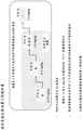
FIG. 1 shows a general schematic of the treatment of cGVHD with an anti-CSF-1R antibody or antigen-binding fragment thereof according to the invention.
FIG. 2 shows the CSF-1R signaling pathway in cGVHD.
Figure 3 shows a general schematic of a clinical trial according to the present invention.
Fig. 4 shows various groups processed according to various embodiments of the invention.
Figure 5 shows the first evidence of CSF-1R inhibition inducing a response in cGVHD at a 1mg/kg Q2W (bi-weekly) dose of anti-CSF-1R antibody or antigen-binding fragment thereof.
FIG. 6 shows evidence of CSF-1R inhibition inducing a response in cGVHD at a 3mg/kg Q2W (every two weeks) dose of an anti-CSF-1R antibody or antigen-binding fragment thereof.
FIG. 7 shows antibody concentrations and monocyte counts (including circulating CD 14) + CD16 + Non-classical and CD14 ++ CD16 + Intermediate monocyte kinetics) consistent with that observed in healthy volunteers and patients. It was shown that at a dose of 3mg/kg q2wk, the patient still had circulating antibodies in the trough<No detectable dose of 3mg/kg, similarly a significant reduction of non-classical monocytes was noted on the right, significantly more pronounced when compared to intermediate and classical.
Figure 8 shows the responses observed in several organ systems after multiple treatments.
Figure 9 shows eculizumab dose escalation and expansion.
Fig. 10 shows the characteristics of chronic GVHD.
Figure 11 shows the demographics and characteristics of the patients.
Figure 12 shows the response across the cGVHD organ system.
Figure 13 shows symptom control following administration of different doses of eculizumab at various intervals.
Fig. 14 shows the waterfall plot and improved Lee symptom score in most patients.
Figure 15 shows a summary of eculizumab and ongoing experiments.
Detailed Description
In some embodiments, the application relates to the treatment of graft versus host disease using an anti-CSF-1R antibody or binding fragment thereof. In some embodiments, the anti-CSF-1R antibody is eculizumab. In some embodiments, the anti-CSF-1R antibody or antigen-binding fragment thereof comprises a heavy chain and/or a light chain, wherein the variable domain of the heavy chain comprises a light chain having the amino acid sequence set forth in SEQ ID NO:4, a CDR having a sequence given in SEQ ID NO:5 and a CDR having the sequence given in SEQ ID NO: 6; wherein the variable domain of the light chain comprises a light chain variable domain having a CDR sequence in SEQ ID NO:1, a CDR having a sequence given in SEQ ID NO:2 and a CDR having the sequence given in SEQ ID NO:3, or a CDR of the sequence given in seq id no.
In some embodiments, an anti-CSF-1R antibody or antigen-binding fragment thereof comprises a heavy chain and a light chain, wherein the variable domain of the heavy chain comprises three CDRs and the sequence of CDR-H1 is identical to SEQ ID NO:4, the sequence of CDR-H2 has at least 60% identity or similarity to the sequence given in SEQ ID NO:5, and the sequence of CDR-H3 has at least 60% identity or similarity to the sequence given in SEQ ID NO:6 has at least 60% identity or similarity; and wherein the variable domain of the light chain comprises three CDRs and the sequence of CDR-L1 is identical to SEQ ID NO:1, the sequence of CDR-L2 has at least 60% identity or similarity to the sequence given in SEQ ID NO:2 and the sequence of CDR-L3 has at least 60% identity or similarity to the sequence given in SEQ ID NO:3 has at least 60% identity or similarity.
In some embodiments, the anti-CSF-1R antibody or antigen-binding fragment thereof comprises a heavy chain, wherein the heavy chain comprises SEQ ID NO:23, or a sequence given in seq id no; and a light chain, wherein the light chain comprises SEQ ID NO:15, or a sequence given in seq id no.
In some embodiments, the antibody has an amino acid sequence comprising SEQ ID NO:27 and a light chain comprising the sequence given in SEQ ID NO:19, or a light chain of the sequence given in table 19. Also provided are anti-CSF-1R antibodies or binding fragments thereof, wherein the heavy and light chains are identical to a light chain comprising SEQ ID NO:27 and a light chain comprising the sequence given in SEQ ID NO:19 is at least 80% (preferably 85%, 90%, 95% or 98%) identical or similar.
In one embodiment, the light chain has the amino acid sequence of SEQ ID NO:19, the heavy chain having or consisting of the sequence given in SEQ ID NO:27 or consists thereof. In another embodiment, the light chain has the amino acid sequence of SEQ ID NO:19 or consists thereof and the heavy chain has the sequence of SEQ ID NO:27, wherein SEQ ID NO:27 by deletion or deletion of the amino acid lysine at position 453.
The present disclosure also provides specific regions or epitopes of human CSF-1R bound by an antibody of the present disclosure, particularly antibody 969.G2 comprising heavy chain sequence gH2 (SEQ ID NO: 27) and/or light chain sequence gL7 (SEQ ID NO: 19).
In some embodiments, the anti-CSF-1R antibody or anti-CSF-1 antibody or antigen-binding fragment thereof is selected from the group consisting of an intact antibody molecule having a full-length heavy and light chain, a Fab, a modified Fab ', a F (ab') 2 Fv, VH, VL and scFv fragments thereof.
In some embodiments, the anti-CSF-1R antibody or antigen-binding fragment thereof comprises a light chain variable region comprising SEQ ID NO:27 and a light chain comprising the sequence given in SEQ ID NO:19, or a light chain of the sequence given in table 19.
In some embodiments, an anti-CSF-1R antibody or antigen-binding fragment thereof cross-blocks a polypeptide comprising the sequence SEQ ID NO: 1. SEQ ID NO for CDR-L2: 2. SEQ ID NO for CDR-L3: 3. SEQ ID NO: 4. SEQ ID NO for CDR-H2: 5 and SEQ ID NO for CDR-H3: binding of an antibody of the 6 CDRs given in 6.
In some embodiments, an anti-CSF-1R antibody or antigen-binding fragment thereof cross-blocks binding by binding to the same epitope as the antibody it blocks.
In some embodiments, an anti-CSF-1 antibody or antigen-binding fragment thereof cross-blocks binding by binding to the same epitope as the antibody it blocks.
In some embodiments, the anti-CSF-1R antibody or anti-CSF-1 antibody or antigen-binding fragment thereof or CSF-1R inhibitor is administered once weekly.
In some embodiments, the anti-CSF-1R antibody or anti-CSF-1 antibody or antigen-binding fragment thereof, or CSF-1R inhibitor is administered biweekly. In some embodiments, the anti-CSF-1R antibody or anti-CSF-1 antibody or antigen-binding fragment thereof or CSF-1R inhibitor is administered twice weekly.
In some embodiments, the anti-CSF-1R antibody or anti-CSF-1 antibody or antigen-binding fragment thereof or CSF-1R inhibitor is administered 3 times per week.
In some embodiments, the anti-CSF-1R antibody, or anti-CSF-1 antibody antigen-binding fragment thereof, or the CSF-1R inhibitor is administered at a dose of about 0.1mg/kg to about 30 mg/kg.
In some embodiments, the anti-CSF-1R antibody, or anti-CSF-1 antibody antigen-binding fragment thereof, or inhibitor of CSF-1R activity is administered at a dose of about 0.1mg/kg to about 10mg/kg. In some embodiments, an anti-CSF-1R antibody, or an anti-CSF-1 antibody antigen-binding fragment thereof, or an inhibitor of CSF-1R activity is administered at a dose of about 0.1mg/kg to about 10mg/kg for the treatment of chronic graft-versus-host disease.
In some embodiments, the anti-CSF-1R antibody or anti-CSF-1 antibody or antigen-binding fragment thereof, or inhibitor of CSF-1R activity is administered at a dose of about 0.1mg/kg, 0.5mg/kg, 1mg/kg, 1.5mg/kg, 3mg/kg, 5mg/kg, 6mg/kg, 7.5mg/kg, or about 10mg/kg.
In some embodiments, the iximab is administered at a dose of 0.15mg/kg per week. In some embodiments, eculizumab is administered at a dose of 0.5mg/kg weekly. In some embodiments, eculizumab is administered at a dose of 1.0mg/kg weekly. In some embodiments, the iximab is administered at a dose of 3.0mg/kg per week. In some embodiments, eculizumab is administered at a dose of 0.15mg/kg every two weeks. In some embodiments, eculizumab is administered at a dose of 0.5mg/kg every two weeks. In some embodiments, eculizumab is administered at a dose of 1.0mg/kg every two weeks. In some embodiments, the iximab is administered at a dose of 3.0mg/kg every two weeks. In some embodiments, eculizumab is administered at a dose of 0.15mg/kg every three weeks. In some embodiments, eculizumab is administered at a dose of 0.5mg/kg every three weeks. In some embodiments, the iximab is administered at a dose of 1.0mg/kg every three weeks. In some embodiments, the iximab is administered at a dose of 3.0mg/kg every three weeks. In some embodiments, the eculizumab is administered at a dose of 0.15mg/kg every four weeks. In some embodiments, the iximab is administered at a dose of 0.5mg/kg every four weeks. In some embodiments, the iximab is administered at a dose of 1.0mg/kg every four weeks. In some embodiments, the eculizumab is administered at a dose of 3.0mg/kg every four weeks. In some embodiments, the dose is increased or decreased weekly according to circulating classical monocyte levels.
Preferably, the anti-CSF-1R antibody or anti-CSF-1 antibody or antigen-binding fragment thereof or CSF-1R inhibitor is administered biweekly. Preferably, the anti-CSF-1R antibody or anti-CSF-1 antibody or antigen-binding fragment thereof or CSF-1R inhibitor is administered at a dose of 1 mg/kg. Preferably, the anti-CSF-1R antibody or anti-CSF-1 antibody or antigen-binding fragment thereof or CSF-1R inhibitor is administered at a dose of 3 mg/kg. Preferably, the anti-CSF-1R antibody or anti-CSF-1 antibody or antigen-binding fragment thereof or CSF-1R inhibitor is administered at a dose of 1mg/kg biweekly.
In some embodiments, the CSF-1R inhibitor is administered and reduces circulating classical monocytes. In some embodiments, the CSF-1R inhibitor is administered and depletes circulating classical monocytes. In some embodiments, the CSF-1R inhibitor is administered and completely depletes levels of classical monocytes. In some embodiments, the initial administration of the CSF-1R inhibitor depletes classical monocytes at a predetermined percentage level. In some embodiments, the initial administration of the CSF-1R inhibitor depletes the level of classical monocytes by a predetermined percentage, and the subsequent administration of the CSF-1R inhibitor occurs once the level of classical monocytes increases. In some embodiments, the initial administration of the CSF-1R inhibitor depletes the level of classical monocytes by a predetermined percentage, and the subsequent administration of the CSF-1R inhibitor occurs once the level of classical monocytes increases to the predetermined percentage. In some embodiments, at least 20%, at least 30%, at least 40%, at least 50%, at least 60%, at least 70%, at least 80%, at least 90%, or 100%.
In some embodiments, the method provides for the treatment of chronic graft versus host disease (cGVHD) in a human, the method comprising administering to a human in need thereof a pharmaceutically effective amount of eculizumab. In some embodiments, the methods of treatment herein are directed to scleroderma. In some embodiments, the methods of treatment are directed to preventing or alleviating the symptoms of chronic graft versus host disease (cGVHD). In some embodiments, the cGVHD is a hepatic cGVHD. In some embodiments, the cGVHD is a renal cGVHD. In some embodiments, the cGVHD is an esophageal cGVHD. In some embodiments, the cGVHD is a gastric cGVHD. In some embodiments, the methods of treatment herein are directed to localized scleroderma, systemic sclerosis, scleroderma-like disorders, and Sine scleroderma. In some embodiments, the methods of treatment herein are directed to systemic sclerosis. In some embodiments, the methods of treatment herein are directed to systemic sclerosis, wherein systemic sclerosis is diffuse or localized. In some embodiments, the methods of treatment herein are directed to CREST (calcinosis, raynaud's esophageal dysfunction, digital sclerosis, telangiectasia). Scleroderma-like disorders are thought to be associated with exposure to industrial environments. In Sine disease, there is an involvement of internal organs without skin changes. The main manifestations of scleroderma and especially systemic sclerosis are inappropriate excessive collagen synthesis and deposition, endothelial dysfunction, spasticity, collapse and fibrotic occlusion. In some embodiments, the cell transplantation is hematopoietic cell transplantation. In some embodiments, GVHD is acute GVHD. In some embodiments, GVHD is chronic GVHD. In some embodiments, GVHD is scleroderma GVHD. In some embodiments, GVHD is steroid resistant GVHD. In some embodiments, GVHD is cyclosporine resistant GVHD. In some embodiments, GVHD is refractory GVHD. In some embodiments, the GHVD is oral GVHD. In some embodiments, oral GVHD is reticular oral GVHD. In some embodiments, oral GVHD is erosive oral GVHD. In some embodiments, oral GVHD is ulcerative oral GVHD. In some embodiments, the oral GVHD is oral GVHD. In some embodiments, oral GVHD is GVHD in the oropharyngeal region. In some embodiments, oral GVHD is GVHD of the pharyngeal region. In some embodiments, oral GVHD is GVHD of the esophageal region. In some embodiments, oral GVHD is acute oral GVHD. In some embodiments, oral GVHD is chronic oral GVHD. In some embodiments, the patient exhibits one or more symptoms of GVHD. In some embodiments, the patient has received or will receive an allogeneic bone marrow or hematopoietic stem cell transplant.
Without being bound by any theory, the presence of monocytes/macrophages provides a positive and negative effect. Without being bound by any theory, it has been found that monocytes/macrophages have a positive and negative effect in the conditions discussed herein, and in some embodiments, the conditions are sclerosing skin conditions and/or chronic graft versus host disease discussed herein. In some embodiments, the presence of circulating classical monocytes has a beneficial effect if allowed to be present in reduced amounts. Allowing monocyte/macrophage levels to increase between CSF-1R inhibitor/antibody administrations allows treatment to take advantage of the positive effects of circulating monocytes/macrophages while avoiding negative effects. In some embodiments, the antibody inhibits monocyte proliferation. In some embodiments, an antibody is considered to "inhibit monocyte proliferation" when the amount of monocyte proliferation is reduced by at least 50% using the described assay (e.g., U.S. patent No. 8,206,715b2). In some embodiments, the antibody reduces the amount of monocyte proliferation by at least 20%, at least 30%, at least 40%, at least 50%, at least 60%, at least 70%, at least 80%, at least 90%, or 100%. In some such embodiments, the antibody is considered to inhibit monocyte proliferation by at least 50%, at least 60%, at least 70%, etc.
Human monocytes exhibit pro-inflammatory characteristics in a variety of disease settings. Human monocytes were identified by expression of CD 14. They can be further classified according to CD16 expression (high affinity Fc receptor). CD 16-cells are referred to as classical monocytes because they typically account for about 90% of the total monocytes in healthy individuals. CD16+ cells appear to be amplified in many inflammatory diseases and exhibit preferential migration through the endothelial layer in response to chemokines. They are therefore commonly referred to as non-classical or pro-inflammatory monocytes (non-classical (CD 14+/CD16 +) monocytes and classical (CD 14+/CD 16-) monocytes).
In some embodiments, administration of a CSF-1R inhibitor of the invention reduces circulating monocytes by at least 40%, at least 50%, at least 60%, at least 70%, at least 80%, at least 90%, or 100%. In some such embodiments, the antibody is considered to inhibit monocyte proliferation by at least 50%, at least 60%, at least 70%, etc. In some embodiments, the level of circulating monocytes is allowed to increase by at least 5%, at least 10%, at least 15%, at least about 20%, at least about 25%, at least about 30%, at least about 40%, at least about 50% prior to administration of a subsequent dose of CSF-1R inhibitor. In some embodiments, the level of circulating monocytes is allowed to increase for 1 week prior to administration of a subsequent dose of CSF-1R inhibitor. In some embodiments, the level of circulating monocytes is allowed to increase for 2 weeks prior to administration of a subsequent dose of CSF-1R inhibitor. In some embodiments, the level of circulating monocytes is allowed to increase for 3 weeks prior to administration of a subsequent dose of CSF-1R inhibitor. In some embodiments, the level of circulating monocytes is allowed to increase for 4 weeks prior to administration of a subsequent dose of CSF-1R inhibitor.
In some embodiments, the monocyte is non-classical. In some embodiments, the monocytes are classical. In some embodiments, the monocyte is a combination of classical and non-classical monocytes. In some embodiments, the monocyte is a combination of a classical monocyte and an intermediate monocyte.
In some embodiments, the CSF-1R inhibitor is eculizumab. In some embodiments, the CSF-1R inhibitor is administered according to the following dosage regimen. In some embodiments, the dosage regimen maximizes the benefits of circulating macrophages and minimizes negative effects. In some embodiments, the dose is increased in contrast to the following dosage regimen.

In some embodiments, the methods of the present invention are directed to the treatment of sclerosing skin conditions, wherein the patient has progressed on one or more previous treatments. In one embodiment, the sclerosing skin condition is active chronic graft versus host disease. In one embodiment, the patient has progressed on at least two prior treatments. In one embodiment, the prior treatment is ibrutinib. In one embodiment, at least one of the prior treatments is ibrutinib.
In some embodiments, the methods of the invention relate to treatment of cGVHD, wherein the patient has progressed on one or more previous treatments. In one embodiment, the patient has progressed on at least two prior treatments. In one embodiment, the prior treatment is ibrutinib. In one embodiment, at least one of the prior treatments is ibrutinib.
In one embodiment, the eculizumab is administered with one or more additional agents useful in the treatment of graft versus host disease, the additional agent is selected from prednisone, methylprednisolone, oral non-absorbable corticosteroids such as budesonide or beclomethasone dipropionate, immunomodulators such as cyclosporin, tacrolimus, mycophenolate mofetil, tellomethasone, imuthiol, antithymocyte globulin, anti-TNF drugs, azathioprine, inosine 5' -monophosphate dehydrogenase inhibitors, azodicarbonide (azodiacarbonide), bisindolylmaleimide VIII, brequinar, chlorambucil, CTLA-4Ig, corticosteroids, cyclophosphamide, deoxyspergualin, dexamethasone, glucocorticoids, leflunomide, mercaptopurine, 6-mercaptopurine, methotrexate, methylprednisolone, prednisolone, prednisone, and other drugs mizoribine, mizoribine monophosphate, muromonab CD3, mycophenolate mofetil, OKT3, rho (D) immunoglobulin, vitamin D analog, MC 1288), daclizumab, infliximab, rituximab, tositumomab, alemtuzumab, methotrexate, antithymotubulin (antithymocyte denilukin difitox), campath-1H, keratinocyte growth factor, abatacept, remestamcel-L suberoylanilide hydroxamic acid, pentostatin, thalidomide, imatinib mesylate, cyclophosphamide, fludarabine, OKT3, melphalan, thiopeta and lymphocyte immunoglobulin, antithymotymocyte and globulin
Nucleic acid, polypeptide
CDR-L1:LASEDIYDNLA(SEQ ID NO:1)
CDRL2:YASSLQD(SEQ ID NO:2)
CDR-L3:LQDSEYPWT(SEQ ID NO:3)
CDR-H1:GFSLTTYGMGVG(SEQ ID NO:4)
CDR-H2:NIWWDDDKYYNPSLKN(SEQ ID NO:5)
CDR-H3:IGPIKYPTAPYRYFDF(SEQ ID NO:6)
Rat Ab969 VL domain DIQMTQSAPAS LSASLEGETGVS IECLASEDIY DNLAWYQKKP GKSPHLLIYY ASSLQDGVPS RFSGSGSGTQ YSLKINSLES EDAATYFCLQ DSEYPWTGFGG GTKLELK (SEQ ID NO: 7)
<xnotran> Ab969 VL : gacatccaga tgacacagtc tccagcttcc ctgtctgcat ctctgggaga aactgtctcc atcgaatgtc tagcaagtga ggacatttac gataatttag cgtggtacca gaagaagcca ggaaaatctc ctcacctcct catctattat gcaagtagct tgcaagatgg ggtcccatca cggttcagtg gcagtggatc tggcacacag tattctctca aaatcaacag cctggaatct gaagatgctg cgacttattt ctgtctacag gattctgagt atccgtggac gttcggtgga ggcaccaagc tggaattgaa a (SEQ ID NO: 8) </xnotran>
Rat Ab969 VL region, signal sequence underlined and in italics:


rat Ab969 VL region, signal sequence underlined and in italics:


rat Ab969 VH region QVTLKESGGPG ILQPSQTLSL TCTFSGFSLT TYGGVGGWIR QPSGKGLEWLANIWWDDDKY YNPKSLKNLT ISKDTSNNQA FLKLTNVHTS DSATYYCARIGPIKYPTAPY RYFDWGPGT MVTVS (SEQ ID NO: 11)
<xnotran> Ab969 VH : caggttactc tgaaagagtc tggccctggg atattgcagc cctcccagac cctcagtctg acttgcactt tctctgggtt ttcactgacc acttatggta tgggtgtggg ctggattcgt cagccttcag ggaagggtct ggagtggctg gcaaacattt ggtgggatga tgataagtat tacaatccat ctctgaaaaa ccggctcaca atctccaagg acacctccaa caaccaagca ttcctcaagc tcaccaatgt acacacttca gattctgcca catactactg tgctcggata gggccgatta aatacccgac ggccccctac cggtactttg acttctgggg cccaggaacc atggtcaccg tctcg (SEQ ID NO: 12) </xnotran>
Rat Ab969 VH region, signal sequence underlined and in italics:


rat Ab969 VH region, signal sequence underlined and in italics:


969 The gL 7V region is DIQMTQSPSLSAVSDRVT ITCLASEDIY DNLAWYQKP GKAPKLLIYY ASSLQDGVPS RFSGSGSGTD YTLTISSLQP EDFATTYYCLQ DSEYPWTGFGG GTKVEIK (SEQ ID NO: 15)
969 gL 7V region: gacatacaga tgactcagtc acccctcaagc ctgagttgccta gtgggaga caggggtgaca atcacctgtc tgggcctcga ggatactcgaacctgg catggttgta gcaaacct ggaaaggctc ccaagctccaagctctcaagacgg cgtccatct cggttcggcggagcaggcatgcatgcatgcatgcatgcatgcatgcatgcatgcatgcatgcatgcaggtg cttcgggacggacggacggagcatgcatgcatgcatgcatgca gattggcactgcctgcaccgcatgca gagccctgaatgcaat acccatggtcggtggtggtggcaccaag (SEQ ID NO: 16)
969 gL 7V region, signal sequence underlined and in italics:


969 gL 7V region, signal sequence underlined and in italics:


969 DIQMTQSPSSSLSSVGDSVDRVT ITCLASEDIY DNLAWYQQKP GKAPKLLIYY ASSLQDGVPS RFSGSGSGSGTD YTISSLQPATYYCLQ DSEYPWTGG GTKVEIKIRTV AAPSVFIFPP SDEQLKSTGTA SVVCLLNNFY PREQWQWDNALGNSQ ESVTEQDSDSSTYSSTLT LSKADYYEKHK VYACEVTHQGGLSSPKSFNRGEC (SEQ ID NO: 19)
969 <xnotran> gL7 (V + ): gacatacaga tgactcagtc accctcaagc ctgagtgcca gtgtgggaga cagggtgaca atcacctgtc tggcctccga ggatatctac gataacctgg catggtatca gcagaaacct ggaaaggctc ccaagctcct gatttattat gcctcctctc tccaagacgg cgttccatct cggttcagcg gaagcggctc cgggacggat tacacactga caattagctc tctgcaaccg gaggattttg ctacttacta ctgcctgcaa gactccgaat acccatggac cttcggtggt ggcaccaaag tggaaatcaa gcgtacggta gcggccccat ctgtcttcat cttcccgcca tctgatgagc agttgaaatc tggaactgcc tctgttgtgt gcctgctgaa taacttctat cccagagagg ccaaagtaca gtggaaggtg gataacgccc tccaatcggg taactcccag gagagtgtca cagagcagga cagcaaggac agcacctaca gcctcagcag caccctgacg ctgagcaaag cagactacga gaaacacaaa gtctacgcct gcgaagtcac ccatcagggc ctgagctcgc ccgtcacaaa gagcttcaac aggggagagt gt (SEQ ID NO: 20) </xnotran>
969 gL7 light chain (V + constant), signal sequence underlined and in italics:


969 gL7 light chain (V + constant), signal sequence underlined and in italics:


969 The gH 2V region EVTLKESGPA LVKPTQTLTL TCTFSGFSLT TYGGMGVGGWIR QPPGKALEWL ANIWWDDDKY YNPSLKNLT ISKDTSKNQV VLTMTNMDPV DTATYYCARI GPIKYPTAPY RYFDFGWGQGT MVTVS (SEQ ID NO: 23)
969 <xnotran> gH 2V : gaagtgacac tcaaggagtc tggacccgct ctggtgaaac caacccaaac actcactttg acatgtactt ttagtggctt ctcattgact acctatggaa tgggcgtggg atggatcaga cagccacctg gcaaggctct ggaatggctg gccaacatct ggtgggatga cgacaagtac tataacccgt ccctgaaaaa ccggctgacc attagcaagg atacttctaa aaatcaagtg gtgctgacca tgacaaatat ggatcccgtt gacaccgcaa cctactactg cgcccgcatt ggtcccataa agtaccctac ggcaccttac cgatatttcg acttttgggg ccaagggaca atggttactg tctcg (SEQ ID NO: 24) </xnotran>
969 gH 2V region, signal sequence underlined and in italics:


969 gH 2V region, signal sequence underlined and in italics:


969 The gH2 heavy chain (V + constant-hu IgG 4P) EVTLKESGPA LVKPTQTLTL TCTFSGFSLT TYGGVGGWIR QPPGKALEWL ANIWWDWDDDKY YNPLKDTSKNQV VLTMTNMDPV DTATYYCARI GPIKYPTAPY RYFGWGQMVGT TVSSTK GPSVLAPC SRSTSTGASTTAA LGVKVKDFLYFP EPVTVSVTSGVHSG QSQAVSGQSVG QSLVSGLYS LSSVVTSSGTKTSS GTKTCN VDHKPVSKTPSKVKTV DKYPCGPSTPF LGSVPGFLFP PKDTLMTPVTEVVVTVTCVVTVVVVVVQFNWVTVGGVGGVGGVGGKWKG NKKG VLKG VLKGGVGS VKSKLASK VLGVGVLKSGVGVLKSGVGVLKSGVGS 27
969 gH2 heavy chain (V + constant-hu IgG4P, exons underlined):


969 gH2 heavy chain (V + constant-hu IgG 4P), signal sequence underlined and in italics:


969 gH2 heavy chain (V + constant-hu IgG4P, exons underlined), signal sequence underlined and in italics:


human VK 1- (1) O12 JK4 receptor framework DIQMTQSPSLSASGTSVGDRVT ITCRASSQSIS SYLNWYQKP GKAPKLLIYA ASSLQSGVPS RFSGSGSGSGTD FTLTISSLQPEDDYQQ SYSTPLTFGG GTKVEIK (SEQ ID NO: 31)
Human VK 1- (1) O12 JK4 receptor framework gacatccaga tgacccagtc tccatccctctgctgcat ctgtaggagagagagagagagagagagagtacccacc atcctgcc gggcaagtc gagagatagc agctatttaa attggtatca gcaaaccca gggaaaagcccc ctaagctcctgattctactgcct gcatccagttgcatgtggtcccatcaggtggaggtggagggatgagagatc ttgcaggcaggcaggcaggcatgagat ttgcaggcatgcatgcactcacgcatgcatgcatcag cgaggcatgcagtcgcaggtgcaggtgcaggcatgcatgcatg (SEQ ID NO: 32)
Human VH 2-1-70 JH3 receptor framework QVTLKESGTLTL TCTFSGFSLS TSGMRVSWIR QPPGKALEWL ARIDWDDDKF YSTSLKTTRLT ISKDTSKNQV VLTMTNTNMDPV DTATYYCARI AFDIWGQGTM VTVS (SEQ ID NO: 33)
<xnotran> VH2 3-1 2-70 JH3 : caggtcacct tgaaggagtc tggtcctgcg ctggtgaaac ccacacagac cctcacactg acctgcacct tctctgggtt ctcactcagc actagtggaa tgcgtgtgag ctggatccgt cagcccccag ggaaggccct ggagtggctt gcacgcattg attgggatga tgataaattc tacagcacat ctctgaagac caggctcacc atctccaagg acacctccaa aaaccaggtg gtccttacaa tgaccaacat ggaccctgtg gacacagcca cgtattactg tgcacggata gcttttgata tctggggcca agggacaatg gtcaccgtct ct (SEQ ID NO: 34) </xnotran>
<xnotran> CSF-1R : MGPGVLLLLL VATAWHGQGI PVIEPSVPEL VVKPGATVTL RCVGNGSVEW DGPPSPHWTL YSDGSSSILS TNNATFQNTG TYRCTEPGDP LGGSAAIHLY VKDPARPWNV LAQEVVVFED QDALLPCLLT DPVLEAGVSL VRVRGRPLMR HTNYSFSPWH GFTIHRAKFI QSQDYQCSAL MGGRKVMSIS IRLKVQKVIP GPPALTLVPA ELVRIRGEAA QIVCSASSVD VNFDVFLQHN NTKLAIPQQS DFHNNRYQKV LTLNLDQVDF QHAGNYSCVA SNVQGKHSTS MFFRVVESAY LNLSSEQNLI QEVTVGEGLN LKVMVEAYPG LQGFNWTYLG PFSDHQPEPK LANATTKDTY RHTFTLSLPR LKPSEAGRYS FLARNPGGWR ALTFELTLRY PPEVSVIWTF INGSGTLLCA ASGYPQPNVT WLQCSGHTDR CDEAQVLQVW DDPYPEVLSQ EPFHKVTVQS LLTVETLEHN QTYECRAHNS VGSGSWAFIP ISAGAHTHPP DEFLFTPVVV ACMSIMALLL LLLLLLLYKY KQKPKYQVRW KIIESYEGNS YTFIDPTQLP YNEKWEFPRN NLQFGKTLGA GAFGKVVEAT AFGLGKEDAV LKVAVKMLKS TAHADEKEAL MSELKIMSHL GQHENIVNLL GACTHGGPVL VITEYCCYGD LLNFLRRKAE AMLGPSLSPG QDPEGGVDYK NIHLEKKYVR RDSGFSSQGV DTYVEMRPVS TSSNDSFSEQ DLDKEDGRPL ELRDLLHFSS QVAQGMAFLA SKNCIHRDVA ARNVLLTNGH VAKIGDFGLA RDIMNDSNYI VKGNARLPVK WMAPESIFDC VYTVQSDVWS YGILLWEIFS LGLNPYPGIL VNSKFYKLVK DGYQMAQPAF APKNIYSIMQ ACWALEPTHR PTFQQICSFL QEQAQEDRRE RDYTNLPSSS RSGGSGSSSS ELEEESSSEH LTCCEQGDIA QPLLQPNNYQ FC (SEQ ID NO: 35) </xnotran>
CSF-1R has an amino acid sequence of MRHTNYSFSSPWHGFTIHRAKFIQSQDYQCSALMGGRKVMSISIRLKQK (SEQ ID NO: 36)
CSF-1R amino acid sequence (SNP V32G, A245S, H247P, V279M, position underlined) IPVIEPSVPELVVKPGATVTLRCVGNGSVEWDGPPSPHWTLYSDGSSSILSTNNATFQNTGTYRCTEPGDPLGGSAAIHLYVKDPARPWNVLAQEVVVFEDQDALLPCLLTDPVLEAGVSLVRVRGRPLMRHTNYSFSPWHGFTIHRAKFIQSQDYQCSALMGGRKVMSISIRLKVQKVIPGPPALTLVPAELVRIRGEAAQIVCSASSVDVNFDVFLQHNNTKLAIHQQSDFHNNRYQKVLTLNLDQVDFQHAGNYSCVASNVQGKHSTSMFFRVVESAYLNLSSEQNLIQEVTVGEGLNLKVMVEAYPGLQGFNWTYLGPFSDHQPEPKLANATTKDTYRHTFTLSLPRLKPSEAGRYSFLARNPGGWRALTFELTLRYPPEVSVIWTFINGSGTLLCAASGYPQPNVTWLQCSGHTDRCDEAQVLQVWDDPYPEVLSQEPFHKVTVQSLLTVETLEHNQTYECRAHNSVGSGSWAFIPISAGAHTHPPDE(SEQ ID NO:37)
MGPGVLLLLL VATAWHGQGI PVIEPSVPEL VVKPGATVTL RCVGNGSVEW DGPPSPHWTL YSDGSSSILS TNNATFQNTG TYRCTEPGDP LGGSAAIHLY VKDPARPWNV LAQEVVVFED QDALLPCLLT DPVLEAGVSL VRVRGRPLMR HTNYSFSPWH GFTIHRAKFI QSQDYQCSAL MGGRKVMSIS IRLKVQKVIP GPPALTLVPA ELVRIRGEAA QIVCSASSVD VNFDVFLQHN NTKLAIPQQS DFHNNRYQKV LTLNLDQVDF QHAGNYSCVA SNVQGKHSTS MFFRVVESAY LNLSSEQNLI QEVTVGEGLN LKVMVEAYPG LQGFNWTYLG PFSDHQPEPK LANATTKDTY RHTFTLSLPR LKPSEAGRYS FLARNPGGWR ALTFELTLRY PPEVSVIWTF INGSGTLLCA ASGYPQPNVT WLQCSGHTDR CDEAQVLQVW DDPYPEVLSQ EPFHKVTVQS LLTVETLEHN QTYECRAHNS VGSGSWAFIP ISAGAHTHPP DE(SEQ ID NO:38)
CSF-1R
As used herein, the term "colony stimulating factor-1 receptor" or "CSF1R" refers to a tyrosine protein kinase that acts as a cell surface receptor for CSF 1and interleukin 34 (IL 34) and plays an important role in the regulation of survival, proliferation and differentiation of hematopoietic precursor cells, particularly mononuclear phagocytes such as macrophages and monocytes. It promotes the release of pro-inflammatory chemokines in response to IL34 and CSF1, playing an important role in innate immunity and inflammation processes. CSF1R also plays an important role in the regulation of osteoclast proliferation and differentiation, regulation of bone resorption, and is essential for normal skeletal and dental development. CSF1R is required for normal male and female fertility and for normal development of ductal and acinar structures in the mammary gland during pregnancy. It also promotes reorganization of the actin cytoskeleton, regulates membrane fold formation, cell adhesion and cell migration, and promotes cell invasion.
CSF1 is a cytokine that controls the production, differentiation and function of macrophages, and CSF1R mediates most, if not all, of the biological effects of this cytokine.
As used herein, the term "ab969.G2" refers to an antibody that specifically binds CSF1-R and comprises (a) a heavy chain variable region comprising a sequence as set forth in SEQ ID NO: 1. SEQ ID NO:2 and SEQ ID NO:3, CDR1, CDR2 and CDR3, and (b) a light chain comprising the amino acid sequence as defined in SEQ ID NO: 4. SEQ ID NO:5 and SEQ ID NO:6 the heavy chain of CDR1, CDR2 and CDR3 as defined. The Ab969.G2 antibody has been previously described in PCT/EP 2014/068050.
The terms "specifically binds CSF1R," "specifically binds CSF1R," and equivalents as used herein when referring to an antibody, mean that the antibody will bind CSF1R with sufficient affinity and specificity to achieve a biologically meaningful effect. The antibody selected typically has binding affinity for CSF1R, e.g., the antibody can bind CSF1R with a Kd value of 100nM to 1 pM. Antibody affinity can be determined by surface plasmon resonance based assays, such as BIAcore assays; enzyme-linked immunosorbent assay (ELISA); and, for example, a competition assay (e.g., of RIA). Within the meaning of the present invention, an antibody that specifically binds CSF1R may also bind another molecule; for example in the case of bispecific antibodies as a non-limiting example.
Formulations and methods of treatment
Any of the antibodies disclosed herein (e.g., anti-CSF-1R antibodies or anti-CSF-1 antibodies) can be used in the methods, kits, or compositions of the present disclosure.
In some embodiments, a pharmaceutical composition of the disclosure comprises an anti-CSF-1R antibody or an anti-CSF-1 antibody or an antigen-binding fragment thereof or an inhibitor of CSF-1R activity and a pharmaceutically acceptable carrier.
In certain embodiments, the combinations described herein are used to treat a skin condition. In some embodiments, the methods involve the use of an anti-CSF-1R antibody or binding fragment thereof for treating systemic scleroderma, local scleroderma, localized scleroderma, linear scleroderma, CREST syndrome, diffuse scleroderma, limited scleroderma (Circumscribed Morphea), calcinosis, raynaud's phenomenon, esophageal dyskinesia, digital scleroderma, telangiectasia, sine sclerosis, and/or diffuse scleroderma.
In some embodiments, the methods involve the use of an anti-CSF-1R antibody or binding fragment thereof for treating acute graft versus host disease (aGvHD). In some embodiments, the methods involve the use of an anti-CSF-1R antibody or binding fragment thereof for the treatment of chronic graft versus host disease (cGvHD).
In some embodiments, a method of treating a human patient identified as having cGVHD comprises determining an initial level of classical monocytes in the patient. In some embodiments, a method of treating a human patient identified as having cGVHD comprises determining an initial level of canonical monocytes in the patient, then administering an effective dose of eculizumab or an anti-CSF-1R antibody; and determining a second level of classical monocytes over a subsequent time period. In some embodiments, a method of treating a human patient identified as having cGVHD comprises determining an initial level of classical monocytes in the patient, then administering an effective dose of eculizumab or an anti-CSF-1R antibody; and determining a second level of classical monocytes over a subsequent time period, and continuing treatment with eculizumab or an anti-CSF-1R antibody if the second level of classical monocytes is greater than a predetermined percentage. In some embodiments, a method of treating a human patient identified as having cGVHD comprises determining an initial level of canonical monocytes in the patient, then administering an effective dose of eculizumab or an anti-CSF-1R antibody; and determining a second level of classical monocytes in a subsequent time period, and continuing treatment with eculizumab or an anti-CSF-1R antibody if the ratio between the initial classical monocyte level and the second classical monocyte level is greater than a predetermined percentage. In some embodiments, the application relates to a method of treating cGVHD comprising treating a patient in need thereof with a therapeutically effective amount of icolizumab, wherein icolizumab is targeted to pathogenic monocyte derived macrophages. In some embodiments, the application relates to a method of treating cGVHD comprising treating a patient in need thereof with a therapeutically effective amount of eculizumab, wherein eculizumab targets pathogenic monocyte-derived macrophages and minimally affects non-classical monocytes. In some embodiments, the application relates to a method of treating cGVHD comprising treating a patient in need thereof with a therapeutically effective amount of eculizumab, wherein eculizumab targets pathogenic monocyte-derived macrophages and minimally affects intermediate monocytes.
A method of treating graft versus host disease (GvHD) in a human comprising administering to a human in need thereof an iximab or anti-CSF-1R antibody, wherein the antibody is administered at a dose determined by the level of circulating classical monocytes. A method of treating graft versus host disease (GvHD) in a human comprising administering to a human in need thereof an iximab or anti-CSF-1R antibody, wherein said antibody is administered at a dose determined by the level of circulating intermediate monocytes. A method of treating human graft versus host disease (GvHD) comprising administering to a human in need thereof an iximab or anti-CSF-1R antibody, wherein the antibody is administered at a dose determined by the level of circulating non-classical monocytes.
The terms "treat," "treating," and "medical" are intended to include the alleviation or elimination of a disorder, disease, or condition; or one or more symptoms associated with a disorder, disease, or condition; or to reduce or eradicate the cause of the disorder, disease, or condition itself. As used herein, "preventing" or "prevention" describes reducing or eliminating the onset of symptoms or complications of a disease, condition, or disorder.
As used herein, the term "reducing" is intended to describe a process of reducing the severity of signs or symptoms of a disorder. Importantly, signs or symptoms can be alleviated without being eliminated. In a preferred embodiment, administration of the pharmaceutical compositions disclosed herein results in elimination of signs or symptoms, however, elimination is not required. An effective dose is expected to reduce the severity of signs or symptoms. For example, if the severity of cGVHD decreases within at least one of the multiple locations, signs or symptoms of a disorder that can occur at the multiple locations, such as cGVHD, are alleviated.
Treatment of the conditions listed herein may result in prevention of the development of or reduction in the severity of chronic graft versus host disease (cGVHD) as described herein, including cGVHD. The reduction of symptoms may also be referred to as "resolution". Preferably, after treatment, the severity is reduced by 5% or more relative to before treatment; more preferably, the severity is reduced by 10% or more; more preferably, a reduction of 20% or more; more preferably, a reduction of 30% or more; more preferably, a 40% or greater reduction; even more preferably, a reduction of 50% or more; most preferably, the reduction is more than 75% or more. Severity can be measured by any repeatable measurement method. Severity can be measured as the diameter of the region of interest or according to various physician scales.
A "pharmaceutical composition" or "therapeutic composition" is a formulation containing an active ingredient (e.g., an anti-CSF-1R antibody or anti-CSF-1 antibody or antigen-binding fragment thereof or inhibitor of CSF-1R activity disclosed herein) in a form suitable for administration to a subject. In some embodiments, the pharmaceutical composition is in bulk or unit dosage form. The unit dosage form is in any of a variety of forms including, for example, capsules, IV bags, tablets, a single pump on an aerosol inhaler, or a vial. The amount of active ingredient (e.g., a formulation of a disclosed compound or a salt, hydrate, solvate, or isomer thereof) in a unit dosage composition is an effective amount and varies with the particular treatment involved. Those skilled in the art will appreciate that it is sometimes necessary to make routine variations in dosage depending on the age and condition of the patient. The dosage will also depend on the route of administration. A variety of routes are contemplated, including oral, pulmonary, rectal, parenteral, transdermal, subcutaneous, intravenous, intramuscular, intraperitoneal, inhalation, buccal, sublingual, intrapleural, intrathecal, intranasal, and the like. Dosage forms for topical or transdermal administration of the compounds of the present disclosure include powders, sprays, ointments, pastes, creams, lotions, gels, solutions, patches and inhalants. In one embodiment, the active compound is mixed under sterile conditions with a pharmaceutically acceptable carrier and any preservatives, buffers, or propellants which may be required.
As used herein, "active ingredient" refers to an ingredient that has a pharmacological, e.g., therapeutic, effect at the relevant dosage.
"pharmaceutically acceptable carrier" means a carrier that can be used to prepare a pharmaceutical composition that is generally safe, non-toxic, and biologically or otherwise non-deleterious and includes excipients that can be used for veterinary as well as human pharmaceutical use. For example, a pharmaceutically acceptable carrier should not itself induce the production of antibodies harmful to the individual receiving the composition and should not be toxic. Suitable carriers can be large, slowly metabolized macromolecules such as proteins, polypeptides, liposomes, polysaccharides, polylactic acids, polyglycolic acids, polymeric amino acids, amino acid copolymers, and inactive virus particles.
Pharmaceutically acceptable salts, such as inorganic acid salts, for example, hydrochloride, hydrobromide, phosphate and sulfate, or organic acid salts, for example, acetate, propionate, malonate and benzoate, can be used.
The pharmaceutically acceptable carrier in the therapeutic composition can additionally comprise a liquid, such as water, saline, glycerol, and ethanol. Furthermore, auxiliary substances, such as wetting or emulsifying agents or pH buffering substances, may be present in such compositions. Such carriers enable the pharmaceutical compositions to be formulated as tablets, pills, dragees, capsules, liquids, gels, syrups, slurries and suspensions, for ingestion by a patient.
Suitable forms for administration include forms suitable for parenteral administration, for example by injection or infusion, for example by bolus injection or continuous infusion. When the product is for injection or infusion, it may take the form of a suspension, solution or emulsion in an oily or aqueous vehicle, and it may contain formulatory agents such as suspending, preservative, stabilising and/or dispersing agents. Alternatively, the antibody molecule may be in dry form for reconstitution with a suitable sterile liquid prior to use.
Once formulated, the compositions of the present disclosure can be administered directly to a subject.
In certain embodiments, the pH of the final preparation is not similar to the value of the isoelectric point (pI) of the antibody or fragment, e.g., if the pH of the preparation is 7, a pI of 8-9 or higher may be appropriate. While not wishing to be bound by theory, it is believed that this may ultimately provide a final formulation with improved stability, e.g., the antibody or fragment remains in solution.
In one example, a pharmaceutical formulation at a pH in the range of 4.0 to 7.0 comprises: 1 to 200mg/mL of an antibody according to the disclosure, 1 to 100mM of a buffer, 0.001 to 1% of a surfactant, a) 10 to 500mM of a stabilizer, b) 10 to 500mM of a stabilizer and 5 to 500mM of a tonicity agent, or c) 5 to 500mM of a tonicity agent.
The pharmaceutical compositions of the present disclosure may be administered by any number of routes, including, but not limited to, oral, intravenous, intramuscular, intraarterial, intramedullary, intrathecal, intraventricular, transdermal (see, e.g., WO 98/20734), subcutaneous, intraperitoneal, intranasal, enteral, topical, sublingual, intravaginal, or rectal routes. Painless subcutaneous syringes (Hypospray) may also be used to administer the pharmaceutical compositions of the present disclosure. Generally, the therapeutic compositions can be prepared as injectables, either as liquid solutions or suspensions. Solid forms suitable for dissolution or suspension in a liquid vehicle prior to injection may also be prepared.
Direct delivery of the composition is typically achieved by subcutaneous, intraperitoneal, intravenous, or intramuscular injection, or delivery to the interstitial space of a tissue. The composition may also be administered into a lesion. The dose treatment may be a single dose schedule or a multiple dose schedule.
It will be appreciated that the active ingredient in the composition will be an antibody molecule. Therefore, it is easily degraded in the gastrointestinal tract. Thus, if the composition is to be administered by a route that uses the gastrointestinal tract, the composition will need to include an agent that protects the antibody from degradation but releases the antibody once it is absorbed from the gastrointestinal tract.
Pharmaceutically acceptable carriers are discussed extensively in Remington's Pharmaceutical Sciences (Mack Publishing Company, N.J.1991).
In one embodiment, the formulation is provided as a formulation for topical administration including inhalation.
Suitable inhalable formulations include inhalable powders, metered aerosol formulations containing a propellant gas or inhalable solutions without a propellant gas. Inhalable powders containing an active substance according to the present disclosure may consist of the active substance described above alone or in a mixture with physiologically acceptable excipients.
These inhalable powders may include monosaccharides (e.g. glucose or arabinose), disaccharides (e.g. lactose, sucrose and maltose), oligosaccharides and polysaccharides (e.g. dextran), polyols (e.g. sorbitol, mannitol and xylitol), salts (e.g. sodium chloride, calcium carbonate) or mixtures thereof with each other. Mono-or disaccharides are suitably used, lactose or glucose are used, particularly but not exclusively in their hydrate form.
Particles for deposition in the lung require a particle size of less than 10 microns, for example 1-9 microns, for example 0.1 to 5 μm, especially 1 to 5 μm. The particle size of the active ingredient (e.g., antibody or fragment) is of primary importance.
Propellant gases useful in the preparation of inhalable aerosols are known in the art. Suitable propellant gases are selected from chlorinated and/or fluorinated derivatives of hydrocarbons such as n-propane, n-butane or isobutane and halogenated hydrocarbons such as methane, ethane, propane, butane, cyclopropane or cyclobutane. The aforementioned propellant gases may be used alone or in mixtures thereof.
Particularly suitable propellant gases are haloalkane derivatives selected from the group consisting of TG 11, TG 12, TG134a and TG 227. <xnotran> , TG134a (1,1,1,2- ) TG227 (1,1,1,2,3,3,3- ) . </xnotran>
The propellant gas-containing inhalable aerosols may also contain other ingredients such as cosolvents, stabilizers, surfactants, antioxidants, lubricants and means for adjusting the pH. All of these ingredients are known in the art. A propellant gas-containing inhalable aerosol according to the present disclosure may contain up to 5% by weight of active substance. Aerosols according to the present disclosure contain, for example, 0.002 to 5 wt.%, 0.01 to 3 wt.%, 0.015 to 2 wt.%, 0.1 to 2 wt.%, 0.5 to 2 wt.%, or 0.5 to 1 wt.% of the active ingredient.
Alternatively, topical administration to the lung may also be by administration of a liquid solution or suspension formulation, for example using a device such as a nebulizer, e.g., a nebulizer connected to a compressor (e.g., a Pari LC-Jet Plus (R) nebulizer connected to a Pari Master (R) compressor manufactured by Pari Respiratory Equipment, inc.
The antibodies of the present disclosure can be delivered dispersed in a solvent, for example, in the form of a solution or suspension. It may be suspended in a suitable physiological solution such as saline or other pharmacologically acceptable solvents or buffered solutions. Buffer solutions known in the art may contain 0.05mg to 0.15mg disodium edetate, 8.0mg to 9.0mg NaCl, 0.15mg to 0.25mg polysorbate, 0.25mg to 0.30mg citric acid anhydrous and 0.45mg to 0.55mg sodium citrate per 1ml water to achieve a pH of about 4.0 to 5.0. The suspension may use, for example, lyophilized antibodies.
The therapeutic suspension or solution formulations may also include one or more excipients. Excipients are well known in the art and include buffers (e.g., citrate buffer, phosphate buffer, acetate buffer, and bicarbonate buffer), amino acids, urea, alcohols, ascorbic acid, phospholipids, proteins (e.g., serum albumin), EDTA, sodium chloride, liposomes, mannitol, sorbitol, and glycerol. The solution or suspension may be encapsulated in liposomes or biodegradable microspheres. The formulation will generally be provided in a substantially sterile form using sterile manufacturing processes.
This may include production and sterilization of buffered solvent/solutions for the formulation by filtration, sterile suspension of the antibody in sterile buffered solvent solution, and dispensing of the formulation into sterile containers by methods familiar to those of ordinary skill in the art.
Nebulizable formulations according to the present disclosure can be provided, for example, in the form of single dose units (e.g., sealed plastic containers or vials) packaged in foil envelopes. Each vial contains a unit dose in a volume (e.g., 2 mL) of solvent/solution buffer.
The antibodies disclosed herein may be suitable for delivery by nebulization.
It is also contemplated that the antibodies of the present disclosure may be administered by using gene therapy. To accomplish this, DNA sequences encoding the heavy and light chains of the antibody molecule under the control of appropriate DNA components are introduced into the patient such that the antibody chains are expressed from the DNA sequences and assembled in situ.
The pharmaceutical composition suitably comprises a therapeutically effective amount of an anti-CSF-1R antibody or an anti-CSF-1 antibody or antigen-binding fragment thereof or an inhibitor of CSF-1R activity. As used herein, the term "therapeutically effective amount" refers to the amount of a therapeutic agent required to treat, ameliorate, or prevent a targeted disease or condition, or to exhibit a detectable therapeutic, pharmacological, or prophylactic effect. For example, for any of the antibodies disclosed herein, a therapeutically effective amount can be estimated initially in a cell culture assay (e.g., of tumor cells) or in an animal model (typically rat, mouse, rabbit, dog, pig, or primate). Animal models can also be used to determine appropriate concentration ranges and routes of administration. Such information can then be used to determine useful doses and routes for administration to humans.
Therapeutic/prophylactic efficacy and toxicity can be determined by standard pharmaceutical procedures in cell cultures or experimental animals, e.g., ED 50 (dose therapeutically effective in 50% of the population) and LD 50 (the dose lethal to 50% of the population). The dose ratio of toxic to therapeutic effect is the therapeutic index, which can be expressed as the ratio, LD 50 /ED 50 . Preferred are pharmaceutical compositions that exhibit a large therapeutic index. The dosage may vary within this range depending upon the dosage form employed, the sensitivity of the patient, and the route of administration.
The dosage and administration are adjusted to provide a sufficient level of the active agent or to maintain the desired effect. Factors that may be considered include the severity of the disease state, the general health of the subject, the age, weight and sex of the subject, diet, time and frequency of administration, drug interactions, response sensitivity and tolerability/response to treatment. In general, the dosage should be sufficient to slow down and preferably reduce the severity of the condition. The dosage may range from about 0.01mg/kg per day to about 10mg/kg per day. In some embodiments, the dose may vary within a range of about 0.1mg/kg, 0.5mg/kg, 1mg/kg, 1.5mg/kg, or 3 mg/kg. In some embodiments, the dose will range from about 0.1 mg/day to about 5 mg/kg. The pharmaceutical compositions may conveniently be presented in unit dosage form containing a predetermined amount of the active agent of the present disclosure per dose.
Therapeutic doses of antibodies according to the present disclosure (e.g., anti-CSF-1R antibodies or anti-CSF-1 antibodies) do not exhibit significant or limited toxicological effects in vivo.
In certain embodiments, the iximab or anti-CSF-1R antibody is administered daily, every other day, weekly, every 2 weeks, 3 weeks, 4 weeks, 5 weeks, 6 weeks, 7 weeks, 8 weeks, 9 weeks, 10 weeks, 11 weeks, 12 weeks, 13 weeks, 14 weeks, 15 weeks, 16 weeks, 17 weeks, 18 weeks, 19 weeks, or every 20 weeks or monthly.
The term "antibody" is used according to its well-known meaning in the art. The antibody molecules of the present disclosure may include intact antibody molecules having full-length heavy and light chains or binding fragments thereof, and may be, but are not limited to, fab, modified Fab ', F (ab') 2, fv, single domain antibodies (e.g., VH or VL or VHH), scFv, diabodies, triabodies, tetrabodies, and epitope-binding fragments of any of the above (see, e.g., holliger and Hudson,2005, nature Biotech.23 (9): 1126-1136 Adair and Lawson,2005, drug Deven Reviews-Online 2 (3), 209-217). Methods for producing and making these antibody fragments are well known in the art (see, e.g., verma et al, 1998, journal of Immunological methods, 216-181. Other antibody fragments useful in the present disclosure include Fab and Fab' fragments described in International patent applications WO05/003169, WO05/003170 and WO 05/003171. Multivalent antibodies may comprise multiple specificities, e.g., bispecific or may be monospecific (see, e.g., WO92/22853, WO05/113605, WO2009/040562 and WO 2010/035012).
A binding fragment of an antibody as used herein refers to a fragment that is capable of binding an antigen with an affinity that characterizes the fragment as specific to the antigen.
In one embodiment, the antibody according to the present disclosure is provided as a CSF-1R binding antibody fusion protein comprising an immunoglobulin moiety, e.g., a Fab or Fab' fragment, and one or two single domain antibodies (dabs) directly or indirectly linked thereto, e.g., as described in WO2009/040562, WO2010/035012, WO2011/030107, WO2011/061492 and WO2011/086091, all of which are incorporated herein by reference.
In some embodiments, the fusion protein comprises a two-domain antibody, e.g., as a variable heavy chain (VH) and variable light chain (VL) pair, optionally linked by a disulfide bond. In some embodiments, the Fab or Fab' element of the fusion protein has the same or similar specificity as the one or more single domain antibodies. In one embodiment, the Fab or Fab' has a different specificity to the single domain antibody or antibodies, that is to say the fusion protein is multivalent. In one embodiment, a multivalent fusion protein according to the present disclosure has an albumin binding site, e.g., wherein the VH/VL pair provides an albumin binding site.
The constant region domains (if present) of the antibody molecules of the present disclosure may be selected according to the proposed function of the antibody molecule and in particular the effector functions that may be required. For example, the constant region domain may be a human IgA, igD, igE, igG or IgM domain. In particular, when the antibody molecule is intended for therapeutic use and antibody effector functions are required, human IgG constant region domains, particularly of the IgG 1and IgG3 isotypes, may be used. Alternatively, igG2 and IgG4 isotypes may be used when the antibody molecule is used for therapeutic purposes and antibody effector functions are not required.
One skilled in the art will also appreciate that antibodies can undergo a variety of post-translational modifications. The type and extent of these modifications will generally depend on the host cell line used to express the antibody and the culture conditions. Such modifications may include changes in glycosylation, methionine oxidation, diketopiperazine formation, aspartic acid isomerization, and asparagine deamidation. One common modification is the loss of a carboxy-terminal basic residue (e.g., lysine or arginine) due to the action of a carboxypeptidase (as described in Harris, journal of Chromatography 705, 129-134, 1995). Thus, the C-terminal lysine of the antibody heavy chain may not be present.
As used herein, the term "humanized antibody" refers to an antibody or antibody molecule in which the heavy and/or light chain contains one or more CDRs (including one or more modified CDRs, if desired) from a donor antibody (e.g., a murine monoclonal antibody) grafted into the heavy and/or light chain variable region framework of a recipient antibody (e.g., a human antibody) (see, e.g., US5,585,089; WO 91/09967). For a review, see Vaughan et al, nature Biotechnology,16,535-539,1998. In one embodiment, rather than transferring the entire CDR, only one or more specificity determining residues from any of the CDRs described above are transferred into the human antibody framework (see, e.g., kashmiri et al, 2005, methods, 36. In one embodiment, only specificity determining residues from one or more of the CDRs described above are transferred to the human antibody framework. In another embodiment, only specificity determining residues from each of the CDRs described above are transferred into a human antibody framework. When the CDRs or specificity determining residues are grafted, any suitable acceptor variable region framework sequence may be used, including mouse, primate and human framework regions, taking into account the class/type of donor antibody from which the CDRs are derived.
As used herein, the terms "about" and "approximately" as applied to one or more values of interest refer to values similar to the recited reference value. In certain embodiments, the term "about" or "approximately" refers to a range of values that fall within 25%, 20%, 19%, 18%, 17%, 16%, 15%, 14%, 13%, 12%, 11%, 10%, 9%, 8%, 7%, 6%, 5%, 4%, 3%, 2%, 1%, or less of the recited reference value in either direction (greater or less) unless otherwise stated or apparent from the context (except where the number would exceed 100% of the possible value). For example, "about" when used in the context of the amount of a given compound in the lipid component of a nanoparticle composition can represent +/-10% of the stated value.
The articles used in the claims and the specification, such as "a", "an" and "the", may mean one or more than one unless indicated to the contrary or otherwise evident from the context. Claims or descriptions containing an "or" between one or more members of a group are deemed to be satisfied if one, more than one, or all of the group members are present in, used in, or otherwise relevant to a given product or process, unless indicated to the contrary or otherwise evident from the context. The present disclosure includes embodiments in which exactly one member of a group is present in, used in, or otherwise associated with a given product or process. The present disclosure includes embodiments in which more than one or all of the group members are present in, used in, or otherwise relevant to a given product or process.
"treatment" or "treating" is a method for obtaining beneficial or desired results, including clinical results. Beneficial or desired clinical results may include one or more of the following: reducing one or more symptoms caused by the disease; (ii) Reducing the extent of the disease and/or stabilizing the disease (e.g., delaying the progression of the disease); (iii) delay of disease transmission; (iv) Delaying or slowing the onset or recurrence of the disease and/or progression of the disease; (v) Ameliorating the disease state and/or providing relief (whether partial or total) of the disease and/or reducing the dosage of one or more other drugs required to treat the disease; (vi) (vii) improving quality of life, and/or (vii) prolonging survival.
By "delaying" the development of a disease or condition is meant delaying, hindering, slowing, delaying, stabilizing and/or delaying the development of the disease or condition. Such delays may be of varying lengths of time, depending on the history of the disease or condition and/or the subject being treated. A method of "delaying" the progression of a disease or condition is a method of reducing the probability of the disease or condition progressing over a given time frame and/or reducing the extent of the disease or condition over a given time frame as compared to not using the method. Such comparisons are typically based on clinical studies using a statistically significant number of subjects. Development of the disease or condition can be detected using standard methods, such as routine physical examination, mammography, imaging, or biopsy. Progression may also refer to the progression of a disease or condition that may not be detectable initially, and includes occurrence, recurrence and onset.
It should also be noted that the term "comprising" is intended to be open-ended and allows, but does not require, the inclusion of additional elements or steps. When the term "comprising" is used herein, the terms "consisting essentially of (8230); 8230; composition" and "consisting of (8230); 8230; composition" are thus also encompassed and disclosed. Throughout the specification, where a composition or combination is described as having, including, or comprising a particular component or step, it is contemplated that the composition or combination also consists essentially of, or consists of, that component or step. Similarly, where a method or process is described as having, including, or comprising specific process steps, the process also consists essentially of, or consists of, the recited process steps. Moreover, it should be understood that the order of steps or order for performing certain actions is immaterial so long as the invention remains operable. Further, two or more steps or actions may be performed simultaneously.
Where ranges are given, endpoints are included. Furthermore, it is to be understood that unless otherwise indicated or otherwise evident from the context and understanding of one of ordinary skill in the art, values expressed as ranges can have any specific value or subrange within the ranges set forth in the various embodiments of the disclosure, up to one tenth of the unit of the lower limit of the range, unless the context clearly dictates otherwise.
The embodiments of the invention can be combined where technically appropriate. Any embodiments specifically and explicitly recited herein may form the basis of a disclaimer either alone or in combination with one or more further embodiments.
All publications and patent documents cited herein are incorporated by reference to the same extent as if each such publication or document were specifically and individually indicated to be incorporated by reference. Citation of publications and patent documents is not intended as an admission that any is pertinent prior art, nor does it constitute any admission as to the contents or date thereof.
Examples
The following examples are included to illustrate embodiments of the invention. Those of skill in the art will understand that changes can be made to the specific embodiments described herein and still obtain a like result without departing from the spirit and scope of the invention.
Example 1: safety and efficacy of eculizumab in the treatment of cGVHD
Clinical trials evaluated the safety and primary efficacy of anti-CSF-1R (Ab 535; excellent) in up to 30 chronic graft versus host disease (cGVHD) patients who had received at least two previous lines of treatment. All patients tested received prior treatment with ibrutinib, steroids and calcineurin inhibitors, included in three dose groups: one patient received 0.15mg/kg of treatment every two weeks (Q2W, cohort 1), one patient received a dose of 0.5mg/kg of Q2W (cohort 2), and three patients received 1.0mg/kg of Q2W (cohort 3).
By the date of data expiration, all evaluable patients observed a response, and no dose-limiting toxicity (DLT) was reported. Of the three patients dosed in cohort 3 (1 mg/kg Q2W), one patient had recently cleared the DLT phase, two patients experienced partial response, and all three patients were still receiving treatment. Upon receiving ibrutinib and after previous treatment with both (ruxotinib) and KD025 (two drugs currently being studied for the treatment of cGVHD), patients in
 cohort 2 experienced a partial response and are currently on the 9 th month of treatment with anti-CSF-1R antibody or antigen-binding fragment thereof. The first patient (cohort 1) achieved a transient partial response, but then discontinued treatment due to an elevated Liver Function Test (LFT) due to the progression of their hepatic cGVHD. Queue 4, which explores the 3mg/kg Q2W dose, is now open for registration.
The data indicate that CSF-1R blockade can prevent and treat diseases in cGVHD animal models. Preliminary data provides the first clinical evidence that targeting CSF-1R-dependent macrophages may benefit cGVHD patients. To date, anti-CSF-1R antibodies or antigen-binding fragments thereof are safe and well tolerated, and no dose-limiting toxicity is observed. Dose escalation is occurring during phase 1 portion of the trial. A preferred initial dosing regimen is to administer 1mg/kg of the Escilizumab anti-CSF-1R antibody or antigen binding fragment thereof every two weeks.
Preliminary results from this assay underscore the potential of anti-CSF-1R antibodies or antigen-binding fragments thereof for use as effective therapies in cGVHD patients in need of effective replacement. It is very encouraging to see early signs of activity in patients with this intractable disease. Other patients were evaluated at 1mg/kg and 3 mg/kg.
Example 2 study of CSF-1R humanized antibody Escilizumab (SNDX-6352) for chronic graft versus host disease after 2 or more lines of systemic treatment
Chronic graft versus host disease (cGVHD) is a major cause of morbidity and late non-recurrent mortality after allogeneic hematopoietic cell transplantation and is often associated with long-term immunosuppression. There are few effective treatment options for patients with steroid inadequate response (pts) and represent an unmet medical need. Available therapies are associated with significant toxicity, immunosuppression, and increased risk of infection. Preclinical studies have shown that CSF-1/CSF-1R is a key regulatory pathway involved in mediating the amplification and infiltration of cGVHD donor-derived macrophages. Exkrizumab (axa) is a humanized full-length IgG4 antibody with high affinity for CSF-1R. Without being bound by any theory, eculizumab affects the migration, proliferation, differentiation and survival of monocytes and macrophages by binding to CSF-1R and blocking its activation by its two known ligands CSF-1and IL 34. It provides a new treatment option for treating these patients.
The method comprises the following steps: the phase 1/2 dose escalation and dose extension studies evaluated the safety, tolerability, pharmacokinetics (PK)/Pharmacodynamics (PD) and efficacy of eculizumab in patients >6 years of age with active symptomatic cGVHD despite prior ≧ 2 line treatment. The endpoints of the first phase are safety, tolerability, PK and PD, the primary objective being to determine the optimal biological dose; the primary endpoint for the second phase study was the overall response rate (CR + PR) at 6 months. The patients were dosed on a 28 day cycle.
As a result: 12 patients had participated in the phase 1 study. Median age at group entry was 58 years (range 29-73 years), 8 patients were males. Patients failed in 5 previous treatment lines (range 4-9) in median. Doses included 0.15mg/kg (n = 1), 0.5mg/kg (n = 1), 1mg/kg (n = 3), 3mg/kg (n = 6) and 3mg/kg q4w (n = 1) every 2 weeks (q 2 w). Of these, 5pts (42%) was still receiving the eklizumab treatment. Median cycle number for all patients was 5 (range 1-12). Of the 3 patients who started at a dose of 3mg/kg q2w and were still under study, 2 patients had reduced doses; 1 to 2mg/kg q4w and 1 to 1mg/kg q2w. Seven patients (58%) discontinued medication for the following reasons: adverse events (3 mg/kg q2w, n = 2); death due to traumatic falls (1 mg/kg q2w, n = 1); investigator decided (0.5 mg/kg q2w, n = 1); progressive cGVHD (1 and 0.15mg/kg q2w, each n = 1); and independence (3 mg/kg q2w, n = 1).
Treatment of an emergency adverse event, considered dose-limiting toxicity (DLT), was reported in2 of 6 patients (17%) at a dose of 3mg/kg q2 w: 1 had an increase in CTCAE grade 4 creatine kinase with signs of myositis following dose 1and an increase in amylase/lipase following dose 2 resulting in a delay of >2 weeks for dose 3. The latter patient resumed treatment at 1mg/kg q2w and continued treatment after 5 cycles.
4 patients (1 0.15mg/kg and 3mg/kg q2w, 33%) had associated treatment emergent adverse events ≧ 3: elevated aspartate aminotransferase (n = 2); creatine phosphokinase increase (n = 2); and increased γ -glutamyl transferase (n = 2). This biochemical elevation is likely a result of inhibition of clearance of these enzymes as a result of CSF-1R blockade on Kupffer cells, consistent with the mechanism of action of axa, and when asymptomatic association with clinical manifestations of hepatitis, pancreatitis or rhabdomyolysis is not present. Periorbital edema was observed in2 patients (< 2 grade); no additional CSF-1Ri class effect-related TEAE was observed.
Clinical responses defined according to the 2014NIH cGVHD consensus standard have been observed in 7 patients (58%) at all dose levels; median reaction time was 12 weeks. Organ-specific responses were observed in the esophagus (n = 1/1), eye (n = 3/10), joint/fascia (n = 5/9), mouth (n = 1/7), and skin (n = 3/8). Prior treatments received by responders included ibrutinib (6 patients), ruxotinib (5 patients) and KD025 (3 patients); all these were received in 3 responding patients. Six patients (50%) reported an improvement of at least 7 points in the Lee symptom score. Preliminary PK Profile and pharmacodynamic endpoints (including circulating CD 14) + CD16 + Non-classical and CD14 ++ CD16 + Intermediate monocyte kinetics) consistent with that observed in healthy volunteers and patients.
And (4) conclusion: these data indicate that eculizumab is clinically active, has an acceptable safety profile and the response observed in active cGVHD patients. The study is being conducted at 3mg/kg q4w and the phase 2 study is conducted at a dose of 1mg/kg q2w.
Example 3: clinical trials of Exkrizumab (Ab 535)
Although the pathophysiological understanding of cGVHD is rising, there has been little meaningful development in the treatment of patients with cGVHD. Currently, there is still a long-term dependence on prednisone as the primary strut for treatment. Steroid administration can alleviate symptoms and delay disease progression; however, this approach is associated with the emergence of significant toxicity and resistance (Flowers and Martin2015, macDonald 2017). Although there is a lack of clinical evidence to support additional therapeutic efficacy of these drugs in combination with corticosteroids, efforts to reduce the amount of corticosteroid has led to their use in combination with other immunosuppressive agents (e.g., cyclosporine, tacrolimus, and sirolimus) in a first-line or second-line setting (Miklos 2017). Approximately 50% to 60% of cGVHD patients require secondary treatment within 2 years after the initial systemic treatment. Although there is no consensus on the optimal choice of drugs, they usually include rituximab or imatinib (Flowers and Martin 2015). 2017 BTK inhibitors (ibrutinib) became the first FDA-approved therapy for this therapy. The side effects of ibrutinib were significant, with 38% of patients discontinuing dosing for adverse events and 31% of patients reducing dose in a key assessment of ibrutinib in cGVHD patients. Furthermore, ibrutinib has not been administered to most cGVHD patients due to organ system involvement of patients participating in clinical development programs. Recent understanding of cGVHD has led to intervention measures for kinases involved in disease-related inflammatory signaling pathways (e.g., BTK, JAK1/2, and Syk), which are under evaluation. Non-clinical and patient sample-related studies directed to these pathways showed promising results (MacDonald 2017). Eculizumab has the potential to provide immunotherapeutic approaches to treat cGVHD based on its high affinity to inhibit CSF-1R. It is currently being evaluated in
 phase 1/2 studies in patients with cGVHD.
Chronic graft versus host disease (cGVHD) remains a major cause of morbidity and non-recurring mortality following allogeneic Hematopoietic Stem Cell Transplantation (HSCT). cGVHD usually presents as a multi-organ pathology, which usually occurs in the first year after HSCT, but may also develop after the first year after HSCT (Jagasia 2015).
Current treatment of cGVHD is based on steroid administration, and none of the approaches has proven to be significantly effective, although many others are commonly used, including additional immunosuppressive agents, ultraviolet B (UVB) phototherapy, and extracorporeal photopheresis (ECP).
Targeting pathogenic monocyte-derived macrophages by inhibiting colony stimulating factor 1receptor (CSF-1R) to prevent their differentiation and survival has proven to be very effective in animal systems.
Eculizumab is a humanized IgG4 monoclonal antibody (mAb) directed against CSF-1R, with the potential to treat cGVHD by blocking macrophage activity. Data from the present phase 1/2 eculizumab study in cGVHD patients indicate that eculizumab has biological and clinical activity, can induce organ-specific responses and symptom improvement, and has no significant adverse events. These data support further evaluation of eculizumab.
Inclusion criteria for study
To qualify for participation in the study, the participants must satisfy all of the following conditions:
age (age)
When signing the informed consent, the patient must be 6 years old.
Participant type and disease characteristics
The patient is an allogeneic HSCT recipient with active cGVHD requiring systemic immunosuppression.
Active cGVHD was defined as signs and symptoms of cGVHD presence according to the NIH Consensus Development Project for cGVHD Clinical trial Criteria in 2014 (Jagasia 2015).
Patients with refractory or relapsed active cGVHD after at least 2 lines of systemic treatment.
Patients must have recorded progressive disease (in terms of organ-specific algorithms or global assessments) as defined by the NIH 2014 consensus standard, or active, symptomatic cGVHD that physicians believe a new systemic treatment line is needed.
Patients may have persistent active acute and cGVHD manifestations (overlapping syndromes), as defined by the NIH consensus development project on cGVHD clinical trial criteria in 2014.
Diagnostic evaluation
Karnofsky expression scale ≧ 60 (if the age is 16 years or older); lansky Performance score ≧ 60 (if the age <16 years)
Adequate organ and bone marrow function assessed within 14 days prior to randomization was as follows:
the absolute count of the neutrophils is more than or equal to 1.5 multiplied by 109/L, no growth factor is generated within 1 week after the study is carried out)
Platelet count > 50X 109/L (no transfusion within 2 weeks after study entry)
Total bilirubin, ALT and aspartate Aminotransferase (AST) is less than or equal to the Upper Limit of Normal (ULN)
For patients suspected of having hepatic cGVHD, ALT and AST ≦ 3X ULN and total bilirubin ≦ ULN
Creatinine clearance (CrCl) was > 50mL/min/1.73m2 according to the Cockcroft-Gault equation in adult patients and the Schwartz equation in pediatric patients.
Sex
Male and/or female participants.
The use of contraceptive devices by men or women should comply with local regulations regarding contraceptive methods involving clinical studies.
Male patients: male patients who are not contraindicated and who are intended to sexually act with fertility female partners must use male condoms and spermicides for the total duration of the treatment period from the start of screening to the entire study intervention and within 90 days of the end of the study intervention. However, periodic abstinence, safe-period contraceptive methods, and in vitro ejaculation are not acceptable methods of contraception. Male patients should avoid donation of sperm during this period.
Female patients: evidence of negative urine or serum pregnancy tests in postmenopausal states or premenopausal women. A woman who has amenorrhea for 12 months without other medical causes will be considered a postmenopausal woman. The following requirements for a particular age apply:
women <50 years of age will be considered postmenopausal if they amenorrhea for 12 months or more after cessation of exogenous hormone treatment, and if their luteinizing hormone and follicle stimulating hormone levels are within the postmenopausal range of the institution or have undergone surgical sterilization (bilateral ovariectomy or hysterectomy).
Women aged > 50 years will be considered postmenopausal if they have amenorrhea for 12 months or more after cessation of all exogenous hormone treatment, have radiation-induced menopause >1 year before the last menstruation, have chemotherapy-induced menopause >1 year before the last menstruation or receive surgical sterilization (bilateral ovariectomy, bilateral salpingectomy or hysterectomy).
Fertility female patients who are not contraindicated and are intended to sexually act with non-sterile male partners must use at least 1 high-efficiency contraceptive method for the total duration from the start of screening to the entire study intervention treatment period and within 90 days of the last study dose prognosis. Non-sterile male partners of female fertile patients must use male condoms plus spermicides during this period. After which the cessation of birth control should be discussed with the responsible physician. Periodic abstinence, safe-period contraceptive methods, and in vitro ejaculation are not acceptable methods of birth control. Female patients should also avoid breast feeding during this period.
The Overall Remission Rate (ORR) of eculizumab in cGVHD patients at 0.3mg/kg Q2W, 1mg/kg Q2W and 3mg/kg Q4W was evaluated.
ORR in the first 6 cycles as defined by the program for consensus development of NIH on cGVHD clinical trial standards in 2014.
Secondary indicators of clinical benefit were assessed.
ORR of the study as defined by the program for consensus development of NIH on cGVHD clinical trial standards in 2014.
Duration of response (DOR) is defined as the time from the optimal response of PR or CR to the recorded progression of cGVHD, initiation of new treatment or death for any reason (definition 1).
DOR is defined as the time from the initial response of PR or CR to the recorded cGVHD progression, initiation of new treatment or death for any reason (definition 2).
Sustained Rate of Reaction (SRR)
Organ-specific response rates were based on the consensus development project of NIH on cGVHD clinical trial standards in 2014. Joint and fascia response rates based on an improved NIH response algorithm for cGVHD. The assessment includes 1) the proportion of patients with a score of > 5 improvement on the modified Lee symptom Scale score; 2) A reduction in the percentage of the average daily dose (or equivalent) of corticosteroid; 3) The proportion of patients who discontinued corticosteroid use after study entry; 4) A reduction in the percentage of the average daily dose (or equivalent) of calcineurin inhibitor; 5) The proportion of patients who discontinued the use of calcineurin inhibitors after study entry.
Secondary-PK/pharmacodynamics
Plasma population PK (pop PK) profiles of eculizumab in cGVHD patients were evaluated. Escilizumab PK parameters and patient factors that might account for variability in drug exposure
To assess the pharmacodynamic profile of eculizumab, changes in colony stimulating factor 1 (CSF-1), interleukin 34 (IL-34) levels relative to baseline and their correlation with cGVHD response were measured. To determine or assess changes in the levels of responding monocytes, changes in circulating monocyte numbers and phenotypes (CD 14/16) were measured from baseline.
Baseline for levels of monocytes with response were determined or assessed. Baseline circulating monocyte numbers and phenotypes (CD 14/16) were measured.
Minor-immunogenicity
Measuring the presence of anti-drug antibodies (ADA).
Pharmacodynamics of medicine
The change in biomarkers after eculizumab treatment was evaluated. The frequency of immune cells (including Natural Killer (NK) cells, T cells, B cells) in the peripheral circulation was measured. To determine or assess changes in the circulating inflammatory biomarker with response, changes in the circulating inflammatory biomarker are measured relative to baseline. To determine or evaluate the baseline circulating inflammatory biomarker with response, the baseline circulating inflammatory biomarker was measured. Additional assessments in patients with cutaneous and pulmonary cGVHD, changes from baseline in skin macrophages, langerhans cells and dendritic cells in skin or lung biopsies (optional skin/lung biopsy consent for patients with skin involvement) before and after 3 eculizumab treatment cycles were measured.
Efficacy of
To explore possible additional evidence of clinical benefit, patient self-reported changes in symptom activity based on Lee cGVHD activity assessment were measured. The proportion of patients with FFS on day 1and year 1 of cycle 7 was determined. FFS is defined as the time from randomization to death or clear progression of cGVHD or relapse of a potential malignancy or addition of another systemic immunosuppressive therapy or discontinuation of study treatment due to toxicity. Overall Survival (OS); reaction time; the time of the next treatment; evaluating the results reported by the physician; changes in cGVHD severity assessed from physician reported global cGVHD activity.
Integral design
The study consisted of 3 sessions: screening, treatment and follow-up. Throughout the study, patients were evaluated. Upon enrollment, eligible patients were randomly assigned to one of 3 dose groups (eculizumab 0.3mg/kg, 1mg/kg Q2W, and 3mg/kg Q4W every 2 weeks [ Q2W ]). Patients will begin treatment within 3 days after randomization/enrollment (cycle 1, day 1) and will receive eskeleton treatment from cycle 1, day 1, within a 4 week (28 day) treatment cycle until disease progression (as defined by NIH 2014 consensus standard), withdrawal consent, or unacceptable toxicity. After cessation of treatment, patients will receive an end of treatment (EOT) visit 30 days after the last dose of study medication and 2 further safety and disease assessment visits 60 and 90 days after the last dose of study medication.
The best 2-phase design for Simon was implemented in each dose queue. In the first phase, 27 patients were randomly assigned to each of the 3 dose groups. To limit potential exposure of patients to ineffective doses and avoid the need to pause accreditation (accural), the initial ineffective analysis was based on an early endpoint (i.e. overall response in the first 3 cycles). Ineffective and unacceptable toxicity was evaluated at each dose, and the stopping limits for ineffective and unacceptable toxicity were as follows:
ineffective assessment of response based on the first 3 cycles: this assessment will be made when each patient in the cohort has an opportunity to complete 3 cycles of treatment. If ≦ 6 patients responded to eculizumab for the first 3 cycles, randomization to this dose level may be discontinued due to ineffectiveness.
Ineffective evaluation based on the response of the first 6 cycles: this assessment will be made when each patient in the cohort has an opportunity to complete 6 cycles of treatment. If < 9 patients responded to eculizumab, randomization to this dose level was discontinued due to ineffectiveness.
Safety assessment: safety assessments were performed as soon as there was a null analysis, and the limit of unacceptable toxicity was > 8 of the 27 patients with a toxic event, defined as any severe or severe (. Gtoreq.3 grade) TEAE due to study drug. Grade 2 events considered to be related to treatment and leading to medical intervention or hospitalization are considered toxic events. Study randomization was not suspended when evaluating data from interim analysis. The independent data monitoring committee will evaluate all data available at the time of data dissection and determine the dose at which the patient should not be re-enrolled based on pre-determined invalid and toxicity limits. Doses that did not meet the invalid or safe limits will continue to be evaluated in the second phase of the study, where an additional 43 patients were included at this dose level. The final efficacy analysis was performed when all patients had an opportunity to complete 6 cycles of axletree resistance. The dose level was considered successful if ≧ 29 patients responded to iximab (PR or CR) as defined by the NIH 2014 cGVHD standard. Patients who participated in the Q2W regimen may be eligible to change to the Q4W regimen during the study. Patients enrolled in a 0.3mg/kg Q2W regimen may be eligible to increase their dose to 1mg/kg Q2W. Treatment response criteria were assessed every 4 weeks and at the EOT visit or at the cessation of study intervention using a consensus development program for NIH on cGVHD clinical trial criteria in 2014: CR, PR, absence of reaction (unchanged, mixed or progressed).
Number of patients:
in phase 1, 27 patients were enrolled in each treatment group (0.3 mg/kg Q2W, 1mg/kg Q2W and 3mg/kg Q4W).
In stage 2, an additional 43 patients were enrolled in each treatment group that had passed the stage 1 assessment of ineffectiveness and safety.
Dosing group and duration of treatment:
there were 3 dosing groups: 0.3mg/kg Q2W, 1mg/kg Q2W and 3mg/kg Q4W. The total study time was 16 months, as follows:


the administration method comprises the following steps:
patients will receive eculizumab intravenously according to the dosing cohort to which they were randomly assigned at the following doses and schedule:
·0.3mg/kg Q2W
·1mg/kg Q2W
·3mg/kg Q4W
without being bound by any particular theory, PK/pharmacodynamic modeling simulations have been performed on the distribution of time-averaged non-classical monocytes (NCMC) and intermediate monocytes (IMMC) in patient populations treated with different elkefirumab Q2W dosing regimens. The model showed that at 0.15mg/kg Q2W both NCMC and IMMC were at or near baseline, while at 0.5mg/kg Q2W NCMC dropped <25%, with IMMC near baseline. A1 mg/kg Q2W dose produced about a 50% reduction in counts for both NCMC and IMMC, whereas 3mg/kg Q2W resulted in a complete reduction of NCMC and IMMC over the 2 week dosing interval. At 3mg/kg Q4W, the model showed a time-averaged decrease in NCMC and IMMC levels between 1mg/kg Q2W and 2mg/kg Q4W. Although the clinical relevance of monocyte counts as pharmacodynamic markers of response probability remains to be determined, circulating monocyte levels are a direct biological readout of CSF-1R inhibition and can be used to guide optimization of dose and schedule. A dose of 0.3mg/kg Q2W is expected to result in some but relatively minimal reduction in time-averaged NCMC and IMMC, thereby yielding sufficient dose ranges to provide more certainty as to the optimal dosing regimen for any subsequent study.
In-patient dose escalation
Patients who included a 0.3mg/kg Q2W dose level who did not experience any treatment-related TEAE ≧ 2, who experienced clear progress, and who otherwise required additional or altered systemic treatment, could increase their dose to 1mg/kg Q2W regardless of timing.
If a 0.3mg/kg dose level is declared ineffective, the patient may increase his dose to 1mg/kg Q2W after deciding to stop taking that dose level. The dose increase should only occur at the beginning of a new cycle.
Variations in dosing regimens
Patients who incorporate the Q2W regimen may have their dosing regimen changed to Q4W if they meet the provided criteria.
If patients progress after a regimen change from Q2W to Q4W, they may return to the Q2W regimen. At the point of change from the Q2W to the Q4W regimen (and vice versa), the dose intensity must remain unchanged, i.e. the dose intensity immediately before the change must be equal to the dose intensity immediately after the change.
Patients incorporating a 1mg/kg Q2W dosage regimen
Patients who have been evaluated and reached PR/CR for at least 20 weeks or who have not progressed may have their dosage regimen changed from Q2W to Q4W. They will maintain their dose strength by going from 1mg/kg to 2 mg/kg. Their new dose is 2mg/kg Q4W.
Patients incorporating a 0.3mg/kg Q2W dosage regimen
Patients who have been evaluated and reached PR/CR for at least 20 weeks or who have not progressed may have their dosage regimen changed from Q2W to Q4W. They will maintain their dose strength by going from 0.3mg/kg to 0.6 mg/kg. Their new dose is 0.6mg/kg Q4W.
Patients who have increased from a 0.3mg/kg Q2W to a 1mg/kg Q2W dosage regimen
If patients experience PR/CR or do not progress after dose increases to 1mg/kg Q2W, and their optimal response is maintained for 20 weeks, they may change their dosage regimen from Q2W to Q4W. They will maintain their dosage strength by going from 1mg/kg to 2 mg/kg. Their new dose is 2mg/kg Q4W.
Level of dose reduction

The following table details the guidelines for dose adjustment of eculizumab due to elevated AST, ALT, bilirubin, CK, amylase or lipase.
AST, ALT, bilirubin, CK, amylase and lipase: guideline for adjustment of eculizumab dose based on laboratory results on the day of dosing (within 2 days before dosing)


Note that: rating according to CTCAE 5.0
Other non-hematologic toxicities: dosage adjustment guidelines for iximab


Note that: rating according to CTCAE 5.0
Hematological toxicity
Guidelines in the hematologic toxicity table are followed for determining dose adjustments based on the hematologic status at the time of scheduled dosing.
Hematological toxicity: dosage modification guidelines for eculizumab

Note that: rating according to CTCAE 5.0
Airkinjumab infusion-related reactions
If patients experience an eppendorf antibody infusion-related response, they can proceed with the study intervention therapy according to the instructions provided. Patients who have previously experienced infusion-related reactions will receive a pre-operative regimen of 25 to 50mg intravenous or oral equivalent diphenhydramine and 650mg intravenous or oral equivalent acetaminophen/paracetamol about 30 to 60 minutes prior to each subsequent eculizumab dose.
The table summarizes the treatment modifications of the eculizumab infusion-related responses.
Infusion-related reactions to iximab

Note that: rating according to CTCAE 5.0
If the level 2 infusion-related response did not improve or worsen after implementing the indicated modification (including reducing the infusion rate by 50%), the investigator may consider using corticosteroid therapy and should stop the infusion the day. In the next cycle, oral pre-operative administration of antihistamines and antipyretics is required. The use of prophylactic steroids is not allowed. If the patient has a second infusion-related response of grade 2 or higher at the slower 50% infusion rate, the infusion should be stopped and the patient removed from the exelont treatment, whether or not further medication is added to the mandatory pre-operative medication.
Randomization of ixelizumab dose levels
All patients were collectively assigned to the eculizumab dose using an Interactive Response Technique (IRT) at a randomized ratio of 1. Patient assignment was stratified according to severity of cGVHD (mild/moderate versus severe) and prior use of at least one of the following therapies (prior therapy versus no prior therapy): ibrutinib, ruxotinib and KD025.
Application program
The exenatide drug product has to be diluted to 50mL with 0.9% saline solution (sodium chloride injection) provided in an infusion bag. No other drugs should be added to the infusion solution containing eculizumab.
The dose required to prepare an icolizumab infusion solution is based on the body weight of the patient in kilograms (kg). All patients should be weighed within 3 days prior to dosing. If the patient loses or gains >10% of body weight compared to the body weight used in the last dose calculation, the study intervention volume must be recalculated. The decision to recalculate the dose of eculizumab for a change in body weight of <10% can be according to institutional practice.
Efficacy assessment
All cGVHD assessments are preferably done by the same healthcare provider who done the C1D1 assessment. At a minimum, the C7D1 assessment should be performed by the same healthcare provider that performed the C1D1 assessment. Furthermore, any assessment that results in a change in cGVHD treatment must be confirmed by PI or the attending physician.
Reaction determination according to NIH consensus definition in 2014
The overall response to physician assessment was assessed according to the definition of the NIH consensus development project for cGVHD clinical trial criteria (Lee 2015) in 2014. CR is defined as the regression of all the expression per organ or site, PR is defined as the improvement of at least 1 organ or site, while no progression in any other organ or site. Table 9 contains consensus definitions of CR, PR and progression proposed by the working group for assessment of organ-specific responses as well as global response determinations.
Response determination for chronic GVHD clinical studies based on clinician assessment


Response assay for pediatric patients
No special evaluation was performed on pediatric patients. However, for younger patients or patients who are not compliant, PFT assessment will include pulse oximetry and inspiratory and expiratory phase CT scans as clinically indicated to assess air retention.
Physician-reported global and organ-specific cGVHD activity assessment
The global and organ-specific cGVHD activity assessment tables reported by physicians were used to assess the changes in cGVHD severity as defined by the NIH 2014 consensus standard. Clinicians will provide a subjective assessment of the current overall chronic GVHD severity (independent of the recorded NIH global severity score) on a 4-point classification scale (no chronic GVHD, mild, moderate, severe) and their assessment of cGVHD changes since the last assessment. Key organ assessments include skin, mouth, liver, upper and lower digestive tracts, esophagus, lung, eyes, and joints/fascia (Jagasia 2015, lee 2015).
Patient-reported assessment of cGVHD Activity (modified Lee cGVHD symptom Scale)
Changes in patient reported symptom activity were assessed using the cGVHD Lee symptom scale (Lee 2002), which has been recommended by the NIH consensus conference for capturing cGVHD symptoms in 2005 and 2014.
The Lee cGVHD symptom questionnaire asks patients to indicate the extent of "annoyance" they have experienced in the past 7 days due to symptoms of the 7 domains (skin, eyes and mouth, breathing, diet and digestion, muscle and joint energy, emotional distress) that may be affected by chronic GVHD (Lee 2002). Published evidence supports its effectiveness, reliability and sensitivity to cGVHD severity (Lee 2015, merkel 2016, teh 2020).
Pharmacokinetics
Levels of eculizumab in plasma samples were determined using a validated enzyme-linked immunosorbent assay (ELISA).
Pharmacodynamic and biomarker
Biomarker samples were collected as part of this study. The following blood samples for the immune-related analysis biomarker study were performed and collected from all patients participating in the study:
blood immune parameter levels, which may include IFN gamma, IL-1 beta, IL-4, IL-5, IL-6, IL-8, IL-10, IL-12p70, TNF alpha, CSF 1and IL-34. Change from baseline compared to PK, safety endpoint.
Circulating classical and non-classical monocyte levels and changes from baseline compared to PK, safety endpoint.
Analysis of the number of circulating immune cell subsets (including CD8+ T cells, CD4+ T cells, B cells, NK cells) and changes from baseline compared to PK and safety endpoints.
Additionally, samples are stored and biomarker variants thought to play a role in immune regulation may be analyzed, including but not limited to urgent candidate gene/genome analysis for RNA, serum analytes, or tissue biomarkers, to assess their association with the observed clinical response to eculizumab.
In patients with cutaneous or pulmonary cGVHD, changes in macrophages, langerhans cells and dendritic cells in the skin and/or bronchopulmonary biopsy before and after 2 cycles of eculizumab treatment (C3D 1) were assessed by Immunohistochemistry (IHC) and/or gene expression analysis. (patients with skin/lung involvement need to obtain alternative skin biopsy/lung biopsy consent).
Immunogenicity assessment
Antibodies against eculizumab were evaluated in plasma samples collected from all patients.
Plasma samples were screened for antibodies that bound to eculizumab and confirmed positive sample titers were reported. Additional analyses were performed to verify the stability of the antibodies against eculizumab and/or to further characterize the immunogenicity of eculizumab.
Detection and characterization of antibodies against eculizumab was performed using validated assays. All samples collected for the detection of antibodies against eculizumab also had a matching sample for the assessment of eculizumab plasma concentration to enable interpretation of antibody data. The antibodies may be further characterized and/or evaluated for their ability to neutralize the activity of the study intervention.
Karnofsky/Lansky Performance State
Karnofsky/Lansky performance allows classifying patients on a scale from 0 to 100 with respect to their dysfunction. The lower the score, the worse the survival of most severe diseases. This score can be used to compare the effectiveness of different therapies and to assess the prognosis of individual patients. The Karnofsky scale is designed for patients 16 years old and older, and the Lansky scale is designed for patients under 16 years old (Lansky 1987). The Karnofsky scale is a well established tool widely used in the oncology setting, particularly HSCT (Schag 1984, croons 1991, O' Toole and Golden 1991). Karnofsky and Lansky performance states are listed in the tables.
Karnofsky/Lansky Performance State


Reference data
Alexander, k.a., r.flynn, k.e.lineurg, r.d.kuns, b.e.teal, s.d.olver, et al (2014). CSF-1-dependent donor-derived macrophages media chlorine graft-cover-host disease.j clean Invest124 (10): 4266-4280.
Crooks,V.,S.Waller,T.Smith,T.J.Hahn(1991).The use of the Karnofsky Performance Scale in determining outcomes and risk in geriatric outpatients.J Gerontol 46(4):M139-144.
Flowers, M.E. and P.J.Martin (2015). How we tread viral gradient-cover-host distance. Blood 125 (4): 606-615.
Hill,L.,A.Alousi,P.Kebriaei,R.Mehta,K.Rezvani,E.Shpall(2018).New and emerging therapies for acute and chronic graft versus host disease.Ther Adv Hematol 9(1):21-46.
Inamoto, Y., S.J.Lee, L.E.Onstad, M.E.D.flowers, B.K.Hamilton, M.H.Jagasia, et al (2020) referred to National Institutes of Health stress improvement for respiratory grade-rivers-subjects-host diseases in joints and facial.blood Adv 4 (1): 40-46.
Jagasia, M., C.Scheid, G.Socie, F.A.Ayuk, J.Tischer, M.L.Donato, et al (2019). Randomized controlled student of ECP with method of first-line procedure of patents with function to segment cGVHD.blood Adv 3 (14): 2218-2229.
Jagasia,M.,R.Zeiser,M.Arbushites,P.Delaite,B.Gadbaw,N.V.Bubnoff(2018).Ruxolitinib for the treatment of patients with steroid-refractory GVHD:an introduction to the REACH trials.Immunotherapy 10(5):391-402.
Jagasia, M.H., H.T.Greinix, M.Arora, K.M.Williams, D.Wolff, E.W.Cowen, et al (2015). National Institutes of Health Collection Project on criterion for Clinical trial in Clinical grade-cover-Host Disease: I.the 2014 Diagnosis and stabilizing Working Group report. Biol Blood Marrow transfer 21 (3): 389-401e381.
Lansky,S.B.,M.A.List,L.L.Lansky,C.Ritter-Sterr,D.R.Miller(1987).The measurement of performance in childhood cancer patients.Cancer 60(7):1651-1656.
Lee,S.,E.F.Cook,R.Soiffer,J.H.Antin(2002).Development and validation of a scale to measure symptoms of chronic graft-versus-host disease.Biol Blood Marrow Transplant 8(8):444-452.
Lee, S.J., D.Wolff, C.Kitko, J.Koreth, Y.Inamoto, M.Jagasia, et al (2015). Measuring thermal response in cyclic shift-cover-host distance. National Institutes of Health sensing evaluation project for clinical scale in cyclic shift-cover-host distance IV.the 2014Response criterion creating Group report. Biological Blood sample map 21 (6): 984-999.
MacDonald,K.P.,G.R.Hill,B.R.Blazar(2017).Chronic graft-versus-host disease:biological insights from preclinical and clinical studies.Blood 129(1):13-21.
Merkel,E.C.,S.A.Mitchell,S.J.Lee(2016).Content Validity of the Lee Chronic Graft-versus-Host Disease Symptom Scale as Assessed by Cognitive Interviews.Biol Blood Marrow Transplant 22(4):752-758.
Miklos, d., c.s.cutler, m.arora, e.k.waller, m.jagasia, i.pure, et al (2017). Ibrutinib for cyclic graft-cover-host disease after failure of the prior therapy.blood 130 (21): 2243-2250.
O' tools, D.M. and A.M.golden (1991). Evaluation cancer patents for rehabilitation potential. West J Med 155 (4): 384-387.
Schag,C.C.,R.L.Heinrich,P.A.Ganz(1984).Karnofsky performance status revisited:reliability,validity,and guidelines.J Clin Oncol 2(3):187-193.
Teh,C.,L.Onstad,S.J.Lee(2020).Reliability and Validity of the Modified 7-Day Lee Chronic Graft-versus-Host Disease Symptom Scale.Biol Blood Marrow Transplant 26(3):562-567.
Thall,P.F.,R.M.Simon,E.H.Estey(1995).Bayesian sequential monitoring designs for single-arm clinical trials with multiple outcomes.Stat Med 14(4):357-379.
Equivalents of the following
The details of one or more embodiments of the invention are set forth in the accompanying description above. Although any methods and materials similar or equivalent to those described herein can be used in the practice or testing of the present invention, the preferred methods and materials are now described. Other features, objects, and advantages of the invention will be apparent from the description and from the claims. In the specification and the appended claims, the singular forms "a", "an", and "the" include plural referents unless the context clearly dictates otherwise. Unless defined otherwise, all technical and scientific terms used herein have the same meaning as commonly understood by one of ordinary skill in the art to which this invention belongs.
The present invention may be embodied in other specific forms without departing from its spirit or essential characteristics. The foregoing description is presented for purposes of illustration only and is not intended to limit the invention to the precise form disclosed, but rather is defined by the appended claims.
Claims (26)
1. A method of treating chronic graft versus host disease (cGVHD), wherein the method comprises administering to a patient in need thereof an anti-CSF-1R antibody or an anti-CSF-1 antibody or antigen-binding fragment thereof.
2. The method of any one of the preceding claims, wherein the anti-CSF-1R antibody or antigen-binding fragment thereof comprises:
a heavy chain, wherein the heavy chain comprises SEQ ID NO:23, the sequence given in seq id no; and
a light chain, wherein the light chain comprises SEQ ID NO:15, and (ii) the sequence given in table 15.
3. The method of claim 1, wherein the anti-CSF-1R antibody or antigen-binding fragment thereof comprises a light chain variable region comprising SEQ ID NO:27 and a light chain comprising the sequence given in SEQ ID NO:19, or a light chain of the sequence given in table 19.
4. The method of any of the preceding claims, wherein the anti-CSF-1R antibody or anti-CSF-1 antibody or antigen-binding fragment thereof or inhibitor of CSF-1R activity is administered once every three weeks to four times a week.
5. The method of any one of the preceding claims, wherein the anti-CSF-1R antibody or anti-CSF-1 antibody or antigen-binding fragment thereof or inhibitor of CSF-1R activity is administered biweekly.
6. The method of any one of the preceding claims, wherein the anti-CSF-1R antibody or anti-CSF-1 antibody or antigen-binding fragment thereof or inhibitor of CSF-1R activity is administered at a dose of about 0.3mg/kg to 3 mg/kg.
7. The method of any of the preceding claims, wherein the anti-CSF-1R antibody or anti-CSF-1 antibody or antigen-binding fragment thereof or inhibitor of CSF-1R activity is administered at a dose of about 0.3 mg/kg.
8. The method of any of the preceding claims, wherein the anti-CSF-1R antibody or anti-CSF-1 antibody or antigen-binding fragment thereof, or inhibitor of CSF-1R activity is administered at a dose of about 1 mg/kg.
9. The method of any of the preceding claims, wherein the anti-CSF-1R antibody or anti-CSF-1 antibody or antigen-binding fragment thereof or inhibitor of CSF-1R activity is administered biweekly at a dose of about 0.3 mg/kg.
10. The method of any of the preceding claims, wherein the anti-CSF-1R antibody or anti-CSF-1 antibody or antigen-binding fragment thereof, or inhibitor of CSF-1R activity is administered at a dose of about 3mg/kg every four weeks.
11. The method of any one of the preceding claims, wherein the chronic graft-versus-host disease is selected from cutaneous chronic graft-versus-host disease, ocular chronic graft-versus-host disease, oral chronic graft-versus-host disease, esophageal chronic graft-versus-host disease, upper gastrointestinal chronic graft-versus-host disease, lower gastrointestinal chronic graft-versus-host disease, hepatic chronic graft-versus-host disease, pulmonary chronic graft-versus-host disease, articular and fascial chronic graft-versus-host disease, or systemic chronic graft-versus-host disease.
12. The method of any one of the preceding claims, wherein the chronic graft-versus-host disease is selected from one or more of cutaneous chronic graft-versus-host disease, ocular chronic graft-versus-host disease, pulmonary chronic graft-versus-host disease, articular and fascial chronic graft-versus-host disease, oral chronic graft-versus-host disease, lower gastrointestinal chronic graft-versus-host disease, and esophageal chronic graft-versus-host disease.
13. The method of any one of the preceding claims, wherein the patient has progressed on one or more previous treatments.
14. The method of any one of the preceding claims, wherein the patient has progressed on at least two previous treatments.
15. The method of any one of the preceding claims, wherein the patient has progressed from a previous ibrutinib treatment.
16. The method of any one of the preceding claims, wherein the patient has progressed on at least two previous treatments, wherein one of the previous treatments is ibrutinib.
17. The method of any one of the preceding claims, wherein the levels of monocytes are not completely depleted between doses of CSF-1R inhibitor.
18. The method of any one of the preceding claims, wherein the levels of monocytes are completely depleted between doses of CSF-1R inhibitor.
19. The method of claim 17 or 18, wherein the monocyte is a non-classical, intermediate and/or classical monocyte.
20. The method of any of the preceding claims, wherein the CSF-1R inhibitor or anti-CSF-1R antibody is eculizumab.
21. The method of any one of the preceding claims, wherein the cGVHD is scleroderma.
22. The method of any one of the preceding claims, wherein the cGVHD is scleroderma with ulcers.
23. The method of any one of the preceding claims, wherein the terminal C-terminal lysine of the eculizumab antibody heavy chain is absent.
24. The method of any one of the preceding claims, wherein the method further comprises administering a second therapeutic agent.
25. The method of any one of the preceding claims, wherein the method further comprises administering a second therapeutic agent, wherein the second therapeutic agent is a corticosteroid.
26. The method of any one of the preceding claims, wherein the method further comprises administering a second therapeutic agent, wherein the second therapeutic agent is prednisone.
Applications Claiming Priority (5)
| Application Number | Priority Date | Filing Date | Title |
|---|---|---|---|
| US201962945842P | 2019-12-09 | 2019-12-09 | |
| US62/945,842 | 2019-12-09 | ||
| US202063110111P | 2020-11-05 | 2020-11-05 | |
| US63/110,111 | 2020-11-05 | ||
| PCT/US2020/064010 WO2021119128A1 (en) | 2019-12-09 | 2020-12-09 | Antibodies for the treatment of chronic graft versus host disease |
Publications (1)
| Publication Number | Publication Date |
|---|---|
| CN115298210A true CN115298210A (en) | 2022-11-04 |
Family
ID=76330540
Family Applications (1)
| Application Number | Title | Priority Date | Filing Date |
|---|---|---|---|
| CN202080095646.6A Pending CN115298210A (en) | 2019-12-09 | 2020-12-09 | Antibodies for the treatment of chronic graft-versus-host disease |
Country Status (10)
| Country | Link |
|---|---|
| US (1) | US20230035183A1 (en) |
| EP (1) | EP4073100A4 (en) |
| JP (1) | JP2023506779A (en) |
| KR (1) | KR20220123004A (en) |
| CN (1) | CN115298210A (en) |
| AU (1) | AU2020401551A1 (en) |
| CA (1) | CA3164126A1 (en) |
| IL (1) | IL293705A (en) |
| MX (1) | MX2022007029A (en) |
| WO (1) | WO2021119128A1 (en) |
-
2020
- 2020-12-09 US US17/783,552 patent/US20230035183A1/en active Pending
- 2020-12-09 EP EP20899869.0A patent/EP4073100A4/en active Pending
- 2020-12-09 CN CN202080095646.6A patent/CN115298210A/en active Pending
- 2020-12-09 KR KR1020227023446A patent/KR20220123004A/en unknown
- 2020-12-09 IL IL293705A patent/IL293705A/en unknown
- 2020-12-09 MX MX2022007029A patent/MX2022007029A/en unknown
- 2020-12-09 CA CA3164126A patent/CA3164126A1/en active Pending
- 2020-12-09 AU AU2020401551A patent/AU2020401551A1/en active Pending
- 2020-12-09 JP JP2022535607A patent/JP2023506779A/en active Pending
- 2020-12-09 WO PCT/US2020/064010 patent/WO2021119128A1/en unknown
Also Published As
| Publication number | Publication date |
|---|---|
| JP2023506779A (en) | 2023-02-20 |
| AU2020401551A1 (en) | 2022-06-30 |
| WO2021119128A1 (en) | 2021-06-17 |
| KR20220123004A (en) | 2022-09-05 |
| US20230035183A1 (en) | 2023-02-02 |
| MX2022007029A (en) | 2022-08-15 |
| IL293705A (en) | 2022-08-01 |
| EP4073100A1 (en) | 2022-10-19 |
| EP4073100A4 (en) | 2024-02-14 |
| CA3164126A1 (en) | 2021-06-17 |
Similar Documents
| Publication | Publication Date | Title |
|---|---|---|
| JP5605895B2 (en) | CD20 binding molecule for treating COPD | |
| EP3581585A1 (en) | Vedolizumab for the treatment of fistulizing crohn's disease | |
| JP2024038250A (en) | Method for treating cancer with anti-PD-1 antibody and anti-CTLA4 antibody | |
| US20220178930A1 (en) | Anti-galectin-9 antibodies and uses thereof | |
| US20230131598A1 (en) | Combination treatment for cancer | |
| TW202237184A (en) | Anti-cd19 combination therapy | |
| US20230340122A1 (en) | Combined inhibition of pd-1, tgfb and tigit for the treatment of cancer | |
| JP2023515675A (en) | Methods of treating cancer using a combination of a PD-1 antagonist, a CTLA4 antagonist and lenvatinib or a pharmaceutically acceptable salt thereof | |
| CN114502241A (en) | Combination cancer therapy with anti-galectin-9 antibodies and chemotherapeutic agents | |
| US20210347889A1 (en) | Dosing regimen of anti-lag3 antibody and combination therapy with anti-pd-1 antibody for treating cancer | |
| US20240043543A1 (en) | Anti-galectin-9 antibodies and therapeutic uses thereof | |
| RU2745814C1 (en) | Aqueous pharmaceutical composition of levilimab and the use thereof | |
| CN115298210A (en) | Antibodies for the treatment of chronic graft-versus-host disease | |
| TW202120546A (en) | Methods of treating lupus nephritis using interleukin-17 (il-17) antagonists | |
| KR20210060582A (en) | Pan-ELR+ CXC chemokine antibody for the treatment of purulent sweating | |
| US20230140694A1 (en) | Combination treatment for cancer involving anti-icos and anti-pd1 antibodies, optionally further involving anti-tim3 antibodies | |
| US20230365691A1 (en) | Use of anti-pd-1 antibody in treatment of nasopharyngeal carcinoma | |
| US20230149543A1 (en) | Combination treatment for cancer based upon an icos antbody and a pd-l1 antibody tgf-bets-receptor fusion protein | |
| JP2023504679A (en) | Methods of treating lichen planus using interleukin-17 (IL-17) antagonists | |
| KR20230095983A (en) | Treatment of cancer with GM-CSF antagonists | |
| JP2023541921A (en) | Results of a randomized, double-blind, placebo-controlled, multicenter trial (EMPACTA) to assess the efficacy and safety of tocilizumab in hospitalized patients with COVID-19 pneumonia | |
| TW202319068A (en) | Methods of treating cancer with anti-tim-3 antibodies |
Legal Events
| Date | Code | Title | Description |
|---|---|---|---|
| PB01 | Publication | ||
| PB01 | Publication | ||
| SE01 | Entry into force of request for substantive examination | ||
| SE01 | Entry into force of request for substantive examination |