CN114173821A - Methods and compositions for restoring STMN2 levels - Google Patents
Methods and compositions for restoring STMN2 levels Download PDFInfo
- Publication number
- CN114173821A CN114173821A CN202080020187.5A CN202080020187A CN114173821A CN 114173821 A CN114173821 A CN 114173821A CN 202080020187 A CN202080020187 A CN 202080020187A CN 114173821 A CN114173821 A CN 114173821A
- Authority
- CN
- China
- Prior art keywords
- stmn2
- agent
- tdp
- disease
- rna
- Prior art date
- Legal status (The legal status is an assumption and is not a legal conclusion. Google has not performed a legal analysis and makes no representation as to the accuracy of the status listed.)
- Pending
Links
Images


Classifications
-
- A—HUMAN NECESSITIES
- A61—MEDICAL OR VETERINARY SCIENCE; HYGIENE
- A61K—PREPARATIONS FOR MEDICAL, DENTAL OR TOILETRY PURPOSES
- A61K38/00—Medicinal preparations containing peptides
- A61K38/16—Peptides having more than 20 amino acids; Gastrins; Somatostatins; Melanotropins; Derivatives thereof
- A61K38/17—Peptides having more than 20 amino acids; Gastrins; Somatostatins; Melanotropins; Derivatives thereof from animals; from humans
- A61K38/1703—Peptides having more than 20 amino acids; Gastrins; Somatostatins; Melanotropins; Derivatives thereof from animals; from humans from vertebrates
-
- C—CHEMISTRY; METALLURGY
- C07—ORGANIC CHEMISTRY
- C07K—PEPTIDES
- C07K14/00—Peptides having more than 20 amino acids; Gastrins; Somatostatins; Melanotropins; Derivatives thereof
- C07K14/435—Peptides having more than 20 amino acids; Gastrins; Somatostatins; Melanotropins; Derivatives thereof from animals; from humans
- C07K14/46—Peptides having more than 20 amino acids; Gastrins; Somatostatins; Melanotropins; Derivatives thereof from animals; from humans from vertebrates
- C07K14/47—Peptides having more than 20 amino acids; Gastrins; Somatostatins; Melanotropins; Derivatives thereof from animals; from humans from vertebrates from mammals
-
- A—HUMAN NECESSITIES
- A61—MEDICAL OR VETERINARY SCIENCE; HYGIENE
- A61K—PREPARATIONS FOR MEDICAL, DENTAL OR TOILETRY PURPOSES
- A61K31/00—Medicinal preparations containing organic active ingredients
- A61K31/70—Carbohydrates; Sugars; Derivatives thereof
- A61K31/7088—Compounds having three or more nucleosides or nucleotides
-
- A—HUMAN NECESSITIES
- A61—MEDICAL OR VETERINARY SCIENCE; HYGIENE
- A61K—PREPARATIONS FOR MEDICAL, DENTAL OR TOILETRY PURPOSES
- A61K45/00—Medicinal preparations containing active ingredients not provided for in groups A61K31/00 - A61K41/00
- A61K45/06—Mixtures of active ingredients without chemical characterisation, e.g. antiphlogistics and cardiaca
-
- A—HUMAN NECESSITIES
- A61—MEDICAL OR VETERINARY SCIENCE; HYGIENE
- A61K—PREPARATIONS FOR MEDICAL, DENTAL OR TOILETRY PURPOSES
- A61K48/00—Medicinal preparations containing genetic material which is inserted into cells of the living body to treat genetic diseases; Gene therapy
-
- A—HUMAN NECESSITIES
- A61—MEDICAL OR VETERINARY SCIENCE; HYGIENE
- A61P—SPECIFIC THERAPEUTIC ACTIVITY OF CHEMICAL COMPOUNDS OR MEDICINAL PREPARATIONS
- A61P25/00—Drugs for disorders of the nervous system
- A61P25/28—Drugs for disorders of the nervous system for treating neurodegenerative disorders of the central nervous system, e.g. nootropic agents, cognition enhancers, drugs for treating Alzheimer's disease or other forms of dementia
-
- C—CHEMISTRY; METALLURGY
- C12—BIOCHEMISTRY; BEER; SPIRITS; WINE; VINEGAR; MICROBIOLOGY; ENZYMOLOGY; MUTATION OR GENETIC ENGINEERING
- C12N—MICROORGANISMS OR ENZYMES; COMPOSITIONS THEREOF; PROPAGATING, PRESERVING, OR MAINTAINING MICROORGANISMS; MUTATION OR GENETIC ENGINEERING; CULTURE MEDIA
- C12N15/00—Mutation or genetic engineering; DNA or RNA concerning genetic engineering, vectors, e.g. plasmids, or their isolation, preparation or purification; Use of hosts therefor
- C12N15/09—Recombinant DNA-technology
- C12N15/11—DNA or RNA fragments; Modified forms thereof; Non-coding nucleic acids having a biological activity
- C12N15/113—Non-coding nucleic acids modulating the expression of genes, e.g. antisense oligonucleotides; Antisense DNA or RNA; Triplex- forming oligonucleotides; Catalytic nucleic acids, e.g. ribozymes; Nucleic acids used in co-suppression or gene silencing
-
- C—CHEMISTRY; METALLURGY
- C12—BIOCHEMISTRY; BEER; SPIRITS; WINE; VINEGAR; MICROBIOLOGY; ENZYMOLOGY; MUTATION OR GENETIC ENGINEERING
- C12N—MICROORGANISMS OR ENZYMES; COMPOSITIONS THEREOF; PROPAGATING, PRESERVING, OR MAINTAINING MICROORGANISMS; MUTATION OR GENETIC ENGINEERING; CULTURE MEDIA
- C12N15/00—Mutation or genetic engineering; DNA or RNA concerning genetic engineering, vectors, e.g. plasmids, or their isolation, preparation or purification; Use of hosts therefor
- C12N15/09—Recombinant DNA-technology
- C12N15/87—Introduction of foreign genetic material using processes not otherwise provided for, e.g. co-transformation
- C12N15/90—Stable introduction of foreign DNA into chromosome
- C12N15/902—Stable introduction of foreign DNA into chromosome using homologous recombination
- C12N15/907—Stable introduction of foreign DNA into chromosome using homologous recombination in mammalian cells
-
- C—CHEMISTRY; METALLURGY
- C12—BIOCHEMISTRY; BEER; SPIRITS; WINE; VINEGAR; MICROBIOLOGY; ENZYMOLOGY; MUTATION OR GENETIC ENGINEERING
- C12Q—MEASURING OR TESTING PROCESSES INVOLVING ENZYMES, NUCLEIC ACIDS OR MICROORGANISMS; COMPOSITIONS OR TEST PAPERS THEREFOR; PROCESSES OF PREPARING SUCH COMPOSITIONS; CONDITION-RESPONSIVE CONTROL IN MICROBIOLOGICAL OR ENZYMOLOGICAL PROCESSES
- C12Q1/00—Measuring or testing processes involving enzymes, nucleic acids or microorganisms; Compositions therefor; Processes of preparing such compositions
- C12Q1/68—Measuring or testing processes involving enzymes, nucleic acids or microorganisms; Compositions therefor; Processes of preparing such compositions involving nucleic acids
- C12Q1/6844—Nucleic acid amplification reactions
- C12Q1/6851—Quantitative amplification
-
- G—PHYSICS
- G01—MEASURING; TESTING
- G01N—INVESTIGATING OR ANALYSING MATERIALS BY DETERMINING THEIR CHEMICAL OR PHYSICAL PROPERTIES
- G01N33/00—Investigating or analysing materials by specific methods not covered by groups G01N1/00 - G01N31/00
- G01N33/48—Biological material, e.g. blood, urine; Haemocytometers
- G01N33/50—Chemical analysis of biological material, e.g. blood, urine; Testing involving biospecific ligand binding methods; Immunological testing
- G01N33/5005—Chemical analysis of biological material, e.g. blood, urine; Testing involving biospecific ligand binding methods; Immunological testing involving human or animal cells
- G01N33/5008—Chemical analysis of biological material, e.g. blood, urine; Testing involving biospecific ligand binding methods; Immunological testing involving human or animal cells for testing or evaluating the effect of chemical or biological compounds, e.g. drugs, cosmetics
- G01N33/502—Chemical analysis of biological material, e.g. blood, urine; Testing involving biospecific ligand binding methods; Immunological testing involving human or animal cells for testing or evaluating the effect of chemical or biological compounds, e.g. drugs, cosmetics for testing non-proliferative effects
- G01N33/5023—Chemical analysis of biological material, e.g. blood, urine; Testing involving biospecific ligand binding methods; Immunological testing involving human or animal cells for testing or evaluating the effect of chemical or biological compounds, e.g. drugs, cosmetics for testing non-proliferative effects on expression patterns
-
- G—PHYSICS
- G01—MEASURING; TESTING
- G01N—INVESTIGATING OR ANALYSING MATERIALS BY DETERMINING THEIR CHEMICAL OR PHYSICAL PROPERTIES
- G01N33/00—Investigating or analysing materials by specific methods not covered by groups G01N1/00 - G01N31/00
- G01N33/48—Biological material, e.g. blood, urine; Haemocytometers
- G01N33/50—Chemical analysis of biological material, e.g. blood, urine; Testing involving biospecific ligand binding methods; Immunological testing
- G01N33/5005—Chemical analysis of biological material, e.g. blood, urine; Testing involving biospecific ligand binding methods; Immunological testing involving human or animal cells
- G01N33/5008—Chemical analysis of biological material, e.g. blood, urine; Testing involving biospecific ligand binding methods; Immunological testing involving human or animal cells for testing or evaluating the effect of chemical or biological compounds, e.g. drugs, cosmetics
- G01N33/5044—Chemical analysis of biological material, e.g. blood, urine; Testing involving biospecific ligand binding methods; Immunological testing involving human or animal cells for testing or evaluating the effect of chemical or biological compounds, e.g. drugs, cosmetics involving specific cell types
- G01N33/5058—Neurological cells
-
- G—PHYSICS
- G01—MEASURING; TESTING
- G01N—INVESTIGATING OR ANALYSING MATERIALS BY DETERMINING THEIR CHEMICAL OR PHYSICAL PROPERTIES
- G01N33/00—Investigating or analysing materials by specific methods not covered by groups G01N1/00 - G01N31/00
- G01N33/48—Biological material, e.g. blood, urine; Haemocytometers
- G01N33/50—Chemical analysis of biological material, e.g. blood, urine; Testing involving biospecific ligand binding methods; Immunological testing
- G01N33/68—Chemical analysis of biological material, e.g. blood, urine; Testing involving biospecific ligand binding methods; Immunological testing involving proteins, peptides or amino acids
- G01N33/6893—Chemical analysis of biological material, e.g. blood, urine; Testing involving biospecific ligand binding methods; Immunological testing involving proteins, peptides or amino acids related to diseases not provided for elsewhere
- G01N33/6896—Neurological disorders, e.g. Alzheimer's disease
-
- A—HUMAN NECESSITIES
- A61—MEDICAL OR VETERINARY SCIENCE; HYGIENE
- A61K—PREPARATIONS FOR MEDICAL, DENTAL OR TOILETRY PURPOSES
- A61K38/00—Medicinal preparations containing peptides
-
- C—CHEMISTRY; METALLURGY
- C12—BIOCHEMISTRY; BEER; SPIRITS; WINE; VINEGAR; MICROBIOLOGY; ENZYMOLOGY; MUTATION OR GENETIC ENGINEERING
- C12N—MICROORGANISMS OR ENZYMES; COMPOSITIONS THEREOF; PROPAGATING, PRESERVING, OR MAINTAINING MICROORGANISMS; MUTATION OR GENETIC ENGINEERING; CULTURE MEDIA
- C12N2310/00—Structure or type of the nucleic acid
- C12N2310/10—Type of nucleic acid
- C12N2310/11—Antisense
-
- C—CHEMISTRY; METALLURGY
- C12—BIOCHEMISTRY; BEER; SPIRITS; WINE; VINEGAR; MICROBIOLOGY; ENZYMOLOGY; MUTATION OR GENETIC ENGINEERING
- C12N—MICROORGANISMS OR ENZYMES; COMPOSITIONS THEREOF; PROPAGATING, PRESERVING, OR MAINTAINING MICROORGANISMS; MUTATION OR GENETIC ENGINEERING; CULTURE MEDIA
- C12N2310/00—Structure or type of the nucleic acid
- C12N2310/10—Type of nucleic acid
- C12N2310/14—Type of nucleic acid interfering N.A.
-
- C—CHEMISTRY; METALLURGY
- C12—BIOCHEMISTRY; BEER; SPIRITS; WINE; VINEGAR; MICROBIOLOGY; ENZYMOLOGY; MUTATION OR GENETIC ENGINEERING
- C12N—MICROORGANISMS OR ENZYMES; COMPOSITIONS THEREOF; PROPAGATING, PRESERVING, OR MAINTAINING MICROORGANISMS; MUTATION OR GENETIC ENGINEERING; CULTURE MEDIA
- C12N2310/00—Structure or type of the nucleic acid
- C12N2310/10—Type of nucleic acid
- C12N2310/20—Type of nucleic acid involving clustered regularly interspaced short palindromic repeats [CRISPRs]
-
- C—CHEMISTRY; METALLURGY
- C12—BIOCHEMISTRY; BEER; SPIRITS; WINE; VINEGAR; MICROBIOLOGY; ENZYMOLOGY; MUTATION OR GENETIC ENGINEERING
- C12N—MICROORGANISMS OR ENZYMES; COMPOSITIONS THEREOF; PROPAGATING, PRESERVING, OR MAINTAINING MICROORGANISMS; MUTATION OR GENETIC ENGINEERING; CULTURE MEDIA
- C12N2310/00—Structure or type of the nucleic acid
- C12N2310/30—Chemical structure
- C12N2310/32—Chemical structure of the sugar
- C12N2310/322—2'-R Modification
-
- G—PHYSICS
- G01—MEASURING; TESTING
- G01N—INVESTIGATING OR ANALYSING MATERIALS BY DETERMINING THEIR CHEMICAL OR PHYSICAL PROPERTIES
- G01N2800/00—Detection or diagnosis of diseases
- G01N2800/28—Neurological disorders
-
- G—PHYSICS
- G01—MEASURING; TESTING
- G01N—INVESTIGATING OR ANALYSING MATERIALS BY DETERMINING THEIR CHEMICAL OR PHYSICAL PROPERTIES
- G01N2800/00—Detection or diagnosis of diseases
- G01N2800/28—Neurological disorders
- G01N2800/2835—Movement disorders, e.g. Parkinson, Huntington, Tourette
Abstract
The present disclosure relates to compositions and methods for treating diseases or conditions associated with TDP-pathology or decreased TDP-43 functionality of neuronal cells in a subject, and for identifying candidate agents that restore normal full-length or protein-encoding STMN2RNA expression.
Description
RELATED APPLICATIONS
This application claims the benefit of U.S. provisional application No. 62/792,276 filed on 14.1.2019, the contents of which are hereby incorporated by reference in their entirety.
Government support
The present invention was made with government support in accordance with NS069395 and NS078736 issued by the National Institutes of Health. The government has certain rights in the invention.
Background
Amyotrophic Lateral Sclerosis (ALS) is a fatal neurodegenerative disease characterized by selective loss of upper and lower motor neurons (1). ALS patients experience progressive paralysis and have difficulty speaking, swallowing, and ultimately breathing (2,3), and often die from the disease after 1-5 years after diagnosis. Treatment of ALS is limited to supportive care, except for two FDA-approved drugs that moderately alter disease progression (4). ALS is now considered to have the same clinical and pathological spectrum as frontotemporal dementia (FTD), the most common cause of alzheimer's disease. FTD is characterized by behavioral changes, language disorders and loss of executive function (5), for which there is no effective treatment. Although the etiology of most cases of ALS and FTD is unclear, pathological consequences and family-based linkage studies demonstrate overlap in the molecular pathways involved in these two diseases (1, 6).
Disclosure of Invention
TDP-43 is a major nuclear DNA/RNA binding protein with functional roles in transcriptional regulation, splicing, microRNA precursor (pre-microRNA) processing, stress particle formation, and messenger RNA transport and stability. TDP-43 has been found to be a major component of inclusion bodies in many cases of ALS and FTD dissemination. A decrease in the level of STMN2 was observed in response to aberrant expression of TDP-43. STMN2, also known as SCG10, is a regulator of microtubule stability and has been shown to encode proteins essential for the outgrowth and repair of normal human motor neurons. Disclosed herein are methods and compositions for restoring or increasing the level of STMN 2.
In some aspects, the invention relates to methods of treating or reducing the likelihood of a disease or condition associated with decreased TAR DNA binding protein 43(TDP-43) functionality in neuronal cells in a subject in need thereof. The method comprises contacting the neuronal cell with an agent that corrects for decreased levels of STMN2 protein.
In some aspects, the invention relates to methods of treating or reducing the likelihood of a disease or condition associated with decreased TAR DNA binding protein 43(TDP-43) functionality in neuronal cells in a subject in need thereof. In some embodiments, the method comprises contacting the neuronal cell with an agent that suppresses or prevents the cryptic exon from being comprised in STMN2 RNA.
In some embodiments, the agent specifically binds STMN2RNA, an RNA precursor (pre-RNA), or a nascent RNA transcript. In some embodiments, the agent specifically binds to a null STMN2RNA, RNA precursor, or nascent RNA transcript. In some embodiments, the agent specifically binds to STMN2RNA, a RNA precursor, or a nascent RNA sequence encoding a cryptic exon. In some embodiments, the agent is designed to target a 5 'splice site, a 3' splice site, a normal binding site, or a polyadenylation site in the transcript. In some embodiments, the agent is designed to target one or more splice sites in the transcript. In some embodiments, the agent is a small molecule or an oligonucleotide (e.g., an antisense oligonucleotide). In some embodiments, the agent is an antisense oligonucleotide.
In some embodiments, the agent restores the normal length or protein encoding of STMN2 mRNA precursor or mRNA. In some embodiments, the agent is a JNK inhibitor (e.g., a small molecule inhibitor of JNK, an oligonucleotide designed to reduce JNK expression, or a gene therapy designed to inhibit JNK).
In some embodiments, the subject exhibits improved neuronal outgrowth and repair as a result of administration of the agent. In some embodiments, the disease or condition is a neurodegenerative disease (e.g., selected from Amyotrophic Lateral Sclerosis (ALS), frontotemporal dementia (FTD), Inclusion Body Myositis (IBM), Parkinson's disease, and Alzheimer's disease). In some embodiments, the disease or condition is or is associated with Traumatic Brain Injury (TBI). In some embodiments, the disease or condition is a proteasome inhibitor-induced neuropathy. In some embodiments, the disease or condition is associated with mis-localized TDP-43 or mutated or reduced levels of TDP-43 in neuronal cells.
In some embodiments, the methods described herein further comprise administering to the subject an effective amount of a second agent. In some aspects, the second agent is administered to treat neurodegenerative diseases, TBI, and/or proteasome inhibitor-induced neuropathy. In some embodiments, the second agent is STMN2 (e.g., administered as gene therapy). In some embodiments, the second agent is a JNK inhibitor. In some embodiments, the second agent is a second oligonucleotide (e.g., an antisense oligonucleotide).
In some aspects, the invention relates to agents that specifically bind to STMN2 mRNA, pre-mRNA, or nascent RNA sequences encoding cryptic exons, thereby suppressing or preventing the inclusion of cryptic exons in STMN2 RNA.
In some aspects, the invention relates to agents that bind to an ineffective or altered STMN2RNA sequence that appears and increases in abundance when TDP-43 function is decreased or TDP pathology occurs, thereby restoring normal full-length or protein-encoding STMN2RNA expression.
In some embodiments, the agent is an oligonucleotide, a protein, or a small molecule. In some embodiments, the agent is an antisense oligonucleotide. In some embodiments, the agent is an antisense oligonucleotide comprising the sequence of SEQ ID NO. 11. In some embodiments, the agent is designed to target a 5 'splice site, a 3' splice site, a normal binding site, or a polyadenylation site in the STMN2 transcript. In some embodiments, the agent is designed to target one or more splice sites. In some embodiments, the agent does not target or bind to a polyadenylation site in the transcript.
In some aspects, the invention relates to pharmaceutical compositions comprising an agent, wherein the agent prevents the degradation of STMN2 protein. In some embodiments, the agent is an oligonucleotide, a protein, or a small molecule. In some embodiments, the agent is an antisense oligonucleotide (e.g., an antisense oligonucleotide comprising the sequence of SEQ ID NO: 11). In some embodiments, the agent is designed to target a 5 'splice site, a 3' splice site, a normal binding site, or a polyadenylation site. In some embodiments, the agent is designed to target one or more splice sites.
In some aspects, the invention relates to pharmaceutical compositions comprising oligonucleotides. The oligonucleotides can specifically bind to STMN2 mRNA, pre-mRNA, or nascent RNA sequences encoding cryptic exons. In some embodiments, the oligonucleotide is an antisense oligonucleotide (e.g., comprising the sequence of SEQ ID NO: 11).
In some embodiments, the oligonucleotide suppresses or prevents cryptic exon inclusion in STMN2RNA and/or suppresses cryptic splicing. In some embodiments, the oligonucleotide targets a 5 'splice site, a 3' splice site, a normal protein binding site (e.g., the normal protein binding site of TDP-43), or a polyadenylation site. In some embodiments, the oligonucleotide targets one or more splice sites. In some embodiments, the oligonucleotide restores normal full-length or protein-encoding expression of STMN2 RNA.
In some embodiments, the pharmaceutical composition further comprises an agent for treating a neurodegenerative disease, traumatic brain injury, or proteasome inhibitor-induced neuropathy. In some embodiments, the pharmaceutical composition further comprises STMN2 as a gene therapy. In some embodiments, the pharmaceutical composition further comprises a JNK inhibitor.
In some aspects, the invention relates to methods of screening one or more test agents to identify candidate agents for treating or reducing the likelihood of a disease or condition associated with decreased TDP-43 functionality of neuronal cells in a subject. The method comprises providing a neuronal cell having a mislocalized TDP-43 or reduced or mutated TDP-43 level; contacting the cell with one or more test agents; determining whether the contacted cells have an increased level of STMN2 protein; and identifying the test agent as a candidate agent if the contacted cells have an increased level of STMN2 protein.
In some embodiments, the step of determining whether the contacted cell has an increased level of STMN2 protein comprises measuring the level of STMN2 protein in the contacted cell. Measurement of the level of STMN2 protein in the contacted cells can comprise the use of an ELISA assay. In some embodiments, the step of determining whether the contacted cell has an increased level of STMN2 protein comprises assessing the morphology or function of the contacted cell. The morphology or function of the contacted cells can be assessed using immunoblotting and/or immunocytochemistry.
In some embodiments, the disease or condition is a neurodegenerative disease. For example, the disease or condition may be selected from Amyotrophic Lateral Sclerosis (ALS), frontotemporal dementia (FTD), Inclusion Body Myositis (IBM), parkinson's disease, and alzheimer's disease. In some embodiments, the disease or condition is traumatic brain injury. In some embodiments, the disease or condition is a proteasome inhibitor-induced neuropathy.
In some aspects, the invention relates to methods of screening one or more test agents to identify candidate agents for treating or reducing the likelihood of a disease or condition associated with decreased TDP-43 functionality of neuronal cells in a subject. The method comprises providing a neuronal cell having a mis-localized TDP-43 or a mutated or reduced level of TDP-43; contacting the cell with one or more test agents; determining whether the contacted cell has a cryptic exon in STMN2 RNA; and identifying the test agent as a candidate agent if the contacted cell has a reduced cryptic exon level in STMN2 RNA.
In some embodiments, the step of determining whether the contacted cell has a cryptic exon in STMN2RNA comprises assessing the contacted cell using RT-PCR, qPCR, or RNA Seq to identify whether the contacted cell has a cryptic exon in STMN2 RNA.
In some embodiments, the disease or condition is a neurodegenerative disease. For example, the disease or condition may be selected from Amyotrophic Lateral Sclerosis (ALS), frontotemporal dementia (FTD), Inclusion Body Myositis (IBM), parkinson's disease, and alzheimer's disease. In some embodiments, the disease or condition is or is associated with TBI. In some embodiments, the disease or condition is a proteasome inhibitor-induced neuropathy.
In some aspects, the invention relates to methods of screening one or more test agents to identify candidate agents for treating or reducing the likelihood of a disease or condition associated with decreased TDP-43 functionality of neuronal cells in a subject. The method comprises providing a neuronal cell having a mis-localized TDP-43 or a mutated or reduced level of TDP-43; contacting the cell with one or more test agents; determining whether the contacted cells express normal full-length or protein-encoding STMN2 RNA; and identifying the test agent as a candidate agent if the contacted cells express normal full-length or protein-encoding STMN2 RNA.
In some aspects, the invention relates to methods of detecting altered levels of STMN2 protein in a subject. The method comprises obtaining a sample from the subject; and detecting whether said STMN2 protein level is altered.
In some embodiments, the subject has amyotrophic lateral sclerosis. In some embodiments, detecting whether the level of STMN2 has changed comprises determining whether the level of STMN2 has decreased (e.g., using ELISA). In some embodiments, the sample is a biological fluid sample (e.g., a CSF sample).
In some aspects, the invention relates to assays for detecting STMN2 cryptic exons in a sample. The assay comprises obtaining a biological fluid sample; extracting exosome RNA from the biological fluid sample; converting the extracted exosome RNA into cDNA; and assaying for cDNA, wherein the assay detects the presence or absence of a cryptic exon transcript of STMN 2. In some embodiments, the assay is a qPCR assay.
In some aspects, the present invention relates to methods of processing a sample. The method comprises obtaining a biological fluid sample; extracting exosome RNA from the biological fluid sample; and converting the extracted exosome RNA into cDNA.
In some embodiments, the method further comprises evaluating the cDNA using an assay (e.g., a qPCR assay). In some embodiments, the biological fluid sample is a cerebrospinal fluid sample.
Drawings
This patent or application document contains at least one drawing executed in color. Copies of this patent or patent application publication with color drawing(s) will be provided by the office upon request and payment of the necessary fee.
FIGS. 1A-1F show RNA sequencing of knocked-down TDP-43 in hMN. Figure 1A provides a schematic showing hMN differentiation, purification and RNAi strategies for knockdown of TDP-43 in cultured MN. FIG. 1B provides a multidimensional scale analysis of RNA-Seq datasets obtained from two biologically independent MN differentiation and siRNA transfection experiments based on 500 genes with the greatest differential expression. Figure 1C provides a volcano plot showing statistically-deregulated genes for hmns treated with siTDP-43 compared to those treated with scrambling controls. Genes identified as significant after differential expression analysis (Benjamini-Hochbcrg adjusted P-value cutoff of 0.05 and log fold change ratio cutoff of 0) are highlighted in yellow (for genes with up/increase abundance) and blue (for genes with down/decrease abundance). Figure 1D provides a scatter plot comparing the TPM values of all genes expressed in MNs treated with control siRNA to the fold change in expression of those genes in cells treated with siTDP-43. Fig. 1E and 1F show that a subset of 11 genes originally identified as "hits" (significantly up-regulated (fig. 1E) or down-regulated (fig. 1F)) in the TDP43 knock-down experiment were selected for validation by qRT-PCR. When the expression of these 11 genes was determined by qRT-PCR (unpaired t test, P value <0.05), a total of 9 of these 11 genes (including TDP-43) showed the predicted response to TDP-43 depletion.
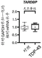
Fig. 2A-2J show the familial ALS model. Figure 2A provides a schematic of the strategy used to assess gene expression in iPS cell-derived hmns expressing mutant TDP-43. Figure 2B provides photomicrographs showing the neuronal morphology of iPS cells derived from healthy controls (11a, 18a, 20B, 17a) and patients with TARDP mutations (+/Q343R, + G298S, + a315T, and +/M337V) cultured for 10 days. FIGS. 2C-2H provide a qRT-PCR analysis of genes that were either continuously downregulated (FIGS. 2D-2F) or upregulated (FIG. 2C) after TDP-43 knockdown in neurons differentiated from control or TDP-43 patients. (unpaired t-test, P value < 0.05). Figure 2I provides representative micrographs of control and patient neurons immunostained against TDP-43 (red), β -III tubulin (green) and counterstained with DAPI (blue). Scale bar, 100 μm. FIG. 2J provides a Pearson correlation analysis of TDP-43 immunostaining and DAPI fluorescence comparing control neurons to neurons with TDP-43 mutations. Dots represent single cells. (unpaired t-test, P value < 0.05).
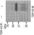
Figures 3A-3I show STMN2 adjustment and positioning. Figure 3A provides qRT-PCR analysis of STMN2 transcripts in independent experiments using two different sets of primer pairs. (unpaired t-test, P value < 0.05). Figure 3B provides an immunoblot analysis of TDP-43 and STMN2 protein levels after partial depletion of TDP-43 by siRNA knockdown. Protein levels were normalized to GAPDH and expressed relative to the levels in MN treated with the siRED control. FIG. 3C provides a qRT-PCR analysis for the analysis of STMN2 transcripts in GFP + MN against Hb9 treated with siRNAs targeting three ALS linked genes (TDP-43, FUS and C9ORF 72). (Dunnett multiple comparison test, alpha value < 0.05). FIGS. 3D-3F show formaldehyde RNA immunoprecipitation for identifying transcripts that bind to TDP-43. After TDP-43 immunoprecipitation (fig. 3D), enrichment of TDP-43 transcripts (fig. 3E) and STMN2 transcripts (fig. 3F) relative to sample input was tested using qRT-PCR analysis. FIG. 3G provides a micrograph of Hb9 immunostained for TDP-43 (red), β -III tubulin (green) and counterstained with DAPI (blue). FIG. 3H provides photomicrographs of GFP + MN co-cultured with Hb9 immunostained against STMN2 (red) and MAP2 green and GOLGIN97 (green) on glia. FIG. 3I provides photomicrographs of Hb9, which was immunostained 3 days after sorting against STMN2 (red), MAP2 (green) and counterstained with F-actin binding protein phalloidin (white). Scale bar, 5 μm.
Figures 4A-4K show STMN2 knockouts. Figure 4A provides a schematic of a knockout strategy using guide rnas (grnas) targeting two component exons of the human STMN2 gene, namely exons 2 and 4. The intervening DNA segment (-18 Kb) was targeted and deleted due to NHEJ (non-homologous end joining) repair of two Double Strand Breaks (DSBs) introduced by the Cas9/gRNA nuclease complex. FIGS. 4B-4D show that STMN2 knockdown was confirmed in HUES3 Hb9:: GFP line by RT-PCR analysis of genomic DNA (FIG. 4B), by immunoblot analysis (FIG. 4C) and by immunofluorescence (FIG. 4D). Figure 4E provides an experimental strategy for assessing the effect of lack of STMN2 in hMN on cells. FIGS. 4F-4H show the Sholl analysis of hMN with and without STMN2 and in the absence (FIG. 4G) or presence (FIG. 4H) of a ROCK inhibitor (Y-27632, 10. mu.M) for stimulation of neurite outgrowth. (unpaired t-test, P value < 0.05). Figure 4I provides an experimental strategy for assessing the effect of the absence of STMN2 in hmns on cells following axonal injury. FIGS. 4J-4K show axonal regrowth after injury. Representative photomicrographs of hmns in microfluidic devices before and after axotomy (fig. 4J). Measurement of regeneration of the axon after the axon cutting operation. (unpaired t-test, P value < 0.05).
Figures 5A-5G illustrate a sporadic ALS model. Figure 5A provides an experimental strategy for assessing the effect of proteasome inhibition on TDP-43 localization in human motor neurons. FIG. 5B shows a Pearson correlation analysis of TDP-43 immunostaining and DAPI fluorescence for cells treated with MG-132(1 μ M). (Dunnett multiple comparison test, alpha value < 0.05). FIG. 5C provides a micrograph of HUES3 motor neurons untreated or treated with MG-132 and immunostained against TDP-43 (red), β -III tubulin (green) and counterstained with DAPI (blue). Scale bar, 100 μm. Figure 5D provides an immunoblot analysis of TDP-43 in detergent soluble (RIPA) and detergent insoluble (UREA) fractions in neurons treated with MG-132 (unpaired t-test, P value < 0.05). Figure 5E provides a qRT-PCR analysis of STMN2 expression of motor neurons treated with MG-132 at indicated concentrations and durations relative to DMSO control (unpaired t-test, P value < 0.05). FIG. 5F provides a schematic representation of the RT-PCR assay strategy for the cryptic exons of STMN 2. FIG. 5G provides a tape machine analysis of STMN2 cryptic exons in hMN control cells treated with MG-132 (1. mu.M).
ALS patient data are presented in fig. 6A-6H. Figures 6A-6C provide histological analysis of adult lumbar spinal cord from post-mortem samples collected from subjects without evidence of spinal cord disease (control) (figure 6A) or two patients diagnosed with sporadic ALS (figures 6B-6C). Immunoreactivity to STMN2 was detected in the perinuclear region of spinal cord motor neurons (indicated by arrows), but not in peripheral glial cells. The STMN2 immunoreactivity in lumbar spinal motoneurons from control and ALS cases was scored as "strong" [ as shown by the arrows in control (fig. 6A) and sporadic ALS (fig. 6B) ] or "absent" [ as shown by the arrows in sporadic ALS (fig. 6C) ]. Scale bar, 50 μm. Figure 6D shows that the percentage of lumbar spinal cord motor neurons with strong STMN2 immunoreactivity was significantly lower in ALS tissue samples (n ═ 3 control and 3 ALS cases; approximately 40 MNs scored per subject; two-tailed t-test, P value < 0.05). Fig. 6E-6G show gene expression analysis of STMN2 from previously published datasets, Rabin et al 2009 (fig. 6E), Highley et al 2014 (fig. 6F), and D' Erchia et al 2017 (two-tailed t-test, P-value < 0.05). Figure 6H provides a molecular model of the pathogenesis of ALS.
FIGS. 7A-7I show the generation of differentiated human motor neurons. Figure 7A shows hMN differentiation, purification, and culture strategies. FIG. 7B provides flow cytometric analysis of differentiated HUES3 Hb9 GFP cells. Cells not treated with RA and SHH pathway agonists were used as a negative control for GFP expression gating. Fig. 7C-7F provide micrographs and quantification of purified HB9:: GFP + cells immunostained against HB9 and counterstained with DAPI (fig. 7C) (scale bar 10 μm) or against ISL1 and the neuronal markers β -III tubulin and MAP2 (fig. 7E) (scale bar 20 μm). Figures 7G-7J show that differentiated MN is electrophysiological active as determined by whole cell patch clamp recordings. Figure 7G shows that after voltage clamp mode depolarization, cells exhibit a rapid inward current followed by a slow outward current, indicating the expression and opening of voltage-activated sodium and potassium channels, respectively. Figure 7H shows that in current clamp mode depolarization causes repetitive action potential firing. FIG. 7I shows that the response to kainic acid is consistent with the expression of functional receptors for excitatory glutamate transmitters.
FIGS. 8A-8E show TDP-43 knockdown in cultured hMN. Figure 8A provides an RNAi strategy for TDP-43 knockdown in cultured MNs. FIG. 8B shows phase and red fluorescence micrographs of cultured hMN after 4 days of treatment with different siRNAs including scrambled siRNA conjugated to Alexa Fluor 555. FIG. 8C provides flow cytometric analysis of hMN after treatment with different siRNA. FIG. 8D shows the relative levels of TDP-43mRNA in MN exposed to different siRNAs for 2,4 or 6 days. The levels of each sample were normalized to GAPDH and expressed relative to the level of the non-transfected control. Figure 8E provides an immunoblot analysis of hmns after treatment of RNAi with indicated sirnas. Each sample was normalized using GAPDH and TDP-43 protein levels were calculated relative to the siSCR _555 treated control sample.
FIGS. 9A-9C show motor neuron RNA-Seq. Fig. 9A shows a global transcriptional analysis of motor neurons processed as indicated, represented as a heat map. Unsupervised clustering of expression profiles revealed that samples were isolated based on batches of motor neuron generation and analysis. FIG. 9B provides an analysis of TDP-43 transcript abundance after RNA sequencing validation knockdown (Benjamini-Hochberg adjusted P-cut of 0.05). Figure 9C shows that alterations in the splicing pattern of the POLDIP3 gene were detected due to TDP-43 knockdown, where siTDP43 treated cells showed a significant decrease in isoform 1 and increased levels of splice variant 2 (lacking exon 3) (false discovery rate 'FDR' > 0.05).
Figure 10 shows the indicated pluripotent stem cell genotyping sequencing chromatogram for TARDBP exon 6 in iPS cell line, used to confirm heterozygous mutations in patient lines.
FIGS. 11A-11F show neuronal cell sorting. Figure 11A shows that using cell surface marker screening, antibodies enriched on GFP + motor neurons (quadrant 1) and GFP cells (quadrant 3) were identified. Figure 11B shows that after sorting for NCAM + and EpCAM-cells, high content imaging was used to determine whether this sorting method could deplete cultures of mitotic cells (EdU +) and significantly enrich for motor neurons (Isl1+) and neurons (MAP2 +). N-6 different iPS cell lines. Statistical analysis was performed using a two-tailed student's t-test. Fig. 11C-11D provide qRT-PCR analysis of the cultures after sorting for the motor neuron marker ISL1 (fig. 11C) and the neuron marker β III-tubulin (fig. 11D), revealing that the cultures are enriched and more homogenous compared to unsorted cultures. Figure 11E provides flow cytometric analysis of cultures differentiated from indicated healthy controls (grey) and TDP-43 mutant lines (red) with Phycoerythrin (PE) -conjugated EpCAM antibody (anti-EpCAM-PE) and Alexa Fluor 700-conjugated NCAM antibody (anti-NCAM-AF 700). Figure 11F shows the percentage of NCAM + cells from 4-6 independently differentiated indicated lines. No significant difference was observed between the mutant and control lines in terms of the ability to generate NCAM + cells. Statistical analysis was performed using a two-tailed student's t-test with P-value < 0.05.
Figures 12A-12G illustrate the association of TDP-43 and STMN 2. Figures 12A-12C provide qRT-PCR validation of ALS gene down-regulation after siRNA treatment. Expression of TDP-43 (fig. 12A), FUS (fig. 12B) and C9ORF72 (fig. 12C) was evaluated against all controls and each siRNA used (unpaired t test, P value < 0.05). Figure 12D provides western blot analysis of STMN2 protein in different types of cells differentiated along motor neurons. FIG. 12E shows the expression levels of RNA-Seq of Stathmin family in motor neurons treated with siSCR (-) or siTDP-43(+) oligonucleotides. After TDP-43 knockdown, only STMN2 levels were altered. FIGS. 12F-12G show the TDP-43 binding site within the Stathmin gene family (FIG. 12F), normalized to gene length (FIG. 12G). STMN2 has the most number of binding motifs.
Figures 13A-13H show that STMN2 regulates neuronal outgrowth. CRISPR-mediated STMN2 knockdown in WA01 line WAs confirmed by RT-PCR analysis of genomic DNA (fig. 13A), by immunoblot analysis (fig. 13B), and by immunofluorescence (fig. 13C). FIGS. 13D-13F provide a Sholl analysis of hMN (unpaired t-test, P value <0.05) with and without STMN2 and in the presence of ROCK inhibitor Y-27632 (10. mu.M) (FIG. 13F). FIGS. 13G-13H show regrowth of axons after injury. Representative photomicrographs of hmns in the microfluidic device before and after axotomy (fig. 13G). Analysis of axonal regrowth after axonal dissection (unpaired t-test, P value <0.05) (fig. 13H).
FIGS. 14A-14E show cell survival and proteasome activity assays. FIGS. 14A-14C show that Cell Titer Glo uses ATP from metabolically active cells to generate light. (FIG. 14A) shows that there is a direct relationship between luminescence and the number of cells in culture that exceeds several orders of magnitude. Figure 14B shows that this assay can detect differences in neuronal survival in the absence of growth factors. N ═ 6 independent neuronal wells. (unpaired t-test, P value < 0.05). FIG. 14C shows a summary of MG-132 neuron survival experiments. FIG. 14D shows a dose response curve for motor neurons incubated with the indicated concentrations of MG-132 for the indicated times. Triplicate wells were used. After 1 day treatment at all concentrations tested, cells were viable and lower concentrations were tolerated for longer periods of time. Fig. 14E shows that upon cleavage by the proteasome, the substrate of luciferase is released, allowing quantitative measurement of proteasome activity. Neurons treated with MG-132 showed a significant reduction in proteasome activity. N-4 independent neuronal wells (unpaired t-test, P value < 0.05).
FIGS. 15A-15E show that TDP-43 modulates cryptic exon splicing in hMN (FIGS. 15A-15C). Cryptic exon visualization of PFKP (fig. 15A), ELAVL3 (fig. 15B), and STMN2 (fig. 15C) of cells treated with scrambled siRNA or siRNA targeting TDP-43 transcript. Read coverage and splice junctions are shown for alignment with the human HG19 genome. Figures 15D-15E provide a strategy diagram for RT-PCR detection of cryptic exons of STMN2 (figure 15D), and Sanger sequencing of the PCR products that confirmed splicing of STMN2 exon 1 to cryptic exons (figure 15E).
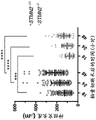
Figures 16A-16P provide qPCR data for cryptic STMN2 transcripts from patient cerebrospinal fluid (CSF) samples. Figures 16A-16D provide graphs summarizing patient sample data for normalized masked STMN2 relative to healthy controls. Figures 16E-16M provide graphs giving details about individual patient samples. Fig. 16N provides a graph showing the duration of survival after diagnosis. Fig. 16O provides a chart showing the age of death. Fig. 16P provides a graph demonstrating lung capacity.
Figures 17A-17C show a multiplex qPCR assay for STMN 2. Figure 17A shows a Q-RT PCT assay for STMN2 in a fluid. Experimental protocols are provided and demonstrate that the STMN2 multiplex TaqMan assay can simultaneously detect cryptic STMN2, normal STMN2 transcript and housekeeping gene RNA18S 5. RNA can be collected from CSF-derived exosomes and then converted to cDNA to determine the intact and cryptic STMN2 transcript and control RNA for normalization. Figure 17B shows in vitro validation of multiplex assays in cells with ASO or with siRNA to reduce TDP-43 levels. Figure 17C shows that the STMN2 multiplex qPCR assay was used to detect cryptic STMN2 transcript levels in cDNA samples generated from MGH CSF samples. Cryptic splicing of STMN2 was significantly induced in ALS patients.
Figures 18A-18D show a sandwich ELISA for detecting STMN2 protein. Figure 18A provides a schematic of the STMN2 sandwich ELISA. Figure 18B shows the sensitivity of STMN2 ELISA to picogram quantities. Figure 18C shows that a sandwich ELISA was validated using recombinant STMN2 protein and was able to detect picogram levels of STMN 2. Figure 18D shows that the level of STMN2 in the cerebrospinal fluid (CSF) of patients was reduced when assessed using the STMN2 ELISA.
Figure 19 provides a chart showing the genetics of ALS, each gene plotted according to the year it was found. See Alsultan et al, Degenerative Neurological and Neurousaral diseases.2016, 6, 49-64.
FIG. 20 shows that TDP-43 is a multifunctional nucleic acid binding protein. TDP-43 has been shown to play a role in a variety of functions including RNA splicing, miRNA processing, autoregulation of its own transcript, RNA transport and stability, and stress particle formation. The transcripts of TDP-43 regulators are highly species and cell type dependent. See Buratti and Baralle Trends in biochem. sci..2012,6, 237-.
FIG. 21 provides a strategy for measuring the transcriptional impact of TDP-43 depletion. The schematic shows the differentiation, purification and culture strategies of hmns. This strategy uses small molecules that mimic early development to convert stem cells into post-mitotic neurons within 2 weeks. Various methods have been developed to sort and study neurons. siRNA technology in combination with RNA sequencing was used to identify transcripts regulated by TDP-43.
Figure 22 shows TDP-43 in combination with STMN 2. STMN2 staining was performed on the spinal cord of ALS patients, and reduction of STMN2 protein was observed in ALS patients based on fold enrichment relative to PGK1 (fpip). See Klim et al Nature Neuroscience Vol.22, pp.167-179 (2019).
FIG. 23 shows splicing changes following TDP-43 depletion. Differential exon usage analysis was performed on RNA-seq samples from motor neurons treated with siTDP. Splice changes were observed in STMN 2.
FIG. 24 shows that TDP-43 suppresses cryptic exons in STMN 2. An integrated genome viewer was used to see where the RNA seq reads mapped to the human genome (top graph of reads) and how the reads reconnected between exons (splicing traces). The graph shows the number of reads mapped to the gene region.
Figure 25 provides a summary of the splicing defects of STMN 2. Under normal conditions, STMN2 was transcribed with all 5 exons, producing mRNA that was translated into a 20kDa STMN2 protein. Following TDP-43 perturbation, the cryptic exon intercepts the transcript so that only a 17 amino acid polypeptide can be translated.
Figure 26 shows that STMN2 continued to decrease. The overlap of reduced transcripts in 3 human RNA seq datasets (ALS patient dataset and siTDP43 stem cell motor neuron dataset) was compared and STMN2 was the only reduced transcript in all three datasets.
Figure 27 shows the presence of the STMN2 cryptic exon in the spinal cord of ALS patients. Read coverage and splice junctions are shown for alignment with the human HG19 genome. Reads mapped to the human genome in ALS patients were observed, and for 5 out of 6 patients, reads mapped to and spliced into cryptic exons, while controls did not.
FIG. 28 shows that TDP-43 depletion results in neurite outgrowth and axonal regrowth defects. Representative micrographs of hmns treated with indicated siRNA and immunostained against β -III tubulin for Sholl analysis are provided. Sholl analysis of hmns after siRNA treatment is provided. The line represents the sample mean and the shading represents s.e.m., double sided, P <0.05 for unpaired t-tests between siTDP43 and sitcr.
Figure 29 shows a microfluidic device for exploring axonal regeneration. The microfluidic device comprises a somatic cell compartment (left panel) and a axon compartment (right panel).
FIGS. 30A-30B show that TDP-43 depletion results in neurite outgrowth and axonal regrowth defects. Figure 30A provides representative photomicrographs of hMN in the microfluidic device after axotomy. Scale bar, 150. mu.M. FIG. 30B provides measurements of axonal regrowth and regeneration after axonal cutting (unpaired t-test, two-sided, P < 0.0518 h ≦ 0.0001, 24h ≦ 0.0001, 48 ≦ 0.0001, and 72 ≦ 0.0001).
Figure 31 shows that STMN2 is a JNK target in the axonal degeneration pathway. JNK1 was shown to bind to and phosphorylate STMN2, and phosphorylated STMN2 was rapidly degraded. See J.Eun Shin et al PNAS 2012,109, E3696-3705.
Fig. 32 provides a strategy to determine whether JNKi can rescue the siTDP43 phenotype. See Klim et al Nature Neuroscience Vol.22, pp.167-179 (2019).
Figure 33 shows that JNK inhibitor (SP600125) increased STMN2 levels. STMN2 protein levels were increased in neurons treated with JNKi, while the lower levels observed in cells treated with siTDP43 could be rescued.
Fig. 34 shows that JNKi (SP600125) increases neurite outgrowth. Cells treated with JNKi showed increased neurite branching.
Fig. 35 shows that JNKi (SP600125) increases neurite outgrowth. Sholl analysis confirmed that JNKi increased neurite branching and regrowth following injury under all conditions.
Fig. 36 shows that JNKi increases axonal regeneration. The microfluidic device demonstrated that JNKi increased neurite branching and regrowth following injury under all conditions.
Fig. 37 provides a model for proteasome inhibition. Disruption of protein homeostasis leads to TDP-43 mislocalization and altered levels of STMN2, which disrupts axonal biology.
FIGS. 38A-38B illustrate TDP-43 positioning. TDP-43 is usually nuclear (FIG. 38A), but after compound washing, a significant loss of nuclear TDP-43 staining was observed (FIG. 38B). No cytoplasmic aggregation was observed, only nuclear TDP-43 loss.
FIG. 39 shows that TDP-43 mispositioning is reversible.
FIG. 40 shows that STMN2 transcript decreased after TDP-43 mislocalization. The reduction in STMN2 was even more pronounced than in cells expressing mutant TDP-43.
Figure 41 provides a table summarizing the latest ALS genes and their relative mutation frequencies and associated pathways in different ALS and FTD cohorts. The evolution of WGS and WES led to the identification of genes carrying the following rare causal variants: TBK1, chchchhd 10, TUBA4A, MATR3, CCNF, NEK1, C21orf2, ANXA11, and TIA 1. TBK1 was shown to have the highest ALS-FTD mutation frequency (3-4%) in the different cohorts. See Nguyen et al, Trends in Genetics, 2018.
Fig. 42 shows that Atg7 and TBK1 act at different times in autophagy. See Hansen et al, Nature Reviews Molecular Cell biology.2018.
Fig. 43 shows that elimination of TBK1 shares similarities with, but differs from, blocking autophagy initiation.
Figure 44 shows that TBK1 knockout reduced functional TDP-43 and STMN2 levels, while elimination of ATG7 had no effect. Loss of TBK1 induces TDP-43 pathology in motor neurons by a mechanism independent of autophagy.
Figure 45 shows that loss of TBK1 indicates impaired axon regeneration following axon injury.
Figure 46 shows the mislocalization of proteasome inhibition induced TDP-43 in TBK1 mutant motor neurons.
Fig. 47A-47C show targeting of STMN2 intron using CRISPR. CRISPR strategies for targeting STMN2 are provided, as well as genotyping of STMN2 (fig. 47A-47B). Fig. 47C provides a table summarizing CRISPR targeting strategies and genotyping of STMN 2.
Figure 48 demonstrates that STMN2 mice were significantly smaller than Rosa26 control mice and showed defects in the motor performance task that were not evidence of progression over time.
Figure 49 demonstrates that STMN2 mice were significantly smaller than Rosa26 control mice and showed defects in the motor performance task that were not evidence of progression over time.
Figure 50 shows that the behavioral results and the total distance traveled in the open field assay appear similar between the two mouse cohorts.
Figure 51 demonstrates that STMN2 transcript levels were significantly reduced or absent in brain tissues in the mutation cohort.
Fig. 52 provides a western blot of brain tissue demonstrating loss or significant reduction of STMN2 protein in the mutant mouse cohort.
Figure 53 shows that STMN2 is predominantly localized in ChAT + motor neurons in the ventral horn of the spinal cord of adult mice.
Figure 54 shows that the STMN2 cohort showed a significant reduction in the number of STMN2+/ChAT + motor neurons on the ventral horn of the spinal cord.
Figure 55 provides a graph giving the difference in organ or muscle weight between control and STMN2 mice. Lower limb muscle weakness was confirmed in STMN2 mice (see two boxed graphs).
Figure 56 provides pre-and post-synaptic staining of STMN2 Gastrocnemius (GA) muscle and Rosa26 control Gastrocnemius (GA) muscle. The staining revealed denervation in STMN 2-/-animals.
Figure 57 shows presynaptic and postsynaptic staining of STMN2 Gastrocnemius (GA) muscle and Rosa26 control Gastrocnemius (GA) muscle revealed denervation in STMN 2-/-animals.
Figure 58 shows that the muscle nerve junction (NMJ) morphology supports active denervation in gastrocnemius of the STMN2 mutant.
FIG. 59 shows that mutant TDP-43 does not exhibit pathological mispositioning. Staining of neurons in both control and ALS patients with TDP-43 showed that neurons in both control and ALS patients had TDP-43 predominantly nuclear.
Figure 60 identifies different classes of proteasome inhibitors and provides their chemical structures.
Figure 61 shows the reduction in expression of full-length STMN2 in hmns after treatment with structurally different proteasome inhibitors.
FIG. 62 shows PCR assays of hMN treated with MG-132 or Bortezomib (Bortezomib). Full-length STMN2 was detected in all samples as controls. The presence of transcripts containing cryptic exons of STMN2 was specific for those cells treated with proteasome inhibitors.
FIGS. 63A-63B show in vitro assays for TDP-43 binding to STMN2 RNA. RNA containing the TDP-43 binding site from the cryptic exon region of STMN2 was transcribed in vitro using genomic DNA (fig. 63A). RNA was used to assess whether it could pull down the IP TDP-43 protein from human neuronal protein lysates. In vitro assays showed that transcripts containing cryptic exon regions pulled down TDP-43 (FIG. 63B).
FIG. 64 shows an in vitro assay for TDP-43 binding to STMN2 RNA. Similar to that described in FIG. 63, RNAs containing 5 'and 3' TDP-43 binding regions were transcribed in vitro. Although both the 5 'and 3' transcripts could pull down some of TDP-43, the enrichment was not as strong as the intact cryptic exon.
Fig. 65 shows the design of grnas used to generate targeted mutant cell lines without cryptic exons. A strategy was prepared to delete 105 nucleotides within the cryptic exon within the STMN2 intron, between exons 1 and 2. This deletion will eliminate the TDP-43 binding motif, but will not affect the predicted polyadenylation site.
Figure 66 provides a confirmation of the mutant status. The mutation status of the clones was analyzed using the TIDE assay and sequence alignment to control cells was examined to obtain a more accurate view of deletion size and location. One cell line contained a homozygous deletion of 105 nucleotides, consistent with gel electrophoresis. This deletion eliminates the TDP-43 binding motif, but does not affect the predicted polyadenylation site.
FIG. 67 shows that the TDP-43 binding site is a potential negative regulator of STMN2 expression. Three cell lines, HUES3, IG2(Stmn2 KO) and CN7 (cryptic exon deletion) were treated with normal medium or medium +1uM MG132 for 24 hours to stress the cells. In HUES3 cells, stressed conditions had 52% STMN2 mRNA expression compared to unstressed conditions. Unstressed cells had 13% expression under IG2(Stmn2 KO) conditions and had increased expression to 42% when stressed. The expression level in CN7 (cryptic exon deletion) cell line was significantly higher than the other two cell lines, with 729% expression in non-stressed cells and 473% expression in stressed cells. It has been demonstrated that expression is decreased if several exons are knocked out, but increased if the TDP-43 binding site is removed.
FIGS. 68A-68B show that deletion of the putative TDP-43 binding site results in an increase in STMN2 protein levels. Consistent with the gene expression data, deletion of the TDP-43 binding region within the cryptic exon of STMN2 resulted in increased protein expression.
FIGS. 69A-69B show the effectiveness of antisense oligonucleotides (ASOs) (SEQ ID NO: 11). FIG. 69A shows the application of 2.5. mu.M ASO and the evaluation of its ability to reduce the abundance of cryptic exon containing transcripts. Figure 69B shows the application of 2.5 μ M ASO and evaluated its ability to increase the abundance of full-length STMN2 transcripts during TDP-43 deletion.
FIGS. 70A-70B show the conservation of the STMN2 locus between different species. The complete triplet (red) of the TDP-43 binding motif is conserved in apes.
Detailed Description
Mislocalization or depletion of the RNA binding protein TDP-43 results in decreased expression of STMN2 encoding a microtubule regulatory factor. STMN2 is critical for normal axonal outgrowth and regeneration. Reduced TDP-43 function results in an inefficient or altered STMN2RNA sequence, resulting in reduced expression of STMN2 protein. STMN2 may be a promising therapeutic target and a biomarker of disease risk (e.g., neurodegenerative disease).
The work described herein relates to compositions and methods for suppressing or preventing cryptic exons from being contained in STMN2 mRNA. The inclusion of cryptic exons in STMN2 mRNA results in the production of truncated transcripts and proteins. In some aspects, inclusion of cryptic exons results in early polyadenylation. STMN2 expression may be restored by repressing the cryptic splice form of STMN2 that occurs when TDP-43 is sequestered or the functionality of TDP-43 is reduced, which may be accomplished, for example, by blocking the appearance or accumulation of the cryptic form and switching it back to or restoring functional STMN2RNA (e.g., by administering a pharmaceutical agent).
Medicament and pharmaceutical composition
The present disclosure contemplates agents that bind to null or altered STMN2RNA sequences that appear and increase in abundance when TDP-43 function is decreased or TDP pathology occurs, thereby restoring expression of normal full-length or protein-encoding STMN2 RNA. In some aspects, the agent prevents degradation of STMN2 protein. In some aspects, the agent restores STMN2 protein levels. In some aspects, the agent suppresses or prevents the cryptic exon from being comprised in STMN2 RNA. In some embodiments, the agent specifically binds to STMN2 mRNA, pre-mRNA, or nascent RNA sequence encoding a cryptic exon.
In some embodiments, the agent binds to an STMN2RNA sequence (e.g., a null or altered STMN2RNA sequence). In some aspects, binding of the agent to the short null or altered STMN2RNA sequence results in sustained production of RNA polymerase. For example, the agent may directly suppress premature transcription termination at the polyadenylation site of the cryptic exon, or may mimic the activity of TDP-43 binding at its target site, thereby altering transcription termination at the cryptic exon. In some aspects, the agent suppresses or prevents the cryptic exon from being comprised in STMN2 RNA. In some aspects, the agent prevents degradation of STMN2 protein. In some aspects, the agent increases STMN2 levels (e.g., by exon skipping). In some embodiments, the agent restores STMN2RNA (e.g., pre-mRNA or mRNA) of normal length or encoding a protein. In some aspects, the agent increases the amount or activity of STMN2 RNA.
In some embodiments, the agent targets one or more sites, such as the 5 'splice site, the 3' splice site, the normal binding site, and/or the polyadenylation site of the STMN2 transcript. In certain embodiments, the agent targets one or more sites including the 5' TDP-43 splice site, the TDP-43 normal binding site, and/or a cryptic polyadenylation site. In some embodiments, the agent does not target or bind to a polyadenylation site. In some embodiments, the agent does not target or bind to the polyadenylation site of the STMN2 transcript. In some embodiments, the agent does not target or bind to cryptic polyadenylation sites. In some aspects, the agent targets and facilitates splicing of STMN2 exon 2 to exon 1.
AGCTCCTAGGAAGCTTCAGGGCTTAAAGCTCCACTCTACTTGGACTGTACTATCAGGCCCCCAAAATGGGGGGAGCCGACAGGGAAGGACTGATTTCCATTTCAAACTGCATTCTGGTACTTTGTACTCCAGCACCATTGGCCGATCAATATTTAATGCTTGGAGATTCTGACTCTGCGGGAGTCATGTCAGGGGACCTTGGGAGCCAATCTGCTTGAGCTTCTGAGTGATAATTATTCATGGGCTCCTGCCTCTTGCTCTTTCTCTAGCACGGTCCCACTCTGCAGACTCAGTGCCTTATTCAGTCTTCTCTCTCGCTCTCTCCGCTGCTGTAGCCGGACCCTTTGCCTTCGCCACTGCTCAGCGTCTGCACATCCCTACAATGGCTAAAACAGCAATGGGACTCGGCAGAAGACCTTCGAGAGAAAGGTAGAAAATAAGAATTTGGCTCTCTGTGTGAGCATGTGTGCGTGTGTGCGAGAGAGAGAGACAGACAGCCTGCCTAAGAAGAAATGAATGTGAATGCGGCTTGTGGCACAGTTGACAAGGATGATAAATCAATAATGCAAGCTTACTATCATTTATGAATAGC(SEQ ID NO:1)。
CCTACAAGGAAAAAATGAAGGAGCTGTCCATGCTGTCACTGATCTGCTCTTGCTTTTACCCGGAACCTCGCAACATCAACATCTATACTTACGATGG(SEQ ID NO:2)。
cryptic exons may have the following sequences:
GACTCGGCAGAAGACCTTCGAGAGAAAGGTAGAAAATAAGAATTTGGCTCTCTGTGTGAGCATGTGTGCGTGTGTGCGAGAGAGAGAGACAGACAGCCTGCCTAAGAAGAAATGAATGTGAATGCGGCTTGTGGCACAGTTGACAAGGATGATAAATCAATAATGCAAGCTTACTATCATTTATGAATAGC(SEQ ID NO:3)。
exemplary types of agents that can be used include small organic or inorganic molecules; saccharin; an oligosaccharide; a polysaccharide; a biological macromolecule selected from the group consisting of peptides, proteins, peptide analogs, and derivatives; a peptide mimetic; a nucleic acid selected from the group consisting of siRNA, shRNA, antisense RNA, ribozyme, and aptamer; an extract made from a biological material selected from the group consisting of bacteria, plants, fungi, animal cells and animal tissues; naturally occurring or synthetic compositions; an antibody; and any combination thereof.
In some embodiments, the agent is an oligonucleotide, a protein, or a small molecule. In some embodiments, the agent comprises one or more oligonucleotides. In some aspects, the oligonucleotide is a splice switching oligonucleotide. In certain aspects, the oligonucleotide is an antisense oligonucleotide (ASO). In some embodiments, the agent is an antisense oligonucleotide. In some embodiments, the agent is a metabolic small molecule (e.g., branaplam (novartis) or risdiplam (roche)) capable of binding to a target site (e.g., STMN2 transcript) and transforming the target.
The agent may target one or more of a 5 'splice site, a 3' splice site, a normal binding site, or a polyadenylation site. In some aspects, the polyadenylation site is the polyadenylation site of the STMN2 transcript. In some aspects, the polyadenylation site is a cryptic exon polyadenylation site (e.g., is a cryptic polyadenylation site). In some embodiments, the agent does not target a 5 'splice site (e.g., TDP-435' splice site). In some embodiments, the agent does not target a normal binding site (e.g., a normal TDP-43 binding site). In some embodiments, the agent does not target a polyadenylation site (e.g., a cryptic polyadenylation site). In certain embodiments, the antisense oligonucleotide may target one or more of a 5 'splice site, a 3' splice site, a normal binding site, or a polyadenylation site. In some embodiments, the antisense oligonucleotide does not target a 5 'splice site (e.g., TDP-435' splice site). In some embodiments, the antisense oligonucleotide does not target a normal binding site (e.g., a normal TDP-43 binding site). In some embodiments, the antisense oligonucleotide does not target a polyadenylation site (e.g., a cryptic polyadenylation site). In certain embodiments, the antisense oligonucleotide comprises the sequence of TCTTCAGTATTGCTATTCAT (SEQ ID NO: 11).
Oligonucleotides (e.g., antisense oligonucleotides) can be designed to bind regions of mRNA that prevent ribosome assembly at the 5' cap, prevent polyadenylation during mRNA maturation, or affect splicing events (Bennett and Swayze, annu.rev.pharmacol.toxicol, 2010; Watts and Corey, j.patol, 2012; Kole et al, nat.rev.drug discov., 2012; Saleh et al, In Exon Skipping: Methods and Protocols,2012, each of which is incorporated herein by reference). In some aspects, oligonucleotides (e.g., antisense oligonucleotides) are designed to target one or more sites including, for example, a 5 'splice site, a 3' splice site, a normal binding site, and/or a polyadenylation site. In some aspects, the oligonucleotide targets one or more splice sites. In some aspects, the oligonucleotide targets one or more of a 5' TDP-43 splice site, a TDP-43 normal binding site, and/or a cryptic polyadenylation site. In some aspects, the oligonucleotide is designed to target one or more sites between exon 2 and exon 1 of STMN2 (e.g., an intron between exon 2 and exon 1). In some aspects, the oligonucleotide is designed not to target a cryptic polyadenylation site. In some aspects, the oligonucleotide is designed not to target the TDP-43 normal binding site. In some aspects, the oligonucleotide is designed not to target the 5' TDP-43 splice site.
Antisense oligonucleotides are small DNA sequences (e.g., about 8-50 base pairs in length) that are capable of targeting an RNA transcript by watson-crick base pairing, resulting in reduced or altered protein expression. Oligonucleotides are composed of a phosphate backbone and sugar rings. In some embodiments, the oligonucleotide is unmodified. In other embodiments, the oligonucleotide includes one or more modifications, for example, to improve the solubility, binding, potency, and/or stability of the antisense oligonucleotide. The modified oligonucleotide may comprise at least one modification relative to unmodified RNA or DNA. In some embodiments, the oligonucleotide is modified to comprise an internucleoside linkage modification, a sugar modification, and/or a nucleobase modification. Examples of such modifications are known to those skilled in the art.
In some embodiments, the oligonucleotides are modified by replacing at least one nucleotide with a modified nucleotide such that in vivo stability is increased as compared to a corresponding unmodified oligonucleotide. In some aspects, the modified nucleotide is a sugar-modified nucleotide. In another aspect, the modified nucleotide is a nucleobase modified nucleotide.
In some embodiments, the oligonucleotide may contain at least one modified nucleotide analog. The nucleotide analogue may be located at a position where the target specific activity, e.g. the splice site selection modulating activity, is substantially unaffected, e.g. in the region at the 5 '-terminus and/or the 3' -terminus of the oligonucleotide molecule. In some aspects, the termini can be stabilized by incorporating modified nucleotide analogs.
In some aspects, preferred nucleotide analogs include sugar and/or backbone modified ribonucleotides (i.e., comprising a modification to the phosphate-sugar backbone). For example, the phosphodiester linkage of the ribonucleotide may be modified to include at least one of a nitrogen or sulfur heteroatom. In preferred backbone-modified ribonucleotides, the phosphate group attached to the adjacent ribonucleotide is replaced by a modified group, such as a phosphorothioate group. In preferred sugar-modified ribonucleotides, the 2' -OH-group may be replaced by a group selected from H, OR, R, halogen, SH, SR, NH2, NHR, NR2 OR ON, wherein R is C1-C6 alkyl, alkenyl OR alkynyl and halogen is F, Cl, Br OR I.
In some embodiments, the modified oligonucleotide comprises one or more modified nucleosides containing a modified sugar moiety. In some embodiments, the modified oligonucleotide comprises one or more modified nucleosides containing a modified nucleobase. In some embodiments, the modified oligonucleotide comprises one or more modified internucleoside linkages. In certain embodiments, the modified oligonucleotide comprises at least two of: one or more modified nucleosides comprising a modified sugar moiety, one or more modified nucleosides comprising a modified nucleobase, and one or more modified internucleoside linkages. In certain embodiments, the modified oligonucleotide comprises: one or more modified nucleosides comprising a modified sugar moiety, one or more modified nucleosides comprising a modified nucleobase, and one or more modified internucleoside linkages.
Sugar modification
In some embodiments, the modified sugar moiety is a non-bicyclic modified sugar moiety. In some embodiments, the modified sugar moiety is a bicyclic or tricyclic sugar moiety. In some embodiments, the modified sugar moiety is a sugar substitute. Such sugar substitutes may comprise one or more substitutions corresponding to substitutions of other types of modified sugar moieties.
In some embodiments, the modified sugar moiety is a non-bicyclic modified sugar moiety comprising a furanosyl ring having one or more substituent groups, wherein none of the substituent groups bridges two atoms of the furanosyl ring to form a bicyclic structure. Such non-bridging substituents may be at any position of the furanosyl group, including but not limited to substituents at the 2', 4' and/or 5' positions. In certain embodiments, one or more of the non-bridging substituents of the non-bicyclic modified sugar moiety are branched.
In some embodiments, the modified sugar moiety comprises a substituent that bridges two atoms of the furanosyl ring to form a second ring, thereby producing a bicyclic sugar moiety. In some aspects, the bicyclic sugar moiety comprises a bridge between the 4 'furanose ring atom and the 2' furanose ring atom.
In some aspects, bicyclic sugar moieties and nucleosides incorporating such bicyclic sugar moieties are further defined by isomeric configuration. In some embodiments, the LNA nucleoside is in the α -L configuration. In some embodiments, the LNA nucleoside is in the β -D configuration.
In some embodiments, the oligonucleotide modification comprises Locked Nucleic Acids (LNAs), wherein a 2' -hydroxyl group is attached to a 3' or 4' carbon atom of the sugar ring, thereby forming a bicyclic sugar moiety. The linkage is preferably a methylene (-CH 2-) n group bridging a2 'oxygen atom and a 4' carbon atom, where n is 1or 2. LNAs and their preparation are described in WO 98/39352 and WO 99/14226, the entire contents of which are incorporated herein by reference.
In some embodiments, the modified sugar moiety comprises one or more non-bridging sugar substituents and one or more bridging sugar substituents (e.g., 5' -substituted sugars and 4' -2 ' bridging sugars).
In some embodiments, the modified sugar moiety is a sugar substitute. In some aspects, the oxygen atom of the sugar moiety is replaced with, for example, a sulfur, carbon, or nitrogen atom. In some aspects, such modified sugar moieties further comprise bridging and/or non-bridging substituents as described herein. In some aspects, the sugar substitute comprises a ring having other than 5 atoms. In certain aspects, the sugar substitute comprises a six-membered Tetrahydropyran (THP). In some aspects, the sugar substitute comprises an acyclic moiety.
Nucleobase modifications
The modified oligonucleotide may comprise one or more nucleosides containing an unmodified nucleobase. In some embodiments, the modified oligonucleotide comprises one or more nucleosides containing a modified nucleobase. In some embodiments, the modified oligonucleotide comprises one or more nucleosides that do not comprise a nucleobase.
In certain embodiments, the modified nucleobase is selected from: 5-substituted pyrimidines, 6-azapyrimidines, alkyl-or alkynyl-substituted pyrimidines, alkyl-substituted purines, and N-2, N-6, and 0-6 substituted purines. In certain embodiments, the modified nucleobase is selected from: 2-aminopropyladenine, 5-hydroxymethylcytosine, xanthine, hypoxanthine, 2-aminoadenine, 6-N-methylguanine, 6-N-methyladenine, 2-propyladenine, 2-thiouracil, 2-thiothymine and 2-thiocytosine, 5-propynyl (-C ℃ -C ]3/4) uracil, 5-propynyl cytosine, 6-azouracil, 6-azocytosine, 6-azothymine, 5-ribouracil (pseudouracil), 4-thiouracil, 8-halogen, 8-amino, 8-thiol, 8-thioalkyl, 8-hydroxy, 8-aza and other 8-substituted purines, 5-halo (especially 5-bromo), 5-trifluoromethyl, 5-and 5-halogenated uracil, 7-methyl guanine, 7-methyl adenine, 2-F-adenine, 2-amino adenine, 7-deazaguanine, 7-deazaadenine, 3-deazaguanine, 3-deazaadenine, 6-N-benzoyl adenine, 2-N-isobutyrylguanine, 4-N-benzoyl cytosine, 4-N-benzoyl uracil, 5-methyl 4-N-benzoyl cytosine, 5-methyl 4-N-benzoyl uracil, universal bases, hydrophobic bases, hybrid bases, size-enlarged bases and fluorinated bases. Additional modified nucleobases include tricyclic pyrimidines such as 1, 3-diazophenoxazin-2-one, 1, 3-diazophene-2-one, and 9- (2-aminoethoxy) -1, 3-diazophene-oxazin-2-one (G-clamp). Modified nucleobases may also include those in which the purine or pyrimidine base is replaced by other heterocycles such as 7-deaza-adenine, 7-deaza-guanine, 2-aminopyridine and 2-pyridone.
Also preferred are nucleobase-modified ribonucleotides, i.e. ribonucleotides comprising at least one non-naturally occurring nucleobase instead of a naturally occurring nucleobase. Examples of modified nucleobases include, but are not limited to, uridine and/or cytidine modifications at the 5-position, such as 5- (2-amino) propyl uridine, 5-bromouridine; adenosine and/or guanosine modified at the 8-position, such as 8-bromoguanosine; deaza nucleotides, for example, 7-deaza-adenosine; o-and N-alkylated nucleotides, such as N6-methyladenosine. The oligonucleotide agents of the invention may also be modified with chemical moieties that improve the in vivo pharmacological properties of the oligonucleotide agent.
Internucleoside modifications
In some embodiments, the nucleosides of the modified oligonucleotide are linked together using any internucleoside linkage. The linking group between the two broad classes of nucleosides is defined by the presence or absence of a phosphorus atom. Representative phosphorus-containing internucleoside linkages include, but are not limited to, phosphate esters (also known as unmodified or naturally occurring linkages) containing phosphodiesters ("P ═ O"), phosphotriesters, methylphosphonates, phosphoramidates and phosphorothioates ("P ═ S"), and phosphorodithioates ("HS-P ═ S"). Representative phosphorus-free internucleoside linking groups include, but are not limited to, methylenemethylimino (-CH)2-N(CH3)-O-CH2-), thiodiesters, thiocarbonylaminocarbamates (-O-C (═ O) (NH) -S-); siloxane (-O-SiH)2-O-); and N, N' -dimethylhydrazine (-CH)2-N(CH3)-N(CH3) -). Modified internucleoside linkages can be used to alter (typically increase) nuclease resistance of an oligonucleotide compared to naturally occurring phosphate linkages. In certain embodiments, the internucleoside linkages having chiral atoms can be prepared as a racemic mixture or as individual enantiomers. Methods for preparing phosphorus-containing and phosphorus-free internucleoside linkages are well known to those skilled in the art.
Additional modifications are known to those skilled in the art, and examples may be found in WO 2019/241648, US 10,307,434, US 9,045,518, and US 10,266,822, each of which is incorporated herein by reference.
The oligonucleotide may be of any size and/or chemical composition sufficient to target the null or altered STMN2 RNA. In some embodiments, the oligonucleotide is about 5-300 nucleotides or modified nucleotides. In some aspects, the oligonucleotide is about 10-100, 15-85, 20-70, 25-55, or 30-40 nucleotides or modified nucleotides. In certain aspects, the oligonucleotide is about 15-35, 15-20, 20-25, 25-30, or 30-35 nucleotides or modified nucleotides.
In some embodiments, the oligonucleotide and the target RNA sequence (e.g., null or altered STMN2 RNA) have 100% sequence complementarity. In some aspects, the oligonucleotide may comprise sequence variations, such as insertions, deletions, and single point mutations, relative to the target sequence. In some embodiments, the oligonucleotide has at least 70% sequence identity or complementarity to a target RNA (e.g., STMN2 mRNA, pre-mRNA, or nascent RNA). In certain embodiments, the oligonucleotide has at least 70%, 75%, 80%, 85%, 90%, 95%, 97%, 99%, or 100% sequence identity to the target sequence.
Antisense oligonucleotides targeting the null or altered STMN2RNA sequence can be designed by any method known to those skilled in the art. For example, antisense oligonucleotides can be synthesized as follows: the ratio of the two-dimensional matrix to the two-dimensional matrix was 52 MOErT/i 2 MOErC/i 2 MOErA/i 2 MOErG/i 2 MOErT/i 2 MOErA/i 2 MOErT/i 2 MOErG/i 2 MOErC/i 2 MOErT/i 2 MOErC/i 2 MOErT/i. In certain embodiments, the antisense oligonucleotide is synthesized as follows: 5' -/52 MOErT/i 2 MOErC/i 2 MOErA/i 2 MOErG/i 2 MOErT/i 2 MOErA/i 2 MOErT/i 2 MOErC/i 2 MOErT/i 2 MOErG/i 2 MOErC/i 2 MOErT/i 2 MOErA/i 2 MOErT/i 2 MOR. One or more oligonucleotides may be synthesized.
In some embodiments, STMN2 is administered as a gene therapy. In some embodiments, STMN2 is administered in combination with an agent described herein.
In some embodiments, the agent is an inhibitor of c-Jun N-terminal kinase (JNK). In some aspects, the JNK inhibitor is selected from: organic or inorganic small molecules; saccharin; an oligosaccharide; a polysaccharide; a biological macromolecule selected from the group consisting of peptides, proteins, peptide analogs, and derivatives; a peptide mimetic; a nucleic acid selected from the group consisting of siRNA, shRNA, antisense RNA, ribozyme, and aptamer; an extract made from a biological material selected from the group consisting of bacteria, plants, fungi, animal cells and animal tissues; naturally occurring or synthetic compositions; an antibody; and any combination thereof. In certain aspects, the agent is a small molecule inhibitor, an oligonucleotide (e.g., an oligonucleotide designed to reduce JNK expression), or gene therapy (e.g., gene therapy designed to inhibit JNK). In some aspects, inhibition of JNK restores or increases STMN2 protein levels. In certain embodiments, the agent is an oligonucleotide (e.g., an antisense oligonucleotide) that targets JNK.
The present disclosure further contemplates pharmaceutical compositions comprising agents that bind to an inoperative or altered STMN2RNA sequence. In some embodiments, the pharmaceutical composition comprises an agent that binds STMN2 mRNA, pre-mRNA, or nascent RNA sequence encoding a cryptic exon. In some embodiments, the pharmaceutical composition comprises an agent that prevents degradation of STMN2 protein. In some aspects, the composition comprises an oligonucleotide, a protein, or a small molecule. In some embodiments, the composition comprises an oligonucleotide (e.g., an antisense oligonucleotide), wherein the oligonucleotide specifically binds to a STMN2 mRNA, pre-mRNA, or nascent RNA sequence encoding a cryptic exon. In some aspects, the agent (e.g., oligonucleotide) suppresses or prevents the cryptic exon from being comprised in the STMN2 RNA. In some aspects, the agent suppresses cryptic splicing.
In some embodiments, the pharmaceutical composition comprises an agent that targets one or more sites, such as one or more splice sites, binding sites, or polyadenylation sites. In some embodiments, the pharmaceutical composition comprises an agent that targets one or more splice sites (e.g., the 5' TDP-43 splice site). In some embodiments, the pharmaceutical composition comprises an agent that targets a normal binding site (e.g., a TDP-43 normal binding site). In some embodiments, the pharmaceutical composition comprises an agent that targets a polyadenylation site (e.g., a cryptic polyadenylation site). In some embodiments, the pharmaceutical composition comprises an agent that does not target one or more splice sites (e.g., the 5' TDP-43 splice site). In some embodiments, the pharmaceutical composition comprises an agent that does not target a normal binding site (e.g., a TDP-43 normal binding site). In some embodiments, the pharmaceutical composition comprises an agent that does not target a polyadenylation site (e.g., a cryptic polyadenylation site).
In some embodiments, the pharmaceutical composition comprises an effective amount of an agent that binds to STMN2 mRNA sequences encoding cryptic exons and an effective amount of a second agent. In some aspects, the second agent is an agent that treats or inhibits a neurodegenerative disease. In some aspects, the second agent is an agent that treats or inhibits traumatic brain injury. In some aspects, the second agent is an agent that treats or inhibits proteasome inhibitor-induced neuropathy.
In some embodiments, the pharmaceutical composition comprises an effective amount of an agent that binds to a null or altered STMN2RNA sequence and an effective amount of STMN2 (e.g., administered as gene therapy).
In some embodiments, the pharmaceutical composition comprises an effective amount of a first agent that binds to a null or altered STMN2RNA sequence and an effective amount of a second agent that inhibits JNK.
In some embodiments, the pharmaceutical composition comprises an effective amount of an agent that binds STMN2 mRNA, pre-mRNA, or nascent RNA sequence encoding a cryptic exon, an effective amount of a second agent, and a pharmaceutically acceptable carrier, diluent, or excipient.
Compositions comprising agents that bind to null or altered STMN2RNA sequences are useful for treating diseases or conditions associated with decreased TDP-43 function or TDP pathology. In some aspects, compositions comprising agents that bind to null or altered STMN2RNA sequences are useful for treating diseases or conditions associated with mutated or reduced STMN2 protein levels (e.g., in neuronal cells) as described herein.
Method of treatment
The present disclosure contemplates various methods of treatment using compositions comprising agents that restore normal length or protein-encoding STMN2 RNA. In some aspects, the agent binds to an ineffective or altered STMN2RNA sequence that appears and increases in abundance when TDP-43 function is decreased or TDP pathology occurs, thereby restoring expression of normal full-length or protein-encoding STMN2 RNA. In some aspects, the agent suppresses or prevents the cryptic exon from being comprised in STMN2 RNA.
In some aspects, the present disclosure contemplates the treatment of any disease or condition, wherein the disease is associated with decreased TDP-43 function or TDP pathology. In some embodiments, the invention disclosed herein relates to methods of treating mutated or reduced levels of TDP-43 (e.g., a disease or condition having a TDP-43 associated pathology) in a neuronal cell. In some embodiments, the invention disclosed herein relates to methods of treating TDP-43 associated dementia (e.g., ALS, FTD, alzheimer's disease, parkinson's disease, or TBI).
In some embodiments, the invention disclosed herein relates to methods of treating a disease or condition associated with mutated, increased or decreased levels of TDP-43. In some embodiments, the invention disclosed herein relates to methods of treating a disease or condition associated with mislocalized TDP-43. In some embodiments, the invention disclosed herein relates to methods of treating diseases or conditions associated with mutated or reduced levels of STMN2 protein and/or incorrect localization of STMN2 protein. In some embodiments, the invention disclosed herein relates to methods of treating a disease or condition associated with proteasome inhibitor-induced neuropathy (e.g., neuropathy that occurs due to a reduction in the amount of functional nuclear TDP-43). In some embodiments, the invention disclosed herein relates to methods of treating neurodegenerative disorders. In some embodiments, the invention disclosed herein relates to methods of treating a disorder or condition associated with or occurring as a result of TBI (e.g., concussion).
In some aspects, a mutated or reduced level of TDP-43 (e.g., nuclear TDP-43) or mis-localization of TDP-43 results in a mutated or reduced level of STMN2 protein. Mis-localization of TDP-43 can result in elevated levels of TDP-43 in the cytosol, but reduced levels of nuclear TDP-43. In addition, STMN2 levels may be reduced due to mutations in TDP-43. In some aspects, a mutated or increased level of TDP-43 (e.g., nuclear TDP-43) or mis-localization of TDP-43 results in a mutated or decreased level of STMN2 protein.
In some aspects, the methods of treatment comprise increasing the level of STMN2 protein and/or preventing degradation or retardation of STMN2 protein. In some aspects, the method of treatment comprises correcting for a mutated or reduced level of STMN2 protein. In some aspects, the method of treatment comprises increasing the amount or activity of STMN2 RNA. In some aspects, the method of treatment comprises suppressing or preventing cryptic exons from being contained in STMN2RNA (e.g., STMN2 mRNA). In some aspects, the treatment method comprises rescuing neurite outgrowth and axonal regeneration.
In some embodiments, the method of treatment comprises administering to the subject an effective amount of an agent, wherein the agent prevents degradation of STMN2 protein. In some embodiments, the method of treatment comprises administering to the subject an effective amount of an agent, wherein the agent restores normal length or STMN2RNA encoding the protein. In some embodiments, the method of treatment comprises administering to the subject an effective amount of an agent, wherein the agent binds to the null or altered STMN2RNA sequence. In some embodiments, the method of treatment comprises administering to the subject an effective amount of an agent, wherein the agent inhibits or prevents the cryptic exon from being comprised in STMN2RNA (e.g., in a neuronal cell). In some aspects, the agent increases STMN2 levels by exon skipping. In some aspects, the agent is an oligonucleotide, a protein, or a small molecule. For example, the agent can be an oligonucleotide (e.g., an antisense oligonucleotide) that specifically binds to STMN2 mRNA, pre-mRNA, or nascent RNA sequences encoding a cryptic exon.
In some embodiments, the agent (e.g., antisense oligonucleotide) is administered (e.g., in vitro or in vivo) in an amount effective to increase and/or restore the level of STMN2 protein.
In some aspects, the agent suppresses cryptic splicing. In some embodiments, a subject treated with an agent that suppresses or prevents cryptic exon inclusion in STMN2RNA exhibits improved neuronal (e.g., motor neuron) outgrowth and/or repair. In some aspects, the agent prevents degradation of STMN2 protein. In some aspects, the agent ameliorates symptoms of a neurodegenerative disease, including ataxia, neuropathy, synaptic dysfunction, cognitive deficit, and/or decreased longevity.
In some embodiments, the use of genome editing (e.g., CRISPR/Cas) suppresses or prevents the inclusion of a mysterious exon in STMN2 RNA.
As used herein, "treat" or "alleviating," when used in reference to a disease, disorder or medical condition, refers to the therapeutic treatment of the condition, wherein the objective is to reverse, alleviate, reduce, inhibit, slow or halt the progression or severity of the symptoms or condition. The term "treating" includes alleviating or alleviating at least one adverse effect or symptom of a condition. Treatment is generally "effective" if one or more symptoms or clinical signs are alleviated. Alternatively, a treatment is "effective" if the progression of the condition is reduced or halted. That is, "treating" includes not only improving the symptoms or signs, but also stopping or at least slowing the progression or worsening of symptoms that would be expected to occur in the absence of treatment. Beneficial or desired clinical results include, but are not limited to, alleviation of one or more symptoms, diminishment of extent of deficiency, e.g., stabilized (i.e., not worsening) state of the neurodegenerative disorder, delay or slowing of progression of the neurodegenerative disorder, and increased lifespan as compared to life expectancy in the absence of treatment.
By "neurodegenerative disorder" is meant a disease condition involving nerve loss mediated or characterized at least in part by at least one of neural stem cell and/or progenitor cell degeneration. Non-limiting examples of neurodegenerative disorders include polyglutamine-ectasia (e.g., HD, dentatorubralpallidoluysian atrophia, Kennedy's disease (also known as spinobulbar muscular atrophy), and spinocerebellar ataxia (e.g., type 1, type 2, type 3 (also known as Machado-Joseph disease), type 6, type 7, and type 17)), other trinucleotide repeat amplification disorders (e.g., Fragile X syndrome, Fragile XE mental retardation, Friedrich's ataxia, myotonic dystrophy, spinocerebellar ataxia 8, and spinocerebellar ataxia 12), Alexander disease, Alps disease, Alzheimer's disease, lateral sclerosis (ALS), ataxia, telangiectasia, Bezier disease (also known as amyotrophic lateral sclerosis-Spje's disease), Kanagawan's disease (Canavan disease), Cockayne syndrome (Cockayne syndrome), corticobasal degeneration, Creutzfeldt-Jakob disease, Guillain-Barre syndrome (Guillain-Barre syndrome), ischemic stroke, Krabbe disease (Krabbe disease), Kuru disease (kuru), Lewy body dementia (Lewy body dementia), multiple sclerosis, multiple system atrophy, non-Huntington's disease (Chorea), Parkinson's disease, Pelizaeus-Merzbacher disease, Pick's disease (Pick's disease), primary lateral sclerosis, progressive supranuclear palsy, Refsum's disease (Ref), Zehnder disease, Sandhearter's disease, spinal cord atrophy (Schwanese-Schedule disease), spinal cord atrophy (Qualcovel-Verteur-Schlem-s disease), spinal cord dementia (infectious spinal cord disease, and phthisis disease (Qualceaus-Verteur). In some cases, neurodegenerative disorders encompass nerve injury or CNS or PNS damage associated with physical injury (e.g., head trauma, mild to severe Traumatic Brain Injury (TBI), diffuse axonal injury, cerebral contusion, acute brain swelling, etc.).
In some embodiments, the neurodegenerative disorder is a disorder associated with a mutated or reduced level of TDP-43 in a neuronal cell. In some embodiments, the neurodegenerative disorder is a disorder associated with a mutated or reduced level of STMN2 protein and/or mislocalization of STMN2 protein. In some embodiments, the neurodegenerative disorder is selected from Amyotrophic Lateral Sclerosis (ALS), frontotemporal dementia (FTD), frontotemporal lobar degeneration (FTLD), alzheimer's disease, parkinson's disease, Inclusion Body Myositis (IBM), and combinations thereof. In some aspects, the neurodegenerative disorder is ALS. In some aspects, the neurodegenerative disorder is ALS in combination with FTD and/or FTLD. In some aspects, the neurodegenerative disorder is alzheimer's disease. In some aspects, the neurodegenerative disorder is parkinson's disease.
"proteasome inhibitor-induced neuropathy" is used herein to refer to a disorder or condition that occurs as a result of a reduction in the amount of functional nuclear TDP-43. The overall level of nuclear TDP-43 may be reduced, or the reduction in level may occur due to increased cytoplasmic accumulation of TDP-43, thereby inducing emptying of nuclear TDP-43. In some aspects, proteasome inhibition results in decreased expression of STMN 2.
"traumatic brain injury" or "TBI" refers to intracranial injury that occurs when the brain is injured by an external force. TBI can be classified based on its severity (e.g., mild, moderate, or severe), mechanism (e.g., closed or penetrating head injury), or other characteristic (e.g., location). TBI can lead to physical, cognitive, social, emotional, and behavioral symptoms. Conditions associated with TBI include concussion. TBI and conditions associated with TBI have been pathologically associated with TDP-43. In some aspects, the change in STMN2 occurs in TBI or a condition associated therewith.
In some embodiments, the traumatic brain injury is or causes a disorder associated with a level of mutated TDP-43 in a neuronal cell. In some embodiments, the traumatic brain injury is or causes a disorder associated with a mutated or reduced level of STMN2 protein and/or a mislocalization of STMN2 protein. In some embodiments, the severity of traumatic brain injury is measured based on a decrease in functional TDP-43 in neuronal cells. In some embodiments, the severity of concussion is measured based on a reduction in functional TDP-43 in neuronal cells.
For administration to a subject, the agents disclosed herein can be provided in a pharmaceutically acceptable composition. These pharmaceutically acceptable compositions comprise a therapeutically effective amount of one or more of the agents formulated together with one or more pharmaceutically acceptable carriers (additives) and/or diluents. The pharmaceutical compositions of the present invention may be specifically formulated for administration in solid or liquid form, including solid or liquid forms suitable for: (1) oral administration, e.g., infusions (aqueous or non-aqueous solutions or suspensions), gavages, lozenges, dragees, capsules, pills, tablets (e.g., those for buccal, sublingual, and systemic absorption), boluses, powders, granules, pastes applied to the tongue; (2) parenteral administration, for example by subcutaneous, intramuscular, intrathecal, intracranial, intravenous or epidural injection, for example in the form of sterile solutions or suspensions or slow-release preparations; (3) surface application, for example in the form of a cream, ointment or controlled release patch or spray applied to the skin; (4) intravaginal or intrarectal administration, for example in the form of pessaries, creams or foams; (5) under the tongue; (6) an eye portion; (7) transdermal; (8) transmucosal; or (9) a nose. Additionally, the agent may be implanted in the patient or injected using a drug delivery system. (see, e.g., Urquhart et al, Ann. Rev. Pharmacol. Toxicol.24:199-236 (1984); Lewis, ed., "Controlled Release of Pesticides and Pharmaceuticals" (Plenum Press, New York, 1981); U.S. Pat. No. 3,773,919; and U.S. Pat. No. 353,270,960, the entire contents of which are incorporated herein by reference.)
As used herein, the term "pharmaceutically acceptable" refers to those agents, materials, compositions, and/or dosage forms which are, within the scope of sound medical judgment, suitable for use in contact with the tissues of human beings and animals without excessive toxicity, irritation, allergic response, or other problem or complication, commensurate with a reasonable benefit/risk ratio.
As used herein, the term "pharmaceutically acceptable carrier" means a pharmaceutically acceptable material, composition or vehicle, such as a liquid or solid filler, diluent, excipient, manufacturing aid (e.g., lubricant, talc, magnesium, calcium or zinc stearate, or stearic acid), or solvent encapsulating material, involved in carrying or transporting the subject agent from one organ or portion of the body to another organ or portion of the body. Each carrier must be "acceptable" in the sense of being compatible with the other ingredients of the formulation and not injurious to the subject. Some examples of materials that can serve as pharmaceutically acceptable carriers include: (1) sugars such as lactose, glucose, and sucrose; (2) starches, such as corn starch and potato starch; (3) cellulose and its derivatives, such as sodium carboxymethyl cellulose, methyl cellulose, ethyl cellulose, microcrystalline cellulose and cellulose acetate; (4) powdered gum tragacanth; (5) malt; (6) gelatin; (7) lubricants such as magnesium stearate, sodium lauryl sulfate and talc; (8) endowing deviceShaping agents such as cocoa butter and suppository waxes; (9) oils such as peanut oil, cottonseed oil, safflower oil, sesame oil, olive oil, corn oil, and soybean oil; (10) glycols, such as propylene glycol; (11) polyols such as glycerol, sorbitol, mannitol, and polyethylene glycol (PEG); (12) esters such as ethyl oleate and ethyl laurate; (13) agar; (14) buffering agents such as magnesium hydroxide and aluminum hydroxide; (15) alginic acid; (16) pyrogen-free water; (17) isotonic saline; (18) ringer's solution (Ringer's solution); (19) ethanol; (20) a pH buffer solution; (21) polyesters, polycarbonates and/or polyanhydrides; (22) bulking agents such as polypeptides and amino acids, (23) serum components such as serum albumin, HDL, and LDL; (22) c2-C12Alcohols, such as ethanol; and (23) other non-toxic compatible materials employed in pharmaceutical formulations. Wetting agents, colorants, mold release agents, coating agents, sweeteners, flavoring agents, aromatics, preservatives and antioxidants may also be present in the formulation. Terms such as "excipient," "carrier," "pharmaceutically acceptable carrier," and the like are used interchangeably herein.
The phrase "therapeutically effective amount" as used herein means an amount of an agent, material, or composition comprising an agent described herein that is effective to produce some desired therapeutic effect in at least one subpopulation of cells in an animal at a reasonable benefit/risk ratio applicable to any medical treatment. For example, an amount of an agent sufficient to produce a statistically significant, measurable increase in TDP-43 function is administered to a subject.
Determination of a therapeutically effective amount of the agents and compositions disclosed herein is well within the capability of those skilled in the art. Generally, a therapeutically effective amount may vary with the subject's history, age, condition, sex, and administration of other pharmaceutically active agents.
As used herein, the term "administering" refers to placing an agent or composition into a subject (e.g., a subject in need thereof) by a method or route that results in the agent or composition being at least partially localized in a desired site, thereby producing a desired effect. Routes of administration suitable for the methods of the invention include local and systemic routes of administration. Typically, local administration results in more of the administered agent being delivered into a particular location than the subject's entire body, while systemic administration results in the agent being delivered into substantially the entire body of the subject.
The compositions and agents disclosed herein may be administered by any suitable route known in the art, including, but not limited to, oral or parenteral routes, including intravenous, intramuscular, subcutaneous, transdermal, airway (aerosol), pulmonary, nasal, rectal, and topical (including buccal and sublingual) administration. Exemplary modes of administration include, but are not limited to, injection, infusion, instillation, inhalation, or ingestion. "injection" includes, but is not limited to, intravenous, intramuscular, intraarterial, intrathecal, intraventricular, intracranial, intracapsular, intraorbital, intracardiac, intradermal, intraperitoneal, transtracheal, subcutaneous, subcuticular, intraarticular, subcapsular, subarachnoid, intraspinal, intracerobrospinal, and intrasternal injection and infusion. In preferred embodiments of the aspects described herein, the composition is administered by intravenous infusion or injection.
As used herein, "subject" means a human or animal (e.g., a mammal). Typically, the animal is a vertebrate, such as a primate, rodent, domestic animal or hunting animal. Primates include chimpanzees, cynomolgus monkeys, spider monkeys, and macaques (e.g., macaques). Rodents include mice, rats, guinea pigs, ferrets, rabbits, and hamsters. Domestic and game animals include cows, horses, pigs, deer, bison, buffalo, feline species (e.g., domestic cats), canine species (e.g., dogs, foxes, wolves), avian species (e.g., chickens, emus, ostriches), and fish (e.g., trout, catfish, and salmon). The patient or subject includes any subgroup of the foregoing, e.g., all of the above, but does not include one or more groups or species such as humans, primates, or rodents. In certain embodiments of the aspects described herein, the subject is a mammal, e.g., a primate, e.g., a human. The terms "patient" and "subject" are used interchangeably herein. The subject may be male or female. In some embodiments, the subject has a disease or condition associated with a mutated or reduced level of TDP-43 (e.g., in a neuronal cell).
Screening method
The present disclosure contemplates methods of screening one or more test agents to identify candidate agents for treating, or reducing the likelihood of, a disease or condition associated with TDP pathology. In some aspects, the disease or condition is associated with a mutated or reduced level of TDP-43 (e.g., in a neuronal cell). The present disclosure further contemplates methods of screening one or more test agents to identify candidate agents for treating a disease or condition associated with a mutated or reduced level of STMN2 protein, or reducing the likelihood of a disease or condition associated with a mutated or reduced level of STMN2 protein.
In some embodiments, the method comprises providing a neuronal cell having a reduced level of TDP-43; contacting the cell with the one or more test agents; determining whether the contacted cells have an increased level of STMN2 protein; and identifying the test agent as a candidate agent if the contacted cells have an increased level of STMN2 protein. In some aspects, the step of determining whether the contacted cell has an increased level of STMN2 protein comprises measuring the level of STMN2 protein in the contacted cell. In some aspects, the STMN2 protein levels are measured using ELISA (e.g., sandwich ELISA), dot blot, and/or Western blot (Western blot). In some aspects, the step of determining whether the contacted cell has an increased level of STMN2 protein comprises assessing the morphology or function of the contacted cell. For example, a neuron lacking STMN2 may have an altered morphology compared to the morphology of a neuron with STMN 2. In some aspects, the morphology or function of the contacted cells is assessed using immunoblotting and/or immunocytochemistry. In some aspects, the contacted cell can be further evaluated to determine whether it expresses full-length STMN2 RNA. STMN2RNA expression can be measured using qRT-PCR.
In some embodiments, the method comprises providing a neuronal cell having a mutated level of TDP-43; contacting the cell with one or more test agents; determining whether the contacted cells have an increased level of STMN2 protein; and identifying the test agent as a candidate agent if the contacted cells have an increased level of STMN2 protein. In some aspects, the step of determining whether the contacted cell has an increased level of STMN2 protein comprises measuring the level of STMN2 protein in the contacted cell. In some aspects, the STMN2 protein level is measured using ELISA, dot blot, and/or western blot. In some aspects, the step of determining whether the contacted cell has an increased level of STMN2 protein comprises assessing the morphology or function of the contacted cell. For example, neurons lacking STMN2 or having a reduced amount of STMN2 may have altered morphology compared to the morphology of neurons having normal STMN2 levels (i.e., STMN2 levels from a control sample). In some aspects, the morphology or function of the contacted cells is assessed using immunoblotting and/or immunocytochemistry. In some aspects, the contacted cell can be further evaluated to determine whether it expresses full-length STMN2 RNA. STMN2RNA expression can be measured using qRT-PCR.
In some embodiments, the method comprises providing a neuronal cell having a reduced level of TDP-43; contacting the cell with the one or more test agents; and determining whether the contacted cell has a cryptic exon in STMN2 RNA. The contacted cells can be evaluated using FISH RNA or RT-PCT, qPCR, qRT-PCR, or RNA sequencing to identify the presence or absence of cryptic exons in STMN2 RNA. In some embodiments, the method comprises providing a neuronal cell having a reduced level of TDP-43; contacting the cell with one or more test agents; and determining whether the contacted cells express full-length STMN2 RNA. Contacted cells can be assessed using RNA FISH or RT-PCT, qPCR, qRT-PCR or RNA sequencing.
In some embodiments, the method comprises providing a neuronal cell having a mutated level of TDP-43; contacting the cell with one or more test agents; and determining whether the contacted cell has a cryptic exon in STMN2 RNA. The contacted cells can be evaluated using FISH RNA or RT-PCT, qPCR, or RNA sequencing to identify the presence or absence of cryptic exons in STMN2 RNA. In some embodiments, the method comprises providing a neuronal cell having a mutated level of TDP-43; contacting the cell with one or more test agents; and determining whether the contacted cells express full-length STMN2 RNA. Contacted cells can be assessed using RNA FISH or RT-PCT, qPCR, qRT-PCR or RNA sequencing.
Biomarkers
In some aspects, the present disclosure contemplates the use of STMN2 as a biomarker for a disease or condition associated with reduced TDP-43 functionality (e.g., a disease or condition having a substantial TDP-43-associated pathology). In some aspects, STMN2 may serve as a biomarker for the presence of a disease or condition. In other aspects, STMN2 may serve as a biomarker for monitoring the progression of a disease or condition. In some aspects, the STMN2 protein level is assessed. In some aspects, STMN2 transcript levels are assessed. In some aspects, the presence of an STMN2 null transcript or an STMN2 cryptic exon is assessed. In some aspects, a 17 amino acid peptide encoded by the cryptic exon of STMN2 was evaluated. The putative peptide may serve as a biomarker for detecting an ineffective STMN2 transcript. In some aspects, downstream protein-encoding exons of STMN2RNA or components of pre-mRNA, nascent RNA, or mRNA located downstream of the STMN2 cryptic exon termination site are evaluated. In some aspects, specific RNAs originating from the 5' end of genes that terminate in cryptic exons are evaluated.
In some embodiments, the disease or condition is associated with a mutated or decreased level of TDP-43 in a neuronal cell. In some embodiments, the disease or condition is associated with a mutated or increased level of TDP-43 in a neuronal cell. In some embodiments, the disease or condition is a neurodegenerative disease (e.g., Amyotrophic Lateral Sclerosis (ALS), alzheimer's disease, parkinson's disease, or frontotemporal dementia (FTD)). In some embodiments, the disease or condition is associated with or occurs as a result of traumatic brain injury.
In some aspects, a method for detecting a disease or condition associated with decreased TDP-43 functionality comprises obtaining a sample from a subject, and evaluating the sample to determine whether it exhibits a mutated or reduced level of STMN2 protein. In some embodiments, the STMN2 protein level is measured using any method known to those of skill in the art, including immunoblotting, immunocytochemistry, dot blotting, and/or ELISA. In certain aspects, the STMN2 protein level is measured using ELISA. In some aspects, a method for detecting a disease or condition associated with decreased TDP-43 functionality comprises obtaining a sample from a subject, and evaluating the sample to determine whether it exhibits reduced levels of STMN2 transcript. In some embodiments, STMN2 transcript levels are measured using any method known to those of skill in the art, including RNA FISH, RT-PCR, qPCR, or RNA sequencing. In certain aspects, STMN2 transcript levels are measured using qRT-PCR. A decrease in the level of STMN2 protein and/or transcript may be indicative of a decrease in TDP-43 functionality due to a disease or disorder. In some aspects, progression of a disease or condition associated with decreased TDP-43 functionality is assessed by analyzing multiple samples from a subject over an extended period of time to monitor levels of STMN2 protein and/or transcript (e.g., in response to a treatment regimen).
In some aspects, a method for detecting a neurodegenerative disease (e.g., ALS, FTD, parkinson's disease, alzheimer's disease) in a subject comprises obtaining a sample (e.g., a biological fluid sample) from a diseased subject, and determining whether the sample contains an altered level of STMN2 protein. In certain aspects, the determination is performed using ELISA. In some aspects, a method for detecting a neurodegenerative disease (e.g., ALS, FTD, parkinson's disease, alzheimer's disease) in a subject comprises obtaining a sample (e.g., a biological fluid sample) from a diseased subject, and determining whether the sample contains a reduced level of STMN2 transcript. Screening of the sample may be performed using RNA FISH, RT-PCR, qPCR or RNA sequencing. In certain aspects, STMN2 transcript levels are measured using qRT-PCR. A decrease in the level of STMN2 protein and/or transcript may be indicative of a decrease in TDP-43 functionality due to a neurodegenerative disease or disorder.
In some aspects, a method for detecting Traumatic Brain Injury (TBI) in a subject comprises obtaining a sample (e.g., a biological fluid sample) from the subject, and determining whether the sample contains an altered level of STMN2 protein. In certain aspects, the determination is performed using ELISA. In some aspects, a method for detecting Traumatic Brain Injury (TBI) in a subject comprises obtaining a sample (e.g., a biological fluid sample) from the subject and screening the sample for reduced levels of STMN2 transcript. Screening of the sample may be performed using RNA FISH, RT-PCR, qPCR or RNA sequencing. In certain aspects, STMN2 transcript levels are measured using qRT-PCR. Decreased levels of STMN2 protein and/or transcript may be indicative of decreased TDP-43 functionality due to TBI.
In some aspects, a method for detecting a disease or condition associated with motor neuron death comprises obtaining a sample (e.g., cerebrospinal fluid) from a subject, and evaluating the sample to determine whether it exhibits a mutant or increased level of STMN2 transcript. In some embodiments, STMN2 transcript levels are measured using RNA FISH, RT-PCR, qPCR, or RNA sequencing. In certain aspects, STMN2 transcript levels are measured using qRT-PCR. Release of STMN2, i.e., an increase in STMN2 in CSF, may occur due to motor neuron death.
In some aspects, the present disclosure contemplates the use of STMN2 cryptic exons as biomarkers for diseases or conditions associated with reduced TDP-43 functionality (e.g., diseases or conditions having substantial TDP-43-associated pathology). In some embodiments, the disease or condition is a neurodegenerative disease (e.g., ALS, FTD, alzheimer's disease, parkinson's disease). In some embodiments, the disease or condition is associated with or is the result of traumatic brain injury.
In some aspects, a method for detecting a disease or condition associated with reduced TDP-43 functionality comprises obtaining a sample from a subject, and evaluating the sample to determine whether it comprises an SMNT2 null transcript. In some aspects, a method for detecting a disease or condition associated with reduced TDP-43 functionality comprises obtaining a sample from a subject, and evaluating the sample to determine whether it comprises a SMNT2 cryptic exon. In some embodiments, the STMN2 transcript is assessed using RNA FISH, RT-PCR, qPCR, or RNA sequencing. In certain aspects, the STMN2 transcript is measured using qRT-PCR. The presence of a null STMN2 transcript or a cryptic exon of STMN2 may be an indicator of reduced TDP-43 functionality.
In some aspects, a method for detecting a neurodegenerative disease includes obtaining a sample (e.g., a biofluid sample) from a subject, and screening the sample for an ineffective STMN2 transcript. In some aspects, a method for detecting a neurodegenerative disease in a subject comprises obtaining a sample (e.g., a biological fluid sample) from the subject, and screening the sample for cryptic exons of STMN 2. Screening of samples may be performed using PCR. The presence of a null STMN2 transcript or a cryptic exon of STMN2 may be an indicator of a decreased TDP-43 functionality due to a neurodegenerative disease or disorder.
In some aspects, a method for detecting TBI comprises obtaining a sample (e.g., a biological fluid sample) from a subject, and screening the sample for an ineffective STMN2 transcript. In some aspects, a method for detecting TBI in a subject comprises obtaining a sample (e.g., a biological fluid sample) from the subject, and screening the sample for cryptic exons to STMN 2. Screening of samples may be performed using PCR. The presence of a null STMN2 transcript or STMN2 cryptic exon may be an indicator of a decrease in TDP-43 functionality due to traumatic brain injury.
In some aspects, a method for detecting a disease or condition associated with reduced TDP-43 functionality comprises obtaining a sample from a subject, and evaluating the sample to determine whether it comprises a putative peptide (e.g., a 17 amino acid peptide). In some embodiments, the peptide is detected using any method known to those of skill in the art. Cryptic exon-containing STMN2 transcripts (e.g., null STMN2 transcripts) encode putative peptides (e.g., 17 amino acid peptides). The presence of the peptide may indicate a decrease in TDP-43 functionality.
In some aspects, a method for detecting a neurodegenerative disease includes obtaining a sample (e.g., a biological fluid sample) from a subject, and evaluating the sample to determine whether it comprises an SMNT2 cryptic exon peptide (e.g., a 17 amino acid peptide). In some embodiments, the peptide is detected using any method known to those of skill in the art. The presence of the peptide may be indicative of a decrease in TDP-43 functionality due to a neurodegenerative disease or disorder.
In some aspects, a method for detecting TBI comprises obtaining a sample (e.g., a biological fluid sample) from a subject, and evaluating the sample to determine whether it comprises an SMNT2 cryptic exon peptide (e.g., a 17 amino acid peptide). In some embodiments, the peptide is detected using any method known to those of skill in the art. The presence of the peptide may indicate a decrease in TDP-43 functionality due to traumatic brain injury.
In some aspects, STMN2 and/or TDP-43 are used as biomarkers for measuring the severity of traumatic brain injury. In some aspects, the present disclosure contemplates the use of STMN2 as a biomarker for measuring the severity of traumatic brain injury. In some embodiments, the amount of TDP-43 accumulated in neuronal cells is an indicator of the severity of traumatic brain injury.
In some aspects, the present disclosure contemplates the use of STMN2 as a biomarker for confirming a diagnosis of alzheimer's disease in a subject. For example, a subject diagnosed as having alzheimer's disease may actually have FTD. In some aspects, a sample (e.g., a biological fluid sample) is obtained from a subject diagnosed with alzheimer's disease and the sample is evaluated (e.g., using an assay) to determine whether it contains an altered level of STMN2 protein. If the level of STMN2 protein in the sample is altered, the subject may be misdiagnosed as having Alzheimer's disease and may be diagnosed as having FTD.
In some aspects, the present disclosure contemplates the use of STMN2 as a biomarker for confirming diagnosis of parkinson's disease in a subject. For example, a subject diagnosed as having parkinson's disease may actually have FTD. In some aspects, a sample (e.g., a biological fluid sample) is obtained from a subject diagnosed with parkinson's disease, and the sample is evaluated (e.g., using an assay) to determine whether it contains an altered level of STMN2 protein. If the level of STMN2 protein in the sample is altered, the subject may be misdiagnosed as having Parkinson's disease and may be diagnosed as having FTD.
Assay method
In some aspects, the present disclosure contemplates assays for measuring STMN2 normal transcripts, STMN2 null transcripts, and/or cryptic exon-containing STMN2 transcripts in a biological fluid sample. In some embodiments, the sample is a CSF sample. In some embodiments, CSF samples are processed to isolate RNA from CSF-derived exosomes. The isolated RNA can be converted to cDNA. In some embodiments, the assay is a Q-RT-PCR assay. In some embodiments, methods of using the assays include obtaining a biological fluid sample (e.g., a CSF biological fluid sample); extracting exosome RNA; converting the extracted RNA into cDNA; and assaying the cDNA (e.g., using qPCR) to detect cryptic STMN2 and normal STMN2 transcripts in the sample. In some embodiments, the STMN2 transcript is normalized against housekeeping ribosomal subunit RNA18S 5.
In some aspects, the present disclosure contemplates processing a sample for an assay. In some embodiments, processing of the sample comprises obtaining a biological fluid sample (e.g., from a subject), extracting exosome RNA from the biological fluid sample, and converting the extracted exosome RNA into cDNA. In some embodiments, the cDNA is used in an assay, such as a qPCR assay. In some embodiments, the biological fluid sample is a cerebrospinal fluid sample.
In some aspects, the present disclosure contemplates assays for measuring the level of STMN2 protein in a biological fluid sample. In some embodiments, the sample is a CSF sample. In some embodiments, the assay is an ELISA sandwich assay. In some embodiments, methods of using the assays include obtaining a biological fluid sample (e.g., a CSF biological fluid sample); detecting a biological fluid sample; and quantifying the level of STMN2 protein in the sample (e.g., using an ELISA sandwich assay) to detect a reduced level of STMN2 protein in the sample.
In some aspects, the present disclosure contemplates assays for measuring the level of a putative peptide (e.g., a 17 amino acid peptide) in a biological fluid sample. In some embodiments, the sample is a CSF sample. In some embodiments, methods of using the assays include obtaining a biological fluid sample (e.g., a CSF biological fluid sample); and evaluating the sample to determine whether it comprises a putative peptide. In some aspects, the amount of putative peptide is quantified. The presence of the putative peptide may serve as a biomarker for the presence of STMN2 null transcripts. The presence of the peptide may further indicate a decrease in TDP-43 functionality.
Example (b):
example 1:
in a milestone-meaningful finding, TDP-43(TAR DNA binding protein 43) was found to be the major component of ubiquitin-positive inclusions in many sporadic ALS cases and an important subset of FTDs (7). TDP-43 is a major nuclear DNA/RNA binding protein (8) with functional roles in transcriptional regulation (9), splicing (10,11), miRNA precursor processing (12), stress particle formation (13,14), and mRNA transport and stability (15, 16). Subsequently, autosomal dominant, distinct pathogenic mutations of TARDBPs have been identified in both the ALS and FTD families, linking genetics and pathology to neurodegeneration (17-21). Therefore, elucidating the role of TDP-43 mislocalization and mutations in disease is crucial to understanding sporadic and familial ALS.
Whether neurodegeneration associated with TDP-43 pathology is the result of a loss-of-function mechanism, a toxic gain-of-function mechanism, or a combination of both, is unclear (22). Early studies showed that overexpression of wild-type and mutant TDP-43 results in its aggregation and loss of nuclear localization (22). While these studies, together with the autosomal dominant inheritance pattern of TARDBP mutations, appear to support the notion of gain-of-function, the loss of nuclear TDP-43, which is often associated with the aggregation of nuclear TDP-43, suggests that its normal function may also be impaired. Subsequent findings revealed that depletion of TDP-43 in developing embryos or postmitotic motor neurons can have profound effects (23-27).
Given the myriad roles TDP-43 plays in neuronal RNA metabolism, a key problem arises: what are the RNA substrates that are misregulated after TDP-43 mislocalization, how do they lead to motor neuropathy? Early efforts to answer this question utilized cross-linking and immunoprecipitation and RNA sequencing (RNA-seq) from whole brain homogenates from patients or mice undergoing TARDBP knockdown (11, 28). These findings have led to the general belief that many transcripts are regulated by TDP-43 in preference for long RNAs containing UG repeats and long introns; however, the highlighting of glial RNA in brain homogenates sequenced in these experiments limited the understanding of the specific neuronal targets of TDP-43. Therefore, it is almost impossible to establish a clear link between TDP-43 target RNA and motor neuron degeneration mechanisms.
To identify substrates that when misregulated lead to neuronal degeneration, the identity (identity) of TDP-43 regulated RNA in purified human motor neurons was sought. Since fragile motor neurons in patients with live ALS cannot be isolated and experimentally perturbed at all, directed differentiation pathways have been developed to direct human pluripotent stem cells into motor neurons (hmns) to study ALS and other neurodegenerative conditions in vitro (29-31). Here, RNA-seq of hMN was performed after TDP-43 knockdown to identify transcripts whose abundance was either positively or negatively regulated by TDP-43 deficiency. In total, 885 transcripts were identified that required TDP-43 to maintain normal RNA levels. Although the misregulation of any number of these targets may play a subtle role in motor neuron degeneration, it was noted that one of the most abundant transcripts encoding STMN2 in motor neurons was particularly sensitive to a decrease in TARDBP, but not to a decrease in FUS or C9ORF72 activity. In addition, it was determined that STMN2 levels were also reduced in hMN expressing mutant TDP-43 and in hMN which is pharmacologically inhibited by proteasome, which has been shown to induce cytoplasmic accumulation and aggregation of TDP-43 in rodent neurons (32). It was further confirmed that STMN2, a known regulator of microtubule stability, encodes a protein essential for the outgrowth and repair of normal human motor neurons. Importantly, the loss of STMN2 function caused by the loss of TDP-43 activity is likely to have a functional correlation with persons with ALS, as its expression was also found to be reproducibly reduced in motor neurons in ALS patients.
Results
Differentiation and purification of human motor neurons (hMN)
To produce hMN, the human embryonic stem cell line HUES3 Hb9 was differentiated into GFP + hMN (33,34) using a modified 14-day strategy under adherent culture conditions (35,36) (FIG. 7A). This pathway relies on neural induction by small molecule inhibition of SMAD signaling, accelerated neural differentiation by FGF and NOTCH signaling inhibition, and MN patterning by activation of Retinoic Acid (RA) and Sonic Hedgehog signaling pathways (fig. 7A). On day 14 of differentiation, cultures containing-18-20% GFP + cells were routinely obtained (fig. 7B). 2 days after Fluorescence Activated Cell Sorting (FACS), > 95% of the resulting cells expressed the transcription factor HB9 (FIGS. 7C-7D). After a further 8 days, the cultures consisted of neurons expressing class 1 islet transcription factor (80%) as well as the pan-neuron cytoskeletal proteins b-III tubulin (97%) and the tubulin-associated protein 2(MAP2) (90%) (fig. 7E-7F). Whole cell patch clamp recordings after FACS and after 10 days of culture in glial conditioned media supplemented with neurotrophic factors revealed that these purified hmns were electrophysiological active (fig. 7G-7I). Following depolarization, hmns exhibited an initial rapid inward current followed by a slow outward current, consistent with the expression of functional voltage-activated sodium and potassium channels, respectively (fig. 7G). In addition, hMN triggers repetitive action potentials (fig. 7H) and responds to kainic acid, an excitatory neurotransmitter (fig. 7I). Taken together, these data demonstrate that these purified hMN cultures possess the expected functional properties.
Utensil for cleaning buttockRNA-Seq of hMN with reduced TDP-43 levels
The decrease in nuclear TDP-43 observed in ALS is increasingly becoming a potential cellular mechanism that may lead to downstream neurodegenerative events (7, 37). Thus, it is desirable to identify specific RNAs regulated by TDP-43 in a purified population of hMNs by a combination of knock-out and RNA-Seq pathways. Transfection conditions were first validated to achieve delivery of high levels of siRNA (-94.6%) into hMN using short interfering RNA conjugated to Alexa Fluor 555 (fig. 8A-8C). TDP-43 RNAi was then performed in purified hMN using two different siRNAs targeting TDP-43 transcript (siTDP43), two control siRNAs with scrambling sequences not targeting any specific gene (siSCR and siSCR _555) at three different time points (2, 4 and 6 days) after siRNA delivery (FIG. 8A). After siRNA transfection, total RNA and protein were isolated from neurons. The qRT-PCR assay verified down-regulation of TDP-43mRNA levels at all time points for MNs treated with siTDP43, but not for those with scrambled controls, with the greatest knockdown occurring 4 days post-siRNA transfection (fig. 8D). Furthermore, depletion of TDP-43 was also demonstrated at the protein level by immunoblot assay, with sindp 43 treated MN showing a 54-65% reduction in TDP-43 levels (fig. 8E).
To capture the global changes in gene expression in response to partial loss of TDP-43 in hMN, RNA-Seq libraries were prepared from siRNA-treated cells (FIG. 1A). Expression data, labeled as number per million Transcripts (TPM), were obtained for each gene after next generation sequencing. Initial unsupervised hierarchical clustering revealed transcriptional effects based on MN production batches (experiment 1 vs experiment 2). (FIG. 9A) subsequent analysis of the major components of RNA-Seq samples focused on the 500 most differentially expressed genes followed by isolation of the samples based on siTDP-43 treatment (pc1), indicating that a decrease in TDP-43 levels leads to reliable transcriptional differences, followed by MN production lots (pc 2). Examination of the TPM values of TDP-43 transcripts (fig. 1B) confirmed that their abundance was significantly reduced only in MNs treated with siTDP43 (fig. 9B). Differential gene expression analysis was then performed using the DESeq2 bioinformatics toolkit (38) which identified a total of 885 statistically differentially expressed genes in hmns after TDP-43 knockdown at a 5% False Discovery Rate (FDR) (fig. 1C-1D). In these cells, the TPM values for 392 genes were significantly higher ("up-regulated") and 493 genes were significantly lower ("down-regulated") compared to the TPM values in MNs treated with scrambled sequence siRNA control (fig. 1C-1D).
In addition to altering the overall transcriptional level of hundreds of genes in the mammalian CNS (11), a decrease in TDP-43 levels may also affect gene splicing (11, 39-42). Although global analysis of splice variants traditionally involved arrays of splice-sensitive exons (11,39), the development of computational pathways for isoform deconvolution of RNA-Seq reads is rapidly evolving (43-45). A limited examination of the data with the bioinformatics algorithm 'Cuffdiff 2' (45) did enable detection of the poldi 3 gene as the best candidate for differential splicing with two significant isoform switching events (fig. 9C), which had previously been associated with TDP-43 functional defects in vitro and in vivo (42, 46).
Of the 885 genes identified as significantly misregulated after TDP-43 knockdown, a candidate subset was selected for further validation. First, genes with abundant neuronal expression (STMN2(47,48), ELAVL3(49)) and genes with associations with neurodevelopmental and nervous system disorders (RCAN1(50), NAT8L (51)) were considered. In addition, genes with reasonable expression levels (TPM ≧ 5) and high fold-change (SELPLG, NAT8L) were considered as "positive controls" because it was assumed that these candidate genes would be more robust and likely to be validated. RNA was then obtained from independent biological replicates after TDP-43 knockdown and the relative expression levels of 11 candidate genes (including TARDBP) were determined by qRT-PCR. Notably, differential gene expression of 9/11 among these genes was confirmed in cells treated with siTDP-43 relative to cells treated with scrambled controls (FIGS. 1E-1F). These results indicate reproducible expression differences between the selected genes and confirm the findings of RNA-Seq analysis.
Down-regulation of STMN2 levels in hMN expressing mutant TDP-43
The next question is whether any RNA with altered abundance after deletion of TDP-43 is also disturbed by the expression of the mutated form of TDP-43 that leads to ALS. To this end, putative TDP-43 target RNAs were explored that showed reproducibly altered expression following TDP-43 knockdown in patient iPS cell-derived motor neurons carrying TARDBP pathogenic mutations (figure 10). Based on previous experience with pluripotent stem cells, it is known that directed differentiation methods tend to produce heterogeneous cultures, which makes quantitative comparative analysis challenging (52). Furthermore, the presence of mitotic progenitor cells is particularly troublesome as they can strongly affect the culture and distort the results. To is coming toOvercoming these obstacles, FACS-based unbiased immuno-profiling of GFP cell lines was performed (53) using 242 antibodies against cell surface markers to identify signature enriched on GFP + and GFP cells (FIG. 11A). By sorting against NCAM +/EpCAM-cells, it was determined that the cultures could remove the proliferating Edu+Cells and normalized the number of MAP2+/Islet-1+ neurons in the mass-induced differentiation of pluripotent stem cells (FIGS. 11B-11D). Using this cell surface signature, 5 control iPSC lines (11a, 15b, 17a, 18a and 20b) and 4 iPSC lines with different TDP-43 mutations (36a (Q343R), 47d (G298S), CS (M337V) and RB20(a325T)) were differentiated and the resulting MNs were purified by FACS. As expected, each iPS cell line showed its own differentiation into NCAM+Tendency of MN (FIGS. 11E-11F). However, after sorting, homogeneous neuronal cultures of all iPSC lines were obtained (fig. 2B).
After further culturing of neurons for 10 days, total RNA from these FACS-purified MNs was collected and qRT-PCR was performed to explore the levels of gene products (ALOX5AP, STMN2, ELAVL3, and RCAN1) that were most reproducibly affected by TDP-43 depletion. For both genes (STMN2 and ELAVL3), a significant decrease in transcript levels was observed (FIGS. 2C-2F). Consistent with the TDP-43 depletion experiment, no significant change in the abundance of closely related STMN1 RNA was observed, indicating that there is a specific relationship between TDP-43 and STMN2 (fig. 2H, fig. 12E). In addition, no significant differences in TDP-43 transcript levels were observed between mutant and control neurons (fig. 2G). Taken together, these data suggest that the presence of pathogenic point mutations in TDP-43 can alter STMN2 and ELAVL 3mRNA levels without affecting their own levels.
It was subsequently explored how ALS-associated mutations might block TDP-43 ability to regulate target transcripts. Previous research reports concluded that hmns derived from iPSC lines expressing mutant TDP-43 recapitulate some aspects of TDP-43 pathology, including its accumulation in soluble and insoluble cellular protein extracts (54,55) and cytoplasmic mislocalization (56). Since nuclear TDP-43 depletion in mutant neurons can mimic partial loss induced by siRNA, evidence of TDP-43 mislocalization was tested using immunofluorescence. However, nuclear staining of TDP-43 was mainly observed in both control and mutant neurons (fig. 2I). Pearson correlation coefficient analysis supported these observations and revealed a strong correlation between TDP-43 immunostaining and DNA counterstaining of mutant and control neurons (FIG. 2J). These results are consistent with some TDP-43 iPS disease modeling studies (56) but not others (54), and suggest the possibility that additional cell perturbation may be required to induce TDP-43 mis-localization (57). Taken together, the data indicate that the affected gene sets were also altered in neurons expressing mutant TDP-43 following TDP-43 depletion, and that these changes precede the accumulation of the marker cytoplasm of TDP-43. Thus, at least from the perspective of these limited number of transcripts, the data indicate that mutations in TDP-43 may result in a partially dysfunctional transcriptional phenotype.
In hMN, STMN2 level is regulated by TDP-43
Interestingly, it was seen that the transcript of Stathmin-like protein 2(STMN2) was reduced both in neurons expressing mutant TDP-43 and after TDP-43 depletion. STMN2 is one of four proteins belonging to the microtubule binding protein Stathmin family (STMN1, STMN2, SCLIP/STMN3 and RB3/STMN4) and has functional roles in neuronal cytoskeletal regulation and axon regeneration pathways (47,48, 58-62). In humans, STMN1 and STMN3 genes exhibit ubiquitous expression, while STMN2 and STMN4 are enriched in CNS tissues (63). Given the increasing evidence that suggests the relevance of the cytoskeletal pathways (64-66) in ALS to their enrichment within the CNS, we decided to focus on further characterizing the relationship between STMN2 and TDP-43.
First, it was examined whether significant down-regulation of STMN2 transcript also resulted in a reduction in STMN2 protein levels. In independent RNAi experiments, qRT-PCR was performed with two different sets of primer pairs that bind STMN2 mRNA, and it was found that simdp 43 treated hMN was significantly downregulated (-50-60%) relative to control (fig. 3A). The hMN protein lysate was then subjected to immunoblot assay and it was found that STMN2 protein levels were also reduced in the hMN treated with siTDP-43 (fig. 3B).
It was then considered whether down-regulation of the other two ALS-linked genes FUS or C9ORF72 (5,67) would also alter STMN2 levels in hmns. FUS proteins similar in structure to TDP-43 are also involved in RNA metabolism (68), and FUS variants have been detected in familial ALS and FTD cases (69). The function of C9ORF72 is an active area of research, but the extensive repeat sequence amplification in the intron region of C9ORF72 is responsible for a large number of familial and sporadic ALS and FTD cases (70-72). After induction of RNAi targeting TDP-43, FUS or C9ORF72, the corresponding siRNA targeted gene was found to be significantly down-regulated by qRT-PCR. (FIGS. 12A-12C). Downregulation of TDP-43 did not alter the expression level of FUS or C9ORF72, and decreased expression of FUS or C9ORF72 showed no effect on other ALS-linked genes (FIGS. 12A-12C). Although knockdown of TDP-43 also reduced levels of STMN2, this was not the case for FUS or C9ORF72 (FIG. 3C). Importantly, these results demonstrate that STMN2 down-regulation is not the result of RNAi induction, but rather in response to a specific molecular mechanism of partial loss of TDP-43.
TDP-43 can bind to RNA molecules to regulate them through highly conserved RNA recognition motifs (73). To determine whether TDP-43 was directly associated with STMN2RNA having many canonical TDP-43 binding motifs (FIGS. 12F-12G), conditions for TDP-43 immunoprecipitation were developed (FIG. 3D) and formaldehyde RNA immunoprecipitation (fRIP) was subsequently performed. After reversing the cross-linking, quantitative qRT-PCR was performed to detect the bound RNA molecules. Amplification of RNA transcripts from TDP-43 was examined, since this autoregulation was well documented (11), and amplification of transcripts from STMN2 was examined. In both cases, enrichment after TDP-43 pull-down was observed, but not for IgG controls or when the different ALS-associated proteins SOD-1 were pulled down (FIGS. 3E-3F). In summary, the results indicate that TDP-43 is directly associated with STMN2 mRNA and that a decrease in TDP-43 levels results in a decrease in STMN2 levels.
STMN2 function in hMN
The STMN2 function in hmns was next explored. First, the expression of STMN2 was examined during the differentiation process to produce MN (fig. 12D). In support of previous expression studies (62,63,74), it was found that the STMN2 protein was selectively expressed in differentiated neurons because it could not be detected in stem cells or neuronal progenitor cells (fig. 12D). Immunocytochemistry was then used to probe the subcellular localization of STMN2 and found it to localize on discrete cellular spots present at the neurite tip, particularly enriched in the perinuclear region (fig. 3G). Using a human specific antibody against golgi-associated protein GOLGIN97 to determine that this region corresponds to the golgi (fig. 3H), a prediction of STMN 2N-terminus as a target for vesicle trafficking and membrane-bound palmitoylation was confirmed (75). STMN2 is also predicted to act at the growth cone during neurite extension and injury (47). When hMN was stained just after differentiation and sorting, strong staining was observed for STMN2 at the interface between microtubules and F-actin bundles (components defining the growth cone) (fig. 3I). These findings support the role of STMN2 microtubule dynamics at the growth cone. Taken together, the data suggest that STMN2 may play a role in cytoskeletal defects and altered axonal transport pathways implicated in the pathogenesis of ALS (76).
To explore the cellular consequences of decreased levels of STMN2 in hMN, STMN2 knock-out stem cells were generated. In particular, the CRISPR/Cas 9-mediated genome editing strategy (FIG. 4A) WAs used to generate large deletions in the human STMN2 locus in two hES cell lines (WA01 and HUES3 Hb9:: GFP). After a preliminary PCR screen was performed to identify clones carrying an 18kb deletion in the STMN2 gene (fig. 4B), protein knockdown in differentiated hmns was confirmed by immunoblotting and immunocytochemistry (fig. 4C-4D). As expected, hmns derived from candidate deletion clones were found to exhibit a complete absence of STMN2 staining when compared to the parental STMN2+/+ line.
In view of the reported role of STMN2 in regulating axonal growth by promoting microtubule dynamic instability (77), phenotypic assays were performed that characterized neurite outgrowth in STMN 2-/-hMN. After 7 days of culture, sorted hMN were fixed and stained for β -III-tubulin to label neuronal processes (FIG. 4E). Sholl analysis quantified the number of intersections at a given spacing from the center of the somatic cell (78), revealing interaction with STMN2+/+In contrast, STMN2-/-Neurite extension in lines was significantly reduced (FIGS. 4F-4G). Separately, the nerves were cultured in the presence of the ROCK inhibitor Y-27632, which has been shown to increase neurite extensionAnd (4) channel element. Enhancing STMN at these utilizations+/+Is not STMN-/-The differences in neurite outgrowth were more prominent in experiments with outgrowth molecules of the line, revealing the role of STMN2 in this signaling cascade (fig. 4H). Similar results were observed for the WA01 cell line (fig. 13).
The question that follows is whether STMN2 plays a role not only in neuronal outgrowth, but also in neuronal repair after injury. To test these hypotheses, sorted hmns were plated into microfluidic devices that allowed independent culture of axons (79) from neuronal cell bodies (fig. 4I). Cells cultured in the somatic cell compartment of the device for 7 days extended the axon through the microchannel into the axon chamber (fig. 4J). Repeated vacuum aspiration and reperfusion of the axon chamber without disturbing the cell bodies in the somatic cell compartment until the axon is effectively cut. Neurite length was then measured from the microchannel over time to assess axonal repair following injury. This analysis revealed that for all measured time points, the STMN2 was compared+/+In contrast, STMN2-/-Regrowth in the lines was significantly reduced (fig. 4K). Similar results were observed for the WA01 cell line (fig. 13). Taken together, these data indicate that decreasing levels of STMN2 can have measurable phenotypic effects on the growth and complexity of neuronal processes in hmns and repair following axotomy.
Proteasome function regulates TDP-43 localization and STMN2 levels
One previous study showed that proteasome inhibition in hMN can trigger accumulation of mutant SOD-1 (31). Thus, it was examined whether MG-132 mediated proteasome inhibition affects TDP-43 localization in hMN as a potential model of sporadic ALS. First, the range and timing of small molecule treatments that can inhibit proteasome without inducing significant cytotoxicity was determined (fig. 14A-14D). Neurons were determined to be able to withstand overnight 1 μ M treatment that reduced proteasome activity to less than 10% of normal activity (fig. 14E). Pulse-chase experiments were then performed to determine the effect of proteasome inhibition on TDP-43 localization (fig. 5A). Surprisingly, using the pearson correlation coefficient analysis described above, a significant reduction in TDP-43 staining in nuclei was observed after 24 hours following the 1 μ M MG-132 pulse (fig. 5B-5C). Notably, after rinsing, TDP-43 staining was found to become indistinguishable from non-challenged neurons after 4 days (fig. 5B-5C). Thus, proteasome inhibition in hMN induces reversible TDP-43 mislocalization. These findings are similar to stress condition studies on primary cortical and hippocampal neurons, in which proteasome inhibition also resulted in loss of TDP-43 nuclear staining (32).
To determine what TDP-43 occurred following proteasome inhibition, the levels of TDP-43 in the detergent soluble and detergent insoluble fractions were examined by immunoblot analysis. In soluble lysates obtained from control neurons treated with low doses of MG-132 (FIG. 5A), a significant reduction in TDP-43 levels was found (FIG. 5D). Exploration of the UREA or insoluble fractions revealed that proteasome inhibition triggered TDP-43 to become insoluble (fig. 5D). Finally, the level of STMN2 in neurons treated with short-term high-dose or long-term low-dose MG-132 was probed. In both cases, a significant reduction in STMN2 mRNA levels was observed (fig. 5E). Taken together, these data link protein homeostasis to TDP-43 localization and STMN2 levels.
TDP-43 suppresses the appearance of cryptic exons in hMN
TDP-43 plays an important role in nuclear regulation of RNA splicing, and recent studies have emphasized its ability to suppress non-conserved or cryptic exons to maintain intron integrity (80). When cryptic exons are included in RNA transcripts, their inclusion can, in many cases, affect normal levels of gene products by disrupting translation of the gene product or by promoting nonsense-mediated decay (80). Interestingly, no gene overlap was observed between mice and humans, which was regulated by TDP-43 cryptic exon repression (80). The sequencing data were examined to obtain evidence that the gene reproducibly regulated by TDP-43 was observed to contain cryptic exons in human cancer cells (81). Reads mapping to cryptic exons of 9 of these 95 genes, including PFKP, were found to be consistently down-regulated in RNA-Seq experiments (fig. 15A, fig. 3C). Based on this observation, RNA-Seq reads mapped to other genes that were consistently misregulated in hMN after TDP-43 depletion were also examined carefully. Strong evidence was found for the inclusion of cryptic exons in both ELAVL3 and STMN2 (fig. 15B-15C). The question was then asked whether inclusion of the cryptic exon could lead to a decrease in the level of STMN2 in hMN after proteasome inhibition. To achieve this goal, an RT-PCR assay was developed to detect transcripts containing cryptic exons (FIG. 5F). Only hmns treated with proteasome inhibitors had detectable levels of the expected PCR products (fig. 5G), and Sanger sequencing of the PCR products confirmed the expected splice junctions (fig. 15D-15E). Together, the data indicate that the STMN2 down-regulation mechanism is similar for TDP-43 depletion and error localization.
STMN2 is expressed in adult primary spinal cord MN and altered in ALS
Finally, attempts were made to test whether in vitro findings were associated with motor neurons in ALS patients in vivo. For this, immunohistochemistry of adult spinal cord tissue was used to explore STMN2 expression in controls and ALS patients. It is predicted that levels of STMN2 protein will be altered in post-mortem spinal cord MN from sporadic ALS cases, which is usually manifested by pathological loss of nuclear TDP-43 staining and accumulation of cytoplasmic TDP-43 immunoreactive inclusion bodies (7, 37). Similar to that observed in stem cell-derived hmns, strong immunoreactivity for STMN2 was present in the cytoplasmic region of adult lumbar MN, but absent in peripheral glial cells (fig. 6A-6C). The percentage of MN that showed strong STMN2 immunoreactivity in lumbar spinal tissue sections of 3 control cases (no evidence of spinal cord disease) and 3 ALS cases was determined. Consistent with the hypothesis, the percentage of lumbar MN with clear immunoreactivity to STMN2 antibody was found to be significantly reduced in tissue samples collected from sporadic ALS cases (fig. 6D). The results were further supported by several independent expression studies of post-ALS samples. Three studies have performed laser dissection of motor neurons from ALS patients to perform expression studies (82-84). This data was interrogated and ALS patient samples were observed to have reduced levels of STMN2 transcript relative to control samples (FIGS. 6E-6F).
Discussion of the related Art
Studies have shown that the abundance of hundreds of transcripts may be regulated by TDP-43 in human motor neurons (including several RNAs that previously appeared in the context of studies of ALS). For example, this finding suggests that BDNF expression may be partially regulated by TDP-43, which is noteworthy because reduced expression of this neurotrophic factor has been previously observed (85). MMP9 has previously been demonstrated to define the motor neuron population most susceptible to degeneration in a mouse model of SOD1 ALS (86). This study suggests that a decrease in TDP-43 function may more broadly induce expression of this factor, thereby sensitizing motor neurons to degeneration. Further interrogation of the transcripts identified herein may provide insight into how perturbation of TDP-43 leads to motor neuron dysfunction.
An important pending question is what the mechanistic consequences of familial mutations in TDP-43 are and how their effects correlate with events that occur when TDP-43 is pathologically relocated in patients with sporadic disease. The identification of motor neuron transcripts regulated by TDP-43 provides an opportunity to explore the potential impact of different manipulations of TDP-43 associated with familial and sporadic diseases. First, it was asked whether the subset of target RNAs identified as decreasing after TDP-43 depletion showed significant expression changes in motor neurons generated by patients with TDP-43 mutations. Interestingly, moderate but significant changes were found in the expression of RNA binding protein ELAVL3 and microtubule regulatory factor STMN2, but not in the other putative targets identified. Thus, a decrease in target RNA expression is considered to be the TDP-43 phenotype, and patient mutations show partial loss of function effects.
After overexpression, it has previously been demonstrated that mutant TDP-43 is susceptible to aggregation (22). Some studies have also shown that mutant TDP-43 is also prone to aggregation when expressed at native levels in patient-specific motor neurons (54,56, 57). To determine whether aggregation or loss of nuclear mutant TDP-43 could result in decreased expression of STMN2 and ELAVL3 in the experiments, TDP-43 was carefully monitored in motor neurons of these patients, but such defects were not identified. Although it cannot be excluded that modest nuclear TDP-43 loss or insolubility below the detection range is responsible for the observed decrease in expression of STMN2 and ELAVL3, this finding is consistent with the notion that muteins may only have reduced affinity or the ability to process certain substrates. Further biochemical experiments beyond the scope of this study may be required to discern these underlying hypotheses.
It is believed that this is detectable if the mutant TDP-43 exhibits larger scale aggregation or nuclear loss in motor neurons of family patients. Proteasome inhibition was found to induce significant nuclear TDP-43 loss and its insoluble accumulation. The inspiration to perform this manipulation was thought after discovering that proteasome inhibition resulted in accumulation of insoluble SOD1 in motor neurons from SOD1 ALS patient-specific stem cells, but not insoluble SOD1 in control motor neurons carrying only normal SOD1 (31). Interestingly and as clearly observed by others in different settings (32), proteasome inhibition leads to loss of nuclear TDP-43 and its insoluble accumulation, whether or not in the control of disease genotype. This result is of concern because it reveals that disruption of protein homeostasis induced by any number of ALS-implicated mutations or events can be upstream of the most common histopathological findings (histopathological fining) in sporadic ALS. These findings further suggest that the relocation of TDP-43 on the cytoplasm may initially provide a protective adaptive response against the disrupted protease (87). However, it is likely that the biochemical nature of this reaction and the liquid crystal transformations that these complexes can undergo lead to transient reactions that become pathological states that chronically deplete the important TDP-43 regulated RNAs of motor neurons (88). The finding of TDP-43 target depletion in motor neurons following proteasome inhibition is consistent with this model.
Although hundreds of RNAs were found to be affected by TDP-43 depletion, it was noted that not all transcripts appeared to be equally affected by TDP-43 alterations, with moderate numbers of transcripts including those encoding STMN2, ELAVL3 being particularly sensitive. This observation presents an important problem of great therapeutic significance: is the primary effect of TDP-43 pathology in the patient and whether its possible role in motor neuropathy and degeneration is spread by a small number of target RNAs? If so, it may be important to understand the function of these key TDP-43 targets, the mechanisms by which they are disrupted, and whether they can be restored, as it may focus one on pathways downstream of TDP-43 pathology for restoring motor neuron functionality. Given the intended function of STMN orthologs and the magnitude of the effect of TDP-43 depletion on STMN2 levels, one would like to know whether it is likely to be such a target.
The Stathmin protein family is a recognized regulator of microtubule stability and has been shown to regulate the kinesthetic biology of flies (77). Gene editing was used to determine whether STMN2 has an important function in human stem cell-derived motor neurons, and it was found that in the absence of this protein, both motor axon outgrowth and repair were significantly impaired. Although hmns generated in vitro share many molecular and functional properties with authentic MN (29), in vivo validation from the discovery of stem cell-based models of ALS is a key test of their relevance to disease mechanisms and therapeutic strategies (89). Thus, adult spinal cord tissue was used to provide in vivo evidence confirming the finding that STMN2 levels were altered in ALS. Given that the likely mechanism by which expression of STMN2 was found to be reduced was the appearance of cryptic exons, it would be of interest in the future to determine whether a properly targeted antisense oligonucleotide might suppress this splicing event and restore STMN2 expression.
Materials and methods
Cell culture of hESC and hiPSC and differentiation of hESC and hiPSC to MN
Allowing pluripotent stem cells to be coated with MatrigelTM(BD Biosciences) in mTeSR1 medium (Stem Cell Technologies) on tissue culture dishes, and the cells were maintained in a 5% CO2 incubator at 37 ℃. Stem cells were passaged as small cell aggregates after 1mM EDTA treatment. After dissociation 10 μ M ROCK inhibitor (Sigma, Y-27632) was added to the culture for 16-24 hours to prevent cell death. MN differentiation was performed using an improved protocol based on adherent culture conditions in combination with dual inhibition of SMAD signaling, inhibition of NOTCH and FGF signaling, and patterning via retinoic acid and SHH signaling. Simple and convenientTo speak, accutase is usedTM(Stem Cell Technologies) the ES cells were dissociated into single cells and the cells were cultured at 80,000 cells/cm2Was plated on matrigel-coated plates containing mTeSR1 medium (Stem Cell Technologies) supplemented with ROCK inhibitors (10 μ M Y-27632, Sigma). When the cells reached 100% confluence, the medium was changed to differentiation medium (1/2Neurobasal (Life Technologies)TM)1/2DMEM-F12(Life TechnologiesTM) The differentiation medium was supplemented with 1x B-27 supplement 1x N-2 supplement
 1x
1x GlutaMAXTM(Life TechnologiesTM) And 100. mu.M non-essential amino acids (NEAA)). This time point was defined as
 day 0 of motor neuron differentiation (d 0). Small molecule processing was performed as follows: at d0-d5, 10. mu.M SB431542(Custom Synthesis), 100nM LDN-193189(Custom Synthesis), 1. mu.M retinoic acid (Sigma) and 1. mu.M smoothened agonist (Custom Synthesis); at d6-d14, 5. mu.M DAPT (Custom Synthesis), 4. mu.M SU-5402(Custom Synthesis), 1. mu.M retinoic acid (Sigma), and 1. mu.M smoothened agonist (Custom Synthesis).
Fluorescence Activated Cell Sorting (FACS) of GFP + MN
Use of accutase in a d14 incubator at 5% CO2/37 ℃TMThe differentiated cultures were dissociated into single cells at 1 hour of treatment. 1000 μ L for use The single cell preparation was obtained by repeating (10-20 times) but gentle pipetting (pipetting). The cells were centrifuged, washed 1 time with PBS and resuspended in sorting buffer (1 × non-cationic PBS 15mM HEPES (pH 7)
 1%BSA
1%BSA 1x penicillin-streptomycin
 1mM EDTA and DAPI (1. mu.g/mL). Cells were passed through a 45 μm filter immediately prior to FACS analysis and purification. Hb9:: GFP was routinely sorted using a BD FACS Aria II cell sorter+The cells were purified to MN medium (Life Technologies)TM) 1xN-2 supplement
 B-27 supplement
 GlutaMax and NEAA) with 10. mu.M ROCK inhibitor (Sigma, Y-27632) and 10ng/mL of the neurotrophic factors GDNF, BDNF and CNTF (R)&D) In the collection tube of (2). Cell viability was analyzed using DAPI signaling, and differentiated cells not exposed to MN-patterned molecules (RA and SAG) were used as negative controls to gate green fluorescence. For lines without Hb9:: GFP reporter, single cell suspensions were incubated with antibodies to NCAM (BD Bioscience, BDB557919, 1:200) and EpCAM (BD Bioscience, BDB347198, 1:50) in sorting buffer for 25 minutes, then washed once with PBS and resuspended in sorting buffer. For RNA-Seq experiments, 200,000 GFP/well were plated in 24-well tissue culture dishes pre-coated with matrigel+A cell. Using GDNF, BDNF and CNTF (R) supplemented with 10ng/mL each&D Systems) was fed to purified MN and matured. The RNA-Seq experiment and most of the downstream assays were performed with d10 purified MN (FACS post-incubation for 10 days) at about 130000 cells/cm2Was grown in plates coated with 0,1mg/ml polylysine (Invitrogen) and 5. mu.g/ml laminin (Sigma-Aldrich).
RNAi
With targeting TDP-43mRNA Select siRNA(Life TechnologiesTM) Or RNAi in purified GFP + MN cultures was induced using a non-targeted siRNA control with scrambling sequences that would not be expected to bind to any human transcripts. The lyophilized siRNA was resuspended in nuclease-free water and stored as a 20 μ M stock at-20 ℃ until ready for use. For transfection, siRNA was diluted in Optimem according to the manufacturer's instructions
 And mixed with rnaimax (invitrogen). After 30 min incubation, the mixture was added dropwise to the MN culture, such that the final siRNA in each well was in 1:1Optimem: MN medium (Life technologies, N2 supplement)
 B-27 supplement
 GlutaMax and NEAA) and 10ng/mL each of GDNF, BDNF and CNTF (R)&D) The concentration in (1) was 60 nM. The medium was changed 12-16 hours after transfection. RNA-Seq experiments and validation assays were performed with material collected 4 days after transfection.
Immunocytochemistry
For immunofluorescence, cells were fixed with ice-cold 4% PFA for 15 min at 4 deg.C, permeabilized with 0.2% Triton-X in 1XPBS for 45 min, and blocked with 10% donkey serum in 1 XPPBS-T (0.1% Tween-20) for 1 hr. Cells were then incubated with primary antibody (diluted in blocking solution) overnight at 4 ℃. At least 4 washes with 1xPBS-T (5 min for each incubation) were performed, after which the cells were incubated with secondary antibody for 1 hour at room temperature (diluted in blocking solution). Nuclei were stained with DAPI. The following antibodies were used in this study: hb9(1:100, DSHB, MNR281.5C10-c), TUJ1(1:1000, Sigma, T2200), MAP2(1:10000, Abcam ab5392), Ki67(1:400, Abcam, ab833), GFP (1:500, Life TechnologiesTM,A10262)、Islet1(1:500,Abcam ab20670)、TDP-43(1:500,ProteinTech Group)、STMN2(1:4000,Novus)、AlexaFluorTM647-Coprin (1: 200). The secondary antibody used (488,555,594 and 647) was AlexaFluorTM(1:1000,Life TechnologiesTM) And DyLight (1:500, Jackson ImmunoResearch Laboratories). The micrographs were analyzed using FIJI software to determine the correlation coefficient.
Immunoblotting assay
To analyze the TDP-43 and STMN2 protein expression levels, d10 MN was lysed in RIPA buffer (150mM sodium chloride; 1% Triton X-100; 0.5% sodium deoxycholate; 0.1% SDS; 50mM Tris pH 8.0) containing protease and phosphatase inhibitors (Roche) on ice for 20 minutes and centrifuged at high speed. Each well of 24-well cultures routinely used 200. mu.L of RIPA buffer, which produced-20. mu.g of total protein as determined by BCA (thermo scientific). After washing twice with RIPA buffer, the insoluble pellet was resuspended in 200. mu.l of UREA buffer (Bio-Rad). For immunoblot assays, 2-3 μ g total protein was isolated by SDS-PAGE (BioRad), transferred to PDVF membrane (BioRad) and probed with antibodies against TDP-43(1:1000, ProteinTech Group), GAPDH (1:1000, Millipore) and STMN2(1:3000, Novus). The insoluble pellet was loaded based on the protein concentration of the corresponding RIPA soluble counterpart. Using RestoreTMPLUS Western blot stripping buffer (Thermo Scientific) 2-3 immunoassays were performed on the same PDVF membrane. Each sample was normalized using GAPDH levels and the protein band signal was quantified using LiCor software.
RNA preparation, qRT-PCR and RNA sequencing
Total RNA was isolated from d10 MN using Trizol LS (Invitrogen) according to the manufacturer's instructions for RNA-Seq experiments and validation assays. 24 well cultures were added at 500. mu.L per well. A total of 300 and 1000ng of total RNA were used to synthesize cDNA by reverse transcription according to the iSCRIPT kit (Bio-rad). Quantitative RT-PCR (qRT-PCR) was then performed using SYBR green (Bio-Rad) and iCycler systems (Bio-Rad). Quantitative levels of all genes assayed were normalized using GAPDH expression. Normalized expression was shown relative to relevant control samples (mainly sred treated MN or cells with 1x TDP-43 levels). For comparison between patient lines, normalized expression is shown relative to the mean of the pooled data points. All primer sequences can be provided as required. For next generation RNA sequencing (RNA-Seq), at least two technical replicates of each siRNA sample or AAVS1-TDP43 genotype were included in the analysis. Following RNA extraction, library preparation was performed using samples with RNA integrity index (RIN) higher than 7.5 as determined by the bioanalyzer. Briefly, an RNA sequencing library was generated from-250 ng total RNA using the illumina TruSeq RNA kit v2 according to the manufacturer's instructions. The library was sequenced on the HiSeq 2000 platform of Harvard Bauer Core Sequencing facility. All FASTQ files were analyzed using bcbioRNASeq workflow and tool chain (90). The FASTQ file was aligned to the GRCh37/hg19 reference genome. Differential expression testing was performed using the DESeq2 bioinformatics tool (38) kit. Differential splicing was identified using the Cuffdiff module of Cufflinks. The Salmon generated counts and txamport were used to load them at the gene level (91, 92). All p values were then corrected for multiple comparisons using the methods of Benjamini and Hochberg (93).
Electrophysiological recording
The GFP was purified+MN at 5,000 cells/cm2Was plated on poly-D-lysine/laminin coated coverslips and cultured for 10 days in MN medium conditioned with mouse glial cells for 2-3 days and supplemented with 10ng/mL each of GDNF, BDNF and CNTF (R)&D Systems). Electrophysiological recordings were made as previously reported (31, 94). Briefly, whole cell voltage clamp or current clamp recordings were performed at room temperature (21-23C) using multiclad 700B (molecular devices). The data was digitized using the Digidata 1440AA/D interface and recorded using pCLAMP 10_ software (Molecular Devices). The data was sampled at 20kHz and low-pass filtered at 2 kHz. The patch pipette was pulled from a borosilicate glass capillary on a Sutter Instruments P-97 puller with a resistance of 2-4 MW. Pipette reduction by wrapping the handle with Parafilm (Parafilm)The capacitor is compensated by using an amplifier circuit. The series resistance is typically 5-10MW, always less than 15MW, and is compensated by at least 80%. The linear leakage current is digitally subtracted using the P/4 protocol. A voltage was drawn from the holding potential of-80 mV, testing potentials in the range from-80 mV to 30mV in increments of 10 mV. The intracellular solution is a potassium-based solution and contains potassium gluconate, 135; MgCl 22, 2; KCl, 6; HEPES, 10; mg ATP, 5; 0.5(pH 7.4, KOH containing). The extracellular solution is a sodium-based solution and contains NaCl, 135; KCl, 5; CaCl2,2; MgCl 21, 1; glucose, 10; HEPES, 10(pH 7.4, NaOH containing). Kainic acid was purchased from Sigma.
Formaldehyde RNA immunoprecipitation
Hmns (200 ten thousand cells) from 1 well in 6-well plates were cross-linked and processed according to MagnaRIP instructions (Millipore). The following antibodies were used in this study: SOD1(Cell Signaling Technologies), TDP-43(FL9, D. Cleveland donation), and mouse IgG (Cell Signaling technology). The Ct values for each RIP RNA fraction were normalized against the Ct values for the input RNA fractions determined by the same qPCR to account for differences in RNA sample preparation. To calculate dCt [ normalized RIP ], a Ct [ RIP ] - (Ct [ input ] -log 2 (input dilution factor)) is determined, where the dilution factor is 100% or 1%. To determine the fold enrichment, ddCt was calculated by dCt [ normalized RIP ] -dCt [ normalized IgG ], and then the fold enrichment was calculated as 2^ -ddCt.
STMN2 knockout generation
The STMN2 guide RNA was designed using the following network resources: CHOPCHOP (chop. rc. fas. harvard. edu) from Schier Lab (95). The guide was cloned into a vector containing the human U6 promoter (custom synthesis Broad Institute, Cambridge), followed by a cloning site obtainable by cleavage with BbsI, and ampicillin resistance. For cloning, all grnas were modified prior to ordering. The following modifications were used in order to generate overhangs compatible with the BbsI sticky ends: if the 5' nucleotide of the sense strand is not G, removing the nucleotide and substituting with G; for the reverse complement strand, most of the 3 'nucleotides are removed and replaced with C, while AAAC is added to the 5' end. Cas9 nuclease genome editing was performed using the resulting modified STMN2 gRNA sequence: guide 1: 5' CACCGTATAGATGTTGATGTTGCG 3 (exon 2) (SEQ ID NO:4), guide 2: 5 'CACCTGAAACAATTGGCAGAGAAG 3' (exon 3) (SEQ ID NO:5), guide 3: 5 'CACCAGTCCTTCAGAAGGCTTTGG 3' (exon 4) (SEQ ID NO: 6). Cloning was performed by first annealing and phosphorylating the two grnas in a PCR tube. mu.L of both strands at a concentration of 100. mu.M was added to 1. mu. L T4 PNK (New England Biolabs), 1. mu. L T4 ligation buffer and 6. mu. L H2O. The tubes were placed in a thermocycler and incubated at 37 ℃ for 30 minutes, then at 95 ℃ for 5 minutes and ramped down to 25 ℃ at a rate of 5 ℃/minute. The annealed oligonucleotides were then diluted 1:100 and 2. mu.L was added to the ligation reaction containing 2. mu.L of 100. mu.M pUC6 vector, 2. mu.L of NEB buffer 2.1, 1. mu.L of 10mM DTT, 1. mu.L of 10mM ATP, 1. mu.L of BbsI (New England Biolabs), 0.5. mu. L T7 ligase (New England Biolabs) and 10.5. mu. L H2O. The solution was incubated in a thermocycler at 37 ℃ for 5 minutes and then at 21 ℃ for 5 minutes, repeated 6 times in total. The vector was subsequently cloned into OneShot Top10(ThermoFisher Scientific) cells and plated on LB-ampicillin agar plates and incubated overnight at 37 ℃. Vectors were isolated using the Qiagen MIDIprep kit (Qiagen) and DNA concentrations were measured using nanodrop. The correct clones were verified by Genewiz by sequencing the vector using M13F (-21) primer.
Stem cell transfection was performed using the Neon transfection system (ThermoFisher Scientific) and 100. mu.L kit (ThermoFisher Scientific). Prior to transfection, stem cells were incubated in mTeSR1 containing 10 μ M Rock inhibitor for 1 hour. Cells were subsequently dissociated by addition of accutase and incubation at 37 ℃ for 5 minutes. Cells were counted using Countess and counted at 2,5 x 10⁶The concentration of individual cells/mL was resuspended in R medium. The cell solution was then added to tubes containing 1. mu.g of each vector containing the guide and 1.5. mu.g of pSpCas9n (BB) -2A-Puro (PX462) V2.0, which is a gift from Feng Zhang (Addgene). Electroporation of cells when transfected with puromycin resistance vectorI.e., released into preincubation 37 ℃ mTeSR medium containing 10. mu.M Rock inhibitor in 10-cm dishes. Selection was initiated 24 hours after transfection with puromycin resistance vector. The medium was aspirated and replaced with mTESR1 medium containing puromycin at different concentrations: 1. mu.g/. mu.L, 2. mu.g/. mu.L and 4. mu.g/. mu.L. After another 24 hours, the medium was aspirated and replaced with mTeSR1 medium. Cells were cultured for 10 days, and then cell colonies were picked up in 24-well plates for expansion.
Genomic DNA was extracted from puromycin-selected colonies using Qiagen DNeasy blood and tissue kit (Qiagen) and PCR-screened to confirm the presence of the expected deletion in the STMN2 gene. The PCR products were analyzed after electrophoresis on a 1% agarose gel. Briefly, the target sequence was PCR amplified with a pair of primers outside of the deletion designed to produce a 1100bp deletion band to detect the deleted clones. The sequences of the primers used were as follows: exo _ Forward sequence, 5 'GCAAAGGAGTCTACCTGGCA 3' (SEQ ID NO:7) and Exo _ reverse sequence, 5 'GGAAGGGTGACTGACTGCTC 3' (SEQ ID NO: 8). Knockout lines were further confirmed using immunoblot analysis.
Neurite outgrowth assay
A single Tuj1 positive neuron for Sholl analysis was randomly selected and imaged using Nikon Eclipse TE300 with a 40-fold objective lens. Neurites were traced using ImageJ (NIH) plug-in NeuronJ (78), and Sholl analysis was performed using the Sholl tool (96) from Fiji to quantify the number of intersections located at 10- μm intervals from the cell body. Statistical analysis was performed by comparing the number of crossover points of KO clones to the parental WT line for each 10 μm interval using Prism 6(Graph Pad, La Jolla, CA, USA). Significance was assessed by a standard student t-test, where p values with p <0.05 were considered significant.
Cutting off the axle
Sorted motor neurons were cultured at a concentration of approximately 250,000 neurons per device in standard neuronal microfluidic devices (SND150, XONA Microfluidics) coated with 0.1mg/ml poly D-lysine (Sigma-Aldrich) and 5 μ g/ml laminin (Invitrogen) fixed on coverslips. On day 7 of culture, axotomy was performed by repeated vacuum aspiration and reperfusion of the axotomy chamber without disturbing the cell bodies in the somatic cell compartment until the axotomy was effectively cut.
Immunohistochemical analysis of TDP-43 and STMN2
Postmortem samples from 3 sporadic ALS cases and 3 controls (no evidence of spinal cord disease) were collected from the Alzheimer's Disease Research Center (ADRC) in massachusetts according to the partner and harvarirb protocol. Histological analysis of TDP-43 immunoreactivity (rabbit polyclonal, ProteinTech Group) was performed to determine diagnosis. For the STMN2 analysis, formalin-fixed lumbar spinal cord sections were stained using standard immunohistological procedures, but citrate buffer antigen retrieval was performed prior to blocking. Briefly, samples were rehydrated, washed with water, blocked in 3% hydrogen peroxide and then in normal serum, incubated with STMN2 rabbit-derived primary antibody (1:100 dilution, Novus), then with an appropriate secondary antibody (anti-rabbit IgG conjugated with horseradish peroxidase, 1:200), and exposed to ABC Vectastain kit and DAB peroxidase substrate and counterstained briefly with hematoxylin before fixation. Multiple levels were examined for each sample.
STMN2 splicing analysis
Total RNA was isolated from neurons using the RNeasy Mini kit (Qiagen) according to the manufacturer's instructions. A total of 300 and 1000ng of total RNA were used to synthesize cDNA by reverse transcription according to the iSCRIPT kit (Bio-rad). RT-PCR was then performed using cryptic exon-specific primers, followed by analysis using Agilent 2200 Tapestation.
Statistical analysis
Statistical significance of qRT-PCR assay and STMN2 immunohistochemistry analysis was assessed using a 2-tailed unpaired student t-test, a p value of p <0.05 was considered significant. Class II errors were controlled at a custom level of 0.05.
Reference to the literature
1.Pasinelli,P.&Brown,R.H.Molecular biology of amyotrophic lateral sclerosis:insights from genetics.Nat.Rev.Neurosci.7,710-723(2006).
2.Rowland,L.P.&Shneider,N.A.Amyotrophic lateral sclerosis.New EnglandJournal of Medicine 344,1688-1700(2001).
3.Ravits,J.et al.Deciphering amyotrophic lateral sclerosis:What phenotype,neuropathology and genetics are telling us about pathogenesis.Amyotroph Lateral Scler Frontotemporal Degener 14,5-18(2013).
4.Miller,R.G.,Mitchell,J.D.&Moore,D.H.Riluzole for amyotrophic lateralsclerosis(ALS)/motor neuron disease(MND).Cochrane Database Syst Rev 3,CD001447(2012).
5.Ling,S.-C.,Polymenidou,M.&Cleveland,D.W.Converging mechanisms in ALS and FTD:disrupted RNA and protein homeostasis.Neuron 79,416-438(2013).
6.Dion,P.A.,Daoud,H.&Rouleau,G.A.Genetics of motor neuron disorders:new insights into pathogenic mechanisms.Nat.Rev.Genet.10,769-782(2009).
7.Neumann,M.et al.Ubiquitinated TDP-43in frontotemporal lobar degeneration and amyotrophic lateral sclerosis.Science 314,130-133(2006).
8.Chen-Plotkin,A.S.,Lee,V.M.-Y.&Trojanowski,J.Q.TAR DNA-binding protein 43in neurodegenerative disease.Nature Reviews Neurology 6,211-220(2010).
9.Ignatius,S.H.,Wu,F.,Harrich,D.,GarciaMartinez,L.F.&Gaynor,R.B.Cloning and Characterization of a Novel Cellular Protein,Tdp-43,That Binds to Human-Immunodeficiency-Virus Type-1 Tar Dna-Sequence Motifs.J Virol 69,3584-3596(1995).
10.Buratti,E.et al.Nuclear factor TDP-43and SR proteins promote in vitro and in vivo CFTR exon 9 skipping.EMBO J 20,1774-1784(2001).
11.Polymenidou,M.et al.Long pre-mRNA depletion and RNA missplicing contribute to neuronal vulnerability from loss of TDP-43.Nat Neurosci 14,459-468(2011).
12.Kawahara,Y.&Mieda-Sato,A.TDP-43 promotes microRNA biogenesis as a component of the Drosha and Dicer complexes.P Natl Acad Sci Usa 109,3347-3352(2012).
13.Liu-Yesucevitz,L.et al.Tar DNA Binding Protein-43(TDP-43)Associates with Stress Granules:Aualysis of Cultued Cells and Pathological Brain Tissue.PLoS ONE 5,(2010).
14.Parker,S.J.et al.Endogenous TDP-43 localized to stress granules can subsequently form protein aggregates.Neurochem.Int.60,415-424(2012).
15.Alami,N.H.et al.Axonal transport of TDP-43 mRNA granules is impaired by ALS-causing mutations.Neuron 81,536-543(2014).
16.Volkening,K.,Leystra-Lantz,C.,Yang,W.,Jaffee,H.&Strong,M.J.Tar DNA binding protein of 43kDa(TDP-43),14-3-3proteins and copper/zinc superoxide dismutase(SOD1)interact to modulate NFL mRNA stability.Implications for altered RNA pocessing in amyotrophic lateral sclerosis(ALS).Brain Research 1305,168-182(2009).
17.Benajiba,L.et al.TARDBP mutations in motoneuron disease with frontotemporal lobar degeneration.Ann Neurol.65,470-473(2009).
18.Rutherford,N.J.et al.Novel Mutations in TARDBP(TDP-43)in Patients with Familial Amyotrophic Lateral Sclerosis.PLoS Genet 4,(2008).
19.Sreedharan,J.et al.TDP-43mutations in familial and sporadic amyotrophic lateral sclerosis.Science 319,1668-1672(2008).
20.Van Deerlin,V.M.et al.TARDBP mutations in amyotrophic lateral sclerosis with TDP-43neuropathology:a genetic and histopathological analysis.The Lancet Neurology 7,409-416(2008).
21.Yokoseki,A.et al.TDP-43 mutation in familial amyotrophic lateral sclerosis.Ann Neurol.63,538-542(2008).
22.Lee,E.B.,Lee,V.M.-Y.&Trojanowski,J.Q.Gains or losses:molecular mechanisms of TDP43-mediated neurodegeneration.Nat.Rev.Neurosci.13,38-50(2012).
23.Kraemer,B.C.et al.Loss of murine TDP-43disrupts motor function and plays an essential role in embryogenesis.Acta Neuropathol 119,409-419(2010).
24.Sephton,C.F.et al.TDP-43 is a developmentally regulated protein essential for early embryonic development.J.Biol.Chem.285,6826-6834(2010).
25.Wu,L.-S.,Cheng,W.-C.&Shen,C.-K.J.Targeted depletion of TDP-43 expression in the spinal cord motor neurons leads to the development of amyotrophic lateral sclerosis-like phenotypes in mice.J.Biol.Chem.287,27335-27344(2012).
26.Yang,C.et al.Partial loss of TDP-43 funetion causes phenotypes of amyotrophic lateral sclerosis.Proc.Natl.Acaad.Sci.U.S.A.111,E1121-9(2014).
27.Iguchi,Y.et al.Loss of TDP-43 causes age-dependent progressive motor neuron degeneration.Brain 136,1371-1382(2013).
28.Tollervey,J.R.et al.Characterizing the RNA targets and position-dependent splicing regulation by TDP-43.Nat Neurosci 14,452-458(2011).
29.Davis-Dusenbery,B.N.,Williams,L.A.,Klim,J.R.&Eggan,K.How to make spinal motor neurons.Development 141,491-501(2014).
30.Han,S.S.W.,Williams,L.A.&Eggan,K.C.Constructing and deconstructing stem cell models of neurological disease.Neuron 70,626-644(2011).
31.Kiskinis,E.et al.Pathways disrupted in human ALS motor neurons identified through genetic correction of mutant SOD1.Cell Stem Cell 14,781-795(2014).
32.van Eersel,J.et al.Cytoplasmic accumulation and aggregation of TDP-43 upon proteasome inhibition in cultured neurons.PLoS ONE 6,e22850(2011).
33.Di Giorgio,F.P.,Boulting,G.L.,Bobrowicz,S.&Eggan,K.C.Human Embryonic Stem Cell-Derived Motor Neurons Are Sensitive tothe Toxic Effect of Glial Cells Carrying an ALS-Causing Mutation.Stem Cell 3,637-648(2008).
34.Amoroso,M.W.et al.Accelerated high-yield generation of limb-innervating motor neurons from human stem cells.J Neurosci 33,574-586(2013).
35.Chambers,S.M.et al.Highly efficient neural conversion of hurnan ES and iPS cells by dual inhibition of SMAD signaling.Nat Biotechnol 27,275-280(2009).
36.Chambers,S.M.et al.Combined small-molecule inhibition accelerates developmental timing and converts human pluripotent stem cells into nociceptors.Nat Biotechnol 30,715-720(2012).
37.Mackenzie,I.R.A.et al.Pathological TDP-43 distinguishes sporadic amyotrophic lateral sclerosis from amyotrophic lateral sclerosis with SOD1 mutations.Ann Neurol.61,427-434(2007).
38.Love,M.I.,Huber,W.&Anders,S.Moderated estimation of fold change and dispersion for RNA-seq data with DESeq2.Genome Biology 15,550(2014).
39.Arnold,E.S.et al.ALS-linked TDP-43 mutations produce aberrant RNA splicing and adult-onset motor neuron disease without aggregation or loss of nuclear TDP-43.Proc.Natl.Acad.Sci.US.A.110,E736-45(2013).
40.Fiesel,F.C.,Weber,S.S.,Supper,J.,Zell,A.&Kahle,P.J.TDP-43regulates global translational yield by splicing of exon junction complex component SKAR.Nucleic Acids Res 40,2668-2682(2012).
41.Prudencio,M.et al.Misregulation of human sortilin splicing leads to the generation of a nonfunctional progranulin receptor.P Natl Acad Sci Usa 109,21510-21515(2012).
42.Yang,C.et al.Partial loss of TDP-43 function causes phenotypes of amyottophic lateral sclerosis.Proc.Natl.Acad.Sci.U.S.A.111,E1121-9(2014).
43.Bernard,E.,Jacob,L.,Mairal,J.&Vert,J.-P.Efficient RNA isoform identification and quantification from RNA-Seq data with network flows.Bioinformatics 30,2447-2455(2014).
44.Bryant,D.W.,Priest,H.D.&Mockler,T.C.Detection and quantification of alternative splicing variants using RNA-seq.Methods Mol Biol 883,97-110(2012).
45.TraPnell,C.et al.Differential analysis of gene regulation at transcript resolution with RNA-seq.Nat Biotechnol 31,46-53(2013).
46.Shiga,A.et al.Alteration of POLDIP3 Splicing Associated with Loss of Function of TDP-43 in Tissues Affected with ALS.PLoS ONE 7,(2012).
47.Grenningloh,G.,Soehrman,S.,Bondallaz,P.,Ruchti,E.&Cadas,H.Role of the microtubule destabilizing proteins SCG10 and stathmin in neuronal growth.J.Neurobiol.58,60-69(2004).
48.Shin,J.E.,Geisler,S.&DiAntonio,A.Dynamic regulation of SCG10 in regenerating axons after injury.Exp Neurol 252,1-11(2014).
49.Kasashima,K.,Sakashita,E.,Saito,K.&Sakamoto,H.Complex formation of the neuron-specific ELAV-like hu RNA-binding proteins.Nucleic Acids Res 30,4519-4526(2002).
50.Martin,K.R.et al.Over-expression of RCAN1 causes Down syndrome-like hippocampal deficits that alter learning and memory.Human Molecular Genetics 21,3025-3041(2012).
51.Ariyannur,P.S.et al.Methamphetamine-induced neuronal protein NAT8L is the NAA biosynthetic enzyme:implications for specialized acetyl coenzyme A metabolism in the CNS.Brain Research 1335,1-13(2010).
52.Boulting,G.L.et al.A functionally characterized test set of human induced pluripotent stem cells.Nat Biotechnol 29.279-286(2011).
53.Yuan,S.H.et al.Cell-surface marker signatures for the isolation of neural stem cells,glia and neurons derived from human pluripotent stem cells.PLoS ONE 6,e17540(2011).
54.Egawa,N.et al.Drug Screening for ALS Using Patient-Specific Induced Pluripotent Stem Cells.Sci Transl Med 4,145ra104-145ra104(2012).
55.Serio,A.et al.Astrocyte pathology and the absence of non-cell autonomy in an induced pluripotent stem cell model of TDP-43 proteinopathy.Proc.Natl.Acad.Sci.U.S.A.110,4697-4702(2013).
56.Bilican,B.et al.Mutant induced pluripotent stem cell lines recapitulate aspects of TDP-43 proteinopathies and reveal cell-specific vulnerability.Proc.Natl.Acad..Sci.U.S.A.109,5803-5808(2012).
57.Zhang,Z.et al.Downregulation of MicroRNA-9 in iPSC-Derived Neurons of FTD/ALS Patients with TDP-43 Mutations.PLoS ONE 8,e76055(2013).
58.Mason,M.,Lieberman,A.R.,Grenningloh,G.&Anderson,P.N.Transcriptional upregulation of SCG10 and CAP-23 is correlated with regeneration of the axons of peripheral and centrel neurons in vivo.Mol Cell Neurosci 20,595-615(2002).
59.Morii,H.,Shiraishi-Yamaguchi,Y.&Mori,N.SCG10,a microtubule destabilizing factor,stimulates the neurite outgrowth by modulating microtubule dynamics in rat hippocampal primary cultured neurons.J.Neurobiol.66,1101-1114(2006).
60.Shin,J.E.et al.SCG10 is a JNK target in the axonal degeneration pathway.P Natl Acad Sci Usa 109,E3696-E3705(2012).
61.Sobczak,A.et al.Calmyrinl binds to SCG10 protein(stathmin2)to modulate neurite outgrowth.Biochim Biophys Acta 1813,1025-1037(2011).
62.Stein,R.,Mori,N.,Matthews,K.,Lo,L.C.&Anderson,D.J.The NGF-inducible SCG10 mRNA encodes a novel membrane-bound protein present in growth cones and abundant in developing neurons.Neuron 1,463-476(1988).
63.Bieche,I.et al.Expression of stathmin family genes in human tissues:non-neural-restricted expression for SCLIP.Genomics 81,400-410(2003).
64.Smith,B.N.et al.Exome-wide rare variant analysis identifies TUBA4A mutations associated with familial ALS.Neuron 84,324-331(2014).
65.Wu,C.-H.et al.Mutations in the profilin 1 gene cause familial amyotrophic lateral sclerosis.Nature 488,499-503(2012).
66.Nicolas,A.et al.Genome-wide Analyses Identify KIF5A as a Novel ALS Gene.Neuron 97,1268-1283.e6(2018).
67.Guerreiro,R.,Bras,J.&Hardy,J.SnapShot:Genetics of ALS and FTD.Cell 160,798-U485(2015).
68.Lagier-Tourenne,C.et al.Divergent roles of ALS-linked proteins FUS/TLS and TDP-43 intersect in processing long pre-mRNAs.Nat Neurosci 15,1488-1497(2012).
69.Lattante,S.,Rouleau,G.A.&Kabashi,E.TARDBP and FUS Mutations Associated with Amyotrophic Lateral Sclerosis:Summary and Update.Hum.Mutat.34,812-826(2013).
70.DeJesus-Hernandez.M.et al.Expanded GGGGCC hexanucleotide repeat in noncoding region of C9ORF72 causes chromosome 9p-linked FTD and ALS.Neuron 72,245-256(2011).
71.Renton,A.E.,Chiò,A.&Traynor,B.J.State of play in amyotrophic lateral sclerosis genetics.Nat Neurosci 17,17-23(2014).
72.Renton,A.E.et alA Hexanucleotide Repeat Expansion in C9ORF72 Is the Cause of Chromosome 9p21-Linked ALS-FTD.Neuron 72,257-268(2011).
73.Buratti,E.&Baralle,F.E.Multiple roles of TDP-43in gene expression,splicing regulation,and human disease.Front Biosci 13,867-878(2008).
74.Sugiura,Y.&Mori,N.SCG10 expresses growth-associated manner in developing rat brain,but shows a different pattern to p19/stathmin or GAP-43.Brain Res.Dev.Brain Res.90.73-91(1995).
75.Levy,A.D.et al.Subcellular Golgi localization of stathmin family proteins is promoted by a specific set of DHHC palmitoyl transferases.Mol Biol Cell22,1930-1942(2011).
76.Taylor,J.P.,Brown,R.H.&Cleveland,D.W.Decoding ALS:from genes to mechanism.Nature 539,197-206(2016).
77.Chauvin,S.&Sobel,A.Neuronal stathmins:A family of phosphoproteins cooperating for neuronal development,plasticity and regeneration.Progress in Neurobiology 126,1-18(2015).
78.Meijering,E.et al.Design and validation of a tool for neurite tracing and analysis in fluorescence microscopy images.Cytometry Part A 58A,167-176(2004).
79.Taylor,A.M.et al.A microfluidic culture platform for CNS axonal injury,regeneration and transport.Nat Methods 2,599-605(2005).
80.Ling,J.P.,Pletnikova,O.,Troncoso,J.C.&Wong,P.C.TDP-43 repression of nonconserved cryptic exons is compromised in ALS-FTD.Science 349,650-655(2015).
81.Humphrey,J.,Emmett,W.,Fratta,P.,Isaacs,A.M.&Plagnol,V.Quantitative analysis of cryptic splicing associated with TDP-43 depletion.BMC Med Genomics 10,38(2017).
82.Rabin,S.J.et al.Sporadic ALS has compartment-specific aberrant exon splicing and altered cell-matrix adhesion biology.Human Molecular Genetics 19, 313-328(2009).
83.Highley,J.R.et al.Loss of nuclear TDP-43 in amyotrophic lateral sclerosis(ALS)causes altered expression of splicing machinery and widesprcad dysregulation of RNA splicing in motor neurones.Neuropathology and Applied Neurobiology 40,670-685(2014).
84.D’Erchia,A.M.et al.Massive transcriptome sequencing of human spinal cord tissues Provides new insights into motor neuron degeneration in ALS.Sci Rep 7,10046(2017).
85.Henriques,A.,Pitzer,C.&Schneider,A.Neurotrophic growth factors for the treatment of amyotrophic lateral sclerosis:where do we standFront Neurosci 4,32(2010).
86.Kaplan,A.et al.Neuronal Matrix Metalloproteinase-9 Is a Determinant of Selective Neurodegeneration.Neuron 81,333-348(2014).
87.Markmiller,S.et al.Context-Dependent and Disease-Specific Diversity in Protein Interactions within Stress Granules.Cell 172,590-604.e13(2018).
88.Taylor,J.P.,Brown,R.H.&Cleveland,D.W.Decoding ALS:from genes to mechanism.Nature 539,197-206(2016).
89.de Boer,A.S.et al.Genetic validation of a therapeutic target in a mouse model of ALS.Sci Transl Med 6,248ra104-248ra104(2014).
90.Steinhaugh,M.J.et al.bcbioRNASeq:R package for bcbio RNA-seq analysis.F1000Research 6,1976(2017).
91.Patro,R.,Duggal,G.,Love,M.I.,Irizarry,R.A.&Kingsford,C.Salmon provides fast and bias-aware quantification of transcript expression.Nat Methods 14,417-419(2017).
92.Soneson,C.,Love,M.I.&Robinson,M.D.Differential analyses for RNA-seq:transcript-level estimates improve gene-level inferences.F1000Research 4,1521(2016).
93.Benjamini,Y.&Hochberg,Y.Controlling the False Discovery Rate:A Practical and Powerful Approach to Multiple Testing.http://www.jstor.org/stable/2346101 1.289-300(1995).
94.Son,E.Y.et al.Conversion of mouse and human fibroblasts into functional spinal motor neurons.Cell Stem Cell 9,205-218(2011).
95.Labun,K.,Montague,T.G.,Gagnon,J.A.,Thyme,S.B.&Valen,E.CHOPCHOP v2:a web tool for the next generation of CRISPR genome engineering.Nucleic Acids Res 44,W272-W276(2016).
96.Ferreira,T.A.et al.Neuronal morphometry directly from bitmap images.Nat Methods 11,982-984(2014).
Example 2:
the identity of mRNA transcripts regulated by the RNA binding protein TDP-43 in human motor neurons has recently been reported. See Klim, j.r., et al, ALS-engineered protein TDP-43 substatin levels of STMN2, a catalyst of motor neuron growth and repair. nat Neurosci, 2019.22 (2): p.167-179. Although TDP-43 regulates hundreds of transcripts in human motor neurons, one of the transcripts most affected by TDP-43 depletion is STMN 2. STMN2 is a protein involved in microtubule assembly and is one of the most abundant transcripts expressed by neurons. Deep analysis of the data revealed that TDP-43 suppressed cryptic exons in the STMN2 transcript. The inclusion of this cryptic exon prevented the full-length form from being expressed, resulting in a dramatic decrease in the level of STMN2 protein. TDP-43 knocked-down cell cultures, as well as post-mortem tissues from patients exhibiting TDP-43 pathology, showed altered STMN2 splicing. The cryptic exon-containing transcript contains its own stop and start sites and therefore may encode a 17 amino acid peptide. This change in the human model was verified in RNA sequencing data from post-mortem spinal cords. Thus, it is contemplated whether the cryptic STMN2 transcript or the peptide encoded thereby can be used as a CSF/fluid biomarker in humans suffering from or suffering from ALS or other patients exhibiting TDP-43 proteinopathies (e.g., parkinson's disease, traumatic brain injury, alzheimer's disease).
Figures 17A-17C show that RNA can be readily collected from CSF-derived exosomes and then converted to cDNA to determine the intact and cryptic STMN2 transcript and control RNA for normalization (figure 17A). The TaqMan Q-RT-PCR assay was validated, confirming that it detects both intact and cryptic STMN2 transcripts simultaneously in human neurons using the TDP-43 knockdown pathway. The STMN2 transcript was normalized to housekeeping ribosomal subunit RNA18S 5. TDP-43 knockdown induced by antisense oligonucleotide (ASO) -depleted cell or by siRNA, reduces TDP-43 levels in cultured human neurons. Under both conditions, strong induction of cryptic exons relative to controls was identified (FIG. 17B). The next RNA was isolated from CSF-derived exosomes using 300ul patient samples using a validated multiplex qPCR assay to determine the level of cryptic STMN2 (n-7 healthy controls, n-2 disease mimetics and n-9 ALS patients). Most ALS samples exhibited higher than average STMN2 cryptic exon levels relative to control samples, with several samples showing orders of magnitude higher levels (fig. 17C). Note that even in this modest set of samples, the increase in cryptic exon expression was very significant in ALS patients (P < 0.005). It is further noteworthy that two individuals with non-ALS motor neuron disease (mimetics) exhibited splicing at control levels. Finally, there is an interesting "texture" in patient data, with some patients showing high levels of expression and others showing more normal levels. It is speculated that patients with lower levels may be in an early stage of disease or have non-TDP-43 disease.
The most common pathological hallmarks in ALS are cytoplasmic accumulation and nuclear clearance of TDP-43. Many groups and companies are interested in developing therapeutics that will save these TDP-43 localization and functional changes. However, to date, there are no biomarkers available for monitoring TDP-43 dysfunction in a living human or for rescue of a living human. The assays described herein can be used in this manner entirely. In addition, STMN2 and its cryptic splicing, which itself is targeted in ALS, are of great interest. This assay would allow direct measurement of target engagement in patients during clinical studies.
Additional CSF samples from controls and patients will be used to replicate the association between ALS and STMN2 splice changes. In addition, samples from individual patients will be evaluated over the course of their disease to determine how cryptic splicing varies with the course of the disease. In addition, samples of individuals with FUS and SOD1 mutations, which are not expected to have TDP-43 pathology, will be evaluated. In general, these individuals are expected to have control levels of STMN2 cryptic exons.
Finally, additional biological fluid samples, including serum, plasma and urine, will be evaluated to determine if cryptic exons of STMN2 can also be detected in these fluids.
***
Those skilled in the art will recognize, or be able to ascertain using no more than routine experimentation, many equivalents to the specific embodiments described herein. It is not intended to limit the scope of the invention to the details described in the specification or the claims. Articles such as "a," "an," and "the" may be intended to mean one or more than one unless indicated to the contrary or otherwise apparent from the context. Claims or descriptions that include "or" and/or "between one or more members of a group are deemed to be eligible if one, more than one, or all of the group members are present in, used in, or otherwise relevant to a given product or process, unless indicated to the contrary or otherwise evident from the context. The invention includes embodiments in which exactly one member of the group is present in, used in, or otherwise relevant to a given product or process. The invention includes embodiments in which more than one or all members of a group are present in, used in, or otherwise associated with a given product or process. Furthermore, it is to be understood that the invention encompasses all variations, combinations, and permutations in which one or more limitations, elements, clauses, descriptive terms, etc., from one or more claims (whether original or later added) is introduced into another claim (whether original or later added). For example, any claim dependent on another claim may be amended to include one or more elements, features or limitations found in any other claim, for example, any other claim dependent on the same base claim. Any one or more claims may be amended to expressly exclude any one or more embodiments, elements, features, etc. For example, any particular siderofexin, siderofexin modulator, cell type, cancer type, etc. may be excluded from any one or more claims.
It is to be understood that (i) any method of classification, prediction, treatment selection, treatment, etc., can include the step of providing a sample, e.g., a sample obtained from a subject in need of classification, prediction, treatment selection, treatment for a cancer, e.g., a cancer sample obtained from a subject; (ii) any method of classification, prediction, treatment selection, treatment, etc., may include the step of providing a subject in need of such classification, prediction, treatment selection, treatment, or treatment for cancer.
Where the claims recite methods, certain aspects of the invention provide products, such as kits, medicaments or compositions, suitable for performing the methods.
When elements are presented in a manifest form, for example in a Markush group (Markush group), each subgroup of the elements is also disclosed and any one or more elements may be removed from the group. For the sake of brevity, only some of these embodiments are specifically enumerated herein, but the disclosure encompasses all such embodiments. It will also be understood that, in general, when the invention or aspects of the invention are referred to as comprising particular elements, features, etc., certain embodiments of the invention or aspects of the invention consist of, or consist essentially of, such elements, features, etc.
When numerical ranges are mentioned herein, the invention includes embodiments that include the endpoints, embodiments that exclude both endpoints, and embodiments that include one endpoint and exclude the other endpoint. Unless otherwise indicated, it should be assumed that both endpoints are included. Furthermore, unless otherwise indicated or otherwise evident from the context and understanding of one of ordinary skill in the art, values that are expressed as ranges can assume any specific value or subrange within the stated range in different embodiments of the invention, up to one tenth of the unit of the lower limit of the range, unless the context clearly dictates otherwise. When phrases such as "less than X," "greater than X," or "at least X" are used (where X is a number or percentage), it is understood that any reasonable value may be selected as the lower or upper limit of a range. It will also be understood that when a list of numerical values is recited herein (whether preceded by "at least"), the invention includes embodiments that involve any interpolation or range bounded by any two values in the list, and the lowest value can be taken as the minimum value and the highest value can be taken as the maximum value. Further, when a list of numbers (e.g., percentages) is preceded by "at least," the term applies to each number in the list. For any embodiment of the invention in which a numerical value is preceded by "about" or "approximately," the invention includes embodiments in which the precise value is recited. For any embodiment of the invention in which a numerical value is not preceded by "about" or "approximately," the invention includes embodiments in which the numerical value is preceded by "about" or "approximately. "approximately" or "about" generally includes a number that falls within 1% of the number in either direction (greater than or less than some value), or within 5% of the number in some embodiments, or within 10% of the number in some embodiments, unless otherwise stated or otherwise apparent from the context (e.g., when such a number would not impermissibly exceed 100% of the possible values).
It should be understood that, unless explicitly indicated to the contrary, in any methods claimed herein that include more than one action, the order of the actions of the method is not necessarily limited to the order in which the actions of the method are recited, but the disclosure encompasses embodiments in which the order is so limited. In some embodiments, the method may be performed by an individual or entity. In some embodiments, the steps of a method may be performed by two or more individuals or entities, such that the method is performed collectively. In some embodiments, the method may be performed, at least in part, by requesting or authorizing another individual or entity to perform one, more than one, or all of the steps of the method. In some embodiments, the method comprises requesting that two or more entities or individuals each perform at least one step of the method. In some embodiments, the performance of two or more steps is coordinated such that the methods are performed collectively. It is also to be understood that any product or composition described herein can be considered "isolated" unless otherwise indicated or otherwise evident from the context. It should also be understood that where applicable, unless otherwise indicated or otherwise evident from the context, any method or method step that may be suitable for execution on mind or as a mental step or using a writing instrument such as a pen or pencil and a surface suitable for writing such as paper may be expressly identified as being at least partially, substantially or fully executed by a machine, such as a computer, apparatus (device) or system, which may be particularly suitable or designed for carrying out such method or step or a portion thereof in some embodiments.
The section headings used herein should not be construed as limiting in any way. It is expressly contemplated that the subject matter presented under any section heading can apply to any aspect or embodiment described herein.
Embodiments or aspects herein may relate to any of the agents, compositions, articles of manufacture, kits, and/or methods described herein. It is contemplated that any one or more of the embodiments or aspects may be freely combined with any one or more of the other such embodiments or aspects, whenever appropriate. For example, any combination of two or more agents, compositions, articles of manufacture, kits, and/or methods that are not mutually inconsistent is provided. It should be understood that any description or illustration of a term anywhere herein can apply to any place where such term appears herein (e.g., in any aspect or embodiment to which such term relates), unless otherwise stated or clearly apparent.
Claims (94)
1. A method of treating, or reducing the likelihood of, a disease or condition associated with decreased functionality of TAR DNA binding protein 43(TDP-43) in neuronal cells in a subject in need thereof, comprising contacting the neuronal cells with an agent that corrects for decreased levels of STMN2 protein, wherein the agent does not target the polyadenylation site of the target transcript.
2. The method of claim 1, wherein the agent specifically binds STMN2RNA, an RNA precursor, or a nascent RNA transcript.
3. The method of claim 2, wherein the agent specifically binds to null STMN2RNA, RNA precursors, or nascent RNA transcripts.
4. The method of claim 2, wherein the agent specifically binds to an STMN2RNA, RNA precursor, or nascent RNA sequence encoding a cryptic exon.
5. The method of any one of claims 1-4, wherein the agent is designed to target a 5 'splice site, a 3' splice site, or a normal TDP-43 binding site.
6. The method of any one of claims 1-5, wherein the agent is designed to target one or more splice sites.
7. The method of any one of claims 1-6, wherein the agent is a small molecule or an oligonucleotide.
8. The method of claim 7, wherein the agent is an antisense oligonucleotide.
9. The method of any one of claims 1-8, wherein the agent restores normal length or STMN2 pre-mRNA or mRNA encoding the protein.
10. The method of claim 1, wherein the agent is a JNK inhibitor.
11. The method of claim 10, wherein the agent is selected from a small molecule inhibitor of JNK, an oligonucleotide designed to reduce JNK expression, or a gene therapy designed to inhibit JNK.
12. The method of any one of claims 1-11, wherein the subject exhibits improved neuronal outgrowth and repair.
13. The method of any one of claims 1-12, wherein the disease or condition is a neurodegenerative disease.
14. The method of any one of claims 1-13, wherein the disease or condition is selected from Amyotrophic Lateral Sclerosis (ALS), frontotemporal dementia (FTD), Inclusion Body Myositis (IBM), parkinson's disease, and alzheimer's disease.
15. The method of any one of claims 1-12, wherein the disease or condition is traumatic brain injury.
16. The method of any one of claims 1-12, wherein the disease or condition is a proteasome inhibitor-induced neuropathy.
17. The method of any one of claims 1-16, wherein the disease or condition is associated with a mutated or reduced level of TDP-43 in neuronal cells.
18. The method of any one of claims 1-17, further comprising administering to the subject an effective amount of a second agent.
19. The method of claim 18, wherein the second agent is administered to treat a neurodegenerative disease.
20. The method of claim 18, wherein the second agent is administered to treat traumatic brain injury.
21. The method of any one of claims 18-20, wherein the second agent is STMN 2.
22. The method of any one of claims 18-21, wherein the second agent is a JNK inhibitor.
23. A method of treating, or reducing the likelihood of, a disease or condition associated with decreased functionality of TAR DNA binding protein 43(TDP-43) in neuronal cells in a subject in need thereof, comprising contacting the neuronal cells with an agent that suppresses or prevents the inclusion of cryptic exons in STMN2RNA, wherein the agent does not target the polyadenylation site of the target transcript.
24. The method of claim 23, wherein the agent specifically binds an STMN2RNA, RNA precursor, or nascent RNA sequence encoding a cryptic exon.
25. The method of claim 23 or claim 24, wherein the agent is designed to target a 5 'splice site, a 3' splice site, or a normal TDP-43 binding site.
26. The method of any one of claims 23-25, wherein the agent is designed to target one or more splice sites.
27. The method of any one of claims 23-26, wherein the agent is an oligonucleotide or a small molecule.
28. The method of claim 27, wherein the agent is an antisense oligonucleotide.
29. The method of any one of claims 23-28, wherein the agent restores STMN2 pre-mRNA or mRNA of normal length or encoding a protein.
30. The method of any one of claims 23-29, wherein the agent suppresses cryptic splicing.
31. The method of any one of claims 23-30, wherein the agent prevents or delays degradation of STMN2 protein.
32. The method of any one of claims 23-31, wherein the subject exhibits improved neuronal outgrowth and repair.
33. The method of any one of claims 23-32, wherein the disease or condition is a neurodegenerative disease.
34. The method of any one of claims 23-33, wherein the disease or condition is selected from Amyotrophic Lateral Sclerosis (ALS), frontotemporal dementia (FTD), Inclusion Body Myositis (IBM), parkinson's disease, and alzheimer's disease.
35. The method of any one of claims 23-32, wherein the disease or condition is traumatic brain injury.
36. The method of any one of claims 23-32, wherein the disease or condition is a proteasome inhibitor-induced neuropathy.
37. The method of any one of claims 23-36, wherein the disease or condition is associated with a mutated or reduced level of TDP-43 in neuronal cells.
38. The method of any one of claims 23-37, further comprising administering to the subject an effective amount of a second agent.
39. The method of claim 38, wherein the second agent is administered to treat a neurodegenerative disease.
40. The method of claim 38, wherein the second agent is administered to treat traumatic brain injury.
41. The method of any one of claims 38-40, wherein the second agent is STMN 2.
42. The method of any one of claims 38-41, wherein the second agent is a JNK inhibitor.
43. An agent that binds to a null or altered STMN2RNA sequence thereby restoring expression of normal full-length or protein-encoding STMN2RNA, wherein the agent does not bind to the polyadenylation site of the STMN2RNA sequence.
44. The agent of claim 43, wherein the null or altered STMN2RNA sequence is present and increased in abundance when TDP-43 function is decreased or TDP pathology is present.
45. An agent that specifically binds to an STMN2 mRNA, pre-mRNA or de novo RNA sequence encoding a cryptic exon thereby suppressing or preventing the inclusion of a cryptic exon in an STMN2RNA, wherein said agent does not bind to the polyadenylation site of said STMN2 mRNA, pre-mRNA or de novo RNA sequence.
46. The agent of any one of claims 43-45, wherein the agent is an oligonucleotide, a protein, or a small molecule.
47. The agent of any one of claims 43-46, wherein the agent is an antisense oligonucleotide.
48. The agent of any one of claims 43-47, wherein the agent is designed to target a 5 'splice site, a 3' splice site, or a normal TDP-43 binding site.
49. The agent of any one of claims 43-48, wherein the agent is designed to target one or more splice sites.
50. A pharmaceutical composition comprising an agent, wherein the agent prevents or delays degradation of STMN2 protein, and wherein the agent does not bind to the polyadenylation site of the target transcript.
51. The pharmaceutical composition of claim 50, wherein the agent is an oligonucleotide, a protein, or a small molecule.
52. The pharmaceutical composition of claim 50 or claim 51, wherein the agent is an antisense oligonucleotide.
53. The pharmaceutical composition of any one of claims 50-52, wherein the agent is designed to target a 5 'splice site, a 3' splice site, or a normal TDP-43 binding site.
54. The pharmaceutical composition of any one of claims 50-53, wherein the agent is designed to target one or more splice sites.
55. A pharmaceutical composition comprising an oligonucleotide, wherein said oligonucleotide specifically binds to an STMN2 mRNA, pre-mRNA, or de novo RNA sequence encoding a cryptic exon, wherein said agent does not bind to the polyadenylation site of said STMN2 mRNA, pre-mRNA, or de novo RNA sequence.
56. The pharmaceutical composition of claim 55, wherein the oligonucleotide blocks or prevents a cryptic exon from being comprised in STMN2 RNA.
57. The pharmaceutical composition of claim 55 or 56, wherein the oligonucleotide suppresses cryptic splicing.
58. The pharmaceutical composition of any one of claims 55-57, wherein the oligonucleotide targets a 5 'splice site, a 3' splice site, or a normal TDP-43 binding site.
59. The pharmaceutical composition of any one of claims 55-58, wherein the oligonucleotide targets one or more splice sites.
60. The pharmaceutical composition of any one of claims 55-59, wherein the oligonucleotide restores normal full-length or expression of STMN2RNA encoding a protein.
61. The pharmaceutical composition of any one of claims 55-60, wherein the oligonucleotide is an antisense oligonucleotide.
62. The pharmaceutical composition of any one of claims 50-61, further comprising an agent for treating a neurodegenerative disease.
63. The pharmaceutical composition of any one of claims 50-61, further comprising an agent for treating traumatic brain injury.
64. The pharmaceutical composition of any one of claims 50-61, further comprising an agent for treating proteasome inhibitor-induced neuropathy.
65. The pharmaceutical composition of any one of claims 50-64, further comprising STMN2 as a gene therapy.
66. The pharmaceutical composition of any one of claims 50-65, further comprising a JNK inhibitor.
67. A method of screening one or more test agents to identify candidate agents for treating a disease or condition associated with decreased TDP-43 functionality of neuronal cells in a subject, or for reducing the likelihood of a disease or condition associated with decreased TDP-43 functionality of neuronal cells in a subject, the method comprising:
providing a neuronal cell having a reduced or mutated level or mis-localized TD-43 of TDP-43;
contacting the cell with the one or more test agents;
determining whether the contacted cells have an increased level of STMN2 protein; and
identifying the test agent as a candidate agent if the contacted cells have an increased level of STMN2 protein.
68. The method of claim 67, wherein the step of determining whether the contacted cell has an increased level of STMN2 protein comprises measuring the level of STMN2 protein in the contacted cell.
69. The method of claim 68, wherein the level of STMN2 protein in the contacted cells is measured using an ELISA assay.
70. The method of claim 67, wherein the step of determining whether the contacted cell has an increased level of STMN2 protein comprises assessing the morphology or function of the contacted cell.
71. The method of claim 70, wherein the morphology or function of the contacted cells is assessed using immunoblotting and/or immunocytochemistry.
72. The method of any one of claims 67-71, wherein the disease or condition is a neurodegenerative disease.
73. The method of any one of claims 67-72, wherein the disease or condition is selected from Amyotrophic Lateral Sclerosis (ALS), frontotemporal dementia (FTD), Inclusion Body Myositis (IBM), Parkinson's disease, and Alzheimer's disease.
74. The method of any one of claims 67-71, wherein the disease or condition is traumatic brain injury.
75. The method of any one of claims 67-71, wherein the disease or condition is a proteasome inhibitor-induced neuropathy.
76. A method of screening one or more test agents to identify candidate agents for treating a disease or condition associated with decreased TDP-43 functionality of neuronal cells in a subject, or for reducing the likelihood of a disease or condition associated with decreased TDP-43 functionality of neuronal cells in a subject, the method comprising:
providing a neuronal cell having a reduced or mutated level or mis-localized TDP-43 of TDP-43;
contacting the cell with the one or more test agents;
determining whether the contacted cell has a cryptic exon in STMN2 RNA; and
identifying the test agent as a candidate agent if the contacted cells have a reduced cryptic exon level in STMN2 RNA.
77. The method of claim 76, wherein the step of determining whether the contacted cell has a cryptic exon in STMN2RNA comprises assessing the contacted cell using RT-PCR, qPCR, or RNA Seq to identify whether the contacted cell has a cryptic exon in STMN2 RNA.
78. The method of claim 76 or 77, wherein the disease or condition is a neurodegenerative disease.
79. The method of any one of claims 76-78, wherein the disease or condition is selected from Amyotrophic Lateral Sclerosis (ALS), frontotemporal dementia (FTD), Inclusion Body Myositis (IBM), Parkinson's disease, and Alzheimer's disease.
80. The method of claim 76 or 77, wherein the disease or condition is traumatic brain injury.
81. The method of claim 76 or 77, wherein the disease or condition is proteasome inhibitor-induced neuropathy.
82. A method of screening one or more test agents to identify candidate agents for treating a disease or condition associated with decreased TDP-43 functionality of neuronal cells in a subject, or for reducing the likelihood of a disease or condition associated with decreased TDP-43 functionality of neuronal cells in a subject, the method comprising:
providing a neuronal cell having a reduced or mutated level or mis-localized TDP-43 of TDP-43;
contacting the cell with the one or more test agents;
determining whether the contacted cells express normal full-length or protein-encoding STMN2 RNA; and
identifying the test agent as a candidate agent if the contacted cells express normal full-length or protein-encoding STMN2 RNA.
83. A method of detecting an altered level of STMN2 protein in a subject, the method comprising obtaining a sample from the subject; and detecting whether said STMN2 protein level is altered.
84. The method of claim 83, wherein the subject has amyotrophic lateral sclerosis.
85. The method of claim 83, wherein the subject has traumatic brain injury.
86. The method of any one of claims 83-85, wherein detecting whether the level of STMN2 has changed comprises determining whether the level of STMN2 has decreased as compared to a reference sample.
87. The method of any one of claims 83-86, wherein detecting whether the level of STMN2 has changed comprises using an ELISA.
88. The method of any one of claims 83-87, wherein the sample is a biological fluid sample.
89. An assay for detecting a STMN2 cryptic exon in a sample, the assay comprising:
obtaining a biological fluid sample;
extracting exosome RNA from the biological fluid sample;
converting the extracted exosome RNA into cDNA; and
(ii) assaying the cDNA to obtain a cDNA,
wherein the assay detects the presence or absence of a cryptic exon transcript of STMN 2.
90. The assay of claim 89 wherein the assay is a qPCR assay.
91. A method of processing a sample, the method comprising:
obtaining a biological fluid sample;
extracting exosome RNA from the biological fluid sample; and
the extracted exosome RNA was converted to cDNA.
92. The method of claim 91, further comprising evaluating the cDNA using an assay.
93. The method of claim 92, wherein the assay is a qPCR assay.
94. The method of any one of claims 91-93, wherein the biological fluid sample is a cerebrospinal fluid sample.
Applications Claiming Priority (3)
| Application Number | Priority Date | Filing Date | Title |
|---|---|---|---|
| US201962792276P | 2019-01-14 | 2019-01-14 | |
| US62/792,276 | 2019-01-14 | ||
| PCT/US2020/013581 WO2020150290A2 (en) | 2019-01-14 | 2020-01-14 | Methods and compositions for restoring stmn2 levels |
Publications (1)
| Publication Number | Publication Date |
|---|---|
| CN114173821A true CN114173821A (en) | 2022-03-11 |
Family
ID=71613551
Family Applications (1)
| Application Number | Title | Priority Date | Filing Date |
|---|---|---|---|
| CN202080020187.5A Pending CN114173821A (en) | 2019-01-14 | 2020-01-14 | Methods and compositions for restoring STMN2 levels |
Country Status (6)
| Country | Link |
|---|---|
| US (1) | US20220133848A1 (en) |
| EP (1) | EP3911411A4 (en) |
| JP (1) | JP2022517117A (en) |
| CN (1) | CN114173821A (en) |
| CA (1) | CA3126918A1 (en) |
| WO (1) | WO2020150290A2 (en) |
Families Citing this family (5)
| Publication number | Priority date | Publication date | Assignee | Title |
|---|---|---|---|---|
| CA3103429A1 (en) | 2018-06-14 | 2019-12-19 | Don W. Cleveland | Compounds and methods for increasing stmn2 expression |
| WO2022018155A1 (en) * | 2020-07-23 | 2022-01-27 | F. Hoffmann-La Roche Ag | Lna oligonucleotides for splice modulation of stmn2 |
| JP2024515307A (en) * | 2021-04-22 | 2024-04-08 | マッコーリー ユニバーシティー | Modified Polypeptides and Uses Thereof |
| WO2023018858A1 (en) * | 2021-08-11 | 2023-02-16 | Arbor Biotechnologies, Inc. | Gene editing systems comprising an rna guide targeting stathmin 2 (stmn2) and uses thereof |
| WO2023102242A2 (en) * | 2021-12-03 | 2023-06-08 | Quralis Corporation | Splice switcher antisense oligonucleotides with modified backbone chemistries |
Citations (4)
| Publication number | Priority date | Publication date | Assignee | Title |
|---|---|---|---|---|
| US20090253622A1 (en) * | 2006-02-03 | 2009-10-08 | Nederlandse Organisatie Voor Toegepast - Natuurwetenschappelijk Onderzoek Tno | Use of tlr3 agonists for the treatment of neurodegenerative disorders |
| WO2013169793A2 (en) * | 2012-05-09 | 2013-11-14 | Ipierian, Inc. | Methods and compositions for tdp-43 proteinopathies |
| US20160291037A1 (en) * | 2015-03-31 | 2016-10-06 | Arizona Board Of Regents On Behalf Of Arizona State University | Methods for detecting traumatic brain injury |
| WO2018154337A1 (en) * | 2017-02-27 | 2018-08-30 | St George's Hospital Medical School | Biomarkers |
Family Cites Families (2)
| Publication number | Priority date | Publication date | Assignee | Title |
|---|---|---|---|---|
| CA3103429A1 (en) * | 2018-06-14 | 2019-12-19 | Don W. Cleveland | Compounds and methods for increasing stmn2 expression |
| US20220333105A1 (en) * | 2019-06-03 | 2022-10-20 | Quralis Corporation | Oligonucleotides and methods of use for treating neurological diseases |
-
2020
- 2020-01-14 CN CN202080020187.5A patent/CN114173821A/en active Pending
- 2020-01-14 US US17/423,104 patent/US20220133848A1/en active Pending
- 2020-01-14 JP JP2021540587A patent/JP2022517117A/en active Pending
- 2020-01-14 EP EP20740861.8A patent/EP3911411A4/en active Pending
- 2020-01-14 WO PCT/US2020/013581 patent/WO2020150290A2/en unknown
- 2020-01-14 CA CA3126918A patent/CA3126918A1/en active Pending
Patent Citations (4)
| Publication number | Priority date | Publication date | Assignee | Title |
|---|---|---|---|---|
| US20090253622A1 (en) * | 2006-02-03 | 2009-10-08 | Nederlandse Organisatie Voor Toegepast - Natuurwetenschappelijk Onderzoek Tno | Use of tlr3 agonists for the treatment of neurodegenerative disorders |
| WO2013169793A2 (en) * | 2012-05-09 | 2013-11-14 | Ipierian, Inc. | Methods and compositions for tdp-43 proteinopathies |
| US20160291037A1 (en) * | 2015-03-31 | 2016-10-06 | Arizona Board Of Regents On Behalf Of Arizona State University | Methods for detecting traumatic brain injury |
| WO2018154337A1 (en) * | 2017-02-27 | 2018-08-30 | St George's Hospital Medical School | Biomarkers |
Non-Patent Citations (2)
| Title |
|---|
| GUO-LIN CHEN等: "Modulation of nuclear REST by alternative splicing: a potential therapeutic target for Huntington’s disease", 《J. CELL. MOL. MED.》 * |
| KLIM, J: "TDP-43 - The Key to Unlocking ALS Through Pluripotent Stem Cell Disease Modeling", 《STEM CELL AND REGENERATIVE MEDICINE CENTER, UNIVERSITY OF WISCONSIN - MADISON》 * |
Also Published As
| Publication number | Publication date |
|---|---|
| US20220133848A1 (en) | 2022-05-05 |
| EP3911411A2 (en) | 2021-11-24 |
| CA3126918A1 (en) | 2020-07-23 |
| EP3911411A4 (en) | 2023-04-12 |
| WO2020150290A3 (en) | 2020-09-10 |
| WO2020150290A2 (en) | 2020-07-23 |
| JP2022517117A (en) | 2022-03-04 |
Similar Documents
| Publication | Publication Date | Title |
|---|---|---|
| Klim et al. | ALS-implicated protein TDP-43 sustains levels of STMN2, a mediator of motor neuron growth and repair | |
| US20220133848A1 (en) | Methods and compositions for restoring stmn2 levels | |
| US20240011027A1 (en) | Methods and compositions for restoring stmn2 levels | |
| Park et al. | Extracellular microRNAs activate nociceptor neurons to elicit pain via TLR7 and TRPA1 | |
| Ma et al. | γCaMKII shuttles Ca2+/CaM to the nucleus to trigger CREB phosphorylation and gene expression | |
| Maldifassi et al. | A new IRAK-M-mediated mechanism implicated in the anti-inflammatory effect of nicotine via α7 nicotinic receptors in human macrophages | |
| US9241991B2 (en) | Agents, compositions, and methods for treating pruritus and related skin conditions | |
| Gross et al. | Loss of slug compromises DNA damage repair and accelerates stem cell aging in mammary epithelium | |
| Liu et al. | Micro124-mediated AHR expression regulates the inflammatory response of chronic rhinosinusitis (CRS) with nasal polyps | |
| JPWO2015064768A1 (en) | Drugs for diseases related to endoplasmic reticulum cell death in corneal endothelium | |
| JP2021528445A (en) | How to treat or prevent the onset of Huntington's disease | |
| Martín-Sánchez et al. | The human-specific duplicated α7 gene inhibits the ancestral α7, negatively regulating nicotinic acetylcholine receptor-mediated transmitter release | |
| Wolf et al. | MKRN2 physically interacts with GLE1 to regulate mRNA export and zebrafish retinal development | |
| DK2733205T3 (en) | Corticospinal upper motor neurons, methods and compositions for differentiating neural stem cells by modulating CB1 cannabinoid receptor signaling and applications thereof | |
| Zhao et al. | MiR‐505 promotes M2 polarization in choroidal neovascularization model mice by targeting transmembrane protein 229B | |
| JP6519851B2 (en) | Ophthalmic cell differentiation markers and differentiation control | |
| JP2021529169A (en) | How to treat schizophrenia and other neuropsychiatric disorders | |
| Qian et al. | MicroRNA-31 inhibits traumatic brain injury-triggered neuronal cell apoptosis by regulating hypoxia-inducible factor-1A/vascular endothelial growth factor A axis | |
| WO2015138960A2 (en) | Molecular re-engineering of excitation-inhibition balance in memory circuits | |
| US20230190887A1 (en) | Targeting g3bp aggregation to prevent neurodegeneration | |
| Ferlazzo et al. | Genome-wide screening in pluripotent cells identifies Mtf1 as a suppressor of mutant huntingtin toxicity | |
| Saito-Diaz et al. | Genipin Crosslinks the Extracellular Matrix to Rescue Developmental and Degenerative Defects, and Accelerates Regeneration of Peripheral Neurons | |
| US20210163555A1 (en) | TARGETING P18 FOR mTOR-RELATED DISORDERS | |
| Ashman | PTEN as a Therapeutic Target for the Promotion of Cell Survival in Cell Models of Amyotrophic Lateral Sclerosis | |
| Jiang et al. | Retinoblastoma (Rb) promotes senescence of corneal endothelial cells by inhibiting the activation of E2F2 |
Legal Events
| Date | Code | Title | Description |
|---|---|---|---|
| PB01 | Publication | ||
| PB01 | Publication | ||
| SE01 | Entry into force of request for substantive examination | ||
| SE01 | Entry into force of request for substantive examination |