CN114144203A - Therapeutic agent for diseases derived from dominant variant gene - Google Patents
Therapeutic agent for diseases derived from dominant variant gene Download PDFInfo
- Publication number
- CN114144203A CN114144203A CN202080050265.6A CN202080050265A CN114144203A CN 114144203 A CN114144203 A CN 114144203A CN 202080050265 A CN202080050265 A CN 202080050265A CN 114144203 A CN114144203 A CN 114144203A
- Authority
- CN
- China
- Prior art keywords
- gene
- sequence
- normal
- therapeutic agent
- rhodopsin
- Prior art date
- Legal status (The legal status is an assumption and is not a legal conclusion. Google has not performed a legal analysis and makes no representation as to the accuracy of the status listed.)
- Pending
Links
Images


Classifications
-
- A—HUMAN NECESSITIES
- A61—MEDICAL OR VETERINARY SCIENCE; HYGIENE
- A61K—PREPARATIONS FOR MEDICAL, DENTAL OR TOILETRY PURPOSES
- A61K48/00—Medicinal preparations containing genetic material which is inserted into cells of the living body to treat genetic diseases; Gene therapy
-
- A—HUMAN NECESSITIES
- A61—MEDICAL OR VETERINARY SCIENCE; HYGIENE
- A61K—PREPARATIONS FOR MEDICAL, DENTAL OR TOILETRY PURPOSES
- A61K31/00—Medicinal preparations containing organic active ingredients
- A61K31/70—Carbohydrates; Sugars; Derivatives thereof
- A61K31/7088—Compounds having three or more nucleosides or nucleotides
- A61K31/7105—Natural ribonucleic acids, i.e. containing only riboses attached to adenine, guanine, cytosine or uracil and having 3'-5' phosphodiester links
-
- A—HUMAN NECESSITIES
- A61—MEDICAL OR VETERINARY SCIENCE; HYGIENE
- A61K—PREPARATIONS FOR MEDICAL, DENTAL OR TOILETRY PURPOSES
- A61K31/00—Medicinal preparations containing organic active ingredients
- A61K31/70—Carbohydrates; Sugars; Derivatives thereof
- A61K31/7088—Compounds having three or more nucleosides or nucleotides
- A61K31/711—Natural deoxyribonucleic acids, i.e. containing only 2'-deoxyriboses attached to adenine, guanine, cytosine or thymine and having 3'-5' phosphodiester links
-
- A—HUMAN NECESSITIES
- A61—MEDICAL OR VETERINARY SCIENCE; HYGIENE
- A61K—PREPARATIONS FOR MEDICAL, DENTAL OR TOILETRY PURPOSES
- A61K38/00—Medicinal preparations containing peptides
- A61K38/16—Peptides having more than 20 amino acids; Gastrins; Somatostatins; Melanotropins; Derivatives thereof
- A61K38/17—Peptides having more than 20 amino acids; Gastrins; Somatostatins; Melanotropins; Derivatives thereof from animals; from humans
- A61K38/1703—Peptides having more than 20 amino acids; Gastrins; Somatostatins; Melanotropins; Derivatives thereof from animals; from humans from vertebrates
- A61K38/1709—Peptides having more than 20 amino acids; Gastrins; Somatostatins; Melanotropins; Derivatives thereof from animals; from humans from vertebrates from mammals
-
- A—HUMAN NECESSITIES
- A61—MEDICAL OR VETERINARY SCIENCE; HYGIENE
- A61K—PREPARATIONS FOR MEDICAL, DENTAL OR TOILETRY PURPOSES
- A61K38/00—Medicinal preparations containing peptides
- A61K38/16—Peptides having more than 20 amino acids; Gastrins; Somatostatins; Melanotropins; Derivatives thereof
- A61K38/17—Peptides having more than 20 amino acids; Gastrins; Somatostatins; Melanotropins; Derivatives thereof from animals; from humans
- A61K38/177—Receptors; Cell surface antigens; Cell surface determinants
-
- A—HUMAN NECESSITIES
- A61—MEDICAL OR VETERINARY SCIENCE; HYGIENE
- A61K—PREPARATIONS FOR MEDICAL, DENTAL OR TOILETRY PURPOSES
- A61K38/00—Medicinal preparations containing peptides
- A61K38/16—Peptides having more than 20 amino acids; Gastrins; Somatostatins; Melanotropins; Derivatives thereof
- A61K38/43—Enzymes; Proenzymes; Derivatives thereof
- A61K38/46—Hydrolases (3)
- A61K38/465—Hydrolases (3) acting on ester bonds (3.1), e.g. lipases, ribonucleases
-
- A—HUMAN NECESSITIES
- A61—MEDICAL OR VETERINARY SCIENCE; HYGIENE
- A61K—PREPARATIONS FOR MEDICAL, DENTAL OR TOILETRY PURPOSES
- A61K48/00—Medicinal preparations containing genetic material which is inserted into cells of the living body to treat genetic diseases; Gene therapy
- A61K48/0008—Medicinal preparations containing genetic material which is inserted into cells of the living body to treat genetic diseases; Gene therapy characterised by an aspect of the 'non-active' part of the composition delivered, e.g. wherein such 'non-active' part is not delivered simultaneously with the 'active' part of the composition
-
- A—HUMAN NECESSITIES
- A61—MEDICAL OR VETERINARY SCIENCE; HYGIENE
- A61K—PREPARATIONS FOR MEDICAL, DENTAL OR TOILETRY PURPOSES
- A61K48/00—Medicinal preparations containing genetic material which is inserted into cells of the living body to treat genetic diseases; Gene therapy
- A61K48/005—Medicinal preparations containing genetic material which is inserted into cells of the living body to treat genetic diseases; Gene therapy characterised by an aspect of the 'active' part of the composition delivered, i.e. the nucleic acid delivered
-
- A—HUMAN NECESSITIES
- A61—MEDICAL OR VETERINARY SCIENCE; HYGIENE
- A61P—SPECIFIC THERAPEUTIC ACTIVITY OF CHEMICAL COMPOUNDS OR MEDICINAL PREPARATIONS
- A61P27/00—Drugs for disorders of the senses
- A61P27/02—Ophthalmic agents
-
- A—HUMAN NECESSITIES
- A61—MEDICAL OR VETERINARY SCIENCE; HYGIENE
- A61P—SPECIFIC THERAPEUTIC ACTIVITY OF CHEMICAL COMPOUNDS OR MEDICINAL PREPARATIONS
- A61P27/00—Drugs for disorders of the senses
- A61P27/02—Ophthalmic agents
- A61P27/06—Antiglaucoma agents or miotics
-
- C—CHEMISTRY; METALLURGY
- C07—ORGANIC CHEMISTRY
- C07K—PEPTIDES
- C07K14/00—Peptides having more than 20 amino acids; Gastrins; Somatostatins; Melanotropins; Derivatives thereof
- C07K14/435—Peptides having more than 20 amino acids; Gastrins; Somatostatins; Melanotropins; Derivatives thereof from animals; from humans
- C07K14/46—Peptides having more than 20 amino acids; Gastrins; Somatostatins; Melanotropins; Derivatives thereof from animals; from humans from vertebrates
- C07K14/47—Peptides having more than 20 amino acids; Gastrins; Somatostatins; Melanotropins; Derivatives thereof from animals; from humans from vertebrates from mammals
-
- C—CHEMISTRY; METALLURGY
- C07—ORGANIC CHEMISTRY
- C07K—PEPTIDES
- C07K14/00—Peptides having more than 20 amino acids; Gastrins; Somatostatins; Melanotropins; Derivatives thereof
- C07K14/435—Peptides having more than 20 amino acids; Gastrins; Somatostatins; Melanotropins; Derivatives thereof from animals; from humans
- C07K14/705—Receptors; Cell surface antigens; Cell surface determinants
-
- C—CHEMISTRY; METALLURGY
- C12—BIOCHEMISTRY; BEER; SPIRITS; WINE; VINEGAR; MICROBIOLOGY; ENZYMOLOGY; MUTATION OR GENETIC ENGINEERING
- C12N—MICROORGANISMS OR ENZYMES; COMPOSITIONS THEREOF; PROPAGATING, PRESERVING, OR MAINTAINING MICROORGANISMS; MUTATION OR GENETIC ENGINEERING; CULTURE MEDIA
- C12N15/00—Mutation or genetic engineering; DNA or RNA concerning genetic engineering, vectors, e.g. plasmids, or their isolation, preparation or purification; Use of hosts therefor
- C12N15/09—Recombinant DNA-technology
- C12N15/87—Introduction of foreign genetic material using processes not otherwise provided for, e.g. co-transformation
- C12N15/90—Stable introduction of foreign DNA into chromosome
- C12N15/902—Stable introduction of foreign DNA into chromosome using homologous recombination
- C12N15/907—Stable introduction of foreign DNA into chromosome using homologous recombination in mammalian cells
-
- C—CHEMISTRY; METALLURGY
- C12—BIOCHEMISTRY; BEER; SPIRITS; WINE; VINEGAR; MICROBIOLOGY; ENZYMOLOGY; MUTATION OR GENETIC ENGINEERING
- C12N—MICROORGANISMS OR ENZYMES; COMPOSITIONS THEREOF; PROPAGATING, PRESERVING, OR MAINTAINING MICROORGANISMS; MUTATION OR GENETIC ENGINEERING; CULTURE MEDIA
- C12N9/00—Enzymes; Proenzymes; Compositions thereof; Processes for preparing, activating, inhibiting, separating or purifying enzymes
- C12N9/14—Hydrolases (3)
- C12N9/16—Hydrolases (3) acting on ester bonds (3.1)
- C12N9/22—Ribonucleases RNAses, DNAses
-
- C—CHEMISTRY; METALLURGY
- C12—BIOCHEMISTRY; BEER; SPIRITS; WINE; VINEGAR; MICROBIOLOGY; ENZYMOLOGY; MUTATION OR GENETIC ENGINEERING
- C12Y—ENZYMES
- C12Y301/00—Hydrolases acting on ester bonds (3.1)
- C12Y301/21—Endodeoxyribonucleases producing 5'-phosphomonoesters (3.1.21)
- C12Y301/21001—Deoxyribonuclease I (3.1.21.1)
-
- C—CHEMISTRY; METALLURGY
- C07—ORGANIC CHEMISTRY
- C07K—PEPTIDES
- C07K2319/00—Fusion polypeptide
-
- C—CHEMISTRY; METALLURGY
- C07—ORGANIC CHEMISTRY
- C07K—PEPTIDES
- C07K2319/00—Fusion polypeptide
- C07K2319/60—Fusion polypeptide containing spectroscopic/fluorescent detection, e.g. green fluorescent protein [GFP]
-
- C—CHEMISTRY; METALLURGY
- C12—BIOCHEMISTRY; BEER; SPIRITS; WINE; VINEGAR; MICROBIOLOGY; ENZYMOLOGY; MUTATION OR GENETIC ENGINEERING
- C12N—MICROORGANISMS OR ENZYMES; COMPOSITIONS THEREOF; PROPAGATING, PRESERVING, OR MAINTAINING MICROORGANISMS; MUTATION OR GENETIC ENGINEERING; CULTURE MEDIA
- C12N2750/00—MICROORGANISMS OR ENZYMES; COMPOSITIONS THEREOF; PROPAGATING, PRESERVING, OR MAINTAINING MICROORGANISMS; MUTATION OR GENETIC ENGINEERING; CULTURE MEDIA ssDNA viruses
- C12N2750/00011—Details
- C12N2750/14011—Parvoviridae
- C12N2750/14111—Dependovirus, e.g. adenoassociated viruses
- C12N2750/14141—Use of virus, viral particle or viral elements as a vector
- C12N2750/14143—Use of virus, viral particle or viral elements as a vector viral genome or elements thereof as genetic vector
Landscapes
- Health & Medical Sciences (AREA)
- Life Sciences & Earth Sciences (AREA)
- Chemical & Material Sciences (AREA)
- General Health & Medical Sciences (AREA)
- Medicinal Chemistry (AREA)
- Pharmacology & Pharmacy (AREA)
- Animal Behavior & Ethology (AREA)
- Public Health (AREA)
- Veterinary Medicine (AREA)
- Engineering & Computer Science (AREA)
- Epidemiology (AREA)
- Molecular Biology (AREA)
- Bioinformatics & Cheminformatics (AREA)
- Organic Chemistry (AREA)
- Genetics & Genomics (AREA)
- Biochemistry (AREA)
- Ophthalmology & Optometry (AREA)
- Zoology (AREA)
- Proteomics, Peptides & Aminoacids (AREA)
- Gastroenterology & Hepatology (AREA)
- Biotechnology (AREA)
- Immunology (AREA)
- Nuclear Medicine, Radiotherapy & Molecular Imaging (AREA)
- General Chemical & Material Sciences (AREA)
- Chemical Kinetics & Catalysis (AREA)
- Wood Science & Technology (AREA)
- General Engineering & Computer Science (AREA)
- Cell Biology (AREA)
- Biophysics (AREA)
- Biomedical Technology (AREA)
- Microbiology (AREA)
- Toxicology (AREA)
- Marine Sciences & Fisheries (AREA)
- Mycology (AREA)
- Physics & Mathematics (AREA)
- Plant Pathology (AREA)
- Medicines That Contain Protein Lipid Enzymes And Other Medicines (AREA)
- Medicines Containing Material From Animals Or Micro-Organisms (AREA)
Abstract
Disclosed is a novel therapeutic agent for a disease derived from a dominant variant gene (7). The therapeutic agent of the present invention comprises a donor DNA (20), and the donor DNA (20) comprises a polynucleotide having the following sequences (a) to (c). (a) A normal gene (1); (b) a1 st reverse target sequence (2a) located upstream of the normal gene (1) and designed to be cleaved by a nuclease; (c) a2 nd reverse target sequence (2b) located downstream of the normal gene (1) and designed to be cleaved by a nuclease. Wherein the reverse target sequences (2a, 2b) are: inverted sequence of the target sequence (6) present in the genome and designed to be cleaved by a nuclease.
Description
Technical Field
The present invention relates to a therapeutic agent for diseases derived from a dominant variant gene.
Background
Disorders resulting from genetic variation include: diseases derived from Recessive (Recessed) variant genes and diseases derived from Dominant (Dominant) variant genes. The former type of disease occurs in the presence of homozygous mutant genes, but the latter type of disease also occurs in the presence of heterozygous mutant genes. This is because the protein derived from the recessive variant gene does not inhibit the function of the protein derived from the normal gene, but the protein derived from the dominant variant gene inhibits the function of the protein derived from the normal gene, or obtains an activity higher than that of the normal protein to cause an excessive cellular response. In addition, the dominant variation also includes a case where the variation causes a deficiency in the protein derived from the normal gene (haploid disorder).
Due to this difference, a disease derived from a recessive variant gene differs from a disease derived from a dominant variant gene in the treatment strategy of gene therapy. In the former category of disorders, therapeutic methods have been developed to introduce normal genes into patients. In the latter case, it is necessary to develop a therapeutic method in which a nucleotide sequence that has been mutated in the genome is replaced with a normal nucleotide sequence, and it is now necessary to develop such a therapeutic method.
In the field of basic research, development of genome editing techniques has been receiving attention in recent years, and various genome editing techniques have been developed (see patent documents 1 and 2). In the method disclosed in patent document 1, the Cas9 polypeptide and the RNA targeting DNA are brought into contact with the target nucleic acid to regulate transcription of the target nucleic acid. The method disclosed in patent document 2 does not depend on homology, but incorporates a foreign DNA sequence into the genome of a non-dividing cell. Many attempts have been made to apply these genome editing techniques to various fields.
(Prior art document)
Patent document 1: international publication No. 2013/176772 pamphlet
Patent document 2: international publication No. 2018/013932 pamphlet
Disclosure of Invention
(problems to be solved by the invention)
Whether the genetic variation is recessive or dominant is judged by genetic pedigree analysis and genetic diagnosis. However, from previous diagnostic knowledge, it was possible to distinguish between recessive and dominant genes based on the causative gene (e.g., the rhodopsin (rodopsin) gene is dominant and the Usolin gene is recessive). Even if the disease is caused by the same disease-causing gene, the mutation site is different for each patient in many cases. Therefore, in the treatment of a disease derived from a dominant variant gene, it is necessary to replace the base in which the variant is present with a normal base for each patient. However, this operation requires a lot of labor and cost, and this problem is a barrier to inhibition of formulation.
The present invention has been made in view of the above problems, and an object of the present invention is to provide a novel therapeutic agent for diseases derived from a dominant variant gene.
(means for solving the problems)
To solve the above problems, a therapeutic agent according to one aspect of the present invention is a therapeutic agent for a disease derived from a dominant variant gene in which a normal gene in a genome is dominant-mutated,
the therapeutic agent contains a donor DNA comprising a polynucleotide having the following sequences (a) to (c):
(a) the normal gene;
(b) a1 st reverse target sequence located upstream of the normal gene and cleaved by designed nucleases (design nucleotides);
(c) a2 nd reverse target sequence located downstream of the normal gene and designed to be cleaved by a nuclease,
(wherein the reverse target sequence is an inverted sequence of the target sequence present in the genome and cleaved by the designed nuclease).
(Effect of the invention)
According to one aspect of the present invention, a novel therapeutic agent for a disease derived from a dominant variant gene can be provided.
Drawings
FIG. 1 is a functional schematic of a therapeutic agent according to one aspect of the present invention.
FIG. 2 is a schematic diagram showing an example of application of the therapeutic agent according to one embodiment of the present invention to rhodopsin gene repair.
FIG. 3 shows the disadvantages of applying the existing genome editing techniques to rhodopsin gene repair.
Fig. 4 shows 3 grnas studied in the repair experiment of rhodopsin gene.
FIG. 5 is a schematic diagram showing the structure of donor DNA used in a repair experiment of rhodopsin gene.
FIG. 6 is a schematic diagram showing the experimental technique for the rhodopsin gene repair experiment.
Fig. 7 is a microscopic image showing the results of the repair experiment of the rhodopsin gene. Expression of rhodopsin in the retina is shown.
Fig. 8 is a microscopic image showing the results of the repair experiment of the rhodopsin gene. The expression of rhodopsin in sections of the retina is shown.
Fig. 9 is a graph showing the results of the repair experiment of the rhodopsin gene. Knock-in efficiency of each gRNA is shown.
Fig. 10 is a microscopic image showing the results of the repair experiment of the rhodopsin gene. Fluorescence staining images obtained using anti-rhodopsin antibodies are shown.
Fig. 11 is a microscopic image showing the results of a repair experiment of rhodopsin gene by non-viral delivery (electroporation). Expression of rhodopsin in the retina is shown.
Fig. 12 is a microscopic image showing the results of a repair experiment of rhodopsin gene by viral delivery (AAV vector). Expression of rhodopsin in the retina is shown.
Fig. 13 is a microscopic image showing the results of a repair experiment of rhodopsin gene by viral delivery (AAV vector). The expression of rhodopsin in sections of the retina is shown.
Fig. 14 is a microscopic image showing the results of the repair experiment of the rhodopsin gene. It was shown that retinal degeneration was inhibited by knocking in the normal rhodopsin gene.
Fig. 15 is a microscopic image showing the results of the repair experiment of the rhodopsin gene. The expression of rhodopsin was confirmed from the fundus image.
Fig. 16 is a graph showing the results of the repair experiment of the rhodopsin gene. The results of a visual functional eye reaction check (qOMR) are shown.
Fig. 17 shows the position of the gRNA recognition sequence selected upstream of exon 1 of the human rhodopsin gene.
Fig. 18 is a schematic representation of an SSA assay for verifying the cleavage efficiency of gRNA recognition sequences.
Fig. 19 is a microscopic image showing the results of the SSA assay. Expression of EGFP is shown.
FIG. 20 is a schematic diagram showing the structure of donor DNA of the normal human rhodopsin gene.
Fig. 21 shows the positions of 3 gRNA recognition sequences studied in the repair experiment of the peripherin gene.
FIG. 22 is a schematic diagram showing the structure of donor DNA used in a repair experiment of a peripherin gene.
FIG. 23 is a microscopic image of the results of an experiment for repair of a peripherin gene. Expression of peripherin is shown.
< description of reference >
1: normal gene
2 a: 1 st reverse target sequence
2 b: 2 nd reverse target sequence
6: target sequence
7: dominant variant gene
20: donor DNA
A: upstream of
B: downstream
Detailed Description
An embodiment of the present invention will be described below, but the present invention is not limited to the embodiments described below, and various modifications can be made within the scope shown in the specification. Embodiments and examples obtained by appropriately combining technical means disclosed in different embodiments and examples are also included in the technical scope of the present invention.
Both academic and patent documents cited in the present specification are incorporated herein by reference.
In the present specification, "a to B" indicating a numerical range means "a to B inclusive" unless otherwise specified.
[ 1. therapeutic agent for diseases derived from dominant variant gene ]
In one embodiment, the present invention provides a therapeutic agent for a disease derived from a dominant variant gene in which a normal gene in a genome is dominant-mutated. The therapeutic agent contains a donor DNA comprising a polynucleotide having the following sequences (a) to (c). (a) A normal gene; (b) 1 st reverse target sequence located upstream of the normal gene and designed to be cleaved by a nuclease; (c) a2 nd reverse target sequence located downstream of the normal gene and designed to be cleaved by a nuclease.
[ mechanism of action of therapeutic Agents ]
The mechanism of action of the therapeutic agent according to one aspect of the present invention will be described below with reference to fig. 1. Fig. 1 shows genome 10 before treatment, donor DNA20, and genome 30 after treatment. The therapeutic agent according to one embodiment of the present invention contains donor DNA 20. The donor DNA20 contains the normal gene 1, and the dominant variant gene 7 is knocked out by knocking the normal gene 1 into the genome. As a result, a disease derived from the dominant variant gene 7 is treated.
In fig. 1, the genome 10 before treatment, the donor DNA20, and the genome 30 after treatment are all shown from the upstream a toward the downstream B. In the genome 10 before treatment and the genome 30 after treatment, the nucleotide chain encoding the endogenous dominant variant gene 7 is "upstream" on the 5 'side and "downstream" on the 3' side. With respect to the nucleotide chain complementary to the nucleotide chain, the 3 'side is "upstream" and the 5' side is "downstream".
On the other hand, in the donor DNA20, the nucleotide chain encoding the normal gene 1 is "upstream" at the 5 'side and "downstream" at the 3' side. With respect to the nucleotide chain complementary to the nucleotide chain, 3 'is "upstream" and 5' is "downstream". In addition, the donor DNA20 may be a single-stranded DNA having no nucleotide chain complementary thereto.
A target sequence 6 exists between the promoter sequence 5 and the dominant variant gene 7, and the target sequence 6 contains a cutting site C. In one embodiment, at least a portion of the target sequence 6 is recognized by an appropriately designed nucleic acid binding site in a designed nuclease such that the cleavage site C is cleaved by the nuclease site in the designed nuclease. Examples of the designed nuclease in this embodiment include TALE nuclease (TALEN, transcription activator-like effector nuclease), Zinc Finger Nuclease (ZFN), and the like. In other embodiments, at least a portion of the target sequence 6 is recognized by an appropriately designed gRNA such that the cleavage site C is cleaved by a nuclease site in a designed nuclease. Examples of the designed nuclease in this embodiment include Cas nuclease and the like. In this case, the end of the DNA designed to be cleaved with a nuclease is preferably a smooth end. This is because if the cleaved end of DNA is a smooth end, it is easy to insert a normal gene by DNA repair through NHEJ (non-homologous end joining) after cleavage.
Examples of designed nucleases satisfying these conditions include Cas nucleases (including CRISPR-associated nucleases; natural Cas nuclease (Cas9, etc.) and artificial variants (dCas, etc.)), TALE nucleases (TALENs), Zinc Finger Nucleases (ZFNs), pentapeptide repeat (PPR) proteins, and the like.
The position of the target sequence 6 is not particularly limited as long as the normal gene 1 can be controlled by the promoter sequence 5 after the normal gene 1 is inserted into the cleavage site C. The target sequence 6 may be located, for example, (i) between the promoter sequence 5 and the dominant variant gene 7, or (ii) within the dominant variant gene 7 (e.g., within exon 1).
The normal gene 1 is a gene encoding a protein that functions substantially normally. Among them, a protein can be regarded as "a protein that functions substantially normally" if it has a plurality of functions and at least 1 function is substantially normal. For example, when the function of a protein is a certain activity (gene expression activity, etc.), it means that the activity may have an intensity of 80% or more, 90% or more, or 95% or more (the upper limit may be 120%, 110% or 105%) of the activity of the wild-type protein as measured by an appropriate measurement method. In one embodiment, normal gene 1 is a wild-type gene.
The 1 st inverted target sequence 2a and the 2 nd inverted target sequence 2b are inverted sequences of the target sequence 6. That is, the nucleotide sequence of the target sequence 6 read from the upstream side is identical to the nucleotide sequence of the 1 st reverse target sequence 2a and the 2 nd reverse target sequence 2b read from the downstream side. Thus, the 1 st inverted target sequence 2a and the 2 nd inverted target sequence 2b also contain a cleavage site C.
In this mechanism, grnas (not shown) may also participate. For example, a design nuclease if a Cas nuclease, the Cas nuclease cooperates with the gRNA to cleave the DNA double strand.
Among them, the target sequence 6 existing in the genome 10 before the treatment disappears after the treatment (9a, 9 b). Therefore, once the normal gene 1 is inserted, the normal gene 1 is not removed by the action of the designed nuclease.
With respect to NHEJ, sometimes the insertion sequence is inverted opposite to the intended direction (column 4 of fig. 1). The genome 30 'having the inverted insertion contains the normal gene (inverted) 1'. When this insertion occurs, the normal product of normal gene 1 is not produced. However, in the genome 30 'obtained by the inverted insertion, the target sequence 6 is located upstream and downstream of the normal gene (inverted position) 1'. Thus, due to the action of designed nucleases, the normal gene (reverse) 1' will be excised and genome repair with NHEJ will take place again.
Thus, the therapeutic agent of one embodiment of the present invention can insert the normal gene 1 in the correct orientation while using NHEJ.
[ various modes of therapeutic Agents ]
In one embodiment, the therapeutic agent comprises a gRNA and/or a designed nuclease. The gRNA and/or the designed nuclease may be contained in the form of an expression vector, an RNA, or a protein in the therapeutic agent. In addition, grnas and designed nucleases can be fused to each other in 1-mer.
In this manner, the therapeutic agent comprises the essential elements required for genome editing. Therefore, a disease derived from a dominant variant gene can be more efficiently treated using only the therapeutic agent.
In one embodiment, the donor DNA20 contains transcriptional control sequences between normal gene 1 and the 2 nd inverted target sequence 2 b. The transcription control sequence is a sequence that controls (e.g., increases or decreases) the production of a transcription product (mRNA) of a gene. In one embodiment, the transcription control sequence is a transcription repression sequence. In one embodiment, the transcription control sequence is an untranslated sequence (3 '-side untranslated region) located on the 3' -end side of a gene. Other specific examples of the transcription control sequence include a polyadenylation amplification sequence (SV40pA, rabbit globin polyadenylation, bGH polyadenylation). In addition, as the transcription control sequence, a sequence in which a polyadenylation amplification sequence and an insulator (insulator) sequence (a boundary sequence: a sequence for preventing the influence of a sequence other than the inserted gene) are combined can be used.
In this manner, the transcription control sequence is located between the normal gene 1 and the dominant variant gene 7 in the treated genome 30. Therefore, for example, the leakage (leaky expression) of the dominant variant gene 7 accompanying the transcription of the normal gene 1 can be prevented more reliably. This is particularly advantageous for cells in which the dominant variant gene 7 is strongly expressed.
In one embodiment, the therapeutic agent is for a non-dividing cell. As described above, the therapeutic agent of one embodiment of the present invention employs NHEJ. Therefore, it is preferable to use a therapeutic agent for cells exhibiting high efficiency of NHEJ-based DNA strand cleavage repair. Generally, NHEJ is applied more frequently in DNA strand cleavage repair in non-dividing cells than in dividing cells. Thus, it is preferred to use the therapeutic agent on non-dividing cells. Examples of the non-dividing cells include visual cells (rod cells and cone cells), retinal pigment epithelial cells, and optic nerve cells.
In one embodiment, the normal gene 1 is at least 1 selected from the group consisting of rhodopsin gene, peripherin gene, BEST1 gene and OPTN gene (optic neuropathy-inducing gene). In one embodiment, normal gene 1 is the rhodopsin gene, the peripherin gene, the BEST1 gene, or the OPTN gene.
In one embodiment, the disease derived from the dominant variant gene is 1 or more selected from the group consisting of retinitis pigmentosa, macular dystrophy, and hereditary glaucoma. In one embodiment, the disorder derived from a dominant variant gene is retinitis pigmentosa, macular dystrophy, or hereditary glaucoma.
The structure of the normal gene 1 is not particularly limited as long as a normal gene product can be obtained. The normal gene 1 may or may not have an untranslated region inside. In one embodiment, normal gene 1 is a cDNA of a normal gene product. In one embodiment, normal gene 1 is a cDNA of a wild-type gene product.
If the normal gene 1 does not have an untranslated region, the genome of the donor DNA20 can be miniaturized. It is therefore advantageous to incorporate the donor DNA20 into a vector (plasmid vector, viral vector, etc.).
When a component contained in a therapeutic agent is introduced into a cell by a carrier, the carrier to be used is not particularly limited. Examples of the therapeutic agent component that can be introduced by the vector include donor DNA20, an expression vector for gRNA, and an expression vector for designing nuclease. Known techniques can be used for introducing various vectors into cells.
At least 2 selected from the group consisting of donor DNA20, grnas, and designed nucleases can also be constituted as 1 vector. For example, (i) the gRNA and the design nuclease, (ii) the donor DNA20 and the gRNA, (iii) the donor DNA20 and the design nuclease, (iv) the donor DNA20 and the gRNA and the design nuclease may constitute 1 vector.
Specific examples of the vector include phage vectors, plasmid vectors, viral vectors, retrovirus vectors, chromosomal vectors, episomal vectors, viral vectors (bacterial plasmids, phages, yeast episomes, and the like), yeast chromosomal components, viruses (baculoviruses, papovaviruses, vaccinia viruses, adenoviruses, adeno-associated viruses, avipox viruses, pseudorabies viruses, herpes viruses, lentiviruses, retrovirus, and the like), and vectors based on a combination thereof (cosmids, phagemids, and the like). Among the above, plasmid vectors are preferred from the viewpoint of high versatility. In addition, from the viewpoint of many clinical applications, viral vectors are preferred, and adeno-associated viral vectors (AAV vectors) are more preferred.
Alternatively, the components contained in the therapeutic agent may be introduced into the cell without the aid of a vector. For example, the donor DNA may be directly introduced into the cell in the form of a DNA molecule, the gRNA may be directly introduced into the cell in the form of an RNA molecule, and the designed nuclease may be directly introduced into the cell in the form of a protein. Examples of such introduction methods include transfection in which a cationic substance (cationic polymer, cationic lipid, calcium phosphate, or the like) is complexed with electroporation, microinjection, sonoporation, laser irradiation, or the like.
[ kit ]
The kit of one aspect of the present invention is used for treating a disease derived from a dominant variant gene in which a normal gene in a genome is dominant-mutated. The kit is provided with the donor DNA.
If the kit includes a gRNA (or an expression vector thereof) and/or a designed nuclease (or an expression vector thereof), the donor DNA, the gRNA, and the designed nuclease may be prepared as a single reagent, or 2 or more reagents may be prepared separately (for example, the donor DNA, the gRNA, and the designed nuclease may be prepared separately as separate reagents). At this time, 2 or more reagents may be contained in different containers.
In one embodiment, "kit" refers to a combination of any reagents and the like for any use. The use may be medical or experimental.
[ preparations, dosage forms, formulations ]
The therapeutic agent of one embodiment of the present invention can be prepared according to a conventional method. More specifically, the donor DNA, optional gRNA and/or designed nuclease (or expression vector thereof), and optional pharmaceutical additives can be prepared into a preparation.
In the present specification, a pharmaceutical additive refers to a substance other than an active ingredient contained in a preparation. Pharmaceutical additives are included in the preparations in order to facilitate formulation, achieve quality stability, and improve usefulness. Examples of pharmaceutical additives include excipients, binders, disintegrants, lubricants, fluidizing agents (anti-solidification agents), colorants, capsule coatings, coating agents, plasticizers, flavoring agents, sweeteners, flavoring agents, solvents, solubilizers, emulsifiers, suspending agents (adhesives), thickeners, pH adjusters (acidifying agents, alkalinizing agents, buffers), wetting agents (solvatable agents), antibacterial preservatives, chelating agents, suppository bases, ointment bases, solidifying agents, softeners, medicinal water, propellants, stabilizers, and preservatives. One skilled in the art can select these pharmaceutical additives according to the desired dosage form, route of administration, and standard pharmaceutical practice.
The therapeutic agent according to an embodiment of the present invention may contain an active ingredient other than the donor DNA, the gRNA, and the designed nuclease. The active ingredient may have an effect on the treatment of a disease derived from a dominant variant gene, or other effects.
Specific examples of the active ingredients and pharmaceutical additives described above can be understood, for example, by standards set by the U.S. Food and Drug Administration (FDA), European Medicines Administration (EMA), japan, ministry of health and labor, and the like.
The therapeutic agent of one embodiment of the present invention may be in any dosage form. Examples of the dosage form include eye drops, tablets, capsules, internal preparations, external preparations, suppositories, injections, and inhalants.
The therapeutic agent according to an embodiment of the present invention can be appropriately formulated according to the judgment of a doctor or a medical staff. The dose and schedule of administration of the therapeutic agent according to one embodiment of the present invention can be determined as appropriate according to the judgment of the physician and the medical staff.
The route of administration of the therapeutic agent according to one embodiment of the present invention may be appropriately selected depending on factors such as the dosage form of the therapeutic agent, the type and severity of the disease to be treated, and the like. Examples of routes of administration include eye drop administration, parenteral administration, intradermal administration, intramuscular administration, intraperitoneal administration, intravenous administration, subcutaneous administration, intranasal administration, epidural administration, oral administration, sublingual administration, intranasal administration, intracerebral administration, intravaginal administration, transdermal administration, intrarectal administration, inhalation, and topical administration.
The "subject" to which the therapeutic agent of an embodiment of the present invention is administered is not limited to a human. Can be used for other non-human mammals. Examples of the non-human mammal include artiodactyl (e.g., cow, wild pig, domestic pig, sheep, goat, etc.), ungulate (e.g., horse), rodent (e.g., mouse, rat, hamster, squirrel, etc.), leporida (e.g., rabbit), carnivorous (e.g., dog, cat, mink, etc.), and the like. The above non-human mammals include wild animals in addition to domestic animals or companion animals (pet animals).
The therapeutic agent of an embodiment of the present invention can also be used in a category other than an organism. For example, it can be used in a system derived from a living body (removed tissue, cultured cells, etc.).
[2] therapeutic agent for diseases derived from a dominant-mutated rhodopsin gene ]
In one embodiment, the present invention provides a therapeutic agent for a disease derived from a dominant variant rhodopsin gene obtained by dominant variation of a normal rhodopsin gene in a genome. The therapeutic agent contains a donor DNA comprising a polynucleotide having the following sequences (a) to (c): (a) a normal rhodopsin gene; (b) 1 st reverse target sequence located upstream of the normal rhodopsin gene and designed to be cleaved by a nuclease; (c) the 2 nd reverse target sequence located downstream of the normal rhodopsin gene and designed to be cleaved by a nuclease.
The constitution of such donor DNA is illustrated in column 1 of FIG. 2. In the figure, "Rho cDNA" is cDNA of normal rhodopsin protein, and corresponds to "normal rhodopsin gene". In addition, the "reverse gRNA sequence" corresponds to the 1 st and 2 nd reverse target sequences. In the example of fig. 2, Cas9 was used as a design nuclease, and therefore the 1 st and 2 nd reverse target sequences were sequences recognized by the gRNA. AcGFP is a marker protein used to determine whether a normal rhodopsin gene is successfully inserted, and need not be included in a therapeutic agent.
The therapeutic agent knocks the normal rhodopsin gene between the promoter of the endogenous rhodopsin gene and exon 1 of the dominant variant rhodopsin gene. As a result, the dominant variant rhodopsin gene was knocked out (middle and lower panels in FIG. 2).
In one embodiment, the therapeutic agent comprises a gRNA and/or a designed nuclease. The gRNA and/or designed nuclease can be included in the form of an expression vector in a therapeutic agent. In addition, grnas and designed nucleases can be fused to each other in 1-mer. In one embodiment, the donor DNA contains a transcription control sequence between the normal rhodopsin gene and the 2 nd inverted target sequence (in FIG. 2, "3' UTR" corresponds to the transcription control sequence). These contents are as described in section [1], and are not described again.
In one embodiment, the donor DNA further comprises sequences that allow high expression of the normal rhodopsin gene. In FIG. 2, "Chimeric intron" corresponds to this sequence. The sequence is known to be derived from the chimeric Intron of the human beta globin Gene and the immunoglobulin Gene and is capable of increasing the Expression level of the protein in the cultured cell system (Choi T et al (1991) 'A genetic Intron genes Expression in Transgenic Rice,' Molecular and Cellular Biology, Vol.11(No.6), pp.3070-3074; Sakurai K et al (2007) 'Physiological Properties of Red Photometer Cells in Green-sensitive protein amplification in Knock-in Rice,' Journal of General Physiology Biology, Vol.130(No.1), pp.21-40.).
In one embodiment, the expression cassette for the normal rhodopsin gene is inserted in the pLeaklessIII plasmid. That is, the donor DNA was inserted into the pLeaklessIII plasmid. The pLeaklessIII plasmid is a vector in which the transcription activity of the inserted sequence is less affected by the endogenous factors of the plasmid (refer to Tunekawa Y et al (2016) "development a de novo targeted deletion-in method based on in vitro electrophoresis in the mammalian brain," development, Vol.143(Issue 17), pp.3216-3222 for details).
With this protocol, the probability of inserting the normal rhodopsin gene from the donor DNA into the genomic DNA can be increased, and the expression efficiency of the normal rhodopsin gene can be increased (see examples of this specification and fig. 11 for details).
In one embodiment, the therapeutic agent further comprises an expression vector for the gRNA and an expression vector for the designed nuclease, and they are delivered into the cell by non-viral delivery. In this embodiment, the expression cassette for the gRNA is placed on a different vector than the expression cassette for the normal rhodopsin gene and/or the expression cassette for the designed nuclease. For example, an expression cassette for gRNA is inserted in a different plasmid from an expression cassette for a normal rhodopsin gene and/or an expression cassette for a designed nuclease.
By using the above-described protocol, the expression efficiency of the normal rhodopsin gene can be improved (see examples and FIG. 11 of the present specification for details). But this effect can only be exerted when non-viral delivery (electroporation, etc.) is employed. If viral delivery is used, the preferred conditions are different.
In one embodiment, the therapeutic agent further comprises an expression vector for the gRNA and an expression vector for the designed nuclease, and they are delivered into the cell by viral delivery. In this embodiment, the donor DNA, the expression vector for gRNA, and the expression vector for designing nuclease are preferably constituted by AAV vectors. The AAV vector is preferably 1 or more selected from AAV2, AAV5, AAV8, and AAV 9. These AAV vectors have a high affinity for visual cells and are therefore capable of efficiently delivering active components of therapeutic agents to visual cells.
In the mode using AAV vectors, the donor DNA, the expression vector for gRNA, and the expression vector for designing nuclease are preferably constituted by different AAV vectors.
By adopting the scheme, the expression efficiency of the normal rhodopsin gene can be improved (refer to the embodiment and figure 12 in the specification for details).
In one embodiment, the sequence recognized by the gRNA is seq id No. 6(gRNA 1: TCTGTCTACGAAGAGCCCGTGGG) or seq id No. 8(gRNA 3: CTGAGCTCGCCAAGCAGCCTTGG). According to the scheme, the efficiency of knocking-in using NHEJ can be improved.
For example, when the therapeutic agent according to an embodiment of the present invention is used for treating an ocular disorder in a human being, the therapeutic agent is preferably administered by Subretinal administration (vitreouious administration), intravitreal administration (vitreouious administration), or Suprachoroidal administration (Suprachoroidal administration). In addition, by administering the therapeutic agent, for example, in a retinal pigment degeneration patient, the following effects can be achieved: (i) preventing degeneration and/or repairing degenerated visual cells, (ii) preventing and/or restoring reduced visual cell function, and (iii) preventing and/or restoring reduced visual function.
[ advantages of treatment with dominant variant rhodopsin Gene ]
Hereinafter, the advantages of the therapeutic agent according to an embodiment of the present invention when used for treating a dominant variant rhodopsin gene will be described with reference to fig. 2 and 3. Examples of diseases caused by a dominant variant rhodopsin gene include retinitis pigmentosa. The disorder may be hereditary intermediate blindness caused by cell death of the optic cells (rods). The number of patients with retinitis pigmentosa caused by rhodopsin gene mutation is about 6% of the total number of patients with retinitis pigmentosa.
Human rhodopsin protein is a photosensitive G protein-coupled receptor (GPCR) consisting of about 348 amino acids, which is present in particular in rod cells. The protein is distributed in the disc membrane in the outer segment of the rod. The rhodopsin gene (Rho) encoding the rhodopsin protein is composed of 5 exons, a coding region for a translated protein (CDS; 10kb), a5 '-untranslated region (1.5kb), and a 3' -untranslated region (poly A-amplified sequences; about 2 kb) (FIG. 2). Further, it has been reported that 110 genetic variants including dominant variants were found from the results of genetic diagnosis of patients with retinitis pigmentosa in which the rhodopsin gene is mutated (FIG. 3).
Since the strategy of the general genome editing technology is to "delete a mutation and insert an appropriate gene", it is necessary to design a gRNA and a donor DNA suitable for each of the 110 genetic mutations. In addition, many genome editing techniques will employ homologous recombination repair (HDR). HDR occurs frequently in dividing cells but less frequently in non-dividing cells. As mentioned herein, the visual cell in which the dominant variant rhodopsin gene occurs is a non-dividing cell. That is, it is difficult for the general mainstream genome editing technology to effectively treat the visual cell.
In contrast, the therapeutic agent according to one embodiment of the present invention knocks out the dominant variant rhodopsin gene by knocking in the normal rhodopsin gene between the promoter sequence and the dominant variant rhodopsin gene. That is, regardless of the type of genetic variation (even unknown variation) contained in the dominant variant rhodopsin gene, treatment can be performed using the same gRNA and donor DNA. Furthermore, since NHEJ is used as a therapeutic agent, genome editing can be efficiently performed even on an optic cell that is a non-dividing cell.
[ others ]
The present invention also includes the following modes.
[1]
A therapeutic agent for a disease originating in a dominant variant gene in which a normal gene in a genome is dominant-mutated,
the therapeutic agent contains a donor DNA comprising a polynucleotide having the following sequences (a) to (c):
(a) the normal rhodopsin gene;
(b) the 1 st reverse target sequence which is positioned at the upstream of the normal rhodopsin gene and is cut by designed nuclease;
(c) the 2 nd reverse target sequence which is positioned at the downstream of the normal rhodopsin gene and is cut by designed nuclease,
(wherein the reverse target sequence is an inverted sequence of the target sequence present in the genome and cleaved by the designed nuclease).
[2] The therapeutic agent according to [1], wherein,
the donor DNA further contains the following sequence (d):
(d) a transcription control sequence located between said normal gene and said 2 nd reverse target sequence.
[3] The therapeutic agent according to [1] or [2], which further comprises the following (i) and/or (ii):
(i) a gRNA or an expression vector for the gRNA;
(ii) the designed nuclease or the expression vector of the designed nuclease.
[4] The therapeutic agent according to any one of [1] to [3], wherein,
the donor DNA also contains sequences that allow high expression of the normal rhodopsin gene.
[5] The therapeutic agent according to any one of [1] to [4], wherein,
the donor DNA is delivered into the cell by non-viral delivery,
the expression cassette of the normal rhodopsin gene is inserted into the pLeaklessIII plasmid.
[6] The therapeutic agent according to [5],
the therapeutic agent also contains an expression vector of the gRNA and an expression vector of the designed nuclease,
the donor DNA, the expression vector for the gRNA, and the expression vector for the designed nuclease are delivered into the cell by non-viral delivery,
the expression cassette of the gRNA and the expression cassette of the normal rhodopsin gene and/or the expression cassette of the designed nuclease are arranged on different vectors.
[7] The therapeutic agent according to any one of [1] to [4],
the therapeutic agent also contains an expression vector of the gRNA and an expression vector of the designed nuclease,
the donor DNA, the expression vector of the gRNA and the expression vector of the designed nuclease are composed of AAV vectors,
the AAV vector is more than 1 selected from AAV2, AAV5, AAV8 and AAV 9.
[8] The therapeutic agent according to [7], wherein
The donor DNA, the expression vector of the gRNA and the expression vector of the designed nuclease are respectively composed of different AAV vectors.
[9] The therapeutic agent according to any one of [1] to [9], wherein,
the sequence recognized by the gRNA is SEQ ID NO.6 or SEQ ID NO. 8.
<1 > a method for treating a disease derived from a dominant variant gene in which a normal gene in a genome is dominant-mutated,
the treatment method has a step of administering a therapeutic agent to an administration subject (e.g., a human or a mammal other than a human (e.g., a cow, a pig, a sheep, a goat, a horse, a dog, a cat, a rabbit, a mouse, a rat, etc.),
the therapeutic agent contains a donor DNA comprising a polynucleotide having the following sequences (a) to (c):
(a) the normal gene;
(b) a1 st reverse target sequence located upstream of the normal gene and designed to be cleaved by a nuclease;
(c) a2 nd reverse target sequence located downstream of the normal gene and designed to be cleaved by a nuclease.
(wherein the reverse target sequence is an inverted sequence of the target sequence present in the genome and cleaved by the designed nuclease).
<2 > the therapeutic method according to <1 > wherein,
the donor DNA further contains the following sequence (d):
(d) a transcription control sequence located between said normal gene and said 2 nd reverse target sequence.
< 3> the method of treatment according to <1 > or <2 >, wherein,
the therapeutic agent further contains the following (i) and/or (ii):
(i) a gRNA or an expression vector for the gRNA;
(ii) the designed nuclease or the expression vector of the designed nuclease.
< 4 > the method of treatment according to <1 > or < 3>, wherein,
the therapeutic agent is used on non-dividing cells.
< 5 > the method of treatment according to any one of <1 > < 4 >, wherein,
the normal gene is at least one selected from the group consisting of rhodopsin gene, peripherin gene, BEST1 gene and OPTN gene.
< 6 > the method of treatment according to any one of <1 > < 5 >, wherein,
the disorder is selected from one or more of retinitis pigmentosa, macular dystrophy, and hereditary glaucoma.
(1) A therapeutic agent for a disease derived from a dominant variant gene in which a normal gene in a genome is dominant-mutated, wherein,
the therapeutic agent contains a donor DNA comprising a polynucleotide having the following sequences (a) to (c):
(a) the normal gene;
(b) a1 st reverse target sequence located upstream of the normal gene and designed to be cleaved by a nuclease;
(c) a2 nd reverse target sequence located downstream of the normal gene and designed to be cleaved by a nuclease.
(wherein the reverse target sequence is an inverted sequence of the target sequence present in the genome and cleaved by the designed nuclease).
(2) The therapeutic agent according to (1), wherein,
the donor DNA further contains the following sequence (d):
(d) a transcription control sequence located between said normal gene and said 2 nd reverse target sequence.
(3) The therapeutic agent according to (1) or (2), further comprising the following (i) and/or (ii):
(i) a gRNA or an expression vector for the gRNA;
(ii) the designed nuclease or the expression vector of the designed nuclease.
(4) The therapeutic agent according to any one of (1) to (3),
it is used on non-dividing cells.
(5) The therapeutic agent according to any one of (1) to (4), wherein,
the normal gene is one or more selected from rhodopsin gene, peripherin gene, BEST1 gene and OPTN gene.
(6) The therapeutic agent according to any one of (1) to (5), wherein,
the disorder is selected from one or more of retinitis pigmentosa, macular dystrophy, and hereditary glaucoma.
Examples
[ Material ]
[ expression cassette ]
An expression cassette to be incorporated into a vector was prepared according to a conventional method as follows.
1. Expression cassette for nucleases
As nucleases, Cas9(SpCas9) (sequence encoding SpCas 9: SEQ ID NO.1, Uniprot database (Uniprot accession No. Q99ZW2); for nuclease-specific expression in rod cells, as promoters, regions of 300bp (SEQ ID NO.2, gene position: chr22:56,231, 474-type 56,231,769) or 2.2kbp (sequence 3, gene position: chr22:56,231,473-56,233,726) within bovine rhodopsin promoter (Gouras P et. (1994) "Reporter expression in controls in nucleic acid genomic dna molecules/genes," Visual neuron "," 12211. 11. beta. expression, "(heat promoter, No. 11. 12, protein of gene expression) (SEQ ID NO. 11, host of nucleic acid, protein, expression of protein, nucleotide P12, host, 12, host protein, expression of protein, 2.12, host protein, expression of protein, nucleotide, No. 11. beta. 12, expression of protein, 12, protein, nucleotide, No.12, expression of protein, "PNAS, Vol.107(No.25), pp.11579-11584.). Further, as the polyadenylation amplification sequence, the polyadenylation amplification sequence of rabbit-. beta.globin (SEQ ID NO: 4, gene positions: chr1:146,236,661-146,237,138) was used. The polyadenylation sequence may be replaced by other conventional polyadenylation sequences (SV40pA, HGH pA, etc.).
The expression cassette comprising the 300bp region of the bovine rhodopsin promoter, the sequence encoding SpCas9, and the polyadenylation amplification sequence arranged in this order from the upstream is referred to as "Rho 300-Cas 9". Similarly, the expression cassette comprising the 2.2kbp region of the bovine rhodopsin promoter, the sequence encoding SpCas9, and the polyadenylation amplification sequence arranged in this order from the upstream is referred to as "Rho 2k-Cas 9".
Expression cassette for gRNA
The gRNA sequence is a complex of a crRNA sequence for recognizing a target nucleic acid and a tracerRNA sequence for activating cleavage. Sequences recognized with crRNA were selected using the gRNA search engine (http:// criprpr. technology /) for the PAM sequence (NGG) of SpCas 9.
the sequence of tracerRNA was 5'-GUUUUAGAGCUAGAAAUAGCAAGUUAAAAUAAGGCUAGUCCGUU AUCAACUUGAAAAAGUGGCACCGAGUCGGUGC-3' (SEQ ID NO: 5). The search range is within 100bp upstream from the translation start point of exon 1. This region is located upstream of exon 1 of the rhodopsin gene and is an evolutionarily non-conserved region (evolutionary non-conserved regions). From among the candidate regions with high specificity suggested by the search, 3 regions with non-overlapping nucleotide sequences were selected. The sequences are as follows (refer to fig. 4 for positional relationship of sequences recognized by each gRNA).
Recognition sequence for gRNA1: sequence number 6 (TCTGTCTACGAAGAGCCCGTGGG; rating value: high (800))
Recognition sequence for gRNA 2: sequence number 7 (GGCTCTCGAGGCTGCCCCACGGG; rating value: high (800))
Recognition sequence for gRNA 3: sequence number 8 (CTGAGCTCGCCAAGCAGCCTTGG; evaluation value: medium (600)).
The expression cassettes in which the human U6 promoter sequence (gene positions chr15:67,840,082-67,840,309), each crRNA sequence and the tracerRNA sequence were placed in this order from the upstream were referred to as "U6-gRNA 1", "U6-gRNA 2" and "U6-gRNA 3", respectively.
3. Normal rhodopsin gene
As the normal rhodopsin gene, cDNA of normal mouse rhodopsin protein (seq id No. 9, accession No. nm — 145383.2) was used.
Since rhodopsin protein accounts for more than 9 th of outer segment of optic cell, it is necessary to have high expression level of normal rhodopsin gene to be knocked in. Therefore, a chimeric intron sequence (228bp (SEQ ID NO: 10), reference Choi op cit.; Sakurai op cit.) derived from the human β -globin gene and immunoglobulin gene was placed upstream of the normal rhodopsin gene. In protein expression experiments of cultured cells, the chimeric intron sequence is used for improving the expression level.
In addition, the stop codon of the normal rhodopsin gene was deleted downstream of the normal rhodopsin gene. Furthermore, a Furin sequence and a P2A sequence (78 bp; both autolytic peptide sequences; SEQ ID NO: 11), an AcGFP Gene (720 bp; SEQ ID NO: 12), and a 3' untranslated region of the rhodopsin Gene (al-Ubaidi MR et al (1990) "Mouse Opsin: Gene construct and molecular bases of multiple transformations," Journal of Biological Chemistry, Vol.265(No.33), pp.20563-20569) and a 100bp sequence downstream thereof (total of 2158bp, SEQ ID NO: 13, Gene positions: chr6:115,936, 720) 115,938,976) were sequentially provided. The Furin sequence and the P2A sequence (these sequences are cleaved at the C-terminal side of the rhodopsin protein and at the N-terminal side of the AcGFP protein, and therefore do not affect other proteins) allow the expression of the rhodopsin protein to be linked to the expression of the AcGFP protein. The AcGFP gene is a reporter gene that allows the knock-in and expression of the donor gene to be visualized.
In addition, reverse target sequences were placed upstream and downstream of the sequence obtained as described above. The reverse target sequence is a sequence in which the upstream and downstream of the base sequence of the gRNA are inverted (inverted sequence). The expression cassette in which the reverse target sequence corresponding to gRNA1 was selected was referred to as "mRho-HITI-Donor [ gRNA1 ]". The expression cassette in which the reverse target sequence corresponding to gRNA2 was selected was referred to as "mRho-HITI-Donor [ gRNA2 ]". The expression cassette in which the reverse target sequence corresponding to gRNA3 was selected was referred to as "mRho-HITI-Donor [ gRNA3 ]".
FIG. 5 is a schematic diagram showing the structure of the donor gene of the expression cassette (about 4.4kb) containing the normal rhodopsin gene prepared as described above. Due to the presence of the reverse target sequence, if the normal rhodopsin gene is inserted in reverse, the inserted sequence will be re-cleaved by Cas 9. And, the cutting and insertion are repeated until the normal rhodopsin gene is inserted in the forward direction.
4.mCherry
As a reporter gene to which a gene was introduced, mCherry (sequence encoding mCherry: SEQ ID NO: 14) which is a red fluorescent protein was used. As the promoter, CAG promoter (SEQ ID NO: 15) or 300bp region of bovine-derived rhodopsin promoter (SEQ ID NO: 16) was used. As the polyadenylation amplification sequence, the polyadenylation amplification sequence of rabbit β -globin was used. The reporter gene can be altered to other red fluorescent proteins (mRFP, DsRed2, tdTomato, etc.). The polyadenylation amplification sequence may be modified to other common polyadenylation amplification sequences (SV40pA, HGH pA, etc.).
An expression cassette comprising a CAG promoter, a sequence encoding mCherry, and a polyadenylation amplification sequence arranged in this order from the upstream is referred to as "CAG-mCherry". Similarly, the expression cassette comprising the 300bp region of the bovine rhodopsin promoter, the sequence encoding mCherry and the polyadenylation amplification sequence arranged in this order from the upstream thereof is referred to as "Rho 300-mCherry".
For genome editing-based therapies, the mCherry expression cassette is not required, and thus a therapeutic agent of an embodiment of the invention need not include the mCherry expression cassette.
[ plasmid vector ]
A plasmid vector having the above expression cassette was prepared according to a conventional method. The specific structure of the plasmid vector is as follows.
Expression vector for Cas9
The region from the CAG promoter to the polyA sequence in plasmid pCAGIG (Addgene #11159) was replaced with Rho300-Cas9 or Rho2k-Cas 9. Thus, expression vectors "pRho 300-Cas 9" and "pRho 2k-Cas 9" were prepared.
Expression vector for gRNA
The Multiple Cloning Site (MCS) of plasmid pBluescriptII was inserted with U6-gRNA1, U6-gRNA2 or U6-gRNA 3. Thus, expression vectors "pU 6-gRNA 1", "pU 6-gRNA 2" and "pU 6-gRNA 3" were prepared.
3. Expression vector of normal rhodopsin gene
To the MCS of plasmid pLeaklessIII (Tunekawa Y et al (2016) "development a node targeted knock-in method based on in the electrochemical hybridization into the said mammalin brain," development, Vol.143(Issue 17), pp.3216-3222.), there was inserted mRho-HITI-Donor [ gRNA1], mRho-HITI-Donor [ gRNA2], or mRho-HITI-Donor [ gRNA3 ]. Thus, expression vectors "pLeaklessIII-mRho-HITI-Donor [ gRNA1 ]", "pLeaklessIII-mRho-HITI-Donor [ gRNA2 ]", and "pLeaklessIII-mRho-HITI-Donor [ gRNA3 ]", were prepared.
Among them, pLeaklessIII is a plasmid in which the endogenous transcription activity of the plasmid has a small influence on the inserted sequence. The plasmid was placed upstream of the MCS with the CMV promoter and 3 SV 40-polyA. Thus, the endogenous transcriptional activity derived from the plasmid is stopped before the MCS sequence for insertion of the insertion sequence.
mCherry expression vector
The sequence of GFP was removed from the CAG promoter of plasmid pCAGIG by restriction enzyme treatment, and CAG-mCherry or Rho300-mCherry was inserted. Thus, expression vectors "pCAG-mCherry" and "pRho 300-mCherry" were prepared.
[ example 1: effect of the used gRNA on the efficiency of tapping
The knock-in efficiency of the normal rhodopsin gene was investigated for each of the gRNA1, gRNA2 and gRNA 3.
The solutions containing the above expression vectors were mixed at the ratios shown in table 1, to prepare 3 knock-in solutions.
[ Table 1]

The total concentration of the expression vector is 5-7 ug/. mu.L
0.3 to 0.4. mu.L of HITI-gRNA1, HITI-gRNA2 and HITI-gRNA3 were injected under the retina of a 0-day-postnatal (P0) mouse (6 mice). The expression vector was introduced into cells by clamping the head of the Mouse with a clamp electrode (CUY 650-7 manufactured by NEPAGENE K.K.) so that the Retina side was In contact with the anode, and applying an electric pulse by an Electroporation apparatus (NEPA 21 manufactured by NEPAGENE K.K.) (for details, see reported techniques: Matsuda op cit.; Onishi op cit.; de Melo J & Blackshaw S (2011) 'In vivo electric polymerization of Developing Mouse Retina,' Journal of Visualized Experiments, (57), e 2847.). FIG. 6 is a schematic illustration of the above method.
The rhodopsin promoter with Rho2k-Cas9 is activated at P7-10 after differentiation into rod cells. That is, Cas9 begins to be expressed in the rod cells, which are non-dividing cells.
At P21, 6 eyeballs were sampled (at which time, cell differentiation of the retina was substantially complete). The eyeball was fixed in a 4% paraformaldehyde solution (Nacalai tesque Co., Ltd.) at room temperature for 1 hour in a state where the sclera on the outer side of the eyeball was peeled off (cup). Then, fluorescence images of GFP and mCherry were taken using a fluoroscope microscope. The results are shown in FIG. 7.
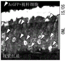
In the superimposed image (Overlay) shown in the upper column of fig. 7, a circle of a broken line indicates 1 eyeball. The middle panel of fig. 7 shows a fluorescence image of AcGFP showing cells knocked into the normal rhodopsin gene. The lower column shows the fluorescence image of mCherry, showing the cells into which the expression vector has been introduced.
As shown in fig. 7, when the gRNA1 and the gRNA3 were used, green light was observed. This result suggests that: from the viewpoint of being able to improve the knock-in efficiency of the normal rhodopsin gene, it is preferable to select gRNA1 or gRNA3 as the gRNA.
Next, a retinal section was prepared using the same cup as described above, and fluorescence images of AcGFP and mCherry were observed. The results are shown in FIG. 8.
As shown in the fluorescence image of the retinal section in fig. 8, only in the outer retinal granular layer (ONL) where rod cells are mainly located, cells expressing AcGFP were confirmed. On the other hand, in addition to the outer retinal stratum granulosum, cells expressing mCherry were also confirmed in the inner retinal stratum granulosum (INL) in which horizontal cells, bipolar cells, and amacrine cells were distributed. This result shows that knock-in of the normal rhodopsin gene occurs specifically in rod cells.
Next, from the fluorescence images of the retinal sections obtained as described above, the ratio of "AcGFP-expressing cell"/"mCherry-expressing cell" in the retinal outer granular layer (ONL) was calculated. The results are shown in FIG. 9.
As shown in fig. 9, when the gRNA1 or the gRNA3 was used, knock-in occurred with a high probability of about 8 to 9 points.
Next, a retinal section of a mouse into which a normal rhodopsin gene was knocked using gRNA1 was stained with an anti-rhodopsin antibody. The results are shown in FIG. 10.
As shown in fig. 10, in the AcGFP-expressing cells, binding of the anti-rhodopsin antibody was confirmed. That is, expression of rhodopsin protein was confirmed in cells into which the normal rhodopsin gene was knocked.
[ example 2: effect of expression vector construction on knockdown efficiency
The effect of the composition of the expression vector on knockin efficiency was investigated. Specifically, the influence of these factors on the knock-in efficiency was investigated by changing the type of plasmid incorporated into the expression cassette and the manner of combining the expression cassettes incorporated into 1 plasmid. Thus, in addition to the above-described expression vector, the following expression vectors were prepared.
These expression vectors were prepared using plasmid-form adeno-associated virus (AAV) vectors (pAAV) having ITR sequences at both ends. pAAV has an upper limit of 5kb or so, and an expression cassette is designed so as not to overflow the upper limit.
1.pAAV-U6-gRNA1:Rho300-mCherry
U6-gRNA1 (expression cassette for gRNA 1) and Rho300-mCherry (expression cassette for mCherry) were inserted into pAAV. Thus, a tandem type expression vector "pAAV-U6-gRNA 1: Rho 300-mCherry" was prepared.
2.pAAV-U6-gRNA1:mRho-HITI-Donor[gRNA1]
U6-gRNA1 (an expression cassette for gRNA 1) and mRho-HITI-Donor [ gRNA1] (an expression cassette for normal rhodopsin gene) were inserted into pAAV. Thus, a tandem-type expression vector "pAAV-U6-gRNA 1: mRho-HITI-Donor [ gRNA1 ]" was prepared.
3.pAAV-mRho-HITI-Donor[gRNA1]
The mRho-HITI-Donor [ gRNA1] (expression cassette for normal rhodopsin gene) was inserted into pAAV. Thus, an expression vector "pAAV-mRho-HITI-Donor [ gRNA1 ]" was prepared.
4.pAAV-Rho2k-Cas9
Rho2k-Cas9 (expression cassette for Cas9) was inserted into pAAV. Thus, an expression vector "pAAV-Rho 2k-Cas 9" was prepared.
5.pAAV-Rho300-Cas9
Rho300-Cas9 was inserted into pAAV. Thus, an expression vector "pAAV-Rho 300-Cas 9" was prepared.
The solutions containing the expression vectors were mixed at the ratios shown in table 2, and the following knock-in solutions (1) to (8) were prepared. The characteristics of each solution are as follows.
(1) Control
(2) Rho promoter shortened from 2kb to 300bp
(3) The expression cassette of gRNA is connected with the expression cassette of mCherry in series
(4) Inserting expression box of normal rhodopsin gene into pAAV
(5) Combining the conditions of (3) and (4)
(6) Combining the conditions of (2) to (4)
(7) Connecting the expression cassette of gRNA and the expression cassette of normal rhodopsin gene in series
(8) Combining the conditions of (2) and (7)
[ Table 2]

The total concentration of the expression vector is 5-7 mug/muL
Using the knock-in solution, the expression vector was introduced into the cells by electroporation in the same manner as in example 1.
At P21, the eyeball is collected. After the eye was dissected to obtain a cup, fluorescence images of AcGFP and mCherry were taken using a fluoroscope microscope. The results are shown in FIG. 11.
As is clear from a comparison of the results of the experiments (1) and (2), the expression intensity of AcGFP was not changed when Rho2k or Rho300 was used as the rhodopsin promoter. This suggests that the knock-in efficiency is not affected even if the rhodopsin promoter is shortened to 300 bp.
As a result of comparing the results of the (1), (3) and (7), it was found that the expression vector obtained by combining the expression cassette of gRNA with another expression cassette has a lower expression intensity of AcGFP than the expression vector not combined. This suggests that the knock-in efficiency is improved without complexing the expression of grnas with other expression cassettes.
As is clear from comparison of the results of the examples in (1) and (4), the expression vector obtained by inserting the expression cassette for the normal rhodopsin gene into pAAV has a lower expression intensity of AcGFP than the expression vector obtained by inserting the expression cassette for the normal rhodopsin gene into pLeaklessIII. This suggests that the knock-in efficiency is improved by inserting the expression cassette for the normal rhodopsin gene into pLeaklessIII.
[ example 3: knock-in efficiency under viral delivery mode ]
Knock-in efficiency under viral delivery modalities was investigated. Specifically, each AAV8 vector was prepared using AAV type 8, which was confirmed to infect mature rod cells, and their knock-in efficiency was investigated. For this purpose, the following recombinant AAV8 virus was prepared.
1.AAV8-Rho300-Cas9
Rho300-Cas9 (expression cassette for Cas9) was inserted into pAAV. Then, a recombinant AAV8 virus "AAV 8-Rho300-Cas 9" is prepared through an AAV unassisted expression system.
2.scAAV8-U6-gRNA1-WPRE-U6-gRNA1
In order to improve the expression efficiency of gRNA, a cyclic tandem type expression cassette "U6-gRNA 1-WPRE-U6-gRNA 1" was prepared, which includes WPRE (wood promoter virus posttranscriptional element). In addition, the expression cassette was inserted into self-complementary (self-complementary) pscAAV in order to improve expression efficiency. Then, a recombinant scAAV8 virus 'scAAV 8-U6-gRNA1-WPRE-U6-gRNA 1' is prepared through an AAV unassisted expression system. WPRE is a sequence that enhances the stability of mRNA delivered from the nucleus to the cytoplasm and promotes maturation, which promotes packaging properties with respect to viruses, viral titer, and expression of introduced genes.
3.AAV8-mRho-HITI-Donor[gRNA1]
The mRho-HITI-Donor [ gRNA1] (expression cassette for normal rhodopsin gene) was inserted into pAAV. Then, a recombinant AAV8 virus "AAV 8-mRho-HITI-Donor [ gRNA1 ]" was prepared by AAV unassisted expression system.
4.AAV8-CAG-mCherry-WPRE
CAG-mCherry (mCherry expression cassette) and WPRE were inserted into pAAV. Then, a recombinant AAV8 virus 'AAV 8-CAG-mCherry-WPRE' is prepared through an AAV unassisted expression system.
Using these recombinant AAV8 viruses, a normal rhodopsin gene was introduced into mice. The expression vectors used for the introduction and the number of gene copies are shown in Table 3.
[ Table 3]

The recombinant AAV8 viruses of test example 1 and test example 2 were injected under the retina of a 3-month-old mouse and infected. Retinas at 1 month and 2 months after infection were collected and flat-mount specimens were prepared. Then, fluorescence images of AcGFP and mCherry were taken in the same manner as in example 2. The results are shown in FIG. 12.
The left column of FIG. 12 shows fluorescence images of retinas of mice infected with the recombinant AAV8 virus of test example 1 (upper column: 1 month after infection, lower column: 2 months after infection). In addition, the right column of FIG. 12 shows fluorescence images of retinas of mice infected with the recombinant AAV8 virus of test example 2 (upper column: 1 month after infection, lower column: 2 months after infection). The arrows in the figure show the injection of recombinant AAV8 virus to the injection site under the retina.
The dotted line region in the lower left column indicates a region where trace AcGFP expression was confirmed. Although test example 1 is a negative control of an expression vector into which no gRNA was introduced, AcGFP was observed due to the transcriptional activity of the ITRs of AAV. That is, in test example 1, the knock-in of the normal rhodopsin gene did not actually occur.
As shown in fig. 12, the occurrence range of AcGFP expression was 1/4 in retina at 1 month after infection, and expanded to about 2/3 at 2 months after infection. This correlates with the time required for the AAV virus to infect the cell, transfer into the nucleus, and express the introduced gene.
Subsequently, a section of the retina at 2 months after infection was prepared, and fluorescence images of AcGFP and mCherry were observed. The results are shown in FIG. 13. The left column of fig. 13 shows a fluorescence image of retinas of mice infected with the recombinant AAV8 virus of test example 1. The right column shows fluorescence images of retinas of mice infected with the recombinant AAV8 virus of test example 2.
As shown in fig. 13, cells expressing AcGFP to the same extent as in example 2 (fig. 8) were confirmed in retinal sections at 2 months after infection with the recombinant AAV8 virus of test example 2. In addition, cells expressing AcGFP were confirmed in retinal sections 2 months after infection with the recombinant AAV8 virus of test example 1, but the result was attributed to the transcriptional activity of the ITRs of AAV.
[ example 4: rho (a)P23HGene therapy in retinitis pigmentosa model mice
As a retinitis pigmentosa model mouse, a knock-in Rho mouse was usedP23HWherein RhoP23HThe most frequent occurrence of Rho mutation in humans (see Sakami S et al (2011) 'combining Mechanisms of Photometer generation in a New Mouse Model of the Common Form of autoimmune therapy Pigmentosa due to P23H Opsin variants', Journal of Biological Chemistry, Vol.286(No.12), pp 10551-10567.). In the knock-in mouse, the 23 rd proline residue of exon 1 of the Rho gene was replaced with a histidine residue. The Rhodopsin protein of this knock-in mouse is not folded into the correct structure and causes Endoplasmic Reticulum stress, and therefore Retinal Degeneration occurs (see Chiang WC et al (2015) 'Robust endogenous diagnosis-Associated Degradation of Rhodopsin prediction regeneration,' Molecular Neurobiology, Vol.52(Issue 1), pp.679-695.).
Mice with this homozygous variation (Rho)P23H/P23H) At P10-P20, most of the rod cells denature and eventually undergo cell death. Then, when the cells reached 1 month of age, the nerve cell layer (outer granular layer: ONL) where the optic cells were located became thin. Thus, the mouse can be effectively used to detect rod cells that have survived treatment.
On the other hand, mice with this heterozygous variation (Rho) before reaching P30+/P23H) The thickness of ONL (a) becomes about half, but thereafter, retinal degeneration becomes gentle. At this time, the response degree of the microscopic morphology of the visual cell (length of the outer segment of the visual cell, etc.) and the electroretinogram is about half that of a normal mouse. Therefore, the mouse can be effectively used for evaluating the function of the rod cells which survive the treatment.
[4-1.RhoP23H/P23HGene therapy in mice]
Rho on P0P23H/P23HOne eye of the mouse was used as in example 2The knock-in solution (1) used was introduced with the expression vector. The method of introducing the expression vector was carried out by electroporation in the same manner as in example 4. As a control, the other eye was not electroporated.
Sections of retinas were made at P14, P21, and P50. The sections were histostained with anti-rhodopsin antibody and DAPI. The results are shown in FIG. 14.
As shown in fig. 14, at P14, the ONL thickness of the retinas having the normal rhodopsin gene knocked in was the same as that of the control retinas. On the other hand, in the retinas with the normal rhodopsin gene knocked in P21 and P50, there were cells expressing AcGFP, and the thinning of ONL was suppressed, and the expression of rhodopsin was confirmed in the outer segment. In contrast, the ONL of the control retina was thinned. That is, Rho is produced by knocking in the normal rhodopsin geneP23H /P23HDegeneration of rod cells in mice was inhibited. This indicates that knock-in of the normal rhodopsin gene and knock-out of P23H rhodopsin occurred and that visual cell death was avoided by expression of normal rhodopsin.
[4-2.Rho+/P23HGene therapy in mice]
Preparing 2 Rho ages 1 month+/P23HMice were injected with the recombinant AAV8 virus used in experimental example 2 of example 3 in their left eyes, respectively. The AAV8 virus was injected by the same method as in example 3. As a control, the right eye was not injected with AAV8 virus.
AcGFP fluorescence was confirmed 1 month after infection by fluorescent fundus imaging. The results are shown in FIG. 15. The upper column of fig. 15 shows the fluorescence image of the right eye, and the lower column shows the fluorescence image of the left eye. In addition, the left column shows fluorescence images obtained when AAV8 virus was injected into a wild-type mouse and a fluorescence fundus imaging examination was performed 1 month after infection.
As shown in FIG. 15, although 2 Rho radicals+/P23HThere were individual differences between mice, but all mice had cells expressing AcGFP. That is, this result suggests that normal rhodopsin is expressed in these visual cells by knocking in the normal rhodopsin gene, andnot P23H mutant rhodopsin.
Next, 3 months after infection, an optokinetic eye reaction test was performed using a qOMR visual motor reaction quantification system (manufactured by Phenosys corporation), and optokinetic eye reactions of the right and left eyes were quantified. In this examination, the mouse is placed in an examination box surrounded on four sides by monitors. The stripe patterns with different widths are displayed on a monitor, and the stripes are moved left and right at regular intervals. If the mouse in the examination box notices the stripe pattern, the head moves following the movement of the stripe pattern (eyeball reaction). When viewed from above, clockwise rotation of the head means a visual eyeball reaction originating in the left eye, and counterclockwise rotation means a visual eyeball reaction originating in the right eye. The position of the camera above the mouse head is tracked, and the reaction of the moving eyeball is quantified. The results are shown in FIG. 16.
FIG. 16, left panel, untreated Rho without AAV8 virus injection+/P23HGraph of experimental results for mice. The central and right panels are 2 Rho cells injected with AAV8 virus into the left eye+/P23HGraph of experimental results for mice.
Both eyes of the wild type mice showed the highest response to a 0.2cyc/deg stripe pattern with a correct response rate (Mean optokinetic response) of around 2.0 (not shown). In contrast, as shown in FIG. 16, Rho was untreated+/P23HBoth eyes of the mouse (left panel) showed the highest response to a 0.2cyc/deg striped pattern as the wild type, but the correct response rate was about 1.5, which was lower than that of the wild type. On the other hand, 2 Rho cells injected with AAV8 virus into the left eye+/P23HThe rate of correct response was significantly higher in the left eye than in the right eye of the mice (center and right panels). This result clearly indicates that Rho is caused by knock-in of the normal rhodopsin gene+/P23HThe function of the mouse rod cells is suppressed. This is because the normal rhodopsin is expressed by knocking-in the normal rhodopsin gene and knocking-out the P23H rhodopsin, and thus the visual function is restored or the visual function deterioration is suppressed.
[ example 5: verification of Gene therapy for human rhodopsin Gene ]
[5-1.gRNA]
To verify the possibility of gene therapy for the human rhodopsin gene, recognition sequences of grnas that can be effectively utilized for human cells were investigated.
The gRNA recognition sequence was searched for in a region within 100bp upstream from the translation start point of exon 1 of the human rhodopsin gene using a gRNA search engine (http:// criprpr. technology /) oriented to SpCas 9. As a result, a sequence identified by gRNA (hs086172148) was found (SEQ ID NO: TCAGGCCTTCGCAGCATTCTTGG; evaluation value: high (800)). The position of the sequence recognized by the gRNA (hs086172148) is shown in FIG. 17. In fig. 17, the arrow shows the Cas9 cleavage position.
[ 5-2. SSA (Single Strand renaturation) assay ]
The efficiency of cleavage of the recognition sequence of the gRNA (hs086172148) was verified by single strand renaturation (SSA) assay. FIG. 18 is a schematic diagram of an SSA assay.
As shown in fig. 18, a gRNA (hs086172148) recognition sequence was inserted into the Enhanced Green Fluorescent Protein (EGFP) gene sequence of the EGFP gene expression plasmid used in the SSA assay; to prepare a modified EGFP gene expression plasmid. The EGFP-encoding nucleotide sequence contained in this plasmid is partially identical at a position adjacent to the recognition sequence of the gRNA (hs 086172148). That is, the recognition sequence of the gRNA (hs086172148) has partially identical nucleotide sequences upstream and downstream of the recognition sequence.
Thus, when the gRNA (hs086172148) recognition sequence is cleaved by Cas9, the repeats located upstream and downstream of the cleavage site cause homologous recombination or single strand renaturation. Thus, the base sequence encoding the entire EGFP is completed, so that the EGFP can be expressed.
The prepared modified EGFP gene expression plasmid, an expression vector for gRNA (hs086172148), and an expression vector for Cas9 were mixed to prepare a mixture for transfection. Using this transfection mixture, HEK293 cells were transfected. Transfected HEK293 cells at 37 ℃ with 5% CO2Was cultured for 72 hours, and then, the expression of EGFP was detected. As a control, useThe same experiment was performed for a system without the gRNA (hs086172148) expression vector. The results are shown in FIG. 19.
As shown in fig. 19, EGFP signals were detected in cells transfected with both the modified EGFP gene expression plasmid and the expression vector of gRNA (hs 086172148). This indicates that the recognition sequence of gRNA (hs086172148) is actually cleaved by gRNA (hs086172148) and Cas 9.
[ 5-3. preparation of donor DNA containing Normal human rhodopsin Gene ]
Donor DNA of normal human rhodopsin gene was prepared using gRNA (hs086172148) recognition sequence. FIG. 20 is a schematic structural view of an expression cassette (about 4.1kb) of the prepared donor DNA.
The normal rhodopsin gene used was a sequence obtained by deleting a stop codon from the cDNA (1044bp, accession No. nm — 000539.3) of the normal human rhodopsin gene. As shown in FIG. 20, a chimeric intron sequence (228bp, SEQ ID NO: 10) was placed upstream of the normal rhodopsin gene. The Furin sequence, the P2A sequence (78 bp; SEQ ID NO: 11), the AcGFP gene (720 bp; SEQ ID NO: 12), and the 3' -untranslated region of the human rhodopsin gene and the sequence of 100bp downstream thereof (1725 bp, SEQ ID NO: 18) are sequentially located downstream of the normal human rhodopsin gene. Then, reverse target sequences corresponding to gRNA (hs086172148) were placed upstream and downstream of the obtained sequence, and expression cassettes of donor DNA of normal human rhodopsin gene were prepared.
The expression cassette prepared as described above is inserted into a viral or non-viral plasmid to prepare an expression vector for the normal human rhodopsin gene. The prepared expression vector of the normal human rhodopsin gene can be introduced into organ-cultured human retinas, human retinal organs, etc., and the efficiency of introduction of the gene of the donor DNA into human visual cells can be evaluated in detail.
[ example 6: verification of Gene therapy for genes that cause dominant variations other than the rhodopsin Gene ]
The possibility of gene therapy of genes causing dominant variation other than the rhodopsin gene was examined. Specifically, in mice, the knock-in efficiency of the normal Peripherin (Peripherin, Prph2) gene was investigated. Dominant variant peripherin genes are causative genes of retinitis pigmentosa and macular dystrophy, and more than 90 different disease variants have been reported previously.
[6-1.gRNA]
The gRNA recognition sequences were searched for within 100bp of the evolutionary non-conserved region upstream from the translation start point of exon 1 of the mouse peripherin gene using the gRNA search engine (http:// criprpr. technology /) oriented to SpCas 9. As a result, 3 gRNA recognition sequences were found. The sequences are as follows (see fig. 21 for positional relationship of recognition sequences of grnas corresponding to the grnas).
Recognition sequence for gRNA 4: sequence number 19 (TGCTCTTCCCTAGACCCTAGCGG; rating value: high (800))
Recognition sequence for gRNA 5: sequence number 20 (GGGCTGGACCGCTAGGGTCTAGG; rating value: high (900))
Recognition sequence for gRNA 6: sequence number 21 (GAGCTCACTCGGATTAGGAGTGG; evaluation value: high (800)).
Expression cassettes in which a human U6 promoter sequence, crRNA sequences corresponding to the respective recognition sequences, and a tracerRNA sequence were arranged in this order from the upstream side were prepared for the grnas 4 to 6, respectively. The expression cassette was inserted into the MCS of plasmid pBluescriptII to prepare expression vectors for gRNA 4-6 (pU6-gRNA4, pU6-gRNA5, and pU6-gRNA 6).
[ 6-2. Normal peripherin Gene ]
In the same manner as the donor DNA of the normal mouse rhodopsin gene, a donor DNA of the normal peripherin gene was prepared. FIG. 22 is a schematic diagram showing the structure of an expression cassette (about 3.6kb) of the prepared donor DNA.
The normal peripherin gene used was a sequence obtained by deleting a stop codon from a cDNA of a normal mouse peripherin protein (accession No. nm — 008938.2). A chimera intron sequence (228bp, SEQ ID NO: 10) was located upstream of the normal peripherin gene. The Furin sequence and the P2A sequence (78 bp; SEQ ID NO: 11), the AcGFP gene (720 bp; SEQ ID NO: 12), and the sequence of the 3' untranslated region of the peripherin gene and the sequence of 100bp downstream thereof (1486 bp in total, SEQ ID NO: 22, gene position: chr17:46,923, 548-. Then, reverse target sequences corresponding to grnas 4 to 6 were provided upstream and downstream of the obtained sequences, respectively, to prepare expression cassettes of donor DNA of the normal peripherin gene.
The expression cassette prepared as described above was inserted into the MCS of plasmid pleakleschi to prepare expression vectors for normal peripherin genes corresponding to each of grnas 4 to 6 (pleakleschi-mrph 2-HITI-Donor [ gRNA4], pleakleschi-mrph 2-HITI-Donor [ gRNA5], and pleakleschi-mrph 2-HITI-Donor [ gRNA6 ]).
[ 6-3. study of knock-in efficiency ]
In the same manner as in example 1 performed for the mouse rhodopsin gene, the knock-in efficiency of the normal peripherin gene was investigated.
Each expression vector of grnas 4-6 (pU6-gRNA4, pU6-gRNA5, or pU6-gRNA6), an expression vector of a normal peripherin gene (pwakessiii-mPrph 2-HITI-Donor [ gRNA4], pwakessiii-mPrph 2-HITI-Donor [ na5], or pwakessiii-mPrph 2-HITI-Donor [ gRNA6]), an expression vector of Cas9 (pRho2 gr 2k-Cas9), and an expression vector of mCherry (pCAG-mCherry) were mixed at the same ratio as in example 1, thereby preparing 3 knock-in solutions.
0.3 to 0.4. mu.L of the knock-in solution was injected under the retina of 0 day (P0) postnatal mouse (4 mice), and the expression vector was introduced into the cells by electroporation. At P21, 4 eyeballs were collected and fixed in a 4% paraformaldehyde solution (Nacalaitesque Co., Ltd.) at room temperature for 1 hour in a state where the sclera on the outer side of the eyeball was peeled off (cup). Then, fluorescence images of GFP and mCherry were taken using a fluoroscope microscope. Further, a retinal section was prepared using the same cup as described above, and fluorescence images of AcGFP and mCherry were observed. The results are shown in FIG. 23 (upper column: cup, lower column: retinal section).
As shown in fig. 23, when the gRNA4 and the gRNA6 were used, green light was observed. This result suggests that: using gRNA4 or gRNA6, the normal peripherin gene can be knocked in. As shown in the lower column of fig. 23, only in the outer retinal granular layer (ONL) where rod cells are mainly located, cells expressing AcGFP were confirmed. On the other hand, in addition to the outer retinal stratum granulosum, cells expressing mCherry were also confirmed in the inner retinal stratum granulosum (INL) in which horizontal cells, bipolar cells, and amacrine cells were distributed. This result shows that knock-in of the normal peripherin gene occurs specifically in the rod cells.
(availability in industry)
The present invention is useful, for example, for the treatment of diseases derived from dominant variant genes.
Sequence listing
<110> national research and development institute of human and chemical Research (RIKEN)
<120> therapeutic agent for diseases derived from dominant variant gene
<130> RK20180PCT
<150> JP 2019-130199
<151> 2019-07-12
<160> 22
<170> PatentIn version 3.5
<210> 1
<211> 4143
<212> DNA
<213> Streptococcus pyogenes (Streptococcus pyogenes)
<400> 1
atggacaaga agtattctat cggactggac atcgggacta atagcgtcgg gtgggccgtg 60
atcactgacg agtacaaggt gccctctaag aagttcaagg tgctcgggaa caccgaccgg 120
cattccatca agaaaaatct gatcggagct ctcctctttg attcagggga gaccgctgaa 180
gcaacccgcc tcaagcggac tgctagacgg cggtacacca ggaggaagaa ccggatttgt 240
taccttcaag agatattctc caacgaaatg gcaaaggtcg acgacagctt cttccatagg 300
ctggaagaat cattcctcgt ggaagaggat aagaagcatg aacggcatcc catcttcggt 360
aatatcgtcg acgaggtggc ctatcacgag aaatacccaa ccatctacca tcttcgcaaa 420
aagctggtgg actcaaccga caaggcagac ctccggctta tctacctggc cctggcccac 480
atgatcaagt tcagaggcca cttcctgatc gagggcgacc tcaatcctga caatagcgat 540
gtggataaac tgttcatcca gctggtgcag acttacaacc agctctttga agagaacccc 600
atcaatgcaa gcggagtcga tgccaaggcc attctgtcag cccggctgtc aaagagccgc 660
agacttgaga atcttatcgc tcagctgccg ggtgaaaaga aaaatggact gttcgggaac 720
ctgattgctc tttcacttgg gctgactccc aatttcaagt ctaatttcga cctggcagag 780
gatgccaagc tgcaactgtc caaggacacc tatgatgacg atctcgacaa cctcctggcc 840
cagatcggtg accaatacgc cgaccttttc cttgctgcta agaatctttc tgacgccatc 900
ctgctgtctg acattctccg cgtgaacact gaaatcacca aggcccctct ttcagcttca 960
atgattaagc ggtatgatga gcaccaccag gacctgaccc tgcttaaggc actcgtccgg 1020
cagcagcttc cggagaagta caaggaaatc ttctttgacc agtcaaagaa tggatacgcc 1080
ggctacatcg acggaggtgc ctcccaagag gaattttata agtttatcaa acctatcctt 1140
gagaagatgg acggcaccga agagctcctc gtgaaactga atcgggagga tctgctgcgg 1200
aagcagcgca ctttcgacaa tgggagcatt ccccaccaga tccatcttgg ggagcttcac 1260
gccatccttc ggcgccaaga ggacttctac ccctttctta aggacaacag ggagaagatt 1320
gagaaaattc tcactttccg catcccctac tacgtgggac ccctcgccag aggaaatagc 1380
cggtttgctt ggatgaccag aaagtcagaa gaaactatca ctccctggaa cttcgaagag 1440
gtggtggaca agggagccag cgctcagtca ttcatcgaac ggatgactaa cttcgataag 1500
aacctcccca atgagaaggt cctgccgaaa cattccctgc tctacgagta ctttaccgtg 1560
tacaacgagc tgaccaaggt gaaatatgtc accgaaggga tgaggaagcc cgcattcctg 1620
tcaggcgaac aaaagaaggc aattgtggac cttctgttca agaccaatag aaaggtgacc 1680
gtgaagcagc tgaaggagga ctatttcaag aaaattgaat gcttcgactc tgtggagatt 1740
agcggggtcg aagatcggtt caacgcaagc ctgggtacct accatgatct gcttaagatc 1800
atcaaggaca aggattttct ggacaatgag gagaacgagg acatccttga ggacattgtc 1860
ctgactctca ctctgttcga ggaccgggaa atgatcgagg agaggcttaa gacctacgcc 1920
catctgttcg acgataaagt gatgaagcaa cttaaacgga gaagatatac cggatgggga 1980
cgccttagcc gcaaactcat caacggaatc cgggacaaac agagcggaaa gaccattctt 2040
gatttcctta agagcgacgg attcgctaat cgcaacttca tgcaacttat ccatgatgat 2100
tccctgacct ttaaggagga catccagaag gcccaagtgt ctggacaagg tgactcactg 2160
cacgagcata tcgcaaatct ggctggttca cccgctatta agaagggtat tctccagacc 2220
gtgaaagtcg tggacgagct ggtcaaggtg atgggtcgcc ataaaccaga gaacattgtc 2280
atcgagatgg ccagggaaaa ccagactacc cagaagggac agaagaacag cagggagcgg 2340
atgaaaagaa ttgaggaagg gattaaggag ctcgggtcac agatccttaa agagcacccg 2400
gtggaaaaca cccagcttca gaatgagaag ctctatctgt actaccttca aaatggacgc 2460
gatatgtatg tggaccaaga gcttgatatc aacaggctct cagactacga cgtggaccac 2520
atcgtccctc agagcttcct caaagacgac tcaattgaca ataaggtgct gactcgctca 2580
gacaagaacc ggggaaagtc agataacgtg ccctcagagg aagtcgtgaa aaagatgaag 2640
aactattggc gccagcttct gaacgcaaag ctgatcactc agcggaagtt cgacaatctc 2700
actaaggctg agaggggcgg actgagcgaa ctggacaaag caggattcat taaacggcaa 2760
cttgtggaga ctcggcagat tactaaacat gtcgcccaaa tccttgactc acgcatgaat 2820
accaagtacg acgaaaacga caaacttatc cgcgaggtga aggtgattac cctgaagtcc 2880
aagctggtca gcgatttcag aaaggacttt caattctaca aagtgcggga gatcaataac 2940
tatcatcatg ctcatgacgc atatctgaat gccgtggtgg gaaccgccct gatcaagaag 3000
tacccaaagc tggaaagcga gttcgtgtac ggagactaca aggtctacga cgtgcgcaag 3060
atgattgcca aatctgagca ggagatcgga aaggccaccg caaagtactt cttctacagc 3120
aacatcatga atttcttcaa gaccgaaatc acccttgcaa acggtgagat ccggaagagg 3180
ccgctcatcg agactaatgg ggagactggc gaaatcgtgt gggacaaggg cagagatttc 3240
gctaccgtgc gcaaagtgct ttctatgcct caagtgaaca tcgtgaagaa aaccgaggtg 3300
caaaccggag gcttttctaa ggaatcaatc ctccccaagc gcaactccga caagctcatt 3360
gcaaggaaga aggattggga ccctaagaag tacggcggat tcgattcacc aactgtggct 3420
tattctgtcc tggtcgtggc taaggtggaa aaaggaaagt ctaagaagct caagagcgtg 3480
aaggaactgc tgggtatcac cattatggag cgcagctcct tcgagaagaa cccaattgac 3540
tttctcgaag ccaaaggtta caaggaagtc aagaaggacc ttatcatcaa gctcccaaag 3600
tatagcctgt tcgaactgga gaatgggcgg aagcggatgc tcgcctccgc tggcgaactt 3660
cagaagggta atgagctggc tctcccctcc aagtacgtga atttcctcta ccttgcaagc 3720
cattacgaga agctgaaggg gagccccgag gacaacgagc aaaagcaact gtttgtggag 3780
cagcataagc attatctgga cgagatcatt gagcagattt ccgagttttc taaacgcgtc 3840
attctcgctg atgccaacct cgataaagtc cttagcgcat acaataagca cagagacaaa 3900
ccaattcggg agcaggctga gaatatcatc cacctgttca ccctcaccaa tcttggtgcc 3960
cctgccgcat tcaagtactt cgacaccacc atcgaccgga aacgctatac ctccaccaaa 4020
gaagtgctgg acgccaccct catccaccag agcatcaccg gactttacga aactcggatt 4080
gacctctcac agctcggagg ggatgaggga gctgatccca agaaaaagcg taaggttgac 4140
tag 4143
<210> 2
<211> 296
<212> DNA
<213> cattle (Bos taurus)
<400> 2
aggcctctgc tctttcccag ggtccccagc acgccccgcc ttctccccga ccacagcctt 60
ctacctggag tcaccttggc cccacctgga agccaattaa gcccctcgtt gcagcagtga 120
ggattaatat gattaataac gcccccaatc tccgaggtgc tgattcagcc gggagcttag 180
ggaggggagg tcacttcata agggcctggg gggggagttg gagccacgag tcgtccagcc 240
ggagccccgt gtggctgagc tccggcctca gaagcatccc cgggttcgcg ccgccg 296
<210> 3
<211> 2253
<212> DNA
<213> cattle (Bos taurus)
<400> 3
agcttccacg ttgtcagaag cgttagcggg taacaagcaa ggccatgagt ttgagggtcg 60
gggggacagc ctggccaggg cagggctgcg tgctctggtc ttgacccagg gagtccatcg 120
tctgtccctc tgtctggcca ccaggggccg tgttagaatc aaactcttca ccttgacctc 180
tttagcgatg gacctaatca cagggccaag caccttgagc agctgcgccc cacccacccg 240
ccacacctgc ctgccccttg ctccttcccc ccagagcact taccaccagg ctccccacac 300
aagtcctgat gctgcccgct gcccgggaca gcagggctct ggagccccag gtgggctgca 360
gcgaagggag cacgtgtctg agcagagagg tgtagacatc ccaggtggta agagggacag 420
cacaagggag cctcagcgat ggaggtctca gacactggga tgcttcctgg ccacccttgc 480
gttccccacc tcggggcctt tgcactggcc gctctctctc cttggacagc tccacccctt 540
agtgctcctt ccatcctcga gttctcagag aggccttcct gggcggctcc aggcaactgt 600
gtgtcgggag gacgtcacca tctgaacagt ccctgtggtc gtttgtttca tggtttgtgg 660
gggcccctcc ccgccccccc agaagaagga cacgtcctac aggcaggggc ttggtccttc 720
ttgtccactg tgtcccccag aactgagcgt cttgtgtaga acacagtcgg tgccagataa 780
aactcacagc tggactgtgg tcattgagga ccccgaaggc caggctgccc gcgaccccac 840
ttcactccta ggaggggtct tcacgatcct attgtagatg ggggcccttg ttcgcaccct 900
gagctcgtgc tgctgttctg ggatacgcca tcacggcttg ccgatcgcac ggcacccgag 960
gcccacggcc ctaacccagc ctgaccccag tcctgggcct cggcccgtgt gctgttccca 1020
gggcccacct cccctccata tgggagtgcc cgggaccaca cctccttgct caccgcaggg 1080
atctggtgcc cggcacaggc ttggccccgt tcacctcctg gagacgaggc cttgcttcag 1140
gagccctaag agcttctgtc ccctccccgc tttggttccc tctgccctgg tcccttcctc 1200
caccatcgct cccttcagct catccttgag ggcttttgag ccaccttcct ctccccaaga 1260
tctgcccaag ttcccctgga taaagcccct ggcactaagc cccaccccag ctgaccctag 1320
cccacgaggg cccacatccc agatgggcct cgggccttca ccgggcctct gtcttcagtc 1380
tatgctgact tccccacttg gcaacctggc aaagtgcccc cctccctcaa cacattgtcc 1440
ccctggacgt ctctgcagat aggtggcctc cccgccccgg gtccagagcg ggtcttccca 1500
cacggaagtc ctcctcagcc cataagctcc tccttcctgg agcctcgggt gccccagtgg 1560
cctgcacata aatgtcggtt tgaacaagtg agtgagtgaa ctccagttca agggaacaaa 1620
tgctttttga gctcctgggc aacgcggggc ttgtgtccgg tccccggagc gccccgctgc 1680
acttgtgaag ctcacgacct ggccggtggg cactgtcagt gtgtggtgac tggataagcc 1740
cagaggtgac ccaggtgggg aagggacagc tcaggggctg agtgagggga tgaccgaagg 1800
accagcgctc tgccctccag gagctcgtgg gccaggcggg aggaaggcaa aatttgcttg 1860
ccctgagcca gagggaagtg ggcctagagc taggggtccc aatgccaggc cagggccatc 1920
agctgagatg ccagaggggg acaggggagt tgggggcagg cctctgctct ttcccagggt 1980
ccccagcacg ccccgccttc tccccgacca cagccttcta cctggagtca ccttggcccc 2040
acctggaagc caattaagcc cctcgttgca gcagtgagga ttaatatgat taataacgcc 2100
cccaatctcc gaggtgctga ttcagccggg agcttaggga ggggaggtca cttcataagg 2160
gcctgggggg ggagttggag ccacgagtcg tccagccgga gccccgtgtg gctgagctcc 2220
ggcctcagaa gcatccccgg gttcgcgccg ccg 2253
<210> 4
<211> 478
<212> DNA
<213> Rabbit (Oryctolagus cuniculus)
<400> 4
agaacagagc ccgaccagcc ttccgtgacc cctcgcctct cccccacctg ctggaaagct 60
ccactgtcat ctcctgtttt acacccaggg aaactaagga cacagaatag gtacccagaa 120
gcagccaggt ggtcagtccc atgtccagtg gggccacagg ccctgggttg gggtgctgaa 180
catgaagcac cctccccagc cttctccaca gcctctggtg ggcatggagc cccaagtctg 240
agggcaggaa tgcctcttgg tggccaggtt tccttccaga accagaaact ctggggtggg 300
tgggggccca gccgagctgt gtttggactc cctggcctca agagccccga caggggcctc 360
agggaaagat cccctcctcc cctctccatg tccaaaatag agttcattct caggagctgg 420
atgattttgc taagattcac aatttatttt tatttattaa aatctcttac ttctgtta 478
<210> 5
<211> 76
<212> RNA
<213> Artificial sequence
<220>
<223> tracerRNA
<400> 5
guuuuagagc uagaaauagc aaguuaaaau aaggcuaguc cguuaucaac uugaaaaagu 60
ggcaccgagu cggugc 76
<210> 6
<211> 23
<212> DNA
<213> Artificial sequence
<220>
<223> gRNA1
<400> 6
tctgtctacg aagagcccgt ggg 23
<210> 7
<211> 23
<212> DNA
<213> Artificial sequence
<220>
<223> gRNA2
<400> 7
ggctctcgag gctgccccac ggg 23
<210> 8
<211> 23
<212> DNA
<213> Artificial sequence
<220>
<223> gRNA3
<400> 8
ctgagctcgc caagcagcct tgg 23
<210> 9
<211> 1047
<212> DNA
<213> mouse (Mus musculus)
<400> 9
atgaacggca cagagggccc caatttttat gtgcccttct ccaacgtcac aggcgtggtg 60
cggagcccct tcgagcagcc gcagtactac ctggcggaac catggcagtt ctccatgctg 120
gcagcgtaca tgttcctgct catcgtgctg ggcttcccca tcaacttcct cacgctctac 180
gtcaccgtac agcacaagaa gctgcgcaca cccctcaact acatcctgct caacttggcc 240
gtggctgacc tcttcatggt cttcggagga ttcaccacca ccctctacac atcactccat 300
ggctacttcg tctttgggcc cacaggctgt aatctcgagg gcttctttgc cacacttgga 360
ggtgaaatcg ccctgtggtc cctggtggtc ctggccattg agcgctacgt ggtggtctgc 420
aagccgatga gcaacttccg cttcggggag aatcacgcta tcatgggtgt ggtcttcacc 480
tggatcatgg cgttggcctg tgctgctccc ccactcgttg gctggtccag gtacatccct 540
gagggcatgc aatgttcatg cgggattgac tactacacac tcaagcctga ggtcaacaac 600
gaatcctttg tcatctacat gttcgtggtc cacttcacca ttcctatgat cgtcatcttc 660
ttctgctatg ggcagctggt cttcacagtc aaggaggcgg ctgcccagca gcaggagtca 720
gccaccactc agaaggcaga gaaggaagtc acccgcatgg ttatcatcat ggtcatcttc 780
ttcctgatct gctggcttcc ctacgccagt gtggccttct acatcttcac ccaccagggc 840
tccaacttcg gccccatctt catgactctg ccagctttct ttgctaagag ctcttccatc 900
tataacccgg tcatctacat catgttgaac aagcagttcc ggaactgtat gctcaccacg 960
ctgtgctgcg gcaagaatcc actgggagat gacgacgcct ctgccaccgc ttccaagacg 1020
gagaccagcc aggtggctcc agcctaa 1047
<210> 10
<211> 228
<212> DNA
<213> Artificial sequence
<220>
<223> chimeric intron and exon (chimeric intron and exon)
<400> 10
agtgactctc ttaaggtagc cttgcagaag ttggtcgtga ggcactgggc aggtaagtat 60
caaggttaca agacaggttt aaggagacca atagaaactg ggcttgtcga gacagagaag 120
actcttgcgt ttctgatagg cacctattgg tcttactgac atccactttg cctttctctc 180
cacaggtgtc cactcccagt tcaattacag ctcttaaggc tagagtac 228
<210> 11
<211> 78
<212> DNA
<213> Artificial sequence
<220>
<223> Furin+P2A
<400> 11
cgacgcaaaa gaggaagcgg agctactaac ttcagcctgc tgaagcaggc tggagacgtg 60
gaggagaacc ctggacct 78
<210> 12
<211> 720
<212> DNA
<213> Artificial sequence
<220>
<223> AcGFP
<400> 12
atggtgagca agggcgagga gctgttcacc ggggtggtgc ccatcctggt cgagctggac 60
ggcgacgtaa acggccacaa gttcagcgtg tccggcgagg gcgagggcga tgccacctac 120
ggcaagctga ccctgaagtt catctgcacc accggcaagc tgcccgtgcc ctggcccacc 180
ctcgtgacca ccctgaccta cggcgtgcag tgcttcagcc gctaccccga ccacatgaag 240
cagcacgact tcttcaagtc cgccatgccc gaaggctacg tccaggagcg caccatcttc 300
ttcaaggacg acggcaacta caagacccgc gccgaggtga agttcgaggg cgacaccctg 360
gtgaaccgca tcgagctgaa gggcatcgac ttcaaggagg acggcaacat cctggggcac 420
aagctggagt acaactacaa cagccacaac gtctatatca tggccgacaa gcagaagaac 480
ggcatcaagg tgaacttcaa gatccgccac aacatcgagg acggcagcgt gcagctcgcc 540
gaccactacc agcagaacac ccccatcggc gacggccccg tgctgctgcc cgacaaccac 600
tacctgagca cccagtccgc cctgagcaaa gaccccaacg agaagcgcga tcacatggtc 660
ctgctggagt tcgtgaccgc cgccgggatc actctcggca tggacgagct gtacaagtaa 720
<210> 13
<211> 2257
<212> DNA
<213> mouse (Mus musculus)
<400> 13
gcctggccag agactgtggc tgaaagtagg agtctcctgt ccccacacac cccagccaca 60
gcccccacca ggagcagcac ccgttgggat gaggtcatgc aggctccctc agtgttcttt 120
tctttgtttt taatgaattc atgaaagcaa aatgaggctc cccactcaat ggggacagct 180
tgacaaaggg catccatcca ccaagaccat cctcaacctg gagtccccaa ttcccggggg 240
gccagcggga tctgtacccc tccctcagct tgtctatcag gaacatgaca agtgtcccgg 300
cttagggcta aatgtctagg acagaatgga acacatagta gctgattaat aaatgctagc 360
tggatgaagg gaggaatgag tgactgactg agtggatata tgagtgaagg gattaatgga 420
agggaacatg gatgtcctca ggtgcccaac ctggcagatc cagtcatgtc tggctggaat 480
ctataagcag ttttacatac ctgccctggt tttctctgcc cccaccccca ccccagttgg 540
atctcccaaa tccagggccc tgatagaata tggctgcttc aaagacagag agatgagggg 600
agggaggggg gagggagaga gggagggagg gagacacaga gagggaatat gtgtgatgcg 660
tgtgtatgtg tgtatgtgtg tgtgtaaaca ctttgtatat aaagagtaca gctggtagtt 720
atgttacaag taacaccgac taatataatt aattaaccat cctaatggtc tctgcttgtt 780
agtgactgct tgggaattag gcagggccca agcactcaga taaggtattt ccctcagcct 840
cagtaggctt ttgcaaatga cccaggcctt caggcctgtg cagggctaga gctggattac 900
agagataaat gacagtgaca gcaacgtgag ctgcagccct taggactgag aaagcatcga 960
gaccaggggt ctccggcaag gcctaggtcc tcccttcagt atggaaacct tgcctcatgt 1020
ctctcagcct ccttggcctg tggagatcca gcccttcctc ttggcttctg gatacatttg 1080
ctcttctaca ccagcaacca agtggcaaca gttccaggcc agtatggagt tttagaagcc 1140
atgccaatat gcccaccttc agggagcagc tgagtccttg atgccaccct tgttctgaag 1200
agttcagaaa cacagtgcaa gacatgacca ggcctcatcc ttaggatgct catggatcca 1260
gttcttagct cccttgttgg atatgctgtt ttccttggcc tttggtcttt tctttatccc 1320
agagggtttt ggctttaagg ccaacaggaa ctatggggta ccagaattga gcagcctcag 1380
tctgcatccc tcctctatag aaccacagct gggccctcag caggcccaac tctgcatggg 1440
gacagaggca ttaaaagctc agctcctaca cttggtggca gtggtggtct gttgctctca 1500
agctctttca aaatggatgg aaactgggac gcttccctga cccctggtta tgaaagacta 1560
gactgtgtgg ggacaaacag tccagagtcc cggggaatgt gatagagcag ctccatcatt 1620
tttagaaacc caatttgagg cagtatagag agatggtgac ctctataagc ctctgtatct 1680
gcaaagagga gcttagacct gcccttgagg ggattatatg agatttaagg gacttatgtg 1740
gccagcctac ttcctggcat gctgaagaca ttggcacact ctggtattct agaccttggc 1800
tcagagctgc ctttactagg atactgtcac ttagcaaaag aatgggatgg agcctcagat 1860
gtggagtgac accatcttcc aagaaggaaa gggtgccagg gtctgggatg aaagcccttt 1920
ggtgctatgt tgggcaaggg cgagtgccag caaggggtta tttgcttgct ctctccatca 1980
gtgatgaggt tccatttggt cacaagaaat tcaccccaat tgctgaaaca gaggctgact 2040
attggcttat aggcatgaaa ccccactccc ctccacttca ggctggctag attaaaagct 2100
cagacctgtg accccttgtg tctctcttct gtgttgactt cactcacagg cctaacctca 2160
gaggtgtctg tagcagtccc agggccagac acccttccag aaaggggctt tccttccctt 2220
tataatcgtg gtgacaggtc agattcttat tgggcag 2257
<210> 14
<211> 711
<212> DNA
<213> Artificial sequence
<220>
<223> mCherry
<400> 14
atggtgagca agggcgagga ggataacatg gccatcatca aggagttcat gcgcttcaag 60
gtgcacatgg agggctccgt gaacggccac gagttcgaga tcgagggcga gggcgagggc 120
cgcccctacg agggcaccca gaccgccaag ctgaaggtga ccaagggtgg ccccctgccc 180
ttcgcctggg acatcctgtc ccctcagttc atgtacggct ccaaggccta cgtgaagcac 240
cccgccgaca tccccgacta cttgaagctg tccttccccg agggcttcaa gtgggagcgc 300
gtgatgaact tcgaggacgg cggcgtggtg accgtgaccc aggactcctc cctgcaggac 360
ggcgagttca tctacaaggt gaagctgcgc ggcaccaact tcccctccga cggccccgta 420
atgcagaaga agaccatggg ctgggaggcc tcctccgagc ggatgtaccc cgaggacggc 480
gccctgaagg gcgagatcaa gcagaggctg aagctgaagg acggcggcca ctacgacgct 540
gaggtcaaga ccacctacaa ggccaagaag cccgtgcagc tgcccggcgc ctacaacgtc 600
aacatcaagt tggacatcac ctcccacaac gaggactaca ccatcgtgga acagtacgaa 660
cgcgccgagg gccgccactc caccggcggc atggacgagc tgtacaagta a 711
<210> 15
<211> 1667
<212> DNA
<213> Artificial sequence
<220>
<223> CAG promoter (CAG promoter)
<400> 15
gacattgatt attgactagt tattaatagt aatcaattac ggggtcatta gttcatagcc 60
catatatgga gttccgcgtt acataactta cggtaaatgg cccgcctggc tgaccgccca 120
acgacccccg cccattgacg tcaataatga cgtatgttcc catagtaacg ccaataggga 180
ctttccattg acgtcaatgg gtggactatt tacggtaaac tgcccacttg gcagtacatc 240
aagtgtatca tatgccaagt acgcccccta ttgacgtcaa tgacggtaaa tggcccgcct 300
ggcattatgc ccagtacatg accttatggg actttcctac ttggcagtac atctacgtat 360
tagtcatcgc tattaccatg ggtcgaggtg agccccacgt tctgcttcac tctccccatc 420
tcccccccct ccccaccccc aattttgtat ttatttattt tttaattatt ttgtgcagcg 480
atgggggcgg gggggggggg ggcgcgcgcc aggcggggcg gggcggggcg aggggcgggg 540
cggggcgagg cggagaggtg cggcggcagc caatcagagc ggcgcgctcc gaaagtttcc 600
ttttatggcg aggcggcggc ggcggcggcc ctataaaaag cgaagcgcgc ggcgggcggg 660
agtcgctgcg ttgccttcgc cccgtgcccc gctccgcgcc gcctcgcgcc gcccgccccg 720
gctctgactg accgcgttac tcccacaggt gagcgggcgg gacggccctt ctcctccggg 780
ctgtaattag cgcttggttt aatgacggct cgtttctttt ctgtggctgc gtgaaagcct 840
taaagggctc cgggagggcc ctttgtgcgg gggggagcgg ctcggggggt gcgtgcgtgt 900
gtgtgtgcgt ggggagcgcc gcgtgcggcc cgcgctgccc ggcggctgtg agcgctgcgg 960
gcgcggcgcg gggctttgtg cgctccgcgt gtgcgcgagg ggagcgcggc cgggggcggt 1020
gccccgcggt gcgggggggc tgcgagggga acaaaggctg cgtgcggggt gtgtgcgtgg 1080
gggggtgagc agggggtgtg ggcgcggcgg tcgggctgta acccccccct gcacccccct 1140
ccccgagttg ctgagcacgg cccggcttcg ggtgcggggc tccgtgcggg gcgtggcgcg 1200
gggctcgccg tgccgggcgg ggggtggcgg caggtggggg tgccgggcgg ggcggggccg 1260
cctcgggccg gggagggctc gggggagggg cgcggcggcc ccggagcgcc ggcggctgtc 1320
gaggcgcggc gagccgcagc cattgccttt tatggtaatc gtgcgagagg gcgcagggac 1380
ttcctttgtc ccaaatctgg cggagccgaa atctgggagg cgccgccgca ccccctctag 1440
cgggcgcggg cgaagcggtg cggcgccggc aggaaggaaa tgggcgggga gggccttcgt 1500
gcgtcgccgc gccgccgtcc ccttctccat ctccagcctc ggggctgccg cagggggacg 1560
gctgccttcg ggggggacgg ggcagggcgg ggttcggctt ctggcgtgtg accggcggct 1620
ctagagcctc tgctaaccat gttcatgcct tcttcttttt cctacag 1667
<210> 16
<211> 589
<212> DNA
<213> Artificial sequence
<220>
<223> WPRE
<400> 16
aatcaacctc tggattacaa aatttgtgaa agattgactg gtattcttaa ctatgttgct 60
ccttttacgc tatgtggata cgctgcttta atgcctttgt atcatgctat tgcttcccgt 120
atggctttca ttttctcctc cttgtataaa tcctggttgc tgtctcttta tgaggagttg 180
tggcccgttg tcaggcaacg tggcgtggtg tgcactgtgt ttgctgacgc aacccccact 240
ggttggggca ttgccaccac ctgtcagctc ctttccggga ctttcgcttt ccccctccct 300
attgccacgg cggaactcat cgccgcctgc cttgcccgct gctggacagg ggctcggctg 360
ttgggcactg acaattccgt ggtgttgtcg gggaaatcat cgtcctttcc ttggctgctc 420
gcctatgttg ccacctggat tctgcgcggg acgtccttct gctacgtccc ttcggccctc 480
aatccagcgg accttccttc ccgcggcctg ctgccggctc tgcggcctct tccgcgtctt 540
cgccttcgcc ctcagacgag tcggatctcc ctttgggccg cctccccgc 589
<210> 17
<211> 23
<212> DNA
<213> Artificial sequence
<220>
<223> gRNA(hs086172148)
<400> 17
tcaggccttc gcagcattct tgg 23
<210> 18
<211> 1725
<212> DNA
<213> human (Homo sapiens)
<400> 18
gacctgccta ggactctgtg gccgactata ggcgtctccc atcccctaca ccttccccca 60
gccacagcca tcccaccagg agcagcgcct gtgcagaatg aacgaagtca cataggctcc 120
ttaatttttt ttttttttta agaaataatt aatgaggctc ctcactcacc tgggacagcc 180
tgagaaggga catccaccaa gacctactga tctggagtcc cacgttcccc aaggccagcg 240
ggatgtgtgc ccctcctcct cccaactcat ctttcaggaa cacgaggatt cttgctttct 300
ggaaaagtgt cccagcttag ggataagtgt ctagcacaga atggggcaca cagtaggtgc 360
ttaataaatg ctggatggat gcaggaagga atggaggaat gaatgggaag ggagaacata 420
tctatcctct cagaccctcg cagcagcagc aactcatact tggctaatga tatggagcag 480
ttgtttttcc ctccctgggc ctcactttct tctcctataa aatggaaatc ccagatccct 540
ggtcctgccg acacgcagct actgagaaga ccaaaagagg tgtgtgtgtg tctatgtgtg 600
tgtttcagca ctttgtaaat agcaagaagc tgtacagatt ctagttaatg ttgtgaataa 660
catcaattaa tgtaactagt taattactat gattatcacc tcctgatagt gaacattttg 720
agattgggca ttcagatgat ggggtttcac ccaaccttgg ggcaggtttt taaaaattag 780
ctaggcatca aggccagacc agggctgggg gttgggctgt aggcagggac agtcacagga 840
atgcagaatg cagtcatcag acctgaaaaa acaacactgg gggaggggga cggtgaaggc 900
caagttccca atgagggtga gattgggcct ggggtctcac ccctagtgtg gggccccagg 960
tcccgtgcct ccccttccca atgtggccta tggagagaca ggcctttctc tcagcctctg 1020
gaagccacct gctcttttgc tctagcacct gggtcccagc atctagagca tggagcctct 1080
agaagccatg ctcacccgcc cacatttaat taacagctga gtccctgatg tcatccttat 1140
ctcgaagagc ttagaaacaa agagtgggaa attccactgg gcctaccttc cttggggatg 1200
ttcatgggcc ccagtttcca gtttcccttg ccagacaagc ccatcttcag cagttgctag 1260
tccattctcc attctggaga atctgctcca aaaagctggc cacatctctg aggtgtcaga 1320
attaagctgc ctcagtaact gctccccctt ctccatataa gcaaagccag aagctctagc 1380
tttacccagc tctgcctgga gactaaggca aattgggcca ttaaaagctc agctcctatg 1440
ttggtattaa cggtggtggg ttttgttgct ttcacactct atccacagga tagattgaaa 1500
ctgccagctt ccacctgatc cctgaccctg ggatggctgg attgagcaat gagcagagcc 1560
aagcagcaca gagtcccctg gggctagagg tggaggaggc agtcctggga atgggaaaaa 1620
ccccaacttt ggggtcatag aggcacaggt aacccataaa actgcaaaca agctttgtca 1680
cctctcagag cttccttatc tgcaaaaaag aatcttaaaa ctgac 1725
<210> 19
<211> 23
<212> DNA
<213> Artificial sequence
<220>
<223> gRNA4
<400> 19
tgctcttccc tagaccctag cgg 23
<210> 20
<211> 23
<212> DNA
<213> Artificial sequence
<220>
<223> gRNA5
<400> 20
gggctggacc gctagggtct agg 23
<210> 21
<211> 23
<212> DNA
<213> Artificial sequence
<220>
<223> gRNA6
<400> 21
gagctcactc ggattaggag tgg 23
<210> 22
<211> 1486
<212> DNA
<213> mouse (Mus musculus)
<400> 22
tggcctgggg cctctccccc tcctcaacac ttagtggact ccagggactg tggatacccc 60
ctttgtccag ctgaaagtcc aaatttcccg agaaagctgg tcacctactg actctccttg 120
atgtgggcct tgaagttcag ggtccttagg gcaggttaca aacatttgtg aaacggctgc 180
ctccagatgt gagtgactga acgatgcagc agatggccag gactgaacgc tcacaggact 240
ggccagtcca gggctctgtc caagtgtgag tccagatctc tcataggtga ctggccacac 300
caagggcctc tcccctcctc agtagtgtct gcctctttta agctacaagt tctgcatccc 360
aaccatcact tgaaacataa aagcaaggtg aaaataagaa aactctaagg gccatgttgt 420
ttgttttcct taatgatagg gttaacaggc tggtcatcat ttgcttcatg ttacacaatg 480
ggggagaagc aatgatattt ttttaagagt attggactcc ctcagaaggt tacctccaca 540
tagaaattca cagatggcac aggaggtaaa aactttcctt tagtaaaatc cagtgagtgt 600
cccatacctg ccccattccc agactcgcag tcacgtactt cagctggcat gtcccagcat 660
cctccactca ccaagtgctg ggtgagccac tgcctgagaa ggccccgggt gagtttgctg 720
tcctttgtgg gagagatgtg gtgatcaatt aggaatctgc cacttgggga ccaagccctg 780
cctcctggtg gtctcagatg gccacaacag tctttgtgga ctgcactgtc caggagggca 840
aagacccctg ggagcttctg ttctccctcc cacaaagcac tatgggaatg cttccgtatc 900
caggaagatt ctggagatag gttatctttc cgagctagcg tgtgtggaaa gttgctgttt 960
cttctgggaa cgtcaggcaa actctcaggg gaagcaaaaa tatgattttg gtggcacctt 1020
gtgggatggc acccacaagg actgttgtgg ccatctgaga ccaccaggag gcagggcttg 1080
gtccccaagg gctcagctgc tgctggcctg tgctgtgtgg ttctaactct ctcaggggga 1140
agaccagatt agagatgctg gcacacccaa cgaactatgg ccccaatgtt ggttagtatt 1200
ctgagtctct cagagcaaga aatggcccag atgctgagcc aggcatctgt ggcggggacc 1260
ctcggggtcc tgccgtcacc tctcaggagg gctgagacta tggtgaaaga tactctagac 1320
ccatttctaa ctatgctgtc aaccatcgta ttaagagata ttaaaaacta tattgtatga 1380
aaatcactgt taagtgactg tccgtctatt caacagcaca cgtcacagga tgtcaaattc 1440
tagccagtac ctgtttaaaa aggaaggagc caggtccagc cgtgca 1486
Claims (6)
1. A therapeutic agent for a disease originating in a dominant variant gene in which a normal gene in a genome is dominant-mutated,
the therapeutic agent contains a donor DNA comprising a polynucleotide having the following sequences (a) to (c):
(a) the normal gene;
(b) a1 st reverse target sequence located upstream of the normal gene and designed to be cleaved by a nuclease;
(c) a2 nd reverse target sequence located downstream of the normal gene and designed to be cleaved by a nuclease,
wherein the reverse target sequence is: an inverted sequence of a target sequence present within the genome and cleaved by the designed nuclease.
2. The therapeutic agent according to claim 1,
the donor DNA further contains the following sequence (d):
(d) a transcription control sequence located between said normal gene and said 2 nd reverse target sequence.
3. The therapeutic agent according to claim 1 or 2, further comprising the following (i) and/or (ii):
(i) a gRNA or an expression vector for the gRNA;
(ii) the designed nuclease or the expression vector of the designed nuclease.
4. The therapeutic agent according to any one of claims 1 to 3,
it is used for non-dividing cells.
5. The therapeutic agent according to any one of claims 1 to 4, wherein,
the normal gene is at least 1 selected from the group consisting of rhodopsin gene, peripherin gene, BEST1 gene and OPTN gene.
6. The therapeutic agent according to any one of claims 1 to 5, wherein,
the disorder is more than 1 selected from retinitis pigmentosa, macular dystrophy and hereditary glaucoma.
Applications Claiming Priority (3)
| Application Number | Priority Date | Filing Date | Title |
|---|---|---|---|
| JP2019-130199 | 2019-07-12 | ||
| JP2019130199 | 2019-07-12 | ||
| PCT/JP2020/026956 WO2021010303A1 (en) | 2019-07-12 | 2020-07-10 | Therapeutic agent for disease caused by dominant mutant gene |
Publications (1)
| Publication Number | Publication Date |
|---|---|
| CN114144203A true CN114144203A (en) | 2022-03-04 |
Family
ID=74210838
Family Applications (1)
| Application Number | Title | Priority Date | Filing Date |
|---|---|---|---|
| CN202080050265.6A Pending CN114144203A (en) | 2019-07-12 | 2020-07-10 | Therapeutic agent for diseases derived from dominant variant gene |
Country Status (8)
| Country | Link |
|---|---|
| US (1) | US20220249698A1 (en) |
| EP (1) | EP3998086A4 (en) |
| JP (1) | JPWO2021010303A1 (en) |
| KR (1) | KR20220034833A (en) |
| CN (1) | CN114144203A (en) |
| AU (1) | AU2020313004A1 (en) |
| CA (1) | CA3146497A1 (en) |
| WO (1) | WO2021010303A1 (en) |
Families Citing this family (1)
| Publication number | Priority date | Publication date | Assignee | Title |
|---|---|---|---|---|
| WO2023120536A1 (en) * | 2021-12-21 | 2023-06-29 | 国立大学法人東海国立大学機構 | Gene therapy using genome editing with single aav vector |
Family Cites Families (4)
| Publication number | Priority date | Publication date | Assignee | Title |
|---|---|---|---|---|
| DK3401400T3 (en) | 2012-05-25 | 2019-06-03 | Univ California | METHODS AND COMPOSITIONS FOR RNA CONTROLLED TARGET DNA MODIFICATION AND FOR RNA-CONTROLLED TRANCE CRITICAL MODULATION |
| SG10202109385QA (en) * | 2016-07-05 | 2021-10-28 | Univ Johns Hopkins | Crispr/cas9-based compositions and methods for treating retinal degenerations |
| JP2019524098A (en) | 2016-07-15 | 2019-09-05 | ソーク インスティテュート フォー バイオロジカル スタディーズ | Methods and compositions for genome editing of non-dividing cells |
| CA3116452A1 (en) * | 2018-10-15 | 2020-04-23 | Fondazione Telethon | Genome editing methods and constructs |
-
2020
- 2020-07-10 CN CN202080050265.6A patent/CN114144203A/en active Pending
- 2020-07-10 US US17/626,042 patent/US20220249698A1/en active Pending
- 2020-07-10 WO PCT/JP2020/026956 patent/WO2021010303A1/en unknown
- 2020-07-10 JP JP2021533026A patent/JPWO2021010303A1/ja active Pending
- 2020-07-10 CA CA3146497A patent/CA3146497A1/en active Pending
- 2020-07-10 KR KR1020227004308A patent/KR20220034833A/en active Search and Examination
- 2020-07-10 EP EP20840269.3A patent/EP3998086A4/en active Pending
- 2020-07-10 AU AU2020313004A patent/AU2020313004A1/en active Pending
Also Published As
| Publication number | Publication date |
|---|---|
| CA3146497A1 (en) | 2021-01-21 |
| KR20220034833A (en) | 2022-03-18 |
| EP3998086A1 (en) | 2022-05-18 |
| WO2021010303A1 (en) | 2021-01-21 |
| JPWO2021010303A1 (en) | 2021-01-21 |
| AU2020313004A1 (en) | 2022-03-10 |
| EP3998086A4 (en) | 2023-08-30 |
| US20220249698A1 (en) | 2022-08-11 |
Similar Documents
| Publication | Publication Date | Title |
|---|---|---|
| KR102604159B1 (en) | Tissue-selective transgene expression | |
| EP3289080B1 (en) | Gene therapy for autosomal dominant diseases | |
| CN111163633B (en) | Non-human animals comprising humanized TTR loci and methods of use thereof | |
| KR102636351B1 (en) | Highly active regulatory element | |
| KR20180008641A (en) | Genetic editing of deep intron mutations | |
| KR20200107949A (en) | Engineered DNA binding protein | |
| KR20210132684A (en) | Compositions and methods for treating Vietti's dysplasia | |
| CN110461146A (en) | The non-human animal model of retinoschisis | |
| CN113272436B (en) | Gene therapy using genome editing with a single AAV vector | |
| CN113710805A (en) | Compositions and methods for diagnosis and treatment of retinopathy | |
| KR20200095462A (en) | Adeno-associated virus composition for restoring HBB gene function and method of use thereof | |
| KR20210018222A (en) | Trans-splicing molecule | |
| WO2019108983A1 (en) | Non-human animals comprising a humanized trkb locus | |
| CA2333465C (en) | Growth differentiation factor promoter and uses therefor | |
| KR20230002788A (en) | Artificial expression constructs for selectively modulating gene expression in neocortical layer 5 glutamatergic neurons | |
| CN114144203A (en) | Therapeutic agent for diseases derived from dominant variant gene | |
| CN113874512A (en) | Compositions and methods for inducing hair cell differentiation | |
| EP4298217A1 (en) | Tissue-specific methods and compositions for modulating a genome | |
| CN115175559A (en) | Non-human animals comprising a humanized PNPLA3 locus and methods of use thereof | |
| JP2002363107A (en) | Method for restoring color vision of animal with color vision insufficiency | |
| KR102709884B1 (en) | Nonhuman animals containing humanized TRKB locus | |
| RU2800428C2 (en) | NON-HUMAN ANIMALS CONTAINING THE HUMANIZED TrkB LOCUS | |
| US20230078498A1 (en) | Targeted Translation of RNA with CRISPR-Cas13 to Enhance Protein Synthesis | |
| TWI848486B (en) | Tissue selective transgene expression | |
| TW202221119A (en) | Dna-binding domain transactivators and uses thereof |
Legal Events
| Date | Code | Title | Description |
|---|---|---|---|
| PB01 | Publication | ||
| PB01 | Publication | ||
| SE01 | Entry into force of request for substantive examination | ||
| SE01 | Entry into force of request for substantive examination |