This application claims priority to U.S. provisional application No. 62/822,529 filed on 22/3/2019. The contents of the priority application are incorporated herein by reference.
Detailed Description
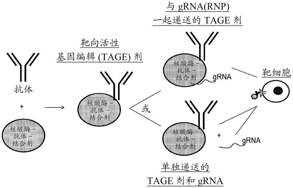
Provided herein are compositions and methods relating to Targeted Active Gene Editing (TAGE) agents that can edit nucleic acids within a particular cell type in vivo and ex vivo. Further, provided herein are compositions and methods for promoting cellular internalization of intracellular site-directed modified polypeptides in vivo and ex vivo. The modular and programmable design of the TAGE agents enables rapid retargeting and versatility, thereby enabling flexible targeting of multiple desired cell types. Furthermore, by editing a particular nucleic acid in a particular target cell, the TAGE agent has dual specificity and has fewer off-target effects than DNA-based delivery methods. To accomplish this, the TAGE agent includes one or more antigen binding polypeptides that promote cell binding and/or cell internalization. The TAGE agents of the present compositions and methods can thereby facilitate the delivery and internalization of site-directed modifying polypeptides (e.g., gene-editing polypeptides), such as Cas9, into a target cell type. Furthermore, antigen-binding polypeptides not only allow receptor-mediated entry of the TAGE agent, but in some cases, the antigen-binding polypeptide also mediates the biology of the cell (e.g., by altering intracellular signal transduction pathways). The TAGE agents described herein are particularly suitable for systemic delivery.
Thus, provided herein are methods and compositions involving a TAGE agent comprising an antigen-binding polypeptide and a site-directed modifying polypeptide that recognizes a nucleic acid sequence within a cell, wherein the antigen-binding polypeptide and the site-directed modifying polypeptide are stably associated such that the site-directed modifying polypeptide can internalize into the cell.
In one aspect, provided herein is a Targeted Active Gene Editing (TAGE) agent comprising an antigen binding polypeptide that specifically binds to an extracellular membrane-binding molecule (e.g., a cell surface molecule) and a site-directed modifying polypeptide that recognizes a nucleic acid sequence within a target cell. The antigen binding polypeptide and the site-directed modifying polypeptide are stably associated such that the site-directed modifying polypeptide can be internalized into a target cell displaying the extracellular membrane-binding molecule.
Further, provided herein are methods of modifying the genome of a cell ex vivo or in vivo, as well as methods of delivering a site-directed modifying polypeptide to a subject via a TAGE agent. Targeted ex vivo editing by TAGE agents enables genetic modification of cells (e.g., hematopoietic stem cells) for use in a variety of cell therapies. In addition, administration of a TAGE agent to a subject can target editing of a desired cell type in vivo.
I. Definition of
The term "targeted active gene editing" or "TAGE" agent refers to a complex of molecules that includes: an antigen-binding polypeptide (e.g., an antibody or antigen-binding portion thereof) that specifically binds to an extracellular target molecule (e.g., an extracellular protein or glycan, such as an extracellular protein on the surface of a cell) displayed on a cell membrane and a site-directed modifying polypeptide (such as, but not limited to, an endonuclease) that recognizes a nucleic acid sequence. The antigen binding polypeptide of the TAGE agent is associated with the site-directed modifying polypeptide such that at least the site-directed modifying polypeptide is internalized by the target cell (i.e., the cell expressing the extracellular molecule to which the antigen binding polypeptide binds). An example of a TAGE agent is an active CRISPR-Targeting (TAGE) agent, wherein the site-directed polypeptide is a nucleic acid-guided DNA endonuclease (e.g., an RNA-guided endonuclease or a DNA-guided endonuclease), such as Cas9 or Cas 12. In some embodiments, the TAGE agent comprises at least one NLS. Notably, the TAGE agent can target any nucleic acid within the cell, including but not limited to a gene.
The term "antigen-binding polypeptide" as used herein refers to a protein that binds to a particular target antigen, such as an extracellular cell membrane-bound protein (e.g., a cell surface protein). Examples of antigen-binding polypeptides include antibodies, antigen-binding fragments of antibodies, and antibody mimetics. In certain embodiments, the antigen binding polypeptide is an antigen binding peptide.
As used herein, "site-directed modifying polypeptide" refers to a protein that is targeted to a particular nucleic acid sequence or a set of similar sequences of a polynucleotide chain by modifying the polypeptide itself or a related molecule (e.g., RNA) to recognize the particular sequence, wherein the polypeptide can modify the polynucleotide chain.
The terms "polypeptide" or "protein" are used interchangeably herein to refer to any polymeric chain of amino acids. The term "polypeptide" encompasses natural or artificial proteins, protein fragments and polypeptide analogs of a protein sequence.
The term "conjugate moiety" as used herein refers to a moiety capable of conjugating two or more molecules, such as antigen binding proteins and site-directed modifying polypeptides. The term "conjugation" as used herein refers to the physical or chemical complexation that is formed between a molecule (e.g., an antibody) and a second molecule (e.g., a site-directed modifying polypeptide, a therapeutic agent, a drug, or a targeting molecule). Chemical complexation specifically constitutes a bond or a chemical moiety formed between a functional group of a first molecule (e.g., an antibody) and a functional group of a second molecule (e.g., a site-directed modifying polypeptide, a therapeutic agent, or a drug). Such bonds include, but are not limited to, covalent and non-covalent bonds, while such chemical moieties include, but are not limited to, ester, carbonate, phosphoramidate, hydrazone, acetal, orthoester, peptide and oligonucleotide bonds. In one embodiment, conjugation is achieved via physical association or non-covalent complexation.
As used herein, the term "target cell" refers to a cell or population of cells, such as mammalian cells (e.g., human cells), that includes a nucleic acid sequence in which site-directed modification of a nucleic acid is desired (e.g., to produce a genetically modified cell in vivo or ex vivo). In some cases, the target cell displays on its cell membrane an extracellular molecule (e.g., an extracellular protein such as a receptor or ligand or glycan) to which the antigen-binding polypeptide of the TAGE agent specifically binds.
The term "genetically modified cell" as used herein refers to a cell or an ancestor thereof in which the DNA sequence has been intentionally modified by a site-directed modifying polypeptide.
As used herein, the term "nucleic acid" refers to a molecule comprising nucleotides, including polynucleotides, oligonucleotides, or other DNA or RNA. In one embodiment, the nucleic acid is present in a cell and can be transmitted to the progeny of the cell via cell division. In some cases, the nucleic acid is a gene found in the genome of the cell within its chromosome (e.g., an endogenous gene). In other cases, the nucleic acid is a mammalian expression vector that has been transfected into a cell. DNA incorporated into the genome of a cell using, for example, transfection methods, is also considered to be within the scope of "nucleic acid" as used herein, even if the incorporated DNA is not meant to be passed on to progeny cells.
As used herein, the term "endosomal escape agent" or "endosomal release agent" refers to an agent (e.g., peptide) that, when conjugated to a molecule (e.g., a polypeptide, such as a site-directed modifying polypeptide), is capable of promoting endosomal release of the molecule from a cell. The polypeptides retained within the endosome may ultimately be targeted for degradation or recycling rather than release into the cytoplasm or transport to a desired subcellular destination. Thus, in some embodiments, the TAGE agent comprises an endosomal escape agent.
As used herein, the term "stably associated" when used in the context of a TAGE agent refers to the ability of an antigen-binding polypeptide and a site-directed modifying polypeptide to complex in the following manner: the complex may be internalized into a target cell such that nucleic acid editing may occur intracellularly. Examples of ways to determine whether a TAGE agent is stably associated include in vitro analysis whereby association of the complex is determined after exposure of the cell to the TAGE agent, for example by using a standard gene editing system to determine whether gene editing has occurred. Examples of such analyses are known in the art, such as SDS-PAGE, western blot analysis, size exclusion chromatography and electrophoretic mobility shift analysis to determine protein complexes and PCR amplification, direct sequencing (e.g. next generation sequencing or Sanger sequencing), enzymatic cleavage of loci with nucleases (e.g. Celery) to confirm editing; and indirect phenotypic analysis, which measures downstream effects of editing a particular gene, such as protein loss as measured by western blotting or flow cytometry or functional protein production as measured by functional analysis.
As used herein, the term "modified nucleic acid" refers to any modification of the nucleic acid targeted by the site-directed modified polypeptide. Examples of such modifications include any alteration of the amino acid sequence, including, but not limited to, any insertion, deletion, or substitution of an amino acid residue in the nucleic acid sequence relative to a reference sequence (e.g., a wild-type or native sequence). Such amino acid changes can, for example, result in a change in gene expression (e.g., an increase or decrease in expression) or a substitution of a nucleic acid sequence. Modification of the nucleic acid may further include double-stranded cleavage, single-stranded cleavage, or binding of any of the RNA-guided endonucleases disclosed herein to the target site. RNA-guided endonuclease binding may inhibit expression of the nucleic acid or may increase expression of any nucleic acid operably linked to the nucleic acid comprising the target site.
The term "cell penetrating peptide" (CPP) refers to a peptide of typically about 5-60 amino acid residues (e.g., 5-10, 10-15, 15-20, 20-25, 25-30, 30-35, 35-40, 40-45, 45-50, 50-55, or 55-60 amino acid residues) in length that can facilitate cellular uptake of a conjugated molecule, particularly one or more site-specifically modified polypeptides. In certain embodiments, a CPP may also be characterized as being capable of facilitating movement or passage of the molecular conjugate through/by one or more of a lipid bilayer, a micelle, a cell membrane, an organelle membrane (e.g., nuclear membrane), a vesicle membrane, or a cell wall. In certain embodiments, a CPP herein may be cationic, amphiphilic, or hydrophobic. Examples of CPPs useful herein, and further descriptions of CPPs in general, are described in Borrelli, Antonella et al, Molecules 23.2(2018): 295; milletti, France sca. drug discovery today 17.15-16(2012):850-860, which is incorporated herein by reference. In addition, there is a database of experimentally validated CPPs (CPPsite, Gautam et al, 2012). The CPP of the TAGE agent can be any known CPP, such as the CPP shown in the CPPsite database.
As used herein, the term "nuclear localization signal" or "NLS" refers to a peptide that, when conjugated to a molecule (e.g., a polypeptide, such as a site-directed modifying polypeptide), is capable of facilitating entry of the molecule into the nucleus of a cell through nuclear transport. For example, NLS can direct the transport of proteins associated with it from the cytoplasm of the cell across the nuclear envelope barrier. NLS is intended to include not only the nuclear localization sequence of a particular peptide, but also derivatives capable of directing translocation of cytoplasmic polypeptides across the nuclear envelope barrier. In some embodiments, one or more NLSs (e.g., 1, 2, 3, 4, 5, 6, 7, 8, 2-6, 3-7, 4-8, 5-9, 6-10, 7-10, 8-10 NLSs) can be attached to the N-terminus, C-terminus, or both the N-terminus and C-terminus of a polypeptide of a TAGE agent herein.
The term "TAT-related peptide" as used herein refers to a CPP derived from the transcriptional transactivator (TAT) of human immunodeficiency virus. The amino acid sequence of the TAT peptide comprises RKKRRQRRR (SEQ ID NO: 9). Thus, TAT-related peptides include any peptide comprising the amino acid sequence of RKKRRQRRR (SEQ ID NO:9) or an amino acid sequence having conservative amino acid substitutions, wherein the peptide is still capable of internalization into a cell. In certain embodiments, the TAT-related peptide comprises 1, 2, or 3 amino acid substitutions, wherein the TAT-related peptide is capable of internalization into a target cell.
As used herein, the term "specifically binds" refers to an antigen-binding polypeptide that recognizes and binds to an antigen present in a sample, but which does not substantially recognize or bind to other molecules in the sample. In one embodiment, the antigen binding polypeptide that specifically binds to an antigen is at least about 1X 10⁻⁴、1×10⁻⁵、1×10⁻⁶M、1×10⁻⁷M、1×10⁻⁸M、1×10⁻⁹M、1×10⁻¹⁰M、1×10-11M、1×10-12Kd of M or greater (as determined by surface plasmon resonance or other methods known in the art (e.g., filter binding assays, fluorescence polarization, isothermal titration calorimetry), including those described further herein) binds to an antigen. In one embodimentIn one embodiment, the antigen-binding polypeptide binds specifically to an antigen if the antigen-binding polypeptide binds to the antigen with an affinity that is at least two times greater than its affinity for a non-specific antigen (as determined by surface plasmon resonance). The term "specifically binds," when used in the context of a ligand, means that the ligand is capable of recognizing and binding to its corresponding receptor. The term "specifically binds," when used in the context of a CPP, means that the CPP is capable of translocating a cell membrane. In some cases, when a CPP and an antibody or ligand are combined into a TAGE agent, the TAGE agent may exhibit specific binding characteristics of the antibody or ligand and the CPP. For example, in such cases, an antibody or ligand to the TAGE agent may confer specific binding to an extracellular cell surface molecule, such as a cell surface protein, while the CPP confers enhanced ability of the TAGE agent to translocate across the cell membrane.
The term "antibody" is used herein in the broadest sense and encompasses a variety of antibody structures, including, but not limited to, monoclonal antibodies, polyclonal antibodies, multispecific antibodies (e.g., bispecific antibodies), nanobodies, monoantibodies, and antibody fragments, so long as they exhibit the desired antigen-binding activity.
The term "antibody" includes immunoglobulin molecules and multimers thereof (e.g., IgM) comprising four polypeptide chains, two heavy (H) chains and two light (L) chains, which are interconnected by disulfide bonds. Each Heavy Chain (HC) comprises a heavy chain variable region (or domain) (abbreviated herein as HCVR or VH) and a heavy chain constant region (or domain). The heavy chain constant region comprises three domains, CH1, CH2, and CH 3. Each Light Chain (LC) comprises a light chain variable region (abbreviated herein as LCVR or VL) and a light chain constant region. The light chain constant region comprises a domain (CL 1). Each VH and VL is composed of three Complementarity Determining Regions (CDRs) and four Framework Regions (FRs) arranged from amino-terminus to carboxy-terminus in the following order: FR1, CDR1, FR2, CDR2, 1-R3, CDR3, FR 4. Immunoglobulin molecules may be of any type (e.g., IgG, IgE, IgM, IgD, IgA, and IgY), class (e.g., IgG1, IgG2, IgG3, IgG4, IgA1, and IgA2), or subclass. Thus, the VH and VL regions may be further subdivided into hypervariable regions, termed Complementarity Determining Regions (CDRs), interspersed with more conserved regions, termed Framework Regions (FRs). Each VH and VL is composed of three CDRs and four FRs, arranged from amino-terminus to carboxy-terminus in the following order: FR1, CDR1, FR2, CDR2, FR3, CDR3, FR 4.
As used herein, the term "CDR" or "complementarity determining region" refers to a non-contiguous antigen binding site found within the variable regions of heavy and light chain polypeptides. These specific regions have been described by Kabat et al, J.biol.chem.252,6609-6616(1977) and Kabat et al, Sequences of proteins of immunological interest (1991) and by Chothia et al, J.mol.biol.196: 901-. Amino acid residues comprising the CDRs defined in each of the above cited references are listed for comparison. Preferably, the term "CDR" is a CDR defined by Kabat based on sequence comparison.
The term "Fc domain" is used to define the C-terminal region of an immunoglobulin heavy chain, which can be produced by papain digestion of intact antibodies. The Fc domain may be a native sequence Fc domain or a variant Fc domain. The Fc domain of an immunoglobulin typically comprises two constant domains, a CH2 domain and a CH3 domain, and optionally a CH4 domain. The substitution of amino acid residues in the Fc portion to alter antibody effector functions is known in the art (Winter et al, U.S. patent nos. 5,648,260, 5,624,821). The Fc domain of antibodies mediates several important effector functions, such as cytokine induction, ADCC, phagocytosis, Complement Dependent Cytotoxicity (CDC) and half-life/clearance of antibody and antigen-antibody complexes. In certain embodiments, at least one amino acid residue is altered (e.g., deleted, inserted, or substituted) in the Fc domain of a binding protein comprising an Fc domain such that the effector function of the binding protein is altered.
As used herein, a "complete" or "full-length" antibody refers to an antibody comprising four polypeptide chains, two heavy (H) chains, and two light (L) chains. In one embodiment, the intact antibody is an intact IgG antibody.
The term "monoclonal antibody" as used herein refers to an antibody obtained from a substantially homogeneous population of antibodies, i.e., the individual antibodies comprising the population are identical and/or bind the same epitope, except for possible variant antibodies, e.g., containing naturally occurring mutations or produced during the production of a monoclonal antibody preparation, such variants typically being present in minor amounts. In contrast to polyclonal antibody preparations, which typically include different antibodies directed against different determinants (epitopes), each monoclonal antibody of a monoclonal antibody preparation is directed against a single determinant on the antigen. Thus, the modifier "monoclonal" indicates the character of the antibody as being obtained from a substantially homogeneous population of antibodies, and is not to be construed as requiring production of the antibody by any particular method. For example, monoclonal antibodies for use according to the invention can be prepared by a variety of techniques, including but not limited to hybridoma methods, recombinant DNA methods, phage display methods, and methods that utilize transgenic animals comprising all or a portion of a human immunoglobulin locus, such methods and other exemplary methods of preparing monoclonal antibodies as described herein.
The term "human antibody" as used herein refers to an antibody having variable regions in which both framework and CDR regions are derived from human germline immunoglobulin sequences. Furthermore, if the antibody contains constant regions, the constant regions are also derived from human germline immunoglobulin sequences. The human antibodies of the invention may comprise amino acid residues that are not encoded by human germline immunoglobulin sequences (e.g., mutations introduced by random or site-specific mutagenesis in vitro or by somatic mutation in vivo). However, the term "human antibody" as used herein is not intended to include antibodies in which CDR sequences derived from the germline of another mammalian species, such as a mouse, have been grafted onto human framework sequences.
The term "humanized antibody" is intended to mean an antibody in which CDR sequences derived from the germline of a mammalian species, such as a mouse, have been grafted onto human framework sequences. Other framework region modifications can be made in the human framework sequences. "humanized forms" of antibodies (e.g., non-human antibodies) refer to antibodies that have undergone humanization.
The term "chimeric antibody" is intended to refer to antibodies in which the variable region sequences are derived from one species and the constant region sequences are derived from another species, such as antibodies in which the variable region sequences are derived from a mouse antibody and the constant region sequences are derived from a human antibody.
An "antibody fragment," "antigen-binding fragment," or "antigen-binding portion" of an antibody refers to a molecule other than an intact antibody that comprises a portion of an intact antibody and binds to an antigen to which the intact antibody binds. Examples of antibody fragments include, but are not limited to, Fv, Fab '-SH, F (ab')2(ii) a A diabody; a linear antibody; single chain antibody molecules (e.g., scFv); and multispecific antibodies formed from antibody fragments.
A "multispecific antigen-binding polypeptide" or "multispecific antibody" is an antigen-binding polypeptide that targets and binds more than one antigen or epitope. A "bispecific", "dual-specific" or "bifunctional" antigen-binding polypeptide or antibody is a hybrid antigen-binding polypeptide or antibody having two different antigen-binding sites, respectively. Bispecific antigen-binding polypeptides and antibodies are examples of multispecific antigen-binding polypeptides or multispecific antibodies, and may be produced by a variety of methods, including, but not limited to, fusion of hybridomas or attachment of Fab' fragments. See, e.g., Songsivilai and Lachmann,1990, Clin. exp. Immunol.79: 315-; kostelny et al, 1992, J.Immunol.148:1547-1553, Brinkmann and Kontermann.2017.MABS.9(2): 182-212. For example, two binding sites of a bispecific antigen binding polypeptide or antibody will bind to two different epitopes, which may be located on the same or different protein targets.
The term "antibody mimetic" or "antibody mimetic" refers to a molecule that is structurally unrelated to an antibody but is capable of specifically binding to an antigen. Examples of antibody mimetics include, but are not limited to, adnectins (i.e., fibronectin-based binding molecules), affilins, affimers, affitins, alphabodies, affibodies, darpins, anticalins, avimers, fynomers, Kunitz domain peptides, single antibodies, nanocapms, nanobodies, unibodies, versabodies, aptamers, and peptide molecules, all of which employ binding structures that, while mimicking traditional antibody binding, are produced and function via different mechanisms.
The amino acid sequences described herein may include "conservative mutations," including nucleic acid substitutions, deletions, or additions that alter, add, or delete a single amino acid or a small number of amino acids in a coding sequence, wherein the nucleic acid alterations result in the substitution of chemically similar amino acids. A conservative amino acid substitution is one in which a first amino acid is replaced with a second amino acid that has similar chemical and/or physical properties (e.g., charge, structure, polarity, hydrophobicity/hydrophilicity) as the first amino acid. Conservative substitutions include the replacement of one amino acid with another within the following groups: lysine (K), arginine (R) and histidine (H); aspartic acid (D) and glutamic acid (E); asparagine (N) and glutamine (Q); n, Q, serine (S), threonine (T) and tyrosine (Y); K. r, H, D and E; D. e, N and Q; alanine (a), valine (V), leucine (L), isoleucine (I), proline (P), phenylalanine (F), tryptophan (W), methionine (M), cysteine (C), and glycine (G); F. w and Y; H. f, W and Y; C. s and T; c and A; s and T; c and S; s, T and Y; v, I and L; v, I and T. Other conservative amino acid substitutions are also considered to be effective, depending on the context of the amino acid in question. For example, in some cases, methionine (M) may be substituted for lysine (K). In addition, sequences that differ by conservative variations are often homologous.
The term "isolated" refers to a compound that is substantially free of other cellular material, which may be, for example, an antibody or antibody fragment. Thus, in some aspects, antibodies are provided that have been isolated from antibodies with different specificities.
Other definitions are described in the following sections.
Various aspects of the invention are described in further detail in the following subsections.
Targeted Active Gene Editing (TAGE) agents
The invention includes Targeted Active Gene Editing (TAGE) agents that can be used to deliver gene editing polypeptides (i.e., site-directed modifying polypeptides) to target cells. In some embodiments, the TAGE agent may be a biological agent. In particular embodiments, the site-directed modifying polypeptide contains a conjugate moiety that allows the protein to be conjugated to an antigen binding protein that binds an antigen associated with an extracellular region of a cell membrane. This target specificity allows for the delivery of site-directed modifying polypeptides only to cells displaying the antigen (e.g., Hematopoietic Stem Cells (HSCs), Hematopoietic Progenitor Stem Cells (HPSCs), natural killer cells, macrophages, DC cells, non-DC bone marrow cells, B cells, T cells (e.g., activated T cells), fibroblasts, or other cells). Such cells may be associated with a certain tissue or cell type associated with a disease. Thus, the TAGE agents provide a means by which the genome of a target cell can be modified.
In one embodiment, the TAGE agent comprises a nucleic acid-guided endonuclease (e.g., an RNA-guided endonuclease or a DNA-guided endonuclease) that recognizes the CRISPR sequence, such as Cas9, and an antigen-binding protein that specifically binds to an extracellular molecule (e.g., a protein, glycan, lipid) located on the membrane of a target cell. Examples of antigen binding proteins that can be used in the TAGE agents of the present invention include, but are not limited to, antibodies, antigen binding portions of antibodies, or antibody mimetics. The types of antigen binding proteins that can be used in the compositions and methods described herein are described in more detail in section IV.
The proteins within the TAGE agent (i.e., at least the site-directed modifying polypeptide and the antigen binding polypeptide) are stably associated such that the antigen binding protein directs the site-directed modifying polypeptide to the cell surface and the site-directed modifying polypeptide is internalized into the target cell. In certain embodiments, the antigen binding protein binds to an antigen on the surface of a cell such that the site-directed modifying polypeptide is internalized by the target cell, but the antigen binding protein is not internalized. In some embodiments, both the site-directed modifying polypeptide and the antigen binding protein are internalized into the target cell.
As described in more detail in section III, in certain embodiments, when the site-modifying polypeptide is a nucleic acid-guided endonuclease such as Cas9, the nucleic acid-guided endonuclease associates with the guide nucleic acid to form a nuclear protein. For example, guide RNA (grna) binds to an RNA-guided nuclease to form Ribonucleoproteins (RNPs), or guide DNA binds to a DNA-guided nuclease to form deoxyribonucleic acids (DNPs). In other embodiments, the nucleic acid-guided endonuclease is associated with a guide nucleic acid comprising a DNA: RNA hybrid. In such cases, the ribonucleoproteins (i.e., RNA-guided endonuclease and guide RNA), the deoxyribonucleic acids (i.e., DNA-guided endonuclease and guide DNA), or the nucleic acid-guided endonuclease bound to a DNA: RNA hybrid, are internalized into the target cell. In a separate embodiment, the guide nucleic acid (e.g., RNA, DNA, or DNA: RNA hybrid) is delivered into the target cell separately from the nucleic acid-guided endonuclease. The guide nucleic acid (e.g., RNA, DNA or DNA: RNA hybrid) may already be present in the target cell when the nucleic acid-guided endonuclease is internalized after contact with the TAGE agent.
The TAGE agent specifically binds to extracellular molecules (e.g., proteins, glycans, lipids) located on the membrane of the target cell. The target molecule may be, for example, an outer membrane-binding protein of a cell, but may also be a non-proteinaceous molecule such as a glycan or lipid. In one embodiment, the extracellular molecule is an extracellular protein, such as a ligand or receptor, expressed by the target cell. Extracellular target molecules may be associated with a particular disease condition or a particular tissue within an organism. Examples of extracellular molecular targets associated with cell membranes are described in the following sections.
The site-directed modifying polypeptide comprises a conjugate moiety such that the antigen binding protein can stably associate with the site-directed modifying polypeptide (thereby forming a TAGE agent). The conjugate moiety provides a covalent or non-covalent linkage between the antigen binding protein and the site-directed modifying polypeptide.
In certain embodiments, the conjugate moieties of the TAGE agents useful in the present invention are stable extracellularly, prevent aggregation of the TAGE agent, and/or maintain the TAGE agent freely soluble and in a monomeric state in aqueous media. Prior to transport or delivery into a cell, the TAGE agent is stable and remains intact, e.g., the antibody or antigen binding protein thereof remains linked to a nucleic acid-guided endonuclease.
In one embodiment, the conjugate moiety is protein a, wherein the site-directed modifying polypeptide comprises protein a, and the antigen binding protein comprises an Fc region that can be bound by protein a, e.g., an antibody comprising an Fc domain. In one embodiment, the site-directed modifying polypeptide comprises SEQ ID NO:2 or an Fc binding portion thereof (SEQ ID NO:2 corresponds to the amino acid sequence of protein A).
In another embodiment, the conjugate moiety is a Spycatcher/SpyTag peptide system. For example, in certain embodiments, the site-directed modifying polypeptide comprises SpyCatcher (e.g., at the N-terminus or C-terminus), and the antigen binding polypeptide comprises SpyTag. For example, where the site-directed modifying polypeptide comprises Cas9, Cas9 can be conjugated to Spycatcher to form Spycatcher-Cas9(SEQ ID NO:6) or Cas9-Spycatcher (SEQ ID NO: 7). In one embodiment, the SpyTag peptide sequence is VPTIVMVDAYKRYK (SEQ ID NO: 116).
Other conjugation moieties that can be used in the TAGE agents provided herein include, but are not limited to, Spycatcher tags, Snaop tags, haloalkane dehalogenases (Halo-tags), sortases, monosynaptophines, ACP tags, SNAP tags, or any other conjugation moiety known in the art. In one embodiment, the antibody binding moiety is selected from the group consisting of protein A, CBP, MBP, GST, poly (His), biotin/streptavidin, V5-tag, Myc-tag, HA-tag, NE-tag, His-tag, Flag tag, Halo-tag, Snap-tag, Fc-tag, Nus-tag, BCCP, thioredoxin, SnooprTag, Spytag, Spycatcher, Isopeptag, SBP-tag, S-tag, Avitag, and calmodulin.
In some embodiments, the antibody-binding moiety is a chemical tag. For example, the chemical tag may be a SNAP tag, a CLIP tag, a HaloTag, or a TMP-tag. In one example, the chemical tag is a SNAP tag or CLIP tag. SNAP and CLIP fusion proteins enable the specific covalent attachment of virtually any molecule to a protein of interest. In another example, the chemical tag is HaloTag. HaloTag relates to a modular protein tagging system that allows different molecules to be linked to a single genetic fusion in solution, in living cells or in chemically fixed cells. In another example, the chemical tag is TMP-tag.
In some embodiments, the antibody binding moiety is an epitope tag. For example, the epitope tag may be a polyhistidine tag, such as a hexa-histidine tag (SEQ ID NO:25) or dodeca-histidine (SEQ ID NO:126), a FLAG tag, a Myc tag, an HA tag, a GST tag, or a V5 tag.
Depending on the conjugation method, the site-directed modifying polypeptide and the antigen binding protein may each be engineered to comprise a complementary binding pair that enables stable association of the antibody binding agent when contacted with the corresponding antibody, antigen binding fragment thereof, or antibody mimetic. Exemplary binding moiety pairs include (i) Streptavidin Binding Peptide (SBP) and Streptavidin (STV), (ii) biotin and EMA (enhanced monomeric avidin), (iii) SpyTag (ST) and Spycatcher (SC), (iv) Halo-Tag and Halo-Tag ligands, (v) and SNAP-Tag, (vi) Myc Tag and anti-Myc immunoglobulin, (vii) FLAG Tag and anti-FLAG immunoglobulin, and (ix) ybbR Tag and coenzyme A group. In some embodiments, the antibody binding unit is selected from the group consisting of SBP, biotin, SpyTag, Spycatcher, halo-tag, SNAP-tag, Myc tag, or FLAG tag.
In certain embodiments, the site-directed modifying polypeptide may alternatively be associated with the antigen binding protein via one or more linkers described herein, wherein the linker is a conjugate moiety.
The term "linker" as used herein means a divalent chemical moiety comprising a covalent bond or chain of atoms that covalently attaches an antigen binding protein to a site-directed modifying polypeptide to form a TAGE agent. Any known method of peptide or macromolecule conjugation may be used in the context of the present disclosure. In general, covalent attachment of an antigen binding protein and a site-directed modifying polypeptide requires that the linker have two reactive functional groups, i.e., a bivalent in the sense of a reaction. Bivalent linker reagents useful for attaching two or more functional or biologically active moieties (such as peptides, nucleic acids, drugs, toxins, antibodies, haptens, and reporter groups) are known, and methods for such conjugation have been described, for example, in Hermanson, G.T (1996) Bioconjugate Techniques; academic Press, New York, page 234-242, the disclosure of which is incorporated herein by reference as it relates to linkers suitable for covalent conjugation. Other linkers are disclosed in, for example, Tsuchikama, k. and zhijiang, a.protein and Cell,9(1), pages 33-46, (2018), the disclosure of which is incorporated herein by reference as it relates to linkers suitable for covalent conjugation.
In general, linkers suitable for use in the disclosed compositions and methods are stable in circulation, but allow for release of the antigen binding protein and/or site-directed modifying polypeptide in or alternatively in the vicinity of a target cell. Linkers suitable for the present disclosure can be broadly classified as non-cleavable or cleavable, as well as intracellular or extracellular, each of which is further described below.
Non-cuttable joint
In some embodiments, the linker conjugated to the antigen binding protein and the site-directed modifying polypeptide is non-cleavable. The non-cleavable linker comprises a stable chemical bond that is resistant to degradation (e.g., proteolysis). In general, non-cleavable linkers need to be proteolytically degraded within the target cell and exhibit high extracellular stability. Non-cleavable linkers suitable for use herein may also include one or more groups selected from: bond, - (C ═ O) -, C1-C6Alkylene radical, C1-C6Heteroalkylene group, C2-C6Alkenylene radical, C2-C6Heteroalkenylene, C2-C6Alkynylene, C2-C6Heteroalkynylene, C3-C6Cycloalkylene, heterocycloalkylene, arylene, heteroarylene, and combinations thereof, each of which may be optionally substituted, and/or may include one or more heteroatoms (e.g., S, N or O) in place of one or more carbon atoms. Non-limiting examples of such groups include alkylene (CH) 2)p、(C=O)(CH2)pAnd polyethylene glycol (PEG; (CH)2CH2O)p) A unit where p is an integer from 1 to 6 independently selected for each case. Non-limiting examples of non-cleavable linkers for antibody-drug conjugation include those based on maleimidomethylcyclohexanecarboxylate, hexanoylmaleimide, and acetylphenylbutyric acid.
Cleavable linker
In some embodiments, the linker conjugated to the antigen binding protein and the site-directed modifying polypeptide is cleavable, such that cleavage of the linker (e.g., by a protease, such as a metalloprotease) releases the CRISPR targeting element or the antibody or antigen binding protein thereof, or both, from the TAGE agent in an intracellular or extracellular (e.g., upon binding of the molecule to a cell surface) environment. The cleavable linker is designed to take advantage of differences in local environment (e.g., extracellular and intracellular environment, such as pH, reduction potential, or enzyme concentration) to trigger the release of the TAGE agent component (i.e., antigen binding protein, site-directed modifying polypeptide, or both) in the target cell. In general, cleavable linkers are relatively stable in circulation in vivo, but are particularly susceptible to cleavage by one or more mechanisms (e.g., including, but not limited to, protease, peptidase, and glucuronidase activities) in the intracellular environment. The cleavable linkers used herein are stable outside the target cell and can cleave at an effective rate inside the target cell or near the extracellular membrane of the target cell. The effective joint will: (i) retain the specific binding characteristics of antigen binding proteins such as antibodies; (ii) allowing intracellular or extracellular delivery of the TAGE agent or component thereof (i.e., site-directed modifying polypeptide); (iii) remain stable and intact, i.e., not cleaved, until the TAGE agent is delivered or transported to its target site; and (iv) maintaining the gene targeting effect of the site-directed modifying polypeptide (e.g., CRISPR). The stability of the TAGE agent can be measured by standard analytical techniques such as mass spectrometry, sizing by size exclusion chromatography or diffusion constant measurement by dynamic light scattering, HPLC and separation/analysis techniques LC/MS.
Suitable cleavable linkers include those that can be cleaved by, for example, enzymatic hydrolysis, photolysis, hydrolysis under acidic conditions, hydrolysis under basic conditions, oxidation, disulfide reduction, nucleophilic cleavage, or organometallic cleavage (see, e.g., Lerich et al, bioorg.Med.chem.,20: 571-. Suitable cleavable linkers may include, for example, chemical moieties such as hydrazines, disulfides, thioethers, or peptides.
Linkers that are hydrolyzable under acidic conditions include, for example, hydrazones, semicarbazones, thiosemicarbazones, cis-aconitamides, orthoesters, acetals, ketals, and the like (see, for example, U.S. Pat. Nos. 5,122,368, 5,824,805, 5,622,929; Dubowchik and Walker,1999, pharm. therapeutics 83: 67-123; Neville et al, 1989, biol. chem.264:14653-14661, the disclosure of each of which is incorporated herein by reference in its entirety as it relates to linkers suitable for covalent conjugation). Such linkers are relatively stable under neutral pH conditions, such as in blood, but are unstable below pH 5.5 or 5.0 (the approximate pH of lysosomes). In general, linkers comprising such acid labile functional groups tend to be relatively unstable extracellularly. This lower stability may be advantageous when extracellular cleavage is required.
Linkers cleavable under reducing conditions include, for example, disulfides. A variety of disulfide linkers are known In the art, including, for example, those that can be formed using SATA (N-succinimidyl-S-acetylthioacetate), SPDP (N-succinimidyl-3- (2-pyridyldithio) propionate), SPDB (N-succinimidyl-3- (2-pyridyldithio) butyrate), and SMPT (N-succinimidyl-oxycarbonyl- α -methyl- α - (2-pyridyldithio) toluene), SPDB, and SMPT (see, e.g., Thorpe et al, 1987, Cancer Res.47: 5924-year 5931; Wawrzyncck et al, In Immunoconjugates: Antibody Conjugates In radio and Therapy of Cancer (C.W.Vogel ed., Oxford U.ss, 1987. see also U.S. Pat. No. 4,880,935, the disclosure of each of which is incorporated herein by reference In its entirety, as it involves a linker suitable for covalent conjugation). Disulfide-based linkers tend to be relatively unstable in the plasma circulation, however, this lower stability may be advantageous in situations where extracellular cleavage is required. Sensitivity to cleavage can also be adjusted by, for example, introducing steric hindrance near the disulfide moiety to hinder reductive cleavage.
The linker susceptible to enzymatic hydrolysis may be, for example, a peptide-containing linker that is cleaved by an intracellular peptidase or protease, including but not limited to lysosomal or endosomal proteases. In some embodiments, the peptidyl linker is at least two amino acids or at least three amino acids in length. Exemplary amino acid linkers include dipeptides, tripeptides, tetrapeptides, or pentapeptides. Examples of suitable peptides include those containing amino acids such as valine, alanine, citrulline (Cit), phenylalanine, lysine, leucine, and glycine. Amino acid residues comprising the amino acid linker component include those that occur naturally, as well as minor amino acids and non-naturally occurring amino acid analogs, such as citrulline. Exemplary dipeptides include valine-citrulline (vc or val-cit) and alanine-phenylalanine (af or ala-phe). Exemplary tripeptides include glycine-valine-citrulline (gly-val-cit) and glycine-glycine (gly-gly-gly). In some embodiments, the linker comprises a dipeptide, such as Val-Cit, Ala-Val, or Phe-Lys, Val-Lys, Ala-Lys, Phe-Cit, Leu-Cit, Ile-Cit, Phe-Arg, or Trp-Cit. Linkers containing dipeptides such as Val-Cit or Phe-Lys are disclosed, for example, in U.S. Pat. No. 6,214,345, the disclosure of which is incorporated herein by reference in its entirety as it relates to linkers suitable for covalent conjugation. In some embodiments, the linker comprises a dipeptide selected from Val-Ala and Val-Cit. In certain embodiments, a linker comprising a peptide moiety may be susceptible to varying degrees of cleavage both intracellularly and extracellularly. Thus, in some embodiments, the linker comprises a dipeptide and the TAGE agent is cleaved extracellularly. Thus, in some embodiments, the linker comprises a dipeptide, and the TAGE agent is stable extracellularly and cleaved intracellularly.
Linkers suitable for conjugating the antigen binding proteins disclosed herein to the site-directed modifying polypeptides disclosed herein include those that are capable of releasing the antigen binding protein or site-directed modifying polypeptide by a 1, 6-elimination process. Chemical moieties capable of performing this elimination process include p-aminobenzyl (PAB) groups, 6-maleimidocaproic acid, pH-sensitive carbonates and other reagents, as described in Jain et al, pharm. Res.32:3526-3540,2015, the disclosure of which is incorporated herein by reference in its entirety as it relates to linkers suitable for covalent conjugation.
In some embodiments, the linker comprises a "self-immolative" group, such as the PAB or PABC (p-aminobenzyloxycarbonyl) groups described above, which are disclosed in, for example, Carl et al, J.Med.chem. (1981)24: 479-. Other such chemical moieties ("self-immolative linkers") capable of performing this process include methylene carbamates and heteroaryl groups such as aminothiazoles, aminoimidazoles, aminopyrimidines, and the like. Linkers containing such heterocyclic self-consuming groups are disclosed, for example, in U.S. patent publication Nos. 20160303254 and 20150079114 and in U.S. patent Nos. 7,754,681, Hay et al (1999) bioorg. Med. chem.Lett.9:2237, U.S. patent No. 2005/0256030, de Groot et al (2001) J.org. chem.66:8815-8830 and U.S. patent No. 7223837. In some embodiments, the dipeptide is used in combination with a self-immolative linker.
Linkers suitable for use herein may further comprise one or more groups selected from: c1-C6Alkylene radical, C1-C6Heteroalkylene group, C2-C6Alkenylene radical, C2-C6Heteroalkenylene, C2-C6Alkynylene, C2-C6Heteroalkynylene, C3-C6Cycloalkylene, heterocycloalkylene, arylene, heteroarylene, and combinations thereof, each of which may be optionally substituted. Non-limiting examples of such groups include (CH)2)p、(CH2CH2O)pAnd- (C ═ O) (CH)2)p-a unit, where p is an integer from 1 to 6 independently selected for each case.
In some embodiments, the linker may comprise one or more of the following: hydrazine, disulfide, thioether, dipeptide, p-aminobenzyl (PAB) group, heterocyclic self-immolative group, optionally substituted C1-C6Alkyl, optionally substituted C1-C6Heteroalkyl, optionally substituted C2-C6Alkenyl, optionally substituted C2-C6Heteroalkenyl, optionally substituted C2-C6Alkynyl, optionally substituted C2-C6Heteroalkynyl, optionally substituted C3-C6Cycloalkyl, optionally substituted heterocycloalkyl, optionally substituted aryl, optionally substituted heteroaryl, solubility enhancing group, acyl, - (C ═ O) -or- (CH)2CH2O)p-a group, wherein p is an integer from 1 to 6. One skilled in the art will recognize that one or more of the listed groups may be divalent (diradical) species (e.g., C) 1-C6Alkylene and the like) Exist in the form of (1).
In some embodiments, the linker comprises a p-aminobenzyl group (PAB). In one embodiment, the p-aminobenzyl group is disposed between the cytotoxic drug and the protease cleavage site in the linker. In one embodiment, the p-aminobenzyl group is part of a p-aminobenzyloxycarbonyl unit. In one embodiment, the para-aminobenzyl group is part of a para-aminobenzylamido unit.
In some embodiments, the linker comprises PAB, Val-Cit-PAB, Val-Ala-, PAB, Val-Lys (Ac) -PAB, Phe-Lys (Ac) -PAB, D-Val-Leu-Lys, Gly-Gly-Arg, Ala-Ala-Asn-PAB, or Ala-PAB. In some embodiments, the linker comprises a combination of one or more of the following: peptides, oligosaccharides, - (CH)2)p-、-(CH2CH2O)p-, PAB, Val-Cit-PAB, Val-Ala-PAB, Val-Lys (Ac) -PAB, Phe-Lys (Ac) -PAB, D-Val-Leu-Lys, Gly-Gly-Arg, Ala-Ala-Asn-PAB or Ala-PAB.
Suitable linkers may be substituted with groups that modulate solubility or reactivity. Suitable linkers may contain groups with solubility enhancing properties. Comprises (CH)2CH2O)pThe linker of the unit (polyethylene glycol, PEG) may for example enhance solubility, as may the alkyl chain which may be substituted by amino, sulfonic, phosphonic or phosphoric acid residues. Linkers comprising such moieties are disclosed, for example, in U.S. patent nos. 8,236,319 and 9,504,756, the disclosure of each of which is incorporated herein by reference as it relates to linkers suitable for covalent conjugation. Linkers containing such groups are described, for example, in U.S. patent No. 9,636,421 and U.S. patent application publication No. 2017/0298145, the disclosures of which are incorporated herein by reference as they relate to linkers suitable for covalent conjugation.
Suitable linkers for covalently conjugating the antigen binding proteins and site-directed modifying polypeptides disclosed herein may have two reactive functional groups (i.e., two reactive termini), one for conjugation to the antigen binding protein and the other for conjugation to the site-directed modifying polypeptide. In certain embodiments, suitable sites on the antigen binding protein for conjugation are nucleophilic, such as thiol, amino groups, or hydroxyl groups. Reactive (e.g., nucleophilic) sites that may be present within the antigen binding proteins disclosed herein include, but are not limited to, nucleophilic substituents on amino acid residues, such as (i) N-terminal amine groups, (ii) pendant amine groups, e.g., lysine, (iii) pendant thiol groups, e.g., cysteine, (iv) pendant hydroxyl groups, e.g., serine; or (iv) a sugar hydroxyl or amino group, wherein the antibody is glycosylated. Suitable sites on the antigen binding protein for conjugation include, but are not limited to, hydroxyl moieties of serine, threonine, and tyrosine residues; the amino moiety of a lysine residue; the carboxyl portion of aspartic and glutamic acid residues; and the thiol moiety of a cysteine residue, as well as the propargyl, azido, haloaryl (e.g., fluoroaryl), haloheteroaryl (e.g., fluoroheteroaryl), haloalkyl, and haloheteroalkyl moieties of a non-naturally occurring amino acid. Thus, in certain embodiments, the antibody-conjugated reactive end on the linker is a thiol-reactive group (such as a double bond, as in maleimide), a leaving group (such as chloro, bromo, iodo), or an R-thio group or a carboxyl group.
In certain embodiments, suitable sites on site-directed modified polypeptides for conjugation may also be nucleophilic. Reactive (e.g., nucleophilic) sites that may be present within the site-directed modified polypeptides disclosed herein include, but are not limited to, nucleophilic substituents on amino acid residues, such as (i) N-terminal amine groups, (ii) pendant amine groups, e.g., lysine, (iii) pendant thiol groups, e.g., cysteine, (iv) pendant hydroxyl groups, e.g., serine; or (iv) a sugar hydroxyl or amino group, wherein the antibody is glycosylated. Suitable sites on site-directed modified polypeptides for conjugation include, but are not limited to, hydroxyl moieties of serine, threonine, and tyrosine residues; the amino moiety of a lysine residue; the carboxyl portion of aspartic and glutamic acid residues; and the thiol moiety of a cysteine residue, as well as the propargyl, azido, haloaryl (e.g., fluoroaryl), haloheteroaryl (e.g., fluoroheteroaryl), haloalkyl, and haloheteroalkyl moieties of a non-naturally occurring amino acid. Thus, in certain embodiments, the site-directed modifying polypeptide-conjugated reactive terminus on the linker is a thiol-reactive group (such as a double bond, as in maleimide), a leaving group (such as chloro, bromo, iodo), or an R-thio group or a carboxyl group.
In some embodiments, the reactive functional group attached to the linker is a nucleophilic group that reacts with an electrophilic group present on the antigen binding protein, the site-directed modifying polypeptide, or both. Electrophilic groups useful on antigen binding proteins or site-directed modified polypeptides include, but are not limited to, aldehyde and ketone carbonyl groups. The heteroatom of the nucleophilic group can react with an electrophilic group on the antigen-binding protein or site-directed modifying polypeptide and form a covalent bond with the antigen-binding protein or site-directed modifying polypeptide. Useful nucleophilic groups include, but are not limited to, hydrazide, oxime, amino, hydroxyl, hydrazine, thiosemicarbazone, hydrazine carboxylate, and aroylhydrazide.
In some embodiments, a TAGE agent disclosed herein comprises a nucleoside or nucleotide. Suitable sites on such nucleosides or nucleotides for conjugation include-OH or phosphate groups, respectively. Linkers and conjugation methods suitable for use in such embodiments are disclosed in, for example, Wang, t.p. et al, bioconj.chem.21(9),1642-55,2010 and Bernardinelli, g. and Hogberg, b.nucleic Acids Research,45(18), page e160 (published online 8/16/2017), the disclosure of each of which is incorporated herein by reference as it relates to linkers suitable for covalent conjugation.
When the term "linker" is used to describe a conjugated form of a linker, one or both reactive termini will be absent (converted to a chemical moiety) or incomplete (such as the carbonyl group of a carboxylic acid only) due to the formation of a bond between the linker and the antigen binding protein and/or between the linker and the site-directed modifying polypeptide. Thus, linkers useful herein include, but are not limited to, linkers containing: chemical moieties formed by conjugation reactions between reactive functional groups on the linker and nucleophilic groups or other reactive substituents on the antigen binding protein, and chemical moieties formed by conjugation reactions between reactive functional groups on the linker and nucleophilic groups on the site-directed modified polypeptide.
Examples of chemical moieties formed by these coupling reactions result from reactions between chemically reactive functional groups, including nucleophile/electrophile pairs (e.g., thiol/haloalkane pairs, amine/carbonyl pairs, or thiol/α, β -unsaturated carbonyl pairs, etc.), diene/dienophile pairs (e.g., azide/alkyne pairs, or diene/α, β -unsaturated carbonyl pairs, etc.), and the like. Coupling reactions between reactive functional groups that form chemical moieties include, but are not limited to, thiol alkylation, hydroxyalkylation, amine alkylation, amine or hydroxylamine condensation, hydrazine formation, amidation, esterification, disulfide formation, cycloaddition (e.g., [4+2] Diels-Alder cycloaddition, [3+2] Huisgen cycloaddition, etc.), nucleophilic aromatic substitution, electrophilic aromatic substitution, and other reactive forms known in the art or described herein. Suitable linkers can contain electrophilic functional groups for reaction with nucleophilic functional groups on the antigen binding protein, site-directed modifying polypeptide, or both.
In some embodiments, the reactive functional group present within an antigen binding protein, site-directed modifying polypeptide, or both disclosed herein is an amine or thiol moiety. Certain antigen binding proteins have reducible interchain disulfides, i.e., cysteine bridges. The antigen binding protein may be made reactive for conjugation to a linker reagent by treatment with a reducing agent such as DTT (dithiothreitol). Thus, theoretically, each cysteine bridge will form two reactive thiol nucleophiles. Additional nucleophilic groups can be introduced into the antigen binding protein by reacting lysine with 2-iminothiolane (Traut's reagent) to convert the amine to a thiol. Reactive thiol groups can be introduced into antigen binding proteins by introducing one, two, three, four, or more cysteine residues (e.g., making mutant antibodies comprising one or more non-native cysteine amino acid residues). U.S. patent No. 7,521,541 proposes engineering antibodies by introducing reactive cysteine amino acids.
Linkers suitable for synthesizing the covalent conjugates disclosed herein include, but are not limited to, reactive functional groups such as maleimide or haloalkyl. These groups may be present in linkers or crosslinking reagents such as 4- (N-maleimidomethyl) -cyclohexane-L-carboxylic acid succinimidyl ester (SMCC), N-Succinimidyl Iodoacetate (SIA), sulfo-SMCC, m-maleimidobenzoyl-N-hydroxysuccinimide ester (MBS), sulfo-MBS, and succinimidyl iodoacetate, as well as those described, for example, in Liu et al, 18:690-697,1979, the disclosure of which is incorporated herein by reference as it relates to linkers for chemical conjugation.
In some embodiments, one or both of the reactive functional groups attached to the linker are maleimide, azide, or alkyne. An example of a maleimide-containing linker is a non-cleavable maleimidocaproyl-based linker. Doronina et al, Bioconjugate chem.17:14-24,2006, the disclosure of which is incorporated herein by reference as it relates to linkers for chemical conjugation, describe such linkers.
In some embodiments, the reactive functional group is- (C ═ O) -or-NH (C ═ O) -, such that the linker can be bound to the antigen binding protein or site-directed modifying polypeptide, respectively, through an amide or urea moiety resulting from the reaction of the- (C ═ O) -or-NH (C ═ O) -group with the amino group of the antigen binding protein or site-directed modifying polypeptide, or both.
In some embodiments, the reactive functional group is an N-maleimido group, a halogenated N-alkylamido group, a sulfonyloxy N-alkylamido group, a carbonate group, a sulfonylhalide group, a thiol group or a derivative thereof, an alkynyl group containing an internal carbon-carbon triple bond, a (hetero) cycloalkynyl group, a bicyclo [6.1.0] non-4-yn-9-yl group, an alkenyl group containing an internal carbon-carbon double bond, a cycloalkenyl group, a tetrazinyl group, an azido group, a phosphine group, an oxynitride group, a nitrone group, a nitrilimine group, a diazo group, a ketone group, an (O-alkyl) hydroxyamino group, a hydrazine group, a halogenated N-maleimido group, a 1, 1-bis (sulfonylmethyl) methylcarbonyl group or an eliminated derivative thereof, a salt thereof, a pharmaceutically acceptable salt thereof, or a pharmaceutically acceptable salt thereof, and a pharmaceutically acceptable salt thereof, A carbonyl halide group or an allenamide group, each of which groups may be optionally substituted. In some embodiments, the reactive functional group comprises a cycloalkene group, a cycloalkyne group, or an optionally substituted (hetero) cycloalkyne group.
Examples of suitable divalent linker reagents suitable for preparing the conjugates disclosed herein include, but are not limited to, N-succinimidyl 4- (maleimidomethyl) cyclohexanecarboxylate (SMCC), N-succinimidyl-4- (N-maleimidomethyl) -cyclohexane-1-carboxy- (6-aminocaproate), which is a "long chain" analog of SMCC (LC-SMCC), kappa-maleimidoundecanoic acid N-succinimidyl ester (KMUA), gamma-maleimidobutyric acid N-succinimidyl ester (GMBS), epsilon-maleimidohexanoic acid N-hydroxysuccinimidyl Ester (EMCS), m-maleimidobenzoyl-N-hydroxysuccinimidyl ester (MBS), N- (. alpha. -maleimidoacetoxy) -succinimidyl ester (AMAS), succinimidyl-6- (. beta. -maleimidopropionamido) hexanoate (SMPH), N-succinimidyl 4- (p-maleimidophenyl) -butyrate (SMPB), and N- (p-maleimidophenyl) isocyanate (PMPI). Crosslinking agents containing haloacetyl-based moieties include N-succinimidyl-4- (iodoacetyl) -aminobenzoate (SIAB), N-Succinimidyl Iodoacetate (SIA), N-Succinimidyl Bromoacetate (SBA), and N-succinimidyl 3- (bromoacetamido) propionate (SBAP).
One of skill in the art will recognize that any one or more of the chemical groups, moieties, and features disclosed herein can be combined in a variety of ways to form linkers useful for conjugating the antigen binding proteins disclosed herein to the site-directed modifying polypeptides disclosed herein. Other linkers useful in conjunction with the compositions and methods described herein are described, for example, in U.S. patent application publication No. 2015/0218220, the disclosure of which is incorporated herein by reference, as it relates to linkers suitable for covalent conjugation.
Site-directed modification of polypeptides with TAGE agents
The TAGE agent comprises a site-directed modifying polypeptide, such as a nucleic acid-guided endonuclease (e.g., an RNA-guided endonuclease (e.g., Cas9) or a DNA-guided endonuclease) that recognizes a nucleic acid sequence in the target cell.
The site-directed modifying polypeptides used in the compositions and methods disclosed herein are site-specific in that the polypeptide itself or a related molecule recognizes and targets a particular nucleic acid sequence or a set of similar sequences (i.e., a target sequence). In some embodiments, site-directed modifying polypeptides (or related molecules thereof) recognize sequences that are similar in sequence and that comprise conserved bases or motifs that may be degenerate at one or more positions.
In particular embodiments, the site-directed modifying polypeptide modifies the polynucleotide at a specific location (i.e., modification site) outside its target sequence. The modification sites modified by a particular site-directed modifying polypeptide are also typically specific for a particular sequence or a group of similar sequences. In some of these embodiments, the site-directed modifying polypeptide modifies a sequence that is similar in sequence and comprises conserved bases or motifs that may be degenerate at one or more positions. In other embodiments, the site-directed modifying polypeptide modifies a sequence within a specific position relative to a target sequence. For example, a site-directed modifying polypeptide can modify a sequence within a particular number of nucleic acids upstream or downstream of a target sequence.
As used herein, with respect to site-directed modification of a polypeptide, the term "modification" means any insertion, deletion, substitution, or chemical modification of at least one nucleotide in the modification site, or alternatively a change in expression of a gene adjacent to the target site. The substitution of at least one nucleotide in the modification site may be the result of recruitment of a base editing domain, such as a cytidine deaminase or adenine deaminase domain (see, e.g., Eid et al (2018) Biochem J475 (11):1955-1964, which is incorporated herein in its entirety).
Changes in expression of genes adjacent to the target site may result from recruitment of a transcriptional activation domain or transcriptional repression domain to the promoter region of the gene or recruitment of an epigenetic modification domain that covalently modifies DNA or histone to alter histone structure and/or chromosome structure without altering the DNA sequence, resulting in changes in gene expression of adjacent genes. The term "modification" also encompasses recruitment of a detectable label to the target site, which can be conjugated to a site-directed modifying polypeptide or related molecule (e.g., a gRNA) that allows for detection of a particular nucleic acid sequence (e.g., a disease-related sequence).
In some embodiments, the site-directed modifying polypeptide is a nuclease or variant thereof, and thus an agent comprising a nuclease or variant thereof is referred to herein as a gene editing cell Targeting (TAGE) agent. As used herein, "nuclease" refers to an enzyme that cleaves phosphodiester bonds in the backbone of a polynucleotide strand. Suitable nucleases for use in the compositions and methods disclosed herein can have endonuclease and/or exonuclease activity. Exonucleases cleave one nucleotide at a time from the end of a polynucleotide strand. Endonucleases cleave polynucleotide strands by cleaving phosphodiester bonds within the polynucleotide strand rather than at both ends of the polynucleotide strand. Nucleases can cleave either RNA polynucleotide strands (i.e., ribonucleases) and/or DNA polynucleotide strands (i.e., deoxyribonucleases).
Nucleases cleave the polynucleotide strand, thereby generating a cleavage site. As used herein, the term "cleavage" refers to hydrolysis of phosphodiester bonds within the backbone of a polynucleotide strand. Cleavage by the nucleases of the TAGE agents disclosed herein can be single-stranded or double-stranded. In some embodiments, double-stranded cleavage of DNA is achieved via cleavage with two nucleases, wherein each nuclease cleaves a single strand of DNA. Blunt ends or staggered ends may be generated by nuclease cleavage.
Non-limiting examples of nucleases suitable for the compositions and methods disclosed herein include meganucleases, such as homing endonucleases; restriction endonucleases, such as type IIS endonucleases (e.g., fokl); zinc finger nucleases; transcription activator-like effector nucleases (TALENs) and nucleic acid-guided nucleases (e.g., RNA-guided endonuclease, DNA-guided endonuclease, or DNA/RNA-guided endonuclease).
As used herein, "meganuclease" refers to an endonuclease that binds DNA at a target sequence greater than 12 base pairs in length. Meganucleases bind to double-stranded DNA as heterodimers. Suitable meganucleases for use in the compositions and methods disclosed herein include homing endonucleases, such as those of the family comprising the LAGLIDADG (SEQ ID NO:127) amino acid motif or variants thereof.
As used herein, "zinc finger nuclease" or "ZFN" refers to a chimeric protein comprising a zinc finger DNA-binding domain fused to a nuclease domain from an exonuclease or endonuclease (such as a restriction endonuclease or meganuclease). The zinc finger DNA binding domain is bound by zinc ions that serve to stabilize the unique structure.
As used herein, "transcription activator-like effector nuclease" or "TALEN" refers to a chimeric protein comprising a DNA-binding domain comprising multiple TAL domain repeats fused to a nuclease domain from an exonuclease or endonuclease (such as a restriction endonuclease or meganuclease). The TAL domain repeats may be derived from the TALE protein family of Xanthomonas (xanthomas) from Proteobacteria (Proteobacteria). TAL domain repeats are 33-34 amino acid sequences with hypervariable amino acids 12 and 13, referred to as Repeat Variable Diresidues (RVDs). The RVD confers binding specificity to the target sequence. TAL domain repeats can be engineered by rational or experimental means to generate variant TALENs with specific target sequence specificities (see, e.g., Boch et al (2009) Science 326(5959): 1509-. DNA cleavage by TALENs requires two DNA targets flanked by a non-specific spacer, where each DNA target is bound by a TALEN monomer. In some embodiments, the TALEN comprises a compact TALEN, which refers to an endonuclease comprising a DNA binding domain with one or more TAL domain repeats fused in any orientation to any portion of a homing endonuclease (e.g., I-TevI, MmeI, EndA, End1, I-basei, I-TevIII, I-TwoI, MspI, MvaI, NucA, and NucM). Compact TALENs are advantageous because they do not require dimerization to obtain DNA processing activity, thus requiring only a single target site.
As used herein, "nucleic acid-guided nuclease" refers to a nuclease that is directed to a particular target sequence based on complementarity (in whole or in part) between a guide nucleic acid (i.e., guide RNA or gRNA, guide DNA or gDNA, or guide DNA/RNA hybrid) associated with the nuclease and the target sequence. Binding between the guide RNA and the target sequence serves to recruit the nuclease to the vicinity of the target sequence. Non-limiting examples of nucleic acid-guided nucleases suitable for the compositions and methods disclosed herein include naturally occurring, regularly clustered, short palindromic repeats (CRISPR) -associated (Cas) polypeptides from prokaryotic organisms (e.g., bacteria, archaea) or variants thereof. CRISPR sequences found in prokaryotes are sequences derived from fragments of polynucleotides from invading viruses, used in subsequent infection processes to recognize similar viruses, and cleave viral polynucleotides via CRISPR-associated (Cas) polypeptides that act as RNA-guided nucleases to cleave viral polynucleotides. As used herein, "CRISPR-associated polypeptide" or "Cas polypeptide" refers to a naturally occurring polypeptide found in the vicinity of a CRISPR sequence within a naturally occurring CRISPR system. Certain Cas polypeptides act as RNA-guided nucleases.
There are at least two classes of naturally occurring CRISPR systems, class 1 and class 2. Generally, whereas class 2 CRISPR systems comprise a single polypeptide having nucleic acid-guided nuclease activity, while class 1 CRISPR systems require a protein complex to obtain nuclease activity, the nucleic acid-guided nuclease of the compositions and methods disclosed herein is a class 2 Cas polypeptide or a variant thereof. There are at least three known types of class 2 CRISPR systems, i.e. type II, V and VI, where there are multiple subtypes (subtypes II-A, II-B, II-C, V-A, V-B, V-C, VI-A, VI-B and VI-C, as well as other undefined or putative subtypes). Generally, type II and V-B systems require tracrRNA in addition to crRNA for activity. In contrast, forms V-A and VI require only crRNA for activity. All known type II and V RNA-guided nucleases target double-stranded DNA, while all known type VI RNA-guided nucleases target single-stranded RNA. The RNA-guided nuclease of the type II CRISPR system is referred to herein and in the literature as Cas 9. In some embodiments, the nucleic acid-guided nuclease of the compositions and methods disclosed herein is a type II Cas9 protein or a variant thereof. V-type Cas polypeptides used as RNA-guided nucleases do not require tracrRNA to target and cleave the target sequence. RNA-guided nucleases of VA-type CRISPR systems are referred to herein and in the literature as Cpf 1; the RNA guided nuclease of the VB-type CRISPR system is called C2C 1; the RNA-guided nuclease of VC-type CRISPR system is called Cas12C or C2C 3; the RNA-guided nuclease of VIA-type CRISPR system is called C2C2 or Cas13a 1; the RNA-guided nuclease of the type VIB CRISPR system is called Cas 13B; and the RNA-guided nuclease of the VIC-type CRISPR system is called Cas13a 2. In certain embodiments, the nucleic acid-directed nucleases of the compositions and methods disclosed herein are VA-type Cpf1 proteins or variants thereof. Naturally occurring Cas polypeptides and variants thereof for use as nucleic acid-guided nucleases are known in the art and include, but are not limited to, Streptococcus pyogenes Cas9, Staphylococcus aureus (Staphylococcus aureus) Cas9, Streptococcus thermophilus (Streptococcus thermophilus) Cas9, Francisella novaculeatus (Francisella novicida) Cpf1 or Shmakov et al (2017) Nat Rev Microbiol 15(3): 169-; makarova et al (2015) Nat Rev Microbiol 13(11): 722-736; and those described in U.S. patent No. 9790490, each of which is incorporated herein in its entirety. Class 2V-type CRISPR nucleases include Cas12 and any subtype of Cas12, such as Cas12a, Cas12b, Cas12c, Cas12d, Cas12e, Cas12f, Cas12g, Cas12h and Cas12 i. Class 2 type VI CRISPR nucleases, including Cas13, can be included in the TAGE agent to cleave RNA target sequences.
The nucleic acid-guided nuclease of the compositions and methods disclosed herein can be a naturally-occurring nucleic acid-guided nuclease (e.g., streptococcus pyogenes Cas9) or a variant thereof. Variant nucleic acid-guided nucleases can be engineered or naturally occurring variants containing amino acid substitutions, deletions, or additions that, for example, alter the activity of one or more of the nuclease domains, fuse the nucleic acid-guided nuclease to a heterologous domain that confers a modification property (e.g., a transcriptional activation domain, an epigenetic modification domain, a detectable label), modify the stability of the nuclease, or modify the specificity of the nuclease.
In some embodiments, the nucleic acid-guided nuclease comprises one or more mutations to improve specificity for a target site and/or stability in the intracellular microenvironment. For example, where the protein is Cas9 (e.g., SpCas9) or modified Cas9, it may be beneficial to delete any or all of the residues N175 to R307 (including endpoints) of the Rec2 domain. It can be found that smaller or lower molecular weight forms of nucleases are more effective. In some embodiments, the nuclease comprises at least one substitution relative to the naturally occurring form of the nuclease. For example, when the protein is Cas9 or modified Cas9, mutations C80 or C574 (or homologues thereof in modified proteins with indels) may be beneficial. In Cas9, the desired substitutions may include any of C80A, C80L, C80I, C80V, C80K, C574E, C574D, C574N, C574Q (in any combination), particularly C80A. Substitutions may be included to reduce intracellular protein binding of the nuclease and/or increase target site specificity. Alternatively or optionally, substitutions may be included to reduce off-target toxicity of the composition.
Nucleic acid-guided nucleases are directed to a particular target sequence by their association with guide nucleic acids (e.g., guide rna (grna), guide dna (gdna)). The nucleic acid-guided nuclease binds to the guide nucleic acid via non-covalent interactions, thereby forming a complex. The polynucleotide targeting nucleic acid provides target specificity for the complex by comprising a nucleotide sequence that is complementary to the sequence of the target sequence. The complex or a domain or labeled nucleic acid guided nuclease fused or otherwise conjugated thereto provides site specific activity. In other words, a nucleic acid-guided nuclease is guided to a target polynucleotide sequence (e.g., a target sequence in a chromosomal nucleic acid; a target sequence in an extrachromosomal nucleic acid, e.g., an episomal nucleic acid, a minicircle; a target sequence in a mitochondrial nucleic acid; a target sequence in a chloroplast nucleic acid; a target sequence in a plasmid) by virtue of its association with a protein-binding fragment of a polynucleotide-targeting guide nucleic acid.
Thus, the guide nucleic acid comprises two fragments, a "polynucleotide targeting fragment" and a "polypeptide binding fragment". By "fragment" is meant a fragment/segment/region of a molecule (e.g., a stretch of contiguous nucleotides in an RNA). A fragment may also refer to a region/segment of a complex, such that a fragment may comprise more than one region of a molecule. For example, in some cases, a polypeptide-binding fragment of a polynucleotide-targeting nucleic acid (described below) comprises only one nucleic acid molecule, and thus the polypeptide-binding fragment comprises a region of that nucleic acid molecule. In other cases, a polypeptide-binding fragment of a DNA-targeting nucleic acid (described below) comprises two separate molecules that hybridize along a region of complementarity.
A polynucleotide targeting fragment (or "polynucleotide targeting sequence" or "guide sequence") comprises a nucleotide sequence (e.g., the complementary strand of a target DNA sequence) that is complementary (in whole or in part) to a particular sequence within the target sequence. The polypeptide binding fragment (or "polypeptide binding sequence") interacts with a nucleic acid-guided nuclease. Generally, site-specific cleavage or modification of target DNA by a nucleic acid-guided nuclease occurs at positions determined by both: (i) base-pairing complementarity between the polynucleotide targeting sequence of the nucleic acid and the target DNA; and (ii) short motifs in the target DNA (called Protospacer Adjacent Motifs (PAM)).
The protospacer adjacent motifs can be of varying lengths and can be at varying distances from the target sequence, but the PAM is typically within about 1 to about 10 nucleotides from the target sequence, including within about 1, about 2, about 3, about 4, about 5, about 6, about 7, about 8, about 9, or about 10 nucleotides from the target sequence. PAM can be 5 'or 3' to the target sequence. Typically, a PAM is a consensus sequence of about 3-4 nucleotides, but in particular embodiments may be 2, 3, 4, 5, 6, 7, 8, 9 or more nucleotides in length. Methods for identifying preferred PAM sequences or consensus sequences for a given RNA-guided nuclease are known in the art, including, but not limited to, the PAM consumption assay described in Karvelis et al (2015) Genome Biol 16:253, or the assay disclosed in Pattanayak et al (2013) Nat Biotechnol 31(9):839-43, each of which is incorporated by reference in its entirety.
A polynucleotide targeting sequence (i.e., a leader sequence) is a nucleotide sequence that hybridizes directly to a target sequence of interest. The leader sequence is engineered to be fully or partially complementary to the target sequence of interest. In various embodiments, the leader sequence may comprise from about 8 nucleotides to about 30 nucleotides or more. For example, the guide sequence may be about 8, about 9, about 10, about 11, about 12, about 13, about 14, about 15, about 16, about 17, about 18, about 19, about 20, about 21, about 22, about 23, about 24, about 25, about 26, about 27, about 28, about 29, about 30, or more nucleotides in length. In some embodiments, the leader sequence is from about 10 to about 26 nucleotides, or from about 12 to about 30 nucleotides in length. In a particular embodiment, the leader sequence is about 30 nucleotides in length. In some embodiments, the degree of complementarity between a leader sequence and its corresponding target sequence is about or greater than about 50%, about 60%, about 70%, about 75%, about 80%, about 81%, about 82%, about 83%, about 84%, about 85%, about 86%, about 87%, about 88%, about 89%, about 90%, about 91%, about 92%, about 93%, about 94%, about 95%, about 96%, about 97%, about 98%, about 99% or more when optimally aligned using a suitable alignment algorithm. In particular embodiments, the leader sequence does not contain secondary structure, which can be predicted using any suitable polynucleotide folding algorithm known in the art, including but not limited to mFold (see, e.g., Zuker and Stiegler (1981) Nucleic Acids Res.9:133-148) and RNAfold (see, e.g., Gruber et al (2008) Cell 106(1): 23-24).
In some embodiments, the guide nucleic acid comprises two separate nucleic acid molecules ("activator-nucleic acid" and "targeting agent-nucleic acid", see below), and is referred to herein as a "dual molecule guide nucleic acid" or "two molecule guide nucleic acid". In other embodiments, the subject guide nucleic acid is a single nucleic acid molecule (single polynucleotide) and is referred to herein as a "single guide nucleic acid", or "sgNA". The term "guide nucleic acid" or "gNA" is inclusive and refers to both bimolecular guide nucleic acids and unimolecular guide nucleic acids (i.e., sgnas). In those embodiments in which the guide nucleic acid is an RNA, the gRNA can be a dual-molecular guide RNA or a single guide RNA. Also, in those embodiments in which the guide nucleic acid is DNA, the gDNA may be a bimolecular guide DNA or a single guide DNA.
An exemplary two-molecule guide nucleic acid comprises a crRNA-like ("CRISPR RNA" or "targeting agent-RNA" or "crRNA repeat") molecule and a corresponding tracrRNA-like ("trans-acting CRISPR RNA" or "activator-RNA" or "tracrRNA") molecule. crRNA-like molecules (targeting agent-RNA) comprise a polynucleotide targeting fragment (single strand) of guide RNA and a stretch of nucleotides forming one half of a dsRNA duplex of a polypeptide binding fragment of guide RNA ("duplex forming fragment") (also referred to herein as CRISPR repeats).
The term "activator-nucleic acid" or "activator-NA" is used herein to mean a tracrRNA-like molecule of a bimolecular guide nucleic acid. The term "targeting agent-nucleic acid" or "targeting agent-NA" is used herein to mean a bimolecular guide nucleic acid crRNA-like molecule. The term "duplex forming fragment" is used herein to mean a stretch of nucleotides in the activator-NA or targeting agent-NA that contributes to dsRNA duplex formation by hybridizing to a stretch of nucleotides of the corresponding activator-NA or targeting agent-NA molecule. In other words, the activator-NA comprises a duplex forming fragment that is complementary to a duplex forming fragment of the corresponding targeting agent-NA. Thus, the activator-NA comprises a duplex forming fragment, while the targeting agent-NA comprises both a duplex forming fragment of the guide nucleic acid and a DNA targeting fragment. Thus, the subject bimolecular guide nucleic acids can be comprised of any corresponding activator-NA and targeting agent-NA pair.
activator-NA comprises a CRISPR repeat comprising a nucleotide sequence comprising a region of sufficient complementarity to hybridize with activator-NA (another portion of the polypeptide-binding fragment of the guide nucleic acid). In various embodiments, the CRISPR repeat can comprise from about 8 nucleotides to about 30 nucleotides or more. For example, the CRISPR repeat can be about 8, about 9, about 10, about 11, about 12, about 13, about 14, about 15, about 16, about 17, about 18, about 19, about 20, about 21, about 22, about 23, about 24, about 25, about 26, about 27, about 28, about 29, about 30, or more nucleotides in length. In some embodiments, the degree of complementarity between a CRISPR repeat and the repeat-resistant region of its corresponding tracr sequence is about or greater than about 50%, about 60%, about 70%, about 75%, about 80%, about 81%, about 82%, about 83%, about 84%, about 85%, about 86%, about 87%, about 88%, about 89%, about 90%, about 91%, about 92%, about 93%, about 94%, about 95%, about 96%, about 97%, about 98%, about 99% or more when optimally aligned using a suitable alignment algorithm.
The corresponding tracrRNA-like molecule (i.e. activator-NA) comprises a stretch of nucleotides (duplex forming fragments) that forms another part of the double stranded duplex of the polypeptide binding fragment of the guide nucleic acid. In other words, a stretch of nucleotides of the crRNA-like molecule (i.e., the CRISPR repeat) is complementary to and hybridizes to a stretch of nucleotides of the tracrRNA-like molecule (i.e., the anti-repeat sequence) to form a double-stranded duplex that guides the polypeptide-binding domain of the nucleic acid. crRNA-like molecules additionally provide single-stranded DNA targeting fragments. Thus, the crRNA-like and tracrRNA-like molecules (as corresponding pairs) hybridize to form the guide nucleic acid. The exact sequence of a given crRNA or tracrRNA molecule is characteristic of the CRISPR system and the species in which the RNA molecule is found. The subject bimolecular guide RNA can comprise any corresponding pair of crRNA and tracrRNA.
Trans-activating-like CRISPR RNA or tracrRNA-like molecules (also referred to herein as "activator-NA") comprise a nucleotide sequence comprising a region of sufficient complementarity to hybridize with a CRISPR repeat of a crRNA, referred to herein as an anti-repeat region. In some embodiments, the tracrRNA-like molecule further comprises a region with secondary structure (e.g., a stem loop) or forms a secondary structure upon hybridization with its corresponding crRNA. In particular embodiments, the region of the tracrRNA-like molecule that is fully or partially complementary to the CRISPR repeat is at the 5 'end of the molecule and the 3' end of the tracrRNA-like molecule comprises a secondary structure. This region of secondary structure typically contains several hairpin structures, including the junction (nexus) hairpins found adjacent to the repeat-resistant sequence. The ligated hairpin usually has a conserved nucleotide sequence in the bases of the hairpin stem, with the motif UNANNC found in many ligated hairpins of tracrRNA. the 3 'end of tracrRNA usually has a terminal hairpin, which may vary in structure and number, but usually contains a GC-rich Rho-independent transcription terminator hairpin followed by a string of U at the 3' end. See, e.g., Briner et al (2014) Molecular Cell 56: 333-; 10.1101/pdb. top090902 and U.S. publication No. 2017/0275648, each of which is incorporated by reference herein in its entirety.
In various embodiments, the anti-repeat region of the tracrRNA-like molecule that is fully or partially complementary to a CRISPR repeat comprises from about 8 nucleotides to about 30 nucleotides or more. For example, the length of the base-pairing region between the tracrRNA-like anti-repeat sequence and the CRISPR repeat can be about 8, about 9, about 10, about 11, about 12, about 13, about 14, about 15, about 16, about 17, about 18, about 19, about 20, about 21, about 22, about 23, about 24, about 25, about 26, about 27, about 28, about 29, about 30 or more nucleotides. In some embodiments, the degree of complementarity between a CRISPR repeat and its corresponding tracrRNA-like anti-repeat sequence is about or greater than about 50%, about 60%, about 70%, about 75%, about 80%, about 81%, about 82%, about 83%, about 84%, about 85%, about 86%, about 87%, about 88%, about 89%, about 90%, about 91%, about 92%, about 93%, about 94%, about 95%, about 96%, about 97%, about 98%, about 99% or more when optimally aligned using a suitable alignment algorithm.
In various embodiments, the entire tracrRNA-like molecule may comprise from about 60 nucleotides to more than about 140 nucleotides. For example, the tracrRNA-like molecule can be about 60, about 65, about 70, about 75, about 80, about 85, about 90, about 95, about 100, about 105, about 110, about 115, about 120, about 125, about 130, about 135, about 140, or more nucleotides in length. In particular embodiments, the tracrRNA-like molecule is about 80 to about 100 nucleotides in length, including about 80, about 81, about 82, about 83, about 84, about 85, about 86, about 87, about 88, about 89, about 90, about 91, about 92, about 93, about 94, about 95, about 96, about 97, about 98, about 99 and about 100 nucleotides.
The subject single molecule guide nucleic acid (i.e., sgNA) comprises two segments of nucleotides (targeting agent-NA and activator-NA) that are complementary to each other, covalently linked by intervening nucleotides ("linker" or "linker nucleotides"), and hybridize to form a double-stranded nucleic acid duplex of protein-binding fragments, thereby creating a stem-loop structure. The targeting agent-NA and the activator-NA can be covalently linked via the 3 'end of the targeting agent-NA and the 5' end of the activator-NA. Alternatively, the targeting agent-NA and the activator-NA may be covalently linked via the 5 'end of the targeting agent-NA and the 3' end of the activator-NA.
The linker of the single molecule DNA targeting nucleic acid may have a length of about 3 nucleotides to about 100 nucleotides. For example, the linker may have a length of about 3 nucleotides (nt) to about 90nt, about 3nt to about 80nt, about 3nt to about 70nt, about 3nt to about 60nt, about 3nt to about 50nt, about 3nt to about 40nt, about 3nt to about 30nt, about 3nt to about 20nt, or about 3nt to about 10nt, including but not limited to about 3, about 4, about 5, about 6, about 7, about 8, about 9, about 10, about 11, about 12, about 13, about 14, about 15, about 16, about 17, about 18, about 19, about 20, or more nucleotides. In some embodiments, the linker of the single molecule DNA targeting nucleic acid is 4 nt.
An exemplary single molecule DNA targeting nucleic acid comprises two complementary stretches of nucleotides that hybridize to form a double-stranded duplex, and a leader sequence that hybridizes to a particular target sequence.
For most Cas proteins, suitable naturally occurring homologous pairs of crrnas (and in some embodiments tracrrnas) are known, which function as nucleic acid-guided nucleases that have been found, or a particular naturally occurring Cas protein with nucleic acid-guided nuclease activity can be determined by sequencing and analyzing the flanking sequences of the Cas nucleic acid-guided nuclease protein to identify the tracrRNA-encoding sequence, and thus by searching known anti-repeat encoding sequences or variants thereof to identify the tracrRNA sequence. the anti-repeat region of tracrRNA contains half of the ds protein binding duplex. The complementary repeat sequence comprising half of the ds protein binding duplex is referred to as a CRISPR repeat. CRISPR repeats and anti-repeat sequences utilized by known CRISPR nucleic acid-guided nucleases are known in the art and can be found, for example, in the CRISPR database criprpr.i 2bc.
The single guide nucleic acid or the double guide nucleic acid may be chemically synthesized or synthesized via in vitro transcription. Assays for determining sequence-specific binding between a nucleic acid-guided nuclease and a guide nucleic acid are known in the art and include, but are not limited to, in vitro binding assays between an expressed nucleic acid-guided nuclease and a guide nucleic acid, which can be labeled with a detectable label (e.g., biotin) and used in fusion-down detection assays in which a nucleoprotein complex is captured via the detectable label (e.g., with streptavidin beads). A control guide nucleic acid having a sequence or structure unrelated to the guide nucleic acid can be used as a negative control for non-specific binding of a nucleic acid-guided nuclease to a nucleic acid.
In some embodiments, the DNA targeting RNA, gRNA, or sgRNA, or a nucleotide sequence encoding the DNA targeting RNA, gRNA, or sgRNA, comprises a modification of the nucleotide sequence. In some cases, the sgRNA (e.g., a truncated sgRNA) comprises a first nucleotide sequence that is complementary to the target nucleic acid and a second nucleotide sequence that interacts with the Cas polypeptide. In other cases, the sgRNA comprises one or more modified nucleotides. In some cases, one or more nucleotides in the first nucleotide sequence and/or the second nucleotide sequence are modified nucleotides.
In some embodiments, the modified nucleotide comprises a modification in a ribose group, a phosphate group, a nucleobase, or a combination thereof. In some cases, the modification in the ribose group includes a modification at the 2' position of the ribose group. In some cases, the modification at the 2' position of the ribose group is selected from the group consisting of 2' -O-methyl, 2' -fluoro, 2' -deoxy, 2' -O- (2-methoxyethyl), and combinations thereof. In other cases, the modification in the phosphate group comprises a phosphorothioate modification. In other embodiments, the modified nucleotide is selected from the group consisting of 2' -ribose 3' -phosphorothioate (S), 2' -O-methyl (M) nucleotides, 2' -O-methyl 3' -phosphorothioate (MS) nucleotides, 2' -O-methyl 3' -thiopace (msp) nucleotides, and combinations thereof.
In certain embodiments, the site-directed modified polypeptides of the compositions and methods disclosed herein comprise a nuclease variant that functions as a nickase, wherein the nuclease comprises a mutation that results in the nuclease being capable of cleaving only a single strand of a double-stranded nucleic acid molecule or completely lacking nuclease activity (i.e., nuclease death) as compared to a wild-type nuclease.
Nucleases used as nickases (such as nucleic acid guided nucleases) contain only a single functional nuclease domain. In some of these embodiments, the additional nuclease domain has been mutated such that the nuclease activity of the particular domain is reduced or eliminated.
In other embodiments, the nuclease (e.g., RNA-guided nuclease) is completely devoid of nuclease activity and is referred to herein as nuclease death. In some of these embodiments, all nuclease domains within the nuclease have been mutated such that all nuclease activity of the polypeptide is eliminated. Any method known in the art can be used to introduce mutations into one or more nuclease domains of a site-directed nuclease, including those set forth in U.S. publication No. 2014/0068797 and U.S. patent No. 9,790,490, each of which is incorporated by reference in its entirety.
Any mutation within the nuclease domain that reduces or eliminates nuclease activity can be used to generate a nucleic acid-guided nuclease or nuclease-dead nucleic acid-guided nuclease with nickase activity. Such mutations are known in the art, including but not limited to the D10A mutation within the RuvC domain or the H840A mutation within the HNH domain of streptococcus pyogenes Cas9 or at a similar position within another nucleic acid-guided nuclease when aligned for maximum homology to streptococcus pyogenes Cas 9. Other positions within the nuclease domain of streptococcus pyogenes Cas9 that can be mutated to produce nickases or nuclease death proteins include G12, G17, E762, N854, N863, H982, H983, and D986. Other mutations within the nuclease domain of a nucleic acid-guided nuclease that can produce nicking enzymes or nuclease death proteins include D917A, E1006A, E1028A, D1227A, D1255A, N1257A, D917A, E1006A, E1028A, D1227A, D1255A, and N1257A (U.S. patent No. 9,790,490, which is incorporated by reference in its entirety) at similar positions when aligned for maximum homology with the novel francisella Cpf1 protein in another nucleic acid-guided nuclease.
Site-directed modifying polypeptides comprising a nuclease death domain can also comprise a domain capable of modifying a polynucleotide. Non-limiting examples of modification domains that can be fused to a nuclease death domain include, but are not limited to, a transcription activation or repression domain, a base editing domain, and an epigenetic modification domain. In other embodiments, the site-directed modifying polypeptide comprising a nuclease death domain further comprises a detectable label that can aid in detecting the presence of a target sequence.
Epigenetic modification domains that can be fused to nuclease death domains are used to covalently modify DNA or histones to alter histone structure and/or chromosomal structure without altering the DNA sequence itself, resulting in altered (up-or down-regulated) gene expression. Non-limiting examples of epigenetic modifications that can be induced by site-directed modification of polypeptides include the following changes in histone residues and their reverse responses: SUMO (methylation), methylation of arginine or lysine residues, acetylation or ubiquitination of lysine residues, phosphorylation of serine and/or threonine residues; and alterations of the following DNA and their reverse reactions: methylation or hydroxymethylation of cytosine residues. Thus, non-limiting examples of epigenetic modification domains include histone acetyltransferase domains, histone deacetylation domains, histone methyltransferase domains, histone demethylase domains, DNA methyltransferase domains, and DNA demethylase domains.
In some embodiments, the site-directed polypeptide comprises a transcriptional activation domain that activates transcription of at least one adjacent gene by interacting with a transcriptional control element and/or a transcriptional regulatory protein (such as a transcription factor or RNA polymerase). Suitable transcription incubation domains are known in the art and include, but are not limited to, the VP16 activation domain.
In other embodiments, the site-directed polypeptide comprises a transcription repressor domain that can also interact with a transcription control element and/or a transcription regulatory protein (such as a transcription factor or RNA polymerase) to reduce or terminate transcription of at least one adjacent gene. Suitable transcription repression domains are known in the art and include, but are not limited to, I κ B and KRAB domains.
In other embodiments, the site-directed modifying polypeptide comprising a nuclease death domain further comprises a detectable label that can aid in detecting the presence of a target sequence (which can be a disease-associated sequence). A detectable label is a molecule that can be visualized or otherwise observed. The detectable label can be fused to the nucleic acid-guided nuclease as a fusion protein (e.g., a fluorescent protein), or can be a small molecule conjugated to a nuclease polypeptide, either visually or Other ways of detection. Detectable labels that can be fused to the nucleic acid-guided nucleases disclosed herein as fusion proteins include any detectable protein domain, including but not limited to fluorescent proteins or protein domains that can be detected with specific antibodies. Non-limiting examples of fluorescent proteins include green fluorescent proteins (e.g., GFP, EGFP, ZsGreen1) and yellow fluorescent proteins (e.g., YFP, EYFP, ZsYellow 1). Non-limiting examples of small molecule detectable labels include radioactive labels, such as3H and35S。
the nucleic acid-guided nuclease can be delivered into the cell as part of the TAGE agent and as a nuclear protein complex comprising the nucleic acid-guided nuclease bound to its guide nucleic acid. Alternatively, the nucleic acid-directed nuclease is delivered as a TAGE agent, and the guide nucleic acid is provided separately. In certain embodiments, the guide RNA can be introduced into the target cell as an RNA molecule. The guide RNA may be transcribed in vitro or chemically synthesized. In other embodiments, a nucleotide sequence encoding a guide RNA is introduced into the cell. In some of these embodiments, the nucleotide sequence encoding the guide RNA is operably linked to a promoter (e.g., an RNA polymerase III promoter), which may be a native promoter or heterologous to the nucleotide sequence encoding the guide RNA.
In certain embodiments, the site-directed polypeptide may comprise an additional amino acid sequence, such as at least one Nuclear Localization Sequence (NLS). The nuclear localization sequence enhances the transport of the site-directed polypeptide to the nucleus. The protein imported into the nucleus binds to one or more proteins in the nuclear pore complex (such as import/perinuclear proteins, which usually bind best to lysine and arginine residues). The best characterized approach for nuclear localization involves short peptide sequences that bind to the import protein-alpha protein. These nuclear localization sequences usually comprise stretches of basic amino acids, and given the presence of two such binding sites on the import protein- α, two basic sequences separated by at least 10 amino acids can constitute a binary NLS. The second best characterized pathway for nuclear import involves proteins that bind to the import protein- β 1 protein, such as the HIV-TAT and HIV-REV proteins, which bind to the import protein- β 1 using the sequences RKKRRQRRR (SEQ ID NO:9) and RQARRNRRRRWR (SEQ ID NO:13), respectively. Other nuclear localization sequences are known in the art (see, e.g., Lange et al, J.biol.chem. (2007)282: 5101-5105). The NLS can be a naturally occurring NLS of a site-directed polypeptide or a heterologous NLS. As used herein, "heterologous" with respect to a sequence is a sequence that originates from a foreign species, or, if from the same species, is significantly modified from its native form at a compositional and/or genomic site by deliberate human intervention. Non-limiting examples of NLS sequences that can be used to enhance the nuclear localization of site-directed polypeptides include the SV40 large T antigen and the NLS of c-Myc. In certain embodiments, the NLS comprises the amino acid sequence PKKKRKV (SEQ ID NO: 8).
A site-directed polypeptide can comprise more than one NLS, such as two, three, four, five, six, or more NLS sequences. Each of the multiple NLSs can be unique in sequence, or more than one identical NLS sequence can be used. The NLS can be located at the amino terminus (N-terminus), the carboxy terminus (C-terminus) or both the N-terminus and C-terminus of the site-directed polypeptide. In certain embodiments, the site-directed polypeptide comprises four NLS sequences at its N-terminus. In other embodiments, the site-directed polypeptide comprises two NLS sequences at the C-terminus of the site-directed polypeptide. In other embodiments, the site-directed polypeptide comprises four NLS sequences at its N-terminus and two NLS sequences at its C-terminus.
In certain embodiments, the site-directed polypeptide further comprises a Cell Penetrating Peptide (CPP) that induces uptake of the linked protein or peptide through the plasma membrane of the cell. Typically, CPPs are induced into cells because their general shape and propensity can self-assemble into transmembrane pores or have several positively charged residues that interact with the negatively charged outer phospholipid membrane, thereby inducing membrane bending, which in turn activates internalization. Exemplary permeable Peptides include, but are not limited to, transit peptide (transportan), PEP1, MPG, P-VEC, MAP, CADY, polyR (e.g., SEQ ID NO:128), HIV-TAT (SEQ ID NO:9), HIV-REV (SEQ ID NO:13), penetration peptide (Penetrat), R6W3, P22N, DPV3, DPV6, K-FGF, and C105Y, and are reviewed in van den Berg and Dowdy (2011) Current Opinion in Biotechnology 22:888-893, and Farkhani et al (2014) Peptides 57:78-94, each of which is incorporated herein by reference in its entirety.
Together with or as an alternative to NLS, the site-directed polypeptide can comprise additional heterologous amino acid sequences, such as a detectable label (e.g., a fluorescent protein) or a purification tag as described elsewhere herein, to form a fusion protein. A purification tag is any molecule that can be used to separate a protein or fusion protein from a mixture (e.g., biological sample, culture medium). Non-limiting examples of purification tags include biotin, myc, Maltose Binding Protein (MBP), and glutathione-S-transferase (GST).
The compositions and methods disclosed herein can be used to edit a genome by introducing a sequence-specific double-stranded break that is repaired (via, for example, error-prone non-homologous end joining (NHEJ), microhomology-mediated end joining (MMEJ), or alternative end joining (alt-EJ) pathways) to introduce a mutation at a particular genomic position. Due to the error-prone nature of the repair process, repair of double-stranded breaks may result in modification of the target sequence. Alternatively, the donor template polynucleotide may be integrated into or exchanged with the target sequence during repair of the introduced double strand break, thereby resulting in the introduction of an exogenous donor sequence. Thus, compositions and methods may also include a donor template polynucleotide, which may include flanking homologous ends. In some of these embodiments, the donor template polynucleotide is tethered to the TAGE agent via a linker as described elsewhere herein (e.g., the donor template polynucleotide is bound to the site-directed polypeptide via a cleavable linker).
In some embodiments, the donor sequence alters the original target sequence such that the newly integrated donor sequence is not recognized and cleaved by a nucleic acid-guided nuclease. The donor sequence may comprise flanking sequences having substantial sequence identity to the sequences flanking the target sequence to enhance integration of the donor sequence via homology-mediated repair. In particular embodiments where the nucleic acid-guided nuclease generates double-stranded staggered breaks, the donor polynucleotide may be flanked by compatible overhangs, thereby allowing incorporation of the donor sequence during repair of the double-stranded break via a non-homologous repair process.
Antigen binding polypeptides of TAGE agents
The antigen binding polypeptides target extracellular antigens associated with the cell membrane and provide specificity for delivery of site-directed modified polypeptides. Examples of antigen-binding polypeptides that can be included in the TAGE agents described herein include, but are not limited to, antibodies, antigen-binding fragments of antibodies, or antibody mimetics.
Antibodies and antigen binding fragments
In certain embodiments, the TAGE agents provided herein comprise an antigen-binding polypeptide that is an antibody or antigen-binding fragment thereof that specifically binds to an extracellular molecule (e.g., protein, glycan, lipid) located on the membrane of a target cell or associated with a particular tissue. The extracellular molecule to which the antibody or antigen-binding fragment thereof specifically binds may be an antigen such as, but not limited to, HLA-DR, CD3, CD11a, CD20, CD22, CD25, CD32, CD33, CD44, CD47, CD54, CD59, CD70, CD74, AchR, CTLA4, CXCR4, EGFR, Her2, EpCam, PD-1, or FAP 1. In certain embodiments, the antigen is CD 22. In one embodiment, the antibody, or antigen-binding portion thereof, specifically binds to CD 3. Other exemplary targets for antibodies, antigen-binding fragments thereof, in the TAGE agents of the invention include: (i) a tumor-associated antigen; (ii) cell surface receptors, (iii) CD proteins and their ligands, such as CD3, CD4, CD8, CD11a, CD19, CD20, CD22, CD25, CD32, CD33, CD34, CD40, CD44, CD47, CD54, CD59, CD70, CD74, CD79a (CD79a) and CD79P (CD79 b); (iv) a member of the ErbB receptor family such as the EGF receptor, HER2, HER3 or HER4 receptor; (v) cell adhesion molecules such as LFA-1, Mac1, p150,95, VLA-4, ICAM-1, VCAM and α v/β 3 integrins including their α or β subunits (e.g., anti-CD 11a, anti-CD 18 or anti-CD 11b antibodies); and (vi) growth factors such as VEGF; IgE; blood group antigens; flk2/flt3 receptor; obesity (OB) receptors; an mpl receptor; CTLA 4; protein C, BR3, c-met, tissue factor,. beta.7, etc. Other examples of antigens that an antibody or antigen binding fragment thereof can target include cell surface receptors such as those described in Chen and flies. nature reviews immunology.13.4(2013):227, which are incorporated herein by reference.
The antigen binding polypeptides used in the TAGE agents described herein may also be specific for a certain cell type. For example, an antigen binding polypeptide such as an antibody or antigen binding portion thereof can bind to an antigen present on the cell surface of a hematopoietic cell (HSC). Examples of antigens found on HSCs include, but are not limited to, CD34, EMCN, CD59, CD90, c-KIT, CD45, or CD 49F. Other cell types to which antigen-binding polypeptides bind via antigens expressed or displayed on the extracellular surface of the cell (and thus via genes edited by the TAGE agent) include neutrophils, T cells, B cells, dendritic cells, macrophages, and fibroblasts.
Exemplary antibodies (or antigen-binding fragments thereof) include those selected from, but not limited to: anti-HLA-DR antibody, anti-CD 3 antibody, anti-CD 20 antibody, anti-CD 22 antibody, anti-CD 11a antibody, anti-CD 25 antibody, anti-CD 32 antibody, anti-CD 33 antibody, anti-CD 44 antibody, anti-CD 47 antibody, anti-CD 54 antibody, anti-CD 59 antibody, anti-CD 70 antibody, anti-CD 74 antibody, anti-AchR antibody, anti-CTLA 4 antibody, anti-CXCR 4 antibody, anti-EGFR antibody, anti-Her 2 antibody, anti-EpCam antibody, anti-PD-1 antibody, or anti-FAP 1 antibody. Exemplary antibodies to these different targets are depicted in the following sequence listing as SEQ ID Nos. 14 to 115.
In one embodiment, the TAGE agent comprises an antigen binding polypeptide that is an anti-CD 22 antibody or antigen binding fragment thereof. In certain embodiments, the anti-CD 22 antibody is selected from epratuzumab (epratuzumab) (also known as hL22, see, e.g., U.S. patent No. 5789554, U.S. application No. 20120302739; sold by Novus Biologicals under catalog No. NBP2-75189 (3.3.2019)), betumumab (bectmomab) (see, e.g., U.S. patent No. US8420086), RFB4 (see, e.g., U.S. patent No. US7355012), SM03 (see, e.g., Zhao et al, Clin Drug Investig (2016)36:889 @) 902), NCI m972 (see, e.g., US8591889, US9279019, US 9895492), or NCI m971 (see, e.g., US 7456565691889, US 9201979492, US 9579492).
In one embodiment, the TAGE agent comprises an anti-CD 22 antibody or antigen-binding portion thereof. In some embodiments, the anti-CD 22 antibody or antigen-binding portion thereof comprises a variable heavy chain region comprising the amino acid residues set forth in SEQ ID No. 108 and a light chain variable region comprising the amino acid residues set forth in SEQ ID No. 109. In one embodiment, the anti-CD 22 antibody or antigen-binding fragment thereof comprises a heavy chain variable region comprising the CDR1, CDR2, and CDR3 domains set forth in SEQ ID NO:108 and a light chain variable region comprising the CDR1, CDR2, and CDR3 domains set forth in SEQ ID NO: 109. CDRs can be determined according to Kabat numbering.
In certain embodiments, the TAGE agent comprises an antigen binding polypeptide that is an anti-CD 11a antibody or antigen-binding fragment thereof. CD11a (also known as integrin,. alpha.L; lymphocyte function-associated antigen 1;. alpha.polypeptide; or ITGAL; Uniprot accession number P20701) is an integrin involved in cell adhesion and lymphocyte costimulatory signaling. CD11a is one of two components that together with CD18 form lymphocyte function-associated antigen-1, which is expressed on leukocytes. In certain embodiments, the anti-CD 11a antibody is efacizumab (described, e.g., in WO1998023761 or U.S. patent No. 6,652,855, each of which is hereby incorporated by reference).
In one embodiment, the anti-CD 11a antibody comprises a heavy chain variable region comprising the CDR1, CDR2, and CDR3 of the anti-CD 11a antibody efacizumab and a light chain variable region comprising the CDR1, CDR2, and CDR3 of the anti-CD 11a antibody efacizumab. In one embodiment, the anti-CD 11a antibody comprises the heavy chain variable region of the anti-CD 11a antibody efacizumab and the light chain variable region of the anti-CD 11a antibody efacizumab.
In certain embodiments, the TAGE agent comprises an antigen binding polypeptide that is an anti-CD 25 antibody or antigen-binding fragment thereof. CD25 (also known as interleukin-2 receptor alpha chain, IL2 RA; Uniprot accession number P01589) is a type I transmembrane protein present on activated T cells, activated B cells, some thymocytes, bone marrow precursors and oligodendrocytes. The interleukin 2(IL2) receptors alpha (IL2RA) and beta (IL2RB) chains together with a common gamma chain (IL2RG) form a high affinity IL2 receptor. In certain embodiments, the anti-CD 25 antibody is daclizumab (described, for example, in U.S. patent No. 7,361,740, which is hereby incorporated by reference).
In one embodiment, the anti-CD 25 antibody comprises a heavy chain variable region comprising the CDR1, CDR2, and CDR3 of the anti-CD 25 antibody darlizumab and a light chain variable region comprising the CDR1, CDR2, and CDR3 of the anti-CD 25 antibody darlizumab. In one embodiment, the anti-CD 25 antibody comprises the heavy chain variable region of the anti-CD 25 antibody darlizumab and the light chain variable region of the anti-CD 11a antibody darlizumab.
In certain embodiments, the TAGE agent comprises an antigen-binding polypeptide that is an anti-FAP antibody or fragment thereof. Fibroblast Activation Protein (FAP), also known as Seprase, is a membrane-bound serine protease of the prolyl oligopeptidase family, having post-prolyl endopeptidase activity. The restricted expression of FAP on tumor microenvironments (e.g., tumor stroma) makes it an attractive therapeutic candidate for targeted therapy of a variety of tumors. In certain embodiments, the anti-FAP antibody is selected from the group consisting of Sibrotuzumab (Sibrotuzumab)/BIBH1 (described in WO 99/57151; Mersmann et al, Int J Cancer 92, 240-168 (2001); Schmidt et al, Eur J Biochem 268,1730-1738 (2001); WO 01/68708; described in WO 2007/077173), F19 (described in WO 93/05804, ATCC No. HB 8269, sold by R & D systems, catalog No. MAB3715), OS4 (described in Waest et al, J Biotech 92,159-168 (2001)). Other anti-FAP antibodies are described, for example, in U.S. patent No. 8568727, U.S. patent No. 8999342, U.S. application No. 20160060356, U.S. application No. 20160060357, and U.S. patent No. US9011847, each of which is incorporated herein by reference.
In one embodiment, the TAGE agent comprises an anti-FAP antibody or antigen-binding portion thereof. In some embodiments, the anti-FAP antibody or antigen-binding portion thereof comprises a variable heavy chain region comprising the amino acid residues set forth in SEQ ID No. 100 and a light chain variable region comprising the amino acid residues set forth in SEQ ID No. 101. In one embodiment, the anti-FAP antibody or antigen-binding fragment thereof comprises a heavy chain variable region comprising the CDR1, CDR2, and CDR3 domains set forth in SEQ ID No. 100 and a light chain variable region comprising the CDR1, CDR2, and CDR3 domains set forth in SEQ ID No. 101. CDRs can be determined according to Kabat numbering.
In certain embodiments, the TAGE agent comprisesAn antigen binding polypeptide which is an anti-CTLA 4 antibody or fragment thereof. CTLA-4 (cytotoxic T-lymphocyte-associated protein 4), also known as CD152 (cluster of differentiation 152), is a member of the immunoglobulin superfamily of protein receptors that serve as immune checkpoints to down-regulate immune responses. CTLA4 is expressed on the surface of T lymphocytes, transiently on the surface of early activated CD8T cells; constitutive expression on regulatory T cells. In certain embodiments, the
anti-CTLA 4 antibody is selected from ipilimumab (trade name:
Described in U.S. patent No. 6984720, U.S. patent No. 605238, U.S. patent No. 8017114, U.S. patent No. 8318916, and U.S. patent No. 8784815). Other anti-CTLA 4 antibodies are described, for example, in U.S. patent No. 9714290, U.S. patent No. 10202453, and U.S. publication No. 20170216433, each of which is incorporated herein by reference.
In one embodiment, the anti-CTLA 4 antibody or antigen-binding portion thereof comprises a variable heavy chain region comprising the amino acid residues shown in SEQ ID No. 102 and a light chain variable region comprising the amino acid residues shown in SEQ ID No. 103. In one embodiment, the anti-CTLA 4 antibody or antigen-binding fragment thereof comprises a heavy chain variable region comprising the CDR1, CDR2, and CDR3 domains set forth in SEQ ID NO:102 and a light chain variable region comprising the CDR1, CDR2, and CDR3 domains set forth in SEQ ID NO: 103. CDRs can be determined according to Kabat numbering. The foregoing sequence corresponds to the anti-CTLA 4 antibody ipilimumab.
In one embodiment, the anti-CTLA 4 antibody or antigen-binding portion thereof comprises a variable heavy chain region comprising the amino acid residues shown in SEQ ID NO:104 and a light chain variable region comprising the amino acid residues shown in SEQ ID NO: 105. In one embodiment, the anti-CTLA 4 antibody or antigen-binding fragment thereof comprises a heavy chain variable region comprising the CDR1, CDR2, and CDR3 domains set forth in SEQ ID NO:104 and a light chain variable region comprising the CDR1, CDR2, and CDR3 domains set forth in SEQ ID NO: 105. CDRs can be determined according to Kabat numbering. The foregoing sequence corresponds to the anti-CTLA 4 antibody tesitumumab (tremelimumab).
In certain embodiments, the TAGE agent comprises an antigen binding polypeptide that is an anti-CD 44 antibody or fragment thereof. CD44 is a ubiquitous cell surface glycoprotein that is highly expressed in many cancers and regulates metastasis via recruitment of CD44 to the cell surface. In certain embodiments, the anti-CD 44 antibody is selected from RG7356 (described in PCT publication WO2013063498a 1). Other anti-CTLA 4 antibodies are described, for example, in U.S. publication No. 20170216433, U.S. publication No. 20070237761a1, and U.S. publication No. US20100092484, each of which is incorporated herein by reference.
In one embodiment, the TAGE agent comprises an anti-CD 44 antibody or antigen-binding portion thereof. In some embodiments, the anti-CD 44 antibody or antigen-binding portion thereof comprises a variable heavy chain region comprising the amino acid residues set forth in SEQ ID No. 30 and a light chain variable region comprising the amino acid residues set forth in SEQ ID No. 31. In one embodiment, the anti-CD 44 antibody or antigen-binding fragment thereof comprises a heavy chain variable region comprising the CDR1, CDR2, and CDR3 domains set forth in SEQ ID No. 30 and a light chain variable region comprising the CDR1, CDR2, and CDR3 domains set forth in SEQ ID No. 31. CDRs can be determined according to Kabat numbering.
In certain embodiments, the TAGE agent comprises an antigen binding polypeptide that is an anti-CD 54 antibody or fragment thereof. CD54 is a cell surface glycoprotein that binds to leukocyte function-associated antigen-1 (CD11a/CD18[ LFA-1 ]). CD54 regulates LFA-1 dependent adhesion of leukocytes to endothelial cells and immune functions involving cell-to-cell contact. anti-CD 54 antibodies are described, for example, in U.S. patent No. 7943744, U.S. patent No. 5773293, U.S. patent No. 8623369, PCT publication No. W091/16928, and U.S. publication No. US20100092484, each of which is incorporated herein by reference.
In one embodiment, the TAGE agent comprises an anti-CD 54 antibody or antigen-binding portion thereof. In some embodiments, the anti-CD 54 antibody or antigen-binding portion thereof comprises a variable heavy chain region comprising the amino acid residues set forth in SEQ ID No. 86 and a light chain variable region comprising the amino acid residues set forth in SEQ ID No. 87. In one embodiment, the anti-CD 54 antibody or antigen-binding fragment thereof comprises a heavy chain variable region comprising the CDR1, CDR2, and CDR3 domains set forth in SEQ ID No. 86 and a light chain variable region comprising the CDR1, CDR2, and CDR3 domains set forth in SEQ ID No. 87. CDRs can be determined according to Kabat numbering.
In certain embodiments, the TAGE agent comprises an antigen binding polypeptide that is an anti-CD 33 antibody or fragment thereof. CD33 or Siglec-3 (sialic acid binding to Ig-like lectin 3, Siglec3, Siglec-3, gp67, p67) are bone marrow specific members of the sialic acid binding receptor family, highly expressed on bone marrow progenitor cells, but at much lower levels in differentiated cells. In certain embodiments, the anti-CD 33 antibody is selected from the group consisting of lintuzumab (lintuzumab) (also known as clone HuM195, described in U.S. patent No. 9079958), 2H12 (described in U.S. patent No. 9587019). Other CD33 antibodies have been described in, for example, U.S. patent No. 7,342,110, U.S. patent No. 7,557,189, U.S. patent No. 8,119,787, U.S. patent No. 8,337,855, U.S. patent No. 8,124,069, U.S. patent No. 5,730,982, U.S. patent No. 7,695,71, WO2012074097, WO2004043344, WO1993020848, WO2012045752, WO2007014743, WO2003093298, WO2011036183, WO1991009058, WO2008058021, WO2011038301, Hoyer et al, (2008) am.J. Clin.Pathol.129,316-323, Rollins-Raval and Roth, (2012) Histopathology 60,933-942), P re rez-Oliva et al, (2011) Glycobiol.21,757-770), Ferlazzo et al (2000) Eur J Immunol.30:827-833, Vital et al, (2001) Proc Natl Acad Sci USA.98:5764-5769, Jands et al, (2011) biochem.Pharmacol.82,323-332, O' Reilly and Paulson, (2009) Trends Pharmacol.30, 240-248, Jurcic, (2012) Cunds Hematol Malig 7,65-73 and Ricart, (2009. Clin-6417, each of these documents is incorporated by reference herein.
In certain embodiments, the TAGE agent comprises an antigen binding polypeptide that is an anti-CD 22 antibody or fragment thereof. In certain embodiments, the anti-CD 22 antibody is the anti-CD 22 antibody epratuzumab (also known as hL22, see, e.g., U.S. patent No. 5789554, U.S. application No. 20120302739; sold by Novus Biologicals under catalogue number NBP2-75189 (3/2019)) or an anti-CD 22 antibody comprising an antigen binding region corresponding to an epratuzumab antibody. Epratuzumab antibody is a humanized antibody derived from antibody LL2(EPB-2), a murine anti-CD 22 IgG2a raised against Raji Burkitt lymphoma cells.
In one embodiment, the anti-CD 22 antibody comprises a heavy chain comprising the CDRs 1, 2 and 3 of the anti-CD 22 antibody epratuzumab and a light chain comprising the CDRs 1, 2 and 3 of the anti-CD 22 antibody epratuzumab.
In one embodiment, the TAGE agent comprises an antigen binding polypeptide that is an anti-CD 3 antibody or antigen binding fragment thereof. In certain embodiments, the anti-CD 3 antibody is morronizumab (muromonab) (also known as OKT 3; sold by BioLegend under catalog No. 317301 or 317302 (3.3.2019), vislizumab (visilizumab) (see, e.g., U.S. patent No. 5834597, U.S. patent No. 7381803, U.S. application No. 20080025975), otelizumab (otelixizumab) (see, e.g., WO2007145941), or Dow2 (see, e.g., WO 2014129270).
In certain embodiments, the TAGE agent comprises an anti-CD 3 antibody, wherein the anti-CD 3 antibody is an anti-CD 3 antibody molobumab (also known as OKT 3; sold by BioLegend under catalog No. 317301 or 317302 (3/2019)) or an anti-CD 3 antibody comprising an antigen-binding region corresponding to molobumab.
In one embodiment, the anti-CD 3 antibody comprises a heavy chain comprising the CDRs 1, 2 and 3 of the anti-CD 3 antibody moluzumab and a light chain variable region comprising the CDRs 1, 2 and 3 of the anti-CD 3 antibody moluzumab.
In one embodiment, the TAGE agent comprises an anti-CD 3 antibody or antigen-binding portion thereof. In some embodiments, the anti-CD 3 antibody or antigen-binding portion thereof comprises a variable heavy chain region comprising the amino acid residues set forth in SEQ ID No. 76 and a light chain variable region comprising the amino acid residues set forth in SEQ ID No. 77. In one embodiment, the anti-CD 3 antibody or antigen-binding fragment thereof comprises a heavy chain variable region comprising the CDR1, CDR2, and CDR3 domains set forth in SEQ ID No. 76 and a light chain variable region comprising the CDR1, CDR2, and CDR3 domains set forth in SEQ ID No. 77. CDRs can be determined according to Kabat numbering.
In one embodiment, the TAGE agent comprises an anti-CD 45 antibody or antigen-binding portion thereof. In some embodiments, the anti-CD 45 antibody or antigen-binding portion thereof comprises a variable heavy chain region comprising the amino acid residues set forth in SEQ ID No. 14 and a light chain variable region comprising the amino acid residues set forth in SEQ ID No. 15. In one embodiment, the anti-CD 45 antibody or antigen-binding fragment thereof comprises a heavy chain variable region comprising the CDR1, CDR2, and CDR3 domains set forth in SEQ ID No. 14 and a light chain variable region comprising the CDR1, CDR2, and CDR3 domains set forth in SEQ ID No. 15. CDRs can be determined according to Kabat numbering.
In one embodiment, the TAGE agent comprises an anti-CD 48 antibody or antigen-binding portion thereof. In some embodiments, the anti-CD 48 antibody or antigen-binding portion thereof comprises a variable heavy chain region comprising the amino acid residues set forth in SEQ ID No. 16 and a light chain variable region comprising the amino acid residues set forth in SEQ ID No. 17. In one embodiment, the anti-CD 45 antibody or antigen-binding fragment thereof comprises a heavy chain variable region comprising the CDR1, CDR2, and CDR3 domains set forth in SEQ ID No. 16 and a light chain variable region comprising the CDR1, CDR2, and CDR3 domains set forth in SEQ ID No. 17. CDRs can be determined according to Kabat numbering.
In one embodiment, the TAGE agent comprises an anti-TIM 3 antibody or antigen-binding portion thereof. In some embodiments, the anti-TIM 3 antibody or antigen-binding portion thereof comprises a variable heavy chain region comprising the amino acid residues set forth in SEQ ID No. 18 and a light chain variable region comprising the amino acid residues set forth in SEQ ID No. 193. In one embodiment, the anti-TIM 3 antibody or antigen-binding fragment thereof comprises a heavy chain variable region comprising the CDR1, CDR2, and CDR3 domains set forth in SEQ ID No. 18 and a light chain variable region comprising the CDR1, CDR2, and CDR3 domains set forth in SEQ ID No. 19. CDRs can be determined according to Kabat numbering.
In one embodiment, the TAGE agent comprises an anti-CD 73 antibody or antigen-binding portion thereof. In some embodiments, the anti-CD 73 antibody or antigen-binding portion thereof comprises a variable heavy chain region comprising the amino acid residues set forth in SEQ ID No. 20 and a light chain variable region comprising the amino acid residues set forth in SEQ ID No. 21. In one embodiment, the anti-CD 73 antibody or antigen-binding fragment thereof comprises a heavy chain variable region comprising the CDR1, CDR2, and CDR3 domains set forth in SEQ ID NO:20 and a light chain variable region comprising the CDR1, CDR2, and CDR3 domains set forth in SEQ ID NO: 21. CDRs can be determined according to Kabat numbering.
In one embodiment, the TAGE agent comprises an anti-TIGIT antibody or antigen-binding portion thereof. In some embodiments, the anti-TIGIT antibody, or antigen-binding portion thereof, comprises a variable heavy chain region comprising the amino acid residues shown in SEQ ID No. 22 and a light chain variable region comprising the amino acid residues shown in SEQ ID No. 23. In one embodiment, the anti-TIGIT antibody or antigen binding fragment thereof comprises a heavy chain variable region comprising the CDR1, CDR2, and CDR3 domains shown in SEQ ID No. 22 and a light chain variable region comprising the CDR1, CDR2, and CDR3 domains shown in SEQ ID No. 23. CDRs can be determined according to Kabat numbering.
In one embodiment, the TAGE agent comprises an anti-CCR 4 antibody or antigen-binding portion thereof. In some embodiments, the anti-CCR 4 antibody or antigen-binding portion thereof comprises a variable heavy chain region comprising the amino acid residues shown in SEQ ID NO:24 and a light chain variable region comprising the amino acid residues shown in SEQ ID NO: 25. In one embodiment, the anti-CCR 4 antibody or antigen-binding fragment thereof comprises a heavy chain variable region comprising the CDR1, CDR2 and CDR3 domains set forth in SEQ ID NO:24 and a light chain variable region comprising the CDR1, CDR2 and CDR3 domains set forth in SEQ ID NO: 25. CDRs can be determined according to Kabat numbering.
In one embodiment, the TAGE agent comprises an anti-IL-4R antibody or antigen-binding portion thereof. In some embodiments, the anti-IL-4R antibody or antigen-binding portion thereof comprises a variable heavy chain region comprising the amino acid residues set forth in SEQ ID NO:26 and a light chain variable region comprising the amino acid residues set forth in SEQ ID NO: 27. In one embodiment, the anti-IL-4R antibody or antigen-binding fragment thereof comprises a heavy chain variable region comprising the CDR1, CDR2, and CDR3 domains shown in SEQ ID No. 26 and a light chain variable region comprising the CDR1, CDR2, and CDR3 domains shown in SEQ ID No. 27. CDRs can be determined according to Kabat numbering.
In one embodiment, the TAGE agent comprises an anti-CCR 2 antibody or antigen-binding portion thereof. In some embodiments, the anti-CCR 2 antibody or antigen-binding portion thereof comprises a variable heavy chain region comprising the amino acid residues shown in SEQ ID NO:28 and a light chain variable region comprising the amino acid residues shown in SEQ ID NO: 29. In one embodiment, the anti-CCR 2 antibody or antigen-binding fragment thereof comprises a heavy chain variable region comprising the CDR1, CDR2 and CDR3 domains set forth in SEQ ID NO:28 and a light chain variable region comprising the CDR1, CDR2 and CDR3 domains set forth in SEQ ID NO: 29. CDRs can be determined according to Kabat numbering.
In one embodiment, the TAGE agent comprises an anti-CCR 5 antibody or antigen-binding portion thereof. In some embodiments, the anti-CCR 5 antibody or antigen-binding portion thereof comprises a variable heavy chain region comprising the amino acid residues shown in SEQ ID NO:32 and a light chain variable region comprising the amino acid residues shown in SEQ ID NO: 33. In one embodiment, the anti-CCR 5 antibody or antigen-binding fragment thereof comprises a heavy chain variable region comprising the CDR1, CDR2 and CDR3 domains shown in SEQ ID NO:32 and a light chain variable region comprising the CDR1, CDR2 and CDR3 domains shown in SEQ ID NO: 33. CDRs can be determined according to Kabat numbering.
In one embodiment, the TAGE agent comprises an anti-CXCR 4 antibody or antigen binding portion thereof. In some embodiments, an anti-CXCR 4 antibody or antigen-binding portion thereof comprises a variable heavy chain region comprising the amino acid residues shown in SEQ ID NO:34 and a light chain variable region comprising the amino acid residues shown in SEQ ID NO: 35. In one embodiment, an anti-CXCR 4 antibody or antigen-binding fragment thereof comprises a heavy chain variable region comprising the CDR1, CDR2 and CDR3 domains set forth in SEQ ID NO:34 and a light chain variable region comprising the CDR1, CDR2 and CDR3 domains set forth in SEQ ID NO: 35. CDRs can be determined according to Kabat numbering.
In one embodiment, the TAGE agent comprises an anti-SLAMF 7 antibody or antigen-binding portion thereof. In some embodiments, the anti-SLAMF 7 antibody or antigen-binding portion thereof comprises a variable heavy chain region comprising the amino acid residues set forth in SEQ ID No. 36 and a light chain variable region comprising the amino acid residues set forth in SEQ ID No. 37. In one embodiment, the anti-SLAMF 7 antibody or antigen-binding fragment thereof comprises a heavy chain variable region comprising the CDR1, CDR2, and CDR3 domains set forth in SEQ ID NO:36 and a light chain variable region comprising the CDR1, CDR2, and CDR3 domains set forth in SEQ ID NO: 37. CDRs can be determined according to Kabat numbering.
In one embodiment, the TAGE agent comprises an anti-ICOS antibody or antigen-binding portion thereof. In some embodiments, the anti-ICOS antibody, or antigen-binding portion thereof, comprises a variable heavy chain region comprising the amino acid residues set forth in SEQ ID No. 38 and a light chain variable region comprising the amino acid residues set forth in SEQ ID No. 39. In one embodiment, the anti-ICOS antibody or antigen-binding fragment thereof comprises a heavy chain variable region comprising the CDR1, CDR2, and CDR3 domains shown in SEQ ID No. 38 and a light chain variable region comprising the CDR1, CDR2, and CDR3 domains shown in SEQ ID No. 39. CDRs can be determined according to Kabat numbering.
In one embodiment, the TAGE agent comprises an anti-PD-L1 antibody or an antigen-binding portion thereof. In some embodiments, the anti-PD-L1 antibody or antigen-binding portion thereof comprises a variable heavy chain region comprising the amino acid residues set forth in SEQ ID No. 40 and a light chain variable region comprising the amino acid residues set forth in SEQ ID No. 41. In one embodiment, the anti-PD-L1 antibody or antigen-binding fragment thereof comprises a heavy chain variable region comprising the CDR1, CDR2, and CDR3 domains set forth in SEQ ID No. 40 and a light chain variable region comprising the CDR1, CDR2, and CDR3 domains set forth in SEQ ID No. 41. CDRs can be determined according to Kabat numbering.
In one embodiment, the TAGE agent comprises an anti-OX 40 antibody or antigen-binding portion thereof. In some embodiments, the anti-OX 40 antibody or antigen-binding portion thereof comprises a variable heavy chain region comprising the amino acid residues set forth in SEQ ID NO. 42 and a light chain variable region comprising the amino acid residues set forth in SEQ ID NO. 43. In one embodiment, the anti-OX 40 antibody or antigen-binding fragment thereof comprises a heavy chain variable region comprising the CDR1, CDR2, and CDR3 domains set forth in SEQ ID NO:42 and a light chain variable region comprising the CDR1, CDR2, and CDR3 domains set forth in SEQ ID NO: 43. CDRs can be determined according to Kabat numbering.
In one embodiment, the TAGE agent comprises an anti-CD 11a antibody or antigen-binding portion thereof. In some embodiments, the anti-CD 11a antibody or antigen-binding portion thereof comprises a variable heavy chain region comprising the amino acid residues set forth in SEQ ID No. 44 and a light chain variable region comprising the amino acid residues set forth in SEQ ID No. 45. In one embodiment, the anti-CD 11a antibody or antigen-binding fragment thereof comprises a heavy chain variable region comprising the CDR1, CDR2, and CDR3 domains set forth in SEQ ID No. 44 and a light chain variable region comprising the CDR1, CDR2, and CDR3 domains set forth in SEQ ID No. 45. CDRs can be determined according to Kabat numbering.
In one embodiment, the TAGE agent comprises an anti-CD 40L antibody or antigen-binding portion thereof. In some embodiments, the anti-CD 40L antibody or antigen-binding portion thereof comprises a variable heavy chain region comprising the amino acid residues set forth in SEQ ID No. 46 and a light chain variable region comprising the amino acid residues set forth in SEQ ID No. 47. In one embodiment, the anti-CD 40L antibody or antigen-binding fragment thereof comprises a heavy chain variable region comprising the CDR1, CDR2, and CDR3 domains set forth in SEQ ID No. 46 and a light chain variable region comprising the CDR1, CDR2, and CDR3 domains set forth in SEQ ID No. 47. CDRs can be determined according to Kabat numbering.
In one embodiment, the TAGE agent comprises an anti-IFNAR 1 antibody or antigen-binding portion thereof. In some embodiments, the anti-IFNAR 1 antibody or antigen-binding portion thereof comprises a variable heavy chain region comprising the amino acid residues set forth in SEQ ID No. 48 and a light chain variable region comprising the amino acid residues set forth in SEQ ID No. 49. In one embodiment, the anti-IFNAR 1 antibody or antigen binding fragment thereof comprises a heavy chain variable region comprising the CDR1, CDR2 and CDR3 domains set forth in SEQ ID No. 48 and a light chain variable region comprising the CDR1, CDR2 and CDR3 domains set forth in SEQ ID No. 49. CDRs can be determined according to Kabat numbering.
In one embodiment, the TAGE agent comprises an anti-transferrin antibody or antigen binding portion thereof. In some embodiments, the anti-transferrin antibody or antigen binding portion thereof comprises a variable heavy chain region comprising the amino acid residues shown in SEQ ID NO 50 and a light chain variable region comprising the amino acid residues shown in SEQ ID NO 51. In one embodiment, the anti-transferrin antibody or antigen binding fragment thereof comprises a heavy chain variable region comprising the CDR1, CDR2, and CDR3 domains shown in SEQ ID NO:50 and a light chain variable region comprising the CDR1, CDR2, and CDR3 domains shown in SEQ ID NO: 51. CDRs can be determined according to Kabat numbering.
In one embodiment, the TAGE agent comprises an anti-CD 80 antibody or antigen-binding portion thereof. In some embodiments, the anti-CD 80 antibody or antigen-binding portion thereof comprises a variable heavy chain region comprising the amino acid residues set forth in SEQ ID No. 52 and a light chain variable region comprising the amino acid residues set forth in SEQ ID No. 53. In one embodiment, the anti-CD 80 antibody or antigen-binding fragment thereof comprises a heavy chain variable region comprising the CDR1, CDR2, and CDR3 domains set forth in SEQ ID No. 52 and a light chain variable region comprising the CDR1, CDR2, and CDR3 domains set forth in SEQ ID No. 53. CDRs can be determined according to Kabat numbering.
In one embodiment, the TAGE agent comprises an anti-IL 6-R antibody or antigen-binding portion thereof. In some embodiments, the anti-IL 6-R antibody or antigen-binding portion thereof comprises a variable heavy chain region comprising the amino acid residues set forth in SEQ ID No. 54 and a light chain variable region comprising the amino acid residues set forth in SEQ ID No. 55. In one embodiment, the anti-IL 6-R antibody or antigen-binding fragment thereof comprises a heavy chain variable region comprising the CDR1, CDR2, and CDR3 domains set forth in SEQ ID NO:54 and a light chain variable region comprising the CDR1, CDR2, and CDR3 domains set forth in SEQ ID NO: 55. CDRs can be determined according to Kabat numbering.
In one embodiment, the TAGE agent comprises an anti-TCRb antibody or antigen binding portion thereof. In some embodiments, the anti-TCRb antibody, or antigen-binding portion thereof, comprises a variable heavy chain region comprising the amino acid residues set forth in SEQ ID No. 56 and a light chain variable region comprising the amino acid residues set forth in SEQ ID No. 57. In one embodiment, the anti-TCRb antibody or antigen-binding fragment thereof comprises a heavy chain variable region comprising the CDR1, CDR2, and CDR3 domains shown in SEQ ID No. 56 and a light chain variable region comprising the CDR1, CDR2, and CDR3 domains shown in SEQ ID No. 57. CDRs can be determined according to Kabat numbering.
In one embodiment, the TAGE agent comprises an anti-CD 59 antibody or antigen-binding portion thereof. In some embodiments, the anti-CD 59 antibody or antigen-binding portion thereof comprises a variable heavy chain region comprising the amino acid residues set forth in SEQ ID No. 58 and a light chain variable region comprising the amino acid residues set forth in SEQ ID No. 59. In one embodiment, the anti-CD 59 antibody or antigen-binding fragment thereof comprises a heavy chain variable region comprising the CDR1, CDR2, and CDR3 domains set forth in SEQ ID NO:58 and a light chain variable region comprising the CDR1, CDR2, and CDR3 domains set forth in SEQ ID NO: 59. CDRs can be determined according to Kabat numbering.
In one embodiment, the TAGE agent comprises an anti-CD 4 antibody or antigen-binding portion thereof. In some embodiments, the anti-CD 4 antibody or antigen-binding portion thereof comprises a variable heavy chain region comprising the amino acid residues set forth in SEQ ID No. 60 and a light chain variable region comprising the amino acid residues set forth in SEQ ID No. 61. In one embodiment, the anti-CD 4 antibody or antigen-binding fragment thereof comprises a heavy chain variable region comprising the CDR1, CDR2, and CDR3 domains set forth in SEQ ID NO:60 and a light chain variable region comprising the CDR1, CDR2, and CDR3 domains set forth in SEQ ID NO: 61. CDRs can be determined according to Kabat numbering.
In one embodiment, the TAGE agent comprises an anti-HLA-DR antibody or an antigen binding portion thereof. In some embodiments, the anti-HLA-DR antibody or antigen-binding portion thereof comprises a variable heavy chain region comprising the amino acid residues set forth in SEQ ID NO:62 and a light chain variable region comprising the amino acid residues set forth in SEQ ID NO: 63. In one embodiment, the anti-HLA-DR antibody or antigen binding fragment thereof comprises a heavy chain variable region comprising the CDR1, CDR2 and CDR3 domains shown in SEQ ID NO:62 and a light chain variable region comprising the CDR1, CDR2 and CDR3 domains shown in SEQ ID NO: 63. CDRs can be determined according to Kabat numbering.
In one embodiment, the TAGE agent comprises an anti-LAG 3 antibody or antigen-binding portion thereof. In some embodiments, the anti-LAG 3 antibody or antigen-binding portion thereof comprises a variable heavy chain region comprising the amino acid residues set forth in SEQ ID No. 64 and a light chain variable region comprising the amino acid residues set forth in SEQ ID No. 65. In one embodiment, the anti-LAG 3 antibody or antigen-binding fragment thereof comprises a heavy chain variable region comprising the CDR1, CDR2, and CDR3 domains set forth in SEQ ID No. 64 and a light chain variable region comprising the CDR1, CDR2, and CDR3 domains set forth in SEQ ID No. 65. CDRs can be determined according to Kabat numbering.
In one embodiment, the TAGE agent comprises an anti-4-1 BB antibody or antigen-binding portion thereof. In some embodiments, the anti-4-1 BB antibody or antigen-binding portion thereof comprises a variable heavy chain region comprising the amino acid residues shown in SEQ ID NO:66 and a light chain variable region comprising the amino acid residues shown in SEQ ID NO: 67. In one embodiment, the anti-4-1 BB antibody or antigen binding fragment thereof comprises a heavy chain variable region comprising the CDR1, CDR2, and CDR3 domains set forth in SEQ ID NO:66 and a light chain variable region comprising the CDR1, CDR2, and CDR3 domains set forth in SEQ ID NO: 67. CDRs can be determined according to Kabat numbering.
In one embodiment, the TAGE agent comprises an anti-GITR antibody, or an antigen-binding portion thereof. In some embodiments, the anti-GITR antibody, or antigen-binding portion thereof, comprises a variable heavy chain region comprising the amino acid residues shown in SEQ ID No. 68 and a light chain variable region comprising the amino acid residues shown in SEQ ID No. 69. In one embodiment, the anti-GITR antibody or antigen-binding fragment thereof comprises a heavy chain variable region comprising the CDR1, CDR2, and CDR3 domains shown in SEQ ID No. 68 and a light chain variable region comprising the CDR1, CDR2, and CDR3 domains shown in SEQ ID No. 69. CDRs can be determined according to Kabat numbering.
In one embodiment, the TAGE agent comprises an anti-CD 27 antibody or antigen-binding portion thereof. In some embodiments, the anti-CD 27 antibody or antigen-binding portion thereof comprises a variable heavy chain region comprising the amino acid residues set forth in SEQ ID No. 70 and a light chain variable region comprising the amino acid residues set forth in SEQ ID No. 71. In one embodiment, the anti-CD 27 antibody or antigen-binding fragment thereof comprises a heavy chain variable region comprising the CDR1, CDR2, and CDR3 domains set forth in SEQ ID No. 70 and a light chain variable region comprising the CDR1, CDR2, and CDR3 domains set forth in SEQ ID No. 71. CDRs can be determined according to Kabat numbering.
In one embodiment, the TAGE agent comprises an anti-nkg 2a antibody or antigen-binding portion thereof. In some embodiments, the anti-nkg 2a antibody or antigen-binding portion thereof comprises a variable heavy chain region comprising the amino acid residues set forth in SEQ ID No. 72 and a light chain variable region comprising the amino acid residues set forth in SEQ ID No. 73. In one embodiment, the anti-nkg 2a antibody or antigen-binding fragment thereof comprises a heavy chain variable region comprising the CDR1, CDR2, and CDR3 domains set forth in SEQ ID No. 72 and a light chain variable region comprising the CDR1, CDR2, and CDR3 domains set forth in SEQ ID No. 73. CDRs can be determined according to Kabat numbering.
In one embodiment, the TAGE agent comprises an anti-CD 25 antibody or antigen-binding portion thereof. In some embodiments, the anti-CD 25 antibody or antigen-binding portion thereof comprises a variable heavy chain region comprising the amino acid residues set forth in SEQ ID No. 74 and a light chain variable region comprising the amino acid residues set forth in SEQ ID No. 75. In one embodiment, the anti-CD 25 antibody or antigen-binding fragment thereof comprises a heavy chain variable region comprising the CDR1, CDR2, and CDR3 domains set forth in SEQ ID No. 74 and a light chain variable region comprising the CDR1, CDR2, and CDR3 domains set forth in SEQ ID No. 75. CDRs can be determined according to Kabat numbering.
In one embodiment, the TAGE agent comprises an anti-TLR 2 antibody or antigen binding portion thereof. In some embodiments, the anti-TLR 2 antibody or antigen-binding portion thereof comprises a variable heavy chain region comprising the amino acid residues shown in SEQ ID No. 78 and a light chain variable region comprising the amino acid residues shown in SEQ ID No. 79. In one embodiment, the anti-TLR 2 antibody or antigen-binding fragment thereof comprises a heavy chain variable region comprising the CDR1, CDR2, and CDR3 domains set forth in SEQ ID No. 78 and a light chain variable region comprising the CDR1, CDR2, and CDR3 domains set forth in SEQ ID No. 79. CDRs can be determined according to Kabat numbering.
In one embodiment, the TAGE agent comprises an anti-PD 1 antibody or antigen-binding portion thereof. In some embodiments, the anti-PD 1 antibody or antigen-binding portion thereof comprises a variable heavy chain region comprising the amino acid residues shown in SEQ ID NO:80 and a light chain variable region comprising the amino acid residues shown in SEQ ID NO: 81. In one embodiment, the anti-PD 1 antibody or antigen-binding fragment thereof comprises a heavy chain variable region comprising the CDR1, CDR2, and CDR3 domains set forth in SEQ ID NO:80 and a light chain variable region comprising the CDR1, CDR2, and CDR3 domains set forth in SEQ ID NO: 81. CDRs can be determined according to Kabat numbering.
In one embodiment, the TAGE agent comprises an anti-CD 2 antibody or antigen-binding portion thereof. In some embodiments, the anti-CD 2 antibody or antigen-binding portion thereof comprises a variable heavy chain region comprising the amino acid residues set forth in SEQ ID No. 82 and a light chain variable region comprising the amino acid residues set forth in SEQ ID No. 83. In one embodiment, the anti-CD 2 antibody or antigen-binding fragment thereof comprises a heavy chain variable region comprising the CDR1, CDR2, and CDR3 domains set forth in SEQ ID NO:82 and a light chain variable region comprising the CDR1, CDR2, and CDR3 domains set forth in SEQ ID NO: 83. CDRs can be determined according to Kabat numbering.
In one embodiment, the TAGE agent comprises an anti-CD 52 antibody or antigen-binding portion thereof. In some embodiments, the anti-CD 52 antibody or antigen-binding portion thereof comprises a variable heavy chain region comprising the amino acid residues set forth in SEQ ID NO:84 and a light chain variable region comprising the amino acid residues set forth in SEQ ID NO: 85. In one embodiment, the anti-CD 52 antibody or antigen-binding fragment thereof comprises a heavy chain variable region comprising the CDR1, CDR2, and CDR3 domains set forth in SEQ ID NO:84 and a light chain variable region comprising the CDR1, CDR2, and CDR3 domains set forth in SEQ ID NO: 85. CDRs can be determined according to Kabat numbering.
In one embodiment, the TAGE agent comprises an anti-EGFR antibody, or antigen-binding portion thereof. In some embodiments, the anti-EGFR antibody, or antigen-binding portion thereof, comprises a variable heavy chain region comprising the amino acid residues set forth in SEQ ID No. 88 and a light chain variable region comprising the amino acid residues set forth in SEQ ID No. 89. In one embodiment, the anti-EGFR antibody or antigen-binding fragment thereof comprises a heavy chain variable region comprising the CDR1, CDR2, and CDR3 domains shown in SEQ ID No. 88 and a light chain variable region comprising the CDR1, CDR2, and CDR3 domains shown in SEQ ID No. 89. CDRs can be determined according to Kabat numbering.
In one embodiment, the TAGE agent comprises an anti-IGF-1R antibody or an antigen-binding portion thereof. In some embodiments, the anti-IGF-1R antibody or antigen-binding portion thereof comprises a variable heavy chain region comprising the amino acid residues set forth in SEQ ID NO:90 and a light chain variable region comprising the amino acid residues set forth in SEQ ID NO: 91. In one embodiment, the anti-IGF-1R antibody or antigen-binding fragment thereof comprises a heavy chain variable region comprising the CDR1, CDR2, and CDR3 domains shown in SEQ ID No. 90 and a light chain variable region comprising the CDR1, CDR2, and CDR3 domains shown in SEQ ID No. 91. CDRs can be determined according to Kabat numbering.
In one embodiment, the TAGE agent comprises an anti-CD 30 antibody or antigen-binding portion thereof. In some embodiments, the anti-CD 30 antibody or antigen-binding portion thereof comprises a variable heavy chain region comprising the amino acid residues set forth in SEQ ID No. 92 and a light chain variable region comprising the amino acid residues set forth in SEQ ID No. 93. In one embodiment, the anti-CD 30 antibody or antigen-binding fragment thereof comprises a heavy chain variable region comprising the CDR1, CDR2, and CDR3 domains set forth in SEQ ID NO:92 and a light chain variable region comprising the CDR1, CDR2, and CDR3 domains set forth in SEQ ID NO: 93. CDRs can be determined according to Kabat numbering.
In one embodiment, the TAGE agent comprises an anti-CD 19 antibody or antigen-binding portion thereof. In some embodiments, the anti-CD 19 antibody or antigen-binding portion thereof comprises a variable heavy chain region comprising the amino acid residues set forth in SEQ ID No. 94 and a light chain variable region comprising the amino acid residues set forth in SEQ ID No. 95. In one embodiment, the anti-CD 19 antibody or antigen-binding fragment thereof comprises a heavy chain variable region comprising the CDR1, CDR2, and CDR3 domains set forth in SEQ ID No. 94 and a light chain variable region comprising the CDR1, CDR2, and CDR3 domains set forth in SEQ ID No. 95. CDRs can be determined according to Kabat numbering.
In one embodiment, the TAGE agent comprises an anti-CD 34 antibody or antigen-binding portion thereof. In some embodiments, the anti-CD 34 antibody or antigen-binding portion thereof comprises a variable heavy chain region comprising the amino acid residues set forth in SEQ ID No. 96 and a light chain variable region comprising the amino acid residues set forth in SEQ ID No. 97. In one embodiment, the anti-CD 34 antibody or antigen-binding fragment thereof comprises a heavy chain variable region comprising the CDR1, CDR2, and CDR3 domains set forth in SEQ ID NO:96 and a light chain variable region comprising the CDR1, CDR2, and CDR3 domains set forth in SEQ ID NO: 97. CDRs can be determined according to Kabat numbering.
In one embodiment, the TAGE agent comprises an anti-CD 59 antibody or antigen-binding portion thereof. In some embodiments, the anti-CD 59 antibody or antigen-binding portion thereof comprises a variable heavy chain region comprising the amino acid residues set forth in SEQ ID No. 98 and a light chain variable region comprising the amino acid residues set forth in SEQ ID No. 99. In one embodiment, the anti-CD 59 antibody or antigen-binding fragment thereof comprises a heavy chain variable region comprising the CDR1, CDR2, and CDR3 domains set forth in SEQ ID NO:98 and a light chain variable region comprising the CDR1, CDR2, and CDR3 domains set forth in SEQ ID NO: 99. CDRs can be determined according to Kabat numbering.
In one embodiment, the TAGE agent comprises an anti-CD 47 antibody or antigen-binding portion thereof. In some embodiments, the anti-CD 47 antibody or antigen-binding portion thereof comprises a variable heavy chain region comprising the amino acid residues set forth in SEQ ID No. 114 and a light chain variable region comprising the amino acid residues set forth in SEQ ID No. 115. In one embodiment, the anti-CD 47 antibody or antigen-binding fragment thereof comprises a heavy chain variable region comprising the CDR1, CDR2, and CDR3 domains set forth in SEQ ID NO:114 and a light chain variable region comprising the CDR1, CDR2, and CDR3 domains set forth in SEQ ID NO: 115. CDRs can be determined according to Kabat numbering.
In some embodiments, the antibodies, antigen-binding fragments thereof, comprise a variable region having an amino acid sequence (including sequences in the cited references) that is at least 95%, 96%, 97%, or 99% identical to an antibody disclosed herein. Alternatively, the antibody or antigen-binding fragment thereof comprises the CDRs of an antibody disclosed herein, wherein the framework regions of the variable regions described herein have a variable region with an amino acid sequence (including sequences in the cited references) that is at least 95%, 96%, 97%, or 99% identical to the antibody disclosed herein. The sequences and disclosures specifically enumerated herein are expressly incorporated by reference.
In some embodiments, the TAGE agent comprises an antigen binding polypeptide that binds to a protein expressed on the surface of a cell selected from the group consisting of: hematopoietic Stem Cells (HSCs), Hematopoietic Progenitor Stem Cells (HPSCs), natural killer cells, macrophages, DC cells, non-DC bone marrow cells, B cells, T cells (e.g., activated T cells), fibroblasts, or other cells. In some embodiments, the T cell is a CD4 or CD8T cell. In certain embodiments, the T cell is a regulatory T cell (T Treg) or an effector T cell. In some embodiments, the T cell is a tumor infiltrating T cell. In some embodiments, the cell is a Hematopoietic Stem Cell (HSCO) or a hematopoietic progenitor cell (HPSC). In some embodiments, the macrophage is an M1 or M2 macrophage.
In certain embodiments, the antigen binding protein of the TAGE agent is an antigen binding fragment. Examples of such fragments include, but are not limited to, domain antibodies, nanobodies, single antibodies, scFv, Fab, BiTE, diabodies, DART, minibodies, F (ab')2Or an intrabody.
In one embodiment, the antigen binding polypeptide of the TAGE agent is a nanobody.
In one embodiment, the nanobody is an anti-MHCII nanobody. In one embodiment, the anti-MHCII nanobody comprises the amino acid sequence of SEQ ID NO 110.
In one embodiment, the nanobody is an anti-EGFR nanobody. In one embodiment, the anti-EGFR nanobody comprises the amino acid sequence of SEQ ID NO 111.
In one embodiment, the nanobody is an anti-HER 2 nanobody. In one embodiment, the anti-HER 2 nanobody comprises the amino acid sequence of SEQ ID NO 112.
In one embodiment, the TAGE agent comprises a domain antibody and a site-directed modifying polypeptide. Domain antibodies (dabs) are small functional binding units of antibodies, corresponding to the variable regions of the heavy (VH) or light (VL) chains of human antibodies. The domain antibody has a molecular weight of about 13 kDa. Domanis has developed a series of large and powerful fully human VH and VL dAb libraries (more than one billion different sequences in each library) and used these libraries to select dabs specific for therapeutic targets. In contrast to many conventional antibodies, domain antibodies are well expressed in bacterial, yeast and mammalian cell systems. More details of domain antibodies and methods of producing the same can be obtained by reference to U.S. Pat. Nos. 6,291,158, 6,582,915, 6,593,081, 6,172,197, 6,696,245, U.S. Ser. No. 2004/0110941, European patent application No. 1433846 and European patents 0368684&0616640, WO05/035572, WO04/101790, WO04/081026, WO04/058821, WO04/003019, and WO03/002609, each of which is incorporated herein by reference in its entirety.
In one embodiment, the TAGE agent comprises a nanobody and a site-directed modifying polypeptide. Nanobodies are antibody-derived therapeutic proteins that contain the unique structural and functional properties of naturally occurring heavy chain antibodies. These heavy chain antibodies contain a single variable region (VHH) and two constant regions (CH2 and CH 3). Importantly, the cloned and isolated VHH domain is a very stable polypeptide with the full antigen binding capacity of the original heavy chain antibody. Nanobodies have high homology to the VH domain of human antibodies and can be further humanized without any loss of activity. Importantly, nanobodies have low immunogenic potential, which has been demonstrated in primate studies with nanobody lead compounds.
Nanobodies combine the advantages of conventional antibodies with the important features of small molecule drugs. Like conventional antibodies, nanobodies exhibit high target specificity, high affinity for their target, and low inherent toxicity. However, like small molecule drugs, they can inhibit enzymes and readily enter receptor gaps. Furthermore, nanobodies are extremely stable, can be administered by means other than injection (see, e.g., WO 04/041867, which is incorporated by reference herein in its entirety), and are easy to manufacture. Other advantages of nanobodies include the recognition of unusual or hidden epitopes due to their small size, uniquely high affinity and selective binding into the lumen or active site of protein targets due to their three-dimensional drug-form flexibility, customizable half-life, and ease and speed of drug discovery.
Nanobodies are encoded by a single gene and can be produced in prokaryotic or eukaryotic hosts, such as escherichia coli (see, e.g., U.S. patent No. 6,765,087, which is incorporated herein by reference in its entirety), molds (e.g., Aspergillus (Aspergillus) or Trichoderma (Trichoderma)), and yeasts (e.g., Saccharomyces (Saccharomyces), Kluyveromyces (Kluyveromyces), Hansenula (Hansenula), or Pichia (Pichia)) (see, e.g., U.S. patent No. 6,838,254, which is incorporated herein by reference in its entirety). The production process is scalable and nanobodies have been produced in quantities of several kilograms. Since nanobodies exhibit superior stability compared to conventional antibodies, they can be formulated into ready-to-use solutions with a long shelf life.
The nanoclony approach (see, e.g., WO 06/079372, which is incorporated herein by reference in its entirety) is a proprietary approach for the generation of nanobodies against desired targets based on automated high-throughput selection of B cells, and may be used in the context of the present invention.
In one embodiment, the TAGE agent comprises a single antibody and a site-directed modifying polypeptide. UniBody is another antibody fragment technique, however this technique is based on the removal of the hinge region of the IgG4 antibody. The absence of the hinge region results in a molecule that is substantially half the size of a conventional IgG4 antibody and has a monovalent binding region rather than the bivalent binding region of an IgG4 antibody. It is also well known that IgG4 antibodies are inert and therefore do not interact with the immune system, which may be advantageous for treating diseases where an immune response is not required, and this advantage is passed on to UniBody. For example, unifoods can be used to inhibit or silence, but not kill cells to which they bind. In addition, unifoods that bind to cancer cells do not stimulate their proliferation. Furthermore, because unifoods are about half the size of traditional IgG4 antibodies, they may exhibit better distribution in larger solid tumors and have potentially advantageous efficacy. Unibodies clear from the body at a similar rate as the intact IgG4 antibody and are able to bind their antigen with a similar affinity as the intact antibody. More details of the Unibody can be obtained by reference to patent application WO2007/059782, which is incorporated herein by reference in its entirety.
In one embodiment, the TAGE agent comprises an affibody and a site-directed modifying polypeptide. Affibody molecules represent a class of affinity proteins based on protein domains of 58 amino acid residues, which are derived from one of the IgG binding domains of staphylococcal protein a. This triple helix bundle domain has been used as a scaffold for the construction of combinatorial phagemid libraries from which affibody variants targeting the desired molecule can be selected using phage display technology (Nord K, Gunneriusson E, Ringdahl J, Stahl S, Uhlen M, Nygren P A, Binding proteins selected from combinatorial library domains of an alpha-hepatic bacterial receptor, Nat Biotechnol 1997; 15: 772-7; Ronmark J, Gronlund H, Uhlen M, Nygren P A, Human immunoglobulin A (IgA) -specific ligands from combinatorial engineering A, Eur J biome 2002; 269: 2647-55). The simple robust structure of the Affibody molecules in combination with their low molecular weight (6kDa) makes them suitable for various applications, for example as detection reagents (Ronmark J, Harmon M, Nguyen T et al, conjugation and characterization of Affibody-Fc molecules produced in Escherichia coli, J Immunol Methods 2002; 261: 199. sup. 211) and for inhibiting receptor interactions (Sandscomm K, Xu Z, Forsberg G, Nygren P A, Inhibition of the CD28-CD 80-stimulation of a CD28-binding affinity ligand and stabilized by binding biological Protein engineering, 2003; 16: signal 691-7). Further details of affibodies and methods for their production can be obtained by reference to U.S. patent No. 5,831,012, which is incorporated herein by reference in its entirety.
In some embodiments, the antibody, antigen-binding fragment thereof, or antibody mimetic can be at least about 1 x 10⁻⁴、1×10⁻⁵、1×10⁻⁶M、1×10⁻⁷M、1×10⁻⁸M、1×10⁻⁹M、1×10⁻¹⁰M、1×10-11M、1×10-12Kd of M or greater specifically binds to extracellular molecules (e.g., proteins, glycans, lipids) located on the membrane of the target cell or associated with a particular tissue, and/or with greater affinity for a non-specific antigenThe affinity is at least two times greater for binding to antigen. This binding can result in antigen-mediated surface interactions. However, it is understood that a binding protein is capable of specifically binding to two or more antigens associated with the sequence. For example, a binding polypeptide of the invention can specifically bind to human and non-human (e.g., mouse or non-human primate) orthologs of an antigen.
In some embodiments, the antibody, antigen-binding fragment thereof, or antibody mimetic binds to a hapten, which in turn specifically binds to an extracellular cell surface protein (e.g., Cas 9-antibody-hapten targeted to a cell receptor).
The binding or affinity between the antigen and the antibody can be determined using a variety of techniques known in the art, such as, but not limited to, equilibrium methods (e.g., enzyme-linked immunosorbent assay (ELISA); KinExA, Rathanawami et al, Analytical Biochemistry, Vol.373: 52-60,2008; or Radioimmunoassay (RIA)), or by surface plasmon resonance assays or other kinetic-based assay mechanisms (e.g., BIACORE. RTM. assay or Octet. RTM. assay (forteBIO)) and other methods such as indirect binding assays, competitive binding assays, Fluorescence Resonance Energy Transfer (FRET), gel electrophoresis, and chromatography (e.g., gel filtration). These and other methods may utilize labels on one or more components being detected and/or employ a variety of detection methods, including but not limited to chromogenic, fluorescent, luminescent, or isotopic labeling. A detailed description of binding affinity and kinetics can be found in Paul, W.E. eds, Fundamental Immunology, 4 th edition, Lippincott-Raven, Philadelphia (1999), which teaches primarily antibody-immunogen interactions. One example of a competitive binding assay is a radioimmunoassay, which involves incubating a labeled antigen with an antibody of interest in the presence of an increased amount of unlabeled antigen, and detecting the antibody bound to the labeled antigen. The affinity and the binding off-rate of the antibody of interest for a particular antigen can be determined from the data by scatchard (scatchard) mapping analysis. Competition with the second antibody can also be determined using radioimmunoassay. In this case, the antigen is incubated with the antibody of interest conjugated to a labeling compound in the presence of an increased amount of unlabeled second antibody.
The antibodies or antigen binding fragments thereof described herein may be in the form of full length antibodies, bispecific antibodies, double variable domain antibodies, multi-chain or single chain antibodies, and/or binding fragments that specifically bind to extracellular molecules (including but not limited to Fab, Fab ', (Fab')2, Fv, scFv (single chain Fv), surrobody (including surrogate light chain constructs), single domain antibodies, camelized antibodies, and the like). They may also be or be derived from any isotype including, for example, IgA (e.g. IgA1 or IgA2), IgD, IgE, IgG (e.g. IgG1, IgG2, IgG3 or IgG4) or IgM. In some embodiments, the antibody is an IgG (e.g., IgG1, IgG2, IgG3, or IgG 4).
In one embodiment, the antibody is Abciximab (Abciximab) (ReoPro; CD41), alemtuzumab (alemtuzumab) (Lemtrada, Campath; CD52), aleurizumab (abrilumab) (integrin. alpha.4. beta.7), Alrasizumab polyethylene glycol (alilizumab) (VEGFR2), alemtuzumab (Lemtrada, Campath; CD52), anitfiumab (anifroluumab) (interferon. alpha./. beta receptor), aprezumab (HLA-DR), apruzumab (apruzumab) (FGFR 2); aselizumab (aselizumab) (L-selectin or CD62L), atelizumab (atezolizumab) (Tecntriq; PD-L1), Avelumab (avelumab) (Bavencio; PD-L1), and Abuximab (azintuxizumab) (CD 319); basiliximab (basiliximab) (simulent; CD25), BCD-100(PD-1), betumumab (betumumab) (LymphoScan; CD22), belangumab (belatamab) (BCMA), belimumab (belimumab) (Benlysta; BAFF), bematuzumab (bemartuzumab) (FGFR2), benralizumab (benralizumab) (fastra; CD125), besartan mab (bersalinmab) (ICAM-1), bimanuumab (bimagrab) (ACVR2 vr2B), bivatuzumab (bivatuzumab) (CD44 v6), brevizumab (breselumab) (CD40), boninumab (blinatumomab) (Blincyto; CD19), sosobuzumab (solonb) (sosobst); this rituximab (brentuximab) (Adcentris; CD30), Brantuzumab (brontictuzumab) (Notch 1), cabeprizumab (CSF1R), carmitan (Camdanlumab) (CD25), Carrilizumab (camrelizumab) (PD-1), Carrotuzumab (Carrituximab) (CD-1) (iv) Cetuximab (endoglim), Cetuzumab (cataxomab) (Removab; EpCAM, CD3), Cedelizumab (CD 4); cimiraprilinumab (cemipilimumab) (Libtayo; PCDC1), cetralizumab (cetrilumab) (PD-1), cetuximab (cetuximab) (Erbitux; EGFR), sibitumumab (cibisatamab) (CEACAM5), cimetizumab (cimtuzumab) (ROR1), cetuximab (cixuumumab IGF) (receptor-1, CD221), clenoliximab (clenoliximab) (CD4), cetuximab (coltuximab) (CD19), conamab (coatumumab) (TRAIL-R2), daclizumab (dacetuzumab) (CD40), daclizumab (daclizumab) (Zenapax; CD25), darotuzumab (dalotuzumab) (IGF-1 receptor, CD221), dapiprizumab polyethylene glycol (dapirolizumab peg) (CD154, CD40L), darunavumab (darratuzumab) (Darzalex; CD38), daclizumab (demuzumab) (DLL4), dininuzumab (dennutuzumab) (CD19), dexuximab (depatuzumab) (EGFR), trolizumab (drozizumab) (DR 5); DS-8201(HER2), dolitemab (deligotuzumab) (ERBB3, HER3), Dupriitumumab (dupilumab) (IL-4 Ra), Duvariazezumab (Durvalizumab) (Imfinzi; PD-L1), Dutuximab (duvortuximab) (CD19, CD3E), efavirenzumab (CD11a), Epitemtumab (elgermutumab) (ERBB3, HER 3); epotuzumab (elotuzumab) (SLAMF7), emmatuzumab (ematuzumab) (CSF1R), empotuzumab (enapotamab) (AXL), epratuzumab (enavatuzumab) (TWEAK receptor), enritumumab polyethylene glycol (enlimonomib pegol) (ICAM-1, CD54), enotuzumab (enobutumab) (CD276), enotuzumab (enotuzumab) (DLL4), epratuzumab (CD22), erlizumab (ITGB2, CD18), ertuzumab (ertuzumab) (Remuxon; HER2/neu, CD3), etalizumab (etalizumab) (Abergin; integrin alpha vβ3) Ertijilizumab (etiglimicab) (TIGIT), Etrolizumab (etrolizumab) (integrin beta)7) Exbivirumab (hepatitis b surface antigen), favacumab (fanolesoma) (NeutroSpec; CD15), faramimomab (faralimomab) (interferon receptor), trastuzumab (farletuzumab) (folate receptor 1), FBTA05(Lymphomun, CD20)Fibitumumab (fibitumumab) (ephrin receptor a3), figituzumab (figitumumab) (IGF-1 receptor, CD221), flutetuzumab (cettuzumab) (IL3 receptor); framumab (foralumab) (CD3 epsilon); vouttuximab (futuximab) (EGFR), galiximab (galiximab) (CD80), gantuzumab (gancotamab) (HER2/neu), ganitumumab (ganitumab) (IGF-1 receptor, CD221), gavelimob (gavilimomab) (CD147, baisin), gemtuzumab (gemtuzumab) (Mylotarg; CD33), gemiximab (gomiliximab) (CD23, IgE receptor), irinotecan Uitumumab (ianlumab) (BAFF-R), ibalizumab (ibalizumab) (Trogarzo; CD4), IBI308(PD-1), ibritumomab (ibritumomab) (ibrituitantetan) (CD20), eculizumab (ibritumomab) (monoclonal-1), ibritumomab (IBebuizumab) (EGFR-3), Guituzumab (VEGFR) (CD C), and VEGFR) (VEGFR-7), VEGFR (VEGFR) (VEGFR-7, VEGFR) (CD-7, VEGFR) (CD-23), VEGFR) (VEGFA-11) (CD-1), VEGFR) (VEGFA (IBE) (VEGFR) (VEGFA) (VEGFR) (CD-1), VEGFR) (VEGFA) (VEGFR) (VEGFA) (CD 23), VEGFR) (VEGFA (E) (VEGFR) (VEGFA) or E) (VEGFR) (VEGFA (E) (VEGFA) or E) (VEGFA) (VEGFR) (VEGFA (E) (VEGFA) or E (E) (VEGFA) or E) (VEGFA (E) (VEGFA) or E (E) (VEGFA) or D-2) (VEGFR) (VEGFA) or D (E) (VEGFR) (VEGFA (E) (VEGFR) (VEGFA) or D (E) (VEGFA) or E (E) (VEGFA) or D) (VEGFA) or E (E) (VEGFA) or D (E) (VEGFR) (VEGFA (E) (VEGFA) or E (E) (VEGFA (E) (VEGFR, Ontotuzumab (exotuzumab) (Besponsa; CD22), ipilimumab (ipilimumab) (Yervoy; CD152), iomab-B (CD45), itumumab (iratumumab) (CD30), ixabelmb (isatuximab) (CD38), iscalizumab (iscalimab) (CD40), eistuzumab (istiratab) (IGF1R, CD221), itolizumab (itolizumab) (Alzumab, CD6), keliximab (keliximab) (CD4), lapitumab (lapitumab) (EGFR), lemuzumab (lemosomab) (NCA-90, granulocytic antigens), cryolizumab (willimab) (hepatitis B surface antigen), lepiruzumab (EGFR) (TRAIL 737), leulimumab (Rivelumab) (HER 7342), olizumab (HER-R-37), rituximab (EGFR) (HER-R6342), rituximab (E-R-9), rituximab (E-B-type II) (EGFR (E-R-II) (CD7), rituximab (E-R-S) (HER-R-S-7), rituximab (E) (CDS-S-R-S-7), E-R-S-7, E-R-S3, E-R-S3, E-S3, and E, Luustuzumab (lorvotuzumab) (CD56), lucatuzumab (lucidumab) (CD40), lulizumab polyethylene glycol (lulizumab pegol) (CD28), lumimab (lumiximab) (CD23, IgE receptor), lumtuzumab (lumretuzumab) (ERBB3, HER3), lupatumab (lyparatuzumab) (LYPD3), mapatuzumab (mapatuzumab) (TRAIL-R1), Majtitumumab (margetuximab) (HER2), mastimumab (mashimomab) (T cell receptor), maverimumab (mavrilimumab) (GMCSF receptor alpha-chain), matuzumab (matuzumab) (EGFR), mirtuximab (mirvetuximab) (folate receptor alpha), motitumumab (modotuximab) (EGFR extracellular domain III), mogulizumab (mogamulizumab) (CCR4), monatomicumab (monelizumab) (NKG2A), mositumumab (mositumumab) (CD3 sonanetus 3E, MS4a1, CD20), moxitumumab (moxetumumab) (CD22), moromizumab (CD3) (CD3), nacololimumab (namomab) (tympab C37), tympaninx (tympaninx) (CD 368632), amatsumacromumab (macrantimab) (CD3) (CD3), natamycin (najimazumab) (tympanina C1R), and amatsumacrantimab (r α), macranthumab (mtuzumab) (CD R)4) Naftuzumab (nafitamab) (c-Met), netitumumab resistant (necitumumab) (EGFR), nemulizumab (nemolizumab) (IL31RA), nimotuzumab (nimotuzumab) (Theracim, Theraloc; EGFR, nimesulizumab (nirsevivimab) (RSVFR), nivolumab (nivolumab) (PD-1), obinutuzumab (obinutuzumab) (CD20), ocatuzumab (ocatuzumab) (CD20), ocrelizumab (ocrelizumab) (CD20), odromimab (oduliomab) (LFA-1, CD11a), ofatumumab (ofatumumab) (CD20), olaratumab (olatemab) (PDGF-R α), omalizumab (ombustamab) (CD276), onatuzumab (onartuzumab) (human diffusion factor receptor kinase), ondoxizumab (ontuzumab) (1), ovalizumab (onalizumab) (VSIR), oxizumab (opitinum) (LIN-1), olizumab (ONtuximab) (3668), opitezomab (CD 5634), gazezumab) (CD 3526), gazelizumab (otuzumab) (3645), gazelizumab (VSIR), olanexotuzumab (OPzezumab) (GLUC-11), and Ezelizumab (CD 3526) (human diffusion factor receptor kinase), and optionally (human diffusion factor receptor kinase) (EBx) (3645) (EBx) (HCI) (HCF-11), and optionally, PDR001(PD-1), Pabollizumab (pembrolizumab) (Keytuda, PD-1), pertuzumab (pertuzumab) (Omnitarg, HER2/neu), pidilizumab (pidilizumab) (PD-1), pertuzumab (pinatuzumab) (CD22), Pojarizumab (plozalizumab) (CCR2), Pogalizumab (pogulizumab) (TNFR superfamily member 4), Potatuzumab (Poatuzumab) (CD79B), Prulizumab (prilizumab) (CD4), PRO140(CCR5), ramucirumab (ramucirumab) (Cyramza; VEGFR2), Lavagalizumab (Lavalacilizumab) (ravagalizab) (CD40), relatelizab (relatlumab) (LAG3), linnouzumab (rinucumab) (platelet-derived growth factor receptor β); rituximab (rituzumab) (MabThera, Rituzan; CD20), Rituzumab (robitumumab) (IGF-1 receptor, CD221), rolizumab (rolizumab) (RHD), rovizumab (rovelizumab) (LeukArrest; CD11, CD18), rozolizumab (rozanolizumab) (FCGRT), Lulizumab (ruplizumab) (Antova; CD154, CD40L), SA237(IL-6R), Gossauzumab (sacitumumab) (TROP-8652), Samazezumab (Samalizumab) (CD200), Samatuzumab (samutamab) (LRRC15), Saglizumab (saituzumab) (IL6 receptor), Sizumab (serurizumab) (Serratizumab) (35, HER3) (SHR 11), Sulizumab (SLITUSP 23) (SG15), Sulizumab (CD 3655), Sulizumab (CD 3635, SLITlizumab) (GCS-11) (GCD 3625), and S6) (SGlizumab (CD 6319), and S1 (SLITUSP) (TSC 11, S) (GCS-11, S-11) (CD 6), and S-11 (SLITUST-11) (TSC-11, S-11 (SLITUST-5R) (CTP), and S-11, S-D, and S-D (S-11, S-5) and S-11 (CD 23), and S-S, and S3 (S, or S-11, or S-11, or S-S (S) or S-11, or S (S-11, S-11, S-11, S-S, Granulocyte antigen), suptavumab (RSVFR), tazomumab (tazolumab) (BAFF), taduzumab (tadocizumab) (integrin alpha) IIbβ3) Tatuzumab (talacotuzumab) (CD123), pertuzumab (talaciumumab) (CD19), talituzumab (tarexizumab) (Notch receptor), tavolitumumab (tavolimab) (CD134), terlizumab (teliostuzumab) (HGFR), teneliximab (CD40), tepodizumab (tepiditamab) (dendritic cell-associated lectin 2), temustizumab (tetumomab) (IGF-1 receptor, CD221), tertuzumab (tetulimab) (CD37), TGN1412(CD28), temustizumab (tibulizumab) (BAFF), tegafur (tigtuzumab) (TRAIL-R2), mitutizumab (igtimezumab) (PC 2), HER (tipulizumab) (TITUMAb, ROUTOMOTUZIMA (TITUMOB), ROUTOMOTITUMAB) (TRA 1, ROUTOMOTITUMAB (TITUMOB, ROUTOMOTUMOTUMOTUMAB) (IGF-1 receptor, CD 221; IL-6 receptor), tolituximab (tomotuximab) (EGFR, HER1), tollizumab (toralizumab) (CD154, CD40L), tositumomab (tositumomab) (Bexxar; CD20), tovetumab (PDGFRA), trastuzumab (trastuzumab) (Herceptin; HER 2/neu); trastuzumab (Kadcila; HER 2/neu); trastuzumab(treegalizumab) (CD4), tremelimumab (tremelimumab) (CTLA4), Ultuximab (ublituximab) (MS4A1), Ulvacizumab (ulokulimumab) (CXCR4, CD184), Ulvacizumab (ureluumab) (4-1BB, CD137), Utomuzumab (utolimumab) (4-1BB, CD137), Watastuzumab (vadastuximab talirine) (CD33), Vanalitumab (Vanalumab) (CD40), Vantuzumab (Vanticumab) (frizzle receptor), Varlumab (CD27), Vateluzumab (vatulimab) (ITGA2, CD49b), Vildazumab (vedolizumab) (Entyvio; integrin alpha; gamma; CTLA) 4β7) Veltuzumab (CD20), vesencusumab (NRP1), vislizumab (visilizumab) (Nuvion; CD3), Vobailizumab (Vobailizumab) (IL6R), Voloximab (Voloxiximab) (integrin. alpha.) (5β1) Pergailizumab (von Lerolizumab) (CD134), Vopratellizumab (Vopratelimab) (CD278, ICOS), XMAB-5574(CD19), zalutumumab (zalutumumab) (HuMax-EGFr; EGFR), zanolimumab (zanolimumab) (HuMax-CD 4; CD4), zatitumumab (zatuximab) (HER1), zetuzumab (zenocutuzumab) (ERBB3, HER3), ziraumumab (zipamumab) (CD147, baigin); zolbeximab (Zolbeteximab) (Claudin 18 isoform 2), zotemab (Zolimomab) (CD5), 3F8(GD2 ganglioside), Adlemab (adecimumab) (EpCAM), Altuomab (altumomab) (Hybri-ceker; CEA), Ammoximab (amatuximab) (mesothelin), Maanalimumab (atamomab mafenox) (TAG-72), Annetuzumab (anetumumab) (MSLN), Amitumomab (Arcitumomab) (CEA), and Adormumab (atolimumab) (Rheusu factor); bavituximab (phosphatidylserine), bexisomab (besilisomab) (Scintimun; CEA-associated antigen), trastuzumab (cantuzumab) (MUC1), carprocizumab (caplacizumab) (cablevi; VWF), tetam-clinotuzumab (clinatuzumab tetratant) (hPAM 4-cid; MUC1), trastuzumab (codituzumab) (phosphatidylinositol 3), cridinlizumab (cridanlizumab) (selectin P), crotuzumab (crotedelimuab) (GCGR), dinutuzumab (dintuximab) (Unituxin; GD2 ganglioside), eimiximab (GD3 ganglioside); Edrecolomab (EpCAM); eszanumab (elezanumab) (RGMA), Fagertuzumab (fgatituzumab) (MUC1), Greenumab (glembatuzumab) (GPNMB), agov (igoozumab) (Inimatacis-125; CA-125), IMAB362(CLDN18.2), Ezelizumab (imarelimab) (MCAM), Invitrokuromab (includemab) (selectin P), Edatuximab (indatuximab) (SDC1), Labeuzumab (labuzumab) (CEA-Cide, CEA), Rifatsuzumab (lifastumab) (sodium phosphate cotransporter), Murmomomab (mintumomab) (TAG-72), Mitsunomimab (mitomomab) (GD3 ganglioside), Motemamumab (momuzumab) (Rfavumglurumab) (Ryamazokuromumab), Murra-5 (Epodolimumab) (MUTUOMA-125), Mutamova-specific tumor (1), Mutamovatuzumab (Epinolimumab) (36125), and Mutamovatuzumab (Tomaton-E) (Tomato-125), and tumor antigen (E) (Mortuzumab) (E-E) (Wootsumacb) (Wootmova E-36) (CGTab-125), and tumor-2 (E-D2, E-E (E-E, E-E (E-E, E-E (E-E, E-E, E-E, E-E, E-, Pertuzumab (pemtumumab) (Theragyn, MUC1), rituximab (racutumumab) (Vaxira, NGNA ganglioside), ranituzumab (radretumab) (fibronectin extra domain B), refnetizumab (refannezumab) (myelin-associated glycoprotein), matuzumab (ontuzumab) (epidialin); TRBS07(GD2 ganglioside), SimuUkin-McImab (tucotuzumab celeukin) (EpCAM), Longitumumab (loncastuzumab) (CD19), Milastuzumab (matuzumab) (CD74), Satuzumab pengdidetide (satumumab pentade) (TAG-72), Sofituzumab (Sofituzumab) (CA-125), Solituzumab (solitomab) (EpCAM), Abituzumab (Abituzumab) (CD51), Adamazumab (Adalelimumab) (Humira; TNF-alpha), Berdalumab (brodalumab) (Siliq; IL-17 receptor), Settuzumab (Certuzumab amulus (Ceratuzumab) (CERTUTumumab), Googlutab (Golomab (Googlutab) and Veitumab) (Gelatib-6), Gelativus-R2 (soluble TNF-alpha), and Veitvatura antibody (VEGFR 6; Keratuzumab (Veitvatuzumab), IL-6R) or natalizumab (avicizumab) (DLL4, VEGFA). Antibodies or antigen binding proteins to cell surface targets disclosed in the previous sentence for a particular antibody are also contemplated as targets on the cell surface, such as HER 2.
In other embodiments, antibodies that can be used in the compositions and methods disclosed herein are antibodies that are known to internalize and are effective as Antibody Drug Conjugates (ADCs). Examples of such antibodies that may be used in the TAGE agents described herein include, but are not limited to, alemtuzumab (anetumumab) (mesothelin), olaratumab (aortuumab) (FGFR2), alemtuzumab (azintuzumab) (SLAMF7), belinostumab (belatamab) (TNFRSF17), bivatuzumab (CD44v6), bentuximab (brentuximab) (CD30), carmidazumab (camidanlumab) (CD25), cantuzumab (Cantuzumab) (CanAg), trastuzumab (cantuzumab) (Cantuzumab) (Canag), clinvatuzumab (clivatuzumab) (MUC1), non-trastuzumab (PTK-pertuzumab) (PTK7), trastuzumab (coltuzumab) (CD19), trastuzumab (Clivazumab) (Netuzumab) (Aituzumab 19), Netuzumab (Aituzumab) (EGFR (Aituzumab) (EPT-E), Netuzumab (Ab) (EGFR (Ab-5), Netuzumab (Netuzumab) (Aimex (Ab) (Aimex-35) (Ab) (EGFR (Ab), Netuzumab) (Ab-E) (Ab), Netuzumab (Netuzumab) (Ab-Ab) (Ab) and Ab-Ab) (Ab) C13), Ab-Ab) (Ab) (Ab) (Ab) (Ab), Ab) (Ab, Hertuzumab (HER2), idazozumab (iladatuzumab) (CD79B), inflataximab (indatuximab) (CD138), infliximab (indatuzumab) (GCC), infliximab (inotuzumab) (CD22), labuzumab (labuzumab) (CEA-CAM4), ladiatuzumab (ladirauzumab) (LIV-1), raloximab (lapriximab) (EGFR), lifastuzumab (lifastuzumab) (SLC 8656), Longoxatuzumab (CD19), lovotuzumab (lorituzumab) (Larituximab) (SLC 8656), Loratuzumab (Loncatuzumab) (CD19), lovatuzumab (lovatuzumab) (CD 638), CD 639, Larituzumab (Larituximab) (EGFR), Paratuzumab (Lytuzumab) (GLUTuzumab) (LYPSN-D2), CD30 (CD30), CD 3662, CD30, CD 3662, CD 464, Larituzumab (Larituzumab) (LIUTuzumab) (GLUTuzumab) (GLUTTUUtuzumab (LIFTC-1), Lerituximab (LIFTC-2) (GLUTTUTUTUUb) (SLC-2) (GLUTTUTUTUNTA-11), and CD-8 (GLUTTUTUTUTUTUTUTUTUTUTUNTA) (GLUTTUNTA), and CD-8), Goxatuzumab (sacituzumab) (TACTD 2), samtuzumab (samrotamab) (LRRC15), stetuzumab (sirtratumab) (SLTRK6), Sofituzumab (sofitumumab) (mucin 16), terituzumab (teltuzumab) (c-Met), tisotuzumab (tisotuzab) (TF), trastuzumab (trastuzumab) (ERBB2), Vadatuximab (vadastuximab) (CD33), Wandoluzumab (vandatuzumab) (WasteAP 1), or volstatuzumab (vortuzumab) (CD 70). Antibodies against the targets cited in the previous sentence are also contemplated herein. Additional cell surface targets that have been shown to be effective ADC targets include, but are not limited to, KAAG-1, PRLR, DLK1, ENPP3, FLT3, ADAM-9, CD248, endothelin receptor ETB, HER3, TM4SF1, SLC44A4, 5T4, AXL, Ror2, CA9, CFC1B, MT B-MMP, HGFR, CXCR B, TIM-1, CD166, CD163, GPC B, Staphylococcus aureus (S.Aureus), folate receptor, FXYD B, CD B, CA125, AMHRII, BCMA, CDH-6, CD B, SAIL, CLDN B, MTCLDN 18.2, EGFRviii, alpha-V integrin, CD123, HLA-DR, CD117, FGFR, EphA, CD205, CD 36276, CD B, Globo H, MTX 72, MTRGD B, MTR 72, LACsRg 72, CEA-72, CEA B, CEA 72, CEA B, CEA 72, CEA 72, CEA, CES.A, CEA, FANG B, CEA B, CED B, CEA, CED B, CEA, CES., CD147, OFP, SLITRK5, EphrinA4, VEGFR2, GCL, CEACAM1, CEACAM6, or NaPi2 b.
The antibodies or antigen binding fragments thereof described herein may be in the form of full length antibodies, bispecific antibodies, double variable domain antibodies, multi-chain or single chain antibodies, and/or binding fragments that specifically bind to extracellular molecules (including but not limited to Fab, Fab ', (Fab')2, Fv, scFv (single chain Fv), surrobody (including surrogate light chain constructs), single domain antibodies, camelized antibodies, and the like). They may also be or be derived from any isotype including, for example, IgA (e.g. IgA1 or IgA2), IgD, IgE, IgG (e.g. IgG1, IgG2, IgG3 or IgG4) or IgM. In some embodiments, the antibody is an IgG (e.g., IgG1, IgG2, IgG3, or IgG 4). In certain embodiments, the antigen-binding polypeptide is a multispecific protein, such as a multispecific (e.g., bispecific) antibody.
In one embodiment, the antigen binding protein is a bispecific molecule comprising a first antigen binding site from a first antibody that binds to a target on the extracellular membrane of a cell and a second antigen binding site with a different binding specificity, such as a binding specificity for a second target on the extracellular membrane of a cell, i.e., a bispecific antibody in which the first antigen binding site and the second antigen binding site do not cross each other blocking binding to the first antigen or the second antigen. Examples of target antigens are provided above. Thus, it is contemplated that a TAGE agent includes a bispecific molecule that binds to two antigens, including those described herein, e.g., CTLA4 and CD 44.
An exemplary bispecific antibody molecule comprises (i) two antibodies conjugated together, one specific for a first antigen and the other specific for a second target, (ii) a single antibody having one chain or arm specific for the first antigen and a second chain or arm specific for the second antigen, (iii) a single chain antibody specific for the first antigen and the second antigen, e.g., via two scfvs linked in series by an additional peptide linker; (iv) dual Variable Domain antibodies (DVD-Ig), In which each light and heavy chain contains two Variable domains connected In series by a short peptide bond (Wu et al, Generation and Characterization of a Dual Variable Domain Immunoglobulin (DVD-IgTM) Molecule, In: Antibody Engineering, Springer Berlin Heidelberg (2010)); (v) chemically linked bispecific (Fab')2A fragment; (vi) tandab, which is a fusion of two single chain diabodies, resulting in a tetravalent bispecific antibody with two binding sites for each target antigen; (vii) flexible bodies (flexosomes), which are a combination of scFv and diabody, resulting in multivalent molecules; (viii) so-called "dock-and-lock" molecules, which are based on a "dimerization and docking domain" in protein kinase a, when applied to Fab, can produce a trivalent bispecific binding protein consisting of two identical Fab fragments linked to different Fab fragments; (ix) a so-called Scorpion molecule comprising, for example, two scfvs fused to the two termini of a human Fc region; and (x) diabodies.
Examples of platforms that can be used to make bispecific antibodies include, but are not limited to, bite (micromet), dart (macrogenetics), Fcab and mab.sup.2(F-star), Fc engineered IgG1 (xenocor), or DuoBody (Fab arm exchange based, gemab).
Examples of different classes of bispecific antibodies include, but are not limited to, asymmetric IgG-like molecules, wherein one side of the molecule contains a Fab region or a portion of a Fab region of at least one antibody, and the other side of the molecule contains a Fab region or a portion of a Fab region of at least one other antibody; in this category, asymmetries in the Fc region may also be present and used for specific ligation of the two parts of the molecule; a symmetric IgG-like molecule, wherein the molecule is flanked on each side by a Fab region or a portion of a Fab region of at least two different antibodies; an IgG fusion molecule in which a full-length IgG antibody is fused to an additional Fab region or portion of a Fab region; an Fc fusion molecule in which a single chain Fv molecule or a stable diabody is fused to an Fc γ region or portion thereof; a Fab fusion molecule in which different Fab fragments are fused together; ScFv and diabody-based molecules, wherein different single chain Fv molecules or different diabodies are fused to each other or to another protein or carrier molecule.
Examples of asymmetric IgG-like molecules include, but are not limited to, Triomab/Quadroma (Trion Pharma/Freenius Biotech), Knobs-into-holes (genentech), CrossMAbs (Roche), and Electrostatic matching (Amgen), LUZ-Y (genentech), strand exchange engineered Domain (EMD Serono), biclonic (Merus), and DuoBody (Genmab A/S).
Examples of symmetric IgG-like molecules include, but are not limited to, Dual Targeting (DT) -Ig (GSK/Domantis), Two-in-one Antibody (Genentech), Cross-linked Mabs (Karmanos Cancer Center), mAb2(F-Star) and CovX-body (CovX/Pfizer).
Examples of IgG fusion molecules include, but are not limited to, Double Variable Domain (DVD) -Ig (Abbott), IgG-like Bispecific (Imclone/Eli Lilly), Ts2Ab (MedImmune/AZ), and BsAb (Zymogenetics), HERCULES (biogen Idec), and TvAb (Roche).
Examples of Fc fusion molecules include, but are not limited to, ScFv/Fc fusion (Academic institute), SCORPION (empirical BioSolutions/Trubion, Zymogenetics/BMS), parent and retargeting technology (Fc-DART) (Macrogenetics), and Dual (ScFv)2-Fab(National Research Center for Antibody Medicine--China)。
Examples of class V bispecific antibodies include, but are not limited to, F (ab)2(Metarex/Amgen), Dual-Action or Bis-Fab (genentech),Dock-and-Lock (DNL) (ImmunoMedics), Bivalent Bispecific (Biotecnol) and Fab-Fv (UCB-Celltech). Examples of ScFv and diabody-based molecules include, but are not limited to, bispecific T cell engagers (BITE) (micromat), tandem diabodies (Tandab) (affected), parental and retargeting technologies (DART) (macrogenetics), single chain diabodies (Academic), TCR-like antibodies (AIT, ReceptorLogics), human serum albumin ScFv Fusion (Merrimack), and COM BODY (Epigen Biotech).
Antibodies, antigen-binding fragments, or antibody mimetics that can be used in conjunction with the compositions and methods described herein include the antibodies and antigen-binding fragments thereof described above, as well as humanized variants of those non-human antibodies and antigen-binding fragments described above, as well as antibodies or antigen-binding fragments that bind to the same epitopes as those described above, as assessed, for example, by competitive antigen binding assays.
The antibodies or binding fragments described herein may also include modifications and/or mutations that alter the properties of the antibody and/or fragment. Methods of engineering antibodies to include any modification are well known in the art. These methods include, but are not limited to, preparation by site-directed (or oligonucleotide-mediated) mutagenesis, PCR mutagenesis, and cassette mutagenesis of the prepared DNA molecule encoding the antibody or at least the constant region of the antibody. Site-directed mutagenesis is well known in the art (see, e.g., Carter et al, Nucleic Acids Res.,13:4431-4443(1985) and Kunkel et al, Proc. Natl. Acad. Sci. USA,82:488 (1987)). PCR mutagenesis is also suitable for preparing amino acid sequence variants of the starting polypeptide. See Higuchi, PCR Protocols, pp.177-183 (Academic Press, 1990); and Vallette et al, Nuc. acids Res.17:723-733 (1989). Another method for preparing sequence variants is cassette mutagenesis based on the technique described by Wells et al, Gene,34: 315-.
Antibodies or fragments thereof can be produced using recombinant methods and compositions, for example, as described in U.S. Pat. No. 4,816,567. In one embodiment, isolated nucleic acids encoding the antibodies described herein are provided. Such nucleic acids may encode an amino acid sequence comprising a VL of an antibody and/or an amino acid sequence comprising a VH of an antibody (e.g., a light chain and/or a heavy chain of an antibody). In another embodiment, one or more vectors (e.g., expression vectors) comprising such nucleic acids are provided. In another embodiment, host cells comprising such nucleic acids are provided. In one such embodiment, the host cell comprises (e.g., has been transformed with): (1) a vector comprising nucleic acids encoding an amino acid sequence comprising an antibody VL and an amino acid sequence comprising an antibody VH, or (2) a first vector comprising nucleic acids encoding an amino acid sequence comprising an antibody VL and a second vector comprising nucleic acids encoding an amino acid sequence comprising an antibody VH. In one embodiment, the host cell is a eukaryotic cell, such as a Chinese Hamster Ovary (CHO) cell or a lymphoid cell (e.g., Y0, NS0, Sp20 cell). In one embodiment, a method of making an anti-CLL-1 antibody is provided, wherein the method comprises culturing a host cell comprising a nucleic acid encoding the antibody provided above under conditions suitable for expression of the antibody, and optionally recovering the antibody from the host cell (or host cell culture medium).
For recombinant production of the antibody (or antibody fragment), nucleic acid encoding, for example, an antibody as described above is isolated and inserted into one or more vectors for further cloning and/or expression in a host cell. Such nucleic acids can be readily isolated and sequenced using conventional procedures (e.g., by using oligonucleotide probes that are capable of specifically binding to genes encoding the heavy and light chains of an antibody).
Suitable host cells for cloning or expressing antibody-encoding vectors include prokaryotic or eukaryotic cells as described herein. For example, antibodies can be made in bacteria, particularly when glycosylation and Fc effector function are not required. For expression of antibody fragments and polypeptides in bacteria, see, e.g., U.S. Pat. nos. 5,648,237, 5,789,199, and 5,840,523. (see also Charlton, Methods in Molecular Biology, Vol.248 (compiled in B.K.C.Lo, Humana Press, Totowa, N.J.,2003), pp.245-254, which describes the expression of antibody fragments in E.coli.) after expression, the antibodies can be isolated from the bacterial cell paste in a soluble fraction and can be further purified.
Vertebrate cells can also be used as hosts. For example, mammalian cell lines suitable for growth in suspension may be useful. Other examples of useful mammalian host cell lines are SV40 transformed monkey kidney CV1 line (COS-7); human embryonic kidney lines (293 or 293 cells, as described, e.g., in Graham et al, J.Gen Virol.36:59 (1977)); baby hamster kidney cells (BHK); mouse Sertoli (Sertoli) cells (TM4 cells, as described, for example, in Mather, biol. reprod.23:243-251 (1980)); monkey kidney cells (CV 1); VERO cells (VERO-76); human cervical cancer cells (HELA); canine kidney cells (MDCK); buffalo rat hepatocytes (BRL 3A); human lung cells (W138); human hepatocytes (Hep G2); mouse mammary tumor (MMT 060562); TRI cells (as described, for example, in Mather et al, Annals N.Y.Acad.Sci.383:44-68 (1982)); MRC 5 cells; and FS4 cells. Other useful mammalian host cell lines include Chinese Hamster Ovary (CHO) cells, including DHFR-CHO cells (Urlaub et al, Proc. Natl. Acad. Sci. USA 77:4216 (1980)); and myeloma cell lines such as Y0, NS0, and Sp 2/0. For a review of certain mammalian host cell lines suitable for antibody production, see, e.g., Yazaki and Wu, Methods in Molecular Biology, Vol.248 (edited by B.K.C.Lo, Humana Press, Totowa, N.J.), pp.255-268 (2003).
Antibody mimetics
The TAGE assay may include antibody mimetics capable of binding the antigen of interest. As described in detail below, a variety of antibody fragment and antibody mimetic techniques have been developed, which are well known in the art. Generally, the antibody mimetics described herein are structurally unrelated to antibodies and include adnectins, affibodies, darpins, anticalins, avimers, versabodies, aptamers, and SMIPS. Antibody mimetics use binding structures that are generated by and act via different mechanisms while mimicking traditional antibody binding. Some of these alternative structures are reviewed in Gill and Damle (2006)17: 653-.
In one embodiment, the TAGE agent comprises an adnectin molecule and a site-directed modifying polypeptide. Adnectin molecules are engineered binding proteins derived from one or more domains of fibronectin. Fibronectin is naturally present in the human body. It exists as an insoluble glycoprotein dimer in the extracellular matrix and also serves as a linker protein. It is also present in plasma in soluble form as a disulfide-linked dimer. The plasma form of fibronectin is synthesized by liver cells (hepatocytes), and the ECM form is made of chondrocytes, macrophages, endothelial cells, fibroblasts, and some cells of the epithelium. As previously mentioned, fibronectin may function naturally as a cell adhesion molecule, or it may mediate cellular interactions by contact in the extracellular matrix. In general, fibronectin consists of three distinct protein modules, i.e., type I, type II and type III. For a review of the functional structure of fibronectin, see Pankov and Yamada (2002) J Cell sci; 115(Pt 20):3861-3, Hohennester and Engel (2002)21: 115-.
In one embodiment, the adnectin molecule is derived from a fibronectin type III domain by altering the native protein consisting of multiple beta strands distributed between two beta sheets. Fibronectin may contain multiple type III domains, which may be represented, for example, as 1Fn3, 2Fn3, 3Fn3, etc., depending on the tissue of origin. The 10Fn3 domain contains an integrin binding motif and also contains three loops connecting the beta strands. These loops can be considered to correspond to the antigen binding loops of the IgG heavy chain, and they can be altered by the methods discussed below to specifically bind the target of interest. Preferably, a fibronectin type III domain useful for the purposes of the present invention is a sequence that exhibits at least 30%, at least 40%, at least 50%, at least 60%, at least 70%, at least 80%, at least 90% or at least 95% sequence identity to the sequence encoding the structure of a fibronectin type III molecule (available from Protein Data Bank (PDB, rcsb. org/PDB/home. do), accession number: 1 ttg). The Adnectin molecules may also be derived from polymers of 10Fn 3-related molecules rather than simple monomeric 10Fn3 structures.
While the native 10Fn3 domain normally binds to integrins, the 10Fn3 protein, which is suitable for becoming an adnectin molecule, is altered to bind to the antigen of interest. In one embodiment, the alteration of the 10Fn3 molecule comprises at least one mutation of the beta strand. In a preferred embodiment, the loop region linking the beta chain of the 10Fn3 molecule is altered to bind to the antigen of interest.
The alteration in 10Fn3 can be performed by any method known in the art, including but not limited to error-prone PCR, site-directed mutagenesis, DNA shuffling, or other types of recombinant mutagenesis mentioned herein. In one example, variants of DNA encoding the 10Fn3 sequence can be synthesized directly in vitro, and subsequently transcribed and translated in vitro or in vivo. Alternatively, the native 10Fn3 sequence may be isolated or cloned from the genome using standard methods (as performed, for example, in U.S. patent application No. 20070082365), and then mutated using mutagenesis methods known in the art.
In one embodiment, the target antigen may be immobilized on a solid support (such as a column resin or a well in a microtiter plate). The target is then contacted with the library of potential binding proteins. The library may comprise 10Fn3 clones or adnectin molecules derived from wild-type 10Fn3 by mutagenesis/randomization of the 10Fn3 sequence or by mutagenesis/randomization of the 10Fn3 loop region (not the beta chain). In a preferred embodiment, the library may be an RNA-protein fusion library produced by the techniques described in the following references: szostak et al, U.S. patent nos. 6,258,558 and 6,261,804; szostak et al, WO 989/31700; and Roberts &Szostak (1997)94: 12297-12302. The library may also be a DNA-protein library (e.g., as described in Lohse, U.S. Pat. No. 6,416,950 and WO 00/32823). The fusion library is then incubated with the immobilized target antigen and the solid support is washed to remove non-specifically bound moieties. The tight binders are then eluted under stringent conditions and the process repeated (with or without additional mutagenesis) using PCR to amplify the genetic information or to generate a new library of binding molecules. The selection/mutagenesis process can be repeated until binders with sufficient affinity for the target are obtained. The adnectin molecule used in the present invention may be a PROfusion molecule adopted by Adnexus (Briston-Myers Squibb Co., Ltd.)TMThe technology is engineered. PROfusion technology is created based on the above-mentioned technology (e.g., Roberts)&Szostak (1997)94: 12297-12302). Methods for generating libraries of altered 10Fn3 domains and selecting suitable binders that can be used with the present invention are fully described in the following U.S. patents and patent application documents and are incorporated herein by reference: U.S. patent nos. 7,115,396, 6,818,418, 6,537,749, 6,660,473. 7,195,880, 6,416,950, 6,214,553, 6623926, 6,312,927, 6,602,685, 6,518,018, 6,207,446, 6,258,558, 6,436,665, 6,281,344, 7,270,950, 6,951,725, 6,846,655, 7,078,197, 6,429,300, 7,125,669, 6,537,749, 6,660,473; and U.S. patent application nos. 20070082365, 20050255548, 20050038229, 20030143616, 20020182597, 20020177158, 20040086980, 20040253612, 20030022236, 20030013160, 20030027194, 20030013110, 20040259155, 20020182687, 20060270604, 20060246059, 20030100004, 20030143616, and 20020182597. Generation of diversity in fibronectin type III domains such as 10Fn3 followed by a selection step can be accomplished using other methods known in the art such as phage display, ribosome display or yeast surface display, for example Lipovsek et al (2007) Journal of Molecular Biology 368: 1024-1041; sergeeva et al (2006) Adv Drug Deliv Rev.58: 1622-; petty et al (2007) Trends biotechnol.25: 7-15; rothe et al (2006) Expert Opin Biol ther.6: 177-187; and Hoogenboom (2005) Nat Biotechnol.23: 1105-1116.
It will be appreciated by those skilled in the art that the above-cited method references can be used to derive antibody mimetics from proteins other than the preferred 10Fn3 domain. Additional molecules that may be used to produce antibody mimetics via the methods mentioned above include, but are not limited to, the human fibronectin modules 1Fn3-9Fn3 and 11Fn3-17Fn3 and related Fn3 modules from non-human animals and prokaryotes. In addition, Fn3 modules from other proteins with sequence homology to 10Fn3, such as tenascin (tenascin) and crude fiber regulator (undulin), can also be used. Other exemplary proteins having an immunoglobulin-like fold (but having a sequence unrelated to the VH domain) include N-cadherin, ICAM-2, titin, GCSF receptor, cytokine receptor, glycosidase inhibitors, E-cadherin, and avidin. Other domains with related structures may be derived from myelin membrane adhesion molecules P0, CD8, CD4, CD2, class I MHC, T cell antigen receptor, CD1, C2 and I-set domains of VCAM-1, I-set immunoglobulin fold of myosin binding protein C, I-set immunoglobulin fold of myosin binding protein H, telokinin, telikin, NCAM, twitch, glial (neuroglian), growth hormone receptor, erythropoietin receptor, prolactin receptor, GC-SF receptor, interferon-gamma receptor, beta-galactosidase/glucuronidase, beta-glucuronidase, and I-immunoglobulin set fold of transglutaminase. Alternatively, any other protein comprising one or more immunoglobulin-like folds may be used to generate an adnectin-like binding moiety. For example, such proteins can be identified using the program SCOP (Murzin et al, J.mol.biol.247:536 (1995); Lo Conte et al, Nucleic Acids Res.25:257 (2000)).
In one embodiment, the TAGE agent comprises an aptamer and a site-directed modifying polypeptide. "aptamer" as used in the compositions and methods disclosed herein includes aptamer molecules made from peptides or nucleotides. Peptide aptamers share many characteristics with nucleotide aptamers (e.g., are small in size and are capable of binding to a target molecule with high affinity), and they can be produced by selection methods with principles similar to those used to produce nucleotide aptamers, e.g., Baines and colas.2006.drug Discov today.11(7-8): 334-41; and Bickle et al 2006.Nat protocol.1 (3):1066-91, which are incorporated herein by reference.
In certain embodiments, an aptamer is a small nucleotide polymer that binds to a specific molecular target. Aptamers can be single-stranded or double-stranded nucleic acid molecules (DNA or RNA), but DNA-based aptamers are most commonly double-stranded. Aptamer nucleic acids are not of a defined length; however, aptamer molecules are most commonly between 15 and 40 nucleotides in length.
Aptamers often form complex three-dimensional structures that determine their affinity for target molecules. Aptamers can offer many advantages over simple antibodies, mainly because they can be engineered and amplified almost entirely in vitro. In addition, aptamers typically induce little or no immune response.
Aptamers can be generated using a variety of techniques, but were originally developed using in vitro selection (Ellington and Szostak. (1990) Nature.346(6287):818-22) and the SELEX method (systematic evolution of exponentially enriched ligands) (Schneider et al 1992.J Mol biol.228(3):862-9), the contents of which are incorporated herein by reference. Other methods of making and using aptamers have been disclosed, including Klussmann. the Aptamer Handbook Functional Oligonucleotides and the same applications. ISBN: 978-3-527-; ulrich et al 2006, Comb Chem High through Screen 9(8) 619-32; cerchia and de Francis.2007. methods Mol biol.361: 187-200; ireson and Kelland.2006.mol Cancer ther.20065 (12): 2957-62; U.S. Pat. nos. 5,582,981, 5,840,867, 5,756,291, 6,261,783, 6,458,559, 5,792,613, 6,111,095; and U.S. patent application publication nos. US20070009476a 1; U.S. publication nos. US20050260164a 1; U.S. patent nos. 7,960,102; and U.S. publication No. US20040110235a1, which are incorporated herein by reference in their entirety.
The SELEX process is clearly the most popular and proceeds in three basic steps. First, a library of candidate nucleic acid molecules is selected for binding to a particular molecular target. Second, nucleic acids with sufficient affinity for the target are separated from the non-binding agents. Third, the bound nucleic acids are amplified to form a second library, and the process is repeated. At each repetition, aptamers with increasingly higher affinity for the target molecule are selected. The SELEX process is more fully described in the following publications, which are incorporated herein by reference: bugaut et al 2006.4(22): 4082-8; stoltenburg et al 2007 Biomol Eng.200724 (4): 381-403; and Gopinath.2007.anal Bioanal chem.2007.387(1): 171-82.
In one embodiment, the TAGE agent comprises DARPin and a site-directed modifying polypeptide. DARPin (designed ankyrin repeat protein) is an example of an antibody mimetic DRP (designed repeat protein) technology that has been developed to exploit the binding capacity of non-antibody polypeptides. Repeat proteins (such as ankyrin or leucine rich repeat proteins) are ubiquitous binding molecules, unlike antibodies, which are present both intracellularly and extracellularly. Their unique modular structure features repeating structural units (repeats) that are stacked together to form elongated repeating domains that exhibit variable and modular target binding surfaces. Based on this modularity, combinatorial libraries of polypeptides with highly diverse binding specificities can be generated. This strategy includes the display of variable surface residues of self-compatible repeats and their random assembly into repeat domain common design.
Darpins can be produced in very high yields in bacterial expression systems and they belong to the most stable proteins known. Darpins have been selected to have high specificity, high affinity for a wide range of target proteins, including human receptors, cytokines, kinases, human proteases, viruses and membrane proteins. Darpins with affinities ranging from single digit nanomolar to picomolar can be obtained.
Darpins have been used in a wide range of applications including ELISA, sandwich ELISA, flow cytometry analysis (FACS), Immunohistochemistry (IHC), chip applications, affinity purification or western blotting. Darpins have also been shown to be highly active in intracellular compartments, for example as intracellular marker proteins fused to Green Fluorescent Protein (GFP). DARPin was further used to inhibit viral entry with IC50 in the pM range. Darpins not only ideally block protein-protein interactions, but also inhibit enzymes. Proteases, kinases and transporters have been successfully inhibited, most commonly with an allosteric inhibition profile. The very rapid and specific enrichment on tumors and the very favourable tumor to blood ratio make darpins very suitable for in vivo diagnostic or therapeutic methods.
Additional information regarding darpins and other DRP techniques may be found in U.S. patent application publication No. 2004/0132028 and international patent application publication No. WO 02/20565, both of which are hereby incorporated by reference in their entirety.
In one embodiment, the TAGE agent comprises an anticalin and a site-directed modifying polypeptide. Anticalin is an additional antibody mimetic technique, however in this case the binding specificity is derived from lipocalins (a family of low molecular weight proteins that are naturally and abundantly expressed in human tissues and body fluids). Lipocalins have evolved to perform a range of functions in vivo related to the physiological transport and storage of chemically sensitive or insoluble compounds. Lipocalins have a robust intrinsic structure, comprising a highly conserved β -barrel supporting four loops at one end of the protein. These loops form the entrance to the binding pocket and the conformational differences in this part of the molecule account for the changes in binding specificity between the individual lipocalins.
Although the overall structure of hypervariable loops supported by a conserved β -sheet framework is reminiscent of immunoglobulins, lipocalins differ significantly in size from antibodies and consist of a single polypeptide chain of 160-180 amino acids, which is slightly larger than a single immunoglobulin domain.
In one embodiment, the TAGE agent comprises a lipocalin and a site-directed modifying polypeptide. Lipocalins were cloned and their loops engineered to produce anticalin. Structurally diverse anticalin libraries have been generated and anticalin display allows selection and screening of binding functions followed by expression and production of soluble proteins for further analysis in prokaryotic or eukaryotic systems. Studies have successfully demonstrated that anticalins can be developed that are specific for almost any human target protein, and that binding affinities in the nanomolar or higher range can be isolated and obtained.
Anticalins can also be formed as dual targeting proteins, so-called duocalins. Duocalin uses standard manufacturing methods to bind two separate therapeutic targets in one easily generated monomeric protein, while maintaining target specificity and affinity, regardless of the structural orientation of its two binding domains.
Modulation of multiple targets by a single molecule is particularly advantageous in diseases known to involve more than a single causative agent. In addition, bivalent or multivalent binding forms (such as duocalin) have significant potential in targeting cell surface molecules in disease, mediating agonism on signal transduction pathways, or binding via cell surface receptors and cluster-induced enhanced internalization. In addition, the high intrinsic stability of Duocalin is comparable to that of the monomeric Anticalin, providing flexible formulation and delivery potential for Duocalin.
Additional information about anticalins can be found in U.S. patent No. 7,250,297 and international patent application publication No. WO 99/16873, both of which are hereby incorporated by reference in their entirety.
Another antibody mimetic technology that can be used in the context of the present invention is avimer. Avimer evolved from a large family of human extracellular receptor domains via exon shuffling and phage display in vitro, generating multidomain proteins with binding and inhibitory properties. Linking multiple independent binding domains has been shown to generate avidity and allow for improved affinity and specificity compared to conventional single epitope binding proteins. Other potential advantages include simple and efficient production of multi-target specific molecules in E.coli, increased thermostability and resistance to proteases. Avimer with subnanomolar affinity for multiple targets have been obtained.
Additional information regarding avimer may be found in U.S. patent application publication nos. 2006/0286603, 2006/0234299, 2006/0223114, 2006/0177831, 2006/0008844, 2005/0221384, 2005/0164301, 2005/0089932, 2005/0053973, 2005/0048512, 2004/0175756, all of which are hereby incorporated by reference in their entirety.
In one embodiment, the TAGE agent comprises versabody and a site-directed modifying polypeptide. Versabody is another antibody simulation technique that can be used in the context of the present invention. Versabody is a small 3-5kDa protein with > 15% cysteines, which forms a high disulfide density scaffold, replacing the hydrophobic core typical of proteins. Replacement of a large number of hydrophobic amino acids comprising a hydrophobic core with a small number of disulfides makes the protein smaller, more hydrophilic (less aggregation and non-specific binding), more protease and heat resistant, and has a lower density of T cell epitopes, since the residues that contribute most to MHC presentation are hydrophobic. It is well known that all four of these properties affect immunogenicity and that together they are expected to result in a substantial reduction in immunogenicity.
Versabody's inspiration comes from natural injectable biopharmaceuticals produced by leeches, snakes, spiders, scorpions, snails and sea anemones, which are known to exhibit unexpectedly low immunogenicity. Starting from a selected family of native proteins, size, hydrophobicity, proteolytic antigen processing and epitope density are minimized by design and by screening to well below the average level of native injectable proteins.
Given the structure of versabody, these antibody mimetics provide a versatile format that includes multivalence, multispecific, multiple half-life mechanisms, tissue targeting modules, and the absence of an antibody Fc region. In addition, versabody is produced in escherichia coli at high yield, and has high solubility due to its hydrophilicity and small size, and can be formulated at high concentration. Versabodies have excellent thermal stability (they can be boiled) and extended shelf life.
Additional information regarding versabodies can be found in U.S. patent application publication No. 2007/0191272, which is hereby incorporated by reference in its entirety.
In one embodiment, the TAGE agent comprises a SMIP and a site-directed modifying polypeptide. For SMIPsTM(small modular immunopharmaceuticals-trubiun drugs) were engineered to maintain and optimize target binding, effector function, half-life in vivo and expression levels. SMIPS consists of three distinct modular domains. First, they contain a binding domain, which can be composed of any protein that confers specificity (e.g., cell surface receptors, single chain antibodies, soluble proteins, etc.). Secondly, they contain a hinge binding domain which acts as a flexible linker between the binding domain and the effector domain and also helps to control the polymerisation of SMIP drugs. Finally, SMIPS contain effector domains, which can be derived from a variety of molecules, including Fc domains or other specifically designed proteins. The modularity of the design, which allows for simple construction of SMIPs with a variety of different binding, hinge and effector domains, provides for a fast and customizable drug design.
More information about SMIPs, including examples of how they may be designed, may be found in Zhao et al (2007) Blood 110:2569-77 and the following U.S. patent application Nos.: 20050238646, 20050202534, 20050202028, 20050202023, 20050202012, 20050186216, 20050180970, and 20050175614.
The detailed description of antibody fragments and antibody mimetic techniques provided above is not intended to be a comprehensive list of all techniques that may be used in the context of the present specification. For example, and also not by way of limitation, a variety of additional techniques (including alternative polypeptide-based techniques such as fusions of complementarity determining regions outlined in Qui et al, Nature Biotechnology,25(8)921-929(2007), which are hereby incorporated by reference in their entirety, and nucleic acid-based techniques such as RNA aptamer techniques described in U.S. Pat. Nos. 5,789,157, 5,864,026, 5,712,375, 5,763,566, 6,013,443, 6,376,474, 6,613,526, 6,114,120, 6,261,774 and 6,387,620, all of which are hereby incorporated by reference) may be used in the context of the present invention.
TAGE agent constructs
In some embodiments, the TAGE agent comprises, in order from N-terminus to C-terminus, an antigen binding polypeptide and a site-directed modifying polypeptide (e.g., Cas 9).
In some embodiments, the TAGE agent comprises, in order from N-terminus to C-terminus, a site-directed modifying polypeptide (e.g., Cas9) and an antigen binding polypeptide.
In some embodiments, the TAGE agent comprises, in order from N-terminus to C-terminus, a site-directed modifying polypeptide (e.g., Cas9), two nuclear localization signals (e.g., 2x SV40 NLS), and SpyCatcher. For example, the TAGE agent can comprise a Cas9-2xNLS-Spycatcher construct, which in turn can be conjugated to an antigen binding polypeptide linked to a SpyTag.
In some embodiments, the TAGE agent comprises, in order from N-terminus to C-terminus, SpyCatcher, a site-directed modifying polypeptide (e.g., Cas9), and two nuclear localization signals (e.g., 2x SV40 NLS). For example, the TAGE agent can comprise a SpyCatcher-Cas9-2xNLS construct, which in turn can be conjugated to an antigen binding polypeptide linked to a SpyTag.
In some embodiments, the TAGE agent comprises, in order from N-terminus to C-terminus, a series of polypeptides linked together by a peptide linker (e.g., a genetic fusion) or a chemical linker selected from Table 1. In some embodiments, the constructs set forth in table 1 further comprise one or more peptide linkers between the polypeptides set forth. In certain embodiments, the constructs shown in table 1 further comprise a peptide sequence corresponding to an HRV 3C protease cleavage site.
Table 1: examples of TAGE Agents or fragments thereof
In some embodiments, the TAGE agent comprises a first series of polypeptides (e.g., a first genetic fusion, such as a fusion selected from table 1) and a second series of polypeptides (e.g., a second genetic fusion, such as a fusion selected from table 1), wherein the first genetic fusion and the second genetic fusion are stably associated in a non-covalent manner or in a covalent manner (e.g., via a complementary conjugation moiety, such as SpyCatcher/Spytag or Halo/Halo-Tag or a ligand).
In some embodiments, the TAGE comprises an antibody-SpyTag fusion (in order from N-terminus to C-terminus) conjugated to SpyCatcher-Cas9(WT) -2xNLS (in order from N-terminus to C-terminus).
In some embodiments, the TAGE comprises a peptide linked to (Spycatcher-Cas9(WT) -2xNLS)2Conjugated antibody-SpyTag fusions (in order from N-terminus to C-terminus).
In some embodiments, the TAGE comprises an antibody-SpyTag fusion (in order from N-terminus to C-terminus) conjugated to Cas9(WT) -2xNLS-Spycatcher-4xNLS (in order from N-terminus to C-terminus).
In some embodiments, the TAGE comprises an antibody-SpyTag fusion (in N-terminal to C-terminal order) conjugated to Cas9(WT) -2xNLS-Spycatcher-HTN (in N-terminal to C-terminal order).
In some embodiments, the TAGE comprises an antibody-SpyTag fusion (in order from N-terminus to C-terminus) conjugated to 4xNLS-Spycatcher-Cas9(WT) -2xNLS (in order from N-terminus to C-terminus).
In some embodiments, the TAGE comprises an antibody-SpyTag fusion (in N-terminal to C-terminal order) conjugated to HTN-Spycatcher-Cas9(WT) -2xNLS (in N-terminal to C-terminal order).
Method of use
The TAGE agents described herein can be used to modify the genome of a target cell. The method comprises contacting a target cell with a TAGE agent disclosed herein such that at least the site-directed modifying polypeptide is internalized into the cell, followed by modification of the genome (or target nucleic acid) of the target cell. Such methods may be used in an in vitro, ex vivo, or in vivo setting, including for therapeutic use in which the genome of a subject in need thereof is modified such that a disease or disorder is treated.
The TAGE agents described herein can be used to target site-directed modifying polypeptides to any cell displaying an antigen of interest. The cell may be a eukaryotic cell, including but not limited to a mammalian cell. Examples of mammalian cells that can be targeted (and have their genome modified) by the TAGE agents of the invention include, but are not limited to, mouse cells, non-human primate cells, or human cells.
In certain instances, the TAGE agents can be used to edit specific cell types, such as Hematopoietic Stem Cells (HSCs), Hematopoietic Progenitor Stem Cells (HPSCs), natural killer cells, macrophages, DC cells, non-DC bone marrow cells, B cells, T cells (e.g., activated T cells), fibroblasts, or other cells, ex vivo or in vivo. In some embodiments, the T cell is a CD4 or CD8T cell. In certain embodiments, the T cell is a regulatory T cell (Treg) or an effector T cell. In some embodiments, the T cell is a tumor infiltrating T cell. In some embodiments, the cell is a Hematopoietic Stem Cell (HSC) or a hematopoietic progenitor cell (HPSC). In some embodiments, the macrophage is a M0, M1, or M2 macrophage. In some embodiments, the TAGE agent is used to edit a plurality (e.g., two or more) of cell types selected from: hematopoietic stem cells, Hematopoietic Progenitor Stem Cells (HPSCs), natural killer cells, macrophages, DC cells, non-DC bone marrow cells, B cells, T cells (e.g., activated T cells), and fibroblasts.
In some embodiments, the TAGE agent further comprises a CPP, and the method comprises contacting a T cell (e.g., a human T cell) with the TAGE agent.
In some embodiments, the TAGE agent further comprises a CPP, and the method comprises contacting a macrophage (e.g., a human macrophage) with the TAGE agent.
In some embodiments, the TAGE agent further comprises CPP, and the method comprises contacting a HSC (e.g., a human HSC) with the TAGE agent. In some embodiments, the TAGE agent comprises a CPP, and the method comprises contacting a cell in the bone marrow of the subject with the TAGE agent. In some embodiments, the cell is not a hematopoietic stem cell (e.g., a fibroblast, a macrophage, an osteoblast, an osteoclast, or an endothelial cell).
In some embodiments, the TAGE agent further comprises at least four NLSs, and the method comprises contacting a T cell (e.g., a human T cell) with the TAGE agent.
In some embodiments, the TAGE agent further comprises at least four NLS, and the method comprises contacting a macrophage (e.g., a human macrophage) with the TAGE agent.
In some embodiments, the TAGE agent further comprises CPP, and the method comprises contacting a HSC (e.g., a human HSC) with the TAGE agent.
In some embodiments, the TAGE agent further comprises at least six NLS's, and the method comprises contacting a T cell (e.g., a human T cell) with the TAGE agent.
In some embodiments, the TAGE agent further comprises at least four NLS, and the method comprises contacting a macrophage (e.g., a human macrophage) with the TAGE agent.
In some embodiments, the TAGE agent further comprises at least six NLS, and the method comprises contacting a HSC (e.g., a human HSC) with the TAGE agent.
In some embodiments, the TAGE agent comprises at least six NLS's, and the method further comprises contacting the fibroblast (e.g., human fibroblast) with the TAGE agent.
In some embodiments, the TAGE agent further comprises a His-TAT-nls (htn) peptide, and the method comprises contacting the T cell with the TAGE agent (e.g., a human T cell).
In some embodiments, the TAGE agent further comprises an HTN peptide, and the method comprises contacting a macrophage (e.g., a human macrophage) with the TAGE agent.
In some embodiments, the TAGE agent further comprises an HTN peptide, and the method comprises contacting a HSC (e.g., a human HSC) with the TAGE agent. In some embodiments, the TAGE agent comprises an HTN peptide, and the method comprises contacting a cell in bone marrow of the subject with the TAGE agent. In some embodiments, the cell is not a hematopoietic stem cell (e.g., a fibroblast, a macrophage, an osteoblast, an osteoclast, or an endothelial cell).
In some embodiments, the TAGE agent further comprises an HTN peptide, and the method comprises contacting a fibroblast (e.g., a human fibroblast) with the TAGE agent.
In some embodiments, the TAGE agent further comprises an antibody, and the method comprises contacting a T cell (e.g., a human T cell) with the TAGE agent.
In some embodiments, the TAGE agent comprises an antibody, and the method further comprises contacting a macrophage (e.g., a human macrophage) with the TAGE agent.
In some embodiments, the TAGE agent comprises an antibody, and the method further comprises contacting the HSC (e.g., human HSC) with the TAGE agent.
In some embodiments, the TAGE agent further comprises an antibody, and the method comprises contacting a fibroblast (e.g., a human fibroblast) with the TAGE agent.
In some embodiments, the TAGE agent further comprises an anti-FAP antibody, and the method comprises contacting a fibroblast (e.g., a human fibroblast) with the TAGE agent.
In some embodiments, the TAGE agent further comprises an anti-CTLA-4 antibody, and the method comprises contacting a T cell (e.g., a human T cell) with the TAGE agent.
In some embodiments, the TAGE agent further comprises an anti-CD 25 antibody, and the method comprises contacting a T cell (e.g., a human T cell) with the TAGE agent.
In some embodiments, the TAGE agent further comprises an anti-CD 11a antibody, and the method comprises contacting a T cell (e.g., a human T cell) with the TAGE agent.
In certain embodiments, the site-directed modifying polypeptide of a TAGE agent creates a cleavage site at a target region of the genome of a target cell, which subsequently modifies the genome of the cell and affects gene expression. Thus, in one embodiment, the target region of the genome is a target gene. In certain embodiments, the ability of the site-directed modifying polypeptide to modify the genome of a target cell provides a means for modifying the expression of a target gene. The expression level of a target nucleic acid (e.g., a gene) can be determined according to standard methods, wherein in some cases the methods disclosed herein are effective to increase expression of the target gene relative to a reference level. Alternatively, in other cases, the methods disclosed herein are capable of reducing expression of a target gene relative to a reference level. The reference level can be determined in a standard assay using a non-specific guide RNA/site-directed modifying polypeptide, wherein an increase or decrease in a target nucleic acid, e.g., a gene, can be measured relative to a control.
Internalization of site-directed modified polypeptides can be determined according to standard internalization assays as well as those described in the examples below. In one embodiment, at least 1%, at least 2%, at least 3%, at least 4%, at least 5%, at least 8%, at least 9%, at least 10%, or at least 15% of the site-directed modifying polypeptides are internalized by the cell within a given time (e.g., one hour, two hours, three hours, or more than three hours) of contacting the TAGE agent with the extracellular cell-bound antigen. For example, in certain embodiments, the site-directed modifying polypeptide is internalized by the target cell within one hour of contacting the TAGE agent with the extracellular cell-binding antigen, as compared to a control reagent (e.g., a site-directed modifying polypeptide that is unconjugated (i.e., without the antigen-binding polypeptide)).
Internalization of the TAGE agent, or a component thereof, can be assessed using any internalization assay known in the art. Internalization of a TAGE agent or component thereof can be assessed, for example, by attaching a detectable label (e.g., a fluorescent dye) to the peptide (and/or cargo to be transfected) or by fusing the peptide to a reporter molecule such that once cellular uptake occurs, it can be detected, for example, by FACS analysis or via a specific antibody. In some embodiments, one or more components of the TAGE agent are conjugated to a reporter molecule having a quenchable signal. For example, as described in example 5, FACS-based internalization assays can be utilized based on detecting Alexa-488 labeled TAGE components (e.g., protein components or nucleic acid guides) after incubating the labeled components with cells for a given period of time, followed by comparing the results obtained with quenching or without quenching with anti-a 488 antibodies. The labeled molecule internalized by the target cell is protected from quenching by the anti-a 488 antibody, thus retaining a stronger Alexa488 signal after quenching relative to the control. In contrast, labeled molecules that are not internalized and thus remain on the cell surface are readily quenched by anti-a 488 antibodies, thus showing a reduced Alexa488 signal relative to the unquenched control.
The TAGE agents described herein can be used to target site-directed modifying polypeptides to any cell that can be targeted by a given extracellular membrane-binding protein (e.g., an antigen-binding protein, ligand, or CPP). The cell may be a eukaryotic cell, including but not limited to a mammalian cell. Examples of mammalian cells that can be targeted (and have their genome modified) by the TAGE agents of the invention include, but are not limited to, mouse cells, non-human primate cells, or human cells. Eukaryotic cells may be cells that are present (i) in vivo in an organism/tissue, (ii) ex vivo in a tissue or cell population, or (iii) in an in vitro state. In certain instances, the eukaryotic cells herein can be present in an isolated state (e.g., cells in vitro, cells in culture) or in a non-isolated state (e.g., in a subject, e.g., a mammal such as a human, non-human primate, or mouse). In certain embodiments, the eukaryotic cell is a mammalian cell, such as a human cell.
The ability of a TAGE agent to edit a target nucleic acid (e.g., a gene) in a target cell can be determined according to methods known in the art, including, for example, phenotypic or sequencing analysis. Such assays can determine the presence or absence of a marker associated with a gene or nucleic acid of the target cell that is edited by the TAGE agent. For example, as described in the examples below, the CD47 flow cytometry assay can be used to determine the efficacy of a TAGE agent for gene editing. In a CD47 flow cytometry assay, the endogenous CD47 gene sequence in the target cell is targeted by the TAGE agent, with editing evidenced by the lack of CD47 expression on the cell surface of the target cell. The level of CD47 can be measured in a population of cells and compared to control TAGE agents, using non-targeted guide RNA as a negative control in the same type of target cell. A decrease in the level of CD47, e.g., relative to a control, indicates gene editing of the TAGE agent. In certain instances, a decrease of at least 1%, at least 2%, at least 5%, at least 10%, at least 15%, at least 20%, at least 25%, at least 30%, at least 35%, etc., in a test assay relative to a control indicates that the nucleic acid (e.g., gene) is edited by a TAGE agent. Ranges of the foregoing percentages are also contemplated herein. Other methods by which the nucleic acid (e.g., gene) editing activity of a TAGE agent can be determined include sequence-based assays known in the art, such as amplicon sequencing.
In an alternative embodiment, endogenous sequences in the target cell are targeted by the TAGE agent, where editing is evidenced by increased expression of a marker on the cell surface or within the cell of the target cell (e.g., a reporter gene, accounting for intracellular tDtomato or fluorescence (e.g., GFP), etc.). In such embodiments, an increase in the level of the marker relative to a control, as detected by, for example, flow cytometry, is indicative of gene editing of the TAGE agent. In certain instances, an increase in the cell surface marker in the test assay of at least 1%, at least 2%, at least 5%, at least 10%, at least 15%, at least 20%, at least 25%, at least 30%, at least 35%, etc., relative to the control, indicates that the nucleic acid (e.g., gene) is edited by the TAGE agent. In some cases, an increase in expression of a cell surface marker in the test assay of at least 2-fold, 3-fold, 4-fold, 5-fold, 6-fold, 7-fold, 8-fold, 9-fold, 10-fold, or more relative to a control, etc., indicates that the nucleic acid (e.g., gene) is edited by the TAGE agent. For example, increased expression of a fluorescent marker (e.g., TdTomato fluorescent system) can be used to measure the increase in editing by a TAGE agent. Ranges of the foregoing percentages are also contemplated herein. Other methods by which the nucleic acid (e.g., gene) editing activity of a TAGE agent can be determined include sequence-based assays known in the art, such as amplicon sequencing.
In some embodiments, the TAGE agent targets an endogenous gene sequence encoding a cell surface protein (e.g., CD47) in the target cell, and is compiled as a percentage of target cells lacking cell surface protein expression on the cell surface of the target cell. In some embodiments, the percentage of target cells lacking cell surface protein expression relative to a control as detected by, for example, flow cytometry indicates gene editing of the TAGE agent. In certain instances, a lack of a cell surface protein (e.g., CD47) in at least 0.05%, at least 0.1%, at least 0.2%, at least 0.3%, at least 0.4%, at least 0.5%, at least 0.6%, at least 0.7%, at least 0.8%, at least 0.9%, at least 1%, at least 2%, at least 5%, at least 10%, at least 15%, at least 20%, at least 25%, at least 30%, at least 35%, etc., of target cells in the target cell population as detected by the test assay indicates that the nucleic acid (e.g., gene) is edited by the TAGE agent. Ranges of the foregoing percentages are also contemplated herein. In some cases, an increase in the percentage of target cells lacking a cell surface protein (e.g., CD47) relative to a control in the test assay of at least 2-fold, 3-fold, 4-fold, 5-fold, 6-fold, 7-fold, 8-fold, 9-fold, 10-fold, or more indicates that the nucleic acid (e.g., gene) is edited by the TAGE agent. Other methods by which the nucleic acid (e.g., gene) editing activity of a TAGE agent can be determined include sequence-based assays known in the art, such as amplicon sequencing.
In an alternative embodiment, the endogenous sequence in the target cell is targeted by a TAGE agent, wherein editing is evidenced by fold change in the level of gene editing relative to a control (e.g., an unedited target cell). In one embodiment, detection of an increase or decrease in a fold by flow cytometry will indicate nucleic acid (e.g., gene) editing relative to a control (e.g., a TAGE agent with a non-targeted guide RNA, or a TAGE agent lacking an antigen binding polypeptide as a negative control). In certain instances, the fold increase in cell surface marker relative to a control is at least 1-fold, at least 2-fold, at least 3-fold, at least 4-fold, at least 5-fold, at least 1-4-fold, at least 2-5-fold, etc. greater in level. In some cases, an increase in expression of a cell surface marker in the test assay of at least 2-fold, 3-fold, 4-fold, 5-fold, 6-fold, 7-fold, 8-fold, 9-fold, 10-fold, or more relative to a control, etc., indicates that the nucleic acid (e.g., gene) is edited by the TAGE agent. In some cases, a decrease in expression of a cell surface marker in the test assay of at least 2-fold, 3-fold, 4-fold, 5-fold, 6-fold, 7-fold, 8-fold, 9-fold, 10-fold, or more relative to a control indicates that the nucleic acid (e.g., gene) is edited by the TAGE agent. Such a fold increase or decrease (depending on the result of nucleic acid editing facilitated by the TAGE agent) would indicate that the nucleic acid (e.g., gene) was edited by the TAGE agent. In some cases, an increase in expression of a cell surface marker in the test assay of at least 2-fold, 3-fold, 4-fold, 5-fold, 6-fold, 7-fold, 8-fold, 9-fold, 10-fold, or more relative to a control, etc., indicates that the nucleic acid (e.g., gene) is edited by the TAGE agent. Ranges of the foregoing fold changes are also contemplated herein. Other methods by which the nucleic acid (e.g., gene) editing activity of a TAGE agent can be determined include sequence-based assays known in the art, such as amplicon sequencing.
For methods of delivering a protein (e.g., an antibody-binding polypeptide) to a cell, the protein can be produced using any method known in the art, e.g., by covalent or non-covalent linkage, or expressed from a nucleic acid encoding a variant protein in a suitable host cell. Many methods of producing proteins are known in the art. For example, proteins can be produced and purified in yeast, bacteria, insect cell lines, plants, transgenic animals, or cultured mammalian cells; see, e.g., Palomares et al, "Production of Recombinant Proteins: Challenges and Solutions," Methods Mol biol. 2004; 267:15-52. In addition, the antigen binding polypeptide may be linked to a moiety that facilitates transfer into a cell, such as a lipid nanoparticle, optionally with a linker that is cleaved once the protein is inside the cell.
In some embodiments, the antigen binding polypeptide can deliver the site-specific modified polypeptide into the cell via an endocytic process. Examples of such processes may include macroendocytosis, clathrin-mediated endocytosis, caveolae/lipid raft-mediated endocytosis, and/or receptor-mediated endocytosis mechanisms (e.g., scavenger receptor-mediated uptake, proteoglycan-mediated uptake).
Once the site-specifically modified polypeptide is intracellular, it can pass through a cellular machinery membrane, such as the nuclear membrane or the mitochondrial membrane. In certain embodiments, the site-specifically modified polypeptide includes at least one (e.g., at least 1, 2, 3, 4, or more) nuclear targeting sequence (e.g., NLS). In other embodiments, the ability to cross the membrane of an organelle (such as the nuclear membrane or mitochondrial membrane) is independent of the presence of a nuclear targeting sequence. Thus, in some embodiments, the site-specific modified polypeptide does not include an NLS.
In some embodiments, the TAGE agent is administered ex vivo to a cell, such as a Hematopoietic Stem Cell (HSC) or hematopoietic progenitor stem cell (HSPC). For example, following ex vivo administration of a TAGE agent provided herein (e.g., an anti-CD 34 TAGE agent or a TAGE-CPP agent) to a HSC, the TAGE-edited HSC can be transplanted into a subject in need of hematopoietic stem cell transplantation.
In certain embodiments, the TAGE agents described herein can be administered to a subject, e.g., by topical administration. In some embodiments, the TAGE agent can be administered to the subject transdermally, subcutaneously, intravenously, intramuscularly, intraocularly, intraosseously, or intratumorally.
The TAGE agent can be administered to the subject in a therapeutically effective amount (e.g., in an amount to achieve a genome editing level that treats or prevents a disease in the subject). For example, a therapeutically effective amount of a TAGE agent can be administered to a subject having cancer (e.g., colon cancer or melanoma), an ocular disease, or a stem cell disorder. A therapeutically effective amount may depend on the mode of delivery, e.g., whether the TAGE agent is administered topically (e.g., by intradermal (e.g., via flank or ear, in the case of mice), intratumoral, intraosseous, intraocular, or intramuscular injection) or systemically.
The TAGE agents described herein can be formulated to be compatible with the intended route of administration, such as by intradermal, intratumoral, intraosseous, intraocular, or intramuscular injection. Solutions, suspensions, dispersions or emulsions may be used for such administration, and may include sterile diluents such as water for injection, saline solution, polyethylene glycol, glycerol, propylene glycol or other synthetic solvents; antibacterial agents such as benzyl alcohol or methyl paraben; antioxidants such as ascorbic acid or sodium bisulfate; buffers such as acetates, citrates or phosphates, and agents for adjusting tonicity such as sodium chloride or dextrose. The pH can be adjusted with an acid or base, such as hydrochloric acid or sodium hydroxide. The formulations may be enclosed in ampoules, disposable syringes or multiple dose vials made of glass or plastic. In certain embodiments, the pharmaceutical composition comprises a TAGE agent and a pharmaceutically acceptable carrier.
The TAGE agent can be contained in a kit, container, package, or dispenser with a medical device suitable for delivering the composition to a subject, such as by intradermal, intratumoral, intraosseous, intraocular, or intramuscular injection. The composition contained in the kit may be supplied in any kind of container, so that the lifetime of the different components may be maintained and not adsorbed or altered by the material of the container. For example, a sealed glass ampoule or vial may contain a composition described herein that has been packaged under a neutral non-reactive gas (such as nitrogen). The ampoule may be composed of any suitable material, such as glass, organic polymers (such as polycarbonate, polystyrene, etc.), ceramics, metals, or any other material commonly used to contain reagents. Other examples of suitable containers include bottles made of similar substances to ampoules, and envelopes consisting of an inner foil portion (such as aluminum or an alloy). Other containers include test tubes, vials, flasks, bottles, syringes, and the like. Some containers may have a sterile resealable access port, such as a bottle having a stopper that can be repeatedly pierced by a hypodermic needle.
The TAGE agents can be administered to a subject by a route that depends on the therapeutic goal. A variety of routes are available for delivering the TAGE agent to the desired cell or tissue, including systemic or local delivery.
In certain embodiments, the TAGE agent can be administered to a subject having a cancer (such as colon cancer or melanoma). In some embodiments, the cancer is, e.g., melanoma, genitourinary tract cancer, non-small cell lung cancer, leukemia, liver cancer, retinoblastoma, astrocytoma, glioblastoma, gum cancer, tongue cancer, neuroblastoma, head cancer, neck cancer, breast cancer, pancreatic cancer, prostate cancer, kidney cancer, bone cancer, testicular cancer, ovarian cancer, mesothelioma, cervical cancer, gastrointestinal cancer, lymphoma, myeloma, brain cancer, colon cancer, sarcoma, or bladder cancer. The cancer may be a primary cancer or a metastatic cancer. In certain embodiments, the TAGE agent can be injected directly into the tumor of the subject (i.e., by intratumoral injection), e.g., in an amount effective to edit one or more cell types (e.g., macrophages, CD4+ T cells, CD8+ T cells, or fibroblasts) in the tumor. For example, the TAGE agents of the present disclosure can be used to treat a solid tumor in a subject (e.g., a human) by intratumorally administering the TAGE agent.
In some embodiments, the TAGE agent can be injected directly into a solid tumor with a Needle, such as a Turner Biopsy Needle (Turner Biopsy Needle) or a thousand leaf Biopsy Needle (Chiba Biopsy Needle). For example, when treating solid tumors in the lung, the TAGE agent may be administered intrathoracic using a bronchoscope or other device capable of intubating the bronchi. Tumors accessible via the bronchial tree can be injected directly by using widely available transbronchial aspiration needles. The TAGE agent can also be implanted into solid tumors using any suitable method known to those skilled in the art of penetrating tumor tissue. Such techniques may include forming an opening in the tumor and localizing the TAGE agent in the tumor.
In other embodiments, the TAGE agent may be injected into the bone marrow of the subject (i.e., intraosseous injection). Intraosseous delivery can be used to edit bone marrow cells (e.g., Hematopoietic Stem Cells (HSCs)) of a subject. When delivered intraosseously, the TAGE agents of the present disclosure may be used to treat stem cell disorders in a subject (e.g., a human) in which bone marrow cells (e.g., HSCS) are modified in a manner that provides for treatment of the stem cell disorder.
In additional embodiments, the TAGE agent can be injected directly into the ocular compartment of a subject (e.g., a human) in an amount effective to edit subretinal cells (e.g., Retinal Pigment Epithelium (RPE) or photoreceptors). For example, the TAGE agents of the present disclosure can be used to treat an ocular disease in a subject (e.g., a human) by intraocular administration of the TAGE agent (e.g., by subretinal injection).
In one embodiment, a TAGE agent comprising an antigen binding polypeptide (e.g., an antibody) can be administered to a human subject via local delivery. Local delivery refers to delivery to a specific location in the body where the TAGE agent will act within the area to which it is delivered, rather than acting systemically. Examples of local delivery of a TAGE agent comprising an antigen binding polypeptide include local administration, ocular delivery, intra-articular delivery, intracardiac delivery, intradermal delivery, intraosseous delivery, intrathecal delivery, or inhalation.
In one embodiment, a TAGE agent comprising an antigen binding polypeptide (e.g., an antibody or antigen binding fragment thereof) is administered to a human subject via systemic administration. Examples of systemic delivery of a TAGE agent containing an antigen-binding polypeptide (e.g., an antibody or antigen-binding fragment thereof) include intravenous injection or intraperitoneal injection.
Examples
The invention will be more fully understood by reference to the following examples. However, they should not be construed as limiting the scope of the invention. All documents and patent citations are incorporated herein by reference.
As used throughout the examples, unless otherwise indicated, the symbol "-" in the name of a construct (e.g., Cas9-2xNLS) refers to a genetic fusion. The symbol "or" in the name of a construct (e.g., Cas 9-protein a: antibody; antibody-SpyCatcher-Cas 9) refers to conjugation mediated by the interaction between two conjugation moieties (e.g., the Fc region of protein a and antibody, SpyCatcher and spycatag; or Halo and Halo-tag).
Example 1 design and production of Cas9-2 xNLS-protein A
Cas9 fusions comprising a 2x nuclear localization signal and protein A (Cas9(C80A) -2 xNLS-protein A, hereinafter also referred to as "Cas 9-2 xNLS-protein A" or "Cas 9-pA"; SEQ ID NO: 3; FIG. 2A), were constructed and purified from E.coli according to the following steps, unless otherwise indicated.
Coli containing a vector expressing Cas9-2 xNLS-protein a was shake cultured in selective TB medium at 37 ℃ at >200 rpm. At OD600 of 0.6-0.8, expression of Cas9-2 xNLS-protein A was induced with 1mM IPTG overnight at 16 ℃ or 3 hours at 37 ℃. The culture was then harvested by centrifugation at 4000Xg for 20 minutes at 4 ℃. Cells were resuspended in 20ml of cold lysis buffer (50mM Tris pH 8, 500mM NaCl, 10mM imidazole, 1 Xprotease inhibitor, 0.025% TX-100) per liter and lysed by sonication. The splits were precipitated at 15000Xg for 40 minutes at 4 ℃.
The lysate was applied to a 5ml NiNTA Fastflow pre-packed column. The column was washed with at least 5 volumes of NiNTA wash buffer (50mM Tris pH 8, 500mM NaCl, 10mM imidazole). The column was then washed with at least 5 volumes of TX-100 buffer (50mM Tris pH 8, 500mM NaCl, 10mM imidazole, 0.025% TX-100). The column was then washed with NiNTA wash buffer until complete. Wash was monitored by Bradford reagent. The samples were eluted in NiNTA elution buffer (50mM Tris pH 8, 500mM NaCl, 300mM imidazole) and monitored by Bradford reagent. Typically, all proteins were eluted with 4 column volumes of NiNTA elution buffer.
The protein concentration in the eluate was measured and HRV 3C protease was added at 1:90w/w protease: eluate. The eluate was transferred to a dialysis cassette and dialyzed overnight at 4 ℃ in 1L of dialysis buffer (50mM Tris pH 8, 300mM NaCl). The dialysate was applied to a 5ml NiNTA column equilibrated in overnight dialysis buffer and the flow-through was collected. This step is repeated again. The column was washed with about 5ml overnight dialysis buffer to ensure that all flow-through proteins were collected. The samples were then diluted with 1:1v/v salt-free buffer (20mM Hepes pH 7.5, 10% glycerol) to reduce the salt concentration to about 150mM and centrifuged at 4000rpm for 10 minutes to precipitate any precipitated protein.
Soluble protein was applied to a HiTrap SP column equilibrated in Ion Exchange (IEX) buffer A (20mM Hepes pH 7.5, 150mM KCL, 10% glycerol) and eluted at a linear gradient of 20CV (Akta Pure) from IEX buffer A to B (20mM Hepes pH 7.5, 1.5M KCl, 10% glycerol) at a rate of 5 ml/min. The SP column was washed in 0.5M NaOH to ensure no endotoxin remains from other purifications.
Cas9-2 xNLS-protein A eluted from the SP column with a peak at about 33mS/cm or about 22% IEX buffer B. Fractions were pooled and concentrated to about 0.5ml with a 30kDa rotary concentrator.
Proteins were separated on a S200 Increatase 10/300 column equilibrated in size exclusion buffer (20mM Hepes pH 7.5, 200mM KCl, 10% glycerol). The S200 column was washed in 0.5M NaOH to ensure no endotoxin remains from other purifications. Cas 9-protein a eluted with a peak of about 12 ml. The protein was concentrated and stored at-80 ℃.
After purification, the sample is incubated with a selective endotoxin removal resin until endotoxin levels are suitably low (e.g. typically 0.1 EU/dose).
Cas9-2 xNLS-protein A fusion was purified at a final concentration of approximately 1 mg/L.
Example 2 in vitro DNA cleavage by Cas9-2 xNLS-protein A
DNA cleavage of Cas9-2 xNLS-protein a alone (Cas9-pA) or Cas9-2 xNLS-protein a bound to anti-CD 3 antibody ("Cas 9-pA- α -CD 3") was assessed by an in vitro DNA cleavage assay.
500nM Cas9-pA: alpha-CD 3 was reconstituted by pooling 1ul of 5 Xbuffer (100mM HEPES pH 7.5, 1M KCl, 25% glycerol, 25mM MgCl2), 2.5ul of 1uM Cas9, 0.6ul of 5uM refolding guide RNA (gRNA; 0.6nM final concentration), and 0.9ul of water. The reconstituted Cas9 RNP was incubated at 37 ℃ for 10 min to allow Cas9 gRNA to bind. To assess DNA cleavage, 100nM of each Cas9 RNP was incubated with 100nM of dsDNA target at 37 ℃ for 30 min. Cas9(C80A) -2xNLS ("C80A") was evaluated as a control.
1ul 20mg/ml proteinase K was added to the reaction and incubated for 15 min at 50 ℃. The quenching reaction was maintained at 4 ℃ and then separated on a fragment analyzer Capillary Electrophoresis (CE) instrument. According to the manufacturer's recommendations, 2ul of reactants were diluted with 22ul of TE buffer and analyzed by capillary electrophoresis. The cleavage reaction was run in triplicate and the background was subtracted from the band intensity. The percent cut was quantified using the following equation: and% cleavage (%) (total moles of cleaved product)/(total moles of substrate). Results are expressed as% cleavage relative to Cas9(C80A) internal control.
As shown in fig. 2, Cas9-pA a-CD 3 achieved similar levels of DNA cleavage as Cas9(C80A) -2 xNLS.
Example 3 Ex vivo DNA editing of Cas9-2 xNLS-protein A after nucleic acid transfection
To assess the ability of Cas9-2 xNLS-protein a ("Cas 9-pA") to edit DNA ex vivo, 25pM Cas9-2 xNLS-protein a or Cas9(C80A) -2xNLS ("C80A") was introduced into stimulated human T cells by nuclear transfection.
To isolate stimulated human T cells, PBMCs were first isolated from buffy coat (SepMate separation protocol from StemCell). T cells were then isolated from PBMC (easy Sep separation protocol from StemCell) into T cell culture medium (X-Vivo-15 medium, 5% FBS, 50 uM) 2-mercaptoethanol, 10uM N-acetyl L-cysteine and 1% penicillin-streptomycin). To stimulate T cells, T cells were plated at 1X10 in T cell culture medium6The concentration of individual cells/mL was transferred to flasks and stimulating agent (200 Uml) was added to the T cells-1IL-2、5ng ml-1IL-7、5ng ml-1IL-15 and 25ul each ml of an immunocult soluble CD3/CD 28). After 72 hours of stimulation, T cells were prepared for nuclear transfection.
Cas9-2 xNLS-protein a was then complexed with guide RNA by incubating 50uM Cas9-2 xNLS-protein a with 25uM refolded single guide RNA targeting CD47 gene (CD47SG2) in Cas9 buffer for 10 min at 37 ℃ to prepare Cas9-2 xNLS-protein a: gRNA RNP.
To evaluate the ability of Cas9-2 xNLS-protein a (Cas9-pA) RNP to edit DNA ex vivo, 25pM Cas9-2 xNLS-protein a RNP or Cas9(C80A) RNP was introduced into stimulated human T cells by nuclear transfection. Following nuclear transfection, phenotypic readings measuring surface CD47 loss (using flow cytometry) were used to assess CD47 down-regulation. Finally, DNA was isolated from the cells and analyzed by amplicon sequencing. As shown in figure 3, Cas9-2 xNLS-protein a RNP showed ex vivo editing in stimulated human T cells.
Example 4 in vitro binding assay to assess formation of Cas9-pA antibody Agents
To assess the ability of Cas9-2 xNLS-protein a ("Cas 9-pA") to complex with antibodies, Cas 9-protein a (pA) was mixed with anti-CD 3 antibody in a 2:1 antibody: Cas9 ratio. Cas9pA alone, anti-CD 3 antibody alone, or a mixture of Cas9-pA and anti-CD 3 antibodies were analyzed on a S200 size exclusion column by size exclusion chromatography.
As shown in fig. 4, Cas9-pA can bind anti-CD 3 antibodies, forming Cas9pA antibody agents.
Example 5 antibody and Cas9-pA antibody internalization assay
Antibody and TAGE agent internalization was assessed by FACS-based internalization assay.
FACS-based internalization assay
FACS-based internalization assays are based on detecting Alexa-488 labeled molecules (e.g., protein or RNA guides) after incubating the labeled molecules with cells for a given period of time, and comparing the results obtained with anti-a 488 antibodies with or without quenching. The labeled molecules internalized by the cells are protected from quenching by the anti-a 488 antibody, thus retaining a stronger Alexa488 signal after quenching relative to the control. In contrast, labeled molecules that are not internalized and thus remain on the cell surface are readily quenched by anti-a 488 antibodies and thus exhibit reduced Alexa488 signal relative to the unquenched control.
An NHS ester-Alexa 488 (trade mark a37563) marketed by thermodissher was used to conjugate to an accessible lysine on a protein to prepare an Alexa-488 labeled protein (e.g., Cas9 or an antibody) as described herein. To prepare an Alexa-488-labeled protein, 16000pmol NHS ester-Alexa 488 was incubated with 1000pmol protein in size exclusion buffer (20mM HEPES pH 7.5, 200mM KCl and 10% glycerol) supplemented with 10% sodium bicarbonate pH8.5 at room temperature for 1 hour. Excess unconjugated NHS ester was quenched with 10mM Tris pH8 and excess dye was removed using a HiTrap desalting column.
Alexa-488 labeled guide RNA was prepared by purchasing custom-made tracrRNA with 5' labeled Alexa488 from IDT. The tracrRNA was complexed with the criprprRNA. First, refolding guide RNA was prepared by combining 1x refolding buffer, 25uM crispr RNA and 25uM Alexa-488-tracrRNA. The refolding reaction was heated to 70 ℃ for 5 minutes and then equilibrated to room temperature. Subsequently, 20mM MgCL2 was added to the reaction and heated at 50 ℃ for 5 minutes, then equilibrated to room temperature. The labeled guide RNA is then complexed with Cas9 (1.3:1cr/trRNA: Cas9 ratio).
Once the labeled molecules are prepared, titration curves are drawn with the molecule of interest to find the optimal amount to achieve good staining on irrelevant cells without background. Cells were then prepared according to the following method. The cells were collected and resuspended such that they were 500,000-100 ten thousand cells/100 uL (500-1000 ten thousand/mL). The Fc-block was added to the cells (1: 100 in mice, 5uL per sample) and incubated on ice for 15 min. 100uL of cells were added to each well, centrifuged at 300Xg for 3 minutes, and the cells were resuspended in 80uL of 10% RPMI. If desired, the cells are stimulated to cause upregulation of surface markers. The cells are then exposed to the labeled molecules according to the wash-off method below or the sequential labeling method in the next section.
The "wash-off" method involves first incubating all samples with 488-labeled molecules at 4 ℃ to allow surface binding. The molecules were then washed off before moving the cells to 37 ℃. In this way, only molecules initially bound to the surface are internalized. For the wash-off method, 20uL a488 molecules were added to cells in 80uL RPMI/FBS and incubated on ice for 30 minutes. Then, 100uL PBS was added at the top of the wells and the cells were spun at 300xg for 3 minutes. Cells were resuspended in 100uL RPMI + 10% FBS. The 4 ℃ sample and control are kept on ice while the 37 ℃ sample is moved to a separate plate and incubated for a set amount of time (e.g., 15 minutes, 60 minutes, or longer (e.g., 3 hours)). After completion of the first time point (i.e., 15 minutes), the plates or cells were removed and kept on ice.
In contrast, the continuous method includes moving the cells to 37 ℃ (or held at 4 ℃) and adding 488 labeled molecules from the beginning. This allowed for the continuous uptake of the molecule throughout the incubation period at 37 ℃. The 4 ℃ samples were kept on ice while the 37 ℃ samples were incubated at 37 ℃. The A488-labeled molecules were then added to the samples at the appropriate time, starting with the longest time point sample (i.e., 488-labeled molecules were added first to the 3 hour sample; 488 molecules were added to the 60 minute sample after 2 hours (1 hour remaining) and then 488 molecules were added to the 15 minute sample after 2.75 hours (0.25 hour remaining).
Finally, the samples were quenched with anti-a 488 antibody and stained for FACS analysis. Prior to centrifugation, each sample was split in half and two 50uL samples were provided at each time point. Plates were centrifuged at 300Xg for 3 min. Then, 50uL of MACS buffer (PBS, 2% FBS, 2mM EDTA) was added to all unquenched wells. Next, 50uL of anti-a 488 quenched master mix was added to all quenched wells. Finally, 50uL FACS mix was added to all samples. The samples were then incubated on ice for 30 minutes. Then 100uL of MACS buffer was added to each well, after which the cells were centrifuged at 300Xg for 3 min at 4 ℃. Cells were resuspended in 170uL MACS buffer and 10uL 7 AAD. After 5 minutes of incubation, the samples were run on an Attune NxT flow cytometer. Alternatively, the cells may be fixed prior to analysis by: cells were resuspended in 100uL of 4% PFA in PBS, incubated at room temperature for 10 min, 100uL PBS added on top, centrifuged, and cells resuspended in 180uL PBS. Thereafter, cells may be analyzed the next day.
Antibody internalization
To identify candidate antibodies that can function in Cas9-2 xNLS-protein a ("Cas 9-pA"): antibody agents, the internalizing ability of the antibodies was first assessed in the absence of Cas 9-pA. Internalization of multiple antibodies with distinct targets (i.e., CD22, CD33, CD3, CXCR4, CD25, CD54, CD44, and EGFR) was evaluated in mouse and human cell populations (e.g., B cells, bone marrow cells, T cells, activated T cells, epithelial cells). As shown in table 2, anti-CD 22, anti-CD 33, anti-CD 3, anti-CXCR 4, anti-CD 54, and anti-CD 44 antibodies were identified as being internalized by a variety of human mouse immune cells.
TABLE 2 antibody internalization
| Antibody targets
|
Target population
|
Is internalized?
|
| CD22
|
B cell
|
Is that
|
| CD33
|
Bone marrow cells
|
Is that
|
| CD3
|
T cells
|
Is slow (7-24 hours)
|
| CXCR4
|
Precursor, T cell, bone marrow
|
Is that
|
| CD25
|
Activated T cells
|
Not tested
|
| CD54(ICAM1)
|
A plurality of cells
|
Is that
|
| CD44
|
A plurality of cells
|
Is that |
Specifically, the internalization rate of anti-CD 3(18nM) or anti-CD 22(100nM) antibodies was assessed by adding each antibody to PBMCs. After a specified time at a specified temperature, the external a488 signal is quenched with anti-a 488 antibody. Specific cell populations were identified by FACS. As shown in fig. 5A and 5B, antibodies recognizing CD3 and CD22 internalize at different rates.
Internalization by TAGE Agents
Next, internalization of candidate antibodies that complex with Cas9-2 xNLS-protein a (Cas9-pA) was evaluated in FACS-based internalization assays. Cas9-pA complexed with human IgG1 or anti-CD 22 antibody was evaluated with an a488 label on Cas9-pA or on the antibody. anti-CD 22 antibody alone was evaluated as a control.
First, cell binding was assessed by adding 10nM of each protein to PBMCs and staining for 30 min on ice. As shown in fig. 6A, Cas9-pA complexed with anti-CD 22 increased binding to B cells, but not to T cells.
Next, after 10nM of each protein was added to PBMCs and quenched, cells were stained for CD45, CD3, and CD 19. As shown in fig. 6B, Cas9-pA can be internalized when complexed with anti-CD 22, while Cas9-pA is not internalized. As shown in fig. 6C, Cas9-pA anti-CD 22 binds and is internalized on B cells only, while T cells that do not bind in the same cell pool are not internalized on T cells. Thus, Cas9-pA antibody agents show efficient internalization by B cells when delivered to large numbers of PBMCs.
Example 6 antibody and Cas9-pA antibody internalization assay
To evaluate the efficacy of the different quenching methods in the FACS-based internalization assay described in example 5, antibody (anti-CD 3 antibody), Cas9-2 xNLS-protein a ("Cas 9-pA") RNP, or TAGE agent (Cas9-pA: anti-CD 3 antibody RNP) ("Cas 9pA: CD 3") internalization was evaluated by FACS-based internalization assay, in which the reporter signal (a488 or ATTO550) was quenched by heparin wash (2000U/mL), acid wash (pH 3.5), or anti-a 488 antibody. For each RNP, a reporter signal (a488 or ATTO500) is conjugated to the guide RNA. Toxicity of each quenching method to CD45+ cells was further assessed by FACS-based live/dead assay, in which levels of FVDe506+ cells (dead cells) were determined by FACS (fig. 7D). As shown in fig. 7D, acid quenching and heparin quenching were more toxic than conventional quenching.
Internalization of Cas9-pA, anti-CD 3 antibody, or Cas9-pA complexed with anti-CD 3 antibody was evaluated in T cells (fig. 7A and 7B) or bone marrow cells (fig. 7C). As shown in fig. 7A and 7B, the acid wash quenched as efficiently as the anti-a 488 antibody in the internalization assay. For the bone marrow population, the acid wash quenched Cas9-pA staining more efficiently than the anti-a 488 antibody (fig. 7C).
Example 7 in vitro DNA cleavage by Cas9-2 xNLS-protein A
DNA cleavage by the TAGE agent Cas9-Darpin (EC1) was assessed by an in vitro DNA cleavage assay as described in example 2. As shown in fig. 8, Cas9-2xNLS-Darpin (EC1) achieved similar levels of DNA cleavage as Cas9(C80A) -2 xNLS.
Example 8 Ex vivo DNA editing of Cas9-Darpin (EC1) following Nuclear transfection
To evaluate the ability of the TAGE agent Cas9-2xNLS-Darpin (EC1) ("Cas 9-Darpin (EC 1)") to edit DNA ex vivo, human T cells stimulated with nuclear transfection of 25pM Cas9-Darpin (EC1) RNP or Cas9(C80A) RNP (see example 3). Guide RNAs targeting CD47 were associated with the corresponding TAGE agents to form ribonucleoproteins, and ribonucleoprotein nuclei were transfected into T cells to test editing. Editing was measured using phenotypic readings that measure surface CD47 loss (using flow cytometry). Following nuclear transfection, phenotypic readings measuring surface CD47 loss (using flow cytometry) were used to assess CD47 down-regulation. Finally, DNA was isolated from the cells and analyzed by amplicon sequencing. As shown in figure 9, Cas9-Darpin (EC1) RNP showed ex vivo editing in stimulated human T cells.
Example 9 binding of Cas9-DARPin (EpCAM) on EpCAM + cells
To assess the ability of the TAGE agents Cas9-2xNLS-darpin (EpCAM) ("Cas 9-darpin (EpCAM)") to bind to EpCAM + cells, 10, 25, 50, 100, or 300nM of Cas9-darpin (EpCAM) RNP or Cas9(C80A)2xNLS controls in PBS were incubated with two different human epithelial breast cancer cell lines SKBR-3 and BT 474. As shown in fig. 10C, SKBR-3 and BT474 cells expressed EpCAM, as detected by EpCAM antibody staining. Designated RNPs were complexed with HBB cr/tr leader labeled with A488 and incubated with SKBR-3 or BT474 cell lines for 30 min on ice. Cells were then washed and analyzed by FACS.
As shown in fig. 10A and 10B, EpCAM-targeted Cas9-DARPin bound EpCAM + cells. Binding was detected, particularly when cells were incubated with high concentrations of Cas9-darpin (epcam), as shown in fig. 10D.
Example 10 internalization of Cas9-DARPin (EpCAM)
The internalization of the TAGE agent Cas9-2xNLS-darpin (EpCAM) ("Cas 9-darpin (EpCAM)") in EpCAM + BT-474 cells or SKBR3 cells was evaluated using a FACS-based internalization assay, the protocol of which is further described in example 5. 100nM or 300nM Cas9-DARPin (EpCAM) was incubated with BT474 cells or SKBR3 cells at 37 ℃ or 4 ℃ for the indicated time (60 minutes or 30 minutes) and then quenched.
As shown in fig. 11, Cas9-darpin (epcam) was internalized in BT474 cells.
Example 11 Ex vivo editing by Cas9-DARPin (EpCAM) following Co-incubation or Nuclear transfection
The TAGE agents Cas9-2xNLS-darpin (epcam) ("Cas 9-darpin (epcam))" were evaluated by an ex vivo editing assay comparing the level of editing achieved by co-incubation in BT474 cells with the level of editing achieved in SKBR3 cells.
Editing adherent cells ex vivo by co-incubation — editing when cells are in suspension
RNP complexes were prepared by pooling Cas9-DARPin (EpCAM) and huCD47g2 guide RNA targeting CD 47. Cells grown on tissue culture plates were detached by simple trypsinization. Trypsinization is quenched by addition of at least a 5-fold excess of complete cell culture medium. The cells were then counted and washed with cell culture medium. The cell culture medium contains 0-10% fetal bovine serum, appropriate for the editing conditions required. The cells are then pelleted by centrifugation and resuspended in cell culture medium at high density. The concentrated cells (approximately 500,000 cells) were mixed with 3.75uM RNP in Eppendorf tubes. The cells were then placed in an incubator at 37 ℃ for 1 hour. After 1 hour, cells with RNPs were transferred to tissue culture plates previously loaded with complete cell culture medium.
The next day, cells divide when they reach 80-100% confluence (the optimal cell density depends on the cell type used). On day 4 and 7 after co-incubation, cells were harvested to measure the extent of gene editing using flow cytometry.
Results
As shown in fig. 12, Cas9-darpin (epcam) exhibited about 1.34% editing after co-incubation in BT474 cells and about 0.7% editing after co-incubation in SKBR3 cells. Results obtained under no RNP conditions are shown for comparison. As a control, the editing of Cas9-darpin (epcam) introduced by nuclear transfection was confirmed in human T cells (fig. 13).
Example 12 production of Cas9-Halo antibody conjugates
A TAGE agent comprising Cas9(Cas9-2xNLS-Halo) ("Cas 9-Halo") linked to a Halo tag was constructed and purified from e.coli according to a similar protocol for the production of Cas9-2 xNLS-protein a, as outlined in example 1. Cas9-Halo can be conjugated to antibodies of any isotype (or any other protein) using succinimidyl ester (promega P6751) linked to Halo ligands. In this example, an anti-CD 22 antibody was complexed with Cas 9-Halo.
First, the anti-CD 22 antibody was linked to the halosuccinimide ester via an amine reactive group to a lysine on the antibody as follows. 100mM sodium bicarbonate pH8.5 was added to the antibody. Then, 8mol excess NHS ester-Halo ligand was added to the antibody. Conjugation was quenched with 10mM Tris pH 7.5. Increasing or decreasing the molar excess of halogenated ligand relative to the antibody can be used to alter the Cas9: antibody conjugation ratio. Next, the antibody conjugation reaction was run on a desalting column and the antibody was concentrated to >50 uM.
To conjugate anti-CD 22 antibodies linked to Halo ligands to Cas9-Halo, the antibodies and Cas9-Halo were combined at a molar ratio of 1:1.5 and incubated at room temperature for 1 hour. The antibody-cas 9 conjugate was separated from the unconjugated substance using an S20010/300 inclusion fractionation column in SEC buffer (20mM HEPES (pH 7.5), 200mM KCL, and 10% glycerol) (fig. 14A). Peaks between 8.5 and 11mL contained conjugate material. SDS-PAGE was used to identify the rate of Cas 9-antibody conjugation (fig. 14B).
Example 13 internalization of Cas9-Halo anti-CD 22 antibodies
The internalization of TAGE agents including Cas9-2xNLS-Halo ("Cas 9-Halo") anti-CD 22 antibodies in mouse B cells from healthy spleens or B16 tumors was evaluated using FACS-based internalization assays (using wash-out methods), the protocol of which is further described in example 5. 20nM of the indicated RNPs with A488 guide RNA (Cas9-Halo: anti-CD 22 antibody, Cas9-Halo: IgG1 or Cas9-Halo) were incubated with total splenocytes or tumor infiltrating lymphocytes at 37 ℃ or 4 ℃ for the indicated time (15 min or 60 min). Samples from each condition with and without quenching were evaluated by FACS analysis gated on CD19+ B cells.
As shown in fig. 15A and 15B, Cas9-Halo anti-CD 22 was internalized into mouse B cells from healthy spleen and B16 tumors.
Example 14 in vitro DNA cleavage and in vitro editing by Cas9-Halo anti-CD 22 antibody
TAGE agents including Cas9-2xNLS-Halo ("Cas 9-Halo") were evaluated in vitro DNA cleavage, and ex vivo nuclear transfection editing activity was evaluated as outlined in examples 2 and 3, respectively. Specifically, the in vitro activity of Cas9-Halo (20181209), Cas9-Halo: anti-mCTLA 4, Cas9-Halo: IgG1, Cas9-Halo: anti-CD 22, Halo-30aa-Cas9, Halo-3aa-Cas9 was assessed by incubation with dsDNA. Each construct showed DNA cleavage activity in vitro (fig. 16A).
Next, 25pM of each RNP was introduced into stimulated human T cells by nuclear transfection. Guide RNAs targeting CD47 were associated with the corresponding TAGE agents to form ribonucleoproteins, and ribonucleoprotein nuclei were transfected into T cells to test editing. Editing was measured using phenotypic readings that measure surface CD47 loss (using flow cytometry). Figure 16B shows the relative editing efficiency of Halo complex antibodies compared to Cas9(C80A) -2 xNLS.
Example 15 differential internalization of Cas9-Halo by antibody RNP in Mixed cell populations
The internalization of TAGE agents, including Cas9-2xNLS-Halo complexed with an antibody ("Cas 9-Halo") ("Cas 9-Halo: antibody"), and TAGE agent RNP internalization was evaluated in a mixed cell population. Live cells isolated from pooled B16F10 tumors were mixed with Cas9-Halo TAGE agent RNP complexed with different antibodies (anti-CTLA 4 antibody, anti-CD 22 antibody, IgG1, MHCII-Nb). Cas9(C80A) RNP and Cas9-Halo RNP alone were also evaluated as controls. Each RNP with a 488-labeled guide RNA was incubated with tumor cells for 1 hour at 4 ℃ and 37 ℃, after which the samples were evaluated by FACS analysis with or without quenching. Internalization of each RNP was assessed in gated DC cells, non-DC bone marrow cells, B cells, T cells, non-T/B cells, or CD45-PDPN + cells.
As shown in figure 17, Cas9-Halo antibody RNP showed differential internalization patterns in DC cells, non-DC myeloid cells, B cells, T cells, non-T/B cells and CD45-PDPN + cells.
Example 16 antibody TAGE Agents with protein A conjugation-internalization and editing assays
The internalization and editing of a TAGE agent containing Cas9-2 xNLS-protein a ("Cas 9-pA") linked to one of five different antibodies (anti-CD 33 antibody, anti-EGFR antibody, anti-CD 25 antibody, anti-FAP antibody, or anti-CTLA-4 antibody) was tested in different cell types.
First, a FACS-based internalization assay was performed to assess cellular internalization of Cas9-pA: antibody complex including anti-CD 33 antibody, anti-EGFR antibody, or anti-FAP antibody (data not shown; see also example 4). The TAGE agent containing Cas9-pA complexed with anti-CD 33 antibody increased internalization of Cas9-pA in US937 cells compared to Cas9pA: huIgG1, but not to the level of internalization of antibody alone. Cas9-pA complexed with anti-EGFR antibody mediates binding and internalization in a431 epithelial cells compared to pA huIgG 1. Similarly, Cas9-pA: FAP binds more than pA: huIgG1 (isotype control) in human fibroblasts and can drive Cas9-pA internalization on human fibroblasts.
Cas9-pA editing (no antibody) consistently showed less editing than Cas9(C80A) alone (data not shown). Furthermore, no detectable editing was observed when Cas9-pA was conjugated to antibodies in the five different cell types and antibodies tested (table 3). The results in table 3 indicate that, despite the ability of the Cas9-pA construct to bind and internalize within cells, the construct has reduced editing relative to controls regardless of the antigen targeted by the TAGE agent. Thus, alternative conjugate moieties other than protein a were evaluated as described in examples 17-21.
Table 3.
Example 17 antibody TAGE agent-binding and ex vivo editing assays with Halo conjugation
In some antibody/cell type pairs, conjugation of an antibody to Cas9 via a Halo/Halo tag appears to affect antibody binding in the case of Cas9 TAGE agents, as shown in the examples below.
The antibodies described in this example were linked to Halo Tag (HT) for conjugation to Cas9-Halo to form Cas9-Halo: HT antibody conjugates (alternatively referred to as Cas9-Halo: antibody conjugates).
Initial experiments with mouse anti-CD 22 antibodies demonstrated that the TAGE agent comprising Cas9-Halo conjugated to anti-CD 22 antibody had equivalent B cell binding compared to anti-CD 22 antibody alone (fig. 18A). Subsequent fibroblast binding assays with anti-FAP antibody conjugated to Cas9-Halo or T cell binding assays with anti-mouse CTLA-4 antibody conjugated to Cas9-Halo (3 different clones tested) showed that Cas9-Halo antibody conjugate had less cell binding than antibody alone, but increased binding than negative control (fig. 18B and 18C). Further testing showed that the position of the Halo Tag from the N-terminus to the C-terminus of Cas9 did not affect binding, nor did the number of Halo tags affect binding.
TAGE agents including Cas9-Halo also show variable editing depending on the cell type into which Cas9-Halo is internalized. Ex vivo editing assays demonstrated that Cas9-Halo conjugated to anti-FAP antibody (CD47 guide RNA; editing was assessed using phenotypic readings measuring surface CD47 loss (using flow cytometry)) was able to edit human fibroblasts via co-incubation at levels similar to Cas9(C80A) -2xNLS (CPP-based TAGE agent used as positive control) (fig. 18D). However, TAGE agents comprising Halo-Cas9 conjugated to anti-CTLA-4 antibodies and co-incubated with mouse T cells showed lower levels of editing (as measured by the Tdtomato fluorescent reporter system) compared to Cas9(C80A) -2xNLS (about 20% editing was observed in Cas9(C80A) -2 xNLS; FIGS. 18E and 18F).
The results of the above examples demonstrate that binding and editing of fibroblasts was performed using a TAGE agent targeting these cells (i.e., an anti-FAP TAGE agent), and indicate that such editing results in T cells may depend on the target or antibody.
Example 18 anti-FAP antibody TAGE agent internalization and ex vivo editing assays
Internalization and ex vivo editing of a TAGE agent comprising a human anti-Fibroblast Activation Protein (FAP) antibody conjugated to Cas9 was evaluated in this example.
Used in lactationStandard methods for antibody expression in biological cells to express anti-FAP antibodies linked to spytag (st) and Cas9 linked to a spycatcher (sc) moiety (see vazzez-Lombardi et al, (2018) Nature protocols,13(1), 99). The SpyTag was genetically fused to the C-terminus of the antibody light chain, while SpyCatcher was genetically fused to the N-terminus of Cas9-2xNLS to form SpyCatcher-Cas9(WT) -2 xNLS. anti-FAP-SpyTag was conjugated to SpyCatcher-Cas9 to form an anti-FAP antibody/Cas 9 conjugate ("FAP-SC 9"). One part of the complex comprises one SpyCatcher-Cas 9/antibody (FAP-SpyCatcher-Cas 9) and the other part of the complex comprises two SpyCatcher-Cas9 parts/antibodies (FAP-SpyCatcher-Cas 9)2). The complex with two Cas9 molecules on a single antibody is formed due to the presence of two light chains and two spytags per antibody.
To assess binding of the conjugate, adherent human dermal fibroblasts were incubated with 270nM protein at 4 ℃ or 37 ℃ for 1 hour, and then analyzed by FACS. The a488 signal is from a labeled antibody or a 488-labeled guide (where Cas9 is present). FAP-Cas 9 binds quite well to anti-FAP antibody alone. In addition, internalization of FAP-SC-Cas 9 conjugates was evaluated in a variety of cell types using the FACS-based internalization assay described herein. The FAP antibody and SC-Cas9 conjugate were rapidly internalized in human fibroblasts.
Subsequently, ex vivo editing of fibroblasts by anti-FAP antibodies was evaluated. CD 47-targeted guide RNAs were associated with the corresponding TAGE agents to form ribonucleoproteins, and the ribonucleoproteins were co-incubated with fibroblasts to test editing. Editing was measured using phenotypic readings that measure surface CD47 loss (using flow cytometry). The edits were compared to Cas9(C80A) -2xNLS with and without spycatcher (sc). In addition, anti-FAP antibodies linked to Spy Tag (ST) with Long Linker (LL) or Short Linker (SL) were evaluated. Human dermal fibroblasts were incubated with 3750nM of the indicated molecule for 1 hour and then maintained in 375nM RNP for an additional 5 days. Edited cells were detected by loss of CD47 surface protein. The edit value was determined as the average of three replicates per group of techniques.
As shown in FIG. 19A, FAP-ST antibodies compared to SC-Cas9 (naked control)Conjugation of the body to SC-Cas9 showed a higher level of editing. To exclude the effect of unconjugated antibody, anti-FAP-ST antibodies were added in trans during the editing of Cas9(C80A) -2xNLS (C80A + FAP). Although binding and internalization were similar to conjugates with a single Cas9 moiety per antibody, only the editing effect of the 2:1Cas9: Ab conjugate (2 Cas9 per 1 Ab) was better than the control. Specifically, FAP ═ 4xNLS-SC-Cas9-2xNLS) 2Shows enhanced editing at high concentration than 4xNLS-SC-Cas9-2xNLS alone (FIGS. 19B and 19C). Furthermore, the editing effect of the 2:1Cas9: Ab conjugate was better than the conditions under which anti-FAP antibodies were delivered in trans with the unconjugated Cas 9.
Assessment with anti-CTLA 4 antibody (ipilimumab, Ipi ═ (SC-Cas9)2) Editing fibroblasts as FAP ═ s (SC-Cas9)2Negative (isotype) control of (1), because fibroblasts do not express CTLA-4. Furthermore, multiple antibody-Cas 9 conjugates similarly bound to fibroblasts (fig. 19E). All constructs were tested at a concentration of 50nM on human dermal fibroblasts (donor 8194). The anti-CTLA 4 control construct was edited similarly to FAP-Cas 9 conjugate and bound to fibroblasts, suggesting that there is an additional mechanism capable of uptake and editing in fibroblasts. For example, while SC-Cas9 and Cas9(C80A) did not exhibit substantial cell binding, Cas9 conjugates that included various non-specific (to fibroblasts) antibodies (e.g., ipilimumab, palivizumab, or the Fc portion of an antibody with two Cas9 linked together) exhibited binding to fibroblasts (fig. 19E). Excess FAP blocks binding of the anti-FAP antibody SC-Cas9 conjugate to fibroblasts, indicating that the anti-FAP antibody SC-Cas9 TAGE agent is specific for FAP expressed on the cell surface of fibroblasts (fig. 19E).
Next, competition assays were performed with excess Fc-Cas 9 (antibody Fc domain conjugated to SC-Cas9) to determine whether binding of a TAGE agent comprising an anti-FAP antibody and SC-Cas9 was mediated by the Fc region of the anti-FAP antibody. Fc addition blocked anti-FAP antibody (SC-Cas9)2Binding of TAGE agent and ipilimumab and palivizumab to fibroblasts. This indicates that anti-FAP antibody (SC-Cas9)2Conjugation to fibroblastsThe synthesis may be mediated by the Fc domain of an anti-FAP antibody or by Cas9 itself. However, as shown in fig. 19F, there is a residual anti-FAP antibody that cannot be blocked by Fc-Cas 9 (SC-Cas9)2Binding, which is consistent with FAP-mediated binding (see box in fig. 19F).
Example 19 antibody TAGE agent screening in human T cells
The objective of this study was to identify antibodies for engineering T cell targeting TAGE agents. In this screen, antibodies against targets on clinically validated human T cells are collected. Antibodies with SpyTag on the human IgG1 backbone were generated so that they could be conjugated to spycatcher (sc) -Cas9 and their binding and editing was verified.
Antibody screening
For this screening assay, human T cell-bound antibodies with spytag were cloned and expressed in Expi293 cells. Expi293 cell cultures were grown in a 24-well plate format in 4mL of medium. On day 0, 3x10 was transfected with 0.5 ug/cell mL of vector expressing the antibody heavy chain and 0.5 ug/cell mL of vector expressing the antibody light chain 6mL and at least 95% survival. Cells were harvested on day 4 or when viability dropped below 85%, whichever was first arrived. Cells were pelleted at 3000Xg for 10 min, and the supernatant was diluted 1:1 v/v with PBS and filtered through a 0.44uM filter. If not used the day, the supernatant was kept at 4 ℃ overnight.
For antibody purification, each antibody was expressed in 25mL of Expi293 cells prepared as described above. Cell lysates from antibody-expressing cells were then applied to a 5mL MabSelect SuRe 5mL HiTrap column and washed with PBS. The antibody was eluted with 50mM citrate buffer and the peak fractions were pooled. The pH of the pooled eluates was adjusted to pH7.5 with 1M HEPES pH 8. The final antibody solution was concentrated using a 30kD concentrator.
To generate the F (ab')2 fragment, the Genovis FragIT Midi-Spin resin was equilibrated in digestion buffer (150mM NaCl and 10mM Na3PO4, pH 7.5). Antibodies were added at twice the amount required for the final F (ab')2 to account for any loss and digested with shaking at room temperature for 2 hours. Digestion was confirmed by SDS-PAGE analysis.
Purified spytag-bearing antibodies (Ab-ST) were mixed with Cas9(WT) -2xNLS-Spycatcher-HTN ("AC 28") (alternatively referred to as "SC-Cas 9") in Expi293 medium to form Ab-ST ═ SC-Cas9 conjugates. Unconjugated excess Cas9 was 'quenched' with SpyTag (5x SpyTag solution, 1-2 hours at room temperature) to achieve blocking without noise from excess Cas 9. PBMC experiments were performed by treating PBMCs with up to 10% Expi293 culture medium. Thus, conjugation of Ab-ST ═ SC-Cas9 in Expi293 medium obviates the need for complete conjugate purification.
For this assay, 45 antibodies that bind to receptors on human T cells were identified and selected for cloning. 31 spytag-bearing antibodies were expressed for further testing.
T cell binding of antibodies
31 spytag-bearing T cell binding antibodies were tested for binding to human T cells. Palivizumab ("Pali") and RNP-free conditions with unstained cells were evaluated as negative controls. Total PBMCs activated for zero, two or seven days were stained with 70nM of antibody against the indicated target for 1 hour at 4 ℃. Binding was detected using an a 488-labeled anti-human secondary antibody. ANOVA with multiple comparisons was performed to compare each antibody to Pali; if the antibody stained significantly more than Pali, it was moved to the next step.
Of the 31 tested antibodies, 14 bound human T cells significantly above background. The antibodies identified target the following antigens: CD11a, CD25, CD27, CD44, CD52, CD54(ICAM), CD59, GITR, HLA-DR, ICOS, OX40, PD-L1 and PD-1. TAGE agents containing anti-CTLA-4 mouse and human antibodies (including ipilimumab and tremelimumab) using the SpyCatcher conjugate system were previously tested on splenocytes and stimulated PBMCs. Although these TAGE agents were able to internalize, no editing was observed. In addition to the novel antigens identified in the T cell antibody screen, further studies on ipilimumab and teximumab are described below.
T cell binding of Ab ═ Cas9 conjugates
The 14 antibodies identified in the previous step were then selected for conjugation to Cas9, as well as ipilimumab ("Ipi") and tesitumumab ("Trem") (16 antibodies in total). Total PBMCs were activated for two days and then stained with 7nM or 70nM Ab ═ Cas9 conjugate. Binding was detected based on the presence of a 550-labeled gRNA. Palivizumab (Pali) was used as a negative control. ANOVA with multiple comparisons was performed to compare each antibody to Pali and if the antibody stained significantly more than Pali, it was moved to the next step.
As shown in figure 20A, 14 tested Ab ═ Cas9 conjugates (antibody ═ Cas9(WT) -2xNLS-Spycatcher-HTN ("AC 28")) bound T cells significantly more than negative control (Pali).
Binding of the Ab ═ Cas9 conjugate to human T cells was further assessed in a 70nM blocking assay with 5X "cold" antibody to assess whether excess unconjugated antibody blocked binding of the Ab ═ Cas9 conjugate. Total PBMCs were activated for 2 days and blocked with 350nM unconjugated SpyTag-bearing antibody for 30 min. Cells were then stained with 70nM Ab ═ Cas9 conjugate. The a550 signal comes from the a550 labeled guide. Percent blocking was determined by comparing the amount of binding of antibody conjugates with and without blocking based on a550 signal.
As shown in fig. 20B-20E, 14 of the 15 tested TAGE agents (Ab ═ Cas9) bound human T cells and were blocked by unconjugated antibody in the blocking assay. These results indicate that a TAGE agent (Ab ═ Cas9) comprising an antibody targeting CD11a, CD25, CD27, CD44, CD52, CD54(ICAM), GITR, HLA-DR, ICOS, OX40, PD-L1, or PD-1 can specifically bind to human T cells.
Example 20 anti-CD 11a and anti-CD 25 antibodies TAGE Agents editing human T cells ex vivo
anti-CD 11a and anti-CD 25a antibodies (as identified in the T cell screen described in example 19), or antigen-binding fragments thereof, are conjugated to Cas9 to form antibody-based TAGE agents (CD11a ═ Cas9 and CD25a ═ Cas 9). Specifically, anti-CD 11a and anti-CD 25a antibodies were conjugated to Cas9(WT) -2xNLS-SpyCatcher-4xNLS ("AC 26") or Cas9(WT) -2xNLS-SpyCatcher-HTN ("AC 28"). The conjugates were purified and tested for editing on human T cells by co-incubation. Guide RNAs targeting CD47 were associated with the corresponding TAGE agents, and the TAGE agents were co-incubated with T cells to test editing. Editing was measured using phenotypic readings that measure surface CD47 loss (using flow cytometry). Human T cell editing from two different donors was evaluated. Full length antibodies and antibody fragments without Fc domains were tested to determine if smaller molecules had higher editing. Palivizumab was used as a negative control.
As shown in fig. 21A and 21B, Cas9 (e.g., AC26 or AC28) conjugated to anti-CD 11A antibody or anti-CD 25 antibody or antigen-binding fragment thereof showed increased editing on human T cells relative to isotype control antibody. Similar editing was achieved in human T cells obtained from a second donor.
Example 21 comparison of ex vivo compiled measurements of amplicon sequencing by flow cytometry
In previous embodiments, in some cases, ex vivo editing is assessed by phenotypic readings using flow cytometry (see, e.g., examples 3, 8, 14, 17, 18, 20, 23, 27, 28, 39, 45, or 47). Flow cytometry provides a rapid method of detecting editing compared to standard amplicon sequencing methods. To determine the extent to which the compiled measurements obtained by flow cytometry correlate with the compiled measurements obtained by sequencing, T cells or fibroblasts compiled by TAGE agents (via co-incubation or nuclear transfection) were evaluated by both flow cytometry and Next Generation Sequencing (NGS).
A TAGE agent comprising Cas9(C80A) -2xNLS or 4xNLS-Cas9(C80A) -2xNLS is complexed with sgrnas targeting CD47 or CD44 to form Ribonucleoproteins (RNPs). Non-targeted sgrnas were used as negative controls (sgBFP; BFP is a gene not present in the human genome).
Editing of fibroblasts by TAGE agents was assessed by co-incubation or nuclear transfection with each TAGE agent.
To evaluate the editing of fibroblasts by co-incubation, human dermal fibroblasts were grown on tissue culture plates. RNPs were added to wells of 96-well round-bottom ultra-low attachment tissue culture plates. 30uL of the appropriate RNP was added to achieve a RNP concentration of 5 uM. Human dermal fibroblasts were harvested from tissue culture plates and cultured at 20 × 106cells/mL were resuspended in fibroblast growth medium. Will be 10uL fibroblasts are added to the RNP-containing wells. The final conditions in each well were: 40uL volume; 3.75uM RNP; 200,000 cells/well, 5 × 10⁶Individual cells/mL. Plates were incubated at 37 degrees celsius for 1 hour. After incubation, each sample was transferred to one well of a 12-well tissue culture plate containing 960uL of fibroblast growth medium, with a final volume of 1mL per well. After three days, cells were peeled from the plate and transferred to wells of 6-well tissue culture plates. After a further three days (after six days of co-incubation), the cells were harvested and divided into two halves. Half of the cells were used for genomic DNA isolation and processing for Next Gene Sequencing (NGS), and half of the cells were processed for flow cytometry, as outlined below.
To evaluate the editing of fibroblasts by nuclear transfection, human dermal fibroblasts were grown on tissue culture plates. RNPs were added to wells of 96-well round-bottom ultra-low attachment tissue culture plates. To each well 5uL of appropriate RNP was added to reach an RNP concentration of 5 uM. Human dermal fibroblasts were harvested from tissue culture plates and cultured at 10 × 106cells/mL were resuspended in Lonza nuclear transfection buffer P4. 20uL fibroblasts were added to the wells containing RNP. The final conditions in each well were: a volume of 25 uL; 1uM RNP; 200,000 cells/well, 8 × 10⁶Individual cells/mL. The cells mixed with RNP were transferred to the wells of nuclear transfection cassette of Lonza 4 DNucleleofecter. Cells were nuclear transfected using Lonza 4D Nucleofector and using the instrument code DS-137. Following nuclear transfection, each sample was transferred to one well of a 12-well tissue culture plate containing 975uL of fibroblast growth medium, with a final volume of 1mL per well. After three days, cells were peeled from the plate and transferred to wells of 6-well tissue culture plates. After a further three days (after six days of co-incubation), the cells were harvested and divided into two halves. Half of the cells were used for genomic DNA isolation and processing for Next Gene Sequencing (NGS), and half of the cells were processed for flow cytometry, as described below.
The editing of T cells by the TAGE agents was assessed by co-incubation with each of the TAGE agents. Human T cells were cultured for 4 days in T cell culture medium containing CD3 and CD28 cross-linked antibodies for stimulation of T cells. After 4 days of stimulation, cells were harvestedAnd at 20x106cells/mL were resuspended in T cell growth medium. RNPs were added to wells of 96-well round-bottom ultra-low attachment tissue culture plates. To each well 30uL of appropriate RNP was added to reach an RNP concentration of 5 uM. 10uL of T cells were added to the RNP-containing wells. The final conditions in each well were: 40uL volume; 3.75uM RNP; 200,000 cells/well, 5 × 10⁶Individual cells/mL. Plates were incubated at 37 degrees celsius for 1 hour. After incubation, each sample was diluted with 160uL of T cell growth medium. On the next six days, cells were fed with fresh medium and expanded to a larger pore volume as needed for standard T cell growth conditions. Six more days after co-incubation, cells were harvested and split into two halves. Half of the cells were used for genomic DNA isolation and processing for Next Gene Sequencing (NGS), and half of the cells were processed for flow cytometry.
First, the phenotypic readings measuring surface CD47 or CD44 loss (using flow cytometry) were used to measure editing. Cells were treated by standard flow cytometry methods and stained with antibodies against human CD44 and CD47 proteins. Samples were analyzed on a flow cytometer. Gene editing was measured by analyzing the frequency of cells with reduced CD44 or CD47 staining. Cells edited with CD44 targeting RNP were analyzed for CD44 staining compared to cells treated with non-targeting (sgBFP) RNP. Cells edited with CD47 targeting RNP were analyzed for CD47 staining compared to cells treated with non-targeting (sgBFP) RNP.
Next, next generation sequencing was used to measure editing. Genomic DNA isolated from at least 10,000 cells/sample was amplified by PCR. PCR primers contain gene specific regions and adaptor containing regions to enable Illumina-based sequencing. Each sample was sequenced using an Illumina sequencing instrument. The sequencing reads for each sample are aligned to the genomic DNA sequence of the human genomic target region. The unmodified sequences and sequences containing insertion and deletion mutations (indels) were counted for each sample. Gene editing was measured as the frequency of indel mutations at the corresponding RNP target site for each sample.
For each sample, gene editing as measured by flow cytometry was compared to gene editing as measured by NGS.
As shown in fig. 22A, the percent editing as measured by flow cytometry correlated with amplicon sequencing of genes and cell types. Editorial measurements obtained by flow cytometry and sequencing were also associated with a lower degree of editing in the cells (fig. 22B).
These results indicate that phenotypic flow cytometer readings are representative of amplicon sequencing assays, any of which can be used to determine the efficacy of a TAGE agent for gene editing. The results provided in fig. 22A and 22B also show that flow cytometry assays can underestimate gene editing levels by 2 to 4 fold compared to editing measurements obtained by sequencing different cell types, sgrnas, and editing efficiencies.
TABLE 4 sequence listing