CN114010802A - DNA antibody constructs and methods of use thereof - Google Patents
DNA antibody constructs and methods of use thereof Download PDFInfo
- Publication number
- CN114010802A CN114010802A CN202110892131.5A CN202110892131A CN114010802A CN 114010802 A CN114010802 A CN 114010802A CN 202110892131 A CN202110892131 A CN 202110892131A CN 114010802 A CN114010802 A CN 114010802A
- Authority
- CN
- China
- Prior art keywords
- acid sequence
- nucleic acid
- chikv
- antibody
- igg
- Prior art date
- Legal status (The legal status is an assumption and is not a legal conclusion. Google has not performed a legal analysis and makes no representation as to the accuracy of the status listed.)
- Pending
Links
- 238000000034 method Methods 0.000 title claims abstract description 89
- 108020004414 DNA Proteins 0.000 title abstract description 90
- 150000007523 nucleic acids Chemical group 0.000 claims abstract description 391
- 239000000203 mixture Substances 0.000 claims abstract description 86
- 108091028043 Nucleic acid sequence Proteins 0.000 claims description 319
- 108090000765 processed proteins & peptides Proteins 0.000 claims description 213
- 102000004196 processed proteins & peptides Human genes 0.000 claims description 202
- 229920001184 polypeptide Polymers 0.000 claims description 197
- 239000012634 fragment Substances 0.000 claims description 118
- 238000003776 cleavage reaction Methods 0.000 claims description 51
- 230000007017 scission Effects 0.000 claims description 51
- 108091005804 Peptidases Proteins 0.000 claims description 45
- 239000004365 Protease Substances 0.000 claims description 43
- 241000701022 Cytomegalovirus Species 0.000 claims description 30
- 102100037486 Reverse transcriptase/ribonuclease H Human genes 0.000 claims 6
- 201000010099 disease Diseases 0.000 abstract description 52
- 208000037265 diseases, disorders, signs and symptoms Diseases 0.000 abstract description 52
- 238000004519 manufacturing process Methods 0.000 abstract description 51
- 241000699670 Mus sp. Species 0.000 description 266
- 239000000427 antigen Substances 0.000 description 254
- 108091007433 antigens Proteins 0.000 description 245
- 102000036639 antigens Human genes 0.000 description 244
- 239000013612 plasmid Substances 0.000 description 186
- 229940027941 immunoglobulin g Drugs 0.000 description 181
- 210000004027 cell Anatomy 0.000 description 178
- 150000001413 amino acids Chemical group 0.000 description 158
- 241000725643 Respiratory syncytial virus Species 0.000 description 121
- 108090000623 proteins and genes Proteins 0.000 description 111
- 241000700605 Viruses Species 0.000 description 90
- 210000002966 serum Anatomy 0.000 description 87
- 230000014509 gene expression Effects 0.000 description 84
- 102000004169 proteins and genes Human genes 0.000 description 84
- 241001502567 Chikungunya virus Species 0.000 description 82
- 235000018102 proteins Nutrition 0.000 description 82
- 238000004520 electroporation Methods 0.000 description 80
- 239000013598 vector Substances 0.000 description 75
- 230000027455 binding Effects 0.000 description 73
- 206010028980 Neoplasm Diseases 0.000 description 65
- 241000725303 Human immunodeficiency virus Species 0.000 description 60
- 238000001727 in vivo Methods 0.000 description 58
- 108010076504 Protein Sorting Signals Proteins 0.000 description 57
- 208000015181 infectious disease Diseases 0.000 description 54
- 235000001014 amino acid Nutrition 0.000 description 45
- 102000039446 nucleic acids Human genes 0.000 description 44
- 108020004707 nucleic acids Proteins 0.000 description 44
- 229920001481 poly(stearyl methacrylate) Polymers 0.000 description 40
- 102000035195 Peptidases Human genes 0.000 description 39
- 238000002347 injection Methods 0.000 description 38
- 239000007924 injection Substances 0.000 description 38
- 230000003472 neutralizing effect Effects 0.000 description 37
- 235000019419 proteases Nutrition 0.000 description 37
- 230000003053 immunization Effects 0.000 description 36
- 238000002649 immunization Methods 0.000 description 36
- 210000001519 tissue Anatomy 0.000 description 35
- 108020004705 Codon Proteins 0.000 description 34
- 102100041003 Glutamate carboxypeptidase 2 Human genes 0.000 description 33
- 230000003612 virological effect Effects 0.000 description 33
- 101000892862 Homo sapiens Glutamate carboxypeptidase 2 Proteins 0.000 description 32
- 238000006467 substitution reaction Methods 0.000 description 31
- 238000002965 ELISA Methods 0.000 description 30
- 238000004458 analytical method Methods 0.000 description 29
- 230000004083 survival effect Effects 0.000 description 29
- 239000013604 expression vector Substances 0.000 description 28
- 241000699666 Mus <mouse, genus> Species 0.000 description 27
- 230000000694 effects Effects 0.000 description 27
- 238000006386 neutralization reaction Methods 0.000 description 27
- 229940024606 amino acid Drugs 0.000 description 26
- 241000701806 Human papillomavirus Species 0.000 description 25
- 230000010056 antibody-dependent cellular cytotoxicity Effects 0.000 description 25
- 238000010790 dilution Methods 0.000 description 25
- 239000012895 dilution Substances 0.000 description 25
- 230000004048 modification Effects 0.000 description 25
- 238000012986 modification Methods 0.000 description 25
- 238000003556 assay Methods 0.000 description 24
- 239000006228 supernatant Substances 0.000 description 24
- 238000001890 transfection Methods 0.000 description 24
- 230000001681 protective effect Effects 0.000 description 23
- 108010047041 Complementarity Determining Regions Proteins 0.000 description 22
- 102000004961 Furin Human genes 0.000 description 20
- 108090001126 Furin Proteins 0.000 description 20
- 230000028993 immune response Effects 0.000 description 20
- 241000894006 Bacteria Species 0.000 description 19
- 208000006454 hepatitis Diseases 0.000 description 19
- 231100000283 hepatitis Toxicity 0.000 description 19
- 238000005457 optimization Methods 0.000 description 19
- 230000008488 polyadenylation Effects 0.000 description 19
- 108091026890 Coding region Proteins 0.000 description 18
- 108060003951 Immunoglobulin Proteins 0.000 description 18
- 238000003119 immunoblot Methods 0.000 description 18
- 102000018358 immunoglobulin Human genes 0.000 description 18
- 210000003501 vero cell Anatomy 0.000 description 18
- 238000000338 in vitro Methods 0.000 description 17
- 239000013642 negative control Substances 0.000 description 17
- 238000011002 quantification Methods 0.000 description 17
- 102100038132 Endogenous retrovirus group K member 6 Pro protein Human genes 0.000 description 16
- 241000711920 Human orthopneumovirus Species 0.000 description 16
- 208000035896 Twin-reversed arterial perfusion sequence Diseases 0.000 description 15
- 230000000295 complement effect Effects 0.000 description 15
- 230000002163 immunogen Effects 0.000 description 15
- 125000003729 nucleotide group Chemical group 0.000 description 15
- 101100038180 Caenorhabditis briggsae rpb-1 gene Proteins 0.000 description 14
- 101710091045 Envelope protein Proteins 0.000 description 14
- 101710188315 Protein X Proteins 0.000 description 14
- 238000002513 implantation Methods 0.000 description 14
- 239000002773 nucleotide Substances 0.000 description 14
- 239000002953 phosphate buffered saline Substances 0.000 description 14
- 238000011740 C57BL/6 mouse Methods 0.000 description 13
- 241000725619 Dengue virus Species 0.000 description 13
- 108090001005 Interleukin-6 Proteins 0.000 description 13
- 102000004889 Interleukin-6 Human genes 0.000 description 13
- 206010022000 influenza Diseases 0.000 description 13
- 229940100601 interleukin-6 Drugs 0.000 description 13
- 230000004044 response Effects 0.000 description 13
- 108091008146 restriction endonucleases Proteins 0.000 description 13
- 101710154606 Hemagglutinin Proteins 0.000 description 12
- 241001465754 Metazoa Species 0.000 description 12
- 101710093908 Outer capsid protein VP4 Proteins 0.000 description 12
- 101710135467 Outer capsid protein sigma-1 Proteins 0.000 description 12
- 101800001494 Protease 2A Proteins 0.000 description 12
- 101800001066 Protein 2A Proteins 0.000 description 12
- 101710176177 Protein A56 Proteins 0.000 description 12
- 230000001419 dependent effect Effects 0.000 description 12
- 108020001507 fusion proteins Proteins 0.000 description 12
- 102000037865 fusion proteins Human genes 0.000 description 12
- 239000000185 hemagglutinin Substances 0.000 description 12
- 230000036039 immunity Effects 0.000 description 12
- 229960005486 vaccine Drugs 0.000 description 12
- 102000004127 Cytokines Human genes 0.000 description 11
- 108090000695 Cytokines Proteins 0.000 description 11
- 208000001490 Dengue Diseases 0.000 description 11
- 206010012310 Dengue fever Diseases 0.000 description 11
- 102000018697 Membrane Proteins Human genes 0.000 description 11
- 108010052285 Membrane Proteins Proteins 0.000 description 11
- 208000025729 dengue disease Diseases 0.000 description 11
- 238000010586 diagram Methods 0.000 description 11
- 239000007927 intramuscular injection Substances 0.000 description 11
- 201000004792 malaria Diseases 0.000 description 11
- 238000005259 measurement Methods 0.000 description 11
- 230000001404 mediated effect Effects 0.000 description 11
- 239000000523 sample Substances 0.000 description 11
- 238000011282 treatment Methods 0.000 description 11
- 101710132601 Capsid protein Proteins 0.000 description 10
- 241000829100 Macaca mulatta polyomavirus 1 Species 0.000 description 10
- 239000003795 chemical substances by application Substances 0.000 description 10
- 230000008713 feedback mechanism Effects 0.000 description 10
- 238000000684 flow cytometry Methods 0.000 description 10
- 230000006698 induction Effects 0.000 description 10
- 238000010255 intramuscular injection Methods 0.000 description 10
- 125000001360 methionine group Chemical group N[C@@H](CCSC)C(=O)* 0.000 description 10
- 230000009870 specific binding Effects 0.000 description 10
- 238000010186 staining Methods 0.000 description 10
- 238000012360 testing method Methods 0.000 description 10
- 230000001225 therapeutic effect Effects 0.000 description 10
- 238000011144 upstream manufacturing Methods 0.000 description 10
- 108091032973 (ribonucleotides)n+m Proteins 0.000 description 9
- 102100021943 C-C motif chemokine 2 Human genes 0.000 description 9
- 241000283707 Capra Species 0.000 description 9
- 102000001301 EGF receptor Human genes 0.000 description 9
- 108091006027 G proteins Proteins 0.000 description 9
- 102000030782 GTP binding Human genes 0.000 description 9
- 108091000058 GTP-Binding Proteins 0.000 description 9
- 241000124008 Mammalia Species 0.000 description 9
- 238000013357 binding ELISA Methods 0.000 description 9
- 201000011510 cancer Diseases 0.000 description 9
- 238000002474 experimental method Methods 0.000 description 9
- 230000006870 function Effects 0.000 description 9
- 230000000977 initiatory effect Effects 0.000 description 9
- 244000045947 parasite Species 0.000 description 9
- 102000005962 receptors Human genes 0.000 description 9
- 108020003175 receptors Proteins 0.000 description 9
- 238000002255 vaccination Methods 0.000 description 9
- 229940021995 DNA vaccine Drugs 0.000 description 8
- 108060006698 EGF receptor Proteins 0.000 description 8
- 241000711549 Hepacivirus C Species 0.000 description 8
- 241000709721 Hepatovirus A Species 0.000 description 8
- 101001012157 Homo sapiens Receptor tyrosine-protein kinase erbB-2 Proteins 0.000 description 8
- 102100030086 Receptor tyrosine-protein kinase erbB-2 Human genes 0.000 description 8
- 241000186339 Thermoanaerobacter Species 0.000 description 8
- 108010078428 env Gene Products Proteins 0.000 description 8
- 238000001943 fluorescence-activated cell sorting Methods 0.000 description 8
- 239000000499 gel Substances 0.000 description 8
- 230000016784 immunoglobulin production Effects 0.000 description 8
- 238000011534 incubation Methods 0.000 description 8
- 229930182817 methionine Natural products 0.000 description 8
- 230000035772 mutation Effects 0.000 description 8
- 230000028327 secretion Effects 0.000 description 8
- 108010068327 4-hydroxyphenylpyruvate dioxygenase Proteins 0.000 description 7
- 108091003079 Bovine Serum Albumin Proteins 0.000 description 7
- 101710155857 C-C motif chemokine 2 Proteins 0.000 description 7
- 241000700721 Hepatitis B virus Species 0.000 description 7
- 108010054477 Immunoglobulin Fab Fragments Proteins 0.000 description 7
- 102000001706 Immunoglobulin Fab Fragments Human genes 0.000 description 7
- 241000699660 Mus musculus Species 0.000 description 7
- 108091081024 Start codon Proteins 0.000 description 7
- 238000000692 Student's t-test Methods 0.000 description 7
- 230000001588 bifunctional effect Effects 0.000 description 7
- 238000013461 design Methods 0.000 description 7
- 239000003814 drug Substances 0.000 description 7
- 239000003623 enhancer Substances 0.000 description 7
- 239000012091 fetal bovine serum Substances 0.000 description 7
- 238000009472 formulation Methods 0.000 description 7
- 238000009396 hybridization Methods 0.000 description 7
- 238000007918 intramuscular administration Methods 0.000 description 7
- 238000004246 ligand exchange chromatography Methods 0.000 description 7
- 238000011580 nude mouse model Methods 0.000 description 7
- 230000005030 transcription termination Effects 0.000 description 7
- 238000012286 ELISA Assay Methods 0.000 description 6
- 201000011001 Ebola Hemorrhagic Fever Diseases 0.000 description 6
- 241001115402 Ebolavirus Species 0.000 description 6
- 238000012413 Fluorescence activated cell sorting analysis Methods 0.000 description 6
- 241000724675 Hepatitis E virus Species 0.000 description 6
- 208000037262 Hepatitis delta Diseases 0.000 description 6
- 241000724709 Hepatitis delta virus Species 0.000 description 6
- 241000701044 Human gammaherpesvirus 4 Species 0.000 description 6
- 101710128560 Initiator protein NS1 Proteins 0.000 description 6
- OKKJLVBELUTLKV-UHFFFAOYSA-N Methanol Chemical compound OC OKKJLVBELUTLKV-UHFFFAOYSA-N 0.000 description 6
- 101100519207 Mus musculus Pdcd1 gene Proteins 0.000 description 6
- 101710144127 Non-structural protein 1 Proteins 0.000 description 6
- 230000005867 T cell response Effects 0.000 description 6
- 238000013459 approach Methods 0.000 description 6
- 230000004071 biological effect Effects 0.000 description 6
- 230000037396 body weight Effects 0.000 description 6
- 230000005764 inhibitory process Effects 0.000 description 6
- 239000002502 liposome Substances 0.000 description 6
- 239000000463 material Substances 0.000 description 6
- 239000012528 membrane Substances 0.000 description 6
- 210000003205 muscle Anatomy 0.000 description 6
- 230000009467 reduction Effects 0.000 description 6
- 230000002829 reductive effect Effects 0.000 description 6
- 230000001105 regulatory effect Effects 0.000 description 6
- 230000010076 replication Effects 0.000 description 6
- 238000002415 sodium dodecyl sulfate polyacrylamide gel electrophoresis Methods 0.000 description 6
- 238000007619 statistical method Methods 0.000 description 6
- 208000023275 Autoimmune disease Diseases 0.000 description 5
- 238000011725 BALB/c mouse Methods 0.000 description 5
- 108010021064 CTLA-4 Antigen Proteins 0.000 description 5
- 102000008203 CTLA-4 Antigen Human genes 0.000 description 5
- 108010041986 DNA Vaccines Proteins 0.000 description 5
- 101710158312 DNA-binding protein HU-beta Proteins 0.000 description 5
- 108060001084 Luciferase Proteins 0.000 description 5
- 239000005089 Luciferase Substances 0.000 description 5
- 239000000020 Nitrocellulose Substances 0.000 description 5
- 101710144128 Non-structural protein 2 Proteins 0.000 description 5
- 101710199667 Nuclear export protein Proteins 0.000 description 5
- 241000223960 Plasmodium falciparum Species 0.000 description 5
- 238000012512 characterization method Methods 0.000 description 5
- 238000012790 confirmation Methods 0.000 description 5
- 238000002716 delivery method Methods 0.000 description 5
- 238000003018 immunoassay Methods 0.000 description 5
- 238000010166 immunofluorescence Methods 0.000 description 5
- 239000003446 ligand Substances 0.000 description 5
- 229920001220 nitrocellulos Polymers 0.000 description 5
- 244000052769 pathogen Species 0.000 description 5
- 229920000447 polyanionic polymer Polymers 0.000 description 5
- 238000002360 preparation method Methods 0.000 description 5
- 238000011160 research Methods 0.000 description 5
- 238000007920 subcutaneous administration Methods 0.000 description 5
- 238000013518 transcription Methods 0.000 description 5
- 230000035897 transcription Effects 0.000 description 5
- 238000013519 translation Methods 0.000 description 5
- YYGNTYWPHWGJRM-UHFFFAOYSA-N (6E,10E,14E,18E)-2,6,10,15,19,23-hexamethyltetracosa-2,6,10,14,18,22-hexaene Chemical compound CC(C)=CCCC(C)=CCCC(C)=CCCC=C(C)CCC=C(C)CCC=C(C)C YYGNTYWPHWGJRM-UHFFFAOYSA-N 0.000 description 4
- 201000009182 Chikungunya Diseases 0.000 description 4
- 208000004293 Chikungunya Fever Diseases 0.000 description 4
- 101710118188 DNA-binding protein HU-alpha Proteins 0.000 description 4
- 101150029707 ERBB2 gene Proteins 0.000 description 4
- 241000196324 Embryophyta Species 0.000 description 4
- 101710204837 Envelope small membrane protein Proteins 0.000 description 4
- 101001065501 Escherichia phage MS2 Lysis protein Proteins 0.000 description 4
- ZHNUHDYFZUAESO-UHFFFAOYSA-N Formamide Chemical compound NC=O ZHNUHDYFZUAESO-UHFFFAOYSA-N 0.000 description 4
- 241000282412 Homo Species 0.000 description 4
- 241000712431 Influenza A virus Species 0.000 description 4
- 102100034353 Integrase Human genes 0.000 description 4
- 102100029193 Low affinity immunoglobulin gamma Fc region receptor III-A Human genes 0.000 description 4
- 101710099301 Low affinity immunoglobulin gamma Fc region receptor III-A Proteins 0.000 description 4
- 101710145006 Lysis protein Proteins 0.000 description 4
- 101150039699 M2-1 gene Proteins 0.000 description 4
- 101150103632 M2-2 gene Proteins 0.000 description 4
- 241000187479 Mycobacterium tuberculosis Species 0.000 description 4
- 108091034117 Oligonucleotide Proteins 0.000 description 4
- 108010004729 Phycoerythrin Proteins 0.000 description 4
- 241000224016 Plasmodium Species 0.000 description 4
- 210000001744 T-lymphocyte Anatomy 0.000 description 4
- BHEOSNUKNHRBNM-UHFFFAOYSA-N Tetramethylsqualene Natural products CC(=C)C(C)CCC(=C)C(C)CCC(C)=CCCC=C(C)CCC(C)C(=C)CCC(C)C(C)=C BHEOSNUKNHRBNM-UHFFFAOYSA-N 0.000 description 4
- 102100040247 Tumor necrosis factor Human genes 0.000 description 4
- ISAKRJDGNUQOIC-UHFFFAOYSA-N Uracil Chemical compound O=C1C=CNC(=O)N1 ISAKRJDGNUQOIC-UHFFFAOYSA-N 0.000 description 4
- 230000001154 acute effect Effects 0.000 description 4
- 230000036436 anti-hiv Effects 0.000 description 4
- 210000004369 blood Anatomy 0.000 description 4
- 239000008280 blood Substances 0.000 description 4
- 239000000872 buffer Substances 0.000 description 4
- 238000004891 communication Methods 0.000 description 4
- CVSVTCORWBXHQV-UHFFFAOYSA-N creatine Chemical compound NC(=[NH2+])N(C)CC([O-])=O CVSVTCORWBXHQV-UHFFFAOYSA-N 0.000 description 4
- 238000011161 development Methods 0.000 description 4
- PRAKJMSDJKAYCZ-UHFFFAOYSA-N dodecahydrosqualene Natural products CC(C)CCCC(C)CCCC(C)CCCCC(C)CCCC(C)CCCC(C)C PRAKJMSDJKAYCZ-UHFFFAOYSA-N 0.000 description 4
- 239000012636 effector Substances 0.000 description 4
- 238000005516 engineering process Methods 0.000 description 4
- 230000002068 genetic effect Effects 0.000 description 4
- 210000000987 immune system Anatomy 0.000 description 4
- 230000005847 immunogenicity Effects 0.000 description 4
- 150000002632 lipids Chemical class 0.000 description 4
- 230000007935 neutral effect Effects 0.000 description 4
- 210000000056 organ Anatomy 0.000 description 4
- 239000002245 particle Substances 0.000 description 4
- 230000001717 pathogenic effect Effects 0.000 description 4
- 239000000546 pharmaceutical excipient Substances 0.000 description 4
- 229920002643 polyglutamic acid Polymers 0.000 description 4
- 239000013641 positive control Substances 0.000 description 4
- 239000000047 product Substances 0.000 description 4
- 238000012216 screening Methods 0.000 description 4
- 239000000243 solution Substances 0.000 description 4
- 229940031439 squalene Drugs 0.000 description 4
- TUHBEKDERLKLEC-UHFFFAOYSA-N squalene Natural products CC(=CCCC(=CCCC(=CCCC=C(/C)CCC=C(/C)CC=C(C)C)C)C)C TUHBEKDERLKLEC-UHFFFAOYSA-N 0.000 description 4
- 230000004936 stimulating effect Effects 0.000 description 4
- 230000002459 sustained effect Effects 0.000 description 4
- 230000009885 systemic effect Effects 0.000 description 4
- 230000008685 targeting Effects 0.000 description 4
- 125000000341 threoninyl group Chemical group [H]OC([H])(C([H])([H])[H])C([H])(N([H])[H])C(*)=O 0.000 description 4
- RWQNBRDOKXIBIV-UHFFFAOYSA-N thymine Chemical compound CC1=CNC(=O)NC1=O RWQNBRDOKXIBIV-UHFFFAOYSA-N 0.000 description 4
- 238000004448 titration Methods 0.000 description 4
- 238000001262 western blot Methods 0.000 description 4
- 108010082808 4-1BB Ligand Proteins 0.000 description 3
- 102000002627 4-1BB Ligand Human genes 0.000 description 3
- 208000030507 AIDS Diseases 0.000 description 3
- 241000224422 Acanthamoeba Species 0.000 description 3
- 102000007469 Actins Human genes 0.000 description 3
- 108010085238 Actins Proteins 0.000 description 3
- 241000283690 Bos taurus Species 0.000 description 3
- 206010006187 Breast cancer Diseases 0.000 description 3
- 208000026310 Breast neoplasm Diseases 0.000 description 3
- 102100025248 C-X-C motif chemokine 10 Human genes 0.000 description 3
- 101100314454 Caenorhabditis elegans tra-1 gene Proteins 0.000 description 3
- 241000242722 Cestoda Species 0.000 description 3
- 208000035473 Communicable disease Diseases 0.000 description 3
- SHZGCJCMOBCMKK-UHFFFAOYSA-N D-mannomethylose Natural products CC1OC(O)C(O)C(O)C1O SHZGCJCMOBCMKK-UHFFFAOYSA-N 0.000 description 3
- 238000008157 ELISA kit Methods 0.000 description 3
- 208000030820 Ebola disease Diseases 0.000 description 3
- 241000242711 Fasciola hepatica Species 0.000 description 3
- PNNNRSAQSRJVSB-SLPGGIOYSA-N Fucose Natural products C[C@H](O)[C@@H](O)[C@H](O)[C@H](O)C=O PNNNRSAQSRJVSB-SLPGGIOYSA-N 0.000 description 3
- 241000233866 Fungi Species 0.000 description 3
- 108090000288 Glycoproteins Proteins 0.000 description 3
- 102000003886 Glycoproteins Human genes 0.000 description 3
- 108060003393 Granulin Proteins 0.000 description 3
- 241000285387 HBV genotype A Species 0.000 description 3
- 241000285452 HBV genotype B Species 0.000 description 3
- 241000285424 HBV genotype C Species 0.000 description 3
- 241000285366 HBV genotype D Species 0.000 description 3
- 241000285370 HBV genotype E Species 0.000 description 3
- 241000285563 HBV genotype F Species 0.000 description 3
- 241000285576 HBV genotype G Species 0.000 description 3
- 241000285579 HBV genotype H Species 0.000 description 3
- 208000032843 Hemorrhage Diseases 0.000 description 3
- 102000008394 Immunoglobulin Fragments Human genes 0.000 description 3
- 108010021625 Immunoglobulin Fragments Proteins 0.000 description 3
- 102000014150 Interferons Human genes 0.000 description 3
- 108010050904 Interferons Proteins 0.000 description 3
- SHZGCJCMOBCMKK-DHVFOXMCSA-N L-fucopyranose Chemical compound C[C@@H]1OC(O)[C@@H](O)[C@H](O)[C@@H]1O SHZGCJCMOBCMKK-DHVFOXMCSA-N 0.000 description 3
- ZDXPYRJPNDTMRX-VKHMYHEASA-N L-glutamine Chemical compound OC(=O)[C@@H](N)CCC(N)=O ZDXPYRJPNDTMRX-VKHMYHEASA-N 0.000 description 3
- 108700026244 Open Reading Frames Proteins 0.000 description 3
- 101710177166 Phosphoprotein Proteins 0.000 description 3
- 240000004808 Saccharomyces cerevisiae Species 0.000 description 3
- 235000014680 Saccharomyces cerevisiae Nutrition 0.000 description 3
- 241001115374 Tai Forest ebolavirus Species 0.000 description 3
- 241000710924 Togaviridae Species 0.000 description 3
- 108060008682 Tumor Necrosis Factor Proteins 0.000 description 3
- JLCPHMBAVCMARE-UHFFFAOYSA-N [3-[[3-[[3-[[3-[[3-[[3-[[3-[[3-[[3-[[3-[[3-[[5-(2-amino-6-oxo-1H-purin-9-yl)-3-[[3-[[3-[[3-[[3-[[3-[[5-(2-amino-6-oxo-1H-purin-9-yl)-3-[[5-(2-amino-6-oxo-1H-purin-9-yl)-3-hydroxyoxolan-2-yl]methoxy-hydroxyphosphoryl]oxyoxolan-2-yl]methoxy-hydroxyphosphoryl]oxy-5-(5-methyl-2,4-dioxopyrimidin-1-yl)oxolan-2-yl]methoxy-hydroxyphosphoryl]oxy-5-(6-aminopurin-9-yl)oxolan-2-yl]methoxy-hydroxyphosphoryl]oxy-5-(6-aminopurin-9-yl)oxolan-2-yl]methoxy-hydroxyphosphoryl]oxy-5-(6-aminopurin-9-yl)oxolan-2-yl]methoxy-hydroxyphosphoryl]oxy-5-(6-aminopurin-9-yl)oxolan-2-yl]methoxy-hydroxyphosphoryl]oxyoxolan-2-yl]methoxy-hydroxyphosphoryl]oxy-5-(5-methyl-2,4-dioxopyrimidin-1-yl)oxolan-2-yl]methoxy-hydroxyphosphoryl]oxy-5-(4-amino-2-oxopyrimidin-1-yl)oxolan-2-yl]methoxy-hydroxyphosphoryl]oxy-5-(5-methyl-2,4-dioxopyrimidin-1-yl)oxolan-2-yl]methoxy-hydroxyphosphoryl]oxy-5-(5-methyl-2,4-dioxopyrimidin-1-yl)oxolan-2-yl]methoxy-hydroxyphosphoryl]oxy-5-(6-aminopurin-9-yl)oxolan-2-yl]methoxy-hydroxyphosphoryl]oxy-5-(6-aminopurin-9-yl)oxolan-2-yl]methoxy-hydroxyphosphoryl]oxy-5-(4-amino-2-oxopyrimidin-1-yl)oxolan-2-yl]methoxy-hydroxyphosphoryl]oxy-5-(4-amino-2-oxopyrimidin-1-yl)oxolan-2-yl]methoxy-hydroxyphosphoryl]oxy-5-(4-amino-2-oxopyrimidin-1-yl)oxolan-2-yl]methoxy-hydroxyphosphoryl]oxy-5-(6-aminopurin-9-yl)oxolan-2-yl]methoxy-hydroxyphosphoryl]oxy-5-(4-amino-2-oxopyrimidin-1-yl)oxolan-2-yl]methyl [5-(6-aminopurin-9-yl)-2-(hydroxymethyl)oxolan-3-yl] hydrogen phosphate Polymers Cc1cn(C2CC(OP(O)(=O)OCC3OC(CC3OP(O)(=O)OCC3OC(CC3O)n3cnc4c3nc(N)[nH]c4=O)n3cnc4c3nc(N)[nH]c4=O)C(COP(O)(=O)OC3CC(OC3COP(O)(=O)OC3CC(OC3COP(O)(=O)OC3CC(OC3COP(O)(=O)OC3CC(OC3COP(O)(=O)OC3CC(OC3COP(O)(=O)OC3CC(OC3COP(O)(=O)OC3CC(OC3COP(O)(=O)OC3CC(OC3COP(O)(=O)OC3CC(OC3COP(O)(=O)OC3CC(OC3COP(O)(=O)OC3CC(OC3COP(O)(=O)OC3CC(OC3COP(O)(=O)OC3CC(OC3COP(O)(=O)OC3CC(OC3COP(O)(=O)OC3CC(OC3COP(O)(=O)OC3CC(OC3COP(O)(=O)OC3CC(OC3CO)n3cnc4c(N)ncnc34)n3ccc(N)nc3=O)n3cnc4c(N)ncnc34)n3ccc(N)nc3=O)n3ccc(N)nc3=O)n3ccc(N)nc3=O)n3cnc4c(N)ncnc34)n3cnc4c(N)ncnc34)n3cc(C)c(=O)[nH]c3=O)n3cc(C)c(=O)[nH]c3=O)n3ccc(N)nc3=O)n3cc(C)c(=O)[nH]c3=O)n3cnc4c3nc(N)[nH]c4=O)n3cnc4c(N)ncnc34)n3cnc4c(N)ncnc34)n3cnc4c(N)ncnc34)n3cnc4c(N)ncnc34)O2)c(=O)[nH]c1=O JLCPHMBAVCMARE-UHFFFAOYSA-N 0.000 description 3
- 230000004913 activation Effects 0.000 description 3
- 239000002671 adjuvant Substances 0.000 description 3
- 230000001580 bacterial effect Effects 0.000 description 3
- 230000015572 biosynthetic process Effects 0.000 description 3
- 230000000740 bleeding effect Effects 0.000 description 3
- -1 but not limited to Proteins 0.000 description 3
- 230000030833 cell death Effects 0.000 description 3
- 210000000170 cell membrane Anatomy 0.000 description 3
- 239000013553 cell monolayer Substances 0.000 description 3
- 230000008859 change Effects 0.000 description 3
- 238000010276 construction Methods 0.000 description 3
- 230000007423 decrease Effects 0.000 description 3
- 238000001514 detection method Methods 0.000 description 3
- 230000033581 fucosylation Effects 0.000 description 3
- 230000028996 humoral immune response Effects 0.000 description 3
- 230000002401 inhibitory effect Effects 0.000 description 3
- 229940079322 interferon Drugs 0.000 description 3
- 210000003734 kidney Anatomy 0.000 description 3
- 230000000670 limiting effect Effects 0.000 description 3
- 239000002609 medium Substances 0.000 description 3
- 210000001616 monocyte Anatomy 0.000 description 3
- 238000010172 mouse model Methods 0.000 description 3
- 125000002924 primary amino group Chemical group [H]N([H])* 0.000 description 3
- 230000000770 proinflammatory effect Effects 0.000 description 3
- 238000000159 protein binding assay Methods 0.000 description 3
- 230000003362 replicative effect Effects 0.000 description 3
- 230000035945 sensitivity Effects 0.000 description 3
- 238000013207 serial dilution Methods 0.000 description 3
- 239000000758 substrate Substances 0.000 description 3
- 238000003786 synthesis reaction Methods 0.000 description 3
- 239000012096 transfection reagent Substances 0.000 description 3
- 241000712461 unidentified influenza virus Species 0.000 description 3
- XLYOFNOQVPJJNP-UHFFFAOYSA-N water Substances O XLYOFNOQVPJJNP-UHFFFAOYSA-N 0.000 description 3
- 230000004580 weight loss Effects 0.000 description 3
- KIUKXJAPPMFGSW-DNGZLQJQSA-N (2S,3S,4S,5R,6R)-6-[(2S,3R,4R,5S,6R)-3-Acetamido-2-[(2S,3S,4R,5R,6R)-6-[(2R,3R,4R,5S,6R)-3-acetamido-2,5-dihydroxy-6-(hydroxymethyl)oxan-4-yl]oxy-2-carboxy-4,5-dihydroxyoxan-3-yl]oxy-5-hydroxy-6-(hydroxymethyl)oxan-4-yl]oxy-3,4,5-trihydroxyoxane-2-carboxylic acid Chemical compound CC(=O)N[C@H]1[C@H](O)O[C@H](CO)[C@@H](O)[C@@H]1O[C@H]1[C@H](O)[C@@H](O)[C@H](O[C@H]2[C@@H]([C@@H](O[C@H]3[C@@H]([C@@H](O)[C@H](O)[C@H](O3)C(O)=O)O)[C@H](O)[C@@H](CO)O2)NC(C)=O)[C@@H](C(O)=O)O1 KIUKXJAPPMFGSW-DNGZLQJQSA-N 0.000 description 2
- UHPMCKVQTMMPCG-UHFFFAOYSA-N 5,8-dihydroxy-2-methoxy-6-methyl-7-(2-oxopropyl)naphthalene-1,4-dione Chemical compound CC1=C(CC(C)=O)C(O)=C2C(=O)C(OC)=CC(=O)C2=C1O UHPMCKVQTMMPCG-UHFFFAOYSA-N 0.000 description 2
- GOZMBJCYMQQACI-UHFFFAOYSA-N 6,7-dimethyl-3-[[methyl-[2-[methyl-[[1-[3-(trifluoromethyl)phenyl]indol-3-yl]methyl]amino]ethyl]amino]methyl]chromen-4-one;dihydrochloride Chemical compound Cl.Cl.C=1OC2=CC(C)=C(C)C=C2C(=O)C=1CN(C)CCN(C)CC(C1=CC=CC=C11)=CN1C1=CC=CC(C(F)(F)F)=C1 GOZMBJCYMQQACI-UHFFFAOYSA-N 0.000 description 2
- LRFVTYWOQMYALW-UHFFFAOYSA-N 9H-xanthine Chemical compound O=C1NC(=O)NC2=C1NC=N2 LRFVTYWOQMYALW-UHFFFAOYSA-N 0.000 description 2
- 241000238876 Acari Species 0.000 description 2
- 241000701242 Adenoviridae Species 0.000 description 2
- 241000256111 Aedes <genus> Species 0.000 description 2
- 102000013455 Amyloid beta-Peptides Human genes 0.000 description 2
- 108010090849 Amyloid beta-Peptides Proteins 0.000 description 2
- 241000244023 Anisakis Species 0.000 description 2
- 241000712892 Arenaviridae Species 0.000 description 2
- 241000244186 Ascaris Species 0.000 description 2
- 241000228212 Aspergillus Species 0.000 description 2
- 241000714230 Avian leukemia virus Species 0.000 description 2
- 241000193738 Bacillus anthracis Species 0.000 description 2
- 241000606125 Bacteroides Species 0.000 description 2
- 241000283726 Bison Species 0.000 description 2
- 241000228405 Blastomyces dermatitidis Species 0.000 description 2
- 241000713704 Bovine immunodeficiency virus Species 0.000 description 2
- BHPQYMZQTOCNFJ-UHFFFAOYSA-N Calcium cation Chemical compound [Ca+2] BHPQYMZQTOCNFJ-UHFFFAOYSA-N 0.000 description 2
- 241000714198 Caliciviridae Species 0.000 description 2
- 241000222120 Candida <Saccharomycetales> Species 0.000 description 2
- 108090000565 Capsid Proteins Proteins 0.000 description 2
- 102000000844 Cell Surface Receptors Human genes 0.000 description 2
- 108010001857 Cell Surface Receptors Proteins 0.000 description 2
- 102100023321 Ceruloplasmin Human genes 0.000 description 2
- 241001327638 Cimex lectularius Species 0.000 description 2
- 241000222290 Cladosporium Species 0.000 description 2
- 241000193163 Clostridioides difficile Species 0.000 description 2
- 241000193449 Clostridium tetani Species 0.000 description 2
- 241000223203 Coccidioides Species 0.000 description 2
- 208000003322 Coinfection Diseases 0.000 description 2
- 102000016550 Complement Factor H Human genes 0.000 description 2
- 108010053085 Complement Factor H Proteins 0.000 description 2
- 241000711573 Coronaviridae Species 0.000 description 2
- 201000007336 Cryptococcosis Diseases 0.000 description 2
- 241001337994 Cryptococcus <scale insect> Species 0.000 description 2
- 241000221204 Cryptococcus neoformans Species 0.000 description 2
- 241001464430 Cyanobacterium Species 0.000 description 2
- 241000255925 Diptera Species 0.000 description 2
- 239000006144 Dulbecco’s modified Eagle's medium Substances 0.000 description 2
- 101150039808 Egfr gene Proteins 0.000 description 2
- 241000991587 Enterovirus C Species 0.000 description 2
- 101710121417 Envelope glycoprotein Proteins 0.000 description 2
- 201000006353 Filariasis Diseases 0.000 description 2
- 241000711950 Filoviridae Species 0.000 description 2
- 108090000331 Firefly luciferases Proteins 0.000 description 2
- 241000710198 Foot-and-mouth disease virus Species 0.000 description 2
- WSFSSNUMVMOOMR-UHFFFAOYSA-N Formaldehyde Chemical compound O=C WSFSSNUMVMOOMR-UHFFFAOYSA-N 0.000 description 2
- 241000223218 Fusarium Species 0.000 description 2
- 241000224467 Giardia intestinalis Species 0.000 description 2
- 108090000369 Glutamate Carboxypeptidase II Proteins 0.000 description 2
- 206010061192 Haemorrhagic fever Diseases 0.000 description 2
- 108010034145 Helminth Proteins Proteins 0.000 description 2
- 108010054147 Hemoglobins Proteins 0.000 description 2
- 102000001554 Hemoglobins Human genes 0.000 description 2
- 241000700739 Hepadnaviridae Species 0.000 description 2
- 208000009889 Herpes Simplex Diseases 0.000 description 2
- 208000007514 Herpes zoster Diseases 0.000 description 2
- 241000700586 Herpesviridae Species 0.000 description 2
- 241000238631 Hexapoda Species 0.000 description 2
- 241000228404 Histoplasma capsulatum Species 0.000 description 2
- 108010000521 Human Growth Hormone Proteins 0.000 description 2
- 102000002265 Human Growth Hormone Human genes 0.000 description 2
- 239000000854 Human Growth Hormone Substances 0.000 description 2
- 241001135569 Human adenovirus 5 Species 0.000 description 2
- 108020005350 Initiator Codon Proteins 0.000 description 2
- 108091092195 Intron Proteins 0.000 description 2
- QNAYBMKLOCPYGJ-REOHCLBHSA-N L-alanine Chemical compound C[C@H](N)C(O)=O QNAYBMKLOCPYGJ-REOHCLBHSA-N 0.000 description 2
- ROHFNLRQFUQHCH-YFKPBYRVSA-N L-leucine Chemical compound CC(C)C[C@H](N)C(O)=O ROHFNLRQFUQHCH-YFKPBYRVSA-N 0.000 description 2
- 241000222722 Leishmania <genus> Species 0.000 description 2
- 206010024264 Lethargy Diseases 0.000 description 2
- ROHFNLRQFUQHCH-UHFFFAOYSA-N Leucine Natural products CC(C)CC(N)C(O)=O ROHFNLRQFUQHCH-UHFFFAOYSA-N 0.000 description 2
- 208000016604 Lyme disease Diseases 0.000 description 2
- 241001115401 Marburgvirus Species 0.000 description 2
- 101710085938 Matrix protein Proteins 0.000 description 2
- 101710127721 Membrane protein Proteins 0.000 description 2
- 102000003505 Myosin Human genes 0.000 description 2
- 108060008487 Myosin Proteins 0.000 description 2
- 101710144111 Non-structural protein 3 Proteins 0.000 description 2
- 241000714209 Norwalk virus Species 0.000 description 2
- 108090001074 Nucleocapsid Proteins Proteins 0.000 description 2
- 101710141454 Nucleoprotein Proteins 0.000 description 2
- 241000150452 Orthohantavirus Species 0.000 description 2
- 241000712464 Orthomyxoviridae Species 0.000 description 2
- 102100034574 P protein Human genes 0.000 description 2
- 101710181008 P protein Proteins 0.000 description 2
- 241000711504 Paramyxoviridae Species 0.000 description 2
- 241001494479 Pecora Species 0.000 description 2
- 108090000284 Pepsin A Proteins 0.000 description 2
- 102000057297 Pepsin A Human genes 0.000 description 2
- 241000150350 Peribunyaviridae Species 0.000 description 2
- 241000709664 Picornaviridae Species 0.000 description 2
- 241000233870 Pneumocystis Species 0.000 description 2
- 229920001213 Polysorbate 20 Polymers 0.000 description 2
- 239000004793 Polystyrene Substances 0.000 description 2
- 241000700625 Poxviridae Species 0.000 description 2
- 241000700159 Rattus Species 0.000 description 2
- 108010008281 Recombinant Fusion Proteins Proteins 0.000 description 2
- 102000007056 Recombinant Fusion Proteins Human genes 0.000 description 2
- 241000702247 Reoviridae Species 0.000 description 2
- 206010061603 Respiratory syncytial virus infection Diseases 0.000 description 2
- 241000712907 Retroviridae Species 0.000 description 2
- 241000711931 Rhabdoviridae Species 0.000 description 2
- 241000607142 Salmonella Species 0.000 description 2
- 208000001203 Smallpox Diseases 0.000 description 2
- PXIPVTKHYLBLMZ-UHFFFAOYSA-N Sodium azide Chemical compound [Na+].[N-]=[N+]=[N-] PXIPVTKHYLBLMZ-UHFFFAOYSA-N 0.000 description 2
- FKNQFGJONOIPTF-UHFFFAOYSA-N Sodium cation Chemical compound [Na+] FKNQFGJONOIPTF-UHFFFAOYSA-N 0.000 description 2
- FAPWRFPIFSIZLT-UHFFFAOYSA-M Sodium chloride Chemical compound [Na+].[Cl-] FAPWRFPIFSIZLT-UHFFFAOYSA-M 0.000 description 2
- 241000191940 Staphylococcus Species 0.000 description 2
- 241000194017 Streptococcus Species 0.000 description 2
- 208000000389 T-cell leukemia Diseases 0.000 description 2
- 208000028530 T-cell lymphoblastic leukemia/lymphoma Diseases 0.000 description 2
- 102100036011 T-cell surface glycoprotein CD4 Human genes 0.000 description 2
- 108020005038 Terminator Codon Proteins 0.000 description 2
- 206010043376 Tetanus Diseases 0.000 description 2
- 241000223257 Thermomyces Species 0.000 description 2
- 108700009124 Transcription Initiation Site Proteins 0.000 description 2
- 241000331598 Trombiculidae Species 0.000 description 2
- 108090000631 Trypsin Proteins 0.000 description 2
- 102000004142 Trypsin Human genes 0.000 description 2
- 206010058874 Viraemia Diseases 0.000 description 2
- 108010003533 Viral Envelope Proteins Proteins 0.000 description 2
- 108010067390 Viral Proteins Proteins 0.000 description 2
- 208000036142 Viral infection Diseases 0.000 description 2
- 208000008383 Wilms tumor Diseases 0.000 description 2
- 241000607479 Yersinia pestis Species 0.000 description 2
- 108700010877 adenoviridae proteins Proteins 0.000 description 2
- 235000004279 alanine Nutrition 0.000 description 2
- 125000003295 alanine group Chemical group N[C@@H](C)C(=O)* 0.000 description 2
- 230000005809 anti-tumor immunity Effects 0.000 description 2
- 230000000840 anti-viral effect Effects 0.000 description 2
- 230000005875 antibody response Effects 0.000 description 2
- 230000006907 apoptotic process Effects 0.000 description 2
- 238000003491 array Methods 0.000 description 2
- 210000004436 artificial bacterial chromosome Anatomy 0.000 description 2
- 210000001106 artificial yeast chromosome Anatomy 0.000 description 2
- 201000009361 ascariasis Diseases 0.000 description 2
- 229940065181 bacillus anthracis Drugs 0.000 description 2
- 230000008901 benefit Effects 0.000 description 2
- 230000003115 biocidal effect Effects 0.000 description 2
- 230000008827 biological function Effects 0.000 description 2
- 208000034158 bleeding Diseases 0.000 description 2
- 108010006025 bovine growth hormone Proteins 0.000 description 2
- 229910001424 calcium ion Inorganic materials 0.000 description 2
- 238000004364 calculation method Methods 0.000 description 2
- 230000032823 cell division Effects 0.000 description 2
- 230000005889 cellular cytotoxicity Effects 0.000 description 2
- 230000001413 cellular effect Effects 0.000 description 2
- 239000003153 chemical reaction reagent Substances 0.000 description 2
- ZPUCINDJVBIVPJ-LJISPDSOSA-N cocaine Chemical compound O([C@H]1C[C@@H]2CC[C@@H](N2C)[C@H]1C(=O)OC)C(=O)C1=CC=CC=C1 ZPUCINDJVBIVPJ-LJISPDSOSA-N 0.000 description 2
- 108010063333 cocaine receptor Proteins 0.000 description 2
- 230000004540 complement-dependent cytotoxicity Effects 0.000 description 2
- 239000002299 complementary DNA Substances 0.000 description 2
- 230000001276 controlling effect Effects 0.000 description 2
- 229940028617 conventional vaccine Drugs 0.000 description 2
- 229960003624 creatine Drugs 0.000 description 2
- 239000006046 creatine Substances 0.000 description 2
- 230000016396 cytokine production Effects 0.000 description 2
- OPTASPLRGRRNAP-UHFFFAOYSA-N cytosine Chemical compound NC=1C=CNC(=O)N=1 OPTASPLRGRRNAP-UHFFFAOYSA-N 0.000 description 2
- 230000003013 cytotoxicity Effects 0.000 description 2
- 231100000135 cytotoxicity Toxicity 0.000 description 2
- UQLDLKMNUJERMK-UHFFFAOYSA-L di(octadecanoyloxy)lead Chemical compound [Pb+2].CCCCCCCCCCCCCCCCCC([O-])=O.CCCCCCCCCCCCCCCCCC([O-])=O UQLDLKMNUJERMK-UHFFFAOYSA-L 0.000 description 2
- 229940079593 drug Drugs 0.000 description 2
- 244000078703 ectoparasite Species 0.000 description 2
- 206010014881 enterobiasis Diseases 0.000 description 2
- 238000001976 enzyme digestion Methods 0.000 description 2
- 239000013613 expression plasmid Substances 0.000 description 2
- 230000002538 fungal effect Effects 0.000 description 2
- 229940085435 giardia lamblia Drugs 0.000 description 2
- ZDXPYRJPNDTMRX-UHFFFAOYSA-N glutamine Natural products OC(=O)C(N)CCC(N)=O ZDXPYRJPNDTMRX-UHFFFAOYSA-N 0.000 description 2
- 125000000404 glutamine group Chemical group N[C@@H](CCC(N)=O)C(=O)* 0.000 description 2
- UYTPUPDQBNUYGX-UHFFFAOYSA-N guanine Chemical compound O=C1NC(N)=NC2=C1N=CN2 UYTPUPDQBNUYGX-UHFFFAOYSA-N 0.000 description 2
- 244000000013 helminth Species 0.000 description 2
- 230000004727 humoral immunity Effects 0.000 description 2
- 229920002674 hyaluronan Polymers 0.000 description 2
- 229960003160 hyaluronic acid Drugs 0.000 description 2
- FDGQSTZJBFJUBT-UHFFFAOYSA-N hypoxanthine Chemical compound O=C1NC=NC2=C1NC=N2 FDGQSTZJBFJUBT-UHFFFAOYSA-N 0.000 description 2
- 230000001939 inductive effect Effects 0.000 description 2
- 230000002458 infectious effect Effects 0.000 description 2
- 230000010354 integration Effects 0.000 description 2
- DRAVOWXCEBXPTN-UHFFFAOYSA-N isoguanine Chemical compound NC1=NC(=O)NC2=C1NC=N2 DRAVOWXCEBXPTN-UHFFFAOYSA-N 0.000 description 2
- 231100000518 lethal Toxicity 0.000 description 2
- 230000001665 lethal effect Effects 0.000 description 2
- 231100000225 lethality Toxicity 0.000 description 2
- 125000001909 leucine group Chemical group [H]N(*)C(C(*)=O)C([H])([H])C(C([H])([H])[H])C([H])([H])[H] 0.000 description 2
- 210000000265 leukocyte Anatomy 0.000 description 2
- 230000007774 longterm Effects 0.000 description 2
- 238000004020 luminiscence type Methods 0.000 description 2
- 230000036210 malignancy Effects 0.000 description 2
- 210000004962 mammalian cell Anatomy 0.000 description 2
- 239000003550 marker Substances 0.000 description 2
- 108020004999 messenger RNA Proteins 0.000 description 2
- 229910044991 metal oxide Inorganic materials 0.000 description 2
- 150000004706 metal oxides Chemical class 0.000 description 2
- 229940035032 monophosphoryl lipid a Drugs 0.000 description 2
- 125000001446 muramyl group Chemical group N[C@@H](C=O)[C@@H](O[C@@H](C(=O)*)C)[C@H](O)[C@H](O)CO 0.000 description 2
- 239000002105 nanoparticle Substances 0.000 description 2
- 210000000822 natural killer cell Anatomy 0.000 description 2
- 229920001542 oligosaccharide Polymers 0.000 description 2
- 150000002482 oligosaccharides Chemical class 0.000 description 2
- 230000002018 overexpression Effects 0.000 description 2
- 230000007170 pathology Effects 0.000 description 2
- 230000037361 pathway Effects 0.000 description 2
- 102000013415 peroxidase activity proteins Human genes 0.000 description 2
- 108040007629 peroxidase activity proteins Proteins 0.000 description 2
- 125000002467 phosphate group Chemical group [H]OP(=O)(O[H])O[*] 0.000 description 2
- 239000013600 plasmid vector Substances 0.000 description 2
- 201000000317 pneumocystosis Diseases 0.000 description 2
- 102000040430 polynucleotide Human genes 0.000 description 2
- 108091033319 polynucleotide Proteins 0.000 description 2
- 239000002157 polynucleotide Substances 0.000 description 2
- 239000000256 polyoxyethylene sorbitan monolaurate Substances 0.000 description 2
- 235000010486 polyoxyethylene sorbitan monolaurate Nutrition 0.000 description 2
- 229920002223 polystyrene Polymers 0.000 description 2
- 239000011148 porous material Substances 0.000 description 2
- 230000003389 potentiating effect Effects 0.000 description 2
- 230000008569 process Effects 0.000 description 2
- 238000012545 processing Methods 0.000 description 2
- 235000019833 protease Nutrition 0.000 description 2
- 150000004053 quinones Chemical class 0.000 description 2
- 230000009257 reactivity Effects 0.000 description 2
- 150000003839 salts Chemical class 0.000 description 2
- 201000004409 schistosomiasis Diseases 0.000 description 2
- 125000003607 serino group Chemical group [H]N([H])[C@]([H])(C(=O)[*])C(O[H])([H])[H] 0.000 description 2
- 229910001415 sodium ion Inorganic materials 0.000 description 2
- 239000003381 stabilizer Substances 0.000 description 2
- 230000000638 stimulation Effects 0.000 description 2
- UCSJYZPVAKXKNQ-HZYVHMACSA-N streptomycin Chemical compound CN[C@H]1[C@H](O)[C@@H](O)[C@H](CO)O[C@H]1O[C@@H]1[C@](C=O)(O)[C@H](C)O[C@H]1O[C@@H]1[C@@H](NC(N)=N)[C@H](O)[C@@H](NC(N)=N)[C@H](O)[C@H]1O UCSJYZPVAKXKNQ-HZYVHMACSA-N 0.000 description 2
- 238000010254 subcutaneous injection Methods 0.000 description 2
- 239000007929 subcutaneous injection Substances 0.000 description 2
- 239000000126 substance Substances 0.000 description 2
- 239000004094 surface-active agent Substances 0.000 description 2
- 208000004441 taeniasis Diseases 0.000 description 2
- 230000002123 temporal effect Effects 0.000 description 2
- 238000002560 therapeutic procedure Methods 0.000 description 2
- 229940113082 thymine Drugs 0.000 description 2
- 238000012546 transfer Methods 0.000 description 2
- 239000013638 trimer Substances 0.000 description 2
- 239000012588 trypsin Substances 0.000 description 2
- 229960001322 trypsin Drugs 0.000 description 2
- 210000004881 tumor cell Anatomy 0.000 description 2
- 230000005909 tumor killing Effects 0.000 description 2
- 125000001493 tyrosinyl group Chemical group [H]OC1=C([H])C([H])=C(C([H])=C1[H])C([H])([H])C([H])(N([H])[H])C(*)=O 0.000 description 2
- 229940035893 uracil Drugs 0.000 description 2
- 239000003981 vehicle Substances 0.000 description 2
- 230000009385 viral infection Effects 0.000 description 2
- 230000003442 weekly effect Effects 0.000 description 2
- DIGQNXIGRZPYDK-WKSCXVIASA-N (2R)-6-amino-2-[[2-[[(2S)-2-[[2-[[(2R)-2-[[(2S)-2-[[(2R,3S)-2-[[2-[[(2S)-2-[[2-[[(2S)-2-[[(2S)-2-[[(2R)-2-[[(2S,3S)-2-[[(2R)-2-[[(2S)-2-[[(2S)-2-[[(2S)-2-[[2-[[(2S)-2-[[(2R)-2-[[2-[[2-[[2-[(2-amino-1-hydroxyethylidene)amino]-3-carboxy-1-hydroxypropylidene]amino]-1-hydroxy-3-sulfanylpropylidene]amino]-1-hydroxyethylidene]amino]-1-hydroxy-3-sulfanylpropylidene]amino]-1,3-dihydroxypropylidene]amino]-1-hydroxyethylidene]amino]-1-hydroxypropylidene]amino]-1,3-dihydroxypropylidene]amino]-1,3-dihydroxypropylidene]amino]-1-hydroxy-3-sulfanylpropylidene]amino]-1,3-dihydroxybutylidene]amino]-1-hydroxy-3-sulfanylpropylidene]amino]-1-hydroxypropylidene]amino]-1,3-dihydroxypropylidene]amino]-1-hydroxyethylidene]amino]-1,5-dihydroxy-5-iminopentylidene]amino]-1-hydroxy-3-sulfanylpropylidene]amino]-1,3-dihydroxybutylidene]amino]-1-hydroxy-3-sulfanylpropylidene]amino]-1,3-dihydroxypropylidene]amino]-1-hydroxyethylidene]amino]-1-hydroxy-3-sulfanylpropylidene]amino]-1-hydroxyethylidene]amino]hexanoic acid Chemical compound C[C@@H]([C@@H](C(=N[C@@H](CS)C(=N[C@@H](C)C(=N[C@@H](CO)C(=NCC(=N[C@@H](CCC(=N)O)C(=NC(CS)C(=N[C@H]([C@H](C)O)C(=N[C@H](CS)C(=N[C@H](CO)C(=NCC(=N[C@H](CS)C(=NCC(=N[C@H](CCCCN)C(=O)O)O)O)O)O)O)O)O)O)O)O)O)O)O)N=C([C@H](CS)N=C([C@H](CO)N=C([C@H](CO)N=C([C@H](C)N=C(CN=C([C@H](CO)N=C([C@H](CS)N=C(CN=C(C(CS)N=C(C(CC(=O)O)N=C(CN)O)O)O)O)O)O)O)O)O)O)O)O DIGQNXIGRZPYDK-WKSCXVIASA-N 0.000 description 1
- MZOFCQQQCNRIBI-VMXHOPILSA-N (3s)-4-[[(2s)-1-[[(2s)-1-[[(1s)-1-carboxy-2-hydroxyethyl]amino]-4-methyl-1-oxopentan-2-yl]amino]-5-(diaminomethylideneamino)-1-oxopentan-2-yl]amino]-3-[[2-[[(2s)-2,6-diaminohexanoyl]amino]acetyl]amino]-4-oxobutanoic acid Chemical compound OC[C@@H](C(O)=O)NC(=O)[C@H](CC(C)C)NC(=O)[C@H](CCCN=C(N)N)NC(=O)[C@H](CC(O)=O)NC(=O)CNC(=O)[C@@H](N)CCCCN MZOFCQQQCNRIBI-VMXHOPILSA-N 0.000 description 1
- 101150084750 1 gene Proteins 0.000 description 1
- IIZPXYDJLKNOIY-JXPKJXOSSA-N 1-palmitoyl-2-arachidonoyl-sn-glycero-3-phosphocholine Chemical compound CCCCCCCCCCCCCCCC(=O)OC[C@H](COP([O-])(=O)OCC[N+](C)(C)C)OC(=O)CCC\C=C/C\C=C/C\C=C/C\C=C/CCCCC IIZPXYDJLKNOIY-JXPKJXOSSA-N 0.000 description 1
- NHBKXEKEPDILRR-UHFFFAOYSA-N 2,3-bis(butanoylsulfanyl)propyl butanoate Chemical compound CCCC(=O)OCC(SC(=O)CCC)CSC(=O)CCC NHBKXEKEPDILRR-UHFFFAOYSA-N 0.000 description 1
- XQCZBXHVTFVIFE-UHFFFAOYSA-N 2-amino-4-hydroxypyrimidine Chemical compound NC1=NC=CC(O)=N1 XQCZBXHVTFVIFE-UHFFFAOYSA-N 0.000 description 1
- TVZRAEYQIKYCPH-UHFFFAOYSA-N 3-(trimethylsilyl)propane-1-sulfonic acid Chemical compound C[Si](C)(C)CCCS(O)(=O)=O TVZRAEYQIKYCPH-UHFFFAOYSA-N 0.000 description 1
- FWBHETKCLVMNFS-UHFFFAOYSA-N 4',6-Diamino-2-phenylindol Chemical compound C1=CC(C(=N)N)=CC=C1C1=CC2=CC=C(C(N)=N)C=C2N1 FWBHETKCLVMNFS-UHFFFAOYSA-N 0.000 description 1
- 206010063409 Acarodermatitis Diseases 0.000 description 1
- 241000606750 Actinobacillus Species 0.000 description 1
- 241000186361 Actinobacteria <class> Species 0.000 description 1
- 229930024421 Adenine Natural products 0.000 description 1
- GFFGJBXGBJISGV-UHFFFAOYSA-N Adenine Chemical compound NC1=NC=NC2=C1N=CN2 GFFGJBXGBJISGV-UHFFFAOYSA-N 0.000 description 1
- RLMISHABBKUNFO-WHFBIAKZSA-N Ala-Ala-Gly Chemical compound C[C@H](N)C(=O)N[C@@H](C)C(=O)NCC(O)=O RLMISHABBKUNFO-WHFBIAKZSA-N 0.000 description 1
- 108010088751 Albumins Proteins 0.000 description 1
- 102000009027 Albumins Human genes 0.000 description 1
- GUBGYTABKSRVRQ-XLOQQCSPSA-N Alpha-Lactose Chemical compound O[C@@H]1[C@@H](O)[C@@H](O)[C@@H](CO)O[C@H]1O[C@@H]1[C@@H](CO)O[C@H](O)[C@H](O)[C@H]1O GUBGYTABKSRVRQ-XLOQQCSPSA-N 0.000 description 1
- 241000710929 Alphavirus Species 0.000 description 1
- 208000024827 Alzheimer disease Diseases 0.000 description 1
- 208000004881 Amebiasis Diseases 0.000 description 1
- 206010001935 American trypanosomiasis Diseases 0.000 description 1
- 206010001980 Amoebiasis Diseases 0.000 description 1
- 241001465677 Ancylostomatoidea Species 0.000 description 1
- 241000415078 Anemone hepatica Species 0.000 description 1
- 108090000672 Annexin A5 Proteins 0.000 description 1
- 102000004121 Annexin A5 Human genes 0.000 description 1
- 235000002198 Annona diversifolia Nutrition 0.000 description 1
- 206010059313 Anogenital warts Diseases 0.000 description 1
- 241000207208 Aquifex Species 0.000 description 1
- 241001142141 Aquificae <phylum> Species 0.000 description 1
- 101100067974 Arabidopsis thaliana POP2 gene Proteins 0.000 description 1
- 241000712891 Arenavirus Species 0.000 description 1
- 241001480043 Arthrodermataceae Species 0.000 description 1
- 102000035101 Aspartic proteases Human genes 0.000 description 1
- 108091005502 Aspartic proteases Proteins 0.000 description 1
- 201000001320 Atherosclerosis Diseases 0.000 description 1
- 102100022005 B-lymphocyte antigen CD20 Human genes 0.000 description 1
- 108010074708 B7-H1 Antigen Proteins 0.000 description 1
- 241000193830 Bacillus <bacterium> Species 0.000 description 1
- 206010004194 Bed bug infestation Diseases 0.000 description 1
- 208000019838 Blood disease Diseases 0.000 description 1
- 241000282817 Bovidae Species 0.000 description 1
- 102100035875 C-C chemokine receptor type 5 Human genes 0.000 description 1
- 101710149870 C-C chemokine receptor type 5 Proteins 0.000 description 1
- 101710098275 C-X-C motif chemokine 10 Proteins 0.000 description 1
- 102400000107 C-terminal peptide Human genes 0.000 description 1
- 102000001902 CC Chemokines Human genes 0.000 description 1
- 108010040471 CC Chemokines Proteins 0.000 description 1
- 108700013048 CCL2 Proteins 0.000 description 1
- 241000244203 Caenorhabditis elegans Species 0.000 description 1
- 241001493160 California encephalitis virus Species 0.000 description 1
- 108010032088 Calpain Proteins 0.000 description 1
- 102000007590 Calpain Human genes 0.000 description 1
- 241000282832 Camelidae Species 0.000 description 1
- 241000282836 Camelus dromedarius Species 0.000 description 1
- 241000589876 Campylobacter Species 0.000 description 1
- 241000222122 Candida albicans Species 0.000 description 1
- 241000282472 Canis lupus familiaris Species 0.000 description 1
- 241000700198 Cavia Species 0.000 description 1
- 241000282693 Cercopithecidae Species 0.000 description 1
- 241000282994 Cervidae Species 0.000 description 1
- 108010008978 Chemokine CXCL10 Proteins 0.000 description 1
- 201000006082 Chickenpox Diseases 0.000 description 1
- 241000606161 Chlamydia Species 0.000 description 1
- 241001185363 Chlamydiae Species 0.000 description 1
- 241000191366 Chlorobium Species 0.000 description 1
- 241000191839 Chrysomya Species 0.000 description 1
- 108090000317 Chymotrypsin Proteins 0.000 description 1
- 206010009344 Clonorchiasis Diseases 0.000 description 1
- 241000933851 Cochliomyia Species 0.000 description 1
- 241000202814 Cochliomyia hominivorax Species 0.000 description 1
- 108700010070 Codon Usage Proteins 0.000 description 1
- 206010009944 Colon cancer Diseases 0.000 description 1
- 208000001333 Colorectal Neoplasms Diseases 0.000 description 1
- 108020004635 Complementary DNA Proteins 0.000 description 1
- 208000000907 Condylomata Acuminata Diseases 0.000 description 1
- 241000699800 Cricetinae Species 0.000 description 1
- 241000699802 Cricetulus griseus Species 0.000 description 1
- 208000008953 Cryptosporidiosis Diseases 0.000 description 1
- 206010011502 Cryptosporidiosis infection Diseases 0.000 description 1
- 241000371644 Curvularia ravenelii Species 0.000 description 1
- 108010005843 Cysteine Proteases Proteins 0.000 description 1
- 102000005927 Cysteine Proteases Human genes 0.000 description 1
- 102100039498 Cytotoxic T-lymphocyte protein 4 Human genes 0.000 description 1
- FBPFZTCFMRRESA-FSIIMWSLSA-N D-Glucitol Natural products OC[C@H](O)[C@H](O)[C@@H](O)[C@H](O)CO FBPFZTCFMRRESA-FSIIMWSLSA-N 0.000 description 1
- FBPFZTCFMRRESA-KVTDHHQDSA-N D-Mannitol Chemical compound OC[C@@H](O)[C@@H](O)[C@H](O)[C@H](O)CO FBPFZTCFMRRESA-KVTDHHQDSA-N 0.000 description 1
- FBPFZTCFMRRESA-JGWLITMVSA-N D-glucitol Chemical compound OC[C@H](O)[C@@H](O)[C@H](O)[C@H](O)CO FBPFZTCFMRRESA-JGWLITMVSA-N 0.000 description 1
- 102000053602 DNA Human genes 0.000 description 1
- 230000004568 DNA-binding Effects 0.000 description 1
- 102000016928 DNA-directed DNA polymerase Human genes 0.000 description 1
- 108010014303 DNA-directed DNA polymerase Proteins 0.000 description 1
- 102000004163 DNA-directed RNA polymerases Human genes 0.000 description 1
- 108090000626 DNA-directed RNA polymerases Proteins 0.000 description 1
- 241000521195 Deferribacter Species 0.000 description 1
- 241000192095 Deinococcus-Thermus Species 0.000 description 1
- 101710088335 Diacylglycerol acyltransferase/mycolyltransferase Ag85A Proteins 0.000 description 1
- 108010044266 Dopamine Plasma Membrane Transport Proteins Proteins 0.000 description 1
- 241000255601 Drosophila melanogaster Species 0.000 description 1
- 108010031111 EBV-encoded nuclear antigen 1 Proteins 0.000 description 1
- 241000192043 Echinochloa Species 0.000 description 1
- 206010014096 Echinococciasis Diseases 0.000 description 1
- 208000009366 Echinococcosis Diseases 0.000 description 1
- 241001260322 Elusimicrobia <phylum> Species 0.000 description 1
- 102000005593 Endopeptidases Human genes 0.000 description 1
- 108010059378 Endopeptidases Proteins 0.000 description 1
- 241000224432 Entamoeba histolytica Species 0.000 description 1
- 241000701832 Enterobacteria phage T3 Species 0.000 description 1
- 241000701867 Enterobacteria phage T7 Species 0.000 description 1
- 241000498255 Enterobius vermicularis Species 0.000 description 1
- 241000709661 Enterovirus Species 0.000 description 1
- 102000004190 Enzymes Human genes 0.000 description 1
- 108090000790 Enzymes Proteins 0.000 description 1
- 102400001368 Epidermal growth factor Human genes 0.000 description 1
- 101800003838 Epidermal growth factor Proteins 0.000 description 1
- 241000283086 Equidae Species 0.000 description 1
- 208000000832 Equine Encephalomyelitis Diseases 0.000 description 1
- 241000289669 Erinaceus europaeus Species 0.000 description 1
- 241000306559 Exserohilum Species 0.000 description 1
- 108050001049 Extracellular proteins Proteins 0.000 description 1
- 241000723648 Fabavirus Species 0.000 description 1
- 102000009109 Fc receptors Human genes 0.000 description 1
- 108010087819 Fc receptors Proteins 0.000 description 1
- 108010008177 Fd immunoglobulins Proteins 0.000 description 1
- 241000282326 Felis catus Species 0.000 description 1
- 241000710831 Flavivirus Species 0.000 description 1
- 206010016952 Food poisoning Diseases 0.000 description 1
- 208000019331 Foodborne disease Diseases 0.000 description 1
- 241000605909 Fusobacterium Species 0.000 description 1
- 101710177291 Gag polyprotein Proteins 0.000 description 1
- 241000287828 Gallus gallus Species 0.000 description 1
- 108010010803 Gelatin Proteins 0.000 description 1
- 108700023863 Gene Components Proteins 0.000 description 1
- 241000159512 Geotrichum Species 0.000 description 1
- WQZGKKKJIJFFOK-GASJEMHNSA-N Glucose Natural products OC[C@H]1OC(O)[C@H](O)[C@@H](O)[C@@H]1O WQZGKKKJIJFFOK-GASJEMHNSA-N 0.000 description 1
- 102000003958 Glutamate Carboxypeptidase II Human genes 0.000 description 1
- 229930186217 Glycolipid Natural products 0.000 description 1
- HVLSXIKZNLPZJJ-TXZCQADKSA-N HA peptide Chemical compound C([C@@H](C(=O)N[C@@H](CC(O)=O)C(=O)N[C@@H](C(C)C)C(=O)N1[C@@H](CCC1)C(=O)N[C@@H](CC(O)=O)C(=O)N[C@@H](CC=1C=CC(O)=CC=1)C(=O)N[C@@H](C)C(O)=O)NC(=O)[C@H]1N(CCC1)C(=O)[C@@H](N)CC=1C=CC(O)=CC=1)C1=CC=C(O)C=C1 HVLSXIKZNLPZJJ-TXZCQADKSA-N 0.000 description 1
- 208000031886 HIV Infections Diseases 0.000 description 1
- 208000037357 HIV infectious disease Diseases 0.000 description 1
- 239000012981 Hank's balanced salt solution Substances 0.000 description 1
- 241000150562 Hantaan orthohantavirus Species 0.000 description 1
- 101710165610 Heat-stable 19 kDa antigen Proteins 0.000 description 1
- 241000255990 Helicoverpa Species 0.000 description 1
- 206010019799 Hepatitis viral Diseases 0.000 description 1
- 208000001688 Herpes Genitalis Diseases 0.000 description 1
- 108010026764 High-Temperature Requirement A Serine Peptidase 2 Proteins 0.000 description 1
- 102000018980 High-Temperature Requirement A Serine Peptidase 2 Human genes 0.000 description 1
- 101000897405 Homo sapiens B-lymphocyte antigen CD20 Proteins 0.000 description 1
- 101000858088 Homo sapiens C-X-C motif chemokine 10 Proteins 0.000 description 1
- 101000889276 Homo sapiens Cytotoxic T-lymphocyte protein 4 Proteins 0.000 description 1
- 101100118549 Homo sapiens EGFR gene Proteins 0.000 description 1
- 101000899111 Homo sapiens Hemoglobin subunit beta Proteins 0.000 description 1
- 101001041393 Homo sapiens Serine protease HTRA1 Proteins 0.000 description 1
- 101000611183 Homo sapiens Tumor necrosis factor Proteins 0.000 description 1
- 241000714259 Human T-lymphotropic virus 2 Species 0.000 description 1
- 241000701024 Human betaherpesvirus 5 Species 0.000 description 1
- 102000004157 Hydrolases Human genes 0.000 description 1
- 108090000604 Hydrolases Proteins 0.000 description 1
- 206010020649 Hyperkeratosis Diseases 0.000 description 1
- 206010020751 Hypersensitivity Diseases 0.000 description 1
- 206010020850 Hyperthyroidism Diseases 0.000 description 1
- UGQMRVRMYYASKQ-UHFFFAOYSA-N Hypoxanthine nucleoside Natural products OC1C(O)C(CO)OC1N1C(NC=NC2=O)=C2N=C1 UGQMRVRMYYASKQ-UHFFFAOYSA-N 0.000 description 1
- XQFRJNBWHJMXHO-RRKCRQDMSA-N IDUR Chemical compound C1[C@H](O)[C@@H](CO)O[C@H]1N1C(=O)NC(=O)C(I)=C1 XQFRJNBWHJMXHO-RRKCRQDMSA-N 0.000 description 1
- 108700005091 Immunoglobulin Genes Proteins 0.000 description 1
- 108010067060 Immunoglobulin Variable Region Proteins 0.000 description 1
- 102000017727 Immunoglobulin Variable Region Human genes 0.000 description 1
- 206010061218 Inflammation Diseases 0.000 description 1
- 241000713196 Influenza B virus Species 0.000 description 1
- 229930010555 Inosine Natural products 0.000 description 1
- UGQMRVRMYYASKQ-KQYNXXCUSA-N Inosine Chemical compound O[C@@H]1[C@H](O)[C@@H](CO)O[C@H]1N1C2=NC=NC(O)=C2N=C1 UGQMRVRMYYASKQ-KQYNXXCUSA-N 0.000 description 1
- 102000002227 Interferon Type I Human genes 0.000 description 1
- 108010014726 Interferon Type I Proteins 0.000 description 1
- 102000008070 Interferon-gamma Human genes 0.000 description 1
- 108010074328 Interferon-gamma Proteins 0.000 description 1
- 108010002352 Interleukin-1 Proteins 0.000 description 1
- 208000001126 Keratosis Diseases 0.000 description 1
- 229930182816 L-glutamine Natural products 0.000 description 1
- 208000034693 Laceration Diseases 0.000 description 1
- GUBGYTABKSRVRQ-QKKXKWKRSA-N Lactose Natural products OC[C@H]1O[C@@H](O[C@H]2[C@H](O)[C@@H](O)C(O)O[C@@H]2CO)[C@H](O)[C@@H](O)[C@H]1O GUBGYTABKSRVRQ-QKKXKWKRSA-N 0.000 description 1
- 241000282838 Lama Species 0.000 description 1
- 241000712902 Lassa mammarenavirus Species 0.000 description 1
- 208000004554 Leishmaniasis Diseases 0.000 description 1
- 241000713666 Lentivirus Species 0.000 description 1
- 241000406668 Loxodonta cyclotis Species 0.000 description 1
- 241000257162 Lucilia <blowfly> Species 0.000 description 1
- 241000721701 Lynx Species 0.000 description 1
- 241000282567 Macaca fascicularis Species 0.000 description 1
- 241000282560 Macaca mulatta Species 0.000 description 1
- 229930195725 Mannitol Natural products 0.000 description 1
- 241000712079 Measles morbillivirus Species 0.000 description 1
- 108010006035 Metalloproteases Proteins 0.000 description 1
- 102000005741 Metalloproteases Human genes 0.000 description 1
- 102000003792 Metallothionein Human genes 0.000 description 1
- 108090000157 Metallothionein Proteins 0.000 description 1
- RJQXTJLFIWVMTO-TYNCELHUSA-N Methicillin Chemical compound COC1=CC=CC(OC)=C1C(=O)N[C@@H]1C(=O)N2[C@@H](C(O)=O)C(C)(C)S[C@@H]21 RJQXTJLFIWVMTO-TYNCELHUSA-N 0.000 description 1
- 241001467578 Microbacterium Species 0.000 description 1
- 241000713333 Mouse mammary tumor virus Species 0.000 description 1
- 241000235395 Mucor Species 0.000 description 1
- 208000034578 Multiple myelomas Diseases 0.000 description 1
- 241000711386 Mumps virus Species 0.000 description 1
- 101100407308 Mus musculus Pdcd1lg2 gene Proteins 0.000 description 1
- 241000186359 Mycobacterium Species 0.000 description 1
- 208000006123 Myiasis Diseases 0.000 description 1
- 229920002274 Nalgene Polymers 0.000 description 1
- 241000244206 Nematoda Species 0.000 description 1
- 206010029113 Neovascularisation Diseases 0.000 description 1
- 208000015914 Non-Hodgkin lymphomas Diseases 0.000 description 1
- 241000257191 Oestridae Species 0.000 description 1
- 241000243985 Onchocerca volvulus Species 0.000 description 1
- 206010067152 Oral herpes Diseases 0.000 description 1
- 241000283973 Oryctolagus cuniculus Species 0.000 description 1
- 241000282579 Pan Species 0.000 description 1
- 108010067372 Pancreatic elastase Proteins 0.000 description 1
- 102000016387 Pancreatic elastase Human genes 0.000 description 1
- 108090000526 Papain Proteins 0.000 description 1
- 241001631646 Papillomaviridae Species 0.000 description 1
- 241001407914 Paragonia <moth> Species 0.000 description 1
- 241000701945 Parvoviridae Species 0.000 description 1
- 229930182555 Penicillin Natural products 0.000 description 1
- JGSARLDLIJGVTE-MBNYWOFBSA-N Penicillin G Chemical compound N([C@H]1[C@H]2SC([C@@H](N2C1=O)C(O)=O)(C)C)C(=O)CC1=CC=CC=C1 JGSARLDLIJGVTE-MBNYWOFBSA-N 0.000 description 1
- 208000037581 Persistent Infection Diseases 0.000 description 1
- 108010089430 Phosphoproteins Proteins 0.000 description 1
- 102000007982 Phosphoproteins Human genes 0.000 description 1
- 241001674048 Phthiraptera Species 0.000 description 1
- 241000425347 Phyla <beetle> Species 0.000 description 1
- 206010035226 Plasma cell myeloma Diseases 0.000 description 1
- 241000242594 Platyhelminthes Species 0.000 description 1
- 206010035664 Pneumonia Diseases 0.000 description 1
- 108091036407 Polyadenylation Proteins 0.000 description 1
- 101710182846 Polyhedrin Proteins 0.000 description 1
- 241000288906 Primates Species 0.000 description 1
- 241000282330 Procyon lotor Species 0.000 description 1
- 108700030875 Programmed Cell Death 1 Ligand 2 Proteins 0.000 description 1
- 102100024216 Programmed cell death 1 ligand 1 Human genes 0.000 description 1
- 102100024213 Programmed cell death 1 ligand 2 Human genes 0.000 description 1
- ONIBWKKTOPOVIA-UHFFFAOYSA-N Proline Natural products OC(=O)C1CCCN1 ONIBWKKTOPOVIA-UHFFFAOYSA-N 0.000 description 1
- 206010060862 Prostate cancer Diseases 0.000 description 1
- 208000000236 Prostatic Neoplasms Diseases 0.000 description 1
- 101710118538 Protease Proteins 0.000 description 1
- 241001112090 Pseudovirus Species 0.000 description 1
- 201000004681 Psoriasis Diseases 0.000 description 1
- 206010037742 Rabies Diseases 0.000 description 1
- 241000711798 Rabies lyssavirus Species 0.000 description 1
- 101710100969 Receptor tyrosine-protein kinase erbB-3 Proteins 0.000 description 1
- 208000035415 Reinfection Diseases 0.000 description 1
- 108700008625 Reporter Genes Proteins 0.000 description 1
- 108091027981 Response element Proteins 0.000 description 1
- 108091028664 Ribonucleotide Proteins 0.000 description 1
- 241000714474 Rous sarcoma virus Species 0.000 description 1
- 241000710799 Rubella virus Species 0.000 description 1
- 101100123851 Saccharomyces cerevisiae (strain ATCC 204508 / S288c) HER1 gene Proteins 0.000 description 1
- 241000447727 Scabies Species 0.000 description 1
- 102000012479 Serine Proteases Human genes 0.000 description 1
- 108010022999 Serine Proteases Proteins 0.000 description 1
- 102100021119 Serine protease HTRA1 Human genes 0.000 description 1
- 241000258242 Siphonaptera Species 0.000 description 1
- 108020004459 Small interfering RNA Proteins 0.000 description 1
- 102100033928 Sodium-dependent dopamine transporter Human genes 0.000 description 1
- 241000256251 Spodoptera frugiperda Species 0.000 description 1
- 241001149962 Sporothrix Species 0.000 description 1
- 241000191967 Staphylococcus aureus Species 0.000 description 1
- 108010090804 Streptavidin Proteins 0.000 description 1
- 101000874347 Streptococcus agalactiae IgA FC receptor Proteins 0.000 description 1
- 101710172711 Structural protein Proteins 0.000 description 1
- 229930006000 Sucrose Natural products 0.000 description 1
- CZMRCDWAGMRECN-UGDNZRGBSA-N Sucrose Chemical compound O[C@H]1[C@H](O)[C@@H](CO)O[C@@]1(CO)O[C@@H]1[C@H](O)[C@@H](O)[C@H](O)[C@@H](CO)O1 CZMRCDWAGMRECN-UGDNZRGBSA-N 0.000 description 1
- 241000282887 Suidae Species 0.000 description 1
- 230000006044 T cell activation Effects 0.000 description 1
- 108700026226 TATA Box Proteins 0.000 description 1
- 241000605118 Thiobacillus Species 0.000 description 1
- 102000035100 Threonine proteases Human genes 0.000 description 1
- 108091005501 Threonine proteases Proteins 0.000 description 1
- 201000005485 Toxoplasmosis Diseases 0.000 description 1
- 102000040945 Transcription factor Human genes 0.000 description 1
- 108091023040 Transcription factor Proteins 0.000 description 1
- 108700019146 Transgenes Proteins 0.000 description 1
- 102100023935 Transmembrane glycoprotein NMB Human genes 0.000 description 1
- 101800001690 Transmembrane protein gp41 Proteins 0.000 description 1
- 208000004938 Trematode Infections Diseases 0.000 description 1
- 206010044608 Trichiniasis Diseases 0.000 description 1
- 239000007983 Tris buffer Substances 0.000 description 1
- 102000044209 Tumor Suppressor Genes Human genes 0.000 description 1
- 108700025716 Tumor Suppressor Genes Proteins 0.000 description 1
- 102000001742 Tumor Suppressor Proteins Human genes 0.000 description 1
- 108010040002 Tumor Suppressor Proteins Proteins 0.000 description 1
- 241000700618 Vaccinia virus Species 0.000 description 1
- 206010046980 Varicella Diseases 0.000 description 1
- 241000700647 Variola virus Species 0.000 description 1
- 102000005789 Vascular Endothelial Growth Factors Human genes 0.000 description 1
- 108010019530 Vascular Endothelial Growth Factors Proteins 0.000 description 1
- 241000251539 Vertebrata <Metazoa> Species 0.000 description 1
- 241001416177 Vicugna pacos Species 0.000 description 1
- 230000010530 Virus Neutralization Effects 0.000 description 1
- 108700022368 Whn Proteins 0.000 description 1
- 241000244005 Wuchereria bancrofti Species 0.000 description 1
- 241000710772 Yellow fever virus Species 0.000 description 1
- HCHKCACWOHOZIP-UHFFFAOYSA-N Zinc Chemical compound [Zn] HCHKCACWOHOZIP-UHFFFAOYSA-N 0.000 description 1
- SXEHKFHPFVVDIR-UHFFFAOYSA-N [4-(4-hydrazinylphenyl)phenyl]hydrazine Chemical compound C1=CC(NN)=CC=C1C1=CC=C(NN)C=C1 SXEHKFHPFVVDIR-UHFFFAOYSA-N 0.000 description 1
- 230000035508 accumulation Effects 0.000 description 1
- 238000009825 accumulation Methods 0.000 description 1
- 230000003213 activating effect Effects 0.000 description 1
- 230000006786 activation induced cell death Effects 0.000 description 1
- 239000004480 active ingredient Substances 0.000 description 1
- 229960002964 adalimumab Drugs 0.000 description 1
- 230000033289 adaptive immune response Effects 0.000 description 1
- 239000000654 additive Substances 0.000 description 1
- 229960000643 adenine Drugs 0.000 description 1
- 241001148470 aerobic bacillus Species 0.000 description 1
- 208000026935 allergic disease Diseases 0.000 description 1
- 230000007815 allergy Effects 0.000 description 1
- 125000003275 alpha amino acid group Chemical group 0.000 description 1
- 230000004075 alteration Effects 0.000 description 1
- 230000003321 amplification Effects 0.000 description 1
- 238000010171 animal model Methods 0.000 description 1
- 208000025009 anogenital human papillomavirus infection Diseases 0.000 description 1
- 201000004201 anogenital venereal wart Diseases 0.000 description 1
- 239000003242 anti bacterial agent Substances 0.000 description 1
- 229940088710 antibiotic agent Drugs 0.000 description 1
- 238000009175 antibody therapy Methods 0.000 description 1
- 238000011230 antibody-based therapy Methods 0.000 description 1
- 230000000890 antigenic effect Effects 0.000 description 1
- 206010003246 arthritis Diseases 0.000 description 1
- 208000006673 asthma Diseases 0.000 description 1
- 108010088716 attachment protein G Proteins 0.000 description 1
- 230000001363 autoimmune Effects 0.000 description 1
- 230000001651 autotrophic effect Effects 0.000 description 1
- 229940120638 avastin Drugs 0.000 description 1
- 201000008680 babesiosis Diseases 0.000 description 1
- 244000052616 bacterial pathogen Species 0.000 description 1
- WQZGKKKJIJFFOK-VFUOTHLCSA-N beta-D-glucose Chemical compound OC[C@H]1O[C@@H](O)[C@H](O)[C@@H](O)[C@@H]1O WQZGKKKJIJFFOK-VFUOTHLCSA-N 0.000 description 1
- 229960000397 bevacizumab Drugs 0.000 description 1
- 229960000074 biopharmaceutical Drugs 0.000 description 1
- 230000000903 blocking effect Effects 0.000 description 1
- 210000004899 c-terminal region Anatomy 0.000 description 1
- 238000004422 calculation algorithm Methods 0.000 description 1
- 230000036952 cancer formation Effects 0.000 description 1
- 229940095731 candida albicans Drugs 0.000 description 1
- 150000001720 carbohydrates Chemical group 0.000 description 1
- 238000004113 cell culture Methods 0.000 description 1
- 239000013592 cell lysate Substances 0.000 description 1
- 230000007969 cellular immunity Effects 0.000 description 1
- 230000036755 cellular response Effects 0.000 description 1
- 229960002376 chymotrypsin Drugs 0.000 description 1
- 238000010367 cloning Methods 0.000 description 1
- 239000011248 coating agent Substances 0.000 description 1
- 238000000576 coating method Methods 0.000 description 1
- 229960003920 cocaine Drugs 0.000 description 1
- 238000002648 combination therapy Methods 0.000 description 1
- 230000000052 comparative effect Effects 0.000 description 1
- 230000024203 complement activation Effects 0.000 description 1
- 230000000139 costimulatory effect Effects 0.000 description 1
- 230000008878 coupling Effects 0.000 description 1
- 238000010168 coupling process Methods 0.000 description 1
- 238000005859 coupling reaction Methods 0.000 description 1
- 239000013078 crystal Substances 0.000 description 1
- 239000012228 culture supernatant Substances 0.000 description 1
- 125000000151 cysteine group Chemical group N[C@@H](CS)C(=O)* 0.000 description 1
- 230000009089 cytolysis Effects 0.000 description 1
- 230000000120 cytopathologic effect Effects 0.000 description 1
- 210000005220 cytoplasmic tail Anatomy 0.000 description 1
- 229940104302 cytosine Drugs 0.000 description 1
- 231100000433 cytotoxic Toxicity 0.000 description 1
- 238000011393 cytotoxic chemotherapy Methods 0.000 description 1
- 230000001472 cytotoxic effect Effects 0.000 description 1
- 230000034994 death Effects 0.000 description 1
- 230000003247 decreasing effect Effects 0.000 description 1
- 230000003111 delayed effect Effects 0.000 description 1
- 238000012217 deletion Methods 0.000 description 1
- 230000037430 deletion Effects 0.000 description 1
- 210000004443 dendritic cell Anatomy 0.000 description 1
- 229940023605 dengue virus vaccine Drugs 0.000 description 1
- 239000005547 deoxyribonucleotide Substances 0.000 description 1
- 125000002637 deoxyribonucleotide group Chemical group 0.000 description 1
- 230000037304 dermatophytes Effects 0.000 description 1
- 230000000368 destabilizing effect Effects 0.000 description 1
- 239000003599 detergent Substances 0.000 description 1
- 239000008121 dextrose Substances 0.000 description 1
- 206010012601 diabetes mellitus Diseases 0.000 description 1
- 239000012973 diazabicyclooctane Substances 0.000 description 1
- 230000004069 differentiation Effects 0.000 description 1
- 239000003085 diluting agent Substances 0.000 description 1
- 239000000539 dimer Substances 0.000 description 1
- 206010013023 diphtheria Diseases 0.000 description 1
- LOKCTEFSRHRXRJ-UHFFFAOYSA-I dipotassium trisodium dihydrogen phosphate hydrogen phosphate dichloride Chemical compound P(=O)(O)(O)[O-].[K+].P(=O)(O)([O-])[O-].[Na+].[Na+].[Cl-].[K+].[Cl-].[Na+] LOKCTEFSRHRXRJ-UHFFFAOYSA-I 0.000 description 1
- 230000006806 disease prevention Effects 0.000 description 1
- 238000009826 distribution Methods 0.000 description 1
- 231100000673 dose–response relationship Toxicity 0.000 description 1
- 230000009977 dual effect Effects 0.000 description 1
- 230000005684 electric field Effects 0.000 description 1
- 208000006036 elephantiasis Diseases 0.000 description 1
- 206010014599 encephalitis Diseases 0.000 description 1
- 210000002889 endothelial cell Anatomy 0.000 description 1
- 230000002708 enhancing effect Effects 0.000 description 1
- 229940007078 entamoeba histolytica Drugs 0.000 description 1
- 229940088598 enzyme Drugs 0.000 description 1
- 229940116977 epidermal growth factor Drugs 0.000 description 1
- 230000010502 episomal replication Effects 0.000 description 1
- 210000003527 eukaryotic cell Anatomy 0.000 description 1
- 238000010195 expression analysis Methods 0.000 description 1
- 239000000284 extract Substances 0.000 description 1
- 208000006275 fascioliasis Diseases 0.000 description 1
- 206010016235 fasciolopsiasis Diseases 0.000 description 1
- 210000002950 fibroblast Anatomy 0.000 description 1
- MHMNJMPURVTYEJ-UHFFFAOYSA-N fluorescein-5-isothiocyanate Chemical compound O1C(=O)C2=CC(N=C=S)=CC=C2C21C1=CC=C(O)C=C1OC1=CC(O)=CC=C21 MHMNJMPURVTYEJ-UHFFFAOYSA-N 0.000 description 1
- 229940014144 folate Drugs 0.000 description 1
- 235000019152 folic acid Nutrition 0.000 description 1
- 239000011724 folic acid Substances 0.000 description 1
- 210000002683 foot Anatomy 0.000 description 1
- 238000010230 functional analysis Methods 0.000 description 1
- 230000004927 fusion Effects 0.000 description 1
- 239000008273 gelatin Substances 0.000 description 1
- 229920000159 gelatin Polymers 0.000 description 1
- 235000019322 gelatine Nutrition 0.000 description 1
- 235000011852 gelatine desserts Nutrition 0.000 description 1
- 201000004946 genital herpes Diseases 0.000 description 1
- 201000006592 giardiasis Diseases 0.000 description 1
- 239000011521 glass Substances 0.000 description 1
- 150000004676 glycans Chemical class 0.000 description 1
- 229960001743 golimumab Drugs 0.000 description 1
- 201000009277 hairy cell leukemia Diseases 0.000 description 1
- 201000010536 head and neck cancer Diseases 0.000 description 1
- 208000014829 head and neck neoplasm Diseases 0.000 description 1
- 230000008642 heat stress Effects 0.000 description 1
- 208000014951 hematologic disease Diseases 0.000 description 1
- 208000018706 hematopoietic system disease Diseases 0.000 description 1
- 229940022353 herceptin Drugs 0.000 description 1
- 239000000833 heterodimer Substances 0.000 description 1
- 210000000548 hind-foot Anatomy 0.000 description 1
- 230000005745 host immune response Effects 0.000 description 1
- 208000033519 human immunodeficiency virus infectious disease Diseases 0.000 description 1
- 229940048921 humira Drugs 0.000 description 1
- 230000008348 humoral response Effects 0.000 description 1
- 210000004408 hybridoma Anatomy 0.000 description 1
- 230000002209 hydrophobic effect Effects 0.000 description 1
- 230000000215 hyperchromic effect Effects 0.000 description 1
- 230000008004 immune attack Effects 0.000 description 1
- 229940127130 immunocytokine Drugs 0.000 description 1
- 238000010569 immunofluorescence imaging Methods 0.000 description 1
- 238000003125 immunofluorescent labeling Methods 0.000 description 1
- 230000002055 immunohistochemical effect Effects 0.000 description 1
- 238000003364 immunohistochemistry Methods 0.000 description 1
- 238000012744 immunostaining Methods 0.000 description 1
- 230000003308 immunostimulating effect Effects 0.000 description 1
- 238000009169 immunotherapy Methods 0.000 description 1
- 230000001976 improved effect Effects 0.000 description 1
- 239000000411 inducer Substances 0.000 description 1
- 239000012678 infectious agent Substances 0.000 description 1
- 230000002757 inflammatory effect Effects 0.000 description 1
- 230000004054 inflammatory process Effects 0.000 description 1
- 239000004615 ingredient Substances 0.000 description 1
- 239000002054 inoculum Substances 0.000 description 1
- 229960003786 inosine Drugs 0.000 description 1
- 238000003780 insertion Methods 0.000 description 1
- 230000037431 insertion Effects 0.000 description 1
- 230000003993 interaction Effects 0.000 description 1
- 239000000543 intermediate Substances 0.000 description 1
- 238000001361 intraarterial administration Methods 0.000 description 1
- 230000003834 intracellular effect Effects 0.000 description 1
- 238000007912 intraperitoneal administration Methods 0.000 description 1
- 238000007913 intrathecal administration Methods 0.000 description 1
- 238000001990 intravenous administration Methods 0.000 description 1
- 238000010253 intravenous injection Methods 0.000 description 1
- 150000002500 ions Chemical class 0.000 description 1
- 229960005386 ipilimumab Drugs 0.000 description 1
- 238000002955 isolation Methods 0.000 description 1
- 239000000644 isotonic solution Substances 0.000 description 1
- 239000008101 lactose Substances 0.000 description 1
- 238000011031 large-scale manufacturing process Methods 0.000 description 1
- 229940067606 lecithin Drugs 0.000 description 1
- 239000000787 lecithin Substances 0.000 description 1
- 235000010445 lecithin Nutrition 0.000 description 1
- 208000028454 lice infestation Diseases 0.000 description 1
- 208000027905 limb weakness Diseases 0.000 description 1
- 231100000861 limb weakness Toxicity 0.000 description 1
- 238000012417 linear regression Methods 0.000 description 1
- 238000013332 literature search Methods 0.000 description 1
- 230000004807 localization Effects 0.000 description 1
- 230000005923 long-lasting effect Effects 0.000 description 1
- 210000003141 lower extremity Anatomy 0.000 description 1
- 238000003670 luciferase enzyme activity assay Methods 0.000 description 1
- 239000006166 lysate Substances 0.000 description 1
- 210000002540 macrophage Anatomy 0.000 description 1
- 230000014759 maintenance of location Effects 0.000 description 1
- 239000000594 mannitol Substances 0.000 description 1
- 235000010355 mannitol Nutrition 0.000 description 1
- 230000007246 mechanism Effects 0.000 description 1
- 201000001441 melanoma Diseases 0.000 description 1
- 238000002844 melting Methods 0.000 description 1
- 230000008018 melting Effects 0.000 description 1
- 210000003071 memory t lymphocyte Anatomy 0.000 description 1
- 229910021645 metal ion Inorganic materials 0.000 description 1
- 229960003085 meticillin Drugs 0.000 description 1
- 238000012544 monitoring process Methods 0.000 description 1
- 239000000178 monomer Substances 0.000 description 1
- 241000609532 mosquito-borne viruses Species 0.000 description 1
- XVWMTUURNMVNJE-UHFFFAOYSA-N n'-(2-phenylsulfanylethyl)propane-1,3-diamine Chemical compound NCCCNCCSC1=CC=CC=C1 XVWMTUURNMVNJE-UHFFFAOYSA-N 0.000 description 1
- 201000003631 narcolepsy Diseases 0.000 description 1
- 230000017074 necrotic cell death Effects 0.000 description 1
- 210000000440 neutrophil Anatomy 0.000 description 1
- 238000001208 nuclear magnetic resonance pulse sequence Methods 0.000 description 1
- 238000012758 nuclear staining Methods 0.000 description 1
- 238000003199 nucleic acid amplification method Methods 0.000 description 1
- 210000004940 nucleus Anatomy 0.000 description 1
- 208000002042 onchocerciasis Diseases 0.000 description 1
- 231100000590 oncogenic Toxicity 0.000 description 1
- 230000002246 oncogenic effect Effects 0.000 description 1
- 210000001672 ovary Anatomy 0.000 description 1
- 229960000402 palivizumab Drugs 0.000 description 1
- 239000012188 paraffin wax Substances 0.000 description 1
- 230000008506 pathogenesis Effects 0.000 description 1
- 230000035515 penetration Effects 0.000 description 1
- 229940049954 penicillin Drugs 0.000 description 1
- 229940111202 pepsin Drugs 0.000 description 1
- 210000003819 peripheral blood mononuclear cell Anatomy 0.000 description 1
- 230000008823 permeabilization Effects 0.000 description 1
- 230000002085 persistent effect Effects 0.000 description 1
- 238000002823 phage display Methods 0.000 description 1
- 239000008194 pharmaceutical composition Substances 0.000 description 1
- 238000000053 physical method Methods 0.000 description 1
- 230000009894 physiological stress Effects 0.000 description 1
- 229920002401 polyacrylamide Polymers 0.000 description 1
- 229920000136 polysorbate Polymers 0.000 description 1
- 230000002516 postimmunization Effects 0.000 description 1
- 239000000843 powder Substances 0.000 description 1
- 239000002244 precipitate Substances 0.000 description 1
- 230000002265 prevention Effects 0.000 description 1
- 230000002035 prolonged effect Effects 0.000 description 1
- 230000001737 promoting effect Effects 0.000 description 1
- 230000000069 prophylactic effect Effects 0.000 description 1
- 238000011321 prophylaxis Methods 0.000 description 1
- 210000002307 prostate Anatomy 0.000 description 1
- 230000004853 protein function Effects 0.000 description 1
- 230000007026 protein scission Effects 0.000 description 1
- 238000000746 purification Methods 0.000 description 1
- 239000002510 pyrogen Substances 0.000 description 1
- JFINOWIINSTUNY-UHFFFAOYSA-N pyrrolidin-3-ylmethanesulfonamide Chemical compound NS(=O)(=O)CC1CCNC1 JFINOWIINSTUNY-UHFFFAOYSA-N 0.000 description 1
- 210000003314 quadriceps muscle Anatomy 0.000 description 1
- 239000001397 quillaja saponaria molina bark Substances 0.000 description 1
- 230000035484 reaction time Effects 0.000 description 1
- 102000027426 receptor tyrosine kinases Human genes 0.000 description 1
- 108091008598 receptor tyrosine kinases Proteins 0.000 description 1
- 238000010188 recombinant method Methods 0.000 description 1
- 230000002441 reversible effect Effects 0.000 description 1
- 206010039073 rheumatoid arthritis Diseases 0.000 description 1
- 239000002336 ribonucleotide Substances 0.000 description 1
- 125000002652 ribonucleotide group Chemical group 0.000 description 1
- 210000003705 ribosome Anatomy 0.000 description 1
- 229960004641 rituximab Drugs 0.000 description 1
- 229930182490 saponin Natural products 0.000 description 1
- 150000007949 saponins Chemical class 0.000 description 1
- 208000005687 scabies Diseases 0.000 description 1
- 230000003248 secreting effect Effects 0.000 description 1
- 230000011664 signaling Effects 0.000 description 1
- 229940068638 simponi Drugs 0.000 description 1
- 235000020183 skimmed milk Nutrition 0.000 description 1
- 239000011780 sodium chloride Substances 0.000 description 1
- 239000000600 sorbitol Substances 0.000 description 1
- 238000001179 sorption measurement Methods 0.000 description 1
- 125000006850 spacer group Chemical group 0.000 description 1
- 210000004988 splenocyte Anatomy 0.000 description 1
- 230000006641 stabilisation Effects 0.000 description 1
- 238000011105 stabilization Methods 0.000 description 1
- 238000010561 standard procedure Methods 0.000 description 1
- 229940071598 stelara Drugs 0.000 description 1
- 229960005322 streptomycin Drugs 0.000 description 1
- 125000001424 substituent group Chemical group 0.000 description 1
- 239000005720 sucrose Substances 0.000 description 1
- 230000008093 supporting effect Effects 0.000 description 1
- 230000003319 supportive effect Effects 0.000 description 1
- 239000000725 suspension Substances 0.000 description 1
- 230000008961 swelling Effects 0.000 description 1
- 229940036185 synagis Drugs 0.000 description 1
- 201000000596 systemic lupus erythematosus Diseases 0.000 description 1
- 208000037816 tissue injury Diseases 0.000 description 1
- 230000000699 topical effect Effects 0.000 description 1
- 230000002103 transcriptional effect Effects 0.000 description 1
- 230000007704 transition Effects 0.000 description 1
- 230000014621 translational initiation Effects 0.000 description 1
- 108091007466 transmembrane glycoproteins Proteins 0.000 description 1
- 102000035160 transmembrane proteins Human genes 0.000 description 1
- 108091005703 transmembrane proteins Proteins 0.000 description 1
- 229960000575 trastuzumab Drugs 0.000 description 1
- 208000003982 trichinellosis Diseases 0.000 description 1
- 201000007588 trichinosis Diseases 0.000 description 1
- IMNIMPAHZVJRPE-UHFFFAOYSA-N triethylenediamine Chemical compound C1CN2CCN1CC2 IMNIMPAHZVJRPE-UHFFFAOYSA-N 0.000 description 1
- LENZDBCJOHFCAS-UHFFFAOYSA-N tris Chemical compound OCC(N)(CO)CO LENZDBCJOHFCAS-UHFFFAOYSA-N 0.000 description 1
- 201000002311 trypanosomiasis Diseases 0.000 description 1
- 230000005740 tumor formation Effects 0.000 description 1
- 230000004614 tumor growth Effects 0.000 description 1
- 239000000439 tumor marker Substances 0.000 description 1
- 229940046728 tumor necrosis factor alpha inhibitor Drugs 0.000 description 1
- 239000002451 tumor necrosis factor inhibitor Substances 0.000 description 1
- 238000005199 ultracentrifugation Methods 0.000 description 1
- 238000002604 ultrasonography Methods 0.000 description 1
- 241001148471 unidentified anaerobic bacterium Species 0.000 description 1
- 241000701447 unidentified baculovirus Species 0.000 description 1
- 230000003827 upregulation Effects 0.000 description 1
- VBEQCZHXXJYVRD-GACYYNSASA-N uroanthelone Chemical compound C([C@@H](C(=O)N[C@H](C(=O)N[C@@H](CS)C(=O)N[C@@H](CC(N)=O)C(=O)N[C@@H](CS)C(=O)N[C@H](C(=O)N[C@@H]([C@@H](C)CC)C(=O)NCC(=O)N[C@@H](CC=1C=CC(O)=CC=1)C(=O)N[C@@H](CO)C(=O)NCC(=O)N[C@@H](CC(O)=O)C(=O)N[C@@H](CCCNC(N)=N)C(=O)N[C@@H](CS)C(=O)N[C@@H](CCC(N)=O)C(=O)N[C@@H]([C@@H](C)O)C(=O)N[C@@H](CCCNC(N)=N)C(=O)N[C@@H](CC(O)=O)C(=O)N[C@@H](CC(C)C)C(=O)N[C@@H](CCCNC(N)=N)C(=O)N[C@@H](CC=1C2=CC=CC=C2NC=1)C(=O)N[C@@H](CC=1C2=CC=CC=C2NC=1)C(=O)N[C@@H](CCC(O)=O)C(=O)N[C@@H](CC(C)C)C(=O)N[C@@H](CCCNC(N)=N)C(O)=O)C(C)C)[C@@H](C)O)NC(=O)[C@H](CO)NC(=O)[C@H](CC(O)=O)NC(=O)[C@H](CC(C)C)NC(=O)[C@H](CO)NC(=O)[C@H](CCC(O)=O)NC(=O)[C@@H](NC(=O)[C@H](CC=1NC=NC=1)NC(=O)[C@H](CCSC)NC(=O)[C@H](CS)NC(=O)[C@@H](NC(=O)CNC(=O)CNC(=O)[C@H](CC(N)=O)NC(=O)[C@H](CC(C)C)NC(=O)[C@H](CS)NC(=O)[C@H](CC=1C=CC(O)=CC=1)NC(=O)CNC(=O)[C@H](CC(O)=O)NC(=O)[C@H](CC=1C=CC(O)=CC=1)NC(=O)[C@H](CO)NC(=O)[C@H](CO)NC(=O)[C@H]1N(CCC1)C(=O)[C@H](CS)NC(=O)CNC(=O)[C@H]1N(CCC1)C(=O)[C@H](CC=1C=CC(O)=CC=1)NC(=O)[C@H](CO)NC(=O)[C@@H](N)CC(N)=O)C(C)C)[C@@H](C)CC)C1=CC=C(O)C=C1 VBEQCZHXXJYVRD-GACYYNSASA-N 0.000 description 1
- 210000002229 urogenital system Anatomy 0.000 description 1
- 201000006266 variola major Diseases 0.000 description 1
- 239000005526 vasoconstrictor agent Substances 0.000 description 1
- 230000007502 viral entry Effects 0.000 description 1
- 201000001862 viral hepatitis Diseases 0.000 description 1
- 210000002845 virion Anatomy 0.000 description 1
- 238000012800 visualization Methods 0.000 description 1
- 238000005406 washing Methods 0.000 description 1
- 229940075420 xanthine Drugs 0.000 description 1
- 229940051021 yellow-fever virus Drugs 0.000 description 1
- 229940055760 yervoy Drugs 0.000 description 1
- 229910052725 zinc Inorganic materials 0.000 description 1
- 239000011701 zinc Substances 0.000 description 1
Images


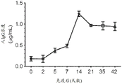


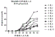


Classifications
-
- C—CHEMISTRY; METALLURGY
- C07—ORGANIC CHEMISTRY
- C07K—PEPTIDES
- C07K16/00—Immunoglobulins [IGs], e.g. monoclonal or polyclonal antibodies
- C07K16/08—Immunoglobulins [IGs], e.g. monoclonal or polyclonal antibodies against material from viruses
- C07K16/10—Immunoglobulins [IGs], e.g. monoclonal or polyclonal antibodies against material from viruses from RNA viruses
- C07K16/1036—Retroviridae, e.g. leukemia viruses
- C07K16/1045—Lentiviridae, e.g. HIV, FIV, SIV
- C07K16/1063—Lentiviridae, e.g. HIV, FIV, SIV env, e.g. gp41, gp110/120, gp160, V3, PND, CD4 binding site
-
- A—HUMAN NECESSITIES
- A61—MEDICAL OR VETERINARY SCIENCE; HYGIENE
- A61K—PREPARATIONS FOR MEDICAL, DENTAL OR TOILETRY PURPOSES
- A61K39/00—Medicinal preparations containing antigens or antibodies
- A61K39/395—Antibodies; Immunoglobulins; Immune serum, e.g. antilymphocytic serum
- A61K39/39533—Antibodies; Immunoglobulins; Immune serum, e.g. antilymphocytic serum against materials from animals
- A61K39/39558—Antibodies; Immunoglobulins; Immune serum, e.g. antilymphocytic serum against materials from animals against tumor tissues, cells, antigens
-
- A—HUMAN NECESSITIES
- A61—MEDICAL OR VETERINARY SCIENCE; HYGIENE
- A61K—PREPARATIONS FOR MEDICAL, DENTAL OR TOILETRY PURPOSES
- A61K48/00—Medicinal preparations containing genetic material which is inserted into cells of the living body to treat genetic diseases; Gene therapy
- A61K48/0008—Medicinal preparations containing genetic material which is inserted into cells of the living body to treat genetic diseases; Gene therapy characterised by an aspect of the 'non-active' part of the composition delivered, e.g. wherein such 'non-active' part is not delivered simultaneously with the 'active' part of the composition
- A61K48/0016—Medicinal preparations containing genetic material which is inserted into cells of the living body to treat genetic diseases; Gene therapy characterised by an aspect of the 'non-active' part of the composition delivered, e.g. wherein such 'non-active' part is not delivered simultaneously with the 'active' part of the composition wherein the nucleic acid is delivered as a 'naked' nucleic acid, i.e. not combined with an entity such as a cationic lipid
-
- A—HUMAN NECESSITIES
- A61—MEDICAL OR VETERINARY SCIENCE; HYGIENE
- A61K—PREPARATIONS FOR MEDICAL, DENTAL OR TOILETRY PURPOSES
- A61K48/00—Medicinal preparations containing genetic material which is inserted into cells of the living body to treat genetic diseases; Gene therapy
- A61K48/005—Medicinal preparations containing genetic material which is inserted into cells of the living body to treat genetic diseases; Gene therapy characterised by an aspect of the 'active' part of the composition delivered, i.e. the nucleic acid delivered
- A61K48/0066—Manipulation of the nucleic acid to modify its expression pattern, e.g. enhance its duration of expression, achieved by the presence of particular introns in the delivered nucleic acid
-
- A—HUMAN NECESSITIES
- A61—MEDICAL OR VETERINARY SCIENCE; HYGIENE
- A61P—SPECIFIC THERAPEUTIC ACTIVITY OF CHEMICAL COMPOUNDS OR MEDICINAL PREPARATIONS
- A61P35/00—Antineoplastic agents
-
- A—HUMAN NECESSITIES
- A61—MEDICAL OR VETERINARY SCIENCE; HYGIENE
- A61P—SPECIFIC THERAPEUTIC ACTIVITY OF CHEMICAL COMPOUNDS OR MEDICINAL PREPARATIONS
- A61P37/00—Drugs for immunological or allergic disorders
- A61P37/02—Immunomodulators
-
- C—CHEMISTRY; METALLURGY
- C07—ORGANIC CHEMISTRY
- C07K—PEPTIDES
- C07K14/00—Peptides having more than 20 amino acids; Gastrins; Somatostatins; Melanotropins; Derivatives thereof
- C07K14/82—Translation products from oncogenes
-
- C—CHEMISTRY; METALLURGY
- C07—ORGANIC CHEMISTRY
- C07K—PEPTIDES
- C07K16/00—Immunoglobulins [IGs], e.g. monoclonal or polyclonal antibodies
- C07K16/08—Immunoglobulins [IGs], e.g. monoclonal or polyclonal antibodies against material from viruses
- C07K16/10—Immunoglobulins [IGs], e.g. monoclonal or polyclonal antibodies against material from viruses from RNA viruses
- C07K16/1081—Togaviridae, e.g. flavivirus, rubella virus, hog cholera virus
-
- C—CHEMISTRY; METALLURGY
- C07—ORGANIC CHEMISTRY
- C07K—PEPTIDES
- C07K16/00—Immunoglobulins [IGs], e.g. monoclonal or polyclonal antibodies
- C07K16/18—Immunoglobulins [IGs], e.g. monoclonal or polyclonal antibodies against material from animals or humans
- C07K16/28—Immunoglobulins [IGs], e.g. monoclonal or polyclonal antibodies against material from animals or humans against receptors, cell surface antigens or cell surface determinants
- C07K16/30—Immunoglobulins [IGs], e.g. monoclonal or polyclonal antibodies against material from animals or humans against receptors, cell surface antigens or cell surface determinants from tumour cells
- C07K16/3069—Reproductive system, e.g. ovaria, uterus, testes, prostate
-
- C—CHEMISTRY; METALLURGY
- C07—ORGANIC CHEMISTRY
- C07K—PEPTIDES
- C07K16/00—Immunoglobulins [IGs], e.g. monoclonal or polyclonal antibodies
- C07K16/18—Immunoglobulins [IGs], e.g. monoclonal or polyclonal antibodies against material from animals or humans
- C07K16/32—Immunoglobulins [IGs], e.g. monoclonal or polyclonal antibodies against material from animals or humans against translation products of oncogenes
-
- C—CHEMISTRY; METALLURGY
- C12—BIOCHEMISTRY; BEER; SPIRITS; WINE; VINEGAR; MICROBIOLOGY; ENZYMOLOGY; MUTATION OR GENETIC ENGINEERING
- C12N—MICROORGANISMS OR ENZYMES; COMPOSITIONS THEREOF; PROPAGATING, PRESERVING, OR MAINTAINING MICROORGANISMS; MUTATION OR GENETIC ENGINEERING; CULTURE MEDIA
- C12N15/00—Mutation or genetic engineering; DNA or RNA concerning genetic engineering, vectors, e.g. plasmids, or their isolation, preparation or purification; Use of hosts therefor
- C12N15/09—Recombinant DNA-technology
-
- G—PHYSICS
- G01—MEASURING; TESTING
- G01N—INVESTIGATING OR ANALYSING MATERIALS BY DETERMINING THEIR CHEMICAL OR PHYSICAL PROPERTIES
- G01N33/00—Investigating or analysing materials by specific methods not covered by groups G01N1/00 - G01N31/00
- G01N33/48—Biological material, e.g. blood, urine; Haemocytometers
- G01N33/50—Chemical analysis of biological material, e.g. blood, urine; Testing involving biospecific ligand binding methods; Immunological testing
- G01N33/53—Immunoassay; Biospecific binding assay; Materials therefor
- G01N33/531—Production of immunochemical test materials
-
- A—HUMAN NECESSITIES
- A61—MEDICAL OR VETERINARY SCIENCE; HYGIENE
- A61K—PREPARATIONS FOR MEDICAL, DENTAL OR TOILETRY PURPOSES
- A61K39/00—Medicinal preparations containing antigens or antibodies
- A61K2039/505—Medicinal preparations containing antigens or antibodies comprising antibodies
-
- A—HUMAN NECESSITIES
- A61—MEDICAL OR VETERINARY SCIENCE; HYGIENE
- A61K—PREPARATIONS FOR MEDICAL, DENTAL OR TOILETRY PURPOSES
- A61K39/00—Medicinal preparations containing antigens or antibodies
- A61K2039/51—Medicinal preparations containing antigens or antibodies comprising whole cells, viruses or DNA/RNA
- A61K2039/53—DNA (RNA) vaccination
-
- C—CHEMISTRY; METALLURGY
- C07—ORGANIC CHEMISTRY
- C07K—PEPTIDES
- C07K16/00—Immunoglobulins [IGs], e.g. monoclonal or polyclonal antibodies
-
- C—CHEMISTRY; METALLURGY
- C07—ORGANIC CHEMISTRY
- C07K—PEPTIDES
- C07K2317/00—Immunoglobulins specific features
- C07K2317/20—Immunoglobulins specific features characterized by taxonomic origin
- C07K2317/21—Immunoglobulins specific features characterized by taxonomic origin from primates, e.g. man
-
- C—CHEMISTRY; METALLURGY
- C07—ORGANIC CHEMISTRY
- C07K—PEPTIDES
- C07K2317/00—Immunoglobulins specific features
- C07K2317/50—Immunoglobulins specific features characterized by immunoglobulin fragments
- C07K2317/55—Fab or Fab'
-
- C—CHEMISTRY; METALLURGY
- C07—ORGANIC CHEMISTRY
- C07K—PEPTIDES
- C07K2317/00—Immunoglobulins specific features
- C07K2317/70—Immunoglobulins specific features characterized by effect upon binding to a cell or to an antigen
- C07K2317/76—Antagonist effect on antigen, e.g. neutralization or inhibition of binding
-
- C—CHEMISTRY; METALLURGY
- C07—ORGANIC CHEMISTRY
- C07K—PEPTIDES
- C07K2319/00—Fusion polypeptide
-
- C—CHEMISTRY; METALLURGY
- C07—ORGANIC CHEMISTRY
- C07K—PEPTIDES
- C07K2319/00—Fusion polypeptide
- C07K2319/01—Fusion polypeptide containing a localisation/targetting motif
- C07K2319/02—Fusion polypeptide containing a localisation/targetting motif containing a signal sequence
-
- C—CHEMISTRY; METALLURGY
- C12—BIOCHEMISTRY; BEER; SPIRITS; WINE; VINEGAR; MICROBIOLOGY; ENZYMOLOGY; MUTATION OR GENETIC ENGINEERING
- C12N—MICROORGANISMS OR ENZYMES; COMPOSITIONS THEREOF; PROPAGATING, PRESERVING, OR MAINTAINING MICROORGANISMS; MUTATION OR GENETIC ENGINEERING; CULTURE MEDIA
- C12N2800/00—Nucleic acids vectors
- C12N2800/10—Plasmid DNA
- C12N2800/106—Plasmid DNA for vertebrates
- C12N2800/107—Plasmid DNA for vertebrates for mammalian
-
- C—CHEMISTRY; METALLURGY
- C12—BIOCHEMISTRY; BEER; SPIRITS; WINE; VINEGAR; MICROBIOLOGY; ENZYMOLOGY; MUTATION OR GENETIC ENGINEERING
- C12N—MICROORGANISMS OR ENZYMES; COMPOSITIONS THEREOF; PROPAGATING, PRESERVING, OR MAINTAINING MICROORGANISMS; MUTATION OR GENETIC ENGINEERING; CULTURE MEDIA
- C12N2800/00—Nucleic acids vectors
- C12N2800/22—Vectors comprising a coding region that has been codon optimised for expression in a respective host
-
- C—CHEMISTRY; METALLURGY
- C12—BIOCHEMISTRY; BEER; SPIRITS; WINE; VINEGAR; MICROBIOLOGY; ENZYMOLOGY; MUTATION OR GENETIC ENGINEERING
- C12N—MICROORGANISMS OR ENZYMES; COMPOSITIONS THEREOF; PROPAGATING, PRESERVING, OR MAINTAINING MICROORGANISMS; MUTATION OR GENETIC ENGINEERING; CULTURE MEDIA
- C12N2830/00—Vector systems having a special element relevant for transcription
- C12N2830/50—Vector systems having a special element relevant for transcription regulating RNA stability, not being an intron, e.g. poly A signal
-
- C—CHEMISTRY; METALLURGY
- C12—BIOCHEMISTRY; BEER; SPIRITS; WINE; VINEGAR; MICROBIOLOGY; ENZYMOLOGY; MUTATION OR GENETIC ENGINEERING
- C12N—MICROORGANISMS OR ENZYMES; COMPOSITIONS THEREOF; PROPAGATING, PRESERVING, OR MAINTAINING MICROORGANISMS; MUTATION OR GENETIC ENGINEERING; CULTURE MEDIA
- C12N2840/00—Vectors comprising a special translation-regulating system
- C12N2840/60—Vectors comprising a special translation-regulating system from viruses
-
- Y—GENERAL TAGGING OF NEW TECHNOLOGICAL DEVELOPMENTS; GENERAL TAGGING OF CROSS-SECTIONAL TECHNOLOGIES SPANNING OVER SEVERAL SECTIONS OF THE IPC; TECHNICAL SUBJECTS COVERED BY FORMER USPC CROSS-REFERENCE ART COLLECTIONS [XRACs] AND DIGESTS
- Y02—TECHNOLOGIES OR APPLICATIONS FOR MITIGATION OR ADAPTATION AGAINST CLIMATE CHANGE
- Y02A—TECHNOLOGIES FOR ADAPTATION TO CLIMATE CHANGE
- Y02A50/00—TECHNOLOGIES FOR ADAPTATION TO CLIMATE CHANGE in human health protection, e.g. against extreme weather
- Y02A50/30—Against vector-borne diseases, e.g. mosquito-borne, fly-borne, tick-borne or waterborne diseases whose impact is exacerbated by climate change
Abstract
The present application relates to DNA antibody constructs and methods of use thereof. In particular, disclosed herein are compositions comprising recombinant nucleic acid sequences encoding antibodies. Also disclosed herein are methods of producing a synthetic antibody in a subject by administering the composition to the subject. The disclosure also provides methods of preventing and/or treating a disease in a subject using the compositions and methods of production.
Description
The present application is a divisional application of chinese patent application No. 201580075039.2 entitled "DNA antibody constructs and methods of use thereof" filed on 2015, 12, month 01.
CROSS-REFERENCE TO RELATED APPLICATIONS
This application claims priority to U.S. provisional application No.62/086,157 filed on 1/12/2014 and U.S. provisional application No.62/213,166 filed on 2/9/2015, which are incorporated herein by reference in their entirety.
Technical Field
The present invention relates to compositions comprising recombinant nucleic acid sequences for the production of synthetic antibodies (including various alterations to the antibodies that can produce an extended half-life synthetic antibody, abrogate antibody-dependent enhancement, enhance antibody-dependent cellular cytotoxicity, provide bispecific binding, provide bifunctional, etc.) and their functionality in vivo, and methods of preventing and/or treating disease in a subject by administering the compositions.
Background
An immunoglobulin molecule comprises 2 chains of each type of light chain (L) and heavy chain (H) that are covalently linked by disulfide bonds (shown as S-S) between cysteine residues. The variable domains of the heavy (VH) and light (VL) chains contribute to the binding site of the antibody molecule. The heavy chain constant region consists of 3 constant domains (CH1, CH2 and CH3) and a (flexible) hinge region. Light chains also have a constant domain (CL). The variable regions of the heavy and light chains comprise 4 framework regions (FR; FR1, FR2, FR3 and FR4) and 3 complementarity determining regions (CDR; CDR1, CDR2 and CDR 3). These are therefore very complex genetic systems that are difficult to assemble in vivo.
Targeted monoclonal antibodies (mabs) represent one of the most important medical treatment advances in the last 25 years. This type of immune-based therapy is now routinely used against host autoimmune diseases, treating cancer, and infectious diseases. For malignant tumors, many of the currently used immunoglobulin (Ig) -based therapies are combined with cytotoxic chemotherapy regimens directed against the tumor. This combined approach has significantly improved overall survival. Various mAb preparations are approved for specific cancers, including rituximab, a chimeric mAb targeting CD20 for the treatment of Non-Hodgkins lymphoma, and ipilimumab (Yervoy), a human mAb that blocks CTLA-4 and has been used to treat melanoma and other malignancies. Moreover, bevacizumab (avastin) is another prominent humanized mAb that targets VEGF and tumor neovascularization and has been used to treat colorectal cancer. Also to the best known mAb for the treatment of malignancies is trastuzumab (herceptin), a Her 2/neu-targeted humanized formulation that has been demonstrated to have comparable efficacy against breast cancer in a subset of patients. In addition, many mabs are used to treat autoimmune disorders and specific blood disorders.
In addition to cancer treatment, passive transfer of polyclonal Ig mediates protective efficacy against a number of infectious diseases including diphtheria, hepatitis a and b, rabies, tetanus, varicella and Respiratory Syncytial Virus (RSV). In fact, several polyclonal Ig preparations provide temporary protection against specific infectious agents for individuals traveling to endemic areas in the case where it is not current to generate protective Ig by active vaccination. Furthermore, palivizumab (Synagis), a mAb targeting RSV infection, has been demonstrated to protect clinically against RSV infection in immunodeficient children.
The currently available therapeutic antibody present on the market is the human IgG1 isotype. These antibodies include glycoproteins having two N-linked biantennary complex oligosaccharides bound to the constant region (Fc) of the antibody, wherein a majority of the oligosaccharides are core fucosylated. It functions as an effector of antibody-dependent cellular cytotoxicity (ADCC) and complement-dependent cytotoxicity (CDC) by Fc interaction with leukocyte receptors (fcyr) or complement. There is a decline in the efficacy of the therapeutic antibody in vivo (versus in vitro), thus resulting in the need for large doses of therapeutic antibody-sometimes hundreds of milligrams per week. This is mainly due to competition between serum IgG and therapeutic antibodies for binding to Fc γ RIIIa on Natural Killer (NK) cells. Endogenous human serum IgG inhibits ADCC induced by the therapeutic antibody. Thus, the efficacy of a non-fucosylated therapeutic antibody in humans can be increased. The binding affinity of the non-fucosylated therapeutic antibody to Fc γ RIIIa is much higher than that of fucosylated human serum IgG, which is a preferred feature to overcome human plasma IgG interference.
Antibody-based therapies are not without risk. One such risk is antibody-dependent enhancement (ADE), which occurs when a non-neutralizing antiviral protein promotes viral entry into a host cell, resulting in enhanced infectivity in the cell. Some cells do not have the usual receptors on their surface for viruses to gain entry. Antiviral proteins (i.e., antibodies) bind to the antibody Fc receptors that some of these cells have in the plasma membrane. The virus binds to the antigen binding site on the other end of the antibody. This virus can use this mechanism to infect human macrophages, making mild viral infections life-threatening. The most widely known examples of ADE occur in the context of infection by the dengue fever virus (DENV). This phenomenon is observed when a person who has previously been infected with one serotype of DENV is infected with a different serotype after many months or years. In such cases, the clinical course of the disease is more severe and these people have higher viremia than those in which ADE has not occurred. This explains that while primary (first) infections cause mainly small Disease (DF) in children, secondary infections (late reinfection) are more likely to be associated with severe disease (DHF and/or DSS) in children and adults. There are four antigenically distinct serotypes of DENV (DENV-1-DENV-4). DENV infection induces the production of neutralizing isotype immunoglobulin g (igg) antibodies that provide a lifetime immunity to the infectious serotype. DENV infection also resulted in a degree of cross-protective immunity against the other 3 serotypes. In addition to inducing neutralizing heterotypic antibodies, DENV infection may also induce heterotypic antibodies that neutralize the virus only partially or not at all. The production of such cross-reactive but non-neutralizing antibodies may be the cause of more serious secondary infections. Once inside the white blood cells, the virus replicates undetectable, ultimately producing extremely high viral titers that cause severe disease.
The clinical impact of mAb therapy is impressive. However, there still remains a problem that limits the use and dissemination of this treatment method. Some of these problems include the high production costs of these complex biologicals, which can limit their use in a broader population, particularly in developing countries where they can have a tremendous impact. Furthermore, the frequent need to repeat administration of mabs to achieve and maintain efficacy can be an obstacle in logistics and patient compliance. There is a need for new antibodies that can reduce or eliminate the low in vivo efficacy of therapeutic antibodies by competing with serum IgG. There is a need for new antibodies with antibody-dependent enhancement that can eliminate viruses such as dengue, HIV, RSV and others. Bispecific antibodies, bifunctional antibodies, and antibody mixtures are needed to perform several functions that may prove to be therapeutic or prophylactic. There is also a need for combination therapies that can utilize the synthetic antibodies described herein and that can immunostimulatory the host system by immunization with vaccines, including DNA-based vaccines. Furthermore, the long-term stability of these antibody formulations is often short and below optimal. Thus, there remains a need in the art for synthetic antibody molecules that can be delivered to a subject in a safe and cost effective manner.
Disclosure of Invention
The present invention relates to methods of producing synthetic antibodies in a subject. The method can comprise administering to the subject a composition comprising a recombinant nucleic acid sequence encoding an antibody or fragment thereof. The recombinant nucleic acid sequence can be expressed in a subject to produce a synthetic antibody.
The resulting synthetic antibodies may be defucosylated. The resulting synthetic antibody may comprise two leucine to alanine mutations in the CH2 region of the Fc region.
The antibody may comprise a heavy chain polypeptide or fragment thereof, and a light chain polypeptide or fragment thereof. The heavy chain polypeptide or fragment thereof may be encoded by a first nucleic acid sequence and the light chain polypeptide or fragment thereof may be encoded by a second nucleic acid sequence. The recombinant nucleic acid sequence can comprise a first nucleic acid sequence and a second nucleic acid sequence. The recombinant nucleic acid sequence may further comprise a promoter for expressing the first nucleic acid sequence and the second nucleic acid sequence as a single transcript in a subject. The promoter may be a Cytomegalovirus (CMV) promoter.
The recombinant nucleic acid sequence may further comprise a third nucleic acid sequence encoding a protease cleavage site. The third nucleic acid sequence may be located between the first nucleic acid sequence and the second nucleic acid sequence. The protease of the subject may recognize and cleave the protease cleavage site.
The recombinant nucleic acid sequence can be expressed in a subject to produce an antibody polypeptide sequence. The antibody polypeptide sequence may comprise a heavy chain polypeptide or fragment thereof, a protease cleavage site, and a light chain polypeptide or fragment thereof. A protease produced by the subject can recognize and cleave a protease cleavage site of the antibody polypeptide sequence, thereby producing a cleaved heavy chain polypeptide and a cleaved light chain polypeptide. Synthetic antibodies can be produced from cleaved heavy chain polypeptides and cleaved light chain polypeptides.
The recombinant nucleic acid sequence may comprise a first promoter for expressing the first nucleic acid sequence as a first transcript and a second promoter for expressing the second nucleic acid sequence as a second transcript. The first transcript may be translated into a first polypeptide and the second transcript may be translated into a second polypeptide. Synthetic antibodies can be produced from the first and second polypeptides. The first promoter and the second promoter may be the same. The promoter may be a Cytomegalovirus (CMV) promoter.
The heavy chain polypeptide may comprise a variable heavy region and a constant heavy region 1. The heavy chain polypeptide may comprise a variable heavy region, a constant heavy region 1, a hinge region, a constant heavy region 2, and a constant heavy region 3. The light chain polypeptide may comprise a variable light region and a constant light region.
The recombinant nucleic acid sequence may further comprise a Kozak sequence. The recombinant nucleic acid sequence may further comprise an immunoglobulin (Ig) signal peptide. The Ig signal peptide may comprise an IgE or IgG signal peptide.
The recombinant nucleic acid sequence may comprise a nucleic acid sequence encoding at least one of the amino acid sequences of SEQ ID NOs 1, 2, 5, 41, 43, 45, 46, 47, 48, 49, 51, 53, 55, 57, 59, 61, and 80. The recombinant nucleic acid sequence may comprise at least one of the nucleic acid sequences of SEQ ID NOs 3, 4, 6, 7, 40, 42, 44, 50, 52, 54, 56, 58, 60, 62, 63 and 79.
The invention also relates to methods of producing synthetic antibodies in a subject. The method can comprise administering to the subject a composition comprising a first recombinant nucleic acid sequence encoding a heavy chain polypeptide or fragment thereof and a second recombinant nucleic acid sequence encoding a light chain polypeptide or fragment thereof. The first recombinant nucleic acid sequence can be expressed in a subject to produce a first polypeptide, and the second recombinant nucleic acid can be expressed in a subject to produce a second polypeptide. Synthetic antibodies can be produced from the first and second polypeptides.
The first recombinant nucleic acid sequence can further comprise a first promoter for expression of the first polypeptide in the subject. The second recombinant nucleic acid sequence can further comprise a second promoter for expression of a second polypeptide in the subject. The first promoter and the second promoter may be the same. The promoter may be a Cytomegalovirus (CMV) promoter.
The heavy chain polypeptide may comprise a variable heavy region and a constant heavy region 1. The heavy chain polypeptide may comprise a variable heavy region, a constant heavy region 1, a hinge region, a constant heavy region 2, and a constant heavy region 3. The light chain polypeptide may comprise a variable light region and a constant light region.
The first and second recombinant nucleic acid sequences can further comprise a Kozak sequence. The first and second recombinant nucleic acid sequences can further comprise an immunoglobulin (Ig) signal peptide. The Ig signal peptide may comprise an IgE or IgG signal peptide.
The invention also relates to methods of preventing or treating a disease in a subject. The method may comprise generating a synthetic antibody in a subject according to one of the methods described above. The synthetic antibody may be specific for the foreign antigen. The foreign antigen may be derived from a virus. The virus may be Human Immunodeficiency Virus (HIV), chikungunya fever virus (CHIKV) or dengue virus.
The virus may be HIV. The recombinant nucleic acid sequence may comprise a nucleic acid sequence encoding at least one amino acid sequence of SEQ ID NOs 1, 2, 5, 46, 47, 48, 49, 51, 53, 55, and 57. The recombinant nucleic acid sequence may comprise at least one nucleic acid sequence of SEQ ID NOs 3, 4, 6, 7, 50, 52, 55, 56, 62 and 63.
The virus may be CHIKV. The recombinant nucleic acid sequence may comprise a nucleic acid sequence encoding at least one of the amino acid sequences of SEQ ID NOs 59 and 61. The recombinant nucleic acid sequence may comprise at least one of the nucleic acid sequences of SEQ ID NO 58 and 60.
The virus may be dengue virus. The recombinant nucleic acid sequence may comprise a nucleic acid sequence encoding at least one amino acid sequence of SEQ ID NO 45. The recombinant nucleic acid sequence comprises at least one nucleic acid sequence of SEQ ID NO. 44.
The synthetic antibody may be specific for an autoantigen. The autoantigen may be Her 2. The recombinant nucleic acid sequence may comprise a nucleic acid sequence encoding at least one of the amino acid sequences of SEQ ID NOs 41 and 43. The recombinant nucleic acid sequence may comprise at least one of the nucleic acid sequences of SEQ ID NOS 40 and 42.
The synthetic antibody may be specific for an autoantigen. The autoantigen may be PSMA. The recombinant nucleic acid sequence may comprise a nucleic acid sequence encoding at least one amino acid sequence of SEQ ID NO 80. The recombinant nucleic acid sequence may comprise at least one nucleic acid sequence of SEQ ID NO. 79.
The invention also relates to a product produced by any of the above methods. The product may be a single DNA plasmid capable of expressing functional antibodies. The product may consist of two or more different DNA plasmids capable of expressing components of functional antibodies that combine in vivo to form functional antibodies.
The present invention also relates to a method of treating a subject infected with a pathogen, comprising: a nucleotide sequence encoding a synthetic antibody specific for the pathogen is administered. The method may further comprise administering an antigen of the pathogen to generate an immune response in the subject.
The present invention also relates to a method of treating a subject with cancer, comprising: administering a nucleotide sequence encoding a cancer marker to induce ADCC.
The invention also relates to nucleic acid molecules encoding synthetic antibodies comprising a nucleic acid sequence having at least about 95% identity over the entire length of the nucleic acid sequence set forth in SEQ ID NO: 79.
The invention also relates to nucleic acid molecules encoding synthetic antibodies comprising the nucleic acid sequence shown in SEQ ID NO. 79.
The invention also relates to nucleic acid molecules encoding synthetic antibodies comprising a nucleic acid sequence encoding a protein having at least about 95% identity over the entire length of the amino acid sequence set forth in SEQ ID NO: 80.
The invention also relates to nucleic acid molecules encoding synthetic antibodies comprising a nucleic acid sequence encoding a protein comprising the amino acid sequence shown in SEQ ID NO: 80.
Any of the above nucleic acid molecules may comprise an expression vector.
The invention also relates to compositions comprising one or more of the above-described nucleic acid molecules. The composition may further comprise a pharmaceutically acceptable excipient.
Drawings
Figure 1 shows the nucleic acid sequence encoding the IgG heavy chain as described in example 1.
Figure 2 shows the nucleic acid sequence encoding an IgG light chain as described in example 1.
FIG. 3 shows a plot of time (hours) plotted against OD450nm (dilution of tissue culture supernatant at 1: 100).
Figure 4 shows an image of a western blot.
FIG. 5 shows the production of pHIV-1Env-Fab and confirmation of expression. (A of FIG. 5 and B of FIG. 5) circular plasmid maps of pHIV-1Env Fab anti-gp 120 Fab expression constructs were designed using VRC01 heavy (H) and light (L) variable chain Ig genes. Several modifications were included to increase the level of expression when constructing the Fab plasmid. The Fab VL and VH fragment genes as shown were individually cloned between BamH1 and Xho1 restriction sites of the pVax1 vector. FIG. 5C shows in vitro expression of pHIV-1Env Fab. The figure shows the temporal kinetics of pHIV-1Env Fab expression after 293T cell transfection. The assigned values representing expression are the mean OD450nm ± SD of triplicate wells. As a control, 293T cells were also transfected with the pVax1 backbone.
FIG. 6 shows the measurement of the time-production of anti-HIV Env specific Fab produced by pHIV-1Env Fab. Fig. 6 a shows the time course of anti-HIV 1Fab production. After administration of pHIV-1Env Fab, the production of specific Fab in the serum at a final dilution of 1:100 was measured by ELISA over a period of 10 days and expressed as OD450 nm. Sera from mice administered pVax1 were used as negative controls. B of fig. 6 shows a comparative measurement of anti-gp 120 antibody responses following immunization with recombinant gp120(rgp 120). Mice were immunized by a single injection of rgp120, followed by measurement of anti-gp 120 antibody production for 10 days and expressed as OD450nm values, as described in example 2. PBS was used as a negative control injection for this study. FIG. 6C shows confirmation of HIV1Env-Fab binding by immunoblot analysis. As indicated in the examples, 5 or 10. mu.g gp120 was injected to SDS-PAGE and subjected to nitrocellulose blotting followed by incubation of the blot with serum from mice administered pHIV-1Env Fab. The immunoblots indicated that experimental sera recognized the binding of rgp120, confirming the specificity of the Fab produced. FIG. 6D shows the time quantification of human IgG1Fab, measured as IgG1 in mouse serum after pHIV-1Env-Fab administration. IgG1 was measured at the indicated time points using a standard ELISA kit and expressed as Fab (. mu.g/mL). + -. SD. Sera from mice administered pVax1 were used as negative controls. Serum samples were analyzed at the time points indicated on the x-axis. Arrows shown in the graphs shown in a of fig. 6, B of fig. 6, and D of fig. 6 indicate the points of DNA plasmid administration.
Fig. 7 shows FACS binding analysis of HIV1Env Fab and clade a HIV Env glycoprotein. FIG. 7A shows FACS scan indicating binding of anti-HIV 1Env-Fab to HIV-1 clade A Env glycoprotein. DNA expressing consensus (pCon-Env-A) or "optimized" (pOpt-Env-A) HIV-1 clade A envelope was transfected into 293T cells. 2 days after transfection, cells were stained with purified native VRC01 Ig, serum produced from pHIV-1Env Fab (collected 48 hours after single plasmid administration) or control Ig produced from pIGG-E1M2 administration. Serum and VRC01 antibody were diluted 1:4 or 1:100 in 50 μ l PBS, respectively, and incubated at room temperature for 30 minutes. Cells were then stained with the appropriate second Phycoerythrin (PE) conjugated Ig and subsequently gated for singlet and viable cells for FACS analysis. Percent binding of positive cells was indicated in each scan. Figure 7B shows a graphical representation of FACS binding data. The number of stained cells (i.e. indicative expression level) in each Ig/serum test group was divided by the background staining value and expressed as the percentage of specific binding on the y-axis as a function of the different HIV clade a Env preparations tested.
FIG. 8 shows the time course of neutralization of HIV-1 by sera from mice administered pHIV-1 Env-Fab. Sera were collected at the time points indicated in the figure for analysis of the neutralizing activity of the sera. A panel of HIV-1 pseudotyped viruses was used: bal26 (FIG. 8A; clade B, Tier 1), Q23Env17 (FIG. 8B; clade A, Tier 1), SF162S (FIG. 8C; clade B, Tier 1), and ZM53M (FIG. 8D; clade C, Tier 2) were subjected to neutralization assays in TZM-BL cells. Cells were infected at an MOI of 0.01 and incubated in the presence of serum containing Fab generated from pHIV-1Env Fab administration (final dilution of 1: 50) as depicted in example 2. Percent neutralization values are shown, the calculation of which is described in example 2. Likewise, horizontal lines are provided in each figure indicating approximate time points at which experimental sera mediate 50% virus neutralization.
FIG. 9 shows the nucleic acid sequence encoding the heavy chain of the HIV-1Env Fab (VH-CH1) described in examples 2-7.
FIG. 10 shows the nucleic acid sequence encoding the light chain of the HIV-1Env Fab (VL-CL) described in examples 2-7.
FIG. 11 shows immunofluorescence of cells transfected with a plasmid encoding HIV Env. Cells were stained with preparations from pVAX1 (left panel) or pHIV-Env-Fab (right panel).
FIG. 12 shows a graph plotting the type of antigen versus serum concentration (ng/mL).
Figure 13 shows a schematic of a construct encoding a synthetic human IgG1 antibody.
FIG. 14 shows a schematic of the assembled antibody (after expression) encoded by the construct of FIG. 13.
Figure 15 shows the amino acid sequence of VRC01 IgG.
FIG. 16A shows a schematic of a construct encoding HIV-1Env-PG9 Ig; FIG. 16B shows a schematic of a vector containing the construct of FIG. 16A; and fig. 16C shows an image of the stained gel.
FIG. 17A shows a schematic of a construct encoding HIV-1Env-4E10 Ig; FIG. 17B shows a schematic of a vector containing the construct of FIG. 17A; and figure 17C shows an image of the stained gel.
FIG. 18 shows the amino acid sequence of HIV-1Env-PG9 Ig prior to cleavage with Fisher-Ellin protease.
FIG. 19 shows the amino acid sequence of HIV-1Env-4E10 Ig before cleavage with furin.
FIG. 20A shows an exemplary diagram of a construct encoding the heavy (VH-CH1) chain of CHIKV-Env-Fab; and FIG. 20B shows a schematic diagram of a construct encoding the heavy (VL-CL) chain of CHIKV-Env-Fab.
FIG. 21 shows a schematic representation of an expression vector containing a construct encoding the heavy (VH-CH1) or light (VL-CL) chain of CHIKV-Env-Fab.
FIG. 22 shows a graph plotting time in hours (hr) versus OD450 nm.
Figure 23 shows an image of an immunoblot.
Figure 24 shows the timing of DNA administration and a schematic of obtaining pre-bleeding and bleeding.
FIG. 25 shows a graph plotting time versus OD450 nm in days.
Figure 26 shows a graph plotting days post challenge versus percent survival.
FIG. 27 shows a graph plotting pg/mL of mouse versus TNF- α.
FIG. 28 shows a graph plotting pg/mL for mouse versus IL-6.
FIG. 29 shows a schematic diagram illustrating a construct encoding VH-CH1 and under the control of a promoter.
FIG. 30 shows a schematic diagram illustrating a construct encoding VL-CL and under the control of a promoter.
FIG. 31 shows a schematic diagram illustrating the constructs encoding anti-Her-2 Fab VH-CH1 or VL-CL cloned into an expression vector.
FIG. 32 shows the nucleic acid sequence encoding VH-CH1 of anti-Her-2 Fab.
FIG. 33 shows the amino acid sequence encoded by the nucleic acid sequence of FIG. 32 (i.e., the amino acid sequence of VH-CH1 of anti-Her-2 Fab).
FIG. 34 shows the nucleic acid sequence encoding VL-CL for anti-Her-2 Fab.
FIG. 35 shows the amino acid sequence encoded by the nucleic acid sequence of FIG. 34 (i.e., the amino acid sequence of VL-CL of anti-Her-2 Fab).
FIG. 36 shows a graph plotting the type of transfected cells versus IgG concentration (. mu.g/mL).
FIG. 37 shows a schematic diagram illustrating the constructs encoding the variable heavy region (VH), variable heavy constant region 1(CH1), hinge region, variable heavy constant region 2(CH2), variable heavy constant region 3(CH3) of an immunoglobulin G (IgG) heavy chain, and variable light region (VL) and variable light constant region (CL) of an IgG light chain. The heavy and light chains of IgG are separated by a protease cleavage site and each is located after a signal peptide (encoded by a leader sequence).
FIG. 38 shows the nucleic acid sequence encoding anti-dengue virus (DENV) human IgG.
FIG. 39 shows the amino acid sequence encoded by the nucleic acid sequence of FIG. 39 (i.e., the amino acid sequence of an anti-DENV human IgG). In this amino acid sequence, protease cleavage has not yet occurred to separate the heavy and light chains into two separate polypeptides.
FIG. 40 shows a graph plotting mouse groups versus OD 450 nm.
FIG. 41 shows a graph plotting the days post injection versus human IgG concentration (ng/mL).
FIG. 42 shows the amino acid sequence encoded by the nucleic acid sequence of FIG. 1 (i.e., SEQ ID NO: 6). This amino acid sequence is that of the IgG heavy chain described in example 1 below.
FIG. 43 shows the amino acid sequence encoded by the nucleic acid sequence of FIG. 2 (i.e., SEQ ID NO: 7). This amino acid sequence is that of the IgG light chain described in example 1 below.
FIG. 44 shows the amino acid sequence encoded by the nucleic acid sequence of FIG. 9 (i.e., SEQ ID NO: 3). This amino acid sequence is that of the heavy chain of the HIV-1Env-Fab (VH-CH1) described in examples 2-7.
FIG. 45 shows the amino acid sequence encoded by the nucleic acid sequence of FIG. 10 (i.e., SEQ ID NO: 4). The amino acid sequence is that of the light chain of the HIV-1Env-Fab (VL-CL) described in examples 2-7.
FIG. 46 shows a nucleic acid sequence encoding the HIV-1PG9 single chain Fab (scFab) described in example 11 below.
FIG. 47 shows the amino acid sequence encoded by the nucleic acid sequence of FIG. 46 (i.e., SEQ ID NO: 50). This amino acid sequence is that of HIV-1PG9 scFab described in example 11 below.
FIG. 48 shows a nucleic acid sequence encoding the HIV-14E 10 single chain Fab (scFab) described in example 13 below.
FIG. 49 shows the amino acid sequence encoded by the nucleic acid sequence of FIG. 48 (i.e., SEQ ID NO: 52). This amino acid sequence is that of HIV-14E 10 scFab described in example 13 below.
FIG. 50 shows a schematic diagram illustrating constructs encoding the variable heavy region (VH), variable heavy constant region 1(CH1), hinge region, variable heavy constant region 2(CH2), variable heavy constant region 3(CH3) of an immunoglobulin G (IgG) heavy chain. The nucleic acid sequence encoding the IgG heavy chain follows the leader sequence.
FIG. 51 shows a schematic diagram illustrating constructs encoding the variable light region (VL) and variable light constant region (CL) of an IgG light chain. The nucleic acid sequence encoding the IgG light chain follows the leader sequence.
FIG. 52 shows the nucleic acid sequence encoding the HIV-1VRC01 IgG1 heavy chain described in example 9 below.
FIG. 53 shows the amino acid sequence encoded by the nucleic acid sequence of FIG. 52 (i.e., SEQ ID NO: 54). This amino acid sequence is that of the HIV-1VRC01 IgG1 heavy chain described in example 9 below.
FIG. 54 shows the nucleic acid sequence encoding the HIV-1VRC01 IgG light chain described in example 9 below.
FIG. 55 shows the amino acid sequence encoded by the nucleic acid sequence of FIG. 54 (i.e., SEQ ID NO: 56). This amino acid sequence is that of the HIV-1VRC01 IgG light chain described in example 9 below.
FIG. 56 shows the nucleic acid sequence encoding the heavy chain of CHIKV-Env-Fab (VH-CH1) described in example 14 below.
FIG. 57 shows the amino acid sequence encoded by the nucleic acid sequence of FIG. 56 (i.e., SEQ ID NO: 58). This amino acid sequence is that of the heavy chain of CHIKV-Env-Fab (VH-CH1) described in example 14 below.
FIG. 58 shows a nucleic acid sequence encoding the light chain of CHIKV-Env-Fab (VL-CL) described in example 14 below.
FIG. 59 shows the amino acid sequence encoded by the nucleic acid sequence of FIG. 58 (i.e., SEQ ID NO: 60). This amino acid sequence is that of the light chain of CHIKV-Env-Fab (VL-CL) described in example 14 below.
FIG. 60 shows a nucleic acid sequence encoding HIV-1Env-4E10 Ig described in example 12 below.
FIG. 61 shows a nucleic acid sequence encoding HIV-1Env-PG9 Ig described in example 10 below.
FIG. 62 shows the nucleic acid sequence (SEQ ID NO:64) encoding VRC01 IgG.
Figure 63 shows a schematic of the linear arrangement of nucleotide sequences designed as constructs for human anti-PSMA antibodies.
FIG. 64 shows a graph that provides in vitro expression of anti-hupPSMA-IgG 1 antibody. The left panel shows anti-hupPSMA expression in 293T transfected cells. The right panel shows the binding of the hupmPA protein to the in vitro transfected cells (comparison negative control: pVax 1-empty vector and positive control: PSMA-mAb-commercial monoclonal antibody)
FIG. 65 shows a graph that provides quantification of anti-hupMAP-IgG 1 for human PSMA protein.
Figure 66 shows in the top panel the immunization and bleeding schedules of Nu/J mice; and the bottom panel shows the in vivo kinetics of anti-huPSMA IgG1 binding in Nu/J mice.
FIG. 67 shows a gel photograph of an immunoblot demonstrating anti-hupMA IgG1 binding.
Figure 68 shows several flow cytometry plots of different subjects, which provide information on the specificity of anti-huppsma IgG1 in treated mouse serum (Nu/J) binding to human prostate (LNCaP) cells.
FIG. 69 shows a graph detailing the anti-hupPSMA IgG1 ADCC activity, such as the cytotoxicity of effector cells against LnCap cells tested in the presence or absence of serum from mice treated with anti-PSMA-IgG DNA.
Figure 70 shows a schematic design of a plasmid expressing the antibody and confirmation of expression and binding kinetics of the antibody after a single EP-mediated injection of the CHIKV-Fab expression plasmid. A: for both CHIKV-Fab and CHIKV-IgG, the selected variable light and heavy (VL and VH) IgG fragment genes of the anti-CHIKV human monoclonal antibodies identified from the NCBI database were cloned into optimized DNA plasmid vectors, respectively. B: DNA plasmids encoding anti-CHIKV VL and VH-Fab genes or CHIKV-IgG were transfected together into 293T cells to determine their respective in vitro expression by ELISA. Cells transfected with the empty control pVax1 plasmid were used as negative control. C: in vivo expression of anti-CHIKV-IgG antibodies following EP mediated delivery. Mice (b6.cg-Foxn1nu/J) were administered by a single intramuscular injection of CHIKV-IgG plasmid (100 μ g total) followed by EP (n ═ 5 mice/group). Injection of empty pVax1 vector was used as a negative control. D: specific binding to CHIKV-Env antigen was measured by ELISA assay using sera collected from CHIKV-IgG and recombinant CHIKV-Env immunized mice and expressed as OD 450nm values of individual mice at different time points. E: serum levels of human IgG concentrations in mice injected intramuscularly with CHIKV-IgG as described in materials and methods were measured at different time points. F: assessment of antibody binding affinity and specificity. Serum from mice injected with CHIKV-IgG (day 14) was tested by immunoblotting for binding affinity functionality to the target protein using cell lysates from CHIKV-infected cells, as described in the examples below.
FIG. 71 shows IgG expression and binding kinetics following a single electroporation-mediated injection of the CHIKV-IgG expression plasmid. FIG. 71A shows that sera from mice administered CHIKV-Fab are specific for the CHIKV-Env antigen. ELISA plates were coated with recombinant CHIKV-Env or HIV-1Env (subtype B; MN) proteins and sera from mice injected with either CHIKV-IgG or pVax1 were obtained as indicated after the first injection. Specific binding of the CHIKV-Env antigen was measured by ELISA assay using collected sera and expressed as OD 450nm values of individual mice at different time points. FIG. 71B shows immunofluorescence assay (IFA) results demonstrating that CHIKV-Fab produced from mice administered with CHIKV-Fab is capable of binding to CHIKV-Env glycoprotein. CHIKV-infected Vero cells were fixed 24 hours post infection and then subjected to immunofluorescence assay to detect CHIKV-Env antigen expression (green). Nuclei were stained with DAPI (blue). Moderate amounts of CHIKV-Env protein expression were observed in Vero cells using CHIKV-Fab antibodies. Sera from mice immunized with pVax1 were used as negative controls. C of fig. 71 shows FACS analysis of binding of sera from plasmid injected mice to CHIKV infected cells. The x-axis indicates GFP staining with a lentivirus GFP pseudovirus complementary to CHIKV-Env. The y-axis shows staining of the test human IgG produced in the mice. Double positive cells are indicative/measured of serum binding to CHIKV infected cells.
Figure 72 shows that sera from mice injected with CHIKV-IgG plus EP showed neutralizing activity against multiple CHIKV strains. F of a-72 of fig. 72 shows that for 6 different CHIKV strains: ross, LR2006-OPY1, IND-63-WB1, PC-08, B448-China and Bianchi measured the neutralizing activity of sera from mice administered CHIKV-IgG and EP. Neutralizing antibody (nAb) titers were plotted as the highest serum dilution that resulted in at least 50% inhibition of CPE in Vero cells. Similar results were observed in 2 independent experiments (for each experiment, using at least 10 mice per group). IC-50 values were performed using Prism GraphPad software.
FIG. 73 shows the durability of anti-CHIKV-Env IgG and serum and mucosal IgG responses after immunization with CHIKV-Fab, and the IgG tablesReach and attack studies. A: schematic representation of IgG plasmid immunization and CHIKV-challenge. B-C: BALB/C mice were injected with pVax1, CHIKV-IgG or CHIKV-Fab on day 0 and challenged with CHIKV-Del-03(JN578247) strain CHIKV (1X10⁷ PFU in a total volume of 25 ul) on day 2 (B) or day 30 (C). Mice were monitored daily following virus challenge and survival was recorded for 20 days. D-E: protection from mice infected with CHIKV virus from different pathways. Two groups of mice were immunized by Intramuscular (IM) injection with 100ug of CHIKV-IgG, one group challenged by subcutaneous route (s.c) (D) on day 2 and the other group challenged by intranasal (i.n) (E) vaccination with CHIKV. Mice were monitored daily after virus challenge and survival was recorded for 20 days. ≈ er @ DNA application;  Representing a virus attack. Each group consisted of 10 mice and the results were representative of 2 independent experiments.
Fig. 74 shows immediate and sustained protection by CHIKV-challenge study. A: schematic representation of CHIKV-IgG vaccination and challenge studies. Group I attack: BALB/c mice were injected with CHIKV-IgG, CHIKV-Env or pVax1 on day 0 and challenged with CHIKV-Del-03(JN578247) strain (1X10⁷ PFU in a total volume of 25 ul) on day 2. Group II attack: BALB/c mice were given a single CHIKV-IgG immunization on day 0 or multiple CHIKV-Env immunizations on the indicated day, followed by challenge of the mice on day 35 under the same conditions as group I challenge. ≈ er @ DNA application; representing a virus attack. For each study, mice were monitored for 20 days and survival rates were recorded. B: survival curves from mice from group I challenge studies. Note that 100% survival was recorded in CHIKV-IgG immunized mice. C: survival curves from mice from group II challenge studies. D: after immunization with CHIKV-IgG plus EP, the concentration levels of anti-CHIKV human IgG were measured at the indicated time points. E: persistent and systemic anti-C produced in mice after CHIKV-IgG and CHIKV-Env immunization Induction of HIKV-Env antibodies.
Figure 75 shows ex vivo cytokine production in response to CHIKV infection. A: day 45 (i.e., 10 days post challenge) viral titers in mice administered CHIKV-IgG and CHIKV-Env from the group II challenge study. Each data point represents the average virus titer from 10 mice. A panel of pVax1 immunized mice was used as a control. The viral load was significantly reduced in CHIKV-IgG (p ═ 0.0244) and CHIKV-Env (p ═ 0.0221) compared to pVax1 mice. B and C: characterization of the serum proinflammatory cytokine levels (TNF-. alpha.and IL-6) from CHIKV infected mice. Cytokine levels in mice were measured by specific ELISA assay at day 45 (15 days post challenge). Mice injected with both CHIKV-IgG and CHIKV-Env had similar and significantly lower serum levels of TNF-a and IL-6(p < 0.0001) than the control group. Data represent the average of 3 wells per mouse (n 10/group). D: immunization with CHIKV-IgG and/or CHIKV-Env of mice followed by stimulation of T cell responses in splenocytes of mice with CHIKV-specific peptides. Data shown are representative of at least 2 separate experiments.
Figure 76 shows in vitro expression by human anti-DENV neutralizing mAb by SNAPi. a: schematic illustration of DNA plasmids for SNAPi; the antibody heavy and light chain sequences are separated by a combination of furin and 2A cleavage sites. b: ELISA quantification of human IgG in supernatants of pDVSF-3WT or LALA transfected 293T cells. c: immunoblot analysis of supernatants of pDVSF-3WT transfected 293T cells containing DVSF-3 WT. Antibodies were purified by protein a spin column and separated by SDS-PAGE under reducing (left) and non-reducing (right) conditions. d: vero cells were either uninfected (mock) or infected with DENV1, 2, 3 or 4, followed by fixation, permeabilization, and staining of 293T cells transfected with pDVSF-3WT or LALA.
Figure 77 shows the results of SNAPi resulting in long-term expression of neutralizing DENV antibodies in mouse sera. a: total serum detectable levels of human IgG were measured by ELISA following a single intramuscular injection of the DNA plasmid encoding anti-DENV human IgG antibody DVSF-1 to Foxn1/NuJ immunodeficient mice. Human IgG levels between 0-4 (left) and week 19 (right). Each line (left) or dot (right) represents a single mouse (n-5). b: total human IgG in serum was measured by ELISA after intramuscular injection of pDVSF-3WT or pDVSF-3LALA plasmid in 129/Sv mice (n-4-5/group). c: vero cells were either not infected (mock) or infected with DENV1, 2, 3 or 4, which were subsequently fixed, permeabilized, and stained with 129/Sv mouse serum (n-5/group) collected on day 0 or day 7 after DNA injection of pDVSF-3WT or pDVSF-3 LALA. d: neutralization was assessed by incubating DENV1, 2, 3 or 4 with serial dilutions of 129/Sv mouse serum (n-5/group) collected on day 7 post-DNA injection of pDVSF-3WT or pDVSF-3LALA, prior to addition to Vero cells. The percentage of infected cells is shown.
Fig. 78 shows that SNAPi protects against disease caused by virus alone and disease enhanced by antibodies. a: challenge with virus only: AG129 mice received intramuscular injections of pDVSF-3WT, pDVSF-3LALA or pVax empty vector 5 days prior to challenge with a sublethal dose of DENV 2S 221(n 5-6/group; p ≦ 0.0084 for comparison between pDVSF-3LALA and pDVSF-3 WT). b: antibody-dependent potentiation of challenge: 5 days prior to administration of booster doses of non-neutralizing anti-DENV mAb 2H2, AG129 mice received intramuscular injections of pDVSF-3WT, pDVF-3 LALA or pVax empty vector. After 30 minutes, mice were challenged with a sub-lethal dose of DENV 2S 221 (n-5-6/group; p ≦ 0.0072 for the comparison between pDVSF-3LALA and pDVSF-3 WT). Kaplan-Meier survival curves are shown in a-b.
FIG. 79 shows an in vitro functional analysis of antibodies encoded by pDVSF-3WT and LALA. a: ELISA binding analysis of human IgG in supernatants of pDVSF-3WT or LALA transfected 293T cells against purified recombinant DENV E protein. b: antibody-dependent enhancement was assessed by incubating DENV1, 2, 3 or 4 with serum dilutions of supernatants of 293T cells transfected with pDVSF-3WT or LALA prior to addition to K562 cells. The percentage of infected cells is shown.
Figure 80 shows pre-challenge levels of anti-DENV human IgG in AG129 mice after SNAPi delivery. a: the total human IgG of DVSF-3WT or DVSF-3LALA in serum was measured by ELISA 4 days after EP and intramuscular injection of DNA for the respective plasmids (one day before DENV2 challenge) in AG129 mice (n-5-6/group; p.ltoreq.0.0005 for the comparison between pDVF-3 WT and pVax; p.ltoreq.0.0001 for the comparison between pDVF-3 LALA and pVax).
Figure 81 shows that delivery of multiple DENV antibody-encoding plasmids in mice produced increased DENV1-4 antisera. a: total human IgG of DVSF-3WT, DVSF-1WT or DVSF-3WT and DVSF-1WT in serum was measured by ELISA in 129/Sv mice (n ═ 5/group; p ≦ 0.0088 for comparison between pDVSF-1WT and pDVSF-1+ 3; p ≦ 0.0240 for comparison between pDVSF-3WT and pDVSF-1+ 3) by intramuscular injection of DNA of the respective plasmids and 7 days after EP.
FIG. 82 shows in the top panel that DVSF-3WT binds to human FcyR1a, whereas DVSF-3LALA does not. The bottom 4 panels show the results of the antibody-dependent enhancement assay: incubation of DENV with DVSF-3LALA did not result in infection of human monocytes (K562 cell line), whereas incubation of DVSF-3WT enhanced DENV infection.
FIG. 83 shows a graph plotting the days after injection of anti-PSMA plasmid into C57BL/6 nude (B6.Cg-Foxn1nu/J) mice versus the concentration of human IgG in the collected sera (μ g/mL).
FIG. 84 shows a graph plotting time (days) versus OD 450 nm. The results plotted were from an ELISA examining the binding of anti-PSMA antibodies to human recombinant PSMA in vivo.
FIG. 85 shows a graph plotting the days after injection of the anti-PSMA plasmid into C57BL/6 mice versus human IgG concentration (. mu.g/mL). Each small mouse is depicted with a separate line, as indicated in the legend.
FIG. 86 shows a graph plotting the days after injection of the anti-PSMA plasmid into C57BL/6 mice versus the human IgG concentration (. mu.g/mL). The results depicted are grouped mice.
FIG. 87 shows a graph plotting the concentration of anti-PSMA antibody (g/mL) in log10 versus the luminescence in Relative Light Units (RLU).
Figure 88 shows a graph plotting sample type versus fold induction of luciferase activity.
Fig. 89 shows time lines for tumor implantation (day 0), immunization (day 5 or day 7), and tumor measurements ( days 7, 14, 21, 28, 35, 42, 49, 56, or 63) of mice immunized with pVax1 or anti-PSMA DNA plasmids.
FIG. 90 shows a plot of the number of days post tumor implantation versus the tumor volume (mm) of mice immunized with pVax1 at day 5 post tumor implantation3) The figure (a).
FIG. 91 shows a plot of the number of days post tumor implantation versus the tumor volume (mm) of mice immunized with anti-PSMA DNA plasmid at day 5 post tumor implantation3) The figure (a).
FIG. 92 shows a plot of the number of days post tumor implantation versus the tumor volume (mm) of mice immunized with anti-PSMADNA plasmid at day 7 post tumor implantation3) The figure (a).
Fig. 93 shows images of mice with tumors and measurements of the respective tumors. The mice were (1) mice immunized with pVax1 on day 5 post tumor implantation, (2) mice immunized with anti-PSMA DNA plasmid on day 5 post tumor implantation, or (3) mice immunized with anti-PSMA DNA plasmid on day 7 post tumor implantation.
FIG. 94 shows a plot of tumor volume (mm) versus days post tumor implantation versus mice of the indicated groups3) The figure (a).
FIGS. 95A-95E show that the optimized PSMA-dMAb plasmid drives high levels of IgG production in vitro. FIG. 95A shows the design of an optimized anti-PSMA-IgG plasmid encoding an anti-PSMA monoclonal antibody (referred to herein as PSMA-dMAb). FIG. 95B shows a quantitative ELISA and FIG. 95C shows a binding ELISA at 48 hours for a 1:50 dilution of supernatant collected from pVax 1-or PSMA-dMAb transfected 293T cells. FIG. 95D shows titration binding ELISA of supernatants at 48 hours from pVax1 or PSMA-dMAb transfected 293T cells. FIG. 95E shows immunoblot analysis of recombinant PSMA (rPSMA) or unrelated recombinant HIV-Env (rHIV-Env) proteins probed with a 1:50 dilution of supernatant from 293T cells transfected with PSMA-dMAb, showing specific binding of PSMA-dMAb-IgG produced in 293T cells.
FIGS. 96A-96C show that the PSMA-dMAb plasmid drives high levels of IgG production in mice. FIG. 96A shows a quantitative ELISA of sera collected from C57BL/6 nude (B6.Cg-Foxn1nu/J) mice vaccinated intramuscularly with a single injection of 100ug of PSMA-dMAb plasmid followed by EP. For C57BL/6 nude mice, a peak IgG concentration of 1.2. mu.g/ml was obtained on day 14. Figure 96B shows a titration binding ELISA of sera collected from mice tested for recombinant human PSMA. FIG. 96C shows immunoblot analysis of recombinant PSMA (rPSMA) or unrelated recombinant HIV-Env (rHIV-Env) proteins probed with 1:50 dilutions of sera from PSMA-dMAb-vaccinated mice, showing specific binding of PSMA-dMAb IgG produced in mice.
FIGS. 97A-97B show that PSMA-dMAb IgG produced in nude mice binds to PSMA-expressing cell lines. FIG. 97A shows flow cytometric analysis of LNCaP and TRAMP-C2 cell lines expressing PSMA stained with a 1:50 dilution of day 14 sera from mice inoculated with either the empty vector pVax1 vector or the PSMA-dMAb plasmid. FIG. 97B shows quantification of Mean Fluorescence Index (MFI) of LnCap and TRAMP-C2 cell staining.
FIG. 98 shows that PSMA-dMAb produced in C57BL/6 nude mice stained PSMA in tumor tissue.
Figure 99A-figure 99C show PSMA-dMAb mediates ADCC against LNCaP cells. Figure 99A shows the ADCC activity of PSMA-dMAb was examined by using an ADCC reporter assay. Figure 99B shows the fold induction of ADCC activity induced by sera from PSMA-dMAb immunized mice compared to no antibody negative control. Figure 99C shows flow cytometry to analyze the effect of PSMA-dMAb serum on cell death of LNCaP cells.
FIGS. 100A-100D show that PSMA-dMAb induces anti-tumor immunity in a TRAMP-C2 tumor-challenged mouse model. FIG. 100A shows the schedule for tumor administration and the administration of pVax1 or PSMA-dMAb plasmids into C57BL/6 mice. Panel 100B shows tumor volume measured weekly with calipers for up to 10 weeks after tumor administration. Panel 100C shows representative tumor-bearing mice from the pVax1 and PSMA-dMAb groups at day 50 post tumor administration. Panel 100D shows depletion of NK cells by single injection of anti-NK 1.1 IgG prior to PSMA-dMAb administration abrogates the protective effects of PSMA-dMAb on tumor killing.
Detailed Description
The present invention relates to compositions comprising recombinant nucleic acid sequences encoding antibodies, fragments thereof, variants thereof, or combinations thereof. The composition can be administered to a subject in need thereof to facilitate in vivo expression and formation of synthetic antibodies.
In particular, the heavy and light chain polypeptides expressed from the recombinant nucleic acid sequences can be assembled into synthetic antibodies. The heavy and light chain polypeptides may interact with each other such that assembly results in a synthetic antibody capable of binding an antigen, being more immunogenic than an antibody not assembled as described herein, and capable of eliciting or eliciting an immune response against the antigen.
In addition, these synthetic antibodies are produced in a subject more rapidly than antibodies produced in response to an antigen-induced immune response. Synthetic antibodies are capable of efficiently binding and neutralizing a range of antigens. Synthetic antibodies are also effective in protecting against disease and/or promoting disease survival.
1. Definition of
Unless defined otherwise, all technical and scientific terms used herein have the same meaning as commonly understood by one of ordinary skill in the art. In case of conflict, the present document, including definitions, will control. Preferred methods and materials are described below, although methods and materials similar or equivalent to those described herein can be used in the practice or testing of the present invention. All publications, patent applications, patents, and other references mentioned herein are incorporated by reference in their entirety. The materials, methods, and examples disclosed herein are illustrative only and not intended to be limiting.
As used herein, the terms "comprises/comprising," "includes," "having," "has," "can," "containing," and variations thereof, are intended to be open-ended transition phrases, terms, or words that do not exclude the possibility of additional acts or structures. The singular forms "a", "an", and "the" include plural references unless the context clearly dictates otherwise. The present disclosure also encompasses other embodiments that "comprise" the embodiments or components set forth herein, "consist of" and "consist essentially of" the embodiments or components, whether or not explicitly shown.
"antibody" may mean an antibody (including Fab, F (ab')2, Fd and single chain antibodies) of the class IgG, IgM, IgA, IgD or IgE or fragments, fragments or derivatives thereof, and derivatives thereof. The antibody may be an antibody isolated from a serum sample of a mammal, a polyclonal antibody, an affinity-purified antibody, or a mixture thereof, which antibody exhibits sufficient binding specificity to a desired epitope or sequence derived therefrom.
"antibody fragment" or "fragment of an antibody" as used interchangeably herein refers to a portion of an intact antibody that comprises an antigen binding site or variable region. The portion does not include the constant heavy chain domain of the Fc region of the intact antibody (i.e., CH2, CH3, or CH4, depending on the antibody isotype). Examples of antibody fragments include, but are not limited to, Fab fragments, Fab '-SH fragments, F (ab')2 fragments, Fd fragments, Fv fragments, diabodies, single chain Fv (scFv) molecules, single chain polypeptides comprising only 1 light chain variable domain, single chain polypeptides comprising 3 CDRs of a light chain variable domain, single chain polypeptides comprising only 1 heavy chain variable region, and single chain polypeptides comprising 3 CDRs of a heavy chain variable region.
"antigen" refers to a protein that has the ability to generate an immune response in a host. The antigen can be recognized and bound by the antibody. The antigen may originate from within the body or from an external environment.
As used herein, "coding sequence" or "coding nucleic acid" can mean a nucleic acid (RNA or DNA molecule) comprising a nucleotide sequence encoding an antibody set forth herein. The coding sequence may also include initiation and termination signals operably linked to regulatory elements (including promoters and polyadenylation signals) capable of directing expression in the cells of the individual or mammal to which the nucleic acid is administered. The coding sequence may also include a sequence encoding a signal peptide.
As used herein, "complementary" or "complementary" can mean a nucleic acid, can mean Watson-Crick (e.g., A-T/U and C-G) or Hoogsteen base pairing between nucleotides or nucleotide analogs of a nucleic acid molecule.
As used herein, "constant current" defines the current received or experienced by a tissue or cells defining the tissue throughout the duration of an electrical pulse delivered to the same tissue. Electrical pulses are delivered from the electroporation devices described herein. This current maintains a constant amperage in the tissue throughout the electrical pulse, as the electroporation devices provided herein have a feedback element, preferably instantaneous feedback. The feedback element may measure the impedance of the tissue (or cells) throughout the duration of the pulse and cause the electroporation device to change its electrical energy output (e.g., increase the voltage) such that the current in the same tissue remains constant throughout the electrical pulse (on the order of microseconds) and between pulses. In some embodiments, the feedback element comprises a controller.
As used herein, "current feedback" or "feedback" may be used interchangeably, and may mean an active response of the provided electroporation device that includes measuring the current between electrodes in tissue and correspondingly varying the energy output delivered by the EP device to maintain the current at a constant level. The constant level is preset by the user before starting the pulse sequence or electrical treatment. Feedback may be implemented by an electroporation component, such as a controller of an electroporation device, because circuitry therein is capable of continuously monitoring the current between electrodes in tissue and comparing the monitored current (or current within the tissue) to a preset current, and continuously making energy output adjustments to maintain the monitored current at a preset level. The feedback loop may be instantaneous in that it is an analog closed loop feedback.
As used herein, "dispersed current" may mean a pattern of current delivered from the various needle electrode arrays of the electroporation devices described herein, wherein the pattern minimizes or preferably eliminates the occurrence of electroporation-related heat stress on any area of the tissue to be electroporated.
As used interchangeably herein, "electroporation," "electro-permeabilization," or "electrokinetic enhancement" ("EP") can refer to the use of transmembrane electric field pulses to induce microscopic channels (pores) in a biological membrane; their presence allows biomolecules such as plasmids, oligonucleotides, siRNA, drugs, ions and water to pass from one side of the cell membrane into the other.
As used herein, "endogenous antibody" may refer to an antibody produced in a subject administered an effective dose of an antigen to elicit a humoral immune response.
As used herein, a "feedback mechanism" may refer to a process by software or hardware (or firmware) that receives and compares the desired impedance of the tissue (before, during, and/or after delivery of the pulse of energy) to an existing value (preferably the current), and then adjusts the pulse of energy delivered to reach a preset value. The feedback mechanism may be performed by an analog closed loop circuit.
By "fragment" can be meant a polypeptide fragment of an antibody that is functional, i.e., that can bind to a desired target and has the same desired effect as a full-length antibody. A fragment of an antibody may be 100% identical to a full-length antibody, with or without a signal peptide and/or a methionine at position 1 in each case, except that at least one amino acid is missing from the N-and/or C-terminus. A fragment may comprise 20% or more, 25% or more, 30% or more, 35% or more, 40% or more, 45% or more, 50% or more, 55% or more, 60% or more, 65% or more, 70% or more, 75% or more, 80% or more, 85% or more, 90% or more, 91% or more, 92% or more, 93% or more, 94% or more, 95% or more, 96% or more, 97% or more, 98% or more, 99% or more percent of the length of a particular full-length antibody, excluding any added heterologous signal peptide. Fragments may include fragments of a polypeptide that are 95% or greater, 96% or greater, 97% or greater, 98% or greater, or 99% or greater identical to the antibody and additionally comprise an N-terminal methionine or a heterologous signal peptide that is included when calculating percent identity. Fragments may also comprise an N-terminal methionine and/or a signal peptide, such as an immunoglobulin signal peptide, e.g. an IgE or IgG signal peptide. The N-terminal methionine and/or signal peptide may be linked to a fragment of the antibody.
In each case with or without a sequence encoding a signal peptide and/or a methionine at position 1, a fragment of the nucleic acid sequence encoding the antibody may be 100% identical to the full length, except for the loss of at least one nucleotide from the 5 'and/or 3' end. A fragment may comprise 20% or more, 25% or more, 30% or more, 35% or more, 40% or more, 45% or more, 50% or more, 55% or more, 60% or more, 65% or more, 70% or more, 75% or more, 80% or more, 85% or more, 90% or more, 91% or more, 92% or more, 93% or more, 94% or more, 95% or more, 96% or more, 97% or more, 98% or more, 99% or more percent of the length of the full length of a particular coding sequence, excluding any added heterologous signal peptide. Fragments may comprise fragments encoding a polypeptide that has 95% or greater, 96% or greater, 97% or greater, 98% or greater, or 99% or greater identity to the antibody, and additionally optionally comprises a sequence encoding an N-terminal methionine or a heterologous signal peptide that is not included when calculating the percent identity. Fragments may also comprise an N-terminal methionine and/or a signal peptide, such as the coding sequence for an immunoglobulin signal peptide, e.g., an IgE or IgG signal peptide. The coding sequence encoding the N-terminal methionine and/or the signal peptide may be linked to a fragment of the coding sequence.
As used herein, "genetic construct" refers to a DNA or RNA molecule comprising a nucleotide sequence encoding a protein, such as an antibody. The coding sequence includes initiation and termination signals operably linked to regulatory elements (including promoters and polyadenylation signals) capable of directing expression in the cells of the individual to which the nucleic acid molecule is administered. As used herein, the term "expressible form" refers to a genetic construct that contains the necessary regulatory elements operably linked to a coding sequence that encodes a protein such that the coding sequence can be expressed when present in the cells of an individual.
As used herein in the context of two or more nucleic acid or polypeptide sequences, "identical" or "identity" can mean that the sequences have a specified percentage of residues that are identical over a specified region. The percentage can be calculated by: optimally aligning the two sequences, comparing the two sequences over a specified region, determining the number of positions in the two sequences at which the identical residue is present to yield the number of matched positions, dividing the number of matched positions by the total number of positions in the specified region, and multiplying the result by 100 to yield the percentage of sequence identity. In cases where two sequences have different lengths or are aligned to produce one or more staggered ends and the specified region of comparison includes only a single sequence, the residues of the single sequence are included in the calculated denominator rather than the numerator. Thymine (T) and uracil (U) can be considered equivalent when comparing DNA and RNA. Identity is obtained manually or by using a computer sequence algorithm such as BLAST or BLAST 2.0.
"impedance" as used herein may be used when discussing the feedback mechanism, and may be converted to a current value according to ohm's law, thereby enabling comparison with a preset current.
As used herein, "immune response" may mean activation of the immune system of a host, e.g., the immune system of a mammal, in response to the introduction of one or more nucleic acids and/or peptides. The immune response may exist in the form of a cellular or humoral response or both.
As used herein, a "nucleic acid" or "oligonucleotide" or "polynucleotide" can mean at least two nucleotides covalently linked together. The description of single strands also defines the sequence of the complementary strand. Thus, nucleic acids also include the complementary strand of the single strand described. Many variants of a nucleic acid may be used for the same purpose as a given nucleic acid. Thus, nucleic acids also include substantially identical nucleic acids and complements thereof. The single strand provides a probe that can hybridize to a target sequence under stringent hybridization conditions. Thus, nucleic acids also include probes that hybridize under stringent hybridization conditions.
The nucleic acid may be single-stranded or double-stranded, or may contain portions of both double-stranded and single-stranded sequence. The nucleic acid can be DNA (genomic DNA and cDNA), RNA, or hybrids, where the nucleic acid can contain a combination of deoxyribonucleotides and ribonucleotides, and a combination of bases including uracil, adenine, thymine, cytosine, guanine, inosine, xanthine hypoxanthine, isocytosine, and isoguanine. Nucleic acids can be obtained by chemical synthesis or by recombinant methods.
As used herein, "operably linked" may mean that expression of a gene is under the control of a promoter to which it is spatially linked. The promoter may be located 5 '(upstream) or 3' (downstream) of the gene under its control. The distance between a promoter and a gene may be about the same as the distance between the promoter and the gene it controls in the gene from which the promoter is derived. As is known in the art, this change in distance can be accepted without loss of promoter function.
As used herein, "peptide," "protein," or "polypeptide" may refer to a linked sequence of amino acids, and may be natural, synthetic, or modified or a combination of natural and synthetic.
As used herein, "promoter" may mean a molecule of synthetic or natural origin that is capable of conferring, activating or enhancing expression of a nucleic acid in a cell. A promoter may comprise one or more specific transcriptional regulatory sequences to further enhance expression of the sequence and/or alter its spatial and/or temporal expression. A promoter may also comprise distal enhancer or repressor elements that are up to several thousand base pairs away from the transcription start site. Promoters may be derived from sources including viruses, bacteria, fungi, plants, insects, and animals. Promoters may regulate expression of a gene component constitutively, or differentially, either against the cell, tissue or organ in which expression occurs, or against the developmental stage at which expression occurs, or in response to an external stimulus, such as a physiological stress, pathogen, metal ion or inducer. Representative examples of promoters include the phage T7 promoter, the phage T3 promoter, the SP6 promoter, the lac operator-promoter, the tac promoter, the SV40 late promoter, the SV40 early promoter, the RSV-LTR promoter, the CMV IE promoter, the SV40 early promoter, or the SV40 late promoter and the CMV IE promoter.
"Signal peptide" and "leader sequence" are used interchangeably herein and refer to amino acid sequences that can be ligated to the amino terminus of the proteins shown herein. The signal peptide/leader sequence generally directs the localization of the protein. The signal peptide/leader sequence used herein preferably promotes secretion of the protein from the cell in which it is produced. After secretion from the cell, the signal peptide/leader sequence is typically cleaved from the remainder of the protein (often referred to as the mature protein). The signal peptide/leader sequence is attached to the N-terminus of the protein.
As used herein, "stringent hybridization conditions" can mean conditions under which a first nucleic acid sequence (e.g., a probe) will hybridize to a second nucleic acid sequence (e.g., a target), such as in a complex mixture of nucleic acids. Stringent conditions are sequence dependent and will be different in different circumstances. The thermal melting point (T) at a defined ionic strength, pH, can be selected for a specific sequencem) About 5-10 ℃ lower as stringent conditions. T ismIt may be that 50% of the probes complementary to the target are in equilibrium (at T due to the excess presence of target sequencemNext, 50% of the probes are occupied at equilibrium) temperature (at defined ionic strength, pH and nucleic acid concentration) at which they hybridize to the target sequence. Stringent conditions can be those at a salt concentration of less than about 1.0M sodium ion, such as about 0.01-1.0M sodium ion concentration (or other salts) at pH 7.0 to 8.3 and a temperature of at least about 30 deg.C (for short probes (e.g., about 10-50 nucleotides)) and at least about 60 deg.C (for long probes (e.g., greater than about 50 nucleotides.) stringent conditions can also be achieved by the addition of a destabilizing agent such as formamide. for selective or specific hybridization, a positive signal can be at least 2 to 10 fold background hybridization. exemplary stringent hybridization conditions include conditions of 50% formamide, 5 XSSC, and 1% SDS incubated at 42 deg.C, or 5 XSSC, 1% SDS incubated at 65 deg.C, washed in 0.2 XSSC and 0.1% SDS at 65 deg.C.
As used interchangeably herein, "subject" and "patient" refer to any vertebrate animal, including but not limited to mammals (e.g., cows, pigs, camels, llamas, horses, goats, rabbits, sheep, hamsters, guinea pigs, cats, dogs, rats and mice, non-human primates (e.g., monkeys, such as cynomolgus or rhesus monkeys, chimpanzees, etc.), and humans). In some embodiments, the subject may be a human or a non-human. The subject or patient may be receiving other forms of treatment.
As used herein, "substantially complementary" can mean that the first sequence is at least 60%, 65%, 70%, 75%, 80%, 81%, 82%, 83%, 84%, 85%, 86%, 87%, 88%, 89%, 90%, 91%, 92%, 93%, 94%, 95%, 96%, 97%, 98%, or 99% identical over a region of 8, 9, 10, 11, 12, 13, 14, 15, 16, 17, 18, 19, 20, 21, 22, 23, 24, 25, 30, 35, 40, 45, 50, 55, 60, 65, 70, 75, 80, 85, 90, 95, 100 or more nucleotides or amino acids to the complement of the second sequence, or that the two sequences hybridize under stringent hybridization conditions.
"substantially identical" as used herein may mean that the first and second sequences are at least 60%, 65%, 70%, 75%, 80%, 81%, 82%, 83%, 84%, 85%, 86%, 87%, 88%, 89%, 90%, 91%, 92%, 93%, 94%, 95%, 96%, 97%, 98%, or 99% identical over a region of 1, 2, 3, 4, 5, 6, 7, 8, 9, 10, 11, 12, 13, 14, 15, 16, 17, 18, 19, 20, 21, 22, 23, 24, 25, 30, 35, 40, 45, 50, 55, 60, 65, 70, 75, 80, 85, 90, 95, 100, 200, 300, 400, 500, 600, 700, 800, 900, 1000, 1100 or more nucleotides or amino acids, or for nucleic acids, the first sequence is substantially identical if it is substantially complementary to the complement of the second sequence.
As used herein, "synthetic antibody" refers to an antibody encoded by a recombinant nucleic acid sequence described herein and produced in a subject.
As used herein, "treating" or "treatment" can mean protecting a subject from a disease by preventing, inhibiting, suppressing, or completely eliminating the disease. Preventing the disease comprises administering the vaccine of the present invention to the subject prior to the onset of the disease. Inhibiting a disease comprises administering a vaccine of the invention to a subject after initiation of the disease but prior to its clinical manifestation. Suppressing the disease comprises administering the vaccine of the invention to the subject after clinical manifestation of the disease.
"variant" as used herein with respect to a nucleic acid may mean (i) a portion or fragment of the nucleotide sequence of reference; (ii) a complement of the reference nucleotide sequence or a portion thereof; (iii) a nucleic acid that is substantially identical to a reference nucleic acid or a complement thereof; or (iv) a nucleic acid that hybridizes under stringent conditions to a reference nucleic acid, its complement, or to substantially the same sequence as it.
With respect to a peptide or polypeptide, a "variant" refers to a peptide or polypeptide that differs in amino acid sequence from an insertion, deletion, or conservative substitution of amino acids, but retains at least one biological activity. A variant may also mean a protein having an amino acid sequence that is substantially identical to the amino acid sequence of a reference protein, retaining at least one biological activity. Conservative substitutions of amino acids, i.e., substitutions of amino acids with different amino acids having similar properties (e.g., hydrophobicity, degree and distribution of charged regions), are recognized in the art as generally involving minor changes. As understood in the art, these minor changes may be identified in part by considering the hydropathic index (hydropathic index) of amino acids. Kyte et al, J.mol.biol.157:105-132 (1982). The hydropathic index of an amino acid is based on consideration of its hydrophobicity and charge. It is known in the art that amino acids with similar hydropathic indices can be substituted and still retain protein function. In one aspect, amino acids having a hydropathic index of ± 2 are substituted. The hydrophilicity of amino acids can also be used to show substitutions in proteins that can result in retention of biological function. Considering the hydrophilicity of amino acids in the context of a peptide allows the calculation of the maximum local average hydrophilicity of the peptide, a useful metric that has been reported to be closely related to antigenicity and immunogenicity. U.S. Pat. No. 4,554,101 (incorporated herein by reference in its entirety). As understood in the art, substitution of amino acids with similar hydrophilicity values can result in peptides that retain biological activity, e.g., immunogenicity. Amino acids having hydrophilicity values within. + -.2 from each other may be used for substitution. The hydrophobicity index and hydrophilicity value of an amino acid are affected by the particular side chain of that amino acid. Consistent with this observation, amino acid substitutions compatible with biological function are understood to depend on the amino acids, and in particular the relative similarity of the side chains of those amino acids, as shown by hydrophobicity, hydrophilicity, charge, size, and other properties.
Variants can be nucleic acid sequences that are substantially identical over the entire gene sequence or fragments thereof. The nucleic acid sequence may be 80%, 81%, 82%, 83%, 84%, 85%, 86%, 87%, 88%, 89%, 90%, 91%, 92%, 93%, 94%, 95%, 96%, 97%, 98%, 99% or 100% identical over the full-length gene sequence or a fragment thereof. A variant may be an amino acid sequence that is substantially identical over the entire amino acid sequence or a fragment thereof. An amino acid sequence may have 80%, 81%, 82%, 83%, 84%, 85%, 86%, 87%, 88%, 89%, 90%, 91%, 92%, 93%, 94%, 95%, 96%, 97%, 98%, 99% or 100% identity over the entire length of the amino acid sequence or a fragment thereof.
As used herein, "vector" may mean a nucleic acid sequence comprising an origin of replication. The vector may be a plasmid, a phage, a bacterial artificial chromosome, or a yeast artificial chromosome. The vector may be a DNA or RNA vector. The vector may be an autonomously replicating extrachromosomal vector or a vector which integrates into the host genome.
For recitation of numerical ranges herein, each intervening number therebetween is explicitly included with equal precision. For example, for the range of 6-9, the values 7 and 8 are included in addition to 6 and 9, and for the range of 6.0-7.0, the values 6.0, 6.1, 6.2, 6.3, 6.4, 6.5, 6.6, 6.7, 6.8, 6.9 and 7.0 are explicitly included.
2. Composition comprising a metal oxide and a metal oxide
The present invention relates to compositions comprising recombinant nucleic acid sequences encoding antibodies, fragments thereof, variants thereof, or combinations thereof. When administered to a subject in need thereof, the composition can result in the production of synthetic antibodies in the subject. The synthetic antibody can bind to a target molecule (i.e., antigen) present in a subject. Such binding can neutralize the antigen, block recognition of the antigen by another molecule, such as a protein or nucleic acid, and elicit or induce an immune response to the antigen.
The synthetic antibodies can treat, prevent and/or protect a disease in a subject to which the composition is administered. The synthetic antibodies can treat, prevent and/or protect a disease in a subject administered the composition from the disease by binding to the antigen. The synthetic antibodies can increase disease survival in a subject administered the composition. The synthetic antibody can provide at least about 50%, 55%, 60%, 65%, 70%, 75%, 80%, 85%, 90%, 95%, or 100% disease survival of a subject to whom the composition is administered. In other embodiments, the synthetic antibody can provide disease survival of at least about 65%, 66%, 67%, 68%, 69%, 70%, 71%, 72%, 73%, 74%, 75%, 76%, 77%, 78%, 79%, or 80% of a subject to which the composition is administered.
The composition can result in production of the synthetic antibody in the subject within at least about 1 hour, 2 hours, 3 hours, 4 hours, 5 hours, 6 hours, 7 hours, 8 hours, 9 hours, 10 hours, 11 hours, 12 hours, 13 hours, 14 hours, 15 hours, 20 hours, 25 hours, 30 hours, 35 hours, 40 hours, 45 hours, 50 hours, or 60 hours of administration of the composition to the subject. The composition can result in the production of the synthetic antibody in the subject within at least about 1 day, 2 days, 3 days, 4 days, 5 days, 6 days, 7 days, 8 days, 9 days, or 10 days of administration of the composition to the subject. The composition can result in production of the synthetic antibody in the subject within about 1 hour to about 6 days, about 1 hour to about 5 days, about 1 hour to about 4 days, about 1 hour to about 3 days, about 1 hour to about 2 days, about 1 hour to about 1 day, about 1 hour to about 72 hours, about 1 hour to about 60 hours, about 1 hour to about 48 hours, about 1 hour to about 36 hours, about 1 hour to about 24 hours, about 1 hour to about 12 hours, or about 1 hour to about 6 hours of administration of the composition to the subject.
When administered to a subject in need thereof, the composition can result in the production of synthetic antibodies in the subject that are faster than the production of endogenous antibodies in the subject to which the antigen is administered to induce a humoral immune response. The composition can result in the production of the synthetic antibody at least about 1 day, 2 days, 3 days, 4 days, 5 days, 6 days, 7 days, 8 days, 9 days, or 10 days prior to endogenous antibody production in a subject administered the antigen to induce a humoral immune response.
The compositions of the present invention may have desirable characteristics for effective compositions, such as being safe so that the composition does not cause disease or death; has protective effect against diseases; as well as providing ease of administration, minimal side effects, biological stability, and low cost per dose.
3. Recombinant nucleic acid sequences
As described above, the composition may comprise a recombinant nucleic acid sequence. The recombinant nucleic acid sequence can encode an antibody, a fragment thereof, a variant thereof, or a combination thereof. Antibodies are described in more detail below.
The recombinant nucleic acid sequence may be a heterologous nucleic acid sequence. The recombinant nucleic acid sequence may comprise at least one heterologous nucleic acid sequence or one or more heterologous nucleic acid sequences.
The recombinant nucleic acid sequence may be an optimized nucleic acid sequence. Such optimization may enhance or alter the immunogenicity of the antibody. Optimization may also improve transcription and/or translation. The optimization may include one or more of the following: low GC content leader sequence to increase transcription; mRNA stability and codon optimization; addition of kozak sequences (e.g., GCC ACC) to increase translation; adding an immunoglobulin (Ig) leader sequence encoding a signal peptide; and eliminating as much of the cis-acting sequence motif as possible (i.e., the internal TATA box).
a. Recombinant nucleic acid sequence constructs
The recombinant nucleic acid sequence may comprise one or more recombinant nucleic acid sequence constructs. The recombinant nucleic acid sequence construct may include one or more components described in more detail below.
The recombinant nucleic acid sequence construct can include a heterologous nucleic acid sequence encoding a heavy chain polypeptide, a fragment thereof, a variant thereof, or a combination thereof. The recombinant nucleic acid sequence construct can include a heterologous nucleic acid sequence encoding a light chain polypeptide, a fragment thereof, a variant thereof, or a combination thereof. The recombinant nucleic acid sequence construct may also include a heterologous nucleic acid sequence encoding a protease or a protease cleavage site. The recombinant nucleic acid sequence construct may include one or more leader sequences, wherein each leader sequence encodes a signal peptide. The recombinant nucleic acid sequence constructs may include one or more promoters, one or more introns, one or more transcription termination regions, one or more initiation codons, one or more termination codons (termination or stop codon), and/or one or more polyadenylation signals. The recombinant nucleic acid sequence construct may further comprise one or more linker or tag sequences. The tag sequence may encode a Hemagglutinin (HA) tag.
(1) Heavy chain polypeptides
The recombinant nucleic acid sequence construct can include a heterologous nucleic acid encoding a heavy chain polypeptide, a fragment thereof, a variant thereof, or a combination thereof. The heavy chain polypeptide may comprise a variable heavy chain (VH) region and/or at least one constant heavy Chain (CH) region. The at least one constant heavy chain region may include constant heavy chain region 1(CH1), constant heavy chain region 2(CH2), and constant heavy chain region 3(CH3) and/or a hinge region.
In some embodiments, the heavy chain polypeptide may comprise a VH region and a CH1 region. In other embodiments, the heavy chain polypeptide may comprise a VH region, a CH1 region, a hinge region, a CH2 region, and a CH3 region.
The heavy chain polypeptide may include a set of complementarity determining regions ("CDRs"). The set of CDRs may contain 3 hypervariable regions of the VH region. Starting from the N-terminus of the heavy chain polypeptide, these CDRs are designated "CDR 1", "CDR 2" and "CDR 3", respectively. The CDRs 1, 2, and 3 of the heavy chain polypeptide can contribute to antigen binding or recognition.
(2) Light chain polypeptides
The recombinant nucleic acid sequence construct can include a heterologous nucleic acid sequence encoding a light chain polypeptide, a fragment thereof, a variant thereof, or a combination thereof. The light chain polypeptides may include a variable light chain (VL) region and/or a constant light Chain (CL) region.
The light chain polypeptides may include a set of complementarity determining regions ("CDRs"). The set of CDRs may comprise 3 hypervariable regions of the VL region. Starting from the N-terminus of the light chain polypeptide, these CDRs are designated "CDR 1", "CDR 2" and "CDR 3", respectively. The CDRs 1, 2, and 3 of the light chain polypeptide can contribute to antigen binding or recognition.
(3) Protease cleavage site
The recombinant nucleic acid sequence construct may include a heterologous nucleic acid sequence encoding a protease cleavage site. The protease cleavage site can be recognized by a protease or peptidase. The protease may be an endopeptidase or endoprotease, such as, but not limited to, furin, elastase, HtrA, calpain, trypsin, chymotrypsin, trypsin, and pepsin. The protease may be furin. In other embodiments, the protease may be a serine protease, a threonine protease, a cysteine protease, an aspartic protease, a metalloprotease, a glutamine protease, or any protease that cleaves internal peptide bonds (i.e., does not cleave the N-terminal or C-terminal peptide bond).
The protease cleavage site may comprise one or more amino acid sequences that facilitate or enhance the efficiency of cleavage. The one or more amino acid sequences may facilitate or enhance the efficiency of forming or producing the isolated polypeptide. The one or more amino acid sequences can include a 2A peptide sequence.
(4) Linker sequences
The recombinant nucleic acid sequence construct may include one or more linker sequences. The linker sequence may spatially separate or connect one or more of the components described herein. In other embodiments, the linker sequence may encode an amino acid sequence that spatially separates or connects two or more polypeptides.
(5) Promoters
The recombinant nucleic acid sequence construct may include one or more promoters. The one or more promoters may be any promoter capable of driving gene expression and regulating gene expression. Such promoters are cis-acting sequence elements required for transcription by DNA-dependent RNA polymerases. The choice of promoter used to direct gene expression depends on the particular application. The promoter may be placed in the recombinant nucleic acid sequence construct at the same distance from the transcription start site as it is in its natural context. However, variations in this distance can be accepted without loss of promoter function.
The promoter may be operably linked to a heterologous nucleic acid sequence encoding a heavy chain polypeptide and/or a light chain polypeptide. The promoter may be one that has been shown to be effective for expression in eukaryotic cells. The promoter operably linked to the coding sequence may be a CMV promoter, a promoter from simian virus 40(SV40), such as the SV40 early promoter and SV40 late promoter, the Mouse Mammary Tumor Virus (MMTV) promoter, a Human Immunodeficiency Virus (HIV) promoter such as the Bovine Immunodeficiency Virus (BIV) Long Terminal Repeat (LTR) promoter, the Moloney virus promoter (Moloney virus), the Avian Leukemia Virus (ALV) promoter, a Cytomegalovirus (CMV) promoter such as the CMV immediate early promoter, the Epstein Barr Virus (EBV) promoter, or the Rous sarcoma virus (rus virus, RSV) promoter. The promoter may also be a promoter from a human gene such as human actin, human myosin, human hemoglobin, human muscle creatine, human polyhedrin, or human metallothionein.
The promoter may be a constitutive promoter or an inducible promoter that initiates transcription only when the host cell is exposed to certain external stimuli. In the case of multicellular organisms, the promoter may also be specific to a particular tissue or organ or developmental stage. The promoter may also be a tissue-specific promoter, such as a muscle or skin-specific promoter (natural or synthetic). Examples of such promoters are described in U.S. patent application publication No. us20040175727 (the contents of which are incorporated herein in their entirety).
The promoter may be associated with an enhancer. Enhancers may be located upstream of a coding sequence. The enhancer may be human actin, human myosin, human hemoglobin, human muscle creatine or a viral enhancer such as from CMV, FMDV, RSV or EBV. Polynucleotide functional enhancers are described in U.S. Pat. Nos. 5,593,972, 5,962,428 and W094/016737 (the disclosure of each of which is incorporated by reference in its entirety).
(6) Intron
The recombinant nucleic acid sequence construct may include one or more introns. Each intron may include a functional splice donor and acceptor site. The intron may comprise a splicing enhancer. The intron may include one or more signals required for efficient splicing.
(7) Transcription termination region
The recombinant nucleic acid sequence construct may include one or more transcription termination regions. The transcription termination region may be located downstream of the coding sequence to provide efficient termination. The transcription termination region may be obtained from the same gene as the above-mentioned promoter, or may be obtained from one or more different genes.
(8) Initiation codon
The recombinant nucleic acid sequence construct may include one or more initiation codons. The start codon can be located upstream of the coding sequence. The initiation codon can be in frame with the coding sequence. The initiation codon can be associated with one or more signals required for efficient translation initiation, such as, but not limited to, a ribosome binding site.
(9) Stop codon
The recombinant nucleic acid sequence construct may include one or more stop codons (termination or stop codons). The stop codon may be located downstream of the coding sequence. The stop codon can be in frame with the coding sequence. The stop codon can be associated with one or more signals required for efficient translation termination.
(10) Polyadenylation signal
The recombinant nucleic acid sequence construct may include one or more polyadenylation signals. The polyadenylation signal may include one or more signals required for efficient polyadenylation of the transcript. The polyadenylation signal may be located downstream of the coding sequence. The polyadenylation signal may be an SV40 polyadenylation signal, an LTR polyadenylation signal, a bovine growth hormone (bGH) polyadenylation signal, a human growth hormone (hGH) polyadenylation signal, or a human β -globin polyadenylation signal. The SV40 polyadenylation signal may be a polyadenylation signal from the pCEP4 plasmid (Invitrogen, San Diego, CA).
(11) Leader sequence
The recombinant nucleic acid sequence construct may include one or more leader sequences. The leader sequence may encode a signal peptide. The signal peptide may be an immunoglobulin (Ig) signal peptide, such as, but not limited to, an IgG signal peptide and an IgE signal peptide.
b. Arrangement of recombinant nucleic acid sequence constructs
As described above, the recombinant nucleic acid sequence can include one or more recombinant nucleic acid sequence constructs, wherein each recombinant nucleic acid sequence construct can include one or more components. The one or more components are described in detail above. The one or more components, when included in a recombinant nucleic acid sequence construct, can be arranged in any order relative to each other. In some embodiments, the one or more components may be arranged in a recombinant nucleic acid sequence construct, as described below.
(1) Arrangement 1
In one arrangement, the first recombinant nucleic acid sequence construct can include a heterologous nucleic acid sequence encoding a heavy chain polypeptide, and the second recombinant nucleic acid sequence construct can include a heterologous nucleic acid sequence encoding a light chain polypeptide.
The first recombinant nucleic acid sequence construct can be placed in a vector. The second recombinant nucleic acid sequence construct can be placed in a second or separate vector. Placement of the recombinant nucleic acid sequence construct into a vector is described in more detail below.
The first recombinant nucleic acid sequence construct may further comprise a promoter, an intron, a transcription termination region, an initiation codon, a stop codon, and/or a polyadenylation signal. The first recombinant nucleic acid sequence construct can further comprise a leader sequence, wherein the leader sequence is located upstream (or 5') of the heterologous nucleic acid sequence encoding the heavy chain polypeptide. Thus, the signal peptide encoded by the leader sequence may be linked to the heavy chain polypeptide by a peptide bond.
The second recombinant nucleic acid sequence construct can further include a promoter, an initiation codon, a termination codon, and a polyadenylation signal. The second recombinant nucleic acid sequence construct can further comprise a leader sequence, wherein the leader sequence is upstream (or 5') of the heterologous nucleic acid sequence encoding the light chain polypeptide. Thus, the signal peptide encoded by the leader sequence may be linked to the light chain polypeptide by a peptide bond.
Thus, one example of arrangement 1 includes a first vector (and thus a first recombinant nucleic acid sequence construct) encoding a heavy chain polypeptide comprising VH and CH1 and a second vector (and thus a second recombinant nucleic acid sequence construct) encoding a light chain polypeptide comprising VL and CL. A second example of arrangement 1 may comprise a first vector (and thus a first recombinant nucleic acid sequence construct) encoding a heavy chain polypeptide comprising VH, CH1, hinge region, CH2 and CH3 and a second vector (and thus a second recombinant nucleic acid sequence construct) encoding a light chain polypeptide comprising VL and CL.
(2) Arrangement 2
In a second arrangement, the recombinant nucleic acid sequence construct may comprise a heterologous nucleic acid sequence encoding a heavy chain polypeptide and a heterologous nucleic acid sequence encoding a light chain polypeptide. The heterologous nucleic acid sequence encoding the heavy chain polypeptide may be placed upstream (or 5') of the heterologous nucleic acid sequence encoding the light chain polypeptide. Alternatively, the heterologous nucleic acid sequence encoding the light chain polypeptide may be placed upstream (or 5') of the heterologous nucleic acid sequence encoding the heavy chain polypeptide.
The recombinant nucleic acid sequence construct may be placed in a vector as described in more detail below.
The recombinant nucleic acid sequence construct may include a heterologous nucleic acid sequence encoding a protease cleavage site and/or a linker sequence. If included in the recombinant nucleic acid sequence construct, the heterologous nucleic acid sequence encoding the protease cleavage site may be located between the heterologous nucleic acid sequence encoding the heavy chain polypeptide and the heterologous nucleic acid sequence encoding the light chain polypeptide. Thus, the protease cleavage site allows the heavy chain polypeptide and the light chain polypeptide to be separated into different polypeptides upon expression. In other embodiments, if a linker sequence is included in the recombinant nucleic acid sequence construct, the linker sequence may be placed between the heterologous nucleic acid sequence encoding the heavy chain polypeptide and the heterologous nucleic acid sequence encoding the light chain polypeptide.
The recombinant nucleic acid sequence construct may further comprise a promoter, an intron, a transcription termination region, an initiation codon, a stop codon, and/or a polyadenylation signal. The recombinant nucleic acid sequence construct may include one or more promoters. The recombinant nucleic acid sequence construct may include 2 promoters such that one promoter may be associated with a heterologous nucleic acid sequence encoding a heavy chain polypeptide and a second promoter may be associated with a heterologous nucleic acid sequence encoding a light chain polypeptide. In other embodiments, the recombinant nucleic acid sequence construct may include a promoter associated with a heterologous nucleic acid sequence encoding a heavy chain polypeptide and a heterologous nucleic acid sequence encoding a light chain polypeptide. .
The recombinant nucleic acid sequence construct may further comprise 2 leader sequences, wherein the first leader sequence is located upstream (or 5 ') of the heterologous nucleic acid sequence encoding the heavy chain polypeptide and the second leader sequence is located upstream (or 5') of the heterologous nucleic acid sequence encoding the light chain polypeptide. Thus, a first signal peptide encoded by the first leader sequence may be linked to the heavy chain polypeptide by a peptide bond and a second signal peptide encoded by the second leader sequence may be linked to the light chain polypeptide by a peptide bond.
Thus, one example of arrangement 2 may comprise a vector (and thus a recombinant nucleic acid sequence construct) encoding a heavy chain polypeptide comprising VH and CH1 and a light chain polypeptide comprising VL and CL, wherein a linker sequence is located between the heterologous nucleic acid sequence encoding the heavy chain polypeptide and the heterologous nucleic acid sequence encoding the light chain polypeptide.
A second example of arrangement 2 may comprise a vector (and thus a recombinant nucleic acid sequence construct) encoding a heavy chain polypeptide comprising VH and CH1 and a light chain polypeptide comprising VL and CL, wherein the heterologous nucleic acid sequence encoding the protease cleavage site is located between the heterologous nucleic acid sequence encoding the heavy chain polypeptide and the heterologous nucleic acid sequence encoding the light chain polypeptide.
A third example of arrangement 2 may comprise a vector (and thus a recombinant nucleic acid sequence construct) encoding a heavy chain polypeptide comprising VH, CH1, hinge region, CH2 and CH3, and a light chain polypeptide comprising VL and CL, wherein the linker sequence is located between the heterologous nucleic acid sequence encoding the heavy chain polypeptide and the heterologous nucleic acid sequence encoding the light chain polypeptide.
A fourth example of arrangement 2 may comprise a vector (and thus a recombinant nucleic acid sequence construct) encoding a heavy chain polypeptide comprising VH, CH1, hinge region, CH2 and CH3, and a light chain polypeptide comprising VL and CL, wherein the heterologous nucleic acid sequence encoding the protease cleavage site is located between the heterologous nucleic acid sequence encoding the heavy chain polypeptide and the heterologous nucleic acid sequence encoding the light chain polypeptide.
c. Expression from recombinant nucleic acid sequence constructs
As described above, the recombinant nucleic acid sequence construct may, in one or more components, include a heterologous nucleic acid sequence encoding a heavy chain polypeptide and/or a heterologous nucleic acid sequence encoding a light chain polypeptide. Thus, the recombinant nucleic acid sequence constructs may facilitate expression of the heavy chain polypeptide and/or the light chain polypeptide.
When arrangement 1 as described above is used, the first recombinant nucleic acid sequence construct may facilitate expression of the heavy chain polypeptide and the second recombinant nucleic acid sequence construct may facilitate expression of the light chain polypeptide. When arrangement 2 as described above is used, the recombinant nucleic acid sequence construct may facilitate expression of the heavy chain polypeptide and the light chain polypeptide.
The heavy chain polypeptide and the light chain polypeptide can be assembled into a synthetic antibody upon expression in, for example, but not limited to, a cell, organism, or mammal. In particular, the heavy and light chain polypeptides may interact with each other such that assembly produces a synthetic antibody capable of binding an antigen. In other embodiments, the heavy chain polypeptide and the light chain polypeptide can interact with each other such that assembly produces a synthetic antibody that is more immunogenic than an antibody that is not assembled as described herein. In other embodiments, the heavy and light chain polypeptides may interact with each other so as to assemble synthetic antibodies that result in an immune response capable of eliminating or eliciting an antigen against the antigen.
d. Carrier
The recombinant nucleic acid sequence constructs described above may be placed in one or more vectors. The one or more vectors may contain an origin of replication. The one or more vectors can be plasmids, phage, bacterial artificial chromosomes, or yeast artificial chromosomes. The vector or vectors may be autonomously replicating extra-chromosomal vectors, or vectors which integrate into the host genome.
The vector or vectors may be heterologous expression constructs, which are typically plasmids used to introduce a particular gene into a target cell. Once the expression vector is within the cell, the heavy chain polypeptide and/or light chain polypeptide encoded by the recombinant nucleic acid sequence construct is produced by the cell's transcription and translation machinery ribosomal complex. The one or more vectors can express a plurality of stable messenger RNAs, thereby expressing proteins.
(1) Expression vector
The one or more vectors may be circular plasmids or linear nucleic acids. The circular plasmid and linear nucleic acid are capable of directing expression of a particular nucleotide sequence in an appropriate subject cell. The one or more vectors comprising the recombinant nucleic acid sequence construct may be chimeric, meaning that at least one of its components is heterologous to at least one other of its components.
(2) Plasmids
The one or more vectors may be plasmids. The plasmids can be used to transfect cells with recombinant nucleic acid sequence constructs. The plasmids can be used to introduce the recombinant nucleic acid sequence constructs into a subject. The plasmid may also contain regulatory sequences which may be well suited for gene expression in cells to which the plasmid is administered.
The plasmid may also comprise a mammalian origin of replication to maintain the plasmid extrachromosomally and to produce multiple copies of the plasmid in the cell. The plasmid may be pVAXI, pCEP4 or pREP4 from Invitrogen (San Diego, CA), which may contain an epstein-barr virus origin of replication and the nuclear antigen EBNA-1 coding region, which can produce high copy episomal replication without integration. The backbone of the plasmid may be pAV 0242. The plasmid may be a replication-defective adenovirus type 5 (Ad5) plasmid.
The plasmid may be pSE420(Invitrogen, San Diego, Calif.) which can be used to produce proteins in e. The plasmid may also be p YES2(Invitrogen, San Diego, Calif.) which can be used to produce proteins in a Saccharomyces cerevisiae strain of yeast. The plasmid may also be a MAXBACTM complete baculovirus expression system (Invitrogen, San Diego, Calif.), which can be used to produce proteins in insect cells. The plasmid may also be pcDNAI or pcDNA3(Invitrogen, San Diego, Calif.), which may be used to produce proteins in mammalian cells such as Chinese Hamster Ovary (CHO) cells.
(3) Circular and linear vectors
The one or more vectors can be circular plasmids, which can be transformed into a target cell by integration into the cellular genome or exist extrachromosomally (e.g., autonomously replicating plasmids with an origin of replication). The vector may be pVAX, pcdna3.0 or provax, or any other expression vector capable of expressing the heavy and/or light chain polypeptide encoded by the recombinant nucleic acid sequence construct.
Also provided herein are linear nucleic acids or linear expression cassettes ("LECs") capable of being efficiently delivered to a subject via electroporation and expressing a heavy chain polypeptide and/or a light chain polypeptide encoded by the recombinant nucleic acid sequence constructs. The LECs may be any linear DNA without any phosphate backbone present. The LECs may not contain any antibiotic resistance genes and/or phosphate backbone. The LECs may be free of other nucleic acid sequences unrelated to the desired gene expression.
The LECs may be derived from any plasmid capable of being linearized. The plasmid may be capable of expressing the heavy chain polypeptide and/or the light chain polypeptide encoded by the recombinant nucleic acid sequence construct. The plasmid may be pNP (Puerto Rico/34) or pM2(New Caledonia/99). The plasmid may be WLV009, pVAX, pcdna3.0 or provax, or any other expression vector capable of expressing the heavy chain polypeptide and/or the light chain polypeptide encoded by the recombinant nucleic acid sequence construct.
The LEC may be pcrM 2. The LEC may be pcrNP. pcrNP and pcrMR were derived from pNP (Puerto Rico/34) and pM2(New Caledonia/99), respectively.
(4) Method for preparing carrier
Provided herein are methods for making one or more vectors into which the recombinant nucleic acid sequence constructs have been placed. After the final subcloning step, the vector can be used to inoculate a cell culture in a large-scale fermentor using methods known in the art.
In other embodiments, the vector may be used with one or more Electroporation (EP) devices after the final subcloning step. The EP apparatus is described in more detail below.
The vector or vectors may be formulated or manufactured using a combination of known devices and techniques, but preferably they are manufactured using the plasmid manufacturing techniques described in the approved, co-pending U.S. provisional application, U.S. sequence No.60/939,792, filed on 23/5/2007. In some examples, a DNA plasmid described herein can be formulated at a concentration of greater than or equal to 10 mg/mL. The manufacturing techniques include or encompass various devices and protocols that are well known to those of ordinary skill in the art in addition to those described in U.S. serial No.60/939792 (including approved patents filed on 3.7.2007, those described in U.S. patent No.7,238,522). The above-mentioned applications and patents (U.S. serial No.60/939,792 and U.S. patent No.7,238,522, respectively) are hereby incorporated in their entirety.
4. Antibodies
As described above, the recombinant nucleic acid sequence can encode an antibody, a fragment thereof, a variant thereof, or a combination thereof. The antibody can bind to or react with an antigen described in more detail below.
The antibodies may comprise sets of heavy and light chain complementarity determining regions ("CDRs") interposed between sets of heavy and light chain frameworks ("FRs"), respectively, which provide support for the CDRs and define the spatial relationship of the CDRs relative to each other. The set of CDRs may contain 3 hypervariable regions of a heavy or light chain V region. Starting from the N-terminus of the heavy or light chain, these regions are designated "CDR 1", "CDR 2" and "CDR 3", respectively. The antigen binding site may thus comprise 6 CDRs, including sets of CDRs from each of the heavy and light chain V regions.
The proteolytic enzyme papain preferentially cleaves IgG molecules to produce several fragments, two of which (f (ab) fragments) each comprise a covalent heterodimer comprising an intact antigen binding site. The enzyme pepsin is capable of cleaving IgG molecules to provide several fragments, including F (ab') comprising two antigen binding sites2And (3) fragment. Thus, the antibody may be Fab or F (ab') 2. The Fab may include a heavy chain polypeptide and a light chain polypeptide. The heavy chain polypeptide of the Fab may include a VH region and a CH1 region. The light chain of the Fab may include a VL region and a CL region.
The antibody may be an immunoglobulin (Ig). The Ig may be, for example, IgA, IgM, IgD, IgE, and IgG. The immunoglobulin may comprise a heavy chain polypeptide and a light chain polypeptide. The heavy chain polypeptide of the immunoglobulin may include a VH region, a CH1 region, a hinge region, a CH2 region, and a CH3 region. The light chain polypeptide of the immunoglobulin can include a VL region and a CL region.
The antibody may be a polyclonal antibody or a monoclonal antibody. The antibody may be a chimeric antibody, a single chain antibody, an affinity matured antibody, a human antibody, a humanized antibody, or a fully human antibody. The humanized antibody may be an antibody from a non-human species that binds to a desired antigen, the antibody having one or more Complementarity Determining Regions (CDRs) from the non-human species and a framework region from a human immunoglobulin molecule.
The antibody may be a bispecific antibody as described in more detail below. The antibody may be a bifunctional antibody, also as described in more detail below.
As described above, the antibody can be produced in the subject after administration of the composition to the subject. The antibody may have a half-life in the subject. In some embodiments, the antibody may be modified to increase or decrease its half-life in a subject. Such modifications are described in more detail below.
Antibodies can be defucosylated as described in more detail below.
Antibodies can be modified to reduce or prevent antibody-dependent enhancement (ADE) of diseases associated with antigens as described in more detail below.
a. Bispecific antibodies
The recombinant nucleic acid sequence can encode a bispecific antibody, a fragment thereof, a variant thereof, or a combination thereof. Bispecific antibodies can bind to or react with two antigens (e.g., two of the antigens described in more detail below). A bispecific antibody can be composed of fragments of two antibodies described herein, thereby allowing the bispecific antibody to bind to or react with two desired target molecules, which can include an antigen, a ligand (including a ligand for a receptor), a receptor (including a ligand binding site on a receptor), a ligand-receptor complex, and markers (including cancer markers) described in more detail below.
b. Bifunctional antibodies
The recombinant nucleic acid sequence may encode a bifunctional antibody, a fragment thereof, a variant thereof, or a combination thereof. Bifunctional antibodies can bind to or react with the following antigens. Bifunctional antibodies may also be modified to confer additional functionality to the antibody beyond recognition and binding to the antigen. Such modifications may include, but are not limited to, coupling to factor H or fragments thereof. Factor H is a soluble regulator of complement activation and thus, can contribute to the immune response by complement-mediated lysis (CML).
c. Prolongation of antibody half-life
As described above, the antibody can be modified to increase or decrease the half-life of the antibody in a subject. The modification may extend or shorten the half-life of the antibody in the serum of the subject.
The modification may be present in the constant region of the antibody. The modification may be one or more amino acid substitutions in the constant region of the antibody that increase the half-life of the antibody compared to the half-life of the antibody without the one or more amino acid substitutions. The modification may be one or more amino acid substitutions in the CH2 domain of the antibody that increase the half-life of the antibody compared to the half-life of the antibody without the one or more amino acid substitutions.
In some embodiments, the one or more amino acid substitutions in the constant region can comprise substitution of a methionine residue in the constant region with a tyrosine residue, substitution of a serine residue in the constant region with a threonine residue, substitution of a threonine residue in the constant region with a glutamine residue, or any combination thereof, thereby extending the half-life of the antibody.
In other embodiments, the one or more amino acid substitutions in the constant region can include substitution of a methionine residue in the CH2 domain with a tyrosine residue, substitution of a serine residue in the CH2 domain with a threonine residue, substitution of a threonine residue in the CH2 domain with a glutamine residue, or any combination thereof, thereby extending the half-life of the antibody.
d. Defucosylation
The recombinant nucleic acid sequence can encode an antibody that is not fucosylated (i.e., a defucosylated antibody or a nonfucosylated antibody), a fragment thereof, a variant thereof, or a combination thereof. Fucosylation involves the addition of a glycofucose to a molecule, for example, attaching fucose to N-glycans, O-glycans, and glycolipids. Thus, in a defucosylated antibody, fucose is not attached to the carbohydrate chain of the constant region. Conversely, this lack of fucosylation improves the Fc γ RIIIa binding and antibody-directed cellular cytotoxicity (ADCC) activity of the antibody compared to the fucosylated antibody. Thus, in some embodiments, the non-fucosylated antibody may exhibit enhanced ADCC activity compared to the fucosylated antibody.
The antibody may be modified to prevent or inhibit fucosylation of the antibody. In some embodiments, such modified antibodies may exhibit enhanced ADCC activity as compared to the unmodified antibody. The modification may be present in the heavy chain, the light chain, or a combination thereof. The modification may be one or more amino acid substitutions in the heavy chain, one or more amino acid substitutions in the light chain, or a combination thereof.
e. Reduced ADE response
Antibodies can be modified to reduce or prevent antibody-dependent potentiation (ADE) of antigen-associated diseases, but still neutralize the antigen. For example, antibodies can be modified to reduce or prevent ADE in diseases associated with DENV, described in more detail below, but still neutralize DENV.
In some embodiments, the antibody may be modified to include one or more amino acid substitutions that reduce or prevent binding of the antibody to FcyR1 a. The one or more amino acid substitutions may be present in a constant region of the antibody. The one or more amino acid substitutions can include substitution of a leucine residue with an alanine residue in the antibody constant region, also referred to herein as LA, a LA mutation, or a LA substitution. The one or more amino acid substitutions can include the replacement of two leucine residues with alanine residues in each constant region of the antibody, and are also referred to herein as LALA, a LALA mutation, or a LALA substitution. The presence of a LALA substitution may prevent or block the binding of the antibody to FcyR1a, and thus, the modified antibody does not enhance or cause ADE in antigen-associated diseases, but still neutralizes the antigen.
5. Antigens
The synthetic antibody is directed against an antigen or a fragment or variant thereof. The antigen may be a nucleic acid sequence, an amino acid sequence, or a combination thereof. The nucleic acid sequence may be DNA, RNA, cDNA, variants thereof, fragments thereof, or combinations thereof. The amino acid sequence may be a protein, a peptide, a variant thereof, a fragment thereof, or a combination thereof.
The antigen may be from a number of organisms, such as viruses, parasites, bacteria, fungi or mammals. The antigen may be associated with autoimmune disease, allergy or asthma. In other embodiments, the antigen may be associated with cancer, herpes, influenza, hepatitis b, hepatitis c, Human Papilloma Virus (HPV), or Human Immunodeficiency Virus (HIV).
In some embodiments, the antigen is foreign. In some embodiments, the antigen is a self-antigen.
a. Foreign antigens
In some embodiments, the antigen is foreign. A foreign antigen is any non-self substance (i.e., derived from outside the subject) that is capable of stimulating an immune response when introduced into the body.
(1) Viral antigens
The foreign antigen may be a viral antigen or a fragment or variant thereof. The viral antigen may be from a virus from one of the following families: the family Adenoviridae (Adenoviridae), the family Arenaviridae (Arenaviridae), the family Bunyaviridae (Bunyaviridae), the family Caliciviridae (Caliciviridae), the family Coronaviridae (Coronaviridae), the family Filoviridae (Filoviridae), the family Hepadnaviridae (Hepadnaviridae), the family Herpesviridae (Herpesviridae), the family Orthomyxoviridae (Orthomyxoviridae), the family Papovaviridae (Papovaviridae), the family Paramyxoviridae (Paramyxoviridae), the family Parvoviridae (partoviridae), the family Picornaviridae (Picornaviridae), the family Poxviridae (Poxviridae), the family Reoviridae (Reoviridae), the family Retroviridae (Retroviridae), the family Rhabdoviridae (Rhabdoviridae), or the family Togaviridae (Togaviridae). The viral antigen may be from Human Immunodeficiency Virus (HIV), chikungunya virus (CHIKV), dengue virus, papilloma virus, e.g., Human Papilloma Virus (HPV), poliovirus, hepatitis viruses, e.g., Hepatitis A Virus (HAV), Hepatitis B Virus (HBV), Hepatitis C Virus (HCV), Hepatitis D Virus (HDV) and Hepatitis E Virus (HEV), smallpox virus (variola major and variola-like), vaccinia virus, influenza virus, rhinovirus, equine encephalitis virus, rubella virus, yellow fever virus, Norwalk virus (Norwalk virus), hepatitis A virus, human T cell leukemia virus (HTLV-I), hairy cell leukemia virus (HTLV-II), California encephalitis virus, Hantaan virus (Hanta virus, hemorrhagic fever), rabies virus, human T cell leukemia virus (HTLV-I), polio virus (Hantavirus, hemorrhagic fever), and combinations thereof, Ebola hemorrhagic fever virus (Ebola feber virus), Marburg virus (Marburg virus), measles virus, mumps virus, Respiratory Syncytial Virus (RSV), herpes simplex 1 (oral herpes), herpes simplex 2 (genital herpes), herpes zoster (varicella-zoster, also known as chicken pox), Cytomegalovirus (CMV), such as human CMV, epstein-barr virus (EBV), flavivirus, foot and mouth disease virus, lassa virus, arenavirus or oncogenic virus.
(a) Human Immunodeficiency Virus (HIV) antigens
The viral antigen may be from a Human Immunodeficiency Virus (HIV) virus. In some embodiments, the HIV antigen may be a nucleic acid or amino acid sequence of subtype a envelope protein, subtype B envelope protein, subtype C envelope protein, subtype D envelope protein, subtype B Nef-Rev protein, Gag subtype A, B, C or D protein, MPol protein, Env a, Env B, Env C, Env D, B Nef-Rev, Gag, or any combination thereof.
Synthetic antibodies specific for HIV may include Fab fragments comprising the amino acid sequence of SEQ ID NO 48 encoded by the nucleic acid sequence of SEQ ID NO 3 and the amino acid sequence of SEQ ID NO 49 encoded by the nucleic acid sequence of SEQ ID NO 4. The synthetic antibody may comprise the amino acid sequence of SEQ ID NO. 46 encoded by the nucleic acid sequence of SEQ ID NO. 6 and the amino acid sequence of SEQ ID NO. 47 encoded by the nucleic acid sequence of SEQ ID NO. 7. The Fab fragment comprises the amino acid sequence of SEQ ID NO 51 encoded by the nucleic acid sequence of SEQ ID NO 50. The Fab may comprise the amino acid sequence of SEQ ID NO 53 encoded by the nucleic acid sequence of SEQ ID NO 52.
Synthetic antibodies specific for HIV may include Ig comprising the amino acid sequence of SEQ ID NO. 5. The Ig may comprise the amino acid sequence of SEQ ID NO. 1 encoded by the nucleic acid sequence of SEQ ID NO. 62. The Ig may comprise the amino acid sequence of SEQ ID NO. 2 encoded by the nucleic acid sequence of SEQ ID NO. 63. The Ig may comprise the amino acid sequence of SEQ ID NO 55 encoded by the nucleic acid sequence of SEQ ID NO 54 and the amino acid sequence of SEQ ID NO 57 encoded by the nucleic acid sequence of SEQ ID NO 56.
(b) Chikungunya fever virus
The viral antigen may be from chikungunya fever virus. Chikungunya virus belongs to the alphavirus genus of togaviridae. Chikungunya virus is transmitted to humans by the bite of infected mosquitoes, such as Aedes (Aedes).
Synthetic antibodies specific for CHIKV may include Fab fragments comprising the amino acid sequence of SEQ ID NO 59 encoded by the nucleic acid sequence of SEQ ID NO 58 and the amino acid sequence of SEQ ID NO 61 encoded by the nucleic acid sequence of SEQ ID NO 60.
(c) Dengue fever virus
The viral antigen may be from dengue virus. The dengue virus antigen may be one of 3 proteins or polypeptides (C, prM and E) that form a viral particle. The dengue virus antigen may be one of 7 other proteins or polypeptides (NS1, NS2a, NS2b, NS3, NS4a, NS4b, NS5) involved in the replication of the virus. The dengue virus may be one of 5 strains or serotypes of the virus, including DENV-1, DENV-2, DENV-3 and DENV-4. The antigen may be any combination of multiple dengue virus antigens.
Synthetic antibodies specific for dengue virus may include Ig comprising the amino acid sequence of SEQ ID NO:45 encoded by the nucleic acid sequence of SEQ ID NO: 44.
(d) Hepatitis antigens
The viral antigen may include a hepatitis viral antigen (i.e., a hepatitis antigen) or a fragment or variant thereof. The hepatitis antigen may be an antigen or immunogen from one or more of Hepatitis A Virus (HAV), Hepatitis B Virus (HBV), Hepatitis C Virus (HCV), Hepatitis D Virus (HDV) and/or Hepatitis E Virus (HEV).
The hepatitis antigen may be an antigen from HAV. The hepatitis antigen may be HAV capsid protein, HAV nonstructural protein, fragment thereof, variant thereof, or combination thereof.
The hepatitis antigen may be an antigen from HCV. The hepatitis antigen may be an HCV nucleocapsid protein (i.e., core protein), an HCV envelope protein (e.g., E1 and E2), an HCV non-structural protein (e.g., NS1, NS2, NS3, NS4a, NS4b, NS5a, and NS5b), a fragment thereof, a variant thereof, or a combination thereof.
The hepatitis antigen may be an antigen from HDV. The hepatitis antigen may be HDV delta antigen, a fragment thereof, or a variant thereof.
The hepatitis antigen may be an antigen from HEV. The hepatitis antigen may be HEV capsid protein, a fragment thereof, or a variant thereof.
The hepatitis antigen may be an antigen from HBV. The hepatitis antigen may be HBV core protein, HBV surface protein, HBV DNA polymerase, HBV protein encoded by gene X, a fragment thereof, a variant thereof, or a combination thereof. The hepatitis antigen can be HBV genotype a core protein, HBV genotype B core protein, HBV genotype C core protein, HBV genotype D core protein, HBV genotype E core protein, HBV genotype F core protein, HBV genotype G core protein, HBV genotype H core protein, HBV genotype a surface protein, HBV genotype B surface protein, HBV genotype C surface protein, HBV genotype D surface protein, HBV genotype E surface protein, HBV genotype F surface protein, HBV genotype G surface protein, HBV genotype H surface protein, fragments thereof, variants thereof, or combinations thereof.
In some embodiments, the hepatitis antigen may be an antigen from HBV genotype A, HBV genotype B, HBV genotype C, HBV genotype D, HBV genotype E, HBV genotype F, HBV genotype G or HBV genotype H.
(e) Human Papilloma Virus (HPV) antigens
The viral antigen may comprise an antigen from HPV. The HPV antigens may be from HPV types 16, 18, 31, 33, 35, 45, 52, and 58, which cause cervical, rectal, and/or other cancers. The HPV antigens may be from HPV types 6 and 11, which can cause genital warts and are known to be the cause of head and neck cancer.
The HPV antigen may be the HPV E6 or E7 domain from each HPV type. For example, for HPV type 16 (HPV16), the HPV16 antigen may comprise an HPV 16E 6 antigen, an HPV 16E 7 antigen, a fragment, variant, or combination thereof. Similarly, the HPV antigen may be HPV 6E6 and/or E7, HPV 11E6 and/or E7, HPV 18E6 and/or E7, HPV 31E6 and/or E7, HPV 33E6 and/or E7, HPV 52E6 and/or E7 or HPV 58E6 and/or E7, fragments, variants or combinations thereof.
(f) RSV antigens
The viral antigen can include an RSV antigen. The RSV antigen can be a human RSV fusion protein (also referred to herein as "RSV F," "RSV F protein," and "F protein"), or a fragment or variant thereof. The human RSV fusion proteins may be conserved between RSV subtypes a and B. The RSV antigen can be RSV F protein from RSV Long strain (GenBank AAX23994.1) or a fragment or variant thereof. The RSV antigen can be RSV F protein from RSV a2 strain (GenBank AAB59858.1), or a fragment or variant thereof. The RSV antigen can be a monomer, dimer, or trimer of the RSV F protein or a fragment or variant thereof.
The RSV F protein may exist in pre-fusion or post-fusion form. The post-fusion forms of RSV F elicit high titers of neutralizing antibodies in immunized animals and protect animals from RSV challenge.
The RSV antigen can also be a human RSV attachment glycoprotein (also referred to herein as "RSV G," "RSV G protein," and "G protein"), or a fragment or variant thereof. The human RSV G protein differs between RSV subtypes a and B. The antigen may be RSV G protein from RSV Long strain (GenBank AAX23993), or a fragment or variant thereof. The RSV antigen can be an RSV G protein from RSV subgroup B isolate H5601, RSV subgroup B isolate H1068, RSV subgroup B isolate H5598, RSV subgroup B isolate H1123, or a fragment or variant thereof.
In other embodiments, the RSV antigen can be human RSV non-structural protein 1 ("NS 1 protein"), or a fragment or variant thereof. For example, the RSV antigen can be RSV NS1 protein from RSV Long strain (GenBank AAX23987.1), or a fragment or variant thereof. The human RSV antigen can also be RSV non-structural protein 2 ("NS 2 protein"), or a fragment or variant thereof. For example, the RSV antigen can be RSV NS2 protein from the RSV Long strain (GenBank AAX23988.1), or a fragment or variant thereof. The RSV antigen can also be a human RSV nucleocapsid ("N") protein, or a fragment or variant thereof. For example, the RSV antigen can be RSV N protein from RSV Long strain (GenBank AAX23989.1), or a fragment or variant thereof. The RSV antigen can be a human RSV phosphoprotein ("P") protein, or a fragment or variant thereof. For example, the RSV antigen can be RSV P protein from RSV Long strain (GenBank AAX23990.1), or a fragment or variant thereof. The RSV antigen can also be a human RSV matrix protein ("M") protein, or a fragment or variant thereof. For example, the RSV antigen can be RSV M protein from RSV Long strain (GenBank AAX23991.1), or a fragment or variant thereof.
In other embodiments, the RSV antigen can be a human RSV small hydrophobic ("SH") protein, or a fragment or variant thereof. For example, the RSV antigen can be RSV SH protein from the RSV Long strain (GenBank AAX23992.1), or a fragment or variant thereof. The RSV antigen can also be the human RSV matrix protein 2-1 ("M2-1") protein, or a fragment or variant thereof. For example, the RSV antigen may be RSV M2-1 protein from RSV Long strain (GenBank AAX23995.1), or a fragment or variant thereof. The RSV antigen can also be the human RSV matrix protein 2-2 ("M2-2") protein, or a fragment or variant thereof. For example, the RSV antigen can be RSV M2-2 protein from RSV Long strain (GenBank AAX23997.1), or a fragment or variant thereof. The human RSV antigen can be an RSV polymerase L ("L") protein, or a fragment or variant thereof. For example, the RSV antigen can be RSV L protein from RSV Long strain (GenBank AAX23996.1), or a fragment or variant thereof.
In other embodiments, the RSV antigen may have an optimized amino acid sequence of NS1, NS2, N, P, M, SH, M2-1, M2-2, or L protein. The RSV antigen can be a human RSV protein or a recombinant antigen, such as any of the proteins encoded by the human RSV genome.
In other embodiments, the RSV antigen can be, but is not limited to, RSV F protein from RSV Long strain, RSV G protein from RSV Long strain, optimized amino acid RSV G amino acid sequence, human RSV genome of RSV Long strain, optimized amino acid RSV F amino acid sequence, RSV NS1 protein from RSV Long strain, RSV NS2 protein from RSV Long strain, RSV N protein from RSV Long strain, RSV P protein from RSV Long strain, RSV M protein from RSV Long strain, RSV SH protein from RSV Long strain, RSV M2-1 protein from RSV Long strain, RSV M2-2 protein from RSV Long strain, RSV L protein from RSV Long strain, RSV subgroup B isolate H5601, RSV subgroup B isolate H1068G protein from RSV subgroup B isolate H98, RSV G55protein from RSV subgroup B isolate H98, RSV G552 protein from RSV Long strain, RSV L protein from RSV Long strain, RSV subgroup B isolate H5601, RSV subgroup B isolate H1068, RSV subgroup B isolate H98, RSV G protein from RSV subgroup B isolate, RSV Long isolate H2, RSV Long isolate, RSV G protein from RSV Long strain, RSV Long protein, An RSV G protein or fragment thereof or variant thereof from RSV subgroup B isolate H1123.
(g) Influenza antigens
The viral antigen may comprise an antigen from an influenza virus. Influenza antigens are those antigens that are capable of eliciting an immune response in a mammal against one or more influenza serotypes. The antigen may include full-length translation product HA0, subunit HA1, subunit HA2, variants thereof, fragments thereof, or combinations thereof. The influenza hemagglutinin antigen may be derived from a hybrid sequence of influenza a serotype H1, multiple strains of serotype H2, multiple strains of influenza a serotype H1 derived from a different group, or multiple strains of influenza b. The influenza hemagglutinin antigen may be from influenza b.
The influenza antigen may also contain at least one antigenic epitope that is effective against the particular influenza immunogen against which an immune response may be elicited. The antigen can provide a complete repertoire of immunogenic sites and epitopes present in a complete influenza virus. The antigen may be derived from hemagglutinin antigen sequences of multiple influenza a virus strains from one serotype, such as multiple influenza a virus strains of serotype H1 or serotype H2. The antigen may be a hybrid hemagglutinin antigen sequence derived by combining two different hemagglutinin antigen sequences or parts thereof. Each of the two different hemagglutinin antigen sequences may be derived from multiple influenza a virus strains of one serotype of a different group, such as multiple influenza a virus strains of serotype H1. The antigen may be a hemagglutinin antigen sequence derived from hemagglutinin antigen sequences from multiple influenza b virus strains.
In some embodiments, the influenza antigen may be H1 HA, H2 HA, H3 HA, H5 HA, or BHA antigen.
(h) Ebola virus
The viral antigen may be from ebola virus. Ebola Virus Disease (EVD) or Ebola Hemorrhagic Fever (EHF) includes any of 4 of 5 known Ebola viruses, including Bundbury virus (BDBV), Ebola virus (EBOV), Sudan virus (Sudan virus, SuDV), and Tay virus (Tay viruses, TAFV, also known as cotterwa ebola virus (ivory coast ebola virus, CIEBOV)).
(2) Bacterial antigens
The foreign antigen may be a bacterial antigen or a fragment or variant thereof. The bacteria may be from any of the following phyla: acidobacteroides (Acidobactria), Actinomycetes (Actinobacillus), Aquifex (Aquificae), Bacteroides (Bacteroides), Thermomyces (Caldiscia), Chlamydia (Chlamydiae), Chloromyces (Chlorobium), Campylobacter (Chloroflexii), Chrysomycota (Chrysogenes), Cyanobacterium (Cyanobacterium), Deferribacter (Deformobacterium), Pyrococcus-Thermus (Deinococcus-Thermus), Dictyomycetes (Dictyoglomimus), Microbacterium (Elusimicrobia), Cellulobacteria (Fibrobacterium), Geotrichum (Firmicutaneta), Thiobacillus (Fusobacterium), Thermomyces (Thermobacteroides), Thermobacteroides (Thermobacteroides), phylum (Thermobacteroides), Thermobacteroides (Thermobacteroides), and Thermobacteroides (Thermobacteroides).
The bacteria may be gram-positive or gram-negative. The bacteria may be aerobic bacteria or anaerobic bacteria. The bacteria may be autotrophic or heterotrophic bacteria. The bacterium may be a mesophile, a neutrophil, an extremophile, an acidophile, an alkalophilus, a thermophile, a psychrophile, a halophil or a hyperthyroid.
The bacteria may be Bacillus anthracis (antrhrax bacteria), antibiotic-resistant bacteria, pathogenic bacteria, food poisoning bacteria, infectious bacteria, Salmonella (Salmonella) bacteria, Staphylococcus (Staphylococcus) bacteria, Streptococcus (Streptococcus) bacteria, or tetanus (tetanius) bacteria. The bacterium may be a Mycobacterium organism (mycobacteria), Clostridium tetani (Clostridium tetani), Yersinia pestis (Yersinia pestis), Bacillus anthracis (Bacillus antrhricus), methicillin-resistant Staphylococcus aureus (MRSA), or Clostridium difficile (Clostridium difficile). The bacteria may be Mycobacterium tuberculosis (Mycobacterium tuberculosis).
(a) Mycobacterium tuberculosis antigens
The bacterial antigen may be a mycobacterium tuberculosis antigen (i.e., a TB antigen or TB immunogen), or a fragment or variant thereof. The TB antigen may be from the Ag85 family of TB antigens, e.g., Ag85A and Ag 85B. TB antigens may be from the Esx family of TB antigens, e.g., EsxA, EsxB, EsxC, EsxD, EsxE, EsxF, EsxH, EsxO, EsxQ, EsxR, EsxS, EsxT, EsxU, EsxV, and EsxW.
(3) Parasite antigens
The foreign antigen may be a parasite antigen, or a fragment or variant thereof. The parasite may be a protozoan, a helminth, or an ectoparasite. A worm (helminth) (i.e., worm (worm)) may be a flatworm (e.g., fluke and tapeworm), an echinochloa worm, or a roundworm (e.g., pinworm). The ectoparasite may be lice, fleas, ticks and mites.
The parasite may be any parasite causing any of the following diseases: acanthamoeba keratosis, amebiasis, ascariasis, babesiosis, venocytosis, raccoon ascariasis (bayliscascarias), south american trypanosomiasis, clonorchiasis sinensis, trypanosomiasis (Cochliomyia), cryptosporidiosis, laceration taeniasis, madinelong nematode, echinococcosis, elephantiasis, enterobiasis, fascioliasis, fasciolopsiasis, filariasis, giardiasis, jawbreakosis, membranous chitinosis, isosporococcosis, schistosomiasis, leishmaniasis, Lyme disease (Lyme disease), malaria, retrozoiasis, myiasis, oniasis, onchocerciasis, pediculosis, scabies, schistosomiasis, narcolepsy, taeniasis, toxoplasmosis, trichinosis, and whiplash.
The parasite may be Acanthamoeba (Acanthamoeba), Anisakis (Anisakis), Ascaris (Ascaris lucmbroides), Drosophila melanogaster (Botfly), Balanidium coli (Balanidium coli), bed bug (Bedbug), Zongstoideae (Cestoda) (tapeworm), Chiggers (Chiggers), Chrysomya pyramidal (Cochliomyia hominivorax), Aminopodium dysenteriae (Entamoeba histolytica), Fasciola hepatica (Fasciola hepatica), Giardia lamblia (Giardia lamblia), hookworm, Leishmania (Leishmania), Lucilia serrata (Linguaratula serata), Helicoverpa hepatica (Liverukflex), Roxulia (Loa loaria), Paragonia, Dipteractinopsis, Diptera pulus, Plasmodium (Balsamina), Plasmodium planus, Wolophyra, Wolophysalpigra, and Wolophymatopsis.
(a) Malaria antigens
The foreign antigen may be a malaria antigen (i.e., a PF antigen or PF immunogen), or a fragment or variant thereof. The antigen may be from a malaria-causing parasite. The malaria-causing parasite may be Plasmodium falciparum (Plasmodium falciparum). The plasmodium falciparum antigen may include a Circumsporozoite (CS) antigen.
In some embodiments, the malaria antigen may be one of plasmodium falciparum (p. falciparum) immunogen CS, LSA1, TRAP, CelTOS, and Ama 1. The immunogen may be a full-length or immunogenic fragment of a full-length protein. .
In other embodiments, the malaria antigen may be TRAP, also known as SSP 2. In other embodiments, the malaria antigen can be CelTOS, also known as Ag2, and is a highly conserved plasmodium antigen. In other embodiments, the malaria antigen may be Ama1, which is a highly conserved plasmodium antigen. In some embodiments, the malaria antigen may be a CS antigen.
In other embodiments, the malaria antigen may be a fusion protein comprising a combination of two or more of the PF proteins set forth herein. For example, a fusion protein may comprise two or more immunogens of CS immunogen, ConLSA1 immunogen, ConTRAP immunogen, ConCelTOS immunogen and ConAma1 immunogen, directly adjacent to each other or linked by a spacer or one or more amino acids in between. In some embodiments, the fusion protein comprises 2 PF immunogens; in some embodiments, the fusion protein comprises 3 PF immunogens, in some embodiments the fusion protein comprises 4 PF immunogens, and in some embodiments the fusion protein comprises 5 PF immunogens. A fusion protein with 2 PF immunogens can comprise: CS and LSA 1; CS and TRAP; CS and CelTOS; CS and Ama 1; LSA1 and TRAP; LSA1 and CelTOS; LSA1 and Ama 1; TRAP and CelTOS; TRAP and Ama 1; or CelTOS and Ama 1. A fusion protein with 3 PF immunogens can comprise: CS, LSA1, and TRAP; CS, LSA1, and CelTOS; CS, LSA1, and Ama 1; LSA1, TRAP and CelTOS; LSA1, TRAP and Ama 1; or TRAP, CelTOS and Ama 1. A fusion protein with 4 PF immunogens can comprise: CS, LSA1, TRAP and CelTOS; CS, LSA1, TRAP and Ama 1; CS, LSA1, CelTOS, and Ama 1; CS, TRAP, CelTOS and Ama 1; or LSA1, TRAP, CelTOS, and Ama 1. Fusion proteins with 5 PF immunogens can comprise CS or CS-alt, LSA1, TRAP, CelTOS, and Ama 1.
(4) Fungal antigens
The foreign antigen may be a fungal antigen, or a fragment or variant thereof. The fungus may be of the genus Aspergillus (Aspergillus), Blastomyces dermatitidis (Blastomyces dermatitidis), Candida (Candida) yeast (e.g., Candida albicans), Coccidioides (Coccidioides), Cryptococcus neoformans (Cryptococcus neoformans), Cryptococcus gatus (Cryptococcus gatti), Epetobacter epidermidis (dermatophyte), Fusarium (Fusarium), various Histoplasma capsulatum (Histoplasma capsulatum), Mycosphaera (Mucor), Pneumocystis japonici pneumonia (Pneumocystis jirozoviridi), Sporotrichomonas schen (Sporothrix scheniii), Helminthosporium (Exserohilum) or Cladosporium (Cladosporium).
b. Self-antigens
In some embodiments, the antigen is a self-antigen. The self-antigen may be a component of the subject's own body that is capable of stimulating an immune response. In some embodiments, the autoantigen does not elicit an immune response unless the subject is in a disease state, e.g., an autoimmune disease.
Self-antigens may include, but are not limited to, cytokines, antibodies against viruses such as those listed above (including HIV and dengue), antigens affecting cancer progression or development, and cell surface receptors or transmembrane proteins.
(1)WT-1
The self-antigen may be Wilm's tumor suppressor 1(WT1), a fragment thereof, a variant thereof, or a combination thereof. WT1 is a transcription factor containing a proline/glutamine rich DNA binding domain at the N-terminus and 4 zinc finger motifs at the C-terminus. WT1 plays a role in the normal development of the urogenital system and interacts with a number of factors, for example, p53, a known tumor suppressor gene, and the serine protease HtrA2, which cleaves WT1 at multiple sites following treatment with cytotoxic drugs. Mutations in WT1 may lead to tumor or cancer formation, e.g., wilms' tumor or WT1 expressing tumor.
(2)EGFR
The autoantigen may comprise Epidermal Growth Factor Receptor (EGFR) or a fragment or variant thereof. EGFR (also known as ErbB-1 and HER1) is a cell surface receptor for members of the epidermal growth factor family (EGF-family) of extracellular protein ligands. EGFR is a member of the ErbB family of receptors, which includes 4 closely related receptor tyrosine kinases: EGFR (ErbB-1), HER2/c-neu (ErbB-2), Her 3(ErbB-3) and Her 4 (ErbB-4). Mutations that affect EGFR expression or activity can lead to cancer.
The antigen may comprise an ErbB-2 antigen. Erb-2 (human epidermal growth factor receptor 2) is also known as Neu, HER2, CD340 (cluster of differentiation 340), or p185, and is encoded by the ERBB2 gene. Amplification or overexpression of this gene has been shown to play a role in the development and progression of certain aggressive types of breast cancer. In approximately 25-30% of women with breast cancer, a genetic change occurs in the ERBB2 gene, which results in the production of increased amounts of HER2 on the surface of tumor cells. This overexpression of HER2 promotes rapid cell division, thus HER2 marks tumor cells.
Synthetic antibodies specific for HER2 can include a Fab fragment comprising the amino acid sequence of SEQ ID NO 41 encoded by the nucleic acid sequence of SEQ ID NO 40 and the amino acid sequence of SEQ ID NO 43 encoded by the nucleic acid sequence of SEQ ID NO 42.
(3) Cocaine
The self-antigen may be a cocaine receptor antigen. Cocaine receptors include dopamine transporters.
(4)PD-1
The self-antigen may include programmed death 1 (PD-1). Programmed death 1(PD-1) and its ligands PD-L1 and PD-L2, deliver inhibitory dependencies that regulate the balance between T cell activation, tolerance, and immunopathology. PD-1 is a 288 amino acid cell surface protein molecule that includes an extracellular IgV domain, followed by a transmembrane region and an intracellular tail.
(5)4-1BB
The autoantigen may comprise 4-1BB ligand. The 4-1BB ligand is a type 2 transmembrane glycoprotein belonging to the TNF superfamily. The 4-1BB ligand may be expressed on activated T lymphocytes. 4-1BB is an activation-induced T cell costimulatory molecule. Up-regulation of survival genes via signaling of 4-1BB, enhancement of cell division, induction of cytokine production, and prevention of activation-induced cell death in T cells.
(6)CTLA4
The autoantigen may include CTLA-4 (cytotoxic T lymphocyte antigen 4), also known as CD152 (cluster of differentiation 152). CTLA-4 is a protein receptor found on the surface of T cells that leads to cellular immune attack on antigens. The antigen may be a fragment of CTLA-4, such as an extracellular V domain, transmembrane domain, and cytoplasmic tail, or a combination thereof.
(7)IL-6
The autoantibody may comprise interleukin 6 (IL-6). IL-6 stimulates a number of diseases including but not limited to diabetes, atherosclerosis, depression, Alzheimer's disease, systemic lupus erythematosus, multiple myeloma, cancer, Behcet's disease And inflammatory and autoimmune processes of rheumatoid arthritis.
(8)MCP-1
The autoantigen may include monocyte chemoattractant protein-1 (MCP-1). MCP-1 is also known as chemokine (C-C motif) ligand 2(CCL2) or small inducible cytokine A2. MCP-1 is a cytokine belonging to the CC chemokine family. MCP-1 recruits monocytes, memory T cells and dendritic cells to sites of inflammation resulting from tissue injury or infection.
(9) Amyloid beta protein
The autoantigen may include amyloid beta (a β) or a fragment or variant thereof. The A.beta.antigen may comprise an A.beta. (X-Y) peptide, wherein the amino acid sequence is derived from the amino acid position X to amino acid Y (including X and Y) of the human sequence A.beta.protein, in particular amino acid position X to amino acid position Y of amino acid sequence DAEFRHDSGYEVHHQKLVFFAEDVGSNKGAIIGLMVGGVVIATVI VI (corresponding to amino acid positions 1 to 47; human query sequence) or a variant thereof. The a β antigen may comprise an a β polypeptide of an a β (X-Y) polypeptide, wherein X may be 1, 2, 3, 4, 5, 6, 7, 8, 9, 10, 11, 12, 13, 14, 15, 16, 17, 18, 19, 20, 21, 22, 23, 24, 25, 26, 27, 28, 29, 30, 31, or 32 and Y may be 47, 46, 45, 44, 43, 42, 41, 40, 39, 38, 37, 36, 35, 34, 33, 32, 31, 30, 29, 28, 27, 26, 25, 24, 23, 22, 21, 20, 19, 18, 17, 16, or 15. The a β polypeptide may comprise a fragment of at least 15, at least 16, at least 17, at least 18, at least 19, at least 20, at least 21, at least 22, at least 23, at least 24, at least 25, at least 30, at least 35, at least 36, at least 37, at least 38, at least 39, at least 40, at least 41, at least 42, at least 43, at least 44, at least 45, or at least 46 amino acids.
(10)IP-10
The autoantigen may include Interferon (IFN) -gamma-induced protein 10 (IP-10). IP-10 is also known as the small inducible cytokine B10 or C-X-C motif chemokine 10(CXCL 10). CXCL10 is secreted by several cell types such as monocytes, endothelial cells and fibroblasts in response to IFN- γ.
(11)PSMA
The autoantigen may comprise Prostate Specific Membrane Antigen (PSMA). PSMA is also known as glutamate carboxypeptidase ii (gcpii), N-acetyl-L-aspartyl-L-glutamate peptidase I (naaladase I), NAAG peptidase or folate hydrolase (FOLH). PMSA is an integral membrane protein highly expressed by prostate cancer cells.
In some embodiments, the recombinant nucleic acid sequence encoding an antibody to PSMA (an anti-PSMA antibody) can be a recombinant nucleic acid sequence described in more detail above and shown in fig. 63, including the recombinant nucleic acid sequence construct in row 2. FIG. 63 shows a recombinant nucleic acid sequence construct comprising a promoter, a leader sequence, an IgG heavy chain, a cleavage site, a second leader sequence, an IgG light chain, and a poly (A) tail.
In other embodiments, the anti-PSMA antibody encoded by the recombinant nucleic acid sequence may be modified as described herein. One such modification is a defucosylated antibody that exhibits enhanced ADCC activity as compared to a commercial antibody, as demonstrated in the examples. The modification may be present in the heavy chain, the light chain, or a combination thereof. The modification may be one or more amino acid substitutions in the heavy chain, one or more amino acid substitutions in the light chain, or a combination thereof.
An antibody specific for PSMA and modified to be nonfucosylated may be encoded by the nucleic acid sequence shown in SEQ ID NO: 79. SEQ ID NO:79 encodes the amino acid sequence shown in SEQ ID NO: 80.
c. Other antigens
In some embodiments, the antigen is an antigen other than a foreign antigen and/or a self-antigen.
(a)HIV-1VRC01
Another antigen may be HIV-1VRC 01. HIV-1VCR01 is a neutralizing CD 4-binding site-antibody to HIV. The HIV-1VCR01 contacts the portion of HIV-1 that is included within the gp120 loop D, CD4 binding loop and the V5 region of HIV-1.
(b)HIV-1PG9
Another antigen may be HIV-1PG 9. HIV-1PG9 is an founding member of an expanded family of glycan-dependent human antibodies that preferentially binds to the HIV (HIV-1) envelope (Env) glycoprotein (gp) trimer and broadly neutralizes the virus.
(c)HIV-1 4E10
Another antigen may be HIV-14E 10. HIV-14E 10 is a neutralizing anti-HIV antibody. HIV-14E 10 is directed against a linear epitope located to the membrane proximal outer region (MPER) of HIV-1, which is located C-terminal to the gp41 ectodomain.
(d)DV-SF1
Another antigen may be DV-SF 1. DV-SF1 is a neutralizing antibody that binds to the envelope proteins of 4 dengue virus serotypes.
(e)DV-SF2
Another antigen may be DV-SF 2. DV-SF2 is a neutralizing antibody that binds to dengue virus epitopes. DV-SF2 may be specific for DENV4 serotype.
(f)DV-SF3
Another antigen may be DV-SF 3. DV-SF3 is a neutralizing antibody that binds to the EDIII A chain of dengue virus envelope proteins.
6. Excipients and other Components of the compositions
The composition may further comprise a pharmaceutically acceptable excipient. The pharmaceutically acceptable excipient may be a functional molecule such as a vehicle, carrier or diluent. The pharmaceutically acceptable vehicle may be a transfection facilitating agent, which may include surfactants such as Immune Stimulating Complexes (ISCOMS), Freunds incomplete adjuvant (LPS), LPS analogs including monophosphoryl lipid a, muramyl peptides, quinone analogs, vesicles such as squalene and squalene, hyaluronic acid, lipids, liposomes, calcium ions, viral proteins, polyanions, polycations, or nanoparticles, or other known transfection facilitating agents.
The transfection facilitating agent is a polyanion, a polycation, including poly-L-glutamic acid (LGS) or a lipid. The transfection facilitating agent is poly-L-glutamic acid, and the poly-L-glutamic acid may be present in the composition at a concentration of less than 6 mg/ml. The transfection facilitating agent may also include surfactants such as Immune Stimulating Complexes (ISCOMS), freunds incomplete adjuvant, LPS analogs, including monophosphoryl lipid a, muramyl peptides, quinone analogs, and vesicles such as squalene and squalene, and hyaluronic acid may also be administered in combination with the composition. The composition may also include a transfection-facilitating agent, such as a lipid, liposome, including lecithin liposome or other liposomes known in the art, as a DNA-liposome mixture (see, e.g., W09324640), calcium ions, viral proteins, polyanions, polycations, or nanoparticles, or other known transfection-facilitating agents. The transfection facilitating agent is a polyanion, a polycation, including poly-L-glutamic acid (LGS) or a lipid. The concentration of transfection reagent in the vaccine is less than 4mg/ml, less than 2mg/ml, less than 1 mg/ml, less than 0.750mg/ml, less than 0.500mg/ml, less than 0.250mg/ml, less than 0.100mg/ml, less than 0.050mg/ml or less than 0.010 mg/ml.
The composition may also comprise a gene promoter as described in U.S. sequence No.021,579 filed on 1/4 of 1994 (which is fully incorporated by reference).
The composition may comprise DNA in an amount of about 1 nanogram to 100 milligrams, about 1 microgram to about 10 milligrams, or preferably about 0.1 microgram to about 10 milligrams, or more preferably about 1 milligram to about 2 milligrams. In some preferred embodiments, the composition according to the invention comprises about 5 nanograms to about 1000 micrograms of DNA. In some preferred embodiments, the composition may contain from about 10 nanograms to about 800 micrograms of DNA. In some preferred embodiments, the composition may contain about 0.1 to about 500 micrograms of DNA. In some preferred embodiments, the composition may contain about 1 to about 350 micrograms of DNA. In some preferred embodiments, the composition may contain about 25 to about 250 micrograms, about 100 to about 200 micrograms, about 1 nanogram to 100 milligrams, about 1 microgram to about 10 milligrams, about 0.1 micrograms to about 10 milligrams, about 1 milligram to about 2 milligrams, about 5 nanograms to about 1000 micrograms, about 10 nanograms to about 800 micrograms, about 0.1 to about 500 micrograms, about 1 to about 350 micrograms, about 25 to about 250 micrograms, about 100 to about 200 micrograms of DNA.
The composition may be formulated according to the mode of administration to be used. Injectable pharmaceutical compositions may be sterile, pyrogen free and particle free. Isotonic formulations or solutions may be used. Additives for isotonicity may include sodium chloride, dextrose, mannitol, sorbitol, and lactose. The composition may comprise a vasoconstrictor. The isotonic solution may include a phosphate buffered saline solution. The composition may also comprise stabilizers, including gelatin and albumin. The stabilizing agent may render the formulation stable at room or ambient temperature for an extended period of time, including LGS or polycations or polyanions.
7. Methods of producing synthetic antibodies
The invention also relates to methods of producing synthetic antibodies. The method can comprise administering the composition to a subject in need thereof by using a delivery method described in more detail below. Thus, synthetic antibodies are produced in or within a subject upon administration of the composition to the subject.
The method can further comprise introducing the composition into one or more cells, such that the synthetic antibody can be produced or produced in the one or more cells. The method may further comprise introducing the composition into one or more tissues, such as, but not limited to, skin and muscle, such that synthetic antibodies may be produced or produced in the one or more tissues.
8. Method for identifying or screening antibodies
The invention also relates to methods of identifying and screening for the above antibodies that react with or bind to the antigens described herein. Methods of identifying or screening antibodies can use antigens in methods of identifying and screening antibodies known to those of skill in the art. Such methods may include, but are not limited to, selection of antibodies from a library (e.g., phage display) and immunization of an animal followed by isolation and/or purification of the antibodies.
9. Method for delivering a composition
The present invention also relates to methods of delivering a composition to a subject in need thereof. The delivery method can include administering the composition to a subject. Administration may include, but is not limited to, DNA injection, liposome-mediated delivery, and nanoparticle-facilitated delivery, with and without in vivo electroporation.
The mammal receiving the delivery of the composition may be a human, primate, non-human primate, cow, sheep, goat, antelope, bison, buffalo, bison, bovine, deer, hedgehog, elephant, camel, alpaca, mouse, rat, and chicken.
The compositions may be administered by different routes including oral, parenteral, sublingual, transdermal, rectal, transmucosal, topical, via inhalation, buccal, intrapleural, intravenous, intraarterial, intraperitoneal, subcutaneous, intramuscular, intrathecal, and intraarticular, or combinations thereof. For veterinary use, the compositions may be administered in a suitably acceptable formulation in accordance with normal veterinary practice. Veterinarians can readily determine the dosage regimen and route of administration that is most appropriate for a particular animal. The composition may be administered by conventional syringes, needleless injection devices, "microprojectile bombardment gene guns", or other physical methods such as electroporation ("EP"), "hydrodynamic methods", or ultrasound.
a. Electroporation
Administration of the composition via electroporation can be achieved using an electroporation device configured to deliver to the desired tissue of the mammal a pulse of energy effective to cause reversible pore formation in the cell membrane, and preferably the pulse of energy is a constant current similar to the current input preset by the user. An electroporation device may include an electroporation component and an electrode component or an operational component. The electroporation component may include and contain one or more of the various elements of the electroporation device, including: a controller, a current waveform generator, an impedance tester, a waveform recorder, an input element, a status reporting element, a communication port, a memory component, a power supply, and a power switch. Electroporation can be accomplished using an in vivo electroporation device such as the CELLECTRA EP system (Inovio Pharmaceuticals, Plymouth Meeting, Pa.) or an Elgen electroporation device (Inovio Pharmaceuticals, Plymouth Meeting, Pa.) to facilitate transfection of the plasmid into the cells.
The electroporation component may function as one element of the electroporation device and the other elements are separate elements (or components) in communication with the electroporation component. The electroporation component may function as more than one element of an electroporation device, which may be in communication with additional other elements of the electroporation apparatus separate from the electroporation component. The elements of the electroporation device that are present as part of the electromechanical or mechanical device may be unlimited in that the elements may function as one device or as separate elements in communication with each other. The electroporation component may be capable of delivering an energy pulse that produces a constant current in a desired tissue and include a feedback mechanism. The electrode assembly may include an electrode array having a plurality of electrodes in a spatial arrangement, wherein the electrode assembly receives energy pulses from the electroporation assembly and delivers them to a desired tissue through the electrodes. At least one of the plurality of electrodes is neutral during delivery of the energy pulse and measures an impedance of the desired tissue and communicates the impedance to the electroporation component. The feedback mechanism may receive the measured impedance and may adjust the energy pulse delivered through the electroporation component to maintain a constant current.
The plurality of electrodes may deliver the energy pulses in a distributed pattern. The plurality of electrodes may deliver the energy pulses in a distributed pattern by controlling the electrodes under a programmed sequence, and the programmed sequence is input into the electroporation component by a user. The programmed sequence may comprise a plurality of pulses delivered in sequence, wherein each pulse of the plurality of pulses is delivered by at least two active electrodes (with one neutral electrode measuring impedance), and wherein a subsequent pulse of the plurality of pulses is delivered by a different one of the at least two active electrodes (with one neutral electrode measuring impedance).
The feedback mechanism may be performed by hardware or software. The feedback mechanism may be performed by an analog closed loop circuit. Feedback is generated once every 50, 20, 10 or 1 μ s, but is preferably real-time or instantaneous (i.e., substantially simultaneous, as determined by available techniques for determining reaction time). The neutral electrode may measure an impedance in a desired tissue and communicate the impedance to a feedback mechanism, and the feedback mechanism responds to the impedance and adjusts the energy pulse to maintain the constant current at a value similar to a preset current. The feedback mechanism can continuously and instantaneously maintain a constant current during the delivery of the energy pulse.
Examples of electroporation devices and methods that can facilitate delivery of the compositions of the present invention include those described in U.S. Pat. No.7,245,963 to Draghia-Akli et al, U.S. patent publication 2005/0052630 to Smith et al, the contents of which are incorporated herein by reference in their entirety. Other electroporation devices and electroporation methods that may be used to facilitate delivery of the compositions include those provided in co-pending and commonly owned U.S. patent application serial No.11/874072, filed on 10/17/2007, which claims the benefit under 35USC 119(e) of U.S. provisional application serial No.60/852,149, filed on 10/17/2006, and 60/978,982, filed on 10/2007, all of which are hereby incorporated in their entireties.
U.S. Pat. No.7,245,963 to Draghia-Akli et al describes modular electrode systems and their use for facilitating the introduction of biomolecules into cells of selected tissues of the body or plant. The modular electrode system may comprise a plurality of needle electrodes; a hypodermic needle; an electrical connector providing a conductive connection from the programmable constant current pulse controller to the plurality of pin electrodes; and a power source. An operator may grasp a plurality of needle electrodes mounted on a support structure and insert them securely into selected tissue of a body or plant. The biomolecules are then delivered to the selected tissue via a hypodermic needle. The programmable constant current pulse controller is activated and a constant current electrode pulse is applied to the plurality of needle electrodes. The applied constant current electrical pulse facilitates introduction of the biomolecule into the cell between the plurality of electrodes. U.S. Pat. No.7,245,963 is incorporated herein by reference in its entirety.
U.S. patent publication 2005/0052630, filed by Smith et al, describes an electroporation device that can be used to effectively facilitate the introduction of biomolecules into cells of a selected tissue of a body or plant. The electroporation devices include electro-mechanical devices ("EKD devices") whose operation is specified by software or firmware. The EKD device generates a series of programmable constant current pulse waveforms between electrodes in the array based on user control and input of pulse parameters and allows current waveform data to be stored and retrieved. The electroporation device also includes a replaceable electrode disk having an array of needle electrodes, a central injection channel for the injection needles, and a removable guide disk. The entire contents of U.S. patent publication 2005/0052630 are incorporated herein by reference.
The electrode arrays and methods described in U.S. Pat. No.7,245,963 and U.S. patent publication 2005/0052630 may be adapted for deep penetration into not only tissues such as muscles, but also other tissues or organs. Due to the electrode array configuration, the injection needle (to deliver the selected biomolecules) can also be inserted completely into the target organ and the injection agent applied perpendicular to the target tissue in the area previously delineated with the electrodes. The electrodes described in U.S. Pat. No.7,245,963 and U.S. patent publication 2005/005263 are preferably 20mm long and 21 gauge.
Furthermore, in some embodiments including electroporation devices and uses thereof, the presence of electroporation devices is contemplated, which are described in the following patents: those electroporation devices described in U.S. patent 5,273,525, dated 28, 1993, U.S. patent 6,110,161, dated 8, dated 29, 2001, 6,261,281, dated 17, dated 7, dated 17, 2001, 6,958,060, dated 25, dated 10, dated 2005, and U.S. patent 6,939,862, dated 9, dated 6, dated 2005. Further, patents designed for methods of injecting DNA encompassing the subject matter provided in U.S. patent 6,697,669 issued 2-24 of 2004 (which involves the delivery of DNA using any of a variety of multiple devices) and U.S. patent 7,328,064 issued 2-5 of 2008 are contemplated herein. The above patents are incorporated by reference in their entirety.
10. Method of treatment
Also provided herein are methods of treating, protecting against, and/or preventing a disease in a subject in need thereof by producing a synthetic antibody in the subject. The method can comprise administering the composition to a subject. Administration of the composition to the subject can be performed using the delivery methods described above.
After production of the synthetic antibody in the subject, the synthetic antibody can bind to or react with an antigen. Such binding can neutralize an antigen, block recognition of the antigen by another molecule, such as a protein or nucleic acid, and elicit or induce an immune response to the antigen, thereby treating a disease associated with the antigen in a subject, protecting the subject from the disease, and/or preventing the disease.
The dosage of the composition may be from 1. mu.g to 10mg of active ingredient per kg of body weight per time, and may be from 20. mu.g to 10mg of ingredient per kg of body weight per time. The composition may be administered once every 1, 2, 3, 4, 5, 6, 7, 8, 9, 10, 11, 12, 13, 14, 15, 16, 17, 18, 19, 20, 21, 22, 23, 24, 25, 26, 27, 28, 29, 30, or 31 days. The number of doses of the composition for effective treatment may be 1, 2, 3, 4, 5, 6, 7, 8, 9 or 10.
The invention has a number of aspects which are illustrated by the following non-limiting examples.
11. Examples of the embodiments
The invention will be further illustrated in the following examples. It should be understood that these examples, while indicating preferred embodiments of the invention, are given by way of illustration only. From the above discussion and these examples, one of ordinary skill in the art can ascertain the essential characteristics of this invention, and without departing from the spirit and scope thereof, can make various changes and modifications of the invention to adapt it to various usages and conditions. Accordingly, various modifications of the invention in addition to those shown and described herein will become apparent to those skilled in the art from the foregoing description. Such modifications are also intended to fall within the scope of the appended claims.
Example 1
High expression systems for in vivo immunoglobulin (Ig) production were constructed. Specifically, the Ig heavy and light chain sequences were modified to enhance in vivo expression of fully assembled Ig molecules, including 2 heavy chain and 2 light chain polypeptides. Constructs of gp120 IgG-heavy and light chain molecules were generated and inserted separately into pVAX1 vector (Life Technologies, Carlsbad, Calif.). The antibody has defined properties that allow it to be used in characterization studies as described below. Several modifications may be included to optimize the expression of Ig in vivo when generating constructs. Optimization included codon optimization and the introduction of a kozak sequence (GCC ACC). The nucleic acid sequences of the optimized constructs for the heavy and light chains of Ig are shown in SEQ ID NO 6 and SEQ ID NO 7 (FIG. 1 and FIG. 2, respectively). In FIGS. 1 and 2, the BamHI (GGA TCC) and XhoI (CTC GAG) restriction enzyme sites used to clone the constructs into the pVAX1 vector are underlined and double underlined, while the start (ATG) and stop (TGA TAA) codons are bold. SEQ ID NO 6 encodes the amino acid sequence shown in SEQ ID NO 46, i.e., the amino acid sequence of the IgG heavy chain (FIG. 42). SEQ ID NO 7 encodes the amino acid sequence shown in SEQ ID NO 47, i.e., the amino acid sequence of the IgG light chain (FIG. 43).
Cells were transfected with either the native Ig construct (i.e., non-optimized) or the constructs containing SEQ ID NOS: 6 and 7 (i.e., optimized). After transfection, IgG secretion from transfected cells was measured, and the kinetics of IgG synthesis are shown in fig. 3. As shown in figure 3, both the non-optimized and optimized constructs expressed heavy and light chains of Ig to form IgG, but the optimized constructs resulted in faster accumulation of IgG antibodies. Cells transfected with plasmids containing SEQ ID NOS: 6 and 7 (i.e., optimized Ig sequences) showed that more fully assembled Ig molecules were produced than cells transfected with plasmids containing non-optimized Ig sequences. Thus, optimization or modification of the construct significantly increases Ig expression. In other words, the constructs containing SEQ ID NOs 6 and 7 provide significantly higher Ig expression compared to the native construct due to the optimization or modification used to generate SEQ ID NOs 6 and 7. These data also indicate that heavy and light chains of Ig can be efficiently assembled in vivo from plasmid systems.
To further examine the constructs containing SEQ ID NOS: 6 and 7, a plasmid containing the sequences shown in SEQ ID NOS: 6 and 7 was administered to mice. Specifically, the plasmid is administered using electroporation. Following administration, induction of an immune response (i.e., IgG levels) to the immunized mice was assessed by western blotting (i.e., serum from the mice was used to detect the gp120 antigen). As shown in FIG. 4, mice administered with plasmids containing SEQ ID NOS 6 and 7 resulted in strong antibody production because binding of the antibody was observed in Western blot analysis. Only one administration is required to observe the antibody production.
Taken together, these data indicate that nucleic acid sequences encoding Ig heavy and light chains, when included in an expression vector such as pVAX1, result in the expression of assembled IgG (i.e., the heavy and light chains together form an antibody that binds its antigen) in transfected cells and mice to which the expression vector is administered. These data also indicate that optimization or modification of nucleic acid sequences encoding Ig heavy and light chains significantly increases Ig production.
Example 2
Materials and methods for examples 3-7
Cells and reagents. 293T and TZM-Bl cells were maintained in Dulbecco's Modified Eagle's medium (DMEM; Gibco-Invitrogen, CA) supplemented with 10% Fetal Bovine Serum (FBS) and antibiotics and passaged after confluency. Recombinant HIV-1p24 and gp120 Env (rgp120) proteins were obtained from Protein Science Inc., and peroxidase-conjugated streptavidin was from Jackson Laboratory. Cell lines and other reagents listed were obtained from AIDS Research and Reference Reagent Program, Division of AIDS, NIAID, NIH.
Animals and proteins as well as plasmids. Female BALB/c mice (8 weeks old) were purchased from Taconic farm (Germantown, NY). For these administrations, 25. mu.g of plasmid DNA (pVax1 or pHIV-1Env-Fab) in a volume of 50. mu.l were injected Intramuscularly (IM), followed by the MID-EP system (  Inovio Pharmaceuticals, Blue Bell, PA) for EP-mediated enhanced delivery. The pulse parameters for delivery were: 3 pulses of 0.5Amp constant current (
interval 1 second, length 52 ms). Each animal received a single administration of either experimental or control plasmid formulation. For protein immunoassay, HIV-1 recombinant gp120(rgp120) from JRFL strain (purchased from Immune Technology Corp, NY) was used. In protein immunization studies, a single 25 μ g dose of rgp120 was mixed with TiterMax adjuvant followed by subcutaneous injection. Depending on the specific assay, sera from mice administered pHIV-1Env Fab or rgp120 were collected at different time points.
Construction of HIV-1Env-Fab plasmid DNA. HIV-1Env-Fab (sequences VH and VL) from anti-Env VRC01 human mAb was generated by using synthetic oligonucleotides with several modifications. The heavy chain (VH-CH1) is encoded by the nucleic acid sequence shown in SEQ ID NO:3 and the light chain (VL-CL) is encoded by the nucleic acid sequence shown in SEQ ID NO:4 (FIGS. 9 and 10, respectively). In FIGS. 9 and 10, HindIII (AAG CTT) and XhoI (CTC GAG) restriction enzyme sites for cloning the encoding nucleic acid sequence into pVAX1 are underlined and double underlined, while the start (ATG) and stop (TGA or TAA) codons are bold. SEQ ID NO 3 encodes the amino acid sequence shown in SEQ ID NO 48, i.e. the amino acid sequence of VH-CH1 of HIV-1Env-Fab (FIG. 44). SEQ ID NO. 4 encodes the amino acid sequence shown in SEQ ID NO. 49, i.e., the amino acid sequence of VL-CL of HIV-1Env-Fab (FIG. 45).
An effective IgE leader sequence was incorporated into the Env antigen gene sequence to increase expression. The resulting modified and enhanced HIV-1Env-Fab DNA immunogens were codon-and RNA-optimized and then cloned into pVax1 expression vectors using GenScript (Piscataway, NJ) followed by large-scale production of these constructs. VH and VL genes (SEQ ID NOS: 3 and 4, respectively) were inserted between the BamH1 and Xho1 restriction sites. The purified plasmid DNA was then formulated in water for subsequent administration into mice. For the negative control plasmid, pIGG-E1M2 that produced "irrelevant"/control Ig was used.
HIV-1Env-Fab expression and immunoblot analysis. The 293T cell line was used in expression assays using non-liposomal FuGENE6 transfection reagent (Promega, WI) using the manufacturer's recommended protocol. Briefly, at 50-70% confluence (1-3X 10)5Cells/2 mL/well in 35mm dishes) for 24 hours before transfection with 5 μ g of pVax1 control or pHIV-1 Env-Fab. Supernatants were collected at various time points up to 70 hours and their levels of specific Fab molecules were assessed by standard ELISA methods. The supernatant from the pVax1 transfected cells was used as a negative control. Furthermore, 293T cells were transfected with the genes for HIV gp160 Env protein.
The recognition of the native HIV-1Env protein by the Fab produced was further confirmed by immunoblot analysis. For this study, rgp120 above was run on 12% SDS-PAGE. Gels were blotted onto nitrocellulose membranes (Millipore, Bedford, Mass.) and blocked with 5% w/v skim milk powder in PBS-T (0.05%). The nitrocellulose was then cut into single strips for analysis. Serum from mice administered with pHIV-1Env Fab collected 48 hours after administration was diluted in PBS at 1:100 and reacted with a single nitrocellulose strip for 1 hour. Subsequently, the strips were washed 4 times with Tris-buffered saline-0.2% Tween, reacted with peroxidase-conjugated antiserum against mouse IgG (Jackson Laboratories, ME) and incubated with diaminobenzidine substrate (Sigma, st. louis, MO) to enable visualization of the correct binding of the produced HIV-1Env Fab to gp 120.
Ig binding assay-ELISA. Confirmation of rgp120 binding by Fab or anti-rgp 120 antibodies produced by DNA plasmids was evaluated by ELISA. Ig binding assays were performed with sera from individual animals administered pHIV-1Env Fab, pVax1 or rgp120 proteins. Again, for this basic Ig immunoassay analysis, serum samples were collected 48 hours after a single DNA plasmid administration. Briefly, clade B HIV MN rgp120 (2 μ g/mL) for dilution in PBS was coated with 96-well high-binding polystyrene plates (Corning, NY) overnight at 4 ℃. On day 2, plates were washed with PBS-T (PBS, 0.05% Tween 20), blocked with 3% BSA in PBS-T for 1 hour, and incubated with sera from immunized and naive mice at 37 ℃ for 1 hour at a dilution of 1: 100. Bound IgG was detected using goat anti-mouse IgG-HRP (Research Diagnostics, NJ) at a dilution of 1:5,000. Bound enzyme was detected by addition of chromogen substrate solution TMB (R & D Systems) and read at 450nm on a Biotek EL312e Bio-Kinetics reader. All serum samples were tested in duplicate. An additional immunoassay analysis was performed that quantitated Fab concentration in serum from mice administered with pHIV-1Env Fab using a commercial IgG1 quantitation ELISA kit. The analysis was performed according to the manufacturer's instructions.
Flow cytometry analysis (FACS). For flow cytometric analysis (FACS), 293T cells were transfected with either a consensus clade A Env plasmid (pCon-Env-A) or an optimized clade A plasmid (pOpt-Env-A) expressing Env from a primary virus isolate (Q23Env 17). Transfection was performed by standard methods. After confirmation of transfection, cells were washed with ice cold buffer a (PBS/0.1% BSA/0.01% NaN3) and incubated with a 1:100 dilution of primary Ig (purified VRC01 or serum from mice injected with the pHIV-1Env Fab or control pig-E1M 2 plasmid, collected 48 hours after plasmid administration) at 4 ℃ for 20 minutes. This was followed by washing and incubation with 50 μ l of a 1:100 dilution of fluorescently labeled second Ig conjugated to Phycoerythrin (PE) for an additional 20 minutes. The cells were then washed and immediately analyzed on a flow cytometer (Becton Dickinson FACS). All incubations and washes were performed with ice-cold buffer a at 4 ℃. Cells were gated for singlet and viable cells. To assess GFP expression, GFP positive cell analysis was performed using a FACS-LSR instrument using CellQuest software (BD Bioscience). Data were analyzed using Flow Jo software.
Single cycle HIV-1 neutralization assay. Fab-mediated HIV-1 neutralization assays were measured with TZM-BI (HeLa cell-derived) based assays, in which the reduction in luciferase gene expression was used as the endpoint of neutralization after a single round of infection with Env-pseudotype virus in the presence or absence of experimental or control serum. TZM-Bl cells were engineered to express CD4 and CCR5, and contained a reporter gene for firefly luciferase. In this assay, sera from mice administered either pVax1 alone or the pHIV-1Env Fab alone were diluted 1:50 in each well, followed by the addition of pseudotype HIV-1Bal26, Q23Env17, SF162S, or ZM53M cell-free virus at a 0.01 multiple infection (MOI). Bal26 and SF162S are both evolved B tier 1 viruses, and this tier status indicates that the viruses have high sensitivity to neutralization or sensitivity above average. Q23Env17 and ZM53M are clade a (Tier 1) and clade C (Tier 2) viruses, respectively. The state of Tier 2 indicates that the virus has an average or modest sensitivity to neutralization. Subsequently in this assay, 10 is added 4TZM-BL cells were added to each well, incubated for 48 hours, lysed, and then 100. mu.l of Bright-Glo substrate (Luciferase Assay System, Promega, Wis.) was added, followed by Luciferase quantification using a photometer. The readout of the assay is RLU (relative light units). The percentage of RLU reduction was calculated as (1- (mean RLU of experimental sample-mean RLU of control)/mean RLU from control wells-mean RLU without added control wells)) x 100. HIV-1 neutralization was then expressed as a percentage reduction in RLU, which represents a percentage inhibition of infection.
Example 3
Generation of anti-HIV-1 Env-Fab expression constructs
cDNAs encoding VH and VL-Ig (immunoglobulin) chains of anti-HIV-1 envelope that broadly neutralized human mAb VRC01 were obtained from VRC (vaccine Research center, NIH) by NIH AIDS Research and Reference Reagent Program and subsequently cloned into the pVax1 vector. Several modifications as specified in example 2 above were incorporated into the expression vector to maximize and optimize the production of biologically active Ig molecules. In particular, these modifications include codon and RNA optimization and stabilization, enhanced leader sequence utility, plasmid production at high concentrations, and in vivo plasmid delivery facilitated by EP. The resulting construct is placed under the control of the immediate early promoter from human Cytomegalovirus (CMV), which is important for proper and efficient expression in mammalian cells and tissues. Schematic profiles of the constructs used in this study are shown in tables 5A and 5B.
In addition, anti-HIV-1 Env Fab was prepared from pHIV-Env-Fab and used to stain cells transfected with plasmid encoding HIV Env. pVAX1 was used as a control. As shown in FIG. 11, immunofluorescence staining showed that vector pHIV-Env-Fab allowed for the preparation of anti-HIV-1 Env Fab, since anti-HIV-1 Env Fab stains cells transfected with HIV Env-encoding plasmid. Thus, the anti-HIV-1 Env Fab is specific for binding to the HIV Env glycoprotein.
Example 4
Ig production by transfected cells
To evaluate the expression of pHIV-1Env-Fab, the constructs were transfected into 293T cells. ELISA immunoassay using consensus HIV-1 clade B gp120 protein confirmed that anti-HIV-1 Env-Fab was present in the supernatant from transfected 293T cells as early as 24 hours post-transfection (C of fig. 5). High OD450nm values (i.e., ranging from about 0.5 to 0.8) were detected in cell extracts from 24 to 72 hours post-transfection, and the values subsequently peaked and plateaued at 48 hours. These results confirm the specificity of anti-HIV-1 Env Fab for HIV Env glycoprotein. Statistical analysis of the data provided in C of fig. 5 is as follows: the OD450nm values of sera from mice injected with pHIV-1Env-Fab were significant compared to the pVax1 control measured from time points of 22 to 72 hours (p <0.05, student's t-test).
Example 5
In vivo characterization of HIV-1Env Fab
To demonstrate that Fab was produced in vivo from DNA plasmid bodies, the pHIV-1Env Fab was administered to mice by the intramuscular route followed by enhanced delivery by EP. A single injection of DNA plasmid was delivered and sera were collected at 12 hours and on days 1, 2, 3, 47 and 10 post administration. Serum (at a dilution of 1: 100) was then assessed for Ig/Fab levels by ELISA analysis, as shown in A of FIG. 6. The data in A of FIG. 6 are presented (from individual mice in the pVax1 and HIV-1Env-Fab groups) as OD450nm, which OD450nm is proportional to the level of Ig/Fab. These data indicate that the relative levels of Fab after a single administration of pHIV-1Env-Fab become detectable on day 1 and subsequently increase over time. For comparison, Balb/C mice were subjected to a single administration/immunization of rgp120 (as described in example 2 above), followed by serum collection over time and analysis by ELISA (at a dilution of 1: 100) to determine the extent and longevity of the level of specific anti-gp 120 antibodies. B of fig. 6 shows the result.
In this protein delivery study, antigen-specific Ig levels above background were detectable only 10 days after immunization. This is in contrast to the level of Fab elicited by pHIV-1Env Fab administration (A of FIG. 6), where OD450nm values reached at least 0.1 OD450nm units on day 1 post administration and reached a plateau at day 10 at levels of 0.28 to 0.35OD units. Thus, delivery of pHIV-1Env Fab results in more rapid production of specific Fab than conventional protein immunization. This finding underscores the potential clinical utility of this DNA plasmid delivery method for the production of biologically active Ig.
Additional analysis was performed to ensure the quality and quantity of recombinant Fab produced by DNA delivery techniques. Specifically, immunoblot analysis was performed using electrophoretically and blotted recombinant HIV-1gp120 protein and probed with serum from pHIV-1Env-Fab mice 48 hours after administration (C of FIG. 6). The blot indicates a band appropriate for the molecular weight of gp120 protein, confirming that it is functional and capable of binding gp 120. Likewise, human Fab quantification was performed by ELISA and expressed as a function of time (i.e. days) after plasmid administration (fig. 6D). The results showed that the level of Fab produced peaked at 2-3. mu.g/ml. These results indicate the ability of the resulting Fab-based VRC01 to correctly polypeptide assemble the VH and VL chains and recognize and specifically bind to the HIV-1Env protein.
Statistical analysis of the data provided in fig. 6 is as follows. For the data outlined in a of figure 6, the OD450nm values of sera from mice injected with pHIV-1Env-Fab were statistically elevated (p <0.05, student's t-test) compared to sera from mice injected with pVax1 at the time points measured from day 1 to day 10. For the data outlined in B of fig. 6, OD450nm values from the rpg120 group were measured as significantly elevated compared to PBS control from day 10 to day 14 time points (p <0.05, student t-test). For the data outlined in D of fig. 6, a significant increase in OD450nm values from the mice injected with pHIV-1Env-Fab was measured from day 2 to day 10 time points (p <0.05, student's t-test).
Example 6
Binding of Fab/Ig to cells expressing different HIV-1Env proteins: FACS-based analysis
Sera from mice administered pHIV-1Env-Fab were also used to test the binding of the Fab produced to different HIV-Env proteins transiently expressed by 293T cells. The native form of VRC01-mAb was used as a positive control to ensure correct expression and detection of Env protein on the cell surface. As indicated earlier, "irrelevant Ig (Ig-E1M2) was used as a negative control. As shown in a of fig. 7 and B of fig. 7, for cells transfected with pVax1 (i.e. lacking the Env insert), there was only background staining with different Ig/fabs. However, there was significant positive staining of transfected cells expressing the consensus clade a Env plasmid (pCon-Env-a) as well as the optimized clade C plasmid (poptt-Env-a) and Env from the primary HIV-1 isolate pQ23Env17 for both the purified VRC01 mAb and sera from mice administered with the pHIV-1 Env-Fab. Furthermore, sera from mice administered with pIg-E1M2 failed to show staining above background levels of any HIV 1Env transfected cells. FACS analysis showing these results is provided in a of fig. 7. A representative plot of data showing FACS analysis of this experiment (i.e., a of fig. 7) is provided in B of fig. 7.
The statistical analysis of the data presented in B of fig. 7 is as follows. There was no significant difference in specific binding between the native VRC01 antibody and sera from mice injected with pHIV-1Env-Fab to the envelope glycoprotein produced by pCon-Env-a (p <0.05, student's t-test). However, binding of the VRC01 antibody to the envelope glycoprotein produced by ppopt-Env-a was significantly stronger than binding by serum from mice injected with pHIV-1Env-Fab (p <0.05, student's t-test).
Example 7
HIV neutralization Activity of Ig generated by pHIV-1Env Fab
Sera from mice administered pHIV-1Env-Fab were used to determine the binding of HIV-Env Fab to HIV-1Env protein expressed transiently transfected into 293T cells. Sera were obtained from mice 6 days after pHIV-1Env-Fab administration. Specifically, cells were transfected with a plasmid from which HIV-1Env from clade A, B or strain C was expressed. Clade A, B and C strains were 92RW020, SF162 and ZM 197. As shown in FIG. 12, serum from mice administered pHIV-1Env-Fab binds HIV-1Env from clade A, B and the C HIV-1 strain, indicating that the serum contains antibodies (i.e., HIV-Env Fab) that cross-react with HIV-1Env from multiple subtypes of HIV-1.
To evaluate the potential HIV-1 neutralizing activity of the HIV-Env Fab generated in this study, a luminescence-based neutralization assay based on the use of TZM-Bl target cells was performed. The TZM-Bl target cells were infected with 4 different pseudotyped HIV virus isolates in the absence or presence of experimental serum and controls as described in example 2 above.
FIG. 8 depicts the neutralization curves of sera from mice injected with pHIV-1Env Fab against HIV pseudotyped virus. HIV-1tier 1 viruses Bal26 and SF162S (both clade B) and Q23Env (clade A) were specifically tested. In addition, sera were also tested against the HIV-1 clade C tier 2 virus ZM 53M. Data are presented as percent neutralization/inhibition of HIV infection. The shaded horizontal lines in the figure indicate the neutralization/inhibition level of 50% in the assay. Positive neutralization control mabs (data not shown) were used in this study to confirm the utility and effectiveness of the assay. Briefly, the positive control of the neutralizing mAb was able to inhibit infection by at least 50% of all 4 virus pseudotypes.
Sera from mice administered with the pHIV-1Env Fab showed an enhanced HIV neutralizing activity over time after plasmid administration, with a percent neutralization of 50% by day 2 for Bal25, Q23Env17, and SF 162S. Likewise, the plateau percent neutralization for these 3 viruses was approximately 62, 60, and 70%, respectively. For ZM53M, the neutralization threshold of 50% was not reached until day 3, and the plateau neutralization did not exceed 50%. This less strongly neutralizing signature likely reflects it as a less neutralizable Tier 2 virus than the other 3 viruses tested. In conclusion, the fabs generated in this study were able to efficiently neutralize a range of HIV isolates. Statistical analysis of the data presented in fig. 8 is as follows. Based on Kruskal-Wallis non-parametric analysis, HIV neutralization levels of the ZM53M clade C virus (D of fig. 8) induced by serum only from mice injected with pHIV-1Env-Fab were significantly different from the other tested viruses (a of fig. 8, B of fig. 8, and C of fig. 8). The difference was in the time (days) required to achieve 50% neutralization and the maximum level of neutralization achieved.
To summarize examples 3-7, serum concentrations of VRC01 Fab in mice administered pHIV-1Env Fab peaked at 2-3. mu.g/mL on day 12 post injection. This range is comparable to the number of monoclonal antibodies currently approved by the FDA, suggesting that our antibody approach produces significant and biologically relevant levels of antibodies in this small animal model. Specifically, Ultezumab (trade name: Stelara) and golimumab (Simponi), two antibodies designated for use against autoimmune diseases such as plaque psoriasis and arthritis, had mean. + -. SD serum concentrations of 0.31. + -. 0.33. mu.g/mL and 1.8. + -. 1.1. mu.g/mL, respectively. In addition, the TNF inhibitor adalimumab (Humira) has a mean serum trough concentration (mean serum trough concentration) of about 6 μ g/mL. In this regard, the data described in examples 4-8 show that delivery of DNA encoding an antibody to an organism results in assembly in vivo such that significant and biologically relevant levels of the antibody are present in the organism.
These data also show the ability to produce Fab more rapidly in vivo after a single EP-enhanced administration of the pHIV-1Env Fab compared to Ig produced by conventional protein administration (fig. 6A and 6B). Furthermore, the ability to produce functionally protective Ig-like molecules against different vaccine targets was also elucidated. To date, induction of HIV-1 neutralizing antibodies following active vaccination has been very difficult, and during primary infection, neutralizing antibodies are not produced until years after infection. By means of this DNA plasmid method, neutralizing titers were observed within 1-2 days post-delivery, while peak neutralizing Fab serum concentrations (3.31 ± 0.13 μ g/mL) appeared 1 week post-administration (fig. 6D). This Ig level was relatively similar to the concentration of 8.3 μ g/mL that has been demonstrated to provide complete protection from infection in recent studies. These data demonstrate the rapid induction of biologically active Ig fragments.
These data also show neutralizing antibody titers and responses to HIV-1 primary isolates elicited by HIV-1Env-Fab DNA administration. Sera were tested against a panel of different viral tier 1 and 2 viral isolates, which represent examples from clades A, B and C. The results indicate that a potent neutralizing activity against these viruses was produced (fig. 8).
Thus, this DNA plasmid-based approach produces specific and biologically active Fab or Ig molecules in vivo, circumventing the need to use conventional antigen-based vaccination for antibody production, and circumventing the need to produce Ig in vitro and purify the produced Ig.
Example 8
Construction of plasmids encoding human Ig antibodies
As described above, Fab, i.e., HIV-Env Fab, produced in vivo following administration of the encoding nucleic acid to a subject is produced from VRC01 antibody. To further expand these studies, nucleic acid sequences encoding IgG1 antibody derived from VRC01 antibody were generated. As shown in the schematic diagram in fig. 13, the nucleic acid sequence encodes an IgG heavy chain and light chain separated by a furin cleavage site and a nucleic acid sequence encoding a P2A peptide sequence. The P2A peptide sequence enhances the cleavage efficiency of the protease, resulting in separate polypeptides after cleavage.
The IgG heavy chain comprises a variable weight (VH) region, a constant weight 1(CH1) region, a hinge region, a constant weight 2(CH2) region, and a constant weight 3(CH3) region. The IgG light chain includes a Variable Light (VL) region and a Constant Light (CL) region. This construct is placed under the control of a giant cell virus (CMV) promoter, for example in the expression vector pVAX 1. This construct resulted in the production of fully assembled IgG antibodies that were reactive gp120 (i.e., the antigen recognized by the VRC01 antibody) (as shown in figure 14). This fully assembled IgG is referred to herein as VRC01 IgG. The amino acid sequence of VRC01 IgG (before cleavage by furin) is shown in FIG. 15 and in SEQ ID NO: 5.
Specifically, the amino acid sequence of VRC01 IgG (before cleavage by furin; SEQ ID NO:5 and FIG. 15) has the following structure: immunoglobulin E1(IgE1) signal peptide, variable heavy region (VH), constant heavy region 1(CH1), hinge region, constant heavy region 2(CH2), constant heavy region 3(CH3), furin cleavage site, GSG linker, P2A peptide, IgE1 signal peptide, variable light region (VL), and constant light region (CL, specifically κ). The sequence of each part of the structure (the entire sequence of which is contained within SEQ ID NO:15 in the order described above and shown in FIG. 13) is provided below.
IgE1 signal peptide of VRC-1 IgG- (SEQ ID NO: 8).
The variable heavy region of VRC01 IgG- (SEQ ID NO: 9).
Constant heavy region 1(CH1) - (SEQ ID NO:10) of VRC01 IgG.
The hinge region of VRC01 IgG (SEQ ID NO: 11).
Constant heavy region 2(CH2) - (SEQ ID NO:12) of VRC01 IgG.
Constant heavy region 3(CH3) - (SEQ ID NO:13) of VRC01 IgG
The furin cleavage site of VRC01 IgG- (SEQ ID NO: 14).
The GSG linker of VRC01 IgG and the P2A peptide- (SEQ ID NO: 15).
IgE1 signal peptide of VRC01 IgG- (SEQ ID NO: 8).
The variable light region (VL) - (SEQ ID NO:16) of VRC01 IgG.
The constant light region of VRC01 IgG (CL, kappa) - (SEQ ID NO: 17).
Example 9
HIV-1VRC01 IgG encoded by two plasmids
As described in examples 2-8 above, Fab (each chain expressed from a separate plasmid), i.e., HIV-Env Fab, was produced from VRC01 antibody, and IgG (expressed from a single plasmid), i.e., VRC01 IgG, was produced from VRC01 antibody. To further expand these studies, IgG was produced from VRC01 antibody, in which the heavy chain (i.e., variable heavy region (VH), constant heavy region 1(CH1), hinge region, constant heavy region 2(CH2), and constant heavy region 3(CH3)) and the light chain (i.e., variable light region (VL) and constant light region (CL)) were encoded by separate constructs (fig. 50 and fig. 51). This IgG is referred to herein as HIV-1VRC01 IgG.
Each construct also includes a leader sequence for optimizing secretion of the antibody once it is produced in vivo. Each construct was cloned into the BamHI and XhoI sites of the pVAX1 vector, placing the constructs under the control of the Cytomegalovirus (CMV) promoter (fig. 50 and 51). Thus, in order to form or produce VRC01 IgG in vivo, a mixture of plasmids, i.e., a plasmid containing the construct encoding the heavy chain and a plasmid containing the construct encoding the light chain, must be administered to the subject.
In addition, each construct was further optimized. Optimization included addition of kozak sequences (GCC ACC) and codon optimization. The nucleic acid sequence of the IgG1 heavy chain encoding HIV-1VRC01 IgG is shown in SEQ ID NO 54 and FIG. 52. In FIG. 52, the BamHI (GGA TCC) and XhoI (CTC GAG) restriction enzyme sites used to clone the nucleic acid sequence into the pVAX1 vector are underlined and double underlined, while the initiation (ATG) and termination (TGA TAA) codons are bold. SEQ ID NO:54 encodes the amino acid sequence shown in SEQ ID NO:55 and FIG. 53, i.e., the amino acid sequence of the IgG1 heavy chain of HIV-1VRC01 IgG.
The nucleic acid sequence of the IgG light chain encoding HIV-1VRC01 IgG is shown in SEQ ID NO 56 and FIG. 54. In FIG. 54, the BamHI (GGA TCC) and XhoI (CTC GAG) restriction enzyme sites used to clone the nucleic acid sequence into the pVAX1 vector are underlined and double underlined, while the initiation (ATG) and termination (TGA TAA) codons are bold. SEQ ID NO:56 encodes the amino acid sequence shown in SEQ ID NO:57 and FIG. 55, i.e., the amino acid sequence of the IgG light chain of HIV-1VRC01 IgG.
Example 10
HIV-1Env-PG9 Ig
In addition to VRC01 IgG, another construct encoding IgG reactive with HIV-1Env was generated. This construct was HIV-1Env-PG9 (FIGS. 16A and 16B) that had been optimized and cloned into an expression vector. Optimization includes the introduction of kozak sequences (e.g., GCC ACC), the introduction of leader sequences, and codon optimization. The production of an expression vector containing the nucleic acid sequence encoding HIV-1Env-PG9 Ig was confirmed by restriction enzyme digestion as shown in FIG. 16C. In FIG. 16C, lane 1 is the undigested expression vector, lane 2 is the expression vector digested with BamHI and Xho1, and lane M is the marker.
The nucleic acid sequence encoding HIV-1Env-PG9 Ig is shown in SEQ ID NO 63 and FIG. 61. In FIG. 61, BamHI (GGA TCC) and XhoI (CTC GAG) restriction enzyme sites used to clone the nucleic acid sequence into the pVAX1 vector are underlined and double underlined, while the start (ATG) and stop (TGA TAA) codons are bold. SEQ ID NO:63 encodes the amino acid sequence shown in SEQ ID NO:2 and FIG. 18, i.e., the amino acid sequence of HIV-1ENv-PG9 Ig (prior to cleavage by furin).
In this amino acid sequence, a signal peptide is linked to each of the heavy and light chains by a peptide bond to increase secretion of the antibody produced in vivo. In addition, the nucleic acid sequence encoding the P2A peptide is located between the nucleic acid sequences encoding the heavy and light chains to allow for more efficient cleavage of the translated polypeptide into separate heavy or light chain-containing polypeptides.
Specifically, the amino acid sequence of HIV-1Env-PG9 Ig (before cleavage by furin; SEQ ID NO:2 and FIG. 18) has the following structure: human IgG heavy chain signal peptide, variable heavy region (VH), constant heavy region 1(CH1), hinge region, constant heavy region 2(CH2), constant heavy region 3(CH3), furin cleavage site, GSG linker, P2A peptide, human λ light chain signal peptide, variable light region (VL), and constant light region (CL, specifically λ). The sequence of each part of the structure (the entire sequence of which is contained within SEQ ID NO:2 in the order described above) is provided below.
Human IgG heavy chain signal peptide of HIV-1Env-PG9 Ig- (SEQ ID NO: 18).
The variable heavy region of HIV-1Env-PG9 Ig- (SEQ ID NO: 19).
Constant heavy region 1(CH1) - (SEQ ID NO:20) of HIV-1Env-PG9 Ig.
The hinge region of HIV-1Env-PG9 Ig- (SEQ ID NO: 21).
HIV-1Env-PG9 Ig constant heavy region 2(CH2) - (SEQ ID NO: 22).
Constant heavy region 3(CH3) - (SEQ ID NO:23) of HIV-1Env-PG9 Ig.
The furin cleavage site of HIV-1Env-PG9 Ig-RGRKRRS- (SEQ ID NO: 24).
The GSG linker and P2A peptide of HIV-1Env-PG9 Ig- (SEQ ID NO: 25).
Human lambda light chain signal peptide of HIV-1Env-PG9 Ig- (SEQ ID NO: 26).
The variable light region (VL) - (SEQ ID NO:27) of HIV-1Env-PG9 Ig.
The constant light region (CL, lambda) - (SEQ ID NO:28) of HIV-1Env-PG9 Ig.
Example 11
HIV-1PG9 Single chain Fab (scFab)
In addition to the above-described HIV-1Env-PG9 Ig, single chain Fab (i.e., VH/CH1 and VL/CL encoded by nucleic acid sequences that are transcribed into a single transcript and translated into a single polypeptide) can be produced based on the PG9 antibody, referred to herein as HIV-1PG9 scFab. The nucleic acid sequence encoding HIV-1PG9 scFab is shown in SEQ ID NO 50 and FIG. 46. In FIG. 46, BamHI (GGA TCC) and XhoI (CTC GAG) used to clone the sequence into the pVAX1 vector are underlined and double underlined, while start (ATG) and end (TGA TAA) codons are bold. The nucleic acid sequence shown in SEQ ID NO 50 is an optimized nucleic acid sequence, i.e.comprising a kozak sequence (GCC ACC), a codon optimized and a leader sequence. The leader sequence is located at the 5' end of the construct, i.e. before the single chain Fab, so that the signal peptide encoded by the linker sequence is linked by a peptide bond to the amino terminus of the single chain Fab. The nucleic acid sequence shown in SEQ ID NO. 50 also includes a linker sequence between the nucleic acid sequence encoding VH/CH1 and the nucleic acid sequence encoding VL/CL. Thus, in the polypeptide encoded by SEQ ID NO. 50, the amino acid sequence encoded by the linker sequence holds VH/CH1 and VL/CL together. SEQ ID NO:50 encodes SEQ ID NO:51 and the amino acid sequence shown in FIG. 47, i.e., the amino acid sequence of HIV-1PG9 scFab.
Example 12
HIV-1Env-4E10 Ig
In addition to VRC01 IgG and HIV-1Env-PG9 Ig, another construct was generated that encoded IgG reactive with HIV-1 Env. This construct was HIV-1Env-4E10, which had been optimized and inserted into an expression vector (FIGS. 17A and 17B). Optimization includes the introduction of kozak sequences (e.g., GCC ACC), leader sequences, and codon optimization. The production of an expression vector containing the nucleic acid sequence encoding HIV-1Env-4E10 Ig was confirmed by restriction enzyme digestion as shown in FIG. 17C. In fig. 17C, lane 1 is the undigested expression vector, lane 2 is the expression vector digested with BamHI and Xho1, and lane M is the marker.
The nucleic acid sequence encoding HIV-1Env-4E10 Ig is shown in SEQ ID NO:62 and FIG. 60. In FIG. 60, BamHI (GGA TCC) and XhoI (CTC GAG) restriction enzyme sites used to clone the nucleic acid sequence into the pVAX1 vector are underlined and double underlined, while start (ATG) and stop (TGA TAA) codons are bold. SEQ ID NO:62 encodes the amino acid sequence shown in SEQ ID NO:1 and FIG. 19, i.e., the amino acid sequence of HIV-1ENv-4E10 Ig (prior to cleavage by furin).
In this amino acid sequence, a signal peptide is linked to each of the heavy and light chains by a peptide bond to increase the secretion of the antibody produced in vivo. In addition, the nucleic acid sequence encoding the P2A peptide is located between the nucleic acid sequences encoding the heavy and light chains to allow for more efficient cleavage of the translated polypeptide into individual polypeptides containing either the heavy or light chain.
Specifically, the amino acid sequence of HIV-1Env-4E10 Ig (before cleavage by furin; SEQ ID NO:1 and FIG. 19) has the following structure: human IgG heavy chain signal peptide, variable heavy region (VH), constant heavy region 1(CH1), hinge region, constant heavy region 2(CH2), constant heavy region 3(CH3), furin cleavage site, GSG linker, P2A peptide, human kappa light chain signal peptide, variable light region (VL), and constant light region (CL, specifically kappa). The sequence of each part of the structure (the entire sequence of which is contained in SEQ ID NO:1 in the order described above) is provided below.
Human IgG heavy chain signal peptide of HIV-1Env-4E10 Ig- (SEQ ID NO: 29).
The variable heavy region of HIV-1Env-4E10 Ig- (SEQ ID NO: 30).
Constant heavy region 1(CH1) - (SEQ ID NO:31) of HIV-1Env-4E10 Ig.
The hinge region of HIV-1Env-4E10 Ig- (SEQ ID NO: 32).
Constant heavy region 2(CH2) - (SEQ ID NO:33) of HIV-1Env-4E10 Ig.
Constant heavy region 3(CH3) - (SEQ ID NO:34) of HIV-1Env-4E10 Ig.
The furin cleavage site of HIV-1Env-4E10 Ig- (SEQ ID NO: 35).
The GSG linker and the P2A peptide of HIV-1Env-4E10 Ig- (SEQ ID NO: 36).
Human kappa light chain signal peptide of HIV-1Env-4E10 Ig- (SEQ ID NO: 37).
The variable light region (VL) - (SEQ ID NO:38) of HIV-1Env-4E10 Ig.
The constant light region (CL, kappa) - (SEQ ID NO:39) of HIV-1Env-4E10 Ig.
Example 13
HIV-1 4E10 ScFab
In addition to the above-described HIV-1Env-PG9 Ig, single chain Fab (i.e., VH/CH1 and VL/CL encoded by nucleic acid sequences that are transcribed as a single transcript and translated into a single polypeptide) was produced based on the 4E10 antibody, referred to herein as HIV-14E 10 scFab. The nucleic acid sequence encoding HIV-14E 10 scFab is shown in SEQ ID NO 52 and FIG. 48. In FIG. 48, BamHI (GGA TCC) and XhoI (CTC GAG) used to clone the nucleic acid sequence into the pVAX1 vector are underlined and double underlined, while the initiation (ATG) and termination (TGA TAA) codons are bolded. The nucleic acid sequence shown in SEQ ID NO 52 is an optimized nucleic acid sequence, i.e.comprising a kozak sequence (GCC ACC), a codon optimized and a leader sequence. The leader sequence is located at the 5' end of the construct, i.e. before the single chain Fab, so that the signal peptide encoded by the linker sequence is linked by a peptide bond to the amino terminus of the single chain Fab. 52 further comprises a linker sequence between the nucleic acid sequence encoding VH/CH1 and the nucleic acid sequence encoding VL/CL. Thus, in the polypeptide encoded by SEQ ID NO 52, the amino acid sequence encoded by the linker sequence holds VH/CH1 and VL/CL together. SEQ ID NO 52 encodes SEQ ID NO 53 and the amino acid sequence shown in FIG. 49, i.e., the amino acid sequence of HIV-14E 10 scFab.
Example 14
CHIKV-Env-Fab
As described above, after nucleic acid sequences encoding the heavy chain (VH-CH1) and light chain (VL-CL) of HIV-1Env Fab are delivered to cells or mice, the Fab, which is reactive with HIV-1Env, is assembled or produced in vivo. To determine whether fabs reactive with other antigens can be produced in vivo following delivery of the encoding nucleic acid sequences to a cell or subject, constructs were generated encoding the heavy chain (VH-CH1) and light chain (VL-CL, type λ) of an antibody reactive with the envelope protein (Env) of chikungunya virus (CHIKV). Each construct included a leader sequence and a kozak sequence as shown in fig. 20A, 20B and 21. Constructs encoding VH-CH1 and VL-CL were cloned into expression vectors and placed under the control of the Cytomegalovirus (CMV) promoter (FIG. 21). Expression vectors containing constructs encoding VH-CH1 and VL-CL are called CHIKV-H and CHIV-L, respectively. In summary, the mixture of CHIKV-H and CHIKV-L vectors is called pCHIKV-Env-Fab, and this mixture produces CHIKV-Env-Fab in vivo (i.e., after introduction into a cell or subject). In other words, two vectors are required to produce the CHIKV-Env-Fab in vivo, as described in more detail below.
The constructs were also optimized for expression. In particular, a leader sequence was included in each construct to increase the secretion efficiency of the CHIKV-Env-Fab after in vivo production thereof. Each construct was also codon optimized and made to contain kozak sequences (GCC ACC). The nucleic acid sequence encoding the heavy chain of CHIKV-Env-Fab (VH-CH1) is shown in SEQ ID NO:58 and FIG. 56. In fig. 56, the bamhi (ggatcc) and xhoi (ctc gag) restriction enzyme sites used to clone the nucleic acid sequence into the pVAX1 vector are underlined and double underlined, while the initiation (ATG) and termination (TGA TAA) codons are bold. SEQ ID NO:58 encodes the amino acid sequence shown in SEQ ID NO:59 and FIG. 57, i.e., the amino acid sequence of the heavy chain of CHIKV-Env-Fab (VH-CH 1).
The nucleic acid sequence encoding the light chain of the CHIKV-Env-Fab (VL-CL) is shown in SEQ ID NO:60 and FIG. 58. In FIG. 58, the BamHI (GGA TCC) and XhoI (CTC GAG) restriction enzyme sites used to clone the nucleic acid sequence into the pVAX1 vector are underlined and double underlined, while the initiation (ATG) and termination (TGATAA) codons are bolded. SEQ ID NO 60 encodes the amino acid sequence shown in SEQ ID NO 61 and FIG. 59, i.e., the amino acid sequence of the light chain of CHIKV-Env-Fab (VL-CL).
To measure the time kinetics of the in vivo production of CHIKV-Env-Fab, cells were transfected with pVAX1, CHIKV-H, CHIKV-L or pCHIKV-Env-Fab. After transfection, ELISA was used to measure the level of CHIKV-Env-Fab production over time. As shown in FIG. 22, cells transfected with pVAX1, CHIKV-H or CHIKV-L did not produce antibodies reactive with the CHIKV Env antigen. In contrast, cells transfected with pCHIKV-Env-Fab produced antibodies reactive with the CHIKV Env antigen (i.e., CHIKV-Env-Fab, also known as CHIKV-Fab). Thus, these data indicate that delivery of nucleic acid sequences encoding the heavy chain (VH-CH1) and light chain (VL-CL) of CHIKV-Env-Fab results in the production of Fab which binds to or reacts with the CHIKV-Env antigen.
Furthermore, CHIKV-Env-Fab was used for Western blotting of lysates obtained from cells transfected with pCHIKV-Env, a plasmid encoding the CHIKV-Env antigen. As shown in figure 23, CHIKV-Env antigen was detected via CHIKV-Env-Fab, indicating that the Fab bound to the antigen.
To further study the in vivo production or assembly of CHIKV-Env-Fab, mice were administered pCHIKV-Env-Fab (i.e., 12.5. mu.g CHIKV-H and 12.5. mu.g CHIKV-L). In addition, 25. mu.g of pVAX1, CHIKV-H and CHIKV-L were administered to the group 2, 3 and 4 mice, respectively, and used as controls. Specifically, the plasmids were administered to the mice of the respective groups on day 0 of obtaining the pre-blood sample. Blood was taken on days 1, 2, 3, 5, 7 and 10 (fig. 24). These blood were subjected to ELISA measurement to determine the level of antibodies reactive with the CHIKV-Env antigen. As shown in fig. 25, mice administered with pCHIKV-Env-Fab resulted in the production of antibodies reactive with CHIKV-Env antigen (i.e., CHIKV-Env-Fab). Mice administered pVAX1, CHIKV-H or CHIKV-L did not produce antibodies with significant reactivity to the CHIKV-Env antigen. Thus, these data also show that upon delivery of nucleic acid sequences encoding the heavy (VH-CH1) chain and the light (VL-CL) chain of CHIKV-Env-Fab, the Fab is produced in vivo (i.e., in mice) and reacts with its antigen (i.e., CHIKV-Env), demonstrating that the Fab is correctly assembled in vivo.
To determine whether the CHIKV-Env-Fab could protect against CHIKV infection, C57BL/6 mice (2-3 weeks old; approximately 20-25 grams in body weight) were administered either pCHIKV-Env-Fab (50 μ g) or pVAX1 on day 0. 6 hours after administration of pCHIKV-Env-Fab, each mouse was inoculated with 7log 10PFU (in a total volume of 25. mu.l) by the intranasal route. On each subsequent day, the body weight of each mouse was determined and the mice were sacrificed if the body weight lost more than 30%.
As shown in figure 26, approximately 75% of mice administered pCHIKV-Env-Fab survived CHIKV infection by day 14 of the study, whereas by day 14 all mice administered pVAX1 died. Furthermore, mice administered with pCHIKV-Env-Fab were associated with lower levels of cytokines TNF-a and IL-6 compared to mice administered with pVAX1 (fig. 27 and 28). Serum obtained from mice was measured for TNF-alpha and IL-6 levels. These surviving mice showed no signs of pathology, weight loss, and had lower levels of the cytokines TNF-. alpha.and IL-6. Thus, these data indicate that administration of pCHIKV-Env-Fab protects mice from CHIKV infection and increases survival of CHIKV infection. In other words, the in vivo production of CHIKV-Env-Fab in mice protects against CHIKV infection and increases the survival rate of said infection.
Example 15
anti-Her-2 Fab
As described above, after nucleic acid sequences encoding the heavy (VH-CH1) and light (VL-CL) chains of HIV-1Env Fab or CHIKV Env Fab are delivered to cells or mice, the Fab's (i.e., VH/CH1 and VL/CL) that react with HIV-1Env or CHIKV Env are assembled or produced in vivo. To determine whether a Fab reactive with a self-antigen (i.e., an antigen endogenous to the subject to which the nucleic acid sequence encoding the Fab is to be administered) can be produced in vivo after delivery of the encoded nucleic acid sequence to cells or the subject, constructs encoding the heavy (VH-CH1) and light (VL-CL, kappa) chains of antibodies reactive with human epidermal growth factor receptor 2 (Her-2; also known as Erb2) were created. Each construct comprises a leader sequence and a kozak sequence (GCC ACC) preceding the nucleic acid sequence encoding VH-CH1 or VL-CL of the anti-Her-2 Fab shown in figure 28, figure 30 and figure 31. Thus, these constructs were optimized for the introduction of leader and kozak sequences, and also for their codon usage.
Constructs encoding VH-CH1 and VL-CL were cloned into the pVAX1 expression vector, i.e., between the BamHI and XhoI restriction sites, and placed under the control of the Cytomegalovirus (CMV) promoter. Specifically, constructs encoding VH-CH1 and VL-CL were cloned into two separate pVAX1 vectors, requiring the resulting two plasmids to produce anti-Her-2 Fab in vivo.
The nucleic acid sequence encoding the VH-CH1 of the anti-Her-2 Fab is shown in SEQ ID NO 40 and FIG. 32. In FIG. 32, BamHI (GGATCC) and XhoI (CTC GAG) restriction enzyme sites used to clone the nucleic acid sequence into the pVAX1 vector are underlined and double underlined, while start (ATG) and stop (TGATAA) codons are bold. SEQ ID NO 40 encodes the amino acid sequence shown in SEQ ID NO 41, i.e. the amino acid sequence of VH-CH1 of anti-Her-2 Fab (FIGS. 32 and 33).
The nucleic acid sequence encoding VL-CL for anti-Her-2 Fab is shown in SEQ ID NO 42 and FIG. 34. In fig. 34, the bamhi (ggatcc) and xho (ctc gag) restriction enzyme sites used to clone the nucleic acid sequence into the pVAX1 vector are underlined and double underlined, while the initiation (ATG) and termination (TGATAA) codons are bold. SEQ ID NO 42 encodes the amino acid sequence shown in SEQ ID NO 43, i.e. the amino acid sequence of VL-CL of anti-Her-2 Fab (FIGS. 34 and 35).
To determine whether a mixture of plasmids encoding VH-CH1 and VL-CL of anti-Her-2 Fab produced anti-Her-2 Fab in vivo, 293T cells were transfected with a mixture of plasmids encoding the heavy chain (VH-CH1) and light chain (VL and CL) of anti-Her-2 Fab or pVAX 1. After transfection, total IgG concentrations were measured as shown in fig. 36. In fig. 36, error bars represent standard deviations. These data indicate that anti-Her-2 Fab was produced in vivo after introduction of the two plasmids (VH-CH1 or VL-CL, each encoding anti-Her-2 Fab).
Example 16
Anti-dengue virus human IgG
A single plasmid system was created to generate anti-dengue virus (DENV) human IgG antibodies in vivo. Specifically, as shown in the schematic diagram of fig. 37, constructs were generated. Specifically, the leader sequence is placed upstream of the nucleic acid sequence encoding the IgG heavy chain, i.e., variable heavy region (VH), constant heavy region 1(CH1), hinge region, constant heavy region 2(CH2), and constant heavy region 3(CH 3). Conversely, the sequence encoding the protease cleavage site is placed downstream of the nucleic acid sequence encoding the IgG heavy chain. The nucleic acid sequence encoding the IgG light chain (i.e., variable light region (VL) and constant light region (CL)) follows the sequence encoding the protease cleavage site (i.e., furin cleavage site). The signal peptide encoded by the construct is a homologous signal peptide, thus providing proper antibody secretion upon expression. Furthermore, upon expression, a single transcript is translated into a single polypeptide, which is subsequently processed by a protease into polypeptides corresponding to the heavy and light chains of an anti-DENV human IgG. These heavy and light chain polypeptides then assemble into a functional anti-DENV human IgG, an antibody that binds its cognate antigen.
This construct was cloned into the expression vector pVAX1 (i.e., BamHI and XhoI sites) so that it was placed under the control of a promoter. The construct encoding anti-dengue virus human IgG has the nucleic acid sequence shown in SEQ ID NO 44 (FIG. 38) which has been optimized for expression. In fig. 38, the bamhi (ggatcc) and xhoi (ctc gag) restriction enzyme sites used to clone the constructs into the pVAX1 vector are underlined and double underlined, while the start (ATG) and stop (TGATAA) codons are bold. Optimization includes inclusion of kozak sequences (e.g., GCC ACC) and codon optimization. SEQ ID NO 44 encodes the amino acid sequence shown in SEQ ID NO 45 and FIG. 39, i.e., the amino acid sequence of anti-DENV human IgG before being cleaved by protease to separate the heavy and light chains into 2 separate polypeptides.
A plasmid containing a nucleic acid sequence encoding anti-dengue virus human IgG was administered to mice to determine whether anti-dengue virus human IgG was produced in vivo (i.e., in mice). After plasmid administration, sera were obtained from mice and analyzed via ELISA to determine whether they contained antibodies reactive with dengue E proteins from 4 dengue virus serotypes, DENV-1, DENV-2, DENV-3 and DENV-4. As shown in FIG. 40, sera from mice administered with plasmids containing nucleic acid sequences encoding anti-DENV human IgG reacted with DENV E proteins from serotypes DENV-1, -2, -3, and-4. Isotype antibodies were used as positive controls. Thus, these data indicate that upon introduction of the plasmid into mice, the nucleic acid sequence encoding anti-DENV human IgG is transcribed and translated into polypeptides that are processed to produce heavy and light chains containing anti-DENV human IgG. These polypeptides assemble into anti-DENV human IgG, providing functional antibodies that bind to or react with DENV E protein.
To further investigate the in vivo production of anti-DENV human IgG by administration of a single plasmid, mice were administered via injection with a plasmid containing a nucleic acid sequence encoding anti-DENV human IgG. Specifically, 50. mu.g or 100. mu.g of plasmid was administered to mice, and 5 mice per group. Mice were tested for seroconversion on days 3 and 6 post-injection. As shown in figure 41, mice from both groups were seropositive for anti-DENV IgG antibodies. Specifically, mice administered with 50. mu.g of plasmid had about 110ng/mL human IgG, and mice administered with 100. mu.g of plasmid had about 170ng/mL human IgG. Thus, these data further demonstrate that anti-DENV human IgG is produced in vivo following administration of a plasmid encoding the anti-DENV human IgG. These data also demonstrate that anti-DENV human IgG antibody production occurs within 1 week, allowing rapid production of anti-DENV human IgG.
Example 17
PSMA
Human anti-PSMAIgG 1 antibody was obtained by literature search for anti-PSMA antibodies. Human anti-PSMA IgG1 was constructed in a linear arrangement as shown in fig. 63, similar to the method described in example 2. A highly potent IgE leader (SEQ ID NO.8) was incorporated into the anti-PSMAIGG 1 gene sequence to enhance expression. The resulting modified and enhanced anti-PSMAIgG 1 sequence was codon and RNA optimized and then cloned into the pVax1 expression vector by GenScript (Piscataway, NJ) followed by mass production of these constructs. The anti-PSMA antibody gene SEQ ID NO:79 was inserted between the BamH1 and Xho1 restriction sites. SEQ ID NO:79 encodes the amino acid sequence shown in SEQ ID NO: 80.
The anti-PSMA antibody was expressed in a single particle system. In vivo expression provides a defucosylated antibody (i.e., an antibody lacking fucose residues) that binds to human PSMA and exhibits enhanced antibody-directed cellular cytotoxicity (ADCC) that kills PSMA-expressing cells.
Expressing in vitro.
5 micrograms of anti-huppsma plasmid was transfected into 293T cells. Samples were collected 48 days post transfection and binding assays were performed. Antibody titer results are shown in figure 64. The right panel in figure 64 compares the binding of human anti-PSMAIgG supernatant to goat anti-human IgG. Quantification of antibody binding is shown in figure 65. The bar graph drawn therein shows transfection with anti-PSMA plasmid (5ug) and at 37 ℃ and 5% CO2293T cells were incubated overnight, and the final supernatant was collected 48 hours post-transfection for quantification.
anti-PSMAIGG quantitative mouse serum-Nu/J mice.
5 Nu/J mice were administered with the human anti-PSMA antibody plasmid on day 0. The anti-PSMA DNA plasmid was delivered intramuscularly using an CELLECTRA electroporation device (Inovio Pharmaceuticals, Plymouth Meeting PA). Sera were collected at different time points-see timeline in figure 66. Serum was diluted 1:00 in PBST plus 1% FBS. The concentration of IgG is shown in the graph in fig. 66. About 1-5 ug/mL of detectable human IgG1 was obtained in the sera of immunodeficient mice.
IgG binding activity was confirmed as follows: 1 microgram of recombinant protein was loaded and subjected to SDS-PAGE, followed by incubation of blots with serum from different sources (commercial sources, injected mice and tissue culture) as shown in FIG. 67. Immunoblotting indicated the specificity of the antibody. Further binding studies demonstrated the specificity of the anti-hupmiagg 1 produced. The binding of anti-PSMA serum (from day 7) to LNCaP cells was examined by flow cytometry analysis. Strong reactivity was observed for PSMA-positive LNCaP cells. The test sera were diluted 1:4 for analysis. See fig. 68.
anti-PSMAIGG quantitation-mouse serum-C57 BL/6(B6.Cg-Foxn1nu/J) mice
In addition, 5C 57BL/6 nude (B6.Cg-Foxn1nu/J) mice were immunized intramuscularly by electroporation using 100. mu.g of the anti-PSMADNA plasmid. Mouse sera were collected by retroorbital bleeds at the time points shown in figure 83. IgG concentrations at each time point are also shown in fig. 83. In vivo anti-PSMA antibodies were expressed and bound to recombinant human PSMA, as shown in figure 84; in the depicted ELISA results, the anti-PSMA antibody was diluted 1: 50. Producing 1 to 1.5. mu.g/mL of detectable human IgG 1.
anti-PSMAIGG quantitative-mouse serum-immune competent mouse
5C 57BL/6 mice were immunized intramuscularly with 100. mu.g of the anti-PSMA DNA plasmid, followed by electroporation. Mouse sera were collected by retroorbital bleeds at the time points shown in fig. 85 and 86. In vivo anti-PSMA antibodies in immune competent mice were detected between 0.5. mu.g/mL and 0.9. mu.g/mL human IgG. Specifically, figure 85 shows IgG quantification of anti-PSMA antibodies per mouse, while figure 86 shows a cohort IgG quantification of anti-PSMA antibodies.
Measurement of anti-human PSMA IgG1 ADCC activity.
Anti-human PSMA IgG1 ADCC activity was measured. The effector cells were tested for cytotoxicity against LnCap cells in the presence or absence of serum from immunized mice. See the diagrams in fig. 69, 87 and 88. The bioactivity of PSMA serum on LnCap cells showed much higher ADCC activity than commercial PSMA antibodies. These results support that defucosylation of the anti-PSMA antibody increases ADCC triggering compared to the non-defucosylated commercial PSMA antibody.
Figures 95A-95E also show that the optimized PSMA-dMAb plasmid drives high levels of IgG production in vitro. FIG. 95A shows the design of an optimized anti-PSMA-IgG plasmid encoding an anti-PSMA monoclonal antibody, referred to herein as PSMA-dMAb. FIG. 95B shows a quantitative ELISA and FIG. 95C shows a binding ELISA at 48 hours for a 1:50 dilution of supernatant collected from 293T cells transfected with pVax1 or PSMA-dMAb. FIG. 95D shows titration binding ELISA of 48 hour supernatants from pVax 1-or PSMA-dMAb transfected 293T cells. FIG. 95E shows immunoblot analysis of recombinant PSMA (rPSMA) or unrelated recombinant HIV-Env (rHIV-Env) proteins probed with a 1:50 dilution of supernatant from PSMA-dMAb transfected 293T cells showed specific binding of PSMA-dMAb-IgG produced in 293T cells.
FIGS. 96A-96C also show that the PSMA-dMAb plasmid drives high levels of IgG production in mice. FIG. 96A shows a quantitative ELISA of sera collected from C57BL/6 nude (B6.Cg-Foxn1nu/J) mice vaccinated intramuscularly by a single injection of 100ug of PSMA-dMAb plasmid, followed by EP vaccination. For C57BL/6 nude mice, a peak IgG concentration of 1.2ug/ml was obtained at day 14. Figure 96B shows a titration binding ELISA of sera collected from mice tested for recombinant human PSMA. FIG. 96C shows immunoblot analysis of recombinant PSMA (rPSMA) or unrelated recombinant HIV-Env (rHIV-Env) proteins probed with 1:50 dilutions of sera from PSMA-dMAb-vaccinated mice, showing specific binding of PSMA-dMAb IgG produced in mice.
FIGS. 97A-97B show that PSMA-dMAb IgG produced in nude mice binds to PSMA expressing cell lines. FIG. 97A shows flow cytometric analysis of LNCaP and TRAMP-C2 cell lines expressing PSMA stained with a 1:50 dilution of day 14 sera from mice inoculated with either the empty vector pVax1 vector or the PSMA-dMAb plasmid. FIG. 97B shows quantification of Mean Fluorescence Index (MFI) of LnCap and TRAMP-C2 cell staining.
FIG. 98 shows that PSMA-dMAb produced in C57BL/6 nude mice stained PSMA in tumor tissue. Immunostaining of bladder and kidney in Formalin Fixed Paraffin Embedded (FFPE) human tissue sections with serum from PSMA-dMAb vaccinated mice (day 14 serum) showed intensity levels of PSMA staining evenly distributed in each tumor tissue. Antibody expression was confirmed using indirect immunofluorescence assays.
Figure 99A-figure 99C show PSMA-dMAb mediates ADCC against LNCaP cells. Figure 99A shows the ADCC activity of PSMA-dMAb was examined by using an ADCC reporter assay. The effector cells used in this assay are engineered Jurkat cells that stably express the Fc γ RIIIa receptor, the V158 (high affinity) variant, and the NFAT response element that drives firefly luciferase expression. The biological activity of the antibody is measured by the luciferase produced as a result of activation of the NFAT pathway. Target LNCaP cells were incubated with engineered Jurkat responsive cells and various dilutions of sera from PSMA-dMAb immunized mice from day 14 samples for 6 hours, followed by quantification of luciferase activity. Figure 99B shows the fold induction of ADCC activity induced by serum from PSMA-dMAb immunized mice compared to no antibody negative control. The data underscores the functional characteristics of PSMA-dMAb by demonstrating a significant increase in ADCC activity (P <0.05) compared to the no antibody negative control. Figure 99C shows flow cytometry to analyze the effect of PSMA-dMAb serum on cell death of LNCaP cells. At different serum concentrations, statistically significant increases in early apoptosis (Q2 and Q3 fraction in the histogram) and late apoptosis and necrosis (Q2 fraction in the histogram) were observed in LNCaP cells treated with PSMA-dMAb serum compared to cells treated with pVax1 control serum. The addition of PBMC in combination with PSMA-dMAb antibody resulted in significant induction of cell death when assessed by annexin V/PI stained LnCaP cells.
Example 18
Chikungunya fever virus SNAPi
Optimized DNA plasmids encoding Fab fragments (CHIKV-Fab) or full length antibodies targeting CHIKV envelope (Env) protein (CHIKV-IgG) were designed and compared. Intramuscular delivery of either of the SNAPi constructs into mice resulted in rapid production of their encoded antibodies and protective efficacy from early and late exposure to CHIKV. Sera from CHIKV-IgG immunized mice can also neutralize multiple clinical CHIKV isolates ex vivo. Notably, single immunizations with CHIKV-IgG showed significantly better protection from early viral exposure than the conventional antigen-inducible DNA plasmids and comparable levels of protection from exposure to CHIKV. These studies are described in more detail below.
Materials and methods
Cells
Human Embryonic Kidney (HEK)293T cells and Vero cells were maintained in Darber's modified eagle's medium (Gibco-Invitrogen) supplemented with 10% Fetal Bovine Serum (FBS), 100IU of penicillin/ml, 100ug of streptomycin/ml and 2mM L-glutamine as previously described.
Construction of CHIKV-Fab and CHIKV-IgG
To construct a plasmid expressing the anti-CHIKV Env antibody, Variable Heavy (VH) and Variable Light (VL) chain segments of the antibody were generated by using synthetic oligonucleotides with several modifications. To clone the Fab fragments of the full-length antibody, a single open reading frame containing the heavy and light chains, an intervening furin cleavage site between the heavy and light chains, and a P2A self-processing peptide site was assembled. The reading frame is incorporated to express the full length antibody from a single open reading frame. In both plasmids, a leader sequence was incorporated into each gene to increase expression. The resulting sequence was modified and enhanced by codon and RNA optimization and cloned into the pVax1 expression vector, and the resulting construct was produced on a large scale for use in this study (GenScript, NJ). Purified plasmid DNA was formulated in water for subsequent administration into mice. The empty control pVax1 expression vector was used as a negative control. Specifically, Variable Light (VL) and Variable (VH) (i.e., Fab) chain or complete immunoglobulin (Ig) DNA for this study was generated from established anti-Env specific CHIKV neutralizing human monoclonal antibodies/hybridomas based on sequences from the NCBI database.
Measurement of expression of anti-CHIKV Env antibodies from CHIKV Fab or CHIKV-IgG by immunoblot analysis
Human 293T cell line was used for expression analysis using TurboFectin 8.0 transfection reagent (OriGene). These cells were confluent at 50-70% (1-3X 10)5Individual cells/well in 2mL total medium volume) were seeded in 35mm petri dishes for 24 hours. After 24 hours, cells were transfected with 10. mu.g of pVax1 control vector, CHIKV-Fab (5. mu.g VH and 5. mu.g VL DNA) or CHIKV-IgG (100 g). Supernatants were collected 48 hours post transfection and evaluated for anti-CHIKV antibodies by ELISA using CHIKV-Env recombinant protein as coating antigen. The supernatant from the pVax1 sample was used as a negative control.
Immunoblot analysis was performed to confirm specific binding of antibodies produced by transfection with CHIKV-Fab or CHIKV-IgG. To generate a source of CHIKV envelope protein, 293T cells were transfected with 10 μ g of a DNA plasmid expressing CHIKV-Env. Cells were lysed 2 days after transfection and run on a 12% SDS-PAGE gel. The gel was transferred to nitrocellulose membrane using iBlot2(Life Technologies). Samples were separated on polyacrylamide gels (12% NuPAGE Novex, Invitrogen) and transferred to PDF membranes (Invitrogen). Membranes were blocked with commercial Buffer (Odyssey Blocking Buffer, LiCor Biosciences) and incubated overnight at 4 ℃ with specific primary antibody raised in mice and β -actin (Santa Cruz). IRDye800 and IRD700 goat anti-rabbit or anti-mouse secondary antibodies were used for detection (LiCor Biosciences).
Virus-specific binding assay: immunofluorescence assay
Vero cells (1X 10) from preculture were used4) Chamber slides (Nalgene Nunc, Naperville, il.) were inoculated. Cells were grown until they reached approximately 80% confluence, and subsequently infected with CHIKV at 1 for 2 hours of complex infection (m.o.i.). After 2 hours of adsorption at 37 ℃, the viral inoculum was aspirated and the cell sheet was rinsed 3 times with Iscove-10% FBS medium. 24 hours post infection, cells were washed 2 times with PBS, fixed with cold methanol at room temperature for 20 minutes, and then allowed to air dry. Antibody binding was detected by adding immune serum (1:100 dilution) from mice to which CHIKV-Fab was administered for 90 minutes in a wet chamber at 37 ℃. After 3 washes with PBS, cells were incubated with FITC-conjugated goat anti-human IgG (Santa Cruz Biotechnology inc.) for 60 min at 37 ℃. Additional nuclear staining was performed with 4', 6-diamidino-2-phenylindole (DAPI) at room temperature for 20 minutes. After each incubation step, a 1 × PBS wash was performed. The samples were then mounted on glass slides using DABCO and observed under a confocal microscope (LSM 710; Carl Zeiss). The resulting images were analyzed using Zen software (Carl Zeiss). In addition, sera immunized with CHIK-IgG were tested for immunoreactivity in Vero cell pseudotyped HIV-1GFP infected with CHIKV-Env virus to test binding activity by flow cytometry.
Antibody quantification by ELISA
ELISA assays were performed using sera from mice administered CHIKV-Fab, CHIKV-IgG or pVax1 to measure the expression kinetics of the antibody constructs and the ability to bind to their target antigen CHIKV-Env. Serum samples were collected from plasmid-injected mice at different time points. To quantify total human Fab in the collected serum samples, 96-well high-binding polystyrene plates (Corning) were coated overnight at 4 ℃ with 1. mu.g/well of goat anti-human IgG-Fab fragment antibody (Bethy Laboratories). To measure the ability of the antibody construct to bind to its target antigen CHIKV Env protein, the recombinant CHIKV Env protein was coated on a lid ELISA assay overnight at 4 ℃. The following day, plates were washed with PBS-T (PBS, 0.05% Tween 20) and blocked with 3% BSA in PBS-T for 1 hour at room temperature. After another wash, the samples were diluted 1:100 in 1% FBS in PBS-T, added to the plate, and incubated at room temperature for 1 hour. Plates were washed and HRP conjugated goat anti-human kappa light chain (Bethyl Laboratories) was added for 1 hour at room temperature. The plates were then read at 450nm using a Biotek EL312e Bio-Kinetics reader. The samples were tested using SIGMAFAST OPD (Sigma-Aldrich). For quantification, a standard curve was generated using purified human IgG/kappa (Bethy Laboratories). All serum samples were tested in duplicate.
Generation of CHIKV-Env pseudotypes and FACS analysis
The CHIKV-Env GFP pseudotype was generated by using 5. mu.g of pNL4-3Env-GFP (38) and 10. mu.g of a plasmid encoding the CHIKV virus envelope. Pseudotyped VSV was produced. Pseudovirions were concentrated by ultracentrifugation at 28,000rpm for 1.5 hours at 4 ℃ in a Sorvall Lynx 400 ultracentrifuge (Thermo Scientific) in a 20% sucrose pad. The precipitate was resuspended in HBSS overnight at 4 ℃. After the p24 ELISA assay, lentiviral pseudovirions were normalized to contain an equal number of viral particles. 24 hours before infection, cells were plated at 2.5X 10⁴Seeded in 48-well plates. Cells were detached 18 hours post infection, fixed with PFA in 1% PBS for 10 minutes, and then permeabilized with 0.1% (w/v) saponin detergent solution. CHIKV-infected cells were incubated with serum from mice administered pCHIKV-IgG and a goat anti-human IgG secondary antibody (Life Technologies) conjugated to Alexa 594. Infection was assessed by flow cytometry (Becton-Dickinson) to assess infectionAnalysis was performed using FlowJo software.
IgG administration and CHIKV challenge studies in mice
Cg-Foxn1nu/J (the Jackson laboratory) mice were used for Fab and full-length IgG production, quantification and functional characterization. Mice were injected in the quadriceps muscle with a total volume of 50ul of pVax1 DNA (100 μ g), CHIKV-Fab DNA (50 μ g VH and 50 μ g VL) or CHIKV-IgG (100 μ g). Optimized EP-mediated delivery immediately after DNA plasmid administration (  Inovoio Pharmaceuticals, inc.). The pulse parameters delivered by the EP were a 0.5Amp constant current of 3 pulses, 1 second intervals and a length of 52 milliseconds.
For the CHIKV challenge study, BALB/c mice were injected intramuscularly with a total volume of 100. mu.l of either CHIKV-Fab or CHIKV-IgG or the empty control pVax1 plasmid (100. mu.g), followed immediately by Opt-EP mediated delivery, as previously described. 2 days after DNA delivery, with a total of 1X10⁷Mice were challenged subcutaneously on the dorsal side of each hind paw with CHIKV-Del-03(JN578247) (41) from PFU (25. mu.l). Foot swelling (height x width) was measured daily with digital calipers from 0 to 14 dpi. Mice were monitored daily for survival and signs of infection (i.e., body weight and lethargy) during a 3-week post-challenge observation period. Animals that lost more than 30% of body mass were euthanized and serum samples were collected for cytokine quantification and other immunoassays. On days 7 to 14 post challenge, blood samples were collected by tail bleeds and analyzed for viremia by plaque assay (PFU/ml). Two independent experiments were performed.
Immunocytokine assay
Sera were collected from CHIKV-Fab or CHIKV-Ig injected and virus challenged mice (day 10 post challenge). TNF-. alpha.IL-1. beta., IP-10 and IL-6 serum cytokine levels were measured using ELISA kits according to the manufacturer's instructions (R & D Systems).
CHIKV neutralization assay
The virus-neutralizing antibody titer in the serum of mice administered with CHIKV-Fab or CHIKV-IgG was determined. Briefly, Vero cells (american type culture collection) were plated at 15,000 cells/well in 96-well plates (Nunc). Two-fold serial dilutions of heat-inactivated mouse serum were prepared in triplicate in 96-well plates, and 100TCID50 suspension of CHIKV virus isolate was added to each well. After incubation for 1 hour at 37 ℃, the samples were added to Vero cell monolayers and incubated for 3 days. Vero cell monolayers were then fixed and stained with 0.05% crystal violet, 20% methanol (Sigma-Aldrich). The neutralization titer was determined by taking the reciprocal of the last dilution in which the Vero cell monolayer remained completely intact and expressed as the reciprocal of the highest serum dilution that still gave 100% inhibition of the cytopathic effect. Graphs and statistics were generated using the GraphPad Prism 5 Software package (GraphPad Software). A non-linear regression fit to the sigmoidal dose-response (variable slope) was used to determine IC 50.
Statistical analysis
Statistical analysis using student's t-test or non-parametric Spearman correlation test was performed using Graph Pad Prism software (Prism Inc.). The correlation between the variables in the control and experimental groups was statistically evaluated using the Spearman rank correlation test. For all tests, p values less than 0.05 were considered significant.
Results
Constructs and functionality of CHIKV monoclonal antibodies
CHIKV-Fab and full-length IgG constructs were optimized for increased expression. A novel fully human mAb specific for CHIKV-Env generated from PubMed available sequence (accession number) was synthesized. FIG. 70A illustrates the design of optimized anti-CHIKV-Fab and CHIKV-IgG plasmids. For the CHIKV-Fab, the VH and VL genes were cloned separately into the pVax1 plasmid vector.
To examine the expression and functionality of CHIKV-Fab and IgG produced in vitro, human 293T cells were transfected with equal amounts of heavy and light chain plasmids of CHIKV-Fab or CHIKV-IgG and supernatants from the transfected cells were collected 48 hours after transfection. As indicated in B of figure 70, only cells transfected with CHIKV-Fab or CHIKV-IgG plasmids produced measurable levels of anti-CHIKV antibodies (as measured by binding ELISA using recombinant CHIKV envelope protein) and demonstrated that a dual plasmid design of CHIKV-Fab or single full-length IgG could produce correctly assembled functional CHIKV antibodies in vitro.
Enhanced in vivo expression kinetics and quantification of CHIKV-IgG post EP-mediated delivery
To characterize this novel DNA delivery method for monoclonal antibody delivery, we next tested the effect of CHIKV-IgG on its ability to produce functional CHIKV antibodies in vivo, and i performed critical studies using the full-length IgG constructs described above. Cg-Foxn1 by IM injection nuthe/J mice were administered either the IgG plasmid or the pVax1 vector, followed immediately by the EP, as indicated in C of figure 70. In addition to the CHIKV-IgG used for comparison purposes, the recombinant CHIKV-Env protein was also administered in a single administration in mice for immunization. Sera from all mice were collected at different time points as indicated during the experiment and measured for target antigen binding to CHIKV envelope protein by ELISA. Mice administered CHIKV-IgG produced detectable levels of antibodies capable of binding to CHIKV envelope protein and primed 1-3 days post-administration, with recombinant CHIKV-Env immunized mice appearing by day 8 post-administration (D of fig. 70), indicating rapid IgG delivery compared to conventional protein administration. Furthermore, a single administration of CHIKV-IgG and EP in mice resulted in the rapid production of human CHIKV-Ab detectable in serum. Serum levels of CHIKV-Ab reached 600-1600 ng/mL by day 5, peaked at 1300-1600ng/mL by day 14, and persisted through day 35>At a level of 800ng/mL (E of FIG. 70). To examine the expression of CHIKV-IgG produced in vivo, the binding specificity of anti-CHIKV antibodies produced from immunized mice was confirmed by immunoblot analysis using recombinant CHIKV protein, showing that the IgG-plasmid design of CHIKV-IgG can produce correctly cleaved and assembled into functional CHIKV-IgG antibodies in vivo (F of fig. 70). These experiments provide evidence that the DNA-delivered CHIKV-monoclonal antibodies are stable not only in vitro but also over the course of a multi-day period in animals.
Binding specificity and immunohistochemical characterization of CHIKV infected cells
We extended our preliminary studies and then used infection to characterize the therapeutic potential of novel DNA delivery anti-CHIKV monoclonal antibodies. CHIKV-ab specifically binds to CHIKV-Env but not to other proteins. The specificity and target binding properties of CHIKV-Ab were assessed by binding ELISA, FACS analysis and immunohistochemistry using CHIKV-infected cells. Test serial dilutions of sera from mice injected with either CHIKV-IgG or CHIKV-Fab antibodies on day 14 were shown to specifically bind to their target antigen, i.e., CHIKV-Env protein, rather than the recombinant HIV-1 envelope protein (a of fig. 71). To further analyze the binding specificity of the anti-CHIKV-IgG antibodies produced in vivo, sera from mice were incubated with immobilized Vero cells that had been infected with CHIKV virus. Immunofluorescence imaging highlighted that the bound anti-CHIKV-IgG was present on the cells expressing CHIKV envelope protein rather than in mouse sera from mice with only the construct pVax1 (fig. 71B). In addition, antibodies generated in vivo against infected cells were analyzed by FACS (C of fig. 71). Experimental serum samples from mice administered CHIKV-IgG bind to CHIKV-Env target antigen.
Sera from CHIKV-IgG injected mice showed extensive neutralizing activity against clinical CHIKV isolates
To evaluate the potential anti-CHIKV activity of sera collected from mice administered CHIKV-Ig, the neutralizing activity was measured against a number of CHIKV isolates, in particular the CHIKV strains Ross, LR2006-OPY1, IND-63WB1, Ross, PC08, Bianchi and DRDE-06. Determination of IC for each of the viral isolates50Values (highest dilution of serum resulting in at least 50% inhibition) (a of figure 72-F of figure 72). Sera from mice injected with CHIKV-IgG effectively neutralized all 6 viral isolates tested, indicating that a single injection of DNA encoding CHIKV-IgG could produce neutralizing titers of human anti-CHIKV Ig in mice. The results indicate that the antibodies generated from the administration of CHIKV-IgG exhibit relevant biological activity after in vivo delivery.
CHIKV-IgG contributes significantly high levels of virus-specific antibody activity and protects mice from CHIKV challenge
The limitations of current CHIKV vaccines do not clear persistent infections, probably due to lack of immunogenicity (45). Furthermore, conventional vaccination is a lag phase requiring and establishing a productive immune response. More and more research has focused on the development of early immunity against viruses as a key factor in controlling the outcome of infection (5, 20). To determine whether the CHIKV-IgG construct could provide protection from early exposure to CHIKV, groups of mice were injected with either CHIKV-IgG or CHIKV-Fab plasmid at day 0, followed by challenge with virus at day 2. Group 3 mice received the empty pVax1 plasmid to serve as a negative control. Survival and weight changes were recorded for 20 days. All mice injected with the pVax1 plasmid died within one week of virus challenge (a of fig. 73). In the challenged mice, 100% survival was observed in mice administered either CHIKV-IgG or CHIKV-Fab during the observation period 20 days after challenge. This indicates that both CHIKV constructs can confer protective immunity against CHIKV as early as after delivery (fig. 73B).
Next, it was evaluated whether CHIKV-IgG and CHIKV-Fab produced long-lasting protective immunity. Groups of mice were challenged with CHIKV virus 30 days after a single injection with either CHIKV-IgG, CHIKV-Fab or pVax1 plasmid. The survival of the mice was then monitored over a period of 20 days. Mice injected with CHIKV-Fab had 50% survival, whereas 90% survival was observed in mice injected with CHIKV-IgG during the post-challenge observation period. These results suggest that both SNAPi strategy constructs can provide long-acting protective immunity in vivo, although the protection conferred by CHIKV-IgG can be longer and longer-acting than the protection recorded using CHIKV-Fab injection (p ═ 0.0075) (C of fig. 73).
Mosquito-borne viruses such as CHIKV can cause severe encephalitis in humans (7). Previous studies from various groups have shown that different patterns of viral challenge, such as intranasal, subcutaneous and footpad infections of mice using CHIKV, resulted in high mortality within 6-9 days of infection (13, 14). In addition, CHIKV caused high mortality within 6-9 days post infection of mice with varying degrees of pathogenesis. Experiments were performed to compare the efficiency of CHIKV-antibody therapy against viral infections with intranasal and subcutaneous viral challenge. 20 mice in each of the pVax1 and CHIKV-IgG groups received a single immunization and half of the mice in each group were challenged by intranasal administration of CHIKV (in 25 μ l PBS), the remaining mice were challenged by subcutaneous injection of CHIKV on day 2, and the protective efficacy of CHIKV-IgG was measured by determining weight loss, hind limb weakness and lethargy. Both immunizations were administered subcutaneously (D of fig. 73) (p <0.0024) or intranasally (E of fig. 73) (p <0.0073), both providing significant protection from CHIKV infection compared to control mice. Mice receiving subcutaneous challenge had a delay in average weight loss compared to intranasal challenge. Taken together with our data, the results support that CHIKV-IgG production by DNA delivery can protect against broad reactive neutralizing antibody responses to both conventional and mucosal CHIKV attacks.
Assessment of CHIKV-specific IgG immediately and persistently following viral challenge
After demonstrating that CHIKV-IgG produces in vivo protective immune responses as fast as, but more durable than, CHIKV-Fab constructs, the protective efficiency produced by CHIKV-IgG was compared to CHIKV-Env plasmid, a conventional DNA vaccine plasmid expressing the full length CHIKV envelope protein (17). Importantly, while conventional vaccine strategies rely on the host immune system to recognize and respond to target antigens, the snap constructs confer protective immunity that is independent of the host immune response. Given this difference, the snap constructs may thus provide a source of protective humoral immunity that is more immediate against early exposure to CHIKV.
Thus, groups of mice were given a single administration of either CHIKV-IgG, CHIKV-Env or pVax1, followed by challenge with CHIKV 2 days after plasmid immunization (A of FIG. 74). All mice administered either CHIKV-Env or pVax1 died within 6 days of virus challenge, whereas 100% survival was observed in mice immunized with CHIKV-IgG (B of fig. 74). This indicates that CHIKV-IgG confers protective immunity much earlier after administration than CHIKV-Env, a conventional antigen-producing DNA vaccine.
The duration of the anti-CHIKV response generated by CHIKV-IgG and CHIKV-Env was then assessed. Different immunization protocols were used to ensure the introduction of a robust immune response generated by CHIKV-Env. Thus, mice were given a single CHIKV-IgG immunization on day 0, or multiple CHIKV-Env immunizations before virus challenge on day 35 ( days 0, 14 and 28). The third group of mice received a single immunization with pVax1 on day 0 and a virus challenge on day 35 (fig. 74 a, group II). 100% survival was recorded for mice receiving multiple booster immunization regimens with CHIKV-Env (C of fig. 74), in sharp contrast to survival of mice previously immunized with a single injection of the same DNA vaccine (B of fig. 74). These findings are consistent with the kinetics of an adaptive immune response that takes about 2 weeks to develop after antigen exposure and typically requires multiple rounds of antibody exposure to generate protective immunity.
Furthermore, 90% survival was recorded in CHIKV-IgG vaccinated mice during the 20 day observation period (p ═ 0.0005) (fig. 74C); the figure shows the survival rate of < 80% of CHIKV-IgG immunized mice by day 43. However, it was assessed how different levels of human IgG detected in mouse serum at early and late time points after plasmid/vaccine administration affected the above challenge results. anti-CHIKV Env specific human IgG was detectable within 48 hours of a single CHIKV-IgG injection and peak levels (-1400 ng/mL) were measured by 14 days post-injection. Importantly, levels of human IgG higher than the value initially measured on day 2 were still detectable 45 days after injection. This reduced protection corresponds to the measured serum levels of anti-CHIKV IgG (D of fig. 74). The decreasing levels of protective antibody may be due to normal clearance of the antibody, suggesting that the level of CHIKV-IgG may drop below the level of protection after an extended period of time if no longer administered. Taken together, these findings suggest that a single injection of CHIKV-IgG can produce a protective response that is similar in quality and duration to the immune response induced by conventional vaccines requiring multiple boosts.
Durable and systemic induction of anti-CHIKV-Env antibodies after CHIKV-IgG and CHIKV-Env immunization
In view of the CHIKV-IgG and CHIKV-Env protective responses seen in mice immunized with CHIKV-IgG and CHIKV-Env, additional supportive studies were performed to assess antibody levels. BALB/c mice were immunized with either CHIKV-IgG DNA on day 0 or CHIKV-Env DNA on days 0, 14 and 21. E of FIG. 74 shows the level of anti-CHIKV IgG from mice immunized with CHIKV-IgG or CHIKV-Env at the indicated time points. The important differences were the measurement of anti-CHIKV human IgG in CHIKV-IgG-immunized mice and the measurement of anti-CHIKV mouse IgG in CHIKV-Env-immunized mice. The results show early detection and rapid increase of human IgG in CHIKV-IgG-immunized mice. Titers of mouse IgG induced by CHIKV-Env reached similar peak levels within 2 weeks of immunization, but showed significantly slower levels of antibody production.
Reduction of CHIKV viral load and cytokine levels leads to control of infection
Previous studies have identified a number of molecular correlates of CHIKV-related disease severity, including CHIKV viral load and a panel of proinflammatory cytokines. Therefore, the ability of CHIKV-IgG at early and late time points after viral challenge to inhibit these relevant disease markers was evaluated. Sera from mice immunized with CHIKV-IgG or CHIKV-Env showed significantly reduced viral load compared to the pVax1 control animals (p ═ 0.0244 and 0.0221, respectively). Notably, CHIKV-IgG-immunized mice showed a reduction in viral load at levels comparable to CHIKV-Env mice. Selected proinflammatory cytokines (TNF-. alpha.and IL-6) were also measured from CHIKV-IgG-immunized mice and CHIKV-Env-immunized mice after the 5 th viral challenge. CHIKV-IgG and CHIKV-Env showed sharply reduced serum levels of both cytokines at similar levels at early and late time points compared to pVax 1-immunized animals. Since serum levels of CHIKV virus TNF- α and IL-6 have been shown to correlate with disease severity, these findings suggest that a single immunization with CHIKV-IgG may provide a sustained level of protection from CHIKV-related pathologies at a level comparable to conventional DNA vaccines.
Since CTLs can be important in eliminating virus-infected cells, further analysis was performed to evaluate vaccine-induced T cell responses, which showed to comprise a significant percentage of total T cell responses, we tested IFN- γ levels in CHIKV-Env immunized mice, and the mice showed relatively high background levels of IFN- γ responses after CHIKV peptide stimulation, as well as IFN- γ producing cells detected in all immunized mice. FIG. 75D is a measurement of T cell responses from mice previously immunized with either CHIKV-IgG or CHIKV-Env or both. The results show that CHIKV-Env elicited strong T cell responses, whereas CHIKV-IgG did not elicit strong T cell responses. This alternative multimodal novel approach leads to a powerful, immediate and sustained systemic humoral and cellular immunity.
In both the CHIKV-Fab and CHIKV-IgG studies, rapid systemic IgG production was recorded from the first 48 to 72 hours after administration. The kinetics and levels of production are similar between the Fab and IgG forms of the antibody at early time points, which is crucial for infectious disease prevention. Both forms of the antibody model protected mice from lethal CHIKV challenge 2 days after immunization. However, when mice were challenged at late time points (30 days post-immunization) after vaccine delivery, the difference in protection was significant: 90% of the mice immunized with CHIKV-IgG survived, whereas 50% survival was recorded in the mice immunized with CHIKV-Fab. Thus, while both antibody constructs have the same antigen specificity and express rapidly after delivery, the full length IgG exhibits a longer half-life than the Fab construct, which is demonstrated to be necessary in maintaining protective immunity.
DNA-based vaccines for CHIKV infection have been previously generated (referred to as CHIKV-Env). See U.S. patent publication No. 2011/0104198. When mice were injected with a single dose of CHIKV-IgG or CHIKV-Env and challenged with virus two days later, all mice in the CHIKV-IgG injected group survived, in sharp contrast to the CHIKV-Env group (where no mice survived infection). However, complete protection was observed for CHIKV-ENV after a complete immunization schedule (3 vaccinations over a three week period). Notably, a similar level of protection was seen in mice administered a single dose of CHIKV-IgG, although the protection dropped to 75% survival over an extended period. These findings demonstrate the advantages of each platform and suggest that the combinatorial approach may be necessary for immediate and durable protection. DNA-based vaccines provide an ideal platform for combinatorial approaches because they do not interfere with or generate anti-vector immunity. Sequential delivery or co-delivery of SNAPi plasmids and conventional DNA vaccines as a means to provide rapid, prolonged immunity against CHIKV and other pathogens is currently an area of active research in our laboratories.
Example 19
Delivery of cross-reactive neutralizing antibodies against DENV into the circulation using DNA plasmid-mediated antibody gene transfer
Intramuscular delivery of a DNA plasmid encoding anti-DENV human IgG1 nAb with a mutation in the Fc region that abolishes Fc γ R binding protected mice from virus-only DENV infection and antibody-enhanced lethal infection.
Highly optimized DNA plasmids encoding the heavy and light chains of the anti-DENV antibody DV87.1 (human IgG1 mAb with the ability to neutralize DENV 1-3) were designed and constructed. Two optimized plasmids were constructed: pDVSF-3WT, which encodes the heavy and light chains of DV87.1, and pDVSF-3LALA, which encodes a modified form of the Fc region of DV87.1 with Fc γ R binding eliminated by two leucine to alanine (LALA) mutations in the CH2 region. This was done to eliminate antibody dependent enhancement. The heavy and light chain genes in the construct are separated by the furin cleavage site and the P2A self-processing peptide. Each transgene was genetically optimized, synthesized, and subcloned into a modified pVax1 mammalian expression vector (a of fig. 76).
The plasmid was transfected into Human Embryonic Kidney (HEK)293T cells, and the secreted antibody level in the supernatant was quantified by enzyme-linked immunosorbent assay (ELISA) after 48 hours (fig. 76 b). Both pDVSF-3WT and pDVSF-3LALA produced 600ng/mL human IgG, confirming that the plasmid can express human IgG and that the LALA mutation has no effect on antibody expression levels in vitro. To confirm correct antibody assembly, DVSF-3 and DVSF-3LALA antibodies were collected from the supernatant of transfected HEK293T cells and separated by SDS-PAGE gel for immunoblot analysis (c of fig. 76). The heavy and light chain proteins had their expected molecular weights, suggesting proper protein cleavage and antibody assembly.
To assess the biological activity of the antibodies, we first performed a binding ELISA assay that measures whether antibody-containing supernatants are bindable to recombinant DENV 1-3E protein. Supernatants of HEK293T cells secreting DVSF-3WT or DVSF-3LALA antibodies were able to recognize DENV 1-3E protein, whereas DENV4 was not recognized, as expected (FIG. 79). In addition, the supernatants containing DVSF-3WT and DVSF-3LALA were able to stain Vero cells infected with DENV1-3, whereas Vero cells infected with DENV4 were not stained by the supernatant (d of FIG. 76). Each construct showed in vitro neutralization of DENV1-3 (data not shown), but DVSF-3WT enhanced DENV infection of human K562 cells with Fc γ R, whereas DVSF-3LALA did not have such in vitro ADE activity (b of fig. 79).
To study the kinetics of antibody production in vivo, we determined the duration of expression in nude mice of human IgG encoded by DNA plasmids that can model antibody expression in an immune-competent host. Mice were injected intravenously with 100ug of a DNA plasmid encoding another human IgG1 anti-DENV antibody, DVSF-1WT, immediately followed by intravenous injection of EP. Human IgG concentrations in serum were detectable within 5 days of injection, reaching peak levels of about 1000ng/mL 2 weeks after injection. The duration of human IgG expression lasted at least 19 weeks (fig. 77 a, right panel), demonstrating the sustained expression levels that can be obtained with DNA plasmids. Whereas the mouse DENV challenge model used mice from a 129/Sv background, antibody-encoding DNA plasmid constructs were investigated to determine the serum detectable level of production of DVSF-3WT or LALA in this background strain. Sera from 129/Sv mice receiving pDVSF-3WT or pDVSF-3LALA showed comparable human IgG levels (b of figure 77) and stained Vero cells infected with DENV1-3 (c of figure 77). In addition, sera containing both WT and LALA were able to neutralize DENV1-3 (FIG. 77 d).
To assess whether mice expressing anti-DENV neutralizing mabs encoded by DNA plasmids could be protected from DENV challenge, we used the AG129 mouse model, which lacks both type I and type II Interferon (IFN) receptors and, following DENV infection, recapitulates many aspects of human disease. Importantly, these mice have been shown to exhibit ADE, with low doses of serum-specific and cross-reactive antibodies, both hyperchromic and enhanced infection. For these studies, mice were infected with mouse-adapted DENV2 strain S221, which, in the presence of a sub-neutralizing amount of anti-DENV mAb 2H2, caused severe disease with antibody enhancement and acute lethality in AG129 at sub-lethal doses (4-6 days post infection).
To determine whether AG129 mice expressing pDVSF-3LALA could be protected from infection with virus only and antibody dependent enhanced disease (ADE), AG129 mice were given pDVSF-3WT or pDVSF-3LALA followed immediately by a single intramuscular injection of EP. The negative control received an empty vector of pVax1 followed by a single intramuscular injection of EP. After 5 days, sublethal doses (1x 10) were used in the presence (ADE) or absence (infection with virus only) of exogenous anti-DENV mAb 2H2 9GE) against DENV 2S 221. One day after challenge, mice in the pDVSF-3WT, pDVSF-3LALA and pVax1 cohorts had mean human IgG concentrations of 750ng/mL, 1139ng/mL and undetectable levels, respectively (supplementary FIG. 2; p.ltoreq. 0.0930 for comparison between pDVSF-3WT and pDVF-3 LALA). Under conditions of infection with virus only, we expect pDVSF-3WT treated mice to experience ADE and acute lethality, as the immune complex formed by DVSF-3WT antibody and DENV should lead to increased infection14. In contrast, we expected that pVax1 and pDVSF-3LALA treated mice were protected from severe disease. Indeed, 5 out of 6 pDVSF-3LALA treated mice and all 5 pVax1 mice were protected from severe disease; all pDVF-3 WT treated mice succumbed to disease by day 5 (a of FIG. 78; p.ltoreq.0.0084 for comparison between pDVF-3 LALA and pDVF-3 WT), indicating the protective capacity of pDVF-3 LALA against infection with virus only. Under ADE conditions, we expect pDVSF-3WT and pVax1 treated mice to experience acute mortality due to enhanced infection, whereas pDVSF-3LALA treated mice should be protected from severe disease. All 5 mice receiving pDVSF-3LALA survived ADE conditions, whereas those receiving pDVSF-3WT or pVax1 empty vector died from acute antibody-enhanced disease within 4-5 days (b of FIG. 78; p.ltoreq.0.0072 for the comparison between pDVF-3 LALA and pDVF-3 WT). Taken together, these data show that injection of pDVSF-3LALA protects against severe disease with both virus and ADE alone, supporting the concept that muscles do handle and express functional antibodies from this platform.
A single intramuscular injection of DNA plasmid encoding modified human anti-DENV 1-3 neutralizing antibody was able to protect against DENV disease caused by virus alone and DENV disease enhanced by antibody. The protection conferred by the neutralizing anti-DENV mAb expressed by SNAPi is very rapid, with the challenged mice fully surviving within 1 week after pDVSF-3LALA administration. In addition, plasmid-encoded antibody delivery provides protection within 5 days post-delivery, significantly faster than vaccine-driven protection.
Since our immune system is unable to produce antibody variants that prevent ADE upon DENV vaccination or natural infection, the ability of DNA plasmids to encode protective Fc region modified LALA antibodies is novel.
Multivalent DENV vaccines
Given that DENV serotypes have been shown to evade neutralization15Thus, one strategy is to design studies to examine mixtures of antibodies targeting multiple epitopes on DENV virions for prophylaxis. 129/Sv mice were injected with pDVSF-3WT (anti-DENV 1-3) on one leg and pDVSF-1WT (anti-DENV 1-4) on the other leg. Mice injected with both plasmids had significantly higher serum human antibody levels at day 7 compared to mice receiving a single plasmid (FIG. 81; p.ltoreq.0.0088 for the comparison between pDVF-1 WT and pDVF-1 + 3; p.ltoreq.0.0240 for the comparison between pDVF-3 WT and pDVF-1 + 3). Furthermore, sera from mice injected with both plasmids stained Vero cells infected with all 4 DENV serotypes (data not shown).
Example 20
Tumor challenge using anti-PSMA antibodies
The anti-PSMA antibody of example 17 expressed from a DNA plasmid was examined in tumor challenge studies. Specifically, 3 groups of C57BL/6 mice were inoculated subcutaneously 1x 10⁶TRAMP-C2 cells/mouse. Each group had 10 mice. On day 5 after tumor implantation, the control group was immunized with 100. mu.g of pVax1 plasmid, and one experimental group was immunized with 100. mu.g of anti-PSMA DNA plasmid. The second experimental group was immunized with 100 μ g of anti-PSMA DNA plasmid at day 7 after tumor implantation. Fig. 89 shows a schematic illustrating the respective time lines for tumor implantation and immunization for each group. Time of week indicated in FIG. 89The spots measure the tumor.
All mice in the pVax1 immunized group developed tumors (10/10, 100%; fig. 90). Half of the mice in the day 5 immunization group of anti-PSMA developed tumors (5/10, 50%; fig. 91). Approximately one third of the mice in the day 7 immunization group of anti-PSMA developed tumors (3/10, 30%; fig. 92).
Further analysis was performed to determine the average tumor volume of each respective group of mice. This analysis showed that the tumor size was reduced in both groups immunized with the anti-PSMA DNA plasmid compared to the group immunized with the pVax1 plasmid. See fig. 93 and 94. Figure 93 shows images of mice from 3 groups and their respective tumors. Statistically different tumor sizes were observed between the pVax1 immunization group and the anti-PSMA day 7 immunization group (p ═ 0.0184, fig. 94).
These results indicate that administration of a DNA plasmid expressing anti-PSMA antibody reduces tumor volume. This observation relies on the DNA plasmid expressing the anti-PSMA antibody, as an empty DNA plasmid, pVax1, not reducing the tumor volume of the recipient mice. In summary, DNA encoding an anti-PSMA antibody that is defucosylated and exhibits enhanced ADCC activity results in expression of the antibody and reduction of tumor volume in mice receiving the DNA.
FIGS. 100A-100D also show that PSMA-dMAb induces anti-tumor immunity in a TRAMP-C2 tumor challenge mouse model. FIG. 100A shows the administration of tumors and the administration of pVax1 or PSMA-dMAb plasmid into C57BL/6 mice. Mice received 1x 10 subcutaneously in their right flank6TRAMP-C2 cells, one week later, 100ug of plasmid was intramuscularly inoculated into mice, and then EP was performed. Panel 100B shows tumor volume measured weekly with calipers for up to 10 weeks after tumor administration. Mice inoculated with the PSMA-dMAb plasmid showed delayed tumor growth. Panel 100C shows representative tumor-bearing mice from the pVax1 and PSMA-dMAb groups at day 50 post tumor administration. Figure 100D shows that depletion of NK cells by a single injection of anti-NK 1.1 IgG prior to PSMA-dMAb administration abrogates the protective effects of PSMA-dMAb on tumor killing.
It should be understood that the foregoing detailed description and accompanying examples are illustrative only and are not intended to be limiting of the scope of the invention, which is defined only by the appended claims and their equivalents.
Various changes and modifications to the disclosed embodiments will be apparent to those skilled in the art. Such changes and modifications, including but not limited to those relating to chemical structures, substituents, derivatives, intermediates, syntheses, compositions, formulations, or methods of use of the invention, may be made without departing from the spirit and scope thereof.
Clause 11. the method of clause 10, wherein the first promoter and the second promoter are the same.
Clause 17. the method of clause 1, wherein the recombinant nucleic acid sequence further comprises an immunoglobulin (Ig) signal peptide.
Clause 19. the method of clause 1, wherein the recombinant nucleic acid sequence comprises a nucleic acid sequence encoding at least one of the amino acid sequences of SEQ ID NOs 1, 2, 5, 41, 43, 45, 46, 47, 48, 49, 51, 53, 55, 57, 59, 61, and 80.
A method of producing a synthetic antibody in a subject, the method comprising administering to the subject a composition comprising a first recombinant nucleic acid sequence encoding a heavy chain polypeptide or fragment thereof and a second recombinant nucleic acid sequence encoding a light chain polypeptide or fragment thereof, wherein the first recombinant nucleic acid sequence is expressed in the subject to produce a first polypeptide and the second recombinant nucleic acid is expressed in the subject to produce a second polypeptide, wherein the synthetic antibody is produced from the first and second polypeptides.
Clause 22. the method of clause 21, wherein the first recombinant nucleic acid sequence further comprises a first promoter for expressing the first polypeptide in the subject and wherein the second recombinant nucleic acid sequence further comprises a second promoter for expressing the second polypeptide in the subject.
Clause 23. the method of clause 22, wherein the first promoter and the second promoter are the same.
Clause 26. the method of clause 21, wherein the heavy chain polypeptide comprises a variable heavy region, a constant heavy region 1, a hinge region, a constant heavy region 2, and a constant heavy region 3.
Clause 27. the method of clause 21, wherein the light chain polypeptide comprises a variable light region and a constant light region.
Clause 29. the method of clause 21, wherein the first recombinant nucleic acid sequence and the second recombinant nucleic acid sequence further comprise an immunoglobulin (Ig) signal peptide.
Clause 31. a method of preventing or treating a disease in a subject, the method comprising producing a synthetic antibody in a subject according to the method of clause 1 or 21.
Clause 32. the method of clause 31, wherein the synthetic antibody is specific for a foreign antigen.
Clause 33. the method of clause 32, wherein the foreign antigen is derived from a virus.
Clause 65. the composition of clause 64, further comprising a pharmaceutically acceptable excipient.
Sequence listing
<110> Pennsylvania UNIVERSITY college council (THE TRUSTEES OF THE UNIVERSITY OF PENNNSYLVANIA)
Inovao PHARMACEUTICALS, Inc. (INC. PHARMACEUTICAL)
<120> DNA antibody constructs and methods of use thereof
<130> 206108-0033-20-WO.604930_UPVG0053WO3
<150> US 62/086,157
<151> 2014-12-01
<150> US 62/213,166
<151> 2015-09-02
<160> 80
<170> PatentIn 3.5 edition
<210> 1
<211> 738
<212> PRT
<213> Artificial sequence
<220>
<223> amino acid sequence of HIV-1 Env-4E10 Ig
<400> 1
Met Asp Trp Thr Trp Arg Ile Leu Phe Leu Val Ala Ala Ala Thr Gly
1 5 10 15
Thr His Ala Gln Val Gln Leu Val Gln Ser Gly Ala Glu Val Lys Arg
20 25 30
Pro Gly Ser Ser Val Thr Val Ser Cys Lys Ala Ser Gly Gly Ser Phe
35 40 45
Ser Thr Tyr Ala Leu Ser Trp Val Arg Gln Ala Pro Gly Arg Gly Leu
50 55 60
Glu Trp Met Gly Gly Val Ile Pro Leu Leu Thr Ile Thr Asn Tyr Ala
65 70 75 80
Pro Arg Phe Gln Gly Arg Ile Thr Ile Thr Ala Asp Arg Ser Thr Ser
85 90 95
Thr Ala Tyr Leu Glu Leu Asn Ser Leu Arg Pro Glu Asp Thr Ala Val
100 105 110
Tyr Tyr Cys Ala Arg Glu Gly Thr Thr Gly Trp Gly Trp Leu Gly Lys
115 120 125
Pro Ile Gly Ala Phe Ala His Trp Gly Gln Gly Thr Leu Val Thr Val
130 135 140
Ser Ser Ala Ser Thr Lys Gly Pro Ser Val Phe Pro Leu Ala Pro Ser
145 150 155 160
Ser Lys Ser Thr Ser Gly Gly Thr Ala Ala Leu Gly Cys Leu Val Lys
165 170 175
Asp Tyr Phe Pro Glu Pro Val Thr Val Ser Trp Asn Ser Gly Ala Leu
180 185 190
Thr Ser Gly Val His Thr Phe Pro Ala Val Leu Gln Ser Ser Gly Leu
195 200 205
Tyr Ser Leu Ser Ser Val Val Thr Val Pro Ser Ser Ser Leu Gly Thr
210 215 220
Gln Thr Tyr Ile Cys Asn Val Asn His Lys Pro Ser Asn Thr Lys Val
225 230 235 240
Asp Lys Lys Val Glu Pro Lys Ser Cys Asp Lys Thr His Thr Cys Pro
245 250 255
Pro Cys Pro Ala Pro Glu Leu Leu Gly Gly Pro Ser Val Phe Leu Phe
260 265 270
Pro Pro Lys Pro Lys Asp Thr Leu Met Ile Ser Arg Thr Pro Glu Val
275 280 285
Thr Cys Val Val Val Asp Val Ser His Glu Asp Pro Glu Val Lys Phe
290 295 300
Asn Trp Tyr Val Asp Gly Val Glu Val His Asn Ala Lys Thr Lys Pro
305 310 315 320
Arg Glu Glu Gln Tyr Asn Ser Thr Tyr Arg Val Val Ser Val Leu Thr
325 330 335
Val Leu His Gln Asp Trp Leu Asn Gly Lys Glu Tyr Lys Cys Lys Val
340 345 350
Ser Asn Lys Ala Leu Pro Ala Pro Ile Glu Lys Thr Ile Ser Lys Ala
355 360 365
Lys Gly Gln Pro Arg Glu Pro Gln Val Tyr Thr Leu Pro Pro Ser Arg
370 375 380
Asp Glu Leu Thr Lys Asn Gln Val Ser Leu Thr Cys Leu Val Lys Gly
385 390 395 400
Phe Tyr Pro Ser Asp Ile Ala Val Glu Trp Glu Ser Asn Gly Gln Pro
405 410 415
Glu Asn Asn Tyr Lys Thr Thr Pro Pro Val Leu Asp Ser Asp Gly Ser
420 425 430
Phe Phe Leu Tyr Ser Lys Leu Thr Val Asp Lys Ser Arg Trp Gln Gln
435 440 445
Gly Asn Val Phe Ser Cys Ser Val Met His Glu Ala Leu His Asn His
450 455 460
Tyr Thr Gln Lys Ser Leu Ser Leu Ser Pro Gly Lys Arg Gly Arg Lys
465 470 475 480
Arg Arg Ser Gly Ser Gly Ala Thr Asn Phe Ser Leu Leu Lys Gln Ala
485 490 495
Gly Asp Val Glu Glu Asn Pro Gly Pro Met Val Leu Gln Thr Gln Val
500 505 510
Phe Ile Ser Leu Leu Leu Trp Ile Ser Gly Ala Tyr Gly Glu Ile Val
515 520 525
Leu Thr Gln Ser Pro Gly Thr Gln Ser Leu Ser Pro Gly Glu Arg Ala
530 535 540
Thr Leu Ser Cys Arg Ala Ser Gln Ser Val Gly Asn Asn Lys Leu Ala
545 550 555 560
Trp Tyr Gln Gln Arg Pro Gly Gln Ala Pro Arg Leu Leu Ile Tyr Gly
565 570 575
Ala Ser Ser Arg Pro Ser Gly Val Ala Asp Arg Phe Ser Gly Ser Gly
580 585 590
Ser Gly Thr Asp Phe Thr Leu Thr Ile Ser Arg Leu Glu Pro Glu Asp
595 600 605
Phe Ala Val Tyr Tyr Cys Gln Gln Tyr Gly Gln Ser Leu Ser Thr Phe
610 615 620
Gly Gln Gly Thr Lys Val Glu Lys Arg Thr Val Ala Ala Pro Ser Val
625 630 635 640
Phe Ile Phe Pro Pro Ser Asp Glu Gln Leu Lys Ser Gly Thr Ala Ser
645 650 655
Val Val Cys Leu Leu Asn Asn Phe Tyr Pro Arg Glu Ala Lys Val Gln
660 665 670
Trp Lys Val Asp Asn Ala Leu Gln Ser Gly Asn Ser Gln Glu Ser Val
675 680 685
Thr Glu Gln Asp Ser Lys Asp Ser Thr Tyr Ser Leu Ser Ser Thr Leu
690 695 700
Thr Leu Ser Lys Ala Asp Tyr Glu Lys His Lys Val Tyr Ala Cys Glu
705 710 715 720
Val Thr His Gln Gly Leu Ser Ser Pro Val Thr Lys Ser Phe Asn Arg
725 730 735
Gly Glu
<210> 2
<211> 768
<212> PRT
<213> Artificial sequence
<220>
<223> amino acid sequence of HIV-1 Env-PG9 Ig
<400> 2
Met Asp Trp Thr Trp Arg Ile Leu Phe Leu Val Ala Ala Ala Thr Gly
1 5 10 15
Thr His Ala Glu Phe Gly Leu Ser Trp Val Phe Leu Val Ala Phe Leu
20 25 30
Arg Gly Val Gln Cys Gln Arg Leu Val Glu Ser Gly Gly Gly Val Val
35 40 45
Gln Pro Gly Ser Ser Leu Arg Leu Ser Cys Ala Ala Ser Gly Phe Asp
50 55 60
Phe Ser Arg Gln Gly Met His Trp Val Arg Gln Ala Pro Gly Gln Gly
65 70 75 80
Leu Glu Trp Val Ala Phe Ile Lys Tyr Asp Gly Ser Glu Lys Tyr His
85 90 95
Ala Asp Ser Val Trp Gly Arg Leu Ser Ile Ser Arg Asp Asn Ser Lys
100 105 110
Asp Thr Leu Tyr Leu Gln Met Asn Ser Leu Arg Val Glu Asp Thr Ala
115 120 125
Thr Tyr Phe Cys Val Arg Glu Ala Gly Gly Pro Asp Tyr Arg Asn Gly
130 135 140
Tyr Asn Tyr Tyr Asp Phe Tyr Asp Gly Tyr Tyr Asn Tyr His Tyr Met
145 150 155 160
Asp Val Trp Gly Lys Gly Thr Thr Val Thr Val Ser Ser Ala Ser Thr
165 170 175
Lys Gly Pro Ser Val Phe Pro Leu Ala Pro Ser Ser Lys Ser Thr Ser
180 185 190
Gly Gly Thr Ala Ala Leu Gly Cys Leu Val Lys Asp Tyr Phe Pro Glu
195 200 205
Pro Val Thr Val Ser Trp Asn Ser Gly Ala Leu Thr Ser Gly Val His
210 215 220
Thr Phe Pro Ala Val Leu Gln Ser Ser Gly Leu Tyr Ser Leu Ser Ser
225 230 235 240
Val Val Thr Val Pro Ser Ser Ser Leu Gly Thr Gln Thr Tyr Ile Cys
245 250 255
Asn Val Asn His Lys Pro Ser Asn Thr Lys Val Asp Lys Arg Val Glu
260 265 270
Pro Lys Ser Cys Asp Lys Thr His Thr Cys Pro Pro Cys Pro Ala Pro
275 280 285
Glu Leu Leu Gly Gly Pro Ser Val Phe Leu Phe Pro Pro Lys Pro Lys
290 295 300
Asp Thr Leu Met Ile Ser Arg Thr Pro Glu Val Thr Cys Val Val Val
305 310 315 320
Asp Val Ser His Glu Asp Pro Glu Val Lys Phe Asn Trp Tyr Val Asp
325 330 335
Gly Val Glu Val His Asn Ala Lys Thr Lys Pro Arg Glu Glu Gln Tyr
340 345 350
Asn Ser Thr Tyr Arg Val Val Ser Val Leu Thr Val Leu His Gln Asp
355 360 365
Trp Leu Asn Gly Lys Glu Tyr Lys Cys Lys Val Ser Asn Lys Ala Leu
370 375 380
Pro Ala Pro Ile Glu Lys Thr Ile Ser Lys Ala Lys Gly Gln Pro Arg
385 390 395 400
Glu Pro Gln Val Tyr Thr Leu Pro Pro Ser Arg Asp Glu Leu Thr Lys
405 410 415
Asn Gln Val Ser Leu Thr Cys Leu Val Lys Gly Phe Tyr Pro Ser Asp
420 425 430
Ile Ala Val Glu Trp Glu Ser Asn Gly Gln Pro Glu Asn Asn Tyr Lys
435 440 445
Thr Thr Pro Pro Val Leu Asp Ser Asp Gly Ser Phe Phe Leu Tyr Ser
450 455 460
Lys Leu Thr Val Asp Lys Ser Arg Trp Gln Gln Gly Asn Val Phe Ser
465 470 475 480
Cys Ser Val Met His Glu Ala Leu His Asn His Tyr Thr Gln Lys Ser
485 490 495
Leu Ser Leu Ser Pro Gly Lys Arg Gly Arg Lys Arg Arg Ser Gly Ser
500 505 510
Gly Ala Thr Asn Phe Ser Leu Leu Lys Gln Ala Gly Asp Val Glu Glu
515 520 525
Asn Pro Gly Pro Met Ala Trp Thr Pro Leu Phe Leu Phe Leu Leu Thr
530 535 540
Cys Cys Pro Gly Gly Ser Asn Ser Gln Ser Ala Leu Thr Gln Pro Ala
545 550 555 560
Ser Val Ser Gly Ser Pro Gly Gln Ser Ile Thr Ile Ser Cys Asn Gly
565 570 575
Thr Ser Asn Asp Val Gly Gly Tyr Glu Ser Val Ser Trp Tyr Gln Gln
580 585 590
His Pro Gly Lys Ala Pro Lys Val Val Ile Tyr Asp Val Ser Lys Arg
595 600 605
Pro Ser Gly Val Ser Asn Arg Phe Ser Gly Ser Lys Ser Gly Asn Thr
610 615 620
Ala Ser Leu Thr Ile Ser Gly Leu Gln Ala Glu Asp Glu Gly Asp Tyr
625 630 635 640
Tyr Cys Lys Ser Leu Thr Ser Thr Arg Arg Arg Val Phe Gly Thr Gly
645 650 655
Thr Lys Leu Thr Val Leu Gly Gln Pro Lys Ala Ala Pro Ser Val Thr
660 665 670
Leu Phe Pro Pro Ser Ser Glu Glu Leu Gln Ala Asn Lys Ala Thr Leu
675 680 685
Val Cys Leu Ile Ser Asp Phe Tyr Pro Gly Ala Val Thr Val Ala Trp
690 695 700
Lys Ala Asp Ser Ser Pro Val Lys Ala Gly Val Glu Thr Thr Thr Pro
705 710 715 720
Ser Lys Gln Ser Asn Asn Lys Tyr Ala Ala Ser Ser Tyr Leu Ser Leu
725 730 735
Thr Pro Glu Gln Trp Lys Ser His Lys Ser Tyr Ser Cys Gln Val Thr
740 745 750
His Glu Gly Ser Thr Val Glu Lys Thr Val Ala Pro Thr Glu Cys Ser
755 760 765
<210> 3
<211> 792
<212> DNA
<213> Artificial sequence
<220>
<223> nucleic acid sequence encoding the heavy chain of HIV-1 Env Fab (VH-CH1)
<400> 3
aagcttgccg ccaccatgga gactgataca ctgctgctgt gggtgctgct gctgtgggtg 60
ccagggtcaa ccggagatgg ggctcaggtc cagctggtcc agagcggcgg acagatgaag 120
aaacccggcg agagcatgag gatctcctgc agagcatctg gatacgagtt catcgactgt 180
accctgaact ggattaggct ggctcctgga aagagaccag agtggatggg gtggctgaaa 240
ccacgagggg gagcagtgaa ttacgcccgg cccctgcagg gacgagtgac catgaccagg 300
gacgtgtaca gcgataccgc cttcctggag ctgcggtccc tgacagtgga cgatactgct 360
gtctacttct gcacacgcgg aaagaactgt gactataatt gggattttga acactggggc 420
cggggaacac ccgtgatcgt cagctccccc agtactaagg gaccttcagt gtttccactg 480
gccccctcta gtaaatccac ctctggaggg acagccgctc tgggatgcct ggtgaaagat 540
tatttccccg aacctgtgac cgtcagttgg aactcagggg ctctgacttc tggcgtgcac 600
acctttcctg cagtcctgca gtcaagcggg ctgtacagtc tgtcctctgt ggtcactgtg 660
cctagttcaa gcctgggcac tcagacctat atttgtaacg tgaatcataa gccatccaat 720
acaaaagtgg acaaaaaagc cgaacccaaa tcctgttacc cttatgatgt gcccgactac 780
gcctgactcg ag 792
<210> 4
<211> 756
<212> DNA
<213> Artificial sequence
<220>
<223> light chain of HIV-1 Env Fab (VL-CL)
<400> 4
aagcttgccg ccaccatgga aaccgataca ctgctgctgt gggtgctgct gctgtgggtg 60
ccaggaagta ccggggatgg ggctcaggtc cagattgtgc tgactcagtc ccctgggacc 120
ctgtctctga gtccaggcga gacagctatc atttcatgcc gaactagcca gtacggcagc 180
ctggcttggt atcagcagcg accaggacag gcaccacgac tggtcatcta ctcaggcagc 240
acaagggccg ctggcatccc cgacaggttc tccggcagca ggtgggggcc tgattacaac 300
ctgactatct ctaatctgga gagtggggac tttggcgtgt actattgcca gcagtatgag 360
ttcttcggcc agggaactaa ggtgcaggtg gacatcaaaa gaaccgtggc agccccatcc 420
gtcttcattt ttcccccttc tgatgagcag ctgaagtcag gcaccgccag cgtggtctgt 480
ctgctgaaca atttctaccc ccgggaagcc aaggtgcagt ggaaagtgga caacgctctg 540
cagagtggaa attcacagga gagcgtgacc gaacaggact ccaaggattc tacatatagt 600
ctgagcagca ccctgaccct gagtaaagca gattacgaga agcacaaagt gtatgcctgt 660
gaagtcacac atcagggcct gaggagcccc gtgactaaaa gtttcaaccg aggagagtgc 720
tacccttatg atgtgcccga ctacgcctaa ctcgag 756
<210> 5
<211> 731
<212> PRT
<213> Artificial sequence
<220>
<223> VRC01 IgG
<400> 5
Met Asp Trp Thr Trp Ile Leu Phe Leu Val Ala Ala Ala Thr Arg Val
1 5 10 15
His Ser Gln Val Gln Leu Val Gln Ser Gly Gly Gln Met Lys Lys Pro
20 25 30
Gly Glu Ser Met Arg Ile Ser Cys Arg Ala Ser Gly Tyr Glu Phe Ile
35 40 45
Asp Cys Thr Leu Asn Trp Ile Arg Leu Ala Pro Gly Lys Arg Pro Glu
50 55 60
Trp Met Gly Trp Leu Lys Pro Arg Gly Gly Ala Val Asn Tyr Ala Arg
65 70 75 80
Pro Leu Gln Gly Arg Val Thr Met Thr Arg Asp Val Tyr Ser Asp Thr
85 90 95
Ala Phe Leu Glu Leu Arg Ser Leu Thr Val Asp Asp Thr Ala Val Tyr
100 105 110
Phe Cys Thr Arg Gly Lys Asn Cys Asp Tyr Asn Trp Asp Phe Glu His
115 120 125
Trp Gly Arg Gly Thr Pro Val Ile Val Ser Ser Pro Ser Thr Lys Gly
130 135 140
Pro Ser Val Phe Pro Leu Ala Pro Ser Ser Lys Ser Thr Ser Gly Gly
145 150 155 160
Thr Ala Ala Leu Gly Cys Leu Val Lys Asp Tyr Phe Pro Glu Pro Val
165 170 175
Thr Val Ser Trp Asn Ser Gly Ala Leu Thr Ser Gly Val His Thr Phe
180 185 190
Pro Ala Val Leu Gln Ser Ser Gly Leu Tyr Ser Leu Ser Ser Val Val
195 200 205
Thr Val Pro Ser Ser Ser Leu Gly Thr Gln Thr Tyr Ile Cys Asn Val
210 215 220
Asn His Lys Pro Ser Asn Thr Lys Val Asp Lys Lys Ala Glu Pro Lys
225 230 235 240
Ser Cys Glu Pro Lys Ser Cys Asp Lys Thr His Thr Cys Pro Pro Cys
245 250 255
Pro Ala Pro Glu Leu Leu Gly Gly Pro Ser Val Phe Leu Phe Pro Pro
260 265 270
Lys Pro Lys Asp Thr Leu Met Ile Ser Arg Thr Pro Glu Val Thr Cys
275 280 285
Val Val Val Asp Val Ser His Glu Asp Pro Glu Val Lys Phe Asn Trp
290 295 300
Tyr Val Asp Gly Val Glu Val His Asn Ala Lys Thr Lys Pro Arg Glu
305 310 315 320
Glu Gln Tyr Asn Ser Thr Tyr Arg Val Val Ser Val Leu Thr Val Leu
325 330 335
His Gln Asp Trp Leu Asn Gly Lys Glu Tyr Lys Cys Lys Val Ser Asn
340 345 350
Lys Ala Leu Pro Ala Pro Ile Glu Lys Thr Ile Ser Lys Ala Lys Gly
355 360 365
Gln Pro Arg Glu Pro Gln Val Tyr Thr Leu Pro Pro Ser Arg Asp Glu
370 375 380
Leu Thr Lys Asn Gln Val Ser Leu Thr Cys Leu Val Lys Gly Phe Tyr
385 390 395 400
Pro Ser Asp Ile Ala Val Glu Trp Glu Ser Asn Gly Gln Pro Glu Asn
405 410 415
Asn Tyr Lys Thr Thr Pro Pro Val Leu Asp Ser Asp Gly Ser Phe Phe
420 425 430
Leu Tyr Ser Lys Leu Thr Val Asp Lys Ser Arg Trp Gln Gln Gly Asn
435 440 445
Val Phe Ser Cys Ser Val Met His Glu Ala Leu His Asn His Tyr Thr
450 455 460
Gln Lys Ser Leu Ser Leu Ser Pro Gly Lys Arg Gly Arg Lys Arg Arg
465 470 475 480
Ser Gly Ser Gly Ala Thr Asn Phe Ser Leu Leu Lys Gln Ala Gly Asp
485 490 495
Val Glu Glu Asn Pro Gly Pro Met Asp Trp Thr Trp Ile Leu Phe Leu
500 505 510
Val Ala Ala Ala Thr Arg Val His Ser Glu Ile Val Leu Thr Gln Ser
515 520 525
Pro Gly Thr Leu Ser Leu Ser Pro Gly Glu Thr Ala Ile Ile Ser Cys
530 535 540
Arg Thr Ser Gln Tyr Gly Ser Leu Ala Trp Tyr Gln Gln Arg Pro Gly
545 550 555 560
Gln Ala Pro Arg Leu Val Ile Tyr Ser Gly Ser Thr Arg Ala Ala Gly
565 570 575
Ile Pro Asp Arg Phe Ser Gly Ser Arg Trp Gly Pro Asp Tyr Asn Leu
580 585 590
Thr Ile Ser Asn Leu Glu Ser Gly Asp Phe Gly Val Tyr Tyr Cys Gln
595 600 605
Gln Tyr Glu Phe Phe Gly Gln Gly Thr Lys Val Gln Val Asp Ile Lys
610 615 620
Arg Thr Val Ala Ala Pro Ser Val Phe Ile Phe Pro Pro Ser Asp Glu
625 630 635 640
Gln Leu Lys Ser Gly Thr Ala Ser Val Val Cys Leu Leu Asn Asn Phe
645 650 655
Tyr Pro Arg Glu Ala Lys Val Gln Trp Lys Val Asp Asn Ala Leu Gln
660 665 670
Ser Gly Asn Ser Gln Glu Ser Val Thr Glu Gln Asp Ser Lys Asp Ser
675 680 685
Thr Tyr Ser Leu Ser Ser Thr Leu Thr Leu Ser Lys Ala Asp Tyr Glu
690 695 700
Lys His Lys Val Tyr Ala Cys Glu Val Thr His Gln Gly Leu Arg Ser
705 710 715 720
Pro Val Thr Lys Ser Phe Asn Arg Gly Glu Cys
725 730
<210> 6
<211> 819
<212> DNA
<213> Artificial sequence
<220>
<223> optimized nucleic acid sequence encoding the heavy chain of IgG
<400> 6
ggatccgcca ccatggaaac cgacactctg ctgctgtggg tgctgctgct gtgggtgccc 60
ggctcaacag gcgacggcgc tcaggtccag ctggtccagt ctggagctgt gatcaagacc 120
cctggcagct ccgtcaaaat ttcttgcaga gcaagtggct acaacttccg ggactatagc 180
atccactggg tgcggctgat tcctgataag ggatttgagt ggatcggctg gatcaagcca 240
ctgtggggcg ctgtgtccta cgcaaggcag ctgcaggggc gcgtctccat gacacgacag 300
ctgtctcagg acccagacga tcccgattgg ggggtggcct acatggagtt cagtggactg 360
actcccgcag acaccgccga atatttttgc gtgcggagag gctcctgcga ctactgtggg 420
gatttcccat ggcagtattg gtgtcaggga actgtggtcg tggtctctag tgcatcaacc 480
aagggcccca gcgtgtttcc tctggcccca tcaagcaaaa gtacatcagg aggaactgca 540
gctctgggat gtctggtgaa ggattacttc cccgagcctg tgaccgtcag ctggaactcc 600
ggagcactga cctccggagt gcacacattt cccgctgtcc tgcagtcctc tgggctgtac 660
tctctgagtt cagtggtcac agtgcctagc tcctctctgg gcacccagac atatatctgc 720
aacgtcaatc ataagccaag taatactaaa gtggacaaga aagtcgaacc caaatcatgt 780
tacccctatg acgtgcctga ttatgcttga taactcgag 819
<210> 7
<211> 753
<212> DNA
<213> Artificial sequence
<220>
<223> optimized nucleic acid sequence encoding light chain of IgG
<400> 7
ggatccgcca ccatggagac tgatacactg ctgctgtggg tgctgctgct gtgggtgcct 60
ggctcaaccg gcgacggggc tcaggtccag attgtgctga cccagagccc tggcatcctg 120
tcactgagcc caggagagac cgcaacactg ttctgcaagg cctcccaggg cgggaacgct 180
atgacatggt accagaaacg gagaggacag gtgccccgac tgctgatcta tgacacttca 240
aggcgagcaa gcggagtgcc tgatcgattt gtcggcagcg gctctgggac agacttcttt 300
ctgactatta ataagctgga cagagaggat ttcgctgtgt actattgcca gcagtttgaa 360
ttctttggac tgggcagcga gctggaagtg cacaggaccg tcgccgctcc aagtgtgttc 420
atttttcccc ctagcgatga gcagctgaaa tccgggacag cctctgtggt ctgtctgctg 480
aacaatttct acccccgcga agcaaaggtg cagtggaaag tcgacaacgc cctgcagagt 540
ggcaattcac aggagagcgt gaccgaacag gactccaagg attctacata tagtctgagc 600
tccactctga ccctgtctaa agctgattac gagaagcaca aagtgtatgc atgcgaagtc 660
actcatcagg gcctgtctag tcctgtgacc aagagcttta accgagggga gtgttaccca 720
tatgacgtcc ccgattacgc ctgataactc gag 753
<210> 8
<211> 18
<212> PRT
<213> Artificial sequence
<220>
<223> IgE1 signal peptide of VRC-1 IgG
<400> 8
Met Asp Trp Thr Trp Ile Leu Phe Leu Val Ala Ala Ala Thr Arg Val
1 5 10 15
His Ser
<210> 9
<211> 126
<212> PRT
<213> Artificial sequence
<220>
<223> variable heavy region of VRC01 IgG
<400> 9
Gln Val Gln Leu Val Gln Ser Gly Gly Gln Met Lys Lys Pro Gly Glu
1 5 10 15
Ser Met Arg Ile Ser Cys Arg Ala Ser Gly Tyr Glu Phe Ile Asp Cys
20 25 30
Thr Leu Asn Trp Ile Arg Leu Ala Pro Gly Lys Arg Pro Glu Trp Met
35 40 45
Gly Trp Leu Lys Pro Arg Gly Gly Ala Val Asn Tyr Ala Arg Pro Leu
50 55 60
Gln Gly Arg Val Thr Met Thr Arg Asp Val Tyr Ser Asp Thr Ala Phe
65 70 75 80
Leu Glu Leu Arg Ser Leu Thr Val Asp Asp Thr Ala Val Tyr Phe Cys
85 90 95
Thr Arg Gly Lys Asn Cys Asp Tyr Asn Trp Asp Phe Glu His Trp Gly
100 105 110
Arg Gly Thr Pro Val Ile Val Ser Ser Pro Ser Thr Lys Gly
115 120 125
<210> 10
<211> 98
<212> PRT
<213> Artificial sequence
<220>
<223> constant heavy region 1 of VRC01 IgG (CH1)
<400> 10
Pro Ser Val Phe Pro Leu Ala Pro Ser Ser Lys Ser Thr Ser Gly Gly
1 5 10 15
Thr Ala Ala Leu Gly Cys Leu Val Lys Asp Tyr Phe Pro Glu Pro Val
20 25 30
Thr Val Ser Trp Asn Ser Gly Ala Leu Thr Ser Gly Val His Thr Phe
35 40 45
Pro Ala Val Leu Gln Ser Ser Gly Leu Tyr Ser Leu Ser Ser Val Val
50 55 60
Thr Val Pro Ser Ser Ser Leu Gly Thr Gln Thr Tyr Ile Cys Asn Val
65 70 75 80
Asn His Lys Pro Ser Asn Thr Lys Val Asp Lys Lys Ala Glu Pro Lys
85 90 95
Ser Cys
<210> 11
<211> 15
<212> PRT
<213> Artificial sequence
<220>
<223> hinge region of VRC01 IgG
<400> 11
Glu Pro Lys Ser Cys Asp Lys Thr His Thr Cys Pro Pro Cys Pro
1 5 10 15
<210> 12
<211> 110
<212> PRT
<213> Artificial sequence
<220>
<223> constant heavy region 2 of VRC01 IgG (CH2)
<400> 12
Ala Pro Glu Leu Leu Gly Gly Pro Ser Val Phe Leu Phe Pro Pro Lys
1 5 10 15
Pro Lys Asp Thr Leu Met Ile Ser Arg Thr Pro Glu Val Thr Cys Val
20 25 30
Val Val Asp Val Ser His Glu Asp Pro Glu Val Lys Phe Asn Trp Tyr
35 40 45
Val Asp Gly Val Glu Val His Asn Ala Lys Thr Lys Pro Arg Glu Glu
50 55 60
Gln Tyr Asn Ser Thr Tyr Arg Val Val Ser Val Leu Thr Val Leu His
65 70 75 80
Gln Asp Trp Leu Asn Gly Lys Glu Tyr Lys Cys Lys Val Ser Asn Lys
85 90 95
Ala Leu Pro Ala Pro Ile Glu Lys Thr Ile Ser Lys Ala Lys
100 105 110
<210> 13
<211> 107
<212> PRT
<213> Artificial sequence
<220>
<223> constant heavy region 3 of VRC01 IgG (CH3)
<400> 13
Gly Gln Pro Arg Glu Pro Gln Val Tyr Thr Leu Pro Pro Ser Arg Asp
1 5 10 15
Glu Leu Thr Lys Asn Gln Val Ser Leu Thr Cys Leu Val Lys Gly Phe
20 25 30
Tyr Pro Ser Asp Ile Ala Val Glu Trp Glu Ser Asn Gly Gln Pro Glu
35 40 45
Asn Asn Tyr Lys Thr Thr Pro Pro Val Leu Asp Ser Asp Gly Ser Phe
50 55 60
Phe Leu Tyr Ser Lys Leu Thr Val Asp Lys Ser Arg Trp Gln Gln Gly
65 70 75 80
Asn Val Phe Ser Cys Ser Val Met His Glu Ala Leu His Asn His Tyr
85 90 95
Thr Gln Lys Ser Leu Ser Leu Ser Pro Gly Lys
100 105
<210> 14
<211> 7
<212> PRT
<213> Artificial sequence
<220>
<223> furin cleavage site of VRC01 IgG
<400> 14
Arg Gly Arg Lys Arg Arg Ser
1 5
<210> 15
<211> 22
<212> PRT
<213> Artificial sequence
<220>
<223> GSG linker and P2A peptide of VRC01 IgG
<400> 15
Gly Ser Gly Ala Thr Asn Phe Ser Leu Leu Lys Gln Ala Gly Asp Val
1 5 10 15
Glu Glu Asn Pro Gly Pro
20
<210> 16
<211> 104
<212> PRT
<213> Artificial sequence
<220>
<223> variable light region (VL) of VRC01 IgG
<400> 16
Glu Ile Val Leu Thr Gln Ser Pro Gly Thr Leu Ser Leu Ser Pro Gly
1 5 10 15
Glu Thr Ala Ile Ile Ser Cys Arg Thr Ser Gln Tyr Gly Ser Leu Ala
20 25 30
Trp Tyr Gln Gln Arg Pro Gly Gln Ala Pro Arg Leu Val Ile Tyr Ser
35 40 45
Gly Ser Thr Arg Ala Ala Gly Ile Pro Asp Arg Phe Ser Gly Ser Arg
50 55 60
Trp Gly Pro Asp Tyr Asn Leu Thr Ile Ser Asn Leu Glu Ser Gly Asp
65 70 75 80
Phe Gly Val Tyr Tyr Cys Gln Gln Tyr Glu Phe Phe Gly Gln Gly Thr
85 90 95
Lys Val Gln Val Asp Ile Lys Arg
100
<210> 17
<211> 106
<212> PRT
<213> Artificial sequence
<220>
<223> constant light region (CL, kappa) of VRC01 IgG
<400> 17
Thr Val Ala Ala Pro Ser Val Phe Ile Phe Pro Pro Ser Asp Glu Gln
1 5 10 15
Leu Lys Ser Gly Thr Ala Ser Val Val Cys Leu Leu Asn Asn Phe Tyr
20 25 30
Pro Arg Glu Ala Lys Val Gln Trp Lys Val Asp Asn Ala Leu Gln Ser
35 40 45
Gly Asn Ser Gln Glu Ser Val Thr Glu Gln Asp Ser Lys Asp Ser Thr
50 55 60
Tyr Ser Leu Ser Ser Thr Leu Thr Leu Ser Lys Ala Asp Tyr Glu Lys
65 70 75 80
His Lys Val Tyr Ala Cys Glu Val Thr His Gln Gly Leu Arg Ser Pro
85 90 95
Val Thr Lys Ser Phe Asn Arg Gly Glu Cys
100 105
<210> 18
<211> 19
<212> PRT
<213> Artificial sequence
<220>
<223> human IgG heavy chain signal peptide of HIV-1 Env-PG9 Ig
<400> 18
Met Asp Trp Thr Trp Arg Ile Leu Phe Leu Val Ala Ala Ala Thr Gly
1 5 10 15
Thr His Ala
<210> 19
<211> 154
<212> PRT
<213> Artificial sequence
<220>
<223> variable heavy region of HIV-1 Env-PG9 Ig
<400> 19
Glu Phe Gly Leu Ser Trp Val Phe Leu Val Ala Phe Leu Arg Gly Val
1 5 10 15
Gln Cys Gln Arg Leu Val Glu Ser Gly Gly Gly Val Val Gln Pro Gly
20 25 30
Ser Ser Leu Arg Leu Ser Cys Ala Ala Ser Gly Phe Asp Phe Ser Arg
35 40 45
Gln Gly Met His Trp Val Arg Gln Ala Pro Gly Gln Gly Leu Glu Trp
50 55 60
Val Ala Phe Ile Lys Tyr Asp Gly Ser Glu Lys Tyr His Ala Asp Ser
65 70 75 80
Val Trp Gly Arg Leu Ser Ile Ser Arg Asp Asn Ser Lys Asp Thr Leu
85 90 95
Tyr Leu Gln Met Asn Ser Leu Arg Val Glu Asp Thr Ala Thr Tyr Phe
100 105 110
Cys Val Arg Glu Ala Gly Gly Pro Asp Tyr Arg Asn Gly Tyr Asn Tyr
115 120 125
Tyr Asp Phe Tyr Asp Gly Tyr Tyr Asn Tyr His Tyr Met Asp Val Trp
130 135 140
Gly Lys Gly Thr Thr Val Thr Val Ser Ser
145 150
<210> 20
<211> 98
<212> PRT
<213> Artificial sequence
<220>
<223> constant heavy region 1 of HIV-1 Env-PG9 Ig (CH1)
<400> 20
Ala Ser Thr Lys Gly Pro Ser Val Phe Pro Leu Ala Pro Ser Ser Lys
1 5 10 15
Ser Thr Ser Gly Gly Thr Ala Ala Leu Gly Cys Leu Val Lys Asp Tyr
20 25 30
Phe Pro Glu Pro Val Thr Val Ser Trp Asn Ser Gly Ala Leu Thr Ser
35 40 45
Gly Val His Thr Phe Pro Ala Val Leu Gln Ser Ser Gly Leu Tyr Ser
50 55 60
Leu Ser Ser Val Val Thr Val Pro Ser Ser Ser Leu Gly Thr Gln Thr
65 70 75 80
Tyr Ile Cys Asn Val Asn His Lys Pro Ser Asn Thr Lys Val Asp Lys
85 90 95
Arg Val
<210> 21
<211> 15
<212> PRT
<213> Artificial sequence
<220>
<223> hinge region of HIV-1 Env-PG9 Ig
<400> 21
Glu Pro Lys Ser Cys Asp Lys Thr His Thr Cys Pro Pro Cys Pro
1 5 10 15
<210> 22
<211> 110
<212> PRT
<213> Artificial sequence
<220>
<223> HIV-1 Env-PG9 Ig constant heavy region 2(CH2)
<400> 22
Ala Pro Glu Leu Leu Gly Gly Pro Ser Val Phe Leu Phe Pro Pro Lys
1 5 10 15
Pro Lys Asp Thr Leu Met Ile Ser Arg Thr Pro Glu Val Thr Cys Val
20 25 30
Val Val Asp Val Ser His Glu Asp Pro Glu Val Lys Phe Asn Trp Tyr
35 40 45
Val Asp Gly Val Glu Val His Asn Ala Lys Thr Lys Pro Arg Glu Glu
50 55 60
Gln Tyr Asn Ser Thr Tyr Arg Val Val Ser Val Leu Thr Val Leu His
65 70 75 80
Gln Asp Trp Leu Asn Gly Lys Glu Tyr Lys Cys Lys Val Ser Asn Lys
85 90 95
Ala Leu Pro Ala Pro Ile Glu Lys Thr Ile Ser Lys Ala Lys
100 105 110
<210> 23
<211> 107
<212> PRT
<213> Artificial sequence
<220>
<223> constant heavy region 3(CH3) of HIV-1 Env-PG9 Ig
<400> 23
Gly Gln Pro Arg Glu Pro Gln Val Tyr Thr Leu Pro Pro Ser Arg Asp
1 5 10 15
Glu Leu Thr Lys Asn Gln Val Ser Leu Thr Cys Leu Val Lys Gly Phe
20 25 30
Tyr Pro Ser Asp Ile Ala Val Glu Trp Glu Ser Asn Gly Gln Pro Glu
35 40 45
Asn Asn Tyr Lys Thr Thr Pro Pro Val Leu Asp Ser Asp Gly Ser Phe
50 55 60
Phe Leu Tyr Ser Lys Leu Thr Val Asp Lys Ser Arg Trp Gln Gln Gly
65 70 75 80
Asn Val Phe Ser Cys Ser Val Met His Glu Ala Leu His Asn His Tyr
85 90 95
Thr Gln Lys Ser Leu Ser Leu Ser Pro Gly Lys
100 105
<210> 24
<211> 7
<212> PRT
<213> Artificial sequence
<220>
<223> furin cleavage site of HIV-1 Env-PG9 Ig
<400> 24
Arg Gly Arg Lys Arg Arg Ser
1 5
<210> 25
<211> 22
<212> PRT
<213> Artificial sequence
<220>
<223> GSG linker and P2A peptide of HIV-1 Env-PG9 Ig
<400> 25
Gly Ser Gly Ala Thr Asn Phe Ser Leu Leu Lys Gln Ala Gly Asp Val
1 5 10 15
Glu Glu Asn Pro Gly Pro
20
<210> 26
<211> 20
<212> PRT
<213> Artificial sequence
<220>
<223> human lambda light chain signal peptide of HIV-1 Env-PG9 Ig
<400> 26
Met Ala Trp Thr Pro Leu Phe Leu Phe Leu Leu Thr Cys Cys Pro Gly
1 5 10 15
Gly Ser Asn Ser
20
<210> 27
<211> 110
<212> PRT
<213> Artificial sequence
<220>
<223> variable light region (VL) of HIV-1 Env-PG9 Ig
<400> 27
Gln Ser Ala Leu Thr Gln Pro Ala Ser Val Ser Gly Ser Pro Gly Gln
1 5 10 15
Ser Ile Thr Ile Ser Cys Asn Gly Thr Ser Asn Asp Val Gly Gly Tyr
20 25 30
Glu Ser Val Ser Trp Tyr Gln Gln His Pro Gly Lys Ala Pro Lys Val
35 40 45
Val Ile Tyr Asp Val Ser Lys Arg Pro Ser Gly Val Ser Asn Arg Phe
50 55 60
Ser Gly Ser Lys Ser Gly Asn Thr Ala Ser Leu Thr Ile Ser Gly Leu
65 70 75 80
Gln Ala Glu Asp Glu Gly Asp Tyr Tyr Cys Lys Ser Leu Thr Ser Thr
85 90 95
Arg Arg Arg Val Phe Gly Thr Gly Thr Lys Leu Thr Val Leu
100 105 110
<210> 28
<211> 106
<212> PRT
<213> Artificial sequence
<220>
<223> constant light region (CL, lambda) of HIV-1 Env-PG9 Ig
<400> 28
Gly Gln Pro Lys Ala Ala Pro Ser Val Thr Leu Phe Pro Pro Ser Ser
1 5 10 15
Glu Glu Leu Gln Ala Asn Lys Ala Thr Leu Val Cys Leu Ile Ser Asp
20 25 30
Phe Tyr Pro Gly Ala Val Thr Val Ala Trp Lys Ala Asp Ser Ser Pro
35 40 45
Val Lys Ala Gly Val Glu Thr Thr Thr Pro Ser Lys Gln Ser Asn Asn
50 55 60
Lys Tyr Ala Ala Ser Ser Tyr Leu Ser Leu Thr Pro Glu Gln Trp Lys
65 70 75 80
Ser His Lys Ser Tyr Ser Cys Gln Val Thr His Glu Gly Ser Thr Val
85 90 95
Glu Lys Thr Val Ala Pro Thr Glu Cys Ser
100 105
<210> 29
<211> 19
<212> PRT
<213> Artificial sequence
<220>
<223> human IgG heavy chain signal peptide of HIV-1 Env-4E10 Ig
<400> 29
Met Asp Trp Thr Trp Arg Ile Leu Phe Leu Val Ala Ala Ala Thr Gly
1 5 10 15
Thr His Ala
<210> 30
<211> 127
<212> PRT
<213> Artificial sequence
<220>
<223> variable heavy region of HIV-1 Env-4E10 Ig
<400> 30
Gln Val Gln Leu Val Gln Ser Gly Ala Glu Val Lys Arg Pro Gly Ser
1 5 10 15
Ser Val Thr Val Ser Cys Lys Ala Ser Gly Gly Ser Phe Ser Thr Tyr
20 25 30
Ala Leu Ser Trp Val Arg Gln Ala Pro Gly Arg Gly Leu Glu Trp Met
35 40 45
Gly Gly Val Ile Pro Leu Leu Thr Ile Thr Asn Tyr Ala Pro Arg Phe
50 55 60
Gln Gly Arg Ile Thr Ile Thr Ala Asp Arg Ser Thr Ser Thr Ala Tyr
65 70 75 80
Leu Glu Leu Asn Ser Leu Arg Pro Glu Asp Thr Ala Val Tyr Tyr Cys
85 90 95
Ala Arg Glu Gly Thr Thr Gly Trp Gly Trp Leu Gly Lys Pro Ile Gly
100 105 110
Ala Phe Ala His Trp Gly Gln Gly Thr Leu Val Thr Val Ser Ser
115 120 125
<210> 31
<211> 98
<212> PRT
<213> Artificial sequence
<220>
<223> constant heavy region 1(CH1) of HIV-1 Env-4E10 Ig
<400> 31
Ala Ser Thr Lys Gly Pro Ser Val Phe Pro Leu Ala Pro Ser Ser Lys
1 5 10 15
Ser Thr Ser Gly Gly Thr Ala Ala Leu Gly Cys Leu Val Lys Asp Tyr
20 25 30
Phe Pro Glu Pro Val Thr Val Ser Trp Asn Ser Gly Ala Leu Thr Ser
35 40 45
Gly Val His Thr Phe Pro Ala Val Leu Gln Ser Ser Gly Leu Tyr Ser
50 55 60
Leu Ser Ser Val Val Thr Val Pro Ser Ser Ser Leu Gly Thr Gln Thr
65 70 75 80
Tyr Ile Cys Asn Val Asn His Lys Pro Ser Asn Thr Lys Val Asp Lys
85 90 95
Lys Val
<210> 32
<211> 15
<212> PRT
<213> Artificial sequence
<220>
<223> hinge region of HIV-1 Env-4E10 Ig
<400> 32
Glu Pro Lys Ser Cys Asp Lys Thr His Thr Cys Pro Pro Cys Pro
1 5 10 15
<210> 33
<211> 110
<212> PRT
<213> Artificial sequence
<220>
<223> constant heavy region 2(CH2) of HIV-1 Env-4E10 Ig
<400> 33
Ala Pro Glu Leu Leu Gly Gly Pro Ser Val Phe Leu Phe Pro Pro Lys
1 5 10 15
Pro Lys Asp Thr Leu Met Ile Ser Arg Thr Pro Glu Val Thr Cys Val
20 25 30
Val Val Asp Val Ser His Glu Asp Pro Glu Val Lys Phe Asn Trp Tyr
35 40 45
Val Asp Gly Val Glu Val His Asn Ala Lys Thr Lys Pro Arg Glu Glu
50 55 60
Gln Tyr Asn Ser Thr Tyr Arg Val Val Ser Val Leu Thr Val Leu His
65 70 75 80
Gln Asp Trp Leu Asn Gly Lys Glu Tyr Lys Cys Lys Val Ser Asn Lys
85 90 95
Ala Leu Pro Ala Pro Ile Glu Lys Thr Ile Ser Lys Ala Lys
100 105 110
<210> 34
<211> 107
<212> PRT
<213> Artificial sequence
<220>
<223> constant heavy region 3(CH3) of HIV-1 Env-4E10 Ig
<400> 34
Gly Gln Pro Arg Glu Pro Gln Val Tyr Thr Leu Pro Pro Ser Arg Asp
1 5 10 15
Glu Leu Thr Lys Asn Gln Val Ser Leu Thr Cys Leu Val Lys Gly Phe
20 25 30
Tyr Pro Ser Asp Ile Ala Val Glu Trp Glu Ser Asn Gly Gln Pro Glu
35 40 45
Asn Asn Tyr Lys Thr Thr Pro Pro Val Leu Asp Ser Asp Gly Ser Phe
50 55 60
Phe Leu Tyr Ser Lys Leu Thr Val Asp Lys Ser Arg Trp Gln Gln Gly
65 70 75 80
Asn Val Phe Ser Cys Ser Val Met His Glu Ala Leu His Asn His Tyr
85 90 95
Thr Gln Lys Ser Leu Ser Leu Ser Pro Gly Lys
100 105
<210> 35
<211> 7
<212> PRT
<213> Artificial sequence
<220>
<223> furin cleavage site of HIV-1 Env-4E10 Ig
<400> 35
Arg Gly Arg Lys Arg Arg Ser
1 5
<210> 36
<211> 22
<212> PRT
<213> Artificial sequence
<220>
<223> GSG linker and P2A peptide of HIV-1 Env-4E10 Ig
<400> 36
Gly Ser Gly Ala Thr Asn Phe Ser Leu Leu Lys Gln Ala Gly Asp Val
1 5 10 15
Glu Glu Asn Pro Gly Pro
20
<210> 37
<211> 20
<212> PRT
<213> Artificial sequence
<220>
<223> human kappa light chain signal peptide of HIV-1 Env-4E10 Ig
<400> 37
Met Val Leu Gln Thr Gln Val Phe Ile Ser Leu Leu Leu Trp Ile Ser
1 5 10 15
Gly Ala Tyr Gly
20
<210> 38
<211> 106
<212> PRT
<213> Artificial sequence
<220>
<223> variable light region (VL) of HIV-1 Env-4E10 Ig
<400> 38
Glu Ile Val Leu Thr Gln Ser Pro Gly Thr Gln Ser Leu Ser Pro Gly
1 5 10 15
Glu Arg Ala Thr Leu Ser Cys Arg Ala Ser Gln Ser Val Gly Asn Asn
20 25 30
Lys Leu Ala Trp Tyr Gln Gln Arg Pro Gly Gln Ala Pro Arg Leu Leu
35 40 45
Ile Tyr Gly Ala Ser Ser Arg Pro Ser Gly Val Ala Asp Arg Phe Ser
50 55 60
Gly Ser Gly Ser Gly Thr Asp Phe Thr Leu Thr Ile Ser Arg Leu Glu
65 70 75 80
Pro Glu Asp Phe Ala Val Tyr Tyr Cys Gln Gln Tyr Gly Gln Ser Leu
85 90 95
Ser Thr Phe Gly Gln Gly Thr Lys Val Glu
100 105
<210> 39
<211> 107
<212> PRT
<213> Artificial sequence
<220>
<223> constant light region (CL, kappa) of HIV-1 Env-4E10 Ig
<400> 39
Lys Arg Thr Val Ala Ala Pro Ser Val Phe Ile Phe Pro Pro Ser Asp
1 5 10 15
Glu Gln Leu Lys Ser Gly Thr Ala Ser Val Val Cys Leu Leu Asn Asn
20 25 30
Phe Tyr Pro Arg Glu Ala Lys Val Gln Trp Lys Val Asp Asn Ala Leu
35 40 45
Gln Ser Gly Asn Ser Gln Glu Ser Val Thr Glu Gln Asp Ser Lys Asp
50 55 60
Ser Thr Tyr Ser Leu Ser Ser Thr Leu Thr Leu Ser Lys Ala Asp Tyr
65 70 75 80
Glu Lys His Lys Val Tyr Ala Cys Glu Val Thr His Gln Gly Leu Ser
85 90 95
Ser Pro Val Thr Lys Ser Phe Asn Arg Gly Glu
100 105
<210> 40
<211> 744
<212> DNA
<213> Artificial sequence
<220>
<223> nucleic acid sequence encoding anti-Her-2 Fab VH-CH1
<400> 40
ggatccgcca ccatggactg gacatggatt ctgtttctgg tcgccgccgc tacaagagtg 60
cattccgaag tgcagctggt cgagagtgga gggggactgg tgcagcccgg cggatctctg 120
cgactgagtt gcgccgcttc aggcttcacc tttacagact acaccatgga ttgggtgaga 180
caggcacctg gcaagggact ggagtgggtg gctgatgtca acccaaatag tgggggctca 240
atctacaacc agaggttcaa gggcaggttc accctgagcg tggacaggtc caaaaacact 300
ctgtatctgc agatgaattc tctgcgggct gaagataccg cagtctacta ttgcgcccgc 360
aatctgggcc caagcttcta ctttgactat tgggggcagg gcacactggt gactgtcagc 420
tccgcttcta caaagggacc aagcgtgttc ccactggcac cctctagtaa atccacctct 480
ggagggacag cagccctggg ctgtctggtg aaagactatt tccccgagcc tgtgactgtc 540
agctggaact ccggagcact gactagcgga gtgcacacct ttccagccgt cctgcagtca 600
agcggcctgt actccctgtc ctctgtggtc acagtgccta gttcaagcct gggaactcag 660
acctatattt gtaatgtgaa ccataaacca agcaatacaa aggtggacaa gaaggtggaa 720
ccaaaatcct gctgataact cgag 744
<210> 41
<211> 240
<212> PRT
<213> Artificial sequence
<220>
<223> amino acid sequence of VH-CH1 of anti-Her-2 Fab
<400> 41
Met Asp Trp Thr Trp Ile Leu Phe Leu Val Ala Ala Ala Thr Arg Val
1 5 10 15
His Ser Glu Val Gln Leu Val Glu Ser Gly Gly Gly Leu Val Gln Pro
20 25 30
Gly Gly Ser Leu Arg Leu Ser Cys Ala Ala Ser Gly Phe Thr Phe Thr
35 40 45
Asp Tyr Thr Met Asp Trp Val Arg Gln Ala Pro Gly Lys Gly Leu Glu
50 55 60
Trp Val Ala Asp Val Asn Pro Asn Ser Gly Gly Ser Ile Tyr Asn Gln
65 70 75 80
Arg Phe Lys Gly Arg Phe Thr Leu Ser Val Asp Arg Ser Lys Asn Thr
85 90 95
Leu Tyr Leu Gln Met Asn Ser Leu Arg Ala Glu Asp Thr Ala Val Tyr
100 105 110
Tyr Cys Ala Arg Asn Leu Gly Pro Ser Phe Tyr Phe Asp Tyr Trp Gly
115 120 125
Gln Gly Thr Leu Val Thr Val Ser Ser Ala Ser Thr Lys Gly Pro Ser
130 135 140
Val Phe Pro Leu Ala Pro Ser Ser Lys Ser Thr Ser Gly Gly Thr Ala
145 150 155 160
Ala Leu Gly Cys Leu Val Lys Asp Tyr Phe Pro Glu Pro Val Thr Val
165 170 175
Ser Trp Asn Ser Gly Ala Leu Thr Ser Gly Val His Thr Phe Pro Ala
180 185 190
Val Leu Gln Ser Ser Gly Leu Tyr Ser Leu Ser Ser Val Val Thr Val
195 200 205
Pro Ser Ser Ser Leu Gly Thr Gln Thr Tyr Ile Cys Asn Val Asn His
210 215 220
Lys Pro Ser Asn Thr Lys Val Asp Lys Lys Val Glu Pro Lys Ser Cys
225 230 235 240
<210> 42
<211> 720
<212> DNA
<213> Artificial sequence
<220>
<223> nucleic acid sequence encoding anti-Her-2 Fab VL-CL
<400> 42
ggatccgcca ccatggattg gacttggatt ctgttcctgg tcgccgccgc tacccgcgtg 60
cattccgata ttcagatgac tcagagcccc tcctcactgt cagccagcgt gggcgaccga 120
gtcaccatca catgcaaagc ttctcaggat gtgagtattg gggtcgcatg gtaccagcag 180
aagccaggca aagcacccaa gctgctgatc tattccgcct cttacaggta tacaggagtg 240
cccagcagat tcagtggctc aggaagcggg actgacttta ctctgaccat cagctccctg 300
cagcctgagg atttcgctac ctactattgc cagcagtact atatctaccc atataccttt 360
ggccagggaa caaaagtgga gatcaagcgg accgtggccg ctccctccgt cttcattttt 420
cccccttctg acgaacagct gaagagcgga acagcaagcg tggtctgtct gctgaacaat 480
ttctaccctc gcgaggccaa agtgcagtgg aaggtcgata acgctctgca gtccgggaat 540
tctcaggaga gtgtgactga acaggactca aaagatagca cctattccct gtctagtaca 600
ctgactctga gcaaggcaga ctacgaaaag cacaaagtgt atgcctgtga ggtcacccac 660
caggggctgt caagtcccgt caccaagtcc ttcaatagag gcgaatgctg ataactcgag 720
<210> 43
<211> 232
<212> PRT
<213> Artificial sequence
<220>
<223> amino acid sequence of VL-CL of anti-Her-2 Fab
<400> 43
Met Asp Trp Thr Trp Ile Leu Phe Leu Val Ala Ala Ala Thr Arg Val
1 5 10 15
His Ser Asp Ile Gln Met Thr Gln Ser Pro Ser Ser Leu Ser Ala Ser
20 25 30
Val Gly Asp Arg Val Thr Ile Thr Cys Lys Ala Ser Gln Asp Val Ser
35 40 45
Ile Gly Val Ala Trp Tyr Gln Gln Lys Pro Gly Lys Ala Pro Lys Leu
50 55 60
Leu Ile Tyr Ser Ala Ser Tyr Arg Tyr Thr Gly Val Pro Ser Arg Phe
65 70 75 80
Ser Gly Ser Gly Ser Gly Thr Asp Phe Thr Leu Thr Ile Ser Ser Leu
85 90 95
Gln Pro Glu Asp Phe Ala Thr Tyr Tyr Cys Gln Gln Tyr Tyr Ile Tyr
100 105 110
Pro Tyr Thr Phe Gly Gln Gly Thr Lys Val Glu Ile Lys Arg Thr Val
115 120 125
Ala Ala Pro Ser Val Phe Ile Phe Pro Pro Ser Asp Glu Gln Leu Lys
130 135 140
Ser Gly Thr Ala Ser Val Val Cys Leu Leu Asn Asn Phe Tyr Pro Arg
145 150 155 160
Glu Ala Lys Val Gln Trp Lys Val Asp Asn Ala Leu Gln Ser Gly Asn
165 170 175
Ser Gln Glu Ser Val Thr Glu Gln Asp Ser Lys Asp Ser Thr Tyr Ser
180 185 190
Leu Ser Ser Thr Leu Thr Leu Ser Lys Ala Asp Tyr Glu Lys His Lys
195 200 205
Val Tyr Ala Cys Glu Val Thr His Gln Gly Leu Ser Ser Pro Val Thr
210 215 220
Lys Ser Phe Asn Arg Gly Glu Cys
225 230
<210> 44
<211> 2241
<212> DNA
<213> Artificial sequence
<220>
<223> nucleic acid sequence encoding anti-DENV human IgG
<400> 44
ggatccgcca ccatggactg gacttggagg attctgtttc tggtcgccgc cgctactggg 60
actcacgctc aggcacatct ggtcgaatct ggaggaggag tggtccagcc tggccgatcc 120
ctgcgactgt cttgcgcagc tagcgccttc aacttcagca caaacgcaat gcactgggtg 180
cgacaggcac caggcaaggg actggagtgg gtcgctgtga tctcatacga cggaagccat 240
aagtactatg cagattctgt gaaaggccgg ttcaccattt ccagggacaa ttctaagaac 300
accctgtatc tgcagatgaa tagcctgcgc gcagccgata ccgcagtgta ctattgcgca 360
actgtcggcg tgctgacctg gccagtgaac gccgaatact ttcaccattg gggacagggc 420
agtctggtct cagtgagctc cgcaagtact aagggaccat cagtgttccc actggcaccc 480
tctagtaaat ctactagtgg cgggaccgct gcactgggat gtctggtgaa ggactatttc 540
cccgagcctg tcaccgtgag ctggaattcc ggagccctga caagcggcgt ccacactttt 600
cccgctgtgc tgcagtcaag cggactgtac tccctgtcct ctgtggtcac tgtgcctagt 660
tcaagcctgg gcactcagac ctatatctgc aatgtgaacc acaagccctc taacaccaaa 720
gtcgacaaga aagtggaacc taagagctgt gataaaacac atacttgccc accttgtcca 780
gcaccagagc tgctgggagg accaagcgtg ttcctgtttc cacccaagcc taaagacaca 840
ctgatgatta gccggacacc tgaagtcact tgcgtggtcg tggacgtgtc ccacgaggac 900
cccgaagtca agtttaattg gtacgtggat ggcgtcgagg tgcataacgc caagaccaaa 960
ccccgggagg aacagtacaa tagcacatat agagtcgtgt ccgtcctgac tgtgctgcat 1020
caggattggc tgaatgggaa ggagtataag tgcaaagtgt ctaacaaggc tctgcctgca 1080
ccaatcgaga aaaccattag caaggctaaa ggccagccta gggaaccaca ggtgtacaca 1140
ctgcctccaa gtcgcgacga gctgaccaag aatcaggtct ccctgacatg tctggtgaaa 1200
ggcttctatc catcagatat cgccgtggag tgggaaagca acgggcagcc cgaaaacaat 1260
tacaagacca caccccctgt gctggactct gatggcagtt tctttctgta ttctaagctg 1320
accgtggaca aaagtagatg gcagcagggg aatgtctttt catgtagcgt gatgcacgag 1380
gccctgcaca accattacac acagaagtcc ctgtctctga gtcccggaaa gaggggccgc 1440
aaacggagat cagggagcgg agctactaat ttcagcctgc tgaaacaggc aggggatgtg 1500
gaggaaaacc ccggacctat ggcttggacc ccactgttcc tgtttctgct gacatgctgt 1560
cccgggggca gcaattctca gagtgtcctg acacagccac catcagtgag cggagcacca 1620
ggacagaggg tgaccatctc ctgcacaggc agcagcagca acattggcgc cgggtacgac 1680
gtgcattggt atcagcagct gcccggcacc gctcctaagc tgctgatctg tggcaacaat 1740
aaccgcccat ctggggtgcc cgatcgattc tccggctcta aaagtgggac ttcagccagc 1800
ctggctatta ccggcctgca ggccgaggac gaagctgatt actattgcca gagctacgac 1860
tcaagcctga ccggagtcgt gttcggagga ggaaccaagc tgacagtcct gggacagcct 1920
aaagccgctc caagcgtgac actgtttcct ccatcctctg aggaactgca ggcaaacaag 1980
gccaccctgg tgtgcctgat ttccgacttc taccccgggg cagtcactgt ggcttggaag 2040
gcagatagtt cacctgtcaa agccggagtg gagactacca caccatcaaa gcagagcaat 2100
aacaaatacg cagccagctc ctatctgtcc ctgacccctg agcagtggaa gtctcacaaa 2160
tcctattctt gccaggtcac tcacgaagga agcactgtgg agaaaactgt cgcaccaacc 2220
gaatgtagtt gataactcga g 2241
<210> 45
<211> 739
<212> PRT
<213> Artificial sequence
<220>
<223> amino acid sequence of anti-DENV human IgG
<400> 45
Met Asp Trp Thr Trp Arg Ile Leu Phe Leu Val Ala Ala Ala Thr Gly
1 5 10 15
Thr His Ala Gln Ala His Leu Val Glu Ser Gly Gly Gly Val Val Gln
20 25 30
Pro Gly Arg Ser Leu Arg Leu Ser Cys Ala Ala Ser Ala Phe Asn Phe
35 40 45
Ser Thr Asn Ala Met His Trp Val Arg Gln Ala Pro Gly Lys Gly Leu
50 55 60
Glu Trp Val Ala Val Ile Ser Tyr Asp Gly Ser His Lys Tyr Tyr Ala
65 70 75 80
Asp Ser Val Lys Gly Arg Phe Thr Ile Ser Arg Asp Asn Ser Lys Asn
85 90 95
Thr Leu Tyr Leu Gln Met Asn Ser Leu Arg Ala Ala Asp Thr Ala Val
100 105 110
Tyr Tyr Cys Ala Thr Val Gly Val Leu Thr Trp Pro Val Asn Ala Glu
115 120 125
Tyr Phe His His Trp Gly Gln Gly Ser Leu Val Ser Val Ser Ser Ala
130 135 140
Ser Thr Lys Gly Pro Ser Val Phe Pro Leu Ala Pro Ser Ser Lys Ser
145 150 155 160
Thr Ser Gly Gly Thr Ala Ala Leu Gly Cys Leu Val Lys Asp Tyr Phe
165 170 175
Pro Glu Pro Val Thr Val Ser Trp Asn Ser Gly Ala Leu Thr Ser Gly
180 185 190
Val His Thr Phe Pro Ala Val Leu Gln Ser Ser Gly Leu Tyr Ser Leu
195 200 205
Ser Ser Val Val Thr Val Pro Ser Ser Ser Leu Gly Thr Gln Thr Tyr
210 215 220
Ile Cys Asn Val Asn His Lys Pro Ser Asn Thr Lys Val Asp Lys Lys
225 230 235 240
Val Glu Pro Lys Ser Cys Asp Lys Thr His Thr Cys Pro Pro Cys Pro
245 250 255
Ala Pro Glu Leu Leu Gly Gly Pro Ser Val Phe Leu Phe Pro Pro Lys
260 265 270
Pro Lys Asp Thr Leu Met Ile Ser Arg Thr Pro Glu Val Thr Cys Val
275 280 285
Val Val Asp Val Ser His Glu Asp Pro Glu Val Lys Phe Asn Trp Tyr
290 295 300
Val Asp Gly Val Glu Val His Asn Ala Lys Thr Lys Pro Arg Glu Glu
305 310 315 320
Gln Tyr Asn Ser Thr Tyr Arg Val Val Ser Val Leu Thr Val Leu His
325 330 335
Gln Asp Trp Leu Asn Gly Lys Glu Tyr Lys Cys Lys Val Ser Asn Lys
340 345 350
Ala Leu Pro Ala Pro Ile Glu Lys Thr Ile Ser Lys Ala Lys Gly Gln
355 360 365
Pro Arg Glu Pro Gln Val Tyr Thr Leu Pro Pro Ser Arg Asp Glu Leu
370 375 380
Thr Lys Asn Gln Val Ser Leu Thr Cys Leu Val Lys Gly Phe Tyr Pro
385 390 395 400
Ser Asp Ile Ala Val Glu Trp Glu Ser Asn Gly Gln Pro Glu Asn Asn
405 410 415
Tyr Lys Thr Thr Pro Pro Val Leu Asp Ser Asp Gly Ser Phe Phe Leu
420 425 430
Tyr Ser Lys Leu Thr Val Asp Lys Ser Arg Trp Gln Gln Gly Asn Val
435 440 445
Phe Ser Cys Ser Val Met His Glu Ala Leu His Asn His Tyr Thr Gln
450 455 460
Lys Ser Leu Ser Leu Ser Pro Gly Lys Arg Gly Arg Lys Arg Arg Ser
465 470 475 480
Gly Ser Gly Ala Thr Asn Phe Ser Leu Leu Lys Gln Ala Gly Asp Val
485 490 495
Glu Glu Asn Pro Gly Pro Met Ala Trp Thr Pro Leu Phe Leu Phe Leu
500 505 510
Leu Thr Cys Cys Pro Gly Gly Ser Asn Ser Gln Ser Val Leu Thr Gln
515 520 525
Pro Pro Ser Val Ser Gly Ala Pro Gly Gln Arg Val Thr Ile Ser Cys
530 535 540
Thr Gly Ser Ser Ser Asn Ile Gly Ala Gly Tyr Asp Val His Trp Tyr
545 550 555 560
Gln Gln Leu Pro Gly Thr Ala Pro Lys Leu Leu Ile Cys Gly Asn Asn
565 570 575
Asn Arg Pro Ser Gly Val Pro Asp Arg Phe Ser Gly Ser Lys Ser Gly
580 585 590
Thr Ser Ala Ser Leu Ala Ile Thr Gly Leu Gln Ala Glu Asp Glu Ala
595 600 605
Asp Tyr Tyr Cys Gln Ser Tyr Asp Ser Ser Leu Thr Gly Val Val Phe
610 615 620
Gly Gly Gly Thr Lys Leu Thr Val Leu Gly Gln Pro Lys Ala Ala Pro
625 630 635 640
Ser Val Thr Leu Phe Pro Pro Ser Ser Glu Glu Leu Gln Ala Asn Lys
645 650 655
Ala Thr Leu Val Cys Leu Ile Ser Asp Phe Tyr Pro Gly Ala Val Thr
660 665 670
Val Ala Trp Lys Ala Asp Ser Ser Pro Val Lys Ala Gly Val Glu Thr
675 680 685
Thr Thr Pro Ser Lys Gln Ser Asn Asn Lys Tyr Ala Ala Ser Ser Tyr
690 695 700
Leu Ser Leu Thr Pro Glu Gln Trp Lys Ser His Lys Ser Tyr Ser Cys
705 710 715 720
Gln Val Thr His Glu Gly Ser Thr Val Glu Lys Thr Val Ala Pro Thr
725 730 735
Glu Cys Ser
<210> 46
<211> 265
<212> PRT
<213> Artificial sequence
<220>
<223> IgG heavy chain
<400> 46
Met Glu Thr Asp Thr Leu Leu Leu Trp Val Leu Leu Leu Trp Val Pro
1 5 10 15
Gly Ser Thr Gly Asp Gly Ala Gln Val Gln Leu Val Gln Ser Gly Ala
20 25 30
Val Ile Lys Thr Pro Gly Ser Ser Val Lys Ile Ser Cys Arg Ala Ser
35 40 45
Gly Tyr Asn Phe Arg Asp Tyr Ser Ile His Trp Val Arg Leu Ile Pro
50 55 60
Asp Lys Gly Phe Glu Trp Ile Gly Trp Ile Lys Pro Leu Trp Gly Ala
65 70 75 80
Val Ser Tyr Ala Arg Gln Leu Gln Gly Arg Val Ser Met Thr Arg Gln
85 90 95
Leu Ser Gln Asp Pro Asp Asp Pro Asp Trp Gly Val Ala Tyr Met Glu
100 105 110
Phe Ser Gly Leu Thr Pro Ala Asp Thr Ala Glu Tyr Phe Cys Val Arg
115 120 125
Arg Gly Ser Cys Asp Tyr Cys Gly Asp Phe Pro Trp Gln Tyr Trp Cys
130 135 140
Gln Gly Thr Val Val Val Val Ser Ser Ala Ser Thr Lys Gly Pro Ser
145 150 155 160
Val Phe Pro Leu Ala Pro Ser Ser Lys Ser Thr Ser Gly Gly Thr Ala
165 170 175
Ala Leu Gly Cys Leu Val Lys Asp Tyr Phe Pro Glu Pro Val Thr Val
180 185 190
Ser Trp Asn Ser Gly Ala Leu Thr Ser Gly Val His Thr Phe Pro Ala
195 200 205
Val Leu Gln Ser Ser Gly Leu Tyr Ser Leu Ser Ser Val Val Thr Val
210 215 220
Pro Ser Ser Ser Leu Gly Thr Gln Thr Tyr Ile Cys Asn Val Asn His
225 230 235 240
Lys Pro Ser Asn Thr Lys Val Asp Lys Lys Val Glu Pro Lys Ser Cys
245 250 255
Tyr Pro Tyr Asp Val Pro Asp Tyr Ala
260 265
<210> 47
<211> 243
<212> PRT
<213> Artificial sequence
<220>
<223> IgG light chain
<400> 47
Met Glu Thr Asp Thr Leu Leu Leu Trp Val Leu Leu Leu Trp Val Pro
1 5 10 15
Gly Ser Thr Gly Asp Gly Ala Gln Val Gln Ile Val Leu Thr Gln Ser
20 25 30
Pro Gly Ile Leu Ser Leu Ser Pro Gly Glu Thr Ala Thr Leu Phe Cys
35 40 45
Lys Ala Ser Gln Gly Gly Asn Ala Met Thr Trp Tyr Gln Lys Arg Arg
50 55 60
Gly Gln Val Pro Arg Leu Leu Ile Tyr Asp Thr Ser Arg Arg Ala Ser
65 70 75 80
Gly Val Pro Asp Arg Phe Val Gly Ser Gly Ser Gly Thr Asp Phe Phe
85 90 95
Leu Thr Ile Asn Lys Leu Asp Arg Glu Asp Phe Ala Val Tyr Tyr Cys
100 105 110
Gln Gln Phe Glu Phe Phe Gly Leu Gly Ser Glu Leu Glu Val His Arg
115 120 125
Thr Val Ala Ala Pro Ser Val Phe Ile Phe Pro Pro Ser Asp Glu Gln
130 135 140
Leu Lys Ser Gly Thr Ala Ser Val Val Cys Leu Leu Asn Asn Phe Tyr
145 150 155 160
Pro Arg Glu Ala Lys Val Gln Trp Lys Val Asp Asn Ala Leu Gln Ser
165 170 175
Gly Asn Ser Gln Glu Ser Val Thr Glu Gln Asp Ser Lys Asp Ser Thr
180 185 190
Tyr Ser Leu Ser Ser Thr Leu Thr Leu Ser Lys Ala Asp Tyr Glu Lys
195 200 205
His Lys Val Tyr Ala Cys Glu Val Thr His Gln Gly Leu Ser Ser Pro
210 215 220
Val Thr Lys Ser Phe Asn Arg Gly Glu Cys Tyr Pro Tyr Asp Val Pro
225 230 235 240
Asp Tyr Ala
<210> 48
<211> 256
<212> PRT
<213> Artificial sequence
<220>
<223> amino acid sequence of heavy chain (VH-CH1) of HIV-1 Env Fab
<400> 48
Met Glu Thr Asp Thr Leu Leu Leu Trp Val Leu Leu Leu Trp Val Pro
1 5 10 15
Gly Ser Thr Gly Asp Gly Ala Gln Val Gln Leu Val Gln Ser Gly Gly
20 25 30
Gln Met Lys Lys Pro Gly Glu Ser Met Arg Ile Ser Cys Arg Ala Ser
35 40 45
Gly Tyr Glu Phe Ile Asp Cys Thr Leu Asn Trp Ile Arg Leu Ala Pro
50 55 60
Gly Lys Arg Pro Glu Trp Met Gly Trp Leu Lys Pro Arg Gly Gly Ala
65 70 75 80
Val Asn Tyr Ala Arg Pro Leu Gln Gly Arg Val Thr Met Thr Arg Asp
85 90 95
Val Tyr Ser Asp Thr Ala Phe Leu Glu Leu Arg Ser Leu Thr Val Asp
100 105 110
Asp Thr Ala Val Tyr Phe Cys Thr Arg Gly Lys Asn Cys Asp Tyr Asn
115 120 125
Trp Asp Phe Glu His Trp Gly Arg Gly Thr Pro Val Ile Val Ser Ser
130 135 140
Pro Ser Thr Lys Gly Pro Ser Val Phe Pro Leu Ala Pro Ser Ser Lys
145 150 155 160
Ser Thr Ser Gly Gly Thr Ala Ala Leu Gly Cys Leu Val Lys Asp Tyr
165 170 175
Phe Pro Glu Pro Val Thr Val Ser Trp Asn Ser Gly Ala Leu Thr Ser
180 185 190
Gly Val His Thr Phe Pro Ala Val Leu Gln Ser Ser Gly Leu Tyr Ser
195 200 205
Leu Ser Ser Val Val Thr Val Pro Ser Ser Ser Leu Gly Thr Gln Thr
210 215 220
Tyr Ile Cys Asn Val Asn His Lys Pro Ser Asn Thr Lys Val Asp Lys
225 230 235 240
Lys Ala Glu Pro Lys Ser Cys Tyr Pro Tyr Asp Val Pro Asp Tyr Ala
245 250 255
<210> 49
<211> 244
<212> PRT
<213> Artificial sequence
<220>
<223> amino acid sequence of light chain of HIV-1 Env Fab (VL-CL)
<400> 49
Met Glu Thr Asp Thr Leu Leu Leu Trp Val Leu Leu Leu Trp Val Pro
1 5 10 15
Gly Ser Thr Gly Asp Gly Ala Gln Val Gln Ile Val Leu Thr Gln Ser
20 25 30
Pro Gly Thr Leu Ser Leu Ser Pro Gly Glu Thr Ala Ile Ile Ser Cys
35 40 45
Arg Thr Ser Gln Tyr Gly Ser Leu Ala Trp Tyr Gln Gln Arg Pro Gly
50 55 60
Gln Ala Pro Arg Leu Val Ile Tyr Ser Gly Ser Thr Arg Ala Ala Gly
65 70 75 80
Ile Pro Asp Arg Phe Ser Gly Ser Arg Trp Gly Pro Asp Tyr Asn Leu
85 90 95
Thr Ile Ser Asn Leu Glu Ser Gly Asp Phe Gly Val Tyr Tyr Cys Gln
100 105 110
Gln Tyr Glu Phe Phe Gly Gln Gly Thr Lys Val Gln Val Asp Ile Lys
115 120 125
Arg Thr Val Ala Ala Pro Ser Val Phe Ile Phe Pro Pro Ser Asp Glu
130 135 140
Gln Leu Lys Ser Gly Thr Ala Ser Val Val Cys Leu Leu Asn Asn Phe
145 150 155 160
Tyr Pro Arg Glu Ala Lys Val Gln Trp Lys Val Asp Asn Ala Leu Gln
165 170 175
Ser Gly Asn Ser Gln Glu Ser Val Thr Glu Gln Asp Ser Lys Asp Ser
180 185 190
Thr Tyr Ser Leu Ser Ser Thr Leu Thr Leu Ser Lys Ala Asp Tyr Glu
195 200 205
Lys His Lys Val Tyr Ala Cys Glu Val Thr His Gln Gly Leu Arg Ser
210 215 220
Pro Val Thr Lys Ser Phe Asn Arg Gly Glu Cys Tyr Pro Tyr Asp Val
225 230 235 240
Pro Asp Tyr Ala
<210> 50
<211> 1536
<212> DNA
<213> Artificial sequence
<220>
<223> nucleic acid sequence encoding HIV-1 PG9 Fab
<400> 50
ggatccgcca ccatggcaag acccctgtgc accctgctgc tgctgatggc aaccctggcc 60
ggagccctgg cacagagcgc cctgacccag cccgcaagcg tctccggctc accaggccag 120
agcatcacta ttagttgcaa cgggactagc aacgacgtgg gaggctatga gagtgtcagc 180
tggtaccagc agcatcccgg aaaagcacca aaagtggtca tctacgatgt cagtaaaagg 240
ccaagtgggg tctcaaatag gttctcaggg agtaaatctg ggaatacagc atctctgacc 300
atctccggac tgggcgcaga agatgaaggc gactactatt gcaaaagcct gacctcaacc 360
agacggcgag tctttgggac aggcaccaag ctgacagtcc tgacagtcgc tgccccctcc 420
gtcttcattt ttccaccttc agatgagcag ctgaaatctg gcactgcatc tgtggtctgc 480
ctgctgaaca acttctatcc acgagaggcc aaggtgcagt ggaaagtgga taacgcactg 540
cagtccggca atagtcagga aagcgtgact gagcaggatt ccaaggacag tacctatagc 600
ctgtccagta cactgaccct gtccaaggct gactacgaaa aacataaggt gtatgcatgt 660
gaagtgactc accagggact gaggtcacca gtcactaagt cttttaacag gggagagtgc 720
ggcgggggag gatctggagg cggcggctct ggagggggag gctcaggggg cggaggaagc 780
ggcggaggag ggtccggagg aggaggcagt cagagactgg tcgaaagcgg gggaggagtg 840
gtgcagcctg ggtcctcact gagactgtca tgcgctgcca gtggctttga tttttcacga 900
cagggaatgc attgggtcag gcaggcaccc ggacagggcc tggaatgggt cgccttcatt 960
aagtacgacg gaagcgagaa gtaccatgcc gactcagtgt ggggaaggct gagcatctca 1020
agggacaact caaaggacac cctgtacctg cagatgaata gcctgagagt ggaagatacc 1080
gctacttatt tctgcgtgcg agaggccgga gggccagatt accggaacgg gtacaattac 1140
tatgatttct acgacggcta ctacaattac cattatatgg atgtctgggg caaaggaact 1200
acagtcaccg tgagctccgc aagtactaag ggaccttccg tgtttcctct ggctcccagt 1260
tccaaaagta catccggagg aacagccgct ctgggatgtc tggtcaagga ctattttccc 1320
gagcccgtga ctgtctcctg gaacagcggg gctctgacaa gcggggtgca cacctttcct 1380
gccgtgctgc agtccagtgg gctgtacagt ctgtctagtg tcgtcactgt gccaagctca 1440
agtctgggga cccagacata catttgtaat gtgaaccata aaccctcaaa caccaaagtg 1500
gacaagaaag tggaacctaa aagctgataa ctcgag 1536
<210> 51
<211> 504
<212> PRT
<213> Artificial sequence
<220>
<223> amino acid sequence of HIV-1 PG9 Fab
<400> 51
Met Ala Arg Pro Leu Cys Thr Leu Leu Leu Leu Met Ala Thr Leu Ala
1 5 10 15
Gly Ala Leu Ala Gln Ser Ala Leu Thr Gln Pro Ala Ser Val Ser Gly
20 25 30
Ser Pro Gly Gln Ser Ile Thr Ile Ser Cys Asn Gly Thr Ser Asn Asp
35 40 45
Val Gly Gly Tyr Glu Ser Val Ser Trp Tyr Gln Gln His Pro Gly Lys
50 55 60
Ala Pro Lys Val Val Ile Tyr Asp Val Ser Lys Arg Pro Ser Gly Val
65 70 75 80
Ser Asn Arg Phe Ser Gly Ser Lys Ser Gly Asn Thr Ala Ser Leu Thr
85 90 95
Ile Ser Gly Leu Gly Ala Glu Asp Glu Gly Asp Tyr Tyr Cys Lys Ser
100 105 110
Leu Thr Ser Thr Arg Arg Arg Val Phe Gly Thr Gly Thr Lys Leu Thr
115 120 125
Val Leu Thr Val Ala Ala Pro Ser Val Phe Ile Phe Pro Pro Ser Asp
130 135 140
Glu Gln Leu Lys Ser Gly Thr Ala Ser Val Val Cys Leu Leu Asn Asn
145 150 155 160
Phe Tyr Pro Arg Glu Ala Lys Val Gln Trp Lys Val Asp Asn Ala Leu
165 170 175
Gln Ser Gly Asn Ser Gln Glu Ser Val Thr Glu Gln Asp Ser Lys Asp
180 185 190
Ser Thr Tyr Ser Leu Ser Ser Thr Leu Thr Leu Ser Lys Ala Asp Tyr
195 200 205
Glu Lys His Lys Val Tyr Ala Cys Glu Val Thr His Gln Gly Leu Arg
210 215 220
Ser Pro Val Thr Lys Ser Phe Asn Arg Gly Glu Cys Gly Gly Gly Gly
225 230 235 240
Ser Gly Gly Gly Gly Ser Gly Gly Gly Gly Ser Gly Gly Gly Gly Ser
245 250 255
Gly Gly Gly Gly Ser Gly Gly Gly Gly Ser Gln Arg Leu Val Glu Ser
260 265 270
Gly Gly Gly Val Val Gln Pro Gly Ser Ser Leu Arg Leu Ser Cys Ala
275 280 285
Ala Ser Gly Phe Asp Phe Ser Arg Gln Gly Met His Trp Val Arg Gln
290 295 300
Ala Pro Gly Gln Gly Leu Glu Trp Val Ala Phe Ile Lys Tyr Asp Gly
305 310 315 320
Ser Glu Lys Tyr His Ala Asp Ser Val Trp Gly Arg Leu Ser Ile Ser
325 330 335
Arg Asp Asn Ser Lys Asp Thr Leu Tyr Leu Gln Met Asn Ser Leu Arg
340 345 350
Val Glu Asp Thr Ala Thr Tyr Phe Cys Val Arg Glu Ala Gly Gly Pro
355 360 365
Asp Tyr Arg Asn Gly Tyr Asn Tyr Tyr Asp Phe Tyr Asp Gly Tyr Tyr
370 375 380
Asn Tyr His Tyr Met Asp Val Trp Gly Lys Gly Thr Thr Val Thr Val
385 390 395 400
Ser Ser Ala Ser Thr Lys Gly Pro Ser Val Phe Pro Leu Ala Pro Ser
405 410 415
Ser Lys Ser Thr Ser Gly Gly Thr Ala Ala Leu Gly Cys Leu Val Lys
420 425 430
Asp Tyr Phe Pro Glu Pro Val Thr Val Ser Trp Asn Ser Gly Ala Leu
435 440 445
Thr Ser Gly Val His Thr Phe Pro Ala Val Leu Gln Ser Ser Gly Leu
450 455 460
Tyr Ser Leu Ser Ser Val Val Thr Val Pro Ser Ser Ser Leu Gly Thr
465 470 475 480
Gln Thr Tyr Ile Cys Asn Val Asn His Lys Pro Ser Asn Thr Lys Val
485 490 495
Asp Lys Lys Val Glu Pro Lys Ser
500
<210> 52
<211> 1503
<212> DNA
<213> Artificial sequence
<220>
<223> nucleic acid sequence encoding HIV-14E 10 Fab
<400> 52
ggatccgcca ccatggcaag acctctgtgc actctgctgc tgctgatggc tactctggcc 60
ggggctctgg ctgagattgt cctgacccag tcccctggca ctcagtcact gtcccccggc 120
gagcgcgcaa ctctgtcctg cagagcaagc cagtccgtcg ggaacaacaa gctggcatgg 180
taccagcagc gcccaggaca ggcacccagg ctgctgatct acggagcaag ctcccggcct 240
agcggagtcg ctgatagatt ctccggaagc ggctccggga ccgatttcac tctgaccatc 300
tccaggctgg aacctgagga ttttgccgtg tattactgtc agcagtacgg gcagagcctg 360
tcaactttcg gccagggaac taaagtcgaa aagagaaccg tggccgcacc aagcgtcttt 420
atttttcccc ctagcgatga acagctgaaa tccgggactg cttccgtggt ctgcctgctg 480
aataacttct atccaagaga ggcaaaggtg cagtggaaag tggacaacgc cctgcagagc 540
ggaaactcac aggaatctgt gacagagcag gactccaagg atagcacata cagtctgtcc 600
tcaactctga ccctgtccaa agctgactat gagaagcata aagtctacgc atgtgaggtg 660
acccaccagg gactgaggtc ccccgtcact aagtccttca atagaggcga gtgcgggggc 720
gggggcagtg gcggaggggg aagtgggggc ggagggagtg gcggcggcgg gagtggcggc 780
ggcggctcag ggggcggcgg ctcccaggtc cagctggtcc agagcggagc cgaggtcaag 840
agaccaggct cttcagtcac cgtgagctgc aaagccagcg gaggctcctt tagcacttac 900
gccctgtcat gggtgcggca ggccccaggc cgaggcctgg agtggatggg cggcgtgatc 960
cccctgctga ccattactaa ctatgcccct agatttggag gccggatcac catcacagct 1020
gacagatcca catccacagc ttacctggag ctgaacagtc tgaggcccga ggacactgca 1080
gtctactact gtgcacgaga aggcaccact ggatgggggt ggctggggaa gcccatcggg 1140
gcttttgcac attggggcgg agggacactg gtgactgtga gctctgccag cactaaaggg 1200
cccagtgtct tccctctggc cccaagttcc aagagtacat cagggggcac cgccgcactg 1260
gggtgtctgg tgaaggatta cttcccagag cccgtgacag tcagttggaa cagcggcgct 1320
ctgaccagtg gggtgcacac tttcccagcc gtgctgcaga gttcagggct gtactccctg 1380
tcctcagtgg tgactgtgcc ctcaagcagt ctggggactc agacttacat ttgtaatgtg 1440
aaccataaac cctcaaatac taaagtggac aaaaaagtgg aaccaaagag ctgataactc 1500
gag 1503
<210> 53
<211> 493
<212> PRT
<213> Artificial sequence
<220>
<223> amino acid sequence of HIV-14E 10 Fab
<400> 53
Met Ala Arg Pro Leu Cys Thr Leu Leu Leu Leu Met Ala Thr Leu Ala
1 5 10 15
Gly Ala Leu Ala Glu Ile Val Leu Thr Gln Ser Pro Gly Thr Gln Ser
20 25 30
Leu Ser Pro Gly Glu Arg Ala Thr Leu Ser Cys Arg Ala Ser Gln Ser
35 40 45
Val Gly Asn Asn Lys Leu Ala Trp Tyr Gln Gln Arg Pro Gly Gln Ala
50 55 60
Pro Arg Leu Leu Ile Tyr Gly Ala Ser Ser Arg Pro Ser Gly Val Ala
65 70 75 80
Asp Arg Phe Ser Gly Ser Gly Ser Gly Thr Asp Phe Thr Leu Thr Ile
85 90 95
Ser Arg Leu Glu Pro Glu Asp Phe Ala Val Tyr Tyr Cys Gln Gln Tyr
100 105 110
Gly Gln Ser Leu Ser Thr Phe Gly Gln Gly Thr Lys Val Glu Lys Arg
115 120 125
Thr Val Ala Ala Pro Ser Val Phe Ile Phe Pro Pro Ser Asp Glu Gln
130 135 140
Leu Lys Ser Gly Thr Ala Ser Val Val Cys Leu Leu Asn Asn Phe Tyr
145 150 155 160
Pro Arg Glu Ala Lys Val Gln Trp Lys Val Asp Asn Ala Leu Gln Ser
165 170 175
Gly Asn Ser Gln Glu Ser Val Thr Glu Gln Asp Ser Lys Asp Ser Thr
180 185 190
Tyr Ser Leu Ser Ser Thr Leu Thr Leu Ser Lys Ala Asp Tyr Glu Lys
195 200 205
His Lys Val Tyr Ala Cys Glu Val Thr His Gln Gly Leu Arg Ser Pro
210 215 220
Val Thr Lys Ser Phe Asn Arg Gly Glu Cys Gly Gly Gly Gly Ser Gly
225 230 235 240
Gly Gly Gly Ser Gly Gly Gly Gly Ser Gly Gly Gly Gly Ser Gly Gly
245 250 255
Gly Gly Ser Gly Gly Gly Gly Ser Gln Val Gln Leu Val Gln Ser Gly
260 265 270
Ala Glu Val Lys Arg Pro Gly Ser Ser Val Thr Val Ser Cys Lys Ala
275 280 285
Ser Gly Gly Ser Phe Ser Thr Tyr Ala Leu Ser Trp Val Arg Gln Ala
290 295 300
Pro Gly Arg Gly Leu Glu Trp Met Gly Gly Val Ile Pro Leu Leu Thr
305 310 315 320
Ile Thr Asn Tyr Ala Pro Arg Phe Gly Gly Arg Ile Thr Ile Thr Ala
325 330 335
Asp Arg Ser Thr Ser Thr Ala Tyr Leu Glu Leu Asn Ser Leu Arg Pro
340 345 350
Glu Asp Thr Ala Val Tyr Tyr Cys Ala Arg Glu Gly Thr Thr Gly Trp
355 360 365
Gly Trp Leu Gly Lys Pro Ile Gly Ala Phe Ala His Trp Gly Gly Gly
370 375 380
Thr Leu Val Thr Val Ser Ser Ala Ser Thr Lys Gly Pro Ser Val Phe
385 390 395 400
Pro Leu Ala Pro Ser Ser Lys Ser Thr Ser Gly Gly Thr Ala Ala Leu
405 410 415
Gly Cys Leu Val Lys Asp Tyr Phe Pro Glu Pro Val Thr Val Ser Trp
420 425 430
Asn Ser Gly Ala Leu Thr Ser Gly Val His Thr Phe Pro Ala Val Leu
435 440 445
Gln Ser Ser Gly Leu Tyr Ser Leu Ser Ser Val Val Thr Val Pro Ser
450 455 460
Ser Ser Leu Gly Thr Gln Thr Tyr Ile Cys Asn Val Asn His Lys Pro
465 470 475 480
Ser Asn Thr Lys Val Asp Lys Lys Val Glu Pro Lys Ser
485 490
<210> 54
<211> 1446
<212> DNA
<213> Artificial sequence
<220>
<223> nucleic acid sequence encoding the heavy chain of HIV-1 VRC01 IgG1 (VH/CH 1/hinge/CH 2/CH3)
<400> 54
ggatccgcca ccatggattg gacatggatt ctgttcctgg tcgccgccgc aactagagtg 60
cattcacagg tgcagctggt gcagtcaggc gggcagatga agaaacccgg cgagagtatg 120
cgaatctcat gccgggctag cgggtacgaa ttcatcgact gtaccctgaa ctggattaga 180
ctggcacctg ggaagaggcc agagtggatg ggatggctga aacctagagg cggggcagtg 240
aattacgcca gaccactgca gggcagggtc actatgaccc gcgacgtgta ttctgatacc 300
gcattcctgg agctgcgaag tctgacagtc gacgatactg ccgtgtactt ctgcacacgg 360
ggcaagaact gtgactataa ttgggatttt gaacactggg gcagggggac acctgtcatt 420
gtgagctccc caagtactaa gggaccctca gtgtttcccc tggccccttc tagtaaaagt 480
acctcaggag gcacagccgc tctgggatgc ctggtgaagg attacttccc tgagccagtc 540
accgtgagtt ggaactcagg cgccctgaca agcggggtcc atacttttcc agctgtgctg 600
cagtcaagcg ggctgtactc cctgtcctct gtggtcacag tgcccagttc aagcctggga 660
acacagactt atatctgtaa cgtcaatcac aagcctagca atactaaagt ggacaagaaa 720
gccgagccta agagctgcga accaaagtcc tgtgataaaa cccatacatg ccctccctgt 780
ccagctcctg aactgctggg cggcccatcc gtgttcctgt ttccacccaa gcccaaagac 840
accctgatga ttagcaggac tcctgaggtc acctgcgtgg tcgtggacgt gtcccacgag 900
gaccccgaag tcaagtttaa ctggtacgtg gatggcgtcg aagtgcataa tgccaagaca 960
aaaccccggg aggaacagta caactctacc tatagagtcg tgagtgtcct gacagtgctg 1020
caccaggact ggctgaacgg gaaggagtat aagtgcaaag tgtctaataa ggccctgcca 1080
gctcccatcg agaaaacaat ttccaaggca aaaggccagc caagggaacc ccaggtgtac 1140
actctgcctc catcccgcga cgagctgact aagaaccagg tctctctgac ctgtctggtg 1200
aaaggattct atccaagcga tatcgccgtg gagtgggaat ccaatggcca gcccgagaac 1260
aattacaaga ccacaccccc tgtgctggac agcgatggct ccttctttct gtattcaaag 1320
ctgaccgtgg ataaaagccg ctggcagcag gggaacgtct ttagctgctc cgtgatgcac 1380
gaagctctgc acaatcatta cacccagaag tctctgagtc tgtcacctgg caagtgataa 1440
ctcgag 1446
<210> 55
<211> 474
<212> PRT
<213> Artificial sequence
<220>
<223> amino acid sequence of heavy chain (VH/CH1/CH2/CH3) of HIV-1 VRC01 IgG1
<400> 55
Met Asp Trp Thr Trp Ile Leu Phe Leu Val Ala Ala Ala Thr Arg Val
1 5 10 15
His Ser Gln Val Gln Leu Val Gln Ser Gly Gly Gln Met Lys Lys Pro
20 25 30
Gly Glu Ser Met Arg Ile Ser Cys Arg Ala Ser Gly Tyr Glu Phe Ile
35 40 45
Asp Cys Thr Leu Asn Trp Ile Arg Leu Ala Pro Gly Lys Arg Pro Glu
50 55 60
Trp Met Gly Trp Leu Lys Pro Arg Gly Gly Ala Val Asn Tyr Ala Arg
65 70 75 80
Pro Leu Gln Gly Arg Val Thr Met Thr Arg Asp Val Tyr Ser Asp Thr
85 90 95
Ala Phe Leu Glu Leu Arg Ser Leu Thr Val Asp Asp Thr Ala Val Tyr
100 105 110
Phe Cys Thr Arg Gly Lys Asn Cys Asp Tyr Asn Trp Asp Phe Glu His
115 120 125
Trp Gly Arg Gly Thr Pro Val Ile Val Ser Ser Pro Ser Thr Lys Gly
130 135 140
Pro Ser Val Phe Pro Leu Ala Pro Ser Ser Lys Ser Thr Ser Gly Gly
145 150 155 160
Thr Ala Ala Leu Gly Cys Leu Val Lys Asp Tyr Phe Pro Glu Pro Val
165 170 175
Thr Val Ser Trp Asn Ser Gly Ala Leu Thr Ser Gly Val His Thr Phe
180 185 190
Pro Ala Val Leu Gln Ser Ser Gly Leu Tyr Ser Leu Ser Ser Val Val
195 200 205
Thr Val Pro Ser Ser Ser Leu Gly Thr Gln Thr Tyr Ile Cys Asn Val
210 215 220
Asn His Lys Pro Ser Asn Thr Lys Val Asp Lys Lys Ala Glu Pro Lys
225 230 235 240
Ser Cys Glu Pro Lys Ser Cys Asp Lys Thr His Thr Cys Pro Pro Cys
245 250 255
Pro Ala Pro Glu Leu Leu Gly Gly Pro Ser Val Phe Leu Phe Pro Pro
260 265 270
Lys Pro Lys Asp Thr Leu Met Ile Ser Arg Thr Pro Glu Val Thr Cys
275 280 285
Val Val Val Asp Val Ser His Glu Asp Pro Glu Val Lys Phe Asn Trp
290 295 300
Tyr Val Asp Gly Val Glu Val His Asn Ala Lys Thr Lys Pro Arg Glu
305 310 315 320
Glu Gln Tyr Asn Ser Thr Tyr Arg Val Val Ser Val Leu Thr Val Leu
325 330 335
His Gln Asp Trp Leu Asn Gly Lys Glu Tyr Lys Cys Lys Val Ser Asn
340 345 350
Lys Ala Leu Pro Ala Pro Ile Glu Lys Thr Ile Ser Lys Ala Lys Gly
355 360 365
Gln Pro Arg Glu Pro Gln Val Tyr Thr Leu Pro Pro Ser Arg Asp Glu
370 375 380
Leu Thr Lys Asn Gln Val Ser Leu Thr Cys Leu Val Lys Gly Phe Tyr
385 390 395 400
Pro Ser Asp Ile Ala Val Glu Trp Glu Ser Asn Gly Gln Pro Glu Asn
405 410 415
Asn Tyr Lys Thr Thr Pro Pro Val Leu Asp Ser Asp Gly Ser Phe Phe
420 425 430
Leu Tyr Ser Lys Leu Thr Val Asp Lys Ser Arg Trp Gln Gln Gly Asn
435 440 445
Val Phe Ser Cys Ser Val Met His Glu Ala Leu His Asn His Tyr Thr
450 455 460
Gln Lys Ser Leu Ser Leu Ser Pro Gly Lys
465 470
<210> 56
<211> 708
<212> DNA
<213> Artificial sequence
<220>
<223> nucleic acid sequence encoding the light chain (VL/CL) of HIV-1 VRC01 IgG
<400> 56
ggatccgcca ccatggattg gacttggatt ctgttcctgg tggcagccgc taccagagtc 60
cattccgaaa ttgtgctgac ccagtctccc ggaacactgt ctctgagtcc tggcgagaca 120
gccatcattt cctgtaggac ttctcagtac gggagtctgg catggtatca gcagcgacca 180
ggacaggctc ctcgactggt catctactca ggaagcactc gggcagccgg cattcccgac 240
cgattctccg ggtctcggtg gggacctgat tacaacctga ccatctcaaa tctggaaagc 300
ggagactttg gcgtgtacta ttgccagcag tatgagttct ttgggcaggg aaccaaggtc 360
caggtggaca tcaaacgcac agtcgctgca ccaagcgtgt tcatctttcc accctcagat 420
gaacagctga agtccggcac cgcctctgtg gtgtgcctgc tgaacaattt ctacccccgg 480
gaggcaaagg tccagtggaa agtggacaac gccctgcagt ctggcaatag tcaggagtca 540
gtgactgaac aggacagcaa ggattccacc tattctctgt cctctactct gaccctgagc 600
aaagctgatt acgagaagca caaagtgtat gcatgtgagg tcacccacca gggactgcgg 660
tcacccgtca ccaagagctt caatcgcgga gagtgttgat aactcgag 708
<210> 57
<211> 228
<212> PRT
<213> Artificial sequence
<220>
<223> amino acid sequence of light chain (VL/CL) of HIV-1 VRC01 IgG
<400> 57
Met Asp Trp Thr Trp Ile Leu Phe Leu Val Ala Ala Ala Thr Arg Val
1 5 10 15
His Ser Glu Ile Val Leu Thr Gln Ser Pro Gly Thr Leu Ser Leu Ser
20 25 30
Pro Gly Glu Thr Ala Ile Ile Ser Cys Arg Thr Ser Gln Tyr Gly Ser
35 40 45
Leu Ala Trp Tyr Gln Gln Arg Pro Gly Gln Ala Pro Arg Leu Val Ile
50 55 60
Tyr Ser Gly Ser Thr Arg Ala Ala Gly Ile Pro Asp Arg Phe Ser Gly
65 70 75 80
Ser Arg Trp Gly Pro Asp Tyr Asn Leu Thr Ile Ser Asn Leu Glu Ser
85 90 95
Gly Asp Phe Gly Val Tyr Tyr Cys Gln Gln Tyr Glu Phe Phe Gly Gln
100 105 110
Gly Thr Lys Val Gln Val Asp Ile Lys Arg Thr Val Ala Ala Pro Ser
115 120 125
Val Phe Ile Phe Pro Pro Ser Asp Glu Gln Leu Lys Ser Gly Thr Ala
130 135 140
Ser Val Val Cys Leu Leu Asn Asn Phe Tyr Pro Arg Glu Ala Lys Val
145 150 155 160
Gln Trp Lys Val Asp Asn Ala Leu Gln Ser Gly Asn Ser Gln Glu Ser
165 170 175
Val Thr Glu Gln Asp Ser Lys Asp Ser Thr Tyr Ser Leu Ser Ser Thr
180 185 190
Leu Thr Leu Ser Lys Ala Asp Tyr Glu Lys His Lys Val Tyr Ala Cys
195 200 205
Glu Val Thr His Gln Gly Leu Arg Ser Pro Val Thr Lys Ser Phe Asn
210 215 220
Arg Gly Glu Cys
225
<210> 58
<211> 744
<212> DNA
<213> Artificial sequence
<220>
<223> nucleic acid sequence encoding the heavy chain of CHIKV-Env-Fab (VH-CH1)
<400> 58
ggatccgcca ccatggattg gacatggagg attctgtttc tggtcgccgc cgctactgga 60
actcacgctc aggtgcagct ggtgcagtca gggtccgaac tgaagaaacc aggggcatct 120
gtgaaggtca gttgcaaagc ctcaggctac accctgacac ggtatgccat gacttgggtg 180
cgccaggctc ctggacaggg actggagtgg atgggctgga tcaacactta caccggaaat 240
ccaacttatg tgcaggggtt caccggccga ttcgtgtttt ctctggacac ttccgtctct 300
accgcctttc tgcacattac aagtctgaag gcagaggaca ctgccgtgta cttctgcgct 360
agggaaggcg gagcaagagg ctttgattat tggggccagg gaaccctggt gacagtcagc 420
tccgccagca caaagggacc ctccgtgttc ccactggctc cctctagtaa aagtacatca 480
gggggcactg ccgctctggg atgtctggtc aaagattact tccccgaacc tgtgaccgtc 540
agctggaact ccggagctct gaccagcggg gtgcatacat ttcccgcagt cctgcagtca 600
agcggactgt actccctgtc ctctgtggtc acagtgccta gttcaagcct ggggacacag 660
acttatatct gtaatgtgaa ccataagcca agcaacacca aagtggacaa aaaagtggaa 720
cctaagagct gctgataact cgag 744
<210> 59
<211> 240
<212> PRT
<213> Artificial sequence
<220>
<223> amino acid sequence of heavy chain (VH-CH1) of CHIKV-Env-Fab
<400> 59
Met Asp Trp Thr Trp Arg Ile Leu Phe Leu Val Ala Ala Ala Thr Gly
1 5 10 15
Thr His Ala Gln Val Gln Leu Val Gln Ser Gly Ser Glu Leu Lys Lys
20 25 30
Pro Gly Ala Ser Val Lys Val Ser Cys Lys Ala Ser Gly Tyr Thr Leu
35 40 45
Thr Arg Tyr Ala Met Thr Trp Val Arg Gln Ala Pro Gly Gln Gly Leu
50 55 60
Glu Trp Met Gly Trp Ile Asn Thr Tyr Thr Gly Asn Pro Thr Tyr Val
65 70 75 80
Gln Gly Phe Thr Gly Arg Phe Val Phe Ser Leu Asp Thr Ser Val Ser
85 90 95
Thr Ala Phe Leu His Ile Thr Ser Leu Lys Ala Glu Asp Thr Ala Val
100 105 110
Tyr Phe Cys Ala Arg Glu Gly Gly Ala Arg Gly Phe Asp Tyr Trp Gly
115 120 125
Gln Gly Thr Leu Val Thr Val Ser Ser Ala Ser Thr Lys Gly Pro Ser
130 135 140
Val Phe Pro Leu Ala Pro Ser Ser Lys Ser Thr Ser Gly Gly Thr Ala
145 150 155 160
Ala Leu Gly Cys Leu Val Lys Asp Tyr Phe Pro Glu Pro Val Thr Val
165 170 175
Ser Trp Asn Ser Gly Ala Leu Thr Ser Gly Val His Thr Phe Pro Ala
180 185 190
Val Leu Gln Ser Ser Gly Leu Tyr Ser Leu Ser Ser Val Val Thr Val
195 200 205
Pro Ser Ser Ser Leu Gly Thr Gln Thr Tyr Ile Cys Asn Val Asn His
210 215 220
Lys Pro Ser Asn Thr Lys Val Asp Lys Lys Val Glu Pro Lys Ser Cys
225 230 235 240
<210> 60
<211> 738
<212> DNA
<213> Artificial sequence
<220>
<223> nucleic acid sequence encoding light chain of CHIKV-Env-Fab (VL-CL)
<400> 60
ggatccgcca ccatggcatg gaccccactg ttcctgttcc tgctgacttg ttgtcctggc 60
gggagcaatt cacagagcgt cctgacccag cccccttctg tgtccggagc accaggacag 120
cgagtcacaa tctcttgcac tggaagctcc tctaacattg gggccagcca cgacgtgcat 180
tggtaccagc agctgccagg gaccgctccc acactgctga tctatgtgaa ctctaatagg 240
cctagtggcg tcccagatag attttcaggg agcaagtccg gcacctctgc tagtctggca 300
attacaggac tgcaggctga ggacgaagca gattactatt gccagagtta cgactcaaac 360
ctgtcaggca gcgcagtgtt cggaggagga actaagctga ccgtcctggg acagcccaaa 420
gccgctcctt ctgtgaccct gtttccccct agttcagagg aactgcaggc caacaaggct 480
actctggtgt gtctgatctc cgacttctac cctggagcag tgaccgtcgc atggaaggcc 540
gatagctccc cagtgaaagc tggggtcgag accacaactc ccagcaagca gtccaacaac 600
aagtacgcag cctctagtta tctgtcactg acacctgaac agtggaagag ccacaaatcc 660
tattcttgcc aagtgactca tgagggcagt accgtggaaa agacagtcgc cccaactgag 720
tgttcctgat aactcgag 738
<210> 61
<211> 238
<212> PRT
<213> Artificial sequence
<220>
<223> amino acid sequence of light chain (VL-CL) of CHIKV-Env-Fab
<400> 61
Met Ala Trp Thr Pro Leu Phe Leu Phe Leu Leu Thr Cys Cys Pro Gly
1 5 10 15
Gly Ser Asn Ser Gln Ser Val Leu Thr Gln Pro Pro Ser Val Ser Gly
20 25 30
Ala Pro Gly Gln Arg Val Thr Ile Ser Cys Thr Gly Ser Ser Ser Asn
35 40 45
Ile Gly Ala Ser His Asp Val His Trp Tyr Gln Gln Leu Pro Gly Thr
50 55 60
Ala Pro Thr Leu Leu Ile Tyr Val Asn Ser Asn Arg Pro Ser Gly Val
65 70 75 80
Pro Asp Arg Phe Ser Gly Ser Lys Ser Gly Thr Ser Ala Ser Leu Ala
85 90 95
Ile Thr Gly Leu Gln Ala Glu Asp Glu Ala Asp Tyr Tyr Cys Gln Ser
100 105 110
Tyr Asp Ser Asn Leu Ser Gly Ser Ala Val Phe Gly Gly Gly Thr Lys
115 120 125
Leu Thr Val Leu Gly Gln Pro Lys Ala Ala Pro Ser Val Thr Leu Phe
130 135 140
Pro Pro Ser Ser Glu Glu Leu Gln Ala Asn Lys Ala Thr Leu Val Cys
145 150 155 160
Leu Ile Ser Asp Phe Tyr Pro Gly Ala Val Thr Val Ala Trp Lys Ala
165 170 175
Asp Ser Ser Pro Val Lys Ala Gly Val Glu Thr Thr Thr Pro Ser Lys
180 185 190
Gln Ser Asn Asn Lys Tyr Ala Ala Ser Ser Tyr Leu Ser Leu Thr Pro
195 200 205
Glu Gln Trp Lys Ser His Lys Ser Tyr Ser Cys Gln Val Thr His Glu
210 215 220
Gly Ser Thr Val Glu Lys Thr Val Ala Pro Thr Glu Cys Ser
225 230 235
<210> 62
<211> 2238
<212> DNA
<213> Artificial sequence
<220>
<223> nucleic acid sequence encoding HIV-1 Env-4E10 Ig
<400> 62
ggatccgcca ccatggattg gacatggagg attctgtttc tggtcgccgc cgctacagga 60
actcacgccc aggtgcagct ggtgcagtca ggagccgaag tgaagcgacc aggcagctcc 120
gtcactgtgt cctgcaaagc atctggcgga tcattcagca cctacgccct gagctgggtg 180
agacaggctc ctggacgagg actggaatgg atgggaggcg tcatcccact gctgacaatt 240
actaactacg ccccccgatt tcagggcagg atcaccatta cagcagaccg ctccacttct 300
accgcctatc tggagctgaa tagcctgaga ccagaagata ccgcagtgta ctattgcgcc 360
cgggagggaa ccacaggatg gggatggctg ggaaagccca tcggggcttt cgcacactgg 420
ggccagggaa ccctggtcac agtgtctagt gccagcacaa agggcccctc cgtgtttccc 480
ctggctcctt caagcaaaag tacttcagga gggaccgccg ctctgggatg tctggtgaag 540
gactacttcc ctgagccagt caccgtgtcc tggaactctg gcgctctgac ctccggagtg 600
catacatttc ccgcagtcct gcagtcctct gggctgtact ctctgagttc agtggtcact 660
gtgcctagct cctctctggg cacacagact tatatctgca acgtgaatca caagccctcc 720
aataccaaag tcgacaagaa agtggaacct aagtcttgtg ataaaaccca tacatgccca 780
ccttgtccag cacctgagct gctgggcgga ccttccgtgt tcctgtttcc acccaagcca 840
aaagacacac tgatgattag ccggacacct gaagtgactt gtgtggtcgt ggacgtcagc 900
cacgaggacc ccgaagtgaa gttcaactgg tacgtggatg gcgtcgaggt gcataatgcc 960
aagaccaaac ccagggagga acagtacaac tctacttata gggtcgtgag tgtcctgacc 1020
gtgctgcacc aggactggct gaacgggaag gagtataagt gcaaagtgtc caataaggcc 1080
ctgccagctc ccatcgagaa aacaatttct aaggctaaag gccagccacg cgaaccccag 1140
gtgtacactc tgcctcccag cagggacgag ctgaccaaga accaggtgag tctgacatgt 1200
ctggtcaaag gcttctatcc aagcgatatc gccgtggagt gggaatccaa tggacagccc 1260
gaaaacaatt acaagactac cccccctgtg ctggacagtg atggatcatt ctttctgtat 1320
tccaagctga ccgtggacaa atctcgctgg cagcagggga acgtctttag ctgctccgtg 1380
atgcacgagg ccctgcacaa tcattacaca cagaagtctc tgagtctgtc accaggcaag 1440
cggggacgca aaaggagaag cgggtccggc gctactaact tcagcctgct gaaacaggca 1500
ggggatgtgg aggaaaatcc tggcccaatg gtcctgcaga cccaggtgtt tatctcactg 1560
ctgctgtgga ttagcggggc ttatggcgaa atcgtgctga ctcagagccc cggaacccag 1620
tctctgagtc ctggggagcg cgctacactg agctgtcgag catcacagag cgtggggaac 1680
aataagctgg catggtacca gcagaggcct ggccaggctc caagactgct gatctatggc 1740
gcaagttcac ggcctagcgg agtggcagac cgcttctccg gatctgggag tggcaccgat 1800
tttactctga ccattagcag gctggagcca gaagacttcg ctgtgtacta ttgccagcag 1860
tacggccagt cactgagcac atttggacag gggactaagg tcgaaaaaag aaccgtggca 1920
gccccaagtg tcttcatttt tccaccctca gacgagcagc tgaagagtgg aacagcctca 1980
gtcgtgtgtc tgctgaacaa tttctacccc agggaggcca aggtccagtg gaaagtggat 2040
aacgctctgc agagcggcaa ttcccaggag tctgtgacag aacaggacag taaggattca 2100
acttatagcc tgagctccac actgactctg tccaaagcag attacgagaa gcacaaagtg 2160
tatgcctgcg aagtcaccca tcagggactg tctagtcctg tgacaaagtc ttttaacaga 2220
ggggagtgat aactcgag 2238
<210> 63
<211> 2328
<212> DNA
<213> Artificial sequence
<220>
<223> nucleic acid sequence encoding HIV-1 Env-PG9 Ig
<400> 63
ggatccgcca ccatggactg gacttggagg attctgtttc tggtcgccgc cgcaactgga 60
actcacgctg aatttggact gtcatgggtc tttctggtgg cctttctgcg aggggtccag 120
tgccagaggc tggtggagtc cggaggagga gtggtccagc caggcagctc cctgcgactg 180
agttgtgccg cttcagggtt cgacttttct agacagggca tgcactgggt gcggcaggca 240
ccaggacagg gactggagtg ggtggctttc atcaagtacg acggaagtga aaaatatcat 300
gccgattcag tgtgggggcg gctgtcaatt agccgcgaca actccaagga taccctgtac 360
ctgcagatga attctctgag ggtcgaggac acagctactt atttctgcgt gagggaagca 420
ggcggacctg attacagaaa cgggtataat tactatgact tttacgatgg ctactataac 480
taccactata tggacgtgtg gggcaaggga accacagtca cagtgtctag tgcatcaact 540
aaaggcccaa gcgtgtttcc cctggcccct tcaagcaagt ccacttctgg aggaaccgca 600
gcactgggat gtctggtgaa ggattacttc cctgagccag tcaccgtgag ttggaactca 660
ggcgccctga ctagcggagt ccataccttt cctgctgtgc tgcagtcctc tgggctgtac 720
agcctgagtt cagtggtcac agtgccaagc tcctctctgg gcacccagac atatatctgc 780
aacgtgaatc acaagcctag caatactaag gtcgacaaaa gagtggaacc aaagagctgt 840
gataaaactc atacctgccc accttgtcca gcacctgagc tgctgggagg gccttccgtg 900
ttcctgtttc cacccaagcc aaaagacacc ctgatgatta gccggacacc agaagtcact 960
tgcgtggtcg tggacgtgag ccacgaggac cccgaagtca agtttaactg gtacgtggat 1020
ggcgtcgagg tgcataatgc taagacaaaa ccacgggagg aacagtacaa ctccacatat 1080
cgcgtcgtgt ctgtcctgac tgtgctgcac caggactggc tgaacggcaa ggagtataag 1140
tgcaaagtgt ccaataaggc actgccagcc cccatcgaga aaaccatttc taaggccaaa 1200
ggccagccac gagaacccca ggtgtacaca ctgcctccaa gtagggacga gctgactaag 1260
aaccaggtct ctctgacctg tctggtgaaa ggcttctatc cctctgatat cgctgtggag 1320
tgggaaagta atggacagcc tgaaaacaat tacaagacta ccccccctgt gctggacagc 1380
gatggcagct tcttcctgta tagcaagctg accgtggaca aatccagatg gcagcagggg 1440
aacgtcttta gttgctcagt gatgcacgag gcactgcaca atcattacac ccagaaaagc 1500
ctgtccctgt ctcctggcaa gaggggaaga aaaaggagaa gtgggtcagg cgcaacaaac 1560
ttcagcctgc tgaagcaggc cggagatgtg gaggaaaatc ctgggccaat ggcttggacc 1620
cccctgttcc tgtttctgct gacatgctgt cctggcggaa gcaactccca gtctgcactg 1680
acacagccag caagtgtgtc agggagccca ggacagagca tcaccatttc ctgtaacggc 1740
acaagcaatg acgtcggggg ctacgagtcc gtgtcttggt atcagcagca tcctggaaag 1800
gccccaaaag tcgtgatcta cgatgtcagc aaacgcccct ctggggtgag taaccgattc 1860
agtggatcaa agagcgggaa taccgcttct ctgacaatta gtggcctgca ggcagaggac 1920
gaaggagatt actattgcaa atcactgaca agcactcggc gccgagtctt cggaaccggg 1980
acaaagctga ctgtgctggg ccagcccaaa gctgcaccta gcgtgaccct gtttccaccc 2040
agttcagagg aactgcaggc taataaggca acactggtgt gtctgatctc cgacttctac 2100
cctggcgctg tcactgtggc ctggaaggct gatagctccc cagtcaaagc aggagtggaa 2160
acaactaccc cctccaagca gtctaacaac aagtacgccg cttctagtta tctgtcactg 2220
actcccgagc agtggaagag ccacaaatcc tattcttgcc aggtgaccca tgagggctcc 2280
actgtcgaaa agaccgtggc ccctacagag tgttcttgat aactcgag 2328
<210> 64
<211> 2217
<212> DNA
<213> Artificial sequence
<220>
<223> nucleic acid sequence encoding VRC01 IgG
<400> 64
ggatccgcca ccatggattg gacatggatt ctgttcctgg tcgccgccgc aactagagtg 60
cattcacagg tgcagctggt gcagtcaggc gggcagatga agaaacccgg cgagagtatg 120
cgaatctcat gccgggctag cgggtacgaa ttcatcgact gtaccctgaa ctggattaga 180
ctggcacctg ggaagaggcc agagtggatg ggatggctga aacctagagg cggggcagtg 240
aattacgcca gaccactgca gggcagggtc actatgaccc gcgacgtgta ttctgatacc 300
gcattcctgg agctgcgaag tctgacagtc gacgatactg ccgtgtactt ctgcacacgg 360
ggcaagaact gtgactataa ttgggatttt gaacactggg gcagggggac acctgtcatt 420
gtgagctccc caagtactaa gggaccctca gtgtttcccc tggccccttc tagtaaaagt 480
acctcaggag gcacagccgc tctgggatgc ctggtgaagg attacttccc tgagccagtc 540
accgtgagtt ggaactcagg cgccctgaca agcggggtcc atacttttcc agctgtgctg 600
cagtcaagcg ggctgtactc cctgtcctct gtggtcacag tgcccagttc aagcctggga 660
acacagactt atatctgtaa cgtcaatcac aagcctagca atactaaagt ggacaagaaa 720
gccgagccta agagctgcga accaaagtcc tgtgataaaa cccatacatg ccctccctgt 780
ccagctcctg aactgctggg cggcccatcc gtgttcctgt ttccacccaa gcccaaagac 840
accctgatga ttagcaggac tcctgaggtc acctgcgtgg tcgtggacgt gtcccacgag 900
gaccccgaag tcaagtttaa ctggtacgtg gatggcgtcg aagtgcataa tgccaagaca 960
aaaccccggg aggaacagta caactctacc tatagagtcg tgagtgtcct gacagtgctg 1020
caccaggact ggctgaacgg gaaggagtat aagtgcaaag tgtctaataa ggccctgcca 1080
gctcccatcg agaaaacaat ttccaaggca aaaggccagc caagggaacc ccaggtgtac 1140
actctgcctc catcccgcga cgagctgact aagaaccagg tctctctgac ctgtctggtg 1200
aaaggattct atccaagcga tatcgccgtg gagtgggaat ccaatggcca gcccgagaac 1260
aattacaaga ccacaccccc tgtgctggac agcgatggct ccttctttct gtattcaaag 1320
ctgaccgtgg ataaaagccg ctggcagcag gggaacgtct ttagctgctc cgtgatgcac 1380
gaagctctgc acaatcatta cacccagaag tctctgagtc tgtcacctgg caagagggga 1440
cgaaaacgga gaagcggcag cggagctaca aacttcagcc tgctgaaaca ggcaggcgac 1500
gtggaggaaa atcctgggcc aatggattgg acttggattc tgttcctggt ggcagccgct 1560
accagagtcc attccgaaat tgtgctgacc cagtctcccg gaacactgtc tctgagtcct 1620
ggcgagacag ccatcatttc ctgtaggact tctcagtacg ggagtctggc atggtatcag 1680
cagcgaccag gacaggctcc tcgactggtc atctactcag gaagcactcg ggcagccggc 1740
attcccgacc gattctccgg gtctcggtgg ggacctgatt acaacctgac catctcaaat 1800
ctggaaagcg gagactttgg cgtgtactat tgccagcagt atgagttctt tgggcaggga 1860
accaaggtcc aggtggacat caaacgcaca gtcgctgcac caagcgtgtt catctttcca 1920
ccctcagatg aacagctgaa gtccggcacc gcctctgtgg tgtgcctgct gaacaatttc 1980
tacccccggg aggcaaaggt ccagtggaaa gtggacaacg ccctgcagtc tggcaatagt 2040
caggagtcag tgactgaaca ggacagcaag gattccacct attctctgtc ctctactctg 2100
accctgagca aagctgatta cgagaagcac aaagtgtatg catgtgaggt cacccaccag 2160
ggactgcggt cacccgtcac caagagcttc aatcgcggag agtgttgata actcgag 2217
<210> 65
<211> 2223
<212> DNA
<213> Artificial sequence
<220>
<223> nucleic acid sequence of CHIKV snap
<400> 65
ggatccgcca ccatggactg gacttggatt ctgtttctgg tcgccgccgc tacccgagtg 60
cattcacagg tgcagctgca gcagcctggg gccgctctgg tgaagccagg agctagcgca 120
atgatgtcct gcaaagcctc tggctacact ttcacctcct attggatcac ctgggtgaag 180
cagcgacctg gacagggact ggagtggatc ggcgacatct acccaggcac cgggagaaca 240
atctacaagg aaaaattcaa gacaaaagcc acactgactg tggacaccag ctcctctaca 300
gcttttatgc agctgaacag cctgacttcc gaggatagcg ccgtgtacta ttgcgcaaga 360
ggatacggct ctccttacta tgccctggac tattgggggc agggaactag cgtcaccgtg 420
agttcagcat ctaccaaggg accaagcgtg ttcccactgg cacctagctc caaatccact 480
tctggcggga ccgccgctct gggatgtctg gtgaaggatt acttccctga gccagtcaca 540
gtgagttgga actcaggggc tctgaccagc ggagtccaca catttcctgc agtgctgcag 600
tctagtggac tgtactccct gtcaagcgtg gtcactgtcc catcctctag tctgggcacc 660
cagacatata tctgcaacgt gaatcacaag ccatccaata ccaaagtcga taagaaagtg 720
gagcccaagt cttgtgacaa aactcatacc tgccctccct gtccagcacc tgaactgctg 780
ggaggcccaa gcgtgttcct gtttccaccc aagcctaaag acaccctgat gattagcagg 840
acaccagagg tcacttgcgt ggtcgtggac gtgagccacg aagaccccga ggtcaagttc 900
aactggtacg tggatggcgt cgaagtgcat aatgccaaga caaaaccccg ggaggaacag 960
tacaactcaa cctatcgggt cgtgagcgtc ctgacagtgc tgcaccagga ctggctgaac 1020
ggaaaggagt acaagtgcaa agtgtctaat aaggccctgc cagctcccat cgaaaaaacc 1080
attagcaagg ctaaaggcca gccaagagag ccccaggtgt acacactgcc tccatcaagg 1140
gacgaactga caaagaacca ggtcagcctg acttgtctgg tgaaaggctt ctatcccagc 1200
gatatcgcag tggaatggga gtccaatggg cagcctgaga acaattacaa gaccacaccc 1260
cctgtgctgg acagcgatgg gtccttcttt ctgtattcca agctgacagt ggataaatct 1320
cggtggcagc agggaaacgt ctttagttgc tcagtgatgc acgaagccct gcacaatcat 1380
tacactcaga agagcctgtc cctgtctccc ggaaagaggg gccgcaaacg gagaagtggc 1440
tcaggggcaa ccaacttctc tctgctgaaa caggccggcg atgtggagga aaatcctggg 1500
ccaatggact ggacatggat tctgttcctg gtggcagccg ctacaagggt ccattccgac 1560
attgtgctga ctcagtctcc tgcaagtctg gccgtgtctc agggacagcg agcaaccatc 1620
agttgtaagg ctagccagtc cgtcgactac gatggggaca gttacgtgaa ctggtatcag 1680
cagaagcctg gacagtcccc aaaactgctg atctatgatg ctagtaatct ggagtcaggc 1740
attcccgcac gattctctgg aagtggctca gggacagact tcaccctgaa cattcaccct 1800
gtcgaggaag aggacgtggc tacctactat tgccaggaaa gcaatgagga cccccgcact 1860
ttcgggggag gcaccaagct ggagatcaaa cgaactgtcg cagcccccag cgtgttcatc 1920
tttccaccct cagacgaaca gctgaagagc ggaaccgcat ccgtggtgtg cctgctgaac 1980
aacttctacc cccgcgaggc caaggtccag tggaaagtgg ataacgctct gcagtcaggc 2040
aatagccagg aatccgtgac tgagcaggat tctaaggaca gtacctattc actgtcaagc 2100
acactgactc tgagcaaagc agactacgaa aagcataaag tgtatgcctg cgaagtcacc 2160
caccaggggc tgaggtctcc agtcactaag tctttcaaca gaggggaatg ctgataactc 2220
gag 2223
<210> 66
<211> 733
<212> PRT
<213> Artificial sequence
<220>
<223> amino acid sequence of CHIKV-snapi
<400> 66
Met Asp Trp Thr Trp Ile Leu Phe Leu Val Ala Ala Ala Thr Arg Val
1 5 10 15
His Ser Gln Val Gln Leu Gln Gln Pro Gly Ala Ala Leu Val Lys Pro
20 25 30
Gly Ala Ser Ala Met Met Ser Cys Lys Ala Ser Gly Tyr Thr Phe Thr
35 40 45
Ser Tyr Trp Ile Thr Trp Val Lys Gln Arg Pro Gly Gln Gly Leu Glu
50 55 60
Trp Ile Gly Asp Ile Tyr Pro Gly Thr Gly Arg Thr Ile Tyr Lys Glu
65 70 75 80
Lys Phe Lys Thr Lys Ala Thr Leu Thr Val Asp Thr Ser Ser Ser Thr
85 90 95
Ala Phe Met Gln Leu Asn Ser Leu Thr Ser Glu Asp Ser Ala Val Tyr
100 105 110
Tyr Cys Ala Arg Gly Tyr Gly Ser Pro Tyr Tyr Ala Leu Asp Tyr Trp
115 120 125
Gly Gln Gly Thr Ser Val Thr Val Ser Ser Ala Ser Thr Lys Gly Pro
130 135 140
Ser Val Phe Pro Leu Ala Pro Ser Ser Lys Ser Thr Ser Gly Gly Thr
145 150 155 160
Ala Ala Leu Gly Cys Leu Val Lys Asp Tyr Phe Pro Glu Pro Val Thr
165 170 175
Val Ser Trp Asn Ser Gly Ala Leu Thr Ser Gly Val His Thr Phe Pro
180 185 190
Ala Val Leu Gln Ser Ser Gly Leu Tyr Ser Leu Ser Ser Val Val Thr
195 200 205
Val Pro Ser Ser Ser Leu Gly Thr Gln Thr Tyr Ile Cys Asn Val Asn
210 215 220
His Lys Pro Ser Asn Thr Lys Val Asp Lys Lys Val Glu Pro Lys Ser
225 230 235 240
Cys Asp Lys Thr His Thr Cys Pro Pro Cys Pro Ala Pro Glu Leu Leu
245 250 255
Gly Gly Pro Ser Val Phe Leu Phe Pro Pro Lys Pro Lys Asp Thr Leu
260 265 270
Met Ile Ser Arg Thr Pro Glu Val Thr Cys Val Val Val Asp Val Ser
275 280 285
His Glu Asp Pro Glu Val Lys Phe Asn Trp Tyr Val Asp Gly Val Glu
290 295 300
Val His Asn Ala Lys Thr Lys Pro Arg Glu Glu Gln Tyr Asn Ser Thr
305 310 315 320
Tyr Arg Val Val Ser Val Leu Thr Val Leu His Gln Asp Trp Leu Asn
325 330 335
Gly Lys Glu Tyr Lys Cys Lys Val Ser Asn Lys Ala Leu Pro Ala Pro
340 345 350
Ile Glu Lys Thr Ile Ser Lys Ala Lys Gly Gln Pro Arg Glu Pro Gln
355 360 365
Val Tyr Thr Leu Pro Pro Ser Arg Asp Glu Leu Thr Lys Asn Gln Val
370 375 380
Ser Leu Thr Cys Leu Val Lys Gly Phe Tyr Pro Ser Asp Ile Ala Val
385 390 395 400
Glu Trp Glu Ser Asn Gly Gln Pro Glu Asn Asn Tyr Lys Thr Thr Pro
405 410 415
Pro Val Leu Asp Ser Asp Gly Ser Phe Phe Leu Tyr Ser Lys Leu Thr
420 425 430
Val Asp Lys Ser Arg Trp Gln Gln Gly Asn Val Phe Ser Cys Ser Val
435 440 445
Met His Glu Ala Leu His Asn His Tyr Thr Gln Lys Ser Leu Ser Leu
450 455 460
Ser Pro Gly Lys Arg Gly Arg Lys Arg Arg Ser Gly Ser Gly Ala Thr
465 470 475 480
Asn Phe Ser Leu Leu Lys Gln Ala Gly Asp Val Glu Glu Asn Pro Gly
485 490 495
Pro Met Asp Trp Thr Trp Ile Leu Phe Leu Val Ala Ala Ala Thr Arg
500 505 510
Val His Ser Asp Ile Val Leu Thr Gln Ser Pro Ala Ser Leu Ala Val
515 520 525
Ser Gln Gly Gln Arg Ala Thr Ile Ser Cys Lys Ala Ser Gln Ser Val
530 535 540
Asp Tyr Asp Gly Asp Ser Tyr Val Asn Trp Tyr Gln Gln Lys Pro Gly
545 550 555 560
Gln Ser Pro Lys Leu Leu Ile Tyr Asp Ala Ser Asn Leu Glu Ser Gly
565 570 575
Ile Pro Ala Arg Phe Ser Gly Ser Gly Ser Gly Thr Asp Phe Thr Leu
580 585 590
Asn Ile His Pro Val Glu Glu Glu Asp Val Ala Thr Tyr Tyr Cys Gln
595 600 605
Glu Ser Asn Glu Asp Pro Arg Thr Phe Gly Gly Gly Thr Lys Leu Glu
610 615 620
Ile Lys Arg Thr Val Ala Ala Pro Ser Val Phe Ile Phe Pro Pro Ser
625 630 635 640
Asp Glu Gln Leu Lys Ser Gly Thr Ala Ser Val Val Cys Leu Leu Asn
645 650 655
Asn Phe Tyr Pro Arg Glu Ala Lys Val Gln Trp Lys Val Asp Asn Ala
660 665 670
Leu Gln Ser Gly Asn Ser Gln Glu Ser Val Thr Glu Gln Asp Ser Lys
675 680 685
Asp Ser Thr Tyr Ser Leu Ser Ser Thr Leu Thr Leu Ser Lys Ala Asp
690 695 700
Tyr Glu Lys His Lys Val Tyr Ala Cys Glu Val Thr His Gln Gly Leu
705 710 715 720
Arg Ser Pro Val Thr Lys Ser Phe Asn Arg Gly Glu Cys
725 730
<210> 67
<211> 2241
<212> DNA
<213> Artificial sequence
<220>
<223> nucleic acid sequence of DVSF-1 WT
<400> 67
ggatccgcca ccatggactg gacttggagg attctgtttc tggtcgccgc cgctactggg 60
actcacgctc aggcacatct ggtcgaatct ggaggaggag tggtccagcc tggccgatcc 120
ctgcgactgt cttgcgcagc tagcgccttc aacttcagca caaacgcaat gcactgggtg 180
cgacaggcac caggcaaggg actggagtgg gtcgctgtga tctcatacga cggaagccat 240
aagtactatg cagattctgt gaaaggccgg ttcaccattt ccagggacaa ttctaagaac 300
accctgtatc tgcagatgaa tagcctgcgc gcagccgata ccgcagtgta ctattgcgca 360
actgtcggcg tgctgacctg gccagtgaac gccgaatact ttcaccattg gggacagggc 420
agtctggtct cagtgagctc cgcaagtact aagggaccat cagtgttccc actggcaccc 480
tctagtaaat ctactagtgg cgggaccgct gcactgggat gtctggtgaa ggactatttc 540
cccgagcctg tcaccgtgag ctggaattcc ggagccctga caagcggcgt ccacactttt 600
cccgctgtgc tgcagtcaag cggactgtac tccctgtcct ctgtggtcac tgtgcctagt 660
tcaagcctgg gcactcagac ctatatctgc aatgtgaacc acaagccctc taacaccaaa 720
gtcgacaaga aagtggaacc taagagctgt gataaaacac atacttgccc accttgtcca 780
gcaccagagc tgctgggagg accaagcgtg ttcctgtttc cacccaagcc taaagacaca 840
ctgatgatta gccggacacc tgaagtcact tgcgtggtcg tggacgtgtc ccacgaggac 900
cccgaagtca agtttaattg gtacgtggat ggcgtcgagg tgcataacgc caagaccaaa 960
ccccgggagg aacagtacaa tagcacatat agagtcgtgt ccgtcctgac tgtgctgcat 1020
caggattggc tgaatgggaa ggagtataag tgcaaagtgt ctaacaaggc tctgcctgca 1080
ccaatcgaga aaaccattag caaggctaaa ggccagccta gggaaccaca ggtgtacaca 1140
ctgcctccaa gtcgcgacga gctgaccaag aatcaggtct ccctgacatg tctggtgaaa 1200
ggcttctatc catcagatat cgccgtggag tgggaaagca acgggcagcc cgaaaacaat 1260
tacaagacca caccccctgt gctggactct gatggcagtt tctttctgta ttctaagctg 1320
accgtggaca aaagtagatg gcagcagggg aatgtctttt catgtagcgt gatgcacgag 1380
gccctgcaca accattacac acagaagtcc ctgtctctga gtcccggaaa gaggggccgc 1440
aaacggagat cagggagcgg agctactaat ttcagcctgc tgaaacaggc aggggatgtg 1500
gaggaaaacc ccggacctat ggcttggacc ccactgttcc tgtttctgct gacatgctgt 1560
cccgggggca gcaattctca gagtgtcctg acacagccac catcagtgag cggagcacca 1620
ggacagaggg tgaccatctc ctgcacaggc agcagcagca acattggcgc cgggtacgac 1680
gtgcattggt atcagcagct gcccggcacc gctcctaagc tgctgatctg tggcaacaat 1740
aaccgcccat ctggggtgcc cgatcgattc tccggctcta aaagtgggac ttcagccagc 1800
ctggctatta ccggcctgca ggccgaggac gaagctgatt actattgcca gagctacgac 1860
tcaagcctga ccggagtcgt gttcggagga ggaaccaagc tgacagtcct gggacagcct 1920
aaagccgctc caagcgtgac actgtttcct ccatcctctg aggaactgca ggcaaacaag 1980
gccaccctgg tgtgcctgat ttccgacttc taccccgggg cagtcactgt ggcttggaag 2040
gcagatagtt cacctgtcaa agccggagtg gagactacca caccatcaaa gcagagcaat 2100
aacaaatacg cagccagctc ctatctgtcc ctgacccctg agcagtggaa gtctcacaaa 2160
tcctattctt gccaggtcac tcacgaagga agcactgtgg agaaaactgt cgcaccaacc 2220
gaatgtagtt gataactcga g 2241
<210> 68
<211> 739
<212> PRT
<213> Artificial sequence
<220>
<223> amino acid sequence of DVSF-1 WT
<400> 68
Met Asp Trp Thr Trp Arg Ile Leu Phe Leu Val Ala Ala Ala Thr Gly
1 5 10 15
Thr His Ala Gln Ala His Leu Val Glu Ser Gly Gly Gly Val Val Gln
20 25 30
Pro Gly Arg Ser Leu Arg Leu Ser Cys Ala Ala Ser Ala Phe Asn Phe
35 40 45
Ser Thr Asn Ala Met His Trp Val Arg Gln Ala Pro Gly Lys Gly Leu
50 55 60
Glu Trp Val Ala Val Ile Ser Tyr Asp Gly Ser His Lys Tyr Tyr Ala
65 70 75 80
Asp Ser Val Lys Gly Arg Phe Thr Ile Ser Arg Asp Asn Ser Lys Asn
85 90 95
Thr Leu Tyr Leu Gln Met Asn Ser Leu Arg Ala Ala Asp Thr Ala Val
100 105 110
Tyr Tyr Cys Ala Thr Val Gly Val Leu Thr Trp Pro Val Asn Ala Glu
115 120 125
Tyr Phe His His Trp Gly Gln Gly Ser Leu Val Ser Val Ser Ser Ala
130 135 140
Ser Thr Lys Gly Pro Ser Val Phe Pro Leu Ala Pro Ser Ser Lys Ser
145 150 155 160
Thr Ser Gly Gly Thr Ala Ala Leu Gly Cys Leu Val Lys Asp Tyr Phe
165 170 175
Pro Glu Pro Val Thr Val Ser Trp Asn Ser Gly Ala Leu Thr Ser Gly
180 185 190
Val His Thr Phe Pro Ala Val Leu Gln Ser Ser Gly Leu Tyr Ser Leu
195 200 205
Ser Ser Val Val Thr Val Pro Ser Ser Ser Leu Gly Thr Gln Thr Tyr
210 215 220
Ile Cys Asn Val Asn His Lys Pro Ser Asn Thr Lys Val Asp Lys Lys
225 230 235 240
Val Glu Pro Lys Ser Cys Asp Lys Thr His Thr Cys Pro Pro Cys Pro
245 250 255
Ala Pro Glu Leu Leu Gly Gly Pro Ser Val Phe Leu Phe Pro Pro Lys
260 265 270
Pro Lys Asp Thr Leu Met Ile Ser Arg Thr Pro Glu Val Thr Cys Val
275 280 285
Val Val Asp Val Ser His Glu Asp Pro Glu Val Lys Phe Asn Trp Tyr
290 295 300
Val Asp Gly Val Glu Val His Asn Ala Lys Thr Lys Pro Arg Glu Glu
305 310 315 320
Gln Tyr Asn Ser Thr Tyr Arg Val Val Ser Val Leu Thr Val Leu His
325 330 335
Gln Asp Trp Leu Asn Gly Lys Glu Tyr Lys Cys Lys Val Ser Asn Lys
340 345 350
Ala Leu Pro Ala Pro Ile Glu Lys Thr Ile Ser Lys Ala Lys Gly Gln
355 360 365
Pro Arg Glu Pro Gln Val Tyr Thr Leu Pro Pro Ser Arg Asp Glu Leu
370 375 380
Thr Lys Asn Gln Val Ser Leu Thr Cys Leu Val Lys Gly Phe Tyr Pro
385 390 395 400
Ser Asp Ile Ala Val Glu Trp Glu Ser Asn Gly Gln Pro Glu Asn Asn
405 410 415
Tyr Lys Thr Thr Pro Pro Val Leu Asp Ser Asp Gly Ser Phe Phe Leu
420 425 430
Tyr Ser Lys Leu Thr Val Asp Lys Ser Arg Trp Gln Gln Gly Asn Val
435 440 445
Phe Ser Cys Ser Val Met His Glu Ala Leu His Asn His Tyr Thr Gln
450 455 460
Lys Ser Leu Ser Leu Ser Pro Gly Lys Arg Gly Arg Lys Arg Arg Ser
465 470 475 480
Gly Ser Gly Ala Thr Asn Phe Ser Leu Leu Lys Gln Ala Gly Asp Val
485 490 495
Glu Glu Asn Pro Gly Pro Met Ala Trp Thr Pro Leu Phe Leu Phe Leu
500 505 510
Leu Thr Cys Cys Pro Gly Gly Ser Asn Ser Gln Ser Val Leu Thr Gln
515 520 525
Pro Pro Ser Val Ser Gly Ala Pro Gly Gln Arg Val Thr Ile Ser Cys
530 535 540
Thr Gly Ser Ser Ser Asn Ile Gly Ala Gly Tyr Asp Val His Trp Tyr
545 550 555 560
Gln Gln Leu Pro Gly Thr Ala Pro Lys Leu Leu Ile Cys Gly Asn Asn
565 570 575
Asn Arg Pro Ser Gly Val Pro Asp Arg Phe Ser Gly Ser Lys Ser Gly
580 585 590
Thr Ser Ala Ser Leu Ala Ile Thr Gly Leu Gln Ala Glu Asp Glu Ala
595 600 605
Asp Tyr Tyr Cys Gln Ser Tyr Asp Ser Ser Leu Thr Gly Val Val Phe
610 615 620
Gly Gly Gly Thr Lys Leu Thr Val Leu Gly Gln Pro Lys Ala Ala Pro
625 630 635 640
Ser Val Thr Leu Phe Pro Pro Ser Ser Glu Glu Leu Gln Ala Asn Lys
645 650 655
Ala Thr Leu Val Cys Leu Ile Ser Asp Phe Tyr Pro Gly Ala Val Thr
660 665 670
Val Ala Trp Lys Ala Asp Ser Ser Pro Val Lys Ala Gly Val Glu Thr
675 680 685
Thr Thr Pro Ser Lys Gln Ser Asn Asn Lys Tyr Ala Ala Ser Ser Tyr
690 695 700
Leu Ser Leu Thr Pro Glu Gln Trp Lys Ser His Lys Ser Tyr Ser Cys
705 710 715 720
Gln Val Thr His Glu Gly Ser Thr Val Glu Lys Thr Val Ala Pro Thr
725 730 735
Glu Cys Ser
<210> 69
<211> 2241
<212> DNA
<213> Artificial sequence
<220>
<223> nucleic acid sequence of DVSF-1 LALA
<400> 69
ggatccgcca ccatggactg gacttggagg attctgtttc tggtcgccgc cgctactggg 60
actcacgctc aggcacatct ggtcgaatct ggaggaggag tggtccagcc tggccgatcc 120
ctgcgactgt cttgcgcagc tagcgccttc aacttcagca caaacgcaat gcactgggtg 180
cgacaggcac caggcaaggg actggagtgg gtcgctgtga tctcatacga cggaagccat 240
aagtactatg cagattctgt gaaaggccgg ttcaccattt ccagggacaa ttctaagaac 300
accctgtatc tgcagatgaa tagcctgcgc gcagccgata ccgcagtgta ctattgcgca 360
actgtcggcg tgctgacctg gccagtgaac gccgaatact ttcaccattg gggacagggc 420
agtctggtct cagtgagctc cgcaagtact aagggaccat cagtgttccc actggcaccc 480
tctagtaaat ctactagtgg cgggaccgct gcactgggat gtctggtgaa ggactatttc 540
cccgagcctg tcaccgtgag ctggaattcc ggagccctga caagcggcgt ccacactttt 600
cccgctgtgc tgcagtcaag cggactgtac tccctgtcct ctgtggtcac tgtgcctagt 660
tcaagcctgg gcactcagac ctatatctgc aatgtgaacc acaagccctc taacaccaaa 720
gtcgacaaga aagtggaacc taagagctgt gataaaacac atacttgccc accttgtcca 780
gcaccagagg cagctggagg accaagcgtg ttcctgtttc cacccaagcc taaagacaca 840
ctgatgatta gccggacacc tgaagtcact tgcgtggtcg tggacgtgtc ccacgaggac 900
cccgaagtca agtttaattg gtacgtggat ggcgtcgagg tgcataacgc caagaccaaa 960
ccccgggagg aacagtacaa tagcacatat agagtcgtgt ccgtcctgac tgtgctgcat 1020
caggattggc tgaatgggaa ggagtataag tgcaaagtgt ctaacaaggc tctgcctgca 1080
ccaatcgaga aaaccattag caaggctaaa ggccagccta gggaaccaca ggtgtacaca 1140
ctgcctccaa gtcgcgacga gctgaccaag aatcaggtct ccctgacatg tctggtgaaa 1200
ggcttctatc catcagatat cgccgtggag tgggaaagca acgggcagcc cgaaaacaat 1260
tacaagacca caccccctgt gctggactct gatggcagtt tctttctgta ttctaagctg 1320
accgtggaca aaagtagatg gcagcagggg aatgtctttt catgtagcgt gatgcacgag 1380
gccctgcaca accattacac acagaagtcc ctgtctctga gtcccggaaa gaggggccgc 1440
aaacggagat cagggagcgg agctactaat ttcagcctgc tgaaacaggc aggggatgtg 1500
gaggaaaacc ccggacctat ggcttggacc ccactgttcc tgtttctgct gacatgctgt 1560
cccgggggca gcaattctca gagtgtcctg acacagccac catcagtgag cggagcacca 1620
ggacagaggg tgaccatctc ctgcacaggc agcagcagca acattggcgc cgggtacgac 1680
gtgcattggt atcagcagct gcccggcacc gctcctaagc tgctgatctg tggcaacaat 1740
aaccgcccat ctggggtgcc cgatcgattc tccggctcta aaagtgggac ttcagccagc 1800
ctggctatta ccggcctgca ggccgaggac gaagctgatt actattgcca gagctacgac 1860
tcaagcctga ccggagtcgt gttcggagga ggaaccaagc tgacagtcct gggacagcct 1920
aaagccgctc caagcgtgac actgtttcct ccatcctctg aggaactgca ggcaaacaag 1980
gccaccctgg tgtgcctgat ttccgacttc taccccgggg cagtcactgt ggcttggaag 2040
gcagatagtt cacctgtcaa agccggagtg gagactacca caccatcaaa gcagagcaat 2100
aacaaatacg cagccagctc ctatctgtcc ctgacccctg agcagtggaa gtctcacaaa 2160
tcctattctt gccaggtcac tcacgaagga agcactgtgg agaaaactgt cgcaccaacc 2220
gaatgtagtt gataactcga g 2241
<210> 70
<211> 739
<212> PRT
<213> Artificial sequence
<220>
<223> amino acid sequence of DVSF-1 LALA
<400> 70
Met Asp Trp Thr Trp Arg Ile Leu Phe Leu Val Ala Ala Ala Thr Gly
1 5 10 15
Thr His Ala Gln Ala His Leu Val Glu Ser Gly Gly Gly Val Val Gln
20 25 30
Pro Gly Arg Ser Leu Arg Leu Ser Cys Ala Ala Ser Ala Phe Asn Phe
35 40 45
Ser Thr Asn Ala Met His Trp Val Arg Gln Ala Pro Gly Lys Gly Leu
50 55 60
Glu Trp Val Ala Val Ile Ser Tyr Asp Gly Ser His Lys Tyr Tyr Ala
65 70 75 80
Asp Ser Val Lys Gly Arg Phe Thr Ile Ser Arg Asp Asn Ser Lys Asn
85 90 95
Thr Leu Tyr Leu Gln Met Asn Ser Leu Arg Ala Ala Asp Thr Ala Val
100 105 110
Tyr Tyr Cys Ala Thr Val Gly Val Leu Thr Trp Pro Val Asn Ala Glu
115 120 125
Tyr Phe His His Trp Gly Gln Gly Ser Leu Val Ser Val Ser Ser Ala
130 135 140
Ser Thr Lys Gly Pro Ser Val Phe Pro Leu Ala Pro Ser Ser Lys Ser
145 150 155 160
Thr Ser Gly Gly Thr Ala Ala Leu Gly Cys Leu Val Lys Asp Tyr Phe
165 170 175
Pro Glu Pro Val Thr Val Ser Trp Asn Ser Gly Ala Leu Thr Ser Gly
180 185 190
Val His Thr Phe Pro Ala Val Leu Gln Ser Ser Gly Leu Tyr Ser Leu
195 200 205
Ser Ser Val Val Thr Val Pro Ser Ser Ser Leu Gly Thr Gln Thr Tyr
210 215 220
Ile Cys Asn Val Asn His Lys Pro Ser Asn Thr Lys Val Asp Lys Lys
225 230 235 240
Val Glu Pro Lys Ser Cys Asp Lys Thr His Thr Cys Pro Pro Cys Pro
245 250 255
Ala Pro Glu Ala Ala Gly Gly Pro Ser Val Phe Leu Phe Pro Pro Lys
260 265 270
Pro Lys Asp Thr Leu Met Ile Ser Arg Thr Pro Glu Val Thr Cys Val
275 280 285
Val Val Asp Val Ser His Glu Asp Pro Glu Val Lys Phe Asn Trp Tyr
290 295 300
Val Asp Gly Val Glu Val His Asn Ala Lys Thr Lys Pro Arg Glu Glu
305 310 315 320
Gln Tyr Asn Ser Thr Tyr Arg Val Val Ser Val Leu Thr Val Leu His
325 330 335
Gln Asp Trp Leu Asn Gly Lys Glu Tyr Lys Cys Lys Val Ser Asn Lys
340 345 350
Ala Leu Pro Ala Pro Ile Glu Lys Thr Ile Ser Lys Ala Lys Gly Gln
355 360 365
Pro Arg Glu Pro Gln Val Tyr Thr Leu Pro Pro Ser Arg Asp Glu Leu
370 375 380
Thr Lys Asn Gln Val Ser Leu Thr Cys Leu Val Lys Gly Phe Tyr Pro
385 390 395 400
Ser Asp Ile Ala Val Glu Trp Glu Ser Asn Gly Gln Pro Glu Asn Asn
405 410 415
Tyr Lys Thr Thr Pro Pro Val Leu Asp Ser Asp Gly Ser Phe Phe Leu
420 425 430
Tyr Ser Lys Leu Thr Val Asp Lys Ser Arg Trp Gln Gln Gly Asn Val
435 440 445
Phe Ser Cys Ser Val Met His Glu Ala Leu His Asn His Tyr Thr Gln
450 455 460
Lys Ser Leu Ser Leu Ser Pro Gly Lys Arg Gly Arg Lys Arg Arg Ser
465 470 475 480
Gly Ser Gly Ala Thr Asn Phe Ser Leu Leu Lys Gln Ala Gly Asp Val
485 490 495
Glu Glu Asn Pro Gly Pro Met Ala Trp Thr Pro Leu Phe Leu Phe Leu
500 505 510
Leu Thr Cys Cys Pro Gly Gly Ser Asn Ser Gln Ser Val Leu Thr Gln
515 520 525
Pro Pro Ser Val Ser Gly Ala Pro Gly Gln Arg Val Thr Ile Ser Cys
530 535 540
Thr Gly Ser Ser Ser Asn Ile Gly Ala Gly Tyr Asp Val His Trp Tyr
545 550 555 560
Gln Gln Leu Pro Gly Thr Ala Pro Lys Leu Leu Ile Cys Gly Asn Asn
565 570 575
Asn Arg Pro Ser Gly Val Pro Asp Arg Phe Ser Gly Ser Lys Ser Gly
580 585 590
Thr Ser Ala Ser Leu Ala Ile Thr Gly Leu Gln Ala Glu Asp Glu Ala
595 600 605
Asp Tyr Tyr Cys Gln Ser Tyr Asp Ser Ser Leu Thr Gly Val Val Phe
610 615 620
Gly Gly Gly Thr Lys Leu Thr Val Leu Gly Gln Pro Lys Ala Ala Pro
625 630 635 640
Ser Val Thr Leu Phe Pro Pro Ser Ser Glu Glu Leu Gln Ala Asn Lys
645 650 655
Ala Thr Leu Val Cys Leu Ile Ser Asp Phe Tyr Pro Gly Ala Val Thr
660 665 670
Val Ala Trp Lys Ala Asp Ser Ser Pro Val Lys Ala Gly Val Glu Thr
675 680 685
Thr Thr Pro Ser Lys Gln Ser Asn Asn Lys Tyr Ala Ala Ser Ser Tyr
690 695 700
Leu Ser Leu Thr Pro Glu Gln Trp Lys Ser His Lys Ser Tyr Ser Cys
705 710 715 720
Gln Val Thr His Glu Gly Ser Thr Val Glu Lys Thr Val Ala Pro Thr
725 730 735
Glu Cys Ser
<210> 71
<211> 2241
<212> DNA
<213> Artificial sequence
<220>
<223> nucleic acid sequence of DVSF-2 WT
<400> 71
ggatccgcca ccatggactg gacatggaga atcctgttcc tggtcgccgc cgcaaccggg 60
acacacgccg aagtgcagct ggtggaatct ggagggggat gggtgcagcc aggagggtcc 120
ctgcgactgt cttgcgccgc tagtggcttc actttttcca gatacgacat gcactgggtc 180
aggcaggtga ccggaaaggg cctggaatgg gtgagcgcaa tcaccacagc cggagacaca 240
tactatcccg attctgtgaa gggccggttc accattagtc gggagaacgc caaaagctcc 300
ctgtatctgc agatgaacaa tctgagagct ggcgacaccg cactgtacta ttgcgctagg 360
ggccccccta cagattgctc tagtggacga tgtctgggag tcggagtggg actggaccca 420
tgggggcagg gaacactggt cactgtgtca agcgcctcca caaagggacc ctctgtgttc 480
cctctggctc catcctctaa aagtacttca ggaggaaccg cagcactggg atgtctggtg 540
aaggattact tcccagagcc cgtcaccgtg agctggaact ccggagctct gactagcggc 600
gtccatacct ttcctgcagt gctgcagagt tcaggcctgt acagcctgag ctccgtggtc 660
accgtgccat ctagttcact ggggacccag acatatatct gcaacgtgaa tcacaagcca 720
tctaatacaa aagtcgacaa gaaagtggaa cccaagagtt gtgataaaac tcatacctgc 780
ccaccatgtc ctgcaccaga gctgctggga ggaccatccg tgttcctgtt tcctccaaag 840
cccaaagaca cactgatgat tagcaggaca cccgaagtca cttgcgtggt cgtggacgtg 900
agccacgagg accccgaagt caagtttaac tggtacgtgg atggcgtcga ggtgcataat 960
gccaagacca aaccccggga ggaacagtac aacagtacct atagagtcgt gtcagtcctg 1020
acagtgctgc accaggactg gctgaacggg aaagagtata agtgcaaagt gtccaataag 1080
gcactgcccg cccctatcga gaaaaccatt tctaaggcca aaggacagcc ccgagaacct 1140
caggtgtaca cactgccccc tagccgcgac gagctgacaa agaaccaggt ctccctgact 1200
tgtctggtga aagggttcta tccttcagat atcgccgtgg agtgggaaag caatggacag 1260
ccagaaaaca attacaagac taccccaccc gtgctggact ctgatggcag tttctttctg 1320
tatagcaagc tgaccgtgga caaatcccgc tggcagcagg ggaacgtctt tagctgctcc 1380
gtgatgcatg aggccctgca caatcattac actcagaagt ctctgagtct gtcacctgga 1440
aagaggggac gaaaacgaag aagcggctcc ggagcaacca acttcagcct gctgaaacag 1500
gccggggatg tggaggaaaa tccaggaccc atggcatgga ctcctctgtt cctgtttctg 1560
ctgacctgct gtccaggcgg gagcaacagc tcctacgagg tgacccagcc tccatctgtc 1620
agtgtgtcac ccggccagac cgcttcaatc acatgtagcg gggacaagct gggaaagaaa 1680
tacacaagtt ggtatcagca gaaaccagga cagtcacccc tgctggtcat ctaccaggat 1740
actaagcgcc ctagcggcat tccagaacgg ttcagcggct ccaactctgg gaatacagct 1800
actctgacca tctccggcac ccaggccatg gacgaggctg attactattg ccaggcatgg 1860
gattctacaa ctcacgtcat tttcggaggc gggaccaagc tgacagtgct ggggcagccc 1920
aaagctgcac ctagcgtcac cctgtttccc ccttctagtg aggaactgca ggctaataag 1980
gcaacactgg tgtgtctgat ttccgacttc tacccaggag cagtcactgt ggcatggaag 2040
gctgattcaa gccccgtcaa agccggagtg gaaaccacaa ctccttcaaa gcagagcaac 2100
aacaagtacg ccgcttcctc ttatctgtcc ctgactcccg agcagtggaa gtctcacaaa 2160
agttattcat gccaggtgac ccatgagggc tccactgtcg aaaagaccgt ggcccctaca 2220
gagtgttctt gataactcga g 2241
<210> 72
<211> 739
<212> PRT
<213> Artificial sequence
<220>
<223> amino acid sequence of DVSF-2 WT
<400> 72
Met Asp Trp Thr Trp Arg Ile Leu Phe Leu Val Ala Ala Ala Thr Gly
1 5 10 15
Thr His Ala Glu Val Gln Leu Val Glu Ser Gly Gly Gly Trp Val Gln
20 25 30
Pro Gly Gly Ser Leu Arg Leu Ser Cys Ala Ala Ser Gly Phe Thr Phe
35 40 45
Ser Arg Tyr Asp Met His Trp Val Arg Gln Val Thr Gly Lys Gly Leu
50 55 60
Glu Trp Val Ser Ala Ile Thr Thr Ala Gly Asp Thr Tyr Tyr Pro Asp
65 70 75 80
Ser Val Lys Gly Arg Phe Thr Ile Ser Arg Glu Asn Ala Lys Ser Ser
85 90 95
Leu Tyr Leu Gln Met Asn Asn Leu Arg Ala Gly Asp Thr Ala Leu Tyr
100 105 110
Tyr Cys Ala Arg Gly Pro Pro Thr Asp Cys Ser Ser Gly Arg Cys Leu
115 120 125
Gly Val Gly Val Gly Leu Asp Pro Trp Gly Gln Gly Thr Leu Val Thr
130 135 140
Val Ser Ser Ala Ser Thr Lys Gly Pro Ser Val Phe Pro Leu Ala Pro
145 150 155 160
Ser Ser Lys Ser Thr Ser Gly Gly Thr Ala Ala Leu Gly Cys Leu Val
165 170 175
Lys Asp Tyr Phe Pro Glu Pro Val Thr Val Ser Trp Asn Ser Gly Ala
180 185 190
Leu Thr Ser Gly Val His Thr Phe Pro Ala Val Leu Gln Ser Ser Gly
195 200 205
Leu Tyr Ser Leu Ser Ser Val Val Thr Val Pro Ser Ser Ser Leu Gly
210 215 220
Thr Gln Thr Tyr Ile Cys Asn Val Asn His Lys Pro Ser Asn Thr Lys
225 230 235 240
Val Asp Lys Lys Val Glu Pro Lys Ser Cys Asp Lys Thr His Thr Cys
245 250 255
Pro Pro Cys Pro Ala Pro Glu Leu Leu Gly Gly Pro Ser Val Phe Leu
260 265 270
Phe Pro Pro Lys Pro Lys Asp Thr Leu Met Ile Ser Arg Thr Pro Glu
275 280 285
Val Thr Cys Val Val Val Asp Val Ser His Glu Asp Pro Glu Val Lys
290 295 300
Phe Asn Trp Tyr Val Asp Gly Val Glu Val His Asn Ala Lys Thr Lys
305 310 315 320
Pro Arg Glu Glu Gln Tyr Asn Ser Thr Tyr Arg Val Val Ser Val Leu
325 330 335
Thr Val Leu His Gln Asp Trp Leu Asn Gly Lys Glu Tyr Lys Cys Lys
340 345 350
Val Ser Asn Lys Ala Leu Pro Ala Pro Ile Glu Lys Thr Ile Ser Lys
355 360 365
Ala Lys Gly Gln Pro Arg Glu Pro Gln Val Tyr Thr Leu Pro Pro Ser
370 375 380
Arg Asp Glu Leu Thr Lys Asn Gln Val Ser Leu Thr Cys Leu Val Lys
385 390 395 400
Gly Phe Tyr Pro Ser Asp Ile Ala Val Glu Trp Glu Ser Asn Gly Gln
405 410 415
Pro Glu Asn Asn Tyr Lys Thr Thr Pro Pro Val Leu Asp Ser Asp Gly
420 425 430
Ser Phe Phe Leu Tyr Ser Lys Leu Thr Val Asp Lys Ser Arg Trp Gln
435 440 445
Gln Gly Asn Val Phe Ser Cys Ser Val Met His Glu Ala Leu His Asn
450 455 460
His Tyr Thr Gln Lys Ser Leu Ser Leu Ser Pro Gly Lys Arg Gly Arg
465 470 475 480
Lys Arg Arg Ser Gly Ser Gly Ala Thr Asn Phe Ser Leu Leu Lys Gln
485 490 495
Ala Gly Asp Val Glu Glu Asn Pro Gly Pro Met Ala Trp Thr Pro Leu
500 505 510
Phe Leu Phe Leu Leu Thr Cys Cys Pro Gly Gly Ser Asn Ser Ser Tyr
515 520 525
Glu Val Thr Gln Pro Pro Ser Val Ser Val Ser Pro Gly Gln Thr Ala
530 535 540
Ser Ile Thr Cys Ser Gly Asp Lys Leu Gly Lys Lys Tyr Thr Ser Trp
545 550 555 560
Tyr Gln Gln Lys Pro Gly Gln Ser Pro Leu Leu Val Ile Tyr Gln Asp
565 570 575
Thr Lys Arg Pro Ser Gly Ile Pro Glu Arg Phe Ser Gly Ser Asn Ser
580 585 590
Gly Asn Thr Ala Thr Leu Thr Ile Ser Gly Thr Gln Ala Met Asp Glu
595 600 605
Ala Asp Tyr Tyr Cys Gln Ala Trp Asp Ser Thr Thr His Val Ile Phe
610 615 620
Gly Gly Gly Thr Lys Leu Thr Val Leu Gly Gln Pro Lys Ala Ala Pro
625 630 635 640
Ser Val Thr Leu Phe Pro Pro Ser Ser Glu Glu Leu Gln Ala Asn Lys
645 650 655
Ala Thr Leu Val Cys Leu Ile Ser Asp Phe Tyr Pro Gly Ala Val Thr
660 665 670
Val Ala Trp Lys Ala Asp Ser Ser Pro Val Lys Ala Gly Val Glu Thr
675 680 685
Thr Thr Pro Ser Lys Gln Ser Asn Asn Lys Tyr Ala Ala Ser Ser Tyr
690 695 700
Leu Ser Leu Thr Pro Glu Gln Trp Lys Ser His Lys Ser Tyr Ser Cys
705 710 715 720
Gln Val Thr His Glu Gly Ser Thr Val Glu Lys Thr Val Ala Pro Thr
725 730 735
Glu Cys Ser
<210> 73
<211> 2241
<212> DNA
<213> Artificial sequence
<220>
<223> nucleic acid sequence of DVSF-2 LALA
<400> 73
ggatccgcca ccatggactg gacatggaga atcctgttcc tggtcgccgc cgctactggg 60
actcacgccg aagtgcagct ggtcgagagt ggagggggat gggtgcagcc cggcggcagc 120
ctgaggctgt cttgcgccgc tagtggcttc actttttcta gatacgacat gcactgggtc 180
cggcaggtga ccgggaaggg actggaatgg gtgagcgcca tcaccacagc aggggacaca 240
tactatcccg attctgtgaa gggcaggttc accattagta gggagaacgc aaaaagctcc 300
ctgtatctgc agatgaacaa tctgagagcc ggcgacaccg ctctgtacta ttgcgccagg 360
ggccctccca cagattgctc tagtggacgc tgtctgggag tcggagtggg actggaccca 420
tggggacagg ggacactggt caccgtgagc agcgcctcca ctaagggacc aagcgtgttc 480
cctctggcac catcctctaa aagtacttca gggggcaccg cagccctggg atgtctggtg 540
aaggattact tcccagagcc cgtcacagtg agctggaact ccggggccct gacttccgga 600
gtccacacct ttcctgctgt gctgcagagt tcaggcctgt actctctgag ctccgtggtc 660
acagtgccat ctagttcact gggaacccag acatatatct gcaacgtgaa tcacaagcca 720
agtaatacta aagtcgacaa gaaagtggaa cccaagtctt gtgataaaac tcatacctgc 780
ccaccctgtc ctgcaccaga ggctgcagga gggccatccg tgttcctgtt tcctccaaag 840
cccaaagaca ccctgatgat tagccggaca cccgaagtca cttgcgtggt cgtggacgtg 900
tcccacgagg accccgaagt caagtttaac tggtacgtgg atggcgtcga ggtgcataat 960
gccaagacaa aacccaggga ggaacagtac aacagtacct atagagtcgt gtcagtcctg 1020
acagtgctgc accaggactg gctgaacgga aaggagtata agtgcaaagt gtctaataag 1080
gctctgcccg cacctatcga gaaaaccatt agcaaggcca aagggcagcc ccgagaacct 1140
caggtgtaca cactgccccc ttcccgcgac gagctgacaa agaaccaggt ctctctgact 1200
tgtctggtga aaggattcta tccttcagat atcgccgtgg agtgggaaag caatgggcag 1260
ccagaaaaca attacaagac taccccaccc gtgctggact ctgatggcag tttctttctg 1320
tatagcaagc tgaccgtgga caaatcccgc tggcagcagg gaaacgtctt tagctgctcc 1380
gtgatgcatg aggccctgca caatcattac acccagaagt ctctgagtct gtcacctggg 1440
aagcgaggac gaaaaaggag aagcggctcc ggagctacaa acttctccct gctgaaacag 1500
gcaggagatg tggaggaaaa tccagggccc atggcctgga ctcctctgtt cctgtttctg 1560
ctgacctgct gtccaggcgg aagcaacagc tcctacgagg tgacccagcc tccaagcgtg 1620
agcgtgagcc caggccagac cgcttcaatc acatgtagcg gagacaagct ggggaagaaa 1680
tacactagtt ggtatcagca gaaaccaggg cagtcacccc tgctggtcat ctaccaggat 1740
accaagcgcc ctagcggcat tccagaacga ttcagcggct ccaactctgg aaatacagcc 1800
actctgacca tcagcggcac ccaggcaatg gacgaggccg attactattg ccaggcttgg 1860
gattccacaa ctcacgtcat tttcgggggc ggaaccaagc tgacagtgct gggacagccc 1920
aaagccgctc cttccgtcac cctgtttccc ccttctagtg aggaactgca ggccaataag 1980
gccaccctgg tgtgcctgat tagcgacttc taccccggag ctgtcactgt ggcatggaag 2040
gccgattcaa gccccgtcaa agcaggggtg gaaaccacaa ctccttcaaa gcagagcaac 2100
aacaagtacg cagcctcctc ttatctgtcc ctgacccctg agcagtggaa gtctcataaa 2160
agttattcat gtcaggtcac ccatgagggc agcacagtgg aaaaaaccgt ggcaccaaca 2220
gaatgtagct gataactcga g 2241
<210> 74
<211> 739
<212> PRT
<213> Artificial sequence
<220>
<223> amino acid sequence of DVSF-2 LALA
<400> 74
Met Asp Trp Thr Trp Arg Ile Leu Phe Leu Val Ala Ala Ala Thr Gly
1 5 10 15
Thr His Ala Glu Val Gln Leu Val Glu Ser Gly Gly Gly Trp Val Gln
20 25 30
Pro Gly Gly Ser Leu Arg Leu Ser Cys Ala Ala Ser Gly Phe Thr Phe
35 40 45
Ser Arg Tyr Asp Met His Trp Val Arg Gln Val Thr Gly Lys Gly Leu
50 55 60
Glu Trp Val Ser Ala Ile Thr Thr Ala Gly Asp Thr Tyr Tyr Pro Asp
65 70 75 80
Ser Val Lys Gly Arg Phe Thr Ile Ser Arg Glu Asn Ala Lys Ser Ser
85 90 95
Leu Tyr Leu Gln Met Asn Asn Leu Arg Ala Gly Asp Thr Ala Leu Tyr
100 105 110
Tyr Cys Ala Arg Gly Pro Pro Thr Asp Cys Ser Ser Gly Arg Cys Leu
115 120 125
Gly Val Gly Val Gly Leu Asp Pro Trp Gly Gln Gly Thr Leu Val Thr
130 135 140
Val Ser Ser Ala Ser Thr Lys Gly Pro Ser Val Phe Pro Leu Ala Pro
145 150 155 160
Ser Ser Lys Ser Thr Ser Gly Gly Thr Ala Ala Leu Gly Cys Leu Val
165 170 175
Lys Asp Tyr Phe Pro Glu Pro Val Thr Val Ser Trp Asn Ser Gly Ala
180 185 190
Leu Thr Ser Gly Val His Thr Phe Pro Ala Val Leu Gln Ser Ser Gly
195 200 205
Leu Tyr Ser Leu Ser Ser Val Val Thr Val Pro Ser Ser Ser Leu Gly
210 215 220
Thr Gln Thr Tyr Ile Cys Asn Val Asn His Lys Pro Ser Asn Thr Lys
225 230 235 240
Val Asp Lys Lys Val Glu Pro Lys Ser Cys Asp Lys Thr His Thr Cys
245 250 255
Pro Pro Cys Pro Ala Pro Glu Ala Ala Gly Gly Pro Ser Val Phe Leu
260 265 270
Phe Pro Pro Lys Pro Lys Asp Thr Leu Met Ile Ser Arg Thr Pro Glu
275 280 285
Val Thr Cys Val Val Val Asp Val Ser His Glu Asp Pro Glu Val Lys
290 295 300
Phe Asn Trp Tyr Val Asp Gly Val Glu Val His Asn Ala Lys Thr Lys
305 310 315 320
Pro Arg Glu Glu Gln Tyr Asn Ser Thr Tyr Arg Val Val Ser Val Leu
325 330 335
Thr Val Leu His Gln Asp Trp Leu Asn Gly Lys Glu Tyr Lys Cys Lys
340 345 350
Val Ser Asn Lys Ala Leu Pro Ala Pro Ile Glu Lys Thr Ile Ser Lys
355 360 365
Ala Lys Gly Gln Pro Arg Glu Pro Gln Val Tyr Thr Leu Pro Pro Ser
370 375 380
Arg Asp Glu Leu Thr Lys Asn Gln Val Ser Leu Thr Cys Leu Val Lys
385 390 395 400
Gly Phe Tyr Pro Ser Asp Ile Ala Val Glu Trp Glu Ser Asn Gly Gln
405 410 415
Pro Glu Asn Asn Tyr Lys Thr Thr Pro Pro Val Leu Asp Ser Asp Gly
420 425 430
Ser Phe Phe Leu Tyr Ser Lys Leu Thr Val Asp Lys Ser Arg Trp Gln
435 440 445
Gln Gly Asn Val Phe Ser Cys Ser Val Met His Glu Ala Leu His Asn
450 455 460
His Tyr Thr Gln Lys Ser Leu Ser Leu Ser Pro Gly Lys Arg Gly Arg
465 470 475 480
Lys Arg Arg Ser Gly Ser Gly Ala Thr Asn Phe Ser Leu Leu Lys Gln
485 490 495
Ala Gly Asp Val Glu Glu Asn Pro Gly Pro Met Ala Trp Thr Pro Leu
500 505 510
Phe Leu Phe Leu Leu Thr Cys Cys Pro Gly Gly Ser Asn Ser Ser Tyr
515 520 525
Glu Val Thr Gln Pro Pro Ser Val Ser Val Ser Pro Gly Gln Thr Ala
530 535 540
Ser Ile Thr Cys Ser Gly Asp Lys Leu Gly Lys Lys Tyr Thr Ser Trp
545 550 555 560
Tyr Gln Gln Lys Pro Gly Gln Ser Pro Leu Leu Val Ile Tyr Gln Asp
565 570 575
Thr Lys Arg Pro Ser Gly Ile Pro Glu Arg Phe Ser Gly Ser Asn Ser
580 585 590
Gly Asn Thr Ala Thr Leu Thr Ile Ser Gly Thr Gln Ala Met Asp Glu
595 600 605
Ala Asp Tyr Tyr Cys Gln Ala Trp Asp Ser Thr Thr His Val Ile Phe
610 615 620
Gly Gly Gly Thr Lys Leu Thr Val Leu Gly Gln Pro Lys Ala Ala Pro
625 630 635 640
Ser Val Thr Leu Phe Pro Pro Ser Ser Glu Glu Leu Gln Ala Asn Lys
645 650 655
Ala Thr Leu Val Cys Leu Ile Ser Asp Phe Tyr Pro Gly Ala Val Thr
660 665 670
Val Ala Trp Lys Ala Asp Ser Ser Pro Val Lys Ala Gly Val Glu Thr
675 680 685
Thr Thr Pro Ser Lys Gln Ser Asn Asn Lys Tyr Ala Ala Ser Ser Tyr
690 695 700
Leu Ser Leu Thr Pro Glu Gln Trp Lys Ser His Lys Ser Tyr Ser Cys
705 710 715 720
Gln Val Thr His Glu Gly Ser Thr Val Glu Lys Thr Val Ala Pro Thr
725 730 735
Glu Cys Ser
<210> 75
<211> 2229
<212> DNA
<213> Artificial sequence
<220>
<223> nucleic acid sequence of DVSF-3 WT
<400> 75
ggatccgcca ccatggactg gacatggaga atcctgttcc tggtcgccgc cgcaaccggg 60
acacacgccg aagtgcagct ggtggaatca gggggagggc tggtgcagcc tggaagaagt 120
ctgaggctgt catgcgccgc tagcggcttc acctttgacg attacgccat gttctgggtg 180
aggcaggctc caggcaaggg actggaatgg atcagcggca tttcctggaa ctctgcaact 240
atcgggtatg ccgactccgt gaaaggacgg tttaccattt caagagacaa cgccaagaaa 300
agcctggatc tgcagatgaa ttccctgcgg cccgacgata ccgctctgta ctattgcgca 360
aagggaggac ctagaggcct gcagctgctg agctcctggg tggactactg gggacagggc 420
actctggtca ccgtgtctag tgcttccaca aagggacctt ctgtgttccc actggcaccc 480
tcaagcaaat caacaagcgg aggaactgca gcactgggat gtctggtgaa ggattatttc 540
cccgagcctg tcaccgtgag ttggaactca ggagcactga cttccggagt ccacaccttt 600
ccagcagtgc tgcagtcctc tggactgtac agcctgagtt cagtggtcac agtgcctagc 660
tcctctctgg gcacacagac ttatatctgc aacgtgaatc acaagcctag caatactaaa 720
gtcgacaaga aagtggaacc aaagtcctgt gataaaaccc atacatgccc accttgtcca 780
gcaccagagc tgctgggggg accaagcgtg ttcctgtttc cacccaagcc caaagacaca 840
ctgatgattt ctcggacccc tgaagtcaca tgtgtggtcg tggacgtgag ccacgaggac 900
cccgaagtca agttcaactg gtacgtggat ggcgtcgagg tgcataatgc taagaccaaa 960
ccccgagagg aacagtacaa cagcacttat cgggtcgtgt ccgtcctgac cgtgctgcac 1020
caggactggc tgaacgggaa ggagtataag tgcaaagtgt ccaataaggc cctgcctgct 1080
ccaatcgaga aaacaatttc taaggcaaaa ggacagcctc gcgaaccaca ggtgtacact 1140
ctgcctccat cccgagacga gctgaccaag aaccaggtct ctctgacatg tctggtgaaa 1200
ggcttctatc caagtgatat cgctgtggag tgggaaagca atgggcagcc cgaaaacaat 1260
tacaagacca caccccctgt gctggacagc gatggctcct tctttctgta ttctaagctg 1320
accgtggata aaagtagatg gcagcagggg aacgtctttt cctgctctgt gatgcatgag 1380
gccctgcaca atcattacac acagaagagt ctgtcactga gcccagggaa gcgaggacgg 1440
aaacggagat ccgggtctgg agcaaccaac ttctccctgc tgaaacaggc aggcgacgtg 1500
gaggaaaatc caggacctat ggtcctgcag acccaggtgt ttatctctct gctgctgtgg 1560
attagtggcg cctacgggga tatccagatg acacagtccc ccagttcact gagtgcctca 1620
gtcggcgaca gggtgactat cacctgtcgc gctagccagg atattaggcg ctacctgaac 1680
tggtatcagc agcgaccagg acgagtgcct cagctgctga tctacactac ctccaccctg 1740
cagtctggag tcccaagtag gttcagcggc tccgggtctg tgacagactt tacactgact 1800
attagctccc tgcagcccga agatttcggc acttactatt gccagcagag ttattcacca 1860
ccccacacat ttggacaggg cactaagctg gaaatcaaaa ctgtcgctgc accctcagtg 1920
ttcatttttc ctccatctga cgagcagctg aagtcaggca ccgccagcgt cgtgtgtctg 1980
ctgaacaatt tctaccctcg cgaggctaag gtccagtgga aagtggataa cgcactgcag 2040
tctgggaata gtcaggagtc agtgacagaa caggacagca aggattccac ttattctctg 2100
tctagtaccc tgacactgag caaagccgac tacgagaagc acaaagtcta tgcttgcgaa 2160
gtgacccatc aggggctgag aagtcccgtg acaaagagct tcaacagggg agagtgttga 2220
taactcgag 2229
<210> 76
<211> 735
<212> PRT
<213> Artificial sequence
<220>
<223> amino acid sequence of DVSF-3 WT
<400> 76
Met Asp Trp Thr Trp Arg Ile Leu Phe Leu Val Ala Ala Ala Thr Gly
1 5 10 15
Thr His Ala Glu Val Gln Leu Val Glu Ser Gly Gly Gly Leu Val Gln
20 25 30
Pro Gly Arg Ser Leu Arg Leu Ser Cys Ala Ala Ser Gly Phe Thr Phe
35 40 45
Asp Asp Tyr Ala Met Phe Trp Val Arg Gln Ala Pro Gly Lys Gly Leu
50 55 60
Glu Trp Ile Ser Gly Ile Ser Trp Asn Ser Ala Thr Ile Gly Tyr Ala
65 70 75 80
Asp Ser Val Lys Gly Arg Phe Thr Ile Ser Arg Asp Asn Ala Lys Lys
85 90 95
Ser Leu Asp Leu Gln Met Asn Ser Leu Arg Pro Asp Asp Thr Ala Leu
100 105 110
Tyr Tyr Cys Ala Lys Gly Gly Pro Arg Gly Leu Gln Leu Leu Ser Ser
115 120 125
Trp Val Asp Tyr Trp Gly Gln Gly Thr Leu Val Thr Val Ser Ser Ala
130 135 140
Ser Thr Lys Gly Pro Ser Val Phe Pro Leu Ala Pro Ser Ser Lys Ser
145 150 155 160
Thr Ser Gly Gly Thr Ala Ala Leu Gly Cys Leu Val Lys Asp Tyr Phe
165 170 175
Pro Glu Pro Val Thr Val Ser Trp Asn Ser Gly Ala Leu Thr Ser Gly
180 185 190
Val His Thr Phe Pro Ala Val Leu Gln Ser Ser Gly Leu Tyr Ser Leu
195 200 205
Ser Ser Val Val Thr Val Pro Ser Ser Ser Leu Gly Thr Gln Thr Tyr
210 215 220
Ile Cys Asn Val Asn His Lys Pro Ser Asn Thr Lys Val Asp Lys Lys
225 230 235 240
Val Glu Pro Lys Ser Cys Asp Lys Thr His Thr Cys Pro Pro Cys Pro
245 250 255
Ala Pro Glu Leu Leu Gly Gly Pro Ser Val Phe Leu Phe Pro Pro Lys
260 265 270
Pro Lys Asp Thr Leu Met Ile Ser Arg Thr Pro Glu Val Thr Cys Val
275 280 285
Val Val Asp Val Ser His Glu Asp Pro Glu Val Lys Phe Asn Trp Tyr
290 295 300
Val Asp Gly Val Glu Val His Asn Ala Lys Thr Lys Pro Arg Glu Glu
305 310 315 320
Gln Tyr Asn Ser Thr Tyr Arg Val Val Ser Val Leu Thr Val Leu His
325 330 335
Gln Asp Trp Leu Asn Gly Lys Glu Tyr Lys Cys Lys Val Ser Asn Lys
340 345 350
Ala Leu Pro Ala Pro Ile Glu Lys Thr Ile Ser Lys Ala Lys Gly Gln
355 360 365
Pro Arg Glu Pro Gln Val Tyr Thr Leu Pro Pro Ser Arg Asp Glu Leu
370 375 380
Thr Lys Asn Gln Val Ser Leu Thr Cys Leu Val Lys Gly Phe Tyr Pro
385 390 395 400
Ser Asp Ile Ala Val Glu Trp Glu Ser Asn Gly Gln Pro Glu Asn Asn
405 410 415
Tyr Lys Thr Thr Pro Pro Val Leu Asp Ser Asp Gly Ser Phe Phe Leu
420 425 430
Tyr Ser Lys Leu Thr Val Asp Lys Ser Arg Trp Gln Gln Gly Asn Val
435 440 445
Phe Ser Cys Ser Val Met His Glu Ala Leu His Asn His Tyr Thr Gln
450 455 460
Lys Ser Leu Ser Leu Ser Pro Gly Lys Arg Gly Arg Lys Arg Arg Ser
465 470 475 480
Gly Ser Gly Ala Thr Asn Phe Ser Leu Leu Lys Gln Ala Gly Asp Val
485 490 495
Glu Glu Asn Pro Gly Pro Met Val Leu Gln Thr Gln Val Phe Ile Ser
500 505 510
Leu Leu Leu Trp Ile Ser Gly Ala Tyr Gly Asp Ile Gln Met Thr Gln
515 520 525
Ser Pro Ser Ser Leu Ser Ala Ser Val Gly Asp Arg Val Thr Ile Thr
530 535 540
Cys Arg Ala Ser Gln Asp Ile Arg Arg Tyr Leu Asn Trp Tyr Gln Gln
545 550 555 560
Arg Pro Gly Arg Val Pro Gln Leu Leu Ile Tyr Thr Thr Ser Thr Leu
565 570 575
Gln Ser Gly Val Pro Ser Arg Phe Ser Gly Ser Gly Ser Val Thr Asp
580 585 590
Phe Thr Leu Thr Ile Ser Ser Leu Gln Pro Glu Asp Phe Gly Thr Tyr
595 600 605
Tyr Cys Gln Gln Ser Tyr Ser Pro Pro His Thr Phe Gly Gln Gly Thr
610 615 620
Lys Leu Glu Ile Lys Thr Val Ala Ala Pro Ser Val Phe Ile Phe Pro
625 630 635 640
Pro Ser Asp Glu Gln Leu Lys Ser Gly Thr Ala Ser Val Val Cys Leu
645 650 655
Leu Asn Asn Phe Tyr Pro Arg Glu Ala Lys Val Gln Trp Lys Val Asp
660 665 670
Asn Ala Leu Gln Ser Gly Asn Ser Gln Glu Ser Val Thr Glu Gln Asp
675 680 685
Ser Lys Asp Ser Thr Tyr Ser Leu Ser Ser Thr Leu Thr Leu Ser Lys
690 695 700
Ala Asp Tyr Glu Lys His Lys Val Tyr Ala Cys Glu Val Thr His Gln
705 710 715 720
Gly Leu Arg Ser Pro Val Thr Lys Ser Phe Asn Arg Gly Glu Cys
725 730 735
<210> 77
<211> 2229
<212> DNA
<213> Artificial sequence
<220>
<223> nucleic acid sequence of DVSF-3 LALA
<400> 77
ggatccgcca ccatggactg gacttggaga atcctgtttc tggtcgccgc cgcaactgga 60
acccacgccg aggtgcagct ggtcgaatca gggggaggcc tggtgcagcc tgggagaagt 120
ctgcggctgt catgcgccgc tagcggcttc acctttgacg attacgcaat gttctgggtg 180
aggcaggcac caggcaaggg actggaatgg atcagcggca tttcctggaa ctctgctacc 240
atcggatatg cagacagcgt gaaagggagg tttacaattt ctagagacaa cgccaagaaa 300
agtctggatc tgcagatgaa ttcactgcgc cccgacgata ccgccctgta ctattgcgct 360
aagggcggac ccaggggcct gcagctgctg agctcctggg tggactactg ggggcagggc 420
actctggtca ccgtgtctag tgcctccaca aagggcccta gcgtgttccc actggctccc 480
tcaagcaaat caacaagcgg gggcactgca gccctgggat gtctggtgaa ggattatttc 540
cccgagcctg tcaccgtgag ttggaactca ggggctctga ctagcggcgt ccacaccttt 600
cccgcagtgc tgcagtcctc tggcctgtac agcctgagtt cagtggtcac tgtccctagc 660
tcctctctgg gaacacagac ttatatctgc aacgtgaatc acaagccttc caataccaaa 720
gtcgacaaga aagtggaacc aaagtcttgt gataaaaccc atacatgccc tccctgtcca 780
gcaccagagg ctgcaggagg gccaagcgtg ttcctgtttc cacccaagcc caaagacaca 840
ctgatgatta gccggacccc tgaagtcaca tgcgtggtcg tggacgtgag ccacgaggac 900
cccgaagtca agtttaactg gtacgtggat ggcgtcgagg tgcataatgc taagaccaaa 960
ccccgagagg aacagtacaa cagtacttat agggtcgtgt cagtcctgac cgtgctgcac 1020
caggactggc tgaacgggaa ggagtataag tgcaaagtgt ccaataaggc actgcctgcc 1080
ccaatcgaga aaactatttc taaggctaaa ggccagccta gagaaccaca ggtgtacacc 1140
ctgcctccaa gccgggacga gctgaccaag aaccaggtca gcctgacatg tctggtgaaa 1200
ggattctatc catccgatat cgcagtggag tgggaatcta atgggcagcc cgaaaacaat 1260
tacaagacca caccccctgt gctggacagc gatggcagct tcttcctgta tagcaagctg 1320
accgtggata aatcccgctg gcagcagggg aacgtctttt cctgctctgt gatgcatgag 1380
gccctgcaca atcattacac acagaagagt ctgtcactga gcccaggaaa gcgagggagg 1440
aaaaggagat ccggatctgg ggctactaac ttctccctgc tgaagcaggc aggcgacgtg 1500
gaggaaaatc ccggacctat ggtcctgcag acacaggtgt ttatcagcct gctgctgtgg 1560
atttccggcg cttacggaga tatccagatg actcagtccc ccagttcact gagtgcatca 1620
gtcggcgacc gggtgactat cacctgtcgc gcctctcagg atattcggcg ctacctgaat 1680
tggtatcagc agcgaccagg acgagtgcct cagctgctga tctacactac ctccacactg 1740
cagtctggcg tcccaagtag gttcagcggc tccggatctg tgactgactt tacactgact 1800
attagctccc tgcagcccga ggatttcggc acctactatt gccagcagag ttattcacca 1860
ccccacacat ttgggcaggg cactaagctg gaaatcaaaa ccgtcgccgc tcccagcgtg 1920
ttcatctttc ctccaagtga cgagcagctg aagtcaggaa cagccagcgt ggtgtgcctg 1980
ctgaacaatt tctaccctag agaagccaag gtccagtgga aagtggataa cgctctgcag 2040
tctgggaata gtcaggagtc agtgacagaa caggacagca aggattccac ttattctctg 2100
tctagtaccc tgacactgag caaagcagac tacgagaagc ataaagtgta tgcctgcgaa 2160
gtcacccacc aggggctgcg gtcaccagtc acaaaatcct ttaacagagg cgaatgctga 2220
taactcgag 2229
<210> 78
<211> 735
<212> PRT
<213> Artificial sequence
<220>
<223> amino acid sequence of DSVF-3 LALA
<400> 78
Met Asp Trp Thr Trp Arg Ile Leu Phe Leu Val Ala Ala Ala Thr Gly
1 5 10 15
Thr His Ala Glu Val Gln Leu Val Glu Ser Gly Gly Gly Leu Val Gln
20 25 30
Pro Gly Arg Ser Leu Arg Leu Ser Cys Ala Ala Ser Gly Phe Thr Phe
35 40 45
Asp Asp Tyr Ala Met Phe Trp Val Arg Gln Ala Pro Gly Lys Gly Leu
50 55 60
Glu Trp Ile Ser Gly Ile Ser Trp Asn Ser Ala Thr Ile Gly Tyr Ala
65 70 75 80
Asp Ser Val Lys Gly Arg Phe Thr Ile Ser Arg Asp Asn Ala Lys Lys
85 90 95
Ser Leu Asp Leu Gln Met Asn Ser Leu Arg Pro Asp Asp Thr Ala Leu
100 105 110
Tyr Tyr Cys Ala Lys Gly Gly Pro Arg Gly Leu Gln Leu Leu Ser Ser
115 120 125
Trp Val Asp Tyr Trp Gly Gln Gly Thr Leu Val Thr Val Ser Ser Ala
130 135 140
Ser Thr Lys Gly Pro Ser Val Phe Pro Leu Ala Pro Ser Ser Lys Ser
145 150 155 160
Thr Ser Gly Gly Thr Ala Ala Leu Gly Cys Leu Val Lys Asp Tyr Phe
165 170 175
Pro Glu Pro Val Thr Val Ser Trp Asn Ser Gly Ala Leu Thr Ser Gly
180 185 190
Val His Thr Phe Pro Ala Val Leu Gln Ser Ser Gly Leu Tyr Ser Leu
195 200 205
Ser Ser Val Val Thr Val Pro Ser Ser Ser Leu Gly Thr Gln Thr Tyr
210 215 220
Ile Cys Asn Val Asn His Lys Pro Ser Asn Thr Lys Val Asp Lys Lys
225 230 235 240
Val Glu Pro Lys Ser Cys Asp Lys Thr His Thr Cys Pro Pro Cys Pro
245 250 255
Ala Pro Glu Ala Ala Gly Gly Pro Ser Val Phe Leu Phe Pro Pro Lys
260 265 270
Pro Lys Asp Thr Leu Met Ile Ser Arg Thr Pro Glu Val Thr Cys Val
275 280 285
Val Val Asp Val Ser His Glu Asp Pro Glu Val Lys Phe Asn Trp Tyr
290 295 300
Val Asp Gly Val Glu Val His Asn Ala Lys Thr Lys Pro Arg Glu Glu
305 310 315 320
Gln Tyr Asn Ser Thr Tyr Arg Val Val Ser Val Leu Thr Val Leu His
325 330 335
Gln Asp Trp Leu Asn Gly Lys Glu Tyr Lys Cys Lys Val Ser Asn Lys
340 345 350
Ala Leu Pro Ala Pro Ile Glu Lys Thr Ile Ser Lys Ala Lys Gly Gln
355 360 365
Pro Arg Glu Pro Gln Val Tyr Thr Leu Pro Pro Ser Arg Asp Glu Leu
370 375 380
Thr Lys Asn Gln Val Ser Leu Thr Cys Leu Val Lys Gly Phe Tyr Pro
385 390 395 400
Ser Asp Ile Ala Val Glu Trp Glu Ser Asn Gly Gln Pro Glu Asn Asn
405 410 415
Tyr Lys Thr Thr Pro Pro Val Leu Asp Ser Asp Gly Ser Phe Phe Leu
420 425 430
Tyr Ser Lys Leu Thr Val Asp Lys Ser Arg Trp Gln Gln Gly Asn Val
435 440 445
Phe Ser Cys Ser Val Met His Glu Ala Leu His Asn His Tyr Thr Gln
450 455 460
Lys Ser Leu Ser Leu Ser Pro Gly Lys Arg Gly Arg Lys Arg Arg Ser
465 470 475 480
Gly Ser Gly Ala Thr Asn Phe Ser Leu Leu Lys Gln Ala Gly Asp Val
485 490 495
Glu Glu Asn Pro Gly Pro Met Val Leu Gln Thr Gln Val Phe Ile Ser
500 505 510
Leu Leu Leu Trp Ile Ser Gly Ala Tyr Gly Asp Ile Gln Met Thr Gln
515 520 525
Ser Pro Ser Ser Leu Ser Ala Ser Val Gly Asp Arg Val Thr Ile Thr
530 535 540
Cys Arg Ala Ser Gln Asp Ile Arg Arg Tyr Leu Asn Trp Tyr Gln Gln
545 550 555 560
Arg Pro Gly Arg Val Pro Gln Leu Leu Ile Tyr Thr Thr Ser Thr Leu
565 570 575
Gln Ser Gly Val Pro Ser Arg Phe Ser Gly Ser Gly Ser Val Thr Asp
580 585 590
Phe Thr Leu Thr Ile Ser Ser Leu Gln Pro Glu Asp Phe Gly Thr Tyr
595 600 605
Tyr Cys Gln Gln Ser Tyr Ser Pro Pro His Thr Phe Gly Gln Gly Thr
610 615 620
Lys Leu Glu Ile Lys Thr Val Ala Ala Pro Ser Val Phe Ile Phe Pro
625 630 635 640
Pro Ser Asp Glu Gln Leu Lys Ser Gly Thr Ala Ser Val Val Cys Leu
645 650 655
Leu Asn Asn Phe Tyr Pro Arg Glu Ala Lys Val Gln Trp Lys Val Asp
660 665 670
Asn Ala Leu Gln Ser Gly Asn Ser Gln Glu Ser Val Thr Glu Gln Asp
675 680 685
Ser Lys Asp Ser Thr Tyr Ser Leu Ser Ser Thr Leu Thr Leu Ser Lys
690 695 700
Ala Asp Tyr Glu Lys His Lys Val Tyr Ala Cys Glu Val Thr His Gln
705 710 715 720
Gly Leu Arg Ser Pro Val Thr Lys Ser Phe Asn Arg Gly Glu Cys
725 730 735
<210> 79
<211> 2217
<212> DNA
<213> Artificial sequence
<220>
<223> nucleic acid sequence of anti-PSMA
<400> 79
ggatccgcca ccatggattg gacatggagg attctgtttc tggtcgccgc cgccacagga 60
acccacgccg aagtgcagct ggtgcagtca ggagccgagg tgaagaaacc aggcgaaagt 120
ctgaaaatct catgcaaagg aagtgggtac tcattcacta gcaactggat tggatgggtg 180
cggcagatgc caggcaaggg actggagtgg atgggaatca tctaccccgg ggactccgat 240
acacgctata gtccttcatt tcagggccag gtgacaatct ctgccgacaa aagcatttcc 300
actgcttatc tgcagtggag ctccctgaag gcttccgata ccgcaatgta ctattgcgcc 360
aggcagacag gcttcctgtg gtctagtgac ctgtggggga gaggcaccct ggtcacagtg 420
tcaagcgcct ctaccaaagg accaagcgtg ttcccactgg ctccttcctc taagtctact 480
agtggcggaa ccgccgctct gggatgtctg gtgaaggatt acttccctga gccagtcaca 540
gtgtcctgga actctggcgc tctgaccagc ggagtccaca catttcccgc agtgctgcag 600
agttcaggcc tgtactccct gagctccgtg gtcacagtcc cttctagttc actgggaact 660
cagacctata tctgcaacgt gaatcacaaa ccttccaata ctaaggtcga caagaaagtg 720
gaaccaaaat cttgtgataa gacacatact tgccctccct gtccagcacc tgagctgctg 780
ggcggcccaa gcgtgttcct gtttccaccc aagcccaaag ataccctgat gattagcagg 840
acaccagaag tcacttgcgt ggtcgtggac gtgtcccacg aggaccccga agtcaagttc 900
aactggtacg tggacggcgt cgaggtgcat aatgctaaga ccaaaccaag agaggaacag 960
tacaactcaa cctatcgggt cgtgagcgtc ctgacagtgc tgcaccagga ctggctgaac 1020
ggaaaggagt ataagtgcaa agtgtctaac aaggccctgc cagctcccat cgagaagact 1080
attagcaagg ctaaagggca gccacgcgaa ccccaggtgt acaccctgcc tccatcacga 1140
gatgagctga caaaaaacca ggtctctctg acttgtctgg tgaagggatt ctatccctct 1200
gacatcgcag tggagtggga aagtaatggg cagcctgaaa acaattacaa gaccacaccc 1260
cctgtgctgg acagtgatgg atcattcttt ctgtatagta aactgaccgt ggataagtca 1320
agatggcagc aggggaacgt cttttcatgc agcgtgatgc atgaggccct gcacaatcat 1380
tacacccaga agtccctgtc tctgagtcct ggcaaacggg gacgcaagag gagatcagga 1440
agcggggcta caaacttctc cctgctgaag caggcagggg acgtggagga aaatcctggc 1500
ccaatggtcc tgcagaccca ggtgtttatc tccctgctgc tgtggatttc tggggcatac 1560
ggcgccatcc agctgacaca gtctcccagc tccctgtccg catctgtcgg cgaccgagtg 1620
accatcacat gtagggccag ccaggatatt tctagtgctc tggcatggta ccagcagaag 1680
cctgggaaag caccaaagct gctgatctat gatgcctcaa gcctggaatc cggagtgcct 1740
agccggttct ccggatacgg aagtgggaca gactttactc tgaccattaa cagcctgcag 1800
cctgaggatt tcgccactta ctattgccag cagttcaata gctatccact gacttttgga 1860
gggggcacca aagtggaaat caagcgaact gtcgcagccc ctagcgtgtt catttttcca 1920
ccctccgatg agcagctgaa gagcggcacc gcttccgtgg tgtgcctgct gaacaacttc 1980
tacccacgcg aggccaaagt ccagtggaag gtggacaacg ctctgcagtc tggaaatagt 2040
caggagtcag tgactgaaca ggacagcaaa gattccacct attctctgtc ctctacactg 2100
actctgagca aggcagacta cgagaagcat aaagtgtatg cctgcgaagt cacccaccag 2160
gggctgtcct caccagtcac taaatctttc aatcggggag aatgttgata actcgag 2217
<210> 80
<211> 731
<212> PRT
<213> Artificial sequence
<220>
<223> amino acid sequence of anti-PSMA
<400> 80
Met Asp Trp Thr Trp Arg Ile Leu Phe Leu Val Ala Ala Ala Thr Gly
1 5 10 15
Thr His Ala Glu Val Gln Leu Val Gln Ser Gly Ala Glu Val Lys Lys
20 25 30
Pro Gly Glu Ser Leu Lys Ile Ser Cys Lys Gly Ser Gly Tyr Ser Phe
35 40 45
Thr Ser Asn Trp Ile Gly Trp Val Arg Gln Met Pro Gly Lys Gly Leu
50 55 60
Glu Trp Met Gly Ile Ile Tyr Pro Gly Asp Ser Asp Thr Arg Tyr Ser
65 70 75 80
Pro Ser Phe Gln Gly Gln Val Thr Ile Ser Ala Asp Lys Ser Ile Ser
85 90 95
Thr Ala Tyr Leu Gln Trp Ser Ser Leu Lys Ala Ser Asp Thr Ala Met
100 105 110
Tyr Tyr Cys Ala Arg Gln Thr Gly Phe Leu Trp Ser Ser Asp Leu Trp
115 120 125
Gly Arg Gly Thr Leu Val Thr Val Ser Ser Ala Ser Thr Lys Gly Pro
130 135 140
Ser Val Phe Pro Leu Ala Pro Ser Ser Lys Ser Thr Ser Gly Gly Thr
145 150 155 160
Ala Ala Leu Gly Cys Leu Val Lys Asp Tyr Phe Pro Glu Pro Val Thr
165 170 175
Val Ser Trp Asn Ser Gly Ala Leu Thr Ser Gly Val His Thr Phe Pro
180 185 190
Ala Val Leu Gln Ser Ser Gly Leu Tyr Ser Leu Ser Ser Val Val Thr
195 200 205
Val Pro Ser Ser Ser Leu Gly Thr Gln Thr Tyr Ile Cys Asn Val Asn
210 215 220
His Lys Pro Ser Asn Thr Lys Val Asp Lys Lys Val Glu Pro Lys Ser
225 230 235 240
Cys Asp Lys Thr His Thr Cys Pro Pro Cys Pro Ala Pro Glu Leu Leu
245 250 255
Gly Gly Pro Ser Val Phe Leu Phe Pro Pro Lys Pro Lys Asp Thr Leu
260 265 270
Met Ile Ser Arg Thr Pro Glu Val Thr Cys Val Val Val Asp Val Ser
275 280 285
His Glu Asp Pro Glu Val Lys Phe Asn Trp Tyr Val Asp Gly Val Glu
290 295 300
Val His Asn Ala Lys Thr Lys Pro Arg Glu Glu Gln Tyr Asn Ser Thr
305 310 315 320
Tyr Arg Val Val Ser Val Leu Thr Val Leu His Gln Asp Trp Leu Asn
325 330 335
Gly Lys Glu Tyr Lys Cys Lys Val Ser Asn Lys Ala Leu Pro Ala Pro
340 345 350
Ile Glu Lys Thr Ile Ser Lys Ala Lys Gly Gln Pro Arg Glu Pro Gln
355 360 365
Val Tyr Thr Leu Pro Pro Ser Arg Asp Glu Leu Thr Lys Asn Gln Val
370 375 380
Ser Leu Thr Cys Leu Val Lys Gly Phe Tyr Pro Ser Asp Ile Ala Val
385 390 395 400
Glu Trp Glu Ser Asn Gly Gln Pro Glu Asn Asn Tyr Lys Thr Thr Pro
405 410 415
Pro Val Leu Asp Ser Asp Gly Ser Phe Phe Leu Tyr Ser Lys Leu Thr
420 425 430
Val Asp Lys Ser Arg Trp Gln Gln Gly Asn Val Phe Ser Cys Ser Val
435 440 445
Met His Glu Ala Leu His Asn His Tyr Thr Gln Lys Ser Leu Ser Leu
450 455 460
Ser Pro Gly Lys Arg Gly Arg Lys Arg Arg Ser Gly Ser Gly Ala Thr
465 470 475 480
Asn Phe Ser Leu Leu Lys Gln Ala Gly Asp Val Glu Glu Asn Pro Gly
485 490 495
Pro Met Val Leu Gln Thr Gln Val Phe Ile Ser Leu Leu Leu Trp Ile
500 505 510
Ser Gly Ala Tyr Gly Ala Ile Gln Leu Thr Gln Ser Pro Ser Ser Leu
515 520 525
Ser Ala Ser Val Gly Asp Arg Val Thr Ile Thr Cys Arg Ala Ser Gln
530 535 540
Asp Ile Ser Ser Ala Leu Ala Trp Tyr Gln Gln Lys Pro Gly Lys Ala
545 550 555 560
Pro Lys Leu Leu Ile Tyr Asp Ala Ser Ser Leu Glu Ser Gly Val Pro
565 570 575
Ser Arg Phe Ser Gly Tyr Gly Ser Gly Thr Asp Phe Thr Leu Thr Ile
580 585 590
Asn Ser Leu Gln Pro Glu Asp Phe Ala Thr Tyr Tyr Cys Gln Gln Phe
595 600 605
Asn Ser Tyr Pro Leu Thr Phe Gly Gly Gly Thr Lys Val Glu Ile Lys
610 615 620
Arg Thr Val Ala Ala Pro Ser Val Phe Ile Phe Pro Pro Ser Asp Glu
625 630 635 640
Gln Leu Lys Ser Gly Thr Ala Ser Val Val Cys Leu Leu Asn Asn Phe
645 650 655
Tyr Pro Arg Glu Ala Lys Val Gln Trp Lys Val Asp Asn Ala Leu Gln
660 665 670
Ser Gly Asn Ser Gln Glu Ser Val Thr Glu Gln Asp Ser Lys Asp Ser
675 680 685
Thr Tyr Ser Leu Ser Ser Thr Leu Thr Leu Ser Lys Ala Asp Tyr Glu
690 695 700
Lys His Lys Val Tyr Ala Cys Glu Val Thr His Gln Gly Leu Ser Ser
705 710 715 720
Pro Val Thr Lys Ser Phe Asn Arg Gly Glu Cys
725 730
Claims (10)
1. A method of producing a synthetic antibody in a subject, the method comprising administering to a subject a composition comprising a recombinant nucleic acid sequence encoding an antibody or fragment thereof, wherein the recombinant nucleic acid sequence is expressed in the subject to produce the synthetic antibody.
2. The method of claim 1, wherein the antibody comprises a heavy chain polypeptide or fragment thereof, and a light chain polypeptide or fragment thereof.
3. The method of claim 2, wherein the heavy chain polypeptide or fragment thereof is encoded by a first nucleic acid sequence and the light chain polypeptide or fragment thereof is encoded by a second nucleic acid sequence.
4. The method of claim 3, wherein the recombinant nucleic acid sequence comprises the first nucleic acid sequence and the second nucleic acid sequence.
5. The method of claim 4, wherein the recombinant nucleic acid sequence further comprises a promoter for expressing the first nucleic acid sequence and the second nucleic acid sequence as a single transcript in the subject.
6. The method of claim 5, wherein the promoter is a Cytomegalovirus (CMV) promoter.
7. The method of claim 5, wherein the recombinant nucleic acid sequence further comprises a third nucleic acid sequence encoding a protease cleavage site, wherein the third nucleic acid sequence is located between the first and second nucleic acid sequences.
8. The method of claim 7, wherein the protease of the subject recognizes and cleaves the protease cleavage site.
9. The method of claim 8, wherein the recombinant nucleic acid sequence is expressed in the subject to produce an antibody polypeptide sequence, wherein the antibody polypeptide sequence comprises the heavy chain polypeptide or fragment thereof, the protease cleavage site, and the light chain polypeptide or fragment thereof, wherein a protease produced by the subject recognizes and cleaves the protease cleavage site of the antibody polypeptide sequence, thereby producing a cleaved heavy chain polypeptide and a cleaved light chain polypeptide, wherein the synthetic antibody is produced from the cleaved heavy chain polypeptide and the cleaved light chain polypeptide.
10. The method of claim 4, wherein the recombinant nucleic acid sequence comprises a first promoter for expressing the first nucleic acid sequence as a first transcript and a second promoter for expressing a second nucleic acid sequence as a second transcript, wherein the first transcript is translated into a first polypeptide and the second transcript is translated into a second polypeptide, wherein the synthetic antibody is produced from the first and second polypeptides.
Applications Claiming Priority (5)
| Application Number | Priority Date | Filing Date | Title |
|---|---|---|---|
| US201462086157P | 2014-12-01 | 2014-12-01 | |
| US62/086,157 | 2014-12-01 | ||
| US201562213166P | 2015-09-02 | 2015-09-02 | |
| US62/213,166 | 2015-09-02 | ||
| CN201580075039.2A CN107427566B (en) | 2014-12-01 | 2015-12-01 | DNA antibody constructs and methods of use thereof |
Related Parent Applications (1)
| Application Number | Title | Priority Date | Filing Date |
|---|---|---|---|
| CN201580075039.2A Division CN107427566B (en) | 2014-12-01 | 2015-12-01 | DNA antibody constructs and methods of use thereof |
Publications (1)
| Publication Number | Publication Date |
|---|---|
| CN114010802A true CN114010802A (en) | 2022-02-08 |
Family
ID=56092325
Family Applications (2)
| Application Number | Title | Priority Date | Filing Date |
|---|---|---|---|
| CN202110892131.5A Pending CN114010802A (en) | 2014-12-01 | 2015-12-01 | DNA antibody constructs and methods of use thereof |
| CN201580075039.2A Active CN107427566B (en) | 2014-12-01 | 2015-12-01 | DNA antibody constructs and methods of use thereof |
Family Applications After (1)
| Application Number | Title | Priority Date | Filing Date |
|---|---|---|---|
| CN201580075039.2A Active CN107427566B (en) | 2014-12-01 | 2015-12-01 | DNA antibody constructs and methods of use thereof |
Country Status (12)
| Country | Link |
|---|---|
| US (2) | US11278619B2 (en) |
| EP (1) | EP3226892A4 (en) |
| JP (3) | JP7268958B2 (en) |
| KR (1) | KR20170085131A (en) |
| CN (2) | CN114010802A (en) |
| AU (3) | AU2015355126B9 (en) |
| BR (1) | BR112017011556A2 (en) |
| CA (1) | CA2969214A1 (en) |
| HK (1) | HK1247100A1 (en) |
| MX (1) | MX2017007187A (en) |
| WO (1) | WO2016089862A1 (en) |
| ZA (1) | ZA201703710B (en) |
Cited By (2)
| Publication number | Priority date | Publication date | Assignee | Title |
|---|---|---|---|---|
| CN110167584A (en) * | 2016-11-07 | 2019-08-23 | 威斯塔解剖学和生物学研究所 | For being directed to the DNA antibody construct of Lyme disease |
| CN110325546A (en) * | 2016-05-05 | 2019-10-11 | 宾夕法尼亚大学理事会 | Target the DNA monoclonal antibody of influenza virus |
Families Citing this family (6)
| Publication number | Priority date | Publication date | Assignee | Title |
|---|---|---|---|---|
| WO2018053478A1 (en) * | 2016-09-19 | 2018-03-22 | Weiner, David | Combination of novel vaccines against zika virus and dna antibody constructs for use against zika virus |
| WO2018175932A1 (en) * | 2017-03-23 | 2018-09-27 | DNARx | Systems and methods for nucleic acid expression in vivo |
| BR112019020235A2 (en) | 2017-03-27 | 2020-04-22 | Inovio Pharmaceuticals Inc | nucleic acid molecule, composition, and methods for treating a disease in an individual and for inducing an immune response in an individual. |
| US20200283525A1 (en) * | 2017-10-06 | 2020-09-10 | The Wistar Institute Of Anatomy And Biology | Dna monoclonal antibodies targeting ctla-4 for the treatment and prevention of cancer |
| US20200362055A1 (en) * | 2018-01-31 | 2020-11-19 | The Wistar Institute Of Anatomy And Biology | Nucleic acid monoclonal antibodies targeting pcsk9 and methods of use |
| CN111253493B (en) * | 2020-03-05 | 2021-03-23 | 武汉科技大学 | Chimeric antigen receptor targeting HIV virus envelope double-site, expression vector and application thereof |
Family Cites Families (50)
| Publication number | Priority date | Publication date | Assignee | Title |
|---|---|---|---|---|
| US21579A (en) | 1858-09-21 | Rotary valve fob steam-engines | ||
| US4554101A (en) | 1981-01-09 | 1985-11-19 | New York Blood Center, Inc. | Identification and preparation of epitopes on antigens and allergens on the basis of hydrophilicity |
| WO1993024640A2 (en) | 1992-06-04 | 1993-12-09 | The Regents Of The University Of California | Methods and compositions for in vivo gene therapy |
| US5273525A (en) | 1992-08-13 | 1993-12-28 | Btx Inc. | Injection and electroporation apparatus for drug and gene delivery |
| DE69434469T2 (en) | 1993-01-26 | 2006-06-14 | Univ Pennsylvania | COMPOSITIONS AND METHOD FOR THE ADMINISTRATION OF GENETIC MATERIAL |
| US5593972A (en) | 1993-01-26 | 1997-01-14 | The Wistar Institute | Genetic immunization |
| US5962428A (en) | 1995-03-30 | 1999-10-05 | Apollon, Inc. | Compositions and methods for delivery of genetic material |
| JP2001520537A (en) | 1997-04-03 | 2001-10-30 | エレクトロフェクト・アクティーゼルスカブ | How to introduce drugs and nucleic acids into skeletal muscle |
| US6261281B1 (en) | 1997-04-03 | 2001-07-17 | Electrofect As | Method for genetic immunization and introduction of molecules into skeletal muscle and immune cells |
| US7276488B2 (en) | 1997-06-04 | 2007-10-02 | Oxford Biomedica (Uk) Limited | Vector system |
| DE69826124T3 (en) | 1997-06-30 | 2007-10-11 | Institut Gustave Roussy | ADMINISTRATION OF NUCLEIC ACID IN CROSS-LINKED MUSCLES |
| EP2428250A1 (en) | 1998-07-13 | 2012-03-14 | Genetronics, Inc. | Skin and muscle-targeted gene therapy by pulsed electrical field |
| US20030039635A1 (en) | 1998-09-30 | 2003-02-27 | Corixa Corporation | Compositions and methods for WT1 specific immunotherapy |
| US20070037165A1 (en) | 2000-09-08 | 2007-02-15 | Applera Corporation | Polymorphisms in known genes associated with human disease, methods of detection and uses thereof |
| JP3543326B2 (en) | 2001-08-30 | 2004-07-14 | ソニー株式会社 | Information processing apparatus and method, information processing system, information distribution apparatus, recording medium, and program |
| US8209006B2 (en) | 2002-03-07 | 2012-06-26 | Vgx Pharmaceuticals, Inc. | Constant current electroporation device and methods of use |
| US7245963B2 (en) | 2002-03-07 | 2007-07-17 | Advisys, Inc. | Electrode assembly for constant-current electroporation and use |
| US7328064B2 (en) | 2002-07-04 | 2008-02-05 | Inovio As | Electroporation device and injection apparatus |
| CA2504593C (en) | 2002-11-04 | 2016-08-09 | Advisys, Inc. | Synthetic muscle promoters with activities exceeding naturally occurring regulatory sequences in cardiac cells |
| US7238522B2 (en) | 2003-05-30 | 2007-07-03 | Advisys, Inc. | Devices and methods for biomaterial production |
| CN1480215A (en) | 2003-07-07 | 2004-03-10 | 叶新新 | Compound antibody vaccine of SARS virus antigen as well as its model of experimental animal and method |
| WO2005118864A2 (en) | 2004-05-28 | 2005-12-15 | Agensys, Inc. | Antibodies and related molecules that bind to psca proteins |
| BR122018073034B1 (en) | 2004-05-28 | 2021-10-13 | Agensys, Inc | ANTIBODIES AND RELATED MOLECULES THAT BIND TO PSCA PROTEINS |
| US7714119B2 (en) | 2004-07-13 | 2010-05-11 | Biosante Pharmaceuticals, Inc. | AAV vector compositions and methods for enhanced expression of immunoglobulins using the same |
| BRPI0513681A (en) | 2004-07-22 | 2008-05-13 | Genentech Inc | compositions comprising her2 antibody, method, pharmaceutical formulations, polypeptide, antibody and cancer treatment method |
| EP1851251A2 (en) | 2005-02-18 | 2007-11-07 | Medarex, Inc. | Monoclonal antibodies against prostate specific membrane antigen (psma) lacking in fucosyl residues |
| US20060216722A1 (en) | 2005-03-25 | 2006-09-28 | Christer Betsholtz | Glomerular expression profiling |
| JP2009508470A (en) * | 2005-07-21 | 2009-03-05 | アボット・ラボラトリーズ | Multiple gene expression including SORF constructs and polyprotein, proprotein and proteolytic methods |
| US8865875B2 (en) | 2007-08-22 | 2014-10-21 | Medarex, L.L.C. | Site-specific attachment of drugs or other agents to engineered antibodies with C-terminal extensions |
| CA2598966A1 (en) | 2007-09-07 | 2009-03-07 | Institut Pasteur | Anti-chikungunya monoclonal antibodies and uses thereof |
| JP2009171880A (en) * | 2008-01-23 | 2009-08-06 | Yokohama City Univ | Development of next-generation gene therapy and immunotherapy in alzheimer's disease |
| CN101970496B (en) | 2008-03-14 | 2014-04-16 | 特朗斯吉有限公司 | Antibody against the csf-1 r |
| KR101589511B1 (en) | 2008-04-04 | 2016-02-01 | 더 트러스티스 오브 더 유니버시티 오브 펜실바니아 | Consensus Sequences of Chikungunya Viral Proteins, Nucleic Acid Molecules Encoding the Same, and Compositions and Methods for Using the Same |
| CN108610416B (en) | 2008-10-13 | 2022-01-14 | 生物医学研究所 | Dengue virus neutralizing antibodies and uses thereof |
| SG177560A1 (en) | 2009-07-06 | 2012-03-29 | Hoffmann La Roche | Bi-specific digoxigenin binding antibodies |
| MY162326A (en) | 2009-07-13 | 2017-05-31 | Bharat Biotech Int Ltd | A composition useful as rotavirus vaccine and a method therefor |
| US20110045534A1 (en) | 2009-08-20 | 2011-02-24 | Cell Signaling Technology, Inc. | Nucleic Acid Cassette For Producing Recombinant Antibodies |
| CN103403026B (en) * | 2009-09-25 | 2016-05-11 | 美国政府(由卫生和人类服务部的部长所代表) | HIV-1 neutralizing antibody and uses thereof |
| US8298820B2 (en) | 2010-01-26 | 2012-10-30 | The Trustees Of The University Of Pennsylvania | Influenza nucleic acid molecules and vaccines made therefrom |
| KR101896124B1 (en) | 2010-07-09 | 2018-09-07 | 얀센 백신스 앤드 프리벤션 비.브이. | Anti-human respiratory syncytial virus(rsv) antibodies and methods of use |
| US8637035B2 (en) | 2010-07-16 | 2014-01-28 | Academia Sinica | Anti-dengue virus antibodies |
| CN103314002B (en) | 2010-11-12 | 2016-09-07 | 宾夕法尼亚大学托管会 | Total PA, the nucleic acid molecules encoding described antigen and the vaccine comprising described nucleic acid molecules and application thereof |
| EA033467B1 (en) | 2011-01-31 | 2019-10-31 | Univ Pennsylvania | Nucleic acid molecule for inducing an immune response against herpes virus and use thereof |
| WO2012106578A1 (en) | 2011-02-04 | 2012-08-09 | The United States Of America, As Represented By The Secretary, Department Of Health And Human Services | HIV NEUTRALIZING ANTIBODIES HAVING MUTATIONS IN CONSTANT DOMAIN (Fc) |
| CN103492574B (en) | 2011-02-22 | 2015-12-09 | 加州理工学院 | Use adeno-associated virus (AAV) vehicle delivery albumen |
| US9540657B2 (en) | 2012-05-25 | 2017-01-10 | California Institute Of Technology | Expression of secreted and cell-surface polypeptides |
| WO2015089492A2 (en) * | 2013-12-13 | 2015-06-18 | The Trustees Of The University Of Pennsylvania | Dna antibody constructs and method of using same |
| KR20230156146A (en) * | 2012-12-13 | 2023-11-13 | 더 트러스티스 오브 더 유니버시티 오브 펜실베니아 | Dna antibody constructs and method of using same |
| BR112015013004B1 (en) | 2012-12-13 | 2022-09-06 | The Trustees Of The University Of Pennsylvania | ISOLATED NUCLEIC ACID MOLECULE, PROTEIN AND ITS USES, AS WELL AS COMPOSITION AND VACCINE |
| US20140377269A1 (en) * | 2012-12-19 | 2014-12-25 | Adimab, Llc | Multivalent antibody analogs, and methods of their preparation and use |
-
2015
- 2015-12-01 CN CN202110892131.5A patent/CN114010802A/en active Pending
- 2015-12-01 AU AU2015355126A patent/AU2015355126B9/en active Active
- 2015-12-01 WO PCT/US2015/063174 patent/WO2016089862A1/en active Application Filing
- 2015-12-01 US US15/532,162 patent/US11278619B2/en active Active
- 2015-12-01 KR KR1020177017224A patent/KR20170085131A/en not_active Application Discontinuation
- 2015-12-01 JP JP2017529013A patent/JP7268958B2/en active Active
- 2015-12-01 CN CN201580075039.2A patent/CN107427566B/en active Active
- 2015-12-01 CA CA2969214A patent/CA2969214A1/en active Pending
- 2015-12-01 BR BR112017011556A patent/BR112017011556A2/en active Search and Examination
- 2015-12-01 MX MX2017007187A patent/MX2017007187A/en unknown
- 2015-12-01 EP EP15864627.3A patent/EP3226892A4/en active Pending
-
2017
- 2017-05-30 ZA ZA201703710A patent/ZA201703710B/en unknown
-
2018
- 2018-05-23 HK HK18106703.5A patent/HK1247100A1/en unknown
-
2019
- 2019-04-08 AU AU2019202433A patent/AU2019202433A1/en not_active Abandoned
-
2020
- 2020-12-04 JP JP2020201725A patent/JP2021052776A/en active Pending
-
2021
- 2021-03-12 AU AU2021201586A patent/AU2021201586A1/en active Pending
-
2022
- 2022-03-03 US US17/685,586 patent/US20230023093A1/en active Pending
- 2022-07-11 JP JP2022111396A patent/JP2022160435A/en active Pending
Cited By (3)
| Publication number | Priority date | Publication date | Assignee | Title |
|---|---|---|---|---|
| CN110325546A (en) * | 2016-05-05 | 2019-10-11 | 宾夕法尼亚大学理事会 | Target the DNA monoclonal antibody of influenza virus |
| CN110325546B (en) * | 2016-05-05 | 2024-03-19 | 宾夕法尼亚大学理事会 | DNA monoclonal antibodies targeting influenza virus |
| CN110167584A (en) * | 2016-11-07 | 2019-08-23 | 威斯塔解剖学和生物学研究所 | For being directed to the DNA antibody construct of Lyme disease |
Also Published As
| Publication number | Publication date |
|---|---|
| US11278619B2 (en) | 2022-03-22 |
| AU2015355126A1 (en) | 2017-06-15 |
| EP3226892A1 (en) | 2017-10-11 |
| BR112017011556A2 (en) | 2018-03-06 |
| AU2015355126B9 (en) | 2020-03-26 |
| HK1247100A1 (en) | 2018-09-21 |
| AU2021201586A1 (en) | 2021-04-01 |
| MX2017007187A (en) | 2018-01-30 |
| JP2021052776A (en) | 2021-04-08 |
| CA2969214A1 (en) | 2016-06-09 |
| CN107427566B (en) | 2021-08-17 |
| JP7268958B2 (en) | 2023-05-08 |
| CN107427566A (en) | 2017-12-01 |
| AU2015355126B2 (en) | 2019-05-02 |
| US20170266282A1 (en) | 2017-09-21 |
| KR20170085131A (en) | 2017-07-21 |
| AU2019202433A1 (en) | 2019-05-02 |
| WO2016089862A1 (en) | 2016-06-09 |
| EP3226892A4 (en) | 2018-07-18 |
| US20230023093A1 (en) | 2023-01-26 |
| JP2017537621A (en) | 2017-12-21 |
| JP2022160435A (en) | 2022-10-19 |
| ZA201703710B (en) | 2020-11-25 |
Similar Documents
| Publication | Publication Date | Title |
|---|---|---|
| AU2020201853B2 (en) | DNA antibody constructs and method of using same | |
| JP2021166534A (en) | Dna antibody constructs and method of using the same | |
| CN107427566B (en) | DNA antibody constructs and methods of use thereof | |
| JP7078350B2 (en) | DNA antibody construct and its usage |
Legal Events
| Date | Code | Title | Description |
|---|---|---|---|
| PB01 | Publication | ||
| PB01 | Publication | ||
| SE01 | Entry into force of request for substantive examination | ||
| SE01 | Entry into force of request for substantive examination |