CN113603782A - anti-CD 40 antibodies and uses thereof - Google Patents
anti-CD 40 antibodies and uses thereof Download PDFInfo
- Publication number
- CN113603782A CN113603782A CN202110985193.0A CN202110985193A CN113603782A CN 113603782 A CN113603782 A CN 113603782A CN 202110985193 A CN202110985193 A CN 202110985193A CN 113603782 A CN113603782 A CN 113603782A
- Authority
- CN
- China
- Prior art keywords
- antibody
- seq
- amino acid
- antigen
- acid sequence
- Prior art date
- Legal status (The legal status is an assumption and is not a legal conclusion. Google has not performed a legal analysis and makes no representation as to the accuracy of the status listed.)
- Pending
Links
Images


Classifications
-
- C—CHEMISTRY; METALLURGY
- C07—ORGANIC CHEMISTRY
- C07K—PEPTIDES
- C07K16/00—Immunoglobulins [IGs], e.g. monoclonal or polyclonal antibodies
- C07K16/18—Immunoglobulins [IGs], e.g. monoclonal or polyclonal antibodies against material from animals or humans
- C07K16/28—Immunoglobulins [IGs], e.g. monoclonal or polyclonal antibodies against material from animals or humans against receptors, cell surface antigens or cell surface determinants
- C07K16/2878—Immunoglobulins [IGs], e.g. monoclonal or polyclonal antibodies against material from animals or humans against receptors, cell surface antigens or cell surface determinants against the NGF-receptor/TNF-receptor superfamily, e.g. CD27, CD30, CD40, CD95
-
- A—HUMAN NECESSITIES
- A61—MEDICAL OR VETERINARY SCIENCE; HYGIENE
- A61P—SPECIFIC THERAPEUTIC ACTIVITY OF CHEMICAL COMPOUNDS OR MEDICINAL PREPARATIONS
- A61P1/00—Drugs for disorders of the alimentary tract or the digestive system
-
- A—HUMAN NECESSITIES
- A61—MEDICAL OR VETERINARY SCIENCE; HYGIENE
- A61P—SPECIFIC THERAPEUTIC ACTIVITY OF CHEMICAL COMPOUNDS OR MEDICINAL PREPARATIONS
- A61P1/00—Drugs for disorders of the alimentary tract or the digestive system
- A61P1/04—Drugs for disorders of the alimentary tract or the digestive system for ulcers, gastritis or reflux esophagitis, e.g. antacids, inhibitors of acid secretion, mucosal protectants
-
- A—HUMAN NECESSITIES
- A61—MEDICAL OR VETERINARY SCIENCE; HYGIENE
- A61P—SPECIFIC THERAPEUTIC ACTIVITY OF CHEMICAL COMPOUNDS OR MEDICINAL PREPARATIONS
- A61P1/00—Drugs for disorders of the alimentary tract or the digestive system
- A61P1/16—Drugs for disorders of the alimentary tract or the digestive system for liver or gallbladder disorders, e.g. hepatoprotective agents, cholagogues, litholytics
-
- A—HUMAN NECESSITIES
- A61—MEDICAL OR VETERINARY SCIENCE; HYGIENE
- A61P—SPECIFIC THERAPEUTIC ACTIVITY OF CHEMICAL COMPOUNDS OR MEDICINAL PREPARATIONS
- A61P11/00—Drugs for disorders of the respiratory system
-
- A—HUMAN NECESSITIES
- A61—MEDICAL OR VETERINARY SCIENCE; HYGIENE
- A61P—SPECIFIC THERAPEUTIC ACTIVITY OF CHEMICAL COMPOUNDS OR MEDICINAL PREPARATIONS
- A61P11/00—Drugs for disorders of the respiratory system
- A61P11/02—Nasal agents, e.g. decongestants
-
- A—HUMAN NECESSITIES
- A61—MEDICAL OR VETERINARY SCIENCE; HYGIENE
- A61P—SPECIFIC THERAPEUTIC ACTIVITY OF CHEMICAL COMPOUNDS OR MEDICINAL PREPARATIONS
- A61P11/00—Drugs for disorders of the respiratory system
- A61P11/06—Antiasthmatics
-
- A—HUMAN NECESSITIES
- A61—MEDICAL OR VETERINARY SCIENCE; HYGIENE
- A61P—SPECIFIC THERAPEUTIC ACTIVITY OF CHEMICAL COMPOUNDS OR MEDICINAL PREPARATIONS
- A61P13/00—Drugs for disorders of the urinary system
- A61P13/12—Drugs for disorders of the urinary system of the kidneys
-
- A—HUMAN NECESSITIES
- A61—MEDICAL OR VETERINARY SCIENCE; HYGIENE
- A61P—SPECIFIC THERAPEUTIC ACTIVITY OF CHEMICAL COMPOUNDS OR MEDICINAL PREPARATIONS
- A61P17/00—Drugs for dermatological disorders
-
- A—HUMAN NECESSITIES
- A61—MEDICAL OR VETERINARY SCIENCE; HYGIENE
- A61P—SPECIFIC THERAPEUTIC ACTIVITY OF CHEMICAL COMPOUNDS OR MEDICINAL PREPARATIONS
- A61P17/00—Drugs for dermatological disorders
- A61P17/04—Antipruritics
-
- A—HUMAN NECESSITIES
- A61—MEDICAL OR VETERINARY SCIENCE; HYGIENE
- A61P—SPECIFIC THERAPEUTIC ACTIVITY OF CHEMICAL COMPOUNDS OR MEDICINAL PREPARATIONS
- A61P17/00—Drugs for dermatological disorders
- A61P17/06—Antipsoriatics
-
- A—HUMAN NECESSITIES
- A61—MEDICAL OR VETERINARY SCIENCE; HYGIENE
- A61P—SPECIFIC THERAPEUTIC ACTIVITY OF CHEMICAL COMPOUNDS OR MEDICINAL PREPARATIONS
- A61P17/00—Drugs for dermatological disorders
- A61P17/10—Anti-acne agents
-
- A—HUMAN NECESSITIES
- A61—MEDICAL OR VETERINARY SCIENCE; HYGIENE
- A61P—SPECIFIC THERAPEUTIC ACTIVITY OF CHEMICAL COMPOUNDS OR MEDICINAL PREPARATIONS
- A61P19/00—Drugs for skeletal disorders
-
- A—HUMAN NECESSITIES
- A61—MEDICAL OR VETERINARY SCIENCE; HYGIENE
- A61P—SPECIFIC THERAPEUTIC ACTIVITY OF CHEMICAL COMPOUNDS OR MEDICINAL PREPARATIONS
- A61P19/00—Drugs for skeletal disorders
- A61P19/02—Drugs for skeletal disorders for joint disorders, e.g. arthritis, arthrosis
-
- A—HUMAN NECESSITIES
- A61—MEDICAL OR VETERINARY SCIENCE; HYGIENE
- A61P—SPECIFIC THERAPEUTIC ACTIVITY OF CHEMICAL COMPOUNDS OR MEDICINAL PREPARATIONS
- A61P19/00—Drugs for skeletal disorders
- A61P19/06—Antigout agents, e.g. antihyperuricemic or uricosuric agents
-
- A—HUMAN NECESSITIES
- A61—MEDICAL OR VETERINARY SCIENCE; HYGIENE
- A61P—SPECIFIC THERAPEUTIC ACTIVITY OF CHEMICAL COMPOUNDS OR MEDICINAL PREPARATIONS
- A61P19/00—Drugs for skeletal disorders
- A61P19/08—Drugs for skeletal disorders for bone diseases, e.g. rachitism, Paget's disease
-
- A—HUMAN NECESSITIES
- A61—MEDICAL OR VETERINARY SCIENCE; HYGIENE
- A61P—SPECIFIC THERAPEUTIC ACTIVITY OF CHEMICAL COMPOUNDS OR MEDICINAL PREPARATIONS
- A61P21/00—Drugs for disorders of the muscular or neuromuscular system
-
- A—HUMAN NECESSITIES
- A61—MEDICAL OR VETERINARY SCIENCE; HYGIENE
- A61P—SPECIFIC THERAPEUTIC ACTIVITY OF CHEMICAL COMPOUNDS OR MEDICINAL PREPARATIONS
- A61P21/00—Drugs for disorders of the muscular or neuromuscular system
- A61P21/04—Drugs for disorders of the muscular or neuromuscular system for myasthenia gravis
-
- A—HUMAN NECESSITIES
- A61—MEDICAL OR VETERINARY SCIENCE; HYGIENE
- A61P—SPECIFIC THERAPEUTIC ACTIVITY OF CHEMICAL COMPOUNDS OR MEDICINAL PREPARATIONS
- A61P25/00—Drugs for disorders of the nervous system
-
- A—HUMAN NECESSITIES
- A61—MEDICAL OR VETERINARY SCIENCE; HYGIENE
- A61P—SPECIFIC THERAPEUTIC ACTIVITY OF CHEMICAL COMPOUNDS OR MEDICINAL PREPARATIONS
- A61P25/00—Drugs for disorders of the nervous system
- A61P25/02—Drugs for disorders of the nervous system for peripheral neuropathies
-
- A—HUMAN NECESSITIES
- A61—MEDICAL OR VETERINARY SCIENCE; HYGIENE
- A61P—SPECIFIC THERAPEUTIC ACTIVITY OF CHEMICAL COMPOUNDS OR MEDICINAL PREPARATIONS
- A61P25/00—Drugs for disorders of the nervous system
- A61P25/04—Centrally acting analgesics, e.g. opioids
-
- A—HUMAN NECESSITIES
- A61—MEDICAL OR VETERINARY SCIENCE; HYGIENE
- A61P—SPECIFIC THERAPEUTIC ACTIVITY OF CHEMICAL COMPOUNDS OR MEDICINAL PREPARATIONS
- A61P25/00—Drugs for disorders of the nervous system
- A61P25/28—Drugs for disorders of the nervous system for treating neurodegenerative disorders of the central nervous system, e.g. nootropic agents, cognition enhancers, drugs for treating Alzheimer's disease or other forms of dementia
-
- A—HUMAN NECESSITIES
- A61—MEDICAL OR VETERINARY SCIENCE; HYGIENE
- A61P—SPECIFIC THERAPEUTIC ACTIVITY OF CHEMICAL COMPOUNDS OR MEDICINAL PREPARATIONS
- A61P27/00—Drugs for disorders of the senses
- A61P27/02—Ophthalmic agents
-
- A—HUMAN NECESSITIES
- A61—MEDICAL OR VETERINARY SCIENCE; HYGIENE
- A61P—SPECIFIC THERAPEUTIC ACTIVITY OF CHEMICAL COMPOUNDS OR MEDICINAL PREPARATIONS
- A61P29/00—Non-central analgesic, antipyretic or antiinflammatory agents, e.g. antirheumatic agents; Non-steroidal antiinflammatory drugs [NSAID]
-
- A—HUMAN NECESSITIES
- A61—MEDICAL OR VETERINARY SCIENCE; HYGIENE
- A61P—SPECIFIC THERAPEUTIC ACTIVITY OF CHEMICAL COMPOUNDS OR MEDICINAL PREPARATIONS
- A61P3/00—Drugs for disorders of the metabolism
- A61P3/08—Drugs for disorders of the metabolism for glucose homeostasis
- A61P3/10—Drugs for disorders of the metabolism for glucose homeostasis for hyperglycaemia, e.g. antidiabetics
-
- A—HUMAN NECESSITIES
- A61—MEDICAL OR VETERINARY SCIENCE; HYGIENE
- A61P—SPECIFIC THERAPEUTIC ACTIVITY OF CHEMICAL COMPOUNDS OR MEDICINAL PREPARATIONS
- A61P31/00—Antiinfectives, i.e. antibiotics, antiseptics, chemotherapeutics
- A61P31/04—Antibacterial agents
-
- A—HUMAN NECESSITIES
- A61—MEDICAL OR VETERINARY SCIENCE; HYGIENE
- A61P—SPECIFIC THERAPEUTIC ACTIVITY OF CHEMICAL COMPOUNDS OR MEDICINAL PREPARATIONS
- A61P31/00—Antiinfectives, i.e. antibiotics, antiseptics, chemotherapeutics
- A61P31/10—Antimycotics
-
- A—HUMAN NECESSITIES
- A61—MEDICAL OR VETERINARY SCIENCE; HYGIENE
- A61P—SPECIFIC THERAPEUTIC ACTIVITY OF CHEMICAL COMPOUNDS OR MEDICINAL PREPARATIONS
- A61P31/00—Antiinfectives, i.e. antibiotics, antiseptics, chemotherapeutics
- A61P31/12—Antivirals
-
- A—HUMAN NECESSITIES
- A61—MEDICAL OR VETERINARY SCIENCE; HYGIENE
- A61P—SPECIFIC THERAPEUTIC ACTIVITY OF CHEMICAL COMPOUNDS OR MEDICINAL PREPARATIONS
- A61P31/00—Antiinfectives, i.e. antibiotics, antiseptics, chemotherapeutics
- A61P31/12—Antivirals
- A61P31/14—Antivirals for RNA viruses
-
- A—HUMAN NECESSITIES
- A61—MEDICAL OR VETERINARY SCIENCE; HYGIENE
- A61P—SPECIFIC THERAPEUTIC ACTIVITY OF CHEMICAL COMPOUNDS OR MEDICINAL PREPARATIONS
- A61P33/00—Antiparasitic agents
-
- A—HUMAN NECESSITIES
- A61—MEDICAL OR VETERINARY SCIENCE; HYGIENE
- A61P—SPECIFIC THERAPEUTIC ACTIVITY OF CHEMICAL COMPOUNDS OR MEDICINAL PREPARATIONS
- A61P35/00—Antineoplastic agents
-
- A—HUMAN NECESSITIES
- A61—MEDICAL OR VETERINARY SCIENCE; HYGIENE
- A61P—SPECIFIC THERAPEUTIC ACTIVITY OF CHEMICAL COMPOUNDS OR MEDICINAL PREPARATIONS
- A61P37/00—Drugs for immunological or allergic disorders
- A61P37/02—Immunomodulators
-
- A—HUMAN NECESSITIES
- A61—MEDICAL OR VETERINARY SCIENCE; HYGIENE
- A61P—SPECIFIC THERAPEUTIC ACTIVITY OF CHEMICAL COMPOUNDS OR MEDICINAL PREPARATIONS
- A61P37/00—Drugs for immunological or allergic disorders
- A61P37/02—Immunomodulators
- A61P37/06—Immunosuppressants, e.g. drugs for graft rejection
-
- A—HUMAN NECESSITIES
- A61—MEDICAL OR VETERINARY SCIENCE; HYGIENE
- A61P—SPECIFIC THERAPEUTIC ACTIVITY OF CHEMICAL COMPOUNDS OR MEDICINAL PREPARATIONS
- A61P37/00—Drugs for immunological or allergic disorders
- A61P37/08—Antiallergic agents
-
- A—HUMAN NECESSITIES
- A61—MEDICAL OR VETERINARY SCIENCE; HYGIENE
- A61P—SPECIFIC THERAPEUTIC ACTIVITY OF CHEMICAL COMPOUNDS OR MEDICINAL PREPARATIONS
- A61P43/00—Drugs for specific purposes, not provided for in groups A61P1/00-A61P41/00
-
- A—HUMAN NECESSITIES
- A61—MEDICAL OR VETERINARY SCIENCE; HYGIENE
- A61P—SPECIFIC THERAPEUTIC ACTIVITY OF CHEMICAL COMPOUNDS OR MEDICINAL PREPARATIONS
- A61P5/00—Drugs for disorders of the endocrine system
-
- A—HUMAN NECESSITIES
- A61—MEDICAL OR VETERINARY SCIENCE; HYGIENE
- A61P—SPECIFIC THERAPEUTIC ACTIVITY OF CHEMICAL COMPOUNDS OR MEDICINAL PREPARATIONS
- A61P5/00—Drugs for disorders of the endocrine system
- A61P5/14—Drugs for disorders of the endocrine system of the thyroid hormones, e.g. T3, T4
-
- A—HUMAN NECESSITIES
- A61—MEDICAL OR VETERINARY SCIENCE; HYGIENE
- A61P—SPECIFIC THERAPEUTIC ACTIVITY OF CHEMICAL COMPOUNDS OR MEDICINAL PREPARATIONS
- A61P7/00—Drugs for disorders of the blood or the extracellular fluid
-
- A—HUMAN NECESSITIES
- A61—MEDICAL OR VETERINARY SCIENCE; HYGIENE
- A61P—SPECIFIC THERAPEUTIC ACTIVITY OF CHEMICAL COMPOUNDS OR MEDICINAL PREPARATIONS
- A61P7/00—Drugs for disorders of the blood or the extracellular fluid
- A61P7/02—Antithrombotic agents; Anticoagulants; Platelet aggregation inhibitors
-
- A—HUMAN NECESSITIES
- A61—MEDICAL OR VETERINARY SCIENCE; HYGIENE
- A61P—SPECIFIC THERAPEUTIC ACTIVITY OF CHEMICAL COMPOUNDS OR MEDICINAL PREPARATIONS
- A61P7/00—Drugs for disorders of the blood or the extracellular fluid
- A61P7/04—Antihaemorrhagics; Procoagulants; Haemostatic agents; Antifibrinolytic agents
-
- A—HUMAN NECESSITIES
- A61—MEDICAL OR VETERINARY SCIENCE; HYGIENE
- A61P—SPECIFIC THERAPEUTIC ACTIVITY OF CHEMICAL COMPOUNDS OR MEDICINAL PREPARATIONS
- A61P7/00—Drugs for disorders of the blood or the extracellular fluid
- A61P7/06—Antianaemics
-
- A—HUMAN NECESSITIES
- A61—MEDICAL OR VETERINARY SCIENCE; HYGIENE
- A61P—SPECIFIC THERAPEUTIC ACTIVITY OF CHEMICAL COMPOUNDS OR MEDICINAL PREPARATIONS
- A61P9/00—Drugs for disorders of the cardiovascular system
-
- A—HUMAN NECESSITIES
- A61—MEDICAL OR VETERINARY SCIENCE; HYGIENE
- A61P—SPECIFIC THERAPEUTIC ACTIVITY OF CHEMICAL COMPOUNDS OR MEDICINAL PREPARATIONS
- A61P9/00—Drugs for disorders of the cardiovascular system
- A61P9/10—Drugs for disorders of the cardiovascular system for treating ischaemic or atherosclerotic diseases, e.g. antianginal drugs, coronary vasodilators, drugs for myocardial infarction, retinopathy, cerebrovascula insufficiency, renal arteriosclerosis
-
- A—HUMAN NECESSITIES
- A61—MEDICAL OR VETERINARY SCIENCE; HYGIENE
- A61P—SPECIFIC THERAPEUTIC ACTIVITY OF CHEMICAL COMPOUNDS OR MEDICINAL PREPARATIONS
- A61P9/00—Drugs for disorders of the cardiovascular system
- A61P9/14—Vasoprotectives; Antihaemorrhoidals; Drugs for varicose therapy; Capillary stabilisers
-
- G—PHYSICS
- G01—MEASURING; TESTING
- G01N—INVESTIGATING OR ANALYSING MATERIALS BY DETERMINING THEIR CHEMICAL OR PHYSICAL PROPERTIES
- G01N33/00—Investigating or analysing materials by specific methods not covered by groups G01N1/00 - G01N31/00
- G01N33/48—Biological material, e.g. blood, urine; Haemocytometers
- G01N33/50—Chemical analysis of biological material, e.g. blood, urine; Testing involving biospecific ligand binding methods; Immunological testing
- G01N33/53—Immunoassay; Biospecific binding assay; Materials therefor
- G01N33/566—Immunoassay; Biospecific binding assay; Materials therefor using specific carrier or receptor proteins as ligand binding reagents where possible specific carrier or receptor proteins are classified with their target compounds
-
- A—HUMAN NECESSITIES
- A61—MEDICAL OR VETERINARY SCIENCE; HYGIENE
- A61K—PREPARATIONS FOR MEDICAL, DENTAL OR TOILETRY PURPOSES
- A61K39/00—Medicinal preparations containing antigens or antibodies
- A61K2039/505—Medicinal preparations containing antigens or antibodies comprising antibodies
-
- A—HUMAN NECESSITIES
- A61—MEDICAL OR VETERINARY SCIENCE; HYGIENE
- A61K—PREPARATIONS FOR MEDICAL, DENTAL OR TOILETRY PURPOSES
- A61K39/00—Medicinal preparations containing antigens or antibodies
- A61K2039/545—Medicinal preparations containing antigens or antibodies characterised by the dose, timing or administration schedule
-
- C—CHEMISTRY; METALLURGY
- C07—ORGANIC CHEMISTRY
- C07K—PEPTIDES
- C07K2317/00—Immunoglobulins specific features
- C07K2317/20—Immunoglobulins specific features characterized by taxonomic origin
- C07K2317/24—Immunoglobulins specific features characterized by taxonomic origin containing regions, domains or residues from different species, e.g. chimeric, humanized or veneered
-
- C—CHEMISTRY; METALLURGY
- C07—ORGANIC CHEMISTRY
- C07K—PEPTIDES
- C07K2317/00—Immunoglobulins specific features
- C07K2317/30—Immunoglobulins specific features characterized by aspects of specificity or valency
- C07K2317/33—Crossreactivity, e.g. for species or epitope, or lack of said crossreactivity
-
- C—CHEMISTRY; METALLURGY
- C07—ORGANIC CHEMISTRY
- C07K—PEPTIDES
- C07K2317/00—Immunoglobulins specific features
- C07K2317/30—Immunoglobulins specific features characterized by aspects of specificity or valency
- C07K2317/34—Identification of a linear epitope shorter than 20 amino acid residues or of a conformational epitope defined by amino acid residues
-
- C—CHEMISTRY; METALLURGY
- C07—ORGANIC CHEMISTRY
- C07K—PEPTIDES
- C07K2317/00—Immunoglobulins specific features
- C07K2317/50—Immunoglobulins specific features characterized by immunoglobulin fragments
- C07K2317/56—Immunoglobulins specific features characterized by immunoglobulin fragments variable (Fv) region, i.e. VH and/or VL
- C07K2317/565—Complementarity determining region [CDR]
-
- C—CHEMISTRY; METALLURGY
- C07—ORGANIC CHEMISTRY
- C07K—PEPTIDES
- C07K2317/00—Immunoglobulins specific features
- C07K2317/50—Immunoglobulins specific features characterized by immunoglobulin fragments
- C07K2317/56—Immunoglobulins specific features characterized by immunoglobulin fragments variable (Fv) region, i.e. VH and/or VL
- C07K2317/567—Framework region [FR]
-
- C—CHEMISTRY; METALLURGY
- C07—ORGANIC CHEMISTRY
- C07K—PEPTIDES
- C07K2317/00—Immunoglobulins specific features
- C07K2317/70—Immunoglobulins specific features characterized by effect upon binding to a cell or to an antigen
- C07K2317/75—Agonist effect on antigen
-
- C—CHEMISTRY; METALLURGY
- C07—ORGANIC CHEMISTRY
- C07K—PEPTIDES
- C07K2317/00—Immunoglobulins specific features
- C07K2317/70—Immunoglobulins specific features characterized by effect upon binding to a cell or to an antigen
- C07K2317/76—Antagonist effect on antigen, e.g. neutralization or inhibition of binding
-
- C—CHEMISTRY; METALLURGY
- C07—ORGANIC CHEMISTRY
- C07K—PEPTIDES
- C07K2317/00—Immunoglobulins specific features
- C07K2317/90—Immunoglobulins specific features characterized by (pharmaco)kinetic aspects or by stability of the immunoglobulin
- C07K2317/92—Affinity (KD), association rate (Ka), dissociation rate (Kd) or EC50 value
-
- G—PHYSICS
- G01—MEASURING; TESTING
- G01N—INVESTIGATING OR ANALYSING MATERIALS BY DETERMINING THEIR CHEMICAL OR PHYSICAL PROPERTIES
- G01N2333/00—Assays involving biological materials from specific organisms or of a specific nature
- G01N2333/435—Assays involving biological materials from specific organisms or of a specific nature from animals; from humans
- G01N2333/705—Assays involving receptors, cell surface antigens or cell surface determinants
- G01N2333/70578—NGF-receptor/TNF-receptor superfamily, e.g. CD27, CD30 CD40 or CD95
Abstract
The present invention relates to anti-CD 40 antibodies and uses thereof. The present invention encompasses antagonist anti-CD 40 antibodies and antigen-binding portions thereof. In particular, the invention relates to humanized anti-CD 40 antibodies. In certain embodiments, the antibodies of the invention neutralize human CD40(hCD40) activity. The antibodies or antibody portions of the invention are useful for detecting CD40 and inhibiting CD40 activity, for example, in human subjects having a disorder in which CD40 activity is detrimental.
Description
The application is a patent application of an invention patent application with an international application number of 201680031563.4 and an application number of 2016, namely PCT/US2016/034716 which is filed in China on the date of 2016, 5, month and 27 and is entitled "anti-CD 40 antibody and application thereof".
RELATED APPLICATIONS
The present invention claims priority from U.S. provisional application No. 62/168,425, filed on day 29, 5/2015, which is incorporated herein by reference in its entirety.
Sequence listing
This application contains a sequence listing that has been submitted electronically in ASCII format and is incorporated by reference herein in its entirety. This ASCII copy was created on 12 th 5 th 2016, named 117813-10420_ sl.
Technical Field
The present invention relates to CD40(CD40) antibodies and antigen binding portions thereof, and their use in the prevention and/or treatment of various diseases.
Background
CD40 is a member of the Tumor Necrosis Factor (TNF) receptor family, which plays an important role in B cell development, lymphocyte activation, and Antigen Presenting Cell (APC) function. CD40 expression is elevated on epithelial cells, leukocytes, and vascular endothelium in organ-specific autoimmune diseases as well as systemic autoimmunity such as Systemic Lupus Erythematosus (SLE). In patients with chronic inflammatory diseases such as Crohn's disease, disruption of the CD40L/CD40 signaling pathway can reduce pro-inflammatory cytokine production such as IL-23 and TNF, attenuate T helper cell differentiation and function, and inhibit macrophage activation. The interaction of CD40 with CD40L induces both humoral and cell-mediated immune responses. CD40 regulates the pair of ligand-receptor activated B cells and other Antigen Presenting Cells (APCs), including Dendritic Cells (DCs).
CD40 is a 48kDa type I transmembrane protein (van Kootecn, J Leukoc biol.2000, 1/month; 67 (1): 2-17) expressed on various hematopoietic cells (lymphocytes, monocytes, dendritic cells) and non-hematopoietic cells (epithelial cells, endothelial cells, fibroblasts). CD40L is expressed primarily on activated T cells, B cells, and platelets. Most of the understanding about CD40/CD40L biology comes from the interaction between APCs (CD40 expression on Dendritic Cells (DCs) or B cells) and T cells expressing CD 40L. On resting B cells, CD40L engagement (engagement) drives B cell activation, proliferation and memory B cell development (Kehry, Immunol.1996, 4.1 days; 156 (7): 2345-8). CD40 signaling is also required for immunoglobulin class conversion and germinal center formation. The importance of the CD40/CD40L signaling pathway in B cell biology is evident in mice deficient in CD40 or CD40L, mice deficient in CD40 or CD40L lack germinal centers and the T-dependent antibody response is suppressed. However, the T-independent IgG response remained intact in CD 40-/-mice, suggesting that cell-cell interactions are absent in these mice. CD40 deficient mice are also deficient in the T cell compartment. Signaling on dendritic cells via CD40 upregulates MHC class II as well as various costimulatory molecules, such as CD80 and CD86, and promotes maturation of DCs. Mature DCs stimulate the activation and survival of CD4+ T cells through the production of cytokines such as IL-2 and IL-12. Inefficient T cell activation appears to be the leading cause of defective T-dependent humoral responses in CD 40L-/-mice (Grewal, Nature.1995, 12/7/378 (6557): 617-20). Similar B cell phenotypes are seen in humans with the X-linked high IgM syndrome. These patients suffer from primary immunodeficiency due to a mutation in the CD40L locus that abrogates CD40/CD40L signaling. These individuals have elevated levels of IgM and are unable to produce IgA, IgG and IgE, resulting in an increased risk of opportunistic infections (Adriana, J Clin Immunol.2008. month 5; 28 suppl 1: S62-6).
The CD40 signaling pathway is important for the conversion of resting or primary lymphocytes and APCs to the activated/mature phenotype. While T cell activation and B cell activation can occur in the absence of CD40/CD40L signaling, this pathway is required to generate a robust and stressful immune response. Engagement of CD40 with CD40L results in recruitment of TNF Receptor Associated Factors (TRAFs) to the cytoplasmic domain of CD40 (Bishop, Adv Exp Med biol. 2007; 597: 131-51). Phosphorylation of various TRAF proteins leads to activation of canonical and atypical NFkB pathways. In addition, binding of JAK3 to the cytoplasmic tail of CD40 causes STAT5 activation, STAT5 activation induces DC maturation and TNF and IFN γ production. TRAF 6-dependent PI3K activation is a key survival signal in DCs, while TRAF2/TRAF6 have redundant functions of NFkB activation and up-regulation of CD80 expression (Hostager, J Biol chem.11/14/2003; 278 (46): 45382-90). TRAF2, 3, 5 and 6 have all been shown to play an important role in immunoglobulin class switching mediated by CD40 signaling (Leo, Proc Natl Acad Sci U S.1999, 16.2.1999; 96 (4): 1421-.
The CD40/CD40L signaling pathway is involved in the pathogenesis of a number of autoimmune diseases, including Systemic Lupus Erythematosus (SLE), Inflammatory Bowel Disease (IBD), multiple sclerosis, rheumatoid arthritis, and Sjogren's syndrome (Law and Grewal, Adv Exp Med biol. 2009; 647: 8-36). In tissues subject to chronic autoimmune destruction, including kidney, gut and joints, CD40 expression is elevated on macrophages, endothelial cells, epithelial cells and B cells (Borcherding, Am J Pathol.2010, 4 months; 176 (4): 1816-27; Sawada-Hase, Am J gastroenterol.2000, 6 months; 95 (6): 1516-23). In patients with SLE, IBD and hugger syndrome, soluble CD40L is elevated, consistent with the inflammatory burden in these patients.
Some of the earliest evidence for the CD40/CD40L pathway in chronic enteritis came from a preclinical model in which anti-CD 40L mAb protected rodents from experimental colitis (de Jong, gastroenterology.2000, 9 months; 119 (3): 715-23; Liu, J immunol.2000, 6 months, 1 day; 164 (11): 6005-14; Stuber, J Exp Med, 1996, 2 months, 1 day, 183 (2): 693-8). A decreased disease activity score is associated with decreased pro-inflammatory cytokine production in the gut and decreased protection against chronic weight loss. Similar results were observed in animals deficient in either CD40 or CD40L genes (de Jong, gastroenterology.2000, 9 months; 119 (3): 715-23). Treatment of mice with anti-CD 40L mAb after disease onset was still effective in reducing disease activity, suggesting that this pathway is critical for the maintenance of chronic inflammatory disease. In addition, CD40 agonistic antibodies were sufficient to drive enteritis in lymphocytes-deficient mice (Uhlig, immunity.2006, 8 months; 25 (2): 309-18). Newer data obtained using CD40 siRNA also showed an important role for CD40 signaling in colitis (Arranz, J Control Release.2013, 2 months and 10 days; 165 (3): 163-72). In crohn's disease, lamina propria monocytes and epithelial cells express CD40 at high levels and peripheral blood is enriched with CD40+ monocytes. Furthermore, the polymorphism in the CD40 locus has been linked to increased susceptibility to IBD. In patients with Crohn's disease treated with anti-TNF antibodies, the transcript profile indicated a reduction in CD40 mRNA levels in patients with appropriate drug treatment response. However, CD40 mRNA levels were unchanged in patients who responded poorly to TNF inhibitors, suggesting that CD 40-dependent, TNF-independent pathways contribute to inflammation in these patients. Studies have shown that inhibition of CD 40-mediated signaling is important in the pathogenesis of IBD and other autoimmune diseases. Thus, there remains a need for antagonistic anti-CD 40 antibodies and antigen-binding portions thereof that can be used for therapeutic purposes in the treatment of chronic inflammatory diseases and disorders, such as crohn's disease.
Disclosure of Invention
The present invention relates to antagonist anti-CD 40 antibodies, or antigen-binding portions thereof. The antibodies of the invention include, but are not limited to, antagonistic humanized antibodies and antigen binding portions thereof that are capable of binding human CD40 and are substantially free of agonistic activity.
In a first aspect, the invention features an isolated antibody, or antigen-binding portion thereof, wherein the antibody, or antigen-binding fragment thereof, binds to a polypeptide consisting of SEQ ID NO: 1 (topographic region) Cys62-Phe67, Gln79-Cys83, Arg90-Thr99 and Thr24-Cys 37. In one embodiment, the antibody, or antigen-binding portion thereof, is an antagonist antibody. In one embodiment, the antibody, or antigen-binding portion thereof, is an antagonist antibody that is substantially free of agonist activity.
In another embodiment, the antibody, or antigen-binding portion thereof, comprises: comprises a polypeptide having the sequence of SEQ ID NO: 8 and a heavy chain variable region comprising a CDR3 having the amino acid sequence of SEQ ID NO: 12, CDR3 of the amino acid sequence of seq id No. 12. In another embodiment, the antibody, or antigen-binding portion thereof, comprises: comprises a polypeptide having the sequence of SEQ ID NO: 111 and a heavy chain variable region comprising CDR2 having the amino acid sequence of SEQ ID NO: 11, CDR2 of the amino acid sequence of seq id No. 11. In another other embodiment, the antibody, or antigen binding portion thereof, comprises: comprises a polypeptide having the sequence of SEQ ID NO: 42 and a heavy chain variable region comprising a CDR2 having the amino acid sequence of SEQ ID NO: 11, CDR2 of the amino acid sequence of seq id No. 11. In another embodiment, the antibody, or antigen-binding portion thereof, comprises: comprises a polypeptide having the sequence of SEQ ID NO: 6 and a light chain variable region comprising CDR1 having the amino acid sequence of SEQ ID NO: 21, the light chain variable region of CDR1 of the amino acid sequence of seq id No. 21.
In one embodiment, the antibody, or antigen-binding portion thereof, is of IgG isotype. In another related embodiment, the antibody, or antigen-binding portion thereof, is an IgG1 or IgG4 isotype.
In one embodiment, the antibody, or antigen-binding portion thereof, has an IC50 of at least 50nM in a Jurkat cell reporter assay.
In another aspect, the invention features an antagonist anti-CD 40 antibody, or antigen-binding portion thereof, comprising: comprises a polypeptide having the sequence shown in SEQ ID NO: 12 and/or a light chain variable region comprising CDR3 having the amino acid sequence set forth in SEQ ID NO: 8, or a heavy chain variable region of CDR3 of the amino acid sequence set forth in seq id No. 8. In one embodiment, the light chain variable region of the antagonist anti-CD 40 antibody or antigen-binding portion thereof comprises a light chain variable region having the amino acid sequence set forth in SEQ ID NO: 12 and wherein the heavy chain variable region comprises a CDR3 having an amino acid sequence set forth in SEQ ID NO: 8, CDR3 of the amino acid sequence set forth in seq id no. In another embodiment, the heavy chain variable region of the antagonist anti-CD 40 antibody or antigen-binding portion thereof further comprises a light chain variable region having the amino acid sequence set forth in SEQ ID NO: 42, CDR2 of the amino acid sequence set forth in seq id no. In another other embodiment, the light chain variable region of the antagonist anti-CD 40 antibody or antigen-binding portion thereof further comprises a light chain variable region having the amino acid sequence of SEQ ID NO: 11, CDR2 of the amino acid sequence set forth in seq id no. In another embodiment, the heavy chain variable region of the antagonist anti-CD 40 antibody or antigen-binding portion thereof further comprises a light chain variable region having the amino acid sequence set forth in SEQ ID NO: 6, CDR1 of the amino acid sequence set forth in seq id no. In another other embodiment, the light chain variable region of the antagonist anti-CD 40 antibody or antigen-binding portion thereof further comprises a light chain variable region having the amino acid sequence set forth in SEQ ID NO: 21, CDR1 of the amino acid sequence set forth in seq id no.
In another embodiment, the antagonist anti-CD 40 antibody, or antigen-binding portion thereof, comprises: comprises the amino acid sequence of SEQ ID NO: 6. 42 and 8 and a light chain variable region comprising the CDR sets of SEQ ID NOs: 21. 11 and 12.
In one embodiment, the antagonist anti-CD 40 antibody or antigen-binding portion thereof is humanized. In another embodiment, the antagonist anti-CD 40 antibody or antigen-binding portion thereof further comprises a human acceptor framework. In another related embodiment, the human acceptor framework comprises a sequence selected from SEQ ID NOs: 82-106. In another embodiment, the human acceptor framework comprises at least one framework region amino acid substitution, wherein the amino acid sequence of the framework is at least 65% identical to the sequence of the human acceptor framework and comprises at least 70 amino acid residues identical to the human acceptor framework. In another embodiment, the human acceptor framework comprises at least one framework region amino acid substitution at a key residue selected from the group consisting of:
residues adjacent to the CDRs;
a glycosylation site residue;
a rare residue;
a residue capable of interacting with human CD 40;
residues capable of interacting with the CDRs;
canonical residue (canonical residue);
A contact residue between the heavy chain variable region and the light chain variable region;
residues within the Vernier zone (Vernier zone); and
residues in the region of overlap between Chothia-defined variable heavy chain CDR1 and Kabat-defined first heavy chain framework.
In another related embodiment, the key residues are selected from 48H, 49H and 36L. In one embodiment, the key residue substitution is in the variable heavy chain region and is V48I or S49A. In another embodiment, the key residue substitution is in the variable light chain region and is Y36F.
In one embodiment, the antagonist anti-CD 40 antibody or antigen-binding portion thereof comprises a heavy chain variable region comprising SEQ ID NO: 28, or a heavy chain variable region of the amino acid sequence set forth in seq id No. 28. In another embodiment, the antagonist anti-CD 40 antibody or antigen-binding portion thereof comprises a light chain variable region comprising SEQ ID NO: 20, or a light chain variable region of an amino acid sequence set forth in seq id no.
In one embodiment, the antagonist anti-CD 40 antibody or antigen-binding portion thereof is substantially free of agonist activity. In another embodiment, the antagonist anti-CD 40 antibody or antigen-binding portion thereof inhibits binding of CD40 to CD40 ligand (CD40L) or to soluble CD40 ligand (sCD 40L). In another other embodiment, the antagonist anti-CD 40 antibody or antigen-binding portion thereof binds cynomolgus monkey CD40(cyno CD 40). In one embodiment, the anti-CD 40 antibody or antigen-binding portion thereof binds to human and cynomolgus monkey CD40, but does not bind to rat, rabbit or mouse CD 40.
In another embodiment, the antagonist anti-CD 40 antibody or antigen-binding portion thereof is capable of modulating a biological function of CD 40. In another embodiment, the antagonist anti-CD 40 antibody or antigen-binding portion thereof is capable of neutralizing CD 40. In yet another embodiment, the antagonist anti-CD 40 antibody, or antigen-binding portion thereof, inhibits NF- κ B activation.
In one embodiment, the binding rate constant (K) of the antagonist anti-CD 40 antibody or antigen-binding portion thereof to CD40on) Is selected from at least about 102M-1s-1(ii) a At least about 103M-1s-1(ii) a At least about 104M-1s-1(ii) a At least about 105M-1s-1(ii) a And at least about 106M-1s-1(ii) a As measured by surface plasmon resonance.
In another embodiment, the dissociation constant (K) of the antagonistic anti-CD 40 antibody or antigen binding portion thereof from CD40D) Selected from the group consisting of: up to about 10-7M; up to about 10-8M; up to about 10-9M; up to about 10-10M; up to about 10-11M; up to about 10-12M; and up to about 10-13M。
In another embodiment, the antagonist anti-CD 40 antibody or antigen-binding portion thereof comprises a heavy chain immunoglobulin constant domain that is a human IgM constant domain, a human IgG1 constant domain, a human IgG2 constant domain, a human IgG3 constant domain, a human IgG4 constant domain, a human IgA constant domain, or a human IgE constant domain. In related embodiments, the heavy chain immunoglobulin constant region of the antagonist anti-CD 40 antibody or antigen-binding portion thereof is a human IgG1 constant domain. In another related embodiment, the human IgG1 constant domain of the antagonist anti-CD 40 antibody or antigen-binding portion thereof comprises SEQ ID NO: 2 or SEQ ID NO: 3.
In another embodiment, the antagonist anti-CD 40 antibody or antigen-binding portion thereof further comprises a light chain immunoglobulin constant domain comprising a human Ig kappa constant domain or a human Ig lambda constant domain. In related embodiments, the human Ig κ constant domain of the antagonistic anti-CD 40 antibody or antigen-binding portion thereof comprises the amino acid sequence of SEQ ID NO: 4 or wherein the human Ig λ constant domain comprises the amino acid sequence of SEQ ID NO: 81.
in certain embodiments, the invention also features an antagonistic anti-CD 40 antibody, or antigen-binding portion thereof, that competes with an antibody, or antigen-binding portion thereof, as set forth in any of the aspects and embodiments described herein.
In another aspect, the invention features an antagonist anti-CD 40 antibody, or antigen-binding portion thereof, comprising: comprises the amino acid sequence shown as SEQ ID NO: 6, the heavy chain CDR1 of the amino acid sequence set forth in seq id No. 6; comprises the amino acid sequence shown as SEQ ID NO: 42, heavy chain CDR2 of the amino acid sequence set forth in seq id no; comprises the amino acid sequence shown as SEQ ID NO: 8, the heavy chain CDR3 of the amino acid sequence set forth in seq id No. 8; comprises the amino acid sequence shown as SEQ ID NO: 21, a light chain CDR1 of the amino acid sequence set forth in seq id no; comprises the amino acid sequence shown as SEQ ID NO: 11, a light chain CDR2 of the amino acid sequence set forth in seq id No. 11; and comprises the amino acid sequence as set forth in SEQ ID NO: 12, light chain CDR3 of the amino acid sequence set forth in seq id no.
In another aspect, the invention features an antagonist anti-CD 40 antibody, or antigen-binding portion thereof, comprising: comprises the amino acid sequence of SEQ ID NO: 28 and a light chain variable domain comprising the amino acid sequence set forth in SEQ ID NO: 20, or a light chain variable domain of an amino acid sequence set forth in seq id No. 20. In another aspect, the invention features an antagonist anti-CD 40 antibody, or antigen-binding portion thereof, comprising: comprises a nucleotide sequence substantially identical to SEQ ID NO: 28 and/or a light chain variable domain comprising an amino acid sequence at least 90%, 95%, 96%, 97%, 98% or 99% identical to SEQ ID NO: 20 a light chain variable domain having an amino acid sequence of at least 90%, 95%, 96%, 97%, 98% or 99% identity.
In another aspect, the invention features an antagonist anti-CD 40 antibody, or antigen-binding portion thereof, comprising: comprises the amino acid sequence of SEQ ID NO: 41 or an amino acid sequence substantially identical to that set forth in SEQ ID NO: 41 having a sequence of at least 90%, 95%, 96%, 97%, 98% or 99% identity; and/or comprises SEQ ID NO: 40 or an amino acid sequence substantially identical to SEQ ID NO: 40 a light chain of a sequence having at least 90%, 95%, 96%, 97%, 98% or 99% identity. In one embodiment, the heavy chain of the antagonist anti-CD 40 antibody or antigen-binding portion thereof comprises SEQ ID NO: 41, and the light chain of the antagonistic anti-CD 40 antibody, or antigen-binding portion thereof, comprises the amino acid sequence set forth in SEQ ID NO: 40, or a pharmaceutically acceptable salt thereof.
In another aspect, the invention features an anti-CD 40 antibody, comprising: comprises the amino acid sequence shown as SEQ ID NO: 41 and a light chain comprising the amino acid sequence set forth in SEQ ID NO: 40, or a light chain of an amino acid sequence set forth in seq id No. 40.
In one embodiment, the antibody or antigen binding portion thereof of the invention is recombinant.
In certain embodiments, the invention also features a pharmaceutical composition comprising an anti-CD 40 antibody, or antigen-binding portion thereof, as set forth in any of the aspects and embodiments described herein and a pharmaceutically acceptable carrier.
In other certain embodiments, the invention also features a pharmaceutical composition comprising an anti-CD 40 antibody, or antigen-binding portion thereof, as set forth in any of the aspects and embodiments described herein and a polysorbate. In another related embodiment, the polysorbate is polysorbate 80.
In another embodiment, the pharmaceutical composition comprises a histidine buffer.
In another other embodiment, the pharmaceutical composition comprises a polyol. In related embodiments, the polyol is selected from mannitol, sorbitol, trehalose, or sucrose.
In another embodiment, the pH of the pharmaceutical composition is from about 4 to about 8. In related embodiments, the pH of the pharmaceutical composition is from about 5 to about 7.
In another embodiment, the pharmaceutical composition is lyophilized.
In other embodiments, the invention also features an isolated nucleic acid encoding an antagonistic anti-CD 40 antibody amino acid sequence of any of the aspects and embodiments described herein. In another embodiment, the invention features a vehicle that includes an isolated nucleic acid. In related embodiments, the vector is selected from the group consisting of pcDNA, pTT3, pEFBOS, pBV, pJV, and pBJ vectors.
In another embodiment, the host cell comprises a carrier. In related embodiments, the host cell is a prokaryotic cell or a eukaryotic cell. In another embodiment, the eukaryotic cell is a protist cell, animal cell, plant cell, fungal cell, yeast cell, mammalian cell, avian cell, or insect cell. In another other embodiment, the mammalian cell is a CHO cell or a COS cell.
In certain embodiments, the invention also features a method of making an antagonist anti-CD 40 antibody or antigen-binding portion thereof, the method comprising the step of culturing a host cell of any of the aspects and embodiments described herein in a culture medium under conditions sufficient to produce the antagonist anti-CD 40 antibody or antigen-binding portion thereof. In other embodiments, an antagonistic anti-CD 40 antibody, or antigen-binding portion thereof, is made by this method.
In other embodiments, the invention also features a method for reducing human CD40 activity, the method comprising the step of contacting human CD40 with an antibody, or antigen-binding portion thereof, of any of the aspects and embodiments described herein, such that human CD40 activity is reduced. In another embodiment, the method is an in vitro method.
In other certain embodiments, the invention also features a method for treating a human subject having a disorder in which CD40 is detrimental, comprising administering to the subject an effective amount of an anti-CD 40 antibody, or antigen-binding portion thereof, of any of the aspects and embodiments described herein.
In other embodiments, the invention also features a method for reducing human CD40 activity in a human subject having a disorder in which CD40 activity is detrimental, the method comprising the step of administering to the human subject the antibody, or antigen-binding portion thereof, of any of the aspects and embodiments described herein, such that human CD40 activity in the human subject is reduced.
In another embodiment, the antibody, or antigen-binding portion thereof, is administered prior to, concurrently with, or subsequent to the administration of the second agent to the subject. In another related embodiment, the second agent is selected from: an antibody or fragment thereof capable of binding to human IL-12; PGE 2; LPA; NGF; CGRP; a sub P; RAGE; histamine; a histamine receptor blocker; bradykinin; IL-1 α; IL-1 β; VEGF; PLGF; methotrexate (methotrexate); corticosteroids, glucocorticoid receptor modulators; cyclosporin (cyclosporin), rapamycin (rapamycin), FK506, non-steroidal anti-inflammatory agents, inhaled steroids; a beta agonist; short-or long-acting beta agonists; an antagonist of a leukotriene or leukotriene receptor; ADVAIR; an IgE inhibitor; anti-IgE antibodies; XOLAIR; a phosphodiesterase inhibitor; a PDE4 inhibitor; xanthine (xanthine); anticholinergic agents; mast cell stabilizers; cromolyn (Cromolyn); an IL-4 inhibitor; an IL-5 inhibitor; eotaxin (eotaxin)/CCR3 inhibitors; histamine or its receptors, including antagonists of H1, H2, H3 and H4; antagonists of prostaglandin D or its receptors DP1 and CRTH 2; a TNF antagonist; a soluble fragment of a TNF receptor; ENBREL; a TNF enzyme antagonist; TNF convertase (TACE) inhibitors; muscarinic receptor antagonists; TGF-beta antagonists; an interferon gamma; pirfenidone (perfenidone); the chemotherapeutic agent methotrexate; leflunomide (leflunomide); sirolimus (rapamycin) or its analog CCI-779; COX2 or cPLA2 inhibitors; an NSAID; an immunomodulator; a p38 inhibitor; TPL-2, MK-2 and NFkB inhibitors; budesonide (budenoside); an epidermal growth factor; a corticosteroid; cyclosporine (cyclosporine); sulfasalazine (sulfasalazine); an aminosalicylate; 6-mercaptopurine; azathioprine (azathioprine); metronidazole (metronidazole); a lipoxygenase inhibitor; mesalamine (mesalamine); olsalazine (olsalazine); balsalazide (balsalazide); an antioxidant; a thromboxane inhibitor; an IL-1 receptor antagonist; anti-IL-1 β antibodies; anti-IL-6 antibodies; a growth factor; an elastase inhibitor; a pyridyl-imidazole compound; antibodies or agonists of LT, IL-1, IL-2, IL-3, IL-4, IL-5, IL-6, IL-7, IL-8, IL-9, IL-10, IL-11, IL-12, IL-14, IL-15, IL-16, IL-17, IL-18, IL-19, IL-20, IL-21, IL-22, IL-23, IL-24, IL-25, IL-26, IL-27, IL-28, IL-29, IL-30, IL-31, IL-32, IL-33, EMAP-II, GM-CSF, FGF or PDGF; antibodies to CD2, CD3, CD4, CD8, CD25, CD28, CD30, CD40, CD45, CD69, CD90, or ligands thereof; FK 506; rapamycin; mycophenolate mofetil (mycophenolate mofetil); ibuprofen (ibuprofen); prednisolone (prednisolone); a phosphodiesterase inhibitor; an adenosine agonist; antithrombotic agents; a complement inhibitor; (ii) an epinephrine agent; IRAK, NIK, IKK, p38 or MAP kinase inhibitors; inhibitors of IL-1 β converting enzyme; a TNF-alpha orthogonal invertase inhibitor; an inhibitor of T cell signaling; (ii) a metalloprotease inhibitor; 6-mercaptopurine; an angiotensin converting enzyme inhibitor; a soluble cytokine receptor; soluble p55 TNF receptor; soluble p75 TNF receptor; sIL-1 RI; sIL-1 RII; sIL-6R; anti-inflammatory cytokines; IL-4; IL-10; IL-11; or TGF-. beta.s.
In another embodiment, the disorder is selected from: respiratory disorders; asthma; allergic and non-allergic asthma; asthma due to infection; asthma due to Respiratory Syncytial Virus (RSV) infection; chronic Obstructive Pulmonary Disease (COPD); conditions involving airway inflammation; eosinophilia; fibrosis and excess mucus production; cystic fibrosis; pulmonary fibrosis; an ectopic disorder; atopic dermatitis; urticaria; eczema; allergic rhinitis; irritable gastroenteritis; inflammatory and/or autoimmune conditions of the skin; inflammatory and/or autoimmune conditions of the gastrointestinal organ; inflammatory Bowel Disease (IBD); ulcerative colitis; crohn's disease; inflammatory and/or autoimmune conditions of the liver; cirrhosis of the liver; liver fibrosis; liver fibrosis caused by hepatitis B and/or C virus; scleroderma; a tumor or cancer; hepatocellular carcinoma; glioblastoma; lymphoma; hodgkin's lymphoma; viral infection; bacterial infection; (ii) a parasitic infection; HTLV-1 infection; suppression of the expression of protective type 1 immune responses and suppression of the expression of protective type 1 immune responses during vaccination.
In another other embodiment, the disorder is selected from autoimmune or inflammatory diseases such as Systemic Lupus Erythematosus (SLE), discoid lupus, lupus nephritis, sarcoidosis, inflammatory arthritis including, but not limited to, juvenile arthritis, rheumatoid arthritis, psoriatic arthritis, Reiter's syndrome (Reiter's syndrome), ankylosing spondylitis and gouty arthritis, rejection of organ or tissue transplants, hyperacute, acute or chronic rejection and/or graft-versus-host disease, multiple sclerosis, hyper IgE syndrome, polyarteritis nodosa, primary biliary cirrhosis, inflammatory bowel disease, crohn's disease, celiac disease (gluten-sensitive bowel disease), autoimmune hepatitis, pernicious anemia, autoimmune hemolytic anemia, psoriasis, scleroderma, myasthenia gravis, autoimmune thrombocytopenic purpura, autoimmune thyroiditis, Grave's disease, Hasimoto's thyroiditis, immune complex diseases, Chronic Fatigue Immune Dysfunction Syndrome (CFIDS), polymyositis and dermatomyositis, cryoglobulinemia, thrombolysis, cardiomyopathy, pemphigus vulgaris, interstitial pulmonary fibrosis, sarcoidosis, type I and type II diabetes, type 1, 2, 3 and 4 delayed hypersensitivity, allergic or allergic disorders, unwanted/unintended immune reactions to therapeutic proteins, asthma, Rich-Strauss syndrome (allergic granulomatosis), atopic dermatitis, allergic and irritant contact dermatitis, urticaria, IgE-mediated hypersensitivity, atherosclerosis, vasculitis, idiopathic inflammatory myopathy, hemolytic diseases, alzheimer's disease, chronic inflammatory demyelinating polyneuropathy, Sjogren's disease and psoriasis.
In one embodiment, the anti-CD 40 antibody or antigen-binding portion thereof (e.g., Ab102) of the invention is for use in the treatment of Inflammatory Bowel Disease (IBD).
In one embodiment, the anti-CD 40 antibody or antigen-binding portion thereof (e.g., Ab102) of the invention is for use in treating ulcerative colitis.
In one embodiment, the anti-CD 40 antibody or antigen-binding portion thereof (e.g., Ab102) of the invention is used to treat crohn's disease.
In one embodiment, the anti-CD 40 antibody or antigen-binding portion thereof (e.g., Ab102) of the invention is for use in the treatment of Systemic Lupus Erythematosus (SLE).
In one embodiment, the anti-CD 40 antibody or antigen-binding portion thereof (e.g., Ab102) of the invention is used to treat sarcoidosis.
In one embodiment, the anti-CD 40 antibody or antigen-binding portion thereof (e.g., Ab102) of the invention is used to treat juvenile arthritis.
In one embodiment, the anti-CD 40 antibody or antigen-binding portion thereof (e.g., Ab102) of the invention is used to treat rheumatoid arthritis.
In one embodiment, the anti-CD 40 antibody or antigen-binding portion thereof (e.g., Ab102) of the invention is for use in the treatment of psoriatic arthritis.
In one embodiment, the anti-CD 40 antibody or antigen-binding portion thereof (e.g., Ab102) of the invention is for use in the treatment of ankylosing spondylitis.
In one embodiment, the anti-CD 40 antibody or antigen-binding portion thereof (e.g., Ab102) of the invention is for use in treating hidradenitis suppurativa.
In one embodiment, the anti-CD 40 antibody or antigen-binding portion thereof (e.g., Ab102) of the invention is for use in treating uveitis.
In one embodiment, the anti-CD 40 antibody or antigen-binding portion thereof (e.g., Ab102) of the invention is used to treat huggers' disease.
In one embodiment, the anti-CD 40 antibody or antigen-binding portion thereof (e.g., Ab102) of the invention is for use in the treatment of psoriasis.
In another further embodiment, the antibody or antigen-binding fragment thereof is administered by at least one mode selected from the group consisting of: parenteral, subcutaneous, intramuscular, intravenous, intraarticular, intrabronchial, intraabdominal, intracapsular, intracartilaginous, intracavity, intracelial, intracelebellar, intracerebroventricular, intracolic, intracervical, intragastric, intrahepatic, intramyocardial, intraosteal, intrapelvic, intrapericardial, intraperitoneal, intrapleural, intraprostatic, intrapulmonary, intrarectal, intrarenal, intraretinal, intraspinal, intrasynovial, intrathoracic, intrauterine, intravesical, bolus, transvaginal, rectal, buccal, sublingual, intranasal, and transdermal.
In other certain embodiments, the invention also features a method of determining the presence of CD40 or a fragment thereof in a test sample by an immunoassay, wherein the immunoassay comprises contacting the test sample with at least one antibody, or antigen-binding portion thereof, of any of the aspects and embodiments described herein and at least one detectable label. In another embodiment, the method further comprises the steps of: (i) contacting the test sample with the at least one antibody or antigen-binding portion thereof, wherein the antibody or antigen-binding portion thereof binds to an epitope on the CD40 or fragment thereof, thereby forming a first complex; (ii) contacting the complex with the at least one detectable label, wherein the detectable label binds to the first complex or to an epitope on the CD40 or fragment thereof that is not bound to the antibody or antigen-binding portion thereof, thereby forming a second complex; and (iii) detecting the presence of the CD40 or fragment thereof in the test sample based on the signal generated by the detectable label in the second complex, wherein the presence of the CD40 or fragment thereof is directly correlated with the signal generated by the detectable label. In another related embodiment, the method further comprises the steps of: (i) contacting the test sample with the at least one antibody or antigen-binding portion thereof, wherein the antibody or antigen-binding portion thereof binds to an epitope on the CD40 or fragment thereof, thereby forming a first complex; (ii) contacting the complex with the at least one detectable label, wherein the detectable label competes with the CD40 or fragment thereof for binding to the antibody or antigen-binding portion thereof, thereby forming a second complex; and (iii) detecting the presence of the CD40 or fragment thereof in the test sample based on the signal generated by the detectable label in the second complex, wherein the presence of the CD40 or fragment thereof is indirectly related to the signal generated by the detectable label.
In one embodiment, the invention provides a DVD-Ig comprising a binding region, e.g., a CDR, as described herein. In one embodiment, a DVD-Ig of the invention comprises four polypeptide chains, wherein two of the polypeptide chains comprise VD1- (X1) n-VD2-C- (X2) n, wherein VD1 is a first heavy chain variable domain, VD2 is a second heavy chain variable domain, C is a heavy chain constant domain, X1 is a linker, but not necessarily CH1, and X2 is an Fc region; and the two polypeptide chains comprise VD1- (X1) n-VD2-C- (X2) n, wherein VD1 is a first light chain variable domain, VD2 is a second light chain variable domain, C is a light chain constant domain, X1 is a linker, but must not be CH1, and X2 does not comprise an Fc region; and n is 0 or 1; wherein the four polypeptide chains of the binding protein form four functional antigen binding sites. In one embodiment, the first (and/or second) heavy chain of the DVD comprises a sequence as set forth in SEQ ID NO: 6. 42 and 8. In one embodiment, the first (and/or second) light chain variable region comprises a sequence as set forth in SEQ ID NO: 21. 11 and 12. In one embodiment, the DVD-Ig of the invention is monospecific and binds to huCD 40. In another embodiment, the DVD-Ig of the invention is multispecific and binds CD40 and a second molecular target.
Drawings
Fig. 1A graphically depicts the antagonistic activity of a chimeric antibody (antibody 1(Ab1)) versus an agonist control and a known antagonistic antibody (4D11(Astellas) and bib (boehringer)). Figure 1B graphically depicts the activity of the same chimeric antibody Ab1 as in figure 1A in an agonistic assay using the same agonist and antagonist controls as in figure 1A.
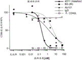
Figure 2 graphically depicts agonistic activity (figure 2A) and antagonistic activity (figure 2B)) of humanized antibody Ab101 versus antibody 4D11, antibody Blb, IgG antibody (control), and agonistic control antibody (2141).
Figure 3 graphically depicts the results of in vivo studies of humanized antibody Ab 101. Figure 3A graphically depicts IgG production in huscid mice that have received human PBMC and Ig control, CTLA4-Ig fusion, or Ab101 antibody. Fig. 3B graphically depicts B cell survival in the same mouse model that has been administered the same agent as in fig. 3A.
FIG. 4 shows an alignment of amino acid sequences of anti-human CD40 murine antibody antagonists and the resulting consensus sequences aligned. FIG. 4A shows the alignment of the variable light chains of antibody 3(Ab3) (SEQ ID NO: 48), Ab1(SEQ ID NO: 9) and antibody 2(Ab2) (SEQ ID NO: 76) and the variable light chain consensus sequence (SEQ ID NO: 116). FIG. 4B shows the alignment of the variable heavy chains of Ab3(SEQ ID NO: 44), Ab1(SEQ ID NO: 5) and Ab2(SEQ ID NO: 75) and the variable heavy chain consensus sequence (SEQ ID NO: 117).
Fig. 5A and 5B graphically depict representative neutralization potency (antagonistic activity) (fig. 5A) and agonistic activity (fig. 5B) of Ab102 on human CD40 in the monocyte activation assay described in example 7. Monocyte activation was consistent with increased TNF concentrations in each assay.
Figure 6 graphically depicts dose-responsive inhibition of endoscopic scoring with prophylactic administration of antibody 138(Ab 138). Antibody 138 was tested at 15, 5, 1.5 and 0.5 mg/kg. IgG negative controls were used. Anti-p 40IL-12/23 treatment served as a positive control. The disease is mediated by CD45Rbhi cells transferred to the animal. Low RB means negative control. Low cell CD45RB does not mediate disease.
Fig. 7 graphically depicts the results of immunohistochemical analysis for determination of IBA1+ macrophages in the colon following administration of antibody 138(Ab 138). Histological analysis of colon sections showed a decrease in macrophages (usually measured for inflammation). IgG negative controls were used. Anti-p 40IL-12/23 treatment served as a positive control.
Fig. 8 graphically depicts serum levels of circulating antibody 138(Ab138) at 96 hours post last dose (equal to C) in a T cell transfer model of colitisLow valley). Serum levels were shown to be dose-responsive. Anti-p 40IL-12/23 treatment served as a positive control. Only 1 animal in the 0.5mg/kg group had measurable Ab138 levels.
Figure 9A graphically depicts results of endoscopic examination after administration of antibody 138 in a mouse model of colitis. After endoscopic confirmation of disease, antibody 138(Ab138) treatment was initiated three weeks after cell injection, and dose-responsive inhibition of the total score of MEDAI was noted. The highest dose (15mg/kg) achieved statistical significance (fig. 9A).
Fig. 9B graphically depicts histological results after administration of antibody 138. Histological analysis of IBA1+ macrophages in the colon is shown in figure 9B as a measure of bone marrow inflammation.
Fig. 10 graphically depicts results showing Ab102 suppresses anti-KLH IgM and anti-KLH IgG (dashed lines) compared to control animals treated with vehicle only (solid lines). Ab102 was administered Subcutaneously (SC) to cynomolgus monkeys (two/sex/group) at doses of 0 (vehicle only) or 10mg/kg for 5 weeks. On day 8, all animals were dosed with Keyhole Limpet Hemocyanin (KLH). Serum samples were collected from each animal on days-11, 7, 0, 4, 7, 10, 14 and 21 (KLH days) relative to KLH administration.
FIG. 11A is a graph showing that anti-CD 40 antibody 138 treatment prevented proteinuria in MRL/lpr mice. Mice were given 15mg/kg antibody 2 x/week, 5mg/kg antibody 2 x/week, 1.5mg/kg antibody 2 x/week, or 15mg/kg antibody 1 x/week. Phosphate Buffered Saline (PBS) vehicle alone was used as a control. Proteinuria was measured as a percentage of urine protein < 300 mg/dL.
Fig. 11B is a graph depicting results showing that anti-CD 40 antibody 138 treatment prolonged survival of MRL/lpr mice. Animals were administered 15mg/kg antibody 2X/week, 5mg/kg antibody 2X/week, 1.5mg/kg antibody 2X/week, or 15mg/kg antibody 1X/week. Vehicle alone was used as a control. Percent survival over time is shown.
Fig. 12A is a graph depicting results showing that anti-CD 40 antibody 138 treatment prevented nephritis development. FIG. 12A shows the effect of antibody 138 on glomerular disease on days 29 and 63 in mice administered 15mg/kg antibody 2X/week, 5mg/kg antibody 2X/week, 1.5mg/kg antibody 2X/week, or 15mg/kg antibody 1X/week. PBS vehicle alone was used as control. Glomerular disease was rated on a scale of 0 to 4. As the severity of glomerular disease in aging MRL mice increased, 5 and 15mg/kg antibody 138 maintained efficacy, minimizing glomerular disease. Perivascular inflammation was scored on a scale of 0-4 based on the following criteria: 0 to at most some rare lymphocytes; 1-some lymphocytes form loose aggregates; 2-lymphocytes form discrete small aggregates; polarized accumulation of 3-lymphocytes, swelling into the proximal venous lumen, but failing to completely encircle the arcuate artery; 4-lymphocytes are fully accumulated around the arcuate artery and extend into the adventitia of the arcuate artery.
Fig. 12B is a graph depicting results showing that anti-CD 40 antibody treatment prevented nephritis development. Fig. 12B depicts results showing the effect of antibody 138 on Perirenal Vascular (PV) inflammation on days 29 and 63 in mice administered 15mg/kg antibody 2 x/week, 5mg/kg antibody 2 x/week, 1.5mg/kg antibody 2 x/week, or 15mg/kg antibody 1 x/week. PBS vehicle alone was used as control. At 29 and 63 days, 5 and 15mg/kg of anti-CD 40 antibody was effective in reducing Perivascular (PV) infiltration in the kidney.
Fig. 12C is a graph showing that anti-CD 40 antibody 138 treatment prevented the development of nephritis. FIG. 12C shows the effect of antibody 138 on Tubulointerstitial Inflammation (TI) at days 29 and 60 in mice administered 15mg/kg antibody 2X/week, 5mg/kg antibody 2X/week, 1.5mg/kg antibody 2X/week, or 15mg/kg antibody 1X/week. PBS vehicle alone was used as control. In early disease, TI is reduced.
Fig. 13A is a graph showing that anti-CD 40 antibody 138 treatment prevented salivary gland inflammation. FIG. 13A shows the effect of antibody 138 on salivary gland inflammation on days 29 and 60 in mice administered 15mg/kg antibody 2X/week, 5mg/kg antibody 2X/week, 1.5mg/kg antibody 2X/week, or 15mg/kg antibody 1X/week. PBS vehicle alone was used as control. Periductal inflammation was scored on a scale of 0-4 based on the following criteria: 0 to at most some rare white blood cells; 1-some leukocytes form loose aggregates; 2-leukocytes form discrete small aggregates; 3-polarized aggregation of white blood cells, completely surrounding the catheter; the 4-white blood cells aggregate and extend into the glandular parenchyma of the salivary glands.
Fig. 13B is a graph showing that anti-CD 40 antibody 138 treatment prevented joint inflammation. FIG. 13B shows the effect of antibody 138 on joint inflammation on days 29 and 60 in mice administered 15mg/kg antibody 2X/week, 5mg/kg antibody 2X/week, 1.5mg/kg antibody 2X/week, or 15mg/kg antibody 1X/week. PBS vehicle alone was used as control. The joint inflammation of each of the two paws per mouse was scored on a scale of 0-4 based on the following criteria: 0-no inflammation; 1-there are some white blood cells in the joint space; 2-white blood cells are commonly seen in the joint space and slight synovial hyperplasia exists; 3-white blood cells expand into the joint space with moderate synovial hyperplasia; 4-white blood cells and synovium proliferate, extend and coalesce within the joint space, with significant bone erosion and/or bone hyperplasia. The possible total score obtained by adding the scores of each mouse was 8.
Fig. 14 is a set of four panels (i-iv) showing that anti-CD 40 antibody 138 prevented the expansion of follicular helper T cells (Tfh) and Germinal Center (GC) B cells in the spleen as determined by flow cytometry. Mice were given 15mg/kg antibody 2 x/week, 5mg/kg antibody 2 x/week, 1.5mg/kg antibody 2 x/week, or 15mg/kg antibody 1 x/week. PBS vehicle alone was used as control. Panel (i) shows the number of Tfh cells in the spleen on day 29. Panel (ii) shows the number of Tfh cells in the spleen on day 63. FIG. (iii) shows the number of GC B cells in the spleen on day 29. Panel (iv) shows the number of GC B cells in spleen at day 63.
Figure 15A is a graph showing that anti-CD 40 antibody 138 treatment prevented an increase in total circulating IgG levels by day 29. Mice were given 15mg/kg antibody 2X/week, 5mg/kg antibody 2X/week, 1.5mg/kg antibody 2X/week and 15mg/kg antibody 1X/week. PBS vehicle alone was used as control.
Fig. 15B is a graph showing that anti-CD 40 antibody 138 treatment prevented an increase in total circulating IgG levels on day 63. Mice were given 15mg/kg antibody 2X/week, 5mg/kg antibody 2X/week, 1.5mg/kg antibody 2X/week and 15mg/kg antibody 1X/week. PBS vehicle alone was used as control.
Figure 16A is a graph showing the effect of anti-CD 40 antibody 138 treatment on anti-double stranded DNA (anti-dsDNA) titers at day 29. Mice were given 15mg/kg antibody 2 x/week, 5mg/kg antibody 2 x/week, 1.5mg/kg antibody 2 x/week, or 15mg/kg antibody 1 x/week. PBS vehicle alone was used as control. On day 29, anti-dsDNA titers were determined.
Figure 16B is a graph showing anti-CD 40 antibody 138 treatment against double stranded DNA (anti-dsDNA) titers at day 63. Mice were given 15mg/kg antibody 2 x/week, 5mg/kg antibody 2 x/week, 1.5mg/kg antibody 2 x/week, or 15mg/kg antibody 1 x/week. PBS vehicle alone was used as control. On day 63, anti-dsDNA titers were determined.
Fig. 17A is a graph showing that prophylactic administration of anti-CD 40 antibody 138 prevented proteinuria. Prophylactic treatment was initiated in mice at 26 weeks of age, and proteinuria mice were excluded from the study. Mice were given 15mg/kg antibody 2 x/week, 1.5mg/kg antibody 2 x/week, or 15mg/kg antibody 1 x/week. PBS vehicle alone was used as control. Proteinuria was measured as a percentage of urine protein < 300 mg/dL.
Figure 17B is a graph showing that prophylactic administration of anti-CD 40 antibody 138 prolongs survival using the SLE mouse model. Prophylactic treatment was initiated in mice at 26 weeks of age, and proteinuria mice were excluded from the study. Mice were given 15mg/kg antibody 2 x/week, 1.5mg/kg antibody 2 x/week, or 15mg/kg antibody 1 x/week. PBS vehicle alone was used as control. The percent survival up to 36 weeks of age was assessed.
Fig. 18A is a graph showing that mice treated intra-peritoneally at a dose of 15mg/kg 2 ×/week with antibody 138 developed low proteinuria over time, as shown by the urinary protein rating (mg/dL equivalent). Vehicle PBS was administered intraperitoneally, 2 x/week, as a control. Prednisolone was administered orally (PO) at a dose of 10mg/kg once a day (SID). Neither control mice treated with vehicle-free PBS nor prednisolone treated mice produced low proteinuria. The threshold for proteinuria was designated 300 mg/dL.
Fig. 18B is a graph showing the rate of recovery from proteinuria of mice treated intraperitoneally at a dose of 15mg/kp 2 x/week with antibody 138. The mean time to recovery from proteinuria was 23 ± 7 days, based on the rate of recovery from proteinuria as determined by the percentage of normal urine protein. Vehicle PBS was administered intraperitoneally, 2 x/week, as a control. Prednisolone was administered orally (PO) at a dose of 10mg/kg once a day (SID).
Fig. 18C is a graph showing a significant prolongation of survival of mice treated intra-peritoneally at a dose of 15mg/kp with anti-CD 40 antibody 138 2 x/week, as shown by the percentage survival. Vehicle PBS was administered intraperitoneally, 2 x/week, as a control. Prednisolone was administered orally (PO) once a day (SID).
Fig. 19A is a graph showing that saliva yield was preserved by prophylactic treatment with antibody 138 at a dose of 15mg/kp IP 2 x/week, 1.5mg/kg 2 x/week, 15mg/kg 1 x/week. Vehicle PBS was used as control. Prednisolone was administered at a dose of 10 mg/kg. Saliva production from 7-week-old NZBHF-1 mice, which were young mice without disease, was used as a further comparison. Saliva volume (mg) was determined. Saliva production was relatively uniform in anti-CD 40 treated mice.
Fig. 19B is a graph showing the preservation of saliva volume by prophylactic treatment with anti-CD 40 antibody 138 at doses of 15mg/kp IP 2 x/week, 1.5mg/kg 2 x/week, 15mg/kg 1 x/week. Vehicle PBS was used as control. Prednisolone was administered at a dose of 10 mg/kg. Saliva volume/body weight (mg/gm) was determined. Saliva production was significantly greater in mice treated with anti-CD 40 antibody than in untreated control mice.
Figure 20A is a graph showing that saliva production was preserved by therapeutic treatment with anti-CD 40 antibody 138 at a dose of 15 mg/kg. Prednisolone was administered at a dose of 10 mg/kg. Saliva production of 11 week old mice was used for further comparison. Saliva volume (mg) was determined.
Figure 20B is a graph showing that saliva production was preserved by therapeutic treatment with anti-CD 40 antibody 138 at a dose of 15 mg/kg. Prednisolone was administered at a dose of 10 mg/kg. Saliva production of 11 week old mice was used for further comparison. Saliva volume/body weight (mg/gm) was determined.
Detailed Description
The present invention relates to antagonist anti-CD 40 antibodies, or antigen-binding portions thereof, and uses thereof. Various aspects of the invention relate to antibodies and antibody fragments and pharmaceutical compositions thereof, as well as nucleic acids, recombinant expression vectors, and host cells for making such antibodies and fragments. The invention also encompasses methods of using the antibodies of the invention to detect human CD40, inhibit human CD40/CD40L activity in vitro or in vivo, and prevent or treat diseases or disorders such as chronic inflammatory diseases and crohn's disease.
Unless otherwise defined herein, scientific and technical terms used in connection with the present invention shall have the meaning commonly understood by one of ordinary skill. The meaning and scope of terms should be clear, however, in the case of any potential ambiguity, the definitions provided herein take precedence over any dictionary or external definition. Furthermore, unless the context requires otherwise, singular terms shall include the plural and plural terms shall include the singular. In this application, the use of "or" means "and/or" unless stated otherwise. Furthermore, the use of the term "including" and other forms such as "includes" and "included" is not limiting. Furthermore, unless specifically stated otherwise, terms such as "element" or "component" encompass both elements and components comprising one unit and elements and components comprising more than one subunit.
Generally, the nomenclature used and the techniques described herein in connection with cell and tissue culture, molecular biology, immunology, microbiology, genetics and protein and nucleic acid chemistry and hybridization are well known and commonly employed in the art. Unless otherwise indicated, the methods and techniques of the present invention are generally performed according to conventional methods well known in the art and as described in various general and more specific references that are cited and discussed throughout the present specification. Enzymatic reactions and purification techniques are performed according to the manufacturer's instructions as commonly practiced in the art or as described herein. The nomenclature used in connection with analytical chemistry, synthetic organic chemistry, and medicinal and pharmaceutical chemistry, and the laboratory procedures and techniques thereof, described herein, are those well known and commonly employed in the art. Standard techniques are used for chemical synthesis, chemical analysis, drug preparation, formulation and delivery, and patient treatment.
In order to make the present invention easier to understand, the selected terms are defined below.
The term "polypeptide" as used herein refers to any polymeric chain of amino acids. The terms "peptide" and "protein" are used interchangeably with the term polypeptide and also refer to a polymeric chain of amino acids. The term "polypeptide" encompasses natural or artificial proteins, protein fragments, and polypeptide analogs of a protein sequence. The polypeptide may be monomeric or polymeric.
The term "isolated protein" or "isolated polypeptide" is a protein or polypeptide that: not associated with its natural binding component that is associated with it in its natural state, depending on its source or derivative; substantially free of other proteins from the same species; expressed by cells from different species; or do not occur in nature. Thus, a polypeptide that is chemically synthesized or synthesized in a cellular system different from the cell from which it is naturally derived will be "isolated" from its naturally associated components. Proteins can also be made substantially free of naturally associated components by isolation using protein purification techniques well known in the art. An example of an isolated polypeptide is an isolated antibody or antigen-binding portion thereof.
The term "recovering" as used herein refers to a process whereby a chemical substance, such as a polypeptide, is rendered substantially free of naturally associated components by, for example, separation using protein purification techniques well known in the art.
The terms "human CD 40" and "human CD40 wild-type" (abbreviated herein as hCD40, hCD40wt) as used herein refer to type I transmembrane proteins. In one embodiment, the term human CD40 is intended to include recombinant human CD40(rhCD40), which can be prepared by standard recombinant expression methods. Table 1 provides the amino acid sequence of human CD40 (i.e., SEQ ID NO.1) and the amino acid sequence of its extracellular domain (i.e., SEQ ID NO: 107), which are known in the art.
Table 1: sequence of human CD40


"biological activity" as used herein refers to all intrinsic biological properties of the CD40 receptor. Biological properties of CD40 include, but are not limited to, binding to CD 40L; participate in B cell development; participate in lymphocyte activation; (ii) involved in antigen presenting cell function; regulating the activity of dendritic cells, macrophages and B cells; inducing the production of inflammatory cytokines in macrophages and dendritic cells; up-regulating antigen presentation; up-regulation of T cell stimulation; and promoting immunoglobulin class switching in B cells.
The term "specifically binds" or "specifically binds" as used herein with respect to the interaction of an antibody, protein or peptide with a second chemical means that the interaction is dependent on the presence of a particular structure (e.g., an antigenic determinant or epitope) on the chemical; if an antibody is specific for epitope "a", the presence of a molecule containing epitope a (or unlabeled free a) will reduce the amount of labeled a bound to the antibody in a reaction containing labeled "a" and the antibody.
The term "agonist" as used herein refers to a modulator that, when contacted with a molecule of interest (e.g., CD40), results in an increase in the amount of a certain activity or function of the molecule compared to the amount of activity or function observed in the absence of agonist.
The term "antagonist" or "inhibitor" as used herein refers to a modulator that, when contacted with a molecule of interest, results in a decrease in the magnitude of a certain activity or function of the molecule compared to the magnitude of activity or function observed in the absence of the antagonist. Specific related antagonists include those that block or modulate the biological or immunological activity of human CD40(hCD 40). Antagonistic antibodies to hCD40 can, for example, inhibit the up-regulation of CD86 in primary human B cells (such as B cells cultured with human T cells expressing CD40L) cultured with CD40L (or exposed to CD 40L). In one embodiment, an antagonistic anti-CD 40 antibody or antigen-binding portion thereof that is substantially free of agonistic activity is defined as having a level of activity equal to or within one standard deviation relative to a negative control in an agonistic assay, such as the agonistic monocyte assay described in example 7.
The antibodies, or antigen-binding portions thereof, of the invention are antagonist antibodies, or antigen-binding portions thereof, that cause a reduction in CD40 activity or function as compared to CD40 activity or function in the absence of the antibody, or antigen-binding portion thereof. In particular embodiments, the antibody or antigen-binding portion thereof is substantially free of agonist activity, i.e., the antibody or antigen-binding portion thereof does not cause an increase in the amount of CD40 activity or function as compared to CD40 activity or function in the absence of the antibody or antigen-binding portion thereof. Agonistic and antagonistic activities can also be assessed using methods known in the art, such as expression of human CD40 using a reporter cell line expressing CD40 associated with NFkB-mediated Alkaline Phosphatase (AP), or B cell assays. Furthermore, in one embodiment, agonistic and antagonistic activities can be assessed using the in vitro monocyte agonistic and antagonistic assays described in example 7.
The term "inhibits binding to CD 40L" refers to the ability of an antibody or antigen-binding fragment thereof to prevent binding of CD40 to ligand CD 40L. Such inhibition of binding to CD40L will result in a reduction or elimination of biological activity mediated by the binding of CD40 to CD 40L.
The term "antibody" as used broadly herein refers to any immunoglobulin (Ig) molecule, or any functional fragment, mutant, variant or derivative thereof, that is composed of four polypeptide chains, two heavy (H) chains and two light (L) chains, that retains the major epitope-binding characteristics of the Ig molecule. Such mutant, variant or derivative antibody formats are known in the art. Non-limiting examples of which are discussed below.
In a full-length antibody, each heavy chain is composed of a heavy chain variable region (abbreviated herein as HCVR or VH) and a heavy chain constant region. The heavy chain constant region is composed of three domains (CH1, CH2, and CH 3). Each light chain is composed of a light chain variable region (abbreviated herein as LCVR or VL) and a light chain constant region. The light chain constant region is composed of one domain CL. The VH and VL regions can be further subdivided into hypervariable regions, termed Complementarity Determining Regions (CDRs), interspersed with more conserved regions termed Framework Regions (FRs). Each VH and VL is composed of three CDRs and four FRs, arranged from amino-terminus to carboxy-terminus in the following order: FR1, CDR1, FR2, CDR2, FR3, CDR3, FR 4. Immunoglobulin molecules can be of any type (e.g., IgG, IgE, IgM, IgD, IgA, and IgY), class (e.g., IgG1, IgG2, IgG3, IgG4, IgA1, and IgA2), or subclass.
The term "antigen-binding portion" or "antigen-binding fragment" of an antibody (or simply "antibody portion" or "antibody fragment") as used herein refers to one or more fragments of an antibody that retain the ability to specifically bind to an antigen (e.g., hCD 40). It has been shown that the antigen binding function of an antibody can be performed by fragments of a full-length antibody. Such antibody embodiments may also be in bispecific, or multispecific forms; specifically bind to two or more different antigens. Examples of binding fragments encompassed within the term "antigen-binding portion" or "antigen-binding fragment" of an antibody include (i) a Fab fragment, a monovalent fragment consisting of the VL, VH, CL and CH1 domains; (ii) f (ab')2A fragment, a bivalent fragment comprising two Fab fragments linked by a disulfide bridge of the hinge region; (iii) an Fd fragment consisting of the VH and CH1 domains; (iv) (ii) an Fv fragment consisting of the VL and VH domains of a single arm of an antibody; (v) dAb fragments (Ward)Et al, (1989) Nature 341: 544-; winter et al, PCT publication WO 90/05144 a1, which is incorporated herein by reference), which comprises a single variable domain; and (vi) isolating the Complementarity Determining Regions (CDRs). Furthermore, although the two domains of the Fv fragment (VL and VH) are encoded by separate genes, they can be joined using recombinant methods by a synthetic linker that can be made into a single protein chain in which the VL and VH regions pair to form monovalent molecules (known as single chain Fv (scFv); see, e.g., Bird et al (1988) Science 242: 423-. Such single chain antibodies are also intended to be encompassed within the term "antigen-binding portion" or "antigen-binding fragment" of an antibody. Other forms of single chain antibodies, such as bifunctional antibodies, are also contemplated. Bifunctional antibodies are bivalent, bispecific antibodies in which the VH and VL domains are expressed on a single polypeptide chain, but using a linker that is too short to allow pairing between the two domains on the same chain, thereby forcing the domains to pair with the complementary domains of the other chain and generating two antigen binding sites (see, e.g., Holliger, p. et al (1993) proc.natl.acad.sci.usa 90: 6444- > 6448; poljak, r.j. et al (1994) Structure 2: 1121-1123). Such antibody binding moieties are known in the art (Kontermann and Dubel eds,Antibody Engineering(2001) Springer-Verlag. New York, page 790 (ISBN 3-540-.
The term "antibody construct" as used herein refers to a polypeptide comprising one or more antigen binding portions of the invention linked to a linker polypeptide or an immunoglobulin constant domain. Linker polypeptides comprise two or more amino acid residues linked by peptide bonds and are used to link one or more antigen binding moieties. Such linker polypeptides are well known in the art (see, e.g., Holliger, P. et al (1993) Proc. Natl. Acad. Sci. USA 90: 6444-. An immunoglobulin constant domain refers to a heavy or light chain constant domain. Human IgG heavy and light chain constant domain amino acid sequences are known in the art and are presented in table 2.
Table 2: sequences of human IgG heavy and light chain constant domainsColumn(s) of


Furthermore, the antibody or antigen-binding portion thereof may be part of a larger immunoadhesion molecule formed by covalent or non-covalent binding of the antibody or antibody portion to one or more other proteins or peptides. Examples of such immunoadhesion molecules include the use of the streptavidin core region to make tetrameric scFv molecules (Kipriyanov, S.M. et al (1995) Human Antibodies and hybrids 6: 93-101) and the use of cysteine residues, tag peptides and C-terminal polyhistidine tags to make bivalent and biotinylated scFv molecules (Kipriyanov, S.M. et al (1994) mol.Immunol.31: 1047-1058). Such as Fab and F (ab') 2The antibody portion of the fragment can be prepared from the whole antibody using conventional techniques, such as papain or pepsin digestion of the whole antibody, respectively. In addition, antibodies, antibody portions, and immunoadhesion molecules can be obtained using standard recombinant DNA techniques, as described herein.
As used herein, "isolated antibody" is intended to refer to an antibody that is substantially free of other antibodies having different antigenic specificities (e.g., an isolated antibody that specifically binds hCD40 is substantially free of antibodies that specifically bind antigens other than hCD 40). However, isolated antibodies that specifically bind hCD40 can be cross-reactive with other antigens, such as CD40 molecules from other species. In addition, the isolated antibody can be substantially free of other cellular material and/or chemicals.
The term "chimeric antibody" refers to an antibody comprising heavy and light chain variable region sequences from one species and constant region sequences from another species, such as an antibody having murine heavy and light chain variable regions linked to human constant regions.
The term "CDR-grafted antibody" refers to an antibody comprising heavy and light chain variable region sequences from one species, but with the sequences of one or more of the CDR regions of VH and/or VL replaced with CDR sequences of another species, such as an antibody having murine heavy and light chain variable regions in which one or more of the murine CDRs (e.g., CDR3) have been replaced with human CDR sequences.
The terms "Kabat numbering", "Kabat definitions" and "Kabat labeling" are used interchangeably herein. These terms are art-recognized and refer to the system of numbering amino acid residues that are variable (i.e., hypervariable) relative to other amino acid residues in the heavy and light chain variable regions of an antibody or antigen-binding portion thereof (Kabat et al (1971) Ann. NY Acad, Sci.190: 382. sup. 391; and Kabat, E.A. et al (1991) Sequences of Proteins of Immunological Interest, fifth edition, U.S. department of Health and public service (U.S. department of Health and Human Services), NIH publication No. 91-3242). For the heavy chain variable region, the hypervariable region ranges from amino acid positions 31 to 35 for CDR1, from amino acid positions 50 to 65 for CDR2, and from amino acid positions 95 to 102 for CDR 3. For the light chain variable region, the hypervariable region ranges from amino acid positions 24 to 34 for CDR1, from amino acid positions 50 to 56 for CDR2, and from amino acid positions 89 to 97 for CDR 3.
As used herein, the terms "receptor" and "receptor antibody" refer to an antibody or nucleic acid sequence that provides or encodes at least 80%, at least 85%, at least 90%, at least 95%, at least 98%, or 100% of the amino acid sequence of one or more framework regions. In some embodiments, the term "receptor" refers to an antibody amino acid or nucleic acid sequence that provides or encodes a constant region. In another embodiment, the term "receptor" refers to an antibody amino acid or nucleic acid sequence that provides or encodes one or more of a framework region and a constant region. In a particular embodiment, the term "acceptor" refers to a human antibody amino acid or nucleic acid sequence that provides or encodes at least 80%, preferably at least 85%, at least 90%, at least 95%, at least 98%, or 100% of the amino acid sequence of one or more framework regions. According to this embodiment, the receptor may contain at least 1, at least 2, at least 3, at least 4, at least 5, or at least 10 amino acid residues that are not present at one or more specific positions of the human antibody. The acceptor framework region and/or acceptor constant region may, for example, be derived or obtained from a germline antibody gene, a mature antibody gene, a functional antibody (e.g., an antibody well known in the art, an antibody under development, or a commercially available antibody).
As used herein, the term "CDR" refers to complementarity determining regions within an antibody variable sequence. There are three CDRs in each of the variable regions of the heavy and light chains, and for each variable region, such CDRs are designated CDR1, CDR2, and CDR 3. The term "set of CDRs" as used herein refers to a set of three CDRs present in a single variable region capable of binding antigen. The precise boundaries of these CDRs have been defined differently according to different systems. The system described by Kabat (Kabat et al, Sequences of Proteins of Immunological Interest (National Institutes of Health, Bethesda, Md. (1987) and (1991)) not only provides a clear residue numbering system suitable for any variable region of an antibody, but also provides precise residue boundaries defining the three CDRs, which may be referred to as Kabat CDR. Chothia and colleagues (Chothia et al, J.mol.biol.196: 901-917(1987) and Chothia et al, Nature 342: 877-883(1989)), it was found that some sub-portions within Kabat CDRs adopt almost the same peptide backbone conformation, even with great differences at the amino acid sequence level, these sub-portions are referred to as L1, L2 and L3 or H1, H2 and H3, where "L" and "H" indicate that these sub-portions have overlapping light chain regions at the amino acid sequence level, respectively, and the other CDRs which have overlapping CDR boundaries (Chobat J.133. 35: FAS. 35. FAS.J.133) (see that the same CDR 133. Ha et al., FAS. 45 (1996)). Other CDR boundary definitions may not strictly follow one of the above systems, but will nevertheless overlap with the Kabat CDRs, but may be shortened or lengthened according to predicted or experimental findings that a particular residue or group of residues, or even all CDRs, do not significantly affect antigen binding. The methods used herein may utilize CDRs defined according to any of these systems, but preferred embodiments use Kabat or Chothia defined CDRs.
As used herein, the term "canonical" residue refers to a residue in a CDR or framework that defines a particular canonical CDR structure as defined by Chothia et al (J.mol.biol.196: 901-907 (1987); Chothia et al, J.mol.biol.227: 799(1992), all incorporated herein by reference). According to Chothia et al, important portions of the CDRs of various antibodies have nearly identical peptide backbone conformations, even though the differences are great at the amino acid sequence level. Each canonical structure specifies a set of peptide backbone torsion angles, primarily for adjacent segments of amino acid residues that form loops.
As used herein, the terms "donor" and "donor antibody" refer to an antibody that provides one or more CDRs. In a preferred embodiment, the donor antibody is an antibody from a species that is different from the antibody of the framework region obtained or produced. In the case of humanized antibodies, the term "donor antibody" refers to a non-human antibody that provides one or more CDRs.
As used herein, the term "framework" or "framework sequence" refers to the sequence of the variable region minus the remainder of the CDRs. Since the exact definition of the CDR sequences can be determined by means of different systems, the meaning of the framework sequences correspondingly requires different interpretations. The six CDRs (CDR-L1, CDR-L2 and CDR-L3 of the light chain and CDR-H1, CDR-H2 and CDR-H3 of the heavy chain) also divide the framework regions on the light and heavy chains into four subregions on each chain (FR1, FR2, FR3 and FR4), with CDR1 located between FR1 and FR2, CDR2 located between FR2 and FR3, and CDR3 located between FR3 and FR 4. Without specifying a specific sub-region as FR1, FR2, FR3 or FR4, the FR of the combination within the variable region of a single naturally occurring immunoglobulin chain is represented as by the other mentioned framework regions. As used herein, FR represents one of the four sub-regions, and FRs represents two or more of the four sub-regions that make up the framework region.
Human heavy and light chain acceptor sequences are known in the art. In one embodiment of the invention, the human heavy and light chain acceptor sequences are selected from the sequences described in tables 3 and 4.
Table 3: heavy chain receptor sequences


Table 4: light chain acceptor sequences

As used herein, the term "germline antibody gene" or "gene segment" refers to immunoglobulin sequences encoded by non-lymphoid cells that have not undergone a maturation process that results in gene rearrangement and mutation to express a particular immunoglobulin. (see, e.g., Shapiro et al, Crit. Rev. Immunol.22 (3): 183-200 (2002); Marchalonis et al, Adv Exp Med biol.484: 13-30 (2001)). One of the advantages provided by the various embodiments of the present invention derives from the following recognition: germline antibody genes are more likely than mature antibody genes to retain the main amino acid sequence structural features of individuals in a species and are therefore less likely to be identified as being from an exogenous source when used therapeutically in that species.
As used herein, the term "critical" residues refers to certain residues within the variable region that have a greater impact on the binding specificity and/or affinity of an antibody, particularly a humanized antibody. Key residues include, but are not limited to, one or more of the following: residues adjacent to the CDRs, potential glycosylation sites (which can be N or O glycosylation sites), rare residues, residues capable of interacting with the antigen, residues capable of interacting with the CDRs, canonical residues, contact residues between the heavy chain variable region and the light chain variable region, residues within the cursor region, and residues in the region of overlap between Chothia-defined variable heavy chain CDR1 and Kabat-defined first heavy chain framework.
As used herein, the term "humanized antibody" is an antibody or variant, derivative, analog or fragment thereof that immunospecifically binds to an antigen of interest (e.g., human CD40) and comprises Framework (FR) regions having substantially the amino acid sequence of a human antibody and Complementarity Determining Regions (CDRs) having substantially the amino acid sequence of a non-human antibody. As used herein, the term "substantially" in the context of a CDR means that the amino acid sequence of the CDR is at least 80%, preferably at least 85%, at least 90%, at least 95%, at least 98%, or at least 99% identical to the amino acid sequence of a non-human antibody CDR. A humanized antibody comprises essentially all of at least one and typically two variable domains (Fab, Fab ', F (ab') 2, FabC, Fv), in which all or substantially all of the CDR regions correspond to those of a non-human immunoglobulin (i.e., donor antibody) and all or substantially all of the framework regions are framework regions having human immunoglobulin consensus sequences. Preferably, the humanized antibody further comprises at least a portion of an immunoglobulin constant region (Fc), typically that of a human immunoglobulin. In some embodiments, the humanized antibody contains a light chain and at least a variable domain of a heavy chain. The antibody may also include the CH1, hinge, CH2, CH3, and CH4 regions of the heavy chain. In some embodiments, the humanized antibody contains only humanized light chains. In some embodiments, the humanized antibody contains only humanized heavy chains. In particular embodiments, the humanized antibody contains only the humanized variable domains of the light chain and/or the humanized heavy chain.
Humanized antibodies may be selected from any class of immunoglobulins, including IgM, IgG, IgD, IgA, and IgE; and any isotype including, but not limited to, IgG 1, IgG2, IgG3, and IgG 4. Humanized antibodies may comprise sequences from more than one class or isotype, and particular constant domains may be selected using techniques well known in the art to optimize the desired effector function.
The framework regions and CDR regions of the humanized antibody do not have to correspond exactly to the parental sequences, e.g., a donor antibody CDR or consensus framework can be mutated by substitution, insertion and/or deletion of at least one amino acid residue such that the CDR or framework residue at that position does not correspond to the donor antibody or consensus framework. However, in a preferred embodiment, there are not many such mutations. Typically, at least 80%, preferably at least 85%, more preferably at least 90% and most preferably at least 95% of the humanized antibody residues will correspond to those of the parent FR and CDR sequences. As used herein, the term "consensus framework" refers to the framework regions in a consensus immunoglobulin sequence. As used herein, the term "consensus immunoglobulin sequence" refers to a sequence formed by the most frequently occurring amino acids (or nucleotides) in a family of related immunoglobulin sequences (see, e.g., Winnaker, From Genes to Clones (Verlagsgesellschaft, Weinheim, Germany 1987)). In the immunoglobulin family, each position in the consensus sequence is occupied by the amino acid that occurs most frequently at that position in the family. If two amino acids occur equally frequently, either may be included in the consensus sequence.
As used herein, a "vernier" region refers to a subset of framework residues that can adjust the CDR structure and fine tune the fit to the antigen as described by Foote and Winter (1992, J.mol.biol.224: 487-499, which is incorporated herein by reference). Cursor region residues form the layer under the CDR and can affect the CDR structure and antibody affinity.
The term "multivalent binding protein" is used in this specification to denote a binding protein comprising two or more antigen binding sites. Multivalent binding proteins are preferably engineered to have three or more antigen binding sites and are generally not naturally occurring antibodies. The term "multispecific binding protein" refers to a binding protein capable of binding two or more related or unrelated targets.
The terms "dual variable domain" or "DVD-Ig" as used interchangeably herein are antigen binding proteins that comprise two or more antigen binding sites and are tetravalent or multivalent binding proteins. Such DVDs can be monospecific, i.e., capable of binding one antigen; or multispecific, i.e. capable of binding two or more antigens. A DVD-binding protein comprising two heavy chain DVD polypeptides and two light chain DVD polypeptides is referred to as DVD Ig. Each half of the DVD Ig contains a heavy chain DVD polypeptide and a light chain DVD polypeptide and two antigen binding sites. Each binding site comprises a heavy chain variable domain and a light chain variable domain, and each antigen binding site involves a total of 6 CDRs at antigen binding. In one embodiment, the CDRs described herein (e.g., SEQ ID NOs: 6, 42 and 8 (heavy chain) and 21, 11 and 12 (light chain)) are used in an anti-CD 40 DVD. Examples of DVD-Ig structures are known in the art and are described, for example, in U.S. patent No. 7,612,181, which is incorporated herein by reference.
As used herein, the term "neutralizing" refers to the neutralization of the biological activity of a cytokine receptor when an antibody or antigen-binding portion thereof specifically binds to the cytokine receptor. Preferably, the neutralizing antibody or antigen-binding portion thereof is a neutralizing antibody whose binding to hCD40 results in inhibition of the biological activity of hCD 40. Preferably, the neutralizing antibody, or antigen-binding portion thereof, binds hCD40 and reduces the biological activity of hCD40 by at least about 20%, 40%, 60%, 80%, 85%, or 85% or more. Inhibition of the biological activity of hCD40 by a neutralizing antibody or antigen-binding portion thereof can be assessed by one or more indicators well known in the art that measure the biological activity of hCD 40.
The term "activity" includes the following activities: such as the binding specificity/affinity of the antibody for an antigen, e.g., an anti-hCD 40 antibody that binds to hCD40 antigen, and/or the neutralizing potency of the antibody, e.g., an anti-hCD 40 antibody that binds to hCD40 that inhibits the biological activity of hCD40 (e.g., binds to CD 40L); participate in B cell development; participate in lymphocyte activation; (ii) involved in antigen presenting cell function; regulating the activity of dendritic cells, macrophages and B cells; inducing the production of inflammatory cytokines in macrophages and dendritic cells; up-regulating antigen presentation; up-regulation of T cell stimulation; and promoting immunoglobulin class switching in B cells.
Exemplary assays for assessing the activity of the anti-CD 40 antibodies of the invention include in vitro and in vivo assays as described herein. In particular, such assays can be used to determine whether an anti-CD 40 antibody is an agonistic or antagonistic antibody.
For example, binding to human CD40 and inhibition of CD40-CD40L interaction can be analyzed by FACS analysis using a cell line expressing human CD 40. Antagonist and agonist activity can be assessed using a reporter cell line expressing CD40, which is associated with NFkB-mediated Alkaline Phosphatase (AP), expressing human CD 40. Upon receipt of the signal via CD40, NFkB activation results in the secretion of AP, measured by a colorimetric substrate. As an exemplary antagonistic assay, CD40 reporter cell lines can be cultured with Jurkat cell lines expressing CD40L (to provide physiological ligand interactions) or with soluble CD40L, and the ability of anti-CD 40 antibodies to block NFkB signaling can be assessed. For exemplary agonistic analysis, a human CD40 reporter cell line may be treated with an anti-CD 40 antibody and NFkB signaling measured as described above.
Alternatively, a B cell agonism assay may be employed in which B cells are activated with low doses of anti-IgM and IL4, followed by addition of a CD40 antagonist antibody. The enhancement of B cell activation can be measured as the upregulation of CD86, which in turn indicates agonist activity. Similarly, a B cell antagonism assay may be used in which primary human B cells are cultured with a human T cell line expressing CD40L, the CD40L expressing human T cell line leading to B cell activation and up-regulation of CD86 expression via CD40/CD40L interactions. Inhibition of CD86 upregulation of primary human B cells is indicative of antagonist activity.
The term "epitope" includes any polypeptide determinant capable of specifically binding to an antibody or antigen-binding portion thereof. In certain embodiments, epitopic determinants include chemically active surface groups of a molecule (such as amino acids, sugar side chains, phosphoryl groups, or sulfonyl groups), and in certain embodiments, may have particular three-dimensional structural characteristics and/or charge-to-mass ratio characteristics. In various embodiments, the epitope can be a linear or sequential epitope, i.e., a linear sequence of amino acids, a linear sequence of the primary structure of an antigen (i.e., CD 40). Alternatively, in other embodiments, the epitope may be a conformational epitope that has a particular three-dimensional shape when the antigen presents its secondary structure. For example, a conformational epitope may comprise non-linear, i.e., non-sequential, amino acids of an antigen.
In a particular embodiment, an epitope is a region of an antigen that is bound by an antibody or antigen-binding portion thereof. In certain embodiments, an antibody, or antigen-binding portion thereof, is said to specifically bind an antigen when it preferentially recognizes its antigen of interest in a complex mixture of proteins and/or macromolecules.
The term "human antibody" as used herein is intended to include antibodies having variable and constant regions derived from human germline immunoglobulin sequences. The human antibodies of the invention may include amino acid residues not encoded by human germline immunoglobulin sequences (e.g., mutations induced by random or site-specific mutations in vitro or introduced by in vivo somatic mutations), for example in the CDRs and in particular in CDR 3. However, the term "human antibody" as used herein is not intended to include antibodies in which CDR sequences derived from the germline of another mammalian species (such as a mouse) have been grafted onto human framework sequences.
The term "recombinant human antibody" as used herein is intended to include all human antibodies prepared, expressed, produced or isolated by recombinant means, such as antibodies expressed using a recombinant expression vector transfected into a host cell (further described in section il C below); antibodies isolated from recombinant combinatorial human antibody libraries (Hoogenboom H.R. (1997) TIB Tech.15: 62-70; Azzazy H. and Highsmith W.E., (2002) Clin. biochem.35: 425. 445; Gavilondo J.V. and Larrick J.W. (2002) BioTechniques 29: 128. 145; Hoogenboom H. and Chames P. (2000) Immunology Today 21: 371. 378); antibodies isolated from animals (e.g., mice) transgenic for human immunoglobulin genes (see, e.g., Taylor, L.D. et al (1992) Nucl. acids Res.20: 6287-6295; Kellermann S-A. and Green L.L. (2002) Current Opinion in Biotechnology 13: 593-597; Little M. et al (2000) Immunology Today 21: 364-370); or by any other means involving splicing of human immunoglobulin gene sequences into other DNA sequences. Such recombinant human antibodies have variable and constant regions derived from human germline immunoglobulin sequences. However, in certain embodiments, such recombinant human antibodies undergo in vitro mutagenesis (or in vivo cell mutagenesis when using animals transgenic for human Ig sequences), and thus the amino acid sequences of the VH and VL regions of the recombinant antibodies are sequences that, while derived from and involving human germline VH and VL sequences, may not naturally occur in vivo within the human antibody germline repertoire. One embodiment provides fully human antibodies capable of binding to human CD40, which can be produced using techniques well known in the art, such as, but not limited to, the use of human Ig phage libraries, such as those disclosed in Jermutus et al, PCT publication No. WO 2005/007699 a 2.
The term "surface plasmon resonance" as used herein refers to an optical phenomenon that allows for the analysis of real-time biospecific interactions by means of, for example, the detection of changes in protein concentration within a Biosensor matrix using the BIAcore system (Pharmacia Biosensor AB, Uppsala, Sweden and Piscataway, n.j.). For further description, see U. et al (1993) ann.biol.clin.51: 19-26;
 U.S. et al (1991) Biotechniques 11: 620-627; johnsson, b. et al (1995) j.mol.recognit.8: 125-131; and Johnnson, b. et al (1991) anal. biochem.198: 268-277.
The term "k" as used hereinon"is intended to mean the binding rate constant of an antibody bound to an antigen to form an antibody/antigen complex as known in the art.
The term "k" as used hereinoffBy "is meant the off-rate constant for dissociation of an antibody/antigen complex as known in the art.
The term "K" as used hereinD"is intended to mean the dissociation constant of a particular antibody-antigen interaction as known in the art.
The term "labeled antibody" as used herein refers to an antibody that incorporates a label ready to identify the antibody or antigen-binding portion thereof. Preferably, the label is a detectable marker, such as a polypeptide incorporating a radiolabeled amino acid or linked to a biotin moiety that can be detected by means of labeled avidin (e.g., streptavidin containing a fluorescent marker or enzymatic activity that can be detected by optical or colorimetric methods). Examples of polypeptide tags include, but are not limited to, the following: a radioisotope or radionuclide (e.g., 3H、14C、35S、90Y、99Tc、111In、125I、131I、177Lu、166Ho, or153Sm); fluorescent labels (e.g., FITC, rhodamine (rhodamine), lanthanide phosphors), enzymatic labels (e.g., horseradish peroxidase, luciferase, alkaline phosphatase); a chemiluminescent marker; a biotin group; a predetermined polypeptide epitope recognized by a secondary reporter (e.g., leucine zipper pair sequence, binding site for a secondary antibody, metal binding domain, epitope tag); and a magnetizer such as gadolinium chelate.
The terms "crystal" and "crystallization" as used herein refer to an antibody or antigen-binding portion thereof that exists in crystal form. A crystal is a solid state form of matter that is different from other forms such as an amorphous solid state or a liquid crystalline state. Crystals are composed of regular, repeating, three-dimensional arrays of atoms, ions, molecules (e.g., proteins such as antibodies) or aggregates of molecules (e.g., antigen/antibody complexes). These three-dimensional arrays are arranged according to specific mathematical relationships well understood in the art. The basic units or building blocks that repeat in the crystal are called asymmetric units. The repetition of asymmetric units in an arrangement that conforms to a given well-defined crystallographic symmetry provides a "unit cell" of the crystal. The unit cell is repeated in all three dimensions by regular transformation (regular transformation) to obtain crystals. See, Giege, R. and Ducruix, A.Barrett, crystallation of Nucleic Acids and Proteins, a Practical Approach, 2 nd edition, pages 201-16, Oxford University Press, New York, New York, (1999).
The term "polynucleotide" as used herein refers to a polymeric form of two or more nucleotides (ribonucleotides or deoxyribonucleotides or modified forms of either nucleotide type). The term includes single-stranded and double-stranded forms of DNA, but preferably double-stranded DNA.
The term "isolated polynucleotide" as used herein shall mean a polynucleotide (e.g., of genomic, cDNA, or synthetic origin or some combination thereof) as follows: depending on its origin, an "isolated polynucleotide" does not bind to all or a portion of the polynucleotide found with the "isolated polynucleotide" in nature; operably linked to a polynucleotide that is not linked in nature; or not in nature as part of a larger sequence.
The term "carrier" as used herein is intended to refer to a nucleic acid molecule capable of transporting another nucleic acid to which it has been linked. One type of carrier is a "plasmid," which refers to a circular double-stranded DNA loop into which additional DNA segments can be ligated. Another type of carrier is a viral carrier, in which additional DNA segments can be joined into the viral genome. Certain vectors are capable of autonomous replication in a host cell into which they are introduced (e.g., bacterial vectors having a bacterial origin of replication and episomal mammalian vectors). Other carriers (e.g., non-episomal mammalian carriers) can be integrated into the genome of a host cell upon introduction into the host cell, and thereby are replicated along with the host genome. In addition, certain vectors are capable of directing the expression of genes to which they are operably linked. Such vectors are referred to herein as "recombinant expression vectors" (or simply "expression vectors"). In general, expression vectors for use in recombinant DNA techniques are often in the form of plasmids. Since plasmids are the most commonly used form of carrier, in this specification, "plasmid" and "carrier" are used interchangeably. However, the invention is intended to include other forms of expression vectors, such as viral vectors (e.g., replication-defective retroviruses, adenoviruses and adeno-associated viruses), which serve equivalent functions.
The term "operably linked" refers to a juxtaposition wherein the components so described are in a relationship permitting them to function in their intended manner. A control sequence "operably linked" to a coding sequence is joined in such a way that expression of the coding sequence is achieved under conditions compatible with the control sequences. Sequences that are "operably linked" include expression control sequences that are adjacent to the gene of interest and expression control sequences that function in trans or at a distance to control the gene of interest. The term "expression control sequence" as used herein refers to a polynucleotide sequence required to effect expression and processing of the ligated coding sequence. Expression control sequences include appropriate transcription initiation, termination, promoter and enhancer sequences; efficient RNA processing signals, such as splicing and polyadenylation signals; sequences that stabilize cytoplasmic mRNA; sequences that enhance translation efficiency (i.e., Kozak consensus sequences); sequences that enhance protein stability; and sequences which enhance protein secretion if necessary. The nature of such control sequences varies depending on the host organism; in prokaryotes, such control sequences typically include a promoter, a ribosome binding site, and a transcription termination sequence; in eukaryotes, such control sequences generally include promoters and transcription termination sequences. The term "control sequences" is intended to include components whose presence is critical to expression and processing, and may also include additional components whose presence is advantageous, such as leader sequences and fusion partner sequences. The protein constructs of the invention may be expressed and purified using expression vectors and host cells known in the art, including expression cassettes, vectors, recombinant host cells, and methods for recombinant expression and proteolytic processing of recombinant polymeric and precursor proteins (pre-proteins) from a single open reading frame (e.g., WO 2007/014162, which is incorporated herein by reference).
As defined herein, "transformation" refers to any process by which exogenous DNA enters a host cell. Transformation can be performed under natural or artificial conditions using various methods well known in the art. Transformation may rely on any known method for inserting foreign nucleic acid sequences into prokaryotic or eukaryotic host cells. Methods are selected based on the host cell to be transformed and may include, but are not limited to, viral infection, electroporation, lipofection, and particle bombardment. Such "transformed" cells include stably transformed cells in which the inserted DNA is capable of replication either as an autonomously replicating plasmid or as part of the host chromosome. It also includes cells that transiently express the inserted DNA or RNA for a limited period of time.
The term "recombinant host cell" (or simply "host cell"), as used herein, is intended to refer to a cell into which exogenous DNA has been introduced. It will be understood that such terms are intended to refer not only to the particular subject cell, but also to the progeny of such a cell. Because certain modifications may occur in succeeding generations due to either mutation or environmental influences, such progeny may not, in fact, be identical to the parent cell, but are still included within the scope of the term "host cell" as used herein. Preferably, the host cell comprises a prokaryotic and eukaryotic cell selected from any of the kingdoms of life. Preferred eukaryotic cells include protists, fungi, plant and animal cells. Most preferably, host cells include, but are not limited to, the prokaryotic cell line escherichia coli (e.coli); mammalian cell lines CHO, HEK 293 and COS; insect cell line Sf 9; and the fungal cell Saccharomyces cerevisiae.
Recombinant DNA, oligonucleotide synthesis, and tissue culture and transformation (e.g., electroporation, lipofection) can be performed using standard techniques. Enzymatic reactions and purification techniques can be performed according to the manufacturer's instructions or as commonly practiced in the art or as described herein. The foregoing techniques and procedures may be generally performed according to conventional methods well known in the art and as described in various general and more specific references that are cited and discussed throughout the present specification. See, e.g., Sambrook et al Molecular Cloning: a Laboratory Manual (2 nd edition, Cold Spring Harbor Laboratory Press, Cold Spring Harbor, N.Y. (1989)), which is incorporated herein by reference for any purpose.
As known in the art and as used herein, "transgenic organism" refers to an organism having cells containing a transgene, wherein the transgene introduced into the organism (or an ancestor of the organism) expresses a polypeptide that is not naturally expressed in the organism. A "transgene" is a DNA construct that is stably and operably integrated into the genome of a cell in which a transgenic organism is developed, directing expression of the encoded gene product in one or more cell types or tissues of the transgenic organism.
The terms "modulate" and "modulation" are used interchangeably and, as used herein, refer to a change or alteration in the activity of a related molecule (e.g., the biological activity of hCD 40). Modulation may be an increase or decrease in the magnitude of certain activities or functions of the relevant molecule. Exemplary activities and functions of the molecules include, but are not limited to, binding characteristics, enzymatic activity, cellular receptor activation, and signal transduction.
Accordingly, the term "modulator" as used herein is a compound capable of altering or modifying the activity or function of the molecule of interest (e.g. the biological activity of hCD 40). For example, a modulator can result in an increase or decrease in the magnitude of a certain activity or function of a molecule compared to the magnitude of activity or function observed in the absence of the modulator. In certain embodiments, the modulator is an inhibitor that reduces the magnitude of at least one activity or function of the molecule. Exemplary inhibitors include, but are not limited to, proteins, peptides, antibodies, peptibodies, carbohydrates, or small organic molecules. Peptibodies are described, for example, in WO 01/83525.
As used herein, the term "effective amount" refers to a sufficient degree of severity and/or duration of treatment to reduce or ameliorate a disorder or one or more symptoms thereof; preventing the progression of the disorder; causing regression of the condition; preventing the recurrence, development, onset, or progression of one or more symptoms associated with the disorder; detecting a condition; or an amount that enhances or improves the prophylactic or therapeutic effect of another therapy (e.g., prophylactic or therapeutic agent).
As used herein, the term "non-responder" is used to refer to a subject having IBD (e.g., crohn's disease or ulcerative colitis) who has no improvement or limited or inappropriate clinical disease status (e.g., no reduction in CDAI score, no reduction in corticosteroid use) following treatment with a TNF α inhibitor. In one embodiment, the TNF non-responder is a subject with IBD whose Crohn's Disease Activity Index (CDAI) score fails to achieve a reduction of 100 points or more after treatment with a TNF α inhibitor. In one embodiment, the non-responder is a subject with IBD whose Crohn's Disease Activity Index (CDAI) score fails to achieve a reduction of 100 points or more over a specified time frame following treatment with a TNF α inhibitor.
I. Antibodies that bind human CD40(hCD40)
One aspect of the invention provides antagonistic humanized antibodies or antigen binding portions thereof that bind CD40, including human CD40(hCD 40). Other embodiments of the invention include murine monoclonal antibodies, or antigen-binding portions thereof, that bind to CD40, as well as chimeric antibodies comprising the variable regions of anti-CD 40 murine antibodies described herein. Preferably, the antibodies of the invention are antagonistic anti-CD 40 (e.g., anti-human CD40) antibodies that do not have significant agonistic activity.
1. Humanized anti-hCD 40 antagonist antibodies derived from antibody 1(Ab1)
The present invention is based, at least in part, on the identification of humanized anti-CD 40 antibodies that have antagonistic characteristics and, in certain embodiments, essentially no agonistic activity.
Three antagonist anti-hCD 40 murine antibodies, Ab1 (VL sequence set forth in SEQ ID NO: 9 and VH sequence set forth in SEQ ID NO: 5), Ab2 (VL sequence set forth in SEQ ID NO: 76 and VH sequence set forth in SEQ ID NO: 75) and Ab3 (VL sequence set forth in SEQ ID NO: 48 and VH sequence set forth in SEQ ID NO: 44) were identified as described in example 1.
Consensus CDR sequences were determined based on alignment of the CDR amino acid sequences of murine antagonistic antibodies Ab1, Ab2, and Ab 3. The consensus amino acid sequences of the VH CDR1, CDR2, and CDR3 regions are described in SEQ ID NOs: 78. 79 and 80, and the consensus amino acid sequences of the VL CDR1, CDR2, and CDR3 amino acid sequences are described in SEQ ID NOs: 108. 109 and 110. All sequences are also described in table 5 below and fig. 4.
TABLE 5 anti-CD 40 hybridoma CDR amino acid sequence alignment

The consensus amino acid sequence of the heavy chain CDR1 domain is set forth in SEQ ID NO: 78(G (F/Y) TF (S/T) (D/S) Y (G/T) M (N/H)). The consensus amino acid sequence of the variable heavy chain CDR2 domain is set forth in SEQ ID NO: 79(YI (S/N) (S/P) (G/S) (R/S) (D/S/G) (N/Y) (I/P) (Y/N) Y (A/N) (D/Q) (T/K) (V/F) K (G/D)). The consensus amino acid sequence of the variable heavy chain CDR3 domain is set forth in SEQ ID NO: 80((S/W) (W/G) (G/Y) (Y/S) FDV).
The consensus amino acid sequence of the variable light chain CDR1 domain is set forth in SEQ ID NO: 108((K/R) SS (Q/K) SLL (N/H) S (G/-) N (Q/G) (K/N) (N/T) YL (T/Y)). The consensus amino acid sequence of the variable light chain CDR2 domain is set forth in SEQ ID NO: 109((W/R) (A/M) ST (R/L) (E/A) S). The consensus amino acid sequence of the variable light chain CDR3 domain is set forth in SEQ ID NO: 110((Q/M) (N/Q) (D/H) (Y/L) (T/E) YPLT).
In one embodiment, the present invention provides an anti-CD 40 antagonist antibody, or antigen-binding portion thereof, comprising a variable light chain comprising a heavy chain variable region having the amino acid sequence of SEQ ID NO: 108, a CDR1 having the amino acid sequence of SEQ ID NO: 109, a CDR2 having the amino acid sequence of SEQ ID NO: 110, CDR3 of the amino acid sequence of seq id no; and comprises a variable heavy chain comprising a light chain having the amino acid sequence of SEQ ID NO: 78, a CDR1 having the amino acid sequence of SEQ ID NO: 79, a CDR2 having the amino acid sequence of SEQ ID NO: 80, CDR3 of the amino acid sequence of seq id no.
After the murine antibodies Ab1, Ab2, and Ab3 were identified, antibodies Ab1 and Ab3 were selected for humanization (described in example 2 below). Table 11 and table 12 provide the amino acid sequences of the CDR, VH and VL regions of humanized Ab1 and Ab3, respectively. Specifically, nine different humanized antibodies were generated based on Ab3 (see example 2 and table 12 below). Four different humanized antibodies were also generated based on Ab1, including the following:
A) huAb1VH.1/VL.1 (as shown in SEQ ID NO: 13 and the VH amino acid sequences as set forth in SEQ ID NOs: 6. VH CDR1, CDR2, and CDR3 sequences described in 7 and 8; and as shown in SEQ ID NO: 14 and VL amino acid sequences as set forth in SEQ ID NOs: 10. the VL CDR1, CDR2, and CDR3 sequences described for 11 and 12);
B) huAb1VH.1A/VL.1 (as shown in SEQ ID NO: 15 and VH amino acid sequences as set forth in SEQ ID NO: 6. VH CDR1, CDR2, and CDR3 sequences described in 7 and 8; and as shown in SEQ ID NO: 14 and VL amino acid sequences as set forth in SEQ ID NOs: 10. the VL CDR1, CDR2, and CDR3 sequences described for 11 and 12);
C) huAb1VH.1/VL.1A (as shown in SEQ ID NO: 13 and the VH amino acid sequences as set forth in SEQ ID NOs: 6. VH CDR1, CDR2, and CDR3 sequences described in 7 and 8; and as shown in SEQ ID NO: 16 and VL amino acid sequences as set forth in SEQ ID NOs: 10. the VL CDR1, CDR2, and CDR3 sequences described for 11 and 12); and
D) huAb1VH.1A/VL.1A (as shown in SEQ ID NO: 15 and VH amino acid sequences as set forth in SEQ ID NO: 6. VH CDR1, CDR2, and CDR3 sequences described in 7 and 8; and as shown in SEQ ID NO: 16 and VL amino acid sequences as set forth in SEQ ID NOs: 10. the VL CDR1, CDR2, and CDR3 sequences described for 11 and 12).
The humanized version of Ab1 was further modified to remove potential deamidation sites in the light chain CDR 1. Six variant huAb1 antibodies were analyzed, and four of these antibodies were identified as antagonists of CD 40. The six antibodies are referred to herein as Ab1v1, Ab1v2, Ab1v3, Ab1v4, Ab1v5 and Ab1v6 (CDRs and variable sequences are provided in table 13 below). Of the six humanized Ab1 variants, huAb1v1 was chosen because of its particularly superior antagonistic activity. The heavy chain variable sequence of huAb1v1 is provided in SEQ ID NO: 15, and the CDR1, CDR2, and CDR3 sequences are depicted in SEQ ID NOs: 6. 7 and 8. The light chain variable sequence of huAb1v1 is provided in SEQ ID NO: 20, and the CDR1, CDR2, and CDR3 sequences are depicted in SEQ ID NOs: 21. 11 and 12.
Further mutagenesis induced heavy chain CDR2 of antibody huAb1v1, resulting in seventeen variants (described in example 4 below). These huAb1v1 heavy chain CDR2 variants are referred to herein as huAb1v1CDR2v1 through huAb1v1CDR2v 17. The sequences of huAb1v1CDR2v1 to huAb1v1CDR2v17 heavy chains are provided in table 16, where VH huAb1v1CDR2v7 was selected as a clone with particularly excellent antagonistic activity against CD40, while still being relatively free of agonistic activity. The heavy chain variable sequence of huAb1v1CDR2v7 is provided in SEQ ID NO: 28, and the CDR1, CDR2, and CDR3 sequences are depicted in SEQ ID NOs: 6. 42 and 8.
After selection of huAb1v1CDR2v7VH (VH of SEQ ID NO: 28 and CDR1, CDR2 and CDR3 sequences set forth in SEQ ID NO: 6, 42 and 8, respectively) and huAb1v1VL (VL of SEQ ID NO: 20 and CDR1, CDR2 and CDR3 sequences set forth in SEQ ID NO: 21, 11 and 12, respectively), the variable regions were cloned into two different IgG backgrounds, generating two anti-CD 40 antagonist antibodies, Ab101 and Ab 102. Table 6 (and table 19) provides the full-length heavy and light chain sequences of particular embodiments of the invention relating to these IgG antibodies. In table 6, the constant regions are underlined and the CDR domains are in bold.
Table 6: humanized anti-CD 40 antibodies Ab101 and Ab102 and their heavy and light chain sequences


Thus, in one embodiment, the invention relates to an antagonistic anti-CD 40 antibody (Ab101) comprising a heavy chain variable region having the sequence set forth in SEQ ID NO: 40 and a light chain having an amino acid sequence as set forth in SEQ ID NO: 39, or a heavy chain of an amino acid sequence set forth in seq id no. In an alternative embodiment, the invention relates to an antagonistic anti-CD 40 antibody (Ab102) comprising a heavy chain variable region having the sequence set forth in SEQ ID NO: 40 and a light chain having an amino acid sequence as set forth in SEQ ID NO: 41, or a heavy chain of the amino acid sequence set forth in seq id no.
Thus, the invention includes murine, chimeric and humanized anti-CD 40 antibodies having antagonist activity. In certain embodiments, the present invention provides an antagonistic anti-CD 40 antibody, or antigen binding portion thereof, comprising a heavy chain variable region having an amino acid sequence as set forth in SEQ ID NO: 12 and/or a light chain variable region having a CDR3 comprising the amino acid sequence set forth in SEQ ID NO: 8, or a heavy chain variable region of CDR3 of the amino acid sequence set forth in seq id No. 8. In a particular embodiment, the antagonist anti-CD 40 antibody, or antigen-binding portion thereof, comprises: has the sequence shown in SEQ ID NO: 6, a light chain CDR1 having the amino acid sequence set forth in SEQ ID NO: 42, a light chain CDR2 having the amino acid sequence set forth in SEQ ID NO: 8, a heavy chain CDR3 having the amino acid sequence set forth in SEQ ID NO: 21, a light chain CDR1 having an amino acid sequence set forth in SEQ ID NO: 11 and a light chain CDR2 having the amino acid sequence set forth in SEQ ID NO: 12, light chain CDR3 of the amino acid sequence set forth in seq id no. In a particular embodiment, the antagonist anti-CD 40 antibody or antigen-binding portion thereof comprises a vh region having SEQ ID NO: 28 and a light chain variable domain having the amino acid sequence set forth in SEQ ID NO: 20, or a light chain variable domain of an amino acid sequence set forth in seq id No. 20. In one embodiment, the antagonist anti-CD 40 antibody or antigen-binding portion thereof comprises a light chain variable region having the amino acid sequence of SEQ ID NO: 41 and a light chain comprising the amino acid sequence set forth in SEQ ID NO: 40, or a light chain of an amino acid sequence set forth in seq id No. 40. In another embodiment, the antagonist anti-CD 40 antibody or antigen-binding portion thereof comprises a light chain variable region having the amino acid sequence of SEQ ID NO: 39 and a light chain comprising the amino acid sequence set forth in SEQ ID NO: 40, or a light chain of an amino acid sequence set forth in seq id No. 40.
Included in the invention are antibodies having the amino acid sequences (variable or CDR) set forth in tables 5, 6, 11, 12, 13, 14, 16, 17, 18 and 19. Accordingly, in one aspect, the invention relates to an antagonistic anti-CD 40 antibody, or antigen binding portion thereof, having a light chain variable region comprising (a) a light chain variable region having the amino acid sequence set forth in SEQ ID NO: 10. 17, 19 or 21, or a CDR1 of an amino acid sequence set forth in seq id no; (b) has the sequence shown in SEQ ID NO: 11, CDR2 of the amino acid sequence set forth in seq id no; and (c) has the sequence of SEQ ID NO: 12, CDR3 of the amino acid sequence set forth in seq id no. Alternatively or in combination, the antagonist anti-CD 40 antibody or antigen-binding portion thereof comprises a heavy chain variable region comprising (a) a light chain variable region having the amino acid sequence set forth in SEQ ID NO: 6, CDR1 of the amino acid sequence set forth in seq id no; (b) has the sequence shown in SEQ ID NO: 7 or 42, CDR2 of the amino acid sequence set forth in seq id no; and (c) has the sequence of SEQ ID NO: 8, CDR3 of the amino acid sequence set forth in seq id no.
In a particular embodiment, the antagonist anti-CD 40 antibody or antigen-binding portion thereof has a light chain variable region comprising (a) a light chain variable region having the amino acid sequence set forth in SEQ ID NO: 10, (b) a CDR1 having the amino acid sequence set forth in SEQ ID NO: 11 and (c) a CDR2 having the amino acid sequence set forth in SEQ ID NO: 12, CDR3 of the amino acid sequence set forth in seq id no; the heavy chain variable region comprises (a) a heavy chain variable region having the amino acid sequence set forth in SEQ ID NO: 6, (b) a CDR1 having the amino acid sequence set forth in SEQ ID NO: 7 and (c) a CDR2 having the amino acid sequence set forth in SEQ ID NO: 8, CDR3 of the amino acid sequence set forth in seq id no.
In another embodiment, the antagonist anti-CD 40 antibody or antigen-binding portion thereof has a light chain variable region comprising (a) a light chain variable region having the amino acid sequence set forth in SEQ ID NO: 19, (b) a CDR1 having the amino acid sequence set forth in SEQ ID NO: 11 and (c) a CDR2 having the amino acid sequence set forth in SEQ ID NO: 12, CDR3 of the amino acid sequence set forth in seq id no; the heavy chain variable region comprises (a) a heavy chain variable region having the amino acid sequence set forth in SEQ ID NO: 6, (b) a CDR1 having the amino acid sequence set forth in SEQ ID NO: 7 and (c) a CDR2 having the amino acid sequence set forth in SEQ ID NO: 8, CDR3 of the amino acid sequence set forth in seq id no.
In another embodiment, the antagonist anti-CD 40 antibody or antigen-binding portion thereof has a light chain variable region comprising (a) a light chain variable region having the amino acid sequence set forth in SEQ ID NO: 17, (b) a CDR1 having the amino acid sequence set forth in SEQ ID NO: 11 and (c) a CDR2 having the amino acid sequence set forth in SEQ ID NO: 12, CDR3 of the amino acid sequence set forth in seq id no; the heavy chain variable region comprises (a) a heavy chain variable region having the amino acid sequence set forth in SEQ ID NO: 6, (b) a CDR1 having the amino acid sequence set forth in SEQ ID NO: 7 and (c) a CDR2 having the amino acid sequence set forth in SEQ ID NO: 8 of the amino acid sequence set forth in CDR3
In another embodiment, the antagonist anti-CD 40 antibody or antigen-binding portion thereof has a light chain variable region comprising (a) a light chain variable region having the amino acid sequence set forth in SEQ ID NO: 21, (b) a CDR1 having the amino acid sequence set forth in SEQ ID NO: 11 and (c) a CDR2 having the amino acid sequence set forth in SEQ ID NO: 12, CDR3 of the amino acid sequence set forth in seq id no; the heavy chain variable region comprises (a) a heavy chain variable region having the amino acid sequence set forth in SEQ ID NO: 6, (b) a CDR1 having the amino acid sequence set forth in SEQ ID NO: 7 and (c) a CDR2 having the amino acid sequence set forth in SEQ ID NO: 8, CDR3 of the amino acid sequence set forth in seq id no.
In another embodiment, the antagonist anti-CD 40 antibody or antigen-binding portion thereof has a light chain variable region comprising (a) a light chain variable region having the amino acid sequence set forth in SEQ ID NO: 21, (b) a CDR1 having the amino acid sequence set forth in SEQ ID NO: 11 and (c) a CDR2 having the amino acid sequence set forth in SEQ ID NO: 12, CDR3 of the amino acid sequence set forth in seq id no; the heavy chain variable region comprises (a) a heavy chain variable region having the amino acid sequence set forth in SEQ ID NO: 6, (b) a CDR1 having the amino acid sequence set forth in SEQ ID NO: 42 and (c) a CDR2 having the amino acid sequence set forth in SEQ ID NO: 8, CDR3 of the amino acid sequence set forth in seq id no.
In another embodiment, the antagonist anti-CD 40 antibody or antigen-binding portion thereof has a light chain variable region comprising (a) a light chain variable region having the amino acid sequence set forth in SEQ ID NO: 108, (b) a CDR1 having the amino acid sequence set forth in SEQ ID NO: 109 and (c) a CDR2 having the amino acid sequence set forth in SEQ ID NO: 110, CDR3 of the amino acid sequence set forth in seq id no; the heavy chain variable region comprises (a) a heavy chain variable region having the amino acid sequence set forth in SEQ ID NO: 78, (b) a CDR1 having the amino acid sequence set forth in SEQ ID NO: 79 and (c) a CDR2 having the amino acid sequence set forth in SEQ ID NO: 80, or a CDR3 of the amino acid sequence set forth in seq id no.
In yet another aspect of the invention, an antagonist anti-CD 40 antibody, or antigen-binding portion thereof, comprises: comprises the amino acid sequence shown as SEQ ID NO: 5. 13, 15 or 22-38; and/or comprises the amino acid sequence as set forth in SEQ ID NO: 9. 14, 16, 18, 20 or 43, or a pharmaceutically acceptable salt thereof.
In a particular embodiment, the antagonist anti-CD 40 antibody, or antigen-binding portion thereof, comprises: comprises the amino acid sequence shown as SEQ ID NO: 5 and a light chain variable region comprising the amino acid sequence set forth in SEQ ID NO: 9, or a light chain variable region of the amino acid sequence set forth in seq id No. 9.
In another embodiment, the antagonist anti-CD 40 antibody, or antigen-binding portion thereof, comprises: comprises the amino acid sequence shown as SEQ ID NO: 13 and a heavy chain variable region comprising the amino acid sequence set forth in SEQ ID NO: 14. 16 or 18, or a light chain variable region of an amino acid sequence set forth in seq id no.
In another embodiment, the antagonist anti-CD 40 antibody, or antigen-binding portion thereof, comprises: comprises the amino acid sequence shown as SEQ ID NO: 15 and a light chain variable region comprising the amino acid sequence set forth in SEQ ID NO: 14. 16, 18, 20 or 43.
In another embodiment, the antagonist anti-CD 40 antibody, or antigen-binding portion thereof, comprises: comprises the amino acid sequence shown as SEQ ID NO: 28 and a light chain variable region comprising the amino acid sequence set forth in SEQ ID NO: 20, or a light chain variable region of an amino acid sequence set forth in seq id no.
The antibodies described herein, in particular antibody Ab102, have antagonist activity to CD40, with substantially no agonist activity. Thus, antibodies that bind to the epitope recognized by antibodies Ab102 and Ab101 are included in the present invention. In a particular embodiment, the invention includes an isolated antibody or antigen-binding portion thereof, wherein the antibody or antigen-binding fragment thereof binds to human CD40 such that the antibody comprises a heavy chain variable region binding to a heavy chain variable region encoded by SEQ ID NO: 1, Cys62-Phe67, Gln79-Cys83, Arg90-Thr99, and Thr24-Cys37, is inhibited from binding CD40 to CD40 ligand (CD 40L). In another aspect, the invention relates to an antibody or antigen-binding fragment thereof capable of binding to human CD40, which binds to the following epitope in human CD 40: which comprises the amino acid sequence of SEQ ID NO: 1 amino acid residues Cys62-Phe67(Cys62, Gly63, Glu64, Ser65, Glu66, Phe67), Gln79-Cys83(Gln79, His80, Lys81, Tyr82, Cys83), Arg90-Thr99(Arg90, Va191, Gln92, Gln93, Lys94, Gly95, Thr96, Ser97, Glu98 and Thr99) and Thr24-Cys37(Thr24, Ala25, Cys26, Arg27, Glu28, Lys29, Gln30, Tyr31, Leu32, Ile33, Asn34, Ser35, Gln36, Cys37) of three, four, five, six, seven, eight, nine, ten, eleven, thirteen, fourteen, fifteen, sixteen, twenty-thirteen, twenty-seven, twenty-thirty-nine, twenty-nine, twenty-twelve or twenty-four.
In another embodiment, the invention provides heavy chain CDR2 regions that have antagonist activity to CD 40. Specifically, residue 55 (residue X) of the VH CDR2 amino acid sequence YISGRXINYYADTVKG (SEQ ID NO: 112) has been identified as playing a role in increasing the antagonistic activity of the antibody relative to the parent CDR2 sequence having residue S55. The residues Thr, Asp, Val, Leu, Ile and Met at position 55 of the HC CDR2 result in a reduced level of antagonistic activity relative to the other amino acids at position 55. In one embodiment, the present invention provides an antagonistic anti-CD 40 antibody, or antigen binding portion thereof, comprising: comprises a polypeptide having the sequence of SEQ ID NO: 111 and a heavy chain variable region comprising CDR2 having the amino acid sequence of SEQ ID NO: 11, CDR2 of the amino acid sequence of seq id No. 11. Variable heavy and light antibody chains having a variable heavy and light chain variable region that differs from the variable region of SEQ ID NO: 111 combined CDR1 and CDR3 domain amino acid sequences are described throughout, including, for example, table 13 and table 18.
In another embodiment, the invention provides a light chain CDR1 region having residues identified as an antagonist/agonist switch. As described in example 3 below, modification of the "NS" motif at residue "S" in VL CDR1 region KSSQSLLNSGNQKNYLT (SEQ ID NO: 10) will cause the antagonistic antibody to switch to the antagonistic antibody. Thus, in one embodiment, the invention includes an antagonistic anti-CD 40 antibody having a CDR1 VL region comprising the amino acid sequence of SEQ ID NO: 113 (KSSQSLLNXGNQKNYLT; wherein X is not an amino acid residue Pro).
The term "competitive antibody" herein refers to any number of antibodies targeting the same molecule or a stable but non-covalently linked supramolecular entity, preferably the same molecule (i.e. CD40), at least one of which is capable of specifically reducing the measurable binding of the other, preferably directly blocking the capture of other target epitopes by sterically hindering the capture of their target epitope by the other antibody (as described above) or by inducing and/or stabilizing a conformation in the target entity that reduces the affinity of the target for the other antibody, more preferably by binding to an epitope sufficiently close to, overlapping or identical to the former, most preferably overlapping or identical, especially identical. Herein, two epitopes are said to "overlap" if they share part of their chemical structure, preferably their amino acid sequence; and are said to be "identical" if their chemical structures, preferably their amino acid sequences, are identical.
In particular embodiments, a competing antibody, or antigen-binding portion thereof, is an antibody, or antigen-binding portion thereof, that competes with any one of the antibodies set forth herein. In one embodiment, the invention provides a competitive antibody that can compete with an antibody described herein (e.g., Ab101 or Ab102) and bind to a topological epitope (topographial epitope) of human CD40, comprising residues Cys62-Phe67, Gln79-Cys83, Arg90-Thr99, and Thr24-Cys 37.
In particular embodiments, the agonistic antibody comprises a heavy chain variable region comprising (a) a heavy chain variable region having the amino acid sequence of SEQ ID NO: 6, (b) a CDR1 having the amino acid sequence set forth in SEQ ID NO: 7 and (c) a CDR2 having the amino acid sequence set forth in SEQ ID NO: 8, CDR3 of the amino acid sequence set forth in seq id No. 8; the light chain variable region comprises (a) a light chain variable region having the amino acid sequence set forth in SEQ ID NO: 74, (b) a CDR1 having the amino acid sequence set forth in SEQ ID NO: 11 and (c) a CDR2 having the amino acid sequence set forth in SEQ ID NO: 12, CDR3 of the amino acid sequence set forth in seq id no.
In other particular embodiments, the agonistic antibody comprises a heavy chain variable region comprising (a) a heavy chain variable region having the amino acid sequence of SEQ ID NO: 6, (b) a CDR1 having the amino acid sequence set forth in SEQ ID NO: 7 and (c) a CDR2 having the amino acid sequence set forth in SEQ ID NO: 8, CDR3 of the amino acid sequence set forth in seq id No. 8; the light chain variable region comprises (a) a light chain variable region having the amino acid sequence set forth in SEQ ID NO: 17, (b) a CDR1 having the amino acid sequence set forth in SEQ ID NO: 11 and (c) a CDR2 having the amino acid sequence set forth in SEQ ID NO: 12, CDR3 of the amino acid sequence set forth in seq id no.
In other embodiments, an agonistic anti-CD 40 antibody, or antigen-binding portion thereof, comprises: comprises the amino acid sequence shown as SEQ ID NO: 15 or 13; and/or comprises the amino acid sequence as set forth in SEQ ID NO: 77 or 43.
2. Humanized anti-CD 40 antibodies derived from antibody 3(Ab3)
The amino acid sequences for the humanized versions of the heavy and light chains of murine Ab3 are provided in table 11 below. Accordingly, the invention further features an antibody comprising variable and/or CDR sequences from antibody 3(Ab 3).
In one aspect, the invention provides a humanized antibody, or antigen-binding portion thereof, comprising a light chain variable region comprising (a) a heavy chain variable region having the amino acid sequence set forth in SEQ ID NO: 49, (b) a CDR1 having the amino acid sequence set forth in SEQ ID NO: 50 and (c) a CDR2 having the amino acid sequence set forth in SEQ ID NO: 51, CDR3 of the amino acid sequence set forth in seq id no; the heavy chain variable region comprises (a) a heavy chain variable region having the amino acid sequence set forth in SEQ ID NO: 45, (b) a CDR1 having the amino acid sequence set forth in SEQ ID NO: 46 and (c) a CDR2 having the amino acid sequence set forth in SEQ ID NO: 47, or a CDR3 of the amino acid sequence set forth in seq id no.
Accordingly, in one aspect, the invention relates to an antagonistic anti-CD 40 antibody, or antigen binding portion thereof, having a light chain variable region comprising (a) a light chain variable region having the amino acid sequence set forth in SEQ ID NO: 49 of the amino acid sequence set forth in CDR 1; (b) has the sequence shown in SEQ ID NO: 50, CDR2 of the amino acid sequence set forth in seq id no; and (c) has the sequence of SEQ ID NO: 51, or a CDR3 of the amino acid sequence set forth in seq id no. Alternatively or in combination, the antagonist anti-CD 40 antibody or antigen-binding portion thereof comprises a heavy chain variable region comprising (a) a light chain variable region having the amino acid sequence set forth in SEQ ID NO: 45, CDR1 of the amino acid sequence set forth in seq id no; (b) has the sequence shown in SEQ ID NO: 46, CDR2 of the amino acid sequence set forth in seq id no; and (c) has the sequence of SEQ ID NO: 47, or a CDR3 of the amino acid sequence set forth in seq id no.
In yet another aspect of the invention, an antagonist anti-CD 40 antibody, or antigen-binding portion thereof, comprises: comprises the amino acid sequence shown as SEQ ID NO: 44. 52, 54 or 55; and/or comprises the amino acid sequence as set forth in SEQ ID NO: 48. 53, 56 or 57.
In a particular embodiment, the antagonist anti-CD 40 antibody, or antigen-binding portion thereof, comprises: comprises the amino acid sequence shown as SEQ ID NO: 44 and a heavy chain variable region comprising the amino acid sequence set forth in SEQ ID NO: 48, or a light chain variable region of an amino acid sequence set forth in seq id no.
In another embodiment, the antagonist anti-CD 40 antibody, or antigen-binding portion thereof, comprises: comprises the amino acid sequence shown as SEQ ID NO: 52 and a heavy chain variable region comprising the amino acid sequence set forth in SEQ ID NO: 53. 56 or 57.
In another embodiment, the antagonist anti-CD 40 antibody, or antigen-binding portion thereof, comprises: comprises the amino acid sequence shown as SEQ ID NO: 54 and a heavy chain variable region comprising the amino acid sequence set forth in SEQ ID NO: 53. 56 or 57.
In another embodiment, the antagonist anti-CD 40 antibody, or antigen-binding portion thereof, comprises: comprises the amino acid sequence shown as SEQ ID NO: 55 and a heavy chain variable region comprising the amino acid sequence set forth in SEQ ID NO: 53. 56 or 57.
3. anti-CD 40 chimeric antibody
Chimeric antibodies are molecules in which different portions of the antibody are derived from different animal species, such as antibodies having variable regions derived from murine monoclonal antibodies and human immunoglobulin constant regions. Methods for making chimeric antibodies are known in the art. See, e.g., Morrison, Science 229: 1202 (1985); oi et al, BioTechniques 4: 214 (1986); gillies et al, (1989) j. immunol. methods 125: 191-202; U.S. patent No. 5,807,715; nos. 4,816,567; and U.S. Pat. No. 4,816,397, which are incorporated herein by reference in their entirety. In addition, techniques developed for the production of "chimeric antibodies" by splicing genes from mouse antibody molecules with appropriate antigen specificity as well as from human antibody molecules with appropriate biological activity can be used (Morrison et al, 1984, Proc. Natl. Acad. Sci.81: 851-855; Neuberger et al, 1984, Nature 312: 604-608; Takeda et al, 1985, Nature 314: 452-454, each of which is incorporated herein by reference in its entirety).
In another aspect, the invention relates to an antagonistic anti-CD 40 antibody, or antigen binding portion thereof, having a light chain variable region comprising (a) a light chain variable region having the amino acid sequence set forth in SEQ ID NO: 10, (b) a CDR1 having the amino acid sequence set forth in SEQ ID NO: 11 and (c) a CDR2 having the amino acid sequence set forth in SEQ ID NO: 12, CDR3 of the amino acid sequence set forth in seq id no; the heavy chain variable region comprises (a) a heavy chain variable region having the amino acid sequence set forth in SEQ ID NO: 6, (b) a CDR1 having the amino acid sequence set forth in SEQ ID NO: 42 and (c) a CDR2 having the amino acid sequence set forth in SEQ ID NO: 8, CDR3 of the amino acid sequence set forth in seq id no. In a particular embodiment, the antagonist anti-CD 40 antibody or antigen-binding portion thereof has an amino acid sequence comprising SEQ ID NO: 76 and a light chain variable region comprising the amino acid sequence set forth in SEQ ID NO: 75, or a heavy chain variable region of the amino acid sequence set forth in seq id No. 75.
In another aspect, the invention relates to an antagonistic anti-CD 40 antibody, or antigen binding portion thereof, having a light chain variable region comprising (a) a light chain variable region having the amino acid sequence set forth in SEQ ID NO: 49, (b) a CDR1 having the amino acid sequence set forth in SEQ ID NO: 50 and (c) a CDR2 having the amino acid sequence set forth in SEQ ID NO: 51, CDR3 of the amino acid sequence set forth in seq id no; the heavy chain variable region comprises (a) a heavy chain variable region having the amino acid sequence set forth in SEQ ID NO: 45, (b) a CDR1 having the amino acid sequence set forth in SEQ ID NO: 46 and (c) a CDR2 having the amino acid sequence set forth in SEQ ID NO: 47, or a CDR3 of the amino acid sequence set forth in seq id no. In a particular embodiment, the antagonist anti-CD 40 antibody or antigen-binding portion thereof has an amino acid sequence comprising SEQ ID NO: 48 and a light chain variable region comprising the amino acid sequence set forth in SEQ ID NO: 44, or a heavy chain variable region of an amino acid sequence set forth in seq id no.
In another aspect, the invention relates to an antagonistic anti-CD 40 antibody, or antigen binding portion thereof, having a light chain variable region comprising (a) a light chain variable region having the amino acid sequence set forth in SEQ ID NO: 10, (b) a CDR1 having the amino acid sequence set forth in SEQ ID NO: 11 and (c) a CDR2 having the amino acid sequence set forth in SEQ ID NO: 12, CDR3 of the amino acid sequence set forth in seq id no; the heavy chain variable region comprises (a) a heavy chain variable region having the amino acid sequence set forth in SEQ ID NO: 6, (b) a CDR1 having the amino acid sequence set forth in SEQ ID NO: 7 and (c) a CDR2 having the amino acid sequence set forth in SEQ ID NO: 8, CDR3 of the amino acid sequence set forth in seq id no. In a particular embodiment, the antagonist anti-CD 40 antibody or antigen-binding portion thereof has an amino acid sequence comprising SEQ ID NO: 9 and a light chain variable region comprising the amino acid sequence set forth in SEQ ID NO: 5, or a pharmaceutically acceptable salt thereof.
The above isolated anti-CD 40 antibody CDR sequences establish a novel family of CD40 antibodies, or antigen-binding portions thereof, isolated according to the invention and including antibodies comprising the CDR sequences listed in tables 5, 11-13, and 15-18. To generate and select the CDRs of the invention with preferred CD40 binding and/or neutralizing activity for hCD40, standard methods known in the art for generating antibodies of the invention and assessing the CD40 binding and/or neutralizing characteristics of those antibodies can be used, including but not limited to those specifically described herein.
4. Characterization of the antibodies of the invention
The anti-CD 40 antibodies of the invention are antagonistic antibodies and may exhibit high ability to reduce or neutralize CD40 activity, e.g., as assessed by any of several in vitro and in vivo assays known in the art and described herein. In certain embodiments, the anti-CD 40 antibodies of the invention are antagonist antibodies and are substantially free of agonist activity. Antagonist activity and agonist activity can be determined by assays known in the art, including those described herein. For example, binding to human CD40 and inhibition of CD40-CD40L interaction can be analyzed by FACS analysis using a cell line expressing human CD 40.
In one embodiment, a reporter cell line is used to determine the CD40 antagonistic or agonistic activity of an anti-CD 40 antibody. For example, antagonistic and agonistic activities can be assessed using a reporter cell line expressing CD40 that is associated with NFkB-mediated Alkaline Phosphatase (AP) expressing human CD 40. Upon receipt of the signal via CD40, NFkB activation results in the secretion of AP, measured by a colorimetric substrate. As an exemplary antagonistic assay, a CD40 reporter cell line can be cultured with a Jurkat cell line expressing CD40L (to provide physiological ligand interaction) or with soluble CD40L, where the ability of an anti-CD 40 antibody to block NFkB signaling (with little or no AP present as determined by standard methods) can be assessed. As an exemplary agonistic assay, a human CD40 reporter cell line may be treated with an anti-CD 40 antibody and NFkB signal (presented in the presence of AP over negative control) measured as described above. An example of a CD40 reporter cell line is HEK-Blue TMCD40L cells (InvivoGen) were used to measure the biological activity of CD40L via the Secretion of Embryonic Alkaline Phosphatase (SEAP) upon NF- κ B activation following CD40 stimulation. CD40L-CD40 interaction can be monitored by assessing the level of SEAP using QUANTI-blue (InvivoGen).
In one embodiment, the anti-CD 40 antagonist antibody or antigen-binding fragment thereof of the invention has an IC50 of 0.4nM or less, as determined by antagonist soluble CD40L reporter gene analysis. In one embodiment, the anti-CD 40 antagonist antibody or antigen binding fragment thereof of the invention has an IC50 of 51nM or less as determined by antagonist CD40 reporter gene analysis in Jurkat cell lines. In one embodiment, the anti-CD 40 antagonist antibody or antigen-binding fragment thereof of the invention has an IC50 of 3.4nM or less, as determined by antagonist CD40 reporter gene analysis in Jurkat cell lines. In one embodiment, the anti-CD 40 antagonist antibody or antigen-binding fragment thereof of the invention has an IC50 of 0.9nM or less, as determined by antagonist CD40 reporter gene analysis in Jurkat cell lines.
In one embodiment, the CD40 antagonist activity or agonist activity of the anti-CD 40 antibody is determined using a B cell agonism assay. For example, a B cell agonism assay may be employed in which B cells are activated with low doses of anti-IgM and IL4, followed by addition of a CD40 antagonist antibody. The enhancement of B cell activation can be measured as the upregulation of CD86, which in turn indicates agonist activity. Similarly, a B cell antagonism assay may be used in which primary human B cells are cultured with a human T cell line expressing CD40L, the CD40L expressing human T cell line leading to B cell activation and up-regulation of CD86 expression via CD40/CD40L interactions. Inhibition of CD86 upregulation of primary human B cells is indicative of antagonist activity.
In one embodiment, the CD40 antagonistic or agonistic activity of an anti-CD 40 antibody is determined using an antibody-dependent cell-mediated cytotoxicity (ADCC) mediated assay. Antagonistic and agonistic activities can be assessed by the ability of the antibody to mediate ADCC. Antagonistic CD40 antibodies will be effective mediators of ADCC, whereas agonistic antibodies will not have ADCC activity. Antibody-dependent cellular cytotoxicity (ADCC) refers to a type of cytotoxicity induced by activation of macrophages, NK cells, neutrophils, etc., which recognize target cells by binding to antibody constant regions via Fc receptors expressed on their surfaces. Complement-dependent cytotoxicity (CDC) refers to a type of cytotoxicity induced by activation of the complement system, which occurs via binding of an antibody to an antigen. Reduced ADCC and CDC activities means those activities are reduced as compared to, for example, control anti-CD 40 antagonist antibodies, such as monoclonal antibodies produced by hybridoma 4D11 (accession number FERM BP-7758). EP 1707627B 1 is incorporated herein by reference in its entirety, describing assays for determining ADCC and CDC activity.
In addition, the biological activity of Dendritic Cells (DCs) stimulated with immobilized anti-CD 40 Ab can be used to analyze agonistic activity. DCs are believed to be the most potent antigen presenting cells that are able to pick up Ag in non-lymphoid tissues and carry it to secondary lymphoid organs, activating T cells in response to maturation stimuli such as danger signals and help signals. In contrast, presentation of Ag by DCs in the absence of activation results in the elimination of effector T cells with homologous TCR in secondary lymphoid tissues or the induction of regulatory T cells. Thus, the presence or absence of a maturation signal for immature DCs in peripheral tissues serves as a switch to induce a dependent immune response or tolerance. Most CD4+ T cell help signals for DC maturation are provided by the interaction between CD40 expressed on the DC and CD40 ligand (L) on activated CD4+ T cells. Thus, CD40 stimulation induces DC migration into secondary lymphoid tissues by up-regulating the expression of CCR 7. Watanabe et al 2003J Immunol; 171: 5828-5836 describes assays that can be used to determine whether anti-CD 40 antibodies can activate DCs to determine agonistic and antagonistic activity. Such exemplary assays include determining expression of MHC class I/class II Ag and co-stimulatory molecules on immobilized anti-CD 40 Ab-stimulated BM-DC, in vivo migratory activity of immobilized anti-CD 40 Ab-stimulated BM-DC, in vitro migratory activity of immobilized anti-CD 40 Ab-stimulated BM-DC, and CCR7 expression.
In one embodiment, the agonistic activity of the anti-CD 40 antibody, or antigen-binding portion thereof, is determined using an in vitro monocyte activation assay, such as the assay described in example 7 below. An in vitro monocyte activation assay comprises exposing monocytes to an anti-CD 40 antibody, or antigen-binding portion thereof, wherein if the antibody, or antigen-binding portion thereof, is an activator (agonist) of CD40, then increased TNF production results. Using an in vitro monocyte activation assay, an antagonistic anti-CD 40 antibody or antigen-binding portion thereof that is substantially free of agonist activity will result in the production of no TNF or low amounts of TNF, as described in example 7.
In one embodiment, an antagonistic anti-CD 40 antibody or antigen-binding portion thereof that is substantially free of agonistic activity has activity within one standard deviation of a negative control in an in vitro CD40 agonistic assay, such as the agonistic monocyte assay described in example 7.
Agonistic analysis is further described in US 5786456, US 2011/0243932, and EP 1707627B1, each of which is incorporated herein by reference in its entirety. Antagonistic assays for testing antibody function are further described in US 7361345, US 2011/0243932 and EP 1707627B1, each of which is incorporated herein by reference.
In preferred embodiments, the isolated antibody or antigen-binding portion thereof binds human CD40, wherein the antibody or antigen-binding portion thereof binds in about 0.1s-1Or 0.1s-1K belowoffRate constants were dissociated from human CD40, as determined by surface plasmon resonance; or it may be at about 1x 10⁻⁶M or 1x 10⁻⁶IC below M50Inhibit human CD40 activity. Alternatively, the antibody or antigen binding portion thereof can be about 1x 10⁻²s-1Or 1x 10⁻²s-1K belowoffRate constants were dissociated from human CD40, as determined by surface plasmon resonance; or may be about 1x 10⁻⁷M or 1x 10⁻⁷IC below M50Inhibit human CD40 activity. Alternatively, the antibody or antigen binding portion thereof can be about 1x 10⁻³s-1Or 1x 10⁻³s-1K belowoffRate constants were dissociated from human CD40, as determined by surface plasmon resonance; or may be about 1x 10⁻⁸M or 1x 10⁻⁸IC below M50Inhibit human CD40 activity. Alternatively, the antibody or antigen binding portion thereof can be about 1x 10⁻⁴s-1Or 1x 10⁻⁴s-1K belowoffRate constants were dissociated from human CD40, as determined by surface plasmon resonance; or may be about 1x 10⁻⁹M or 1x 10⁻⁹IC below M50Inhibit CD40 activity. Alternatively, the antibody or antigen binding portion thereof can be about 1x 10⁻⁵s-1Or 1x 10⁻⁵s-1K below offRate constants were dissociated from human CD40, as determined by surface plasmon resonance; or may be about 1x 10⁻¹⁰M or 1x 10⁻¹⁰IC below M50Inhibit CD 40. Alternatively, the antibody or antigen binding portion thereof can be about 1x 10⁻⁵s-1Or 1x 10⁻⁵s-1K belowoffRate constants were dissociated from human CD40, as determined by surface plasmon resonance; or may be about 1x 10-11M or 1x 10-11IC below M50Inhibit CD40 activity.
Antibodies were humanized as described in the examples below. Framework back mutations are introduced into CDR-grafted antibody sequences by de novo synthesis or by mutation-induced oligonucleotide primers and polymerase chain reactions, or by allowing different combinations of back mutations and other mutations for each of the CDR grafts. The humanized variable regions of the murine monoclonal CD40 antibody were cloned into an IgG expression vehicle for functional characterization.
In certain embodiments, the antibody comprises a heavy chain constant region, such as an IgG1, IgG2, IgG3, IgG4, IgA, IgE, IgM, or IgD constant region. Preferably, the heavy chain constant region is an IgG1 heavy chain constant region or an IgG4 heavy chain constant region. In addition, the antibody may comprise a light chain constant region, a kappa light chain constant region or a lambda light chain constant region. Preferably, the antibody comprises a kappa light chain constant region. Alternatively, the antibody portion may be, for example, a Fab fragment or a single chain Fv fragment.
5. Generation of anti-CD 40 humanized antibodies
As noted above, a humanized antibody is an antibody molecule derived from an antibody of a non-human species that binds a desired antigen, having one or more Complementarity Determining Regions (CDRs) from the non-human species and framework regions from a human immunoglobulin molecule. Known human Ig sequences have been disclosed, for example, www.ncbi.nlm.nih.gov/entry z-/query. fcgi; www.atcc.org/phase/hdb. html; www.sciquest.com/; www.abcam.com/; www.antibodyresource.com/onlinecomp. html;
www.public.iastate.edu/.about.pedro/research_tools.html;www.mgen.uni-heidelberg.de/SD/IT/IT.html;www.whfreeman.com/immunology/CH-05/kuby05.htm;www.library.thinkquest.org/12429/Immune/Antibody.html;www.hhmi.org/grants/lectures/1996/vlab/;www.path.cam.ac.uk/.about.mrc7/m-ikeimages.html;www.antibodyresource.com/;mcb.harvard.edu/BioLinks/Immuno-logy.html.www.immunologylink.com/;pathbox.wustl.edu/.about.hcenter/index.-html;www.biotech.ufl.edu/.about.hcl/;www.pebio.corn/pa/340913/340913.html-;www.nal.usda.gov/awic/pubs/antibody/;www.m.ehime-u.acjp/.about.yasuhito-/Elisa.html;www.biodesign.com/table.asp;www.icnet.uk/axp/facs/davies/lin-ks.html;www.biotech.ufl.edu/.about.fccl/protocol.html;www.isac-net.org/sites_geo.html;aximtl.imt.uni-marburg.de/.about.rek/AEP-Start.html;baserv.uci.kun.nl/.about.jraats/linksl.html;www.recab.uni-hd.de/immuno.bme.nwu.edu/;www.mrc-cpe.cam.ac.uk/imt-doc/pu-blic/INTRO.html;www.ibt.unam.mx/vir/V_mice.html;imgt.cnusc.fr:8104/;www.biochem.ucl.ac.uk/.about.martin/abs/index.html;antibody.bath.ac.uk/;abgen.cvm.tamu.edu/lab/wwwabgen.html;
www.unizh.ch/.about.honegger/AHOsem-inar/Slide01.html;
www.cryst.bbk.ac.uk/.about.ubcg07s/;
www.nimr.mrc.ac.uk/CC/ccaewg/ccaewg.htm;www.path.cam.ac.uk/.about.mrc7/h-umanisation/TAHHP.html;www.ibt.unam.mx/vir/structure/stat_aim.html;
html, www.biosci.missouri.edu/smithgp/index; www.cryst.bioc.cam.ac.uk/. abo-ut. fmolina/Web-pages/Pept/spottech. html; www.jerini.de/frroducts. htm; www.patents.ibm.com/ibm. Kabat et al, Sequences of Proteins of Immunological Interest, U.S. dept.health (1983), each of which is incorporated herein by reference in its entirety. As known in the art, such introduced sequences may be used to reduce immunogenicity or reduce, enhance or modify binding, affinity, association rate, dissociation rate, avidity, specificity, half-life or any other suitable characteristic.
Framework residues in the human framework regions can be substituted with corresponding residues from a CDR donor antibody to alter, preferably improve, antigen binding. These framework substitutions are identified by methods well known in the art, for example by modeling the interaction of the CDRs with the framework residues to identify framework residues important for antigen binding, and performing sequence comparisons to identify aberrant framework residues at specific positions. (see, e.g., U.S. Pat. No. 5,585,089 to Queen et al; Riechmann et al, Nature 332: 323(1988), which is incorporated herein by reference in its entirety.) three-dimensional immunoglobulin models are generally available and familiar to those skilled in the art. Computer programs are available that illustrate and display the likely three-dimensional conformational structures of a selected candidate immunoglobulin sequence. These displayed examinations allow the analysis of the likely role of the residues in the function of the candidate immunoglobulin sequence, i.e., the analysis of residues that affect the ability of the candidate immunoglobulin to bind its antigen. In this manner, FR residues can be selected from the consensus sequence and the introduced sequence and combined in order to achieve a desired antibody characteristic, such as increased affinity for the antigen of interest. In general, CDR residues are directly and most significantly involved in affecting antigen binding. Antibodies can be humanized using various techniques known in the art, such as, but not limited to, those described in: jones et al, Nature 321: 522 (1986); verhoeyen et al, Science 239: 1534 (1988); sims et al, j.immunol.151: 2296 (1993); chothia and Lesk, j.mol.biol.196: 901 (1987); carter et al, proc.natl.acad.sci.u.s.a.89: 4285 (1992); presta et al, j.immunol.151: 2623 (1993); padlan, Molecular Immunology 28 (4/5): 489-498 (1991); studnicka et al, Protein Engineering 7 (6): 805-814 (1994); roguska et al, PNAS 91: 969-973 (1994); PCT publications WO 91/09967; PCT/: US 98/16280, US 96/18978, US 91/09630, US 91/05939, US 94/01234, GB 89/01334, GB 91/01134, GB 92/01755; WO 90/14443, WO 90/14424, WO 90/14430, EP 229246, EP 592,106; EP 519,596, EP 239,400, U.S. patent nos. 5,565,332, 5,723,323, 5,976,862, 5,824,514, 5,817,483, 5814476, 5763192, 5723323, 5,766,886, 5,714,352, 6,204,023, 6,180,370, 5,693,762, 5,530,101, 5,585,089, 5,225,539, 4,816,567, each of which is incorporated herein by reference in its entirety, including the references cited therein.
Examples of anti-CD 40 humanized antibodies are provided in parts 1 and 2 above and in the examples below.
Production of antibodies and antibody-producing cell lines
The antibodies of the invention can be made by any of a variety of techniques known in the art.
1. anti-CD 40 monoclonal antibody, using hybridoma technology
Monoclonal antibodies can be prepared using a variety of techniques known in the art, including the use of hybridomas, recombinant, and phage display techniques, or a combination thereof. For example, monoclonal Antibodies can be generated using hybridoma technology, including those known in the art and exemplified by Harlow et al, Antibodies: a Laboratory Manual, (Cold Spring Harbor Laboratory Press, 2 nd edition 1988); hammerling et al, Monoclonal Antibodies and T-Cell hybrids 563-681(Elsevier, N.Y., 1981) (such references are incorporated by reference in their entirety). The term "monoclonal antibody" as used herein is not limited to antibodies produced via hybridoma technology. The term "monoclonal antibody" refers to an antibody that is derived from a single clone, including any eukaryotic, prokaryotic, or phage clone, and is not a method of producing an antibody.
Methods for making and screening for specific antibodies using hybridoma technology are conventional and well known in the art. In one embodiment, the present invention provides a method of producing monoclonal antibodies and antibodies produced by the method, the method comprising culturing hybridoma cells that secrete antibodies of the invention (wherein preferably the hybridomas are produced by fusing spleen cells isolated from mice immunized with an antigen of the invention) with myeloma cells and subsequently screening the hybridomas produced by fusion of hybridoma clones that secrete antibodies capable of binding a polypeptide of the invention (see example 1). Briefly, mice can be immunized with the CD40 antigen. In a preferred embodiment, the CD40 antigen is administered with an adjuvant to stimulate an immune response. Such adjuvants include complete or incomplete Freund's adjuvant, RIBI (muramyl dipeptide), or ISCOM (immune stimulating complex). Such adjuvants may prevent rapid dispersal of the polypeptide by sequestering the polypeptide in a localized sink, or they may contain substances that stimulate secretion of macrophage chemokines and other components of the immune system by the host. Preferably, if the polypeptide is to be administered, the immunization schedule will involve two or more administrations of the polypeptide over a period spanning weeks.
After immunization of an animal with the CD40 antigen, antibodies and/or antibody-producing cells may be obtained from the animal. By exsanguinating or killing the animal, the animal obtains serum containing anti-CD 40 antibodies. The serum may be used in its auto-obtained form, the immunoglobulin fraction may be obtained from the serum, or the anti-CD 40 antibody may be purified from the serum. The serum or immunoglobulin obtained in this way is a multi-strain and thus has a wide range of properties.
In detecting an immune response, for example, antibodies specific for the antigen CD40 are detected in mouse sera, mouse spleens are collected and splenocytes isolated. The spleen cells are then fused by well-known techniques to any suitable myeloma cells, for example, cells from cell line SP20 available from ATCC. Hybridomas were selected and cloned by limiting dilution. Hybridoma clones were then analyzed for cells secreting antibodies capable of binding CD40 by methods known in the art. Ascites fluid usually contains a large amount of antibodies and can be produced by immunizing mice with positive hybridoma clones.
In another embodiment, antibody-producing immortalized hybridomas can be prepared from immunized animals. Following immunization, the animals are killed and spleen B cells are fused to immortal myeloma cells as is well known in the art. See, e.g., the foregoing references to Harlow and Lane. In a preferred embodiment, the myeloma cells do not secrete immunoglobulin polypeptides (non-secreting cell lines). Following fusion and antibiotic selection, hybridomas were screened using CD40 or a portion thereof or cells expressing CD 40. In a preferred embodiment, the initial screening is performed using enzyme linked immunosorbent assay (ELISA) or Radioimmunoassay (RIA), preferably ELISA. An example of ELISA screening is provided in WO 00/37504, which is incorporated herein by reference.
Hybridomas producing anti-CD 40 antibodies are selected, cloned, and further screened for desired characteristics, including robust hybridoma growth, high antibody production, and desired antibody characteristics, as discussed further below. Hybridomas can be cultured and expanded in vivo in isogenic animals, in animals lacking the immune system (e.g., nude mice), or in vitro cell culture. Methods for selecting, cloning and expanding hybridomas are well known to those of ordinary skill in the art.
In a preferred embodiment, the hybridoma is a mouse hybridoma as described above. In another preferred embodiment, the hybridoma is produced in a non-human, non-mouse species, such as in a rat, sheep, pig, goat, cow, or horse. In another embodiment, the hybridoma is a human hybridoma in which a human non-secretory myeloma is fused to a human cell expressing an anti-CD 40 antibody.
Antibody fragments that recognize a particular epitope can be generated by known techniques. For example, the Fab and F (ab ') 2 fragments of the invention can be produced by proteolytic cleavage of immunoglobulin molecules using enzymes such as papain (for producing Fab fragments) or pepsin (for producing F (ab') 2 fragments). The F (ab') 2 fragment contains the variable region, the light chain constant region and the CHI domain of the heavy chain.
2. anti-CD 40 monoclonal antibody, using SLAM
In another aspect of the invention, recombinant antibodies are produced from a single isolated lymphocyte using a procedure known in the art as the Selected Lymphocyte Antibody Method (SLAM), as described in U.S. Pat. Nos. 5,627,052, PCT publication WO 92/02551, and Babcock, J.S. et al (1996) Proc.Natl.Acad.Sci.USA 93: 7843 and 7848. In this method, antigen-specific hemolytic plaque assays are used to screen single cells secreting the relevant antibodies, such as lymphocytes derived from any of the immunized animals described in section 1, where subunits of the antigens CD40, CD40, or fragments thereof, are coupled to sheep red blood cells using a linker such as biotin and used to identify single cells secreting antibodies specific for CD 40. After identifying relevant antibody-secreting cells, the heavy and light chain variable region cdnas are extracted back from the cells by reverse transcriptase-PCR, and these variable regions can then be expressed in mammalian host cells such as COS or CHO cells in the context of appropriate immunoglobulin constant regions (e.g., human constant regions). Host cells transfected with amplified immunoglobulin sequences derived from lymphocytes selected in vivo can then be subjected to further analysis and selection in vitro, for example by panning the transfected cells to isolate cells expressing the CD40 antibody. The amplified immunoglobulin sequences can be further manipulated in vitro, such as by in vitro affinity maturation methods, such as those described in PCT publication WO 97/29131 and PCT publication WO 00/56772.
3. anti-CD 40 monoclonal antibody, use of transgenic animals
In another embodiment of the invention, antibodies are produced by immunizing a non-human animal comprising some or all of the human immunoglobulin loci with the CD40 antigen. In a preferred embodiment, the non-human animal is a XENOMOUSE transgenic mouse that is an engineered mouse strain comprising a large fragment of a human immunoglobulin locus and defective in mouse antibody production. See, e.g., Green et al Nature Genetics 7: 13-21(1994), and U.S. Pat. Nos. 5,916,771, 5,939,598, 5,985,615, 5,998,209, 6,075,181, 6,091,001, 6,114,598, and 6,130,364. See also WO 91/10741 published on month 7 and 25 of 1991, WO 94/02602 published on month 2 and 3 of 1994, WO 96/34096 published on month 10 and 31 of 1996, WO 96/33735 published on month 4 and 23 of 1998, WO 98/16654 published on month 6 and 11 of 1998, WO 98/24893 published on month 11 of 1998, WO 98/50433 published on month 11 and 12 of 1998, WO 99/45031 published on month 9 and 10 of 1999, WO 99/53049 published on month 10 and 21 of 1999, WO 0009560 published on month 2 and 24 of 2000, and WO 00/037504 published on month 6 and 29 of 2000. XENOMOUSE transgenic mice produce an adult-like human repertoire of fully human antibodies and produce antigen-specific human mabs. XENOMOUSE transgenic mice contained about 80% of the human antibody profile via the introduction of megabase (megabase) sized germline configured YAC fragments of the human heavy chain locus and the x light chain locus. See Mendez et al, Nature Genetics 15: 146-156 (1997); green and jakobvits j.exp.med.188: 483-495(1998), the disclosure of which is incorporated herein by reference.
4. anti-CD 40 monoclonal antibodies, using recombinant antibody libraries
Antibodies of the invention can also be made using in vitro methods, wherein antibody libraries are screened to identify antibodies with the desired binding specificity. Such screening methods for recombinant antibody libraries are well known in the art and include the methods described in each of the following: such as U.S. Pat. Nos. 5,223,409 to Ladner et al; PCT publication WO 92/18619 to Kang et al; PCT publication WO 91/17271 to Dower et al; PCT publication WO 92/20791 to Winter et al; PCT publication WO 92/15679 to Markland et al; PCT publication WO 93/01288 to Breitling et al; PCT publication WO 92/01047 to McCafferty et al; PCT publication WO 92/09690 to Garrrad et al; fuchs et al (1991) Bio/Technology9: 1370-1372; hay et al (1992) Hum antibody hybrids 3: 81-85; huse et al (1989) Science 246: 1275-1281; McCafferty et al, Nature (1990) 348: 552 and 554; griffiths et al (1993) EMBO J12: 725-; hawkins et al (1992) J Mol Biol 226: 889-896; clackson et al (1991) Nature 352: 624-; gram et al (1992) PNAS 89: 3576-3580; garrad et al (1991) Bio/Technology 9: 1373-1377; hoogenboom et al (1991) Nuc Acid Res 19: 4133-4137; and Barbas et al (1991) PNAS 88: 7978-7982; U.S. patent application publication 20030186374 and PCT publication WO 97/29131, the contents of each of which are incorporated herein by reference.
The library of recombinant antibodies may be from a subject immunized with CD40 or a portion of CD40, such as the extracellular domain. Alternatively, the recombinant antibody library may be from an untreated subject, i.e., a subject that has not been immunized with CD40, such as a human antibody library from a human subject that has not been immunized with human CD 40. Antibodies of the invention are selected by screening libraries of recombinant antibodies with peptides comprising human CD40, thereby selecting those that recognize CD 40. Methods of performing such screening and selection are well known in the art, such as the references in the preceding paragraphsThe method is described in the literature. To select antibodies of the invention with a particular binding affinity for hCD40, such as with a particular koffThose antibodies whose rate constants dissociate from human CD40 can be selected to have the desired k using surface plasmon resonance methods known in the artoffAntibody with rate constant. To select antibodies of the invention having specific neutralizing activity against hCD40, such as having a specific IC50Can be assessed using standard methods known in the art for the inhibition of hCD40 activity.
In one aspect, the invention relates to an isolated antibody, or antigen-binding portion thereof, that binds human CD 40. Preferably, the antibody is a neutralizing antibody. In various embodiments, the antibody is a recombinant antibody or a monoclonal antibody.
For example, the antibodies of the invention can also be produced using various phage display methods known in the art. In the phage display method, functional antibody domains are displayed on the surface of phage particles carrying the polynucleotide sequences encoding them. In particular, such phage can be used to present antigen binding domains expressed from libraries or combinatorial antibody libraries (e.g., human or murine). Phage expressing an antigen binding domain that binds to the antigen of interest can be selected or identified with the antigen, for example using a labeled antigen or an antigen bound or captured to a solid surface or bead. The phage used in these methods are typically filamentous phage comprising fd and M13 binding domains expressed from phage having Fab, Fv, or disulfide-stabilized Fv antibody domains recombinantly fused to phage gene III or gene VIII proteins. Examples of phage display methods that can be used to make antibodies of the invention include those disclosed in: brinkman et al, j.immunol.methods 182: 41-50 (1995); ames et al, j.immunol.methods 184: 177-186 (1995); ketleborough et al, eur.j.immunol.24: 952 and 958 (1994); persic et al, Gene 1879-18 (1997); burton et al, Advances in Immunology 57: 191-280 (1994); PCT application PCT/GB 91/01134; PCT publications WO 90/02809; WO 91/10737; WO 92/01047; WO 92/18619; WO 93/11236; WO 95/15982; WO 95/20401; and U.S. Pat. nos. 5,698,426; 5,223,409; 5,403,484; 5,580,717; 5,427,908; 5,750,753, respectively; 5,821,047, respectively; 5,571,698; 5,427,908; 5,516,637; 5,780,225, respectively; 5,658,727, respectively; 5,733,743, and 5,969,108, each of which is incorporated by reference herein in its entirety.
As described in the references above, following phage selection, the antibody coding regions can be isolated from phage and used to produce whole antibodies, including human antibodies or any other desired antigen binding fragment, and expressed in any desired host, including mammalian cells, insect cells, plant cells, yeast, and bacteria, for example, as described in detail below. For example, techniques for recombinantly producing Fab, Fab ', and F (ab') 2 fragments can also be employed using methods known in the art, such as those described in PCT publications WO 92/22324; mullinax et al, BioTechniques 12 (6): 864-869 (1992); and Sawai et al, AJRI 34: 26-34 (1995); and Better et al, Science 240: 1041-1043(1988), which is incorporated by reference in its entirety. Examples of techniques that can be used to produce single chain Fv's and antibodies include those described in U.S. Pat. nos. 4,946,778 and 5,258,498; huston et al, Methods in Enzymology 203: 46-88 (1991); shu et al, PNAS 90: 7995-7999 (1993); and Skerra et al, Science 240: 1038-1040 (1988).
Instead of screening recombinant antibody libraries by phage display, other methods known in the art for screening large combinatorial libraries can be applied to identify the dual specific antibodies of the invention. One type of alternative expression system is one in which recombinant antibody libraries are expressed as RNA-protein fusions, such as PCT publication WO 98/31700 to Szostak and Roberts, R.W., and Szostak, J.W, (1997) Proc.Natl.Acad.Sci.USA 94: 12297, 12302. In this system, by in vitro translation of a synthetic mRNA carrying the peptidyl receptor antibiotic puromycin (puromycin) at the 3' end, a covalent fusion is created between the mRNA and the peptide or protein it encodes. Thus, specific mrnas can be enriched from complex mRNA mixtures (e.g., combinatorial libraries) based on the properties of the encoded peptide or protein (e.g., antibody or portion thereof), such as binding of the antibody or portion thereof to a dual specific antigen. Nucleic acid sequences encoding antibodies or portions thereof recovered from screening such libraries may be expressed by recombinant means as described above (e.g., in mammalian host cells), and in addition, further affinity maturation may be performed by additional rounds of screening of mRNA-peptide fusions that have introduced mutations into the originally selected sequence or by other methods for in vitro affinity maturation of recombinant antibodies as described above.
In another approach, the antibodies of the invention can also be produced using yeast presentation methods known in the art. In the yeast presentation method, genetic methods are used to tether antibody domains to the yeast cell wall and present them on the surface of the yeast. In particular, the yeast can be used to present antigen binding domains expressed from libraries or combinatorial antibody libraries (e.g., human or murine). Examples of yeast presentation methods that can be used to make the antibodies of the invention include those disclosed in Wittrup et al (U.S. patent 6,699,658), which is incorporated herein by reference.
5. Manufacture of recombinant CD40 antibody
The antibodies of the invention can be made by any of a variety of techniques known in the art. For example, expression from a host cell, wherein the expression vectors encoding the heavy and light chains are transfected into the host cell by standard techniques. The term "transfection" of various forms is intended to cover the commonly used introduction of exogenous DNA into prokaryotic or eukaryotic host cells in various techniques, such as electroporation, calcium phosphate precipitation, DEAE-polydextrose transfection and similar techniques. Although it is possible to express the antibodies of the invention in prokaryotic or eukaryotic host cells, expression of the antibodies in eukaryotic cells is preferred, and most preferred in mammalian host cells, because such eukaryotic cells (and especially mammalian cells) are more likely to assemble and secrete properly folded and immunologically active antibodies than prokaryotic cells.
Preferred mammalian host cells for expression of recombinant antibodies of the invention include Chinese hamster ovary (CHO cells) (including Urlaub and Chasin, (1980) DHFR-CHO cells as described in Proc. Natl. Acad. Sci. USA 77: 4216-4220, used with DHFR selectable markers such as described in R.J.Kaufman and P.A.Sharp (1982) mol.biol.159: 601-621), NS0 myeloma cells, COS cells and SP2 cells. When a recombinant expression vector encoding an antibody gene is introduced into a mammalian host cell, the antibody is produced by culturing the host cell for a period of time sufficient to allow the antibody to be expressed in the host cell or, more preferably, to secrete the antibody into the medium in which the host cell is grown. The antibody can be recovered from the culture medium using standard protein purification methods.
The host cell may also be used to produce functional antibody fragments, such as Fab fragments or scFv molecules. It should be appreciated that variations of the above procedure are within the scope of the invention. For example, it may be desirable to transfect a host cell with DNA encoding a functional fragment of the light and/or heavy chain of an antibody of the invention. Recombinant DNA techniques can also be used to remove some or all of the DNA encoding either or both of the light and heavy chains that is not required for binding to the relevant antigen. The antibodies of the invention also encompass molecules expressed from such truncated DNA molecules. In addition, bifunctional antibodies can be produced by crosslinking an antibody of the invention with a second antibody by standard chemical crosslinking methods, wherein one heavy and one light chain is an antibody of the invention and the other heavy and light chains are specific for an antigen other than the antigen of interest.
In a preferred system for recombinant expression of the antibodies or antigen-binding portions thereof of the invention, recombinant expression vectors encoding the antibody heavy chain and the antibody light chain are introduced into dhfr-CHO cells by calcium phosphate-mediated transfection. In recombinant expression vectors, antibody heavy and light chain genes are each operably linked to a CMV enhancer/AdMLP promoter regulatory component to drive high levels of gene transcription. The recombinant expression vector also carries the DHFR gene, which allows for the use of methotrexate selection/amplification to select CHO cells that have been transfected with the vector. The selected transformant host cells are cultured to allow expression of the antibody heavy and light chains, and the intact antibody is recovered from the culture medium. Standard molecular biology techniques are used to prepare recombinant expression vectors, transfect host cells, select transformants, culture host cells, and recover antibodies from the culture medium. Furthermore, the present invention provides a method for synthesizing a recombinant antibody of the present invention by culturing the host cell of the present invention in a suitable medium until the recombinant antibody of the present invention is synthesized. The method may further comprise isolating the recombinant antibody from the culture medium.
Another embodiment of the invention provides a glycosylated antibody or an antigen binding portion thereof, wherein the antibody or antigen binding portion thereof comprises one or more carbohydrate residues. Protein production in the living organism may undergo further processing, known as post-translational modification. Specifically, sugar (glycosyl) residues can be added enzymatically (a process called glycosylation). The resulting protein carries covalently attached oligosaccharide side chains, called glycosylated proteins or glycoproteins. Antibodies are glycoproteins that contain one or more carbohydrate residues in the Fc domain as well as in the variable domain. Carbohydrate residues in the Fc domain have a significant effect on the effector function of the Fc domain with minimal effect on the antigen binding or half-life of the antibody (r.jefferis, biotechnol. prog.21(2005), pages 11-16). In contrast, glycosylation of the variable domains can have an effect on the antigen binding activity of the antibody. Glycosylation in the variable domains may have an adverse effect on antibody binding affinity due to steric hindrance (Co, M.S. et al, mol.Immunol. (1993) 30: 1361-.
One aspect of the invention relates to the generation of glycosylation site mutants, wherein the O or N linked glycosylation site of an antibody or antigen binding portion thereof has been mutated. Such mutants can be generated by one skilled in the art using standard well known techniques. Glycosylation site mutants that retain biological activity, but have increased or decreased binding activity, are another object of the invention.
In another embodiment, the glycosylation of the antibody or antigen binding portion of the invention is modified. For example, deglycosylated antibodies (i.e., antibodies lacking glycosylation) can be made. Glycosylation can be altered, for example, to increase the affinity of an antibody for an antigen. Such carbohydrate modifications can be accomplished, for example, by altering one or more glycosylation sites within the antibody sequence. For example, one or more amino acid substitutions may be made to eliminate one or more variable region glycosylation sites, thereby eliminating glycosylation at that site. Such deglycosylation can increase the affinity of the antibody for the antigen. Such methods are further described in detail in PCT publication WO 2003016466 a2 and U.S. Pat. nos. 5,714,350 and 6,350,861, each of which is incorporated herein by reference in its entirety.
Additionally or alternatively, modified antibodies of the invention with altered glycosylation patterns can be made, such as oligofucosylated antibodies with reduced amounts of fucosyl residues or antibodies with increased bisecting GlcNAc structures. Such altered glycosylation patterns have been shown to increase the ADCC ability of the antibody. Such carbohydrate modifications can be achieved, for example, by expressing the antibody in a host cell with altered glycosylation machinery. Cells with altered glycosylation machinery have been described in the art and can be used as host cells for expressing recombinant antibodies of the invention to thereby produce the altered glycosylation antibodies. See, e.g., Shields, r.l. et al (2002) j.biol.chem.277: 26733. about.26740; umana et al (1999) nat. biotech.17: 176-1 and european patent EP 1,176,195; PCT publications WO 03/035835; WO 99/5434280, each of which is incorporated herein by reference in its entirety.
Protein glycosylation depends on the amino acid sequence of the protein of interest and the host cell in which the protein is expressed. Different organisms can produce different glycosylases (e.g., glycosyltransferases and glycosidases) and have different available substrates (nucleotide sugars). Due to such factors, the glycosylation pattern of a protein and the composition of glycosyl residues may vary depending on the host system expressing the particular protein. Glycosyl residues suitable for use in the present invention can include, but are not limited to, glucose, galactose, mannose, trehalose, N-acetylglucosamine, and sialic acid. Preferably, the glycosylated antibody or antigen binding portion thereof comprises glycosyl residues such that the glycosylation pattern is human.
It is known to those skilled in the art that glycosylation of different proteins can result in different protein characteristics. For example, the efficacy of a therapeutic protein produced in a microbial host such as yeast and glycosylated using the yeast endogenous pathway may be reduced compared to the efficacy of the same protein expressed in a mammalian cell such as a CHO cell line. Such glycoproteins may also be immunogenic in humans and exhibit reduced in vivo half-life following administration. Specific receptors in humans and other animals can recognize specific glycosyl residues and facilitate rapid clearance of proteins from the blood stream. Other adverse effects may include changes in protein folding, solubility, susceptibility to proteases, migration, transport, compartmentalization, secretion, recognition by other proteins or factors, antigenicity, or allergenicity. Thus, physicians may prefer therapeutic proteins having a particular composition and glycosylation pattern, e.g., a glycosylation composition and pattern that is the same as or at least similar to that produced in human cells or in species-specific cells of a given subject animal.
Expression of a glycosylated protein that is different from the protein of the host cell may be achieved by genetically modifying the host cell to express a heterologous glycosylase. Using techniques known in the art, a physician can generate an antibody or antigen-binding portion thereof that exhibits glycosylation of a human protein. For example, yeast strains have been genetically modified to express non-naturally occurring glycosylases such that the glycosylated proteins (glycoproteins) produced in these yeast strains exhibit the same protein glycosylation as that of animal cells, particularly human cells (U.S. patent publication nos. 20040018590 and 20020137134 and PCT publication No. WO 2005100584 a 2).
In addition to antibodies or antigen-binding portions thereof, the invention also relates to an anti-idiotypic (anti-Id) antibody specific for such antibodies or antigen-binding portions thereof of the invention. An anti-Id antibody is an antibody that recognizes a unique determinant that is normally associated with the antigen binding region of another antibody. Anti Id can be by using the antibody or antigen binding portions thereof or CDR containing region immune animal preparation. The immunized animal will recognize and respond to the idiotypic determinants of the immunizing antibody and produce anti-Id antibody. The anti-Id antibody may also be used as an "immunogen" to induce an immune response in another animal, producing what is known as an anti-Id antibody.
Furthermore, it will be appreciated by those skilled in the art that the relevant proteins can be expressed using a library of host cells genetically engineered to express various glycosylases, such that the host cell members of the library produce the relevant proteins with variant glycosylation patterns. The physician can then select and isolate relevant proteins with specific novel glycosylation patterns. Preferably, proteins with a specifically selected novel glycosylation pattern exhibit improved or altered biological properties.
Use of antagonist anti-CD 40 antibodies
In view of its ability to bind to human CD40, the anti-human CD40 antibodies or portions thereof of the invention can be used to detect human CD40 (e.g., in a biological sample such as serum or plasma) using conventional immunoassays, such as enzyme-linked immunosorbent assay (ELISA), Radioimmunoassay (RIA), or tissue immunohistochemistry. The present invention provides a method for detecting human CD40 in a biological sample, comprising contacting the biological sample with an antibody or antibody portion of the invention and detecting the antibody (or antibody portion) or unbound antibody (or antibody portion) bound to human CD40, thereby detecting human CD40 in the biological sample. The antibody may be labeled with a detectable substance, either directly or indirectly, to facilitate detection of bound or unbound antibody. Suitable detectable substances include various enzymes, prosthetic groups, fluorescent materials, luminescent materials, and radioactive materials. Examples of aptamers include horseradish peroxidase, alkaline phosphatase, beta-galactosidase, or acetylcholinesterase; examples of suitable prosthetic group complexes include streptavidin/biotin and avidin/biotin; examples of suitable fluorescent materials include umbelliferone, fluorescein isothiocyanate, rhodamine, dichlorotriazinylamine fluorescein, dansyl chloride, or phycoerythrin; examples of luminescent materials include luminol (Iuminol); and examples of suitable radioactive materials include 3H、14C、35S、90y、99Tc、111In、125I、131I、177Lu、166Ho, or153Sm。
Instead of labeled antibodies, human CD40 can be analyzed in biological fluids by competitive immunoassay using a rhCD40 standard labeled with a detectable substance and an unlabeled anti-human CD40 antibody. In this assay, the biological sample, labeled rhCD40 standard, and anti-human CD40 antibody are combined and the amount of labeled rhCD40 standard bound to unlabeled antibody is determined. The amount of human CD40 in the biological sample is inversely proportional to the amount of labeled rhCD40 standard bound to the anti-CD 40 antibody. Similarly, human CD40 can also be analyzed in biological fluids by competitive immunoassay using a rhCD40 standard labeled with a detectable substance and an unlabeled anti-human CD40 antibody.
The antibodies and antibody portions of the invention are preferably capable of neutralizing human CD40 activity in vitro and in vivo. Thus, such antibodies and antibody portions of the invention are useful for inhibiting hCD40 activity, for example in cell cultures containing hCD40, in human subjects having CD40 cross-reactive with the antibodies of the invention, or other mammalian subjects. In one embodiment, the invention provides a method for inhibiting hCD40 activity comprising contacting hCD40 with an antibody or antibody moiety of the invention such that hCD40 activity is inhibited. For example, in a cell culture containing or suspected of containing hCD40, an antibody or antibody portion of the invention can be added to the culture medium to inhibit hCD40 activity in the culture.
In another embodiment, the invention provides a method for reducing hCD40 activity in a subject, advantageously a subject having a disease or disorder in which CD40 activity is detrimental. The invention provides methods for reducing CD40 activity in a subject having such a disease or disorder, the method comprising administering to the subject an antibody or antibody portion of the invention, such that CD40 activity in the subject is reduced. Preferably, CD40 is human CD40 and the subject is a human subject. Alternatively, the subject may be a mammal expressing CD40 to which an antibody of the invention is capable of binding. Further, the subject may be a mammal into which CD40 has been introduced (e.g., by administration of CD40 or by expression of a CD40 transgene). The antibodies of the invention may be administered to a human subject for therapeutic purposes. In addition, the antibodies of the invention may be administered to a non-human mammal expressing CD40 to which the antibody is capable of binding, for veterinary purposes or as an animal model of human disease. With respect to the latter, such animal models may be useful for assessing the therapeutic efficacy of the antibodies of the invention (e.g., the test dose and time course of administration).
As used herein, the term "disorder in which CD40 activity is detrimental" is intended to include diseases and other disorders in which the presence of CD40 in a subject having the disorder has been shown or suspected to cause the pathophysiology of the disorder or is a factor contributing to the exacerbation of the disorder. Thus, a disorder in which CD40 activity is detrimental is one in which decreased CD40 activity is expected to alleviate symptoms and/or progression of the disorder. Such a disorder may be evidenced, for example, by an increase in the concentration of CD40 in a biological fluid of a subject having the disorder (e.g., an increase in the concentration of CD40 in the serum, plasma, synovial fluid, etc., of the subject), which may be detected, for example, using an anti-CD 40 antibody as described above. Non-limiting examples of conditions that can be treated with the antibodies of the invention and variants or antigen-binding fragments thereof include those conditions discussed below in the section on pharmaceutical compositions of the antibodies of the invention. For example, suitable conditions include, but are not limited to, Systemic Lupus Erythematosus (SLE), discoid lupus, lupus nephritis, sarcoidosis, juvenile arthritis, rheumatoid arthritis, psoriatic arthritis, reiter's syndrome, ankylosing spondylitis, gouty arthritis, rejection of organ or tissue transplants, graft-versus-host disease, multiple sclerosis, hyper IgE syndrome, polyarteritis nodosa, primary biliary cirrhosis, inflammatory bowel disease, crohn's disease, celiac disease (gluten-sensitive enteropathy), autoimmune hepatitis, pernicious anemia, autoimmune hemolytic anemia, psoriasis, scleroderma, myasthenia gravis, autoimmune thrombocytopenic purpura, autoimmune thyroiditis, grave's disease, hashimoto's thyroiditis, immune complex disease, Chronic Fatigue Immune Dysfunction Syndrome (CFIDS); inflammatory bowel disease, Polymyositis and dermatomyositis, cryoglobulinemia, thrombolysis, cardiomyopathy, pemphigus vulgaris, pulmonary interstitial fibrosis, sarcoidosis, type I and type II diabetes, type 1, 2, 3 and 4 delayed-type hypersensitivity, allergic or anaphylactic disorders, asthma, scotch-strauss syndrome (allergic granulomatosis), atopic dermatitis, allergic and irritant contact dermatitis, urticaria, IgE-mediated allergy, atherosclerosis, vasculitis, idiopathic inflammatory myopathy, hemolytic disease, alzheimer's disease, and chronic inflammatory demyelinating polyneuropathy. In a particular embodiment, the disease or disorder is a chronic inflammatory disorder. In a particular embodiment, the disorder in which CD40 activity is detrimental is Inflammatory Bowel Disease (IBD), including but not limited to crohn's disease or ulcerative colitis.
In other embodiments, the anti-CD 40 antibodies or antigen-binding portions of the invention are used to treat disorders in which TNF α activity is detrimental, including, but not limited to, rheumatoid arthritis, ulcerative colitis, hidradenitis suppurativa, juvenile idiopathic arthritis, psoriatic arthritis, psoriasis, ankylosing spondylitis, and crohn's disease. In one embodiment, the disorder in which TNF α activity is detrimental is uveitis.
In one embodiment, the anti-CD 40 antibody or antigen-binding portion thereof (e.g., Ab102) of the invention is for use in treating a human subject having Inflammatory Bowel Disease (IBD).
In one embodiment, the anti-CD 40 antibody or antigen-binding portion thereof (e.g., Ab102) of the invention is for use in treating ulcerative colitis.
In one embodiment, the anti-CD 40 antibody or antigen-binding portion thereof (e.g., Ab102) of the invention is used to treat crohn's disease.
In one embodiment, the anti-CD 40 antibody or antigen-binding portion thereof (e.g., Ab102) of the invention is for use in the treatment of Systemic Lupus Erythematosus (SLE).
In one embodiment, the anti-CD 40 antibody or antigen-binding portion thereof (e.g., Ab102) of the invention is used to treat sarcoidosis.
In one embodiment, the anti-CD 40 antibody or antigen-binding portion thereof (e.g., Ab102) of the invention is for use in treating a human subject with juvenile arthritis.
In one embodiment, the anti-CD 40 antibody or antigen-binding portion thereof (e.g., Ab102) of the invention is used to treat rheumatoid arthritis.
In one embodiment, the anti-CD 40 antibody or antigen-binding portion thereof (e.g., Ab102) of the invention is for use in the treatment of psoriatic arthritis.
In one embodiment, the anti-CD 40 antibody or antigen-binding portion thereof (e.g., Ab102) of the invention is for use in treating a human subject having ankylosing spondylitis.
In one embodiment, the anti-CD 40 antibody or antigen-binding portion thereof (e.g., Ab102) of the invention is for use in treating hidradenitis suppurativa.
In one embodiment, the anti-CD 40 antibody or antigen-binding portion thereof (e.g., Ab102) of the invention is for use in treating uveitis.
In one embodiment, the anti-CD 40 antibody or antigen-binding portion thereof (e.g., Ab102) of the invention is used to treat huggers' disease.
In one embodiment, the anti-CD 40 antibody or antigen-binding portion thereof (e.g., Ab102) of the invention is for use in the treatment of psoriasis.
In one embodiment, the anti-CD 40 antibody or antigen-binding portion thereof (e.g., Ab102) of the invention is for use in the treatment of atopic dermatitis.
In one embodiment, the anti-CD 40 antibody or antigen-binding portion thereof (e.g., Ab102) of the invention is for use in treating scleroderma.
Due to the important role of CD40 in innate and adaptive immune responses, the anti-CD 40 mAbs of the invention will have therapeutic utility in biologically untreated patients potency) and the potential of anti-TNF inappropriate response populations. The present invention provides a treatment capable of inhibiting CD40 signaling, repressing molecular pathways such as TNF and IL-23 production and maintaining adhesion/costimulatory molecule expression of chronic inflammation of the intestinal tract. Based on the analysis of expression profiles of anti-TNF treated crohn's disease patients, treatment with
 anti-CD 40 may have the potential to extend beyond the anti-TNF response population to treat a wider range of crohn's disease patients. In certain embodiments, the invention provides a method of treating anti-TNF therapyA method for treating a sub-population of non-responsive IBD patients. Such IBD patients may have crohn's disease or ulcerative colitis and have no or limited response to treatment with TNF α inhibitors such as, but not limited to, infliximab (infliximab), adalimumab (adalimumab), pegylated certolizumab pegol (certolizumab pegol), or golimumab (golimumab). The anti-CD 40 antagonist antibodies described herein can be used to treat TNF-non-responders with crohn's disease or ulcerative colitis. In certain embodiments, the invention includes a method of treating a patient (e.g., an adult patient) with moderate to severe active crohn's disease who is an anti-TNF non-responder.
The antibodies of the invention or antigen binding portions thereof can be used alone or in combination to treat such diseases. It will be appreciated that the antibodies of the invention, or antigen binding portions thereof, can be used alone or in combination with additional agents (e.g., therapeutic agents) selected by those skilled in the art for their intended purpose. For example, the additional agent may be a therapeutic agent recognized in the art as being useful for the treatment of a disease or condition to be treated by the antibody of the invention. The additional agent may also be an agent that imparts beneficial properties to the therapeutic composition, such as an agent that affects the viscosity of the composition. It will be further appreciated that combinations included within the invention are those suitable for their intended purpose. The agents set forth below are for illustrative purposes and are not intended to be limiting. A combination as part of the invention may be an antibody of the invention and at least one additional agent selected from the following list. A combination may also include more than one additional agent, e.g., two or three additional agents, which, if combined, may result in a composition that performs its intended function.
Combination therapy may include one or more CD40 antagonists, such as an anti-CD 40 antibody or fragment thereof, formulated and/or co-administered with one or more additional therapeutic agents, such as one or more cytokine and growth factor inhibitors, immunosuppressive agents, anti-inflammatory agents (e.g., systemic anti-inflammatory agents), anti-fibrotic agents, metabolic inhibitors, enzyme inhibitors and/or cytotoxic or cytostatic agents, mitotic inhibitors, antitumor antibiotics, immunomodulatory agents, gene therapy vehicles, alkylating agents, anti-angiogenic agents, antimetabolites, boron-containing agents, chemoprotectants, hormones, anti-hormonal agents, corticosteroids, photoactive therapeutic agents, oligonucleotides, radionuclide agents, topoisomerase (topoisomerase) inhibitors, tyrosine kinase inhibitors, or radiosensitizing agents, as described in detail herein.
In one embodiment, the anti-CD 40 antibody or antigen-binding portion thereof (e.g., Ab102) of the invention is used in combination with a second agent for treating Inflammatory Bowel Disease (IBD). In certain embodiments, the second agent is mesalamine, balsalazide, azathioprine, 6-MP, methotrexate, infliximab, certolizumab, adalimumab, golimumab, natalizumab, vedolizumab, usteklizumab, or a combination thereof.
In one embodiment, the anti-CD 40 antibody or antigen-binding portion thereof (e.g., Ab102) of the invention is used in combination with a second agent for the treatment of SLE, wherein the second agent is a nitro patch (nitropasterate)/nitroglycerin, nifedipine (nifedipine), sildenafil (sildenafil), tadalafil (tadalafil), or a combination thereof.
In one embodiment, the anti-CD 40 antibody or antigen-binding portion thereof (e.g., Ab102) of the invention is used in combination with a second agent for the treatment of SLE, wherein the second agent is a corticosteroid, an endogenous steroid producer, an NSAID, an anti-inflammatory agent, a disease-modifying anti-rheumatic drug (DMARD), an immunosuppressive agent, an anticoagulant, or a combination thereof.
In one embodiment, the anti-CD 40 antibody or antigen-binding portion thereof (e.g., Ab102) of the invention is used in combination with a corticosteroid for the treatment of SLE. Examples of corticosteroids that may be used include prednisolone, methylprednisolone, prednisone, or combinations thereof.
In one embodiment, an anti-CD 40 antibody or antigen-binding portion thereof (e.g., Ab102) of the invention is used in combination with an agent that affects endogenous steroid production, e.g., corticotropin (acthrar).
In one embodiment, the anti-CD 40 antibody or antigen-binding portion thereof (e.g., Ab102) of the invention is used in combination with topical or injection therapy to treat SLE. Examples of such therapies include, but are not limited to, corticosterone, hydrocortisone, pimecrolimus cream (pimecrolimus cream), tacrolimus ointment (tacrolimus ointment), imiquimod (imiquimod), or combinations thereof.
In one embodiment, the anti-CD 40 antibody or antigen-binding portion thereof (e.g., Ab102) of the invention is used in combination with a non-steroidal anti-inflammatory drug (NSAID) for the treatment of SLE. Examples of NSAIDs useful in combination therapy include, but are not limited to, indomethacin (indomethacin), nabumetone (nabumetone), celecoxib (celecoxib), ibuprofen, naproxen (naproxen), diclofenac (diclofenac), or combinations thereof.
In one embodiment, the anti-CD 40 antibody or antigen-binding portion thereof (e.g., Ab102) of the invention is used in combination with an anti-inflammatory drug for the treatment of SLE. In other embodiments, the anti-inflammatory agent is acetamidophenol and/or salicylate.
In one embodiment, the anti-CD 40 antibody or antigen-binding portion thereof (e.g., Ab102) of the invention is used in combination with a disease-modifying antirheumatic drug (DMARD) for the treatment of SLE. Examples of DMARDs include, but are not limited to, hydroxychloroquine (hydroxychloroquine) (plaquinal), chloroquine (chloroquine), methotrexate (rhamnorex), leflunomide (enowawa), sulfasalazine, or combinations thereof.
In one embodiment, the anti-CD 40 antibody or antigen-binding portion thereof (e.g., Ab102) of the invention is used in combination with Belimumab (Belimumab) (benlistat), Rituximab (Rituximab), intravenous Ig, or a combination thereof.
In one embodiment, the anti-CD 40 antibody or antigen-binding portion thereof (e.g., Ab102) of the invention is used in combination with an immunosuppressive agent for the treatment of SLE. Examples of immunosuppressive agents include, but are not limited to, azathioprine (Imuran), cyclophosphamide (Cytoxan), cyclosporine, tacrolimus, and mycophenolate mofetil.
In one embodiment, the anti-CD 40 antibody or antigen-binding portion thereof (e.g., Ab102) of the invention is used in combination with an anticoagulant for the treatment of SLE. Examples of anticoagulants include, but are not limited to, aspirin (aspirin), heparin, warfarin (warfarin), and enoxaparin (enoxarin).
Other examples of preferred additional therapeutic agents may be co-administered and/or formulated with one or more CD40 antagonists (e.g., anti-CD 40 antibodies or fragments thereof). Such combinations may be used to treat CD 40-related disorders as set forth herein. Additional examples of therapeutic agents that may be co-administered and/or formulated with one or more anti-CD 40 antibodies or fragments thereof include one or more of the following: in particular TNF antagonists (e.g. soluble fragments of TNF receptors such as p55 or p75 human TNF receptors or derivatives thereof such as 75kD TNFR-IgG (75kD TNF receptor-IgG fusion protein, etanercept)); TNF enzyme antagonists, such as TNF converting enzyme (TACE) inhibitors; muscarinic receptor antagonists; TGF-beta antagonists; an interferon gamma; pirfenidone; chemotherapeutic agents, such as methotrexate, leflunomide or sirolimus (rapamycin) or analogs thereof, such as CCI-779; COX2 and cPLA2 inhibitors; non-steroidal anti-inflammatory drugs (NSAIDs); an immunomodulator; p38 inhibitors, TPL-2, MK-2 and NFkB inhibitors.
Other preferred combinations are anti-inflammatory drugs that inhibit Cytokines (CSAID); other human cytokines or growth factors and antibodies or antagonists to receptors for such cytokines and growth factors, such as IL-1, IL-2, IL-3, IL-4, IL-5, IL-6, IL-7, IL-8, IL-15, IL-16, IL-18, IL-21, IL-31, interferons, EMAP-II, GM-CSF, FGF, EGF, PDGF and endothelin-1. The antibodies of the invention or antigen binding portions thereof can be combined with antibodies to cell surface molecules such as CD2, CD3, CD4, CD8, CD25, CD28, CD30, CD40, CD45, CD69, CD80(B7.1), CD86(B7.2), CD90, CTLA), or ligands thereof, including CD154(gp39 or CD 40L).
Preferred combinations of therapeutic agents may interfere with different points in the inflammatory cascade; preferred examples include TNF antagonists such as chimeric, humanized or human TNF antibodies, adalimumab (HUMIRA; D2E 7; PCT publication No. WO 97/29131), CA2 (REMICADE), CDP 571 and soluble p55 or p75TNF receptors, derivatives thereof (p75TNFR1gG (etanercept) or p55TNFR1gG (lencept (LENERCEPT)) and TNF convertase (TACE) inhibitors, similarly IL-1 inhibitors (interleukin-1 convertase inhibitors, IL-1RA etc.) may be effective for the same reasons.
The dosing regimen may be adjusted to provide the optimal desired response (e.g., a therapeutic or prophylactic response). For example, a single bolus may be administered, several divided doses may be administered over time, or the dose may be reduced or increased proportionally as indicated by the urgency of the treatment situation. Formulation of parenteral compositions into unit dosage forms is particularly advantageous in terms of ease of administration and uniformity of dosage. Unit dosage form as used herein refers to a physical individual unit suitable as a single dose for a mammalian subject to be treated; each unit containing a predetermined quantity of active compound calculated to produce the desired therapeutic effect in association with the required pharmaceutical carrier. The specification for the unit dosage form of the present invention is specified and directly depends on: (a) the unique characteristics of the active compounds and the particular therapeutic or prophylactic effect to be achieved, and (b) the limitations inherent in the art of admixing such active compounds to treat sensitivity in a subject.
It should be noted that dosage values may vary with the type and severity of the condition to be alleviated. It will be further understood that for any particular subject, the particular dosage regimen will be adjusted at any time according to the needs of the subject and the professional judgment of the person administering or supervising the administration of the compositions, and that the dosage ranges set forth herein are exemplary only and are not intended to limit the scope or practice of the claimed compositions.
In another aspect, the application features a method of treating (e.g., curing, suppressing, ameliorating, delaying, or preventing onset or preventing recurrence or relapse) or preventing a disorder in which CD40 activity is detrimental in a subject. The method comprises the following steps: a CD40 binding agent (particularly an antagonist), such as an anti-CD 40 antibody or fragment thereof described herein, is administered to a subject in an amount sufficient to treat or prevent a CD 40-associated disorder. A CD40 antagonist (e.g., an anti-CD 40 antibody or fragment thereof) can be administered to a subject alone or in combination with other modes of treatment as described herein.
In another aspect, the present application provides a method for detecting the presence of CD40 in a sample (e.g., a biological sample, such as serum, plasma, tissue, biopsy) ex vivo. The methods of the invention are useful for diagnosing disorders, such as inflammatory disorders. The method comprises the following steps: (i) contacting the sample or control sample with an anti-CD 40 antibody or fragment thereof as described herein; and (ii) detecting complex formation between the anti-CD 40 antibody or fragment thereof and the sample or a control sample, wherein a statistically significant change in complex formation in the sample relative to the control sample is indicative of the presence of CD40 in the sample.
In yet another aspect, the present application provides a method for detecting the presence of CD40 in vivo (e.g., in vivo imaging in a subject). The methods of the invention are useful for diagnosing disorders, such as CD 40-related disorders. The method comprises the following steps: (i) administering to a subject or control subject an anti-CD 40 antibody or fragment thereof as described herein under conditions that allow the antibody or fragment to bind to CD 40; and (ii) detecting complex formation between the antibody or fragment and CD40, wherein a statistically significant change in complex formation in the individual relative to a control individual is indicative of the presence of CD 40.
It is known in the art to replace amino acid residues in the Fc portion to alter antibody effector function (Winter et al, U.S. Pat. No. 5,648,260; No. 5624821). The Fc portion of an antibody mediates several important effector functions, such as cytokine induction, ADCC, phagocytosis, Complement Dependent Cytotoxicity (CDC) and half-life/clearance of the antibody and antigen-antibody complex. In some cases, these effector functions are required for therapeutic antibodies, but in other cases may be unnecessary or even detrimental depending on the therapeutic target. Certain human IgG isotypes, particularly IgG1 and IgG3, mediate ADCC and CDC via binding to Fc γ R and complement C1q, respectively. Neonatal Fc receptor (FcRn) is a key component in determining the circulating half-life of an antibody. In another embodiment, at least one amino acid residue in the constant region of an antibody (e.g., the Fc region of an antibody) is substituted such that the effector function of the antibody is altered.
One embodiment provides a labeled antibody or antigen-binding portion thereof, wherein the antibody or antibody portion of the invention is derivatized or linked to one or more functional molecules (e.g., another peptide or protein). For example, a labeled antibody or antigen-binding portion thereof of the invention may be derived by functionally linking (via chemical coupling, genetic fusion, non-covalent binding, or otherwise) an antibody or antibody portion of the invention to one or more other molecular entities, such as another antibody (e.g., a bispecific or bifunctional antibody), a detectable agent, a pharmaceutical agent, a protein or peptide that may mediate the binding of the antibody or antibody portion to another molecule (such as a streptavidin core region or a polyhistidine tag), and/or a cytotoxic or therapeutic agent selected from the group consisting of: mitotic inhibitors, anti-tumor antibiotics, immunomodulators, gene therapy carriers, alkylating agents, anti-angiogenic agents, anti-metabolites, boron-containing agents, chemoprotectants, hormones, anti-hormonal agents, corticosteroids, photoactive therapeutic agents, oligonucleotides, radionuclide agents, topoisomerase inhibitors, tyrosine kinase inhibitors, radiosensitizing agents, and combinations thereof.
Detectable reagents useful for derivatizing an antibody or antibody portion of the invention include fluorescent compounds. Exemplary fluorescently detectable agents include fluorescein, fluorescein isothiocyanate, rhodamine, 5-dimethylamine-1-naphthalenesulfonyl chloride, phycoerythrin, and analogs thereof. Antibodies can also be derivatized with detectable enzymes such as alkaline phosphatase, horseradish peroxidase, glucose oxidase, and the like. When the antibody is derivatized with a detectable enzyme, it is detected by the addition of an additional reagent for the enzyme to produce a detectable reaction product. For example, when the detectable agent horseradish peroxidase is present, the addition of hydrogen peroxide and diaminobenzidine produces a detectable colored reaction product. Antibodies can also be derivatized with biotin and detected via indirect measurement of avidin or streptavidin binding.
Another embodiment of the invention provides a crystallized antibody, or antigen-binding portion thereof. Preferably, the present invention relates to crystals of intact anti-CD 40 antibodies and fragments thereof as disclosed herein, formulations and compositions comprising such crystals. In one embodiment, the crystallized antibody, or antigen-binding portion thereof, has a greater half-life in vivo than the soluble counterpart of the antibody, or antigen-binding portion thereof. In another embodiment, the antibody, or antigen binding portion thereof, retains biological activity after crystallization.
The crystallized antibodies, or antigen binding portions thereof, of the present invention can be produced according to methods known in the art and as disclosed in WO 02072636, which is incorporated herein by reference.
Pharmaceutical compositions
The invention also provides a pharmaceutical composition comprising an antibody, or antigen-binding portion thereof, of the invention and a pharmaceutically acceptable carrier.
The pharmaceutical compositions of the invention may comprise a "therapeutically effective amount" or a "prophylactically effective amount" of an antibody or antibody portion of the invention. A "therapeutically effective amount" is an amount effective, at the dosages and for the periods of time necessary, to achieve the desired therapeutic result. A therapeutically effective amount of an antibody or antibody portion can be determined by one skilled in the art and may vary depending on the following factors: such as the disease state, age, sex and weight of the individual and the ability of the antibody or antibody portion to elicit a desired response in the individual. A therapeutically effective amount is also an amount at which the therapeutically beneficial effect of the antibody or antibody portion outweighs any toxic or detrimental effects. A "prophylactically effective amount" is an amount effective, at dosages and for periods of time necessary, to achieve the desired prophylactic result. Typically, since the prophylactic dose is for a subject prior to or early in the disease, the prophylactically effective amount will be less than the therapeutically effective amount.
Pharmaceutical compositions comprising the antibodies of the invention are useful for, but not limited to, diagnosing, detecting or monitoring a disorder; preventing, treating, managing or ameliorating a disorder or one or more symptoms thereof; and/or research. In a particular embodiment, the composition comprises one or more antibodies of the invention. In another embodiment, the pharmaceutical composition comprises one or more antibodies of the invention and one or more prophylactic or therapeutic agents other than an antibody of the invention for treating a disorder in which CD40 activity is detrimental. Preferably, the prophylactic or therapeutic agent is known to be applicable to, or has been used or is currently being used to prevent, treat, manage or ameliorate a condition or one or more symptoms thereof. According to these embodiments, the composition may further comprise a carrier, diluent or excipient.
The antibodies and antibody portions of the invention can be incorporated into pharmaceutical compositions suitable for administration to a subject. Typically, the pharmaceutical composition comprises an antibody or antibody portion of the invention and a pharmaceutically acceptable carrier. As used herein, "pharmaceutically acceptable carrier" includes any and all solvents, dispersion media, coatings, antibacterial and antifungal agents, isotonic and absorption delaying agents, and the like, which are physiologically compatible. Examples of pharmaceutically acceptable carriers include one or more of water, physiological saline, phosphate buffered saline, dextrose, glycerol, ethanol, and the like, and combinations thereof. In many cases, it will be preferable to include isotonic agents, for example, sugars, polyols (such as mannitol, sorbitol) or sodium chloride in the composition. The pharmaceutically acceptable carrier may further comprise minor amounts of auxiliary substances, such as wetting or emulsifying agents, preservatives or buffering agents, which may increase the shelf life or effectiveness of the antibody or antibody portion.
Various delivery systems are known and may be used to administer one or more antibodies of the invention or a combination of one or more antibodies of the invention and a prophylactic or therapeutic agent suitable for preventing, managing, treating or ameliorating a disorder or one or more symptoms thereof, e.g., encapsulated in liposomes, microparticles, microcapsules, recombinant cells capable of expressing the antibody or antibody fragment; receptor-mediated endocrinology (see, e.g., Wu and Wu, J.biol.chem.262: 4429-4432 (1987)); constructing the nucleic acid as part of a retrovirus or other vector; and so on. Methods of administering the prophylactic or therapeutic agents of the invention include, but are not limited to, parenteral administration (e.g., intradermal, intramuscular, intraperitoneal, intravenous and subcutaneous), epidural administration, intratumoral administration, and mucosal administration (e.g., intranasal and oral routes). In addition, pulmonary administration, such as by use of an inhaler or nebulizer, and formulation with an aerosol, may also be used. See, e.g., U.S. patent nos. 6,019,968, 5,985,320, 5,985,309, 5,934,272, 5,874,064, 5,855,913, 5,290,540, and 4,880,078; and PCT publications No. WO 92/19244, WO 97/32572, WO 97/44013, WO 98/31346 and WO 99/66903, each of which is incorporated herein by reference in its entirety. In one embodiment, an antibody of the invention, a combination therapy or a composition of the invention is administered using the Alkermes AIR pulmonary drug delivery technique (Alkermes, inc., Cambridge, Mass.). In a particular embodiment, the prophylactic or therapeutic agent of the invention is administered intramuscularly, intravenously, intratumorally, orally, intranasally, pulmonary or subcutaneously. The prophylactic or therapeutic agent can be administered by any convenient route, e.g., by infusion or bolus injection, by absorption through epithelial or mucocutaneous linings (e.g., oral mucosa, rectal and intestinal mucosa, etc.), and can be administered with other bioactive agents. Administration can be systemic or local.
In a particular embodiment, it may be desirable to administer a prophylactic or therapeutic agent of the invention locally to the area in need of treatment; this can be achieved by, for example but not limited to, local infusion, injection or by means of implants, either porous or non-porous materials, including membranes and matrices, such as silicone rubber membranes (silastic membranes), polymers, fibrous matrices (e.g. TISSUEL) or collagen matrices. In one embodiment, an effective amount of one or more of the antibody antagonists of the invention is administered locally to the affected area of the subject, thereby preventing, treating, managing and/or ameliorating the disorder or a symptom thereof. In another embodiment, an effective amount of one or more antibodies of the invention is administered locally to an affected area of a subject in combination with an effective amount of one or more therapies other than an antibody of the invention (e.g., one or more prophylactic or therapeutic agents), thereby preventing, treating, managing and/or ameliorating a disorder or one or more symptoms thereof.
In another embodiment, the prophylactic or therapeutic agents of the invention can be delivered in a controlled release or sustained release system. In one embodiment, a pump may be used to achieve controlled or sustained release (see Langer supra; Sefton, 1987, CRC Crit. Ref. biomed. Eng. 14: 20; Buchwald et al, 1980, Surgery 88: 507; Saudek et al, 1989, N.Engl. J. Med. 321: 574). In another embodiment, Controlled or sustained Release of the therapies of the invention can be achieved using polymeric materials (see, e.g., Medical Applications of Controlled Release, Langer and Wise, CRC Pres., Boca Raton, Fla. (1974); Controlled Drug Bioavailability, Drug Product Design and Performance, Smolen and Ball, Wiley, New York (1984); Ranger and Peppas, 1983, J., Macromol. Sci. Rev. Macromol. Chem.23: 61; see also Levy et al, 1985, Science 228: 190; During et al, 1989, Ann. Neurol.25: 351; Howard et al, 1989, J. Neurog.71: 105); U.S. patent No. 5,679,377; U.S. patent No. 5,916,597; U.S. patent No. 5,912,015; U.S. patent No. 5,989,463; U.S. patent No. 5,128,326; PCT publication No. WO 99/15154; and PCT publication No. WO 99/20253. Examples of polymers for sustained release formulations include, but are not limited to, poly (2-hydroxyethyl methacrylate), poly (methyl methacrylate), poly (acrylic acid), poly (ethylene-co-vinyl acetate), poly (methacrylic acid), Polyglycolide (PLG), polyanhydrides, poly (N-vinylpyrrolidone), poly (vinyl alcohol), polyacrylamide, poly (ethylene glycol), Polylactide (PLA), poly (lactide-co-glycolide) (PLGA), and polyorthoesters. In a preferred embodiment, the polymer used in the sustained release formulation is inert, free of leachable impurities, storage stable, sterile, and biodegradable. In yet another embodiment, a Controlled Release or sustained Release system can be placed adjacent to a prophylactic or therapeutic target such that only a portion of the systemic dose is required (see, e.g., Goodson, Medical Applications of Controlled Release, supra, Vol.2, pp.115-138 (1984)).
Controlled release systems are discussed in the review by Langer (1990, Science 249: 1527) -1533). Any technique known to those skilled in the art may be used to make sustained release formulations comprising one or more therapeutic agents of the present invention. See, e.g., U.S. patent nos. 4,526,938; PCT publications WO 91/05548; PCT publications WO 96/20698; ning et al, 1996, "Integrated Radiometheraphy of a Human Colon Cancer xenogenic use a stabilized-Release Gel", Radiotherapy & Oncology 39: 179-189; song et al, 1995, "Antibody Mediated pointing of Long-Circulating Emulsions", PDA Journal of Pharmaceutical Science & Technology 50: 372-397; cleek et al, 1997, "Biodegradable Polymeric Carriers for a bFGF antibodies for Cardiovascular Application", Pro. int' l. Symp. control. Rel. bioact. Mater.24: 853-854; and Lam et al, 1997, "Microencapsidation of recombined modulated Monoclonal Antibody for Local Delivery", Proc. int' l. Symp. control Rel. Bioact. Mater.24: 759 and 760, each of which is incorporated herein by reference in its entirety.
In particular embodiments where the compositions of the invention are nucleic acids encoding prophylactic or therapeutic agents, the nucleic acids can be administered in vivo to facilitate expression of the prophylactic or therapeutic agents encoded thereby, by constructing them as part of a suitable nucleic acid expression vector and administering them such that they become intracellular, for example, by using retroviral vectors (see U.S. Pat. No. 4,980,286), or by direct injection, or by using microprojectile bombardment (e.g., gene gun; Biolistic, Dupont), or coating with lipids or cell surface receptors or transfection agents, or by administering them along with homologous cassette-like peptides known to enter the nucleus (see, for example, Joliot et al, 1991, Proc. Natl. Acad. Sci. USA 88: 1864-1868). Alternatively, the nucleic acid may be introduced into the cell by homologous recombination and incorporated into the host cell DNA for expression.
The pharmaceutical compositions of the present invention are formulated to be compatible with their intended route of administration. Examples of routes of administration include, but are not limited to, parenteral, e.g., intravenous, intradermal, subcutaneous, oral, intranasal (e.g., inhalation), transdermal (e.g., topical), transmucosal, and rectal administration. In a particular embodiment, the composition is formulated according to conventional procedures as a pharmaceutical composition suitable for intravenous, subcutaneous, intramuscular, oral, intranasal or topical administration to a human. Typically, compositions for intravenous administration are solutions in sterile isotonic aqueous buffer. If necessary, the composition may also include a solubilizing agent and a local anesthetic such as lidocaine (lignocaine) to relieve pain at the site of injection.
If the compositions of the present invention are to be administered topically, such compositions may be formulated in the form of ointments, creams, transdermal patches, lotions, gels, shampoos, sprays, aerosols, solutions, emulsions, or other forms well known to those skilled in the art. See, e.g., Remington's Pharmaceutical Sciences and Introduction to Pharmaceutical Dosage Forms, 19 th edition, Mack pub. Co., Easton, Pa. (1995). With respect to non-sprayable topical dosage forms, viscous to semi-solid or solid forms containing carriers or one or more excipients compatible with topical application and preferably having a dynamic viscosity greater than water are generally employed. Suitable formulations include, but are not limited to, solutions, suspensions, emulsions, creams, ointments, powders, liniments, salves, and the like, which are sterilized or mixed with adjuvants (e.g., preservatives, stabilizers, humectants, buffering agents, or salts) for affecting various properties such as osmotic pressure, if necessary. Other suitable topical dosage forms include sprayable aerosol formulations wherein the active ingredient, preferably in combination with a solid or liquid inert carrier, is packaged in admixture with a pressurized volatile material (e.g., a gaseous propellant such as fluorochloroalkane (freon)) or in a squeeze bottle. Moisturizers or humectants can also be added to the pharmaceutical compositions and dosage forms if desired. Examples of such additional ingredients are well known in the art.
If the methods of the present invention comprise intranasal administration of the composition, the composition may be formulated in the form of an aerosol, spray, mist or droplets. In particular, the prophylactic or therapeutic agents for use according to the present invention are preferably delivered from pressurized packs or nebulizers in the form of an aerosol spray by use of a suitable propellant, e.g., dichlorodifluoromethane, trichlorofluoromethane, dichlorotetrafluoroethane, carbon dioxide or other suitable gas. In the case of a pressurized aerosol, the dosage unit may be determined by providing a valve that delivers a metered amount. Capsules and cartridges (made of, for example, gelatin) containing a powder mixture of the compound and a suitable powder base such as lactose or starch may be formulated for use in an inhaler or insufflator.
If the methods of the present invention comprise oral administration, the compositions may be formulated into oral forms of tablets, capsules, cachets, caplets, solutions, suspensions, and the like. Tablets or capsules may be prepared by conventional means with pharmaceutically acceptable excipients such as binding agents (e.g., pregelatinized corn starch, polyvinylpyrrolidone or hydroxypropylmethylcellulose); fillers (e.g., lactose, microcrystalline cellulose, or dibasic calcium phosphate); lubricants (e.g., magnesium stearate, talc, or silica); a disintegrant (e.g., potato starch or sodium starch glycolate) or a wetting agent (e.g., sodium lauryl sulfate). Tablets may be coated by methods well known in the art. Liquid preparations for oral administration may be in the form of, but are not limited to, solutions, syrups or suspensions, or they may be presented as a dry product for reconstitution with water or other suitable vehicle before use. Such liquid preparations may be prepared by conventional means with pharmaceutically acceptable additives such as suspending agents (e.g. sorbitol syrup, cellulose derivatives or hydrogenated edible fats); emulsifying agents (e.g., lecithin or acacia (acacia)); non-aqueous vehicles (e.g., almond oil, oily esters, ethyl alcohol, or fractionated vegetable oils); and preservatives (e.g., methyl or propyl parabens or sorbic acid). Such formulations may also contain buffer salts, flavouring agents, colouring agents and sweetening agents as appropriate. Formulations for oral administration may be suitably formulated for slow release, controlled release or sustained release of the prophylactic or therapeutic agent.
The methods of the invention may comprise administering the composition formulated with an aerosol, for example, pulmonary by use of an inhaler or nebulizer. See, e.g., U.S. patent nos. 6,019,968, 5,985,320, 5,985,309, 5,934,272, 5,874,064, 5,855,913, 5,290,540, and 4,880,078; and PCT publications No. WO 92/19244, WO 97/32572, WO 97/44013, WO 98/31346 and WO 99/66903, each of which is incorporated herein by reference in its entirety. In a particular embodiment, an antibody of the invention, a combination therapy and/or a composition of the invention is administered using the Alkermes AIR transpulmonary drug delivery technique (Alkermes, inc., Cambridge, Mass.).
The methods of the invention may comprise administering a composition formulated for parenteral administration by injection (e.g., by bolus injection or continuous infusion). Formulations for injection may be presented in unit dosage form (e.g., in ampoules or in multi-dose containers) with an added preservative. The compositions may take such forms as suspensions, solutions or emulsions in oily or aqueous vehicles, and may contain formulatory agents such as suspending, stabilizing and/or dispersing agents. Alternatively, the active ingredient may be in powder form for constitution with a suitable vehicle (e.g., sterile, pyrogen-free water) before use.
The methods of the invention may further comprise administering the composition formulated as a depot preparation. Such long acting formulations may be administered by implantation (e.g., subcutaneously or intramuscularly) or by intramuscular injection. Thus, for example, the compositions may be formulated with suitable polymeric or hydrophobic materials (e.g., as an emulsion in an acceptable oil) or ion exchange resins or sparingly soluble derivatives (e.g., a sparingly soluble salt).
The methods of the invention encompass administration of compositions formulated to be neutral or salt form. Pharmaceutically acceptable salts include salts with anions such as those derived from hydrochloric, phosphoric, acetic, oxalic, tartaric acids, and the like; and salts formed with cations such as those derived from sodium hydroxide, potassium hydroxide, ammonium hydroxide, calcium hydroxide, ferric hydroxide, isopropylamine, triethylamine, 2-ethylamino ethanol, histidine, procaine (procaine), and the like.
Generally, the components of the compositions are provided separately or mixed together in unit dosage form, e.g., as a dry lyophilized powder or water-free concentrate in a hermetically sealed container (such as an ampoule or sachet) in which the amount of active agent is indicated. When the mode of administration is infusion, the composition can be dispensed in an infusion bottle containing sterile pharmaceutical grade water or saline. When the mode of administration is injection, an ampoule of sterile water for injection or physiological saline may be provided so that the ingredients may be mixed prior to administration.
In particular, the invention also provides, the invention of one or more prophylactic or therapeutic agents or pharmaceutical compositions are packaged in the indicated medicament amount of airtight sealed container (such as ampoule or capsule). In one embodiment, one or more of the prophylactic or therapeutic agents or pharmaceutical compositions of the invention is provided in a hermetically sealed container in the form of a dry sterile lyophilized powder or water-free concentrate and which can be reconstituted (e.g., with water or saline) to an appropriate concentration for administration to a subject. Preferably, the one or more prophylactic or therapeutic agents or pharmaceutical compositions of the invention are provided in a unit dose of at least 5mg, more preferably at least 10mg, at least 15mg, at least 25mg, at least 35mg, at least 45mg, at least 50mg, at least 75mg or at least 100mg in a dry sterile lyophilized powder form in a hermetically sealed container. The lyophilized prophylactic or therapeutic agent or pharmaceutical composition of the present invention should be stored in its initial container at 2 ℃ to 8 ℃, and the prophylactic or therapeutic agent or pharmaceutical composition of the present invention should be administered within 1 week, preferably within 5 days, within 72 hours, within 48 hours, within 24 hours, within 12 hours, within 6 hours, within 5 hours, within 3 hours or within 1 hour after reconstitution. In an alternative embodiment, one or more prophylactic or therapeutic agents or pharmaceutical compositions of the invention are provided in liquid form in hermetically sealed containers indicating the quantity and concentration of the agent. Preferably, the administered composition in liquid form is provided in a hermetically sealed container at least 0.25mg/ml, more preferably at least 0.5mg/ml, at least 1mg/ml, at least 2.5mg/ml, at least 5mg/ml, at least 8mg/ml, at least 10mg/ml, at least 15mg/kg, at least 25mg/ml, at least 50mg/ml, at least 75mg/ml or at least 100 mg/ml. The liquid form should be stored in its initial container at 2 ℃ to 8 ℃.
The antibodies and antibody portions of the invention can be incorporated into pharmaceutical compositions suitable for parenteral administration. The antibody or antibody portion will preferably be prepared as an injectable solution containing 0.1-250mg/ml of antibody. Injectable solutions may be composed of liquid or lyophilized dosage forms in flint or amber vials, ampoules or pre-filled syringes. The buffer may be L-histidine (1-50mM), most preferably 5-10mM, pH 5.0 to 7.0 (most preferably pH 6.0). Other suitable buffers include, but are not limited to, sodium succinate, sodium citrate, sodium phosphate, or potassium phosphate. Sodium chloride at a concentration of 0-300mM (most preferably 150mM for liquid dosage forms) can be used to modify the toxicity of the solution. For lyophilized dosage forms, a cryoprotectant may be included, predominantly 0-10% sucrose (most preferably 0.5% -1.0%). Other suitable cryoprotectants include trehalose and lactose. For lyophilized formulations, bulking agents, primarily 1% -10% mannitol (most preferably 2% -4%) may be included. Stabilizers, mainly 1-50mM L-methionine (most preferably 5-10mM), may be used in both liquid and lyophilized forms. Other suitable bulking agents include glycine, arginine, and may be included in the form of 0-0.05% polysorbate 80 (most preferably 0.005% -0.01%). Other surfactants include, but are not limited to, polysorbate 20 and BRIJ surfactants. Pharmaceutical compositions comprising the antibodies and antibody portions of the invention prepared as injectable solutions for parenteral administration may further comprise agents suitable for use as adjuvants, such as agents for increasing the absorption or dispersion of therapeutic proteins (e.g., antibodies). A particularly suitable adjuvant is a hyaluronidase, such as HYLENEX (recombinant human hyaluronidase). The addition of hyaluronidase in injectable solutions improves the bioavailability in humans after parenteral administration, especially subcutaneous administration. It also allows for larger injection site volumes (i.e., greater than 1ml) with less pain and discomfort, and the incidence of injection site reactions is minimal. (see WO 2004078140, US 2006104968, which is incorporated herein by reference).
In one embodiment, the invention includes a pharmaceutical composition comprising an antibody of the invention (e.g., antibody Ab102), histidine, and a polysorbate (e.g., polysorbate 80). In one embodiment, the invention includes a pharmaceutical composition comprising an antibody comprising an amino acid sequence comprising SEQ ID NO: 41 and a light chain comprising the amino acid sequence of SEQ ID NO: 40, or a light chain of amino acids. In certain embodiments, the pharmaceutical composition is lyophilized.
The compositions of the present invention may take a variety of forms. These forms include, for example, liquid, semi-solid, and solid dosage forms, such as liquid solutions (e.g., injectable and infusible solutions), dispersions or suspensions, tablets, pills, powders, liposomes, and suppositories. The preferred form depends on the intended mode of administration and therapeutic application. Typical preferred compositions are in the form of injectable or infusible solutions, such as compositions similar to those used to passively immunize humans with other antibodies. Preferred modes of administration are parenteral (e.g., intravenous, subcutaneous, intraperitoneal, intramuscular). In a preferred embodiment, the antibody is administered by intravenous infusion or injection. In another preferred embodiment, the antibody is administered by intramuscular or subcutaneous injection.
Therapeutic compositions generally must be sterile and stable under the conditions of manufacture and storage. The compositions may be formulated as solutions, microemulsions, dispersions, liposomes or other ordered structures suitable for high drug concentrations. Sterile injectable solutions can be prepared by incorporating the active compound (i.e., the antibody or antibody portion) in the required amount in an appropriate solvent with one or a combination of ingredients enumerated above, as required, followed by filtered sterilization. Generally, dispersions are prepared by incorporating the active compound into a sterile vehicle which contains the basic dispersion medium and the required other ingredients from those listed above. In the case of sterile lyophilized powders for the preparation of sterile injectable solutions, the preferred methods of preparation are vacuum drying and spray drying which yields a powder of the active ingredient plus any additional desired ingredient from a previously sterile-filtered solution of the active ingredient. Proper fluidity of the solution can be maintained, for example, by the use of a coating such as lecithin, by the maintenance of the required particle size in the case of dispersions and by the use of surfactants. Prolonged absorption of the injectable compositions can be brought about by including in the composition a delayed absorption agent such as monostearate salts and gelatin.
The antibodies and antibody portions of the invention can be administered by a variety of methods known in the art, but for many therapeutic applications, the preferred route/mode of administration is subcutaneous injection, intravenous injection, or infusion. As will be appreciated by those skilled in the art, the route and/or mode of administration will vary depending on the desired result. In certain embodiments, the active compound may be formulated with carriers that will protect the compound from rapid release, such as controlled release formulations, including implants, transdermal patches, and microencapsulated delivery systems. Biodegradable biocompatible polymers such as ethylene vinyl acetate, polyanhydrides, polyglycolic acid, collagen, polyorthoesters, and polylactic acid may be used. Many methods for preparing such formulations have been patented or are well known to those skilled in the art. See, e.g., Sustained and Controlled Release Drug Delivery Systems, ed.J.R. Robinson, Marcel Dekker, Inc., New York, 1978.
In certain embodiments, the antibodies or antibody portions of the invention can be administered orally, e.g., with an inert diluent or an assimilable edible carrier. The compound (and optionally other ingredients) may also be enclosed in hard or soft shell gelatin capsules, compressed into tablets, or incorporated directly into the diet of the subject. For oral therapeutic administration, the compounds can be combined with excipients and used in the form of ingestible lozenges, buccal lozenges, dragees, capsules, elixirs, suspensions, syrups, wafers, and the like. To administer the compounds of the present invention by a form other than parenteral administration, it may be desirable to coat or co-administer the compounds with a material that prevents their inactivation.
In other embodiments, the antibodies or antibody portions of the invention can be conjugated to polymer-based materials such that the polymer-based materials can impart sufficient size to the antibodies or antibody portions of the invention such that the antibodies or antibody portions of the invention can benefit from enhanced permeation and retention effects (EPR effects) (see also PCT publication No. WO 2006/042146 a2 and U.S. publication nos. 2004/0028687 a1, 2009/0285757 a1 and 2011/0217363 a1 and U.S. patent No. 7,695,719, each of which is incorporated herein by reference in its entirety for all purposes)).
Supplementary active compounds may also be incorporated into the compositions. In certain embodiments, the antibodies or antibody portions of the invention are formulated and/or co-administered with one or more additional therapeutic agents useful for treating disorders in which CD40 activity is detrimental. For example, an anti-hCD 40 antibody or antibody portion of the invention can be formulated and/or co-administered with one or more additional antibodies that bind to other targets (e.g., antibodies that bind cytokines or bind cell surface molecules). Furthermore, one or more antibodies of the invention may be used in combination with two or more of the above therapeutic agents. Such combination therapies preferably employ lower doses of the therapeutic agent administered, thereby avoiding the potential toxicity or complications associated with each monotherapy.
In certain embodiments, the antibody or fragment thereof directed to CD40 is associated with a half-life extending vehicle known in the art. Such vehicles include, but are not limited to, Fc domains, polyethylene glycols, and polydextrose. Such vehicles are described, for example, in U.S. application No. 09/428,082 and published PCT application No. WO 99/25044, which are incorporated herein by reference for any purpose.
In a particular embodiment, a nucleic acid sequence comprising a nucleotide sequence encoding an antibody of the invention or another prophylactic or therapeutic agent of the invention is administered to treat, prevent, manage or ameliorate a disorder or one or more symptoms thereof by means of gene therapy. Gene therapy refers to therapy by administering an expressed or expressible nucleic acid to a subject. In this embodiment of the invention, the nucleic acid produces the antibody of the invention encoded thereby or a prophylactic or therapeutic agent that mediates a prophylactic or therapeutic effect.
Any method for gene therapy available in the art may be used according to the present invention. For a general review of methods of gene therapy, see Goldspiel et al, 1993, Clinical Pharmacy 12: 488-505; wu and Wu, 1991, Biotherapy 3: 87-95; tolstshev, 1993, ann.rev.pharmacol.toxicol.32: 573-596; mulligan, Science 260: 926-; and Morgan and Anderson, 1993, ann.rev.biochem.62: 191-217; month 5 1993, TIBTECH 11 (5): 155-215. Available methods generally known in the art of recombinant DNA technology are described in Ausubel et al (eds.), Current Protocols in Molecular Biology, John Wiley & Sons, NY (1993); and Kriegler, Gene Transfer and Expression, A Laboratory Manual, Stockton Press, NY (1990). A detailed description of various methods of gene therapy is provided in US 20050042664 a1, which is incorporated herein by reference.
It will be readily apparent to those skilled in the art that other suitable modifications and adaptations of the methods of the present invention described herein are apparent and may be made using suitable equivalents without departing from the scope of the invention or the embodiments disclosed herein. Having now described the invention in detail, it will be more clearly understood that the same is by reference to the following examples, which are included merely for purposes of illustration and are not intended to be limiting.
Examples of the invention
Example 1: antagonistic anti-human CD40(hCD40) monoclonal antibodies
To identify antagonist antibodies specific for CD40, murine monoclonal anti-CD 40 antibodies were isolated using hybridoma technology.
Briefly, mice were immunized with human CD40 antigen and adjuvant. After immunization (which involves several administrations of antigen over several weeks), serum is collected from each immunized animal. The test sera were then analyzed using standard ELISA and flow cytometry to identify sera with antibodies capable of detecting CD 40. After the presence of CD 40-specific antibodies in mouse serum was detected based on binding assays, mouse spleens were collected and antibody-producing cells were isolated according to standard techniques. The spleen cells are then fused by known techniques to form antibody-producing myeloma cells. After fusion, hybridomas were screened by ELISA and flow cytometry to determine various antibody CD40 blocking and neutralizing characteristics.
After screening, three murine monoclonal antibodies (Ab1, Ab2, and Ab3) were identified as having antagonistic activity to CD40 and essentially no agonistic activity, although most antibodies were identified as having agonistic activity. The heavy and light chain amino acid sequences of these three murine antibodies are described in tables 7 to 9 below. The CDRs within the Variable Heavy (VH) and Variable Light (VL) chains are shown in bold (CDR 1, CDR2, and CDR3, respectively).
Table 7: VH and VL amino acid sequences of murine antibody 1(Ab1)

Table 8: VH and VL amino acid sequences of murine antibody 3(Ab3)

Table 9: VH and VL amino acid sequences of murine antibody 2(Ab2)


Consensus sequences for the CDR regions from three anti-CD 40 antagonist murine monoclonal antibodies (Ab1, Ab2, and Ab3) were identified and provided in table 5 above. An alignment of the variable region amino acid sequences of these three murine antibodies is also provided in fig. 4A (light chain) and fig. 4B (heavy chain).
Reverse transcriptase-PCR (RT-PCR) was used to clone the murine heavy and light chain variable regions (VH and VL) of the three antibodies. These VH and VL regions (described in tables 7 to 9 above) are then cloned into a vector comprising human immunoglobulin (Ig) constant regions and subsequently expressed as chimeric antibodies in mammalian host cells. These human chimeric antibodies (human constant region and murine variable region) were then characterized using in vitro assays to determine whether each had antagonistic and/or agonistic effects.
FACS analysis was used to determine whether three chimeric anti-CD 40 antibodies could bind to huCD40 or cyno CD40 expressed on human fetal kidney (HEK) cells. While each of the three chimeric antibodies can bind to human CD40 expressed on HEK cells, only two of such chimeric antibodies recognize cyno, and chimeric antibody 3 (chb 3) does not bind to cyno CD 40. The results of the FACS binding studies are summarized in table 10.
FACS analysis was also used to determine whether the three chimeric antibodies could inhibit the binding of soluble CD40 ligand (sCD40L) to CD 40. IC50 values were measured using HEK cells expressing CD 40. As described in table 10, the chimeric antibodies were each capable of blocking the binding of CD40 to its ligand.
In addition to binding assays, antagonist and agonist activities were measured using a reporter cell line expressing CD40, associated with NFkB-mediated Alkaline Phosphatase (AP), expressing human CD 40. In a reporter cell line assay expressing CD40, NFkB activation results in the secretion of AP when signal is received via CD40, measured by a colorimetric substrate. To determine antagonist activity, a CD40 reporter cell line (HEK) was cultured with a Jurkat cell line expressing CD40L (to provide physiological ligand interactions) or with soluble CD40L (e.g., His-CD40L mentioned in table 10). The ability of anti-CD 40 antibodies to block NFkB signaling was measured. To measure agonist activity, human CD40 reporter cell line was treated directly with anti-CD 40 antibody and NFkB signaling was measured. Representative CD40 antagonism and agonism assay data for the three chimeric antibodies are summarized in table 10 and in fig. 1A and 1B.
Table 10: summary of functional characteristics of anti-CD 40 chimeric antibodies

As described in table 10 and figure 1, the anti-CD 40 chimeric antibody showed antagonist activity with no detectable agonistic activity. Based on the results of the above experiments, the heavy and light chain variable regions of anti-CD 40 antagonist antibodies Ab1 and Ab3 were selected for humanization.
Example 2: humanization of antagonist anti-CD 40 antibodies Ab1 and Ab3
Humanization of antagonist anti-CD 40 antibody Ab1
Humanized antibodies were generated based on the Variable Heavy (VH) and Variable Light (VL) CDR sequences of Ab 1. Specifically, human germline sequences were selected to construct CDR-grafted humanized Ab1 antibody in which the CDR domains of the VH and VL chains of Ab1 were grafted onto different human heavy and light chain acceptor sequences. Based on the alignment with the VH and VL sequences of monoclonal antibody Ab1, the following human sequences were selected as receptors:
IGHV3-21 x 01 and IGHJ6 x 01 for the construction of heavy chain receptor sequences
IGHV3-48 x 01 and IGHJ6 x 01 as surrogate receptors for the construction of heavy chains
IGKV4-1 x 01 and IGKJ2 x 01 for constructing light chain acceptor sequences
IGKV2-40 × 01 and IGKJ2 × 01 as surrogate receptors for the construction of light chains
The CDR-grafted antibodies were then prepared by grafting the corresponding VH and VL CDRs of Ab1 into the acceptor sequences described in 1-4 above.
To generate humanized antibodies with framework back mutations, framework mutations are identified and introduced into CDR-grafted antibodies. These mutations are introduced using standard techniques, including de novo synthesis of variable domains containing back-mutation and mutation-inducing oligonucleotide primers in the polymerase chain reaction. For each of the CDR-grafted antibodies (containing the CDRs of antibody Ab 1), different combinations of back mutations with other mutations were constructed as follows. (Note: residue numbering of mutations referred to below is based on the Kabat numbering system.)
With respect to the heavy chain of a CDR-grafted antibody, one or more of the following vernier residues and VH/VL interface residues are back-mutated: V48I and/or S49A.
With respect to the light chain of the CDR-grafted antibody, the following vernier residues and VH/VL interface residues were back-mutated: Y36F.
An illustration of the variable regions of the humanized antibody derived from murine monoclonal Ab1 is provided below:
humanized Ab1(huab1vh.1) is CDR-grafted Ab1VH containing framework sequences IGHV3-21 × 01 and IGHJ6 × 01;
humanized ab1vh.1a (huab1vh.1a) is a humanized heavy chain comprising the amino acid sequence of huab1vh.1 with the following two framework back mutations: V48I, S49A;
humanized ab1vl.1(huab1vl.1) is CDR-grafted Ab1VL containing framework sequences IGKV4-1 × 01 and IGKJ2 × 01;
Humanized ab1vl.1a (huab1vl.1a) is a humanized light chain based on huab1vl.1 and contains 1 proposed framework back mutation: Y36F.
Note that: IGHV3-21_ IGHJ6 refers to antibodies comprising variable sequences corresponding to IGHV3-21 x 01 and IGHJ6 x 01.
The humanized variable regions were subsequently cloned into an IgG expression vector for functional characterization of four different humanized antibodies based on the following heavy and light chain variable region combinations:
A.huAb1VH.1/VL.1
B.huAb1VH.1A/VL.1
C.huAb1VH.1/VL.1A
D.huAb1VH.1A/VL.1A
the variable regions and CDR amino acid sequences of the above humanized antibodies are described in table 11 below.
Table 11: VH and VL sequences of humanized version of antibody 1(huAb1)


As described above, the CDRs of the humanized versions of the VH and VL regions of Ab1 are identical to the murine Ab1 antibody.
Humanization of antagonist anti-CD 40 antibody 3(Ab3)
Humanized antibodies were also generated based on the Variable Heavy (VH) and Variable Light (VL) CDR sequences of Ab 3. Human germline sequences were selected to construct CDR-grafted humanized Ab3 antibody in which the CDR domains of the VH and VL chains of Ab3 were grafted onto different human heavy and light chain acceptor sequences. Based on the alignment with the VH and VL sequences of monoclonal antibody Ab3, the following human sequences were selected as receptors:
IGHV3-69 x 01 and IGHJ6 x 06 for the construction of heavy chain receptor sequences
IGHV1-18 x 01 and IGHJ6 x 01 as surrogate receptors for the construction of heavy chains
IGKV2-29 x 02 and IGKJ2 x 01 for the construction of light chain acceptor sequences
IGKV2-28 and IGKJ2 and 01 as surrogate receptors for the construction of light chains
The CDR-grafted antibodies were then prepared by grafting the corresponding VH and VL CDRs of Ab3 into the acceptor sequences described in 1-4 above.
To generate humanized antibodies containing framework back mutations, a variety of framework mutations were identified and introduced into CDR-grafted antibodies. These mutations are introduced using standard techniques, including de novo synthesis of variable domains containing back-mutation and mutation-inducing oligonucleotide primers in the polymerase chain reaction. Different combinations of mutations (including back mutations) were constructed for each of the CDR-grafted antibodies (containing the CDRs of antibody Ab 3). (Note: residue numbering of mutations referred to below is based on the Kabat numbering system.)
With respect to heavy chain Ab3, one or more of the following vernier residues and VH/VL interface residues were back mutated: M48I, V67A, I69L. Further, it is considered to become Q1E. The Q1E mutation was introduced to prevent the formation of pyroglutamate.
With respect to light chain Ab3, one or more of the following vernier residues and VH/VL interface residues were back mutated: Y36F, L46Y.
An illustration of the variable regions of the humanized antibody derived from murine monoclonal Ab3 is provided below:
Huab3vh.1z is CDR-grafted humanized Ab3VH containing framework sequences IGHV1-69 × 06 and IGHJ6 × 01.
Huab3vh.1 is based on huab3vh.1z, with Q1E altered to prevent the formation of pyroglutamate.
Huab3vh.1a is based on humanized design of huab3vh.1 and contains 3 additional framework back mutations: M48I, V67A, I69L.
Huab3vh.1b is an intermediate design between huab3vh.1 and huab3vh.1a and contains 1 proposed framework back-mutation: I69L.
Huab3vl.1 is a CDR-grafted humanized Ab3VL containing framework sequences IGKV2-28 × 01 and IGKJ2 × 01.
Huab3vl.1a is based on humanized design of huab3vl.1 and contains 2 framework back mutations: Y36F, L46Y.
Huab3vl.1b is an intermediate design between huab3vl.1 and huab3vl.1a. It contains 1 proposed framework back-mutation: L46Y.
It should also be noted that: IGHV1-69_ IGHJ6 consists of IGHV1-69 x 06 and IGHJ6 x 01 germline sequences.
The humanized variable regions were subsequently cloned into an IgG expression vector for functional characterization of nine different humanized antibodies based on the following heavy and light chain variable region combinations:
A.huAb3VH.1/VL.1
B.huAb3VH.1B/VL.1
C.huAb3VH.1A/VL.1
D.huAb3VH.1/VL.1A
E.huAb3VH.1B/VL.1A
F.huAb3VH.1A/VL.1A
G.huAb3VH.1/VL.1B
H.huAb3VH.1B/VL.1B
I.huAb3VH.1A/VL.1B
the variable region and CDR amino acid sequences of the above humanized antibodies are described in table 12 below.
TABLE 12 amino acid sequences of VH and VL regions of humanized Ab3 antibody


As described above, the CDRs of the humanized versions of the VH and VL regions of Ab3 are identical to the murine Ab3 antibody.
Because Ab3 did not bind to cyno CD40 (see example 1), a humanized version of Ab1 was selected for further analysis.
Example 3: modification of VL CDR1 of humanized Ab1 antibody
The humanized Ab1VH and VL antibody sequences described above were examined to identify potential deamidated sequence motifs ("NS" motifs) exposed in the light chain CDR 1. The identified "NS" motif site causes deamidation and hydrolysis, and produces a succinimide intermediate and aspartyl-ASP or isoasp. Thus, the sequence motif was engineered from the humanized Ab1 VL CDR1 sequence. Removal of the "NS" motif will promote improved antibody production.
Humanized Ab1 was further engineered to produce six different antibodies. Notably, four retained antagonistic activity, while two became agonistic antibodies (huAb1v4 and huAb1v 3). As shown in table 14, huAb1v4 and huAb1v3 showed agonist activity as determined by huCD40 reporter gene analysis; while exhibiting no antagonistic activity as determined in the Jurkat/reporter gene assay. The VH and VL amino acid sequences and CDRs of the variant humanized Ab1 antibodies (huAb1v1 through huAb1v6) are described in table 13 below.
TABLE 13 humanized Ab1 antibody variant (huAb1v #) VH and VL amino acid sequences


In addition to the modification of the "NS" motif in VL CDR1, the variants huAb1v2 and huAb1v3 described above in table 13 also have additional framework mutations in their VH domains.
Table 14 below provides a summary of the binding, agonistic, and antagonistic activities of the variants. An illustration of such an analysis can be found in example 2 above. In table 14 below, the mutated "NS" motif in VL CDR1 is underlined. As described in table 14, the antibodies huAb1v4 (containing a "P" mutation in VL CDR1 domain) and huAb1v3 (containing an "L" mutation in VL CDR1 domain and containing a framework mutation in the VH region) exhibited agonistic activity despite being derived from a parent antibody with antagonistic activity.
Table 14: sequence and functional overview of variant humanized antibodies

Additional framework differences in VH
Humanized anti-CD 40 antibody huAb1v1 was selected for further study and improvement.
Example 4: engineering of HC CDR2 of anti-CD 40 antibody huAb1v1
From the variants described in example 3, the antibody huAb1v1 was selected for further analysis. To further improve the potency of this antibody, a huAb1v1 Heavy Chain (HC) variant was generated containing mutations within the HC CDR2 domain. Seventeen additional variants were made (designated huAb1v1CDR2v1 to v 17). The variant HC region was paired with huAb1v1LC (SEQ ID NO: 20) for activity studies to determine agonistic and antagonistic activities. Seventeen variant heavy chains were made and in vitro activity studies showed that, in general, the variants retained their antagonistic activity and potency differed from huAb1v 1. Table 15 shows that although antibody variants maintain antagonistic activity, the potency of each variant differs.
Table 15: functional overview of huAb1v1CDR2 HC variants.


All huAb1v1 HC variants had mutations at position S55, as described in table 16 below (position 55 underlined).
Table 16: amino acid sequence of additional VH regions (variant of huAb1v1 VH)


Table 17 below provides a comparison of the HC CDR2 regions of the above huAb1v1 variants after engineering of residues S55 (in bold/underlined). The VH CDR2 region of huAb1v1 corresponds to SEQ ID NO: 15, amino acid residues 50-66.
Table 17: huAb1v1
Alignment of HC CDR2 variants at position 55

The heavy chain variable region, huAb1v1CDR2v7, was chosen because of its particularly advantageous properties over the other variants produced and described in tables 15-17 above. Notably, the huAb1v1CDR2v7 has a mutation in its HC CDR2, identified as S55G. Specifically, antibodies containing the VL (SEQ ID NO: 20; see Table 13) of antibody huAb1v1 and VH huAb1v1CDR2v7 were determined to have 20-fold increased antagonistic activity compared to antibody huAb1v 1.
The VL of huAbv1 and the VH of huAb1v1CDR2v7 were expressed under two different human IgG1 constant regions. An IgG1 constant region was selected because of its reduced effector function (hCg1, z, non-a L234A, L235A or LALA); and other IgG1 constant regions were selected because their effector functions were reduced and they had a set of mutations that enhanced FcRn binding (hCg1, z, non-a L234A, L235A-T250Q, M428L, or LALA-QL). Tables 18 and 19 below provide amino acid sequence information for the heavy and light chains of anti-human CD40 antibody Ab101(VL huAbv1/VH huAb1v1CDR2v7/hCg1/k-LALA) and Ab102(VL huAbv1/VH huAb1v1CDR2v7/hCg 1/k-LALA-QL). The amino acid residues of each CDR of each VH or VL sequence are indicated in bold. In table 19, the constant regions are underlined.
TABLE 18 VH and VL amino acid sequences of Ab101 and Ab102 anti-hCD 40 antibodies


TABLE 19 amino acid sequences of Heavy (HC) and Light (LC) Ab101 and Ab102 anti-hCD 40 antibodies


Example 5: functional characterization of humanized antagonistic anti-hCD 40 antibodies Ab101 and Ab102
In vitro analysis
Both humanized anti-CD 40 antibodies Ab101 and Ab102 showed antagonistic activity, similar to the findings in the reporter gene assay described in example 1. Because residual agonistic activity is associated with potential risk, B cell agonistic assays were developed. In this assay, CD86 upregulation in human B cells was addressedThe inhibition of (c), the antibody is assessed. Human B cells constitutively express CD40 and signal through CD40, causing B cell activation as measured by upregulation of CD86 on the surface. B cells were activated with low doses of anti-IgM and IL4 and CD40 antagonist antibody was added. Enhancement of B cell activation, measured as an up-regulation measure of CD86, observed in the presence of agonistic CD40, but not antagonistic CD40 Ab, indicates no detectable agonistic activity in vitro of the former candidate. To measure antagonistic activity, primary human B cells were cultured with a human T cell line expressing CD40L, which caused B cell activation and upregulation of CD86 expression via CD40/CD40L interaction. Antagonist CD40 was measured for its ability to inhibit up-regulation of CD86 of primary human B cells and showed strong antagonistic activity of anti-CD 40 antibody Ab101, as shown in fig. 2B. Fig. 2A shows that antibody Ab101 has no agonistic activity. Notably, IC of antibody Ab101 as depicted in fig. 2B 50Value 1.337, IC of antagonistic antibody Blb (Boehringer Ingelheim)50IC of value 4.213 and agonistic antibody AD11(Astellas)50Value 0.1906. Thus, antibody Ab101 (and Ab102, given the same variable regions) is a strong antagonist of CD40 and exhibits essentially no in vitro agonistic activity.
In vivo assays
To test the in vivo activity of antibody Ab101, a model of human antibody production and B cell survival was established. Briefly, when human PBMCs isolated from healthy donors were transferred into immunocompromised scid mice, human IgG was produced in response to mouse antigens, which could be measured after 14 days. In addition, FACS analysis of splenocytes from these mice indicates human B cell transplantation and survival. Antigen-specific responses were measured by including challenge with tetanus toxoid (TetTox) vaccine and measurement of anti-TetTox-specific IgG (Naito, 2000; Jeurissen, 2004).
Treatment of these huscid mice with weekly doses of anti-human CD40(Ab101, 5mg/kg, i.p.) resulted in > 85% inhibition of human IgG production (fig. 3A) and B cell survival (fig. 3B), clearly demonstrating that the antibodies were active in vivo. Specifically, fig. 3A shows that antibody Ab101 is capable of inhibiting IgG production compared to Ig controls, and fig. 3B indicates that administration of antibody Ab101 inhibits B cell survival in the above huscid model.
Example 6: epitope analysis of Fab Ab102
Using Fab Ab102, crystallographic studies were performed to determine the epitope bound by Fab Ab 101. As described above, the VH and VL sequences of antibodies Ab101 and Ab102 are identical, and therefore, the use of Fab Ab102 in the following crystal structure studies represents the binding characteristics of both antibodies Ab101 and Ab 102.
The crystal structures of Ab102 Fab alone and Ab101 Fab complexed with CD40 antigen were determined. Crystals were obtained and data collected under IMCA-CAT 17ID beam line. The crystal structure of Ab102 Fab was resolved as Resolution and Ab102 Fab/CD40 Complex Structure resolved as
 Resolution. The crystal structure provides identification of the 3D conformational epitope of Ab102 Fab.
Identification of 3D conformational epitopes of Ab102 Fab
The contact between Ab102 Fab and CD40 involves key hydrogen bonds and hydrophobic interactions that stabilize the interface. The list of molecular contacts was generated using the program NCONT in the CCP4 program suite (at Lower measurement). The contact between two isolated crystallography CD40 monomers and the corresponding bound light and heavy chains of Ab102 Fab was measured. Additional contact was observed between Ab102 Fab and the crystallographic CD40 dimer (the dimer resulting from crystal contact). Based on this information, the epitope bound by Ab102 Fab contains the topological region defined by Cys62-Phe67, Gln79-Cys83, Arg90-Thr99, Thr24-Cys37 of
 CD 40.
Materials and methods
Preparation and purification of CD40 antigen:
the DNA sequence encoding the extracellular domain of human CD40 (amino acids 1-193) was cloned into the pHybE vector, followed by a boxed C-terminal Tev protease cleavage site and a hexahistidine tag (SEQ ID NO: 115). Plasmids were transfected into HEK 2936 e cells (MRL) at 1 × 10e6 cells/ml using the transfection reagent polyethyleneimine (PEI, Polysciences Inc) at a PEI: DNA ratio of 4: 1. At 24 hours post-transfection, the transfected cell cultures were loaded with trypsinin-N1 (to 0.5%). At day 7 post-transfection, the transfected cell cultures were cleaned by centrifugation followed by filtration through a 0.2u PES filter (Corning). The buffer of the cleaned medium was changed to PBS, pH 7.4, using a Kvick TFF system equipped with a 10kDa membrane (GE Healthcare) and loaded onto a 5ml HisTrap FF column (GE Healthcare) equilibrated with PBS (pH 7.4). The column was washed with 25mM imidazole in PBS (pH 7.4) and bound protein was eluted with 250mM imidazole in PBS (pH 7.4). The eluted proteins were concentrated using an Amicon Ultra-15 centrifugal filtration device (Millipore) with a 10kDa molecular weight cut-off and further purified by SEC on an 26/60 Superdex 200 column (GE Healthcare) equilibrated and operated with PBS (pH 7.4). The fractions containing CD40 were pooled, the concentration was measured by absorbance at 280nm, and the samples were analyzed by SEC, SDS-PAGE, and mass spectrometry. [ CD40(h) (21-193) ] -Tev-His6 ("His 6" revealed as SEQ ID NO: 115) was stored in aliquots at-80 ℃.
Preparation and purification of CD40 Ab102 Fab fragment:
fab fragments of CD40 Ab102 were prepared by papain cleavage of the parent mAb as detailed below. Papain was activated with 50mM cysteine in PBS (pH 7.4) buffer. mAb CD40 Ab102[ hu IgG1/k ] LALA QL in PBS (pH 7.4) buffer was mixed with papain at a ratio of 1: 100: mAb weight ratio mixing and at 37 ℃ incubation for 1 h. The reaction was quenched with 5mM iodoacetamide. The mixture was purified on 10ml Mab SelectSure resin (GE Healthcare), where the Fab fragments were collected as flow-through. The flow-through was concentrated using an Ultrafree-15 Biomax 10kDa molecular weight cut-off (MWCO) centrifuge device (Millipore). The concentrated mixture was purified on a 2.6cm × 60cm Sephacryl 200 HiPrep column (GE Healthcare) pre-equilibrated in 50mM HEPES, 50mM NaCl buffer (pH 7.5). The fractions containing the Fab fragments (monitored by UV absorbance at 280 nm) were pooled and frozen at-80 ℃. Sample purity was assessed by analytical SEC, SDS-PAGE and mass spectrometry.
Preparation of CD40/CD40 Ab102 Fab complex:
recombinant human CD40 was expressed in a mammalian expression system and subsequently purified using techniques well known in the art. Recombinant human CD40 and CD40 Ab102 Fab protein were mixed at a 1.1: 1 molar ratio and incubated at 4 ℃ for 4 h. A sample of the complex was loaded at 1ml/min onto a 2.6cm X60 cm Sephacryl 200 HiPrep column (GE Healthcare) pre-equilibrated in 50mM HEPES, 50mM NaCl buffer (pH 7.5). Fractions containing the complex (monitored by UV absorbance at 280 nm) were combined and concentrated to 18mg/ml using an Ultrafree-15 Biomax 10kDa molecular weight cut-off (MWCO) centrifuge device (Millipore). The purity of the samples was assessed by analytical SEC and SDS-PAGE.
Ab102 Fab crystallization:
the Fab alone was provided at 22.5mg/ml in 50mM HEPES, 50mM NaCl (pH 7.5). Crystals were grown by vapor diffusion at 23 ℃. The reservoir contained 25% (w/v) PMME 550, 0.1M MES pH 6.5, 0.01M Zinc sulfate. Dropping was performed by adding equal volumes of protein and reservoir solution. The crystals grew as thick prisms and were cryogenically protected using a reservoir solution supplemented with 10% (v/v) propylene glycol. The crystals were collected, washed via a cryogenic solution and cryogenically cooled directly in liquid nitrogen. In the Argong National Laboratories (Argonne IL), the collection was performed under an advanced photon source under a 17ID beam line at 100K in gaseous nitrogen, under 17ID beam line The diffraction data of (1).
Crystallization of Ab102 Fab complexed with CD40 antigen:
fab complexes were provided at 18mg/ml in 50mM HEPES, 50mM NaCl (pH 7.5). The antigen construct used was [ CD40(h) (21-193)]TEV-6His ("His 6" disclosed as SEQ ID NO: 115). Crystals were grown by vapor diffusion at 23 ℃. The reservoir contained 2M ammonium sulfate, 0.1M phosphate-citrate, pH 4.2. By adding equal volumes of protein andthe reservoir solution was dripped. The crystals were grown as thin rods and cryo-protected with 2.5M lithium sulfate. The crystals were collected, washed via a cryogenic solution and cryogenically cooled directly in liquid nitrogen. In the Argonne national laboratory (Argonne IL), collection was performed under an advanced photon source under a 17ID beam line at 100K in gaseous nitrogen  The diffraction data of (1).
Ab102 Fab and Ab102 Fab CD40 Complex Structure determination
Diffraction data for both crystal structures were processed using the program autoPROC from Global pharmacy Ltd.
In space group C2221Ab102 Fab data set was processed with the following unit cell sizes: a-64.65 b-130.4 c-132.6. The most probable molecular replacement solution was determined using the program PHASER using the previously reported Fab search model (Protein Data Bank entry 3Q 0S). The coordinates of 1 Fab molecule were generated based on a molecular displacement solution. The resulting solution was initially optimized using REFMAC and the program busy. Iterative protein model construction was performed using the program COOT and examining the 2Fo-Fc and Fo-Fc electron density maps. Optimization ends with the addition of water molecules using BUSTER. The final optimized statistics are reported as RFreedom of movement/RWork by(Rfree/Rwork) The value: 0.23/0.19.
In space group P2 1212, Ab102 Fab CD40 complex dataset was processed with the following unit cell sizes: a 173.3b 76.0c 126.1. The most probable molecular replacement solution was determined using the procedure PHASER, using the previously resolved Ab102 Fab reported above. The coordinates of 2 Fab molecules were found based on a molecular displacement solution. The resulting solution was initially optimized using REFMAC and the program busy. A model of CD40 was constructed manually using the program COOT and examining the 2Fo-Fc and Fo-Fc electron density maps. Optimization ends with the addition of water molecules using BUSTER. The final optimized statistics are reported as R Freedom of movement/RWork by(Rfree/Rwork) The value: 0.25/0.20.
Example 7: neutralization potency and agonistic activity of Ab102 in monocyte activation assays
The following methods were used in this example to examine Ab102 for in vitro antagonistic and agonistic activity.
Antagonism analysis: the ability of Ab102 to block CD 40-mediated monocyte activation was assessed in an antagonistic assay. Purified monocytes are washed 2X 10 with GM-CSF at 80ng/mL and IFN γ at 80ng/mL6The concentration/mL was mixed with 1. mu.g/mL MEGACD40L (Enzo). In 96-well tissue culture with U-bottom (TC) plates, 50. mu.L of each well was added. Dilutions of the test material were prepared in culture medium and 50 μ Ι _ of dilution was added to human monocytes obtained from donors. Cells were incubated at 37 ℃ with 5% CO2Two days of incubation followed by collection of supernatant for cytokine (TNF) analysis using Meso Scale Discovery (MSD) immunoassay platform.
And (3) analyzing the excitability: ab102 was assessed for its ability to induce monocyte activation via CD 40. MEGACD40L (Enzo) was used as a positive control for monocyte activation. Purified human monocytes were diluted to 2X 10 in culture medium in the presence of 80ng/mL GM-CSF and 80ng/mL IFN γ6mL, and 50 μ L/well in a 96-well U-bottomed TC plate. Dilutions of the test material were prepared in culture medium and 50 μ L of the dilution was added to the monocytes. Cells were incubated at 37 ℃ with 5% CO 2Two days of incubation followed by collection of supernatant for cytokine (TNF) analysis using Meso Scale Discovery (MSD) immunoassay platform.
Since bone marrow cells play an important role in the pathogenesis of crohn's disease, the above-mentioned monocyte-based assay was developed to assess the functional activity of Ab 102. CD40 signaling induces monocyte activation and thereby produces inflammatory cytokines, such as TNF. Representative monocyte antagonism and agonism assays for Ab102 are shown in fig. 5A and 5B, respectively. As shown in fig. 5A, Ab102 blocked TNF expression in a concentration-dependent manner. In the agonistic assay format, soluble CD40L induced an EC of 1.9nM50TNF is produced from monocytes, whereas Ab102 at concentrations up to 200nM does not induce TNF production. As depicted in FIG. 5B, the effect of Ab102 on TNF levels was similar to that of the negative control (non-negative control)Related IgG), showed little to no detectable TNF production. Consistent results were obtained from three different donors, with IC50 values shown in table 20 below.
TABLE 20 summary of functional assessment of Ab102 in monocyte activation assay

In summary, the results of testing Ab102 in the antagonism and agonism assays described above show that Ab102 is an antagonistic anti-CD 40 antibody that is substantially free of agonistic activity and has no measurable agonistic activity.
Example 8: cross-reactivity of Ab102
Ab102 was tested for cross-reactivity with CD40 from various species. Using standard techniques, Alexa 647-labeled Ab102 exhibited similar binding kinetics to human and cynomolgus CD40 on the surface of B cells with EC50 values of 0.89 ± 0.17nM on human cells and EC50 values of 1.4 ± 0.15nM on cynomolgus monkey cells as shown in table 21 below. At concentrations up to 30. mu.g/mL (200nM), no binding of Ab102 to mouse, rat, and rabbit CD40 was detected.
TABLE 21 summary of Ab102 binding to various CD40 on B cell surface

In summary, Ab102 bound to human and cynomolgus CD40 using standard binding assays, but did not show detectable binding to mouse, rat or rabbit CD 40.
Example 9: treatment of T cell metastatic colitis using mouse anti-CD 40 antibody 138
The ability of a mouse anti-CD 40 antibody (antibody 138) to treat colitis was determined in an in vivo study using the following method. Anti-murine CD40 antibody 138 has similar characteristics to Ab102, e.g., antibody 138, like Ab102, is an antagonistic antibody with substantially no agonistic activity. Thus, antibody 138 is representative of Ab102 activity in the mouse model of example 9. In vivo T cell transfer models for colitis are described below.
Isolation and injection of untreated T cells
On day 0, spleens were collected from balb/c mice and placed in RPMI medium (complete medium) supplemented with 4% fetal bovine serum on ice. Single cell suspensions were obtained by mechanical disruption and passage through 100 μm cell filters into RPMI medium (complete medium) supplemented with 4% fetal bovine serum. Cells were collected by centrifugation (1250rpm for 10 min at 4 ℃) and resuspended in Robosep buffer (Stem Cell Technologies). Cell concentrations were measured using a Moxi Mini cell counter (Orflo) and adjusted to 1X 10 in Robosep buffer8Individual cells/ml. CD4+ T cells were isolated using a negative selection magnetic bead set (Stem Cell Technologies) according to the manufacturer's instructions. The cells were further purified by FACS to obtain CD4+CD45RBHeight ofAnd CD4+CD45RBIs low inThe population of (4) collected as the brightest 42% and darkest 12% of the cells, respectively. Cells were counted and adjusted to 1 × 10⁶One cell/mL, and 0.5mL (1X 10)5Individual cells) were injected Intraperitoneally (IP) into SCID mice.
Treatment of
Endoscopic examination
Disease was assessed by colonoscopy at various times after cell injection into mice. After anesthesia with isoflurane, a flexible gavage needle was slowly inserted into the anus and 300 μ Ι _ of PBS was slowly injected to remove the fecal pellets. The animals were allowed to recover from anesthesia and walked around to facilitate the removal of any remaining fecal pellets (approximately five minutes). Mice were then anesthetized with isoflurane and an endoscopic probe (Karl Storz) was inserted into the anus to a depth of 3 cm. The photographic images at 3, 2 and 1cm from the anal verge were captured. Images were scored later using the scale detailed in table 22 below. The scores for each parameter at each of the three distances were combined to produce a total Murine Endoscopic Disease Activity Index (MEDAI) score, shown in table 22. The maximum score that can be obtained using the total score of the MEDAI is 24.
In the case of therapeutic dosing, disease was confirmed using an intra-microscopic score of three weeks after cell injection and animals were treated in groups.
TABLE 22 MEDAI endoscopic score


Histology
GI samples were submitted in boxes along the entire colon orientation of the stretch segment, which allowed analysis of the entire colon length and processed into formalin (formalin) fixed paraffin embedded (FFPE). Each block was sectioned at 5 microns and mounted on glass slides, and immunohistochemistry was performed using anti-IBA 1 antibody (catalog No. 019-19741; Wako Pure Chemical Industries, Ltd.) to detect ionized calcium binding to attachment molecule 1(IBA 1). Macrophages in tissue sections were identified using IBA1 marker. Slides were counterstained with methyl green, dehydrated, and mounted with glass coverslips.
The slide was then scanned at 4 x down by the Vectra imaging system and a 4 x low power mosaic image (mosaic image) was examined, then the selected area was imaged at 20 x.
Vectra imaging rescannes the slide and captures the selected 20 x high power high resolution images, which are then subjected to image analysis algorithms in inForm software (Perkin Elmer). The inForm algorithm set used has three algorithms: the first set the threshold for IBA1 and CD3 staining in spectral imaging files to eliminate background/extraneous staining. Subsequent algorithms segment the tissue into relevant tissues (lamina propria, epithelial cells, muscularis, submucosa and background) and quantify CD3 or IBA1 in the RGB images generated by the first algorithm.
For tissue segmentation or cell segmentation data, the InForm data is exported as a text archive, which is merged into a single data file.
Based on the validation of antagonistic activity in the acute model, a conceptual validation was established using a model of chronic colitis. Three studies were performed to test the effect of blocking colitis after transfer of untreated T cells into immunocompromised hosts (SCID mice). Single doses were studied in both prophylactic and therapeutic treatment modalities, while the complete dose response was examined in the prophylactic modality.
Dose response of mouse anti-CD 40 antibody 138 administered at cell transfer
The dose response of mice prophylactically administered anti-CD 40 antibody 138 was determined using a T cell transfer model of colitis. Treatment began with cell transfer and ranged from a range of doses (0.5, 1.5, 5 and 15 mg/kg). The dose down to 1.5mg/kg resulted in the greatest inhibition of the total MEDAI score, whereas 0.5mg/kg had no significant effect. The activity of the mouse anti-CD 40 antibody 138 was equivalent to the standard dose of anti-p 40IL-12/23 treatment used as a positive control. Histological analysis of colon sections showed a decrease in macrophages (a general measure of inflammation) which is closely associated with endoscopic assessment of the disease, as described in fig. 6 and 7. Specifically, figure 6 depicts dose response inhibition on day 39 endoscopic scores (final score) with prophylactic administration of mouse anti-CD 40 antibody 138. In fig. 6, RB low means a negative control group. Low cell CD45RB does not mediate disease. In this model of colitis, the disease is mediated by CD45RBhi cells that are transferred to the animal. Figure 7 shows the number of IBA1+ macrophages in the colon of mice (determined histologically) and depicts lower levels of macrophages at doses greater than 0.5mg/kg and lower than the p40 positive control.
The level of circulating mouse anti-CD 40 antibody 138 (equal to C) was measured 96 hours after the last doseLow valley) And it was shown to be dose-responsive, as described in figure 8. In all animals in the group except one, the lowest dose (0.5mg/kg) resulted in a concentration less than the lower limit of quantitation (LLOQ). Dose response of mouse anti-CD 40 antibody 138 administered 3 weeks after cell transfer
When mouse anti-CD 40 antibody 138 treatment was initiated three weeks after cell injection, dose-responsive inhibition of the total MEDAI score was observed at the highest dose (15mg/kg) after confirmation of endoscopic disease, reaching statistical significance (fig. 9A). IBA1 in the colon as a measure of bone marrow inflammation+Histological analysis of macrophages yielded similar results, as depicted in fig. 9B. The mean serum concentration of antibody 138 measured 72 hours after the last dose in animals receiving 15mg/kg was 191.9 μ g/ml.
Example 10: t cell-dependent antibody responses in cynomolgus monkeys
T cell dependent antibody response (TDAR) analysis was performed in cynomolgus monkeys. Ab102 was administered to each group at a dose of 0 (vehicle only) or 10mg/kg to two Subcutaneous (SC) s per sex for 5 weeks. On day 8, all animals were dosed with Keyhole Limpet Hemocyanin (KLH). Serum samples were collected from each animal on days-11, 7, 0, 4, 7, 10, 14 and 21 (KLH days) relative to KLH administration. After completion of the last scheduled blood collection, all animals were returned to the test facility animal population (fig. 10).
Ab102 was found to suppress anti-KLH IgM and IgG antibody T cell dependent antibody responses compared to vehicle-treated animals alone, as shown in figure 10.
The findings in this study are consistent with the pharmacological suppression of CD 40-dependent IgM and IgG antibody production following parenteral administration of the prototypical foreign protein. The results indicate that the use of Ab102 may be suitable for treating lupus, where autoantibody production is part of the disease. These findings also support the biological relevance of cynomolgus monkeys as a suitable species for preclinical toxicology studies. In addition, the study demonstrated cross-reactivity with cynomolgus monkey CD40 and showed in vivo activity of Ab102 in mechanisms known to require CD40 (T-dependent antibody response).
Example 11: treatment of Systemic Lupus Erythematosus (SLE) with mouse anti-CD 40 antibody 138
Due to the antagonistic activity shown by mouse anti-CD 40 antibody 138 in the above acute model and colitis model (see example 9), the efficacy of this mouse anti-CD 40 antibody was examined in a mouse model of Systemic Lupus Erythematosus (SLE). Two SLE models were used: MRL/lpr and NZB/W-F1 (described in Theofiloulolos and Kono.1999.Murine Lupus models: gene-specific and gene-wide students. Lahita R.G., ed., systematic Lupus Erythematosus, 3 rd edition, p.145). The rationale for assessing the efficacy of anti-CD 40 treatment in MRL/lpr and NZB/W-F1 mice is twofold. First, such models differ in their SLE performance. NZB/W-F1 mice spontaneously developed lupus nephritis and siallitis, while MRL/lpr mice developed joint and skin manifestations in addition to nephritis and siallitis (Andrews et al J.Exp.Med.148: 1198-1215.1978). Because patients exhibit different SLE performance, the use of both models allows assessment of potential efficacy in most SLE patients. Second, MRL/lpr and NZB/W-F1 mice differ in their genetic basis of disease (Perry et al j.biomed.biotech.2011, Article ID 271694), and therefore, the reliability of the efficacy of each model in translating into genetically heterogeneous humans increases.
MRL/lpr model of SLE
To determine efficacy, mouse anti-CD 40 antibody 138 was administered to mice by intraperitoneal injection (i.p.) at the doses indicated in table 23 below, starting at 10 weeks of age. In this study, PBS injection was used as a negative control, and prednisolone was used as a positive control.
Table 23.
| Group of | n | Treatment of | |
| 1 | 18 | |
2×/wk i.p. |
| 2 | 18 | Antibody 138 | 15mg/ |
| 3 | 18 | Antibody 138 | 5mg/ |
| 4 | 18 | Antibody 138 | 1.5mg/ |
| 5 | 18 | Antibody 138 | 15mg/ |
| 6 | 18 | Prednisolone | 10mg/kg PO sid |
Proteinuria was monitored weekly by Albustix (the brand of urine dip stick) for urine protein. Development of proteinuria, defined as ≧ 300mg/dL, began shortly after initiation of treatment, as shown in FIG. 11A. As described in figure 11A, by day 63 the study was completed, 50% -60% of untreated PBS control mice and mice treated with 1.5mg/kg anti-CD 40 antibody had developed hyperproteinuria. In contrast, nearly all mice in the other treatment groups maintained low proteinuria throughout the study. The 15mg/kg 1 x/week treated group differed significantly from the PBS control. Survival was also monitored and administration at 15mg/kg 1 x/week and 5mg/kg 2 x/week was found to significantly prolong survival over untreated PBS control animals, as shown in figure 11B. It should be noted that many mice were euthanized before they developed nephritis due to the pain caused by lymphadenopathy resulting from the Faslpr mutation. Thus, the observed decrease in survival cannot be attributed to nephritis alone. Nevertheless, these data indicate that anti-CD 40 antibody dose-dependently prevents proteinuria and prolongs survival of lupus-prone MRL/lpr mice.
The efficacy of anti-CD 40 antibodies in treating SLE was also assessed in hematoxylin and eosin (H & E) stained Formalin Fixed Paraffin Embedded (FFPE) tissue sections from the kidneys, salivary glands and ankles of mice from each treatment group. All organs tested in untreated PBS control mice had increased severity of disease within the 9-week study as the age of MRL mice changed from the beginning 10 weeks of age to the end 19 weeks.
In the kidney, at 29 days and 63 days of treatment, anti-CD 40 antibody treatment was effective in reducing glomerular disease when administered at 15mg/kg, as shown in fig. 12A. As the severity of glomerular disease in aging MRL mice increased, treatment with 5 and 15mg/kg anti-CD 40 antibody maintained efficacy, minimizing glomerular disease. The anti-CD 40 antibody administered once a week at a dose of 15mg/kg was as effective as twice a week. The effectiveness of anti-CD 40 antibody administered twice a week at a dose of 5mg/kg was also nearly the same. anti-CD 40 antibody administered at doses of 5 and 15mg/kg at 29 and 63 days was effective in reducing Perivascular (PV) infiltration in the kidney, as shown in fig. 12B, and had a tendency to reduce intervascular properties (TI) early in the disease, as shown in fig. 12C.
In salivary glands, anti-CD 40 antibody administered at doses of 1.5, 5, and 15mg/kg on day 29 was effective in reducing salivary gland inflammation, while 15mg/kg maintained efficacy on day 63 of treatment (fig. 13A). Salivary gland infiltration was not significantly altered after day 29 in untreated mice.
anti-CD 40 antibody administered at doses of 1.5, 5, and 15mg/kg in tarsal joint tissues on day 29 was effective in reducing inflammation around the joints; whereas on day 63 of therapy, 15mg/kg maintained efficacy (fig. 13B). In the joints, inflammation tended to decrease in 19-week mice compared to 10-week mice. Nevertheless, at 63 days of therapy, treatment with 15mg/kg of anti-CD 40 antibody significantly reduced inflammation to near zero.
Germinal Center (GC) formation requires B-cell and T-cell interaction via engagement of CD40 on GC B-cells with CD40L on follicular helper T cells (Tfh). GC is the anatomical structure that gives rise to plasma cells and memory B cells and undergoes affinity maturation and Ig class switching. It is crucial for the development of high affinity and pathogenic autoantibodies in SLE.
In murine and human SLE, circulating total IgG levels increase over time, in addition to the levels of auto-reactive antibodies. Thus, to assess the effect of anti-CD 40 antibody 138 on antibody production, total circulating IgM and IgG levels were examined. In addition, anti-dsDNA antibodies, which are common lupus-associated auto-antibodies, were also examined. It was found that at day 29, total IgG levels in mice treated with anti-CD 40 antibody at doses of 15 and 1.5mg/kg were significantly lower than total IgG levels in untreated controls, as described in fig. 15A. Significantly lower total IgG levels were maintained for up to 63 days in mice treated with anti-CD 40 antibody 138 at 15mg/kg, as depicted in fig. 15B. Furthermore, at this time point, significantly lower levels were observed in mice treated with anti-CD 40 antibody 138 at 5 mg/kg. No difference in circulating IgM levels was found.
On day 29, the anti-dsDNA titers were not significantly different in anti-CD 40 antibody 138 treated mice and untreated control mice, as described in fig. 16A. However, by day 63, the anti-dsDNA titers increased dramatically in untreated control mice, but decreased in mice treated with 15 and 5mg/kg anti-CD 40 antibody 138, but the differences were not significant, as described in figure 16B.
The results obtained from the above study (example 11.1) indicate that anti-CD 40 antibody 138 is effective in preventing the development of nephritis in lupus-prone MRL/lpr mice. In addition, anti-CD 40 antibody 138 prevents the development of salivary gland and joint inflammation. This study demonstrates that antagonistic mouse anti-CD 40 antibody 138, with similar properties to Ab102, is effective in treating human SLE.
1NZB/W-F mouse model of SLE
A second mouse model of SLE was also tested to determine if antagonistic mouse anti-CD 40 antibody 138 was effective in treating the disease. Specifically, to determine the efficacy of anti-CD 40 antibody 138, the antibody was tested in NZB/W-F1 mice, in which both prophylactic and therapeutic regimens were used according to the dosing schedule described in table 24 below. As in study 11.1, PBS served as a negative control and prednisolone was a positive control.
Watch 24

For the prophylactic protocol, treatment was initiated at 26 weeks of age of the mice. All mice were validated to have < 300mg/dL protein. For treatment protocols, rolling registration is used; untreated mice were monitored weekly for proteinuria and when they produced ≧ 300mg/dL proteinuria, they were enrolled into one of 3 treatment regimens.
Prophylactic treatment
Proteinuria was monitored weekly and as shown in fig. 17A, where approximately 50% of untreated control mice had proteinuria by 32 weeks of age. In contrast, only 1 of the mice treated with anti-CD 40 antibody 138 and none of the prednisolone-treated mice produced hyperproteinuria. These results were significant relative to the untreated control. Survival as shown in figure 17B reflects proteinuria results, and all treatments were significantly different from PBS control. Thus, both the 15 and 1.5mg/kg treatment doses prevented proteinuria and prolonged survival.
Therapeutic treatment
Therapeutic treatment with anti-CD 40 antibody 138 was also effective in this second mouse model of SLE. As shown in fig. 18A and 18B, mice treated with anti-CD 40 antibody 138 produced low proteinuria over time, whereas untreated control mice and prednisolone-treated mice did not. Based on the rate of recovery from proteinuria, as shown in fig. 18B, the mean time to recovery from proteinuria was estimated to be 23 ± 7 days. anti-CD 40 antibody 138 treatment also significantly prolonged survival as described in figure 18C.
Saliva output
Salivary gland function was assessed by measuring salivary production in prophylactically and therapeutically treated mice. The anesthetized mice were administered pilocarpine nitrate (pilocarpine nitrate) and saliva was collected with a cotton swab over a period of 8 minutes. Saliva output was measured as net weight gain of cotton swabs. In prophylactic studies, saliva production was found to be highly variable in untreated control mice, but significantly different from that of younger mice not affected by nzbff-1, as described in fig. 19A and 19B. Variability may be attributed to the extent of disease, as most vehicle control mice with the lowest saliva production had proteinuria (fig. 19A and 19B, purple versus black in vehicle group). Importantly, however, saliva production was relatively uniform in mice treated with anti-CD 40 antibody 138 (fig. 19A) and significantly greater than in untreated control mice. While all of the anti-CD 40 antibody 138-treated cohorts had higher saliva production, only the 1.5 mg/kg-treated cohort retained significance when measured as a function of body weight (fig. 19B). Nevertheless, these data indicate that prophylactic anti-CD 40 antibody 138 treatment can prevent loss of salivary gland function.
In the therapeutically treated mice, the saliva production was found to be significantly higher in the mice treated with anti-CD 40 antibody 138 than in the untreated controls (fig. 20A and 20B). Fig. 20A and 20B show that saliva production can be preserved by therapeutic administration of anti-CD 40 antibody. This is evident whether total saliva is considered or saliva normalized for body weight. Notably, untreated mice all had proteinuria, whereas none of the mice treated with anti-CD 40 antibody had proteinuria. It can therefore be concluded that therapeutic administration of an antagonistic anti-CD 40 antibody substantially free of agonist activity can prevent or reverse the decline of salivary gland function.
This study indicated that antagonist anti-CD 40 antibody 138 was effective in preventing the development of nephritis in lupus-prone NZB/W-F1 mice and in rescuing these mice from nephritis. In addition, anti-CD 40 antibodies prevent the development of salivary gland and joint inflammation. This study supports the following assumptions: antagonist anti-CD 40 antibodies that are substantially free of agonist activity would be effective in human SLE.
The methods used in examples 11.1 and 11.2 include the following:
MRL/lpr mice
MRL/lpr: anti-CD 40 antibody (antibody 138) was administered intraperitoneally to 10-week-old MRL/lpr mice at one of 4 doses: 15 mg/kg; 5 mg/kg; or 1.5mg/kg twice weekly; or 15mg/kg, once per week. Mice treated intraperitoneally twice weekly with PBS (vehicle) were included as negative controls, and mice treated with 10mg/kg prednisolone PO sid were included as positive controls.
NZB/W-F1: in the prevention and treatment schemes, NZB/W-F1Mice were administered anti-CD 40 antibody 138. For the prophylactic regimen, anti-CD 40 was administered intraperitoneally to mice at either of two doses, 15mg/kg or 1.5mg/kg twice weekly, starting at 26 weeks of age. Mice given prednisolone at 10mg/kg PO sid or PBS served as positive and negative controls, respectively. Mice were tested for proteinuria from the start and any mice with ≧ 300mg/dL were excluded from the prophylactic study. For the treatment regimen, treatment was initiated when mice developed proteinuria of > 300 mg/dL. Mice received anti-CD 40 intraperitoneally at 15mg/kg twice weekly. Mice given prednisolone or PBS at 10mg/kg PO sid served as positive and negative controls, respectively.
Proteinuria
Urine was tested weekly for protein levels using Albustix reagent strips (Siemens 2191, Pittsburgh, Pa.). Mice were considered to have proteinuria when urinary protein levels increased to ≧ 300mg/dL upon at least 2 consecutive tests or prior to death or euthanasia.
Flow cytometry
Splenic single cell suspensions were prepared in Hanks buffered saline (Invitrogen) containing 1% heat-inactivated fetal bovine serum (Invitrogen) and 1 x penicillin/streptomycin (Sigma, St Louis, MO). Erythrocytes were removed by centrifugation and cells were resuspended in staining buffer (pbs (invitrogen), containing 1.5% heat-inactivated fetal bovine serum and 0.02% sodium azide (Sigma)). Cells were stained with antibodies against CD3(145-2011, BD), CD4(GK1.5 eBioSciences), ICOS (C398.4A, Biolegend), CXCR5(L138D7, Biolegend), PD-1(29F.1A12, Biolegend), GL7(A488, Biolegend), CD19(BUV395, BD), CD95(Jo2, Biolegend). Antibodies were directly coupled to fluorescein isothiocyanate, phycoerythrin and cyanine 5, allophycocyanin, polymethacrylic chlorophyll and cyanine 5.5 or biotin. All B cells were identified as CD19 +; GC B cells were identified as CD19+, CD95+, and GL7 +; tfh cells were identified as CD3+, CD4+, ICOS +, CXCR5+, and PD-1 +. Cells were analyzed by facscalibur (bd biosciences) flow cytometer and FlowJo software (version 8.5, Treestar Inc.).
Histology
When vehicle control mice succumbed, kidney, ankle and salivary gland tissues were collected and fixed in 10% neutral buffered formalin. In addition, at specific intermediate time points, these same tissues and blood were collected from representative mice of each group. Tissues were fixed in 10% neutral buffered formalin for 8 hours, processed and paraffin embedded for H & E. Inflammatory infiltrates of kidney, salivary glands and ankles were assessed based on assessment of routine H & E staining of Formalin Fixed Paraffin Embedded (FFPE) sections. With respect to the kidney, the pathologist scored 3 μm H & E sections on a scale of 0 to 4 for glomerular disease, perivascular and tubulointerstitial infiltrates based on the following scoring criteria: glomerular diseases: 0 ═ no disease; segmental thickening of the mesangial membrane with occasional glomeruli; segmental to diffuse thickening of the glomerular membrane of the majority of glomeruli; 3-diffuse thickening of the mesangium, hypercellularity, enlarged podocytes, no adhesion; 4 ═ diffuse thickening of the mesangium, and contains areas that are worse than others in one or more of the following: aggregated proteins, fibrosis, cell deficiency, enlarged podocytes, adhesion, and crescents of Bowman's capsule. Perivascular kidney inflammation: 0 ═ up to some rare lymphocytes; some lymphocytes form loose aggregates; 2 ═ lymphocytes form discrete small aggregates; 3-polarized accumulation of lymphocytes, swelling into the proximal venous lumen, but failing to completely encircle the arcuate artery; 4-lymphocytes are completely gathered around the arch artery, and are not polarized. Tubulointerstitial (TI) infiltration: 0-no infiltration; 1-minimal to mild TI infiltration; 2-light infiltration between 20% tubules; mild to moderate infiltrates between up to 50% of the tubules; 4-moderate infiltration between and around > 50% of tubules in the entire renal cortex. The saliva and joint inflammation scores were based on the same principle, with a score of 0-4 increasing infiltration into the tissue.
Saliva output
To measure salivary gland output, mice were first sedated with oxygen/isoflurane in a small isolation chamber. After sedation, mice were administered intraperitoneally 30 μ l (60 μ g) of pilocarpine nitrate (Sigma). Two minutes after injection, pre-weighed child-resistant swabs (trimmed to about 1.5cm, cotton plus stems) were placed in the animal's mouth behind the incisors. The mice were placed on their sides to allow for the pooling of saliva on the cheeks and absorption by cotton swabs. After 8 minutes, the swab was removed and weighed to calculate the net saliva weight.
ELISA
Total circulating IgG and IgM levels were determined by ELISA using the eBioscience kit (cat Nos. 88-50400, 88-50470) according to the manufacturer's instructions.
Sequence overview


Claims (10)
1. An isolated antibody or antigen-binding portion thereof, wherein the antibody or antigen-binding fragment thereof binds a polypeptide consisting of SEQ ID NO: 1 Cys62-Phe67, Gln79-Cys83, Arg90-Thr99 and Thr24-Cys 37.
2. The isolated antibody or antigen-binding portion thereof of claim 1, wherein the antibody or antigen-binding portion thereof is an antagonist antibody.
3. The antibody or antigen binding portion thereof of any one of the preceding claims, wherein the antibody or antigen binding portion thereof comprises: comprises a polypeptide having the sequence of SEQ ID NO: 8 and a heavy chain variable region comprising a CDR3 having the amino acid sequence of SEQ ID NO: 12, CDR3 of the amino acid sequence of seq id No. 12.
4. The antibody or antigen binding portion thereof of any one of the preceding claims, wherein the antibody or antigen binding portion thereof comprises: comprises a polypeptide having the sequence of SEQ ID NO: 111 and a heavy chain variable region comprising CDR2 having the amino acid sequence of SEQ ID NO: 11, CDR2 of the amino acid sequence of seq id No. 11.
5. The antibody or antigen binding portion thereof of any one of the preceding claims, wherein the antibody or antigen binding portion thereof comprises: comprises a polypeptide having the sequence of SEQ ID NO: 42 and a heavy chain variable region comprising a CDR2 having the amino acid sequence of SEQ ID NO: 11, CDR2 of the amino acid sequence of seq id No. 11.
6. The antibody or antigen binding portion thereof of any one of the preceding claims, wherein the antibody or antigen binding portion thereof comprises: comprises a polypeptide having the sequence of SEQ ID NO: 6 and a light chain variable region comprising CDR1 having the amino acid sequence of SEQ ID NO: 21, the light chain variable region of CDR1 of the amino acid sequence of seq id No. 21.
7. The antibody or antigen binding portion thereof of any one of the preceding claims, wherein the antibody or antigen binding portion thereof is of the IgG isotype.
8. The antibody or antigen binding portion thereof of claim 7, wherein the antibody or antigen binding portion thereof is of the IgG1 or IgG4 isotype.
9. The antibody or antigen binding portion thereof of any one of the preceding claims, wherein the antibody or antigen binding portion thereof has an IC50 of at least 50nM in a Jurkat cell reporter assay.
10. An antagonistic anti-CD 40 antibody, or antigen binding portion thereof, comprising: comprises a polypeptide having the sequence shown in SEQ ID NO: 12 and/or a light chain variable region comprising CDR3 having the amino acid sequence set forth in SEQ ID NO: 8, or a heavy chain variable region of CDR3 of the amino acid sequence set forth in seq id No. 8.
Applications Claiming Priority (3)
| Application Number | Priority Date | Filing Date | Title |
|---|---|---|---|
| US201562168425P | 2015-05-29 | 2015-05-29 | |
| US62/168425 | 2015-05-29 | ||
| CN201680031563.4A CN107810198B (en) | 2015-05-29 | 2016-05-27 | anti-CD 40 antibodies and uses thereof |
Related Parent Applications (1)
| Application Number | Title | Priority Date | Filing Date |
|---|---|---|---|
| CN201680031563.4A Division CN107810198B (en) | 2015-05-29 | 2016-05-27 | anti-CD 40 antibodies and uses thereof |
Publications (1)
| Publication Number | Publication Date |
|---|---|
| CN113603782A true CN113603782A (en) | 2021-11-05 |
Family
ID=56118046
Family Applications (2)
| Application Number | Title | Priority Date | Filing Date |
|---|---|---|---|
| CN202110985193.0A Pending CN113603782A (en) | 2015-05-29 | 2016-05-27 | anti-CD 40 antibodies and uses thereof |
| CN201680031563.4A Active CN107810198B (en) | 2015-05-29 | 2016-05-27 | anti-CD 40 antibodies and uses thereof |
Family Applications After (1)
| Application Number | Title | Priority Date | Filing Date |
|---|---|---|---|
| CN201680031563.4A Active CN107810198B (en) | 2015-05-29 | 2016-05-27 | anti-CD 40 antibodies and uses thereof |
Country Status (35)
| Country | Link |
|---|---|
| US (3) | US10174121B2 (en) |
| EP (3) | EP3303395B1 (en) |
| JP (3) | JP6518917B2 (en) |
| KR (1) | KR20180010265A (en) |
| CN (2) | CN113603782A (en) |
| AR (1) | AR104809A1 (en) |
| AU (2) | AU2016270640B2 (en) |
| BR (1) | BR112017025693A2 (en) |
| CA (1) | CA2987051A1 (en) |
| CL (1) | CL2017003007A1 (en) |
| CO (1) | CO2017012286A2 (en) |
| CR (1) | CR20170592A (en) |
| CY (1) | CY1122593T1 (en) |
| DK (1) | DK3303395T3 (en) |
| DO (1) | DOP2017000273A (en) |
| EC (1) | ECSP17085333A (en) |
| ES (1) | ES2774016T3 (en) |
| HK (1) | HK1248718A1 (en) |
| HR (1) | HRP20200262T1 (en) |
| HU (1) | HUE048284T2 (en) |
| IL (1) | IL255961B (en) |
| LT (1) | LT3303395T (en) |
| MX (1) | MX2017015143A (en) |
| PE (1) | PE20180193A1 (en) |
| PH (1) | PH12017502161A1 (en) |
| PL (1) | PL3303395T3 (en) |
| PT (1) | PT3303395T (en) |
| RS (1) | RS59935B1 (en) |
| RU (1) | RU2715597C2 (en) |
| SI (1) | SI3303395T1 (en) |
| TW (2) | TWI688574B (en) |
| UA (1) | UA125208C2 (en) |
| UY (1) | UY36692A (en) |
| WO (1) | WO2016196314A1 (en) |
| ZA (1) | ZA201707903B (en) |
Families Citing this family (33)
| Publication number | Priority date | Publication date | Assignee | Title |
|---|---|---|---|---|
| CN114099671A (en) | 2014-08-12 | 2022-03-01 | 鳄鱼生物科学公司 | Combination therapy with anti-CD 40 antibodies |
| JP6518917B2 (en) * | 2015-05-29 | 2019-05-29 | アッヴィ・インコーポレイテッド | Anti-CD40 antibody and use thereof |
| WO2017189964A2 (en) | 2016-04-29 | 2017-11-02 | Voyager Therapeutics, Inc. | Compositions for the treatment of disease |
| EP3448874A4 (en) | 2016-04-29 | 2020-04-22 | Voyager Therapeutics, Inc. | Compositions for the treatment of disease |
| CN109476750B (en) * | 2016-05-27 | 2022-02-01 | 艾伯维生物制药股份有限公司 | anti-CD 40 antibodies and uses thereof |
| RU2769282C2 (en) | 2016-06-20 | 2022-03-30 | Кимаб Лимитед | Anti-pd-l1 and il-2 cytokines |
| EP3630831B1 (en) | 2017-05-25 | 2022-06-15 | Bristol-Myers Squibb Company | Antagonistic cd40 monoclonal antibodies and uses thereof |
| WO2018219327A1 (en) | 2017-06-01 | 2018-12-06 | 江苏恒瑞医药股份有限公司 | Anti-cd40 antibody, antigen binding fragment thereof and medical use thereof |
| SG11202004865SA (en) * | 2017-12-01 | 2020-06-29 | Abbvie Inc | Anti-cd40 antibody drug conjugates |
| EP3752532A1 (en) | 2018-02-12 | 2020-12-23 | Diabetes-Free, Inc. | Improved antagonistic anti-human cd40 monoclonal antibodies |
| CN108918892B (en) * | 2018-08-01 | 2021-04-27 | 百奥泰生物制药股份有限公司 | Method for determining anti-VEGF antibody activity and application thereof |
| WO2020102454A1 (en) | 2018-11-13 | 2020-05-22 | Regents Of The University Of Minnesota | Cd40 targeted peptides and uses thereof |
| AR117091A1 (en) | 2018-11-19 | 2021-07-07 | Bristol Myers Squibb Co | MONOCLONAL ANTIBODIES ANTAGONISTS AGAINST CD40 AND THEIR USES |
| MX2021005823A (en) | 2018-11-30 | 2021-07-15 | Jiangsu Hengrui Medicine Co | Anti-cd40 antibody, antigen binding fragment and pharmaceutical use thereof. |
| CN112839961B (en) | 2018-11-30 | 2023-04-04 | 江苏恒瑞医药股份有限公司 | CD40 antibody pharmaceutical composition and application thereof |
| MX2021008305A (en) * | 2019-01-11 | 2021-08-24 | Novartis Ag | Anti-cd40 antibodies for use in treatment of hidradenitis suppurativa. |
| MX2021010017A (en) | 2019-02-20 | 2021-11-17 | Ksl Biomedical Inc | A method for predicting and monitoring the efficacy of low-dose il-2 and hydroxychloroquine therapy in autoimmune diseases and its long-term use in autoimmune-related conditions. |
| CN109678959B (en) * | 2019-02-22 | 2020-12-08 | 北京免疫方舟医药科技有限公司 | anti-CD 40 antibodies and uses thereof |
| WO2020232247A1 (en) | 2019-05-14 | 2020-11-19 | Provention Bio, Inc. | Methods and compositions for preventing type 1 diabetes |
| EP4058063A4 (en) * | 2019-11-13 | 2024-01-17 | Childrens Hospital Med Ct | Methods for treating diseases |
| WO2021118156A2 (en) * | 2019-12-09 | 2021-06-17 | (주)옵토레인 | Switching binder, preparation method therefor, and pharmaceutical composition, assay kit, and antigen and antibody assay method, each using same |
| MX2022007688A (en) | 2019-12-20 | 2022-07-19 | Amgen Inc | Mesothelin-targeted cd40 agonistic multispecific antibody constructs for the treatment of solid tumors. |
| WO2022002065A1 (en) * | 2020-06-30 | 2022-01-06 | 百奥泰生物制药股份有限公司 | Anti-cd40 antibody or antigen-binding fragment and use thereof |
| WO2022036495A1 (en) * | 2020-08-17 | 2022-02-24 | Utc Therapeutics Inc. | Lymphocytes-antigen presenting cells co-stimulators and uses thereof |
| CA3202891A1 (en) | 2021-01-28 | 2022-08-04 | Kara Olson | Compositions and methods for treating cytokine release syndrome |
| CN113341161A (en) * | 2021-06-10 | 2021-09-03 | 苏州大学附属第一医院 | Application of ICOS/ICOSL in diagnosis and treatment of allergic skin diseases |
| CN115768477A (en) * | 2021-06-29 | 2023-03-07 | 舒泰神(北京)生物制药股份有限公司 | Antibody specifically recognizing CD40 and application thereof |
| CN115812082A (en) * | 2021-07-14 | 2023-03-17 | 舒泰神(北京)生物制药股份有限公司 | Antibody specifically recognizing CD40 and application thereof |
| CN114158523A (en) * | 2021-08-12 | 2022-03-11 | 昆明医科大学 | Method for establishing improved rabbit abdominal aortic atherosclerotic plaque model |
| CA3232304A1 (en) * | 2021-09-26 | 2023-03-30 | Chen SHEN | Use of anti-cd40 antibody |
| WO2023230526A2 (en) * | 2022-05-27 | 2023-11-30 | Eledon Pharmaceuticals | Methods for the treatment of kidney diseases or disorders |
| WO2024054992A1 (en) | 2022-09-09 | 2024-03-14 | Bristol-Myers Squibb Company | Methods of separating chelator |
| CN116990528B (en) * | 2023-09-27 | 2024-02-06 | 成都华西海圻医药科技有限公司 | Analysis method for rapidly determining anti-CD40 monoclonal antibody based on Gyrolab platform |
Citations (3)
| Publication number | Priority date | Publication date | Assignee | Title |
|---|---|---|---|---|
| CN101426817A (en) * | 2006-04-21 | 2009-05-06 | 诺华有限公司 | Antagonist anti-CD40 antibody pharmaceutical compositions |
| CN101508734A (en) * | 2001-04-27 | 2009-08-19 | 协和发酵麒麟株式会社 | Anti-CD40 monoclonal antibody |
| CN102918063A (en) * | 2010-03-31 | 2013-02-06 | 贝林格尔.英格海姆国际有限公司 | Anti-cd40 antibodies |
Family Cites Families (145)
| Publication number | Priority date | Publication date | Assignee | Title |
|---|---|---|---|---|
| US225A (en) | 1837-06-03 | Machine fob | ||
| US4526A (en) | 1846-05-16 | Improvement in the mode o | ||
| US5780A (en) | 1848-09-19 | Improvement in preparing shoe-pegs | ||
| US938A (en) | 1838-09-22 | Improvement in paddle-wheels for propelling boats | ||
| DE3378250D1 (en) | 1982-04-22 | 1988-11-24 | Ici Plc | Continuous release formulations |
| GB8308235D0 (en) | 1983-03-25 | 1983-05-05 | Celltech Ltd | Polypeptides |
| US4816567A (en) | 1983-04-08 | 1989-03-28 | Genentech, Inc. | Recombinant immunoglobin preparations |
| US5807715A (en) | 1984-08-27 | 1998-09-15 | The Board Of Trustees Of The Leland Stanford Junior University | Methods and transformed mammalian lymphocyte cells for producing functional antigen-binding protein including chimeric immunoglobulin |
| US5128326A (en) | 1984-12-06 | 1992-07-07 | Biomatrix, Inc. | Drug delivery systems based on hyaluronans derivatives thereof and their salts and methods of producing same |
| DE3546807C2 (en) | 1985-03-30 | 1991-03-28 | Marc Genf/Geneve Ch Ballivet | |
| US4980286A (en) | 1985-07-05 | 1990-12-25 | Whitehead Institute For Biomedical Research | In vivo introduction and expression of foreign genetic material in epithelial cells |
| US5618920A (en) | 1985-11-01 | 1997-04-08 | Xoma Corporation | Modular assembly of antibody genes, antibodies prepared thereby and use |
| DE3600905A1 (en) | 1986-01-15 | 1987-07-16 | Ant Nachrichtentech | METHOD FOR DECODING BINARY SIGNALS AND VITERBI DECODERS AND APPLICATIONS |
| GB8607679D0 (en) | 1986-03-27 | 1986-04-30 | Winter G P | Recombinant dna product |
| US5225539A (en) | 1986-03-27 | 1993-07-06 | Medical Research Council | Recombinant altered antibodies and methods of making altered antibodies |
| US5247069A (en) | 1986-06-13 | 1993-09-21 | Oncogen | Ligands and methods for augmenting B-cell proliferation |
| US5182368A (en) | 1986-06-13 | 1993-01-26 | Ledbetter Jeffrey A | Ligands and methods for augmenting B-cell proliferation |
| US4946778A (en) | 1987-09-21 | 1990-08-07 | Genex Corporation | Single polypeptide chain binding molecules |
| US5763192A (en) | 1986-11-20 | 1998-06-09 | Ixsys, Incorporated | Process for obtaining DNA, RNA, peptides, polypeptides, or protein, by recombinant DNA technique |
| EP0307434B2 (en) | 1987-03-18 | 1998-07-29 | Scotgen Biopharmaceuticals, Inc. | Altered antibodies |
| US5258498A (en) | 1987-05-21 | 1993-11-02 | Creative Biomolecules, Inc. | Polypeptide linkers for production of biosynthetic proteins |
| US4880078A (en) | 1987-06-29 | 1989-11-14 | Honda Giken Kogyo Kabushiki Kaisha | Exhaust muffler |
| US5506126A (en) | 1988-02-25 | 1996-04-09 | The General Hospital Corporation | Rapid immunoselection cloning method |
| HUT53672A (en) | 1988-02-25 | 1990-11-28 | Gen Hospital Corp | Quick immunoselective cloning process |
| DE68927933T2 (en) | 1988-09-02 | 1997-08-14 | Dyax Corp | PRODUCTION AND SELECTION OF RECOMBINANT PROTEINS WITH DIFFERENT BINDING POINTS |
| US5223409A (en) | 1988-09-02 | 1993-06-29 | Protein Engineering Corp. | Directed evolution of novel binding proteins |
| AU634186B2 (en) | 1988-11-11 | 1993-02-18 | Medical Research Council | Single domain ligands, receptors comprising said ligands, methods for their production, and use of said ligands and receptors |
| US5530101A (en) | 1988-12-28 | 1996-06-25 | Protein Design Labs, Inc. | Humanized immunoglobulins |
| CA2016841C (en) | 1989-05-16 | 1999-09-21 | William D. Huse | A method for producing polymers having a preselected activity |
| CA2016842A1 (en) | 1989-05-16 | 1990-11-16 | Richard A. Lerner | Method for tapping the immunological repertoire |
| CA2057923A1 (en) | 1989-05-16 | 1990-11-17 | William D. Huse | Co-expression of heteromeric receptors |
| AU6430190A (en) | 1989-10-10 | 1991-05-16 | Pitman-Moore, Inc. | Sustained release composition for macromolecular proteins |
| JP2571874B2 (en) | 1989-11-06 | 1997-01-16 | アルカーメス コントロールド セラピューティクス,インコーポレイテッド | Protein microsphere composition |
| GB8928874D0 (en) | 1989-12-21 | 1990-02-28 | Celltech Ltd | Humanised antibodies |
| AU7247191A (en) | 1990-01-11 | 1991-08-05 | Molecular Affinities Corporation | Production of antibodies using gene libraries |
| EP1690935A3 (en) | 1990-01-12 | 2008-07-30 | Abgenix, Inc. | Generation of xenogeneic antibodies |
| US6075181A (en) | 1990-01-12 | 2000-06-13 | Abgenix, Inc. | Human antibodies derived from immunized xenomice |
| US5427908A (en) | 1990-05-01 | 1995-06-27 | Affymax Technologies N.V. | Recombinant library screening methods |
| CA2109602C (en) | 1990-07-10 | 2002-10-01 | Gregory P. Winter | Methods for producing members of specific binding pairs |
| GB9015198D0 (en) | 1990-07-10 | 1990-08-29 | Brien Caroline J O | Binding substance |
| CA2090126C (en) | 1990-08-02 | 2002-10-22 | John W. Schrader | Methods for the production of proteins with a desired function |
| US5698426A (en) | 1990-09-28 | 1997-12-16 | Ixsys, Incorporated | Surface expression libraries of heteromeric receptors |
| EP0564531B1 (en) | 1990-12-03 | 1998-03-25 | Genentech, Inc. | Enrichment method for variant proteins with altered binding properties |
| DK1279731T3 (en) | 1991-03-01 | 2007-09-24 | Dyax Corp | Process for the development of binding mini-proteins |
| DE69233367T2 (en) | 1991-04-10 | 2005-05-25 | The Scripps Research Institute, La Jolla | LIBRARIES OF HETERODIMERIC RECEPTORS BY PHAGEMIDES |
| CA2109528A1 (en) | 1991-05-01 | 1992-11-02 | Gregory A. Prince | A method for treating infectious respiratory diseases |
| EP0519596B1 (en) | 1991-05-17 | 2005-02-23 | Merck & Co. Inc. | A method for reducing the immunogenicity of antibody variable domains |
| AU2238292A (en) | 1991-06-14 | 1993-01-12 | Xoma Corporation | Microbially-produced antibody fragments and their conjugates |
| DE4122599C2 (en) | 1991-07-08 | 1993-11-11 | Deutsches Krebsforsch | Phagemid for screening antibodies |
| US5565332A (en) | 1991-09-23 | 1996-10-15 | Medical Research Council | Production of chimeric antibodies - a combinatorial approach |
| ATE275198T1 (en) | 1991-12-02 | 2004-09-15 | Medical Res Council | PRODUCTION OF ANTIBODIES ON PHAGE SURFACES BASED ON ANTIBODIES SEGMENT LIBRARIES. |
| ATE249840T1 (en) | 1991-12-13 | 2003-10-15 | Xoma Corp | METHOD AND MATERIALS FOR PRODUCING MODIFIED VARIABLE ANTIBODY DOMAIN AND THERAPEUTIC USE THEREOF |
| US5714350A (en) | 1992-03-09 | 1998-02-03 | Protein Design Labs, Inc. | Increasing antibody affinity by altering glycosylation in the immunoglobulin variable region |
| US5912015A (en) | 1992-03-12 | 1999-06-15 | Alkermes Controlled Therapeutics, Inc. | Modulated release from biocompatible polymers |
| US5733743A (en) | 1992-03-24 | 1998-03-31 | Cambridge Antibody Technology Limited | Methods for producing members of specific binding pairs |
| US5397703A (en) | 1992-07-09 | 1995-03-14 | Cetus Oncology Corporation | Method for generation of antibodies to cell surface molecules |
| ATE381614T1 (en) | 1992-07-24 | 2008-01-15 | Amgen Fremont Inc | FORMATION OF XENOGENE ANTIBODIES |
| WO1994004570A1 (en) | 1992-08-21 | 1994-03-03 | Schering Corporation | Cd40 ligand, anti cd40 antibodies, and soluble cd40 |
| US5639641A (en) | 1992-09-09 | 1997-06-17 | Immunogen Inc. | Resurfacing of rodent antibodies |
| US5934272A (en) | 1993-01-29 | 1999-08-10 | Aradigm Corporation | Device and method of creating aerosolized mist of respiratory drug |
| KR960704576A (en) | 1993-10-01 | 1996-10-09 | 크리스토퍼 엘. 와이트 | Antibodies to CD40 |
| WO1995015982A2 (en) | 1993-12-08 | 1995-06-15 | Genzyme Corporation | Process for generating specific antibodies |
| JPH09507074A (en) | 1993-12-23 | 1997-07-15 | イミュネックス・コーポレーション | Method for preventing or treating a disease characterized by tumor cells expressing CD40 |
| PT1231268E (en) | 1994-01-31 | 2005-11-30 | Univ Boston | BANKS OF POLYCLONE ANTIBODIES |
| US5516637A (en) | 1994-06-10 | 1996-05-14 | Dade International Inc. | Method involving display of protein binding pairs on the surface of bacterial pili and bacteriophage |
| ATE252894T1 (en) | 1995-01-05 | 2003-11-15 | Univ Michigan | SURFACE-MODIFIED NANOPARTICLES AND METHODS FOR THEIR PRODUCTION AND USE |
| US6091001A (en) | 1995-03-29 | 2000-07-18 | Abgenix, Inc. | Production of antibodies using Cre-mediated site-specific recombination |
| US6130364A (en) | 1995-03-29 | 2000-10-10 | Abgenix, Inc. | Production of antibodies using Cre-mediated site-specific recombination |
| US6019968A (en) | 1995-04-14 | 2000-02-01 | Inhale Therapeutic Systems, Inc. | Dispersible antibody compositions and methods for their preparation and use |
| KR100479146B1 (en) | 1995-04-21 | 2005-05-16 | 셀 제네시스, 인코포레이티드 | Generation of Large Genomic DNA Deletions |
| EP1709970A1 (en) | 1995-04-27 | 2006-10-11 | Abgenix, Inc. | Human antibodies against EGFR, derived from immunized xenomice |
| CA2219486A1 (en) | 1995-04-28 | 1996-10-31 | Abgenix, Inc. | Human antibodies derived from immunized xenomice |
| US6127977A (en) | 1996-11-08 | 2000-10-03 | Cohen; Nathan | Microstrip patch antenna with fractal structure |
| WO1997007788A2 (en) | 1995-08-31 | 1997-03-06 | Alkermes Controlled Therapeutics, Inc. | Composition for sustained release of an agent |
| JP2978435B2 (en) | 1996-01-24 | 1999-11-15 | チッソ株式会社 | Method for producing acryloxypropyl silane |
| HU221984B1 (en) | 1996-02-09 | 2003-03-28 | Basf Ag | Human antibodies binding human tnfalfa, pharmaceutical compositions containing thereof and use thereof |
| ATE508733T1 (en) | 1996-03-04 | 2011-05-15 | Penn State Res Found | MATERIALS AND METHODS FOR INCREASE CELLULAR INTERNALIZATION |
| US5714352A (en) | 1996-03-20 | 1998-02-03 | Xenotech Incorporated | Directed switch-mediated DNA recombination |
| US5985309A (en) | 1996-05-24 | 1999-11-16 | Massachusetts Institute Of Technology | Preparation of particles for inhalation |
| US5855913A (en) | 1997-01-16 | 1999-01-05 | Massachusetts Instite Of Technology | Particles incorporating surfactants for pulmonary drug delivery |
| US5874064A (en) | 1996-05-24 | 1999-02-23 | Massachusetts Institute Of Technology | Aerodynamically light particles for pulmonary drug delivery |
| US6699658B1 (en) | 1996-05-31 | 2004-03-02 | Board Of Trustees Of The University Of Illinois | Yeast cell surface display of proteins and uses thereof |
| US5916771A (en) | 1996-10-11 | 1999-06-29 | Abgenix, Inc. | Production of a multimeric protein by cell fusion method |
| CA2722378C (en) | 1996-12-03 | 2015-02-03 | Amgen Fremont Inc. | Human antibodies that bind tnf.alpha. |
| PT954282E (en) | 1997-01-16 | 2005-06-30 | Massachusetts Inst Technology | PREPARATION OF PARTICLES FOR INHALATION |
| AU738328B2 (en) | 1997-01-21 | 2001-09-13 | General Hospital Corporation, The | Selection of proteins using RNA-protein fusions |
| US6235883B1 (en) | 1997-05-05 | 2001-05-22 | Abgenix, Inc. | Human monoclonal antibodies to epidermal growth factor receptor |
| US5989463A (en) | 1997-09-24 | 1999-11-23 | Alkermes Controlled Therapeutics, Inc. | Methods for fabricating polymer-based controlled release devices |
| SE512663C2 (en) | 1997-10-23 | 2000-04-17 | Biogram Ab | Active substance encapsulation process in a biodegradable polymer |
| US6051228A (en) | 1998-02-19 | 2000-04-18 | Bristol-Myers Squibb Co. | Antibodies against human CD40 |
| AU2978899A (en) | 1998-03-03 | 1999-09-20 | Abgenix, Inc. | Cd147 binding molecules as therapeutics |
| US20020029391A1 (en) | 1998-04-15 | 2002-03-07 | Claude Geoffrey Davis | Epitope-driven human antibody production and gene expression profiling |
| AU747231B2 (en) | 1998-06-24 | 2002-05-09 | Alkermes, Inc. | Large porous particles emitted from an inhaler |
| AU770555B2 (en) | 1998-08-17 | 2004-02-26 | Abgenix, Inc. | Generation of modified molecules with increased serum half-lives |
| KR100849443B1 (en) | 1998-12-23 | 2008-07-31 | 화이자 인크. | Human monoclonal antibodies to ctla-4 |
| TR200603997T1 (en) | 1999-03-25 | 2010-01-21 | Abbott Gmbh & Co. Kg | Human antibodies that bind human IL-12 and methods for producing them. |
| ES2420835T3 (en) | 1999-04-09 | 2013-08-27 | Kyowa Hakko Kirin Co., Ltd. | Procedure to control the activity of immunofunctional molecules |
| IL129907A (en) | 1999-05-12 | 2006-08-01 | Compugen Ltd | Alternative splice variants |
| IL130989A0 (en) | 1999-07-20 | 2001-01-28 | Compugen Ltd | Variants of alternative splicing |
| AU1590201A (en) | 1999-11-09 | 2001-06-06 | Chiron Corporation | Compositions and methods for treating autoimmune diseases and transplant rejections |
| WO2001058953A2 (en) | 2000-02-11 | 2001-08-16 | The Government Of The United States Of America As Represented By The Secretary, Department Of Health And Human Services | Identification of a domain in the tumor necrosis factor receptor family that mediates pre-ligand receptor assembly and function |
| US20030059427A1 (en) * | 2000-04-28 | 2003-03-27 | Force Walker R. | Isolation and characterization of highly active anti-CD40 antibody |
| US7449308B2 (en) | 2000-06-28 | 2008-11-11 | Glycofi, Inc. | Combinatorial DNA library for producing modified N-glycans in lower eukaryotes |
| EP2322644A1 (en) | 2000-06-28 | 2011-05-18 | GlycoFi, Inc. | Methods for producing modified glycoproteins |
| EP1326896B1 (en) | 2000-10-02 | 2010-11-24 | Novartis Vaccines and Diagnostics, Inc. | Human anti-cd40 antibodies |
| JP4731793B2 (en) | 2000-12-28 | 2011-07-27 | アルセア テクノロジーズ インコーポレイテッド | Crystals of whole antibodies or fragments thereof, and methods for making and using the crystals |
| JP4025881B2 (en) * | 2001-04-27 | 2007-12-26 | キリンファーマ株式会社 | Anti-CD40 monoclonal antibody |
| CA2451998A1 (en) | 2001-08-17 | 2003-02-27 | Eli Lilly And Company | Anti-a.beta. antibodies |
| ES2327905T3 (en) | 2001-10-01 | 2009-11-05 | Dyax Corp. | MULTI-CHAIN EUCARIOT PRESENTATION VECTORS AND USES OF THE SAME. |
| WO2003029296A1 (en) | 2001-10-02 | 2003-04-10 | Chiron Corporation | Human anti-cd40 antibodies |
| JP2005532253A (en) | 2001-10-25 | 2005-10-27 | ジェネンテック・インコーポレーテッド | Glycoprotein composition |
| US20040028687A1 (en) | 2002-01-15 | 2004-02-12 | Waelti Ernst Rudolf | Methods and compositions for the targeted delivery of therapeutic substances to specific cells and tissues |
| EP1485480A2 (en) | 2002-02-22 | 2004-12-15 | Compugen Ltd. | Cd40 splice variants, method of detection thereof, compositions comprising said variant(s) or nucleic acid encoding the same and use thereof |
| WO2003074679A2 (en) * | 2002-03-01 | 2003-09-12 | Xencor | Antibody optimization |
| BRPI0408116B1 (en) | 2003-03-05 | 2022-09-20 | Halozyme, Inc | POLYMER-CONJUGATED SOLUBLE HYALURONIDASE POLYPEPTIDES, PHARMACEUTICAL COMPOSITIONS COMPRISING SOLUBLE PH20 POLYPEPTIDES, THEIR USES AND PREPARATION PROCESS, AND NUCLEIC ACIDS ENCODING SOLUBLE HYALURONIDASE POLYPEPTIDES |
| US20060104968A1 (en) | 2003-03-05 | 2006-05-18 | Halozyme, Inc. | Soluble glycosaminoglycanases and methods of preparing and using soluble glycosaminogly ycanases |
| GB0407315D0 (en) | 2003-07-15 | 2004-05-05 | Cambridge Antibody Tech | Human antibody molecules |
| US20050042664A1 (en) | 2003-08-22 | 2005-02-24 | Medimmune, Inc. | Humanization of antibodies |
| PL1682178T3 (en) | 2003-11-04 | 2010-12-31 | Novartis Vaccines & Diagnostics Inc | Methods of therapy for cancers expressing the cd40 antigen |
| US8277810B2 (en) | 2003-11-04 | 2012-10-02 | Novartis Vaccines & Diagnostics, Inc. | Antagonist anti-CD40 antibodies |
| NZ548325A (en) | 2003-12-25 | 2009-07-31 | Kirin Pharma Kk | Mutants of anti-CD40 antibody |
| WO2005108428A2 (en) | 2004-02-26 | 2005-11-17 | Compugen Ltd. | Cd40 splice variants and their uses |
| WO2005105840A2 (en) | 2004-03-26 | 2005-11-10 | Five Prime Therapeutics, Inc. | Cd40 variants and uses thereof |
| CN104611245A (en) | 2004-04-15 | 2015-05-13 | 格利科菲公司 | Production of galactosylated glycoproteins in lower eukaryotes |
| US20070292439A1 (en) | 2004-04-27 | 2007-12-20 | Novartis Vaccines And Diagnostics, Inc. | Antagonist Anti-Cd40 Monoclonal Antibodies and Methods for Their Use |
| WO2006042146A2 (en) | 2004-10-07 | 2006-04-20 | Emory University | Multifunctional nanoparticles conjugates and their use |
| WO2007014162A2 (en) | 2005-07-21 | 2007-02-01 | Abbott Laboratories | Multiple gene expression including sorf constructs and methods with polyproteins, pro-proteins, and proteolysis |
| US7612181B2 (en) | 2005-08-19 | 2009-11-03 | Abbott Laboratories | Dual variable domain immunoglobulin and uses thereof |
| ES2400660T3 (en) | 2005-11-01 | 2013-04-11 | Novartis Ag | Uses of anti-CD40 antibodies |
| AU2006308860B2 (en) | 2005-11-01 | 2012-01-12 | Novartis Ag | Uses of anti-CD40 antibodies |
| EP1854810A1 (en) | 2006-05-09 | 2007-11-14 | PanGenetics B.V. | Deimmunized antagonistic anti-human CD40 monoclonal antibody from the ch5D12 antibody |
| JP5470817B2 (en) | 2008-03-10 | 2014-04-16 | 日産自動車株式会社 | Battery electrode, battery using the same, and manufacturing method thereof |
| US20090285757A1 (en) | 2008-05-16 | 2009-11-19 | Northeastern University | Methods of targeting cells for diagnosis and therapy |
| US9562104B2 (en) * | 2009-03-10 | 2017-02-07 | Baylor Research Institute | Anti-CD40 antibodies |
| US20110217363A1 (en) | 2010-03-05 | 2011-09-08 | Bionanox | Two-step targeted tumor therapy with prodrug encapsulated in nanocarrier |
| AR083847A1 (en) | 2010-11-15 | 2013-03-27 | Novartis Ag | FC VARIANTS (CONSTANT FRAGMENT) SILENCERS OF ANTI-CD40 ANTIBODIES |
| JP6170840B2 (en) * | 2011-03-11 | 2017-07-26 | ベス イスラエル デアコネス メディカル センター インコーポレイテッド | Anti-CD40 antibody and use thereof |
| PT2699601T (en) | 2011-04-21 | 2018-04-05 | Domantis Ltd | Antibody polypeptides that antagonize cd40 |
| KR20190122889A (en) | 2011-04-29 | 2019-10-30 | 아펙시젠, 인코포레이티드 | Anti-cd40 antibodies and methods of use |
| EP2914627B1 (en) | 2012-10-30 | 2021-04-07 | Apexigen, Inc. | Anti-cd40 antibodies and methods of use |
| JP6136279B2 (en) | 2013-01-15 | 2017-05-31 | 株式会社ジェイテクト | Rolling bearing device |
| TWI503850B (en) | 2013-03-22 | 2015-10-11 | Polytronics Technology Corp | Over-current protection device |
| TWI510996B (en) | 2013-10-03 | 2015-12-01 | Acer Inc | Methods for controlling a touch panel and portable computers using the same |
| JP6518917B2 (en) * | 2015-05-29 | 2019-05-29 | アッヴィ・インコーポレイテッド | Anti-CD40 antibody and use thereof |
| US9816280B1 (en) | 2016-11-02 | 2017-11-14 | Matthew Reitnauer | Portable floor |
-
2016
- 2016-05-27 JP JP2017560946A patent/JP6518917B2/en active Active
- 2016-05-27 UA UAA201713047A patent/UA125208C2/en unknown
- 2016-05-27 SI SI201630637T patent/SI3303395T1/en unknown
- 2016-05-27 LT LTEP16728520.4T patent/LT3303395T/en unknown
- 2016-05-27 AR ARP160101557A patent/AR104809A1/en active IP Right Grant
- 2016-05-27 CA CA2987051A patent/CA2987051A1/en active Pending
- 2016-05-27 CN CN202110985193.0A patent/CN113603782A/en active Pending
- 2016-05-27 MX MX2017015143A patent/MX2017015143A/en unknown
- 2016-05-27 KR KR1020177037215A patent/KR20180010265A/en active IP Right Grant
- 2016-05-27 RU RU2017145554A patent/RU2715597C2/en active
- 2016-05-27 UY UY0001036692A patent/UY36692A/en not_active Application Discontinuation
- 2016-05-27 EP EP16728520.4A patent/EP3303395B1/en active Active
- 2016-05-27 PE PE2017002472A patent/PE20180193A1/en unknown
- 2016-05-27 AU AU2016270640A patent/AU2016270640B2/en active Active
- 2016-05-27 TW TW105116748A patent/TWI688574B/en active
- 2016-05-27 ES ES16728520T patent/ES2774016T3/en active Active
- 2016-05-27 CN CN201680031563.4A patent/CN107810198B/en active Active
- 2016-05-27 TW TW109105273A patent/TWI740384B/en active
- 2016-05-27 PT PT167285204T patent/PT3303395T/en unknown
- 2016-05-27 US US15/167,598 patent/US10174121B2/en active Active
- 2016-05-27 EP EP22152400.2A patent/EP4047022A1/en not_active Withdrawn
- 2016-05-27 BR BR112017025693-2A patent/BR112017025693A2/en active Search and Examination
- 2016-05-27 WO PCT/US2016/034716 patent/WO2016196314A1/en active Application Filing
- 2016-05-27 HU HUE16728520A patent/HUE048284T2/en unknown
- 2016-05-27 RS RS20200134A patent/RS59935B1/en unknown
- 2016-05-27 PL PL16728520T patent/PL3303395T3/en unknown
- 2016-05-27 DK DK16728520.4T patent/DK3303395T3/en active
- 2016-05-27 EP EP19205705.7A patent/EP3626744A1/en not_active Withdrawn
- 2016-05-27 CR CR20170592A patent/CR20170592A/en unknown
-
2017
- 2017-11-21 ZA ZA2017/07903A patent/ZA201707903B/en unknown
- 2017-11-27 DO DO2017000273A patent/DOP2017000273A/en unknown
- 2017-11-27 CL CL2017003007A patent/CL2017003007A1/en unknown
- 2017-11-28 PH PH12017502161A patent/PH12017502161A1/en unknown
- 2017-11-28 IL IL255961A patent/IL255961B/en unknown
- 2017-11-29 CO CONC2017/0012286A patent/CO2017012286A2/en unknown
- 2017-12-28 EC ECIEPI201785333A patent/ECSP17085333A/en unknown
-
2018
- 2018-06-26 HK HK18108161.6A patent/HK1248718A1/en unknown
- 2018-12-21 US US16/230,449 patent/US20190359725A1/en not_active Abandoned
-
2019
- 2019-03-18 JP JP2019049426A patent/JP6842488B2/en active Active
-
2020
- 2020-02-05 CY CY20201100107T patent/CY1122593T1/en unknown
- 2020-02-17 HR HRP20200262TT patent/HRP20200262T1/en unknown
-
2021
- 2021-02-19 JP JP2021024919A patent/JP2021090433A/en active Pending
- 2021-10-07 US US17/496,476 patent/US20220289858A1/en not_active Abandoned
-
2022
- 2022-06-10 AU AU2022204066A patent/AU2022204066A1/en active Pending
Patent Citations (3)
| Publication number | Priority date | Publication date | Assignee | Title |
|---|---|---|---|---|
| CN101508734A (en) * | 2001-04-27 | 2009-08-19 | 协和发酵麒麟株式会社 | Anti-CD40 monoclonal antibody |
| CN101426817A (en) * | 2006-04-21 | 2009-05-06 | 诺华有限公司 | Antagonist anti-CD40 antibody pharmaceutical compositions |
| CN102918063A (en) * | 2010-03-31 | 2013-02-06 | 贝林格尔.英格海姆国际有限公司 | Anti-cd40 antibodies |
Also Published As
Similar Documents
| Publication | Publication Date | Title |
|---|---|---|
| CN107810198B (en) | anti-CD 40 antibodies and uses thereof | |
| RU2650767C2 (en) | Interleukin-13-binding proteins | |
| JP5396080B2 (en) | IL-12 / p40 binding protein | |
| JP2016029027A (en) | Prostaglandin e2 binding proteins and uses thereof | |
| MX2012011629A (en) | Tnf-î± binding proteins. | |
| JP2014503202A (en) | TNF-α binding protein | |
| KR20240056663A (en) | Anti-cd40 antibodies and uses thereof |
Legal Events
| Date | Code | Title | Description |
|---|---|---|---|
| PB01 | Publication | ||
| PB01 | Publication | ||
| SE01 | Entry into force of request for substantive examination | ||
| SE01 | Entry into force of request for substantive examination |