CN113576552B - Early integrated painless section sampler of female reproductive organ pathological change - Google Patents
Early integrated painless section sampler of female reproductive organ pathological change Download PDFInfo
- Publication number
- CN113576552B CN113576552B CN202110859356.0A CN202110859356A CN113576552B CN 113576552 B CN113576552 B CN 113576552B CN 202110859356 A CN202110859356 A CN 202110859356A CN 113576552 B CN113576552 B CN 113576552B
- Authority
- CN
- China
- Prior art keywords
- sampling
- assembly
- sampling assembly
- assemblies
- painless
- Prior art date
- Legal status (The legal status is an assumption and is not a legal conclusion. Google has not performed a legal analysis and makes no representation as to the accuracy of the status listed.)
- Expired - Fee Related
Links
- 210000000056 organ Anatomy 0.000 title claims abstract description 16
- 230000001850 reproductive effect Effects 0.000 title claims abstract description 16
- 230000036285 pathological change Effects 0.000 title 1
- 231100000915 pathological change Toxicity 0.000 title 1
- 238000005070 sampling Methods 0.000 claims abstract description 236
- 230000000712 assembly Effects 0.000 claims abstract description 35
- 238000000429 assembly Methods 0.000 claims abstract description 35
- 230000003902 lesion Effects 0.000 claims abstract description 14
- 206010002091 Anaesthesia Diseases 0.000 claims description 24
- 230000037005 anaesthesia Effects 0.000 claims description 24
- 239000007788 liquid Substances 0.000 claims description 18
- 206010061978 Genital lesion Diseases 0.000 claims description 9
- 238000004891 communication Methods 0.000 claims description 9
- 230000002093 peripheral effect Effects 0.000 claims description 5
- 230000000903 blocking effect Effects 0.000 claims 3
- 230000010354 integration Effects 0.000 claims 1
- 210000004392 genitalia Anatomy 0.000 abstract description 26
- 238000000034 method Methods 0.000 description 13
- 230000008569 process Effects 0.000 description 7
- PXHVJJICTQNCMI-UHFFFAOYSA-N Nickel Chemical compound [Ni] PXHVJJICTQNCMI-UHFFFAOYSA-N 0.000 description 6
- 239000010965 430 stainless steel Substances 0.000 description 4
- XEEYBQQBJWHFJM-UHFFFAOYSA-N Iron Chemical compound [Fe] XEEYBQQBJWHFJM-UHFFFAOYSA-N 0.000 description 4
- 230000003647 oxidation Effects 0.000 description 4
- 238000007254 oxidation reaction Methods 0.000 description 4
- VYZAMTAEIAYCRO-UHFFFAOYSA-N Chromium Chemical compound [Cr] VYZAMTAEIAYCRO-UHFFFAOYSA-N 0.000 description 3
- 229910052804 chromium Inorganic materials 0.000 description 3
- 239000011651 chromium Substances 0.000 description 3
- 229910052759 nickel Inorganic materials 0.000 description 3
- 238000012216 screening Methods 0.000 description 3
- 239000000126 substance Substances 0.000 description 3
- 229910000619 316 stainless steel Inorganic materials 0.000 description 2
- NNJVILVZKWQKPM-UHFFFAOYSA-N Lidocaine Chemical compound CCN(CC)CC(=O)NC1=C(C)C=CC=C1C NNJVILVZKWQKPM-UHFFFAOYSA-N 0.000 description 2
- 230000009471 action Effects 0.000 description 2
- 238000003745 diagnosis Methods 0.000 description 2
- 238000010586 diagram Methods 0.000 description 2
- 201000010099 disease Diseases 0.000 description 2
- 208000037265 diseases, disorders, signs and symptoms Diseases 0.000 description 2
- 238000005516 engineering process Methods 0.000 description 2
- 210000001752 female genitalia Anatomy 0.000 description 2
- 229910052742 iron Inorganic materials 0.000 description 2
- JEIPFZHSYJVQDO-UHFFFAOYSA-N iron(III) oxide Inorganic materials O=[Fe]O[Fe]=O JEIPFZHSYJVQDO-UHFFFAOYSA-N 0.000 description 2
- 229960004194 lidocaine Drugs 0.000 description 2
- 230000036407 pain Effects 0.000 description 2
- 239000010963 304 stainless steel Substances 0.000 description 1
- 206010008342 Cervix carcinoma Diseases 0.000 description 1
- 208000035473 Communicable disease Diseases 0.000 description 1
- 229920000742 Cotton Polymers 0.000 description 1
- 208000013452 Fallopian tube neoplasm Diseases 0.000 description 1
- 206010028980 Neoplasm Diseases 0.000 description 1
- 206010061535 Ovarian neoplasm Diseases 0.000 description 1
- 229910000589 SAE 304 stainless steel Inorganic materials 0.000 description 1
- 208000006105 Uterine Cervical Neoplasms Diseases 0.000 description 1
- 238000005452 bending Methods 0.000 description 1
- 230000009286 beneficial effect Effects 0.000 description 1
- 230000001680 brushing effect Effects 0.000 description 1
- 201000010881 cervical cancer Diseases 0.000 description 1
- 230000007797 corrosion Effects 0.000 description 1
- 238000005260 corrosion Methods 0.000 description 1
- 238000001514 detection method Methods 0.000 description 1
- 238000013399 early diagnosis Methods 0.000 description 1
- 238000000605 extraction Methods 0.000 description 1
- 239000011487 hemp Substances 0.000 description 1
- 238000007689 inspection Methods 0.000 description 1
- 239000000463 material Substances 0.000 description 1
- 229910021645 metal ion Inorganic materials 0.000 description 1
- 238000012986 modification Methods 0.000 description 1
- 230000004048 modification Effects 0.000 description 1
- 238000001556 precipitation Methods 0.000 description 1
- 238000011084 recovery Methods 0.000 description 1
- 208000017443 reproductive system disease Diseases 0.000 description 1
- 125000006850 spacer group Chemical group 0.000 description 1
- 238000001356 surgical procedure Methods 0.000 description 1
- 230000007704 transition Effects 0.000 description 1
- 208000025421 tumor of uterus Diseases 0.000 description 1
- 206010046766 uterine cancer Diseases 0.000 description 1
Images


Classifications
-
- A—HUMAN NECESSITIES
- A61—MEDICAL OR VETERINARY SCIENCE; HYGIENE
- A61B—DIAGNOSIS; SURGERY; IDENTIFICATION
- A61B10/00—Instruments for taking body samples for diagnostic purposes; Other methods or instruments for diagnosis, e.g. for vaccination diagnosis, sex determination or ovulation-period determination; Throat striking implements
- A61B10/02—Instruments for taking cell samples or for biopsy
- A61B10/0291—Instruments for taking cell samples or for biopsy for uterus
Landscapes
- Health & Medical Sciences (AREA)
- Life Sciences & Earth Sciences (AREA)
- Surgery (AREA)
- Animal Behavior & Ethology (AREA)
- Biomedical Technology (AREA)
- Heart & Thoracic Surgery (AREA)
- Medical Informatics (AREA)
- Molecular Biology (AREA)
- Pathology (AREA)
- Engineering & Computer Science (AREA)
- General Health & Medical Sciences (AREA)
- Public Health (AREA)
- Veterinary Medicine (AREA)
- Gynecology & Obstetrics (AREA)
- Reproductive Health (AREA)
- Percussion Or Vibration Massage (AREA)
- Sampling And Sample Adjustment (AREA)
Abstract
本申请涉及医疗设备领域,特别涉及一种女性生殖器官病变早期一体化无痛切片取样器,其包括:两组第一取样组件,两组所述第一取样组件相互靠近或远离设置;第二取样组件,其位于两组所述第一取样组件之间;连接组件,所述第二取样组件和两组所述第二取样组件通过所述连接组件连接,随两组所述第一取样组件的相互远离活动,所述连接组件带动所述第二取样组件从所述第一取样组件的一端伸出。本申请具有同时对生殖器内壁和宫腔内进行取样,提高了取样效率,且减少了取样次数,优化了患者的体验的优点。
The present application relates to the field of medical equipment, in particular to an integrated painless slice sampler for early stage lesions of female reproductive organs, which includes: two sets of first sampling assemblies, wherein the two sets of first sampling assemblies are arranged close to or away from each other; a second A sampling assembly, which is located between the two sets of the first sampling assemblies; a connecting assembly, where the second sampling assembly and the two sets of the second sampling assemblies are connected through the connecting assembly, along with the two sets of the first sampling assemblies move away from each other, the connecting assembly drives the second sampling assembly to protrude from one end of the first sampling assembly. The present application has the advantages of simultaneously sampling the inner wall of the genitalia and the uterine cavity, improving sampling efficiency, reducing sampling times, and optimizing patient experience.
Description
技术领域technical field
本申请涉及医疗设备领域,特别涉及一种女性生殖器官病变早期一体化无痛切片取样器。The present application relates to the field of medical equipment, in particular to an integrated painless slice sampler for the early stage of female reproductive organ lesions.
背景技术Background technique
女性生殖器官病变包括卵巢肿瘤,输卵管肿瘤,子宫体肿瘤等各项肿瘤,宫颈癌变等各项癌变疾病,以及各种感染疾病,对于女性生殖器官的生殖疾病需要定期进行常规筛查,早期诊断,早期手术治疗使降低女性生殖器官发病率和死亡率的管件,其筛查方式通过刷取技术、盥洗技术和抽取技术,因而取样器成为筛查和治疗过程中必不可少的工具之一。Diseases of female reproductive organs include ovarian tumors, fallopian tube tumors, uterine tumors and other tumors, cervical cancer and other cancerous diseases, as well as various infectious diseases. For reproductive diseases of female reproductive organs, regular routine screening, early diagnosis, Early surgical treatment enables the tube to reduce the morbidity and mortality of female reproductive organs, and its screening method is through brushing technology, toilet technology and extraction technology, so the sampler has become one of the indispensable tools in the process of screening and treatment.
相关技术中,通常采用棉签或海绵在生殖器内擦拭,而粘附生殖器内壁的样本,以此取出生殖器内的细胞样品,便于后续的检测和诊断。In the related art, cotton swabs or sponges are usually used to wipe the genitals, and the samples of the inner wall of the genitals are adhered, so as to take out the cell samples in the genitals, which is convenient for subsequent detection and diagnosis.
但是,在生殖器官病变患者的生殖器内取样时,若采用擦拭方式取样的方式,每次取样的样本容量有限,仅只能对某一深度的位置进行取样,若需了解生殖器内壁和宫腔内的情况,则需多次取样,多次取样效率较慢,需患者多次忍受取样时的疼痛感,因而患者的体验感较差。However, when sampling the genitals of patients with genital lesions, if the method of swabbing is used, the sample capacity of each sampling is limited, and only a certain depth can be sampled. In this case, multiple samplings are required, the efficiency of multiple samplings is slow, and the patient needs to endure the pain during sampling multiple times, so the patient's experience is poor.
发明内容SUMMARY OF THE INVENTION
本申请实施例提供一种女性生殖器官病变早期一体化无痛切片取样器,以解决相关技术中擦拭式取样时,所需取样次数较多,效率较低,且影响患者体验感的技术问题。The embodiments of the present application provide an integrated painless slice sampler for the early stage of female reproductive organ lesions, so as to solve the technical problems of wiping sampling in the related art, which require more sampling times, lower efficiency, and affect the experience of patients.
一种女性生殖器官病变早期一体化无痛切片取样器,其包括:An integrated painless slice sampler for the early stage of female reproductive organ lesions, comprising:
两组第一取样组件,两组所述第一取样组件相互靠近或远离设置;Two sets of first sampling assemblies, the two sets of first sampling assemblies are arranged close to or away from each other;
第二取样组件,其位于两组所述第一取样组件之间;a second sampling component located between the two sets of the first sampling components;
连接组件,所述第二取样组件和两组所述第二取样组件通过所述连接组件连接,随两组所述第一取样组件的相互远离活动,所述连接组件带动所述第二取样组件从所述第一取样组件的一端伸出。A connecting assembly, the second sampling assembly and the two sets of the second sampling assemblies are connected through the connecting assembly, and the connecting assembly drives the second sampling assembly as the two sets of the first sampling assemblies move away from each other Extends from one end of the first sampling assembly.
一些实施例中,所述第一取样组件包括第一取样片,其内部中空设置,且所述第一取样片表面开设有与其内部连通的第一取样孔。In some embodiments, the first sampling component includes a first sampling piece, the interior of which is hollow, and a surface of the first sampling piece is provided with a first sampling hole that communicates with the inside thereof.
一些实施例中,所述第一取样组件还包括连接杆,所述第一取样片与所述连接杆的端面连接。In some embodiments, the first sampling assembly further includes a connecting rod, and the first sampling piece is connected to an end surface of the connecting rod.
一些实施例中,该女性生殖器官病变早期一体化无痛切片取样器还包括收集筒,所述第一取样组件还包括收集管,所述收集管的两端分别与所述收集筒和所述第一取样片连通。In some embodiments, the integrated painless slice sampler for the early stage of female reproductive organ lesions further includes a collection tube, the first sampling assembly further includes a collection tube, and the two ends of the collection tube are respectively connected with the collection tube and the collection tube. The first coupon is connected.
一些实施例中,该女性生殖器官病变早期一体化无痛切片取样器还包括两个导向杆,两个所述导向杆均与所述收集筒的周向外侧面连接,且两个所述导向杆分别穿设于两个所述连接杆,以使两组所述第一取样组件靠近或远离设置。In some embodiments, the integrated painless slice sampler for the early stage of female genital lesions further includes two guide rods, both of which are connected with the peripheral outer side of the collection tube, and the two guide rods are The rods are respectively penetrated through the two connecting rods, so that the two groups of the first sampling components are arranged close to or away from each other.
一些实施例中,该女性生殖器官病变早期一体化无痛切片取样器还包括吸气件,其与所述收集筒连通,以降低所述收集筒内的气压。In some embodiments, the integrated painless slice sampler for the early stage of female genital lesions further includes an air suction part, which is communicated with the collection tube, so as to reduce the air pressure in the collection tube.
一些实施例中,所述第二取样组件包括第二取样片,其内部中空设置,且所述第二取样片表面开设有与其内部连通的第二取样孔。In some embodiments, the second sampling assembly includes a second sampling piece, the interior of which is hollow, and the second sampling hole is opened on the surface of the second sampling piece and communicated with the inside thereof.
一些实施例中,所述第二取样组件还包括麻醉球,所述麻醉球与所述第二取样片连通设置。In some embodiments, the second sampling assembly further includes an anesthesia bulb, and the anesthesia bulb is arranged in communication with the second sampling piece.
一些实施例中,所述第二取样组件还包括储液管,所述储液管与所述麻醉球连通设置。In some embodiments, the second sampling assembly further includes a liquid storage tube, and the liquid storage tube is arranged in communication with the anesthesia bulb.
一些实施例中,所述连接组件包括两个连杆,所述连杆的两端分别与所述收集管和所述储液管铰接,随两个所述收集管逐渐远离,所述连杆带动所述第二取样组件移动,以使所述第二取样片从所述第一取样片的端部伸出。In some embodiments, the connecting assembly includes two connecting rods, and two ends of the connecting rods are hinged with the collecting pipe and the liquid storage pipe respectively. As the two collecting pipes move away from each other, the connecting rods become The second sampling assembly is driven to move, so that the second sampling piece protrudes from the end of the first sampling piece.
本申请提供的技术方案带来的有益效果包括:The beneficial effects brought by the technical solution provided by this application include:
本申请实施例提供了一种女性生殖器官病变早期一体化无痛切片取样器,取样时,该取样器伸至生殖器内,第一取样组件对生殖器的内壁进行取样操作,随着第一取样组件深入生殖器内部,通过使两组第一取样组件相互远离,在连接组件的作用下,第二取样组件被带动从第一取样组件的一端伸出,便于将第二取样组件伸至宫腔内进行取样,即可在一次取样过程中,同时对生殖器内壁和宫腔内进行取样,提高了取样效率,且减少了取样次数,优化了患者的体验。The embodiment of the present application provides an integrated painless slice sampler for early stage lesions of female genital organs. When sampling, the sampler extends into the genitalia, and the first sampling component performs sampling operation on the inner wall of the genitalia. Go deep into the genitals, by keeping the two groups of first sampling assemblies away from each other, under the action of the connecting assembly, the second sampling assembly is driven to protrude from one end of the first sampling assembly, so that the second sampling assembly can be extended into the uterine cavity. Sampling can simultaneously sample the inner wall of the genitalia and the uterine cavity in one sampling process, which improves the sampling efficiency, reduces the number of sampling times, and optimizes the patient's experience.
附图说明Description of drawings
为了更清楚地说明本申请实施例中的技术方案,下面将对实施例描述中所需要使用的附图作简单地介绍,显而易见地,下面描述中的附图仅仅是本申请的一些实施例,对于本领域普通技术人员来讲,在不付出创造性劳动的前提下,还可以根据这些附图获得其他的附图。In order to illustrate the technical solutions in the embodiments of the present application more clearly, the following briefly introduces the drawings that are used in the description of the embodiments. Obviously, the drawings in the following description are only some embodiments of the present application. For those of ordinary skill in the art, other drawings can also be obtained from these drawings without creative effort.
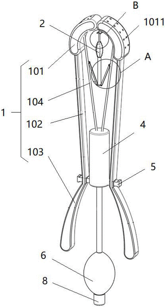
图1为本申请实施例提供的女性生殖器官病变早期一体化无痛切片取样器的整体结构示意图;1 is a schematic diagram of the overall structure of an integrated painless slice sampler in the early stage of female reproductive organ lesions provided by the embodiment of the present application;
图2为图1中A处的放大图;Fig. 2 is the enlarged view of A place in Fig. 1;
图3为图1中B处的放大图;Fig. 3 is the enlarged view at B in Fig. 1;
图4为本申请实施例提供的女性生殖器官病变早期一体化无痛切片取样器的纵剖图;FIG. 4 is a longitudinal sectional view of the integrated painless slice sampler in the early stage of female reproductive organ lesions provided by the embodiment of the present application;
图5为本申请实施例提供的吸气件的示意图。FIG. 5 is a schematic diagram of a getter provided by an embodiment of the present application.
图中:1、第一取样组件;101、第一取样片;1011、第一取样孔;102、连接杆;103、手柄;104、收集管;2、第二取样组件;201、第二取样片;2011、第二取样孔;202、麻醉球;2021、微麻垫片;203、储液管;3、连接组件;4、收集筒;5、导向杆;6、吸气件;7、泄压管;701、泄压孔;8、遮挡盖。In the figure: 1, the first sampling assembly; 101, the first sampling piece; 1011, the first sampling hole; 102, the connecting rod; 103, the handle; 104, the collection tube; 2, the second sampling assembly; 201, the second sampling sheet; 2011, the second sampling hole; 202, anesthesia bulb; 2021, micro anesthesia gasket; 203, the liquid storage tube; 3, the connecting assembly; 4, the collection tube; 5, the guide rod; 6, the suction part; 7, Pressure relief pipe; 701, pressure relief hole; 8, cover.
具体实施方式Detailed ways
为使本申请实施例的目的、技术方案和优点更加清楚,下面将结合本申请实施例中的附图,对本申请实施例中的技术方案进行清楚、完整地描述,显然,所描述的实施例是本申请的一部分实施例,而不是全部的实施例。基于本申请中的实施例,本领域普通技术人员在没有做出创造性劳动的前提下所获得的所有其他实施例,都属于本申请保护的范围。In order to make the purposes, technical solutions and advantages of the embodiments of the present application clearer, the technical solutions in the embodiments of the present application will be described clearly and completely below with reference to the drawings in the embodiments of the present application. Obviously, the described embodiments It is a part of the embodiments of this application, but not all of the embodiments. Based on the embodiments in the present application, all other embodiments obtained by those of ordinary skill in the art without creative work shall fall within the protection scope of the present application.
本申请实施例提供了一种女性生殖器官病变早期一体化无痛切片取样器,其能解决相关技术中擦拭式取样时,所需取样次数较多,效率较低,且影响患者体验感的技术问题。The embodiment of the present application provides an integrated painless slice sampler for the early stage of female genital organ lesions, which can solve the problem of wiping sampling in the related art, which requires more sampling times, has low efficiency, and affects the experience of patients. question.
一种女性生殖器官病变早期一体化无痛切片取样器,其包括:An integrated painless slice sampler for the early stage of female reproductive organ lesions, comprising:
两组第一取样组件1,两组所述第一取样组件1相互靠近或远离设置;Two groups of first sampling assemblies 1, and the two groups of
第二取样组件2,其位于两组所述第一取样组件1之间;a
连接组件3,所述第二取样组件2和两组所述第二取样组件2通过所述连接组件3连接,随两组所述第一取样组件1的相互远离活动,所述连接组件3带动所述第二取样组件2从所述第一取样组件1的一端伸出。The connecting
参照图1,其中,该女性生殖器官病变早期一体化无痛切片取样器包括:第二取样组件2、连接组件3和两组第一取样组件1。Referring to FIG. 1 , the integrated painless slice sampler for the early stage of female reproductive organ lesions includes: a
参照图1,两组第一取样组件1相互靠近或远离设置,第一取样组件1伸至生殖器内后,第一取样组件1对生殖器的内壁进行取样。Referring to FIG. 1 , two groups of
参照图1,第二取样组件2位于两个第一取样组件1之间,且通过连接组件3与两组第一取样组件1连接。由于第二取样组件2位于两个第一取样组件1之间,当第一取样组件1对生殖器内壁进行取样时,第二取样组件2在第一取样组件1的遮挡下,第二取样组件2不对生殖器的内壁取样。Referring to FIG. 1 , the
参照图1和图2,连接组件3将第二取样组件2与两组第一取样组件1连接,随着两个第一取样组件1相互远离活动,连接组件3带动第二取样组件2从第一取样组件1的端部伸出,而便于第二取样组件2伸至宫腔内,并对宫腔内进行取样。第二取样组件2取样完成后,两组第一取样组件1靠近活动,连接组件3带动第二取样组件2回缩,使得第一取样组件1对第二取样组件2进行遮挡,因而在取出该取样器的过程中,第二取样组件2不易附带对生殖器内壁的取样。因而生殖器内壁和宫腔内的取样分别保存在第一取样组件1和第二取样组件2内,不易混合,而便于后续的检查和诊断。1 and 2, the
这样设置,取样时,该取样器伸至生殖器内,第一取样组件1对生殖器的内壁进行取样操作,随着第一取样组件1深入生殖器内部,通过使两组第一取样组件1相互远离,在连接组件3的作用下,第二取样组件2被带动从第一取样组件1的一端伸出,便于将第二取样组件2伸至宫腔内进行取样,即可在一次取样过程中,同时对生殖器内壁和宫腔内进行取样,提高了取样效率,且减少了取样次数,优化了患者的体验。In this way, when sampling, the sampler extends into the genitalia, and the
可选地,所述第一取样组件1包括第一取样片101,其内部中空设置,且所述第一取样片101表面开设有与其内部连通的第一取样孔1011。Optionally, the
参照图1和图4,其中,第一取样组件1包括第一取样片101,第一取样片101呈圆弧片状设置,且第一取样片101的棱角处均圆弧过渡设置。第一取样片101内部中空设置,且第一取样片101的外表面开设有多个第一取样孔1011了,第一取样孔1011使得第一取样片101的内外连通。随着第一取样片101与生殖器的内壁接触,样件通过第一取样孔1011进入第一取样片101内,而完成生殖器内壁的取样操作。1 and FIG. 4 , the
参照图1和图4,当对生殖器内壁取样时,两个第一取样片101的一端相互抵接,以此限制第二取样组件2与生殖器内壁的接触。当需要对宫腔内取样时,两个第一取样片101分开,以此供第二取样组件2伸出第一取样片101的端部,以使第二取样组件2伸至宫腔内进行取样。1 and 4 , when sampling the inner wall of the genitalia, one end of the two
可选地,所述第一取样组件1还包括连接杆102,所述第一取样片101与所述连接杆102的端面连接。Optionally, the
参照图1和图4,其中,第一取样组件1还包括连接杆102和手柄103,连接杆102的两端面分别与第一取样片101和手柄103固定。连接杆102和手柄103均采用医用316不锈钢弯曲成型,且棱角处均以圆弧过渡处理。通过握持手柄103,而便于将第一取样片101伸至生殖器内,且连接杆102增大了第一取样片101的伸入长度,便于对不同深度的位置的取样。同时,可便于将第二取样组件2伸至宫腔的位置,而便于第二取样组件2伸至宫腔内。1 and 4 , the
该取样器在使用前均需消毒处理,传统的430不锈钢采用铁+18%以上的铬无镍,可以防止自然因素所造成的氧化,但无法抵抗空气中的化学物质所造成的氧化,430不锈钢不常使用一段时间后,仍会因非自然因素而有氧化生锈的情况,由于医疗中设备对安全要求需要保证最高性能的安全性,因此430不锈钢和不适合用作取样器连接杆102和手柄103的制作,304不锈钢采用铁+18%铬+8%镍,可以抗化学性的氧化,但空气中的化学成分愈来愈多,有些污染较严重的地方连304都会有生锈的情况,所以取样器的连接杆102和手柄103采用医用316不锈钢+18%铬+10%镍来制作,以使其更耐用更抗蚀,完全没有金属离子析出,提高取样器的安全性,The sampler needs to be sterilized before use. The traditional 430 stainless steel is made of iron + 18% chromium without nickel, which can prevent oxidation caused by natural factors, but cannot resist oxidation caused by chemical substances in the air. 430 stainless steel After infrequent use for a period of time, there will still be oxidation and rust due to unnatural factors. Due to the safety requirements of medical equipment to ensure the highest performance safety, 430 stainless steel and 430 stainless steel are not suitable for use as
可选地,该女性生殖器官病变早期一体化无痛切片取样器还包括收集筒4,所述第一取样组件1还包括收集管104,所述收集管104的两端分别与所述收集筒4和所述第一取样片101连通。Optionally, the integrated painless slice sampler for the early stage of female reproductive organ lesions further includes a
参照图1和图4,其中,收集筒4位于两个连接杆102之间,且两组第二取样组件2以收集筒4的轴线均匀圆周分布。第一取样组件1还包括收集管104,收集管104的两端分别与收集筒4和第一取样片101连通,且收集管104与收集筒4和第一取样片101的连通处均密封处理。两组的第一取样组件1中的两个收集管104以收集筒4的轴线均匀圆周分布。Referring to FIGS. 1 and 4 , the
这样设置,第一取样片101取样时,生殖器内壁的样品经过第一取样孔1011进入第一取样片101内,再经过收集管104而流入收集筒4内储存。同时收集筒4包括筒体和与筒体螺纹连接的盖体,通过拆除盖体即可取出存储在收集筒4内的样品。In this way, when the
可选地,该女性生殖器官病变早期一体化无痛切片取样器还包括两个导向杆5,两个所述导向杆5均与所述收集筒4的周向外侧面连接,且两个所述导向杆5分别穿设于两个所述连接杆102,以使两组所述第一取样组件1靠近或远离设置。Optionally, the integrated painless slice sampler for the early stage of female genital lesions also includes two
参照图1和图4,其中,导向杆5的一端面与收集筒4的外向侧壁固定,导向杆5的长度方向与收集筒4的径向一致。两个导向杆5以收集筒4的轴线均匀圆周分布,且两个导向杆5分别穿设于两个连接杆102,其中,连接杆102与导向杆5的连接位置位于连接杆102靠近手柄103的一端。因而连接杆102在导向杆5上滑动,以此可靠近或远离收集筒4,且两个连接杆102可相互靠近或远离滑动,使得两组第一取样组件1靠近或远离滑动设置。导向杆5背离收集筒4的一端一体成型有限位头,限位头的截面积大于导向杆5的截面积,以限制连接杆102脱离导向杆5。Referring to FIGS. 1 and 4 , one end surface of the
当两个连接杆102均最为靠近收集筒4设置时,此时两个第一取样片101抵接设置,以此遮挡第二取样组件2。当两个连接杆102均最为远离收集筒4设置时,此时两个第一取样片101分开,同时第二取样组件2从两个第一取样片101之间伸出,以此对宫腔取样。When the two connecting
可选地,该女性生殖器官病变早期一体化无痛切片取样器还包括吸气件6,其与所述收集筒4连通,以降低所述收集筒4内的气压。Optionally, the integrated painless slice sampler for the early stage of female genital lesions further includes an
参照图1和图4,其中,吸气件6位于两个第一取样组件1之间,且与收集筒4连通,本实施例中,收集筒4的轴线与吸气件6的轴线一致。吸气件6包括球囊或者吸气机,本实施例中,吸气件6包括球囊,球囊与收集筒4连通设置。第一取样片101伸至生殖器内前,球囊呈预压缩状态,需要取样时,展开球囊以此吸气,而使得收集筒4内的气压降低,便于将第一取样片101内的样品吸入收集筒4内,而完成生殖器内壁的取样过程。Referring to FIGS. 1 and 4 , the
参照图5,吸气件6连通有泄压管7,泄压管7一体成型于球囊,以此通过泄压管7将球囊的内外连通。泄压管7上罩设有遮挡盖8,泄压管7的周向外侧壁上设有螺纹,遮挡盖8通过螺纹与泄压管7连接。泄压管7的周向侧壁还开设有多个泄压孔701,遮挡盖8与泄压管7配合时,泄压孔701也被遮挡盖8遮挡。球囊处于压瘪状态时,利用遮挡盖8将泄压孔701遮挡,通过复原球囊即可减小收集筒4内的气压,而并与样品从第一取样片101内流入收集筒4内。若在球囊的一次复原过程中,进入收集筒4内的样品量较少,即可开启遮挡盖8,重新压瘪球囊,而球囊内的气体从泄压孔701内至大气,进而通过再次复原球囊,而再次将第一取样片101内的样品吸入收集筒4内。Referring to FIG. 5 , a
可选地,所述第二取样组件2包括第二取样片201,其内部中空设置,且所述第二取样片201表面开设有与其内部连通的第二取样孔2011。Optionally, the
参照图1和图3,其中,第二取样组件2包括第二取样片201,第二取样片201呈弧形片状设置,且其内部中空。第二取样片201的外表面开设有多个将第二取样片201内外连通的第二取样孔2011。第二取样片201取样时,样品从第二取样孔2011进入第二取样片201内部。第二取样片201位于两个第一取样片101之间,随着第一取样片101相互远离运动,连接组件3带动第二取样片201从两个第一取样片101之间伸出,且第二取样片201伸至宫腔内,以完成取样。1 and FIG. 3 , wherein, the
可选地,所述第二取样组件2还包括麻醉球202,所述麻醉球202与所述第二取样片201连通设置。Optionally, the
参照图1、图3和图4,其中,第二取样组件2还包括麻醉球202,麻醉球202由橡胶材质制成。第二取样片201与麻醉球202连通设置,第二取样片201内的样品流入麻醉球202内。麻醉球202呈梭锥状,其外表面固定有多个微麻垫片2021。取样之前,将麻醉球202在质量分数为0.5%利多卡因溶液内浸泡,而使得微麻垫片2021涂覆有利多卡因溶液。随着第二取样片201伸至宫腔内,麻醉球202一并伸入宫腔内,而对取样位置进行麻醉处理,减缓取样时的疼痛感。Referring to FIG. 1 , FIG. 3 and FIG. 4 , the
可选地,所述第二取样组件2还包括储液管203,所述储液管203与所述麻醉球202连通设置。Optionally, the
参照图1和图3,其中,储液管203与麻醉球202连通设置,且储液管203与麻醉球202螺纹连接。麻醉球202内的样品流入储液管203内进行储存,且通过将储液管203和麻醉球202拆开,而便于取出储液管203内的样品。Referring to FIGS. 1 and 3 , the
可选地,所述连接组件3包括两个连杆,所述连杆的两端分别与所述收集管104和所述储液管203铰接,随两个所述收集管104逐渐远离,所述连杆带动所述第二取样组件2移动,以使所述第二取样片201从所述第一取样片101的端部伸出。Optionally, the connecting
参照图1和图2,其中,连接组件3包括两个连杆,连杆的两端分别与储液管203和收集管104铰接,本实施例中,连杆与储液管203背离第二取样片201的一端铰接,且两个连杆形成的锐角开口朝向第二取样片201设置。随着两个第一收集管104的远离,两个连杆之间的角度逐渐变大,而带动储液管203移动,同时,第二取样片201被带动移动而从两个第一取样片101之间伸出,以此便于第二取样片201伸至宫腔内进行取样。1 and 2, the connecting
在本申请的描述中,需要说明的是,术语“上”、“下”等指示的方位或位置关系为基于附图所示的方位或位置关系,仅是为了便于描述本申请和简化描述,而不是指示或暗示所指的装置或元件必须具有特定的方位、以特定的方位构造和操作,因此不能理解为对本申请的限制。除非另有明确的规定和限定,术语“安装”、“相连”、“连接”应做广义理解,例如,可以是固定连接,也可以是可拆卸连接,或一体地连接;可以是机械连接,也可以是电连接;可以是直接相连,也可以通过中间媒介间接相连,可以是两个元件内部的连通。对于本领域的普通技术人员而言,可以根据具体情况理解上述术语在本申请中的具体含义。In the description of this application, it should be noted that the orientation or positional relationship indicated by the terms "upper", "lower", etc. is based on the orientation or positional relationship shown in the accompanying drawings, and is only for the convenience of describing the application and simplifying the description, Rather than indicating or implying that the referred device or element must have a particular orientation, be constructed and operate in a particular orientation, it should not be construed as a limitation on the application. Unless otherwise expressly specified and limited, the terms "installed", "connected" and "connected" should be understood in a broad sense, for example, it may be a fixed connection, a detachable connection, or an integral connection; it may be a mechanical connection, It can also be an electrical connection; it can be a direct connection, an indirect connection through an intermediate medium, or an internal connection between two components. For those of ordinary skill in the art, the specific meanings of the above terms in this application can be understood according to specific situations.
需要说明的是,在本申请中,诸如“第一”和“第二”等之类的关系术语仅仅用来将一个实体或者操作与另一个实体或操作区分开来,而不一定要求或者暗示这些实体或操作之间存在任何这种实际的关系或者顺序。而且,术语“包括”、“包含”或者其任何其他变体意在涵盖非排他性的包含,从而使得包括一系列要素的过程、方法、物品或者设备不仅包括那些要素,而且还包括没有明确列出的其他要素,或者是还包括为这种过程、方法、物品或者设备所固有的要素。在没有更多限制的情况下,由语句“包括一个……”限定的要素,并不排除在包括所述要素的过程、方法、物品或者设备中还存在另外的相同要素。It should be noted that, in this application, relational terms such as "first" and "second" are only used to distinguish one entity or operation from another entity or operation, and do not necessarily require or imply Any such actual relationship or sequence exists between these entities or operations. Moreover, the terms "comprising", "comprising" or any other variation thereof are intended to encompass a non-exclusive inclusion such that a process, method, article or device that includes a list of elements includes not only those elements, but also includes not explicitly listed or other elements inherent to such a process, method, article or apparatus. Without further limitation, an element qualified by the phrase "comprising a..." does not preclude the presence of additional identical elements in a process, method, article or apparatus that includes the element.
以上所述仅是本申请的具体实施方式,使本领域技术人员能够理解或实现本申请。对这些实施例的多种修改对本领域的技术人员来说将是显而易见的,本文中所定义的一般原理可以在不脱离本申请的精神或范围的情况下,在其它实施例中实现。因此,本申请将不会被限制于本文所示的这些实施例,而是要符合与本文所申请的原理和新颖特点相一致的最宽的范围。The above descriptions are only specific embodiments of the present application, so that those skilled in the art can understand or implement the present application. Various modifications to these embodiments will be readily apparent to those skilled in the art, and the generic principles defined herein may be implemented in other embodiments without departing from the spirit or scope of the present application. Therefore, this application is not intended to be limited to the embodiments shown herein, but is to be accorded the widest scope consistent with the principles and novel features claimed herein.
Claims (6)
Priority Applications (1)
| Application Number | Priority Date | Filing Date | Title |
|---|---|---|---|
| CN202110859356.0A CN113576552B (en) | 2021-07-28 | 2021-07-28 | Early integrated painless section sampler of female reproductive organ pathological change |
Applications Claiming Priority (1)
| Application Number | Priority Date | Filing Date | Title |
|---|---|---|---|
| CN202110859356.0A CN113576552B (en) | 2021-07-28 | 2021-07-28 | Early integrated painless section sampler of female reproductive organ pathological change |
Publications (2)
| Publication Number | Publication Date |
|---|---|
| CN113576552A CN113576552A (en) | 2021-11-02 |
| CN113576552B true CN113576552B (en) | 2022-08-05 |
Family
ID=78251258
Family Applications (1)
| Application Number | Title | Priority Date | Filing Date |
|---|---|---|---|
| CN202110859356.0A Expired - Fee Related CN113576552B (en) | 2021-07-28 | 2021-07-28 | Early integrated painless section sampler of female reproductive organ pathological change |
Country Status (1)
| Country | Link |
|---|---|
| CN (1) | CN113576552B (en) |
Citations (5)
| Publication number | Priority date | Publication date | Assignee | Title |
|---|---|---|---|---|
| WO2014174490A1 (en) * | 2013-04-26 | 2014-10-30 | Michael Owen Richards | Cervical access device |
| CN105555200A (en) * | 2013-10-14 | 2016-05-04 | Dna研究中心(马来西亚)私人有限公司 | Exocervical and endocervical cell sampling device |
| CN206745384U (en) * | 2017-01-10 | 2017-12-15 | 中国人民解放军第三军医大学第三附属医院 | Cervical sampling device |
| CN108354636A (en) * | 2018-04-05 | 2018-08-03 | 王丽先 | A kind of Gynecological cervical disease examination sampler |
| CN111513770A (en) * | 2020-04-30 | 2020-08-11 | 秦彦芹 | Vaginal secretion sampling device for gynecological clinical treatment |
Family Cites Families (2)
| Publication number | Priority date | Publication date | Assignee | Title |
|---|---|---|---|---|
| GB2511551B (en) * | 2013-03-07 | 2015-05-27 | Univ Staffordshire | Sample collection apparatus |
| CN112494081A (en) * | 2020-12-25 | 2021-03-16 | 王慧 | Be used for gynaecology's reproductive organ inspection sampling device |
-
2021
- 2021-07-28 CN CN202110859356.0A patent/CN113576552B/en not_active Expired - Fee Related
Patent Citations (5)
| Publication number | Priority date | Publication date | Assignee | Title |
|---|---|---|---|---|
| WO2014174490A1 (en) * | 2013-04-26 | 2014-10-30 | Michael Owen Richards | Cervical access device |
| CN105555200A (en) * | 2013-10-14 | 2016-05-04 | Dna研究中心(马来西亚)私人有限公司 | Exocervical and endocervical cell sampling device |
| CN206745384U (en) * | 2017-01-10 | 2017-12-15 | 中国人民解放军第三军医大学第三附属医院 | Cervical sampling device |
| CN108354636A (en) * | 2018-04-05 | 2018-08-03 | 王丽先 | A kind of Gynecological cervical disease examination sampler |
| CN111513770A (en) * | 2020-04-30 | 2020-08-11 | 秦彦芹 | Vaginal secretion sampling device for gynecological clinical treatment |
Also Published As
| Publication number | Publication date |
|---|---|
| CN113576552A (en) | 2021-11-02 |
Similar Documents
| Publication | Publication Date | Title |
|---|---|---|
| CN103202716A (en) | Uterus sampler | |
| CN113576552B (en) | Early integrated painless section sampler of female reproductive organ pathological change | |
| CN203153815U (en) | Uterus sampling apparatus | |
| CN216777117U (en) | A core rod endometrial sampler | |
| CN216317728U (en) | A tumor cell sampling and inspection device | |
| CN115969428A (en) | Thyroid needle aspiration cytology biopsy puncture device | |
| CN215272964U (en) | Endometrium biopsy forceps | |
| CN110680412B (en) | Negative pressure suction biopsy forceps | |
| CN116687465A (en) | Tumor sampling device | |
| CN217772386U (en) | Disposable endometrium sampler | |
| CN108836405A (en) | A kind of vaginal fluid sampling device for clinical obstetrics | |
| CN108784754A (en) | A kind of lung aspiration biopsy device | |
| CN219397355U (en) | Secretion collector | |
| CN210749285U (en) | Novel endocrine hydrops is gathered device | |
| CN219048633U (en) | Focus sampler for preventing sample pollution | |
| CN201469315U (en) | Multifunctional endometrium sampler | |
| CN221671778U (en) | A segmented sampling device for endometrial atypical hyperplasia | |
| CN219089396U (en) | a vaginal sampler | |
| CN209236207U (en) | Gynecological sampling device | |
| CN221844791U (en) | Gynecological vaginal secretion inspection device | |
| CN221533832U (en) | Positive pressure sampling tube | |
| CN221814040U (en) | Endocervical canal sampling tool | |
| CN221083676U (en) | Saliva quantitative collection device | |
| CN201631247U (en) | Collector of cervix uteri cell | |
| CN221599986U (en) | Cervical canal sampling curet |
Legal Events
| Date | Code | Title | Description |
|---|---|---|---|
| PB01 | Publication | ||
| PB01 | Publication | ||
| SE01 | Entry into force of request for substantive examination | ||
| SE01 | Entry into force of request for substantive examination | ||
| GR01 | Patent grant | ||
| GR01 | Patent grant | ||
| CF01 | Termination of patent right due to non-payment of annual fee |
Granted publication date: 20220805 |
|
| CF01 | Termination of patent right due to non-payment of annual fee |