CN112867499A - Soluble extracellular matrix compositions and methods for intravascular delivery - Google Patents
Soluble extracellular matrix compositions and methods for intravascular delivery Download PDFInfo
- Publication number
- CN112867499A CN112867499A CN201980067713.0A CN201980067713A CN112867499A CN 112867499 A CN112867499 A CN 112867499A CN 201980067713 A CN201980067713 A CN 201980067713A CN 112867499 A CN112867499 A CN 112867499A
- Authority
- CN
- China
- Prior art keywords
- soluble
- composition
- ecm
- infusion
- tissue
- Prior art date
- Legal status (The legal status is an assumption and is not a legal conclusion. Google has not performed a legal analysis and makes no representation as to the accuracy of the status listed.)
- Pending
Links
Images


Classifications
-
- A—HUMAN NECESSITIES
- A61—MEDICAL OR VETERINARY SCIENCE; HYGIENE
- A61L—METHODS OR APPARATUS FOR STERILISING MATERIALS OR OBJECTS IN GENERAL; DISINFECTION, STERILISATION OR DEODORISATION OF AIR; CHEMICAL ASPECTS OF BANDAGES, DRESSINGS, ABSORBENT PADS OR SURGICAL ARTICLES; MATERIALS FOR BANDAGES, DRESSINGS, ABSORBENT PADS OR SURGICAL ARTICLES
- A61L27/00—Materials for grafts or prostheses or for coating grafts or prostheses
- A61L27/36—Materials for grafts or prostheses or for coating grafts or prostheses containing ingredients of undetermined constitution or reaction products thereof, e.g. transplant tissue, natural bone, extracellular matrix
- A61L27/3604—Materials for grafts or prostheses or for coating grafts or prostheses containing ingredients of undetermined constitution or reaction products thereof, e.g. transplant tissue, natural bone, extracellular matrix characterised by the human or animal origin of the biological material, e.g. hair, fascia, fish scales, silk, shellac, pericardium, pleura, renal tissue, amniotic membrane, parenchymal tissue, fetal tissue, muscle tissue, fat tissue, enamel
- A61L27/3633—Extracellular matrix [ECM]
-
- A—HUMAN NECESSITIES
- A61—MEDICAL OR VETERINARY SCIENCE; HYGIENE
- A61K—PREPARATIONS FOR MEDICAL, DENTAL OR TOILETRY PURPOSES
- A61K35/00—Medicinal preparations containing materials or reaction products thereof with undetermined constitution
- A61K35/12—Materials from mammals; Compositions comprising non-specified tissues or cells; Compositions comprising non-embryonic stem cells; Genetically modified cells
- A61K35/30—Nerves; Brain; Eyes; Corneal cells; Cerebrospinal fluid; Neuronal stem cells; Neuronal precursor cells; Glial cells; Oligodendrocytes; Schwann cells; Astroglia; Astrocytes; Choroid plexus; Spinal cord tissue
-
- A—HUMAN NECESSITIES
- A61—MEDICAL OR VETERINARY SCIENCE; HYGIENE
- A61K—PREPARATIONS FOR MEDICAL, DENTAL OR TOILETRY PURPOSES
- A61K35/00—Medicinal preparations containing materials or reaction products thereof with undetermined constitution
- A61K35/12—Materials from mammals; Compositions comprising non-specified tissues or cells; Compositions comprising non-embryonic stem cells; Genetically modified cells
- A61K35/22—Urine; Urinary tract, e.g. kidney or bladder; Intraglomerular mesangial cells; Renal mesenchymal cells; Adrenal gland
-
- A—HUMAN NECESSITIES
- A61—MEDICAL OR VETERINARY SCIENCE; HYGIENE
- A61K—PREPARATIONS FOR MEDICAL, DENTAL OR TOILETRY PURPOSES
- A61K35/00—Medicinal preparations containing materials or reaction products thereof with undetermined constitution
- A61K35/12—Materials from mammals; Compositions comprising non-specified tissues or cells; Compositions comprising non-embryonic stem cells; Genetically modified cells
- A61K35/34—Muscles; Smooth muscle cells; Heart; Cardiac stem cells; Myoblasts; Myocytes; Cardiomyocytes
-
- A—HUMAN NECESSITIES
- A61—MEDICAL OR VETERINARY SCIENCE; HYGIENE
- A61K—PREPARATIONS FOR MEDICAL, DENTAL OR TOILETRY PURPOSES
- A61K35/00—Medicinal preparations containing materials or reaction products thereof with undetermined constitution
- A61K35/12—Materials from mammals; Compositions comprising non-specified tissues or cells; Compositions comprising non-embryonic stem cells; Genetically modified cells
- A61K35/37—Digestive system
- A61K35/38—Stomach; Intestine; Goblet cells; Oral mucosa; Saliva
-
- A—HUMAN NECESSITIES
- A61—MEDICAL OR VETERINARY SCIENCE; HYGIENE
- A61K—PREPARATIONS FOR MEDICAL, DENTAL OR TOILETRY PURPOSES
- A61K38/00—Medicinal preparations containing peptides
- A61K38/01—Hydrolysed proteins; Derivatives thereof
- A61K38/012—Hydrolysed proteins; Derivatives thereof from animals
- A61K38/014—Hydrolysed proteins; Derivatives thereof from animals from connective tissue peptides, e.g. gelatin, collagen
-
- A—HUMAN NECESSITIES
- A61—MEDICAL OR VETERINARY SCIENCE; HYGIENE
- A61K—PREPARATIONS FOR MEDICAL, DENTAL OR TOILETRY PURPOSES
- A61K9/00—Medicinal preparations characterised by special physical form
- A61K9/0012—Galenical forms characterised by the site of application
- A61K9/0019—Injectable compositions; Intramuscular, intravenous, arterial, subcutaneous administration; Compositions to be administered through the skin in an invasive manner
-
- A—HUMAN NECESSITIES
- A61—MEDICAL OR VETERINARY SCIENCE; HYGIENE
- A61L—METHODS OR APPARATUS FOR STERILISING MATERIALS OR OBJECTS IN GENERAL; DISINFECTION, STERILISATION OR DEODORISATION OF AIR; CHEMICAL ASPECTS OF BANDAGES, DRESSINGS, ABSORBENT PADS OR SURGICAL ARTICLES; MATERIALS FOR BANDAGES, DRESSINGS, ABSORBENT PADS OR SURGICAL ARTICLES
- A61L27/00—Materials for grafts or prostheses or for coating grafts or prostheses
- A61L27/36—Materials for grafts or prostheses or for coating grafts or prostheses containing ingredients of undetermined constitution or reaction products thereof, e.g. transplant tissue, natural bone, extracellular matrix
- A61L27/3683—Materials for grafts or prostheses or for coating grafts or prostheses containing ingredients of undetermined constitution or reaction products thereof, e.g. transplant tissue, natural bone, extracellular matrix subjected to a specific treatment prior to implantation, e.g. decellularising, demineralising, grinding, cellular disruption/non-collagenous protein removal, anti-calcification, crosslinking, supercritical fluid extraction, enzyme treatment
-
- A—HUMAN NECESSITIES
- A61—MEDICAL OR VETERINARY SCIENCE; HYGIENE
- A61L—METHODS OR APPARATUS FOR STERILISING MATERIALS OR OBJECTS IN GENERAL; DISINFECTION, STERILISATION OR DEODORISATION OF AIR; CHEMICAL ASPECTS OF BANDAGES, DRESSINGS, ABSORBENT PADS OR SURGICAL ARTICLES; MATERIALS FOR BANDAGES, DRESSINGS, ABSORBENT PADS OR SURGICAL ARTICLES
- A61L27/00—Materials for grafts or prostheses or for coating grafts or prostheses
- A61L27/50—Materials characterised by their function or physical properties, e.g. injectable or lubricating compositions, shape-memory materials, surface modified materials
- A61L27/54—Biologically active materials, e.g. therapeutic substances
-
- A—HUMAN NECESSITIES
- A61—MEDICAL OR VETERINARY SCIENCE; HYGIENE
- A61P—SPECIFIC THERAPEUTIC ACTIVITY OF CHEMICAL COMPOUNDS OR MEDICINAL PREPARATIONS
- A61P9/00—Drugs for disorders of the cardiovascular system
-
- A—HUMAN NECESSITIES
- A61—MEDICAL OR VETERINARY SCIENCE; HYGIENE
- A61K—PREPARATIONS FOR MEDICAL, DENTAL OR TOILETRY PURPOSES
- A61K47/00—Medicinal preparations characterised by the non-active ingredients used, e.g. carriers or inert additives; Targeting or modifying agents chemically bound to the active ingredient
- A61K47/06—Organic compounds, e.g. natural or synthetic hydrocarbons, polyolefins, mineral oil, petrolatum or ozokerite
- A61K47/26—Carbohydrates, e.g. sugar alcohols, amino sugars, nucleic acids, mono-, di- or oligo-saccharides; Derivatives thereof, e.g. polysorbates, sorbitan fatty acid esters or glycyrrhizin
-
- A—HUMAN NECESSITIES
- A61—MEDICAL OR VETERINARY SCIENCE; HYGIENE
- A61K—PREPARATIONS FOR MEDICAL, DENTAL OR TOILETRY PURPOSES
- A61K9/00—Medicinal preparations characterised by special physical form
- A61K9/0012—Galenical forms characterised by the site of application
- A61K9/0019—Injectable compositions; Intramuscular, intravenous, arterial, subcutaneous administration; Compositions to be administered through the skin in an invasive manner
- A61K9/0024—Solid, semi-solid or solidifying implants, which are implanted or injected in body tissue
-
- A—HUMAN NECESSITIES
- A61—MEDICAL OR VETERINARY SCIENCE; HYGIENE
- A61L—METHODS OR APPARATUS FOR STERILISING MATERIALS OR OBJECTS IN GENERAL; DISINFECTION, STERILISATION OR DEODORISATION OF AIR; CHEMICAL ASPECTS OF BANDAGES, DRESSINGS, ABSORBENT PADS OR SURGICAL ARTICLES; MATERIALS FOR BANDAGES, DRESSINGS, ABSORBENT PADS OR SURGICAL ARTICLES
- A61L2400/00—Materials characterised by their function or physical properties
- A61L2400/06—Flowable or injectable implant compositions
-
- A—HUMAN NECESSITIES
- A61—MEDICAL OR VETERINARY SCIENCE; HYGIENE
- A61L—METHODS OR APPARATUS FOR STERILISING MATERIALS OR OBJECTS IN GENERAL; DISINFECTION, STERILISATION OR DEODORISATION OF AIR; CHEMICAL ASPECTS OF BANDAGES, DRESSINGS, ABSORBENT PADS OR SURGICAL ARTICLES; MATERIALS FOR BANDAGES, DRESSINGS, ABSORBENT PADS OR SURGICAL ARTICLES
- A61L2430/00—Materials or treatment for tissue regeneration
- A61L2430/20—Materials or treatment for tissue regeneration for reconstruction of the heart, e.g. heart valves
Landscapes
- Health & Medical Sciences (AREA)
- Life Sciences & Earth Sciences (AREA)
- Chemical & Material Sciences (AREA)
- Engineering & Computer Science (AREA)
- Medicinal Chemistry (AREA)
- Biomedical Technology (AREA)
- Public Health (AREA)
- General Health & Medical Sciences (AREA)
- Animal Behavior & Ethology (AREA)
- Veterinary Medicine (AREA)
- Epidemiology (AREA)
- Zoology (AREA)
- Pharmacology & Pharmacy (AREA)
- Cell Biology (AREA)
- Dermatology (AREA)
- Immunology (AREA)
- Oral & Maxillofacial Surgery (AREA)
- Transplantation (AREA)
- Molecular Biology (AREA)
- Developmental Biology & Embryology (AREA)
- Chemical Kinetics & Catalysis (AREA)
- Botany (AREA)
- Bioinformatics & Cheminformatics (AREA)
- Biotechnology (AREA)
- Virology (AREA)
- Urology & Nephrology (AREA)
- Proteomics, Peptides & Aminoacids (AREA)
- Biophysics (AREA)
- Cardiology (AREA)
- Organic Chemistry (AREA)
- Nutrition Science (AREA)
- Heart & Thoracic Surgery (AREA)
- General Chemical & Material Sciences (AREA)
- Nuclear Medicine, Radiotherapy & Molecular Imaging (AREA)
- Physiology (AREA)
- Neurology (AREA)
- Neurosurgery (AREA)
- Ophthalmology & Optometry (AREA)
- Vascular Medicine (AREA)
- Medicines That Contain Protein Lipid Enzymes And Other Medicines (AREA)
Abstract
Compositions and methods for their manufacture and use are provided, including a soluble extracellular matrix fraction for intravascular delivery that forms a gel or blanket in situ for the treatment of myocardial infarction and ischemia and endothelial injury/dysfunction in various tissues.
Description
Cross reference to related applications
This application claims priority to U.S. provisional application No. 62/750,303 filed on 25/10/2018, which is incorporated herein by reference.
Government sponsorship
The invention was made with government support funded by number HL113468 awarded by the National Institutes of Health (NIH). The government has certain rights in the invention.
Technical Field
The present invention relates to infusible soluble extracellular matrix compositions and treatments for minimally invasive delivery to tissues/organs/cells, including ischemic or injured heart, brain, skeletal muscle, blood vessels, and endothelial cells.
Background
Extracellular matrix treatments include natural tissue components that provide a scaffold for tissue regeneration. Current decellularized extracellular matrix treatments are limited to patches or direct injection. No extracellular matrix therapy is capable of intravascular/infusion delivery to the tissue of interest. ECM hydrogels made from decellularized and digested tissue have been generated, but they are not fully soluble or transparent colloids. They are translucent suspensions containing both soluble and non-soluble components (Freytes et al, 2008, Singelyn et al, 2009)) that prevent material from entering the tissue from the bloodstream through the leaking vasculature that occurs in ischemic tissues such as acute myocardial infarction, stroke, cancer, etc. (Nguyen et al, 2015, Dvorak et al, 1988, Yuan et al, 1995), lining the leaking vasculature or filling the pores of the leaking vasculature.
Myocardial Infarction (MI) is characterized by ischemic necrosis of the myocardium progressing over time, leading to poor Left Ventricular (LV) remodeling and ultimately heart failure. Current standards of care do not address this ischemic insult. Minimally invasive tissue engineering treatments can repair the heart after MI. Many stem cell and growth factor treatments have entered clinical trials; however, these treatments show poor efficacy, which may be due to insufficient retention of the unencapsulated treatment.
Extracellular matrix (ECM) hydrogels have shown great promise in the field of cardiac tissue engineering; in MI models in particular, tissue-specific hydrogels derived from decellularized porcine myocardium, known as Myocardial Matrix (MM), show an increase in myocardium, neovascularization in the infarcted area, and improved regional and overall cardiac function [2-4 ]. Furthermore, the mechanism of repair was studied by whole transcriptome analysis of RNA isolated from infarcted regions of rat hearts injected with MM, suggesting upregulated pathways associated with cardiac repair (e.g., neovascularization and cardiac development) and downregulated pathways associated with poor LV remodeling (e.g., hypertrophy, apoptosis and fibrosis [6 ]. this material was evaluated in a phase I trial in post-MI patients within 60 to 3 years post-MI using endocardial injection (american clinical trial database (clinical trials. gov) identifier: NCT 02305602.) however, myocardial cell death and poor remodeling are processes that begin within minutes to days post-MI [26,27 ]. current delivery of MM is limited to endocardial catheter injection only, as it contains submicron particles that are too large to enter the infarct through the leaky coronary vasculature, in addition, endocardial injection is a specialized medical technique with some safety issues of ventricular rupture and arrhythmia within the first week after MI [7,8 ]. This prevents MM from being delivered within the critical therapeutic window following acute MI.
Intracoronary infusion is an alternative to trans-endocardial injection. Intracoronary delivery is feasible in acute MI because it can be accompanied by balloon angioplasty, which is usually performed shortly after patient admission. Such techniques are standard techniques for interventional cardiology and do not require specialized training. Intracoronary infusion utilizes leaky vasculature after an acute MI to allow biological material to pass through the coronary vasculature and into the infarcted area [5 ]. The feasibility of intracoronary delivery of biomaterials has been demonstrated in porcine MI models with alginate hydrogels [9] and has progressed to phase II clinical trials (US clinical trial database identifier: NCT 01226563). However, this material does not show a significant improvement in cardiac function, probably due to the limited biological activity of algin [10 ].
Disclosure of Invention
In embodiments, the present invention provides a method of preparing a soluble extracellular matrix (ECM) composition, the method comprising enzymatically digesting ECM material with an acidic protease, such as pepsin; neutralizing said digested ECM material in a liquid to a pH of 7.0-8.0; treating the liquid ECM to produce soluble and insoluble fractions; and separating at least a portion of the soluble fraction from the insoluble fraction to produce a soluble ECM composition.
In an embodiment, the present invention provides: treating the liquid ECM to produce soluble and insoluble fractions is achieved by centrifugation. In an embodiment, the present invention provides: dialyzing and/or filtering the soluble ECM composition to remove insoluble material. In an embodiment, the present invention provides: the soluble ECM composition is further lyophilized for storage and rehydrated for use.
In embodiments, the soluble ECM composition is substantially separated from ECM solids in the liquid ECM. In embodiments, the soluble ECM composition is more transparent than the digested non-isolated ECM material. In embodiments, the soluble ECM composition comprises a transparent ECM colloid that can pass through a 0.25 μm filter.
In embodiments, the present invention provides a method of treating a subject in need thereof, the method comprising administering to the subject an effective amount of the soluble ECM composition to promote tissue repair or cell recruitment. In embodiments, the infusion is via a catheter, intravenously, or intravascularly. In an embodiment, the present invention provides: when delivered in vivo, the soluble fraction will then form a gel in the tissue. In embodiments, the soluble fraction will coat the vascular lining. In embodiments, the soluble fraction will fill pores, fenestrations, endothelial disruptions, open intercellular junctions, or gaps of leaky or damaged vasculature.
In an embodiment, the present invention provides: the soluble fraction of ECM is further cross-linked with glutaraldehyde, formaldehyde, bis-NHS molecules, or other cross-linking agents. In an embodiment, the present invention provides: combining and/or crosslinking the soluble fraction of ECM with a synthetic polymer or a biologically derived material. In an embodiment, the present invention provides: combining said soluble fraction of ECM with cells, peptides, proteins, DNA, drugs, nanoparticles, antibiotics, growth factors, nutrients, survival-promoting additives, proteoglycans and/or glycosaminoglycans.
In an embodiment, the present invention provides: the soluble fraction of ECM is used in combination with the above components for endogenous cellular ingrowth, angiogenesis and regeneration. In an embodiment, the present invention provides: the soluble fraction of ECM is used in combination with the above components as a matrix to alter the mechanical properties of the tissue. In an embodiment, the present invention provides: the soluble fraction of ECM is delivered with cells for regeneration or repair of damaged tissue, alone or in combination with the above components.
In an embodiment, the present invention provides: after adjusting the concentration and/or sterile filtration, the soluble fraction of ECM can be lyophilized and stored frozen (e.g., -20C, -80C) for at least 3 months. The soluble ECM composition or fraction can then be resuspended and/or sterile filtered prior to injection or infusion.
In an embodiment, the present invention provides: after adjusting the concentration and/or sterile filtration, the soluble fraction of ECM can be lyophilized and stored in a freezer (e.g., 4C) for at least 3 months. The soluble ECM fraction can then be resuspended and/or sterile filtered prior to injection or infusion.
In an embodiment, the present invention provides: after adjusting the concentration and/or sterile filtration, the soluble fraction of ECM can be lyophilized and stored at room temperature for at least 3 months. The soluble ECM fraction can then be resuspended and/or sterile filtered prior to injection or infusion.
In an embodiment, the present invention provides: the method of separating at least a portion of the soluble fraction from the insoluble fraction in the liquid extracellular matrix (pre-gel solution) may be performed by high speed centrifugation, dialysis, filtration or adjusting pH or salinity. In embodiments, the separation of the soluble fraction is performed by removing at least a portion of the solids from the ECM material. In embodiments, the separation of the soluble fraction is performed with a filter having a size limit of less than 1 μm, 0.5 μm, 0.25 μm, 0.22 μm, or 0.2 μm. In embodiments, the present invention provides soluble ECM compositions that are derived from decellularized tissue and are treated to isolate a soluble fraction prior to gelation in vivo. In an embodiment, the present invention provides: preparing a composition of the soluble ECM for intravascular infusion.
In an embodiment, the present invention provides: the composition of the soluble extracellular matrix is derived from a human, animal, embryonic and/or fetal tissue source. In an embodiment, the present invention provides: the composition of the soluble extracellular matrix is derived from heart, brain, bladder, small intestine, skeletal muscle, kidney, liver, lung, blood vessels, and other tissue/organ tissue sources.
In an embodiment, the present invention provides a method of treating acute myocardial infarction, the method comprising injecting or infusing into a subject in need thereof having a myocardial infarction an effective amount of a composition comprising a soluble decellularized extracellular matrix derived from a muscle tissue.
In an embodiment, the present invention provides: the soluble ECM composition is delivered intravascularly by infusion. In an embodiment, the present invention provides: the soluble ECM composition is delivered by intracoronary infusion with a balloon infusion catheter. In an embodiment, the present invention provides: the soluble ECM composition is converted to a gel form in the tissue after delivery. In an embodiment, the present invention provides: the soluble ECM composition, after delivery, transitions to form a coating on the endothelium of the damaged vessel. In an embodiment, the present invention provides: the soluble ECM composition degrades within one to 14 days after injection or infusion.
In an embodiment, the present invention provides: injection or infusion of the composition repairs damage to the myocardium, such as myocardial infarction, sustained by the subject. In an embodiment, the present invention provides: injection or infusion of the composition is used to treat muscle or nerve damage caused by disease, trauma, stroke and/or ischemia in the subject. In an embodiment, the present invention provides: the effective amount is an amount that increases blood flow, increases tissue mass, or induces neovascularization in an injection or infusion area of the subject. In an embodiment, the present invention provides: the effective amount is an amount that promotes cell survival, reduces inflammation, and repairs damaged vasculature in an injection or infusion region of a subject.
Drawings
FIGS. 1A-1F illustrate the generation of soluble myocardial matrix. FIG. 1A shows isolated left ventricular myocardium fragmented. Figure 1B shows that cells were decellularized after continuous stirring in 1% sodium dodecyl sulfate. Figure 1C shows lyophilization and grinding to a fine powder. Figure 1D shows a partially digested myocardial matrix. (E) Fractionated myocardial matrix after centrifugation, (1) SolMM fraction in supernatant and (2) insoluble pellet. (F) SolMM hydrogel after subcutaneous injection. Images (A-D) were taken from [3 ].
FIG. 2 shows a PAGE comparing protein distribution of ladder (ladder) (full range RPN800E, lane 1), collagen (lane 2), myocardial matrix (lane 3) and soluble myocardial matrix (lane 4).
FIG. 3 shows the distribution and retention of SolMM (red grayscale) 12 hours after intracoronary injection of 200 μ L of 10mg/mL SolMM in an ischemia-reperfusion rat model. (left) short axis view of infarcted heart stained with hematoxylin and eosin, infarct spanning the lower half of the heart, scale: 3 mm; (right) inset on infarcted myocardium showing SolMM micro-scale gel on whole infarcted myocardium, scale: 200 μm.
Fig. 4 shows the distribution and retention of SolMM (red grayscale) after 1 hour of intracoronary infusion in a porcine ischemia-reperfusion model. Short axis gross histology of (left) infarcted porcine heart. Infarctions were summarized in blue gray. (right) infarcted myocardium showed SolMM microgels throughout the infarcted myocardium.
Figure 5 shows the remission of poor left ventricular remodeling (preserved EDV and ESV) 24 hours and 5 weeks post-infusion following intracoronary infusion of SolMM in an ischemia-reperfusion model. EDV-end diastolic volume, ESV-end systolic volume, EF-ejection fraction, SolMM (blue grey square), saline (red grey circle). N is 10-11/group.
Figure 6 shows the infarcted arteriole density increase in SolMM infused rats 5 weeks after infusion and ischemia-reperfusion. Arterioles were identified by co-staining for alpha-smooth muscle actin and isolectin (isonectin) and followed manually in ImageJ. N is 10-11/group.
Figure 7 shows a reduction in infarcted border zone cardiomyocyte apoptosis in SolMM infused rats 3 days after infusion and ischemia-reperfusion. The tissue was stained with: cardiomyocytes were stained with alpha-actinin and apoptosis was stained with cleaved caspase 3. Apoptotic cardiomyocytes were counted manually in ImageJ. N-5-6/group.
Figure 8 shows the relative gene expression changes in SolMM infused rats 1 day after infusion and ischemia-reperfusion. Gene expression was measured by RT-qPCR using RNA isolated from LV free wall. Gene expression at day 1 indicates increased angiogenesis and reactive oxygen species metabolic pathways. N-5-6/group.
Figure 9 shows the relative gene expression changes in SolMM infused rats 3 days after infusion and ischemia reperfusion. Gene expression was measured by RT-qPCR using RNA isolated from LV free wall. Gene expression at day 3 indicated a decrease in apoptotic/necrotic and fibrotic pathways. LRG1 has been shown to be involved in the angiogenic pathway, whereas LRG1 down-regulation is involved in fibrosis. N-5-6/group.
Figure 10 shows confocal imaging of isolectin (green gray scale) of soluble stroma (red gray scale) and endothelial cells after infusion of soluble stroma in an ischemia-reperfusion rat model. The panel (panel) is a sequential image from the z-stack. The soluble matrix covers the interior of a small (approximately 5um diameter) capillary, but the soluble matrix does not completely block the lumen.
Figure 11 shows confocal imaging of isolectin (green gray scale) of soluble stroma (red gray scale) and endothelial cells after infusion of soluble stroma in an ischemia-reperfusion rat model. The soluble matrix overlaps the endothelial cells and does not occlude the lumen of the vessel.
Fig. 12 shows soluble matrix retention in soluble matrix infused hearts 24 hours after infusion and ischemia-reperfusion. From left to right, hearts were infused with 1) saline, 2) 10mg/ml soluble matrix conjugated to Vivo Tag750, 3) 10mg/ml trilysine conjugated to Vivo Tag750, 4) 10mg/ml soluble matrix conjugated to Vivo Tag 750. Trilysine was used as a small peptide control and showed minimal cardiac retention.
Figure 13 shows a scanning electron microscope image of a soluble matrix hydrogel. Left image scale: 20 μm, right image scale: 5 μm.
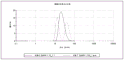
FIG. 14 shows dynamic light scattering data for soluble matrix (SolMM) and complete matrix (MM) at 1:50 dilution (1.0 mg/ml and 0.6mg/ml, respectively), showing soluble matrix particles less than 100nm in diameter, while the complete matrix has larger particles.
FIG. 15 shows dynamic light scattering data for soluble matrix (SolMM) at 1:10 and 1:100 dilutions (1.0 mg/ml and 0.1mg/ml, respectively), showing particles less than 100nm in diameter.
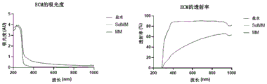
Fig. 16 shows absorbance (left) and transmittance (right) for saline, soluble matrix (SolMM), and complete matrix (MM).
Fig. 17 shows the relative absorbance (left) and transmittance (right) for soluble matrix (SolMM) and complete matrix (MM).
Detailed Description
All publications, patents and patent applications mentioned in this specification are herein incorporated by reference to the same extent as if each individual publication, patent or patent application was specifically and individually indicated to be incorporated by reference.
Unless defined otherwise, all technical and scientific terms and any acronyms used herein have the same meanings as commonly understood by one of ordinary skill in the art in the field of the invention. Although any methods and materials similar or equivalent to those described herein can be used in the practice of the present invention, the exemplary methods, devices, and materials are described herein.
The practice of the present invention will employ, unless otherwise indicated, conventional techniques of molecular biology (including recombinant techniques), microbiology, cell biology, biochemistry and immunology, which are within the skill of the art. Such techniques are explained fully in the following documents: molecular Cloning (Molecular Cloning), A Laboratory Manual (A Laboratory Manual), 2 nd edition (Sambrook et al, 1989); oligonucleotide Synthesis (Oligonucleotide Synthesis) (m.j. gait edition, 1984); animal Cell Culture (Animal Cell Culture) (r.i. freshney, 1987); enzymatic Methods (Methods in Enzymology) (academic Press Co., Ltd.); molecular Biology Protocols in Molecular Biology (f.m. ausubel et al, 1987, and updated regularly); PCR Polymerase Chain Reaction (PCR: The Polymerase Chain Reaction) (Mullis et al, 1994); remington, pharmaceutical Science and Practice (The Science and Practice of Pharmacy), 20 th edition (lipgakete, williams & wilkins 2003) and Remington, pharmaceutical Science and Practice, 22 nd edition (Philadelphia College of Pharmacy at University of The Sciences 2012).
In embodiments, the present invention provides a method of preparing one or more bioactive soluble fractions in an extracellular matrix (ECM) for therapeutic delivery, the method comprising:
a. partially or completely digesting decellularized ECM prepared from tissue with an acidic protease, such as pepsin;
b. neutralizing the digested ECM material to a pH of 7.0-8.0;
c. treating the liquid ECM (pre-gel solution) to produce soluble and insoluble fractions; and
d. separating at least a portion of the insoluble fraction from the soluble fraction to produce a soluble ECM composition.
In an embodiment, the present invention provides: the soluble ECM composition is further lyophilized, dialyzed, and/or filtered. In an embodiment, the present invention provides: rehydrating the soluble ECM composition after lyophilization.
In embodiments, the present invention provides a method of treating a subject in need thereof, the method comprising administering to the subject an intravascular infusion of an effective amount of a soluble ECM composition to promote organ, tissue or cell repair or cell recruitment. In embodiments, the infusion is via a catheter, intravenously, or intravascularly. In an embodiment, the present invention provides: when delivered in vivo, the soluble ECM composition will subsequently form a gel in and/or around the microvasculature of the tissue.
In an embodiment, the present invention provides a method of treating acute myocardial infarction, the method comprising injecting or infusing into a subject in need thereof having a myocardial infarction an effective amount of a composition comprising a soluble decellularized extracellular matrix derived from a muscle tissue.
In an embodiment, the present invention provides: the composition is delivered intravascularly. In an embodiment, the present invention provides: the composition is delivered with a balloon infusion catheter. In an embodiment, the present invention provides: the composition is converted to a gel form in the tissue after delivery. In an embodiment, the present invention provides: the composition degrades within one to 14 days after injection or infusion. In an embodiment, the present invention provides: injection or infusion of the composition repairs the damage to the myocardium suffered by the subject. In an embodiment, the present invention provides: injection or infusion of the composition repairs damage to non-cardiac tissue caused by trauma or ischemia in the subject.
In an embodiment, the present invention provides: the effective amount is an amount that increases blood flow, increases tissue mass, or induces neovascularization in an injection or infusion area of the subject.
For human therapy, there are many source species for extracellular matrices: for example, human, porcine, bovine, goat, mouse, rat, rabbit, chicken and other animal sources. Furthermore, there are many sources of tissue: such as the heart, brain, bladder, small intestine, skeletal muscle, kidney, liver, lung, blood vessels, and other tissues and organs.
In embodiments, the tissue is first decellularized, leaving only the extracellular matrix, such as disclosed in U.S. patent publication US2013/0251687, which is incorporated by reference in its entirety. The matrix is then lyophilized, milled or ground into a fine powder, solubilized with pepsin or other enzymes, and subsequently neutralized and buffered as previously reported. After neutralization, the digest (pre-gel solution) is fractionated to separate soluble and insoluble fractions. Separation of the soluble and insoluble fractions can be achieved by centrifugation, dialysis, filtration or adjustment of pH or salinity. The soluble fraction can be dialyzed to remove salts, lyophilized, and resuspended to adjust ECM concentration. The ECM can be sterile filtered, lyophilized, and stored in a sterile container. The ECM can be resuspended to the appropriate/physiological concentration for infusion.
A soluble ECM composition refers to extracellular matrix material that has been decellularized, lyophilized, milled, and digested and from which at least a portion of the solid components have been removed. . In embodiments, the soluble ECM composition is obtained from a centrifugation supernatant. In embodiments, the soluble ECM composition can pass through a filter size of less than 1 μm, 500nm, 250nm, 220nm, or 200 nm. A soluble ECM composition from which at least a portion of the naturally occurring solid ECM components are removed is a more transparent material than before the ECM solids are removed. However, it is understood that some degree of insoluble small particulate matter, such as ECM colloids, may still be present in the soluble ECM composition. A soluble ECM composition has been substantially isolated when at least 50%, 60%, 70%, 80%, 90%, 95%, 98%, or 99% by volume of naturally occurring ECM solids have been removed therefrom.
After adjusting the concentration and/or sterile filtration, the soluble ECM composition can be lyophilized and stored frozen (e.g., -20C, -80C) for at least 3 months. The soluble ECM composition can then be rehydrated with sterile water prior to injection or infusion.
The soluble ECM composition may be delivered by catheter infusion, intravenously, or by intravascular infusion with or without a balloon. Soluble ECM compositions can pass through damaged leaky vasculature such as those found in acute myocardial infarction, stroke, other ischemic tissues, tumors, and the like. Once in the tissue, the soluble ECM composition will then form a gel.
The soluble ECM composition may be delivered by catheter infusion, intravenously, or by intravascular infusion with or without a balloon. The soluble ECM composition may assemble into a covering layer or fill pores of the leaky vasculature on the lining of the leaky vasculature, such as those found in acute myocardial infarction, stroke, other ischemic tissues, tumors, traumatized tissue, and the like.
The soluble ECM composition gel may be cross-linked with glutaraldehyde, formaldehyde, bis-NHS molecules, or other cross-linking agents. The soluble ECM composition may be combined with cells, peptides, proteins, DNA, drugs, nutrients, survival promoting additives, proteoglycans and/or glycosaminoglycans. The soluble ECM composition can be combined and/or crosslinked with a synthetic polymer. Soluble ECM compositions can be used alone or in combination with the above components for endogenous cellular ingrowth, angiogenesis, and regeneration. Soluble ECM compositions can be used alone or in combination with the above components as a matrix to alter the mechanical properties of tissue. The soluble ECM composition can be delivered with cells for regeneration of damaged tissue, alone or in combination with the above ingredients.
The soluble ECM compositions can be used for tissue repair following tissue injury (such as caused by myocardial infarction, stroke, craniocerebral injury, peripheral arterial disease, liver cirrhosis, cancerous tumors, or kidney injury). The soluble ECM composition can be used alone or as a therapeutic delivery vehicle (vehicle).
The present invention provides soluble ECM compositions and methods for treating conditions having endothelial cell injury or dysfunction, leaky vasculature, endothelial cell junction disruption, vasodilation inhibition, and inflammation.
The present invention provides soluble ECM compositions and methods for treating conditions with potential reperfusion injury, including myocardial infarction, stroke, and peripheral artery and vessel disease. The soluble ECM compositions can be used as tissue engineering scaffolds to reduce reperfusion injury, reduce apoptosis, and promote tissue repair.
The present invention provides soluble ECM compositions and methods for treating excess or sustained Reactive Oxygen Species (ROS) production/signaling leading to endothelial cell activation and inflammation. Soluble ECM compositions can protect cells and tissues from ROS damage and inflammation by physical shielding and/or ROS sequestration (sequestration).
The present invention provides soluble ECM compositions and methods for treating heart disease, ischemia, and perfusion. Soluble ECM compositions can promote neovascularization and increase tissue perfusion.
The present invention provides soluble ECM compositions and methods for treating diabetes, insulin resistance. The soluble ECM composition can treat endothelial cells and restore endothelium-dependent vasodilation.
The present invention provides soluble ECM compositions and methods for treating cancer, including tumor growth, metastasis. The soluble ECM composition can treat leaky blood vessels and endothelial dysfunction present in cancer. ECM degradation products have been shown to inhibit tumor growth and formation.
The present invention provides soluble ECM compositions and methods for treating pulmonary diseases, such as chronic obstructive pulmonary disease, asthma, pulmonary hypertension. The soluble ECM composition can be infused to treat damaged tissue and/or endothelial cells of the lung.
The present invention provides soluble ECM compositions and methods for treating chronic renal failure. The soluble ECM composition can treat blood vessels to restore vasodilation and contraction.
The present invention provides soluble ECM compositions and methods for treating venous embolization. Infusion of a soluble ECM composition can cover the blood vessels to prevent thrombus formation and platelet aggregation.
The present invention provides soluble ECM compositions and methods for treating severe infectious diseases, particularly diseases with endothelial barrier disruption, such as hemorrhagic fever viruses including dengue hemorrhagic fever and hantavirus pulmonary syndrome. Soluble matrix infusion can treat and restore the endothelial barrier.
The present invention provides soluble ECM compositions and methods for treating atherosclerosis. Infusion of soluble ECM compositions can prevent plaque rupture by covering and stabilizing atherosclerotic plaques, or can adhere to endothelial cells and reduce inflammation.
The invention provides soluble ECM compositions and methods for treating cirrhosis, acute liver failure. The soluble ECM composition can treat endothelial dysfunction in cirrhosis. Soluble ECM compositions can attenuate inflammatory and oxidative stress.
The present invention provides soluble ECM compositions and methods for treating tissue hemorrhage and edema. The soluble matrix may cover the endothelial cells, fill gaps in the endothelial cell layer, increase tissue perfusion or decrease fluid access to the tissue by vascular stimulation.
The present invention provides soluble ECM compositions and methods for treating brain trauma and other nerve injuries. The soluble matrix can treat endothelial cells to repair leaky blood vessels, restore endothelium-dependent relaxation and nitric oxide production and reduce inflammation and oxidative stress.
Intracoronary infusion is an alternative to trans-endocardial injection. During typical treatment of Myocardial Infarction (MI), intracoronary delivery may be accompanied by balloon angioplasty. Following an acute MI, intracoronary infusion utilizes leaky vasculature, thus allowing biological material to enter the infarcted area [5 ]. In prior art formulations, the Matrix Material (MM) consists of a soluble fraction and insoluble submicron particles (>800nm) that are too large to pass through or adhere to the leaky vasculature. As a result, methods have been provided to at least partially separate a soluble fraction (referred to as soluble mm (solmm)) that can pass through and/or cover leaky vasculature and still form a hydrogel in vivo. Since SolMM is derived from MM, SolMM will have similar therapeutic effects, including reduced cardiomyocyte apoptosis, neovascularization, and reduced adverse LV remodeling.
To facilitate an understanding of the present invention, many of the terms and abbreviations used herein are defined below:
when introducing elements of the present invention or the preferred embodiments(s) thereof, the articles "a" or "an", "the" and "said" are intended to mean that there are one or more of the elements. The terms "comprising," "including," and "having" are intended to be inclusive and mean that there may be additional elements other than the listed elements.
When used in a list of two or more items, the term "and/or" means that any one of the listed items can be used alone or in combination with any one or more of the listed items. For example, the expression "a and/or B" is intended to mean either or both of a and B, i.e. a alone, B alone or a and B in combination. The expression "A, B and/or C" is intended to mean only a, only B, only C, A and B combinations, a and C combinations, B and C combinations, or A, B and C combinations.
It is to be understood that the aspects and embodiments of the invention described herein include "consisting of and/or" consisting essentially of aspects and embodiments.
It is to be understood that the description in range format is merely for convenience and brevity and should not be construed as an inflexible limitation on the scope of the invention. Accordingly, it is intended that the description of a range has specifically disclosed all possible sub-ranges as well as individual numerical values within that range. For example, description of a range such as from 1 to 6 should be considered to have specifically disclosed sub-ranges such as from 1 to 3, from 1 to 4, from 1 to 5, from 2 to 4, from 2 to 6, from 3 to 6, etc., as well as individual numbers within the range, e.g., 1, 2, 3, 4, 5, and 6. This applies regardless of the breadth of the range. Values or ranges can also be expressed herein as "about," from "about" one particular value, and/or to "about" another particular value. When such values or ranges are expressed, other embodiments of the disclosure include the particular values recited, from one particular value, and/or to other particular values. Similarly, when values are expressed as approximations, by use of the antecedent "about," it will be understood that the particular value forms another embodiment. It will be further understood that there are a plurality of values disclosed herein, and that each value is also disclosed herein as "about" the particular value in addition to the particular value itself. In embodiments, "about" may be used to mean, for example, within 10% of the recited value, within 5% of the recited value, or within 2% of the recited value.
The term "pharmaceutical composition" as used herein refers to a pharmaceutically acceptable composition, wherein the composition comprises a pharmaceutically active agent, and in some embodiments further comprises a pharmaceutically acceptable carrier. In some embodiments, the pharmaceutical composition may be a combination of a pharmaceutically active agent and a carrier.
The term "combination" means a fixed combination or kit of parts for combined administration in the form of one dosage unit, wherein the active compound(s) and the combination partner (e.g. the other drug substance, also referred to as "therapeutic agent" or "co-agent", as explained below) can be administered separately at the same time or separately within time intervals. In some cases, the combination partners show cooperation, e.g., a synergistic effect. The terms "co-administration" or "co-administration" and the like as used herein are intended to encompass the administration of the selected combination partners to a single subject (e.g., patient) in need thereof, and are intended to include treatment regimens in which administration is not necessarily by the same route of administration or at the same time. As used herein, the term "pharmaceutical combination" means a product resulting from mixing or combining more than one active ingredient, and includes both fixed and non-fixed combinations of active ingredients. The term "fixed combination" means that the active ingredients (e.g., the compound and the combination partner) are both administered to the patient simultaneously, in the form of a single entity or dose. The term "non-fixed combination" means that the active ingredients (e.g., the compound and the combination partner) are both administered to a patient as separate entities, simultaneously, concurrently or sequentially with no specific time limitation, wherein such administration provides therapeutically effective levels of both compounds in the patient. The latter also applies to cocktail therapies, e.g. the administration of three or more active ingredients.
As used herein, the term "pharmaceutically acceptable" means approved by a regulatory agency of the federal or a state government or listed in the U.S. pharmacopeia, other generally recognized pharmacopeia, in addition to other formulations safe for use in animals (and more particularly in humans and/or non-human mammals).
The term "pharmaceutically acceptable carrier" as used herein refers to excipients, diluents, preservatives, solubilizers, emulsifiers, adjuvants and/or carriers with which one or more demethylated compounds are administered. Such carriers can be sterile liquids, such as water and oils, including those of petroleum, animal, vegetable or synthetic origin, such as peanut oil, soybean oil, mineral oil, sesame oil and the like; polyethylene glycol, glycerol, propylene glycol or other synthetic solvents. Antibacterial agents such as benzyl alcohol or methyl paraben; antioxidants such as ascorbic acid or sodium bisulfite; chelating agents, such as ethylenediaminetetraacetic acid; and agents for adjusting tonicity, such as sodium chloride or dextrose, may also be carriers. Methods for producing combinations of compositions and carriers are known to those skilled in the art. In some embodiments, the phrase "pharmaceutically acceptable carrier" is intended to include any and all solvents, dispersion media, coatings, isotonic and absorption delaying agents, and the like, compatible with pharmaceutical administration. The use of such media and agents for pharmaceutically active substances is well known in the art. See, e.g., Remington, pharmaceutical Science and Practice (The Science and Practice of Pharmacy), 20 th edition (Rispeokt, Williams & Wilkins 2003). Except to the extent that any conventional media or agent is incompatible with the active compound, it is contemplated that such material will be used in the compositions.
As used herein, "therapeutically effective" refers to an amount of one or more pharmaceutically active compounds sufficient to treat or ameliorate or in some way alleviate symptoms associated with diseases and medical conditions. When used with reference to a method, the method is sufficient to effectively treat or ameliorate or in some way alleviate symptoms associated with the disease or disorder. For example, an effective amount with respect to age-related eye disease is sufficient to block or prevent onset; or if disease pathology has already begun, an amount sufficient to moderate, ameliorate, stabilize, reverse or slow disease progression, or otherwise mitigate the pathological consequences of the disease. In any case, an effective amount may be administered in a single dose or in divided doses.
The terms "treatment," "treating," or "treating," as used herein, encompass at least an improvement in the symptoms associated with the disease of the patient, wherein improvement is used in a broad sense to refer to a reduction in the magnitude of at least a parameter (e.g., symptoms associated with the disease or disorder being treated). Thus, "treating" also includes a disease, disorder, or pathological condition, or at least symptoms associated therewith, being completely inhibited (e.g., prevented from occurring) or stopped (e.g., terminated) such that the patient no longer suffers from the disorder, or at least the symptoms that characterize the disorder.
As used herein and unless otherwise indicated, the terms "prevent", "preventing" and "prevention" refer to the prevention of the onset, recurrence or spread of a disease or disorder, or one or more symptoms thereof. In certain embodiments, the term refers to treatment or administration of a compound or dosage form provided herein with a compound or dosage form provided herein, with or without one or more other additional active agents, prior to the onset of symptoms, particularly with respect to a subject at risk for a disease or disorder provided herein. This term encompasses inhibiting or alleviating the symptoms of a particular disease. In certain embodiments, subjects with a family history are potential candidates for a prophylactic regimen. In certain embodiments, subjects with a history of recurring symptoms are also potential candidates for a prophylactic regimen. In this regard, the term "prevention" may be used interchangeably with the term "prophylactic treatment".
As used herein and unless otherwise indicated, a "prophylactically effective amount" of a compound is an amount sufficient to prevent a disease or condition or prevent its recurrence. A prophylactically effective amount of a compound means an amount of a therapeutic agent, alone or in combination with one or more other agents, that provides a prophylactic benefit in the prevention of disease. The term "prophylactically effective amount" can encompass an amount that improves overall prophylaxis or enhances the prophylactic efficacy of other prophylactic agents.
As used herein and unless otherwise indicated, the term "subject" is defined herein to include animals, such as mammals, including, but not limited to, primates (e.g., humans), cows, sheep, goats, horses, dogs, cats, rabbits, rats, mice, and the like. In a specific embodiment, the subject is a human. The terms "subject" and "patient" are used interchangeably herein when referring to, for example, a mammalian subject (such as a human).
Examples
Experiment 1-SolMM Generation and characterization
Formulations of Myocardial Matrix (MM) can be generated based on previously described protocols (FIG. 1) [ 3)]. Briefly, fresh hearts were harvested from pigs (about 30-45kg) and LV myocardium was isolated. Removing large blood vessels and connective tissue, and cutting the rest of the tissue to less than 5mm3Of (2) (fig. 1A). The tissue was decellularized in 1% (w/v) Sodium Dodecyl Sulfate (SDS) for 4-5 days until the tissue was completely whitened, followed by an additional one day of rinsing with water to remove residual SDS (fig. 1B). The material was freeze-dried and ground to a fine powder (fig. 1C), and then partially enzymatically digested for 48 hours. The material was then neutralized and buffered to match in vivo conditions, producing MM capable of thermally induced gelation (fig. 1D).
Next, MM was centrifuged at 15,000RCF at 4 ℃ to separate soluble and insoluble fractions (fig. 1E). The supernatant was separated from the insoluble pellet and this supernatant was called soluble MM fraction (SolMM). The SolMM was then dialyzed and lyophilized to adjust the concentration and ratio of salts to maintain physiological conditions for the SolMM. SolMM was then resuspended at high concentration (16mg/mL), passed through a 0.22 μm filter into a sterile container, lyophilized, weighed and stored at-80 ℃ until needed. SolMM was then resuspended to the appropriate concentration in sterile water about 30 minutes prior to injection. This suspension formed a gel within 5 minutes after subcutaneous injection into rats (FIG. 1F, 10mg/mL, 500. mu.L). The consistency of the material can be assessed by: polyacrylamide gel electrophoresis for protein distribution, Picogreen assay for DNA content, dimethylmethylene blue assay for sulfated glycosaminoglycan (sGAG) content, and methylene blue assay for SDS content. Due to the digestion process of generating MM and the resulting SolMM, accurate data cannot be obtained from the mass spectrometer. However, PAGE showed protein overlap distribution between MM and SolMM, exclusively including the high molecular weight proteins in SolMM (figure 2, lane 4).
Experiment 2-blood compatibility of SolMM with human blood
The interaction between SolMM and human blood samples (n ═ 4) was evaluated at different dilutions (1:1, 1:2, 1:10) of SolMM and either whole human blood or platelet rich plasma. Based on the volumetric flow rate of the coronary vasculature and the expected infusion rate (1mL/min), 1:1 represents the highest possible ratio between blood and SolMM, while 1:10 represents a physiologically relevant dilution. Hemocompatibility was assessed as previously described for MM [4 ]. After hematocrit was adjusted to 45% with autologous plasma, red blood cell aggregation was performed on a myrennne aggregometer (Myrenne GmbH) within 4 hours of sampling. Aggregation was assessed after stasis (M0) or low shear rate (3 Hz; M1) while absorbance (800nm) was measured for 5 seconds. Similarly, platelet aggregation was measured using platelet-rich plasma isolated on a lumi-aggregometer (Chrono-log). Using the same dilution as the above sample and platelet rich plasma, high concentrations of coagulation cascade agonists (adenosine diphosphate, epinephrine, and collagen) (1:200-1:1000 dilution) were added and platelet aggregation was measured by absorbance (600-620 nm).
The results from 1:1 and 1:10 dilutions (material to human blood) indicate that SolMM is hemocompatible, as all values are within the normal physiological range (table 1).
TABLE 1

Table 1 shows the hemocompatibility of the soluble matrix. A1: 1 dilution of 10mg/ml soluble matrix with human blood represents the highest ratio, while a 1:10 dilution (1mg/ml) represents a physiologically relevant dilution.
Experiment 3-distribution, Retention and efficacy in the ischemia reperfusion model in Small animals
The left coronary artery was occluded for 45 minutes using an ischemia-reperfusion model in Sprague Dawley rats (225-250g) at MI, followed by reperfusion. Within 5 minutes after reperfusion, the aorta was clamped for about 15 seconds to simulate intracoronary infusion and 200 μ l of SolMM was injected into the LV cavity at a concentration of 6, 10 or 14 mg/mL. This will force the material into the coronary arteries and then distribute into the infarcted myocardium [12]]. The heart (n-2/concentration) was isolated 60 minutes after injection to determine if the material was first distributed and then retained in the heart because the non-gelling material cleared from the heart within one hour [ 5%]. Mixing SolMM with Alexa FluorTM568N-hydroxysuccinimidyl ester (Invitrogen) to allow fluorescence detection and analysis. The retention of the material was tested at the optimal concentration at 6, 12, 24 hours, 2, 3, 4 and 5 days and at 1 week time point (n-2-3/time point) after injection to assess degradation. Saline (n ═ 3/time point) was incubated with unconjugated Alexa FluorTM568 were mixed as a control. Hearts were freshly frozen in OCT Tissue-Tek compounds and the short axis was cut into 16 evenly spaced areas (approximately 300 μm between areas), 4 slides per area, in duplicate, 10 μm per section. H with one slide/zone&E to confirm infarction, and fluorescence analysis of SolMM using one slide/area.
1 hour after injection using concentrations of 6, 10 and 14mg/mL (n-2/group), the distribution in the infarcted area increased with increasing concentration, as indicated by the increasing intensity of the infarcted area (10 mg/mL shown in fig. 2) with the more pre-labeled gel distribution; however, 10mg/mL will be used for future experiments based on the yield produced by SolMM. Based on the filtration step mentioned in experiment 1, the yield was particularly limited, since SolMM was resuspended at 16mg/mL and subsequently filtered, giving a concentration of about 10 mg/mL. Resuspended concentrations above 16mg/mL will generally not pass through the filter.
Based on time history histology, material was observed in the infarcted heart within up to about 3 days after infusion.
In the rat ischemia reperfusion model, the left coronary artery was occluded for 35 minutes to simulate myocardial infarction. The heart was then reperfused and infused with soluble matrix via the coronary arteries using an aortic cross-clamp (aortic-cross clamp) model. Rats were imaged using magnetic resonance imaging 24 hours and 5 weeks after infusion. Left Ventricular (LV) volume and ejection fraction are shown in fig. 5. Twenty-four hours after infusion, significant retention of LV volume (end systole and end diastole) was observed compared to the saline infused control group. Ejection fraction showed a trend of increasing over saline control. After five weeks, LV volume by matrix infusion was also significantly reduced compared to saline-infused control group, showing that matrix infusion reduced adverse left ventricular remodeling.
Experiment 4: the heart is repaired after myocardial infarction using the infusible (infusible) extracellular matrix of a balloon infusion catheter.
Following myocardial infarction, extracellular matrix is infused through a blood vessel of the heart (e.g., the left anterior descending branch or the left main stem) using a balloon infusion catheter for targeted delivery. Fig. 4 shows the distribution and retention of soluble myocardial matrix (SolMM) after 1 hour of intracoronary infusion in a porcine ischemia-reperfusion model using a balloon infusion catheter. The short axis gross histology of the infarcted porcine heart is shown on the left side of figure 4. Infarctions were summarized in blue gray. Figure 4 shows the right side of the infarcted myocardium showing the SolMM microgel throughout the infarcted myocardium in the red grey scale channel.
The accessory organs (brain, kidney, liver, lung, spleen) were evaluated by a blind histopathologist and did not show any abnormal signs of ischemia or inflammation 1 hour after the infusion of the stroma (table 2). No soluble matrix gel was observed in any of the accessory organs, indicating the ability of the infusible matrix to target ischemic tissue.
TABLE 2

Table 2 shows that in the porcine ischemia-reperfusion model, intracoronary soluble matrix infusion did not show abnormal signs of ischemia or inflammation in the accessory organs (brain, kidney, liver, lung and spleen).
Experiment 5: the infusible matrix can be used as a scaffold to promote angiogenesis in ischemic or damaged tissues.
Figure 6 shows the increase in density of infarcted arterioles following matrix infusion in a myocardial infarction model. Infarctions were imaged 5 weeks after infusion in the rat ischemia-reperfusion model. Arterioles are identified by co-staining for alpha-smooth muscle actin and isolectin (isoelection) and tracked manually in ImageJ. Upregulation of the angiogenic pathway is shown in figure 6.
Experiment 6: the infusible matrix can be used as a scaffold to reduce apoptosis or necrosis of ischemic or damaged tissue.
Figure 7 shows the reduction of cardiomyocyte apoptosis following matrix infusion in a myocardial infarction model. Staining of infarcted and infarcted border zone of cardiomyocytes with alpha-actinin and staining of apoptosis with cleaved caspase 3. Apoptotic cardiomyocytes were counted manually in ImageJ. Reduced apoptosis may extend to other cell types, but is not limited to endothelial cells, immune cells, fibroblasts, neurons and (cardiac) muscle cells. The reduced apoptotic/necrotic pathway is shown in figure 9. The reduced apoptosis can be explained by increased Reactive Oxygen Species (ROS) metabolism, as the up-regulated ROS metabolic pathway is shown in fig. 8.
Figures 8 and 9 show differential gene expression, indicating a pathway for repair of infusible extracellular matrix therapy. RNA was isolated from left ventricular free wall tissue on days 1 and 3 after matrix infusion and ischemia-reperfusion injury. On day 1, angiogenic and reactive oxygen metabolic pathways are up-regulated. On day 3, reduced apoptosis/necrosis and reduced fibrotic pathways were observed. LRG1 downregulation involved cardiac fibrosis, and trends were observed in the opposite direction. Saline infusion was used as a control.
Experiment 7: matrix infusion can treat endothelial cell injury/dysfunction. The soluble matrix may coat endothelial cells to reduce reactive oxygen species damage, increase endothelial cell survival, and/or fill leaky vasculature spaces after ischemic injury.
Following ischemic injury and matrix infusion, soluble matrix was observed to cover the lumen of small blood vessels (capillaries/endothelial cells). Figure 10 shows endothelial cell (green gray) chambers covered with soluble matrix (red gray). Note that the matrix does not block the lumen. In addition, fig. 11 shows soluble matrix overlapping endothelial cells in large vessels without occluding the lumen. The heart was imaged using confocal microscopy up to 24 hours after infusion and simulated myocardial infarction.
Experiment 8: the infusible matrix can be co-delivered with drugs, growth factors, microRNAs or other therapeutic agents
The soluble extracellular matrix composition has potential binding domains for growth factors, micrornas, and other potential drugs or therapeutic agents. The infusible matrix can be used for slow release of therapy as it forms a gel in the tissue after infusion.
Figure 12 shows soluble ECM retention in infarcted tissue 24 hours after matrix infusion in an ischemia-reperfusion model. From left to right, the heart was infused with saline, matrix conjugated to w/VivoTag750, trilysine conjugated to w/VivoTag750, and matrix conjugated to w/VivoTag 750. Twenty-four hours after infusion, hearts were harvested and imaged on Licor Odyssey. Matrix infused hearts showed stronger signal intensity compared to saline infused and trilysine infused hearts. Trilysine with VivoTag750 was used as a small peptide control and does not show visible retention.
Figure 13 shows the nanofiber architecture of the soluble matrix hydrogel. 10mg/ml of the pre-gel solution was injected subcutaneously into the back of the rat, which then formed a gel, and harvested for scanning electron microscopy imaging. Gel structures are reminiscent of the natural extracellular matrix.
Experiment 9: dynamic light scattering analysis shows the difference between MM and SolMM.
FIG. 14 shows dynamic light scattering data for soluble matrix (SolMM) and complete matrix (MM) at 1:50 dilution (1.0 mg/ml and 0.6mg/ml, respectively), showing soluble matrix particles less than 100nm in diameter, while the complete matrix has larger particles.
FIG. 15 shows dynamic light scattering data for soluble matrix (SolMM) at 1:10 and 1:100 dilutions (1.0 mg/ml and 0.1mg/ml, respectively), showing particles less than 100nm in diameter.
Fig. 16 shows absorbance (left) and transmittance (right) for saline, soluble matrix (SolMM), and complete matrix (MM).
Fig. 17 shows the relative absorbance (left) and transmittance (right) for soluble matrix (SolMM) and complete matrix (MM).
Reference to the literature
[1] Benjamin et al, American Heart Association Statistics (American Heart Association Statistics), o.b.o.t.a.h.a.s.c. Stroke Statistics sub committee (Stroke Statistics), s.stroke, Heart Disease and Stroke Statistics-2017Update (Heart Disease and Stroke Statistics-2017 Update): report from the American Heart Association, issue (Circulation)135(10) first, (2017) e146-e 603.
[2] Singelyn et al, a Naturally derived myocardial matrix as an injectable scaffold for cardiac tissue engineering, Biomaterials (Biomaterials)30(29) (2009) 5409-16.
[3] Singelyn et al, a ductally delivered hydrogel derived from decellularized ventricular extracellular matrix, augments endogenous cardiomyocytes and preserves cardiac function following myocardial infarction (cardiac-responsive hydrogel derivative from decellularized cardiac extracellular matrix) in vitro, J.Am.Cardiograph 59(8) (2012) 751-63.
[4] Seif-Naraghi et al, Safety and efficacy of injectable extracellular matrix hydrogels for the treatment of myocardial infarction (Safety and efficiency of an injectable extracellular matrix hydrogel for treating myocardial infarction), scientific transformation medicine (Science transformation medicine)5(173) (2013)173ra 25.
[5] Nguyen et al, Enzyme-Responsive Nanoparticles for target Accumulation and Prolonged Retention in cardiac Tissue after Myocardial Infarction (Enzyme-Responsive Nanoparticles for Targeted Accumulation and managed Retention in Heart Tissue, Advanced Materials 27(37) (2015) 5547-5552.
[6] Wasseenaar et al, Evidence of a potential mechanism for functional benefit of myocardial matrix hydrogels for post-MI treatment (Evidence for mechanisms underlying the functional details of a myocardial matrix hydrogel for post-MI treatment), J.Cardiology Association 67(9), (2016) 1074-86.
[7] Schuster et al, dilation of transmural myocardial infarction: pathophysiological factors of cardiac rupture (Expansion of cardiac muscular injury: a pathophysiological factor in cardiac failure), issue 60(7) (1979) 1532-1538.
[8] Arsenos et al, arrhythmic sudden cardiac death: substrates, mechanisms and current risk stratification strategies for patients after myocardial infarction (understrates, mechanisms and current risk stratification strategies for the post-myocardial infarction), the Heart Greek journal (Hellent JCardiol)54(4) (2013) 301-.
[9] Leor et al, Intracoronary Injection of In Situ formed Alginate Hydrogel retropulsion Left Ventricular Remodeling After Myocardial Infarction In Swine (Intra coronary Injection of In Situ formation of Heart formation Hydrogel Left Ventricular Remodeling After Heart disease In Swine, J.Am.Cardiology 54(11) (2009) 1014-1023.
[10] Frey et al, intracoronary delivery of an injectable bioabsorbable stent (IK-5001) to treat left ventricular remodeling following ST-elevation myocardial infarction: the first human study (IK-5001) to linear left branched modified rear after ST-elevation myocardial infarnation: first-in-man study) issued: cardiovascular Interventions (Cardiovascular Interventions)7(6) (2014) 806-.
[11] Spang et al, extracellular matrix hydrogel therapy: in vivo applications and developments (Extracellular matrix hydrogels: In vivo applications and definitions), BioMaterial science (Acta Biomater)68(2018) 1-14.
[12] Cheng et al magnetically enhance Cell retention, implantation and functional benefits after intracoronary delivery of heart-derived stem cells in a rat model of ischemia/reperfusion (Magnetic enhancement of Cell retention, expression, and functional graft of cardiac-derived stem cells in a rate model), Cell transplantation (Cell transplantation)21(6 (2012) 1121-35.
[13] Rane et al, increase of infarct wall thickness by bioinert material is insufficient to prevent adverse left ventricular remodeling after myocardial infarction (induced myocardial infarction wall thickness by a bio-inert material is induced to a present negative left ventricular remodeling after myocardial infarction), scientific public library complex (PloS one)6 (2011) e 21571.
[14] Conesa et al, a survey of best practices for RNA-seq data analysis (A surfey of best practices for RNA-seq data analysis), Genome biology (Genome biology)17(2016) 13-13.
[15] Costa-Silva et al, RNA-Seq differential expression analysis: extended overview and software tools (RNA-Seq differential expression analysis: extended review and a software tool), scientific public library integration (PLOS ONE)12(12), (2017) e0190152-e 0190152.
[16] Wang et al, Humanized mouse model for assessing human immune response to xenogeneic and allogeneic decellularized biomaterials (Humanized mouse model for assessing the human immune response to xenogeneic and allogeneic decellularized biomaterials), biomaterials 129(2017) 98-110.
[17] Sonnenberg et al, Delivery of engineered HGF fragments in hydrogels derived from extracellular matrix to prevent poor left ventricular remodeling following myocardial infarction (Delivery of an engineered HGF fragment in an extracellular matrix-derived hydrogel preceding negative LV remodelling post-myocardial infarction), biomaterial 45(2015) 56-63.
[18] Seif-Naraghi et al, Injectable extracellular matrix-derived hydrogels provide a platform for enhanced heparin-binding growth factor retention and delivery (Injectable extracellular matrix-derived hydrogel precursors a platform for enhanced recovery and delivery of a heparin-binding growth factor), the biomaterials journal 8(10 (2012) 3695) 703.
[19] van den Akker et al, intramyocardial stem cell injection: with flow (Intramyocardial stem cell injection: go (ne) with the flow), the European Heart disease journal (2016) ehw056-ehw 056.
[20] Wasseenaar et al, modulate the rate of in vivo degradation of injectable extracellular matrix hydrogels (modulated in vivo degradation rates of injectable extracellular matrix hydrogels), journal of materials chemistry B (J.Mater.chem.B.)4(16) (2016) 2794-2802.
[21] Grover et al, Myocardial matrix-polyethylene glycol hybrid hydrogels for tissue engineering (Myocardial matrix-polyethylene glycol hydrogels for tissue engineering), nanotechnology (Nanotech.)25(1), (2014) 014011.
[22] Gallet et al, Exosomes secreted by cardiosphere-derived cells attenuate scars, reduce adverse remodeling and improve acute and chronic porcine myocardial infarction function (Exosomes secreted by cardiac-derived cells reduction screening, and embryonic function in both acid and cyclic cardiac activity), European journal of cardiology 38(3), (2016) ehw240-ehw 240.
[23] Dawn et al, intravascular delivery of Cardiac stem cells across the vascular barrier regenerates infarcted myocardium and improves Cardiac function (Cardiac stem cells delayed in myocardial transformed in the vascular barrier, regenerated in myocardial muscle, and advanced Cardiac function), Proceedings of the National Academy of Sciences 102(10 (2005) 3766-3771.
[24] Gallet et al, self-assembled Intracoronary administration of cardiac micro-tissue (glomus) prevents adverse remodeling in porcine models of convalescent myocardial infarction (cardiac), for prevention of accommodation remodelling in a pig model of accommodation myocardial infarction, circulatory cardiovascular intervention (Circ cardiovascular Interval) 8(5) (2015) e002391.doi: 10.1161/CINTERVENTIONS.115.002391-e002391. doi: 10.1161/CINTERVENTIONS.115.002391.
[25] Bolli et al, Intracoronary Delivery of Autologous Cardiac Stem Cells Improves Cardiac Function in a Porcine Model of Chronic Ischemic Cardiomyopathy (Intracoronary Delivery of Autologus Cardiac Stem Cells improvement in a porous Model of viral Ischemic Cardiopaphy), issue 128(2) (2013) 122-.
[26] McKay, Raymond g. et al, "left ventricular remodeling after myocardial infarction: the inevitable consequence of infarct expansion (Left ventricular remodeling after myocardial infarction:. alpha. renal to myocardial expansion), issue 74, No.4(1986): 693-.
[27] Harpster, Mark H. et al, "Earliest change in the left ventricular transcriptome after myocardial infarction-myocardial infringement", Mammalian genome (Mammalian genome)17, No.7(2006) 701-715.
Claims (32)
1. A method of preparing a soluble extracellular matrix (ECM) composition, comprising:
a. digesting the decellularized ECM material with an acidic protease;
b. neutralizing said digested ECM material in a liquid to a pH of 7.0-8.0;
c. treating the liquid ECM to produce soluble and insoluble fractions; and
d. separating at least a portion of the soluble fraction from the insoluble fraction to produce a soluble ECM composition.
2. The method of claim 1, wherein said treating said liquid ECM to produce said soluble and insoluble fractions is by centrifugation.
3. The method of claim 1, wherein said processing said liquid ECM to produce said soluble and insoluble fractions is performed by dialysis or filtration.
4. The method of claim 1, wherein the separating is performed with a size exclusion filter of 250nm or less.
5. The method of claim 1, wherein said soluble ECM composition is further lyophilized and rehydrated.
6. A soluble ECM composition comprising decellularized, digested, and neutralized tissue from which at least a portion of solid ECM material has been removed, wherein the soluble ECM composition passes through a 250nm size exclusion filter.
7. The soluble ECM composition of claim 6, wherein the composition is formulated for intravascular infusion.
8. The soluble ECM composition of claim 6, wherein the composition is liquid at room temperature and forms a gel in vivo following infusion or injection.
9. The soluble ECM composition of claim 6, wherein the composition is liquid at room temperature and forms a coating in vivo lining damaged blood vessels after infusion or injection.
10. The soluble ECM composition of claim 6, wherein the composition is liquid at room temperature and fills the pores between endothelial cells in vivo after infusion or injection.
11. The soluble ECM composition of claim 6, which is derived from human, animal, embryonic, or fetal tissue.
12. The soluble ECM composition of claim 6, which is derived from heart, brain, bladder, small intestine or skeletal muscle tissue, kidney, liver, lung and blood vessels.
13. A method of treating a subject to promote tissue repair comprising administering to a subject in need thereof an infusion of an effective amount of the soluble ECM composition of claim 6.
14. The method of claim 13, wherein the infusion is delivered via a catheter, intravenously, or intravascularly.
15. The method of claim 13, wherein the soluble ECM composition forms a gel in a tissue when delivered in vivo.
16. The method of claim 13, wherein said soluble ECM composition is crosslinked with glutaraldehyde, formaldehyde, bis-NHS molecules, or other crosslinking agents prior to administration.
17. The method of claim 13, wherein said soluble ECM composition is combined with cells, peptides, proteins, DNA, drugs, nanoparticles, nutrients, survival-promoting additives, proteoglycans, and/or glycosaminoglycans prior to administration.
18. The method of claim 13, wherein the soluble ECM composition is combined and/or crosslinked with a synthetic polymer or a biologically derived material prior to administration.
19. The method of claim 13, wherein said soluble ECM composition causes endogenous cellular ingrowth, angiogenesis, and regeneration in said subject.
20. The method of claim 13, wherein said soluble ECM composition promotes cell survival and reduces inflammation in said subject.
21. A method for treating acute myocardial infarction comprising injecting or infusing into a subject in need thereof an effective amount of a soluble ECM composition comprising decellularized, digested and neutralized tissue from which at least a portion of solid ECM material has been removed.
22. The method of claim 21, wherein the composition is delivered intravascularly.
23. The method of claim 21, wherein the composition is delivered with a balloon infusion catheter.
24. The method of claim 21, wherein the composition is converted to gel form in tissue after delivery.
25. The method of claim 21, wherein the composition is liquid at room temperature and forms a coating lining the infarcted vessel after delivery.
26. The method of claim 21, wherein the composition is liquid at room temperature and fills pores between infarcted endothelial cells after delivery.
27. The method of claim 21, wherein the composition degrades within one to 14 days after injection or infusion.
28. The method of claim 21, wherein injection or infusion of the composition repairs damage to the myocardium suffered by the subject.
29. The method of claim 21, wherein injection or infusion of the composition repairs damage caused by ischemia in the subject.
30. The method of claim 21, wherein the effective amount is an amount that increases blood flow, increases living tissue mass, or induces neovascularization in an injection or infusion region of the subject.
31. A method of treating endothelial cell injury and/or dysfunction, comprising injecting or infusing into a subject in need thereof an effective amount of a soluble ECM composition comprising decellularized, digested, and neutralized tissue from which at least a portion of solid ECM material has been removed.
32. The method of claim 31, wherein the effective amount promotes endothelial cell survival, proliferation, or vasoactivity and/or reduces inflammation, apoptosis, reactive oxygen species damage, or leaky vasculature.
Applications Claiming Priority (3)
| Application Number | Priority Date | Filing Date | Title |
|---|---|---|---|
| US201862750303P | 2018-10-25 | 2018-10-25 | |
| US62/750,303 | 2018-10-25 | ||
| PCT/US2019/058052 WO2020086955A1 (en) | 2018-10-25 | 2019-10-25 | Soluble extracellular matrix composition and method for intravascular delivery |
Publications (1)
| Publication Number | Publication Date |
|---|---|
| CN112867499A true CN112867499A (en) | 2021-05-28 |
Family
ID=70330739
Family Applications (1)
| Application Number | Title | Priority Date | Filing Date |
|---|---|---|---|
| CN201980067713.0A Pending CN112867499A (en) | 2018-10-25 | 2019-10-25 | Soluble extracellular matrix compositions and methods for intravascular delivery |
Country Status (7)
| Country | Link |
|---|---|
| US (1) | US20210379245A1 (en) |
| EP (1) | EP3870192A4 (en) |
| JP (1) | JP7536004B2 (en) |
| CN (1) | CN112867499A (en) |
| AU (1) | AU2019363610A1 (en) |
| CA (1) | CA3117688A1 (en) |
| WO (1) | WO2020086955A1 (en) |
Families Citing this family (1)
| Publication number | Priority date | Publication date | Assignee | Title |
|---|---|---|---|---|
| WO2022056437A1 (en) * | 2020-09-14 | 2022-03-17 | The Regents Of The University Of California | Compositions and methods of use for infusible extracellular matrix |
Citations (4)
| Publication number | Priority date | Publication date | Assignee | Title |
|---|---|---|---|---|
| KR101628821B1 (en) * | 2015-03-02 | 2016-06-13 | 강원대학교산학협력단 | Biocompatible Solubilized Scaffold Extract Derived from Decellularized Organ Tissue, Method for Preparating the Biocompatible Scaffold Extract and Uses for Thereof |
| US20170049932A1 (en) * | 2014-04-24 | 2017-02-23 | University Of Pittsburgh-Of The Commonwealth System Of Higher Education | Fractionating Extracellular Matrix to Modulate Bioactivity and the Host Response |
| CN106619723A (en) * | 2010-08-24 | 2017-05-10 | 加利福尼亚大学董事会 | Compositions and methods for cardiac therapy |
| WO2018161034A1 (en) * | 2017-03-02 | 2018-09-07 | University Of Pittsburgh - Of The Commonwealth System Of Higher Education | Extracellular matrix (ecm) hydrogel and soluble fraction thereof for the treatment of cancer |
Family Cites Families (2)
| Publication number | Priority date | Publication date | Assignee | Title |
|---|---|---|---|---|
| CN108699522B (en) * | 2016-01-13 | 2022-05-27 | 高等教育联邦系统-匹兹堡大学 | Vascular extracellular matrix hydrogel |
| EP3755267B1 (en) * | 2018-02-22 | 2024-06-12 | University of Pittsburgh - of the Commonwealth System of Higher Education | Extracts for the regeneration of ligaments |
-
2019
- 2019-10-25 CA CA3117688A patent/CA3117688A1/en active Pending
- 2019-10-25 JP JP2021521511A patent/JP7536004B2/en active Active
- 2019-10-25 CN CN201980067713.0A patent/CN112867499A/en active Pending
- 2019-10-25 US US17/286,534 patent/US20210379245A1/en active Pending
- 2019-10-25 EP EP19876046.4A patent/EP3870192A4/en active Pending
- 2019-10-25 WO PCT/US2019/058052 patent/WO2020086955A1/en unknown
- 2019-10-25 AU AU2019363610A patent/AU2019363610A1/en not_active Abandoned
Patent Citations (4)
| Publication number | Priority date | Publication date | Assignee | Title |
|---|---|---|---|---|
| CN106619723A (en) * | 2010-08-24 | 2017-05-10 | 加利福尼亚大学董事会 | Compositions and methods for cardiac therapy |
| US20170049932A1 (en) * | 2014-04-24 | 2017-02-23 | University Of Pittsburgh-Of The Commonwealth System Of Higher Education | Fractionating Extracellular Matrix to Modulate Bioactivity and the Host Response |
| KR101628821B1 (en) * | 2015-03-02 | 2016-06-13 | 강원대학교산학협력단 | Biocompatible Solubilized Scaffold Extract Derived from Decellularized Organ Tissue, Method for Preparating the Biocompatible Scaffold Extract and Uses for Thereof |
| WO2018161034A1 (en) * | 2017-03-02 | 2018-09-07 | University Of Pittsburgh - Of The Commonwealth System Of Higher Education | Extracellular matrix (ecm) hydrogel and soluble fraction thereof for the treatment of cancer |
Non-Patent Citations (2)
| Title |
|---|
| YAEL EFRAIM 等: "Biohybrid cardiac ECM-based hydrogels improve long term cardiac function post myocardial infarction", ACTA BIOMATERIALIA, pages 220 - 233 * |
| 徐晓霞 等: "细胞外基质在无瘢痕修复和再生中的研究进展", 重庆医学, vol. 44, no. 24, pages 3445 - 3447 * |
Also Published As
| Publication number | Publication date |
|---|---|
| JP7536004B2 (en) | 2024-08-19 |
| AU2019363610A1 (en) | 2021-05-13 |
| EP3870192A4 (en) | 2022-09-21 |
| CA3117688A1 (en) | 2020-04-30 |
| US20210379245A1 (en) | 2021-12-09 |
| EP3870192A1 (en) | 2021-09-01 |
| JP2022505468A (en) | 2022-01-14 |
| WO2020086955A1 (en) | 2020-04-30 |
Similar Documents
| Publication | Publication Date | Title |
|---|---|---|
| Spang et al. | Extracellular matrix hydrogel therapies: In vivo applications and development | |
| US11648281B2 (en) | Methods for treating cardiac conditions | |
| Neuman et al. | Hyaluronic acid and wound healing | |
| Habib et al. | A combined cell therapy and in-situ tissue-engineering approach for myocardial repair | |
| Ruvinov et al. | The promotion of myocardial repair by the sequential delivery of IGF-1 and HGF from an injectable alginate biomaterial in a model of acute myocardial infarction | |
| US20140106447A1 (en) | Compositions and methods for recruiting stem cells | |
| EP3302594B1 (en) | Cardiac fibroblast-derived extracellular matrix and injectable formulations thereof for treatment of ischemic disease or injury | |
| Vu et al. | An autologous platelet-rich plasma hydrogel compound restores left ventricular structure, function and ameliorates adverse remodeling in a minimally invasive large animal myocardial restoration model: a translational approach: Vu and Pal “Myocardial Repair: PRP, Hydrogel and Supplements” | |
| Reffelmann et al. | Transplantation of neonatal cardiomyocytes after permanent coronary artery occlusion increases regional blood flow of infarcted myocardium | |
| KR20120033299A (en) | Compositions and methods for preventing cardiac arrhythmia | |
| Yoon et al. | Differential regeneration of myocardial infarction depending on the progression of disease and the composition of biomimetic hydrogel | |
| JP7554033B2 (en) | Composition for treating tissue lesions | |
| Lee et al. | VEGF-attenuated platelet-rich plasma improves therapeutic effect on cartilage repair | |
| Shibata et al. | Enhanced sternal healing through platelet-rich plasma and biodegradable gelatin hydrogel | |
| WO2012125007A2 (en) | Methods and compositions for the treatment, prevention, and alleviation of bone and cartilage diseases or injuries and hair loss | |
| JP7536004B2 (en) | Soluble extracellular matrix compositions and methods for intravascular delivery - Patents.com | |
| KR100702250B1 (en) | Osteoblast composition of semi-solidified mixed fibrin for bone fracture agglutination and its manufacturing method | |
| Alsousou et al. | Platelet-rich plasma in regenerative medicine | |
| KR20200085748A (en) | Stem cell extraction from bone marrow niches | |
| US20150283183A1 (en) | Methods for treating cardiac conditions | |
| Spang | Intravascular infusion of a soluble extracellular matrix hydrogel therapy for acute myocardial infarction | |
| US20230364152A1 (en) | Compositions and methods of use for infusible extracellular matrix | |
| JP2018529670A (en) | Non-surgical and localized delivery of placental growth factor composition | |
| EP1983833A2 (en) | Method for promoting myocardial regeneration and uses thereof | |
| Xue et al. | A Horn Peptide‐Thermoresponsive Hydrogel for Angiogenesis and Bone Regeneration |
Legal Events
| Date | Code | Title | Description |
|---|---|---|---|
| PB01 | Publication | ||
| PB01 | Publication | ||
| SE01 | Entry into force of request for substantive examination | ||
| SE01 | Entry into force of request for substantive examination |