Disclosure of Invention
The invention provides a tumor treatment material capable of self-enhancing chemical kinetic cycle, which is CaO2Transition metal nanoparticles, which provide highly cytotoxic OH for tumor therapy, scavenge the over-expressed GSH, and continuously enhance the CDT effect in the circulation of transition ions, thus overcoming the drawbacks of chemokinetic therapy (mainly the drawback of insufficient endogenous hydrogen peroxide, poor efficacy).
Specifically, calcium peroxide (CaO)2) Due to the existence of peroxy bond (-O-O-), can stably release a large amount of H under acidic condition2O2Based on CaO2H of (A) to (B)2O2Production is independent of other precursors. The generator can effectively accumulate H with tumor specificity2O2At the tumor site. The loaded transition metal ions of copper (Cu) or manganese (Mn) or iron (Fe) or vanadium (V) or cerium (Ce) or platinum (Pt) or cobalt (Co) or palladium (Pd) or gold (Au) or silver (Ag) consume the GSH over-expressed in the tumor cells through oxidation, so that the embarrassment that OH with high cytotoxicity is removed by GSH is avoided, and the CDT curative effect is improved. The high valence ions are reduced to low valence ions during GSH removal, i.e. Fenton reaction ions, the low valence ions and H2O2The Fenton reaction is generated, and H is oxidized into high price at the same time2O2The decomposition into OH and hydroxyl (-OH) to this point, the transition metal ion forms a cycle and is accompanied by the formation of a large amount of highly cytotoxic OH.
Wherein the transition metal is coordinated at a size of 91-122nm CaO2In the granules, the CaO2The molar ratio of the transition metal to the transition metal is 15: 1-30: 1, which is beneficial to CaO2Transition metal nanoparticles enter tumor cells by the EPR effect and provide sufficient iron ions to remove glutathione and produce hydrogen peroxide.
In the application, CaO synthesized by dopamine assistance2CaO with stable structure and performance is constructed by a wet chemical method as a raw material2-transition metal nanoparticles, in particular, the method is: adding CaO2Dissolving the particles and transition metal salt in ethanol (15mL) and stirring to obtain CaO2-transitionMetal nanoparticles.
The invention has the beneficial effects that: CaO provided by the invention2The transition metal nanoparticles can provide highly cytotoxic hydroxyl radicals (. OH) for tumor treatment, remove over-expressed Glutathione (GSH), continuously enhance the CDT effect in the circulation of iron ions, and provide a new strategy for improving tumor cell selectivity.
Drawings
FIG. 1 is a characteristic image of CaO2-Fe nanoparticles, wherein 1a is a transmission electron micrograph, 1b is an X-ray diffraction chart, 1c is an X-ray energy scattering spectrum analysis, and 1d is CaO2The X-ray photoelectron spectrum of Fe, the high-resolution X-ray photoelectron spectra of Fe 2p and O1s for 1e and 1f, respectively.
FIG. 2 shows an ultraviolet-visible absorption spectrum (UV-Vis) and an electron spin resonance spectrum (ESR), wherein 2a is H in an acidic environment2O2、CaO2NPs and CaO2Fe NPs treatment of KMnO42b different concentrations of GSH treated CaO2UV-VIS absorption spectra of Fe NPs after MB degradation at pH5.4, 2c GSH treated CaO of different concentrations at pH5.42ESR spectra of Fe NPs (5, 5-dimethyl-1-pyrroline N-oxide (DMPO) as spin trap).
FIG. 3a is a schematic representation of a reaction with FeCl3、CaO2NPs and CaO2Fluorescence image of DCFH-DA stained 4T1 cells after incubation with Fe NPs. The scale bar is 100 μm. 3b is FeCl3、CaO2NPs and CaO2Viability of 4T1 cells after 24h incubation with Fe NPs. 3c calcein AM (Green, viable cells) and PI (Red, dead cells) stained 4T1 cells with FeCl3,CaO2NPs and CaO2Fluorescence images of Fe NPs after 24h incubation. The scale bar is 100 μm. 3d is with CaO2Viability of L929 cells after 24h incubation with Fe NPs. 3e is CaO2-in vitro chemokinetic toxicity of Fe NPs on L-BSO (L-Thioanimine). 3f is the intracellular GSH level of 4T1 cells and L929 cells. 3g FeCl for flow cytometry analysis3,CaO2NP and CaO24T1 apoptosis in 24h Fe NPs. For 3h isCaO with or without NAC treatment2In vitro chemical kinetic toxicity studies of Fe NPs.
FIG. 4a shows the use of saline (control), FeCl3,CaO2NPs and CaO2-relative tumor growth curves of 4T1 tumor-bearing mice after Fe NPs treatment. 4b are representative photographs of dissected tumors from different groups at day 17 post-dose. 4c is H in different groups on day 17 after administration&Images of E and TUNEL stained tumor sections. 4d is the time-dependent body weight curve of different groups of mice. 4e is normal saline or CaO injected into the vein of healthy mice2Biochemical analysis of the blood 17 days after Fe NPs.
FIG. 5 shows CaO2High angle annular dark field scanning transmission electron microscope images of Fe NPs and elemental mapping of Fe, Ca, O.
FIG. 6 shows the measurement of CaO by Dynamic Light Scattering (DLS)2And CaO2-Fe nanoparticle size.
FIG. 7 is a transmission electron micrograph of CaO2-Mn nanoparticles.
Fig. 8 is an X-ray energy scattering spectroscopy analysis.
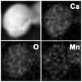
FIG. 9 shows CaO2-high angle annular dark field scanning transmission electron microscope images of Mn NPs and elemental mapping of Mn, Ca, O.
FIG. 10 shows potassium ferricyanide vs. Fe2+The color-developed photograph of (1).
FIG. 11a shows different concentrations of GSH treated CaO2The UV-VIS absorption spectrum of NPs after MB degradation at pH5.4 is bar chart of MB degradation rate 11 b.
FIG. 12a shows different concentrations of GSH treated CaO2The uv-vis absorption spectrum of NPs after degradation of MB at pH 7.4, 12b is a histogram of MB degradation rate.
FIG. 13 shows CaO2GSH content in 4T1 cells 8h after Fe NPs treatment.
Figure 14 is a histological analysis of the main tissues after treatment. The scale bar is 100 μm.
FIG. 15 shows the normal saline or CaO injected intravenously to healthy mice2Hematological analysis 17 days after Fe NPs.
Detailed Description
Example 1
In one embodiment of the invention, CaO is used2For example, CaO was synthesized by a modified dopamine-assisted method2NP, then mixed with ferrous chloride for 30 minutes. The method comprises the following specific steps:
(1)CaO2the preparation of (1): CaCl is treated under the action of ultrasonic waves2(0.1g, 0.68mmol) and dopamine (0.003g) were dissolved in ethanol (15 mL). Then, 1ml of NH was added with stirring4OH (ammonia). Followed by dropwise addition of 0.2ml of H2O2And (3) solution. The product CaO was collected by centrifugation at 8000rpm2Washed 3 times with ethanol and finally redispersed in 20mL of ethanol.
(2)CaO2-preparation of Fe: mixing the above 50mg CaO2The granules and 25mg of ferrous chloride are dissolved in ethanol (15mL) and stirred to obtain CaO2-Fe。
With the original CaO2NP vs. CaO2The morphology of the Fe NPs remains unchanged and their diameter increases from 91nm to 122nm (FIGS. 1a and 6), and X-ray diffraction (XRD) confirms that the introduction of iron ions does not affect CaO2Phase of NP (FIG. 1 b). Strong and uniform iron signals were observed from energy dispersive X-ray spectroscopy (EDS) and EDS mapping, indicating iron ions with CaO2Efficient binding between NPs (fig. 1c), calcium element: iron element ≈ 15: 1. to explore the valence state of the chelated iron ions, X-ray photoelectron spectroscopy (XPS) was used, with a central peak at about 710.0eV (Fe 2 p)3/2) And a shaking satellite peak at about 724.0eV (Fe 2 p)1/2) Showing that Fe2+The ions have been converted into Fe3+Ions, possibly substituted by CaO2And (4) oxidizing (FIGS. 1d and e). The photoelectron peak at 532.5eV for O1s can be attributed to the O — O bond, indicating the presence of a peroxy group (fig. 1 f).
Example 2
In another embodiment of the present invention, CaO is used2By taking Co as an example, CaO is synthesized by a modified dopamine-assisted method2NP, then mixed with cobalt chloride for 30 minutes. The method comprises the following specific steps:
(1)CaO2the preparation of (1): CaCl is treated under the action of ultrasonic waves2(0.1g,0.68mmol) and dopamine (0.003g) were dissolved in ethanol (15 mL). Then, 1ml of NH was added with stirring4OH (ammonia). Followed by dropwise addition of 0.2ml of H2O2And (3) solution. The product CaO was collected by centrifugation at 8000rpm2Washed 3 times with ethanol and finally redispersed in 20mL of ethanol.
(2)CaO2Preparation of Co: mixing the above 50mg CaO2The granules and 12mg of cobalt chloride were dissolved in ethanol (15mL) and stirred to obtain CaO2-Co。
With the original CaO2NP vs. CaO2The morphology of the-Co NPs remains unchanged and their diameter increases from 91nm to 126nm, as evidenced by X-ray diffraction (XRD), the introduction of Co ions does not affect CaO2Phase of NP. A strong and uniform Co signal was observed from energy dispersive X-ray spectroscopy (EDS) and EDS mapping, indicating Co ions with CaO2Efficient binding between NPs), elemental calcium: cobalt element ≈ 30: 1. to explore the valence state of the chelated iron ions, X-ray photoelectron spectroscopy (XPS) was used, with a central peak at about 778.0eV (Co 2 p)3/2) And the rocking satellite peak is located at about 795.0eV (Co 2 p)1/2) Showing that Co2+The ions have been converted into Co3 +Ions, possibly substituted by CaO2And (4) oxidizing.
Example 3
In one embodiment of the invention, CaO is used2By taking Mn as an example, CaO is synthesized by a modified dopamine-assisted method2NP, then mixed with manganese chloride for 30 minutes. The method comprises the following specific steps:
(1)CaO2the preparation of (1): CaCl is treated under the action of ultrasonic waves2(0.1g, 0.68mmol) and dopamine (0.003g) were dissolved in ethanol (15 mL). Then, 1ml of NH was added with stirring4OH (ammonia). Followed by dropwise addition of 0.2ml of H2O2And (3) solution. The product CaO was collected by centrifugation at 8000rpm2Washed 3 times with ethanol and finally redispersed in 20mL of ethanol.
(2)(2)CaO2Preparation of Mn: mixing the above 50mg CaO2The granules and 20mg of manganese chloride were dissolved in ethanol (15mL) and stirred to obtain CaO2-Mn。
With the original CaO2NP vs. CaO2The morphology of the-Mn NPs remained unchanged and their diameter increased from 91nm to 102nm (fig. 7). Manganese ions with CaO can be seen from energy dispersive X-ray spectroscopy (EDS) and elemental distribution2Efficient binding between NPs (fig. 8 and 9), elemental calcium: iron element ≈ 18: 1. to explore the valence state of the chelated manganese ion, X-ray photoelectron spectroscopy (XPS) was used with a central peak at about 640.0eV (Mn 2 p)3/2) And the shaking satellite peak was located at about 652.0eV (Mn 2 p)1/2) Showing Mn2+The ion has been converted to Mn4+Ions.
Performance characterization
Transition metal ions of copper (Cu), manganese (Mn), iron (Fe), vanadium (V), cerium (Ce), platinum (Pt), cobalt (Co), palladium (Pd), gold (Au) or silver (Ag) consume GSH over-expressed in tumor cells through oxidation, so that the dilemma that OH with high cytotoxicity is removed by GSH is avoided, and the CDT curative effect is improved. Meanwhile, the above-mentioned high-valence metal ions are reduced to low-valence ions, i.e., fenton reaction ions, during the removal of GSH2O2The Fenton reaction is generated, and H is oxidized into high price at the same time2O2The decomposition into OH and hydroxyl (-OH) groups, so far, the above-mentioned transition metal ions form a cycle, accompanied by the formation of a large amount of highly cytotoxic OH.
Therefore, only CaO is used hereinafter2And (4) taking Fe NPs as an example, and carrying out effect verification. Those skilled in the art may be based on CaO2The technical effect of Fe NPs, it is unambiguously expected that other transition metals mentioned above will have equivalent technical effects.
One, in situ H2O2Generator and method for generating a voltage
Research on CaO by taking potassium permanganate as indicator2Fe NPs to produce H2O2The effect of (1). As shown in FIG. 2a, CaO is added2post-Fe NP, permanganate (MnO) in solution4-) The color of (2) disappears, which indicates the generation of H2O2MnO of4-Reduced to colorless Mn2+. The results show that CaO2Fe NPs are H supplements in acidic environment2O2Is a good choice. These H2O2Further OH can be liberated by the Fenton reaction. Evaluation of CaO with methylene blue as indicator, as shown in FIG. 2b2The Fe NPs trigger the formation of OH. It can be seen that GSH is essential for the generation of ROS. This is due to GSH converting Fe3+Reduction to Fe2+Thereby inducing a fenton reaction.
Fluorescence imaging of 4T1 cells using 2', 7' -dichlorodifluorofluorescein diacetate (DCFH-DA) as an indicator showed CaO2The fluorescence signal of Fe NPs is dose-dependent. With the same dosage of CaO2NPs and FeCl3In contrast, CaO2the-Fe NPs showed significantly stronger green fluorescence, indicating the presence of CaO in the tumor cells2-self-potentiation OH triggered by Fe NPs (FIG. 3 a). Cell viability was then investigated by standard methylthiazolyl tetrazolium (MTT) assay, taking into account the therapeutic effect of OH. As shown in FIG. 3b, CaO is added2The concentration of Fe NPs can induce more cell death, CaO2The cytotoxic effect of the group of-Fe NPs is greater than that of FeCl at the same concentration3And CaO2And (4) NPs. These results were further confirmed by the results of live/dead cell staining assays with calcein AM and Propidium Iodide (PI), indicating that 4T1 cells were present in CaO2Only a small number of cells remained viable after treatment with Fe NPs, while with FeCl3And CaO2Only a fraction of the cells died after 24 hours of NPs treatment (fig. 3 c).
The type of cell death was further investigated by annexin V-FITC/PI detection kit and flow cytometry. As shown in FIG. 3g, all groups induced cell death by apoptosis, while CaO2The proportion of apoptotic cells (27%) was higher in the Fe NPs-treated group than in the other groups. Since ROS-mediated cell killing is considered to be the major pathway of apoptosis, the effect of N-acetylcysteine (NAC), a ROS scavenger, on cell viability was explored. After addition of NAC, CaO2Apoptosis induced by-Fe NPs was clearly reversed, indicating that cytostatic effects derive from ROS produced in cancer cells (fig. 3 h). All these results show that CaO2Fe NPs are self-enhancing CDT therapeutics capable of self-supplying H2O2Can be communicated withPerOH efficiently and selectively induces apoptosis in tumor cells.
Second, glutathione scavenger
Glutathione has strong eliminating ability to OH, CaO is shown in FIG. 2b when GSH reaches 10mM2the-Fe NPs still show excellent ROS releasing energy capability and higher MB degradation efficiency due to CaO2Fe NPs consume GSH. As shown in FIG. 11, there is no Fe3+Chelated bare CaO2NPs vs CaO2The MB degradation efficiency of the Fe NPs is reduced. Illustrating Fe3+Reacts with GSH. CaO (CaO)2The cytostatic effect of the-Fe NPs was tumor cell selective with lower cytotoxicity on the normal L929 cell line (FIG. 3 d). This phenomenon is attributed to the lack of GSH in normal cells (fig. 3e and 13), whereas low GSH hardly triggers the formation of sufficient OH (fig. 12). In contrast, with CaO2Significant decrease in intracellular GSH levels in 4T1 cells after Fe NPs treatment (FIG. 13), indicating CaO2The production of OH induced by Fe NPs is related to the concentration of intracellular GSH. Cell viability at 4T1 was significantly reversed following downregulation of GSH by using 1-buthionine sulfoximine (1-BSO) as an inhibitor (FIG. 3 f).
Third, iron ion recycling system
Fe3+The ions are reduced to low valence ions during GSH removal, i.e. Fenton reaction ions Fe2+,Fe2+And H2O2Undergoes Fenton reaction and is oxidized into Fe3+Simultaneously adding H2O2Decomposition into OH and hydroxyl (-OH), iron ion forms a cycle, and is accompanied by the formation of a large amount of highly cytotoxic OH. With Fe2+CaO detection by indicator potassium ferricyanide2The solution after reaction of Fe with GSH showed blue color, and Fe was also confirmed3+Is reduced to Fe2+Guessing of (d); fe2+And H2O2After Fenton, CaO is further verified by an Electron Paramagnetic Resonance (EPR) spin trapping method2-ROS species generated by Fe NPs. As shown in fig. 2c, a signature of 1: 2: 2: 1, indicating CaO2The ROS generated by-FeNPs is OH, and Fe is also proved2+To Fe3+Conversion of (Fe)2++H2O2=Fe3++·OH+-OH)
Application of self-enhanced CDT (CDT) cycle treatment probe in tumor treatment
Since ROS-mediated cell killing is considered to be the major pathway of apoptosis, the effect of N-acetylcysteine (NAC), a ROS scavenger, on cell viability was investigated. The apoptosis induced by CaO2-Fe NPs was significantly reversed after the addition of NAC, indicating that the cytostatic effect originates from the production of ROS in cancer cells (FIG. 3h)
The in vivo tumor growth inhibition of the probes was studied by intravenous (i.v.) administration. As shown in FIGS. 4a, b, saline (control group), FeCl was injected3And CaO2Mice with NPs have faster tumor growth, while using CaO2The tumor size was significantly suppressed in the Fe NPs treated mice. Hematoxylin and eosin (H)&E) The therapeutic effect of this probe was also demonstrated by staining with terminal deoxynucleotidyl transferase mediated dUTP nick end labeling (TUNEL), CaO2Massive apoptosis of tumor cells after Fe NPs treatment (fig. 4 c). CaO (CaO)2The remarkable tumor growth inhibition of Fe NPs can be attributed to their efficient accumulation at the tumor site and their ability to generate highly cytotoxic OH in acidic pH environment and over-expressing GSH. The mice were observed for changes within 15 days after receiving intravenous injection, and were injected with CaO2Mice with-Fe NPs showed no significant body weight change (fig. 4d) and no significant histological damage in major organs (fig. 14). CaO, as shown by blood biochemistry and hematology analyses2Fe NPs had no apparent toxicity (FIGS. 4e and 15). Thus, CaO2the-Fe NPs have wide prospect as an intelligent low-toxicity self-enhanced CDT circulating therapeutic device for high-efficiency tumor treatment.