CN112770785A - Labeled inhibitors of Prostate Specific Membrane Antigen (PSMA), their use as imaging agents and agents for treating PSMA-expressing cancers - Google Patents
Labeled inhibitors of Prostate Specific Membrane Antigen (PSMA), their use as imaging agents and agents for treating PSMA-expressing cancers Download PDFInfo
- Publication number
- CN112770785A CN112770785A CN201980064198.0A CN201980064198A CN112770785A CN 112770785 A CN112770785 A CN 112770785A CN 201980064198 A CN201980064198 A CN 201980064198A CN 112770785 A CN112770785 A CN 112770785A
- Authority
- CN
- China
- Prior art keywords
- compound
- psma
- compounds
- tumor
- imaging
- Prior art date
- Legal status (The legal status is an assumption and is not a legal conclusion. Google has not performed a legal analysis and makes no representation as to the accuracy of the status listed.)
- Pending
Links
Images


Classifications
-
- A—HUMAN NECESSITIES
- A61—MEDICAL OR VETERINARY SCIENCE; HYGIENE
- A61K—PREPARATIONS FOR MEDICAL, DENTAL OR TOILETRY PURPOSES
- A61K51/00—Preparations containing radioactive substances for use in therapy or testing in vivo
- A61K51/02—Preparations containing radioactive substances for use in therapy or testing in vivo characterised by the carrier, i.e. characterised by the agent or material covalently linked or complexing the radioactive nucleus
- A61K51/04—Organic compounds
- A61K51/0404—Lipids, e.g. triglycerides; Polycationic carriers
- A61K51/0406—Amines, polyamines, e.g. spermine, spermidine, amino acids, (bis)guanidines
-
- A—HUMAN NECESSITIES
- A61—MEDICAL OR VETERINARY SCIENCE; HYGIENE
- A61K—PREPARATIONS FOR MEDICAL, DENTAL OR TOILETRY PURPOSES
- A61K51/00—Preparations containing radioactive substances for use in therapy or testing in vivo
- A61K51/02—Preparations containing radioactive substances for use in therapy or testing in vivo characterised by the carrier, i.e. characterised by the agent or material covalently linked or complexing the radioactive nucleus
- A61K51/04—Organic compounds
- A61K51/0402—Organic compounds carboxylic acid carriers, fatty acids
-
- A—HUMAN NECESSITIES
- A61—MEDICAL OR VETERINARY SCIENCE; HYGIENE
- A61K—PREPARATIONS FOR MEDICAL, DENTAL OR TOILETRY PURPOSES
- A61K51/00—Preparations containing radioactive substances for use in therapy or testing in vivo
- A61K51/02—Preparations containing radioactive substances for use in therapy or testing in vivo characterised by the carrier, i.e. characterised by the agent or material covalently linked or complexing the radioactive nucleus
- A61K51/04—Organic compounds
- A61K51/0497—Organic compounds conjugates with a carrier being an organic compounds
Abstract
The invention relates to compounds of formula (1) (I), wherein Y3Is O or S, wherein S, t, u and w are 0 or 1, wherein i is an integer from 1 to 3, wherein j is an integer from 3 to 5, wherein Z is1、Z2And Z3Selected from CO2H、‑SO2H、‑SO3H、‑OSO3H and-OPO3H2,R1is-CH3Or H, X is selected from the group consisting of alkylaryl, aryl, alkylheteroaryl and heteroaryl, Y1And Y2Selected from the group consisting of aryl, alkylaryl, cycloalkyl, heterocycloalkyl, heteroaryl, and alkylheteroaryl, and wherein A is a chelating agent residue having a structure selected from the group consisting of (Ia), (Ib), and (Ic), wherein R is2、R3、R4And R5Selected from H, -CH2-COOH and-CH2‑C(=O)‑NH2Or wherein R is2And R4Form- (CH)2)n-a bridge, n is an integer from 1 to 3, wherein n is preferably 2, wherein r, v and q are 0 or 1, with the proviso that q and v are 0 in case u and w are 0, and (a) wherein u and w are 1, or (B) wherein u is 0 and w is 1, and wherein a is selected from (Ia) or (Ib), or (C), wherein a is not. Compounds useful for treating PSMA-expressing cancers are disclosed.
Description
Technical Field
The present invention relates generally to the field of radiopharmaceuticals and their use in nuclear medicine as tracers, imaging agents and for the treatment of various conditions of PSMA expressing cancers, in particular prostate cancer and metastases thereof.
Prior Art
Prostate cancer (PCa) is the predominant cancer in the us and european population. In the western hemisphere, at least one to two million men suffer from prostate cancer, and it is estimated that one of every six men aged 55 to 85 will suffer from the disease. In the united states, there are over 300000 new cases of prostate cancer per year. The mortality rate of the disease is second only to lung cancer. Currently, imaging methods with high anatomical resolution, such as Computed Tomography (CT), Magnetic Resonance (MR) imaging and ultrasound, dominate in clinical imaging of prostate cancer. It is estimated that the current worldwide annual costs for surgery, radiation, medication and minimally invasive treatment are $ 20 billion. However, there is currently no effective treatment for recurrent, metastatic, androgen-independent prostate cancer.
A wide variety of experimental low molecular weight PCa imaging agents are currently in clinical practice, including radiolabeled choline analogs18F]Fluorodihydrotestosterone ([ alpha ]18F]FDHT), anti-1-amino-3-, a pharmaceutically acceptable salt thereof18F]Fluorocyclobutyl-1-carboxylic acid (anti [, ]18F]F-FACBC、[11C]Acetate and 1- (2-deoxy-2-)18F]fluoro-L-arabinofuranosyl) -5-methyluracil (-, (-)18F]FMAU) (Scher, b.; et al, Eur J Nucl Med Mol Imaging 2007,34, 45-53; rinmab, L; et al, BJU Int 2007,100,786,793; reske, s.n.; et al, J Nucl Med 2006,47, 1249-; zophel, k.; kotzerke, J.Eur J Nucl Med Mol Imaging 2004,31, 756-759; vees, h.; et al, BJU Int 2007,99, 1415-; larson, s.m.; et al, J Nucl Med 2004,45, 366-; schuster, d.m.; et al, J Nucl Med 2007,48, 56-63; tehrani, o.s.; et al, J Nucl Med 2007,48, 1436-. Each of which acts by a different mechanism and has certain advantages such as11C]Choline has a low urinary excretion and also has certain disadvantages such as a short physical half-life of the positron-emitting radionuclide.
It is well known that tumors may express their unique proteins associated with malignant phenotypes, or may overexpress normal constituent proteins in greater quantities than normal cells. Expression of unique proteins on the surface of tumor cells provides the opportunity to probe the diagnosis and characterization of disease by probing the phenotypic characteristics, biochemical composition and activity of tumors. Radioactive molecules that selectively bind to specific tumor cell surface proteins provide an attractive route for imaging and treating tumors under non-invasive conditions. A series of promising novel low molecular weight Imaging agents target Prostate Specific Membrane Antigen (PSMA) (Mease R.C. et al, Clin Cancer Res.2008,14, 3036-.
PSMA is a transmembrane, 750 amino acid type II glycoprotein with abundant and restricted expression on the PCa surface, especially in androgen-independent, late and metastatic disease (Schulke, n et al, Proc Natl Acad Sci U S A2003, 100, 12590-12595). The latter is important because almost all PCa become androgen independent over time. PSMA possesses a promising target standard for therapy (Schulke, n. et al, proc.natl.acad.sci. us 2003,100, 12590-. The PSMA gene is located on the short arm of chromosome 11 and functions as a folate hydrolase and a neuropeptidase. It has a neuropeptidase function equivalent to glutamate carboxypeptidase II (GCPII), which is called "brain PSMA", and may modulate glutamatergic transmission by cleaving/V-acetylaspartic acid (NAAG) into N-acetylaspartic acid (NAA) and glutamic acid (Nan, F. et al, J Med Chem 2000,43, 772-. A maximum of 10 per cancer cell6A PSMA molecule, further suggesting that it is an ideal target for imaging and therapy using radionuclide-based techniques (Tasch, J. et al, Crit Rev Immunol 2001,21, 249-261).
Radioimmunoconjugates of anti-PSMA monoclonal antibody (mAb)7E11, designated as Scanning, currently used to diagnose prostate cancer metastasis and recurrence. However, such agents tend to produce images that are difficult to interpret (Lange, P.H. PROSTASCENT scan for starting procedure chair. urology 2001,57,402-1,7,27-37). Recently, monoclonal antibodies have been developed that bind to the extracellular domain of PSMA, and have been radiolabeled and shown to accumulate in PSMA-positive prostate tumor models in animals. However, diagnosis and tumor detection using monoclonal antibodies is limited by the low permeability of monoclonal antibodies in solid tumors.
The selective targeting of cancer cells by radiopharmaceuticals is challenging, both for imaging and therapeutic purposes. A wide variety of radionuclides are known for use in radiation imaging or cancer radiotherapy, including111In,90Y,68Ga,177Lu,99mTc,123I and131I. recently, some compounds containing a glutamate-urea-glutamate (GUG) or glutamate-urea-lysine (GUL) recognition element linked to radionuclide-ligand conjugates have been shown to exhibit high affinity for PSMA. In WO2015/055318, new imaging agents with improved tumor targeting properties and pharmacokinetics are described. These compounds comprise a motif that specifically binds to the cell membrane of cancer cells, wherein the motif comprises Prostate Specific Membrane Antigen (PSMA), which is the glutamate-urea-lysine motif described above. Preferred molecules described in WO2015/055318 further comprise a linker attached via an amide bond to the carboxylic acid group of DOTA as chelating agent. Some of these compounds have been shown to be promising agents for prostate tumor specific targeting. By using177Lu (for therapeutic purposes) or68Ga (for diagnostic purposes) labels compounds and allows visualization and targeting of prostate cancer for radiotherapy purposes.
However, there is still a need for alternative or improved ligands that interact with PSMA and carry suitable radionuclides, e.g.177Lu or68Ga for use in the detection, treatment and control of PSMA expressing cancers, particularly prostate cancer.
Further, it should be noted that none of the compounds described in WO2015/055318 is described and claimed64Cu and67cu forms a stable complex. However,64Cu/67Cufor the purpose of having good performance characteristics,64cu is well suited for long-term PET imaging and allows for assays67Dose of Cu-labeled radiotherapeutic agent. Therapeutic nuclide67Cu is cyclotron produced and is suitable for GMP (good manufacturing practice) production. Its half-life allows for optimization of repeat dosing, requires only a few days of hospitalization and reduces waste disposal costs. Due to t1/212.7 hours,. beta.+17.4%、EMaximum of=0.656MeV、β -39%、EMaximum of0.573MeV), and68Ga(t1/267.71 min, 88.9% beta+) In contrast to the above-mentioned results,64the amount of Cu is greater (Wadas TJ, Wong EH, Weisman GR, Anderson CJ. copper chemistry and its role in copper radiopharmaceuticals. Des.2007; 13: 3-16.). And68in comparison with Ga (67.71 min),64a sufficient half-life of Cu (12.7 hours) facilitates imaging at later time points and subsequent increase In tumor contour (Lewis MR, Wang M, Axworthy DB et al, In vivo evaluation of predicted64J Nucl Med.2003, Cu for tumor imaging and therapy; 44:1284-1292.21. De Silva RA, Jain S, Lears KA et al, chip-64 radiodiagnosis and biological evaluation for radiopharmaceutical evaluation.Nucl Med biol.2012; 39:1099-1104.). Another major advantage of this radioisotope is that it can be used for both diagnosis and for therapy. Thus, there is a need for PSMA ligands labeled with copper radionuclides.
Further, it should be noted that in bone metastasis prostate cancer, osteophilic elements are used223RaCl2Survival benefits were observed following alpha-ray therapy, which emitted beta analogs89SrCl2This cannot be demonstrated (Rubini G, Nicoletti A, Rubini D, Asabella AN. Radiometalic Treatment of bone-metastasizing cancer: from186Re to 223Ra.Cancer Biother Radiopharm.2014;29:1-11]). It has been demonstrated that alpha is emitted in patients with neuroendocrine tumors213Bi-DOTATOC overcoming beta-firing90Y/177Resistance to Lu-DOTATOC [ Kratochwil C, Giesel FL, Bruchertseifer F, et al213Bi-DOTATOC receptor-targeted alpha-radionuclide therapy induces remission in neuroendocrine tumors refractory to beta radiation:a first-in-human experience.Eur J Nucl Med Mol Imaging.2014Nov;41(11):2106-19]. Interest in alpha emitter-based radionuclide therapy continues to increase. Although of interest for research purposes, it is not uncommon to find213Bi(t1/20.8 hours) after,212Bi(t1/21.0 hour) after,149Tb(t1/24.1 hours) and211At(t1/27.2 hours) is challenging for possible clinical applications. On the other hand, if used in a wide range of applications, such as for the treatment of epidemiologically important tumors, long half-life alpha emitters such as227Th(t1/218.7 days) may accumulate in the environment and cause problems with waste disposal, while its long half-life may be necessary to cope with the slow pharmacokinetics of full-length antibodies. Intense radiation and a relatively short path length are the main characteristics of alpha emitters. However, there are only a few alpha emitters with a suitable half-life that are suitable for clinical routine, e.g. alpha emitters212Pb。
Thus, there remains a need for PSMA ligands that form suitable complexes with alpha-emitting radionuclides.
Therefore, it is a general object of the present invention to develop improved ligands that interact with PSMA and carry suitable radionuclides, which provides an advantageous option for the detection, treatment and management of PSMA-expressing cancers, in particular prostate cancer.
Disclosure of Invention
The solution of the object is achieved by providing the embodiments described in the claims. The inventors have discovered novel compounds that are useful and advantageous radiopharmaceuticals and that are useful as tracers, imaging agents for nuclear medicine and for the treatment of various conditions of PSMA-expressing cancers, particularly prostate cancer. These compounds are described in more detail below:
in particular, the invention relates to compounds of formula (1)

Or a pharmaceutically acceptable salt or solvate thereof, wherein Y3Is O or S, where S, t, u and w are each independently of the other 0 or 1, where i is an integer from 1 to 3, where j is an integer from 3 to 5, where Z1、Z2And Z3Independently of one another from the group-CO2H、-SO2H、-SO3H、-OSO3H and-OPO3H2,R1is-CH3Or H, preferably H, X is selected from optionally substituted alkylaryl, aryl, alkylheteroaryl and heteroaryl, Y1And Y2Independently of one another, are selected from optionally substituted aryl, alkylaryl, cycloalkyl, heterocycloalkyl, heteroaryl and alkylheteroaryl, and wherein A is a residue of a chelating agent having a structure selected from (Ia), (Ib) and (Ic),

wherein R is2、R3、R4And R5Independently of one another, from H, -CH2-COOH and-CH2-C(=O)-NH2Or wherein R is2And R4Form- (CH)2)n-a bridge, n being an integer from 1 to 3, wherein n is preferably 2, wherein r, v and q independently of each other are 0 or 1, with the proviso that in case u and w are 0, q and v are 0, and wherein
(A) u and w are 1, or
(B) u is 0 and w is 1, and wherein A is selected from (Ia) or (Ib), or
(C) A is not

Further, the present invention relates to a complex comprising
(a) A radionuclide, and
(b) a compound as described above or below, or a salt, solvate, metabolite or prodrug thereof.
Further, the present invention relates to a pharmaceutical composition comprising a compound as described above or below, or a complex as described above or below. Further, the present invention relates to a compound as described above or below, or a complex as described above or below, or a pharmaceutical composition as described above or below, for use in the treatment, amelioration or prevention of PSMA-expressing cancers and/or metastases thereof, in particular prostate cancer and/or metastases thereof. Further, the present invention relates to a compound as described above or below, or a complex as described above or below, or a pharmaceutical composition as described above or below, for use in diagnosis. In addition, the present invention relates to a compound as described above or below, or a complex as described above or below, or a pharmaceutical composition as described above or below, for use in the diagnosis of cancer, in particular PSMA-expressing tumors and/or metastases thereof.
The term "PSMA-expressing cancer and/or metastases thereof" as used within the meaning of the present invention relates to any cancer whose cancer cells express the Prostate Specific Membrane Antigen (PSMA) and to the respective metastases thereof. Preferably, the cancer (or cancer cells) that can be treated according to the invention is selected from prostate cancer, conventional renal cell carcinoma, transitional cell carcinoma of the bladder, testicular-embryonic carcinoma, neuroendocrine carcinoma, colon cancer, brain tumors and breast cancer. In a particularly preferred aspect of the invention, the PSMA-expressing cancer is prostate cancer or breast cancer, in particular prostate cancer.
As mentioned above, the compounds of the present invention or the compounds comprised in the complexes have the following structure:

it is to be understood that the compound or compounds comprised in the complex may be in the form of an anion, or in the form of a salt of the compound of formula (1).
The invention therefore also relates to salts, in particular pharmaceutically acceptable salts, of the compounds of the general formula (1) or of the compounds contained in the complexes. The invention also relates to solvates of these compounds, including salts and active metabolites thereof, and where appropriate tautomers thereof, including prodrug formulations.
A "pharmaceutically acceptable salt" is a pharmaceutically acceptable organic or inorganic acid or base salt of a compound of the invention. Representative pharmaceutically acceptable salts include, for example, alkali metal salts, alkaline earth metal salts, ammonium salts, water-soluble and water-insoluble salts, such as acetate, carbonate, chloride, gluconate, glutamate, lactate, laurate, malate, or tartrate.
The term "prodrug" refers to a precursor of a drug, which is a compound that, once administered to a patient, must undergo a chemical transformation by a metabolic process and then become the active agent. Illustrative prodrugs of compounds according to formula (1) are esters and amides, preferably alkyl esters or fatty acid esters. Prodrug preparations herein include all substances formed by simple transformations involving enzymatic, metabolic or in any other way hydrolysis, oxidation or reduction. Suitable prodrugs contain, for example, substances of the general formula (1) which are linked via an enzymatically cleavable linker (e.g. a carbamate, phosphate, N-glycoside or disulfide group) to a substance which improves solubility (e.g. tetraethylene glycol, sugars, formic acid or glucuronic acid, etc.). Such a prodrug of the compound according to the present invention may be administered to a patient, and such a prodrug may be converted into a substance of the general formula (1) to obtain a desired pharmacological effect.
Some of the compounds of general formula (1) may be included in the form of stereoisomeric mixtures, such as racemic mixtures and/or mixtures of cis/trans isomers, or as single enantiomers, diastereomers and/or specific cis/trans isomers, including all possible mixtures thereof.
According to the invention, all chiral C atoms should have D configuration and/or L configuration; combinations within one compound are also possible, i.e. some of the chiral C atoms may be in the D configuration and others may be in the L configuration. More preferably, the amino acid residue present in the compound has the L configuration.
The compounds obtained can optionally be separated in their enantiomers and/or diastereomers by known methods (e.g., Allinger, n.l.und Elliel e.l.in, Topics in stereoschemistry "vol.6, Wiley Interscience, 1971). One possible method of enantiomeric separation is the use of chromatography.
Urea backbone:
compound (1) comprises a urea building block (1A). In this urea building block (1A) of compound (1)

Z1、Z2And Z3Independently of one another from CO2H、-SO2H、-SO3H、-OSO3H and-OPO3H2More preferably Z1、Z2And Z3At least one of them is-CO2H, more preferably Z1、Z2And Z3Are all-CO2H。
It will be appreciated that the building block (1A) may be present in any stereoisomeric form, however, it is preferred that (1A) has the structure (1Aa):

therefore, it is preferable that the compound of the present invention and the compound contained in the complex of the present invention have the structures

The integer i and the integer j are as described above.
Preferably i is 2. The invention therefore also relates to compounds of formula (1), preferably of formula (1a), and to compounds comprised in the complexes of the invention, wherein i is 2.
Preferably j is 4. The invention therefore also relates to compounds of formula (1), preferably formula (1a), and to compounds comprised in the complexes of the invention, wherein j is 4.
R1Preferably H.
Thus, the urea building block (1Aa) most preferably has the structure (1Aa _1)

Residue X and building block (1B):
if s is 1, the compound of formula (1) comprises the building block (1B)

As mentioned above, in this building block, X preferably comprises a residue selected from the group consisting of naphthyl, phenyl, biphenyl, indolyl and benzothiazolyl. Preferably, X is naphthyl, alkyl-naphthyl, phenyl, benzyl, biphenyl, alkyl-biphenyl, indolyl, alkyl-indolyl, benzothiazolyl, or alkyl-benzothiazolyl.
Within the meaning of the present invention, the terms naphthyl, phenyl, biphenyl, indolyl and benzothiazolyl comprise a group further substituted by one or more suitable substituents. The term "substituted" as used in the context of the present invention preferably refers to a group substituted in any position by one or more than one substituent, preferably 1,2, 3, 4, 5 or 6 substituents, more preferably 1,2 or 3 substituents. If two or more substituents are present, each substituent may be the same or may be different from at least one other substituent. Preferably, the group is unsubstituted.
In the meaning of the present invention, the term "alkyl" relates to unbranched alkyl residues and branched alkyl residues. The term also includes alkyl groups further substituted with one or more than one suitable substituent. The term "substituted alkyl" as used in the context of the present invention preferably refers to an alkyl group substituted in any position with one or more than one substituent, preferably 1,2, 3, 4, 5 or 6 substituents, more preferably 1,2 or 3 substituents.
More preferably, residue X is selected from:

wherein these groups may be suitably substituted. Preferably, these groups are unsubstituted.
Most preferably, X, if present, is

Thus, the building block (1B), if present, preferably has the following structure:

1group Y and building block (1C):
if t is 1, the compound of formula (1) comprises the building block (1C)

As described, Y1Selected from the group consisting of aryl, alkaryl, cycloalkyl, heterocycloalkyl, heteroaryl, and alkylheteroaryl.
The term "aryl" as used in the context of the present invention refers to optionally substituted aryl groups, i.e. in particular optionally substituted 5-or 6-membered aromatic rings, and substituted or unsubstituted polycyclic aromatic groups (aryl groups), such as tricyclic aryl groups or bicyclic aryl groups. Optionally substituted phenyl or naphthyl groups may be mentioned as examples. Polycyclic aromatic groups may also contain non-aromatic rings.
The term "heteroaryl" as used in the context of the present invention refers to optionally substituted heteroaryl groups, i.e. in particular optionally substituted 5-or 6-membered aromatic rings, and substituted or unsubstituted polycyclic aromatic groups, such as tricyclic or bicyclic aryl groups, which contain one or more than one, such as 1 to 4, for example 1,2, 3 or 4 heteroatoms, in the ring system. If more than one heteroatom is present in the ring system, the at least two heteroatoms present may be the same or different. Suitable heteroaryl groups are known to the skilled worker. The following optionally substituted heteroaryl residues may be mentioned as non-limiting examples: benzodi (benzo-b) Cyclopentadienyl, pyrrolyl, furyl, thienyl, thiazolyl, isothiazolyl, imidazolyl, triazolyl, tetrazolyl, pyrazolyl,
 Azolyl radical, iso
 Azolyl, pyridyl, pyrazinyl, pyridazinyl, benzo
 Azolyl, benzodiazepines
 Oxazolyl, benzothiazolyl, benzimidazolyl, benzothienyl, methylenedioxyphenyl, naphthyridinyl, quinolinyl, isoquinolinyl (isoqunolinylyl), indolyl, benzofuranyl, purinyl, benzofuranyl, deazapurinyl, pyridazinyl, and indolizinyl.
The term "alkylaryl" or "alkylheteroaryl" as used within the meaning of the present invention refers to a group wherein the aryl or heteroaryl group is linked to the respective remaining part of the building block via an alkyl group. Thus, where X is, for example, a C backbone, "alkylaryl" in this case refers to an-alkyl-aryl group, and "alkylheteroaryl" refers to an-alkyl-heteroalkyl group. In the case of Y1, the aryl or heteroaryl group is linked to the carbonyl group through an alkyl group, i.e. thus in this case "alkylaryl" refers to the group-alkyl-aryl-and "alkylheteroaryl" refers to the group-alkyl-heteroaryl-. In the case of Y2, the aryl or heteroaryl group is linked to the NH group via an alkyl group, i.e. thus in this case "alkylaryl" refers to the-alkyl-aryl-group and "alkylheteroaryl" refers to the-alkyl-heteroaryl-group.
In the context of the present invention, the term "cycloalkyl" refers to optionally substituted cycloalkyl residues, wherein they may be monocyclic or polycyclic groups. As preferred examples of cycloalkyl residues, optionally substituted cyclohexyl groups may be mentioned.
The term "heterocycloalkyl" as used in the context of the present invention refers to optionally substituted cycloalkyl residues having at least one heteroatom in the ring, such as O, N or S, wherein they may be monocyclic or polycyclic groups.
The term "substituted cycloalkyl residue" or "cycloheteroalkyl" as used in the context of the present invention refers to a cycloalkyl residue or cycloheteroalkyl residue wherein at least one H is substituted with a suitable substituent.
Preferably, Y1Is cycloalkyl or heterocycloalkyl, more preferably cycloalkyl, more preferably

Thus, the building block (1C), if present, preferably has the following structure:

it is to be understood that such structures include any possible stereoisomers, such as cis/trans isomers.
Preferably, the group

Exist as trans isomers. Thus, the building block (1C), if present, preferably has the following structure:

3radical Y
As described above, Y3Preferably O or S.
Radioactive nucleotide
Depending on whether the compounds according to the invention are used as radioimaging agents or radiopharmaceuticals, different radionuclides are complexed with a chelating agent.
Illustrative radionuclides include, for example89Zr、44Sc、111In、90Y、66Ga、67Ga、68Ga、177Lu、99mTc、60Cu、61Cu、62Cu、64Cu、66Cu、67Cu、149Tb、152Tb、153Sm、155Tb、161Tb,153Gd、155Gd、157Gd、213Bi、225Ac、230U、223Ra、165Radionuclides of Er and Fe (e.g. Er, Fe)52Fe and59radionuclides of Fe) and Pb (e.g. Fe)203Pb and212Pb、211Pb、213Pb、214Pb、209Pb、198Pb、197Pb)。
preferably, the radionuclide is selected from111In、90Y、68Ga、177Lu、153Gd、155Gd、213Bi、225Ac、60Cu、61Cu、62Cu、64Cu、66Cu、67Radionuclides of Cu, Fe (e.g. Cu, Fe)52Fe and59radionuclides of Fe) and Pb (e.g. Fe)203Pb and212Pb、211Pb、213Pb、214Pb、209Pb、198Pb、197Pb)。
the radionuclide Pb is more preferably203Pb and212Pb。
the radionuclide Cu is more preferably64Cu and67Cu。
the complex of the compound according to the invention may contain one or more than one radionuclide, preferably one radionuclide. These radionuclides are preferably suitable for use as radioimaging agents or as therapeutics for the treatment of proliferating cells, such as PSMA-expressing cancer cells, in particular PSMA-expressing prostate cancer cells. According to the invention, they are referred to as "metal complexes" or "radiopharmaceuticals".
Preferred imaging methods are Positron Emission Tomography (PET) or Single Photon Emission Computed Tomography (SPECT).
Embodiment (A)
According to a preferred embodiment of the invention, u and w are 1. According to this embodiment, the compounds of the invention therefore have the following structure.

In this case, Y3Most preferably S. Therefore, the compound of the present invention more preferably has the following structure.

As described above, Y2Selected from the group consisting of aryl, alkaryl, cycloalkyl, heterocycloalkyl, heteroaryl, and alkylheteroaryl. More preferably, Y2Is aryl, more preferably, Y2Comprising an optionally substituted benzene ring, even more preferably Y2Is composed of

Wherein R is6、R7、R8And R9Independently of one another, H or alkyl, alkenyl, alkynyl, alkoxy, alkanoyloxy, aryl, heteroaryl, halogen, hydroxyl, mercapto, nitrile, amine or in each case optionally substituted alkyl, alkenyl, alkynyl, alkoxy, alkylthio, alkanoyloxy (alkyloxy), cycloalkyl, benzyloxy or aryl, more preferably R6、R7、R8And R9Independently of one another, is alkyl or H, more preferably R6、R7、R8And R9Is H.
Thus, the compounds of the present invention preferably include the following structures.

It has been unexpectedly found that with this compound, the interaction with PSMA can be optimized. With the compounds according to the invention, improved tumor-non-target tissue accumulation and tissue values can be achieved and improved distribution patterns in non-target tissues can be obtained.
Furthermore, the chelating agents administered according to the present invention allow for the stable binding of radionuclides that cannot be used to target PSMA-expressing tumors to practically known tracers.
As mentioned above, typically the chelating agent is selected from (Ia), (Ib) and (Ic).
In the case of embodiment a, the integer r is preferably 0, as described above and below. More preferably, a is a chelating agent selected from

The present invention therefore also relates to a compound as described above and below, and complexes comprising said compound, said compound having the following structure.

More preferably, it has the following structure:

wherein A is a chelating agent selected from

Preferred compounds according to this embodiment have a structure selected from the group consisting of CA007, CA008, CA009, CA029 and CA030 (see table 1A), with the compounds CA007, CA008 and CA009 being more preferred. It is understood that if no stereocenter is specified in the corresponding depicted structure in table 1A, this means that all of the corresponding stereoisomers should be included in individual form as well as in mixtures of the corresponding stereoisomers. Thus, compounds CA008, CA009, CA029 and CA030 may be present as a single stereoisomer or as a mixture of stereoisomers. More preferably, according to this embodiment, the structure of the compound is selected from the group consisting of CA007, CA008, CA009, CA029 and CA030 (see table 1B), with the compounds CA007, CA008 and CA009 being particularly preferred.
It has been surprisingly shown that these compounds exhibit high binding affinity for PSMA and are efficiently internalized.
Embodiment (B)
According to another preferred embodiment of the invention, u is 0 and w is 1. According to this embodiment, the compounds of the invention therefore have the following structure.

According to this embodiment, a has structure (Ia) or structure (Ib).

As described above, Y2Selected from the group consisting of aryl, alkaryl, cycloalkyl, heterocycloalkyl, heteroaryl, and alkylheteroaryl. More preferably, Y2Is aryl or heteroaryl, more preferably, Y2Comprising an optionally substituted benzene ring, even more preferably Y2Is composed of

Wherein R is6、R7、R8And R9Independently of each other, H or alkyl, most preferably H.
Accordingly, the present invention also relates to compounds and complexes comprising said compounds as described above and below, said compounds having the following structure:

wherein A has structure (Ia) or structure (Ib). More preferably, in this case, Y3Is O.
As preferred groups a in this context, the following groups are mentioned:

more preferably, A is selected from

And most preferably A is

Preferred compounds according to this embodiment have a structure selected from CA001, CA002, CA003, CA005, CA006, CA007, CA008, CA009, CA023, CA025, CA026, CA027, CA028, and CA029 (the structures of these compounds are described in table 1A).
More preferably, the structure of the compound is selected from CA007, CA008 and CA009 (the structures of these compounds are described in table 1A). It is understood that if no stereocenter is specified in the corresponding depicted structure in table 1A, this means that all of the corresponding stereoisomers should be included in individual form as well as in mixtures of the corresponding stereoisomers. Thus, the compounds CA002, CA003, CA005, CA006, CA008, CA009, CA023, CA025, CA026, CA027, CA028 and CA029 may be present as single stereoisomers or mixtures of stereoisomers. More preferably, according to this embodiment, the structure of the compound is selected from CA001, CA002, CA003, CA005, CA006, CA007, CA008, CA009, CA023, CA025, CA026, CA027, CA028, and CA029 (the structures of these compounds are described in table 1B), with CA007, CA008, and CA009 being particularly preferred.
It has been surprisingly shown that these compounds exhibit high binding affinity for PSMA and are efficiently internalized.
Embodiment (C)
According to another preferred embodiment of the invention, A is not

Preferably, according to this embodiment, a is selected from:

it has surprisingly been found that the compounds of the invention comprising these chelator building blocks form stable complexes with radionuclides of lead and/or copper and have advantageous tumour targeting properties. The novel compounds offer the possibility of fine-tuning the pharmacokinetic profile depending on the corresponding radionuclide used. Also, these compounds allow for stable labeling with specific radionuclides.
Copper-binding compound:
in the case where the compound is used as a copper-binding PSMA ligand, as described above, a is preferably selected from

It has surprisingly been found that stable and effective complexes can be formed with copper radionuclides with these compounds. The invention therefore also relates to a compound as described above or below, or a complex as described above or below, wherein A is

Wherein the radionuclide is a copper radionuclide, more preferably64Cu and/or67Cu。
For example, the following copper binding compounds, CA001, CA002, CA003, CA005, CA006, CA022, CA023, CA024, CA025 and CA026 (the structures of these compounds are described in table 1A) should be mentioned. More preferred examples are CA001, CA002, CA003, CA005, CA006, CA022, CA023, CA024, CA025 and CA026 (see Table 1B). It is understood that if no stereocenter is specified in the corresponding depicted structure in table 1A, this means that all of the corresponding stereoisomers should be included in individual form as well as in mixtures of the corresponding stereoisomers. Thus, compounds CA002, CA003, CA005, CA006, CA022, CA023, CA024, CA025 and CA026 may be present as single stereoisomers or as mixtures of stereoisomers.
More preferably, in case a compound is used as radionuclide together with Cu, the compound is selected from CA003, CA006, CA022 and CA023 (the corresponding chemical structures are described in table 1A), preferably CA003, CA006, CA022 and CA023 (see table 1B).
More preferably, the compound is CA003 (see table 1A), most preferably CA003 (see table 1B).
It has been unexpectedly found that with these compounds, two unmet needs for PSMA targeting can be met: a) highly specific enrichment in tumors is achieved with favorable biodistribution properties, in particular significantly improved renal clearance; and b) isotopes of both metals copper and lead can be used, with the preferred radioisotopes being present.
Lead binding compound:
in case a compound is used as radionuclide together with e.g. lead, as mentioned above, a is preferably selected from

It has surprisingly been found that with such a composition, advantageous complexes with lead can be formed, which show advantageous PSMA targeting properties.
The invention therefore also relates to a compound as described above or below, or a complex as described above or below, wherein A is

Wherein the radionuclide is a lead radionuclide, more preferably203Pb or212Pb。
For example, the following lead binding compounds, CA007, CA008, CA009, CA010, CA011 and CA012 (the structures of these compounds are described in table 1A), preferably CA007, CA008, CA009, CA010, CA011 and CA012, should be mentioned.
More preferably, the compound is CA009 or CA012, more preferably CA009 or CA 012. Thus, the present invention relates to a compound or complex, as described above, wherein the compound has the structure CA009 or CA012 (see table a1), more preferably CA009 or CA012 (see table 1B), and wherein the radionuclide is preferably a lead radionuclide, more preferably a lead radionuclide203Pb or212Pb。
It was surprisingly found that these compounds show high stability in human serum for 48 hours. In addition, the compounds show high affinity for inhibiting PSMA.
Furthermore, it is possible to provide a liquid crystal display device,203pb-labeled compounds showed high specific internalization rates in PSMA-positive cell lines.
Pharmaceutical composition
As mentioned above, the present invention also relates to a pharmaceutical composition comprising a compound as described above or below, or a complex as described above or below. It is understood that the pharmaceutical compositions comprise a therapeutically effective amount of the compounds and/or complexes, respectively. The composition may further comprise at least one organic or inorganic solid or liquid and/or at least one pharmaceutically acceptable carrier.
The phrase "pharmaceutically acceptable" is employed herein to refer to those compounds, materials, compositions, and/or dosage forms which are, within the scope of sound medical judgment, suitable for use in contact with the tissues of patients without excessive toxicity, irritation, allergic response, or other problem or complication, commensurate with a reasonable benefit/risk ratio.
"patient" includes animals, such as humans, monkeys, cows, horses, cats, or dogs. The animal can be a mammal, such as a non-primate and a primate (e.g., monkey and human). In one embodiment, the patient is a human.
In general, the compounds of formula (1) or pharmaceutical compositions thereof may be administered orally or by parenteral route, usually by injection or infusion.
By "parenteral route of administration" is meant modes of administration other than enteral and topical administration, typically by injection, and includes, but is not limited to, intravenous, intramuscular, intraarterial, intrathecal, intracapsular, intraorbital, intracardiac, intradermal, intraperitoneal, transtracheal, subcutaneous, subcuticular, intraarticular, subintimal, subarachnoid, intraspinal and intrasternal injection and infusion.
The dose of the compound according to the invention (referring to the amount of carrier molecule) is determined by a physician on the basis of patient specific parameters, such as age, body weight, sex, severity of the disease, etc. The dose depends on the mode of administration: typically, the compound for molecular imaging purposes is administered in an amount of tracer, i.e. by using a total dose of 1 to 100 nanomolar per patient, with a preferred dose of 5 to 20 nanomolar per patient. For therapeutic applications (internal radiotherapy), higher doses are required to achieve the number of absorbed doses of radiation (grey) needed to achieve a therapeutic effect. For therapeutic use, the dose is preferably from 0.1 to 10 mmoles/kg body weight, preferably from 0.2 to 5 mmoles/kg body weight, most preferably from 0.5 to 2 mmoles/kg body weight. The medicaments are suitably formulated, corresponding to the type of administration, for example in the form of solutions or suspensions, simple tablets or dragees, hard or soft gelatine capsules, suppositories, ovules, preparations for injection, which are prepared according to the usual galenic method.
The compounds according to the invention can be formulated, where appropriate, with other active substances and with excipients and carriers customary in pharmaceutical compositions, for example, according to the formulation to be prepared-talc, gum arabic, lactose, starch, magnesium stearate, cocoa butter, aqueous and non-aqueous carriers, fatty bodies of animal or vegetable origin, paraffin derivatives, glycols (in particular polyethylene glycol), various plasticizers, dispersants or emulsifiers, pharmaceutically compatible gases (for example air, oxygen, carbon dioxide, etc.), preservatives.
For the preparation of liquid formulations, additives such as sodium chloride solution, ethanol, sorbitol, glycerin, olive oil, almond oil, propylene glycol or ethylene glycol may be used.
When solutions for infusion or injection are used, they are preferably aqueous solutions or suspensions, it being possible for them to be prepared before use, for example from lyophilized preparations containing the active substance itself or together with a carrier such as mannitol, lactose, glucose, albumin, etc. The ready-to-use solutions are sterilized and, where appropriate, mixed with excipients, for example preservatives, stabilizers, emulsifiers, solubilizers, buffers and/or salts for regulating the osmotic pressure. Sterilization may be achieved by sterile filtration using filters with small pore sizes, in which case the composition may be lyophilized where appropriate. Small amounts of antibiotics may also be added to ensure maintenance of sterility.
The phrase "effective amount" or "therapeutically effective amount" as used herein refers to an amount of a compound, material, or composition or other active ingredient comprising a compound of the present invention that is effective to produce some desired therapeutic effect in at least one subpopulation of cells of a patient at a reasonable benefit/risk ratio applicable to any medical treatment. A therapeutically effective amount with respect to a composition of the invention refers to the amount of the therapeutic agent alone, or in combination with other therapies that provide a therapeutic benefit in the treatment or prevention of disease. When used in conjunction with a compound of the present invention, the term can include an amount that improves overall treatment, reduces or avoids symptoms or causes of disease, or enhances the therapeutic efficacy of or synergy with other therapeutic agents.
Further, the present invention also relates to a compound as described above or below, or a complex as described above or below, or the pharmaceutical composition for use in the treatment, amelioration or prevention of a cell proliferative disease or disorder, in particular prostate cancer and/or metastases thereof.
Further, the present invention relates to a compound as described above or below, or a complex as described above or below, or a pharmaceutical composition, for use in diagnosis.
In addition, the present invention relates to a complex as described above or below, or a pharmaceutical composition for use in the diagnosis of cancer, in particular prostate cancer and/or metastases thereof.
As used herein, the term "treatment" is intended to also include diagnosis, prevention, prophylaxis, treatment, and cure.
The term "preventing" refers to preventing the onset, recurrence or spread of a disease in a patient caused by administration of a prophylactic or therapeutic agent.
Preferably, the complex, or the pharmaceutical composition, as described above or below, or as described above or below, is used for in vivo imaging and radiotherapy. Suitable pharmaceutical compositions may contain a radioimaging agent, or a radiotherapeutic agent, having as an element a radionuclide, i.e. radioiodine, or a radiometal chelate complex of a compound of formula (1a) and/or (1b) in an amount sufficient for imaging, and a pharmaceutically acceptable radioactive carrier. The radioactive carrier should be suitable for injection or aspiration, such as human serum albumin; aqueous buffer solutions such as tris (hydroxymethyl) -aminomethane (and salts thereof), phosphate, citrate, bicarbonate, and the like; treating saline water with sterile water; and a counterion solution containing chloride and/or bicarbonate or normal plasma cations such as calcium, potassium, sodium and magnesium.
The concentration of the imaging or therapeutic agent in the radioactive carrier should be sufficient to provide a satisfactory image. For example, when using aqueous solutions, the dose is between 0.1 and 300 mCurie, a broad range resulting from the fact that the alpha-emitting isotope has a very strong cytotoxic effect, and therefore in the case of actinium labelled PSMA-617, at low doses, for example 0.135 mCurie per treatment cycle225Ac is used. For beta-emitting radioisotopes such as177Lu, typically administered at a dose of up to 216 millicuries within one treatment cycle. These dosages are determined by the skilled person. However, the actual dose administered to a patient for imaging or therapeutic purposes is determined by the physician administering the treatment. The imaging or therapeutic agent should be administered to remain in the patient for about 1 hour to 10 days, even though longer or shorter periods of time are acceptable. Thus, convenient ampoules containing from 1mL to 10mL of aqueous solution may be prepared.
Imaging can be performed in the normal manner, for example by injecting a sufficient amount of the imaging composition to provide adequate imaging, followed by scanning with a suitable imaging machine or scanning machine such as a tomographic scanner or gamma camera. In certain embodiments, a method of imaging a region within a patient's body comprises the steps of: (i) administering to the patient a diagnostically effective amount of a compound complexed with the radionuclide; exposing a region of a patient to a scanning device and (ii) obtaining an image of the region of the patient. In certain embodiments, the region imaged is the head or chest. In other embodiments, the compounds or complexes of formula i (a) and/or formula (lb) target PSMA protein.
Thus, in some embodiments, there is provided a method of imaging a tissue, such as spleen tissue, kidney tissue or PSMA-expressing tumor tissue, comprising contacting the tissue with a complex synthesized by contacting a radionuclide and a compound of formula (la) and/or formula (lb).
The amount of a compound of the present invention or a formulation containing a complex of a compound, or a salt, solvate, stereoisomer, or tautomer thereof, administered to a patient depends on several physiological factors. These factors are known to the physician and include the nature of the imaging to be performed, the target tissue to be used for imaging or treatment, and the weight and medical history of the patient to be imaged or treated using the radiopharmaceutical.
According to another aspect, the invention provides a method of treating a patient suffering from a cell proliferative disease or disorder by administering to the patient a therapeutically effective amount of a complex as described above. In particular, the cell proliferative disease or disorder treated or imaged with a compound, pharmaceutical composition or radiopharmaceutical according to the invention is a cancer, e.g. prostate cancer and/or prostate cancer metastases in e.g. lung, liver, kidney, bone, brain, spinal cord, bladder etc.
The synthesis of the compounds of the invention is described in detail in the examples section.
The following embodiments, summarized in the present finding, are particularly preferred:
1. a compound of formula (1):


or a pharmaceutically acceptable salt or solvate thereof,
wherein Y is3Is an oxygen atom or a sulfur atom,
wherein s, t and w are independently of each other 0 or 1,
wherein i is an integer of 1 to 3,
where j is an integer from 3 to 5,
and wherein Z1、Z2And Z3Independently of one another from CO2H、-SO2H、-SO3H、-OSO3H and-OPO3H2,
R1is-CH3Or H, preferably H,
x is selected from optionally substituted alkylaryl (-alkyl-aryl), aryl, alkylheteroaryl (-alkyl-heteroaryl) and heteroaryl,
Y1and Y2Independently of one another, from optionally substituted aryl, alkylaryl (-alkyl-aryl), cycloalkyl, heterocycloalkyl, heteroaryl and alkylheteroaryl (-alkyl-heteroaryl),
and wherein A is a chelator residue having a structure selected from (Ia), (Ib) and (Ic)


Wherein R is2、R3、R4And R5Independently of one another, from H, -CH2-COOH and-CH2-C(=O)-NH2Or wherein R is2And R4Form- (CH)2)nA bridge, n being an integer from 1 to 3, wherein n is preferably 2,
wherein r, v and q are independently of one another 0 or 1,
provided that in the case where u and w are 0, q and v are 0, and
(A) wherein u and w are 1, or
(B) Wherein u is 0 and w is 1, and wherein A is selected from (Ia) or (Ib), or
(C) A is not

2. The compound according to embodiment 1, wherein X preferably comprises a residue selected from optionally substituted naphthyl, phenyl, biphenyl, indolyl and benzothiazolyl, more preferably wherein X is selected from

3. A compound according to embodiment 1 wherein X is

4. A compound according to any one of embodiments 1 to 3, wherein Z1、Z2And Z3is-CO2H。
5. A compound according to any one of embodiments 1 to 4, wherein R1Is H.
6. A compound according to any one of embodiments 1 to 5, wherein Y1Is that

7. A compound according to any one of embodiments 1 to 6, wherein i is 2 and j is 4, wherein the compound preferably has the structure (1a)

8. A compound according to any one of embodiments 1 to 7 wherein u and w are 1.
9. The compound of embodiment 8 wherein Y3Is S.
10. The compound according to embodiment 8 or 9, wherein Y2Is that

Wherein R is6、R7、R8And R9Independently of one another, H or alkyl, preferably H.
11. A compound according to any one of embodiments 8 to 10, wherein r is preferably 0.
12. A compound according to any one of embodiments 8 to 11, wherein a is a chelator selected from:

13. a compound according to any one of embodiments 8 to 12, having a structure selected from CA007, CA008, CA009, CA029 and CA030 (see table 1A), preferably a structure selected from CA007, CA008, CA009, CA029 and CA030 (see table 1B).
14. A compound according to any one of embodiments 8 to 12 having a structure selected from CA007, CA008 and CA009 (see table 1A), preferably a structure selected from CA007, CA008 and CA009 (see table 1B).
15. A compound according to any one of embodiments 1 to 7 wherein u is 0 and w is 1, wherein a is selected from (Ia) or (Ib).
16. A compound according to embodiment 15, wherein Y2Is an optionally substituted aryl or heteroaryl group.
17. A compound according to embodiment 16 or 17, wherein Y2Is that

Wherein R is6、R7、R8And R9Independently of one another, H or alkyl, preferably H.
18. A compound according to any one of embodiments 15 to 17, wherein a is selected from:

19. a compound according to any one of embodiments 15 to 18, wherein a is selected from

20. A compound according to any one of embodiments 15 to 19 wherein a is

21. A compound according to any one of embodiments 15 to 20, having a structure selected from CA001, CA002, CA003, CA007, CA008, CA009, CA022, CA023, CA025, CA026, CA027, CA028, and CA029 (see table 1A), preferably a structure selected from CA001, CA002, CA003, CA007, CA008, CA009, CA022, CA023, CA025, CA026, CA027, CA028, and CA029 (see table 1B).
22. A compound according to any one of embodiments 15 to 21, having a structure selected from CA007, CA008 and CA009, preferably a structure selected from CA007, CA008 and CA009 (table 1B).
23. The compound according to any one of embodiments 1 to 7, wherein a is not

24. A compound according to embodiment 23, wherein a is selected from


25. The complex comprises
(a) A radionuclide, and
(b) a compound according to any one of claims 1 to 24, or a salt thereof.
26. The complex of embodiment 25, wherein the radionuclide is selected from89Zr、44Sc、111In、90Y、66Ga、67Ga、68Ga、177Lu、99mTc、60Cu、61Cu、62Cu、64Cu、66Cu、67Cu、149Tb、152Tb、155Tb、161Tb、153Sm、153Gd、155Gd、157Gd、213Bi、225Ac、230U、223Ra、165Radionuclides of Er and Fe (e.g. Er, Fe)52Fe and59radionuclides of Fe) and Pb (e.g. Fe)203Pb and212Pb、211Pb、213Pb、214Pb、209Pb、198Pb、197Pb)。
27. the complex according to embodiment 25, wherein the radionuclide is a lead radionuclide and a is selected from

28. The complex of embodiment 27 wherein (b) is a compound having a structure selected from the group consisting of CA007, CA008, CA009, CA010, and CA011, or a salt thereof.
29. The complex according to embodiment 25, wherein the radionuclide is that of copper and a is selected from

30. A pharmaceutical composition comprising a compound of any one of embodiments 1 to 24 or a complex of any one of claims 25 to 29.
31. A compound according to any one of embodiments 1 to 24 or a complex according to any one of embodiments 25 to 29 or a pharmaceutical composition according to embodiment 30 for use in the treatment, amelioration or prevention of PSMA-expressing cancers and/or metastases thereof, in particular prostate cancer and/or metastases thereof.
32. A compound of any one of embodiments 1 to 24 or a complex of any one of embodiments 25 to 29 or a pharmaceutical composition of embodiment 30 for use in diagnosis.
33. A compound according to any one of embodiments 1 to 24 or a complex according to any one of embodiments 25 to 29 or a pharmaceutical composition according to embodiment 30 for use in the diagnosis of cancer, for example PSMA-expressing cancer and/or metastases thereof, in particular prostate cancer and/or metastases thereof.
The disclosures of all references cited throughout this specification are specifically incorporated herein by reference in their entirety.
Drawings
FIG. 1: table 1A: summary of preferred compounds. It is understood that if no stereocenter is specified for the respective depicted structures, this means that all of the respective stereoisomers should be included in individual form as well as in mixtures of the respective stereoisomers, table 1B: summary of highly preferred compounds.
FIG. 2: reaction scheme for the Synthesis of PSMA-chelator conjugates (a) triphosgene, DIPEA, CH2Cl2At 0 ℃ C; (b) H-Lys (alloc) -2 CT-resin,CH2Cl2;(c)Pd[P(C6H5)3]4morpholine, CH2Cl2(d) Fmoc-2-Nal-OH, HBTU, DIPEA, DMF; (e) 20% piperidine, DMF; (f) trans-4- (Fmoc-aminomethyl) -cyclohexanecarboxylic acid, HBTU, DIPEA, DMF; (g) 20% piperidine, DMF; (h) chelating agent, HBTU (if required), DIPEA, DMF; (i) 95% TFA, 2.5% H2O,2.5%TIPS。
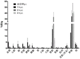
FIG. 3: the PSMA inhibitory ability and specific internalization values of the nuclide-labeled novel PSMA ligands shown in the table. Competitive cell binding was performed using the PSMA positive C4-2 cell line. The novel ligands have K in the low nanomolar range compared to PSMA-617iShows good inhibition and, in part, higher internalization values (specific lysate).
FIG. 4: A. display device64Cu-PSMA-617、64Cu-CA003 and64PET scans and time-activity curves of Cu-CA023 biodistributed C4-2 tumor-bearing mice.64Cu-PSMA-617 showed a high accumulation in tumors and in the kidney and liver. A.64Cu-PSMA-617、64Cu-CA003 and64biodistribution of Cu-CA023 was visualized at different time points (0 min to 20 min, 40 min to 60 min, 2h, 48 h). B. Display device64Cu-PSMA-617 and64dynamic PET scan (0 min to 60 min) time-activity curves of Cu-CA003 biodistributed C4-2 tumor-bearing mice. Standard Uptake Value (SUV) display of tumors and liver, in which64Uptake of Cu-PSMA-617 was higher than for tumors, an64Cu-CA003 is the opposite.
FIG. 5: in PET images of C4-2 tumor-bearing mice at different time points (0h to 48h)64Cu-PSMA-617、64Cu-CA003 and64maximum standard uptake value (mSUV) for Cu-CA 023.64Cu-CA003 showed high accumulation in tumor and kidney,64Cu-CA023 shows high accumulation in tumors and rapid clearance from the kidneys.
FIG. 6A: accumulation in the organ of interest dissected from C4-2 tumor-bearing mice64Cu-CA003 demonstrated tumor specificity and rapid clearance from the kidney.
FIG. 6B: accumulation in the organ of interest dissected from C4-2 tumor-bearing mice64Cu-CA023 demonstrated tumor specificity and rapid clearance from the kidney.
FIG. 6C: at the following time points, 0.025 nanomolar64Organ distribution of Cu-CA 003: 10 min, 1h, 4h, 24h and 72h after injection. Values are expressed as% ID/g tissue. + -. standard deviation; n-3 for all tissues.
FIG. 6D: in blocking experiments (B), radiotracers64Cu-CA003(0.030 nanomole) was injected simultaneously with 2mg PSMA-617 per kg body weight. Values are expressed as% ID/g tissue. + -. standard deviation; n-3 for all tissues.
FIG. 7: 2 hours (A) and 20 hours (B) after patient injection64Cu-CA003(200MBq, 0.5 nanomolar) PET/CT maximum intensity projection. Red arrows point to selected right shoulder soft tissue infiltrates from scapular origin, with contrast increasing for lung, bone and lymph node metastases over time. Liver and gall clearance leads to hot spots in the intestine; cross-sectional slices (C) must be provided to avoid false positive readings.
FIG. 8: (A) differences 1h after tail vein injection in BALB/C nu/nu C4-2 tumor-bearing mice203Planar scintigraphic imaging of a Pb-labelled Compound and (B)203Distribution time course of Pb-CA 012. Radiolabeled derivatives of CA009 and CA012 showed high uptake of the tracer in tumor tissue. Measured by203The kinetics of Pb-CA012 uptake showed a longer retention time of the radiotracer in the tumor tissue. When in contact with203When the Pb-CA009 is compared with the Pb-CA009,203the selectivity of Pb-CA012 uptake is enhanced. This is a result of rapid clearance of non-target organs.
FIG. 9:203organ distribution of Pb-PSMA-CA012 in tumor-bearing mice, 0.025 nanomolar203Pb-PSMA-CA 012. This quantification confirmed the results of the imaging experiment. The high rate of tumor to kidney uptake is due to the higher excretion value of the kidney.
FIG. 10:203geometric mean value image (A) of Pb-CA012 planar scanning with time change and its use177Treatment scan (B) comparison with Lu-PSMA-617; both obtained using a dielectric energy collimator。
FIG. 11: diagnosis based on male-adult model in OLINDA203Pb-CA012 (left column) and treatment212Safety dosimetry estimates for Pb-CA012 (right column) (ULI upper large intestine and LLI lower large intestine).
FIG. 12: for salivary glands, randomly selected tumor lesions (spherical models) and putative dose limiting organs212Pb-CA012 ("TCMC" -PSMA-617) dosimetry and213Bi-PSMA-617 and225comparison of Ac-PSMA-617.
FIG. 13A: multiple lymph node prostate cancer metastases 1h and 3h after injection68Maximum intensity projection of Ga-PSMA-CA028 PET scan. Cross-sectional sections showing axillary and pulmonary (C) lymph node metastases (indicated by red arrows); it can also be depicted on the relevant CT as a reference standard.
FIG. 13B: 295MBq/20 nanomolar injection68Maximum intensity projection of PSMA-PET performed 1h (A) and 3h (B) after Ga-CA 030. Arrows point to cross-sectional slice locations showing bone metastases in multiple regions of the axial skeleton (C to E). In ct (f), no typical osteoblastic response can delineate the tumor contour, with only morphological information.
FIG. 14: at 1h, 2h and 4h after injection68Organ distribution of Ga-PSMA-CA028 expressed as% ID/g tissue ± SD (n ═ 3).
FIG. 15: in BALB/C nu/nu mice bearing C4-2 tumor xenografts, selection was made68Global small animal PET imaging of Ga-PSMA ligand 2h after injection (A) and68time courses (B) and (B) of Ga-CA02868Comparison of the time course (C) of Ga-CA 030.
FIG. 16: measurement by radiation-ITLC method177Lu-CA028、177Lu-CA029、177Lu-CA030 with177Serum stability of Lu-PSMA-617 at 37 ℃ for 72h (mean ± SD, n ═ 3).
FIG. 17: measurement by radiation-ITLC method64Cu-CA003、64Cu-CA005 and64serum stability of Cu-PSMA-617 at 37 ℃ for 72h (mean ± SD, n ═ 4).
FIG. 18: by activity measurement assay64Cu-CA003、64Cu-CA005 and64serum stability of Cu-PSMA-617 at 37 ℃ for 72h (mean ± SD, n ═ 4).
FIG. 19:64in vivo metabolite analysis of Cu-CA003 p.i. in BALB/c nude mice (no tumor) for 10 min. radio-HPLC chromatograms of extracts from kidney, blood and liver show that activity elutes at the retention time of the intact tracer. This demonstrates the integrity of the copper complex over the major distribution period.
FIG. 20: BALB/c nude mice (tumor-free) in the liver at 10 min p.i.64radio-HPLC chromatogram of Cu-CA003 extract and in liver64Comparison of Cu-chloride.
FIG. 21A: whole body mouse PET scans of BALB/C nu/nu mice bearing C4-2 tumor xenografts were taken as maximum intensity projections.64Cu-PSMA-617(10MBq, 0.2 nanomole),64PET imaging of Cu-PSMA-CA003(10MBq, 0.2 nanomolar).
FIG. 21B:64Cu-PSMA-CA003(5MBq, 0.030 nanomolar) with excess unlabeled PSMA-617(2 mg/kg body weight) and64cu-chloride (10MBq) was co-injected. The color bar gives the correlation between the SUV and PET image gradations, 0-min and 4-max.
FIG. 22: whole body mouse PET scans of BALB/C nu/nu mice bearing C4-2 tumor xenografts were compared as maximum intensity projections. By using68 PET imaging 2h after injection of Ga (20MBq, 0.2 nanomolar) radiolabelled four novel PSMA ligands (A),68time course of Ga-CA028(B) and68time course of Ga-CA030 (C). The color bar gives the correlation between SUV and PET image gradations, 0-min and 4E 0-max.
FIG. 23:68ga PSMA-CA027(0.6 nanomolar, 5MBq) and68blood time-activity curves of Ga PSMA-CA028(0.6 nanomolar, 6MBq), including a two-exponential curve fit.
FIG. 24: 1h p.i. in BALB/c nude mice (tumors)177Lu-CA028 and177Lu-PSMA-617(10MBq, 0.2 nanomolar in about 100. mu.l of 0.9% saline) for in vivo metabolite analysis comparison. From the kidney,radio-HPLC chromatograms of extracts of blood, liver and tumor showed that activity eluted at retention time of the intact tracer. This demonstrates the integrity of the complex over the major distribution period.
FIG. 25: 20 min, 1h, 2h and 4h, 0.05 nanomolar after injection68Organ distribution of Ga-CA028, expressed as% ID/g tissue ± SD (n ═ 3).
FIG. 26: by using64radio-HPLC chromatogram of Cu-labelled novel compounds.
FIG. 27 is a schematic view showing: by using68Time-activity curves for Ga-labeled novel PSMA ligands. Time-activity curve of kidney (A) and time-activity curve of tumor (B) within 1h after injection. Data are mean normalized uptake values (SUV mean).
FIG. 28: (A) 9MBq (0.30 nanomolar) injection in female Swiss mice64PET images after Cu-CA 00310 minutes. The Maximum Intensity Projection (MIP) represents circulation in the blood and uptake by the kidneys. (B) Injection of 10MBq in female Swiss mice64PET images after C10 min p.i. The Maximum Intensity Projection (MIP) indicates strong uptake in the liver and kidney.
The following examples are intended only to illustrate the invention. They should not be construed as limiting the scope of the invention in any way.
Example (b):
materials and methods
Solvents and chemicals were purchased from Merck (darmstadt, germany) and Sigma-Aldrich (munich, germany) and used without further purification. In vitro experiments were performed in triplicate, and at least three independent sets of data were obtained for each experiment. PET imaging of prostate cancer patients was approved by the university of hedburg hospital according to german law and was approved by the declaration of helsinki (permit S321/2012).
Synthesis of chelator moieties
The chelator moiety was synthesized in high yield and characterized by LC-MS. Chelating agent bifunctional macrocyclic cyclylamine analogue 4- [ (1,4,8, 11-tetraazacyclotetradecylamine-1-yl) -methyl]The synthesis of benzoic acid is described by Studer and Kaden (Studer M, and Kadan, T.A. one-step synthesis of mono-N-substitated azamacrocycles)with a carboxylic group in the side-chain and their complexes with Cu2+and Ni2+Helvetica.1986; 2081-2086), and the crosslinking bridge chelator 4-carboxymethyl-11- (1, 3-dicarboxypropyl) -1,4,8, 11-tetraazabicyclo [6.6.2 ]]Hexadecane-glutaric acid was reported by Boswell et al (Boswell CA, Regino CA, Baiduo KE et al, Synthesis of a cross-branched cycle derivative for peptide coupling and64Cu radiolabeling.Bioconjug Chem.2008;19:1476-1484)。
I. the general steps are as follows: synthesis of novel PSMA ligands
PSMA-binding motifs were prepared by solid phase synthesis on 2-chlorotrityl resins (2 CT-resins), such as Eder et al (Eder M, m, Bauder-W ust U, et al,68ga-complex reactivity and the targeting property of a urea-based PSMA inhibitor for PET imaging. bioconjugation chem.2012; 23:688-697) and
 et al (benisova M,
 m, Bauder-Wust U, et al, preliminary Evaluation of a Tailor-Made DOTA-jointed PSMA Inhibitor with Optimized Linker mobility for Imaging and Endoradiaotherapy of State cancer. J Nucl Med.2015; 56: 914-. See fig. 2. For this purpose, Fmoc-Lys (alloc) -OH was immobilized on an equimolar amount of 2-chlorotrityl resin. Triphosgene was then used to generate glutamyl-partial isocyanate (2). Epsilon-allyloxycarbonyl protected lysine immobilized on 2-chlorotrityl resin was added and the reaction was stirred carefully for 16h to give compound (3). The resin was filtered off and the allyloxycarbonyl protecting group was cleaved to give (4). To obtain compounds CA001 and CA027, the corresponding chelators were coupled to this intermediate. Subsequently, the chelating agent-conjugated PSMA was cleaved from the resin. Alternatively, coupling of Fmoc-2-naphthylalanine was performed toTo obtain (5). To obtain the compounds CA002, CA005, CA008 and CA011, the corresponding chelating agents were coupled to this intermediate. Subsequently, the chelating agent-conjugated PSMA was cleaved from the resin. Alternatively, trans-4- (Fmoc-aminomethyl) cyclohexanecarboxylic acid was coupled to obtain (6), and the corresponding chelating agents were coupled to this compound to obtain compounds CA003, CA006, CA009, CA012, CA022, CA023, CA024, CA025, CA026, CA028, CA029, and CA 030. Subsequently, the chelating agent-conjugated PSMA was cleaved from the resin. The structure was confirmed by HPLC and MS-LC. Materials were separated by preparative HPLC using a water-acetonitrile gradient containing trifluoroacetic acid. For this purpose, the compound was purified using a gradient of 20% to 50% acetonitrile in water for 15 minutes. The purified compound was analyzed by analytical HPLC, where the compound was treated in aqueous acetonitrile (0% to 100%) containing trifluoroacetic acid for 5 minutes, using a 100 x 3mm Monolith RP HPLC column and LC/MS method. The product fractions were combined and lyophilized.
Ligands for copper isotope imaging and therapy
(CA001) description
By mixing the resin (Compound 4) with 1.5 equivalents of CTPA-NHS-ester (4- [ (1,4,8, 11-tetraazacyclotetradecan-1-yl) -methyl) in 500. mu.l of DMF]Benzoic acid) and 10 equivalents of DIPEA were incubated together to obtain the product. The compound was purified and the final product was analyzed by HPLC as described above (see section I). HPLC-retention time: 1.68 minutes; ESI-MS (M/z) < M + H]+(C30H50N7O8Calculated value) 636.37(636.36),

chemical structure of chelating agent CTPA-NHS-ester, used for the synthesis of compounds of CA001, CA002 and CA 003.
Description of CA002
By mixing the resin (Compound 5) with 1.5 equivalents of CTPA-NHS-ester (4- [ (1,4,8, 11-tetraazacyclotetradecan-1-yl) -methyl) in 500. mu.l of DMF]Benzoic acid) and 10 equivalents of Diisopropylamine (DIPEA) to obtain the productA compound (I) is provided. The compound was purified and the final product was analyzed by HPLC as described above (see section I). HPLC-retention time: 2.39 minutes; ESI-MS (M/z) < M + H]+C43H61N8O9Calculated value) 833.42 (833.45).
Description of CA003
By mixing the resin (Compound 6) with 1.5 equivalents of CTPA-NHS-ester (4- [ (1,4,8, 11-tetraazacyclotetradecan-1-yl) -methyl) in 500. mu.l of Dimethylformamide (DMF)]Benzoic acid) and 10 equivalents of DIPEA were incubated together to obtain the product. The compound was purified and the final product was analyzed by HPLC as described above (see section I). HPLC-retention time: 2.50 minutes; ESI-MS (M/z) < M + H]+(C51H74N9O10Calculated value) 972.52 (972.55).
Description of CA005
By mixing the resin (Compound 5) with 1.5 equivalents of the cross-linking bridge-TE 2A chelating agent, 0.98 xn in 500. mu.l of DMFChelating agentsHBTU was incubated with 10 equivalents of DIPEA to obtain the product. The compound was purified and the final product was analyzed by HPLC as described above (see section I). HPLC-retention time: 2.38 minutes; ESI-MS (M/z) < M + H]+(C44H64N8O13Calculated value) 913.45(913.47),

the chemical structure of chelating agent 8-carboxymethyl-cross-linking bridge-TE 2A, was used to synthesize compounds of CA005 and CA 006.
Description of CA006
By mixing the resin (Compound 6) with 1.5 equivalents of the cross-linking bridge-TE 2A chelating agent, 0.98 xn in 500. mu.l of DMFChelating agentsHBTU was incubated with 10 equivalents of DIPEA to obtain the product. The compound was purified and the final product was analyzed by HPLC as described above (see section I). HPLC-retention time: 2.55 minutes; ESI-MS (M/z) < M + H]+(C52H78N9O14Calculated values): 1052.62(1052.56).
Description of CA022
The product was obtained by incubating the resin (compound 6) with 1.5 equivalents of the cross-linking bridge-CTPA chelator and 10 equivalents of DIPEA in 500 μ l of DMF. The compound was purified and the final product was analyzed by HPLC as described above (see section I). HPLC-retention time: 2.72 minutes; ESI-MS (M/z) < M + H]+(C53H76N9O10Calculated values): 998.56(998.57),

chemical structure of chelator cross-bridged-CTPA, used for the synthesis of compounds of CA 022.
Description of CA023
The product was obtained by incubating the resin (compound 6) with 1.5 equivalents of 8-carboxymethyl-CTPA chelator and 10 equivalents of DIPEA in 500. mu.l of DMF. The compound was purified and the final product was analyzed by HPLC as described above (see section I). HPLC-retention time: 2.54 minutes; ESI-MS (M/z) < M + H]+(C53H76N9O12Calculated values): 1030.55(1030.56),

chemical structure of chelating agent 8-carboxymethyl-CTPA, used for the synthesis of compounds of CA 023.
Description of CA024
The product was obtained by incubating the resin (compound 6) with 1.5 equivalents of 8-carboxymethyl-cross-linking bridge-CTPA chelating agent and 10 equivalents of DIPEA in 500 μ l of DMF. The compound was purified and the final product was analyzed by HPLC as described above (see section I). HPLC-retention time: 2.60 minutes; ESI-MS (M/z) < M + H]+(C55H78N9O12Calculated values): 1056.56(1056.57)

Chemical structure of chelator 8-carboxymethyl-cross-linking bridge-CTPA, compound used for the synthesis of CA 024.
Description of CA025
By mixing the resin (compound 6) with 1.5 equivalents of the 8, 11-bis (carboxymethyl) -CTPA chelator [ CPTA ═ 4- [ (1,4,8, 11-tetraazacyclotetradecan-1-yl) methyl group in 500 μ l of DMF]Benzoic acid]And 10 equivalents of DIPEA were incubated together to obtain the product. The compound was purified and the final product was analyzed by HPLC as described above (see section I). HPLC-retention time: 2.60 minutes; ESI-MS (M/z) < M + H]+(C55H78N9O14Calculated values): 1088.55(1088.56),

chemical structure of chelating agent 8, 11-bis (carboxymethyl) -CTPA, used for the synthesis of CA025 compounds.
Description of CA026
The product was obtained by incubating the resin (compound 6) with 1.5 equivalents of 8, 11-bis (carboxymethyl) -CTPA chelating agent and 10 equivalents of DIPEA in 500 μ l of DMF. The compound was purified and the final product was analyzed by HPLC as described above (see section I). HPLC-retention time: 2.53 minutes; ESI-MS (M/z) < M + H]+(C57H80N9O16Calculated values): 1146.56(1146.57),

chemical structure of chelating agent 4,8, 11-tris (carboxymethyl) -CTPA, compound for the synthesis of CA 026.
Lead isotopes for alpha therapy (a)203Pb/212PSMA ligands of Pb)
Description of CA007
By mixing the resin (Compound 5) with 1.5 equivalents in 500. mu.l of DMFp-SCN-Bn-TCMC chelating agent [ TCMC ═ 1,4,7, 10-tetraaza-1, 4,7, 10-tetrakis (2-carbamoylmethyl) cyclododecane]And 10 equivalents of DIPEA were incubated together to obtain the product. The compound was purified and the final product was analyzed by HPLC as described above (see section I). HPLC-retention time: 2.41 minutes; ESI-MS (M/z) < M + H]+(C49H70N13O12Calculated S): 1064.49(1064.50),

chemical structure of chelating agent p-SCN-Bn-TCMC for the synthesis of compounds of CA007, CA008 and CA 009.
Description of CA009
The product was obtained by incubating the resin (compound 6) with 1.5 equivalents of p-SCN-Bn-TCMC chelating agent and 10 equivalents of DIPEA in 500. mu.l of DMF. The compound was purified and the final product was analyzed by HPLC as described above (see section I). ESI-MS (M/z) < M + H]+(C57H83N14O13Calculated S): 1203.59(1203.60).
Description of CA011
By mixing the resin (Compound 5) with 1.5 equivalents of 2- (4,7, 10-tris (2-amino-2-oxoethyl) -1,4,7, 10-tetraazacyclododecan-1-yl) acetic acid, the chelating agent 1,4,7, 10-tetraazacyclododecan-1, 4,7, 10-tetraacetamide (DO3AM) monocarboxylate derivative, 0.98 xnChelating agentsHBTU was incubated with 10 equivalents of DIPEA to obtain the product. The compound was purified and the final product was analyzed by HPLC as described above (see section I). HPLC-retention time: 2.07 minutes; ESI-MS (M/z) < M + H]+(C41H62N11O12Calculated values): 900.45(900.46),

chemical structure of chelating agent
Monocarboxylic ester derivatives of 2- (4,7, 10-tris (2-amino-2-oxoethyl) -1,4,7, 10-tetraazacyclododecan-1-yl) acetic acid, chelating agent 1,4,7, 10-tetraazacyclododecan-1, 4,7, 10-tetraazacyclododecane (DO3AM), compounds for the synthesis of CA010, CA011 and CA 012.
Description of CA012
By mixing the resin (Compound 6) with 1.5 equivalents of DO3AM chelating agent, 0.98 xn in 500. mu.l of DMFChelating agentsHBTU was incubated with 10 equivalents of DIPEA to obtain the product. The compound was purified and the final product was analyzed by HPLC as described above (see section I). HPLC-retention time: 2.21 minutes; [ M + H ]]+(C49H75N12O13Calculated values): 1039.54(1039.56).
Chelator spacer moieties to enhance the pharmacokinetic properties of PSMA-617
Description of CA027
The product was obtained by incubating the resin (Compound 4) with 1.5 equivalents of p-NHS-Bn-DOTA chelating agent and 10 equivalents of DIPEA in 500. mu.l DMF. The compound was purified and the final product was analyzed by HPLC as described above (see section I). HPLC-retention time: 1.47 minutes; [ M + H ]]+(C34H52N7O14Calculated values): 782.33(782.36),

chemical structure of p-NHS ester-Bn-DOTA, chelating agents for the synthesis of CA027 and CA 028.
Description of CA028
The product was obtained by incubating the resin (Compound 6) with 1.5 equivalents of p-NHS-Bn-DOTA chelator and 10 equivalents of DIPEA in 500. mu.l of DMF. The compound was purified and the final product was analyzed by HPLC as described above (see section I). HPLC-retention time: 2.36 minutes; [ M + H ]]+(C55H76N9O16Calculated values): 1119.53(1118.54).
Description of CA029
By mixing the trees in 500. mu.l of DMFThe lipid (compound 6) was incubated with 1.5 equivalents of p-SCN-Bn-DOTA chelator and 10 equivalents of DIPEA to obtain the product. The compound was purified and the final product was analyzed by HPLC as described above (see section I). HPLC-retention time: 2.49 minutes; [ M + H ]]+(C57H79N10O17Calculated S): 1207.52(1207.53),

chemical structure of chelating agent p-SCN-Bn-DOTA for the synthesis of compounds of CA 029.
Description of CA030
The product was obtained by incubating the resin (compound 6) with 1.5 equivalents of p-NCS-benzyl-DOTA-GA chelating agent and 10 equivalents of DIPEA in 500. mu.l of DMF. The compound was purified and the final product was analyzed by HPLC as described above (see section I). HPLC-retention time: 2.50 minutes; [ M + H ]]+(C60H84N11O18Calculated S): 1278.56(1278.57),

chemical structure of chelating agent p-NCS-benzyl-DOTA-GA for the synthesis of CA030 compounds.
Synthesis of V radiolabelled complexes
V.1 64Radiochemical Synthesis of Cu-PSMA-derivatives
The conjugate (1mM in water, 5. mu.l, 5 nanomole) was added to 400. mu.l of sodium acetate buffer (0.4M in water, pH 5.0), 10. mu.l of ascorbic acid (20% in water), and 282. mu.l of64Cu]CuCl2(in 0.1M HCl (200 MBq)). The mixture was heated at 95 ℃ for 5 minutes. The labeling was controlled by radio-HPLC (0% to 100% MeCN for 5 min, Monolith column) with a flow rate of 2 ml/min and a retention time of 2.3 min.
This labelling resulted in radiolabel yields within 10 minutes>98%(as shown in the radiochromatogram of FIG. 26). For example,64the specific activity of Cu-PSMA-CA003 was about 40 Megabeoka/nanomolar. The same protocol was used for67Labeling of Cu.
V.2 203/212Radiochemical synthesis of Pb-PSMA-ligands
80 nanomolar conjugates (1mM in water, 80. mu.l, 80 nanomolar) were added to 400. mu.l sodium acetate buffer (0.4M in water, pH 5.0), 10. mu.l ascorbic acid (20% in water) and 140. mu.l203Pb-chloride solution (0.04M HCl) with a specific activity of about 102.6 TBq. (Lanthenus Medical Imaging, USA). The mixture was then heated at 95 ℃ for 5 minutes. Labeling was controlled by radio-HPLC.
V.3 68Radiochemical Synthesis of Ga-PSMA-CA028(CA027, CA029, CA030)
From68Elution in Ge/Ga Generator (iThemba LABS, south Africa)68Ga. Conjugate (1mM in DMSO, 20. mu.l, 20 nanomolar) was added to 320. mu.l sodium acetate buffer (0.4M in water, pH 4 to 5), 10. mu.l ascorbic acid (20% in water) and 400MBq68Ga (0.6M HCl). The mixture was heated at 95 ℃ for 5 minutes. The labeling was controlled by radio-HPLC (0% to 100% MeCN for 5 min, Monolith column) with a flow rate of 2 ml/min and a retention time of 2.4 min.
Analytical data for selected novel ligands

V.4 177Radiochemical Synthesis of Lu-PSMA-CA028(CA027, CA029, CA030)
For the177Lu labeling, about 20MBq was mixed with 200 μ l of Chelex-containing 0.4M sodium acetate buffer (pH 5). Mu.l of a 1mM compound solution in a 10% DMSO aqueous solution, 2. mu.l of an ascorbic acid saturated solution, and 40. mu.l of177Lu]LuCl3The solution was mixed and heated to 95 ℃ and held for 10 minutes. The labeling was checked by radio-HPLC (0% to 100% ACN in water for 5 min, Monolith column).
Preclinical assessment
In vitro and in vivo experiments were performed using the PSMA positive C4-2 cell line, a subline of the LNCaP (prostate lymph node carcinoma) cell line (CRL-3314; American Type Culture Collection). C4-2 cells were cultured in RPMI 1640(PAN Biotech) medium supplemented with 10% fetal bovine serum and stable glutamine (PAN Biotech). Cells were grown at 37 ℃ and incubated with 5% CO2Equilibrated incubation with humid air.
VI.1 in vitro
VI.1.1 competitive binding assays and internalization rates
MultiScreen was paired with 100. mu.l PBS containing 1% BSA per well at room temperatureHTSDV Filter plates were incubated for 30 minutes. After removal of PBS/BSA solution, 1X 10 was added to each well of Opti-MEM5And C4-2 cells. Using 0.75nM68Ga-labeled PSMA-HBED-CC dimer (68Ga-PSMA-10)( M,Bauder-Wüst U,Leotta K,et al.A dimerized urea-based inhibitor of the prostate-specific membrane antigen for 68Ga-PET imaging of state cancer. ejnmi research.2012; 2:23-23.) as standard to determine the inhibitory potency of the synthesized compounds. All unlabelled compounds were dissolved in Opti-MEM at the following concentrations in a volume of 300. mu.l: 0nM, 0.5nM, 1nM, 2.5nM, 5nM, 10nM, 25nM, 50nM, 100nM, 500nM, 1000nM and 5000 nM. Then, 3. mu.l of radiolabeled compound was added. 50 μ l of this mixture was taken to obtain a concentration of 0.75nM of radiolabeled ligand. After 45 min incubation at 37 ℃, cells were washed twice with PBS on a multi-screen vacuum manifold (Millipore, Billerica, ma) and cell-bound radioactivity was measured with a gamma counter (Packard Cobra II, GMI, mn, usa). Use of68Ga-labeled PSMA-HBED-CC dimers (e.g., PSMA-11) are used as a reference to determine the inhibitory potency. Ki was calculated using a non-linear regression algorithm (Graph Pad Prism 5.01 software). The experiment was performed four times.
For the determination of a specific internalization rate, 0.1% in PBS was used at room temperatureThe 24-well plate was incubated for 20 minutes and washed once with PBS. In the next step, a solution containing 1X 10 of the active compound was added to each well51ml of RPMI medium containing C4-2 cells and cultured overnight. The conditions for each compound during the experiment were: incubations were carried out at 37 ℃ or 4 ℃ with or without receptor blockade by 2- (phosphonomethyl) glutaric acid (2-PMPA; Axxora) at a final concentration of 500. mu.M. Then, the cells were incubated with 250. mu.l of a 30nM solution of the labeling compound. The plates were incubated for 45 minutes in a water bath at 37 ℃ or on ice at 4 ℃. Then, cells were washed 3 times with 1mL ice cold PBS and incubated with glycine (50 mM in HCl, pH 2.8) for 5 minutes. After an additional wash step with 1mL ice-cold PBS, cells were lysed with 0.5mL 0.3M NaOH, collected and radioactivity measured with a gamma counter for 1 min. By subtracting the corresponding uptake under blocking conditions to bind to 106Individual cell (% IA/10)6Individual cells) the percentage of radioactivity initially added determines the specific cellular uptake. All experiments were performed in triplicate.
The results are shown in FIG. 3. KiThe assay showed nanomolar binding affinity of the synthetic ligand for PSMA.
For the203Pb-labeled compounds, compounds CA009 and CA012 showed the highest affinity to inhibit PSMA. Furthermore, it is possible to provide a liquid crystal display device,203pb-labeled compounds CA009 and CA012 showed higher specific internalization rates in PSMA-positive cell lines. Compound (I)203Pb-CA009 and203Pb-CA012 showed the highest internalization rate of 28.36 + -2.23 and the injection activity of 7.33 + -1.26/106And C4-2 cells (n-3).
As shown in fig. 3, a particularly high affinity for PSMA was shown for Cu-labeled ligands, such as CA003, followed by CA006, CA002 and CA 026. Furthermore, it is possible to provide a liquid crystal display device,64cu-labeled compounds showed specific binding to C4-2 cells.64Cu-CA003 internalizes 34.63 +/-2.77%,64Cu-CA005 internalizes 18.63 +/-4.46%,64Cu-CA022 internalized, 38.7 ± 6.69% (n ═ 3). 106A single C4-2 cell was used for these experiments.
Further, for example, K of Ga-labeled ligandiThe measurement results are shown, for exampleNanomolar binding affinity of the synthetic ligand to PSMA. As shown in fig. 3, CA030 shows particularly advantageous properties among all the novel compounds.
VI.1.2 serum stability
VI.1.2.1 203Serum stability of Pb-labeled Compounds
The stability of the radiolabeled compounds was determined by incubation in 300 μ l human serum at 37 ℃ for 1h, 2h, 3h, 6h, 24h, 48h and 72 h. The serum was precipitated by adding 2 parts of acetonitrile. The samples were then vortexed and centrifuged at 13000rpm for 5 minutes (2 times), and the supernatants were analyzed by radio-HPLC (0% to 100% MeCN for 5 minutes, Monolith column).
All compounds showed high stability in human serum for at least 48 hours. CA011 and CA012 showed particularly advantageous stability, and were stable for at least 72 hours.
VI.1.2.2 68Ga、177Lu and64serum stability of Cu-labeled Compounds
Serum stability was determined by tlc and HPLC analysis after radiolabeling of the compounds. To 200. mu.L of human serum (H4522; Sigma-Aldrich, Germany) 50. mu.l (20MBq) of labeled ligand was added and incubated at 37 ℃ for various time points (0H, 2H, 24H, 48H and 72H). Paper was chromatographed using 0.5X 5cm iTLC-SG-glass microfiber chromatography (Folsom, Calif., USA). 0.5 μ L of radiolabeled compound in serum was applied to each band 1cm from the bottom (origin) and solvent (at177The front of the Lu ligand was raised from the bottom to 5cm by sodium citrate buffer (0.5M, pH 5.0) and (1% Na-EDTA, pH 4) in the case of Cu-labeled ligand. Finally, dividing each paper slip into 8 parts; each aliquot was measured in a gamma counter. For HPLC analysis, an equal volume of ACN was added to the sample to precipitate serum proteins. Then, the sample was centrifuged at 130000rpm for 10 minutes, the pellet and the supernatant were separated, and the relative activity was measured. The results are expressed as percentages. In addition, an aliquot of the supernatant was analyzed by radio-HPLC (0% to 100% ACN for 5 minutes, Monolith column) with a flow rate of 2 ml/min.
By using68Ga and177the results of the stability testing of the Lu radiolabeled compounds are shown in fig. 16. As shown by HPLC and by radio TLC,68ga-labelled compounds showed no degradation after 2h incubation in human serum. After 24 hours of incubation177Lu-CA028 showed 40% free177Lu, whereas CA029, CA030 and reference compound PSMA-617 showed no free activity at this time point (fig. 16).
64The results of the stability test for the Cu-labeled compound are shown in fig. 17 and 18. After 2h incubation, ITLC showed that all compounds only dissociated below 2 ± 0.6%. After 24h incubation, only 6. + -. 4% of64Cu-CA003 and 3 + -1%64Cu-CA006 dissociates. On the contrary, the present invention is not limited to the above-described embodiments,64Cu-PSMA-617 showed a freeness of 13. + -. 3%64And (4) Cu activity. Long-term stability test (72h) shows64Cu-CA006 (8. + -. 4%) has a chemical bond with64Cu-CA003 (11. + -. 3%) comparable stability. At this time point, 18. + -. 6% of64Cu-PSMA-617 dissociated. After incubation for 2h, the activity measurement in the pellet (FIG. 18) showed 19.0. + -. 5.2% of64Cu-CA003, 12 +/-7.2%64Cu-CA006 and 40 + -7.5%64Cu-PSMA-617 precipitated with the protein fraction. The percentage of activity in the precipitate increased with time. In that64The highest activity was measured in the precipitate of Cu-PSMA-617, followed by64Cu-CA003, and found64The Cu-CA006 precipitation activity was low.
VI.2 in vivo experiments
In vivo experiments were performed according to the law of the federal republic of germany. For PET imaging and biodistribution studies, 4-to 5-week old male nude mice (Balb/c nu/nu mice) (19-23 g) were obtained from Charles River and stored for 1 week in the absence of specific pathogens prior to the study. The rats were housed in a 12-hour/12-hour light/dark cycle and were freely accessible for water and food. Mice were anesthetized with 2% sevoflurane and then inoculated subcutaneously in the right flank with 5X 10 in 50% matrigel cultured in Opti-MEM I (1X) medium7And C4-2 cells. When the tumor size is about 1cm3At that time, organ distribution studies were performed.
Vi.2.1 lead-labeled compounds:
VI.2.1a scintigraphic imaging and biodistribution
For small animal imaging, mice were anesthetized with 2% sevoflurane. Injecting 0.1 nanomolar (1.0MBq) into tail vein203A Pb-ligand. After 10 minutes, 1h, 4h, 24h and 72h, continuous planar scans were performed using a gamma imager SCT (Biospace Lab, paris, france) with parallel collimators (35mm/1.8mm/0.2 mm). Based on the imaging results, compound CA012 was selected for biodistribution studies. The experiment was performed three times.
The results are given in fig. 8 and 9.
VI.2.2 64Cu-labelled compound:
VI.2.2a)64cu-chloride and64stability in blood and in vivo fate of Cu-CA003
Determination by ITLC and HPLC64In vivo stability of Cu-labeled CA 003. Tumor-free male BALB/c nude mice (n ═ 3) were injected via tail vein64Cu-CA003(3.6 MBq; 0.26 nanomolar, dissolved in a total volume of about 100. mu.l of 0.9% saline) and 800. mu.l of blood was collected 10 minutes after injection. Blood samples were centrifuged at 13000rpm for 10 minutes. Then, the pellet and the supernatant were separated, and the relative activity was measured. ITLC was performed to assess the stability of radiolabeled compounds in blood as described above. Furthermore, after addition of an equal volume of ACN and removal of protein by centrifugation, an aliquot of the supernatant was analyzed by radio-HPLC (0% to 100% ACN for 5 minutes, Monolith column) at a flow rate of 2 ml/min.
In vivo metabolism was studied by radio-HPLC analysis. Tumor-free female Swiss mice (n-3) were injected via tail vein64Cu-chloride (10MBq in about 100. mu.l of 0.9% saline) or64Cu-CA003(9MBq, 0.30 nanomolar in about 100. mu.l of 0.9% saline). PET imaging was performed 10 minutes after injection, and then blood, liver and kidney were collected. The tissue was rinsed with pre-cooled saline, blotted dry and washed with 2mL of 0.1M NH4OAc/EtOH (35: 65). The tissue was homogenized using an Ultra-Turrax T8(IKA Labortechnik, Germany). The sample was centrifuged at 13000rpm (4 ℃) for 10 minutes. Then theThe pellet and supernatant were separated and the relative activity was measured. The results are expressed as percentages. In addition, aliquots of the supernatant were prepared for HPLC measurements by precipitating the protein with ACN as described above. In addition, the samples were analyzed by radio-HPLC (0% to 100% ACN for 5 min, Monolith column) with a flow rate of 2 ml/min. During the entire chromatographic analysis, fractions were collected every 10 seconds and the relative activity of the samples was measured in a gamma counter to reconstruct the chromatogram.
The ITLC results in blood stability show that,64Cu-CA003 experienced 3%64Cu dissociation or 97 ± 2.3% of intact tracer (see fig. 19). The radiohplc chromatogram also confirmed the integrity of the copper complex (see fig. 20). To carry out64Cu-chloride or64The in vivo fate study of Cu-CA003,64cu-chloride and64PET imaging of Cu-CA003 showed different pharmacokinetics (fig. S21).64Maximum intensity projection PET imaging of Cu-chloride showed lower blood circulation (1.3), high liver uptake (2.7) and kidney (3.4) uptake. While64Cu-CA003 shows a ratio64Longer blood circulation of Cu-chloride (2.3), higher uptake by the kidney (5.7) and lower liver accumulation (1.0) (fig. 28). The radio HPLC chromatograms of the extracts of kidney, blood and liver tissue demonstrate64Integrity of Cu-CA003 (FIG. 19).64The chromatogram for Cu-CA003 showed that the retention time of the tracer was different from that of free copper64Cu-chloride (FIG. 20).
VI.2.2b) organ distribution experiment (64Cu ligand) and small animal PET
Based on PET imaging results, CA003 and CA023 were selected for biodistribution analysis using C4-2 tumor bearing mice. The experiment was performed three times. 0.025 nanomolar by tail vein injection64Cu-labelled compounds (1 MBq per mouse in about 100 μ l of 0.9% saline). At the time point: organs were dissected and weighed for 10 min, 1h, 4h, 24h and 72h, and activity was measured using a gamma counter (Packard Cobra Auto-gamma). The percentage of injected dose per gram (% ID/g) was calculated. (see FIG. 6A and FIG. 6B)
In addition, experiments in which PSMA-617 was administered at 1h (n ═ 3) simultaneously to block PSMA binding are shown (fig. 6C and 6D).
To make use of64Small animal PET imaging of Cu-labeled PSMA ligand, 0.2 nanomolar, 10MBq of approximately 100 μ l of a radiolabeled compound in 0.9% saline was injected into C4-2 tumor-bearing mice. Dynamic PET was recorded in a small animal PET scanner (Siemens Inveon D-PET, Malvern, Pa., USA). SUV values were obtained from conventional (non-dynamic) PET images. The formula for SUV is:

the volume of interest (VOI) is obtained by manually delineating appropriate whole tissues (heart, kidney, bladder, tumor, volume of about 100 μ l to 500 μ l) or parts of tissues liver and muscle. The image was reconstructed according to the procedure OSEM 3D/SP MAP, with 16 subsets, 2 iterations, image x-y size 256, image z size 161. The post-processing filter is not used to modify the data. The software for analyzing the images and TAC was Inveon from Siemens IRW 4.1TMAcquisition Workplace (IAW). Dynamic PET scans were performed 0 min to 60 min after injection, and images were reconstructed in three time frames of 20 min (0 min to 20 min, 20 min to 40 min, and 40 min to 60 min) for visual display. As shown in fig. 2/fig. 3 and table S1+ S2, for some compounds showing long retention times, the subsequent time points (2h, 4h, 20h, 45/48h) were included. After 1h, a static PET scan was performed. To compare different radiotracers, the average SUV over time is depicted.
FIGS. 6A to 6D show PSMA ligands64Biodistribution results obtained with Cu-CA003(n ═ 3). Uptake of 11.33 ± 4.11% ID/g tumor was observed 10 min after injection. After 4h, the amount of tracer accumulated in the tumor (32.34. + -. 10.6% ID/g) was much higher than in the kidney (13.33. + -. 3.36% ID/g). The time activity curves generated from dynamic PET imaging showed a tumor to muscle ratio of 10.5 and a tumor to blood ratio of 3.0 one hour after injection, see table vi.2.2b _1 below:
TABLE VI.2.2b _1 tumor-bearing xenografts C4-2In BALB/c nu/nu mice, from64Average normalized uptake value (mSUV) from time-activity curves obtained with Cu-CA003 small animal PET.

These curves indicate rapid uptake by the kidney. Organ distribution studies showed that high kidney uptake at 1h (67.04. + -. 20.89% ID/g) was mostly cleared within 24h (7.48. + -. 8.51% ID/g). In contrast, high tumor uptake values (30.83 ± 12.61% ID/g at 1h p.i.) remained almost unchanged (19.99 ± 6.43% ID/g at 24h p.i.). PET imaging confirmed the strong accumulation of the radiotracer in the tumor (fig. 4). After 1h of injection, the amount of radioactivity in the background organs, e.g. kidneys, decreased, while the tumor to background ratio increased. PET scans showed very high tumor uptake 24h after injection, which confirmed the enrichment in tumors.
The binding specificity with PSMA was confirmed by blocking experiments, co-injection of unlabeled PSMA-617[2mg/kg]Resulting in 1 hour after injection64Accumulation of Cu-CA003 was significantly reduced in C4-2 tumors (30.83 + -12.61% ID/g to 2.35 + -0.38% ID/g) and in the kidney (67.04 + -20.89% ID/g to 3.47 to 0.48% ID/g). With excess unlabeled PSMA64PET imaging of Cu-CA003 (FIGS. 6C/6D) clearly demonstrated the biodistribution results.
The time activity curves generated from dynamic PET imaging showed a tumor to muscle ratio of 10.5 and a tumor to blood ratio of 3.0 at 1h post injection (table S1). These curves indicate rapid uptake by the kidney. Organ distribution studies (FIG. 6A) showed that high kidney uptake at 1h (67.04. + -. 20.89% ID/g) was mostly cleared within 24h (7.48. + -. 8.51% ID/g). In contrast, high tumor uptake values (30.83 ± 12.61% ID/g at 1h p.i.) remained almost unchanged (19.99 ± 6.43% ID/g at 24h p.i.). PET imaging confirmed the strong accumulation of the radiotracer in the tumor (fig. 4). After 1h of injection, the amount of radioactivity in the background organs, e.g. kidneys, decreased, while the tumor to background ratio increased. PET scans showed very high tumor uptake 24h after injection, which confirmed the enrichment in tumors. High tumor accumulation was maintained at long time points, i.e. 45h after injection (fig. 4).
IV.2.2c)64Cu-PSMA-CA003 and64Cu-PSMA-617 and64in vivo comparison of Cu-chloride
To demonstrate the in vivo stability of the copper complex of PSMA-CA003, the following procedures were carried out64Cu-PSMA-CA003 and64Cu-PSMA-617 and64cu-chloride was compared (FIG. 20). The compounds were studied in a small animal PET study of C4-2 tumor xenografts. The results are shown in Table 3. From64The time activity curve obtained in dynamic PET of Cu-CA003 showed a high tumor to liver ratio (4.0) after 1h of injection, whereas for64Cu-PSMA-617 with a tumor to liver ratio of 0.37 (FIG. 20/21 and Table S1/S2).
TABLE S2 in BALB/C nu/nu mice carrying C4-2 tumor xenografts, from64Average normalized uptake value (mSUV) from time-activity curves obtained for Cu-PSMA-617 small animal PET.

To prove that the substance entering the tumor is actually64Cu-CA003 rather than free64Cu, in C4-2 tumor-bearing mice64After Cu-chloride PET imaging (fig. 20/21), corresponding tissues were homogenized, extracted and then analyzed by HPLC. Observed64The pharmacokinetics of Cu-chloride are different from that of64Cu-CA003。64PET imaging of Cu-chloride in the maximum intensity projection showed an increase in tumor uptake within 2h after injection. And64in contrast to Cu-CA003,64cu-chloride showed very high liver accumulation (fig. 20/21).64The tumor to liver ratio of Cu-chloride at 2h was 0.38, while64The tumor to liver ratio of Cu-CA003 was 6.3.
1h after injection, from68Time activity curves generated by Ga-CA028 dynamic PET imaging and mean SUV body weight values showed a tumor to kidney ratio of 0.78. At 2h, the ratio increased to 3.0 (FIG. 15 and Table S1.1), while the ligand68Ga-CA030 and 03068Tumor to kidney ratios of 0.52 and 0.33 for Ga-CA029 (fig. 3, 25 and 23), respectively.
TABLE S1.1 in BALB/C nu/nu mice bearing C4-2 tumor xenografts, from68Average normalized uptake value (mSUV) from time-activity curves obtained for small animal PET of Ga-PSMA-CA 028.

The time-activity curve shows rapid clearance of the tracer — -68Ga-CA028 shows high tumor accumulation and high renal values. On the contrary, for68Ga-CA027, found faster renal clearance upon tumor accumulation (FIG. 22). Even if68Ga-CA030 representing ratio68Higher renal uptake values of Ga-CA028, but it showed the highest tumor uptake in all compounds (fig. 15C). The PET image of the small animal shows68Ga-CA027 very fast renal clearance and low tumor accumulation. Evidently, the radiotracer68Ga-CA028、68Ga-CA029 and68Ga-CA030 showed high tumor accumulation (fig. 27B).68Ga-CA029 exhibits the highest renal uptake, followed by68Ga-CA030 (FIG. 27A).
In vivo study of first patient
VII.1 utilization of64PET of Cu-PSMA-CA003
PET imaging of prostate cancer patients shown in figure 7 was agreed by the university of heidelberg hospital, following german law, and acknowledging the declaration of helsinki (permit S321/2012).
The first human test is with 200MBq64Cu-PSMA-CA 003. First PET imaging as shown in figure 7, shows imaging of patients with high levels of prostate specific membrane antigen. It is clear that the patient has metastatic PCa, particularly by multiple lymph node metastasis, with the main tumor located in the right shoulder.
Significant improvements in pharmacokinetics and tumor targeting of the copper isotope were observed after use of the novel copper ligand. Although containing64PSMA-6 of Cu17 higher in vitro labeling rate: (>99%) but poor in vivo stability was observed with high hepatic uptake (Cui C, Hanyu M, Hatori A, et al Synthesis and evaluation of [ (64) Cu)]PSMA-617targeted for prostate-specific membrane antigen in prostate cancer.Am J Nucl Med Mol Imaging.2017;7:40-52)。
Novel64Cu-labeled PSMA ligands are promising drugs for targeting PSMA and imaging PSMA-positive tumor lesions, as shown by small animal PET studies, organ distribution, and preclinical assessment of first human application.
64Patient imaging of the Cu-PSMA-CA003 ligand indicated successful transformation in clinical studies.
VII.2 utilization of203Experiment with Pb-CA012
Two castration-resistant metastatic prostate cancer patients were injected with 258MBq and 310MBq, respectively203Planar whole body scans were taken 0.4h, 4h, 18h, 28h and 42h after Pb-CA012 (GE hawkeymillnenium, 1"crystal, ME-collimator, 279keV peaks +/-10%, 8 cm/min). Images were loaded into the QDOSE dosimetry software suite (ABX-CRO, dereston) and registered. Kidney, liver, spleen, bladder, salivary glands (left and right parotid and submandibular glands) and several neoplastic lesions as well as a whole body region of interest (ROI) were segmented at the most appropriate time points using the maximum threshold percentage of organ-related (15% to 65%) and propagated to all other time points, performing an additional organ-based automatic rigid registration step. These ROIs were used to determine time-activity curves (TAC) for each organ, tumor and whole body. The first time point (pre-void) of the uncorrected geometric mean image was used to calibrate the ROI count to injection activity (MBq). Red bone Marrow TAC [ Shen, s., measure, r.f., Duan, j., Macey, d.j., Khazaeli, m.b., Robert, f., LoBuglio, a.f.: Improved Prediction of myelooxidicity Using a Patient-specific imaging Dose for Non-Marrow-Targeting 90Y-Antibody therapy.j nuclear Med,43:1245-1253,2002.Sgouros, g.bone Marrow dosometry for radiation therapy: the clinical constraints.j.j. J., 34: 6949, 1993) was calculated from venous blood (6 samples/Patient) Using established model assumptions.]。
From203All TAC obtained in Pb data were recalculated using the displaced nuclide function of QDOSE, which automatically corrects all time points of the source isotope physical decay, retaining only its biological clearance; physical decay of the replacement radionuclide is then applied.
Double exponential curve fitting is applicable to all organ TAC (except for a few tumors and glands, which must be fitted in a single exponential fashion). Assuming a linear increase from 0 to the first measurement time point, the values from the first measurement time point to the last measurement time point are integrated using a trapezoidal approximation and a fitting function from the last measurement time point to infinity. The remaining body part is calculated by subtracting all source organs from the whole body. In OLINDA 1.1[ Stabin, M.G., Sparks, R.B., crown, E.OLINDA/EXM: the second-generation personal computer software for internal patient assessment in nuclear medicine. J.Nucl. Med.46 (6):1023 1027,2005], the organ mass of the male-adult-phantom was used to derive the residence time of the kidney, liver, spleen, bladder contents, red bone marrow and remaining body parts for dose calculation.
Potential therapeutic nuclide212Further decay of Pb to212Bi、212Po and208tl. Assuming that the daughter nuclide remains at the decay point of the parent nuclide and212transient equilibrium between Pb and its daughter nuclide, application of and to daughter nuclide212Pb the same residence time. The OLINDA calculation was performed separately for all nuclides. According to the branch ratio, use212The Po is 64.07 percent,208Tl is a weight factor of 35.93%, and the respective decay steps are summed. According to the recommendations of the medical radiation dose Committee and U.S. department of energy [ Sgouros G, Roeske JC, McDeitt MR et al, MIRD pampthlet No.22 (bridge): radiology and pathology of alpha-particulate matters for targeted radiology. J Nucl Med.2010; 311-28. Feinendegen LE, McClure JJ. meeting report alpha-entities for medical therapy-work of the United States Department of Energy, Denver, Colorado, may 30-31, 1996. radiation Res.1997; 148:195-201.]To makeConverting the physical absorbed dose into an equivalent dose with a weight factor of 5 for alpha, a weight factor of 1 for beta and photon radiation; thus, reflecting the relative biological efficacy with respect to deterministic radiation effects-is considered to be the dominant factor in the treatment environment. Tumor and salivary gland volumes were measured separately based on CT segmentation and their Absorbed doses [ Stabin, M.G., Konijnenberg, M.Re-evaluation of adsorbed Fractions for Photons and Electrons in Small spheres. J Nucl Med,41:149-160,2000 ] were estimated by power functions estimated using an interpolated spherical model.]。
Of a patient203Pb imaging data
When the injection activity is 258MBq-310MBq, the injection activity is found to be203279keV gamma-rays emitted by Pb are sufficient to obtain a clear plan scan at 80% abundance probability (fig. 10). These scans show that the tracer labelled with lead isotopes is specifically enriched in the target tissue. Thus, the method provides a successful approach to lead isotopes for the intended applications.
(in tumor-bearing mice203Organ distribution of Pb-PSMA-CA012 shown in the Table of FIG. 11)
VII.3 dose measurement estimation203Pb-CA012
Dosimetry estimation
Diagnosis of203The dosimetric estimates of Pb-CA012 are shown in the left column of the table of FIG. 11. The absorbed dose is dominated by photons for all organs (primary emission at 279 keV), the sum of the contributions of the low probability emissions in all organs is separated<10 percent. A typical clinical examination of 250MBq to 300MBq, for example for individual dosimetric prediction, translates to a radiation load of 6.0 to 7.5 mSv.
At the slave212Pb decaying to stable208During Pb, each atom emits two β -particles and one α -particle, whether branched by polonium or thallium. Therapeutic taking into account the intact subsequent decay chain212The safety dosimetry evaluation of Pb-CA012 is shown in the right column of fig. 11. Therapeutic assuming RBE of α to 5 and β and γ radiation to 1212The equivalent dose of Pb-CA012 included an α contribution of 96.4%, a β of 2.2%, and a γ contribution of 1.4%. It is worth noting that: by203Direct Pb tracingIs tracked212The initial beta decay of Pb contributes less than 1% to the total equivalent dose; 99% of the equivalent dose comes from the subsequent daughter species.
In the OLINDA organs, kidneys and red bone marrow as well as salivary glands assessed using the global model may have dose limitations. The therapeutic range of the radiopharmaceutical is defined by the ratio between the tumor dose and the dose limiting organ. Figure 12 summarizes the most relevant dosimetry information and compares it to other PSMA-targeted alpha therapies.
VII.4 68Ga-CA028 and68human PET scanning of Ga-CA030
A method of CA028 Radiation Dosimetry as described previously in 2015 by Afshar-Oromih et al (Afshar-Oromih A, Hetzheim H, Kratochwil C, et al, The Theranostic PSMA Ligand PSMA Ligand-617 in The Diagnosis of State Cancer by PET/CT: Biodistribution in Humans, Radiation Diagnosis, and First Evaluation of kinetic delivery.J Nucl Med.2015; 56:1697 @ 1705.) was performed.
Are used separately68Ga-CA028 and68Ga-CA030 by intravenous injection (339 MBq/20 nanomole per patient)68Ga-CA028 or 295MBq/20 nanomolar68Ga-CA030) to obtain images. At time points 1h and 3h after injection68Diagnostic examination of Ga-CA 028. (see FIG. 14)
339MBq/20 nanomolar injections were given to each patient before the elbow68Ga-CA028 or 295MBq/20 nanomolar68Diagnostic PSMA-PET/CT examinations were performed 1h and 3h after Ga-CA 030. A method for assessing Biodistribution as described previously by Afshar-Oromih et al was performed (Afshar-Oromih A, Hetzheim H, Kratochwil C, et al, the Theranostic PSMA Ligand PSMA-617in the diagnostics of State Cancer by PET/CT: Biodistribution in Humans, Radiation Dosimetry, and First Evaluation of Tumor Lesion.J. Nucl. Med. 2015; 56: 16997 1705). The activity distribution of the source organ was determined using the clinical standard software syngo (siemens) which was used to define the VOI in PET images. The reference Affhar-Oromih et al is also used as68Reference standard for Ga-PSMA-617.
To prove that68Ga-CA028 and68clinical applicability of Ga-CA030 PET/CT imaging was performed on a first patient. The resulting images are shown in fig. 13A and 13B. By using68Ga-CA028 and68imaging of Ga-CA030 confirmed the results obtained in vitro and in animal models. PET scans and SUVs were obtained using standard scanner settings, calibrated for authentic electron emitters (Wadas TJ, Pandya DN, Solingappearance Sai KK, Mintz A. molecular targeted alpha-specific thermal for environmental applications. AJR Am J Roentgenol. 2014; 203: 253-260). The SUV values obtained 1h after injection versus 3h are shown in the following table. As reflected by these values, high and stable accumulation was achieved in the tumor.
Table: adult male motifs based on OLINDA,68Ga-CA028、68Ga-PSMA-617 (values from Afshar-Oromie et al, 2015(9)), and68safe dosimetry of the mean values of diagnostic SUVs of Ga-CA 030.

Claims (16)
1. A compound of formula (1)

Or a pharmaceutically acceptable salt or solvate thereof,
wherein Y is3Is an oxygen atom or a sulfur atom,
wherein s, t, u and w are independently of each other 0 or 1,
wherein i is an integer of 1 to 3,
wherein j is an integer of 3 to 5,
and wherein Z1、Z2And Z3Independently of one another from the group-CO2H、-SO2H、-SO3H、-OSO3H and-OPO3H2,
R1is-CH3Or H, preferably H,
x is selected from optionally substituted alkylaryl (-alkyl-aryl), aryl, alkylheteroaryl (-alkyl-heteroaryl) and heteroaryl,
Y1and Y2Independently of one another, from optionally substituted aryl, alkylaryl (-alkyl-aryl), cycloalkyl, heterocycloalkyl, heteroaryl and alkylheteroaryl (-alkyl-heteroaryl),
wherein A is a chelator residue having a structure selected from (Ia), (Ib) and (Ic)


Wherein R is2、R3、R4And R5Independently of one another, from H, -CH2-COOH and-CH2-C(=O)-NH2Or wherein R is2And R4Form- (CH)2)nA bridge, n being an integer from 1 to 3, wherein n is preferably 2,
wherein r, v and q are independently of one another 0 or 1,
provided that in the case where u and w are 0, q and v are 0, and
(A) wherein u and w are 1, or
(B) Wherein u is 0 and w is 1, and wherein A is selected from (Ia) or (Ib), or
(C) Wherein A is not

2. The compound of claim 1, wherein X comprises a residue selected from optionally substituted phenyl, biphenyl, indolyl, and benzothiazolyl, more preferably wherein X is selected from

Preferably wherein X is

3. The compound of claim 1 or 2, wherein Z1、Z2And Z3is-CO2H,R1Is H.
4. A compound according to any one of claims 1 to 3, wherein Y1Is composed of

5. The compound according to any one of claims 1 to 4, wherein i is 2 and j is 4, and wherein the compound preferably has the structure (1a)

6. The compound according to any one of claims 1 to 5, wherein u and w are 1, and wherein
Y3Is S, wherein Y2Is composed of

And wherein R6、R7、R8And R9Independently of one another, H or alkyl, preferably H.
7. The compound of claim 6, wherein A is a chelating agent selected from the group consisting of,

8. the compound according to any one of claims 1 to 5, wherein u is 0, w is 1, and wherein A is selected from (Ia) or (Ib).
9. The compound of claim 8, wherein Y2Is composed of

And wherein R6、R7、R8And R9Independently of one another, H or alkyl, preferably H.
10. The compound of claim 8 or 9, wherein a is selected from

11. A complex comprising
(a) A radionuclide, and
(b) a compound according to any one of claims 1 to 10 or a salt thereof.
12. The complex according to claim 11, wherein the radionuclide is selected from89Zr、44Sc、111In、90Y、66Ga、67Ga、68Ga、177Lu、99mTc、60Cu、61Cu、62Cu、64Cu、66Cu、67Cu、149Tb、152Tb、155Tb、161Tb、153Sm、153Gd、155Gd、157Gd、213Bi、225Ac、230U、223Ra、165Radionuclides of Er, Fe, and radionuclides of Pb.
13. The complex according to claim 11, wherein the radionuclide is a lead (Pb) radionuclide and a is

14. The complex according to claim 11, wherein the radionuclide is that of copper and a is

15. A pharmaceutical composition comprising a compound according to any one of claims 1 to 10 or a complex according to any one of claims 11 to 14.
16. A compound according to any one of claims 1 to 10 or a complex according to any one of claims 11 to 14 or a pharmaceutical composition according to claim 15 for use in the treatment, amelioration or prevention of PSMA-expressing cancers and/or metastases thereof, in particular prostate cancer and/or metastases thereof, or for use in diagnosis.
Applications Claiming Priority (3)
| Application Number | Priority Date | Filing Date | Title |
|---|---|---|---|
| EP18197704 | 2018-09-28 | ||
| EP18197704.2 | 2018-09-28 | ||
| PCT/EP2019/076267 WO2020065045A1 (en) | 2018-09-28 | 2019-09-27 | Labeled inhibitors of prostate specific membrane antigen (psma), their use as imaging agents and pharmaceutical agents for the treatment of psma-expressing cancers |
Publications (1)
| Publication Number | Publication Date |
|---|---|
| CN112770785A true CN112770785A (en) | 2021-05-07 |
Family
ID=63713709
Family Applications (1)
| Application Number | Title | Priority Date | Filing Date |
|---|---|---|---|
| CN201980064198.0A Pending CN112770785A (en) | 2018-09-28 | 2019-09-27 | Labeled inhibitors of Prostate Specific Membrane Antigen (PSMA), their use as imaging agents and agents for treating PSMA-expressing cancers |
Country Status (6)
| Country | Link |
|---|---|
| US (1) | US20210393809A1 (en) |
| EP (1) | EP3856261A1 (en) |
| JP (1) | JP2022502422A (en) |
| CN (1) | CN112770785A (en) |
| BR (1) | BR112021005931A2 (en) |
| WO (1) | WO2020065045A1 (en) |
Cited By (4)
| Publication number | Priority date | Publication date | Assignee | Title |
|---|---|---|---|---|
| CN113354707A (en) * | 2021-06-07 | 2021-09-07 | 苏州思萃同位素技术研究所有限公司 | Synthetic method of three-function diagnosis and treatment integrated prodrug for prostate cancer and product thereof |
| CN115745903A (en) * | 2021-09-03 | 2023-03-07 | 晶核生物医药科技(南京)有限公司 | Peptide urea derivative, pharmaceutical composition containing peptide urea derivative and application of peptide urea derivative |
| WO2023030434A1 (en) * | 2021-09-01 | 2023-03-09 | 天津恒瑞医药有限公司 | Inhibitor of prostate specific membrane antigen and pharmaceutical use thereof |
| WO2023143612A1 (en) * | 2022-01-30 | 2023-08-03 | 晶核生物医药科技(南京)有限公司 | Peptide urea derivative, pharmaceutical composition containing peptide urea derivative, and application of peptide urea derivative |
Families Citing this family (5)
| Publication number | Priority date | Publication date | Assignee | Title |
|---|---|---|---|---|
| CN114401947A (en) | 2019-06-21 | 2022-04-26 | 省卫生服务机构 | Radiolabeled compounds targeting prostate specific membrane antigen |
| CN112851637B (en) * | 2021-01-22 | 2022-11-18 | 北京瑞达福明科技有限公司 | PSMA inhibitor, compound, preparation method and application thereof |
| CN112898270B (en) * | 2021-01-22 | 2023-03-21 | 周彤 | Diagnosis and treatment integrated PSMA inhibitor and compound, and preparation method and application thereof |
| WO2023139203A1 (en) * | 2022-01-21 | 2023-07-27 | Sciencons AS | Complexes for cancer treatment and imaging |
| WO2024044549A1 (en) * | 2022-08-22 | 2024-02-29 | Abdera Therapeutics Inc. | Vhh antibody conjugates |
Citations (4)
| Publication number | Priority date | Publication date | Assignee | Title |
|---|---|---|---|---|
| CN105636924A (en) * | 2013-10-18 | 2016-06-01 | 德国癌症研究中心 | Labeled inhibitors of prostate specific membrane antigen (psma), their use as imaging agents and pharmaceutical agents for the treatment of prostate cancer |
| CN106660943A (en) * | 2014-05-06 | 2017-05-10 | 约翰霍普金斯大学 | Metal/radiometal-labeled psma inhibitors for psma-targeted imaging and radiotherapy |
| WO2017165473A1 (en) * | 2016-03-22 | 2017-09-28 | The Johns Hopkins University | Prostate-specific membrane antigen targeted high-affinity agents for endoradiotherapy of prostate cancer |
| CN108290924A (en) * | 2015-11-09 | 2018-07-17 | 生碧昂 | Peptide thiourea derivative, the radioisotope labeled compound for containing it and contain the compound as active constituent for treating or the pharmaceutical composition of diagnosis of prostate cancer |
Family Cites Families (2)
| Publication number | Priority date | Publication date | Assignee | Title |
|---|---|---|---|---|
| JP5843338B2 (en) | 2011-08-05 | 2016-01-13 | モレキュラ インサイト ファーマシューティカルズ インコーポレイテッド | Radiolabeled prostate-specific membrane antigen inhibitor |
| EP3723816A1 (en) * | 2017-12-13 | 2020-10-21 | Sciencons AS | Complex comprising a psma-targeting compound linked to a lead or thorium radionuclide |
-
2019
- 2019-09-27 WO PCT/EP2019/076267 patent/WO2020065045A1/en unknown
- 2019-09-27 EP EP19773468.4A patent/EP3856261A1/en active Pending
- 2019-09-27 US US17/279,914 patent/US20210393809A1/en active Pending
- 2019-09-27 JP JP2021517042A patent/JP2022502422A/en active Pending
- 2019-09-27 CN CN201980064198.0A patent/CN112770785A/en active Pending
- 2019-09-27 BR BR112021005931-8A patent/BR112021005931A2/en unknown
Patent Citations (4)
| Publication number | Priority date | Publication date | Assignee | Title |
|---|---|---|---|---|
| CN105636924A (en) * | 2013-10-18 | 2016-06-01 | 德国癌症研究中心 | Labeled inhibitors of prostate specific membrane antigen (psma), their use as imaging agents and pharmaceutical agents for the treatment of prostate cancer |
| CN106660943A (en) * | 2014-05-06 | 2017-05-10 | 约翰霍普金斯大学 | Metal/radiometal-labeled psma inhibitors for psma-targeted imaging and radiotherapy |
| CN108290924A (en) * | 2015-11-09 | 2018-07-17 | 生碧昂 | Peptide thiourea derivative, the radioisotope labeled compound for containing it and contain the compound as active constituent for treating or the pharmaceutical composition of diagnosis of prostate cancer |
| WO2017165473A1 (en) * | 2016-03-22 | 2017-09-28 | The Johns Hopkins University | Prostate-specific membrane antigen targeted high-affinity agents for endoradiotherapy of prostate cancer |
Non-Patent Citations (1)
| Title |
|---|
| SANGEETA RAY BANERJEE等: "64Cu-Labeled Inhibitors of Prostate-Specific Membrane Antigen for PET Imaging of Prostate Cancer", 《J. MED. CHEM.》 * |
Cited By (5)
| Publication number | Priority date | Publication date | Assignee | Title |
|---|---|---|---|---|
| CN113354707A (en) * | 2021-06-07 | 2021-09-07 | 苏州思萃同位素技术研究所有限公司 | Synthetic method of three-function diagnosis and treatment integrated prodrug for prostate cancer and product thereof |
| WO2023030434A1 (en) * | 2021-09-01 | 2023-03-09 | 天津恒瑞医药有限公司 | Inhibitor of prostate specific membrane antigen and pharmaceutical use thereof |
| CN115745903A (en) * | 2021-09-03 | 2023-03-07 | 晶核生物医药科技(南京)有限公司 | Peptide urea derivative, pharmaceutical composition containing peptide urea derivative and application of peptide urea derivative |
| WO2023030509A1 (en) * | 2021-09-03 | 2023-03-09 | 晶核生物医药科技(南京)有限公司 | Peptide-urea derivative, pharmaceutical composition containing same and application thereof |
| WO2023143612A1 (en) * | 2022-01-30 | 2023-08-03 | 晶核生物医药科技(南京)有限公司 | Peptide urea derivative, pharmaceutical composition containing peptide urea derivative, and application of peptide urea derivative |
Also Published As
| Publication number | Publication date |
|---|---|
| EP3856261A1 (en) | 2021-08-04 |
| US20210393809A1 (en) | 2021-12-23 |
| JP2022502422A (en) | 2022-01-11 |
| BR112021005931A2 (en) | 2021-06-29 |
| WO2020065045A1 (en) | 2020-04-02 |
Similar Documents
| Publication | Publication Date | Title |
|---|---|---|
| CN112770785A (en) | Labeled inhibitors of Prostate Specific Membrane Antigen (PSMA), their use as imaging agents and agents for treating PSMA-expressing cancers | |
| JP7304588B2 (en) | Metal/radiometal-labeled PSMA inhibitors for PSMA-targeted imaging and radiotherapy | |
| Decristoforo et al. | 99m Tc-EDDA/HYNIC-TOC: a new 99m Tc-labelled radiopharmaceutical for imaging somatostatin receptor-positive tumours: first clinical results and intra-patient comparison with 111 In-labelled octreotide derivatives | |
| JP5781026B2 (en) | Ethylenedicysteine (EC) -drug conjugate | |
| JP2018058847A (en) | Labeled inhibitors of prostate specific membrane antigen (psma), their use as imaging agents and pharmaceutical agents for the treatment of prostate cancer | |
| EP3375787B1 (en) | Peptide thiourea derivative, radioisotope labeled compound containing same, and pharmaceutical composition containing same as active ingredient for treating or diagnosing prostate cancer | |
| CN101203249A (en) | Poly(peptide) as a chelator: methods of manufacture and uses | |
| JP2020505445A (en) | Treatment of cancer cells overexpressing somatostatin receptor using octreotide derivatives chelated to radioisotopes | |
| Ruivo et al. | Improved stability of a novel fluorine-18 labeled TCO analogue for pretargeted PET imaging | |
| Singh et al. | A homodimeric bivalent radioligand derived from 1-(2-methoxyphenyl) piperazine with high affinity for in vivo 5-HT1A receptor imaging | |
| US20120065367A1 (en) | Radioactively Labeled Substance | |
| KR102269315B1 (en) | A radiolabeled compound for imaging or therapy of prostate cancer | |
| US20220280662A1 (en) | Compositions and methods for the treatment and imaging of cancer | |
| WO2020045638A1 (en) | Radioactive imidazothiadiazole derivative compound | |
| Laznickova et al. | Labeling of a bifunctional monophosphinic acid DOTA analogue with 111 In: Radiochemical aspects and preclinical results | |
| Dos Santos | Development of inhibitors of the prostate specific membrane antigen (PSMA) for imaging and endoradiotherapy | |
| Class et al. | Patent application title: METAL/RADIOMETAL-LABELED PSMA INHIBITORS FOR PSMA-TARGETED IMAGING AND RADIOTHERAPY | |
| JP5128293B2 (en) | Bridgehead labeled compounds and methods of use thereof | |
| AU2021405127A1 (en) | Radiolabelled alpha-v beta-3 and/or alpha-v beta-5 integrins antagonist for use as theragnostic agent | |
| JP2012042215A (en) | Method for detecting early pancreas cancer | |
| CN116217505A (en) | Novel marker targeting agents for diagnosis or treatment of cancers expressing prostate specific membrane antigen | |
| EP2377852A1 (en) | Labelled huprine derivatives and their use in medical imaging |
Legal Events
| Date | Code | Title | Description |
|---|---|---|---|
| PB01 | Publication | ||
| PB01 | Publication | ||
| SE01 | Entry into force of request for substantive examination | ||
| SE01 | Entry into force of request for substantive examination |