CN112689511A - Compositions comprising minicells of bacterial origin and methods of use thereof - Google Patents
Compositions comprising minicells of bacterial origin and methods of use thereof Download PDFInfo
- Publication number
- CN112689511A CN112689511A CN201980060185.6A CN201980060185A CN112689511A CN 112689511 A CN112689511 A CN 112689511A CN 201980060185 A CN201980060185 A CN 201980060185A CN 112689511 A CN112689511 A CN 112689511A
- Authority
- CN
- China
- Prior art keywords
- cancer
- type
- tumor
- cell
- minicell
- Prior art date
- Legal status (The legal status is an assumption and is not a legal conclusion. Google has not performed a legal analysis and makes no representation as to the accuracy of the status listed.)
- Pending
Links
- 239000000203 mixture Substances 0.000 title claims abstract description 94
- 238000000034 method Methods 0.000 title claims abstract description 34
- 230000001580 bacterial effect Effects 0.000 title claims description 88
- 206010028980 Neoplasm Diseases 0.000 claims abstract description 295
- 239000000556 agonist Substances 0.000 claims abstract description 172
- 239000002246 antineoplastic agent Substances 0.000 claims abstract description 162
- 108010074328 Interferon-gamma Proteins 0.000 claims abstract description 82
- 102000002227 Interferon Type I Human genes 0.000 claims abstract description 79
- 108010014726 Interferon Type I Proteins 0.000 claims abstract description 79
- 102000008070 Interferon-gamma Human genes 0.000 claims abstract description 79
- 201000011510 cancer Diseases 0.000 claims abstract description 66
- 210000004027 cell Anatomy 0.000 claims description 202
- 210000004881 tumor cell Anatomy 0.000 claims description 75
- 108020004414 DNA Proteins 0.000 claims description 69
- 229940034982 antineoplastic agent Drugs 0.000 claims description 68
- 108090000623 proteins and genes Proteins 0.000 claims description 64
- AOJJSUZBOXZQNB-TZSSRYMLSA-N Doxorubicin Chemical compound O([C@H]1C[C@@](O)(CC=2C(O)=C3C(=O)C=4C=CC=C(C=4C(=O)C3=C(O)C=21)OC)C(=O)CO)[C@H]1C[C@H](N)[C@H](O)[C@H](C)O1 AOJJSUZBOXZQNB-TZSSRYMLSA-N 0.000 claims description 54
- 239000003446 ligand Substances 0.000 claims description 45
- 230000014509 gene expression Effects 0.000 claims description 43
- 102000039446 nucleic acids Human genes 0.000 claims description 43
- 108020004707 nucleic acids Proteins 0.000 claims description 43
- 150000007523 nucleic acids Chemical class 0.000 claims description 43
- 108020004459 Small interfering RNA Proteins 0.000 claims description 36
- 229940127089 cytotoxic agent Drugs 0.000 claims description 36
- 239000002924 silencing RNA Substances 0.000 claims description 35
- -1 alkane sulfonates Chemical class 0.000 claims description 34
- 229940044683 chemotherapy drug Drugs 0.000 claims description 33
- 108091032973 (ribonucleotides)n+m Proteins 0.000 claims description 31
- 102000040650 (ribonucleotides)n+m Human genes 0.000 claims description 31
- SLURUCSFDHKXFR-WWMWMSKMSA-N (7s,9s)-7-[[(1s,3r,4as,9s,9ar,10as)-9-methoxy-1-methyl-3,4,4a,6,7,9,9a,10a-octahydro-1h-pyrano[1,2][1,3]oxazolo[3,4-b][1,4]oxazin-3-yl]oxy]-6,9,11-trihydroxy-9-(2-hydroxyacetyl)-4-methoxy-8,10-dihydro-7h-tetracene-5,12-dione Chemical compound O=C1C2=CC=CC(OC)=C2C(=O)C(C(O)=C23)=C1C(O)=C3C[C@@](O)(C(=O)CO)C[C@@H]2O[C@H]1C[C@@H]2N3CCO[C@H](OC)[C@H]3O[C@@H]2[C@H](C)O1 SLURUCSFDHKXFR-WWMWMSKMSA-N 0.000 claims description 27
- 229960004679 doxorubicin Drugs 0.000 claims description 26
- 108010001857 Cell Surface Receptors Proteins 0.000 claims description 25
- 108091070501 miRNA Proteins 0.000 claims description 25
- 239000002773 nucleotide Substances 0.000 claims description 25
- 125000003729 nucleotide group Chemical group 0.000 claims description 25
- VQFKFAKEUMHBLV-BYSUZVQFSA-N 1-O-(alpha-D-galactosyl)-N-hexacosanoylphytosphingosine Chemical compound CCCCCCCCCCCCCCCCCCCCCCCCCC(=O)N[C@H]([C@H](O)[C@H](O)CCCCCCCCCCCCCC)CO[C@H]1O[C@H](CO)[C@H](O)[C@H](O)[C@H]1O VQFKFAKEUMHBLV-BYSUZVQFSA-N 0.000 claims description 22
- 208000003174 Brain Neoplasms Diseases 0.000 claims description 21
- 239000002679 microRNA Substances 0.000 claims description 19
- 239000003112 inhibitor Substances 0.000 claims description 18
- 108091034117 Oligonucleotide Proteins 0.000 claims description 16
- 208000026310 Breast neoplasm Diseases 0.000 claims description 15
- 229940045799 anthracyclines and related substance Drugs 0.000 claims description 15
- 230000006907 apoptotic process Effects 0.000 claims description 15
- 239000002158 endotoxin Substances 0.000 claims description 15
- 206010006187 Breast cancer Diseases 0.000 claims description 14
- 210000003169 central nervous system Anatomy 0.000 claims description 13
- 206010061902 Pancreatic neoplasm Diseases 0.000 claims description 12
- 210000004962 mammalian cell Anatomy 0.000 claims description 12
- 229920001282 polysaccharide Polymers 0.000 claims description 12
- 239000005017 polysaccharide Substances 0.000 claims description 12
- 206010027406 Mesothelioma Diseases 0.000 claims description 11
- 208000015486 malignant pancreatic neoplasm Diseases 0.000 claims description 11
- 201000002528 pancreatic cancer Diseases 0.000 claims description 11
- 208000008443 pancreatic carcinoma Diseases 0.000 claims description 11
- IJGRMHOSHXDMSA-UHFFFAOYSA-N Atomic nitrogen Chemical compound N#N IJGRMHOSHXDMSA-UHFFFAOYSA-N 0.000 claims description 10
- 229940046168 CpG oligodeoxynucleotide Drugs 0.000 claims description 10
- 229960000975 daunorubicin Drugs 0.000 claims description 10
- 229960000908 idarubicin Drugs 0.000 claims description 10
- 208000020816 lung neoplasm Diseases 0.000 claims description 10
- 108010056274 polo-like kinase 1 Proteins 0.000 claims description 10
- STQGQHZAVUOBTE-UHFFFAOYSA-N 7-Cyan-hept-2t-en-4,6-diinsaeure Natural products C1=2C(O)=C3C(=O)C=4C(OC)=CC=CC=4C(=O)C3=C(O)C=2CC(O)(C(C)=O)CC1OC1CC(N)C(O)C(C)O1 STQGQHZAVUOBTE-UHFFFAOYSA-N 0.000 claims description 9
- XDXDZDZNSLXDNA-TZNDIEGXSA-N Idarubicin Chemical compound C1[C@H](N)[C@H](O)[C@H](C)O[C@H]1O[C@@H]1C2=C(O)C(C(=O)C3=CC=CC=C3C3=O)=C3C(O)=C2C[C@@](O)(C(C)=O)C1 XDXDZDZNSLXDNA-TZNDIEGXSA-N 0.000 claims description 9
- XDXDZDZNSLXDNA-UHFFFAOYSA-N Idarubicin Natural products C1C(N)C(O)C(C)OC1OC1C2=C(O)C(C(=O)C3=CC=CC=C3C3=O)=C3C(O)=C2CC(O)(C(C)=O)C1 XDXDZDZNSLXDNA-UHFFFAOYSA-N 0.000 claims description 9
- 206010058467 Lung neoplasm malignant Diseases 0.000 claims description 9
- 230000003213 activating effect Effects 0.000 claims description 9
- 208000020990 adrenal cortex carcinoma Diseases 0.000 claims description 9
- 208000007128 adrenocortical carcinoma Diseases 0.000 claims description 9
- 230000004663 cell proliferation Effects 0.000 claims description 9
- 229940106189 ceramide Drugs 0.000 claims description 9
- STQGQHZAVUOBTE-VGBVRHCVSA-N daunorubicin Chemical compound O([C@H]1C[C@@](O)(CC=2C(O)=C3C(=O)C=4C=CC=C(C=4C(=O)C3=C(O)C=21)OC)C(C)=O)[C@H]1C[C@H](N)[C@H](O)[C@H](C)O1 STQGQHZAVUOBTE-VGBVRHCVSA-N 0.000 claims description 9
- 230000009977 dual effect Effects 0.000 claims description 9
- 229920006008 lipopolysaccharide Polymers 0.000 claims description 9
- 201000005202 lung cancer Diseases 0.000 claims description 9
- 201000009030 Carcinoma Diseases 0.000 claims description 8
- RFCBNSCSPXMEBK-INFSMZHSSA-N c-GMP-AMP Chemical compound C([C@H]1O2)OP(O)(=O)O[C@H]3[C@@H](O)[C@H](N4C5=NC=NC(N)=C5N=C4)O[C@@H]3COP(O)(=O)O[C@H]1[C@@H](O)[C@@H]2N1C(N=C(NC2=O)N)=C2N=C1 RFCBNSCSPXMEBK-INFSMZHSSA-N 0.000 claims description 8
- 238000002512 chemotherapy Methods 0.000 claims description 8
- 239000003534 dna topoisomerase inhibitor Substances 0.000 claims description 8
- 229960005501 duocarmycin Drugs 0.000 claims description 8
- 229930184221 duocarmycin Natural products 0.000 claims description 8
- 229940044693 topoisomerase inhibitor Drugs 0.000 claims description 8
- YDNKGFDKKRUKPY-JHOUSYSJSA-N C16 ceramide Natural products CCCCCCCCCCCCCCCC(=O)N[C@@H](CO)[C@H](O)C=CCCCCCCCCCCCCC YDNKGFDKKRUKPY-JHOUSYSJSA-N 0.000 claims description 7
- 206010009944 Colon cancer Diseases 0.000 claims description 7
- 101000831496 Homo sapiens Toll-like receptor 3 Proteins 0.000 claims description 7
- 101000669447 Homo sapiens Toll-like receptor 4 Proteins 0.000 claims description 7
- 229940123237 Taxane Drugs 0.000 claims description 7
- 102100024324 Toll-like receptor 3 Human genes 0.000 claims description 7
- 102100039360 Toll-like receptor 4 Human genes 0.000 claims description 7
- 229940122803 Vinca alkaloid Drugs 0.000 claims description 7
- ZVEQCJWYRWKARO-UHFFFAOYSA-N ceramide Natural products CCCCCCCCCCCCCCC(O)C(=O)NC(CO)C(O)C=CCCC=C(C)CCCCCCCCC ZVEQCJWYRWKARO-UHFFFAOYSA-N 0.000 claims description 7
- 125000002757 morpholinyl group Chemical group 0.000 claims description 7
- VVGIYYKRAMHVLU-UHFFFAOYSA-N newbouldiamide Natural products CCCCCCCCCCCCCCCCCCCC(O)C(O)C(O)C(CO)NC(=O)CCCCCCCCCCCCCCCCC VVGIYYKRAMHVLU-UHFFFAOYSA-N 0.000 claims description 7
- 230000000242 pagocytic effect Effects 0.000 claims description 7
- 108700002563 poly ICLC Proteins 0.000 claims description 7
- 102000040430 polynucleotide Human genes 0.000 claims description 7
- 108091033319 polynucleotide Proteins 0.000 claims description 7
- 239000002157 polynucleotide Substances 0.000 claims description 7
- RYCNUMLMNKHWPZ-SNVBAGLBSA-N 1-acetyl-sn-glycero-3-phosphocholine Chemical compound CC(=O)OC[C@@H](O)COP([O-])(=O)OCC[N+](C)(C)C RYCNUMLMNKHWPZ-SNVBAGLBSA-N 0.000 claims description 6
- 201000008271 Atypical teratoid rhabdoid tumor Diseases 0.000 claims description 6
- 101000669402 Homo sapiens Toll-like receptor 7 Proteins 0.000 claims description 6
- 108010021625 Immunoglobulin Fragments Proteins 0.000 claims description 6
- 102000008394 Immunoglobulin Fragments Human genes 0.000 claims description 6
- 208000034578 Multiple myelomas Diseases 0.000 claims description 6
- 206010061535 Ovarian neoplasm Diseases 0.000 claims description 6
- 206010035226 Plasma cell myeloma Diseases 0.000 claims description 6
- 108010041388 Ribonucleotide Reductases Proteins 0.000 claims description 6
- 102000000505 Ribonucleotide Reductases Human genes 0.000 claims description 6
- 102100031463 Serine/threonine-protein kinase PLK1 Human genes 0.000 claims description 6
- 102100039390 Toll-like receptor 7 Human genes 0.000 claims description 6
- 230000033115 angiogenesis Effects 0.000 claims description 6
- 230000000340 anti-metabolite Effects 0.000 claims description 6
- 229940100197 antimetabolite Drugs 0.000 claims description 6
- 239000002256 antimetabolite Substances 0.000 claims description 6
- 229940045719 antineoplastic alkylating agent nitrosoureas Drugs 0.000 claims description 6
- 210000004556 brain Anatomy 0.000 claims description 6
- 125000004122 cyclic group Chemical group 0.000 claims description 6
- VQNATVDKACXKTF-XELLLNAOSA-N duocarmycin Chemical compound COC1=C(OC)C(OC)=C2NC(C(=O)N3C4=CC(=O)C5=C([C@@]64C[C@@H]6C3)C=C(N5)C(=O)OC)=CC2=C1 VQNATVDKACXKTF-XELLLNAOSA-N 0.000 claims description 6
- 150000002224 folic acids Chemical class 0.000 claims description 6
- 150000002270 gangliosides Chemical class 0.000 claims description 6
- 229930004094 glycosylphosphatidylinositol Natural products 0.000 claims description 6
- 208000014018 liver neoplasm Diseases 0.000 claims description 6
- 201000001441 melanoma Diseases 0.000 claims description 6
- 201000005962 mycosis fungoides Diseases 0.000 claims description 6
- CTMCWCONSULRHO-UHQPFXKFSA-N nemorubicin Chemical compound C1CO[C@H](OC)CN1[C@@H]1[C@H](O)[C@H](C)O[C@@H](O[C@@H]2C3=C(O)C=4C(=O)C5=C(OC)C=CC=C5C(=O)C=4C(O)=C3C[C@](O)(C2)C(=O)CO)C1 CTMCWCONSULRHO-UHQPFXKFSA-N 0.000 claims description 6
- 206010003571 Astrocytoma Diseases 0.000 claims description 5
- 102000053642 Catalytic RNA Human genes 0.000 claims description 5
- 108090000994 Catalytic RNA Proteins 0.000 claims description 5
- 206010051066 Gastrointestinal stromal tumour Diseases 0.000 claims description 5
- 206010029260 Neuroblastoma Diseases 0.000 claims description 5
- 239000012648 POLY-ICLC Substances 0.000 claims description 5
- 208000007641 Pinealoma Diseases 0.000 claims description 5
- 208000000236 Prostatic Neoplasms Diseases 0.000 claims description 5
- 229940044665 STING agonist Drugs 0.000 claims description 5
- 229940045687 antimetabolites folic acid analogs Drugs 0.000 claims description 5
- 229930195731 calicheamicin Natural products 0.000 claims description 5
- 208000029742 colonic neoplasm Diseases 0.000 claims description 5
- 201000011243 gastrointestinal stromal tumor Diseases 0.000 claims description 5
- 229940125697 hormonal agent Drugs 0.000 claims description 5
- 201000007270 liver cancer Diseases 0.000 claims description 5
- 230000003211 malignant effect Effects 0.000 claims description 5
- 229910052757 nitrogen Inorganic materials 0.000 claims description 5
- 229940115270 poly iclc Drugs 0.000 claims description 5
- 150000003230 pyrimidines Chemical class 0.000 claims description 5
- 230000010837 receptor-mediated endocytosis Effects 0.000 claims description 5
- 108091092562 ribozyme Proteins 0.000 claims description 5
- 206010005003 Bladder cancer Diseases 0.000 claims description 4
- 206010006143 Brain stem glioma Diseases 0.000 claims description 4
- 208000009798 Craniopharyngioma Diseases 0.000 claims description 4
- 239000012623 DNA damaging agent Substances 0.000 claims description 4
- 206010014967 Ependymoma Diseases 0.000 claims description 4
- 229930186217 Glycolipid Natural products 0.000 claims description 4
- 101000800483 Homo sapiens Toll-like receptor 8 Proteins 0.000 claims description 4
- 208000009164 Islet Cell Adenoma Diseases 0.000 claims description 4
- 208000008839 Kidney Neoplasms Diseases 0.000 claims description 4
- 206010062038 Lip neoplasm Diseases 0.000 claims description 4
- 108091007460 Long intergenic noncoding RNA Proteins 0.000 claims description 4
- 208000000172 Medulloblastoma Diseases 0.000 claims description 4
- 208000003445 Mouth Neoplasms Diseases 0.000 claims description 4
- 201000007224 Myeloproliferative neoplasm Diseases 0.000 claims description 4
- 208000009277 Neuroectodermal Tumors Diseases 0.000 claims description 4
- 206010033128 Ovarian cancer Diseases 0.000 claims description 4
- 208000007452 Plasmacytoma Diseases 0.000 claims description 4
- 206010060862 Prostate cancer Diseases 0.000 claims description 4
- 108010009460 RNA Polymerase II Proteins 0.000 claims description 4
- 102000009572 RNA Polymerase II Human genes 0.000 claims description 4
- 206010038389 Renal cancer Diseases 0.000 claims description 4
- 208000005718 Stomach Neoplasms Diseases 0.000 claims description 4
- 229940124614 TLR 8 agonist Drugs 0.000 claims description 4
- 102100033110 Toll-like receptor 8 Human genes 0.000 claims description 4
- 208000007097 Urinary Bladder Neoplasms Diseases 0.000 claims description 4
- 108091027569 Z-DNA Proteins 0.000 claims description 4
- 230000025084 cell cycle arrest Effects 0.000 claims description 4
- 230000000973 chemotherapeutic effect Effects 0.000 claims description 4
- 230000002124 endocrine Effects 0.000 claims description 4
- 206010017758 gastric cancer Diseases 0.000 claims description 4
- 201000010982 kidney cancer Diseases 0.000 claims description 4
- 208000012987 lip and oral cavity carcinoma Diseases 0.000 claims description 4
- 201000006721 lip cancer Diseases 0.000 claims description 4
- 229950010159 nemorubicin Drugs 0.000 claims description 4
- 210000001672 ovary Anatomy 0.000 claims description 4
- 229940115272 polyinosinic:polycytidylic acid Drugs 0.000 claims description 4
- 150000003212 purines Chemical class 0.000 claims description 4
- YUOCYTRGANSSRY-UHFFFAOYSA-N pyrrolo[2,3-i][1,2]benzodiazepine Chemical class C1=CN=NC2=C3C=CN=C3C=CC2=C1 YUOCYTRGANSSRY-UHFFFAOYSA-N 0.000 claims description 4
- 201000011549 stomach cancer Diseases 0.000 claims description 4
- 229940044616 toll-like receptor 7 agonist Drugs 0.000 claims description 4
- 229940044655 toll-like receptor 9 agonist Drugs 0.000 claims description 4
- 201000005112 urinary bladder cancer Diseases 0.000 claims description 4
- IRPOZWRRAFKYMQ-LMIAXWKISA-N 1-O-(alpha-D-galactopyranuronosyl)-N-tetradecanoyldihydrosphingosine Chemical compound CCCCCCCCCCCCCCC[C@@H](O)[C@@H](NC(=O)CCCCCCCCCCCCC)CO[C@H]1O[C@H](C(O)=O)[C@H](O)[C@H](O)[C@H]1O IRPOZWRRAFKYMQ-LMIAXWKISA-N 0.000 claims description 3
- FJAAFGBZVPKOCR-HBHGLPGMSA-N 1-palmitoyl-2-cis-vaccenoyl-3-alpha-D-glucosyl-sn-glycerol Chemical compound CCCCCC\C=C/CCCCCCCCCC(=O)O[C@H](COC(=O)CCCCCCCCCCCCCCC)CO[C@H]1O[C@H](CO)[C@@H](O)[C@H](O)[C@H]1O FJAAFGBZVPKOCR-HBHGLPGMSA-N 0.000 claims description 3
- HVCOBJNICQPDBP-UHFFFAOYSA-N 3-[3-[3,5-dihydroxy-6-methyl-4-(3,4,5-trihydroxy-6-methyloxan-2-yl)oxyoxan-2-yl]oxydecanoyloxy]decanoic acid;hydrate Chemical compound O.OC1C(OC(CC(=O)OC(CCCCCCC)CC(O)=O)CCCCCCC)OC(C)C(O)C1OC1C(O)C(O)C(O)C(C)O1 HVCOBJNICQPDBP-UHFFFAOYSA-N 0.000 claims description 3
- 108020005544 Antisense RNA Proteins 0.000 claims description 3
- YPMUZVRAVKLKGC-UHFFFAOYSA-N BBGL-II Natural products OC1C(O)C(O)C(COC(=O)CCCCCCCCCCCCCCC)OC1OC1CC2=CCC3C4CCC(C(C)CCCC(C)C)C4(C)CCC3C2(C)CC1 YPMUZVRAVKLKGC-UHFFFAOYSA-N 0.000 claims description 3
- 206010008342 Cervix carcinoma Diseases 0.000 claims description 3
- 208000001333 Colorectal Neoplasms Diseases 0.000 claims description 3
- UNXHWFMMPAWVPI-UHFFFAOYSA-N Erythritol Natural products OCC(O)C(O)CO UNXHWFMMPAWVPI-UHFFFAOYSA-N 0.000 claims description 3
- 206010018338 Glioma Diseases 0.000 claims description 3
- 206010025323 Lymphomas Diseases 0.000 claims description 3
- 208000015914 Non-Hodgkin lymphomas Diseases 0.000 claims description 3
- 208000007913 Pituitary Neoplasms Diseases 0.000 claims description 3
- 206010039491 Sarcoma Diseases 0.000 claims description 3
- 208000000453 Skin Neoplasms Diseases 0.000 claims description 3
- 108091027967 Small hairpin RNA Proteins 0.000 claims description 3
- 208000006105 Uterine Cervical Neoplasms Diseases 0.000 claims description 3
- POQRWMRXUOPCLD-HIIAJUEOSA-N beta-D-galactosyl-N-(tetracosanoyl)sphingosine Chemical compound CCCCCCCCCCCCCCCCCCCCCCCC(=O)N[C@H]([C@H](O)\C=C\CCCCCCCCCCCCC)CO[C@@H]1O[C@H](CO)[C@H](O)[C@H](O)[C@H]1O POQRWMRXUOPCLD-HIIAJUEOSA-N 0.000 claims description 3
- 125000002915 carbonyl group Chemical group [*:2]C([*:1])=O 0.000 claims description 3
- 229950001725 carubicin Drugs 0.000 claims description 3
- 201000010881 cervical cancer Diseases 0.000 claims description 3
- SQQXRXKYTKFFSM-UHFFFAOYSA-N chembl1992147 Chemical compound OC1=C(OC)C(OC)=CC=C1C1=C(C)C(C(O)=O)=NC(C=2N=C3C4=NC(C)(C)N=C4C(OC)=C(O)C3=CC=2)=C1N SQQXRXKYTKFFSM-UHFFFAOYSA-N 0.000 claims description 3
- 239000003184 complementary RNA Substances 0.000 claims description 3
- 150000001982 diacylglycerols Chemical class 0.000 claims description 3
- 239000003937 drug carrier Substances 0.000 claims description 3
- PEDCQBHIVMGVHV-UHFFFAOYSA-N glycerol Substances OCC(O)CO PEDCQBHIVMGVHV-UHFFFAOYSA-N 0.000 claims description 3
- 229940124669 imidazoquinoline Drugs 0.000 claims description 3
- DOUYETYNHWVLEO-UHFFFAOYSA-N imiquimod Chemical compound C1=CC=CC2=C3N(CC(C)C)C=NC3=C(N)N=C21 DOUYETYNHWVLEO-UHFFFAOYSA-N 0.000 claims description 3
- 229960002751 imiquimod Drugs 0.000 claims description 3
- DDOVBCWVTOHGCU-QMXMISKISA-N n-[(e,2s,3r)-3-hydroxy-1-[(2r,3r,4s,5r,6r)-3,4,5-trihydroxy-6-(hydroxymethyl)oxan-2-yl]oxynonadec-4-en-2-yl]octadecanamide Chemical compound CCCCCCCCCCCCCCCCCC(=O)N[C@H]([C@H](O)\C=C\CCCCCCCCCCCCCC)CO[C@@H]1O[C@H](CO)[C@H](O)[C@H](O)[C@H]1O DDOVBCWVTOHGCU-QMXMISKISA-N 0.000 claims description 3
- 208000002154 non-small cell lung carcinoma Diseases 0.000 claims description 3
- 229950010550 resiquimod Drugs 0.000 claims description 3
- BXNMTOQRYBFHNZ-UHFFFAOYSA-N resiquimod Chemical compound C1=CC=CC2=C(N(C(COCC)=N3)CC(C)(C)O)C3=C(N)N=C21 BXNMTOQRYBFHNZ-UHFFFAOYSA-N 0.000 claims description 3
- 201000000849 skin cancer Diseases 0.000 claims description 3
- 239000004055 small Interfering RNA Substances 0.000 claims description 3
- 208000000587 small cell lung carcinoma Diseases 0.000 claims description 3
- 208000029729 tumor suppressor gene on chromosome 11 Diseases 0.000 claims description 3
- 208000030507 AIDS Diseases 0.000 claims description 2
- 208000002008 AIDS-Related Lymphoma Diseases 0.000 claims description 2
- 208000024893 Acute lymphoblastic leukemia Diseases 0.000 claims description 2
- 208000014697 Acute lymphocytic leukaemia Diseases 0.000 claims description 2
- 208000031261 Acute myeloid leukaemia Diseases 0.000 claims description 2
- 101800002638 Alpha-amanitin Proteins 0.000 claims description 2
- 206010061424 Anal cancer Diseases 0.000 claims description 2
- 208000007860 Anus Neoplasms Diseases 0.000 claims description 2
- 208000010839 B-cell chronic lymphocytic leukemia Diseases 0.000 claims description 2
- 208000032791 BCR-ABL1 positive chronic myelogenous leukemia Diseases 0.000 claims description 2
- 206010004146 Basal cell carcinoma Diseases 0.000 claims description 2
- 206010004593 Bile duct cancer Diseases 0.000 claims description 2
- 206010005949 Bone cancer Diseases 0.000 claims description 2
- 208000018084 Bone neoplasm Diseases 0.000 claims description 2
- 208000011691 Burkitt lymphomas Diseases 0.000 claims description 2
- 206010007279 Carcinoid tumour of the gastrointestinal tract Diseases 0.000 claims description 2
- 201000009047 Chordoma Diseases 0.000 claims description 2
- 208000010833 Chronic myeloid leukaemia Diseases 0.000 claims description 2
- 102100028712 Cytosolic purine 5'-nucleotidase Human genes 0.000 claims description 2
- 208000001976 Endocrine Gland Neoplasms Diseases 0.000 claims description 2
- 206010014733 Endometrial cancer Diseases 0.000 claims description 2
- 206010014759 Endometrial neoplasm Diseases 0.000 claims description 2
- 241000283073 Equus caballus Species 0.000 claims description 2
- 208000000461 Esophageal Neoplasms Diseases 0.000 claims description 2
- 208000006168 Ewing Sarcoma Diseases 0.000 claims description 2
- 208000022072 Gallbladder Neoplasms Diseases 0.000 claims description 2
- 201000003741 Gastrointestinal carcinoma Diseases 0.000 claims description 2
- 208000032612 Glial tumor Diseases 0.000 claims description 2
- 208000017604 Hodgkin disease Diseases 0.000 claims description 2
- 208000021519 Hodgkin lymphoma Diseases 0.000 claims description 2
- 208000010747 Hodgkins lymphoma Diseases 0.000 claims description 2
- 206010061252 Intraocular melanoma Diseases 0.000 claims description 2
- 208000007766 Kaposi sarcoma Diseases 0.000 claims description 2
- 206010023825 Laryngeal cancer Diseases 0.000 claims description 2
- 208000031422 Lymphocytic Chronic B-Cell Leukemia Diseases 0.000 claims description 2
- 208000006644 Malignant Fibrous Histiocytoma Diseases 0.000 claims description 2
- 208000032271 Malignant tumor of penis Diseases 0.000 claims description 2
- 108010038016 Mannose-1-phosphate guanylyltransferase Proteins 0.000 claims description 2
- 208000002030 Merkel cell carcinoma Diseases 0.000 claims description 2
- 201000003793 Myelodysplastic syndrome Diseases 0.000 claims description 2
- 208000033761 Myelogenous Chronic BCR-ABL Positive Leukemia Diseases 0.000 claims description 2
- 208000033776 Myeloid Acute Leukemia Diseases 0.000 claims description 2
- JMENXJYBCQFIRK-KRJDXUSZSA-N N-hexacosanoylisoglobotriaosyl ceramide Chemical compound O[C@@H]1[C@@H](O)[C@H](OC[C@H](NC(=O)CCCCCCCCCCCCCCCCCCCCCCCCC)[C@H](O)\C=C\CCCCCCCCCCCCC)O[C@H](CO)[C@H]1O[C@H]1[C@H](O)[C@@H](O[C@@H]2[C@@H]([C@@H](O)[C@@H](O)[C@@H](CO)O2)O)[C@@H](O)[C@@H](CO)O1 JMENXJYBCQFIRK-KRJDXUSZSA-N 0.000 claims description 2
- 206010028729 Nasal cavity cancer Diseases 0.000 claims description 2
- 206010028767 Nasal sinus cancer Diseases 0.000 claims description 2
- 208000002454 Nasopharyngeal Carcinoma Diseases 0.000 claims description 2
- 206010061306 Nasopharyngeal cancer Diseases 0.000 claims description 2
- 208000034176 Neoplasms, Germ Cell and Embryonal Diseases 0.000 claims description 2
- 206010029266 Neuroendocrine carcinoma of the skin Diseases 0.000 claims description 2
- 206010030155 Oesophageal carcinoma Diseases 0.000 claims description 2
- 206010031096 Oropharyngeal cancer Diseases 0.000 claims description 2
- 206010057444 Oropharyngeal neoplasm Diseases 0.000 claims description 2
- 241000283973 Oryctolagus cuniculus Species 0.000 claims description 2
- 208000003937 Paranasal Sinus Neoplasms Diseases 0.000 claims description 2
- 208000000821 Parathyroid Neoplasms Diseases 0.000 claims description 2
- 206010061336 Pelvic neoplasm Diseases 0.000 claims description 2
- 208000002471 Penile Neoplasms Diseases 0.000 claims description 2
- 206010034299 Penile cancer Diseases 0.000 claims description 2
- 208000009565 Pharyngeal Neoplasms Diseases 0.000 claims description 2
- 206010034811 Pharyngeal cancer Diseases 0.000 claims description 2
- 208000033014 Plasma cell tumor Diseases 0.000 claims description 2
- 201000008199 Pleuropulmonary blastoma Diseases 0.000 claims description 2
- 208000006664 Precursor Cell Lymphoblastic Leukemia-Lymphoma Diseases 0.000 claims description 2
- 208000015634 Rectal Neoplasms Diseases 0.000 claims description 2
- 208000006265 Renal cell carcinoma Diseases 0.000 claims description 2
- 201000000582 Retinoblastoma Diseases 0.000 claims description 2
- RXGJTYFDKOHJHK-UHFFFAOYSA-N S-deoxo-amaninamide Natural products CCC(C)C1NC(=O)CNC(=O)C2Cc3c(SCC(NC(=O)CNC1=O)C(=O)NC(CC(=O)N)C(=O)N4CC(O)CC4C(=O)NC(C(C)C(O)CO)C(=O)N2)[nH]c5ccccc35 RXGJTYFDKOHJHK-UHFFFAOYSA-N 0.000 claims description 2
- 208000004337 Salivary Gland Neoplasms Diseases 0.000 claims description 2
- 206010061934 Salivary gland cancer Diseases 0.000 claims description 2
- 208000009359 Sezary Syndrome Diseases 0.000 claims description 2
- 208000021388 Sezary disease Diseases 0.000 claims description 2
- 206010041067 Small cell lung cancer Diseases 0.000 claims description 2
- 208000021712 Soft tissue sarcoma Diseases 0.000 claims description 2
- 208000031673 T-Cell Cutaneous Lymphoma Diseases 0.000 claims description 2
- 206010042971 T-cell lymphoma Diseases 0.000 claims description 2
- 208000027585 T-cell non-Hodgkin lymphoma Diseases 0.000 claims description 2
- 208000024313 Testicular Neoplasms Diseases 0.000 claims description 2
- 206010057644 Testis cancer Diseases 0.000 claims description 2
- 206010043515 Throat cancer Diseases 0.000 claims description 2
- 208000024770 Thyroid neoplasm Diseases 0.000 claims description 2
- 208000015778 Undifferentiated pleomorphic sarcoma Diseases 0.000 claims description 2
- 208000023915 Ureteral Neoplasms Diseases 0.000 claims description 2
- 206010046458 Urethral neoplasms Diseases 0.000 claims description 2
- 208000002495 Uterine Neoplasms Diseases 0.000 claims description 2
- 201000005969 Uveal melanoma Diseases 0.000 claims description 2
- 206010047741 Vulval cancer Diseases 0.000 claims description 2
- 208000004354 Vulvar Neoplasms Diseases 0.000 claims description 2
- 208000016025 Waldenstroem macroglobulinemia Diseases 0.000 claims description 2
- 208000033559 Waldenström macroglobulinemia Diseases 0.000 claims description 2
- 208000008383 Wilms tumor Diseases 0.000 claims description 2
- 239000004007 alpha amanitin Substances 0.000 claims description 2
- MYYARUCLXARAEE-ZATZPJRKSA-N alpha-C-GalCer Chemical compound CCCCCCCCCCCCCCCCCCCCCCCCCCC(=O)N[C@@H](CC[C@H]1O[C@H](CO)[C@H](O)[C@H](O)[C@H]1O)[C@H](O)[C@H](O)CCCCCCCCCCCCCCCCC MYYARUCLXARAEE-ZATZPJRKSA-N 0.000 claims description 2
- CIORWBWIBBPXCG-SXZCQOKQSA-N alpha-amanitin Chemical compound O=C1N[C@@H](CC(N)=O)C(=O)N2C[C@H](O)C[C@H]2C(=O)N[C@@H]([C@@H](C)[C@@H](O)CO)C(=O)N[C@@H](C2)C(=O)NCC(=O)N[C@@H]([C@@H](C)CC)C(=O)NCC(=O)N[C@H]1C[S@@](=O)C1=C2C2=CC=C(O)C=C2N1 CIORWBWIBBPXCG-SXZCQOKQSA-N 0.000 claims description 2
- CIORWBWIBBPXCG-UHFFFAOYSA-N alpha-amanitin Natural products O=C1NC(CC(N)=O)C(=O)N2CC(O)CC2C(=O)NC(C(C)C(O)CO)C(=O)NC(C2)C(=O)NCC(=O)NC(C(C)CC)C(=O)NCC(=O)NC1CS(=O)C1=C2C2=CC=C(O)C=C2N1 CIORWBWIBBPXCG-UHFFFAOYSA-N 0.000 claims description 2
- 201000011165 anus cancer Diseases 0.000 claims description 2
- 208000002458 carcinoid tumor Diseases 0.000 claims description 2
- 229930188550 carminomycin Natural products 0.000 claims description 2
- XREUEWVEMYWFFA-CSKJXFQVSA-N carminomycin Chemical compound C1[C@H](N)[C@H](O)[C@H](C)O[C@H]1O[C@@H]1C2=C(O)C(C(=O)C3=C(O)C=CC=C3C3=O)=C3C(O)=C2C[C@@](O)(C(C)=O)C1 XREUEWVEMYWFFA-CSKJXFQVSA-N 0.000 claims description 2
- XREUEWVEMYWFFA-UHFFFAOYSA-N carminomycin I Natural products C1C(N)C(O)C(C)OC1OC1C2=C(O)C(C(=O)C3=C(O)C=CC=C3C3=O)=C3C(O)=C2CC(O)(C(C)=O)C1 XREUEWVEMYWFFA-UHFFFAOYSA-N 0.000 claims description 2
- 208000011654 childhood malignant neoplasm Diseases 0.000 claims description 2
- 208000032852 chronic lymphocytic leukemia Diseases 0.000 claims description 2
- 238000011109 contamination Methods 0.000 claims description 2
- 201000007241 cutaneous T cell lymphoma Diseases 0.000 claims description 2
- 208000017763 cutaneous neuroendocrine carcinoma Diseases 0.000 claims description 2
- 201000011523 endocrine gland cancer Diseases 0.000 claims description 2
- 208000037828 epithelial carcinoma Diseases 0.000 claims description 2
- 208000010932 epithelial neoplasm Diseases 0.000 claims description 2
- 201000004101 esophageal cancer Diseases 0.000 claims description 2
- 201000008819 extrahepatic bile duct carcinoma Diseases 0.000 claims description 2
- 238000001914 filtration Methods 0.000 claims description 2
- 201000010175 gallbladder cancer Diseases 0.000 claims description 2
- 230000002496 gastric effect Effects 0.000 claims description 2
- 201000007116 gestational trophoblastic neoplasm Diseases 0.000 claims description 2
- 230000002710 gonadal effect Effects 0.000 claims description 2
- 201000009277 hairy cell leukemia Diseases 0.000 claims description 2
- 201000010536 head and neck cancer Diseases 0.000 claims description 2
- 208000014829 head and neck neoplasm Diseases 0.000 claims description 2
- 206010073071 hepatocellular carcinoma Diseases 0.000 claims description 2
- 231100000844 hepatocellular carcinoma Toxicity 0.000 claims description 2
- 208000013010 hypopharyngeal carcinoma Diseases 0.000 claims description 2
- 201000002313 intestinal cancer Diseases 0.000 claims description 2
- 210000001821 langerhans cell Anatomy 0.000 claims description 2
- 206010023841 laryngeal neoplasm Diseases 0.000 claims description 2
- 208000026045 malignant tumor of parathyroid gland Diseases 0.000 claims description 2
- 210000000716 merkel cell Anatomy 0.000 claims description 2
- 208000037970 metastatic squamous neck cancer Diseases 0.000 claims description 2
- 201000011216 nasopharynx carcinoma Diseases 0.000 claims description 2
- 201000002575 ocular melanoma Diseases 0.000 claims description 2
- 201000006958 oropharynx cancer Diseases 0.000 claims description 2
- 201000008968 osteosarcoma Diseases 0.000 claims description 2
- 208000021284 ovarian germ cell tumor Diseases 0.000 claims description 2
- 208000003154 papilloma Diseases 0.000 claims description 2
- 208000029211 papillomatosis Diseases 0.000 claims description 2
- 201000007052 paranasal sinus cancer Diseases 0.000 claims description 2
- 210000004560 pineal gland Anatomy 0.000 claims description 2
- 208000020943 pineal parenchymal cell neoplasm Diseases 0.000 claims description 2
- 208000010626 plasma cell neoplasm Diseases 0.000 claims description 2
- 208000025638 primary cutaneous T-cell non-Hodgkin lymphoma Diseases 0.000 claims description 2
- 208000029340 primitive neuroectodermal tumor Diseases 0.000 claims description 2
- 206010038038 rectal cancer Diseases 0.000 claims description 2
- 201000001275 rectum cancer Diseases 0.000 claims description 2
- 208000010639 renal pelvis urothelial carcinoma Diseases 0.000 claims description 2
- 210000002345 respiratory system Anatomy 0.000 claims description 2
- 201000009410 rhabdomyosarcoma Diseases 0.000 claims description 2
- 230000001953 sensory effect Effects 0.000 claims description 2
- 201000008261 skin carcinoma Diseases 0.000 claims description 2
- 206010062261 spinal cord neoplasm Diseases 0.000 claims description 2
- 206010041823 squamous cell carcinoma Diseases 0.000 claims description 2
- 208000037969 squamous neck cancer Diseases 0.000 claims description 2
- 210000002536 stromal cell Anatomy 0.000 claims description 2
- 208000011580 syndromic disease Diseases 0.000 claims description 2
- 201000003120 testicular cancer Diseases 0.000 claims description 2
- 208000008732 thymoma Diseases 0.000 claims description 2
- 210000001541 thymus gland Anatomy 0.000 claims description 2
- 201000002510 thyroid cancer Diseases 0.000 claims description 2
- 206010044412 transitional cell carcinoma Diseases 0.000 claims description 2
- 208000029387 trophoblastic neoplasm Diseases 0.000 claims description 2
- 210000000626 ureter Anatomy 0.000 claims description 2
- 201000000334 ureter transitional cell carcinoma Diseases 0.000 claims description 2
- 206010046766 uterine cancer Diseases 0.000 claims description 2
- 208000037965 uterine sarcoma Diseases 0.000 claims description 2
- 206010046885 vaginal cancer Diseases 0.000 claims description 2
- 208000013139 vaginal neoplasm Diseases 0.000 claims description 2
- 201000005102 vulva cancer Diseases 0.000 claims description 2
- 229960005502 α-amanitin Drugs 0.000 claims description 2
- 102000006240 membrane receptors Human genes 0.000 claims 6
- MNFPZBOQEWMBOK-UHFFFAOYSA-N AS-I-145 Chemical compound C1=CC=CC2=C(CCCl)C(NC(=O)C3=CC=4C=C(C(=C(OC)C=4N3)OC)OC)=CC(N)=C21 MNFPZBOQEWMBOK-UHFFFAOYSA-N 0.000 claims 1
- 125000000420 C-glycoside group Chemical group 0.000 claims 1
- 241000282326 Felis catus Species 0.000 claims 1
- 241001494479 Pecora Species 0.000 claims 1
- 108010044540 auristatin Proteins 0.000 claims 1
- HXCHCVDVKSCDHU-LULTVBGHSA-N calicheamicin Chemical compound C1[C@H](OC)[C@@H](NCC)CO[C@H]1O[C@H]1[C@H](O[C@@H]2C\3=C(NC(=O)OC)C(=O)C[C@](C/3=C/CSSSC)(O)C#C\C=C/C#C2)O[C@H](C)[C@@H](NO[C@@H]2O[C@H](C)[C@@H](SC(=O)C=3C(=C(OC)C(O[C@H]4[C@@H]([C@H](OC)[C@@H](O)[C@H](C)O4)O)=C(I)C=3C)OC)[C@@H](O)C2)[C@@H]1O HXCHCVDVKSCDHU-LULTVBGHSA-N 0.000 claims 1
- 108010050904 Interferons Proteins 0.000 description 72
- 102000014150 Interferons Human genes 0.000 description 70
- 238000011282 treatment Methods 0.000 description 68
- 102000053602 DNA Human genes 0.000 description 67
- 239000003814 drug Substances 0.000 description 66
- 229940079593 drug Drugs 0.000 description 63
- 229940079322 interferon Drugs 0.000 description 57
- 241000699670 Mus sp. Species 0.000 description 51
- 210000000822 natural killer cell Anatomy 0.000 description 45
- 210000001744 T-lymphocyte Anatomy 0.000 description 40
- 102000004169 proteins and genes Human genes 0.000 description 38
- 210000004443 dendritic cell Anatomy 0.000 description 33
- 235000018102 proteins Nutrition 0.000 description 31
- 230000004044 response Effects 0.000 description 31
- 230000000694 effects Effects 0.000 description 29
- 102000005962 receptors Human genes 0.000 description 28
- 108020003175 receptors Proteins 0.000 description 28
- 230000000259 anti-tumor effect Effects 0.000 description 27
- 230000008685 targeting Effects 0.000 description 27
- 238000004519 manufacturing process Methods 0.000 description 26
- 229960004641 rituximab Drugs 0.000 description 25
- FAPWRFPIFSIZLT-UHFFFAOYSA-M Sodium chloride Chemical compound [Na+].[Cl-] FAPWRFPIFSIZLT-UHFFFAOYSA-M 0.000 description 24
- RJURFGZVJUQBHK-UHFFFAOYSA-N actinomycin D Natural products CC1OC(=O)C(C(C)C)N(C)C(=O)CN(C)C(=O)C2CCCN2C(=O)C(C(C)C)NC(=O)C1NC(=O)C1=C(N)C(=O)C(C)=C2OC(C(C)=CC=C3C(=O)NC4C(=O)NC(C(N5CCCC5C(=O)N(C)CC(=O)N(C)C(C(C)C)C(=O)OC4C)=O)C(C)C)=C3N=C21 RJURFGZVJUQBHK-UHFFFAOYSA-N 0.000 description 24
- 108090000765 processed proteins & peptides Proteins 0.000 description 24
- 239000011780 sodium chloride Substances 0.000 description 24
- 102000004127 Cytokines Human genes 0.000 description 23
- 108090000695 Cytokines Proteins 0.000 description 23
- 241000894006 Bacteria Species 0.000 description 22
- 238000012384 transportation and delivery Methods 0.000 description 22
- 230000004913 activation Effects 0.000 description 20
- 239000000427 antigen Substances 0.000 description 20
- 108091007433 antigens Proteins 0.000 description 20
- 102000036639 antigens Human genes 0.000 description 20
- 102000000844 Cell Surface Receptors Human genes 0.000 description 19
- 210000002865 immune cell Anatomy 0.000 description 19
- 210000002540 macrophage Anatomy 0.000 description 19
- 239000012528 membrane Substances 0.000 description 18
- UHDGCWIWMRVCDJ-CCXZUQQUSA-N Cytarabine Chemical compound O=C1N=C(N)C=CN1[C@H]1[C@@H](O)[C@H](O)[C@@H](CO)O1 UHDGCWIWMRVCDJ-CCXZUQQUSA-N 0.000 description 17
- 229940126586 small molecule drug Drugs 0.000 description 16
- 231100000331 toxic Toxicity 0.000 description 16
- 230000002588 toxic effect Effects 0.000 description 16
- 108060006698 EGF receptor Proteins 0.000 description 15
- 102000001301 EGF receptor Human genes 0.000 description 15
- 239000003795 chemical substances by application Substances 0.000 description 15
- 210000001151 cytotoxic T lymphocyte Anatomy 0.000 description 15
- 230000001965 increasing effect Effects 0.000 description 15
- 230000036457 multidrug resistance Effects 0.000 description 15
- 230000001225 therapeutic effect Effects 0.000 description 15
- WYWHKKSPHMUBEB-UHFFFAOYSA-N tioguanine Chemical compound N1C(N)=NC(=S)C2=C1N=CN2 WYWHKKSPHMUBEB-UHFFFAOYSA-N 0.000 description 15
- 230000004614 tumor growth Effects 0.000 description 15
- 238000002560 therapeutic procedure Methods 0.000 description 14
- CMSMOCZEIVJLDB-UHFFFAOYSA-N Cyclophosphamide Chemical compound ClCCN(CCCl)P1(=O)NCCCO1 CMSMOCZEIVJLDB-UHFFFAOYSA-N 0.000 description 13
- UWKQSNNFCGGAFS-XIFFEERXSA-N irinotecan Chemical compound C1=C2C(CC)=C3CN(C(C4=C([C@@](C(=O)OC4)(O)CC)C=4)=O)C=4C3=NC2=CC=C1OC(=O)N(CC1)CCC1N1CCCCC1 UWKQSNNFCGGAFS-XIFFEERXSA-N 0.000 description 13
- 108010092160 Dactinomycin Proteins 0.000 description 12
- 101001046686 Homo sapiens Integrin alpha-M Proteins 0.000 description 12
- 102100022338 Integrin alpha-M Human genes 0.000 description 12
- RJURFGZVJUQBHK-IIXSONLDSA-N actinomycin D Chemical compound C[C@H]1OC(=O)[C@H](C(C)C)N(C)C(=O)CN(C)C(=O)[C@@H]2CCCN2C(=O)[C@@H](C(C)C)NC(=O)[C@H]1NC(=O)C1=C(N)C(=O)C(C)=C2OC(C(C)=CC=C3C(=O)N[C@@H]4C(=O)N[C@@H](C(N5CCC[C@H]5C(=O)N(C)CC(=O)N(C)[C@@H](C(C)C)C(=O)O[C@@H]4C)=O)C(C)C)=C3N=C21 RJURFGZVJUQBHK-IIXSONLDSA-N 0.000 description 12
- 239000013543 active substance Substances 0.000 description 12
- 229960000640 dactinomycin Drugs 0.000 description 12
- VJJPUSNTGOMMGY-MRVIYFEKSA-N etoposide Chemical compound COC1=C(O)C(OC)=CC([C@@H]2C3=CC=4OCOC=4C=C3[C@@H](O[C@H]3[C@@H]([C@@H](O)[C@@H]4O[C@H](C)OC[C@H]4O3)O)[C@@H]3[C@@H]2C(OC3)=O)=C1 VJJPUSNTGOMMGY-MRVIYFEKSA-N 0.000 description 12
- 238000003384 imaging method Methods 0.000 description 12
- 238000009169 immunotherapy Methods 0.000 description 12
- 229940047124 interferons Drugs 0.000 description 12
- 230000037361 pathway Effects 0.000 description 12
- 102000004196 processed proteins & peptides Human genes 0.000 description 12
- FDKXTQMXEQVLRF-ZHACJKMWSA-N (E)-dacarbazine Chemical compound CN(C)\N=N\c1[nH]cnc1C(N)=O FDKXTQMXEQVLRF-ZHACJKMWSA-N 0.000 description 11
- 102100031256 Cyclic GMP-AMP synthase Human genes 0.000 description 11
- 108030002637 Cyclic GMP-AMP synthases Proteins 0.000 description 11
- GHASVSINZRGABV-UHFFFAOYSA-N Fluorouracil Chemical compound FC1=CNC(=O)NC1=O GHASVSINZRGABV-UHFFFAOYSA-N 0.000 description 11
- 102100040247 Tumor necrosis factor Human genes 0.000 description 11
- 230000027455 binding Effects 0.000 description 11
- 210000000805 cytoplasm Anatomy 0.000 description 11
- 230000001086 cytosolic effect Effects 0.000 description 11
- 229960002949 fluorouracil Drugs 0.000 description 11
- 238000001727 in vivo Methods 0.000 description 11
- KKZJGLLVHKMTCM-UHFFFAOYSA-N mitoxantrone Chemical compound O=C1C2=C(O)C=CC(O)=C2C(=O)C2=C1C(NCCNCCO)=CC=C2NCCNCCO KKZJGLLVHKMTCM-UHFFFAOYSA-N 0.000 description 11
- 210000000952 spleen Anatomy 0.000 description 11
- RCINICONZNJXQF-MZXODVADSA-N taxol Chemical compound O([C@@H]1[C@@]2(C[C@@H](C(C)=C(C2(C)C)[C@H](C([C@]2(C)[C@@H](O)C[C@H]3OC[C@]3([C@H]21)OC(C)=O)=O)OC(=O)C)OC(=O)[C@H](O)[C@@H](NC(=O)C=1C=CC=CC=1)C=1C=CC=CC=1)O)C(=O)C1=CC=CC=C1 RCINICONZNJXQF-MZXODVADSA-N 0.000 description 11
- DLGOEMSEDOSKAD-UHFFFAOYSA-N Carmustine Chemical compound ClCCNC(=O)N(N=O)CCCl DLGOEMSEDOSKAD-UHFFFAOYSA-N 0.000 description 10
- 206010059866 Drug resistance Diseases 0.000 description 10
- 231100000433 cytotoxic Toxicity 0.000 description 10
- 230000001472 cytotoxic effect Effects 0.000 description 10
- 230000007246 mechanism Effects 0.000 description 10
- 230000001404 mediated effect Effects 0.000 description 10
- SGDBTWWWUNNDEQ-LBPRGKRZSA-N melphalan Chemical compound OC(=O)[C@@H](N)CC1=CC=C(N(CCCl)CCCl)C=C1 SGDBTWWWUNNDEQ-LBPRGKRZSA-N 0.000 description 10
- GLVAUDGFNGKCSF-UHFFFAOYSA-N mercaptopurine Chemical compound S=C1NC=NC2=C1NC=N2 GLVAUDGFNGKCSF-UHFFFAOYSA-N 0.000 description 10
- 238000004806 packaging method and process Methods 0.000 description 10
- QXZBMSIDSOZZHK-DOPDSADYSA-N 31362-50-2 Chemical compound C([C@@H](C(=O)N[C@@H](CC(C)C)C(=O)N[C@@H](CCSC)C(N)=O)NC(=O)CNC(=O)[C@@H](NC(=O)[C@H](C)NC(=O)[C@H](CC=1C2=CC=CC=C2NC=1)NC(=O)[C@H](CCC(N)=O)NC(=O)[C@H](CC(N)=O)NC(=O)CNC(=O)[C@H](CC(C)C)NC(=O)[C@H](CCCNC(N)=N)NC(=O)[C@H](CCC(N)=O)NC(=O)[C@H]1NC(=O)CC1)C(C)C)C1=CNC=N1 QXZBMSIDSOZZHK-DOPDSADYSA-N 0.000 description 9
- 102000013585 Bombesin Human genes 0.000 description 9
- 108010051479 Bombesin Proteins 0.000 description 9
- 241000699666 Mus <mouse, genus> Species 0.000 description 9
- 229930012538 Paclitaxel Natural products 0.000 description 9
- 230000015572 biosynthetic process Effects 0.000 description 9
- JCKYGMPEJWAADB-UHFFFAOYSA-N chlorambucil Chemical compound OC(=O)CCCC1=CC=C(N(CCCl)CCCl)C=C1 JCKYGMPEJWAADB-UHFFFAOYSA-N 0.000 description 9
- 229960000684 cytarabine Drugs 0.000 description 9
- 229960005420 etoposide Drugs 0.000 description 9
- 238000011068 loading method Methods 0.000 description 9
- 229960001156 mitoxantrone Drugs 0.000 description 9
- 230000035772 mutation Effects 0.000 description 9
- 229960001592 paclitaxel Drugs 0.000 description 9
- 238000011002 quantification Methods 0.000 description 9
- NRUKOCRGYNPUPR-QBPJDGROSA-N teniposide Chemical compound COC1=C(O)C(OC)=CC([C@@H]2C3=CC=4OCOC=4C=C3[C@@H](O[C@H]3[C@@H]([C@@H](O)[C@@H]4O[C@@H](OC[C@H]4O3)C=3SC=CC=3)O)[C@@H]3[C@@H]2C(OC3)=O)=C1 NRUKOCRGYNPUPR-QBPJDGROSA-N 0.000 description 9
- 210000001519 tissue Anatomy 0.000 description 9
- 229960000303 topotecan Drugs 0.000 description 9
- UCFGDBYHRUNTLO-QHCPKHFHSA-N topotecan Chemical compound C1=C(O)C(CN(C)C)=C2C=C(CN3C4=CC5=C(C3=O)COC(=O)[C@]5(O)CC)C4=NC2=C1 UCFGDBYHRUNTLO-QHCPKHFHSA-N 0.000 description 9
- 108700028369 Alleles Proteins 0.000 description 8
- 102000015790 Asparaginase Human genes 0.000 description 8
- 108010024976 Asparaginase Proteins 0.000 description 8
- 108010006654 Bleomycin Proteins 0.000 description 8
- 108090001053 Gastrin releasing peptide Proteins 0.000 description 8
- 102000004862 Gastrin releasing peptide Human genes 0.000 description 8
- 108090001005 Interleukin-6 Proteins 0.000 description 8
- 102000004889 Interleukin-6 Human genes 0.000 description 8
- GQYIWUVLTXOXAJ-UHFFFAOYSA-N Lomustine Chemical compound ClCCN(N=O)C(=O)NC1CCCCC1 GQYIWUVLTXOXAJ-UHFFFAOYSA-N 0.000 description 8
- 102000005157 Somatostatin Human genes 0.000 description 8
- 108010056088 Somatostatin Proteins 0.000 description 8
- NKANXQFJJICGDU-QPLCGJKRSA-N Tamoxifen Chemical compound C=1C=CC=CC=1C(/CC)=C(C=1C=CC(OCCN(C)C)=CC=1)/C1=CC=CC=C1 NKANXQFJJICGDU-QPLCGJKRSA-N 0.000 description 8
- 238000010162 Tukey test Methods 0.000 description 8
- 108010003205 Vasoactive Intestinal Peptide Proteins 0.000 description 8
- 102400000015 Vasoactive intestinal peptide Human genes 0.000 description 8
- 235000001014 amino acid Nutrition 0.000 description 8
- 229940024606 amino acid Drugs 0.000 description 8
- 150000001413 amino acids Chemical class 0.000 description 8
- 229960005395 cetuximab Drugs 0.000 description 8
- 238000003501 co-culture Methods 0.000 description 8
- 150000001875 compounds Chemical class 0.000 description 8
- 229960004397 cyclophosphamide Drugs 0.000 description 8
- HOMGKSMUEGBAAB-UHFFFAOYSA-N ifosfamide Chemical compound ClCCNP1(=O)OCCCN1CCCl HOMGKSMUEGBAAB-UHFFFAOYSA-N 0.000 description 8
- 229960001101 ifosfamide Drugs 0.000 description 8
- 230000002401 inhibitory effect Effects 0.000 description 8
- 230000003993 interaction Effects 0.000 description 8
- 229940100601 interleukin-6 Drugs 0.000 description 8
- VBUWHHLIZKOSMS-RIWXPGAOSA-N invicorp Chemical compound C([C@@H](C(=O)N[C@@H](CC(C)C)C(=O)N[C@@H](CC(N)=O)C(=O)N[C@@H](CO)C(=O)N[C@@H]([C@@H](C)CC)C(=O)N[C@@H](CC(C)C)C(=O)N[C@@H](CC(N)=O)C(O)=O)NC(=O)[C@H](CCCCN)NC(=O)[C@H](CCCCN)NC(=O)[C@@H](NC(=O)[C@H](C)NC(=O)[C@H](CCSC)NC(=O)[C@H](CCC(N)=O)NC(=O)[C@H](CCCCN)NC(=O)[C@H](CCCNC(N)=N)NC(=O)[C@H](CC(C)C)NC(=O)[C@H](CCCNC(N)=N)NC(=O)[C@@H](NC(=O)[C@H](CC=1C=CC(O)=CC=1)NC(=O)[C@H](CC(N)=O)NC(=O)[C@H](CC(O)=O)NC(=O)[C@@H](NC(=O)[C@H](CC=1C=CC=CC=1)NC(=O)[C@@H](NC(=O)[C@H](C)NC(=O)[C@H](CC(O)=O)NC(=O)[C@H](CO)NC(=O)[C@@H](N)CC=1NC=NC=1)C(C)C)[C@@H](C)O)[C@@H](C)O)C(C)C)C1=CC=C(O)C=C1 VBUWHHLIZKOSMS-RIWXPGAOSA-N 0.000 description 8
- 229960004768 irinotecan Drugs 0.000 description 8
- 210000001165 lymph node Anatomy 0.000 description 8
- 230000035800 maturation Effects 0.000 description 8
- 229960001924 melphalan Drugs 0.000 description 8
- CFCUWKMKBJTWLW-BKHRDMLASA-N mithramycin Chemical compound O([C@@H]1C[C@@H](O[C@H](C)[C@H]1O)OC=1C=C2C=C3C[C@H]([C@@H](C(=O)C3=C(O)C2=C(O)C=1C)O[C@@H]1O[C@H](C)[C@@H](O)[C@H](O[C@@H]2O[C@H](C)[C@H](O)[C@H](O[C@@H]3O[C@H](C)[C@@H](O)[C@@](C)(O)C3)C2)C1)[C@H](OC)C(=O)[C@@H](O)[C@@H](C)O)[C@H]1C[C@@H](O)[C@H](O)[C@@H](C)O1 CFCUWKMKBJTWLW-BKHRDMLASA-N 0.000 description 8
- 239000013642 negative control Substances 0.000 description 8
- 238000001543 one-way ANOVA Methods 0.000 description 8
- 229960003171 plicamycin Drugs 0.000 description 8
- 230000003389 potentiating effect Effects 0.000 description 8
- CPTBDICYNRMXFX-UHFFFAOYSA-N procarbazine Chemical compound CNNCC1=CC=C(C(=O)NC(C)C)C=C1 CPTBDICYNRMXFX-UHFFFAOYSA-N 0.000 description 8
- 239000000047 product Substances 0.000 description 8
- 230000001737 promoting effect Effects 0.000 description 8
- NHXLMOGPVYXJNR-ATOGVRKGSA-N somatostatin Chemical compound C([C@H]1C(=O)N[C@H](C(N[C@@H](CO)C(=O)N[C@@H](CSSC[C@@H](C(=O)N[C@@H](CCCCN)C(=O)N[C@@H](CC(N)=O)C(=O)N[C@@H](CC=2C=CC=CC=2)C(=O)N[C@@H](CC=2C=CC=CC=2)C(=O)N[C@@H](CC=2C3=CC=CC=C3NC=2)C(=O)N[C@@H](CCCCN)C(=O)N[C@H](C(=O)N1)[C@@H](C)O)NC(=O)CNC(=O)[C@H](C)N)C(O)=O)=O)[C@H](O)C)C1=CC=CC=C1 NHXLMOGPVYXJNR-ATOGVRKGSA-N 0.000 description 8
- 229960000553 somatostatin Drugs 0.000 description 8
- 229960001278 teniposide Drugs 0.000 description 8
- 230000001988 toxicity Effects 0.000 description 8
- 231100000419 toxicity Toxicity 0.000 description 8
- JXLYSJRDGCGARV-CFWMRBGOSA-N vinblastine Chemical compound C([C@H](C[C@]1(C(=O)OC)C=2C(=CC3=C([C@]45[C@H]([C@@]([C@H](OC(C)=O)[C@]6(CC)C=CCN([C@H]56)CC4)(O)C(=O)OC)N3C)C=2)OC)C[C@@](C2)(O)CC)N2CCC2=C1NC1=CC=CC=C21 JXLYSJRDGCGARV-CFWMRBGOSA-N 0.000 description 8
- AOJJSUZBOXZQNB-VTZDEGQISA-N 4'-epidoxorubicin Chemical compound O([C@H]1C[C@@](O)(CC=2C(O)=C3C(=O)C=4C=CC=C(C=4C(=O)C3=C(O)C=21)OC)C(=O)CO)[C@H]1C[C@H](N)[C@@H](O)[C@H](C)O1 AOJJSUZBOXZQNB-VTZDEGQISA-N 0.000 description 7
- IYMAXBFPHPZYIK-BQBZGAKWSA-N Arg-Gly-Asp Chemical compound NC(N)=NCCC[C@H](N)C(=O)NCC(=O)N[C@@H](CC(O)=O)C(O)=O IYMAXBFPHPZYIK-BQBZGAKWSA-N 0.000 description 7
- 102000019034 Chemokines Human genes 0.000 description 7
- 108010012236 Chemokines Proteins 0.000 description 7
- JWBOIMRXGHLCPP-UHFFFAOYSA-N Chloditan Chemical compound C=1C=CC=C(Cl)C=1C(C(Cl)Cl)C1=CC=C(Cl)C=C1 JWBOIMRXGHLCPP-UHFFFAOYSA-N 0.000 description 7
- PTOAARAWEBMLNO-KVQBGUIXSA-N Cladribine Chemical compound C1=NC=2C(N)=NC(Cl)=NC=2N1[C@H]1C[C@H](O)[C@@H](CO)O1 PTOAARAWEBMLNO-KVQBGUIXSA-N 0.000 description 7
- HTIJFSOGRVMCQR-UHFFFAOYSA-N Epirubicin Natural products COc1cccc2C(=O)c3c(O)c4CC(O)(CC(OC5CC(N)C(=O)C(C)O5)c4c(O)c3C(=O)c12)C(=O)CO HTIJFSOGRVMCQR-UHFFFAOYSA-N 0.000 description 7
- 102100041003 Glutamate carboxypeptidase 2 Human genes 0.000 description 7
- 101000892862 Homo sapiens Glutamate carboxypeptidase 2 Proteins 0.000 description 7
- 101001011382 Homo sapiens Interferon regulatory factor 3 Proteins 0.000 description 7
- 102100029843 Interferon regulatory factor 3 Human genes 0.000 description 7
- ZCYVEMRRCGMTRW-AHCXROLUSA-N Iodine-123 Chemical compound [123I] ZCYVEMRRCGMTRW-AHCXROLUSA-N 0.000 description 7
- FBOZXECLQNJBKD-ZDUSSCGKSA-N L-methotrexate Chemical compound C=1N=C2N=C(N)N=C(N)C2=NC=1CN(C)C1=CC=C(C(=O)N[C@@H](CCC(O)=O)C(O)=O)C=C1 FBOZXECLQNJBKD-ZDUSSCGKSA-N 0.000 description 7
- 241000186660 Lactobacillus Species 0.000 description 7
- NWIBSHFKIJFRCO-WUDYKRTCSA-N Mytomycin Chemical compound C1N2C(C(C(C)=C(N)C3=O)=O)=C3[C@@H](COC(N)=O)[C@@]2(OC)[C@@H]2[C@H]1N2 NWIBSHFKIJFRCO-WUDYKRTCSA-N 0.000 description 7
- 102400001103 Neurotensin Human genes 0.000 description 7
- 101800001814 Neurotensin Proteins 0.000 description 7
- VWQVUPCCIRVNHF-OUBTZVSYSA-N Yttrium-90 Chemical compound [90Y] VWQVUPCCIRVNHF-OUBTZVSYSA-N 0.000 description 7
- YBBLVLTVTVSKRW-UHFFFAOYSA-N anastrozole Chemical compound N#CC(C)(C)C1=CC(C(C)(C#N)C)=CC(CN2N=CN=C2)=C1 YBBLVLTVTVSKRW-UHFFFAOYSA-N 0.000 description 7
- 229960003272 asparaginase Drugs 0.000 description 7
- DCXYFEDJOCDNAF-UHFFFAOYSA-M asparaginate Chemical compound [O-]C(=O)C(N)CC(N)=O DCXYFEDJOCDNAF-UHFFFAOYSA-M 0.000 description 7
- VSRXQHXAPYXROS-UHFFFAOYSA-N azanide;cyclobutane-1,1-dicarboxylic acid;platinum(2+) Chemical compound [NH2-].[NH2-].[Pt+2].OC(=O)C1(C(O)=O)CCC1 VSRXQHXAPYXROS-UHFFFAOYSA-N 0.000 description 7
- 229960004562 carboplatin Drugs 0.000 description 7
- 229960005243 carmustine Drugs 0.000 description 7
- 230000032823 cell division Effects 0.000 description 7
- 229960004630 chlorambucil Drugs 0.000 description 7
- 229960002436 cladribine Drugs 0.000 description 7
- 229960001904 epirubicin Drugs 0.000 description 7
- GIUYCYHIANZCFB-FJFJXFQQSA-N fludarabine phosphate Chemical compound C1=NC=2C(N)=NC(F)=NC=2N1[C@@H]1O[C@H](COP(O)(O)=O)[C@@H](O)[C@@H]1O GIUYCYHIANZCFB-FJFJXFQQSA-N 0.000 description 7
- PUBCCFNQJQKCNC-XKNFJVFFSA-N gastrin-releasingpeptide Chemical compound C([C@@H](C(=O)N[C@@H](CC(C)C)C(=O)N[C@@H](CCSC)C(N)=O)NC(=O)CNC(=O)[C@@H](NC(=O)[C@H](C)NC(=O)[C@H](CC=1C2=CC=CC=C2NC=1)NC(=O)[C@H](CC=1N=CNC=1)NC(=O)[C@H](CC(N)=O)NC(=O)CNC(=O)[C@H](CCCN=C(N)N)NC(=O)[C@H]1N(CCC1)C(=O)[C@H](CC=1C=CC(O)=CC=1)NC(=O)[C@H](CCSC)NC(=O)[C@H](CCCCN)NC(=O)[C@@H](NC(=O)[C@H](CC(C)C)NC(=O)[C@@H](NC(=O)[C@@H](NC(=O)CNC(=O)CNC(=O)CNC(=O)[C@H](C)NC(=O)[C@H]1N(CCC1)C(=O)[C@H](CC(C)C)NC(=O)[C@H]1N(CCC1)C(=O)[C@@H](N)C(C)C)[C@@H](C)O)C(C)C)[C@@H](C)O)C(C)C)C1=CNC=N1 PUBCCFNQJQKCNC-XKNFJVFFSA-N 0.000 description 7
- 210000000987 immune system Anatomy 0.000 description 7
- 230000002163 immunogen Effects 0.000 description 7
- 229940039696 lactobacillus Drugs 0.000 description 7
- 229960000485 methotrexate Drugs 0.000 description 7
- 210000000581 natural killer T-cell Anatomy 0.000 description 7
- 239000002245 particle Substances 0.000 description 7
- 229960000624 procarbazine Drugs 0.000 description 7
- 230000001105 regulatory effect Effects 0.000 description 7
- 230000028327 secretion Effects 0.000 description 7
- 229960003087 tioguanine Drugs 0.000 description 7
- 229960000575 trastuzumab Drugs 0.000 description 7
- 239000003981 vehicle Substances 0.000 description 7
- 102100033350 ATP-dependent translocase ABCB1 Human genes 0.000 description 6
- COVZYZSDYWQREU-UHFFFAOYSA-N Busulfan Chemical compound CS(=O)(=O)OCCCCOS(C)(=O)=O COVZYZSDYWQREU-UHFFFAOYSA-N 0.000 description 6
- GYHNNYVSQQEPJS-YPZZEJLDSA-N Gallium-68 Chemical compound [68Ga] GYHNNYVSQQEPJS-YPZZEJLDSA-N 0.000 description 6
- 101000946889 Homo sapiens Monocyte differentiation antigen CD14 Proteins 0.000 description 6
- 102100035877 Monocyte differentiation antigen CD14 Human genes 0.000 description 6
- 108091008606 PDGF receptors Proteins 0.000 description 6
- 102000011653 Platelet-Derived Growth Factor Receptors Human genes 0.000 description 6
- 101710170630 Ribonucleoside-diphosphate reductase 1 subunit alpha Proteins 0.000 description 6
- 101710201789 Ribonucleoside-diphosphate reductase 1 subunit beta Proteins 0.000 description 6
- 102100038192 Serine/threonine-protein kinase TBK1 Human genes 0.000 description 6
- BPEGJWRSRHCHSN-UHFFFAOYSA-N Temozolomide Chemical compound O=C1N(C)N=NC2=C(C(N)=O)N=CN21 BPEGJWRSRHCHSN-UHFFFAOYSA-N 0.000 description 6
- FOCVUCIESVLUNU-UHFFFAOYSA-N Thiotepa Chemical compound C1CN1P(N1CC1)(=S)N1CC1 FOCVUCIESVLUNU-UHFFFAOYSA-N 0.000 description 6
- 108091008605 VEGF receptors Proteins 0.000 description 6
- 102000009484 Vascular Endothelial Growth Factor Receptors Human genes 0.000 description 6
- JXLYSJRDGCGARV-WWYNWVTFSA-N Vinblastine Natural products O=C(O[C@H]1[C@](O)(C(=O)OC)[C@@H]2N(C)c3c(cc(c(OC)c3)[C@]3(C(=O)OC)c4[nH]c5c(c4CCN4C[C@](O)(CC)C[C@H](C3)C4)cccc5)[C@@]32[C@H]2[C@@]1(CC)C=CCN2CC3)C JXLYSJRDGCGARV-WWYNWVTFSA-N 0.000 description 6
- 229960002932 anastrozole Drugs 0.000 description 6
- 230000001093 anti-cancer Effects 0.000 description 6
- 230000030741 antigen processing and presentation Effects 0.000 description 6
- 229960003270 belimumab Drugs 0.000 description 6
- 229960001561 bleomycin Drugs 0.000 description 6
- OYVAGSVQBOHSSS-UAPAGMARSA-O bleomycin A2 Chemical compound N([C@H](C(=O)N[C@H](C)[C@@H](O)[C@H](C)C(=O)N[C@@H]([C@H](O)C)C(=O)NCCC=1SC=C(N=1)C=1SC=C(N=1)C(=O)NCCC[S+](C)C)[C@@H](O[C@H]1[C@H]([C@@H](O)[C@H](O)[C@H](CO)O1)O[C@@H]1[C@H]([C@@H](OC(N)=O)[C@H](O)[C@@H](CO)O1)O)C=1N=CNC=1)C(=O)C1=NC([C@H](CC(N)=O)NC[C@H](N)C(N)=O)=NC(N)=C1C OYVAGSVQBOHSSS-UAPAGMARSA-O 0.000 description 6
- 229960000455 brentuximab vedotin Drugs 0.000 description 6
- 230000010261 cell growth Effects 0.000 description 6
- 230000001413 cellular effect Effects 0.000 description 6
- 230000000295 complement effect Effects 0.000 description 6
- 229960003901 dacarbazine Drugs 0.000 description 6
- 238000013461 design Methods 0.000 description 6
- INVTYAOGFAGBOE-UHFFFAOYSA-N entinostat Chemical compound NC1=CC=CC=C1NC(=O)C(C=C1)=CC=C1CNC(=O)OCC1=CC=CN=C1 INVTYAOGFAGBOE-UHFFFAOYSA-N 0.000 description 6
- 229960000390 fludarabine Drugs 0.000 description 6
- SDUQYLNIPVEERB-QPPQHZFASA-N gemcitabine Chemical compound O=C1N=C(N)C=CN1[C@H]1C(F)(F)[C@H](O)[C@@H](CO)O1 SDUQYLNIPVEERB-QPPQHZFASA-N 0.000 description 6
- 229960005277 gemcitabine Drugs 0.000 description 6
- 208000005017 glioblastoma Diseases 0.000 description 6
- 230000012010 growth Effects 0.000 description 6
- 229960004857 mitomycin Drugs 0.000 description 6
- PCJGZPGTCUMMOT-ISULXFBGSA-N neurotensin Chemical compound C([C@@H](C(=O)N[C@@H]([C@@H](C)CC)C(=O)N[C@@H](CC(C)C)C(O)=O)NC(=O)[C@H]1N(CCC1)C(=O)[C@H](CCCN=C(N)N)NC(=O)[C@H](CCCN=C(N)N)NC(=O)[C@H]1N(CCC1)C(=O)[C@H](CCCCN)NC(=O)[C@H](CC(N)=O)NC(=O)[C@H](CCC(O)=O)NC(=O)[C@H](CC=1C=CC(O)=CC=1)NC(=O)[C@H](CC(C)C)NC(=O)[C@H]1NC(=O)CC1)C1=CC=C(O)C=C1 PCJGZPGTCUMMOT-ISULXFBGSA-N 0.000 description 6
- 210000003819 peripheral blood mononuclear cell Anatomy 0.000 description 6
- 229960002087 pertuzumab Drugs 0.000 description 6
- 239000013612 plasmid Substances 0.000 description 6
- 239000007787 solid Substances 0.000 description 6
- 238000003786 synthesis reaction Methods 0.000 description 6
- 210000004981 tumor-associated macrophage Anatomy 0.000 description 6
- 229940124676 vascular endothelial growth factor receptor Drugs 0.000 description 6
- 229960004914 vedolizumab Drugs 0.000 description 6
- 229960003048 vinblastine Drugs 0.000 description 6
- OGWKCGZFUXNPDA-XQKSVPLYSA-N vincristine Chemical compound C([N@]1C[C@@H](C[C@]2(C(=O)OC)C=3C(=CC4=C([C@]56[C@H]([C@@]([C@H](OC(C)=O)[C@]7(CC)C=CCN([C@H]67)CC5)(O)C(=O)OC)N4C=O)C=3)OC)C[C@@](C1)(O)CC)CC1=C2NC2=CC=CC=C12 OGWKCGZFUXNPDA-XQKSVPLYSA-N 0.000 description 6
- 229960004528 vincristine Drugs 0.000 description 6
- OGWKCGZFUXNPDA-UHFFFAOYSA-N vincristine Natural products C1C(CC)(O)CC(CC2(C(=O)OC)C=3C(=CC4=C(C56C(C(C(OC(C)=O)C7(CC)C=CCN(C67)CC5)(O)C(=O)OC)N4C=O)C=3)OC)CN1CCC1=C2NC2=CC=CC=C12 OGWKCGZFUXNPDA-UHFFFAOYSA-N 0.000 description 6
- 229950009002 zanolimumab Drugs 0.000 description 6
- XRASPMIURGNCCH-UHFFFAOYSA-N zoledronic acid Chemical compound OP(=O)(O)C(P(O)(O)=O)(O)CN1C=CN=C1 XRASPMIURGNCCH-UHFFFAOYSA-N 0.000 description 6
- 229960004276 zoledronic acid Drugs 0.000 description 6
- 102100032367 C-C motif chemokine 5 Human genes 0.000 description 5
- 241000282472 Canis lupus familiaris Species 0.000 description 5
- GAGWJHPBXLXJQN-UORFTKCHSA-N Capecitabine Chemical compound C1=C(F)C(NC(=O)OCCCCC)=NC(=O)N1[C@H]1[C@H](O)[C@H](O)[C@@H](C)O1 GAGWJHPBXLXJQN-UORFTKCHSA-N 0.000 description 5
- 108010055166 Chemokine CCL5 Proteins 0.000 description 5
- 241000588724 Escherichia coli Species 0.000 description 5
- BLCLNMBMMGCOAS-URPVMXJPSA-N Goserelin Chemical compound C([C@@H](C(=O)N[C@H](COC(C)(C)C)C(=O)N[C@@H](CC(C)C)C(=O)N[C@@H](CCCN=C(N)N)C(=O)N1[C@@H](CCC1)C(=O)NNC(N)=O)NC(=O)[C@H](CO)NC(=O)[C@H](CC=1C2=CC=CC=C2NC=1)NC(=O)[C@H](CC=1NC=NC=1)NC(=O)[C@H]1NC(=O)CC1)C1=CC=C(O)C=C1 BLCLNMBMMGCOAS-URPVMXJPSA-N 0.000 description 5
- 108010069236 Goserelin Proteins 0.000 description 5
- 101000665442 Homo sapiens Serine/threonine-protein kinase TBK1 Proteins 0.000 description 5
- 101000643024 Homo sapiens Stimulator of interferon genes protein Proteins 0.000 description 5
- 108010065805 Interleukin-12 Proteins 0.000 description 5
- 102000013462 Interleukin-12 Human genes 0.000 description 5
- 108010002350 Interleukin-2 Proteins 0.000 description 5
- 102000043131 MHC class II family Human genes 0.000 description 5
- 108091054438 MHC class II family Proteins 0.000 description 5
- 101800001751 Melanocyte-stimulating hormone alpha Proteins 0.000 description 5
- 108010047230 Member 1 Subfamily B ATP Binding Cassette Transporter Proteins 0.000 description 5
- 206010027476 Metastases Diseases 0.000 description 5
- 206010059282 Metastases to central nervous system Diseases 0.000 description 5
- 229930192392 Mitomycin Natural products 0.000 description 5
- 102100027467 Pro-opiomelanocortin Human genes 0.000 description 5
- 238000012228 RNA interference-mediated gene silencing Methods 0.000 description 5
- 230000033289 adaptive immune response Effects 0.000 description 5
- SHGAZHPCJJPHSC-YCNIQYBTSA-N all-trans-retinoic acid Chemical class OC(=O)\C=C(/C)\C=C\C=C(/C)\C=C\C1=C(C)CCCC1(C)C SHGAZHPCJJPHSC-YCNIQYBTSA-N 0.000 description 5
- 229940041181 antineoplastic drug Drugs 0.000 description 5
- 230000001640 apoptogenic effect Effects 0.000 description 5
- 238000013459 approach Methods 0.000 description 5
- 230000008901 benefit Effects 0.000 description 5
- 210000000988 bone and bone Anatomy 0.000 description 5
- 238000002619 cancer immunotherapy Methods 0.000 description 5
- 230000006037 cell lysis Effects 0.000 description 5
- 210000000170 cell membrane Anatomy 0.000 description 5
- 239000002738 chelating agent Substances 0.000 description 5
- DQLATGHUWYMOKM-UHFFFAOYSA-L cisplatin Chemical compound N[Pt](N)(Cl)Cl DQLATGHUWYMOKM-UHFFFAOYSA-L 0.000 description 5
- 229960004316 cisplatin Drugs 0.000 description 5
- 231100000599 cytotoxic agent Toxicity 0.000 description 5
- 229960002482 dalotuzumab Drugs 0.000 description 5
- 230000034994 death Effects 0.000 description 5
- 231100000517 death Toxicity 0.000 description 5
- 238000002474 experimental method Methods 0.000 description 5
- 238000000684 flow cytometry Methods 0.000 description 5
- 230000009368 gene silencing by RNA Effects 0.000 description 5
- 230000002209 hydrophobic effect Effects 0.000 description 5
- YLMAHDNUQAMNNX-UHFFFAOYSA-N imatinib methanesulfonate Chemical compound CS(O)(=O)=O.C1CN(C)CCN1CC1=CC=C(C(=O)NC=2C=C(NC=3N=C(C=CN=3)C=3C=NC=CC=3)C(C)=CC=2)C=C1 YLMAHDNUQAMNNX-UHFFFAOYSA-N 0.000 description 5
- 230000028993 immune response Effects 0.000 description 5
- 210000005007 innate immune system Anatomy 0.000 description 5
- 102000006495 integrins Human genes 0.000 description 5
- 108010044426 integrins Proteins 0.000 description 5
- 238000002372 labelling Methods 0.000 description 5
- 210000000265 leukocyte Anatomy 0.000 description 5
- 229960002247 lomustine Drugs 0.000 description 5
- OHSVLFRHMCKCQY-NJFSPNSNSA-N lutetium-177 Chemical compound [177Lu] OHSVLFRHMCKCQY-NJFSPNSNSA-N 0.000 description 5
- 229960000350 mitotane Drugs 0.000 description 5
- 230000004048 modification Effects 0.000 description 5
- 238000012986 modification Methods 0.000 description 5
- 210000001616 monocyte Anatomy 0.000 description 5
- 229960005027 natalizumab Drugs 0.000 description 5
- 229950010203 nimotuzumab Drugs 0.000 description 5
- DWAFYCQODLXJNR-BNTLRKBRSA-L oxaliplatin Chemical compound O1C(=O)C(=O)O[Pt]11N[C@@H]2CCCC[C@H]2N1 DWAFYCQODLXJNR-BNTLRKBRSA-L 0.000 description 5
- 229960001756 oxaliplatin Drugs 0.000 description 5
- 210000001539 phagocyte Anatomy 0.000 description 5
- 230000005855 radiation Effects 0.000 description 5
- 210000002966 serum Anatomy 0.000 description 5
- 238000002603 single-photon emission computed tomography Methods 0.000 description 5
- 239000000243 solution Substances 0.000 description 5
- 229960001052 streptozocin Drugs 0.000 description 5
- ZSJLQEPLLKMAKR-GKHCUFPYSA-N streptozocin Chemical compound O=NN(C)C(=O)N[C@H]1[C@@H](O)O[C@H](CO)[C@@H](O)[C@@H]1O ZSJLQEPLLKMAKR-GKHCUFPYSA-N 0.000 description 5
- 239000000126 substance Substances 0.000 description 5
- 230000009885 systemic effect Effects 0.000 description 5
- 229960004964 temozolomide Drugs 0.000 description 5
- 210000005166 vasculature Anatomy 0.000 description 5
- 230000035899 viability Effects 0.000 description 5
- WAEXFXRVDQXREF-UHFFFAOYSA-N vorinostat Chemical compound ONC(=O)CCCCCCC(=O)NC1=CC=CC=C1 WAEXFXRVDQXREF-UHFFFAOYSA-N 0.000 description 5
- WHNFPRLDDSXQCL-UAZQEYIDSA-N α-msh Chemical compound C([C@@H](C(=O)N[C@@H](CO)C(=O)N[C@@H](CCSC)C(=O)N[C@@H](CCC(O)=O)C(=O)N[C@@H](CC=1NC=NC=1)C(=O)N[C@@H](CC=1C=CC=CC=1)C(=O)N[C@@H](CCCNC(N)=N)C(=O)N[C@@H](CC=1C2=CC=CC=C2NC=1)C(=O)NCC(=O)N[C@@H](CCCCN)C(=O)N1[C@@H](CCC1)C(=O)N[C@@H](C(C)C)C(N)=O)NC(=O)[C@H](CO)NC(C)=O)C1=CC=C(O)C=C1 WHNFPRLDDSXQCL-UAZQEYIDSA-N 0.000 description 5
- BMKDZUISNHGIBY-ZETCQYMHSA-N (+)-dexrazoxane Chemical compound C([C@H](C)N1CC(=O)NC(=O)C1)N1CC(=O)NC(=O)C1 BMKDZUISNHGIBY-ZETCQYMHSA-N 0.000 description 4
- LKJPYSCBVHEWIU-KRWDZBQOSA-N (R)-bicalutamide Chemical compound C([C@@](O)(C)C(=O)NC=1C=C(C(C#N)=CC=1)C(F)(F)F)S(=O)(=O)C1=CC=C(F)C=C1 LKJPYSCBVHEWIU-KRWDZBQOSA-N 0.000 description 4
- VSNHCAURESNICA-NJFSPNSNSA-N 1-oxidanylurea Chemical compound N[14C](=O)NO VSNHCAURESNICA-NJFSPNSNSA-N 0.000 description 4
- NDMPLJNOPCLANR-UHFFFAOYSA-N 3,4-dihydroxy-15-(4-hydroxy-18-methoxycarbonyl-5,18-seco-ibogamin-18-yl)-16-methoxy-1-methyl-6,7-didehydro-aspidospermidine-3-carboxylic acid methyl ester Natural products C1C(CC)(O)CC(CC2(C(=O)OC)C=3C(=CC4=C(C56C(C(C(O)C7(CC)C=CCN(C67)CC5)(O)C(=O)OC)N4C)C=3)OC)CN1CCC1=C2NC2=CC=CC=C12 NDMPLJNOPCLANR-UHFFFAOYSA-N 0.000 description 4
- NMUSYJAQQFHJEW-KVTDHHQDSA-N 5-azacytidine Chemical compound O=C1N=C(N)N=CN1[C@H]1[C@H](O)[C@H](O)[C@@H](CO)O1 NMUSYJAQQFHJEW-KVTDHHQDSA-N 0.000 description 4
- 241000252073 Anguilliformes Species 0.000 description 4
- 102100037435 Antiviral innate immune response receptor RIG-I Human genes 0.000 description 4
- 238000011729 BALB/c nude mouse Methods 0.000 description 4
- 229940122361 Bisphosphonate Drugs 0.000 description 4
- GAGWJHPBXLXJQN-UHFFFAOYSA-N Capecitabine Natural products C1=C(F)C(NC(=O)OCCCCC)=NC(=O)N1C1C(O)C(O)C(C)O1 GAGWJHPBXLXJQN-UHFFFAOYSA-N 0.000 description 4
- HKVAMNSJSFKALM-GKUWKFKPSA-N Everolimus Chemical compound C1C[C@@H](OCCO)[C@H](OC)C[C@@H]1C[C@@H](C)[C@H]1OC(=O)[C@@H]2CCCCN2C(=O)C(=O)[C@](O)(O2)[C@H](C)CC[C@H]2C[C@H](OC)/C(C)=C/C=C/C=C/[C@@H](C)C[C@@H](C)C(=O)[C@H](OC)[C@H](O)/C(C)=C/[C@@H](C)C(=O)C1 HKVAMNSJSFKALM-GKUWKFKPSA-N 0.000 description 4
- 206010015866 Extravasation Diseases 0.000 description 4
- 108010052343 Gastrins Proteins 0.000 description 4
- 101001001420 Homo sapiens Interferon gamma receptor 1 Proteins 0.000 description 4
- 101001109501 Homo sapiens NKG2-D type II integral membrane protein Proteins 0.000 description 4
- 101000738771 Homo sapiens Receptor-type tyrosine-protein phosphatase C Proteins 0.000 description 4
- 229940076838 Immune checkpoint inhibitor Drugs 0.000 description 4
- 102100035678 Interferon gamma receptor 1 Human genes 0.000 description 4
- 102000003814 Interleukin-10 Human genes 0.000 description 4
- 108090000174 Interleukin-10 Proteins 0.000 description 4
- 102100034216 Melanocyte-stimulating hormone receptor Human genes 0.000 description 4
- 101710151805 Mitochondrial intermediate peptidase 1 Proteins 0.000 description 4
- CRJGESKKUOMBCT-VQTJNVASSA-N N-acetylsphinganine Chemical compound CCCCCCCCCCCCCCC[C@@H](O)[C@H](CO)NC(C)=O CRJGESKKUOMBCT-VQTJNVASSA-N 0.000 description 4
- ZDZOTLJHXYCWBA-VCVYQWHSSA-N N-debenzoyl-N-(tert-butoxycarbonyl)-10-deacetyltaxol Chemical compound O([C@H]1[C@H]2[C@@](C([C@H](O)C3=C(C)[C@@H](OC(=O)[C@H](O)[C@@H](NC(=O)OC(C)(C)C)C=4C=CC=CC=4)C[C@]1(O)C3(C)C)=O)(C)[C@@H](O)C[C@H]1OC[C@]12OC(=O)C)C(=O)C1=CC=CC=C1 ZDZOTLJHXYCWBA-VCVYQWHSSA-N 0.000 description 4
- 102100022680 NKG2-D type II integral membrane protein Human genes 0.000 description 4
- 102000002002 Neurokinin-1 Receptors Human genes 0.000 description 4
- 108010040718 Neurokinin-1 Receptors Proteins 0.000 description 4
- 108010078067 RNA Polymerase III Proteins 0.000 description 4
- 102000014450 RNA Polymerase III Human genes 0.000 description 4
- 102100037422 Receptor-type tyrosine-protein phosphatase C Human genes 0.000 description 4
- 102000004584 Somatomedin Receptors Human genes 0.000 description 4
- 108010017622 Somatomedin Receptors Proteins 0.000 description 4
- 108050001286 Somatostatin Receptor Proteins 0.000 description 4
- 102000011096 Somatostatin receptor Human genes 0.000 description 4
- GKLVYJBZJHMRIY-OUBTZVSYSA-N Technetium-99 Chemical compound [99Tc] GKLVYJBZJHMRIY-OUBTZVSYSA-N 0.000 description 4
- 108010021428 Type 1 Melanocortin Receptor Proteins 0.000 description 4
- 239000012190 activator Substances 0.000 description 4
- 239000002671 adjuvant Substances 0.000 description 4
- 230000002411 adverse Effects 0.000 description 4
- JKOQGQFVAUAYPM-UHFFFAOYSA-N amifostine Chemical compound NCCCNCCSP(O)(O)=O JKOQGQFVAUAYPM-UHFFFAOYSA-N 0.000 description 4
- 229960001097 amifostine Drugs 0.000 description 4
- 230000005975 antitumor immune response Effects 0.000 description 4
- 229960002756 azacitidine Drugs 0.000 description 4
- 229960000997 bicalutamide Drugs 0.000 description 4
- 230000033228 biological regulation Effects 0.000 description 4
- 239000000872 buffer Substances 0.000 description 4
- 229960002092 busulfan Drugs 0.000 description 4
- 229960004117 capecitabine Drugs 0.000 description 4
- 230000021164 cell adhesion Effects 0.000 description 4
- 230000003833 cell viability Effects 0.000 description 4
- 230000036755 cellular response Effects 0.000 description 4
- 230000021615 conjugation Effects 0.000 description 4
- 239000002254 cytotoxic agent Substances 0.000 description 4
- 238000011161 development Methods 0.000 description 4
- 230000018109 developmental process Effects 0.000 description 4
- 229960000605 dexrazoxane Drugs 0.000 description 4
- 201000010099 disease Diseases 0.000 description 4
- 208000037265 diseases, disorders, signs and symptoms Diseases 0.000 description 4
- 229960003668 docetaxel Drugs 0.000 description 4
- 238000002296 dynamic light scattering Methods 0.000 description 4
- 230000012202 endocytosis Effects 0.000 description 4
- 210000001163 endosome Anatomy 0.000 description 4
- 229950005837 entinostat Drugs 0.000 description 4
- 229960005167 everolimus Drugs 0.000 description 4
- 229960000961 floxuridine Drugs 0.000 description 4
- ODKNJVUHOIMIIZ-RRKCRQDMSA-N floxuridine Chemical compound C1[C@H](O)[C@@H](CO)O[C@H]1N1C(=O)NC(=O)C(F)=C1 ODKNJVUHOIMIIZ-RRKCRQDMSA-N 0.000 description 4
- 229960002074 flutamide Drugs 0.000 description 4
- MKXKFYHWDHIYRV-UHFFFAOYSA-N flutamide Chemical compound CC(C)C(=O)NC1=CC=C([N+]([O-])=O)C(C(F)(F)F)=C1 MKXKFYHWDHIYRV-UHFFFAOYSA-N 0.000 description 4
- 229960000578 gemtuzumab Drugs 0.000 description 4
- 150000004676 glycans Chemical class 0.000 description 4
- 239000003102 growth factor Substances 0.000 description 4
- 229940022353 herceptin Drugs 0.000 description 4
- UUVWYPNAQBNQJQ-UHFFFAOYSA-N hexamethylmelamine Chemical compound CN(C)C1=NC(N(C)C)=NC(N(C)C)=N1 UUVWYPNAQBNQJQ-UHFFFAOYSA-N 0.000 description 4
- 102000050022 human STING1 Human genes 0.000 description 4
- 229960002411 imatinib Drugs 0.000 description 4
- 230000002519 immonomodulatory effect Effects 0.000 description 4
- 230000005934 immune activation Effects 0.000 description 4
- 230000005931 immune cell recruitment Effects 0.000 description 4
- 239000012274 immune-checkpoint protein inhibitor Substances 0.000 description 4
- 238000000338 in vitro Methods 0.000 description 4
- 230000005764 inhibitory process Effects 0.000 description 4
- 230000015788 innate immune response Effects 0.000 description 4
- 229940076144 interleukin-10 Drugs 0.000 description 4
- 229960005386 ipilimumab Drugs 0.000 description 4
- 238000002955 isolation Methods 0.000 description 4
- 239000007788 liquid Substances 0.000 description 4
- 229960001428 mercaptopurine Drugs 0.000 description 4
- 108020004999 messenger RNA Proteins 0.000 description 4
- 230000009401 metastasis Effects 0.000 description 4
- FDLYAMZZIXQODN-UHFFFAOYSA-N olaparib Chemical compound FC1=CC=C(CC=2C3=CC=CC=C3C(=O)NN=2)C=C1C(=O)N(CC1)CCN1C(=O)C1CC1 FDLYAMZZIXQODN-UHFFFAOYSA-N 0.000 description 4
- 229960000572 olaparib Drugs 0.000 description 4
- 229960001972 panitumumab Drugs 0.000 description 4
- 229960005184 panobinostat Drugs 0.000 description 4
- FPOHNWQLNRZRFC-ZHACJKMWSA-N panobinostat Chemical compound CC=1NC2=CC=CC=C2C=1CCNCC1=CC=C(\C=C\C(=O)NO)C=C1 FPOHNWQLNRZRFC-ZHACJKMWSA-N 0.000 description 4
- 229960005079 pemetrexed Drugs 0.000 description 4
- WBXPDJSOTKVWSJ-ZDUSSCGKSA-L pemetrexed(2-) Chemical compound C=1NC=2NC(N)=NC(=O)C=2C=1CCC1=CC=C(C(=O)N[C@@H](CCC([O-])=O)C([O-])=O)C=C1 WBXPDJSOTKVWSJ-ZDUSSCGKSA-L 0.000 description 4
- 230000035515 penetration Effects 0.000 description 4
- 239000012071 phase Substances 0.000 description 4
- BASFCYQUMIYNBI-UHFFFAOYSA-N platinum Chemical compound [Pt] BASFCYQUMIYNBI-UHFFFAOYSA-N 0.000 description 4
- 230000010287 polarization Effects 0.000 description 4
- 230000008569 process Effects 0.000 description 4
- XJMOSONTPMZWPB-UHFFFAOYSA-M propidium iodide Chemical compound [I-].[I-].C12=CC(N)=CC=C2C2=CC=C(N)C=C2[N+](CCC[N+](C)(CC)CC)=C1C1=CC=CC=C1 XJMOSONTPMZWPB-UHFFFAOYSA-M 0.000 description 4
- ZAHRKKWIAAJSAO-UHFFFAOYSA-N rapamycin Natural products COCC(O)C(=C/C(C)C(=O)CC(OC(=O)C1CCCCN1C(=O)C(=O)C2(O)OC(CC(OC)C(=CC=CC=CC(C)CC(C)C(=O)C)C)CCC2C)C(C)CC3CCC(O)C(C3)OC)C ZAHRKKWIAAJSAO-UHFFFAOYSA-N 0.000 description 4
- 230000008707 rearrangement Effects 0.000 description 4
- 230000009467 reduction Effects 0.000 description 4
- 230000002829 reductive effect Effects 0.000 description 4
- 229960003452 romidepsin Drugs 0.000 description 4
- OHRURASPPZQGQM-GCCNXGTGSA-N romidepsin Chemical compound O1C(=O)[C@H](C(C)C)NC(=O)C(=C/C)/NC(=O)[C@H]2CSSCC\C=C\[C@@H]1CC(=O)N[C@H](C(C)C)C(=O)N2 OHRURASPPZQGQM-GCCNXGTGSA-N 0.000 description 4
- OHRURASPPZQGQM-UHFFFAOYSA-N romidepsin Natural products O1C(=O)C(C(C)C)NC(=O)C(=CC)NC(=O)C2CSSCCC=CC1CC(=O)NC(C(C)C)C(=O)N2 OHRURASPPZQGQM-UHFFFAOYSA-N 0.000 description 4
- 108010091666 romidepsin Proteins 0.000 description 4
- BTIHMVBBUGXLCJ-OAHLLOKOSA-N seliciclib Chemical compound C=12N=CN(C(C)C)C2=NC(N[C@@H](CO)CC)=NC=1NCC1=CC=CC=C1 BTIHMVBBUGXLCJ-OAHLLOKOSA-N 0.000 description 4
- 230000011664 signaling Effects 0.000 description 4
- RYMZZMVNJRMUDD-HGQWONQESA-N simvastatin Chemical compound C([C@H]1[C@@H](C)C=CC2=C[C@H](C)C[C@@H]([C@H]12)OC(=O)C(C)(C)CC)C[C@@H]1C[C@@H](O)CC(=O)O1 RYMZZMVNJRMUDD-HGQWONQESA-N 0.000 description 4
- 229960002930 sirolimus Drugs 0.000 description 4
- QFJCIRLUMZQUOT-HPLJOQBZSA-N sirolimus Chemical compound C1C[C@@H](O)[C@H](OC)C[C@@H]1C[C@@H](C)[C@H]1OC(=O)[C@@H]2CCCCN2C(=O)C(=O)[C@](O)(O2)[C@H](C)CC[C@H]2C[C@H](OC)/C(C)=C/C=C/C=C/[C@@H](C)C[C@@H](C)C(=O)[C@H](OC)[C@H](O)/C(C)=C/[C@@H](C)C(=O)C1 QFJCIRLUMZQUOT-HPLJOQBZSA-N 0.000 description 4
- 229960001603 tamoxifen Drugs 0.000 description 4
- 229960001196 thiotepa Drugs 0.000 description 4
- 229960005267 tositumomab Drugs 0.000 description 4
- 238000011269 treatment regimen Methods 0.000 description 4
- 229950007217 tremelimumab Drugs 0.000 description 4
- 229960001099 trimetrexate Drugs 0.000 description 4
- NOYPYLRCIDNJJB-UHFFFAOYSA-N trimetrexate Chemical compound COC1=C(OC)C(OC)=CC(NCC=2C(=C3C(N)=NC(N)=NC3=CC=2)C)=C1 NOYPYLRCIDNJJB-UHFFFAOYSA-N 0.000 description 4
- HHJUWIANJFBDHT-KOTLKJBCSA-N vindesine Chemical compound C([N@]1C[C@@H](C[C@]2(C(=O)OC)C=3C(=CC4=C([C@]56[C@H]([C@@]([C@H](O)[C@]7(CC)C=CCN([C@H]67)CC5)(O)C(N)=O)N4C)C=3)OC)C[C@@](C1)(O)CC)CC1=C2NC2=CC=CC=C12 HHJUWIANJFBDHT-KOTLKJBCSA-N 0.000 description 4
- 229960004355 vindesine Drugs 0.000 description 4
- GBABOYUKABKIAF-GHYRFKGUSA-N vinorelbine Chemical compound C1N(CC=2C3=CC=CC=C3NC=22)CC(CC)=C[C@H]1C[C@]2(C(=O)OC)C1=CC([C@]23[C@H]([C@]([C@H](OC(C)=O)[C@]4(CC)C=CCN([C@H]34)CC2)(O)C(=O)OC)N2C)=C2C=C1OC GBABOYUKABKIAF-GHYRFKGUSA-N 0.000 description 4
- 229960002066 vinorelbine Drugs 0.000 description 4
- JBHPLHATEXGMQR-LFWIOBPJSA-N vipivotide tetraxetan Chemical compound OC(=O)CC[C@H](NC(=O)N[C@@H](CCCCNC(=O)[C@H](CC1=CC=C2C=CC=CC2=C1)NC(=O)[C@H]1CC[C@H](CNC(=O)CN2CCN(CC(O)=O)CCN(CC(O)=O)CCN(CC(O)=O)CC2)CC1)C(O)=O)C(O)=O JBHPLHATEXGMQR-LFWIOBPJSA-N 0.000 description 4
- 229960000237 vorinostat Drugs 0.000 description 4
- PNDPGZBMCMUPRI-HVTJNCQCSA-N 10043-66-0 Chemical compound [131I][131I] PNDPGZBMCMUPRI-HVTJNCQCSA-N 0.000 description 3
- 102100033793 ALK tyrosine kinase receptor Human genes 0.000 description 3
- 101710168331 ALK tyrosine kinase receptor Proteins 0.000 description 3
- NOWKCMXCCJGMRR-UHFFFAOYSA-N Aziridine Chemical compound C1CN1 NOWKCMXCCJGMRR-UHFFFAOYSA-N 0.000 description 3
- MLDQJTXFUGDVEO-UHFFFAOYSA-N BAY-43-9006 Chemical compound C1=NC(C(=O)NC)=CC(OC=2C=CC(NC(=O)NC=3C=C(C(Cl)=CC=3)C(F)(F)F)=CC=2)=C1 MLDQJTXFUGDVEO-UHFFFAOYSA-N 0.000 description 3
- 244000063299 Bacillus subtilis Species 0.000 description 3
- 235000014469 Bacillus subtilis Nutrition 0.000 description 3
- 102000015735 Beta-catenin Human genes 0.000 description 3
- 108060000903 Beta-catenin Proteins 0.000 description 3
- 241000186000 Bifidobacterium Species 0.000 description 3
- 102100031650 C-X-C chemokine receptor type 4 Human genes 0.000 description 3
- 101100355609 Caenorhabditis elegans rae-1 gene Proteins 0.000 description 3
- 241000193403 Clostridium Species 0.000 description 3
- 241000186216 Corynebacterium Species 0.000 description 3
- 102100021906 Cyclin-O Human genes 0.000 description 3
- 230000004543 DNA replication Effects 0.000 description 3
- ZBNZXTGUTAYRHI-UHFFFAOYSA-N Dasatinib Chemical compound C=1C(N2CCN(CCO)CC2)=NC(C)=NC=1NC(S1)=NC=C1C(=O)NC1=C(C)C=CC=C1Cl ZBNZXTGUTAYRHI-UHFFFAOYSA-N 0.000 description 3
- 238000002965 ELISA Methods 0.000 description 3
- 108010029961 Filgrastim Proteins 0.000 description 3
- 102100039928 Gamma-interferon-inducible protein 16 Human genes 0.000 description 3
- 102400000921 Gastrin Human genes 0.000 description 3
- JRZJKWGQFNTSRN-UHFFFAOYSA-N Geldanamycin Natural products C1C(C)CC(OC)C(O)C(C)C=C(C)C(OC(N)=O)C(OC)CCC=C(C)C(=O)NC2=CC(=O)C(OC)=C1C2=O JRZJKWGQFNTSRN-UHFFFAOYSA-N 0.000 description 3
- 102000005720 Glutathione transferase Human genes 0.000 description 3
- 108010070675 Glutathione transferase Proteins 0.000 description 3
- 102100039619 Granulocyte colony-stimulating factor Human genes 0.000 description 3
- 101000952099 Homo sapiens Antiviral innate immune response receptor RIG-I Proteins 0.000 description 3
- 101000922348 Homo sapiens C-X-C chemokine receptor type 4 Proteins 0.000 description 3
- 101000897441 Homo sapiens Cyclin-O Proteins 0.000 description 3
- 101001032342 Homo sapiens Interferon regulatory factor 7 Proteins 0.000 description 3
- 101001082073 Homo sapiens Interferon-induced helicase C domain-containing protein 1 Proteins 0.000 description 3
- 101000917858 Homo sapiens Low affinity immunoglobulin gamma Fc region receptor III-A Proteins 0.000 description 3
- 101000917839 Homo sapiens Low affinity immunoglobulin gamma Fc region receptor III-B Proteins 0.000 description 3
- 102000043138 IRF family Human genes 0.000 description 3
- 108091054729 IRF family Proteins 0.000 description 3
- 102100037850 Interferon gamma Human genes 0.000 description 3
- 102100038070 Interferon regulatory factor 7 Human genes 0.000 description 3
- 102100027353 Interferon-induced helicase C domain-containing protein 1 Human genes 0.000 description 3
- 108090000193 Interleukin-1 beta Proteins 0.000 description 3
- 102000003777 Interleukin-1 beta Human genes 0.000 description 3
- 102000014158 Interleukin-12 Subunit p40 Human genes 0.000 description 3
- 108010011429 Interleukin-12 Subunit p40 Proteins 0.000 description 3
- 102100020793 Interleukin-13 receptor subunit alpha-2 Human genes 0.000 description 3
- 108090000978 Interleukin-4 Proteins 0.000 description 3
- 102000004388 Interleukin-4 Human genes 0.000 description 3
- 102000015696 Interleukins Human genes 0.000 description 3
- 108010063738 Interleukins Proteins 0.000 description 3
- SHGAZHPCJJPHSC-NUEINMDLSA-N Isotretinoin Chemical compound OC(=O)C=C(C)/C=C/C=C(C)C=CC1=C(C)CCCC1(C)C SHGAZHPCJJPHSC-NUEINMDLSA-N 0.000 description 3
- 239000005511 L01XE05 - Sorafenib Substances 0.000 description 3
- 102100029185 Low affinity immunoglobulin gamma Fc region receptor III-B Human genes 0.000 description 3
- 102000010168 Myeloid Differentiation Factor 88 Human genes 0.000 description 3
- 108010077432 Myeloid Differentiation Factor 88 Proteins 0.000 description 3
- HRNLUBSXIHFDHP-UHFFFAOYSA-N N-(2-aminophenyl)-4-[[[4-(3-pyridinyl)-2-pyrimidinyl]amino]methyl]benzamide Chemical compound NC1=CC=CC=C1NC(=O)C(C=C1)=CC=C1CNC1=NC=CC(C=2C=NC=CC=2)=N1 HRNLUBSXIHFDHP-UHFFFAOYSA-N 0.000 description 3
- 108090000189 Neuropeptides Proteins 0.000 description 3
- 101710179684 Poly [ADP-ribose] polymerase Proteins 0.000 description 3
- 102100040678 Programmed cell death protein 1 Human genes 0.000 description 3
- RYMZZMVNJRMUDD-UHFFFAOYSA-N SJ000286063 Natural products C12C(OC(=O)C(C)(C)CC)CC(C)C=C2C=CC(C)C1CCC1CC(O)CC(=O)O1 RYMZZMVNJRMUDD-UHFFFAOYSA-N 0.000 description 3
- 241000194017 Streptococcus Species 0.000 description 3
- ZSJLQEPLLKMAKR-UHFFFAOYSA-N Streptozotocin Natural products O=NN(C)C(=O)NC1C(O)OC(CO)C(O)C1O ZSJLQEPLLKMAKR-UHFFFAOYSA-N 0.000 description 3
- 238000000692 Student's t-test Methods 0.000 description 3
- 230000005867 T cell response Effects 0.000 description 3
- DPOPAJRDYZGTIR-UHFFFAOYSA-N Tetrazine Chemical compound C1=CN=NN=N1 DPOPAJRDYZGTIR-UHFFFAOYSA-N 0.000 description 3
- 102000005789 Vascular Endothelial Growth Factors Human genes 0.000 description 3
- 108010019530 Vascular Endothelial Growth Factors Proteins 0.000 description 3
- 230000003044 adaptive effect Effects 0.000 description 3
- 229960001220 amsacrine Drugs 0.000 description 3
- XCPGHVQEEXUHNC-UHFFFAOYSA-N amsacrine Chemical compound COC1=CC(NS(C)(=O)=O)=CC=C1NC1=C(C=CC=C2)C2=NC2=CC=CC=C12 XCPGHVQEEXUHNC-UHFFFAOYSA-N 0.000 description 3
- 238000004458 analytical method Methods 0.000 description 3
- 210000000612 antigen-presenting cell Anatomy 0.000 description 3
- 108010072041 arginyl-glycyl-aspartic acid Proteins 0.000 description 3
- 239000003886 aromatase inhibitor Substances 0.000 description 3
- 229940046844 aromatase inhibitors Drugs 0.000 description 3
- 238000003556 assay Methods 0.000 description 3
- 210000003719 b-lymphocyte Anatomy 0.000 description 3
- 229950001863 bapineuzumab Drugs 0.000 description 3
- 229940108502 bicnu Drugs 0.000 description 3
- 239000003124 biologic agent Substances 0.000 description 3
- 150000004663 bisphosphonates Chemical class 0.000 description 3
- 230000008499 blood brain barrier function Effects 0.000 description 3
- 210000001218 blood-brain barrier Anatomy 0.000 description 3
- 210000000481 breast Anatomy 0.000 description 3
- 230000030833 cell death Effects 0.000 description 3
- AOXOCDRNSPFDPE-UKEONUMOSA-N chembl413654 Chemical compound C([C@H](C(=O)NCC(=O)N[C@H](CC=1C2=CC=CC=C2NC=1)C(=O)N[C@H](CCSC)C(=O)N[C@H](CC(O)=O)C(=O)N[C@H](CC=1C=CC=CC=1)C(N)=O)NC(=O)[C@@H](C)NC(=O)[C@@H](CCC(O)=O)NC(=O)[C@@H](CCC(O)=O)NC(=O)[C@@H](CCC(O)=O)NC(=O)[C@H](CCC(O)=O)NC(=O)[C@H](CCC(O)=O)NC(=O)[C@H](CC(C)C)NC(=O)[C@H](CC=1C2=CC=CC=C2NC=1)NC(=O)[C@H]1N(CCC1)C(=O)CNC(=O)[C@@H](N)CCC(O)=O)C1=CC=C(O)C=C1 AOXOCDRNSPFDPE-UKEONUMOSA-N 0.000 description 3
- 239000003153 chemical reaction reagent Substances 0.000 description 3
- 210000000349 chromosome Anatomy 0.000 description 3
- 210000003690 classically activated macrophage Anatomy 0.000 description 3
- 238000002648 combination therapy Methods 0.000 description 3
- 239000000356 contaminant Substances 0.000 description 3
- 230000008878 coupling Effects 0.000 description 3
- 238000010168 coupling process Methods 0.000 description 3
- 238000005859 coupling reaction Methods 0.000 description 3
- 239000002619 cytotoxin Substances 0.000 description 3
- 230000003247 decreasing effect Effects 0.000 description 3
- 229960001251 denosumab Drugs 0.000 description 3
- 230000001419 dependent effect Effects 0.000 description 3
- NIJJYAXOARWZEE-UHFFFAOYSA-N di-n-propyl-acetic acid Natural products CCCC(C(O)=O)CCC NIJJYAXOARWZEE-UHFFFAOYSA-N 0.000 description 3
- LOKCTEFSRHRXRJ-UHFFFAOYSA-I dipotassium trisodium dihydrogen phosphate hydrogen phosphate dichloride Chemical compound P(=O)(O)(O)[O-].[K+].P(=O)(O)([O-])[O-].[Na+].[Na+].[Cl-].[K+].[Cl-].[Na+] LOKCTEFSRHRXRJ-UHFFFAOYSA-I 0.000 description 3
- 229940115080 doxil Drugs 0.000 description 3
- 239000012636 effector Substances 0.000 description 3
- 229950009760 epratuzumab Drugs 0.000 description 3
- 230000036251 extravasation Effects 0.000 description 3
- 229960004177 filgrastim Drugs 0.000 description 3
- OVBPIULPVIDEAO-LBPRGKRZSA-N folic acid Chemical compound C=1N=C2NC(N)=NC(=O)C2=NC=1CNC1=CC=C(C(=O)N[C@@H](CCC(O)=O)C(O)=O)C=C1 OVBPIULPVIDEAO-LBPRGKRZSA-N 0.000 description 3
- 230000006870 function Effects 0.000 description 3
- 229950001109 galiximab Drugs 0.000 description 3
- QTQAWLPCGQOSGP-GBTDJJJQSA-N geldanamycin Chemical compound N1C(=O)\C(C)=C/C=C\[C@@H](OC)[C@H](OC(N)=O)\C(C)=C/[C@@H](C)[C@@H](O)[C@H](OC)C[C@@H](C)CC2=C(OC)C(=O)C=C1C2=O QTQAWLPCGQOSGP-GBTDJJJQSA-N 0.000 description 3
- 230000002068 genetic effect Effects 0.000 description 3
- 102000006602 glyceraldehyde-3-phosphate dehydrogenase Human genes 0.000 description 3
- 108020004445 glyceraldehyde-3-phosphate dehydrogenase Proteins 0.000 description 3
- 229960002913 goserelin Drugs 0.000 description 3
- 210000002443 helper t lymphocyte Anatomy 0.000 description 3
- 230000001506 immunosuppresive effect Effects 0.000 description 3
- 230000001024 immunotherapeutic effect Effects 0.000 description 3
- 230000001976 improved effect Effects 0.000 description 3
- 230000001939 inductive effect Effects 0.000 description 3
- 208000015181 infectious disease Diseases 0.000 description 3
- 230000017306 interleukin-6 production Effects 0.000 description 3
- 229940047122 interleukins Drugs 0.000 description 3
- 239000000543 intermediate Substances 0.000 description 3
- 238000001990 intravenous administration Methods 0.000 description 3
- BCFGMOOMADDAQU-UHFFFAOYSA-N lapatinib Chemical compound O1C(CNCCS(=O)(=O)C)=CC=C1C1=CC=C(N=CN=C2NC=3C=C(Cl)C(OCC=4C=C(F)C=CC=4)=CC=3)C2=C1 BCFGMOOMADDAQU-UHFFFAOYSA-N 0.000 description 3
- 230000000670 limiting effect Effects 0.000 description 3
- 239000003550 marker Substances 0.000 description 3
- 239000002609 medium Substances 0.000 description 3
- 201000011519 neuroendocrine tumor Diseases 0.000 description 3
- BPGXUIVWLQTVLZ-OFGSCBOVSA-N neuropeptide y(npy) Chemical compound C([C@@H](C(=O)N[C@@H]([C@@H](C)CC)C(=O)N[C@@H](CC(N)=O)C(=O)N[C@@H](CC(C)C)C(=O)N[C@@H]([C@@H](C)CC)C(=O)N[C@@H]([C@@H](C)O)C(=O)N[C@@H](CCCN=C(N)N)C(=O)N[C@@H](CCC(N)=O)C(=O)N[C@@H](CCCN=C(N)N)C(=O)N[C@@H](CC=1C=CC(O)=CC=1)C(O)=O)NC(=O)[C@H](CC=1N=CNC=1)NC(=O)[C@H](CCCN=C(N)N)NC(=O)[C@H](CC(C)C)NC(=O)[C@H](C)NC(=O)[C@H](CO)NC(=O)[C@H](CC=1C=CC(O)=CC=1)NC(=O)[C@H](CC=1C=CC(O)=CC=1)NC(=O)[C@H](CCCN=C(N)N)NC(=O)[C@H](C)NC(=O)[C@H](CC(C)C)NC(=O)[C@H](CC(O)=O)NC(=O)[C@H](CCC(O)=O)NC(=O)[C@H](C)NC(=O)[C@H]1N(CCC1)C(=O)[C@H](C)NC(=O)[C@H](CC(O)=O)NC(=O)[C@H](CCC(O)=O)NC(=O)CNC(=O)[C@H]1N(CCC1)C(=O)[C@H](CC(N)=O)NC(=O)[C@H](CC(O)=O)NC(=O)[C@H]1N(CCC1)C(=O)[C@H](CCCCN)NC(=O)[C@H](CO)NC(=O)[C@H]1N(CCC1)C(=O)[C@@H](N)CC=1C=CC(O)=CC=1)C1=CC=C(O)C=C1 BPGXUIVWLQTVLZ-OFGSCBOVSA-N 0.000 description 3
- 210000004940 nucleus Anatomy 0.000 description 3
- 230000002611 ovarian Effects 0.000 description 3
- 230000002018 overexpression Effects 0.000 description 3
- WRUUGTRCQOWXEG-UHFFFAOYSA-N pamidronate Chemical compound NCCC(O)(P(O)(O)=O)P(O)(O)=O WRUUGTRCQOWXEG-UHFFFAOYSA-N 0.000 description 3
- 239000002953 phosphate buffered saline Substances 0.000 description 3
- 210000004508 polar body Anatomy 0.000 description 3
- 229920001184 polypeptide Polymers 0.000 description 3
- 238000002600 positron emission tomography Methods 0.000 description 3
- 102000027426 receptor tyrosine kinases Human genes 0.000 description 3
- 108091008598 receptor tyrosine kinases Proteins 0.000 description 3
- HXCHCVDVKSCDHU-PJKCJEBCSA-N s-[(2r,3s,4s,6s)-6-[[(2r,3s,4s,5r,6r)-5-[(2s,4s,5s)-5-(ethylamino)-4-methoxyoxan-2-yl]oxy-4-hydroxy-6-[[(2s,5z,9r,13e)-9-hydroxy-12-(methoxycarbonylamino)-13-[2-(methyltrisulfanyl)ethylidene]-11-oxo-2-bicyclo[7.3.1]trideca-1(12),5-dien-3,7-diynyl]oxy]-2-m Chemical compound C1[C@H](OC)[C@@H](NCC)CO[C@H]1O[C@H]1[C@H](O[C@@H]2C\3=C(NC(=O)OC)C(=O)C[C@@](C/3=C/CSSSC)(O)C#C\C=C/C#C2)O[C@H](C)[C@@H](NO[C@@H]2O[C@H](C)[C@@H](SC(=O)C=3C(=C(OC)C(O[C@H]4[C@@H]([C@H](OC)[C@@H](O)[C@H](C)O4)O)=C(I)C=3C)OC)[C@@H](O)C2)[C@@H]1O HXCHCVDVKSCDHU-PJKCJEBCSA-N 0.000 description 3
- 229960004540 secukinumab Drugs 0.000 description 3
- 230000035945 sensitivity Effects 0.000 description 3
- 229960002855 simvastatin Drugs 0.000 description 3
- 229960003787 sorafenib Drugs 0.000 description 3
- 230000004936 stimulating effect Effects 0.000 description 3
- 230000002195 synergetic effect Effects 0.000 description 3
- 238000012385 systemic delivery Methods 0.000 description 3
- 238000012353 t test Methods 0.000 description 3
- 229940056501 technetium 99m Drugs 0.000 description 3
- 229950010127 teplizumab Drugs 0.000 description 3
- 238000013518 transcription Methods 0.000 description 3
- 230000035897 transcription Effects 0.000 description 3
- 238000013519 translation Methods 0.000 description 3
- 230000006433 tumor necrosis factor production Effects 0.000 description 3
- 230000003827 upregulation Effects 0.000 description 3
- MSRILKIQRXUYCT-UHFFFAOYSA-M valproate semisodium Chemical compound [Na+].CCCC(C(O)=O)CCC.CCCC(C([O-])=O)CCC MSRILKIQRXUYCT-UHFFFAOYSA-M 0.000 description 3
- 229960000604 valproic acid Drugs 0.000 description 3
- 239000013598 vector Substances 0.000 description 3
- JNAHVYVRKWKWKQ-CYBMUJFWSA-N veliparib Chemical compound N=1C2=CC=CC(C(N)=O)=C2NC=1[C@@]1(C)CCCN1 JNAHVYVRKWKWKQ-CYBMUJFWSA-N 0.000 description 3
- 229950011257 veliparib Drugs 0.000 description 3
- 229960004854 viral vaccine Drugs 0.000 description 3
- 229950008250 zalutumumab Drugs 0.000 description 3
- AQDZAHJUWYRHGM-INIZCTEOSA-N (3S)-3-(1H-Indol-3-ylmethyl)-3H-1,4-benzodiazepine-2,5-diol Chemical compound O=C1NC2=CC=CC=C2C(=O)N[C@H]1CC1=CNC2=CC=CC=C12 AQDZAHJUWYRHGM-INIZCTEOSA-N 0.000 description 2
- RQFCJASXJCIDSX-UHFFFAOYSA-N 14C-Guanosin-5'-monophosphat Natural products C1=2NC(N)=NC(=O)C=2N=CN1C1OC(COP(O)(O)=O)C(O)C1O RQFCJASXJCIDSX-UHFFFAOYSA-N 0.000 description 2
- UEJJHQNACJXSKW-UHFFFAOYSA-N 2-(2,6-dioxopiperidin-3-yl)-1H-isoindole-1,3(2H)-dione Chemical compound O=C1C2=CC=CC=C2C(=O)N1C1CCC(=O)NC1=O UEJJHQNACJXSKW-UHFFFAOYSA-N 0.000 description 2
- RXACEEPNTRHYBQ-UHFFFAOYSA-N 2-[[2-[[2-[(2-sulfanylacetyl)amino]acetyl]amino]acetyl]amino]acetic acid Chemical compound OC(=O)CNC(=O)CNC(=O)CNC(=O)CS RXACEEPNTRHYBQ-UHFFFAOYSA-N 0.000 description 2
- WUIABRMSWOKTOF-OYALTWQYSA-N 3-[[2-[2-[2-[[(2s,3r)-2-[[(2s,3s,4r)-4-[[(2s,3r)-2-[[6-amino-2-[(1s)-3-amino-1-[[(2s)-2,3-diamino-3-oxopropyl]amino]-3-oxopropyl]-5-methylpyrimidine-4-carbonyl]amino]-3-[(2r,3s,4s,5s,6s)-3-[(2r,3s,4s,5r,6r)-4-carbamoyloxy-3,5-dihydroxy-6-(hydroxymethyl)ox Chemical compound OS([O-])(=O)=O.N([C@H](C(=O)N[C@H](C)[C@@H](O)[C@H](C)C(=O)N[C@@H]([C@H](O)C)C(=O)NCCC=1SC=C(N=1)C=1SC=C(N=1)C(=O)NCCC[S+](C)C)[C@@H](O[C@H]1[C@H]([C@@H](O)[C@H](O)[C@H](CO)O1)O[C@@H]1[C@H]([C@@H](OC(N)=O)[C@H](O)[C@@H](CO)O1)O)C=1NC=NC=1)C(=O)C1=NC([C@H](CC(N)=O)NC[C@H](N)C(N)=O)=NC(N)=C1C WUIABRMSWOKTOF-OYALTWQYSA-N 0.000 description 2
- MDOJTZQKHMAPBK-UHFFFAOYSA-N 4-iodo-3-nitrobenzamide Chemical compound NC(=O)C1=CC=C(I)C([N+]([O-])=O)=C1 MDOJTZQKHMAPBK-UHFFFAOYSA-N 0.000 description 2
- XTWYTFMLZFPYCI-KQYNXXCUSA-N 5'-adenylphosphoric acid Chemical compound C1=NC=2C(N)=NC=NC=2N1[C@@H]1O[C@H](COP(O)(=O)OP(O)(O)=O)[C@@H](O)[C@H]1O XTWYTFMLZFPYCI-KQYNXXCUSA-N 0.000 description 2
- XAUDJQYHKZQPEU-KVQBGUIXSA-N 5-aza-2'-deoxycytidine Chemical compound O=C1N=C(N)N=CN1[C@@H]1O[C@H](CO)[C@@H](O)C1 XAUDJQYHKZQPEU-KVQBGUIXSA-N 0.000 description 2
- 108060000255 AIM2 Proteins 0.000 description 2
- 108010006533 ATP-Binding Cassette Transporters Proteins 0.000 description 2
- 102000005416 ATP-Binding Cassette Transporters Human genes 0.000 description 2
- 102100030088 ATP-dependent RNA helicase A Human genes 0.000 description 2
- 206010052747 Adenocarcinoma pancreas Diseases 0.000 description 2
- XTWYTFMLZFPYCI-UHFFFAOYSA-N Adenosine diphosphate Natural products C1=NC=2C(N)=NC=NC=2N1C1OC(COP(O)(=O)OP(O)(O)=O)C(O)C1O XTWYTFMLZFPYCI-UHFFFAOYSA-N 0.000 description 2
- 108090000672 Annexin A5 Proteins 0.000 description 2
- 102100034283 Annexin A5 Human genes 0.000 description 2
- 101710127675 Antiviral innate immune response receptor RIG-I Proteins 0.000 description 2
- 102100024222 B-lymphocyte antigen CD19 Human genes 0.000 description 2
- 102100022005 B-lymphocyte antigen CD20 Human genes 0.000 description 2
- 238000011725 BALB/c mouse Methods 0.000 description 2
- 241000193830 Bacillus <bacterium> Species 0.000 description 2
- 241000606125 Bacteroides Species 0.000 description 2
- KXDAEFPNCMNJSK-UHFFFAOYSA-N Benzamide Chemical compound NC(=O)C1=CC=CC=C1 KXDAEFPNCMNJSK-UHFFFAOYSA-N 0.000 description 2
- 108010073466 Bombesin Receptors Proteins 0.000 description 2
- 102100035875 C-C chemokine receptor type 5 Human genes 0.000 description 2
- 101710149870 C-C chemokine receptor type 5 Proteins 0.000 description 2
- 150000000700 C-glycosides Chemical group 0.000 description 2
- OBMZMSLWNNWEJA-XNCRXQDQSA-N C1=CC=2C(C[C@@H]3NC(=O)[C@@H](NC(=O)[C@H](NC(=O)N(CC#CCN(CCCC[C@H](NC(=O)[C@@H](CC4=CC=CC=C4)NC3=O)C(=O)N)CC=C)NC(=O)[C@@H](N)C)CC3=CNC4=C3C=CC=C4)C)=CNC=2C=C1 Chemical compound C1=CC=2C(C[C@@H]3NC(=O)[C@@H](NC(=O)[C@H](NC(=O)N(CC#CCN(CCCC[C@H](NC(=O)[C@@H](CC4=CC=CC=C4)NC3=O)C(=O)N)CC=C)NC(=O)[C@@H](N)C)CC3=CNC4=C3C=CC=C4)C)=CNC=2C=C1 OBMZMSLWNNWEJA-XNCRXQDQSA-N 0.000 description 2
- 108700012439 CA9 Proteins 0.000 description 2
- 102000008203 CTLA-4 Antigen Human genes 0.000 description 2
- 108010021064 CTLA-4 Antigen Proteins 0.000 description 2
- 101150066398 CXCR4 gene Proteins 0.000 description 2
- KLWPJMFMVPTNCC-UHFFFAOYSA-N Camptothecin Natural products CCC1(O)C(=O)OCC2=C1C=C3C4Nc5ccccc5C=C4CN3C2=O KLWPJMFMVPTNCC-UHFFFAOYSA-N 0.000 description 2
- 102100024423 Carbonic anhydrase 9 Human genes 0.000 description 2
- 208000005623 Carcinogenesis Diseases 0.000 description 2
- 244000132059 Carica parviflora Species 0.000 description 2
- 235000014653 Carica parviflora Nutrition 0.000 description 2
- 108090000426 Caspase-1 Proteins 0.000 description 2
- 108010089448 Cholecystokinin B Receptor Proteins 0.000 description 2
- 102000004859 Cholecystokinin Receptors Human genes 0.000 description 2
- 108090001085 Cholecystokinin Receptors Proteins 0.000 description 2
- 241000192700 Cyanobacteria Species 0.000 description 2
- 108090000266 Cyclin-dependent kinases Proteins 0.000 description 2
- 102000003903 Cyclin-dependent kinases Human genes 0.000 description 2
- 108010037462 Cyclooxygenase 2 Proteins 0.000 description 2
- 101710112752 Cytotoxin Proteins 0.000 description 2
- 102100036279 DNA (cytosine-5)-methyltransferase 1 Human genes 0.000 description 2
- 230000004568 DNA-binding Effects 0.000 description 2
- 101000582926 Dictyostelium discoideum Probable serine/threonine-protein kinase PLK Proteins 0.000 description 2
- BWGNESOTFCXPMA-UHFFFAOYSA-N Dihydrogen disulfide Chemical compound SS BWGNESOTFCXPMA-UHFFFAOYSA-N 0.000 description 2
- 108050002772 E3 ubiquitin-protein ligase Mdm2 Proteins 0.000 description 2
- 102000012199 E3 ubiquitin-protein ligase Mdm2 Human genes 0.000 description 2
- 102000004190 Enzymes Human genes 0.000 description 2
- 108090000790 Enzymes Proteins 0.000 description 2
- 108091008794 FGF receptors Proteins 0.000 description 2
- 241000192125 Firmicutes Species 0.000 description 2
- GYHNNYVSQQEPJS-OIOBTWANSA-N Gallium-67 Chemical compound [67Ga] GYHNNYVSQQEPJS-OIOBTWANSA-N 0.000 description 2
- 208000031448 Genomic Instability Diseases 0.000 description 2
- 239000000579 Gonadotropin-Releasing Hormone Substances 0.000 description 2
- 102000001398 Granzyme Human genes 0.000 description 2
- 108060005986 Granzyme Proteins 0.000 description 2
- 102000006354 HLA-DR Antigens Human genes 0.000 description 2
- 108010058597 HLA-DR Antigens Proteins 0.000 description 2
- 102000002812 Heat-Shock Proteins Human genes 0.000 description 2
- 108010004889 Heat-Shock Proteins Proteins 0.000 description 2
- 102000003964 Histone deacetylase Human genes 0.000 description 2
- 108090000353 Histone deacetylase Proteins 0.000 description 2
- 101000864670 Homo sapiens ATP-dependent RNA helicase A Proteins 0.000 description 2
- 101001017818 Homo sapiens ATP-dependent translocase ABCB1 Proteins 0.000 description 2
- 101000980825 Homo sapiens B-lymphocyte antigen CD19 Proteins 0.000 description 2
- 101000897405 Homo sapiens B-lymphocyte antigen CD20 Proteins 0.000 description 2
- 101000960209 Homo sapiens Gamma-interferon-inducible protein 16 Proteins 0.000 description 2
- 101000598002 Homo sapiens Interferon regulatory factor 1 Proteins 0.000 description 2
- 101001139134 Homo sapiens Krueppel-like factor 4 Proteins 0.000 description 2
- 101001017828 Homo sapiens Leucine-rich repeat flightless-interacting protein 1 Proteins 0.000 description 2
- 101000972282 Homo sapiens Mucin-5AC Proteins 0.000 description 2
- 101000972276 Homo sapiens Mucin-5B Proteins 0.000 description 2
- 101000934338 Homo sapiens Myeloid cell surface antigen CD33 Proteins 0.000 description 2
- 101000874165 Homo sapiens Probable ATP-dependent RNA helicase DDX41 Proteins 0.000 description 2
- 101001012157 Homo sapiens Receptor tyrosine-protein kinase erbB-2 Proteins 0.000 description 2
- 101000914484 Homo sapiens T-lymphocyte activation antigen CD80 Proteins 0.000 description 2
- OAKJQQAXSVQMHS-UHFFFAOYSA-N Hydrazine Chemical compound NN OAKJQQAXSVQMHS-UHFFFAOYSA-N 0.000 description 2
- SIKJAQJRHWYJAI-UHFFFAOYSA-N Indole Chemical compound C1=CC=C2NC=CC2=C1 SIKJAQJRHWYJAI-UHFFFAOYSA-N 0.000 description 2
- 108010034143 Inflammasomes Proteins 0.000 description 2
- 206010061218 Inflammation Diseases 0.000 description 2
- 102100036981 Interferon regulatory factor 1 Human genes 0.000 description 2
- 102100024064 Interferon-inducible protein AIM2 Human genes 0.000 description 2
- 101710112634 Interleukin-13 receptor subunit alpha-2 Proteins 0.000 description 2
- 108090001007 Interleukin-8 Proteins 0.000 description 2
- 102000004890 Interleukin-8 Human genes 0.000 description 2
- UETNIIAIRMUTSM-UHFFFAOYSA-N Jacareubin Natural products CC1(C)OC2=CC3Oc4c(O)c(O)ccc4C(=O)C3C(=C2C=C1)O UETNIIAIRMUTSM-UHFFFAOYSA-N 0.000 description 2
- 102100020677 Krueppel-like factor 4 Human genes 0.000 description 2
- OUYCCCASQSFEME-QMMMGPOBSA-N L-tyrosine Chemical compound OC(=O)[C@@H](N)CC1=CC=C(O)C=C1 OUYCCCASQSFEME-QMMMGPOBSA-N 0.000 description 2
- 239000002067 L01XE06 - Dasatinib Substances 0.000 description 2
- 239000002136 L01XE07 - Lapatinib Substances 0.000 description 2
- 241001468155 Lactobacillaceae Species 0.000 description 2
- 102100033303 Leucine-rich repeat flightless-interacting protein 1 Human genes 0.000 description 2
- 108010000817 Leuprolide Proteins 0.000 description 2
- 241000186781 Listeria Species 0.000 description 2
- 108010052285 Membrane Proteins Proteins 0.000 description 2
- 102000018697 Membrane Proteins Human genes 0.000 description 2
- 206010027452 Metastases to bone Diseases 0.000 description 2
- 241001465754 Metazoa Species 0.000 description 2
- 102100023727 Mitochondrial antiviral-signaling protein Human genes 0.000 description 2
- 101710142315 Mitochondrial antiviral-signaling protein Proteins 0.000 description 2
- 102100026888 Mitogen-activated protein kinase kinase kinase 7 Human genes 0.000 description 2
- 102100022494 Mucin-5B Human genes 0.000 description 2
- 102000004855 Multi drug resistance-associated proteins Human genes 0.000 description 2
- 108090001099 Multi drug resistance-associated proteins Proteins 0.000 description 2
- 102100025243 Myeloid cell surface antigen CD33 Human genes 0.000 description 2
- 230000006051 NK cell activation Effects 0.000 description 2
- 241001655308 Nocardiaceae Species 0.000 description 2
- 102000007399 Nuclear hormone receptor Human genes 0.000 description 2
- 108010037516 PSMA-617 Proteins 0.000 description 2
- 101710176384 Peptide 1 Proteins 0.000 description 2
- ISWSIDIOOBJBQZ-UHFFFAOYSA-N Phenol Chemical compound OC1=CC=CC=C1 ISWSIDIOOBJBQZ-UHFFFAOYSA-N 0.000 description 2
- 101710128017 Polyribonucleotide nucleotidyltransferase 1, mitochondrial Proteins 0.000 description 2
- 102100034410 Polyribonucleotide nucleotidyltransferase 1, mitochondrial Human genes 0.000 description 2
- 102100035727 Probable ATP-dependent RNA helicase DDX41 Human genes 0.000 description 2
- 101710089372 Programmed cell death protein 1 Proteins 0.000 description 2
- 102100038280 Prostaglandin G/H synthase 2 Human genes 0.000 description 2
- 102000000574 RNA-Induced Silencing Complex Human genes 0.000 description 2
- 108010016790 RNA-Induced Silencing Complex Proteins 0.000 description 2
- 101100247004 Rattus norvegicus Qsox1 gene Proteins 0.000 description 2
- 102100030086 Receptor tyrosine-protein kinase erbB-2 Human genes 0.000 description 2
- 101710100969 Receptor tyrosine-protein kinase erbB-3 Proteins 0.000 description 2
- 102100029986 Receptor tyrosine-protein kinase erbB-3 Human genes 0.000 description 2
- 108020004511 Recombinant DNA Proteins 0.000 description 2
- 102000018780 Replication Protein A Human genes 0.000 description 2
- 108010027643 Replication Protein A Proteins 0.000 description 2
- 108091028664 Ribonucleotide Proteins 0.000 description 2
- IGLNJRXAVVLDKE-OIOBTWANSA-N Rubidium-82 Chemical compound [82Rb] IGLNJRXAVVLDKE-OIOBTWANSA-N 0.000 description 2
- 241000293869 Salmonella enterica subsp. enterica serovar Typhimurium Species 0.000 description 2
- 102100023085 Serine/threonine-protein kinase mTOR Human genes 0.000 description 2
- 101000857870 Squalus acanthias Gonadoliberin Proteins 0.000 description 2
- 102100038021 Steryl-sulfatase Human genes 0.000 description 2
- 241000187747 Streptomyces Species 0.000 description 2
- 102100027222 T-lymphocyte activation antigen CD80 Human genes 0.000 description 2
- 108010065917 TOR Serine-Threonine Kinases Proteins 0.000 description 2
- 101150011375 Tab2 gene Proteins 0.000 description 2
- 108091046869 Telomeric non-coding RNA Proteins 0.000 description 2
- 210000000447 Th1 cell Anatomy 0.000 description 2
- YTPLMLYBLZKORZ-UHFFFAOYSA-N Thiophene Chemical compound C=1C=CSC=1 YTPLMLYBLZKORZ-UHFFFAOYSA-N 0.000 description 2
- 108010022394 Threonine synthase Proteins 0.000 description 2
- 102000005497 Thymidylate Synthase Human genes 0.000 description 2
- 108010060818 Toll-Like Receptor 9 Proteins 0.000 description 2
- 102000002689 Toll-like receptor Human genes 0.000 description 2
- 108020000411 Toll-like receptor Proteins 0.000 description 2
- 102100033117 Toll-like receptor 9 Human genes 0.000 description 2
- IVTVGDXNLFLDRM-HNNXBMFYSA-N Tomudex Chemical compound C=1C=C2NC(C)=NC(=O)C2=CC=1CN(C)C1=CC=C(C(=O)N[C@@H](CCC(O)=O)C(O)=O)S1 IVTVGDXNLFLDRM-HNNXBMFYSA-N 0.000 description 2
- YCPOZVAOBBQLRI-WDSKDSINSA-N Treosulfan Chemical compound CS(=O)(=O)OC[C@H](O)[C@@H](O)COS(C)(=O)=O YCPOZVAOBBQLRI-WDSKDSINSA-N 0.000 description 2
- 108060008682 Tumor Necrosis Factor Proteins 0.000 description 2
- 102100036976 X-ray repair cross-complementing protein 6 Human genes 0.000 description 2
- 101710124907 X-ray repair cross-complementing protein 6 Proteins 0.000 description 2
- FHNFHKCVQCLJFQ-NJFSPNSNSA-N Xenon-133 Chemical compound [133Xe] FHNFHKCVQCLJFQ-NJFSPNSNSA-N 0.000 description 2
- VWQVUPCCIRVNHF-OIOBTWANSA-N Yttrium-86 Chemical compound [86Y] VWQVUPCCIRVNHF-OIOBTWANSA-N 0.000 description 2
- YGPZYYDTPXVBRA-RTDBHSBRSA-N [(2r,3s,4r,5r,6s)-2-[[(2r,3r,4r,5s,6r)-3-[[(3r)-3-dodecanoyloxytetradecanoyl]amino]-6-(hydroxymethyl)-5-phosphonooxy-4-[(3r)-3-tetradecanoyloxytetradecanoyl]oxyoxan-2-yl]oxymethyl]-3,6-dihydroxy-5-[[(3r)-3-hydroxytetradecanoyl]amino]oxan-4-yl] (3r)-3-hydr Chemical compound O1[C@H](CO)[C@@H](OP(O)(O)=O)[C@H](OC(=O)C[C@@H](CCCCCCCCCCC)OC(=O)CCCCCCCCCCCCC)[C@@H](NC(=O)C[C@@H](CCCCCCCCCCC)OC(=O)CCCCCCCCCCC)[C@@H]1OC[C@@H]1[C@@H](O)[C@H](OC(=O)C[C@H](O)CCCCCCCCCCC)[C@@H](NC(=O)C[C@H](O)CCCCCCCCCCC)[C@@H](O)O1 YGPZYYDTPXVBRA-RTDBHSBRSA-N 0.000 description 2
- JLCPHMBAVCMARE-UHFFFAOYSA-N [3-[[3-[[3-[[3-[[3-[[3-[[3-[[3-[[3-[[3-[[3-[[5-(2-amino-6-oxo-1H-purin-9-yl)-3-[[3-[[3-[[3-[[3-[[3-[[5-(2-amino-6-oxo-1H-purin-9-yl)-3-[[5-(2-amino-6-oxo-1H-purin-9-yl)-3-hydroxyoxolan-2-yl]methoxy-hydroxyphosphoryl]oxyoxolan-2-yl]methoxy-hydroxyphosphoryl]oxy-5-(5-methyl-2,4-dioxopyrimidin-1-yl)oxolan-2-yl]methoxy-hydroxyphosphoryl]oxy-5-(6-aminopurin-9-yl)oxolan-2-yl]methoxy-hydroxyphosphoryl]oxy-5-(6-aminopurin-9-yl)oxolan-2-yl]methoxy-hydroxyphosphoryl]oxy-5-(6-aminopurin-9-yl)oxolan-2-yl]methoxy-hydroxyphosphoryl]oxy-5-(6-aminopurin-9-yl)oxolan-2-yl]methoxy-hydroxyphosphoryl]oxyoxolan-2-yl]methoxy-hydroxyphosphoryl]oxy-5-(5-methyl-2,4-dioxopyrimidin-1-yl)oxolan-2-yl]methoxy-hydroxyphosphoryl]oxy-5-(4-amino-2-oxopyrimidin-1-yl)oxolan-2-yl]methoxy-hydroxyphosphoryl]oxy-5-(5-methyl-2,4-dioxopyrimidin-1-yl)oxolan-2-yl]methoxy-hydroxyphosphoryl]oxy-5-(5-methyl-2,4-dioxopyrimidin-1-yl)oxolan-2-yl]methoxy-hydroxyphosphoryl]oxy-5-(6-aminopurin-9-yl)oxolan-2-yl]methoxy-hydroxyphosphoryl]oxy-5-(6-aminopurin-9-yl)oxolan-2-yl]methoxy-hydroxyphosphoryl]oxy-5-(4-amino-2-oxopyrimidin-1-yl)oxolan-2-yl]methoxy-hydroxyphosphoryl]oxy-5-(4-amino-2-oxopyrimidin-1-yl)oxolan-2-yl]methoxy-hydroxyphosphoryl]oxy-5-(4-amino-2-oxopyrimidin-1-yl)oxolan-2-yl]methoxy-hydroxyphosphoryl]oxy-5-(6-aminopurin-9-yl)oxolan-2-yl]methoxy-hydroxyphosphoryl]oxy-5-(4-amino-2-oxopyrimidin-1-yl)oxolan-2-yl]methyl [5-(6-aminopurin-9-yl)-2-(hydroxymethyl)oxolan-3-yl] hydrogen phosphate Polymers Cc1cn(C2CC(OP(O)(=O)OCC3OC(CC3OP(O)(=O)OCC3OC(CC3O)n3cnc4c3nc(N)[nH]c4=O)n3cnc4c3nc(N)[nH]c4=O)C(COP(O)(=O)OC3CC(OC3COP(O)(=O)OC3CC(OC3COP(O)(=O)OC3CC(OC3COP(O)(=O)OC3CC(OC3COP(O)(=O)OC3CC(OC3COP(O)(=O)OC3CC(OC3COP(O)(=O)OC3CC(OC3COP(O)(=O)OC3CC(OC3COP(O)(=O)OC3CC(OC3COP(O)(=O)OC3CC(OC3COP(O)(=O)OC3CC(OC3COP(O)(=O)OC3CC(OC3COP(O)(=O)OC3CC(OC3COP(O)(=O)OC3CC(OC3COP(O)(=O)OC3CC(OC3COP(O)(=O)OC3CC(OC3COP(O)(=O)OC3CC(OC3CO)n3cnc4c(N)ncnc34)n3ccc(N)nc3=O)n3cnc4c(N)ncnc34)n3ccc(N)nc3=O)n3ccc(N)nc3=O)n3ccc(N)nc3=O)n3cnc4c(N)ncnc34)n3cnc4c(N)ncnc34)n3cc(C)c(=O)[nH]c3=O)n3cc(C)c(=O)[nH]c3=O)n3ccc(N)nc3=O)n3cc(C)c(=O)[nH]c3=O)n3cnc4c3nc(N)[nH]c4=O)n3cnc4c(N)ncnc34)n3cnc4c(N)ncnc34)n3cnc4c(N)ncnc34)n3cnc4c(N)ncnc34)O2)c(=O)[nH]c1=O JLCPHMBAVCMARE-UHFFFAOYSA-N 0.000 description 2
- IEDXPSOJFSVCKU-HOKPPMCLSA-N [4-[[(2S)-5-(carbamoylamino)-2-[[(2S)-2-[6-(2,5-dioxopyrrolidin-1-yl)hexanoylamino]-3-methylbutanoyl]amino]pentanoyl]amino]phenyl]methyl N-[(2S)-1-[[(2S)-1-[[(3R,4S,5S)-1-[(2S)-2-[(1R,2R)-3-[[(1S,2R)-1-hydroxy-1-phenylpropan-2-yl]amino]-1-methoxy-2-methyl-3-oxopropyl]pyrrolidin-1-yl]-3-methoxy-5-methyl-1-oxoheptan-4-yl]-methylamino]-3-methyl-1-oxobutan-2-yl]amino]-3-methyl-1-oxobutan-2-yl]-N-methylcarbamate Chemical compound CC[C@H](C)[C@@H]([C@@H](CC(=O)N1CCC[C@H]1[C@H](OC)[C@@H](C)C(=O)N[C@H](C)[C@@H](O)c1ccccc1)OC)N(C)C(=O)[C@@H](NC(=O)[C@H](C(C)C)N(C)C(=O)OCc1ccc(NC(=O)[C@H](CCCNC(N)=O)NC(=O)[C@@H](NC(=O)CCCCCN2C(=O)CCC2=O)C(C)C)cc1)C(C)C IEDXPSOJFSVCKU-HOKPPMCLSA-N 0.000 description 2
- KRHYYFGTRYWZRS-BJUDXGSMSA-N ac1l2y5h Chemical compound [18FH] KRHYYFGTRYWZRS-BJUDXGSMSA-N 0.000 description 2
- 239000002253 acid Substances 0.000 description 2
- 230000009471 action Effects 0.000 description 2
- 230000008649 adaptation response Effects 0.000 description 2
- 208000037844 advanced solid tumor Diseases 0.000 description 2
- 229940098174 alkeran Drugs 0.000 description 2
- ORDAZKGHSNRHTD-UHFFFAOYSA-N alpha-Toxicarol Natural products O1C(C)(C)C=CC2=C1C=CC1=C2OC2COC(C=C(C(=C3)OC)OC)=C3C2C1=O ORDAZKGHSNRHTD-UHFFFAOYSA-N 0.000 description 2
- 229960000473 altretamine Drugs 0.000 description 2
- 229960003437 aminoglutethimide Drugs 0.000 description 2
- ROBVIMPUHSLWNV-UHFFFAOYSA-N aminoglutethimide Chemical compound C=1C=C(N)C=CC=1C1(CC)CCC(=O)NC1=O ROBVIMPUHSLWNV-UHFFFAOYSA-N 0.000 description 2
- 150000003927 aminopyridines Chemical class 0.000 description 2
- 239000003098 androgen Substances 0.000 description 2
- 239000003242 anti bacterial agent Substances 0.000 description 2
- 230000000118 anti-neoplastic effect Effects 0.000 description 2
- 230000000692 anti-sense effect Effects 0.000 description 2
- 230000005809 anti-tumor immunity Effects 0.000 description 2
- 229940088710 antibiotic agent Drugs 0.000 description 2
- 229940045713 antineoplastic alkylating drug ethylene imines Drugs 0.000 description 2
- 229940045985 antineoplastic platinum compound Drugs 0.000 description 2
- 239000000074 antisense oligonucleotide Substances 0.000 description 2
- 238000012230 antisense oligonucleotides Methods 0.000 description 2
- 208000036815 beta tubulin Diseases 0.000 description 2
- 230000004993 binary fission Effects 0.000 description 2
- 210000004369 blood Anatomy 0.000 description 2
- 239000008280 blood Substances 0.000 description 2
- 210000004204 blood vessel Anatomy 0.000 description 2
- 210000001185 bone marrow Anatomy 0.000 description 2
- 229940127093 camptothecin Drugs 0.000 description 2
- VSJKWCGYPAHWDS-FQEVSTJZSA-N camptothecin Chemical compound C1=CC=C2C=C(CN3C4=CC5=C(C3=O)COC(=O)[C@]5(O)CC)C4=NC2=C1 VSJKWCGYPAHWDS-FQEVSTJZSA-N 0.000 description 2
- 229960001838 canakinumab Drugs 0.000 description 2
- 230000009702 cancer cell proliferation Effects 0.000 description 2
- 230000036952 cancer formation Effects 0.000 description 2
- 150000001720 carbohydrates Chemical group 0.000 description 2
- 125000003178 carboxy group Chemical group [H]OC(*)=O 0.000 description 2
- 231100000504 carcinogenesis Toxicity 0.000 description 2
- 230000015556 catabolic process Effects 0.000 description 2
- 230000020411 cell activation Effects 0.000 description 2
- 230000003915 cell function Effects 0.000 description 2
- 238000002659 cell therapy Methods 0.000 description 2
- 210000002421 cell wall Anatomy 0.000 description 2
- 230000008859 change Effects 0.000 description 2
- 239000013043 chemical agent Substances 0.000 description 2
- 238000006243 chemical reaction Methods 0.000 description 2
- 230000002759 chromosomal effect Effects 0.000 description 2
- 102000003675 cytokine receptors Human genes 0.000 description 2
- 108010057085 cytokine receptors Proteins 0.000 description 2
- 230000001461 cytolytic effect Effects 0.000 description 2
- 108091007930 cytoplasmic receptors Proteins 0.000 description 2
- 230000003013 cytotoxicity Effects 0.000 description 2
- 231100000135 cytotoxicity Toxicity 0.000 description 2
- 229960002448 dasatinib Drugs 0.000 description 2
- 229960003603 decitabine Drugs 0.000 description 2
- 230000007423 decrease Effects 0.000 description 2
- 238000006731 degradation reaction Methods 0.000 description 2
- GSZRULWGAWHHRI-UHFFFAOYSA-N deguelin Natural products O1C=CC(C)(C)C2=C1C=CC1=C2OC2COC(C=C(C(=C3)OC)OC)=C3C2C1=O GSZRULWGAWHHRI-UHFFFAOYSA-N 0.000 description 2
- ORDAZKGHSNRHTD-UXHICEINSA-N deguelin Chemical compound O1C(C)(C)C=CC2=C1C=CC1=C2O[C@@H]2COC(C=C(C(=C3)OC)OC)=C3[C@@H]2C1=O ORDAZKGHSNRHTD-UXHICEINSA-N 0.000 description 2
- 238000002716 delivery method Methods 0.000 description 2
- CFCUWKMKBJTWLW-UHFFFAOYSA-N deoliosyl-3C-alpha-L-digitoxosyl-MTM Natural products CC=1C(O)=C2C(O)=C3C(=O)C(OC4OC(C)C(O)C(OC5OC(C)C(O)C(OC6OC(C)C(O)C(C)(O)C6)C5)C4)C(C(OC)C(=O)C(O)C(C)O)CC3=CC2=CC=1OC(OC(C)C1O)CC1OC1CC(O)C(O)C(C)O1 CFCUWKMKBJTWLW-UHFFFAOYSA-N 0.000 description 2
- 238000003745 diagnosis Methods 0.000 description 2
- 150000001993 dienes Chemical class 0.000 description 2
- 239000006185 dispersion Substances 0.000 description 2
- VSJKWCGYPAHWDS-UHFFFAOYSA-N dl-camptothecin Natural products C1=CC=C2C=C(CN3C4=CC5=C(C3=O)COC(=O)C5(O)CC)C4=NC2=C1 VSJKWCGYPAHWDS-UHFFFAOYSA-N 0.000 description 2
- AMRJKAQTDDKMCE-UHFFFAOYSA-N dolastatin Chemical compound CC(C)C(N(C)C)C(=O)NC(C(C)C)C(=O)N(C)C(C(C)C)C(OC)CC(=O)N1CCCC1C(OC)C(C)C(=O)NC(C=1SC=CN=1)CC1=CC=CC=C1 AMRJKAQTDDKMCE-UHFFFAOYSA-N 0.000 description 2
- 229930188854 dolastatin Natural products 0.000 description 2
- 238000001647 drug administration Methods 0.000 description 2
- 238000012377 drug delivery Methods 0.000 description 2
- 238000009510 drug design Methods 0.000 description 2
- 238000001493 electron microscopy Methods 0.000 description 2
- 230000002121 endocytic effect Effects 0.000 description 2
- 238000005516 engineering process Methods 0.000 description 2
- 230000002255 enzymatic effect Effects 0.000 description 2
- 229940088598 enzyme Drugs 0.000 description 2
- 229960001433 erlotinib Drugs 0.000 description 2
- AAKJLRGGTJKAMG-UHFFFAOYSA-N erlotinib Chemical compound C=12C=C(OCCOC)C(OCCOC)=CC2=NC=NC=1NC1=CC=CC(C#C)=C1 AAKJLRGGTJKAMG-UHFFFAOYSA-N 0.000 description 2
- 239000000262 estrogen Substances 0.000 description 2
- 102000052178 fibroblast growth factor receptor activity proteins Human genes 0.000 description 2
- 102000006815 folate receptor Human genes 0.000 description 2
- 108020005243 folate receptor Proteins 0.000 description 2
- 235000019152 folic acid Nutrition 0.000 description 2
- 239000011724 folic acid Substances 0.000 description 2
- YAKWPXVTIGTRJH-UHFFFAOYSA-N fotemustine Chemical compound CCOP(=O)(OCC)C(C)NC(=O)N(CCCl)N=O YAKWPXVTIGTRJH-UHFFFAOYSA-N 0.000 description 2
- 229960004783 fotemustine Drugs 0.000 description 2
- 125000000524 functional group Chemical group 0.000 description 2
- 229940006110 gallium-67 Drugs 0.000 description 2
- XGALLCVXEZPNRQ-UHFFFAOYSA-N gefitinib Chemical compound C=12C=C(OCCCN3CCOCC3)C(OC)=CC2=NC=NC=1NC1=CC=C(F)C(Cl)=C1 XGALLCVXEZPNRQ-UHFFFAOYSA-N 0.000 description 2
- 150000002339 glycosphingolipids Chemical class 0.000 description 2
- XLXSAKCOAKORKW-AQJXLSMYSA-N gonadorelin Chemical compound C([C@@H](C(=O)NCC(=O)N[C@@H](CC(C)C)C(=O)N[C@@H](CCCNC(N)=N)C(=O)N1[C@@H](CCC1)C(=O)NCC(N)=O)NC(=O)[C@H](CO)NC(=O)[C@H](CC=1C2=CC=CC=C2NC=1)NC(=O)[C@H](CC=1N=CNC=1)NC(=O)[C@H]1NC(=O)CC1)C1=CC=C(O)C=C1 XLXSAKCOAKORKW-AQJXLSMYSA-N 0.000 description 2
- 229940035638 gonadotropin-releasing hormone Drugs 0.000 description 2
- 238000011194 good manufacturing practice Methods 0.000 description 2
- RQFCJASXJCIDSX-UUOKFMHZSA-N guanosine 5'-monophosphate Chemical compound C1=2NC(N)=NC(=O)C=2N=CN1[C@@H]1O[C@H](COP(O)(O)=O)[C@@H](O)[C@H]1O RQFCJASXJCIDSX-UUOKFMHZSA-N 0.000 description 2
- 229940003183 hexalen Drugs 0.000 description 2
- VKYKSIONXSXAKP-UHFFFAOYSA-N hexamethylenetetramine Chemical compound C1N(C2)CN3CN1CN2C3 VKYKSIONXSXAKP-UHFFFAOYSA-N 0.000 description 2
- 239000002955 immunomodulating agent Substances 0.000 description 2
- 238000011293 immunotherapeutic strategy Methods 0.000 description 2
- 230000006872 improvement Effects 0.000 description 2
- 239000000411 inducer Substances 0.000 description 2
- 230000006698 induction Effects 0.000 description 2
- 230000008595 infiltration Effects 0.000 description 2
- 238000001764 infiltration Methods 0.000 description 2
- 230000002757 inflammatory effect Effects 0.000 description 2
- 230000004054 inflammatory process Effects 0.000 description 2
- 229950002133 iniparib Drugs 0.000 description 2
- NOESYZHRGYRDHS-UHFFFAOYSA-N insulin Chemical compound N1C(=O)C(NC(=O)C(CCC(N)=O)NC(=O)C(CCC(O)=O)NC(=O)C(C(C)C)NC(=O)C(NC(=O)CN)C(C)CC)CSSCC(C(NC(CO)C(=O)NC(CC(C)C)C(=O)NC(CC=2C=CC(O)=CC=2)C(=O)NC(CCC(N)=O)C(=O)NC(CC(C)C)C(=O)NC(CCC(O)=O)C(=O)NC(CC(N)=O)C(=O)NC(CC=2C=CC(O)=CC=2)C(=O)NC(CSSCC(NC(=O)C(C(C)C)NC(=O)C(CC(C)C)NC(=O)C(CC=2C=CC(O)=CC=2)NC(=O)C(CC(C)C)NC(=O)C(C)NC(=O)C(CCC(O)=O)NC(=O)C(C(C)C)NC(=O)C(CC(C)C)NC(=O)C(CC=2NC=NC=2)NC(=O)C(CO)NC(=O)CNC2=O)C(=O)NCC(=O)NC(CCC(O)=O)C(=O)NC(CCCNC(N)=N)C(=O)NCC(=O)NC(CC=3C=CC=CC=3)C(=O)NC(CC=3C=CC=CC=3)C(=O)NC(CC=3C=CC(O)=CC=3)C(=O)NC(C(C)O)C(=O)N3C(CCC3)C(=O)NC(CCCCN)C(=O)NC(C)C(O)=O)C(=O)NC(CC(N)=O)C(O)=O)=O)NC(=O)C(C(C)CC)NC(=O)C(CO)NC(=O)C(C(C)O)NC(=O)C1CSSCC2NC(=O)C(CC(C)C)NC(=O)C(NC(=O)C(CCC(N)=O)NC(=O)C(CC(N)=O)NC(=O)C(NC(=O)C(N)CC=1C=CC=CC=1)C(C)C)CC1=CN=CN1 NOESYZHRGYRDHS-UHFFFAOYSA-N 0.000 description 2
- 230000010468 interferon response Effects 0.000 description 2
- 229940028894 interferon type ii Drugs 0.000 description 2
- 230000003834 intracellular effect Effects 0.000 description 2
- 230000002601 intratumoral effect Effects 0.000 description 2
- 229960005280 isotretinoin Drugs 0.000 description 2
- 230000002147 killing effect Effects 0.000 description 2
- 229960004891 lapatinib Drugs 0.000 description 2
- 229940063725 leukeran Drugs 0.000 description 2
- GFIJNRVAKGFPGQ-LIJARHBVSA-N leuprolide Chemical compound CCNC(=O)[C@@H]1CCCN1C(=O)[C@H](CCCNC(N)=N)NC(=O)[C@H](CC(C)C)NC(=O)[C@@H](CC(C)C)NC(=O)[C@@H](NC(=O)[C@H](CO)NC(=O)[C@H](CC=1C2=CC=CC=C2NC=1)NC(=O)[C@H](CC=1N=CNC=1)NC(=O)[C@H]1NC(=O)CC1)CC1=CC=C(O)C=C1 GFIJNRVAKGFPGQ-LIJARHBVSA-N 0.000 description 2
- 229960004338 leuprorelin Drugs 0.000 description 2
- 210000004185 liver Anatomy 0.000 description 2
- 210000004072 lung Anatomy 0.000 description 2
- 210000004698 lymphocyte Anatomy 0.000 description 2
- 230000002132 lysosomal effect Effects 0.000 description 2
- 210000003712 lysosome Anatomy 0.000 description 2
- 230000001868 lysosomic effect Effects 0.000 description 2
- 229920002521 macromolecule Polymers 0.000 description 2
- 108700025647 major vault Proteins 0.000 description 2
- 238000005259 measurement Methods 0.000 description 2
- 229960005108 mepolizumab Drugs 0.000 description 2
- 230000004060 metabolic process Effects 0.000 description 2
- FXWHFKOXMBTCMP-WMEDONTMSA-N milbemycin Natural products COC1C2OCC3=C/C=C/C(C)CC(=CCC4CC(CC5(O4)OC(C)C(C)C(OC(=O)C(C)CC(C)C)C5O)OC(=O)C(C=C1C)C23O)C FXWHFKOXMBTCMP-WMEDONTMSA-N 0.000 description 2
- 108010093470 monomethyl auristatin E Proteins 0.000 description 2
- 238000010172 mouse model Methods 0.000 description 2
- 108090000743 multidrug resistance protein 3 Proteins 0.000 description 2
- 102000004233 multidrug resistance protein 3 Human genes 0.000 description 2
- 229940090009 myleran Drugs 0.000 description 2
- UFVHVURXVBHPDA-UHFFFAOYSA-N n-(dichloromethyl)-n-ethylethanamine Chemical compound CCN(CC)C(Cl)Cl UFVHVURXVBHPDA-UHFFFAOYSA-N 0.000 description 2
- RRTPWQXEERTRRK-UHFFFAOYSA-N n-[4-(4-amino-2-butylimidazo[4,5-c]quinolin-1-yl)oxybutyl]octadecanamide Chemical compound C1=CC=CC2=C3N(OCCCCNC(=O)CCCCCCCCCCCCCCCCC)C(CCCC)=NC3=C(N)N=C21 RRTPWQXEERTRRK-UHFFFAOYSA-N 0.000 description 2
- 229930014626 natural product Natural products 0.000 description 2
- IXOXBSCIXZEQEQ-UHTZMRCNSA-N nelarabine Chemical compound C1=NC=2C(OC)=NC(N)=NC=2N1[C@@H]1O[C@H](CO)[C@@H](O)[C@@H]1O IXOXBSCIXZEQEQ-UHTZMRCNSA-N 0.000 description 2
- 229960000801 nelarabine Drugs 0.000 description 2
- 210000000440 neutrophil Anatomy 0.000 description 2
- 239000002547 new drug Substances 0.000 description 2
- 229960002450 ofatumumab Drugs 0.000 description 2
- 229920001542 oligosaccharide Polymers 0.000 description 2
- 150000002482 oligosaccharides Chemical class 0.000 description 2
- GTVPOLSIJWJJNY-UHFFFAOYSA-N olomoucine Chemical compound N1=C(NCCO)N=C2N(C)C=NC2=C1NCC1=CC=CC=C1 GTVPOLSIJWJJNY-UHFFFAOYSA-N 0.000 description 2
- 238000011275 oncology therapy Methods 0.000 description 2
- 210000000056 organ Anatomy 0.000 description 2
- 229950007318 ozogamicin Drugs 0.000 description 2
- 229950010626 pagibaximab Drugs 0.000 description 2
- 229960003978 pamidronic acid Drugs 0.000 description 2
- 201000002094 pancreatic adenocarcinoma Diseases 0.000 description 2
- 244000052769 pathogen Species 0.000 description 2
- 230000001717 pathogenic effect Effects 0.000 description 2
- 210000004180 plasmocyte Anatomy 0.000 description 2
- 229910052697 platinum Inorganic materials 0.000 description 2
- 150000003058 platinum compounds Chemical class 0.000 description 2
- 238000010837 poor prognosis Methods 0.000 description 2
- 230000032361 posttranscriptional gene silencing Effects 0.000 description 2
- 238000002360 preparation method Methods 0.000 description 2
- 238000012545 processing Methods 0.000 description 2
- 230000002250 progressing effect Effects 0.000 description 2
- 230000000770 proinflammatory effect Effects 0.000 description 2
- 238000000746 purification Methods 0.000 description 2
- 229940117820 purinethol Drugs 0.000 description 2
- QNMMYUBZGLXCCK-UHFFFAOYSA-N pyrrolo[3,2-a]carbazole Chemical compound N1=C2C=CC=CC2=C2C1=C1C=CN=C1C=C2 QNMMYUBZGLXCCK-UHFFFAOYSA-N 0.000 description 2
- GZTPJDLYPMPRDF-UHFFFAOYSA-N pyrrolo[3,2-c]pyrazole Chemical compound N1=NC2=CC=NC2=C1 GZTPJDLYPMPRDF-UHFFFAOYSA-N 0.000 description 2
- 238000000163 radioactive labelling Methods 0.000 description 2
- 239000012217 radiopharmaceutical Substances 0.000 description 2
- 229940121896 radiopharmaceutical Drugs 0.000 description 2
- 230000002799 radiopharmaceutical effect Effects 0.000 description 2
- 238000001959 radiotherapy Methods 0.000 description 2
- 229960004432 raltitrexed Drugs 0.000 description 2
- 229960002633 ramucirumab Drugs 0.000 description 2
- 230000007115 recruitment Effects 0.000 description 2
- 230000012121 regulation of immune response Effects 0.000 description 2
- 230000008439 repair process Effects 0.000 description 2
- 230000010076 replication Effects 0.000 description 2
- 238000012552 review Methods 0.000 description 2
- 239000002336 ribonucleotide Substances 0.000 description 2
- 125000002652 ribonucleotide group Chemical group 0.000 description 2
- HNMATTJJEPZZMM-BPKVFSPJSA-N s-[(2r,3s,4s,6s)-6-[[(2r,3s,4s,5r,6r)-5-[(2s,4s,5s)-5-[acetyl(ethyl)amino]-4-methoxyoxan-2-yl]oxy-6-[[(2s,5z,9r,13e)-13-[2-[[4-[(2e)-2-[1-[4-(4-amino-4-oxobutoxy)phenyl]ethylidene]hydrazinyl]-2-methyl-4-oxobutan-2-yl]disulfanyl]ethylidene]-9-hydroxy-12-(m Chemical compound C1[C@H](OC)[C@@H](N(CC)C(C)=O)CO[C@H]1O[C@H]1[C@H](O[C@@H]2C\3=C(NC(=O)OC)C(=O)C[C@@](C/3=C/CSSC(C)(C)CC(=O)N\N=C(/C)C=3C=CC(OCCCC(N)=O)=CC=3)(O)C#C\C=C/C#C2)O[C@H](C)[C@@H](NO[C@@H]2O[C@H](C)[C@@H](SC(=O)C=3C(=C(OC)C(O[C@H]4[C@@H]([C@H](OC)[C@@H](O)[C@H](C)O4)O)=C(I)C=3C)OC)[C@@H](O)C2)[C@@H]1O HNMATTJJEPZZMM-BPKVFSPJSA-N 0.000 description 2
- 239000000523 sample Substances 0.000 description 2
- 238000012216 screening Methods 0.000 description 2
- 231100000004 severe toxicity Toxicity 0.000 description 2
- 230000019491 signal transduction Effects 0.000 description 2
- 210000004988 splenocyte Anatomy 0.000 description 2
- 230000006641 stabilisation Effects 0.000 description 2
- 238000011105 stabilization Methods 0.000 description 2
- 150000003431 steroids Chemical class 0.000 description 2
- PVYJZLYGTZKPJE-UHFFFAOYSA-N streptonigrin Chemical compound C=1C=C2C(=O)C(OC)=C(N)C(=O)C2=NC=1C(C=1N)=NC(C(O)=O)=C(C)C=1C1=CC=C(OC)C(OC)=C1O PVYJZLYGTZKPJE-UHFFFAOYSA-N 0.000 description 2
- NCEXYHBECQHGNR-QZQOTICOSA-N sulfasalazine Chemical compound C1=C(O)C(C(=O)O)=CC(\N=N\C=2C=CC(=CC=2)S(=O)(=O)NC=2N=CC=CC=2)=C1 NCEXYHBECQHGNR-QZQOTICOSA-N 0.000 description 2
- NCEXYHBECQHGNR-UHFFFAOYSA-N sulfasalazine Natural products C1=C(O)C(C(=O)O)=CC(N=NC=2C=CC(=CC=2)S(=O)(=O)NC=2N=CC=CC=2)=C1 NCEXYHBECQHGNR-UHFFFAOYSA-N 0.000 description 2
- 229960001940 sulfasalazine Drugs 0.000 description 2
- 239000006228 supernatant Substances 0.000 description 2
- 229950007866 tanespimycin Drugs 0.000 description 2
- AYUNIORJHRXIBJ-TXHRRWQRSA-N tanespimycin Chemical compound N1C(=O)\C(C)=C\C=C/[C@H](OC)[C@@H](OC(N)=O)\C(C)=C\[C@H](C)[C@@H](O)[C@@H](OC)C[C@H](C)CC2=C(NCC=C)C(=O)C=C1C2=O AYUNIORJHRXIBJ-TXHRRWQRSA-N 0.000 description 2
- 229950008160 tanezumab Drugs 0.000 description 2
- 108091008743 testicular receptors 4 Proteins 0.000 description 2
- 150000004905 tetrazines Chemical class 0.000 description 2
- 229960003433 thalidomide Drugs 0.000 description 2
- BKVIYDNLLOSFOA-OIOBTWANSA-N thallium-201 Chemical compound [201Tl] BKVIYDNLLOSFOA-OIOBTWANSA-N 0.000 description 2
- 229940124597 therapeutic agent Drugs 0.000 description 2
- 229930192474 thiophene Natural products 0.000 description 2
- 229960003181 treosulfan Drugs 0.000 description 2
- 229960001727 tretinoin Drugs 0.000 description 2
- 230000001960 triggered effect Effects 0.000 description 2
- APJYDQYYACXCRM-UHFFFAOYSA-N tryptamine Chemical compound C1=CC=C2C(CCN)=CNC2=C1 APJYDQYYACXCRM-UHFFFAOYSA-N 0.000 description 2
- 239000000439 tumor marker Substances 0.000 description 2
- 230000014567 type I interferon production Effects 0.000 description 2
- OUYCCCASQSFEME-UHFFFAOYSA-N tyrosine Natural products OC(=O)C(N)CC1=CC=C(O)C=C1 OUYCCCASQSFEME-UHFFFAOYSA-N 0.000 description 2
- 229940106670 xenon-133 Drugs 0.000 description 2
- 229940033942 zoladex Drugs 0.000 description 2
- QDZOEBFLNHCSSF-PFFBOGFISA-N (2S)-2-[[(2R)-2-[[(2S)-1-[(2S)-6-amino-2-[[(2S)-1-[(2R)-2-amino-5-carbamimidamidopentanoyl]pyrrolidine-2-carbonyl]amino]hexanoyl]pyrrolidine-2-carbonyl]amino]-3-(1H-indol-3-yl)propanoyl]amino]-N-[(2R)-1-[[(2S)-1-[[(2R)-1-[[(2S)-1-[[(2S)-1-amino-4-methyl-1-oxopentan-2-yl]amino]-4-methyl-1-oxopentan-2-yl]amino]-3-(1H-indol-3-yl)-1-oxopropan-2-yl]amino]-1-oxo-3-phenylpropan-2-yl]amino]-3-(1H-indol-3-yl)-1-oxopropan-2-yl]pentanediamide Chemical compound C([C@@H](C(=O)N[C@H](CC=1C2=CC=CC=C2NC=1)C(=O)N[C@@H](CC(C)C)C(=O)N[C@@H](CC(C)C)C(N)=O)NC(=O)[C@@H](CC=1C2=CC=CC=C2NC=1)NC(=O)[C@H](CCC(N)=O)NC(=O)[C@@H](CC=1C2=CC=CC=C2NC=1)NC(=O)[C@H]1N(CCC1)C(=O)[C@H](CCCCN)NC(=O)[C@H]1N(CCC1)C(=O)[C@H](N)CCCNC(N)=N)C1=CC=CC=C1 QDZOEBFLNHCSSF-PFFBOGFISA-N 0.000 description 1
- BRVFNEZMTRVUGW-QFIPXVFZSA-N (2s)-2-[6-[(3-methyl-1-oxo-4h-benzo[f]quinazolin-9-yl)methylamino]-3-oxo-1h-isoindol-2-yl]pentanedioic acid Chemical compound C1=C2C(=O)N([C@@H](CCC(O)=O)C(O)=O)CC2=CC(NCC2=CC=C3C=CC4=C(C3=C2)C(=O)N=C(N4)C)=C1 BRVFNEZMTRVUGW-QFIPXVFZSA-N 0.000 description 1
- WOWDZACBATWTAU-FEFUEGSOSA-N (2s)-2-[[(2s)-2-(dimethylamino)-3-methylbutanoyl]amino]-n-[(3r,4s,5s)-1-[(2s)-2-[(1r,2r)-3-[[(1s,2r)-1-hydroxy-1-phenylpropan-2-yl]amino]-1-methoxy-2-methyl-3-oxopropyl]pyrrolidin-1-yl]-3-methoxy-5-methyl-1-oxoheptan-4-yl]-n,3-dimethylbutanamide Chemical compound CC(C)[C@H](N(C)C)C(=O)N[C@@H](C(C)C)C(=O)N(C)[C@@H]([C@@H](C)CC)[C@H](OC)CC(=O)N1CCC[C@H]1[C@H](OC)[C@@H](C)C(=O)N[C@H](C)[C@@H](O)C1=CC=CC=C1 WOWDZACBATWTAU-FEFUEGSOSA-N 0.000 description 1
- IDXCXSCCZNCXCL-XMADEQCMSA-N (2s)-2-[[(2s)-2-[[(2s)-1-[(2s)-2-[[(2s)-2-[[2-[[(2s,4r)-1-[(2s)-1-[(2s)-2-amino-5-(diaminomethylideneamino)pentanoyl]pyrrolidine-2-carbonyl]-4-hydroxypyrrolidine-2-carbonyl]amino]acetyl]amino]-3-thiophen-2-ylpropanoyl]amino]-3-hydroxypropanoyl]pyrrolidine Chemical compound C1=CC(OC)=CC=C1C[C@@H](CN[C@@H](CCCN=C(N)N)C(O)=O)NC(=O)[C@H]1N(C(=O)[C@H](CO)NC(=O)[C@H](CC=2SC=CC=2)NC(=O)CNC(=O)[C@H]2N(C[C@H](O)C2)C(=O)[C@H]2N(CCC2)C(=O)[C@@H](N)CCCN=C(N)N)CCC1 IDXCXSCCZNCXCL-XMADEQCMSA-N 0.000 description 1
- ARLKVQYMFRECLV-JSGCOSHPSA-N (2s)-2-[[(2s)-2-amino-3-(1h-indol-3-yl)propanoyl]amino]-4-methylsulfanylbutanamide Chemical compound C1=CC=C2C(C[C@H](N)C(=O)N[C@@H](CCSC)C(N)=O)=CNC2=C1 ARLKVQYMFRECLV-JSGCOSHPSA-N 0.000 description 1
- KCEHUPIXDRDKQS-VKHMYHEASA-N (2s)-5-amino-2-hydrazinyl-5-oxopentanoic acid Chemical compound NN[C@H](C(O)=O)CCC(N)=O KCEHUPIXDRDKQS-VKHMYHEASA-N 0.000 description 1
- MSTNYGQPCMXVAQ-RYUDHWBXSA-N (6S)-5,6,7,8-tetrahydrofolic acid Chemical compound C([C@H]1CNC=2N=C(NC(=O)C=2N1)N)NC1=CC=C(C(=O)N[C@@H](CCC(O)=O)C(O)=O)C=C1 MSTNYGQPCMXVAQ-RYUDHWBXSA-N 0.000 description 1
- GPMIHHFZKBVWAZ-LMMKTYIZSA-N (7s,9s)-7-[(2r,4s,5s,6s)-4-amino-6-methyl-5-phenylmethoxyoxan-2-yl]oxy-6,9,11-trihydroxy-9-(2-hydroxyacetyl)-4-methoxy-8,10-dihydro-7h-tetracene-5,12-dione;hydrochloride Chemical compound Cl.O([C@H]1[C@@H](N)C[C@@H](O[C@H]1C)O[C@H]1C[C@@](O)(CC=2C(O)=C3C(=O)C=4C=CC=C(C=4C(=O)C3=C(O)C=21)OC)C(=O)CO)CC1=CC=CC=C1 GPMIHHFZKBVWAZ-LMMKTYIZSA-N 0.000 description 1
- FPVKHBSQESCIEP-UHFFFAOYSA-N (8S)-3-(2-deoxy-beta-D-erythro-pentofuranosyl)-3,6,7,8-tetrahydroimidazo[4,5-d][1,3]diazepin-8-ol Natural products C1C(O)C(CO)OC1N1C(NC=NCC2O)=C2N=C1 FPVKHBSQESCIEP-UHFFFAOYSA-N 0.000 description 1
- FBFJOZZTIXSPPR-UHFFFAOYSA-N 1-(4-aminobutyl)-2-(ethoxymethyl)imidazo[4,5-c]quinolin-4-amine Chemical compound C1=CC=CC2=C(N(C(COCC)=N3)CCCCN)C3=C(N)N=C21 FBFJOZZTIXSPPR-UHFFFAOYSA-N 0.000 description 1
- WUAPFZMCVAUBPE-NJFSPNSNSA-N 188Re Chemical compound [188Re] WUAPFZMCVAUBPE-NJFSPNSNSA-N 0.000 description 1
- HYZJCKYKOHLVJF-UHFFFAOYSA-N 1H-benzimidazole Chemical compound C1=CC=C2NC=NC2=C1 HYZJCKYKOHLVJF-UHFFFAOYSA-N 0.000 description 1
- BAXOFTOLAUCFNW-UHFFFAOYSA-N 1H-indazole Chemical compound C1=CC=C2C=NNC2=C1 BAXOFTOLAUCFNW-UHFFFAOYSA-N 0.000 description 1
- RTQWWZBSTRGEAV-PKHIMPSTSA-N 2-[[(2s)-2-[bis(carboxymethyl)amino]-3-[4-(methylcarbamoylamino)phenyl]propyl]-[2-[bis(carboxymethyl)amino]propyl]amino]acetic acid Chemical compound CNC(=O)NC1=CC=C(C[C@@H](CN(CC(C)N(CC(O)=O)CC(O)=O)CC(O)=O)N(CC(O)=O)CC(O)=O)C=C1 RTQWWZBSTRGEAV-PKHIMPSTSA-N 0.000 description 1
- DJQYYYCQOZMCRC-UHFFFAOYSA-N 2-aminopropane-1,3-dithiol Chemical group SCC(N)CS DJQYYYCQOZMCRC-UHFFFAOYSA-N 0.000 description 1
- HFPLHASLIOXVGS-UHFFFAOYSA-N 2-methyl-5-(4-methylanilino)-1,3-benzothiazole-4,7-dione Chemical compound S1C(C)=NC(C2=O)=C1C(=O)C=C2NC1=CC=C(C)C=C1 HFPLHASLIOXVGS-UHFFFAOYSA-N 0.000 description 1
- IGRZXNLKVUEFDM-UHFFFAOYSA-N 2-phenyl-N-(5-propan-2-yl-2-thiazolyl)acetamide Chemical compound S1C(C(C)C)=CN=C1NC(=O)CC1=CC=CC=C1 IGRZXNLKVUEFDM-UHFFFAOYSA-N 0.000 description 1
- ZOOGRGPOEVQQDX-UUOKFMHZSA-N 3',5'-cyclic GMP Chemical compound C([C@H]1O2)OP(O)(=O)O[C@H]1[C@@H](O)[C@@H]2N1C(N=C(NC2=O)N)=C2N=C1 ZOOGRGPOEVQQDX-UUOKFMHZSA-N 0.000 description 1
- 125000003974 3-carbamimidamidopropyl group Chemical group C(N)(=N)NCCC* 0.000 description 1
- ZDRVLAOYDGQLFI-UHFFFAOYSA-N 4-[[4-(4-chlorophenyl)-1,3-thiazol-2-yl]amino]phenol;hydrochloride Chemical compound Cl.C1=CC(O)=CC=C1NC1=NC(C=2C=CC(Cl)=CC=2)=CS1 ZDRVLAOYDGQLFI-UHFFFAOYSA-N 0.000 description 1
- 125000004042 4-aminobutyl group Chemical group [H]C([*])([H])C([H])([H])C([H])([H])C([H])([H])N([H])[H] 0.000 description 1
- KDOPAZIWBAHVJB-UHFFFAOYSA-N 5h-pyrrolo[3,2-d]pyrimidine Chemical compound C1=NC=C2NC=CC2=N1 KDOPAZIWBAHVJB-UHFFFAOYSA-N 0.000 description 1
- HEOQXHNKRXRCTO-UHFFFAOYSA-N 6,7,8,9-tetrahydro-5h-benzo[7]annulene Chemical compound C1CCCCC2=CC=CC=C21 HEOQXHNKRXRCTO-UHFFFAOYSA-N 0.000 description 1
- ZCYVEMRRCGMTRW-UHFFFAOYSA-N 7553-56-2 Chemical compound [I] ZCYVEMRRCGMTRW-UHFFFAOYSA-N 0.000 description 1
- HFDKKNHCYWNNNQ-YOGANYHLSA-N 75976-10-2 Chemical compound C([C@@H](C(=O)N[C@@H]([C@@H](C)CC)C(=O)N[C@@H](CC(N)=O)C(=O)N[C@@H](CCSC)C(=O)N[C@@H](CC(C)C)C(=O)N[C@@H]([C@@H](C)O)C(=O)N[C@@H](CCCNC(N)=N)C(=O)N1[C@@H](CCC1)C(=O)N[C@@H](CCCNC(N)=N)C(=O)N[C@@H](CC=1C=CC(O)=CC=1)C(N)=O)NC(=O)[C@H](CCCNC(N)=N)NC(=O)[C@H](CCCNC(N)=N)NC(=O)[C@H](CC(C)C)NC(=O)[C@H](CC(O)=O)NC(=O)[C@H](C)NC(=O)[C@H](C)NC(=O)[C@H](CC=1C=CC(O)=CC=1)NC(=O)[C@H](CCC(N)=O)NC(=O)[C@H](C)NC(=O)[C@H](CCSC)NC(=O)[C@H](CCC(N)=O)NC(=O)[C@H](CCC(O)=O)NC(=O)[C@H]1N(CCC1)C(=O)[C@@H](NC(=O)[C@H](C)NC(=O)[C@H](CC(N)=O)NC(=O)[C@H](CC(O)=O)NC(=O)CNC(=O)[C@H]1N(CCC1)C(=O)[C@H](CC=1C=CC(O)=CC=1)NC(=O)[C@@H](NC(=O)[C@H]1N(CCC1)C(=O)[C@H](CCC(O)=O)NC(=O)[C@H](CC(C)C)NC(=O)[C@H]1N(CCC1)C(=O)[C@H](C)N)C(C)C)[C@@H](C)O)C1=CC=C(O)C=C1 HFDKKNHCYWNNNQ-YOGANYHLSA-N 0.000 description 1
- 229940126253 ADU-S100 Drugs 0.000 description 1
- OONFNUWBHFSNBT-HXUWFJFHSA-N AEE788 Chemical compound C1CN(CC)CCN1CC1=CC=C(C=2NC3=NC=NC(N[C@H](C)C=4C=CC=CC=4)=C3C=2)C=C1 OONFNUWBHFSNBT-HXUWFJFHSA-N 0.000 description 1
- 102100022410 ATP-dependent DNA/RNA helicase DHX36 Human genes 0.000 description 1
- 102100038266 ATP-dependent RNA helicase DDX54 Human genes 0.000 description 1
- 241000590020 Achromobacter Species 0.000 description 1
- 241001468182 Acidobacterium Species 0.000 description 1
- 241000606750 Actinobacillus Species 0.000 description 1
- 241000186361 Actinobacteria <class> Species 0.000 description 1
- 241000203716 Actinomycetaceae Species 0.000 description 1
- 241000203809 Actinomycetales Species 0.000 description 1
- 108020000948 Antisense Oligonucleotides Proteins 0.000 description 1
- 102100021569 Apoptosis regulator Bcl-2 Human genes 0.000 description 1
- 241000207208 Aquifex Species 0.000 description 1
- 101100043388 Arabidopsis thaliana SRK2D gene Proteins 0.000 description 1
- 241000203069 Archaea Species 0.000 description 1
- 102000003916 Arrestin Human genes 0.000 description 1
- 108090000328 Arrestin Proteins 0.000 description 1
- CKLJMWTZIZZHCS-UHFFFAOYSA-N Aspartic acid Chemical compound OC(=O)C(N)CC(O)=O CKLJMWTZIZZHCS-UHFFFAOYSA-N 0.000 description 1
- 208000023275 Autoimmune disease Diseases 0.000 description 1
- 229930192734 Avilamycin Natural products 0.000 description 1
- 239000004190 Avilamycin Substances 0.000 description 1
- 208000004736 B-Cell Leukemia Diseases 0.000 description 1
- 208000003950 B-cell lymphoma Diseases 0.000 description 1
- QULDDKSCVCJTPV-UHFFFAOYSA-N BIIB021 Chemical compound COC1=C(C)C=NC(CN2C3=NC(N)=NC(Cl)=C3N=C2)=C1C QULDDKSCVCJTPV-UHFFFAOYSA-N 0.000 description 1
- 108020000946 Bacterial DNA Proteins 0.000 description 1
- 241001474374 Blennius Species 0.000 description 1
- 208000031648 Body Weight Changes Diseases 0.000 description 1
- 241000283690 Bos taurus Species 0.000 description 1
- 102100022595 Broad substrate specificity ATP-binding cassette transporter ABCG2 Human genes 0.000 description 1
- 102100031172 C-C chemokine receptor type 1 Human genes 0.000 description 1
- 101710149814 C-C chemokine receptor type 1 Proteins 0.000 description 1
- 102100021942 C-C motif chemokine 28 Human genes 0.000 description 1
- 108010074051 C-Reactive Protein Proteins 0.000 description 1
- 102100032752 C-reactive protein Human genes 0.000 description 1
- 238000011357 CAR T-cell therapy Methods 0.000 description 1
- 108091005932 CCKBR Proteins 0.000 description 1
- 108010058905 CD44v6 antigen Proteins 0.000 description 1
- 210000001266 CD8-positive T-lymphocyte Anatomy 0.000 description 1
- 229940045513 CTLA4 antagonist Drugs 0.000 description 1
- 206010006895 Cachexia Diseases 0.000 description 1
- 241000252983 Caecum Species 0.000 description 1
- 101100463133 Caenorhabditis elegans pdl-1 gene Proteins 0.000 description 1
- 101100464293 Caenorhabditis elegans plk-1 gene Proteins 0.000 description 1
- 101100464299 Caenorhabditis elegans plk-2 gene Proteins 0.000 description 1
- 241000282465 Canis Species 0.000 description 1
- 206010057248 Cell death Diseases 0.000 description 1
- 102100025064 Cellular tumor antigen p53 Human genes 0.000 description 1
- 241000186321 Cellulomonas Species 0.000 description 1
- 241000282693 Cercopithecidae Species 0.000 description 1
- 108010019670 Chimeric Antigen Receptors Proteins 0.000 description 1
- 241000606161 Chlamydia Species 0.000 description 1
- 101100009647 Chlamydomonas reinhardtii DIP13 gene Proteins 0.000 description 1
- 241000191366 Chlorobium Species 0.000 description 1
- 241001142109 Chloroflexi Species 0.000 description 1
- 101800001982 Cholecystokinin Proteins 0.000 description 1
- 102100025841 Cholecystokinin Human genes 0.000 description 1
- 241000588881 Chromobacterium Species 0.000 description 1
- 241001112695 Clostridiales Species 0.000 description 1
- UDMBCSSLTHHNCD-UHFFFAOYSA-N Coenzym Q(11) Natural products C1=NC=2C(N)=NC=NC=2N1C1OC(COP(O)(O)=O)C(O)C1O UDMBCSSLTHHNCD-UHFFFAOYSA-N 0.000 description 1
- 108010069514 Cyclic Peptides Proteins 0.000 description 1
- 102000001189 Cyclic Peptides Human genes 0.000 description 1
- 101710192476 Cytoplasmic polyadenylation element-binding protein Proteins 0.000 description 1
- UNXHWFMMPAWVPI-QWWZWVQMSA-N D-threitol Chemical compound OC[C@@H](O)[C@H](O)CO UNXHWFMMPAWVPI-QWWZWVQMSA-N 0.000 description 1
- 108010009540 DNA (Cytosine-5-)-Methyltransferase 1 Proteins 0.000 description 1
- 108010041052 DNA Topoisomerase IV Proteins 0.000 description 1
- 229940123780 DNA topoisomerase I inhibitor Drugs 0.000 description 1
- 229940124087 DNA topoisomerase II inhibitor Drugs 0.000 description 1
- 241000450599 DNA viruses Species 0.000 description 1
- KWHWFTSHDPJOTG-UHFFFAOYSA-N Deazaflavin Chemical compound C1=CC=C2C=C(C(=O)NC(=O)N3)C3=NC2=C1 KWHWFTSHDPJOTG-UHFFFAOYSA-N 0.000 description 1
- 241000192095 Deinococcus-Thermus Species 0.000 description 1
- 241001135761 Deltaproteobacteria Species 0.000 description 1
- 206010061818 Disease progression Diseases 0.000 description 1
- VQNATVDKACXKTF-UHFFFAOYSA-N Duocarmycin SA Natural products COC1=C(OC)C(OC)=C2NC(C(=O)N3C4=CC(=O)C5=C(C64CC6C3)C=C(N5)C(=O)OC)=CC2=C1 VQNATVDKACXKTF-UHFFFAOYSA-N 0.000 description 1
- KCXVZYZYPLLWCC-UHFFFAOYSA-N EDTA Chemical compound OC(=O)CN(CC(O)=O)CCN(CC(O)=O)CC(O)=O KCXVZYZYPLLWCC-UHFFFAOYSA-N 0.000 description 1
- 241000196324 Embryophyta Species 0.000 description 1
- AFMYMMXSQGUCBK-UHFFFAOYSA-N Endynamicin A Natural products C1#CC=CC#CC2NC(C=3C(=O)C4=C(O)C=CC(O)=C4C(=O)C=3C(O)=C3)=C3C34OC32C(C)C(C(O)=O)=C(OC)C41 AFMYMMXSQGUCBK-UHFFFAOYSA-N 0.000 description 1
- 241000588921 Enterobacteriaceae Species 0.000 description 1
- 241001609975 Enterococcaceae Species 0.000 description 1
- 241001114405 Entomoplasmatales Species 0.000 description 1
- 102000018651 Epithelial Cell Adhesion Molecule Human genes 0.000 description 1
- 108010066687 Epithelial Cell Adhesion Molecule Proteins 0.000 description 1
- 241001148568 Epsilonproteobacteria Species 0.000 description 1
- 241000402754 Erythranthe moschata Species 0.000 description 1
- PIICEJLVQHRZGT-UHFFFAOYSA-N Ethylenediamine Chemical compound NCCN PIICEJLVQHRZGT-UHFFFAOYSA-N 0.000 description 1
- DBVJJBKOTRCVKF-UHFFFAOYSA-N Etidronic acid Chemical compound OP(=O)(O)C(O)(C)P(O)(O)=O DBVJJBKOTRCVKF-UHFFFAOYSA-N 0.000 description 1
- 241000282324 Felis Species 0.000 description 1
- 241000662429 Fenerbahce Species 0.000 description 1
- 241000923108 Fibrobacteres Species 0.000 description 1
- 241001210405 Flexithrix Species 0.000 description 1
- 102000016621 Focal Adhesion Protein-Tyrosine Kinases Human genes 0.000 description 1
- 108010067715 Focal Adhesion Protein-Tyrosine Kinases Proteins 0.000 description 1
- 102000003688 G-Protein-Coupled Receptors Human genes 0.000 description 1
- 108090000045 G-Protein-Coupled Receptors Proteins 0.000 description 1
- 101710113436 GTPase KRas Proteins 0.000 description 1
- 101710192437 Gamma-interferon-inducible protein 16 Proteins 0.000 description 1
- 102000052874 Gastrin receptors Human genes 0.000 description 1
- 208000034534 Gastroenteropancreatic neuroendocrine neoplasm Diseases 0.000 description 1
- 241000159512 Geotrichum Species 0.000 description 1
- 201000010915 Glioblastoma multiforme Diseases 0.000 description 1
- 102000051325 Glucagon Human genes 0.000 description 1
- 108060003199 Glucagon Proteins 0.000 description 1
- WHUUTDBJXJRKMK-UHFFFAOYSA-N Glutamic acid Natural products OC(=O)C(N)CCC(O)=O WHUUTDBJXJRKMK-UHFFFAOYSA-N 0.000 description 1
- 102000003886 Glycoproteins Human genes 0.000 description 1
- 108090000288 Glycoproteins Proteins 0.000 description 1
- 102100039620 Granulocyte-macrophage colony-stimulating factor Human genes 0.000 description 1
- NYHBQMYGNKIUIF-UUOKFMHZSA-N Guanosine Chemical compound C1=NC=2C(=O)NC(N)=NC=2N1[C@@H]1O[C@H](CO)[C@@H](O)[C@H]1O NYHBQMYGNKIUIF-UUOKFMHZSA-N 0.000 description 1
- 102000003693 Hedgehog Proteins Human genes 0.000 description 1
- 108090000031 Hedgehog Proteins Proteins 0.000 description 1
- 241000282412 Homo Species 0.000 description 1
- 101000901942 Homo sapiens ATP-dependent DNA/RNA helicase DHX36 Proteins 0.000 description 1
- 101000883804 Homo sapiens ATP-dependent RNA helicase DDX54 Proteins 0.000 description 1
- 101000971171 Homo sapiens Apoptosis regulator Bcl-2 Proteins 0.000 description 1
- 101000823298 Homo sapiens Broad substrate specificity ATP-binding cassette transporter ABCG2 Proteins 0.000 description 1
- 101000897477 Homo sapiens C-C motif chemokine 28 Proteins 0.000 description 1
- 101000914324 Homo sapiens Carcinoembryonic antigen-related cell adhesion molecule 5 Proteins 0.000 description 1
- 101000914321 Homo sapiens Carcinoembryonic antigen-related cell adhesion molecule 7 Proteins 0.000 description 1
- 101000721661 Homo sapiens Cellular tumor antigen p53 Proteins 0.000 description 1
- 101000931098 Homo sapiens DNA (cytosine-5)-methyltransferase 1 Proteins 0.000 description 1
- 101000729474 Homo sapiens DNA-directed RNA polymerase I subunit RPA1 Proteins 0.000 description 1
- 101000650600 Homo sapiens DNA-directed RNA polymerase I subunit RPA2 Proteins 0.000 description 1
- 101000851181 Homo sapiens Epidermal growth factor receptor Proteins 0.000 description 1
- 101100232357 Homo sapiens IL13RA1 gene Proteins 0.000 description 1
- 101100232360 Homo sapiens IL13RA2 gene Proteins 0.000 description 1
- 101000599951 Homo sapiens Insulin-like growth factor I Proteins 0.000 description 1
- 101001032345 Homo sapiens Interferon regulatory factor 8 Proteins 0.000 description 1
- 101001057504 Homo sapiens Interferon-stimulated gene 20 kDa protein Proteins 0.000 description 1
- 101001055144 Homo sapiens Interleukin-2 receptor subunit alpha Proteins 0.000 description 1
- 101001033312 Homo sapiens Interleukin-4 receptor subunit alpha Proteins 0.000 description 1
- 101001133081 Homo sapiens Mucin-2 Proteins 0.000 description 1
- 101000972284 Homo sapiens Mucin-3A Proteins 0.000 description 1
- 101000972286 Homo sapiens Mucin-4 Proteins 0.000 description 1
- 101000972273 Homo sapiens Mucin-7 Proteins 0.000 description 1
- 101000581981 Homo sapiens Neural cell adhesion molecule 1 Proteins 0.000 description 1
- 101001066878 Homo sapiens Polyribonucleotide nucleotidyltransferase 1, mitochondrial Proteins 0.000 description 1
- 101000617725 Homo sapiens Pregnancy-specific beta-1-glycoprotein 2 Proteins 0.000 description 1
- 101000709305 Homo sapiens Replication protein A 14 kDa subunit Proteins 0.000 description 1
- 101001092206 Homo sapiens Replication protein A 32 kDa subunit Proteins 0.000 description 1
- 101001092125 Homo sapiens Replication protein A 70 kDa DNA-binding subunit Proteins 0.000 description 1
- 101000575639 Homo sapiens Ribonucleoside-diphosphate reductase subunit M2 Proteins 0.000 description 1
- 101000595548 Homo sapiens TIR domain-containing adapter molecule 1 Proteins 0.000 description 1
- 101000653679 Homo sapiens Translationally-controlled tumor protein Proteins 0.000 description 1
- 101000851376 Homo sapiens Tumor necrosis factor receptor superfamily member 8 Proteins 0.000 description 1
- 241000701806 Human papillomavirus Species 0.000 description 1
- VSNHCAURESNICA-UHFFFAOYSA-N Hydroxyurea Chemical compound NC(=O)NO VSNHCAURESNICA-UHFFFAOYSA-N 0.000 description 1
- 206010020772 Hypertension Diseases 0.000 description 1
- 101150103227 IFN gene Proteins 0.000 description 1
- 101150026109 INSR gene Proteins 0.000 description 1
- 101150068197 INSRR gene Proteins 0.000 description 1
- 108060003951 Immunoglobulin Proteins 0.000 description 1
- 238000012404 In vitro experiment Methods 0.000 description 1
- 241000371980 Influenza B virus (B/Shanghai/361/2002) Species 0.000 description 1
- 108091008026 Inhibitory immune checkpoint proteins Proteins 0.000 description 1
- 102000037984 Inhibitory immune checkpoint proteins Human genes 0.000 description 1
- 101710090028 Inositol-3-phosphate synthase 1 Proteins 0.000 description 1
- 108090001061 Insulin Proteins 0.000 description 1
- 102000004877 Insulin Human genes 0.000 description 1
- 102100037852 Insulin-like growth factor I Human genes 0.000 description 1
- 102000001617 Interferon Receptors Human genes 0.000 description 1
- 108010054267 Interferon Receptors Proteins 0.000 description 1
- 108010032038 Interferon Regulatory Factor-3 Proteins 0.000 description 1
- 102000007578 Interferon Regulatory Factor-3 Human genes 0.000 description 1
- 102100038069 Interferon regulatory factor 8 Human genes 0.000 description 1
- 102100027268 Interferon-stimulated gene 20 kDa protein Human genes 0.000 description 1
- 102100039078 Interleukin-4 receptor subunit alpha Human genes 0.000 description 1
- KFZMGEQAYNKOFK-UHFFFAOYSA-N Isopropanol Chemical compound CC(C)O KFZMGEQAYNKOFK-UHFFFAOYSA-N 0.000 description 1
- 102000010638 Kinesin Human genes 0.000 description 1
- 108010063296 Kinesin Proteins 0.000 description 1
- WHUUTDBJXJRKMK-VKHMYHEASA-N L-glutamic acid Chemical compound OC(=O)[C@@H](N)CCC(O)=O WHUUTDBJXJRKMK-VKHMYHEASA-N 0.000 description 1
- FFEARJCKVFRZRR-BYPYZUCNSA-N L-methionine Chemical compound CSCC[C@H](N)C(O)=O FFEARJCKVFRZRR-BYPYZUCNSA-N 0.000 description 1
- 239000005411 L01XE02 - Gefitinib Substances 0.000 description 1
- 239000005551 L01XE03 - Erlotinib Substances 0.000 description 1
- 231100000111 LD50 Toxicity 0.000 description 1
- 241000192132 Leuconostoc Species 0.000 description 1
- 229910052765 Lutetium Inorganic materials 0.000 description 1
- 210000004322 M2 macrophage Anatomy 0.000 description 1
- 102000043129 MHC class I family Human genes 0.000 description 1
- 108091054437 MHC class I family Proteins 0.000 description 1
- PEEHTFAAVSWFBL-UHFFFAOYSA-N Maleimide Chemical compound O=C1NC(=O)C=C1 PEEHTFAAVSWFBL-UHFFFAOYSA-N 0.000 description 1
- 102100027754 Mast/stem cell growth factor receptor Kit Human genes 0.000 description 1
- 101710087603 Mast/stem cell growth factor receptor Kit Proteins 0.000 description 1
- 208000037196 Medullary thyroid carcinoma Diseases 0.000 description 1
- 101710151321 Melanostatin Proteins 0.000 description 1
- 239000005984 Mepiquat Substances 0.000 description 1
- 102000003735 Mesothelin Human genes 0.000 description 1
- 108090000015 Mesothelin Proteins 0.000 description 1
- 108091030146 MiRBase Proteins 0.000 description 1
- 108700011259 MicroRNAs Proteins 0.000 description 1
- 241001467578 Microbacterium Species 0.000 description 1
- 102100034256 Mucin-1 Human genes 0.000 description 1
- 108010008707 Mucin-1 Proteins 0.000 description 1
- 102100034263 Mucin-2 Human genes 0.000 description 1
- 102100022497 Mucin-3A Human genes 0.000 description 1
- 102100022693 Mucin-4 Human genes 0.000 description 1
- 102100022492 Mucin-7 Human genes 0.000 description 1
- 108010063954 Mucins Proteins 0.000 description 1
- 102000011279 Multidrug resistance protein 1 Human genes 0.000 description 1
- 101100452374 Mus musculus Ikbke gene Proteins 0.000 description 1
- 101100519207 Mus musculus Pdcd1 gene Proteins 0.000 description 1
- 101001123065 Mus musculus Protein phosphatase 1 regulatory subunit 42 Proteins 0.000 description 1
- 101100043703 Mus musculus Sting1 gene Proteins 0.000 description 1
- 241000204031 Mycoplasma Species 0.000 description 1
- QPCDCPDFJACHGM-UHFFFAOYSA-N N,N-bis{2-[bis(carboxymethyl)amino]ethyl}glycine Chemical compound OC(=O)CN(CC(O)=O)CCN(CC(=O)O)CCN(CC(O)=O)CC(O)=O QPCDCPDFJACHGM-UHFFFAOYSA-N 0.000 description 1
- NQTADLQHYWFPDB-UHFFFAOYSA-N N-Hydroxysuccinimide Chemical compound ON1C(=O)CCC1=O NQTADLQHYWFPDB-UHFFFAOYSA-N 0.000 description 1
- OVBPIULPVIDEAO-UHFFFAOYSA-N N-Pteroyl-L-glutaminsaeure Natural products C=1N=C2NC(N)=NC(=O)C2=NC=1CNC1=CC=C(C(=O)NC(CCC(O)=O)C(O)=O)C=C1 OVBPIULPVIDEAO-UHFFFAOYSA-N 0.000 description 1
- FOCMISOLVPZNSV-CANPYCKCSA-N N-[(E,2R,3S)-1-[5-[5-[3-acetamido-4,5-dihydroxy-6-(hydroxymethyl)oxan-2-yl]oxy-3,4-dihydroxy-6-(hydroxymethyl)oxan-2-yl]oxy-3,4-dihydroxy-6-(hydroxymethyl)oxan-2-yl]oxy-3-hydroxyoctadec-4-en-2-yl]octadecanamide Chemical compound CCCCCCCCCCCCCCCCCC(=O)N[C@H](COC1OC(CO)C(OC2OC(CO)C(OC3OC(CO)C(O)C(O)C3NC(C)=O)C(O)C2O)C(O)C1O)[C@@H](O)\C=C\CCCCCCCCCCCCC FOCMISOLVPZNSV-CANPYCKCSA-N 0.000 description 1
- 108010001657 NK Cell Lectin-Like Receptor Subfamily K Proteins 0.000 description 1
- 102000000812 NK Cell Lectin-Like Receptor Subfamily K Human genes 0.000 description 1
- 101150117329 NTRK3 gene Proteins 0.000 description 1
- 108010004217 Natural Cytotoxicity Triggering Receptor 1 Proteins 0.000 description 1
- 102100032870 Natural cytotoxicity triggering receptor 1 Human genes 0.000 description 1
- 206010061309 Neoplasm progression Diseases 0.000 description 1
- 206010029113 Neovascularisation Diseases 0.000 description 1
- 102100027347 Neural cell adhesion molecule 1 Human genes 0.000 description 1
- 102000014413 Neuregulin Human genes 0.000 description 1
- 108050003475 Neuregulin Proteins 0.000 description 1
- 206010073103 Neuroendocrine breast tumour Diseases 0.000 description 1
- 102400000064 Neuropeptide Y Human genes 0.000 description 1
- 102000003797 Neuropeptides Human genes 0.000 description 1
- 102000017922 Neurotensin receptor Human genes 0.000 description 1
- 108060003370 Neurotensin receptor Proteins 0.000 description 1
- 101150056950 Ntrk2 gene Proteins 0.000 description 1
- 229960005557 OSI-7904 Drugs 0.000 description 1
- 102000043276 Oncogene Human genes 0.000 description 1
- 108700020796 Oncogene Proteins 0.000 description 1
- 240000007594 Oryza sativa Species 0.000 description 1
- 235000007164 Oryza sativa Nutrition 0.000 description 1
- 238000012879 PET imaging Methods 0.000 description 1
- 102000038030 PI3Ks Human genes 0.000 description 1
- 108091007960 PI3Ks Proteins 0.000 description 1
- 229910019142 PO4 Inorganic materials 0.000 description 1
- 102000018886 Pancreatic Polypeptide Human genes 0.000 description 1
- 108010067902 Peptide Library Proteins 0.000 description 1
- KHGNFPUMBJSZSM-UHFFFAOYSA-N Perforine Natural products COC1=C2CCC(O)C(CCC(C)(C)O)(OC)C2=NC2=C1C=CO2 KHGNFPUMBJSZSM-UHFFFAOYSA-N 0.000 description 1
- 108090001050 Phosphoric Diester Hydrolases Proteins 0.000 description 1
- 102000004861 Phosphoric Diester Hydrolases Human genes 0.000 description 1
- ABLZXFCXXLZCGV-UHFFFAOYSA-N Phosphorous acid Chemical class OP(O)=O ABLZXFCXXLZCGV-UHFFFAOYSA-N 0.000 description 1
- 208000012641 Pigmentation disease Diseases 0.000 description 1
- 206010050487 Pinealoblastoma Diseases 0.000 description 1
- 102000002681 Polyribonucleotide nucleotidyltransferase Human genes 0.000 description 1
- TUZYXOIXSAXUGO-UHFFFAOYSA-N Pravastatin Natural products C1=CC(C)C(CCC(O)CC(O)CC(O)=O)C2C(OC(=O)C(C)CC)CC(O)C=C21 TUZYXOIXSAXUGO-UHFFFAOYSA-N 0.000 description 1
- 102100022019 Pregnancy-specific beta-1-glycoprotein 2 Human genes 0.000 description 1
- 108090000315 Protein Kinase C Proteins 0.000 description 1
- 102000003923 Protein Kinase C Human genes 0.000 description 1
- 241000192142 Proteobacteria Species 0.000 description 1
- 108700020978 Proto-Oncogene Proteins 0.000 description 1
- 102000052575 Proto-Oncogene Human genes 0.000 description 1
- 101800001295 Putative ATP-dependent helicase Proteins 0.000 description 1
- 101800001006 Putative helicase Proteins 0.000 description 1
- 101710141955 RAF proto-oncogene serine/threonine-protein kinase Proteins 0.000 description 1
- 108091005685 RIG-I-like receptors Proteins 0.000 description 1
- 108700031422 RMP 7 Proteins 0.000 description 1
- 108090000944 RNA Helicases Proteins 0.000 description 1
- 102000004409 RNA Helicases Human genes 0.000 description 1
- 241000700159 Rattus Species 0.000 description 1
- 101001039269 Rattus norvegicus Glycine N-methyltransferase Proteins 0.000 description 1
- 102100029981 Receptor tyrosine-protein kinase erbB-4 Human genes 0.000 description 1
- 101710100963 Receptor tyrosine-protein kinase erbB-4 Proteins 0.000 description 1
- 102100034372 Replication protein A 14 kDa subunit Human genes 0.000 description 1
- 102100035525 Replication protein A 32 kDa subunit Human genes 0.000 description 1
- 102100035729 Replication protein A 70 kDa DNA-binding subunit Human genes 0.000 description 1
- 102100026006 Ribonucleoside-diphosphate reductase subunit M2 Human genes 0.000 description 1
- 229940127395 Ribonucleotide Reductase Inhibitors Drugs 0.000 description 1
- 101100355601 Saccharomyces cerevisiae (strain ATCC 204508 / S288c) RAD53 gene Proteins 0.000 description 1
- 241000607142 Salmonella Species 0.000 description 1
- 101710106944 Serine/threonine-protein kinase TBK1 Proteins 0.000 description 1
- BQCADISMDOOEFD-UHFFFAOYSA-N Silver Chemical compound [Ag] BQCADISMDOOEFD-UHFFFAOYSA-N 0.000 description 1
- 241001136275 Sphingobacterium Species 0.000 description 1
- 241001180364 Spirochaetes Species 0.000 description 1
- 241000586493 Spirosoma Species 0.000 description 1
- 241000194018 Streptococcaceae Species 0.000 description 1
- 102400000096 Substance P Human genes 0.000 description 1
- 101800003906 Substance P Proteins 0.000 description 1
- 229930006000 Sucrose Natural products 0.000 description 1
- CZMRCDWAGMRECN-UGDNZRGBSA-N Sucrose Chemical compound O[C@H]1[C@H](O)[C@@H](CO)O[C@@]1(CO)O[C@@H]1[C@H](O)[C@@H](O)[C@H](O)[C@@H](CO)O1 CZMRCDWAGMRECN-UGDNZRGBSA-N 0.000 description 1
- 101000983124 Sus scrofa Pancreatic prohormone precursor Proteins 0.000 description 1
- 241000192707 Synechococcus Species 0.000 description 1
- 241000192584 Synechocystis Species 0.000 description 1
- 230000006044 T cell activation Effects 0.000 description 1
- 210000000662 T-lymphocyte subset Anatomy 0.000 description 1
- 108010025037 T140 peptide Proteins 0.000 description 1
- 238000003917 TEM image Methods 0.000 description 1
- 102100036073 TIR domain-containing adapter molecule 1 Human genes 0.000 description 1
- 229940124613 TLR 7/8 agonist Drugs 0.000 description 1
- 102000003141 Tachykinin Human genes 0.000 description 1
- NAVMQTYZDKMPEU-UHFFFAOYSA-N Targretin Chemical compound CC1=CC(C(CCC2(C)C)(C)C)=C2C=C1C(=C)C1=CC=C(C(O)=O)C=C1 NAVMQTYZDKMPEU-UHFFFAOYSA-N 0.000 description 1
- 229910052771 Terbium Inorganic materials 0.000 description 1
- WDLRUFUQRNWCPK-UHFFFAOYSA-N Tetraxetan Chemical compound OC(=O)CN1CCN(CC(O)=O)CCN(CC(O)=O)CCN(CC(O)=O)CC1 WDLRUFUQRNWCPK-UHFFFAOYSA-N 0.000 description 1
- 241000970807 Thermoanaerobacterales Species 0.000 description 1
- 241001291204 Thermobacillus Species 0.000 description 1
- 241000223257 Thermomyces Species 0.000 description 1
- 102000008230 Toll-like receptor 3 Human genes 0.000 description 1
- 108010060885 Toll-like receptor 3 Proteins 0.000 description 1
- 239000000365 Topoisomerase I Inhibitor Substances 0.000 description 1
- 239000000317 Topoisomerase II Inhibitor Substances 0.000 description 1
- 102100036502 Trans-1,2-dihydrobenzene-1,2-diol dehydrogenase Human genes 0.000 description 1
- 108010039246 Trans-1,2-dihydrobenzene-1,2-diol dehydrogenase Proteins 0.000 description 1
- 108090000340 Transaminases Proteins 0.000 description 1
- 102000003929 Transaminases Human genes 0.000 description 1
- 108091023040 Transcription factor Proteins 0.000 description 1
- 102000040945 Transcription factor Human genes 0.000 description 1
- 102000004887 Transforming Growth Factor beta Human genes 0.000 description 1
- 108090001012 Transforming Growth Factor beta Proteins 0.000 description 1
- 102100029887 Translationally-controlled tumor protein Human genes 0.000 description 1
- 102100023935 Transmembrane glycoprotein NMB Human genes 0.000 description 1
- 102000004243 Tubulin Human genes 0.000 description 1
- 108090000704 Tubulin Proteins 0.000 description 1
- 102000001742 Tumor Suppressor Proteins Human genes 0.000 description 1
- 108010040002 Tumor Suppressor Proteins Proteins 0.000 description 1
- 102100036857 Tumor necrosis factor receptor superfamily member 8 Human genes 0.000 description 1
- 108010073929 Vascular Endothelial Growth Factor A Proteins 0.000 description 1
- 108010075974 Vasoactive Intestinal Peptide Receptors Proteins 0.000 description 1
- 102000012088 Vasoactive Intestinal Peptide Receptors Human genes 0.000 description 1
- 102100038388 Vasoactive intestinal polypeptide receptor 1 Human genes 0.000 description 1
- 101710137655 Vasoactive intestinal polypeptide receptor 1 Proteins 0.000 description 1
- 241001261005 Verrucomicrobia Species 0.000 description 1
- 108020000999 Viral RNA Proteins 0.000 description 1
- 102100040310 Z-DNA-binding protein 1 Human genes 0.000 description 1
- 101710181770 Z-DNA-binding protein 1 Proteins 0.000 description 1
- XIRGHRXBGGPPKY-OTPQUNEMSA-N [(2r,3s,4r,6s)-6-[(2'r,3's,3ar,4r,4'r,6s,7ar)-6-[(2s,3r,4r,5s,6r)-2-[(2r,3s,4s,5s,6s)-6-[(2r,3as,3'ar,6'r,7r,7's,7ar,7'ar)-7'-acetyl-7'-hydroxy-6'-methyl-7-(2-methylpropanoyloxy)spiro[4,6,7,7a-tetrahydro-3ah-[1,3]dioxolo[4,5-c]pyran-2,4'-6,7a-dihydro-3ah- Chemical compound O([C@H]1[C@H](O)C[C@@H](O[C@@H]1C)O[C@H]1[C@H](O)CC2(O[C@]3(C)C[C@@H](O[C@H](C)[C@H]3O2)O[C@H]2[C@@H](OC)[C@@H](C)O[C@H]([C@@H]2O)O[C@H]2[C@H](O)[C@H](OC)[C@H](OC3[C@@H]([C@@H]4O[C@]5(O[C@H]4CO3)[C@@H]3OCO[C@H]3[C@@](O)([C@@H](C)O5)C(C)=O)OC(=O)C(C)C)O[C@@H]2COC)O[C@@H]1C)C(=O)C1=C(C)C(Cl)=C(O)C(Cl)=C1OC XIRGHRXBGGPPKY-OTPQUNEMSA-N 0.000 description 1
- PNDPGZBMCMUPRI-XXSWNUTMSA-N [125I][125I] Chemical compound [125I][125I] PNDPGZBMCMUPRI-XXSWNUTMSA-N 0.000 description 1
- 230000002159 abnormal effect Effects 0.000 description 1
- 238000010521 absorption reaction Methods 0.000 description 1
- 238000009825 accumulation Methods 0.000 description 1
- 230000006978 adaptation Effects 0.000 description 1
- 210000005006 adaptive immune system Anatomy 0.000 description 1
- UDMBCSSLTHHNCD-KQYNXXCUSA-N adenosine 5'-monophosphate Chemical compound C1=NC=2C(N)=NC=NC=2N1[C@@H]1O[C@H](COP(O)(O)=O)[C@@H](O)[C@H]1O UDMBCSSLTHHNCD-KQYNXXCUSA-N 0.000 description 1
- 239000002487 adenosine deaminase inhibitor Substances 0.000 description 1
- 229950006790 adenosine phosphate Drugs 0.000 description 1
- 102000019997 adhesion receptor Human genes 0.000 description 1
- 108010013985 adhesion receptor Proteins 0.000 description 1
- 230000000240 adjuvant effect Effects 0.000 description 1
- 239000003470 adrenal cortex hormone Substances 0.000 description 1
- 229940009456 adriamycin Drugs 0.000 description 1
- 238000012382 advanced drug delivery Methods 0.000 description 1
- 108010081667 aflibercept Proteins 0.000 description 1
- 229930013930 alkaloid Natural products 0.000 description 1
- 229940045714 alkyl sulfonate alkylating agent Drugs 0.000 description 1
- 150000008052 alkyl sulfonates Chemical class 0.000 description 1
- 229940100198 alkylating agent Drugs 0.000 description 1
- 239000002168 alkylating agent Substances 0.000 description 1
- 230000000735 allogeneic effect Effects 0.000 description 1
- KUFRQPKVAWMTJO-LMZWQJSESA-N alvespimycin Chemical compound N1C(=O)\C(C)=C\C=C/[C@H](OC)[C@@H](OC(N)=O)\C(C)=C\[C@H](C)[C@@H](O)[C@@H](OC)C[C@H](C)CC2=C(NCCN(C)C)C(=O)C=C1C2=O KUFRQPKVAWMTJO-LMZWQJSESA-N 0.000 description 1
- 229950007861 alvespimycin Drugs 0.000 description 1
- 229950010817 alvocidib Drugs 0.000 description 1
- BIIVYFLTOXDAOV-YVEFUNNKSA-N alvocidib Chemical compound O[C@@H]1CN(C)CC[C@@H]1C1=C(O)C=C(O)C2=C1OC(C=1C(=CC=CC=1)Cl)=CC2=O BIIVYFLTOXDAOV-YVEFUNNKSA-N 0.000 description 1
- 150000001412 amines Chemical class 0.000 description 1
- KRMDCWKBEZIMAB-UHFFFAOYSA-N amitriptyline Chemical compound C1CC2=CC=CC=C2C(=CCCN(C)C)C2=CC=CC=C21 KRMDCWKBEZIMAB-UHFFFAOYSA-N 0.000 description 1
- 229960000836 amitriptyline Drugs 0.000 description 1
- 229960000723 ampicillin Drugs 0.000 description 1
- AVKUERGKIZMTKX-NJBDSQKTSA-N ampicillin Chemical compound C1([C@@H](N)C(=O)N[C@H]2[C@H]3SC([C@@H](N3C2=O)C(O)=O)(C)C)=CC=CC=C1 AVKUERGKIZMTKX-NJBDSQKTSA-N 0.000 description 1
- 238000010171 animal model Methods 0.000 description 1
- 230000002424 anti-apoptotic effect Effects 0.000 description 1
- 230000003110 anti-inflammatory effect Effects 0.000 description 1
- 230000001946 anti-microtubular Effects 0.000 description 1
- 229940124691 antibody therapeutics Drugs 0.000 description 1
- 229940058936 antimalarials diaminopyrimidines Drugs 0.000 description 1
- 238000003782 apoptosis assay Methods 0.000 description 1
- XZNUGFQTQHRASN-XQENGBIVSA-N apramycin Chemical compound O([C@H]1O[C@@H]2[C@H](O)[C@@H]([C@H](O[C@H]2C[C@H]1N)O[C@@H]1[C@@H]([C@@H](O)[C@H](N)[C@@H](CO)O1)O)NC)[C@@H]1[C@@H](N)C[C@@H](N)[C@H](O)[C@H]1O XZNUGFQTQHRASN-XQENGBIVSA-N 0.000 description 1
- 229950006334 apramycin Drugs 0.000 description 1
- 239000012736 aqueous medium Substances 0.000 description 1
- 229940078010 arimidex Drugs 0.000 description 1
- 235000003704 aspartic acid Nutrition 0.000 description 1
- 230000017047 asymmetric cell division Effects 0.000 description 1
- 125000004429 atom Chemical group 0.000 description 1
- 229960005185 avilamycin Drugs 0.000 description 1
- 235000019379 avilamycin Nutrition 0.000 description 1
- 229940125717 barbiturate Drugs 0.000 description 1
- HNYOPLTXPVRDBG-UHFFFAOYSA-N barbituric acid Chemical compound O=C1CC(=O)NC(=O)N1 HNYOPLTXPVRDBG-UHFFFAOYSA-N 0.000 description 1
- 230000004888 barrier function Effects 0.000 description 1
- 229940054066 benzamide antipsychotics Drugs 0.000 description 1
- 150000003936 benzamides Chemical class 0.000 description 1
- 150000001556 benzimidazoles Chemical class 0.000 description 1
- 230000005250 beta ray Effects 0.000 description 1
- OQFSQFPPLPISGP-UHFFFAOYSA-N beta-carboxyaspartic acid Natural products OC(=O)C(N)C(C(O)=O)C(O)=O OQFSQFPPLPISGP-UHFFFAOYSA-N 0.000 description 1
- 229960002938 bexarotene Drugs 0.000 description 1
- 230000031018 biological processes and functions Effects 0.000 description 1
- 230000005540 biological transmission Effects 0.000 description 1
- QKSKPIVNLNLAAV-UHFFFAOYSA-N bis(2-chloroethyl) sulfide Chemical class ClCCSCCCl QKSKPIVNLNLAAV-UHFFFAOYSA-N 0.000 description 1
- 229950008548 bisantrene Drugs 0.000 description 1
- 230000000903 blocking effect Effects 0.000 description 1
- 230000037396 body weight Effects 0.000 description 1
- 230000004579 body weight change Effects 0.000 description 1
- 230000004097 bone metabolism Effects 0.000 description 1
- 238000007469 bone scintigraphy Methods 0.000 description 1
- GXJABQQUPOEUTA-RDJZCZTQSA-N bortezomib Chemical compound C([C@@H](C(=O)N[C@@H](CC(C)C)B(O)O)NC(=O)C=1N=CC=NC=1)C1=CC=CC=C1 GXJABQQUPOEUTA-RDJZCZTQSA-N 0.000 description 1
- 229960001467 bortezomib Drugs 0.000 description 1
- 210000004958 brain cell Anatomy 0.000 description 1
- 201000008211 brain sarcoma Diseases 0.000 description 1
- 239000012267 brine Substances 0.000 description 1
- PKFDLKSEZWEFGL-MHARETSRSA-N c-di-GMP Chemical compound C([C@H]1O2)OP(O)(=O)O[C@H]3[C@@H](O)[C@H](N4C5=C(C(NC(N)=N5)=O)N=C4)O[C@@H]3COP(O)(=O)O[C@H]1[C@@H](O)[C@@H]2N1C(N=C(NC2=O)N)=C2N=C1 PKFDLKSEZWEFGL-MHARETSRSA-N 0.000 description 1
- 229940088954 camptosar Drugs 0.000 description 1
- 230000004712 cancer cell adhesion Effects 0.000 description 1
- 150000001718 carbodiimides Chemical class 0.000 description 1
- 239000000969 carrier Substances 0.000 description 1
- 230000003197 catalytic effect Effects 0.000 description 1
- 210000000469 cd16+ mdc Anatomy 0.000 description 1
- 210000004534 cecum Anatomy 0.000 description 1
- 210000005056 cell body Anatomy 0.000 description 1
- 230000008614 cellular interaction Effects 0.000 description 1
- 230000010094 cellular senescence Effects 0.000 description 1
- 210000003850 cellular structure Anatomy 0.000 description 1
- 230000004700 cellular uptake Effects 0.000 description 1
- 238000012512 characterization method Methods 0.000 description 1
- 229940107137 cholecystokinin Drugs 0.000 description 1
- 230000024321 chromosome segregation Effects 0.000 description 1
- 210000001072 colon Anatomy 0.000 description 1
- 238000011284 combination treatment Methods 0.000 description 1
- 238000004624 confocal microscopy Methods 0.000 description 1
- RYGMFSIKBFXOCR-IGMARMGPSA-N copper-64 Chemical compound [64Cu] RYGMFSIKBFXOCR-IGMARMGPSA-N 0.000 description 1
- 150000003983 crown ethers Chemical class 0.000 description 1
- ZOOGRGPOEVQQDX-UHFFFAOYSA-N cyclic GMP Natural products O1C2COP(O)(=O)OC2C(O)C1N1C=NC2=C1NC(N)=NC2=O ZOOGRGPOEVQQDX-UHFFFAOYSA-N 0.000 description 1
- 230000000445 cytocidal effect Effects 0.000 description 1
- 230000016396 cytokine production Effects 0.000 description 1
- 230000009089 cytolysis Effects 0.000 description 1
- 108091008878 cytoplasmic PRRs Proteins 0.000 description 1
- 229940043239 cytotoxic antineoplastic drug Drugs 0.000 description 1
- 238000002784 cytotoxicity assay Methods 0.000 description 1
- 231100000263 cytotoxicity test Toxicity 0.000 description 1
- 230000006378 damage Effects 0.000 description 1
- 230000007547 defect Effects 0.000 description 1
- 230000002950 deficient Effects 0.000 description 1
- 238000012217 deletion Methods 0.000 description 1
- 230000037430 deletion Effects 0.000 description 1
- 230000004041 dendritic cell maturation Effects 0.000 description 1
- 239000005549 deoxyribonucleoside Substances 0.000 description 1
- 238000001514 detection method Methods 0.000 description 1
- 238000002059 diagnostic imaging Methods 0.000 description 1
- 238000010586 diagram Methods 0.000 description 1
- 239000012954 diazonium Substances 0.000 description 1
- IJGRMHOSHXDMSA-UHFFFAOYSA-O diazynium Chemical compound [NH+]#N IJGRMHOSHXDMSA-UHFFFAOYSA-O 0.000 description 1
- 230000004069 differentiation Effects 0.000 description 1
- 230000029087 digestion Effects 0.000 description 1
- 125000005594 diketone group Chemical group 0.000 description 1
- 239000001177 diphosphate Substances 0.000 description 1
- 230000005750 disease progression Effects 0.000 description 1
- 238000009826 distribution Methods 0.000 description 1
- 231100000673 dose–response relationship Toxicity 0.000 description 1
- 239000000890 drug combination Substances 0.000 description 1
- 238000007876 drug discovery Methods 0.000 description 1
- 239000003596 drug target Substances 0.000 description 1
- 238000002651 drug therapy Methods 0.000 description 1
- 229960005510 duocarmycin SA Drugs 0.000 description 1
- AFMYMMXSQGUCBK-AKMKHHNQSA-N dynemicin a Chemical compound C1#C\C=C/C#C[C@@H]2NC(C=3C(=O)C4=C(O)C=CC(O)=C4C(=O)C=3C(O)=C3)=C3[C@@]34O[C@]32[C@@H](C)C(C(O)=O)=C(OC)[C@H]41 AFMYMMXSQGUCBK-AKMKHHNQSA-N 0.000 description 1
- 210000003162 effector t lymphocyte Anatomy 0.000 description 1
- 229940073038 elspar Drugs 0.000 description 1
- 210000003890 endocrine cell Anatomy 0.000 description 1
- 239000006274 endogenous ligand Substances 0.000 description 1
- 210000002472 endoplasmic reticulum Anatomy 0.000 description 1
- 210000002889 endothelial cell Anatomy 0.000 description 1
- 239000003623 enhancer Substances 0.000 description 1
- YJGVMLPVUAXIQN-UHFFFAOYSA-N epipodophyllotoxin Natural products COC1=C(OC)C(OC)=CC(C2C3=CC=4OCOC=4C=C3C(O)C3C2C(OC3)=O)=C1 YJGVMLPVUAXIQN-UHFFFAOYSA-N 0.000 description 1
- QTTMOCOWZLSYSV-QWAPEVOJSA-M equilin sodium sulfate Chemical compound [Na+].[O-]S(=O)(=O)OC1=CC=C2[C@H]3CC[C@](C)(C(CC4)=O)[C@@H]4C3=CCC2=C1 QTTMOCOWZLSYSV-QWAPEVOJSA-M 0.000 description 1
- 229960001842 estramustine Drugs 0.000 description 1
- FRPJXPJMRWBBIH-RBRWEJTLSA-N estramustine Chemical compound ClCCN(CCCl)C(=O)OC1=CC=C2[C@H]3CC[C@](C)([C@H](CC4)O)[C@@H]4[C@@H]3CCC2=C1 FRPJXPJMRWBBIH-RBRWEJTLSA-N 0.000 description 1
- 229940011871 estrogen Drugs 0.000 description 1
- LIQODXNTTZAGID-OCBXBXKTSA-N etoposide phosphate Chemical compound COC1=C(OP(O)(O)=O)C(OC)=CC([C@@H]2C3=CC=4OCOC=4C=C3[C@@H](O[C@H]3[C@@H]([C@@H](O)[C@@H]4O[C@H](C)OC[C@H]4O3)O)[C@@H]3[C@@H]2C(OC3)=O)=C1 LIQODXNTTZAGID-OCBXBXKTSA-N 0.000 description 1
- 229960000752 etoposide phosphate Drugs 0.000 description 1
- 230000001747 exhibiting effect Effects 0.000 description 1
- 239000013613 expression plasmid Substances 0.000 description 1
- 238000001125 extrusion Methods 0.000 description 1
- PWUOOJVYZQILBG-UHFFFAOYSA-N fascaplysine Chemical compound [Cl-].C1=CC=C2C3=CC=[N+]4C5=CC=CC=C5C(=O)C4=C3NC2=C1 PWUOOJVYZQILBG-UHFFFAOYSA-N 0.000 description 1
- 230000002349 favourable effect Effects 0.000 description 1
- 238000011354 first-line chemotherapy Methods 0.000 description 1
- 229960005304 fludarabine phosphate Drugs 0.000 description 1
- 239000007850 fluorescent dye Substances 0.000 description 1
- 125000001153 fluoro group Chemical group F* 0.000 description 1
- 229940014144 folate Drugs 0.000 description 1
- 229960000304 folic acid Drugs 0.000 description 1
- 239000004052 folic acid antagonist Substances 0.000 description 1
- 230000037406 food intake Effects 0.000 description 1
- 238000009472 formulation Methods 0.000 description 1
- 125000002446 fucosyl group Chemical group C1([C@@H](O)[C@H](O)[C@H](O)[C@@H](O1)C)* 0.000 description 1
- 210000001035 gastrointestinal tract Anatomy 0.000 description 1
- 229960002584 gefitinib Drugs 0.000 description 1
- 230000030279 gene silencing Effects 0.000 description 1
- 230000014684 gene silencing by miRNA Effects 0.000 description 1
- 238000010353 genetic engineering Methods 0.000 description 1
- MASNOZXLGMXCHN-ZLPAWPGGSA-N glucagon Chemical compound C([C@@H](C(=O)N[C@H](C(=O)N[C@@H](CCC(N)=O)C(=O)N[C@@H](CC=1C2=CC=CC=C2NC=1)C(=O)N[C@@H](CC(C)C)C(=O)N[C@@H](CCSC)C(=O)N[C@@H](CC(N)=O)C(=O)N[C@@H]([C@@H](C)O)C(O)=O)C(C)C)NC(=O)[C@H](CC(O)=O)NC(=O)[C@H](CCC(N)=O)NC(=O)[C@H](C)NC(=O)[C@H](CCCNC(N)=N)NC(=O)[C@H](CCCNC(N)=N)NC(=O)[C@H](CO)NC(=O)[C@H](CC(O)=O)NC(=O)[C@H](CC(C)C)NC(=O)[C@H](CC=1C=CC(O)=CC=1)NC(=O)[C@H](CCCCN)NC(=O)[C@H](CO)NC(=O)[C@H](CC=1C=CC(O)=CC=1)NC(=O)[C@H](CC(O)=O)NC(=O)[C@H](CO)NC(=O)[C@@H](NC(=O)[C@H](CC=1C=CC=CC=1)NC(=O)[C@@H](NC(=O)CNC(=O)[C@H](CCC(N)=O)NC(=O)[C@H](CO)NC(=O)[C@@H](N)CC=1NC=NC=1)[C@@H](C)O)[C@@H](C)O)C1=CC=CC=C1 MASNOZXLGMXCHN-ZLPAWPGGSA-N 0.000 description 1
- 229960004666 glucagon Drugs 0.000 description 1
- 239000003862 glucocorticoid Substances 0.000 description 1
- 235000013922 glutamic acid Nutrition 0.000 description 1
- 239000004220 glutamic acid Substances 0.000 description 1
- 239000001963 growth medium Substances 0.000 description 1
- 125000002795 guanidino group Chemical group C(N)(=N)N* 0.000 description 1
- 235000015220 hamburgers Nutrition 0.000 description 1
- 210000003128 head Anatomy 0.000 description 1
- 230000002489 hematologic effect Effects 0.000 description 1
- 239000000710 homodimer Substances 0.000 description 1
- 229940088597 hormone Drugs 0.000 description 1
- 239000005556 hormone Substances 0.000 description 1
- 230000005745 host immune response Effects 0.000 description 1
- 210000005260 human cell Anatomy 0.000 description 1
- 229960001330 hydroxycarbamide Drugs 0.000 description 1
- XXSMGPRMXLTPCZ-UHFFFAOYSA-N hydroxychloroquine Chemical compound ClC1=CC=C2C(NC(C)CCCN(CCO)CC)=CC=NC2=C1 XXSMGPRMXLTPCZ-UHFFFAOYSA-N 0.000 description 1
- 229960004171 hydroxychloroquine Drugs 0.000 description 1
- 229940075628 hypomethylating agent Drugs 0.000 description 1
- 229960001001 ibritumomab tiuxetan Drugs 0.000 description 1
- 210000003405 ileum Anatomy 0.000 description 1
- 239000012216 imaging agent Substances 0.000 description 1
- KTUFNOKKBVMGRW-UHFFFAOYSA-N imatinib Chemical compound C1CN(C)CCN1CC1=CC=C(C(=O)NC=2C=C(NC=3N=C(C=CN=3)C=3C=NC=CC=3)C(C)=CC=2)C=C1 KTUFNOKKBVMGRW-UHFFFAOYSA-N 0.000 description 1
- MTNDZQHUAFNZQY-UHFFFAOYSA-N imidazoline Chemical compound C1CN=CN1 MTNDZQHUAFNZQY-UHFFFAOYSA-N 0.000 description 1
- 230000005965 immune activity Effects 0.000 description 1
- 230000008073 immune recognition Effects 0.000 description 1
- 230000006058 immune tolerance Effects 0.000 description 1
- 229940124622 immune-modulator drug Drugs 0.000 description 1
- 231100001158 immune-related toxicity Toxicity 0.000 description 1
- 230000009851 immunogenic response Effects 0.000 description 1
- 230000005847 immunogenicity Effects 0.000 description 1
- 102000018358 immunoglobulin Human genes 0.000 description 1
- 229940121354 immunomodulator Drugs 0.000 description 1
- 230000002584 immunomodulator Effects 0.000 description 1
- 238000013394 immunophenotyping Methods 0.000 description 1
- 230000003308 immunostimulating effect Effects 0.000 description 1
- 230000001771 impaired effect Effects 0.000 description 1
- 239000007943 implant Substances 0.000 description 1
- 238000002513 implantation Methods 0.000 description 1
- 238000000099 in vitro assay Methods 0.000 description 1
- 238000011065 in-situ storage Methods 0.000 description 1
- 230000000415 inactivating effect Effects 0.000 description 1
- 230000002779 inactivation Effects 0.000 description 1
- 238000011534 incubation Methods 0.000 description 1
- 229940055742 indium-111 Drugs 0.000 description 1
- APFVFJFRJDLVQX-AHCXROLUSA-N indium-111 Chemical compound [111In] APFVFJFRJDLVQX-AHCXROLUSA-N 0.000 description 1
- 150000002473 indoazoles Chemical class 0.000 description 1
- PZOUSPYUWWUPPK-UHFFFAOYSA-N indole Natural products CC1=CC=CC2=C1C=CN2 PZOUSPYUWWUPPK-UHFFFAOYSA-N 0.000 description 1
- RKJUIXBNRJVNHR-UHFFFAOYSA-N indolenine Natural products C1=CC=C2CC=NC2=C1 RKJUIXBNRJVNHR-UHFFFAOYSA-N 0.000 description 1
- 150000002475 indoles Chemical class 0.000 description 1
- VVVPGLRKXQSQSZ-UHFFFAOYSA-N indolo[3,2-c]carbazole Chemical compound C1=CC=CC2=NC3=C4C5=CC=CC=C5N=C4C=CC3=C21 VVVPGLRKXQSQSZ-UHFFFAOYSA-N 0.000 description 1
- 229960005544 indolocarbazole Drugs 0.000 description 1
- 230000028709 inflammatory response Effects 0.000 description 1
- 230000004941 influx Effects 0.000 description 1
- 238000011221 initial treatment Methods 0.000 description 1
- 230000000977 initiatory effect Effects 0.000 description 1
- 238000002347 injection Methods 0.000 description 1
- 239000007924 injection Substances 0.000 description 1
- 229940125396 insulin Drugs 0.000 description 1
- 229960003130 interferon gamma Drugs 0.000 description 1
- 230000010039 intracellular degradation Effects 0.000 description 1
- 238000010253 intravenous injection Methods 0.000 description 1
- 229940125425 inverse agonist Drugs 0.000 description 1
- XMBWDFGMSWQBCA-OIOBTWANSA-N iodane Chemical compound [124IH] XMBWDFGMSWQBCA-OIOBTWANSA-N 0.000 description 1
- 230000002083 iodinating effect Effects 0.000 description 1
- 229910052740 iodine Inorganic materials 0.000 description 1
- 239000011630 iodine Substances 0.000 description 1
- 229940044173 iodine-125 Drugs 0.000 description 1
- 229940084651 iressa Drugs 0.000 description 1
- 230000002427 irreversible effect Effects 0.000 description 1
- DXXSRTXAOPBLSG-HQKKTFEGSA-N isoglobotriosylceramide Chemical compound O[C@@H]1[C@@H](O)[C@H](OC[C@@H]([C@H](O)/C=C/CCCCCCCCCCCCC)NC(C)=O)O[C@H](CO)[C@H]1O[C@H]1[C@H](O)[C@@H](O[C@@H]2[C@@H]([C@@H](O)[C@@H](O)[C@@H](CO)O2)O)[C@@H](O)[C@@H](CO)O1 DXXSRTXAOPBLSG-HQKKTFEGSA-N 0.000 description 1
- XTBAPWCYTNCZTO-UHFFFAOYSA-N isoindol-1-one Chemical class C1=CC=C2C(=O)N=CC2=C1 XTBAPWCYTNCZTO-UHFFFAOYSA-N 0.000 description 1
- PXZQEOJJUGGUIB-UHFFFAOYSA-N isoindolin-1-one Chemical compound C1=CC=C2C(=O)NCC2=C1 PXZQEOJJUGGUIB-UHFFFAOYSA-N 0.000 description 1
- VDBNYAPERZTOOF-UHFFFAOYSA-N isoquinolin-1(2H)-one Chemical compound C1=CC=C2C(=O)NC=CC2=C1 VDBNYAPERZTOOF-UHFFFAOYSA-N 0.000 description 1
- 210000001630 jejunum Anatomy 0.000 description 1
- 230000014725 late viral mRNA transcription Effects 0.000 description 1
- 108091053410 let-7 family Proteins 0.000 description 1
- 231100000636 lethal dose Toxicity 0.000 description 1
- AUJXLBOHYWTPFV-UHFFFAOYSA-N levomycin Chemical compound CN1C(=O)C(C)NC(=O)C(NC(=O)C=2N=C3C=CC=CC3=NC=2)COC(=O)C(C(C)C)N(C)C(=O)C2N(C)C(=O)C(C)NC(=O)C(NC(=O)C=3N=C4C=CC=CC4=NC=3)COC(=O)C(C(C)C)N(C)C(=O)C1CSC2SC AUJXLBOHYWTPFV-UHFFFAOYSA-N 0.000 description 1
- 108010052322 limitin Proteins 0.000 description 1
- 150000002632 lipids Chemical class 0.000 description 1
- 239000002502 liposome Substances 0.000 description 1
- 230000004807 localization Effects 0.000 description 1
- OHSVLFRHMCKCQY-UHFFFAOYSA-N lutetium atom Chemical compound [Lu] OHSVLFRHMCKCQY-UHFFFAOYSA-N 0.000 description 1
- 230000002934 lysing effect Effects 0.000 description 1
- 229940100029 lysodren Drugs 0.000 description 1
- 229940124302 mTOR inhibitor Drugs 0.000 description 1
- 230000034701 macropinocytosis Effects 0.000 description 1
- 230000036210 malignancy Effects 0.000 description 1
- 239000003628 mammalian target of rapamycin inhibitor Substances 0.000 description 1
- 238000013507 mapping Methods 0.000 description 1
- 229940087732 matulane Drugs 0.000 description 1
- WKPWGQKGSOKKOO-RSFHAFMBSA-N maytansine Chemical class CO[C@@H]([C@@]1(O)C[C@](OC(=O)N1)([C@H]([C@@H]1O[C@@]1(C)[C@@H](OC(=O)[C@H](C)N(C)C(C)=O)CC(=O)N1C)C)[H])\C=C\C=C(C)\CC2=CC(OC)=C(Cl)C1=C2 WKPWGQKGSOKKOO-RSFHAFMBSA-N 0.000 description 1
- 208000023356 medullary thyroid gland carcinoma Diseases 0.000 description 1
- 230000034217 membrane fusion Effects 0.000 description 1
- NNCAWEWCFVZOGF-UHFFFAOYSA-N mepiquat Chemical compound C[N+]1(C)CCCCC1 NNCAWEWCFVZOGF-UHFFFAOYSA-N 0.000 description 1
- 201000008806 mesenchymal cell neoplasm Diseases 0.000 description 1
- 239000002207 metabolite Substances 0.000 description 1
- 229910052751 metal Inorganic materials 0.000 description 1
- 239000002184 metal Substances 0.000 description 1
- 230000001394 metastastic effect Effects 0.000 description 1
- 208000037819 metastatic cancer Diseases 0.000 description 1
- 208000011575 metastatic malignant neoplasm Diseases 0.000 description 1
- 206010061289 metastatic neoplasm Diseases 0.000 description 1
- 229930182817 methionine Natural products 0.000 description 1
- CWWARWOPSKGELM-SARDKLJWSA-N methyl (2s)-2-[[(2s)-2-[[2-[[(2s)-2-[[(2s)-2-[[(2s)-5-amino-2-[[(2s)-5-amino-2-[[(2s)-1-[(2s)-6-amino-2-[[(2s)-1-[(2s)-2-amino-5-(diaminomethylideneamino)pentanoyl]pyrrolidine-2-carbonyl]amino]hexanoyl]pyrrolidine-2-carbonyl]amino]-5-oxopentanoyl]amino]-5 Chemical compound C([C@@H](C(=O)NCC(=O)N[C@@H](CC(C)C)C(=O)N[C@@H](CCSC)C(=O)OC)NC(=O)[C@H](CC=1C=CC=CC=1)NC(=O)[C@H](CCC(N)=O)NC(=O)[C@H](CCC(N)=O)NC(=O)[C@H]1N(CCC1)C(=O)[C@H](CCCCN)NC(=O)[C@H]1N(CCC1)C(=O)[C@@H](N)CCCN=C(N)N)C1=CC=CC=C1 CWWARWOPSKGELM-SARDKLJWSA-N 0.000 description 1
- 108091074487 miR-34 stem-loop Proteins 0.000 description 1
- 108091092493 miR-34-1 stem-loop Proteins 0.000 description 1
- 108091059780 miR-34-2 stem-loop Proteins 0.000 description 1
- 108091089612 miR-352 stem-loop Proteins 0.000 description 1
- 244000000010 microbial pathogen Species 0.000 description 1
- ZLBGSRMUSVULIE-GSMJGMFJSA-N milbemycin A3 Chemical class O1[C@H](C)[C@@H](C)CC[C@@]11O[C@H](C\C=C(C)\C[C@@H](C)\C=C\C=C/2[C@]3([C@H](C(=O)O4)C=C(C)[C@@H](O)[C@H]3OC\2)O)C[C@H]4C1 ZLBGSRMUSVULIE-GSMJGMFJSA-N 0.000 description 1
- 101150110189 minE gene Proteins 0.000 description 1
- 230000011278 mitosis Effects 0.000 description 1
- 229950007812 mocetinostat Drugs 0.000 description 1
- ZDZOTLJHXYCWBA-BSEPLHNVSA-N molport-006-823-826 Chemical compound O([C@H]1[C@H]2[C@@](C([C@H](O)C3=C(C)[C@@H](OC(=O)[C@H](O)[C@@H](NC(=O)OC(C)(C)C)C=4C=CC=CC=4)C[C@@]1(O)C3(C)C)=O)(C)[C@@H](O)C[C@H]1OC[C@]12OC(=O)C)C(=O)C1=CC=CC=C1 ZDZOTLJHXYCWBA-BSEPLHNVSA-N 0.000 description 1
- 101150035528 mukB gene Proteins 0.000 description 1
- 229960003816 muromonab-cd3 Drugs 0.000 description 1
- 208000025113 myeloid leukemia Diseases 0.000 description 1
- NJSMWLQOCQIOPE-OCHFTUDZSA-N n-[(e)-[10-[(e)-(4,5-dihydro-1h-imidazol-2-ylhydrazinylidene)methyl]anthracen-9-yl]methylideneamino]-4,5-dihydro-1h-imidazol-2-amine Chemical compound N1CCN=C1N\N=C\C(C1=CC=CC=C11)=C(C=CC=C2)C2=C1\C=N\NC1=NCCN1 NJSMWLQOCQIOPE-OCHFTUDZSA-N 0.000 description 1
- GNVRJGIVDSQCOP-UHFFFAOYSA-N n-ethyl-n-methylethanamine Chemical compound CCN(C)CC GNVRJGIVDSQCOP-UHFFFAOYSA-N 0.000 description 1
- 239000002105 nanoparticle Substances 0.000 description 1
- 229960000513 necitumumab Drugs 0.000 description 1
- 210000002569 neuron Anatomy 0.000 description 1
- 238000012633 nuclear imaging Methods 0.000 description 1
- 238000009206 nuclear medicine Methods 0.000 description 1
- 229950005751 ocrelizumab Drugs 0.000 description 1
- 229950008516 olaratumab Drugs 0.000 description 1
- 229940046166 oligodeoxynucleotide Drugs 0.000 description 1
- 231100000590 oncogenic Toxicity 0.000 description 1
- 230000002246 oncogenic effect Effects 0.000 description 1
- 238000005457 optimization Methods 0.000 description 1
- 239000003960 organic solvent Substances 0.000 description 1
- 210000000963 osteoblast Anatomy 0.000 description 1
- 229960004390 palbociclib Drugs 0.000 description 1
- 229940046231 pamidronate Drugs 0.000 description 1
- 210000000496 pancreas Anatomy 0.000 description 1
- 201000008129 pancreatic ductal adenocarcinoma Diseases 0.000 description 1
- 208000021010 pancreatic neuroendocrine tumor Diseases 0.000 description 1
- 238000000059 patterning Methods 0.000 description 1
- 229960002340 pentostatin Drugs 0.000 description 1
- FPVKHBSQESCIEP-JQCXWYLXSA-N pentostatin Chemical compound C1[C@H](O)[C@@H](CO)O[C@H]1N1C(N=CNC[C@H]2O)=C2N=C1 FPVKHBSQESCIEP-JQCXWYLXSA-N 0.000 description 1
- 239000000813 peptide hormone Substances 0.000 description 1
- 102000014187 peptide receptors Human genes 0.000 description 1
- 108010011903 peptide receptors Proteins 0.000 description 1
- 229930192851 perforin Natural products 0.000 description 1
- 230000002093 peripheral effect Effects 0.000 description 1
- 230000002688 persistence Effects 0.000 description 1
- 230000002085 persistent effect Effects 0.000 description 1
- 238000002823 phage display Methods 0.000 description 1
- 230000001664 phagolytic effect Effects 0.000 description 1
- 230000003285 pharmacodynamic effect Effects 0.000 description 1
- 238000009520 phase I clinical trial Methods 0.000 description 1
- 150000002989 phenols Chemical class 0.000 description 1
- 208000001297 phlebitis Diseases 0.000 description 1
- 239000010452 phosphate Substances 0.000 description 1
- 150000004713 phosphodiesters Chemical class 0.000 description 1
- 150000003904 phospholipids Chemical class 0.000 description 1
- IJAPPYDYQCXOEF-UHFFFAOYSA-N phthalazin-1(2H)-one Chemical compound C1=CC=C2C(=O)NN=CC2=C1 IJAPPYDYQCXOEF-UHFFFAOYSA-N 0.000 description 1
- 230000001766 physiological effect Effects 0.000 description 1
- 201000003113 pineoblastoma Diseases 0.000 description 1
- 229960001237 podophyllotoxin Drugs 0.000 description 1
- YJGVMLPVUAXIQN-XVVDYKMHSA-N podophyllotoxin Chemical compound COC1=C(OC)C(OC)=CC([C@@H]2C3=CC=4OCOC=4C=C3[C@H](O)[C@@H]3[C@@H]2C(OC3)=O)=C1 YJGVMLPVUAXIQN-XVVDYKMHSA-N 0.000 description 1
- YVCVYCSAAZQOJI-UHFFFAOYSA-N podophyllotoxin Natural products COC1=C(O)C(OC)=CC(C2C3=CC=4OCOC=4C=C3C(O)C3C2C(OC3)=O)=C1 YVCVYCSAAZQOJI-UHFFFAOYSA-N 0.000 description 1
- 239000011148 porous material Substances 0.000 description 1
- 230000034190 positive regulation of NF-kappaB transcription factor activity Effects 0.000 description 1
- 229960002965 pravastatin Drugs 0.000 description 1
- TUZYXOIXSAXUGO-PZAWKZKUSA-N pravastatin Chemical compound C1=C[C@H](C)[C@H](CC[C@@H](O)C[C@@H](O)CC(O)=O)[C@H]2[C@@H](OC(=O)[C@@H](C)CC)C[C@H](O)C=C21 TUZYXOIXSAXUGO-PZAWKZKUSA-N 0.000 description 1
- 239000002243 precursor Substances 0.000 description 1
- 230000037452 priming Effects 0.000 description 1
- 239000006041 probiotic Substances 0.000 description 1
- 235000018291 probiotics Nutrition 0.000 description 1
- 230000005522 programmed cell death Effects 0.000 description 1
- 210000001236 prokaryotic cell Anatomy 0.000 description 1
- 230000035755 proliferation Effects 0.000 description 1
- 210000002307 prostate Anatomy 0.000 description 1
- 208000023958 prostate neoplasm Diseases 0.000 description 1
- 230000000722 protumoral effect Effects 0.000 description 1
- 239000000649 purine antagonist Substances 0.000 description 1
- PMXCMJLOPOFPBT-HNNXBMFYSA-N purvalanol A Chemical compound C=12N=CN(C(C)C)C2=NC(N[C@@H](CO)C(C)C)=NC=1NC1=CC=CC(Cl)=C1 PMXCMJLOPOFPBT-HNNXBMFYSA-N 0.000 description 1
- 239000003790 pyrimidine antagonist Substances 0.000 description 1
- YAAWASYJIRZXSZ-UHFFFAOYSA-N pyrimidine-2,4-diamine Chemical compound NC1=CC=NC(N)=N1 YAAWASYJIRZXSZ-UHFFFAOYSA-N 0.000 description 1
- 239000002096 quantum dot Substances 0.000 description 1
- UOWVMDUEMSNCAV-WYENRQIDSA-N rachelmycin Chemical compound C1([C@]23C[C@@H]2CN1C(=O)C=1NC=2C(OC)=C(O)C4=C(C=2C=1)CCN4C(=O)C1=CC=2C=4CCN(C=4C(O)=C(C=2N1)OC)C(N)=O)=CC(=O)C1=C3C(C)=CN1 UOWVMDUEMSNCAV-WYENRQIDSA-N 0.000 description 1
- 230000002285 radioactive effect Effects 0.000 description 1
- 239000000700 radioactive tracer Substances 0.000 description 1
- 229960003876 ranibizumab Drugs 0.000 description 1
- 108010014186 ras Proteins Proteins 0.000 description 1
- 230000007420 reactivation Effects 0.000 description 1
- 210000000664 rectum Anatomy 0.000 description 1
- 208000016691 refractory malignant neoplasm Diseases 0.000 description 1
- 230000003362 replicative effect Effects 0.000 description 1
- 238000011160 research Methods 0.000 description 1
- 230000000717 retained effect Effects 0.000 description 1
- WUAPFZMCVAUBPE-IGMARMGPSA-N rhenium-186 Chemical compound [186Re] WUAPFZMCVAUBPE-IGMARMGPSA-N 0.000 description 1
- 239000002342 ribonucleoside Substances 0.000 description 1
- 235000009566 rice Nutrition 0.000 description 1
- 102220215718 rs750503329 Human genes 0.000 description 1
- 150000003839 salts Chemical class 0.000 description 1
- KZUNJOHGWZRPMI-AKLPVKDBSA-N samarium-153 Chemical compound [153Sm] KZUNJOHGWZRPMI-AKLPVKDBSA-N 0.000 description 1
- 108010038379 sargramostim Proteins 0.000 description 1
- 229960002530 sargramostim Drugs 0.000 description 1
- 238000004062 sedimentation Methods 0.000 description 1
- 238000005204 segregation Methods 0.000 description 1
- 238000000926 separation method Methods 0.000 description 1
- 229910052709 silver Inorganic materials 0.000 description 1
- 239000004332 silver Substances 0.000 description 1
- IZTQOLKUZKXIRV-YRVFCXMDSA-N sincalide Chemical compound C([C@@H](C(=O)N[C@@H](CCSC)C(=O)NCC(=O)N[C@@H](CC=1C2=CC=CC=C2NC=1)C(=O)N[C@@H](CCSC)C(=O)N[C@@H](CC(O)=O)C(=O)N[C@@H](CC=1C=CC=CC=1)C(N)=O)NC(=O)[C@@H](N)CC(O)=O)C1=CC=C(OS(O)(=O)=O)C=C1 IZTQOLKUZKXIRV-YRVFCXMDSA-N 0.000 description 1
- 238000004513 sizing Methods 0.000 description 1
- 230000004877 small RNA loading onto RISC Effects 0.000 description 1
- HPALAKNZSZLMCH-UHFFFAOYSA-M sodium;chloride;hydrate Chemical group O.[Na+].[Cl-] HPALAKNZSZLMCH-UHFFFAOYSA-M 0.000 description 1
- 210000004872 soft tissue Anatomy 0.000 description 1
- 239000002904 solvent Substances 0.000 description 1
- 238000000638 solvent extraction Methods 0.000 description 1
- 102000004052 somatostatin receptor 2 Human genes 0.000 description 1
- 108090000586 somatostatin receptor 2 Proteins 0.000 description 1
- 230000004991 spatiotemporal regulation Effects 0.000 description 1
- 241000894007 species Species 0.000 description 1
- 108010035597 sphingosine kinase Proteins 0.000 description 1
- 101150087667 spk1 gene Proteins 0.000 description 1
- 229940068117 sprycel Drugs 0.000 description 1
- 238000010186 staining Methods 0.000 description 1
- 230000000638 stimulation Effects 0.000 description 1
- ADNPLDHMAVUMIW-CUZNLEPHSA-N substance P Chemical compound C([C@@H](C(=O)NCC(=O)N[C@@H](CC(C)C)C(=O)N[C@@H](CCSC)C(N)=O)NC(=O)[C@H](CC=1C=CC=CC=1)NC(=O)[C@H](CCC(N)=O)NC(=O)[C@H](CCC(N)=O)NC(=O)[C@H]1N(CCC1)C(=O)[C@H](CCCCN)NC(=O)[C@H]1N(CCC1)C(=O)[C@@H](N)CCCN=C(N)N)C1=CC=CC=C1 ADNPLDHMAVUMIW-CUZNLEPHSA-N 0.000 description 1
- 239000005720 sucrose Substances 0.000 description 1
- 208000024891 symptom Diseases 0.000 description 1
- 230000009897 systematic effect Effects 0.000 description 1
- 229940037128 systemic glucocorticoids Drugs 0.000 description 1
- 231100000057 systemic toxicity Toxicity 0.000 description 1
- 108060008037 tachykinin Proteins 0.000 description 1
- 229940061353 temodar Drugs 0.000 description 1
- GZCRRIHWUXGPOV-UHFFFAOYSA-N terbium atom Chemical compound [Tb] GZCRRIHWUXGPOV-UHFFFAOYSA-N 0.000 description 1
- 150000003505 terpenes Chemical class 0.000 description 1
- 238000012360 testing method Methods 0.000 description 1
- 239000005460 tetrahydrofolate Substances 0.000 description 1
- ZRKFYGHZFMAOKI-QMGMOQQFSA-N tgfbeta Chemical compound C([C@H](NC(=O)[C@H](C(C)C)NC(=O)CNC(=O)[C@H](CCC(O)=O)NC(=O)[C@H](CCCNC(N)=N)NC(=O)[C@H](CC(N)=O)NC(=O)[C@H](CC(C)C)NC(=O)[C@H]([C@@H](C)O)NC(=O)[C@H](CCC(O)=O)NC(=O)[C@H]([C@@H](C)O)NC(=O)[C@H](CC(C)C)NC(=O)CNC(=O)[C@H](C)NC(=O)[C@H](CO)NC(=O)[C@H](CCC(N)=O)NC(=O)[C@@H](NC(=O)[C@H](C)NC(=O)[C@H](C)NC(=O)[C@@H](NC(=O)[C@H](CC(C)C)NC(=O)[C@@H](N)CCSC)C(C)C)[C@@H](C)CC)C(=O)N[C@@H]([C@@H](C)O)C(=O)N[C@@H](C(C)C)C(=O)N[C@@H](CC=1C=CC=CC=1)C(=O)N[C@@H](C)C(=O)N1[C@@H](CCC1)C(=O)N[C@@H]([C@@H](C)O)C(=O)N[C@@H](CC(N)=O)C(=O)N[C@@H](CCC(O)=O)C(=O)N[C@@H](C)C(=O)N[C@@H](CC=1C=CC=CC=1)C(=O)N[C@@H](CCCNC(N)=N)C(=O)N[C@@H](C)C(=O)N[C@@H](CC(C)C)C(=O)N1[C@@H](CCC1)C(=O)N1[C@@H](CCC1)C(=O)N[C@@H](CCCNC(N)=N)C(=O)N[C@@H](CCC(O)=O)C(=O)N[C@@H](CCCNC(N)=N)C(=O)N[C@@H](CO)C(=O)N[C@@H](CCCNC(N)=N)C(=O)N[C@@H](CC(C)C)C(=O)N[C@@H](CC(C)C)C(O)=O)C1=CC=C(O)C=C1 ZRKFYGHZFMAOKI-QMGMOQQFSA-N 0.000 description 1
- 231100001274 therapeutic index Toxicity 0.000 description 1
- 229940127044 therapeutic radiopharmaceutical Drugs 0.000 description 1
- 125000003396 thiol group Chemical group [H]S* 0.000 description 1
- 150000003577 thiophenes Chemical class 0.000 description 1
- 208000013818 thyroid gland medullary carcinoma Diseases 0.000 description 1
- PLHJCIYEEKOWNM-HHHXNRCGSA-N tipifarnib Chemical compound CN1C=NC=C1[C@](N)(C=1C=C2C(C=3C=C(Cl)C=CC=3)=CC(=O)N(C)C2=CC=1)C1=CC=C(Cl)C=C1 PLHJCIYEEKOWNM-HHHXNRCGSA-N 0.000 description 1
- 229950009158 tipifarnib Drugs 0.000 description 1
- 230000000451 tissue damage Effects 0.000 description 1
- 231100000827 tissue damage Toxicity 0.000 description 1
- 231100000167 toxic agent Toxicity 0.000 description 1
- 239000003440 toxic substance Substances 0.000 description 1
- 238000001890 transfection Methods 0.000 description 1
- 230000009466 transformation Effects 0.000 description 1
- 230000001131 transforming effect Effects 0.000 description 1
- 238000003146 transient transfection Methods 0.000 description 1
- 108091007466 transmembrane glycoproteins Proteins 0.000 description 1
- 102000035160 transmembrane proteins Human genes 0.000 description 1
- 108091005703 transmembrane proteins Proteins 0.000 description 1
- YNDXUCZADRHECN-JNQJZLCISA-N triamcinolone acetonide Chemical compound C1CC2=CC(=O)C=C[C@]2(C)[C@]2(F)[C@@H]1[C@@H]1C[C@H]3OC(C)(C)O[C@@]3(C(=O)CO)[C@@]1(C)C[C@@H]2O YNDXUCZADRHECN-JNQJZLCISA-N 0.000 description 1
- 150000003918 triazines Chemical class 0.000 description 1
- 239000001226 triphosphate Substances 0.000 description 1
- 230000005747 tumor angiogenesis Effects 0.000 description 1
- 230000005851 tumor immunogenicity Effects 0.000 description 1
- 230000005751 tumor progression Effects 0.000 description 1
- 239000000225 tumor suppressor protein Substances 0.000 description 1
- 238000007492 two-way ANOVA Methods 0.000 description 1
- 230000010472 type I IFN response Effects 0.000 description 1
- 231100000402 unacceptable toxicity Toxicity 0.000 description 1
- 229960005486 vaccine Drugs 0.000 description 1
- 230000002792 vascular Effects 0.000 description 1
- 230000024883 vasodilation Effects 0.000 description 1
- 210000003462 vein Anatomy 0.000 description 1
- 229960004449 vismodegib Drugs 0.000 description 1
- BPQMGSKTAYIVFO-UHFFFAOYSA-N vismodegib Chemical compound ClC1=CC(S(=O)(=O)C)=CC=C1C(=O)NC1=CC=C(Cl)C(C=2N=CC=CC=2)=C1 BPQMGSKTAYIVFO-UHFFFAOYSA-N 0.000 description 1
- 238000012800 visualization Methods 0.000 description 1
- XLYOFNOQVPJJNP-UHFFFAOYSA-N water Substances O XLYOFNOQVPJJNP-UHFFFAOYSA-N 0.000 description 1
- 230000003442 weekly effect Effects 0.000 description 1
- 230000004584 weight gain Effects 0.000 description 1
- 235000019786 weight gain Nutrition 0.000 description 1
- 230000004580 weight loss Effects 0.000 description 1
- 208000016261 weight loss Diseases 0.000 description 1
- 229940053867 xeloda Drugs 0.000 description 1
- 229940072168 zocor Drugs 0.000 description 1
- HFQKBOPMAOTAIR-TZSVBWBLSA-N α-d-galactosyl-(1->4)-β-d-galactosyl-(1->4)-β-d-glucosylceramide Chemical compound O[C@@H]1[C@@H](O)[C@H](OC[C@@H]([C@H](O)/C=C/CCCCCCCCCCCCC)NC(C)=O)O[C@H](CO)[C@H]1O[C@H]1[C@H](O)[C@@H](O)[C@@H](O[C@@H]2[C@@H]([C@@H](O)[C@@H](O)[C@@H](CO)O2)O)[C@@H](CO)O1 HFQKBOPMAOTAIR-TZSVBWBLSA-N 0.000 description 1
Images

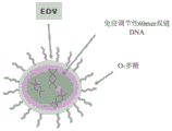


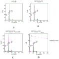


Classifications
-
- A—HUMAN NECESSITIES
- A61—MEDICAL OR VETERINARY SCIENCE; HYGIENE
- A61K—PREPARATIONS FOR MEDICAL, DENTAL OR TOILETRY PURPOSES
- A61K31/00—Medicinal preparations containing organic active ingredients
- A61K31/70—Carbohydrates; Sugars; Derivatives thereof
- A61K31/7028—Compounds having saccharide radicals attached to non-saccharide compounds by glycosidic linkages
- A61K31/7034—Compounds having saccharide radicals attached to non-saccharide compounds by glycosidic linkages attached to a carbocyclic compound, e.g. phloridzin
- A61K31/704—Compounds having saccharide radicals attached to non-saccharide compounds by glycosidic linkages attached to a carbocyclic compound, e.g. phloridzin attached to a condensed carbocyclic ring system, e.g. sennosides, thiocolchicosides, escin, daunorubicin
-
- A—HUMAN NECESSITIES
- A61—MEDICAL OR VETERINARY SCIENCE; HYGIENE
- A61K—PREPARATIONS FOR MEDICAL, DENTAL OR TOILETRY PURPOSES
- A61K35/00—Medicinal preparations containing materials or reaction products thereof with undetermined constitution
- A61K35/66—Microorganisms or materials therefrom
- A61K35/74—Bacteria
-
- A—HUMAN NECESSITIES
- A61—MEDICAL OR VETERINARY SCIENCE; HYGIENE
- A61K—PREPARATIONS FOR MEDICAL, DENTAL OR TOILETRY PURPOSES
- A61K31/00—Medicinal preparations containing organic active ingredients
- A61K31/33—Heterocyclic compounds
- A61K31/335—Heterocyclic compounds having oxygen as the only ring hetero atom, e.g. fungichromin
- A61K31/337—Heterocyclic compounds having oxygen as the only ring hetero atom, e.g. fungichromin having four-membered rings, e.g. taxol
-
- A—HUMAN NECESSITIES
- A61—MEDICAL OR VETERINARY SCIENCE; HYGIENE
- A61K—PREPARATIONS FOR MEDICAL, DENTAL OR TOILETRY PURPOSES
- A61K31/00—Medicinal preparations containing organic active ingredients
- A61K31/33—Heterocyclic compounds
- A61K31/395—Heterocyclic compounds having nitrogen as a ring hetero atom, e.g. guanethidine or rifamycins
- A61K31/435—Heterocyclic compounds having nitrogen as a ring hetero atom, e.g. guanethidine or rifamycins having six-membered rings with one nitrogen as the only ring hetero atom
- A61K31/47—Quinolines; Isoquinolines
- A61K31/4738—Quinolines; Isoquinolines ortho- or peri-condensed with heterocyclic ring systems
- A61K31/4745—Quinolines; Isoquinolines ortho- or peri-condensed with heterocyclic ring systems condensed with ring systems having nitrogen as a ring hetero atom, e.g. phenantrolines
-
- A—HUMAN NECESSITIES
- A61—MEDICAL OR VETERINARY SCIENCE; HYGIENE
- A61K—PREPARATIONS FOR MEDICAL, DENTAL OR TOILETRY PURPOSES
- A61K31/00—Medicinal preparations containing organic active ingredients
- A61K31/33—Heterocyclic compounds
- A61K31/395—Heterocyclic compounds having nitrogen as a ring hetero atom, e.g. guanethidine or rifamycins
- A61K31/495—Heterocyclic compounds having nitrogen as a ring hetero atom, e.g. guanethidine or rifamycins having six-membered rings with two or more nitrogen atoms as the only ring heteroatoms, e.g. piperazine or tetrazines
- A61K31/505—Pyrimidines; Hydrogenated pyrimidines, e.g. trimethoprim
- A61K31/517—Pyrimidines; Hydrogenated pyrimidines, e.g. trimethoprim ortho- or peri-condensed with carbocyclic ring systems, e.g. quinazoline, perimidine
-
- A—HUMAN NECESSITIES
- A61—MEDICAL OR VETERINARY SCIENCE; HYGIENE
- A61K—PREPARATIONS FOR MEDICAL, DENTAL OR TOILETRY PURPOSES
- A61K31/00—Medicinal preparations containing organic active ingredients
- A61K31/33—Heterocyclic compounds
- A61K31/395—Heterocyclic compounds having nitrogen as a ring hetero atom, e.g. guanethidine or rifamycins
- A61K31/535—Heterocyclic compounds having nitrogen as a ring hetero atom, e.g. guanethidine or rifamycins having six-membered rings with at least one nitrogen and one oxygen as the ring hetero atoms, e.g. 1,2-oxazines
- A61K31/5375—1,4-Oxazines, e.g. morpholine
- A61K31/5383—1,4-Oxazines, e.g. morpholine ortho- or peri-condensed with heterocyclic ring systems
-
- A—HUMAN NECESSITIES
- A61—MEDICAL OR VETERINARY SCIENCE; HYGIENE
- A61K—PREPARATIONS FOR MEDICAL, DENTAL OR TOILETRY PURPOSES
- A61K38/00—Medicinal preparations containing peptides
- A61K38/16—Peptides having more than 20 amino acids; Gastrins; Somatostatins; Melanotropins; Derivatives thereof
- A61K38/17—Peptides having more than 20 amino acids; Gastrins; Somatostatins; Melanotropins; Derivatives thereof from animals; from humans
- A61K38/19—Cytokines; Lymphokines; Interferons
- A61K38/21—Interferons [IFN]
- A61K38/217—IFN-gamma
-
- A—HUMAN NECESSITIES
- A61—MEDICAL OR VETERINARY SCIENCE; HYGIENE
- A61K—PREPARATIONS FOR MEDICAL, DENTAL OR TOILETRY PURPOSES
- A61K45/00—Medicinal preparations containing active ingredients not provided for in groups A61K31/00 - A61K41/00
- A61K45/06—Mixtures of active ingredients without chemical characterisation, e.g. antiphlogistics and cardiaca
-
- A—HUMAN NECESSITIES
- A61—MEDICAL OR VETERINARY SCIENCE; HYGIENE
- A61P—SPECIFIC THERAPEUTIC ACTIVITY OF CHEMICAL COMPOUNDS OR MEDICINAL PREPARATIONS
- A61P1/00—Drugs for disorders of the alimentary tract or the digestive system
-
- A—HUMAN NECESSITIES
- A61—MEDICAL OR VETERINARY SCIENCE; HYGIENE
- A61P—SPECIFIC THERAPEUTIC ACTIVITY OF CHEMICAL COMPOUNDS OR MEDICINAL PREPARATIONS
- A61P35/00—Antineoplastic agents
-
- A—HUMAN NECESSITIES
- A61—MEDICAL OR VETERINARY SCIENCE; HYGIENE
- A61K—PREPARATIONS FOR MEDICAL, DENTAL OR TOILETRY PURPOSES
- A61K47/00—Medicinal preparations characterised by the non-active ingredients used, e.g. carriers or inert additives; Targeting or modifying agents chemically bound to the active ingredient
- A61K47/50—Medicinal preparations characterised by the non-active ingredients used, e.g. carriers or inert additives; Targeting or modifying agents chemically bound to the active ingredient the non-active ingredient being chemically bound to the active ingredient, e.g. polymer-drug conjugates
- A61K47/51—Medicinal preparations characterised by the non-active ingredients used, e.g. carriers or inert additives; Targeting or modifying agents chemically bound to the active ingredient the non-active ingredient being chemically bound to the active ingredient, e.g. polymer-drug conjugates the non-active ingredient being a modifying agent
- A61K47/68—Medicinal preparations characterised by the non-active ingredients used, e.g. carriers or inert additives; Targeting or modifying agents chemically bound to the active ingredient the non-active ingredient being chemically bound to the active ingredient, e.g. polymer-drug conjugates the non-active ingredient being a modifying agent the modifying agent being an antibody, an immunoglobulin or a fragment thereof, e.g. an Fc-fragment
- A61K47/6801—Drug-antibody or immunoglobulin conjugates defined by the pharmacologically or therapeutically active agent
- A61K47/6803—Drugs conjugated to an antibody or immunoglobulin, e.g. cisplatin-antibody conjugates
- A61K47/6807—Drugs conjugated to an antibody or immunoglobulin, e.g. cisplatin-antibody conjugates the drug or compound being a sugar, nucleoside, nucleotide, nucleic acid, e.g. RNA antisense
-
- A—HUMAN NECESSITIES
- A61—MEDICAL OR VETERINARY SCIENCE; HYGIENE
- A61K—PREPARATIONS FOR MEDICAL, DENTAL OR TOILETRY PURPOSES
- A61K47/00—Medicinal preparations characterised by the non-active ingredients used, e.g. carriers or inert additives; Targeting or modifying agents chemically bound to the active ingredient
- A61K47/50—Medicinal preparations characterised by the non-active ingredients used, e.g. carriers or inert additives; Targeting or modifying agents chemically bound to the active ingredient the non-active ingredient being chemically bound to the active ingredient, e.g. polymer-drug conjugates
- A61K47/51—Medicinal preparations characterised by the non-active ingredients used, e.g. carriers or inert additives; Targeting or modifying agents chemically bound to the active ingredient the non-active ingredient being chemically bound to the active ingredient, e.g. polymer-drug conjugates the non-active ingredient being a modifying agent
- A61K47/68—Medicinal preparations characterised by the non-active ingredients used, e.g. carriers or inert additives; Targeting or modifying agents chemically bound to the active ingredient the non-active ingredient being chemically bound to the active ingredient, e.g. polymer-drug conjugates the non-active ingredient being a modifying agent the modifying agent being an antibody, an immunoglobulin or a fragment thereof, e.g. an Fc-fragment
- A61K47/6835—Medicinal preparations characterised by the non-active ingredients used, e.g. carriers or inert additives; Targeting or modifying agents chemically bound to the active ingredient the non-active ingredient being chemically bound to the active ingredient, e.g. polymer-drug conjugates the non-active ingredient being a modifying agent the modifying agent being an antibody, an immunoglobulin or a fragment thereof, e.g. an Fc-fragment the modifying agent being an antibody or an immunoglobulin bearing at least one antigen-binding site
- A61K47/6875—Medicinal preparations characterised by the non-active ingredients used, e.g. carriers or inert additives; Targeting or modifying agents chemically bound to the active ingredient the non-active ingredient being chemically bound to the active ingredient, e.g. polymer-drug conjugates the non-active ingredient being a modifying agent the modifying agent being an antibody, an immunoglobulin or a fragment thereof, e.g. an Fc-fragment the modifying agent being an antibody or an immunoglobulin bearing at least one antigen-binding site the antibody being a hybrid immunoglobulin
- A61K47/6879—Medicinal preparations characterised by the non-active ingredients used, e.g. carriers or inert additives; Targeting or modifying agents chemically bound to the active ingredient the non-active ingredient being chemically bound to the active ingredient, e.g. polymer-drug conjugates the non-active ingredient being a modifying agent the modifying agent being an antibody, an immunoglobulin or a fragment thereof, e.g. an Fc-fragment the modifying agent being an antibody or an immunoglobulin bearing at least one antigen-binding site the antibody being a hybrid immunoglobulin the immunoglobulin having two or more different antigen-binding sites, e.g. bispecific or multispecific immunoglobulin
-
- A—HUMAN NECESSITIES
- A61—MEDICAL OR VETERINARY SCIENCE; HYGIENE
- A61K—PREPARATIONS FOR MEDICAL, DENTAL OR TOILETRY PURPOSES
- A61K9/00—Medicinal preparations characterised by special physical form
- A61K9/48—Preparations in capsules, e.g. of gelatin, of chocolate
- A61K9/50—Microcapsules having a gas, liquid or semi-solid filling; Solid microparticles or pellets surrounded by a distinct coating layer, e.g. coated microspheres, coated drug crystals
- A61K9/5005—Wall or coating material
- A61K9/5063—Compounds of unknown constitution, e.g. material from plants or animals
- A61K9/5068—Cell membranes or bacterial membranes enclosing drugs
-
- C—CHEMISTRY; METALLURGY
- C12—BIOCHEMISTRY; BEER; SPIRITS; WINE; VINEGAR; MICROBIOLOGY; ENZYMOLOGY; MUTATION OR GENETIC ENGINEERING
- C12N—MICROORGANISMS OR ENZYMES; COMPOSITIONS THEREOF; PROPAGATING, PRESERVING, OR MAINTAINING MICROORGANISMS; MUTATION OR GENETIC ENGINEERING; CULTURE MEDIA
- C12N15/00—Mutation or genetic engineering; DNA or RNA concerning genetic engineering, vectors, e.g. plasmids, or their isolation, preparation or purification; Use of hosts therefor
- C12N15/09—Recombinant DNA-technology
- C12N15/11—DNA or RNA fragments; Modified forms thereof; Non-coding nucleic acids having a biological activity
- C12N15/111—General methods applicable to biologically active non-coding nucleic acids
-
- C—CHEMISTRY; METALLURGY
- C12—BIOCHEMISTRY; BEER; SPIRITS; WINE; VINEGAR; MICROBIOLOGY; ENZYMOLOGY; MUTATION OR GENETIC ENGINEERING
- C12N—MICROORGANISMS OR ENZYMES; COMPOSITIONS THEREOF; PROPAGATING, PRESERVING, OR MAINTAINING MICROORGANISMS; MUTATION OR GENETIC ENGINEERING; CULTURE MEDIA
- C12N15/00—Mutation or genetic engineering; DNA or RNA concerning genetic engineering, vectors, e.g. plasmids, or their isolation, preparation or purification; Use of hosts therefor
- C12N15/09—Recombinant DNA-technology
- C12N15/11—DNA or RNA fragments; Modified forms thereof; Non-coding nucleic acids having a biological activity
- C12N15/113—Non-coding nucleic acids modulating the expression of genes, e.g. antisense oligonucleotides; Antisense DNA or RNA; Triplex- forming oligonucleotides; Catalytic nucleic acids, e.g. ribozymes; Nucleic acids used in co-suppression or gene silencing
- C12N15/1137—Non-coding nucleic acids modulating the expression of genes, e.g. antisense oligonucleotides; Antisense DNA or RNA; Triplex- forming oligonucleotides; Catalytic nucleic acids, e.g. ribozymes; Nucleic acids used in co-suppression or gene silencing against enzymes
-
- C—CHEMISTRY; METALLURGY
- C12—BIOCHEMISTRY; BEER; SPIRITS; WINE; VINEGAR; MICROBIOLOGY; ENZYMOLOGY; MUTATION OR GENETIC ENGINEERING
- C12N—MICROORGANISMS OR ENZYMES; COMPOSITIONS THEREOF; PROPAGATING, PRESERVING, OR MAINTAINING MICROORGANISMS; MUTATION OR GENETIC ENGINEERING; CULTURE MEDIA
- C12N2310/00—Structure or type of the nucleic acid
- C12N2310/10—Type of nucleic acid
- C12N2310/14—Type of nucleic acid interfering N.A.
- C12N2310/141—MicroRNAs, miRNAs
-
- C—CHEMISTRY; METALLURGY
- C12—BIOCHEMISTRY; BEER; SPIRITS; WINE; VINEGAR; MICROBIOLOGY; ENZYMOLOGY; MUTATION OR GENETIC ENGINEERING
- C12N—MICROORGANISMS OR ENZYMES; COMPOSITIONS THEREOF; PROPAGATING, PRESERVING, OR MAINTAINING MICROORGANISMS; MUTATION OR GENETIC ENGINEERING; CULTURE MEDIA
- C12N2310/00—Structure or type of the nucleic acid
- C12N2310/10—Type of nucleic acid
- C12N2310/17—Immunomodulatory nucleic acids
-
- C—CHEMISTRY; METALLURGY
- C12—BIOCHEMISTRY; BEER; SPIRITS; WINE; VINEGAR; MICROBIOLOGY; ENZYMOLOGY; MUTATION OR GENETIC ENGINEERING
- C12N—MICROORGANISMS OR ENZYMES; COMPOSITIONS THEREOF; PROPAGATING, PRESERVING, OR MAINTAINING MICROORGANISMS; MUTATION OR GENETIC ENGINEERING; CULTURE MEDIA
- C12N2320/00—Applications; Uses
- C12N2320/30—Special therapeutic applications
- C12N2320/32—Special delivery means, e.g. tissue-specific
-
- C—CHEMISTRY; METALLURGY
- C12—BIOCHEMISTRY; BEER; SPIRITS; WINE; VINEGAR; MICROBIOLOGY; ENZYMOLOGY; MUTATION OR GENETIC ENGINEERING
- C12Y—ENZYMES
- C12Y207/00—Transferases transferring phosphorus-containing groups (2.7)
- C12Y207/11—Protein-serine/threonine kinases (2.7.11)
- C12Y207/11001—Non-specific serine/threonine protein kinase (2.7.11.1), i.e. casein kinase or checkpoint kinase
-
- Y—GENERAL TAGGING OF NEW TECHNOLOGICAL DEVELOPMENTS; GENERAL TAGGING OF CROSS-SECTIONAL TECHNOLOGIES SPANNING OVER SEVERAL SECTIONS OF THE IPC; TECHNICAL SUBJECTS COVERED BY FORMER USPC CROSS-REFERENCE ART COLLECTIONS [XRACs] AND DIGESTS
- Y02—TECHNOLOGIES OR APPLICATIONS FOR MITIGATION OR ADAPTATION AGAINST CLIMATE CHANGE
- Y02A—TECHNOLOGIES FOR ADAPTATION TO CLIMATE CHANGE
- Y02A50/00—TECHNOLOGIES FOR ADAPTATION TO CLIMATE CHANGE in human health protection, e.g. against extreme weather
- Y02A50/30—Against vector-borne diseases, e.g. mosquito-borne, fly-borne, tick-borne or waterborne diseases whose impact is exacerbated by climate change
Abstract
Compositions and methods for treating cancer are provided. In particular, the compositions comprise an anti-tumor agent and a type I interferon agonist or a type II interferon agonist or a combination of a type I interferon agonist and a type II interferon agonist.
Description
Cross Reference to Related Applications
The present application claims priority from U.S. provisional application 62/702,172 filed 2018, 7, 23 and 62/788,265 filed 2019, 1, 4, as per the provisions of 35 USC § 119, the entire contents of which are incorporated herein by reference in their entirety.
Background
Currently, most drugs used to treat cancer are administered systemically. Although systemic delivery of cytotoxic anticancer drugs plays a crucial role in cancer therapy, it also poses serious problems. For example, systemic exposure of normal tissues/organs to administered drugs can cause severe toxicity. This is exacerbated by the fact that systemically delivered cancer chemotherapeutic drugs must typically be delivered at very high doses to overcome the poor bioavailability of the drug and the large volume distribution within the patient. Moreover, systemic drug administration can be invasive, as it typically requires the use of a fixed catheter in the main vessel. Since systemic drug administration usually requires the use of the peripheral or central vein, it can cause local complications such as phlebitis. Extravasation of drugs can also cause blistering/tissue damage at the site of local administration, as is common in the administration of vinca alkaloids and anthracyclines.
Another challenge in cancer treatment is intrinsic or acquired clinical tumor chemotherapy resistance. Intrinsic resistance exists at the time of diagnosis of tumors that fail to respond to first-line chemotherapy. Acquired resistance occurs in tumors that can respond well to initial treatment but exhibit a resistant phenotype upon recurrence. Such tumors acquire resistance to both previously used drugs and to new drugs, including drugs with different structures and mechanisms of action. The term MDR (multidrug resistance) describes this phenomenon, in which tumor cells become cross-resistant to several structurally unrelated drugs after exposure to one drug. The mechanism of multidrug resistance is complex and multifactorial, largely due to high levels of genomic instability and mutations in cancer cells. Exemplary mechanisms are drug inactivation, drug extrusion through cell membrane pumps, decreased drug influx, drug target mutations and failure to initiate apoptosis. Bredel, 2001; chenet al, 2001; sun et al, 2001; and White & McCubrey, 2001.
The interaction between the immune system and malignant cells also plays an important role in tumorigenesis. The failure of the immune system to detect and reject transformed cells can lead to the development of cancer. Tumors use a variety of mechanisms to evade immune-mediated rejection. Many of these mechanisms are now known at the cellular and molecular level. Despite this knowledge, cancer immunotherapy is not a clinically established treatment.
Thus, there remains a great need for delivery systems capable of providing targeted delivery of drugs that can reduce drug resistance, promote apoptosis and induce immune responses, while avoiding the problems associated with systemic delivery of these drugs. The present invention meets these needs.
Disclosure of Invention
One embodiment of the present invention relates to compositions comprising (a) a therapeutically effective dose of purified whole bacteria derived minicells (bacterial derived minicells) containing an anti-neoplastic agent, and (b) a type I interferon agonist, a type II interferon agonist, or a combination of a type I interferon agonist and a type II interferon agonist. Type I interferon agonists and/or type II interferon agonists may optionally be present in intact bacterially derived minicells.
In one embodiment, the composition comprises (a) a therapeutically effective dose of purified intact bacteria-derived minicells that comprise an anti-neoplastic agent, and (b) a therapeutically effective dose of purified intact bacteria-derived minicells that comprise a type I interferon agonist. In another embodiment, the composition comprises (a) a therapeutically effective dose of purified intact bacteria-derived minicells that comprise an anti-neoplastic agent, and (b) a therapeutically effective dose of purified intact bacteria-derived minicells that comprise a type II interferon agonist. In yet another embodiment, the composition comprises (a) a therapeutically effective dose of purified intact bacterially-derived minicells that comprise an anti-neoplastic agent; (b) a therapeutically effective dose of purified intact bacterially-derived minicells that comprise a type I interferon agonist; and (c) a therapeutically effective dose of purified intact bacterially-derived minicells that contain a type II interferon agonist.
In one embodiment, the anti-neoplastic agent and the type I interferon agonist, the type II interferon agonist, or the combination of the type I interferon agonist and the type II interferon agonist are packaged (packeged) in two or more purified intact minicells of bacterial origin. In one embodiment, the antineoplastic agent and the type I interferon agonist, the type II interferon agonist, or the combination of the type I interferon agonist and the type II interferon agonist are packaged in three separate purified populations of intact bacterially derived minicells.
In one embodiment, the composition comprises an antineoplastic agent, a type I interferon agonist, and a type II interferon agonist, wherein: (a) an anti-tumor agent, a type I interferon agonist, and a type II interferon agonist are contained in the same minicell; (b) an anti-neoplastic agent and a type I interferon agonist are contained in a first minicell, and a type II interferon agonist is contained in a second minicell; (c) an anti-neoplastic agent and a type II interferon agonist are contained in a first minicell, and a type I interferon agonist is contained in a second minicell; (d) an anti-neoplastic agent is contained in a first minicell, and a type I interferon agonist and a type II interferon agonist are contained in a second minicell; or (e) an anti-neoplastic agent is contained in a first minicell, a type I interferon agonist is contained in a second minicell, and a type II interferon agonist is contained in a third minicell.
In one embodiment, the composition does not comprise a type I interferon agonist.
In one embodiment, the antineoplastic agent is selected from the group consisting of a radionuclide, a chemotherapeutic drug, a functional nucleic acid, and a polynucleotide that is transcribed into a functional nucleic acid. In one embodiment, the antineoplastic agent is a super-toxic chemotherapeutic drug. In one embodiment, the chemotherapy is a super-toxic chemotherapyThe drug is selected from morpholinyl anthracyclines, maytansinoids (maytansinoids), ducramycins (ducramycins), auristatins (auristatins), calicheamicins (DNA damaging agents), alpha-amanitins (RNA polymerase II inhibitors), centanamycins, pyrrolobenzodiazepines Streptavidin (streptonigtin), nitrogen mustards, nitrosoureas, alkane sulfonates, pyrimidine analogs, purine analogs, antimetabolites, folic acid analogs, anthracyclines, taxanes, vinca alkaloids, topoisomerase inhibitors, hormonal agents, and combinations thereof. In one embodiment, the morpholinyl anthracycline is selected from nemorubicin (nemorubicin), PNU-159682, idarubicin (idarubicin), daunorubicin (daunorubicin); carminomycin (caminomycin) and doxorubicin (oxorubicin). In one embodiment, the super toxic chemotherapeutic agent is PNU-159682.
In one embodiment, the functional nucleic acid is selected from the group consisting of siRNA, miRNA, shRNA, lincRNA, antisense RNA, and ribozyme. In one embodiment, the functional nucleic acid inhibits a gene that promotes tumor cell proliferation, angiogenesis or chemotherapy resistance, and/or inhibits apoptosis or cell cycle arrest. In some embodiments, the siRNA inhibits ribonucleotide reductase M1(RRM1) expression. In some embodiments, the siRNA inhibits Polo-like kinase 1(Plk1) expression. In some embodiments, the miRNA is miRNA 16 a.
In one embodiment, the type I interferon agonist, the type II interferon agonist, or the combination of a type I interferon agonist and a type II interferon agonist is an oligonucleotide. In one embodiment, the oligonucleotide comprises a sequence of at least about 40 nucleotides, at least about 50 nucleotides, or at least about 60 nucleotides. In some embodiments, the oligonucleotide is a polynucleotide product of PNPasel, poly (I: C), poly-ICLC, imiquimod, imidazoquinoline resiquimod, cGAMP, or CpG-oligodeoxynucleotide.
In one embodiment, the type I interferon agonist is selected from the group consisting of double stranded RNA (dsrna), poly (dA: dT) DNA, double stranded Z-DNA and B-DNA, DNA (dsdna) and DNA-RNA hybrids longer than 36bp, bacterial second messenger loop-bis GMP, TLR3, TLR4, TLR7, TLR8 and TLR9 agonists, STING agonists, and combinations thereof.
In one embodiment, the type II interferon agonist is selected from the group consisting of a C-glycoside form of alpha-galactosylceramide (alpha-C-GalCer), a-galactosylceramide (alpha-GalCer), a 12 carbonyl form of galactosylceramide (beta-GalCer), beta-D-glucopyranosyl ceramide (beta-GlcCer), 1, 2-diacyl-3-0-galactosyl-sn-glycerol (BbGL-II), diacylglycerol-containing glycolipid (Glc-DAG-s2), ganglioside (GD3), gangliosidic ceramide (gangliotriosylceramide) (Gg3Cer), Glycosylphosphatidylinositol (GPI), alpha-glucuronic ceramide (GSL-1 or GSL-4), isocycloglobin trihexylceramide (isoglobotriosylceramide) (i 3), Phospholipid Glycan (LPG), Lysophosphatidylcholine (LPC), alpha-galactosylceramide analogs (OCH), threitol ceramides, and combinations thereof. In one embodiment, the type II interferon agonist is an alpha-galactosylceramide (α -GalCer).
In one embodiment, the composition further comprises a bispecific ligand that binds to minicells that comprise an anti-neoplastic agent. In one embodiment, the composition further comprises a bispecific ligand that binds to a minicell that comprises a type I interferon agonist. In one embodiment, the composition further comprises a bispecific ligand that binds to minicells that comprise a type II interferon agonist.
In one embodiment, the bispecific ligand comprises a first arm specific for a minicell surface structure and a second arm specific for a non-phagocytic mammalian cell surface receptor. In one embodiment, the minicell surface structure is the O-polysaccharide component of a lipopolysaccharide on a minicell surface. In one embodiment, the non-phagocytic mammalian cell surface receptor is capable of activating receptor-mediated endocytosis of the minicell.
In one embodiment, the bispecific ligand comprises a bispecific antibody or antibody fragment. In one embodiment, the antibody or antibody fragment comprises a first multivalent arm specific for a minicell surface structure of bacterial origin and a second multivalent arm specific for a cancer cell surface receptor, wherein the cancer cell surface receptor is capable of activating receptor-mediated endocytosis of the minicell.
In one embodiment, the composition comprises every 107Less than about 1 minicell per 10 contaminating parent bacterial cells8Less than about 1 minicell per 10 contaminating parent bacterial cells9Less than about 1 minicell per 10 contaminating parent bacterial cells10Less than about 1 minicell per contaminating parent bacterial cell, or every 10 minicells 11Less than about 1 minicell in a contaminating parent bacterial cell.
In one embodiment, the composition further comprises a pharmaceutically acceptable carrier. In one embodiment, minicells are approximately 400nm in diameter. In one embodiment, the composition is free of parental bacterial cell contamination removable by 200nm filtration.
In one embodiment, the composition comprises minicells or killed bacterial cells in the following amounts: (a) at least about 109A plurality of; (b) at least about 1X 10⁹A plurality of; (c) at least about 2X 10⁹A plurality of; (d) at least about 5X 10⁹A plurality of; (e) at least 8 x 10⁹A plurality of; (f) not more than about 1011A plurality of; (g) not more than about 1X 10¹¹A plurality of; (h) not more than about 9X 10¹⁰A plurality of; or (i) no more than about 8X 10¹⁰And (4) respectively.
One embodiment of the present invention relates to a method of treating a subject in need thereof, the method comprising administering to the subject an effective amount of a composition disclosed herein. In one embodiment, the subject is a human, non-human primate, canine, feline, bovine, ovine, equine, rabbit, mouse, or rat. In one embodiment, the subject is a human.
In one embodiment, the subject has cancer. In one embodiment, the cancer is selected from lung cancer, breast cancer, brain cancer, liver cancer, colon cancer, pancreatic cancer, and bladder cancer. In one embodiment, the cancer is selected from acute lymphoblastic leukemia; acute myeloid leukemia; adrenocortical carcinoma; AIDS-related cancers; AIDS-related lymphomas; anal cancer; appendiceal carcinoma; astrocytoma; atypical teratoid/rhabdoid tumors; basal cell carcinoma; bladder cancer; brain stem glioma; brain tumors; breast cancer; bronchial tumors; burkitt's lymphoma; cancers with unknown primary site; carcinoid; cancers with unknown primary site; atypical teratoid/rhabdoid tumors of the central nervous system; embryonic tumors of the central nervous system; cervical cancer; childhood cancer; chordoma; chronic lymphocytic leukemia; chronic myelogenous leukemia; chronic myeloproliferative disorders; colon cancer; colorectal cancer; craniopharyngioma; cutaneous T cell lymphoma; endocrine pancreatic islet cell tumors; endometrial cancer; an ependymal cell tumor; ependymoma; esophageal cancer; a sensory neuroblastoma; ewing's sarcoma; extracranial germ cell tumors; gonadal ectogenital cell tumors; extrahepatic bile duct cancer; gallbladder cancer; gastric cancer; gastrointestinal carcinoid tumors; gastrointestinal stromal cell tumors; gastrointestinal stromal tumors (GIST); gestational trophoblastic tumors; a glioma; hairy cell leukemia; head and neck cancer; cancer of the heart; hodgkin lymphoma; hypopharyngeal carcinoma; intraocular melanoma; pancreatic islet cell tumors; kaposi's sarcoma; kidney cancer; langerhans cell tissue cell proliferation; laryngeal cancer; lip cancer; liver cancer; malignant fibrous histiocytoma bone cancer; medulloblastoma; a medullary epithelioma; melanoma; merkel cell carcinoma; merkel cell skin cancer; mesothelioma; primary occult metastatic squamous neck cancer; oral cancer; multiple endocrine tumor syndrome; multiple myeloma; multiple myeloma/plasmacytoma; mycosis fungoides; myelodysplastic syndrome; myeloproliferative tumors; nasal cavity cancer; nasopharyngeal carcinoma; neuroblastoma; non-Hodgkin lymphoma; non-melanoma skin cancer; non-small cell lung cancer; lip cancer; oral cancer; oropharyngeal cancer; osteosarcoma; other brain and spinal cord tumors; ovarian cancer; epithelial carcinoma of the ovary; ovarian germ cell tumors; low malignant potential of the ovary; pancreatic cancer; papillomatosis; paranasal sinus cancer; parathyroid cancer; pelvic cancer; penile cancer; pharyngeal cancer; mesodifferentiated pineal parenchymal tumors; pineal gland cell tumor; pituitary tumors; plasma cell tumor/multiple myeloma; pleuropulmonary blastoma; primary Central Nervous System (CNS) lymphoma; primary hepatocellular carcinoma; prostate cancer; rectal cancer; kidney cancer; renal cell (renal) carcinoma; renal cell carcinoma; cancer of the respiratory tract; retinoblastoma; rhabdomyosarcoma; salivary gland cancer; sezary syndrome; small cell lung cancer; small bowel cancer; soft tissue sarcoma; squamous cell carcinoma; squamous neck cancer; gastric cancer; supratentorial primary neuroectodermal tumors; t cell lymphoma; testicular cancer; throat cancer; thymus gland cancer; thymoma; thyroid cancer; transitional cell carcinoma; transitional cell carcinoma of the renal pelvis and ureter; a trophoblastic tumor; cancer of the ureter; cancer of the urethra; uterine cancer; uterine sarcoma; vaginal cancer; vulvar cancer; waldenstrom macroglobulinemia; and Wilm tumors.
In one embodiment, the brain cancer or tumor is selected from the group consisting of brain stem glioma, central nervous system atypical teratoid/rhabdoid tumor, central nervous system embryonic tumor, astrocytoma, craniopharyngioma, ependymoma, medulloblastoma, medullary epithelial cytoma, medially differentiated pineal parenchymal tumor, supratentorial primary neuroectodermal tumor, and pineoblastoma.
In one embodiment, the composition is administered at least once a week over the course of several weeks. In one embodiment, the composition is administered at least once a week over several weeks to several months. In one embodiment, the composition is administered at least once a week for about 2 weeks, about 3 weeks, about 4 weeks, about 5 weeks, about 6 weeks, about 7 weeks, about 8 weeks, about 9 weeks, about 10 weeks, about 11 weeks, about 12 weeks, about 13 weeks, about 14 weeks, about 15 weeks, about 16 weeks, about 17 weeks, about 18 weeks, about 19 weeks, or about 20 weeks or longer. In one embodiment, the composition is administered about twice per week. In one embodiment, the composition is administered twice a week for about 2 weeks, about 3 weeks, about 4 weeks, about 5 weeks, about 6 weeks, about 7 weeks, about 8 weeks, about 9 weeks, about 10 weeks, about 11 weeks, about 12 weeks, about 13 weeks, about 14 weeks, about 15 weeks, about 16 weeks, about 17 weeks, about 18 weeks, about 19 weeks, or about 20 weeks or longer.
The foregoing summary, as well as the following drawings and detailed description, are exemplary and explanatory. It is intended to provide further details of the invention, but should not be construed as limiting. Other objects, advantages and novel features will be apparent to those skilled in the art from the following detailed description of the invention.
Drawings
FIG. 1 is a schematic representation of EnGeneIC Dream Vehicle (EDV) (e.g., bacterial minicells) comprising a bispecific antibody against O-polysaccharide and human epidermal growth factor receptor antigen and loaded with the anticancer drug PNU-159682 (anthracycline analogue).
FIG. 2 is a schematic representation of EnGeneIC Dream Vehicle (EDV) comprising O-polysaccharides on the surface and loaded with immunomodulatory 60mer double stranded DNA.
FIG. 3 is a schematic representation of EnGeneIC Dream Vehicle (EDV) comprising O-polysaccharides on the surface and loaded with immunomodulatory alpha galactosylceramide (alpha GC).
Figure 4 is a graphical summary of the clinical trials evaluating EGFR-targeted and miRNA16 a-loaded EDVs for treatment of mesothelioma patients.
Figure 5 shows the cytotoxic effect of indicated chemotherapeutic agents on a549 lung cancer cell line. FIG. 4A compares the effect of the indicated chemotherapeutic agent with the super toxic agent PNU-159682. FIG. 4B compares the effect of doxorubicin and PNU-159682.
Figure 6 shows the effect of indicated chemotherapeutic drugs on the adrenocortical carcinoma cell lines ACC01 (figure 6A) and ACC07 (figure 6B).
FIG. 7 shows the effect of indicated chemotherapeutic agents on MDA-MB-468 breast cancer cell line.
FIG. 8 shows the effect of indicated chemotherapeutic agents on human colorectal cancer cell lines Caco-2 (FIG. 8A) and HCT116 (FIG. 8B).
FIG. 9 shows the effect of indicated chemotherapeutic agents on the glioblastoma cell line U87-MG.
FIG. 10 shows the effect of indicated chemotherapeutic drugs on human pancreatic cell lines MiaPaca-2 (FIG. 10A) and gemcitabine-resistant MiaPaca-2 GemR cells (FIG. 10B).
FIG. 11 shows EGFR-targeting and PNU-159682(EGFREDVS682 TM) Or doxorubicin (c)EGFREDV SDox TM) The effect of EDV (b) on the growth of a549 xenograft tumors in mice. Negative control is onlySaline or PNU-159682-loaded non-targeted EDV (EDV)S682 TM). Arrows indicate when mice were treated with the indicated saline or EDV compositions. Asterisks indicate when mice initially treated with saline were administeredEGFREDVS682 TMA composition is provided.
FIG. 12 shows the expression of GAPDH (glyceraldehyde-3-phosphate dehydrogenase) (G), KSP (kinesin spindle protein), Plk1 (Polo-like kinase 1) (P) and RRM1 (ribonucleotide reductase 1) (R) expressed relative to GAPDH in the indicated NSCLC cell lines.
Figure 13 shows the effect of delivering EGFR-targeted, siRRM 1-packaged EDV to either a mesothelioma cell line (MSTO, figure 13A) or an adrenocortical carcinoma cell line (H295R, figure 13B).
Figure 14 shows miRNA16a (targeting EGFR) ((r))EGFREDVmiRNA16a TM) Or EGFR-targeted EDV (EDV) packaging siRRM1EGFREDVsiRRM1 TM) Effect on mesothelioma xenograft tumor growth in Balb/c nu/nu mice. Negative controls were saline or EGFR-targeted EDV loaded with scrambled (scrambled) siRNA.
Figure 15 shows the use of miRNA targeting EGFR 16a (r) (treatment) compared to saline treatment or EGFR targeting EDV loaded with scrambled siRNA (treatment)EGFREDVmiRNA16a TM) Or EGFR-targeted EDV (EDV) packaging siRRM1EGFREDVsiRRM1 TM) Treated tumors isolated from mesothelioma xenograft Balb/c nu/nu mice.
FIGS. 16A and 16B show targeting of Polo-like kinase 1 (by loading withEGFREDVTM siPLK TM) And ribonucleotide reductase 1: (EGFREDVsiRRM1 TM) EGFR-targeted EDV of siRNA induced apoptosis in adrenocortical carcinoma cells (ACC01) -based on measuring the number of cell debris (fig. 16A) and the ratio of Annexin5 to Propidium Iodide (PI) positive cells (fig. 16B). As negative controls, untreated ACC01 cells were included, with EDV loaded with irrelevant siRNA (II)EGFREDVTM siLuciferase TM) Treated ACC01 cells and unloaded EDV (II) ((III))EGFREDVTM) Apoptosis of (2).
FIGS. 17A-D show targeting of Polo-like kinase 1 by loading (A-D) EGFREDVTM siPLK TM) (FIG. 17D) and ribonucleotide reductase 1: (EGFREDVsiRRM1 TM) EDV targeting EGFR of siRNA induced sub-G1 blockade in adrenocortical carcinoma cells (ACC01) (fig. 17D) -based on measuring the number of cell debris and the ratio of Annexin5 to Propidium Iodide (PI) positive cells. Untreated ACC01 (FIG. 17A), using EDV loaded with irrelevant siRNA (CEGFREDVTM siLuciferase TM) Treated ACC01 cells (fig. 17C) and unloaded EDV (v) ((v))EGFREDVTM) Apoptosis (fig. 17B) was included as a negative control.
FIG. 18 shows the effect of A549 (lung cancer) xenograft tumor growth in Balb/c nu/nu mice treated with: (i) solid triangle ═EGFREDVsPNU-159682 TM+EDVs40mer TM(ii) solid circle ═EGFREDVsPNU-159682 TM(iii) Hollow square ═EGFREDVsPNU-159682 TM+ EDV, (iv) hollow triangle ═EGFREDVsPNU-159682 TM+EDVs50mer TMAnd (v) solid square ═ brine. Mice were treated with these EDV combinations on days 24, 27, 29, 31, 34, 36 and 38 after xenograft implantation, as indicated by the arrows above. On days 36 and 38, useEGFREDVsPNU-159682 TM+EDVs50mer TMThe volume of the tumor to be treated is 650mm3Mice in saline group, as indicated by the arrow below.
FIG. 19 shows the results obtained with an EDV comprising 40mers (EGFREDVs40mers TM) And EDV comprising PNU-159682 (EGFREDVsPNU TM) Effect on a549 (lung cancer) xenograft tumor growth in Balb/c nu/nu mice treated with the combination of (a). The triangles represent treatment days.
FIG. 20 shows saline (negative control), IFN-. gamma. (0.5X 10 per dose) 4IU), EGFR-targeting EDV loaded with doxorubicin: (EGFREDVsDox TM) AndEGFREDVsDox TM+ effect of IFN- γ treatment on a549 (lung cancer) xenograft tumor growth in Balb/c nu/nu mice. The triangles represent treatment days.
FIG. 21 shows saline (negative control), IFN-. gamma. (0.5X 10 per dose)4IU), EGFR-targeting EDV loaded with doxorubicin: (EGFREDVsDox TM) AndEGFREDVsDox TMeffect on MDA-MB 468 (Breast cancer) xenograft tumor growth in Balb/c nu/nu mice treated with IFN- γ. The triangles represent treatment days.
FIG. 22 shows another experiment, exemplified with saline (negative control), IFN-. gamma.0.5X 10 per dose4IU), EGFR-targeting EDV loaded with doxorubicin: (EGFREDVsDox TM) AndEGFREDVsDox TMeffect on MDA-MB 468 (Breast cancer) xenograft tumor growth in Balb/c nu/nu mice treated with IFN- γ. The triangles represent treatment days.
FIG. 23 shows EGFR-targeting EDV (EDV) loaded with doxorubicin with saline (negative control, group 1)EGFREDVsDox TMGroup 2),EGFREDVsDox TM+ IFN-. gamma.s (0.75X 10 per dose4IU) (group 3) andEGFREDVsDox TM+ IFN-. gamma.s (0.5X 10 per dose4IU) (group 4) effect on doxorubicin-resistant a549 xenograft tumor growth in Balb/c nu/nu mice treated. Mice in groups 1-3 received treatment twice weekly, as shown by the solid triangles. Mice in group 4 were treated 3 times per week as indicated by the open triangles.
FIGS. 24A-K show cytokine profiles from patients from a first human clinical study, given different doses of paclitaxel-loadedEGFREDVsTM. (fig. 24A) ═ pg/mL of IL-6 measured for each of 5doses (5 doses); (fig. 24B) pg/mL of IL-8 measured for each of the 5 doses; (fig. 24C) pg/mL of IL-10 measured for each of the 5 doses; (fig. 24D) pg/mL of TNF-a measured for each of the 5 doses; (Figure 24E) ═ pg/mL of IFN- α measured for each of the 5 doses; (figure 24F) pg/mL of IFN- γ measured for each of 5 doses; (fig. 24G) — pg/mL of IL-1 β measured for each of the 5 doses; (fig. 24H) pg/mL of IL-2 measured for each of the 5 doses; (fig. 24I) ═ pg/mL of IL-4 measured for each of the 5 doses; and (fig. 24J) ═ pg/mL of IL-12 measured for each of the 5 doses. Finally, five doses tested are shown (fig. 24K).
FIGS. 25A-K show cytokine profiles from patients from a first human clinical study, in which different doses of doxorubicin-loaded EGFREDVsTM. (fig. 25A) pg/mL of IL-6 measured for each of the 8 doses; (fig. 25B) pg/mL of IL-8 measured for each of the 8 doses; (fig. 25C) pg/mL of IL-10 measured for each of the 8 doses; (fig. 25D) pg/mL of TNF-a measured for each of the 8 doses; (fig. 25E) — pg/mL of IFN- α measured for each of the 8 doses; (figure 25F) pg/mL of IFN- γ measured for each of the 8 doses; (fig. 25G) — pg/mL of IL-1 β measured for each of the 8 doses; (fig. 25H) pg/mL of IL-2 measured for each of the 8 doses; (fig. 25I) — pg/mL of IL-4 measured for each of the 8 doses; and (fig. 25J) ═ pg/mL of IL-12 measured for each of the 8 doses. Finally, (fig. 25K) shows three additional doses tested, with the first five doses being the same as those shown in (fig. 24K).
FIG. 26 shows the signaling pathway of a cytoplasmic DNA sensor with DNA challenge (challenge). To date, a variety of cytoplasmic DNA sensors have been defined to detect intracellular double stranded DNA. RNA polymerase III transcribes AT-rich DNA into RNA recognized by RNA sensor RIG-I, followed by STING and IRF3 activation. The DNA sensors DAI, IFI16, DDX41 and LSm14A directly sense dsDNA to activate STING for type I IFN production. cGAS catalyzes the synthesis of cSAMP (a strong activator of STING) in the presence of dsDNA. By dsDNA, LRRFIP1 initiates β -catenin and IRF3 activation in a STING-dependent manner. Other DNA sensors elicit immune responses independent of STING. Upon recognition of dsDNA, Sox2 triggered activation of the Tab2/TAK1 complex in neutrophils. DHX9/36 activates NF κ B and IRF7 by MyD88 when detected by dsDNA. The DNA sensor Ku70 triggered the activation of IRF1 and IRF 7. AIM2 initiates activation of inflammasome by ASCs with DNA binding.
Fig. 27 shows RAW264.7 cell and myeloid-derived dendritic cell (BMDC) activation in response to EDV treatment. (FIG. 27A) CD86 expression in RAW cells grown directly with l. mu.g/ml LPS, Ep-EDV682 or 682. (FIG. 27B) expression of CD86 in RAW cells co-cultured with 4T1 or CT26Ep12.1 cells treated with Ep-EDV, Ep-EDV682 or 682. RAW cells co-cultured with Ep-EDV 682-treated tumor cells resulted in a significant increase in CD86 expression. (fig. 27C) TNF α production in RAW cell/tumor cell co-cultures, showing a significant increase in TNF α production by RAW cells incubated with EDV-treated tumor cells. (FIG. 27D) IL-6 production in RAW cell/tumor cell co-cultures, showing a significant increase in IL-6 production in RAW cells incubated with EDV-treated tumor cells. (FIG. 27E) PCR quantification of IFN α and IFN β expression in BMDC/4T1 cocultures. (FIG. 27F) PCR quantification of IFN α and IFN β expression in BMDC/CT26Ep12.1 cocultures. (FIG. 27G) CD86HiAnd MHC class IIHiExpression and (FIG. 27H) CD86HiQuantification of expression in BMDC/tumor cell co-cultures. (FIG. 27I) flow cytometry density mapping of MHC class II vs CD86 expression in co-cultures of BMDCs with EDV and drug-treated CT26Ep12.1 cells. (FIG. 27J) ELISA analysis of TNF α (FIG. 27K) IL-12p40 and (FIG. 27L) IL-6-supernatants from BMDC/tumor cell co-cultures. Data represent mean ± s.e.m. and were analyzed by one-way ANOVA and Tukey multiple comparison tests.
Figure 28 shows tumor response and macrophage activation in response to EDV treatment. Tumor growth in response to Ep-EDV and Ep-EDV682 treatment in BALB/c mice bearing (FIG. 28A)4T1 or (FIG. 28B) CT26Ep12.1 tumors. (FIG. 28C) tumor growth in response to EDV-682 and EDV-EGFR682 in BALB/C nude mice bearing T84 xenografts and (FIG. 28D) tumor growth of EDV-682, EDV-EGFRDOX, and EDV-EGFR682 in BALB/C nude mice bearing A549/MDR xenografts. Green colourArrows indicate where EDV-EGFR682 began in saline treated mice. Data (fig. 28A-D) represent mean ± s.e.m., and were analyzed by two-way ANOVA and Tukey multiple comparison tests. (fig. 28E) isolated from 4T1 tumor and mixed with 4T1 cells at 5: 1 (E: T) ratio of Co-cultured CD11b+xCEELLigene RTCA. The normalized cell index, which correlates with cell adhesion and growth/death vs time, is plotted. CD11b from 4T1 tumor+Cells undergo an initial adhesion and sedimentation phase, as indicated by an increase in cellular index, followed by growth or death, as indicated by an increase or decrease in cellular index. (FIG. 28F) M1(CD 86) in the 4T1 tumor of treated mice+):M2(CD206+) Proportion of macrophages. (FIG. 28G) CD11b isolated from CT26Ep12.1 tumors and co-cultured with CT26Ep12.1 cells at a ratio of 5: 1 (E: T) +xCEELLigene RTCA. (FIG. 28H) M1(CD 86) in CT26Ep12.1 tumors of treated mice+):M2(CD206+) Proportion of macrophages. Data (fig. 28F and 28H) represent mean ± s.e.m., and were analyzed by one-way ANOVA and Tukey multiple comparison tests.
Figure 29 shows NK cell response to EDV treatment. (FIG. 29A) xCelling RTCA of NK cells co-cultured with 4T1 cells at a ratio of 20: 1 (E: T) isolated from spleen of 4T1 tumor-bearing mice. The cell viability over time is plotted and calculated from the normalized cell index. (FIG. 29B)% viability of 4T1 cells co-cultured with NK cells from saline, Ep-EDV or Ep-EDV-682 treated mice 70 hours after NK addition. (fig. 29C) ct26ep12.1 cells isolated from the spleen of mice bearing ct26ep12.1 tumors at 20: 1 (E: T) ratio of xCELLigence RTCA of Co-cultured NK cells. (FIG. 29D)% viability of CT26Ep12.1 cells co-cultured with NK cells from saline, Ep-EDV or Ep-EDV-682 treated mice 50 hours after NK cell addition. (FIG. 29E) NK cells CD45 in 4T1 tumor+、CD11b+、DX5+) NKG2D in, showing increased NKG2D expression in Ep-EDV-682 treated mice. Production of RANTES (fig. 29F) and TNF α (fig. 29G) in coculture of NK cells isolated from spleen of EDV treated mice bearing 4T1 tumor and 4T1 cells. (FIG. 29H)4 different mouse tumor cells Quantification of the NKG2D ligands RAE-1, H60a and MULT-1 on the surface. (FIG. 29I) comparison of 4T1 cells in the presence of RAE-1 and/or H60a inhibitory antibodies at 20: 1 (E: T) ratio of Co-cultured NK cells isolated from spleen of EpEDV-682-treated mice bearing 4T1 tumor xCellosion RTCA, both of which proved to be very important in cytolysis of NK tumor cells. (fig. 29J) quantification of NK cell lysis 80 hours after NK cell addition, showing significant inhibition of NK cell lysis by binding of H60a antibody alone to the RAE-1 inhibitory antibody. The data (fig. 29B, 29D, 29E-G, and 29J) represent mean ± s.e.m., and were analyzed by one-way ANOVA and Tukey multiple comparison tests.
Figure 30 shows the production of mesenchymal tumor cytokines/chemokines by splenocyte/tumor cell co-cultures in response to EDV treatment. ELISA analysis of stromal cytokines and chemokines produced in response to Ep-EDV and Ep-EDV682 treatment in BALB/c mice bearing (FIG. 30A)4T1 or (FIG. 30B) CT26Ep12.1 tumors. Ep-EDV-682 treatment mainly results in an increase of the Th1 cytokine. Data represent mean ± s.e.m. Each cytokine data was analyzed by one-way ANOVA and Tukey multiple comparison tests. ELISA analysis of splenocytes from mice bearing 4T1 and CT26Ep12.1 tumors treated with saline, Ep-EDV and Ep-EDV-682 (FIG. 30C), together with supernatants from co-cultures of their respective tumor cells, TNF α, (FIG. 30D) IL-2, (FIG. 30E) IL-1 β, (FIG. 30F) IFN γ, and (FIG. 30G) IL-10. Data represent mean ± s.e.m. One-way ANOVA analysis and Tukey multiple comparisons were used to compare groups of + or-tumor cells. The T-test is used to compare individual treatments with or without tumor cells.
Figure 31 shows T cell function and phenotype in response to EDV treatment. (FIG. 31A) 4T1 cells isolated from the spleen of a mouse bearing a 4T1 tumor were differentiated at 30: 1 (E: T) ratio of Co-cultured CD8+xCEELLigene RTCA for T cells. The normalized cell index associated with cell adhesion and growth/death vs time is plotted. (FIG. 31B) adding CD8 +30 hours after T cells, CD8 from saline, Ep-EDV or Ep-EDV-682-treated mice+% viability of 4T1 cells co-cultured with T cells. (fig. 31C) cells isolated from the spleen of mice bearing the ct26ep12.1 tumor were separated from ct26ep12.1 at 30: ratio of 1 (E: T)Example Co-cultured CD8+xCEELLigene RTCA for T cells. The normalized cell index associated with cell adhesion and growth/death vs time is plotted. (FIG. 31D) adding CD8 +20 hours after T cells, CD8 from saline, Ep-EDV or Ep-EDV-682-treated mice+% viability of ct26ep12.1 cells co-cultured with T cells. (FIG. 31E) CD8 detected in 4T1 tumor+T cells (defined as CD 45)+、CD3+、CD8+) Percentage of (c). (FIG. 31F) T-regs detected in 4T1 tumors (defined as CD 45)+、CD3+、CD4+、CD25+) Percentage of (c). (fig. 31G) T cell number in tumor draining lymph nodes of mice carrying 4T1 tumor was shown as a percentage of total cells. (FIG. 31H)% CD80/MHC class II expression in dendritic cells in tumor draining lymph nodes of mice carrying 4T1 tumor. (FIG. 31I) isolated CD8 from Ep-EDV-682-treated mice +Confocal images of T cell interaction with 4T1 cells. Red-actin, green-perforin, blue (dark) -DAPI; scale bar 10 μm. Data (fig. 31B, 31D, and 31E-H) represent mean ± s.e.m., and were analyzed by one-way ANOVA and Tukey multiple comparison tests (fig. 31B, 31D, and 31E-G) or t-test (fig. 31H).
FIG. 32 shows prognostic indicators and immunophenotyping of patient Peripheral Blood Mononuclear Cells (PBMCs) revealing increased dendritic cell and monocyte antigen presentation and cytotoxic CD8 at dose 12+Evidence of elevated T cell content. Prognostic indicators (FIG. 32A) CA19-9 and (FIG. 32B) C-reactive protein serum levels. PBMC analysis using Duraclone immunotyping maps for (FIG. 32C) monocytes and (FIG. 32D) the intermediate (CD14+ CD16+ +) antigen presenting monocyte subtype. Expressed as% white blood cells. The dendritic cell subset included (FIG. 32E) the driver CD8+Effector T cell responsive myeloid dendritic cells (Clec 9A)+) (mDC) and (fig. 32F) plasmacytoid and myeloid dendritic (cells) (professional antigen presenting DCs). As shown, expressed as% DC or% mDC. (FIG. 32G) CD8+T cell subtypes. Cytotoxic CD8+T cells include effector subtypes and depletion (PD 1)+) The subtype is.
FIG. 33 showsA schematic diagram showing how EDV first establishes an immunogenic tumor microenvironment by delivering cytotoxic agents directly to the tumor, then directly or indirectly stimulates the innate immune system towards an anti-tumor phenotype, and finally generates an adaptive response in which tumor-specific cytotoxic T cells are generated. (FIG. 33A) Ep-EDV-682 entered the tumor microenvironment through leaky vasculature, leading to tumor apoptosis and immune-activated DAMP release. (FIG. 33B) interaction of macrophages within the tumor microenvironment directly by engulfment of apoptotic cells or even EDV, resulted in M1 macrophage polarization and release of the inflammatory cytokines TNF α and IL-6. (FIG. 33C) M1 macrophages were able to further lyse tumor cells and release MIP-1 α, which recruits more immune cells. (FIG. 33D) immature dendritic cells engulfed apoptotic cell bodies and released tumor antigens-which were present in response to Ep-EDV-682 treatment and matured, releasing the type 1 interferons TNF α, IL-12p40 and IL-6. (FIG. 33E) the mature DCs then migrated to lymph nodes to present antigen to T cells. (fig. 33F) NK cell activation also occurred in the tumor microenvironment, resulting in the release of IFN γ and TNF α and RANTES to attract additional immune cells. In addition, activated NK cells efficiently lyse tumor cells. (FIG. 33G) Release of RANTES and MIP-1 α recruits additional T cells, NK cells and macrophages into the tumor, with (FIG. 33H) tumor-specific CD8 +T cells then facilitate the response by tumor cell lysis. (FIG. 33I) these steps were all combined to generate an effective anti-tumor immune response.
Figure 34 shows RAW264.7 and JAWS II cell activation in response to EDV treatment. (FIG. 34A) TNF α production by RAW264.7 cells incubated directly with EDV preparation. (FIG. 34B) IL-6 production by RAW264.7 cells incubated directly with the EDV preparation. (FIG. 34C) flow cytometry histograms of CD86 and MHC class II expression in cocultures of JAWS II cells with untreated CT26Ep12.1 and 4T1 cells or JAWS II cells of CT26Ep12.1 and 4T1 cells treated with Ep-EDV, Ep-EDV-682 or 682 alone. (FIG. 34D) quantification of CD86 expression on JAWS II cells co-cultured with treated tumor cells as determined by flow cytometry. (FIG. 34E) quantification of MHC class II expression on JAWS II cells co-cultured with treated tumor cells as determined by flow cytometry. Data represent mean ± s.e.m. and were analyzed by one-way analysis of variance and Tukey multiple comparison tests.
FIG. 35 shows the weight change and macrophage activation in response to EDV treatment in Balb/c and Balb/c naked xenografts. The% body weight change in mice carrying (fig. 35A) ct26ep12.1, (fig. 35B)4T1, (fig. 35C) T84 and (fig. 35D) a549/MDR tumors in response to treatment. No more than 5% weight loss was observed at the initial dose, and then body weight recovered and stabilized with subsequent dosing. Data represent mean ± s.e.m. The M1/M2(CD 86: CD206) ratio of macrophages in the A549/MDR and T84 tumors (FIG. 35F) of EDV treated mice. (FIG. 35G) CD11b isolated from A549/MDR tumors and co-cultured with A549/MDR cells at a ratio of 5: 1 (E: T) +xCEELLigene RTCA. The normalized cell index associated with cell adhesion and growth/death vs time is plotted. (FIG. 35H) CD11b with tumors from saline or EGFR-EDV-682 treated mice+Cell co-cultured A549/MDR cells in CD11b+% cell lysis 6.5 hours after cell addition. (FIG. 35I) CD11b isolated from treated 4T1 tumor+MIP-1 alpha production in coculture of cells with 4T1 cells. Data (fig. 35E, 35F, 35H and 35I) represent mean ± s.e.m. and were analyzed by one-way ANOVA and Tukey multiple comparison test (fig. 35E, 35H, 35I) or t-test (fig. 35F).
Figure 36 shows NK cell response to EDV treatment. (FIG. 36A) xCelling RTCA of NK cells co-cultured with T84 cells at a ratio of 10: 1 (E: T) isolated from spleen of Balb/c nude mice carrying T84 tumor. The plots represent cell viability over time as calculated from the normalized cell index. (FIG. 36B) Granzyme B (Granzyme B) production in co-cultures of NK cells and T84 cells isolated from spleen of mice treated with saline and EGFR-EDV-682. Data represent mean ± s.e.m. and were analyzed by t-test. (FIG. 36C) xCellligence RTCA of NK cells isolated from spleen of Balb/C nude mice bearing A549/MDR tumor co-cultured with A549/MDR cells at a ratio of 10: 1 (E: T). The plots represent cell viability over time as calculated from the normalized cell index (saline n ═ 5; EGFR-EDV-682n ═ 4).
Figure 37 shows receptor expression and drug sensitivity screening of patient derived pancreatic ductal adenocarcinoma cells. (FIG. 37A) quantification of EGFR surface receptors from cells of tumor head. (FIG. 37B) quantification of EGFR surface receptors from cells in the tail of tumors. (FIG. 37C) drug sensitivity and IC 50 of first and second line chemotherapeutic drug/drug combinations compared to 682 sensitivity.
FIG. 38 shows that individual cells from a total PBMC pool (FSC v SSC) were gated based on the forward scatter width (FSC-W) vs forward scatter area (FSC-A) and then analyzed for CD45 staining (FSC v CS 45)+CD45 on+Gated). Cell viability was 96% (count and viability kit # C00162, Beckman Coulter, data not shown), and dead cells were excluded based on FSC threshold discriminator 80. Total Dendritic Cells (DCs) on leukocytes (CD 45)+) Up-gated and defined as HLA-DR+And pedigree- (# B53351, Beckman Coulter). Lineage negative markers consist of a population of antibodies conjugated to the same fluorophore (PE) that were raised against CD3, CD14, CD19, CD20, and CD56 for negative selection of T cells, monocytes, B cells, and NK cells, respectively. Remaining HLA-DR+Cells are gated as dendritic cells and subdivided into plasmacytoid DCs (CD11c-CD132+) or antigen presenting myeloid DCs (CD 11C) +CD 123-). Myeloid DC (mDC) are divided into three major subsets, CDlc + mDCl, CD141+ mDC2 (shown here as Clec9A +) and CD16+ mDC. Expression of CD14+ defines monocytes gated on leukocytes (# B93604, Beckman Coulter), and is then subdivided into classical (CD14+ CD16-), intermediate (CD14+ CD16+) and non-classical (CD14+ CD16+ +). T cells (# B53328, Beckman Coulter) are CD3+ and are gated on lymphocytes (CD45+ SSC low). Then define CD3+T cell subset, T helper cell (CD 4)+) And cytotoxic T cells (CD 8)+). PD1+ CD8+ cytotoxic T cell gates were plotted against SSC and gated on CD8+ T cells. All FSC and SSC axes are linear, while the fluorescence channel axis (all CD markers) is logarithmic or bi-exponential ("logicle", Kaluza software, Beckman Coulter).
FIG. 39 shows flow cytometry analysis showing the purity of cell isolation for the xCelling RTCA experiment. (FIG. 39A) from childhoodMouse tumor isolation CD11b+A cell. The density plots show isotype controls vs. fsc and CD11b vs. fsc. CD11b of samples+The cell purity was 80%. (FIG. 39B) isolation of NK cells from mouse spleen. Density plots show isotype controls vs. fsc, NKp46 vs. fsc and CD11b vs. fsc. The NK cell purity of the sample was-90%. (FIG. 39C) isolation of CD8 from mouse spleen +T cells. Density plots show isotype controls vs. fsc, CD3e vs. fsc, and CD8 vs. fsc. T cells of the sample (CD 3e)+) Purity of-90%, with over 98% of those T cells being CD8+。
FIG. 40 shows a syngeneic mouse model (in Balb/c miceEpCT26 colon tumor) by the same methodEpMinicellsDoxAnd minicellsα-GCThe combination therapy of (1).
FIG. 41 showsEpMinicellsDoxAnd minicellsα-GCThe combination treatment of (a) is effective in reducing large tumors in Balb/c mice carrying CT26 allogeneic grafts.
FIG. 42 showsEpMinicellsDoxAnd minicellsα-GCEffect on tumor regression in Balb/c mice with CT26 syngeneic grafts.
FIG. 43 is a schematic view (FIGS. 43A and 43B)EpMinicellsDoxAnd minicellsα-GCAnd (FIGS. 43D and 43E) minicells onlyα-GC(FIG. 43F) onlyEpMinicellsDoxAnd (fig. 43C) saline treated CT26 syngeneic grafts of different sizes.
FIG. 44 shows aEpMinicellsDoxAnd minicellsα-GCTreated CT26 syngeneic grafts of different sizes.
FIG. 45 shows minicellsαGCPresentation of α GC-CD1D by JAWSII cells at various time points after treatment (FIGS. 45A-E)
Detailed Description
I. Overview
The present invention is based on the following findings: compositions comprising the following combinations may synergistically improve cancer treatment strategies: (i) antineoplastic agents and type I interferon agonists; (ii) antineoplastic agents and type II interferon agonists; or (iii) an antineoplastic agent, a type I interferon agonist, and a type II interferon agonist, wherein at least the antineoplastic agent is packaged in intact bacterially derived minicells.
The combination of active agent and immunomodulator(s), wherein at least the antineoplastic agent and optionally the type I and/or type II interferon agonist are packaged in intact bacterially derived minicells, results in significant efficacy against cancer cells, and surprisingly no development of drug resistance in a subject. The compositions avoid the toxicity associated with systemic delivery of antineoplastic drugs in combination with immunomodulatory drugs, such as type I and/or type II interferon agonists, to provide synergistically improved cancer treatment strategies.
Recent advances in cancer immunotherapy have led to unprecedented persistent clinical responses in specific cancers (Emens et al, 2017; Farkona et al, 2016; Oiseth and Aziz, 2017; Sharma et al, 2017; Ventola, 2017). However, current immunotherapy strategies have limited success across multiple tumor types, and most patients who initially show encouraging tumor regression relapse over time (Emens et al, 2017; Mellman et al, 2011; Oiseth and Aziz, 2017; Sharma et al, 2017; Ventola, 2017).
The data described in example 16 below illustrate the cellular immunotherapeutic functional mechanism of tumor-targeted nanocellular therapeutics, where it initiates a dual challenge to the tumor by delivering an ultrafine cytotoxin in combination with multi-arm engagement of the immune system. The method circumvents some of the current traps of immunotherapy by creating immunogenic tumor microenvironments and acting on multiple immune cell subsets, thereby avoiding primary and/or adaptive resistance that patients may develop.
In addition, some patients lack tumor immunogenicity due to lack of tumor cell antigens or lack of immune cell infiltration, and are therefore presented with an initial response to currently available strategies (Emens et al, 2017; Oiseth and Aziz, 2017; Sharma et al, 2017). Thus, the identification of new and powerful immunotherapeutic approaches can lead to significant improvements in clinical outcomes and remains a highly preferred field.
In order to increase the effective anti-tumor immune response, certain steps must be effected, either spontaneously or therapeutically. First, tumor cell antigens that can be obtained in situ by tumor cell death or delivered exogenously must be taken up by Dendritic Cells (DCs) (Anguille et al, 2015; Emens et al, 2017; Jung et al, 2018; Mellman et al, 2011). In conjunction with antigen uptake, DCs need to receive appropriate maturation signals, promoting differentiation and enhanced processing and presentation of antigens, thus promoting anti-tumor function rather than tolerance (Anguille et al 2015; emers et al 2017; Jung et al 2018; Mellman et al 2011; Simmons et al 2012). These mature, tumor antigen-loaded DCs must then efficiently generate an anti-tumor T cell response that can occur through the generation of tumor-specific cytotoxic T cells, triggering of NK and/or NKT cell responses, and enhancement of type 1T helper cell responses, etc. (emers et al, 2017; Fang et al, 2017; Mellman et al, 2011; Sharma et al, 2017; Zitvogel et al, 2015). Anti-tumor T cells must eventually enter the tumor microenvironment, where immunosuppressive signals may be present, and effectively perform their anti-tumor function (emers et al, 2017; Mellman et al, 2011). Problems with either of these steps can hinder the efficacy of immunotherapy and can even lead to complete failure of the therapy (Emens et al, 2017; Mellman et al, 2011; Sharma et al, 2017).
Currently, the immunotherapeutic strategies of greatest clinical interest include immune checkpoint inhibitors and chimeric antigen receptor T cell therapy (CAR-T) (Emens et al, 2017; Mellman et al, 2011; Oiseth and Aziz, 2017; Sharma et al, 2017; Ventola, 2017). Checkpoint inhibitors such as cytotoxic T lymphocyte antigen 4(CTLA-4) and programmed cell death I/programmed cell death 1 ligand (PD-1/PDL-1) act by blocking the transmission of immunosuppressive signals and directly stimulating the activation of cytotoxic T lymphocytes in the tumor microenvironment (Dine et al, 2017; Jenkins et al, 2018; Sharpe, 2017). Inhibitors of these pathways have shown significant clinical results in specific cancers, but the overall response rate across different cancers is still low (-15-25%), and the immune-related toxicities associated with these therapies may be high (diene et al, 2017; emers et al, 2017; Jenkins et al, 2018; sharp, 2017; Ventola, 2017). With the continuous discovery of new checkpoints as potential immune targets, it is clear that tumors can utilize a complex and diverse set of immunosuppressive pathways (diene et al, 2017; emers et al, 2017; Farkona et al, 2016; Jenkins et al, 2018; Jenkins, 2017). Thus, the development of resistance to checkpoint inhibitors remains an obstacle and attempts are being made to overcome these problems with combinations of more than one checkpoint inhibitor, although this often exacerbates the associated toxicity (Dine et al, 2017; Jenkins et al, 2018; Sharma et al, 2017; Ventola, 2017).
The second therapy that has received widespread attention is CAR-T cell therapy, which requires genetic engineering of the patient's T cells to express membrane fusion receptors with defined tumor antigen specificity and capable of triggering strong T cell activation to initiate killing of the target tumor cells (D ' Aloia et al, 2018 '; Farkona et al, 2016; Mellman et al, 2011; Sharma et al, 2017). This therapeutic approach has produced unprecedented clinical outcomes in the treatment of "liquid" hematological cancers, but to date has not produced a comparable response in targeting solid malignancies due to the lack of good specific antigen targets, poor tumor homing, poor extravasation in tumors, and lack of persistence-related limitations in adverse tumor microenvironments (D 'Aloia et al, 2018'; Sharma et al, 2017). There are also practical limitations associated with the availability and long manufacturing times of lymphocytes from heavily pretreated patients that are not viable treatment options for patients with rapidly progressing disease (Oiseth and Aziz, 2017; Rezvani et al, 2017).
EnGeneIC tear Vector (EDV) is a bacterial-derived delivery system consisting of non-viable nanocells of 400. + -.20 nm diameter, which is generated by reactivation of the polar body sites of cell division in bacteria (MacDiamid et al, 2007 b). It has been demonstrated that these nanocells can be packaged with cytotoxic drugs, siRNA or miRNA and specifically target tumor cell surface receptors by attaching bispecific antibodies to the surface polysaccharides of the nanocells (macdiaramid et al, 2009; macdiaramid et al, 2007 b; Reid et al, 2013). Post-intravenous administration studies in mice and dogs have shown that, due to their size, they are retained in the vascular system, but then rapidly extravasate into tumors through tumor-associated leaky vasculature (macdiaramid et al, 2007 b; Sagnella et al, 2018). Post-tumor cell surface receptor engagement by related bispecific antibodies results in macropinocytosis in the endosome and release of the payload by intracellular degradation in the lysosome (macdiaramid et al, 2009; macdiaramid et al, 2007 b; Sagnella et al, 2018). The safety of these nanocellular therapies has been demonstrated in three phase I clinical trials, where thousands of doses have been administered in various advanced cancer patients, and EDVs loaded with 682 are currently being delivered to patients in phase I trials and show promising safety profiles to date (2017; Kao et al, 2015; Solomon et al, 2015; van Zandwijk et al, 2017; Whittle et al, 2015).
A. Overview of bacterial minicell delivery methods
The use of bacterial minicells to deliver chemotherapeutic agents to cancer cells has been described previously. This method of delivery for the treatment of cancer packages toxic chemotherapeutic agents or drugs or functional nucleic acids into bacterially derived minicells, typically about 400nm in diameter. Typically, minicells carry antibodies that target specific cancer cells. The antibody attaches to the surface of the cancer cell, and the minicells are internalized by the cancer cell. In this way, toxic chemotherapeutic agents are not spread throughout the body, thus reducing the chance of side effects and intolerance when toxic drugs or compounds are delivered into cancer cells. The use of antibodies targeting minicells as a delivery vehicle for toxic chemotherapeutic agents results in significantly less drug required to kill cancer cells, thereby improving the therapeutic index.
Indeed, the present inventors have demonstrated that minicells (or EnGeneIC Dream Vehicle (EDV)) can deliver chemotherapeutic drugs such as paclitaxel or doxorubicin into xenograft tumors in mice (example 1), dogs (example 2) and monkeys (example 3.) Targeted delivery ensures that cancer cells receive the majority of the chemotherapeutic agent, resulting in low levels of toxicity; see also macdiaramid et al, 2007 b; MacDiarmid et al, 2007 a; MacDiarmid et al, 2009; and MacDiarmid et al, 2016. furthermore, minicells do not induce a significant immune response in xenograft models, and minicells are well tolerated (example 4) therefore, intact bacterially derived minicells are well tolerated vehicles for delivering anti-cancer drugs to patients, examples include doxorubicin targeted to advanced solid tumors (example 5), doxorubicin targeted to glioblastoma (example 6), and MicroRNA-16a targeted to mesothelioma (example 7).
However, these treatment strategies do not result in complete remission or cure of all cancers in all patients. Accordingly, there is a need for improved cancer treatment therapies. The present inventors have discovered that the use of a combination of minicells with three different types of payloads produces surprisingly significant and effective clinical efficacy.
In particular, the inventors have found that minicells that contain a chemotherapeutic agent (in the examples below, for example, the agent is PNU-159682, a super-toxic chemotherapeutic drug) in combination with minicells that contain a type I interferon agonist and/or a type II interferon agonist produce a synergistic anti-tumor effect and are well tolerated by patients with advanced pancreatic cancer. See example 12. In fact, patients with advanced pancreatic cancer present a significantly improved quality of life after this treatment, which is significant for patients at this stage. This triple or dual combination strategy may synergistically improve the treatment of cancer, particularly late stage cancer. The inventors have also found that minicells that contain a chemotherapeutic agent in combination with minicells that contain a type II interferon agonist produce a synergistic anti-tumor effect.
It has also been surprisingly found that a double combination of a minicell-packaged anti-neoplastic agent in combination with a type II interferon agonist, and in the absence of a type I interferon agonist, produces significant efficacy against large size tumors. Such results have not been described previously. Theoretically, in some patients, the combination of a type I interferon agonist and a type II interferon agonist may be counterproductive, as the two types of interferon agonists may compete rather than act synergistically. This data will be described in more detail below.
The following description summarizes the invention associated with these findings and does not limit the invention to the particular embodiments, methods, protocols, or reagents described. Also, the terminology used herein is for the purpose of describing particular embodiments only and is not intended to limit the scope of the present invention.
B. Summary of the Experimental results
(i) Minicell-packaged antitumor agent in combination with minicell-packaged type I interferon agonist
In a first embodiment, compositions and methods are described that involve the combination of an anti-neoplastic agent packaged in bacterial minicells in combination with a type I interferon agonist packaged in bacterial minicells.
Example 11 and figure 18 depict data showing results in lung cancer xenograft models in mice treated with various minicell (EDV) compositions, summarized in the table below. Animals of groups 1 and 5 were administered a combination of a chemotherapeutic agent (PMJ 159682) packaged in intact bacterially derived minicells and a type I interferon agonist (40mer double stranded DNA or 50mer double stranded DNA) also packaged in intact bacterially derived minicells. All minicell compositions resulted in tumor growth stabilization. However, the most significant results were obtained after treatment of large tumor sizes due to saline treatment in experimental part 1. When a saline treatment control group was subsequently treated with a composition comprising a combination of a minicell-packaged anti-neoplastic agent + a minicell-packaged type I interferon agonist in experimental part 2, tumor size decreased 62% over a 5 day period.

In a follow-up of example 11 (results are shown in figure 19), the addition of a type I interferon agonist packaged in minicells resulted in a significant reduction in tumor size, which was not observed when an antitumor agent packaged in minicells was used without type I interferon agonist adjuvant. The results are summarized in the following table. These results clearly demonstrate the adjuvant effect of the addition of minicell-packaged type I interferon agonists on minicell-packaged anti-tumor agents.


(ii) Minicell-packaged antitumor agent in combination with minicell-packaged type I interferon agonist and optionally type II interferon agonist (non-minicell-packaged)
The second embodiment relates to methods and compositions utilizing minicell-packaged anti-tumor agents in combination with minicell-packaged type I interferon agonists or minicell-packaged anti-tumor agents in combination with minicell-packaged type I interferon agonists and type II interferon agonists (no bacterial minicells).
The clinical results shown in example 12 reflect further evidence of the significant and surprising effectiveness of the compositions of the present invention. In particular, example 12 relates to a condition that occurs when a patient having advanced solid tumors is treated with a composition comprising: (1) a combination of a minicell-packaged antineoplastic agent and a minicell-packaged type I interferon agonist; (2) a combination of a minicell-packaged antineoplastic agent, a minicell-packaged type I interferon agonist, and a type II interferon agonist.
In particular, the human clinical data detailed in example 12 demonstrate the safety profile of type I and type II IFN agonists used as adjuvants to minicell-packaged anti-tumor agents in human patients. Referring to the data in Table 3 below, the relevant type I interferon agonist packaged in intact bacterially-derived minicells was 40mer double-stranded DNA (ED V)S40mer) Or 60mer double-stranded DNA (EDV)S60mer)。
In addition, the results obtained for stage 4 pancreatic cancer patients who used up all other treatment options were significant. The tumor marker (CA 19-9) levels in patients decreased by more than 90% after the initial three doses (equivalent to only 10 days of treatment). After ten doses, it was still further reduced, with tumor marker levels reduced by almost 95%. The patient also showed significant weight gain compared to the cachexia status experienced by most stage IV pancreatic cancer patients and reported significant improvements in quality of life. These results are significant, particularly in view of the poor prognosis associated with advanced pancreatic cancer.
In conclusion, a total of 69 doses were received in five patientsEGFR(V)EDVsPNU/DoxOrEGFR(V)EDVsPNU+EDVs40mer/60mer(type I IFN agonists) ± Imukin (type II IFN agonists). The treatment was well tolerated and the addition of an immunomodulatory adjuvant did not exhibit a safety profile that altered single drug loading and targeted EDV.


(iii) Minicell-packaged antitumor agent in combination with type II interferon agonist
Example 13 describes the results of various studies performed to evaluate the effectiveness of antineoplastic agents packaged in minicells in combination with a type II interferon agonist (e.g., IFN- γ). The results indicate that the addition of a type II interferon agonist increases or enhances the anti-cancer effect of an anti-tumor agent packaged in minicells in a xenograft model of various cancers including lung cancer and breast cancer. Furthermore, the data presented in example 13 and listed in table 4 below indicate that the addition of a type II interferon agonist to a composition comprising an anti-neoplastic agent packaged in minicells is essential to achieve tumor stabilization in the treatment of tumors that are generally resistant to anti-neoplastic agents alone. Thus, combining minicell-packaged antineoplastic agents with type II interferon agonists can overcome drug resistance.

(iv) A triple combination of a minicell-packaged antineoplastic agent, a minicell-packaged interferon type I agonist, and an interferon type II agonist (alone or in minicell-packaged form)
The present inventors have also discovered that a triple combination of a minicell-packaged antineoplastic agent, a minicell-packaged type I interferon agonist, and a type II interferon agonist (alone or in minicell-packaged form) can produce significant anti-cancer effects. In particular, example 14 details treatment of dogs with advanced endogenous tumors (brain cancer, sarcoma, or melanoma) with a combination of a minicell-packaged anti-neoplastic agent, a minicell-packaged type I interferon agonist, and a type II interferon agonist. The results show that the combined composition is well tolerated. In addition, the disease was stabilized in 6 of 7 animals evaluated (85.7%), although one dog achieved a near-partial response (29.8% reduction in tumor size).
(v) Dual combination of a minicell-packaged antineoplastic agent in combination with a minicell-packaged type II interferon agonist in the absence of a type I interferon agonist
In another embodiment, the present invention relates to the surprising discovery that: compositions comprising a combination of a minicell-packaged antineoplastic agent and a minicell-packaged type II interferon agonist, such as, for example, alpha-galactosylceramide (alpha-GC), exhibit surprising anti-cancer efficacy in the absence of a type I interferon agonist.
In particular, example 23 describes data illustrating the efficacy of a dual combination of a therapeutic agent contained by a minicell and an interferon type II agonist contained by a minicell against a tumor. The results indicate that compositions lacking a type I interferon agonist can be used to effectively treat tumors. See also fig. 40 and 42. The experimental results show that the product is acceptedEpMinicellsDox +Minicellsα-GC(type II interferon agonist) combination therapy with saline andEpminicellsDoxTumor progression was significantly halted compared to treatment. The result supports the directionEpMinicellsDoxTheory that the addition of minicell-GC treatment resulted in the effect of an immune adjuvant.
Further data indicate that saline treated control tumors showed significant tumor regression after treatment shift to drug and α -GC EDV mediated dual combination therapy (figure 41); for example, a combination of a minicell-packaged antineoplastic agent and a minicell-packaged type II interferon agonist. In particular, 800mm has been reached 3Before termination of the experiment 3Decrease to 600mm in a day3The following-a significant dramatic decrease in tumor size (-25%) in a short time. The ability of the dual combined composition to significantly reduce large tumors in a short period of time was not known prior to the present invention.
In one embodiment of the invention, a dual-combination composition (e.g., a minicell-packaged antineoplastic agent in combination with a minicell-packaged type II interferon agonist) can reduce tumor size (including the size of a large tumor) by about 5%, 10%, about 15%, about 20%, about 25%, about 30%, about 35%, about 40%, about 45%, about 50%, about 55%, about 60%, about 65%, about 70%, about 75%, about 80%, about 85%, about 90%, about 95%, or about 100%. The reduction in tumor size can be measured over any suitable period of time, such as about 3 days, about 5 days, about 1 week, about 2 weeks, about 3 weeks, about 1 month, about 2 months, about 3 months, about 4 months, about 5 months, about 6 months, about 7 months, about 8 months, about 9 months, about 10 months, about 11 months, about 12 months, about 1.5 years, about 2 years, or more.
C. Immunotherapy data
Example 16 details data demonstrating minicell-packaged anti-tumor agents targeting tumor cell surface receptors as cancer immunotherapy, e.g., as cellular immunotherapy. Specifically, this example illustrates the ability of bacterial minicells to activate cells of the innate immune system, including macrophages, NK cells, and dendritic cells. This is followed by dendritic cell maturation and antigen presentation, leading to an adaptive T cell response in which tumor-specific cytotoxic T cells are generated and further recruitment of other immune cells to the tumor microenvironment. This approach circumvents some of the current traps of immunotherapy by creating immunogenic tumor microenvironments and acting on multiple immune cell subsets, thereby avoiding primary and/or adaptive resistance that may develop in patients.
This example thus demonstrates the ability of bacterial minicells to deliver cytotoxic drugs within tumor cells and simultaneously elicit innate and adaptive immune responses specific for tumors.
Further immunotherapy data is shown in example 18, which describes data demonstrating that NK cells adopt an anti-tumor phenotype in vivo following treatment with targeted minicells containing an anti-tumor agent. This is very important because NK cells are the main effector cells of the innate immune system and are tightly regulated by a balance of activating and inhibitory signals (Morvan and Lanier, 2016; Wallace and Smyth, 2005). Impaired NK cell function is associated with increased tumorigenesis, growth and metastasis, and thus its importance in promoting anti-tumor immune responses is well documented (Fang et al, 2017; Morvan and Lanier, 2016; Rezvani et al, 2017; Wallace and Smyth, 2005).
Interestingly, example 19 details the data showing that a predominantly Th1 cytokine response was exhibited within the tumor microenvironment following treatment with a minicell encapsulated antineoplastic agent (e.g., PNU-159682). The production of cytokines and chemokines within the tumor microenvironment allows immune cells to effectively interact with each other to generate a coordinated response, which may be tumor promoting or inhibiting (belandelli and Ferrantini, 2002; Lee and Margolin, 2011). The effect of individual cytokines on the immune response depends on a variety of factors including local concentration, cytokine receptor expression pattern and activation status of surrounding cells (Lee and Margolin, 2011). Thus, a variety of cytokines have been shown to cause adverse effects on tumor growth (Dredge et al, 2002; Landskron et al, 2014; Lee and Margolin, 2011).
In addition, example 20 details data indicating that treatment with a minicell-encapsulated anti-neoplastic agent (e.g., PNU-159682) results in tumor-specific CD8+ T cell production. Initial in vitro experiments indicate that EDV treatment can lead to dendritic cell maturation-either through direct interaction or due to cell death in response to targeted EDVs loaded with potent chemotherapeutic agents. Therefore, this experiment was aimed to investigate whether this result could translate into DC maturation and antigen presentation in vivo, leading to tumor-specific CD8+Cytotoxic T cell production. The data obtained indicate that minicell treatment successfully caused tumor-specific CD8+T cell production. In addition, total T cell number (CD3+) was observed in lymph nodes of mice treated with minicell-encapsulated antineoplastic agents (e.g., PNU-159682)A significant increase and a significant increase in both CD4+ and CD8+ T cell numbers. ) (FIG. 31G). A significant increase in mature dendritic cells was also detected in the lymph nodes of the treated mice (fig. 31H), and CD8 isolated from the treated mice+Visualization of the interaction between T cells and 4T1 cells showed that these T cells were able to attach and engraft perforin (green) to tumor cells (fig. 31I).
Example 21 demonstrates the ability of targeted bacterial minicells loaded with an anti-tumor agent (e.g., the hypercytotoxin PNU-159682) to not only effectively deliver this drug to the tumor site, but also to act as immunotherapeutic agents by stimulating multiple immune cell subsets. This example demonstrates that minicell-encapsulated (encapsulated) antineoplastic therapy will include macrophages, NK cells and CD8 +The subpopulation of immune cells, including T cells, contributes to the ability to effectively eliminate the anti-tumor phenotype of tumor cells. When combined with the effectiveness of antineoplastic agents, this results in a dual challenge to the tumor.
Although the idea of cancer immunotherapy has existed for decades, only recently its potential has begun to be realized by the approval of various immunotherapies (Farkona et al, 2016; Ventola, 2017). Bacterial minicells represent a unique combination cellular immunotherapy that first creates an immunogenic tumor microenvironment by delivering cytotoxic agents directly to the tumor, where they directly or indirectly stimulate the innate immune system toward an anti-tumor phenotype. This innate immune activation then triggers an adaptive response in which tumor-specific cytotoxic T cells are generated (fig. 33).
Upon intravenous administration, minicells extravasate into the tumor through the leaky vasculature of the tumor, with > 30% of the administered dose of targeted minicells carrying their toxic payload deposited directly into the tumor microenvironment over a 2 hour period (macdiaramid et al, 2007 b). The targeted bacterial minicells bind to receptors on tumor cells (4T 1 and ct26ep12.1 in the case of example 21) and are then internalized, effectively delivering their payload (the antineoplastic agent) directly within the tumor cells. PNU-159682 is a highly potent hypercytotoxin, resulting in rapid apoptosis within 24 hours after delivery to tumor cells (fig. 33A). Apoptotic cells and DAMP signals generated by treatment with bacterial minicells (e.g., Ep-EDV-682) can then interact with innate immune cells (e.g., tumor-associated macrophages (TAMs)) and stimulate the upregulation of CD86 and the production of Th1 pro-inflammatory cytokines such as TNF α and IL-6 (fig. 33B). These changes are a general (characteristic) of M1 polarization of macrophages capable of lysing tumor cells and releasing cytokines to signal activation of other immune cell subsets and have therefore been shown to have anti-tumor properties (Sawa-Wejksza and Kandefer-Szerszen, 2018; Yuan et al, 2015).
In addition, bacterial minicells themselves may also interact directly with TAMs to produce a similar polarization of M1, although it is expected that this will occur at very low levels in current systems. TAMs are generally the most abundant immune cells in the tumor microenvironment, and increased numbers of TAMs have been shown to be associated with poor prognosis and increased tumor growth (Sawa-Wejksza and Kandefer-Szerszen, 2018). This is largely due to the fact that TAM is composed primarily of anti-inflammatory M2 macrophages, which have been shown to have tumor promoting properties, whereas inflammatory M1 macrophages exhibit anti-tumor properties (Sawa-Wejksza and Kandefer-Szerszen, 2018; Yuan et al, 2015). Example 21 demonstrates bacterial minicell therapy in 4 different tumor models to transform M1 within the tumor microenvironment: m2 ability to balance. Although the extent of this shift varied among different tumor models, it was shown that an increase in M1 polarization translated into an increase in tumor cell lysis caused by TAMs isolated from tumors in mice that had been treated with bacterial minicells. In addition to the phenotypic shift to M1, TAM from tumors of bacterial minicell treated mice also secreted increased amounts of MIP-1 a (FIG. 33C), a protein that has been shown to promote immune cell recruitment and specifically NK cell, CD4 +T cells and CD8+Plays a role in tumor infiltration of T cells (Allen et al, 2018).
In addition to TAM activation, immature Dendritic Cells (DCs) interact directly with bacterial minicells, or more likely interact with DAMP signals produced by apoptotic cells and tumors treated by bacterial minicells, causing dendritic cells to mature and migrate to lymph nodes for antigen presentation. DCs have been explored as potential targets for cancer immunotherapy, as DCs are considered to be the most potent antigen presenting cells and constitute a bridge between the innate and adaptive immune systems (Allen et al, 2018).
Most current strategies for DC-based immunotherapy involve ex vivo manipulation and initiation of DC or DC precursors, but the success of this strategy is limited by a number of factors, including: insufficient production and quantity of immune tolerance CD8+Cytotoxic T Cell (CTL) or CD8 with poor antitumor effect+The induction of cytotoxic T Cells (CTL) and the inhibitory properties of the tumor microenvironment (Anguille et al, 2015; Jung et al, 2018; Landskron et al, 2014; Oiseth and Aziz, 2017). Bacterial minicell treatment achieved DC priming and maturation in vivo within the tumor microenvironment in response to dying tumor cells (fig. 33D). Immature DCs are able to engulf DAMP and/or apoptotic tumor cell bodies produced in response to targeted, drug-loaded bacterial minicells. These DAMP and dying tumor cells are then processed for antigen presentation on the DC surface by MHC class I and class II molecules, with DC maturation. Upregulation of the co-stimulatory molecules CD86, CD80 and MHC class II (which have been identified as markers of the DC maturation process) was shown to occur in DCs co-cultured with bacterial minicell-treated tumor cells and an increase in the percentage of mature DCs detected in tumor draining lymph nodes of bacterial minicell-treated mice (Anguille et al, 2015; Cauwels et al, 2018; Simmons et al, 2012). During maturation, DCs migrate to tumor draining lymph nodes to present antigens to T cells, thereby increasing CD4 +T helper and tumor specific CD8+CTL production initiated an adaptive immune response to the tumor (fig. 33E). Subsequently, increased production of IFN α/β, TNF α, IL-12p40 and IL-6 was detected in DCs co-cultured with bacterial minicell-treated tumor cells, and in addition, a significant increase in IFN α concentration in the tumor microenvironment was also observed in both the 4T1 and ct26ep12.1 tumor models (example 21). Expression levels of type 1 IFN (IFN α/β) and IFN stimulatory genes in the tumor microenvironment have been shown to correlate with favorable disease outcomes, and indeed may even be necessary for the success of cancer therapies including immunotherapy (Cauwels et al, 2018); Fitzgerald-Bocasly andfeng, 2007; zitvogel et al, 2015). The anti-tumor activity of type 1 IFN is indirectly produced by immune cell activation of DC, T and B lymphocytes, NK cells and macrophages (Cauwels et al, 2018; Fitzgerald-Bocasly and Feng, 2007; Showalter et al, 2017; Zitvogel et al, 2015).
In combination with enhanced macrophage and DC anti-tumor function, treatment with bacterial minicells containing anti-tumor agents resulted in NK cell activation, leading to increased cytotoxicity (FIG. 33F). NK cells have the inherent ability to lyse malignant cells in an antigen-independent manner and therefore their activation and functional state must be tightly controlled to avoid potential adverse effects on the host. The ability to attract NK cells and activate NK cells in the tumor microenvironment is crucial for their ability to exert their anti-tumor functions. Cytokines including IL-2, IFN γ and IFN α, which are significantly increased in the microenvironment of Ep-EDV-682 treated tumors, are known to activate NK cells to increase cytokine production and enhance cytolytic function (Fang et al, 2017; Ferlazzo and Munz, 2004; Lee and Margolin, 2011; Morvan and Lanier, 2016; Rezvani et al, 2017). Indeed, evidence suggests that type 1 IFN is required for activation of NK cell cytotoxicity (Ferlazzo and Munz, 2004; Muller et al, 2017). Furthermore, type 1 IFN is able to induce cellular senescence and then up-regulate the expression of NKG2D ligand in tumor cells, thereby promoting its clearance by NK cells (Muller et al, 2017). Upregulation of the NKG2D receptor was observed on NK cells within tumors of mice treated with Ep-EDV-682, and this receptor was shown to contribute significantly to the cytolytic capacity of NK cells isolated from Ep-EDV-682 treated mice. In addition, immature, intermediate and mature mouse NK cells express CCR1 and CCR5 chemokine receptors that bind to the chemokines MIPl α and RANTES, both of which are upregulated in Ep-EDV-682 treated tumors and macrophages and NK cells in EDV-682 treated mice (Bernardini et al, 2016).
Chemokines, such as MIPl α and RANTES, are responsible for further recruitment of helper and effector immune cells, including NK cells, macrophages and T cells, to the tumor microenvironment (fig. 33G) (Allen et al, 2018; bernardin et al, 2016; Zibert et al, 2004). Following the initial innate immune response due to EDV treatment including macrophages, NK cells and DCs, an adaptive immune response is performed in which tumor-specific CTLs and T helper cells are generated and then recruited to the tumor site (fig. 33H). Tumor-specific CTLs then target and lyse tumor cells, further promoting the main antitumor environment that has been created by the association of other immune cell subsets with targeted, drug-loaded EDVs. Targeted, drug-loaded EDV treatment elicited a major Th1 response as evidenced by an increase in Th1 cytokines (TNF α, IFN γ, IL-2, and IL-6) in the tumor microenvironment. As previously mentioned, subpopulations of innate immune cells, when activated, become the primary source of one or more of these specific cytokines. T cells are also capable of producing all of the above cytokines (Belladelli and Ferrantini, 2002; Lee and Margolin, 2011). The release of these cytokines by innate immune cells or T cells is responsible for the co-stimulation, activation, growth and increased antigen presentation by other immune cells, forming a feedback loop that further enhances the anti-tumor activity of the immune system (fig. 33I) (Lee and Margolin, 2011).
Bacterial minicell therapy represents a unique cancer treatment strategy that enables the delivery of conventional and novel drug therapies directly to the tumor site and the subsequent elicitation of an anti-tumor immune response. Dual attacks on tumors occur first by cell death in response to the delivered therapeutic agent, followed by innate immune cell activation leading to an adaptive immune response. This type of treatment (therapy) has certain advantages over current immunotherapeutic strategies because immune cell activation occurs in vivo and mainly at the tumor site, which is a rapidly changing dynamic environment. Furthermore, it creates an immunogenic tumor environment and elicits effects on multiple immune cell subsets, avoiding problems associated with patients showing little to no immune response to their tumor or adaptation to therapies targeting only a single immune cell subset. The study described in example 21 highlights the potential of bacterial minicells as a novel cancer immunotherapeutic, and in view of the versatility of this technology in terms of payload and targeting ability, future bacterial minicell formulations may further exploit their inherent immunogenicity (macdiaramid et al, 2007 a).
D. Ultra-toxic antitumor agents
Example 17 details data demonstrating the effective delivery of a super-toxic antineoplastic agent, such as PNU-159682, which could not be delivered using conventional means due to the severe toxicity associated with this compound. In particular, example 17 details how PNU-159682 is a supercytotoxin with an IC50 in the pM range even for drug resistant cancer cells (Quintieri et al, 2005), which means that this compound cannot be used clinically due to severe systemic toxicity (Staudacher and Brown, 2017). However, when encapsulated in bacterial minicells, an ultrafine cytotoxin such as PNU-159682 can be efficiently delivered to tumors with few side effects.
Composition Components
As noted above, the compositions of the present invention comprise at least two different active agents, an antineoplastic agent and a type I interferon agonist, a type II interferon agonist, or a type I interferon agonist and a type II interferon agonist, and an antineoplastic agent. The three different active agents may be packaged in one, two, or three different minicells. Type II interferon agonists may also be included in the methods and compositions of the present invention without being packaged in minicells.
A. Antitumor or cytotoxic agents useful for the treatment of cancer
The phrase "antineoplastic agent" means a chemical or biological agent that prevents or inhibits the growth, development, maturation or spread of tumor cells. The term "antineoplastic agent" is used interchangeably with "anti-cancer agent" and "chemotherapeutic agent".
In the context of the present disclosure, consistent with conventional medical practice, the choice of an anti-neoplastic agent for treating a given brain tumor patient depends on several factors. These factors include, but are not limited to, the age of the patient, the Karnofsky score, and whatever prior treatment the patient may have received. See generally Principles and Practice Of Neuro-Oncology, M.Mehta (Demos Medical Publishing 2011) and Principles Of Neuro-Oncology, D.Schiff and P.O' Neill, eds. (McGraw-Hill 2005).
The composition may contain up to about 1mg of an antineoplastic or chemotherapeutic agent. Alternatively, the amount of chemotherapeutic agent may be up to about 750 μ g, about 500 μ g, about 250 μ g, about 100 μ g, about 50 μ g, about 10 μ g, about 5 μ g, about 1 μ g, about 0.5 μ g, or about 0.1 μ g. In another aspect, the composition comprises a chemotherapeutic agent in an amount less than about 1/1,000, or alternatively less than about 1/2,000, 1/5,000, 1/10,000, 1/20,000, 1/50,000, 1/100,000, 1/200,000, or 1/500,000, of the amount of therapeutically effective agent when used without being packaged into minicells. According to yet another aspect of the present disclosure, the composition may comprise at least about 1nmol of the chemotherapeutic agent. Thus, the disclosure also includes embodiments wherein the amount of chemotherapeutic drug is at least about 2nmol, about 3nmol, about 4nmol, about 5nmol, about 10nmol, about 20nmol, about 50nmol, about 100nmol, or about 800nmol, respectively.
In the context of the present disclosure, the choice of antineoplastic agent for treating a given tumor depends on several factors. These factors include, but are not limited to, the age of the patient, the stage of the tumor, and any prior therapies that the patient may have received.
In accordance with the present disclosure, a drug may be selected from the following detailed categories for packaging into intact bacterially derived minicells. These drugs may also be synthetic analogs designed by drug design and discovery efforts. Any known chemotherapeutic agent may be used in the compositions of the present invention. Examples of known chemotherapeutic agents include, but are not limited to:
(1) alkylating agents, such as mustard derivatives (dichloromethyldiethylamine (mechlororethamine), cyclophosphamide (Cytoxan), Chlorambucil (chlorembucil) (Leukeran), Melphalan (Melphalan) and Ifosfamide (Ifosfamide)), ethyleneimines (Thiotepa) (Thioplex) and hexamethylmelamine), alkylsulfonates (Busulfan) (Myleran), hydrazine and triazines (altramine (Hexalen), Procarbazine (Procarbazine), Dacarbazine (Dacarbazine) (DTIC) and temozolomide), nitrosoureas (Carmustine), Lomustine (Lomustine) and Streptozocin (streptacin), and metal salts (Carboplatin), platinum (Oxaliplatin), and Oxaliplatin (ifolin), and ethylene diamine (Ifosfamide);
(2) Plant alkaloids, terpenoids and topoisomerase inhibitors, such as vinca alkaloids (vincristine (Oncovin), vinblastine (Velban), vindesine and vinorelbine), taxanes (paclitaxel and docetaxel), podophyllotoxins (Etoposide and tenisonide) and camptothecin (camptothecan) analogues (Irinotecan and Topotecan);
(3) antitumor antibiotics such as anthracyclines (Doxorubicin) (Adriamycin, rubix (Rubex), doxel (Doxil)), Daunorubicin (Daunorubicin), Epirubicin (Epirubicin), Mitoxantrone (Mitoxantrone), Idarubicin (Idarubicin), Duocarmycin (Duocarmycin)), Dactinomycin (Dactinomycin) and Plicamycin (Plicamycin) Mithramycin), and various (Mitomycin) and Bleomycin (Bleomycin) (Blenoxane));
(4) antimetabolites such as folic acid antagonists (methotrexate), pyrimidine antagonists (5-fluorouracil, Foxuridine, Cytarabine (Cytarabine), fluorouracil (5-FU), Capecitabine (Capecitabine) and Gemcitabine (Gemcitabine)), purine antagonists (6-mercaptopurine (Purinethol) and 6-Thioguanine), 6-thiopurine and adenosine deaminase inhibitors (Cladribine (Cladrabine) (Leustatin), Fludarabine (Fludarabine), Nelarabine (Nelarabine) and pentastatin (Pentostatin)), Azacitidine (Azacitidine), Thioguanine (Thioguanine) and Cytarabine (Cytarabine) (ara-C);
(5) Topoisomerase inhibitors, such as topoisomerase I inhibitors (ilotecan, topotecan) and topoisomerase II inhibitors (ampicillin (Amsacrine), etoposide (etoposide), etoposide phosphate, teniposide (teniposide));
(6) hormonal agents, such as estrogen and androgen inhibitors (Tamoxifen) and Flutamide (Flutamide)), gonadotropin releasing hormone agonists (Leuprolide and Goserelin (Zoladex)), aromatase inhibitors (aminoglutamine (Aminoglutethimide) and Anastrozole (Arimidex));
(7) DNA hypomethylating agents, such as azacitidine, decitabine;
(8) poly (adenosine diphosphate [ ADP ] -ribose) polymerase (PARP) pathway inhibitors such as iparib (Iniparib), Olaparib (Olaparib), viliparib (Veliparib);
(9) PI3K/Akt/mTOR pathway inhibitors, such as Everolimus (Everolimus);
(10) histone Deacetylase (HDAC) inhibitors, such as Vorinostat (Vorinostat), Entinostat (Entinostat) (SNDX-275), moxystat (Mocetiostat) (MGCD0103), Panobinostat (Panobiostat) (LBH589), Romidepsin (Romidepsin), Valproic acid (Valproic acid), cyclin-dependent kinase (CDK) inhibitors, such as flazadol (Flavopiridol), olomocaine (Olomoucine), Roscovitine (Roscovitine), Kenpalon (Kenalone), AG-024322(Pfizer), Fascaplysin, Ryuvidine, Purvalanol A, NU2058, BML-259, SU 6, PD-0332991, P276-00. Heat shock protein (HSP90) inhibitors, such as Geldanamycin (Geldanamycin), Tanespimycin (Tanespimycin), avilamycin (Alvespimycin), radicol (radiciol), Deguelin (Deguelin) and BIIB 021;
(11) Murine double minute 2(MDM2) inhibitors such as cis imidazoline, Benzodiazepinedione (Benzodiazepinedione), spiro-oxindole, isoquinolinone, thiophene, 5-deazaflavin, tryptamine;
(12) anaplastic Lymphoma Kinase (ALK) inhibitors, such as aminopyridine, diaminopyrimidine, pyridylisoquinoline, pyrrolopyrazole, indolocarbazole, pyrrolopyrimidine, dianilinopyrimidine;
(13) poly [ ADP-ribose ] polymerase (PARP) inhibitors, such as benzamide, phthalone (phthalone), tricyclic indole, benzimidazole, indazole, pyrrolocarbazole, Phthalazinone, isoindolinone; and
(14) various anticancer drugs, such as aminotransferase (Amsacrine), Asparaginase (Elspar), hydroxyurea, mitoxantrone (Novantrone), Mitotane (Mitotane) (Lysodren), maytansinoids, retinoic acid derivatives, bone marrow growth factors (sargrastim and filgrastim), Amifostine (Amifostine), folic acid metabolism disrupters such as Pemetrexed (Pemetrexed), ribonucleotide reductase inhibitors (hydroxyurea), adrenocortical steroid inhibitors (Mitotane), enzymes (Asparaginase (Asparaginase) and Pegasparnase), antimicrotubule inhibitors (Estramustine) and retinoids (Bexarotene), Isotretinoin (Isotretinoin), Tretinoin (Tretinoin) (ATRA)).
Examples of chemotherapeutic drugs of the small molecule drug subclass are actinomycin D, melphalan (alkoran), Ara-C, Anastrozole (Anastrozole), BiCNU, Bicalutamide (Bicalutamide), bleomycin, busulfan, capecitabine, Carboplatin (Carboplatin), Carmustine (Carmustine), CCNU, Chlorambucil (Chlorambucil), cisplatin, Cladribine (Cladribine), CPT-11, cyclophosphamide, cytarabine, Cytosine arabinoside (cytarabine arabine), cyclophosphamide (Cytoxan), Dacarbazine (Dacabazine), Dactinomycin (Dactinomycin), daunorubicin, Dexrazoxane (Dexrazoxane), Docetaxel (Doctexlubicin), doxorubicin, DTIC, Epirubicin (Epirubicin), Epirubicin (Epimeridine), flunomide (Etodoxadine), Fludarabine (Fluorouracil), Fludarabine (Fluorouracil), Fluorouracil (Fluoro, Hydroxyurea, Idarubicin (Idarubicin), Ifosfamide (Ifosfamide), Irinotecan (Irinotecan), Lomustine (Lomustine), dichloromethyldiethylamine, melphalan, mercaptopurine, methotrexate, mitomycin, mitotane, Mitoxantrone (Mitoxantrone), oxaliplatin, paclitaxel, Pamidronate, pentastatin, Plicamycin (Plicamycin), Procarbazine (Procarbazine), steroids, streptozotocin, STI-571, streptozotocin, tamoxifen, temozolomide, Teniposide (Teniposide), tetrazine, thioguanine, thiotepa, Tomudex (Tomudex), Topotecan (Topotecan), trosuvin (Treosulphan), Trimetrexate (Trimetrexate), vinblastine, vindesine, vinorelbine (VP 16), and Xeloda (xoloda).
Maytansinoids (molecular weight: -738 dalton) are a group of chemical derivatives of maytansine, which are highly cytotoxic. Although considered unsafe for human patient use due to toxicity considerations, maytansinoids are suitable for delivery to brain tumor patients by minicells, according to the present invention.
Ducamycin (molecular weight: -588 Dalton) is a series of related natural products, which was first isolated from Streptomyces bacteria. It is also highly cytotoxic, but is considered unsafe for human use. Like maytansinoids, duocarmycin is a suitable chemotherapeutic agent for use in the present invention.
Subclasses of biological chemotherapeutic drugs include, but are not limited to, asparaginase, AIN-457, babitumumab (Bapineuzumab), Belimumab (Belimumab), Brentuximab (Brentuximab), brerunumab (Brikinumab), canazumab (Canainumab), Cetuximab (Cetuximab), Dalotuzumab (Dalotuzumab), dinozumab (Denosumab), Eprituzumab (Epratuzumab), Estafenatox, Fatuzumab (Farletumab), Figituzumab, Galiximab (Galiximab), Gemtuzumab (Gemtuzumab), gemtuximab (Girretuximab) (WX-G250), Herceptin (Herceptin), Ibritumomab (Ibrolimumab), Netuzumab (Intuzumab), Netuzumab (Ilitumumab), Murituximab (Ilitumumab), Morituximab (Ilitumumab), Morituzumab (E-D), Netuzumab (Ilitumumab), Netuzumab (E-D (E-D (E, E-D (E, E-D, Ozomicin (Ozogamicin), pargyloximab (Pagibaximab), Panitumumab (Panitumumab), Pertuzumab (Pertuzumab), ramucizumab (Ramucirumab), resizumab (resizumab), Rituximab (Rituximab), REGN88, sorafezumab (solaneezumab), tanizumab (Tanezumab), telithromab (Teplizumab), Tiuxetan, tositumumab (Tositumomab), Trastuzumab (Trastuzumab), Trastuzumab (trelimumab), Vedolizumab (Vedolizumab), zalutumab (Zalutumumab), and Zanolimumab (zanolimab).
In some embodiments, the antineoplastic agent comprises a compound selected from the group consisting of: putIpomycin D, melphalan (alkeran), ara-C, anastrozole, BiCNU, bicalutamide, bleomycin, busulfan, capecitabine, Carboplatin (Carboplatin), Carmustine (Carmustine), CCNU, chlorambucil, cisplatin, cladribine, CPT-11, cyclophosphamide, cytarabine, cytosine arabinoside, cyclophosphamide (Cytoxan), dacarbazine, dactinomycin (ctinomycin), daunorubicin, dexrazoxane, docetaxel, doxorubicin, DTIC, epirubicin, ethylenimine, etoposide, floxuridine (floxuridine), fludarabine, fluorouracil, flutamide, fotemustine, gemcitabine, hexamethylamine, hydroxyurea, idarubicin, ifosfamide, irinotecan, dichloromethylstaran, mecaptamine, mecaptoxanthine, mitoxantrone, mitomycin, methotrexate, and the like, Oxaliplatin, paclitaxel, pamidronic acid, pentitazine, plicamycin, procarbazine, steroids, streptozotocin, STI-571, tamoxifen, temozolomide, teniposide, tetrazine, thioguanine, thiotepa, topotecan, troosulfan (treosulphan), trimetrexate, vinblastine, vincristine, vindesine, vinorelbine, VP-16, hiloda, asparaginase, AIN-457, barbiturate (bapineuzumab), belimumab (belimumab), brentumab (brentuximab), brentuximab (brentuximateximab), brazinumab (brazinumab), canadensimab (akamiab), cetuximab (cetuximab), dalotuzumab (doltuzumab), didamumab (netuzumab), dexameximab), rituximab (wuxib), rituximab (wumab), rituximab (pertuzumab (pratenstezomib), pentoxib (netuzumab), rituximab (Writuximab), Writuximab (Writuximab group (W250), and the like, Herceptin (herceptin), ibritumomab tiuxetan (ibritumomab), nortuzumab (inotuzumab), ipilimumab (ipilimumab), meplizumab (mepolizumab), moruzumab CD3(muromonab-CD3), natalizumab (naptuzumab), nimotuzumab (necitumumab), nimotuzumab (nimotuzumab), azulizumab (ocrelizumab), ofatumumab (ofatumumab), oterituzumab (otelixumab), ozomicin (ozogamicin), pargyloximab (pagibaximab), panitumumab (panitumumab), pertuzumab (pertuzumab) (pertuzumab), ramucizumab (ramucirumab), Rituzumab (resizumab), rituximab (rituximab), REGN88, sorafezumab (solarezumab), Taniuzumab (tanuzumab), Teplituzumab (teplizumab), tiuxetan, tositumumab (tositumomab), trastuzumab (trastuzumab), tremelimumab (tremelimumab), vedolizumab (vedolizumab), zalutumab (zalutumumab), zanolimumab (zanolimumab), zanolimumab (zalilumab), 5FC, isoveovate (acutane hoffmanla roche) of Hoffurtin, AEE788 novartis, AMG-102, antitumor (anti-neoplaston), AQU 4 (AQE 4N (Baltuxantrone), Baltimothiazine (E), Novacizumab (E), Rovacizumab (E779), Rovacizumab (E), Acutazone (E) of Rovacizumab), E (E), AMG-102, Epilot-E (E, E) (Novacizumab), E (E, E) of Novacceva), E (E, E (E) of Novacceva), E (E, E (E) of Novacceva (E) and E (E) of Novacceva), E (E) and E (E) and E3, E (E) and E (E) of Novacceva), E (E), CPT-11(CAMPTOSAR, irinotecan), dasatinib (BMS-354825, Sprycel), dendritic cell therapy, etoposide (Eposine, Etopops, Vepesid), GDC-0449, Gleevec (Imatinib mesylate), Grignard implants (gliadewafer), hydroxychloroquine, IL-13, IMC-3G3, immunotherapy, Iressa (ZD-1839), lapatinib (lapatinib) (GW572016), methotrexate (Systemic) for cancer, novocure, OSI-774, PCV, Novartis (novartis) mTOR 001(mTOR inhibitor), rapamycin (lapatine), Sirolimus (Sirolimus)), RMP-7, RTA 744, simvastatin (simvastatin), Sirolimus (sorafenib), sorafenib (sorafenib), erlotinib (EVA-5416), sulfasalamycin (EVA-16), sulfasalasirolimus (HCl) (RCP-16), sulfasalazine (HCl (sulfasalazine), sulfa-16, sulfa-L-I (RCP-III), and so on (Tamifepr), and so on (Tamifitin, Tamifusae), wherein the combination of the above is can be, and the compound can be, Paclitaxel (taxol), TEMODAR sequencing-pough, TGF-B antisense, thalidomide, topotecan (Systemic), VEGF trap, VEGF-trap, vorinostat (SAHA), XL765, XL 184, XL765, zarestra (tipifarnib), ZOCOR (simvastatin), cyclophosphamide (Cytoxan), melphalan (alkena), chlorambucil (Leukeran), (thiotepa (Thioplex)), Marylan (Busulan) (Myleran), procarbazine (Matulane), dacarbazine (thalidomide) (DTIC), oxytetramine (altretamine) (Hexalen), chlorambucil, cisplatin (platinum), ifosfamide, Methotrexate (MTX), 6-thiopurine (mercaptopurine [ 6-MP)]Thioguanine [6-TG]) Mercaptopurine (Purinethol), fludarabine phosphate, (Leustatin), fluorouracil (5-FU), cytarabine (ara-C), azacitidine, vinblastine (Velban), vincristine (Oncovin), podophyllotoxin (etoposide { VP-16} and teniposide { VM-26}), camptothecin (camptothecins) (topotecan and irinotecan), taxanes such as paclitaxel (Taxol) and docetaxel (Taxotere), (doxorubicin, Rubex, Doxil), dactinomycin (dactinomycin) (comegen), plicamycin (mithramycin), mitomycin: (Mutamycin), bleomycin (Blenoxane), oestrogen and androgen inhibitors (Tamoxifen), gonadotropin releasing hormone agonists (Leuprolide and Goserelin (Zoladex)), aromatase inhibitors (Aminoglutethimide and anastrozole), amitriptyline (amsacrine), asparaginase (El-spar), mitoxantrone (Novantrone), mitotane (Lysodren), retinoic acid derivatives, bone marrow growth factor (BMP) ((TM)) (Levomycin and Goserelin (Zoledex)), aromatase inhibitors (Aminogliptin and Sarcotropin (Arimidadex)), and (El-rod)Saggestan(sargramostim) and filgrastim (filgrastim)), amifostine (amifostine), pemetrexed (pemetrexed), decitabine, aninepalib (iniparib), olaparib (olaparib), veliparib (veliparib), everolimus (everolimus), vorinostat (vorinostat), entinostat (entinostat) (SNDX-275), moxistat (mocetinostat) (MGCD0103), panobinostat (panobiostat) (LBH589), romidepsin (romidepsin), valproic acid, fusiformipid (Pfavidinol), ololimusin (olomoucine), roscovitine (rosyn), fosalvine (olmyoglobin (olone), rostatin (roxypticine), roscovitine (rox), kellon (kenpaucine), keliparisin (ken-024322), pffalcarindine (pfolazine), apramycin (milbemycin-2056, milbemycin (griseofilomycin), griseofilomycin (Ab-2056), griseofibrinomycin (Ab-D-259), valacyclin (napicine (napestrin), valacyclin-024322), valacyclin (cis-D-29, gent), valacyclindamycin (naphazel), valacyclin, cis-D-1, valacyclin, valacyclindamycin  Diketones, spiro-oxindoles, isocycloquinolinones, thiophenes, 5-deazaflavins, tryptamines, aminopyridines,Diaminopyrimidines, pyridylisoquinolines, pyrrolopyrazoles, dianilinopyrimidines, benzamides, phthalazinones, tricyclic indoles, benzimidazoles, indazoles, pyrrolocarbazoles, isoindolones, morpholinyl anthracyclines, maytansinoids, duccarmycins, auristatins (auristatins), calicheamicins (DNA damaging agents), alpha-amanitins (RNA polymerase II inhibitors), centanamycins, pyrrolobenzodiazepines
 Streptonigrin (streptonigtin), nitrogen mustards, nitrosoureas, alkane sulfonates, pyrimidine analogs, antimetabolites, folic acid analogs, anthracyclines, taxanes, vinca alkaloids, topoisomerase inhibitors, hormonal agents, and any combination thereof.
Active agents useful according to the present disclosure are not limited to those drug classes or specific agents listed above. Different discovery platforms continue to produce new drugs directed against the unique molecular characteristics of cancer cells. In fact, thousands of such chemical and biological agents have been discovered, only some of which are listed here. However, the surprising ability of intact bacterially derived minicells and killed bacterial cells to adapt to the packaging of various hydrophilic or hydrophobic active agents means that, according to the findings of the present disclosure, essentially any such drug has the potential to treat cancer when packaged in minicells.
Examples of classes of antineoplastic agents are radionuclides, chemotherapeutic drugs, and functional nucleic acids, including but not limited to regulatory RNAs. Members of this class are discussed further below.
i. Radionuclides
A "radionuclide" is an atom that has an unstable nucleus, i.e., is characterized by excess energy available to donate newly generated radiation particles or atomic electrons within the nucleus. The radionuclide herein may also be referred to as a "radioisotope", "radioimaging agent" or "radiolabel". Radionuclides may be used for imaging and/or therapeutic purposes. It may be contained within a minicell, or may be attached to a ligand, peptide, or glycolipid on the outer surface of a minicell. Attachment may be directly or via a linker comprising a chelating moiety comprising a chelator such as mercaptoacetyltriglycine (MAG3), DOTA, EDTA, HYNIC, DTPA, or a crown ether may be used. The chelator may be attached directly to the minicell surface component, or to the minicell through a linker. Various radionuclides are known in the art, and many of them are known to be suitable for medical use, such as yttrium-90, technetium-99 m, iodine-123, iodine-124, iodine-125, iodine-131, rubidium-82, thallium 201, gallium 67, fluorine 18, xenon 133, and indium 111.
Thus, in some embodiments, the radioisotope includes a radioisotope selected from the group consisting of: yttrium 90, yttrium 86, technetium-152, technetium-155, technetium-149, technetium-161, technetium-99 m, iodine-123, iodine-131, rubidium-82, thallium-201, gallium-67, fluorine-18, copper-64, gallium-68, xenon-133, indium-III, lutetium-177, and any combination thereof.
Radioisotopes useful for attachment to minicells for imaging and therapeutic purposes include, for example, iodine 131 and lutetium-177, which are gamma and beta emitters. Thus, these agents are useful for imaging and therapy.
Different isotopes of the same element, such as iodine 123 (gamma emitter) and iodine 1331 (gamma and beta emitters), can also be used for imaging and therapeutic purposes (Gerard and Cavalieri, 2002; Alzahrani et al, 2012).
Newer examples are yttrium-86/yttrium-90 or: terbium isotope (Tb):152tb (beta + emitters),155Tb (gamma-emitters),149Tb (alpha emitter) and161tb (. beta. -particles) (Miiller et al, 2012; Walrand et al, 2015).
Nuclear imaging utilizes gamma and positron emitters (beta)+). Gamma emitters, e.g. technetium-99 m (99mTc) or iodine-123 (123I) Localization can be performed using gamma cameras (planar imaging) or SPECT (single photon emission computed tomography) (Holman and Tumeh, 1990).
Tissue penetration of these particles is proportional to the energy of the radioisotope (Kramer-Marek and Capala, 2012). Beta particles have a potential cytocidal effect, but they also leave surrounding healthy tissue with tissue penetration of only a few millimeters. General ofBeta emitters commonly used in nuclear oncology practice include lutetium-177 (lutetium177Lu, tissue penetration: 0.5-0.6mm, max: 2mm, 497keV, half-life: 6.7 days) and Yttrium-90 ((II)90Y, tissue penetration: average 2.5 mm: maximum: 11mm, 935keV, half-life: 64 hours) (Teunissen et al, 2005; kwekkeboom et al, 2008; ahmadzadehfar et al, 2010; pillai et al, 2013; ahmadzadehfar et al, 2016).
Radionuclides have found widespread use in nuclear medicine, particularly as beta-ray emitters for the destruction of tumor cells. In some embodiments, the radionuclide is suitable for use as an anti-tumor agent.
The radionuclide may be bound to the intact bacterially derived minicells by any known technique. Thus, proteins or other minicell surface moieties may be labeled with radionuclides using commercially available labeling means, such as Pierce iodinating reagents using the product of Pierce Biotechnology Inc. (see below), described in detail in Rice et al, Semin. Nucl. Med.,41,265-282 (2011). Alternatively, radionuclides may be incorporated into proteins within minicells.
In the latter case, minicell-producing bacterial strains are transformed with plasmid DNA encoding the foreign protein. When minicells form during asymmetric cell division, several copies of plasmid DNA segregate into minicells. The resulting recombinant minicells were incubated in the presence of radiolabeled amino acids under conditions such that: so that foreign proteins from plasmid DNA expressed in minicells incorporate the radionuclide-carrying amino acids. Recombinant minicells are contained within them according to protocols such as Clark-Curtiss and Curtiss, Methods Enzymol.,101:347-362(1983)35SThe minimal growth medium for methionine is incubated so that the newly expressed plasmid-encoded protein comprises35SMethionine. Similar methods may be used such that recombinant minicells are packaged with other radioactive labels as needed.
Minicell surface oligosaccharides may also be radiolabeled using well-established protocols as described, for example, by Fukuda, Curr.Protocols, molecular biol. (Suppl.26),17.5.1-17.5.8 (1994). An example of such an oligosaccharide characteristic of minicells is the O-polysaccharide component of Lipopolysaccharide (LPS) found on the surface of minicells derived from gram-negative bacteria (see below).
In this regard, a preferred approach is to radiolabel a bispecific antibody used as a tumor targeting ligand for targeting minicells to a specific tumor. See U.S. patent publication 2007/0237744, the contents of which are incorporated herein by reference. That is, bispecific antibodies "coated" on minicells expose a large amount of additional surface proteins for radiolabelling. Thus, higher specific activity of radiolabels bound to antibody-coated minicells can be achieved. In contrast, radiolabeling of uncoated minicells, i.e., when the radionuclide labels only a specific moiety, can result in weaker labeling (lower specific activity). In one embodiment, this weaker labeling is thought to occur because the outer membrane-binding proteins of gram-negative bacteria-derived minicells are masked by LPS, which, as described below, comprises long chains of O-polysaccharides that cover the minicell surface.
For treatment of tumors, the compositions of the present disclosure will be delivered in a single or multiple doses that provide an intratumoral irradiation level sufficient to at least reduce tumor mass (e.g., not completely eliminate the tumor). Treatment progress can be monitored on a case-by-case basis. Generally, however, the amount of radioactivity packaged in the composition will generally be from about 30 to about 50Gy, although higher radioactivity is also contemplated by the present invention, such as, for example, from about 50 to about 200Gy, with a total range between about 30Gy and about 200 Gy.
In some cases, the amount of radioactivity packaged in the composition may be even lower than described above, given the efficient and specific delivery of minicell-loaded radionuclides to tumors. Thus, in one aspect, the composition comprises from about 20 to about 40Gy, or from about 10 to about 30Gy, or from about 1 to about 20Gy, or less than about 10 Gy.
Some tumor-targeting ligands may include a radioisotope that acts to deliver radiation to the tumor when the ligand binds to the tumor cells. In some embodiments, the ligand comprises Arg-Gly-Asp (RGD) peptide, bombesin (bomisin) (BBN)/gastrin-releasing peptide (GRP), cholecystokininShrine (CCK)/gastrin peptide, alpha-melanocyte stimulating hormone (alpha-MSH), neuropeptide Y (NPY), Neurotensin (NT), [ solution (C) and [ solution (C) ], and [ solution (68Ga]Ga-PSMA-HBED-CC([68Ga]Ga-PSMA-11[PET])、[177Lu]Lu/[90Y]Y-J591、[123I]I-MIP-1072、[131I]I-MIP-1095、68Ga or177Lu-labeled PSMA-I&T、68Ga or177Lu-labeled DKFZ-PSMA-617(PSMA-617), somatostatin (SST) peptide, substance P, T140, tumor molecule targeting peptide 1(TMTP1), Vasoactive Intestinal Peptide (VIP), or any combination thereof.
In some embodiments, the radioisotope is conjugated to a tumor targeting ligand. In some embodiments, conjugation is through a linker. In some embodiments, the tumor targeting ligand comprises a peptide containing functional group(s) for conjugation of a radioisotope or a chelator moiety that chelates a radioisotope. Functional groups of peptides that can be used for conjugation include, but are not limited to, the epsilon-amino group on the lysine side chain, the guanidino group on the arginine side chain, the carboxyl group on aspartic or glutamic acid, the cysteine thiol group, and phenol on tyrosine. The most common conjugation reactions are carbodiimide/N-hydroxysuccinimide (EDC/NHS) mediated carboxyl and amine coupling, maleimide coupling to thiol groups, and diazonium modification of phenols on tyrosine. Representative chemical methods for coupling peptides to imaging moieties can be found in a number of reviews (Erathodiyil and Ying, 2011; Takahashi et al, 2008).
In some embodiments, the radioisotope is used as a radioimaging agent. Several radioisotopes have been used for peptide labeling, including99mTc、123I and111in for SPECT imaging, and18F、64cu and68ga is used for PET imaging (Chatalic et al, 2015). Generally, these radioisotopes are attached to the peptide by a chelator. Some widely used chelators are described in (Sun et al, 2017). Most therapeutic radiopharmaceuticals are labeled with a beta emitting isotope (beta-).
Minicells of the present invention that target tumor cells will also deliver targeted radiation from the radioisotope to tumor cells to which the minicells are bound. In some embodiments, the radioisotope is used as a therapeutic radiation emitting agent, and wherein the amount of radiation provided by the radioisotope is sufficient to provide a therapeutic effect to the tumor. In some embodiments, the therapeutic effect is a reduction in tumor size. The tumor size may be reduced by about 100%, about 90%, about 80%, about 70%, about 60%, about 50%, about 40%, about 30%, about 20%, about 10%, or about 5%.
Radiolabeled phosphonates have high bone affinity and are useful for imaging and alleviating painful bone metastases. Depending on the extent of bone metabolism, the tracer accumulates through bone metastasis that adheres to bone and preferably to osteoblasts. The therapeutic program requires bone scintigraphy using technetium-99 m-hydroxyethylidene diphosphonic acid ((HEDP) to estimate the extent of metabolism and metastasis the HEDP bisphosphonates can be used therapeutically labelled with rhenium-186 (β -emitter, half-life: 89 hours, maximum energy 1.1MeV, maximum range: 4.6mm) or rhenium-188 (β -emitter [ to 85%, 2.1MeV ] and γ -emitter [ to 15%, 155keV ], half-life: 16.8 hours, maximum range in soft tissue: 10mm (Palmedo, 2007); new promising radiopharmaceuticals for bone relief therapy include radiolabeled complexes of zoledronic acid (zoledronic acid), zoledronic acid is the new generation of the most effective bisphosphonate with cyclic side chains, zoledronic acid labelled with-46 or lutetium-177 shows excellent absorption (zoledronic acid Lu is 98%, zoledronic acid Sc [46Sc ] is 82%), much higher than the bisphosphonate labeled with samarium-153 (max: 67%) (Majkowska et al, 2009). These bisphosphonates can be conjugated to intact minicells for use in the diagnosis or treatment of bone metastases.
Chemotherapeutic agents
The antineoplastic agent used in the present disclosure may also be a chemotherapeutic drug. In this specification, "chemotherapeutic drug," "chemotherapeutic agent," and "chemotherapy" are used interchangeably to refer to a drug that has the ability to kill or destroy tumor cells. The chemotherapeutic agent may be a small molecule drug or a biological drug, as described in further detail below.
The "small molecule drug" subclass includes those characterized as having (i) a biological processEffect and (ii) compounds having a low molecular weight compared to proteins or polymeric macromolecules. Small molecule drugs are typically below about 800 daltons with a lower limit of about 150 daltons, e.g. (temozolomide), about 194 daltons, for use in treating glioblastoma and other brain cancer types. In this case, "about" means that the molecular weight value that meets the condition is subject to variation in measurement accuracy and experimental error on the order of several daltons or several tens of daltons. Thus, the small molecule drug may have a molecular weight of, for example, less than about 900 daltons, less than about 800, less than about 700, less than about 600, less than about 500, or less than about 400 daltons, such as in the range of about 150 to about 400 daltons. More specifically, the small molecule drug may have a molecular weight of about 400 daltons or more, about 450 daltons or more, about 500 daltons or more, about 550 daltons or more, about 600 daltons or more, about 650 daltons or more, about 700 daltons or more, or about 750 daltons or more. In another embodiment, the small molecule drug packaged into a minicell has a molecular weight between about 400 and about 900 daltons, between about 450 and about 850 daltons, between about 450 and about 800 daltons, between about 500 and about 800 daltons, or between about 550 and about 750 daltons.
Specifically, suitable small molecule drugs include, but are not limited to, those listed above, such as nitrogen mustards, nitrosoureas, ethylenimines, alkane sulfonates, tetrazines, platinum compounds, pyrimidine analogs, purine analogs, antimetabolites, folic acid analogs, anthracyclines, taxanes, vinca alkaloids, and topoisomerase inhibitors, among others. Therefore, the small molecule drug used in the present invention may be selected from any of the following, and the like: enediynes, such as daunorubicin A (dynemicin A), eucamycin (unicalamycin), calicheamicin gamma 1, and calicheamicin theta-1; synthetic analogs of milbemycins (meayamicin), FR 901464; benzocycloheptene (benzocycloene) derivatives, as described, for example, in tancure et al, bioorg.med.chem.,21:8019-32 (2013); auristatins, e.g. auristatin E, monomethyl auristatin E (MMAE) and auristatinRosuvastatin F, which is a synthetic analog of dolastatin (dolastatin); duocarmycins, such as duocarmycin SA and CC-1065; maytansinoids and derivatives thereof (maytansinoids), such as DM1 and DM 4; irinotecan And other topoisomerase inhibitors such as topotecan, etoposide, mitoxantrone and teniposide; and oryzanol (yatakemycin), the synthesis of which is described in detail by Okano et al, 2006.
More specifically, any one or more or all of the specific small molecule drugs detailed herein are exemplary of those suitable for use in the present invention: actinomycin D, melphalan (alkeran), ara-C, anastrozole, BiCNU, bicalutamide, bisantrene, bleomycin, busulfan, capecitabine Carboplatin (Carboplatin), Carmustine (Carmustine), CCNU, chlorambucil, cisplatin, cladribine, CPT-11, cyclophosphamide, cytarabine, cytosine arabinoside, cyclophosphamide (Cytoxan), dacarbazine, dactinomycin (dactinomycin), daunorubicin, dexrazoxane, docetaxel, doxorubicin, DTIC, epirubicin, ethylenimine, etoposide, floxuridine (floxuridine), fludarabine, fluorouracil, flutamide, fotemustine, gemcitabine, hexamethylamine, hydroxyurea, idarubicin, ifosfamide, irinotecan, lomustine, methyldiethylamine, melphalan, mercaptopurine, methotrexate, mitomycin, mitotane, mitoxantrone, oxaliplatin, paclitaxel, pamidronic acid, pentastatin, pravastatin, procarbazine, streptozocin, STI, Cytoxiflavine, Cytoxine, dactinomycin, and its derivatives, Temozolomide, teniposide, tetrazine, thioguanine, thiotepa, topotecan, troosulfan, trimetrexate, vinblastine, vincristine, vindesine, vinorelbine, and VP-16.
For the purposes of this description, "biological agent" is defined in contrast to mean any biologically active macromolecule that may be produced by a biological process, excluding "functional nucleic acids" discussed below and polypeptides of a size consistent with the small molecule drug as defined above. Thus, the "biological drug" subclass does not include, and does not overlap with, the small molecule drug and functional nucleic acid subclasses. Examples of biological drugs are therapeutic proteins and antibodies, whether natural or recombinant or synthetically produced, for example using pharmaceutical chemistry and drug design tools.
Super toxic chemotherapeutic agents
Certain molecules designed for chemotherapeutic purposes fail in preclinical or clinical trials due to unacceptable toxicity. The present inventors have shown that highly toxic or "hyper-toxic" chemotherapeutic drugs are packaged in minicells and then delivered systemically to tumor patients, resulting in drug delivery to tumor cells. Furthermore, even after the tumor cells are ruptured and the drug-containing cytoplasm is released to the nearby normal tissue, the result is not toxicity to the normal tissue. This is because the drug has bound to the tumor cell structure (e.g., DNA) and is no longer able to attack normal cells. Thus, the present invention is particularly useful for delivering highly toxic ("super-toxic") chemotherapeutic drugs to cancer patients.
When a cancer subject has exhausted all treatment options, the tumor may have reached a fairly heterogeneous stage and be highly resistant to conventional cytotoxic drugs. In this specification, "highly toxic chemotherapeutic drug" or "ultra-toxic chemotherapeutic drug" refers to a chemotherapeutic drug that can overcome resistance to conventional drugs because its lethal dose to normal cells is relatively lower than its effective dose to cancer cells.
Thus, in one aspect, highly toxic chemotherapeutic drugs have a median Lethal Dose (LD)50) Below its median Effective Dose (ED) for targeting cancer50). For example, LD of highly toxic or super-toxic chemotherapeutic drugs50Can be lower than the ED of the drug on the target cancer50About 500%, 400%, 300%, 250%, 200%, 150%, 120% or 100%. On the other hand, the maximal sub-lethal dose of a highly toxic or super-toxic chemotherapeutic drug (i.e., not causing severe symptoms)The highest dose of heavy or irreversible toxicity) is less than its least effective dose, e.g., about 500%, about 400%, about 300%, about 250%, about 200%, about 150%, about 120%, about 100%, about 90%, about 80%, about 70%, about 60%, or about 50% of the least effective dose. In one embodiment, the targeted cancer may be, for example, (1) the type of cancer for which the drug is designed, (2) the first type of cancer for which the drug is pre-clinically or clinically tested, or (3) the type of cancer for which the drug exhibits the highest efficacy among all cancers tested.
Illustrative, non-limiting examples of super-toxic chemotherapeutic drugs include, but are not limited to, maytansinoids, duocarmycins, morpholinyl anthracyclines, and derivatives thereof. Maytansinoids (molecular weight: about 738 daltons) are a group of chemical derivatives of maytansinoids, which are highly cytotoxic. Although considered unsafe for human patient use due to toxicity considerations, maytansinoids are suitable for delivery to oncology patients via minicells in accordance with the present invention. The duocarmycins (molecular weight: about 588 daltons) are a series of related natural products that were first isolated from Streptomyces bacteria. It is also highly cytotoxic, but is considered unsafe for human use. Like maytansinoids, duocarmycin is a suitable chemotherapeutic agent for use in the present invention.
Also exemplified are compounds of morpholinyl anthracycline derivatives as described in international patent application WO 1998/002446. Among such derivatives are nemorubicin (3 '-deamino-3' - [2(S) -methoxy-4-morpholinyl ] doxorubicin) (MMDX) and its major metabolite PNU-159682(3 '-deamino-3' -4 '-anhydro- [ 2' (S) -methoxy-3 '-hydroxy-4' -morpholinyl ] doxorubicin), and four other such derivatives described in U.S. Pat. No. 8,470,984, the contents of which are incorporated herein by reference 3 '-deamino-3' -4 '-anhydro- [ 2' (S) -methoxy-3 '(R) -hydroxy-4' -morpholinyl ] -idarubicin, 3 '-deamino-3' -4 '-anhydro- [ 2' (S) - Methoxy-3 "(R) -hydroxy-4" -morpholinyl ] -daunorubicin; 3 '-deamino-3 "-4' -anhydro- [ 2" (S) -methoxy-3 "(R) -hydroxy-4" -morpholinyl ] -carminomycin; 3 '-deamino-3 "-4' -anhydro- [ 2" (S) -ethoxy-3 "(R) -hydroxy-4" -morpholinyl ] -doxorubicin.
In an exemplary embodiment of the present disclosure, minicells contain the super-toxic chemotherapeutic drug 3 '-deamino-3 ", 4' -anhydro- [ 2" (S) -methoxy-3 "(R) -oxo-4" -morpholinyl ] -doxorubicin (PNU-159682). The present inventors have found that PNU-159682 is an effective drug that appears to overcome drug resistance in many different tumor cell lines and is far more effective than a range of conventional chemotherapeutic agents in cytotoxicity assays against a variety of different tumor cell lines. See examples 8 and 9. Furthermore, in vivo mouse xenograft experiments have shown that human tumor xenografts resistant to doxorubicin can be effectively treated by IV administration of EDV targeting EGFR and loaded with PNU-159682. See example 11. Significantly, PNU-159682-loaded EDV was found to be well tolerated in combination with a type I interferon agonist and provide synergistic and enhanced anti-cancer effects in patients with advanced pancreatic cancer. See example 12. Thus, in one embodiment of the invention, the composition comprises EGFR-targeted minicells that comprise PNU-159682 as the active anti-cancer drug.
Other suitable cancer chemotherapeutic agents that may exhibit super-toxic chemotherapeutic properties include auristatins, calicheamicin (DNA damaging agents), α -amanitin (RNA polymerase II inhibitors), normycin, geldanamycin, pyrrolobenzodiazepines  Streptonigrin, nitrogen mustards, nitrosoureas, ethyleneimines, alkane sulfonates, tetrazines, platinum compounds, pyrimidine analogs, purine analogs, antimetabolites, folic acid analogs, anthracyclines, taxanes, vinca alkaloids, topoisomerase inhibitors, hormonal agents, and the like.
Biological chemotherapeutic drugs
In another aspect, minicells may contain a biological chemotherapeutic drug. Examples of such drugs include, but are not limited to, asparaginase, AIN-457, babitumumab (bapineuzumab), belimumab (belimumab), brentuximab (brentuximab), brerunumab (brikinumab),Canakinumab (canakinumab), cetuximab (cetuximab), dalotuzumab (dalotuzumab), denosumab (denosumab), epratuzumab (epratuzumab), estafenatox (farlatuzumab), gituzumab (galiximab), gemtuzumab (gemtuzumab), gemtuximab (girrituximab) (WX-G250), ibritumomab (tikititumomab), nortuzumab (inotuzumab), ipilimumab (ipilimumab), mepiquat mab (mepolizumab), muromab CD3 (muramonb-CD 3), natalizumab (naptuzumab), ranibizumab (necumab), nimotuzumab (nimotuzumab), rituximab (rituximab), cetuximab (cetuximab), rituximab (rituximab), rituximab (CD 3 (mura-CD 3), natalizumab (natalizumab), natalizumab (rituximab (netuzumab), rituximab (neutuzumab), rituximab (rituximab), rituximab (e (rituximab), rituximab (88), rituximab (oxb), rituximab (rituximab, Tanizumab (tanezumab), telithromumab (teplizumab), tiuxetan, tositumomab (tositumomab), trastuzumab (trastuzumab)  Tremelimumab (tremelimumab), vedolizumab (vedolizumab), zalutumab (zalutumumab), and zanolimumab (zanolimumab).
v. functional nucleic acids
"functional nucleic acid" refers to a nucleic acid molecule that specifically interferes with the expression of a protein upon introduction into a host cell. With respect to treating cancer, preferably, according to the present invention, a functional nucleic acid payload delivered to cancer cells via intact bacterially derived minicells inhibits genes that promote tumor cell proliferation, angiogenesis or chemotherapy resistance and/or inhibit apoptosis or cell cycle arrest; namely, a "cancer-promoting gene".
In general, functional nucleic acid molecules used in the present disclosure have the ability to reduce protein expression by interacting with the transcript of the protein. Such classes of minicell payloads for use in the present disclosure include regulatory RNAs, such as sirnas, shrnas, short RNAs (typically less than 400 bases in length), micrornas (mirnas), ribozymes and decoy RNAs, antisense nucleic acids, lincrnas, and the like. In this regard, "ribozyme" refers to an RNA molecule having enzymatic activity that can repeatedly cleave other RNA molecules in a nucleotide-base sequence-specific manner. By "antisense oligonucleotide" is meant a nucleic acid molecule that is complementary to a portion of a transcript of a particular gene, such that the molecule can hybridize to the transcript and block its translation. Antisense oligonucleotides may include RNA or DNA. The "LincRNA" or "long intergenic non-coding RNA" column encompasses non-protein coding transcripts of more than 200 nucleotides. LincRNA can regulate transcription, splicing, and/or translation of a gene, as described by Khalil et al (2009).
Each type of regulatory RNA can be a functional nucleic acid molecule that inhibits a tumor promoting gene as described above and is therefore suitable for use according to the invention. Thus, in one embodiment of the disclosure, intact minicells carry siRNA molecules that mediate the mechanism of post-transcriptional gene silencing RNA interference (RNAi), which can be used to target oncogenes. See, for example, macdiaramid et al, 2009 (antibody presenting minicells deliver chemotherapeutic drugs and sirnas generated against Drug resistance), and Oh and Park, Advanced Drug Delivery rev.,61:850-62(2009) (Delivery of therapeutic sirnas, treatment of breast, ovarian, cervical, liver, lung, and prostate cancers, respectively.
As stated, "siRNA" generally refers to double-stranded RNA molecules of about 10 to about 30 nucleotides in length, named for their ability to specifically interfere with protein expression. Preferably, the siRNA molecule is about 12 to about 28 nucleotides in length, more preferably about 15 to about 25 nucleotides in length, still more preferably about 19 to about 23 nucleotides in length, and most preferably about 21 to about 23 nucleotides in length. Thus, the siRNA molecule can be, for example, about 12, about 13, about 14, about 15, about 16, about 17, about 18, about 19, about 20, about 21, about 22, about 23, about 24, about 25, about 26, about 27, about 28, or about 29 nucleotides in length.
The length of one strand represents the length of the siRNA molecule. For example, an siRNA described as 21 ribonucleotides long (2-mer) may comprise two opposing RNA strands that can anneal to 19 consecutive base pairs. The two remaining ribonucleotides on each strand will form a "overhang". When the siRNA comprises two strands of different lengths, the longer of the strands indicates the length of the siRNA. For example, a dsRNA comprising a 21 nucleotide long strand and a 20 nucleotide long second strand constitutes a 2-mer.
Tools to aid in the design of siRNA and regulatory RNA are generally readily available. For example, computer-based siRNA design tools are available from www.dharmacon.com on the internet.
In another preferred embodiment, an intact minicell of the present disclosure carries a miRNA that, like a siRNA, is capable of mediating a post-transcriptional gene silencing RNA interference (RNAi) mechanism. Also like siRNA, miRNA-mediated gene silencing effects can be used to target tumor-promoting genes. See, for example, Kota et al, 2009 (delivery of mirnas by transfection results in inhibition of cancer cell proliferation, tumor-specific apoptosis, and significant protection from toxicity in murine models of liver cancer to prevent disease progression), and Takeshita et al, 2010 (delivery of synthetic mirnas by transient transfection inhibits growth of metastatic prostate tumor cells on bone tissue).
Although both mediate RNA interference, miRNA and siRNA have been noted to be different. In this regard, "miRNA" generally refers to a class of single-stranded RNA molecules of about 17 to about 27 nucleotides (double-stranded in the case of siRNA). Thus, the miRNA molecule can be, for example, about 17, about 18, about 19, about 20, about 21, about 22, about 23, about 24, about 25, about 26, or about 27 nucleotides in length. Preferably, the miRNA molecule is about 21 to about 25 nucleotides in length.
Another difference between miRNA and siRNA is that the former are not entirely complementary to mRNA targets overall. In contrast, siRNA must be fully complementary to mRNA target. Thus, siRNA generally leads to silencing of a single specific target, whereas miRNA is arbitrary (promiscuous).
Furthermore, although both assemble into RISC (RNA-induced silencing complex), their corresponding initial processing of siRNA and miRNA prior to RISC assembly is different. These differences are described in detail in Chu et al, 2006; and Gregory et al, 2006. Multiple databases serve as miRNA depositories. See, for example, miRBase (www.mirbase.org) and tarbase (http:// diana. cslab. ecec. ntua. gr/diana toolnew/index. phpr ═ tarbase/index). In conventional usage, mirnas are usually named with the prefix "-mir" and carry the serial number. For example, a new miRNA found after mouse mir-352 will be named "mir-353". Also, tools to aid in the design of regulatory RNA including mirnas are readily available. In this regard, a computer-based miRNA design tool is available on the internet at wmd2.weigelworld. org/cgi-bin/mirnaoles.pl.
The present inventors have discovered that miRNA16a can be administered by targeted minicell-mediated delivery to mesothelioma and adrenocortical carcinoma cells. See example 7. Upon internalization by cancer cells, miRNA16a was found to be effective in inhibiting cancer cell proliferation. Thus, in some embodiments, a minicell of the present disclosure comprises miRNA16 a. Other microRNAs that may be used to inhibit tumor cell proliferation include the mir-34 family and the let-7 family.
As described above, the functional nucleic acid used in the composition of the present invention can inhibit a gene that promotes tumor cell proliferation, angiogenesis, or resistance to chemotherapy. The suppressed genes themselves may also inhibit apoptosis or cell cycle arrest. Examples of genes that can be targeted by functional nucleic acids are provided below.
The functional nucleic acids of the present disclosure preferably target genes or transcripts of proteins that promote drug resistance, inhibit apoptosis, or promote a tumor phenotype. Successful application of functional nucleic acid strategies under these circumstances has been achieved in the art, but without the benefit of minicell vectors. See, e.g., Sioud, Trends pharmacol. sci., 2004; caplen, Expert opin, biol, ther, 2003; nieth et al, 2003; caplen and mouses, 2003; duxbury et al, 2004; yague et al, 2004; and Duan et al, 2004.
Proteins that promote drug resistance constitute preferred targets for functional nucleic acids. The protein can promote acquired drug resistance or inherent drug resistance. The resistant phenotype is obtained when diseased cells (e.g., tumor cells) initially respond to the drug but become refractory in subsequent treatment cycles. Useful targets involving acquired resistance include ATP-binding cassette transporters such as P glycoprotein (P-gp, P-170, PGY1, MDR1, ABCB1, MDR-related protein, multidrug resistance protein 1), MDR-2, and MDR-3. MRP2 (multidrug resistance-associated protein), BCR-ABL (breakpoint cluster region- -Abelson protooncogene), STI-571 resistance-associated protein, lung resistance-associated protein, cyclooxygenase-2, nuclear factor kappa, XRCC1 (X-ray cross complement group 1), ERCC1 (excision cross complement gene), GSTP1 (glutathione S-transferase), mutant beta-tubulin, and growth factors (such as IL-6) are other targets involved in acquired resistance.
Particularly useful targets for promoting drug resistance include ATP-binding cassette transporters such as P-glycoprotein, MDR-2, MDR-3, BCRP, APT11a, and LRP. Useful targets also include proteins that promote apoptosis resistance. These include Bcl-2(B cell leukemia/lymphoma), Bc1-X LA1/Bfl 1, focal adhesion kinase, dihydrodiol dehydrogenase and p53 mutein.
Useful targets also include oncogenic and mutant tumor suppressor proteins. Examples of these are β -catenin PKC- α (protein kinase C), C-RAF, K-Ras (V12), DP97 Dead cassette RNA helicase, DNMT1(DNA methyltransferase 1), FLIP (Flce-like arrestin), C-Sfc, 53BPI, polycombin EZH2 (enhancer of zeste homolog), ErbB1, HPV-16E5 and E7 (human papilloma virus early 5 to early 7), Fortilin & MCI1P (myeloid leukemia 1 protein), DIP13 α (DDC interacting protein 13a), MBD2 (methyl CpG binding domain), P21, KLF4 (Kruppel-like factor 4), tpt/TCTP (translation control tumor protein), SPK1 and SPK2 (sphingosine kinase), P300, PLK 2 (Polo-like kinase-1), Trp 638, Ras, vascular endothelial growth factor 23, VEGF-related gene ABP 1R-1), and ABP 638, STI-571 resistance associated protein, lung resistance associated protein, cyclooxygenase-2, nuclear factor kappa, XRCC1, ERCC1, GSTP1, mutant-beta-tubulin, and growth factor.
Also useful as targets are global regulatory elements, exemplified by cytoplasmic polyadenylation element binding protein (CEPB). For example, CEPB4 is overexpressed in glioblastoma and pancreatic cancers, where the protein activates hundreds of genes associated with tumor growth and it is undetectable in healthy cells (Oritz-Zapater et al, 2011). Thus, according to the present description, treatment of glioblastoma may be achieved by administering a composition comprising intact bacterially derived minicells that comprise an agent that antagonizes CEPB4 overexpression, such as siRNA or other functional nucleic acid molecule that disrupts CEPB4 expression of tumor cells.
Another example of a useful target for functional nucleic acids includes Replication Protein A (RPA), a trimeric complex composed of 70-kDa (RPA1), 32-kDa (RPA2) and 14-kDa (RPA3) subunits, which is essential for DNA replication in all organisms. Iftode et al, 1999.
Other useful targets are those important for mitosis and for maintaining genomic stability. Examples include Polo-like kinases (PLK1), which are found to be overexpressed in a wide range of cancer cells. See example 3, figure 12. The inventors of the present disclosure also found that expression of siRNA (siPlk1) that inhibits Plk1 inhibits proliferation of mesothelioma and adrenocortical carcinoma cells. See example 10. Thus, in some embodiments, a minicell of the present disclosure comprises Plk 1.
Other useful targets are those involved in DNA replication and repair. Examples include Ribonucleotide Reductase (RR), which is a potential therapeutic target for cancer, as it catalyzes the conversion of ribonucleoside 5' -diphosphate to its corresponding 2' -deoxyribonucleoside 5' -triphosphate, which is essential for DNA replication and repair. See D' angioella et al, 2012. Human RR comprises two subunits RRM1 and RRM2, and functional nucleic acids targeting these two subunits may be used in the present invention. The inventors of the present disclosure demonstrated that RRM 1-targeted sirnas (siRRM1) were effective in inhibiting mesothelioma and adrenocortical carcinoma cell proliferation when delivered with minicells. See example 10. Thus, in some embodiments, minicells comprise siRNAs that inhibit the expression of ribonucleotide reductase M1(RRM 1).
Type I interferon agonists
The compositions of the invention may comprise a type 1 interferon agonist, i.e. an agent that increases the level (e.g. activity or expression level) of type 1 interferon. Human type I Interferons (IFNs) are a large subset of interferon proteins that contribute to the regulation of immune system activity. Interferons bind to interferon receptors. All type I IFNs bind to a specific cell surface receptor complex called IFN α receptor (IFN ar) consisting of chains of IFN ar 1 and IFN ar 2. Mammalian type I IFNs are known as IFN- α (alpha), IFN- β (beta), IFN- κ (kappa), IFN- δ (delta), IFN- ε (epsilon), IFN- τ (tau), IFN- ω (omega), and IFN- ζ (zeta, also known as limitin).
i. Oligonucleotides
FIG. 2 shows a schematic representation of an exemplary embodiment of a minicell that comprises immunomodulatory 60mer double-stranded DNA. The inventors have discovered that the delivery of type I interferon agonists, such as double stranded DNA, with EGFR-targeted minicells serves as an adjunct (i.e., potentiate) to the anti-tumor efficacy of cytotoxic drug-loaded minicells. See example 11. Thus, minicells packaged in combination with the super-toxic drug PNU-159682 resulted in enhanced antitumor efficacy, and this treatment was well tolerated by patients with advanced pancreatic cancer. See example 12.
Expression of type I Interferon (IFN) can be induced by delivering double stranded DNA to target cells. In particular, innate immune activation of cytoplasmic DNA from microbial pathogens is a potent trigger for type I IFNs and proinflammatory cytokines mediated by cytoplasmic DNA sensors such as cGAMP, cyclic GMP-AMP synthase (cGAS), and IFN γ inducible factor 16(IFI 16). See, e.g., Hansen et al, 2014; and Unterholzner et al, 2013. After binding to the double-stranded DNA, cGAS has the enzymatic ability to produce the second messenger cyclic GMP-AMP that docks to the endoplasmic reticulum-bound protein Stimulator (STING) of the IFN gene. Barber et al, 2011. This induces conformational changes that homodimerize STING, migrate from the ER (Dobbs et al, 2015), and recruit TANK binding kinase 1, which phosphorylates STING, to produce the transcription factor IFN regulatory factor 3 which initiates IFN expression. See Dobbs et al, 2015; wang et al, 2014; and Liu et al, 2015. Thus, expression of type I IFNs can be induced by delivering double stranded DNA to target cells that can be recognized by cytoplasmic DNA sensors, as described above and in the cited references.
In some embodiments, the compositions disclosed herein comprise intact minicells that comprise a type I IFN agonist. In some embodiments, a type I IFN agonist is an oligonucleotide suitable for DNA sensor-mediated induction of type I IFN, as described herein. In some embodiments, the oligonucleotide comprises a sequence of at least about 10, at least about 20, at least about 30, at least about 40, at least about 50, at least about 60, at least about 70, at least about 80, at least about 90, at least about 100, at least about 110, at least about 120, at least about 130, at least about 140, at least about 150, at least about 160, at least about 170, at least about 180, at least about 190, or at least about 200 nucleotides. In another embodiment, the oligonucleotide comprises from about 10 up to about 200 nucleotides or any amount between these two values of sequence. In some embodiments, the oligonucleotide comprises a sequence of at least about 40 nucleotides, at least about 50 nucleotides, or at least about 60 nucleotides.
In other embodiments, the polynucleotide product of the enzyme polynucleotide phosphorylase (PNPase 1) may be used as a synthetic inducer of IFN activity. Field et al, 1967. Similarly, the dsRNA mimetic polyinosinic acid: polycytidylic acid (poly (I: C)) was shown to act as an agonist for TLR3 and MDA 5. Alexopououlu et al, 2001; and Gitlin et al, 2006. Thus, in some embodiments, the oligonucleotide is a polynucleotide product of PNPase 1, poly (I: C), poly-ICLC, imiquimod, imidazoquinoline resiquimod, or CpG-oligodeoxynucleotide.
Synthetic oligonucleotides can also be designed and used as agonists for nucleic acid sensors. For example, TLR9 was designed to stimulate the synthesis of CpG oligodeoxynucleotides (CpG-ODNs) that are rich in unmethylated CpG motifs compared to human DNA, based on the immunostimulatory properties of bacterial DNA. Krieg et al, 1995. Optimization of sequence characteristics and backbone modifications resulted in preferential activation of CpG-ODN subtypes of B cells or pDC. Thus, it is contemplated herein that CpG-ODN may be methylated or unmethylated or a combination of both.
A variety of molecules are known to be stimulators of type I IFN secretion, and these molecules and their agonists are suitable for delivery by minicells to elicit type I IFN secretion. These molecules include, but are not limited to, double-stranded RNA (dsRNA), poly (dA: dT) DNA, double-stranded Z-DNA and B-DNA, DNA longer than 36bp (dsDNA) and DNA-RNA hybrids, bacterial second messenger cyclic di-GMP, TLR3, TLR4, TLR7, TLR8 and TLR9 agonists, and STING agonists, which are described more fully below.
Double-stranded RNA (dsRNA)
Double-stranded RNA is an inducer of type I IFN. The RNA helicase retinoic acid inducible gene I (RIG-1) and melanoma differentiation associated gene 5(MDA5) are cytoplasmic receptors that trigger type I IFN secretion. These receptors (RIG-I like receptors) signal through the mitochondria-located linker molecules IPS-1 or MAVS and the kinases TBK1 and IKKi to activate IRF3 and induce transcription of type I IFN genes (Kawai and Akira, 2010). RIG-1 and MDA5 respond to 5' triphosphorylated viral RNA (Leung and Amarasinger, 2016; Lu et al, 2010; Marq et al, 2011; Wang et al, 2010).
Poly (dA: dT) DNA
RNA polymerase III is a cytoplasmic DNA sensor of poly (dA: dT) DNA (ablaser et al, 2009). In the cytoplasm, RNA polymerase III converts poly (dA: dT) to 5' triphosphorylated RNA. The transformed 5' -ppp RNA then initiates the RIG-I-MAVS pathway and NF-. kappa.B activation to initiate type I IFN secretion.
Double stranded Z-DNA and B-DNA
DNA-dependent activators of cytoplasmic DNA sensors, IRFs (DAI) or Z-DNA binding protein 1 are known to induce type I IFNs in response to right-handed dsDNA conformation (B-DNA) in a TBK1 and IRF3 mediated mechanism (Kawai and Akira, 2010). RNA polymerase III also transcribes B-DNA into 5' -ppp RNA, which then activates type I IFN transcription by RIG-I (Chiu et al, 2009). After being phosphorylated, these transcription factors help drive the expression of all genes of the type I IFN family, thereby amplifying the production of type I IFN. Various cytoplasmic DNA sensors have been reported to recognize pathogenic DNA within cells. See, e.g., FIG. 26, taken from Xia et al, "DNA sensor cGAS-mediated immune recognition," Protein Cell,7(11): 777-.
For example, DDX41(Zhang et al, 2011b), IFI16(Orzalli et al, 2012; Unterholzner et al, 2010) and DAI (Takaoka et al, 2007) detect double-stranded DNA (dsDNA) and activate the STING-TBK1-IRF3 pathway. LRRFIP1 binds to dsDNA and triggers IRF3 activation by β -catenin (Yang et al, 2010). DHX9 and DHX36 associate with dsDNA and lead to NF κ B activation by MyD88 (Kim et al, 2010). Ku70 binds to dsDNA to induce type I Interferon (IFN) by activating IRF1 and IRF7 (Zhang et al, 2011 a). AIM2 interacts with dsDNA and activates inflammasome by recruiting ASCs and procaspase-1 (procaspase-1) (burcktstummer et al, 2009; Fernandes-Alnemri et al, 2009; Homung et al, 2009). Notably, Sox2 is expressed in the cytoplasm of neutrophils and activates the Tab2/TAK1 complex upon binding to dsDNA in a sequence-dependent manner (Xia et al, 2015).
DNA (dsDNA) and DNA-RNA hybrids longer than 36bp
cGAS is a DNA sensor that recognizes cytoplasmic DNA (ablaser et al, 2013 a; ablaser et al, 2013 b; Gao et al, 2013 a; Li et al, 2013 b; Schoggins et al, 2014; Sun et al, 2013; Wu et al, 2013). Double stranded DNA longer than 36bp (dsDNA) is optimal for cGAS activation (Gao et al, 2013 b). Upon DNA binding, cGAS undergoes a conformational change that brings ATP and GTP into the catalytic pocket, resulting in the synthesis of cGAMP as a strong activator of the STING-TBK1 axis (Civril et al, 2013; Gao et al, 2013 b; Kranzusch et al, 2013; Wu et al, 2013; Zhang et al, 2014). cGAS can be activated by dsDNA and DNA-RNA hybrids (Mankan et al, 2014).
Bacterial second messenger cyclic GMP
Bacterial second messengers cyclic di-GMPs efficiently induce type I IFNs by a mechanism that is independent of DAI or other known cytoplasmic receptors but requires TBK1 and IRF3 (mccuirter et al, 2009).
In some cell types such as macrophages and DCs, type I IFNs are produced in response to dsRNA and lipopolysaccharide triggering the transmembrane receptors Toll-like receptor 3(TLR3) and TLR4, respectively. TLR3 and TLR4 signal through the linker molecule, TRIF, which associates with TBK1 and activates IRF3(Kawai and Akira, 2010).
Native IFN-producing cells, plasma cell-like dc (pdc) (Colonna et al, 2004) preferentially express intracellular endosomal receptors TLR7 and TLR9, which respond to single-stranded RNA and DNA viruses by triggering signal transduction via the linker protein MyD88, respectively (Colonna et al, 2004). These receptors efficiently induce type I IFN only in pDC, as these cells constitutively express IRF7 and IRF8, and the MyD88-IRF7 complex undergoes spatiotemporal regulation after TLR ligation such that it remains in the endosomal compartment where it induces type I IFN production (Colonna et al, 2004).
TLR4 agonist Glucopyranosyl Lipid Adjuvant (GLA) was tested alone or in combination with anti-PD-1 mAb [ Immune Design 2016 ](J.Meulen and S.Brady, "Immune Design," hum.vaccine.Immunother., 13(1):15-16 (2017)). Poly ICLC (Hiltonol) as TLR3 agonistTM) And the TLR7/8 agonist MEDI9197 were also tested in patients with advanced-stage accessible solid tumors (Medlmmune 2016; oncovir 201). ("Activating the Natural Host Defence; Hiltonol (Poly-ICLC) and Malignant Brain burners, A.Salzar, Oncovir, Inc.,www.oncovir.com/id2(visit on 7/11/2018); and Gupta et al, "Abstract CT091: Safety and pharmacodynamic activity of MEDI9197, a TLR7/8 agonst, supplemented expressed uptake in subjects with soluble tumors," Cancer Research, AACR Annual Meeting 2017; days 4 months 1-5 in 2017 (published in 7 months 2017)). Intratumoral injection of TLR agonists such as CpG-rich oligodeoxynucleotides (CpG ODN, PF-3512676) and low dose radiotherapy have shown clinical responses in patients with advanced non-Hodgkin lymphoma in phase I/II clinical studies [ Dynavax 2016](Adamus et al, 2018).
Sting agonists
Cyclic Dinucleotides (CDNs) [ cyclic digmp (guanosine 5 '-monophosphate), cyclic digmp (adenosine 5' -monophosphate) and cyclic GMP-AMP (cgamp) ], are a class of pathogen-associated molecular patterning molecules (PAMPs) that activate the TBK1/IRF 3/type 1 interferon signaling axis via cytoplasmic pattern recognition receptor Stimulators (STING) of the interferon gene.
New STING agonists are being developed to elicit type I interferon responses. One major approach involves rational modification of CDNs to improve efficiency, which leads to the production of synthetic disulfide hybrid linked CDNs (corales et al, 2015). One compound (ML RR-S2 CD A or ADLT-S100) binds to human and mouse STING and shows potent anti-tumor effects in various animal models (Corrales et al, 2015). Phase 1 clinical trials of ADU-S100 in patients with cutaneous accessible solid tumors and lymphomas are progressing (Aduro Biotech 2016).
Analysis of the 1000Genome Project database (http:// www.1000genomes.org /) identified five human STING variants, including the WT allele, the Reference (REF) allele (R232H), the HAQ allele (R71H, G230A, R293Q), the AQ allele (G230A, R293Q) and the Q allele (R293Q) (Yi et al, 2013).
A rationally designed synthetic CDN agonist ML RR-S2 CDA has been developed and exhibits enhanced stability, human STING activation, cellular uptake and anti-tumor efficacy and low reactogenicity compared to natural STING ligands produced by bacterial or host cell cGAS (Corrales et al, 2015; Fu et al, 2015).
Compared to CDN without disulfide modification, Rp (R, R) disulfide-substituted diastereomer CDNs resist phosphodiesterase digestion, stimulate higher expression of IFN-b in cultured human cells, and induce more potent anti-tumor immunity (Corrales et al, 2015; Fu et al, 2015). To increase affinity for human STING, ML RR-S2 CDA contains an atypical structure defined by a phosphate bridge with one 2'-5' and one 3'-5' mixed phosphodiester linkage (2',3' CDN). The 2',3' mixed bond structure confers increased STING binding affinity (Gao et al, 2013b) and is also found in endogenous cGAMP produced by eukaryotic cGAS. ML RR-S2 CDA was demonstrated to activate widely all known human STING alleles in HEK293T cell STING signaling assays and to induce dose-dependent expression of IFN- β in human Peripheral Blood Mononuclear Cells (PBMC) isolated from multiple donors with different STING genotypes, including donors homozygous for the REF allele, which are known to be refractory to bacterial 3',3' CDN-induced signaling (Corrales et al 2015; Fu et al 2015).
C.II interferon agonists
The compositions and methods of the invention may include a type II IFN agonist, i.e., an agent that increases the level (e.g., activity or expression level) of a type II interferon. This class II Interferon (IFN) currently includes members known as IFN- γ (gamma). Mature IFN- γ is an antiparallel homodimer that binds to the IFN- γ receptor (IFNGR) complex to direct signaling in its target cell. IFNGR is composed of two subunits, each of which is named IFNGR1 and IFNGR 2. IFN-gamma is involved in the regulation of immune and inflammatory responses; in humans, there is only one type of interferon- γ. It is produced in activated T cells and natural killer cells. IFN-gamma enhances the effect of type I IFN. IFN- γ released by Th1 cells recruits leukocytes to the site of infection, resulting in increased inflammation. It also stimulates macrophages to kill the bacteria that are ingested. IFN-gamma released from Th1 cells is also important in modulating Th2 responses. Since IFN- γ is closely related to the regulation of immune responses, its production can lead to autoimmune disorders.
Accordingly, one embodiment of the invention includes a composition comprising minicells that contain a type II IFN agonist. Although minicells are derived from bacteria, minicells themselves do not activate the type II interferon response in human patients. See example 15. The inventors found that the addition of IFN γ enhances the anti-tumor efficacy of EGFR-targeted EDV loaded with doxorubicin and causes tumor regression in xenograft models. See example 13. In addition, comprising (i) an EFGR-targeted minicell loaded with a super-toxic chemotherapeutic drug PNU-159682; (ii) non-targeted minicells loaded with double-stranded DNA comprising 60 nucleotides; and (iii) minicells that contain the IFN γ product, Imukin, are tolerated and elicit anti-cancer effects in dogs with advanced endogenous tumors. See example 14.
Type II interferons play an important role in anti-tumor immunity by activating cytotoxic T cells. See, e.g., Chikuma et al, 2017. Innate natural killer cells release IFN γ cytokines upon natural antigen binding, but glycosphingolipid compounds may act as potent activators of innate and adaptive immune responses. The present inventors found that exposure to glycosphingolipids induces a strong cytokine response by Innate Natural Killer T (iNKT) cells, including type II interferons, IFN- γ and various interleukins (Th1, Th2 and/or Th17 type cytokines-see, e.g., Carreno et al, 2016. iNKT cells then induce DC maturation and exhibit T cell cofactor-like functions that lead to the generation of cytotoxic T cell responses.
Examples of glycosphingolipids useful for inducing a type II IFN response are described herein and include the C-glycoside form of alpha-galactosylceramide (aC-GalCer), alpha-galactosylceramide (alpha-GalCer), the 12 carbonyl form of galactosylceramide (beta-GalCer), beta-D-glucopyranosyl ceramide (beta-GlcCer), 1, 2-diacyl-3-0-galactosyl-sn-glycerol (BbGL-II), diacylglycerol-containing glycolipids (Glc-DAG-s2), gangliosides (GD3), gangliotriacylceramides (gancycloosylceramide) (Gg3Cer), Glycosylphosphatidylinositol (GPI), alpha-glucuronic ceramide (alpha-glucorosylceramide) (GSL-1 or GSL-4), Isocollic trihexosylceramide (isoglobohaxsylceramide) (iGb3), Lipophosphoglycan (LPG), Lysophosphatidylcholine (LPC), alpha-galactosylceramide analogs (OCH), and threitol ceramide. In particular embodiments, minicells disclosed herein comprise alpha-galactosylceramide (alpha-GalCer) as a type II IFN agonist.
alpha-GC, an INF type II agonist, is known to stimulate the immune system by activating a leukocyte type called natural killer T cells (NKT cells) (Birkholz et al, 2015). Minicells are known to promote alpha-GC presentation on target cells, as further discussed in example 17, and applicants further investigated the use of minicellsα-GCThe minicell-facilitated immune activation of (a) may complement a treatment consisting of minicell-facilitated delivery of a chemotherapeutic drug.
As shown in example 18, applicants have found that the administration of onlyEpMinicellsDoxIs administered with chemotherapeutic doxorubicin: (EpMinicellsDox) And alpha-GC-containing minicells (minicells)α-GC) The tumor-containing mice showed a significant cessation of tumor progression. These observations indicate that minicell compositions that do not include a type I INF agonist and instead comprise a type II INF agonist are effective in treating tumors in mice.
Minicells can deliver type II IFN agonists directly to cells of the immune system to enhance iNKT cell activation and type II interferon IFN- γ production in vivo. Alternatively, non-targeted EDV is taken up by phagocytes of the immune system and broken down in endosomes, and α GC is presented to iNKT cells for immune activation. Thus, in some embodiments, minicells provide targeted delivery of type II interferon agonists. In other embodiments, the compositions disclosed herein comprise non-targeted minicells that comprise a type II interferon agonist.
IFN- γ production is controlled by cytokines secreted by Antigen Presenting Cells (APCs), most notably Interleukins (IL) -12 and IL-18. These cytokines serve as bridges linking infection to IFN- γ production in the innate immune response. Macrophage recognition by a variety of pathogens induces secretion of IL-12 and chemokines. These chemokines attract NK cells to sites of inflammation and IL-12 promotes IFN- γ synthesis in these cells. The combination of IL-12 and IL-18 stimulation further increases IFN- γ production in macrophages, natural killer cells and T cells. Thus, any one of these proteins, or a combination thereof, is a suitable agent for the purposes of the present disclosure.
Negative regulators of IFN- γ production include IL-4, IL-10, transforming growth factor β, and glucocorticoids. Proteins or nucleic acids that inhibit these factors will be able to stimulate the production of IFN- γ.
Also suitable for use in this case are polynucleotides encoding IFN- γ or genes which activate the production and/or secretion of IFN- γ.
The agent that increases IFN- γ levels may also be a viral vaccine. A variety of viral vaccines are available that can induce IFN- γ production without causing infection or other types of adverse effects. An example of such a viral vaccine agent is a flu vaccine.
The data show that when patients also receive drug-loaded, bispecific antibody-targeted minicells or killed bacterial cell administration, the serum concentration of IFN- γ required to effectively activate the host immune response to tumor cells is low. Thus, in one aspect, the methods of the invention result in an increase in serum IFN- γ concentration of no more than about 30,000 pg/mL. In another aspect, the serum IFN- γ concentration is increased to no greater than about 5000pg/mL, 1000pg/mL, 900pg/mL, 800pg/mL, 700pg/mL, 600pg/mL, 500pg/mL, 400pg/mL, 300pg/mL, 200pg/mL, or 100 pg/mL. In another aspect, the resulting serum IFN- γ concentration is at least about 10pg/mL, or at least about 20pg/mL, 30pg/mL, 40pg/mL, 50pg/mL, 60pg/mL, 70pg/mL, 80pg/mL, 90pg/mL, 100pg/mL, 150pg/mL, 200pg/mL, 300pg/mL, 400pg/mL, or 500 pg/mL.
According to some aspects, the agent is an IFN- γ protein or an engineered protein or analog. In some aspects, the administration achieves about 0.02ng to 1 microgram IFN- γ per ml of host blood. In one aspect, the concentration of IFN- γ in the blood of the host is from about 0.1ng to about 500ng per ml, from about 0.2ng to about 200ng per ml, from about 0.5ng to about 100ng per ml, from 1ng to about 50ng per ml, or from about 2ng to about 20ng per ml.
Intact minicells of bacterial origin
The term "minicell" is used herein to refer to a bacterial cell derivative: lack of chromosomes ("chromosome-free") and are produced by perturbations in the coordination of cell division and DNA separation during binary fission. Minicells are distinguished from other vesicles, such as so-called "membrane vesicles" (about 0.2 μm or less in size), which in some cases are produced and released spontaneously but not as a result of specific gene rearrangements or episomal gene expression. For the same reason, intact minicells are distinguished from bacterial ghost, which is not produced by specific gene rearrangements or episomal gene expression. The bacterially derived minicells employed in the present disclosure are completely intact and therefore are distinguished from other chromosome-free forms of bacterial cell derivatives characterized by outer membranes or defined membranes that are disrupted or degraded or even removed. See U.S. patent No. 7,183,105, column 111, line 54 and below. Characterization of the intact membrane of minicells of the invention allows the therapeutic payload to remain within the minicells until the payload is released within the tumor cells upon ingestion.
Minicells or EDVs are non-viable, non-nuclear nanoparticles produced by inactivating genes that control normal bacterial cell division, thereby de-inhibiting polar sites of the cell. Ma et al, 2004. De-inhibition means that the bacteria split at the center and poles; the minicell-producing polar body divisions demonstrated by the inventors of the present disclosure may serve as anti-leakage microreservoir carriers that allow for the effective packaging of a variety of different chemotherapeutic drugs. Furthermore, EDVs can easily accommodate payloads of up to 100 million drug molecules, as compared to, for example, current stealth liposomal drug carriers such as DOXIL (liposomal doxorubicin) (only 14,000 molecules per particle) (Park et al, Breast Cancer Res, 4(3):95-99(2002)) or "armed antibodies" (which can carry less than 5 drug molecules). Furthermore, EDVs can target overexpressed receptors on the surface of cancer cells using bispecific antibodies, see section D below, which allows highly significant tumor growth inhibition and/or regression, both in vivo and in vitro.
Minicells used in the present invention can be prepared from bacterial cells such as escherichia coli (e.coli) and salmonella typhimurium (s.typhimurium). Prokaryotic chromosomal replication is associated with normal binary fission, which involves mesocellular septal formation. For example, in E.coli, mutations in the min gene (e.g., minCD) can remove the inhibition of septal formation at the cell poles during cell division, resulting in the production of normal daughter cells and chromosome-deficient minicells. See de Boer et al, j. bacteriol, 174:63-70 (1992); raskin & de Boer, j. bacteriol.,181:6419-s24 (1999); hu & Lutkenhaus, mol. microbio.,34:82-90 (1999); harry, mol. Microbiol.,40: 795-.
In addition to minimal operon mutations, chromosome-free minicells are also generated following a series of other genetic rearrangements or mutations that affect septum formation, such as in divIVB1 in Bacillus subtilis. See, Reeve and Cornett, J.Virol, 15:1308-16 (1975). Minicells may also be formed following perturbation of the gene expression levels of proteins involved in cell division/chromosome segregation. For example, overexpression of minE results in polar body division and minicell production. Similarly, chromosome-free minicells can be caused by chromosomal segregation defects, such as smc mutations in Bacillus subtilis (Britton et al, Genes Dev.,12:1254-9(1998)), spoOJ deletions in Bacillus subtilis (Ireton et al, J.Bacteriol.,176:5320-29(1994)), mukB mutations in E.coli (Hiraga et al, J.Bacteriol.,171:1496-1505(1989)), and parC mutations in E.coli (Stewart and D' Ari, J.Bacteriol.,174:4513-6 (1992)). In addition, CafA can increase the rate of cell division and/or inhibit chromosome partitioning after replication (Okada et al, J.Bacteriol.,176:917-22(1994)), leading to the formation of chain-like cells and chromosome-free minicells.
Thus, because of the conserved nature of bacterial cell division in these bacteria, minicells of the invention, whether of gram-positive or gram-negative origin, can be prepared from any bacterial cell. Furthermore, as noted above, minicells used in the present disclosure should have intact cell walls (i.e., "intact minicells") and should be distinguished and separated from other vesicles (e.g., membrane vesicles) that do not belong to a particular genetic rearrangement or episomal gene expression.
In a given embodiment, the parent (source) bacterium of a minicell may be gram-positive, or it may be gram-negative. In one aspect, the parent bacterium is one or more selected from the group consisting of: Terra-/Glidobacterium (BV1), Proteobacteria (BV2), BV 4-including Spirochaetes, Sphingobacterium and Phytoplanktobacteria. According to another aspect, the bacteria are one or more selected from the group consisting of: firmicutes (BV3) such as Bacillus (Bacillus), Clostridium (Clostridium) or Geotrichum (Tenericus)/Flexithrix (Mollicus) or Actinobacillus (BV5), for example Actinomycetales or Bifidobacterium.
According to the invention, the killed bacterial cells are non-living prokaryotic cells Of bacteria, cyanobacteria, eubacteria and archaea, as defined in the Manual Of Systematic Biology, second edition, Of Bergey. Such a cell is considered "intact" if it possesses an intact cell wall and/or membrane and contains genetic material (nucleic acid) endogenous to the bacterial species. Methods of preparing killed bacterial cells are described, for example, in U.S. patent application publication No. 2008/0038296, the contents of which are incorporated herein by reference.
In a further aspect, the bacteria are one or more selected from the group consisting of: probiotics (Eobactia) (Chlorobiomyces (Chloroflexi), Thermomyces-Thermomyces (Deinococcus-Thermus)), Cyanobacteria (Cyanobactria), Thermobacillus (Thermodesulfobacillus), Thermomyces (thermophilus) (Aquifex, Thermotomycota), alpha, beta, gamma (Enterobacteriaceae), delta or epsilon proteobacteria, Spirosoma, Cellulomonas (Fibrobacteres), Chloromycota (Chlorobium)/Bacteroides (Bacteroides), Chlamydia/verruculomycota (Verrucomicrobia), Poomycota (Phycomycoctes), Listeria (Acidomycetaceae), Synidiomycota (Chromobacterium), Streptococcus/Microbacterium (Verruculobacterium), Rhodobactria (Salmonella), Rhodobactria, Listeria (Acidobacterium), Synechocystis (Synechococcus), Lactobacillus (Streptococcus), Lactobacillus (Lactobacillus), Lactobacillus (Lactobacillus), Lactobacillus, Enterococci (Enterococcaceae), Lactobacillaceae (Lactobacillaceae), leuconostoc (leuconostaceae), Streptococcaceae (streptococcus), Clostridiales (clostridium), haloanaerobes (haloanaerobes), thermoanaerobes (Thermoanaerobacterales), mycoplasma (mycoplasmates), entomoplasmales (Entomoplasmatales), anaerobes (anaerobactetales), achromobacteres (achromobacteriales), haloplasms (achromobacteriales), haloplasmases, actinomycetes (Actinomycetaceae), corynebacteria (corynebacterium), Nocardiaceae (Nocardiaceae), corynebacterium (corynebacterium), lactobacillus (bifidobacterium), and bifidobacterium (microacceae).
For pharmaceutical use, the compositions of the present disclosure should contain minicells or killed bacterial cells separated as thoroughly as possible from immunogenic components and other toxic contaminants. Methods for purifying minicells of bacterial origin to remove free endotoxin and parent bacterial cells are described, for example, in WO 2004/113507, which is incorporated herein by reference in its entirety. In short, the purification process achieves removal of (a) smaller vesicles (such as membrane vesicles, generally less than 0.2 μm in size), (b) free endotoxin released from the cell membrane, and (c) both the live and dead parent bacteria and their debris (which is also the source of free endotoxin). This removal can be achieved by: a 0.2 μm filter to remove smaller vesicles and cell debris, a 0.45 μm filter to remove parental cells after inducing them to form filaments, antibiotics to kill live bacterial cells, and anti-free endotoxin antibodies.
The basis of the purification procedure is the inventors' discovery that: all intact minicells, although of different bacterial origin, are about 400nm in size, i.e., larger than membrane vesicles and other smaller vesicles, but smaller than the parent bacterium. Minicell sizing may be accomplished by using a solid state (e.g., electron microscopy) or by liquid-based techniques (e.g., dynamic light scattering). The dimensional values derived by each such technique may have a range of errors, and these values may vary from technique to technique. Thus, the size of minicells in the dry state can be measured by electron microscopy to be about 400 nm. + -.50 nm. Dynamic light scattering can measure the same minicell size at about 500nm + -50 nm. Furthermore, again using dynamic light scattering, drug-packaged, ligand-targeted minicells can be measured at approximately 400nm to 600nm ± 50 nm.
Such size dispersion (scatter) values are readily adaptable in practice for purposes such as separating minicells from immunogenic components and other toxic contaminants, as described above. That is, intact bacterially derived minicells are characterized by a rigid membrane surrounding the cytoplasm, which gives the minicells a rigid spherical structure. This structure is evident in transmission electron micrographs, where minicell diameters across minicells are measured between the outer limits of a rigid membrane. This measurement provides the above-mentioned size values of 400 nm. + -.50 nm.
Another structural element of killed bacterial cells or minicells derived from gram-negative bacteria is the O polysaccharide component of Lipopolysaccharide (LPS), which is embedded in the outer membrane by lipid a anchors. The component is a repeating chain of saccharide residue units, each chain repeat unit having up to 70 to 100 repeat units of 4 to 5 saccharides. Since these chains are not rigid in a liquid environment as in vivo, they can adopt a wavy flexible structure that gives the overall appearance of the seaweed in a coral sea environment. I.e., the chains move with the liquid while remaining anchored to the minicell membrane.
As described above, dynamic light scattering, under the influence of the O-polysaccharide component, can provide minicell size values of about 500nm to about 600 nm. However, minicells from both gram-negative and gram-positive bacteria readily pass through 0.45 μm filters, demonstrating an effective minicell size of 400nm ± 50 nm. The above size dispersion is included in the present invention, and is particularly indicated by the qualifier "about" in the phrase "size of about 400 nm" or the like.
With respect to toxic contaminants, the compositions of the present disclosure preferably comprise less than about 350EU of free endotoxin. Examples in this respect are the following free endotoxin levels: about 250EU or less, about 200EU or less, about 150EU or less, about 100EU or less, about 90EU or less, about 80EU or less, about 70EU or less, about 60EU or less, about 50EU or less, about 40EU or less, about 30EU or less, about 20EU or less, about 15EU or less, about 10EU or less, about 9EU or less, about 8EG or less, about 7EG or less, about 6EG or less, about 5EG or less, about 4EG or less, about 3EG or less, about 2EG or less, about 1EG or less, about 0.9EU or less, about 0.8EU or less, about 0.7EU or less, about 0.6EU or less, about 0.5EU or less, about 0.4EU or less, about 0.3EU or less, about 0.2EU or less, about 0.1EU or less, about 0.05EU or less, about 0.01 or less.
The compositions of the present disclosure may also comprise at least about 109Individual minicells or killed bacterial cells, e.g. at least about 1X 10⁹At least about 2X 10⁹At least about 5X 10⁹Or at least 8X 10⁹And (4) respectively. In some embodiments, the composition comprises no more than about 10 11Individual minicells or killed bacterial cells, e.g. not more than about 1X 10¹¹Or no more than about 9X 10¹⁰Or not more than about 8X 10¹⁰And (4) respectively.
Loading of active agents into minicells or killed bacterial cells
Active or antineoplastic agents, such as small molecule drugs, proteins, and functional nucleic acids, can be packaged into minicells directly by incubating a plurality of intact minicells with the active agent in a buffer. The buffer composition may be varied according to conditions well known in the art to optimize loading of the active agent into intact minicells. The buffer may also vary depending on the reagent (e.g., in the case of a nucleic acid payload, depending on the nucleotide sequence or length of the nucleic acid to be loaded into the minicells). Exemplary buffers suitable for loading include, but are not limited to, Phosphate Buffered Saline (PBS). After packaging, the active agent will remain inside the minicells and be protected from degradation. Long incubation studies with siRNA-packaged minicells incubated in sterile saline showed, for example, that there was no leakage of siRNA.
An active agent, such as a functional nucleic acid or a protein that can be encoded by a nucleic acid, can be introduced into a minicell by transforming a vector, such as a plasmid, that encodes the active agent into a parent bacterial cell. When minicells are formed from parent bacterial cells, the minicells retain some copy of the plasmid and/or the expression product anti-tumor agent. Further details of packaging and expressing products into minicells are provided in WO 03/033519, the contents of which are incorporated by reference in their entirety into the present disclosure.
The data provided in WO 03/033519 demonstrate that, for example, recombinant minicells carrying mammalian gene expression plasmids can be delivered to both phagocytic and non-phagocytic cells. WO 03/033519 also describes the genetic transformation of minicell-producing parent bacterial strains with heterologous nucleic acids carried on episomally replicating plasmid DNA. After isolating the parent bacteria and minicells, some free DNA is isolated into minicells. The resulting recombinant minicells are readily engulfed by mammalian phagocytic cells and degraded by endocytic phagolytic enzymes. In addition, some recombinant DNA escapes the phagolyase membrane and is transported into the nucleus of a mammalian cell expressing the recombinant gene.
In other embodiments, multiple nucleic acids directed to different mRNA targets may be packaged in the same minicell. This method can be used for resistance to anti-drugs and apoptosis resistance. For example, cancer patients often exhibit resistance to chemotherapeutic drugs. This resistance can be mediated by overexpression of genes such as the multidrug resistance (MDR) pump and anti-apoptotic genes. To combat this resistance, minicells can be packaged with therapeutically significant concentrations of functional nucleic acids directed against MDR-associated genes and administered to patients prior to chemotherapy. Furthermore, since most molecular targets are susceptible to mutation and have multiple alleles, packaging multiple functional nucleic acids for different mRNA targets into the same minicell can increase treatment success. Further details of packaging nucleic acids directly into minicells are provided in WO 2009/027830, the contents of which are incorporated by reference in their entirety into the present disclosure.
Small molecule drugs (whether hydrophilic or hydrophobic) may be packaged in minicells by creating a concentration gradient of the drug between the extracellular medium containing the minicells and the minicell cytoplasm. When the extracellular medium has a higher drug concentration than the minicell cytoplasm, the drug will naturally move along this concentration gradient and enter the minicell cytoplasm. However, when the concentration gradient is reversed, the drug does not migrate out of the minicells. Further details of the drug loading process and its surprising properties are found, for example, in U.S. patent application publication No. 2008/0051469, the contents of which are specifically incorporated herein by reference.
To load minicells with a drug that is generally insoluble in water, the drug may first be dissolved in a suitable solvent. For example, paclitaxel may be dissolved in 1: 1, then diluted in PBS to obtain a paclitaxel solution, which is partially diluted in an aqueous medium and carries a minimal amount of organic solvent to ensure that the drug remains in solution. Minicells can be incubated in this final medium for drug loading. Thus, the inventors have discovered that even hydrophobic drugs can diffuse into the cytoplasm or membrane of minicells to achieve a high and therapeutically significant cytoplasmic drug load. This is unexpected because the minicell membrane is composed of a hydrophobic phospholipid bilayer, which is expected to prevent hydrophobic molecules from diffusing into the cytoplasm. The loading of various representative small molecule drugs into minicells has been demonstrated, illustrating different sizes and chemistries: doxorubicin, paclitaxel, flutaxel, cisplatin, vinblastine, montelurol, the Thymidylate Synthase (TS) inhibitor OSI-7904, irinotecan, 5-fluorouracil, gemcitabine, and carboplatin. In addition, overall, the resulting minicells packaging small molecule drugs exhibit significant anti-tumor efficacy in vitro and in vivo.
Targeting minicells to specific mammalian cells and tumors
The inventors found that blood vessels surrounding tumor cells showed a loss of integrity. That is, even in the Blood Brain Barrier (BBB) environment, blood vessels have large windows and are "leaky". When a cancer cell builds, it secretes a substance that promotes neovascularization-a process known as angiogenesis. These vessels grow rapidly and, unlike normal vessels, are leaky, having "holes" (fenestrations) (hyper-vasculature) ranging from 50nm to 1.2 μm. It is currently believed that drug delivery particles such as liposomes achieve tumor targeting through a passive process involving extravasation from the leaky vasculature that supports the tumor microenvironment. Hobbs et al, 1998. Although it has been shown that abnormal tumor microenvironments are characterized by interstitial hypertension and that this phenomenon may limit the access of anticancer antibody therapeutics, it does not present an absolute barrier, exemplified by immunoliposomes (Nielsen et al, 2002) and quantum dot conjugated antibodies (Gao et al, 2004). This phenomenon is also true for EDVs, which have the additional advantage of carrying specific tumor-targeting antibodies. Following intravenous injection, EDV extravasates into the tumor microenvironment and is then actively targeted by the involvement of cancer cell surface receptors and endocytosis. Thus, in contrast to conventional understanding, particles that are large as minicells (i.e., much larger than the consensus pore size limit of the BBB described above) are still smaller than the fenestration of the leaky vessel wall; thus, it can passively infiltrate through these fenestrations and enter the tumor microenvironment.
Upon entering the tumor microenvironment, minicells are able to trigger receptor-mediated internalization and uptake by host tumor cells. Thus, minicells packaged with an anti-tumor agent release the agent into the cytoplasm of tumor cells, killing them.
According to another aspect of the present disclosure, the minicells or killed bacterial cells of the composition are targeted to target mammalian tumor cells through a ligand, as described above. In some embodiments, the ligand is "bispecific. That is, the ligand shows specificity for both minicell and mammalian (tumor) cell components, such that it binds a given vesicle to a target cell, thereby engulfming the former by the target cell. The use of bispecific ligands to target minicells to tumor cells is further described in WO 05/056749 and WO 05/079854, and the use of bispecific ligands to target killed bacterial cells to tumor cells is further described in U.S. patent No. 8,591,862, the corresponding content of which is incorporated herein by reference in its entirety. After such a ligand is attached to the vesicle, the vacancy specificity ("monospecificity") of the ligand applies until it interacts with the target (tumor) mammalian cell. Various tumor targeting ligands are known in the art (Hong et al, 2011; Hoelder et al, 2012; Galluzzi et al, 2013). Several peptides, such as somatostatin (SST) peptides, Vasoactive Intestinal Peptide (VIP), Arg-Gly-Asp (RGD) peptides and bombesin/gastrin releasing peptide (BBN/GRP), have been successfully characterized for tumor receptor imaging (De Jong et al, 2009; Tweedle, 2009; Schottelius and Wester 2009; Igarashi et al, 2011; Laverman et al, 2012).
Tumor-targeting peptide sequences can be selected mainly by three different means: (1) derived from a native protein (Nagpal et al, 2011); (2) chemical synthesis and rational structural-based modifications (Andersson et al, 2000; Merrifield, 2006); (3) peptide library screening (Gray and Brown, 2013). Of these, phage display technology is a conventional but the most widely used method with many advantages, such as ease of handling and the ability to efficiently screen a large number of different peptides (Deutscher, 2010).
Receptors that are overexpressed on tumor cells but not normal cells are excellent candidates for in vivo tumor imaging. To date, a variety of tumor targeting peptides and analogs thereof have been identified, as described below.
Arg-Gly-Asp (RGD) peptide RGD binds specifically to integrin receptors (Ruoslahti, 1996). Integrins constitute two subunits (the α and β subunits). The integrin family, especially α V β 3, is associated with tumor angiogenesis and metastasis. It is overexpressed on endothelial cells during angiogenesis, but is barely detectable in most normal organs. Therefore, it is widely used for diagnostic imaging.
Bombesin (BBN)/Gastrin Releasing Peptide (GRP) -an amphibian BBN and its related peptides are composed of a series of neuropeptides that exhibit various physiological effects, such as exocrine and endocrine, thermal regulation, sucrose regulation, and cell growth (Ohki-Hamazaki et al, 2005). Bombesin-like peptide receptors have 4 subtypes: the neuregulin B receptor, the bombesin 3 receptor, the GRP receptor, and the bombesin 4 receptor. These receptors are overexpressed in a variety of tumors, such as breast, ovarian, and gastrointestinal stromal tumors.
Cholecystokinin (CCK)/gastrin peptide-CCK and gastrin are structurally and functionally similar peptides that play multiple physiological roles in the gastrointestinal tract as well as in the central nervous system (Matsuno et al, 1997). Three types of CCK receptors have been identified (CCK1, CCK2, and CCK2i4sv), all of which belong to the superfamily of GPCRs. Among them, CCK 2/gastrin receptor is frequently found in human cancers such as interstitial ovarian cancer and astrocytoma.
Alpha-melanocyte stimulating hormone (alpha-MSH) -alpha-MSH is a linear tridecapeptide primarily responsible for skin pigmentation regulation (Singh and Mukhopadhyay, 2014). alpha-MSH and its analogs exhibit binding affinity for the melanocortin-1 receptor (MC-1r), which is expressed in more than 80% of human melanoma metastases, and are therefore widely used as a vehicle for targeted imaging and radiotherapy of melanoma.
Neuropeptide y (NPY) -NPY is a 36 amino acid peptide belonging to the pancreatic polypeptide family (Tatemoto, 2004). NPY receptors are overexpressed in a variety of tumors including neuroblastoma, sarcoma, and breast cancer.
Neurotensin (NT), NT is a 13 amino acid peptide, targeting the NT receptor which has been identified in a variety of tumors, such as ductal pancreatic adenocarcinoma, small cell lung carcinoma and medullary thyroid carcinoma (Tyler-McMahon et al, 2000). Therefore, it is an attractive candidate for cancer imaging.
Prostate Specific Membrane Antigen (PSMA) -prostate cancer cells overexpress PSMA on the cell surface (Silver et al, 2007; Ghosh and He)ston, 2004; Mhawech-Fauceglia et al, 2007; santoni et al, 2014). There are several radiopharmaceuticals available that target PSMA, including68Ga]Ga-PSMA-HBED-CC (also referred to as [ 2 ])68Ga]Ga-PSMA-11[PET]) The monoclonal antibody [ alpha ], [ beta177Lu]Lu/[90Y]Y-J591 (treatment), and123I]I-MIP-1072 (plane/SPECT), (flat/SPECT)131I]I-MIP-1095 (therapeutic) and therapeutic PSMA-I&T and DKFZ-PSMA-617(PSMA-617), use thereof68Ga labelling by PET or by177The Lu marker is used for therapy.
Somatostatin (SST) peptides-SST is a naturally occurring cyclic peptide hormone of 14 or 28 amino acids (Weckbecker et al, 2003). It can inhibit the secretion of insulin, glucagon and other hormones. Somatostatin receptors (SSTR; five subtypes SSTR1-SSTR5) are overexpressed in a variety of tumors, including gliomas, neuroendocrine tumors, and breast tumors. Neuroendocrine tumors (NEN) of the GEP system most often originate in the pancreas, jejunum, ileum, caecum, rectum, appendix and colon. A common feature of all GEP-NENs is a complex feature of endocrine and neuronal cells. Well differentiated NENs overexpress somatostatin receptors (SSTR), especially the SSTR-2 subtype.
Substance P-substance P is an undecapeptide belonging to the family of neuropeptides known as tachykinins (Strand, 1999). Substance P is a known specific endogenous ligand of the neurokinin 1 receptor (NK1R) which has been found to be expressed on various cancer cells.
T140-T140 is a 14 amino acid peptide with one disulfide bridge and is an inverse agonist of the chemokine receptor type 4 (CXCR4) (Burger et al, 2005). Derivatives thereof are widely used as CXCR4 imaging agents.
Tumor molecule targeting peptide 1(TMTP1) -TMTP 1 is a 5 amino acid peptide that has been found to specifically bind highly metastatic cancer cells, particularly from cancer cells that are normally liver micrometastased (Yang et al, 2008).
Vasoactive Intestinal Peptide (VIP), a neuropeptide with 28 amino acids, is shown by Igarashi et al, 2011. It promotes vasodilation, cell growth and proliferation. Its action is mainly controlled by two receptor subtypes (VPAC1 and VPAC 2). A large number of VIP receptors are expressed on a number of tumors including pancreatic adenocarcinoma and neuroendocrine tumors.
The ligand may be attached to the cell membrane of the vesicle by virtue of an interaction between the ligand and a component on the cell membrane, such as a polysaccharide, glycoprotein or polypeptide. The expressed ligand is anchored on the surface of the vesicle such that a tumor surface component binding portion of the ligand is exposed, such that the portion can bind to a target mammalian cell surface receptor upon contact of the vesicle and a mammalian tumor cell.
Alternatively, the ligand may be expressed and displayed by the live counterpart of a vesicle of bacterial origin (e.g., by a parent cell of a minicell or a bacterial cell prior to becoming a killed cell). In this case, the ligand need not be specific for the vesicle, but only for a characteristic component of the mammalian cell. That is, such a component need not be unique to the tumor cell itself, or even unique to the particular type of tumor cell being treated, so long as the tumor cell presents the component on its surface.
Vesicles rapidly accumulate in the tumor microenvironment after intravenous administration. This accumulation, which occurs in response to the leaky tumor vasculature described above, causes the vesicle-packaged therapeutic payload to be delivered to the tumor cell, which then internalizes the packaged vesicle.
The inventors have found that this method of delivery is applicable to a variety of mammalian tumor cells, including cells that are generally refractory to specific adhesion and endocytosis of minicells. For example, a ligand comprising an antibody directed against the HER2 receptor or against the EGF receptor may bind minicells to the corresponding receptors on a range of targeted non-phagocytic cells (e.g., lung, ovarian, brain, breast, prostate, and skin cancer cells).
The binding thus achieved is before the vesicles are taken up by each type of non-phagocytic cell. That is, in the context of the present invention, a suitable target cell presents a cell surface receptor that causes endocytosis of the vesicle by binding of a ligand on the vesicle.
More specifically, the inventors have discovered that interaction between (a) a ligand on a minicell or killed bacterial cell and (b) a mammalian cell surface receptor can activate an uptake pathway, referred to herein as the "receptor-mediated endocytosis" (rME) pathway, into the late endosomal/lysosomal compartment of a target host cell (e.g., a tumor cell). By this rME pathway, the inventors discovered that vesicles of bacterial origin are processed by early endosomes, late endosomes and lysosomes, resulting in the release of their payload into the cytoplasm of mammalian host cells. Moreover, the payload, being a nucleic acid, not only escapes complete degradation in the late endosomal/lysosomal compartment, but is also expressed by the host cell.
The tumor targeting ligand used in this delivery method may be "bispecific", as described above, in that it binds to the surface components on the payload-carrying vesicle and the target cell, respectively, and its interaction with the latter components results in vesicle uptake into the rME pathway. In any case, a given target cell surface receptor may be a candidate for ligand binding in accordance with the present invention-if the interaction with the component actually enters the endocytic pathway, which requires cytoplasmic internalization from the target cell surface. The suitability of such candidates for use in the present invention is readily assessed by the following assay: wherein cell types presenting a candidate component on their surface are co-incubated in vitro with minicells carrying a ligand that binds to the candidate and is also linked to a fluorescent dye or other marker suitable for detection, e.g., as assessed visually by confocal microscopy (an in vitro assay of this type is described in MacDiarmid et al, 2007b, page 436, the description of FIG. 3. thus, by such an assay, the observed internalization of the marker constitutes a positive indication that the target cell surface receptor tested is suitable for the present invention.
According to the present invention, the ligand may be any polypeptide or polysaccharide exhibiting the desired specificity or specificities. Preferred ligands are antibodies. In its current use, the term "antibody" encompasses immunoglobulin molecules obtained by producing an immunogenic response in vitro or in vivo. Thus, the "antibody" class includes monoclonal antibodies and humanized antibodies, such as single chain antibody fragments (scFv), bispecific antibodies, and the like. A large number of different bispecific proteins and antibody-based ligands are known, as evidenced by review articles by Caravella and Lugovskoy, curr, opin, chem, biol.,14:520-28(2010), which are incorporated herein by reference in their entirety. Antibodies useful according to the present disclosure can be obtained by known recombinant DNA techniques.
Thus, as a non-limiting example, antibodies carrying specificity for a surface component, such as a tumor antigen, may be used to target minicells to cells in a tumor to be treated. In this regard, exemplary cell surface receptors include any of the RTK Epidermal Growth Factor Receptor (EGFR), Vascular Endothelial Growth Factor Receptor (VEGFR), Platelet Derived Growth Factor Receptor (PDGFR), and insulin-like growth factor receptor (IGFR), each of which is highly expressed in several solid tumors including brain tumors and folate receptor, which is overexpressed in some pituitary adenomas. Such bispecific ligands may also target mutant or variant receptors, such as the IL-13R α 2 receptor, which is expressed in 50% to 80% of human glioblastoma multiforme tumors, see Wykosky et al, 2008; and Jarboe et al, 2007; debinski et al, 2000; and Okada et al, 1994), but which is distinguished from its physiological counterpart, IL4R/IL13R, expressed in normal tissues. See Hershey, 2003. Thus, normal brain cells are virtually free of IL13R α 2. See Debinski and Gibo, 2000. In addition, tumors that metastasize to the brain may overexpress certain receptors, which may also be suitable targets. For example, Da Silva et al (2010) suggest that brain metastases of breast cancer express all members of the RTK of the HER family. HER2 was amplified and overexpressed in 20% of brain metastases, EGFR was overexpressed in 21% of brain metastases, HER3 was overexpressed in 60% of brain metastases, and HER4 was overexpressed in 22% of brain metastases. Notably, HER3 expression was increased in breast cancer cells residing in the brain.
An example of a candidate target cell surface receptor is a member of the receptor tyrosine kinase or "RKT" (a family of transmembrane proteins that undergo constitutive internalization (endocytosis) at a similar rate to other integral membrane proteins). See Goh and Sorkin, 2013. Family RKT is described in Lemmon and Schlessinger, Cell,141(7): 1117-. Exemplary RTKs are ErbB EGFR, ErbB Ins InsR, IGF1, InsRR PDGF PDGFR. alpha., PDGFR. beta., CSF 1/Fms, Kit/SCFR, Fit/Flk/Fit, VEGFR/KDR, VEGFR/Fit FGF FGFR, FGFR PTK/CCK Trk TrkA, TrkB, TrkC Ror, Ror MuSK Met, Ron Axl, Mer, Tyro Tie Tie, Tie Eph EphA-8, EphA, EphB-4, EphB Ret Ryk DDR, Ros LMR LMR, LMR ALK, LTK STYK SuYK 106/STYK.
Another candidate for suitable target cell surface receptors is the membrane-associated high affinity folate-binding protein (folate receptor) family that binds folate and reduced folate derivatives and mediates the delivery of tetrahydrofolate to the interior of cells; a family of membrane-bound cytokine receptors that function in the internalization of the same class of cytokines (e.g., IL 13); surface antigens that express and mediate internalization of cognate monoclonal antibodies (e.g., rituximab (rituximab) in the case of CD 20) on certain cancer cells, such as CD20, CD33, mesothelin, and HM 1.24; and the adhesion receptor (integrin) family, which is a transmembrane glycoprotein transported by the endosomal pathway and is the major mediator of cancer cell adhesion. In one embodiment of the invention, the tumor cell surface receptors include integrins, neurotonin B receptors, bombesin 3 receptors, GRP receptors, bombesin 4 receptors, CCK 2/gastrin, melanocortin-1 receptor (MC-1r), neuropeptide y (npy) receptors, Neurotensin (NT) receptors, Prostate Specific Membrane Antigen (PSMA), somatostatin (SST) receptors, neurokinin 1 receptor (NK1R), chemokine receptor type 4 (CXCR4), Vasoactive Intestinal Peptide (VIP), Epidermal Growth Factor Receptor (EGFR), Vascular Endothelial Growth Factor Receptor (VEGFR), Platelet Derived Growth Factor Receptor (PDGFR), insulin-like growth factor receptor (IGFR), or any combination thereof.
According to another embodiment of the invention, the cell surface receptor is an antigen that is uniquely expressed on the target cell in the disease state, but remains unexpressed, expressed at low levels, or inaccessible in the healthy state. Examples of such target antigens that can be specifically bound by the targeting ligands of the invention may advantageously be selected from the group consisting of EpCAM, CCR5, CD19, HER-2neu, HER-3, HER-4, EGFR, PSMA, CEA, MUC-1 (mucin), MUC2, MUC3, MUC4, MUC5, MUC5, MUC7, BhcG, Lewis-y.cd20, CD33, CD30, ganglioside GD3, 9-O-acetyl-GD 3, GM2, Globo H, fucosyl GM1, polysa, GD2, carbonic anhydrase IX (MN/CA IX), CD44v6, Sonic Hedgehog (Shh), Wue-1, plasma cell antigens, (membrane-bound) IgE, melanoma proteoglycan sulfate (MCSP), chondroitin sulfate 8, TNF- α precursor, STEAP, mesothelin, prostate stem cell antigen 33, prostate stem cell antigen (PSCA-6 PSCA); desmoglein 4(desmoglein 4), E-cadherin neo-epitopes, fetal acetylcholine receptors, CD25, CA19-9 marker, CA-125 marker, and Muellian Inhibitor (MIS) receptor type II, sTn (sialyl Tn antigen; TAG-72), FAP (fibroblast activation antigen), endosialin, EGFRVIII, LG, SAS, and CD 63.
Preparation VI
The present invention includes within its scope compositions or formulations comprising minicells with, as payloads, a combination of one or more of the following: (1) an anti-tumor agent, (2) a type I IFN agonist, and/or (3) a type II IFN agonist. In a composition comprising all three components, the anti-tumor agent, the type I IFN agonist, and the type II IFN agonist may be contained in one or more minicells. For example: (a) the anti-tumor agent, the type I IFN agonist, and the type II IFN agonist may be contained in the same minicell; (b) an anti-tumor agent and a type I IFN agonist may be contained in a first minicell, while a type II IFN agonist may be contained in a second minicell; (c) an anti-tumor agent and a type II IFN agonist may be contained in a first minicell, while a type I IFN agonist may be contained in a second minicell; or (d) the anti-neoplastic agent may be contained in a first minicell and the type I IFN agonist and the type II IFN agonist may be contained in a second minicell; or (e) an anti-neoplastic agent may be contained in a first minicell, a type I IFN agonist may be contained in a second minicell, and a type II IFN agonist may be contained in a third minicell.
The present invention includes within its scope compositions or formulations comprising minicells with a combination of the following as payloads: (1) an anti-neoplastic agent and (2) a type I IFN agonist or a type II IFN agonist. In some embodiments, an anti-tumor agent and a type I IFN agonist or a type II INF agonist may be contained in one or more minicells. For example: (a) the anti-tumor agent and the type I IFN agonist may be contained in the same minicell; (b) an anti-neoplastic agent may be contained in a first minicell, and a type I IFN agonist may be contained in a second minicell; (c) the anti-tumor agent and the type II IFN agonist may be contained in the same minicell; or (d) the anti-neoplastic agent may be contained in a first minicell and the type II IFN agonist may be contained in a second minicell.
In an exemplary embodiment, the compositions disclosed herein comprise the anti-neoplastic agent siPlk1, the type I interferon agonist 60mer double stranded DNA, and/or the type II interferon agonist α -galactosylceramide, wherein siPlk1, the 60mer double stranded DNA, and the α -galactosylceramide are contained in one or more minicells.
In another exemplary embodiment, the compositions disclosed herein comprise the anti-neoplastic agent siRRM1, a type I interferon agonist 60mer double-stranded DNA, and/or a type II interferon agonist α -galactosylceramide, wherein the siRRM1, the 60mer double-stranded DNA, and the α -galactosylceramide are contained in one or more minicells.
In another exemplary embodiment, the compositions disclosed herein comprise the antineoplastic agent PNU-159682, a type I interferon agonist 60mer double stranded DNA, and/or a type II interferon agonist α -galactosylceramide, wherein PNU-159682, 60mer double stranded DNA, and/or α -galactosylceramide are contained within one or more minicells.
The formulation also optionally comprises a bispecific ligand for targeting minicells to target cells. Minicells and ligands can be any of those described herein. Thus, bispecific ligands of the invention are capable of binding to both surface components of minicells and surface components of target mammalian cells.
Formulations comprising minicells of the present invention, a drug, and optionally a dual-specific ligand (i.e., formulations that include such minicells, a drug, and a ligand, as well as other components that do not unduly interfere with the drug or drug delivery qualities of the composition) may be formulated in a conventional manner using one or more pharmaceutically acceptable carriers or excipients.
The formulations or compositions of the present disclosure may be presented in unit dosage form, for example, in ampoules or vials, or in multi-dose containers, with or without an added preservative. The formulations may be solutions, suspensions or emulsions in oily or aqueous vehicles, and may contain formulatory agents such as suspending, stabilizing and/or dispersing agents. Suitable solutions are isotonic with the blood of the recipient and are exemplified by saline, ringer's solution, and dextrose solution. Alternatively, the formulation may be in the form of a lyophilized powder for reconstitution with a suitable vehicle, such as sterile pyrogen-free water or physiological saline. The formulation may also be in the form of a depot preparation. Such long acting formulations may be administered by implantation (e.g., subcutaneously or intramuscularly) or by intramuscular injection. In some embodiments, the administering comprises enteral or parenteral administration. In some embodiments, administering comprises administering selected from the group consisting of oral, buccal, sublingual, intranasal, rectal, vaginal, intravenous, intramuscular, and subcutaneous injection.
In some aspects, minicell-containing compositions are provided that comprise a therapeutically effective amount of an anti-neoplastic agent. In accordance with the present disclosure, a "therapeutically effective" amount of an antineoplastic agent is a dose of the agent, e.g., an siRNA or an ultra-fine cytotoxic drug, that elicits a pharmacological response when administered to a subject.
Thus, in the context of the present disclosure, a therapeutically effective amount may be determined with reference to prevention or amelioration of a tumor or tumor symptoms in an animal model or human subject when minicells carrying a therapeutic payload are administered, as further described below. An amount that proves to be "therapeutically effective" in a given situation for a particular subject may not be effective for 100% of subjects similarly receiving tumor treatment, even if such a dose is considered by the skilled practitioner as a "therapeutically effective amount". In this regard, suitable dosages will also vary depending on, for example, the type, stage and severity of the tumor.
When "therapeutically effective" is used to refer to the number of minicells in a pharmaceutical composition, the number can be determined based on the antineoplastic agent packaged in the minicells and the efficacy of the agent in treating a tumor. In this regard, clinical or pathological parameters such as tumor mass (tumor mass) can be used to measure the effect of treatment. Thus, a decrease in tumor mass or a decrease in an increase in tumor mass can be used to measure the effect of a treatment.
A. Route of administration
The formulations of the present invention can be administered by various routes and to various sites within the body of a mammal to achieve a desired therapeutic effect, either locally or systemically. Delivery may be accomplished, for example, by: by oral administration, by administering the formulation to a body cavity, by inhalation or insufflation, or by parenteral, intramuscular, intravenous, intraportal, intrahepatic, peritoneal, subcutaneous, intratumoral, or intradermal administration. The mode and site of administration depends on the location of the target cells. For example, tumor metastasis can be more effectively treated by intravenous delivery of targeted minicells. Primary ovarian cancer can be treated by intraperitoneal delivery of targeted minicells. A combination approach may also be employed. For example, in metastatic bladder cancer, minicells loaded with a cytotoxic drug and a targeted receptor may be administered intravesically as well as intravenously, and adjuvant-packaged (receptor-targeted or non-targeted) minicells may be administered intravenously with minicells packaging a targeted drug. In situ administration of targeted drug-packaged minicells can target tumors exposed on the bladder surface, while sufficient combinations of intravenously administered minicells can target tissue-localized tumors and also elicit an anti-tumor immune response.
B. Purity of
Minicells of the invention are substantially free of contaminating parent bacterial cells. Thus, minicell-containing formulations preferably contain every 10 minicells7Less than about 1 minicell per 10 contaminating parent bacterial cells8Less than about 1 minicell per 10 contaminating parent bacterial cells9Less than about 1 minicell per 10 contaminating parent bacterial cells10Less than about 1 minicell per contaminating parent bacterial cell, or every 10 minicells11Less than about 1 minicell in a contaminating parent bacterial cell.
Methods for purifying minicells are known in the art and are described in PCT/IB 02/04632. One such method combines cross-flow filtration (feed stream parallel to the membrane surface; Forbes, 1987) and dead-end filtration (feed stream perpendicular to the membrane surface). Optionally, differential centrifugation may be performed at low centrifugal force prior to the filtration combination to remove a portion of the bacterial cells, thereby enriching the supernatant minicells.
Another purification method employs density gradient centrifugation in a biocompatible medium. After centrifugation, the minicell band is collected from the gradient, and optionally, the minicells are subjected to further rounds of density gradient centrifugation to maximize purity. The method may further comprise a preliminary step of performing differential centrifugation on a sample comprising minicells. When performed at low centrifugal force, differential centrifugation will remove a portion of the parent bacterial cells, thereby enriching the supernatant minicells.
Particularly effective purification methods utilize bacterial filamentation to increase minicell purity. Accordingly, a minicell purification method may comprise the steps of: (a) subjecting a sample comprising minicells to conditions that induce parent bacterial cells to take a filamentous form, and then (b) filtering the sample to obtain a purified minicell preparation.
Known minicell purification methods may also be combined. A highly efficient method is combined as follows:
step A: differential centrifugation of minicell-producing bacterial cell cultures. This step, which may be performed at 2,000g for about 20 minutes, removes most of the parental bacterial cells, while leaving minicells in the supernatant;
and B: density gradient centrifugation was performed using an isotonic and non-toxic density gradient medium. This step separates minicells from multiple contaminants (including parent bacterial cells) and minimizes minicell loss. Preferably, this step is repeated in the purification process;
and C: cross-flow filtration through a 0.45 μm filter to further reduce parental bacterial cell contamination;
step D: stress-induced filamentation of residual parent bacterial cells. This may be accomplished by subjecting the minicell suspension to any one of several stress-inducing environmental conditions;
Step E: antibiotic treatment to kill the parental bacterial cells;
step F: cross-flow filtration to remove small contaminants such as membrane bubbles, membrane debris, bacterial debris, nucleic acids, media components, etc., and to concentrate minicells. Minicells may be separated from small contaminants using a 0.2 μm filter, and minicells may be concentrated using a 0.1 μm filter;
step G: dead-end filtration to eliminate filamentous dead bacterial cells. A0.45 μm filter can be used for this step; and
step H: endotoxin was removed from minicell preparations. Anti-lipid a coated magnetic beads can be used for this step.
C. Administration schedule
In general, the formulations disclosed herein may be used at appropriate dosages defined by routine testing to achieve optimal physiological results while minimizing any potential toxicity. The dosage regimen may be selected in accordance with a variety of factors including the age, weight, sex, medical condition of the patient; the severity of the disease to be treated, the route of administration, and the renal and hepatic function of the patient.
Achieving optimal accuracy of minicell and drug concentrations within the range that yields maximum efficacy with minimal side effects may require protocols based on minicell kinetics and drug availability to target sites and target cells. Minicell or drug distribution, equilibrium, and elimination may be considered in determining the optimal concentration for a treatment regimen. When used in combination, the minicell and drug dosages may be adjusted to achieve the desired effect.
In addition, a pharmacokinetic/pharmacodynamic modeling system may be used to optimize dosing of the formulation. For example, one or more dosage regimens may be selected, and a pharmacokinetic/pharmacodynamic model may be used to determine the pharmacokinetic/pharmacodynamic characteristics of the one or more dosage regimens. Next, one of the dosage regimens that achieves the desired pharmacokinetic/pharmacodynamic response may be selected for administration based on the particular pharmacokinetic/pharmacodynamic profile. See, for example, WO 00/67776.
In particular, the formulation may be administered at least once a week over the course of several weeks. In one embodiment, the formulation is administered at least once a week over several weeks to several months.
More specifically, the formulation may be administered at least once a day for about 2, about 3, about 4, about 5, about 6, about 7, about 8, about 9, about 10, about 11, about 12, about 13, about 14, about 15, about 16, about 17, about 18, about 19, about 20, about 21, about 22, about 23, about 24, about 25, about 26, about 27, about 28, about 29, about 30, about 31 days. Alternatively, the formulation may be administered about once daily, about every 2, about 3, about 4, about 5, about 6, about 7, about 8, about 9, about 10, about 11, about 12, about 13, about 14, about 15, about 16, about 17, about 18, about 19, about 20, about 21, about 22, about 23, about 24, about 25, about 26, about 27, about 28, about 29, about 30, or about 31 or more days.
Alternatively, the formulation may be administered about once per week, about every 2, about 3, about 4, about 5, about 6, about 7, about 8, about 9, about 10, about 11, about 12, about 13, about 14, about 15, about 16, about 17, about 18, about 19, or about 20 weeks or more. Alternatively, the formulation may be administered at least once a week for about 2, about 3, about 4, about 5, about 6, about 7, about 8, about 9, about 10, about 11, about 12, about 13, about 14, about 15, about 16, about 17, about 18, about 19, or about 20 weeks or more.
Alternatively, the formulation may be administered about twice weekly, about 2, about 3, about 4, about 5, about 6, about 7, about 8, about 9, about 10, about 11, about 12, about 13, about 14, about 15, about 16, about 17, about 18, about 19, or about 20 weeks or more about twice weekly. Alternatively, the formulation may be administered at least once a week for about 2, about 3, about 4, about 5, about 6, about 7, about 8, about 9, about 10, about 11, about 12, about 13, about 14, about 15, about 16, about 17, about 18, about 19, or about 20 weeks or more.
Alternatively, the formulation may be administered about once a month, about once every about 2, about 3, about 4, about 5, about 6, about 7, about 8, about 9, about 10, about 11, or about 12 months or more.
The formulation may be administered in a single daily dose, or the total daily dose may be administered in divided doses of two, three, four times a day.
In methods in which minicells are administered prior to a drug, administration of the drug may occur anywhere from a few minutes to a few hours after minicells are administered. Alternatively, the drug may be administered anywhere from hours to days, possibly weeks to months, after the minicells.
More specifically, minicells may be administered at least about 1, about 2, about 3, about 4, about 5, about 6, about 7, about 8, about 9, about 10, about 11, about 12, about 13, 14 hours, about 15, about 16, about 17, about 18, about 19, about 20, about 21, about 22, about 23, or about 24 hours prior to drug administration. In addition, minicells may be administered at least about 1, about 2, about 3, about 4, about 5, about 6, about 7, about 8, about 9, about 10, about 11, about 12, about 13, about 14, about 15, about 16, about 17, about 18, about 19, about 20, about 21, about 22, about 23, about 24, about 25, about 26, about 27, about 28, about 29, about 30, or about 31 days prior to drug administration. In yet another embodiment, minicells may be administered at least about 1, about 2, about 3, about 4, about 5, about 6, about 7, about 8, about 9, about 10, about 11, about 12, about 13, about 14, about 15, about 16, about 17, about 18, about 19, or about 20 or more weeks prior to drug administration. In further embodiments, minicells may be administered at least about 1, 2, 3, 4, 5, 6, 7, 8, 9, 10, 11, or 12 months prior to drug administration.
In another embodiment, the minicells are administered after the drug. Administration of minicells may occur anywhere from a few minutes to a few hours after drug administration. Alternatively, minicells may be administered at any time from hours to days, possibly weeks to months, following the drug.
Methods of treating cancer
The compositions described herein are useful for treating a subject having cancer. The methods disclosed herein comprise administering to a subject an effective amount of a composition according to the invention comprising at least one anti-neoplastic agent, a type I interferon agonist, a type II interferon agonist, or a combination of a type I interferon agonist and a type II interferon agonist. An anti-neoplastic agent, a type I interferon agonist, a type II interferon agonist, or a combination of a type I interferon agonist and a type II interferon agonist is contained in one or more minicells.
In another aspect, the composition for treating a subject having cancer further comprises a pharmaceutically acceptable carrier.
In another aspect, the methods disclosed herein can be used to treat a subject having cancer, wherein the subject is a human, non-human primate, canine, feline, bovine, ovine, equine, rabbit, mouse, or rat.
In another aspect, the methods disclosed herein can be used to treat cancer. In some embodiments, the cancer comprises lung cancer, breast cancer, brain cancer, liver cancer, colon cancer, pancreatic cancer, or bladder cancer.
In some embodiments, the cancer comprises acute lymphoblastic leukemia; acute myeloid leukemia; adrenocortical carcinoma; AIDS-related cancers; AIDS-related lymphomas; anal cancer; appendiceal carcinoma; astrocytoma; atypical teratoid/rhabdoid tumors; basal cell carcinoma; bladder cancer; brain stem glioma; brain tumors (including brain stem glioma, central nervous system atypical teratoid/rhabdoid tumors, central nervous system embryonic tumors, astrocytomas, craniopharyngiomas, ependymomas, medulloblastomas, mesodifferentiated pineal parenchyma tumors, supratentorial primary neuroectodermal tumors); breast cancer; bronchial tumors; burkitt's lymphoma; cancers with unknown primary site; carcinoid tumors; cancers with unknown primary site; atypical teratoid/rhabdoid tumors of the central nervous system; embryonic tumors of the central nervous system; cervical cancer; childhood cancer; chordoma; chronic lymphocytic leukemia; chronic myelogenous leukemia; chronic myeloproliferative disorders; colon cancer; colorectal cancer; craniopharyngioma; cutaneous T cell lymphoma; endocrine pancreatic islet cell tumors; endometrial cancer; an ependymal cell tumor; ependymoma; esophageal cancer; a sensory neuroblastoma; ewing's sarcoma; extracranial germ cell tumors; gonadal ectogenital cell tumors; extrahepatic bile duct cancer; gallbladder cancer; gastric cancer; gastrointestinal carcinoid tumors; gastrointestinal stromal cell tumors; gastrointestinal stromal tumors (GIST); gestational trophoblastic tumors; a glioma; hairy cell leukemia; head and neck cancer; heart cancer; hodgkin lymphoma; hypopharyngeal carcinoma; intraocular melanoma; pancreatic islet cell tumors; kaposi's sarcoma; kidney cancer; langerhans cell tissue cell proliferation; laryngeal cancer; lip cancer; liver cancer; malignant fibrous histiocytoma bone cancer; medulloblastoma; a medullary epithelioma; melanoma; merkel cell carcinoma; merkel cell skin cancer; mesothelioma; primary occult metastatic squamous neck cancer; oral cancer; multiple endocrine tumor syndrome; multiple myeloma; multiple myeloma/plasmacytoma; mycosis fungoides; myelodysplastic syndrome; myeloproliferative tumors; nasal cavity cancer; nasopharyngeal carcinoma; neuroblastoma; non-Hodgkin lymphoma; non-melanoma skin cancer; non-small cell lung cancer; lip cancer; oral cancer; oropharyngeal cancer; osteosarcoma; other brain and spinal cord tumors; ovarian cancer; epithelial carcinoma of the ovary; ovarian germ cell tumors; low malignant potential of the ovary; pancreatic cancer; papillomatosis; paranasal sinus cancer; parathyroid cancer; pelvic cancer; penile cancer; pharyngeal cancer; mesodifferentiated pineal parenchymal tumors; pineal gland cell tumor; pituitary tumors; plasma cell tumor/multiple myeloma; pleuropulmonary blastoma; primary Central Nervous System (CNS) lymphoma; primary hepatocellular carcinoma; prostate cancer; rectal cancer; kidney cancer; renal cell (renal) carcinoma; renal cell carcinoma; cancer of the respiratory tract; retinoblastoma; rhabdomyosarcoma; salivary gland cancer; sezary syndrome; small cell lung cancer; small bowel cancer; soft tissue sarcoma; squamous cell carcinoma; squamous neck cancer; gastric cancer; supratentorial primary neuroectodermal tumors; t cell lymphoma; testicular cancer; throat cancer; thymus gland cancer; thymoma; thyroid cancer; transitional cell carcinoma; transitional cell carcinoma of the renal pelvis and ureter; a trophoblastic tumor; cancer of the ureter; cancer of the urethra; uterine cancer; uterine sarcoma; vaginal cancer; vulvar cancer; waldenstrom macroglobulinemia; or Wilm tumor.
In some embodiments, the brain cancer or tumor is selected from the group consisting of brain stem glioma, central nervous system atypical teratoid/rhabdoid tumor, central nervous system embryonic tumor, astrocytoma, craniopharyngioma, ependymoma, medulloblastoma, medullary epithelial cytoma, medially differentiated pineal parenchymal tumor, supratentorial primary neuroectodermal tumor, and pineoblastoma.
VIII. definition
Unless defined otherwise, technical and scientific terms used herein have the same meaning as commonly understood by one of ordinary skill in the art to which this invention belongs. Unless otherwise indicated, the materials, reagents, etc. referred to in the following description and examples are available from commercial sources.
For convenience, the meanings of certain terms and phrases used in the specification, examples, and appended claims are provided below. Other terms and phrases are defined throughout the specification.
The singular forms "a", "an" and "the" include plural referents unless the context clearly dictates otherwise.
The term "about" means that the number being understood is not limited to the exact numerical value set forth herein, and is intended to refer to values substantially around the stated value without departing from the scope of the invention. As used herein, "about" will be understood by one of ordinary skill in the art and will vary to some extent in the environment in which it is used. If the use of this term is not clear to one of ordinary skill in the art given its context of use, then "about" means up to plus or minus 10% of the particular item.
"individual," "subject," "host," and "patient," used interchangeably herein, refer to any mammalian subject for whom diagnosis, treatment, or therapy is desired. In a preferred embodiment, the individual, subject, host or patient is a human. Other subjects may include, but are not limited to, cows, horses, dogs, cats, guinea pigs, rabbits, rats, primates, and mice.
"cancer," "tumor," "neoplasm," "malignant disease," and "cancer," used interchangeably herein, refer to a cell or tissue exhibiting an abnormal growth phenotype characterized by a significant loss of control of cell proliferation. There are several major types of cancer. Cancer is cancer that begins at the skin or at tissues that bound or cover internal organs. Sarcomas are cancers that originate in bone, cartilage, fat, muscle, blood vessels, or other connective or supporting tissue. Leukemia is a cancer that begins in blood-forming tissues such as bone marrow and results in the production of large numbers of abnormal blood cells and enters the blood. Lymphomas and multiple myeloma are cancers that begin with cells of the immune system. Central nervous system cancers are cancers that begin in brain and spinal cord tissue. The methods and compositions of the invention are particularly useful for precancerous, malignant, pre-metastatic, and non-metastatic cells.
The terms "treatment" and "treating" refer to obtaining a desired pharmacological and/or physiological effect in a patient having a tumor. The effect may be prophylactic in terms of complete or partial prevention of the tumor or a symptom thereof, and/or may be therapeutic in terms of partial or complete stabilization or cure and/or adverse effects attributable to the tumor. Treatment includes any treatment of tumors in mammals, particularly humans. The desired effect is specifically a tumor response, which can be measured by a decrease in tumor mass or an inhibition of an increase in tumor mass. In addition to tumor response, overall survival, progression-free survival, or increased time to tumor recurrence or reduction of adverse effects may also be used clinically as a desired therapeutic effect.
As used herein, the term "administering" includes direct administration to another, self-administration, and prescription or directed administration of an agent disclosed herein.
As used herein, the phrases "effective amount" and "therapeutically effective amount" refer to the dose or plasma concentration, respectively, of an active agent in a subject that provides a particular pharmacological effect of administering the active agent in a subject in need of such treatment. It is emphasized that an effective amount of an active dose is not always effective to treat the conditions/diseases described herein, even if such dose is considered to be an effective amount by one of skill in the art.
As used herein, the term "active agent" is any small molecule drug, protein, functional nucleic acid, or polynucleic acid encoding a functional nucleic acid that can be used to treat a subject. The active agent can be any of the antineoplastic agents, functional acids, type I interferon agonists, or type II agonists described herein.
The phrase "pharmaceutically acceptable" is employed herein to refer to those compounds, materials, compositions, and/or dosage forms which are, within the scope of sound medical judgment, suitable for in vivo use without excessive toxicity, irritation, allergic response, or other problem or complication, commensurate with a reasonable benefit/risk ratio.
The term "endocytosis" includes (1) phagocytosis and (2) pinocytosis, which is itself a category that includes: (2a) receptor-binding-free macropinocytosis and (2b) clathrin-mediated endocytosis and (2c) crypt (caveolae-like internalisation) mediated endocytosis and (2d) clathrin/crypt-independent endocytosis, all of which tend to enter the late endosomal/lysosomal pathway. The inventors found that the interaction between ligands on minicells and mammalian cell surface receptors activates specific endocytosis pathways involved in receptor-mediated endocytosis of the late endosomal/lysosomal compartment (rME). With this endocytosis pathway, the inventors further discovered that minicells were able to release their payload into the cytoplasm of target mammalian cells. Where the payload is an encoding nucleic acid, the nucleic acid is not only not completely degraded in the endosomal/lysosomal compartment, but is also expressed in the target mammalian cell.
The following examples are illustrative only, not limiting, and provide a more complete understanding of the invention. The examples show that targeted recombinant minicells carrying RNAi sequences designed to reduce or eliminate the expression of drug resistance-encoding gene(s) can be administered by (1) administering targeted recombinant minicells carrying cancer cell-sensitive drugs, and (2) administering targeted drug-packaged minicells.
The following examples are provided to illustrate the invention. It should be understood, however, that the invention is not limited to the specific conditions or details described in these examples. Throughout this specification, any and all references to publicly available documents, including U.S. patents, are specifically incorporated by reference.
Working examples
Example 1: preclinical study in mice
This example demonstrates that minicells (EDVs) achieve effective delivery of chemotherapeutic drugs and inhibit tumor growth in a mouse xenograft model. EDV targeting techniques have been tested on mouse xenograft models of a variety of cancers, including colon, breast, ovarian, leukemia, lung, mesothelioma, and uterine cancers. In addition, since EDV targets tumor cell surface receptors, including EGFR, human epidermal growth factor receptor 2(HER2), Mesothelin (MSLN), and CD33, a variety of targeting moieties are utilized. Finally, the targeted EDVs tested included a variety of cytotoxic drugs, including doxorubicin, paclitaxel, monastrol, irinotecan, ultra-fine cytotoxic drugs (such as PNU-159682), and novel thymidylate synthase inhibitors (OSI-7904). PNU-159682 is an anthracycline analog that is several thousand times more cytotoxic than doxorubicin. OSI-7904 is a folate benzoquinazoline analog with anti-tumor activity. As a thymidylate synthase inhibitor, OSI-7904 non-competitively binds to thymidylate synthase, resulting in the inhibition of thymidylate synthesis and DNA replication.
In all cases, even if it is made large: (>1000mm3) Tumor stabilization or regression was also observed with EDVs specifically targeted and drug-packaged (see table 5). Control mice treated with free drug (not packaged in EDV) exhibited the expected phlebitis toxicity and eventually weight loss and death. These toxicities were not observed after repeated administration of EDV-packaged drugs.
Surprisingly, even though the concentration of cytotoxic drug delivered by EDV is up to 8,000 times less than systemic delivery, the concentration of cytotoxic drug delivered by EDV is still sufficient to maximize antitumor efficacy.


Example 2: preclinical study in canines
This example shows that drug-loaded minicells (EDVs) can be safely administered to dogs.
Canine toxicology studies in dogs with a range of endogenous tumors (n ═ 41) have shown that up to 98 doses of targeted and drug-packaged EDV can be safely administered to a single animal over the course of more than 2 years. After administration, there was a slight increase in temperature (up to l ℃ increase) in some dogs, accompanied by an increase in interleukin-6 (IL-6), interleukin-10 (IL-10) and tumor necrosis factor alpha (TNF-alpha), which was not associated with any significant adverse events.
Furthermore, preclinical studies in dogs have shown that doxorubicin-loaded EDV targeted to CD3 can inhibit tumor growth. Treatment of two dogs with advanced non-Hodgkin lymphoma with EDV targeting CD3 packaged doxorubicin both showed significant tumor regression as evidenced by a significant reduction in lymph node size. MacDiarmid et al, 2007 b. More than 60% of dogs with angiosarcoma show tumor stabilization or regression when treated with EDV targeted to CD33 and packaged doxorubicin.
In another study of dogs with advanced brain cancer (n-17), the animals were treated with EGFR-targeted EDV loaded with doxorubicin. MacDiarmid et al, 2016. Single dogs were given a concentration of 1X10¹⁰EGFRMinicellsDox98 repeat doses (11 dogs received)>20 doses) no signs of toxicity were observed. The objective response rate was 23.53% (4 of 17 dogs; 95% confidence interval, 6.8-49.8%). Of the 15 dogs that evaluated tumor response, 2 had Complete Response (CR) to treatment, 2 had Partial Response (PR) to treatment (reduction in tumor volume of 90-98.95%), 10 had Stable Disease (SD), and 1 showed Progressive Disease (PD).
Use of123Biodistribution studies of iodine radiolabeled EDV targeting EGFR in two dogs with brain cancer showed that targeted EDV localized to brain tumors, indicating that EDV can evade the blood brain barrier to enter the tumor environment. Some localization in the gastrointestinal tract suggests excretion through the feces.
Example 3: preclinical study of monkeys
This example shows that minicell (EDV) technology is well tolerated by monkeys.
Three rhesus experiments were performed to evaluate blank EDV (up to 2x10 per dose)10Individual), EGFR targeted doxorubicin loaded EDV (up to 2x10 per dose) 10One) and EGFR targeted paclitaxel loaded EDV (up to 1x10 per dose)11One) of the two or more. Monkeys were treated once weekly with EDV for 5 weeks (35 days repeat dose test).
As seen with canines, a brief temperature increase (up to 1 ℃) occurred following administration, accompanied by an increase in IL-6. At these times, the inflammatory marker C-reactive protein was also increased, but no significant toxic or adverse events were observed. During treatment with only EGFR-targeted doxorubicin-loaded EDV, a slight increase in TNF-a was observed. A total of 72 monkeys had been safely administered by EDV techniques.
Example 4: inflammatory and immune responses in preclinical studies.
This example shows that only minor inflammatory responses were observed in the preclinical mouse study (example 1), preclinical canine study (example 2) and preclinical monkey study (example 3). These responses were resolved as quickly as 4 hours after dosing. No other significant changes in hematological or biochemical parameters were observed. Throughout the treatment, the appearance and behavior of the animals remained healthy.
The formation of anti-product antibodies was evaluated in canine studies and in monkey assays. The immune responses considered for the administration of targeted EDV are:
Serum antibody response to an immunodominant antigen exposed to the surface of EDV, which is the O polysaccharide component of LPS (IgG or IgM response). The anti-O-polysaccharide antibody response is T cell independent and does not exhibit a memory response.
The response of serum antibodies to mouse IgG monoclonal antibodies used in the construction of BsAb to target EDV to tumor cell surface receptors (e.g., EGFR).
Serum anti-LPS IgG titers were elevated to an average of about 10,000 in dogs with angiosarcoma or brain cancer by EDV targeting and packaging doxorubicin at doses 3-4. Subsequent doses did not result in further increases in titer.anti-LPS IgG titers in 3 monkey trials (healthy animals) showed overall mild increases over the first 2-3 dose course before plateau. The response is largely dose-dependent, for the highest dose levelEGFREDVDoxAnd up to a maximum titer just over 100. These are considered weak antibody responses, as the expected anti-O polysaccharide antibody titers in anti-gram-negative vaccines are generally in the millions.
anti-LPS IgM titer responses in monkey-like experiments were also mild inEGFREDVDoxTreatment rose to just over 100, while treatment with non-targeted EDV rose to 1,000. After doses 3-4, there was no further increase in titer.
Immunogenic responses to the monoclonal antibodies used in the construction of BsAbs were also measured in monkey-like studies, with a slight increase in titers responding to EGFR antibodies observed in monkeys treated with EDV (mouse IgG) targeting EGFR. These results indicate that administration of EDV of packaged drugs targeting BsAb may not elicit a significant anti-LPS immune response that may prevent the effectiveness of subsequent doses. This is particularly relevant for cancer patients where the immune system may be compromised, as this suggests that repeated administration may be a viable treatment option.
Example 5: evaluation of Erbitux-targeted EDV of paclitaxel-packaged in advanced solid tumors (ii) ((iii))EGFR(Erb)EDVsPac) First human phase 1 clinical trial
This example shows the use of minicells (EDVs) to deliver paclitaxel Promising results for delivery to advanced solid tumors.
In this trial it was shown that EDV packaging paclitaxel targeting ERBITETX (cetuximab) is well tolerated in human patients, but a considerable number of patients have to stop the study due to adverse events or dose limiting toxicity in clinical trials. Furthermore, while this treatment strategy may achieve disease stabilization, none of the patients in this study exhibited a partial or complete response to treatment. The results of this test data are reported in Solomon et al, 2015.
The first human trial was designed as a dose escalation study to determine EGFR-targeted paclitaxel-loaded EDV: (EGFR(Erb)EDVsPac) Safety, tolerability and maximum tolerated dose or recommended phase 2 dose. Note that the antibodies used to target EDV to EGFR are based on the Erbitux sequence. Other objective was to evaluate injection on IVEGFR(Erb)EDVsPacAnd evaluating the response to the treatment according to RECIST criteria.
The study was conducted in 3 tumor clinics, melbourne, australia and registered with the Australian New Zealand Clinical Trials Registry (accession No. ACTRN 12609000672257). A final study report is available and the study has been published in Solomon et al, 2015.
The patient is an adult of at least 18 years of age, with epithelial malignancies and no standard therapeutic treatment.
EGFR(Erb)EDVsPacAdministered weekly in 20 minute IV infusions, with a period consisting of 5 weeks of treatment. It is then a treatment-free week, wherein the patient receives a radiological assessment by MRI, CT and/or FDG-PET. If the tumor remains stable or responsive to treatment, or if it receives clinical benefit from treatment and it does not experience any dose-limiting toxicity (DLT) or other Adverse Event (AE) requiring discontinuation of treatment, the patient may continue to receive more cycles of treatment.
A total of 236 doses were delivered at 7 dose levels: 1x10 per dose8、1x10⁹、3x10⁹、1x10¹⁰、1.5x10¹⁰、2x10¹⁰And 5x10¹⁰An EDV targeting EGFR (Erb) comprising paclitaxel. Of the 28 patients, 22 completed at least 1 complete cycle of treatment (5 once weekly doses) and 1 patient received 45 doses over 9 complete cycles (about 14 months). No treatment-related deaths occurred. The maximum tolerated dose was determined as 1x10¹⁰AnEGFR(Erb)EDVsPacAbove this dose level, significant toxicity is observed, specifically long-term fever and liver functionA short-lived elevated form (LFT) can be tested. In the indicated population, the treatment is generally well tolerated with acceptable safety.
Table 6 below lists a summary of clinical studies and findings.
Table 6: summary of clinical dataEGFR(Erb)EDVsPac

1One complete cycle consisted of 5 once weekly doses.
2Responses were evaluated after 1 complete cycle of treatment was completed.
At least the most common adverse events likely to be associated with study treatment were low grade fever (fever) and rigor (rigor) (chills), experienced by up to 60% of patients (grade 1-2 severity). 4 hours after administration, most patients experienced mild transient elevations of the cytokines IL-6, IL-8 and IL-10. Levels generally returned to baseline within 24 hours after dose. This is consistent with a mild inflammatory response to treatment.
5 patients experienced adverse events considered serious for treatment and 8 patients experienced dose limiting toxicity or adverse events requiring dose reduction. These events are summarized in table 7 and described in the following description.
Table 7: severe adverse events or dose limiting toxicity

1×1081 patient at the dose level experienced an increase in LFT that met the DLT criteria. This event was not considered severe and the definition of DLT was modified for subsequent dose levels. Four patients at dose levels above MTD experienced an increase in LFT that did not meet the modified DLT criteria, but these patients also experienced severe treatment-related clinical symptoms (fever, stiffness, nausea, vomiting)And the safety committee decides to reduce the dose.
Three patients were recruited to the first dose level, 1x10⁸EGFR(Erb)EDVsPac. One patient experienced a grade 3 decrease in phosphate levels after 3 of its 5 doses. In each case, levels returned to normal 24 hours after dosing and no clinical symptoms. Another patient experienced an asymptomatic grade 3 rise in the liver enzymes alanine Aminotransferase (ALT) and aspartate Aminotransferase (AST) 4 hours after the 3 rd dose, which returned to baseline before the next dose. These events met the initial dose-limiting toxicity (DLT) definition of the protocol and ongoing patient dose reduction to 5x10 7EGFR(Erb)EDVsPacAnd one patient received a total of 45 doses over the course of the study. An additional 3 patients were recruited to receive 1x10⁸EGFR(Erb)EDVsPacThese 3 patients reported no drug related adverse events. The DLT definition of the protocol was modified to exclude biochemical abnormalities that resolved within 7 days of treatment.
Dose escalation to 1x10⁹EGFR(Erb)EDVsPac. Two days after the second dose, one patient experienced severe joint pain. This was accompanied by a significant increase in the cytokine interferon- α, indicating viral infection. The patient was admitted to the hospital for observation and was later diagnosed with reactive arthritis, which was classified as a Serious Adverse Event (SAE). After one consideration, the safety committee decides to proceed with caution and defines this event as DLT. Thus, the group was also expanded to 6 patients. No other patients experienced similar events in the trial. The safety data supports further dose increases at the completion of the first cycle.
Three patient groups were recruited to the next dose level, 3x10⁹EGFR(Erb)EDVsPac. One patient was withdrawn due to rapidly progressive disease after receiving only 1 dose and subsequently died due to disease. A fourth patient was enrolled to the same dose level. All patients tolerated the dose without any major concerns, and safety data support further increases in dose. One patient in this group achieved stabilization of the disease after the first cycle and completed 10 doses for both cycles. One patient is composed of Disease infiltration in their bone marrow received only four of five doses.
Three patient groups were recruited to the next dose level, i.e., 1x10¹⁰EGFR(Erb)EDVsPac. Patients tolerated this dose level without any major concern, and safety data support further dose increases at the completion of cycle 1. Two of the three patients achieved disease stabilization and three and five cycles, 15 and 25 doses, were completed, respectively.
Two patients were recruited to the next dose level, 5x10¹⁰EGFR(Erb)EDVsPac. They received one dose at this dose level and experienced grade 3-4 elevations of both liver enzymes ALT and AST. These changes were transient and therefore not in line with the modified definition of DLT for the protocol, but since patients experienced other AEs such as fever, stiffness and nausea (in one case leading to SAE hospitalization), a reduction of the dose level to 1x10 was made10EGFR(Erb)EDVsPacAnd (4) determining. These patients also experienced significant elevation of the inflammatory markers IL-6, IL-8, L-10 and TNF- α. One of these two patients continued to achieve disease stabilization and completed two complete cycles after dose reduction.
To determine the Maximum Tolerated Dose (MTD), one patient was recruited to an intermediate dose level of 2x10 10EGFR(Erb)EDVsPac. Patients received one dose at this level and similarly experienced grade 3-4 transient elevations in ALT and AST, with fever, stiffness, nausea and vomiting and elevated inflammatory markers. Clinically significant increases in Lactate Dehydrogenase (LDH) and gamma-glutamyl transferase (GGT) were also observed. Also, elevated liver enzymes are considered SAE, although these parameters do not meet the modified definition of DLT for the protocol, and it is judged to reduce the dose to 1x10¹⁰EGFR(Erb)EDVsPacIs clinically appropriate. The patient achieved stabilization of the disease and completed four cycles, receiving 19 doses.
In a further attempt to determine MTD, three patients were recruited to an intermediate dose level, 1.5x10¹⁰EGFR(Erb)EDVsPac. These patients suffer fromOne of the subjects received its first dose of treatment without any adverse reactions, but did not continue treatment due to rapid disease progression. Another patient experienced grade 3 hypotension, which was considered a dose limiting serious adverse event, and therefore its dose level dropped to 5x10⁹EGFR(Erb)EDVsPac. The last patient in this group experienced a grade 3 increase in AST after its first dose with treatment-related symptoms (fever, stiffness, vomiting) and an increase in inflammatory markers, and the subsequent dose was reduced to 1x10 10EGFR(Erb)EDVsPac。
It can therefore be concluded that, in the first place,EGFR(Erb)EDVsPacthe MTD of (1X 10)10And 3 additional patients were enrolled to this dose level. One patient dropped out of the study after one dose due to the presumed cytokine release syndrome. The patient had a pre-existing cough and experienced occasional syncope events due to the supraclavicular material pressing on the brachiocephalic vein. Between 2-4 hours after administration, the patient was hot, started coughing, and experienced 3-4 syncopes under the staff's findings. The patient remains in the hospital for observation and diagnosis of cytokine release syndrome, although no detectable IFN-. gamma.elevation. The patient experienced an increase in IL-6, IL-8 and IL-10, which, similar to the observations in other patients at or above this dose level, is likely to represent an inflammatory response to the bacterial component of the drug product, rather than the general cytokine release syndrome. Although this event does represent SAE, the safety committee considers it not to meet the DLT standard due to the involvement of pre-existing disease. The remaining two patients completed the first cycle of treatment without any major concerns.
There was no death due to adverse events caused by the treatment. Overall, the treatment is well tolerated and there are no particular safety concerns for the intended target population.
All patients were negative for both salmonella typhimurium (anti-LPS) and Erbitux antibodies at the time of screening. All patients were in use except oneEGFR(Erb)EDVsPacPositive salmonella antibody titers were produced after treatment (27/28 ═ 96%). anti-LPS antibody titers were in 3 rd roundThe dose (day 15) peaked and remained at this level despite repeated dosing. None of the patients produced positive Erbitux antibody titers.
Tumor responses were evaluated in 22 patients who completed cycle 1. The best response observed is Stable Disease (SD) (no patient achieved partial or complete response according to RECIST criteria). At the end of cycle 1, stable disease was observed in 10/22 (45.5%) patients, and 12/22 (55.5%) showed Progressive Disease (PD). One patient at dose level 1 completed 9 complete treatment cycles with alternating stabilization and progression of the disease from the end of cycle 4. It is the longest time to produce PD, 197 days.
In summary, the first human study showed that EDV packaged with paclitaxel was well tolerated and 45.5% of patients exhibited disease stabilization. This example also shows that it is desirable to improve cancer treatment strategies to improve survival (condition) and disease response.
Example 6: evaluating EDV targeting EGFR packaging doxorubicin ((S))EGFR(V)EDVsDox) Phase 1 clinical trial in relapsed glioblastoma
This example demonstrates that treatment with EGFR-targeted doxorubicin-packaged minicells (EDV) is well-tolerated in patients with recurrent glioblastoma. 50% of patients exhibit stabilization of the disease, but none experience partial or complete response. Whittle et al, J.Clin.Neurosci, 22(12):1889-1894 (2015).
Recurrent glioblastoma experiments designed as dose escalation studies to determine EGFR-targeted doxorubicin-loaded EDV: (VEDVsDox) Safety, tolerability and maximum tolerated dose or recommended phase 2 dose. Note that the antibody used to target EDV to EGFR is identical to the antibody of the current protocol (Vectibix based sequence). Other objectives are to evaluate administration of IVVEDVsDoxAnd assessing the response to treatment according to neuro-tumor Response Assessment (RANO) criteria.
The study began at 5.2.2013 and ended at 26.6.2014. It was performed in 4 tumor clinics of sydney and melbourne australia and registered in Australian New Zealand Clinical Trials Registry (accession No. ACTRN 12613000297729). A final clinical study report is available. This study is published on a draft-based listing in Whittle et al, J.Clin.Neurosci, 22(12): 1889-.
Patients are adults at least 18 years of age, with pathologically documented and clearly diagnosed recurrent World Health Organization (WHO) grade IV glioblastomas that have experienced disease recurrence or progression after receiving standard of care therapy, including maximal safe surgical resection, standard adjuvant radiation/temozolomide, and maintenance temozolomide therapy.
VEDVsDoxGiven weekly as a 20 minute IV infusion, the cycle consisted of 8 weeks of treatment. At the end of each cycle, the patient underwent radiologic assessment of their tumor by Magnetic Resonance Imaging (MRI). If the tumor remains stable or responsive to treatment, or if it receives clinical benefit from treatment and it does not experience any DLT or other AE requiring discontinuation of treatment, the patient may continue to receive more cycles of treatment.
A total of 197 doses were delivered via 3 dose levels: 2x10 per dose9、5x10⁹And 8x10⁹VEDVsDox. Of the 14 patients 8 completed at least 1 complete cycle of treatment (8 weekly doses) and 1 patient received 47 doses over almost 6 complete sessions (about 12 months). No treatment-related deaths occurred, and no patients experienced dose-limiting toxicity or other adverse events requiring discontinuation of treatment. Table 8 below shows a summary of clinical studies and findings.

1One complete cycle consisted of 8 once weekly doses.
2For patients who completed at least 1 treatment cycle.
3The median historical survival for relapsed glioblastoma is about 5 months.
4Number and percentage of patients surviving 2 years.
At least the most common adverse events likely to be associated with study treatment were low-grade fever (fever), nausea, and rigor (chills), experienced by up to 50% of patients (overall grade 1-2 severity). Most patients experience mild transient increases in cytokines IL-6, IL-8, IL-10 and TNF- α 3 hours after dosing. Levels generally returned to baseline within 24 hours after receiving the medication. This is consistent with a mild inflammatory response to treatment.
Five patients experienced treatment-related AEs of grade 3 or greater severity according to the National Cancer Institute's Common telematics criterion for additive Events (NCI-CTCAE). These events are summarized in table 9 below and described in the following description.

Two patients experienced adverse events with a severity ≧ grade 3 severity, which were considered severe. Evening after dose administration, 5x10⁹One patient at dose was hospitalized for grade 3 debilitating severe adverse events with grade 1 fever the evening after the 2 nd dose of cycle 1. This is not considered to be dose limiting toxicity, as patients were positive for the anti-product antibody (antibody to the salmonella LPS component of EDV) at study enrollment. The patient continued to receive another 2 doses of treatment without repeating the event.
8x10⁹Another patient at dose level experienced a severe adverse event 4 hours after the 1 st dose of cycle 1, grade 3 symptomatic hypotension, which led to hospitalization for intravenous water supplementation. This is not considered to be DLT as it is likely due to e.coli urinary tract infection in the treated patient. The patient received 3 additional subsequent doses of study drug.
3 patients experienced an adverse event of grade 3 severity or greater that was not considered severe. One patient experienced a liver enzyme grade 3 increase after some doses. These are not considered dose-limiting toxicities or serious adverse events, as they are asymptomatic and transient, returning to baseline between the two doses. Two patients experienced grade 3 hypophosphatemia and in one case it was accompanied by grade 4 lymphopenia. These are also not considered dose-limiting toxicities or serious adverse events, as they are transient and do not require therapeutic intervention.
Overall, the treatment is well tolerated and there are no particular safety concerns for the intended target population.
To evaluate the immune response to EDV treatment, antibodies to salmonella were evaluated at the time of screening. 13 of 14 patients (93%) were negative for salmonella antibodies at the time of screening. One patient assigned to dose level 2 was positive at screening. All patients showed an initial rise in antibody titers up to the 3 rd dose. Titers remained without further increase in subsequent doses, although one patient received a total of 47 doses. No patient produced antibodies against Vectibix.
To evaluate efficacy, tumor response was evaluated in 8 patients who completed cycle 1. None of the patients experienced complete or partial remission, but 50% of the patients showed stable disease, and none experienced disease progression. One patient was reported to have stable disease throughout the study, receiving almost 6 complete treatment cycles (approximately 12 months).
Survival was followed for 8 patients who completed at least 1 complete treatment cycle. All 8 patients survived over a median historical survival of 5-7 months, and median OS was 15.1 months (range 9.1 to > 18.4). Four subjects also survived the last contact and were reviewed at this time. Two of these patients (14.3%) completed at least 1 treatment cycle, surviving >18 months.
In summary, EDV targeting EGFR, packaging doxorubicin, showed promising results for treatment of recurrent glioblastoma with few adverse events, and 50% of patients experienced disease stabilization. However, improved treatment strategies are needed.
Example 7: evaluation of packaged microRNA-16 modules in mesotheliomaEGFR-targeting EDV of mimetics (EGFR(V)EDVsmiRNA16a) Phase 1 clinical trial
This example shows that EGFR-targeted EDV packaged with microrna-16 gives a partial response in 1 mesothelioma patient out of 16 patients tested for efficacy; while 62.5% of patients experienced stabilized disease and 31.3% experienced progressive disease. The experimental data are reported in Zandwijk et al, LancetOncol, 18(10): 1386-. This therapeutic strategy is generally well-tolerated.
In an open label, multicenter, exploratory phase 1 study, patients with recurrent Malignant Pleural Mesothelioma (MPM) were evaluatedVEDVsmiRNA16a. The main objective of this test was to establishVEDVsmiRNA16aMaximum tolerated dose and DLT to evaluate the effects of multiple dosing, and testingVEDVsmiRNA16aEarly signs of efficacy. Note that the antibody used to target EDV to EGFR is identical to the antibody of the current protocol (Vectibix based sequence). A secondary objective of the assay is to assess acceptanceVEDVsmiRNA16aAnd monitoring the state of expression of Eastern Cooperative Oncology Group (ECOG) and changes in lung function parameters during treatment. Exploratory objectives assess changes in immune and cytokine markers during treatment.
To be eligible, patients must have histological or cytological evidence of MPM and evidence of EGFR expression in their tumor tissues. Patients included men and women over 18 years of age, and ECOG performance status was 0 or 1 with a life expectancy of at least 3 months. Patients must exhibit disease progression during or after standard 1-or 2-line treatment regimens and need to have adequate bone marrow, liver, and kidney functions.
Will be provided withVEDVsmiRNA16aOnce weekly or twice weekly given as a 20 minute IV infusion, the cycle consisted of 8 weeks of treatment. At the end of each cycle, patients underwent radiological assessment of their tumors. Tumor response was evaluated according to a revised solid tumor Response Evaluation Criteria (RECIST). Spirometry, FDG-PET Scan Scans and CT scans are used to assess the extent of disease.
The trial began in three tumor clinics in sydney, australia on day 2 of 10 of 2014 and ended on day 24 of 11 of 2016. This study has been registered with Australian New Zealand Clinical Trials Registry (accession number ACTRN 12614001248651) and Clinical Trials. gov (accession number NCT 02369198). A total of 27 patients were enrolled in 5 groups, of which 26 received a total of 316 dosesVEDVsmiRNA16a(one subject died before receiving any treatment and was excluded from further analysis). This study has been published in Kao et al, 2015 and van Zandwijk et al, 2017.
Dose level evaluated was 5x10⁹Once or twice weekly; and 2.5x10⁹Twice a week. To avoid an increase in cytokine response, doses from 1x10 were also evaluated9Dose compliance with gradual increase to phase 1 equivalent dose. Dexamethasone (dex) compliance was also evaluated to gradually reduce dex for pre-administration of subsequent doses. Weekly administration of 5X10⁹VEDVsmiR16aThe MTD is determined. In the indicated population, the treatment was generally well tolerated with acceptable safety findings.
A summary of clinical studies and findings is shown in table 10 below.

11 of the 27 patients enrolled died before receiving treatment and were excluded from further analysis.
2One complete cycle consisted of 8 weeks of treatment (8 weekly doses or 16 biweekly doses).
3For patients who completed at least 1 treatment cycle.
4Infusion-related reactions include any of the following: chills, stiffness, fever, tachycardia, night sweat or hypertension.
16 of 24 patients completed at least 1Complete treatment cycle, 2 patients received a total of at least 40 doses (≧ 5 complete treatment cycles). The best response observed was a partial response for 1 patient. The patient presents a response as described belowVEDVsmiR16aNear complete remission of treatment. 10 patients (62.5%) presented with stabilized disease and 5 patients (31.3%) presented with progressive disease. Median survival time of 36.5 weeks, or 8.4 months (range 9.3->119.6 weeks), 9 of which (60.0%) survived for more than or equal to 6 months, and 5 of which began from treatment>Survived and good at 12 months. See fig. 4.
Of particular note, one patient (patient # 5 of group 1) presented a severe clinical response at the end of the first cycle (see Kao et al, am.j.respir.crit.care med.,191(12): 1467-. At the end of the 8 week period, its PET-CT scan showed a "complete" metabolic response, and the chest CT scan showed a partial response and was confirmed after 4 weeks. The target imaging response is accompanied by a significant improvement in the respiratory function test parameters.
The most common treatment-related adverse events were infusion-related reactions (96.2%), including chills, rigor, fever, tachycardia or hypertension (night sweats are also included in this category). Most of which are mild to moderate severity. 14 patients (53.8%) also experienced non-cardiac chest pain at the tumor site after infusion. These responses were addressed by a modification of the regimen in which all subjects received an adaptive escalating dose regimen at cycle 1, resulting in fewer infusion-related responses and less severe thereof. Laboratory examinations have shown that, in the majority of patients, inVEDVsmiRNA16aShortly after infusion, inflammatory cytokines and neutrophils were transiently elevated and lymphocytes were transiently depleted, consistent with a mild inflammatory response.
8 patients experienced 9 serious adverse events, which at least were likely to be associated with treatment. Both non-cardiac chest pain and infusion related reactions occurred in 2 patients. Three patients experienced dose-limiting toxicity, and two other subjects experienced toxicity that was considered dose-limiting but did not meet the dose-limiting toxicity criteria, as it occurred outside the dose-limiting toxicity window. No treatment-related deaths occurred. These events are summarized in table 11 below and described in the following description.

Three patients were recruited to group 1 (weekly, 5x 10)9). One of them experienced dose-limiting toxic non-cardiac chest pain around the tumor site. The subject goes to a reduced dose and then increments back to full intensity. The group was expanded to include a total of 6 subjects with no further DLT.
2 of the 2 subjects of group 2 (twice weekly, 5x 10)9) Toxicity leading to dose reduction or study withdrawal was experienced. The first is an infusion-related response, which is classified as dose-limiting toxicity. The subject goes to a reduced dose and then increments back to full intensity. The second event was a persistent ECG change with concurrent coronary ischemia, which was not classified as dose-limiting toxicity because it did not occur within week 4 dosing (discussed further below). This subject was excluded from the study and no other subjects were enrolled in the group (maximum dose administered).
Six subjects selected another study (group 3, 5x10 weekly)9Dose escalation), where all subjects begin with a reduced dose and then escalate to full intensity to minimize infusion response of study drug. No subjects in this group experienced dose-limiting toxicity.
Two subjects were enrolled in group 4 (2.5 x10 twice weekly)9+ dose escalation) and no dose limiting toxicity was experienced. However, due to the significant clinical burden associated with twice weekly dosing, it was decided to terminate the group enrollment.
9 subjects selected another study (group 5, 5X10 weekly)9+ dose escalation + dex compliance) to evaluate dosing regimens that include dose escalation and progressive reduction of dexamethasone predose. See fig. 4. One subject in this group did not receive any treatment. Of the remaining 8 subjects, 2 experiencesToxicity resulted in dose reduction or study withdrawal. The first is Takotsubo (stress-related) cardiomyopathy, which is classified as dose-limiting toxicity (discussed further below). The subject exited the study. The second is anaphylactoid reaction, which is not classified as dose-limiting toxicity, as it occurs within week 7 dosing. However, the subject was removed from the study and recruitment of the group was discontinued. The maximum tolerated dose was determined as 5x10 once per week9VEDVsmiRNA16a。
Several cardiac events that were not previously observed in trials of different EDV products for other indications were noted in this study. Two subjects experienced severe adverse events of cardiac events, resulting in dose reduction or withdrawal (ischemia and Takotsubo cardiomyopathy). Ischemia was considered unlikely to be due to study treatment, as the event occurred 7 days after drug administration, and the subject had a prior history of coronary heart disease. The subject experienced ECG changes during the earlier dose. The cardiomyopathic event is followed by an infusion response, possibly as a result of a decrement in dex (tapering). The subject also had a history of coronary artery disease. The other three subjects experienced transient ECG changes (T-wave abnormalities) after drug administration, which were not classified as severe. These are not associated with troponin elevation, ischemia or LVEF changes. All events were resolved and the subject continued to receive other doses of treatment without further ECG abnormalities. However, given the advanced age of malignant pleural mesothelioma patients and the morbidity associated with the disease, these observations together led the safety committee to recommend more stringent cardiac exclusion criteria and additional cardiac monitoring for the remainder of the study as well as any future trials.
By usingVEDVsmiRNA16aThe overall treatment performed was generally well tolerated, up to 5x10⁹The maximum tolerated dose of EDV, and given adequate monitoring and appropriate escalation dosing regimens, this particular patient population is without significant concern.
Thus, this example shows promising results for treating mesothelioma by delivering miRNA16a to cancer cells using EDVs targeting EGFR. However, the results of this example also indicate the need for improved mesothelioma treatment strategies.
Example 8: in vitro cytotoxicity assays indicated that the super-toxic drug PNU-159682 inhibited tumor cell line proliferation to a greater extent than other chemotherapeutic agents.
This example demonstrates that PNU-159682, a super-toxic cytotoxic chemotherapeutic agent, is a more potent inhibitor of cancer cell growth than a wide range of other chemotherapeutic agents.
PNU-159682 is a highly potent metabolite of the anthracycline nemorubicin (MMDX) and is more than 3,000 times more cytotoxic than its parent compounds (MMDX and doxorubicin); thus, PNU-159682 is considered a "super-toxic" chemotherapeutic agent. Because the toxic levels of the ultra-toxic drugs can lead to serious adverse events, including death, it is generally not possible to use such drugs with chemotherapy.
PNU-159682 and other indicated chemotherapeutic agents were added to tumor cell lines at concentrations indicated in FIGS. 5-10.
All cells were incubated for a further 72 hours and then according to the manufacturer's instructions CellTiter 96AQu was usedeousOne Solution Cell Proliferation Assay (Promega Corp., Madison, Wis., USA) performs colorimetric MTS Cell Proliferation assays (Cory et al, Cancer Commun, 3(7):207-12 (1991)). The colorimetric measurements were read at 490 nm.
Higher cytotoxicity: the results of these in vitro cytotoxicity assays indicated that PNU-159682 exhibited higher cytotoxicity against human lung cancer cell line a549 than that observed with a range of known chemotherapeutic agents, as shown in figure 5A. Specifically, PNU-159682 inhibited A549 cells to a much greater extent than doxorubicin. Fig. 5B.
Anticancer cells that are known to exhibit resistance to conventional chemotherapeutic agents are effective: further, fig. 6 shows that PNU-159682 and duocarmycin exhibited potent cytotoxic effects against two adrenocortical cancer cell lines derived from phase IV patients, which are highly resistant to doxorubicin, mitotane, paclitaxel, oxaliplatin and mitoxantrone. In addition, FIG. 7 shows that PNU-159682 inhibits proliferation of human breast cancer cell line MDA-MB-468, which is resistant to doxorubicin, paclitaxel and docetaxel. FIG. 8 shows that PNU-159682 inhibits the proliferation of the human colorectal cancer cell lines Caco-2 (FIG. 8A) and HCT116 (FIG. 8B), which are resistant to doxorubicin, cisplatin. FIG. 9 shows that PNU-159682 can inhibit proliferation of the glioblastoma cell line U87-MG resistant to doxorubicin and paclitaxel IVAX. FIG. 10A shows that PNU-159682 can inhibit the proliferation of gemcitabine-sensitive human pancreatic cell line (MiaPaca-2 cells, FIG. 10A), even though these cells were resistant to doxorubicin, gemzar, methotrexate, oxaliplatin, irinotecan, and 5-fluorouracil (5-FU). FIG. 10B shows that PNU-159682 can inhibit proliferation of the human pancreatic cell line (MiaPaca-2GemR cells, FIG. 10B) as gemcitabine-resistant cells, even though these cells were resistant to gemzar.
These data indicate that the use of a super-toxic chemotherapeutic agent, such as PNU-159682, in cancer therapy is highly desirable because the agent can be effectively used to treat cancers that exhibit resistance to conventional non-super-toxic chemotherapeutic agents.
Example 9: PNU-159682 delivered with EDV targeting EGFR can overcome resistance of human lung cancer cells in a mouse xenograft model.
This example demonstrates that the delivery of a super-toxic chemotherapeutic agent, such as PNU-159682, using minicells (EDVs) effectively inhibits tumor growth in a lung cancer xenograft model.
A549 (lung cancer) cells are made doxorubicin-resistant by continuously culturing and selecting dox-resistant clones in the presence of doxorubicin (dox). These cells were then implanted as xenografts in Balb/c nu/nu mice. When the tumor volume reaches 150mm3At times, 4 different groups of mice (each group n ═ 7) were treated Intravenously (IV) at the time points indicated by the solid arrows in fig. 11 with the following: (i) saline, (ii) Epidermal Growth Factor Receptor (EGFR) -targeted doxorubicin-loaded EDV (r) ((ii))EGFREDVsTM Dox) (iii) EGFR-targeting PNU-159682 loaded EDV: (II)EGFREDVsTM 682) And (iv) non-targeted EDVs (EDVs) loaded with PNU-159682TM 682). The composition of an EDV targeting EGFR and loaded with PNU-159682 is depicted graphically in FIG. 1.
FIG. 11The results shown show that it is possible to display,EGFREDVsTM Doxhave no anti-tumor efficacy, and therefore the tumor exhibits dox resistance. In contrast, byEGFREDVsTM 682Treated mice showed complete tumor stabilization. When the tumor treated by saline reaches 500mm3To 700mm3At tumor volumes within range, the treatment was changed to treatment at the time points indicated by asterisks (#) in fig. 6EGFREDVsTM 682. Surprisingly, the results show a highly significant antitumor efficacy, even up to 500mm3To 700mm3In tumors of a volume within the range.
Example 10: delivering functional DNA agents with EDVs targeting EGFR effectively inhibited Mesothelioma (MSTO) and adrenocortical carcinoma (ACC) cancer cell growth.
This example demonstrates that the delivery of siRNA targeting Polo-like kinase 1(Plk1), siRNA targeting ribonucleotide reductase 1(RRM1), or miRNA16a using minicells (EDVs) can effectively inhibit the growth of cancer cells.
Polo-like kinase 1(Plk1) and ribonucleotide reductase 1(RRM1) have been shown to be overexpressed in several non-small cell lung cancer (NSCLC) cell lines, including a549, a549MDR (dox resistant a549 cell line, which overexpress the multidrug resistant membrane pump (MDR), H2122, H358, and H441 fig. 12 shows the expression of GAPDH (g), ksp (k), Plk1(P), and RRM1, and this expression is shown relative to the expression of GAPDH in the indicated NSCLC cell lines.
To test whether Plk1 or RRM1 are useful targets for cancer therapy, inhibitory non-coding small interfering rnas (sirnas) targeting RRM1(siRRM1) and Plk1(siPlk1) were synthesized and packaged in EDVs for delivery to cancer cell lines.
Sirnas targeting RRM1 were found to inhibit the proliferation of mesothelioma and adrenocortical carcinoma cells. EDV targeting EGFR-targeting packaged siRRM1 were transfected into MSTO (mesothelioma cell line) or H295R (adrenocortical carcinoma cell line). Five days after transfection, cell proliferation was measured and the results are shown in figure 13 and show that cell proliferation inhibition is highly significant compared to control transfection with either non-targeted siRRM 1-packaged EDV or EGFR-targeted sinensense-packaged EDV.
In the Mesothelioma (MSTO) xenograft study of Balb/c nu/nu mice, Intravenous (IV) treatment with EDV with EGFR-targeted packaged siRRM1 showed highly significant anti-tumor efficacy compared to saline or EGFR-targeted packaged si scrambled (siScrambled) EDV, as shown in figure 14. Tumors isolated from the mice receiving the edrv packaging siRRM1 were significantly smaller than those from the mice receiving the negative control. Fig. 15.
miRNA16a was found to inhibit the proliferation of mesothelioma cancer cells. Intravenous (IV) treatment with EDV targeting EGFR-packaged miRNA16a showed highly significant anti-tumor efficacy in Balb/c nu/nu Mesothelioma (MSTO) xenograft studies compared to saline or EGFR-targeted packaged si-scrambled EDV, as shown in figure 14. Tumors isolated from mice receiving EDV packaging miRNA16a were significantly smaller than those of mice receiving negative controls. Fig. 15.
siPLK loaded EDV targeting EGFR: (EGFREDVTM siPLK) Apoptosis was induced in cancer patient derived tumor spheroids (ACC001) as shown in figure 16. EDV without any payload (C: (D))EGFREDVTM) Or EDV with RNA interference molecules targeting unrelated luciferase sequences (EGFREDVTM Luciferase enzyme) Used as a negative control. In comparison to these negative controls, determination was made by measuring cell debris (FIG. 16A) and measuring the Annexin/Propidium Iodide (PI) ratio (FIG. 16B)EGFREDVTM siPLKApoptosis of ACC001 spheroids was induced. Compared with the negative control, the method has the advantages that,EGFREDVTM siPLKtreatment also resulted in massive cell arrest of the sub-G1 cell cycle, as shown in fig. 17. Therefore, inhibition of Plk1 expression with siRNA is an effective strategy to induce apoptosis and cell cycle arrest in ACC001 adrenocortical carcinoma cells.
Example 11: delivery of type I interferon agonists with EDVs targeting EGFR in xenograft models enhances the antitumor efficacy of EDVs loaded with cytotoxic drugs.
This example demonstrates that the delivery of chemotherapeutic agents using minicells (EDVs) in combination with a type I interferon agonist is an effective strategy for the treatment of cancer, such as lung cancer xenografts. The type I interferon agonist may be located in the same or different minicells as the chemotherapeutic agent. In this example, the chemotherapeutic agent is the super-toxic drug PNU-159682 and the type I interferon agonist is 40mer double stranded DNA.
A549 (lung cancer) xenografts in Balb/c nu/nu mice were treated with various combinations of EDVs by intravenous injection in the tail vein as shown in figure 18. Mice were treated with: (i) solid triangle ═EGFREDVsPNU-159682+EDVs40mer(ii) solid circle ═EGFREDVsPNU-159682(iii) hollow square ═EGFREDVsPNU-159682+ EDV, (iv) hollow triangle ═EGFREDVsPNU-159682+EDVs50mer(v) solid square ═ brine. This is a type I interferon agonist.
Mice were treated with these EDVs combination on days 24, 27, 29, 31, 34, 36 and 38 post xenograft implantation, as shown by the upward arrow in fig. 18. As shown in fig. 18, all EDV combinations tested resulted in tumor growth stabilization. In contrast, the saline-treated control group appeared up to a volume of 650mm3The tumor growth of (2). On day 36 and day 38, useEGFREDVsPNU-159682+EDVs50merThe volume of the treated tumor is 650mm3As shown by the lower arrow in fig. 18.
(ii) with a minicell (EDV) comprising PNU-159682 and a targeting EGFREGFREDVsPNU-159682) Combining EDVs comprising 40mer double stranded DNA40mer) The treatment has a thickness of 650mm3Mice with large tumor volumes cause a sharp regression of the tumor. Specifically, within only 5 days, the tumor volume was from 650mm3Reduced to 250mm3Or a 62% reduction in tumor size within 5 days. The results are summarized in the following table.

Furthermore, EDVs (EDVs) comprising 40mer double-stranded DNA40mer) And minicells (EDV) comprising PNU-159682 and targeting EGFR (EGFREDVsPNU-159682) Combination induced in mouse xenograft model of lung cancer with that aloneEGFREDVsPNU-159682The treated tumor cells showed more significant tumor regression compared to that shown in figure 19. In FIG. 19, Balb/c nu/nu mice were treated with: (i) solid round ═EGFREDVsPNU-159682(ii) solid triangle ═EGFREDVsPNU-159682+EDVs40merOr (iii) solid square ═ brine. The results are summarized in the following table.


In summary, type I IFN agonists packaged in minicells are enhancedEGFREDVsPNU-159682The anti-tumor effect of the treatment.
Example 12: together with adjuvant type I IFN agonists (EDV)40merOr EDVs60mer) And type II IFN agonists (Imukin)PNU-159682Clinical evaluation of (1).
This example demonstrates that type I and type II IFN agonists enhance in human patients with advanced solid tumorsEGFREDVsPNU-159682The anticancer effect of (1). Significantly, even in advanced pancreatic cancer patients, this treatment resulted in a 90% reduction in tumor marker levels after only 3 doses, and the patients exhibited a significant improvement in quality of life.
Treatment with minicells that contain a drug, a type I IFN agonist, and a type II IFN agonist: the inventors of the present disclosure conducted a clinical case study in which subjects received targeted and loaded EDV versus 40mer double stranded DNA (EDVs) loaded with type 1 IFN agonist 40mer) (type 1 IFN agonists) or 60mer double stranded DNA (EDVs)60mer) A combination of minicells that are (type 1 IFN agonists).
Three subjects with advanced solid tumors receivedEGFR(V)EDVsPNU-159682With adjuvants EDVs40merCombination therapy (type 1 IFN agonist) as part of the Designer EDV study (melbourne, australia). According to the same regulations, two other patients received a loaded and targeted EDV and EDVs60mer(type 1 IFN agonists) combination therapy. One isosexually used patient diagnosed with stage IV pancreatic cancer receivedEGFR(V)EDVsPNU-159682And EDVs40mer(type 1 IFN agonists) or EDVs60mer(type 1 IFN agonists) and the second isosexed patient diagnosed with recurrent adrenocortical carcinoma received treatmentEGFR(V)EDVsPNUand EDVs60mer+ Imukin (type II IFN).
Clinical research
Minicells comprising a drug and a type I IFN agonist: in an open label, single-center, exploratory phase 1 study (Designer EDV study) in subjects with advanced solid tumors, 3 patients were evaluatedEGFR(V)EDVsPNUAnd adjuvant EDVs40mer(type 1 IFN agonists). To be eligible, patients should have histological or cytological evidence of advanced solid tumors and evidence suggests EGFR expression in their tumor tissues to facilitate targeting. Patients must have shown disease progression during or after administration of a standard linear, bi-or tri-linear treatment regimen. The main objective of this test was to establish EGFR(V)EDVsPNU-159682With adjuvants EDVs40mer(type 1 IFN agonists) safety and tolerability.
The test was initiated at a center in melbourne australia and has been registered with the Australian New Zealand Clinical Trials Registry (accession number ACTRN 12617000037303).
3 patients in the trial received a total of 13 doses of 2.5x10⁹Is/are as followsEGFR(V)EDVsPNU-159682And 5x10⁸EDVs40mer(type 1 IFN agonists). Treatment was given once weekly as a 20 minute IV infusion with a cycle consisting of 8 weeks of treatment. At the end of each cycle, patients underwent radiological assessment of their tumors.
Available safety data is limited, but treatment is generally well tolerated without extrinsic IP-related adverse reactions. As seen with the administration of other EDV products, patients generally experience a transient increase in the inflammatory cytokines IL-6, IL-8, and TNF- α, returning to baseline between therapeutic and therapeutic agents. The observed changes in hematological parameters largely reflect the changes observed in prior trials with EDV therapeutics, including a mild self-limiting elevation of White Blood Cells (WBCs), an elevation of neutrophils 3 hours after dosing, and a concomitant decrease in lymphocytes and monocytes. At the next time point, the parameters returned to normal before the next dose. Some subjects experienced a mild reduction in serum phosphate levels, which did not require intervention and returned to baseline between doses.
Two patients experienced adverse events involving infusion-related reactions, with stiffness and fever beginning about 1 hour after dosing. These patients were admitted to the hospital for overnight observation, and the event was resolved the following day. One patient dropped out of the study due to dose-limiting toxicity. The second patient continued the study and received additional doses.
Study on sexual use
The first sympathy use case study was conducted in Sydney Royal North Shore Hospital. The patient was a 68 year old female diagnosed with stage IV pancreatic cancer. It has undergone a Whipple surgery (pancreaticoduodenectomy), and gemcitabine is used as a first-line treatment. It also received FOLFIRINOX treatment regimen, but developed metastatic liver disease. Its tumor cells were tested in vitro and found to be sensitive to PNU-159682.
The patient received a biweekly dose of EDV product comprising PNU-loaded and EGFR-targeted EDV in combination with different immunomodulatory adjuvants. These were delivered IV in 20mL infusions. It is expected to accept EDVs targeting egfr (v) and EDVs targeting ITG (609), including PNU, as used in this protocol, and EDVs40mer(type 1 IFN agonists) or EDVs60mer(type 1 IFN agonists). The patient received a total of 45 doses EGFR(V)EDVsPNU-159682+EDVs40mer(or related products EDVs)60mer)。EGFR(V)EDVsPNU-159682AndITG(609)EDVsPNU-159682respectively, up to a maximum of 2x10⁹And 4x10⁹And EDVs40mer/60merAt 5x10⁸The set dose of (a) is administered.
Patients tolerated the treatment well without IP-related serious adverse events. Preliminary results indicate a transient increase in inflammatory cytokines IL-6, IL-8 and TNF- α following drug administration, similar to that seen with other EDV products. These responses are generally reduced by subsequent doses. The anti-inflammatory cytokine IL-10 also increases transiently after administration. Notably, IFN- α increased at various time points throughout the study, which may be EDVs40mer/60merThe result of the stimulation. No increase in IFN-. gamma.was detected 2 hours after administration.
Significantly, after the first 3 doses, equivalent to only 10 days of treatment, the patient's level of tumor marker (CA 19-9) decreased by more than 90%. After 10 doses, it decreased still further, with tumor marker levels reduced by almost 95%. She also showed significant weight gain, in contrast to the cachexia status experienced by most patients with stage IV pancreatic cancer, and reported a significant improvement in quality of life. Thus, the preliminary safety and efficacy results of this case study are therefore very promising, especially in cases of poor prognosis associated with advanced pancreatic cancer.
Minicells comprising a drug, a type I IFN agonist, and a type II agonist: a second, isosexual use case study in end stage adrenocortical carcinoma patients with very severe tumor burden was treated at Royal North Shore Hospital (sydney).
The patient receiving every 10 biweekly dosesEGFR(V)EDVsPNU-159682(minicells containing an antineoplastic agent, wherein the antineoplastic agent is present in a dose range of 1x10⁹To 4x10⁹)、EDVs60mer(minicells that contain a type I IFN agonist, wherein the type 1 IFN agonist is administered in a dose range of 5x10⁸To 2x10⁹[EDVs]) And Imukin (type II IFN, wherein the dosage range for type II IFN is 5 μ g (1X 10)5IU) to 30. mu.g (6X 10)5IU))。
The patients were very well tolerated for the treatment and experienced only a mild temperature rise up to 60 minutes after administration. This is expected in the administration of EDV products. Unfortunately, the patient had a high disease burden, including high levels of cortisol, which is known to be a serious immune system inhibitor, and the CT scan during week 7 showed disease progression, so the patient was withdrawn from the study.
In total, 5 patients received a total of 69 dosesEGFR(V)EDVsPNU/DoxOrEGFR(V)EDVsPNU/PNU+EDVs40mer/60mer(type I IFN agonists) ± Imukin (type II IFN). These treatments were well tolerated and the addition of immunomodulatory adjuvants did not appear to alter the safety of single drug loading and targeted EDV.
Example 13: the addition of IFN- γ (a type II IFN agonist) enhances the antitumor efficacy of doxorubicin-loaded EDV targeting epidermal growth factor receptor and causes tumor regression in xenograft models of various cancers.
This example demonstrates that delivery of doxorubicin in combination with IFN- γ using minicells (EDVs) enhances the anti-tumor effect in a mouse xenograft model.
Lung cancer: to study combinations in lung cancer modelsEGFREDVsDoxAnd anti-tumor effects of IFN- γ (type II IFN), a549 (lung cancer) xenografts were established in Balb/c nu/nu mice and divided into four groups receiving different combinations of treatments by intravenous tail vein injection. Group 1 received sterile saline (fig. 20, open diamond). Group 2 received IFN-. gamma. (0.5X 10) per dose4IU) (fig. 20, filled triangle). Group 3 receptionEGFREDVsDox(FIG. 20, solid square). Group 4 received each doseEGFREDVsDoxAnd IFN-gamma (0.5x 10)4IU) (fig. 20, filled circle).
By usingEGFREDVsDoxTreated mice achieved tumor stabilization of a549 lung cancer xenografts (fig. 20, solid squares). In contrast, byEGFREDVsDoxAnd IFN- γ treated mice showed highly significant tumor regression by day 43 after a total of 6 doses (fig. 20, filled circles). Mice treated with IFN-gamma alone did not show anti-edema Tumor efficacy (fig. 20, filled triangles) and tumors as in the saline treated group generally grown (fig. 20, open diamonds). Therefore, willEGFREDVsDoxAnd IFN- γ (type II IFN) in combination resulted in tumor regression in a lung cancer mouse xenograft model, as summarized in table 14 below.

Breast cancer: to study combinations in breast cancer modelsEGFREDVsDoxAnd anti-tumor effects of IFN- γ, MDA-MB 468 xenografts were established in Balb/c nu/nu mice and divided into four groups receiving different combinations of treatments by intravenous tail vein injection. Group 1 received sterile saline (fig. 21, open diamond). Group 2 received IFN-. gamma. (0.5X 10) per dose4IU) (fig. 21, filled triangle). Group 3 receptionEGFREDVsDox(FIG. 21, solid square). Group 4 received each doseEGFREDVsDoxAnd IFN-gamma (0.5x 10)4IU) (fig. 21, filled circle).
By usingEGFREDVsDoxTreated mice achieved tumor stabilization of MDA-MB 468 breast cancer xenografts, but by-day 25, tumors started growing again, probably due to resistance to doxorubicin (figure 21, filled squares). In contrast, byEGFREDVsDoxMouse treatment with IFN- γ showed highly significant tumor regression, and by day 30, after a total of 6 doses, these tumors were more like scar tissue (fig. 21, filled circles). Mice treated with IFN- γ alone showed no anti-tumor efficacy (fig. 21, filled triangles) and tumors grew as in the saline treated group (fig. 21, open diamonds). In a further experiment, depicted in FIG. 22, the following experiment was performed EGFREDVsDoxThe treated mice achieved tumor regression again with MDA-MB 468 breast cancer xenografts, but by day-23, tumors started growing again, probably due to resistance to doxorubicin (figure 22, filled squares). In contrast, byEGFREDVsDoxAnd IFN- γ treated mice showed highly significant tumor regression, and by day 28, these tumors were more like scar tissue after a total of 3 doses (fig. 22, filled circles). Mice treated with IFN- γ alone showed no antitumor efficacy (fig. 22, filled triangles) and tumors grew as in the saline treated group (fig. 22, open diamonds). Therefore, willEGFREDVsDoxAnd IFN- γ (type II IFN) in combination resulted in tumor regression in breast cancer mouse xenograft models, as summarized in Table 15 below.

Lung cancer: for the study ofEGFREDVsDoxAnd anti-tumor effects of IFN- γ (type II interferon) in a doxorubicin-resistant lung cancer model, a549 lung cancer cells were first grown in culture in the presence of doxorubicin (Dox) and a Dox-resistant derivative cell line was established. Then, Dox resistant a549 xenografts were established in Balb/c nu/nu mice and divided into four groups receiving different combinations of treatments by intravenous tail vein injection. Group 1 received sterile saline twice weekly (fig. 23, open diamonds). Group 2 reception EGFREDVsDox(FIG. 23, filled triangle). Group 3 received each doseEGFREDVsDoxAnd IFN-gamma (0.75x 10)4IU) (fig. 23, filled square). Group 4 received each doseEGFREDVsDoxAnd IFN-gamma (0.5x 10)4IU) (fig. 23, filled circle). As shown in fig. 23, groups 1, 2 and 3 are the twice weekly indicator doses (indicated by the filled triangles on the x-axis in fig. 23); while group 4 mice were given the indicated dose three times per week (indicated by the open triangles on the x-axis in figure 23). Used in every weekEGFREDVsDoxAnd IFN-gamma (0.5 or 0.75x 10)4IU) two or three times treated mice achieved tumor stabilization of resistant a549 lung cancer xenografts (figure 23). In contrast, byEGFREDVsDoxThe treated mice did not show anti-tumor efficacy and tumors grew as in the saline treated group. The results show that IFN-. gamma.was administered (0.5 or 0.75X 10)4IU) together withEGFREDVsDoxInclusion together in the treatment of tumors that are generally resistant to the latter alone is crucial for achieving tumor stabilization. Therefore, willEGFREDVsDoxAnd IFN- γ in combination can overcome resistance in lung cancer xenograft models, as summarized in Table 16 below.

Example 14: by usingEGFREDVsPNUOrITGEDVsPNU-159682+EDVs40mer(type 1 IFN agonists) + Imukin (type II IFN) treatment of dogs with advanced endogenous tumors
This example shows that in a canine study, the delivery of the ultra toxic drug PNU-159682 and a type I IFN agonist (40mer double stranded DNA) using minicell technology and additionally with interferon gamma (Imukin) (type II IFN) is well tolerated.
Toxicology studies were performed on dogs with endogenous advanced cancer. Dogs are companion animals that present advanced tumors. Informed consent was obtained from each pet owner.
Using PNU-loaded EDVs targeting EGFR or ITG (integrins) with immunomodulatory adjuvants (EDVs)60mer/50merAnd/or Imukin) in combination with 13 dogs with brain cancer, sarcoma or melanoma. Dogs with brain cancer or sarcoma were treated with EDV targeting EGFR (n-9) and dogs with melanoma were treated with EDV targeting ITG (n-4). A total of 185 weekly or biweekly doses were given in different combinations with or without adjuvant, and a single dog received up to 73 doses. EDV doses administered were up to 5x10⁹PNU-loaded and targeted EDV of (2 x 10)9EDVs40mer/50merAnd 25. mu.g/m per dose2Imukin of (1). All combinations were generally well tolerated, with the most common adverse events similar to those seen with other EDV products (mild lethargy, fever, nausea, vomiting). The addition of immunomodulatory adjuvants did not appear to alter the overall profile or increase the frequency of AEs seen with EDV administration.
Significant effects on hematological parameters included a mild transient reduction in leukocyte subpopulations (WBCs, neutrophils, lymphocytes, monocytes, and eosinophils) at 3 hours post-dose. Similar changes were observed with or without addition of an immunomodulatory adjuvant. Notably, in other canine and human clinical studies, neutrophils generally increased rather than decreased 3 hours after dosing. This mild reduction in neutrophils appears to be dog-specific with PNU-loaded EDV treatment.
Dogs experienced a transient elevation of IL-6, IL-8, IL-10, IL-12p40 and TNF- α 3 hours after dosing. Overall, the dose containing the immunomodulatory adjuvant resulted in the induction of these cytokines slightly higher than the dose containing the PNU-loaded EDV alone. It also results in a decrease in IL-2 levels after administration. These data provide support for the safety and tolerability of immunomodulatory EDV adjuvants used in combination with antitumor-loaded and targeted EDVs.
The best response observed was that 6 of 7 animals (85.7%) were evaluated for stabilization, although 1 dog achieved a near partial response (29.8% reduction in tumor size). One dog showed stabilized disease over the course of the study and received 73 doses over 11 months of treatment. This dog exhibited loss of vision due to tumor mass pressing on the optic nerve; however, vision was restored over the course of treatment, indicating an improvement in clinical symptoms.
Example 15: targeted and loaded EDV-inactivated IFN-gamma in human clinical studies
This example shows that EDV packaged with paclitaxel or doxorubicin induces a cytokine response in human patients consistent with toll-like receptor activation, but this EDV treatment does not induce a type II interferon response.
To understand the pathways activated by administration of targeted and loaded EDV, a panel of cytokines was evaluated in the first human trial in humans (example 5 above) and in the recurrent glioblastoma human clinical trial (example 6 above).
FIG. 24 shows the pairs in the first human study (example 5)EGFR(Erb)EDVsPacCytokine response to treatment, and figure 25 shows the response in a relapsed glioblastoma studyEGFR(V)EDVsDoxThe cytokine response of (a). As shown in FIGS. 24 and 25, both treatments were with IL-6, IL-8, IL-10 and to a lesser extent TNF- α, elevated transiently at 3-4 hours post-dose and returned to baseline by 24 hours or prior to the next dose. The cytokine response to EDV loaded with paclitaxel or doxorubicin is consistent with the activation of the toll-like receptor pathway, in particular TLR4, which is known to be stimulated by bacterial LPS. IL-12 was elevated randomly in only 2 patients in the first human study (FIG. 24), although the elevation was not consistent with drug administration.
Notably, type I interferon IFN- α was elevated throughout the study at random phase in 3/22 patients in the first human study (see figure 24) and in 1/14 patients in the relapsed glioblastoma study (see figure 25). IFN- α induction was inconsistent with dosing. The interferon pathway is activated by viral stimulation as a whole rather than by bacterial stimulation as the TLR pathway, and therefore these selected patients may have unreported viral infections (e.g., colds) at the same time during the study. However, all other cytokines tested, including IFN- γ, were unaffected by targeted and loaded EDV administration. This suggests that activation of type II IFN- γ (by addition of Imukin) may be a viable approach to enhance the efficacy of targeted and loaded EDV by stimulating different anti-tumor pathways.
Example 16: cellular immunotherapy for cancer: new approach for leading out bacterial source nano cell for packaging ultra-toxic medicine of targeting tumor
This example demonstrates that EDV nanocells targeting tumor cell surface receptors act as cancer immunotherapy, able to dually attack tumors by delivering the hypercytotoxin PNU-159682(682) in combination with activation of the innate and adaptive immune systems.
This example shows that targeted EDV nanocells deliver 682-activated M1 macrophages and NK cells capable of killing tumor cells in the microenvironment with a predominant Th1 cytokine response. This is followed by dendritic cell maturation, resulting in antigen presentation and tumor-specific CD8+Generation of T cells. The combination of hypercytotoxin delivery with activation of innate and adaptive anti-tumor immune responses results in effective cancer cell immunotherapy with potential in clinical oncology.
This example demonstrates for the first time a completely new cellular immunotherapeutic function of EDV nanocells, where it is not only able to deliver cytotoxic drugs within tumor cells, but at the same time elicits both innate and adaptive immune responses against tumors. Clinically, in homeopathic use cases, the 682-loaded EDV was shown to overcome drug resistance while stimulating adaptive immunity. Preclinical studies have shown that the immunotherapeutic approach resulting from EDV treatment encompasses approaches that address all the necessary steps required to generate an effective anti-tumor response by the immune system. This example illustrates the ability of EDVs to activate cells of the innate immune system, including macrophages, NK cells, and dendritic cells. This is then dendritic cell maturation and antigen presentation, leading to an adaptive T cell response in which tumor-specific cytotoxic T cells are produced and lead to further recruitment of other immune cells to the tumor microenvironment. The method circumvents some of the current traps of immunotherapy by creating immunogenic tumor microenvironments and also acting on multiple immune cell subsets, thereby avoiding primary and/or adaptive resistance that may occur in patients.
As a result: m1 macrophage polarization and dendritic cell maturation in response to EDV treatment: due to the fact that EDV is derived from Salmonella Typhimurium (Salmonella Typhimurium) bacteria, the EDV outer membrane contains a high Lipopolysaccharide (LPS) content (macdiaramid et al, 2007 b). It is well known that the interaction of high doses of LPS with macrophages leads to macrophage activation and M1 polarization. To determine whether EDVs are capable of eliciting similar phenotypic responses, RAW264.7 cells were incubated with Ep-EDV-682 and Ep-EDV and examined for changes in macrophage phenotype and cytokine production. Expression of co-stimulatory CD86 expression is known to be a phenotypic indicator of macrophage polarization as well as a marker for anti-tumor M1 tumor-associated macrophages (TAMs) (Dong et al, 2016).
Both Ep-EDV-682 and Ep-EDV were able to elicit a significant increase in the expression level of CD86, most likely in response to the presence of LPS on the surface of EDV, whereas 682 alone did not elicit the same response (fig. 27A). Furthermore, EDV-treated RAW cells showed a significant increase in the proinflammatory cytokines TNF- α and IL-6, which has been thought to be responsible for Th1 macrophage polarization (Yuan et al, 2015) (fig. 35A and 35B). Notably, mouse tumor cells (4T1 and ct26ep12.1) that had been treated with Ep-EDV-682 and then co-cultured with RAW264.7 cells were also able to produce a significant increase in CD86 expression on RAW264.7 cells (fig. 27B) and a significant increase in the production of the proinflammatory cytokines TNF-a and IL-6 (fig. 27C-27D). However, Ep-EDV or 682 treatment alone failed to produce any significant change in CD86 expression, and 682 treatment showed no increase in Th1 cytokine production, suggesting that cell death in response to EDV loading 682 is necessary to adequately induce subsequent M1 macrophage polarization.
The effect of tumor cell death on dendritic cell maturation in response to Ep-EDV, Ep-EDV-682 and 682 treatments was also examined (FIGS. 27E-27I). Bone marrow-derived dendritic cells (BMDCs) were co-cultured with treated tumor cells (4T1 and ct26ep12.1) for 48 hours, and then the production of type 1 interferons IFN α and IFN β was evaluated. Increased production of type 1 interferons by dendritic cells is well established as a mechanism for dendritic cell maturation and enhanced antigen presentation and is critical for its interaction with other immune cell subsets, including NK cells and T cells (Fitzgerald-bocasly and Feng, 2007; Simmons et al, 2012). Coculture of BMDC with Ep-EDV-682 treated 4T1 cells showed a significant increase in both type 1 interferons, with a-100 fold increase in IFN α mRNA production and a-70 fold increase in IFN β mRNA production (fig. 27E). Similarly, co-culture of BMDCs with Ep-EDV-682 treated ct26ep12.1 showed a-300-fold increase in IFN α mRNA production and a-60-fold increase in IFN β mRNA production (fig. 27F).
In addition, to assess whether differences in drug loading into EDV had any effect on type 1 interferon production. Ct26ep12.1 was treated with Ep-EDV-Dox and showed a-20 fold significant increase in IFN β mRNA production, but only a slight insignificant increase in IFN α mRNA production. Ep-EDV and 682 treatment failed to elicit increased production of type 1 interferon mRNA in co-culture with either cell line. However, doxorubicin (Dox) treatment of ct26ep12.1 did in fact show a-5-fold significant increase in IFN β mRNA production (fig. 27F). Doxorubicin treatment of tumor cells is known to cause immunogenic cell death and thus can promote dendritic cell maturation. However, this type of death that occurs with this drug alone generally requires a much higher dose of drug than IC50 (Showalter et al, 2017). Co-incubation of BMDC with EDV and drug-treated 4T1 and ct26ep12.1 cells resulted in up-regulation of CD86, MHC class II and CD80 within 24h (fig. 27G-27I), with similar results observed in mouse JAWS II cells (fig. 35C-35E). BMDCs co-cultured with EDV-treated tumor cells also exhibited a dramatic and significant increase in the production of TNF α, IL-12p40, and IL-6 (FIGS. 27J-27L). These results, combined, indicate that 682-loaded EDV is able to polarize macrophages towards the M1 anti-tumor phenotype, as well as promote dendritic cell maturation, while 682 alone is unable to elicit a similar response.
Example 17: efficient delivery of the hypercytotoxin PNU-159682 produced tumoricidal CD11b+Innate immune cells
Since 682 is a hypercytotoxin in the pM range for IC50 even for drug-resistant cancer cells (Quintieri et al, 2005), it cannot be used clinically because of the severe systemic toxicity associated with this compound (Staudacher and Brown, 2017). However, when encapsulated in EDVs, the hypercytotoxin (e.g., 682) can be delivered efficiently to tumors with few side effects, as demonstrated by minimal weight loss (< 5%), little to no fur heaving (fur ruffling), and no lethargy or kyphosis posture in EP-EDV-682 treated mice associated with previous studies involving mice treated with EDVs carrying multiple therapeutic payloads (macdiaramid et al, 2009; macdiaramid et al, 2007 a; macdiaramid et al, 2007 b; Sagnella et al, 2008) (fig. 35A-35D).
Here, significant tumor regression was observed in Ep-EDV-682 treated BALB/c mice bearing either 4T1 tumor in mammary fat pad or subcutaneous ct26ep12.1 tumor (fig. 28A-28B). Ep-EDV-682 is also able to promote significant tumor reduction in athymic BALB/c nude mice carrying T84 human xenografts and drug-resistant A549/MDR xenografts, as well as cause large (-600 mm) 3) Significant tumor reduction of A549/MDR tumors (FIGS. 28C-28D).
While it is well established that EDVs can effectively deliver chemotherapeutic agents to tumors, initial in vitro experiments indicate that EDVs can also act as immunotherapeutic agents in a variety of ways, including but not limited to driving M1 macrophage polarization. To establish whether these results extend to stimulation of the innate immune system within the tumor microenvironment in vivo, tumor cells treated with saline, Ep-EDV or Ep-EDV-682 and corresponding thereto were analyzed in an xcelligene real-time cell analyzer (RTCA) at 5: 1 (Effector: target) ratio CD11b isolated from ex vivo co-cultured 4T1 or CT26Ep12.1 mouse tumors+Immune cells (FIGS. 28E-28G). CD11b co-cultured with adherent 4T1 cells+The cells showed an initial adhesion and sedimentation phase leading to an increase in the cellular index, followed by an active phase in which the cellular index was at CD11b+Steadily decrease if the cells effectively kill adherent tumor cells, or increase if the tumor cells are not effectively lysed and continue to grow.
As shown, CD11b isolated from a mouse tumor that has been treated with Ep-EDV-682+Cell highly effective kill 4T1, while CD11b isolated from Ep-EDV or saline treated tumors+The cells did not kill 4T1 tumor cells (fig. 28E). Macrophages in 4T1 tumor (CD 45) +CD11b+Ly6G Ly6C+) The inner typing showed an increase in the M1/M2 ratio, as expressed in CD 86: increase in the proportion of macrophages by CD206 (fig. 28F). Furthermore, CD11b from Ep-EDV-6824T 1 tumor-loaded mice+It was shown that macrophage inflammatory protein 1 alpha (CCL3/MIP-1 alpha) production was 2-fold increased relative to saline and Ep-EDV treated mice when co-cultured ex vivo with 4T1 cells (figure 36I). The localized production of MIP-1 α has been thought to be the major factor responsible for attracting infiltration of immune effector cells into the tumor microenvironment and enhancing effector cell-mediated anti-tumor immune responses (Allen et al, 2018; Zibert et al, 2004).
CD11b co-cultured with adherent CT26Ep12.1 cells+The cells similarly presented CD11b isolated from a tumor of a mouse treated with Ep-EDV-682+The cytolytic activity of the cells was enhanced (fig. 28G). In general terms, the amount of the solvent to be used,CD11b from CT26Ep12.1 tumor+Cells were more active than those from 4T1 tumors, as indicated by a steady decrease in the cellular index of all three treatment groups on xcelligene RTCA. However, cell lysis was more evident in the Ep-EDV-682 treated group and was seen in CD11b+Starting within 1 hour after cell addition, at CD11b+Cell viability decreased to 42% 10h after addition. In contrast, cell lysis began for approximately 5 hours in the Ep-EDV treated samples and 7 hours in the saline treated group, and was observed in CD11b +The viability 10h after cell addition was 76% and 86%, respectively. CD11b isolated from CT26Ep12.1 tumors+Flow cytometric analysis of the cells showed that, in Ep-EDV-682 treated tumors, CD 86: the CD206 ratio (M1/M2 ratio) increased significantly (FIG. 28H).
As with the immunocompetent mouse strains, a shift in macrophage polarization was also observed in drug-resistant human lung cancer xenografts (A549/MDR) in athymic nude mice, where an increase of 2-3 fold was observed for M1/M2 in the spleen of EGFR-EDV-682-treated mice compared to saline-treated mice or mice treated with EDV that failed to reduce tumor growth (FIG. 35E). In addition, a small and significant increase in the M1/M2 ratio was also detected in EGFR-EDV-682 treated T84 tumors (fig. 35F). Similar to the two immunocompetent mouse cancer models, CD11b isolated from EGFR-EDV-682-treated A549/MDR tumor-bearing nude mouse tumors+The cells showed better tumor cell lysis, among which CD11b+6.5H after addition of cells to A549/MDR tumor cells there was 28% lysis compared to those treated with saline, as determined by xCELLigence RTCA (FIGS. 35G and 35H).
Example 18: NK cells adopt an anti-tumor phenotype in vivo following Ep-EDV-682 treatment
The purpose of this example was to determine the effect of bacterial minicells containing anti-tumor agents on NK cell function.
To explore the effect of EDV carrying 682 on NK cell function, NK cells were isolated from the spleen of EDV treated BALB/c mice carrying 4T1 or ct26ep12.1 tumors and control BALB/c mice after 2 weeks of treatment with Ep-EDV-682, Ep-EDV or saline. Splenic NK cells and their corresponding tumor cells were differentiated in xcelligene RTCA at 20: 1E: t-ratio co-cultures and tumor cell death was analyzed over 3-4 days (FIGS. 29A-29D).
NK cells of mice treated with Ep-EDV-682 in both tumor models demonstrated anti-tumor properties by significant and efficient cytolysis of the target tumor cells, while those treated with saline or Ep-EDV showed little to no cytolytic potential for their target tumor cells. NK cells from mice carrying ct26ep12.1 showed rapid cell lysis of the target ct26ep12.1 cells, dropping to nearly 60% target cell viability within the first few hours of co-culture and maintaining only 18% target cell viability after 50 hours. NK cells from Ep-EDV treated ct26ep12.1 had low level of cytolytic capacity with 70-80% target cell viability after 50h, while NK cells from saline treated mice showed-82% target cell viability over the same period (fig. 29D). NK cells from Ep-EDV-682-treated mice carrying 4T1 tumor, while still highly active, showed a more progressive cytolytic profile down to-36% target cell viability after 70 hours, while NK cells from saline or Ep-EDV-treated mice maintained > 90% target cell viability (FIGS. 29A and 29B). Furthermore, NK cells isolated from Ep-EDV-682 treated mice co-cultured with NK/4T1 showed an increase in both TNF α and RANTES production of more than 2-fold compared to those from saline treated mice (fig. 29F and 29G).
NK cells isolated from the spleen of athymic mice carrying a549/MDR or T84 tumors showed similar cytolytic characteristics as the immunocompetent mouse tumor model (fig. 36A and 36C). NK cells of mice treated with EGFR-EDV-682 resulted in less than 40% target cell viability in both A549/MDR and T84 tumors when co-cultured with their target tumor cells, while both saline controls maintained target cell viability at > 70%. Examination of granzyme B production in T84/NK cell co-cultures showed significantly higher levels of granzyme B in co-cultures containing NK cells from Ep-EDV-682 treated mice (fig. 36B).
Examination of NK cells within the 4T1 tumor by flow cytometry indicated that NK cells in Ep-EDV-682 treated tumors (CD45+, CD11b +, DX5+) had significantly increased NKG2D expression, which is known to be an important NK activation receptor in tumor immune surveillance (fig. 29E). Upregulation of NKG2D ligand on the surface of tumor cells was sufficient to counteract NK inhibition signals, enabling tumor cell lysis (Morvan and Lanier, 2016). Screening of mouse tumor cell lines (including ct26ep12.1 and 4T1) against NKG2D that bind NK ligands RAE-1, H60a and MULT-1 showed that both 4T1 and ct26ep12.1 had the highest level of H60a expression in 4 cell lines, and this was the ligand that was expressed highest overall in both cell lines. In addition, mouse breast cancer cell lines 4T1 and EMT-6 cells showed the highest RAE-1 expression in all cell lines screened, but at levels well below H60a, whereas CT26Ep12.1 had only very low levels of this ligand. Finally, all other cell lines showed no expression of MULT-1 except 4T1 cells (FIG. 29H).
To further investigate the role of these ligands and the NKG2D receptor in the cytolytic activity of isolated NK cells, NK cells were isolated from the spleen of Ep-EDV-682 treated 4T1 tumor bearing mice. Prior to co-culture with 4T1 cells in the xcelligene RTCA system, NK cells were incubated with antibodies designed to block the binding of NKG2D receptor to its specific ligand (fig. 29I). The RAE-1 antibody resulted in-13% inhibition of NK cell lysis of 4T1 cells, while the H60a antibody resulted in-21% inhibition, and the combination of both antibodies inhibited the-25% cytolytic capacity of NK cells (fig. 29J).
Example 19: the predominant Th1 cytokine response within the tumor microenvironment was shown following Ep-EDV-682 treatment
The purpose of this example was to explore the cytokine environment within the tumor microenvironment after EDV treatment.
4T1 and ct26ep12.1 tumors were harvested 24 hours after final treatment and gently dissociated in a non-enzymatic manner ensuring cell-free lysis, so that interstitial tumor cytokine levels could be assessed (fig. 30A-30B). Due to significant differences in tumor size, tumors were weighed and measured, and cytokine levels per gram of tissue were calculated to account for these size differences. Both tumors exhibited significant increases in TNF α within the tumor microenvironment in response to Ep-EDV-682 treatment, although this increase was more pronounced in the ct26ep12.1 tumor, with a > 10-fold increase following Ep-EDV-682 treatment compared to saline treatment. Similarly, Ep-EDV-682 treatment resulted in a significant increase in interstitial IFN α concentration in both tumors, with a 2-fold increase in IFN α levels in the 4T1 tumor and a 15-fold increase in the ct26ep12.1 tumor.
The most significant changes in cytokine levels for Ep-EDV-682 treatment occurred in the ct26ep12.1 tumor, with a 500-fold increase in IFN γ levels, while a small and significant 2-fold increase in IFN γ levels occurred in the 4T1 tumor. IL-l β levels resulting from Ep-EDV-682 treatment were significantly reduced in 4T1 tumors but showed an increase, although not significant, in ct26ep12.1 tumors. Significant increases in IL-2 (-4 fold) and IL-4 (-3 fold) occurred in the 4T1 tumor, whereas there was no significant change in IL-6 levels following Ep-EDV-682 treatment. In the CT26Ep12.1 tumor of Ep-EDV-682-treated mice, a significant increase in IL-2 levels of over 150-fold was observed, whereas IL-6 levels were significantly increased by 3-fold and IL-4 levels were not significantly increased by 2-fold. MIP-1 α (CCL3) and RANTES (CCL5) were also investigated, which have been shown to be the major determinants of infiltration by immune cells such as antigen presenting cells, NK cells and T cells (Allen et al, 2018). Both 4T1 and ct26ep12.1 exhibited elevated levels of both chemokines in Ep-EDV-682 treated tumors, with MIP-1 α levels in ct26ep12.1 tumors and RANTES levels in 4T1 tumors being elevated-3 fold in Ep-EDV-682 treated tumors. Furthermore, a 2-2.5 fold increase in RANTES and MIP-1 α levels was seen in both CT-26Ep12.1 and 4T1 Ep-EDV treated tumors, respectively, although this was not significant. Overall, Ep-EDV treatment resulted in interstitial tumor cytokine and chemokine levels similar to the saline treatment group.
In addition to the study of interstitial tumor cytokines, cytokine production (TNF α, IFN γ, IL-1 β, IL-2, and IL-10) from splenocytes from treated animals was also evaluated. Splenocytes were cultured alone or co-cultured with dispersed (dispersed) tumor cells from their respective mice. Systemic treatment with saline, Ep-EDV and Ep-EDV-682 did not have a significant effect on cytokine production by splenocytes from either the 4T1 or the CT26Ep12.1 tumor model (FIGS. 30C-30G). However, this is no longer the case when splenocytes are cultured with their corresponding treated tumors. TNF α production in cocultures from Ep-EDV-682 treated mice was significantly increased compared to splenocytes alone and cocultures from saline and Ep-EDV treated mice from both tumor models (fig. 30C). In the 4T1 model, IL-2 production was significantly increased from co-cultures of Ep-EDV-682 treated mice compared to saline treated mice (fig. 30D). Furthermore, when splenocytes from saline-treated mice were co-cultured with their corresponding tumor cells, IL-2 production was significantly reduced. IL-2 production was significantly increased in all co-cultures compared to splenocytes alone isolated from mice bearing the CT26Ep12.1 tumor. The only significant change in IL-1 β production occurred in co-cultures from the 4T1 model, in which samples from saline treated mice exhibited a significant increase compared to Ep-EDV-682 treated mice, corresponding to the differences observed in vivo (fig. 30E). IFN γ production was significantly reduced between splenocytes alone and co-cultures isolated from saline-treated mice bearing 4T1 tumors, and was significantly higher in Ep-EDV-682-treated splenocyte/tumor cell co-cultures than those from saline or Ep-EDV-treated co-cultures bearing 4T1 tumor-bearing mice (fig. 30F). In the ct26ep12.1 tumor model, IFN γ production was reduced in the Ep-EDV-682 treated spleen/tumor cell co-culture compared to saline and Ep-EDV co-culture, but this was only of interest for Ep-EDV. Finally, IL-10 production was significantly increased in saline-treated splenocytes/tumor cell co-cultures from ct26ep12.1 treated mice compared to splenocytes and EDV treated groups alone (fig. 30G).
Example 20: Ep-EDV-682 treatment results in tumor-specific CD8+ T cell production
Initial in vitro experiments showed that EDV treatment can lead to dendritic cell maturation through direct interaction or by cell death in response to targeted EDVs loaded with potent chemotherapeutic agents. Thus, the experiment was aimed to investigate whether this result could be translated into DC maturation and antigen presentation in vivo, leading to the generation of tumor-specific CD8+Cytotoxic T cells.
After 2 weeks of treatment, from those already treated with saline, Ep-EDV or Ep-EDV-682Spleens from 4T1 or CT26Ep12.1 tumor bearing mice were removed and CD8 was isolated+T cells. Then CD8+T cells were added to the corresponding tumor cells and their ability to specifically recognize and kill those cells was investigated using xcelligene RTCA (fig. 31A and 31C). CD8 isolated from 4T 1-bearing mice and treated with saline or Ep-EDV+T cells did not exhibit cytotoxicity against 4T1 cells, whereas CD8 isolated from mice treated with Ep-EDV-682+T cells induced 50% cytolysis of target cells after 30 hours (fig. 31B).
CD8 isolated from mice carrying CT26Ep12.1 and treated with Ep-EDV-682+T cells were highly effective at killing the target ct26ep12.1 cells, with 81% cytotoxicity observed 20 hours after addition of effector cells to the target cells (fig. 31D). Notably, even Ep-EDV treatment was able to elicit tumor-specific CD8 in the ct26ep12.1 model +Production of T cells in which 40% cytotoxicity occurred after 20h, and CD8 from saline treated mice+T cells showed no specificity for ct26ep12.1 cells. 4T1 intratumoral CD8+Flow analysis of T cells showed tumors in Ep-EDV-682 treated mice (CD 45)+、CD3+、CD8+) Inner CD8+The percentage of T cells increased significantly with small amplitudes (fig. 31E). In addition, regulatory T cells (CD 45) were observed in tumors of Ep-EDV-682-treated mice+、CD3+、CD4+、CD25+) The percentage of (d) was significantly reduced by a factor of 2 (fig. 31F). The number of T cells in the tumor draining lymph nodes of mice carrying the 4T1 tumor was also examined by flow. Total T cell numbers were observed in lymph nodes of Ep-EDV-682-treated mice compared to saline and Ep-EDV-treated mice (CD 3)+) Significant increase and CD4+And CD8+The number of T cells is obviously increased. EDV treated mice (fig. 31G). A significant increase in mature dendritic cells in the lymph nodes of Ep-EDV-682 treated mice carrying the 4T1 tumor was also detected (fig. 31H). Isolated CD8 from Ep-EDV-682-treated mice+Visualization of the interaction between T cells and 4T1 cells, which were able to attach to and engraft perforin (green) into tumor cells(FIG. 31I).
Example 21: patient response to EGFR-EDV-682 in stage IV pancreatic ductal adenocarcinoma cases
This experiment involves the clinical observation of the use of an isosexual case of EGFR-EDV-682 treatment in patients with stage IV Pancreatic Ductal Adenocarcinoma (PDAC) (CEB).
Diagnostic evaluation of CEB included computerized axial tomography (CT) of the abdomen (5 months in 2017), revealing a number of low density liver lesions. Tumors are not enthusiastic (avid on, approx.) Positron Emission Tomography (PET). Standard biochemical and hematological tests are generally commonplace. Serum CA19-9 and C-reactive protein (CRP) were also evaluated. Low serum levels of CA19-9, a carbohydrate antigen expressed in some gastrointestinal malignancies (especially pancreatic cancer), have been shown to be a prognostic indicator of overall survival and therapeutic response. Also, elevated CRP levels have also been shown to be significantly associated with poor clinical outcomes (szkandra et al, 2014). Even after gemcitabine and FOLFIRINOX, CEB exhibited CA19-9 levels > L20,000ku/L, 3,000-fold higher than normal, and significantly elevated CRP levels of 64 mg/L.
PDAC cells obtained from tumor tissue excised from the head and tail of the pancreas were examined for drug sensitivity. Both head and tail PDAC cells exhibited low sensitivity, with partial or no response to first-and second-line drugs (fig. 37A). In contrast, both showed extreme sensitivity to 682, with IC50 in the pM range (fig. 37A). Furthermore, Epidermal Growth Factor Receptor (EGFR) was found by flow cytometry to be overexpressed at >200,000 copies per cell (fig. 37), and thus is an ideal receptor for therapy targeting the 682-loaded EDV.
CEB well tolerated EDV carrying 682 and targeting EGFR (E-EDV-D682), and reported significant improvements in health status (well-bing) throughout the cycle. During this time, her ECOG performance status dropped from 2 to 0. TNF α and IL-6 were elevated transiently at 3 hours post-dose, with the highest after the 1 st dose and significantly lower in subsequent doses, which may indicate increased tolerability. Throughout the cycle, there was no change in hematological parameters and the white blood cell count (WCC) remained normal. Even after 14 doses, biochemical results are commonplace. Notably, at dose 13, the CA19-9 marker steadily decreased from >120,000kU/L to 5,310kU/ml, and CRP levels decreased from 64mg/L to 7mg/L (FIGS. 32A and 32B).
Following dose 12, immunophenotyping of a major immune cell subpopulation from Peripheral Blood Mononuclear Cells (PBMCs) revealed changes within multiple cell types that could support a favorable anti-tumor response (gating strategy-fig. 38). Total CD14 when compared to screening dose (D1)+Precursors of monocytes, macrophages and dendritic cells increased from 15.28% to 31.39% (105% increase) at D12 (fig. 32C), including intermediates (CD14+ CD16+ +) monocyte subpopulations (69% increase; fig. 32D). Intermediate monocytes showed the highest capacity to present antigen to T cells and had superior antigen-specific induction of IL-12 and IFN- γ (Ziegler-Heitbreck and Hofer, 2013).
A 28% increase in myeloid dendritic cells (mdcs) and a 60% decrease in plasmacytoid dcs (pdcs) was also observed (fig. 32F). Is responsible for driving CD8+Effector T-responsive Clec9A + myeloid dendritic cells (mdcs) were also increased (98% at D12) (fig. 32E). This increase was compared to the total cytotoxicity at D12 CD8+Consistent increase in T cells (60%), including the effect CD8+T cells (50% increase) (fig. 32G). Effect CD8+The T cell bank is CD8 that has recently interacted with antigens presented by monocytes, macrophages or dendritic cells+T cells and contains tumor and/or EDV antigen specific T cells. At D12, the depleted programmed death-1 (PD-1) was expressed+) Phenotypic cytotoxicity CD8+T cells (indicating a susceptibility of prolonged cell activation and PD-1 ligation of tumor cells expressing PD-L1) were reduced by 17% compared to D1. This process usually occurs in tumors and tumor draining lymph nodes. The results observed in this study case are trending similar to those observed in the preclinical mouse model.
Discussion: this data indicates that targeted EDVs loaded with hypercytotoxin 682 are not only able to efficiently deliver the drug to the tumor site, but also can act as immunotherapeutic agents by stimulating multiple immune cell subsets. It has been demonstrated that Ep-EDV-682 treatment will involve macrophages, NK cells and CD8+The immune cell subpopulation of T cells pushes toward the ability to effectively eliminate the anti-tumor phenotype of tumor cells. This, when combined with the effectiveness of the drug, results in a dual challenge to the tumor.
Following intravenous administration, the EDV extravasates into the tumor through its leaky vasculature, with > 30% of the administered dose of the targeted EDV carrying its toxic payload deposited directly into the tumor microenvironment within a 2hr period (macdiaramid et al, 2007 b). EpCAM-targeted EDVs bind to EpCAM expressed on the surface of tumor cells (4T 1 and ct26ep12.1 in this example) and are then internalized, effectively delivering their payload (682) directly into the tumor cells. 682 is a highly potent hypercytotoxin, causing rapid apoptosis within 24h of delivery to tumor cells (fig. 33A). Apoptotic cells and DAMP signals generated by Ep-EDV-682 treatment can then interact with innate immune cells (e.g., tumor-associated macrophages (TAMs) and stimulate the upregulation of CD86 and the production of Th1 pro-inflammatory cytokines (e.g., TNF α and IL-6) (fig. 33B).
Furthermore, the EDV itself can also interact directly with TAMs, producing similar MI polarization, although it is expected that this will occur at very low levels in current systems. Here, Ep-EDV-682 treatment has been demonstrated to metastasize M1 within the tumor microenvironment in 4 different tumor models: m2 ability to balance. Although the extent of this metastasis varied in different tumor models, it was shown that an increase in M1 polarization translated to an increase in tumor cell lysis caused by TAMs isolated from tumors of mice that had been treated with Ep-EDV-682. In addition to phenotypic shift to M1, TAM from tumors of Ep-EDV-682 treated mice secreted increased amounts of MIP-1 α (FIG. 33C), a cell that has been identified as promoting immune cell recruitment and in particular NK cells, CD4 +T cells and CD8+Chemokines that play a role in tumor infiltration of T cells (Allen et al, 2018).
EDV treatment allowed the achievement of DC formation (priming) and maturation within the tumor microenvironment in response to dying tumor cells in vivo (fig. 33D). During maturation, DCs migrate to tumor draining lymph nodes for antigen presentation to T cells, increasing CD4+T-helperCell and tumor specific CD8+CTL production initiated an adaptive immune response to the tumor (fig. 33E).
In combination with enhancing macrophage and DC anti-tumor function, EDV treatment was able to cause NK cell activation, resulting in increased cytotoxicity (fig. 33F). NKG2D receptor upregulation was observed on NK cells within the tumors of mice treated with Ep-EDV-682, and from the receptors it was demonstrated that the cytolytic capacity of NK cells isolated from Ep-EDV-682-treated mice was significantly promoted. In addition, immature, intermediate and mature mouse NK cells express CCR1 and CCR5 chemokine receptors that bind to the chemokines MIPl α and RANTES, both of which are upregulated in Ep-EDV-682 treated tumors and macrophages and NK cells in mice treated by Ep-EDV-682 (Bernardini et al, 2016).
Chemokines, such as MIP1 α and RANTES, are responsible for further recruitment of helper and effector immune cells, including NK cells, macrophages and T cells, to the tumor microenvironment (fig. 33G) (Allen et al, 2018; bernardin et al, 2016; Zibert et al, 2004). Following the initial innate immune response resulting from EDV treatment, including macrophages, NK cells and DCs, an adaptive immune response is generated in which tumor-specific CTLs and T helper cells are generated and then recruited to the tumor site (fig. 33H). Tumor-specific CTLs then target and lyse tumor cells, further promoting the generation of a major antitumor environment that has been combined with targeted drug-loaded EDVs by other subpopulations of immune cells. Drug-loaded EDV treatment can elicit a response of predominantly Th1, as evidenced by an increase in Th1 cytokines (TNF α, IFN γ, IL-2, and IL-6) in the tumor microenvironment. As previously mentioned, subpopulations of innate immune cells, when activated, become the primary source of one or more of these specific cytokines. T cells are also capable of producing all of the above cytokines (Belladelli and Ferrantini, 2002; Lee and Margolin, 2011). Release of these cytokines by innate immune cells or T cells causes co-stimulation, activation, growth and increased antigen presentation by other immune cells, forming a feedback loop that further enhances the anti-tumor activity of the immune system (fig. 33I) (Lee and Margolin, 2011).
Method and material
EnGeneIC Drag Vector (EDV): EDV was prepared and purified from a strain of Salmonella typhi enterica serovar Typhimurium (s. Typhimurium) minCDE, as described previously (macdiamid et al, 2007 b). Drug loading, antibody targeting, lyophilization, and dose preparation have been previously described (MacDiarmid et al, 2007 b; Sagnalla et al, 2018). EDV formulations were subjected to stringent quality control in which EDV size and quantity were assessed using dynamic light scattering using Zetasizer Nano series and NanoSight LM20(Malvern instrument). Endotoxin levels were assessed using the Endosafe portable test system (Charles River). From EDVTMThe formulations were drug extracted and quantified by HPLC as described previously (macdiaramid et al, 2007 b).
Flow cytometry: all flow cytometries were performed on Beckman Coulter Gallios 6C and analyzed using Kaluza software (Beckman Coulter).
Cell culture: RAW264.7 cells (ATCC) were grown in Dulbecco's Modified Eagle Medium (DMEM) (Sigma) containing 10% FCS and passaged using a cell scraper. Mouse tumor cell lines (4T1 and CT26) were grown in monolayers in RPMI-1640 medium (Sigma) containing 10% FCS and passaged 2-3 times weekly using Phosphate Buffered Saline (PBS)/trypsin EDTA. All cells were maintained at 37 ℃ in the presence of 5% CO 2And routinely screened and found to be free of mycoplasma. EpCAM expression and receptor number in mouse cell lines were quantified by flow cytometry using Quantum simple Cellular anti-rat IgG microspheres (Bangs Laboratory) and APC anti-mouse CD326 (Biolegend). Since CT26 was shown to be EpCAM negative, cells were transfected with pcDNA3.1+ C DYK containing the mouse EpCAM ORF clone (NM-008532.2) (Genescript) using Lipofectamine 2000(Thermo Fisher). Pure populations of EpCAM expressing CT26 clones were obtained using G418 selection and cells were screened for EpCAM expression as described above. Clones were examined for growth rate, drug sensitivity and in vivo tumorigenicity, and one with high EpCAM expression and similar 3 parameters as described above to the parental CT26 cell line was selected for all subsequent studies (ct26ep12.1).
Bone marrow-derived dc (bmdc): bone marrow was isolated from the femur and tibia of Balb/c mice. After erythrocyte lysis and washing, cells were resuspended in AIMV + 5% FBS + 2-mercaptoethanol + penicillin/streptomycin +20ng/ml GM-CSF (Miltenyi Biotec) and allowed to grow for 7 days.
Treatment of RAW264.7 cells with EDV: RAW264.7 cells were plated at 3x10 per well 5Individual cells were seeded in 6-well plates and incubated overnight. The medium is then changed to a fresh medium containing one of the following: 1pg/mL LPS (Sigma); 100pmol PNU-159682(National Levena); Ep-EDV-682 (500: 1 and 1000: 1 EDV: cells); Ep-EDV (5000: 1 EDV: cells); or left untreated. Cells were harvested at 6 and 24 hours post-treatment using a cell scraper, and samples were taken with dapi (sigma), anti-CD 45 Brilliant Violet 510(BioLegend), anti-CD 86APC-Cy7(BioLegend) and anti-CD 206 AF488 (R)&D Systems) stained and evaluated by flow cytometry.
Macrophage and DC/tumor cell co-culture: the ct26ep12.1 and 4T1 cells were harvested using versene (gibco) and collected in 1mL Eppendorf tubes. Cells were resuspended in 1mL DMEM (Sigma) supplemented with 10% fbs (bovogen) containing: Ep-EDV (1000: 1 and 5000: 1-EDV: cells); Ep-EDV-682 (500: 1 and 1000: 1-EDV: cells); Ep-EDV-Dox (10,000: 1-EDV: cells), 100pmol PNU-159682, 5mm doxorubicin or medium alone. Drug and EDV amounts were determined by MTS and xcellife real-time experiments such that selected concentrations resulted in the onset of cell death within 24 hours after treatment. The cells were then washed extensively with PBS to remove any non-adherent EDV or excess drug. The treated tumor cells were mixed with RAW264.7 or BMDC at a ratio of 1: 1, tumor cell: RAW264.7/BMDC/JAWS II ratio culture overnight. The supernatant was collected for ELISA analysis. RAW 264.7/tumor cell co-cultures were collected using a cell scraper and samples were stained with dapi (sigma), anti-CD 45 Brilliant Violet 510(BioLegend), anti-CD 86APC-Cy7 and anti-CD 206 AF488 and evaluated by flow cytometry. JAWS II/tumor cell and BMDC/tumor cell co-cultures were collected with versene and treated with DAPI (Sigma), CD11b AF488(Abeam), CD11c PE (molecular probes), anti-CD 45 Brilliant Violet 510 or PECy5(BioLegend), anti-CD 86APC-Cy7, MHC class II PECy5 (Therm) o Fisher), MHC class II Brilliant Violet 421(BioLegend), 7-AAD (BioLegend), and/or CD80 PE (thermo Fisher) staining, and by flow cytometry assessment. RNA was extracted from BMDC/tumor cell co-cultures using RNAeasy Plus Mini kit (Qiagen) following the manufacturer's protocol. Briefly, cells were lysed and homogenized in RLT buffer and passed through gDNA-eliminating reagent spin columns. 70% ethanol was added to the flow-through (flow through), and the sample was then passed through an RNeasy spin column, washed and eluted in RNase-free water. The RNA concentration was determined on an Eppendorf Biophotometer plus (Eppendorf biophotometer plus). Using SuperScriptTMVILOTMcDNA Synthesis kit (Thermo Fisher), RNA was used for reverse transcription of cDNA according to the manufacturer's protocol. The transcribed cDNA was expressed as 1: 2 dilutions were used for qPCR. Each qPCR reaction contained 5uL TaqMan fast advanced mother liquor (Thermo Fisher), 0.5uL 20X Taqman primer/probe mix (IFN. alpha. Mm03030145_ gH, IFNb1Mm00439552_ s1, GAPDH Mm99999915_ g 1; Thermo Fisher) and 2.5uL water. Add 8. mu.L of the mixture to 2. mu.L of cDNA to a 96-well plate. qPCR was performed using an Applied Biosystems real-time PCR system. Data were exported to excel and relative quantification was calculated from AACt.
In vivo tumor models: all animal work was performed according to EnGeneIC animal ethical guidelines of AEC 1/2016, AEC 14/2016, AEC 15/2011, and AEC 11/2017. For the 4Tl and CT26Ep12.1 models, female BALB/c mice were obtained from an Animal Resources center (Animal Resources Centre) at 6-8 weeks of age. BALB/c Fox1 at 5-7 weeks of age for T84 and A549/MDR modelsnuthe/ARC is obtained from an animal resource center. After observing for at least 1 week, mice were injected with 5x10 in the 3 rd mammary fat pad on the right hand side44T1 cells/50. mu.l PBS, or 2X10 s.c. injections in the right flank of BALB/c mice5CT26Ep12.1/100. mu.l PBS. For human xenografts, 5x10⁶A549/MDR or 1x10⁷T84/100. mu.l PBS/matrigel (Sigma) was injected subcutaneously into the right flank. Treatment of the 4T1 model was initiated on day 7 post-tumor induction, when the mean tumor size was-90 mm3(ii) a The CT26Ep12.1 model started on day 9 with an average tumor size of 125mm3. Intravenous tail 3 times a week over 2 weeksI.v., mice were treated with one of the following treatments: brine, l x109EpCAM-targeted EDV (Ep-EDV) or 1x10⁹EpCAM-targeted PNU-159682-loaded EDV (Ep-EDV-682). Tumors were measured 3 times/week and tumor volume was calculated as pi/6 (length x width x height). At the end of the 2 week period, mice were humanely euthanized and tumors and spleens were collected for ex vivo analysis. When the tumor reaches 100-120mm respectively 3And 120-3At that time, treatment of A549/MDR and T84 tumors was initiated. With saline, 1X10⁹EGFR-targeting doxorubicin-loaded EDV (EGFR-EDV-Dox), 1x10⁹PNU-159682-loaded EDV (EGFR-EDV-682) or 1x10 targeting EGFR9Non-targeted PNU-159682-loaded EDV (EDV-682).
Isolation of CD11b from 4T1 and CT26Ep12.1 tumors+Cell: GentlemACS was used at 37 ℃ using the Tissue discovery kit (Miltenyi Biotec) according to the manufacturer's instructionsTMDissociator, dissect tumor, weigh and enzymatically digest. After dissociation, erythrocytes were removed using RBC lysis buffer (Sigma). After washing, the cells were passed through a 70 μm cell filter to remove any clumps. CD11b was purified by positive selection using CD11b MACS beads (Miltenyi Biotec) on a LS column on a MACS separator (Miltenyi Biotec)+A cell. Evaluation of isolated CD11b by flow cytometry using APC anti-mouse CD11b (Biolegend)+Purity of the cell population and showed it to be-80% pure (fig. 39A).
Isolation of NK and CD8 from spleen: the spleen was homogenized using a Dounce homogenizer and filtered through a 70 μ M mesh filter to obtain a single cell suspension, followed by red blood cell lysis using RBC lysis buffer. The splenocytes were then washed and cell counted before being subjected to NK or CD8 +T cell isolation. NK cell isolation II kit (Miltenyi Biotec) or CD8a+T cell isolation kit (Miltenyi Biotec) for the isolation of NK cells and CD8 from dissociated splenocytes by negative selection according to the manufacturer's instructions+T cells. The cells were isolated by means of an LS column using a MACS separator (Miltenyi Biotec). Evaluation of NK cells and CD8 by flow cytometry+T is thinCell preparation and NK cell purity consistently above 90% (FIG. 39B), whereas CD8+T cell purity was consistently above 86% (fig. 39C). Prior to the NK cell-mediated cytolysis assay, NK cells were placed overnight at 37 ℃ in RPMI-1640 medium supplemented with 10% FBS. Immediately after isolation, CD8+T cells added to tumor cells to assess CD8+Cytolysis of T cells.
XCELLigence-monitored tumor cell CD11b+、CD8+And cytolysis of NK cells: cell growth characteristics and tumor cell death were monitored in real time by the xcelligene DP system. To this end, a circular electrode covering the bottom of the tissue culture well detects changes in electrical impedance and converts the impedance value to a Cellular Index (CI). Cell index measurements correspond directly to cell adhesion strength and cell number. Target cells (4T1, CT26Ep12.1, A549/MDR or T84) were seeded into E-Plate 16. The cells were allowed to attach and proliferate until they had reached their log phase of growth. Effector cells (CD11 b) +Cells, NK cells or CD8+T cells) were added to the target cells at the following effector-target cell ratios: 5: 1(CD11 b)+: tumor cells), 20: 1(NK) cell: mouse tumor cells), 10: 1 (NK: human tumor cell) and 30: 1(CD 8)+T cell: tumor cells). After addition of effector cells, the system performs periodic measurements (every 5 or 15 minutes) for 3-4 days to monitor immune cell-mediated killing of tumor cells.
NK cell-mediated cytolytic inhibition: NK cell ligand-expressing mouse tumor cell lines were first screened by flow cytometry using anti-Rae-1 α/β/γ -PE (Miltenyi Biotec), anti-H60 a-PE (Miltenyi Biotec) and anti-MULT-1 PE (R & D Systems). For NK cell-mediated cytolytic inhibition based on these ligand expression levels, effector NK cells were added to target cells in the presence of blocking mabs of NK cell ligands at 3 μ g/ml: anti-RAE-1. alpha. beta. gamma. (R & D Systems) or anti-H60 (R & D Systems), separately or mixed. xCELLigence data was transformed in Excel and exported to Prism (GraphPad Software) for graphical and statistical analysis.
Tumor/spleen flow cytometry: tumors and spleens were dissociated as described above. After red blood cell lysis, cells were incubated with Fc blocker (block)1:10 in MACS buffer (Miltenyi Biotec) for 10 minutes. After 10 min incubation, cells were washed once and incubated with the primary antibody groups in MACS buffer on ice for 15 min in the dark. Cells were washed 2 times and then resuspended in MACS buffer for flow cytometry analysis. The following antibodies were used for T cell, NK cell and macrophage chromatids: anti-CD 45 PECy7(BioLegend), anti-CD 45 BV510(BioLegend), anti-CD 3E APC-eFluor780(eBioscience), anti-CD 3 APC (Molecular Probes), anti-CD 4 PE-TR (Abeam), anti-CD 8a FITC (eBioscience), anti-CD 8 BV510(BioLegend), anti-CD 25 PE (Abeam), anti-CD 314(NKG2D) PE-eFluor610(eBioscience), anti-CD 335(NKp46) PECy7(BioLegend), anti-CD 27 BV421(BioLegend), anti-CD 183(CXCR3) BV510(BioLegend), anti-NKG 2 84/C/E FITC (eBioscience), anti-CD 3911 BV (BD Pharming 466), anti-CD 58G (BioLegend 465) and anti-CD 58465 (BioLegend) FIT 206 (BioLegend). Single staining control and/or versacomp (Beckman Coulter) beads were used for fluorescence compensation. DAPI (Sigma), propidium iodide (Sigma), DRAQ5(Thermo Fisher) or Live/Dead Yellow (Thermo Fisher) were used for Live cell detection. Unstained and isotype controls were used to determine the level of autofluorescence and confirm antibody specificity.
Cytokine and chemokine detection (tumor and splenocytes): to measure the levels of stromal cytokines and chemokines in mouse tumors, tumors were dissected carefully, all skin removed, placed in serum-free medium, and weighed. The tumors were then gently disrupted using an Eppendorf mini-pestle (Sigma) to ensure no large clumps were visible. The cell suspension was centrifuged and the supernatant was collected and stored at-80 ℃ until analysis. For splenocyte/tumor cell co-culture, spleens were dissociated and tumors were enzymatically digested as described previously. Splenocytes and tumors from the same mouse were then treated at 10: 1 (splenocytes: tumor cells) were placed in tissue culture plates and cultured for up to 72 h. Supernatants were collected at 24, 48 and 72h and stored at-80 ℃ until analysis. Tumor and splenocyte supernatants were analyzed for mouse IL-1 β, TNF- α, IL-2, IL-4, IL-6, IFN α, IFN γ, RANTES and MIP-1 α according to the manufacturer's instructions. IFN alpha kit from PBL Assay Science, and IL-1 beta, TNF alpha, IL-2, IL-4, IL-6, IFN gamma, RANTES (CCL5) and MIP-1 alpha (CCL3) duoset kit from R & D systems. Each ELISA was developed using 3,3',5,5' -tetramethylbenzidine (Sigma) substrate. The plates were read at 450nm with a Biotek uQuant reader, with 540nm as the reference wavelength. The 4 parameter logistic curves were fitted to the standards and inserted into the samples using KC primary software. The Minimum Detectable Concentration (MDC) for each assay was calculated by multiplying the s.d. of the response by 10 and dividing by the slope of the standard curve at the inflection point.
Confocal microscopy: 4T1 cells were seeded on Lab-Tek chamber slides (Sigma) and left attached and grown for 24 hours. Separating CD8+T cells were added to 4T1 cells and left for 8h, at which time the cells were fixed in 4% paraformaldehyde. Cells were washed and permeabilized with 0.5% Triton-x-100 in PBS (PBST). Cells were blocked with 3% BSA for 30 min and then incubated with primary anti-perforin antibody (Abcam) diluted in PBST. After washing, cells were incubated with a secondary goat anti-rat IgG Alexafluor 488(Abcam) followed by AlexaFluor 568 Phalloidin (Phalloidin) (Thermo Fisher). Cells were fixed with a Long Diamond antibody (Thermo Fisher) with DAPI and sealed with nail polish prior to imaging. Images were acquired on a Zeiss LSM 780 and the binding merged and processed the images in Image J.
Stage IV pancreatic ductal adenocarcinoma case display: a 67 year old caucasian female CEB with jaundice as the primary symptom had previously undergone a complete Whipple procedure with Pancreatic Ductal Adenocarcinoma (PDAC) to remove the pancreas, gall bladder, duodenum, spleen and part of the stomach and surrounding lymph nodes. Both head and tail of the pancreas have tumors, and the disease is diagnosed as stage IV. It received gemcitabine treatment followed by FOLFIRINOX in another institution, but extensive metastatic disease had developed in the liver at the time of treatment. 16 months after their Whipple surgery, at the end of chemotherapy, they had exhausted all treatment options, their body weight decreased from 62kg to 45kg, seeking experimental EDV treatment that could be given under the homeostatic regimen of Australian Therapeutic foods Administration (TGA) and had been previously tested in phase I trials in mesothelioma (van Zandwijk et al, 2017) and relapsed glioblastoma (Whittle et al, 2015). In the first cycle of the sydney Royal North Shore Hospital tumor ward, CEB was dosed twice weekly for 7 weeks. However, due to its weaker state, and in order to potentially establish tolerance to the lipopolysaccharide inherent to EDV, the dose was slowly increased during this period (table 17 below).
TABLE 17

The dose was administered by a 20ml niki pump over 20min and the pre-drug treatment was given as before dosing (van Zandwijk et al, 2017). Serum biochemistry, hematology and cytokine expression were evaluated before each dose and 3 hours after each dose. The levels of CA19-9 and C-reactive protein were monitored at least every two weeks. Changes in the number of anti-tumor immune cells of Peripheral Blood Mononuclear Cells (PBMCs) were examined by flow cytometry prior to dosing and at the end of the cycle. Tumor tissue was obtained from initial surgical resection and PDAC cells were cultured and tested for drug sensitivity and surface receptor expression.
Counting: all statistical analyses were performed using the GraphPad Prism software package. Data are presented as mean ± Standard Deviation (SD) or standard error of the mean (SEM). Statistical significance between the two groups was determined by student's t-test. Statistical significance between 3 or more groups was determined by one-way ANOVA followed by Tukey multiple comparison test. The importance of the tumor regression study was determined by two-way ANOVA followed by Tukey multiple comparison test. For all tests, the p-values were as follows: p is less than or equal to 0.05, p is less than or equal to 0.01, p is less than or equal to 0.001, and p is less than or equal to 00001
Example 22: EDV of JAWSII cells αGCTreatment and subsequent surface presentation of α GC by CD1d ligand this example compares EDV delivery of α GC and free α GC against cancer cells.
The cells used were: mouse immature monocyte JAWSII ( CRL-11904TM)。
Preparation of a perfect 3D 96 well hanging drop plate: the upper and lower tray reservoirs of the 3D hanging-drop plate were filled with melted 1% agarose using a P1000 pipette (1g of agarose dissolved in 100ml of water, dissolved in a microwave and allowed to cool to-50 ℃). The plates were dried and allowed to settle at room temperature for at least 30 minutes. The outside wells of the hanging drop plate were then filled with 50 μ l sterile cell culture medium (cell-free)/well.
By EDVαGCTreatment of JAWSII spheroids: JAWSII cells were treated with 100ng/ml α GC (positive control); blank minicells and minicellsαGCCompared to untreated cells, and collected at 8h, 16h, 24h, and 48h post-treatment (FIGS. 46A-46D).
JAWSII cells dissociated into single cell suspensions: JAWSII cells were grown as semi-suspension cultures in T25 or T75 flasks. The medium was carefully collected into sterile 50ml tubes by pipetting using a pipette and the culture surface of the flask was washed twice with 5ml sterile PBS and collected in the same sterile 50ml tube after each wash. Adherent cells were harvested by adding 5ml of 0.25% trypsin/EDTA and incubated at 37 ℃ for 3 minutes or until all cells were lifted from the flask surface. The lifted cells were carefully broken down into single cells by gentle pipetting using a pipette and transferred to a sample sterile 50ml tube used in the previous step. The cell suspension was then centrifuged at 300g for 7 minutes and the supernatant decanted. The cell pellet was dissociated by flicking the bottom of the tube with a finger and then resuspended in 5ml of pre-warmed JAWSII medium. The cell suspension was further dissociated into single cells by careful pipetting using a pipette. To determine cell number, 10 μ l of cell suspension was mixed with 10 μ l of trypan blue solution and analyzed using EVE automated cell counter.
Initial treatment preparation: 6 hanging drop suspension samples were used at each time point for each treatment group. Will be 5X10⁴JAWSII cells and 5X10⁸Several minicells (1: 1000 minicell/cell ratio) were used for each treatment sample and cultured in JAWSII cell culture mediumThe culture was carried out in a total volume of 50. mu.l. Additional untreated samples were prepared for isotype control. An appropriate amount of minicells were pelleted by centrifugation at 12,000g for 7 minutes, and the supernatant carefully removed by pipetting. Appropriate numbers of live JAWS cells (based on cell counts in the previous section) were added to the clumped minicells. Minicells were then dissociated into cell suspensions of single minicells by gentle pipetting. The final volume of each sample was then made up by adding sterile medium. For untreated and α GC treated samples, 5x10⁴JAWSII cells were used for each sample, and cultured in JAWSII cell culture medium in a total volume of 50. mu.l. Appropriate amounts of live JAWSII cells were transferred to Eppendorf tubes. The final volume of each sample was then made up by adding sterile medium. 1000ng/mL of α GC was added directly to the cell suspension of JAWSII cells treated with 1000ng/mL of α GC (positive control). The samples were then carefully seeded into each well of the hanging drop plate at 50 μ l treatment suspension/well and at 5% CO 2Incubate at 37 ℃ until harvest.
Staining of treated JAWSII cells with anti- α GalCer: mCD1d composite monoclonal antibody: the entire contents of each pendant drop well were carefully collected using a P200 pipette and transferred to an Eppendorf tube. A total of 6 samples were collected into 1 tube for each treatment group. Mixing the following components in parts by weight: 1000PE conjugated anti-mouse α GalCer: mCD1d composite monoclonal antibody and 1: 1000PE conjugated mouse IgG1 isotype control was added to the appropriate samples and mixed by gentle vortexing. The monoclonal antibody GalCer: mCD1d binds to the cell surface exposed portion of the GalCer: mCD1d complex. The samples were then incubated in the dark at room temperature for 20 minutes. The sample was then pelleted by centrifugation at 350g for 5 minutes. The supernatant was removed by careful pipetting and the pellet resuspended and washed once in 500 μ L FACS buffer. Cells were then harvested by centrifugation at 350g for 5 minutes, resuspended in 250pL FACS buffer, and transferred to FACS tubes. mu.L of DAPI was added to each sample and mixed by gently swirling the tube. The samples were then analyzed using a galios flow cytometer.
As a result: flow cytometry data (FIG. 45) shows, with isolation and without treatment In comparison with JAWSII cells treated with minicellsα-GCAnd free α -GC treated JAWSII cells showed significant changes after staining with anti-GalCer: mCD1 d. This positive staining confirmed successful minicell delivery of α -GC to JAWSII cells and subsequent antigen presentation on the cell surface by the CD1d molecule, which CD1d molecule presents glycolipids on the cell surface. Presentation of α -GC is a crucial step leading to receptor recognition by an invariant NKT cell triggering the type II IFN cascade necessary for anti-tumor activity.
Example 23: use in syngeneic mouse modelsEpMinicellsDoxAnd minicellsα-GCIn vivo study of combination therapy (in Balb/c miceEpCT26 mouse colon cancer)
This example illustrates the anti-tumor efficacy of minicells that contain therapeutic agents and minicells that contain type II interferon agonists. This result indicates that compositions lacking a type I interferon agonist can be used to effectively treat tumors.
Mice and treatment (experiments 1-3): female Balb/c mice, 6-7 weeks old, were obtained from Animal Resources Company in Western Australia. Mice were acclimated for one week prior to the start of the experiment. CT26 cells (mouse colon carcinoma) were stably transformed with a plasmid expressing the EpCAM antigen and a stable clone (Epclone 12.1) was established. This clone expresses EpCAM on the cell surface. All Animal experiments were performed according to the National Health and Medical Research Council, australia guidelines for the care and use of experimental animals and approval by the engeic Animal industries Committee.
By subcutaneous injection of 2X10 in the left flank of each mouse5CT26(Epclone 12.1) allografts were established per 100. mu.l PBS. Tumors grew to 125mm within 8 days after implantation3The starting volume of (a). Mice were randomly grouped, 8 mice per treatment group. By usingEpMinicellsDoxMinicellsα-GCAndEpminicellsDox +Minicellsα-GCTumors were treated (combination) in contrast to saline alone treatment.
Dosing was performed 3 times per week for two weeks. In the case of single or combination treatment,EpminicellsDoxAt 1x10⁹Minicells/dose administered. Minicellsα-GCIn experiments 1 (fig. 40) and 3 (fig. 42) at 1x10⁷And in experiment 2 (FIG. 40) at 1x10⁸Administration; wherein the tumor volume reaches 800mm3Saline groups are also challenged.
As a result: with salt water andEpminicellsDoxTreatment compares with, acceptsEpMinicellsDox +Minicellsα-GCAll 3 experiments of the combination treatment group of (a) showed a significant cessation of tumor progression. The result supports the pass throughEpMinicellsDoxAddition of minicellsα-GCTheory of treatment to produce immune-assisted effects. Using single minicellsα-GCThe treatments performed also showed a cessation of tumor progression for all 3 experiments, although not to the extent of combination treatment, as best shown in experiment 2.
In experiment 2, saline treated control tumors exhibited significant tumor regression after treatment was changed to drug and α -GC EDV mediated combination therapy (fig. 41). Within three days before the termination of the experiment, 800mm had been reached 3The tumor is reduced to 600mm3The following.
Dose evaluation of tumors of different sizes; mouse and treatment (experiment 4): by mixing 2X 10⁵A6-7 week old Balb/c female mouse was injected subcutaneously in the left flank of individual cells/100. mu.1 PBS to create a CT26(Epclone12.1) homograft. Before the start of the treatment, tumors were grown to-200-250 mm3Or 600-800mm3. The mice were randomly divided into 6 groups of 3 mice each. Mice received only one dose. The treatment group comprises; brine (FIG. 43C),EpMinicellsDox(1x10⁹) (FIG. 43F), minicellsα-GC(1x10⁶) (FIG. 43E), minicellsα-GC(1x10⁷) (FIG. 43D),EpMinicellsDox1x109+ minicellα-GC(l x 106) (FIG. 43B),EpMinicellsDox(1x10⁹) + minicellα-GC(1x10⁷) (FIG. 43A).
Mice were sacrificed 24 hours post treatment to obtain 200-250mm3Tumors (FIG. 43), sacrificed at 16 and 24 hours to obtain 600-800mm3Tumor (fig. 44).
As a result: minicells were further investigated by single dose treatment of tumors of different sizes as described aboveα-GCEffect of single or combined administration in Balb/c mice bearing CT26 syngeneic tumor. Notably, it was found to be carried 200-250mm3And 400-600mm3In tumor-bearing mice, tumors all showed marked necrosis (black) within 24 hours of dosing. This effect was more pronounced in larger tumors and not seen in the control group.
Taken together, this data indicates that minicells comprising a therapeutic agent in combination with a type II interferon agonist exhibit anti-tumor efficacy. This result indicates that compositions lacking a type I interferon agonist can be used to effectively treat tumors.
It will be apparent to those skilled in the art that various modifications and variations can be made in the methods and compositions of the present invention without departing from the spirit or scope of the invention. Thus, it is intended that the present invention cover the modifications and variations of this invention provided they come within the scope of the appended claims and their equivalents.
Cited publication
Ablasser et al.,Nat.Immunol.,10(10):1065-72(2009).
Ablasser et al.,Nature,498:380–384(2013a).
Ablasser et al.,Nature,503:530–534(2013b).
Adamus et al.,Contemp.Oncol(Ponzn),22(1A):56-60(Mar.2018).
Aduro Biotech Inc.(2016),Novartis Pharmaceuticals.Study of the Safety and Efficacy of MIW815(ADU-S100)in Patients with Advanced/Metastatic Solid Tumors or Lymphomas.2020.ClinicalTrials.gov[Internet].Bethesda(MD):National Library of Medicine(US).Identifier:NCT02675439.Available from:https://ClinicalTrials.gov/show/NCT02675439.(cited 01 July 2016).
Ahmadzadehfar et al.,“Radioembolization of liver tumors with yttrium-90microspheres,”Semin Nucl Med.2010;40(2):105–121.
Ahmadzadehfar et al.,“Therapeutic response and side effects of repeated radioligand therapy with 177Lu-PSMA-DKFZ-617of castrate-resistant metastatic prostate cancer,”Oncotarget.2016;7(11):12477–12488.
Alexopoulou et al.,Nature,413:732-738(2001).
Alzahrani AS,AlShaikh O,Tuli M,Al-Sugair A,Alamawi R,Al-Rasheed MM.Diagnostic value of recombinant human thyrotropin-stimulated 123I whole-body scintigraphy in the follow-up of patients with differentiated thyroid cancer.Clin Nucl Med.2012;37(3):229–234.
Andersson L,Blomberg L,Flegel M,Lepsa L,Nilsson B,Verlander M.Large-scale synthesis of peptides.Pept Sci.2000;55:227–250.
Anguille et al.,Pharmacological Reviews 67,731-753(2015).
Barber et al.,Curr.Opin.Immunol.,23(1):10-20(2011).
Belardelli et al.,TRENDS in Immunology 23,201-208(2002).
Bernardini et al.,Frontiers in immunology 7,402(2016).
Birkholz et al.(2015),J Biol Chem.The Alpha and Omega of Galactyosylceramides in T Cell Immune Function.NIH.gov[Internet].Bethesda(MD):United States National Library of Medicine(United States).Identifier:PMC4505449.Available from:https://www.ncbi.nlm.nih.gov/pmc/articles/PMC4505449/.
Bobanga et al.,Oncoimmunology 7,e1393598.
Bredel,Brain Res.Rev.,35:161(2001).
Britton et al.,Genes Dev.,12:1254-9(1998).
Brody et al.,J.Clin.Oncol.,28:4324-4332(2010).
Burckstummer et al.,Nat.Immunol.,10:266–272(2009).
Burger M,Hartmann T,Krome M,Rawluk J,Tamamura H,Fujii N,Kipps TJ,Burger JA.Small peptide inhibitors of the CXCR4 chemokine receptor(CD184)antagonize the activation,migration,and antiapoptotic responses of CXCL12 in chronic lymphocytic leukemia B cells.Blood.2005;106:1824–1830.
Caplen,N.J.,Expert Opin.Biol.Ther.,3:575-86(2003).
Caplen and Mousses,Ann.NY Acad.Sci.,1002:56-62(2003).
Caravella and Lugovskoy,Curr.Opin.Chem.Biol.,14:520-28(2010).
Carreno et al.,Clin Transl.Immunology,5(4):e69(2016).
Caskey et al.,J.Exp.Med.,208:2357-2366(2011).
Cauwels et al.Cancer research 78,463-474(2018).
Chatalic KLS,Kwekkeboom DJ,de Jong M.Radiopeptides for imaging and therapy:a radiant future.J Nucl Med.2015;56:1809–1812.
Chen et al.,Int.J.Cancer,93:107(2001).
Chikuma et al.,Cancer Sci.,108:574-580(2017).
Chiu et al.,Cell,138:576–591(2009).
Chu et al.,PLoS Biology,4:1122-36(2006).
Civril et al.,Nature,498:332–337(2013).
Clark-Curtiss and Curtiss,Methods Enzymol.,101:347-362(1983).
Colonna et al.,Nat.Immunol.,5:1219–1226(2004).
Corrales et al.,Cell Rep.,11:1018-1030(2015).
Cory et al.,Cancer Commun.,3(7):207-12(1991).
D’Aloia et al.,Cell death&disease 9,282(2018).
D’Angiolella et al.,Cell,149:1023-34(2012).
Da Silva et al.,Breast Cancer Res.,12:R46(1-13)(2010).
Debinski et al.,J.Neurooncol.,48:103-11(2000).
Debinski and Gibo,Mol.Med.,6:440-49(2000).
de Boer et al.,J.Bacteriol.,174(1):63-70(1992).
Deutscher SL.Phage display in molecular imaging and diagnosis of cancer.Chem Rev.2010;110:3196–3211.
Dine et al.,Asia-Pacific journal of oncology nursing 4,127-135(2017).
Dobbs et al.,Cell Host Microbe,18(2):15-24(2015).
Dong et al.,International journal of molecular sciences 17,320(2016).
Dowling et al.,PLoS One,8:e58164(2013).
Dredge et al.,Cancer immunology,immunotherapy:CII 51,521-531(2002).
Duan et al.,Mol.Cancer Ther.,3:833-8(2004).
Duxbury et al.,Ann.Surg.,240:667-74(2004).
Dynavax Technologies Corporation(2016).Study of SD-101 in Combination with Localized Low-dose Radiation in Patients with Untreated Low-grade B-cell Lymphoma.2016.ClinicalTrials.gov[Internet].Bethesda(MD):National Library of Medicine(US).Identifier:NCT02266147.Available from:https://ClinicalTrials.gov/show/NCT02266147.(cited 01 Jul 2016).
Emens et al.,European journal of cancer 81,116-129(2017).
Erathodiyil N,Ying JY.Functionalization of inorganic nanoparticles for bioimaging applications.Acc Chem Res.2011;44:925–935.[PubMed:21648430].
Fang et al.,Seminars in immunology 31,37-54(2017).
Farkona,et al.,BMC medicine 14,73(2016).
Ferlazzo et al.The Journal of Immunology 172,1333-1339(2004).
Fernandes-Alnemri et al.,Nature,458:509–513(2009).
Field et al.,Proc.Natl Acad.Sci.USA,58:1004-1010(1967).
Fitzgerald-Bocarsly et al.,Biochimie 89,843-855(2007).
Fu et al.,Sci.Transl.Med.,7(283):283ra252(2015).
Fukuda,Curr.Protocols Molec.Biol.(Suppl.26),17.5.1-17.5.8(1994).
Gao et al.,Nat Biotechnol.,22(8):969-976(2004).
Gao et al.,Science,341:903–906(2013a).
Gao et al.,Cell,153:1094–1107(2013b).
Gerard SK,Cavalieri RR.I-123 diagnostic thyroid tumor whole-body scanning with imaging at 6,24,and 48 hours.Clin Nucl Med.2002;27(1):1–8.
Ghosh A,Heston WDW.Tumor target prostate specific membrane antigen(PSMA)and its regulation in prostate cancer.J Cell Biochem.2004;91(3):528–539.
Gitlin et al.,Proc.Natl Acad.Sci.USA,103:8459-8464(2006).
Goh and Sorkin,Cold Spring Harb.Perspect.Biol.,5:a017459(2013).
Gray BP,Brown KC.Combinatorial peptide libraries:mining for cell-binding peptides.Chem Rev.2013;114:1020–1081.
Gregory et al.,Methods in Molecular Biology,342:33-47(2006).
Gupta et al.,“Abstract CT091:Safety and pharmacodynamic activity of MEDI9197,a TLR 7/8 agonist,administered intratumorally in subjects with solid tumors,”Cancer Research,AACR Annual Meeting 2017;April 1-5,2017(Published July 2017)).
Hansen et al.,EMBO J.,33(15):1654-66(2014).
Harry,E.J.,Mol.Microbiol.,40(4):795-803(2001).
Hershey,J.Allergy Clin.Immunol.,111:677-90(2003).
Hiraga et al.,J.Bacteriol.,171:1496-1505(1989).
Hobbs et al.,Proc.Natl.Acad.Sci.USA,95(8):4607-4612(1998).
Holman BL,Tumeh SS.Single-photon emission computed (SPECT):applications and potential.JAMA.1990;263(4):561–564.
Hornung et al.,Nature,458:514–518(2009).
Hu&Lutkenhaus,Mol.Microbio.,34(1):82-90(1999).
Iftode et al.,Crit.Rev.Biochem.Mol.Biol.,34:141-80(1999).
Igarashi H,Fujimori N,Ito T,Nakamura T,Oono T,Nakamura K,Suzuki K,Jensen RT,Takayanagi R.Vasoactive intestinal peptide(VIP)and VIP receptors—elucidation of structure and function for therapeutic applications.Int J Clin Med.2011;2:500–508.
Immune Design(2016),Merck Sharp&Dohme Corp.Study of Intratumoral G100 with or without Pembrolizumab in Patients with Follicular Non-Hodgkin’s Lymphoma.2017.ClinicalTrials.gov[Internet].Bethesda(MD):National Library of Medicine(US).Identifier:NCT02501473.Available from:https://ClinicalTrials.gov/show/NCT02501473.(cited 01 July 2016).
Jarboe et al.,Cancer Res.,67:7983-86(2007).
Jenkins et al.,British journal of cancer 118,9-16(2018).
Jung et al.,Translational oncology 11,686-690(2018).
Kao et al.,Am.J.Respir.Crit.Care Med.,191(12):1467-1469(2015).
Kao et al.,American Journal of Respiratory and Critical Care Medicine 191,1467-1469(2015).
Kawai and Akira,Nat.Immunol.,11:373–384(2010).
Khalil et al.,Proc Nat’l Acad.USA,106:11667-72(2009).
Kim et al.,Proc.Natl.Acad.Sci.USA,107:15181–15186(2010).
Kota et al.,Cell,137:1005-17(2009).
Kramer-Marek G,Capala J.The role of nuclear medicine in modern therapy of cancer.Tumour Biol.2012;33(3):629–640.
Kranzusch et al.,Cell Rep.,3:1362–1368(2013).
Krieg et al.,Nature,374:546-549(1995).
Kwekkeboom DJ,de Herder WW,Kam BL,et al.Treatment with the radiolabeled somatostatin analog[177 Lu-DOTA 0,Tyr3]octreotate:toxicity,efficacy,and survival.J Clin Oncol.2008;26(13):2124–2130.
Landskron et al.,Journal of immunology research 2014,149185(2014).
Lee et al.,Cancers 3,3856-3893(2011).
Lemmon and Schlessinger,Cell,141(7):1117-134(2010).
Leung and Amarasinghe,Curr.Opin.Struct.Biol.,36:133–141(2016).
Li et al.,Science,341:1390–1394(2013b).
Liu et al.,Science,347(6227):aaa2630(2015).
Lu et al.,Structure,18:1032–1043(2010).
Ma et al.,Mol.Microbiol.,54:99-108(2004).
MacDiarmid et al.,PLoS One,11(4)(2016).
MacDiarmidet al.Nature biotechnology 27,643-651(2009).
MacDiarmid et al.,Cell cycle 6,2099-2105(2007a).
MacDiarmidet al.,Cancer cell 11,431-445(2007b).
Majkowska et al.,“Complexes of low energy beta emitters 47Sc and 177Lu with zoledronic acid for bone pain therapy,”Appl Radiat Isot.2009;67(1):11–13.
Mankan et al.,EMBO J.,33:2937–2946(2014).
McWhirter et al.,J.Exp.Med.,206:1899–1911(2009).
Marq et al.,J.Biol.Chem.,286:6108–6116(2011).
Matsuno M,Matsui T,Iwasaki A,Arakawa Y.Role of acetylcholine and gastrin-releasing peptide(GRP)in gastrin secretion.J Gastroenterol.1997;32:579–586.
MedImmune LLC(2016).A Study of MEDI9197 Administered in Subjects with a Solid Tumor Cancer.2018.ClinicalTrials.gov[Internet].Bethesda(MD):National Library of Medicine(US).Identifier:NCT02556463.Available from:https://ClinicalTrials.gov/show/NCT02556463.(cited 01 Jul 2016).
Mellman et al.,Nature 480,480-489(2011).
Merrifield R.Solid-phase peptide synthesis.Adv Enzymol Relat Areas Mol Biol.2006;32:221–296.
Meulen and Brady,Hum.Vaccin.Immunother.,13(1):15-16(2017).
Mhawech-Fauceglia P,Zhang S,Terracciano L,et al.Prostate-specific membrane antigen(PSMA)protein expression in normal and neoplastic tissues and its sensitivity and specificity in prostate adenocarcinoma:an immunohistochemical study using mutiple tumour tissue microarray technique.Histopathology.2007;50(4):472–483.
Morvan et al.,Nature reviews Cancer 16,7-19(2016).
Muller et al.,Frontiers in immunology 8,304(2017).
Müller C,Zhernosekov K, U,et al.A unique matched
 of terbium radioisotopes for PET and SPECT and for alpha-and beta-radionuclide therapy:an in vivo proof-of-concept study with a new receptor-targeted folate derivative.J Nucl Med.2012;53(12):1951–1959.
NHMRC Clinical Trials Centre,University of Sydney Australian New Zealand Clinical Trials Registry:Sydney(NSW):(2017)-Identifier ACTRN12617000037303 A Phase 1 Study of Anti-Human EGFR(Vectibix Sequence)Targeted EDVs Carrying the Cytotoxic Drug PNU-159682(EGFR(V)-EDV-PNU)with Concurrent Non-Targeted EDVs Carrying an Immunomodulatory Adjuvant(EDV-40mer)in Subjects with Advanced Solid Tumours who have No Curative Treatment Options 2017 January 10;https://www.anzctr.org.au/ACTRN12617000037303.aspx.
Nielsen et al,Biochim.Biophys.Acta,,1591(1-3),109-118(2002).
Nieth et al.,FEBS Lett.,545:144-50(2003).
Oh and Park,Advanced Drug Delivery Rev.,61:850-62(2009).
Ohki-Hamazaki H,Iwabuchi M,Maekawa F.Development and function of bombesin-like peptides and their receptors.Int J Dev Biol.2005;49:293–300.
Oiseth et al.,Journal of Cancer Metastasis and Treatment 3,250(2017).
Okada et al.,J.Bacteriol.,176:917-22(1994).
Okano et al.,J.Am.Chem.Soc.,128:7136-37(2006).
Oncovir Inc.(2016),National Institutes of Health,Icahn School of Medicine at Mount Sinai,Bay Hematology Oncology,Emory University,University of Pittsburgh,National Cancer Institute.In Situ,Autologous Therapeutic Vaccination Against Solid Cancers with Intratumoral 2018.ClinicalTrials.gov[Internet].Bethesda(MD):National Library of Medicine(US).Identifier:NCT02423863.Available from:https://ClinicalTrials.gov/show/NCT02423863.(cited 01 Jul 2016).
Oritz-Zapater et al.,Nature Medicine,18(1):83-90(2011).
Orzalli et al.,Proc.Natl.Acad.Sci.USA,109:E3008–E3017(2012).
Park et al.,Breast Cancer Res.,4(3):95-99(2002).
Palmedo H.Radionuclide therapy of bone metastases.In:Biersack HJ,Freeman LM,editors.Clinical Nuclear Medicine.Berlin,Heidelberg:Springer Berlin Heidelberg;2007:433–442.
Pillai et al.,“Production logistics of 177Lu for radionuclide therapy,”Appl Radiat Isot.2003;59(2–3):109–118.
Quintieri et al.,Clinical Cancer Research 11,1608-1617(2005).
Raskin&de Boer,J.Bacteriol.,181:6419-6424(1999).
Reeve and Cornett,J.Virol.,15:1308-16(1975).
Reidet al.,Annals of oncology:official journal of the European Society for Medical Oncology 24,3128-3135(2013).
Rezvani et al.,Molecular therapy:the journal of the American Society of Gene Therapy 25,1769-1781(2017).
Rice et al.,Semin.Nucl.Med.,41:265-282(2011).
Ruoslahti E.RGD and other recognition sequences for integrins.Annu Rev Cell Dev Biol.1996;12:697–715.
Sagnella et al.,Molecular cancer therapeutics 17,1012-1023(2018).
Santoni M,Scarpelli M,Mazzucchelli R,et al.Targeting prostate-specific membrane antigen for personalized therapies in prostate cancer:morphologic and molecular backgrounds and future promises.J Biol Regul Homeost Agents.2014;28(4):555–563.
Sawa-Wejksza et al.,Archivum immunologiae et therapiae experimentalis 66,97-111(2018).
Sazar,“Activating the Natural Host Defense;Hiltonol(Poly-ICLC)and Malignant Brain Tumors,Oncovir,Inc.,www.oncovir.com/id2(accessed July 11,2018).
Sharma et al.Cell 168,707-723(2017).
Sharpe,Immunological reviews 276,5-8(2017).
Showalter,Cytokine 97,123-132(2017).
Silver DA,Pellicer I,Fair WR,Heston WD,Cordon-Cardo C.Prostate-specific membrane antigen expression in normal and malignant human tissues.Clin Cancer Res.1997;3(1):81–85.
Simmons et al.The Journal of Immunology 188,3116-3126(2012).
Singh M,Mukhopadhyay K.Alpha-melanocyte stimulating hormone:an emerging anti-inflammatory antimicrobial peptide.Biomed Res Int.2014;2014:874610.
Sioud,M.,Trends Pharmacol.Sci.,25:22-8(2004).
Schoggins et al.,Nature,505:691–695(2014).
Solomon et al.,PLos One,10:1-17(2015).
Staudacher et al.,British journal of cancer 117,1736-1742(2017).
Strand,FL.Neuropeptides:Regulators of Physiological Processes.MIT press;1999.
Stewart and D’Ari,J.Bacteriol.,174:4513-6(1992).
Sun et al.,Science,339(6121):786–791(2013).
Sun et al.,Biochem.Biophys.Res.Commun.,280:788(2001).
Sun X,Li Y,Liu T,Li Z,Zhang X,Chen X.Peptide based imaging agents for cancer detection.Adv Drug Deliv Rev.110-111:38-51(2017).
Szkandera et al.,British journal of cancer 110,183-188(2014).
Takahashi H,Emoto K,Dubey M,Castner DG,Grainger DW.Imaging surface immobilization chemistry:correlation with cell patterning on non-adhesive hydrogel thin films.Adv Funct Mater.2008;18:2079–2088.
Takaoka et al.,Nature,448:501–505(2007).
Takeshita et al.,Molec.Ther.,18:181-87(2010).
Tanpure et al.,Bioorg.Med.Chem.,21:8019-32(2013).
Tatemoto,K.Neuropeptide Y:history and overview,Neuropeptide Y and Related Peptides.Springer;2004.p.1-21.
Teunissen et al.,“Endocrine tumours of the gastrointestinal tract.Peptide receptor radionuclide therapy,”Best Pract Res Clin Gastroenterol.2005;19(4):595–616.
Tyler-McMahon BM,Boules M,Richelson E.Neurotensin:peptide for the next millennium.Regul Pept.2000;93:125–136.
Unterholzner et al.,Nat.Immunol.,11:997–1004(2010).
Unterholzner et al.,Immunobiology,128(11):1312-21(2013).
van Zandwijk et al.,Lancet Oncol.,18(10):1386-1396(2017).
van Zandwijk et al.,The Lancet Oncology 18,1386-1396(2017).
Ventola,Pharmacy and Therapeutics 42,452-463(2017).
Wallace et al.,Springer seminars in immunopathology 27,49-64(2005).
Walrand S,Hesse M,Renaud L,Jamar F.The impact of image bias on PET/CT 90Y dosimetry after radioembolization.J Nucl Med.2015;56(3):494–495.
Wang et al.,Nat.Struct.Mol.Biol.,17:781–787(2010).
Wang et al.,Immunity,41(6):919-33(2014).
Weckbecker G,Lewis I,Albert R,Schmid HA,Hoyer D,Bruns C.Opportunities in somatostatin research:biological,chemical and therapeutic aspects.Nat Rev Drug Discov.2003;2:999–1017.
White&McCubrey,Leukemia,15:1011-1021(2001).
Whittle et al.,J.Clin.Neurosci.,22(12):1889-1894(2015).
Whittle et al.,Journal of clinical neuroscience:official journal of the Neurosurgical Society of Australasia 22,1889-1894(2015).
Wu et al.,Science,339:826–830(2013).
Wykosky et al.,Clin Cancer Res.,14:199-208(2008).
Xiaet al.,Nat.Immunol.,16:366–375(2015).
Yague et al.,Gene Ther.,11:1170-74(2004).
Yang W,Luo D,Wang S,Wang R,Chen R,Liu Y,Zhu T,Ma X,Liu R,Xu G.TMTP1,a novel tumor-homing peptide specifically targeting metastasis.Clin Cancer Res.2008;14:5494–5502.
Yang et al.,Nat.Immunol.,11:487–494(2010).
Yi et al.,PLoS One,8(10):e77846(2013).
Yuan et al.,Scientific reports 5,14273(2015).
Zhang et al.,J.Immunol.,186:4541–4545(2011a).
Zhang et al.,Nat.Immunol.,12:959–965(2011b).
Zhang et al.,Cell Rep.,6:421–430(2014).
Zibert et al.,Human Gene Therapy 15,21-34(2004).
Ziegler-Heitbrock et al.,Frontiers in immunology 4,23(2013).
Zitvogel et al.,Nature reviews Immunology 15,405-414(2015).
U.S.Patent No.8,591,862.
U.S.Patent No.7,183,105.
U.S.2008/0051469.
WO 2000/067776.
WO 2003/033519.
WO 2004/113507.
WO 2005/056749.
WO 2005/079854.
WO 2009/027830.
Claims (20)
1. A composition, comprising:
(a) a therapeutically effective dose of purified intact bacterially-derived minicells that comprises at least one antineoplastic agent, and
(b) a type I interferon agonist, a type II interferon agonist, or a combination of a type I interferon agonist and a type II interferon agonist.
2. The composition of claim 1, wherein component (b) of the composition comprises:
(i) a therapeutically effective dose of purified intact bacterially-derived minicells that comprises a type I interferon agonist; or
(ii) A therapeutically effective dose of purified intact bacterially-derived minicells that comprises a type II interferon agonist; or
(iii) A combination of:
(1) a therapeutically effective dose of purified intact bacterially-derived minicells that comprises a type I interferon agonist; and
(2) a therapeutically effective dose of purified intact bacterially-derived minicells that comprises a type II interferon agonist.
3. The composition of claim 1 or 2, wherein:
(a) the anti-neoplastic agent and the type I interferon agonist, the type II interferon agonist, or a combination of the type I interferon agonist and the type II interferon agonist are packaged in two or more purified intact bacterially-derived minicells; or
(b) The anti-neoplastic agent and the type I interferon agonist, the type II interferon agonist, or the combination of the type I interferon agonist and the type II interferon agonist are packaged in three separate populations of purified intact bacterially-derived minicells.
4. The composition of any one of claims 1-3, comprising the anti-neoplastic agent, the type I interferon agonist, and the type II interferon agonist, wherein:
(a) the anti-neoplastic agent, the type I interferon agonist, and the type II interferon agonist are contained in the same minicell;
(b) the anti-neoplastic agent and the type I interferon agonist are contained in a first minicell, and the type II interferon agonist is contained in a second minicell;
(c) The anti-neoplastic agent and the type II interferon agonist are contained in a first minicell, and the type I interferon agonist is contained in a second minicell;
(d) the anti-neoplastic agent is contained in a first minicell, and the type I interferon agonist and the type II interferon agonist are contained in a second minicell; or
(e) The anti-neoplastic agent is contained in a first minicell, the type I interferon agonist is contained in a second minicell, and the type II interferon agonist is contained in a third minicell.
5. The composition of any one of claims 1-4, wherein the composition does not comprise a type I interferon agonist.
6. The composition of any one of claims 1-5, wherein:
(a) the antineoplastic agent is selected from: radionuclides, chemotherapeutic agents, functional nucleic acids, and polynucleotides capable of being transcribed into a functional nucleic acid; and/or
(b) The antineoplastic agent is a super-toxic chemotherapeutic drug; and/or
(c) The antineoplastic agent is a super-toxic chemotherapeutic selected from the group consisting of: morpholinyl anthracyclines, maytansinoids, duocarmycin, auristatin, calicheamicin (DNA damaging agent), alpha-amanitin (RNA polymerase II inhibitor), centanamycin, pyrrolobenzodiazepines  Pronetanins, nitrogen mustards, nitrosoureas, alkane sulfonates, pyrimidine analogs, purine analogs, antimetabolites, folic acid analogs, anthracyclines, taxanes, vinca alkaloids, topoisomerase inhibitors, hormonal agents, and combinations thereof; and/or
(d) The anti-tumor agent is a super-toxic chemotherapeutic drug which is a morpholinyl anthracycline selected from the group consisting of: nemorubicin, PNU-159682, idarubicin, daunorubicin, carminomycin, and doxorubicin; and/or
(e) The antineoplastic agent is a super-toxic chemotherapeutic agent, the super-toxic chemotherapeutic agent is PNU-159682; and/or
(f) The antineoplastic agent is a functional nucleic acid selected from the group consisting of siRNA, miRNA, shRNA, lincRNA, antisense RNA, and ribozyme; and/or
(g) The anti-neoplastic agent is a functional nucleic acid selected from the group consisting of siRNA, miRNA, shRNA, lincRNA, antisense RNA and ribozyme, wherein the functional nucleic acid inhibits a gene that promotes tumor cell proliferation, angiogenesis or chemotherapy resistance and/or inhibits apoptosis or cell cycle arrest, and optionally wherein (i) the siRNA inhibits ribonucleotide reductase M1(RRM1) expression; (ii) the siRNA inhibits Polo-like kinase 1(Plk1) expression; or (iii) the miRNA is miRNA16 a.
7. The composition of any one of claims 1-6, wherein:
(a) said type I interferon agonist, said type II interferon agonist, or a combination of said type I interferon agonist and type II interferon agonist is an oligonucleotide; and/or
(b) The type I interferon agonist, the type II interferon agonist, or the combination of a type I interferon agonist and a type II interferon agonist is an oligonucleotide, and wherein the oligonucleotide comprises a sequence of at least about 40 nucleotides, at least about 50 nucleotides, or a sequence of at least about 60 nucleotides; and/or
(c) The type I interferon agonist, the type II interferon agonist, or a combination of the type I and type II interferon agonists is an oligonucleotide, wherein the oligonucleotide is a polynucleotide product of PNPasel, poly (I: C), poly-ICLC, imiquimod, imidazoquinoline resiquimod, cGAMP, or CpG-oligodeoxynucleotide.
8. The composition of any one of claims 1-7, wherein the type I interferon agonist is selected from the group consisting of double stranded RNA (dsRNA), poly (dA: dT) DNA, double stranded Z-DNA and B-DNA, DNA longer than 36bp (dsDNA) and DNA-RNA hybrids, bacterial second messenger cyclic double GMP, TLR3, TLR4, TLR7, TLR8 and TLR9 agonists, STING agonists, and combinations thereof.
9. The composition of any one of claims 1-8, wherein:
(a) the type II interferon agonist is selected from the group consisting of a C-glycoside form of alpha-galactosylceramide (alpha-C-GalCer), alpha-galactosylceramide (alpha-GalCer), a 12 carbonyl form of galactosylceramide (beta-GalCer), beta-D-glucopyranosyl ceramide (beta-GlcCer), 1, 2-diacyl-3-0-galactosyl-sn-glycerol (BbGL-II), diacylglycerol-containing glycolipid (Glc-DAG-s2), ganglioside (GD3), ganglitriacylceramide (Gg3Cer), Glycosylphosphatidylinositol (GPI), alpha-glucuronide (GSL-1 or GSL-4), isochloroglobulin trihexylceramide (iGb3), phosphatidlycan (LPG), Lysophosphatidylcholine (LPC), alpha-galactosylceramide analogs (OCH), threitol ceramides, and combinations thereof; and/or
(b) The type II interferon agonist is alpha-galactosylceramide (alpha-GalCer).
10. The composition of any one of claims 1-9, further comprising:
(a) a bispecific ligand that binds to a minicell that contains the anti-neoplastic agent; and/or
(b) A bispecific ligand that binds to a minicell that comprises the type I interferon agonist; and/or
(c) A bispecific ligand that binds to a minicell that comprises said type II interferon agonist.
11. The composition of claim 10, wherein the dual specific ligand:
(a) comprising a first arm specific for a minicell surface structure and a second arm specific for a non-phagocytic mammalian cell surface receptor; and/or
(b) Comprising a first arm specific for a minicell surface structure and a second arm specific for a non-phagocytic mammalian cell surface receptor, and wherein said minicell surface structure is an O-polysaccharide component of a lipopolysaccharide on the minicell surface; and/or
(c) Comprising a first arm specific for a minicell surface structure and a second arm specific for a non-phagocytic mammalian cell surface receptor, wherein said non-phagocytic mammalian cell surface receptor is capable of activating receptor-mediated endocytosis of said minicell; and/or
(d) Comprising a bispecific antibody or antibody fragment; and/or
(e) Comprising a bispecific antibody or antibody fragment, and wherein said antibody or antibody fragment comprises a first multivalent arm specific for a minicell surface structure of bacterial origin and a second multivalent arm specific for a cancer cell surface receptor, wherein said cancer cell surface receptor is capable of activating receptor-mediated endocytosis of said minicell.
12. The composition of any one of claims 1-11, wherein the composition comprises every 107Less than about 1 minicell per 10 contaminating parent bacterial cells8Less than about 1 minicell per 10 contaminating parent bacterial cells9Less than about 1 minicell per 10 contaminating parent bacterial cells10Less than about 1 minicell per contaminating parent bacterial cell, or every 10 minicells11Less than about 1 minicell in a contaminating parent bacterial cell.
13. The composition of any one of claims 1 to 12, further comprising a pharmaceutically acceptable carrier.
14. A composition according to any one of claims 1 to 13, wherein said minicells are about 400nm in diameter.
15. The composition of any one of claims 1 to 14, wherein the composition is free of parental bacterial cell contamination that can be removed by 200nm filtration.
16. The composition of any one of claims 1-15, wherein the composition comprises minicells or killed bacterial cells in the following amounts:
(a) at least about 109A plurality of;
(b) at least about 1X 10⁹A plurality of;
(c) at least about 2X 10⁹A plurality of;
(d) at least about5×109A plurality of;
(e) at least 8 x 10⁹A plurality of;
(f) not more than about 1011A plurality of;
(g) Not more than about 1X 10¹¹A plurality of;
(h) not more than about 9X 10¹⁰A plurality of; or
(i) Not more than about 8X 10¹⁰And (4) respectively.
17. The composition of any one of claims 1 to 16 for use in a method of treating a subject in need thereof, the method comprising administering to the subject an effective amount of the composition.
18. The composition of claim 17, wherein the subject is a human, a non-human primate, a dog, a cat, a cow, a sheep, a horse, a rabbit, a mouse, or a rat.
19. The composition of claim 17 or 18, wherein:
(a) the subject has cancer; and/or
(b) The subject has cancer, and wherein the cancer is selected from lung cancer, breast cancer, brain cancer, liver cancer, colon cancer, pancreatic cancer, and bladder cancer; and/or
(c) The subject has cancer, and wherein the cancer is selected from acute lymphoblastic leukemia; acute myeloid leukemia; adrenocortical carcinoma; AIDS-related cancers; AIDS-related lymphomas; anal cancer; appendiceal carcinoma; astrocytoma; atypical teratoid/rhabdoid tumors; basal cell carcinoma; bladder cancer; brain stem glioma; brain tumors; breast cancer; bronchial tumors; burkitt's lymphoma; cancers with unknown primary site; carcinoid; cancers with unknown primary site; atypical teratoid/rhabdoid tumors of the central nervous system; embryonic tumors of the central nervous system; cervical cancer; childhood cancer; chordoma; chronic lymphocytic leukemia; chronic myelogenous leukemia; chronic myeloproliferative disorders; colon cancer; colorectal cancer; craniopharyngioma; cutaneous T cell lymphoma; endocrine pancreatic islet cell tumors; endometrial cancer; an ependymal cell tumor; ependymoma; esophageal cancer; a sensory neuroblastoma; ewing's sarcoma; extracranial germ cell tumors; gonadal ectogenital cell tumors; extrahepatic bile duct cancer; gallbladder cancer; gastric cancer; gastrointestinal carcinoid tumors; gastrointestinal stromal cell tumors; gastrointestinal stromal tumors (GIST); gestational trophoblastic tumors; a glioma; hairy cell leukemia; head and neck cancer; cancer of the heart; hodgkin lymphoma; hypopharyngeal carcinoma; intraocular melanoma; pancreatic islet cell tumors; kaposi's sarcoma; kidney cancer; langerhans cell tissue cell proliferation; laryngeal cancer; lip cancer; liver cancer; malignant fibrous histiocytoma bone cancer; medulloblastoma; a medullary epithelioma; melanoma; merkel cell carcinoma; merkel cell skin cancer; mesothelioma; primary occult metastatic squamous neck cancer; oral cancer; multiple endocrine tumor syndrome; multiple myeloma; multiple myeloma/plasmacytoma; mycosis fungoides; myelodysplastic syndrome; myeloproliferative tumors; nasal cavity cancer; nasopharyngeal carcinoma; neuroblastoma; non-Hodgkin lymphoma; non-melanoma skin cancer; non-small cell lung cancer; lip cancer; oral cancer; oropharyngeal cancer; osteosarcoma; other brain and spinal cord tumors; ovarian cancer; epithelial carcinoma of the ovary; ovarian germ cell tumors; low malignant potential of the ovary; pancreatic cancer; papillomatosis; paranasal sinus cancer; parathyroid cancer; pelvic cancer; penile cancer; pharyngeal cancer; mesodifferentiated pineal parenchymal tumors; pineal gland cell tumor; pituitary tumors; plasma cell tumor/multiple myeloma; pleuropulmonary blastoma; primary Central Nervous System (CNS) lymphoma; primary hepatocellular carcinoma; prostate cancer; rectal cancer; kidney cancer; renal cell (renal) carcinoma; renal cell carcinoma; cancer of the respiratory tract; retinoblastoma; rhabdomyosarcoma; salivary gland cancer; sezary syndrome; small cell lung cancer; small bowel cancer; soft tissue sarcoma; squamous cell carcinoma; squamous neck cancer; gastric cancer; supratentorial primary neuroectodermal tumors; t cell lymphoma; testicular cancer; throat cancer; thymus gland cancer; thymoma; thyroid cancer; transitional cell carcinoma; transitional cell carcinoma of the renal pelvis and ureter; a trophoblastic tumor; cancer of the ureter; cancer of the urethra; uterine cancer; uterine sarcoma; vaginal cancer; vulvar cancer; waldenstrom macroglobulinemia; and Wilm tumors; and/or
(d) The subject has a brain cancer or tumor selected from the group consisting of: brain stem glioma, central nervous system atypical teratoid/rhabdoid tumor, central nervous system embryonic tumor, astrocytoma, craniopharyngioma, ependymoma, medulloblastoma, medullary epithelial cytoma, medially differentiated pineal parenchymal tumor, supratentorial primary neuroectodermal tumor, and pineal cytoma.
20. The composition of any one of claims 17-19, wherein the composition is administered in the following manner:
(a) at least once per week over the course of weeks; and/or
(b) At least once per week over a period of weeks to months; and/or
(c) At least once per week for about 2, about 3, about 4, about 5, about 6, about 7, about 8, about 9, about 10, about 11, about 12, about 13, about 14, about 15, about 16, about 17, about 18, about 19, or about 20 weeks or more; and/or
(d) About twice a week; and/or
(e) About twice a week for about 2, about 3, about 4, about 5, about 6, about 7, about 8, about 9, about 10, about 11, about 12, about 13, about 14, about 15, about 16, about 17, about 18, about 19, or about 20 weeks or more.
Applications Claiming Priority (5)
| Application Number | Priority Date | Filing Date | Title |
|---|---|---|---|
| US201862702172P | 2018-07-23 | 2018-07-23 | |
| US62/702,172 | 2018-07-23 | ||
| US201962788265P | 2019-01-04 | 2019-01-04 | |
| US62/788,265 | 2019-01-04 | ||
| PCT/IB2019/056259 WO2020021437A1 (en) | 2018-07-23 | 2019-07-22 | Compositions comprising bacterially derived minicells and methods of using the same |
Publications (1)
| Publication Number | Publication Date |
|---|---|
| CN112689511A true CN112689511A (en) | 2021-04-20 |
Family
ID=69181384
Family Applications (1)
| Application Number | Title | Priority Date | Filing Date |
|---|---|---|---|
| CN201980060185.6A Pending CN112689511A (en) | 2018-07-23 | 2019-07-22 | Compositions comprising minicells of bacterial origin and methods of use thereof |
Country Status (8)
| Country | Link |
|---|---|
| US (2) | US11504402B2 (en) |
| EP (1) | EP3826654A4 (en) |
| JP (2) | JP2021532106A (en) |
| CN (1) | CN112689511A (en) |
| AU (1) | AU2019311380A1 (en) |
| CA (1) | CA3107095A1 (en) |
| SG (1) | SG11202100507PA (en) |
| WO (1) | WO2020021437A1 (en) |
Families Citing this family (6)
| Publication number | Priority date | Publication date | Assignee | Title |
|---|---|---|---|---|
| JP7029957B2 (en) * | 2014-10-03 | 2022-03-04 | エンジーンアイシー モレキュラー デリバリー ピーティーワイ リミテッド | Improved filling of small molecule compounds in intact bacterial vesicles |
| US20220105177A1 (en) * | 2019-01-04 | 2022-04-07 | Engenelc Molecular Delivery Pty Ltd | Encapsulated glycolipid antigens for treatment of neoplastic diseases |
| WO2021191796A1 (en) * | 2020-03-24 | 2021-09-30 | Engeneic Molecular Delivery Pty Ltd | Compositions and vaccines for treating and/or preventing viral infections, including coronavirus infections, and methods of using the same |
| EP4236996A1 (en) * | 2020-10-27 | 2023-09-06 | Rutgers, the State University of New Jersey | Replication-defective vaccines and uses thereof |
| CN114569627A (en) * | 2020-12-02 | 2022-06-03 | 杭州星鳌生物科技有限公司 | Preparation and composition of novel STING agonist compound and application of novel STING agonist compound in antitumor drugs |
| WO2023002433A1 (en) * | 2021-07-22 | 2023-01-26 | Engeneic Molecular Delivery Pty Ltd | Compositions and vaccines for treating and/or preventing coronavirus variant infections and methods of using the same |
Citations (3)
| Publication number | Priority date | Publication date | Assignee | Title |
|---|---|---|---|---|
| CN104837506A (en) * | 2012-10-02 | 2015-08-12 | 瓦克星治疗有限责任公司 | Immunomodulatory minicells and methods of use |
| CN105658233A (en) * | 2013-10-04 | 2016-06-08 | 安吉尼科分子传输公司 | Combination tumor treatment with drug-loaded, bispecific ligand-targeted minicells and interferon-gamma |
| CN106170299A (en) * | 2014-01-22 | 2016-11-30 | 小利兰斯坦福大学托管委员会 | For antibody and the method and composition of antibody loaded dendritic cell mediated therapy |
Family Cites Families (12)
| Publication number | Priority date | Publication date | Assignee | Title |
|---|---|---|---|---|
| US6747002B2 (en) | 1999-05-11 | 2004-06-08 | Ortho-Mcneil Pharmaceutical, Inc. | Pharmacokinetic and pharmacodynamic modeling of erythropoietin administration |
| US20030194798A1 (en) | 2001-05-24 | 2003-10-16 | Surber Mark W. | Minicell compositions and methods |
| JP4458846B2 (en) | 2001-10-15 | 2010-04-28 | エンジーンアイシー モレキュラー デリバリー ピーティーワイ リミテッド | Complete minicells as vectors for DNA transfer and gene therapy in vitro and in vivo |
| US7611885B2 (en) | 2003-06-24 | 2009-11-03 | Engeneic Molecular Delivery Pty, Ltd. | Pharmaceutically compatible method for purifying intact bacterial minicells |
| PL1694361T3 (en) | 2003-12-09 | 2011-08-31 | Engeneic Molecular Delivery Pty Ltd | Targeted gene delivery to non-phagocytic mammalian cells via bacterially derived intact minicells |
| PT1718338E (en) | 2004-02-02 | 2015-09-21 | Engeneic Molecular Delivery Pty Ltd | Compositions and methods for targeted in vitro and in vivo drug delivery to mammalian cells via bacterially derived intact minicells |
| US8772013B2 (en) | 2004-02-02 | 2014-07-08 | Engeneic Molecular Delivery Pty Ltd | Methods for targeted in vitro and in vivo drug delivery to mammalian cells via bacterially derived intact minicells |
| MX2007002294A (en) * | 2004-08-26 | 2007-10-19 | Engeneic Molecular Delivery Pty Ltd | Delivering functional nucleic acids to mammalian cells via bacterially derived, intact minicells. |
| EP2040725B1 (en) | 2006-06-23 | 2014-01-15 | EnGeneIC Molecular Delivery Pty Ltd. | Targeted delivery of drugs, therapeutic nucleic acids and functional nucleic acids to mammalian cells via intact killed bacterial cells |
| DK2865755T3 (en) | 2007-03-30 | 2017-05-15 | Engeneic Molecular Delivery Pty Ltd | BACTERIAL DERIVATIVE INTACT MINICELLS WITH REGULATORY RNA |
| US9006200B2 (en) * | 2013-03-13 | 2015-04-14 | Asbestos Diseases Research Foundation | MicroRNA-based approach to treating malignant pleural mesothelioma |
| CN110022905B (en) * | 2016-10-06 | 2024-02-06 | 安吉尼科分子传输公司 | Bacterial minicells for delivery of nucleic acid adjuvants and methods of using the same |
-
2019
- 2019-07-22 CN CN201980060185.6A patent/CN112689511A/en active Pending
- 2019-07-22 EP EP19839892.7A patent/EP3826654A4/en active Pending
- 2019-07-22 SG SG11202100507PA patent/SG11202100507PA/en unknown
- 2019-07-22 CA CA3107095A patent/CA3107095A1/en active Pending
- 2019-07-22 JP JP2021503095A patent/JP2021532106A/en active Pending
- 2019-07-22 US US16/518,833 patent/US11504402B2/en active Active
- 2019-07-22 AU AU2019311380A patent/AU2019311380A1/en active Pending
- 2019-07-22 WO PCT/IB2019/056259 patent/WO2020021437A1/en unknown
-
2022
- 2022-11-18 US US17/990,402 patent/US20230079745A1/en active Pending
-
2023
- 2023-12-28 JP JP2023222336A patent/JP2024038199A/en active Pending
Patent Citations (3)
| Publication number | Priority date | Publication date | Assignee | Title |
|---|---|---|---|---|
| CN104837506A (en) * | 2012-10-02 | 2015-08-12 | 瓦克星治疗有限责任公司 | Immunomodulatory minicells and methods of use |
| CN105658233A (en) * | 2013-10-04 | 2016-06-08 | 安吉尼科分子传输公司 | Combination tumor treatment with drug-loaded, bispecific ligand-targeted minicells and interferon-gamma |
| CN106170299A (en) * | 2014-01-22 | 2016-11-30 | 小利兰斯坦福大学托管委员会 | For antibody and the method and composition of antibody loaded dendritic cell mediated therapy |
Also Published As
| Publication number | Publication date |
|---|---|
| AU2019311380A1 (en) | 2021-02-11 |
| WO2020021437A1 (en) | 2020-01-30 |
| US20230079745A1 (en) | 2023-03-16 |
| EP3826654A4 (en) | 2022-07-06 |
| JP2024038199A (en) | 2024-03-19 |
| SG11202100507PA (en) | 2021-02-25 |
| EP3826654A1 (en) | 2021-06-02 |
| CA3107095A1 (en) | 2020-01-30 |
| US11504402B2 (en) | 2022-11-22 |
| JP2021532106A (en) | 2021-11-25 |
| US20200054689A1 (en) | 2020-02-20 |
Similar Documents
| Publication | Publication Date | Title |
|---|---|---|
| US11504402B2 (en) | Compositions comprising bacterially derived minicells and methods of using the same | |
| JP6594383B2 (en) | Bacteria-derived intact minicells for delivering therapeutic agents to brain tumors | |
| US10500277B2 (en) | Combination tumor treatment with drug-loaded, bispecific ligand-targeted minicells and interferon-gamma | |
| JP7330994B2 (en) | Enhancing Training Immunity with Therapeutic Nanobiological Compositions | |
| CN111954530A (en) | Gamma polyglutamated pemetrexed and application thereof | |
| US20220202950A1 (en) | Compositions comprising bacterially derived intact minicells for theranostic applications | |
| US20220273767A1 (en) | Interleukin 10 Conjugates and Uses Thereof | |
| Capolla et al. | A new approach for the treatment of CLL using chlorambucil/hydroxychloroquine-loaded anti-CD20 nanoparticles | |
| US20220105177A1 (en) | Encapsulated glycolipid antigens for treatment of neoplastic diseases | |
| Lee | Cell Surface Engineering and Its Applications in Cancer Therapy | |
| TW202304481A (en) | Novel compositions enriched in gamma delta t cells, methods of preparation, and uses thereof |
Legal Events
| Date | Code | Title | Description |
|---|---|---|---|
| PB01 | Publication | ||
| PB01 | Publication | ||
| SE01 | Entry into force of request for substantive examination | ||
| SE01 | Entry into force of request for substantive examination | ||
| REG | Reference to a national code |
Ref country code: HK Ref legal event code: DE Ref document number: 40045237 Country of ref document: HK |