CN112480248B - Molecules that specifically bind to CLD18A2 - Google Patents
Molecules that specifically bind to CLD18A2 Download PDFInfo
- Publication number
- CN112480248B CN112480248B CN202011332200.9A CN202011332200A CN112480248B CN 112480248 B CN112480248 B CN 112480248B CN 202011332200 A CN202011332200 A CN 202011332200A CN 112480248 B CN112480248 B CN 112480248B
- Authority
- CN
- China
- Prior art keywords
- antibody
- cells
- gly
- binding molecule
- antibodies
- Prior art date
- Legal status (The legal status is an assumption and is not a legal conclusion. Google has not performed a legal analysis and makes no representation as to the accuracy of the status listed.)
- Active
Links
- 238000000034 method Methods 0.000 claims abstract description 85
- 108060003951 Immunoglobulin Proteins 0.000 claims abstract description 29
- 102000018358 immunoglobulin Human genes 0.000 claims abstract description 29
- 150000007523 nucleic acids Chemical class 0.000 claims abstract description 26
- 108020004707 nucleic acids Proteins 0.000 claims abstract description 23
- 102000039446 nucleic acids Human genes 0.000 claims abstract description 23
- 239000013604 expression vector Substances 0.000 claims abstract description 9
- 230000027455 binding Effects 0.000 claims description 179
- 238000009739 binding Methods 0.000 claims description 171
- 125000003275 alpha amino acid group Chemical group 0.000 claims description 58
- 108010003723 Single-Domain Antibodies Proteins 0.000 claims description 40
- 102100035360 Cerebellar degeneration-related antigen 1 Human genes 0.000 claims description 36
- 208000037265 diseases, disorders, signs and symptoms Diseases 0.000 claims description 34
- 150000001413 amino acids Chemical class 0.000 claims description 24
- 238000006467 substitution reaction Methods 0.000 claims description 22
- 241001416177 Vicugna pacos Species 0.000 claims description 19
- 239000003814 drug Substances 0.000 claims description 16
- 238000012217 deletion Methods 0.000 claims description 14
- 230000037430 deletion Effects 0.000 claims description 14
- 239000008194 pharmaceutical composition Substances 0.000 claims description 13
- 208000005718 Stomach Neoplasms Diseases 0.000 claims description 10
- 206010017758 gastric cancer Diseases 0.000 claims description 10
- 201000011549 stomach cancer Diseases 0.000 claims description 10
- 238000007792 addition Methods 0.000 claims description 9
- 230000006229 amino acid addition Effects 0.000 claims description 9
- 239000003937 drug carrier Substances 0.000 claims description 6
- 238000004519 manufacturing process Methods 0.000 claims description 6
- 102000034287 fluorescent proteins Human genes 0.000 claims description 2
- 108091006047 fluorescent proteins Proteins 0.000 claims description 2
- 230000009870 specific binding Effects 0.000 abstract description 6
- 210000004027 cell Anatomy 0.000 description 289
- 206010028980 Neoplasm Diseases 0.000 description 89
- 229950007157 zolbetuximab Drugs 0.000 description 75
- 239000000427 antigen Substances 0.000 description 61
- 108091007433 antigens Proteins 0.000 description 61
- 102000036639 antigens Human genes 0.000 description 61
- 201000011510 cancer Diseases 0.000 description 56
- 108090000623 proteins and genes Proteins 0.000 description 56
- 102000004169 proteins and genes Human genes 0.000 description 38
- 235000018102 proteins Nutrition 0.000 description 37
- 239000000872 buffer Substances 0.000 description 35
- 239000000203 mixture Substances 0.000 description 33
- 230000014509 gene expression Effects 0.000 description 31
- 230000004540 complement-dependent cytotoxicity Effects 0.000 description 30
- 230000010056 antibody-dependent cellular cytotoxicity Effects 0.000 description 28
- 238000001943 fluorescence-activated cell sorting Methods 0.000 description 28
- 235000001014 amino acid Nutrition 0.000 description 27
- 230000000694 effects Effects 0.000 description 26
- 210000004881 tumor cell Anatomy 0.000 description 26
- 239000013598 vector Substances 0.000 description 25
- 229940024606 amino acid Drugs 0.000 description 24
- 239000006228 supernatant Substances 0.000 description 22
- 241001529936 Murinae Species 0.000 description 20
- 230000002147 killing effect Effects 0.000 description 19
- 238000012216 screening Methods 0.000 description 19
- 239000002246 antineoplastic agent Substances 0.000 description 18
- -1 antibody Substances 0.000 description 16
- 208000035475 disorder Diseases 0.000 description 16
- 239000012634 fragment Substances 0.000 description 16
- 239000000126 substance Substances 0.000 description 16
- FAPWRFPIFSIZLT-UHFFFAOYSA-M Sodium chloride Chemical compound [Na+].[Cl-] FAPWRFPIFSIZLT-UHFFFAOYSA-M 0.000 description 15
- 230000022534 cell killing Effects 0.000 description 15
- 230000001404 mediated effect Effects 0.000 description 15
- 238000011282 treatment Methods 0.000 description 15
- 241000699666 Mus <mouse, genus> Species 0.000 description 14
- LOKCTEFSRHRXRJ-UHFFFAOYSA-I dipotassium trisodium dihydrogen phosphate hydrogen phosphate dichloride Chemical compound P(=O)(O)(O)[O-].[K+].P(=O)(O)([O-])[O-].[Na+].[Na+].[Cl-].[K+].[Cl-].[Na+] LOKCTEFSRHRXRJ-UHFFFAOYSA-I 0.000 description 14
- 238000002474 experimental method Methods 0.000 description 14
- 239000002609 medium Substances 0.000 description 14
- 239000002953 phosphate buffered saline Substances 0.000 description 14
- 210000001519 tissue Anatomy 0.000 description 14
- 238000001890 transfection Methods 0.000 description 14
- 238000004113 cell culture Methods 0.000 description 13
- 238000000684 flow cytometry Methods 0.000 description 13
- 238000009472 formulation Methods 0.000 description 13
- 231100000491 EC50 Toxicity 0.000 description 12
- 238000010790 dilution Methods 0.000 description 12
- 239000012895 dilution Substances 0.000 description 12
- 210000003819 peripheral blood mononuclear cell Anatomy 0.000 description 12
- 241001465754 Metazoa Species 0.000 description 11
- 239000002671 adjuvant Substances 0.000 description 11
- 238000003556 assay Methods 0.000 description 11
- 201000010099 disease Diseases 0.000 description 11
- 230000012010 growth Effects 0.000 description 11
- 108010047041 Complementarity Determining Regions Proteins 0.000 description 10
- 230000008901 benefit Effects 0.000 description 10
- 210000002919 epithelial cell Anatomy 0.000 description 10
- 108010050848 glycylleucine Proteins 0.000 description 10
- 239000007788 liquid Substances 0.000 description 10
- 239000013612 plasmid Substances 0.000 description 10
- JBCLFWXMTIKCCB-UHFFFAOYSA-N H-Gly-Phe-OH Natural products NCC(=O)NC(C(O)=O)CC1=CC=CC=C1 JBCLFWXMTIKCCB-UHFFFAOYSA-N 0.000 description 9
- 150000001875 compounds Chemical class 0.000 description 9
- 229940127089 cytotoxic agent Drugs 0.000 description 9
- 229940079593 drug Drugs 0.000 description 9
- 238000011534 incubation Methods 0.000 description 9
- 238000002823 phage display Methods 0.000 description 9
- 108010020755 prolyl-glycyl-glycine Proteins 0.000 description 9
- 230000008685 targeting Effects 0.000 description 9
- 102100040835 Claudin-18 Human genes 0.000 description 8
- YQPFCZVKMUVZIN-AUTRQRHGSA-N Glu-Val-Gln Chemical compound [H]N[C@@H](CCC(O)=O)C(=O)N[C@@H](C(C)C)C(=O)N[C@@H](CCC(N)=O)C(O)=O YQPFCZVKMUVZIN-AUTRQRHGSA-N 0.000 description 8
- 101000749329 Homo sapiens Claudin-18 Proteins 0.000 description 8
- 241000880493 Leptailurus serval Species 0.000 description 8
- 206010027476 Metastases Diseases 0.000 description 8
- 240000004808 Saccharomyces cerevisiae Species 0.000 description 8
- 235000014680 Saccharomyces cerevisiae Nutrition 0.000 description 8
- 108010076324 alanyl-glycyl-glycine Proteins 0.000 description 8
- 239000003963 antioxidant agent Substances 0.000 description 8
- 235000006708 antioxidants Nutrition 0.000 description 8
- 231100000433 cytotoxic Toxicity 0.000 description 8
- 230000003053 immunization Effects 0.000 description 8
- 238000001727 in vivo Methods 0.000 description 8
- 230000001965 increasing effect Effects 0.000 description 8
- 239000007924 injection Substances 0.000 description 8
- 238000002347 injection Methods 0.000 description 8
- 229920001184 polypeptide Polymers 0.000 description 8
- 102000004196 processed proteins & peptides Human genes 0.000 description 8
- 108090000765 processed proteins & peptides Proteins 0.000 description 8
- 238000001959 radiotherapy Methods 0.000 description 8
- 239000000243 solution Substances 0.000 description 8
- 238000005406 washing Methods 0.000 description 8
- KCXVZYZYPLLWCC-UHFFFAOYSA-N EDTA Chemical compound OC(=O)CN(CC(O)=O)CCN(CC(O)=O)CC(O)=O KCXVZYZYPLLWCC-UHFFFAOYSA-N 0.000 description 7
- 241000588724 Escherichia coli Species 0.000 description 7
- YMTLKLXDFCSCNX-BYPYZUCNSA-N Ser-Gly-Gly Chemical compound OC[C@H](N)C(=O)NCC(=O)NCC(O)=O YMTLKLXDFCSCNX-BYPYZUCNSA-N 0.000 description 7
- 125000000539 amino acid group Chemical group 0.000 description 7
- 239000003795 chemical substances by application Substances 0.000 description 7
- 238000010276 construction Methods 0.000 description 7
- 230000001472 cytotoxic effect Effects 0.000 description 7
- 239000003085 diluting agent Substances 0.000 description 7
- 230000002401 inhibitory effect Effects 0.000 description 7
- 239000000463 material Substances 0.000 description 7
- 230000009401 metastasis Effects 0.000 description 7
- 150000003839 salts Chemical class 0.000 description 7
- 239000011780 sodium chloride Substances 0.000 description 7
- 108010061238 threonyl-glycine Proteins 0.000 description 7
- LMFAFFIRCNUJJO-UHFFFAOYSA-N 5-(4-azidophenyl)-8-iodo-3-methyl-1,2,4,5-tetrahydro-3-benzazepin-7-ol Chemical compound C1N(C)CCC2=CC(I)=C(O)C=C2C1C1=CC=C(N=[N+]=[N-])C=C1 LMFAFFIRCNUJJO-UHFFFAOYSA-N 0.000 description 6
- LFQSCWFLJHTTHZ-UHFFFAOYSA-N Ethanol Chemical compound CCO LFQSCWFLJHTTHZ-UHFFFAOYSA-N 0.000 description 6
- OYTPNWYZORARHL-XHNCKOQMSA-N Gln-Ala-Pro Chemical compound C[C@@H](C(=O)N1CCC[C@@H]1C(=O)O)NC(=O)[C@H](CCC(=O)N)N OYTPNWYZORARHL-XHNCKOQMSA-N 0.000 description 6
- MIIVFRCYJABHTQ-ONGXEEELSA-N Gly-Leu-Val Chemical compound [H]NCC(=O)N[C@@H](CC(C)C)C(=O)N[C@@H](C(C)C)C(O)=O MIIVFRCYJABHTQ-ONGXEEELSA-N 0.000 description 6
- LCRDMSSAKLTKBU-ZDLURKLDSA-N Gly-Ser-Thr Chemical compound C[C@@H](O)[C@@H](C(O)=O)NC(=O)[C@H](CO)NC(=O)CN LCRDMSSAKLTKBU-ZDLURKLDSA-N 0.000 description 6
- PEDCQBHIVMGVHV-UHFFFAOYSA-N Glycerine Chemical compound OCC(O)CO PEDCQBHIVMGVHV-UHFFFAOYSA-N 0.000 description 6
- PMGDADKJMCOXHX-UHFFFAOYSA-N L-Arginyl-L-glutamin-acetat Natural products NC(=N)NCCCC(N)C(=O)NC(CCC(N)=O)C(O)=O PMGDADKJMCOXHX-UHFFFAOYSA-N 0.000 description 6
- KDXKERNSBIXSRK-UHFFFAOYSA-N Lysine Natural products NCCCCC(N)C(O)=O KDXKERNSBIXSRK-UHFFFAOYSA-N 0.000 description 6
- AJHCSUXXECOXOY-UHFFFAOYSA-N N-glycyl-L-tryptophan Natural products C1=CC=C2C(CC(NC(=O)CN)C(O)=O)=CNC2=C1 AJHCSUXXECOXOY-UHFFFAOYSA-N 0.000 description 6
- DNAXXTQSTKOHFO-QEJZJMRPSA-N Phe-Lys-Ala Chemical compound OC(=O)[C@H](C)NC(=O)[C@H](CCCCN)NC(=O)[C@@H](N)CC1=CC=CC=C1 DNAXXTQSTKOHFO-QEJZJMRPSA-N 0.000 description 6
- PURRNJBBXDDWLX-ZDLURKLDSA-N Ser-Thr-Gly Chemical compound C[C@H]([C@@H](C(=O)NCC(=O)O)NC(=O)[C@H](CO)N)O PURRNJBBXDDWLX-ZDLURKLDSA-N 0.000 description 6
- 108010069020 alanyl-prolyl-glycine Proteins 0.000 description 6
- 108010047495 alanylglycine Proteins 0.000 description 6
- 108010008355 arginyl-glutamine Proteins 0.000 description 6
- KRKNYBCHXYNGOX-UHFFFAOYSA-N citric acid Chemical compound OC(=O)CC(O)(C(O)=O)CC(O)=O KRKNYBCHXYNGOX-UHFFFAOYSA-N 0.000 description 6
- 238000012258 culturing Methods 0.000 description 6
- 230000009089 cytolysis Effects 0.000 description 6
- 239000002254 cytotoxic agent Substances 0.000 description 6
- 231100000599 cytotoxic agent Toxicity 0.000 description 6
- 239000012636 effector Substances 0.000 description 6
- 108010067216 glycyl-glycyl-glycine Proteins 0.000 description 6
- XKUKSGPZAADMRA-UHFFFAOYSA-N glycyl-glycyl-glycine Natural products NCC(=O)NCC(=O)NCC(O)=O XKUKSGPZAADMRA-UHFFFAOYSA-N 0.000 description 6
- 230000028993 immune response Effects 0.000 description 6
- 238000000338 in vitro Methods 0.000 description 6
- 239000003112 inhibitor Substances 0.000 description 6
- 230000000670 limiting effect Effects 0.000 description 6
- 239000000546 pharmaceutical excipient Substances 0.000 description 6
- YBYRMVIVWMBXKQ-UHFFFAOYSA-N phenylmethanesulfonyl fluoride Chemical compound FS(=O)(=O)CC1=CC=CC=C1 YBYRMVIVWMBXKQ-UHFFFAOYSA-N 0.000 description 6
- 230000035755 proliferation Effects 0.000 description 6
- RXWNCPJZOCPEPQ-NVWDDTSBSA-N puromycin Chemical compound C1=CC(OC)=CC=C1C[C@H](N)C(=O)N[C@H]1[C@@H](O)[C@H](N2C3=NC=NC(=C3N=C2)N(C)C)O[C@@H]1CO RXWNCPJZOCPEPQ-NVWDDTSBSA-N 0.000 description 6
- 230000001225 therapeutic effect Effects 0.000 description 6
- 231100000588 tumorigenic Toxicity 0.000 description 6
- 230000000381 tumorigenic effect Effects 0.000 description 6
- XLYOFNOQVPJJNP-UHFFFAOYSA-N water Chemical compound O XLYOFNOQVPJJNP-UHFFFAOYSA-N 0.000 description 6
- NFGXHKASABOEEW-UHFFFAOYSA-N 1-methylethyl 11-methoxy-3,7,11-trimethyl-2,4-dodecadienoate Chemical group COC(C)(C)CCCC(C)CC=CC(C)=CC(=O)OC(C)C NFGXHKASABOEEW-UHFFFAOYSA-N 0.000 description 5
- 241000894006 Bacteria Species 0.000 description 5
- 108020004414 DNA Proteins 0.000 description 5
- 208000000461 Esophageal Neoplasms Diseases 0.000 description 5
- XEYMBRRKIFYQMF-GUBZILKMSA-N Gln-Asp-Leu Chemical compound [H]N[C@@H](CCC(N)=O)C(=O)N[C@@H](CC(O)=O)C(=O)N[C@@H](CC(C)C)C(O)=O XEYMBRRKIFYQMF-GUBZILKMSA-N 0.000 description 5
- FQCILXROGNOZON-YUMQZZPRSA-N Gln-Pro-Gly Chemical compound NC(=O)CC[C@H](N)C(=O)N1CCC[C@H]1C(=O)NCC(O)=O FQCILXROGNOZON-YUMQZZPRSA-N 0.000 description 5
- WQZGKKKJIJFFOK-GASJEMHNSA-N Glucose Natural products OC[C@H]1OC(O)[C@H](O)[C@@H](O)[C@@H]1O WQZGKKKJIJFFOK-GASJEMHNSA-N 0.000 description 5
- 102000003855 L-lactate dehydrogenase Human genes 0.000 description 5
- 108700023483 L-lactate dehydrogenases Proteins 0.000 description 5
- FFEARJCKVFRZRR-BYPYZUCNSA-N L-methionine Chemical compound CSCC[C@H](N)C(O)=O FFEARJCKVFRZRR-BYPYZUCNSA-N 0.000 description 5
- IDGRADDMTTWOQC-WDSOQIARSA-N Leu-Trp-Arg Chemical compound [H]N[C@@H](CC(C)C)C(=O)N[C@@H](CC1=CNC2=C1C=CC=C2)C(=O)N[C@@H](CCCNC(N)=N)C(O)=O IDGRADDMTTWOQC-WDSOQIARSA-N 0.000 description 5
- SITLTJHOQZFJGG-UHFFFAOYSA-N N-L-alpha-glutamyl-L-valine Natural products CC(C)C(C(O)=O)NC(=O)C(N)CCC(O)=O SITLTJHOQZFJGG-UHFFFAOYSA-N 0.000 description 5
- 206010030155 Oesophageal carcinoma Diseases 0.000 description 5
- 239000004698 Polyethylene Substances 0.000 description 5
- SWIQQMYVHIXPEK-FXQIFTODSA-N Ser-Cys-Val Chemical compound [H]N[C@@H](CO)C(=O)N[C@@H](CS)C(=O)N[C@@H](C(C)C)C(O)=O SWIQQMYVHIXPEK-FXQIFTODSA-N 0.000 description 5
- QYSFWUIXDFJUDW-DCAQKATOSA-N Ser-Leu-Arg Chemical compound [H]N[C@@H](CO)C(=O)N[C@@H](CC(C)C)C(=O)N[C@@H](CCCNC(N)=N)C(O)=O QYSFWUIXDFJUDW-DCAQKATOSA-N 0.000 description 5
- 239000002253 acid Substances 0.000 description 5
- 239000003242 anti bacterial agent Substances 0.000 description 5
- 229940088710 antibiotic agent Drugs 0.000 description 5
- 108010077245 asparaginyl-proline Proteins 0.000 description 5
- 108010068265 aspartyltyrosine Proteins 0.000 description 5
- 210000004899 c-terminal region Anatomy 0.000 description 5
- 238000005119 centrifugation Methods 0.000 description 5
- 238000010367 cloning Methods 0.000 description 5
- 238000001514 detection method Methods 0.000 description 5
- 201000004101 esophageal cancer Diseases 0.000 description 5
- 230000036541 health Effects 0.000 description 5
- 238000002649 immunization Methods 0.000 description 5
- 230000003993 interaction Effects 0.000 description 5
- 229930182817 methionine Natural products 0.000 description 5
- 238000002156 mixing Methods 0.000 description 5
- 230000001613 neoplastic effect Effects 0.000 description 5
- 210000000056 organ Anatomy 0.000 description 5
- 108091033319 polynucleotide Proteins 0.000 description 5
- 102000040430 polynucleotide Human genes 0.000 description 5
- 239000002157 polynucleotide Substances 0.000 description 5
- 239000000843 powder Substances 0.000 description 5
- 239000000523 sample Substances 0.000 description 5
- 210000002966 serum Anatomy 0.000 description 5
- 239000007787 solid Substances 0.000 description 5
- 241000894007 species Species 0.000 description 5
- 210000001578 tight junction Anatomy 0.000 description 5
- YBJHBAHKTGYVGT-ZKWXMUAHSA-N (+)-Biotin Chemical compound N1C(=O)N[C@@H]2[C@H](CCCCC(=O)O)SC[C@@H]21 YBJHBAHKTGYVGT-ZKWXMUAHSA-N 0.000 description 4
- GOJUJUVQIVIZAV-UHFFFAOYSA-N 2-amino-4,6-dichloropyrimidine-5-carbaldehyde Chemical group NC1=NC(Cl)=C(C=O)C(Cl)=N1 GOJUJUVQIVIZAV-UHFFFAOYSA-N 0.000 description 4
- YYSWCHMLFJLLBJ-ZLUOBGJFSA-N Ala-Ala-Ser Chemical compound C[C@H](N)C(=O)N[C@@H](C)C(=O)N[C@@H](CO)C(O)=O YYSWCHMLFJLLBJ-ZLUOBGJFSA-N 0.000 description 4
- JBVSSSZFNTXJDX-YTLHQDLWSA-N Ala-Ala-Thr Chemical compound C[C@@H](O)[C@@H](C(O)=O)NC(=O)[C@H](C)NC(=O)[C@H](C)N JBVSSSZFNTXJDX-YTLHQDLWSA-N 0.000 description 4
- AWZKCUCQJNTBAD-SRVKXCTJSA-N Ala-Leu-Lys Chemical compound C[C@H](N)C(=O)N[C@@H](CC(C)C)C(=O)N[C@H](C(O)=O)CCCCN AWZKCUCQJNTBAD-SRVKXCTJSA-N 0.000 description 4
- QUIGLPSHIFPEOV-CIUDSAMLSA-N Ala-Lys-Ala Chemical compound [H]N[C@@H](C)C(=O)N[C@@H](CCCCN)C(=O)N[C@@H](C)C(O)=O QUIGLPSHIFPEOV-CIUDSAMLSA-N 0.000 description 4
- AJBVYEYZVYPFCF-CIUDSAMLSA-N Ala-Lys-Asn Chemical compound [H]N[C@@H](C)C(=O)N[C@@H](CCCCN)C(=O)N[C@@H](CC(N)=O)C(O)=O AJBVYEYZVYPFCF-CIUDSAMLSA-N 0.000 description 4
- XSTZMVAYYCJTNR-DCAQKATOSA-N Ala-Met-Leu Chemical compound [H]N[C@@H](C)C(=O)N[C@@H](CCSC)C(=O)N[C@@H](CC(C)C)C(O)=O XSTZMVAYYCJTNR-DCAQKATOSA-N 0.000 description 4
- PQWTZSNVWSOFFK-FXQIFTODSA-N Arg-Asp-Asn Chemical compound C(C[C@@H](C(=O)N[C@@H](CC(=O)O)C(=O)N[C@@H](CC(=O)N)C(=O)O)N)CN=C(N)N PQWTZSNVWSOFFK-FXQIFTODSA-N 0.000 description 4
- NKBQZKVMKJJDLX-SRVKXCTJSA-N Arg-Glu-Leu Chemical compound [H]N[C@@H](CCCNC(N)=N)C(=O)N[C@@H](CCC(O)=O)C(=O)N[C@@H](CC(C)C)C(O)=O NKBQZKVMKJJDLX-SRVKXCTJSA-N 0.000 description 4
- AEZCCDMZZJOGII-DCAQKATOSA-N Asn-Met-Leu Chemical compound [H]N[C@@H](CC(N)=O)C(=O)N[C@@H](CCSC)C(=O)N[C@@H](CC(C)C)C(O)=O AEZCCDMZZJOGII-DCAQKATOSA-N 0.000 description 4
- 241000283707 Capra Species 0.000 description 4
- 102000002029 Claudin Human genes 0.000 description 4
- 108050009302 Claudin Proteins 0.000 description 4
- CEZSLNCYQUFOSL-BQBZGAKWSA-N Cys-Arg-Gly Chemical compound [H]N[C@@H](CS)C(=O)N[C@@H](CCCNC(N)=N)C(=O)NCC(O)=O CEZSLNCYQUFOSL-BQBZGAKWSA-N 0.000 description 4
- LYSHSHHDBVKJRN-JBDRJPRFSA-N Cys-Ile-Ala Chemical compound CC[C@H](C)[C@@H](C(=O)N[C@@H](C)C(=O)O)NC(=O)[C@H](CS)N LYSHSHHDBVKJRN-JBDRJPRFSA-N 0.000 description 4
- UVZFZTWNHOQWNK-NAKRPEOUSA-N Cys-Ile-Arg Chemical compound [H]N[C@@H](CS)C(=O)N[C@@H]([C@@H](C)CC)C(=O)N[C@@H](CCCNC(N)=N)C(O)=O UVZFZTWNHOQWNK-NAKRPEOUSA-N 0.000 description 4
- AOJJSUZBOXZQNB-TZSSRYMLSA-N Doxorubicin Chemical compound O([C@H]1C[C@@](O)(CC=2C(O)=C3C(=O)C=4C=CC=C(C=4C(=O)C3=C(O)C=21)OC)C(=O)CO)[C@H]1C[C@H](N)[C@H](O)[C@H](C)O1 AOJJSUZBOXZQNB-TZSSRYMLSA-N 0.000 description 4
- 241000255581 Drosophila <fruit fly, genus> Species 0.000 description 4
- 239000006144 Dulbecco’s modified Eagle's medium Substances 0.000 description 4
- 241000196324 Embryophyta Species 0.000 description 4
- JSYULGSPLTZDHM-NRPADANISA-N Gln-Ala-Val Chemical compound [H]N[C@@H](CCC(N)=O)C(=O)N[C@@H](C)C(=O)N[C@@H](C(C)C)C(O)=O JSYULGSPLTZDHM-NRPADANISA-N 0.000 description 4
- MSHXWFKYXJTLEZ-CIUDSAMLSA-N Gln-Met-Asn Chemical compound CSCC[C@@H](C(=O)N[C@@H](CC(=O)N)C(=O)O)NC(=O)[C@H](CCC(=O)N)N MSHXWFKYXJTLEZ-CIUDSAMLSA-N 0.000 description 4
- PUUYVMYCMIWHFE-BQBZGAKWSA-N Gly-Ala-Arg Chemical compound NCC(=O)N[C@@H](C)C(=O)N[C@H](C(O)=O)CCCN=C(N)N PUUYVMYCMIWHFE-BQBZGAKWSA-N 0.000 description 4
- UUYBFNKHOCJCHT-VHSXEESVSA-N Gly-Leu-Pro Chemical compound CC(C)C[C@@H](C(=O)N1CCC[C@@H]1C(=O)O)NC(=O)CN UUYBFNKHOCJCHT-VHSXEESVSA-N 0.000 description 4
- LHYJCVCQPWRMKZ-WEDXCCLWSA-N Gly-Leu-Thr Chemical compound [H]NCC(=O)N[C@@H](CC(C)C)C(=O)N[C@@H]([C@@H](C)O)C(O)=O LHYJCVCQPWRMKZ-WEDXCCLWSA-N 0.000 description 4
- HHRODZSXDXMUHS-LURJTMIESA-N Gly-Met-Gly Chemical compound CSCC[C@H](NC(=O)C[NH3+])C(=O)NCC([O-])=O HHRODZSXDXMUHS-LURJTMIESA-N 0.000 description 4
- SBVMXEZQJVUARN-XPUUQOCRSA-N Gly-Val-Ser Chemical compound NCC(=O)N[C@@H](C(C)C)C(=O)N[C@@H](CO)C(O)=O SBVMXEZQJVUARN-XPUUQOCRSA-N 0.000 description 4
- DZMVESFTHXSSPZ-XVYDVKMFSA-N His-Ala-Ser Chemical compound [H]N[C@@H](CC1=CNC=N1)C(=O)N[C@@H](C)C(=O)N[C@@H](CO)C(O)=O DZMVESFTHXSSPZ-XVYDVKMFSA-N 0.000 description 4
- LBRCLQMZAHRTLV-ZKWXMUAHSA-N Ile-Gly-Ser Chemical compound CC[C@H](C)[C@H](N)C(=O)NCC(=O)N[C@@H](CO)C(O)=O LBRCLQMZAHRTLV-ZKWXMUAHSA-N 0.000 description 4
- 102000018071 Immunoglobulin Fc Fragments Human genes 0.000 description 4
- 108010091135 Immunoglobulin Fc Fragments Proteins 0.000 description 4
- 241000235649 Kluyveromyces Species 0.000 description 4
- 244000285963 Kluyveromyces fragilis Species 0.000 description 4
- RCFDOSNHHZGBOY-UHFFFAOYSA-N L-isoleucyl-L-alanine Natural products CCC(C)C(N)C(=O)NC(C)C(O)=O RCFDOSNHHZGBOY-UHFFFAOYSA-N 0.000 description 4
- QJXHMYMRGDOHRU-NHCYSSNCSA-N Leu-Ile-Gly Chemical compound [H]N[C@@H](CC(C)C)C(=O)N[C@@H]([C@@H](C)CC)C(=O)NCC(O)=O QJXHMYMRGDOHRU-NHCYSSNCSA-N 0.000 description 4
- KIZIOFNVSOSKJI-CIUDSAMLSA-N Leu-Ser-Cys Chemical compound CC(C)C[C@@H](C(=O)N[C@@H](CO)C(=O)N[C@@H](CS)C(=O)O)N KIZIOFNVSOSKJI-CIUDSAMLSA-N 0.000 description 4
- ILDSIMPXNFWKLH-KATARQTJSA-N Leu-Thr-Ser Chemical compound CC(C)C[C@H](N)C(=O)N[C@@H]([C@@H](C)O)C(=O)N[C@@H](CO)C(O)=O ILDSIMPXNFWKLH-KATARQTJSA-N 0.000 description 4
- 241000699670 Mus sp. Species 0.000 description 4
- BIYWZVCPZIFGPY-QWRGUYRKSA-N Phe-Gly-Ser Chemical compound [H]N[C@@H](CC1=CC=CC=C1)C(=O)NCC(=O)N[C@@H](CO)C(O)=O BIYWZVCPZIFGPY-QWRGUYRKSA-N 0.000 description 4
- HAAQQNHQZBOWFO-LURJTMIESA-N Pro-Gly-Gly Chemical compound OC(=O)CNC(=O)CNC(=O)[C@@H]1CCCN1 HAAQQNHQZBOWFO-LURJTMIESA-N 0.000 description 4
- YDTUEBLEAVANFH-RCWTZXSCSA-N Pro-Val-Thr Chemical compound C[C@@H](O)[C@@H](C(O)=O)NC(=O)[C@H](C(C)C)NC(=O)[C@@H]1CCCN1 YDTUEBLEAVANFH-RCWTZXSCSA-N 0.000 description 4
- 102000007056 Recombinant Fusion Proteins Human genes 0.000 description 4
- 108010008281 Recombinant Fusion Proteins Proteins 0.000 description 4
- JFWDJFULOLKQFY-QWRGUYRKSA-N Ser-Gly-Phe Chemical compound [H]N[C@@H](CO)C(=O)NCC(=O)N[C@@H](CC1=CC=CC=C1)C(O)=O JFWDJFULOLKQFY-QWRGUYRKSA-N 0.000 description 4
- LWMQRHDTXHQQOV-MXAVVETBSA-N Ser-Ile-Phe Chemical compound [H]N[C@@H](CO)C(=O)N[C@@H]([C@@H](C)CC)C(=O)N[C@@H](CC1=CC=CC=C1)C(O)=O LWMQRHDTXHQQOV-MXAVVETBSA-N 0.000 description 4
- 241000256251 Spodoptera frugiperda Species 0.000 description 4
- DFTCYYILCSQGIZ-GCJQMDKQSA-N Thr-Ala-Asn Chemical compound [H]N[C@@H]([C@@H](C)O)C(=O)N[C@@H](C)C(=O)N[C@@H](CC(N)=O)C(O)=O DFTCYYILCSQGIZ-GCJQMDKQSA-N 0.000 description 4
- XSLXHSYIVPGEER-KZVJFYERSA-N Thr-Ala-Val Chemical compound [H]N[C@@H]([C@@H](C)O)C(=O)N[C@@H](C)C(=O)N[C@@H](C(C)C)C(O)=O XSLXHSYIVPGEER-KZVJFYERSA-N 0.000 description 4
- JBHMLZSKIXMVFS-XVSYOHENSA-N Thr-Asn-Phe Chemical compound [H]N[C@@H]([C@@H](C)O)C(=O)N[C@@H](CC(N)=O)C(=O)N[C@@H](CC1=CC=CC=C1)C(O)=O JBHMLZSKIXMVFS-XVSYOHENSA-N 0.000 description 4
- LGNBRHZANHMZHK-NUMRIWBASA-N Thr-Glu-Asp Chemical compound C[C@H]([C@@H](C(=O)N[C@@H](CCC(=O)O)C(=O)N[C@@H](CC(=O)O)C(=O)O)N)O LGNBRHZANHMZHK-NUMRIWBASA-N 0.000 description 4
- VOHWDZNIESHTFW-XKBZYTNZSA-N Thr-Glu-Cys Chemical compound C[C@H]([C@@H](C(=O)N[C@@H](CCC(=O)O)C(=O)N[C@@H](CS)C(=O)O)N)O VOHWDZNIESHTFW-XKBZYTNZSA-N 0.000 description 4
- FQPDRTDDEZXCEC-SVSWQMSJSA-N Thr-Ile-Ser Chemical compound [H]N[C@@H]([C@@H](C)O)C(=O)N[C@@H]([C@@H](C)CC)C(=O)N[C@@H](CO)C(O)=O FQPDRTDDEZXCEC-SVSWQMSJSA-N 0.000 description 4
- HJWLQSFTGDQSRX-BPUTZDHNSA-N Trp-Met-Ser Chemical compound [H]N[C@@H](CC1=CNC2=C1C=CC=C2)C(=O)N[C@@H](CCSC)C(=O)N[C@@H](CO)C(O)=O HJWLQSFTGDQSRX-BPUTZDHNSA-N 0.000 description 4
- UIDJDMVRDUANDL-BVSLBCMMSA-N Trp-Tyr-Arg Chemical compound [H]N[C@@H](CC1=CNC2=C1C=CC=C2)C(=O)N[C@@H](CC1=CC=C(O)C=C1)C(=O)N[C@@H](CCCNC(N)=N)C(O)=O UIDJDMVRDUANDL-BVSLBCMMSA-N 0.000 description 4
- 102000004142 Trypsin Human genes 0.000 description 4
- 108090000631 Trypsin Proteins 0.000 description 4
- JWHOIHCOHMZSAR-QWRGUYRKSA-N Tyr-Asp-Gly Chemical compound OC(=O)CNC(=O)[C@H](CC(O)=O)NC(=O)[C@@H](N)CC1=CC=C(O)C=C1 JWHOIHCOHMZSAR-QWRGUYRKSA-N 0.000 description 4
- ANHVRCNNGJMJNG-BZSNNMDCSA-N Tyr-Tyr-Cys Chemical compound C1=CC(=CC=C1C[C@@H](C(=O)N[C@@H](CC2=CC=C(C=C2)O)C(=O)N[C@@H](CS)C(=O)O)N)O ANHVRCNNGJMJNG-BZSNNMDCSA-N 0.000 description 4
- 208000002495 Uterine Neoplasms Diseases 0.000 description 4
- ASQFIHTXXMFENG-XPUUQOCRSA-N Val-Ala-Gly Chemical compound CC(C)[C@H](N)C(=O)N[C@@H](C)C(=O)NCC(O)=O ASQFIHTXXMFENG-XPUUQOCRSA-N 0.000 description 4
- JVYIGCARISMLMV-HOCLYGCPSA-N Val-Gly-Trp Chemical compound CC(C)[C@@H](C(=O)NCC(=O)N[C@@H](CC1=CNC2=CC=CC=C21)C(=O)O)N JVYIGCARISMLMV-HOCLYGCPSA-N 0.000 description 4
- LJSZPMSUYKKKCP-UBHSHLNASA-N Val-Phe-Ala Chemical compound CC(C)[C@H](N)C(=O)N[C@H](C(=O)N[C@@H](C)C(O)=O)CC1=CC=CC=C1 LJSZPMSUYKKKCP-UBHSHLNASA-N 0.000 description 4
- HWNYVQMOLCYHEA-IHRRRGAJSA-N Val-Ser-Tyr Chemical compound CC(C)[C@@H](C(=O)N[C@@H](CO)C(=O)N[C@@H](CC1=CC=C(C=C1)O)C(=O)O)N HWNYVQMOLCYHEA-IHRRRGAJSA-N 0.000 description 4
- HTONZBWRYUKUKC-RCWTZXSCSA-N Val-Thr-Val Chemical compound CC(C)[C@H](N)C(=O)N[C@@H]([C@@H](C)O)C(=O)N[C@@H](C(C)C)C(O)=O HTONZBWRYUKUKC-RCWTZXSCSA-N 0.000 description 4
- RJURFGZVJUQBHK-UHFFFAOYSA-N actinomycin D Natural products CC1OC(=O)C(C(C)C)N(C)C(=O)CN(C)C(=O)C2CCCN2C(=O)C(C(C)C)NC(=O)C1NC(=O)C1=C(N)C(=O)C(C)=C2OC(C(C)=CC=C3C(=O)NC4C(=O)NC(C(N5CCCC5C(=O)N(C)CC(=O)N(C)C(C(C)C)C(=O)OC4C)=O)C(C)C)=C3N=C21 RJURFGZVJUQBHK-UHFFFAOYSA-N 0.000 description 4
- 230000002411 adverse Effects 0.000 description 4
- 108010086434 alanyl-seryl-glycine Proteins 0.000 description 4
- 238000004458 analytical method Methods 0.000 description 4
- 229940125644 antibody drug Drugs 0.000 description 4
- 230000001580 bacterial effect Effects 0.000 description 4
- WQZGKKKJIJFFOK-VFUOTHLCSA-N beta-D-glucose Chemical compound OC[C@H]1O[C@@H](O)[C@H](O)[C@@H](O)[C@@H]1O WQZGKKKJIJFFOK-VFUOTHLCSA-N 0.000 description 4
- 229960002685 biotin Drugs 0.000 description 4
- 239000011616 biotin Substances 0.000 description 4
- 229910002091 carbon monoxide Inorganic materials 0.000 description 4
- 230000001413 cellular effect Effects 0.000 description 4
- 210000003169 central nervous system Anatomy 0.000 description 4
- 230000007423 decrease Effects 0.000 description 4
- 238000011161 development Methods 0.000 description 4
- 230000018109 developmental process Effects 0.000 description 4
- 230000002496 gastric effect Effects 0.000 description 4
- 201000010536 head and neck cancer Diseases 0.000 description 4
- 208000014829 head and neck neoplasm Diseases 0.000 description 4
- 230000006872 improvement Effects 0.000 description 4
- 230000005764 inhibitory process Effects 0.000 description 4
- 230000003834 intracellular effect Effects 0.000 description 4
- 108010090333 leucyl-lysyl-proline Proteins 0.000 description 4
- 239000003550 marker Substances 0.000 description 4
- 230000004048 modification Effects 0.000 description 4
- 238000012986 modification Methods 0.000 description 4
- 239000013642 negative control Substances 0.000 description 4
- 108010024607 phenylalanylalanine Proteins 0.000 description 4
- 108010083476 phenylalanyltryptophan Proteins 0.000 description 4
- BASFCYQUMIYNBI-UHFFFAOYSA-N platinum Chemical compound [Pt] BASFCYQUMIYNBI-UHFFFAOYSA-N 0.000 description 4
- 108010071207 serylmethionine Proteins 0.000 description 4
- 238000007920 subcutaneous administration Methods 0.000 description 4
- 229940124597 therapeutic agent Drugs 0.000 description 4
- 231100000331 toxic Toxicity 0.000 description 4
- 230000002588 toxic effect Effects 0.000 description 4
- 239000012588 trypsin Substances 0.000 description 4
- 206010046766 uterine cancer Diseases 0.000 description 4
- IBIDRSSEHFLGSD-UHFFFAOYSA-N valinyl-arginine Natural products CC(C)C(N)C(=O)NC(C(O)=O)CCCN=C(N)N IBIDRSSEHFLGSD-UHFFFAOYSA-N 0.000 description 4
- 108010009962 valyltyrosine Proteins 0.000 description 4
- VGPWRRFOPXVGOH-BYPYZUCNSA-N Ala-Gly-Gly Chemical compound C[C@H](N)C(=O)NCC(=O)NCC(O)=O VGPWRRFOPXVGOH-BYPYZUCNSA-N 0.000 description 3
- SLKLLQWZQHXYSV-CIUDSAMLSA-N Asn-Ala-Lys Chemical compound NC(=O)C[C@H](N)C(=O)N[C@@H](C)C(=O)N[C@@H](CCCCN)C(O)=O SLKLLQWZQHXYSV-CIUDSAMLSA-N 0.000 description 3
- JSHWXQIZOCVWIA-ZKWXMUAHSA-N Asp-Ser-Val Chemical compound [H]N[C@@H](CC(O)=O)C(=O)N[C@@H](CO)C(=O)N[C@@H](C(C)C)C(O)=O JSHWXQIZOCVWIA-ZKWXMUAHSA-N 0.000 description 3
- 241000228212 Aspergillus Species 0.000 description 3
- 244000063299 Bacillus subtilis Species 0.000 description 3
- 235000014469 Bacillus subtilis Nutrition 0.000 description 3
- 241000282836 Camelus dromedarius Species 0.000 description 3
- 206010009944 Colon cancer Diseases 0.000 description 3
- 238000002965 ELISA Methods 0.000 description 3
- 102000004190 Enzymes Human genes 0.000 description 3
- 108090000790 Enzymes Proteins 0.000 description 3
- 206010062878 Gastrooesophageal cancer Diseases 0.000 description 3
- WHUUTDBJXJRKMK-UHFFFAOYSA-N Glutamic acid Natural products OC(=O)C(N)CCC(O)=O WHUUTDBJXJRKMK-UHFFFAOYSA-N 0.000 description 3
- GWCRIHNSVMOBEQ-BQBZGAKWSA-N Gly-Arg-Ser Chemical compound [H]NCC(=O)N[C@@H](CCCNC(N)=N)C(=O)N[C@@H](CO)C(O)=O GWCRIHNSVMOBEQ-BQBZGAKWSA-N 0.000 description 3
- DTPOVRRYXPJJAZ-FJXKBIBVSA-N Gly-Arg-Thr Chemical compound C[C@@H](O)[C@@H](C(O)=O)NC(=O)[C@@H](NC(=O)CN)CCCN=C(N)N DTPOVRRYXPJJAZ-FJXKBIBVSA-N 0.000 description 3
- VNNRLUNBJSWZPF-ZKWXMUAHSA-N Gly-Ser-Ile Chemical compound [H]NCC(=O)N[C@@H](CO)C(=O)N[C@@H]([C@@H](C)CC)C(O)=O VNNRLUNBJSWZPF-ZKWXMUAHSA-N 0.000 description 3
- ZZWUYQXMIFTIIY-WEDXCCLWSA-N Gly-Thr-Leu Chemical compound [H]NCC(=O)N[C@@H]([C@@H](C)O)C(=O)N[C@@H](CC(C)C)C(O)=O ZZWUYQXMIFTIIY-WEDXCCLWSA-N 0.000 description 3
- OCRQUYDOYKCOQG-IRXDYDNUSA-N Gly-Tyr-Phe Chemical compound C([C@H](NC(=O)CN)C(=O)N[C@@H](CC=1C=CC=CC=1)C(O)=O)C1=CC=C(O)C=C1 OCRQUYDOYKCOQG-IRXDYDNUSA-N 0.000 description 3
- 241000238631 Hexapoda Species 0.000 description 3
- 241000282412 Homo Species 0.000 description 3
- FBOZXECLQNJBKD-ZDUSSCGKSA-N L-methotrexate Chemical compound C=1N=C2N=C(N)N=C(N)C2=NC=1CN(C)C1=CC=C(C(=O)N[C@@H](CCC(O)=O)C(O)=O)C=C1 FBOZXECLQNJBKD-ZDUSSCGKSA-N 0.000 description 3
- 241000713666 Lentivirus Species 0.000 description 3
- AIMGJYMCTAABEN-GVXVVHGQSA-N Leu-Val-Glu Chemical compound [H]N[C@@H](CC(C)C)C(=O)N[C@@H](C(C)C)C(=O)N[C@@H](CCC(O)=O)C(O)=O AIMGJYMCTAABEN-GVXVVHGQSA-N 0.000 description 3
- 206010058467 Lung neoplasm malignant Diseases 0.000 description 3
- 102000018697 Membrane Proteins Human genes 0.000 description 3
- 108010052285 Membrane Proteins Proteins 0.000 description 3
- 108091028043 Nucleic acid sequence Proteins 0.000 description 3
- 241000283973 Oryctolagus cuniculus Species 0.000 description 3
- 206010033128 Ovarian cancer Diseases 0.000 description 3
- 206010061535 Ovarian neoplasm Diseases 0.000 description 3
- 229910019142 PO4 Inorganic materials 0.000 description 3
- 206010061902 Pancreatic neoplasm Diseases 0.000 description 3
- JKJSIYKSGIDHPM-WBAXXEDZSA-N Phe-Phe-Ala Chemical compound C[C@H](NC(=O)[C@H](Cc1ccccc1)NC(=O)[C@@H](N)Cc1ccccc1)C(O)=O JKJSIYKSGIDHPM-WBAXXEDZSA-N 0.000 description 3
- YMIZSYUAZJSOFL-SRVKXCTJSA-N Phe-Ser-Asn Chemical compound [H]N[C@@H](CC1=CC=CC=C1)C(=O)N[C@@H](CO)C(=O)N[C@@H](CC(N)=O)C(O)=O YMIZSYUAZJSOFL-SRVKXCTJSA-N 0.000 description 3
- JXQVYPWVGUOIDV-MXAVVETBSA-N Phe-Ser-Ile Chemical compound [H]N[C@@H](CC1=CC=CC=C1)C(=O)N[C@@H](CO)C(=O)N[C@@H]([C@@H](C)CC)C(O)=O JXQVYPWVGUOIDV-MXAVVETBSA-N 0.000 description 3
- MGDFPGCFVJFITQ-CIUDSAMLSA-N Pro-Glu-Asp Chemical compound [H]N1CCC[C@H]1C(=O)N[C@@H](CCC(O)=O)C(=O)N[C@@H](CC(O)=O)C(O)=O MGDFPGCFVJFITQ-CIUDSAMLSA-N 0.000 description 3
- DNIAPMSPPWPWGF-UHFFFAOYSA-N Propylene glycol Chemical compound CC(O)CO DNIAPMSPPWPWGF-UHFFFAOYSA-N 0.000 description 3
- 206010060862 Prostate cancer Diseases 0.000 description 3
- 208000000236 Prostatic Neoplasms Diseases 0.000 description 3
- 239000008156 Ringer's lactate solution Substances 0.000 description 3
- 108010084592 Saporins Proteins 0.000 description 3
- 206010039491 Sarcoma Diseases 0.000 description 3
- 241000311088 Schwanniomyces Species 0.000 description 3
- 238000012300 Sequence Analysis Methods 0.000 description 3
- BPMRXBZYPGYPJN-WHFBIAKZSA-N Ser-Gly-Asn Chemical compound [H]N[C@@H](CO)C(=O)NCC(=O)N[C@@H](CC(N)=O)C(O)=O BPMRXBZYPGYPJN-WHFBIAKZSA-N 0.000 description 3
- UBRMZSHOOIVJPW-SRVKXCTJSA-N Ser-Leu-Lys Chemical compound OC[C@H](N)C(=O)N[C@@H](CC(C)C)C(=O)N[C@@H](CCCCN)C(O)=O UBRMZSHOOIVJPW-SRVKXCTJSA-N 0.000 description 3
- VMHLLURERBWHNL-UHFFFAOYSA-M Sodium acetate Chemical compound [Na+].CC([O-])=O VMHLLURERBWHNL-UHFFFAOYSA-M 0.000 description 3
- HEMHJVSKTPXQMS-UHFFFAOYSA-M Sodium hydroxide Chemical compound [OH-].[Na+] HEMHJVSKTPXQMS-UHFFFAOYSA-M 0.000 description 3
- MEJHFIOYJHTWMK-VOAKCMCISA-N Thr-Leu-Leu Chemical compound CC(C)C[C@@H](C(O)=O)NC(=O)[C@H](CC(C)C)NC(=O)[C@@H](N)[C@@H](C)O MEJHFIOYJHTWMK-VOAKCMCISA-N 0.000 description 3
- HUPLKEHTTQBXSC-YJRXYDGGSA-N Thr-Ser-Tyr Chemical compound C[C@@H](O)[C@H](N)C(=O)N[C@@H](CO)C(=O)N[C@H](C(O)=O)CC1=CC=C(O)C=C1 HUPLKEHTTQBXSC-YJRXYDGGSA-N 0.000 description 3
- BPGDJSUFQKWUBK-KJEVXHAQSA-N Thr-Val-Tyr Chemical compound C[C@@H](O)[C@H](N)C(=O)N[C@@H](C(C)C)C(=O)N[C@H](C(O)=O)CC1=CC=C(O)C=C1 BPGDJSUFQKWUBK-KJEVXHAQSA-N 0.000 description 3
- SVGAWGVHFIYAEE-JSGCOSHPSA-N Trp-Gly-Gln Chemical compound C1=CC=C2C(C[C@H](N)C(=O)NCC(=O)N[C@@H](CCC(N)=O)C(O)=O)=CNC2=C1 SVGAWGVHFIYAEE-JSGCOSHPSA-N 0.000 description 3
- PZXUIGWOEWWFQM-SRVKXCTJSA-N Tyr-Asn-Asn Chemical compound [H]N[C@@H](CC1=CC=C(O)C=C1)C(=O)N[C@@H](CC(N)=O)C(=O)N[C@@H](CC(N)=O)C(O)=O PZXUIGWOEWWFQM-SRVKXCTJSA-N 0.000 description 3
- RYSNTWVRSLCAJZ-RYUDHWBXSA-N Tyr-Gln-Gly Chemical compound OC(=O)CNC(=O)[C@H](CCC(N)=O)NC(=O)[C@@H](N)CC1=CC=C(O)C=C1 RYSNTWVRSLCAJZ-RYUDHWBXSA-N 0.000 description 3
- NMKJPMCEKQHRPD-IRXDYDNUSA-N Tyr-Gly-Tyr Chemical compound C([C@H](N)C(=O)NCC(=O)N[C@@H](CC=1C=CC(O)=CC=1)C(O)=O)C1=CC=C(O)C=C1 NMKJPMCEKQHRPD-IRXDYDNUSA-N 0.000 description 3
- NZGOVKLVQNOEKP-YDHLFZDLSA-N Val-Phe-Asn Chemical compound CC(C)[C@@H](C(=O)N[C@@H](CC1=CC=CC=C1)C(=O)N[C@@H](CC(=O)N)C(=O)O)N NZGOVKLVQNOEKP-YDHLFZDLSA-N 0.000 description 3
- VHIZXDZMTDVFGX-DCAQKATOSA-N Val-Ser-Leu Chemical compound CC(C)C[C@@H](C(=O)O)NC(=O)[C@H](CO)NC(=O)[C@H](C(C)C)N VHIZXDZMTDVFGX-DCAQKATOSA-N 0.000 description 3
- KZSNJWFQEVHDMF-UHFFFAOYSA-N Valine Natural products CC(C)C(N)C(O)=O KZSNJWFQEVHDMF-UHFFFAOYSA-N 0.000 description 3
- 241000700605 Viruses Species 0.000 description 3
- 208000009956 adenocarcinoma Diseases 0.000 description 3
- 230000009830 antibody antigen interaction Effects 0.000 description 3
- 239000004599 antimicrobial Substances 0.000 description 3
- 238000013459 approach Methods 0.000 description 3
- 108010059459 arginyl-threonyl-phenylalanine Proteins 0.000 description 3
- 210000004369 blood Anatomy 0.000 description 3
- 239000008280 blood Substances 0.000 description 3
- 238000007664 blowing Methods 0.000 description 3
- 238000004422 calculation algorithm Methods 0.000 description 3
- 230000010261 cell growth Effects 0.000 description 3
- 239000006285 cell suspension Substances 0.000 description 3
- 239000003153 chemical reaction reagent Substances 0.000 description 3
- 208000029742 colonic neoplasm Diseases 0.000 description 3
- 238000007796 conventional method Methods 0.000 description 3
- XUJNEKJLAYXESH-UHFFFAOYSA-N cysteine Natural products SCC(N)C(O)=O XUJNEKJLAYXESH-UHFFFAOYSA-N 0.000 description 3
- 235000018417 cysteine Nutrition 0.000 description 3
- 230000003247 decreasing effect Effects 0.000 description 3
- 239000003995 emulsifying agent Substances 0.000 description 3
- 230000012202 endocytosis Effects 0.000 description 3
- 229940088598 enzyme Drugs 0.000 description 3
- 239000012997 ficoll-paque Substances 0.000 description 3
- 230000002538 fungal effect Effects 0.000 description 3
- 201000006974 gastroesophageal cancer Diseases 0.000 description 3
- 210000004602 germ cell Anatomy 0.000 description 3
- 108010078144 glutaminyl-glycine Proteins 0.000 description 3
- 230000036039 immunity Effects 0.000 description 3
- 238000001990 intravenous administration Methods 0.000 description 3
- UWKQSNNFCGGAFS-XIFFEERXSA-N irinotecan Chemical compound C1=C2C(CC)=C3CN(C(C4=C([C@@](C(=O)OC4)(O)CC)C=4)=O)C=4C3=NC2=CC=C1OC(=O)N(CC1)CCC1N1CCCCC1 UWKQSNNFCGGAFS-XIFFEERXSA-N 0.000 description 3
- 201000007270 liver cancer Diseases 0.000 description 3
- 208000014018 liver neoplasm Diseases 0.000 description 3
- 210000004072 lung Anatomy 0.000 description 3
- 201000005202 lung cancer Diseases 0.000 description 3
- 208000020816 lung neoplasm Diseases 0.000 description 3
- 210000004698 lymphocyte Anatomy 0.000 description 3
- 230000003211 malignant effect Effects 0.000 description 3
- 208000015486 malignant pancreatic neoplasm Diseases 0.000 description 3
- 210000004962 mammalian cell Anatomy 0.000 description 3
- 230000008774 maternal effect Effects 0.000 description 3
- 229960000485 methotrexate Drugs 0.000 description 3
- 238000012544 monitoring process Methods 0.000 description 3
- 210000000822 natural killer cell Anatomy 0.000 description 3
- 239000002773 nucleotide Substances 0.000 description 3
- 125000003729 nucleotide group Chemical group 0.000 description 3
- 230000002018 overexpression Effects 0.000 description 3
- 201000002528 pancreatic cancer Diseases 0.000 description 3
- 208000008443 pancreatic carcinoma Diseases 0.000 description 3
- 235000021317 phosphate Nutrition 0.000 description 3
- 235000010482 polyoxyethylene sorbitan monooleate Nutrition 0.000 description 3
- 229920000053 polysorbate 80 Polymers 0.000 description 3
- 230000002062 proliferating effect Effects 0.000 description 3
- 238000000159 protein binding assay Methods 0.000 description 3
- 229950010131 puromycin Drugs 0.000 description 3
- 230000010076 replication Effects 0.000 description 3
- 230000004044 response Effects 0.000 description 3
- 239000001632 sodium acetate Substances 0.000 description 3
- 235000017281 sodium acetate Nutrition 0.000 description 3
- 239000002904 solvent Substances 0.000 description 3
- 239000003381 stabilizer Substances 0.000 description 3
- 210000002784 stomach Anatomy 0.000 description 3
- 239000013589 supplement Substances 0.000 description 3
- 239000000375 suspending agent Substances 0.000 description 3
- 239000000725 suspension Substances 0.000 description 3
- 230000001988 toxicity Effects 0.000 description 3
- 229960005486 vaccine Drugs 0.000 description 3
- IAKHMKGGTNLKSZ-INIZCTEOSA-N (S)-colchicine Chemical compound C1([C@@H](NC(C)=O)CC2)=CC(=O)C(OC)=CC=C1C1=C2C=C(OC)C(OC)=C1OC IAKHMKGGTNLKSZ-INIZCTEOSA-N 0.000 description 2
- APRZHQXAAWPYHS-UHFFFAOYSA-N 4-[5-[3-(carboxymethoxy)phenyl]-3-(4,5-dimethyl-1,3-thiazol-2-yl)tetrazol-3-ium-2-yl]benzenesulfonate Chemical compound S1C(C)=C(C)N=C1[N+]1=NC(C=2C=C(OCC(O)=O)C=CC=2)=NN1C1=CC=C(S([O-])(=O)=O)C=C1 APRZHQXAAWPYHS-UHFFFAOYSA-N 0.000 description 2
- UZOVYGYOLBIAJR-UHFFFAOYSA-N 4-isocyanato-4'-methyldiphenylmethane Chemical compound C1=CC(C)=CC=C1CC1=CC=C(N=C=O)C=C1 UZOVYGYOLBIAJR-UHFFFAOYSA-N 0.000 description 2
- STQGQHZAVUOBTE-UHFFFAOYSA-N 7-Cyan-hept-2t-en-4,6-diinsaeure Natural products C1=2C(O)=C3C(=O)C=4C(OC)=CC=CC=4C(=O)C3=C(O)C=2CC(O)(C(C)=O)CC1OC1CC(N)C(O)C(C)O1 STQGQHZAVUOBTE-UHFFFAOYSA-N 0.000 description 2
- ZIWWTZWAKYBUOB-CIUDSAMLSA-N Ala-Asp-Leu Chemical compound [H]N[C@@H](C)C(=O)N[C@@H](CC(O)=O)C(=O)N[C@@H](CC(C)C)C(O)=O ZIWWTZWAKYBUOB-CIUDSAMLSA-N 0.000 description 2
- LMFXXZPPZDCPTA-ZKWXMUAHSA-N Ala-Gly-Ile Chemical compound CC[C@H](C)[C@@H](C(O)=O)NC(=O)CNC(=O)[C@H](C)N LMFXXZPPZDCPTA-ZKWXMUAHSA-N 0.000 description 2
- PNALXAODQKTNLV-JBDRJPRFSA-N Ala-Ile-Ala Chemical compound C[C@H](N)C(=O)N[C@@H]([C@@H](C)CC)C(=O)N[C@@H](C)C(O)=O PNALXAODQKTNLV-JBDRJPRFSA-N 0.000 description 2
- CFPQUJZTLUQUTJ-HTFCKZLJSA-N Ala-Ile-Ile Chemical compound CC[C@H](C)[C@@H](C(O)=O)NC(=O)[C@H]([C@@H](C)CC)NC(=O)[C@H](C)N CFPQUJZTLUQUTJ-HTFCKZLJSA-N 0.000 description 2
- LXAARTARZJJCMB-CIQUZCHMSA-N Ala-Ile-Thr Chemical compound [H]N[C@@H](C)C(=O)N[C@@H]([C@@H](C)CC)C(=O)N[C@@H]([C@@H](C)O)C(O)=O LXAARTARZJJCMB-CIQUZCHMSA-N 0.000 description 2
- VHVVPYOJIIQCKS-QEJZJMRPSA-N Ala-Leu-Phe Chemical compound C[C@H](N)C(=O)N[C@@H](CC(C)C)C(=O)N[C@H](C(O)=O)CC1=CC=CC=C1 VHVVPYOJIIQCKS-QEJZJMRPSA-N 0.000 description 2
- OMDNCNKNEGFOMM-BQBZGAKWSA-N Ala-Met-Gly Chemical compound [H]N[C@@H](C)C(=O)N[C@@H](CCSC)C(=O)NCC(O)=O OMDNCNKNEGFOMM-BQBZGAKWSA-N 0.000 description 2
- OEVCHROQUIVQFZ-YTLHQDLWSA-N Ala-Thr-Ala Chemical compound C[C@H](N)C(=O)N[C@@H]([C@H](O)C)C(=O)N[C@@H](C)C(O)=O OEVCHROQUIVQFZ-YTLHQDLWSA-N 0.000 description 2
- VNFSAYFQLXPHPY-CIQUZCHMSA-N Ala-Thr-Ile Chemical compound [H]N[C@@H](C)C(=O)N[C@@H]([C@@H](C)O)C(=O)N[C@@H]([C@@H](C)CC)C(O)=O VNFSAYFQLXPHPY-CIQUZCHMSA-N 0.000 description 2
- MTDDMSUUXNQMKK-BPNCWPANSA-N Ala-Tyr-Arg Chemical compound C[C@@H](C(=O)N[C@@H](CC1=CC=C(C=C1)O)C(=O)N[C@@H](CCCN=C(N)N)C(=O)O)N MTDDMSUUXNQMKK-BPNCWPANSA-N 0.000 description 2
- XAXMJQUMRJAFCH-CQDKDKBSSA-N Ala-Tyr-Lys Chemical compound NCCCC[C@@H](C(O)=O)NC(=O)[C@@H](NC(=O)[C@@H](N)C)CC1=CC=C(O)C=C1 XAXMJQUMRJAFCH-CQDKDKBSSA-N 0.000 description 2
- VHAQSYHSDKERBS-XPUUQOCRSA-N Ala-Val-Gly Chemical compound C[C@H](N)C(=O)N[C@@H](C(C)C)C(=O)NCC(O)=O VHAQSYHSDKERBS-XPUUQOCRSA-N 0.000 description 2
- UFBURHXMKFQVLM-CIUDSAMLSA-N Arg-Glu-Ser Chemical compound [H]N[C@@H](CCCNC(N)=N)C(=O)N[C@@H](CCC(O)=O)C(=O)N[C@@H](CO)C(O)=O UFBURHXMKFQVLM-CIUDSAMLSA-N 0.000 description 2
- UVTGNSWSRSCPLP-UHFFFAOYSA-N Arg-Tyr Natural products NC(CCNC(=N)N)C(=O)NC(Cc1ccc(O)cc1)C(=O)O UVTGNSWSRSCPLP-UHFFFAOYSA-N 0.000 description 2
- NVPHRWNWTKYIST-BPNCWPANSA-N Arg-Tyr-Ala Chemical compound NC(N)=NCCC[C@H](N)C(=O)N[C@H](C(=O)N[C@@H](C)C(O)=O)CC1=CC=C(O)C=C1 NVPHRWNWTKYIST-BPNCWPANSA-N 0.000 description 2
- ULBHWNVWSCJLCO-NHCYSSNCSA-N Arg-Val-Glu Chemical compound OC(=O)CC[C@@H](C(O)=O)NC(=O)[C@H](C(C)C)NC(=O)[C@@H](N)CCCN=C(N)N ULBHWNVWSCJLCO-NHCYSSNCSA-N 0.000 description 2
- 239000004475 Arginine Substances 0.000 description 2
- CIWBSHSKHKDKBQ-JLAZNSOCSA-N Ascorbic acid Chemical compound OC[C@H](O)[C@H]1OC(=O)C(O)=C1O CIWBSHSKHKDKBQ-JLAZNSOCSA-N 0.000 description 2
- PAXHINASXXXILC-SRVKXCTJSA-N Asn-Asp-Tyr Chemical compound C1=CC(=CC=C1C[C@@H](C(=O)O)NC(=O)[C@H](CC(=O)O)NC(=O)[C@H](CC(=O)N)N)O PAXHINASXXXILC-SRVKXCTJSA-N 0.000 description 2
- ZYPWIUFLYMQZBS-SRVKXCTJSA-N Asn-Lys-Lys Chemical compound C(CCN)C[C@@H](C(=O)N[C@@H](CCCCN)C(=O)O)NC(=O)[C@H](CC(=O)N)N ZYPWIUFLYMQZBS-SRVKXCTJSA-N 0.000 description 2
- VITDJIPIJZAVGC-VEVYYDQMSA-N Asn-Met-Thr Chemical compound [H]N[C@@H](CC(N)=O)C(=O)N[C@@H](CCSC)C(=O)N[C@@H]([C@@H](C)O)C(O)=O VITDJIPIJZAVGC-VEVYYDQMSA-N 0.000 description 2
- MKJBPDLENBUHQU-CIUDSAMLSA-N Asn-Ser-Leu Chemical compound [H]N[C@@H](CC(N)=O)C(=O)N[C@@H](CO)C(=O)N[C@@H](CC(C)C)C(O)=O MKJBPDLENBUHQU-CIUDSAMLSA-N 0.000 description 2
- JXMREEPBRANWBY-VEVYYDQMSA-N Asn-Thr-Arg Chemical compound NC(=O)C[C@H](N)C(=O)N[C@@H]([C@H](O)C)C(=O)N[C@@H](CCCNC(N)=N)C(O)=O JXMREEPBRANWBY-VEVYYDQMSA-N 0.000 description 2
- PUUPMDXIHCOPJU-HJGDQZAQSA-N Asn-Thr-Lys Chemical compound C[C@H]([C@@H](C(=O)N[C@@H](CCCCN)C(=O)O)NC(=O)[C@H](CC(=O)N)N)O PUUPMDXIHCOPJU-HJGDQZAQSA-N 0.000 description 2
- JPPLRQVZMZFOSX-UWJYBYFXSA-N Asn-Tyr-Ala Chemical compound NC(=O)C[C@H](N)C(=O)N[C@H](C(=O)N[C@@H](C)C(O)=O)CC1=CC=C(O)C=C1 JPPLRQVZMZFOSX-UWJYBYFXSA-N 0.000 description 2
- QNNBHTFDFFFHGC-KKUMJFAQSA-N Asn-Tyr-Lys Chemical compound C1=CC(=CC=C1C[C@@H](C(=O)N[C@@H](CCCCN)C(=O)O)NC(=O)[C@H](CC(=O)N)N)O QNNBHTFDFFFHGC-KKUMJFAQSA-N 0.000 description 2
- QXHVOUSPVAWEMX-ZLUOBGJFSA-N Asp-Asp-Ser Chemical compound OC(=O)C[C@H](N)C(=O)N[C@@H](CC(O)=O)C(=O)N[C@@H](CO)C(O)=O QXHVOUSPVAWEMX-ZLUOBGJFSA-N 0.000 description 2
- HSWYMWGDMPLTTH-FXQIFTODSA-N Asp-Glu-Gln Chemical compound [H]N[C@@H](CC(O)=O)C(=O)N[C@@H](CCC(O)=O)C(=O)N[C@@H](CCC(N)=O)C(O)=O HSWYMWGDMPLTTH-FXQIFTODSA-N 0.000 description 2
- OFYVKOXTTDCUIL-FXQIFTODSA-N Asp-Ser-Met Chemical compound CSCC[C@@H](C(=O)O)NC(=O)[C@H](CO)NC(=O)[C@H](CC(=O)O)N OFYVKOXTTDCUIL-FXQIFTODSA-N 0.000 description 2
- MNQMTYSEKZHIDF-GCJQMDKQSA-N Asp-Thr-Ala Chemical compound [H]N[C@@H](CC(O)=O)C(=O)N[C@@H]([C@@H](C)O)C(=O)N[C@@H](C)C(O)=O MNQMTYSEKZHIDF-GCJQMDKQSA-N 0.000 description 2
- 241000351920 Aspergillus nidulans Species 0.000 description 2
- 101000669426 Aspergillus restrictus Ribonuclease mitogillin Proteins 0.000 description 2
- 206010005003 Bladder cancer Diseases 0.000 description 2
- 108010006654 Bleomycin Proteins 0.000 description 2
- 241000255789 Bombyx mori Species 0.000 description 2
- 206010005949 Bone cancer Diseases 0.000 description 2
- 208000018084 Bone neoplasm Diseases 0.000 description 2
- 206010006187 Breast cancer Diseases 0.000 description 2
- 208000026310 Breast neoplasm Diseases 0.000 description 2
- 241000222120 Candida <Saccharomycetales> Species 0.000 description 2
- 229920000858 Cyclodextrin Polymers 0.000 description 2
- LWYKPOCGGTYAIH-FXQIFTODSA-N Cys-Met-Asp Chemical compound CSCC[C@@H](C(=O)N[C@@H](CC(=O)O)C(=O)O)NC(=O)[C@H](CS)N LWYKPOCGGTYAIH-FXQIFTODSA-N 0.000 description 2
- UHDGCWIWMRVCDJ-CCXZUQQUSA-N Cytarabine Chemical compound O=C1N=C(N)C=CN1[C@H]1[C@@H](O)[C@H](O)[C@@H](CO)O1 UHDGCWIWMRVCDJ-CCXZUQQUSA-N 0.000 description 2
- 108090000695 Cytokines Proteins 0.000 description 2
- 102000004127 Cytokines Human genes 0.000 description 2
- 108010092160 Dactinomycin Proteins 0.000 description 2
- 241000702421 Dependoparvovirus Species 0.000 description 2
- 241000255925 Diptera Species 0.000 description 2
- 241000588921 Enterobacteriaceae Species 0.000 description 2
- 241000233866 Fungi Species 0.000 description 2
- 108700004714 Gelonium multiflorum GEL Proteins 0.000 description 2
- 108700028146 Genetic Enhancer Elements Proteins 0.000 description 2
- 208000032612 Glial tumor Diseases 0.000 description 2
- 206010018338 Glioma Diseases 0.000 description 2
- FGYPOQPQTUNESW-IUCAKERBSA-N Gln-Gly-Leu Chemical compound CC(C)C[C@@H](C(=O)O)NC(=O)CNC(=O)[C@H](CCC(=O)N)N FGYPOQPQTUNESW-IUCAKERBSA-N 0.000 description 2
- CVRUVYDNRPSKBM-QEJZJMRPSA-N Gln-Trp-Ser Chemical compound C1=CC=C2C(=C1)C(=CN2)C[C@@H](C(=O)N[C@@H](CO)C(=O)O)NC(=O)[C@H](CCC(=O)N)N CVRUVYDNRPSKBM-QEJZJMRPSA-N 0.000 description 2
- PHONAZGUEGIOEM-GLLZPBPUSA-N Glu-Glu-Thr Chemical compound [H]N[C@@H](CCC(O)=O)C(=O)N[C@@H](CCC(O)=O)C(=O)N[C@@H]([C@@H](C)O)C(O)=O PHONAZGUEGIOEM-GLLZPBPUSA-N 0.000 description 2
- KRRMJKMGWWXWDW-STQMWFEESA-N Gly-Arg-Phe Chemical compound NC(=N)NCCC[C@H](NC(=O)CN)C(=O)N[C@H](C(O)=O)CC1=CC=CC=C1 KRRMJKMGWWXWDW-STQMWFEESA-N 0.000 description 2
- LEGMTEAZGRRIMY-ZKWXMUAHSA-N Gly-Cys-Ile Chemical compound CC[C@H](C)[C@@H](C(=O)O)NC(=O)[C@H](CS)NC(=O)CN LEGMTEAZGRRIMY-ZKWXMUAHSA-N 0.000 description 2
- BULIVUZUDBHKKZ-WDSKDSINSA-N Gly-Gln-Asn Chemical compound NCC(=O)N[C@@H](CCC(N)=O)C(=O)N[C@@H](CC(N)=O)C(O)=O BULIVUZUDBHKKZ-WDSKDSINSA-N 0.000 description 2
- BYYNJRSNDARRBX-YFKPBYRVSA-N Gly-Gln-Gly Chemical compound NCC(=O)N[C@@H](CCC(N)=O)C(=O)NCC(O)=O BYYNJRSNDARRBX-YFKPBYRVSA-N 0.000 description 2
- LPCKHUXOGVNZRS-YUMQZZPRSA-N Gly-His-Ser Chemical compound [H]NCC(=O)N[C@@H](CC1=CNC=N1)C(=O)N[C@@H](CO)C(O)=O LPCKHUXOGVNZRS-YUMQZZPRSA-N 0.000 description 2
- LUJVWKKYHSLULQ-ZKWXMUAHSA-N Gly-Ile-Cys Chemical compound CC[C@H](C)[C@@H](C(=O)N[C@@H](CS)C(=O)O)NC(=O)CN LUJVWKKYHSLULQ-ZKWXMUAHSA-N 0.000 description 2
- AAHSHTLISQUZJL-QSFUFRPTSA-N Gly-Ile-Ile Chemical compound [H]NCC(=O)N[C@@H]([C@@H](C)CC)C(=O)N[C@@H]([C@@H](C)CC)C(O)=O AAHSHTLISQUZJL-QSFUFRPTSA-N 0.000 description 2
- UESJMAMHDLEHGM-NHCYSSNCSA-N Gly-Ile-Leu Chemical compound NCC(=O)N[C@@H]([C@@H](C)CC)C(=O)N[C@@H](CC(C)C)C(O)=O UESJMAMHDLEHGM-NHCYSSNCSA-N 0.000 description 2
- ZOTGXWMKUFSKEU-QXEWZRGKSA-N Gly-Ile-Met Chemical compound [H]NCC(=O)N[C@@H]([C@@H](C)CC)C(=O)N[C@@H](CCSC)C(O)=O ZOTGXWMKUFSKEU-QXEWZRGKSA-N 0.000 description 2
- PAWIVEIWWYGBAM-YUMQZZPRSA-N Gly-Leu-Ala Chemical compound NCC(=O)N[C@@H](CC(C)C)C(=O)N[C@@H](C)C(O)=O PAWIVEIWWYGBAM-YUMQZZPRSA-N 0.000 description 2
- YIFUFYZELCMPJP-YUMQZZPRSA-N Gly-Leu-Cys Chemical compound NCC(=O)N[C@@H](CC(C)C)C(=O)N[C@@H](CS)C(O)=O YIFUFYZELCMPJP-YUMQZZPRSA-N 0.000 description 2
- FHQRLHFYVZAQHU-IUCAKERBSA-N Gly-Lys-Gln Chemical compound [H]NCC(=O)N[C@@H](CCCCN)C(=O)N[C@@H](CCC(N)=O)C(O)=O FHQRLHFYVZAQHU-IUCAKERBSA-N 0.000 description 2
- PDUHNKAFQXQNLH-ZETCQYMHSA-N Gly-Lys-Gly Chemical compound NCCCC[C@H](NC(=O)CN)C(=O)NCC(O)=O PDUHNKAFQXQNLH-ZETCQYMHSA-N 0.000 description 2
- OJNZVYSGVYLQIN-BQBZGAKWSA-N Gly-Met-Asp Chemical compound [H]NCC(=O)N[C@@H](CCSC)C(=O)N[C@@H](CC(O)=O)C(O)=O OJNZVYSGVYLQIN-BQBZGAKWSA-N 0.000 description 2
- VDCRBJACQKOSMS-JSGCOSHPSA-N Gly-Phe-Val Chemical compound [H]NCC(=O)N[C@@H](CC1=CC=CC=C1)C(=O)N[C@@H](C(C)C)C(O)=O VDCRBJACQKOSMS-JSGCOSHPSA-N 0.000 description 2
- TVTZEOHWHUVYCG-KYNKHSRBSA-N Gly-Thr-Thr Chemical compound [H]NCC(=O)N[C@@H]([C@@H](C)O)C(=O)N[C@@H]([C@@H](C)O)C(O)=O TVTZEOHWHUVYCG-KYNKHSRBSA-N 0.000 description 2
- MUGLKCQHTUFLGF-WPRPVWTQSA-N Gly-Val-Met Chemical compound CC(C)[C@@H](C(=O)N[C@@H](CCSC)C(=O)O)NC(=O)CN MUGLKCQHTUFLGF-WPRPVWTQSA-N 0.000 description 2
- DHMQDGOQFOQNFH-UHFFFAOYSA-N Glycine Chemical compound NCC(O)=O DHMQDGOQFOQNFH-UHFFFAOYSA-N 0.000 description 2
- OWYIDJCNRWRSJY-QTKMDUPCSA-N His-Pro-Thr Chemical compound [H]N[C@@H](CC1=CNC=N1)C(=O)N1CCC[C@H]1C(=O)N[C@@H]([C@@H](C)O)C(O)=O OWYIDJCNRWRSJY-QTKMDUPCSA-N 0.000 description 2
- 208000017604 Hodgkin disease Diseases 0.000 description 2
- 208000010747 Hodgkins lymphoma Diseases 0.000 description 2
- VEXZGXHMUGYJMC-UHFFFAOYSA-N Hydrochloric acid Chemical compound Cl VEXZGXHMUGYJMC-UHFFFAOYSA-N 0.000 description 2
- 206010061252 Intraocular melanoma Diseases 0.000 description 2
- 208000008839 Kidney Neoplasms Diseases 0.000 description 2
- 235000014663 Kluyveromyces fragilis Nutrition 0.000 description 2
- 241001138401 Kluyveromyces lactis Species 0.000 description 2
- XUJNEKJLAYXESH-REOHCLBHSA-N L-Cysteine Chemical compound SC[C@H](N)C(O)=O XUJNEKJLAYXESH-REOHCLBHSA-N 0.000 description 2
- HGCNKOLVKRAVHD-UHFFFAOYSA-N L-Met-L-Phe Natural products CSCCC(N)C(=O)NC(C(O)=O)CC1=CC=CC=C1 HGCNKOLVKRAVHD-UHFFFAOYSA-N 0.000 description 2
- ODKSFYDXXFIFQN-BYPYZUCNSA-P L-argininium(2+) Chemical compound NC(=[NH2+])NCCC[C@H]([NH3+])C(O)=O ODKSFYDXXFIFQN-BYPYZUCNSA-P 0.000 description 2
- WHUUTDBJXJRKMK-VKHMYHEASA-N L-glutamic acid Chemical compound OC(=O)[C@@H](N)CCC(O)=O WHUUTDBJXJRKMK-VKHMYHEASA-N 0.000 description 2
- HNDVDQJCIGZPNO-YFKPBYRVSA-N L-histidine Chemical compound OC(=O)[C@@H](N)CC1=CN=CN1 HNDVDQJCIGZPNO-YFKPBYRVSA-N 0.000 description 2
- AGPKZVBTJJNPAG-WHFBIAKZSA-N L-isoleucine Chemical compound CC[C@H](C)[C@H](N)C(O)=O AGPKZVBTJJNPAG-WHFBIAKZSA-N 0.000 description 2
- LHSGPCFBGJHPCY-UHFFFAOYSA-N L-leucine-L-tyrosine Natural products CC(C)CC(N)C(=O)NC(C(O)=O)CC1=CC=C(O)C=C1 LHSGPCFBGJHPCY-UHFFFAOYSA-N 0.000 description 2
- KFKWRHQBZQICHA-STQMWFEESA-N L-leucyl-L-phenylalanine Natural products CC(C)C[C@H](N)C(=O)N[C@H](C(O)=O)CC1=CC=CC=C1 KFKWRHQBZQICHA-STQMWFEESA-N 0.000 description 2
- COLNVLDHVKWLRT-QMMMGPOBSA-N L-phenylalanine Chemical compound OC(=O)[C@@H](N)CC1=CC=CC=C1 COLNVLDHVKWLRT-QMMMGPOBSA-N 0.000 description 2
- AYFVYJQAPQTCCC-GBXIJSLDSA-N L-threonine Chemical compound C[C@@H](O)[C@H](N)C(O)=O AYFVYJQAPQTCCC-GBXIJSLDSA-N 0.000 description 2
- QIVBCDIJIAJPQS-VIFPVBQESA-N L-tryptophane Chemical compound C1=CC=C2C(C[C@H](N)C(O)=O)=CNC2=C1 QIVBCDIJIAJPQS-VIFPVBQESA-N 0.000 description 2
- OUYCCCASQSFEME-QMMMGPOBSA-N L-tyrosine Chemical compound OC(=O)[C@@H](N)CC1=CC=C(O)C=C1 OUYCCCASQSFEME-QMMMGPOBSA-N 0.000 description 2
- KZSNJWFQEVHDMF-BYPYZUCNSA-N L-valine Chemical compound CC(C)[C@H](N)C(O)=O KZSNJWFQEVHDMF-BYPYZUCNSA-N 0.000 description 2
- 241000481961 Lachancea thermotolerans Species 0.000 description 2
- WSGXUIQTEZDVHJ-GARJFASQSA-N Leu-Ala-Pro Chemical compound CC(C)C[C@H](N)C(=O)N[C@@H](C)C(=O)N1CCC[C@@H]1C(O)=O WSGXUIQTEZDVHJ-GARJFASQSA-N 0.000 description 2
- AXZGZMGRBDQTEY-SRVKXCTJSA-N Leu-Gln-Met Chemical compound [H]N[C@@H](CC(C)C)C(=O)N[C@@H](CCC(N)=O)C(=O)N[C@@H](CCSC)C(O)=O AXZGZMGRBDQTEY-SRVKXCTJSA-N 0.000 description 2
- WMIOEVKKYIMVKI-DCAQKATOSA-N Leu-Pro-Ala Chemical compound [H]N[C@@H](CC(C)C)C(=O)N1CCC[C@H]1C(=O)N[C@@H](C)C(O)=O WMIOEVKKYIMVKI-DCAQKATOSA-N 0.000 description 2
- KLSUAWUZBMAZCL-RHYQMDGZSA-N Leu-Thr-Pro Chemical compound CC(C)C[C@H](N)C(=O)N[C@@H]([C@@H](C)O)C(=O)N1CCC[C@H]1C(O)=O KLSUAWUZBMAZCL-RHYQMDGZSA-N 0.000 description 2
- GQZMPWBZQALKJO-UWVGGRQHSA-N Lys-Gly-Arg Chemical compound [H]N[C@@H](CCCCN)C(=O)NCC(=O)N[C@@H](CCCNC(N)=N)C(O)=O GQZMPWBZQALKJO-UWVGGRQHSA-N 0.000 description 2
- DAOSYIZXRCOKII-SRVKXCTJSA-N Lys-His-Asp Chemical compound [H]N[C@@H](CCCCN)C(=O)N[C@@H](CC1=CNC=N1)C(=O)N[C@@H](CC(O)=O)C(O)=O DAOSYIZXRCOKII-SRVKXCTJSA-N 0.000 description 2
- GAHJXEMYXKLZRQ-AJNGGQMLSA-N Lys-Lys-Ile Chemical compound [H]N[C@@H](CCCCN)C(=O)N[C@@H](CCCCN)C(=O)N[C@@H]([C@@H](C)CC)C(O)=O GAHJXEMYXKLZRQ-AJNGGQMLSA-N 0.000 description 2
- IPSDPDAOSAEWCN-RHYQMDGZSA-N Lys-Met-Thr Chemical compound [H]N[C@@H](CCCCN)C(=O)N[C@@H](CCSC)C(=O)N[C@@H]([C@@H](C)O)C(O)=O IPSDPDAOSAEWCN-RHYQMDGZSA-N 0.000 description 2
- RMKJOQSYLQQRFN-KKUMJFAQSA-N Lys-Tyr-Asp Chemical compound [H]N[C@@H](CCCCN)C(=O)N[C@@H](CC1=CC=C(O)C=C1)C(=O)N[C@@H](CC(O)=O)C(O)=O RMKJOQSYLQQRFN-KKUMJFAQSA-N 0.000 description 2
- TUSOIZOVPJCMFC-FXQIFTODSA-N Met-Asp-Asp Chemical compound CSCC[C@H](N)C(=O)N[C@@H](CC(O)=O)C(=O)N[C@@H](CC(O)=O)C(O)=O TUSOIZOVPJCMFC-FXQIFTODSA-N 0.000 description 2
- CRGKLOXHKICQOL-GARJFASQSA-N Met-Gln-Pro Chemical compound CSCC[C@@H](C(=O)N[C@@H](CCC(=O)N)C(=O)N1CCC[C@@H]1C(=O)O)N CRGKLOXHKICQOL-GARJFASQSA-N 0.000 description 2
- MTBVQFFQMXHCPC-CIUDSAMLSA-N Met-Glu-Asp Chemical compound CSCC[C@H](N)C(=O)N[C@@H](CCC(O)=O)C(=O)N[C@@H](CC(O)=O)C(O)=O MTBVQFFQMXHCPC-CIUDSAMLSA-N 0.000 description 2
- UZWMJZSOXGOVIN-LURJTMIESA-N Met-Gly-Gly Chemical compound CSCC[C@H](N)C(=O)NCC(=O)NCC(O)=O UZWMJZSOXGOVIN-LURJTMIESA-N 0.000 description 2
- SQPZCTBSLIIMBL-BPUTZDHNSA-N Met-Trp-Ser Chemical compound CSCC[C@@H](C(=O)N[C@@H](CC1=CNC2=CC=CC=C21)C(=O)N[C@@H](CO)C(=O)O)N SQPZCTBSLIIMBL-BPUTZDHNSA-N 0.000 description 2
- PNHRPOWKRRJATF-IHRRRGAJSA-N Met-Tyr-Ser Chemical compound CSCC[C@H](N)C(=O)N[C@H](C(=O)N[C@@H](CO)C(O)=O)CC1=CC=C(O)C=C1 PNHRPOWKRRJATF-IHRRRGAJSA-N 0.000 description 2
- JHVNNUIQXOGAHI-KJEVXHAQSA-N Met-Tyr-Thr Chemical compound C[C@H]([C@@H](C(=O)O)NC(=O)[C@H](CC1=CC=C(C=C1)O)NC(=O)[C@H](CCSC)N)O JHVNNUIQXOGAHI-KJEVXHAQSA-N 0.000 description 2
- CNFMPVYIVQUJOO-NHCYSSNCSA-N Met-Val-Gln Chemical compound CSCC[C@H](N)C(=O)N[C@@H](C(C)C)C(=O)N[C@H](C(O)=O)CCC(N)=O CNFMPVYIVQUJOO-NHCYSSNCSA-N 0.000 description 2
- 108010079364 N-glycylalanine Proteins 0.000 description 2
- 241000221960 Neurospora Species 0.000 description 2
- 241000221961 Neurospora crassa Species 0.000 description 2
- 208000000821 Parathyroid Neoplasms Diseases 0.000 description 2
- 241000228143 Penicillium Species 0.000 description 2
- GXDPQJUBLBZKDY-IAVJCBSLSA-N Phe-Ile-Ile Chemical compound [H]N[C@@H](CC1=CC=CC=C1)C(=O)N[C@@H]([C@@H](C)CC)C(=O)N[C@@H]([C@@H](C)CC)C(O)=O GXDPQJUBLBZKDY-IAVJCBSLSA-N 0.000 description 2
- RORUIHAWOLADSH-HJWJTTGWSA-N Phe-Ile-Val Chemical compound CC(C)[C@@H](C(O)=O)NC(=O)[C@H]([C@@H](C)CC)NC(=O)[C@@H](N)CC1=CC=CC=C1 RORUIHAWOLADSH-HJWJTTGWSA-N 0.000 description 2
- FGWUALWGCZJQDJ-URLPEUOOSA-N Phe-Thr-Ile Chemical compound [H]N[C@@H](CC1=CC=CC=C1)C(=O)N[C@@H]([C@@H](C)O)C(=O)N[C@@H]([C@@H](C)CC)C(O)=O FGWUALWGCZJQDJ-URLPEUOOSA-N 0.000 description 2
- ISWSIDIOOBJBQZ-UHFFFAOYSA-N Phenol Chemical compound OC1=CC=CC=C1 ISWSIDIOOBJBQZ-UHFFFAOYSA-N 0.000 description 2
- 208000007913 Pituitary Neoplasms Diseases 0.000 description 2
- 201000005746 Pituitary adenoma Diseases 0.000 description 2
- 206010061538 Pituitary tumour benign Diseases 0.000 description 2
- DYJTXTCEXMCPBF-UFYCRDLUSA-N Pro-Tyr-Phe Chemical compound C1C[C@H](NC1)C(=O)N[C@@H](CC2=CC=C(C=C2)O)C(=O)N[C@@H](CC3=CC=CC=C3)C(=O)O DYJTXTCEXMCPBF-UFYCRDLUSA-N 0.000 description 2
- WWXNZNWZNZPDIF-SRVKXCTJSA-N Pro-Val-Arg Chemical compound NC(N)=NCCC[C@@H](C(O)=O)NC(=O)[C@H](C(C)C)NC(=O)[C@@H]1CCCN1 WWXNZNWZNZPDIF-SRVKXCTJSA-N 0.000 description 2
- 241000589516 Pseudomonas Species 0.000 description 2
- 208000015634 Rectal Neoplasms Diseases 0.000 description 2
- 206010038389 Renal cancer Diseases 0.000 description 2
- 208000006265 Renal cell carcinoma Diseases 0.000 description 2
- 241000235347 Schizosaccharomyces pombe Species 0.000 description 2
- QEDMOZUJTGEIBF-FXQIFTODSA-N Ser-Arg-Asp Chemical compound [H]N[C@@H](CO)C(=O)N[C@@H](CCCNC(N)=N)C(=O)N[C@@H](CC(O)=O)C(O)=O QEDMOZUJTGEIBF-FXQIFTODSA-N 0.000 description 2
- KIEIJCFVGZCUAS-MELADBBJSA-N Ser-Tyr-Pro Chemical compound C1C[C@@H](N(C1)C(=O)[C@H](CC2=CC=C(C=C2)O)NC(=O)[C@H](CO)N)C(=O)O KIEIJCFVGZCUAS-MELADBBJSA-N 0.000 description 2
- 208000000453 Skin Neoplasms Diseases 0.000 description 2
- 208000021712 Soft tissue sarcoma Diseases 0.000 description 2
- 108010090804 Streptavidin Proteins 0.000 description 2
- CZMRCDWAGMRECN-UGDNZRGBSA-N Sucrose Chemical compound O[C@H]1[C@H](O)[C@@H](CO)O[C@@]1(CO)O[C@@H]1[C@H](O)[C@@H](O)[C@H](O)[C@@H](CO)O1 CZMRCDWAGMRECN-UGDNZRGBSA-N 0.000 description 2
- 229930006000 Sucrose Natural products 0.000 description 2
- YRNBANYVJJBGDI-VZFHVOOUSA-N Thr-Ala-Cys Chemical compound C[C@H]([C@@H](C(=O)N[C@@H](C)C(=O)N[C@@H](CS)C(=O)O)N)O YRNBANYVJJBGDI-VZFHVOOUSA-N 0.000 description 2
- TWLMXDWFVNEFFK-FJXKBIBVSA-N Thr-Arg-Gly Chemical compound [H]N[C@@H]([C@@H](C)O)C(=O)N[C@@H](CCCNC(N)=N)C(=O)NCC(O)=O TWLMXDWFVNEFFK-FJXKBIBVSA-N 0.000 description 2
- UNURFMVMXLENAZ-KJEVXHAQSA-N Thr-Arg-Tyr Chemical compound [H]N[C@@H]([C@@H](C)O)C(=O)N[C@@H](CCCNC(N)=N)C(=O)N[C@@H](CC1=CC=C(O)C=C1)C(O)=O UNURFMVMXLENAZ-KJEVXHAQSA-N 0.000 description 2
- ADPHPKGWVDHWML-PPCPHDFISA-N Thr-Ile-Leu Chemical compound CC[C@H](C)[C@@H](C(=O)N[C@@H](CC(C)C)C(=O)O)NC(=O)[C@H]([C@@H](C)O)N ADPHPKGWVDHWML-PPCPHDFISA-N 0.000 description 2
- BIBYEFRASCNLAA-CDMKHQONSA-N Thr-Phe-Gly Chemical compound C[C@@H](O)[C@H](N)C(=O)N[C@H](C(=O)NCC(O)=O)CC1=CC=CC=C1 BIBYEFRASCNLAA-CDMKHQONSA-N 0.000 description 2
- GQPQJNMVELPZNQ-GBALPHGKSA-N Thr-Ser-Trp Chemical compound C[C@H]([C@@H](C(=O)N[C@@H](CO)C(=O)N[C@@H](CC1=CNC2=CC=CC=C21)C(=O)O)N)O GQPQJNMVELPZNQ-GBALPHGKSA-N 0.000 description 2
- QGVBFDIREUUSHX-IFFSRLJSSA-N Thr-Val-Gln Chemical compound [H]N[C@@H]([C@@H](C)O)C(=O)N[C@@H](C(C)C)C(=O)N[C@@H](CCC(N)=O)C(O)=O QGVBFDIREUUSHX-IFFSRLJSSA-N 0.000 description 2
- MNYNCKZAEIAONY-XGEHTFHBSA-N Thr-Val-Ser Chemical compound C[C@@H](O)[C@H](N)C(=O)N[C@@H](C(C)C)C(=O)N[C@@H](CO)C(O)=O MNYNCKZAEIAONY-XGEHTFHBSA-N 0.000 description 2
- AYFVYJQAPQTCCC-UHFFFAOYSA-N Threonine Natural products CC(O)C(N)C(O)=O AYFVYJQAPQTCCC-UHFFFAOYSA-N 0.000 description 2
- 239000004473 Threonine Substances 0.000 description 2
- 108010022394 Threonine synthase Proteins 0.000 description 2
- 208000024770 Thyroid neoplasm Diseases 0.000 description 2
- 239000013504 Triton X-100 Substances 0.000 description 2
- 229920004890 Triton X-100 Polymers 0.000 description 2
- SLOYNOMYOAOUCX-BVSLBCMMSA-N Trp-Phe-Arg Chemical compound [H]N[C@@H](CC1=CNC2=C1C=CC=C2)C(=O)N[C@@H](CC1=CC=CC=C1)C(=O)N[C@@H](CCCNC(N)=N)C(O)=O SLOYNOMYOAOUCX-BVSLBCMMSA-N 0.000 description 2
- UJGDFQRPYGJBEH-AAEUAGOBSA-N Trp-Ser-Gly Chemical compound C1=CC=C2C(=C1)C(=CN2)C[C@@H](C(=O)N[C@@H](CO)C(=O)NCC(=O)O)N UJGDFQRPYGJBEH-AAEUAGOBSA-N 0.000 description 2
- QIVBCDIJIAJPQS-UHFFFAOYSA-N Tryptophan Natural products C1=CC=C2C(CC(N)C(O)=O)=CNC2=C1 QIVBCDIJIAJPQS-UHFFFAOYSA-N 0.000 description 2
- DANHCMVVXDXOHN-SRVKXCTJSA-N Tyr-Asp-Asn Chemical compound NC(=O)C[C@@H](C(O)=O)NC(=O)[C@H](CC(O)=O)NC(=O)[C@@H](N)CC1=CC=C(O)C=C1 DANHCMVVXDXOHN-SRVKXCTJSA-N 0.000 description 2
- KSCVLGXNQXKUAR-JYJNAYRXSA-N Tyr-Leu-Glu Chemical compound [H]N[C@@H](CC1=CC=C(O)C=C1)C(=O)N[C@@H](CC(C)C)C(=O)N[C@@H](CCC(O)=O)C(O)=O KSCVLGXNQXKUAR-JYJNAYRXSA-N 0.000 description 2
- MWUYSCVVPVITMW-IGNZVWTISA-N Tyr-Tyr-Ala Chemical compound C([C@@H](C(=O)N[C@@H](C)C(O)=O)NC(=O)[C@@H](N)CC=1C=CC(O)=CC=1)C1=CC=C(O)C=C1 MWUYSCVVPVITMW-IGNZVWTISA-N 0.000 description 2
- 208000007097 Urinary Bladder Neoplasms Diseases 0.000 description 2
- 201000005969 Uveal melanoma Diseases 0.000 description 2
- QHFQQRKNGCXTHL-AUTRQRHGSA-N Val-Gln-Glu Chemical compound CC(C)[C@H](N)C(=O)N[C@@H](CCC(N)=O)C(=O)N[C@@H](CCC(O)=O)C(O)=O QHFQQRKNGCXTHL-AUTRQRHGSA-N 0.000 description 2
- JVGHIFMSFBZDHH-WPRPVWTQSA-N Val-Met-Gly Chemical compound CC(C)[C@@H](C(=O)N[C@@H](CCSC)C(=O)NCC(=O)O)N JVGHIFMSFBZDHH-WPRPVWTQSA-N 0.000 description 2
- WHVSJHJTMUHYBT-SRVKXCTJSA-N Val-Met-Met Chemical compound CC(C)[C@@H](C(=O)N[C@@H](CCSC)C(=O)N[C@@H](CCSC)C(=O)O)N WHVSJHJTMUHYBT-SRVKXCTJSA-N 0.000 description 2
- QIVPZSWBBHRNBA-JYJNAYRXSA-N Val-Pro-Phe Chemical compound CC(C)[C@H](N)C(=O)N1CCC[C@H]1C(=O)N[C@@H](Cc1ccccc1)C(O)=O QIVPZSWBBHRNBA-JYJNAYRXSA-N 0.000 description 2
- OWFGFHQMSBTKLX-UFYCRDLUSA-N Val-Tyr-Tyr Chemical compound CC(C)[C@@H](C(=O)N[C@@H](CC1=CC=C(C=C1)O)C(=O)N[C@@H](CC2=CC=C(C=C2)O)C(=O)O)N OWFGFHQMSBTKLX-UFYCRDLUSA-N 0.000 description 2
- 241000235013 Yarrowia Species 0.000 description 2
- 230000002159 abnormal effect Effects 0.000 description 2
- 229930188522 aclacinomycin Natural products 0.000 description 2
- LJZPVWKMAYDYAS-QKKPTTNWSA-N aclacinomycin T Chemical class O([C@H]1C[C@]([C@@H](C2=CC=3C(=O)C4=CC=CC(O)=C4C(=O)C=3C(O)=C21)C(=O)OC)(O)CC)[C@H]1C[C@H](N(C)C)[C@H](O)[C@H](C)O1 LJZPVWKMAYDYAS-QKKPTTNWSA-N 0.000 description 2
- RJURFGZVJUQBHK-IIXSONLDSA-N actinomycin D Chemical compound C[C@H]1OC(=O)[C@H](C(C)C)N(C)C(=O)CN(C)C(=O)[C@@H]2CCCN2C(=O)[C@@H](C(C)C)NC(=O)[C@H]1NC(=O)C1=C(N)C(=O)C(C)=C2OC(C(C)=CC=C3C(=O)N[C@@H]4C(=O)N[C@@H](C(N5CCC[C@H]5C(=O)N(C)CC(=O)N(C)[C@@H](C(C)C)C(=O)O[C@@H]4C)=O)C(C)C)=C3N=C21 RJURFGZVJUQBHK-IIXSONLDSA-N 0.000 description 2
- 230000009471 action Effects 0.000 description 2
- 230000004913 activation Effects 0.000 description 2
- 239000004480 active ingredient Substances 0.000 description 2
- OIRDTQYFTABQOQ-KQYNXXCUSA-N adenosine Chemical compound C1=NC=2C(N)=NC=NC=2N1[C@@H]1O[C@H](CO)[C@@H](O)[C@H]1O OIRDTQYFTABQOQ-KQYNXXCUSA-N 0.000 description 2
- 201000005188 adrenal gland cancer Diseases 0.000 description 2
- 208000024447 adrenal gland neoplasm Diseases 0.000 description 2
- 238000001042 affinity chromatography Methods 0.000 description 2
- 108010008685 alanyl-glutamyl-aspartic acid Proteins 0.000 description 2
- WNROFYMDJYEPJX-UHFFFAOYSA-K aluminium hydroxide Chemical compound [OH-].[OH-].[OH-].[Al+3] WNROFYMDJYEPJX-UHFFFAOYSA-K 0.000 description 2
- 230000003321 amplification Effects 0.000 description 2
- 230000003698 anagen phase Effects 0.000 description 2
- 239000004037 angiogenesis inhibitor Substances 0.000 description 2
- 210000004102 animal cell Anatomy 0.000 description 2
- 239000005557 antagonist Substances 0.000 description 2
- 238000009175 antibody therapy Methods 0.000 description 2
- 239000002257 antimetastatic agent Substances 0.000 description 2
- 239000008365 aqueous carrier Substances 0.000 description 2
- 239000007864 aqueous solution Substances 0.000 description 2
- ODKSFYDXXFIFQN-UHFFFAOYSA-N arginine Natural products OC(=O)C(N)CCCNC(N)=N ODKSFYDXXFIFQN-UHFFFAOYSA-N 0.000 description 2
- 210000004436 artificial bacterial chromosome Anatomy 0.000 description 2
- 210000004507 artificial chromosome Anatomy 0.000 description 2
- 210000001106 artificial yeast chromosome Anatomy 0.000 description 2
- 108010092854 aspartyllysine Proteins 0.000 description 2
- 239000008228 bacteriostatic water for injection Substances 0.000 description 2
- 230000015572 biosynthetic process Effects 0.000 description 2
- 235000020958 biotin Nutrition 0.000 description 2
- 229960001561 bleomycin Drugs 0.000 description 2
- OYVAGSVQBOHSSS-UAPAGMARSA-O bleomycin A2 Chemical compound N([C@H](C(=O)N[C@H](C)[C@@H](O)[C@H](C)C(=O)N[C@@H]([C@H](O)C)C(=O)NCCC=1SC=C(N=1)C=1SC=C(N=1)C(=O)NCCC[S+](C)C)[C@@H](O[C@H]1[C@H]([C@@H](O)[C@H](O)[C@H](CO)O1)O[C@@H]1[C@H]([C@@H](OC(N)=O)[C@H](O)[C@@H](CO)O1)O)C=1N=CNC=1)C(=O)C1=NC([C@H](CC(N)=O)NC[C@H](N)C(N)=O)=NC(N)=C1C OYVAGSVQBOHSSS-UAPAGMARSA-O 0.000 description 2
- BMLSTPRTEKLIPM-UHFFFAOYSA-I calcium;potassium;disodium;hydrogen carbonate;dichloride;dihydroxide;hydrate Chemical compound O.[OH-].[OH-].[Na+].[Na+].[Cl-].[Cl-].[K+].[Ca+2].OC([O-])=O BMLSTPRTEKLIPM-UHFFFAOYSA-I 0.000 description 2
- 239000000969 carrier Substances 0.000 description 2
- 230000004663 cell proliferation Effects 0.000 description 2
- 230000008859 change Effects 0.000 description 2
- 239000002738 chelating agent Substances 0.000 description 2
- 238000006243 chemical reaction Methods 0.000 description 2
- 238000002512 chemotherapy Methods 0.000 description 2
- 210000004978 chinese hamster ovary cell Anatomy 0.000 description 2
- OSASVXMJTNOKOY-UHFFFAOYSA-N chlorobutanol Chemical compound CC(C)(O)C(Cl)(Cl)Cl OSASVXMJTNOKOY-UHFFFAOYSA-N 0.000 description 2
- 238000003776 cleavage reaction Methods 0.000 description 2
- 238000002648 combination therapy Methods 0.000 description 2
- 230000000052 comparative effect Effects 0.000 description 2
- 230000024203 complement activation Effects 0.000 description 2
- 230000000295 complement effect Effects 0.000 description 2
- 239000000356 contaminant Substances 0.000 description 2
- 208000030381 cutaneous melanoma Diseases 0.000 description 2
- 229940097362 cyclodextrins Drugs 0.000 description 2
- OPTASPLRGRRNAP-UHFFFAOYSA-N cytosine Chemical compound NC=1C=CNC(=O)N=1 OPTASPLRGRRNAP-UHFFFAOYSA-N 0.000 description 2
- 239000000824 cytostatic agent Substances 0.000 description 2
- 229960000640 dactinomycin Drugs 0.000 description 2
- 229960000975 daunorubicin Drugs 0.000 description 2
- STQGQHZAVUOBTE-VGBVRHCVSA-N daunorubicin Chemical compound O([C@H]1C[C@@](O)(CC=2C(O)=C3C(=O)C=4C=CC=C(C=4C(=O)C3=C(O)C=21)OC)C(C)=O)[C@H]1C[C@H](N)[C@H](O)[C@H](C)O1 STQGQHZAVUOBTE-VGBVRHCVSA-N 0.000 description 2
- 230000002950 deficient Effects 0.000 description 2
- 239000008121 dextrose Substances 0.000 description 2
- 238000003745 diagnosis Methods 0.000 description 2
- 230000004069 differentiation Effects 0.000 description 2
- 102000004419 dihydrofolate reductase Human genes 0.000 description 2
- 239000002270 dispersing agent Substances 0.000 description 2
- 238000010494 dissociation reaction Methods 0.000 description 2
- 230000005593 dissociations Effects 0.000 description 2
- 239000002552 dosage form Substances 0.000 description 2
- 229960004679 doxorubicin Drugs 0.000 description 2
- 210000000750 endocrine system Anatomy 0.000 description 2
- 239000002158 endotoxin Substances 0.000 description 2
- 239000003623 enhancer Substances 0.000 description 2
- 229930013356 epothilone Natural products 0.000 description 2
- DEFVIWRASFVYLL-UHFFFAOYSA-N ethylene glycol bis(2-aminoethyl)tetraacetic acid Chemical compound OC(=O)CN(CC(O)=O)CCOCCOCCN(CC(O)=O)CC(O)=O DEFVIWRASFVYLL-UHFFFAOYSA-N 0.000 description 2
- 210000003527 eukaryotic cell Anatomy 0.000 description 2
- 239000000284 extract Substances 0.000 description 2
- OVBPIULPVIDEAO-LBPRGKRZSA-N folic acid Chemical compound C=1N=C2NC(N)=NC(=O)C2=NC=1CNC1=CC=C(C(=O)N[C@@H](CCC(O)=O)C(O)=O)C=C1 OVBPIULPVIDEAO-LBPRGKRZSA-N 0.000 description 2
- 125000002485 formyl group Chemical group [H]C(*)=O 0.000 description 2
- CHPZKNULDCNCBW-UHFFFAOYSA-N gallium nitrate Chemical compound [Ga+3].[O-][N+]([O-])=O.[O-][N+]([O-])=O.[O-][N+]([O-])=O CHPZKNULDCNCBW-UHFFFAOYSA-N 0.000 description 2
- 239000008103 glucose Substances 0.000 description 2
- VPZXBVLAVMBEQI-UHFFFAOYSA-N glycyl-DL-alpha-alanine Natural products OC(=O)C(C)NC(=O)CN VPZXBVLAVMBEQI-UHFFFAOYSA-N 0.000 description 2
- 108010078326 glycyl-glycyl-valine Proteins 0.000 description 2
- 108010028188 glycyl-histidyl-serine Proteins 0.000 description 2
- 108010089804 glycyl-threonine Proteins 0.000 description 2
- 108010084389 glycyltryptophan Proteins 0.000 description 2
- PCHJSUWPFVWCPO-UHFFFAOYSA-N gold Chemical compound [Au] PCHJSUWPFVWCPO-UHFFFAOYSA-N 0.000 description 2
- 229910052737 gold Inorganic materials 0.000 description 2
- 239000010931 gold Substances 0.000 description 2
- 239000001963 growth medium Substances 0.000 description 2
- 201000005787 hematologic cancer Diseases 0.000 description 2
- 208000024200 hematopoietic and lymphoid system neoplasm Diseases 0.000 description 2
- HNDVDQJCIGZPNO-UHFFFAOYSA-N histidine Natural products OC(=O)C(N)CC1=CN=CN1 HNDVDQJCIGZPNO-UHFFFAOYSA-N 0.000 description 2
- 238000003384 imaging method Methods 0.000 description 2
- 230000001900 immune effect Effects 0.000 description 2
- 238000003018 immunoassay Methods 0.000 description 2
- 230000005847 immunogenicity Effects 0.000 description 2
- 230000001939 inductive effect Effects 0.000 description 2
- 239000004615 ingredient Substances 0.000 description 2
- 238000011081 inoculation Methods 0.000 description 2
- NOESYZHRGYRDHS-UHFFFAOYSA-N insulin Chemical compound N1C(=O)C(NC(=O)C(CCC(N)=O)NC(=O)C(CCC(O)=O)NC(=O)C(C(C)C)NC(=O)C(NC(=O)CN)C(C)CC)CSSCC(C(NC(CO)C(=O)NC(CC(C)C)C(=O)NC(CC=2C=CC(O)=CC=2)C(=O)NC(CCC(N)=O)C(=O)NC(CC(C)C)C(=O)NC(CCC(O)=O)C(=O)NC(CC(N)=O)C(=O)NC(CC=2C=CC(O)=CC=2)C(=O)NC(CSSCC(NC(=O)C(C(C)C)NC(=O)C(CC(C)C)NC(=O)C(CC=2C=CC(O)=CC=2)NC(=O)C(CC(C)C)NC(=O)C(C)NC(=O)C(CCC(O)=O)NC(=O)C(C(C)C)NC(=O)C(CC(C)C)NC(=O)C(CC=2NC=NC=2)NC(=O)C(CO)NC(=O)CNC2=O)C(=O)NCC(=O)NC(CCC(O)=O)C(=O)NC(CCCNC(N)=N)C(=O)NCC(=O)NC(CC=3C=CC=CC=3)C(=O)NC(CC=3C=CC=CC=3)C(=O)NC(CC=3C=CC(O)=CC=3)C(=O)NC(C(C)O)C(=O)N3C(CCC3)C(=O)NC(CCCCN)C(=O)NC(C)C(O)=O)C(=O)NC(CC(N)=O)C(O)=O)=O)NC(=O)C(C(C)CC)NC(=O)C(CO)NC(=O)C(C(C)O)NC(=O)C1CSSCC2NC(=O)C(CC(C)C)NC(=O)C(NC(=O)C(CCC(N)=O)NC(=O)C(CC(N)=O)NC(=O)C(NC(=O)C(N)CC=1C=CC=CC=1)C(C)C)CC1=CN=CN1 NOESYZHRGYRDHS-UHFFFAOYSA-N 0.000 description 2
- 238000007918 intramuscular administration Methods 0.000 description 2
- AGPKZVBTJJNPAG-UHFFFAOYSA-N isoleucine Natural products CCC(C)C(N)C(O)=O AGPKZVBTJJNPAG-UHFFFAOYSA-N 0.000 description 2
- 229960000310 isoleucine Drugs 0.000 description 2
- 108010044374 isoleucyl-tyrosine Proteins 0.000 description 2
- 239000007951 isotonicity adjuster Substances 0.000 description 2
- 201000010982 kidney cancer Diseases 0.000 description 2
- JVTAAEKCZFNVCJ-UHFFFAOYSA-N lactic acid Chemical compound CC(O)C(O)=O JVTAAEKCZFNVCJ-UHFFFAOYSA-N 0.000 description 2
- 108010051673 leucyl-glycyl-phenylalanine Proteins 0.000 description 2
- 108010044056 leucyl-phenylalanine Proteins 0.000 description 2
- 108010030617 leucyl-phenylalanyl-valine Proteins 0.000 description 2
- 108010012058 leucyltyrosine Proteins 0.000 description 2
- 208000032839 leukemia Diseases 0.000 description 2
- 239000006193 liquid solution Substances 0.000 description 2
- 208000020984 malignant renal pelvis neoplasm Diseases 0.000 description 2
- 208000026045 malignant tumor of parathyroid gland Diseases 0.000 description 2
- 239000011159 matrix material Substances 0.000 description 2
- 238000005259 measurement Methods 0.000 description 2
- 201000001441 melanoma Diseases 0.000 description 2
- 239000012528 membrane Substances 0.000 description 2
- 206010027191 meningioma Diseases 0.000 description 2
- 108010085203 methionylmethionine Proteins 0.000 description 2
- 108010068488 methionylphenylalanine Proteins 0.000 description 2
- 229960001156 mitoxantrone Drugs 0.000 description 2
- KKZJGLLVHKMTCM-UHFFFAOYSA-N mitoxantrone Chemical compound O=C1C2=C(O)C=CC(O)=C2C(=O)C2=C1C(NCCNCCO)=CC=C2NCCNCCO KKZJGLLVHKMTCM-UHFFFAOYSA-N 0.000 description 2
- 238000001823 molecular biology technique Methods 0.000 description 2
- 238000010369 molecular cloning Methods 0.000 description 2
- 239000000178 monomer Substances 0.000 description 2
- 229930027945 nicotinamide-adenine dinucleotide Natural products 0.000 description 2
- 208000002154 non-small cell lung carcinoma Diseases 0.000 description 2
- 231100000252 nontoxic Toxicity 0.000 description 2
- 230000003000 nontoxic effect Effects 0.000 description 2
- 238000003199 nucleic acid amplification method Methods 0.000 description 2
- 201000002575 ocular melanoma Diseases 0.000 description 2
- 239000003002 pH adjusting agent Substances 0.000 description 2
- 238000004091 panning Methods 0.000 description 2
- 238000007911 parenteral administration Methods 0.000 description 2
- 230000036961 partial effect Effects 0.000 description 2
- 210000001322 periplasm Anatomy 0.000 description 2
- COLNVLDHVKWLRT-UHFFFAOYSA-N phenylalanine Natural products OC(=O)C(N)CC1=CC=CC=C1 COLNVLDHVKWLRT-UHFFFAOYSA-N 0.000 description 2
- NBIIXXVUZAFLBC-UHFFFAOYSA-K phosphate Chemical compound [O-]P([O-])([O-])=O NBIIXXVUZAFLBC-UHFFFAOYSA-K 0.000 description 2
- 239000008363 phosphate buffer Substances 0.000 description 2
- 230000004962 physiological condition Effects 0.000 description 2
- 208000021310 pituitary gland adenoma Diseases 0.000 description 2
- 239000004033 plastic Substances 0.000 description 2
- 229920003023 plastic Polymers 0.000 description 2
- 229910052697 platinum Inorganic materials 0.000 description 2
- 229920001223 polyethylene glycol Polymers 0.000 description 2
- 238000002360 preparation method Methods 0.000 description 2
- 239000003755 preservative agent Substances 0.000 description 2
- 230000008569 process Effects 0.000 description 2
- 239000000047 product Substances 0.000 description 2
- 210000001236 prokaryotic cell Anatomy 0.000 description 2
- 238000000746 purification Methods 0.000 description 2
- 238000010188 recombinant method Methods 0.000 description 2
- 206010038038 rectal cancer Diseases 0.000 description 2
- 201000001275 rectum cancer Diseases 0.000 description 2
- 230000009467 reduction Effects 0.000 description 2
- 230000002829 reductive effect Effects 0.000 description 2
- 201000007444 renal pelvis carcinoma Diseases 0.000 description 2
- 230000001850 reproductive effect Effects 0.000 description 2
- 238000010839 reverse transcription Methods 0.000 description 2
- 230000007017 scission Effects 0.000 description 2
- 230000001568 sexual effect Effects 0.000 description 2
- 238000002741 site-directed mutagenesis Methods 0.000 description 2
- 201000000849 skin cancer Diseases 0.000 description 2
- 201000003708 skin melanoma Diseases 0.000 description 2
- 201000002314 small intestine cancer Diseases 0.000 description 2
- 239000008354 sodium chloride injection Substances 0.000 description 2
- 238000010561 standard procedure Methods 0.000 description 2
- 210000000130 stem cell Anatomy 0.000 description 2
- 239000008223 sterile water Substances 0.000 description 2
- ZSJLQEPLLKMAKR-GKHCUFPYSA-N streptozocin Chemical compound O=NN(C)C(=O)N[C@H]1[C@@H](O)O[C@H](CO)[C@@H](O)[C@@H]1O ZSJLQEPLLKMAKR-GKHCUFPYSA-N 0.000 description 2
- 229960001052 streptozocin Drugs 0.000 description 2
- 239000005720 sucrose Substances 0.000 description 2
- 235000000346 sugar Nutrition 0.000 description 2
- 239000004094 surface-active agent Substances 0.000 description 2
- 230000004083 survival effect Effects 0.000 description 2
- 239000003826 tablet Substances 0.000 description 2
- 239000002562 thickening agent Substances 0.000 description 2
- CWERGRDVMFNCDR-UHFFFAOYSA-N thioglycolic acid Chemical compound OC(=O)CS CWERGRDVMFNCDR-UHFFFAOYSA-N 0.000 description 2
- RWQNBRDOKXIBIV-UHFFFAOYSA-N thymine Chemical compound CC1=CNC(=O)NC1=O RWQNBRDOKXIBIV-UHFFFAOYSA-N 0.000 description 2
- 201000002510 thyroid cancer Diseases 0.000 description 2
- WYWHKKSPHMUBEB-UHFFFAOYSA-N tioguanine Chemical compound N1C(N)=NC(=S)C2=C1N=CN2 WYWHKKSPHMUBEB-UHFFFAOYSA-N 0.000 description 2
- 231100000419 toxicity Toxicity 0.000 description 2
- 239000003053 toxin Substances 0.000 description 2
- 231100000765 toxin Toxicity 0.000 description 2
- 230000010474 transient expression Effects 0.000 description 2
- 229930013292 trichothecene Natural products 0.000 description 2
- 108010084932 tryptophyl-proline Proteins 0.000 description 2
- 208000029729 tumor suppressor gene on chromosome 11 Diseases 0.000 description 2
- 230000005760 tumorsuppression Effects 0.000 description 2
- OUYCCCASQSFEME-UHFFFAOYSA-N tyrosine Natural products OC(=O)C(N)CC1=CC=C(O)C=C1 OUYCCCASQSFEME-UHFFFAOYSA-N 0.000 description 2
- 238000000108 ultra-filtration Methods 0.000 description 2
- 241000701161 unidentified adenovirus Species 0.000 description 2
- 241000701447 unidentified baculovirus Species 0.000 description 2
- 241001430294 unidentified retrovirus Species 0.000 description 2
- 201000005112 urinary bladder cancer Diseases 0.000 description 2
- 239000004474 valine Substances 0.000 description 2
- 229960004528 vincristine Drugs 0.000 description 2
- OGWKCGZFUXNPDA-XQKSVPLYSA-N vincristine Chemical compound C([N@]1C[C@@H](C[C@]2(C(=O)OC)C=3C(=CC4=C([C@]56[C@H]([C@@]([C@H](OC(C)=O)[C@]7(CC)C=CCN([C@H]67)CC5)(O)C(=O)OC)N4C=O)C=3)OC)C[C@@](C1)(O)CC)CC1=C2NC2=CC=CC=C12 OGWKCGZFUXNPDA-XQKSVPLYSA-N 0.000 description 2
- OGWKCGZFUXNPDA-UHFFFAOYSA-N vincristine Natural products C1C(CC)(O)CC(CC2(C(=O)OC)C=3C(=CC4=C(C56C(C(C(OC(C)=O)C7(CC)C=CCN(C67)CC5)(O)C(=O)OC)N4C=O)C=3)OC)CN1CCC1=C2NC2=CC=CC=C12 OGWKCGZFUXNPDA-UHFFFAOYSA-N 0.000 description 2
- GBABOYUKABKIAF-GHYRFKGUSA-N vinorelbine Chemical compound C1N(CC=2C3=CC=CC=C3NC=22)CC(CC)=C[C@H]1C[C@]2(C(=O)OC)C1=CC([C@]23[C@H]([C@]([C@H](OC(C)=O)[C@]4(CC)C=CCN([C@H]34)CC2)(O)C(=O)OC)N2C)=C2C=C1OC GBABOYUKABKIAF-GHYRFKGUSA-N 0.000 description 2
- 229960002066 vinorelbine Drugs 0.000 description 2
- 230000003442 weekly effect Effects 0.000 description 2
- OZFAFGSSMRRTDW-UHFFFAOYSA-N (2,4-dichlorophenyl) benzenesulfonate Chemical compound ClC1=CC(Cl)=CC=C1OS(=O)(=O)C1=CC=CC=C1 OZFAFGSSMRRTDW-UHFFFAOYSA-N 0.000 description 1
- MTCFGRXMJLQNBG-REOHCLBHSA-N (2S)-2-Amino-3-hydroxypropansäure Chemical compound OC[C@H](N)C(O)=O MTCFGRXMJLQNBG-REOHCLBHSA-N 0.000 description 1
- DYIOSHGVFJTOAR-JGWLITMVSA-N (2r,3r,4s,5r)-6-sulfanylhexane-1,2,3,4,5-pentol Chemical compound OC[C@@H](O)[C@@H](O)[C@H](O)[C@@H](O)CS DYIOSHGVFJTOAR-JGWLITMVSA-N 0.000 description 1
- IESDGNYHXIOKRW-YXMSTPNBSA-N (2s)-2-[[(2s)-1-[(2s)-6-amino-2-[[(2s,3r)-2-amino-3-hydroxybutanoyl]amino]hexanoyl]pyrrolidine-2-carbonyl]amino]-5-(diaminomethylideneamino)pentanoic acid Chemical compound C[C@@H](O)[C@H](N)C(=O)N[C@@H](CCCCN)C(=O)N1CCC[C@H]1C(=O)N[C@@H](CCCNC(N)=N)C(O)=O IESDGNYHXIOKRW-YXMSTPNBSA-N 0.000 description 1
- IYLGMFKRTLBESI-ATIWLJMLSA-N (2s)-2-[[(2s)-2-[[(2s)-2-[[(2s)-2-[[(2s)-2-amino-5-(diaminomethylideneamino)pentanoyl]amino]-5-(diaminomethylideneamino)pentanoyl]amino]propanoyl]amino]-5-(diaminomethylideneamino)pentanoyl]amino]-5-(diaminomethylideneamino)pentanoic acid Chemical compound NC(N)=NCCC[C@H](N)C(=O)N[C@@H](CCCN=C(N)N)C(=O)N[C@@H](C)C(=O)N[C@@H](CCCN=C(N)N)C(=O)N[C@@H](CCCN=C(N)N)C(O)=O IYLGMFKRTLBESI-ATIWLJMLSA-N 0.000 description 1
- DIBLBAURNYJYBF-XLXZRNDBSA-N (2s)-2-[[(2s)-2-[[2-[[(2s)-6-amino-2-[[(2s)-2-amino-3-methylbutanoyl]amino]hexanoyl]amino]acetyl]amino]-3-phenylpropanoyl]amino]-3-(4-hydroxyphenyl)propanoic acid Chemical compound C([C@H](NC(=O)CNC(=O)[C@H](CCCCN)NC(=O)[C@@H](N)C(C)C)C(=O)N[C@@H](CC=1C=CC(O)=CC=1)C(O)=O)C1=CC=CC=C1 DIBLBAURNYJYBF-XLXZRNDBSA-N 0.000 description 1
- LMGGOGHEVZMZCU-FGJMKEJPSA-N (2s,4s)-4-[(2r,4s,5s,6s)-4-amino-5-hydroxy-6-methyloxan-2-yl]oxy-2,5,7,12-tetrahydroxy-6,11-dioxo-3,4-dihydro-1h-tetracene-2-carboxylic acid Chemical compound C1[C@H](N)[C@H](O)[C@H](C)O[C@H]1O[C@@H]1C2=C(O)C(C(=O)C3=C(O)C=CC=C3C3=O)=C3C(O)=C2C[C@@](O)(C(O)=O)C1 LMGGOGHEVZMZCU-FGJMKEJPSA-N 0.000 description 1
- FDKXTQMXEQVLRF-ZHACJKMWSA-N (E)-dacarbazine Chemical compound CN(C)\N=N\c1[nH]cnc1C(N)=O FDKXTQMXEQVLRF-ZHACJKMWSA-N 0.000 description 1
- 108091032973 (ribonucleotides)n+m Proteins 0.000 description 1
- ICLYJLBTOGPLMC-KVVVOXFISA-N (z)-octadec-9-enoate;tris(2-hydroxyethyl)azanium Chemical compound OCCN(CCO)CCO.CCCCCCCC\C=C/CCCCCCCC(O)=O ICLYJLBTOGPLMC-KVVVOXFISA-N 0.000 description 1
- WNXJIVFYUVYPPR-UHFFFAOYSA-N 1,3-dioxolane Chemical compound C1COCO1 WNXJIVFYUVYPPR-UHFFFAOYSA-N 0.000 description 1
- ULWKUJKHKUABSH-UHFFFAOYSA-N 1-butyl-3-methoxy-2-methylbenzene Chemical compound CCCCC1=CC=CC(OC)=C1C ULWKUJKHKUABSH-UHFFFAOYSA-N 0.000 description 1
- VSNHCAURESNICA-NJFSPNSNSA-N 1-oxidanylurea Chemical compound N[14C](=O)NO VSNHCAURESNICA-NJFSPNSNSA-N 0.000 description 1
- JKMHFZQWWAIEOD-UHFFFAOYSA-N 2-[4-(2-hydroxyethyl)piperazin-1-yl]ethanesulfonic acid Chemical compound OCC[NH+]1CCN(CCS([O-])(=O)=O)CC1 JKMHFZQWWAIEOD-UHFFFAOYSA-N 0.000 description 1
- OTLLEIBWKHEHGU-UHFFFAOYSA-N 2-[5-[[5-(6-aminopurin-9-yl)-3,4-dihydroxyoxolan-2-yl]methoxy]-3,4-dihydroxy-6-(hydroxymethyl)oxan-2-yl]oxy-3,5-dihydroxy-4-phosphonooxyhexanedioic acid Chemical compound C1=NC=2C(N)=NC=NC=2N1C(C(C1O)O)OC1COC1C(CO)OC(OC(C(O)C(OP(O)(O)=O)C(O)C(O)=O)C(O)=O)C(O)C1O OTLLEIBWKHEHGU-UHFFFAOYSA-N 0.000 description 1
- QCXJFISCRQIYID-IAEPZHFASA-N 2-amino-1-n-[(3s,6s,7r,10s,16s)-3-[(2s)-butan-2-yl]-7,11,14-trimethyl-2,5,9,12,15-pentaoxo-10-propan-2-yl-8-oxa-1,4,11,14-tetrazabicyclo[14.3.0]nonadecan-6-yl]-4,6-dimethyl-3-oxo-9-n-[(3s,6s,7r,10s,16s)-7,11,14-trimethyl-2,5,9,12,15-pentaoxo-3,10-di(propa Chemical compound C[C@H]1OC(=O)[C@H](C(C)C)N(C)C(=O)CN(C)C(=O)[C@@H]2CCCN2C(=O)[C@H](C(C)C)NC(=O)[C@H]1NC(=O)C1=C(N=C2C(C(=O)N[C@@H]3C(=O)N[C@H](C(N4CCC[C@H]4C(=O)N(C)CC(=O)N(C)[C@@H](C(C)C)C(=O)O[C@@H]3C)=O)[C@@H](C)CC)=C(N)C(=O)C(C)=C2O2)C2=C(C)C=C1 QCXJFISCRQIYID-IAEPZHFASA-N 0.000 description 1
- QKNYBSVHEMOAJP-UHFFFAOYSA-N 2-amino-2-(hydroxymethyl)propane-1,3-diol;hydron;chloride Chemical compound Cl.OCC(N)(CO)CO QKNYBSVHEMOAJP-UHFFFAOYSA-N 0.000 description 1
- IVLXQGJVBGMLRR-UHFFFAOYSA-N 2-aminoacetic acid;hydron;chloride Chemical compound Cl.NCC(O)=O IVLXQGJVBGMLRR-UHFFFAOYSA-N 0.000 description 1
- QTWJRLJHJPIABL-UHFFFAOYSA-N 2-methylphenol;3-methylphenol;4-methylphenol Chemical compound CC1=CC=C(O)C=C1.CC1=CC=CC(O)=C1.CC1=CC=CC=C1O QTWJRLJHJPIABL-UHFFFAOYSA-N 0.000 description 1
- CTRPRMNBTVRDFH-UHFFFAOYSA-N 2-n-methyl-1,3,5-triazine-2,4,6-triamine Chemical class CNC1=NC(N)=NC(N)=N1 CTRPRMNBTVRDFH-UHFFFAOYSA-N 0.000 description 1
- NDMPLJNOPCLANR-UHFFFAOYSA-N 3,4-dihydroxy-15-(4-hydroxy-18-methoxycarbonyl-5,18-seco-ibogamin-18-yl)-16-methoxy-1-methyl-6,7-didehydro-aspidospermidine-3-carboxylic acid methyl ester Natural products C1C(CC)(O)CC(CC2(C(=O)OC)C=3C(=CC4=C(C56C(C(C(O)C7(CC)C=CCN(C67)CC5)(O)C(=O)OC)N4C)C=3)OC)CN1CCC1=C2NC2=CC=CC=C12 NDMPLJNOPCLANR-UHFFFAOYSA-N 0.000 description 1
- MIJYXULNPSFWEK-GTOFXWBISA-N 3beta-hydroxyolean-12-en-28-oic acid Chemical compound C1C[C@H](O)C(C)(C)[C@@H]2CC[C@@]3(C)[C@]4(C)CC[C@@]5(C(O)=O)CCC(C)(C)C[C@H]5C4=CC[C@@H]3[C@]21C MIJYXULNPSFWEK-GTOFXWBISA-N 0.000 description 1
- AOJJSUZBOXZQNB-VTZDEGQISA-N 4'-epidoxorubicin Chemical compound O([C@H]1C[C@@](O)(CC=2C(O)=C3C(=O)C=4C=CC=C(C=4C(=O)C3=C(O)C=21)OC)C(=O)CO)[C@H]1C[C@H](N)[C@@H](O)[C@H](C)O1 AOJJSUZBOXZQNB-VTZDEGQISA-N 0.000 description 1
- TVZGACDUOSZQKY-LBPRGKRZSA-N 4-aminofolic acid Chemical compound C1=NC2=NC(N)=NC(N)=C2N=C1CNC1=CC=C(C(=O)N[C@@H](CCC(O)=O)C(O)=O)C=C1 TVZGACDUOSZQKY-LBPRGKRZSA-N 0.000 description 1
- LGZKGOGODCLQHG-CYBMUJFWSA-N 5-[(2r)-2-hydroxy-2-(3,4,5-trimethoxyphenyl)ethyl]-2-methoxyphenol Chemical compound C1=C(O)C(OC)=CC=C1C[C@@H](O)C1=CC(OC)=C(OC)C(OC)=C1 LGZKGOGODCLQHG-CYBMUJFWSA-N 0.000 description 1
- XZIIFPSPUDAGJM-UHFFFAOYSA-N 6-chloro-2-n,2-n-diethylpyrimidine-2,4-diamine Chemical compound CCN(CC)C1=NC(N)=CC(Cl)=N1 XZIIFPSPUDAGJM-UHFFFAOYSA-N 0.000 description 1
- 229960005538 6-diazo-5-oxo-L-norleucine Drugs 0.000 description 1
- YCWQAMGASJSUIP-YFKPBYRVSA-N 6-diazo-5-oxo-L-norleucine Chemical compound OC(=O)[C@@H](N)CCC(=O)C=[N+]=[N-] YCWQAMGASJSUIP-YFKPBYRVSA-N 0.000 description 1
- VVIAGPKUTFNRDU-UHFFFAOYSA-N 6S-folinic acid Natural products C1NC=2NC(N)=NC(=O)C=2N(C=O)C1CNC1=CC=C(C(=O)NC(CCC(O)=O)C(O)=O)C=C1 VVIAGPKUTFNRDU-UHFFFAOYSA-N 0.000 description 1
- ZGXJTSGNIOSYLO-UHFFFAOYSA-N 88755TAZ87 Chemical compound NCC(=O)CCC(O)=O ZGXJTSGNIOSYLO-UHFFFAOYSA-N 0.000 description 1
- 108010066676 Abrin Proteins 0.000 description 1
- 241000256118 Aedes aegypti Species 0.000 description 1
- 241000256173 Aedes albopictus Species 0.000 description 1
- 229920001817 Agar Polymers 0.000 description 1
- LGQPPBQRUBVTIF-JBDRJPRFSA-N Ala-Ala-Ile Chemical compound [H]N[C@@H](C)C(=O)N[C@@H](C)C(=O)N[C@@H]([C@@H](C)CC)C(O)=O LGQPPBQRUBVTIF-JBDRJPRFSA-N 0.000 description 1
- CZPAHAKGPDUIPJ-CIUDSAMLSA-N Ala-Gln-Pro Chemical compound C[C@H](N)C(=O)N[C@@H](CCC(N)=O)C(=O)N1CCC[C@H]1C(O)=O CZPAHAKGPDUIPJ-CIUDSAMLSA-N 0.000 description 1
- KXEVYGKATAMXJJ-ACZMJKKPSA-N Ala-Glu-Asp Chemical compound C[C@H](N)C(=O)N[C@@H](CCC(O)=O)C(=O)N[C@@H](CC(O)=O)C(O)=O KXEVYGKATAMXJJ-ACZMJKKPSA-N 0.000 description 1
- ZBLQIYPCUWZSRZ-QEJZJMRPSA-N Ala-Phe-Leu Chemical compound CC(C)C[C@@H](C(O)=O)NC(=O)[C@@H](NC(=O)[C@H](C)N)CC1=CC=CC=C1 ZBLQIYPCUWZSRZ-QEJZJMRPSA-N 0.000 description 1
- WQKAQKZRDIZYNV-VZFHVOOUSA-N Ala-Ser-Thr Chemical compound [H]N[C@@H](C)C(=O)N[C@@H](CO)C(=O)N[C@@H]([C@@H](C)O)C(O)=O WQKAQKZRDIZYNV-VZFHVOOUSA-N 0.000 description 1
- KTXKIYXZQFWJKB-VZFHVOOUSA-N Ala-Thr-Ser Chemical compound [H]N[C@@H](C)C(=O)N[C@@H]([C@@H](C)O)C(=O)N[C@@H](CO)C(O)=O KTXKIYXZQFWJKB-VZFHVOOUSA-N 0.000 description 1
- IYKVSFNGSWTTNZ-GUBZILKMSA-N Ala-Val-Arg Chemical compound C[C@H](N)C(=O)N[C@@H](C(C)C)C(=O)N[C@H](C(O)=O)CCCN=C(N)N IYKVSFNGSWTTNZ-GUBZILKMSA-N 0.000 description 1
- 229930183010 Amphotericin Natural products 0.000 description 1
- 244000303258 Annona diversifolia Species 0.000 description 1
- 235000002198 Annona diversifolia Nutrition 0.000 description 1
- 108020000948 Antisense Oligonucleotides Proteins 0.000 description 1
- SGYSTDWPNPKJPP-GUBZILKMSA-N Arg-Ala-Arg Chemical compound NC(=N)NCCC[C@H](N)C(=O)N[C@@H](C)C(=O)N[C@@H](CCCNC(N)=N)C(O)=O SGYSTDWPNPKJPP-GUBZILKMSA-N 0.000 description 1
- VKKYFICVTYKFIO-CIUDSAMLSA-N Arg-Ala-Glu Chemical compound OC(=O)CC[C@@H](C(O)=O)NC(=O)[C@H](C)NC(=O)[C@@H](N)CCCN=C(N)N VKKYFICVTYKFIO-CIUDSAMLSA-N 0.000 description 1
- BEXGZLUHRXTZCC-CIUDSAMLSA-N Arg-Gln-Ser Chemical compound C(C[C@@H](C(=O)N[C@@H](CCC(=O)N)C(=O)N[C@@H](CO)C(=O)O)N)CN=C(N)N BEXGZLUHRXTZCC-CIUDSAMLSA-N 0.000 description 1
- SKTGPBFTMNLIHQ-KKUMJFAQSA-N Arg-Glu-Phe Chemical compound [H]N[C@@H](CCCNC(N)=N)C(=O)N[C@@H](CCC(O)=O)C(=O)N[C@@H](CC1=CC=CC=C1)C(O)=O SKTGPBFTMNLIHQ-KKUMJFAQSA-N 0.000 description 1
- HPSVTWMFWCHKFN-GARJFASQSA-N Arg-Glu-Pro Chemical compound C1C[C@@H](N(C1)C(=O)[C@H](CCC(=O)O)NC(=O)[C@H](CCCN=C(N)N)N)C(=O)O HPSVTWMFWCHKFN-GARJFASQSA-N 0.000 description 1
- WVNFNPGXYADPPO-BQBZGAKWSA-N Arg-Gly-Ser Chemical compound NC(N)=NCCC[C@H](N)C(=O)NCC(=O)N[C@@H](CO)C(O)=O WVNFNPGXYADPPO-BQBZGAKWSA-N 0.000 description 1
- CGWVCWFQGXOUSJ-ULQDDVLXSA-N Arg-Tyr-Leu Chemical compound [H]N[C@@H](CCCNC(N)=N)C(=O)N[C@@H](CC1=CC=C(O)C=C1)C(=O)N[C@@H](CC(C)C)C(O)=O CGWVCWFQGXOUSJ-ULQDDVLXSA-N 0.000 description 1
- 102100029361 Aromatase Human genes 0.000 description 1
- 108010078554 Aromatase Proteins 0.000 description 1
- RZVVKNIACROXRM-ZLUOBGJFSA-N Asn-Ala-Asp Chemical compound C[C@@H](C(=O)N[C@@H](CC(=O)O)C(=O)O)NC(=O)[C@H](CC(=O)N)N RZVVKNIACROXRM-ZLUOBGJFSA-N 0.000 description 1
- OLVIPTLKNSAYRJ-YUMQZZPRSA-N Asn-Gly-Lys Chemical compound C(CCN)C[C@@H](C(=O)O)NC(=O)CNC(=O)[C@H](CC(=O)N)N OLVIPTLKNSAYRJ-YUMQZZPRSA-N 0.000 description 1
- WLVLIYYBPPONRJ-GCJQMDKQSA-N Asn-Thr-Ala Chemical compound [H]N[C@@H](CC(N)=O)C(=O)N[C@@H]([C@@H](C)O)C(=O)N[C@@H](C)C(O)=O WLVLIYYBPPONRJ-GCJQMDKQSA-N 0.000 description 1
- JBDLMLZNDRLDIX-HJGDQZAQSA-N Asn-Thr-Leu Chemical compound [H]N[C@@H](CC(N)=O)C(=O)N[C@@H]([C@@H](C)O)C(=O)N[C@@H](CC(C)C)C(O)=O JBDLMLZNDRLDIX-HJGDQZAQSA-N 0.000 description 1
- LGCVSPFCFXWUEY-IHPCNDPISA-N Asn-Trp-Tyr Chemical compound C1=CC=C2C(=C1)C(=CN2)C[C@@H](C(=O)N[C@@H](CC3=CC=C(C=C3)O)C(=O)O)NC(=O)[C@H](CC(=O)N)N LGCVSPFCFXWUEY-IHPCNDPISA-N 0.000 description 1
- XLDMSQYOYXINSZ-QXEWZRGKSA-N Asn-Val-Arg Chemical compound CC(C)[C@@H](C(=O)N[C@@H](CCCN=C(N)N)C(=O)O)NC(=O)[C@H](CC(=O)N)N XLDMSQYOYXINSZ-QXEWZRGKSA-N 0.000 description 1
- PDECQIHABNQRHN-GUBZILKMSA-N Asp-Glu-Leu Chemical compound CC(C)C[C@@H](C(O)=O)NC(=O)[C@H](CCC(O)=O)NC(=O)[C@@H](N)CC(O)=O PDECQIHABNQRHN-GUBZILKMSA-N 0.000 description 1
- YNCHFVRXEQFPBY-BQBZGAKWSA-N Asp-Gly-Arg Chemical compound OC(=O)C[C@H](N)C(=O)NCC(=O)N[C@H](C(O)=O)CCCN=C(N)N YNCHFVRXEQFPBY-BQBZGAKWSA-N 0.000 description 1
- SNDBKTFJWVEVPO-WHFBIAKZSA-N Asp-Gly-Ser Chemical compound [H]N[C@@H](CC(O)=O)C(=O)NCC(=O)N[C@@H](CO)C(O)=O SNDBKTFJWVEVPO-WHFBIAKZSA-N 0.000 description 1
- WSGVTKZFVJSJOG-RCOVLWMOSA-N Asp-Gly-Val Chemical compound [H]N[C@@H](CC(O)=O)C(=O)NCC(=O)N[C@@H](C(C)C)C(O)=O WSGVTKZFVJSJOG-RCOVLWMOSA-N 0.000 description 1
- KTTCQQNRRLCIBC-GHCJXIJMSA-N Asp-Ile-Ala Chemical compound [H]N[C@@H](CC(O)=O)C(=O)N[C@@H]([C@@H](C)CC)C(=O)N[C@@H](C)C(O)=O KTTCQQNRRLCIBC-GHCJXIJMSA-N 0.000 description 1
- AHWRSSLYSGLBGD-CIUDSAMLSA-N Asp-Pro-Glu Chemical compound OC(=O)C[C@H](N)C(=O)N1CCC[C@H]1C(=O)N[C@@H](CCC(O)=O)C(O)=O AHWRSSLYSGLBGD-CIUDSAMLSA-N 0.000 description 1
- WMLFFCRUSPNENW-ZLUOBGJFSA-N Asp-Ser-Ala Chemical compound [H]N[C@@H](CC(O)=O)C(=O)N[C@@H](CO)C(=O)N[C@@H](C)C(O)=O WMLFFCRUSPNENW-ZLUOBGJFSA-N 0.000 description 1
- YUELDQUPTAYEGM-XIRDDKMYSA-N Asp-Trp-Leu Chemical compound CC(C)C[C@@H](C(=O)O)NC(=O)[C@H](CC1=CNC2=CC=CC=C21)NC(=O)[C@H](CC(=O)O)N YUELDQUPTAYEGM-XIRDDKMYSA-N 0.000 description 1
- DCXYFEDJOCDNAF-UHFFFAOYSA-N Asparagine Natural products OC(=O)C(N)CC(N)=O DCXYFEDJOCDNAF-UHFFFAOYSA-N 0.000 description 1
- 241000228245 Aspergillus niger Species 0.000 description 1
- 241001203868 Autographa californica Species 0.000 description 1
- MLDQJTXFUGDVEO-UHFFFAOYSA-N BAY-43-9006 Chemical compound C1=NC(C(=O)NC)=CC(OC=2C=CC(NC(=O)NC=3C=C(C(Cl)=CC=3)C(F)(F)F)=CC=2)=C1 MLDQJTXFUGDVEO-UHFFFAOYSA-N 0.000 description 1
- 241000193830 Bacillus <bacterium> Species 0.000 description 1
- 241000194108 Bacillus licheniformis Species 0.000 description 1
- VGGGPCQERPFHOB-MCIONIFRSA-N Bestatin Chemical compound CC(C)C[C@H](C(O)=O)NC(=O)[C@@H](O)[C@H](N)CC1=CC=CC=C1 VGGGPCQERPFHOB-MCIONIFRSA-N 0.000 description 1
- 229940122361 Bisphosphonate Drugs 0.000 description 1
- 101100282617 Bovine herpesvirus 1.1 (strain Cooper) gC gene Proteins 0.000 description 1
- 244000056139 Brassica cretica Species 0.000 description 1
- 235000003351 Brassica cretica Nutrition 0.000 description 1
- 235000003343 Brassica rupestris Nutrition 0.000 description 1
- 239000004322 Butylated hydroxytoluene Substances 0.000 description 1
- NLZUEZXRPGMBCV-UHFFFAOYSA-N Butylhydroxytoluene Chemical compound CC1=CC(C(C)(C)C)=C(O)C(C(C)(C)C)=C1 NLZUEZXRPGMBCV-UHFFFAOYSA-N 0.000 description 1
- 239000002126 C01EB10 - Adenosine Substances 0.000 description 1
- OYPRJOBELJOOCE-UHFFFAOYSA-N Calcium Chemical compound [Ca] OYPRJOBELJOOCE-UHFFFAOYSA-N 0.000 description 1
- 241000282832 Camelidae Species 0.000 description 1
- 101710158575 Cap-specific mRNA (nucleoside-2'-O-)-methyltransferase Proteins 0.000 description 1
- GAGWJHPBXLXJQN-UHFFFAOYSA-N Capecitabine Natural products C1=C(F)C(NC(=O)OCCCCC)=NC(=O)N1C1C(O)C(O)C(C)O1 GAGWJHPBXLXJQN-UHFFFAOYSA-N 0.000 description 1
- GAGWJHPBXLXJQN-UORFTKCHSA-N Capecitabine Chemical compound C1=C(F)C(NC(=O)OCCCCC)=NC(=O)N1[C@H]1[C@H](O)[C@H](O)[C@@H](C)O1 GAGWJHPBXLXJQN-UORFTKCHSA-N 0.000 description 1
- 201000009030 Carcinoma Diseases 0.000 description 1
- 102100035882 Catalase Human genes 0.000 description 1
- 108010053835 Catalase Proteins 0.000 description 1
- 102000053642 Catalytic RNA Human genes 0.000 description 1
- 108090000994 Catalytic RNA Proteins 0.000 description 1
- 208000035473 Communicable disease Diseases 0.000 description 1
- 108020004635 Complementary DNA Proteins 0.000 description 1
- 241000186216 Corynebacterium Species 0.000 description 1
- 229920000742 Cotton Polymers 0.000 description 1
- CMSMOCZEIVJLDB-UHFFFAOYSA-N Cyclophosphamide Chemical compound ClCCN(CCCl)P1(=O)NCCCO1 CMSMOCZEIVJLDB-UHFFFAOYSA-N 0.000 description 1
- 229930105110 Cyclosporin A Natural products 0.000 description 1
- PMATZTZNYRCHOR-CGLBZJNRSA-N Cyclosporin A Chemical compound CC[C@@H]1NC(=O)[C@H]([C@H](O)[C@H](C)C\C=C\C)N(C)C(=O)[C@H](C(C)C)N(C)C(=O)[C@H](CC(C)C)N(C)C(=O)[C@H](CC(C)C)N(C)C(=O)[C@@H](C)NC(=O)[C@H](C)NC(=O)[C@H](CC(C)C)N(C)C(=O)[C@H](C(C)C)NC(=O)[C@H](CC(C)C)N(C)C(=O)CN(C)C1=O PMATZTZNYRCHOR-CGLBZJNRSA-N 0.000 description 1
- 108010036949 Cyclosporine Proteins 0.000 description 1
- ZXCAQANTQWBICD-DCAQKATOSA-N Cys-Lys-Val Chemical compound CC(C)[C@@H](C(=O)O)NC(=O)[C@H](CCCCN)NC(=O)[C@H](CS)N ZXCAQANTQWBICD-DCAQKATOSA-N 0.000 description 1
- FBPFZTCFMRRESA-KVTDHHQDSA-N D-Mannitol Chemical compound OC[C@@H](O)[C@@H](O)[C@H](O)[C@H](O)CO FBPFZTCFMRRESA-KVTDHHQDSA-N 0.000 description 1
- GUBGYTABKSRVRQ-WFVLMXAXSA-N DEAE-cellulose Chemical compound OC1C(O)C(O)C(CO)O[C@H]1O[C@@H]1C(CO)OC(O)C(O)C1O GUBGYTABKSRVRQ-WFVLMXAXSA-N 0.000 description 1
- 230000005778 DNA damage Effects 0.000 description 1
- 231100000277 DNA damage Toxicity 0.000 description 1
- 102000016607 Diphtheria Toxin Human genes 0.000 description 1
- 108010053187 Diphtheria Toxin Proteins 0.000 description 1
- 241000255601 Drosophila melanogaster Species 0.000 description 1
- 239000012591 Dulbecco’s Phosphate Buffered Saline Substances 0.000 description 1
- 229930193152 Dynemicin Natural products 0.000 description 1
- 239000004150 EU approved colour Substances 0.000 description 1
- 241000792859 Enema Species 0.000 description 1
- 101800003838 Epidermal growth factor Proteins 0.000 description 1
- 102400001368 Epidermal growth factor Human genes 0.000 description 1
- YQYJSBFKSSDGFO-UHFFFAOYSA-N Epihygromycin Natural products OC1C(O)C(C(=O)C)OC1OC(C(=C1)O)=CC=C1C=C(C)C(=O)NC1C(O)C(O)C2OCOC2C1O YQYJSBFKSSDGFO-UHFFFAOYSA-N 0.000 description 1
- HTIJFSOGRVMCQR-UHFFFAOYSA-N Epirubicin Natural products COc1cccc2C(=O)c3c(O)c4CC(O)(CC(OC5CC(N)C(=O)C(C)O5)c4c(O)c3C(=O)c12)C(=O)CO HTIJFSOGRVMCQR-UHFFFAOYSA-N 0.000 description 1
- 241000283074 Equus asinus Species 0.000 description 1
- 241000588698 Erwinia Species 0.000 description 1
- 241000588722 Escherichia Species 0.000 description 1
- 241000701959 Escherichia virus Lambda Species 0.000 description 1
- 241001524679 Escherichia virus M13 Species 0.000 description 1
- 241000206602 Eukaryota Species 0.000 description 1
- 108010087819 Fc receptors Proteins 0.000 description 1
- 102000009109 Fc receptors Human genes 0.000 description 1
- 102000004961 Furin Human genes 0.000 description 1
- 108090001126 Furin Proteins 0.000 description 1
- 208000022072 Gallbladder Neoplasms Diseases 0.000 description 1
- 229930182566 Gentamicin Natural products 0.000 description 1
- CEAZRRDELHUEMR-URQXQFDESA-N Gentamicin Chemical compound O1[C@H](C(C)NC)CC[C@@H](N)[C@H]1O[C@H]1[C@H](O)[C@@H](O[C@@H]2[C@@H]([C@@H](NC)[C@@](C)(O)CO2)O)[C@H](N)C[C@@H]1N CEAZRRDELHUEMR-URQXQFDESA-N 0.000 description 1
- HHWQMFIGMMOVFK-WDSKDSINSA-N Gln-Ala-Gly Chemical compound OC(=O)CNC(=O)[C@H](C)NC(=O)[C@@H](N)CCC(N)=O HHWQMFIGMMOVFK-WDSKDSINSA-N 0.000 description 1
- RBWKVOSARCFSQQ-FXQIFTODSA-N Gln-Gln-Ser Chemical compound NC(=O)CC[C@H](N)C(=O)N[C@@H](CCC(N)=O)C(=O)N[C@@H](CO)C(O)=O RBWKVOSARCFSQQ-FXQIFTODSA-N 0.000 description 1
- ICDIMQAMJGDHSE-GUBZILKMSA-N Gln-His-Ser Chemical compound [H]N[C@@H](CCC(N)=O)C(=O)N[C@@H](CC1=CNC=N1)C(=O)N[C@@H](CO)C(O)=O ICDIMQAMJGDHSE-GUBZILKMSA-N 0.000 description 1
- PIUPHASDUFSHTF-CIUDSAMLSA-N Gln-Pro-Asn Chemical compound C1C[C@H](N(C1)C(=O)[C@H](CCC(=O)N)N)C(=O)N[C@@H](CC(=O)N)C(=O)O PIUPHASDUFSHTF-CIUDSAMLSA-N 0.000 description 1
- WIMVKDYAKRAUCG-IHRRRGAJSA-N Gln-Tyr-Glu Chemical compound C1=CC(=CC=C1C[C@@H](C(=O)N[C@@H](CCC(=O)O)C(=O)O)NC(=O)[C@H](CCC(=O)N)N)O WIMVKDYAKRAUCG-IHRRRGAJSA-N 0.000 description 1
- CSMHMEATMDCQNY-DZKIICNBSA-N Gln-Val-Tyr Chemical compound [H]N[C@@H](CCC(N)=O)C(=O)N[C@@H](C(C)C)C(=O)N[C@@H](CC1=CC=C(O)C=C1)C(O)=O CSMHMEATMDCQNY-DZKIICNBSA-N 0.000 description 1
- SOEXCCGNHQBFPV-DLOVCJGASA-N Gln-Val-Val Chemical compound [H]N[C@@H](CCC(N)=O)C(=O)N[C@@H](C(C)C)C(=O)N[C@@H](C(C)C)C(O)=O SOEXCCGNHQBFPV-DLOVCJGASA-N 0.000 description 1
- ITYRYNUZHPNCIK-GUBZILKMSA-N Glu-Ala-Leu Chemical compound [H]N[C@@H](CCC(O)=O)C(=O)N[C@@H](C)C(=O)N[C@@H](CC(C)C)C(O)=O ITYRYNUZHPNCIK-GUBZILKMSA-N 0.000 description 1
- GLWXKFRTOHKGIT-ACZMJKKPSA-N Glu-Asn-Asn Chemical compound [H]N[C@@H](CCC(O)=O)C(=O)N[C@@H](CC(N)=O)C(=O)N[C@@H](CC(N)=O)C(O)=O GLWXKFRTOHKGIT-ACZMJKKPSA-N 0.000 description 1
- FLQAKQOBSPFGKG-CIUDSAMLSA-N Glu-Cys-Arg Chemical compound OC(=O)CC[C@H](N)C(=O)N[C@@H](CS)C(=O)N[C@H](C(O)=O)CCCN=C(N)N FLQAKQOBSPFGKG-CIUDSAMLSA-N 0.000 description 1
- HNVFSTLPVJWIDV-CIUDSAMLSA-N Glu-Glu-Gln Chemical compound OC(=O)CC[C@H](N)C(=O)N[C@@H](CCC(O)=O)C(=O)N[C@@H](CCC(N)=O)C(O)=O HNVFSTLPVJWIDV-CIUDSAMLSA-N 0.000 description 1
- BFEZQZKEPRKKHV-SRVKXCTJSA-N Glu-Pro-Lys Chemical compound C1C[C@H](N(C1)C(=O)[C@H](CCC(=O)O)N)C(=O)N[C@@H](CCCCN)C(=O)O BFEZQZKEPRKKHV-SRVKXCTJSA-N 0.000 description 1
- ALMBZBOCGSVSAI-ACZMJKKPSA-N Glu-Ser-Asn Chemical compound C(CC(=O)O)[C@@H](C(=O)N[C@@H](CO)C(=O)N[C@@H](CC(=O)N)C(=O)O)N ALMBZBOCGSVSAI-ACZMJKKPSA-N 0.000 description 1
- VNCNWQPIQYAMAK-ACZMJKKPSA-N Glu-Ser-Ser Chemical compound [H]N[C@@H](CCC(O)=O)C(=O)N[C@@H](CO)C(=O)N[C@@H](CO)C(O)=O VNCNWQPIQYAMAK-ACZMJKKPSA-N 0.000 description 1
- MFYLRRCYBBJYPI-JYJNAYRXSA-N Glu-Tyr-Lys Chemical compound C1=CC(=CC=C1C[C@@H](C(=O)N[C@@H](CCCCN)C(=O)O)NC(=O)[C@H](CCC(=O)O)N)O MFYLRRCYBBJYPI-JYJNAYRXSA-N 0.000 description 1
- ZALGPUWUVHOGAE-GVXVVHGQSA-N Glu-Val-His Chemical compound CC(C)[C@@H](C(=O)N[C@@H](CC1=CN=CN1)C(=O)O)NC(=O)[C@H](CCC(=O)O)N ZALGPUWUVHOGAE-GVXVVHGQSA-N 0.000 description 1
- GRIRDMVMJJDZKV-RCOVLWMOSA-N Gly-Asn-Val Chemical compound [H]NCC(=O)N[C@@H](CC(N)=O)C(=O)N[C@@H](C(C)C)C(O)=O GRIRDMVMJJDZKV-RCOVLWMOSA-N 0.000 description 1
- KQDMENMTYNBWMR-WHFBIAKZSA-N Gly-Asp-Ala Chemical compound [H]NCC(=O)N[C@@H](CC(O)=O)C(=O)N[C@@H](C)C(O)=O KQDMENMTYNBWMR-WHFBIAKZSA-N 0.000 description 1
- PABFFPWEJMEVEC-JGVFFNPUSA-N Gly-Gln-Pro Chemical compound C1C[C@@H](N(C1)C(=O)[C@H](CCC(=O)N)NC(=O)CN)C(=O)O PABFFPWEJMEVEC-JGVFFNPUSA-N 0.000 description 1
- YWAQATDNEKZFFK-BYPYZUCNSA-N Gly-Gly-Ser Chemical compound NCC(=O)NCC(=O)N[C@@H](CO)C(O)=O YWAQATDNEKZFFK-BYPYZUCNSA-N 0.000 description 1
- UQJNXZSSGQIPIQ-FBCQKBJTSA-N Gly-Gly-Thr Chemical compound C[C@@H](O)[C@@H](C(O)=O)NC(=O)CNC(=O)CN UQJNXZSSGQIPIQ-FBCQKBJTSA-N 0.000 description 1
- SWQALSGKVLYKDT-ZKWXMUAHSA-N Gly-Ile-Ala Chemical compound NCC(=O)N[C@@H]([C@@H](C)CC)C(=O)N[C@@H](C)C(O)=O SWQALSGKVLYKDT-ZKWXMUAHSA-N 0.000 description 1
- SWQALSGKVLYKDT-UHFFFAOYSA-N Gly-Ile-Ala Natural products NCC(=O)NC(C(C)CC)C(=O)NC(C)C(O)=O SWQALSGKVLYKDT-UHFFFAOYSA-N 0.000 description 1
- GMTXWRIDLGTVFC-IUCAKERBSA-N Gly-Lys-Glu Chemical compound [H]NCC(=O)N[C@@H](CCCCN)C(=O)N[C@@H](CCC(O)=O)C(O)=O GMTXWRIDLGTVFC-IUCAKERBSA-N 0.000 description 1
- WMGHDYWNHNLGBV-ONGXEEELSA-N Gly-Phe-Ala Chemical compound OC(=O)[C@H](C)NC(=O)[C@@H](NC(=O)CN)CC1=CC=CC=C1 WMGHDYWNHNLGBV-ONGXEEELSA-N 0.000 description 1
- IGOYNRWLWHWAQO-JTQLQIEISA-N Gly-Phe-Gly Chemical compound OC(=O)CNC(=O)[C@@H](NC(=O)CN)CC1=CC=CC=C1 IGOYNRWLWHWAQO-JTQLQIEISA-N 0.000 description 1
- YLEIWGJJBFBFHC-KBPBESRZSA-N Gly-Phe-Lys Chemical compound NCCCC[C@@H](C(O)=O)NC(=O)[C@@H](NC(=O)CN)CC1=CC=CC=C1 YLEIWGJJBFBFHC-KBPBESRZSA-N 0.000 description 1
- WNZOCXUOGVYYBJ-CDMKHQONSA-N Gly-Phe-Thr Chemical compound C[C@H]([C@@H](C(=O)O)NC(=O)[C@H](CC1=CC=CC=C1)NC(=O)CN)O WNZOCXUOGVYYBJ-CDMKHQONSA-N 0.000 description 1
- CSMYMGFCEJWALV-WDSKDSINSA-N Gly-Ser-Gln Chemical compound NCC(=O)N[C@@H](CO)C(=O)N[C@H](C(O)=O)CCC(N)=O CSMYMGFCEJWALV-WDSKDSINSA-N 0.000 description 1
- CQMFNTVQVLQRLT-JHEQGTHGSA-N Gly-Thr-Gln Chemical compound [H]NCC(=O)N[C@@H]([C@@H](C)O)C(=O)N[C@@H](CCC(N)=O)C(O)=O CQMFNTVQVLQRLT-JHEQGTHGSA-N 0.000 description 1
- 239000004471 Glycine Substances 0.000 description 1
- 244000068988 Glycine max Species 0.000 description 1
- 235000010469 Glycine max Nutrition 0.000 description 1
- 241000219146 Gossypium Species 0.000 description 1
- 102000001398 Granzyme Human genes 0.000 description 1
- 108060005986 Granzyme Proteins 0.000 description 1
- 239000007995 HEPES buffer Substances 0.000 description 1
- 229920000209 Hexadimethrine bromide Polymers 0.000 description 1
- WMKXFMUJRCEGRP-SRVKXCTJSA-N His-Asn-His Chemical compound C1=C(NC=N1)C[C@@H](C(=O)N[C@@H](CC(=O)N)C(=O)N[C@@H](CC2=CN=CN2)C(=O)O)N WMKXFMUJRCEGRP-SRVKXCTJSA-N 0.000 description 1
- 108010093488 His-His-His-His-His-His Proteins 0.000 description 1
- SKYULSWNBYAQMG-IHRRRGAJSA-N His-Leu-Arg Chemical compound [H]N[C@@H](CC1=CNC=N1)C(=O)N[C@@H](CC(C)C)C(=O)N[C@@H](CCCNC(N)=N)C(O)=O SKYULSWNBYAQMG-IHRRRGAJSA-N 0.000 description 1
- 101000917858 Homo sapiens Low affinity immunoglobulin gamma Fc region receptor III-A Proteins 0.000 description 1
- 101000917839 Homo sapiens Low affinity immunoglobulin gamma Fc region receptor III-B Proteins 0.000 description 1
- 101000702559 Homo sapiens Probable global transcription activator SNF2L2 Proteins 0.000 description 1
- 101001012157 Homo sapiens Receptor tyrosine-protein kinase erbB-2 Proteins 0.000 description 1
- 240000005979 Hordeum vulgare Species 0.000 description 1
- 235000007340 Hordeum vulgare Nutrition 0.000 description 1
- MPBVHIBUJCELCL-UHFFFAOYSA-N Ibandronate Chemical compound CCCCCN(C)CCC(O)(P(O)(O)=O)P(O)(O)=O MPBVHIBUJCELCL-UHFFFAOYSA-N 0.000 description 1
- XDXDZDZNSLXDNA-TZNDIEGXSA-N Idarubicin Chemical compound C1[C@H](N)[C@H](O)[C@H](C)O[C@H]1O[C@@H]1C2=C(O)C(C(=O)C3=CC=CC=C3C3=O)=C3C(O)=C2C[C@@](O)(C(C)=O)C1 XDXDZDZNSLXDNA-TZNDIEGXSA-N 0.000 description 1
- XDXDZDZNSLXDNA-UHFFFAOYSA-N Idarubicin Natural products C1C(N)C(O)C(C)OC1OC1C2=C(O)C(C(=O)C3=CC=CC=C3C3=O)=C3C(O)=C2CC(O)(C(C)=O)C1 XDXDZDZNSLXDNA-UHFFFAOYSA-N 0.000 description 1
- RQJUKVXWAKJDBW-SVSWQMSJSA-N Ile-Ser-Thr Chemical compound CC[C@H](C)[C@@H](C(=O)N[C@@H](CO)C(=O)N[C@@H]([C@@H](C)O)C(=O)O)N RQJUKVXWAKJDBW-SVSWQMSJSA-N 0.000 description 1
- PZWBBXHHUSIGKH-OSUNSFLBSA-N Ile-Thr-Arg Chemical compound CC[C@H](C)[C@H](N)C(=O)N[C@@H]([C@@H](C)O)C(=O)N[C@H](C(O)=O)CCCN=C(N)N PZWBBXHHUSIGKH-OSUNSFLBSA-N 0.000 description 1
- KWHFUMYCSPJCFQ-NGTWOADLSA-N Ile-Thr-Trp Chemical compound CC[C@H](C)[C@@H](C(=O)N[C@@H]([C@@H](C)O)C(=O)N[C@@H](CC1=CNC2=CC=CC=C21)C(=O)O)N KWHFUMYCSPJCFQ-NGTWOADLSA-N 0.000 description 1
- 108010054477 Immunoglobulin Fab Fragments Proteins 0.000 description 1
- 102000001706 Immunoglobulin Fab Fragments Human genes 0.000 description 1
- 102000008394 Immunoglobulin Fragments Human genes 0.000 description 1
- 108700005091 Immunoglobulin Genes Proteins 0.000 description 1
- 108090001061 Insulin Proteins 0.000 description 1
- 102000004877 Insulin Human genes 0.000 description 1
- 108010065920 Insulin Lispro Proteins 0.000 description 1
- 241000588748 Klebsiella Species 0.000 description 1
- 241000235058 Komagataella pastoris Species 0.000 description 1
- ONIBWKKTOPOVIA-BYPYZUCNSA-N L-Proline Chemical compound OC(=O)[C@@H]1CCCN1 ONIBWKKTOPOVIA-BYPYZUCNSA-N 0.000 description 1
- QNAYBMKLOCPYGJ-REOHCLBHSA-N L-alanine Chemical compound C[C@H](N)C(O)=O QNAYBMKLOCPYGJ-REOHCLBHSA-N 0.000 description 1
- DCXYFEDJOCDNAF-REOHCLBHSA-N L-asparagine Chemical compound OC(=O)[C@@H](N)CC(N)=O DCXYFEDJOCDNAF-REOHCLBHSA-N 0.000 description 1
- CKLJMWTZIZZHCS-REOHCLBHSA-N L-aspartic acid Chemical compound OC(=O)[C@@H](N)CC(O)=O CKLJMWTZIZZHCS-REOHCLBHSA-N 0.000 description 1
- ZDXPYRJPNDTMRX-VKHMYHEASA-N L-glutamine Chemical compound OC(=O)[C@@H](N)CCC(N)=O ZDXPYRJPNDTMRX-VKHMYHEASA-N 0.000 description 1
- ROHFNLRQFUQHCH-YFKPBYRVSA-N L-leucine Chemical compound CC(C)C[C@H](N)C(O)=O ROHFNLRQFUQHCH-YFKPBYRVSA-N 0.000 description 1
- KDXKERNSBIXSRK-YFKPBYRVSA-N L-lysine Chemical compound NCCCC[C@H](N)C(O)=O KDXKERNSBIXSRK-YFKPBYRVSA-N 0.000 description 1
- 239000005551 L01XE03 - Erlotinib Substances 0.000 description 1
- 239000005511 L01XE05 - Sorafenib Substances 0.000 description 1
- 239000002146 L01XE16 - Crizotinib Substances 0.000 description 1
- 239000002177 L01XE27 - Ibrutinib Substances 0.000 description 1
- JVTAAEKCZFNVCJ-UHFFFAOYSA-M Lactate Chemical compound CC(O)C([O-])=O JVTAAEKCZFNVCJ-UHFFFAOYSA-M 0.000 description 1
- 229920001491 Lentinan Polymers 0.000 description 1
- XBBKIIGCUMBKCO-JXUBOQSCSA-N Leu-Ala-Thr Chemical compound [H]N[C@@H](CC(C)C)C(=O)N[C@@H](C)C(=O)N[C@@H]([C@@H](C)O)C(O)=O XBBKIIGCUMBKCO-JXUBOQSCSA-N 0.000 description 1
- PVMPDMIKUVNOBD-CIUDSAMLSA-N Leu-Asp-Ser Chemical compound CC(C)C[C@H](N)C(=O)N[C@@H](CC(O)=O)C(=O)N[C@@H](CO)C(O)=O PVMPDMIKUVNOBD-CIUDSAMLSA-N 0.000 description 1
- LAGPXKYZCCTSGQ-JYJNAYRXSA-N Leu-Glu-Phe Chemical compound [H]N[C@@H](CC(C)C)C(=O)N[C@@H](CCC(O)=O)C(=O)N[C@@H](CC1=CC=CC=C1)C(O)=O LAGPXKYZCCTSGQ-JYJNAYRXSA-N 0.000 description 1
- VWHGTYCRDRBSFI-ZETCQYMHSA-N Leu-Gly-Gly Chemical compound CC(C)C[C@H](N)C(=O)NCC(=O)NCC(O)=O VWHGTYCRDRBSFI-ZETCQYMHSA-N 0.000 description 1
- AUNMOHYWTAPQLA-XUXIUFHCSA-N Leu-Met-Ile Chemical compound [H]N[C@@H](CC(C)C)C(=O)N[C@@H](CCSC)C(=O)N[C@@H]([C@@H](C)CC)C(O)=O AUNMOHYWTAPQLA-XUXIUFHCSA-N 0.000 description 1
- MVHXGBZUJLWZOH-BJDJZHNGSA-N Leu-Ser-Ile Chemical compound [H]N[C@@H](CC(C)C)C(=O)N[C@@H](CO)C(=O)N[C@@H]([C@@H](C)CC)C(O)=O MVHXGBZUJLWZOH-BJDJZHNGSA-N 0.000 description 1
- SBANPBVRHYIMRR-GARJFASQSA-N Leu-Ser-Pro Chemical compound CC(C)C[C@@H](C(=O)N[C@@H](CO)C(=O)N1CCC[C@@H]1C(=O)O)N SBANPBVRHYIMRR-GARJFASQSA-N 0.000 description 1
- SBANPBVRHYIMRR-UHFFFAOYSA-N Leu-Ser-Pro Natural products CC(C)CC(N)C(=O)NC(CO)C(=O)N1CCCC1C(O)=O SBANPBVRHYIMRR-UHFFFAOYSA-N 0.000 description 1
- ROHFNLRQFUQHCH-UHFFFAOYSA-N Leucine Natural products CC(C)CC(N)C(O)=O ROHFNLRQFUQHCH-UHFFFAOYSA-N 0.000 description 1
- 102100029185 Low affinity immunoglobulin gamma Fc region receptor III-B Human genes 0.000 description 1
- 208000004852 Lung Injury Diseases 0.000 description 1
- 235000007688 Lycopersicon esculentum Nutrition 0.000 description 1
- 208000007433 Lymphatic Metastasis Diseases 0.000 description 1
- 206010025323 Lymphomas Diseases 0.000 description 1
- YKIRNDPUWONXQN-GUBZILKMSA-N Lys-Asn-Gln Chemical compound C(CCN)C[C@@H](C(=O)N[C@@H](CC(=O)N)C(=O)N[C@@H](CCC(=O)N)C(=O)O)N YKIRNDPUWONXQN-GUBZILKMSA-N 0.000 description 1
- KWUKZRFFKPLUPE-HJGDQZAQSA-N Lys-Asp-Thr Chemical compound [H]N[C@@H](CCCCN)C(=O)N[C@@H](CC(O)=O)C(=O)N[C@@H]([C@@H](C)O)C(O)=O KWUKZRFFKPLUPE-HJGDQZAQSA-N 0.000 description 1
- XNKDCYABMBBEKN-IUCAKERBSA-N Lys-Gly-Gln Chemical compound NCCCC[C@H](N)C(=O)NCC(=O)N[C@H](C(O)=O)CCC(N)=O XNKDCYABMBBEKN-IUCAKERBSA-N 0.000 description 1
- VMTYLUGCXIEDMV-QWRGUYRKSA-N Lys-Leu-Gly Chemical compound OC(=O)CNC(=O)[C@H](CC(C)C)NC(=O)[C@@H](N)CCCCN VMTYLUGCXIEDMV-QWRGUYRKSA-N 0.000 description 1
- CNGOEHJCLVCJHN-SRVKXCTJSA-N Lys-Pro-Glu Chemical compound NCCCC[C@H](N)C(=O)N1CCC[C@H]1C(=O)N[C@@H](CCC(O)=O)C(O)=O CNGOEHJCLVCJHN-SRVKXCTJSA-N 0.000 description 1
- WQDKIVRHTQYJSN-DCAQKATOSA-N Lys-Ser-Arg Chemical compound C(CCN)C[C@@H](C(=O)N[C@@H](CO)C(=O)N[C@@H](CCCN=C(N)N)C(=O)O)N WQDKIVRHTQYJSN-DCAQKATOSA-N 0.000 description 1
- IEVXCWPVBYCJRZ-IXOXFDKPSA-N Lys-Thr-His Chemical compound NCCCC[C@H](N)C(=O)N[C@@H]([C@H](O)C)C(=O)N[C@H](C(O)=O)CC1=CN=CN1 IEVXCWPVBYCJRZ-IXOXFDKPSA-N 0.000 description 1
- DLCAXBGXGOVUCD-PPCPHDFISA-N Lys-Thr-Ile Chemical compound [H]N[C@@H](CCCCN)C(=O)N[C@@H]([C@@H](C)O)C(=O)N[C@@H]([C@@H](C)CC)C(O)=O DLCAXBGXGOVUCD-PPCPHDFISA-N 0.000 description 1
- CAVRAQIDHUPECU-UVOCVTCTSA-N Lys-Thr-Thr Chemical compound [H]N[C@@H](CCCCN)C(=O)N[C@@H]([C@@H](C)O)C(=O)N[C@@H]([C@@H](C)O)C(O)=O CAVRAQIDHUPECU-UVOCVTCTSA-N 0.000 description 1
- 239000004472 Lysine Substances 0.000 description 1
- FYYHWMGAXLPEAU-UHFFFAOYSA-N Magnesium Chemical compound [Mg] FYYHWMGAXLPEAU-UHFFFAOYSA-N 0.000 description 1
- 229930195725 Mannitol Natural products 0.000 description 1
- 240000004658 Medicago sativa Species 0.000 description 1
- 235000017587 Medicago sativa ssp. sativa Nutrition 0.000 description 1
- DTICLBJHRYSJLH-GUBZILKMSA-N Met-Ala-Val Chemical compound CSCC[C@H](N)C(=O)N[C@@H](C)C(=O)N[C@@H](C(C)C)C(O)=O DTICLBJHRYSJLH-GUBZILKMSA-N 0.000 description 1
- JQECLVNLAZGHRQ-CIUDSAMLSA-N Met-Asp-Gln Chemical compound CSCC[C@H](N)C(=O)N[C@@H](CC(O)=O)C(=O)N[C@H](C(O)=O)CCC(N)=O JQECLVNLAZGHRQ-CIUDSAMLSA-N 0.000 description 1
- ZIIMORLEZLVRIP-SRVKXCTJSA-N Met-Leu-Gln Chemical compound [H]N[C@@H](CCSC)C(=O)N[C@@H](CC(C)C)C(=O)N[C@@H](CCC(N)=O)C(O)=O ZIIMORLEZLVRIP-SRVKXCTJSA-N 0.000 description 1
- DBMLDOWSVHMQQN-XGEHTFHBSA-N Met-Ser-Thr Chemical compound CSCC[C@H](N)C(=O)N[C@@H](CO)C(=O)N[C@@H]([C@@H](C)O)C(O)=O DBMLDOWSVHMQQN-XGEHTFHBSA-N 0.000 description 1
- GGXZOTSDJJTDGB-GUBZILKMSA-N Met-Ser-Val Chemical compound [H]N[C@@H](CCSC)C(=O)N[C@@H](CO)C(=O)N[C@@H](C(C)C)C(O)=O GGXZOTSDJJTDGB-GUBZILKMSA-N 0.000 description 1
- 102000029749 Microtubule Human genes 0.000 description 1
- 108091022875 Microtubule Proteins 0.000 description 1
- VFKZTMPDYBFSTM-KVTDHHQDSA-N Mitobronitol Chemical compound BrC[C@@H](O)[C@@H](O)[C@H](O)[C@H](O)CBr VFKZTMPDYBFSTM-KVTDHHQDSA-N 0.000 description 1
- 229930192392 Mitomycin Natural products 0.000 description 1
- 235000009811 Momordica charantia Nutrition 0.000 description 1
- 240000001910 Momordica cochinchinensis Species 0.000 description 1
- 235000009812 Momordica cochinchinensis Nutrition 0.000 description 1
- 235000018365 Momordica dioica Nutrition 0.000 description 1
- NWIBSHFKIJFRCO-WUDYKRTCSA-N Mytomycin Chemical compound C1N2C(C(C(C)=C(N)C3=O)=O)=C3[C@@H](COC(N)=O)[C@@]2(OC)[C@@H]2[C@H]1N2 NWIBSHFKIJFRCO-WUDYKRTCSA-N 0.000 description 1
- WUGMRIBZSVSJNP-UHFFFAOYSA-N N-L-alanyl-L-tryptophan Natural products C1=CC=C2C(CC(NC(=O)C(N)C)C(O)=O)=CNC2=C1 WUGMRIBZSVSJNP-UHFFFAOYSA-N 0.000 description 1
- OVBPIULPVIDEAO-UHFFFAOYSA-N N-Pteroyl-L-glutaminsaeure Natural products C=1N=C2NC(N)=NC(=O)C2=NC=1CNC1=CC=C(C(=O)NC(CCC(O)=O)C(O)=O)C=C1 OVBPIULPVIDEAO-UHFFFAOYSA-N 0.000 description 1
- KZNQNBZMBZJQJO-UHFFFAOYSA-N N-glycyl-L-proline Natural products NCC(=O)N1CCCC1C(O)=O KZNQNBZMBZJQJO-UHFFFAOYSA-N 0.000 description 1
- 241001045988 Neogene Species 0.000 description 1
- 229930193140 Neomycin Natural products 0.000 description 1
- 244000061176 Nicotiana tabacum Species 0.000 description 1
- 235000002637 Nicotiana tabacum Nutrition 0.000 description 1
- MIJYXULNPSFWEK-UHFFFAOYSA-N Oleanolinsaeure Natural products C1CC(O)C(C)(C)C2CCC3(C)C4(C)CCC5(C(O)=O)CCC(C)(C)CC5C4=CCC3C21C MIJYXULNPSFWEK-UHFFFAOYSA-N 0.000 description 1
- 108020005187 Oligonucleotide Probes Proteins 0.000 description 1
- 229930187135 Olivomycin Natural products 0.000 description 1
- 239000008118 PEG 6000 Substances 0.000 description 1
- 102100036232 PH and SEC7 domain-containing protein 4 Human genes 0.000 description 1
- 108010019160 Pancreatin Proteins 0.000 description 1
- 108090000526 Papain Proteins 0.000 description 1
- 241001631646 Papillomaviridae Species 0.000 description 1
- 229930040373 Paraformaldehyde Natural products 0.000 description 1
- 206010033799 Paralysis Diseases 0.000 description 1
- 240000002834 Paulownia tomentosa Species 0.000 description 1
- 235000010678 Paulownia tomentosa Nutrition 0.000 description 1
- 235000019483 Peanut oil Nutrition 0.000 description 1
- 102000057297 Pepsin A Human genes 0.000 description 1
- 108090000284 Pepsin A Proteins 0.000 description 1
- KHGNFPUMBJSZSM-UHFFFAOYSA-N Perforine Natural products COC1=C2CCC(O)C(CCC(C)(C)O)(OC)C2=NC2=C1C=CO2 KHGNFPUMBJSZSM-UHFFFAOYSA-N 0.000 description 1
- 240000007377 Petunia x hybrida Species 0.000 description 1
- MSHZERMPZKCODG-ACRUOGEOSA-N Phe-Leu-Phe Chemical compound C([C@H](N)C(=O)N[C@@H](CC(C)C)C(=O)N[C@@H](CC=1C=CC=CC=1)C(O)=O)C1=CC=CC=C1 MSHZERMPZKCODG-ACRUOGEOSA-N 0.000 description 1
- JDMKQHSHKJHAHR-UHFFFAOYSA-N Phe-Phe-Leu-Tyr Natural products C=1C=C(O)C=CC=1CC(C(O)=O)NC(=O)C(CC(C)C)NC(=O)C(NC(=O)C(N)CC=1C=CC=CC=1)CC1=CC=CC=C1 JDMKQHSHKJHAHR-UHFFFAOYSA-N 0.000 description 1
- 241000219506 Phytolacca Species 0.000 description 1
- 101100413173 Phytolacca americana PAP2 gene Proteins 0.000 description 1
- KMSKQZKKOZQFFG-HSUXVGOQSA-N Pirarubicin Chemical compound O([C@H]1[C@@H](N)C[C@@H](O[C@H]1C)O[C@H]1C[C@@](O)(CC=2C(O)=C3C(=O)C=4C=CC=C(C=4C(=O)C3=C(O)C=21)OC)C(=O)CO)[C@H]1CCCCO1 KMSKQZKKOZQFFG-HSUXVGOQSA-N 0.000 description 1
- 239000010103 Podophyllin Substances 0.000 description 1
- 229920002584 Polyethylene Glycol 6000 Polymers 0.000 description 1
- 239000002202 Polyethylene glycol Substances 0.000 description 1
- 241001105482 Prionoxystus robiniae Species 0.000 description 1
- JFACETXYABVHFD-WXPPGMDDSA-N Pristimerin Chemical compound CC1=C(O)C(=O)C=C2[C@@](CC[C@]3(C)[C@]4(C)CC[C@@]5(C)CC[C@@](C[C@H]53)(C)C(=O)OC)(C)C4=CC=C21 JFACETXYABVHFD-WXPPGMDDSA-N 0.000 description 1
- FMPJNBPZCVETGY-UHFFFAOYSA-N Pristimerinen Natural products C12=CC=C3C(C)=C(O)C(=O)C=C3C2=C(C)CC2(C)C1(C)CCC1(C)CCC(C(=O)OC)(C)CC12 FMPJNBPZCVETGY-UHFFFAOYSA-N 0.000 description 1
- TUYWCHPXKQTISF-LPEHRKFASA-N Pro-Cys-Pro Chemical compound C1C[C@H](NC1)C(=O)N[C@@H](CS)C(=O)N2CCC[C@@H]2C(=O)O TUYWCHPXKQTISF-LPEHRKFASA-N 0.000 description 1
- NXEYSLRNNPWCRN-SRVKXCTJSA-N Pro-Glu-Leu Chemical compound [H]N1CCC[C@H]1C(=O)N[C@@H](CCC(O)=O)C(=O)N[C@@H](CC(C)C)C(O)=O NXEYSLRNNPWCRN-SRVKXCTJSA-N 0.000 description 1
- VPEVBAUSTBWQHN-NHCYSSNCSA-N Pro-Glu-Val Chemical compound [H]N1CCC[C@H]1C(=O)N[C@@H](CCC(O)=O)C(=O)N[C@@H](C(C)C)C(O)=O VPEVBAUSTBWQHN-NHCYSSNCSA-N 0.000 description 1
- KWMUAKQOVYCQJQ-ZPFDUUQYSA-N Pro-Ile-Glu Chemical compound CC[C@H](C)[C@@H](C(=O)N[C@@H](CCC(=O)O)C(=O)O)NC(=O)[C@@H]1CCCN1 KWMUAKQOVYCQJQ-ZPFDUUQYSA-N 0.000 description 1
- DSGSTPRKNYHGCL-JYJNAYRXSA-N Pro-Phe-Met Chemical compound [H]N1CCC[C@H]1C(=O)N[C@@H](CC1=CC=CC=C1)C(=O)N[C@@H](CCSC)C(O)=O DSGSTPRKNYHGCL-JYJNAYRXSA-N 0.000 description 1
- PCWLNNZTBJTZRN-AVGNSLFASA-N Pro-Pro-Lys Chemical compound NCCCC[C@@H](C(O)=O)NC(=O)[C@@H]1CCCN1C(=O)[C@H]1NCCC1 PCWLNNZTBJTZRN-AVGNSLFASA-N 0.000 description 1
- KBUAPZAZPWNYSW-SRVKXCTJSA-N Pro-Pro-Val Chemical compound CC(C)[C@@H](C(O)=O)NC(=O)[C@@H]1CCCN1C(=O)[C@H]1NCCC1 KBUAPZAZPWNYSW-SRVKXCTJSA-N 0.000 description 1
- GOMUXSCOIWIJFP-GUBZILKMSA-N Pro-Ser-Arg Chemical compound [H]N1CCC[C@H]1C(=O)N[C@@H](CO)C(=O)N[C@@H](CCCNC(N)=N)C(O)=O GOMUXSCOIWIJFP-GUBZILKMSA-N 0.000 description 1
- PRKWBYCXBBSLSK-GUBZILKMSA-N Pro-Ser-Val Chemical compound [H]N1CCC[C@H]1C(=O)N[C@@H](CO)C(=O)N[C@@H](C(C)C)C(O)=O PRKWBYCXBBSLSK-GUBZILKMSA-N 0.000 description 1
- MTMJNKFZDQEVSY-BZSNNMDCSA-N Pro-Val-Trp Chemical compound [H]N1CCC[C@H]1C(=O)N[C@@H](C(C)C)C(=O)N[C@@H](CC1=CNC2=C1C=CC=C2)C(O)=O MTMJNKFZDQEVSY-BZSNNMDCSA-N 0.000 description 1
- HCBIBCJNVBAKAB-UHFFFAOYSA-N Procaine hydrochloride Chemical compound Cl.CCN(CC)CCOC(=O)C1=CC=C(N)C=C1 HCBIBCJNVBAKAB-UHFFFAOYSA-N 0.000 description 1
- 241001304230 Progne cryptoleuca Species 0.000 description 1
- ONIBWKKTOPOVIA-UHFFFAOYSA-N Proline Natural products OC(=O)C1CCCN1 ONIBWKKTOPOVIA-UHFFFAOYSA-N 0.000 description 1
- 239000004365 Protease Substances 0.000 description 1
- 108010076504 Protein Sorting Signals Proteins 0.000 description 1
- 241000588769 Proteus <enterobacteria> Species 0.000 description 1
- 241000589517 Pseudomonas aeruginosa Species 0.000 description 1
- 206010061924 Pulmonary toxicity Diseases 0.000 description 1
- 239000012980 RPMI-1640 medium Substances 0.000 description 1
- 102100030086 Receptor tyrosine-protein kinase erbB-2 Human genes 0.000 description 1
- 108700008625 Reporter Genes Proteins 0.000 description 1
- 241000235527 Rhizopus Species 0.000 description 1
- 108010039491 Ricin Proteins 0.000 description 1
- 238000011579 SCID mouse model Methods 0.000 description 1
- 235000018368 Saccharomyces fragilis Nutrition 0.000 description 1
- 241000607142 Salmonella Species 0.000 description 1
- 241000293869 Salmonella enterica subsp. enterica serovar Typhimurium Species 0.000 description 1
- 241001123650 Schwanniomyces occidentalis Species 0.000 description 1
- OYEDZGNMSBZCIM-XGEHTFHBSA-N Ser-Arg-Thr Chemical compound [H]N[C@@H](CO)C(=O)N[C@@H](CCCNC(N)=N)C(=O)N[C@@H]([C@@H](C)O)C(O)=O OYEDZGNMSBZCIM-XGEHTFHBSA-N 0.000 description 1
- VGNYHOBZJKWRGI-CIUDSAMLSA-N Ser-Asn-Lys Chemical compound NCCCC[C@@H](C(O)=O)NC(=O)[C@H](CC(N)=O)NC(=O)[C@@H](N)CO VGNYHOBZJKWRGI-CIUDSAMLSA-N 0.000 description 1
- UGJRQLURDVGULT-LKXGYXEUSA-N Ser-Asn-Thr Chemical compound [H]N[C@@H](CO)C(=O)N[C@@H](CC(N)=O)C(=O)N[C@@H]([C@@H](C)O)C(O)=O UGJRQLURDVGULT-LKXGYXEUSA-N 0.000 description 1
- KNCJWSPMTFFJII-ZLUOBGJFSA-N Ser-Cys-Asp Chemical compound [H]N[C@@H](CO)C(=O)N[C@@H](CS)C(=O)N[C@@H](CC(O)=O)C(O)=O KNCJWSPMTFFJII-ZLUOBGJFSA-N 0.000 description 1
- MOVJSUIKUNCVMG-ZLUOBGJFSA-N Ser-Cys-Ser Chemical compound C([C@@H](C(=O)N[C@@H](CS)C(=O)N[C@@H](CO)C(=O)O)N)O MOVJSUIKUNCVMG-ZLUOBGJFSA-N 0.000 description 1
- HMRAQFJFTOLDKW-GUBZILKMSA-N Ser-His-Glu Chemical compound [H]N[C@@H](CO)C(=O)N[C@@H](CC1=CNC=N1)C(=O)N[C@@H](CCC(O)=O)C(O)=O HMRAQFJFTOLDKW-GUBZILKMSA-N 0.000 description 1
- YUJLIIRMIAGMCQ-CIUDSAMLSA-N Ser-Leu-Ser Chemical compound [H]N[C@@H](CO)C(=O)N[C@@H](CC(C)C)C(=O)N[C@@H](CO)C(O)=O YUJLIIRMIAGMCQ-CIUDSAMLSA-N 0.000 description 1
- GZSZPKSBVAOGIE-CIUDSAMLSA-N Ser-Lys-Ala Chemical compound [H]N[C@@H](CO)C(=O)N[C@@H](CCCCN)C(=O)N[C@@H](C)C(O)=O GZSZPKSBVAOGIE-CIUDSAMLSA-N 0.000 description 1
- XUDRHBPSPAPDJP-SRVKXCTJSA-N Ser-Lys-Leu Chemical compound CC(C)C[C@@H](C(O)=O)NC(=O)[C@H](CCCCN)NC(=O)[C@@H](N)CO XUDRHBPSPAPDJP-SRVKXCTJSA-N 0.000 description 1
- HNDMFDBQXYZSRM-IHRRRGAJSA-N Ser-Val-Phe Chemical compound [H]N[C@@H](CO)C(=O)N[C@@H](C(C)C)C(=O)N[C@@H](CC1=CC=CC=C1)C(O)=O HNDMFDBQXYZSRM-IHRRRGAJSA-N 0.000 description 1
- MTCFGRXMJLQNBG-UHFFFAOYSA-N Serine Natural products OCC(N)C(O)=O MTCFGRXMJLQNBG-UHFFFAOYSA-N 0.000 description 1
- 241000607720 Serratia Species 0.000 description 1
- 241000607715 Serratia marcescens Species 0.000 description 1
- 241000607768 Shigella Species 0.000 description 1
- 241000700584 Simplexvirus Species 0.000 description 1
- 240000003768 Solanum lycopersicum Species 0.000 description 1
- 244000061456 Solanum tuberosum Species 0.000 description 1
- 235000002595 Solanum tuberosum Nutrition 0.000 description 1
- 241000985245 Spodoptera litura Species 0.000 description 1
- 241000187747 Streptomyces Species 0.000 description 1
- ZSJLQEPLLKMAKR-UHFFFAOYSA-N Streptozotocin Natural products O=NN(C)C(=O)NC1C(O)OC(CO)C(O)C1O ZSJLQEPLLKMAKR-UHFFFAOYSA-N 0.000 description 1
- BXFOFFBJRFZBQZ-QYWOHJEZSA-N T-2 toxin Chemical compound C([C@@]12[C@]3(C)[C@H](OC(C)=O)[C@@H](O)[C@H]1O[C@H]1[C@]3(COC(C)=O)C[C@@H](C(=C1)C)OC(=O)CC(C)C)O2 BXFOFFBJRFZBQZ-QYWOHJEZSA-N 0.000 description 1
- 210000001744 T-lymphocyte Anatomy 0.000 description 1
- 229940123237 Taxane Drugs 0.000 description 1
- BPEGJWRSRHCHSN-UHFFFAOYSA-N Temozolomide Chemical compound O=C1N(C)N=NC2=C(C(N)=O)N=CN21 BPEGJWRSRHCHSN-UHFFFAOYSA-N 0.000 description 1
- FOCVUCIESVLUNU-UHFFFAOYSA-N Thiotepa Chemical compound C1CN1P(N1CC1)(=S)N1CC1 FOCVUCIESVLUNU-UHFFFAOYSA-N 0.000 description 1
- ASJDFGOPDCVXTG-KATARQTJSA-N Thr-Cys-Leu Chemical compound [H]N[C@@H]([C@@H](C)O)C(=O)N[C@@H](CS)C(=O)N[C@@H](CC(C)C)C(O)=O ASJDFGOPDCVXTG-KATARQTJSA-N 0.000 description 1
- KWQBJOUOSNJDRR-XAVMHZPKSA-N Thr-Cys-Pro Chemical compound C[C@H]([C@@H](C(=O)N[C@@H](CS)C(=O)N1CCC[C@@H]1C(=O)O)N)O KWQBJOUOSNJDRR-XAVMHZPKSA-N 0.000 description 1
- DSLHSTIUAPKERR-XGEHTFHBSA-N Thr-Cys-Val Chemical compound [H]N[C@@H]([C@@H](C)O)C(=O)N[C@@H](CS)C(=O)N[C@@H](C(C)C)C(O)=O DSLHSTIUAPKERR-XGEHTFHBSA-N 0.000 description 1
- DKDHTRVDOUZZTP-IFFSRLJSSA-N Thr-Gln-Val Chemical compound CC(C)[C@H](NC(=O)[C@H](CCC(N)=O)NC(=O)[C@@H](N)[C@@H](C)O)C(O)=O DKDHTRVDOUZZTP-IFFSRLJSSA-N 0.000 description 1
- LHEZGZQRLDBSRR-WDCWCFNPSA-N Thr-Glu-Leu Chemical compound [H]N[C@@H]([C@@H](C)O)C(=O)N[C@@H](CCC(O)=O)C(=O)N[C@@H](CC(C)C)C(O)=O LHEZGZQRLDBSRR-WDCWCFNPSA-N 0.000 description 1
- NCXVJIQMWSGRHY-KXNHARMFSA-N Thr-Leu-Pro Chemical compound C[C@H]([C@@H](C(=O)N[C@@H](CC(C)C)C(=O)N1CCC[C@@H]1C(=O)O)N)O NCXVJIQMWSGRHY-KXNHARMFSA-N 0.000 description 1
- IJVNLNRVDUTWDD-MEYUZBJRSA-N Thr-Leu-Tyr Chemical compound [H]N[C@@H]([C@@H](C)O)C(=O)N[C@@H](CC(C)C)C(=O)N[C@@H](CC1=CC=C(O)C=C1)C(O)=O IJVNLNRVDUTWDD-MEYUZBJRSA-N 0.000 description 1
- KZSYAEWQMJEGRZ-RHYQMDGZSA-N Thr-Leu-Val Chemical compound [H]N[C@@H]([C@@H](C)O)C(=O)N[C@@H](CC(C)C)C(=O)N[C@@H](C(C)C)C(O)=O KZSYAEWQMJEGRZ-RHYQMDGZSA-N 0.000 description 1
- JWQNAFHCXKVZKZ-UVOCVTCTSA-N Thr-Lys-Thr Chemical compound [H]N[C@@H]([C@@H](C)O)C(=O)N[C@@H](CCCCN)C(=O)N[C@@H]([C@@H](C)O)C(O)=O JWQNAFHCXKVZKZ-UVOCVTCTSA-N 0.000 description 1
- TZQWJCGVCIJDMU-HEIBUPTGSA-N Thr-Thr-Cys Chemical compound C[C@H]([C@@H](C(=O)N[C@@H]([C@@H](C)O)C(=O)N[C@@H](CS)C(=O)O)N)O TZQWJCGVCIJDMU-HEIBUPTGSA-N 0.000 description 1
- BEZTUFWTPVOROW-KJEVXHAQSA-N Thr-Tyr-Arg Chemical compound C[C@H]([C@@H](C(=O)N[C@@H](CC1=CC=C(C=C1)O)C(=O)N[C@@H](CCCN=C(N)N)C(=O)O)N)O BEZTUFWTPVOROW-KJEVXHAQSA-N 0.000 description 1
- FYBFTPLPAXZBOY-KKHAAJSZSA-N Thr-Val-Asp Chemical compound [H]N[C@@H]([C@@H](C)O)C(=O)N[C@@H](C(C)C)C(=O)N[C@@H](CC(O)=O)C(O)=O FYBFTPLPAXZBOY-KKHAAJSZSA-N 0.000 description 1
- 102000000591 Tight Junction Proteins Human genes 0.000 description 1
- 108010002321 Tight Junction Proteins Proteins 0.000 description 1
- 241001149964 Tolypocladium Species 0.000 description 1
- 239000000365 Topoisomerase I Inhibitor Substances 0.000 description 1
- 102000004338 Transferrin Human genes 0.000 description 1
- 108090000901 Transferrin Proteins 0.000 description 1
- 206010069363 Traumatic lung injury Diseases 0.000 description 1
- 241000223259 Trichoderma Species 0.000 description 1
- 108010021119 Trichosanthin Proteins 0.000 description 1
- UKINEYBQXPMOJO-UBHSHLNASA-N Trp-Asn-Ser Chemical compound C1=CC=C2C(=C1)C(=CN2)C[C@@H](C(=O)N[C@@H](CC(=O)N)C(=O)N[C@@H](CO)C(=O)O)N UKINEYBQXPMOJO-UBHSHLNASA-N 0.000 description 1
- DQDXHYIEITXNJY-BPUTZDHNSA-N Trp-Gln-Gln Chemical compound C1=CC=C2C(=C1)C(=CN2)C[C@@H](C(=O)N[C@@H](CCC(=O)N)C(=O)N[C@@H](CCC(=O)N)C(=O)O)N DQDXHYIEITXNJY-BPUTZDHNSA-N 0.000 description 1
- OZUJUVFWMHTWCZ-HOCLYGCPSA-N Trp-Gly-His Chemical compound N[C@@H](Cc1c[nH]c2ccccc12)C(=O)NCC(=O)N[C@@H](Cc1cnc[nH]1)C(O)=O OZUJUVFWMHTWCZ-HOCLYGCPSA-N 0.000 description 1
- JEYRCNVVYHTZMY-SZMVWBNQSA-N Trp-Pro-Val Chemical compound [H]N[C@@H](CC1=CNC2=C1C=CC=C2)C(=O)N1CCC[C@H]1C(=O)N[C@@H](C(C)C)C(O)=O JEYRCNVVYHTZMY-SZMVWBNQSA-N 0.000 description 1
- GSCPHMSPGQSZJT-JYBASQMISA-N Trp-Ser-Thr Chemical compound C[C@H]([C@@H](C(=O)O)NC(=O)[C@H](CO)NC(=O)[C@H](CC1=CNC2=CC=CC=C21)N)O GSCPHMSPGQSZJT-JYBASQMISA-N 0.000 description 1
- SCCKSNREWHMKOJ-SRVKXCTJSA-N Tyr-Asn-Ser Chemical compound N[C@@H](Cc1ccc(O)cc1)C(=O)N[C@@H](CC(N)=O)C(=O)N[C@@H](CO)C(O)=O SCCKSNREWHMKOJ-SRVKXCTJSA-N 0.000 description 1
- HVHJYXDXRIWELT-RYUDHWBXSA-N Tyr-Glu-Gly Chemical compound [H]N[C@@H](CC1=CC=C(O)C=C1)C(=O)N[C@@H](CCC(O)=O)C(=O)NCC(O)=O HVHJYXDXRIWELT-RYUDHWBXSA-N 0.000 description 1
- RIVVDNTUSRVTQT-IRIUXVKKSA-N Tyr-Thr-Gln Chemical compound C[C@H]([C@@H](C(=O)N[C@@H](CCC(=O)N)C(=O)O)NC(=O)[C@H](CC1=CC=C(C=C1)O)N)O RIVVDNTUSRVTQT-IRIUXVKKSA-N 0.000 description 1
- SCBITHMBEJNRHC-LSJOCFKGSA-N Val-Asp-Val Chemical compound CC(C)[C@@H](C(=O)N[C@@H](CC(=O)O)C(=O)N[C@@H](C(C)C)C(=O)O)N SCBITHMBEJNRHC-LSJOCFKGSA-N 0.000 description 1
- RKIGNDAHUOOIMJ-BQFCYCMXSA-N Val-Glu-Trp Chemical compound C1=CC=C2C(C[C@H](NC(=O)[C@H](CCC(O)=O)NC(=O)[C@@H](N)C(C)C)C(O)=O)=CNC2=C1 RKIGNDAHUOOIMJ-BQFCYCMXSA-N 0.000 description 1
- UEHRGZCNLSWGHK-DLOVCJGASA-N Val-Glu-Val Chemical compound CC(C)[C@H](N)C(=O)N[C@@H](CCC(O)=O)C(=O)N[C@@H](C(C)C)C(O)=O UEHRGZCNLSWGHK-DLOVCJGASA-N 0.000 description 1
- XTDDIVQWDXMRJL-IHRRRGAJSA-N Val-Leu-His Chemical compound CC(C)C[C@@H](C(=O)N[C@@H](CC1=CN=CN1)C(=O)O)NC(=O)[C@H](C(C)C)N XTDDIVQWDXMRJL-IHRRRGAJSA-N 0.000 description 1
- SYSWVVCYSXBVJG-RHYQMDGZSA-N Val-Leu-Thr Chemical compound C[C@H]([C@@H](C(=O)O)NC(=O)[C@H](CC(C)C)NC(=O)[C@H](C(C)C)N)O SYSWVVCYSXBVJG-RHYQMDGZSA-N 0.000 description 1
- HPANGHISDXDUQY-ULQDDVLXSA-N Val-Lys-Phe Chemical compound CC(C)[C@@H](C(=O)N[C@@H](CCCCN)C(=O)N[C@@H](CC1=CC=CC=C1)C(=O)O)N HPANGHISDXDUQY-ULQDDVLXSA-N 0.000 description 1
- SBJCTAZFSZXWSR-AVGNSLFASA-N Val-Met-His Chemical compound CC(C)[C@@H](C(=O)N[C@@H](CCSC)C(=O)N[C@@H](CC1=CN=CN1)C(=O)O)N SBJCTAZFSZXWSR-AVGNSLFASA-N 0.000 description 1
- UZFNHAXYMICTBU-DZKIICNBSA-N Val-Phe-Gln Chemical compound CC(C)[C@@H](C(=O)N[C@@H](CC1=CC=CC=C1)C(=O)N[C@@H](CCC(=O)N)C(=O)O)N UZFNHAXYMICTBU-DZKIICNBSA-N 0.000 description 1
- CEKSLIVSNNGOKH-KZVJFYERSA-N Val-Thr-Ala Chemical compound C[C@H]([C@@H](C(=O)N[C@@H](C)C(=O)O)NC(=O)[C@H](C(C)C)N)O CEKSLIVSNNGOKH-KZVJFYERSA-N 0.000 description 1
- NLNCNKIVJPEFBC-DLOVCJGASA-N Val-Val-Glu Chemical compound CC(C)[C@H](N)C(=O)N[C@@H](C(C)C)C(=O)N[C@H](C(O)=O)CCC(O)=O NLNCNKIVJPEFBC-DLOVCJGASA-N 0.000 description 1
- LLJLBRRXKZTTRD-GUBZILKMSA-N Val-Val-Ser Chemical compound CC(C)[C@@H](C(=O)N[C@@H](C(C)C)C(=O)N[C@@H](CO)C(=O)O)N LLJLBRRXKZTTRD-GUBZILKMSA-N 0.000 description 1
- 102000005789 Vascular Endothelial Growth Factors Human genes 0.000 description 1
- 108010019530 Vascular Endothelial Growth Factors Proteins 0.000 description 1
- 240000001866 Vernicia fordii Species 0.000 description 1
- JXLYSJRDGCGARV-WWYNWVTFSA-N Vinblastine Natural products O=C(O[C@H]1[C@](O)(C(=O)OC)[C@@H]2N(C)c3c(cc(c(OC)c3)[C@]3(C(=O)OC)c4[nH]c5c(c4CCN4C[C@](O)(CC)C[C@H](C3)C4)cccc5)[C@@]32[C@H]2[C@@]1(CC)C=CCN2CC3)C JXLYSJRDGCGARV-WWYNWVTFSA-N 0.000 description 1
- 240000008042 Zea mays Species 0.000 description 1
- 235000005824 Zea mays ssp. parviglumis Nutrition 0.000 description 1
- 235000002017 Zea mays subsp mays Nutrition 0.000 description 1
- ZYVSOIYQKUDENJ-ASUJBHBQSA-N [(2R,3R,4R,6R)-6-[[(6S,7S)-6-[(2S,4R,5R,6R)-4-[(2R,4R,5R,6R)-4-[(2S,4S,5S,6S)-5-acetyloxy-4-hydroxy-4,6-dimethyloxan-2-yl]oxy-5-hydroxy-6-methyloxan-2-yl]oxy-5-hydroxy-6-methyloxan-2-yl]oxy-7-[(3S,4R)-3,4-dihydroxy-1-methoxy-2-oxopentyl]-4,10-dihydroxy-3-methyl-5-oxo-7,8-dihydro-6H-anthracen-2-yl]oxy]-4-[(2R,4R,5R,6R)-4-hydroxy-5-methoxy-6-methyloxan-2-yl]oxy-2-methyloxan-3-yl] acetate Chemical class COC([C@@H]1Cc2cc3cc(O[C@@H]4C[C@@H](O[C@@H]5C[C@@H](O)[C@@H](OC)[C@@H](C)O5)[C@H](OC(C)=O)[C@@H](C)O4)c(C)c(O)c3c(O)c2C(=O)[C@H]1O[C@H]1C[C@@H](O[C@@H]2C[C@@H](O[C@H]3C[C@](C)(O)[C@@H](OC(C)=O)[C@H](C)O3)[C@H](O)[C@@H](C)O2)[C@H](O)[C@@H](C)O1)C(=O)[C@@H](O)[C@@H](C)O ZYVSOIYQKUDENJ-ASUJBHBQSA-N 0.000 description 1
- XJLXINKUBYWONI-DQQFMEOOSA-N [[(2r,3r,4r,5r)-5-(6-aminopurin-9-yl)-3-hydroxy-4-phosphonooxyoxolan-2-yl]methoxy-hydroxyphosphoryl] [(2s,3r,4s,5s)-5-(3-carbamoylpyridin-1-ium-1-yl)-3,4-dihydroxyoxolan-2-yl]methyl phosphate Chemical compound NC(=O)C1=CC=C[N+]([C@@H]2[C@H]([C@@H](O)[C@H](COP([O-])(=O)OP(O)(=O)OC[C@@H]3[C@H]([C@@H](OP(O)(O)=O)[C@@H](O3)N3C4=NC=NC(N)=C4N=C3)O)O2)O)=C1 XJLXINKUBYWONI-DQQFMEOOSA-N 0.000 description 1
- 210000000683 abdominal cavity Anatomy 0.000 description 1
- 230000001133 acceleration Effects 0.000 description 1
- DPXJVFZANSGRMM-UHFFFAOYSA-N acetic acid;2,3,4,5,6-pentahydroxyhexanal;sodium Chemical compound [Na].CC(O)=O.OCC(O)C(O)C(O)C(O)C=O DPXJVFZANSGRMM-UHFFFAOYSA-N 0.000 description 1
- 230000002378 acidificating effect Effects 0.000 description 1
- 150000007513 acids Chemical class 0.000 description 1
- 229930183665 actinomycin Natural products 0.000 description 1
- 239000013543 active substance Substances 0.000 description 1
- 230000009056 active transport Effects 0.000 description 1
- 229960005305 adenosine Drugs 0.000 description 1
- 210000004100 adrenal gland Anatomy 0.000 description 1
- 239000000443 aerosol Substances 0.000 description 1
- 230000009824 affinity maturation Effects 0.000 description 1
- 239000008272 agar Substances 0.000 description 1
- 235000004279 alanine Nutrition 0.000 description 1
- 108010087924 alanylproline Proteins 0.000 description 1
- 150000001298 alcohols Chemical class 0.000 description 1
- 229940045714 alkyl sulfonate alkylating agent Drugs 0.000 description 1
- 150000008052 alkyl sulfonates Chemical class 0.000 description 1
- 229940100198 alkylating agent Drugs 0.000 description 1
- 239000002168 alkylating agent Substances 0.000 description 1
- WYTGDNHDOZPMIW-RCBQFDQVSA-N alstonine Natural products C1=CC2=C3C=CC=CC3=NC2=C2N1C[C@H]1[C@H](C)OC=C(C(=O)OC)[C@H]1C2 WYTGDNHDOZPMIW-RCBQFDQVSA-N 0.000 description 1
- 229910052782 aluminium Inorganic materials 0.000 description 1
- XAGFODPZIPBFFR-UHFFFAOYSA-N aluminium Chemical compound [Al] XAGFODPZIPBFFR-UHFFFAOYSA-N 0.000 description 1
- 229960002749 aminolevulinic acid Drugs 0.000 description 1
- 229960003896 aminopterin Drugs 0.000 description 1
- 238000012870 ammonium sulfate precipitation Methods 0.000 description 1
- 229960000723 ampicillin Drugs 0.000 description 1
- AVKUERGKIZMTKX-NJBDSQKTSA-N ampicillin Chemical compound C1([C@@H](N)C(=O)N[C@H]2[C@H]3SC([C@@H](N3C2=O)C(O)=O)(C)C)=CC=CC=C1 AVKUERGKIZMTKX-NJBDSQKTSA-N 0.000 description 1
- 229960001220 amsacrine Drugs 0.000 description 1
- XCPGHVQEEXUHNC-UHFFFAOYSA-N amsacrine Chemical compound COC1=CC(NS(C)(=O)=O)=CC=C1NC1=C(C=CC=C2)C2=NC2=CC=CC=C12 XCPGHVQEEXUHNC-UHFFFAOYSA-N 0.000 description 1
- 239000003098 androgen Substances 0.000 description 1
- 229940030486 androgens Drugs 0.000 description 1
- 229940035674 anesthetics Drugs 0.000 description 1
- 238000010171 animal model Methods 0.000 description 1
- 125000000129 anionic group Chemical group 0.000 description 1
- 239000003945 anionic surfactant Substances 0.000 description 1
- 230000002280 anti-androgenic effect Effects 0.000 description 1
- 229940046836 anti-estrogen Drugs 0.000 description 1
- 230000001833 anti-estrogenic effect Effects 0.000 description 1
- 230000000843 anti-fungal effect Effects 0.000 description 1
- 230000000340 anti-metabolite Effects 0.000 description 1
- 230000001028 anti-proliverative effect Effects 0.000 description 1
- 239000000051 antiandrogen Substances 0.000 description 1
- 229940030495 antiandrogen sex hormone and modulator of the genital system Drugs 0.000 description 1
- 229940121375 antifungal agent Drugs 0.000 description 1
- 230000000890 antigenic effect Effects 0.000 description 1
- 229940100197 antimetabolite Drugs 0.000 description 1
- 239000002256 antimetabolite Substances 0.000 description 1
- 229940045686 antimetabolites antineoplastic purine analogs Drugs 0.000 description 1
- 229940045687 antimetabolites folic acid analogs Drugs 0.000 description 1
- 229940045713 antineoplastic alkylating drug ethylene imines Drugs 0.000 description 1
- 239000000074 antisense oligonucleotide Substances 0.000 description 1
- 238000012230 antisense oligonucleotides Methods 0.000 description 1
- 239000003443 antiviral agent Substances 0.000 description 1
- 230000006907 apoptotic process Effects 0.000 description 1
- 230000036528 appetite Effects 0.000 description 1
- 235000019789 appetite Nutrition 0.000 description 1
- 239000012736 aqueous medium Substances 0.000 description 1
- 150000008209 arabinosides Chemical class 0.000 description 1
- 108010052670 arginyl-glutamyl-glutamic acid Proteins 0.000 description 1
- 108010069926 arginyl-glycyl-serine Proteins 0.000 description 1
- 238000000149 argon plasma sintering Methods 0.000 description 1
- 239000003886 aromatase inhibitor Substances 0.000 description 1
- 229940046844 aromatase inhibitors Drugs 0.000 description 1
- 229940000489 arsenate Drugs 0.000 description 1
- 125000003118 aryl group Chemical group 0.000 description 1
- 235000010323 ascorbic acid Nutrition 0.000 description 1
- 229960005070 ascorbic acid Drugs 0.000 description 1
- 239000011668 ascorbic acid Substances 0.000 description 1
- 235000009582 asparagine Nutrition 0.000 description 1
- 229960001230 asparagine Drugs 0.000 description 1
- 235000003704 aspartic acid Nutrition 0.000 description 1
- 150000001541 aziridines Chemical class 0.000 description 1
- 239000011324 bead Substances 0.000 description 1
- 235000013405 beer Nutrition 0.000 description 1
- 229960000686 benzalkonium chloride Drugs 0.000 description 1
- 229960001950 benzethonium chloride Drugs 0.000 description 1
- UREZNYTWGJKWBI-UHFFFAOYSA-M benzethonium chloride Chemical compound [Cl-].C1=CC(C(C)(C)CC(C)(C)C)=CC=C1OCCOCC[N+](C)(C)CC1=CC=CC=C1 UREZNYTWGJKWBI-UHFFFAOYSA-M 0.000 description 1
- CADWTSSKOVRVJC-UHFFFAOYSA-N benzyl(dimethyl)azanium;chloride Chemical compound [Cl-].C[NH+](C)CC1=CC=CC=C1 CADWTSSKOVRVJC-UHFFFAOYSA-N 0.000 description 1
- OQFSQFPPLPISGP-UHFFFAOYSA-N beta-carboxyaspartic acid Natural products OC(=O)C(N)C(C(O)=O)C(O)=O OQFSQFPPLPISGP-UHFFFAOYSA-N 0.000 description 1
- 210000000013 bile duct Anatomy 0.000 description 1
- 239000011230 binding agent Substances 0.000 description 1
- 230000003115 biocidal effect Effects 0.000 description 1
- 230000004071 biological effect Effects 0.000 description 1
- 238000001574 biopsy Methods 0.000 description 1
- QKSKPIVNLNLAAV-UHFFFAOYSA-N bis(2-chloroethyl) sulfide Chemical compound ClCCSCCCl QKSKPIVNLNLAAV-UHFFFAOYSA-N 0.000 description 1
- 150000004663 bisphosphonates Chemical class 0.000 description 1
- 201000000053 blastoma Diseases 0.000 description 1
- 210000001124 body fluid Anatomy 0.000 description 1
- 239000010839 body fluid Substances 0.000 description 1
- 230000037396 body weight Effects 0.000 description 1
- 229960005520 bryostatin Drugs 0.000 description 1
- MJQUEDHRCUIRLF-TVIXENOKSA-N bryostatin 1 Chemical compound C([C@@H]1CC(/[C@@H]([C@@](C(C)(C)/C=C/2)(O)O1)OC(=O)/C=C/C=C/CCC)=C\C(=O)OC)[C@H]([C@@H](C)O)OC(=O)C[C@H](O)C[C@@H](O1)C[C@H](OC(C)=O)C(C)(C)[C@]1(O)C[C@@H]1C\C(=C\C(=O)OC)C[C@H]\2O1 MJQUEDHRCUIRLF-TVIXENOKSA-N 0.000 description 1
- MUIWQCKLQMOUAT-AKUNNTHJSA-N bryostatin 20 Natural products COC(=O)C=C1C[C@@]2(C)C[C@]3(O)O[C@](C)(C[C@@H](O)CC(=O)O[C@](C)(C[C@@]4(C)O[C@](O)(CC5=CC(=O)O[C@]45C)C(C)(C)C=C[C@@](C)(C1)O2)[C@@H](C)O)C[C@H](OC(=O)C(C)(C)C)C3(C)C MUIWQCKLQMOUAT-AKUNNTHJSA-N 0.000 description 1
- 235000010354 butylated hydroxytoluene Nutrition 0.000 description 1
- 229940095259 butylated hydroxytoluene Drugs 0.000 description 1
- 108700002839 cactinomycin Proteins 0.000 description 1
- 239000011575 calcium Substances 0.000 description 1
- 229910052791 calcium Inorganic materials 0.000 description 1
- BPKIGYQJPYCAOW-FFJTTWKXSA-I calcium;potassium;disodium;(2s)-2-hydroxypropanoate;dichloride;dihydroxide;hydrate Chemical compound O.[OH-].[OH-].[Na+].[Na+].[Cl-].[Cl-].[K+].[Ca+2].C[C@H](O)C([O-])=O BPKIGYQJPYCAOW-FFJTTWKXSA-I 0.000 description 1
- 238000004364 calculation method Methods 0.000 description 1
- 229940088954 camptosar Drugs 0.000 description 1
- 229940022399 cancer vaccine Drugs 0.000 description 1
- 238000009566 cancer vaccine Methods 0.000 description 1
- 229940041514 candida albicans extract Drugs 0.000 description 1
- 229960004117 capecitabine Drugs 0.000 description 1
- 239000002775 capsule Substances 0.000 description 1
- 150000001720 carbohydrates Chemical class 0.000 description 1
- 235000014633 carbohydrates Nutrition 0.000 description 1
- 239000001768 carboxy methyl cellulose Substances 0.000 description 1
- XREUEWVEMYWFFA-CSKJXFQVSA-N carminomycin Chemical compound C1[C@H](N)[C@H](O)[C@H](C)O[C@H]1O[C@@H]1C2=C(O)C(C(=O)C3=C(O)C=CC=C3C3=O)=C3C(O)=C2C[C@@](O)(C(C)=O)C1 XREUEWVEMYWFFA-CSKJXFQVSA-N 0.000 description 1
- 229930188550 carminomycin Natural products 0.000 description 1
- XREUEWVEMYWFFA-UHFFFAOYSA-N carminomycin I Natural products C1C(N)C(O)C(C)OC1OC1C2=C(O)C(C(=O)C3=C(O)C=CC=C3C3=O)=C3C(O)=C2CC(O)(C(C)=O)C1 XREUEWVEMYWFFA-UHFFFAOYSA-N 0.000 description 1
- 229950001725 carubicin Drugs 0.000 description 1
- 230000015556 catabolic process Effects 0.000 description 1
- 125000002091 cationic group Chemical group 0.000 description 1
- 239000003093 cationic surfactant Substances 0.000 description 1
- 230000030833 cell death Effects 0.000 description 1
- 230000032823 cell division Effects 0.000 description 1
- 230000003915 cell function Effects 0.000 description 1
- 230000003833 cell viability Effects 0.000 description 1
- 239000013043 chemical agent Substances 0.000 description 1
- 238000007385 chemical modification Methods 0.000 description 1
- 229960004630 chlorambucil Drugs 0.000 description 1
- JCKYGMPEJWAADB-UHFFFAOYSA-N chlorambucil Chemical compound OC(=O)CCCC1=CC=C(N(CCCl)CCCl)C=C1 JCKYGMPEJWAADB-UHFFFAOYSA-N 0.000 description 1
- 229960004926 chlorobutanol Drugs 0.000 description 1
- 238000004587 chromatography analysis Methods 0.000 description 1
- 229960001265 ciclosporin Drugs 0.000 description 1
- 239000007979 citrate buffer Substances 0.000 description 1
- 229960001338 colchicine Drugs 0.000 description 1
- LGZKGOGODCLQHG-UHFFFAOYSA-N combretastatin Natural products C1=C(O)C(OC)=CC=C1CC(O)C1=CC(OC)=C(OC)C(OC)=C1 LGZKGOGODCLQHG-UHFFFAOYSA-N 0.000 description 1
- 238000012875 competitive assay Methods 0.000 description 1
- 230000004154 complement system Effects 0.000 description 1
- 239000002299 complementary DNA Substances 0.000 description 1
- 238000002591 computed tomography Methods 0.000 description 1
- 238000004590 computer program Methods 0.000 description 1
- 230000021615 conjugation Effects 0.000 description 1
- 238000013270 controlled release Methods 0.000 description 1
- 239000013256 coordination polymer Substances 0.000 description 1
- 235000005822 corn Nutrition 0.000 description 1
- 235000005687 corn oil Nutrition 0.000 description 1
- 239000002285 corn oil Substances 0.000 description 1
- 235000012343 cottonseed oil Nutrition 0.000 description 1
- 239000002385 cottonseed oil Substances 0.000 description 1
- 229930003836 cresol Natural products 0.000 description 1
- 229960005061 crizotinib Drugs 0.000 description 1
- KTEIFNKAUNYNJU-GFCCVEGCSA-N crizotinib Chemical compound O([C@H](C)C=1C(=C(F)C=CC=1Cl)Cl)C(C(=NC=1)N)=CC=1C(=C1)C=NN1C1CCNCC1 KTEIFNKAUNYNJU-GFCCVEGCSA-N 0.000 description 1
- 238000004132 cross linking Methods 0.000 description 1
- 239000013078 crystal Substances 0.000 description 1
- 239000012228 culture supernatant Substances 0.000 description 1
- 229960004397 cyclophosphamide Drugs 0.000 description 1
- 229940104302 cytosine Drugs 0.000 description 1
- 230000003013 cytotoxicity Effects 0.000 description 1
- 231100000135 cytotoxicity Toxicity 0.000 description 1
- 239000002619 cytotoxin Substances 0.000 description 1
- 229960003901 dacarbazine Drugs 0.000 description 1
- 230000006378 damage Effects 0.000 description 1
- 238000006731 degradation reaction Methods 0.000 description 1
- 238000013461 design Methods 0.000 description 1
- 210000001047 desmosome Anatomy 0.000 description 1
- 239000008355 dextrose injection Substances 0.000 description 1
- UQLDLKMNUJERMK-UHFFFAOYSA-L di(octadecanoyloxy)lead Chemical compound [Pb+2].CCCCCCCCCCCCCCCCCC([O-])=O.CCCCCCCCCCCCCCCCCC([O-])=O UQLDLKMNUJERMK-UHFFFAOYSA-L 0.000 description 1
- 238000000502 dialysis Methods 0.000 description 1
- MTHSVFCYNBDYFN-UHFFFAOYSA-N diethylene glycol Chemical compound OCCOCCO MTHSVFCYNBDYFN-UHFFFAOYSA-N 0.000 description 1
- 238000009792 diffusion process Methods 0.000 description 1
- 230000029087 digestion Effects 0.000 description 1
- 238000007865 diluting Methods 0.000 description 1
- 239000000539 dimer Substances 0.000 description 1
- 229940042399 direct acting antivirals protease inhibitors Drugs 0.000 description 1
- 239000007884 disintegrant Substances 0.000 description 1
- 238000009826 distribution Methods 0.000 description 1
- 239000003534 dna topoisomerase inhibitor Substances 0.000 description 1
- AMRJKAQTDDKMCE-UHFFFAOYSA-N dolastatin Chemical compound CC(C)C(N(C)C)C(=O)NC(C(C)C)C(=O)N(C)C(C(C)C)C(OC)CC(=O)N1CCCC1C(OC)C(C)C(=O)NC(C=1SC=CN=1)CC1=CC=CC=C1 AMRJKAQTDDKMCE-UHFFFAOYSA-N 0.000 description 1
- 229930188854 dolastatin Natural products 0.000 description 1
- 229960003722 doxycycline Drugs 0.000 description 1
- XQTWDDCIUJNLTR-CVHRZJFOSA-N doxycycline monohydrate Chemical compound O.O=C1C2=C(O)C=CC=C2[C@H](C)[C@@H]2C1=C(O)[C@]1(O)C(=O)C(C(N)=O)=C(O)[C@@H](N(C)C)[C@@H]1[C@H]2O XQTWDDCIUJNLTR-CVHRZJFOSA-N 0.000 description 1
- 239000003596 drug target Substances 0.000 description 1
- VLCYCQAOQCDTCN-UHFFFAOYSA-N eflornithine Chemical compound NCCCC(N)(C(F)F)C(O)=O VLCYCQAOQCDTCN-UHFFFAOYSA-N 0.000 description 1
- 238000004520 electroporation Methods 0.000 description 1
- XOPYFXBZMVTEJF-UHFFFAOYSA-N eleutherobin Natural products C1=CC2(OC)OC1(C)C(OC(=O)C=CC=1N=CN(C)C=1)CC(C(=CCC1C(C)C)C)C1C=C2COC1OCC(O)C(O)C1OC(C)=O XOPYFXBZMVTEJF-UHFFFAOYSA-N 0.000 description 1
- XOPYFXBZMVTEJF-PDACKIITSA-N eleutherobin Chemical compound C(/[C@H]1[C@H](C(=CC[C@@H]1C(C)C)C)C[C@@H]([C@@]1(C)O[C@@]2(C=C1)OC)OC(=O)\C=C\C=1N=CN(C)C=1)=C2\CO[C@@H]1OC[C@@H](O)[C@@H](O)[C@@H]1OC(C)=O XOPYFXBZMVTEJF-PDACKIITSA-N 0.000 description 1
- 239000012149 elution buffer Substances 0.000 description 1
- 201000008184 embryoma Diseases 0.000 description 1
- 210000002889 endothelial cell Anatomy 0.000 description 1
- 239000007920 enema Substances 0.000 description 1
- 229940079360 enema for constipation Drugs 0.000 description 1
- 238000005516 engineering process Methods 0.000 description 1
- 230000002708 enhancing effect Effects 0.000 description 1
- 231100000655 enterotoxin Toxicity 0.000 description 1
- 229960004671 enzalutamide Drugs 0.000 description 1
- WXCXUHSOUPDCQV-UHFFFAOYSA-N enzalutamide Chemical compound C1=C(F)C(C(=O)NC)=CC=C1N1C(C)(C)C(=O)N(C=2C=C(C(C#N)=CC=2)C(F)(F)F)C1=S WXCXUHSOUPDCQV-UHFFFAOYSA-N 0.000 description 1
- 229940116977 epidermal growth factor Drugs 0.000 description 1
- 102000052116 epidermal growth factor receptor activity proteins Human genes 0.000 description 1
- 108700015053 epidermal growth factor receptor activity proteins Proteins 0.000 description 1
- 229960005139 epinephrine Drugs 0.000 description 1
- 229960001904 epirubicin Drugs 0.000 description 1
- HESCAJZNRMSMJG-KKQRBIROSA-N epothilone A Chemical class C/C([C@@H]1C[C@@H]2O[C@@H]2CCC[C@@H]([C@@H]([C@@H](C)C(=O)C(C)(C)[C@@H](O)CC(=O)O1)O)C)=C\C1=CSC(C)=N1 HESCAJZNRMSMJG-KKQRBIROSA-N 0.000 description 1
- 150000003883 epothilone derivatives Chemical class 0.000 description 1
- 229960001433 erlotinib Drugs 0.000 description 1
- AAKJLRGGTJKAMG-UHFFFAOYSA-N erlotinib Chemical compound C=12C=C(OCCOC)C(OCCOC)=CC2=NC=NC=1NC1=CC=CC(C#C)=C1 AAKJLRGGTJKAMG-UHFFFAOYSA-N 0.000 description 1
- 210000003743 erythrocyte Anatomy 0.000 description 1
- 210000003238 esophagus Anatomy 0.000 description 1
- ITSGNOIFAJAQHJ-BMFNZSJVSA-N esorubicin Chemical compound O([C@H]1C[C@@](O)(CC=2C(O)=C3C(=O)C=4C=CC=C(C=4C(=O)C3=C(O)C=21)OC)C(=O)CO)[C@H]1C[C@H](N)C[C@H](C)O1 ITSGNOIFAJAQHJ-BMFNZSJVSA-N 0.000 description 1
- 229950002017 esorubicin Drugs 0.000 description 1
- 229940011871 estrogen Drugs 0.000 description 1
- 239000000262 estrogen Substances 0.000 description 1
- 239000000328 estrogen antagonist Substances 0.000 description 1
- WHRIKZCFRVTHJH-UHFFFAOYSA-N ethylhydrazine Chemical compound CCNN WHRIKZCFRVTHJH-UHFFFAOYSA-N 0.000 description 1
- XFBVBWWRPKNWHW-UHFFFAOYSA-N etodolac Chemical compound C1COC(CC)(CC(O)=O)C2=N[C]3C(CC)=CC=CC3=C21 XFBVBWWRPKNWHW-UHFFFAOYSA-N 0.000 description 1
- 229960005293 etodolac Drugs 0.000 description 1
- VJJPUSNTGOMMGY-MRVIYFEKSA-N etoposide Chemical compound COC1=C(O)C(OC)=CC([C@@H]2C3=CC=4OCOC=4C=C3[C@@H](O[C@H]3[C@@H]([C@@H](O)[C@@H]4O[C@H](C)OC[C@H]4O3)O)[C@@H]3[C@@H]2C(OC3)=O)=C1 VJJPUSNTGOMMGY-MRVIYFEKSA-N 0.000 description 1
- 239000002095 exotoxin Substances 0.000 description 1
- 231100000776 exotoxin Toxicity 0.000 description 1
- 239000013613 expression plasmid Substances 0.000 description 1
- 239000003889 eye drop Substances 0.000 description 1
- 229940012356 eye drops Drugs 0.000 description 1
- 235000013861 fat-free Nutrition 0.000 description 1
- 210000002950 fibroblast Anatomy 0.000 description 1
- 239000000945 filler Substances 0.000 description 1
- 239000000834 fixative Substances 0.000 description 1
- 239000000796 flavoring agent Substances 0.000 description 1
- JYEFSHLLTQIXIO-SMNQTINBSA-N folfiri regimen Chemical compound FC1=CNC(=O)NC1=O.C1NC=2NC(N)=NC(=O)C=2N(C=O)C1CNC1=CC=C(C(=O)N[C@@H](CCC(O)=O)C(O)=O)C=C1.C1=C2C(CC)=C3CN(C(C4=C([C@@](C(=O)OC4)(O)CC)C=4)=O)C=4C3=NC2=CC=C1OC(=O)N(CC1)CCC1N1CCCCC1 JYEFSHLLTQIXIO-SMNQTINBSA-N 0.000 description 1
- 229960000304 folic acid Drugs 0.000 description 1
- 235000019152 folic acid Nutrition 0.000 description 1
- 239000011724 folic acid Substances 0.000 description 1
- 150000002224 folic acids Chemical class 0.000 description 1
- 235000008191 folinic acid Nutrition 0.000 description 1
- 239000011672 folinic acid Substances 0.000 description 1
- VVIAGPKUTFNRDU-ABLWVSNPSA-N folinic acid Chemical compound C1NC=2NC(N)=NC(=O)C=2N(C=O)C1CNC1=CC=C(C(=O)N[C@@H](CCC(O)=O)C(O)=O)C=C1 VVIAGPKUTFNRDU-ABLWVSNPSA-N 0.000 description 1
- 235000013355 food flavoring agent Nutrition 0.000 description 1
- 230000006870 function Effects 0.000 description 1
- 230000004927 fusion Effects 0.000 description 1
- 201000010175 gallbladder cancer Diseases 0.000 description 1
- 229940044658 gallium nitrate Drugs 0.000 description 1
- 108010063718 gamma-glutamylaspartic acid Proteins 0.000 description 1
- 210000003976 gap junction Anatomy 0.000 description 1
- 239000000499 gel Substances 0.000 description 1
- 238000001502 gel electrophoresis Methods 0.000 description 1
- 239000007903 gelatin capsule Substances 0.000 description 1
- SDUQYLNIPVEERB-QPPQHZFASA-N gemcitabine Chemical compound O=C1N=C(N)C=CN1[C@H]1C(F)(F)[C@H](O)[C@@H](CO)O1 SDUQYLNIPVEERB-QPPQHZFASA-N 0.000 description 1
- 229960005277 gemcitabine Drugs 0.000 description 1
- 239000003193 general anesthetic agent Substances 0.000 description 1
- 229910052732 germanium Inorganic materials 0.000 description 1
- GNPVGFCGXDBREM-UHFFFAOYSA-N germanium atom Chemical compound [Ge] GNPVGFCGXDBREM-UHFFFAOYSA-N 0.000 description 1
- 239000011521 glass Substances 0.000 description 1
- 235000013922 glutamic acid Nutrition 0.000 description 1
- 239000004220 glutamic acid Substances 0.000 description 1
- ZDXPYRJPNDTMRX-UHFFFAOYSA-N glutamine Natural products OC(=O)C(N)CCC(N)=O ZDXPYRJPNDTMRX-UHFFFAOYSA-N 0.000 description 1
- 108010013768 glutamyl-aspartyl-proline Proteins 0.000 description 1
- 150000004676 glycans Chemical class 0.000 description 1
- 229930182470 glycoside Natural products 0.000 description 1
- 230000013595 glycosylation Effects 0.000 description 1
- 238000006206 glycosylation reaction Methods 0.000 description 1
- 108010015792 glycyllysine Proteins 0.000 description 1
- 239000008187 granular material Substances 0.000 description 1
- 210000003714 granulocyte Anatomy 0.000 description 1
- 239000003102 growth factor Substances 0.000 description 1
- 210000003958 hematopoietic stem cell Anatomy 0.000 description 1
- 210000000301 hemidesmosome Anatomy 0.000 description 1
- 239000008241 heterogeneous mixture Substances 0.000 description 1
- 238000002744 homologous recombination Methods 0.000 description 1
- 230000006801 homologous recombination Effects 0.000 description 1
- 229940088597 hormone Drugs 0.000 description 1
- 239000005556 hormone Substances 0.000 description 1
- 238000001794 hormone therapy Methods 0.000 description 1
- 210000005260 human cell Anatomy 0.000 description 1
- 238000009396 hybridization Methods 0.000 description 1
- 229910052739 hydrogen Inorganic materials 0.000 description 1
- 239000001257 hydrogen Substances 0.000 description 1
- 238000004191 hydrophobic interaction chromatography Methods 0.000 description 1
- 229910052588 hydroxylapatite Inorganic materials 0.000 description 1
- 239000001866 hydroxypropyl methyl cellulose Substances 0.000 description 1
- 229920003088 hydroxypropyl methyl cellulose Polymers 0.000 description 1
- 235000010979 hydroxypropyl methyl cellulose Nutrition 0.000 description 1
- UFVKGYZPFZQRLF-UHFFFAOYSA-N hydroxypropyl methyl cellulose Chemical compound OC1C(O)C(OC)OC(CO)C1OC1C(O)C(O)C(OC2C(C(O)C(OC3C(C(O)C(O)C(CO)O3)O)C(CO)O2)O)C(CO)O1 UFVKGYZPFZQRLF-UHFFFAOYSA-N 0.000 description 1
- 229940015872 ibandronate Drugs 0.000 description 1
- 229960001507 ibrutinib Drugs 0.000 description 1
- XYFPWWZEPKGCCK-GOSISDBHSA-N ibrutinib Chemical compound C1=2C(N)=NC=NC=2N([C@H]2CN(CCC2)C(=O)C=C)N=C1C(C=C1)=CC=C1OC1=CC=CC=C1 XYFPWWZEPKGCCK-GOSISDBHSA-N 0.000 description 1
- 229960000908 idarubicin Drugs 0.000 description 1
- 229960001101 ifosfamide Drugs 0.000 description 1
- HOMGKSMUEGBAAB-UHFFFAOYSA-N ifosfamide Chemical compound ClCCNP1(=O)OCCCN1CCCl HOMGKSMUEGBAAB-UHFFFAOYSA-N 0.000 description 1
- 210000002865 immune cell Anatomy 0.000 description 1
- 239000012642 immune effector Substances 0.000 description 1
- 210000000987 immune system Anatomy 0.000 description 1
- 238000003119 immunoblot Methods 0.000 description 1
- 230000002998 immunogenetic effect Effects 0.000 description 1
- 230000016784 immunoglobulin production Effects 0.000 description 1
- 229940072221 immunoglobulins Drugs 0.000 description 1
- 239000002955 immunomodulating agent Substances 0.000 description 1
- 229940121354 immunomodulator Drugs 0.000 description 1
- 230000000091 immunopotentiator Effects 0.000 description 1
- 229960001438 immunostimulant agent Drugs 0.000 description 1
- 239000003022 immunostimulating agent Substances 0.000 description 1
- 230000003308 immunostimulating effect Effects 0.000 description 1
- 238000009169 immunotherapy Methods 0.000 description 1
- 238000002513 implantation Methods 0.000 description 1
- 230000006698 induction Effects 0.000 description 1
- 208000015181 infectious disease Diseases 0.000 description 1
- 208000027866 inflammatory disease Diseases 0.000 description 1
- 150000002484 inorganic compounds Chemical class 0.000 description 1
- 229910010272 inorganic material Inorganic materials 0.000 description 1
- 229940125396 insulin Drugs 0.000 description 1
- 238000001361 intraarterial administration Methods 0.000 description 1
- 230000006525 intracellular process Effects 0.000 description 1
- 238000007917 intracranial administration Methods 0.000 description 1
- 238000007912 intraperitoneal administration Methods 0.000 description 1
- 239000007928 intraperitoneal injection Substances 0.000 description 1
- 238000007913 intrathecal administration Methods 0.000 description 1
- 238000010253 intravenous injection Methods 0.000 description 1
- 238000007914 intraventricular administration Methods 0.000 description 1
- 238000004255 ion exchange chromatography Methods 0.000 description 1
- 229960004768 irinotecan Drugs 0.000 description 1
- XEEYBQQBJWHFJM-UHFFFAOYSA-N iron Substances [Fe] XEEYBQQBJWHFJM-UHFFFAOYSA-N 0.000 description 1
- 229910052742 iron Inorganic materials 0.000 description 1
- 229940031154 kluyveromyces marxianus Drugs 0.000 description 1
- 235000014655 lactic acid Nutrition 0.000 description 1
- 239000004310 lactic acid Substances 0.000 description 1
- 229940115286 lentinan Drugs 0.000 description 1
- 230000003902 lesion Effects 0.000 description 1
- 229960001691 leucovorin Drugs 0.000 description 1
- 108010044311 leucyl-glycyl-glycine Proteins 0.000 description 1
- 108010073472 leucyl-prolyl-proline Proteins 0.000 description 1
- 238000001638 lipofection Methods 0.000 description 1
- 229920006008 lipopolysaccharide Polymers 0.000 description 1
- 239000002502 liposome Substances 0.000 description 1
- 239000003589 local anesthetic agent Substances 0.000 description 1
- 229960005015 local anesthetics Drugs 0.000 description 1
- 229960003538 lonidamine Drugs 0.000 description 1
- WDRYRZXSPDWGEB-UHFFFAOYSA-N lonidamine Chemical compound C12=CC=CC=C2C(C(=O)O)=NN1CC1=CC=C(Cl)C=C1Cl WDRYRZXSPDWGEB-UHFFFAOYSA-N 0.000 description 1
- 238000000464 low-speed centrifugation Methods 0.000 description 1
- 239000000314 lubricant Substances 0.000 description 1
- 231100000515 lung injury Toxicity 0.000 description 1
- 210000001165 lymph node Anatomy 0.000 description 1
- 239000008176 lyophilized powder Substances 0.000 description 1
- 229920002521 macromolecule Polymers 0.000 description 1
- 210000002540 macrophage Anatomy 0.000 description 1
- 239000011777 magnesium Substances 0.000 description 1
- 229910052749 magnesium Inorganic materials 0.000 description 1
- 238000002595 magnetic resonance imaging Methods 0.000 description 1
- 239000000594 mannitol Substances 0.000 description 1
- 235000010355 mannitol Nutrition 0.000 description 1
- 238000013507 mapping Methods 0.000 description 1
- 238000013178 mathematical model Methods 0.000 description 1
- 230000007246 mechanism Effects 0.000 description 1
- 229960004961 mechlorethamine Drugs 0.000 description 1
- HAWPXGHAZFHHAD-UHFFFAOYSA-N mechlorethamine Chemical class ClCCN(C)CCCl HAWPXGHAZFHHAD-UHFFFAOYSA-N 0.000 description 1
- GLVAUDGFNGKCSF-UHFFFAOYSA-N mercaptopurine Chemical compound S=C1NC=NC2=C1NC=N2 GLVAUDGFNGKCSF-UHFFFAOYSA-N 0.000 description 1
- 229960001428 mercaptopurine Drugs 0.000 description 1
- 108020004999 messenger RNA Proteins 0.000 description 1
- 230000001394 metastastic effect Effects 0.000 description 1
- 206010061289 metastatic neoplasm Diseases 0.000 description 1
- MYWUZJCMWCOHBA-VIFPVBQESA-N methamphetamine Chemical compound CN[C@@H](C)CC1=CC=CC=C1 MYWUZJCMWCOHBA-VIFPVBQESA-N 0.000 description 1
- 125000002496 methyl group Chemical group [H]C([H])([H])* 0.000 description 1
- 235000010270 methyl p-hydroxybenzoate Nutrition 0.000 description 1
- HPNSFSBZBAHARI-UHFFFAOYSA-N micophenolic acid Natural products OC1=C(CC=C(C)CCC(O)=O)C(OC)=C(C)C2=C1C(=O)OC2 HPNSFSBZBAHARI-UHFFFAOYSA-N 0.000 description 1
- 244000005700 microbiome Species 0.000 description 1
- 239000011859 microparticle Substances 0.000 description 1
- 238000000386 microscopy Methods 0.000 description 1
- 210000004688 microtubule Anatomy 0.000 description 1
- 235000013336 milk Nutrition 0.000 description 1
- 239000008267 milk Substances 0.000 description 1
- 210000004080 milk Anatomy 0.000 description 1
- 229960005485 mitobronitol Drugs 0.000 description 1
- 229960003539 mitoguazone Drugs 0.000 description 1
- MXWHMTNPTTVWDM-NXOFHUPFSA-N mitoguazone Chemical compound NC(N)=N\N=C(/C)\C=N\N=C(N)N MXWHMTNPTTVWDM-NXOFHUPFSA-N 0.000 description 1
- VFKZTMPDYBFSTM-GUCUJZIJSA-N mitolactol Chemical compound BrC[C@H](O)[C@@H](O)[C@@H](O)[C@H](O)CBr VFKZTMPDYBFSTM-GUCUJZIJSA-N 0.000 description 1
- 229950010913 mitolactol Drugs 0.000 description 1
- 229960004857 mitomycin Drugs 0.000 description 1
- 230000011278 mitosis Effects 0.000 description 1
- 239000002991 molded plastic Substances 0.000 description 1
- 229940125645 monoclonal antibody drug Drugs 0.000 description 1
- 210000001616 monocyte Anatomy 0.000 description 1
- PJUIMOJAAPLTRJ-UHFFFAOYSA-N monothioglycerol Chemical compound OCC(O)CS PJUIMOJAAPLTRJ-UHFFFAOYSA-N 0.000 description 1
- 235000010460 mustard Nutrition 0.000 description 1
- 238000002703 mutagenesis Methods 0.000 description 1
- 231100000350 mutagenesis Toxicity 0.000 description 1
- 230000035772 mutation Effects 0.000 description 1
- HPNSFSBZBAHARI-RUDMXATFSA-N mycophenolic acid Chemical compound OC1=C(C\C=C(/C)CCC(O)=O)C(OC)=C(C)C2=C1C(=O)OC2 HPNSFSBZBAHARI-RUDMXATFSA-N 0.000 description 1
- 229960000951 mycophenolic acid Drugs 0.000 description 1
- YOHYSYJDKVYCJI-UHFFFAOYSA-N n-[3-[[6-[3-(trifluoromethyl)anilino]pyrimidin-4-yl]amino]phenyl]cyclopropanecarboxamide Chemical compound FC(F)(F)C1=CC=CC(NC=2N=CN=C(NC=3C=C(NC(=O)C4CC4)C=CC=3)C=2)=C1 YOHYSYJDKVYCJI-UHFFFAOYSA-N 0.000 description 1
- 229930014626 natural product Natural products 0.000 description 1
- 101150091879 neo gene Proteins 0.000 description 1
- 229960004927 neomycin Drugs 0.000 description 1
- 229910052754 neon Inorganic materials 0.000 description 1
- GKAOGPIIYCISHV-UHFFFAOYSA-N neon atom Chemical compound [Ne] GKAOGPIIYCISHV-UHFFFAOYSA-N 0.000 description 1
- 210000005170 neoplastic cell Anatomy 0.000 description 1
- 230000004770 neurodegeneration Effects 0.000 description 1
- 208000015122 neurodegenerative disease Diseases 0.000 description 1
- 210000000440 neutrophil Anatomy 0.000 description 1
- BOPGDPNILDQYTO-NNYOXOHSSA-N nicotinamide-adenine dinucleotide Chemical compound C1=CCC(C(=O)N)=CN1[C@H]1[C@H](O)[C@H](O)[C@@H](COP(O)(=O)OP(O)(=O)OC[C@@H]2[C@H]([C@@H](O)[C@@H](O2)N2C3=NC=NC(N)=C3N=C2)O)O1 BOPGDPNILDQYTO-NNYOXOHSSA-N 0.000 description 1
- 239000012457 nonaqueous media Substances 0.000 description 1
- 239000002736 nonionic surfactant Substances 0.000 description 1
- 239000000346 nonvolatile oil Substances 0.000 description 1
- 238000001668 nucleic acid synthesis Methods 0.000 description 1
- 239000002777 nucleoside Substances 0.000 description 1
- 235000015097 nutrients Nutrition 0.000 description 1
- 239000002674 ointment Substances 0.000 description 1
- 239000002751 oligonucleotide probe Substances 0.000 description 1
- CZDBNBLGZNWKMC-MWQNXGTOSA-N olivomycin Chemical compound O([C@@H]1C[C@@H](O[C@H](C)[C@@H]1O)OC=1C=C2C=C3C[C@H]([C@@H](C(=O)C3=C(O)C2=C(O)C=1)O[C@H]1O[C@@H](C)[C@H](O)[C@@H](OC2O[C@@H](C)[C@H](O)[C@@H](O)C2)C1)[C@H](OC)C(=O)[C@@H](O)[C@@H](C)O)[C@H]1C[C@H](O)[C@H](OC)[C@H](C)O1 CZDBNBLGZNWKMC-MWQNXGTOSA-N 0.000 description 1
- 229950005848 olivomycin Drugs 0.000 description 1
- 231100000590 oncogenic Toxicity 0.000 description 1
- 230000002246 oncogenic effect Effects 0.000 description 1
- 229940006093 opthalmologic coloring agent diagnostic Drugs 0.000 description 1
- 230000008520 organization Effects 0.000 description 1
- 229940127084 other anti-cancer agent Drugs 0.000 description 1
- 210000001672 ovary Anatomy 0.000 description 1
- 229960001756 oxaliplatin Drugs 0.000 description 1
- DWAFYCQODLXJNR-BNTLRKBRSA-L oxaliplatin Chemical compound O1C(=O)C(=O)O[Pt]11N[C@@H]2CCCC[C@H]2N1 DWAFYCQODLXJNR-BNTLRKBRSA-L 0.000 description 1
- 230000003647 oxidation Effects 0.000 description 1
- 238000007254 oxidation reaction Methods 0.000 description 1
- 239000006179 pH buffering agent Substances 0.000 description 1
- 238000002559 palpation Methods 0.000 description 1
- 210000000277 pancreatic duct Anatomy 0.000 description 1
- 229940055695 pancreatin Drugs 0.000 description 1
- 229940055729 papain Drugs 0.000 description 1
- 235000019834 papain Nutrition 0.000 description 1
- 229920002866 paraformaldehyde Polymers 0.000 description 1
- 239000000312 peanut oil Substances 0.000 description 1
- 230000035515 penetration Effects 0.000 description 1
- XYJRXVWERLGGKC-UHFFFAOYSA-D pentacalcium;hydroxide;triphosphate Chemical compound [OH-].[Ca+2].[Ca+2].[Ca+2].[Ca+2].[Ca+2].[O-]P([O-])([O-])=O.[O-]P([O-])([O-])=O.[O-]P([O-])([O-])=O XYJRXVWERLGGKC-UHFFFAOYSA-D 0.000 description 1
- 229940111202 pepsin Drugs 0.000 description 1
- 239000000137 peptide hydrolase inhibitor Substances 0.000 description 1
- 229930192851 perforin Natural products 0.000 description 1
- 230000035699 permeability Effects 0.000 description 1
- 239000000825 pharmaceutical preparation Substances 0.000 description 1
- 229940127557 pharmaceutical product Drugs 0.000 description 1
- 108010076042 phenomycin Proteins 0.000 description 1
- WVDDGKGOMKODPV-ZQBYOMGUSA-N phenyl(114C)methanol Chemical compound O[14CH2]C1=CC=CC=C1 WVDDGKGOMKODPV-ZQBYOMGUSA-N 0.000 description 1
- 239000010452 phosphate Substances 0.000 description 1
- 150000003013 phosphoric acid derivatives Chemical class 0.000 description 1
- 238000000053 physical method Methods 0.000 description 1
- 239000006187 pill Substances 0.000 description 1
- 229960001221 pirarubicin Drugs 0.000 description 1
- 150000003057 platinum Chemical class 0.000 description 1
- 231100000374 pneumotoxicity Toxicity 0.000 description 1
- 229940068582 podophyllin Drugs 0.000 description 1
- 108700028325 pokeweed antiviral Proteins 0.000 description 1
- 238000003752 polymerase chain reaction Methods 0.000 description 1
- 229920005862 polyol Polymers 0.000 description 1
- 150000003077 polyols Chemical class 0.000 description 1
- 239000000244 polyoxyethylene sorbitan monooleate Substances 0.000 description 1
- 229920001282 polysaccharide Polymers 0.000 description 1
- 239000005017 polysaccharide Substances 0.000 description 1
- 229940068968 polysorbate 80 Drugs 0.000 description 1
- 239000001267 polyvinylpyrrolidone Substances 0.000 description 1
- 229920000036 polyvinylpyrrolidone Polymers 0.000 description 1
- 235000013855 polyvinylpyrrolidone Nutrition 0.000 description 1
- 238000002600 positron emission tomography Methods 0.000 description 1
- 230000004481 post-translational protein modification Effects 0.000 description 1
- 230000003389 potentiating effect Effects 0.000 description 1
- IENZQIKPVFGBNW-UHFFFAOYSA-N prazosin Chemical compound N=1C(N)=C2C=C(OC)C(OC)=CC2=NC=1N(CC1)CCN1C(=O)C1=CC=CO1 IENZQIKPVFGBNW-UHFFFAOYSA-N 0.000 description 1
- 229960001289 prazosin Drugs 0.000 description 1
- 230000001376 precipitating effect Effects 0.000 description 1
- 125000002924 primary amino group Chemical group [H]N([H])* 0.000 description 1
- IXWGHMMOEFOOFA-UHFFFAOYSA-N pristimerin Natural products COC(=O)C1(C)CCC2(C)CCC3(C)C4CC=C5C(=C(O)C(=O)C=C5C4(C)CCC3(C)C2C1)C IXWGHMMOEFOOFA-UHFFFAOYSA-N 0.000 description 1
- JFACETXYABVHFD-UHFFFAOYSA-N pristimerine Natural products CC1=C(O)C(=O)C=C2C(CCC3(C)C4(C)CCC5(C)CCC(CC53)(C)C(=O)OC)(C)C4=CC=C21 JFACETXYABVHFD-UHFFFAOYSA-N 0.000 description 1
- 229960001309 procaine hydrochloride Drugs 0.000 description 1
- CPTBDICYNRMXFX-UHFFFAOYSA-N procarbazine Chemical compound CNNCC1=CC=C(C(=O)NC(C)C)C=C1 CPTBDICYNRMXFX-UHFFFAOYSA-N 0.000 description 1
- 229960000624 procarbazine Drugs 0.000 description 1
- 230000002035 prolonged effect Effects 0.000 description 1
- 108010031719 prolyl-serine Proteins 0.000 description 1
- 108010070643 prolylglutamic acid Proteins 0.000 description 1
- 230000000069 prophylactic effect Effects 0.000 description 1
- 238000011321 prophylaxis Methods 0.000 description 1
- 235000010232 propyl p-hydroxybenzoate Nutrition 0.000 description 1
- 230000004845 protein aggregation Effects 0.000 description 1
- 230000006916 protein interaction Effects 0.000 description 1
- 238000001243 protein synthesis Methods 0.000 description 1
- 230000012743 protein tagging Effects 0.000 description 1
- 230000017854 proteolysis Effects 0.000 description 1
- 150000003212 purines Chemical class 0.000 description 1
- 239000001397 quillaja saponaria molina bark Substances 0.000 description 1
- 230000005855 radiation Effects 0.000 description 1
- 238000002601 radiography Methods 0.000 description 1
- 238000003127 radioimmunoassay Methods 0.000 description 1
- 230000003439 radiotherapeutic effect Effects 0.000 description 1
- 230000009257 reactivity Effects 0.000 description 1
- 238000011084 recovery Methods 0.000 description 1
- 230000000306 recurrent effect Effects 0.000 description 1
- 208000015347 renal cell adenocarcinoma Diseases 0.000 description 1
- 238000011160 research Methods 0.000 description 1
- 230000029058 respiratory gaseous exchange Effects 0.000 description 1
- 150000004492 retinoid derivatives Chemical class 0.000 description 1
- 230000002441 reversible effect Effects 0.000 description 1
- 238000012552 review Methods 0.000 description 1
- 238000002702 ribosome display Methods 0.000 description 1
- 108091092562 ribozyme Proteins 0.000 description 1
- 238000010079 rubber tapping Methods 0.000 description 1
- VHXNKPBCCMUMSW-FQEVSTJZSA-N rubitecan Chemical compound C1=CC([N+]([O-])=O)=C2C=C(CN3C4=CC5=C(C3=O)COC(=O)[C@]5(O)CC)C4=NC2=C1 VHXNKPBCCMUMSW-FQEVSTJZSA-N 0.000 description 1
- 238000005185 salting out Methods 0.000 description 1
- 229930182490 saponin Natural products 0.000 description 1
- 150000007949 saponins Chemical class 0.000 description 1
- 230000028327 secretion Effects 0.000 description 1
- 239000000333 selective estrogen receptor modulator Substances 0.000 description 1
- 229940095743 selective estrogen receptor modulator Drugs 0.000 description 1
- 238000000926 separation method Methods 0.000 description 1
- 238000012163 sequencing technique Methods 0.000 description 1
- 108010048397 seryl-lysyl-leucine Proteins 0.000 description 1
- 239000008159 sesame oil Substances 0.000 description 1
- 235000011803 sesame oil Nutrition 0.000 description 1
- 229960004249 sodium acetate Drugs 0.000 description 1
- WBHQBSYUUJJSRZ-UHFFFAOYSA-M sodium bisulfate Chemical compound [Na+].OS([O-])(=O)=O WBHQBSYUUJJSRZ-UHFFFAOYSA-M 0.000 description 1
- 229910000342 sodium bisulfate Inorganic materials 0.000 description 1
- 235000019812 sodium carboxymethyl cellulose Nutrition 0.000 description 1
- 229920001027 sodium carboxymethylcellulose Polymers 0.000 description 1
- AKHNMLFCWUSKQB-UHFFFAOYSA-L sodium thiosulfate Chemical compound [Na+].[Na+].[O-]S([O-])(=O)=S AKHNMLFCWUSKQB-UHFFFAOYSA-L 0.000 description 1
- 235000019345 sodium thiosulphate Nutrition 0.000 description 1
- 239000008137 solubility enhancer Substances 0.000 description 1
- 229960003787 sorafenib Drugs 0.000 description 1
- 229940035044 sorbitan monolaurate Drugs 0.000 description 1
- 210000000952 spleen Anatomy 0.000 description 1
- 230000000638 stimulation Effects 0.000 description 1
- 150000008163 sugars Chemical class 0.000 description 1
- 239000000829 suppository Substances 0.000 description 1
- 238000002198 surface plasmon resonance spectroscopy Methods 0.000 description 1
- 230000002459 sustained effect Effects 0.000 description 1
- 230000008961 swelling Effects 0.000 description 1
- 238000003786 synthesis reaction Methods 0.000 description 1
- 239000006188 syrup Substances 0.000 description 1
- 235000020357 syrup Nutrition 0.000 description 1
- 229960004964 temozolomide Drugs 0.000 description 1
- NRUKOCRGYNPUPR-QBPJDGROSA-N teniposide Chemical compound COC1=C(O)C(OC)=CC([C@@H]2C3=CC=4OCOC=4C=C3[C@@H](O[C@H]3[C@@H]([C@@H](O)[C@@H]4O[C@@H](OC[C@H]4O3)C=3SC=CC=3)O)[C@@H]3[C@@H]2C(OC3)=O)=C1 NRUKOCRGYNPUPR-QBPJDGROSA-N 0.000 description 1
- 229960001278 teniposide Drugs 0.000 description 1
- 238000012360 testing method Methods 0.000 description 1
- UGNWTBMOAKPKBL-UHFFFAOYSA-N tetrachloro-1,4-benzoquinone Chemical compound ClC1=C(Cl)C(=O)C(Cl)=C(Cl)C1=O UGNWTBMOAKPKBL-UHFFFAOYSA-N 0.000 description 1
- 238000002560 therapeutic procedure Methods 0.000 description 1
- 238000011285 therapeutic regimen Methods 0.000 description 1
- RTKIYNMVFMVABJ-UHFFFAOYSA-L thimerosal Chemical compound [Na+].CC[Hg]SC1=CC=CC=C1C([O-])=O RTKIYNMVFMVABJ-UHFFFAOYSA-L 0.000 description 1
- 229940033663 thimerosal Drugs 0.000 description 1
- 229940035024 thioglycerol Drugs 0.000 description 1
- 229960001196 thiotepa Drugs 0.000 description 1
- 229940113082 thymine Drugs 0.000 description 1
- 229960003087 tioguanine Drugs 0.000 description 1
- 238000003325 tomography Methods 0.000 description 1
- 230000000699 topical effect Effects 0.000 description 1
- 229940044693 topoisomerase inhibitor Drugs 0.000 description 1
- 239000011573 trace mineral Substances 0.000 description 1
- 235000013619 trace mineral Nutrition 0.000 description 1
- 230000005026 transcription initiation Effects 0.000 description 1
- 230000005030 transcription termination Effects 0.000 description 1
- 239000012581 transferrin Substances 0.000 description 1
- 230000014616 translation Effects 0.000 description 1
- 238000011269 treatment regimen Methods 0.000 description 1
- LZAJKCZTKKKZNT-PMNGPLLRSA-N trichothecene Chemical compound C12([C@@]3(CC[C@H]2OC2C=C(CCC23C)C)C)CO1 LZAJKCZTKKKZNT-PMNGPLLRSA-N 0.000 description 1
- 150000003327 trichothecene derivatives Chemical class 0.000 description 1
- 229940117013 triethanolamine oleate Drugs 0.000 description 1
- 229950010147 troxacitabine Drugs 0.000 description 1
- RXRGZNYSEHTMHC-BQBZGAKWSA-N troxacitabine Chemical compound O=C1N=C(N)C=CN1[C@H]1O[C@@H](CO)OC1 RXRGZNYSEHTMHC-BQBZGAKWSA-N 0.000 description 1
- 239000012137 tryptone Substances 0.000 description 1
- 108010045269 tryptophyltryptophan Proteins 0.000 description 1
- 229960001005 tuberculin Drugs 0.000 description 1
- 230000004614 tumor growth Effects 0.000 description 1
- 239000000439 tumor marker Substances 0.000 description 1
- 229950009811 ubenimex Drugs 0.000 description 1
- 238000002604 ultrasonography Methods 0.000 description 1
- 238000009281 ultraviolet germicidal irradiation Methods 0.000 description 1
- 241001529453 unidentified herpesvirus Species 0.000 description 1
- VBEQCZHXXJYVRD-GACYYNSASA-N uroanthelone Chemical compound C([C@@H](C(=O)N[C@H](C(=O)N[C@@H](CS)C(=O)N[C@@H](CC(N)=O)C(=O)N[C@@H](CS)C(=O)N[C@H](C(=O)N[C@@H]([C@@H](C)CC)C(=O)NCC(=O)N[C@@H](CC=1C=CC(O)=CC=1)C(=O)N[C@@H](CO)C(=O)NCC(=O)N[C@@H](CC(O)=O)C(=O)N[C@@H](CCCNC(N)=N)C(=O)N[C@@H](CS)C(=O)N[C@@H](CCC(N)=O)C(=O)N[C@@H]([C@@H](C)O)C(=O)N[C@@H](CCCNC(N)=N)C(=O)N[C@@H](CC(O)=O)C(=O)N[C@@H](CC(C)C)C(=O)N[C@@H](CCCNC(N)=N)C(=O)N[C@@H](CC=1C2=CC=CC=C2NC=1)C(=O)N[C@@H](CC=1C2=CC=CC=C2NC=1)C(=O)N[C@@H](CCC(O)=O)C(=O)N[C@@H](CC(C)C)C(=O)N[C@@H](CCCNC(N)=N)C(O)=O)C(C)C)[C@@H](C)O)NC(=O)[C@H](CO)NC(=O)[C@H](CC(O)=O)NC(=O)[C@H](CC(C)C)NC(=O)[C@H](CO)NC(=O)[C@H](CCC(O)=O)NC(=O)[C@@H](NC(=O)[C@H](CC=1NC=NC=1)NC(=O)[C@H](CCSC)NC(=O)[C@H](CS)NC(=O)[C@@H](NC(=O)CNC(=O)CNC(=O)[C@H](CC(N)=O)NC(=O)[C@H](CC(C)C)NC(=O)[C@H](CS)NC(=O)[C@H](CC=1C=CC(O)=CC=1)NC(=O)CNC(=O)[C@H](CC(O)=O)NC(=O)[C@H](CC=1C=CC(O)=CC=1)NC(=O)[C@H](CO)NC(=O)[C@H](CO)NC(=O)[C@H]1N(CCC1)C(=O)[C@H](CS)NC(=O)CNC(=O)[C@H]1N(CCC1)C(=O)[C@H](CC=1C=CC(O)=CC=1)NC(=O)[C@H](CO)NC(=O)[C@@H](N)CC(N)=O)C(C)C)[C@@H](C)CC)C1=CC=C(O)C=C1 VBEQCZHXXJYVRD-GACYYNSASA-N 0.000 description 1
- 238000010200 validation analysis Methods 0.000 description 1
- 108010052774 valyl-lysyl-glycyl-phenylalanyl-tyrosine Proteins 0.000 description 1
- 108010073969 valyllysine Proteins 0.000 description 1
- 235000015112 vegetable and seed oil Nutrition 0.000 description 1
- 239000008158 vegetable oil Substances 0.000 description 1
- 235000013311 vegetables Nutrition 0.000 description 1
- 210000003462 vein Anatomy 0.000 description 1
- 229960003862 vemurafenib Drugs 0.000 description 1
- GPXBXXGIAQBQNI-UHFFFAOYSA-N vemurafenib Chemical compound CCCS(=O)(=O)NC1=CC=C(F)C(C(=O)C=2C3=CC(=CN=C3NC=2)C=2C=CC(Cl)=CC=2)=C1F GPXBXXGIAQBQNI-UHFFFAOYSA-N 0.000 description 1
- 238000009423 ventilation Methods 0.000 description 1
- 238000012795 verification Methods 0.000 description 1
- 230000035899 viability Effects 0.000 description 1
- 229960003048 vinblastine Drugs 0.000 description 1
- JXLYSJRDGCGARV-XQKSVPLYSA-N vincaleukoblastine Chemical compound C([C@@H](C[C@]1(C(=O)OC)C=2C(=CC3=C([C@]45[C@H]([C@@]([C@H](OC(C)=O)[C@]6(CC)C=CCN([C@H]56)CC4)(O)C(=O)OC)N3C)C=2)OC)C[C@@](C2)(O)CC)N2CCC2=C1NC1=CC=CC=C21 JXLYSJRDGCGARV-XQKSVPLYSA-N 0.000 description 1
- 229960004355 vindesine Drugs 0.000 description 1
- UGGWPQSBPIFKDZ-KOTLKJBCSA-N vindesine Chemical compound C([C@@H](C[C@]1(C(=O)OC)C=2C(=CC3=C([C@]45[C@H]([C@@]([C@H](O)[C@]6(CC)C=CCN([C@H]56)CC4)(O)C(N)=O)N3C)C=2)OC)C[C@@](C2)(O)CC)N2CCC2=C1N=C1[C]2C=CC=C1 UGGWPQSBPIFKDZ-KOTLKJBCSA-N 0.000 description 1
- 239000013603 viral vector Substances 0.000 description 1
- 230000000007 visual effect Effects 0.000 description 1
- 238000001262 western blot Methods 0.000 description 1
- 238000009736 wetting Methods 0.000 description 1
- 239000000080 wetting agent Substances 0.000 description 1
- 210000005253 yeast cell Anatomy 0.000 description 1
- 239000012138 yeast extract Substances 0.000 description 1
- 229960000641 zorubicin Drugs 0.000 description 1
- FBTUMDXHSRTGRV-ALTNURHMSA-N zorubicin Chemical compound O([C@H]1C[C@@](O)(CC=2C(O)=C3C(=O)C=4C=CC=C(C=4C(=O)C3=C(O)C=21)OC)C(\C)=N\NC(=O)C=1C=CC=CC=1)[C@H]1C[C@H](N)[C@H](O)[C@H](C)O1 FBTUMDXHSRTGRV-ALTNURHMSA-N 0.000 description 1
Images


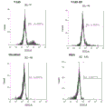
Classifications
-
- G—PHYSICS
- G01—MEASURING; TESTING
- G01N—INVESTIGATING OR ANALYSING MATERIALS BY DETERMINING THEIR CHEMICAL OR PHYSICAL PROPERTIES
- G01N33/00—Investigating or analysing materials by specific methods not covered by groups G01N1/00 - G01N31/00
- G01N33/48—Biological material, e.g. blood, urine; Haemocytometers
- G01N33/50—Chemical analysis of biological material, e.g. blood, urine; Testing involving biospecific ligand binding methods; Immunological testing
- G01N33/53—Immunoassay; Biospecific binding assay; Materials therefor
- G01N33/574—Immunoassay; Biospecific binding assay; Materials therefor for cancer
- G01N33/57484—Immunoassay; Biospecific binding assay; Materials therefor for cancer involving compounds serving as markers for tumor, cancer, neoplasia, e.g. cellular determinants, receptors, heat shock/stress proteins, A-protein, oligosaccharides, metabolites
- G01N33/57492—Immunoassay; Biospecific binding assay; Materials therefor for cancer involving compounds serving as markers for tumor, cancer, neoplasia, e.g. cellular determinants, receptors, heat shock/stress proteins, A-protein, oligosaccharides, metabolites involving compounds localized on the membrane of tumor or cancer cells
-
- C—CHEMISTRY; METALLURGY
- C07—ORGANIC CHEMISTRY
- C07K—PEPTIDES
- C07K16/00—Immunoglobulins [IGs], e.g. monoclonal or polyclonal antibodies
- C07K16/18—Immunoglobulins [IGs], e.g. monoclonal or polyclonal antibodies against material from animals or humans
-
- A—HUMAN NECESSITIES
- A61—MEDICAL OR VETERINARY SCIENCE; HYGIENE
- A61P—SPECIFIC THERAPEUTIC ACTIVITY OF CHEMICAL COMPOUNDS OR MEDICINAL PREPARATIONS
- A61P35/00—Antineoplastic agents
-
- C—CHEMISTRY; METALLURGY
- C07—ORGANIC CHEMISTRY
- C07K—PEPTIDES
- C07K16/00—Immunoglobulins [IGs], e.g. monoclonal or polyclonal antibodies
- C07K16/18—Immunoglobulins [IGs], e.g. monoclonal or polyclonal antibodies against material from animals or humans
- C07K16/28—Immunoglobulins [IGs], e.g. monoclonal or polyclonal antibodies against material from animals or humans against receptors, cell surface antigens or cell surface determinants
-
- C—CHEMISTRY; METALLURGY
- C12—BIOCHEMISTRY; BEER; SPIRITS; WINE; VINEGAR; MICROBIOLOGY; ENZYMOLOGY; MUTATION OR GENETIC ENGINEERING
- C12N—MICROORGANISMS OR ENZYMES; COMPOSITIONS THEREOF; PROPAGATING, PRESERVING, OR MAINTAINING MICROORGANISMS; MUTATION OR GENETIC ENGINEERING; CULTURE MEDIA
- C12N15/00—Mutation or genetic engineering; DNA or RNA concerning genetic engineering, vectors, e.g. plasmids, or their isolation, preparation or purification; Use of hosts therefor
- C12N15/09—Recombinant DNA-technology
- C12N15/63—Introduction of foreign genetic material using vectors; Vectors; Use of hosts therefor; Regulation of expression
-
- G—PHYSICS
- G01—MEASURING; TESTING
- G01N—INVESTIGATING OR ANALYSING MATERIALS BY DETERMINING THEIR CHEMICAL OR PHYSICAL PROPERTIES
- G01N33/00—Investigating or analysing materials by specific methods not covered by groups G01N1/00 - G01N31/00
- G01N33/48—Biological material, e.g. blood, urine; Haemocytometers
- G01N33/50—Chemical analysis of biological material, e.g. blood, urine; Testing involving biospecific ligand binding methods; Immunological testing
- G01N33/53—Immunoassay; Biospecific binding assay; Materials therefor
- G01N33/574—Immunoassay; Biospecific binding assay; Materials therefor for cancer
-
- A—HUMAN NECESSITIES
- A61—MEDICAL OR VETERINARY SCIENCE; HYGIENE
- A61K—PREPARATIONS FOR MEDICAL, DENTAL OR TOILETRY PURPOSES
- A61K39/00—Medicinal preparations containing antigens or antibodies
- A61K2039/505—Medicinal preparations containing antigens or antibodies comprising antibodies
-
- C—CHEMISTRY; METALLURGY
- C07—ORGANIC CHEMISTRY
- C07K—PEPTIDES
- C07K2317/00—Immunoglobulins specific features
- C07K2317/10—Immunoglobulins specific features characterized by their source of isolation or production
- C07K2317/14—Specific host cells or culture conditions, e.g. components, pH or temperature
-
- C—CHEMISTRY; METALLURGY
- C07—ORGANIC CHEMISTRY
- C07K—PEPTIDES
- C07K2317/00—Immunoglobulins specific features
- C07K2317/20—Immunoglobulins specific features characterized by taxonomic origin
- C07K2317/22—Immunoglobulins specific features characterized by taxonomic origin from camelids, e.g. camel, llama or dromedary
-
- C—CHEMISTRY; METALLURGY
- C07—ORGANIC CHEMISTRY
- C07K—PEPTIDES
- C07K2317/00—Immunoglobulins specific features
- C07K2317/20—Immunoglobulins specific features characterized by taxonomic origin
- C07K2317/24—Immunoglobulins specific features characterized by taxonomic origin containing regions, domains or residues from different species, e.g. chimeric, humanized or veneered
-
- C—CHEMISTRY; METALLURGY
- C07—ORGANIC CHEMISTRY
- C07K—PEPTIDES
- C07K2317/00—Immunoglobulins specific features
- C07K2317/30—Immunoglobulins specific features characterized by aspects of specificity or valency
- C07K2317/33—Crossreactivity, e.g. for species or epitope, or lack of said crossreactivity
-
- C—CHEMISTRY; METALLURGY
- C07—ORGANIC CHEMISTRY
- C07K—PEPTIDES
- C07K2317/00—Immunoglobulins specific features
- C07K2317/50—Immunoglobulins specific features characterized by immunoglobulin fragments
- C07K2317/52—Constant or Fc region; Isotype
-
- C—CHEMISTRY; METALLURGY
- C07—ORGANIC CHEMISTRY
- C07K—PEPTIDES
- C07K2317/00—Immunoglobulins specific features
- C07K2317/50—Immunoglobulins specific features characterized by immunoglobulin fragments
- C07K2317/56—Immunoglobulins specific features characterized by immunoglobulin fragments variable (Fv) region, i.e. VH and/or VL
- C07K2317/565—Complementarity determining region [CDR]
-
- C—CHEMISTRY; METALLURGY
- C07—ORGANIC CHEMISTRY
- C07K—PEPTIDES
- C07K2317/00—Immunoglobulins specific features
- C07K2317/50—Immunoglobulins specific features characterized by immunoglobulin fragments
- C07K2317/56—Immunoglobulins specific features characterized by immunoglobulin fragments variable (Fv) region, i.e. VH and/or VL
- C07K2317/569—Single domain, e.g. dAb, sdAb, VHH, VNAR or nanobody®
-
- C—CHEMISTRY; METALLURGY
- C07—ORGANIC CHEMISTRY
- C07K—PEPTIDES
- C07K2317/00—Immunoglobulins specific features
- C07K2317/60—Immunoglobulins specific features characterized by non-natural combinations of immunoglobulin fragments
- C07K2317/64—Immunoglobulins specific features characterized by non-natural combinations of immunoglobulin fragments comprising a combination of variable region and constant region components
-
- C—CHEMISTRY; METALLURGY
- C07—ORGANIC CHEMISTRY
- C07K—PEPTIDES
- C07K2317/00—Immunoglobulins specific features
- C07K2317/70—Immunoglobulins specific features characterized by effect upon binding to a cell or to an antigen
- C07K2317/73—Inducing cell death, e.g. apoptosis, necrosis or inhibition of cell proliferation
- C07K2317/732—Antibody-dependent cellular cytotoxicity [ADCC]
-
- C—CHEMISTRY; METALLURGY
- C07—ORGANIC CHEMISTRY
- C07K—PEPTIDES
- C07K2317/00—Immunoglobulins specific features
- C07K2317/70—Immunoglobulins specific features characterized by effect upon binding to a cell or to an antigen
- C07K2317/73—Inducing cell death, e.g. apoptosis, necrosis or inhibition of cell proliferation
- C07K2317/734—Complement-dependent cytotoxicity [CDC]
-
- C—CHEMISTRY; METALLURGY
- C07—ORGANIC CHEMISTRY
- C07K—PEPTIDES
- C07K2317/00—Immunoglobulins specific features
- C07K2317/90—Immunoglobulins specific features characterized by (pharmaco)kinetic aspects or by stability of the immunoglobulin
- C07K2317/92—Affinity (KD), association rate (Ka), dissociation rate (Kd) or EC50 value
Abstract
The present disclosure provides molecules that specifically bind to CLD18A2 comprising an immunoglobulin single variable domain. The present disclosure also provides nucleic acid molecules encoding the molecules that specifically bind to CLD18A2, expression vectors and host cells for expressing the molecules that specifically bind to CLD18 A2. The present disclosure further provides methods of producing the CLD18 A2-specific binding molecules and their use for treating or preventing CLD18 A2-associated disorders.
Description
Sequence listing
The present application contains a sequence listing and is incorporated by reference in its entirety.
Technical Field
The present application relates generally to antibodies. More specifically, the present application relates to molecules that specifically bind to CLD18A2, methods of making the same, and uses thereof.
Background
Cell attachment is a linking structure between cells, and is an important basis for the interaction and synergy between adjacent cells in multicellular organisms. In general, animal cells have four types of junctions, tight junctions, cohesive junctions, gap junctions, and desmosomes/hemidesmosomes.
Tight junctions, also known as closed junctions, are structures formed between endothelial cells or epithelial cells that prevent the diffusion of inter-tissue material from the interstitial space and thus allow entry into the cell only by active transport. Tightly linked structures are formed by several tens of Claudin proteins through intracellular and intercellular protein interactions, and the expression of these proteins has a certain tissue specificity. CLDN18 is an important protein among Claudin proteins present in tight junctions.
CLDN18 is a membrane protein with four transmembrane regions, containing two extracellular domains. There are two variants of CLDN18 in humans, CLDN18.1 and CLDN18.2 (also known as "CLD18 A2"), respectively. The two are distributed in different tissues, the former is mainly expressed in lung epithelial cells, the latter is specifically expressed in stomach epithelial cells and is not expressed in stomach stem cells. The sequence similarity of the CLDN18.1 and CLDN18.2 is extremely high in protein sequence, the main difference between the two is at the N-terminus, and only 8 amino acids are different between the two in the first extracellular domain; the C-terminal sequences are identical except for the N-terminal.
Antibody therapy is becoming one of the most promising approaches to treat cancer patients. CLDN18.2 has become a very potential target for antibody drug action due to its specificity of expression in tumor cells and normal tissues. However, because of the extremely high sequence similarity of CLDN18.1 and CLDN18.2, developing antibodies directed only to CLDN18.2 and not to CLDN18.1 is extremely difficult. Development of antibody drugs against CLDN18.2 target earlier was by gansmed corporation of germany, whose IMAB362 was developed as a monoclonal antibody consisting of two protein heavy chains and two protein light chains. However, up to now, no clinical research reports have been seen with single domain antibodies targeting CLDN 18.2.
Single domain antibodies, abbreviated as single domain antibodies (single domain antibody, sdabs), are antibodies that consist of a single monomer variable antibody domain (e.g., an antibody heavy chain variable domain). Like whole antibodies (e.g., igG), it is capable of selectively binding to a particular antigen. Single domain antibodies, however, have much lower molecular weights than the common antibodies that consist of two protein heavy chains and two protein light chains. The first single domain antibody was engineered from the heavy chain antibody found in camelids (Hamers-Casterman C, atarhouch T, muyldermans S, robinson G, hamers C, songa EB, bendahman N, hamers R (1993) Naturally occurring antibodies devoid of light char 363 (6428): 446-448.); heavy chain antibodies found in these camelids are also referred to as VHH fragments. Currently, most studies on single domain antibodies are based on heavy chain variable domains.
Single domain antibodies have a number of advantages. For example, they generally exhibit higher solubility, good thermal stability and tissue penetration, single domain antibodies are also resistant to degradation by papain and the like due to the presence of a second pair of intramolecular disulfide bonds; in addition, the single domain antibody can be expressed in various host cells such as yeast, plant and mammalian cells, and the expression level is high, so that the single domain antibody has extremely high cost advantage. The advantages of single domain antibodies make them suitable for use in a variety of biotechnology and therapeutic applications. For example, they may be used to treat diseases including, but not limited to, cancer, infectious diseases, inflammatory diseases, and neurodegenerative diseases.
Although there is currently a clinical on-the-fly monoclonal antibody drug targeting CLDN18.2 target, there is still an urgent need to continue to develop antibodies targeting CLDN18.2 target as therapeutic agents. It is desirable in the art to develop novel antibodies targeting CLDN18.2, in particular single domain antibodies that specifically recognize CLDN18.2 only and not CLDN 18.1.
Disclosure of Invention
In broad terms, the present disclosure provides novel compounds, methods of preparation, compositions and articles of manufacture of antibodies. The benefits provided by the present disclosure are broadly applicable to the fields of antibody therapy and diagnosis. More specifically, the present disclosure provides single domain antibodies targeting CLDN18.2, as well as methods of making the antibodies, expression vectors and host cells for expressing the antibodies, and the like. The antibodies of the present disclosure provide methods of treating or preventing disorders associated with Claudin proteins, particularly CLDN18.2, and uses thereof.
The inventors have found molecules that specifically bind to CLDN18.2, i.e., CLDN18.2 binding molecules that are capable of specifically binding to the extracellular domain 1 (ECD 1) of human CLDN18.2 (e.g., single domain antibodies that target CLDN 18.2).
The present disclosure includes at least the following embodiments, which are ordered and enumerated by way of "N" (where "N" represents a number), respectively. The following list is not exhaustive and the skilled person can combine different solutions.
A cldn18.2 binding molecule comprising at least one immunoglobulin single variable domain, wherein the immunoglobulin single variable domain comprises CDR1, CDR2 and CDR3, and wherein CDR1 differs in amino acid sequence from the sequence set forth in SEQ ID No. 1, 4 or 7 by NO more than 2 amino acid additions, deletions or substitutions; CDR2 differs in amino acid sequence from the sequence shown in SEQ ID NO. 2, 5 or 8 by amino acid additions, deletions or substitutions of not more than 2 amino acids; and/or the CDR3 differs in amino acid sequence from the sequence shown in SEQ ID NO 3, 6 or 9 by amino acid additions, deletions or substitutions of NO more than 2 amino acids.
2. The CLDN18.2 binding molecule of embodiment 1, wherein the CLDN18.2 binding molecule is an antibody or antigen-binding fragment thereof directed against CLDN 18.2.
3. The CLDN18.2 binding molecule of any one of the preceding embodiments, wherein the immunoglobulin single variable domain is a VHH; for example VHH from a camelid (e.g. alpaca).
4. The CLDN18.2 binding molecule of any one of the preceding embodiments, wherein the immunoglobulin single variable domain comprises:
i) CDR1 as shown in SEQ ID NO 1, 4 or 7;
ii) CDR2 as shown in SEQ ID NO 2, 5 or 8; and
iii) CDR3 as shown in SEQ ID 3, 6 or 9.
5. The CLDN18.2 binding molecule of any one of the preceding embodiments, wherein the immunoglobulin single variable domain comprises:
(a) CDR1 of the amino acid sequence shown in SEQ ID NO. 1, CDR2 of the amino acid sequence shown in SEQ ID NO. 2 and CDR3 of the amino acid sequence shown in SEQ ID NO. 3;
(b) CDR1 of the amino acid sequence shown in SEQ ID NO. 4, CDR2 of the amino acid sequence shown in SEQ ID NO. 5 and CDR3 of the amino acid sequence shown in SEQ ID NO. 6; or (b)
(c) CDR1 of the amino acid sequence shown in SEQ ID NO. 7, CDR2 of the amino acid sequence shown in SEQ ID NO. 8 and CDR3 of the amino acid sequence shown in SEQ ID NO. 9.
6. The CLDN18.2 binding molecule of any one of the preceding embodiments, wherein the immunoglobulin single variable domain comprises or consists of:
(A) An amino acid sequence shown in SEQ ID NO. 10, 11, 12, 13, 14 or 15;
(B) An amino acid sequence which is at least 80%, 85%, 90%, 95%, 98% or 99% identical to the amino acid sequence shown in SEQ ID No. 10, 11, 12, 13, 14 or 15; or (b)
(C) Amino acid sequences having one or more (e.g., 1, 2, 3, 4, 5, 6, 7, 8, 9, or 10) amino acids compared to SEQ ID NOs 10, 11, 12, 13, 14, or 15, preferably, the additions, deletions, and/or substitutions do not occur in the CDR regions.
7. The CLDN18.2 binding molecule of any one of the preceding embodiments, wherein the CLDN18.2 binding molecule is a single domain antibody, e.g., a heavy chain single domain antibody; a chimeric antibody; or a humanized antibody.
8. The CLDN18.2 binding molecule of any one of the preceding embodiments, wherein the immunoglobulin single variable domain is fused to another molecule, e.g., an Fc domain of an immunoglobulin (e.g., igG) or a fluorescent protein.
9. The CLDN18.2 binding molecule of embodiment 8, wherein the CLDN18.2 binding molecule is a chimeric antibody comprising VHH from a camelid and an Fc domain of a human IgG (e.g., human IgG1 or IgG 4).
10. The CLDN18.2 binding molecule of embodiment 9, wherein the CLDN18.2 binding molecule is a chimeric antibody comprising VHH from alpaca and an Fc domain of human IgG 1.
11. The CLDN18.2 binding molecule of any one of the preceding embodiments, wherein the CLDN18.2 binding molecule binds to extracellular domain 1 (ECD 1) of human CLDN 18.2.
A CLDN18.2 binding molecule that competes for the same epitope with a CLDN18.2 binding molecule of any of the preceding embodiments.
13. The CLDN18.2 binding molecule of any one of the preceding embodiments which specifically binds CLDN18.2 but does not bind CLDN18.1.
14. An isolated nucleic acid molecule encoding a CLDN18.2 binding molecule as defined in any one of the preceding embodiments.
15. An expression vector comprising the isolated nucleic acid molecule of embodiment 14.
16. A host cell comprising the expression vector of embodiment 15.
17. The host cell of embodiment 16, which is a bacterial cell (e.g., E.coli), a fungal cell (e.g., yeast), or a mammalian cell.
18. A pharmaceutical composition comprising at least one CLDN18.2 binding molecule as defined in any one of embodiments 1-13 and a pharmaceutically acceptable carrier.
19. A method of preparing a CLDN18.2 binding molecule as defined in any one of embodiments 1-13 comprising the steps of:
-expressing in a host cell of embodiment 16 or 17 a CLDN18.2 binding molecule as defined in any one of embodiments 1-13; and
isolating CLDN18.2 binding molecules from host cells.
20. A method of treating a disorder associated with CLDN18.2 in a subject, the method comprising administering to the subject a therapeutically effective amount of a CLDN18.2 binding molecule as defined in any one of embodiments 1-13.
21. The method of embodiment 20, wherein the disorder associated with CLDN18.2 comprises a disease involving cells expressing CLDN18.2 or a disease associated with cells expressing CLDN 18.2.
22. The method of embodiment 20 or 21, wherein the disorder associated with CLDN18.2 comprises cancer.
23. The method of embodiment 22, wherein the cancer comprises bone cancer, blood cancer, lung cancer, liver cancer, pancreatic cancer, skin cancer, head and neck cancer, skin or intraocular melanoma, uterine cancer, ovarian cancer, rectal cancer, anal region cancer, gastric cancer, colon cancer, breast cancer, prostate cancer, uterine cancer, cancer of the sexual and reproductive organs, hodgkin's disease, esophageal cancer, small intestine cancer, cancer of the endocrine system, thyroid cancer, parathyroid cancer, adrenal cancer, soft tissue sarcoma, bladder cancer, kidney cancer, renal cell carcinoma, renal pelvis cancer, central Nervous System (CNS) tumors, neuroectodermal cancer, spinal axis tumors, glioma, meningioma, and pituitary adenoma.
24. The method of embodiment 23, wherein the cancer is gastric cancer.
25. Use of a CLDN18.2 binding molecule as defined in any one of embodiments 1-13 in the manufacture of a medicament for treating or preventing a disorder associated with CLDN 18.2.
26. A kit for treating or diagnosing a disorder associated with CLDN18.2 comprising a container comprising a CLDN18.2 binding molecule as defined in any one of embodiments 1-13.
The foregoing is a general description and may include simplifications, generalizations, and omissions of detail as necessary. Accordingly, those skilled in the art will recognize that this general description is merely illustrative and is not intended to be limiting in any way. Other aspects, features, and advantages of the methods, compositions, and/or devices described herein and/or other subject matter will become apparent in the teachings shown herein. The general description is provided to introduce a selection of concepts in a simplified form that are further described below in the detailed description. The above summary is not intended to identify key features or essential features of the claimed subject matter, nor is it intended to be used as an aid in determining the scope of the claimed subject matter. Further, the contents of all references, patents and published patent applications cited throughout this application are incorporated herein by reference in their entirety.
Drawings
FIGS. 1a and 1b show the results of flow cytometry identification of overexpressing cell lines and tumor cell lines of example 1 using IMAB362, hIgG and antibodies against CLDN18. Wherein, FIG. 1a shows the results of flow cytometry identification of human CLDN18.2-HEK293T, human CLDN18.1-HEK293, murine CLDN18.2-HEK293 and murine CLDN18.1-HEK293 overexpressing cell lines; FIG. 1b shows the results of flow cytometry identification of human CLDN18.2-KATOIII tumor cell lines.
FIGS. 2a-2f show the results of cross-binding experiments of candidate antibodies NA4-S, NA7-S and NA8-S on human CLDN18.2-HEK293T, human CLDN18.1-HEK293, murine CLDN18.2-HEK293 and murine CLDN18.1-HEK293 overexpressing cell lines.
FIGS. 3a, 3b and 3c show the results of a comparative experiment of binding of candidate antibodies NA4-S, NA7-S, NA8-S and control antibodies at the cellular level. Wherein FIG. 3a shows the results of a flow-level comparison experiment of antibodies NA4-S and NA7-S on human CLDN18.2-HEK293T overexpressing cells; FIG. 3b shows results of a flow-through level comparison experiment of antibody NA8-S on human CLDN18.2-HEK293T overexpressing cells; FIG. 3c shows the results of a comparative experiment of the flow levels of antibodies NA4-S, NA7-S, NA8-S and control antibodies on human CLDN18.2-KATOIII tumor cell lines.
FIGS. 4a, 4b, 4c, 4d and 4e show Complement Dependent Cytotoxicity (CDC) mediated by candidate antibodies NA4-S, NA7-S and NA8-S on human CLDN18.2-HEK293T overexpressing cell lines and human CLDN18.2-KATOIII tumor cell lines. Wherein FIGS. 4a, 4b and 4c show the CDC cytotoxic effects mediated by antibodies NA4-S, NA-S and NA8-S on human CLDN18.2-HEK293T, respectively; FIGS. 4d and 4e show CDC mediated by antibodies NA7-S and NA8-S, respectively, on human CLDN18.2-KATOIII tumor cell lines.
FIGS. 5a, 5b, 5c, 5d show antibody-dependent cell-mediated cytotoxicity (ADCC) mediated by candidate antibodies NA7-S and NA8-S on human CLDN18.2-HEK293T and human CLDN18.2-KATOIII tumor cell lines. Wherein FIGS. 5a and 5b show ADCC cytotoxicity effects mediated through antibodies NA7-S and NA8-S on the human CLDN18.2-HEK293T cell line; FIGS. 5c and 5d show ADCC mediated by antibodies NA7-S and NA8-S on human CLDN18.2-KATOIII tumor cell lines.
FIGS. 6a and 6b show the tumor inhibition effect of candidate antibodies NA7-S and NA8-S in a human CLDN18.2-HEK293T xenograft model. FIG. 6a shows the tumor suppression effect of candidate antibody NA7-S in a human CLDN18.2-HEK293T xenograft model; FIG. 6b shows the tumor suppression effect of candidate antibody NA8-S in a human CLDN18.2-HEK293T xenograft model. Wherein, the mice are immune deficient mice SCID, and NA7-S, NA8-S and IMAB362 are administered according to the mass concentration of each kilogram of body weight, and the administration mode is cross administration of tail vein and abdominal cavity.
FIGS. 7 a-7 g show the binding epitope on the CLDN18.2 protein and the key amino acid positions of binding for candidate antibodies NA7-S, NA-S and IMAB 362. Wherein FIG. 7a shows a competition binding assay of candidate antibodies NA7-S and NA8-S to IMAB362 on human CLDN18.2-HEK 293T; FIG. 7b shows a competition binding assay of antibodies IMAB362 and NA8-S to NA7-S on human CLDN18.2-HEK 293T; FIG. 7c uses anti-CLDN 18 antibodies to determine the expression levels of mutant CLDN18.2 and wild CLDN18.2 on wild and mutant cell lines of human CLDN 18.2; FIG. 7d shows the binding levels of NA7-S and IMAB362 to the CLDN18.2 mutant on human CLDN18.2 mutant cell lines; FIG. 7e shows the relative percentage binding of NA7-S and IMAB362 on the human CLDN18.2 mutant cell line relative to the wild-type cell line. FIG. 7f shows the binding levels of NA8-S and IMAB362 to the CLDN18.2 mutant on a human CLDN18.2 mutant cell line; FIG. 7g shows the relative percent binding of NA8-S and IMAB362 on a human CLDN18.2 mutant cell line relative to the wild-type cell line.
FIGS. 8a-8b show the cell killing efficiency of NA7-S, NA8-S and IMAB362 against CLDN18.2-HEK293T in the presence of Fab-ZAP. FIG. 8a shows the cell killing efficiency of NA7-S and IMAB362 against CLDN18.2-HEK293T, and FIG. 8b shows the cell killing efficiency of NA8-S and IMAB362 against CLDN18.2-HEK293T, wherein the assay uses hIgG and Fab-ZAP as controls and the Fab-ZAP has some toxic effect on cells when incubated with cells for a prolonged period of time.
FIGS. 9a-9c show binding activity of NA4-S in human CLDN18.2-HEK293T and human CLDN18.1-HEK293T cells before and after humanization of the sequence, wherein NA4-S-H7 is a humanized molecule. FIG. 9a shows comparison of binding activity of NA4-S humanized pre-and post-molecules in human CLDN18.2-HEK293T cells; FIG. 9b shows the binding strength of NA4-S, NA4-S-H7 and IMAB362 at different concentrations on CLDN18.1-HEK293T cells; FIG. 9c shows the binding positivity of NA4-S, NA4-S-H7 and IMAB362 on CLDN18.1-HEK293T cells at high concentrations (100. Mu.g/ml), using irrelevant antibodies as controls.
FIGS. 10a-10c show binding activity of NA7-S in human CLDN18.2-HEK293T and human CLDN18.1-HEK293T cells before and after humanization of the sequence, wherein NA7-S-H2 is a humanized molecule. FIG. 10a shows comparison of binding activity of NA7-S humanized pre-and post-molecules in human CLDN18.2-HEK293T cells; FIG. 10b shows the binding strength of NA7-S, NA7-S-H2 and IMAB362 at different concentrations on CLDN18.1-HEK293T cells; FIG. 10c shows the binding positivity of NA7-S, NA7-S-H2 and IMAB362 on CLDN18.1-HEK293T cells at high concentrations (100. Mu.g/ml), using irrelevant antibodies as controls.
FIGS. 11a-11c show binding activity of NA8-S in human CLDN18.2-HEK293T and human CLDN18.1-HEK293T cells before and after humanization of the sequence, wherein NA8-S-H9 is a humanized molecule. FIG. 11a shows comparison of binding activity of NA8-S humanized pre-and post-molecules in human CLDN18.2-HEK293T cells; FIG. 11b shows the binding strength of NA8-S, NA8-S-H9 and IMAB362 at different concentrations on CLDN18.1-HEK293T cells; FIG. 11c shows the binding positivity of NA8-S, NA8-S-H9 and IMAB362 on CLDN18.1-HEK293T cells at high concentrations (100. Mu.g/ml), using irrelevant antibodies as controls.
Detailed Description
While this invention may be embodied in many different forms, there are disclosed herein specific illustrative embodiments thereof which are indicative of the principles of the invention. It should be emphasized that the present invention is not limited to the specific embodiments illustrated. Furthermore, any section headings used herein are for organizational purposes only and are not to be construed as limiting the subject matter described.
Unless otherwise defined herein, scientific and technical terms used in connection with the present invention will have the meanings commonly understood by one of ordinary skill in the art. Furthermore, unless the context requires otherwise, terms in the singular shall include the plural and terms in the plural shall include the singular. More specifically, as used in this specification and the appended claims, the singular forms "a," "an," and "the" include plural referents unless the context clearly dictates otherwise. Thus, for example, reference to "a protein" includes a plurality of proteins; reference to "a cell" includes mixtures of cells and the like. In this application, the use of "or" means "and/or" unless stated otherwise. Furthermore, the use of the term "include" and other forms (such as "include" and "contain") is not limiting. Furthermore, the scope provided in the specification and the appended claims includes all values between the endpoints and between the endpoints.
Generally, terms related to cell and tissue culture, molecular biology, immunology, microbiology, genetics and protein and nucleic acid chemistry and hybridization described herein, and techniques thereof, are well known and commonly used in the art. Unless otherwise indicated, the methods and techniques of the present invention are generally performed according to conventional methods well known in the art and as described in the various general and more specific references cited and discussed throughout the present specification. See, e.g., abbas et al, cellular and Molecular Immunology,6 th ed.,W.B.Saunders Company(2010);Sambrook J.&Russell d.molecular Cloning: A Laboratory Manual,3rd ed., cold Spring Harbor Laboratory Press, cold Spring Harbor, n.y. (2000); ausubel et al Short Protocols in Molecular Biology: A Compendium of Methods from Current Protocols in Molecular Biology, wiley, john&Sons, inc. (2002); harlow and Lane Using Antibodies: A Laboratory Manual, cold Spring Harbor Laboratory Press, cold Spring Harbor, n.y. (1998); and Coligan et al, short Protocols in Protein Science, wiley, john&Sons, inc. (2003). The terminology and laboratory procedures and techniques related to analytical chemistry, synthetic organic chemistry and pharmaceutical chemistry described herein are well known in the art and are described in detail in the following Commonly used terminology. Furthermore, any section headings used herein are for organizational purposes only and are not to be construed as limiting the subject matter described.
Definition of the definition
For a better understanding of the present invention, definitions and explanations of related terms are provided below.
As used herein, the term "antibody" or "Ab" generally refers to any form of antibody that exhibits the desired biological or binding activity. It includes, but is not limited to, humanized antibodies, fully human antibodies, chimeric antibodies, and single domain antibodies. Antibodies may comprise a heavy chain and a light chain. Heavy chains can be divided into μ, δ, γ, α and ε, which define the isotype of antibodies as IgM, igD, igG, igA and IgE, respectively. Each heavy chain consists of a heavy chain variable region (VH) and a heavy chain constant region (CH). The heavy chain constant region consists of 3 domains (CH 1, CH2 and CH 3). Each light chain consists of a light chain variable region (VL) and a light chain constant region (CL). VH and VL regions can be further divided into relatively conserved regions, known as Framework Regions (FR), and hypervariable regions, known as Complementarity Determining Regions (CDRs), separated by FR. Each VH and VL is composed of 3 CDRs and 4 FRs in the order from N-terminus to C-terminus, FR1, CDR1, FR2, CDR2, FR3, CDR3, FR4. The distribution of amino acids in various regions or domains follows Kabat Sequences of Proteins of Immunological Interest (National Institutes of Health, bethesda, md. (1987 and 1991)) or Chothia & Lesk (1987) J.mol.biol.196:901-917; chothia et al (1989) Nature 342:878-883. Antibodies may have different antibody isotypes, for example IgG (including for example IgG1, igG2, igG3 or IgG4 subtypes), igA1, igA2, igD, igE or IgM antibodies.
The term "humanized antibody" as used herein refers to an antibody in which CDR sequences derived from the germline of another non-human mammal are grafted onto human framework sequences. Additional framework region modifications may be made within the human framework sequence.
The term "chimeric antibody" as used herein refers broadly to engineered antibodies that contain one or more regions from one antibody and one or more regions from one or more other antibodies. In particular, chimeric antibodies comprise a variable region derived from a non-human animal antibody and a constant region of another antibody, for example comprising a variable region of camel origin and a constant region of human origin. Chimeric antibodies may also refer to multispecific antibodies that are specific for at least two different antigens.
The term "CLDN18.2 binding molecule" as used herein means a molecule that specifically binds CLDN 18.2.
The terms "CLDN18.2 antibody", "antibody against CLDN 18.2", "antibody specifically binding to CLDN 18.2", "antibody specifically targeting CLDN 18.2", "antibody specifically recognizing CLDN 18.2" as used herein are used interchangeably and mean an antibody capable of specifically binding to the Claudin protein CLDN 18.2. In particular, in a specific embodiment, it is meant an antibody that specifically binds to human CLDN18.2, in particular an antibody that specifically binds to human CLDN18.2 but not to human CLDN 18.1. The amino acid sequences of the human CLDN18.2 and the human CLDN18.1 are respectively shown as SEQ ID NO. 16 and SEQ ID NO. 17; the amino acid sequences of the mouse CLDN18.2 and the mouse CLDN18.1 are respectively shown as SEQ ID NO. 18 and SEQ ID NO. 19.
The term "immunoglobulin single variable domain" or "ISV" as used herein is generally defined herein as an amino acid sequence that comprises an immunoglobulin fold or is capable of forming an immunoglobulin fold (i.e., by folding) under suitable conditions (e.g., physiological conditions), i.e., thereby forming an immunoglobulin variable domain (e.g., a VH, VL, or VHH domain); and forming (or under suitable conditions being able to form) an immunoglobulin variable domain comprising a functional antigen binding site (in the sense that it does not need to interact (e.g. VH-VL interactions) with another immunoglobulin variable domain to form a functional antigen binding site).
The term "Ka" as used herein is intended to denote the rate of association of a particular antibody-antigen interaction, while the term "Kd" as used herein is intended to denote the rate of dissociation of a particular antibody-antigen interaction. The term "K", as used herein D "or" K D The value "is intended to represent the dissociation constant of a particular antibody-antigen interaction, which is obtained from the ratio of Kd to Ka (i.e., kd/Ka) and expressed as molar concentration(M). Antibody K D The values may be determined using well-established methods in the art. Determination of antibody K D The preferred method of value is by using surface plasmon resonance, preferably using a biosensor system such as The system.
The term "specific binding" or "specifically binding to" as used herein refers to a non-random binding reaction between two molecules, e.g., between an antibody and an antigen.
As used herein, "ability to inhibit binding," "block binding," or "compete for the same epitope" refers to the ability of an antibody to inhibit the binding of two molecules to any detectable extent. In some embodiments, an antibody that blocks binding between two molecules inhibits binding interactions between two molecules by at least 50%. In some embodiments, the inhibition may be greater than 60%, greater than 70%, greater than 80% or greater than 90%.
The term "high affinity" antibody as used herein refers to an antibody having 1 x 10 binding to a target antigen -7 M or less, more preferably 5X 10 -8 M or less, even more preferably 1X 10 -8 M or less, even more preferably 5X 10 -9 M or less, and even more preferably 1X 10 -9 M or lower K D Antibodies of the values.
The term "EC" as used herein 50 ", also referred to as" half-maximal effective concentration, "refers to the concentration of a drug, antibody, or toxin that induces a 50% response between baseline and maximum after a particular exposure time. In the context of the present application, EC 50 In units of "nM".
As used herein, the term "epitope" refers to the portion of an antigen to which an immunoglobulin or antibody specifically binds. An "epitope" is also referred to as an "epitope". Epitopes or antigenic determinants are generally composed of chemically active surface groupings of molecules such as amino acids, carbohydrates or sugar side chains, and generally have a specific three dimensional structure and specific charge characteristics. For example, an epitope typically comprises at least 3, 4, 5, 6, 7, 8, 9, 10, 11, 12, 13, 14, or 15 contiguous or non-contiguous amino acids in a unique steric conformation, which may be a "linear epitope" or a "conformational epitope. See, e.g., epitope Mapping Protocols in Methods in Molecular Biology, vol.66, g.e.Morris, ed. (1996). In a linear epitope, all interaction sites between a protein and an interacting molecule (e.g., an antibody) exist linearly along the primary amino acid sequence of the protein. In conformational epitopes, the interaction sites span amino acid residues in the protein that are separated from each other. Antibodies can be screened depending on the competition for binding to the same epitope as detected by conventional techniques known to those skilled in the art. For example, competition or cross-competition studies can be performed to obtain antibodies that compete or cross-compete with each other for binding to an antigen (e.g., CLDN 18.2). In international patent application WO03/048731, high throughput methods for obtaining antibodies binding to the same epitope are described, which are based on their cross-competition.
As used herein, the term "isolated" refers to a state of a substance or component obtained from a natural state by artificial means. If a certain "isolated" substance or component occurs naturally, it may be due to a change in the natural environment in which it is located, or the separation of the substance or component from the natural environment, or both. For example, a polynucleotide or polypeptide that is not isolated naturally occurs in a living animal, and the same high purity polynucleotide or polypeptide that is isolated from that natural state is referred to as an isolated polynucleotide or polypeptide. The term "isolated" does not exclude mixed artificial or synthetic substances nor other impure substances that do not affect the activity of the isolated substances.
As used herein, the term "isolated antibody" refers to an antibody that is substantially free of other antibodies having different antigen specificities. In addition, the isolated antibodies may be substantially free of other cellular material and/or chemicals.
As used herein, the term "vector" refers to a nucleic acid vector into which a polynucleotide may be inserted. When a vector allows expression of a protein encoded by a polynucleotide inserted therein, the vector is referred to as an expression vector. The vector may be transformed, transduced or transfected into a host cell to express the carried genetic material element in the host cell. Vectors are well known to those skilled in the art and include, but are not limited to, plasmids, phages, cosmids, artificial chromosomes such as Yeast Artificial Chromosome (YAC), bacterial Artificial Chromosome (BAC) or P1 derived artificial chromosome (PAC), phages such as lambda phage or M13 phage, and animal viruses. Animal viruses that may be used as vectors include, but are not limited to, retroviruses (including lentiviruses), adenoviruses, adeno-associated viruses, herpesviruses (e.g., herpes simplex virus), poxviruses, baculoviruses, papillomaviruses, papovaviruses (e.g., SV 40). The vector may contain a number of elements for controlling expression, including but not limited to promoter sequences, transcription initiation sequences, enhancer sequences, selection elements and reporter genes. In addition, the vector may comprise an origin of replication.
As used herein, the term "host cell" refers to any kind of cell system into which a vector can be introduced, including, but not limited to, prokaryotic cells such as e.coli (e.coli) or bacillus subtilis (Bacillus subtilis), fungal cells such as yeast cells or Aspergillus (Aspergillus), insect cells such as S2 drosophila cells or Sf9, and animal cells such as fibroblasts, CHO cells, COS cells, NSO cells, heLa cells, BHK cells, HEK293 cells or human cells.
Methods for producing antibodies of the invention using host cells are conventional in the art and include expressing the antibodies in prokaryotic or eukaryotic cells, then isolating the antibodies, and generally purifying to a pharmaceutically acceptable purity. As in some embodiments, nucleic acids encoding antibodies are inserted into expression vectors and the expression vectors are introduced into suitable prokaryotic or eukaryotic host cells by standard techniques known in the art, the host cells are cultured, such as CHO cells, NSO cells, SP2/0 cells, HEK293 cells, COS cells, per.c6 (R) cells, yeast or e.coli cells, under conditions and for a time sufficient to produce the antibodies or functional fragments thereof of the invention, and the antibodies are recovered from the cells (supernatant or cells after lysis) and purified. Conventional methods for producing antibodies are known in the art and are described, for example, in Makrides, s.c., protein expr.purif.17 (1999) 183-202; geisse, S.et al Protein expr. Purif.8 (1996) 271-282; kaufman, R.J., mol.Biotechnol.16 (2000) 151-160; fferner, r.g., drug res.48 (1998) review articles 870-880.
As used herein, the term "identity" refers to the relationship between sequences of two or more polypeptide molecules or two or more nucleic acid molecules as determined by aligning and comparing the sequences. "percent identity" refers to the percentage of identical residues between amino acids or nucleotides in a comparison molecule and is calculated based on the size of the smallest molecule being compared. For these calculations, the gaps in the alignment (if any) are preferably addressed by a specific mathematical model or computer program (i.e., an "algorithm"). Methods that can be used to calculate identity of aligned nucleic acids or polypeptides include those described in Computational Molecular Biology, (Lesk, a.m., ed.), 1988,New York:Oxford University Press; biocomputing Informatics and Genome Projects, (Smith, d.w., ed.), 1993,New York:Academic Press; computer Analysis of Sequence Data Part I, (Griffin, a.m., and Griffin, h.g., eds.), 1994,New Jersey:Humana Press; von Heinje, g.,1987,Sequence Analysis in Molecular Biology,New York:Academic Press; sequence Analysis Primer, (Gribskov, m.and deveeux, j., eds.), 1991,New York:M.Stockton Press; and those described in Carilo et al, 1988,SIAMJ.Applied Math.48:1073.
As used herein, the term "immunogenicity" refers to the ability to stimulate the formation of specific antibodies or sensitized lymphocytes in an organism. It refers not only to the nature of antigens to stimulate the activation, proliferation and differentiation of specific immune cells to ultimately produce immune effector substances such as antibodies and sensitized lymphocytes, but also to the fact that specific immune responses of antibodies or sensitized T lymphocytes can develop in the immune system of an organism after stimulation of the organism with an antigen. Immunogenicity is the most important property of an antigen. Whether an antigen is capable of successfully inducing an immune response in a host depends on three factors, the nature of the antigen, the reactivity of the host and the means of immunization.
As used herein, the term "transfection" refers to the process of introducing nucleic acid into eukaryotic cells, particularly mammalian cells. Protocols and techniques for transfection include, but are not limited to, lipofection, chemical and physical methods of transfection such as electroporation. Numerous transfection techniques are known in the art, see, e.g., graham et al, 1973,Virology 52:456; sambrook et al, 2001,Molecular Cloning:A Laboratory Manual; davis et al, 1986,Basic Methods in Molecular Biology,Elsevier; chu et al,1981, gene 13:197.
As used herein, the term "fluorescence activated cell sorting" or "FACS" refers to a specialized type of flow cytometry. It provides a method of sorting a heterogeneous mixture of biological cells into two or more containers one cell at a time according to specific light scattering and fluorescence characteristics of each cell (flowmetric. "Sorting Out Fluorescence Activated Cell Sorting". 2017-11-09). The apparatus for performing FACS is known to those skilled in the art and commercially available to the public. Examples of such instruments include FACS Star Plus, FACScan and FACSort instruments from Becton Dickinson (Foster City, calif.), epics C from Coulter Epics Division (Hialeah, FL) and MoFlo from Cytomation (Colorado Springs, colorado).
The term "subject" includes any human or non-human animal, preferably a human.
As used herein, the term "disorder associated with CLDN 18.2" refers to any disorder caused, exacerbated, or otherwise associated with increased or decreased expression or activity of CLDN18.2 (e.g., human CLDN 18.2).
As used herein, the term "cancer" refers to a solid or non-solid tumor such as leukemia mediated by the growth, proliferation or metastasis of any tumor or malignant cell that is involved in a medical condition.
The term "treatment" as used herein in the context of treating a condition generally relates to the treatment and therapy of a human or animal in which some desired therapeutic effect is achieved, e.g., inhibiting the progression of the condition, including a decrease in the rate of progression, a arrest in the rate of progression, regression of the condition, improvement of the condition, and cure of the condition. Treatment as a prophylactic measure (i.e., prophylaxis) is also included. For cancer, "treatment" may refer to inhibiting or slowing the growth, proliferation, or metastasis of a tumor or malignant cell, or some combination thereof. For a tumor, "treating" includes removing all or part of the tumor, inhibiting or slowing the growth and metastasis of the tumor, preventing or delaying the progression of the tumor, or some combination thereof.
As used herein, the term "therapeutically effective amount" refers to an amount of an active compound or a material, composition or dosage form comprising an active compound that is effective for producing certain desired therapeutic effects commensurate with a reasonable benefit/risk ratio when administered according to a desired therapeutic regimen. In particular, a "therapeutically effective amount" means an amount or concentration of an antibody, or antigen-binding portion thereof, effective to treat a disorder associated with CLDN 18.2.
As used herein, the term "pharmaceutically acceptable" means that the carrier, diluent, excipient, and/or salt thereof is chemically and/or physically compatible with the other ingredients in the formulation, and physiologically compatible with the recipient.
As used herein, the term "pharmaceutically acceptable carrier and/or excipient" refers to a carrier and/or excipient that is pharmacologically and/or physiologically compatible with the subject and active agent, which is well known in the art (see, e.g., remington's Pharmaceutical sciences. Mediated by Gennaro AR,19th ed.Pennsylvania:Mack Publishing Company,1995), and includes, but is not limited to, pH modifiers, surfactants, adjuvants, and ionic strength enhancers. For example, pH modifiers include, but are not limited to, phosphate buffers; surfactants include, but are not limited to, cationic, anionic or nonionic surfactants, such as Tween-80; ionic strength enhancers include, but are not limited to, sodium chloride.
As used herein, the term "adjuvant" refers to a non-specific immunopotentiator that, when delivered to an organism with an antigen or delivered to an organism in advance, can enhance the immune response to an antigen or alter the type of immune response in an organism. There are a variety of adjuvants in the prior art including, but not limited to, aluminum adjuvants (e.g., aluminum hydroxide), freund's adjuvants (e.g., freund's complete adjuvant and Freund's incomplete adjuvant), corynebacterium parvum, lipopolysaccharide, cytokines, and the like. Freund's adjuvant is the most commonly used adjuvant in current animal experiments. Aluminum hydroxide adjuvants are more commonly used in clinical trials.
CLDN18.2 binding molecules
In some aspects, the disclosure provides CLDN18.2 binding molecules.
In general, CLDN18.2 binding molecules can include any molecule that specifically binds CLDN 18.2. In some cases, a "CLDN18.2 binding molecule" can include a "CLDN18.2 antagonist. The CLDN18.2 binding molecule or CLDN18.2 antagonist can be a polypeptide or protein, such as an antibody, more specifically an antibody that specifically binds CLDN18.2 (e.g., human CLDN 18.2).
Antibodies include, but are not limited to, chimeric antibodies, humanized antibodies, single domain antibodies, or the like. In a specific embodiment, the CLDN18.2 binding molecule is a single domain antibody, which generally refers to an antibody consisting of a single monomer variable antibody domain. Like whole antibodies, single domain antibodies are capable of selectively binding to a particular antigen.
More specifically, CLDN18.2 binding molecules are heavy chain single domain antibodies that can be associated with the terms "VHH", "VHH antibody", "VHH domain", "VHH antibody fragment", "V HH "or" nanobody "and the like are used interchangeably. VHH molecules from camelidae antibodies are one of the smallest known complete antigen binding domains (about 15KDa, or 1/10 of conventional IgG) and are therefore well suited for delivery to dense tissues and into the limited space between macromolecules.
The single domain antibodies disclosed herein can be prepared by one of skill in the art according to methods known in the art or any future method. For example, VHHs may be obtained using methods known in the art, for example by immunizing a camel and obtaining therefrom VHHs that bind to and neutralise target antigens, or by cloning a library of VHHs of the invention using molecular biology techniques known in the art, followed by selection using phage display. In some embodiments, the single domain antibodies of the invention are naturally produced in camelids, i.e., by immunizing a camel with CLDN18.2 or fragments thereof using techniques described herein for other antibodies.
In one placeIn some embodiments, single domain antibodies are obtained by immunizing llama or alpaca with the desired antigen and then isolating the mRNA encoding the heavy chain antibody. By reverse transcription and polymerase chain reaction, a gene library containing millions of cloned single domain antibodies is generated. Screening techniques such as phage display and ribosome display help identify antigen-binding clones. Wherein phage display is the synthesis of a library of antibodies on phage, screening the library with an antigen of interest or antibody binding portion thereof, and isolating phage that bind the antigen from which immunoreactive fragments can be obtained. Methods for preparing and screening such libraries are well known in the art, and kits for generating phage display libraries are commercially available (e.g., pharmacia recombinant phage antibody system, catalog No. 27-9400-01; and Stratagene SurfZAP) TM Phage display kit, catalog no 240612). Still other methods and reagents can be used to generate and screen antibody display libraries (see, e.g., barbas et al, proc.Natl. Acad. Sci. USA88:7978-7982 (1991)).
When the most potent clone is identified, its DNA sequence may be optimized, for example, by affinity maturation or humanization, to prevent immune responses in humans against antibodies.
Thus, the single domain antibodies of the invention can be obtained by (1) isolating the VHH domain of a naturally occurring heavy chain antibody; (2) By expressing a nucleotide sequence encoding a naturally occurring VHH domain; (3) By "humanization" of naturally occurring VHH domains or by expression of nucleic acids encoding such humanized VHH domains; (4) By "camelisation" of naturally occurring VH domains from any animal species, particularly mammalian species, for example from humans, or by expression of nucleic acids encoding such camelised VH domains; (5) "camelization" by "domain antibodies" or "dabs" (see, e.g., ward et al, 1989,Nature 341:544-546), or by expression of nucleic acids encoding such camelized VH domains; (6) Preparing a protein, polypeptide or other amino acid sequence by using synthetic or semi-synthetic techniques; (7) Preparing a nucleic acid encoding a VHH by using techniques for nucleic acid synthesis, and then expressing the nucleic acid thus obtained; and/or (8) by any combination of the foregoing. Suitable methods and techniques for performing the foregoing will be apparent to those skilled in the art based on the disclosure herein, and include methods and techniques as described in more detail below.
Single domain antibodies are typically produced by PCR cloning a library of variable domains into phage display vectors from cdnas of blood, lymph node or spleen lymphocytes obtained from immunized animals. Antigen-specific single domain antibodies are typically selected by panning the corresponding library over an immobilized antigen (e.g., an antigen coated on the surface of a tube plastic, a biotinylated antigen immobilized on streptavidin beads, or a membrane protein expressed on the cell surface). Affinity of sdabs can be increased by in vitro modeling of this strategy, for example by site-directed mutagenesis of CDR regions and further panning of immobilized antigen under increased stringency conditions (higher temperature, high or low salt concentration, high or low pH and low antigen concentration) (Wesolowski et al Single domain antibodies: promising experimental and therapeutic tools in infection and immunity. Med Microbiol Immunol (2009) 198:157-174).
Methods for preparing VHHs that specifically bind an antigen or epitope are described in the references, see, for example, R.van der Linden et al Journal of Immunological Methods,240 (2000) 185-195; li et al, J Biol chem.,287 (2012) 13713-13721; deffar et al African Journal of Biotechnology Vol.8 (12), pp.2645, 17June,2009 and WO94/04678.
In some embodiments, the VHH in the CLDN18.2 binding molecule is fused to an Fc domain of an antibody (e.g., an Fc domain of IgG (e.g., igG1 or IgG 4)). By fusing VHH to Fc domains, effector functions such as ADCC and CDC can be more effectively recruited. Furthermore, fusion of VHH to Fc domain can help the CLDN18.2 binding molecule form dimers and can also help to extend the in vivo half-life of CLDN18.2 binding molecules.
As used herein, the term "antibody-dependent cell-mediated cytotoxicity" or "ADCC" refers to a cytotoxic form of secreted antibodies that bind to Fc receptors (fcrs) present on certain cytotoxic effector cells (e.g., natural Killer (NK) cells, neutrophils and macrophages) to enable these cytotoxic effector cells to specifically bind to antigen-bearing target cells and subsequently kill the target cells with cytotoxins. The antibody "arming" the cytotoxic cells is absolutely required for this killing. The primary cells mediating ADCC, NK cells, express fcyriii only, whereas monocytes express fcyri, fcyrii and fcyriii. FcR expression on hematopoietic cells is summarized in Table 3 at page 464 of Ravetch and Kinet, annu. Rev. Immunol9:457-92 (1991). In order to assess ADCC activity of a molecule of interest, an in vitro ADCC assay may be performed, for example as described in US5500362 or US 5821337. Effector cells useful in such assays include Peripheral Blood Mononuclear Cells (PBMC) and Natural Killer (NK) cells. Alternatively or additionally, ADCC activity of the molecule of interest may be assessed in vivo, for example in an animal model as disclosed in Clynes et al PNAS (USA) 95:652-656 (1998).
The term "complement-dependent cytotoxicity" or "CDC" refers to the lysis of target cells in the presence of complement. Activation of the classical complement pathway is initiated by binding of the first component of the complement system (C1 q) to antibodies (appropriate subclasses) that bind to their cognate antigens. To assess complement activation, CDC assays may be performed by, for example, the method described in Gazzano-Santoro et al, J.Immunol. Methods 202:163 (1996).
For ease of description, CLDN18.2 binding molecules are described below as CLDN18.2 antibodies.
CLDN18.2 antibodies capable of specifically binding to a particular epitope of CLDN18.2
In one aspect, the disclosure relates to single domain antibodies that specifically bind CLDN18.2, but do not bind or do not substantially bind CLDN 18.1.
The inventors have discovered CLDN18.2 binding molecules (e.g., single domain antibodies targeting CLDN 18.2) that are capable of specifically binding to extracellular domain 1 (ECD 1) of human CLDN 18.2.
As previously described, CLDN18.2 has two extracellular domains (ECDs), wherein the full length sequence of human CLDN18.2 is shown in SEQ ID NO. 16 and wherein the sequence of ECD1 is shown in SEQ ID NO. 20. The mouse CLDN18.2 is shown as SEQ ID NO. 18.
For human and mouse CLDN18.1, the sequences are shown in SEQ ID NO. 17 and SEQ ID NO. 19, respectively.
CLDN18.2 antibodies comprising CDRs with sequence identity to a particular sequence
In some embodiments, a CLDN18.2 antibody of the disclosure comprises at least one immunoglobulin single variable domain (e.g., a VHH), wherein the VHH comprises CDR1, CDR2, and CDR3, and wherein CDR1 comprises an amino acid sequence that is at least 80% identical to SEQ ID No. 1, 4, or 7, CDR2 comprises an amino acid sequence that is at least 80% identical to SEQ ID No. 2, 5, or 8, and CDR3 comprises an amino acid sequence that is at least 80% identical to SEQ ID No. 3, 6, or 9.
In a given heavy chain variable region amino acid sequence, the exact amino acid sequence boundaries of each CDR can be determined using any one or a combination of a number of well-known antibody CDR assignment systems, including, for example: chothia (Chothia et al, (1989) Nature 342:877-883, al-Lazikani et al, "Standardconformations for the canonical structures of immunoglobulins", journal of Molecular Biology,273,927-948 (1997)) based on the three-dimensional structure of antibodies and topology of CDR loops, kabat (Kabat et al, sequences of Proteins ofImmunological Interest, 4 th edition, U.S. Pat. No. of Health and Human Services, nationalInstitutes of Health (1987)), abM (University of Bath), contact (University College London), international ImMunoGeneTics database (IMGT) (world Wide Web IMGT. Cines. Fr /), and North CDR definitions based on neighbor-propagating clusters (affinity propagation clustering) using a large number of crystal structures. The CLDN18.2 antibodies of the present disclosure employ the AbM approach to defining the amino acid sequence of the CDR regions, but such definition is not exclusive and limiting and does not even suggest that the present disclosure only protects the antibody molecules represented by the CDR regions defined using the AbM approach. Based on the aforementioned classes of antibody CDR assignment systems conventional in the art, the CLDN18.2 antibody molecules of the present disclosure can also be characterized in a variety of other CDR region definitions. It should be noted that the boundaries of CDRs of the variable regions of the same antibody obtained based on different assignment systems may differ, i.e., CDR sequences of the same antibody variable regions defined under different assignment systems may differ. CDR sequences defined under these different assignment systems and their characterized antibody molecules are also intended to be protected by the present disclosure.
The variable regions and CDRs in an antibody sequence can be identified according to general rules that have been developed in the art (as described above, e.g., the Kabat numbering system) or by aligning the sequences with a database of known variable regions. Methods of identifying these regions are described in Kontermann and Dubel, eds., antibody Engineering, springer, new York, NY,2001 and Dinarello et al Current Protocols in Immunology, john Wiley and Sons Inc., hoboken, NJ, 2000. Exemplary databases of antibody sequences are described in and available from the "Abysis" website on www.bioinf.org.uk/abs (maintained by Department of Biochemistry & Molecular Biology University College London, london, a.c. martin of England) and VBASE2 website www.vbase2.org, as described in Retter et al, nucleic acids res, 33 (Database issue): D671-D674 (2005). Sequences are preferably analyzed using the Abysis database, which integrates sequence data from Kabat, IMGT and Protein Databases (PDB) with structural data from PDB, see Protein Sequence and Structure Analysis of Antibody Variable domains in the book by dr. Andrew c.r.martin: antibody Engineering Lab Manual (Ed.: duebel, s.and Kontermann, r., springer-Verlag, heidelberg, ISBN-13:978-3540413547, also available on the website bioin for. The Abysis database website also includes general rules that have been developed for identifying CDRs that can be used in accordance with the teachings herein.
The percent identity between two amino acid sequences can be determined using the algorithm of e.meyers and w.miller (comp. Appl. Biosci.,4:11-17 (1988)), which has been incorporated into the ALIGN program (version 2.0), using the PAM120 weight residue table with a gap length penalty of 12 and a gap penalty of 4. In addition, the percent identity between two amino acid sequences can be determined by the algorithms of Needleman and Wunsch (j.mol. Biol.48:444-453 (1970)), which have been incorporated into the GAP program in the GCG software package (available from http:// www.gcg.com), using either the Blossum 62 matrix or PAM250 matrix, a GAP weight of 16, 14, 12, 10, 8, 6 or 4, and a length weight of 1, 2, 3, 4, 5 or 6.
Additionally or alternatively, the protein sequences of the present disclosure may be further used as "query sequences" to perform searches against public databases to, for example, identify related sequences. Such searches may be performed using the XBLAST program of Altschul, et al (1990) J.MoI.biol.215:403-10 (version 2.0). BLAST protein searches can be performed using the XBLAST program with a score=50 and word length=3 to obtain amino acid sequences homologous to the antibody molecules of the present disclosure. To obtain a gap alignment for comparison purposes, gap BLAST may be used, as described in Altschul et al, (1997) Nucleic Acids Res.25 (17): 3389-3402. When using BLAST and empty BLAST programs, default parameters for the respective programs (e.g., XBLAST and NBLAST) can be used, see www.ncbi.nlm.nih.gov.
In other embodiments, the amino acid sequence of a CDR may be at least 85%, 86%, 87%, 88%, 89%, 90%, 91%, 92%, 93%, 94%, 95%, 96%, 97%, 98% or 99% identical to each of the sequences given above.
CLDN18.2 antibodies comprising CDRs with amino acid additions, deletions or substitutions
In some embodiments, a CLDN18.2 antibody of the disclosure comprises at least one immunoglobulin single variable domain (e.g., a VHH), wherein the VHH comprises CDR1, CDR2, and CDR3, and wherein CDR1 differs in amino acid sequence from the sequence set forth in SEQ ID NOs 1, 4, or 7 by NO more than 2 (e.g., 0, 1, or 2) amino acid additions, deletions, or substitutions; CDR2 differs in amino acid sequence from the sequences shown in SEQ ID NOs 2, 5 or 8 by amino acid additions, deletions or substitutions of not more than 2 (e.g., 0, 1 or 2) amino acids; and/or CDR3 differs in amino acid sequence from the sequence shown in SEQ ID NO 3, 6 or 9 by amino acid additions, deletions or substitutions of not more than 2 (e.g.0, 1 or 2) amino acids. For example, CDR1, CDR2 and CDR3 differ from the amino acid sequences shown in SEQ ID NO. 1, 4 or 7, SEQ ID NO. 2, 5 or 8 and SEQ ID NO. 3, 6 or 9, respectively, by amino acid additions, deletions or substitutions of only one amino acid.
Preferably, the CDRs of an isolated antibody or antigen binding portion thereof contain conservative substitutions of no more than 2 amino acids or no more than 1 amino acid. As used herein, the term "conservative substitution" refers to an amino acid substitution that does not adversely affect or alter the basic properties of a protein/polypeptide comprising the amino acid sequence. For example, conservative substitutions may be introduced by standard techniques known in the art (e.g., site-directed mutagenesis and PCR-mediated mutagenesis). Conservative amino acid substitutions include substitutions in which an amino acid residue is substituted by another amino acid residue having a similar side chain, e.g., a residue that is physically or functionally similar (e.g., of similar size, shape, charge, chemical nature including the ability to form covalent or hydrogen bonds, etc.) is substituted by the corresponding amino acid residue. Families of amino acid residues with similar side chains have been defined in the art. These families include amino acids with basic side chains (e.g., lysine, arginine and histidine), acidic side chains (e.g., aspartic acid and glutamic acid), uncharged polar side chains (e.g., glycine, asparagine, glutamine, serine, threonine, tyrosine, cysteine, tryptophan), nonpolar side chains (e.g., alanine, valine, leucine, isoleucine, proline, phenylalanine, methionine), beta-branched side chains (e.g., threonine, valine, isoleucine) and aromatic side chains (e.g., tyrosine, phenylalanine, tryptophan, histidine). Thus, the corresponding amino acid residue is preferably substituted with another amino acid residue from the same side chain family. Methods for identifying amino acid conservative substitutions are well known in the art (see, e.g., brummell et al, biochem.32:1180-1187 (1993); kobayashi et al, protein Eng.12 (10): 879-884 (1999); and Burks et al, proc. Natl. Acad. Sci. USA 94:412-417 (1997), which is incorporated herein by reference).
In certain embodiments, the immunoglobulin single variable domain of the CLDN18.2 antibody comprises:
i) CDR1 of the amino acid sequence shown as SEQ ID NO. 1, 4 or 7;
ii) CDR2 as shown in SEQ ID NO 2, 5 or 8; and
iii) CDR3 as shown in SEQ ID 3, 6 or 9.
CLDN18.2 antibodies comprising CDRs
In some embodiments, the CLDN18.2 antibody of the disclosure comprises at least one immunoglobulin single variable domain (e.g., a VHH), wherein the VHH comprises CDR1, CDR2, and CDR3, and wherein CDR1, CDR2, and CDR3 are selected from the group consisting of:
(a) CDR1 of the amino acid sequence shown in SEQ ID NO. 1, CDR2 of the amino acid sequence shown in SEQ ID NO. 2 and CDR3 of the amino acid sequence shown in SEQ ID NO. 3;
(b) CDR1 of the amino acid sequence shown in SEQ ID NO. 4, CDR2 of the amino acid sequence shown in SEQ ID NO. 5 and CDR3 of the amino acid sequence shown in SEQ ID NO. 6; or (b)
(c) CDR1 of the amino acid sequence shown in SEQ ID NO. 7, CDR2 of the amino acid sequence shown in SEQ ID NO. 8 and CDR3 of the amino acid sequence shown in SEQ ID NO. 9.
CLDN18.2 antibodies defined by VHH sequences
In some embodiments, the CLDN18.2 antibodies of the disclosure comprise at least one (e.g., one) immunoglobulin single variable domain (e.g., VHH), wherein the VHH comprises:
(A) An amino acid sequence shown in SEQ ID NO. 10, 11, 12, 13, 14 or 15;
(B) An amino acid sequence which is at least 80%, 85%, 90%, 95%, 98% or 99% identical to SEQ ID No. 10, 11, 12, 13, 14 or 15; or (b)
(C) Amino acid sequences having one or more (e.g., 1, 2, 3, 4, 5, 6, 7, 8, 9, or 10) amino acids compared to SEQ ID NOs 10, 11, 12, 13, 14, or 15, preferably, the additions, deletions, and/or substitutions do not occur in the CDR regions.
In other embodiments, the amino acid sequence of the VHH may be at least 85%, 86%, 87%, 88%, 89%, 90%, 91%, 92%, 93%, 94%, 95%, 96%, 97%, 98% or 99% identical to each of the above sequences. As illustrative examples, an antibody may comprise a VHH having at least 85%, 86%, 87%, 88%, 89%, 90%, 91%, 92%, 93%, 94%, 95%, 96%, 97%, 98% or 99% sequence identity to SEQ ID No. 10, 11, 12, 13, 14 or 15.
In some further embodiments, the CLDN18.2 antibodies of the disclosure can comprise conservative substitutions or modifications of amino acids in the heavy chain variable region. It is understood in the art that certain conservative sequence modifications may be made that do not eliminate antigen binding properties. See, for example, brummel et al (1993) Biochem 32:1180-8; de Wildt et al (1997) Prot.Eng.10:835-41; komissarov et al (1997) J.biol. Chem.272:26864-26870; hall et al (1992) J.Immunol.149:1605-12; kelley and O' Connell (1993) biochem.32:6862-35; adib-Conquy et al (1998) int. Immunol.10:341-6 and beer et al (2000) Clin. Can. Res.6:2835-43.
Nucleic acid molecules encoding antibodies of the present disclosure and methods of producing CLDN18.2 antibodies
In some aspects, the disclosure relates to an isolated nucleic acid molecule comprising a nucleic acid sequence encoding a CLDN18.2 antibody of the disclosure.
The nucleic acids of the present disclosure may be obtained using standard molecular biology techniques. For antibodies obtained from immunoglobulin gene libraries (e.g., using phage display techniques), nucleic acids encoding such antibodies can be recovered from the gene library.
Nucleic acid molecules encoding the CLDN18.2 antibodies of the disclosure can be inserted into vectors for further cloning (amplifying DNA) or for expression using recombinant techniques known in the art. Many vectors are available. The vector or vector component typically includes, but is not limited to, one or more of a signal sequence, an origin of replication, one or more marker genes, an enhancer element, a promoter (e.g., SV40, CMV, EF-1. Alpha.), and a transcription termination sequence. The selectable marker gene facilitates selection of host cells into which the vector has been introduced (see, e.g., U.S. Pat. Nos. 4,399,216;4,634,665 and 5,179,017). For example, selectable marker genes typically confer resistance to drugs (e.g., G418, hygromycin or methotrexate) on the host cell into which the vector has been introduced. In one embodiment, the selectable marker genes may include a dihydrofolate reductase (DHFR) gene (for DHFR-host cells with methotrexate selection/amplification) and a neo gene (for G418 selection). In another embodiment, the antibodies may be produced by homologous recombination as known in the art. DNA encoding a monoclonal antibody is readily isolated and sequenced using conventional procedures (e.g., by using oligonucleotide probes that are capable of specifically binding to genes encoding the heavy chain of the antibody).
In some embodiments, the vector system includes mammalian, bacterial, yeast systems, and the like, and includes plasmids such as, but not limited to, pALTER, pBAD, pcDNA, pCal, pL, pET, pGEMEX, pGEX, pCI, pCMV, pEGFP, pEGFT, pSV, pFUSE, pVITRO, pVIVO, pMAL, pMONO, pSELECT, pUNO, pDUO, psg5L, pBABE, pWPXL, pBI, p15TV-L, pPro18, pTD, pRS420, pLexA, pact2.2, and the like, as well as other laboratory and commercially available vectors. Suitable vectors may include plasmids or viral vectors (e.g., replication defective retroviruses, adenoviruses and adeno-associated viruses). In one embodiment of the disclosure, the vector may be pET, e.g., pETbac containing a hexahistidine tag and a c-Myc-tag gene.
Vectors comprising a nucleic acid sequence encoding the CLDN18.2 antibodies of the disclosure can be introduced into host cells for cloning or gene expression. Suitable host cells for cloning or expressing the DNA in the vectors herein are prokaryotes, yeast, or higher eukaryote cells. Suitable prokaryotes for this purpose include eubacteria, such as gram-negative or gram-positive organisms, for example enterobacteriaceae such as the genus escherichia, for example escherichia coli, enterobacteriaceae, erwinia, klebsiella, proteus, salmonella, for example salmonella typhimurium, serratia, for example serratia marcescens and shigella, and bacillus such as bacillus subtilis and bacillus licheniformis, pseudomonas such as pseudomonas aeruginosa and streptomyces.
In addition to prokaryotes, eukaryotic microbes such as filamentous fungi or yeast are suitable host cells for expressing the CLDN18.2 antibodies of the disclosure. Saccharomyces cerevisiae or Saccharomyces cerevisiae is the most commonly used among lower eukaryotic host microorganisms. However, many other genera, species and strains are generally available and useful in the present disclosure, such as schizosaccharomyces pombe (Schizosaccharomyces pombe); kluyveromyces hosts such as Kluyveromyces lactis (K.lactis), kluyveromyces fragilis (K.fragilis) (ATCC 12,424), kluyveromyces bulgaricus (K.bulgaricus) (ATCC 16,045), kluyveromyces wikiwi (K.winkeramii) (ATCC 24,178), K.walti (ATCC 56,500), kluyveromyces drosophila (K drosophila) (ATCC 36,906), kluyveromyces thermotolerans (K.thermotolerans) and Kluyveromyces marxianus (K.marxianus); yarrowia (yarrowia) (EP 402,226); pichia pastoris (EP 183,070); candida (Candida); trichoderma reesia (EP 244,234); neurospora crassa (Neurospora crassa); schwanniomyces (schwanniomyces) such as Schwanniomyces western (schwanniomyces occidentalis); and filamentous fungi, such as Neurospora (Neurospora), penicillium (Penicillium), tolypocladium, and Aspergillus hosts such as Aspergillus nidulans (A. Nidulans) and Aspergillus niger (A. Niger).
Other suitable host cells for expressing the CLDN18.2 antibodies of the present disclosure are derived from multicellular organisms. Examples of invertebrate cells include plant and insect cells. A large number of baculovirus strains and variants and corresponding permissive insect host cells have been identified from the hosts Spodoptera frugiperda (Spodoptera Frugiperda, carpenterworm), aedes aegypti (mosquito), aedes albopictus (mosquito), drosophila melanogaster (Drosophila) and Bombycis mori (Bombyx mori). Various strains for transfection are publicly available, for example, the L-1 variant of the alfalfa spodoptera litura (Autographa californica) NPV and the Bm-5 strain of silkworm NPV, and according to the present disclosure, these viruses may be used in transfection procedures in which the CLDN18.2 antibody is expressed in suitable host cells, particularly for transfecting spodoptera frugiperda (Spodoptera frugiperda) cells. Plant cell cultures of cotton, corn, potato, soybean, petunia, tomato, and tobacco can also be used as hosts.
Host cells were transformed with the vectors described above for CLDN18.2 antibody production of the present disclosure and cultured in conventional nutrient media modified as needed for the induction of promoters, selection of transformants or amplification of genes encoding the desired sequences.
Host cells for producing CLDN18.2 antibodies of the present disclosure can be cultured in a variety of media. Commercially available media such as Ham's F10 (Sigma), minimal Essential Medium (MEM), (Sigma), RPMI-1640 (Sigma) and Dulbecco's Modified Eagle's Medium (DMEM, sigma) are suitable for culturing host cells. Furthermore, ham et al, meth.Enz.58:44 (1979), barnes et al, anal.biochem.102:255 (1980), U.S. Pat.No.4,767,704;4,657,866;4,927,762;4,560,655; or 5,122,469; WO 90/03430; WO 87/00195; or any of the media described in U.S. Pat. re.30,985 may be used as a medium for the host cells. Hormones and/or other growth factors (e.g., insulin, transferrin or epidermal growth factor), salts (e.g., sodium chloride, calcium, magnesium and phosphate), buffers (e.g., HEPES), nucleotides (e.g., adenosine and thymine), antibiotics (e.g., GENTAMYCIN drugs), trace elements (defined as inorganic compounds, typically present in final concentrations in the micromolar range) and glucose or equivalent energy sources may be added to any of these media as necessary. Any other necessary supplements may also be included in suitable concentrations known to those skilled in the art. Culture conditions such as temperature, pH, etc. are those used with the host cells previously selected for expression and will be apparent to the ordinarily skilled artisan.
When recombinant techniques are used, the antibodies may be produced intracellularly, in the periplasmic space, or secreted directly into the medium. If antibodies are produced intracellularly, as a first step, the particulate debris (host cells or lysed fragments) is removed, for example, by centrifugation or ultrafiltration. Carter et al, bio/Technology 10:163-167 (1992) describe a method of isolating antibodies secreted into the periplasmic space of E.coli. Briefly, cells were thawed in the presence of sodium acetate (pH 3.5), EDTA and phenylmethylsulfonyl fluoride (PMSF) in about 30 minutes. Cell debris can be removed by centrifugation. In the case of antibody secretion into the culture medium, the supernatant from such an expression system is typically first concentrated using a commercially available protein concentration filter, such as an Amicon or Millipore Pellicon ultrafiltration unit. Protease inhibitors such as PMSF may be included in any of the foregoing steps to inhibit proteolysis, and antibiotics may be included to prevent the growth of foreign contaminants.
Antibodies prepared from cells can be purified using, for example, hydroxyapatite chromatography, gel electrophoresis, dialysis, DEAE-cellulose ion exchange chromatography, ammonium sulfate precipitation, salting out, and affinity chromatography, with affinity chromatography being a preferred purification technique.
After any preliminary purification steps, the mixture comprising the antibody of interest and contaminants may be subjected to low pH hydrophobic interaction chromatography using an elution buffer having a pH between about 2.5 and 4.5, preferably at a low salt concentration (e.g., about 0-0.25M salt).
Pharmaceutical composition
In some aspects, the disclosure relates to pharmaceutical compositions comprising at least one CLDN18.2 binding molecule as disclosed herein (e.g., a CLDN18.2 antibody of the disclosure) and a pharmaceutically acceptable carrier.
Components of pharmaceutical compositions
The pharmaceutical composition may optionally contain one or more additional pharmaceutically active ingredients, such as another antibody or drug. The pharmaceutical compositions of the present disclosure may also be administered in combination with, for example, another immunostimulant, anticancer agent, antiviral agent, or vaccine, such that the anti-CLDN 18.2 antibody enhances the immune response to the vaccine. Pharmaceutically acceptable carriers can include, for example, pharmaceutically acceptable liquid, gel or solid carriers, aqueous media, nonaqueous media, antimicrobial agents, isotonic agents, buffers, antioxidants, anesthetics, suspending/dispersing agents, chelating agents, diluents, adjuvants, excipients or non-toxic auxiliary substances, combinations of various components known in the art or more.
Suitable components may include, for example, antioxidants, fillers, binders, disintegrants, buffers, preservatives, lubricants, flavouring agents, thickening agents, colouring agents, emulsifying agents or stabilizing agents such as sugars and cyclodextrins. Suitable antioxidants may include, for example, methionine, ascorbic acid, EDTA, sodium thiosulfate, platinum, catalase, citric acid, cysteine, thioglycerol, thioglycolic acid, thiosorbitol, butylmethylanisole, butylated hydroxytoluene and/or propyl arsenate. In the present disclosure, the antibody or antigen-binding fragment of the compositions of the present disclosure may be oxidized in a solvent containing one or more antioxidants, such as methionine, that reduce the antibody or antigen-binding fragment thereof. Redox can prevent or reduce the decrease in binding affinity, thereby enhancing antibody stability and extending shelf life. Thus, in some embodiments, the present disclosure provides compositions comprising one or more antibodies or antigen-binding fragments thereof and one or more antioxidants, such as methionine. The present disclosure further provides methods wherein the antibodies or antigen binding fragments thereof are admixed with one or more antioxidants, such as methionine. Thus, the antibody or antigen binding fragment thereof may be protected from oxidation to extend its shelf life and/or increase activity.
For further illustration, pharmaceutically acceptable carriers may include, for example, aqueous carriers such as sodium chloride injection, ringer's injection, isotonic dextrose injection, sterile water injection or dextrose and lactate ringer's injection, non-aqueous carriers such as fixed oils of vegetable origin, cottonseed oil, corn oil, sesame oil or peanut oil, bacteriostats or antifungal concentrations of antimicrobial agents, isotonic agents such as sodium chloride or glucose, buffers such as phosphate or citrate buffers, antioxidants such as sodium bisulfate, local anesthetics such as procaine hydrochloride, suspending and dispersing agents such as sodium carboxymethyl cellulose, hydroxypropyl methylcellulose or polyvinylpyrrolidone, emulsifying agents such as polysorbate 80 (TWEEN-80), chelating agents such as EDTA (ethylenediamine tetraacetic acid) or EGTA (ethylene glycol tetraacetic acid), ethanol, polyethylene glycol, propylene glycol, sodium hydroxide, hydrochloric acid, citric acid or lactic acid. The antimicrobial agent used as a carrier may be added to a pharmaceutical composition in a multi-dose container containing phenol or cresol, a mercuric preparation, benzyl alcohol, chlorobutanol, methyl and propyl parahydroxybenzoates, thimerosal, benzalkonium chloride and benzethonium chloride. Suitable excipients may include, for example, water, saline, dextrose, glycerol or ethanol. Suitable non-toxic auxiliary substances may include, for example, wetting or emulsifying agents, pH buffering agents, stabilizers, solubility enhancers, or agents such as sodium acetate, sorbitan monolaurate, triethanolamine oleate, or cyclodextrins.
Administration, formulation and dosage
The pharmaceutical compositions of the present disclosure may be administered to a subject in need thereof in vivo by a variety of routes including, but not limited to, oral, intravenous, intra-arterial, subcutaneous, parenteral, intranasal, intramuscular, intracranial, intracardiac, intraventricular, intratracheal, buccal, rectal, intraperitoneal, intradermal, topical, transdermal and intrathecal, or by implantation or inhalation. The pharmaceutical compositions of the present disclosure may be formulated as solid, semi-solid, liquid or gaseous forms of formulation; including but not limited to tablets, capsules, powders, granules, ointments, solutions, suppositories, enemas, injections, inhalants and aerosols. The appropriate formulation and route of administration may be selected depending upon the intended application and treatment regimen.
Suitable formulations for enteral administration include hard or soft gelatin capsules, pills, tablets, including coated tablets, elixirs, suspensions, syrups or inhalants and controlled release dosage forms thereof.
Formulations suitable for parenteral administration (e.g., by injection) include aqueous or nonaqueous, isotonic, pyrogen-free, sterile liquids (e.g., solutions, suspensions) in which the active ingredient is dissolved, suspended, or otherwise provided (e.g., in liposomes or other microparticles). These liquids may additionally contain other pharmaceutically acceptable ingredients such as antioxidants, buffers, preservatives, stabilizers, bacteriostats, suspending agents, thickening agents and solutes which render the formulation isotonic with the blood (or other relevant body fluids) of the intended recipient. Examples of excipients include, for example, water, alcohols, polyols, glycerol, vegetable oils, and the like. Examples of isotonic carriers suitable for use in such formulations include sodium chloride injection, ringer's solution or lactated ringer's injection. Similarly, the particular dosage regimen (i.e., dosage, time and repetition) will depend on the particular individual and medical history of the individual, and empirical considerations such as pharmacokinetics (e.g., half-life, clearance rate, etc.).
The frequency of administration can be determined and adjusted during treatment and based on reducing the number of proliferating or tumorigenic cells, maintaining such a reduction in tumor cells, reducing proliferation of tumor cells or delaying the development of metastasis. In some embodiments, the administered dose may be adjusted or reduced to control potential side effects and/or toxicity. Alternatively, sustained continuous release formulations of the pharmaceutical compositions of the present disclosure for treatment may be suitable.
Those skilled in the art will appreciate that the appropriate dosage may vary from patient to patient. Determining the optimal dose generally involves balancing the level of therapeutic benefit with any risk or adverse side effects. The dosage level selected will depend on a variety of factors including, but not limited to, the activity of the particular compound, the administration, the time of administration, the rate of clearance of the compound, the duration of treatment, other co-administered drugs, compounds and/or materials, the severity of the condition, as well as the species, sex, age, weight, condition, general health and previous medical history of the patient. The amount of the compound and the route of administration are ultimately at the discretion of the physician, veterinarian or clinician, but the dosage is typically selected to achieve the local concentration at the site of action of the desired effect without causing substantial adverse or adverse side effects.
In general, CLDN18.2 binding molecules can be administered in a variety of dosage ranges. In some embodiments, CLDN18.2 binding molecules provided herein can be administered at a therapeutically effective dose of about 0.01mg/kg to about 100mg/kg (e.g., about 0.01mg/kg, about 0.5mg/kg, about 1mg/kg, about 2mg/kg, about 5mg/kg, about 10mg/kg, about 15mg/kg, about 20mg/kg, about 25mg/kg, about 30mg/kg, about 35mg/kg, about 40mg/kg, about 45mg/kg, about 50mg/kg, about 55mg/kg, about 60mg/kg, about 65mg/kg, about 70mg/kg, about 75mg/kg, about 80mg/kg, about 85mg/kg, about 90mg/kg, about 95mg/kg, or about 100 mg/kg). In some of these embodiments, the antibody is administered at a dose of about 50mg/kg or less, and in some of these embodiments, the dose is 10mg/kg or less, 5mg/kg or less, 1mg/kg or less, 0.5mg/kg or less, or 0.1mg/kg or less. In certain embodiments, the dosage administered may vary during the course of treatment. For example, in certain embodiments, the initial administered dose may be higher than the subsequent administered dose. In certain embodiments, the dosage administered may vary during the course of treatment depending on the subject's response.
Regardless, the antibodies of the disclosure, or antigen-binding portions thereof, are preferably administered to a subject in need thereof, as desired. The frequency of administration can be determined by one of skill in the art, for example, based on considerations by the treating disorder, the age of the subject being treated, the severity of the disorder being treated, the general health of the subject being treated, and the like.
In certain preferred embodiments, the course of treatment involving an antibody or antigen-binding portion thereof of the present disclosure will comprise multiple doses of the selected pharmaceutical product administered over a period of weeks or months. More specifically, the antibodies of the present disclosure, or antigen-binding portions thereof, may be administered daily, every two days, every four days, weekly, every ten days, every two weeks, every three weeks, monthly, every six weeks, every two months, every ten weeks, or every three months. In this regard, it is understood that the dosage or adjustment interval may be varied based on patient response and clinical practice.
The dosage and regimen of the disclosed pharmaceutical compositions for treatment can also be determined empirically in individuals administered one or more administrations. For example, an incremental dose of a pharmaceutical composition as described herein may be administered to an individual. In selected embodiments, the dosage may be determined empirically, or gradually increasing or decreasing, respectively, based on observed side effects or toxicity. To assess the efficacy of a selected composition, markers of a particular disease, disorder, or condition may be tracked. For cancer, these include direct measurement of tumor size by palpation or visual observation, indirect measurement of tumor size by X-ray or other imaging techniques; improvement assessed by direct tumor biopsy and microscopy of tumor samples; measuring the reduction of pain or paralysis of an indirect tumor marker (e.g., PSA for prostate cancer) or tumorigenic antigen identified according to the methods described herein; improvement of speech, vision, respiration or other disability associated with tumors; appetite increases; or an improvement in quality of life or an increase in survival as measured by the accepted test. Those skilled in the art will appreciate that the dosage will vary depending on the individual, the type of neoplastic condition, the stage of the neoplastic condition, whether the neoplastic condition has begun to metastasize to other locations in the individual, and the treatments used in the past and in parallel.
Compatible formulations for parenteral administration (e.g., intravenous injection) may comprise a CLDN18.2 binding molecule as provided herein at a concentration of about 10 μg/ml to about 100mg/ml. In some embodiments, the concentration of CLDN18.2 binding molecules can include 20 μg/ml,40 μg/ml,60 μg/ml,80 μg/ml,100 μg/ml,200 μg/ml,300 μg/ml,400 μg/ml,500 μg/ml,600 μg/ml,700 μg/ml,800 μg/ml,900 μg/ml or 1mg/ml. In other preferred embodiments, the concentration of CLDN18.2 binding molecules will include 2mg/ml,3mg/ml,4mg/ml,5mg/ml,6mg/ml,8mg/ml,10mg/ml,12mg/ml,14mg/ml,16mg/ml,18mg/ml,20mg/ml,25mg/ml,30mg/ml,35mg/ml,40mg/ml,45mg/ml,50mg/ml,60mg/ml,70mg/ml,80mg/ml,90mg/ml or 100mg/ml. If calculated on a molar basis, in some embodiments, the concentration of CLDN18.2 binding molecules can include, for example, 133nm,266nm,400nm,53 nm,667nm,1.3 μm,2 μm,2.67 μm,3.33 μm,4 μm,4.67 μm,5.33 μm,6 μm, or 6.67 μm.
Application of the invention
The CLDN18.2 binding molecules of the invention have many uses in vitro and in vivo.
Treatment of disease
The conditions and disorders associated with CLDN18.2 can be immune-related diseases or disorders including, but not limited to, "diseases involving cells expressing CLDN 18.2" or "diseases associated with cells expressing CLDN 18.2" or similar expressions, meaning that CLDN18.2 is expressed in cells of diseased tissues or organs. In one embodiment, CLDN18.2 is expressed in cells of a diseased tissue or organ as compared to a state in a corresponding healthy tissue or organ. By increased is meant an increase of at least 10%, in particular at least 20%, at least 50%, at least 100%, at least 200%, at least 500%, at least 1000%, at least 10000% or even more. In one embodiment, expression is only found in diseased tissue, while expression in corresponding healthy tissue is inhibited. According to the present disclosure, diseases associated with cells expressing CLDN18.2 include cancer diseases. Furthermore, in accordance with the present disclosure, cancer diseases are preferably those in which the cancer cells express CLDN 18.2.
The term "cancer disease" or "cancer" refers to or describes a physiological condition in an individual that is generally characterized by unregulated cell growth. Examples of cancers include, but are not limited to, epithelial cancers, lymphomas, blastomas, sarcomas, and leukemias. More particularly, examples of such cancers include bone cancer, blood cancer, lung cancer, liver cancer, pancreatic cancer, skin cancer, head and neck cancer, skin or intraocular melanoma, uterine cancer, ovarian cancer, rectal cancer, anal region cancer, stomach cancer, colon cancer, breast cancer, prostate cancer, uterine cancer, cancer of the sexual and reproductive organs, hodgkin's Disease, esophageal cancer, small intestine cancer, cancer of the endocrine system, thyroid cancer, parathyroid cancer, adrenal gland cancer, soft tissue sarcoma, bladder cancer, kidney cancer, renal cell cancer, renal pelvis cancer, central Nervous System (CNS) tumors, neuroectodermal cancer, spinal axis tumors, glioma, meningioma and pituitary adenoma. The term "cancer" according to the present disclosure also includes cancer metastasis. Preferably, a "cancer disease" is characterized by cells expressing CLDN18.2, and cancer cells express CLDN18.2. The cell expressing CLDN18.2 is preferably a cancer cell, preferably a cancer cell of the cancers described herein.
According to the present disclosure, the term "tumor" or "neoplastic disease" refers to an abnormal growth of cells (referred to as neoplastic cells, tumorigenic cells, or tumor cells), preferably forming a swelling or lesion. "tumor cells" means abnormal cells that grow by rapid uncontrolled cell proliferation and continue to grow after the stimulus that initiated the new growth ceases. Tumors exhibit partial or complete loss of structural organization and functional coordination with normal tissue, and often form unique tissue masses, which may be benign, premalignant, or malignant. According to the present disclosure, a "cancer disease" is preferably a "tumor disease". However, in general the terms "cancer" and "tumor" are used interchangeably herein.
In one embodiment, the cancer according to the present disclosure relates to cancer cells expressing CLDN 18.2. In one embodiment, the cancer is CLDN18.2 positive. In one embodiment, expression of CLDN18.2 is at the surface of a cell. In one embodiment, at least 50%, preferably 60%, 70%, 80% or 90% of the cancer cells are positive for CLDN18.2 and/or at least 40%, preferably at least 50% of the cancer cells are positive for surface expression of CLDN 18.2. In one embodiment, at least 95% or at least 98% of the cancer cells are CLDN18.2 positive. In one embodiment, at least 60%, at least 70%, at least 80%, or at least 90% of the cancer cells are positive for surface expression of CLDN 18.2.
In one embodiment, the cancer that expresses CLDN18.2, a cancer involving cancer cells that express CLDN18.2, or a CLDN18.2 positive cancer is selected from the group consisting of gastric cancer, esophageal cancer, pancreatic cancer, lung cancer (e.g., non-small cell lung cancer (NSCLC)), ovarian cancer, colon cancer, liver cancer, head and neck cancer, and gallbladder cancer, and metastases thereof, particularly gastric cancer metastasis (e.g., krukin Bei Geliu), peritoneal metastasis, and lymph node metastasis. In one embodiment, the cancer is an adenocarcinoma, particularly an advanced adenocarcinoma. Particularly preferred cancer diseases are adenocarcinomas of the stomach, esophagus, pancreatic duct, bile duct, lung and ovary. In one embodiment, the cancer is selected from stomach cancer, esophageal cancer (particularly lower esophageal cancer), cancer of the esophageal-gastric junction, and gastroesophageal cancer. In a particularly preferred embodiment, the cancer is a gastroesophageal cancer, such as metastatic, refractory or recurrent advanced gastroesophageal cancer.
Furthermore, the antibodies of the present disclosure, or antigen binding portions thereof, may be used in combination with chemotherapy or radiation therapy.
Combined use of chemotherapy
The antibody or antigen binding portion thereof may be used in combination with an anticancer agent, a cytotoxic agent, or a chemotherapeutic agent.
The term "anti-cancer agent" or "antiproliferative agent" means any agent that can be used to treat cell proliferative disorders such as cancer, and includes, but is not limited to, cytotoxic agents, cytostatic agents, anti-angiogenic agents, radiation therapy and radiation therapeutic agents, targeted anti-cancer agents, BRMs, therapeutic antibodies, cancer vaccines, cytokines, hormonal therapy, radiation therapy and anti-metastatic agents and immunotherapeutic agents. It will be appreciated that in selected embodiments as described above, such anti-cancer agents may comprise conjugates and may be conjugated to the disclosed site-specific antibodies prior to administration. More specifically, in some embodiments, a selected anti-cancer agent is linked to a unpaired cysteine of an engineered antibody to provide an engineered conjugate as described herein. Accordingly, such engineered conjugates are expressly contemplated as being within the scope of the present disclosure. In other embodiments, the disclosed anti-cancer agents will be administered in combination with site-specific conjugates comprising different therapeutic agents as described above.
As used herein, the term "cytotoxic agent" refers to a substance that is toxic to cells and reduces or inhibits cellular function and/or causes cell destruction. In some embodiments, the substance is a naturally occurring molecule derived from a living organism. Examples of cytotoxic agents include, but are not limited to, bacterial (e.g., diphtheria toxin, pseudomonas endotoxin and exotoxin, staphylococcal enterotoxin a), fungal (e.g., α -sarcina, restrictocin), plant (abrin, ricin, pristimerin, mistletin, pokeweed antiviral protein, saporin, gelonin, moliridine, trichosanthin, barley toxin, tung (Aleurites fordii) protein, caryophyllin protein, phytolacca mericana protein (PAPI, PAPII and PAP-S), balsam pear inhibitors, jatrophin, crootoxin, lycopodii inhibitors, gelonin, mitegellin, restrictocin, phenomycin, neomycin and trichothecene) or animal (e.g., cytotoxic rnases, such as exopancreatic rnases; dnase I, including fragments and/or variants thereof).
For purposes of this disclosure, "chemotherapeutic agent" includes chemical compounds (e.g., cytotoxic or cytostatic agents) that non-specifically reduce or inhibit the growth, proliferation and/or survival of cancer cells. These chemicals are generally directed to intracellular processes required for cell growth or division and are therefore particularly effective for cancer cells that are typically fast growing and dividing. For example, vincristine depolymerizes microtubules, thereby inhibiting the entry of cells into mitosis. In general, a chemotherapeutic agent may include any chemical agent that inhibits or is designed to inhibit cancer cells or cells that may become cancerous or produce tumorigenic offspring (e.g., TICs). These agents are typically used in combination and are typically the most effective, for example, in a regimen such as CHOP or FOLFIRI.
Examples of anticancer agents that can be used in combination with the site-specific constructs of the present disclosure (as a component of the site-specific conjugate or in the unconjugated state) include, but are not limited to, alkylating agents, alkyl sulfonates, aziridines, ethyleneimines, and methyl melamines, polyacetyls (acetogenins), camptothecins, bryostatin, calistatin, CC-1065, crizomethins, dolastatin, docarpium, muginetin (eleutherobin), waterpuiposes, sha Kedi factor (sarcandylin), tarabine spongin (spinginstatin), nitrogen mustard, antibiotics, enediyne antibiotics, dynemicin, bisphosphonates, epothilones, chromoprotein enediyne antibiotic chromophores, aclacinomycins (aclacinomycins), actinomycins, amphotericins, azoserine, bleomycin, actinomycin C, carbobin (carabin), carminomycin, carcinomycin, chromomycins (dactinomycin), dactinomycin, daunorubicin, dithizicin, 6-diazo-5-oxo-L-norleucine, Doxorubicin, epirubicin, esorubicin, idarubicin, doxycycline, mitomycin, mycophenolic acid, norgamycin, olivomycin, pelomycin, bleomycin (potfiromycin), puromycin, tri-iron doxorubicin, rodubicin, streptozocin, streptozotocin, tuberculin, ubenimex, jingstatin, zorubicin; anti-metabolites, erlotinib, vemurafenib, crizotinib, sorafenib, ibrutinib, enzalutamide, folic acid analogues, purine analogues, androgens, anti-epinephrine, folic acid supplements such as furin acid (freolicic acid), acetoglucurolactone, aldehyde phosphoramide glycosides, aminolevulinic acid Enuracil, amsacrine, bei Sibu Hi (bestreband), bishengmy, idatroxad, diff-famine (defofamine), colchicine, diaquinone, efonidine (elfornitine), elica, elbowilone, etodolac, gallium nitrate, hydroxyurea, lentinan, lonidamine, maytansinoid (maytansinoids), mitoguazone, mitoxantrone, mo Danma mol (mopidanmol), nitaline (nitarene), pentastatin, egg mustard, pirarubicin, loxoanthraquinone, podophyllin, 2-ethylhydrazine, procarbazine, prazosin,
 Polysaccharide complex (JHS Natural Products, eugene, OR), rafoxan; rhizopus extract; a sirzopyran; germanium spiroamine; temozolomide; triiminoquinone; 2,2',2 "-trichlorotriethylamine; trichothecenes (especially T-2 toxin, verakurine A (verracurin A), cyclosporin a and serpentine; uratam; vindesine; dacarbazine; mannitol; dibromomannitol; dibromodulcitol; pipobromine; casitoxin (gacytosine); arabinoside ("Ara-C"); cyclophosphamide; thiotepa; taxanes; chlorambucil (chloranil); />
 Gemcitabine; 6-thioguanine; mercaptopurine; methotrexate; a platinum analog; vinblastine; platinum; etoposide (VP-16); ifosfamide; mitoxantrone; vincristine (L.) Ohwi >
 Vinorelbine; norxiaoling; teniposide; eda traxas; daunorubicin; aminopterin; hilded; ibandronate; irinotecan (Camptosar, CPT-11); topoisomerase inhibitor RFS 2000; difluoromethyl ornithine; a retinoid; capecitabine; combretastatin; leucovorin; oxaliplatin; inhibitors of PKC-Sub>A, raf, H-Ras, EGFR and VEGF-Sub>A (which reduce cell proliferation), and pharmaceutically acceptable salts, acids or derivatives of any of the foregoing. Also included in this definition are methods for modulating or inhibiting tumorSuch as antiestrogens and selective estrogen receptor modulators, aromatase inhibitors that inhibit aromatase that regulates estrogen production in the adrenal gland, and anti-androgens; troxacitabine (1, 3-dioxolane nucleoside cytosine analogue); antisense oligonucleotides, ribozymes such as VEGF expression inhibitors and HER2 expression inhibitors; vaccine (S)>
 rIL-2;/>
Topoisomerase 1 inhibitors; /> rmRH; vinorelbine and epothilone, and a pharmaceutically acceptable salt, acid or derivative of any of the foregoing.
Used in combination with radiotherapy
The present disclosure also provides for the combination of antibodies or antigen binding portions thereof with radiation therapy (i.e., any mechanism for locally inducing DNA damage within tumor cells, such as gamma irradiation, X-rays, UV-irradiation, microwaves, electron emission, etc.). Combination therapies using targeted delivery of radioisotopes to tumor cells are also contemplated, and the disclosed conjugates can be used in combination with targeted anticancer agents or other targeting means. Typically, radiation therapy is administered in pulses over a period of about 1 week to about 2 weeks. Radiation therapy may be administered to a subject with head and neck cancer for about 6 to 7 weeks. Optionally, radiation therapy may be administered as a single dose or as multiple sequential doses.
Diagnosis of
The present disclosure provides in vitro and in vivo methods for detecting, diagnosing, or monitoring proliferative disorders and methods of screening cells from a patient to identify tumor cells, including tumorigenic cells. Such methods comprise identifying an individual having cancer for treatment or monitoring progression of cancer, comprising contacting a patient or a sample obtained from the patient (in vivo or in vitro) with an antibody described herein, and detecting the presence or absence or level of binding of the bound antibody to a bound or free target molecule in the sample. In some embodiments, the antibody will comprise a detectable label or reporter.
In some embodiments, binding of an antibody to a particular cell in a sample may indicate that the sample may contain tumorigenic cells, thereby indicating that an individual with cancer may be effectively treated with an antibody as described herein.
Samples may be analyzed by a variety of assays, such as radioimmunoassays, enzyme immunoassays (e.g., ELISA), competitive binding assays, fluorescent immunoassays, immunoblot assays, western blot analysis, and flow cytometry assays. Compatible in vivo diagnostic or diagnostic assays may include imaging or monitoring techniques known in the art, such as magnetic resonance imaging, computerized tomography (e.g., CAT scan), positron emission tomography (e.g., PET scan), radiography, ultrasound, and the like, as known to those skilled in the art.
Pharmaceutical package and kit
The present disclosure also provides pharmaceutical packages and kits comprising one or more containers of one or more doses of the antibodies or antigen binding portions thereof. In some embodiments, a unit dose is provided, wherein the unit dose contains a predetermined amount of a composition comprising, for example, an antibody or antigen-binding portion thereof, with or without one or more other agents. For other embodiments, such unit doses are supplied in single use, pre-filled syringes. In other embodiments, the compositions contained in the unit dose may comprise saline, sucrose, or the like; buffers such as phosphates and the like; and/or formulated in a stable and effective pH range. Alternatively, in some embodiments, the composition is a conjugate composition, which may be provided as a lyophilized powder and reconstituted upon addition of a suitable liquid (e.g., sterile water or saline solution). Any label on or associated with the container indicates that the encapsulated conjugate composition is to be used to treat the selected neoplastic disease condition. In some preferred embodiments, the composition comprises one or more substances that inhibit protein aggregation, including, but not limited to, sucrose and arginine.
The present disclosure also provides kits for producing a single or multi-dose administration unit of a site-specific conjugate and optionally one or more anticancer agents. The kit includes a container and a label or package insert on or associated with the container. Suitable containers include, for example, bottles, vials, syringes, and the like. The container may be formed of a variety of materials, such as glass or plastic, and contains a pharmaceutically effective amount of the disclosed conjugate in conjugated or unconjugated form. In other preferred embodiments, the container includes a sterile access port (e.g., the container may be an intravenous solution bag or a vial having a stopper pierceable by a hypodermic injection needle). Such kits typically comprise a pharmaceutically acceptable formulation of the engineered conjugate in a suitable container, and optionally one or more anticancer agents in the same or different containers. The kit may also contain other pharmaceutically acceptable formulations for diagnostic or combination therapy. For example, such a kit may contain, in addition to an antibody of the present disclosure or antigen-binding portion thereof, any one or more anti-cancer agents, such as chemotherapeutic agents or radiotherapeutic agents; an anti-angiogenic agent; an anti-metastatic agent; targeting anticancer agents; a cytotoxic agent; and/or other anticancer agents.
More specifically, the kits may have a single container containing the disclosed antibodies or antigen-binding portions thereof, with or without additional components, or they may have different containers for each desired agent. Where a combination therapeutic agent is provided for conjugation, a single solution may be pre-mixed in molar equivalent combination or with more of one component than the other. Alternatively, the conjugate of the kit and any optional anticancer agent may be stored separately in separate containers prior to administration to the patient. The kit may further comprise a second/third container means for holding a sterile pharmaceutically acceptable buffer or other diluent, such as bacteriostatic water for injection (BWFI), phosphate Buffered Saline (PBS), ringer's solution and dextrose solution.
When the components of the kit are provided in one or more liquid solutions, the liquid solution is preferably an aqueous solution, particularly preferably a sterile aqueous solution or a saline solution. However, the components of the kit may be provided as a dry powder. When the reagents or components are provided in dry powder form, the powder may be reconstituted by the addition of a suitable solvent. It is contemplated that the solvent may also be provided in another container.
As briefly described above, the kit may also contain means for administering the antibody or antigen-binding portion thereof and any optional components to the patient, such as one or more needles, i.v. bags or syringes, or even eye drops, pipettes or other similar devices, by which the formulation may be injected or introduced into the animal body or administered to the affected area of the body. Kits of the present disclosure also typically include means for holding vials or the like, as well as other tightly closed components for commercial sale, such as injection or blow molded plastic containers in which the desired vials and other devices are placed and held.
Advantages of the invention
Development of antibodies against CLDN18.2, particularly human CLDN18.2, has at least the following advantages:
first, human CLDN18.2 is highly expressed in cancer cells, whereas in normal cells, it is specifically expressed only in gastric epithelial cells, so that the toxic and side effects are low and the probability of patent medicine is higher;
second, CLDN18.2 is expressed in tight junctions of gastric epithelial cells, relatively loose cancer cells, and antibodies are not readily available; even if the epithelial cells are killed by the antibody, stem cells under the epithelial cells can supplement damaged gastric epithelial cells by differentiation because they do not express CLDN 18.2;
Third, antibodies against CLDN18.2 can mediate apoptosis by not only ADCC and CDC killing cells, but also crosslinking CLDN18.2 by antibodies, and can inhibit proliferation of cells to some extent.
The nano antibody is taken as a novel antibody, and the cappucizumab antibody is obtained and marketed in batches, so that the drug property of the nano antibody is fully demonstrated. Compared with other antibodies, the antibody has obvious advantages, such as adjustable half-life period through chemical modification or protein fusion modification, strong permeability, capability of recognizing hidden epitopes which are not reachable by common antibodies, pepsin resistance, acid resistance, heat resistance, easy production, and easy assembly with other types of antibodies into bivalent antibodies and multivalent antibodies because of single chains.
In some embodiments of the disclosure, antibodies against CLDN18.2 are obtained by alpaca immune repertoire screening, on the basis of which the C-terminal and Fc-fragments of IgG1 are fused, and current results of experiments with partial candidate antibodies at both cellular level CDC and ADCC show better or at least comparable activity than the control antibodies. The antibody is used for the immunotherapy of cancer by combining the characteristic of CLDN18.2 target, has lower toxic and side effects and better clinical efficacy, and provides more drug choices for patients.
Examples
The invention generally described herein will be understood more readily by reference to the following examples, which are provided by way of illustration and are not intended to limit the invention. These examples are not intended to be an indication that the experiments below are all or only experiments performed.
Example 1
Construction and identification of overexpressing cell lines and tumor cell lines
In this example, different types of overexpressing cell lines and tumor cell lines were constructed, respectively, and flow-identified.
1. Construction and identification of overexpressing cell lines
The nucleic acid sequences of full-length human CLDN18.1 (SEQ ID NO: 17), murine CLDN18.2 (SEQ ID NO: 18), and murine CLDN18.1 (SEQ ID NO: 19) were constructed on the pLVX-puro plasmid (Clontech, cat# 632164). Then, the resulting plasmid was transformed into HEK293 cells by electrotransformation CRL-1573 TM ) Is a kind of medium. Through screening, an over-expression cell strain expressing full-length human CLDN18.1 (human CLDN18.1-HEK 293), an over-expression cell strain expressing mouse CLDN18.2 (mouse CLDN18.2-HEK 293) and an over-expression cell strain expressing mouse CLDN18.1 (mouse CLDN18.1-HEK 293) are obtained. Thereafter, by IMAB362 antibody (expressed and purified according to the sequence information disclosed in patent US20180127489A 1) anti-CLDN 18 antibody [34H14L15 ] recognizing the C-terminal CLDN18 intracellular fragment (GFKASTGFGSNTKN, SEQ ID NO: 22) ](Abcam, ab 203563) to obtain successfully transformed overexpressed cell lines by the following procedure.
1.1 electric conversion
First, HEK293 cells were recovered and cultured, serially passaged 2-3 times, and the cells were plated at 3X 10 the day before transfection 5 The cells were seeded at a density of about 70% per mL in a cell culture dish and used the next day until cell confluence reached. Digesting cells with Trypsin (Gibco, 25200-072) containing EDTA in 0.25% by volume for 2min, collecting cells, centrifuging at room temperature and 100g for 5min, removing supernatant, adding 1×DPBS (source culture, B210), re-suspending cells, and counting to obtain 5×10 6 Individual cells were centrifuged and collected, and washed with 250. Mu.L Buffer R (Invitrogen, neon) TM Kit, PK 10096) buffer, and 25 μg of plasmid of interest was added thereto, and gently mixed with a pipette. The suspension was then subjected to electrotransformation using an electrotransformation apparatus (Invitrogen, neonTM Transfection System, MP 922947) under reaction conditions of 1100V/20ms/2 electrotransformation.
1.2 cell culture
After the electrotransformation, the obtained cells were transferred to DMEM medium (Gibco, 11995065) containing 10% fbs (Gibco, 15140-141) by volume and containing no antibiotics, respectively, and then the cells were inoculated into 10cm×10cm cell culture dishes for culturing for 48 hours, and then the cells were packed into 96-well cell culture plates at an average density of 0.5 cells/well, puromycin (Gibco, a 111138-03) was added as a screening pressure at a final concentration of 2 μg/mL, and clone growth of the cell lines was observed for about 2 weeks, and the cell lines forming the clones were picked up for identification.
1.3 flow identification of overexpressing cell lines
The cell line obtained in section 1.2 was identified by flow cytometry as follows.
1.3.1 flow identification of human CLDN18.2-HEK293T and murine CLDN18.2-HEK293 cell lines
For human CLDN18.2-HEK293T (Kang Yuanbo, KC-0986) and murine CLDN18.2-HEK293 cell lines, the IMAB362 antibody was used directly for identification.
Taking 1×10 5 The cells were centrifuged at low speed (300 g) to remove the supernatant. Cells at the bottom of the centrifuge tube were rinsed once with formulated FACS buffer (1×pbs buffer containing 2% fbs by volume), then 12.5 μg/mL antibody IMAB362 was added to the rinsed cells, incubated for 1h at 4 ℃, followed by three additional rinses with FACS buffer as described above, 0.5 μg of PE-labeled goat anti-human IgG Fc antibody (Abcam, ab 98596) was added, and incubated for 1h at 4 ℃. Thereafter, the cells were resuspended by washing three times with FACS buffer and 200. Mu.L of FACS buffer was added to the cells, and finally examined by flow cytometry (Beckman, cytoFLEX AOO-1-1102).
1.3.2 flow identification of human CLDN18.1-HEK293 and murine CLDN18.1-HEK293 cell lines
Since the IMAB362 antibody recognizes CLDN18.2 only and not CLDN18.1, it is desirable to identify human CLDN18.1-HEK293, murine CLDN18.1-HEK293 cell lines in combination with other methods. Considering the sequence identity of human CLDN18.1 and murine CLDN18.1, cells were first fixed and broken according to the instructions of the cell disruption kit (eBioscience, 88-8824-00), and then flow cytometry was performed based on the recognition of the C-terminal CLDN18 intracellular segment by the anti-CLDN 18 antibody [34H14L15] and the specific binding to the IMAB362 antibody.
The method comprises collecting 1×10 5 Individual cells, low speed centrifuged (300 g), and the supernatant removed. Cells at the bottom of the centrifuge tube were rinsed once with FACS buffer, 200. Mu.L of IC fixative (eBioscience, 00-8222) was then added to the rinsed cells, incubated at 4℃for 1h, followed by rinsing twice with membrane disruption buffer (eBioscience, 00-8333), the anti-CLDN 18 antibody was added, and incubation at 4℃for 1h. After the membrane rupture buffer is used for three times of washing again, alexa is added 488 fluorescence labeled donkey anti-rabbit IgG H&L (abcam, ab 150073) 0.5. Mu.g, incubated at 4℃for 1h, and finally detected by flow cytometry (Beckman, cytoFLEX AOO-1-1102).
And simultaneously, using the method of 1.3.1, using an IMAB362 antibody to determine whether the human CLDN18.1-HEK293 and the mouse CLDN18.1-HEK293 cell strain are combined.
The results of the streaming authentication are shown in figure 1 a. As can be seen in FIG. 1a, anti-CLDN 18 antibodies recognize human CLDN18.2-HEK293T, human CLDN18.1-HEK293, murine CLDN18.2-HEK293, murine CLDN18.1-HEK293, HEK293 and HEK293T cells; the antibody IMAB362 recognizes only mouse CLDN18.2-HEK293 and human CLDN18.2-HEK293T, but does not recognize mouse CLDN18.1-HEK293 and human CLDN18.1-HEK293. This shows that four cell lines of human CLDN18.2-HEK293T, human CLDN18.1-HEK293, murine CLDN18.2-HEK293, and murine CLDN18.1-HEK293 all successfully expressed the corresponding CLDN18 protein.
2. Construction and identification of overexpressing tumor cell lines
Construction of the gastric cancer cell line katoil (human CLDN 18.2-katoil tumor cell line) overexpressing human CLDN18.2 was performed by lentiviral transfection and was identified by antibody IMAB 362. The specific method comprises the following steps:
taking human gastric cancer cell KATOIII with good state HTB-103 TM )5×10 4 Separately, packaged lentivirus containing the human CLDN18.2 sequence (SEQ ID NO: 16) was added at a MOI ratio of 30:1, thoroughly mixed, and then IMDM complete medium (Gibco, 12440061) containing 8. Mu.g/mL Polybrene (Sigma, 107689) was added, mixed well, at 37℃and 5% CO 2 After 20h incubation in a constant temperature incubator, the medium was removed, fresh IMDM complete medium was changed and incubation was continued for 24h, then transfected katoil cells were seeded into 96-well plates at an average cell/well density of 0.5 cells/well, and resistance pressure screening was performed with the addition of puromycin at a final concentration of 2 μg/mL at 37 ℃, 5% co 2 Is cultured in a constant temperature incubator for 2-3 weeks, and clones are picked for identification.
The resistance-screened cell line was subjected to flow-through identification by the antibody IMAB362 in the same manner as described in section 1.3.1.
The results of the streaming authentication are shown in figure 1 b. As can be seen from FIG. 1b, KATOIII cells not transfected with CLDN18.2 were hardly recognized by the antibody IMAB362, indicating that the cell strain of KATOIII hardly expressed CLDN18.2 or had very low expression level; the successful construction of this cell line was demonstrated by the recognition of the human CLDN18.2-KATOIII tumor cell line, successfully constructed by lentivirus transfection, by antibody IMAB 362.
Example 2
Animal immunity and serum immunity titer detection
1. Immunization with
In this example, alpaca immunization was used. The specific procedures are as follows, the immunogens are cell lines human CLDN18.2-HEK293T (Kang Yuanbo, KC-0986) and hCLDIN 18.2-pLVX-puro plasmid containing human CLDN18.2 ECD1 (SEQ ID NO: 20). Respectively 2X 10 7 The individual CLDN18.2-HEK293T cells (subcutaneous multipoint injection) and 2mg plasmid (intramuscular multipoint injection) were alternately immunized on a weekly basis against alpaca (nanchang da biological feed) with alpaca code NSY002 for a total of 8 immunizations. Finally, use 2X 10 7 Individual CLDN18.2-HEK293T cells were primed.
2. Serum immunotiter assay
The immunotiter was determined by ELISA method based on the signal of immune serum on the antigen recombinant protein CLDN18.2 (gold srey, CP 0007). The specific method is as follows.
The day before the immunotiter assay, the antigen recombinant protein CLDN18.2 was diluted to 1 μg/mL with PBS to obtain a dilution. mu.L of the dilution was added to the Elisa plate and coated overnight at 4 ℃. On the day of immunotiter determination, the coated plates were rinsed twice with PBS, then blocked with PBST containing 5% nonfat milk powder by volume for two hours at room temperature, and rinsed twice with PBS. The non-immunized negative and post-immunized sera were diluted with PBS in another 96-well dilution plate, 1000-fold dilution in the first well, and then 2-fold gradient dilution in the next 7 wells. The diluted serum is correspondingly added into a first Elisa plate coated with antigen recombinant protein CLDN18.2, incubated for 1h at 37 ℃, washed twice with PBS and then added with secondary antibody Mono Rab at a ratio of 1:5000 TM Rabbit anti-camelidae VHH antibody (gold Style, A01862-200) and finally on a microplate reader (Molecular Devices, spectraMax 190)OD was read at 450nm wavelength. The results are shown in Table 1, where alpaca immune titers are approximately 1:8000.
TABLE 1

Example 3
Construction and screening of alpaca immune repertoire
After animal immunization was completed, alpaca was bled 80mL and PBMCs were isolated by Ficoll-Paque density gradient (GE, 17144003S) for alpaca immune repertoire construction. The specific method comprises the following steps:
15mL of Ficoll-Paque density gradient separating liquid is slowly added into a 50mL centrifuge tube, and then 15mL of collected alpaca blood is slowly added, so that a clear separating interface of the two liquids is maintained. Centrifugation was performed at about 15℃under conditions of 400g,20min, acceleration of 3 and deceleration of 0. After centrifugation, the whole liquid level is divided into four layers, wherein the upper layer is a plasma mixture, the lower layer is red blood cells and granulocytes, the middle layer is Ficoll-Paque liquid, and a white cloud layer narrow band mainly comprising PBMC (PBMC) is arranged at the junction of the upper layer and the middle layer, namely a PBMC cell layer. The upper plasma mixture was carefully aspirated with a sterile pasteur pipette and then PBMCs were aspirated with a new sterile pasteur pipette to obtain isolated PBMCs. Rinsed twice with PBS, centrifuged at 1500rpm for 10min at 4℃and finally resuspended in 1.5mL of PBS and counted by a cytometer (CountStar, countStar Altair).
Isolated PBMCs were extracted for RNA and reverse transcribed into cDNA by a reverse transcription kit (TaKaRa, 6210A). Because the molecular form of the alpaca antibody is different from that of a common antibody, the alpaca antibody does not contain a light chain and a heavy chain does not contain CH1, two fragments with different sizes are obtained by designing primers on the front end of a VH germline gene and CH2 and recovering smaller target fragments by tapping; by comparing all sequences of all V genes and J genes of the VHH antibody, degenerate primers containing NcoI and NotI cleavage sites are designed, all VHH genes are amplified by taking recovered DNA fragment products as templates, and finally target antibody gene fragments are obtained by double cleavage and ligationInserted into a phage display vector in which the VHH is fused at the C-terminus to the GIII gene. The ligation products were recovered by a recovery kit (Omega, D6492-02), and finally transformed into competent E.coli SS320 (Lucigen, MC 1061F) by electrotransfer (Bio-Rad, micropulser) and plated onto ampicillin-resistant 2-YT solid plates (prepared from tryptone 1.5%, yeast extract 1%, naCl 0.5%, agar 1.5% in mass volume g/mL). To calculate the pool volume, the total number of clones formed by all electrotransformation, i.e.the pool volume, was calculated as clones formed on plates after dilution with 1. Mu.L of bacterial liquid. The library capacity of the immune library is 1×10 9 cfu。
Based on the stock capacity, 50 OD's (1 OD is 5×10) 8 cfu) of alpaca was added to fresh 2-YT broth to give an initial OD of 0.05. Culturing at 37deg.C and 220rpm to logarithmic growth phase, adding VSCM13 helper phage at 5 times the number of bacteria, mixing thoroughly, standing for 30min, culturing at 220rpm for 1 hr, centrifuging at 10000rpm for 5min, discarding supernatant, replacing into C+/K+2-YT medium, and culturing at 30deg.C and 220rpm overnight. The following day, 13000g was centrifuged for 10min, and the supernatant was subjected to phage screening by adding 20% PEG/NaCl (prepared from PEG6000 at a volume concentration of 20% and 2.5M NaCl) and precipitating to give phage corresponding to alpaca stock, which was rinsed once with PBS.
The antibody aiming at the human CLDN18.2 is screened from a phage display library by adopting a cell screening method and taking the human CLDN18.2-HEK293T cell strain as a screening antigen, and the specific method is as follows. Human CLDN18.2-HEK293T or human CLDN18.1-HEK293 were cultured in T25 culture flasks. When the growth is to about 90% density, the growth state is optimal, the culture supernatant is removed, PBS (source culture, B310 KJ) is used for washing once, 5mL of 4% paraformaldehyde (engineering, E672002-0500) is added for fixation for 1h, and finally PBS is used for washing twice, so that the cell can be used as an antigen material for screening phage cells. During screening, the phage corresponding to the alpaca library is firstly incubated with a fixed human CLDN18.1-HEK293 cell culture square bottle for 1h at room temperature, then the adsorbed supernatant phage is sucked and incubated with the fixed human CLDN18.2-HEK293T cell culture square bottle for 2h. P (P) After BS was rinsed twice, 3mL glycine-HCl (pH 2.0) was added and gently mixed for 10min to elute phage specifically binding to the target membrane protein CLDN18.2, then the eluted supernatant was allowed to stand for 30min to infect SS320 cells (Lucigen, 60512-1) in logarithmic phase, then cultured for 1h at 220rpm, then VSCM13 helper phage was added, and the mixture was allowed to stand for 30min, and further cultured for 1h at 220rpm, centrifuged and replaced to C + /K + In 2-YT medium, the final phage was used for the second round of screening. Repeating the steps, and carrying out sequence analysis on 10 randomly selected clones in each round, and finding that the sequence enrichment is obvious after 3 rounds of screening and the third round of screening.
Phage supernatants were prepared from clones from the third round of screening in 96-well plates, positive clones for CLDN18.2 recombinant protein were screened by phage ELISA, then all positive clones were picked for sequencing analysis, and phage supernatants from clones with unique sequences were further validated at the flow level, screening for candidate antibodies that bind only to human CLDN18.2 but not to human CLDN 18.1.
The specific stream type horizontal verification method comprises the following steps:
firstly, respectively taking 1X 10 5 Personal CLDN18.2-HEK293T and human CLDN18.1-HEK293 cells, supernatant was removed by low-speed centrifugation at 500g, supernatant was prepared by adding phage blocked with 10% fbs (Gibco, 15140-141) for 1h after washing with FACS buffer as described in 1.3.1, incubated at 4 ℃ for 1h, then washed twice with FACS buffer, anti-M13 murine monoclonal antibody (Sino Biological,11973-MM 05T) was added at 1:50, and incubated at 4 ℃ for 1h. Then rinsed twice with FACS buffer, then APC-labeled anti-murine Fc secondary antibody (Jackson, 115136071) was added, incubated for 1h at 4℃and rinsed twice with FACS buffer, and finally detected by flow cytometry (Beckman, cytoFLEX AOO-1-1102). As a result, NA4-S, NA7-S and NA8-S were found to be able to specifically bind to human CLDN18.2-HEK293T, but not human CLDN18.1-HEK293 cells, and these antibodies were constructed in full length for further validation.
Example 4
Production and expression of chimeric VHH-Fc (hIgG 1) antibodies
CLDN18.2 is highly expressed on cancer cells such as gastric cancer, and antibody drugs directed against such tumor-associated targets can kill tumors through Complement Dependent Cytotoxicity (CDC) and antibody-dependent cell-mediated cytotoxicity (ADCC). This example designed chimeric antibodies based on the candidate nanobodies screened in example 3 and expressed for subsequent CDC and ADCC experiments. In view of the specificity of the target, when constructing the candidate nanobody gene on transient expression plasmid pcDNA3.4 (thermo filter, A14697), a human IgG1 Fc fragment (EPKSCDKTHTCPPCPAPELLGGPSVFLFPPKPKDTLMISRTPEVTCVVVDVSHEDPEVKFNWYVDGVEVHNAKTKPREEQYNSTYRVVSVLTVLHQDWLNGKEYKCKVSNKALPAPIEKTISKAKGQPREPQVYTLPPSRDELTKNQVSLTCLVKGFYPSDIAVEWESNGQPENNYKTTPPVLDSDGSFFLYSKLTVDKSRWQQGNVFSCSVMHEALHNHYTQKSLSLSPGK, SEQ ID NO: 21) comprising a junction region and a constant region of IgG1 is fused at the C-terminus of the candidate nanobody to mediate the effects of ADCC, CDC, etc. In this example, candidate nanobodies NA4-S, NA-S and NA8-S were selected, and on a separate basis, human IgG1 Fc fragments were fused to obtain Fc-fused chimeric antibodies NA4-S (also referred to herein as "candidate antibody NA4-S", "antibody NA4-S" or "NA4-S antibody", which is a chimeric antibody obtained by fusing candidate nanobody NA4-S with human IgG1 Fc), NA7-S (also referred to herein as "candidate antibody NA7-S", "antibody NA7-S" or "NA7-S antibody", which is a chimeric antibody obtained by fusing candidate nanobody NA7-S with human IgG1 Fc ", and NA8-S (also referred to herein as" candidate antibody NA8-S "," antibody NA8-S "or" NA8-S antibody ", which is a chimeric antibody obtained by fusing candidate nanobody NA8-S with human IgG1 Fc).
The antibodies NA4-S, NA7-S and NA8-S were expressed using the ExpiCHO transient expression system (Gibco, A29133) as follows:
on the day of transfection, cell density was confirmed to be 7×10 6 Up to 1X 10 7 About living cells/mL, cell viability>98, at this point the cells were adjusted to a final concentration of 6X 10 using 25mL of fresh ExpiCHO expression medium pre-warmed at 37 ℃ 6 Individual cells/mL, with 4℃precooled OptiPRO TM SFM 1mL diluted plasmid of interest (25. Mu.g total) with 920. Mu.L OptiPRO TM SFM dilution of 80. Mu.L of ExpiFectamine TM CHO, mixing the two materials, and gently blowing to obtain an extract TM The CHO/plasmid DNA mixture was transferred to the prepared cell suspension after incubation at room temperature for 1-5min, slowly added while gently shaking the cell suspension, and finally placed in a cell culture shaker at 37℃with 8% CO 2 Culturing under the condition.
ExpiCHO additions within 18-22 hours post transfection TM Enhance and ExpiCHO TM Feed, shake flask placed on a shaker at 32℃and 5% CO 2 Culture was continued under the conditions, and the same volume of ExpiCHO was added the fifth day after transfection TM Feed, slowly add while gently mixing the cell suspension, after 12-15 days of transfection, spin the cell expression supernatant at high speed (15000 g,10 min), affinity purify the resulting supernatant with Protein A (Millipore, P2545), then elute the Protein of interest with 100mM sodium acetate (pH 3.0), then neutralize with 1M Tris-HCl, finally replace the resulting proteins (i.e., antibodies NA4-S, NA7-S and NA 8-S) with a concentration tube (Millipore, UFC 901096) into PBS buffer.
Example 5
Specific binding and species cross-specific binding assays for candidate antibodies NA4-S, NA7-S and NA8-S
Taking candidate antibody NA4-S as an example, human CLDN18.2-HEK293T, HEK293T, HEK293, human CLDN18.1-HEK293, mouse CLDN18.2-HEK293 and mouse CLDN18.1-HEK293 cells with good growth state are digested with Trypsin (Gibco, 25200-072) containing 0.25% EDTA, and 1×10 cells are taken 5 Cells were incubated with 10. Mu.g/mL of candidate antibody NA4-S for 1h, and hIgG1 isotype antibody was used as a control. Rinsed twice with FACS buffer, then incubated with 0.5 μg of PE-labeled goat anti-human IgG-Fc secondary antibody (Abcam, ab 98596) for 1h at 4 ℃. Thereafter, the candidate antibody was detected for binding to cells by flow cytometry (Beckman, cytoFLEX AOO-1-1102) by washing three times with FACS buffer.
The specific binding and species cross-specificity of the candidate antibodies NA7-S and NA8-S were determined by the same method as NA 4-S.
The results of the flow assay (shown in FIG. 2) showed that, similar to the control antibody IMAB362, the candidate antibodies NA4-S, NA7-S and NA8-S specifically bound human CLDN18.2-HEK293T and murine CLDN18.2-HEK293, but did not bind on HEK293T, HEK293, human CLDN18.1-HEK293 and murine CLDN18.1HEK293 cells. This demonstrates that the candidate antibodies NA4-S, NA-S and NA8-S of the present disclosure are capable of specifically binding to CLDN18.2 without binding to CLDN18.1 and cross-recognizing on human and murine CLDN 18.2.
Example 6
Comparison of binding Capacity of candidate antibodies NA4-S, NA7-S and NA8-S on human CLDN18.2-HEK293T cell line and human CLDN18.2-KATOIII tumor cell line
Human CLDN18.2-HEK293T cells with good culture state are respectively incubated with candidate antibodies NA4-S, NA7-S and NA8-S after three-time gradient dilution for 1h at 4 ℃. After washing twice with FACS buffer, 0.5. Mu.g of PE-labeled goat anti-human IgG-Fc antibody (Abcam, ab 98596) was added, incubated at 4℃for 1h, washed twice with FACS buffer and detected by flow cytometry.
The results of the flow assay are shown in FIG. 3a and FIG. 3b, which show that on human CLDN18.2-HEK293T cells, both candidate antibodies NA4-S, NA-S and NA8-S exhibit cell binding activity that is comparable to or even better than that of the control antibody.
The binding capacity of the candidate antibodies NA4-S, NA7-S and NA8-S on human CLDN18.2-KATOIII tumor cell lines was determined by the same method. The flow detection result is shown in FIG. 3c, and the result shows that on the human CLDN18.2-KATOIII tumor cell line, candidate antibodies NA7-S, NA-S all show cell binding activity which is equivalent to or even better than that of a control antibody; NA4-S did not bind to human CLDN18.2-KATOIII tumor cell lines, which indicated differences in post-translational modifications such as structural or glycosylation of the CLDN18.2 protein from both CLDN18.2-HEK293T and CLDN18.2-KATOIII cells.
Example 7
Complement Dependent Cytotoxicity (CDC) of candidate antibodies NA4-S, NA7-S and NA8-S
The CDC cell killing effect of the candidate antibodies was determined using the MTS method. The candidate antibodies NA4-S, NA7-S and NA8-S, because they contain an IgG1 Fc fragment, can kill cells by CDC, whereas MTS reagent can be reduced to a colored compound by NADPH or NADH produced by living cells, and thus the darkness of the color represents the killing effect of the antibody-mediated killing effect of CDC cells. The specific operation method is as follows:
candidate antibody NA4-S is exemplified. Firstly, after human CLDN18.2-HEK293T cells with good culture state are digested by trypsin, 5 multiplied by 10 is taken 4 The individual cells were mixed with 5-fold dilution of rabbit serum, and then 50. Mu.L of each of the candidate antibody NA4-S or the control antibody IMAB362 was added and incubated at 37℃for 3 hours. Then, 30. Mu.L of MTS reagent (Promega, G3580) was added to the candidate antibody NA4-S or the control antibody IMAB362, respectively, and the mixture was thoroughly mixed and placed at 37℃in 5% CO 2 During incubation in a constant temperature incubator for 4 hours, the color change of the medium was observed, and the OD at 492nm was measured by an enzyme-labeled instrument. Wherein 10% Triton X-100 plus target cells served as a control for total lysis, only plus target cells served as a blank negative control, and rabbit complement plus target cells served as a background negative control.
Cell killing rate (%) = (candidate antibody well OD value-background well OD value)/(total lysis well OD value-blank well OD value) ×100% was calculated as follows.
The result of the CDC killing effect of candidate antibody NA4-S on human CLDN18.2-HEK293T cells is shown in FIG. 4 a. It can be seen from the figure that the candidate antibody NA4-S showed comparable killing effect against CDC cells at equimolar concentrations relative to the control antibody IMAB362, with the CDC cell killing EC50 of NA4-S and IMAB362 being 0.7317nM and 0.6125nM, respectively. The CDC killing effect of candidate antibodies NA7-S and NA8-S on human CLDN18.2-HEK293T cells was determined in the same manner and the results are shown in FIGS. 4b and 4 c. As can be seen from the figure, the candidate antibodies NA7-S and NA8-S also showed comparable CDC cell killing effects at equimolar concentrations relative to the control antibody IMAB362, wherein the CDC cell killing EC50 for NA7-S and IMAB362 comparisons was 0.5069nM and 0.6285nM, respectively; the CDC cell killing EC50 for NA8-S and IMAB362 comparisons were 0.4782nM and 1.863nM, respectively.
The CDC killing effect of candidate antibodies NA7-S and NA8-S on human CLDN18.2-KATOIII tumor cells was determined in the same manner and the results are shown in FIGS. 4d and 4 e.
As can be seen from FIG. 4d, NA7-S showed a stronger killing effect on CDC cells than the control antibody IMAB362 at equimolar concentrations, with the nanobody NA7-S having an EC50 of 6.853nM and IMAB362 having an EC50 of 69.79nM.
As can be seen from FIG. 4e, NA8-S showed a stronger killing effect on CDC cells than the control antibody IMAB362 at equimolar concentrations, with the nanobody NA8-S having an EC50 of 11.25nM and IMAB362 having an EC50 of 1865nM.
In conclusion, the CDC killing effect mediated by the candidate antibodies NA4-S, NA7-S and NA8-S on the CLDN18.2-HEK293T and CLDN18.2-KATOIII cells was superior to that of the control antibody IMAB362.
Example 8
Antibody dependent cell-mediated cytotoxicity (ADCC)
ADCC effect was detected using Lactate Dehydrogenase (LDH) release. The principle is that the variable region of the antibody binds to a target antigen on a target cell, when the Fc segment of the antibody binds to FcRIIa (also called CD16 a) on NK effector cells in PBMC, the NK cells release perforin, granzyme and the like to lyse the target cell, and then the release of lactate dehydrogenase in cell supernatant can be detected by an LDH lactate dehydrogenase kit (Takara, MK 401) so as to determine the degree of killing of the NK cells on the target cell. The specific operation is as follows:
mu.L of a density of 2X 10 was added to each well of a 96-well cell culture plate 5 individual/mL human CLDN18.2-HEK293T cells were incubated overnight (16-20 h) in an incubator at 37 ℃. Adding gradient diluted candidate antibody NA7-S or NA8-S50 μl, mixing, incubating at 37deg.C for 20min, and adding resuscitated 5×10 5 Human PBMC cells per well (effector cell/target cell ratio 50:1), were incubated in an incubator at 37℃for 4 hours, centrifuged at 300g to obtain a supernatant, then LDH detection reagent was added, reacted for 60 minutes, and finally OD at 492nm was measured by an enzyme-labeled instrument (Molecular Devices, spectraMax 190) and analysis of the detection results was performed. Wherein 10% Triton X-100 plus target cells served as control for total lysis, only plus target cells served as blank negative control, and PBMC plus target cells served as background negative control.
Cell killing rate was calculated as follows, killing rate (%) = (candidate antibody well OD value-background well OD value)/(total lysis well OD value-blank well OD value) ×100%
The results (FIGS. 5a,5 b) show that the candidate antibodies NA7-S or NA8-S have cell killing activity comparable to that of the control antibody IMAB362 at equimolar concentrations. Wherein the ADCC cell killing EC50 compared from NA7-S and IMAB362 in FIG. 5a is 0.1097nM and 0.0862nM, respectively; ADCC cell killing EC 50S compared with NA8-S and IMAB362 in FIG. 5b were 0.0997nM and 0.0636nM, respectively.
The same method was used to determine the ADCC killing effect of candidate antibodies NA7-S or NA8-S on human CLDN18.2-KATOIII tumor cells. As shown in fig. 5c and 5d, the candidate antibody exhibited a stronger ADCC cell killing effect than the control antibody IMAB362 at equimolar concentrations, wherein in the comparison of candidate antibodies NA7-S and IMAB362, NA7-S mediated ADCC killing was nearly 50%, whereas the IMAB362 control antibody had only about 23% ADCC killing efficiency; in the comparison of candidate antibodies NA8-S and IMAB362, NA8-S mediated ADCC killing was approximately 44%, whereas the IMAB362 control antibody had an ADCC killing efficiency of only about 24%.
Example 9
In vivo tumor inhibition experiment (human CLDN18.2-HEK293T as an oncogenic cell line)
Female SCID mice (24-26 g) were used for the experiment at 6-8 weeks of age. The experimental mice are fed in an independent ventilation box with constant temperature and humidity, and the temperature of a feeding room is 21-24 ℃ and the humidity is 30-53%.
Will be 1X 10 7 The right armpit subcutaneous inoculation injection was performed on individual CLDN18.2-HEK293T cells. The volume of the tumor blocks under the skin reaches 80-100mm 3 At this time, mouse samples with large differences in tumor volume were removed and randomly grouped (8 mice per group) according to tumor volume, respectively PBS-treated, IMAB362 antibody-treated, and candidate antibodies NA7-S or NA 8-S-treated. Antibody treatment was initiated 8 days after cell inoculation (candidate antibody NA7-S or NA8-S was given at doses equimolar and equal mass to the IMAB362 antibody, respectively, at 10mg/kg candidate antibody and at 20mg/kg IMAB362 antibody and at 10mg/kg IMAB362 antibody, respectively) and was administered twice a week, alternately as intravenous and intraperitoneal injections, respectively. Tumor length (mm) and width (mm) were observed and recorded at any time, and their tumors were calculated Growth volume (V) calculated by v= (length x width 2 )/2。
The results of antibody inhibition are shown in figures 6a and 6b, from which it can be seen that at doses of equivalent quality, the candidate antibodies NA7-S or NA8-S are significantly better at inhibiting tumor growth than the control antibody IMAB362.
Example 10
Binding epitope determination of candidate antibodies on human CLDN18.2
Both the candidate and control antibodies IMAB362 specifically bound CLDN18.2 but not CLDN18.1, whereas the extracellular regions of CLDN18.2 and CLDN18.1 were only 8 amino acids different in the ECD1 region, so it was assumed that the antigen binding epitopes of candidate antibodies NA7-S, NA8-S and control antibody IMAB362 were on the ECD1 region, and in order to confirm that the antigen binding positions of candidate antibodies and IMAB362 were identical, the specific method of competing binding was adopted in this example as follows:
CLDN18.2-HEK293T cells passaged 2-4 times and grown well were used for experiments. The supernatant was removed by centrifugation at 300g at 4℃and the cells were subsequently resuspended in FACS buffer and the cell density was adjusted to 2X 10 after counting 6 Individual cells/mL were added at 100. Mu.L per well to a new 96-well round bottom plate, centrifuged at 4℃at 300g and the supernatant removed.
FACS buffer (1xpbs+2% fbs) was simultaneously prepared and the competing antibody NA7-S, NA-S and the control antibody IMAB362 were diluted gradient using FACS buffer, and the biotin-labeled NA7-S (or IMAB 362) protein was diluted to 13.4nM using FACS buffer as well, 100 μl of gradient dilution and 100 μl of biotin-labeled antibody dilution were added to 96-well plates, respectively, gently swirled with a row gun, and the 96-well plates were incubated at 4 ℃ for 1h. Centrifuging the incubated antibody and cell mixture at 4deg.C and 300g, removing supernatant, adding 200 μl FACS buffer to each corresponding well, and re-suspending cells, centrifuging at 4deg.C and 300g to remove supernatant; this step was repeated 2 times. PE-labeled streptavidin (eBioscience, 12-4317-87) was diluted 1:200 using FACS buffer, 200. Mu.L/well was added to the corresponding cells using a row gun and the cells were gently resuspended by gentle pipetting, followed by incubation at 4deg.C for 30min in the absence of light, after incubation the cells were centrifuged at 4deg.C for 300g to remove supernatant, and the step was repeated twice with FACS buffer. Finally, the sample was examined by a flow cytometer (Beckman, cytoFLEX AOO-1-1102).
The flow results are shown in FIG. 7a, where binding of IMAB362-biotin to CLDN18.2-HEK293T cells decreased with increasing concentration of NA7-S, NA8-S or control antibody IMAB 362. Likewise, the flow results as in FIG. 7b show that binding of NA7-S-biotin on CLDN18.2-HEK293T cells decreases with increasing concentration of NA7-S, NA-S or control antibody IMAB362, which all indicate that the epitopes bound by NA7-S, NA-S and IMAB362 on CLDN18.2 are similar or there is an epitope overlap.
To further understand the key amino acid sites for binding of NA7-S, NA8-S and IMAB362 on CLDN18.2, 8 amino acid differences in ECD1 were present based on CLDN18.2 and CLDN18.1, 8 mutant HEK293T cell lines were constructed in this example, the 8 different amino acids on CLDN18.2 were mutated to the amino acids corresponding to CLDN18.1, respectively, and then it was determined whether this amino acid was the key amino acid for antibody binding by measuring differences in binding ability of the antibodies to be tested on wild and mutant strains. The specific method comprises the following steps:
first, the expression of CLDN18.2 wild-type and mutant HEK293T cell lines was determined, for example, by 1.3.2, by fixing and disrupting cells according to the instructions of the cell disruption kit (eBioscience, 88-8824-00), and then determining the expression based on the binding capacity of the anti-CLDN 18 antibody [34H14L15] to the intracellular segment of C-terminal CLDN18. As a result, as shown in FIG. 7c, the CLDN18.2 was expressed higher in each of the mutant cell lines as well as the wild type line.
Based on the constructed cell lines, the binding strength of candidate antibodies NA7-S, NA8-S and IMAB362 on CLDN18.2 wild-type and mutant HEK293T cell lines was determined with reference to the method of 1.3.1, while the binding strength of the candidate antibodies was divided by the expression level of each cell line and normalized to the binding of the candidate antibodies on the wild-type line as 100% and as the final relative binding strength. As shown in FIGS. 7d-7G, the epitopes of NA7-S and NA8-S are relatively close, 1 site (E56) on CLDN18.2ECD1 is critical for binding of NA7-S and NA8-S to human CLDN18.2-HEK293T, while A42, E56 and G65 are the 3 key amino acids for binding of IMAB362 to CLDN 18.2.
Example 11
Candidate antibody mediated Fab-ZAP cell killing effect
CLDN18.2 is highly expressed on a variety of tumors, and its specific expression in normal tissues is in the tight junction structure of gastric epithelial cells, thus being possible to become an ideal ADC drug target. The present experiment detects the endocytosis activity of antibodies by cytotoxicity of antibody-mediated Fab-ZAP endocytosis, wherein Fab-ZAP (ats bio, IT-51-100) is a Fab fragment of the anti-Fc region to which saporin (saponin) is attached, and saporin is a ribosome inhibitor capable of inhibiting protein synthesis to cause cell death. Fab-ZAP and CLDN18.2 antibodies were brought to Fab-ZAP after incubation with CLDN18.2 antibodies, fab-ZAP was taken into cells with the antibodies when CLDN18.2 antibodies were endocytosed, thereby killing the cells, and then endocytosis of candidate and control antibodies was detected and compared by detecting cell activity by MTS (Promega, G3580). The specific method comprises the following steps:
CLDN18.2-HEK293T cells in the logarithmic growth phase were taken, and the cells were digested with pancreatin (0.25% (w/v) Trypsin 0.53mM EDTA) to a cell-to-cell non-adhesion, creating a gap, and the digestion was stopped with complete medium. After the cells were thoroughly mixed, the cells were counted and their viability was determined. Cell density was adjusted to 4X 10 4 Each cell/mL was added to the cell culture plate at 50. Mu.L per well and incubated in a 37℃cell incubator for 16 hours. Meanwhile, diluting Fab-ZAP to 2 mug/mL by using a DMEM complete culture medium, taking the diluted solution as a diluent, further carrying out gradient dilution on candidate antibody NA7-S, NA-S and a control antibody, taking 50 mu L of the diluent by using a gun, adding the diluent into a cell culture plate, lightly blowing and mixing the diluent with CLDN18.2-HEK293T, and placing the cell culture plate into a cell culture box at 37 ℃ for further incubation for 72 hours. Then 20. Mu.L MTS was added to each well using a row gun and gently mixed by blowing, then incubated at 37℃for 2-4 hours, and finally the cell plates were centrifuged at 1000rpm for 5 minutes using a tabletop centrifuge, and the data were read in a microplate reader (Molecular Devices, spectraMax 190) at a detection wavelength of 492nm.
As a result, as shown in fig. 8a-8b, candidate antibodies NA7-S and NA8-S can more effectively kill target cells CLDN18.2-HEK293T by Fab-ZAP relative to control antibody IMAB362, wherein in the comparison of NA7-S and IMAB362, IC50 is 0.0487nM and 0.1284nM, respectively; meanwhile, in the comparison of NA8-S and IMAB362, the IC50 was 0.0492nM and 0.1362nM, respectively.
Not only does this fully demonstrate that the candidate antibody can enter cells through CLDN18.2, but it also has greater potential for development of ADC antibody-conjugated drugs over the control antibody IMAB 362.
Example 12
Humanized engineering of candidate antibodies NA4-S, NA7-S and NA8-S
Compared with the murine antibody, the alpaca-derived nano antibody and the humanized antibody have higher homology, but have special structure, so that the germline gene closest to human is selected in the process of humanized design of NA4-S, NA7-S and NA8-S, the structure of the antibody is kept unchanged during back mutation, and finally a series of humanized antibodies are designed, wherein NA4-S-H7, NA7-S-H2 and NA8-S-H9 are respectively the optimal humanized molecules corresponding to the maternal antibodies (NA 4-S, NA7-S and NA 8-S).
To examine whether the affinities did not decrease after humanization of NA4-S, NA7-S and NA8-S, the binding capacity on CLDN18.2-HEK293T cells was compared using the method described in 1.3.1.
As shown in FIG. 9a, NA4-S was humanized to achieve similar affinities for NA4-S-H7 and the parent antibody, the EC50 of the molecules before and after humanization was 0.8822. Mu.g/mL and 0.8771. Mu.g/mL, respectively, and the MFI of the antibody binding strength at the highest concentration was far superior to that of the IMAB362 control antibody, 129181, 127633 and 82136, respectively.
As shown in FIG. 10a, NA7-S was humanized to achieve similar affinities for NA7-S-H2 and the parent antibody, the EC50 of the molecules before and after humanization was 0.3790. Mu.g/mL and 0.4824. Mu.g/mL, respectively, and the MFI of the antibody binding strength at the highest concentration was far superior to that of the IMAB362 control antibody, 163000, 184000 and 106000, respectively.
As shown in FIG. 11a, only NA8-S was humanized to achieve a lower affinity for NA8-S-H9 than the parent antibody, the EC50 of the molecules before and after humanization was 0.09844. Mu.g/mL and 0.1492. Mu.g/mL, respectively, while the binding strength MFI of NA8-S-H9 at the highest concentration was similar to that of the IMAB362 control antibody, and the MFI of the three antibodies were 101714, 65195 and 76447, respectively.
Example 13
Binding specificity of candidate antibodies NA4-S, NA7-S and NA8-S under high concentration of antibodies before and after humanization
There is only a8 amino acid difference between CLDN18.2 and CLDN18.1 candidate antibody binding regions ECD1, which is expressed in lung epithelial cells, so that antibody drug would cause severe lung injury or toxicity if not specifically bound to CLDN18.1, thus limiting clinical application, so this example uses different concentrations of antibody (including high concentration of 100 μg/mL) and CLDN18.1-HEK293 in vitro, incubating different concentrations of antibody and target cells CLDN18.1-HEK293 at 4 ℃ for 1h as described in 1.3.1, followed by three additional washes with FACS buffer as described above, adding PE-labeled goat anti-human IgG antibody (Abcam, ab 98596) 0.5 μg (0.5 mg/mL), and incubating for 1h at 4 ℃. Thereafter, the cells were resuspended by washing three times with FACS buffer and 200. Mu.L of FACS buffer was added to the cells, and finally examined by flow cytometry (Beckman, cytoFLEX AOO-1-1102) and binding strength and cell binding positivity were recorded.
As shown in FIG. 9b, the binding strength of NA4-S, NA4-S-H7 and control antibody IMAB362 on CLDN18.1-HEK293 cells did not increase with increasing concentration, nearly identical to that of the unrelated controls, whereas the positive rate of NA4-S, NA4-S-H7 and control antibody IMAB362 and unrelated control hIgG1 were very close, 0.434%, 0.247%, 0.095% and 0.049%, respectively, in the cell binding positive rate of FIG. 9c, even at high concentrations of 100. Mu.g/mL.
For comparison of specificity before and after humanization of NA7-S, as shown in FIG. 10b, the binding strength of NA7-S, NA7-S-H2 and control antibody IMAB362 on CLDN18.1-HEK293 cells was very weak, almost consistent with the binding strength of the unrelated control, whereas the positive rate of NA7-S, NA-S-H2, control antibody IMAB362 and unrelated control hIgG1 was 10.801%, 5.499%, 1.433% and 0.851%, respectively, at a high concentration of 100. Mu.g/mL, with the positive rate or non-specificity of NA7-S-H2 even being better than that of the maternal antibody NA7-S.
For the comparison of specificity before and after humanization of NA8-S, as shown in FIG. 11b, the binding strength of NA8-S, NA8-S-H9 and control antibody IMAB362 was very weak at CLDN18.1-HEK293, but the binding strength of NA8-S and NA8-S-H9 was significantly higher than control antibody IMAB362; meanwhile, in terms of the cell binding positive rate of FIG. 11c, the positive rates of NA8-S, NA-S-H9, control antibody IMAB362 and irrelevant control hIgG1 were 14.438%, 5.459%, 0.070% and 0.067%, respectively, at a high concentration of 100. Mu.g/mL, wherein the positive rate or non-specificity of NA8-S-H9 was even better than that of the maternal antibody NA8-S.
Summary of the sequence Listing


Those skilled in the art will further recognize that the invention may be embodied in other specific forms without departing from the spirit or central characteristics thereof. Since the foregoing description of the present disclosure discloses only exemplary embodiments thereof, it should be understood that other variations are considered to be within the scope of the invention. Therefore, the present invention is not limited to the specific embodiments described in detail herein. Rather, reference should be made to the appended claims for indicating the scope and content of the invention.
Sequence listing
<110> Sanyou biomedical (Shanghai) Co., ltd
<120> molecules that specifically bind to CLD18A2
<130> GWHWW204125DI
<141> 2020-11-24
<160> 22
<170> SIPOSequenceListing 1.0
<210> 1
<211> 8
<212> PRT
<213> Artificial sequence (Artificial Sequence)
<400> 1
Gly Ser Ile Phe Ser Ile Asn Val
1 5
<210> 2
<211> 7
<212> PRT
<213> Artificial sequence (Artificial Sequence)
<400> 2
Ile Ser Thr Gly Gly Thr Thr
1 5
<210> 3
<211> 17
<212> PRT
<213> Artificial sequence (Artificial Sequence)
<400> 3
Asn Ala Asp Leu Ala Thr Ala Thr Ser Trp Pro Val Arg Tyr Leu Glu
1 5 10 15
Val
<210> 4
<211> 8
<212> PRT
<213> Artificial sequence (Artificial Sequence)
<400> 4
Gly Ser Phe Phe Arg Ile Val Ala
1 5
<210> 5
<211> 7
<212> PRT
<213> Artificial sequence (Artificial Sequence)
<400> 5
Ile Thr Arg Gly Gly Ser Thr
1 5
<210> 6
<211> 14
<212> PRT
<213> Artificial sequence (Artificial Sequence)
<400> 6
Asn Val Arg Val Glu Val Pro Phe Met Gln Pro Asn Asp Tyr
1 5 10
<210> 7
<211> 8
<212> PRT
<213> Artificial sequence (Artificial Sequence)
<400> 7
Gly Arg Thr Phe Ser Asn Tyr Ala
1 5
<210> 8
<211> 8
<212> PRT
<213> Artificial sequence (Artificial Sequence)
<400> 8
Ile Thr Trp Ser Gly Gly Ser Thr
1 5
<210> 9
<211> 15
<212> PRT
<213> Artificial sequence (Artificial Sequence)
<400> 9
Ala Gly Gly Phe Phe Ala Thr Ser Tyr Ser Gly Asn Tyr Gly Tyr
1 5 10 15
<210> 10
<211> 123
<212> PRT
<213> Artificial sequence (Artificial Sequence)
<400> 10
Glu Val Gln Val Val Glu Ser Gly Gly Gly Leu Val Gln Pro Gly Gly
1 5 10 15
Ser Leu Arg Leu Ser Cys Ala Ala Ser Gly Ser Ile Phe Ser Ile Asn
20 25 30
Val Met Gly Trp Tyr Arg Gln Ala Pro Gly Lys Gln Arg Glu Leu Val
35 40 45
Ala Gly Ile Ser Thr Gly Gly Thr Thr Arg Tyr Ala Asp Ser Ala Lys
50 55 60
Gly Arg Phe Thr Ile Ser Arg Asp Asn Ala Lys Asn Thr Val Tyr Leu
65 70 75 80
Gln Met Asn Ser Leu Lys Pro Glu Asp Thr Ala Val Tyr Tyr Cys Asn
85 90 95
Ala Asp Leu Ala Thr Ala Thr Ser Trp Pro Val Arg Tyr Leu Glu Val
100 105 110
Trp Gly Gln Gly Thr Leu Val Thr Val Ser Ala
115 120
<210> 11
<211> 120
<212> PRT
<213> Artificial sequence (Artificial Sequence)
<400> 11
Glu Val Gln Val Gln Glu Ser Gly Gly Gly Leu Val Gln Pro Gly Gly
1 5 10 15
Ser Leu Arg Leu Ser Cys Ala Ala Ser Gly Ser Phe Phe Arg Ile Val
20 25 30
Ala Met Gly Trp Tyr Arg Gln Ala Pro Gly Ser Gln Arg Glu Leu Val
35 40 45
Ala Thr Ile Thr Arg Gly Gly Ser Thr Tyr Tyr Ala Asp Ser Met Lys
50 55 60
Gly Arg Ser Thr Ile Ser Arg Asp Asn Ala Lys Asn Thr Val Tyr Leu
65 70 75 80
Gln Met Asn Ser Leu Lys Pro Glu Asp Thr Ala Val Tyr Tyr Cys Asn
85 90 95
Val Arg Val Glu Val Pro Phe Met Gln Pro Asn Asp Tyr Trp Gly His
100 105 110
Gly Thr Gln Val Thr Val Ser Ser
115 120
<210> 12
<211> 122
<212> PRT
<213> Artificial sequence (Artificial Sequence)
<400> 12
Glu Val Gln Val Gln Glu Ser Gly Gly Gly Leu Val Gln Ala Gly Gly
1 5 10 15
Ser Leu Arg Leu Ser Cys Ala Ala Ser Gly Arg Thr Phe Ser Asn Tyr
20 25 30
Ala Val Gly Trp Phe Arg Gln Ala Pro Gly Lys Glu Arg Glu Phe Val
35 40 45
Ala Ala Ile Thr Trp Ser Gly Gly Ser Thr Asn Tyr Ala Asp Ser Val
50 55 60
Lys Gly Arg Phe Thr Ile Ser Arg Asp Asn Ala Lys Asn Thr Ala Trp
65 70 75 80
Leu Gln Met Asn Ser Leu Lys Pro Glu Asp Thr Ala Val Tyr Tyr Cys
85 90 95
Ala Gly Gly Phe Phe Ala Thr Ser Tyr Ser Gly Asn Tyr Gly Tyr Trp
100 105 110
Gly Gln Gly Thr Gln Val Thr Val Ser Ser
115 120
<210> 13
<211> 123
<212> PRT
<213> Artificial sequence (Artificial Sequence)
<400> 13
Glu Val Gln Leu Val Glu Ser Gly Gly Gly Leu Val Gln Pro Gly Gly
1 5 10 15
Ser Leu Arg Leu Ser Cys Ala Ala Ser Gly Ser Ile Phe Ser Ile Asn
20 25 30
Val Met Gly Trp Tyr Arg Gln Ala Pro Gly Lys Gln Arg Glu Leu Val
35 40 45
Ala Gly Ile Ser Thr Gly Gly Thr Thr Arg Tyr Ala Asp Ser Val Lys
50 55 60
Gly Arg Phe Thr Ile Ser Arg Asp Asn Ala Lys Asn Thr Leu Tyr Leu
65 70 75 80
Gln Met Asn Ser Leu Arg Ala Glu Asp Thr Ala Val Tyr Tyr Cys Asn
85 90 95
Ala Asp Leu Ala Thr Ala Thr Ser Trp Pro Val Arg Tyr Leu Glu Val
100 105 110
Trp Gly Gln Gly Thr Leu Val Thr Val Ser Ser
115 120
<210> 14
<211> 120
<212> PRT
<213> Artificial sequence (Artificial Sequence)
<400> 14
Glu Val Gln Leu Val Glu Ser Gly Gly Gly Leu Val Gln Pro Gly Gly
1 5 10 15
Ser Leu Arg Leu Ser Cys Ala Ala Ser Gly Ser Phe Phe Arg Ile Val
20 25 30
Ala Met Gly Trp Tyr Arg Gln Ala Pro Gly Lys Gly Arg Glu Leu Val
35 40 45
Ala Thr Ile Thr Arg Gly Gly Ser Thr Tyr Tyr Ala Asp Ser Met Lys
50 55 60
Gly Arg Ser Thr Ile Ser Arg Asp Asn Ala Lys Asn Thr Val Tyr Leu
65 70 75 80
Gln Met Asn Ser Leu Lys Pro Glu Asp Thr Ala Val Tyr Tyr Cys Asn
85 90 95
Val Arg Val Glu Val Pro Phe Met Gln Pro Asn Asp Tyr Trp Gly Gln
100 105 110
Gly Thr Leu Val Thr Val Ser Ser
115 120
<210> 15
<211> 122
<212> PRT
<213> Artificial sequence (Artificial Sequence)
<400> 15
Glu Val Gln Leu Val Glu Ser Gly Gly Gly Leu Val Gln Pro Gly Gly
1 5 10 15
Ser Leu Arg Leu Ser Cys Ala Ala Ser Gly Arg Thr Phe Ser Asn Tyr
20 25 30
Ala Val Gly Trp Phe Arg Gln Ala Pro Gly Lys Gly Leu Glu Phe Val
35 40 45
Ala Ala Ile Thr Trp Ser Gly Gly Ser Thr Asn Tyr Ala Asp Ser Val
50 55 60
Lys Gly Arg Phe Thr Ile Ser Arg Asp Asn Ala Lys Asn Thr Leu Tyr
65 70 75 80
Leu Gln Met Asn Ser Leu Arg Ala Glu Asp Thr Ala Val Tyr Tyr Cys
85 90 95
Ala Gly Gly Phe Phe Ala Thr Ser Tyr Ser Gly Asn Tyr Gly Tyr Trp
100 105 110
Gly Gln Gly Thr Leu Val Thr Val Ser Ser
115 120
<210> 16
<211> 261
<212> PRT
<213> Artificial sequence (Artificial Sequence)
<400> 16
Met Ala Val Thr Ala Cys Gln Gly Leu Gly Phe Val Val Ser Leu Ile
1 5 10 15
Gly Ile Ala Gly Ile Ile Ala Ala Thr Cys Met Asp Gln Trp Ser Thr
20 25 30
Gln Asp Leu Tyr Asn Asn Pro Val Thr Ala Val Phe Asn Tyr Gln Gly
35 40 45
Leu Trp Arg Ser Cys Val Arg Glu Ser Ser Gly Phe Thr Glu Cys Arg
50 55 60
Gly Tyr Phe Thr Leu Leu Gly Leu Pro Ala Met Leu Gln Ala Val Arg
65 70 75 80
Ala Leu Met Ile Val Gly Ile Val Leu Gly Ala Ile Gly Leu Leu Val
85 90 95
Ser Ile Phe Ala Leu Lys Cys Ile Arg Ile Gly Ser Met Glu Asp Ser
100 105 110
Ala Lys Ala Asn Met Thr Leu Thr Ser Gly Ile Met Phe Ile Val Ser
115 120 125
Gly Leu Cys Ala Ile Ala Gly Val Ser Val Phe Ala Asn Met Leu Val
130 135 140
Thr Asn Phe Trp Met Ser Thr Ala Asn Met Tyr Thr Gly Met Gly Gly
145 150 155 160
Met Val Gln Thr Val Gln Thr Arg Tyr Thr Phe Gly Ala Ala Leu Phe
165 170 175
Val Gly Trp Val Ala Gly Gly Leu Thr Leu Ile Gly Gly Val Met Met
180 185 190
Cys Ile Ala Cys Arg Gly Leu Ala Pro Glu Glu Thr Asn Tyr Lys Ala
195 200 205
Val Ser Tyr His Ala Ser Gly His Ser Val Ala Tyr Lys Pro Gly Gly
210 215 220
Phe Lys Ala Ser Thr Gly Phe Gly Ser Asn Thr Lys Asn Lys Lys Ile
225 230 235 240
Tyr Asp Gly Gly Ala Arg Thr Glu Asp Glu Val Gln Ser Tyr Pro Ser
245 250 255
Lys His Asp Tyr Val
260
<210> 17
<211> 261
<212> PRT
<213> Artificial sequence (Artificial Sequence)
<400> 17
Met Ser Thr Thr Thr Cys Gln Val Val Ala Phe Leu Leu Ser Ile Leu
1 5 10 15
Gly Leu Ala Gly Cys Ile Ala Ala Thr Gly Met Asp Met Trp Ser Thr
20 25 30
Gln Asp Leu Tyr Asp Asn Pro Val Thr Ser Val Phe Gln Tyr Glu Gly
35 40 45
Leu Trp Arg Ser Cys Val Arg Gln Ser Ser Gly Phe Thr Glu Cys Arg
50 55 60
Pro Tyr Phe Thr Ile Leu Gly Leu Pro Ala Met Leu Gln Ala Val Arg
65 70 75 80
Ala Leu Met Ile Val Gly Ile Val Leu Gly Ala Ile Gly Leu Leu Val
85 90 95
Ser Ile Phe Ala Leu Lys Cys Ile Arg Ile Gly Ser Met Glu Asp Ser
100 105 110
Ala Lys Ala Asn Met Thr Leu Thr Ser Gly Ile Met Phe Ile Val Ser
115 120 125
Gly Leu Cys Ala Ile Ala Gly Val Ser Val Phe Ala Asn Met Leu Val
130 135 140
Thr Asn Phe Trp Met Ser Thr Ala Asn Met Tyr Thr Gly Met Gly Gly
145 150 155 160
Met Val Gln Thr Val Gln Thr Arg Tyr Thr Phe Gly Ala Ala Leu Phe
165 170 175
Val Gly Trp Val Ala Gly Gly Leu Thr Leu Ile Gly Gly Val Met Met
180 185 190
Cys Ile Ala Cys Arg Gly Leu Ala Pro Glu Glu Thr Asn Tyr Lys Ala
195 200 205
Val Ser Tyr His Ala Ser Gly His Ser Val Ala Tyr Lys Pro Gly Gly
210 215 220
Phe Lys Ala Ser Thr Gly Phe Gly Ser Asn Thr Lys Asn Lys Lys Ile
225 230 235 240
Tyr Asp Gly Gly Ala Arg Thr Glu Asp Glu Val Gln Ser Tyr Pro Ser
245 250 255
Lys His Asp Tyr Val
260
<210> 18
<211> 264
<212> PRT
<213> Artificial sequence (Artificial Sequence)
<400> 18
Met Ser Val Thr Ala Cys Gln Gly Leu Gly Phe Val Val Ser Leu Ile
1 5 10 15
Gly Phe Ala Gly Ile Ile Ala Ala Thr Cys Met Asp Gln Trp Ser Thr
20 25 30
Gln Asp Leu Tyr Asn Asn Pro Val Thr Ala Val Phe Asn Tyr Gln Gly
35 40 45
Leu Trp Arg Ser Cys Val Arg Glu Ser Ser Gly Phe Thr Glu Cys Arg
50 55 60
Gly Tyr Phe Thr Leu Leu Gly Leu Pro Ala Met Leu Gln Ala Val Arg
65 70 75 80
Ala Leu Met Ile Val Gly Ile Val Leu Gly Val Ile Gly Ile Leu Val
85 90 95
Ser Ile Phe Ala Leu Lys Cys Ile Arg Ile Gly Ser Met Asp Asp Ser
100 105 110
Ala Lys Ala Lys Met Thr Leu Thr Ser Gly Ile Leu Phe Ile Ile Ser
115 120 125
Gly Ile Cys Ala Ile Ile Gly Val Ser Val Phe Ala Asn Met Leu Val
130 135 140
Thr Asn Phe Trp Met Ser Thr Ala Asn Met Tyr Ser Gly Met Gly Gly
145 150 155 160
Met Gly Gly Met Val Gln Thr Val Gln Thr Arg Tyr Thr Phe Gly Ala
165 170 175
Ala Leu Phe Val Gly Trp Val Ala Gly Gly Leu Thr Leu Ile Gly Gly
180 185 190
Val Met Met Cys Ile Ala Cys Arg Gly Leu Thr Pro Asp Asp Ser Asn
195 200 205
Phe Lys Ala Val Ser Tyr His Ala Ser Gly Gln Asn Val Ala Tyr Arg
210 215 220
Pro Gly Gly Phe Lys Ala Ser Thr Gly Phe Gly Ser Asn Thr Arg Asn
225 230 235 240
Lys Lys Ile Tyr Asp Gly Gly Ala Arg Thr Glu Asp Asp Glu Gln Ser
245 250 255
His Pro Thr Lys Tyr Asp Tyr Val
260
<210> 19
<211> 264
<212> PRT
<213> Artificial sequence (Artificial Sequence)
<400> 19
Met Ala Thr Thr Thr Cys Gln Val Val Gly Leu Leu Leu Ser Leu Leu
1 5 10 15
Gly Leu Ala Gly Cys Ile Ala Ala Thr Gly Met Asp Met Trp Ser Thr
20 25 30
Gln Asp Leu Tyr Asp Asn Pro Val Thr Ala Val Phe Gln Tyr Glu Gly
35 40 45
Leu Trp Arg Ser Cys Val Gln Gln Ser Ser Gly Phe Thr Glu Cys Arg
50 55 60
Pro Tyr Phe Thr Ile Leu Gly Leu Pro Ala Met Leu Gln Ala Val Arg
65 70 75 80
Ala Leu Met Ile Val Gly Ile Val Leu Gly Val Ile Gly Ile Leu Val
85 90 95
Ser Ile Phe Ala Leu Lys Cys Ile Arg Ile Gly Ser Met Asp Asp Ser
100 105 110
Ala Lys Ala Lys Met Thr Leu Thr Ser Gly Ile Leu Phe Ile Ile Ser
115 120 125
Gly Ile Cys Ala Ile Ile Gly Val Ser Val Phe Ala Asn Met Leu Val
130 135 140
Thr Asn Phe Trp Met Ser Thr Ala Asn Met Tyr Ser Gly Met Gly Gly
145 150 155 160
Met Gly Gly Met Val Gln Thr Val Gln Thr Arg Tyr Thr Phe Gly Ala
165 170 175
Ala Leu Phe Val Gly Trp Val Ala Gly Gly Leu Thr Leu Ile Gly Gly
180 185 190
Val Met Met Cys Ile Ala Cys Arg Gly Leu Thr Pro Asp Asp Ser Asn
195 200 205
Phe Lys Ala Val Ser Tyr His Ala Ser Gly Gln Asn Val Ala Tyr Arg
210 215 220
Pro Gly Gly Phe Lys Ala Ser Thr Gly Phe Gly Ser Asn Thr Arg Asn
225 230 235 240
Lys Lys Ile Tyr Asp Gly Gly Ala Arg Thr Glu Asp Asp Glu Gln Ser
245 250 255
His Pro Thr Lys Tyr Asp Tyr Val
260
<210> 20
<211> 129
<212> PRT
<213> Artificial sequence (Artificial Sequence)
<400> 20
Met Glu Thr Asp Thr Leu Leu Leu Trp Val Leu Leu Leu Trp Val Pro
1 5 10 15
Gly Ser Thr Gly Asp Ala Ala Gln Pro Ala Arg Arg Ala Arg Arg Thr
20 25 30
Lys Leu Gly Thr Glu Leu Gly Ser Thr Pro Val Trp Trp Asn Ser Ala
35 40 45
Asp Gly Arg Met Asp Gln Trp Ser Thr Gln Asp Leu Tyr Asn Asn Pro
50 55 60
Val Thr Ala Val Phe Asn Tyr Gln Gly Leu Trp Arg Ser Cys Val Arg
65 70 75 80
Glu Ser Ser Gly Phe Thr Glu Cys Arg Gly Tyr Phe Thr Leu Leu Gly
85 90 95
Leu Pro Ala Met Leu Gln Ala Val Arg Ala Ala Ile Gln His Ser Gly
100 105 110
Gly Arg Ser Arg Arg Ala Arg Thr Lys Thr His Leu Arg Arg Gly Ser
115 120 125
Glu
<210> 21
<211> 232
<212> PRT
<213> Artificial sequence (Artificial Sequence)
<400> 21
Glu Pro Lys Ser Cys Asp Lys Thr His Thr Cys Pro Pro Cys Pro Ala
1 5 10 15
Pro Glu Leu Leu Gly Gly Pro Ser Val Phe Leu Phe Pro Pro Lys Pro
20 25 30
Lys Asp Thr Leu Met Ile Ser Arg Thr Pro Glu Val Thr Cys Val Val
35 40 45
Val Asp Val Ser His Glu Asp Pro Glu Val Lys Phe Asn Trp Tyr Val
50 55 60
Asp Gly Val Glu Val His Asn Ala Lys Thr Lys Pro Arg Glu Glu Gln
65 70 75 80
Tyr Asn Ser Thr Tyr Arg Val Val Ser Val Leu Thr Val Leu His Gln
85 90 95
Asp Trp Leu Asn Gly Lys Glu Tyr Lys Cys Lys Val Ser Asn Lys Ala
100 105 110
Leu Pro Ala Pro Ile Glu Lys Thr Ile Ser Lys Ala Lys Gly Gln Pro
115 120 125
Arg Glu Pro Gln Val Tyr Thr Leu Pro Pro Ser Arg Asp Glu Leu Thr
130 135 140
Lys Asn Gln Val Ser Leu Thr Cys Leu Val Lys Gly Phe Tyr Pro Ser
145 150 155 160
Asp Ile Ala Val Glu Trp Glu Ser Asn Gly Gln Pro Glu Asn Asn Tyr
165 170 175
Lys Thr Thr Pro Pro Val Leu Asp Ser Asp Gly Ser Phe Phe Leu Tyr
180 185 190
Ser Lys Leu Thr Val Asp Lys Ser Arg Trp Gln Gln Gly Asn Val Phe
195 200 205
Ser Cys Ser Val Met His Glu Ala Leu His Asn His Tyr Thr Gln Lys
210 215 220
Ser Leu Ser Leu Ser Pro Gly Lys
225 230
<210> 22
<211> 14
<212> PRT
<213> Artificial sequence (Artificial Sequence)
<400> 22
Gly Phe Lys Ala Ser Thr Gly Phe Gly Ser Asn Thr Lys Asn
1 5 10
Claims (18)
- A cld 18.2 binding molecule comprising at least one immunoglobulin single variable domain, wherein the immunoglobulin single variable domain is a VHH domain comprising CDR1 as set forth in SEQ ID No. 4, CDR2 as set forth in SEQ ID No. 5, and CDR3 of the amino acid sequence as set forth in SEQ ID No. 6.
- 2. The CLDN18.2 binding molecule of claim 1 wherein the immunoglobulin single variable domain comprises or consists of:(A) An amino acid sequence shown in SEQ ID NO. 11 or 14;(B) An amino acid sequence which is at least 80%, 85%, 90%, 95%, 98% or 99% identical to the amino acid sequence shown in SEQ ID NO. 11 or 14; or (b)(C) Amino acid sequence having one or more amino acid additions, deletions and/or substitutions compared to SEQ ID NO. 11 or 14, which additions, deletions and/or substitutions do not occur in the CDR regions.
- 3. The CLDN18.2 binding molecule of claim 2, wherein the immunoglobulin single variable domain comprises or consists of:(C) An amino acid sequence having 1, 2, 3, 4, 5, 6, 7, 8, 9 or 10 amino acids additions, deletions and/or substitutions compared to SEQ ID NO. 11 or 14, which additions, deletions and/or substitutions do not occur in the CDR regions.
- 4. The CLDN18.2 binding molecule of any one of claims 1-3, wherein the CLDN18.2 binding molecule is a heavy chain single domain antibody, chimeric antibody or humanized antibody.
- 5. The CLDN18.2 binding molecule of any one of claims 1-3, wherein the immunoglobulin single variable domain is fused to another molecule which is an Fc domain of an immunoglobulin or a fluorescent protein.
- 6. The CLDN18.2 binding molecule of any one of claims 1-3, wherein the immunoglobulin single variable domain is fused to another molecule which is an Fc domain of IgG.
- 7. The CLDN18.2 binding molecule of claim 5, wherein the CLDN18.2 binding molecule is a chimeric antibody comprising VHH from a camelid and an Fc domain of human IgG.
- 8. The CLDN18.2 binding molecule of claim 5, wherein the CLDN18.2 binding molecule is a chimeric antibody comprising VHH from a camelid and an Fc domain of human IgG1 or IgG 4.
- 9. The CLDN18.2 binding molecule of claim 7, wherein the CLDN18.2 binding molecule is a chimeric antibody comprising a VHH from alpaca and an Fc domain of human IgG 1.
- 10. The CLDN18.2 binding molecule of any one of claims 1-3, wherein the CLDN18.2 binding molecule binds to extracellular domain 1 (ECD 1) of human CLDN 18.2.
- 11. The CLDN18.2 binding molecule of any one of claims 1-3 which specifically binds CLDN18.2 but not CLDN18.1.
- 12. An isolated nucleic acid molecule encoding the CLDN18.2 binding molecule of any one of claims 1-11.
- 13. An expression vector comprising the isolated nucleic acid molecule of claim 12.
- 14. A host cell comprising the expression vector of claim 13.
- 15. A pharmaceutical composition comprising at least one CLDN18.2 binding molecule of any one of claims 1-11 and a pharmaceutically acceptable carrier.
- 16. A method of preparing the CLDN18.2 binding molecule of any one of claims 1-11 comprising the steps of:-expressing the CLDN18.2 binding molecule of any one of claims 1-11 in the host cell of claim 14; and-isolating the CLDN18.2 binding molecule from a host cell.
- 17. Use of a CLDN18.2 binding molecule of any one of claims 1-11 in the manufacture of a medicament for treating or preventing gastric cancer.
- 18. A kit for treating or diagnosing a disorder associated with CLDN18.2 comprising a container comprising a CLDN18.2 binding molecule of any one of claims 1-11.
Priority Applications (8)
| Application Number | Priority Date | Filing Date | Title |
|---|---|---|---|
| CN202011332200.9A CN112480248B (en) | 2020-11-24 | 2020-11-24 | Molecules that specifically bind to CLD18A2 |
| AU2021387558A AU2021387558A1 (en) | 2020-11-24 | 2021-11-23 | Molecule specifically binding to cldn18.2 |
| CN202180078658.2A CN116568809A (en) | 2020-11-24 | 2021-11-23 | Molecules that specifically bind CLDN18.2 |
| JP2023532547A JP2023551027A (en) | 2020-11-24 | 2021-11-23 | Molecule that specifically binds to CLDN18.2 |
| US18/254,206 US20240092891A1 (en) | 2020-11-24 | 2021-11-23 | Molecule Specifically Binding to CLDN18.2 |
| EP21896926.9A EP4253411A1 (en) | 2020-11-24 | 2021-11-23 | Molecule specifically binding to cldn18.2 |
| CA3200052A CA3200052A1 (en) | 2020-11-24 | 2021-11-23 | Molecule specifically binding to cldn18.2 |
| PCT/CN2021/132218 WO2022111425A1 (en) | 2020-11-24 | 2021-11-23 | Molecule specifically binding to cldn18.2 |
Applications Claiming Priority (1)
| Application Number | Priority Date | Filing Date | Title |
|---|---|---|---|
| CN202011332200.9A CN112480248B (en) | 2020-11-24 | 2020-11-24 | Molecules that specifically bind to CLD18A2 |
Publications (2)
| Publication Number | Publication Date |
|---|---|
| CN112480248A CN112480248A (en) | 2021-03-12 |
| CN112480248B true CN112480248B (en) | 2023-05-05 |
Family
ID=74934321
Family Applications (2)
| Application Number | Title | Priority Date | Filing Date |
|---|---|---|---|
| CN202011332200.9A Active CN112480248B (en) | 2020-11-24 | 2020-11-24 | Molecules that specifically bind to CLD18A2 |
| CN202180078658.2A Pending CN116568809A (en) | 2020-11-24 | 2021-11-23 | Molecules that specifically bind CLDN18.2 |
Family Applications After (1)
| Application Number | Title | Priority Date | Filing Date |
|---|---|---|---|
| CN202180078658.2A Pending CN116568809A (en) | 2020-11-24 | 2021-11-23 | Molecules that specifically bind CLDN18.2 |
Country Status (7)
| Country | Link |
|---|---|
| US (1) | US20240092891A1 (en) |
| EP (1) | EP4253411A1 (en) |
| JP (1) | JP2023551027A (en) |
| CN (2) | CN112480248B (en) |
| AU (1) | AU2021387558A1 (en) |
| CA (1) | CA3200052A1 (en) |
| WO (1) | WO2022111425A1 (en) |
Families Citing this family (6)
| Publication number | Priority date | Publication date | Assignee | Title |
|---|---|---|---|---|
| CN112480248B (en) * | 2020-11-24 | 2023-05-05 | 三优生物医药(上海)有限公司 | Molecules that specifically bind to CLD18A2 |
| KR20240007766A (en) * | 2021-05-31 | 2024-01-16 | 쉬지아주앙 일링 파머서티컬 컴퍼니 리미티드 | Monoclonal antibodies against CLDN18.2 and Fc engineered forms thereof |
| CN115611983A (en) | 2021-07-14 | 2023-01-17 | 三优生物医药(上海)有限公司 | CLDN18.2 binding molecules and uses thereof |
| WO2023016348A1 (en) * | 2021-08-09 | 2023-02-16 | Harbour Biomed (Shanghai) Co., Ltd | Cldn18.2-targeting antibody, bispecific antibody and use thereof |
| WO2023025306A1 (en) * | 2021-08-27 | 2023-03-02 | 三优生物医药(上海)有限公司 | Bispecific antibody targeting pd-l1 and cldn18.2, and preparation method therefor and use thereof |
| WO2023078386A1 (en) * | 2021-11-05 | 2023-05-11 | 正大天晴药业集团股份有限公司 | Anti-cldn18.2 antibody and use thereof |
Citations (1)
| Publication number | Priority date | Publication date | Assignee | Title |
|---|---|---|---|---|
| CN111848809A (en) * | 2019-04-08 | 2020-10-30 | 上海健信生物医药科技有限公司 | CAR molecule targeting Claudin18.2, immune cell modified by same and application |
Family Cites Families (31)
| Publication number | Priority date | Publication date | Assignee | Title |
|---|---|---|---|---|
| CH591382A5 (en) | 1974-05-28 | 1977-09-15 | Ferag Ag | |
| USRE30985E (en) | 1978-01-01 | 1982-06-29 | Serum-free cell culture media | |
| US4634665A (en) | 1980-02-25 | 1987-01-06 | The Trustees Of Columbia University In The City Of New York | Processes for inserting DNA into eucaryotic cells and for producing proteinaceous materials |
| US4399216A (en) | 1980-02-25 | 1983-08-16 | The Trustees Of Columbia University | Processes for inserting DNA into eucaryotic cells and for producing proteinaceous materials |
| US5179017A (en) | 1980-02-25 | 1993-01-12 | The Trustees Of Columbia University In The City Of New York | Processes for inserting DNA into eucaryotic cells and for producing proteinaceous materials |
| US4560655A (en) | 1982-12-16 | 1985-12-24 | Immunex Corporation | Serum-free cell culture medium and process for making same |
| US4657866A (en) | 1982-12-21 | 1987-04-14 | Sudhir Kumar | Serum-free, synthetic, completely chemically defined tissue culture media |
| US4767704A (en) | 1983-10-07 | 1988-08-30 | Columbia University In The City Of New York | Protein-free culture medium |
| US4879231A (en) | 1984-10-30 | 1989-11-07 | Phillips Petroleum Company | Transformation of yeasts of the genus pichia |
| GB8516415D0 (en) | 1985-06-28 | 1985-07-31 | Celltech Ltd | Culture of animal cells |
| US4927762A (en) | 1986-04-01 | 1990-05-22 | Cell Enterprises, Inc. | Cell culture medium with antioxidant |
| GB8610600D0 (en) | 1986-04-30 | 1986-06-04 | Novo Industri As | Transformation of trichoderma |
| IL85035A0 (en) | 1987-01-08 | 1988-06-30 | Int Genetic Eng | Polynucleotide molecule,a chimeric antibody with specificity for human b cell surface antigen,a process for the preparation and methods utilizing the same |
| EP0435911B1 (en) | 1988-09-23 | 1996-03-13 | Cetus Oncology Corporation | Cell culture medium for enhanced cell growth, culture longevity and product expression |
| EP0402226A1 (en) | 1989-06-06 | 1990-12-12 | Institut National De La Recherche Agronomique | Transformation vectors for yeast yarrowia |
| US5122469A (en) | 1990-10-03 | 1992-06-16 | Genentech, Inc. | Method for culturing Chinese hamster ovary cells to improve production of recombinant proteins |
| EP0590058B1 (en) | 1991-06-14 | 2003-11-26 | Genentech, Inc. | HUMANIZED Heregulin ANTIBODy |
| DK0656946T4 (en) | 1992-08-21 | 2010-07-26 | Univ Bruxelles | Immunoglobulins without light chains |
| JP2005512044A (en) | 2001-12-03 | 2005-04-28 | アブジェニックス・インコーポレーテッド | Antibody classification based on binding properties |
| EP1790664A1 (en) | 2005-11-24 | 2007-05-30 | Ganymed Pharmaceuticals AG | Monoclonal antibodies against claudin-18 for treatment of cancer |
| CA2815888C (en) * | 2010-10-25 | 2020-06-30 | National Research Council Of Canada | Clostridium difficile-specific antibodies and uses thereof |
| ME02932B (en) * | 2012-05-23 | 2018-04-20 | Ganymed Pharmaceuticals Gmbh | Combination therapy involving antibodies against claudin 18.2 for treatment of cancer |
| WO2016165762A1 (en) * | 2015-04-15 | 2016-10-20 | Ganymed Pharmaceuticals Ag | Drug conjugates comprising antibodies against claudin 18.2 |
| CA3089653A1 (en) * | 2018-03-08 | 2019-09-12 | Phanes Therapeutics, Inc. | Anti-claudin 18.2 antibodies and uses thereof |
| EP3808376A4 (en) * | 2018-06-17 | 2021-09-01 | L&L Biopharma Co., Ltd. | Antibody targeting cldn18.2, bispecific antibody, adc, and car, and applications thereof |
| CN111518214B (en) * | 2019-02-03 | 2023-09-22 | 上海健信生物医药科技有限公司 | Bispecific antibody targeting CLDN18.2 as well as preparation method and application thereof |
| CN114106183B (en) * | 2019-01-15 | 2023-06-23 | 浙江道尔生物科技有限公司 | anti-CLD 18A2 nano antibody and application thereof |
| CN109762067B (en) * | 2019-01-17 | 2020-02-28 | 北京天广实生物技术股份有限公司 | Antibodies that bind human Claudin18.2 and uses thereof |
| CN111961135B (en) * | 2020-07-13 | 2022-08-16 | 北京亦庄国际蛋白药物技术有限公司 | Antibody for preventing or treating cancer |
| EP4225798A1 (en) * | 2020-10-12 | 2023-08-16 | Janssen Biotech, Inc. | Biosynthetic materials and methods for multidirectional biotransportation |
| CN112480248B (en) * | 2020-11-24 | 2023-05-05 | 三优生物医药(上海)有限公司 | Molecules that specifically bind to CLD18A2 |
-
2020
- 2020-11-24 CN CN202011332200.9A patent/CN112480248B/en active Active
-
2021
- 2021-11-23 CA CA3200052A patent/CA3200052A1/en active Pending
- 2021-11-23 AU AU2021387558A patent/AU2021387558A1/en active Pending
- 2021-11-23 EP EP21896926.9A patent/EP4253411A1/en active Pending
- 2021-11-23 JP JP2023532547A patent/JP2023551027A/en active Pending
- 2021-11-23 WO PCT/CN2021/132218 patent/WO2022111425A1/en active Application Filing
- 2021-11-23 US US18/254,206 patent/US20240092891A1/en active Pending
- 2021-11-23 CN CN202180078658.2A patent/CN116568809A/en active Pending
Patent Citations (1)
| Publication number | Priority date | Publication date | Assignee | Title |
|---|---|---|---|---|
| CN111848809A (en) * | 2019-04-08 | 2020-10-30 | 上海健信生物医药科技有限公司 | CAR molecule targeting Claudin18.2, immune cell modified by same and application |
Non-Patent Citations (2)
| Title |
|---|
| CLDN18.2在胃癌中的研究进展;种晓艺等;《中国肿瘤临床》;20211231;第48卷(第8期);第420-425页 * |
| Targeting CLDN18.2 by CD3 Bispecifc and ADC Modalities for the Treatments of Gastric and for the Treatments of Gastric and for the Treatments of Gastric and Pancreatic Cancer;Guoyun Zhu et al.;《Scientific Reports》;20190610;第9卷;第1-11页 * |
Also Published As
| Publication number | Publication date |
|---|---|
| AU2021387558A1 (en) | 2023-06-29 |
| JP2023551027A (en) | 2023-12-06 |
| WO2022111425A1 (en) | 2022-06-02 |
| CA3200052A1 (en) | 2022-06-02 |
| CN116568809A (en) | 2023-08-08 |
| US20240092891A1 (en) | 2024-03-21 |
| CN112480248A (en) | 2021-03-12 |
| EP4253411A1 (en) | 2023-10-04 |
Similar Documents
| Publication | Publication Date | Title |
|---|---|---|
| CN112480248B (en) | Molecules that specifically bind to CLD18A2 | |
| CN114206935B (en) | Novel CLDN18.2 binding molecules | |
| CN110204614B (en) | Anti-human LAG-3 monoclonal antibody and preparation method and application thereof | |
| CN111978403B (en) | Novel CLDN18.2 binding molecules | |
| CN110357961B (en) | Anti-human 4-1BB monoclonal antibody, preparation method and application thereof | |
| CN110467674B (en) | Fully human antibodies against OX40 and methods of making and using the same | |
| CN112236454B (en) | Novel anti-PD-1 antibodies | |
| CN111978402B (en) | Novel CLDN18.2 binding molecules | |
| CN114430746B (en) | Novel anti-PD-L1 antibodies | |
| CN112552411B (en) | Novel anti-PD-L1/anti-LAG-3 bispecific antibodies and uses thereof | |
| TW202337910A (en) | Antibodies binding b7-h3 and uses thereof | |
| CN117186222A (en) | PD-L1 binding molecules and uses thereof |
Legal Events
| Date | Code | Title | Description |
|---|---|---|---|
| PB01 | Publication | ||
| PB01 | Publication | ||
| SE01 | Entry into force of request for substantive examination | ||
| SE01 | Entry into force of request for substantive examination | ||
| GR01 | Patent grant | ||
| GR01 | Patent grant | ||
| PE01 | Entry into force of the registration of the contract for pledge of patent right | ||
| PE01 | Entry into force of the registration of the contract for pledge of patent right |
Denomination of invention: Molecules specifically bound to CLD18A2 Effective date of registration: 20231227 Granted publication date: 20230505 Pledgee: Bank of Shanghai Limited by Share Ltd. Pudong branch Pledgor: Sanyou biomedical (Shanghai) Co.,Ltd. Registration number: Y2023310000933 |