CN112166323A - Direct immunoassay measurement of autoantibodies - Google Patents
Direct immunoassay measurement of autoantibodies Download PDFInfo
- Publication number
- CN112166323A CN112166323A CN201980036385.8A CN201980036385A CN112166323A CN 112166323 A CN112166323 A CN 112166323A CN 201980036385 A CN201980036385 A CN 201980036385A CN 112166323 A CN112166323 A CN 112166323A
- Authority
- CN
- China
- Prior art keywords
- antigen
- solid support
- labeled
- antibody
- subject
- Prior art date
- Legal status (The legal status is an assumption and is not a legal conclusion. Google has not performed a legal analysis and makes no representation as to the accuracy of the status listed.)
- Pending
Links
Images


Classifications
-
- G—PHYSICS
- G01—MEASURING; TESTING
- G01N—INVESTIGATING OR ANALYSING MATERIALS BY DETERMINING THEIR CHEMICAL OR PHYSICAL PROPERTIES
- G01N33/00—Investigating or analysing materials by specific methods not covered by groups G01N1/00 - G01N31/00
- G01N33/48—Biological material, e.g. blood, urine; Haemocytometers
- G01N33/50—Chemical analysis of biological material, e.g. blood, urine; Testing involving biospecific ligand binding methods; Immunological testing
- G01N33/53—Immunoassay; Biospecific binding assay; Materials therefor
- G01N33/564—Immunoassay; Biospecific binding assay; Materials therefor for pre-existing immune complex or autoimmune disease, i.e. systemic lupus erythematosus, rheumatoid arthritis, multiple sclerosis, rheumatoid factors or complement components C1-C9
-
- G—PHYSICS
- G01—MEASURING; TESTING
- G01N—INVESTIGATING OR ANALYSING MATERIALS BY DETERMINING THEIR CHEMICAL OR PHYSICAL PROPERTIES
- G01N33/00—Investigating or analysing materials by specific methods not covered by groups G01N1/00 - G01N31/00
- G01N33/48—Biological material, e.g. blood, urine; Haemocytometers
- G01N33/50—Chemical analysis of biological material, e.g. blood, urine; Testing involving biospecific ligand binding methods; Immunological testing
- G01N33/68—Chemical analysis of biological material, e.g. blood, urine; Testing involving biospecific ligand binding methods; Immunological testing involving proteins, peptides or amino acids
- G01N33/6854—Immunoglobulins
-
- G—PHYSICS
- G01—MEASURING; TESTING
- G01N—INVESTIGATING OR ANALYSING MATERIALS BY DETERMINING THEIR CHEMICAL OR PHYSICAL PROPERTIES
- G01N33/00—Investigating or analysing materials by specific methods not covered by groups G01N1/00 - G01N31/00
- G01N33/48—Biological material, e.g. blood, urine; Haemocytometers
- G01N33/50—Chemical analysis of biological material, e.g. blood, urine; Testing involving biospecific ligand binding methods; Immunological testing
- G01N33/53—Immunoassay; Biospecific binding assay; Materials therefor
- G01N33/543—Immunoassay; Biospecific binding assay; Materials therefor with an insoluble carrier for immobilising immunochemicals
- G01N33/54353—Immunoassay; Biospecific binding assay; Materials therefor with an insoluble carrier for immobilising immunochemicals with ligand attached to the carrier via a chemical coupling agent
-
- G—PHYSICS
- G01—MEASURING; TESTING
- G01N—INVESTIGATING OR ANALYSING MATERIALS BY DETERMINING THEIR CHEMICAL OR PHYSICAL PROPERTIES
- G01N33/00—Investigating or analysing materials by specific methods not covered by groups G01N1/00 - G01N31/00
- G01N33/48—Biological material, e.g. blood, urine; Haemocytometers
- G01N33/50—Chemical analysis of biological material, e.g. blood, urine; Testing involving biospecific ligand binding methods; Immunological testing
- G01N33/74—Chemical analysis of biological material, e.g. blood, urine; Testing involving biospecific ligand binding methods; Immunological testing involving hormones or other non-cytokine intercellular protein regulatory factors such as growth factors, including receptors to hormones and growth factors
- G01N33/78—Thyroid gland hormones, e.g. T3, T4, TBH, TBG or their receptors
-
- C—CHEMISTRY; METALLURGY
- C07—ORGANIC CHEMISTRY
- C07K—PEPTIDES
- C07K16/00—Immunoglobulins [IG], e.g. monoclonal or polyclonal antibodies
- C07K16/18—Immunoglobulins [IG], e.g. monoclonal or polyclonal antibodies against material from animals or humans
-
- G—PHYSICS
- G01—MEASURING; TESTING
- G01N—INVESTIGATING OR ANALYSING MATERIALS BY DETERMINING THEIR CHEMICAL OR PHYSICAL PROPERTIES
- G01N2800/00—Detection or diagnosis of diseases
- G01N2800/24—Immunology or allergic disorders
Landscapes
- Health & Medical Sciences (AREA)
- Life Sciences & Earth Sciences (AREA)
- Immunology (AREA)
- Engineering & Computer Science (AREA)
- Molecular Biology (AREA)
- Hematology (AREA)
- Chemical & Material Sciences (AREA)
- Biomedical Technology (AREA)
- Urology & Nephrology (AREA)
- Biotechnology (AREA)
- Analytical Chemistry (AREA)
- Cell Biology (AREA)
- Pathology (AREA)
- Food Science & Technology (AREA)
- Medicinal Chemistry (AREA)
- Physics & Mathematics (AREA)
- Microbiology (AREA)
- Biochemistry (AREA)
- General Health & Medical Sciences (AREA)
- General Physics & Mathematics (AREA)
- Endocrinology (AREA)
- Rehabilitation Therapy (AREA)
- Rheumatology (AREA)
- Proteomics, Peptides & Aminoacids (AREA)
- Peptides Or Proteins (AREA)
Abstract
Disclosed herein are immunoassays for detecting antibodies in a biological sample from a subject and/or diagnosing autoimmune disease in a subject. The disclosed immunoassay uses a single, direct step to assess the level of antibody in a biological sample from a subject by simultaneously binding the antibody to a capture antigen (e.g., an unlabeled antigen bound to a solid support) and a labeled antigen not bound to a solid support.
Description
Cross Reference to Related Applications
This application claims priority from U.S. provisional application No. 62/693,439 filed on 7/2/2018, which is incorporated by reference herein in its entirety.
Technical Field
Disclosed herein are methods of detecting antibodies in a biological sample from a subject and methods of diagnosing autoimmune disease in the subject.
Background
The assessment of antibodies, such as autoantibodies, in a biological sample from a subject is hampered by the lack of sensitivity and specificity of reagents and assays. Competitive assays are currently used to detect autoantibodies in biological samples from subjects. In these assays, a labeled control antibody is bound to an antigen, which in turn may be bound to a solid support, and the labeled control antibody/antigen complex is incubated with a biological sample suspected of containing autoantibodies. If autoantibodies are present in the sample, the labeled control antibody will be displaced from the labeled control antibody/antigen complex, resulting in a reduction in the signal generated from the complex. The amount of autoantibodies present in the sample is inversely proportional to the decrease in signal. Such competition assays are based on the following premises: autoantibodies and control antibodies will compete for the same epitope on the antigen, however this is not always the case. Thus, competitive assay formats have limited utility.
Indirect binding assays have also been developed in which the antigen is bound to a solid phase, incubated with a biological sample, washed/separated, and incubated with a labeled anti-human secondary antibody. However, such assays are time consuming and require additional reagents.
SUMMARY
Disclosed herein are methods of detecting antibodies in a biological sample from a subject, the method comprising: a) incubating the biological sample from the subject with: a solid support having an unlabeled antigen bound thereto, wherein the unlabeled antigen is specifically recognized by the antibody; and a labeled antigen, wherein the labeled antigen does not bind to the solid support and is specifically recognized by the antibody, wherein, in the presence of the antibody, a solid support/labeled antigen complex is formed; and b) detecting said solid support/labeled antigen complex, the presence of which indicates the presence of said antibody in said biological sample.
Also provided are methods of diagnosing an autoimmune disease in a subject. The method comprises the following steps: a) incubating a biological sample from the subject with: a solid support having an unlabeled antigen bound thereto, wherein the unlabeled antigen is specifically recognized by autoantibodies from the subject; and a labeled antigen, wherein the labeled antigen does not bind to the solid support and is specifically recognized by the autoantibody, wherein a solid support/labeled antigen complex is formed in the presence of the autoantibody; and b) diagnosing the subject as having an autoimmune disease if the solid support/labeled antigen complex is detected.
Further disclosed herein are kits comprising: 1) a solid support, an unlabeled antigen, and a labeled antigen; or 2) a solid support having unlabeled antigen bound thereto and labeled antigen.
Brief Description of Drawings
The summary, as well as the following detailed description, is further understood when read in conjunction with the appended drawings. For the purpose of illustrating the disclosed methods and kits, there are shown in the drawings exemplary embodiments thereof; however, the methods and kits are not limited to the specific embodiments disclosed. In the drawings:
FIG. 1 illustrates an exemplary reaction scheme for the disclosed immunoassay (referred to herein as an antigen bridge immunoassay).
FIG. 2 illustrates an exemplary timeline for practicing the disclosed immunoassay.
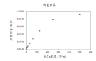
FIG. 3 illustrates an exemplary dose response curve (concentration of anti-thyroglobulin antigen as a function of observed signal (RLU)) for the disclosed immunoassay.
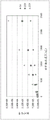
Figure 4 illustrates an exemplary dose response curve comparing undiluted (diamonds) and diluted (squares) antibody samples using the disclosed immunoassay.
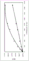
Figure 5 illustrates an exemplary dose response curve analyzing the effect of increasing amounts of acridinium ester. Triangle = 20X acridinium ester; square = 10X acridinium ester; and diamond = 5X acridinium ester.
Detailed description of illustrative embodiments
The disclosed methods and kits may be understood more readily by reference to the following detailed description taken in connection with the accompanying drawings, which form a part of this disclosure. It is to be understood that the disclosed methods and kits are not limited to the specific methods and kits described and/or shown herein, and that the terminology used herein is for the purpose of describing particular embodiments by way of example only and is not intended to be limiting of the claimed methods and kits.
Unless specifically stated otherwise, any description of possible mechanisms or modes of action or reasons for improvement is intended to be illustrative only, and the disclosed methods and kits should not be constrained by the correctness or incorrectness of any such suggested mechanism or mode of action or reason for improvement.
Throughout this document, the present specification relates to methods of detecting antibodies and methods of diagnosing autoimmune diseases. Where the present disclosure describes or claims features or embodiments relating to methods of detecting antibodies, such features or embodiments are equally applicable to methods of diagnosing autoimmune diseases. Likewise, where the disclosure describes or claims features or embodiments relating to a method of diagnosing an autoimmune disease, such features or embodiments are equally applicable to a method of detecting an antibody.
Where a range of numerical values is stated or established herein, the range includes the endpoints thereof and all the individual integers and fractions within the range, and also includes each of the narrower ranges therein formed by all possible combinations of those endpoints and the internal integers and fractions forming subgroups of the larger groups of values within the range, to the same extent as if each of those narrower ranges were explicitly stated. Where a range of values is specified herein as being greater than a specified value, the range is still limited and is bounded at its upper end by values operable in the context of the invention as described herein. Where a range of values is specified herein as being less than a specified value, the range is still bounded at its lower end by a non-zero value. It is not intended that the scope of the invention be limited to the specific values recited when defining a range. All ranges are inclusive and combinable.
When values are expressed as approximations, by use of the antecedent "about," it will be understood that the particular value forms another embodiment. Reference to a particular numerical value includes at least that particular value unless the context clearly dictates otherwise.
It is to be understood that certain features of the disclosed methods and kits, which are, for clarity, described herein in the context of separate embodiments, may also be provided in combination in a single embodiment. Conversely, various features of the disclosed methods and kits that are, for brevity, described in the context of a single embodiment, may also be provided separately or in any subcombination.
As used herein, the singular forms "a", "an" and "the" include the plural.
Various terms are used throughout the description and claims that refer to various aspects of the specification. Such terms are given their ordinary meaning in the art unless otherwise indicated. Other specifically defined terms are to be construed in a manner consistent with the definitions provided herein.
The term "comprising" is intended to include the examples encompassed by the term "consisting essentially of and" consisting of; similarly, the term "consisting essentially of is intended to include the examples encompassed by the term" consisting of.
Disclosed herein are immunoassays for detecting antibodies in a biological sample from a subject and/or diagnosing autoimmune disease in a subject. The disclosed immunoassay uses a single, direct step to assess the level of antibody in a sample by simultaneously binding the antibody to a capture antigen (e.g., an unlabeled antigen bound to a solid support) and a labeled antigen not bound to a solid support. Due to the bivalent nature of the antibodies, the antibodies present in the biological sample will simultaneously bind unlabeled antigen and labeled antigen bound to the solid support, forming a complex comprising the solid support and the labeled antigen. The read value of the assay (i.e., the signal from the labeled antigen attached to the solid support via the antibody) is directly proportional to the level of antibody present in the sample.
In some embodiments, the immunoassay may comprise an "antigen bridge" immunoassay, an exemplary reaction scheme of which is shown in fig. 1. A biological sample known to have or suspected of having the target antibody 10 is incubated with labeled antigen 20 and solid support 30 having unlabeled antigen bound thereto. In the absence of antibodies, the labeled antigen does not bind to or otherwise interact with the solid support. Thus, in the absence of antibodies, the labeled antigen remains in solution and separation of the solid support does not result in separation of the labeled antigen. When antibody 10 is present in the biological sample, antibody 10 binds both unlabeled antigen and labeled antigen 20 bound to solid support 30, thereby linking labeled antigen 20 and solid support 30 and resulting in the formation of solid support/labeled antigen complex 40. It will be appreciated that the order in which incubation occurs may differ from that illustrated in figure 1. For example, a biological sample known to have or suspected of having target antibodies 10 can be first incubated with a solid support 30 having unlabeled antigens bound thereto, followed by incubation with labeled antigens 20. Alternatively, a sample known or suspected of having the target antibody 10 can be incubated with both the labeled antigen 20 and the solid support 30 having unlabeled antigen bound thereto.
An exemplary timeline for practicing the disclosed immunoassay is illustrated in fig. 2. In fig. 2, a biological sample known to have or suspected of having the target antibody 10 is incubated with the labeled antigen 20 for less than about 3 minutes. Solid support 30 with unlabeled antigen bound thereto is added and incubated with the antibody/labeled antigen mixture for about 6.5 minutes. The solid support/labeled antigen complex 40 may then be detected (not shown).
The disclosed immunoassays include methods of detecting antibodies in a biological sample from a subject. A method of detecting antibodies in a biological sample from a subject comprising:
a) incubating the biological sample from the subject with:
a solid support having an unlabeled antigen bound thereto, wherein the unlabeled antigen is specifically recognized by the antibody; and
a labeled antigen, wherein the labeled antigen does not bind to the solid support and is specifically recognized by the antibody,
wherein, in the presence of the antibody, a solid support/labeled antigen complex is formed; and
b) detecting said solid support/labeled antigen complex, the presence of which indicates the presence of said antibody in said biological sample.
In some embodiments, the antibody is an autoantibody. Thus, the disclosed methods can be used to detect autoantibodies in a biological sample from the subject. Exemplary autoantibodies that can be detected by the disclosed methods are autoantibodies that specifically bind to thyroglobulin (i.e., anti-thyroglobulin antibodies). In embodiments wherein the autoantibody specifically binds to thyroglobulin, the unlabeled antigen is thyroglobulin and the labeled antigen is labeled thyroglobulin.
The disclosed immunoassays include methods of diagnosing an autoimmune disease in a subject. A method of diagnosing an autoimmune disease in a subject comprising:
a) incubating a biological sample from the subject with:
a solid support having an unlabeled antigen bound thereto, wherein the unlabeled antigen is specifically recognized by autoantibodies from the subject; and
a labeled antigen, wherein the labeled antigen does not bind to the solid support and is specifically recognized by the autoantibody,
wherein, in the presence of said autoantibody, a solid support/labelled antigen complex is formed; and
b) diagnosing the subject as having an autoimmune disease if the solid support/labeled antigen complex is detected.
The disclosed methods can be used to diagnose any autoimmune disease in which the antigen recognized by the autoantibody is known. In some embodiments, the autoimmune disease is an autoimmune thyroid disease, including graves 'disease (GD) and Hashimoto's Thyroiditis (HT). In embodiments wherein the autoimmune disease is autoimmune thyroid disease, the antigen (unlabeled and labeled) is thyroglobulin.
The following disclosure is equally applicable to methods of detecting antibodies and methods of diagnosing autoimmune diseases.
Suitable biological samples include any biological sample from a subject containing or suspected of containing an antigen of interest, including, but not limited to, serum, plasma, whole blood, saliva, urine, semen, sweat, tears, and body tissue.
In some embodiments, prior to the incubating step, the biological sample from the subject can be diluted to reduce the concentration of the sample. Suitable dilutions include, for example, 1:2, 1:5, 1:10, 1:20, 1:50, 1:100, 1:1000, and the like.
The solid support may be any material to which the unlabeled antigen may be bound or linked, either directly or indirectly. Exemplary solid supports include, but are not limited to, column matrix material, culture plates, tubes, dishes, flasks, microtiter plates, beads/particles, heat-killed formalin- (or other chemical) -fixed prokaryotic or eukaryotic cells, microscope slides, ACLAR @ thin films, or any other optically clear polymer, or combinations thereof. The solid support may be composed in whole or in part of plastic, cellulose derivatives, nitrocellulose, glass fiber, latex, or a combination thereof. In some embodiments, the solid support comprises magnetic particles. Suitable magnetic particles include paramagnetic particles (PMP) and Latex Magnetic Particles (LMP).
The unlabeled antigen can be bound directly or indirectly to a solid support to thereby form a solid support having unlabeled antigen bound thereto. The unlabeled antigen can be bound directly to a solid support. Suitable techniques for binding unlabeled antigens directly to a solid support include, for example, covalent attachment, adsorption, non-covalent interactions, or combinations thereof. Alternatively, the unlabeled antigen can be indirectly attached to a solid support. Suitable means for indirectly attaching the unlabeled antigen to the solid support include, for example, attachment via a peptide, protein, antibody, linker, or a combination thereof. In some embodiments, the unlabeled antigen can be indirectly attached to the solid support through streptavidin and biotin. For example, the unlabeled antigen can be biotinylated, and the solid support can comprise streptavidin.
The solid support having unlabeled antigen bound thereto can be present in a buffer comprising one or more salts, one or more stabilizing agents, and one or more surfactants. In some embodiments, the buffer may comprise HEPES salt, sodium chloride, bovine albumin, bovine globulin, and Tween 20. In some embodiments, the buffer may comprise HEPES salts (16.9 g/L HEPES acid and 7.6 g/L HEPES sodium), 300 mM sodium chloride, 1% bovine albumin, 0.1% bovine globulin, and 0.2% Tween 20.
In the absence of antibodies, the labeled antigen does not bind to or otherwise interact with the solid support. Thus, in embodiments in which the solid support comprises streptavidin, the labeled antigen will not be biotinylated.
The labeled antigen comprises a detectable label. Suitable detectable labels include, but are not limited to, enzyme conjugates (e.g., horseradish peroxidase (HRP), Alkaline Phosphatase (AP), glucose oxidase, and beta-galactosidase), fluorescent probes, radioisotopes, chemiluminescent, and bioluminescent labels, or combinations thereof. In some embodiments, the detectable label comprises an acridinium ester or an analog thereof. Acridinium ester analogs include, but are not limited to, dimethylacridine ester (DMAE), N-sulfopropyldimethylacridine ester (NSP-DMAE), High Quantum Yield Acridinium Ester (HQYAE), Zwitterionic Acridinium Ester (ZAE), hexa (ethylene) glycol acridinium ester (HEGAE), N-sulfopropyl-2-isopropoxydimethylacridine ester (Iso-Di-ZAE), trissulfopropylacridinium ester (TSP-AE), or N-sulfopropyldimethylacridine ester with a hexa (ethylene) glycol linker (HEG-GLU-AE). In some embodiments, the detectable label comprises a ruthenium ester or an analog thereof. The detectable label may be present in a 1:1 molar ratio to the antigen or may be present in molar excess. For example, the detectable label may be present in a 5, 10, 20 or 50 molar excess.
The labeled antigen may be present in a buffer comprising one or more salts, one or more stabilizers, and one or more surfactants. In some embodiments, the buffer may comprise HEPES salt, sodium chloride, bovine albumin, bovine globulin, and Tween 20. In some embodiments, the buffer may comprise HEPES salts (16.9 g/L HEPES acid and 7.6 g/L HEPES sodium), 300 mM sodium chloride, 1% bovine albumin, 0.1% bovine globulin, and 0.2% Tween 20.
The disclosed method can be performed much faster than the currently used indirect two-step assay, which can take approximately about 1 hour. For example, in the disclosed methods, a biological sample known to have or suspected of having antibodies can be incubated in the reaction mixture for about 1 minute, about 2 minutes, about 3 minutes, about 4 minutes, about 5 minutes, or less than about 10 minutes. The labeled antigen may be added and incubated with the biological sample for about 1 minute, about 2 minutes, about 3 minutes, about 4 minutes, or less than about 5 minutes. The solid support having unlabeled antigen bound thereto can be added to a mixture of the biological sample and the labeled antigen and incubated for about 1 minute, about 2 minutes, about 3 minutes, about 4 minutes, about 5 minutes, about 6 minutes, about 7 minutes, or less than 10 minutes. In some embodiments, the incubating step is performed for a total of about 10 minutes to about 20 minutes. Subsequent detection or diagnostic steps may be performed in less than about 5 minutes. It will be appreciated that the amount of time required for the assay may vary based on several factors, including the level of antibody in the biological sample and the affinity of the antibody for the antigen. Thus, the disclosed methods may be performed for any suitable amount of time.
The method may further comprise determining the level of the antibody in the biological sample of the subject. The level of the antibody in the biological sample of the subject is directly proportional to the level of the solid support/labeled antigen complex. Thus, the level of antibody can be determined by determining the level of solid support/labeled antigen complex, which can be done, for example, by measuring the signal from the complex. Similarly, in embodiments wherein the method is used to diagnose an autoimmune disease in a subject, the solid support/labeled antigen complex is measured by measuring a signal from a labeled antigen attached to the solid support.
The disclosed methods may be performed manually or may be automated. For example, the disclosed methods may use ADVIA CENTAUR ® immunoassay system or ATELLICA @TMAnd (4) carrying out the system.
Kits for practicing the disclosed methods are also provided. The kit may comprise a solid support, an unlabeled antigen, and a labeled antigen. Suitable solid supports include those disclosed above. The solid support binds unlabeled antigen, but not labeled antigen. Thus, in some embodiments, the kit comprises a solid support having unlabeled antigen bound thereto and a labeled antigen. In some embodiments, the antigen is thyroglobulin and the labeled antigen is labeled thyroglobulin. Suitable labels include those disclosed above for this method.
Examples
The following examples are provided to further describe some embodiments disclosed herein. The examples are intended to illustrate, but not to limit, the disclosed embodiments.
Preparation of reagents
Thyroglobulin-bound solid-phase reagentsAssay solid phase contains streptavidin-coated magnetic particles (Thermo Fisher Scientific, Dynabeads;. M270 REF 34353) coupled to biotinylated human thyroglobulin, at a target particle concentration of 0.6 g/L in buffered saline containing HEPES salts (16.9 g/L HEPES acid and 7.6 g/L HEPES sodium), 300 mM sodium chloride (to provide ionic strength), 1% bovine albumin and 0.1% bovine globulin (as stabilizer) and 0.2% Tween 20.
Labeled thyroglobulin reagentsAssay labeled reagents containing human thyroglobulin labeled with ZAE-type acridinium ester, thyroglobulin target concentration 1.2 μ g/mL in buffered saline containing HEPES salts (16.9 g/L HEPES acid and 7.6 g/L HEPES sodium), 300 mM sodium chloride (to provide ionic strength), 1% bovine albumin and 0.1% bovine globulin (as stabilizer) and 0.2% Tween 20.
Sample preparation
Samples (10 levels) from WHO reference material (antithyroid globulin serum, human NIBSC code 65/093) were prepared according to the recommended dilution procedure. The expected concentration of antithyroid globulin within each sample is provided in table 1. Similarly, patient serum standards were prepared with highly concentrated human anti-thyroglobulin antibodies (e.g., between 10,000 and 50,000 IU/mL) and diluted to prepare 10 levels spanning a wide concentration range (table 2). All samples were analyzed on ADVIA CENTAUR ® system using the disclosed immunoassay (referred to as "antigen bridge immunoassay") and the currently used competitive assay.
Reaction procedure for antigen bridge immunoassay
The following steps were performed by the ADVIA CENTAUR ® system, as illustrated in FIG. 2:
distributing 25 μ L of a sample containing an anti-thyroglobulin antibody to cuvettes;
incubating the sample for 4.75 minutes;
reagent probe 1 dispenses 100 μ L of labeled thyroglobulin reagent and incubates the mixture for 2.75 minutes;
separating the formed complex using a magnet and aspirating the complex and washing it with a wash buffer;
dispensing 300 μ L each of an acid reagent (HCL) and a base (NaOH) reagent to initiate a chemiluminescent reaction; and
relative photometric unit (RLU) results are reported.
Reaction procedure for competitive immunoassays
The following steps were performed manually or using ADVIA CENTAUR ® systems:
dispensing 40 μ L of a sample containing anti-thyroglobulin antibodies into a cuvette;
add 100 μ Ι _ of labeled reagent to the cuvette and incubate for 2.5 minutes at 37 ℃. Labeled reagents were prepared by mixing human thyroglobulin (-0.38 μ g/mL) with acridinium ester in buffered saline solution containing BSA, protein stabilizers and preservatives.
Mix 200 μ L of solid phase in a cuvette and incubate 5.0 min at 37 ℃. Preparing a solid phase reagent by mixing a polyclonal human antithyroid protein antibody (-1.98 ug/mL) bound to a polyclonal goat anti-human antibody (-49.5 ug/mL) in a buffer containing BSA, a protein stabilizer and a preservative, said polyclonal goat anti-human antibody being covalently coupled to paramagnetic particles;
then apply magnet to separate, aspirate and wash cuvette with reagent water;
dispensing 300 μ L each of an acid reagent (HCL) and a base reagent (NaOH) to initiate a chemiluminescent reaction; and
reporting RLU results.
Data analysis
RLU produced by the reaction was determined as a function of expected concentration in internal units of antibody activity per mL (IU/mL) (table 1 for WHO reference samples and table 2 for serum patient samples). In antigen bridge immunoassays, the signal (RLU) is directly proportional to the concentration of autoantibodies present in the sample, whereas in competition assays the signal is inversely proportional to the concentration of autoantibodies present in the sample.
Table 1.
| WHO sample | Expected concentration (IU/mL) | Antigen bridging assay RLU | Competition assay RLU |
| Level 1 | 0 | 3381 | 887440 |
| |
7.8125 | 47742 | 897444 |
| Level 3 | 15.625 | 84672 | 863750 |
| Level 4 | 31.25 | 151756 | 815643 |
| |
62.5 | 276207 | 759089 |
| |
125 | 486019 | 614878 |
| Level 7 | 250 | 779351 | 371528 |
| Level 8 | 500 | 918564 | 339795 |
Table 2.
| Patient sample | Expected concentration (IU/mL) | Antigen bridging assay RLU | Competition assay RLU |
| Level 1 | 4.35 | 8122 | 700855 |
| |
7.16 | 15445 | 649511 |
| Level 3 | 21.40 | 30262 | 638638 |
| Level 4 | 16.18 | 63240 | 564694 |
| |
30.77 | 105924 | 502377 |
| |
48.43 | 200597 | 395283 |
| Level 7 | 94.23 | 295594 | 308100 |
| Level 8 | 164.12 | 398975 | 221646 |
The bridging assay showed a significantly higher relative increase in signal compared to the relative decrease in signal observed for the competition assay at low concentrations. The relative increase or decrease in signal at low concentrations has a direct effect on the detectability of the assay. For example, the detection limit (LoD), which is the lowest concentration at which an antibody can be detected 95% of the time, depends on the detection capability of the assay. In the experiments shown above, LoD was calculated as 1 IU/mL and 45 IU/mL for the bridge assay and the competition assay, respectively. The above results indicate that the disclosed antigen bridge immunoassay is more sensitive than the currently used competition assays.
Optimized antigen bridge immunoassay
One potential drawback of single-step immunoassays is the "hook effect", which results in a decrease in detectable signal with increasing analyte concentration, leading to potentially false low results. As shown in figure 3, for example, at lower concentrations of anti-thyroglobulin antibodies, the antigen bridge assay showed a direct relationship between the concentration of antibody and the relative signal (RLU). However, as the concentration of antibody increased, the relative signal in fig. 3 began to become saturated (about 500 UI/ml). In the presence of low concentrations of anti-thyroglobulin antibodies, a limited number of antibodies are encountered by the bulk solid support/unlabeled thyroglobulin complexes. As the concentration of antibody increases, the solid support/unlabeled thyroglobulin complex becomes deficient such that only a portion of the total amount of antibody binds to the solid support/unlabeled thyroglobulin complex and a portion of the antibody bound to the solid support/unlabeled thyroglobulin complex is not fully detected. With the disclosed immunoassay, this "hook effect" can be overcome by optimizing the molar ratio of antibody to labeled and bound unlabeled thyroglobulin. As shown in fig. 4, when the anti-thyroglobulin antibody sample was diluted (squares), the antibody measurement range increased and a linear relationship between signal and antibody concentration was achieved, compared to the undiluted (diamonds) sample.
To analyze the effect of the marker on the relative signal obtained in the immunoassay, the amount of acridinium ester added to thyroglobulin was varied to obtain ratios of 5, 10 and 20 molar excesses of acridinium ester marker. As shown in fig. 5, higher relative signals were obtained with higher amounts of acridinium ester.
Similarly, the ratio of anti-thyroglobulin antibody to solid support/unlabeled thyroglobulin complex can be optimized to produce the desired dose response and increase assay sensitivity and measurement range.
Those skilled in the art will appreciate that numerous changes and modifications may be made to the preferred embodiments of the invention and that such changes and modifications may be made without departing from the spirit of the invention. It is, therefore, intended that the appended claims cover all such equivalent variations as fall within the true spirit and scope of this present invention.
Claims (16)
Applications Claiming Priority (3)
| Application Number | Priority Date | Filing Date | Title |
|---|---|---|---|
| US201862693439P | 2018-07-02 | 2018-07-02 | |
| US62/693439 | 2018-07-02 | ||
| PCT/US2019/040167 WO2020010009A1 (en) | 2018-07-02 | 2019-07-01 | Direct immunoassay measurement of autoantibodies |
Publications (1)
| Publication Number | Publication Date |
|---|---|
| CN112166323A true CN112166323A (en) | 2021-01-01 |
Family
ID=69060911
Family Applications (1)
| Application Number | Title | Priority Date | Filing Date |
|---|---|---|---|
| CN201980036385.8A Pending CN112166323A (en) | 2018-07-02 | 2019-07-01 | Direct immunoassay measurement of autoantibodies |
Country Status (5)
| Country | Link |
|---|---|
| US (2) | US12436148B2 (en) |
| EP (1) | EP3818375A4 (en) |
| JP (2) | JP7503505B2 (en) |
| CN (1) | CN112166323A (en) |
| WO (1) | WO2020010009A1 (en) |
Families Citing this family (3)
| Publication number | Priority date | Publication date | Assignee | Title |
|---|---|---|---|---|
| CN111505268A (en) * | 2020-04-29 | 2020-08-07 | 四川携光生物技术有限公司 | Autoimmune antibody detection method |
| CN114324884A (en) * | 2020-09-30 | 2022-04-12 | 南京金斯瑞生物科技有限公司 | Antibody detection method independent of antibody type and application thereof |
| CN113358871B (en) * | 2021-06-02 | 2024-04-19 | 施康培医药科技(武汉)有限公司 | Colloidal gold test strip for detecting Tg-anti-Tg antibody complex |
Citations (7)
| Publication number | Priority date | Publication date | Assignee | Title |
|---|---|---|---|---|
| US5296347A (en) * | 1991-02-08 | 1994-03-22 | Ciba Corning Diagnostics Corp. | Bridge immunoassay |
| EP0627081A1 (en) * | 1992-12-21 | 1994-12-07 | Henning Berlin Gmbh | METHOD AND TEST PACK FOR DETERMINING ANTIBODIES. |
| CN1515909A (en) * | 2003-08-27 | 2004-07-28 | 魏景艳 | Quantum point marker sandwich immunodetection method and its diagnosis kit |
| CN101029894A (en) * | 2007-04-04 | 2007-09-05 | 长春西诺生物科技有限公司 | Double-antigen sandwiched colloidal golden inspecting test paper of rabies virus antibody and its production |
| CN101246167A (en) * | 2008-03-17 | 2008-08-20 | 广东省昆虫研究所 | STLV double-antigen sandwich enzyme linked immunosorbent detecting method and its reagent kit |
| US20130149700A1 (en) * | 2011-12-13 | 2013-06-13 | Baxter Healthcare S.A. | Measurement of autoantibodies at low conductivity with increased sensitivity |
| WO2017087634A1 (en) * | 2015-11-17 | 2017-05-26 | The Regents Of The University Of Colorado, A Body Corporate | Novel multiplex assays to diagnose or evaluate diseases or disorders in mammals |
Family Cites Families (14)
| Publication number | Priority date | Publication date | Assignee | Title |
|---|---|---|---|---|
| JPH0382962A (en) | 1989-08-28 | 1991-04-08 | Tosoh Corp | Method of detecting antibody to hepatitis b virus core antigen |
| JP3162438B2 (en) | 1991-09-12 | 2001-04-25 | 住友製薬株式会社 | Highly sensitive specific antibody assay |
| DE19534988A1 (en) * | 1995-09-21 | 1997-03-27 | Forssmann Wolf Georg | Process for the production and use of synthetic, biotinylated peptides |
| GB9823397D0 (en) * | 1998-10-27 | 1998-12-23 | Rsr Ltd | Assays for thyroid autoantibodies |
| US7759133B2 (en) * | 1998-12-22 | 2010-07-20 | Alk-Abello A/S | Method of detecting and/or quantifying a specific IgE antibody in a liquid sample |
| US8999727B2 (en) * | 2005-09-26 | 2015-04-07 | Ulrich Loos | Innovative TSH-R-Ab-kit |
| WO2008135274A2 (en) * | 2007-05-08 | 2008-11-13 | F. Hoffmann-La-Roche Ag | Method for the detection of specific immunoglobulin class g antibodies |
| ES2450998T3 (en) * | 2007-12-13 | 2014-03-26 | F. Hoffmann-La Roche Ag | New variants of rubella E1 envelope protein and its use in the detection of anti-rubella antibodies |
| CN101470117A (en) | 2007-12-26 | 2009-07-01 | 天津市协和医药科技有限公司 | Chemiluminescent ligand analysis method for quantitative detection of human auto-antibody |
| US8012769B2 (en) * | 2008-11-12 | 2011-09-06 | Hemopet | Thyroid analyte detection and measurement |
| US9939448B2 (en) | 2010-01-06 | 2018-04-10 | The Regents Of The University Of Colorado, A Body Corporate | Methods for detecting insulin autoantibody |
| BR112018017336A2 (en) | 2016-03-07 | 2018-12-26 | Hoffmann La Roche | in vitro method for detecting a p53 antibody (anti-p53 antibody) in a sample and fusion polypeptide |
| CN107044977A (en) * | 2016-06-30 | 2017-08-15 | 深圳市亚辉龙生物科技股份有限公司 | A kind of tyrosine phosphatase antibody chemical luminescence immunity detection reagent and preparation method thereof |
| CN106226526A (en) | 2016-06-30 | 2016-12-14 | 深圳市亚辉龙生物科技股份有限公司 | A kind of Zinc transporter 8 antibody chemical luminescence immunity detection reagent and preparation method thereof |
-
2019
- 2019-07-01 US US17/250,151 patent/US12436148B2/en active Active
- 2019-07-01 WO PCT/US2019/040167 patent/WO2020010009A1/en not_active Ceased
- 2019-07-01 EP EP19830952.8A patent/EP3818375A4/en active Pending
- 2019-07-01 JP JP2020573123A patent/JP7503505B2/en active Active
- 2019-07-01 CN CN201980036385.8A patent/CN112166323A/en active Pending
-
2022
- 2022-11-16 JP JP2022183008A patent/JP2023017986A/en active Pending
-
2025
- 2025-09-09 US US19/323,123 patent/US20260009794A1/en active Pending
Patent Citations (7)
| Publication number | Priority date | Publication date | Assignee | Title |
|---|---|---|---|---|
| US5296347A (en) * | 1991-02-08 | 1994-03-22 | Ciba Corning Diagnostics Corp. | Bridge immunoassay |
| EP0627081A1 (en) * | 1992-12-21 | 1994-12-07 | Henning Berlin Gmbh | METHOD AND TEST PACK FOR DETERMINING ANTIBODIES. |
| CN1515909A (en) * | 2003-08-27 | 2004-07-28 | 魏景艳 | Quantum point marker sandwich immunodetection method and its diagnosis kit |
| CN101029894A (en) * | 2007-04-04 | 2007-09-05 | 长春西诺生物科技有限公司 | Double-antigen sandwiched colloidal golden inspecting test paper of rabies virus antibody and its production |
| CN101246167A (en) * | 2008-03-17 | 2008-08-20 | 广东省昆虫研究所 | STLV double-antigen sandwich enzyme linked immunosorbent detecting method and its reagent kit |
| US20130149700A1 (en) * | 2011-12-13 | 2013-06-13 | Baxter Healthcare S.A. | Measurement of autoantibodies at low conductivity with increased sensitivity |
| WO2017087634A1 (en) * | 2015-11-17 | 2017-05-26 | The Regents Of The University Of Colorado, A Body Corporate | Novel multiplex assays to diagnose or evaluate diseases or disorders in mammals |
Non-Patent Citations (1)
| Title |
|---|
| 郑皆安等: "《医药临床操作技术大全》", 31 August 2002, 第二军医大学出版社, pages: 851 - 852 * |
Also Published As
| Publication number | Publication date |
|---|---|
| WO2020010009A1 (en) | 2020-01-09 |
| JP7503505B2 (en) | 2024-06-20 |
| US20210247392A1 (en) | 2021-08-12 |
| US20260009794A1 (en) | 2026-01-08 |
| JP2021529948A (en) | 2021-11-04 |
| JP2023017986A (en) | 2023-02-07 |
| US12436148B2 (en) | 2025-10-07 |
| EP3818375A4 (en) | 2021-10-13 |
| EP3818375A1 (en) | 2021-05-12 |
Similar Documents
| Publication | Publication Date | Title |
|---|---|---|
| US11959912B2 (en) | Fluorescence immunochromatographic detection card and a preparation method therefor and use thereof | |
| US4469787A (en) | Immunoassay involving soluble complex of second antibody and labeled binding protein | |
| JP6096813B2 (en) | Multi-biomarker set for breast cancer diagnosis, detection method thereof, and breast cancer diagnosis kit including antibody thereto | |
| JP5744385B2 (en) | CVD analysis | |
| US20260009794A1 (en) | Direct immunoassay measurement of autoantibodies | |
| TWI698639B (en) | Prostate antigen standards and uses thereof | |
| CN104198712A (en) | Galectin-3 immunoassay | |
| CN115335700A (en) | Immunoassay method for amyloid beta in blood and kit for the same | |
| JP6850254B2 (en) | Ways to reduce interference | |
| CN101144811A (en) | Biochemical markers for acute pulmonary embolism | |
| CN109187971A (en) | Neuronspecific enolase chemiluminescence immune detection reagent kit and preparation method thereof | |
| US20250130237A1 (en) | Point of Care Assays | |
| KR102172016B1 (en) | A method for detection of CYFRA21-1 Autoantibody-Antigen complex , CYFRA21-1 antigen and Lung Cancer diagnosis kit by using ratio of these markers | |
| JP4197393B2 (en) | Test method for IgA nephropathy | |
| WO2012006394A2 (en) | Hmga2 as a biomarker for diagnosis and prognosis of ovarian cancer | |
| JPH03229153A (en) | Detection of existence of specific anti- body or antigen useful for diagnosis of rheumatism and test kit used therefor | |
| CN117110613A (en) | Diagnostic kit and application of LMWK-Fc in preparation of liver fibrosis diagnostic reagent | |
| US5436132A (en) | Quantitative determination of tenascin as glioma marker | |
| JP7315965B2 (en) | Method for detecting viral liver cancer | |
| JP2009529133A (en) | A novel assay method for detecting antibodies bound to cell membrane receptors | |
| JP5997446B2 (en) | Liquid reagent for immobilizing thyroid hormone and use thereof | |
| JP7106810B2 (en) | Novel lung cancer marker | |
| CN102360016A (en) | Preparation of Sm-RNP Antibody Detection Kit for SLE by Flow Cytometry | |
| WO2023068249A1 (en) | Measuring reagent for cross-linked n-telopeptide of type i collagen, preparation method thereof, and immunoassay method using same | |
| WO2026016622A1 (en) | Immune-related adverse event marker and kit |
Legal Events
| Date | Code | Title | Description |
|---|---|---|---|
| PB01 | Publication | ||
| PB01 | Publication | ||
| SE01 | Entry into force of request for substantive examination | ||
| SE01 | Entry into force of request for substantive examination |