CN112105382A - Sequential methods for CAR T cell therapy - Google Patents
Sequential methods for CAR T cell therapy Download PDFInfo
- Publication number
- CN112105382A CN112105382A CN201980027953.8A CN201980027953A CN112105382A CN 112105382 A CN112105382 A CN 112105382A CN 201980027953 A CN201980027953 A CN 201980027953A CN 112105382 A CN112105382 A CN 112105382A
- Authority
- CN
- China
- Prior art keywords
- compound
- dose
- pharmaceutically acceptable
- acceptable salt
- patient
- Prior art date
- Legal status (The legal status is an assumption and is not a legal conclusion. Google has not performed a legal analysis and makes no representation as to the accuracy of the status listed.)
- Pending
Links
- 238000000034 method Methods 0.000 title claims abstract description 365
- 238000011357 CAR T-cell therapy Methods 0.000 title description 4
- 206010028980 Neoplasm Diseases 0.000 claims abstract description 224
- 108091007741 Chimeric antigen receptor T cells Proteins 0.000 claims abstract description 141
- 230000008685 targeting Effects 0.000 claims abstract description 113
- 201000011510 cancer Diseases 0.000 claims abstract description 107
- 150000003384 small molecules Chemical class 0.000 claims abstract description 79
- 239000000203 mixture Substances 0.000 claims abstract description 68
- 150000001875 compounds Chemical class 0.000 claims description 332
- 150000003839 salts Chemical class 0.000 claims description 315
- 230000001174 ascending effect Effects 0.000 claims description 252
- 108010019670 Chimeric Antigen Receptors Proteins 0.000 claims description 227
- 210000001744 T-lymphocyte Anatomy 0.000 claims description 203
- 239000003446 ligand Substances 0.000 claims description 133
- 150000007523 nucleic acids Chemical class 0.000 claims description 34
- 206010029350 Neurotoxicity Diseases 0.000 claims description 30
- 206010044221 Toxic encephalopathy Diseases 0.000 claims description 30
- 230000007135 neurotoxicity Effects 0.000 claims description 30
- 231100000228 neurotoxicity Toxicity 0.000 claims description 30
- 108020004707 nucleic acids Proteins 0.000 claims description 24
- 102000039446 nucleic acids Human genes 0.000 claims description 24
- 108090000765 processed proteins & peptides Proteins 0.000 claims description 15
- 238000012360 testing method Methods 0.000 claims description 13
- 208000001953 Hypotension Diseases 0.000 claims description 10
- 206010037660 Pyrexia Diseases 0.000 claims description 10
- 230000036543 hypotension Effects 0.000 claims description 9
- 229920001184 polypeptide Polymers 0.000 claims description 9
- 102000004196 processed proteins & peptides Human genes 0.000 claims description 9
- 231100001096 no neurotoxicity Toxicity 0.000 claims description 4
- 210000004027 cell Anatomy 0.000 description 151
- IAZDPXIOMUYVGZ-UHFFFAOYSA-N Dimethylsulphoxide Chemical compound CS(C)=O IAZDPXIOMUYVGZ-UHFFFAOYSA-N 0.000 description 141
- 239000011724 folic acid Substances 0.000 description 111
- 230000037396 body weight Effects 0.000 description 109
- 229940014144 folate Drugs 0.000 description 100
- -1 folate compound Chemical class 0.000 description 100
- 235000019152 folic acid Nutrition 0.000 description 97
- 206010052015 cytokine release syndrome Diseases 0.000 description 89
- 125000005647 linker group Chemical group 0.000 description 62
- WEVYAHXRMPXWCK-UHFFFAOYSA-N Acetonitrile Chemical compound CC#N WEVYAHXRMPXWCK-UHFFFAOYSA-N 0.000 description 60
- YMWUJEATGCHHMB-UHFFFAOYSA-N Dichloromethane Chemical compound ClCCl YMWUJEATGCHHMB-UHFFFAOYSA-N 0.000 description 54
- 239000003795 chemical substances by application Substances 0.000 description 54
- 230000004913 activation Effects 0.000 description 51
- XKRFYHLGVUSROY-UHFFFAOYSA-N Argon Chemical compound [Ar] XKRFYHLGVUSROY-UHFFFAOYSA-N 0.000 description 46
- 239000011347 resin Substances 0.000 description 45
- 229920005989 resin Polymers 0.000 description 45
- 241000699670 Mus sp. Species 0.000 description 44
- JGFZNNIVVJXRND-UHFFFAOYSA-N N,N-diisopropylethylamine Substances CCN(C(C)C)C(C)C JGFZNNIVVJXRND-UHFFFAOYSA-N 0.000 description 44
- MHMNJMPURVTYEJ-UHFFFAOYSA-N fluorescein-5-isothiocyanate Chemical compound O1C(=O)C2=CC(N=C=S)=CC=C2C21C1=CC=C(O)C=C1OC1=CC(O)=CC=C21 MHMNJMPURVTYEJ-UHFFFAOYSA-N 0.000 description 41
- 239000000243 solution Substances 0.000 description 41
- 241001465754 Metazoa Species 0.000 description 37
- 150000001413 amino acids Chemical class 0.000 description 36
- 238000011282 treatment Methods 0.000 description 32
- 229920001223 polyethylene glycol Polymers 0.000 description 31
- 102000017420 CD3 protein, epsilon/gamma/delta subunit Human genes 0.000 description 30
- DTQVDTLACAAQTR-UHFFFAOYSA-N Trifluoroacetic acid Chemical class OC(=O)C(F)(F)F DTQVDTLACAAQTR-UHFFFAOYSA-N 0.000 description 27
- 102000001301 EGF receptor Human genes 0.000 description 26
- 108060006698 EGF receptor Proteins 0.000 description 26
- 102100036856 Tumor necrosis factor receptor superfamily member 9 Human genes 0.000 description 25
- 210000004369 blood Anatomy 0.000 description 23
- 239000008280 blood Substances 0.000 description 23
- 208000016261 weight loss Diseases 0.000 description 23
- OVBPIULPVIDEAO-LBPRGKRZSA-N folic acid Chemical compound C=1N=C2NC(N)=NC(=O)C2=NC=1CNC1=CC=C(C(=O)N[C@@H](CCC(O)=O)C(O)=O)C=C1 OVBPIULPVIDEAO-LBPRGKRZSA-N 0.000 description 22
- 230000004580 weight loss Effects 0.000 description 22
- 229910052786 argon Inorganic materials 0.000 description 21
- 238000000684 flow cytometry Methods 0.000 description 21
- 230000011664 signaling Effects 0.000 description 21
- RTZKZFJDLAIYFH-UHFFFAOYSA-N Diethyl ether Chemical compound CCOCC RTZKZFJDLAIYFH-UHFFFAOYSA-N 0.000 description 19
- 238000006243 chemical reaction Methods 0.000 description 19
- 229910052736 halogen Inorganic materials 0.000 description 19
- 150000002367 halogens Chemical class 0.000 description 19
- 230000001988 toxicity Effects 0.000 description 19
- 231100000419 toxicity Toxicity 0.000 description 19
- 108090000695 Cytokines Proteins 0.000 description 18
- 102000004127 Cytokines Human genes 0.000 description 18
- 230000006044 T cell activation Effects 0.000 description 18
- 239000012634 fragment Substances 0.000 description 18
- 230000002829 reductive effect Effects 0.000 description 18
- 239000000427 antigen Substances 0.000 description 17
- 108091007433 antigens Proteins 0.000 description 17
- 102000036639 antigens Human genes 0.000 description 17
- 208000031261 Acute myeloid leukaemia Diseases 0.000 description 16
- 125000002915 carbonyl group Chemical group [*:2]C([*:1])=O 0.000 description 16
- 239000001257 hydrogen Substances 0.000 description 16
- 229910052739 hydrogen Inorganic materials 0.000 description 16
- 102000005962 receptors Human genes 0.000 description 16
- 108020003175 receptors Proteins 0.000 description 16
- 210000004881 tumor cell Anatomy 0.000 description 16
- 241000699666 Mus <mouse, genus> Species 0.000 description 15
- 210000001151 cytotoxic T lymphocyte Anatomy 0.000 description 15
- 150000002431 hydrogen Chemical class 0.000 description 15
- 210000001519 tissue Anatomy 0.000 description 15
- NQRYJNQNLNOLGT-UHFFFAOYSA-N Piperidine Chemical compound C1CCNCC1 NQRYJNQNLNOLGT-UHFFFAOYSA-N 0.000 description 14
- 239000002202 Polyethylene glycol Substances 0.000 description 14
- 229960002143 fluorescein Drugs 0.000 description 14
- 239000005090 green fluorescent protein Substances 0.000 description 14
- 210000004185 liver Anatomy 0.000 description 14
- 239000000047 product Substances 0.000 description 14
- 108010043121 Green Fluorescent Proteins Proteins 0.000 description 13
- 102000004144 Green Fluorescent Proteins Human genes 0.000 description 13
- 230000015572 biosynthetic process Effects 0.000 description 13
- 230000000139 costimulatory effect Effects 0.000 description 13
- 108020005243 folate receptor Proteins 0.000 description 13
- 102000006815 folate receptor Human genes 0.000 description 13
- 150000002224 folic acids Chemical class 0.000 description 13
- 229910052760 oxygen Inorganic materials 0.000 description 13
- 239000012071 phase Substances 0.000 description 13
- OXNIZHLAWKMVMX-UHFFFAOYSA-N picric acid Chemical compound OC1=C([N+]([O-])=O)C=C([N+]([O-])=O)C=C1[N+]([O-])=O OXNIZHLAWKMVMX-UHFFFAOYSA-N 0.000 description 13
- 238000003786 synthesis reaction Methods 0.000 description 13
- ZMANZCXQSJIPKH-UHFFFAOYSA-N Triethylamine Chemical compound CCN(CC)CC ZMANZCXQSJIPKH-UHFFFAOYSA-N 0.000 description 12
- 230000005587 bubbling Effects 0.000 description 12
- 230000008878 coupling Effects 0.000 description 12
- 238000010168 coupling process Methods 0.000 description 12
- 238000005859 coupling reaction Methods 0.000 description 12
- 238000001802 infusion Methods 0.000 description 12
- SOAPXKSPJAZNGO-WDSKDSINSA-N (2s)-2-[[(1s)-1,3-dicarboxypropyl]carbamoylamino]pentanedioic acid Chemical compound OC(=O)CC[C@@H](C(O)=O)NC(=O)N[C@H](C(O)=O)CCC(O)=O SOAPXKSPJAZNGO-WDSKDSINSA-N 0.000 description 11
- USFZMSVCRYTOJT-UHFFFAOYSA-N Ammonium acetate Chemical compound N.CC(O)=O USFZMSVCRYTOJT-UHFFFAOYSA-N 0.000 description 11
- 101000914514 Homo sapiens T-cell-specific surface glycoprotein CD28 Proteins 0.000 description 11
- ZMXDDKWLCZADIW-UHFFFAOYSA-N N,N-Dimethylformamide Chemical compound CN(C)C=O ZMXDDKWLCZADIW-UHFFFAOYSA-N 0.000 description 11
- 102100027213 T-cell-specific surface glycoprotein CD28 Human genes 0.000 description 11
- 125000003545 alkoxy group Chemical group 0.000 description 11
- 238000010586 diagram Methods 0.000 description 11
- 229960000304 folic acid Drugs 0.000 description 11
- 238000001727 in vivo Methods 0.000 description 11
- 239000007924 injection Substances 0.000 description 11
- 238000002347 injection Methods 0.000 description 11
- 201000008968 osteosarcoma Diseases 0.000 description 11
- 239000002953 phosphate buffered saline Substances 0.000 description 11
- 238000004007 reversed phase HPLC Methods 0.000 description 11
- YBJHBAHKTGYVGT-ZKWXMUAHSA-N (+)-Biotin Chemical compound N1C(=O)N[C@@H]2[C@H](CCCCC(=O)O)SC[C@@H]21 YBJHBAHKTGYVGT-ZKWXMUAHSA-N 0.000 description 10
- 206010027476 Metastases Diseases 0.000 description 10
- 238000003776 cleavage reaction Methods 0.000 description 10
- 238000001990 intravenous administration Methods 0.000 description 10
- 230000007017 scission Effects 0.000 description 10
- 238000002560 therapeutic procedure Methods 0.000 description 10
- KFZMGEQAYNKOFK-UHFFFAOYSA-N Isopropanol Chemical compound CC(C)O KFZMGEQAYNKOFK-UHFFFAOYSA-N 0.000 description 9
- NINIDFKCEFEMDL-UHFFFAOYSA-N Sulfur Chemical compound [S] NINIDFKCEFEMDL-UHFFFAOYSA-N 0.000 description 9
- 229940024606 amino acid Drugs 0.000 description 9
- QVGXLLKOCUKJST-UHFFFAOYSA-N atomic oxygen Chemical compound [O] QVGXLLKOCUKJST-UHFFFAOYSA-N 0.000 description 9
- GNBHRKFJIUUOQI-UHFFFAOYSA-N fluorescein Chemical compound O1C(=O)C2=CC=CC=C2C21C1=CC=C(O)C=C1OC1=CC(O)=CC=C21 GNBHRKFJIUUOQI-UHFFFAOYSA-N 0.000 description 9
- 230000014509 gene expression Effects 0.000 description 9
- 230000001965 increasing effect Effects 0.000 description 9
- 238000004895 liquid chromatography mass spectrometry Methods 0.000 description 9
- 229910052757 nitrogen Inorganic materials 0.000 description 9
- 239000001301 oxygen Substances 0.000 description 9
- 201000001275 rectum cancer Diseases 0.000 description 9
- 229910052717 sulfur Inorganic materials 0.000 description 9
- 239000011593 sulfur Substances 0.000 description 9
- 239000013598 vector Substances 0.000 description 9
- ZMTAPBHUSYTHBY-PMERELPUSA-N (2s)-5-amino-2-[[4-[(2-amino-4-oxo-1h-pteridin-6-yl)methylamino]benzoyl]-[2-[(3',6'-dihydroxy-3-oxospiro[2-benzofuran-1,9'-xanthene]-5-yl)carbamothioylamino]ethyl]amino]-5-oxopentanoic acid Chemical compound N1C(N)=NC(=O)C2=NC(CNC3=CC=C(C=C3)C(=O)N(CCNC(=S)NC=3C=C4C(C5(C6=CC=C(O)C=C6OC6=CC(O)=CC=C65)OC4=O)=CC=3)[C@@H](CCC(=O)N)C(O)=O)=CN=C21 ZMTAPBHUSYTHBY-PMERELPUSA-N 0.000 description 8
- 239000005695 Ammonium acetate Substances 0.000 description 8
- IJGRMHOSHXDMSA-UHFFFAOYSA-N Atomic nitrogen Chemical group N#N IJGRMHOSHXDMSA-UHFFFAOYSA-N 0.000 description 8
- 102100024423 Carbonic anhydrase 9 Human genes 0.000 description 8
- 101001137987 Homo sapiens Lymphocyte activation gene 3 protein Proteins 0.000 description 8
- 102000017578 LAG3 Human genes 0.000 description 8
- 108010076504 Protein Sorting Signals Proteins 0.000 description 8
- 102100037346 Substance-P receptor Human genes 0.000 description 8
- 101710097909 Substance-P receptor Proteins 0.000 description 8
- 229940043376 ammonium acetate Drugs 0.000 description 8
- 235000019257 ammonium acetate Nutrition 0.000 description 8
- 230000009089 cytolysis Effects 0.000 description 8
- 230000003013 cytotoxicity Effects 0.000 description 8
- 231100000135 cytotoxicity Toxicity 0.000 description 8
- 239000002609 medium Substances 0.000 description 8
- 239000012528 membrane Substances 0.000 description 8
- 238000002360 preparation method Methods 0.000 description 8
- 239000011541 reaction mixture Substances 0.000 description 8
- 230000009467 reduction Effects 0.000 description 8
- 238000007920 subcutaneous administration Methods 0.000 description 8
- 206010014733 Endometrial cancer Diseases 0.000 description 7
- 206010014759 Endometrial neoplasm Diseases 0.000 description 7
- 101710107035 Gamma-glutamyltranspeptidase Proteins 0.000 description 7
- 101710173228 Glutathione hydrolase proenzyme Proteins 0.000 description 7
- 102100034458 Hepatitis A virus cellular receptor 2 Human genes 0.000 description 7
- 101001068133 Homo sapiens Hepatitis A virus cellular receptor 2 Proteins 0.000 description 7
- 101001109501 Homo sapiens NKG2-D type II integral membrane protein Proteins 0.000 description 7
- 101001047681 Homo sapiens Tyrosine-protein kinase Lck Proteins 0.000 description 7
- 102100022680 NKG2-D type II integral membrane protein Human genes 0.000 description 7
- 108091028043 Nucleic acid sequence Proteins 0.000 description 7
- 102100022153 Tumor necrosis factor receptor superfamily member 4 Human genes 0.000 description 7
- 102100024036 Tyrosine-protein kinase Lck Human genes 0.000 description 7
- 230000000259 anti-tumor effect Effects 0.000 description 7
- 239000000872 buffer Substances 0.000 description 7
- 238000010511 deprotection reaction Methods 0.000 description 7
- 239000003814 drug Substances 0.000 description 7
- 230000000694 effects Effects 0.000 description 7
- 239000013604 expression vector Substances 0.000 description 7
- 238000009472 formulation Methods 0.000 description 7
- 102000006640 gamma-Glutamyltransferase Human genes 0.000 description 7
- 238000003384 imaging method Methods 0.000 description 7
- 230000000977 initiatory effect Effects 0.000 description 7
- 210000004698 lymphocyte Anatomy 0.000 description 7
- 238000005259 measurement Methods 0.000 description 7
- 239000002935 phosphatidylinositol 3 kinase inhibitor Substances 0.000 description 7
- 239000002904 solvent Substances 0.000 description 7
- 238000010361 transduction Methods 0.000 description 7
- 230000026683 transduction Effects 0.000 description 7
- 239000005483 tyrosine kinase inhibitor Substances 0.000 description 7
- 238000005406 washing Methods 0.000 description 7
- PJUPKRYGDFTMTM-UHFFFAOYSA-N 1-hydroxybenzotriazole;hydrate Chemical compound O.C1=CC=C2N(O)N=NC2=C1 PJUPKRYGDFTMTM-UHFFFAOYSA-N 0.000 description 6
- 108700012439 CA9 Proteins 0.000 description 6
- 108020004705 Codon Proteins 0.000 description 6
- 206010015548 Euthanasia Diseases 0.000 description 6
- 239000002067 L01XE06 - Dasatinib Substances 0.000 description 6
- 206010061535 Ovarian neoplasm Diseases 0.000 description 6
- 208000003721 Triple Negative Breast Neoplasms Diseases 0.000 description 6
- 208000002495 Uterine Neoplasms Diseases 0.000 description 6
- 239000002253 acid Substances 0.000 description 6
- 229910052799 carbon Inorganic materials 0.000 description 6
- 210000003169 central nervous system Anatomy 0.000 description 6
- 229960002448 dasatinib Drugs 0.000 description 6
- 230000002950 deficient Effects 0.000 description 6
- UAOMVDZJSHZZME-UHFFFAOYSA-N diisopropylamine Chemical compound CC(C)NC(C)C UAOMVDZJSHZZME-UHFFFAOYSA-N 0.000 description 6
- 201000010099 disease Diseases 0.000 description 6
- 208000037265 diseases, disorders, signs and symptoms Diseases 0.000 description 6
- 238000005516 engineering process Methods 0.000 description 6
- 210000003743 erythrocyte Anatomy 0.000 description 6
- 229940020947 fluorescein sodium Drugs 0.000 description 6
- 108020001507 fusion proteins Proteins 0.000 description 6
- 102000037865 fusion proteins Human genes 0.000 description 6
- 208000014829 head and neck neoplasm Diseases 0.000 description 6
- NPZTUJOABDZTLV-UHFFFAOYSA-N hydroxybenzotriazole Substances O=C1C=CC=C2NNN=C12 NPZTUJOABDZTLV-UHFFFAOYSA-N 0.000 description 6
- 238000010369 molecular cloning Methods 0.000 description 6
- 201000008785 pediatric osteosarcoma Diseases 0.000 description 6
- 238000010647 peptide synthesis reaction Methods 0.000 description 6
- 210000003819 peripheral blood mononuclear cell Anatomy 0.000 description 6
- 239000000126 substance Substances 0.000 description 6
- 208000022679 triple-negative breast carcinoma Diseases 0.000 description 6
- 206010046766 uterine cancer Diseases 0.000 description 6
- 239000003643 water by type Substances 0.000 description 6
- VQTBINYMFPKLQD-UHFFFAOYSA-N (2,5-dioxopyrrolidin-1-yl) 2-(3-hydroxy-6-oxoxanthen-9-yl)benzoate Chemical compound C=12C=CC(=O)C=C2OC2=CC(O)=CC=C2C=1C1=CC=CC=C1C(=O)ON1C(=O)CCC1=O VQTBINYMFPKLQD-UHFFFAOYSA-N 0.000 description 5
- 125000003088 (fluoren-9-ylmethoxy)carbonyl group Chemical group 0.000 description 5
- UJPMYEOUBPIPHQ-UHFFFAOYSA-N 1,1,1-trifluoroethane Chemical compound CC(F)(F)F UJPMYEOUBPIPHQ-UHFFFAOYSA-N 0.000 description 5
- UFBJCMHMOXMLKC-UHFFFAOYSA-N 2,4-dinitrophenol Chemical compound OC1=CC=C([N+]([O-])=O)C=C1[N+]([O-])=O UFBJCMHMOXMLKC-UHFFFAOYSA-N 0.000 description 5
- VVIAGPKUTFNRDU-UHFFFAOYSA-N 6S-folinic acid Natural products C1NC=2NC(N)=NC(=O)C=2N(C=O)C1CNC1=CC=C(C(=O)NC(CCC(O)=O)C(O)=O)C=C1 VVIAGPKUTFNRDU-UHFFFAOYSA-N 0.000 description 5
- 201000009030 Carcinoma Diseases 0.000 description 5
- 206010009944 Colon cancer Diseases 0.000 description 5
- 108010025905 Cystine-Knot Miniproteins Proteins 0.000 description 5
- 108700022150 Designed Ankyrin Repeat Proteins Proteins 0.000 description 5
- 108010021625 Immunoglobulin Fragments Proteins 0.000 description 5
- 102000008394 Immunoglobulin Fragments Human genes 0.000 description 5
- 241000713666 Lentivirus Species 0.000 description 5
- 206010033128 Ovarian cancer Diseases 0.000 description 5
- 239000012980 RPMI-1640 medium Substances 0.000 description 5
- 238000004458 analytical method Methods 0.000 description 5
- 229960002685 biotin Drugs 0.000 description 5
- 235000020958 biotin Nutrition 0.000 description 5
- 239000011616 biotin Substances 0.000 description 5
- 125000003178 carboxy group Chemical group [H]OC(*)=O 0.000 description 5
- 230000010261 cell growth Effects 0.000 description 5
- 230000001684 chronic effect Effects 0.000 description 5
- 239000012043 crude product Substances 0.000 description 5
- 230000004069 differentiation Effects 0.000 description 5
- 238000010790 dilution Methods 0.000 description 5
- 239000012895 dilution Substances 0.000 description 5
- 229940079593 drug Drugs 0.000 description 5
- 150000002148 esters Chemical class 0.000 description 5
- VVIAGPKUTFNRDU-ABLWVSNPSA-N folinic acid Chemical compound C1NC=2NC(N)=NC(=O)C=2N(C=O)C1CNC1=CC=C(C(=O)N[C@@H](CCC(O)=O)C(O)=O)C=C1 VVIAGPKUTFNRDU-ABLWVSNPSA-N 0.000 description 5
- 235000008191 folinic acid Nutrition 0.000 description 5
- 239000011672 folinic acid Substances 0.000 description 5
- 229960001691 leucovorin Drugs 0.000 description 5
- 208000032839 leukemia Diseases 0.000 description 5
- 208000014018 liver neoplasm Diseases 0.000 description 5
- 201000001441 melanoma Diseases 0.000 description 5
- 238000002953 preparative HPLC Methods 0.000 description 5
- 125000006850 spacer group Chemical group 0.000 description 5
- 208000024891 symptom Diseases 0.000 description 5
- 230000001225 therapeutic effect Effects 0.000 description 5
- 230000003442 weekly effect Effects 0.000 description 5
- HZAXFHJVJLSVMW-UHFFFAOYSA-N 2-Aminoethan-1-ol Chemical compound NCCO HZAXFHJVJLSVMW-UHFFFAOYSA-N 0.000 description 4
- JOAQINSXLLMRCV-UHFFFAOYSA-N 4-{[(2-amino-4-hydroxypteridin-6-yl)methyl]amino}benzoic acid Chemical compound C1=NC2=NC(N)=NC(O)=C2N=C1CNC1=CC=C(C(O)=O)C=C1 JOAQINSXLLMRCV-UHFFFAOYSA-N 0.000 description 4
- 206010006187 Breast cancer Diseases 0.000 description 4
- 208000026310 Breast neoplasm Diseases 0.000 description 4
- ZBNZXTGUTAYRHI-UHFFFAOYSA-N Dasatinib Chemical compound C=1C(N2CCN(CCO)CC2)=NC(C)=NC=1NC(S1)=NC=C1C(=O)NC1=C(C)C=CC=C1Cl ZBNZXTGUTAYRHI-UHFFFAOYSA-N 0.000 description 4
- LTMHDMANZUZIPE-AMTYYWEZSA-N Digoxin Natural products O([C@H]1[C@H](C)O[C@H](O[C@@H]2C[C@@H]3[C@@](C)([C@@H]4[C@H]([C@]5(O)[C@](C)([C@H](O)C4)[C@H](C4=CC(=O)OC4)CC5)CC3)CC2)C[C@@H]1O)[C@H]1O[C@H](C)[C@@H](O[C@H]2O[C@@H](C)[C@H](O)[C@@H](O)C2)[C@@H](O)C1 LTMHDMANZUZIPE-AMTYYWEZSA-N 0.000 description 4
- 229920001917 Ficoll Polymers 0.000 description 4
- 239000007821 HATU Substances 0.000 description 4
- 206010019695 Hepatic neoplasm Diseases 0.000 description 4
- 101000611936 Homo sapiens Programmed cell death protein 1 Proteins 0.000 description 4
- 101000738771 Homo sapiens Receptor-type tyrosine-protein phosphatase C Proteins 0.000 description 4
- OAKJQQAXSVQMHS-UHFFFAOYSA-N Hydrazine Chemical compound NN OAKJQQAXSVQMHS-UHFFFAOYSA-N 0.000 description 4
- 229940123266 IL2-inducible T-cell kinase inhibitor Drugs 0.000 description 4
- FBOZXECLQNJBKD-ZDUSSCGKSA-N L-methotrexate Chemical compound C=1N=C2N=C(N)N=C(N)C2=NC=1CN(C)C1=CC=C(C(=O)N[C@@H](CCC(O)=O)C(O)=O)C=C1 FBOZXECLQNJBKD-ZDUSSCGKSA-N 0.000 description 4
- OVBPIULPVIDEAO-UHFFFAOYSA-N N-Pteroyl-L-glutaminsaeure Natural products C=1N=C2NC(N)=NC(=O)C2=NC=1CNC1=CC=C(C(=O)NC(CCC(O)=O)C(O)=O)C=C1 OVBPIULPVIDEAO-UHFFFAOYSA-N 0.000 description 4
- MUBZPKHOEPUJKR-UHFFFAOYSA-N Oxalic acid Chemical compound OC(=O)C(O)=O MUBZPKHOEPUJKR-UHFFFAOYSA-N 0.000 description 4
- 229940116355 PI3 kinase inhibitor Drugs 0.000 description 4
- 206010060862 Prostate cancer Diseases 0.000 description 4
- 208000000236 Prostatic Neoplasms Diseases 0.000 description 4
- 102100037422 Receptor-type tyrosine-protein phosphatase C Human genes 0.000 description 4
- 206010039491 Sarcoma Diseases 0.000 description 4
- 229940045687 antimetabolites folic acid analogs Drugs 0.000 description 4
- 210000000227 basophil cell of anterior lobe of hypophysis Anatomy 0.000 description 4
- 239000011324 bead Substances 0.000 description 4
- 101150058049 car gene Proteins 0.000 description 4
- 125000004432 carbon atom Chemical group C* 0.000 description 4
- 238000005119 centrifugation Methods 0.000 description 4
- 210000001072 colon Anatomy 0.000 description 4
- 229960005156 digoxin Drugs 0.000 description 4
- LTMHDMANZUZIPE-PUGKRICDSA-N digoxin Chemical compound C1[C@H](O)[C@H](O)[C@@H](C)O[C@H]1O[C@@H]1[C@@H](C)O[C@@H](O[C@@H]2[C@H](O[C@@H](O[C@@H]3C[C@@H]4[C@]([C@@H]5[C@H]([C@]6(CC[C@@H]([C@@]6(C)[C@H](O)C5)C=5COC(=O)C=5)O)CC4)(C)CC3)C[C@@H]2O)C)C[C@@H]1O LTMHDMANZUZIPE-PUGKRICDSA-N 0.000 description 4
- LTMHDMANZUZIPE-UHFFFAOYSA-N digoxine Natural products C1C(O)C(O)C(C)OC1OC1C(C)OC(OC2C(OC(OC3CC4C(C5C(C6(CCC(C6(C)C(O)C5)C=5COC(=O)C=5)O)CC4)(C)CC3)CC2O)C)CC1O LTMHDMANZUZIPE-UHFFFAOYSA-N 0.000 description 4
- 238000002474 experimental method Methods 0.000 description 4
- 238000004128 high performance liquid chromatography Methods 0.000 description 4
- 238000000338 in vitro Methods 0.000 description 4
- 230000003993 interaction Effects 0.000 description 4
- 238000007918 intramuscular administration Methods 0.000 description 4
- 239000003550 marker Substances 0.000 description 4
- 230000009401 metastasis Effects 0.000 description 4
- 206010061289 metastatic neoplasm Diseases 0.000 description 4
- 229960000485 methotrexate Drugs 0.000 description 4
- 208000002154 non-small cell lung carcinoma Diseases 0.000 description 4
- 239000003960 organic solvent Substances 0.000 description 4
- 238000007911 parenteral administration Methods 0.000 description 4
- 230000035755 proliferation Effects 0.000 description 4
- 230000004044 response Effects 0.000 description 4
- 238000002390 rotary evaporation Methods 0.000 description 4
- 239000007787 solid Substances 0.000 description 4
- 230000004083 survival effect Effects 0.000 description 4
- 230000008961 swelling Effects 0.000 description 4
- 238000001890 transfection Methods 0.000 description 4
- 208000029729 tumor suppressor gene on chromosome 11 Diseases 0.000 description 4
- YOVVNQKCSKSHKT-HNNXBMFYSA-N (2s)-1-[4-[[2-(2-aminopyrimidin-5-yl)-7-methyl-4-morpholin-4-ylthieno[3,2-d]pyrimidin-6-yl]methyl]piperazin-1-yl]-2-hydroxypropan-1-one Chemical group C1CN(C(=O)[C@@H](O)C)CCN1CC1=C(C)C2=NC(C=3C=NC(N)=NC=3)=NC(N3CCOCC3)=C2S1 YOVVNQKCSKSHKT-HNNXBMFYSA-N 0.000 description 3
- SJVFAHZPLIXNDH-QFIPXVFZSA-N (2s)-2-(9h-fluoren-9-ylmethoxycarbonylamino)-3-phenylpropanoic acid Chemical compound C([C@@H](C(=O)O)NC(=O)OCC1C2=CC=CC=C2C2=CC=CC=C21)C1=CC=CC=C1 SJVFAHZPLIXNDH-QFIPXVFZSA-N 0.000 description 3
- LEELWLKZRKDMAS-UHFFFAOYSA-N 2-(2,4-dimethoxy-3-methylsulfanylphenyl)ethanamine Chemical compound COC1=CC=C(CCN)C(OC)=C1SC LEELWLKZRKDMAS-UHFFFAOYSA-N 0.000 description 3
- FPQQSJJWHUJYPU-UHFFFAOYSA-N 3-(dimethylamino)propyliminomethylidene-ethylazanium;chloride Chemical compound Cl.CCN=C=NCCCN(C)C FPQQSJJWHUJYPU-UHFFFAOYSA-N 0.000 description 3
- GZAJOEGTZDUSKS-UHFFFAOYSA-N 5-aminofluorescein Chemical group C12=CC=C(O)C=C2OC2=CC(O)=CC=C2C21OC(=O)C1=CC(N)=CC=C21 GZAJOEGTZDUSKS-UHFFFAOYSA-N 0.000 description 3
- UQXNEWQGGVUVQA-UHFFFAOYSA-N 8-aminooctanoic acid Chemical compound NCCCCCCCC(O)=O UQXNEWQGGVUVQA-UHFFFAOYSA-N 0.000 description 3
- QTBSBXVTEAMEQO-UHFFFAOYSA-N Acetic acid Chemical compound CC(O)=O QTBSBXVTEAMEQO-UHFFFAOYSA-N 0.000 description 3
- 102100026882 Alpha-synuclein Human genes 0.000 description 3
- 229940124291 BTK inhibitor Drugs 0.000 description 3
- 208000031648 Body Weight Changes Diseases 0.000 description 3
- 206010005949 Bone cancer Diseases 0.000 description 3
- 208000018084 Bone neoplasm Diseases 0.000 description 3
- 108091003079 Bovine Serum Albumin Proteins 0.000 description 3
- 206010006143 Brain stem glioma Diseases 0.000 description 3
- 208000011691 Burkitt lymphomas Diseases 0.000 description 3
- 206010007953 Central nervous system lymphoma Diseases 0.000 description 3
- 206010008342 Cervix carcinoma Diseases 0.000 description 3
- IAZDPXIOMUYVGZ-WFGJKAKNSA-N Dimethyl sulfoxide Chemical compound [2H]C([2H])([2H])S(=O)C([2H])([2H])[2H] IAZDPXIOMUYVGZ-WFGJKAKNSA-N 0.000 description 3
- 208000000461 Esophageal Neoplasms Diseases 0.000 description 3
- PIICEJLVQHRZGT-UHFFFAOYSA-N Ethylenediamine Chemical compound NCCN PIICEJLVQHRZGT-UHFFFAOYSA-N 0.000 description 3
- 108010087819 Fc receptors Proteins 0.000 description 3
- 102000009109 Fc receptors Human genes 0.000 description 3
- 208000017604 Hodgkin disease Diseases 0.000 description 3
- 208000010747 Hodgkins lymphoma Diseases 0.000 description 3
- 101000834898 Homo sapiens Alpha-synuclein Proteins 0.000 description 3
- 101001042104 Homo sapiens Inducible T-cell costimulator Proteins 0.000 description 3
- 101001002657 Homo sapiens Interleukin-2 Proteins 0.000 description 3
- 101000652359 Homo sapiens Spermatogenesis-associated protein 2 Proteins 0.000 description 3
- 108060003951 Immunoglobulin Proteins 0.000 description 3
- 102100021317 Inducible T-cell costimulator Human genes 0.000 description 3
- 206010061252 Intraocular melanoma Diseases 0.000 description 3
- 229940122245 Janus kinase inhibitor Drugs 0.000 description 3
- 206010058467 Lung neoplasm malignant Diseases 0.000 description 3
- 208000031422 Lymphocytic Chronic B-Cell Leukemia Diseases 0.000 description 3
- 206010052178 Lymphocytic lymphoma Diseases 0.000 description 3
- 206010027457 Metastases to liver Diseases 0.000 description 3
- AFVFQIVMOAPDHO-UHFFFAOYSA-N Methanesulfonic acid Chemical compound CS(O)(=O)=O AFVFQIVMOAPDHO-UHFFFAOYSA-N 0.000 description 3
- MSFSPUZXLOGKHJ-UHFFFAOYSA-N Muraminsaeure Natural products OC(=O)C(C)OC1C(N)C(O)OC(CO)C1O MSFSPUZXLOGKHJ-UHFFFAOYSA-N 0.000 description 3
- 208000033776 Myeloid Acute Leukemia Diseases 0.000 description 3
- ZHXNIYGJAOPMSO-UHFFFAOYSA-N N-[5-[[5-[(4-acetyl-1-piperazinyl)-oxomethyl]-4-methoxy-2-methylphenyl]thio]-2-thiazolyl]-4-[(3,3-dimethylbutan-2-ylamino)methyl]benzamide Chemical group C1=C(C(=O)N2CCN(CC2)C(C)=O)C(OC)=CC(C)=C1SC(S1)=CN=C1NC(=O)C1=CC=C(CNC(C)C(C)(C)C)C=C1 ZHXNIYGJAOPMSO-UHFFFAOYSA-N 0.000 description 3
- 206010030137 Oesophageal adenocarcinoma Diseases 0.000 description 3
- 206010030155 Oesophageal carcinoma Diseases 0.000 description 3
- 206010061902 Pancreatic neoplasm Diseases 0.000 description 3
- 208000000821 Parathyroid Neoplasms Diseases 0.000 description 3
- 108010013639 Peptidoglycan Proteins 0.000 description 3
- 208000007913 Pituitary Neoplasms Diseases 0.000 description 3
- 201000005746 Pituitary adenoma Diseases 0.000 description 3
- 206010061538 Pituitary tumour benign Diseases 0.000 description 3
- 206010035603 Pleural mesothelioma Diseases 0.000 description 3
- 229920000037 Polyproline Polymers 0.000 description 3
- OFOBLEOULBTSOW-UHFFFAOYSA-N Propanedioic acid Natural products OC(=O)CC(O)=O OFOBLEOULBTSOW-UHFFFAOYSA-N 0.000 description 3
- 208000015634 Rectal Neoplasms Diseases 0.000 description 3
- 208000006265 Renal cell carcinoma Diseases 0.000 description 3
- 208000000453 Skin Neoplasms Diseases 0.000 description 3
- 208000021712 Soft tissue sarcoma Diseases 0.000 description 3
- 208000005718 Stomach Neoplasms Diseases 0.000 description 3
- 208000024770 Thyroid neoplasm Diseases 0.000 description 3
- 101710165473 Tumor necrosis factor receptor superfamily member 4 Proteins 0.000 description 3
- 102100023345 Tyrosine-protein kinase ITK/TSK Human genes 0.000 description 3
- 206010046458 Urethral neoplasms Diseases 0.000 description 3
- 208000006105 Uterine Cervical Neoplasms Diseases 0.000 description 3
- 201000005969 Uveal melanoma Diseases 0.000 description 3
- 201000005188 adrenal gland cancer Diseases 0.000 description 3
- 208000024447 adrenal gland neoplasm Diseases 0.000 description 3
- 238000013459 approach Methods 0.000 description 3
- 239000012298 atmosphere Substances 0.000 description 3
- 239000002585 base Substances 0.000 description 3
- 230000006399 behavior Effects 0.000 description 3
- 230000004579 body weight change Effects 0.000 description 3
- 230000020411 cell activation Effects 0.000 description 3
- 201000010881 cervical cancer Diseases 0.000 description 3
- 208000024207 chronic leukemia Diseases 0.000 description 3
- KRKNYBCHXYNGOX-UHFFFAOYSA-N citric acid Chemical compound OC(=O)CC(O)(C(O)=O)CC(O)=O KRKNYBCHXYNGOX-UHFFFAOYSA-N 0.000 description 3
- 208000029742 colonic neoplasm Diseases 0.000 description 3
- 208000030381 cutaneous melanoma Diseases 0.000 description 3
- 230000003247 decreasing effect Effects 0.000 description 3
- 238000000432 density-gradient centrifugation Methods 0.000 description 3
- 238000011161 development Methods 0.000 description 3
- 235000005911 diet Nutrition 0.000 description 3
- 230000037213 diet Effects 0.000 description 3
- LOKCTEFSRHRXRJ-UHFFFAOYSA-I dipotassium trisodium dihydrogen phosphate hydrogen phosphate dichloride Chemical compound P(=O)(O)(O)[O-].[K+].P(=O)(O)([O-])[O-].[Na+].[Na+].[Cl-].[K+].[Cl-].[Na+] LOKCTEFSRHRXRJ-UHFFFAOYSA-I 0.000 description 3
- NJDNXYGOVLYJHP-UHFFFAOYSA-L disodium;2-(3-oxido-6-oxoxanthen-9-yl)benzoate Chemical compound [Na+].[Na+].[O-]C(=O)C1=CC=CC=C1C1=C2C=CC(=O)C=C2OC2=CC([O-])=CC=C21 NJDNXYGOVLYJHP-UHFFFAOYSA-L 0.000 description 3
- 230000008030 elimination Effects 0.000 description 3
- 238000003379 elimination reaction Methods 0.000 description 3
- 108010075324 emt protein-tyrosine kinase Proteins 0.000 description 3
- 210000000750 endocrine system Anatomy 0.000 description 3
- 201000004101 esophageal cancer Diseases 0.000 description 3
- 238000011156 evaluation Methods 0.000 description 3
- 239000012091 fetal bovine serum Substances 0.000 description 3
- 239000012595 freezing medium Substances 0.000 description 3
- 206010017758 gastric cancer Diseases 0.000 description 3
- 201000007492 gastroesophageal junction adenocarcinoma Diseases 0.000 description 3
- 201000009277 hairy cell leukemia Diseases 0.000 description 3
- 230000002440 hepatic effect Effects 0.000 description 3
- 238000009396 hybridization Methods 0.000 description 3
- 102000018358 immunoglobulin Human genes 0.000 description 3
- 238000009169 immunotherapy Methods 0.000 description 3
- 238000002513 implantation Methods 0.000 description 3
- 239000003112 inhibitor Substances 0.000 description 3
- 108091008042 inhibitory receptors Proteins 0.000 description 3
- 230000002601 intratumoral effect Effects 0.000 description 3
- 238000002955 isolation Methods 0.000 description 3
- 230000000670 limiting effect Effects 0.000 description 3
- 201000005202 lung cancer Diseases 0.000 description 3
- 208000020816 lung neoplasm Diseases 0.000 description 3
- 208000015486 malignant pancreatic neoplasm Diseases 0.000 description 3
- 208000026037 malignant tumor of neck Diseases 0.000 description 3
- 208000026045 malignant tumor of parathyroid gland Diseases 0.000 description 3
- 238000002156 mixing Methods 0.000 description 3
- 208000025113 myeloid leukemia Diseases 0.000 description 3
- 231100000252 nontoxic Toxicity 0.000 description 3
- 230000003000 nontoxic effect Effects 0.000 description 3
- 201000002575 ocular melanoma Diseases 0.000 description 3
- 210000001672 ovary Anatomy 0.000 description 3
- 238000004806 packaging method and process Methods 0.000 description 3
- 201000002528 pancreatic cancer Diseases 0.000 description 3
- 208000008443 pancreatic carcinoma Diseases 0.000 description 3
- 230000002093 peripheral effect Effects 0.000 description 3
- 230000002688 persistence Effects 0.000 description 3
- 208000021310 pituitary gland adenoma Diseases 0.000 description 3
- 229920001992 poloxamer 407 Polymers 0.000 description 3
- 108010026466 polyproline Proteins 0.000 description 3
- 229920000036 polyvinylpyrrolidone Polymers 0.000 description 3
- 239000001267 polyvinylpyrrolidone Substances 0.000 description 3
- 235000013855 polyvinylpyrrolidone Nutrition 0.000 description 3
- 239000002244 precipitate Substances 0.000 description 3
- 208000016800 primary central nervous system lymphoma Diseases 0.000 description 3
- 238000003653 radioligand binding assay Methods 0.000 description 3
- 206010038038 rectal cancer Diseases 0.000 description 3
- 201000007444 renal pelvis carcinoma Diseases 0.000 description 3
- 229960001860 salicylate Drugs 0.000 description 3
- YGSDEFSMJLZEOE-UHFFFAOYSA-M salicylate Chemical compound OC1=CC=CC=C1C([O-])=O YGSDEFSMJLZEOE-UHFFFAOYSA-M 0.000 description 3
- 201000000849 skin cancer Diseases 0.000 description 3
- 201000003708 skin melanoma Diseases 0.000 description 3
- 239000011734 sodium Chemical class 0.000 description 3
- 238000010186 staining Methods 0.000 description 3
- 230000000638 stimulation Effects 0.000 description 3
- 238000003756 stirring Methods 0.000 description 3
- 201000011549 stomach cancer Diseases 0.000 description 3
- 235000000346 sugar Nutrition 0.000 description 3
- 201000002510 thyroid cancer Diseases 0.000 description 3
- JOXIMZWYDAKGHI-UHFFFAOYSA-N toluene-4-sulfonic acid Chemical compound CC1=CC=C(S(O)(=O)=O)C=C1 JOXIMZWYDAKGHI-UHFFFAOYSA-N 0.000 description 3
- VZCYOOQTPOCHFL-UHFFFAOYSA-N trans-butenedioic acid Natural products OC(=O)C=CC(O)=O VZCYOOQTPOCHFL-UHFFFAOYSA-N 0.000 description 3
- 238000012546 transfer Methods 0.000 description 3
- 230000004614 tumor growth Effects 0.000 description 3
- 230000003612 virological effect Effects 0.000 description 3
- XLYOFNOQVPJJNP-UHFFFAOYSA-N water Substances O XLYOFNOQVPJJNP-UHFFFAOYSA-N 0.000 description 3
- MCEHFIXEKNKSRW-LBPRGKRZSA-N (2s)-2-[[3,5-dichloro-4-[(2,4-diaminopteridin-6-yl)methyl-methylamino]benzoyl]amino]pentanedioic acid Chemical compound C=1N=C2N=C(N)N=C(N)C2=NC=1CN(C)C1=C(Cl)C=C(C(=O)N[C@@H](CCC(O)=O)C(O)=O)C=C1Cl MCEHFIXEKNKSRW-LBPRGKRZSA-N 0.000 description 2
- FJRKUZWBJSBJNI-AWEZNQCLSA-N (2s)-2-[[4-[(5,7-diaminopyrido[3,4-b]pyrazin-3-yl)methyl-methylamino]benzoyl]amino]pentanedioic acid Chemical compound C=1N=C2C=C(N)N=C(N)C2=NC=1CN(C)C1=CC=C(C(=O)N[C@@H](CCC(O)=O)C(O)=O)C=C1 FJRKUZWBJSBJNI-AWEZNQCLSA-N 0.000 description 2
- UGWODJDIYOBPOW-HNNXBMFYSA-N (2s)-2-[[4-[(6,8-diaminopyrido[2,3-b]pyrazin-2-yl)methyl-methylamino]benzoyl]amino]pentanedioic acid Chemical compound C=1N=C2N=C(N)C=C(N)C2=NC=1CN(C)C1=CC=C(C(=O)N[C@@H](CCC(O)=O)C(O)=O)C=C1 UGWODJDIYOBPOW-HNNXBMFYSA-N 0.000 description 2
- GOPWHXPXSPIIQZ-FQEVSTJZSA-N (4s)-4-(9h-fluoren-9-ylmethoxycarbonylamino)-5-[(2-methylpropan-2-yl)oxy]-5-oxopentanoic acid Chemical compound C1=CC=C2C(COC(=O)N[C@@H](CCC(O)=O)C(=O)OC(C)(C)C)C3=CC=CC=C3C2=C1 GOPWHXPXSPIIQZ-FQEVSTJZSA-N 0.000 description 2
- KBDDHSXZOLZZGF-GJZGRUSLSA-N (4s)-4-[[(2s)-1,5-bis[(2-methylpropan-2-yl)oxy]-1,5-dioxopentan-2-yl]carbamoylamino]-5-[(2-methylpropan-2-yl)oxy]-5-oxopentanoic acid Chemical compound CC(C)(C)OC(=O)CC[C@@H](C(=O)OC(C)(C)C)NC(=O)N[C@@H](CCC(O)=O)C(=O)OC(C)(C)C KBDDHSXZOLZZGF-GJZGRUSLSA-N 0.000 description 2
- KYVBNYUBXIEUFW-UHFFFAOYSA-N 1,1,3,3-tetramethylguanidine Chemical compound CN(C)C(=N)N(C)C KYVBNYUBXIEUFW-UHFFFAOYSA-N 0.000 description 2
- KIHYPELVXPAIDH-HNSNBQBZSA-N 1-[[4-[(e)-n-[[4-cyclohexyl-3-(trifluoromethyl)phenyl]methoxy]-c-methylcarbonimidoyl]-2-ethylphenyl]methyl]azetidine-3-carboxylic acid Chemical compound CCC1=CC(C(\C)=N\OCC=2C=C(C(C3CCCCC3)=CC=2)C(F)(F)F)=CC=C1CN1CC(C(O)=O)C1 KIHYPELVXPAIDH-HNSNBQBZSA-N 0.000 description 2
- 238000005160 1H NMR spectroscopy Methods 0.000 description 2
- WEEMDRWIKYCTQM-UHFFFAOYSA-N 2,6-dimethoxybenzenecarbothioamide Chemical compound COC1=CC=CC(OC)=C1C(N)=S WEEMDRWIKYCTQM-UHFFFAOYSA-N 0.000 description 2
- BOEUHAUGJSOEDZ-UHFFFAOYSA-N 2-amino-5,6,7,8-tetrahydro-1h-pteridin-4-one Chemical compound N1CCNC2=C1C(=O)N=C(N)N2 BOEUHAUGJSOEDZ-UHFFFAOYSA-N 0.000 description 2
- 125000000022 2-aminoethyl group Chemical group [H]C([*])([H])C([H])([H])N([H])[H] 0.000 description 2
- TVZGACDUOSZQKY-LBPRGKRZSA-N 4-aminofolic acid Chemical compound C1=NC2=NC(N)=NC(N)=C2N=C1CNC1=CC=C(C(=O)N[C@@H](CCC(O)=O)C(O)=O)C=C1 TVZGACDUOSZQKY-LBPRGKRZSA-N 0.000 description 2
- XRVDGNKRPOAQTN-FQEVSTJZSA-N 5-[3-[(1s)-1-(2-hydroxyethylamino)-2,3-dihydro-1h-inden-4-yl]-1,2,4-oxadiazol-5-yl]-2-propan-2-yloxybenzonitrile Chemical compound C1=C(C#N)C(OC(C)C)=CC=C1C1=NC(C=2C=3CC[C@@H](C=3C=CC=2)NCCO)=NO1 XRVDGNKRPOAQTN-FQEVSTJZSA-N 0.000 description 2
- 108090000672 Annexin A5 Proteins 0.000 description 2
- 102000004121 Annexin A5 Human genes 0.000 description 2
- 108091023037 Aptamer Proteins 0.000 description 2
- 208000005443 Circulating Neoplastic Cells Diseases 0.000 description 2
- KRKNYBCHXYNGOX-UHFFFAOYSA-K Citrate Chemical compound [O-]C(=O)CC(O)(CC([O-])=O)C([O-])=O KRKNYBCHXYNGOX-UHFFFAOYSA-K 0.000 description 2
- 108091026890 Coding region Proteins 0.000 description 2
- 102000029816 Collagenase Human genes 0.000 description 2
- 108060005980 Collagenase Proteins 0.000 description 2
- CMSMOCZEIVJLDB-UHFFFAOYSA-N Cyclophosphamide Chemical compound ClCCN(CCCl)P1(=O)NCCCO1 CMSMOCZEIVJLDB-UHFFFAOYSA-N 0.000 description 2
- 150000004922 Dasatinib derivatives Chemical group 0.000 description 2
- 102000007260 Deoxyribonuclease I Human genes 0.000 description 2
- 108010008532 Deoxyribonuclease I Proteins 0.000 description 2
- FEWJPZIEWOKRBE-JCYAYHJZSA-N Dextrotartaric acid Chemical compound OC(=O)[C@H](O)[C@@H](O)C(O)=O FEWJPZIEWOKRBE-JCYAYHJZSA-N 0.000 description 2
- SHIBSTMRCDJXLN-UHFFFAOYSA-N Digoxigenin Natural products C1CC(C2C(C3(C)CCC(O)CC3CC2)CC2O)(O)C2(C)C1C1=CC(=O)OC1 SHIBSTMRCDJXLN-UHFFFAOYSA-N 0.000 description 2
- 208000006402 Ductal Carcinoma Diseases 0.000 description 2
- MPJKWIXIYCLVCU-UHFFFAOYSA-N Folinic acid Natural products NC1=NC2=C(N(C=O)C(CNc3ccc(cc3)C(=O)NC(CCC(=O)O)CC(=O)O)CN2)C(=O)N1 MPJKWIXIYCLVCU-UHFFFAOYSA-N 0.000 description 2
- ZHNUHDYFZUAESO-UHFFFAOYSA-N Formamide Chemical compound NC=O ZHNUHDYFZUAESO-UHFFFAOYSA-N 0.000 description 2
- DHMQDGOQFOQNFH-UHFFFAOYSA-N Glycine Chemical class NCC(O)=O DHMQDGOQFOQNFH-UHFFFAOYSA-N 0.000 description 2
- 208000009329 Graft vs Host Disease Diseases 0.000 description 2
- 101001023230 Homo sapiens Folate receptor alpha Proteins 0.000 description 2
- 101000603882 Homo sapiens Nuclear receptor subfamily 1 group I member 3 Proteins 0.000 description 2
- 101000831616 Homo sapiens Protachykinin-1 Proteins 0.000 description 2
- 101000611023 Homo sapiens Tumor necrosis factor receptor superfamily member 6 Proteins 0.000 description 2
- 101000610640 Homo sapiens U4/U6 small nuclear ribonucleoprotein Prp3 Proteins 0.000 description 2
- 101001074035 Homo sapiens Zinc finger protein GLI2 Proteins 0.000 description 2
- 108091006905 Human Serum Albumin Proteins 0.000 description 2
- 102000008100 Human Serum Albumin Human genes 0.000 description 2
- 108010003272 Hyaluronate lyase Proteins 0.000 description 2
- 102000001974 Hyaluronidases Human genes 0.000 description 2
- 208000008839 Kidney Neoplasms Diseases 0.000 description 2
- 102000003855 L-lactate dehydrogenase Human genes 0.000 description 2
- 108700023483 L-lactate dehydrogenases Proteins 0.000 description 2
- 102000018697 Membrane Proteins Human genes 0.000 description 2
- 108010052285 Membrane Proteins Proteins 0.000 description 2
- MBBZMMPHUWSWHV-BDVNFPICSA-N N-methylglucamine Chemical compound CNC[C@H](O)[C@@H](O)[C@H](O)[C@H](O)CO MBBZMMPHUWSWHV-BDVNFPICSA-N 0.000 description 2
- 206010061309 Neoplasm progression Diseases 0.000 description 2
- 241000283973 Oryctolagus cuniculus Species 0.000 description 2
- 239000002033 PVDF binder Substances 0.000 description 2
- 229930182555 Penicillin Natural products 0.000 description 2
- JGSARLDLIJGVTE-MBNYWOFBSA-N Penicillin G Chemical compound N([C@H]1[C@H]2SC([C@@H](N2C1=O)C(O)=O)(C)C)C(=O)CC1=CC=CC=C1 JGSARLDLIJGVTE-MBNYWOFBSA-N 0.000 description 2
- 102100024304 Protachykinin-1 Human genes 0.000 description 2
- 206010037423 Pulmonary oedema Diseases 0.000 description 2
- 101001110823 Saccharomyces cerevisiae (strain ATCC 204508 / S288c) 60S ribosomal protein L6-A Proteins 0.000 description 2
- 101000712176 Saccharomyces cerevisiae (strain ATCC 204508 / S288c) 60S ribosomal protein L6-B Proteins 0.000 description 2
- PXIPVTKHYLBLMZ-UHFFFAOYSA-N Sodium azide Chemical compound [Na+].[N-]=[N+]=[N-] PXIPVTKHYLBLMZ-UHFFFAOYSA-N 0.000 description 2
- FAPWRFPIFSIZLT-UHFFFAOYSA-M Sodium chloride Chemical compound [Na+].[Cl-] FAPWRFPIFSIZLT-UHFFFAOYSA-M 0.000 description 2
- 108091081024 Start codon Proteins 0.000 description 2
- 238000000692 Student's t-test Methods 0.000 description 2
- 239000004809 Teflon Substances 0.000 description 2
- 229920006362 Teflon® Polymers 0.000 description 2
- 102000003929 Transaminases Human genes 0.000 description 2
- 108090000340 Transaminases Proteins 0.000 description 2
- UYXTWWCETRIEDR-UHFFFAOYSA-N Tributyrin Chemical compound CCCC(=O)OCC(OC(=O)CCC)COC(=O)CCC UYXTWWCETRIEDR-UHFFFAOYSA-N 0.000 description 2
- GSEJCLTVZPLZKY-UHFFFAOYSA-N Triethanolamine Chemical compound OCCN(CCO)CCO GSEJCLTVZPLZKY-UHFFFAOYSA-N 0.000 description 2
- 108060008682 Tumor Necrosis Factor Proteins 0.000 description 2
- 102100040403 Tumor necrosis factor receptor superfamily member 6 Human genes 0.000 description 2
- 206010045170 Tumour lysis syndrome Diseases 0.000 description 2
- 102100040374 U4/U6 small nuclear ribonucleoprotein Prp3 Human genes 0.000 description 2
- 201000003761 Vaginal carcinoma Diseases 0.000 description 2
- 241000700605 Viruses Species 0.000 description 2
- 208000004354 Vulvar Neoplasms Diseases 0.000 description 2
- 102100035558 Zinc finger protein GLI2 Human genes 0.000 description 2
- 230000002378 acidificating effect Effects 0.000 description 2
- 150000007513 acids Chemical class 0.000 description 2
- 230000003213 activating effect Effects 0.000 description 2
- 230000000735 allogeneic effect Effects 0.000 description 2
- BJEPYKJPYRNKOW-UHFFFAOYSA-N alpha-hydroxysuccinic acid Natural products OC(=O)C(O)CC(O)=O BJEPYKJPYRNKOW-UHFFFAOYSA-N 0.000 description 2
- 239000012080 ambient air Substances 0.000 description 2
- 229960003896 aminopterin Drugs 0.000 description 2
- 230000003444 anaesthetic effect Effects 0.000 description 2
- 238000010171 animal model Methods 0.000 description 2
- 230000000844 anti-bacterial effect Effects 0.000 description 2
- 230000000719 anti-leukaemic effect Effects 0.000 description 2
- 238000003556 assay Methods 0.000 description 2
- 210000003719 b-lymphocyte Anatomy 0.000 description 2
- 125000002619 bicyclic group Chemical group 0.000 description 2
- 239000011230 binding agent Substances 0.000 description 2
- 239000012888 bovine serum Substances 0.000 description 2
- 229940098773 bovine serum albumin Drugs 0.000 description 2
- 239000012267 brine Substances 0.000 description 2
- 230000001413 cellular effect Effects 0.000 description 2
- 208000019065 cervical carcinoma Diseases 0.000 description 2
- 229960005395 cetuximab Drugs 0.000 description 2
- 238000003501 co-culture Methods 0.000 description 2
- 230000004186 co-expression Effects 0.000 description 2
- 229960002424 collagenase Drugs 0.000 description 2
- 230000000295 complement effect Effects 0.000 description 2
- 230000001086 cytosolic effect Effects 0.000 description 2
- 230000001419 dependent effect Effects 0.000 description 2
- 238000013461 design Methods 0.000 description 2
- ZBCBWPMODOFKDW-UHFFFAOYSA-N diethanolamine Chemical compound OCCNCCO ZBCBWPMODOFKDW-UHFFFAOYSA-N 0.000 description 2
- 230000029087 digestion Effects 0.000 description 2
- QONQRTHLHBTMGP-UHFFFAOYSA-N digitoxigenin Natural products CC12CCC(C3(CCC(O)CC3CC3)C)C3C11OC1CC2C1=CC(=O)OC1 QONQRTHLHBTMGP-UHFFFAOYSA-N 0.000 description 2
- SHIBSTMRCDJXLN-KCZCNTNESA-N digoxigenin Chemical compound C1([C@@H]2[C@@]3([C@@](CC2)(O)[C@H]2[C@@H]([C@@]4(C)CC[C@H](O)C[C@H]4CC2)C[C@H]3O)C)=CC(=O)OC1 SHIBSTMRCDJXLN-KCZCNTNESA-N 0.000 description 2
- OZRNSSUDZOLUSN-LBPRGKRZSA-N dihydrofolic acid Chemical compound N=1C=2C(=O)NC(N)=NC=2NCC=1CNC1=CC=C(C(=O)N[C@@H](CCC(O)=O)C(O)=O)C=C1 OZRNSSUDZOLUSN-LBPRGKRZSA-N 0.000 description 2
- 229940043279 diisopropylamine Drugs 0.000 description 2
- 239000003937 drug carrier Substances 0.000 description 2
- 238000012377 drug delivery Methods 0.000 description 2
- 238000001035 drying Methods 0.000 description 2
- 239000012636 effector Substances 0.000 description 2
- 201000003914 endometrial carcinoma Diseases 0.000 description 2
- 210000004696 endometrium Anatomy 0.000 description 2
- 102000052116 epidermal growth factor receptor activity proteins Human genes 0.000 description 2
- 108700015053 epidermal growth factor receptor activity proteins Proteins 0.000 description 2
- CCIVGXIOQKPBKL-UHFFFAOYSA-M ethanesulfonate Chemical compound CCS([O-])(=O)=O CCIVGXIOQKPBKL-UHFFFAOYSA-M 0.000 description 2
- VJJPUSNTGOMMGY-MRVIYFEKSA-N etoposide Chemical compound COC1=C(O)C(OC)=CC([C@@H]2C3=CC=4OCOC=4C=C3[C@@H](O[C@H]3[C@@H]([C@@H](O)[C@@H]4O[C@H](C)OC[C@H]4O3)O)[C@@H]3[C@@H]2C(OC3)=O)=C1 VJJPUSNTGOMMGY-MRVIYFEKSA-N 0.000 description 2
- 229960005420 etoposide Drugs 0.000 description 2
- 201000001343 fallopian tube carcinoma Diseases 0.000 description 2
- 229960000390 fludarabine Drugs 0.000 description 2
- GIUYCYHIANZCFB-FJFJXFQQSA-N fludarabine phosphate Chemical compound C1=NC=2C(N)=NC(F)=NC=2N1[C@@H]1O[C@H](COP(O)(O)=O)[C@@H](O)[C@@H]1O GIUYCYHIANZCFB-FJFJXFQQSA-N 0.000 description 2
- 239000012458 free base Substances 0.000 description 2
- 208000024908 graft versus host disease Diseases 0.000 description 2
- 239000001963 growth medium Substances 0.000 description 2
- 230000036541 health Effects 0.000 description 2
- 229960002773 hyaluronidase Drugs 0.000 description 2
- 239000002955 immunomodulating agent Substances 0.000 description 2
- 238000011534 incubation Methods 0.000 description 2
- 230000002401 inhibitory effect Effects 0.000 description 2
- 238000007912 intraperitoneal administration Methods 0.000 description 2
- 238000007913 intrathecal administration Methods 0.000 description 2
- 210000003734 kidney Anatomy 0.000 description 2
- 229940043355 kinase inhibitor Drugs 0.000 description 2
- 210000000265 leukocyte Anatomy 0.000 description 2
- 229960003208 levomefolic acid Drugs 0.000 description 2
- 244000144972 livestock Species 0.000 description 2
- 210000004072 lung Anatomy 0.000 description 2
- 238000012423 maintenance Methods 0.000 description 2
- VZCYOOQTPOCHFL-UPHRSURJSA-N maleic acid Chemical compound OC(=O)\C=C/C(O)=O VZCYOOQTPOCHFL-UPHRSURJSA-N 0.000 description 2
- 210000003071 memory t lymphocyte Anatomy 0.000 description 2
- 230000001394 metastastic effect Effects 0.000 description 2
- 230000004048 modification Effects 0.000 description 2
- 238000012986 modification Methods 0.000 description 2
- 238000012544 monitoring process Methods 0.000 description 2
- YOHYSYJDKVYCJI-UHFFFAOYSA-N n-[3-[[6-[3-(trifluoromethyl)anilino]pyrimidin-4-yl]amino]phenyl]cyclopropanecarboxamide Chemical compound FC(F)(F)C1=CC=CC(NC=2N=CN=C(NC=3C=C(NC(=O)C4CC4)C=CC=3)C=2)=C1 YOHYSYJDKVYCJI-UHFFFAOYSA-N 0.000 description 2
- 125000004433 nitrogen atom Chemical group N* 0.000 description 2
- QJGQUHMNIGDVPM-UHFFFAOYSA-N nitrogen group Chemical group [N] QJGQUHMNIGDVPM-UHFFFAOYSA-N 0.000 description 2
- 238000001543 one-way ANOVA Methods 0.000 description 2
- 239000012044 organic layer Substances 0.000 description 2
- 239000012074 organic phase Substances 0.000 description 2
- 230000001151 other effect Effects 0.000 description 2
- 229950008141 ozanimod Drugs 0.000 description 2
- 210000000496 pancreas Anatomy 0.000 description 2
- 239000008188 pellet Substances 0.000 description 2
- 229940049954 penicillin Drugs 0.000 description 2
- 210000005259 peripheral blood Anatomy 0.000 description 2
- 239000011886 peripheral blood Substances 0.000 description 2
- 230000002085 persistent effect Effects 0.000 description 2
- 239000003757 phosphotransferase inhibitor Substances 0.000 description 2
- 239000013612 plasmid Substances 0.000 description 2
- 229920000642 polymer Polymers 0.000 description 2
- 229920002981 polyvinylidene fluoride Polymers 0.000 description 2
- 230000000069 prophylactic effect Effects 0.000 description 2
- 150000003195 pteridines Chemical class 0.000 description 2
- 208000005333 pulmonary edema Diseases 0.000 description 2
- 238000000746 purification Methods 0.000 description 2
- 239000002287 radioligand Substances 0.000 description 2
- 238000011084 recovery Methods 0.000 description 2
- 229940043267 rhodamine b Drugs 0.000 description 2
- YGSDEFSMJLZEOE-UHFFFAOYSA-N salicylic acid Chemical compound OC(=O)C1=CC=CC=C1O YGSDEFSMJLZEOE-UHFFFAOYSA-N 0.000 description 2
- 238000000926 separation method Methods 0.000 description 2
- VGKDLMBJGBXTGI-SJCJKPOMSA-N sertraline Chemical compound C1([C@@H]2CC[C@@H](C3=CC=CC=C32)NC)=CC=C(Cl)C(Cl)=C1 VGKDLMBJGBXTGI-SJCJKPOMSA-N 0.000 description 2
- 229960002073 sertraline Drugs 0.000 description 2
- 229950005693 siponimod Drugs 0.000 description 2
- 210000000813 small intestine Anatomy 0.000 description 2
- 239000001488 sodium phosphate Substances 0.000 description 2
- 229910000162 sodium phosphate Inorganic materials 0.000 description 2
- HPALAKNZSZLMCH-UHFFFAOYSA-M sodium;chloride;hydrate Chemical compound O.[Na+].[Cl-] HPALAKNZSZLMCH-UHFFFAOYSA-M 0.000 description 2
- 238000010532 solid phase synthesis reaction Methods 0.000 description 2
- 239000007858 starting material Substances 0.000 description 2
- 210000000130 stem cell Anatomy 0.000 description 2
- 229960002385 streptomycin sulfate Drugs 0.000 description 2
- ADNPLDHMAVUMIW-CUZNLEPHSA-N substance P Chemical compound C([C@@H](C(=O)NCC(=O)N[C@@H](CC(C)C)C(=O)N[C@@H](CCSC)C(N)=O)NC(=O)[C@H](CC=1C=CC=CC=1)NC(=O)[C@H](CCC(N)=O)NC(=O)[C@H](CCC(N)=O)NC(=O)[C@H]1N(CCC1)C(=O)[C@H](CCCCN)NC(=O)[C@H]1N(CCC1)C(=O)[C@@H](N)CCCN=C(N)N)C1=CC=CC=C1 ADNPLDHMAVUMIW-CUZNLEPHSA-N 0.000 description 2
- KDYFGRWQOYBRFD-UHFFFAOYSA-N succinic acid Chemical compound OC(=O)CCC(O)=O KDYFGRWQOYBRFD-UHFFFAOYSA-N 0.000 description 2
- 230000009885 systemic effect Effects 0.000 description 2
- 238000002054 transplantation Methods 0.000 description 2
- 125000004044 trifluoroacetyl group Chemical group FC(C(=O)*)(F)F 0.000 description 2
- 230000001960 triggered effect Effects 0.000 description 2
- RYFMWSXOAZQYPI-UHFFFAOYSA-K trisodium phosphate Chemical compound [Na+].[Na+].[Na+].[O-]P([O-])([O-])=O RYFMWSXOAZQYPI-UHFFFAOYSA-K 0.000 description 2
- 208000010380 tumor lysis syndrome Diseases 0.000 description 2
- 102000003390 tumor necrosis factor Human genes 0.000 description 2
- 230000005751 tumor progression Effects 0.000 description 2
- 239000003981 vehicle Substances 0.000 description 2
- 229940053729 vitrax Drugs 0.000 description 2
- 208000013013 vulvar carcinoma Diseases 0.000 description 2
- OTKXCALUHMPIGM-FQEVSTJZSA-N (2s)-2-(9h-fluoren-9-ylmethoxycarbonylamino)-5-[(2-methylpropan-2-yl)oxy]-5-oxopentanoic acid Chemical compound C1=CC=C2C(COC(=O)N[C@@H](CCC(=O)OC(C)(C)C)C(O)=O)C3=CC=CC=C3C2=C1 OTKXCALUHMPIGM-FQEVSTJZSA-N 0.000 description 1
- MSTNYGQPCMXVAQ-RYUDHWBXSA-N (6S)-5,6,7,8-tetrahydrofolic acid Chemical compound C([C@H]1CNC=2N=C(NC(=O)C=2N1)N)NC1=CC=C(C(=O)N[C@@H](CCC(O)=O)C(O)=O)C=C1 MSTNYGQPCMXVAQ-RYUDHWBXSA-N 0.000 description 1
- 125000004642 (C1-C12) alkoxy group Chemical group 0.000 description 1
- UCTWMZQNUQWSLP-VIFPVBQESA-N (R)-adrenaline Chemical compound CNC[C@H](O)C1=CC=C(O)C(O)=C1 UCTWMZQNUQWSLP-VIFPVBQESA-N 0.000 description 1
- 229930182837 (R)-adrenaline Natural products 0.000 description 1
- BJEPYKJPYRNKOW-REOHCLBHSA-N (S)-malic acid Chemical compound OC(=O)[C@@H](O)CC(O)=O BJEPYKJPYRNKOW-REOHCLBHSA-N 0.000 description 1
- QLAJNZSPVITUCQ-UHFFFAOYSA-N 1,3,2-dioxathietane 2,2-dioxide Chemical compound O=S1(=O)OCO1 QLAJNZSPVITUCQ-UHFFFAOYSA-N 0.000 description 1
- ASOKPJOREAFHNY-UHFFFAOYSA-N 1-Hydroxybenzotriazole Chemical compound C1=CC=C2N(O)N=NC2=C1 ASOKPJOREAFHNY-UHFFFAOYSA-N 0.000 description 1
- IJRKANNOPXMZSG-SSPAHAAFSA-N 2-hydroxypropane-1,2,3-tricarboxylic acid;(2r,3s,4r,5r)-2,3,4,5,6-pentahydroxyhexanal Chemical compound OC[C@@H](O)[C@@H](O)[C@H](O)[C@@H](O)C=O.OC(=O)CC(O)(C(O)=O)CC(O)=O IJRKANNOPXMZSG-SSPAHAAFSA-N 0.000 description 1
- 229940080296 2-naphthalenesulfonate Drugs 0.000 description 1
- FWMNVWWHGCHHJJ-SKKKGAJSSA-N 4-amino-1-[(2r)-6-amino-2-[[(2r)-2-[[(2r)-2-[[(2r)-2-amino-3-phenylpropanoyl]amino]-3-phenylpropanoyl]amino]-4-methylpentanoyl]amino]hexanoyl]piperidine-4-carboxylic acid Chemical compound C([C@H](C(=O)N[C@H](CC(C)C)C(=O)N[C@H](CCCCN)C(=O)N1CCC(N)(CC1)C(O)=O)NC(=O)[C@H](N)CC=1C=CC=CC=1)C1=CC=CC=C1 FWMNVWWHGCHHJJ-SKKKGAJSSA-N 0.000 description 1
- MSTNYGQPCMXVAQ-KIYNQFGBSA-N 5,6,7,8-tetrahydrofolic acid Chemical compound N1C=2C(=O)NC(N)=NC=2NCC1CNC1=CC=C(C(=O)N[C@@H](CCC(O)=O)C(O)=O)C=C1 MSTNYGQPCMXVAQ-KIYNQFGBSA-N 0.000 description 1
- HBAQYPYDRFILMT-UHFFFAOYSA-N 8-[3-(1-cyclopropylpyrazol-4-yl)-1H-pyrazolo[4,3-d]pyrimidin-5-yl]-3-methyl-3,8-diazabicyclo[3.2.1]octan-2-one Chemical class C1(CC1)N1N=CC(=C1)C1=NNC2=C1N=C(N=C2)N1C2C(N(CC1CC2)C)=O HBAQYPYDRFILMT-UHFFFAOYSA-N 0.000 description 1
- 206010000060 Abdominal distension Diseases 0.000 description 1
- QTBSBXVTEAMEQO-UHFFFAOYSA-M Acetate Chemical compound CC([O-])=O QTBSBXVTEAMEQO-UHFFFAOYSA-M 0.000 description 1
- 206010000830 Acute leukaemia Diseases 0.000 description 1
- 206010000871 Acute monocytic leukaemia Diseases 0.000 description 1
- 239000012114 Alexa Fluor 647 Substances 0.000 description 1
- 206010002198 Anaphylactic reaction Diseases 0.000 description 1
- 206010002961 Aplasia Diseases 0.000 description 1
- 239000004475 Arginine Substances 0.000 description 1
- 206010003571 Astrocytoma Diseases 0.000 description 1
- 108090001008 Avidin Proteins 0.000 description 1
- 102100027203 B-cell antigen receptor complex-associated protein beta chain Human genes 0.000 description 1
- 208000003950 B-cell lymphoma Diseases 0.000 description 1
- 102100024222 B-lymphocyte antigen CD19 Human genes 0.000 description 1
- 238000000035 BCA protein assay Methods 0.000 description 1
- 241000894006 Bacteria Species 0.000 description 1
- BVKZGUZCCUSVTD-UHFFFAOYSA-M Bicarbonate Chemical compound OC([O-])=O BVKZGUZCCUSVTD-UHFFFAOYSA-M 0.000 description 1
- LSNNMFCWUKXFEE-UHFFFAOYSA-M Bisulfite Chemical compound OS([O-])=O LSNNMFCWUKXFEE-UHFFFAOYSA-M 0.000 description 1
- 206010005003 Bladder cancer Diseases 0.000 description 1
- BTBUEUYNUDRHOZ-UHFFFAOYSA-N Borate Chemical compound [O-]B([O-])[O-] BTBUEUYNUDRHOZ-UHFFFAOYSA-N 0.000 description 1
- 241000283690 Bos taurus Species 0.000 description 1
- 102100027207 CD27 antigen Human genes 0.000 description 1
- 210000001266 CD8-positive T-lymphocyte Anatomy 0.000 description 1
- OYPRJOBELJOOCE-UHFFFAOYSA-N Calcium Chemical class [Ca] OYPRJOBELJOOCE-UHFFFAOYSA-N 0.000 description 1
- 241000283707 Capra Species 0.000 description 1
- BVKZGUZCCUSVTD-UHFFFAOYSA-L Carbonate Chemical compound [O-]C([O-])=O BVKZGUZCCUSVTD-UHFFFAOYSA-L 0.000 description 1
- 241000282693 Cercopithecidae Species 0.000 description 1
- 241000283153 Cetacea Species 0.000 description 1
- VEXZGXHMUGYJMC-UHFFFAOYSA-M Chloride anion Chemical compound [Cl-] VEXZGXHMUGYJMC-UHFFFAOYSA-M 0.000 description 1
- 241001481833 Coryphaena hippurus Species 0.000 description 1
- 206010011224 Cough Diseases 0.000 description 1
- 241000699800 Cricetinae Species 0.000 description 1
- DSLZVSRJTYRBFB-LLEIAEIESA-L D-glucarate(2-) Chemical compound [O-]C(=O)[C@@H](O)[C@@H](O)[C@H](O)[C@@H](O)C([O-])=O DSLZVSRJTYRBFB-LLEIAEIESA-L 0.000 description 1
- RGHNJXZEOKUKBD-SQOUGZDYSA-M D-gluconate Chemical compound OC[C@@H](O)[C@@H](O)[C@H](O)[C@@H](O)C([O-])=O RGHNJXZEOKUKBD-SQOUGZDYSA-M 0.000 description 1
- AEMOLEFTQBMNLQ-AQKNRBDQSA-N D-glucopyranuronic acid Chemical compound OC1O[C@H](C(O)=O)[C@@H](O)[C@H](O)[C@H]1O AEMOLEFTQBMNLQ-AQKNRBDQSA-N 0.000 description 1
- 238000001712 DNA sequencing Methods 0.000 description 1
- 206010061818 Disease progression Diseases 0.000 description 1
- 231100000491 EC50 Toxicity 0.000 description 1
- KCXVZYZYPLLWCC-UHFFFAOYSA-N EDTA Chemical compound OC(=O)CN(CC(O)=O)CCN(CC(O)=O)CC(O)=O KCXVZYZYPLLWCC-UHFFFAOYSA-N 0.000 description 1
- 102100025137 Early activation antigen CD69 Human genes 0.000 description 1
- 241000283073 Equus caballus Species 0.000 description 1
- 241000283070 Equus zebra Species 0.000 description 1
- 201000001342 Fallopian tube cancer Diseases 0.000 description 1
- 208000013452 Fallopian tube neoplasm Diseases 0.000 description 1
- 241000282326 Felis catus Species 0.000 description 1
- 108050001931 Folate receptor alpha Proteins 0.000 description 1
- 102000010451 Folate receptor alpha Human genes 0.000 description 1
- 102000010449 Folate receptor beta Human genes 0.000 description 1
- 108050001930 Folate receptor beta Proteins 0.000 description 1
- 101150095463 Folr1 gene Proteins 0.000 description 1
- BDAGIHXWWSANSR-UHFFFAOYSA-M Formate Chemical compound [O-]C=O BDAGIHXWWSANSR-UHFFFAOYSA-M 0.000 description 1
- VZCYOOQTPOCHFL-OWOJBTEDSA-N Fumaric acid Chemical compound OC(=O)\C=C\C(O)=O VZCYOOQTPOCHFL-OWOJBTEDSA-N 0.000 description 1
- 241000233866 Fungi Species 0.000 description 1
- 201000003741 Gastrointestinal carcinoma Diseases 0.000 description 1
- 241000282816 Giraffa camelopardalis Species 0.000 description 1
- WQZGKKKJIJFFOK-GASJEMHNSA-N Glucose Natural products OC[C@H]1OC(O)[C@H](O)[C@@H](O)[C@@H]1O WQZGKKKJIJFFOK-GASJEMHNSA-N 0.000 description 1
- 102100041003 Glutamate carboxypeptidase 2 Human genes 0.000 description 1
- 239000004471 Glycine Chemical class 0.000 description 1
- 241000282575 Gorilla Species 0.000 description 1
- 102100029360 Hematopoietic cell signal transducer Human genes 0.000 description 1
- HTTJABKRGRZYRN-UHFFFAOYSA-N Heparin Chemical compound OC1C(NC(=O)C)C(O)OC(COS(O)(=O)=O)C1OC1C(OS(O)(=O)=O)C(O)C(OC2C(C(OS(O)(=O)=O)C(OC3C(C(O)C(O)C(O3)C(O)=O)OS(O)(=O)=O)C(CO)O2)NS(O)(=O)=O)C(C(O)=O)O1 HTTJABKRGRZYRN-UHFFFAOYSA-N 0.000 description 1
- 229920000209 Hexadimethrine bromide Polymers 0.000 description 1
- 101000980825 Homo sapiens B-lymphocyte antigen CD19 Proteins 0.000 description 1
- 101000914511 Homo sapiens CD27 antigen Proteins 0.000 description 1
- 101000934374 Homo sapiens Early activation antigen CD69 Proteins 0.000 description 1
- 101001023204 Homo sapiens Folate receptor beta Proteins 0.000 description 1
- 101000892862 Homo sapiens Glutamate carboxypeptidase 2 Proteins 0.000 description 1
- 101000990188 Homo sapiens Hematopoietic cell signal transducer Proteins 0.000 description 1
- 101000599940 Homo sapiens Interferon gamma Proteins 0.000 description 1
- 101001057504 Homo sapiens Interferon-stimulated gene 20 kDa protein Proteins 0.000 description 1
- 101001055144 Homo sapiens Interleukin-2 receptor subunit alpha Proteins 0.000 description 1
- 101001018097 Homo sapiens L-selectin Proteins 0.000 description 1
- 101000797623 Homo sapiens Protein AMBP Proteins 0.000 description 1
- 101000633780 Homo sapiens Signaling lymphocytic activation molecule Proteins 0.000 description 1
- 101000851376 Homo sapiens Tumor necrosis factor receptor superfamily member 8 Proteins 0.000 description 1
- VEXZGXHMUGYJMC-UHFFFAOYSA-N Hydrochloric acid Chemical compound Cl VEXZGXHMUGYJMC-UHFFFAOYSA-N 0.000 description 1
- UFHFLCQGNIYNRP-UHFFFAOYSA-N Hydrogen Chemical compound [H][H] UFHFLCQGNIYNRP-UHFFFAOYSA-N 0.000 description 1
- CPELXLSAUQHCOX-UHFFFAOYSA-N Hydrogen bromide Chemical compound Br CPELXLSAUQHCOX-UHFFFAOYSA-N 0.000 description 1
- DGAQECJNVWCQMB-PUAWFVPOSA-M Ilexoside XXIX Chemical class C[C@@H]1CC[C@@]2(CC[C@@]3(C(=CC[C@H]4[C@]3(CC[C@@H]5[C@@]4(CC[C@@H](C5(C)C)OS(=O)(=O)[O-])C)C)[C@@H]2[C@]1(C)O)C)C(=O)O[C@H]6[C@@H]([C@H]([C@@H]([C@H](O6)CO)O)O)O.[Na+] DGAQECJNVWCQMB-PUAWFVPOSA-M 0.000 description 1
- 102000001706 Immunoglobulin Fab Fragments Human genes 0.000 description 1
- 108010054477 Immunoglobulin Fab Fragments Proteins 0.000 description 1
- 102100026878 Interleukin-2 receptor subunit alpha Human genes 0.000 description 1
- ODKSFYDXXFIFQN-BYPYZUCNSA-P L-argininium(2+) Chemical class NC(=[NH2+])NCCC[C@H]([NH3+])C(O)=O ODKSFYDXXFIFQN-BYPYZUCNSA-P 0.000 description 1
- CKLJMWTZIZZHCS-REOHCLBHSA-N L-aspartic acid Chemical compound OC(=O)[C@@H](N)CC(O)=O CKLJMWTZIZZHCS-REOHCLBHSA-N 0.000 description 1
- KDXKERNSBIXSRK-YFKPBYRVSA-N L-lysine Chemical class NCCCC[C@H](N)C(O)=O KDXKERNSBIXSRK-YFKPBYRVSA-N 0.000 description 1
- 102100033467 L-selectin Human genes 0.000 description 1
- JVTAAEKCZFNVCJ-UHFFFAOYSA-M Lactate Chemical compound CC(O)C([O-])=O JVTAAEKCZFNVCJ-UHFFFAOYSA-M 0.000 description 1
- 241000254158 Lampyridae Species 0.000 description 1
- 241000406668 Loxodonta cyclotis Species 0.000 description 1
- 239000005089 Luciferase Substances 0.000 description 1
- 206010025323 Lymphomas Diseases 0.000 description 1
- 206010025327 Lymphopenia Diseases 0.000 description 1
- KDXKERNSBIXSRK-UHFFFAOYSA-N Lysine Chemical class NCCCCC(N)C(O)=O KDXKERNSBIXSRK-UHFFFAOYSA-N 0.000 description 1
- 239000004472 Lysine Chemical class 0.000 description 1
- FYYHWMGAXLPEAU-UHFFFAOYSA-N Magnesium Chemical class [Mg] FYYHWMGAXLPEAU-UHFFFAOYSA-N 0.000 description 1
- OFOBLEOULBTSOW-UHFFFAOYSA-L Malonate Chemical compound [O-]C(=O)CC([O-])=O OFOBLEOULBTSOW-UHFFFAOYSA-L 0.000 description 1
- 206010027406 Mesothelioma Diseases 0.000 description 1
- 208000035489 Monocytic Acute Leukemia Diseases 0.000 description 1
- 241000204031 Mycoplasma Species 0.000 description 1
- 229910002651 NO3 Inorganic materials 0.000 description 1
- 208000002454 Nasopharyngeal Carcinoma Diseases 0.000 description 1
- 206010061306 Nasopharyngeal cancer Diseases 0.000 description 1
- PVNIIMVLHYAWGP-UHFFFAOYSA-N Niacin Chemical compound OC(=O)C1=CC=CN=C1 PVNIIMVLHYAWGP-UHFFFAOYSA-N 0.000 description 1
- NHNBFGGVMKEFGY-UHFFFAOYSA-N Nitrate Chemical compound [O-][N+]([O-])=O NHNBFGGVMKEFGY-UHFFFAOYSA-N 0.000 description 1
- 239000004677 Nylon Substances 0.000 description 1
- 240000007019 Oxalis corniculata Species 0.000 description 1
- 208000025174 PANDAS Diseases 0.000 description 1
- 238000012879 PET imaging Methods 0.000 description 1
- 229910019142 PO4 Inorganic materials 0.000 description 1
- 208000021155 Paediatric autoimmune neuropsychiatric disorders associated with streptococcal infection Diseases 0.000 description 1
- 241000282577 Pan troglodytes Species 0.000 description 1
- 240000000220 Panda oleosa Species 0.000 description 1
- 235000016496 Panda oleosa Nutrition 0.000 description 1
- 241000282320 Panthera leo Species 0.000 description 1
- 241000282373 Panthera pardus Species 0.000 description 1
- 241000282376 Panthera tigris Species 0.000 description 1
- 241001494479 Pecora Species 0.000 description 1
- 241000009328 Perro Species 0.000 description 1
- NBIIXXVUZAFLBC-UHFFFAOYSA-L Phosphate ion(2-) Chemical compound OP([O-])([O-])=O NBIIXXVUZAFLBC-UHFFFAOYSA-L 0.000 description 1
- 101100377577 Picea glauca 3CAR gene Proteins 0.000 description 1
- 206010035226 Plasma cell myeloma Diseases 0.000 description 1
- ZLMJMSJWJFRBEC-UHFFFAOYSA-N Potassium Chemical class [K] ZLMJMSJWJFRBEC-UHFFFAOYSA-N 0.000 description 1
- 102100032859 Protein AMBP Human genes 0.000 description 1
- 208000003251 Pruritus Diseases 0.000 description 1
- 241000700159 Rattus Species 0.000 description 1
- 206010038389 Renal cancer Diseases 0.000 description 1
- 241000283984 Rodentia Species 0.000 description 1
- 241001125046 Sardina pilchardus Species 0.000 description 1
- 102100029215 Signaling lymphocytic activation molecule Human genes 0.000 description 1
- DBMJMQXJHONAFJ-UHFFFAOYSA-M Sodium laurylsulphate Chemical compound [Na+].CCCCCCCCCCCCOS([O-])(=O)=O DBMJMQXJHONAFJ-UHFFFAOYSA-M 0.000 description 1
- QAOWNCQODCNURD-UHFFFAOYSA-L Sulfate Chemical compound [O-]S([O-])(=O)=O QAOWNCQODCNURD-UHFFFAOYSA-L 0.000 description 1
- 241000282898 Sus scrofa Species 0.000 description 1
- 230000005867 T cell response Effects 0.000 description 1
- 206010042971 T-cell lymphoma Diseases 0.000 description 1
- FEWJPZIEWOKRBE-UHFFFAOYSA-N Tartaric acid Natural products [H+].[H+].[O-]C(=O)C(O)C(O)C([O-])=O FEWJPZIEWOKRBE-UHFFFAOYSA-N 0.000 description 1
- 102000008579 Transposases Human genes 0.000 description 1
- 108010020764 Transposases Proteins 0.000 description 1
- 239000007983 Tris buffer Substances 0.000 description 1
- 108060008683 Tumor Necrosis Factor Receptor Proteins 0.000 description 1
- 102100036857 Tumor necrosis factor receptor superfamily member 8 Human genes 0.000 description 1
- 102000005918 Ubiquitin Thiolesterase Human genes 0.000 description 1
- 108010005656 Ubiquitin Thiolesterase Proteins 0.000 description 1
- 208000023915 Ureteral Neoplasms Diseases 0.000 description 1
- 206010046392 Ureteric cancer Diseases 0.000 description 1
- 206010046431 Urethral cancer Diseases 0.000 description 1
- 208000007097 Urinary Bladder Neoplasms Diseases 0.000 description 1
- 241000282458 Ursus sp. Species 0.000 description 1
- 208000036142 Viral infection Diseases 0.000 description 1
- 206010047741 Vulval cancer Diseases 0.000 description 1
- HCHKCACWOHOZIP-UHFFFAOYSA-N Zinc Chemical class [Zn] HCHKCACWOHOZIP-UHFFFAOYSA-N 0.000 description 1
- 230000003187 abdominal effect Effects 0.000 description 1
- 229940022663 acetate Drugs 0.000 description 1
- 239000012190 activator Substances 0.000 description 1
- 239000012082 adaptor molecule Substances 0.000 description 1
- 208000009956 adenocarcinoma Diseases 0.000 description 1
- 239000002671 adjuvant Substances 0.000 description 1
- 230000002411 adverse Effects 0.000 description 1
- 239000000556 agonist Substances 0.000 description 1
- 229910001413 alkali metal ion Inorganic materials 0.000 description 1
- 229910001420 alkaline earth metal ion Inorganic materials 0.000 description 1
- 125000000217 alkyl group Chemical group 0.000 description 1
- 229910052782 aluminium Inorganic materials 0.000 description 1
- 125000003277 amino group Chemical group 0.000 description 1
- 230000003321 amplification Effects 0.000 description 1
- 230000036783 anaphylactic response Effects 0.000 description 1
- 208000003455 anaphylaxis Diseases 0.000 description 1
- 239000003242 anti bacterial agent Substances 0.000 description 1
- 230000000845 anti-microbial effect Effects 0.000 description 1
- 229940088710 antibiotic agent Drugs 0.000 description 1
- 239000007864 aqueous solution Substances 0.000 description 1
- ODKSFYDXXFIFQN-UHFFFAOYSA-N arginine Natural products OC(=O)C(N)CCCNC(N)=N ODKSFYDXXFIFQN-UHFFFAOYSA-N 0.000 description 1
- 229940009098 aspartate Drugs 0.000 description 1
- 125000004429 atom Chemical group 0.000 description 1
- 230000005812 autoimmune toxicity Effects 0.000 description 1
- 231100001152 autoimmune toxicity Toxicity 0.000 description 1
- 229940088007 benadryl Drugs 0.000 description 1
- 230000008901 benefit Effects 0.000 description 1
- JUHORIMYRDESRB-UHFFFAOYSA-N benzathine Chemical class C=1C=CC=CC=1CNCCNCC1=CC=CC=C1 JUHORIMYRDESRB-UHFFFAOYSA-N 0.000 description 1
- 229940077388 benzenesulfonate Drugs 0.000 description 1
- SRSXLGNVWSONIS-UHFFFAOYSA-M benzenesulfonate Chemical compound [O-]S(=O)(=O)C1=CC=CC=C1 SRSXLGNVWSONIS-UHFFFAOYSA-M 0.000 description 1
- 229940050390 benzoate Drugs 0.000 description 1
- WPYMKLBDIGXBTP-UHFFFAOYSA-N benzoic acid Chemical compound OC(=O)C1=CC=CC=C1 WPYMKLBDIGXBTP-UHFFFAOYSA-N 0.000 description 1
- WQZGKKKJIJFFOK-VFUOTHLCSA-N beta-D-glucose Chemical compound OC[C@H]1O[C@@H](O)[C@H](O)[C@@H](O)[C@@H]1O WQZGKKKJIJFFOK-VFUOTHLCSA-N 0.000 description 1
- 210000000601 blood cell Anatomy 0.000 description 1
- 230000036772 blood pressure Effects 0.000 description 1
- 210000004556 brain Anatomy 0.000 description 1
- 239000011575 calcium Chemical class 0.000 description 1
- 229910052791 calcium Inorganic materials 0.000 description 1
- FATUQANACHZLRT-KMRXSBRUSA-L calcium glucoheptonate Chemical compound [Ca+2].OC[C@@H](O)[C@@H](O)[C@H](O)[C@@H](O)C(O)C([O-])=O.OC[C@@H](O)[C@@H](O)[C@H](O)[C@@H](O)C(O)C([O-])=O FATUQANACHZLRT-KMRXSBRUSA-L 0.000 description 1
- 229940069978 calcium supplement Drugs 0.000 description 1
- MIOPJNTWMNEORI-UHFFFAOYSA-N camphorsulfonic acid Chemical compound C1CC2(CS(O)(=O)=O)C(=O)CC1C2(C)C MIOPJNTWMNEORI-UHFFFAOYSA-N 0.000 description 1
- 150000001720 carbohydrates Chemical class 0.000 description 1
- 235000014633 carbohydrates Nutrition 0.000 description 1
- 239000000969 carrier Substances 0.000 description 1
- 239000006143 cell culture medium Substances 0.000 description 1
- 230000006037 cell lysis Effects 0.000 description 1
- 239000006285 cell suspension Substances 0.000 description 1
- 230000008859 change Effects 0.000 description 1
- 239000003153 chemical reaction reagent Substances 0.000 description 1
- 238000002512 chemotherapy Methods 0.000 description 1
- 201000002687 childhood acute myeloid leukemia Diseases 0.000 description 1
- 208000011654 childhood malignant neoplasm Diseases 0.000 description 1
- 210000004978 chinese hamster ovary cell Anatomy 0.000 description 1
- OEYIOHPDSNJKLS-UHFFFAOYSA-N choline Chemical class C[N+](C)(C)CCO OEYIOHPDSNJKLS-UHFFFAOYSA-N 0.000 description 1
- 229960001231 choline Drugs 0.000 description 1
- 238000013170 computed tomography imaging Methods 0.000 description 1
- 238000004590 computer program Methods 0.000 description 1
- 239000000039 congener Substances 0.000 description 1
- 238000011109 contamination Methods 0.000 description 1
- 239000013058 crude material Substances 0.000 description 1
- 238000005138 cryopreservation Methods 0.000 description 1
- 238000012258 culturing Methods 0.000 description 1
- 229960004397 cyclophosphamide Drugs 0.000 description 1
- 230000016396 cytokine production Effects 0.000 description 1
- 230000001461 cytolytic effect Effects 0.000 description 1
- 238000002784 cytotoxicity assay Methods 0.000 description 1
- 231100000263 cytotoxicity test Toxicity 0.000 description 1
- 230000006378 damage Effects 0.000 description 1
- INSRQEMEVAMETL-UHFFFAOYSA-N decane-1,1-diol Chemical compound CCCCCCCCCC(O)O INSRQEMEVAMETL-UHFFFAOYSA-N 0.000 description 1
- 230000002939 deleterious effect Effects 0.000 description 1
- 239000008121 dextrose Substances 0.000 description 1
- HPNMFZURTQLUMO-UHFFFAOYSA-N diethylamine Chemical class CCNCC HPNMFZURTQLUMO-UHFFFAOYSA-N 0.000 description 1
- NBIIXXVUZAFLBC-UHFFFAOYSA-M dihydrogenphosphate Chemical compound OP(O)([O-])=O NBIIXXVUZAFLBC-UHFFFAOYSA-M 0.000 description 1
- 239000003085 diluting agent Substances 0.000 description 1
- ZZVUWRFHKOJYTH-UHFFFAOYSA-N diphenhydramine Chemical compound C=1C=CC=CC=1C(OCCN(C)C)C1=CC=CC=C1 ZZVUWRFHKOJYTH-UHFFFAOYSA-N 0.000 description 1
- 229960000520 diphenhydramine Drugs 0.000 description 1
- PCHPORCSPXIHLZ-UHFFFAOYSA-N diphenhydramine hydrochloride Chemical compound [Cl-].C=1C=CC=CC=1C(OCC[NH+](C)C)C1=CC=CC=C1 PCHPORCSPXIHLZ-UHFFFAOYSA-N 0.000 description 1
- 230000008034 disappearance Effects 0.000 description 1
- 230000005750 disease progression Effects 0.000 description 1
- 230000009429 distress Effects 0.000 description 1
- 238000009826 distribution Methods 0.000 description 1
- 239000002552 dosage form Substances 0.000 description 1
- 231100000673 dose–response relationship Toxicity 0.000 description 1
- 210000003162 effector t lymphocyte Anatomy 0.000 description 1
- 230000003511 endothelial effect Effects 0.000 description 1
- 230000002708 enhancing effect Effects 0.000 description 1
- 230000006862 enzymatic digestion Effects 0.000 description 1
- 230000002255 enzymatic effect Effects 0.000 description 1
- 229960005139 epinephrine Drugs 0.000 description 1
- 238000001914 filtration Methods 0.000 description 1
- RUVMGUQBIFUPMM-VENZBCCWSA-N fluorescein-DNP hapten Chemical compound N([C@@H](CNC(=O)COCCOCCC(=O)N[C@@H](CCCCNC(=O)[C@H](CCC(O)=O)NC(=O)[C@H](CCCCNC(=S)NC=1C=C(C(=CC=1)C1=C2C=CC(=O)C=C2OC2=CC(O)=CC=C21)C(O)=O)N)C(N)=O)C(O)=O)C1=CC=C([N+]([O-])=O)C=C1[N+]([O-])=O RUVMGUQBIFUPMM-VENZBCCWSA-N 0.000 description 1
- 238000001943 fluorescence-activated cell sorting Methods 0.000 description 1
- 125000001153 fluoro group Chemical group F* 0.000 description 1
- 238000004108 freeze drying Methods 0.000 description 1
- 230000006870 function Effects 0.000 description 1
- 230000002496 gastric effect Effects 0.000 description 1
- 238000001476 gene delivery Methods 0.000 description 1
- 238000010353 genetic engineering Methods 0.000 description 1
- 229940050410 gluconate Drugs 0.000 description 1
- 229940097042 glucuronate Drugs 0.000 description 1
- 231100001156 grade 3 toxicity Toxicity 0.000 description 1
- 230000012010 growth Effects 0.000 description 1
- 238000003306 harvesting Methods 0.000 description 1
- 238000010438 heat treatment Methods 0.000 description 1
- 229960002897 heparin Drugs 0.000 description 1
- 229920000669 heparin Polymers 0.000 description 1
- IPCSVZSSVZVIGE-UHFFFAOYSA-M hexadecanoate Chemical compound CCCCCCCCCCCCCCCC([O-])=O IPCSVZSSVZVIGE-UHFFFAOYSA-M 0.000 description 1
- 208000021760 high fever Diseases 0.000 description 1
- XMBWDFGMSWQBCA-UHFFFAOYSA-N hydrogen iodide Chemical compound I XMBWDFGMSWQBCA-UHFFFAOYSA-N 0.000 description 1
- QAOWNCQODCNURD-UHFFFAOYSA-M hydrogensulfate Chemical compound OS([O-])(=O)=O QAOWNCQODCNURD-UHFFFAOYSA-M 0.000 description 1
- 239000012216 imaging agent Substances 0.000 description 1
- 210000000987 immune system Anatomy 0.000 description 1
- 238000002991 immunohistochemical analysis Methods 0.000 description 1
- 230000001976 improved effect Effects 0.000 description 1
- 238000010348 incorporation Methods 0.000 description 1
- 230000006698 induction Effects 0.000 description 1
- 230000008595 infiltration Effects 0.000 description 1
- 238000001764 infiltration Methods 0.000 description 1
- 230000002757 inflammatory effect Effects 0.000 description 1
- 201000002313 intestinal cancer Diseases 0.000 description 1
- 238000001361 intraarterial administration Methods 0.000 description 1
- 238000000185 intracerebroventricular administration Methods 0.000 description 1
- 238000007917 intracranial administration Methods 0.000 description 1
- 238000007915 intraurethral administration Methods 0.000 description 1
- SUMDYPCJJOFFON-UHFFFAOYSA-N isethionic acid Chemical compound OCCS(O)(=O)=O SUMDYPCJJOFFON-UHFFFAOYSA-N 0.000 description 1
- ZBKFYXZXZJPWNQ-UHFFFAOYSA-N isothiocyanate group Chemical group [N-]=C=S ZBKFYXZXZJPWNQ-UHFFFAOYSA-N 0.000 description 1
- 150000002540 isothiocyanates Chemical class 0.000 description 1
- 230000007803 itching Effects 0.000 description 1
- 201000010982 kidney cancer Diseases 0.000 description 1
- 230000002045 lasting effect Effects 0.000 description 1
- 230000003902 lesion Effects 0.000 description 1
- 201000002364 leukopenia Diseases 0.000 description 1
- 231100001022 leukopenia Toxicity 0.000 description 1
- 239000012669 liquid formulation Substances 0.000 description 1
- 201000007270 liver cancer Diseases 0.000 description 1
- 208000012866 low blood pressure Diseases 0.000 description 1
- 231100001023 lymphopenia Toxicity 0.000 description 1
- 239000008176 lyophilized powder Substances 0.000 description 1
- 210000002540 macrophage Anatomy 0.000 description 1
- 239000011777 magnesium Chemical class 0.000 description 1
- 229910052749 magnesium Inorganic materials 0.000 description 1
- FVVLHONNBARESJ-NTOWJWGLSA-H magnesium;potassium;trisodium;(2r,3s,4r,5r)-2,3,4,5,6-pentahydroxyhexanoate;acetate;tetrachloride;nonahydrate Chemical compound O.O.O.O.O.O.O.O.O.[Na+].[Na+].[Na+].[Mg+2].[Cl-].[Cl-].[Cl-].[Cl-].[K+].CC([O-])=O.OC[C@@H](O)[C@@H](O)[C@H](O)[C@@H](O)C([O-])=O FVVLHONNBARESJ-NTOWJWGLSA-H 0.000 description 1
- 229940049920 malate Drugs 0.000 description 1
- 239000011976 maleic acid Substances 0.000 description 1
- 239000001630 malic acid Substances 0.000 description 1
- 235000011090 malic acid Nutrition 0.000 description 1
- 230000003211 malignant effect Effects 0.000 description 1
- 208000020984 malignant renal pelvis neoplasm Diseases 0.000 description 1
- 238000004519 manufacturing process Methods 0.000 description 1
- 239000000463 material Substances 0.000 description 1
- 230000001404 mediated effect Effects 0.000 description 1
- 229960003194 meglumine Drugs 0.000 description 1
- 208000030159 metabolic disease Diseases 0.000 description 1
- 229910021645 metal ion Inorganic materials 0.000 description 1
- 229940098779 methanesulfonic acid Drugs 0.000 description 1
- 150000007522 mineralic acids Chemical class 0.000 description 1
- 210000001616 monocyte Anatomy 0.000 description 1
- 210000003205 muscle Anatomy 0.000 description 1
- 230000035772 mutation Effects 0.000 description 1
- 201000000050 myeloid neoplasm Diseases 0.000 description 1
- KVBGVZZKJNLNJU-UHFFFAOYSA-M naphthalene-2-sulfonate Chemical compound C1=CC=CC2=CC(S(=O)(=O)[O-])=CC=C21 KVBGVZZKJNLNJU-UHFFFAOYSA-M 0.000 description 1
- 125000005609 naphthenate group Chemical group 0.000 description 1
- 201000011216 nasopharynx carcinoma Diseases 0.000 description 1
- 210000000822 natural killer cell Anatomy 0.000 description 1
- 239000013642 negative control Substances 0.000 description 1
- 235000001968 nicotinic acid Nutrition 0.000 description 1
- 239000011664 nicotinic acid Substances 0.000 description 1
- 239000012457 nonaqueous media Substances 0.000 description 1
- 238000003199 nucleic acid amplification method Methods 0.000 description 1
- 239000002773 nucleotide Substances 0.000 description 1
- 125000003729 nucleotide group Chemical group 0.000 description 1
- 229920001778 nylon Polymers 0.000 description 1
- QIQXTHQIDYTFRH-UHFFFAOYSA-N octadecanoic acid Chemical compound CCCCCCCCCCCCCCCCCC(O)=O QIQXTHQIDYTFRH-UHFFFAOYSA-N 0.000 description 1
- 238000012634 optical imaging Methods 0.000 description 1
- 210000000056 organ Anatomy 0.000 description 1
- 150000007524 organic acids Chemical class 0.000 description 1
- 150000007530 organic bases Chemical class 0.000 description 1
- PXQPEWDEAKTCGB-UHFFFAOYSA-N orotic acid Chemical compound OC(=O)C1=CC(=O)NC(=O)N1 PXQPEWDEAKTCGB-UHFFFAOYSA-N 0.000 description 1
- 230000002611 ovarian Effects 0.000 description 1
- 235000006408 oxalic acid Nutrition 0.000 description 1
- FJKROLUGYXJWQN-UHFFFAOYSA-N papa-hydroxy-benzoic acid Natural products OC(=O)C1=CC=C(O)C=C1 FJKROLUGYXJWQN-UHFFFAOYSA-N 0.000 description 1
- 230000036961 partial effect Effects 0.000 description 1
- 239000002245 particle Substances 0.000 description 1
- 235000019371 penicillin G benzathine Nutrition 0.000 description 1
- VLTRZXGMWDSKGL-UHFFFAOYSA-N perchloric acid Chemical class OCl(=O)(=O)=O VLTRZXGMWDSKGL-UHFFFAOYSA-N 0.000 description 1
- 239000000546 pharmaceutical excipient Substances 0.000 description 1
- NBIIXXVUZAFLBC-UHFFFAOYSA-K phosphate Chemical compound [O-]P([O-])([O-])=O NBIIXXVUZAFLBC-UHFFFAOYSA-K 0.000 description 1
- 239000010452 phosphate Substances 0.000 description 1
- 230000004962 physiological condition Effects 0.000 description 1
- 239000002504 physiological saline solution Substances 0.000 description 1
- 229920001481 poly(stearyl methacrylate) Polymers 0.000 description 1
- 239000011591 potassium Chemical class 0.000 description 1
- 229910052700 potassium Inorganic materials 0.000 description 1
- 238000012910 preclinical development Methods 0.000 description 1
- 230000003405 preventing effect Effects 0.000 description 1
- 230000008569 process Effects 0.000 description 1
- 230000002035 prolonged effect Effects 0.000 description 1
- 210000002307 prostate Anatomy 0.000 description 1
- 238000000159 protein binding assay Methods 0.000 description 1
- 102000004169 proteins and genes Human genes 0.000 description 1
- 108090000623 proteins and genes Proteins 0.000 description 1
- 238000011002 quantification Methods 0.000 description 1
- 230000002285 radioactive effect Effects 0.000 description 1
- 238000001959 radiotherapy Methods 0.000 description 1
- 230000009257 reactivity Effects 0.000 description 1
- 230000001105 regulatory effect Effects 0.000 description 1
- 229940127558 rescue medication Drugs 0.000 description 1
- 238000011160 research Methods 0.000 description 1
- 108091008146 restriction endonucleases Proteins 0.000 description 1
- 230000001177 retroviral effect Effects 0.000 description 1
- 229960004889 salicylic acid Drugs 0.000 description 1
- 235000019512 sardine Nutrition 0.000 description 1
- 238000010845 search algorithm Methods 0.000 description 1
- AFJYYKSVHJGXSN-KAJWKRCWSA-N selamectin Chemical compound O1[C@@H](C)[C@H](O)[C@@H](OC)C[C@@H]1O[C@@H]1C(/C)=C/C[C@@H](O[C@]2(O[C@@H]([C@@H](C)CC2)C2CCCCC2)C2)C[C@@H]2OC(=O)[C@@H]([C@]23O)C=C(C)C(=N\O)/[C@H]3OC\C2=C/C=C/[C@@H]1C AFJYYKSVHJGXSN-KAJWKRCWSA-N 0.000 description 1
- 229960002245 selamectin Drugs 0.000 description 1
- 210000002966 serum Anatomy 0.000 description 1
- 238000002603 single-photon emission computed tomography Methods 0.000 description 1
- 229910052708 sodium Inorganic materials 0.000 description 1
- 239000011780 sodium chloride Substances 0.000 description 1
- 239000008354 sodium chloride injection Substances 0.000 description 1
- 239000012192 staining solution Substances 0.000 description 1
- 238000007619 statistical method Methods 0.000 description 1
- 230000004936 stimulating effect Effects 0.000 description 1
- 210000002784 stomach Anatomy 0.000 description 1
- KDYFGRWQOYBRFD-UHFFFAOYSA-L succinate(2-) Chemical compound [O-]C(=O)CCC([O-])=O KDYFGRWQOYBRFD-UHFFFAOYSA-L 0.000 description 1
- 239000001384 succinic acid Substances 0.000 description 1
- 150000008163 sugars Chemical class 0.000 description 1
- 239000006228 supernatant Substances 0.000 description 1
- 230000002459 sustained effect Effects 0.000 description 1
- 239000011975 tartaric acid Substances 0.000 description 1
- 235000002906 tartaric acid Nutrition 0.000 description 1
- 229940095064 tartrate Drugs 0.000 description 1
- 235000021195 test diet Nutrition 0.000 description 1
- 239000005460 tetrahydrofolate Substances 0.000 description 1
- 238000010257 thawing Methods 0.000 description 1
- 229940124597 therapeutic agent Drugs 0.000 description 1
- 210000001685 thyroid gland Anatomy 0.000 description 1
- 238000003151 transfection method Methods 0.000 description 1
- 238000013519 translation Methods 0.000 description 1
- 238000011269 treatment regimen Methods 0.000 description 1
- 102000003298 tumor necrosis factor receptor Human genes 0.000 description 1
- 231100000588 tumorigenic Toxicity 0.000 description 1
- 230000000381 tumorigenic effect Effects 0.000 description 1
- 201000011294 ureter cancer Diseases 0.000 description 1
- 201000005112 urinary bladder cancer Diseases 0.000 description 1
- 206010046885 vaginal cancer Diseases 0.000 description 1
- 208000013139 vaginal neoplasm Diseases 0.000 description 1
- 210000003462 vein Anatomy 0.000 description 1
- 230000009385 viral infection Effects 0.000 description 1
- 210000002845 virion Anatomy 0.000 description 1
- 210000001835 viscera Anatomy 0.000 description 1
- 210000003905 vulva Anatomy 0.000 description 1
- 201000005102 vulva cancer Diseases 0.000 description 1
- 239000012130 whole-cell lysate Substances 0.000 description 1
- 229910052725 zinc Chemical class 0.000 description 1
- 239000011701 zinc Chemical class 0.000 description 1
Images


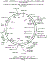


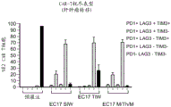
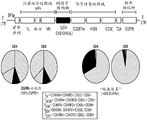


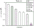

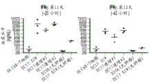

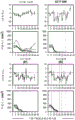


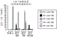


Classifications
-
- C—CHEMISTRY; METALLURGY
- C07—ORGANIC CHEMISTRY
- C07K—PEPTIDES
- C07K16/00—Immunoglobulins [IGs], e.g. monoclonal or polyclonal antibodies
- C07K16/44—Immunoglobulins [IGs], e.g. monoclonal or polyclonal antibodies against material not provided for elsewhere, e.g. haptens, metals, DNA, RNA, amino acids
-
- A—HUMAN NECESSITIES
- A61—MEDICAL OR VETERINARY SCIENCE; HYGIENE
- A61K—PREPARATIONS FOR MEDICAL, DENTAL OR TOILETRY PURPOSES
- A61K35/00—Medicinal preparations containing materials or reaction products thereof with undetermined constitution
- A61K35/12—Materials from mammals; Compositions comprising non-specified tissues or cells; Compositions comprising non-embryonic stem cells; Genetically modified cells
- A61K35/14—Blood; Artificial blood
- A61K35/17—Lymphocytes; B-cells; T-cells; Natural killer cells; Interferon-activated or cytokine-activated lymphocytes
-
- A—HUMAN NECESSITIES
- A61—MEDICAL OR VETERINARY SCIENCE; HYGIENE
- A61K—PREPARATIONS FOR MEDICAL, DENTAL OR TOILETRY PURPOSES
- A61K39/00—Medicinal preparations containing antigens or antibodies
- A61K39/46—Cellular immunotherapy
- A61K39/461—Cellular immunotherapy characterised by the cell type used
- A61K39/4611—T-cells, e.g. tumor infiltrating lymphocytes [TIL], lymphokine-activated killer cells [LAK] or regulatory T cells [Treg]
-
- A—HUMAN NECESSITIES
- A61—MEDICAL OR VETERINARY SCIENCE; HYGIENE
- A61K—PREPARATIONS FOR MEDICAL, DENTAL OR TOILETRY PURPOSES
- A61K39/00—Medicinal preparations containing antigens or antibodies
- A61K39/46—Cellular immunotherapy
- A61K39/463—Cellular immunotherapy characterised by recombinant expression
- A61K39/4631—Chimeric Antigen Receptors [CAR]
-
- A—HUMAN NECESSITIES
- A61—MEDICAL OR VETERINARY SCIENCE; HYGIENE
- A61K—PREPARATIONS FOR MEDICAL, DENTAL OR TOILETRY PURPOSES
- A61K39/00—Medicinal preparations containing antigens or antibodies
- A61K39/46—Cellular immunotherapy
- A61K39/464—Cellular immunotherapy characterised by the antigen targeted or presented
- A61K39/4643—Vertebrate antigens
- A61K39/4644—Cancer antigens
- A61K39/464402—Receptors, cell surface antigens or cell surface determinants
- A61K39/464403—Receptors for growth factors
- A61K39/464404—Epidermal growth factor receptors [EGFR]
-
- A—HUMAN NECESSITIES
- A61—MEDICAL OR VETERINARY SCIENCE; HYGIENE
- A61K—PREPARATIONS FOR MEDICAL, DENTAL OR TOILETRY PURPOSES
- A61K39/00—Medicinal preparations containing antigens or antibodies
- A61K39/46—Cellular immunotherapy
- A61K39/464—Cellular immunotherapy characterised by the antigen targeted or presented
- A61K39/4646—Small organic molecules e.g. cocaine or nicotine
-
- A—HUMAN NECESSITIES
- A61—MEDICAL OR VETERINARY SCIENCE; HYGIENE
- A61K—PREPARATIONS FOR MEDICAL, DENTAL OR TOILETRY PURPOSES
- A61K47/00—Medicinal preparations characterised by the non-active ingredients used, e.g. carriers or inert additives; Targeting or modifying agents chemically bound to the active ingredient
- A61K47/50—Medicinal preparations characterised by the non-active ingredients used, e.g. carriers or inert additives; Targeting or modifying agents chemically bound to the active ingredient the non-active ingredient being chemically bound to the active ingredient, e.g. polymer-drug conjugates
- A61K47/51—Medicinal preparations characterised by the non-active ingredients used, e.g. carriers or inert additives; Targeting or modifying agents chemically bound to the active ingredient the non-active ingredient being chemically bound to the active ingredient, e.g. polymer-drug conjugates the non-active ingredient being a modifying agent
- A61K47/54—Medicinal preparations characterised by the non-active ingredients used, e.g. carriers or inert additives; Targeting or modifying agents chemically bound to the active ingredient the non-active ingredient being chemically bound to the active ingredient, e.g. polymer-drug conjugates the non-active ingredient being a modifying agent the modifying agent being an organic compound
- A61K47/55—Medicinal preparations characterised by the non-active ingredients used, e.g. carriers or inert additives; Targeting or modifying agents chemically bound to the active ingredient the non-active ingredient being chemically bound to the active ingredient, e.g. polymer-drug conjugates the non-active ingredient being a modifying agent the modifying agent being an organic compound the modifying agent being also a pharmacologically or therapeutically active agent, i.e. the entire conjugate being a codrug, i.e. a dimer, oligomer or polymer of pharmacologically or therapeutically active compounds
- A61K47/551—Medicinal preparations characterised by the non-active ingredients used, e.g. carriers or inert additives; Targeting or modifying agents chemically bound to the active ingredient the non-active ingredient being chemically bound to the active ingredient, e.g. polymer-drug conjugates the non-active ingredient being a modifying agent the modifying agent being an organic compound the modifying agent being also a pharmacologically or therapeutically active agent, i.e. the entire conjugate being a codrug, i.e. a dimer, oligomer or polymer of pharmacologically or therapeutically active compounds one of the codrug's components being a vitamin, e.g. niacinamide, vitamin B3, cobalamin, vitamin B12, folate, vitamin A or retinoic acid
-
- A—HUMAN NECESSITIES
- A61—MEDICAL OR VETERINARY SCIENCE; HYGIENE
- A61P—SPECIFIC THERAPEUTIC ACTIVITY OF CHEMICAL COMPOUNDS OR MEDICINAL PREPARATIONS
- A61P35/00—Antineoplastic agents
-
- C—CHEMISTRY; METALLURGY
- C07—ORGANIC CHEMISTRY
- C07K—PEPTIDES
- C07K14/00—Peptides having more than 20 amino acids; Gastrins; Somatostatins; Melanotropins; Derivatives thereof
- C07K14/435—Peptides having more than 20 amino acids; Gastrins; Somatostatins; Melanotropins; Derivatives thereof from animals; from humans
- C07K14/705—Receptors; Cell surface antigens; Cell surface determinants
- C07K14/70503—Immunoglobulin superfamily
- C07K14/7051—T-cell receptor (TcR)-CD3 complex
-
- A—HUMAN NECESSITIES
- A61—MEDICAL OR VETERINARY SCIENCE; HYGIENE
- A61K—PREPARATIONS FOR MEDICAL, DENTAL OR TOILETRY PURPOSES
- A61K39/00—Medicinal preparations containing antigens or antibodies
- A61K2039/545—Medicinal preparations containing antigens or antibodies characterised by the dose, timing or administration schedule
-
- A—HUMAN NECESSITIES
- A61—MEDICAL OR VETERINARY SCIENCE; HYGIENE
- A61K—PREPARATIONS FOR MEDICAL, DENTAL OR TOILETRY PURPOSES
- A61K2239/00—Indexing codes associated with cellular immunotherapy of group A61K39/46
- A61K2239/38—Indexing codes associated with cellular immunotherapy of group A61K39/46 characterised by the dose, timing or administration schedule
-
- C—CHEMISTRY; METALLURGY
- C07—ORGANIC CHEMISTRY
- C07K—PEPTIDES
- C07K2317/00—Immunoglobulins specific features
- C07K2317/20—Immunoglobulins specific features characterized by taxonomic origin
- C07K2317/21—Immunoglobulins specific features characterized by taxonomic origin from primates, e.g. man
-
- C—CHEMISTRY; METALLURGY
- C07—ORGANIC CHEMISTRY
- C07K—PEPTIDES
- C07K2317/00—Immunoglobulins specific features
- C07K2317/40—Immunoglobulins specific features characterized by post-translational modification
- C07K2317/41—Glycosylation, sialylation, or fucosylation
-
- C—CHEMISTRY; METALLURGY
- C07—ORGANIC CHEMISTRY
- C07K—PEPTIDES
- C07K2317/00—Immunoglobulins specific features
- C07K2317/50—Immunoglobulins specific features characterized by immunoglobulin fragments
- C07K2317/52—Constant or Fc region; Isotype
-
- C—CHEMISTRY; METALLURGY
- C07—ORGANIC CHEMISTRY
- C07K—PEPTIDES
- C07K2317/00—Immunoglobulins specific features
- C07K2317/50—Immunoglobulins specific features characterized by immunoglobulin fragments
- C07K2317/52—Constant or Fc region; Isotype
- C07K2317/524—CH2 domain
-
- C—CHEMISTRY; METALLURGY
- C07—ORGANIC CHEMISTRY
- C07K—PEPTIDES
- C07K2317/00—Immunoglobulins specific features
- C07K2317/50—Immunoglobulins specific features characterized by immunoglobulin fragments
- C07K2317/52—Constant or Fc region; Isotype
- C07K2317/53—Hinge
-
- C—CHEMISTRY; METALLURGY
- C07—ORGANIC CHEMISTRY
- C07K—PEPTIDES
- C07K2317/00—Immunoglobulins specific features
- C07K2317/60—Immunoglobulins specific features characterized by non-natural combinations of immunoglobulin fragments
- C07K2317/62—Immunoglobulins specific features characterized by non-natural combinations of immunoglobulin fragments comprising only variable region components
- C07K2317/622—Single chain antibody (scFv)
-
- C—CHEMISTRY; METALLURGY
- C07—ORGANIC CHEMISTRY
- C07K—PEPTIDES
- C07K2317/00—Immunoglobulins specific features
- C07K2317/70—Immunoglobulins specific features characterized by effect upon binding to a cell or to an antigen
- C07K2317/72—Increased effector function due to an Fc-modification
-
- C—CHEMISTRY; METALLURGY
- C07—ORGANIC CHEMISTRY
- C07K—PEPTIDES
- C07K2317/00—Immunoglobulins specific features
- C07K2317/90—Immunoglobulins specific features characterized by (pharmaco)kinetic aspects or by stability of the immunoglobulin
- C07K2317/92—Affinity (KD), association rate (Ka), dissociation rate (Kd) or EC50 value
-
- C—CHEMISTRY; METALLURGY
- C07—ORGANIC CHEMISTRY
- C07K—PEPTIDES
- C07K2319/00—Fusion polypeptide
- C07K2319/01—Fusion polypeptide containing a localisation/targetting motif
- C07K2319/03—Fusion polypeptide containing a localisation/targetting motif containing a transmembrane segment
-
- C—CHEMISTRY; METALLURGY
- C07—ORGANIC CHEMISTRY
- C07K—PEPTIDES
- C07K2319/00—Fusion polypeptide
- C07K2319/33—Fusion polypeptide fusions for targeting to specific cell types, e.g. tissue specific targeting, targeting of a bacterial subspecies
-
- C—CHEMISTRY; METALLURGY
- C12—BIOCHEMISTRY; BEER; SPIRITS; WINE; VINEGAR; MICROBIOLOGY; ENZYMOLOGY; MUTATION OR GENETIC ENGINEERING
- C12N—MICROORGANISMS OR ENZYMES; COMPOSITIONS THEREOF; PROPAGATING, PRESERVING, OR MAINTAINING MICROORGANISMS; MUTATION OR GENETIC ENGINEERING; CULTURE MEDIA
- C12N2740/00—Reverse transcribing RNA viruses
- C12N2740/00011—Details
- C12N2740/10011—Retroviridae
- C12N2740/16011—Human Immunodeficiency Virus, HIV
- C12N2740/16041—Use of virus, viral particle or viral elements as a vector
- C12N2740/16043—Use of virus, viral particle or viral elements as a vector viral genome or elements thereof as genetic vector
Landscapes
- Health & Medical Sciences (AREA)
- Life Sciences & Earth Sciences (AREA)
- Chemical & Material Sciences (AREA)
- General Health & Medical Sciences (AREA)
- Medicinal Chemistry (AREA)
- Immunology (AREA)
- Cell Biology (AREA)
- Organic Chemistry (AREA)
- Pharmacology & Pharmacy (AREA)
- Veterinary Medicine (AREA)
- Public Health (AREA)
- Animal Behavior & Ethology (AREA)
- Epidemiology (AREA)
- Engineering & Computer Science (AREA)
- Microbiology (AREA)
- Mycology (AREA)
- Bioinformatics & Cheminformatics (AREA)
- Zoology (AREA)
- Proteomics, Peptides & Aminoacids (AREA)
- Molecular Biology (AREA)
- Genetics & Genomics (AREA)
- Biochemistry (AREA)
- Biophysics (AREA)
- Nuclear Medicine, Radiotherapy & Molecular Imaging (AREA)
- General Chemical & Material Sciences (AREA)
- Chemical Kinetics & Catalysis (AREA)
- Gastroenterology & Hepatology (AREA)
- Toxicology (AREA)
- Developmental Biology & Embryology (AREA)
- Biotechnology (AREA)
- Virology (AREA)
- Biomedical Technology (AREA)
- Hematology (AREA)
- Dermatology (AREA)
- Oncology (AREA)
- Pharmaceuticals Containing Other Organic And Inorganic Compounds (AREA)
- Medicines Containing Antibodies Or Antigens For Use As Internal Diagnostic Agents (AREA)
- Medicines Containing Material From Animals Or Micro-Organisms (AREA)
- Peptides Or Proteins (AREA)
- Preparation Of Compounds By Using Micro-Organisms (AREA)
Abstract
The present invention relates to methods of treating a cancer patient by administering to the patient a composition comprising CAR T cells and administering to the patient a small molecule linked by a linker to a targeting moiety. The invention also relates to compositions for use in such methods.
Description
Cross Reference to Related Applications
Priority of the present application for U.S. provisional patent application No. 62/634,573 filed on 23.2.2018, U.S. provisional patent application No. 62/656,265 filed on 11.4.2018, U.S. provisional patent application No. 62/724,345 filed on 29.8.8.2018, and U.S. provisional patent application No. 62/736,730 filed on 26.9.2018, all of the above disclosures being expressly incorporated herein by reference in their entireties.
Technical Field
The present invention relates to a method of treating a cancer patient by administering to the patient a composition comprising CAR T cells and administering to the patient a small molecule linked by a linker to a targeting moiety. The invention also relates to a composition for use in such a method.
Background
Immunotherapy based on adoptive transfer of lymphocytes (e.g., T cells) into patients is a valuable therapy for the treatment of cancer and other diseases. The development of immunotherapy based on adoptive transfer of lymphocytes has made significant progress. Among the many different types of immunotherapeutic agents, one of the most promising immunotherapeutic agents developed is T cells expressing chimeric antigen receptors (CAR T cells). A Chimeric Antigen Receptor (CAR) is a genetically engineered receptor designed to target a specific antigen, such as a tumor antigen. Such targeting can cause cytotoxicity against the tumor, e.g., so that CAR T cells expressing the CAR can target and kill the tumor via a particular tumor antigen.
The first generation CARs were composed of a recognition region, such as an antibody-derived single chain fragment variable (scFv) region for recognition and binding of an antigen expressed by a tumor, and an activation signaling domain, such as the T cell CD3 zeta chain, which can serve as a T cell activation signal in the CAR. Although CAR T cells have shown positive results in vitro, their success in eliminating disease (e.g., cancer) in clinical trials is limited. One problem is the inability to prolong activation and expansion of CAR T cell populations in vivo.
To address this issue, co-stimulatory domains (e.g., CD137, CD28, or CD134) have been included in second generation CARs to achieve prolonged T cell activation in vivo. The addition of the co-stimulatory domain enhances proliferation and survival of CAR-containing T cells in vivo, and initial clinical data has shown such constructs to be promising therapeutic agents for the treatment of diseases such as cancer.
Although CAR T cell therapy has improved, there are several problems. First, an "off-target" toxicity can occur due to normal cells expressing an antigen (e.g., a tumor-associated antigen) targeted by CAR T cells. Secondly, unregulated CAR T cell activation can be found, where rapid and uncontrolled elimination of diseased cells (e.g., cancer cells) by CAR T cells induces a series of metabolic disorders, called tumor lysis syndrome or Cytokine Release Syndrome (CRS), which can be fatal to the patient. Tumor lysis syndrome and CRS can result from administration of CAR T cells that cannot be easily regulated and that are uncontrollably activated. Thus, while CAR T cells show great promise as a tool to treat diseases such as cancer, there remains a need for additional CAR T cell therapy approaches that provide reduced off-target toxicity and more precise control of CAR T cell activation.
Disclosure of Invention
In various embodiments described herein, a small molecule ligand linked to a targeting moiety by a linker serves as a bridge between the cancer and the CAR T cell, directing the CAR T cell to the cancer to ameliorate the cancer. In one embodiment, a "small molecule ligand" can be, for example, a folate compound, DUPA, NK-1R ligand, CAIX ligand, ligand for gamma glutamyl transpeptidase, NKG2D ligand, or CCK2R ligand, each of which is a small molecule ligand that specifically binds to cancer cells (i.e., the receptors for these ligands are overexpressed on cancer as compared to normal tissue).
In one embodiment, a "small molecule ligand" is linked to a "targeting moiety" that binds to a CAR expressed by a CAR T cell. In various embodiments, a "targeting moiety" may be selected from, for example, 2, 4-Dinitrophenol (DNP), 2,4, 6-Trinitrophenol (TNP), biotin, digoxigenin (digoxigenin), Fluorescein Isothiocyanate (FITC), NHS-fluorescein, pentafluorophenyl ester (PFP), tetrafluorophenyl ester (TFP), knottin (knottin), tributyrin (centryrin), and DARPin.
The "targeting moiety" binds to the recognition region of a genetically engineered CAR expressed by CAR T cells. Thus, CAR (e.g., antibody, Fab, Fv, Fc, (Fab')2Single-chain fragment variable region (scFv)) of fragments and the like) the recognition region is directed to a "targeting moiety". Thus, the small molecule ligand linked to the targeting moiety by the linker acts as a bridge between the cancer and the CAR T cell, directing the CAR T cell to the cancer to ameliorate the cancer.
In one embodiment, a method of treating cancer is provided. The method comprises i) administering to the patient at least one dose of a CAR T cell composition comprising a CAR T cell, wherein the CAR T cell comprises a CAR directed to a targeting moiety; ii) administering a compound or a pharmaceutically acceptable salt thereof to the patient, wherein the compound comprises a small molecule ligand linked to a targeting moiety by a linker, and wherein the compound or pharmaceutically acceptable salt thereof is administered in at least a first ascending-dose order and a second ascending-dose order.
In another embodiment, a method of treating cancer is provided. The method comprises i) administering to the patient at least one dose of a CAR T cell composition comprising a CAR T cell, wherein the CAR T cell comprises a CAR directed to a targeting moiety; ii) administering a compound or a pharmaceutically acceptable salt thereof to the patient, wherein the compound comprises a small molecule ligand linked to a targeting moiety by a linker, and wherein the compound or pharmaceutically acceptable salt thereof is administered in a first ascending dosage order, wherein if severe CRS is present in the first ascending dosage order, the compound or pharmaceutically acceptable salt thereof is administered using a lower ascending dosage order, wherein the first dosage of the compound or pharmaceutically acceptable salt thereof in the lower ascending dosage order is lower than the first dosage of the compound or pharmaceutically acceptable salt thereof administered in the first ascending dosage order. In one embodiment, the compound or pharmaceutically acceptable salt thereof may be administered at about 0.5%, about 5%, and about 50% of the full dose of the compound or pharmaceutically acceptable salt thereof on three separate days in a lower ascending dosage order.
Additional embodiments are also described by the terms listed below. Also encompassed is any of the following embodiments in combination with any suitable embodiment described in the summary of the invention section, the detailed description of the illustrative embodiments, the examples section, or the claims of this patent application.
1. A method of treating cancer, the method comprising:
i) administering to the patient at least one dose of a CAR T cell composition comprising a CAR T cell, wherein the CAR T cell comprises a CAR directed to a targeting moiety;
ii) administering a compound or a pharmaceutically acceptable salt thereof to the patient, wherein the compound comprises a small molecule ligand linked to a targeting moiety by a linker, and wherein the compound or pharmaceutically acceptable salt thereof is administered in at least a first ascending-dose order and a second ascending-dose order.
2. The method of clause 1, wherein the compound or pharmaceutically acceptable salt thereof is administered in at least a first ascending dose order, a second ascending dose order, and a third ascending dose order.
3. The method of clause 1, wherein the compound or pharmaceutically acceptable salt thereof is administered in at least a first ascending dose order, a second ascending dose order, a third ascending dose order, and a fourth ascending dose order.
4. The method of clause 1, wherein the compound or pharmaceutically acceptable salt thereof is administered in at least a first ascending dose order, a second ascending dose order, a third ascending dose order, a fourth ascending dose order, and a fifth ascending dose order.
5. The method of clause 1, wherein the compound or pharmaceutically acceptable salt thereof is administered in at least a first ascending dose order, a second ascending dose order, a third ascending dose order, a fourth ascending dose order, a fifth ascending dose order, and a sixth ascending dose order.
6. The method of any of clauses 1-5, wherein a first dose of the CAR T cells and a second dose of the CAR T cells are administered to the patient.
7. The method of clause 6, wherein the first dose of said CAR T cells is a test dose that monitors the patient's tolerance to said CAR T cells.
8. The method of clause 6, wherein the second dose of said CAR T cells comprises a higher dose of said CAR T cells than the first dose of said CAR T cells.
9. The method of any of clauses 6 to 8, wherein the first dose of the CAR T cells comprises about 0.5x 10⁵(ii) each said CAR T cells/kg patient body weight to about 1.5X 10⁶One CAR T cell per kilogram patient body weight.
10. The method of any of clauses 6 to 9, wherein the second dose of said CAR T cells comprises about 0.8 x 10⁶(ii) each said CAR T cells/kg patient body weight to about 2X 10⁷One CAR T cell per kilogram patient body weight.
11. The method of any one of clauses 1 to 10, wherein the first ascending dosage order is followed by a period of time during which the compound or pharmaceutically acceptable salt thereof is not administered.
12. The method of any one of clauses 1-11, wherein the second ascending dosage sequence is followed by a period of time during which the compound or pharmaceutically acceptable salt thereof is not administered.
13. The method of any one of clauses 2-12, wherein the third ascending dosage sequence is followed by a period of time during which the compound or pharmaceutically acceptable salt thereof is not administered.
14. The method of any one of clauses 3-13, wherein the fourth ascending dosage sequence is followed by a period of time during which the compound or pharmaceutically acceptable salt thereof is not administered.
15. The method of any one of clauses 4-14, wherein the fifth ascending dosage sequence is followed by a period of time during which the compound or pharmaceutically acceptable salt thereof is not administered.
16. The method of any one of clauses 5-15, wherein the sixth ascending dosage sequence is followed by a period of time during which the compound or pharmaceutically acceptable salt thereof is not administered.
17. The method of any of clauses 11-16, wherein the period of time is about 7 days.
18. The method of any one of clauses 1-17, wherein the first ascending dose sequence comprises administering to the patient about 1%, about 10%, and about 100% of a full dose of the compound or pharmaceutically acceptable salt thereof on three separate days.
19. The method of any one of clauses 1-18, wherein the second ascending dose sequence comprises administering to the patient about 1%, about 30%, and about 300% of the full dose of the compound or pharmaceutically acceptable salt thereof on three separate days.
20. The method of any one of clauses 2-19, wherein the third ascending dose sequence comprises administering to the patient about 1%, about 50%, and about 500% of the full dose of the compound or pharmaceutically acceptable salt thereof on three separate days.
21. The method of any one of clauses 3-20, wherein the fourth ascending dose sequence comprises administering to the patient about 1%, about 10%, and about 100% of the full dose of the compound or pharmaceutically acceptable salt thereof on three separate days.
22. The method of any one of clauses 4-21, wherein the fifth ascending dose sequence comprises administering to the patient about 1%, about 30%, and about 300% of the full dose of the compound or pharmaceutically acceptable salt thereof on three separate days.
23. The method of any one of clauses 5-22, wherein the sixth ascending dose sequence comprises administering to the patient about 1%, about 50%, and about 500% of the full dose of the compound or pharmaceutically acceptable salt thereof on three separate days.
24. The method of any one of clauses 18 to 23, wherein the full dose of the compound or pharmaceutically acceptable salt thereof is about 10 μ g/kg to about 50 μ g/kg of the compound or pharmaceutically acceptable salt thereof.
25. The method of any one of clauses 18 to 24, wherein the full dose of the compound or pharmaceutically acceptable salt thereof is about 20 μ g/kg to about 40 μ g/kg of the compound or pharmaceutically acceptable salt thereof.
26. The method of any one of clauses 18 to 25, wherein the full dose of the compound or pharmaceutically acceptable salt thereof is about 25 μ g/kg to about 35 μ g/kg of the compound or pharmaceutically acceptable salt thereof.
27. The method of any one of clauses 18 to 26, wherein the full dose of the compound or pharmaceutically acceptable salt thereof is about 30 μ g/kg of the compound or pharmaceutically acceptable salt thereof.
28. The method of any of clauses 6-27, wherein the first dose of the CAR T cells and the second dose of the CAR T cells are administered to the patient during week 1.
29. The method of any of clauses 6-28, wherein the first dose of the CAR T cells and the second dose of the CAR T cells are administered to the patient on monday and thursday of week 1.
30. The method of any one of clauses 1-29, wherein the first ascending dose order for the compound or pharmaceutically acceptable salt thereof occurs during weeks 2 and 3.
31. The method of any one of clauses 1-30, wherein the second ascending order of dose for the compound or pharmaceutically acceptable salt thereof occurs during weeks 4 and 5.
32. The method of any one of clauses 2-31, wherein the third ascending dose sequence for the compound or pharmaceutically acceptable salt thereof occurs during weeks 6 and 7.
33. The method of clause 30, wherein the compound or pharmaceutically acceptable salt thereof is administered on three separate days, and the three separate days are monday on week 2 and monday on weeks four and 3.
34. The method of clause 31, wherein the compound or pharmaceutically acceptable salt thereof is administered on three separate days, and the three separate days are monday on week 4 and monday on weeks four and 5.
35. The method of clause 32, wherein the compound or pharmaceutically acceptable salt thereof is administered on three separate days, and the three separate days are monday on week 6 and monday on weeks 7.
36. The method of any of clauses 1-35, wherein the lymphocytes in the patient are depleted prior to administering the CART cell composition to the patient.
37. The method of any one of clauses 1-36, further comprising administering platelets to the patient, administering concentrated red blood cells to the patient, administering cryoprecipitate to the patient, administering intravenous immunoglobulin to the patient, and/or providing antibacterial therapy to the patient.
38. The method of any one of clauses 1-37, wherein if CRS or neurotoxicity is not observed in the patient during the first up-dosing sequence, the method proceeds to the second up-dosing sequence.
39. The method of any one of clauses 2-38, wherein if CRS or neurotoxicity is not observed in the patient during the second up-dosing sequence, the method advances to the third up-dosing sequence.
40. The method of any one of clauses 3-39, wherein if CRS or neurotoxicity is not observed in the patient during the third up-dosing sequence, the method advances to the fourth up-dosing sequence.
41. The method of any one of clauses 4-40, wherein if CRS or neurotoxicity is not observed in the patient during the fourth ascending dose sequence, the method advances to the fifth ascending dose sequence.
42. The method of any of clauses 5-41, wherein if CRS or neurotoxicity is not observed in the patient during the fifth ascending dose sequence, the method advances to the sixth ascending dose sequence.
43. The method of any one of clauses 1-42, wherein if fever without hypotension and no neurotoxicity is observed in the patient during any one of the ascending dose sequences, all subsequent doses of the compound or pharmaceutically acceptable salt thereof are administered to the patient at the ascending dose sequence level that results in the fever without hypotension.
44. The method of any one of clauses 1-43, wherein if severe CRS or neurotoxicity is present in the patient in any ascending dose order, all subsequent doses of the compound or pharmaceutically acceptable salt thereof are administered to the patient at ascending dose order levels that are lower than the ascending dose order level that caused severe CRS or neurotoxicity in the patient.
45. A method of treating cancer, the method comprising:
i) administering to the patient at least one dose of a CAR T cell composition comprising a CAR T cell, wherein the CAR T cell comprises a CAR directed to a targeting moiety;
ii) administering a compound or a pharmaceutically acceptable salt thereof to the patient, wherein the compound comprises a small molecule ligand linked to a targeting moiety by a linker, and wherein the compound or pharmaceutically acceptable salt thereof is administered in a first ascending-dose order, wherein if severe CRS is present in the first ascending-dose order, the compound or pharmaceutically acceptable salt thereof is administered using a lower ascending-dose order, wherein the first dose of the compound or pharmaceutically acceptable salt thereof in the lower ascending-dose order is lower than the first dose of the compound or pharmaceutically acceptable salt thereof administered in the first ascending-dose order.
46. The method of clause 45, wherein the compound or pharmaceutically acceptable salt thereof is administered at about 0.5%, about 5%, and about 50% of the full dose of the compound or pharmaceutically acceptable salt thereof on three separate days in the lower ascending dosage order.
47. The method of clause 46, wherein the full dose of the compound or pharmaceutically acceptable salt thereof is about a full dose of the compound or pharmaceutically acceptable salt thereof is about 10 μ g/kg to about 50 μ g/kg of the compound or pharmaceutically acceptable salt thereof.
48. The method of any one of clauses 46 to 47, wherein the full dose of the compound or pharmaceutically acceptable salt thereof is about 25 μ g/kg to about 35 μ g/kg of the compound or pharmaceutically acceptable salt thereof.
49. The method of any one of clauses 46 to 48, wherein the full dose of the compound or pharmaceutically acceptable salt thereof is about 30 μ g/kg of the compound or pharmaceutically acceptable salt thereof.
50. The method of any one of clauses 1 to 49, wherein the ligand is selected from the group consisting of: folate compounds, DUPA, NK-1R ligands, CAIX ligands, gamma glutamyl transpeptidase ligands, NKG2D ligands, and CCK2R ligands.
51. The method of any of clauses 1 to 50, wherein the ligand is a folate compound.
52. The method of any one of clauses 1 to 50, wherein the ligand is an NK-1R ligand.
53. The method of any of clauses 1-50, wherein the ligand is DUPA.
54. The method of any one of clauses 1 to 50, wherein the ligand is a CCK2R ligand.
55. The method of any one of clauses 1-50, wherein the ligand is a gamma glutamyl transpeptidase ligand.
56. The method of any one of clauses 1 to 55, wherein the targeting moiety is selected from the group consisting of: 2, 4-Dinitrophenol (DNP), 2,4, 6-Trinitrophenol (TNP), biotin, digoxin, Fluorescein Isothiocyanate (FITC), NHS-fluorescein, pentafluorophenyl ester, tetrafluorophenyl ester, knottin, sertraline, and DARPin.
57. The method of any one of clauses 1 to 56, wherein the targeting moiety is FITC.
58. The method of any one of clauses 1-56, wherein the targeting moiety is DNP.
59. The method of any one of clauses 1-56, wherein the targeting moiety is TNP.
60. The method of any of clauses 1-59, wherein the linker comprises polyethylene glycol (PEG), polyproline, a hydrophilic amino acid, a sugar, a non-natural peptidoglycan, polyvinylpyrrolidone, pluronic F-127, or a combination thereof.
61. The method of any one of clauses 1-60, wherein the linker comprises PEG.
62. The method of any one of clauses 1 to 61, wherein the compound or pharmaceutically acceptable salt thereof has the formula:
wherein B represents the small molecule ligand, L represents the linker, and T represents the targeting moiety, and wherein L comprises a structure having the formula:

wherein n is an integer of 0 to 200.
63. The method of clause 62, wherein n is an integer from 0 to 150.
64. The method of clause 62, wherein n is an integer from 0 to 110.
65. The method of clause 62, wherein n is an integer from 0 to 20.
66. The method of clause 62, wherein n is an integer from 15 to 20.
67. The method of clause 62, wherein n is an integer from 15 to 110.
68. The method of any one of clauses 1-67, wherein the cancer is selected from the group consisting of: lung cancer, bone cancer, pancreatic cancer, skin cancer, head cancer, neck cancer, skin melanoma, intraocular melanoma, uterine cancer, ovarian cancer, endometrial cancer (endometrial cancer), rectal cancer, gastric cancer, colon cancer, breast cancer, triple negative breast cancer, carcinoma of the fallopian tubes, carcinoma of the endometrium (carcinoma of the endometerium), carcinoma of the cervix, carcinoma of the vagina, carcinoma of the vulva, Hodgkin's Disease, esophageal cancer, cancer of the small intestine, cancer of the endocrine system, thyroid cancer, parathyroid cancer, non-small cell lung cancer, adrenal cancer, soft tissue sarcoma, osteosarcoma (including pediatric or non-pediatric osteosarcoma), cancer of the urethra, prostate cancer, chronic leukemia, acute myelocytic leukemia, lymphocytic lymphoma, myeloid leukemia, myelomonocytic leukemia, hairy cell leukemia, pleural mesothelioma, carcinoma, uterine cancer of the endometrium, carcinoma of the rectum, cancer, carcinoma of the rectum cancer, carcinoma of the colon cancer, carcinoma of the rectum, Burkitt's lymphoma, ductal carcinoma, renal cell carcinoma, renal pelvis carcinoma, Central Nervous System (CNS) neoplasms, primary CNS lymphoma, spinal axis tumors, brain stem glioma, pituitary adenoma, and adenocarcinoma of the gastroesophageal junction.
69. The method of any one of clauses 1-51 or 56-68, wherein the cancer is a folate receptor expressing cancer.
70. The method of any one of clauses 1-69, wherein the cancer is osteosarcoma.
71. The method of any of clauses 1-70, wherein the CAR has a recognition region and the recognition region is a single chain fragment variable (scFv) region of an antibody.
72. The method of any of clauses 1-57 or 60-71, wherein the CAR has a recognition region and the recognition region of the CAR is a single chain fragment variable (scFv) region of an anti-FITC antibody.
73. The method of any of clauses 1-72, wherein the CAR has a costimulatory domain and the costimulatory domain is selected from the group consisting of: CD28, CD137(4-1BB), CD134 (OX40) and CD278 (ICOS).
74. The method of any of clauses 1-73, wherein the CAR has an activation signaling domain and the activation signaling domain is a T cell CD3 zeta chain or Fc receptor gamma.
75. The method of any of clauses 1-57 or 60-74, wherein the CAR has a recognition region and the recognition region is a single chain fragment variable (scFv) region of an anti-FITC antibody, wherein the CAR has a costimulatory domain and the costimulatory domain is CD137(4-1BB), and wherein the CAR has an activation signaling domain and the activation signaling domain is the T cell CD3 zeta chain.
76. The method of any of clauses 1 to 75, wherein the patient is imaged prior to administration of the compound or pharmaceutically acceptable salt thereof, or prior to administration of the CAR T cell composition.
77. The method of any one of clauses 1-76, wherein the compound or pharmaceutically acceptable salt thereof is not an antibody and does not comprise a fragment of an antibody.
78. The method of any one of clauses 1 to 77, wherein the targeting moiety does not comprise a peptide epitope.
79. The method of any of clauses 1 to 78, wherein cytokine release resulting in off-target toxicity in the patient does not occur, and wherein CAR T cytotoxicity to the cancer occurs.
80. The method of any of clauses 1 to 78, wherein off-target tissue toxicity in the patient does not occur, and wherein CAR T cytotoxicity to the cancer occurs.
81. The method of any one of clauses 1-78, wherein the cancer comprises a tumor, wherein tumor size is reduced in the patient, and wherein off-target toxicity does not occur.
82. The method of any of clauses 1-81, wherein the CART cell comprises a nucleic acid comprising seq id no: 1.
83. The method of any of clauses 1-82, wherein the CART cell comprises a polypeptide comprising seq id no: 2.
84. The method of clause 82, wherein the nucleic acid encodes a chimeric antigen receptor.
85. The method of any of clauses 1-81, wherein the CART cell comprises a nucleic acid comprising seq id no: 4.
86. The method of any of clauses 1-81 or 85, wherein the CART cell comprises a polypeptide comprising seq id No. 5.
87. The method of clause 85, wherein the nucleic acid encodes a chimeric antigen receptor.
88. The method of any of clauses 1-87, wherein the CAR comprises a humanized amino acid sequence.
89. The method of any of clauses 1-87, wherein the CAR consists of a humanized amino acid sequence.
90. The method of any one of clauses 1-89, further comprising administering to the patient a folate compound; a conjugate comprising a folate compound, wherein the conjugate comprising a folate compound does not comprise a targeting moiety; or an agent that inhibits activation of the CAR T cell.
91. The method of clause 90, wherein the folate compound is administered.
92. The method of clause 90, wherein folic acid or leucovorin is administered.
93. The method of clause 90, wherein the conjugate comprising a folate compound is administered.
94. The method of clause 93, wherein the conjugate comprising a folate compound comprises a folate compound linked to one or more amino acids.
95. The method of clause 93, wherein the conjugate comprising a folate compound has the formula:

96. the method of clause 91, wherein the folate compound has the formula:

wherein X1And Y1Each independently selected from: halogen, R2、OR2、SR3And NR4R5;
U, V and W represent divalent moieties each independently selected from: - (R)6a)C=、-N=、-(R6a)C(R7a) -and-N (R)4a) -; q is selected from: c and CH; t is selected from: s, O, N and-C = C-;
X2and X3Each independently selected from: oxygen, sulfur, -C (Z) -, -C (Z) O-, -OC (Z) -, -N (R)4b)-、-C(Z)N(R4b)-、-N(R4b)C(Z)-、-OC(Z)N(R4b)-、-N(R4b)C(Z)O-、-N(R4b)C(Z)N(R5b)-、-S(O)-、-S(O)2-、-N(R4a)S(O)2-、-C(R6b)(R7b)-、-N(C≡CH)-、-N(CH2C≡CH)-、C1-C12Alkylene and C1-C12Alkyleneoxy, wherein Z is oxygen or sulfur;
R1selected from: hydrogen, halogen, C1-C12Alkyl and C1-C12An alkoxy group;
R2、R3、R4、R4a、R4b、R5、R5b、R6band R7bEach independently selected from: hydrogen, halogen, C1-C12Alkyl radical, C1-C12Alkoxy radical, C1-C12Alkanoyl radical, C1-C12Alkenyl radical, C1-C12Alkynyl, (C)1-C12Alkoxy) carbonyl and (C)1-C12Alkylamino) carbonyl;
R6and R7Each independently selected from: hydrogen, halogen, C1-C12Alkyl and C1-C12An alkoxy group; or R6And R7Together form a carbonyl group;
R6aand R7aEach independently selected from: hydrogen, halogen, C1-C12Alkyl and C1-C12An alkoxy group; or R6aAnd R7aTogether form a carbonyl group;
p, r, s and t are each independently 0 or 1; and is
If any other chemical moiety is part of the folate compound, then an optional covalent bond to the remainder of the conjugate is indicated.
97. The method of clause 90, wherein the agent that inhibits activation of the CART cells is selected from the group consisting of: lymphocyte-specific protein tyrosine kinase inhibitors, PI3 kinase inhibitors, IL-2-inducible T cell kinase inhibitors, JAK inhibitors, BTK inhibitors, EC2319, and agents that block CAR T cell binding to the compound or a pharmaceutically acceptable salt thereof but not to the cancer.
98. The method of clause 90, wherein the agent that inhibits activation of the CART cell is administered and the agent is a lymphocyte-specific protein tyrosine kinase inhibitor.
99. The method of clause 98, wherein the lymphocyte-specific protein tyrosine kinase inhibitor is Dasatinib (Dasatinib).
100. The method of clause 90, wherein the agent that inhibits activation of the CAR T cells is administered and the agent is a PI3 kinase inhibitor.
101. The method of clause 100, wherein the PI3 kinase inhibitor is GDC 0980.
102. The method of clause 90, wherein the agent that inhibits activation of the CART cell is administered and the agent is an IL-2-inducible T cell kinase inhibitor.
103. The method of clause 102, wherein the IL-2-inducible T cell kinase inhibitor is BMS-509744.
104. The method of clause 90, wherein the agent that inhibits activation of the CART cell is administered and is an agent that blocks CAR T cells from binding to the compound or pharmaceutically acceptable salt thereof but not to the cancer.
105. The method of clause 104, wherein the agent is fluorescein amine, FITC, or fluorescein sodium.
106. The method of clause 105, wherein the agent is fluorescein sodium.
107. The method of any of clauses 90-106, wherein administering the folate compound, the conjugate comprising a folate compound (wherein the conjugate comprising a folate compound does not comprise a targeting moiety), or the agent that inhibits activation of the CAR T cells, results in a reduction of cytokine levels in the patient.
108. The method of clause 107, wherein the cytokine level is reduced to about the cytokine level in an untreated patient.
109. The method of any of clauses 90-108, wherein the cancer comprises a tumor and the tumor size in the patient does not increase when the folate compound, the conjugate comprising a folate compound (wherein the conjugate comprising a folate compound does not comprise a targeting moiety), or the agent that inhibits activation of the CAR T cells is administered to the patient.
110. The method of any of clauses 104-106, wherein the agent that inhibits activation of said CAR T cells is administered to the patient when the CRS rating reaches 1,2, 3, or 4.
111. The method of clause 110, wherein the agent that inhibits activation of said CAR T cells is administered to the patient when the CRS rating reaches 3 or 4.
112. The method of any of clauses 104 to 106, wherein the agent that inhibits activation of said CAR T cells is administered at a dose of about 0.01 to about 300 micromoles per kilogram body weight of the patient.
113. The method of any one of clauses 1-112, wherein CRS is reduced or prevented in the patient and the method causes a reduction in tumor volume in the patient.
114. The method of any of clauses 1-113, wherein weight loss due to CRS is reduced or prevented.
115. The method of any one of clauses 90-114, further comprising re-administering the compound or pharmaceutically acceptable salt thereof to the patient.
116. The method of clause 115, wherein the subsequent administration of the compound or pharmaceutically acceptable salt thereof causes CAR T cell activation and an increase in cytokine levels in the patient.
117. The method of any of clauses 1 to 116, wherein the CAR T cell composition is administered prior to the compound or pharmaceutically acceptable salt thereof.
118. The method of any of clauses 1 to 117, wherein the CAR T cell is autologous.
119. The method of any one of clauses 2-17, wherein the first ascending dosage sequence comprises administering to the patient on three separate days about 1%, about 2%, and about 20% of the full dose of the compound or pharmaceutically acceptable salt thereof, wherein the second ascending dosage sequence comprises administering to the patient on three separate days about 1%, about 6%, and about 60% of the full dose of the compound or pharmaceutically acceptable salt thereof, and wherein the third ascending dosage sequence comprises administering to the patient on three separate days about 1%, about 10%, and about 100% of the full dose of the compound or pharmaceutically acceptable salt thereof.
120. The method of any one of clauses 2-17, wherein the first ascending dosage sequence comprises administering to the patient on three separate days about 1%, about 10%, and about 100% of the full dose of the compound or pharmaceutically acceptable salt thereof, and wherein the second ascending dosage sequence comprises administering to the patient on three separate days about 1%, about 20%, and about 200% of the full dose of the compound or pharmaceutically acceptable salt thereof.
121. The method of any one of clauses 2-17, wherein the first ascending dosage sequence comprises administering to the patient on three separate days about 1%, about 10%, and about 100% of the full dose of the compound or pharmaceutically acceptable salt thereof, and wherein the second ascending dosage sequence comprises administering to the patient on three separate days about 1%, about 10%, and about 100% of the full dose of the compound or pharmaceutically acceptable salt thereof.
122. The method of any one of clauses 2-17, wherein the first ascending dosage sequence comprises administering to the patient on three separate days about 1%, about 20%, and about 200% of the full dose of the compound or pharmaceutically acceptable salt thereof, and wherein the second ascending dosage sequence comprises administering to the patient on three separate days about 1%, about 20%, and about 200% of the full dose of the compound or pharmaceutically acceptable salt thereof.
123. The method of any one of clauses 119 to 122, wherein the full dose of the compound or pharmaceutically acceptable salt thereof is about 500 nanomoles/kg.
Drawings
Figure 1 shows a schematic representation of an exemplary dosing regimen according to the claimed method. In this exemplary protocol, E2CAR T cell (CAR T cells expressing SEQ ID NO: 4) administration occurred on mondays and thursdays of week 1, before administration of a small molecule ligand (e.g., EC17) linked to a targeting moiety by a linker was initiated on week 2.
At week 2, a small molecule ligand (e.g., EC17) linked to a targeting moiety through a linker was administered on monday and thursday, and then on monday of week 3, using an ascending dose sequence (i.e., sequence 1). Small molecule ligands (e.g., EC17) linked to the targeting moiety through a linker are not administered on tuesday to sunday of week 3. Weeks 2 to 3 are referred to as "cycle 1".
At week 4, a small molecule ligand (e.g., EC17) linked to a targeting moiety through a linker was administered on monday and thursday, and then on monday of week 5, using an ascending dose sequence (i.e., sequence 2). Small molecule ligands (e.g., EC17) linked to the targeting moiety through a linker are not administered on tuesday to sunday of week 5. Weeks 4 to 5 are referred to as "cycle 2".
At week 6, a small molecule ligand (e.g., EC17) linked to a targeting moiety through a linker was administered on monday and thursday, and then on monday of week 7, using an ascending dose sequence (i.e., sequence 3). Small molecule ligands (e.g., EC17) linked to the targeting moiety through a linker are not administered on tuesday to sunday of week 7. Weeks 6 to 7 were referred to as "cycle 3". Week 2 to week 7 are referred to as "course 1".
During sequence 1, the small molecule ligand (e.g., EC17) attached to the targeting moiety by a linker is administered on monday and thursday of week 2 and then monday of week 3, wherein the doses of the small molecule ligand (e.g., EC17) attached to the targeting moiety by a linker are 1%, 10%, and 100% of the full dose (e.g., 31 μ g/kg) of the small molecule ligand (e.g., EC17), respectively.
During sequence 2, the small molecule ligand (e.g., EC17) attached to the targeting moiety by a linker is administered on monday and thursday of week 4 and then monday of week 5, wherein the doses of the small molecule ligand (e.g., EC17) attached to the targeting moiety by a linker are 1%, 30%, and 300% of the full dose (e.g., 31 μ g/kg) of the small molecule ligand (e.g., EC17), respectively.
During sequence 3, the small molecule ligand (e.g., EC17) attached to the targeting moiety by a linker is administered on monday and thursday of week 6 and then monday of week 7, wherein the doses of the small molecule ligand (e.g., EC17) attached to the targeting moiety by a linker are 1%, 50%, and 500% of the full dose (e.g., 31 μ g/kg) of the small molecule ligand (e.g., EC17), respectively. Course 1 is then repeated, presenting clinical benefit and tolerating toxicity.
Figure 2 shows an overall diagram of constructs used for CAR T transduction.
Figure 3 shows diagrammatically a diagram of the E2 construct relative to the 4M5.3 construct and showing the E2 construct.
FIG. 4 (upper panel) is a graph showing tumor volume of HOS-FR α tumors when treated with E2Car-T cells only (●) and E2Car-T cells + EC-17 (. smallcircle.). FIG. 4 (lower panel) is a graph showing the change in body weight of mice bearing HOS-FR α tumors when treated with E2Car-T cells only (●) and E2Car-T cells + EC-17 (. smallcircle.).
FIG. 5 is a diagram showing the use of Car-T (●); EC-17 (■) was not used; EC-17 SIW 500 nmol/kg (. tangle-solidup.); with EC-17 TIW (5, 50, 500 nmol/kg on/off); graph of body weight change of mice bearing THP-1-FR β AML when treated with (♦) EC-17 dose escalation (M/Th/M on/off).
Fig. 6 (left panel) is a graph showing metastatic tumor burden of the liver. Figure 6 (right panel) is a graph showing non-liver metastatic tumor burden.
FIG. 7 is a graph showing the circulating THP1-FR beta cell count per 100 μ L of blood on day 39 on a logarithmic scale.
Figure 8 is a graph showing the percentage of total E2CAR T cells isolated from solid liver tumors (y-axis) under different EC-17 dosing regimens. For each dosing regimen, the leftmost bar was PD1+ LAG3+ TIM3+, the second bar on the left was PD1+ LAG3+ TIM3-, and the middle bar was PD1+ LAG 3-TIM 3 +; the second bar on the right is PD1+ LAG 3-TIM 3-; the rightmost bar is PD 1-LAG 3-TIM 3-.
Figure 9 shows a fully human CAR construct comprising an anti-FITC scFv (clone E2), a full length IgG4 spacer (Fc-derived hinge-CH 2(L235D, N297Q) -CH3), a CD28tm transmembrane domain, a 4-1BB/CD3 zeta cytoplasmic activation domain, and a non-functional truncated cell surface polypeptide of epidermal growth factor receptor (EGFRt). Bottom: examples of CD4/CD 8T cell phenotypic analyses performed by flow cytometry on EGFRt sorted (left side pie chart) CAR-T cell preparations and unsorted "clinical facsimile" (right side pie chart). The color keys are as shown.
FIG. 10, Panel A: of FR + target cells after 2-hour incubation at 37 ℃3Kd values for H-EC17 uptake (calculated from the number of molecules bound per cell). And B, drawing: of E2-CAR-T cells (-24% EGFRT +, -95: 5 CD8/CD4 ratio) after 2 hours incubation at room temperature3Kd values for H-EC17 uptake (calculated from total cell-associated radioactive DPM).
FIG. 11 shows a graph obtained by basing3Binding assay for H-FA (100 nM, 1 hour at 37 ℃), measured levels of functional FR on tumor cells.
Figure 12 shows the results of EC17 dose studies and CRS assessment in tumors compared to tumor-free mice. A, picture A: shows that 3 different patterns of dose of CAR-T cells (about 1 million "clinical facsimile") plus EC17 administered in NSG mice, with or without pre-established MDA-MB-231 xenograftsSchematic illustration of the arrangement. Tumor-free mice received EC17 SIW 500 nmol/kg (2 doses on days 2 and 10). Tumor bearing mice received EC17 as follows: EC17 SIW 500 nmol/kg (5 doses on days 2, 10, 17, 24 and 31), EC 17M/Th/M _ increment-1 (5/50/500 nmol/kg repeated on days 2, 6, 10, 17, 20, 24 and 31 with subsequent discontinuation for 1 week on monday/thursday/monday) or EC 17M/Th/M _ increment-2 (5/100/1000 nmol/kg repeated on days 2, 6, 10, 17, 20, 24 and 31 with subsequent discontinuation for 1 week on monday/thursday/monday). And B, drawing: systemic levels of human IFN γ on log2 scale were detected in mouse plasma at days 11 and 12 after CAR-T cell injection in tumor-bearing and tumor-free mice (i.e., about 20 and 42 hours after prior EC17 dosing in all treatment groups). And (C) diagram: circulating CAR-T cells in mouse blood identified by flow cytometry as human CD3+ EGFRT + events and counted per 100 μ L of whole blood. And (D) diagram: measurement of changes in body weight and tumor volume of tumor-bearing mice receiving CAR-T cells alone, or CAR-T cells administered in 3 different ways plus EC17 (dashed line indicates individual EC17 doses). n = 5 mice/group. All data represent mean ± s.e.m. *p <0.05, tested by one-way ANOVA.
Figure 13 shows in vivo safety and anti-leukemic activity of EC17 dose escalation. A, picture A: a schematic showing the dosing schedule of EC17 plus unsorted EGFRt CAR-T cells (about 6 million, day 0) in 1 day old NSG mice xenografted with intravenous THP1-FR β. From 3 days after CAR-T cell injection, EC17 was administered in 3 different ways: EC17 SIW 500 nmol/kg (5 doses on days 3, 10, 17, 24, 31 and 38), EC17 TIW 5/50/500 nmol/kg (on monday/wednesday/friday, followed by 9 days off, i.e. 3 repetitions of 5/50/500 nmol/kg on days 3/5/7, 17/19/21 and 31/33/35) or EC 17M/Th/M _ dose escalation on monday/thursday/monday (3 escalation cycles, 5/10/100 nmol/kg for cycle 1, 5/30/300 nmol/kg for cycle 2 and 5/50/500 nmol/kg for cycle 3, i.e. on days 3/6/10, 17/20/24 and 31/34/38). And B, drawing: measurement of body weight change (n = 5). And (C) diagram: circulating CAR-T cells found as human CD3+ EGFRT + events every 100 μ L of whole mouse blood on day 31, on a logarithmic scale. And (D) diagram: left bar graph: circulating tumor cells (GFP +) per 100 µ L of whole blood in all cohorts measured at the end of the study (day 39); middle bar graph: THP1-FR β infiltrated liver weight representing metastatic load of the liver; right bar graph: total tumor weight of all non-liver large metastases. E, drawing: flow cytometry analysis of T cell depletion markers PD1, LAG3, TIM3 on pre-infused CAR-T cell products (triple negative) and tumor-infiltrating CAR-T cells isolated from liver metastases. The main feature of fully depleted T cells is the co-expression of multiple inhibitory receptor markers (i.e., triple positive).
Figure 14 shows antitumor activity and CRS rescue in an aggressive model of pediatric osteosarcoma. A, picture A: schematic diagram showing dosing schedule of CAR-T cells (about 6 million, day 0) plus EC17 in 3-day-old NSG mice (n = 5) of subcutaneous HOS-FR α xenografts. Starting 3 days after CAR-T cell injection (6 days after tumor implantation), 3 cycles were: EC 17M/Th/M "in patient" dose escalation was performed at 5/10/100 nmol/kg in cycle 1, 5/30/300 nmol/kg in cycle 2, and 5/50/500 nmol/kg in cycle 3, on Mondays/Thursdays/Mondays, i.e., on days 3/6/10, 17/20/24 and 31/34/38. And B, drawing: measurement of tumor volume and body weight change. Due to tumor progression, five mice receiving CAR-T cells only (no EC17) were euthanized on days 23-31, and five mice treated with EC17 were euthanized on days 33 (2 mice) and 47 (3 mice), respectively. And (C) diagram: flow cytometric analysis of CAR-T cells (human CD3+ EGFRt +) (left) and tumor infiltrating CAR-T cells at euthanasia per 100 μ L whole blood plotted on a logarithmic scale (right).
Definition of
As used herein, "a" or "an" can mean one or more. As used herein, with respect to numerical values, including, for example, integers, fractions, and percentages, "about" generally refers to a range of numerical values (e.g., +/-5% to 10% of the recited value) that one of ordinary skill in the art would consider equivalent to the recited value (e.g., having the same function or result).
As used herein, the term "treatment" refers to both therapeutic and prophylactic or preventative treatment.
As used herein, with respect to cancer, the term "ameliorating/administered" can mean alleviating a symptom of the cancer, reducing the size of the tumor, removing the tumor completely or partially (e.g., complete or partial response), producing a stable disease, arresting the progression of the cancer (e.g., progression-free survival), or any other effect on the cancer that would be considered by a physician as a therapeutic, prophylactic, or preventative treatment of the cancer.
As used herein, the term "administering" means all means of introducing a compound or a pharmaceutically acceptable salt thereof or a CAR T cell composition described herein into a patient, including, but not limited to, oral, intravenous, intramuscular, subcutaneous, and transdermal.
As used herein, the term "off-target toxicity" means organ destruction or weight loss in a patient that is unacceptable to the physician treating the patient, or any other effect on the patient that is unacceptable to the physician treating the patient, such as B-cell aplasia, fever, decreased blood pressure, or pulmonary edema.
As used herein, the terms "transduction" and "transfection" are used equivalently and the terms mean the introduction of nucleic acids into cells by any artificial method, including viral and non-viral methods.
The term "ascending dose order," as used herein, means that an increasing dose of a compound, or a pharmaceutically acceptable salt thereof, is administered over time. As used herein, reference to a "first ascending dosage sequence" used in combination with a "second ascending dosage sequence", "third ascending dosage sequence", "fourth ascending dosage sequence", "fifth ascending dosage sequence", and "sixth ascending dosage sequence", and the like, means that multiple ascending dosage sequences occur, and for each individual ascending dosage sequence following the first ascending dosage sequence, the first dosage of a compound or a pharmaceutically acceptable salt thereof in the subsequent ascending dosage sequence is lower than the last dosage of the compound or a pharmaceutically acceptable salt thereof in the previous ascending dosage sequence (see, e.g., fig. 1 and the explanation of fig. 1 in the description of the figures).
Detailed description of illustrative embodiments
In various embodiments described herein, a small molecule ligand linked to a targeting moiety by a linker serves as a bridge between the cancer and CAR T cells (i.e., T cells expressing a chimeric antigen receptor). The bridge directs CAR T cells to the cancer to ameliorate the cancer. In one embodiment, a "small molecule ligand" can be a folate compound, a CAIX ligand, DUPA, an NK-1R ligand, a ligand for gamma glutamyl transpeptidase, an NKG2D ligand, or a CCK2R ligand, each of which is a small molecule ligand that specifically binds to a cancer cell type (i.e., the receptor for each of these ligands is overexpressed on cancer compared to normal tissue).
A "targeting moiety" linked to a small molecule ligand binds to the recognition region of a genetically engineered CAR expressed by CAR T cells. Thus, the recognition region of the CAR (e.g., a single chain fragment variable region (scFv), Fab, Fv, Fc, or (Fab')2Fragments, and the like) are directed to a "targeting moiety". Thus, the small molecule ligand linked to the targeting moiety by the linker acts as a bridge between the cancer and the CAR T cells, directing the CAR T cells to the cancer to ameliorate the cancer. The bridge between the cancer and CAR T cells in various embodiments can be any applicable conjugate shown in the examples.
The bridge is a small organic molecule and thus can be cleared from the bloodstream rapidly (e.g., for about 20 minutes or less). In one aspect, CAR T cell responses can target only those cancer cells that express receptors for the small molecule ligand portion of the "bridge," thereby reducing off-target toxicity to normal tissues. In addition, this system can be "universal" in that one type of CAR T cell construct can use different "bridges" for targeting multiple cancers. Illustratively, the targeting moiety recognized by CAR T cells can be kept constant so that one type of CAR T cell construct can be used, while the small molecule ligand that binds to the cancer can be altered to allow targeting of multiple cancers.
In one embodiment, a method of treating cancer is provided. The method comprises i) administering to the patient at least one dose of a CAR T cell composition comprising a CAR T cell, wherein the CAR T cell comprises a CAR directed to a targeting moiety; ii) administering a compound or a pharmaceutically acceptable salt thereof to the patient, wherein the compound comprises a small molecule ligand linked to a targeting moiety by a linker, and wherein the compound or pharmaceutically acceptable salt thereof is administered in at least a first ascending-dose order and a second ascending-dose order.
In another embodiment, a method of treating cancer is provided. The method comprises i) administering to the patient at least one dose of a CAR T cell composition comprising a CAR T cell, wherein the CAR T cell comprises a CAR directed to a targeting moiety; ii) administering a compound or a pharmaceutically acceptable salt thereof to the patient, wherein the compound comprises a small molecule ligand linked to a targeting moiety by a linker and wherein the compound or pharmaceutically acceptable salt thereof is administered in a first ascending dosage order, wherein if severe CRS is present in the first ascending dosage order, the compound or pharmaceutically acceptable salt thereof is administered using a lower ascending dosage order, wherein the first dosage of the compound or pharmaceutically acceptable salt thereof in the lower ascending dosage order is lower than the first dosage of the compound or pharmaceutically acceptable salt thereof administered in the first ascending dosage order. In another embodiment, the compound or pharmaceutically acceptable salt thereof may be administered at about 0.5%, about 5%, and about 50% of the full dose of the compound or pharmaceutically acceptable salt thereof on three separate days in a lower ascending dosage sequence.
In the various embodiments described in the following clauses and claims and throughout the application, a small molecule ligand attached to a targeting moiety through a linker is referred to as a "compound".
Several embodiments are described by the terms listed below. Also encompassed is any of the following embodiments in combination with any suitable embodiment described in the summary of the invention section, the detailed description of the illustrative embodiments, the examples section, or the claims of the present patent application.
1. A method of treating cancer, the method comprising:
i) administering to the patient at least one dose of a CAR T cell composition comprising a CAR T cell, wherein the CAR T cell comprises a CAR directed to a targeting moiety;
ii) administering a compound or a pharmaceutically acceptable salt thereof to the patient, wherein the compound comprises a small molecule ligand linked to a targeting moiety by a linker, and wherein the compound or pharmaceutically acceptable salt thereof is administered in at least a first ascending-dose order and a second ascending-dose order.
2. The method of clause 1, wherein the compound or pharmaceutically acceptable salt thereof is administered in at least a first ascending dose order, a second ascending dose order, and a third ascending dose order.
3. The method of clause 1, wherein the compound or pharmaceutically acceptable salt thereof is administered in at least a first ascending dose order, a second ascending dose order, a third ascending dose order, and a fourth ascending dose order.
4. The method of clause 1, wherein the compound or pharmaceutically acceptable salt thereof is administered in at least a first ascending dose order, a second ascending dose order, a third ascending dose order, a fourth ascending dose order, and a fifth ascending dose order.
5. The method of clause 1, wherein the compound or pharmaceutically acceptable salt thereof is administered in at least a first ascending dose order, a second ascending dose order, a third ascending dose order, a fourth ascending dose order, a fifth ascending dose order, and a sixth ascending dose order.
6. The method of any of clauses 1-5, wherein a first dose of the CAR T cells and a second dose of the CAR T cells are administered to the patient.
7. The method of clause 6, wherein the first dose of said CAR T cells is a test dose that monitors the patient's tolerance to said CAR T cells.
8. The method of clause 6, wherein the second dose of said CAR T cells comprises a higher dose of said CAR T cells than the first dose of said CAR T cells.
9. The method of any of clauses 6 to 8, wherein the first dose of the CAR T cells comprises about 0.5x 10⁵(ii) each said CAR T cells/kg patient body weight to about 1.5X 10⁶One CAR T cell per kilogram patient body weight.
10. The method of any of clauses 6 to 9, wherein the second dose of said CAR T cells comprises about 0.8 x 10⁶(ii) each said CAR T cells/kg patient body weight to about 2X 10⁷One CAR T cell per kilogram patient body weight.
11. The method of any one of clauses 1 to 10, wherein the first ascending dosage order is followed by a period of time during which the compound or pharmaceutically acceptable salt thereof is not administered.
12. The method of any one of clauses 1-11, wherein the second ascending dosage sequence is followed by a period of time during which the compound or pharmaceutically acceptable salt thereof is not administered.
13. The method of any one of clauses 2-12, wherein the third ascending dosage sequence is followed by a period of time during which the compound or pharmaceutically acceptable salt thereof is not administered.
14. The method of any one of clauses 3-13, wherein the fourth ascending dosage sequence is followed by a period of time during which the compound or pharmaceutically acceptable salt thereof is not administered.
15. The method of any one of clauses 4-14, wherein the fifth ascending dosage sequence is followed by a period of time during which the compound or pharmaceutically acceptable salt thereof is not administered.
16. The method of any one of clauses 5-15, wherein the sixth ascending dosage sequence is followed by a period of time during which the compound or pharmaceutically acceptable salt thereof is not administered.
17. The method of any of clauses 11-16, wherein the period of time is about 7 days.
18. The method of any one of clauses 1-17, wherein the first ascending dose sequence comprises administering to the patient about 1%, about 10%, and about 100% of a full dose of the compound or pharmaceutically acceptable salt thereof on three separate days.
19. The method of any one of clauses 1-18, wherein the second ascending dose sequence comprises administering to the patient about 1%, about 30%, and about 300% of the full dose of the compound or pharmaceutically acceptable salt thereof on three separate days.
20. The method of any one of clauses 2-19, wherein the third ascending dose sequence comprises administering to the patient about 1%, about 50%, and about 500% of the full dose of the compound or pharmaceutically acceptable salt thereof on three separate days.
21. The method of any one of clauses 3-20, wherein the fourth ascending dose sequence comprises administering to the patient about 1%, about 10%, and about 100% of the full dose of the compound or pharmaceutically acceptable salt thereof on three separate days.
22. The method of any one of clauses 4-21, wherein the fifth ascending dose sequence comprises administering to the patient about 1%, about 30%, and about 300% of the full dose of the compound or pharmaceutically acceptable salt thereof on three separate days.
23. The method of any one of clauses 5-22, wherein the sixth ascending dose sequence comprises administering to the patient about 1%, about 50%, and about 500% of the full dose of the compound or pharmaceutically acceptable salt thereof on three separate days.
24. The method of any one of clauses 18 to 23, wherein the full dose of the compound or pharmaceutically acceptable salt thereof is about 10 μ g/kg to about 50 μ g/kg of the compound or pharmaceutically acceptable salt thereof.
25. The method of any one of clauses 18 to 24, wherein the full dose of the compound or pharmaceutically acceptable salt thereof is about 20 μ g/kg to about 40 μ g/kg of the compound or pharmaceutically acceptable salt thereof.
26. The method of any one of clauses 18 to 25, wherein the full dose of the compound or pharmaceutically acceptable salt thereof is about 25 μ g/kg to about 35 μ g/kg of the compound or pharmaceutically acceptable salt thereof.
27. The method of any one of clauses 18 to 26, wherein the full dose of the compound or pharmaceutically acceptable salt thereof is about 30 μ g/kg of the compound or pharmaceutically acceptable salt thereof.
28. The method of any of clauses 6-27, wherein the first dose of the CAR T cells and the second dose of the CAR T cells are administered to the patient during week 1.
29. The method of any of clauses 6-28, wherein the first dose of the CAR T cells and the second dose of the CAR T cells are administered to the patient on monday and thursday of week 1.
30. The method of any one of clauses 1-29, wherein the first ascending dose order for the compound or pharmaceutically acceptable salt thereof occurs during weeks 2 and 3.
31. The method of any one of clauses 1-30, wherein the second ascending order of dose for the compound or pharmaceutically acceptable salt thereof occurs during weeks 4 and 5.
32. The method of any one of clauses 2-31, wherein the third ascending dose sequence for the compound or pharmaceutically acceptable salt thereof occurs during weeks 6 and 7.
33. The method of clause 30, wherein the compound or pharmaceutically acceptable salt thereof is administered on three separate days, and the three separate days are monday on week 2 and monday on weeks four and 3.
34. The method of clause 31, wherein the compound or pharmaceutically acceptable salt thereof is administered on three separate days, and the three separate days are monday on week 4 and monday on weeks four and 5.
35. The method of clause 32, wherein the compound or pharmaceutically acceptable salt thereof is administered on three separate days, and the three separate days are monday on week 6 and monday on weeks 7.
36. The method of any of clauses 1-35, wherein the lymphocytes in the patient are depleted prior to administering the CART cell composition to the patient.
37. The method of any one of clauses 1-36, further comprising administering platelets to the patient, administering concentrated red blood cells to the patient, administering cryoprecipitate to the patient, administering intravenous immunoglobulin to the patient, and/or providing antibacterial therapy to the patient.
38. The method of any one of clauses 1-37, wherein if CRS or neurotoxicity is not observed in the patient during the first up-dosing sequence, the method proceeds to the second up-dosing sequence.
39. The method of any one of clauses 2-38, wherein if CRS or neurotoxicity is not observed in the patient during the second up-dosing sequence, the method advances to the third up-dosing sequence.
40. The method of any one of clauses 3-39, wherein if CRS or neurotoxicity is not observed in the patient during the third up-dosing sequence, the method advances to the fourth up-dosing sequence.
41. The method of any one of clauses 4-40, wherein if CRS or neurotoxicity is not observed in the patient during the fourth ascending dose sequence, the method advances to the fifth ascending dose sequence.
42. The method of any of clauses 5-41, wherein if CRS or neurotoxicity is not observed in the patient during the fifth ascending dose sequence, the method advances to the sixth ascending dose sequence.
43. The method of any one of clauses 1-42, wherein if fever without hypotension and no neurotoxicity is observed in the patient during any one of the ascending dose sequences, all subsequent doses of the compound or pharmaceutically acceptable salt thereof are administered to the patient at the ascending dose sequence level that results in the fever without hypotension.
44. The method of any one of clauses 1-43, wherein if severe CRS or neurotoxicity is present in the patient in any ascending dose order, all subsequent doses of the compound or pharmaceutically acceptable salt thereof are administered to the patient at ascending dose order levels that are lower than the ascending dose order level that caused severe CRS or neurotoxicity in the patient.
45. A method of treating cancer, the method comprising:
i) administering to the patient at least one dose of a CAR T cell composition comprising a CAR T cell, wherein the CAR T cell comprises a CAR directed to a targeting moiety;
ii) administering a compound or a pharmaceutically acceptable salt thereof to the patient, wherein the compound comprises a small molecule ligand linked to a targeting moiety by a linker, and wherein the compound or pharmaceutically acceptable salt thereof is administered in a first ascending-dose order, wherein if severe CRS is present in the first ascending-dose order, the compound or pharmaceutically acceptable salt thereof is administered using a lower ascending-dose order, wherein the first dose of the compound or pharmaceutically acceptable salt thereof in the lower ascending-dose order is lower than the first dose of the compound or pharmaceutically acceptable salt thereof administered in the first ascending-dose order.
46. The method of clause 45, wherein the compound or pharmaceutically acceptable salt thereof is administered at about 0.5%, about 5%, and about 50% of the full dose of the compound or pharmaceutically acceptable salt thereof on three separate days in the lower ascending dosage order.
47. The method of clause 46, wherein the full dose of the compound or pharmaceutically acceptable salt thereof is about a full dose of the compound or pharmaceutically acceptable salt thereof is about 10 μ g/kg to about 50 μ g/kg of the compound or pharmaceutically acceptable salt thereof.
48. The method of any one of clauses 46 to 47, wherein the full dose of the compound or pharmaceutically acceptable salt thereof is about 25 μ g/kg to about 35 μ g/kg of the compound or pharmaceutically acceptable salt thereof.
49. The method of any one of clauses 46 to 48, wherein the full dose of the compound or pharmaceutically acceptable salt thereof is about 30 μ g/kg of the compound or pharmaceutically acceptable salt thereof.
50. The method of any one of clauses 1 to 49, wherein the ligand is selected from the group consisting of: folate compounds, DUPA, NK-1R ligands, CAIX ligands, gamma glutamyl transpeptidase ligands, NKG2D ligands, and CCK2R ligands.
51. The method of any of clauses 1 to 50, wherein the ligand is a folate compound.
52. The method of any one of clauses 1 to 50, wherein the ligand is an NK-1R ligand.
53. The method of any of clauses 1-50, wherein the ligand is DUPA.
54. The method of any one of clauses 1 to 50, wherein the ligand is a CCK2R ligand.
55. The method of any one of clauses 1-50, wherein the ligand is a gamma glutamyl transpeptidase ligand.
56. The method of any one of clauses 1 to 55, wherein the targeting moiety is selected from the group consisting of: 2, 4-Dinitrophenol (DNP), 2,4, 6-Trinitrophenol (TNP), biotin, digoxin, Fluorescein Isothiocyanate (FITC), NHS-fluorescein, pentafluorophenyl ester, tetrafluorophenyl ester, knottin, sertraline, and DARPin.
57. The method of any one of clauses 1 to 56, wherein the targeting moiety is FITC.
58. The method of any one of clauses 1-56, wherein the targeting moiety is DNP.
59. The method of any one of clauses 1-56, wherein the targeting moiety is TNP.
60. The method of any of clauses 1-59, wherein the linker comprises polyethylene glycol (PEG), polyproline, a hydrophilic amino acid, a sugar, a non-natural peptidoglycan, polyvinylpyrrolidone, pluronic F-127, or a combination thereof.
61. The method of any one of clauses 1-60, wherein the linker comprises PEG.
62. The method of any one of clauses 1 to 61, wherein the compound or pharmaceutically acceptable salt thereof has the formula:
wherein B represents the small molecule ligand, L represents the linker, and T represents the targeting moiety, and wherein L comprises a structure having the formula:

wherein n is an integer of 0 to 200.
63. The method of clause 62, wherein n is an integer from 0 to 150.
64. The method of clause 62, wherein n is an integer from 0 to 110.
65. The method of clause 62, wherein n is an integer from 0 to 20.
66. The method of clause 62, wherein n is an integer from 15 to 20.
67. The method of clause 62, wherein n is an integer from 15 to 110.
68. The method of any one of clauses 1-67, wherein the cancer is selected from the group consisting of: lung cancer, bone cancer, pancreatic cancer, skin cancer, head cancer, neck cancer, skin melanoma, intraocular melanoma, uterine cancer, ovarian cancer, endometrial cancer (endometrial cancer), rectal cancer, gastric cancer, colon cancer, breast cancer, triple negative breast cancer, carcinoma of the fallopian tubes, carcinoma of the endometrium (carcinoma of the endometerium), carcinoma of the cervix, carcinoma of the vagina, carcinoma of the vulva, Hodgkin's Disease, esophageal cancer, cancer of the small intestine, cancer of the endocrine system, thyroid cancer, parathyroid cancer, non-small cell lung cancer, adrenal cancer, soft tissue sarcoma, osteosarcoma (including pediatric or non-pediatric osteosarcoma), cancer of the urethra, prostate cancer, chronic leukemia, acute myelocytic leukemia, lymphocytic lymphoma, myeloid leukemia, myelomonocytic leukemia, hairy cell leukemia, pleural mesothelioma, carcinoma, uterine cancer of the endometrium, carcinoma of the rectum, cancer, carcinoma of the rectum cancer, carcinoma of the colon cancer, carcinoma of the rectum, Burkitt's lymphoma, ductal carcinoma, renal cell carcinoma, renal pelvis carcinoma, Central Nervous System (CNS) neoplasms, primary CNS lymphoma, spinal axis tumors, brain stem glioma, pituitary adenoma, and adenocarcinoma of the gastroesophageal junction.
69. The method of any one of clauses 1-51 or 56-68, wherein the cancer is a folate receptor expressing cancer.
70. The method of any one of clauses 1-69, wherein the cancer is osteosarcoma.
71. The method of any of clauses 1-70, wherein the CAR has a recognition region and the recognition region is a single chain fragment variable (scFv) region of an antibody.
72. The method of any of clauses 1-57 or 60-71, wherein the CAR has a recognition region and the recognition region of the CAR is a single chain fragment variable (scFv) region of an anti-FITC antibody.
73. The method of any of clauses 1-72, wherein the CAR has a costimulatory domain and the costimulatory domain is selected from the group consisting of: CD28, CD137(4-1BB), CD134 (OX40) and CD278 (ICOS).
74. The method of any of clauses 1-73, wherein the CAR has an activation signaling domain and the activation signaling domain is a T cell CD3 zeta chain or Fc receptor gamma.
75. The method of any of clauses 1-57 or 60-74, wherein the CAR has a recognition region and the recognition region is a single chain fragment variable (scFv) region of an anti-FITC antibody, wherein the CAR has a costimulatory domain and the costimulatory domain is CD137(4-1BB), and wherein the CAR has an activation signaling domain and the activation signaling domain is the T cell CD3 zeta chain.
76. The method of any of clauses 1 to 75, wherein the patient is imaged prior to administration of the compound or pharmaceutically acceptable salt thereof, or prior to administration of the CAR T cell composition.
77. The method of any one of clauses 1-76, wherein the compound or pharmaceutically acceptable salt thereof is not an antibody and does not comprise a fragment of an antibody.
78. The method of any one of clauses 1 to 77, wherein the targeting moiety does not comprise a peptide epitope.
79. The method of any of clauses 1 to 78, wherein cytokine release resulting in off-target toxicity in the patient does not occur, and wherein CAR T cytotoxicity to the cancer occurs.
80. The method of any of clauses 1 to 78, wherein off-target tissue toxicity in the patient does not occur, and wherein CAR T cytotoxicity to the cancer occurs.
81. The method of any one of clauses 1-78, wherein the cancer comprises a tumor, wherein tumor size is reduced in the patient, and wherein off-target toxicity does not occur.
82. The method of any of clauses 1 to 81, wherein the CAR T cell comprises a nucleic acid comprising SEQ ID NO: 1.
83. The method of any of clauses 1 to 82, wherein the CAR T cell comprises a polypeptide comprising SEQ ID NO: 2.
84. The method of clause 82, wherein the nucleic acid encodes a chimeric antigen receptor.
85. The method of any of clauses 1 to 81, wherein the CAR T cell comprises a nucleic acid comprising SEQ ID No. 4.
86. The method of any of clauses 1 to 81 or 85, wherein the CAR T cell comprises a polypeptide comprising SEQ ID No. 5.
87. The method of clause 85, wherein the nucleic acid encodes a chimeric antigen receptor.
88. The method of any of clauses 1-87, wherein the CAR comprises a humanized amino acid sequence.
89. The method of any of clauses 1-87, wherein the CAR consists of a humanized amino acid sequence.
90. The method of any one of clauses 1-89, further comprising administering to the patient a folate compound; a conjugate comprising a folate compound, wherein the conjugate comprising a folate compound does not comprise a targeting moiety; or an agent that inhibits activation of the CAR T cell.
91. The method of clause 90, wherein the folate compound is administered.
92. The method of clause 90, wherein folic acid or leucovorin is administered.
93. The method of clause 90, wherein the conjugate comprising a folate compound is administered.
94. The method of clause 93, wherein the conjugate comprising a folate compound comprises a folate linked to one or more amino acids.
95. The method of clause 93, wherein the conjugate comprising a folate compound has the formula:

96. the method of clause 91, wherein the folate compound has the formula:

wherein X1And Y1Each independently selected from: halogen, R2、OR2、SR3And NR4R5;
U, V and W represent divalent moieties each independently selected from: - (R)6a)C=、-N=、-(R6a)C(R7a) -and-N (R)4a) -; q is selected from: c and CH; t is selected from: s, O, N and-C = C-;
X2and X3Each independently selected from: oxygen, sulfur, -C (Z) -, -C (Z) O-, -OC (Z) -, -N (R)4b)-、-C(Z)N(R4b)-、-N(R4b)C(Z)-、-OC(Z)N(R4b)-、-N(R4b)C(Z)O-、-N(R4b)C(Z)N(R5b)-、-S(O)-、-S(O)2-、-N(R4a)S(O)2-、-C(R6b)(R7b)-、-N(C≡CH)-、-N(CH2C≡CH)-、C1-C12Alkylene and C1-C12Alkyleneoxy, wherein Z is oxygen or sulfur;
R1selected from: hydrogen, halogen, C1-C12Alkyl and C1-C12An alkoxy group;
R2、R3、R4、R4a、R4b、R5、R5b、R6band R7bEach independently selected from: hydrogen, halogen, C1-C12Alkyl radical, C1-C12Alkoxy radical, C1-C12Alkanoyl radical, C1-C12Alkenyl radical, C1-C12Alkynyl, (C)1-C12Alkoxy) carbonyl and (C)1-C12Alkylamino) carbonyl;
R6and R7Each independently selected from: hydrogen, halogen, C1-C12Alkyl and C1-C12An alkoxy group; or R6And R7Together form a carbonyl group;
R6aand R7aEach independently selected from: hydrogenHalogen, C1-C12Alkyl and C1-C12An alkoxy group; or R6aAnd R7aTogether form a carbonyl group;
p, r, s and t are each independently 0 or 1; and is
If any other chemical moiety is part of the folate compound, then an optional covalent bond to the remainder of the conjugate is indicated.
97. The method of clause 90, wherein the agent that inhibits activation of said CAR T cells is selected from the group consisting of: lymphocyte-specific protein tyrosine kinase inhibitors, PI3 kinase inhibitors, IL-2-inducible T cell kinase inhibitors, JAK inhibitors, BTK inhibitors, EC2319, and agents that block CAR T cell binding to the compound or a pharmaceutically acceptable salt thereof but not to the cancer.
98. The method of clause 90, wherein the agent that inhibits activation of the CAR T cells is administered and the agent is a lymphocyte-specific protein tyrosine kinase inhibitor.
99. The method of clause 98, wherein the lymphocyte-specific protein tyrosine kinase inhibitor is Dasatinib (Dasatinib).
100. The method of clause 90, wherein the agent that inhibits activation of the CAR T cells is administered and the agent is a PI3 kinase inhibitor.
101. The method of clause 100, wherein the PI3 kinase inhibitor is GDC 0980.
102. The method of clause 90, wherein the agent that inhibits T cell activation of the CAR is administered and the agent is an IL-2-inducible T cell kinase inhibitor.
103. The method of clause 102, wherein the IL-2-inducible T cell kinase inhibitor is BMS-509744.
104. The method of clause 90, wherein the agent that inhibits activation of the CAR T cells is administered and is an agent that blocks CAR T cells from binding to the compound or pharmaceutically acceptable salt thereof but not to the cancer.
105. The method of clause 104, wherein the agent is fluorescein amine, FITC, or fluorescein sodium.
106. The method of clause 105, wherein the agent is fluorescein sodium.
107. The method of any of clauses 90-106, wherein administering the folate compound, the conjugate comprising a folate compound (wherein the conjugate comprising a folate compound does not comprise a targeting moiety), or the agent that inhibits activation of the CAR T cells, results in a reduction of cytokine levels in the patient.
108. The method of clause 107, wherein the cytokine level is reduced to about the cytokine level in an untreated patient.
109. The method of any of clauses 90-108, wherein the cancer comprises a tumor and the tumor size in the patient does not increase when the folate compound, the conjugate comprising a folate compound (wherein the conjugate comprising a folate compound does not comprise a targeting moiety), or the agent that inhibits activation of the CAR T cells is administered to the patient.
110. The method of any of clauses 104-106, wherein the agent that inhibits activation of said CAR T cells is administered to the patient when the CRS rating reaches 1,2, 3, or 4.
111. The method of clause 110, wherein the agent that inhibits activation of said CAR T cells is administered to the patient when the CRS rating reaches 3 or 4.
112. The method of any of clauses 104 to 106, wherein the agent that inhibits activation of said CAR T cells is administered at a dose of about 0.01 to about 300 micromoles per kilogram body weight of the patient.
113. The method of any one of clauses 1-112, wherein CRS is reduced or prevented in the patient and the method causes a reduction in tumor volume in the patient.
114. The method of any of clauses 1-113, wherein weight loss due to CRS is reduced or prevented.
115. The method of any one of clauses 90-114, further comprising re-administering the compound or pharmaceutically acceptable salt thereof to the patient.
116. The method of clause 115, wherein the subsequent administration of the compound or pharmaceutically acceptable salt thereof causes CAR T cell activation and an increase in cytokine levels in the patient.
117. The method of any of clauses 1 to 116, wherein the CAR T cell composition is administered prior to the compound or pharmaceutically acceptable salt thereof.
118. The method of any of clauses 1 to 117, wherein the CAR T cell is autologous.
119. The method of any one of clauses 2-17, wherein the first ascending dosage sequence comprises administering to the patient on three separate days about 1%, about 2%, and about 20% of the full dose of the compound or pharmaceutically acceptable salt thereof, wherein the second ascending dosage sequence comprises administering to the patient on three separate days about 1%, about 6%, and about 60% of the full dose of the compound or pharmaceutically acceptable salt thereof, and wherein the third ascending dosage sequence comprises administering to the patient on three separate days about 1%, about 10%, and about 100% of the full dose of the compound or pharmaceutically acceptable salt thereof.
120. The method of any one of clauses 2-17, wherein the first ascending dosage sequence comprises administering to the patient on three separate days about 1%, about 10%, and about 100% of the full dose of the compound or pharmaceutically acceptable salt thereof, and wherein the second ascending dosage sequence comprises administering to the patient on three separate days about 1%, about 20%, and about 200% of the full dose of the compound or pharmaceutically acceptable salt thereof.
121. The method of any one of clauses 2-17, wherein the first ascending dosage sequence comprises administering to the patient on three separate days about 1%, about 10%, and about 100% of the full dose of the compound or pharmaceutically acceptable salt thereof, and wherein the second ascending dosage sequence comprises administering to the patient on three separate days about 1%, about 10%, and about 100% of the full dose of the compound or pharmaceutically acceptable salt thereof.
122. The method of any one of clauses 2-17, wherein the first ascending dosage sequence comprises administering to the patient on three separate days about 1%, about 20%, and about 200% of the full dose of the compound or pharmaceutically acceptable salt thereof, and wherein the second ascending dosage sequence comprises administering to the patient on three separate days about 1%, about 20%, and about 200% of the full dose of the compound or pharmaceutically acceptable salt thereof.
123. The method of any one of clauses 119 to 122, wherein the full dose of the compound or pharmaceutically acceptable salt thereof is about 500 nanomoles/kg.
Accordingly, various implementations are provided in the preceding paragraphs and in the above clause list, and all applicable implementations described in this "detailed description of illustrative implementations", summary of the invention section, examples, and claims are applicable to these implementations.
As described herein, a "patient" may be a human, or in the case of veterinary applications, a patient may be a laboratory animal, farm animal, livestock, or wild animal. In various aspects, the patient can be a laboratory animal, such as a rodent (e.g., mouse, rat, hamster, etc.), rabbit, monkey, chimpanzee, livestock (such as dog, cat, or rabbit), farm animal (such as cow, horse, pig, sheep, goat), or caged wild animal, such as a bear, panda, lion, tiger, leopard, elephant, zebra, giraffe, gorilla, dolphin, or whale.
In various embodiments, the cancer to be treated can be selected from carcinoma, sarcoma, lymphoma, melanoma, mesothelioma, nasopharyngeal carcinoma, leukemia, adenocarcinoma, or myeloma. In other embodiments, the cancer can be lung cancer, bone cancer, pancreatic cancer, skin cancer, head cancer, neck cancer, skin melanoma, intraocular melanoma, uterine cancer, ovarian cancer, endometrial cancer, rectal cancer, gastric cancer, colon cancer, breast cancer, triple negative breast cancer, fallopian tube cancer, endometrial cancer, cervical cancer, vaginal cancer, vulvar cancer, hodgkin's disease, esophageal cancer, small bowel cancer, cancer of the endocrine system, thyroid cancer, parathyroid cancer, non-small cell lung cancer, adrenal cancer, soft tissue sarcoma, osteosarcoma (including pediatric or non-pediatric osteosarcoma), urethral cancer, prostate cancer, chronic leukemia, acute leukemia (including acute myelogenous leukemia), lymphocytic lymphoma, myeloid leukemia, myelomonocytic leukemia, hairy cell leukemia, pleural mesothelioma, bladder cancer, burkitt's lymphoma, cervical cancer, uterine cancer, ureter cancer, kidney cancer, renal cell carcinoma, renal pelvis cancer, Central Nervous System (CNS) neoplasms, primary CNS lymphoma, spinal axis tumors, brain stem glioma, pituitary adenoma, and adenocarcinoma of the gastroesophageal junction.
In some aspects of these embodiments, the cancer is a folate receptor expressing cancer. In another embodiment, the cancer is a folate receptor alpha expressing cancer. In another embodiment, the cancer is a folate receptor beta expressing cancer. In some aspects of these embodiments, the cancer is endometrial cancer, non-small cell lung cancer, ovarian cancer, osteosarcoma (including pediatric or non-pediatric osteosarcoma), or triple negative breast cancer. In another embodiment, the cancer treated is a tumor. In another embodiment, the cancer is malignant. In another embodiment, the cancer is an osteosarcoma, including pediatric or non-pediatric osteosarcoma.
In one embodiment, a "small molecule ligand" can be a folate compound, DUPA (a ligand bound by PSMA-positive human prostate cancer cells and other cancer cell types), NK-1R ligand (e.g., a receptor for NK-1R ligand found on cancers of the colon and pancreas), CAIX ligand (e.g., a receptor for CAIX ligand found on kidney, ovary, vulva, and breast cancers), a ligand for gamma glutamyl transpeptidase (which is overexpressed, for example, in ovarian, colon, liver, astrocytoma, melanoma, and leukemia), NKG2D ligand (e.g., a receptor for NKG2D ligand found on lung, colon, kidney, prostate, and on T-cell and B-cell lymphomas), or CCK2R ligand (a receptor for CCK2R ligand found, inter alia, on cancers of the thyroid, lung, pancreas, ovary, brain, stomach, gastrointestinal stroma, and colon), each of which is a small molecule ligand that specifically binds to a cancer cell type (i.e., the receptor for each of these ligands is overexpressed on the cancer compared to normal tissue).
In one embodiment, the small molecule ligand may have a mass of less than about 10,000 daltons, less than about 9000 daltons, less than about 8,000 daltons, less than about 7000 daltons, less than about 6000 daltons, less than about 5000 daltons, less than about 4500 daltons, less than about 4000 daltons, less than about 3500 daltons, less than about 3000 daltons, less than about 2500 daltons, less than about 2000 daltons, less than about 1500 daltons, less than about 1000 daltons, or less than about 500 daltons. In another embodiment, the small molecule ligand may have a mass of about 1 to about 10,000 daltons, about 1 to about 9000 daltons, about 1 to about 8,000 daltons, about 1 to about 7000 daltons, about 1 to about 6000 daltons, about 1 to about 5000 daltons, about 1 to about 4500 daltons, about 1 to about 4000 daltons, about 1 to about 3500 daltons, about 1 to about 3000 daltons, about 1 to about 2500 daltons, about 1 to about 2000 daltons, about 1 to about 1500 daltons, about 1 to about 1000 daltons, or about 1 to about 500 daltons.
In one embodiment, the DUPA derivative may be a ligand for a small molecule ligand linked to a targeting moiety, and the DUPA derivative is described in WO 2015/057852, incorporated herein by reference.
In one embodiment, the small molecule ligand in the context of "small molecule ligand attached to linker" is a folate compound. In various embodiments, the folate compound can be folate, a folate analog, or another folate receptor binding molecule. In various embodiments, folic acid analogs that can be used include folinic acid (e.g., leucovorin), pteroylpolyglutamic acid, and folate receptor binding pteridines, such as tetrahydropterin, dihydrofolate, tetrahydrofolate, and deaza analogs thereof. The terms "deaza" and "deaza" analogs refer to art-recognized analogs in which one or both nitrogen atoms in the naturally occurring folate structure are replaced by a carbon atom. For example, denitrogenation analogs include 1-denitrogenation, 3-denitrogenation, 5-denitrogenation, 8-denitrogenation, and 10-denitrogenation analogs. The dideoxynitrides include, for example, 1, 5-dideoxynitrides, 5, 10-dideoxynitrides, 8, 10-dideoxynitrides, and 5, 8-dideoxynitrides. The above folic acid analogs are commonly referred to as "folic acid compounds," reflecting their ability to bind folic acid receptors. Other folate receptor binding analogs include aminopterin, methotrexate (methotrexate), N10-methyl folate, 2-deamino-hydroxy folate, deaza analogs (such as 1-deaza methotrexate or 3-deaza methotrexate) and 3',5' -dichloro-4-amino-4-deoxy-N10-methyl pteroylglutamic acid (dichloromethotrexate).
In another embodiment, a small molecule ligand in the context of "a small molecule ligand attached to a linker" can have the formula:

wherein X1And Y1Each independently selected from: halogen, R2、OR2、SR3And NR4R5;
U, V and W represent divalent moieties each independently selected from: - (R)6a)C=、-N=、-(R6a)C(R7a) -and-N (R)4a) -, Q is selected from: c and CH; t is selected from: s, O, N and-C = C-;
X2and X3Each independently selected from: oxygen, sulfur, -C (Z) -, -C (Z) O-, -OC (Z) -, -N (R)4b)-、-C(Z)N(R4b)-、-N(R4b)C(Z)-、-OC(Z)N(R4b)-、-N(R4b)C(Z)O-、-N(R4b)C(Z)N(R5b)-、-S(O)-、-S(O)2-、-N(R4a)S(O)2-、-C(R6b)(R7b)-、-N(C≡CH)-、-N(CH2C≡CH)-、C1-C12Alkylene and C1-C12Alkyleneoxy, wherein Z is oxygen or sulfur;
R1selected from: hydrogen, halogen, C1-C12Alkyl and C1-C12An alkoxy group;
R2、R3、R4、R4a、R4b、R5、R5b、R6band R7bEach independently selected from: hydrogen, halogen, C1-C12Alkyl radical, C1-C12Alkoxy radical, C1-C12Alkanoyl radical, C1-C12Alkenyl radical, C1-C12Alkynyl, (C)1-C12Alkoxy) carbonyl and (C)1-C12Alkylamino) carbonyl;
R6and R7Each independently selected from: hydrogen, halogen, C1-C12Alkyl and C1-C12Alkoxy radicalA group; or R6And R7Together form a carbonyl group;
R6aand R7aEach independently selected from: hydrogen, halogen, C1-C12Alkyl and C1-C12An alkoxy group; or R6aAnd R7aTogether form a carbonyl group;
p, r, s and t are each independently 0 or 1; and
if any other chemical moiety is part of the folate compound, then an optional covalent bond to the remainder of the conjugate is indicated.
In one aspect, a "targeting moiety" that binds to a CAR expressed by a CAR T cell can be selected from, for example, 2, 4-Dinitrophenol (DNP), 2,4, 6-Trinitrophenol (TNP), biotin, digoxin, Fluorescein Isothiocyanate (FITC), NHS-fluorescein, pentafluorophenyl ester, tetrafluorophenyl ester, knottin, selamectin, DARPin, affibody (affibody), affilin, anticalin, atrimer, avimer, bicyclic peptide, FN3 scaffold, cys-knot, fynomer, Kunitz domain, or Obody. The nature of the targeting moiety is limited to the fact that it should be recognized by the CAR and bind (preferably specifically) to the CAR and that it has a relatively low molecular weight. In various aspects, exemplary targeting moieties are haptens, including small molecular weight organic molecules.
In one illustrative embodiment, the targeting moiety can have the following illustrative structure:

wherein X is oxygen, nitrogen or sulfur, and wherein X is attached to a linker L; y is ORa、NRa 2Or NRa 3 +(ii) a And Y' is O, NRaOr NRa 2 +(ii) a Wherein each R is independently selected from H, fluoro, sulfonic acid, sulfonate ester and its salts and analogs thereof; and R isaIs hydrogen or alkyl.
In one illustrative aspect, the linker can comprise polyethylene glycol (PEG), polyproline, hydrophilic amino acids, sugars, non-natural peptidoglycan, polyvinylpyrrolidone, Pluronic F-127, or a combination thereof.
In another illustrative aspect, the linker in a compound described herein or a pharmaceutically acceptable salt thereof can comprise a direct linkage (e.g., a reaction between the isothiocyanate group of FITC and the free amino group of a small molecule ligand) or the linkage can be via an intermediate linker. In one embodiment, the intermediate linker, if present, may be any biocompatible linker known in the art, such as a bivalent linker. In an illustrative embodiment, the divalent linker may comprise from about 1 to about 30 carbon atoms. In another illustrative embodiment, the divalent linker may comprise from about 2 to about 20 carbon atoms. In other embodiments, lower molecular weight divalent linkers (i.e., those having an approximate molecular weight of about 30 to about 300 daltons) are employed. In another embodiment, suitable linker lengths include, but are not limited to, linkers having 2, 3, 4,5, 6,7, 8, 9, 10, 11, 12, 13, 14, 15, 16, 17, 18, 19, 20, 21, 22, 23, 24, 25, 26, 27, 28, 29, 30, 31, 32, 33, 34, 35, 36, 37, 38, 39, or 40 or more atoms.
In various embodiments, the small molecule ligand attached to the targeting moiety can have the formula:
wherein B represents the small molecule ligand, L represents the linker, and T represents the targeting moiety, and wherein L comprises a structure having the formula:

wherein n is an integer of 0 to 200. In another embodiment, n may be an integer from 0 to 150, 0 to 110, 0 to 100, 0 to 90, 0 to 80, 0 to 70, 0 to 60, 0 to 50, 0 to 40, 0 to 30, 0 to 20, 0 to 15, 0 to 14, 0 to 13, 0 to 12, 0 to 11, 0 to 10,0 to 9, 0 to 8,0 to 7, 0 to 6, 0 to 5, 0 to 4, 0 to 3,0 to 2, 0 to 1, 15 to 16, 15 to 17, 15 to 18, 15 to 19, 15 to 20, 15 to 21, 15 to 22, 15 to 23, 15 to 24, 15 to 25, 15 to 26, 15 to 27, 15 to 28, 15 to 29, 15 to 30, 15 to 31, 15 to 32, 15 to 33, 15 to 34, 15 to 35, 15 to 36, 15 to 37, 15 to 38, 15 to 39, 15 to 40, 15 to 50, 15 to 70, 15 to 80, 15 to 130, 15 to 15, 15 to 36, 15 to 30, 15 to 15, 15 to 140, 15 to 15, 15 to 36, or n can be 1,2, 3, 4,5, 6,7, 8, 9, 10, 11, 12, 13, 14, 15, 16, 17, 18, 19, 20, 21, 22, 23, 24, 25, 26, 27, 28, 29, 30, 31, 32, 33, 34, 35, 36, 37, 38, 39, 40, 50, 60, 70, 80, 90, 100, 108, 110, 120, 130, 140, or 150.
In another embodiment, the linker may be a divalent linker that may include one or more spacers. Illustrative spacers are shown in the table below. The following non-limiting exemplary spacers are described, where indicates the point of attachment to a small molecule ligand or to a targeting moiety or to other divalent linker moieties.


In other embodiments, the small molecule ligand (bridge) attached to the targeting moiety can have any of the following structures.


In other embodiments, the compound or pharmaceutically acceptable salt thereof is not an antibody and does not comprise an antibody fragment. In yet another embodiment, the targeting moiety does not comprise a peptide epitope.
In one illustrative embodiment, the small molecule ligand (bridge) attached to the targeting moiety by a linker comprises Fluorescein Isothiocyanate (FITC) attached to the small molecule ligand. In one aspect, the cancer may overexpress a receptor for a small molecule ligand. In another aspect, for example, cytotoxic T cells or another type of T cells can be transformed to express a CAR comprising an anti-FITC scFv. In this regard, as a result of the small molecule ligand binding to the cancer, the CAR may target FITC that decorates the cancer with FITC molecules. Thus, toxicity to normal, non-target cells can be avoided or reduced. In this embodiment, when T cells expressing an anti-FITC CAR bind FITC, the CAR T cells activate and ameliorate cancer.
"pharmaceutically acceptable salts" of small molecule ligands linked to a targeting moiety by a linker are contemplated. As used herein, the term "pharmaceutically acceptable salts" refers to those salts whose counter-ions are useful in a medicament. In various embodiments, such salts include (but are not limited to): 1) acid addition salts, which may be obtained by reaction of the free base of the parent compound with an inorganic acid, such as hydrochloric, hydrobromic, nitric, phosphoric, sulfuric, and perchloric acids and the like; or from the reaction of the free base of the parent compound with an organic acid such as acetic acid, oxalic acid, (D) or (L) malic acid, maleic acid, methanesulfonic acid, ethanesulfonic acid, p-toluenesulfonic acid, salicylic acid, tartaric acid, citric acid, succinic acid or malonic acid, and the like; or (2) a salt formed when an acidic proton present in the parent compound is replaced with a metal ion (e.g., an alkali metal ion, an alkaline earth metal ion, or an aluminum ion); or salts formed when an acidic proton present in the parent compound coordinates with an organic base, such as ethanolamine, diethanolamine, triethanolamine, tris, N-methylglucamine, and the like. Pharmaceutically acceptable salts are well known to those skilled in the art and any such pharmaceutically acceptable salt is contemplated in connection with the embodiments described herein.
In various embodiments, suitable acid addition salts are formed from acids that form non-toxic salts. Illustrative examples include acetate, aspartate, benzoate, benzenesulfonate, bicarbonate/carbonate, bisulfate/sulfate, borate, camphorsulfonate, citrate, edisylate, ethanesulfonate, formate, fumarate, glucoheptonate, gluconate, glucuronate, hexafluorophosphate, hyacinate, hydrochloride/chloride, hydrobromide, hydroiodide, isethionate, lactate, malate, maleate, malonate, methanesulfonate, methosulfate, naphthenate, 2-naphthalenesulfonate, nicotinate, nitrate, orotate, oxalate, palmitate, pamoate, phosphate/biphosphate/dihydrogenphosphate, glucarate, stearate, succinate, citrate, salicylate, glycollate, salicylate, and salicylate, Tartrate, tosylate and trifluoroacetate salts.
In various embodiments, suitable base salts are formed from bases that form non-toxic salts. Illustrative examples include salts of arginine, benzathine, calcium, choline, diethylamine, diethanolamine, glycine, lysine, magnesium, meglumine, ethanolamine, potassium, sodium, triethanolamine, and zinc. Hemisalts of acids and bases, such as hemisulfate and hemicalcium salts, may also be formed.
In one exemplary aspect, a compound described herein, or a pharmaceutically salt thereof, may contain one or more chiral centers, or may otherwise be capable of existing as multiple stereoisomers. Thus, various embodiments may include pure stereoisomers as well as mixtures of stereoisomers, such as enantiomers, diastereomers, and enantiomerically or diastereomerically enriched mixtures. In one aspect, a compound described herein, or a pharmaceutically acceptable salt thereof, can exist as geometric isomers. Thus, various embodiments may include pure geometric isomers or mixtures of geometric isomers.
In some aspects, a compound described herein, or a pharmaceutically acceptable salt thereof, can exist in unsolvated as well as solvated forms, including hydrated forms. In general, the solvated forms are equivalent to unsolvated forms and are encompassed within the scope of the present invention.
The methods described herein also utilize T lymphocytes (e.g., cytotoxic T lymphocytes) engineered to express a Chimeric Antigen Receptor (CAR) that recognizes and binds to a targeting moiety of the bridge (e.g., FITC, DNP, or TNP). In one embodiment, the CARs described herein comprise three domains, including 1) a recognition region (e.g., a single chain fragment variable (scFv) region of an antibody, a Fab fragment, and analogs thereof) that recognizes and specifically binds to a targeting moiety, 2) a costimulatory domain that enhances proliferation and survival of T lymphocytes, 3) an activation signaling domain that generates a T lymphocyte activation signal.
In various aspects, as non-limiting examples, scFv regions of antibodies that bind to: 2, 4-Dinitrophenol (DNP), 2,4, 6-Trinitrophenol (TNP), biotin, digoxin, Fluorescein Isothiocyanate (FITC), NHS-fluorescein, pentafluorophenyl ester, tetrafluorophenyl ester, knotting element, sardine, DARPin, affilin, anticalin, atrimer, avimer, bicyclic peptide, FN3 backbone, cys-knot, fynomer, Kunitz domain, or Obody. In an illustrative, non-limiting embodiment, the scFv region can be made by: (i) antibodies known in the art that bind a targeting moiety, (ii) antibodies newly prepared using a selected targeting moiety (such as a hapten), and (iii) sequence variants of scFv regions derived from such antibodies, e.g., scFv regions having at least about 80%, at least about 90%, at least about 91%, at least about 92%, at least about 93%, at least about 94%, at least about 95%, at least about 96%, at least about 97%, at least about 98%, at least about 99%, or at least about 99.5% sequence identity to the amino acid sequence of the scFv region from which they are derived.
In one aspect, the co-stimulatory domain serves to enhance proliferation and survival of cytotoxic T lymphocytes upon binding of the CAR to the targeting moiety. Suitable co-stimulatory domains include, but are not limited to, CD28, CD137(4-1BB), members of the Tumor Necrosis Factor (TNF) receptor family, CD134 (OX40), members of the TNFR receptor superfamily, CD27, CD30, CD150, DAP10, NKG2D, and CD278 (ICOS), CD28 superfamily co-stimulatory molecules expressed on activated T cells, or combinations thereof. One skilled in the art will appreciate that sequence variants of these co-stimulatory domains may be used without adversely affecting the present invention, wherein the variants have the same or similar activity as the domain from which they were engineered. In various embodiments, such variants may have at least about 80%, at least about 90%, at least about 91%, at least about 92%, at least about 93%, at least about 94%, at least about 95%, at least about 96%, at least about 97%, at least about 98%, at least about 99%, or at least about 99.5% sequence identity to the amino acid sequence of the domain from which the variant is derived.
In illustrative embodiments, the activation signaling domain serves to activate a T lymphocyte (e.g., a cytotoxic T lymphocyte) upon binding of the CAR to the targeting moiety. In various embodiments, suitable activation signaling domains include the T cell CD3 zeta chain, CD3 receptor protein, mbl receptor protein, B29 receptor protein, and Fc receptor gamma. The skilled person will appreciate that sequence variants of these activation signaling domains may be used, wherein the variants have the same or similar activity as the domain from which they were engineered. In various embodiments, a variant has at least about 80%, at least about 90%, at least about 91%, at least about 92%, at least about 93%, at least about 94%, at least about 95%, at least about 96%, at least about 97%, at least about 98%, at least about 99%, or at least about 99.5% sequence identity to the amino acid sequence of the domain from which it is derived.
In one aspect, the construct encoding the CAR is made using genetic engineering techniques. Such techniques are described in detail in Sambrook et al, "Molecular Cloning: A Laboratory Manual", 3 rd edition, Cold Spring Harbor Laboratory Press, (2001) and Green and Sambrook, "Molecular Cloning: A Laboratory Manual", 4 th edition, Cold Spring Harbor Laboratory Press, (2012), which are incorporated herein by reference.
As an example, plasmids or viral expression vectors (e.g., lentiviral vectors, retroviral vectors, sleeping beauty, and piggyback (including the transposon/transposase system of the non-virus mediated CAR gene delivery system)) can be prepared that encode fusion proteins comprising a recognition region, one or more costimulatory domains, and an activation signaling domain in-frame and linked in the 5 'to 3' direction. In other embodiments, other configurations are acceptable and include a recognition region, an activation signaling domain, and one or more co-stimulatory domains. In one embodiment, the placement of the recognition region in the fusion protein will generally be such that display of the region outside of the CAR T cell is achieved. In one embodiment, the CAR may include other components, such as a signal peptide (e.g., CD8 a signal peptide) that ensures proper export of the fusion protein to the cell surface; a transmembrane domain (e.g., CD8 a transmembrane domain, CD28 transmembrane domain, or CD3 ζ transmembrane domain) that ensures maintenance of the fusion protein as an integral membrane protein, and a hinge domain (e.g., CD8 a hinge) that confers flexibility to the recognition region and allows strong binding to a targeting moiety.
Diagrams of exemplary CARs are shown in figures 2 and 3. For fig. 2, the fusion protein sequence can be incorporated into a lentiviral expression vector, where "SP" is a signal peptide, CAR is an anti-FITC CAR, there is a CD8 a hinge and CD8 a transmembrane domain, the costimulatory domain is 4-1BB and the activation signaling domain is CD3 ζ. Exemplary nucleic acid sequences of the CAR insert are provided as SEQ ID NOs 1 and 3 and the encoded amino acid sequence is provided as SEQ ID NO 2. In yet another embodiment, SEQ ID NO. 2 may comprise or consist of a humanized or human amino acid sequence.
For figure 3, a diagram of an exemplary CAR construct is shown, wherein the expressed CAR comprises an E2 anti-fluorescein antibody fragment, wherein the fusion protein sequence can be incorporated into an expression vector and wherein the CAR comprises an E2 anti-fluorescein antibody fragment, an IgG4 hinge domain, a CD28 transmembrane domain, and wherein the co-stimulatory domain is CD137(4-1BB) and the activation signaling domain is CD3 ζ. The CAR may comprise an additional suitable domain. An exemplary nucleic acid sequence of this CAR insert is provided as SEQ ID No. 4 and an exemplary encoded amino acid sequence is provided as SEQ ID No. 5. As used herein, "SEQ ID NO: 4" means a sequence that begins at the underlined "agc" codon and ends at the underlined "ggc" codon. This part of the longer sequence encodes a CAR that is inserted into the T cell membrane. Other parts of the longer sequence include the coding sequence for a signal peptide, EGFRt domain, etc., which is not part of the CAR inserted into the membrane and acting as a receptor for the chimeric antigen. As used herein, "SEQ ID NO: 5" means a sequence that begins at the underlined "S" and ends at the underlined "G". This part of the longer sequence is the amino acid sequence of the CAR inserted into the T cell membrane. Other parts of the longer sequence include the amino acid sequence of the signal peptide, EGFRt domain, etc., which are not part of the CAR inserted into the membrane and acting as a receptor for the chimeric antigen. In yet another embodiment, SEQ ID NO 5 may comprise or consist of a humanized or human amino acid sequence. SEQ ID NOS 4 and 5 are as described above and shown below. The start and stop codons in the longer nucleic acid sequences are underlined and the longer sequences are exemplary sequences that can be used to transduce T cells for use in the methods as described herein.
4 (E2 anti-fluorescein antibody fragment CAR nucleic acid sequence (insert))

5 (E2 anti-fluorescein antibody fragment CAR amino acid sequence (insert))


Another exemplary CAR construct is 4M5.3CAR, shown diagrammatically in figure 3. As used herein, "SEQ ID NO: 6" means the sequence shown below starting at the underlined "gac" codon and ending at the underlined "ggc" codon. This portion of the longer sequence encodes an exemplary 4M5.3 CAR. CAR inserts into T cell membrane. Other parts of the longer sequence include the coding sequence for a signal peptide, EGFRt domain, etc., which is not part of the CAR inserted into the membrane and acting as a receptor for the chimeric antigen. As used herein, "SEQ ID NO: 7" means a sequence that begins at the underlined "D" and ends at the underlined "G". This part of the longer sequence is the amino acid sequence of the CAR inserted into the T cell membrane. Other parts of the longer sequence include the amino acid sequence of the signal peptide, EGFRt domain, etc., which are not part of the CAR inserted into the membrane and acting as a receptor for the chimeric antigen. In yet another embodiment, SEQ ID NO. 7 may comprise or consist of a humanized or human amino acid sequence. SEQ ID NO 6 and SEQ ID NO 7 are as described above and shown below. The start and stop codons in the longer nucleic acid sequence are underlined, and the longer sequence is an exemplary sequence that can be used to transduce T cells to make 4M5.3 CARs.
SEQ ID NO 7 [4M5.3-CAR amino acid sequence (insert) ]

6 [4M5.3-CAR nucleotide sequence (insert) ]SEQ ID NO


In one embodiment, the CAR has a recognition region and the recognition region is a single chain fragment variable (scFv) region of an anti-FITC antibody, has a costimulatory domain and the costimulatory domain is CD137(4-1BB), and has an activation signaling domain and the activation signaling domain is a T cell CD3 zeta chain. The skilled person is familiar with anti-FITC scFv and anti-fluorescein scFv as equivalent terms.
In one embodiment, T lymphocytes (e.g., cytotoxic T lymphocytes) can be genetically engineered to express the CAR construct by transfecting a population of T lymphocytes with an expression vector encoding the CAR construct. Methods suitable for preparing transduced populations of T lymphocytes expressing selected CAR constructs are well known to the skilled artisan and are described in Sambrook et al, "Molecular Cloning: A Laboratory Manual", 3 rd edition, Cold Spring Harbor Laboratory Press, (2001) (incorporated herein by reference) and Green and Sambrook, "Molecular Cloning: A Laboratory Manual", 4 th edition, Cold Spring Harbor Laboratory Press, (2012) (incorporated herein by reference).
In one embodiment, a CAR T cell is provided comprising a nucleic acid of SEQ ID NO 1, SEQ ID NO 3, SEQ ID NO 4 or SEQ ID NO 6. In another embodiment, a CAR T cell comprising a polypeptide of SEQ ID NO 2, SEQ ID NO 5 or SEQ ID NO 7 is provided. In another illustrative aspect, a nucleic acid (e.g., an isolated nucleic acid) comprising SEQ ID NO 1, SEQ ID NO 3, SEQ ID NO 4, or SEQ ID NO 6 and encoding a chimeric antigen receptor is provided. In yet another embodiment, chimeric antigen receptor polypeptides comprising SEQ ID NO 2, SEQ ID NO 5 or SEQ ID NO 7 are provided. In another embodiment, a vector comprising SEQ ID NO 1, 3, 4 or 6 is provided. In another aspect, a lentiviral vector comprising SEQ ID NO 1, 3, 4 or 6 is provided. In yet another embodiment, SEQ ID NO 2, 5 or 7 may comprise or consist of a humanized or human amino acid sequence.
In each of these embodiments, variant nucleic acid sequences or amino acid sequences having at least about 80%, at least about 90%, at least about 95%, at least about 97%, at least about 98%, at least about 99%, or at least about 99.5% sequence identity to SEQ ID NOs 1 to 7 are encompassed. In another embodiment, the nucleic acid sequence may be a variant nucleic acid sequence having at least about 80%, at least about 90%, at least about 95%, at least about 97%, at least about 98%, at least about 99%, or at least about 99.5% sequence identity to SEQ ID NO 1, 3, 4, or 6, provided that the variant sequence encodes a polypeptide of SEQ ID NO 2 (for SEQ ID NO 1 and 3), SEQ ID NO 5 (for SEQ ID NO 4), or SEQ ID NO 7 (for SEQ ID NO 6). In another embodiment, the nucleic acid sequence or amino acid sequence may be a variant nucleic acid or amino acid sequence having at least about 80%, at least about 90%, at least about 95%, at least about 97%, at least about 98%, at least about 99%, or at least about 99.5% sequence identity to SEQ ID NO 1, SEQ ID NO 3, SEQ ID NO 4, or SEQ ID NO 6 along a stretch of 200 nucleic acids, or to SEQ ID NO 2, SEQ ID NO 5, or SEQ ID NO 7 along a stretch of 200 amino acids. In one embodiment, determination of the percent identity or similarity between sequences can be accomplished, for example, by using the GAP program (Genetics Computer Group, software; currently available via Accelrys on http:// www.accelrys.com), and alignment can be accomplished using, for example, the ClustalW algorithm (VNTI software, InforMax, Inc.). A sequence database can be searched using a nucleic acid or amino acid sequence of interest. Database search algorithms are typically based on BLAST software (Altschul et al, 1990). In some embodiments, the percent identity may be determined along the entire length of a nucleic acid or amino acid sequence.
Nucleic acids complementary to the nucleic acids represented by SEQ ID NO. 1, SEQ ID NO. 3, SEQ ID NO. 4 or SEQ ID NO. 6 and those that hybridize to the nucleic acids represented by SEQ ID NO. 1, SEQ ID NO. 3, SEQ ID NO. 4 or SEQ ID NO. 6 or to their complementary sequences under highly stringent conditions are also within the scope of the present invention. According to the invention, "highly stringent conditions" means hybridization in 5 XSSPE and 50% formamide at 65 ℃ and washing in 0.5 XSSPE at 65 ℃. Conditions for high stringency, low stringency, and medium stringency hybridizations are described in Sambrook et al, "Molecular Cloning: A Laboratory Manual", 3 rd edition, Cold Spring Harbor Laboratory Press, (2001) (incorporated herein by reference), and Green and Sambrook, "Molecular Cloning: A Laboratory Manual", 4 th edition, Cold Spring Harbor Laboratory Press, (2012) (incorporated herein by reference). In some illustrative aspects, hybridization occurs along the entire length of the nucleic acid.
In one embodiment, although the T lymphocytes used in the methods described herein (e.g., cytotoxic T lymphocytes used to make CAR T cells) can be autologous cells, heterologous cells can also be used, such as when the treated patient has been subjected to high dose chemotherapy or radiation therapy to destroy the patient's immune system. In one embodiment, allogeneic cells may be used.
In one aspect, T lymphocytes can be obtained from a patient by means well known in the art. For example, T cells (e.g., cytotoxic T cells) can be obtained by: peripheral blood from the patient is collected, subjected to Ficoll density gradient centrifugation, and then a negative T cell separation kit (such as an EasySep T-cell separation kit) is used to separate a T cell population from the peripheral blood. In an illustrative embodiment, the population of T lymphocytes (e.g., cytotoxic T cells) need not be pure, and may contain other cells, such as other types of T cells (in the case of, for example, cytotoxic T cells), monocytes, macrophages, natural killer cells, and B cells. In one aspect, the collected population can comprise at least about 90% of the selected cell type, at least about 91%, 92%, 93%, 94%, 95%, 96%, 97%, 98%, 99%, or 100% of the selected cell type.
In one embodiment, after obtaining T lymphocytes (e.g., cytotoxic T cells used to make CAR T cells), the cells are cultured under conditions that promote cell activation. In this embodiment, the culture conditions may be such that the cells may be administered to a patient without involving reactivity against components of the culture medium. For example, the culture conditions may not include a bovine serum product, such as bovine serum albumin. In one illustrative aspect, activation can be achieved by introducing a known activating agent, such as an anti-CD 3 antibody, into the culture medium in the case of cytotoxic T cells. Other suitable activating agents include anti-CD 28 antibodies. In one aspect, the lymphocyte population can be cultured for about 1 day to about 4 days under conditions that promote activation. In one embodiment, the appropriate level of activation can be determined by cell size, proliferation rate, or activation marker as determined by flow cytometry.
In one illustrative embodiment, after a population of T lymphocytes (e.g., cytotoxic T lymphocytes used to make CAR T cells) has been cultured under conditions that promote activation, the cells can be transfected with an expression vector encoding the CAR. Suitable vectors and transfection methods for use in the various embodiments are described above. In one aspect, the cells can be administered to the patient immediately after transfection, or the cells can be cultured for at least about 1 day, 2 days, 3 days, 4 days, 5 days, 6 days, 7 days, 8 days, 9 days, 10 days, 11 days, 12 days, 13 days, 14 days, 15 days, 16 days, 17 days, 18 days, or more, or between about 5 days and about 12 days, between about 6 days and about 13 days, between about 7 days and about 14 days, or between about 8 days and about 15 days, for example to allow time for the cells to recover from transfection. In one aspect, suitable culture conditions may be similar to those used to culture cells to activate with or without an agent to promote activation.
Thus, as described above, in one illustrative aspect, the therapeutic methods described herein can further comprise 1) obtaining a population of autologous or heterologous T lymphocytes (e.g., cytotoxic T lymphocytes used to make CAR T cells), 2) culturing the T lymphocytes under conditions that promote cell activation, and 3) transfecting the lymphocytes with an expression vector encoding the CAR to form the CAR T cells.
In one embodiment, CAR T cells can be cultured using a medium lacking any animal product (such as bovine serum). In another embodiment, tissue culture conditions typically used by skilled artisans to avoid contamination by bacteria, fungi, and mycoplasma may be used. In an exemplary embodiment, prior to administration to a patient, cells (e.g., CAR T cells) are pelleted, washed, and resuspended in a pharmaceutically acceptable carrier or diluent. Exemplary compositions comprising CAR-expressing T lymphocytes (e.g., cytotoxic T lymphocytes) include compositions comprising the cells in sterile 290 mOsm physiological saline, in an infusible freezing medium (containing Plasma-Lyte a, dextrose, sodium chloride injection, human serum albumin, and DMSO), in 0.9% NaCl with 2% human serum albumin, or in any other sterile 290 mOsm infusible substance. Alternatively, in another embodiment, depending on the nature of the medium, the CAR T cells can be administered in the medium as a composition, or concentrated and resuspended in the medium prior to administration. In various embodiments, the CAR T cell composition can be administered to the patient via any suitable means, such as parenteral administration, e.g., intradermal, subcutaneous, intramuscular, intraperitoneal, intravenous, or intrathecal.
In one aspect, the total number of CAR T cells and the concentration of cells in the composition administered to the patient will vary depending on factors including the type of T lymphocytes (e.g., cytotoxic T lymphocytes) being used, the binding specificity of the CAR, the properties of the targeting moiety and small molecule ligand, the properties of the cancer, the location of the cancer in the patient, the means used to administer the composition to the patient, and the health, age, weight of the patient being treated. In various embodiments, suitable compositions comprising transduced CAR T cells include those having a volume of about 0.1 ml to about 200 ml and about 0.1 ml to about 125 ml.
In various embodiments, the transduced CAR T cells administered to a patient can comprise about 1 x 10⁵To about 1X 10¹⁵Or 1X 10⁶To about 1X 10¹⁵Each transduced CAR T cell. In various embodiments, about 1 × 10 can be administered to a patient5To about 1X 10¹⁰About 1X 10⁶To about 1X 10¹⁰About 1X 10⁶To about 1X 10⁹About 1X 10⁶To about 1X 10⁸About 1X 10⁶To about 2X 10⁷About 1X 10⁶To about 3X 10⁷About 1X 10⁶To about 1.5X 10⁷About 1X 10⁶To about 1X 10⁷About 1X 10⁶To about 9X 10⁶About 1X 10⁶To about 8X 10⁶About 1X 10⁶To about 7X 10⁶About 1X 10⁶To about 6X 10⁶About 1X 10⁶To about 5X 10⁶About 1X 10⁶To about 4X 10⁶About 1X 10⁶To about 3X 10⁶About 1X 10⁶To about 2X 10⁶About 2X 10⁶To about 6X 10⁶About 2X 10⁶To about 5X 10⁶About 3X 10⁶To about 6X 10⁶About 4X 10⁶To about 6X 10⁶About 4X 10⁶To about 1X 10⁷About 1X 10⁶To about 1X 10⁷About 1X 10⁶To about 1.5X 10⁷About 1X 10⁶To about 2X 10⁷About 0.2X 10⁶To about 1X 10⁷About 0.2X 10⁶To about 1.5X 10⁷About 0.2X 10⁶To about 2X 10⁷About 0.2X 10⁶To about 3X 10⁷About 0.2X 10⁶To about 4X 10⁷About 0.2X 10⁶To about 5X 10⁷About 0.2X 10⁵To about 1.5X 10⁶About 0.5X 10⁵To about 1.5X 10⁶About 0.2X 10⁵To about 1.4X 10⁶About 0.2X 10⁵To about 1.3X 10⁶About 0.5X 10⁵To about 1.3X 10⁶About 0.8X 10⁶To about 2X 10⁷About 0.8X 10⁶To about 1.5X 10⁷About 0.9X 10⁶To about 1.2X 10⁷Or about 0.5X 10⁶、1×10⁶、5×10⁶、6×10⁶、7×10⁶、8×10⁶、9×10⁶、1×10⁷、1.5×10⁷、2×10⁷、3×10⁷、4×10⁷Or 5X 10⁷And (c) a CAR T cell. These amounts may be for each kg of patient body weight.
In other embodiments, the dose of CAR T cells administered to the patient in the CAR T cell composition is selected from the group consisting of: about 100, about 200, about 300, about 400, about 500, about 600, about 700, about 800, about 900, about 1000, about 1100, about 1200, about 1250, about 1300, about 1400 and about 1500 million of the CAR T cells. These amounts may be for each kg of patient body weight.
In any of the embodiments described in this paragraph, the CAR T cell dose can be the number of CAR T cells per kg body weight of the patientTo achieve the purpose. In one aspect, in any of the embodiments described herein, a single dose or multiple doses of CAR T cells can be administered to a patient. In one illustrative embodiment, a first dose of CAR T cells and a second dose of CAR T cells can be administered to a patient. In one aspect, the first dose of CAR T cells can be a test dose to monitor the patient's tolerance to CAR T cells, and the second dose of CAR T cells can include a dose of CAR T cells that is higher than the first dose of CAR T cells. In one embodiment, the first dose of CAR T cells may comprise about 0.5x 10⁵To about 1.5 x 10⁶The CAR T cell of (1). In another embodiment, the second dose of CAR T cells can comprise about 0.8 x 10⁶To about 2x 10⁷The CAR T cell of (1). In these embodiments that include a first dose and a second dose of CAR T cells, any dose of CAR T cells described herein can be administered.
In any of the embodiments described herein, the CAR T cell can be administered before or after the compound or pharmaceutically acceptable salt thereof. As will be understood, unless otherwise stated, the names i), ii), iii), etc., for the steps of any of the methods described herein do not indicate an order.
The compounds described herein, or pharmaceutically acceptable salts thereof, or CAR T cell compositions can be administered to a patient using any suitable method known in the art. As used herein, the term "administering" or "administered" includes all means of introducing a compound or a pharmaceutically acceptable salt thereof or a CAR T cell composition to a patient, including, but not limited to, oral, intravenous, intramuscular, subcutaneous, transdermal and the like. In one aspect, a compound described herein, or a pharmaceutically acceptable salt thereof, can be administered in unit dosage forms and/or formulations containing conventional non-toxic pharmaceutically acceptable carriers, adjuvants, and vehicles.
In one aspect, a compound or pharmaceutically acceptable salt thereof or CAR T cell composition as described herein can be administered directly into the bloodstream, into muscle, or into an internal organ. In various embodiments, routes suitable for such parenteral administration include intravenous, intraarterial, intraperitoneal, intrathecal, epidural, intracerebroventricular, intraurethral, intrasternal, intracranial, intratumoral, intramuscular, and subcutaneous delivery. In one embodiment, means for parenteral administration include needle (including microneedle) syringes, needle-free syringes, and infusion techniques.
In one illustrative aspect, parenteral formulations are typically aqueous solutions which may contain carriers or excipients such as salts, carbohydrates and buffers (preferably at a pH of 3 to 9), but they may be more suitably formulated as sterile non-aqueous solutions, or as dry forms for use in combination with suitable vehicles such as sterile, pyrogen-free water or sterile saline. In other embodiments, any of the liquid formulations described herein may be adapted for parenteral administration as described herein. Can be readily prepared under sterile conditions using standard pharmaceutical techniques well known to those skilled in the art by lyophilization to produce sterile lyophilized powders for parenteral formulation. In one embodiment, the solubility of a compound, or a pharmaceutically acceptable salt thereof, used to prepare a parenteral formulation can be increased by using appropriate formulation techniques, such as the incorporation of solubility enhancing agents.
In one embodiment, the amount of the compound or pharmaceutically acceptable salt thereof to be administered to the patient can vary significantly depending on the cancer being treated, the route of administration and tissue distribution of the compound or pharmaceutically acceptable salt thereof. In one aspect, the amount to be administered to a patient can be based on body surface area, mass, and physician assessment.
In various embodiments, a compound or a pharmaceutically acceptable salt thereof may be administered as follows: 1) at least a first and a second ascending dose order, 2) at least a first, a second and a third ascending dose order, 3) at least a first, a second, a third and a fourth ascending dose order, 4) at least a first, a second, a third, a fourth and a fifth ascending dose order, 5) at least a first, a second, a third, a fourth, a sixth ascending dose order, or 6) one or more additional ascending dose orders.
In various embodiments, the amount of the compound to be administered to the patient or a pharmaceutically acceptable salt thereof may be about 0.1 to about 2000 mug/kg, 0.1 to about 1500 mug/kg, 0.1 to about 1000 mug/kg, 0.1 to about 500 mug/kg, about 0.1 to about 100 mug/kg, about 0.1 to about 80 mug/kg, about 0.1 to about 70 mug/kg, about 0.1 to about 50 mug/kg, about 0.1 to about 40 mug/kg, about 0.1 to about 30 mug/kg, about 0.1 to about 3 mug/kg, and about 0.1 to about 3 to about 500 mug/kg for a first, second, third, fourth, fifth, or sixth, etc. dose escalation order, About 0.3 to about 400 mug/kg, about 0.3 to about 300 mug/kg, about 0.3 to about 200 mug/kg, about 0.3 to about 100 mug/kg, about 0.3 to about 90 mug/kg, about 0.1 to about 400 mug/kg, about 0.1 to about 350 mug/kg, about 0.1 to about 300 mug/kg, about 0.1 to about 250 mug/kg, about 0.1 to about 200 mug/kg, about 0.1 to about 150 mug/kg, or about 0.3 to about 150 mug/kg, 5 to about 1500, 5 to about 1000, 5 to about 500, about 5 to about 100, about 5 to about 80, about 5 to about 70, about 5 to about 40, or about 5 to about 30 mug/kg. In another embodiment, for the first and fourth dose escalation sequences, the amount of the compound or pharmaceutically acceptable salt thereof to be administered to the patient may be about 0.1 to about 100 μ g/kg, about 0.1 to about 80 μ g/kg, about 0.1 to about 70 μ g/kg, about 0.1 to about 50 μ g/kg, about 0.1 to about 40 μ g/kg, about 0.1 to about 30 μ g/kg or about 0.3 to about 30 μ g/kg, about 5 to about 100 μ g/kg, about 5 to about 80 μ g/kg, about 5 to about 70 μ g/kg, about 5 to about 50 μ g/kg, From about 5 to about 40, from about 5 to about 30, or from about 5 to about 2000, or from about 5 to about 1000, or from about 5 to about 500 mug/kg. In another embodiment, for the second and fifth dose escalation sequences, the amount of the compound or pharmaceutically acceptable salt thereof to be administered to the patient may be about 0.3 to about 500 μ g/kg, about 0.3 to about 400 μ g/kg, about 0.3 to about 300 μ g/kg, about 0.3 to about 200 μ g/kg, about 0.3 to about 100 μ g/kg, or about 0.3 to about 90 μ g/kg or about 5 to about 100 μ g/kg, about 5 to about 80 μ g/kg, about 5 to about 70 μ g/kg, about 5 to about 50 μ g/kg, about 5 to about 40 μ g/kg, From about 5 mug/kg to about 30 mug/kg or from about 5 mug/kg to about 2000 mug/kg or from about 5 mug/kg to about 1500 mug/kg or from about 5 mug/kg to about 1000 mug/kg or from about 5 mug/kg to about 500 mug/kg. In yet another embodiment, for the third and sixth dose increasing order, the amount of the compound or pharmaceutically acceptable salt thereof to be administered to the patient may be from 0.1 to about 400 μ g/kg, from about 0.1 to about 350 μ g/kg, from about 0.1 to about 300 μ g/kg, from about 0.1 to about 250 μ g/kg, from about 0.1 to about 200 μ g/kg, from about 0.1 to about 150 g/kg, or from about 0.3 to about 150 μ g/kg, or from about 5 to about 100 μ g/kg, from about 5 to about 80 μ g/kg, from about 5 to about 70 μ g/kg, from about 5 to about 50 μ g/kg, From about 5 to about 40, from about 5 to about 30, or from about 5 to about 2000, or from about 5 to about 1000, or from about 5 to about 500 mug/kg. In these embodiments, "kg" is the number of kilograms of the patient's body weight.
In any of these embodiments, the amount of the compound or pharmaceutically acceptable salt thereof may range based on a calculated percentage of the "full dose" of the compound or pharmaceutically acceptable salt thereof, wherein the "full dose" of the compound or pharmaceutically acceptable salt thereof may be about 30 μ g/kg, and wherein the percentage is about 1 percent to about 100 percent (see sequence 1 in fig. 1), about 1 percent to about 300 percent (see sequence 2 in fig. 1), and about 1 percent to about 500 percent (see sequence 3 in fig. 1) of the "full dose" of the compound or pharmaceutically acceptable salt thereof. In other embodiments, the percentage of the "full dose" of the compound or pharmaceutically acceptable salt thereof administered in ascending dose order may be about 1 percent, about 2 percent, about 3 percent, about 4 percent, about 5 percent, about 6 percent, about 7 percent, about 8 percent, about 9 percent, about 10 percent, about 20 percent, about 30 percent, about 40 percent, about 50 percent, about 60 percent, about 70 percent, about 80 percent, about 90 percent, about 100 percent, about 200 percent, about 300 percent, about 400 percent, or about 500 percent, and the amount administered may be about 0.3 mug/kg, about 3 mug/kg, about 9 mug/kg, about 15 mug/kg, about 30 mug/kg, about 90 mug/kg, or about 150 mug/kg, respectively, or about 17, about 333, about 1666, or about 3333 percent, and the amount administered may be about 5, about 100, about 500, or about 1000 mug/kg, respectively. In these embodiments, "kg" is the number of kilograms of the patient's body weight.
In another embodiment, the percentage of a "full dose" of a compound, or a pharmaceutically acceptable salt thereof, administered in ascending order of dose may be: about 1 percent, about 2 percent, and about 20 percent for the first dose escalation order, about 1 percent, about 6 percent, and about 60 percent for the second dose escalation order, and about 1 percent, about 10 percent, and about 100 percent for the third dose escalation order, and the "full dose" of the compound or a pharmaceutically acceptable salt thereof can be 500 nmol/kg. In this embodiment, "kg" is the number of kilograms of the patient's body weight. In this embodiment, the compound or pharmaceutically acceptable salt thereof can be administered on monday, thursday, and monday with about 6 days between each dose escalation cycle.
In another embodiment, the percentage of a "full dose" of a compound, or a pharmaceutically acceptable salt thereof, administered in ascending order of dose may be: about 1 percent, about 10 percent, and about 100 percent for the first dose ascending order and about 1 percent, about 20 percent, and about 200 percent for the second dose ascending order, and the "full dose" of the compound or pharmaceutically acceptable salt thereof may be 500 nanomoles/kg. In this embodiment, "kg" is the number of kilograms of the patient's body weight. In this embodiment, the compound or pharmaceutically acceptable salt thereof can be administered on monday, thursday, and monday with about 6 days between each dose escalation cycle.
In another embodiment, the percentage of a "full dose" of a compound, or a pharmaceutically acceptable salt thereof, administered in ascending order of dose may be: about 1 percent, about 10 percent, and about 100 percent for the first dose ascending order and about 1 percent, about 10 percent, and about 100 percent for the second dose ascending order, and the "full dose" of the compound or pharmaceutically acceptable salt thereof may be 500 nanomoles/kg. In this embodiment, "kg" is the number of kilograms of the patient's body weight. In this embodiment, the compound or pharmaceutically acceptable salt thereof can be administered on monday, thursday, and monday with about 6 days between each dose escalation cycle. In another embodiment, the compound or pharmaceutically acceptable salt thereof can be administered on monday, thursday, and monday with 9 days between each dose escalation cycle. In other embodiments, dose escalation may be repeated one, two, three, four, five, six, seven, eight, nine or ten times or any suitable number of times.
In yet another embodiment, the percentage of a "full dose" of a compound, or a pharmaceutically acceptable salt thereof, administered in ascending dose order may be: about 1 percent, about 20 percent, and about 200 percent for the first dose ascending order and about 1 percent, about 20 percent, and about 200 percent for the second dose ascending order, and the "full dose" of the compound or pharmaceutically acceptable salt thereof may be 500 nanomoles/kg. In this embodiment, "kg" is the number of kilograms of the patient's body weight. In this embodiment, the compound or pharmaceutically acceptable salt thereof can be administered on monday, thursday, and monday with about 6 days between each dose escalation cycle. In other embodiments, dose escalation may be repeated one, two, three, four, five, six, seven, eight, nine or ten times or any suitable number of times.
In another embodiment, the percentage of the "full dose" of the compound or pharmaceutically acceptable salt thereof administered in the first or fourth ascending dose order may be about 1 percent, about 10 percent, and about 100 percent of the "full dose" (about 10 to about 50 μ g/kg, about 20 to about 40 μ g/kg, about 25 to about 35 μ g/kg, or about 30 μ g/kg) of the compound or pharmaceutically acceptable salt thereof in ascending amounts, and the amount of the compound or pharmaceutically acceptable salt thereof administered may be about 0.3, 3, and 30 μ g/kg, respectively. In yet another embodiment, the percentage of the "full dose" of the compound or pharmaceutically acceptable salt thereof administered in the second or fifth ascending dose order may be about 1 percent, about 30 percent, and about 300 percent of the "full dose" (about 10 to about 50 μ g/kg, about 20 to about 40 μ g/kg, about 25 to about 35 μ g/kg, or about 30 μ g/kg) of the compound or pharmaceutically acceptable salt thereof in ascending amounts, and the amount of the compound or pharmaceutically acceptable salt thereof administered may be about 0.3, 9, and 90 μ g/kg, respectively. In yet another embodiment, the percentage of the "full dose" of the compound or pharmaceutically acceptable salt thereof administered in the third or sixth ascending dose order may be about 1 percent, about 50 percent, and about 500 percent of the "full dose" (about 10 to about 50 μ g/kg, about 20 to about 40 μ g/kg, about 25 to about 35 μ g/kg, or about 30 μ g/kg) of the compound or pharmaceutically acceptable salt thereof in ascending amounts, and the amount of the compound or pharmaceutically acceptable salt thereof administered may be about 0.3, 15, and 150 μ g/kg, respectively. In these embodiments, "kg" is the number of kilograms of the patient's body weight.
In various embodiments, the amount to be administered to a patient may range, for example, from about 0.05 mg to about 30 mg, 0.05 mg to about 25.0 mg, about 0.05 mg to about 20.0 mg, about 0.05 mg to about 15.0 mg, about 0.05 mg to about 10.0 mg, about 0.05 mg to about 9.0 mg, about 0.05 mg to about 8.0 mg, about 0.05 mg to about 7.0 mg, about 0.05 mg to about 6.0 mg, about 0.05 mg to about 5.0 mg, about 0.05 mg to about 4.0 mg, about 0.05 mg to about 3.0 mg, about 0.05 mg to about 2.0 mg, about 0.05 mg to about 1.0 mg, about 0.05 mg to about 0.5 mg, about 0.05 mg to about 0.4 mg, about 0.05 mg to about 0.3 mg, about 0.05 mg to about 1.0 mg, about 0.05 mg to about 0.0 mg, about 0.0 mg to about 0.5 mg, about 0.05 mg to about 0.4 mg, about 0.0 mg, about 0.05 mg to about 0.0 mg, about 0.0 mg to about 0.0 mg, about 0.0.0.0 mg to about 0 mg, about 0.
In other embodiments, the dosage of the compound or pharmaceutically acceptable salt thereof can be in the range of, for example, from about 50 nmol/kg patient weight to about 3000 nmol/kg patient weight, from about 50 nmol/kg patient weight to about 2000 nmol/kg patient weight, from about 50 nmol/kg patient weight to about 1000 nmol/kg patient weight, from about 50 nmol/kg patient weight to about 900 nmol/kg patient weight, from about 50 nmol/kg patient weight to about 800 nmol/kg patient weight, from about 50 nmol/kg patient weight to about 700 nmol/kg patient weight, from about 50 nmol/kg patient weight to about 600 nmol/kg patient weight, from about 50 nmol/kg patient weight to about 500 nmol/kg patient weight, or, From about 50 nmol/kg patient weight to about 400 nmol/kg patient weight, from about 50 nmol/kg patient weight to about 300 nmol/kg patient weight, from about 50 nmol/kg patient weight to about 200 nmol/kg patient weight, from about 50 nmol/kg patient weight to about 100 nmol/kg patient weight, from about 100 nmol/kg patient weight to about 300 nmol/kg patient weight, from about 100 nmol/kg patient weight to about 500 nmol/kg patient weight, from about 100 nmol/kg patient weight to about 1000 nmol/kg patient weight, from about 100 nmol/kg patient weight to about 2000 nmol/kg patient weight. In other embodiments, the dose can be about 1 nmol/kg patient weight, about 5 nmol/kg patient weight, about 10 nmol/kg patient weight, about 20 nmol/kg patient weight, about 25 nmol/kg patient weight, about 30 nmol/kg patient weight, about 40 nmol/kg patient weight, about 50 nmol/kg patient weight, about 60 nmol/kg patient weight, about 70 nmol/kg patient weight, about 80 nmol/kg patient weight, about 90 nmol/kg patient weight, about 100 nmol/kg patient weight, about 150 nmol/kg patient weight, about 200 nmol/kg patient weight, about 250 nmol/kg patient weight, about 300 nmol/kg patient weight, about, About 350 nmol/kg patient weight, about 400 nmol/kg patient weight, about 450 nmol/kg patient weight, about 500 nmol/kg patient weight, about 600 nmol/kg patient weight, about 700 nmol/kg patient weight, about 800 nmol/kg patient weight, about 900 nmol/kg patient weight, about 1000 nmol/kg patient weight, about 2000 nmol/kg patient weight, about 2500 nmol/kg patient weight, or about 3000 nmol/kg patient weight. In yet other embodiments, the dose can be about 0.1 nmol/kg patient weight, about 0.2 nmol/kg patient weight, about 0.3 nmol/kg patient weight, about 0.4 nmol/kg patient weight, or about 0.5 nmol/kg patient weight, about 0.1 nmol/kg patient weight to about 1000 nmol/kg patient weight, about 0.1 nmol/kg patient weight to about 900 nmol/kg patient weight, about 0.1 nmol/kg patient weight to about 850 nmol/kg patient weight, about 0.1 nmol/kg patient weight to about 800 nmol/kg patient weight, about 0.1 nmol/kg patient weight to about 700 nmol/kg patient weight, about 0.1 nmol/kg patient weight to about 600 nmol/kg patient weight, or, From about 0.1 nanomole/kg of patient body weight to about 500 nanomole/kg of patient body weight, from about 0.1 nanomole/kg of patient body weight to about 400 nanomole/kg of patient body weight, from about 0.1 nanomole/kg of patient body weight to about 300 nanomole/kg of patient body weight, from about 0.1 nanomole/kg of patient body weight to about 200 nanomole/kg of patient body weight, from about 0.1 nanomole/kg of patient body weight to about 100 nanomole/kg of patient body weight, from about 0.1 nanomole/kg of patient body weight to about 50 nanomole/kg of patient body weight, from about 0.1 nanomole/kg of patient body weight to about 10 nanomole/kg of patient body weight, or from about 0.1 nanomole/kg of patient body weight to about 1 nanomole/kg of patient body weight. In other embodiments, the dose can be from about 0.3 nmol/kg patient weight to about 1000 nmol/kg patient weight, from about 0.3 nmol/kg patient weight to about 900 nmol/kg patient weight, from about 0.3 nmol/kg patient weight to about 850 nmol/kg patient weight, from about 0.3 nmol/kg patient weight to about 800 nmol/kg patient weight, from about 0.3 nmol/kg patient weight to about 700 nmol/kg patient weight, from about 0.3 nmol/kg patient weight to about 600 nmol/kg patient weight, from about 0.3 nmol/kg patient weight to about 500 nmol/kg patient weight, from about 0.3 nmol/kg patient weight to about 400 nmol/kg patient weight, from about 0.3 nmol/kg patient weight to about 300 nmol/kg patient weight, or, From about 0.3 nanomoles per kilogram of patient body weight to about 200 nanomoles per kilogram of patient body weight, from about 0.3 nanomoles per kilogram of patient body weight to about 100 nanomoles per kilogram of patient body weight, from about 0.3 nanomoles per kilogram of patient body weight to about 50 nanomoles per kilogram of patient body weight, from about 0.3 nanomoles per kilogram of patient body weight to about 10 nanomoles per kilogram of patient body weight, or from about 0.3 nanomoles per kilogram of patient body weight to about 1 nanomole per kilogram of patient body weight. In these embodiments, "kg" is the number of kilograms of the patient's body weight.
In another embodiment, the first and second dose escalation steps using the compound (e.g., wherein each dose escalation step is 50 nmol/kg and then 500 nmol/kg), or a pharmaceutically acceptable salt thereof, are performed after administration of the CAR T cells (e.g., in any of the amounts described herein). In this embodiment, the level of the compound or pharmaceutically acceptable salt thereof can remain constant for the third week (e.g., at 500 nmol/kg) relative to the last dose administered in the second week, after the first and second dose escalation steps, e.g., in the first and second weeks. In any of the embodiments described herein, the level of the compound or pharmaceutically acceptable salt thereof can remain constant over the following week relative to the last dose administered in the preceding week.
In various other embodiments, the dosage of the compound or pharmaceutically acceptable salt thereof can range, for example, from about 10 nmol/kg patient weight to about 10000 nmol/kg patient weight, from about 10 nmol/kg patient weight to about 5000 nmol/kg patient weight, from about 10 nmol/kg patient weight to about 3000 nmol/kg patient weight, from about 10 nmol/kg patient weight to about 2500 nmol/kg patient weight, from about 10 nmol/kg patient weight to about 2000 nmol/kg patient weight, from about 10 nmol/kg patient weight to about 1000 nmol/kg patient weight, from about 10 nmol/kg patient weight to about 900 nmol/kg patient weight, from about 10 nmol/kg patient weight to about 800 nmol/kg patient weight, from, About 10 nmol/kg patient weight to about 700 nmol/kg patient weight, about 10 nmol/kg patient weight to about 600 nmol/kg patient weight, about 10 nmol/kg patient weight to about 500 nmol/kg patient weight, about 10 nmol/kg patient weight to about 400 nmol/kg patient weight, about 10 nmol/kg patient weight to about 300 nmol/kg patient weight, about 10 nmol/kg patient weight to about 200 nmol/kg patient weight, about 10 nmol/kg patient weight to about 150 nmol/kg patient weight, about 10 nmol/kg patient weight to about 100 nmol/kg patient weight, about 10 nmol/kg patient weight to about 90 nmol/kg patient weight, about, About 10 nmol/kg patient body weight to about 80 nmol/kg patient body weight, about 10 nmol/kg patient body weight to about 70 nmol/kg patient body weight, about 10 nmol/kg patient body weight to about 60 nmol/kg patient body weight, about 10 nmol/kg patient body weight to about 50 nmol/kg patient body weight, about 10 nmol/kg patient body weight to about 40 nmol/kg patient body weight, about 10 nmol/kg patient body weight to about 30 nmol/kg patient body weight, about 10 nmol/kg patient body weight to about 20 nmol/kg patient body weight, about 200 nmol/kg patient body weight to about 900 nmol/kg patient body weight, about 200 nmol/kg patient body weight to about 800 nmol/kg patient body weight, from about 200 nanomoles per kilogram of patient body weight to about 700 nanomoles per kilogram of patient body weight, from about 200 nanomoles per kilogram of patient body weight to about 600 nanomoles per kilogram of patient body weight, from about 200 nanomoles per kilogram of patient body weight to about 500 nanomoles per kilogram of patient body weight, from about 250 nanomoles per kilogram of patient body weight to about 600 nanomoles per kilogram of patient body weight, from about 300 nanomoles per kilogram of patient body weight to about 500 nanomoles per kilogram of patient body weight, or from about 400 nanomoles per kilogram of patient body weight to about 600 nanomoles per kilogram of patient body weight. In these embodiments, "kg" is the number of kilograms of the patient's body weight.
In all of the dosage embodiments described above, the percentage of the "full dose" of a compound or a pharmaceutically acceptable salt thereof administered in ascending order of dose at any step may be about 1 percent, about 2 percent, about 3 percent, about 4 percent, about 5 percent, about 6 percent, about 7 percent, about 8 percent, about 9 percent, about 10 percent, about 20 percent, about 30 percent, about 40 percent, about 50 percent, about 60 percent, about 70 percent, about 80 percent, about 90 percent, about 100 percent, about 200 percent, about 300 percent, about 400 percent, or about 500 percent of the "full dose" of the compound or a pharmaceutically acceptable salt thereof. The "full dose" of the compound or pharmaceutically acceptable salt thereof can be any of the doses of the compound or pharmaceutically acceptable salt thereof described in the preceding paragraphs as being administered in ascending dose order.
In various other embodiments, the dosage of the compound or pharmaceutically acceptable salt thereof can range, for example, from about 1 nanomole/kg to about 10000 nanomole/kg, from about 1 nanomole/kg to about 5000 nanomole/kg, from about 1 nanomole/kg to about 3000 nanomole/kg, from about 1 nanomole/kg to about 2500 nanomole/kg, from about 1 nanomole/kg to about 2000 nanomole/kg, from about 1 nanomole/kg to about 1000 nanomole/kg, from about 1 nanomole/kg to about 900 nanomole/kg, from about 1 nanomole/kg to about 800 nanomole/kg, from about 1 nanomole/kg to about 700 nanomole/kg, from about 1 nanomole/kg to about 600 nanomole/kg, from about 1 nanomole/kg to about 500 nanomole/kg, or, About 1 to about 400 nmol/kg, about 1 to about 300 nmol/kg, about 1 to about 200 nmol/kg, about 1 to about 150 nmol/kg, about 1 to about 100 nmol/kg, about 1 to about 90 nmol/kg, about 1 to about 80 nmol/kg, about 1 to about 70 nmol/kg, about 1 to about 60 nmol/kg, about 1 to about 50 nmol/kg, about 1 to about 40 nmol/kg, about 1 to about 30 nmol/kg, or about 1 to about 20 nmol/kg . In these embodiments, "kg" is the number of kilograms of the patient's body weight.
In another embodiment, the compound, or a pharmaceutically acceptable salt thereof, can be administered to the patient in an amount of from about 20 μ g/kg body weight of the patient to about 3 mg/kg body weight of the patient. In another aspect, the amount can be from about 0.2 mg/kg of patient body weight to about 0.4 mg/kg of patient body weight.
In any of the above dosage embodiments, a single dose or multiple doses of the compound, or a pharmaceutically acceptable salt thereof, may be administered to the patient.
In one embodiment, a small molecule ligand attached to a targeting moiety can be administered to a patient prior to a CAR T cell composition. In another embodiment, the small molecule ligand attached to the targeting moiety can be administered to the patient at the same time as the CAR T cell composition but in a different formulation or in the same formulation. In yet another embodiment, the small molecule ligand attached to the targeting moiety can be administered to the patient after the CAR T cell composition.
In one illustrative aspect, the timing between administration of the CAR T cells and the small molecule attached to the targeting moiety can vary widely depending on factors including the type of CAR T cells used, the binding specificity of the CAR, the properties of the targeting moiety and the small molecule ligand, the properties of the cancer, the location of the cancer in the patient, the means used to administer the CAR T cells and the small molecule ligand attached to the targeting moiety to the patient, and the health, age, and weight of the patient. In one aspect, the small molecule ligand attached to the targeting moiety can be administered within about 3 hours, 6 hours, 9 hours, 12 hours, 15 hours, 18 hours, 21 hours, 24 hours, 27 hours, 30 hours, 33 hours, 36 hours, 39 hours, 42 hours, 45 hours, 48 hours, or 51 hours, or within about 0.5 days, 1 day, 1.5 days, 2 days, 2.5 days, 3 days, 4 days, 5 days, 6 days, 7 days, 8 days, 9 days, 10 days, or more before or after the CAR T cell.
In one embodiment, any suitable dosing schedule known in the art may be used for administration of the compound or pharmaceutically acceptable salt thereof or the CAR T cell composition. In one aspect, the time course of administration selected for the compound or pharmaceutically acceptable salt thereof and the CAR T cell composition can take into account the concentration of the compound or pharmaceutically acceptable salt thereof, and the number of CAR T cells administered, to modulate the cytotoxicity of the CAR T cell composition and control CRS.
In an exemplary embodiment, the first ascending dose sequence, the second ascending dose sequence, the third ascending dose sequence, the fourth ascending dose sequence, the fifth ascending dose sequence, the sixth ascending dose sequence, or any additional ascending dose sequence may be followed by a period of time during which the compound or pharmaceutically acceptable salt thereof is not administered. In various illustrative embodiments, the period of time in which the compound or pharmaceutically acceptable salt thereof is not administered can be 1 day, 2 days, 3 days, 4 days, 5 days, 6 days, 7 days, 8 days, 9 days, or 10 days. In another embodiment, the period of time in which the compound or pharmaceutically acceptable salt thereof is not administered may be 6 days, 7 days, or 8 days. In yet another embodiment, the period of time in which the compound or pharmaceutically acceptable salt thereof is not administered may be 7 days.
In one aspect, the first dose of CAR T cells and the second dose of CAR T cells are administered to the patient during week 1, e.g., on monday and thursday. In this embodiment, the first ascending order of dosage of the compound or pharmaceutically acceptable salt thereof may be followed between weeks 2 and 3. For example, the compound or pharmaceutically acceptable salt thereof may be administered on three separate days, and the three separate days may be monday and thursday on week 2 and monday on week 3 (see sequence 1 in fig. 1). In this embodiment, the second ascending dosage sequence of the compound or pharmaceutically acceptable salt thereof may be followed between weeks 4 and 5 (see sequence 2 in fig. 1). For example, the compound or pharmaceutically acceptable salt thereof can be administered on three separate days, and the three separate days can be monday and thursday on week 4 and monday on week 5. In this embodiment, a third ascending dose sequence of the compound or pharmaceutically acceptable salt thereof may be followed between weeks 6 and 7 (see sequence 3 in fig. 1). For example, the compound or pharmaceutically acceptable salt thereof can be administered on three separate days, and the three separate days can be monday and thursday on week 6 and monday on week 7. In other embodiments, subsequent dose escalation sequences may follow a similar sequence, or sequence 3 may be repeated as "course 2" about 7 days after sequence 3 ends, and no additional treatment with the compound or pharmaceutically acceptable salt thereof is performed until "course 3" begins (the length of time for "course 2" is based on the length of time shown in fig. 1 for "course 1"). In one embodiment, the patient may receive four courses of treatment.
In another illustrative embodiment, a method of treating cancer is provided. The method comprises i) administering to the patient at least one dose of a CAR T cell composition comprising a CAR T cell, wherein the CAR T cell comprises a CAR directed to a target site; ii) administering a compound or a pharmaceutically acceptable salt thereof to the patient, wherein the compound comprises a small molecule ligand linked to the targeting site by a linker and wherein the compound or pharmaceutically acceptable salt thereof is administered in a first ascending dosage order, wherein if severe CRS occurs under the first ascending dosage order, the compound or pharmaceutically acceptable salt thereof is administered using a lower ascending dosage order, wherein the first dosage of the compound or pharmaceutically acceptable salt thereof in the lower ascending dosage order is lower than the first dosage of the compound or pharmaceutically acceptable salt thereof administered in the first ascending dosage order. In this embodiment, the compound or pharmaceutically acceptable salt thereof is administered at about 0.5%, about 5%, and about 50% of the full dose of the compound or pharmaceutically acceptable salt thereof on three separate days in a lower ascending order of dose. In this embodiment, the full dose of the compound or pharmaceutically acceptable salt thereof may be about 10 to about 50, about 20 to about 40, about 25 to about 35, or about 30 mug/kg. In these embodiments, "kg" is the number of kilograms of the patient's body weight.
In various related embodiments, the lymphocytes in the patient may be depleted using, for example, cyclophosphamide (cytoxan), fludarabine (fludarabine), and/or etoposide (etoposide) prior to administering the CAR T cell composition to the patient, and the method may further comprise additional steps, including but not limited to; administering platelets to a patient, administering packed red blood cells to a patient, administering cryoprecipitate to a patient, administering intravenous immunoglobulin to a patient, treating with a calcium supplement, acid-citrate-dextrose, and/or heparin, and/or providing antimicrobial treatment to a patient. In one embodiment, lymphodepletion occurs at least about 24 hours prior to CAR T cell administration.
In another aspect, the method may include a CRS monitoring step. In one illustrative aspect, if CRS or neurotoxicity is not observed in the patient during the first up-dosing sequence, the method can proceed to a second up-dosing sequence. If CRS or neurotoxicity is not observed in the patient during the second up-dosing sequence, the method can proceed to a third up-dosing sequence. If CRS or neurotoxicity is not observed during the third up-dosing sequence, the method can proceed to the fourth up-dosing sequence. If CRS or neurotoxicity is not observed in the patient during the fourth up-dosing sequence, the method can proceed to the fifth up-dosing sequence. If CRS or neurotoxicity is not observed in the patient during the fifth up-dosing sequence, the method can proceed to the sixth up-dosing sequence, and so on.
In another illustrative embodiment, if fever without hypotension (i.e., non-severe CRS) is observed in the patient and no neurotoxicity is observed in the patient during any of the ascending dose sequences, all subsequent doses of the compound or pharmaceutically acceptable salt thereof may be administered to the patient at ascending dose sequence levels that result in fever without hypotension. In another aspect, if CRS (i.e., severe CRS) or neurotoxicity occurs in the patient in any ascending dose order, all subsequent doses of the compound or pharmaceutically acceptable salt thereof may be administered to the patient at ascending dose order levels that are lower than the ascending dose order level that caused severe CRS or neurotoxicity in the patient.
In one aspect, toxicity occurs about 3 weeks after CAR T cell infusion in addition to grade 4 fever for less than 48 hours after initiation of treatment, grade 3 chilling for less than 24 hours after initiation of treatment, grade 3 cough for less than 24 hours after initiation of treatment, grade 3 transaminase for less than 7 days after initiation of treatment, grade 3 hypotension for less than 48 hours after initiation of treatment, grade 3 CRS for less than 48 hours after initiation of treatment, grade 3 anaphylaxis associated with DMSO addressable by diphenhydramine (Benadryl) and/or epinephrine, or grade 3 pain controlled by oral or IV anesthetic treatment, or otherwise, including grade 3 chilling for up to 5 days, grade 3 transaminase for up to 2 weeks, grade 3 for up to 2 weeks, grade 4 lymphopenia, Grade 4 leukopenia or grade 3 pain controlled by oral or IV anesthetic treatment lasting up to 2 weeks), severe CRS may include any toxicity requiring the use of cetuximab, any grade 3 autoimmune toxicity, any grade 3 toxicity attributable to CAR T cell administration or administration of the compound or pharmaceutically acceptable salt thereof and toxicity that occurs within 28 days after treatment initiation.
In one embodiment, to prevent or inhibit CRS in a patient, the method may further comprise administering to the patient a folate compound, a folate compound-containing conjugate (wherein the folate compound-containing conjugate does not comprise a targeting moiety), or a drug that inhibits CAR T cell activation. In this embodiment, any folate compound, folate compound-containing conjugate (wherein the folate compound-containing conjugate does not comprise a targeting moiety), or drug that inhibits CAR T cell activation may be referred to herein as a "rescue agent. In one embodiment, a folate compound, such as folate, can be administered to prevent or inhibit CRS in a patient. In this embodiment, the folate compound inhibits the interaction of the bridge (i.e., the small molecule ligand linked to the targeting moiety by the linker) with the receptor for the bridge on the tumor, thereby inhibiting tumor lysis and preventing or inhibiting CRS in the patient.
In one embodiment, the folate compound administered as an inhibitor of bridge binding to the tumor can be, for example, folic acid, an analog of folic acid, or another folate receptor binding molecule. In various embodiments, folic acid analogs that can be used include folinic acid, pteroylpolyglutamic acid, and folate receptor binding pteridines, such as tetrahydropterin, dihydrofolic acid, tetrahydrofolic acid, and its deaza and deaza analogs. The terms "deaza" and "deaza" analogs refer to art-recognized analogs in which one or both nitrogen atoms in the naturally occurring folate compound structure are replaced by a carbon atom. For example, denitrogenation analogs include 1-denitrogenation, 3-denitrogenation, 5-denitrogenation, 8-denitrogenation, and 10-denitrogenation analogs. The dideoxynitrides include, for example, 1, 5-dideoxynitrides, 5, 10-dideoxynitrides, 8, 10-dideoxynitrides, and 5, 8-dideoxynitrides. The aforementioned folic acid analogs are commonly referred to as "folic acid compounds," which reflect their ability to bind to the folate receptor. Other folate receptor binding analogs include aminopterin, methotrexate (methotrexate), N10-methyl folate, 2-deamino-hydroxy folate, deaza analogs (such as 1-deaza methotrexate or 3-deaza methotrexate) and 3',5' -dichloro-4-amino-4-deoxy-N10-methyl pteroylglutamic acid (dichloromethotrexate).
In another embodiment, the folate compound administered as an inhibitor of tumor binding as a bridge has the formula

Wherein X1And Y1Each independently selected from: halogen, R2、OR2、SR3And NR4R5;
U, V and W represent divalent moieties each independently selected from: - (R)6a)C=、-N=、-(R6a)C(R7a) -and-N (R)4a) -; q is selected from: c and CH; t is selected from: s, O, N and-C = C-;
X2and X3Each independently selected from: oxygen, sulfur, -C (Z) -, -C (Z) O-, -OC (Z) -, -N (R)4b)-、-C(Z)N(R4b)-、-N(R4b)C(Z)-、-OC(Z)N(R4b)-、-N(R4b)C(Z)O-、-N(R4b)C(Z)N(R5b)-、-S(O)-、-S(O)2-、-N(R4a)S(O)2-、-C(R6b)(R7b)-、-N(C≡CH)-、-N(CH2C≡CH)-、C1-C12Alkylene and C1-C12Alkyleneoxy, wherein Z is oxygen or sulfur;
R1selected from: hydrogen, halogen, C1-C12Alkyl and C1-C12An alkoxy group;
R2、R3、R4、R4a、R4b、R5、R5b、R6band R7bEach independently selected from: hydrogen, halogen, C1-C12Alkyl radical, C1-C12Alkoxy radical, C1-C12Alkanoyl radical, C1-C12Alkenyl radical, C1-C12Alkynyl, (C)1-C12Alkoxy) carbonyl and (C)1-C12Alkylamino) carbonyl;
R6and R7Each independently selected from: hydrogen, halogen, C1-C12Alkyl and C1-C12An alkoxy group; or R6And R7Together form a carbonyl group;
R6aand R7aEach independently selected from: hydrogen, halogen, C1-C12Alkyl and C1-C12An alkoxy group; or R6aAnd R7aTogether form a carbonyl group;
p, r, s and t are each independently 0 or 1; and is
If any other chemical moiety is part of the folate compound, then an optional covalent bond to the remainder of the conjugate is indicated.
In yet another embodiment, a conjugate comprising a folate compound can be administered to prevent or inhibit Cytokine Release Syndrome (CRS) in a patient. CRS can have deleterious effects on patients, including but not limited to weight loss, high fever, pulmonary edema, and dangerously low blood pressure.
In this embodiment, the conjugate comprising a folate compound does not comprise a targeting moiety, and thus, the conjugate inhibits the interaction of the bridge with the tumor to prevent tumor lysis and reduce CRS in the patient. In this embodiment, the folate moiety in the conjugate comprising a folate compound can comprise any of the folate compounds described in the preceding paragraphs linked to a chemical moiety that does not comprise a targeting moiety. In one aspect, a conjugate comprising a folate compound can comprise the folate compound linked to one or more amino acids that do not comprise a targeting moiety. Illustratively, conjugates comprising folate compounds can have the formula

This compound may also be referred to as "EC 923". In these embodiments, the folate compound or conjugate comprising the folate compound can be administered to the patient in a molar excess relative to the bridge (i.e., the small molecule ligand attached to the targeting moiety by the linker), such as a 10-fold excess, a 100-fold excess, a 200-fold excess, a 300-fold excess, a 400-fold excess, a 500-fold excess, a 600-fold excess, a 700-fold excess, an 800-fold excess, a 900-fold excess, a 1000-fold excess, or a 10,000-fold excess of the folate compound or conjugate comprising the folate compound relative to the small molecule ligand attached to the targeting moiety by the linker. The amount of folate compound or conjugate comprising a folate compound required to inhibit the interaction of the bridge with the tumor relative to the amount of small molecule ligand attached to the targeting moiety by the linker can be determined by the skilled person.
In another embodiment, an agent that inhibits CAR T cell activation can be administered to a patient to inhibit CAR T cell activation and inhibit or prevent CRS in the patient. In one aspect, the agent may be selected from: lymphocyte-specific protein tyrosine kinase inhibitors (e.g., Dasatinib (Dasatinib), PI3 kinase inhibitors (e.g., GDC0980), Tociluzumab (Tociluzumab), inhibitors of IL-2-inducible T-cell kinase (e.g., BMS-509744), JAK inhibitors, BTK inhibitors, SIP agonists (e.g., Siponimod (Siponimod), and Ozanimod (Ozanimod)) and agents that block CAR T cells from binding to the bridge but not binding to the cancer (e.g., fluorescein amine, FITC, or fluorescein sodium) the skilled artisan will appreciate that FITC (i.e., fluorescein) can be in the form of a salt (e.g., fluorescein sodium) or in its non-salt form at physiological conditions or at physiological pH, e.g., in a buffer. The rescue agent that inhibits CAR T cell activation can be a compound of the formula:

this compound may also be referred to as "EC 2319".
In various embodiments, the rescue agent can be administered at a concentration of about 0.001 nM to about 100 mM, about 0.01 nM to about 100 mM, about 1 nM to about 100 mM, about 10 nM to about 100 mM, about 50 nM to about 100 mM, or about 100 nM to about 100 mM, in any suitable volume including, for example, 0.1 ml, 0.2 ml, 0.3 ml, 0.4 ml, 0.5 ml, 0.6 ml, 0.7 ml, 0.8 ml, 0.9 ml, 1 ml, 2 ml, 3 ml, 4 ml, 5 ml, 10 ml, 100 ml, or 1000 ml. In other embodiments, the rescue medication can be administered at a dose of about 0.01 to about 300 micromoles per kilogram of patient body weight, about 0.06 to about 100 micromoles per kilogram of patient body weight, about 0.06 to about 90 micromoles per kilogram of patient body weight, about 0.06 to about 80 micromoles per kilogram of patient body weight, about 0.06 to about 70 micromoles per kilogram of patient body weight, about 0.06 to about 60 micromoles per kilogram of patient body weight, about 0.06 to about 50 micromoles per kilogram of patient body weight, about 0.06 to about 40 micromoles per kilogram of patient body weight, about 0.06 to about 30 micromoles per kilogram of patient body weight, about 0.06 to about 20 micromoles per kilogram of patient body weight, about 0.06 to about 10 micromoles per kilogram of patient body weight, about 0.06 to about 8 micromoles per kilogram of patient body weight, or about 0.06 to about 6 micromoles per kilogram of patient body weight.
In these embodiments, the rescue agent can be administered to the patient in a molar excess relative to the compound or a pharmaceutically acceptable salt thereof (i.e., the small molecule ligand attached to the targeting moiety by the linker), such as about a 10-fold excess, about a 20-fold excess, about a 30-fold excess, about a 40-fold excess, about a 50-fold excess, about a 60-fold excess, about a 70-fold excess, about an 80-fold excess, about a 90-fold excess, about a 100-fold excess, about a 200-fold excess, about a 300-fold excess, about a 400-fold excess, about a 500-fold excess, about a 600-fold excess, about a 700-fold excess, about an 800-fold excess, about a 900-fold excess, about a 1000-fold excess, or about a 10,000-fold excess relative to the small molecule ligand attached to the targeting moiety by the linker. The amount of rescue agent required to inhibit the interaction of a compound or pharmaceutically acceptable salt thereof with a tumor and/or CAR T cell relative to the amount of small molecule ligand attached to the targeting moiety by a linker can be determined by the skilled artisan.
In another embodiment, the patient may be administered more than one dose of a folate compound, a conjugate comprising a folate compound (wherein the conjugate comprising a folate compound does not comprise a targeting moiety), or an agent that inhibits CAR T cell activation.
In the "rescue agent" embodiments described herein, the folate compound, the conjugate comprising the folate compound (wherein the conjugate comprising the folate compound does not comprise a targeting moiety), or the agent that inhibits CAR T cell activation can be administered to the patient before and/or after the compound or pharmaceutically acceptable salt thereof. In another aspect, the compound, or a pharmaceutically acceptable salt thereof, can be administered before and after administration of the folate compound, the folate compound-containing conjugate (wherein the folate compound-containing conjugate does not comprise a targeting moiety), or the agent that inhibits CAR T cell activation. In this embodiment, subsequent administration of the compound or pharmaceutically acceptable salt thereof can result in CAR T cell activation and increased cytokine levels in the patient.
In another embodiment, administration of a folate compound, a conjugate comprising a folate compound (wherein the conjugate comprising a folate compound does not comprise a targeting moiety), or an agent that inhibits CAR T cell activation can cause a reduction in cytokine levels in the patient. In another embodiment, the cytokine level is reduced to about the cytokine level in a non-treated patient. In other embodiments, the agent that inhibits CAR T cell activation is administered to the patient when the CRS rank reaches 1,2, 3, or 4, or when the CRS rank reaches 3 or 4.
In any of the embodiments in which the folate compounds described herein are ligands linked to a targeting moiety by a linker, the patient can be prescribed a folate-deficient diet prior to treatment with the methods described herein, or the patient can administer the folate compounds in the diet. In embodiments where the patient is administered a folate compound, the dosage can be, for example, between about 50 nmol/kg patient weight to about 3000 nmol/kg patient weight, about 50 nmol/kg patient weight to about 2000 nmol/kg patient weight, about 50 nmol/kg patient weight to about 1000 nmol/kg patient weight, about 50 nmol/kg patient weight to about 900 nmol/kg patient weight, about 50 nmol/kg patient weight to about 800 nmol/kg patient weight, about 50 nmol/kg patient weight to about 700 nmol/kg patient weight, about 50 nmol/kg patient weight to about 600 nmol/kg patient weight, about 50 nmol/kg patient weight to about 500 nmol/kg patient weight, about 50 nmol/kg patient weight to about 400 nmol/kg patient weight, about, From about 50 nmol/kg patient weight to about 300 nmol/kg patient weight, from about 50 nmol/kg patient weight to about 200 nmol/kg patient weight, from about 50 nmol/kg patient weight to about 100 nmol/kg patient weight, from about 100 nmol/kg patient weight to about 300 nmol/kg patient weight, from about 100 nmol/kg patient weight to about 500 nmol/kg patient weight, from about 100 nmol/kg patient weight to about 1000 nmol/kg patient weight, from about 100 nmol/kg patient weight to about 2000 nmol/kg patient weight. In other embodiments, the dose can be about 100 nmol/kg, about 150 nmol/kg, about 200 nmol/kg, about 250 nmol/kg, about 300 nmol/kg, about 350 nmol/kg, about 400 nmol/kg, about 450 nmol/kg, about 500 nmol/kg, about 600 nmol/kg, about 700 nmol/kg, about 800 nmol/kg, about 900 nmol/kg, about 1000 nmol/kg, about 2000 nmol/kg, or about 3000 nmol/kg of the patient's body weight. In these embodiments, "kg" is the number of kilograms of the patient's body weight. In one aspect, the folate compound can be administered, for example, daily, weekly, biweekly, thrice weekly, or using any suitable regimen of administration of the folate compound.
In various embodiments described herein, the CAR T cells can retain an increased number of circulating CAR T cells for up to about 10 days, up to about 15 days, up to about 20 days, up to about 25 days, up to about 30 days, up to about 35 days, up to about 40 days, up to about 45 days, up to about 50 days, up to about 55 days, up to about 60 days, up to about 65 days, up to about 70 days, up to about 75 days, or up to about 80 days after administration of the CAR T cells.
In various embodiments described herein, the half maximal Effective Concentration (EC) of a compound or a pharmaceutically acceptable salt thereof50) May be about 1 pM to about 2 nM, about 1 pM to about 5 nM, about 1 pM to about 10 nM, about 1 pM to about 20 nM, about 1 pM to about 30 nM, about 1 pM to about 40 nM, about 1 pM to about 50 nM, about 1 pM to about 60 nM, about 1 pM to about 70 nM, about 1 pM to about 80 nM, about 1 pM to about 90 nM, about 1 pM to about 100 nM, about 1 pM to about 200 nM, about 1 pM to about 300 nM, about 1 pM to about 400 nM, about 1 pM to about 500 nM, about 1 pM to about 600 nM, about 1 pM to about 700 nM, about 1 pM to about 800 nM, about 1 pM to about 900 nM, about 1 pM to about 1 nM, about 1 pM to about 900 pM, about 1 pM to about 800 pM, about 1 pM to about 700 pM to about 600 nM, about 1 pM to about 500 pM, about 1 to about 500 pM to about 500 nM, about 1 pM to about 1 to about 500 nM, About 1 to about 400 pM, about 1 to about 300 pM, about 1 to about 200 pM, about 1 to about 100 pM, about 1 to about 90 pM, about 1 to about 80 pM, about 1 to about 70 pM, about 1 to about 60 pM, about 1 to about 50 pM, about 1 to about 40 pM, about 1 to about 30 pM, about 1 to about 20 pM, about 1 to about 10 pM, or about 1 to about 5 pM.
In various embodiments described herein, the binding of a compound or a pharmaceutically acceptable salt thereof to a CAR T cell can have a Kd of about 1 nM to about 100 nM, about 1 nM to about 200 nM, about 1 nM to about 300 nM, about 1 nM to about 400 nM, about 1 nM to about 500 nM, about 1 nM to about 600 nM, about 1 nM to about 700 nM, about 1 nM to about 800 nM, about 1 nM to about 900 nM, about 100 nM to about 500 nM, about 100 nM to about 400 nM, about 100 nM to about 300 nM, about 100 nM to about 200 nM, about 100 nM to about 150 nM, or about 130 nM.
In various embodiments described herein, EGFRt-sorted or unsorted CAR T cells can be used. In another embodiment, "clinical facsimile" batches of CAR T cells with low differentiation characteristics can be used. In another embodiment, a "study batch" of CAR T cells can be used. A "clinical facsimile" batch (about 39% EGFRT +) may comprise a sample presentation of about 66% TSCMAnd about 32% TCMAnd about 95% T of the CD4+ (II)SCMAnd about 3% TCMCD8 subgroup. The study batch (about 23% EGFRt +) may comprise a T at about 32%SCMAbout 53% TCMAbout 11% TEMAnd about 3.7% TEFFAnd about 44% T of CD4SCMAbout 0.28% TCMAbout 3.4% TEMAnd about 52% TEFFCD8 subgroup.
In various illustrative embodiments described herein, the compound, or pharmaceutically acceptable salt thereof, can be administered to the patient for the first time at about 1, about 2, about 3, about 4, about 5, about 6, about 7, about 8, about 9, or about 10 days before or after the CAR T cells, or any suitable day before or after the CAR T cells.
In one embodiment of the methods described herein, the cancer is imaged prior to administering the compound or pharmaceutically acceptable salt thereof to the patient or prior to administering the CAR T cell composition to the patient. In one illustrative embodiment, the imaging is performed by PET imaging. In other illustrative embodiments, the imaging is by MRI imaging or SPECT/CT imaging. The imaging method may be any suitable imaging method known in the art. In one embodiment, the imaging method may involve the use of a small molecule ligand as described herein, but linked to an imaging agent suitable for the type of imaging described herein, to determine whether the patient is positive for folate receptor expression. In another embodiment, immunohistochemical analysis may be used for this purpose.
In any of the embodiments described herein, even if CAR T cytotoxicity against cancer occurs, cytokine release that produces off-target toxicity may not occur in the patient. In any of the embodiments described herein, even if CAR T cytotoxicity against cancer occurs, off-target tissue toxicity may not occur in the patient. In any of the embodiments described herein, the cancer may comprise a tumor, and the size of the tumor in the patient may be reduced even if off-target toxicity does not occur. In any of the embodiments described herein, CRS may be reduced or prevented and the method may result in a reduction in tumor volume in the patient. In any of the embodiments described herein, weight loss due to CRS and CAR T cell depletion can be reduced or prevented. In any of the embodiments described herein, the cancer may comprise a tumor and a complete response of the tumor may be obtained.
Example 1
Synthesis of FITC-Folic acid

Folate- γ -ethylenediamine was coupled with Fluorescein Isothiocyanate (FITC) isomer I (Sigma-Aldrich) in anhydrous Dimethylsulfoxide (DMF) in the presence of tetramethylguanidine and diisopropylamine. The crude product was loaded onto an Xterra RP18 preparative HPLC column (Waters) and eluted with a gradient starting with 99% 5 mM sodium phosphate (mobile phase a, pH 7.4) and 1% acetonitrile (mobile phase B) to 90% a and 10% B over 10 min at a flow rate of 20 mL/min. Under these conditions, the FITC-folate major peak typically elutes at 27-50 min. The quality of the FITC-folate fraction was monitored by analytical reverse phase HPLC and UV detector. Fractions with purity greater than 98.0% (LCMS) were lyophilized to obtain the final FITC-folate product. As known in the art, compounds having this structure are also known as EC 17.
Example 2
Synthesis of FITC-PEG 12-Folic acid

Mixing Universal polyethylene glycol (PEG) Nova TagTMThe resin (0.2 g) was charged to a peptide synthesis vessel and purified with isopropanol (b)iPrOH) (3X 10 mL) and dimethylformamide (DMF, 3X 10 mL). Deprotection of 9-fluorenylmethoxycarbonyl (Fmoc) was performed using 20% piperidine in DMF (3X 10 mL). A Kaiser test was performed to assess the reaction progress. Subsequently, Fmoc-L-glutamic acid 5-tert-butyl ester (Fmoc-Glu- (O-t-Bu) -OH) (23.5 mg) in DMF, N-diisopropylethylamine (diisopropylethylamine) (N, N-diisopropylethylamine (diisopropylethylamine)) was introduced into the vesseli-Pr2NEt) (4 equivalents) andbenzotriazol-1-yl-oxytripyrrolidinylphosphonium hexafluorophosphate (PyBOP) (2 equiv.). Fmoc deprotection was performed using 20% piperidine in DMF (3X 10 mL). Subsequently introducing N into the vessel10-TFA-Pte-OH (22.5 mg)、DMF、i-Pr2NEt (4 equiv) and PyBOP (2 equiv). Argon bubbling for 2h, with DMF (3X 3 mL) andi-PrOH (3X 3 mL) Wash the resin. After the resin was swollen in Dichloromethane (DCM), a solution of 1M Hydroxybenzotriazole (HOBT) in DCM/Trifluoroethane (TFE) (1:1) (2X 3 mL) was added. Argon bubbling for 1h, remove solvent, and add DMF (3X 3 mL) andi-PrOH (3X 3 mL) Wash the resin. After swelling the resin in DMF, Fmoc-NH- (PEG) was added12A solution of-COOH (46.3 mg) in DMF,i-Pr2NEt (4 equiv.) and PyBOP (2 equiv.). Argon bubbling for 2h, with DMF (3X 3 mL) andi-PrOH (3X 3 mL) Wash the resin. Fmoc deprotection was performed using 20% piperidine in DMF (3X 10 mL). A Kaiser test was performed to assess the reaction progress. Subsequently, FITC (Life Technologies 21.4 mg) in DMF was introduced into the vesseli-Pr2NEt (4 equiv.), followed by argon bubbling for 2h, and DMF (3X 3 mL) andi-PrOH (3X 3 mL) Wash the resin. Then adding 2% NH to the vessel2NH2DMF (2X 2 mL). Using TFA H2Triisopropylsilane (TIS) (95:2.5:2.5) (cleavage solution) the final compound was cleaved from the resin and concentrated under vacuum. Concentrated product in Et2Precipitated in O and dried under vacuum. The crude product was purified using preparative RP-HPLC (mobile phase: a = 10 mM ammonium acetate pH = 7, B = ACN; method: 0% B to 30% B in 30 minutes at 13 mL/min). The pure fractions were pooled and lyophilized to give FITC-PEG 12-folate.
Example 3
Synthesis of FITC-PEG 20-Folic acid

Ethylenediamine, polymer-bound (200-400 mesh) resin (50 mg) was charged to a peptide synthesis vessel and swollen with DCM (3 mL) followed by DMF (3 mL). Then to the containerFmoc-PEG in DMF20-COOH solution (131 mg, 1.0 eq)i-Pr2NEt (6.0 equiv.) and PyBOP (4.0 equiv.). Argon bubbling for 6 h, the coupling solution was drained and the solution was diluted with DMF (3X 10 mL) andi-PrOH (3X 10 mL) washes the resin. A Kaiser test was performed to assess the reaction progress. Fmoc deprotection was performed using 20% piperidine in DMF (3X 10 mL) prior to coupling of each amino acid. The above sequence was repeated to complete the reaction with Fmoc-Glu-OtBu (72 mg, 2.0 equivalents) and the Tfa. pteroic acid (41 mg, 1.2 equivalents) coupling step. The resin was washed with 3X 10 mL (5 min) of DMF containing 2% hydrazine to cleave the trifluoro-acetyl protecting group on the pteroic acid, and then withiPrOH (3X 10 mL) and DMF (3X 10 mL). The resin was dried under argon for 30 min. The folate-peptide is cleaved from the resin using a cleavage solution. 10 mL of the lysis mixture was introduced and argon bubbled for 1.5 h. The cleavage mixture was drained into a clean flask. The resin was washed 3 times with more cleavage mixture. The combined mixture was concentrated under reduced pressure to a smaller volume (about 5 mL) and precipitated in ether.
The precipitate was collected by centrifugation, washed with diethyl ether (3 times) and dried under high vacuum. Drying folic acid-PEG at room temperature20EDA (1.0 equiv.) was treated with FITC (50 mg, 1.5 equiv.) in DMSO and DIPEA. The progress of the reaction was monitored by LCMS. After 8h, the starting material was consumed to give the product. The crude reaction mixture was purified by preparative HPLC (mobile phase a = 10 mM ammonium acetate, pH = 7; organic phase B = acetonitrile; method: 0% B to 30% B in 35 min at 13 mL/min) and FITC-PEG 20-folic acid was obtained in 60% yield.
Example 4
Synthesis of FITC-PEG 108-Folic acid

Ethylenediamine, polymer-bound (200-400 mesh) resin (50 mg) was charged to a peptide synthesis vessel and swollen with DCM (3 mL) followed by DMF (3 mL). Subsequent introduction of Fmoc-PEG in DMF to the vessel36-COOH solution (161 mg, 1.0 eq.),i-Pr2NEt(6.0 equiv.) and PyBOP (4.0 equiv.). Argon bubbling for 6 h, the coupling solution was drained and the solution was diluted with DMF (3X 10 mL) andi-PrOH (3X 10 mL) washes the resin. A Kaiser test was performed to assess the reaction progress. Fmoc deprotection was performed using 20% piperidine in DMF (3X 10 mL) prior to coupling of each amino acid. Repeating the above sequence to complete the product with 2X Fmoc-PEG36-COOH (161 mg, 1.0 equiv.), Fmoc-Glu-OtBu (72 mg, 2.0 equiv.), and Tfa. pteroic acid (41.0 mg, 1.2 equiv.) coupling step. At the end, the resin is washed with 3X 10 mL (5 min) of DMF containing 2% hydrazine to cleave the trifluoro-acetyl protecting group on the pteroic acid, and then withiPrOH (3X 10 mL) and DMF (3X 10 mL). The resin was dried under argon for 30 min. The folate-peptide is cleaved from the resin using a cleavage solution. 10 mL of the lysis mixture was introduced and argon bubbled for 1.5 h. The cleavage mixture was drained into a clean flask. The resin was washed 3 times with more lysis solution. The combined mixture was concentrated under reduced pressure to a smaller volume (about 5 mL) and precipitated in ether.
The precipitate was collected by centrifugation, washed with diethyl ether (3 times) and dried under high vacuum. Drying folic acid-PEG at room temperature108EDA (1.0 equiv.) was treated with FITC (50 mg, 1.5 equiv.) in DMSO and DIPEA. Reaction progress was monitored by LCMS. After 10 h, the starting material was consumed to give the product. The crude reaction mixture was purified by preparative HPLC (mobile phase a = 10 mM ammonium acetate, pH = 7; organic phase B = acetonitrile; method: 0% B to 30% B in 35 min at 13 mL/min) and FITC-PEG 108-folic acid was obtained in 64% yield.
Example 5
Synthesis of FITC-DUPA

DUPA-FITC was synthesized by the following solid phase method. Universal Nova TagTMThe resin (50 mg, 0.53 mM) was swollen with DCM (3 mL) followed by DMF (3 mL). A solution of 20% piperidine in DMF (3X 3 mL) was added to the resin and argon bubbled for 5 min. With DMF (3X 3 mL) and isopropanol (i-PrOH, 3X 3 mL) washAnd (3) washing the resin. After swelling the resin in DMF, DUPA- (OtBu) -OH (1.5 equiv.), HATU (2.5 equiv.) andi-Pr2NEt (4.0 equiv) in DMF. Argon bubbling for 2h, with DMF (3X 3 mL) andi-PrOH (3X 3 mL) Wash the resin. After swelling the resin in DCM, a solution of 1M HOBt in DCM/TFE (1:1) (2X 3 mL) was added. Argon bubbling for 1h, remove solvent and add DMF (3X 3 mL) andi-PrOH (3X 3 mL) Wash the resin. After the resin was swollen in DMF, a solution of Fmoc-Phe-OH (2.5 equiv.), HATU (2.5 equiv.), and DIPEA (4.0 equiv.) in DMF was added. Argon bubbling for 2h, with DMF (3X 3 mL) andi-PrOH (3X 3 mL) Wash the resin. The above sequence was repeated for 2 additional coupling steps to add 8-aminocaprylic acid and fluorescein isothiocyanate or rhodamine B isothiocyanate. The final compound was cleaved from the resin using a cleavage solution and concentrated under vacuum. The concentrated product was precipitated in ether and dried under vacuum. Preparative RP-HPLC [ λ = 488 nm; solvent gradient: 1% B to 80% B in 25 minutes, and washing with 80% B for 30 minutes; a = 10 mM NH4OAc, pH = 7; b = Acetonitrile (ACN)]The crude product was purified. ACN was removed under vacuum and the pure fractions were lyophilized to give FITC-DUPA as a pale brown-orange solid. RP-HPLC: tR = 8.0 min (a = 10 mM NH)4OAc, pH = 7.0; b = ACN, solvent gradient: 1% B to 50% B in 10 min, 80% B wash 15 min run).1H NMR (DMSO-d6/D2O): 0.98-1.27 (ms, 9H); 1.45 (b, 3H); 1.68-1.85 (ms, 11H); 2.03 (m, 8H); 2.6-3.44 (ms, 12H); 3.82 (b, 2H); 4.35 (m, 1H); 6.53 (d, J = 8.1 Hz, 2H), 6.61 (dd, J = 5.3, 3.5 Hz, 2H); 6.64 (s, 2H); 7.05 (d, J = 8.2 Hz, 2H), 7.19 (m, 5H); 7.76 (d, J = 8.0 Hz, 1H); 8.38 (s, 1H)。HRMS (ESI) (m/z):C51H59N7O15S of (M + H)+Calculated value, 1040.3712, experimental value, 1040.3702. UV/vis: λ max = 491 nm.
Example 6
Synthesis of FITC-PEG12-DUPA

1, 2-Diaminoethane trityl-resin (0.025 g) was charged to a peptide synthesis vessel and used successivelyiPrOH (3X 10 mL) and DMF (3X 10 mL). Subsequently introducing Fmoc-NH- (PEG) into the vessel12-COOH (42.8 mg) in DMF,i-Pr2NEt (2.5 equiv.) and PyBOP (2.5 equiv.). The resulting solution was bubbled with Ar for 1h, the coupling solution was drained, and the solution was washed with DMF (3X 10 mL) andi-PrOH (3X 10 mL) washes the resin. A Kaiser test was performed to assess the reaction progress. Fmoc deprotection was performed using 20% piperidine in DMF (3X 10 mL). This procedure was repeated to complete all coupling steps (2 × 1.5 equivalents of Fmoc-Phe-OH and 1.5 equivalents of 8-aminocaprylic acid and 1.2 equivalents of DUPA for each of their corresponding coupling steps). After DUPA coupling, the resin was washed with DMF (3X 10 mL) andi-PrOH (3X 10 mL) washed and dried under reduced pressure. In the peptide synthesis vessel, the peptide is cleaved from the resin using a cleavage solution. 15 mL of the lysis solution was added to the peptide synthesis vessel and the reaction was bubbled under Ar for 15 min. The resin was treated with two additional 10 mL amounts of lysis solution, each for 5 min. The cleavage mixture was concentrated to about 5 mL and precipitated with diethyl ether. The precipitate was collected by centrifugation, washed with ether (3 times), and dried under high vacuum, yielding recovery of the crude material. To crude DUPA- (PEG) at room temperature12To a stirred solution of-EDA (10 mg) and FITC (5.6 mg) in dimethylsulfoxide (DMSO, 1 mL) was addedi-Pr2NEt (5 eq) and stirred under argon for 6 h. The reaction was monitored by LCMS and purified by preparative HPLC (mobile phase: a = 10 mM ammonium acetate pH = 7, B = ACN; method: 0% B to 50% B in 30 min at 13 mL/min). The purified fractions were pooled and lyophilized to give FITC-PEG 12-DUPA.
Example 7
Synthesis of FITC-PEG11-NK1

Under argon at room temperature to the atmosphere of NK-1 (0.02 g, 0.0433 mmol, 1.0 eq.),O- (2-aminoethyl) -oneO' - [2- (Boc-amino) ethyl]Decanediol (BocNH-PEG)11-NH2) (Sigma, 0.0336 g, 0.0521 mmol, 1.2 equiv.), benzotriazol-1-yl-oxytripyrrolidinylphosphonium hexafluorophosphate (PyBOP) (0.027 g, 0.0521 mmol, 1.2 equiv.) in anhydrous CH2Cl2Adding into the stirred solutionN,NDiisopropylethylamine (DIPEA) (0.076 mL, 0.4338 mmol, 10 equiv.). Reaction progress was monitored by LCMS and by preparative RP-HPLC (Waters, Xbridge)TMPrep C18, 5 μm; 19 x 100 mM column, mobile phase a = 20 mM ammonium acetate buffer, pH 7, B = acetonitrile, gradient 30 min 10-100% B, 13 mL/min, λ = 220 nm, 254 nm). The pure fractions were collected, all organic solvents were evaporated, and the samples were lyophilized for 48 hours to give NK1-PEG11-NHBoc. Yield: 40.13 mg (97%). NK1-PEG into dried DCM11-NHBoc (0.0165 g, 0.015 mmol) trifluoroacetic acid (TFA, 20 equiv.) was added and the reaction mixture was stirred at room temperature for 4 h. Excess TFA was removed, and the remaining solution was diluted with water and CH was used2Cl2(3X 5 mL) was extracted. The combined organic layers were washed with brine, washed with (Na)2SO4) Dried and concentrated. The resulting residue was dried under vacuum and used in the next step without further purification. NK1-PEG at room temperature under argon11-NH2 (0.008 g, 0.0081 mmol, 1.0 equiv.), Fluorescein Isothiocyanate (FITC) (Sigma, 0.0037 g, 0.0097 mmol, 1.2 equiv.) A stirred solution in dry dimethylsulfoxide (DMSO, 0.3 mL) was added to diisopropylethylamine (0.0028 mL, 0.0162 mmol, 2.0 equiv.). Reaction progress was monitored by LCMS and product was purified by preparative RP-HPLC (Waters, Xbridge)TMPrep C18, 5 μm; 19 x 100 mM column, mobile phase a = 20 mM ammonium acetate buffer, pH 7, B = acetonitrile, gradient 10-100% B in 30 min, 13 mL/min, λ = 280 nm). The pure fractions were collected, all organic solvents were evaporated, and the sample was lyophilized for 48 h to give FITC-PEG11-NK1 with a yield of 8.54 mg (77%).
Note that: the NK-1 compound was synthesized by a two-step procedure starting from a basic ligand, which was prepared by using a procedure in the literature. (see: DESIGN AND DEVELOPMENT OF NEUROKININ-1 RECEPTOR-BINDING AGENT DELIVERY CONJUGATES, application No.: PCT/US 2015/44229; incorporated herein by reference).
Example 8
Synthesis of FITC-PEG2-CA9

CA9 ligand (53.6 mg) was dissolved in DMF (2-3 mL) in a 50 mL round bottom flask using a Teflon magnetic stir bar. The ambient air was removed using vacuum and replaced with nitrogen, this cycle was repeated three times. The round bottom flask was kept under constant nitrogen. To the flask was added 28.9 mg of N- (3-dimethylaminopropyl) -N' -ethylcarbodiimide hydrochloride (EDC), followed by 21.6 mg of 1-hydroxybenzotriazole hydrate (HOBt) and 18.9. mu.L of Boc-PEG2-NH2(Sigma Aldrich). 5.4 μ L Triethylamine (TEA) was added and the reaction stirred overnight. The reaction mixture was purified using HPLC and confirmed with UHPLC-MS (target m/z 831). Acetonitrile was removed using high vacuum rotary evaporation and the product was lyophilized. The compound was mixed with 1:1 TFA: DCM for 30 min. TFA/DCM was removed using high vacuum rotary evaporation followed by high vacuum for 30 min. The compound is subsequently dissolved in DMF and is added in 5 molar equivalentsi-Pr2NEt, 16 mg fluorescein isothiocyanate (Life Technologies) were combined and stirred for 1 hour. The reaction mixture was purified by HPLC and the target compound was confirmed by UHPLC-MS (target m/z 1120). Samples were lyophilized and stored at-20 ℃.
Example 9
T cell preparation
Human Peripheral Blood Mononuclear Cells (PBMCs) were isolated from whole blood of healthy donors by using Ficoll density gradient centrifugation (GE Healthcare Lifesciences). T CELLs were subsequently isolated from PBMC by using the EasySep ™ human T CELL isolation kit (STEM CELL technologies). T cells were cultured in TexMACS medium (Miltenyi Biotech Inc) with 40-100 IU/mL human IL-2 (Miltenyi Biotech), 2% human AB type serum, and 1% penicillin/streptomycin sulfate. Dynabeads human T-activator CD3/CD28 (ThermoFisher Scientific) was mixed in a 1:1 ratioThe rate is added to T cells to activate the T cells. 12-24 hours after activation, in the presence of 8 μ g/mL polybrene (Santa Cruiz Biotech), by heating at 22-32 ℃ in the presence of 1,200gInfection was performed for 90 minutes by centrifugation, and T cells were transduced with FITC-CAR lentiviral particles. Prior to removal of activated beads, the T cell mixture containing T cells with CAR modification (CAR-T) and T cells without CAR modification (untransformed T) was cultured for 6 days in the presence of activated beads. Fluorescence activated cell sorting was used to sort CAR-T cells (GFP positive) and untransformed T cells (GFP negative) based on their GFP expression. Sorted T cells were cultured for 7-15 days prior to injection into mice. When a T cell mixture is used, CAR-T cells are mixed with untransformed T cells in the desired ratio prior to mouse injection.
Example 10
Generation of lentiviral vectors encoding CAR genes
Overlapping PCR methods were used to generate CAR constructs comprising scFv against fluorescein. Anti-fluorescein scFV, 4M5.3 (Kd = 270 fM, 762bp) derived from an anti-fluorescein (4-4-20) antibody was synthesized. As shown in FIG. 2, the sequence encoding the human CD8 α signal peptide (SP, 63bp), hinge and transmembrane region (249bp), cytoplasmic domain of 4-1BB (CD137, 141bp) and CD3 zeta chain (336bp) were fused to an anti-fluorescein scFV by overlap PCR. The resulting CAR construct (1551bp) was inserted into the EcoRI/NotI split lentiviral expression vector pCDH-EF1-MCS- (PGK-GFP) (FIG. 2, System Biosciences). The sequence of the CAR construct in the lentiviral vector was confirmed by DNA sequencing.
An exemplary CAR nucleic acid encoding sequence can comprise:


in the exemplary nucleic acid sequence shown above (SEQ ID NO:1), the first ATG is the initiation codon. An exemplary CAR amino acid sequence can comprise:

an exemplary insert may comprise:


in the exemplary insert described above (SEQ ID NO:3), the first GCCACC sequence may comprise a restriction enzyme cleavage site followed by an ATG initiation codon. The encoded amino acid sequence can comprise:

example 11
Generation of lentiviruses containing the CAR gene for human T cell transduction
To prepare lentiviruses containing an anti-fluorescein (i.e., anti-FITC) single-chain fragment variable (scFv) CAR, HEK-293TN packaging cell line was co-transfected with a lentiviral vector encoding an anti-fluorescein scFv CAR and either a generation 2 lentiviral packaging plasmid mixture (Cellecta) or a ViraPower Lentivrial packaging mixture (ThermoFisher). 24 and 48 hours after transfection, the supernatant containing the lentivirus with the CAR gene was harvested and the virions were concentrated by standard polyethylene glycol virus concentration (Clontech) for future transduction with human T cells.
Example 12
Isolation of human T cells from human PBMC
T cells were isolated from human Peripheral Blood Mononuclear Cells (PBMCs) by Ficoll density gradient centrifugation (GE Healthcare Lifesciences). After washing off the remaining Ficoll solution, a human T cell isolation kit (STE) was used by using EasySep ™M CELL technologies) to isolate T CELLs. Purified T cells were cultured in TexMACSTM medium (Miltenyi Biotech Inc) with 1% penicillin and streptomycin sulfate in the presence of human IL-2 (100 IU/mL, Miltenyi Biotech Inc). T cells at 1X 10⁶The density of individual cells/ml is cultured in multi-well plates. T cells were divided and re-dosed every 2-3 days.
Example 13
Transduction of human T cells
Isolated T cells were activated with Dynabeads coupled with anti-CD 3/CD28 antibodies (Life Technologies) for 12-24 hours in the presence of human IL-2 (100 IU/mL) and subsequently transduced with lentiviruses encoding the anti-fluorescein CAR gene. Cells were harvested after 72 hours and expression of CAR on transduced T cells was identified by measuring GFP fluorescent cells using flow cytometry.
Example 14
Synthesis of FITC-Folic acid
Folate- γ -ethylenediamine was coupled with Fluorescein Isothiocyanate (FITC) isomer I (Sigma-Aldrich) in anhydrous Dimethylsulfoxide (DMF) in the presence of tetramethylguanidine and diisopropylamine. The crude product was loaded onto an Xterra RP18 preparative HPLC column (Waters) and eluted with a gradient starting with 99% 5 mM sodium phosphate (mobile phase a, pH 7.4) and 1% acetonitrile (mobile phase B) to 90% a and 10% B over 10 min at a flow rate of 20 mL/min. Under these conditions, the FITC-folate major peak typically elutes at 27-50 min. The quality of the FITC-folate fraction was monitored by analytical reverse phase HPLC and UV detector. Fractions with purity greater than 98.0% (LCMS) were lyophilized to obtain the final FITC-folate product.

Example 15
Synthesis of FITC-DUPA
DUPA-FITC was synthesized by the following solid phase method. Universal NovaTag resin (50 mg, 0.53 mM) was swollen with Dichloromethane (DCM) (3 mL), followed by dimethylformamide (DMF, 3 mL). Mixing 20% piperidineA solution of DMF (3X 3 mL) was added to the resin and argon bubbled for 5 min. With DMF (3X 3 mL) and isopropanol (i-PrOH, 3X 3 mL) washing the resin. After the resin had swelled in DMF, a solution of DUPA- (OtBu) -OH (1.5 equiv.), HATU (2.5 equiv.), and DIPEA (4.0 equiv.) in DMF was added. Argon bubbling for 2h, with DMF (3X 3 mL) andi-PrOH (3X 3 mL) Wash the resin. After swelling the resin in DCM, a solution of 1M HOBt in DCM/Trifluoroethane (TFE) (1:1) (2X 3 mL) was added. Argon bubbling for 1h, remove solvent and add DMF (3X 3 mL) andi-PrOH (3X 3 mL) Wash the resin. After the resin was swollen in DMF, a solution of Fmoc-Phe-OH (2.5 equiv.), HATU (2.5 equiv.), and DIPEA (4.0 equiv.) in DMF was added. Argon bubbling for 2h, with DMF (3X 3 mL) andi-PrOH (3X 3 mL) Wash the resin. The above sequence was repeated for 2 additional coupling steps to add 8-aminocaprylic acid and fluorescein isothiocyanate or rhodamine B isothiocyanate. Using trifluoroacetic acid (TFA): H2Triisopropylsilane mixture (95:2.5:2.5) the final compound was cleaved from the resin and concentrated under vacuum. The concentrated product was precipitated in ether and dried under vacuum. Preparative RP-HPLC [ λ = 488 nm; solvent gradient: 1% B to 80% B in 25 minutes, and washing with 80% B for 30 minutes; a = 10 mM NH4OAc, pH = 7; b = Acetonitrile (ACN)]The crude product was purified. ACN was removed under vacuum and the purified fractions were lyophilized to give DUPA-FITC as a pale brown-orange solid. RP-HPLC: tR = 8.0 min (a = 10 mM NH4OAc, pH = 7.0; B = ACN, solvent gradient: 1% B to 50% B in 10 min, 80% B wash 15 min run). 1H NMR (DMSO-D6/D2O) 0.98-1.27 (ms, 9H), 1.45 (b, 3H), 1.68-1.85 (ms, 11H), 2.03 (m, 8H), 2.6-3.44 (ms, 12H), 3.82 (b, 2H), 4.35 (m, 1H), 6.53 (D, J = 8.1 Hz, 2H), 6.61 (dd, J = 5.3, 3.5 Hz, 2H), 6.64 (s, 2H), 7.05 (D, J = 8.2 Hz, 2H), 7.19 (m, 5H), 7.76 (D, J = 8.0 Hz, 1H), 8.38 (s, 1H). HRMS (ESI) (m/z): c51H59N7O15S of (M + H)+Calculated value, 1040.3712, experimental value, 1040.3702. UV/vis: λ max = 491 nm.

Example 16
Synthesis of FITC-CA9
In a 50 mL round bottom flask, CA9 ligand (53.6 mg, synthesized in the laboratory) was dissolved in the desired amount of N, N-Dimethylformamide (DMF) (2-3 mL) using a Teflon magnetic stir bar. The ambient air was removed using vacuum and replaced with nitrogen, this cycle was repeated three times. The round bottom flask was then kept under constant nitrogen. The flask was charged with 28.9 mg of N- (3-dimethylaminopropyl) -N' -ethylcarbodiimide hydrochloride (EDC) followed by 21.6 mg of 1-hydroxybenzotriazole hydrate (HOBt) and 18.9. mu.L of Boc-PEG2-NH2 (purchased from Sigma Aldrich). Finally 5.4 μ L Triethylamine (TEA) was added and the reaction was allowed to stir overnight. The reaction mixture was purified using HPLC and confirmed with UHPLC-MS (target m/z 831). Acetonitrile was removed using high vacuum rotary evaporation and placed on a lyophilizer for 48 hours. Boc deprotection with 1:1 TFA: DCM over 30 min. TFA/DCM was removed using high vacuum rotary evaporation followed by high vacuum for 30 min. The compound was then dissolved in DMF and combined with 5 molar equivalents of N, N-Diisopropylethylamine (DIPEA). 16 mg fluorescein isothiocyanate (purchased from Life Technologies) was added to the solution and stirred for 1 hour. The reaction mixture was purified by HPLC and the compound of interest was confirmed by UHPLC-MS (target m/z 1120). The samples were placed on a lyophilizer for 48 hours and the compounds were stored at-20 ℃.

Example 17
Synthesis of FITC-NK1R
To NK-1 (0.02 g, 0.0433 mmol, 1.0 equiv.), O- (2-aminoethyl) -O' - [2- (Boc-amino) ethyl ] ethyl at room temperature under argon]Decanediol (BocNH-PEG)11-NH2) (Sigma, 0.0336 g, 0.0521 mmol, 1.2 equiv.), benzotriazol-1-yl-oxytripyrrolidinylphosphonium hexafluorophosphate (PYBOP) (0.027 g, 0.0521 mmol, 1.2 equiv.) in dry CH2Cl2Adding into the stirred solutionN, N-Diisopropylethylamine (DIPEA) (0.076 mL, 0.4338 mmol, 10 equiv.). Reaction progress was monitored by LCMS and by preparative RP-HPLC (Waters, Xbridge)TMPrep C18, 5 μm; 19 x 100 mM column, mobile phase a = 20 mM ammonium acetate buffer, pH 7, B = acetonitrile, gradient 30 min 10-100% B, 13 mL/min, λ = 220 nm, 254 nm). The pure fractions were collected, all organic solvents were evaporated, and the sample was lyophilized for 48 hours to give NK1-PEG 11-NHBoc. Yield: 40.13 mg (97%). To NK1-PEG11-NHBoc (0.0165 g, 0.015 mmol) in dry CH2Cl2 was added trifluoroacetic acid (TFA, 20 equiv.) and the reaction mixture was stirred at room temperature for 4 h. Excess TFA was removed, diluted with water, and extracted with CH2Cl2 (3 × 5 mL). The combined organic layers were washed with brine, washed with (Na)2SO4) Dried and concentrated. The resulting residue was dried under vacuum and used in the next step without further purification. NK1-PEG at room temperature under argon11-NH2 (0.008 g, 0.0081 mmol, 1.0 equiv.), Fluorescent Isothiocyanate (FITC) (Sigma, 0.0037 g, 0.0097 mmol, 1.2 equiv.) to a stirred solution of dry dimethylsulfoxide (DMSO, 0.3 mL) was added diisopropylethylamine (0.0028 mL, 0.0162 mmol, 2.0 equiv.). Reaction progress was monitored by LCMS and by preparative RP-HPLC (Waters, Xbridge)TMPrep C18, 5 μm; 19 x 100 mM column, mobile phase a = 20 mM ammonium acetate buffer, pH 7, B = acetonitrile, gradient 10-100% B in 30 min, 13 mL/min, λ = 280 nm). The pure fractions were collected, all organic solvents were evaporated, and the sample was lyophilized for 48 hours to give NK1-PEG11-FITC (5). Yield: 8.54 mg (77%).
The NK-1 compound was synthesized by a two-step procedure starting from a basic ligand, which was prepared by using literature procedures. (see: DESIGN AND DEVELOPMENT OF NEUROKININ-1 RECEPTOR-BINDING AGENT DELIVERY CONJUGATES, application No.: PCT/US2015/44229, incorporated herein by reference in its entirety).
Example 18
HOS-FR-alpha and AML research outline

Successive ascending doses of EC17 will be administered according to the following rules:
if no CRS or neurotoxicity, boost by EC17 dose escalation as planned.
If the CRS level is ≦ 2, all subsequent EC17 doses are administered at sequential levels that result in no severe CRS in the same cycle.
If CRS level > 2, then the other EC17 doses in the current cycle are cancelled and all subsequent cycles begin in the next highest order that gave rise to CRS.
A. Tumor implantation
HOS-FR α (i.e., HOS-143B-LV-FR α) was subcloned by HOS-143B (ATCC CRL-8303) stably transfected with human FR α. HOS-143B was originally derived from a 13-year-old caucasian female with osteosarcoma. Tumor cells were grown in folate-deficient RPMI1640 with 5% FBS in a humidified 5% CO2 atmosphere at 37 ℃. For in vivo studies at 1X 10⁶One cell/animal was inoculated subcutaneously with tumor cells.
B. CAR-T cell administration
EGFRT-sorted anti-FITC E2 scFv-CAR T cells were frozen in T cell freezing medium. Upon arrival, vials of frozen CAR-T cells were stored at-80 ℃. CAR-T cells were flash thawed at 37 ℃, washed twice with PBS, and injected in 6 million active EGFRt + E2CAR-T cells (CD4/CD8 is about 1:1) per animal. Small aliquots were removed on the day of infusion for flow cytometry analysis of the E2-CAR T cell phenotype.
C. Preparation of drug delivery solution
At the start of dosing, EC17 dosing solutions were prepared by: appropriate amounts of each compound were weighed, reconstituted in PBS (pH 7.4), the drug solution was sterile filtered through a 0.22 μm PVDF syringe filter, and aliquots were frozen at-20 ℃ for daily dosing.
D. Compound administration
By the end of the day (3-4 PM), all EC17 doses were given to allow for the overnight appearance of possible CRS (cytokine release syndrome). On the next morning, animals were scored according to the CRS grading system (see below).
E. Evaluation of
CRS ranking system

Tumor growth, body weight and overall assessment: tumor size was monitored 2-3 times/week and body weight was measured 2-3 times/week. Body weight measurements were taken daily and attention was focused on overall animal morphology and behavior for the days immediately following any EC17 dose.
And (4) euthanasia: when the weight loss of the mouse is more than or equal to 20 percent, when the tumor reaches more than or equal to 1500 mm3In time, euthanasia was performed. Mice also euthanize if they lose a significant amount of weight in a short period of time (e.g., due to severe head tilt), or when they approach an moribund state according to the CRS classification system.
Dose escalation therapy (3 cycles) in EC17 host

Antitumor activity (N = 5).
As seen in fig. 4:
group 1 (E2-CAR T cells only) showed robust growth of HOS-FR α in the absence of EC17 treatment.
Group 2(EC 175/100/1000 nmol/kg, M/Th/M) showed significant tumor regression without weight loss or CRS throughout this study.
The slow EC17 dose escalation (M/Th/M) regimen was safe. No symptoms of weight loss or Cytokine Release Syndrome (CRS) were observed in mice bearing HOS-FR α tumors.
One goal of this E2-CAR T cell study was to test EC17/E2 CAR-T cell activity and associated toxicity (e.g., sCRS) using multiple dosing regimens starting 3 days after CAR-T infusion. Three different EC17 dosing regimens included (a) 500 nmol/kg once weekly (siw), (b) 5, 50 and 500 nmol/kg "TIW-like" at 9 day intervals on monday/wednesday/friday, and (c) 5, 100 and 1000 nmol/kg "TIW-like" at 7 day intervals on monday/thursday/monday.
Experimental group (C = cycle):

group 4-5: for week 2 of each cycle, dosing was skipped.
Summary of the study
Group 1 (untreated): only for comparison of therapeutic effects.
Group 2 (CAR-T only):

group 3 (EC17 SIW):

group 4 (EC17 true TIW):

group 5-EC 17 low dose "TIW" (M/Th/M, 6 days off) had/was therapeutic:

A. tumor implantation
THP-1-FR beta AML tumor cells in 5% CO2Growth was carried out in a moist atmosphere at 37 ℃ in folate deficient RPMI1640 with 5-10% FBS. At 5X 10⁶Vein of individual cell/animalTHP-1-FRbeta tumor cells in serum-free folate deficient RPMI1640 medium were injected internally.
B. CAR-T cell administration
anti-FITC E2 scFv-CAR T cells were frozen in T cell freezing medium. Upon arrival, vials of frozen CAR-T cells were stored at-80 ℃. CAR-T cells were flash thawed at 37 ℃, washed twice with CAR-T cell culture medium, and injected in 6 million active EGFRt + E2CAR-T cells (CD4/CD8 at about 1:1) per animal. Small aliquots were removed on the day of infusion for flow cytometry analysis of the E2-CAR T cell phenotype.
C. Preparation of drug delivery solution
At the start of dosing, EC17 and bis-EDA-FITC dosing solutions were prepared by: appropriate amounts of each compound were weighed, reconstituted in PBS (pH 7.4), the drug solution was sterile filtered through a 0.22 μm PVDF syringe filter, and aliquots were frozen at-20 ℃ for daily dosing.
D. Compound administration
By the end of the day (3-4 PM), all EC17 doses were given to allow for the overnight appearance of possible CRS (cytokine release syndrome). On the next morning, animals were scored according to the CRS grading system (see below).
EC17 dose schedule:
groups 1-2: no EC17 dose was administered
Group 3:

group 4:

group 5:

monitoring/efficacy:
daily body weight measurements were required several days after the EC17 dose. Attention is focused on overall animal morphology and behavior. Mice were euthanized if they lost significant weight for a short period of time, or when they approached moribund status according to the CRS classification system or neurotoxicity.
CRS ranking system

Tumor growth, body weight and overall assessment: body weight was measured 2-3 times per week. Body weight measurements were taken daily and attention was focused on overall animal morphology and behavior for the days immediately following any EC17 dose.
Flow cytometry analysis
Whole blood cell analysis: plasma was removed from a predetermined volume of EDTA-treated whole blood and RBCs were lysed with RBC lysis solution. The leukocyte pellet was then resuspended in flow cytometry staining solution (1% bovine serum albumin, 50 mg/mL human IgG (Equitech Bio, cat # SLH56-0001), 0.9% sodium azide in phosphate buffered saline, pH = 7.4), and leukocyte surface marker staining was performed using the following antibodies: anti-human CD45 (clone HI30, eBioscience #47-0459-42, 1:20(v/v) dilution), anti-human CD137 (clone 4B4-1, BD Bioscience #564092, 1:20(v/v) dilution), anti-human CD8 alpha [ clone RPA-T8, BD Bioscience, catalog #557746, 1:20(v/v) dilution ]]Anti-human CD4 [ clone SK3, eBioscience catalog #46-0047-42, 1:20(v/v) dilution]Anti-human EGFR [ R ]&D systems, clone Hu1, Cat # FAB9577B, @1:10(v/v)]Anti-human PD1 [ BD Biosciences, clone EH12.1, Cat #562511, @1:20(v/v)]Anti-human LAG3 [ BD Biosciences, clone T47-530, Cat. No. 565616, @1:20(v/v)]Anti-human TIM3 [ BD Biosciences, clone 7D3, Cat. No. 565558, @1:20(v/v)]Anti-human CD3 [ BD Biosciences, clone SK7, Cat No. 557832, @1:20(v/v)]. White colour (Bai)After staining the cells, the cells were washed with PBS and resuspended in a medium containing 53,000 CountBrightsTMBead [ Invitrogen Cat No. C36950]In cold PBS and transferred to flow cytometry collection tubes. Flow cytometry data were collected on a Gallios flow cytometer (Beckman Coulter, Brea, CA). The determination of CAR T cell concentration in each blood sample was calculated according to Invitrogen's instructions. CAR T cells were identified as human CD3+ EGFRT + events and were readily used with KaluzaTMFlow cytometry software differentiation and enumeration. The number of CAR T cells in the circulation of each subsequent infused mouse is presented on the graph as the total number of CAR T cells per 50 μ L of whole blood analyzed. Statistical significance was determined by using unpaired two-tailed student t-test, where significance was set at p< 0.05。
Tumor and tissue analysis: harvesting solid tumors (100-3) Weighed, and chopped into small pieces prior to transfer to 50 mL tubes containing 20 mL of tumor digestion mixture. Enzymatic tumor digestion mixture consisted of 0.5 mg/mL collagenase IV (Sigma-Aldrich, Cat # C5138), 0.5 mg/mL hyaluronidase (Sigma-Aldrich, Cat # H3506), and 0.1 mg/mL DNase I (Sigma-Aldrich, Cat # DN25) in serum-free and folate-deficient RPMI1640 medium supplemented with antibiotics. Tumor fragments were digested on a horizontal shaker at 37 ℃ for one hour at 300 rpm. Subsequently, the tumor digest was centrifuged at 400 × g for 5 minutes, and the tumor cell pellet was subjected to a red blood cell lysis step, followed by washing with cold phosphate buffered saline (PBS, pH 7.4) and finally filtration through a 40 μm nylon cell filter.
Results and conclusions
A. Three different dose schedules of EC17 administration showed different weight loss patterns and different CRS levels
As seen in fig. 5, group 1 (untreated) and group 2 (CAR-T only) showed no weight loss and no CRS. Cohort 3 (EC17 SIW) showed EC17 dose-dependent type body weight loss after each EC17 dose and showed CRS grade 1-2 throughout the study. Group 5 (EC17 dose escalation) showed little weight loss during the first two cycles, but began to show similar weight loss as group 2 after the end of the second EC17 dose cycle. Group 4 (EC17 TIW on/off) showed the slightest weight loss of the three treatment groups. Neither group 4 or 5 showed any CRS larger than 1.


Three different dose schedules of EC17 administration showed different antitumor activity.
As seen in fig. 6, both hepatic and non-hepatic tumor burden was assessed on day 39 in all cohorts. Group 2 (CAR-T only) did not show any difference compared to group 1 (untreated). Group 4 showed the best antitumor activity in three different EC17 dose schedules.
Elimination of circulating AML cell line THP1-FR beta
In studies using CD 19-specific anti-ALL therapy, it has been reported that the expansion and maintenance of CAR T cells in the blood is associated with the elimination of circulating CD19+ ALL cells leading to a complete response to therapy. In this experiment, we performed a similar assay in our mouse AML model to determine the anti-tumor activity of our aptamer-controlled E2CAR T cells by exploring the disappearance of our GFP-tagged human AML cell line THP1-FR β in response to different dosing regimens of CAR T adaptor molecule EC 17. Fig. 7 shows circulating THP1-FR β cells, with the y-axis showing the total number of GFP + cells per 100 μ L of whole blood on a logarithmic scale. Different bars represent different treatment groups. It was demonstrated that high AML cell load was present in the circulation 40 days after infusion of AML cells into mice that did not receive CAR T cells nor EC 17. In mice receiving both AML cells and E2CAR T cell input but without EC17 treatment, no reduction in AML cells in the blood was seen. This demonstrates negligible antitumor activity by possible alloreactivity of human T cells with allogeneic THP1 cells. Interestingly, a significant reduction in circulating THP1 cell burden was seen in all three groups of mice infused with E2CAR T cells plus EC17 at different dosing regimens, thereby demonstrating the effectiveness of CAR T therapy under three different aptamer dosing regimens.
E2-CAR-T depletion phenotype
Under chronic antigen stimulation, such as in the case of chronic viral infection or cancer, T cells undergo a depletion process where they are no longer able to proliferate, secrete inflammatory cytokines or kill target cells presenting antigen, CAR T cells also have the potential to deplete upon chronic stimulation of the CAR stimulating antigen through the persistence of scFv. Since our E2CAR T cells only respond to the presence of our bridge molecule EC17 bound to the surface of FR + tumor cells, we have the ability to prevent chronic antigen stimulation of E2CAR T cells by discontinuing treatment with EC17 and thereby removing the presence of surface bound antigen. The termination phase, characterized by the absence of surface antigen, prevents chronic antigen exposure of E2CAR T cells and prevents resultant depletion that can occur.
To confirm that our EC17 dosing regimen did not cause E2CAR T cell depletion, flow cytometry analyses were performed on single cell preparations from THP1-FR β liver cancer metastasis and surface markers specifically expressed on depleted T cells. As T cells approach depletion, co-expression of the surface inhibitory receptors PD1, LAG3, and TIM3 increases. Figure 8 shows a histogram in which the y-axis illustrates the percentage of total E2CAR T cells isolated from solid liver tumors expressing various combinations of three surface markers represented by different bars. Notably, fully depleted T cells expressing all three markers simultaneously are represented by the left bar in each group. The left panel shows pre-infusion of E2CAR T cells into liver tumor CAR T cells isolated from three different EC17 treatment groups. Inhibitory receptor expression was almost zero in the pre-infused CAR T cell product, since most of the T cells were negative for all three surface markers. Importantly, all three EC 17-treated group E2CAR T cells had depleted three positive cells, and interestingly, group 4 (EC17 TIW) expressed the fewest number of double and three positive events and further consisted of a significant number of three negative CAR T cells. These data indicate that all three EC17 dosing regimens employed in this mouse experiment are clinically useful due to the presence of significant anti-tumor activity and the induction of little CAR T depletion.
Examples 19 to 25-see figures 9 to 14.
Example 19
Cell lines and reagents
Unless otherwise indicated, all FR + and FR-negative cancer cell lines were maintained in RPMI-1640 medium (Gibco BRL) supplemented with 10% heat-inactivated fetal bovine serum, which contained no or no 2.4 μ M Folate (FA) (FFRPMI). KB (FR α expressing human cervical cancer with HeLa marker) and CHO- β (human FR β transfected chinese hamster ovary cells) were used as sources of FR β 0 and FR β, respectively, for radioligand binding assays. MDA-MB-231 represents an FR α subclone of the human TNBC (triple negative breast cancer) cell line. For AML studies, a Green Fluorescent Protein (GFP) -expressing isogenic pair of FR β positive (THP1-FR β) and FR-negative (THP1-FG12) cell lines was provided. Both were constructed from THP-1 (ATCC, TIB-202), a common cellular model for studying pediatric AML, originally derived from 1 year old male infants with acute monocytic leukemia. For osteosarcoma studies, HOS-FR α was constructed by lentiviral transduction of FR-negative HOS-143b (ATCC, CRL8303) with FOLR1 gene encoding human FR α. HOS-143b was originally constructed from a primary tumor in a 13-year-old caucasian female and was highly tumorigenic in NSG mice. Transduction of FR + HOS-FR alpha with lentiviral firefly luciferasefLucAnd FR-negative HOS-143bfLucA bioluminescent pair expressing GFP.
LEGENDplexTMThe human cytokines were purchased from BioLegend (San Diego, CA). Based on Lactate Dehydrogenase (LDH) CytoTox 96®Nonradioactive cytotoxicity assay kitFrom Promega (Madison, Wis.). Commercially available anti-human antibodies for use in multicolor flow cytometry are: CD45RA (clone HI100), CD45RO (clone UCHL1), CD4 (clone SK3) and CD69 (clone FN50) from Thermo Fisher Scientific (Waltham, MA); CD3 (clone SK7), CD8 a (clone RPA-T8), CD137/4-1BB (clone 4B4-1), CD25 (clone M-A251), PD1 (clone EH12.1), LAG3 (clone T47-530), and TIM3 (clone 7D3) from BD Bioscience (San Jose, CA); from R&Biotinylated anti-human EGFR (Cetuximab), clone Hu1) for D systems (Minneapolis, MN); and FR α (clone LK26) from BioLegend (San Diego, CA). Fluorophore conjugated avidin was also purchased from BioLegend. APC-conjugated anti-FITC mouse IgG2a/kappa antibody (clone NAWESLEE), CountBrightTMBeads (Invitrogen), annexin V staining buffer, and AlexaFluor-647 conjugated annexin V were purchased from Thermo Fisher Scientific. For enzymatic digestion of tumor tissue, collagenase type IV, hyaluronidase and DNase I were all purchased from Sigma-Aldrich (St. Louis, MO).
EC17 or folate-FITC [ FA- (gamma) -ethylenediamine-FITC]Synthesized in Endocyte.3H-EC17 was purchased from Moravek biochemicals (Brea, CA) at a specific activity of about 0.952 Ci/mmol, or at the endothelial site by reacting FITC with that prepared by ViTrax (Placentia, CA)3H-FA- (gamma) -ethylenediamine conjugated to give a specific activity of about 1.2 Ci/mmol.3H-FA was also purchased from ViTrax and showed a specific activity of 59 Ci/mmol. For CRS rescue, the sodium fluorescein dosing solution was prepared from AK-FLUOR purchased from Purdue Pharmacy (NDC 17478-® Dilution 25% (fluorescein injection, USP).
Example 20
Humanized CAR constructs and CAR-modified T cells
Previous studies used a GFP + second generation anti-FITC scFv (clone 4M5.3) CAR containing the hinge and transmembrane sequences of the CD8 α and 4-1BB/CD3 ζ signaling domains (i.e., FITC-4M5.3-scFv-CD8 α hinge-CD 8 α tm-4-1BB/CD3 ζ). For translation into the first in vivo test, a second generation fully human FITC-specific (clone E2) CAR construct (referred to herein as E2) was developed (fig. 9, top panel). CAR-modified T cells are shown in figure 9, bottom pie chart.
The constructs described herein are FITC-specific CAR constructs comprising: (1) fully human anti-FITC scFv (clone E2, Kd = 0.75 nM), (2) IgG4 hinge-CH 2(L235D, N297Q) -CH3 spacer fused to CD 28-transmembrane domain, (3) second generation 4-1BB/CD3 ζ -endodomain, and (4) cell surface human EGFRt marker (fig. 9, top panel) (SEQ ID NOs: 4 and 5 are nucleic acid and amino acid sequences, respectively). To generate CAR-modified T cells, lentiviruses were generated in 293T cells co-transfected with the CAR-encoding epHIV7 lentiviral vector. Donor CD4+ and CD8+ T cells were purified by immunomagnetic selection and transduced individually or at an approximately 50:50 ratio. Generally, only one round of CD3/CD28 bead activation is performed followed by one or two rounds of rapid in vitro amplification. For preclinical evaluation, several batches of EGFRt-sorted CD4, CD8, and unsorted CD4/CD8 CAR-T cells were used. All CAR-T cell preparations were analyzed before cryopreservation and after thawing to determine EGFRt expression and CD4/CD8 ratio by flow cytometry. Differentiation status of CD4+ and CD8+ CAR-T cell subsets on day of infusion and defined as TNCD45RA + CD45 RO-CD 62L + CD 95-naive T cells; t isSCMCD45RA + CD45 RO-CD 62L + CD95+ stem cell memory T cells; t isCMCD45 RA-CD 45RO + CD62L + CD95+ central memory T cells; t isEMCD45 RA-CD 45RO + CD 62L-CD 95+ effector memory cells; and TEFFCD45RA + CD45 RO-CD 62L-CD 95+ effector T cells. For the preclinical testing described below, unsorted approximately 1:1 EGFRt + CD4/CD8 mixtures comprising two batches of EGFRt-sorted pure CD4 and CD8 subgroups (after mixing at approximately 1:1 ratio) and several batches of "clinical facsimile" preparations with low differentiation characteristics were studied.
Among a series of different CAR constructs synthesized and evaluated, a fully human anti-FITC scFv (FITC-E2) CAR was selected for preclinical development (fig. 9). This second generation fully human CAR consists of an anti-FITC scFv (clone E2), a double mutation in the CH2 region (L235D and N297Q) to reduce IgG4-Fc spacer/hinge binding to Fc γ R, a CD28 transmembrane domain, and attachment to the cell surface by a T2A ribosome skip sequenceEGFRT-labeled 4-1BB/CD3 zeta signaling domain (i.e., FITC-E2-scFv-IgG4 hinge-CD 28tm-4-1BB/CD3 zeta-T2A-EGFRT). For preclinical studies, both EGFRt-sorted and unsorted E2CAR-T cells were prepared at about a 1:1 CD4/CD8 ratio, and T cell subtype phenotyping was routinely performed by flow cytometry at CAR T cell infusion (day 0) for each in vivo experiment. A typical expression pattern for EGFRT-sorted CAR-T cells includes approximately 42% T eachSCM、10% TCM、12% TEMAnd 34% TEFFCD4 and CD8 subgroup (fig. 9, pie chart on left). EGFRT-sorted CAR-T cells were used only for co-culture and pharmacokinetic studies. For tumor therapy, a "clinical facsimile" batch with the poorly differentiated characteristics of MDA-MB-231 (FIG. 9, pie chart on right) was used, and the study batch was used for THP1-FR β and HOS-FR α studies. The "clinical facsimile" batch (about 39% EGFRT +) included at about 66% TSCMAnd about 32% TCMAnd about 95% T of the CD4+ (II)SCMAnd about 3% TCMCD8 subgroup. The study batch (about 23% EGFRt +) was more differentiated and included at about 32% TSCMAbout 53% TCMAbout 11% TEMAnd about 3.7% TEFFAnd about 44% T of CD4SCMAbout 0.28% TCMAbout 3.4% TEMAnd about 52% TEFFCD8 subgroup.
Example 21
Bispecific affinity of EC17 CAM
Use in cell-based radioligand binding assays3H-EC17 assesses the bispecific affinity of EC17 CAM (CAM is equivalent to "bridge" or "compound" in this application). For binding to the FR + target, KB and CHO-beta cells were pre-seeded overnight in 24-well tissue culture plates and with 0.1, 0.5, 1,5, 10, 20, and 40 nM in FFRPMI3H-EC17 were incubated together at 37 ℃ for 2H. Subsequently, cells were washed with phosphate buffered saline (PBS, pH 7.4) and lysed with 1% sodium dodecyl sulfate. Whole cell lysates were quantified by standard Pierce BCA protein assay for radioactivity grade and cellular protein content. Calculating the binding of each cell3Number of H-EC17 molecules to determine FR α (KB) and F, respectivelyDissociation constant (Kd) for R.beta. (CHO-. beta.) (FIG. 10).
EC17 has been tested clinically for immunotherapy and optical imaging purposes. To directly quantify its bispecific binding affinity, synthesis3H-EC17 and radioligand binding assays were performed on KB and CHO- β cell lines representing FR α + and FR β + target cells, respectively, and unsorted EGFRt CAR-T cells representing effector cells. EC17, when bound to its target, exhibited similar affinities for both FR α and FR β, with low Kd values of 1.7 nM and 0.8 nM, respectively (fig. 10, panel a). The Kd value was estimated to be about 130 nM when bound to unsorted E2-CAR-T cells (about 24% EGFRT +, about 95:5 CD8/CD4 ratio) (FIG. 10, panel B).
Example 22
Tumor model
All Animal Care and Use was performed according to NIH guidelines and following the protocol approved by Purdue University Animal Use and Care Committee. NOD/SCID gamma (NSG) for 4 to 5 weeks of female ageTM) Mouse (materials number: 005557) from The Jackson Laboratory (Bar Harbor, ME). Unless specifically indicated, all animals maintained a FA-deficient diet at arrival and throughout the study (TestDiet, St. Louis, MO). To establish subcutaneous xenografts, MDA-MB-231 and HOS-FR α were expressed at 2.5X 10, respectively6And 1X 10⁶Individual cells/animal were implanted in the right abdominal region. For intravenous xenografts, at 5 × 10⁶One cell/animal was inoculated with THP1-FR beta cells. Subcutaneous tumors were measured 2-3 times per week with caliper and using the ellipse formula (length x width)2) And/2. Euthanasia was performed as designed by the study or at the following times: (i) the weight loss of the animal is more than or equal to 20 percent or is close to the dying state, (ii) the size of subcutaneous tumor reaches more than or equal to 1500 mm3Or (iii) the animal showed swollen abdomen and severe distress (i.e., THP1-FR β). All animals were dosed intravenously (CAR-T cells, EC17, sodium fluorescein).
Example 23
Tumor therapy
In the therapeutic context, EC17 CAM can be administered before or after CAR-T cell injection. As described herein, a first dose of EC17 was administered 2-3.5 days after CAR T cells to allow for a period of observation of human T cells in tumor bearing mice. Two batches of unsorted E2-CAR T cells (23% or 39% EGFRt +, 1:1 CD4/CD8) were used for in vivo studies. On the day of infusion (day 0) for each experiment, frozen CAR T cells were flash thawed at 37 ℃, washed 2x with darebeck (Dulbecco's)1 x PBS (pH 7.4) and injected into the tail vein at the desired EGFRt + E2-CAR-T cell dose. In addition, small aliquots of CAR T cells were analyzed by flow cytometry for CD4 to CD8 ratio and differentiation status of CAR T cells. On the first day of EC17 administration, tumor-bearing animals were randomized into groups according to their tumor size or the same number of days post-intravenous transplantation (i.e., THP1-FR β).
For the MDA-MB-231 study, mice received a high dose (about 10 million) of "clinically facsimile" E2-CAR T cells with poorly differentiated features (about 39% EGFRt +) (fig. 9). Two days later, the mean tumor size was about 293. + -.39 mm3Next, 3 different EC17 treatment regimens were initiated (fig. 12). Once a week (SIW) EC17 was administered at 500 nmol/kg on monday, or EC17 was administered at increasing doses of 5, 50 or 100 and 500 or 1000 nmol/kg (i.e., 5/50/500 or 5/100/1000) on monday, thursday and monday, with 6 days of interruption between cycles. Control mice remained untreated (received CAR T cells but no EC 17). For comparison, the cohort without tumor congeners also received the same number of CAR T cells without or with an EC17 SIW of 500 nmol/kg.
For the AML study (figure 13), mice were injected intravenously with THP1-FR β tumor cells one day before receiving a low dose of about 6 million E2-CAR T cells (about 23% EGFRt +). Approximately 3.5 days after CAR-T cell infusion, EC17 was administered in 3 different ways, including i) at 500 nmol/kg SIW, ii) at 5/50/500 nmol/kg three times monday/wednesday, followed by 9 days of interruption between cycles (TIW on/off), and iii) at increasing doses of 5/10/100 nmol/kg for cycle 1, 5/30/300 nmol/kg for cycle 2, and 5/50/500 nmol/kg for cycle 3, all at monday/thursday/monday, 6 days of interruption between cycles (M/Th/M _ on/off). On day 31, animal peripheral tissues were collected for quantification of CAR positive T cells recognized in mouse blood to human CD3+ EGFRt + events and calculated as absolute number per 100 μ L of whole blood. Following euthanasia or at the end of the study on day 38, the total tumor burden in THP1-FR β tumor-bearing mice was assessed by measuring GFP + tumor cells in blood using flow cytometry and collecting the total weight of liver weight (with metastatic lesions) and non-liver giant metastases found in vivo. Tumor fragments of liver metastases were enzymatically digested into single cell suspensions and CAR-T cell depletion status of live cell populations was analyzed using anti-human PD1, LAG3, and TIM3 (clones EH12.1, T47-530, and 7D3, respectively).
For the osteosarcoma study (fig. 14), two groups of mice were implanted subcutaneously with HOS-fra tumor cells 3 days before receiving approximately 6 million of the same CAR-T cell preparation for the THP1-FR study (fig. 13). Approximately 3.5 days after CAR-T cell infusion, one group of mice was given up to 3 cycles of EC 17: cycle 1 at 5/10/100 nmol/kg, cycle 2 at 5/30/300 nmol/kg and cycle 3 at 5/50/500 nmol/kg, all following the Monday/Thursday/Monday protocol with 6 days of interruption between cycles. At the end of the study, circulating CD3+ EGFRt + CAR T cells per 100 μ Ι _ of mouse blood were counted. HOS-FR α tumors (+/-EC17 treatment) were also harvested and digested for flow cytometry analysis of tumor-infiltrating CAR T cells.
Dose escalation trial for aggressive osteosarcoma model
For our intended purpose, HOS-FR α is a low FR-expressing but most aggressive tumor model with a functional FR level of about 5.82. + -. 1.45 pmol/mg protein. In parallel to the THP1-FR study described above, two groups of 3-day-old mice with HOS-FR tumors were given the same E2-CAR T cells at the same dose (about 6 million). One cohort was treated with the same accelerated EC17 dose escalation regimen: cycle 1 at 5/10/100 nmol/kg, cycle 2 at 5/30/300 nmol/kg and cycle 3 at 5/50/500 nmol/kg, scheduled on Monday/Thursday/Monday, followed by 6 days of interruption (FIG. 14, panel A). Since HOS-FR α tumors grew strongly in the absence of EC17 treatment, accelerated EC17 dose escalation at this CAR-T cell dose was safe (no CRS or weight loss) and caused tumor growth within the first two treatment cyclesSignificant delay (fig. 14, panel B). Only the tumor size (1500 mm or more) is authorized in the protocol3) Following the resulting euthanasia, flow cytometry analysis performed on whole blood showed EC 17-dependent CAR-T cell expansion to day 47 but higher at day 33 (fig. 14, panel C, left histogram). The ex vivo tumor analysis at day 33 indicated a low but significant intratumoral CD3+ EGFRt + CAR-T cell population in EC 17-treated animals, which accounted for about 1% of total surviving digested tumor-derived cells (fig. 14, panel C, right histogram). As large HOS-FR α tumors ceased to respond to cycle 3 treatment (fig. 14, panel B), intratumoral CAR T cells also decreased at day 47 (fig. 14, panel C). Notably, after disease progression by3HOS-FR α tumors analyzed by the H-FA radioligand assay showed similar levels of FR α in the presence and absence of EC17 treatment. Thus, it appears that HOS-FR α tumors in NSG mice grow rapidly, exceed the tumor infiltration capacity of CAR T cells and can have reduced cytolytic activity due to T cell depletion as demonstrated by in vitro co-culture studies.
In vivo EC17 dose discovery study in tumor-containing and tumor-free mice
Initial EC17 dose finding studies were performed in mice bearing MDA-MB-231 tumors, as shown in the schematic of the experimental layout (fig. 12, panel a). On day 0, there was no or no MDA-MB-231 tumor (approximately 211. + -. 65 mm)3) The NSG mice were transplanted with approximately 10 million "clinical facsimile" batches of E2-CAR T cells (approximately 39% EGFRT +, 51:49 CD4/CD 8). This batch of CAR T cells was composed of a majority of TSCMAnd TCMPhenotypic composition (see figure 9). Two days after CAR-T cell injection, one group of tumor bearing mice remained untreated while three groups were treated with different EC17 regimens, including dosing on a monday/thursday/week schedule with 1 week drug free intervals at a single weekly injection of 500 nmol/kg (SIW), or as an increasing EC17 dose level of 5/50/500 (increment-1) or 5/100/1000 nmol/kg (increment-2). For comparison, two tumor-free groups were untreated or treated with 500 nmol/kg EC17 SIW.
Human T cell-derived IFN γ levels in the blood of mice in all groups were measured on days 11 and 12 (about 20 and 42 hours after the previous EC17 dose) using animal peripheral tissues. All tumor groups treated with EC17 had approximately 30 x (day 11) and approximately 10 x (day 12) higher IFN γ production in the plasma of mice compared to tumor-bearing mice receiving CAR T cells only, and the levels of this cytokine naturally decreased later from 20 to 42 hours (fig. 12, panel B). Based on IFN γ release, EC17 also triggered CAR-T cell expansion identified as a human CD3+ EGFRt + event in mouse blood, and cells persisted in tumor bearing animals for up to 54 days (last measurement) (fig. 12, panel C). In the tumor-free cohort, no CAR-T cell expansion was detected by flow cytometry, but low levels of IFN γ were detected in animals receiving the same number of CAR T cells plus EC17 on days 11 and 12 (fig. 12, B-to C-panels). Furthermore, no CRS symptoms (grade 0 out of 0 to 5 fractions) or weight loss were observed in tumor-free mice receiving two weekly doses of EC17 at 500 nmol/kg.
Moderate to severe CRS symptoms (grade 2 to 3) and significant weight loss were observed in the tumor-bearing cohort with persistent EC17 dosing, independent of the regimen (figure 12, panel D). EC17 SIW at 500 nmol/kg resulted in the earliest onset of CRS and weight loss, while the aggressive EC17 escalation-2 regimen (up to 1000 nmol/kg) resulted in persistent weight loss, with animal recovery occurring after EC17 dose had ceased (fig. 12, panel D, top column). Notably, symptoms of Graft Versus Host Disease (GVHD) include red skin itching and hairless and become apparent at about 1 month after CAR-T cell transplantation. Although animals receiving only CAR T cells displayed signs of non-specific CAR-T cell/tumor alloreactivity, only the group treated with EC17 resulted in 100% cure (fig. 12, panel D, bottom column). Thus, EC17 administration in the presence of FR + tumors is driving i) CAR-T cell activation, ii) cytokine production, and iii)In vivoCAR-T cell expansion and persistence are critical. However, under specific conditions, severe CRS (. gtoreq.3 grade) was triggered by high CAR-T cell doses in combination with EC17 doses equal to or greater than 500 nmol/kg.
Anti-leukemia active EC17 CAM administration control
Intravenously implanted THP1-FR β tumor cells expressing GFP produce multiple diseases in NSG mice with circulating tumor cells and systemic hepatic/non-hepatic metastases. THP1-FR β tumor cells can also be localized to the mouse ovary, which is inflamed at the early stage of tumor progression. Thus, the total tumor burden in each animal in the study group was assessed by quantifying circulating GFP + tumor cells in the blood, liver weight, and total contained non-liver giant metastases visible to the naked eye. Although THP1-FR β expresses low levels of FR in vitro, THP1-FR β tumor metastasis was found to express higher than expected levels of functional FR at about 8.9 pmol/mg. + -. 2.8 pmol/mg membrane protein. Thus, study batches of EGFRt-unsorted E2-CAR T cells (about 23% EGFRt +, 1:1 CD4: CD8) were transplanted at about 6 million/animal into THP1-FR β tumor-bearing mice and subsequently treated with 3 different EC17 dosing regimens (fig. 13). Starting 3 days after CAR-T cell injection, EC17 dosing regimen was initiated as SIW sustained at 500 nmol/kg, performed three times a week on monday/wednesday/friday at 5/50/500 nmol/kg (TIW) followed by 9 days discontinuation, or by accelerated dose escalation regimen at 5/10/100 nmol/kg in cycle 1, 5/30/300 nmol in cycle 2 and 5/50/500 nmol kg in cycle 3 on monday/thursday/monday (M/Th/M), followed by 6 days discontinuation (fig. 13, panel a). Some weight loss and grade 1 to 2 CRS were observed in animals receiving EC17 SIW treatment in cycles 2 and 3, whereas animals receiving EC17 TIW had minimal weight loss, only grade 0 to 1 CRS (fig. 13, panel B). Among animals receiving 3 cycles of escalation of the EC 17M/Th/M dose, CRS grade 0 to 1 and minimal weight loss were observed in cycle 2 after the last dose of EC17 at 300 nmol/kg. CAR T cells in blood were counted using animal peripheral tissues on day 31, and the data showed EC 17-dependent CAR-T cell expansion and persistence in all treated groups (figure 13, panel C). EC17 administered in any of the 3 "in patient" increasing formats was effective in reducing circulating THP1-FR β tumor cells in the blood and displayed similar activity against liver tumor metastasis compared to control animals receiving tumor cells alone or tumor cells plus CAR T cells without EC17 (fig. 13, D panel, left and middle bar). In mice receiving only CAR T cells, only minor alloreactivity against THP1-FR β liver metastasis was found. EC17 SIW and TIW in 10-fold dose escalation (on/off) successfully controlled non-liver giant metastases, EC 17M/Th/M dose escalation in slow steps per cycle (fig. 13, panel a) failed to control non-liver giant metastases (fig. 13, panel D, right-most panel). At the end of the study (i.e., 39 days post CAR-T cell injection), CAR T cells isolated from liver THP1-FR β tumor metastases were shown to have minimal expression of the double and triple positive T cell inhibitory receptors PD1, LAG3, and TIM3 (fig. 13, panel E). Overall, no severe CRS (i.e., grade ≧ 3) was observed in any of the EC 17-treated groups. However, a more aggressive dose escalation (on/off) schedule of EC17 TIW tends to be the best protocol for reducing overall THP1-FR β tumor burden in these mice.
Example 24
Functional FR assessment
These functional FR evaluations apply to the embodiments described herein. In addition to the pediatric cancer cell lines transfected with FR α (HOS-FR α) and FR β (THP1-FR β), cancer cell lines with different histologies and FR expression levels were included (FIG. 11). As determined by radioligand binding (100 nM at 37 ℃)3H-FA, 1H) the ranking order of overall available FRs on these cell lines was estimated as: 9X 10⁴(OV90, low-FR expressing ovarian cancer cell line), 1.9X 10⁵ (THP1-FRβ)、2.4×10⁵ (HOS-FRαfLuc)、7×10⁵ (HOS-FRα)、2.1×10⁶(MDA-MB-231) and 4.8X 10⁶(KB) FA molecules/cell. HOS-143b was also included as an FR-negative control(fLuc)And the THP1-FG12 parent cell line. Thus, a general grading of functional FR expression on the co-cultured FR + cancer cell lines is: KB>MDA-MB-231>HOS-FRα>HOS-FRαfLuc>THP1-FRβ (AML)>OV90。
Example 25
Statistics of
Statistical analysis was performed using the computer program GraphPad Prism (GraphPad Software, San Diego, CA). Data were analyzed using student t-test or one-way ANOVA. After the appropriate multiple comparisons, if applicableThe data was further analyzed across treatment groups. Will = in all testsp <0.05 was considered statistically significant.
Claims (45)
1. A method of treating cancer, the method comprising:
i) administering to the patient at least one dose of a CAR T cell composition comprising a CAR T cell, wherein the CAR T cell comprises a CAR directed to a targeting moiety;
ii) administering a compound or a pharmaceutically acceptable salt thereof to the patient, wherein the compound comprises a small molecule ligand linked to a targeting moiety by a linker, and wherein the compound or pharmaceutically acceptable salt thereof is administered in at least a first ascending-dose order and a second ascending-dose order.
2. The method of claim 1, wherein the compound or pharmaceutically acceptable salt thereof is administered in at least a first ascending dose order, a second ascending dose order, and a third ascending dose order.
3. The method of claim 1, wherein the compound or pharmaceutically acceptable salt thereof is administered in at least a first ascending dose order, a second ascending dose order, a third ascending dose order, and a fourth ascending dose order.
4. The method of claim 1, wherein the compound or pharmaceutically acceptable salt thereof is administered in at least a first ascending dose order, a second ascending dose order, a third ascending dose order, a fourth ascending dose order, and a fifth ascending dose order.
5. The method of claim 1, wherein the compound or pharmaceutically acceptable salt thereof is administered in at least a first ascending dose order, a second ascending dose order, a third ascending dose order, a fourth ascending dose order, a fifth ascending dose order, and a sixth ascending dose order.
6. The method of any one of claims 1 to 5, wherein a first dose of said CAR T cells and a second dose of said CAR T cells are administered to said patient.
7. The method of claim 6, wherein the first dose of the CAR T cells is a test dose that monitors the patient's tolerance to the CAR T cells.
8. The method of claim 6, wherein said second dose of said CAR T cells comprises a higher dose of said CAR T cells than said first dose of said CAR T cells.
9. The method of any one of claims 1 to 8, wherein the first ascending order of dose is followed by a period of time during which the compound or pharmaceutically acceptable salt thereof is not administered.
10. The method of any one of claims 1 to 9, wherein the second ascending dosage sequence is followed by a period of time during which the compound, or pharmaceutically acceptable salt thereof, is not administered.
11. The method of any one of claims 2 to 10, wherein the third ascending dosage sequence is followed by a period of time during which the compound, or pharmaceutically acceptable salt thereof, is not administered.
12. The method of any one of claims 3 to 11, wherein the fourth ascending dosage sequence is followed by a period of time during which the compound, or pharmaceutically acceptable salt thereof, is not administered.
13. The method of any one of claims 4 to 12, wherein the fifth ascending dosage sequence is followed by a period of time during which the compound, or pharmaceutically acceptable salt thereof, is not administered.
14. The method of any one of claims 5 to 13, wherein the sixth ascending dosage sequence is followed by a period of time during which the compound, or pharmaceutically acceptable salt thereof, is not administered.
15. The method of any one of claims 9 to 14, wherein the period of time is about 7 days.
16. The method of any one of claims 1 to 15, wherein the first ascending dose sequence comprises administering to the patient about 1%, about 10%, and about 100% of a full dose of the compound or pharmaceutically acceptable salt thereof on three separate days.
17. The method of any one of claims 1 to 16, wherein the second ascending dose sequence comprises administering to the patient about 1%, about 30%, and about 300% of a full dose of the compound or pharmaceutically acceptable salt thereof on three separate days.
18. The method of any one of claims 2 to 17, wherein the third ascending dose sequence comprises administering to the patient about 1%, about 50%, and about 500% of the full dose of the compound or pharmaceutically acceptable salt thereof on three separate days.
19. The method of any one of claims 3 to 18, wherein the fourth ascending dose sequence comprises administering to the patient about 1%, about 10%, and about 100% of the full dose of the compound or pharmaceutically acceptable salt thereof on three separate days.
20. The method of any one of claims 4 to 19, wherein the fifth ascending dose sequence comprises administering to the patient about 1%, about 30%, and about 300% of the full dose of the compound or pharmaceutically acceptable salt thereof on three separate days.
21. The method of any one of claims 5 to 20, wherein the sixth ascending dose sequence comprises administering to the patient on three separate days about 1%, about 50%, and about 500% of the full dose of the compound or pharmaceutically acceptable salt thereof.
22. The method of any one of claims 16 to 21, wherein the full dose of the compound or a pharmaceutically acceptable salt thereof is about 10 μ g/kg to about 50 μ g/kg of the compound or a pharmaceutically acceptable salt thereof.
23. The method of any one of claims 16 to 22, wherein the full dose of the compound or a pharmaceutically acceptable salt thereof is about 20 μ g/kg to about 40 μ g/kg of the compound or a pharmaceutically acceptable salt thereof.
24. The method of any one of claims 16 to 23, wherein the full dose of the compound or a pharmaceutically acceptable salt thereof is about 25 μ g/kg to about 35 μ g/kg of the compound or a pharmaceutically acceptable salt thereof.
25. The method of any one of claims 16 to 24, wherein the full dose of the compound or pharmaceutically acceptable salt thereof is about 30 μ g/kg of the compound or pharmaceutically acceptable salt thereof.
26. The method of any one of claims 1 to 25, wherein if CRS or neurotoxicity is not observed in the patient during the first up-dosing sequence, the method proceeds to the second up-dosing sequence.
27. The method of any one of claims 2 to 26, wherein if CRS or neurotoxicity is not observed in the patient during the second up-dosing sequence, the method advances to the third up-dosing sequence.
28. The method of any one of claims 3 to 27, wherein if CRS or neurotoxicity is not observed in the patient during the third up-dosing sequence, the method advances to the fourth up-dosing sequence.
29. The method of any one of claims 4 to 28, wherein if CRS or neurotoxicity is not observed in the patient during the fourth ascending dose sequence, the method advances to the fifth ascending dose sequence.
30. The method of any one of claims 5 to 29, wherein if CRS or neurotoxicity is not observed in the patient during the fifth ascending dose sequence, the method advances to the sixth ascending dose sequence.
31. The method of any one of claims 1 to 30, wherein if fever without hypotension and no neurotoxicity is observed in the patient during any one of the ascending dose sequences, all subsequent doses of the compound or pharmaceutically acceptable salt thereof are administered to the patient at the ascending dose sequence level that results in the fever without hypotension.
32. The method of any one of claims 1 to 30, wherein if severe CRS or neurotoxicity is present in the patient in any ascending dose order, all subsequent doses of the compound or pharmaceutically acceptable salt thereof are administered to the patient at ascending dose order levels that are lower than the ascending dose order level that caused severe CRS or neurotoxicity in the patient.
33. A method of treating cancer, the method comprising:
i) administering to the patient at least one dose of a CAR T cell composition comprising a CAR T cell, wherein the CAR T cell comprises a CAR directed to a targeting moiety;
ii) administering a compound or a pharmaceutically acceptable salt thereof to the patient, wherein the compound comprises a small molecule ligand linked to a targeting moiety by a linker, and wherein the compound or pharmaceutically acceptable salt thereof is administered in a first ascending-dose order, wherein if severe CRS is present in the first ascending-dose order, the compound or pharmaceutically acceptable salt thereof is administered using a lower ascending-dose order, wherein the first dose of the compound or pharmaceutically acceptable salt thereof in the lower ascending-dose order is lower than the first dose of the compound or pharmaceutically acceptable salt thereof administered in the first ascending-dose order.
34. The method of claim 33, wherein said compound or pharmaceutically acceptable salt thereof is administered at about 0.5%, about 5%, and about 50% of the full dose of said compound or pharmaceutically acceptable salt thereof on three separate days in said lower ascending order of dose.
35. The method of claim 34, wherein a full dose of the compound or pharmaceutically acceptable salt thereof is about 10 μ g/kg to about 50 μ g/kg of the compound or pharmaceutically acceptable salt thereof.
36. The method of any one of claims 34 to 35, wherein the full dose of the compound or pharmaceutically acceptable salt thereof is about 25 μ g/kg to about 35 μ g/kg of the compound or pharmaceutically acceptable salt thereof.
37. The method of any one of claims 34 to 36, wherein the full dose of the compound or pharmaceutically acceptable salt thereof is about 30 μ g/kg of the compound or pharmaceutically acceptable salt thereof.
38. The method of any one of claims 1 to 37, wherein said CAR T cell comprises a nucleic acid comprising SEQ ID No. 4.
39. The method of any of claims 1 to 38, wherein said CAR T cell comprises a polypeptide comprising SEQ ID No. 5.
40. The method of claim 38, wherein the nucleic acid encodes a chimeric antigen receptor.
41. The method of any one of claims 2 to 15, wherein the first ascending dosage sequence comprises administering to the patient on three separate days about 1%, about 2%, and about 20% of the full dose of the compound or pharmaceutically acceptable salt thereof, wherein the second ascending dosage sequence comprises administering to the patient on three separate days about 1%, about 6%, and about 60% of the full dose of the compound or pharmaceutically acceptable salt thereof, and wherein the third ascending dosage sequence comprises administering to the patient on three separate days about 1%, about 10%, and about 100% of the full dose of the compound or pharmaceutically acceptable salt thereof.
42. The method of any one of claims 2 to 15, wherein the first ascending dose sequence comprises administering to the patient on three separate days about 1%, about 10%, and about 100% of the full dose of the compound or pharmaceutically acceptable salt thereof, and wherein the second ascending dose sequence comprises administering to the patient on three separate days about 1%, about 20%, and about 200% of the full dose of the compound or pharmaceutically acceptable salt thereof.
43. The method of any one of claims 2 to 15, wherein the first ascending dose sequence comprises administering to the patient on three separate days about 1%, about 10%, and about 100% of the full dose of the compound or pharmaceutically acceptable salt thereof, and wherein the second ascending dose sequence comprises administering to the patient on three separate days about 1%, about 10%, and about 100% of the full dose of the compound or pharmaceutically acceptable salt thereof.
44. The method of any one of claims 2 to 15, wherein the first ascending dose sequence comprises administering to the patient on three separate days about 1%, about 20%, and about 200% of the full dose of the compound or pharmaceutically acceptable salt thereof, and wherein the second ascending dose sequence comprises administering to the patient on three separate days about 1%, about 20%, and about 200% of the full dose of the compound or pharmaceutically acceptable salt thereof.
45. The method of any one of claims 41 to 44, wherein the full dose of the compound or pharmaceutically acceptable salt thereof is about 500 nanomoles per kilogram.
Applications Claiming Priority (9)
| Application Number | Priority Date | Filing Date | Title |
|---|---|---|---|
| US201862634573P | 2018-02-23 | 2018-02-23 | |
| US62/634573 | 2018-02-23 | ||
| US201862656265P | 2018-04-11 | 2018-04-11 | |
| US62/656265 | 2018-04-11 | ||
| US201862724345P | 2018-08-29 | 2018-08-29 | |
| US62/724345 | 2018-08-29 | ||
| US201862736730P | 2018-09-26 | 2018-09-26 | |
| US62/736730 | 2018-09-26 | ||
| PCT/US2019/019191 WO2019165237A1 (en) | 2018-02-23 | 2019-02-22 | Sequencing method for car t cell therapy |
Publications (1)
| Publication Number | Publication Date |
|---|---|
| CN112105382A true CN112105382A (en) | 2020-12-18 |
Family
ID=67687888
Family Applications (1)
| Application Number | Title | Priority Date | Filing Date |
|---|---|---|---|
| CN201980027953.8A Pending CN112105382A (en) | 2018-02-23 | 2019-02-22 | Sequential methods for CAR T cell therapy |
Country Status (8)
| Country | Link |
|---|---|
| US (1) | US20210346431A1 (en) |
| EP (1) | EP3755366A4 (en) |
| JP (1) | JP2021514003A (en) |
| CN (1) | CN112105382A (en) |
| AU (1) | AU2019225174A1 (en) |
| CA (1) | CA3091674A1 (en) |
| TW (1) | TW202000229A (en) |
| WO (1) | WO2019165237A1 (en) |
Families Citing this family (4)
| Publication number | Priority date | Publication date | Assignee | Title |
|---|---|---|---|---|
| WO2018160622A1 (en) | 2017-02-28 | 2018-09-07 | Endocyte, Inc. | Compositions and methods for car t cell therapy |
| CN112055595A (en) * | 2018-01-22 | 2020-12-08 | 恩多塞特公司 | Methods of use of CAR T cells |
| WO2022118277A1 (en) * | 2020-12-03 | 2022-06-09 | Vilnius University | Carbonic anhydrase inhibitors synthesized on interconnecting linker chains |
| WO2024086563A1 (en) * | 2022-10-19 | 2024-04-25 | Seattle Children's Hospital (dba Seattle Children's Research Institute) | Therapeutic methods with target-linked small molecules and anti-target car t cells |
Citations (5)
| Publication number | Priority date | Publication date | Assignee | Title |
|---|---|---|---|---|
| WO2014100615A1 (en) * | 2012-12-20 | 2014-06-26 | Purdue Research Foundation | Chimeric antigen receptor-expressing t cells as anti-cancer therapeutics |
| WO2016149665A1 (en) * | 2015-03-18 | 2016-09-22 | Baylor College Of Medicine | Her2/erbb2 chimeric antigen receptor |
| CN106163547A (en) * | 2014-03-15 | 2016-11-23 | 诺华股份有限公司 | Use Chimeric antigen receptor treatment cancer |
| WO2017114497A1 (en) * | 2015-12-30 | 2017-07-06 | Novartis Ag | Immune effector cell therapies with enhanced efficacy |
| WO2017177149A2 (en) * | 2016-04-08 | 2017-10-12 | Purdue Research Foundation | Methods and compositions for car t cell therapy |
Family Cites Families (4)
| Publication number | Priority date | Publication date | Assignee | Title |
|---|---|---|---|---|
| CA2926698C (en) * | 2013-10-15 | 2021-06-22 | The California Institute For Biomedical Research | Chimeric antigen receptor t cell switches and uses thereof |
| US20180100026A1 (en) * | 2015-04-15 | 2018-04-12 | California Institute For Biomedical Research | Optimized chimeric receptor t cell switches and uses thereof |
| WO2018160622A1 (en) * | 2017-02-28 | 2018-09-07 | Endocyte, Inc. | Compositions and methods for car t cell therapy |
| CN112055595A (en) * | 2018-01-22 | 2020-12-08 | 恩多塞特公司 | Methods of use of CAR T cells |
-
2019
- 2019-02-22 TW TW108106131A patent/TW202000229A/en unknown
- 2019-02-22 CN CN201980027953.8A patent/CN112105382A/en active Pending
- 2019-02-22 CA CA3091674A patent/CA3091674A1/en active Pending
- 2019-02-22 WO PCT/US2019/019191 patent/WO2019165237A1/en unknown
- 2019-02-22 US US16/971,801 patent/US20210346431A1/en active Pending
- 2019-02-22 AU AU2019225174A patent/AU2019225174A1/en active Pending
- 2019-02-22 EP EP19757681.2A patent/EP3755366A4/en active Pending
- 2019-02-22 JP JP2020544671A patent/JP2021514003A/en active Pending
Patent Citations (5)
| Publication number | Priority date | Publication date | Assignee | Title |
|---|---|---|---|---|
| WO2014100615A1 (en) * | 2012-12-20 | 2014-06-26 | Purdue Research Foundation | Chimeric antigen receptor-expressing t cells as anti-cancer therapeutics |
| CN106163547A (en) * | 2014-03-15 | 2016-11-23 | 诺华股份有限公司 | Use Chimeric antigen receptor treatment cancer |
| WO2016149665A1 (en) * | 2015-03-18 | 2016-09-22 | Baylor College Of Medicine | Her2/erbb2 chimeric antigen receptor |
| WO2017114497A1 (en) * | 2015-12-30 | 2017-07-06 | Novartis Ag | Immune effector cell therapies with enhanced efficacy |
| WO2017177149A2 (en) * | 2016-04-08 | 2017-10-12 | Purdue Research Foundation | Methods and compositions for car t cell therapy |
Non-Patent Citations (3)
| Title |
|---|
| EUGENIA ZAH 等: "T cells expressing CD19/CD20 bi-specific chimeric antigen receptors prevent antigen escape by malignant B cells", 《CANCER IMMUNOL RES.》, vol. 4, no. 6, pages 498 - 508, XP055835189, DOI: 10.1158/2326-6066.CIR-15-0231 * |
| MIN SOO KIM 等: "Redirection of Genetically Engineered CAR-T Cells Using Bifunctional Small Molecules", 《JOURNAL OF THE AMERICAN CHEMICAL SOCIETY》, vol. 137, no. 8, pages 2832 - 2835, XP055260071, DOI: 10.1021/jacs.5b00106 * |
| ZAH, E. 等: "GenBank: AMZ04818.1", 《GENBANK》 * |
Also Published As
| Publication number | Publication date |
|---|---|
| EP3755366A1 (en) | 2020-12-30 |
| JP2021514003A (en) | 2021-06-03 |
| EP3755366A4 (en) | 2021-12-29 |
| US20210346431A1 (en) | 2021-11-11 |
| TW202000229A (en) | 2020-01-01 |
| WO2019165237A1 (en) | 2019-08-29 |
| CA3091674A1 (en) | 2019-08-29 |
| AU2019225174A1 (en) | 2020-09-03 |
Similar Documents
| Publication | Publication Date | Title |
|---|---|---|
| JP7417542B2 (en) | How to use CAR T cells | |
| US11759480B2 (en) | Compositions and methods for CAR T cell therapy | |
| JP7282521B2 (en) | Methods and compositions for CAR T cell therapy | |
| CN112105382A (en) | Sequential methods for CAR T cell therapy | |
| AU2021231887A1 (en) | Methods, compounds, and compositions for modifying CAR-T cell activity | |
| WO2024086563A1 (en) | Therapeutic methods with target-linked small molecules and anti-target car t cells | |
| RU2792653C2 (en) | Methods and compositions for car-t-cell therapy |
Legal Events
| Date | Code | Title | Description |
|---|---|---|---|
| PB01 | Publication | ||
| PB01 | Publication | ||
| SE01 | Entry into force of request for substantive examination | ||
| SE01 | Entry into force of request for substantive examination |