CN111712252A - Peptides and other agents for treating pain and increasing pain sensitivity - Google Patents
Peptides and other agents for treating pain and increasing pain sensitivity Download PDFInfo
- Publication number
- CN111712252A CN111712252A CN201880089341.7A CN201880089341A CN111712252A CN 111712252 A CN111712252 A CN 111712252A CN 201880089341 A CN201880089341 A CN 201880089341A CN 111712252 A CN111712252 A CN 111712252A
- Authority
- CN
- China
- Prior art keywords
- seq
- peptide
- magi
- pain
- subject
- Prior art date
- Legal status (The legal status is an assumption and is not a legal conclusion. Google has not performed a legal analysis and makes no representation as to the accuracy of the status listed.)
- Pending
Links
Images


Classifications
-
- A—HUMAN NECESSITIES
- A61—MEDICAL OR VETERINARY SCIENCE; HYGIENE
- A61P—SPECIFIC THERAPEUTIC ACTIVITY OF CHEMICAL COMPOUNDS OR MEDICINAL PREPARATIONS
- A61P23/00—Anaesthetics
-
- A—HUMAN NECESSITIES
- A61—MEDICAL OR VETERINARY SCIENCE; HYGIENE
- A61K—PREPARATIONS FOR MEDICAL, DENTAL OR TOILETRY PURPOSES
- A61K38/00—Medicinal preparations containing peptides
- A61K38/04—Peptides having up to 20 amino acids in a fully defined sequence; Derivatives thereof
- A61K38/08—Peptides having 5 to 11 amino acids
-
- A—HUMAN NECESSITIES
- A61—MEDICAL OR VETERINARY SCIENCE; HYGIENE
- A61K—PREPARATIONS FOR MEDICAL, DENTAL OR TOILETRY PURPOSES
- A61K38/00—Medicinal preparations containing peptides
- A61K38/16—Peptides having more than 20 amino acids; Gastrins; Somatostatins; Melanotropins; Derivatives thereof
- A61K38/43—Enzymes; Proenzymes; Derivatives thereof
- A61K38/45—Transferases (2)
-
- A—HUMAN NECESSITIES
- A61—MEDICAL OR VETERINARY SCIENCE; HYGIENE
- A61P—SPECIFIC THERAPEUTIC ACTIVITY OF CHEMICAL COMPOUNDS OR MEDICINAL PREPARATIONS
- A61P25/00—Drugs for disorders of the nervous system
- A61P25/04—Centrally acting analgesics, e.g. opioids
-
- A—HUMAN NECESSITIES
- A61—MEDICAL OR VETERINARY SCIENCE; HYGIENE
- A61P—SPECIFIC THERAPEUTIC ACTIVITY OF CHEMICAL COMPOUNDS OR MEDICINAL PREPARATIONS
- A61P29/00—Non-central analgesic, antipyretic or antiinflammatory agents, e.g. antirheumatic agents; Non-steroidal antiinflammatory drugs [NSAID]
-
- C—CHEMISTRY; METALLURGY
- C07—ORGANIC CHEMISTRY
- C07K—PEPTIDES
- C07K14/00—Peptides having more than 20 amino acids; Gastrins; Somatostatins; Melanotropins; Derivatives thereof
- C07K14/435—Peptides having more than 20 amino acids; Gastrins; Somatostatins; Melanotropins; Derivatives thereof from animals; from humans
- C07K14/705—Receptors; Cell surface antigens; Cell surface determinants
-
- C—CHEMISTRY; METALLURGY
- C07—ORGANIC CHEMISTRY
- C07K—PEPTIDES
- C07K7/00—Peptides having 5 to 20 amino acids in a fully defined sequence; Derivatives thereof
- C07K7/04—Linear peptides containing only normal peptide links
- C07K7/08—Linear peptides containing only normal peptide links having 12 to 20 amino acids
-
- A—HUMAN NECESSITIES
- A61—MEDICAL OR VETERINARY SCIENCE; HYGIENE
- A61K—PREPARATIONS FOR MEDICAL, DENTAL OR TOILETRY PURPOSES
- A61K45/00—Medicinal preparations containing active ingredients not provided for in groups A61K31/00 - A61K41/00
- A61K45/06—Mixtures of active ingredients without chemical characterisation, e.g. antiphlogistics and cardiaca
-
- C—CHEMISTRY; METALLURGY
- C12—BIOCHEMISTRY; BEER; SPIRITS; WINE; VINEGAR; MICROBIOLOGY; ENZYMOLOGY; MUTATION OR GENETIC ENGINEERING
- C12Y—ENZYMES
- C12Y207/00—Transferases transferring phosphorus-containing groups (2.7)
- C12Y207/04—Phosphotransferases with a phosphate group as acceptor (2.7.4)
- C12Y207/04008—Guanylate kinase (2.7.4.8)
Abstract
Described herein are peptides that are useful for treating pain or increasing pain sensitivity in a subject in need of treatment. In addition, the peptides of the present disclosure may be administered with an analgesic and/or an anesthetic. The peptides of the present disclosure are suitable for use when a subject in need of treatment has an injury, a chronic disease, a chronic inflammation, a morton's neuroma, a surgical/post-surgical pain, or a combination thereof.
Description
Cross Reference to Related Applications
This application claims priority to U.S. provisional application No. 62/598,067, filed on 12/13/2017, the disclosure of which is incorporated herein by reference.
Statement regarding federally sponsored research or development
The invention was made with government support of the NS078184 fund awarded by the National Institutes of Health. The government has certain rights in the invention.
Background of the disclosure
Nociceptive neurons have a specific subset of voltage-dependent sodium channels (Nav) that allow neurons to respond uniquely to noxious and inflammatory stimuli. Thus, current strategies developed for new analgesics rely on targeting nociceptor-specific sodium channels. For example, the Nav1.8 channel (SCN10A) has unique biophysical properties that allow nociceptive neurons to repeatedly fire Action Potentials (APs) under the damaging conditions associated with tissue damage. This has led to clinical testing of Nav1.8 channel-specific blockers for pain relief. Since channels are also transported to the membrane in inflammatory signaling, another way to affect the functioning of the Nav1.8 channel would be to interfere with its transport. However, the precise molecular mechanisms controlling Nav1.8 channel transport are not fully understood, and thus, specific drugs targeting Nav1.8 remain elusive.
There is a continuing need to develop drugs that act on the Nav1.8 channel to increase or decrease pain sensitivity in a subject.
Summary of the disclosure
The invention provides peptides, compositions, and methods of using these peptides and/or compositions to treat pain, induce analgesia, or increase pain sensitivity. In addition, the present disclosure provides methods of treating pain or inducing local analgesia by administering shrnas targeting Magi-1 (membrane-associated guanylate kinase 1) or sirnas targeting Magi-1. The present disclosure also provides peptides for use as research tools. The use of peptides having the sequence of SEQ ID NO 1-35, shRNA targeting Magi-1, or siRNA targeting Magi-1 will reduce or eliminate the need for anesthetic anti-pain.
In one aspect, the present disclosure provides a peptide comprising or consisting of the sequence:
X1X2X3X4X5X6PX7YX8X9VX10X11X12(SEQ ID NO:75),
wherein X1Is S, P or A; x2Is T, S or A; x3Is A or T; x4Is A,T, I or S; x5Is C, S or F; x6Is P or L; x7Is any amino acid residue; x8Is E, D or Y; x9Is S or R; x10Is T, A, E or D, and T is optionally phosphorylated; x11Is K or R; and X12Is P, A or G, and wherein X1、X2、X3、X9、X10、X11Or a combination thereof, is acylated (e.g., n is 4 to 18, for example 4, 5, 6, 7, 8, 9, 10, 11, 12, 13, 14, 15, 16, 17 or 18). In another embodiment, an acyl group (e.g.
 ) Is myristoyl.
In one aspect, the disclosure also provides a pharmaceutical composition comprising a pharmaceutically acceptable carrier, a peptide of the disclosure, an shRNA optionally targeting Magi-1 or an siRNA targeting Magi-1, and optionally one or more analgesic agents (e.g., non-steroidal anti-inflammatory drugs (NSAIDS)) and/or one or more anesthetic agents. Non-limiting examples of analgesics or anesthetics include bupivacaine, etidocaine, levobupivacaine, lidocaine, mepivacaine, prilocaine, ropivacaine, procaine, chloroprocaine, meloxicam, ketorolac, diclofenac, tyroprofen, piroxicam, analgin, or combinations thereof. Other examples of analgesics include acetaminophen, aspirin, ibuprofen, naproxen, and the like, and salts thereof. The compositions may be formulated as intramuscular, intradermal, intrathecal or neuroinjection, topical cream or transdermal patch using techniques and carriers known to those skilled in The art (e.g., Remington: The Science and Practice of Pharmacy (2005), 21 st edition, Philadelphia, PA. Lippincott Williams & Wilkin).
In one aspect, the peptides of the disclosure or compositions thereof are used to alter (e.g., increase or decrease) pain sensitivity in a subject (e.g., a subject in need of treatment for pain and pain sensitivity). In one embodiment, the pain of the subject is reduced (e.g., reduced) when the pain sensitivity of the subject is reduced. In another embodiment, the subject has increased pain sensitivity. In one embodiment, a peptide of the present disclosure, a composition thereof, a Magi-1 targeting shRNA, or a Magi-1 targeting siRNA is used for pain management (e.g., pain control).
In one aspect, the disclosure provides a method of inducing local analgesia in a subject comprising administering to the subject an analgesic-effective amount (e.g., a therapeutically-effective amount) of a composition comprising a peptide having any one of SEQ ID NOs 1-7 or 15-21, an shRNA targeting Magi-1, or an siRNA targeting Magi-1.
The present disclosure also provides a method of increasing pain sensitivity in a subject comprising administering to the subject a composition comprising a peptide having any one of SEQ ID NOs 8-14 or 21-35 in an amount effective to increase pain sensitivity.
In one aspect, the disclosure also provides peptides of SEQ ID NOs: 36-70 for use as research tools. As such, they can be administered to study subjects, such as mice and rats. Peptides may also be used for in vitro testing.
Drawings
For a fuller understanding of the nature and objects of the present disclosure, reference should be made to the following detailed description taken together with the accompanying figures.
FIG. 1 shows PDZ binding motif regulation KNaAnd (4) expressing the channel. (A) Alignment of amino acids at the C-terminus of distil of orthologous Slack subunits (Xenopus laevis, chicken, rat and human Slack) and rat Slick subunits. The last four evolutionarily conserved amino acids (ETQL) (SEQ ID NO:112) (shown in dotted box at the C-terminus) represent a shared PDZ motif of type 1 (X-S/T-X-V/L/I). (solid line boxes indicate AP-2 binding sites; dotted boxes indicate putative PKA phosphorylation sites; dashed line boxes indicate putative PKC phosphorylation sites) (B) representative current traces (above) of Slack and mutant Slack channels (Mut) in CHO cells with or without Magi-1, where the PDZ motif of Slack is truncated and recombinantly expressed. Current density analysis (below) for each experimental condition. Current was analyzed for 20-25 cells under each experimental condition and the values were expressed as mean ±. + -SEM,*p<0.05, relative to the respective control. (C) Co-immunoprecipitation assay of Magi-1 with wild type and mutant Slack variants with truncated PDZ motif. Truncation of the Slack PDZ motif prevented co-immunoprecipitation with Magi-1. (D) Representative immunoblots of surface biotinylation assays of CHO cells co-expressing Magi-1 with Slack or CHO cells expressing Slack alone (left). The right side shows a quantitative analysis of surface Slack expression (t)6=4.276,*p<0.0129, n-4 for each group, two-tailed t-test). Data were normalized to input to account for transfection efficiency. (E) Double immunolabeling experiments showed that when expressed in CHO cells, there was overlap expression between Magi-1 and Slack and between f. (F) Representative immunoblots of co-immunoprecipitation assays between Magi-1 and slak of intact DRG neurons from adult mice. (G) Double immunolabeling experiments, depicting co-localization between Magi-1 and Slack in cultured DRG neurons (upper panel) and intact DRG neurons (middle panel). Scale bar, 50 μm. The superficial layer of the spinal cord is located below. The scale bar represents 50 μm. (H) Co-IP showed the Magi-1/Slack interaction in mouse DRG. As shown, DRG lysates were immunoprecipitated with either the Magi-1 antibody or Slack antibody and immunoblotted with either the Slack or Magi-1 antibody. The experiment was repeated at least 3 times. (I) Top, representative current clamp trace of neurons treated with PDZ peptide derived from the C-terminal of slak. Perturbed peptide B (myristoyl-QPNTRLDETE) (SEQ ID NO:113) (23/23) (top left) and untreated neurons (17/17) (top middle) elicited an action potential followed by habituation during suprathreshold stimulation (400pA) for 1000 ms. Neurons treated with PDZ peptide showed repeated firing (11/21) (upper right). In DRG neurons, surface biotinylation assays showed a decrease in slak surface expression after 24 h incubation of PDZ peptide compared to perturbed peptide B (lower left). Quantitative analysis of surface slak expression is shown (lower right). (n-3) p<0.05, one-way ANOVA.
FIG. 2 shows that Magi-1 regulates Slick channels in CHO cells. (A) Representative current traces of slide currents recombinantly expressed in CHO cells in the presence or absence of Magi-1 (top), and current density analysis of slide currents under each condition is shown below. 25 cells were analyzed and values were expressed as mean ± SEM,. p <0.05, relative to the respective control. (B) Immunoblots, depicting the overall improvement in Slick protein expression when co-expressed with Magi-1. Results were taken from three independent cultures and values were expressed as mean ± SEM (t4 ═ 6.152, × <0.0021, n ═ 3 cultures per group, two-tailed t-test). (C) Recombinant Slick channel and immune markers of Magi-1 when expressed alone or in combination in CHO cells. (D) When expressed in CHO cells, recombinant Slick channels were co-labeled with an immunogen of Magi-1.
FIG. 3 shows that Magi-1 knockdown reduces ion current and excitability in DRG neurons. (A) Representative Magi-1 immunolabeling of DRG neurons cultured 3 days after transfection with Magi-1-targeting siRNA and non-targeting perturbed siRNA (left) using a previously validated polyclonal Magi-1 antibody. Quantitative analysis of the Magi-1 immunoreactivity is shown on the right. The integrated fluorescence intensity was calculated as the product of area and mean pixel intensity using Metamorph software (Molecular Devices). Values for four independent DRG neuron cultures were analyzed under each experimental condition and expressed as mean ± SEM. (ANOVA, F)(2,11)=32.25,p<0.0003,***p<0.001, relative to the respective control. (B) Representative immunoblots depicting Magi-1 expression after siRNA-mediated knockdown of Magi-1. As shown by the multiple bands observed on western blots, multiple splice variants were typically detected by Magi-1 antibodies. (right) shows quantitative analysis of Magi-1 knockdown in DRG neurons. Three different cultures were analyzed under each experimental condition and the values are expressed as mean ± SEM (ANOVA, F)(2,6)=42.94,p=0.0003,***p<0.001, relative to the respective control. (C) Representative immunoblots of surface biotin acylation of DRG neurons after Magi-1 knockdown (left). Quantitative analysis of the surface expression of the slak channels is shown on the right. Three independent cultures were analyzed and values expressed as mean ± SEM (ANOVA, F)(2,6)=10.84,P=0.0102,**p<0.01, relative to the respective control. (D) I in Magi-1 knockdown DRG neuronsKRepresentative current trace (above). 11-12 neurons were analyzed under each experimental condition, andand values are expressed as mean ± SEM, ═ p ═<0.05. (E) Representative AP firing of neurons after siRNA-mediated Maxi-1 knockdown during suprathreshold current stimulation (400pA) for 1000ms, one AP firing of untransduced (10/10) and perturbed DRG neurons 12/12, whereas 12/18 neurons transfected with Maxi-1 siRNA failed to fire a single AP.
FIG. 4 shows that Magi-1 knockdown reduces Nav1.8 plasma membrane expression. (A) Total I in DRG neurons cultured 3 days after transfection with either MAGi-1-targeted siRNA or non-targeted perturbed siRNANaAnd TTX resistance INaRepresentative whole cell voltage clamp current traces of (a). (B) Under different conditions INaCurrent density analysis of the current. Sodium current in neurons was recorded in the presence or absence of 25nM TTX. Total I in DRG neurons cultured following siRNA-mediated knockdown of Magi-1NaAnd TTX resistance INaIs significantly reduced. A total of 9-12 cells were analyzed per experimental group and the values are expressed as mean ± SEM. (C) post-Magi-1 knockdown INaPeak and TTX resistance INaQuantitative analysis of the peak (voltage step-20 mV). 9-12 cells were analyzed per experimental group and the values are expressed as mean ± SEM. F(3,26)=66.24,P<0.0001,*p<0.0106,***p<0.001, relative to respective controls (scrambled siRNA in the presence or absence of TTX). (D) Representative immunoblots of surface biotinylation experiments of DRG neurons, depicting a decrease in Nav1.8 surface expression following Magi-1 knockdown (left). The right side shows the quantitative analysis of Nav1.8 surface expression. For quantitative analysis, four independent DRG cultures were analyzed under each experimental condition and the values are expressed as mean ± SEM. ANOVA, F(2,6)=7.319,p=0.0246,*p<0.05, relative to the respective control.
FIG. 5 shows that Magi-1 is expressed in DRG neurons, spinal cord, sciatic nerve and at the Ranvier node. (A) Representative immunoblots of Magi-1 expression of intact DRG (left) and spinal cord (right) are depicted. (B) Images of immunolabeling of Magi-1 expression in cultured DRG neurons (panels 1), DRG sections (panels 2 and 3), and spinal cord (panels 4 and 5) using previously validated monoclonal antibodies are shown. Panel 4 demonstrates control immunolabeling stained with secondary antibody only. Dapi marks all nuclei of the cells. Scale bar, 50 μm. (C) Double immunolabeling, depicted in sciatic nerve sections with Magi-1 and the paratuber node marker Caspr (top). Scale bar, 50 μm. Arrows indicate the Magi-1 marker at the Ranvier node. The lower inset represents a highly magnified image of Magi-1 immunoreactivity at the nodule. Scale bar, 10 μm. (D) Frequency distribution of Magi-1 among intact DRG neurons of different size cell bodies. A total of 735 neurons from four mice were analyzed (in each series: all neurons on the left and MAGi-1 positive neurons on the right).
FIG. 6 shows that Magi-1 in DRG neurons complexes Nav1.8 channels with Slack KNaA channel. (A) Representative total immunoblots of Co-immunoprecipitation (Co-IP) assays demonstrated binding between Magi-1 and Nav1.8 using intact adult DRG tissue. IP product samples were run in duplicate. The polyclonal Magi-1 antibody also recognized a 50kDa band during blotting (as described by the manufacturer) that was considered a degradation product. (B) Double immuno-marker experiments demonstrated similar localization between Magi-1 and nav1.8 in cultured DRG neurons (panel 1), DRG sections (panel 2) and spinal cord (panel 3). Scale bar, 50 μm. (C) Representative immunoblots of Co-IP between Slack and Nav1.8 in intact adult DRG neurons. (D) CoIP shows the interaction between Magi-1 and Nav1.8 in mouse DRG. As shown, DRG lysates were immunoprecipitated with anti-Magi-1 antibody (left) or anti-Nav1.8 antibody (right) and immunoblotted with anti-Nav1.8 antibody or anti-Magi-1 antibody. The experiment was repeated at least 3 times. (E) Double immunolabeling revealed co-localization of Slack and Nav1.8 in intact DRG neurons. The scale bar represents 20 μm.
FIG. 7 shows that in vivo Magi-1 knockdown slows thermal nociception and acute inflammatory pain behavior. (A) Time course of experiments before and after knockdown of Magi-1 in vivo. (B) The Hargreaves thermal nociception assay showed an increase in the paw withdrawal latency of the ipsilateral paw injected with a shRNA targeting Magi-1 compared to the contralateral paw. There was no significant difference in the paw withdrawal latencies between the two paws of the mice injected with the non-targeted shRNA. Behavior was obtained and analyzed from nine (9) different animals (3 females and 6 males) under each experimental condition. Will valueExpressed as mean ± SEM. P<0.001, relative to the respective control. (C) The difference score analysis determined a difference of about 3 seconds in the depletions latency between the ipsilateral and contralateral paws after transfection of Magi-1shRNA in vivo ( days 7, 11 and 15). P<0.05, relative to control. Values are expressed as mean ± SEM. (D) Formalin-induced second-phase inflammatory pain measured by three nociceptive behaviors, licking (left panel), lifting (middle panel), and systemic flinching (right panel), 15 days later in mice injected with Magi-1-targeting shRNA, was measured every 5 minutes compared to the control group. The behaviour of nine different animals (n-9) was analysed under each experimental condition and the values were expressed as mean ± SEM. (ANOVA, licking paw: F(1,16)7.545, p 0.0143, lift: f(1,16)11.67, p 0.0035, intolerance: f(1,16)=5.007,p=0.0398,*p<0.05,**p<0.01, relative to the respective control). In each series, the left column was the "perturbed shRNA" and the right column was the "Magi-1 shRNA". (E) Representative Magi-1 immunolabeling in DRG sections obtained from one mouse injected with shRNA targeting Magi-1 (bottom left) compared to one mouse injected with non-targeting perturbed shRNA (top left). The Magi-1 immunoreactivity was significantly reduced in the ipsilateral paw of mice injected with Magi-1shRNA compared to the contralateral paw (right). No significant change in immunoreactivity was observed in mice injected with non-targeted perturbed shRNA. Three different animals were analyzed for DRG and values were expressed as mean. + -. SEM (ANOVA, F)(3,20)=9.872,p=0.0003,**p<0.01, relative to the respective control). (F) Western blot analysis confirmed the knockdown of Magi-1 in DRG 15 days after transfection of a Magi-1-targeting shRNA in vivo (left). Quantitative analysis of western blots is shown on the right. Three different animals were analyzed for intact DRGs and the values were expressed as mean ± SEM. (ANOVA, F)(3,8)=5.161,*p=0.0282,*p<0.05, relative to the respective control.
FIG. 8 shows that Nav1.8 expression is reduced following in vivo Magi-1 knockdown. (A) Representative immunolabeling of the sciatic nerve, depicting Nav1.8 expression in paw injected with non-targeted shRNA after 15 days (top), Nav1.8 expression at Ranvier nodes was detected using the para-node marker Caspr. The boxed area shows Nav1.8 and Caspr immunoreactivity in high magnification below it. The lower panel shows no Nav1.8 immunoreactivity in the sciatic nerve and at the tuberosity in the paw injected with a Maxi-1 targeting shRNA after 15 days. (B) Representative immunoblots of Nav1.8 expression in ipsilateral and contralateral DRG lysates of mouse sciatic nerve injected with either non-targeting Maxi-1 (perturbed) shRNA or Maxi-1 targeting shRNA. Representative blots shown under each condition were from the same mouse. The right side shows the quantitative analysis of Nav1.8 expression. Waist DRG was analyzed for three different animals and values were expressed as mean ± SEM. P <0.05, relative to representative controls. (C) Western blot analysis showed a decrease in Nav1.8 expression following in vivo Magi-1 knockdown (left). Quantitative analysis of western blots is shown on the right side (. p < 0.05; one-way ANOVA, n ═ 3).
FIG. 9 shows that cell penetration of the WW motif mimetic peptide alters neuronal excitability and affects pain behavior. (A) Representative Voltage Clamp recordings depicting I in DRG neurons cultured 24 hours after pretreatment with a peptidomimetic designated "PY peptideNaDecreased (arrow), whereas phosphorylated-PY peptide increased INa(upper). Representative AP traces (below) of cultured DRG neurons pretreated with PY peptide or phosphorylated-PY peptide for 24 hours during suprathreshold stimulation (400pA) for 1000ms are shown below. (B) I treated with different peptides in DRG neuronsNaPeak (voltage step-20 mV). Neurons were treated with PY peptide or phosphorylated-PY peptide for 6 hours or 24 hours. 10-12 DRG neurons were analyzed for each experimental condition and values were expressed as mean. + -. SEM. (ANOVA, F)(4,35)=19.11,P<0.0001,*p<0.05,***p<0.001, relative to the respective control). (C) The peptide analogue treatment after change of Nav1.8protein expression. Representative Western blots of total Nav1.8 expression and surface Nav1.8 membrane expression 24 hours after treatment of DRG neurons with PY peptide, phosphorylated-PY peptide, or perturbed peptide (left). Quantitative analysis of western blots is shown on the right. Compared to the perturbed peptide, treatment with the PY peptide significantly reduced total nav1.8 expression and surface nav1.8 expression. Compared to the perturbed peptide, the phosphorylated-PY peptide increased the surface expression of Nav1.8. Data from three independent cultures were analyzed,and values are expressed as mean ± SEM. P<0.05,**p<0.01, relative to control; # p<0.01, relative to phosphorylated-PY peptide. (D) Formalin-induced second-phase inflammatory pain measured by nociceptive behaviour (licking (left panel), lifting (middle panel) and systemic flinching (right panel)) at 5-minute intervals was alleviated by pretreatment with 100 μ M (20 μ l) of the PY peptide by plantar injection, whereas phosphorylated-PY peptide increased nociceptive behaviour response compared to perturbed peptide controls. The peptide was administered 24 hours prior to formalin injection (5%, μ l). The behavior of six different animals was analyzed under each experimental condition and the values were expressed as mean ± SEM. P<0.05,**p<0.01, relative to control. # p<0.05,##p<0.01, relative to phosphorylated-PY peptide. (E) Magi-1 constitutes the sodium signaller in DRG neurons. Slack K was previously shownNaChannels can be internalized by adaptor 2-dependent clathrin-mediated endocytosis. AP-2-adaptor protein complex, CL-clathrin, UL-ubiquitin ligase.
FIG. 10 shows additional images of perturbed siRNA treated neurons and MAGi-1siRNA treated neurons. Transfection of perturbed siRNA (upper panel) and Magi-1siRNA (lower panel) into cultured DRG neurons was generally non-cytotoxic, and equal numbers of neurons were used for quantitative analysis of immunofluorescence.
FIG. 11 shows that MAGi-1 knockdown of DRG neurons altered AP properties. (A) Representative traces for inducing increased current injection of the AP. The Magi-1 knockdown results in a graded potential, rather than the typical all or no AP. (B) AP height measurements were performed on untreated (n ═ 10) neurons, perturbed siRNA (n ═ 12) neurons, and Magi-1siRNA (n ═ 18) neurons. Measurement of the respective base strengths: untreated neurons 211+/-27 pA; perturbing siRNA 142+/-24 pA; magi-1siRNA 583+/-76pA, p<0.05, relative to siRNA control (ANOVA). The input resistance of untreated and perturbed siRNA treated neurons ranged from 200-300M Ω, while that of Magi-1siRNA treated neurons ranged from 400-500M Ω. AP amplitude was analyzed for these experimental conditions and the values are expressed as mean ± SEM. (ANOVA, F)(2,32)=39.64,p<0.001,*p<0.0001, relative to the respective control.
FIG. 12 shows Magi-1 knockdown in vivo. (A) Time course of inflammatory pain response obtained from the entire cohort (6 males and 3 females) after 5% formalin injection. No animals were excluded. Nociceptive behaviour was measured over a 60 minute period in mice injected with Magi-1shRNA or control shRNA. Formalin injection caused a typical biphasic response of this model to inflammatory pain. Mice injected with shRNA showed a significant reduction in second-phase (10-60 min) inflammatory pain. (B) Non-quantitative qRT-PCR demonstrated a reduction in Magi-1 RNA in the contralateral paw injected with Magi-1shRNA compared to the contralateral paw of the same mouse and animals injected with control shRNA. (C) No change in Nav1.8 transcript was observed in the contralateral paw injected with the Magi-1shRNA compared to the contralateral paw of the same mouse and the animal injected with the control shRNA.
FIG. 13 shows the PY motif that is evolutionarily conserved in each channel of Nav. WW binding motifs in Nav channels. Sequence alignment of rat Nav channels with a consensus WW binding motif. PPSY (SEQ ID NO:73) is the WW motif. The phosphorylation proteomic data of nav1.8 channel of (a) indicated that threonine 1924 in rat (corresponding to threonine 1926 in mouse) (the third residue from C-terminus) is phosphorylated.
Figure 14 shows a representative total blot of membrane biotinylation experiments. Biotinylation of DRG neurons; (A) FIG. 3E, (B) FIG. 4D and (C) FIG. 9C and CHO cell biotinylation (D) FIG. 1E.
Figure 15 shows representative total blots for western blot and co-immunoprecipitation assays. A. Fig. 2C, B, 5A, C, 2B, d, 6C, e, 6F, 8B.
FIG. 16 shows that Nav1.7 protein expression was reduced during Magi-1 knockdown in vivo, but surface expression was unchanged after treatment with WW motif mimetic peptides. A. Immunoblots of Nav1.7 expression in ipsilateral and contralateral DRG lysates of mice injected with either non-targeting Maxi-1 (perturbed) shRNA or Maxi-1 targeting shRNA. B. Western blot of surface (biotinylated) Nav1.7 membrane expression in two experiments 24 h after treatment of cultured DRG neurons with PY peptide, phosphorylated-PY peptide or perturbed peptide (left).
Detailed Description
While the claimed subject matter will be described in terms of certain embodiments/examples, other embodiments/examples, including embodiments/examples that do not provide all of the benefits and features set forth herein, are also within the scope of the present disclosure. Various structural, logical, and process step changes may be made without departing from the scope of the present disclosure.
Ranges of values are disclosed herein. The ranges set forth below include the lower and upper limits. Unless otherwise indicated, this range includes all values up to the order of the minimum value (lower or upper) and ranges between values in the range.
In this application, the singular forms include the plural and vice versa. The references cited in this application are incorporated herein by reference. All parts of this application, including any accompanying parts or figures, are entirely part of this application.
As used herein, the term "treatment" refers to the alleviation of one or more symptoms or features associated with the presence of the particular disorder being treated. Treatment does not necessarily mean complete cure or remission, nor does it necessarily preclude relapse or exacerbation. For example, treatment in the present disclosure refers to reducing pain (e.g., reducing pain sensitivity) or increasing pain sensitivity.
The term "therapeutically effective amount" as used herein refers to an amount of an agent sufficient to achieve therapeutic objectives in a single or multiple doses. While treatment may result in a cure, a complete cure is not necessarily produced. Treatment may mean alleviation of one or more symptoms or indicators of the indication. The exact amount desired or needed will vary depending on the particular compound or composition used, its mode of administration, the particular circumstances of the patient, and the like. An appropriate effective amount can be determined by one of ordinary skill in the art in light of this disclosure using only routine experimentation. Treatment may be symptomatic, e.g. suppression of symptoms. Can be effective in a short term, in a medium term, or can be treated for a long term, such as within the scope of maintenance treatment. The treatment may be continuous or intermittent.
Unless otherwise indicated, nucleic acids are written from left to right in the 5 'to 3' direction, respectively; amino acid sequences are written from left to right in the direction from amino to carboxyl. Recitation of ranges of numbers in the specification are inclusive of the numbers defining the range and include each integer within the defined range. Amino acids may be referred to herein by their well known three letter symbols or by one letter symbol recommended by the IUPAC-IUB Biochemical nomenclature Commission. Nucleotides, likewise, may be referred to by their commonly accepted single-letter codes.
The Nav1.8 channel determines the depolarization phase of the Action Potential (AP) in nociceptive neurons. Nav1.8 channel plasma membrane localization, maintenance and stability through the called Magi-1 containing PDZ-and WW-domain scaffold protein and direct interaction occurs. In addition, specific knockdown of the Dorsal Root Ganglion (DRG) by Magi-1 attenuated thermal nociception and inflammatory pain as well as defective expression of Nav1.8 protein. The present disclosure describes competitive cell penetration mimetics derived from the Nav1.8WW binding motif. The peptides of the present disclosure triggered a reduction in sodium current, an almost complete loss of nav1.8 expression and a blocked AP excitation in DRG neurons. The peptides of the present disclosure may also trigger an increase in sodium current. A single intraplantar injection of the mimetic peptide resulted in decreased nociceptive behavior. Magi-1 is also described to interact with Slack K through PDZNaChannel binding, resulting in and Nav1.8channel numerous complex. These data suggest that Magi-1 is an essential scaffold for ion transport in DRG neurons and is a major participant in pain signaling.
In one aspect, the present disclosure provides a peptide comprising or consisting of the sequence:
X1X2X3X4X5X6PX7YX8X9VX10X11X12(SEQ ID NO:75),
wherein X1Is S, P or A; x2Is T, S or A; x3Is A or T; x4Is A, T, I or S; x5Is C, S or F; x6Is P or L; x7Is any amino acid residue, X8Is E, D or Y; x9Is S or R; x10Is T, A, E or D, and T is optionally phosphorylated; x11Is K or R; and X12Is P, A or G, and wherein X1、X2、X3、X9、X10、X11Or a combination thereof, is acylated (e.g., and n is 4-18, e.g. 4, 5, 6, 7, 8, 9, 10, 11, 12, 13, 14, 15, 16, 17 or 18). In another embodiment, an acyl group (e.g.,
 ) Is myristoyl.
In one embodiment, the peptides of the present disclosure are acylated at the N-terminus. In another embodiment, the peptides of the present disclosure are acylated at a nucleophilic atom of an amino acid residue (e.g., a side chain oxygen atom of a serine or threonine, a side chain nitrogen of a lysine, etc.).
In one embodiment, X1Is S or P, and is at X1Is acylated at the N-terminal amino group of the amino group (e.g., myristoylation). at-X of the peptide sequence6PX7At the Y-part, the sequence also includes-PPX7Y-, wherein X is any amino acid residue. In another embodiment, X7Is S.
The present disclosure describes a peptide comprising, in order:
a) the acylated amino acid residue (e.g., n is 4-18, e.g. 4, 5, 6, 7, 8, 9, 10, 11, 12, 13, 14, 15, 16, 17 or 18), e.g. a myristoylated amino acid residue);
b) at least one intervening amino acid residue;
c) a first amino acid sequence; and
d) a terminal amino acid sequence.
In one embodiment, the myristoylated amino acid residue is S, P or a. In embodiments for human use, the myristoylated amino acid residue is S or P.
In another embodiment, the at least one intervening amino acid residue comprises T, S or a, wherein the T, S or a immediately follows (i.e., continues from N-terminus to C-terminus) the myristoylated amino acid residue. In another embodiment, said T, S or a is followed by a or T. In yet another embodiment, said a or T is followed by A, T, S or I. In embodiments for human use, the a or T is followed by A, T or S. In another embodiment, the A, T, S or I is immediately followed by C, S or F. In another embodiment for human use, the A, T or S is immediately followed by C, S or F.
In one embodiment, the first amino acid sequence comprises PXY, wherein X is any amino acid residue.
In another embodiment, the first amino acid sequence comprises UPXY (SEQ ID NO:71), wherein U is P or L and X is any amino acid residue. In another embodiment, X is S.
In another embodiment, the terminal amino acid sequence comprises A, D, E or an amino acid residue capable of being phosphorylated and dephosphorylated. In one embodiment, the amino acid residue capable of being phosphorylated and dephosphorylated is T. In another embodiment, amino acid residues capable of being phosphorylated and dephosphorylated are phosphorylated. In another embodiment, the amino acid residue capable of being phosphorylated and dephosphorylated is not phosphorylated.
In one embodiment, the terminal amino acid sequence comprises D or E, wherein said D or E immediately follows said first amino acid sequence. In one embodiment, said D or E is followed by R or S. In another embodiment, said R or S is followed by V. In another embodiment, said V is followed by said A, D, E or an amino acid residue capable of being phosphorylated and dephosphorylated. In one embodiment, the A, D, E or amino acid residue capable of being phosphorylated and dephosphorylated is followed by K or R. In another embodiment, said K or R is immediately followed by P, A or G.
In a preferred embodiment, the peptide has any one of the sequences in table 1 or table 2.
Table 1: human polypeptides


Wherein the underlined residues are myristoylated, and ^ indicates that T immediately before "^" is phosphorylated.
Table 2: rat peptides


Wherein the underlined residues are myristoylated, and ^ indicates that T immediately before "^" is phosphorylated.
Taking sequence 1 of Table 1 as an example, the present disclosure provides a peptide, wherein the myristoylated amino acid is S, the at least one intervening amino acid residue is TAAC (SEQ ID NO:72), the first amino acid sequence is PPSY (SEQ ID NO:73), and the terminal amino acid sequence is DRVTKP (SEQ ID NO: 74).
The sequences of table 1 are derived from the WW binding domain in sodium channels SCN1A, SCN2A, SCN3A, SCN5A, SCN8A, SCN9A, SCN10A, and are further modified (e.g., acylated). The peptides of SEQ ID NO 1-14 use threonine as the penultimate amino acid of the sequence. Threonine can be phosphorylated and dephosphorylated. In the peptides of SEQ ID NO:15-21, alanine was substituted for threonine, disabling any putative in vivo phosphorylation of the peptide that may limit potency. In the peptides of SEQ ID NO. 22-28, glutamic acid is substituted for threonine. In the peptides of SEQ ID NO. 29-35, aspartic acid is substituted for glutamic acid. Glutamate and aspartate each mimic permanently phosphorylated threonine (against endogenous phosphatase action). Virtually none of the glutamic and aspartic acids are phosphorylated. Without wishing to be bound by any particular theory, these peptides may provide longer action due to the negative charge retained at the amino acid site against phosphatase action.
In one embodiment, the peptides of the present disclosure increase or decrease the inward sodium current of a neuron. The sodium current is typically measured at the peak. In embodiments, the peptides of the present disclosure increase or decrease sodium current of neurons in a localized/targeted region (e.g., a treatment region).
In one embodiment, the peptide of the present disclosure is not an anesthetic. Peptides of the present disclosure that reduce sodium current do not act as sodium channel blockers, but induce degradation of sodium channels.
In one aspect, the disclosure also provides a pharmaceutical composition comprising a pharmaceutically acceptable carrier, a peptide of the disclosure, an shRNA optionally targeting Magi-1 or an siRNA targeting Magi-1, and optionally one or more analgesic agents (e.g., a non-steroidal anti-inflammatory drug (NSAID)) and/or one or more anesthetic agents. Non-limiting examples of analgesics or anesthetics include bupivacaine, etidocaine, levobupivacaine, lidocaine, mepivacaine, prilocaine, ropivacaine, procaine, chloroprocaine, meloxicam, ketorolac, diclofenac, ketoprofen, piroxicam, analgin, or combinations thereof. Other examples of analgesics include acetaminophen, aspirin, ibuprofen, naproxen, and the like, and salts thereof. The compositions may be formulated as intramuscular, intradermal, intrathecal or neuroinjection, topical cream or transdermal patch using techniques and carriers known to those skilled in The art (e.g., Remington: The Science and Practice of Pharmacy (2005), 21 st edition, Philadelphia, PA. Lippincott Williams & Wilkins). Non-limiting examples of siRNAs include GGAAAGACAGCCAGAAUAGUU (SEQ ID NO:88), GCCCAAGCUCCAGAUCAAACU (SEQ ID NO:89), GUGGAUGGGACGCCAGUAAUU (SEQ ID NO:90), GAAGCAUUCUCGAGCUAUAGA (SEQ ID NO:91), GUUUCCCCUAUUCACCAGUGU (SEQ ID NO:92), GCCUCUCGCACCAUGUGAUUA (SEQ ID NO:93), GACCAAGAGCGAAGGAAUGUU (SEQ ID NO:94), GUUCCUCAGAUCCAAUUGUUA (SEQ ID NO:95), GACCAUCUGAGCCCACUACUA (SEQ ID NO:96), GGAAACAUGUGACUAUACCUU (SEQ ID NO:97) and GAUCUUUACAUAGCUUAGUGU (SEQ ID NO: 98).
In one embodiment, the agent is an siRNA for RNA interference (RNAi) -mediated MAGI-1mRNA silencing or down-regulation. RNAi agents are typically expressed in cells as short hairpin rnas (shrnas). shRNA is an RNA molecule comprising a sense strand, an antisense strand, and a short loop sequence between the sense and antisense fragments. The shRNA is exported to the cytoplasm where it is processed by Dicer enzyme into short interfering RNA (siRNA). siRNA are generally double-stranded RNA molecules of 20-23 nucleotides that are recognized by the RNA-induced silencing complex (RISC). The siRNA, once incorporated into the RISC, promotes cleavage and degradation of the target mRNA. Thus, for use in RNAi-mediated silencing or down-regulation of MAGI-1 expression, the polynucleotide agent may be an siRNA or shRNA. Representative but non-limiting shrnas for use in various aspects of the present disclosure are provided in example 1.
The shRNA may be expressed from any suitable vector (e.g., a recombinant viral vector), which is two separate complementary RNA molecules or a single RNA molecule having two regions of complementarity. In this regard, any viral vector capable of accepting the coding sequence of the shRNA molecule to be expressed may be used. Examples of suitable vectors include, but are not limited to, vectors derived from adenovirus, adeno-associated virus, retrovirus (e.g., lentivirus), rhabdovirus, murine leukemia virus, herpes virus, and the like. Preferred viruses are lentiviruses. The tropism of a viral vector may also be modified by pseudotyping the vector with envelope proteins or other surface antigens from other viruses. As an alternative to expressing shRNA in cells from recombinant vectors, chemically stable shRNA or siRNA may also be administered as an agent in the methods of the invention. Vectors for expressing shrnas that produce siRNA once introduced into a cell are commercially available. In addition, shrnas or sirnas targeting almost every known human gene are also known and commercially available.
The present disclosure provides compositions comprising at least one peptide of the present disclosure. Non-limiting examples of compositions include solutions, suspensions, emulsions, solid injectable compositions dissolved or suspended in a solvent prior to use, and the like. Injections may be prepared by dissolving, suspending or emulsifying one or more active ingredients in a diluent. Examples of diluents include, but are not limited to, distilled water for injection, physiological saline, vegetable oils, alcohols, and combinations thereof. In addition, the injection may contain stabilizers, solubilizers, suspension aids, emulsifiers, soothing aids, buffers, preservatives and the like. The injection may be sterilized in the final formulation step or prepared by a sterile method. The compositions of the present disclosure may also be formulated as sterile solid formulations, e.g., by lyophilization, and may be used immediately prior to use, after sterilization or dissolution in sterile water for injection or other sterile diluents.
The composition may comprise one or more pharmaceutically acceptable carriers. Pharmaceutically acceptable carriers include, but are not limited to, sugars such as lactose, glucose, and sucrose; starches, such as corn starch and potato starch; cellulose, including sodium carboxymethyl cellulose, ethyl cellulose, and cellulose acetate; powdered tragacanth; malt; gelatin; talc; excipients, such as cocoa butter and suppository waxes; oils such as peanut oil, cottonseed oil, safflower oil, sesame oil, olive oil, corn oil and soybean oil; glycols, such as propylene glycol; polyols such as glycerol, sorbitol, mannitol and polyethylene glycol; esters such as ethyl oleate and ethyl laurate; agar; buffering agents such as magnesium hydroxide and aluminum hydroxide; alginic acid; pyrogen-free water; isotonic saline; ringer's solution; ethanol; a phosphate buffer solution; and other non-toxic compatible materials used in pharmaceutical formulations. The composition may also contain small amounts of wetting or emulsifying aids or pH buffers, if desired. Other non-limiting examples of pharmaceutically acceptable carriers can be found in Remington, The Science and Practice of Pharmacy (2005), 21 st edition, Philadelphia, PA.Lippincott Williams & Wilkins.
In one aspect, the peptides of the disclosure or compositions thereof are used to alter (e.g., increase or decrease) pain sensitivity in a subject (e.g., a subject in need of treatment for pain and/or pain sensitivity). In one embodiment, the pain of the subject is reduced (e.g., reduced) when the pain sensitivity of the subject is reduced. In another embodiment, the subject has increased pain sensitivity. In one embodiment, a peptide of the present disclosure, a composition thereof, a Magi-1 targeting shRNA, or a Magi-1 targeting siRNA is used for pain management (e.g., pain control).
The disclosure also provides methods of treating a subject suffering from pain comprising administering to the subject a pain treating effective amount of a peptide of the disclosure, a composition comprising a peptide having SEQ ID NOs 1-7, 15-21, a shRNA targeting Magi-1, or a siRNA targeting Magi-1. Treating pain includes, but is not limited to, reducing the sensitivity of a subject to pain. The subject in need of treatment may be a human or non-human mammal. Non-limiting examples of non-human mammals include cows, pigs, mice, rats, rabbits, cats, dogs, or other agricultural, pet, or service animals, and the like.
Pain (e.g., pain sensitivity, pain intensity, pain relief in response to intervention/therapy, improvement to intervention/therapy and satisfactory patient rating, pain intervention to physical function, pain intervention to emotional function) of a subject can be determined from a description of the subject based on pain assessment using various validated pain measurement tools (e.g., pain Visual Analog Scale (VAS), pain digital scale (NRS), pain oral rating scale (VRS), multi-dimensional scale that assesses sensory elements and cognitive and psychological dimensions of pain, health-related quality of life assessment, pain-related functional assessment). Non-limiting examples of pain measurement tools include VAS, NRS, VRS, McGill Pain Questionnaire (MPQ) and its abbreviated form, abbreviated pain list (BPI), Neuropathic Pain Score (NPS), pain self-efficacy questionnaire, varying-scale patient global impression scale, european quality of life scale (EQ 5D), Pain Disorder Index (PDI), Oswestry disorder index (ODI), Beck Depression self-rating Scale and mood State questionnaire, Wong-Baker facial pain Scale, FLACC Scale (face, leg, active, crying, and placebo), CRIES Scale (crying, SaO 2)<95% of O required2Vital signs (BP and HR) increase, expression, insomnia), COMFORT scale, Mankoski pain scale, differential description of pain intensity scale, and the like, and combinations thereof. In one embodiment, the shRNA targeting Magi-1 or siRNA targeting Magi-1 may be a human Magi-1 or human Magi-1 targeting shRNA.
When a subject's pain (e.g., pain sensitivity) is reduced, the subject's pain may be reduced. In an embodiment, when the pain (e.g., pain sensitivity) of the subject is at a desired level (e.g., the pain is not uncomfortable) the pain of the subject is reduced.
In another embodiment, the subject is in need of the peptide, an shRNA targeting Magi-1, or an siRNA targeting Magi-1.
In one embodiment, the pain in the subject is nociceptive. In another embodiment, the pain in the subject is neuropathic. The pain of the subject may be a symptom of any disease, condition, or event, such as an injury (e.g., spinal cord injury, nerve injury, or burn), a chronic disease (e.g., diabetes, herpes zoster, major depressive disorder, fibromyalgia arthritis, or cancer), a chronic inflammation (e.g., a chronic inflammation associated with repetitive stress, such as carpal tunnel syndrome), chemotherapy, radiation therapy, or morton's neuroma. The pain may also be post-operative pain.
In one embodiment, the subject is pre-treated for pain (e.g., pain that is caused prior to the pain that is expected, e.g., during surgery, chemotherapy, dental work, radiation therapy, etc.). In another embodiment, the subject is treated for pain after the pain-inducing procedure. Such procedures include, for example, surgery, chemotherapy, radiation therapy, and the like, and combinations thereof.
In one embodiment, the subject has chronic pain and/or acute pain. Chronic pain refers to any pain that lasts for more than about 12 weeks. In another embodiment, chronic pain is pain that exceeds the expected healing period.
Acute pain is acute and usually lasts no more than about six months. Acute pain disappears when there is no longer an underlying cause of pain. Causes of acute pain include, but are not limited to, surgery, bone fractures, dental work, burns, cuts, labor/childbirth, combinations thereof, and the like.
In one embodiment, the subject is not taking opioids, has opioid addiction or is at risk of relapse of opioid addiction.
In one aspect, the disclosure provides a method of inducing local analgesia in a subject, the method comprising administering an analgesic-effective amount (e.g., a therapeutically-effective amount) of a composition comprising a peptide having any one of SEQ ID NOs 1-7 or 15-21, an shRNA targeting Magi-1, or an siRNA targeting Magi-1 to the subject.
The present disclosure also provides a method of increasing pain sensitivity in a subject, the method comprising administering to the subject an effective pain sensitivity increasing amount of a composition comprising a peptide having a sequence of any one of SEQ ID NOs 8-14 or 21-35.
In one embodiment, a peptide of the present disclosure, a composition thereof, a Magi-1 targeting shRNA, or a Magi-1 targeting siRNA can be used to alter local (e.g., in a treatment area) pain sensitivity.
The subject may have a disease or condition that is symptomatic of low sensitivity to pain. These diseases or conditions include, but are not limited to, amyotrophic lateral sclerosis, multiple sclerosis, schizophrenia, autism spectrum disorders (e.g., asperger's syndrome, etc.), innate low sensitivity to pain, and diabetes-induced nerve loss.
In one embodiment, one or more peptides of the disclosure (e.g., the same or different one or more peptides of the disclosure) may be administered or used. In another example, one or more compositions, including a peptide of the disclosure (e.g., the same or different peptide(s) of the disclosure), a shRNA targeting Magi-1 or a siRNA targeting Magi-1, can be administered or used in combination with one or more analgesics and/or one or more anesthetics (e.g., lidocaine) and/or anti-inflammatory agents (e.g., glucocorticoids).
When any of the foregoing is used in combination or administered, the use or administration may occur simultaneously or sequentially. Any of the foregoing may be formulated in a combined preparation or in separate preparations.
Exemplary co-administration is i) co-administration of a peptide having any one of SEQ ID NOs 1-7, 15-21, a shRNA targeting Magi-1 or a siRNA targeting Magi-1 and ii) a peptide having any one of SEQ ID NOs 8-14 or 22-35, wherein i) is administered after ii), or ii) is administered after i). There may be a delay between the administration of i) and ii) or ii) and i). In another embodiment, i) is administered immediately (e.g., without delay) after ii), or ii) is administered immediately (e.g., without delay) after i). Co-administration can be used to reverse the first addition of peptide. For example, a peptide having a sequence of any one of SEQ ID NOs 8-14 or 22-35 can be administered to a subject who has been treated for pain with a peptide having a sequence of any one of SEQ ID NOs 1-7 or 15-21 (e.g., the subject has decreased pain sensitivity), such that the subject's pain sensitivity is increased. In another example, if too much SEQ ID NO 1-7 or 15-21 is administered to the subject, a peptide having the sequence of any one of SEQ ID NO 8-14 or 22-35 can be administered to the subject to increase pain sensitivity. Alternatively, if too much SEQ ID NO 8-14 or 22-35 is administered to the subject, a peptide having the sequence of any of SEQ ID NO 1-7 or 15-21 may be administered to the subject to reduce the pain sensitivity of the subject.
In one embodiment, a peptide of the present disclosure, a composition thereof, a shRNA targeting Magi-1, or a siRNA targeting Magi-1 is used for pain management.
The peptides, Magi-1-targeting shRNA, or Magi-1-targeting siRNA of the present disclosure may be administered to a subject in a variety of ways. For example, they may be injected into spinal nerves or nerve endings during and/or after surgery. Peptides, shRNAs targeting Magi-1, or siRNAs targeting Magi-1 may also be administered intramuscularly or intradermally. In addition, the peptide may be administered topically. Without wishing to be bound by any particular theory, the most effective route or mode of administration of shRNA is directly to the nerve.
In one embodiment, one or more compounds and/or one or more compositions comprising one or more compounds described herein are administered to a subject in need of treatment using any known methods and routes, including but not limited to oral, parenteral, subcutaneous, intraperitoneal, intrapulmonary, intranasal, and intracranial injection. Parenteral infusion includes, but is not limited to, intramuscular, intravenous, intraarterial, intraperitoneal, and subcutaneous administration. Topical and/or transdermal administration is also included. Methods of administration involving needleless injection are also included.
In one embodiment, a therapeutically effective amount of a peptide, a Magi-1-targeting shRNA, or a Magi-1-targeting siRNA of the present disclosure is administered to a subject in need of treatment. A therapeutically effective dose of a peptide of the present disclosure may have a concentration of 10nM to 10mM (e.g., 100 μ Μ), including all 0.1nM values and ranges therebetween. In one embodiment, a therapeutically effective dose of a peptide of the present disclosure may have a concentration of 1-500. mu.M, 50-500. mu.M, 1-250. mu.M, 10-250. mu.M, 25-150. mu.M, 50-250. mu.M, or 50-150. mu.M.
In one embodiment, a single dose (e.g., a single administration step) of a peptide of the present disclosure, a composition thereof, a shRNA targeting Magi-1, or an siRNA targeting Magi-1 is administered to a subject in need of treatment. After a single dose, the subject's pain will be reduced or the subject's pain sensitivity will be increased for 1-120 hours (hours or h) (e.g., 24-120 hours, 1-48 hours, 12-48 hours, or 24-48 hours), including all second values and ranges therebetween. In another embodiment, the pain reduction in the subject or the pain sensitivity in the subject is increased for 1-120 hours (e.g., 24-120 hours, 1-48 hours, 12-48 hours, or 24-48 hours), including all second values and ranges therebetween, in the absence of any other active ingredient (e.g., additional analgesic and/or anesthetic).
In one embodiment, a subject in need of treatment is administered multiple doses (e.g., multiple administration steps) of a peptide of the disclosure, a composition thereof, a shRNA targeting Magi-1, or an siRNA targeting Magi-1. After multiple doses, the subject's pain reduction or the subject's pain sensitivity increases for 1-120 hours (e.g., 24-120 hours, 1-48 hours, 12-48 hours, or 24-48 hours), including all second values and ranges therebetween. In another embodiment, the pain reduction in the subject or the pain sensitivity in the subject is increased for 1-120 hours (e.g., 24-120 hours, 1-48 hours, 12-48 hours, or 24-48 hours), including all second values and ranges therebetween, in the absence of any other active ingredient (e.g., additional analgesic and/or anesthetic).
In one aspect, the disclosure further provides a kit.
In one embodiment, a kit comprises a pharmaceutical formulation comprising any one or any combination of the compounds of the present disclosure.
In one embodiment, the kit comprises a package (e.g., a closed or sealed package) containing a pharmaceutical formulation, such as one or more closed or sealed vials, bottles, blister (bubble) packages, or any other suitable package for sale, distribution, or use of the pharmaceutical compound and compositions containing the same.
In one embodiment, the printed material includes, but is not limited to, printed information. The printed information may be provided on a label or paper insert or may be printed on the wrapper itself. The printed information may include information such as the composition in the identification packet, the amounts and types of other active and/or inactive ingredients, and instructions for taking the composition, such as the number of doses taken over a given period of time and/or information directed to the pharmacist and/or other healthcare provider (e.g., physician or patient). The printed material may include, for example, instructions for the pharmaceutical composition and/or any other pharmaceutical agent provided to treat a subject having a bacterial infection. In one embodiment, the product comprises a label that describes the contents of the container and provides instructions and/or instructions for using the contents of the container to treat a subject suffering from any bacterial infection.
In one aspect, the disclosure also provides peptides of SEQ ID NOs: 36-70 for use as research tools. Thus, they can be administered to study subjects, such as mice and rats. Peptides may also be used for in vitro testing.
In the following statements, various examples of peptides, compositions, and methods of using the peptides and compositions of the present disclosure are described:
X1X2X3X4X5X6PX7YX8X9VX10X11X12(SEQ ID NO:75),
wherein X1Selected from S, P and A;
X2selected from T, S and A;
X3selected from A and T;
X4selected from A, T, I and S;
X5selected from C, S and F;
X6is selected from P and L;
X7is any amino acid residue;
X8selected from E, D and Y;
X9selected from S and R;
X10selected from T, A, E and D, and T is optionally phosphorylated;
X11selected from K and R; and
X12selected from the group consisting of P, A and G,
wherein X1、X2、X3、X4、X10、X11Or combinations thereof, are acylated (e.g. by n is 4-18 (e.g., 4, 5, 6, 7, 8, 9, 10, 11, 12, 13, 14, 15, 16, 17, or 18), e.g., myristoyl).
A peptide according to any one of the preceding claims, wherein the peptide has the sequence:
X1X2X3X4X5PPSYX8X9VX10X11X12(SEQ ID NO:75,X6is S, X7Is S), wherein X1Is myristoylated.
SX2X3X4X5PPSYX8X9VX10X11X12(SEQ ID NO:75, wherein X1Is S, X6Is P, X7Is S), wherein S is myristoylated.
Statement 8. a composition comprising one or more peptides according to any of the preceding statements and a carrier.
A method of treating pain or increasing pain sensitivity in a subject in need thereof, comprising:
administering to a subject in need of treatment a therapeutically effective amount of one or more peptides of any one of claims 1-7 and/or one or more compositions of any one of claims 8-12,
wherein the pain is reduced or the pain sensitivity is increased in a subject in need thereof.
Statement 16. the method of any one of statements 13-15, wherein the administering step is performed in anticipation of pain.
Statement 17. the method of any one of statements 14-16, wherein the subject in need of treatment has injury, chronic disease, chronic inflammation, morton's neuroma, post-operative pain, or a combination thereof.
Statement 19. the method of statement 17, wherein the chronic disease is diabetes, shingles, major depressive disorder, fibromyalgia arthritis, amyotrophic lateral sclerosis, multiple sclerosis, schizophrenia, autism spectrum disorder, cancer, or a combination thereof.
Statement 21. the method of any one of statements 14-20, wherein the peptide administered to the subject has a sequence selected from the group consisting of SEQ ID NOs 1-7, 15-21, and combinations thereof.
Statement 22. the method of any one of statements 14-21, wherein pain is reduced in the subject (e.g., pain sensitivity is reduced in the subject).
Statement 23. the method of any one of statements 14-22, wherein the subject's pain is reduced (e.g., the subject's pain sensitivity is reduced) for 1-120 hours after a single administration step.
Statement 24. the method of any one of statements 14-23, wherein the subject's pain is reduced (e.g., the subject's pain sensitivity is reduced) for 24-120 hours after a single administration step.
Statement 26. the method of any one of statements 14-20 or 25, wherein the peptide administered to the subject has a sequence selected from the group consisting of SEQ ID NOs 8-14, 22-35, and combinations thereof.
Statement 27. the method of any one of statements 14-20, 25, or 26, wherein the subject's pain sensitivity is increased 1-120 hours after a single administration step.
Statement 28. the method of any one of statements 14-20 or 25-27, wherein the subject's pain sensitivity increases for 24-120 hours after a single administration step.
Statement 29. the method of any one of statements 14-20 or 25-28, wherein the subject is administered a peptide having a sequence selected from the group consisting of SEQ ID NOs 1-7, 15-21, and combinations thereof, followed by administration of a peptide having a sequence selected from the group consisting of SEQ ID NOs 8-14, 22-35, and combinations thereof.
Statement 31. the method of any one of statements 14-30, wherein the administering step comprises administering one or more of the following sequences:SSTTSPPSYDSVTKP(SEQ ID NO:6)、SATSFPPSYESVTRG(SEQ ID NO:7)、SSTTSPPSYDSVTKP(SEQ ID NO:13)、SATSFPPSYESVTRG(SEQ ID NO:14)、SSTTSPPSYDSVAKP(SEQ ID NO:20)、SATSFPPSYESVARG(SEQ ID NO:21)、SSTTSPPSYDSVEKP(SEQ ID NO:27)、SATSFPPSYESVERG(SEQ ID NO:28)、SSTTSPPSYDSVDKP (SEQ ID NO:34) orSATSFPPSYESVDRG (SEQ ID NO:35), wherein the underlined S is myristoylated and the underlined T is phosphorylated.
The following examples are presented to illustrate the present disclosure. And are not intended to be limiting in any way.
Example 1
This example provides a description of the peptides of the disclosure and their uses.
The effect of Magi-1 deficiency on pain sensitivity is disclosed. Prove Nav1.8 and KNaMembrane targeting of the channel was dependent on Magi-1. The expression and distribution of Magi-1 in DRG neurons were characterized and it was found that knock-down of Magi-1 results in sodium (I)Na) Current and potassium (I)K) The current decreases and causes a decrease in the excitability of the neuron. Nav1.8 and Slack K were also determinedNaThe channels are multiplexed together. Knockdown of Magi-1 in vivo inhibits painful behavior and causes a significant loss of nav1.8 channel protein expression. Finally, the use of WW motif cell penetration of peptide mimics, it describes can be in a pharmacological way to manipulate Nav1.8channel transport.
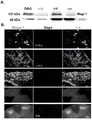
KNaChannel expression is affected by the PDZ binding motif. Both the Slack and Slick channels contain type 1 PDZ binding motifs at their respective distil C-termini (FIG. 1A). Using the PDZ protein interaction prediction model (PDZPedInt, university of fleabag) and inputting the Slack amino acid sequence, Magi-1 was identified as a couplant of the Slack channel, in particular the second and fifth PDZ domains of Magi-1. Heterologous co-expression of Magi-1 and Slack-B subunits improves Slack current density (fig. 1B), whereas coexpression of Magi-1 with a mutant ack construct with a truncated PDZ motif did not affect the ack current density (fig. 1B). Using a Co-immunoprecipitation (Co-IP) assay, we demonstrated that Magi-1 channels interact with the slak channel in Chinese Hamster Ovary (CHO) cells and DRG neurons (fig. 1C, F). Dual immunolabeling studies describe Magi-1 and Slack K in CHO cells, in cultured DRG neurons and intact DRG neuronsNaCo-location between channels (fig. 1E, G). Co-IP confirmed Slack KNaThe channel interacts with Magi-1 through its C-terminal PDZ motif (ETQL (SEQ ID NO:99)) (FIG. 1C). To verify the role of Magi-1 in the expression of the slak channel membrane, a surface biotinylation assay was performed and it was confirmed that the coexpression of slak with Magi-1 increased the surface expression of the slak channel (fig. 1D). Slick, KNaAnother member of the channel family, with about 74% sequence homology to Slack, has the same evolutionarily conserved PDZ type 1 binding motif (ETQL) (fig. 1A). Whether Magi-1 also modulates Slick's galvanic activity in CHO cells was assessed by co-expressing Magi-1 and Slick. Patch clamp recordings showed that Magi-1 enhanced Slick current density in a similar manner (FIG. 2A), but unlike Slack-B, Western blot analysis surprisingly showed an increase (8-fold) in Slick total protein expression (FIG. 2C). Magi-1 was also found to co-localize with the Slick channel when expressed heterologously in CHO cells (fig. 2D). Thus, Magi-1 affected K by increasing the expression of the Slack channel membraneNaCurrent, and for the Slick channel: magi-1 appears to provide additional protein stabilization function, as expression of Magi-1 results in increased expression of total protein in the Slick channel.
Magi-1 knockdown inhibition in cultured DRG neurons IKBut causes low excitability. The neurophysiological function of Magi-1 was examined by using a knockdown strategy in cultured DRG neurons along with previously validated small interfering rna (sirna). The knockdown of Magi-1 was verified by immunolabeling and western blot analysis (fig. 3A, B) using a previously validated polyclonal Magi-1 antibody. At 72 hours post-transfection, a substantial Magi-1 knockdown was achieved, as a decrease of about 70-75% in Magi-1 protein expression was observed compared to non-encoded perturbed control siRNAs (FIG. 3A, B). FIG. 1 shows a schematic view of aOther immunofluorescence images describing knockdown can be found in 0. We examined the Magi-1 knock-down pair KNaEffect of surface expression of the Slack channel. Membrane biotinylation assays showed a significant reduction of membrane slak channel expression by about 70% compared to the control (figure 3C). Although transient state I still existsKVoltage clamp recordings also showed extroversion I after Magi-1 knockdownKThe density decreased significantly (fig. 3D). Surprisingly, Magi-1 knockdown resulted in DRG hypoexcitability and neurons failed to excite AP (fig. 3E and fig. 11A). It is expected that the surface Slack KNaThe decrease in channels leads to repeated excitations, however the observed severely hindered action potential suggests that Magi-1 deficiency may also affect the function of sodium channels.
Magi-1 knockdown also reduces I in DRG neuronsNaAnd Nav1.8 plasma membrane expression. The internal sodium current (I) of the Magi-1 knockdown pair in DRG neurons was examined using whole-cell voltage clamp recordings (see above)Na) The influence of (c). The Magi-1 knockdown caused Total INaSignificantly decreased (fig. 4A-C). I compared to neurons treated with control siRNANaBoth The Tetrodotoxin (TTX) sensitive and TTX resistant component peaks were significantly reduced, which explains the low excitatory phenotype of DRG neurons during Magi-1 knockdown despite reduced membrane Slack expression. Notably, culture conditions for these DRG neurons were favorable for TrkA positive, nociceptive DRG neurons expressing high levels of nav1.8 channels (see "methods"). In most mature nociceptive DRG neurons, the nav1.8 channel accounts for up to 90% of the overshoot of action potential. Reduction of I due to Magi-1 knock-downNaThe TTX resistance component of (1), Nav1.8 for INaThe TTX resistance component of (A) is an important contributor, and therefore research has focused on the surface expression of Nav1.8. Using the surface biotinylation assay, a decrease in Nav1.8 membrane expression (about 50%) was found following Magi-1 knockdown (FIG. 4D). Taken together, these results indicate that Magi-1 is an essential scaffold for membrane localization of both nav1.8 and Slack channels in DRG neurons.
Magi-1 is expressed in small and medium DRG neurons, in their axonal bundles, and at some Ranvier nodes. The complete DRG neuron is verifiedThe expression of Magi-1 in order to indicate its broader physiological function. Allen mouse spinal cord mapping and BioGPS support high Magi-1 information within DRG neurons. According to BioGPS, DRG tissue has the second highest tissue expression profile for Magi-1mRNA, with the highest expression in the hypothalamus. Furthermore, allen spinal cord maps describe differential Magi-1 expression in small and medium sized, putative nociceptive DRG neurons. The MAGi-1 immunolabeling in the growth cone of cultured DRG neurons and in the dorsal root entry region of the embryonic spinal cord was also previously shown. We confirmed the expression of Magi-1 in adult mouse DRG neurons and spinal cord tissue by western blot (fig. 5A) and immunohistochemical analysis (fig. 5B). Immunohistochemistry was performed using previously validated monoclonal anti-Magi-1 antibodies. Histological examination of the sciatic nerve using the accessory node marker Caspr showed higher Magi-1 immunoreactivity along axonal fibers and at some of the Ranvier nodes (fig. 5C). Cell size analysis showed that in small and medium size DRG neurons:<600μm2) The highest distribution of Magi-1 among DRG neurons within the range (fig. 5D) is similar to the data that can be seen in the allen mouse spinal cord map. The preferential tissue expression profile of Magi-1 for small and medium DRG neurons and spinal cord dorsal horn indicates that Magi-1 has a potential function in pain signaling.
Magi-1 mediated Slack K in DRG neuronsNaCoupling between the channel and Nav1.8. Previous reports indicate that neuron K blocks sodium access pathwaysNaThe activity of the channel is reduced, which indicates the position of the Nav channel and KNaThe channels are in close proximity. Furthermore, it was previously observed that in DRG neurons, slak KNaThe channel and Nav1.8immune between co-localization. These data also suggest possible couplings as indicated by a decrease in membrane localization of Nav1.8 and Slack channels after Magi-1 knockdown. The possibility of interaction of Magi-1 with Nav1.8 in DRG neurons was examined, as well as whether Magi-1 promoted Nav1.8 with Slack KNaAnd (4) coupling the channels. In the Co-IP assay, interaction of Magi-1 with Nav1.8 channel was confirmed (FIG. 6A). The dual immunolabeling study also described co-localization between cultured and intact DRG neurons and Magi-1 and nav1.8 within the spinal cord (fig. 6B). Slack and Using intact DRG lysateNav1.8 specific antibodies Co-IP experiments were performed and successfully Slack Co-immunoprecipitated with Nav1.8 (FIG. 6C), which indicates Nav1.8 and Slack K in DRG neuronsNaThe channels are multiplexed together. These findings suggest a scaffolding effect of Magi-1 on slak and nav1.8 in sensory neurons.
In vivo Magi-1 knockdown in DRG neurons reduced pain sensitivity. The ability of knock-down Magi-1 in vivo to alleviate painful behavior was examined. Novel spinal nerve injection techniques using non-viral vectors comprising short hairpin rna (shrna) sequences. This disclosure describes for the first time this mouse in vivo transfection method, which allows DRG sensory neurons to take up shRNA plasmid via axonal retrograde transport without the need for invasive paraspinal muscle dissection (necessary for rats). FIG. 7A depicts a schematic of the experimental outline. Intraspinal injection of Magi-1shRNA induced a significant and sustained reduction in thermal nociception in primary male and female mice compared to control shRNA (fig. 7B, C). To assess differences in paw withdrawal latencies inside animals, the contralateral (non-injected) Paw Withdrawal Latency (PWL) was subtracted from the ipsilateral (injected) PWL. In a single animal, there was a significant increase in PWL of about 3 seconds in mice injected with Magi-1shRNA compared to paws injected with non-targeted shRNA. Next, the role of Magi-1 knockdown in an acute inflammatory pain model (formalin assay) was examined. Intraplantar (i.pl.) injection of 5% formalin elicits the typical biphasic inflammatory pain response associated with this acute inflammatory pain model. To this extent, there was a significant reduction in phase I flinching behavior and phase II licking, lifting, and flinching behavior fifteen days after in vivo transfection of Magi-1shRNA (fig. 7D).
In vivo Magi-1 silencing was confirmed by immunohistochemical and biochemical analysis 15 days after intrasciatic transfection of DRG and sciatic nerves in mice injected with shRNA. A significant loss of Magi-1 immunoreactivity in ipsilateral DRGs and sciatic nerves was observed from mice injected with Magi-1shRNA compared to contralateral DRGs and mice injected with control shRNA from the same mice (fig. 7E). The Magi-1 transcript knockdown was also verified using RT-PCR (fig. 12B). The knockdown of Magi-1 protein was confirmed by immunoblotting (about 70-75%) (fig. 7F), comparable to that achieved in vitro (fig. 3A, B). Taken together, these results indicate that Magi-1 modulates nociceptive and acute inflammatory pain.
Nav1.8 expression decreased after Magi-1 knockdown in vivo. Immunohistochemical analysis of the sciatic nerve and Ranvier nodules also revealed an unexpected but significant reduction in nav1.8 immunoreactivity following treatment with Magi-1shRNA compared to the non-coding perturbed shRNA control (fig. 8A). This finding is confirmed by the following: a75% decrease in Nav1.8 protein expression in DRG neurons following in vivo Magi-1 knockdown was observed as determined by Western blot analysis (FIG. 8B). These data reveal that, in addition to the scaffold channel on the membrane, Magi-1 is also essential for nav1.8 protein stability. Recent studies have demonstrated the protective role of Magi-2 in protecting dendritic proteins from Nedd4-2 mediated protein degradation via WW-mediated interactions. Furthermore, loss of Nav1.8 protein and the concomitant reduction in phase II inflammatory pain behavior is consistent with a reduction in phase II behavior in Nav1.8 knockout mice. The use of RT-PCR confirmed during Magi-1 knockdown period Nav1.8information remains unchanged, which enhances Magi-1 regulation of Nav1.8protein stability concept (figure 12C). These results indicate that Magi-1 plays a key role in regulating ion channel protein stability.
The PY motif mimetic peptide regulates nav1.8 trafficking, DRG neuron excitability and pain behavior. It was demonstrated that PDZ-mediated interaction is an absolute requirement for Slack/Magi-1 interaction (FIG. 1D). The nav1.8 channel is reported to contain multiple internal putative PDZ binding motifs and binds to PDZ domains comprising the protein Pdzd 2. However, Pdzd2 knockout mice did not show any change in pain behavior. On the other hand, the Nav1.8 channel also contained a PY motif (PPXY (SEQ ID NO:76)) at its distal C-terminus (FIG. 13), and this motif is assumed to regulate the interaction with Nedd4-2 ubiquitin ligase for targeted protein degradation. Interestingly, the WW domain of Nedd4-2 has a high degree of sequence homology to the WW domain in Magi-1. Furthermore, it has been shown that Magi proteins protect Nedd4-2 target proteins from degradation through WW interactions. Therefore, using cell penetrating PY motif mimetic peptides, selection competed away WW domain binding of nav 1.8. Based on Nav1.8WW binding motif, two sequences are designed to be identicalExcept that one of the peptides is phosphorylated (representing Thr1926 in the channel). The reason for this is because of the use of Post-translational modification resource tool scan Nav1.8 revealed that the fourth amino acid (4), Thr1926, adjacent to the PPXY (SEQ ID NO:76) domain was presumed to be phosphorylated (FIG. 13). Then, 10. mu.M of either unphosphorylated peptide (PY, myristoyl-SATSFPPSYDSVTRG (SEQ ID NO:77)) or phosphorylated peptide (phosphorylated-PY, myristoyl-SATSFPPSYDSV [ pT)]RG (SEQ ID NO:77, where T is phosphorylated)) treated primary DRG neurons to outperform WW domain binding of the Nav1.8 channel. 24 hours of PY peptide exposure resulted in almost complete loss of neurons of total INaIn contrast, the phosphorylated-PY peptide strongly increased peak INa. PY peptide and phosphorylated-PY peptide each show time-dependent INaDecrease or increase (6 hours and 24 hours) (fig. 9A, B). The PY peptide treatment (24 hours) almost completely eliminated AP excitation (10 out of 11 neurons), while the phosphorylated-PY peptide caused sharply contrasting repetitive AP excitation (7 out of 12) (fig. 9B). We evaluated the surface expression of nav1.8 channels after treatment with PY peptide and found that nav1.8 channels on plasma membrane were significantly reduced, while phospho-PY peptide significantly increased the expression of nav1.8 channels (fig. 9C). Furthermore, a large reduction in total nav1.8 protein was observed after incubation with PY peptide, indicating that nav1.8 protein stability is dependent on this WW binding motif. These results indicate that the phosphorylation status of the competing Nav1.8 PY motif peptide is critical for the stability of the Nav1.8 channel. Furthermore, these data suggest that other sodium channels may be regulated by PY motif interactions (fig. 9A, B).
To assess the potential analgesic effect of disrupting sodium channel membrane localization on pain behavior, the effect of competing peptides in the formalin inflammatory pain model was investigated. Mice were given a single intraplantar injection of the PY peptide, phosphorylated-PY peptide or perturbed PY peptide (100. mu.M, 20. mu.l) to the right hindpaw, and 24 hours later, 5% formalin was injected into the same paw. Whereas pretreatment with PY peptide significantly reduced stage II acute inflammatory pain compared to mice treated with perturbed peptide, mice pretreated with phosphorylated-PY peptide showed a sharply contrasted increase in stage II response (fig. 9D). These data confirm the results observed in vitro experiments and demonstrate the ability to modulate the entrance and exit of Nav1.8 channels into and out of neuronal membranes using a PY motif-based mimetic peptide. In addition, these data indicate that the phosphorylation state of Thr1926 determines transport of nav1.8 channels, neuronal excitability and acute pain behavior.
Discussion: described herein is the expression of Magi-1 in the pain pathway: high expression of Magi-1 was observed in the cell body and in the axons of nociceptive DRG neurons as well as within the superficial dorsal horn of the spinal cord. In addition, Magi-1 was demonstrated to be Nav1.8 and Slack K in DRG neuronsNaKey support for channel membrane positioning. Nav1.8 and Slack K have been implicated in inflammatory and neuropathic pain models in rodentsNaBoth channels, and further shows that Magi-1 interacts with both the slak and nav1.8 channels. Furthermore, Magi-1 silencing was found to decrease membrane expression of both types of ion channels, leading to a net defect in DRG neuron excitability, suggesting that Magi-1 is a key regulator of ion channel function in neurons. To assess the importance of Magi-1 in the pain process, spinal nerve injection was used to transfect in vivo silencing Magi-1shRNA into DRG neurons of naive mice. This is a new and rapid technique to manipulate gene function in DRG neurons in primitive rodents, especially mice. It allows internal control testing of modified ipsilateral DRG neurons versus unaltered contralateral DRG neurons in the same mouse. Using this in vivo transfection method, it was found that the knockdown of Magi-1 resulted in significant deficits in thermal nociception and acute inflammatory pain behavior.
In addition to locating ion channels on neuronal membranes, these findings suggest for the first time that Magi-1 is also critical for ion channel protein stability. Indeed, unlike other scaffold proteins, the Magi protein family may exert a broader function of protecting proteins from degradation. For example, non-UL YAP1 protein shows protection against Nedd4-2 mediated protein degradation through its WW domain. In turn, Magi-2 was also reported to associate with the conserved PY group in dendritic proteins via the WW domainThe sequences interact to protect the protein dendritic protein from Nedd4-2 mediated ubiquitination. Like dendritic proteins, Nav1.8 has an evolutionarily conserved PY motif that has been shown to be a binding site for Nedd4-2, targeting Nav1.8 for subsequent proteasomal degradation. During prolonged in vivo Magi-1 knockdown, as determined by immunoblotting, a large and statistically significant reduction in nav1.8 immune marker and protein expression was observed (fig. 8A, B). Also, almost complete loss of Nav1.8 expression was observed after 24 hours incubation of PY peptide in DRG neurons. In fact, after only 6 hours, INaThe density decreased by 50%, indicating that both TTX-resistant and TTX-sensitive Nav channel membrane expression are dependent on the PY motif. However, subsequent immunoblot analysis of Nav1.7 protein revealed that, although some protein reductions were observed during long-term in vivo Maxi-1 shRNA knockdown, there was no change in surface Nav1.7 protein levels following peptidomimetic treatment (FIG. 16). It should be noted that embryonic DRG neurons also express nav1.3 channels, which contain the WW binding domain (fig. 13). Thus, some of the in vitro effects we observed on the TTX sensitive channel may be attributed to this channel. Nevertheless, these data indicate that Magi-1 elicits differential sensitivity of each Nav subtype to scaffold and protein stability. Furthermore, Slick K having a putative PY motif (-PPRY- (SEQ ID NO:78)) at amino acids 12-15 of its N-terminus was observedNaSubunits that, when expressed recombinantly with Magi-1, cause Slick K to be expressed relative to expression aloneNaApproximately 5-fold higher current at subunit, with increased levels of Slick channel protein (figure 2). This is in contrast to the Slack-B subunit, which does not have the PY motif at its N-terminus; the membrane current was increased to a lesser extent (2-fold), and no increase in the expression of the slak-B protein was observed. Indeed, in heterologous expression systems, the difficulty of expressing the slave channel compared to the slave channel may be due to this WW binding motif and sensitivity to UL-dependent degradation. Thus, these results indicate that Magi-1 can protect ion channels from degradation pathways in addition to membrane targeting, and indeed targeting Magi-1 represents a novel pharmacological approach to affecting ion channel levels and function.
Although both the Magi-1 knockdown and PY peptides were derivedResult in INaReduced, reduced stability of Nav1.8 channels and reduced pain behaviour, but the opposite effect caused by the phosphorylated-PY peptide was unexpected, in particular INaElevation, repeated stimulation and augmentation of nociceptive responses. Can be selected from Phosphorylation proteomics data obtained on-line confirmed that Thr1926 in the nav1.8 channel was presumed to be phosphorylated. We used this information to design a second peptide, since Thr1926 is the fourth amino acid downstream of the PY motif (fig. 13). Using Scansite's network-based software (MIT), Thr1926 was predicted to be a casein II kinase or GSK-3 kinase consensus phosphorylation site. Both kinases are constitutively active kinases, suggesting that Thr1926 may be basal phosphorylated. Without wishing to be bound by any particular theory, it is speculated that the phosphorylated-PY peptide competes with the phosphorylated nav1.8 channel in the presence of UL, thereby preventing a substantial portion of the nav1.8 channel from being ubiquitinated and retained in the cytosol (fig. 9E). Furthermore, no statistically significant increase in input levels of Nav1.8 protein was observed within 24 hours, indicating that most of the internally located channels may be in monoubiquitination at least within this time window. Without wishing to be bound by any particular theory, it is also speculated that dephosphorylated Thr1926 has a higher affinity for Magi-1, which explains why competing PY peptides would lead to loss of nav1.8 channel membrane expression. In this case, observed total Nav1.8channel protein level is greatly reduced, which indicates that in 24 hours in the channel final fate is degradation. However, these results strongly suggest that the PY motif is a major determinant of nav1.8 channel trafficking, while Magi-1 is a key component of sodium signalizers in DRG neurons (fig. 9E).
It is demonstrated herein that a single intradermal injection of the PY mimetic peptide elicits significant analgesia at 24 hours post-administration (fig. 9D). The PY peptides act as local long-acting analgesics and have therapeutic value for invasive procedures requiring long-acting analgesia and/or reducing the need for postoperative opioids. In contrast, the phosphorylated-PY peptide drives nav1.8 channels to the DRG neuronal membrane and exacerbates nociceptive behaviour and is therefore of potential value for diseases associated with insensitivity to pain. Myristoylation makes it possible for peptides to partition the membrane by a turnover mechanism, but binds most of the peptides to the inner surface of the membrane. Such membrane-defining features may enhance the ability of the peptide to exert its mimetic effects, particularly for membrane-associated proteins. In addition to causing cell permeability and anchoring the peptide inside the phospholipid bilayer, myristoylation of the peptide and its inherent hydrophobicity may also ensure localization of the peptide to the injection site. Furthermore, the metabolism of these peptides requires phospholipid membrane turnover, which may contribute to their long-lasting effect in vivo. Previous studies using a similar intradermal injection method of a mimetic peptide directed against the T-mem100 protein (important for the TRPA1-TRPV1 complex) demonstrated an analgesic effect on paclitaxel-induced chronic pain. Thus, the use of myristoylated cell penetrating peptides provides a potential therapeutic approach to manipulate nerve terminal activity.
In addition to high expression of Magi-1 in peripheral DRG neurons, Magi-1 is stably expressed in the central nervous system. Thus, Magi-1 pairs Nav1.8 and Slack K in DRG neuronsNaThe scaffold and membrane stability of the channel may be similar to that of the Nav channel in central neurons where TTX sensitivity contains a WW binding motif. Indeed, these data indicate that Magi-1 knockdown in cultured DRG neurons also leads to TTX sensitivity INaSignificantly reduced, probably because multiple Nav channel subtypes contain the WW binding domain (fig. 13). Mutations that are dysfunctional in the Nav channel are associated with mental health disorders. These findings may help to address the reasons why Magi-1 deficiency is also associated with various psychiatric syndromes, as Magi-1 deficiency leads to reduced sodium transport and low excitability. It is expected that phosphorylated-PY peptides may serve as novel therapeutic platforms for increasing excitability in hypoexcitability-related neurological diseases.
Experimental procedure: animals-all animals used in this study were housed in the University of Buffalo (UB) laboratory animal facility and were free to access food and water according to a 12/12 light/dark cycle. All experimental procedures were in accordance with the guidelines of the national institute of health, united states, "guidelines for care and use of laboratory animals", and were approved by the institutional animal care use committee of the university of buffalo.
Primary DRG neuron cultures-periodically pregnant Sprague-Dawley rats (Harlan, Indianapolis, IN) were used for the culture of neurons. On the day of dissection, rats were euthanized by carbon dioxide asphyxiation and E15 embryos were extracted. DRG neurons were isolated from embryos and enzymatically digested with trypsin (2.5mg/ml) at 37 ℃ for 45 min, then dissociated and plated. DRG neurons were plated onto poly-D-lysine (Sigma; 100. mu.g/ml) and laminin (Invitrogen; 3. mu.g/ml) coated coverslips. Neurons were maintained at 37 ℃ and 7% CO2Serum-free medium in a humidified incubator containing 50% DMEM and the trophic factors N2 (product of Gemini Bio; 1%), 1-glutamine (Invitrogen; 200. mu.g/ml) and Nerve Growth Factor (NGF) (Harlan; 100 ng/ml; critical for embryonic neuron survival). dependence of embryonic DRG neurons on NGF selects a small diameter population that is considered to be the basis of nociception and thermal perception.DRG dissection is followed two consecutive days by culturing DRG neurons in C2 medium containing the antimitotic agent cytosine β -d-arabinofuranoside hydrochloride (Sigma; 3. mu.M). The neurons are then recovered in conventional serum-free medium two days before experiments are performed using the neurons.
Cell culture-Chinese Hamster Ovary (CHO) cells were grown in Iscove's Modified Dulbecco's Medium (IMDM) supplemented with 10% FBS, 1% hypoxanthine/thymine (HT) supplements (Life Technologies), and 1% penicillin-streptomycin at 37 deg.C with 5% CO2Culturing in medium. CHO cells were plated on 12 mm coverslips for immunolabeling experiments, 35 mm petri dishes for all electrophysiological experiments, and six well plates for biochemical experiments. Cells were co-transfected with 0.5. mu.g Slick (pTRACER) or 0.5. mu.g Slack (pTRACER) plus 0.5. mu.g Magi-1 (pcDNA3.1; Addge) or 0.5. mu.g empty vector using lipofectamine (Thermoscientific) according to the manufacturer's instructions. The Magi-1 clone was mutated to include a Kozak sequence at the 5' end to enhance protein expression.
DRG siRNA transfection-Small interfering RNA (siRNA) against Magi-1 was purchased from Santa Cruz Biotechnology (Santa Cruz, Calif., USA). Negative control sirnas consisting of scrambled sequences were provided by the same supplier. For each experiment, neurons were transfected with Magi-1siRNA or non-targeted control siRNA. Cultured DRG neurons were transfected (as described above) using lipofectamine 2000(ThermoScientific) following the manufacturer's protocol. Briefly, we diluted 1.5. mu.l lipofectamine 2000 in 50. mu.l Opti-mem medium and allowed the mixture to stand at room temperature for 5 minutes. After 5 minutes, the mixture was combined with 40pmol of either perturbed siRNA or Magi-1siRNA (three different siRNA duplexes mixed together) in 50 μ l Opti-mem, the mixture was incubated at room temperature for 30 minutes and then added to cells plated on 12 mm coverslips in 24-well plates. The siRNA mixture added to DRG medium was incubated with DRG neurons for 48-72 hours and then used for electrophysiological recording. For western blotting, 300pmole of siRNA was used in each six well plate. siRNA transfected DRG neurons were used for experiments 48-72 hours after transfection. For electrophysiological experiments, DRG neurons were co-transfected with GFP-containing plasmid pTRACER and siRNA duplexes for positive indication of transfection. Immunofluorescence procedures were performed on cultured neurons as previously described. Transfection conditions were concealed for the investigator.
Electrophysiology: all data were collected using an Axomatch 200B amplifier (Molecular Devices) and multiclad-700B (Molecular Devices, Sunnyvale, Calif.), digitized and filtered at 5 kHz. Data acquisition was monitored and controlled using pClamp 10(Molecular Devices). Whole cell patch clamp recordings were performed on cultured DRG neurons and CHO cells were transiently transfected with WT or mutant Slack and Magi-1. The glass electrode was pulled out using a vertical pipette (Narishige International USA, Amityville, NY) and flame polished to a resistance of 5-8 M.OMEGA.. Pipettes were filled with a solution containing (in mM) 124K-gluconic acid, 2MgCl for neuronal recording213.2NaCl, 1EGTA, 10HEPES, 4Mg-ATP and 0.3Na-GTP (pH 7.2), and a solution containing 32.5mM KCl, 97.5mM potassium gluconate, 5mM EGTA and 10mM HEPES (pH 7.2) for CHO cell Slack and Slick recordings. The bath solution for all cells contained (in mM)140NaCl, 5.4KCl, 1CaCl2、1MgCl210HEPES and 10 glucose (pH 7.4). Both voltage-clamp and current-clamp modes use the same bath and pipette solutions. In voltage clamp mode, macroscopic currents are recorded in the voltage range-120 to + 120. Cells were clamped at-70 mV and a voltage step of 20mV was applied over a duration of 200 milliseconds. The cell capacities of CHO cells recorded under these conditions ranged from 10-15pF, while those of DRG neurons cultured ranged from 20-25 pF. A current clamping scheme consisting of depolarization steps that increment by 10pA (20ms duration) from 10 to 200pA was used to check for action potential firing. The firing frequency of individual neurons was assessed by measuring the repetitive firing after a 400 pA-infused suprathreshold stimulation lasting 1000 ms. For INaNote that the pipette solution contained (in mM): 130CsCl, 13CsF, 10 tetraethylammonium chloride, 1MgCl2、1EGTA、2.5Na2ATP, 10HEPES, adjusted pH to 7.2 with CsOH. The bath solution contained (in mM)140NaCl, 5.4KCl, 1CaCl2、1MgCl210HEPES and 10 glucose (pH 7.4). Resistance to TTX INaDRG neurons were recorded in bath solutions containing 250nM TTX. DRG neurons were recorded at a voltage range of-60 to +60 mV. Cells were maintained at-70 mV and I was induced by an increasing 10mV depolarization step for 50msNa。
immunohistochemistry-Sciatic Nerve (SN), lumbar Spinal Cord (SC), lumbar and thoracic DRG were isolated from adult mice. Briefly, animals were anesthetized with a total plus, perfused with ice-cold PBS containing heparin (50 μ g/ml) and sodium nitrite (5mg/ml), and then perfused with ice-cold 4% Paraformaldehyde (PFA). DRG, SN and SC were subsequently removed, surrounding tissue cleared, and post-fixed by incubation overnight in 4% PFA at 4 ℃. The next day, SN and DRG were transferred into 20% sucrose (Cryoprotect). After two days, the DRG, SN and SC were removed from the sucrose and embedded in freezing medium and stored at-80 ℃ for future use. A low temperature thermostat was used to make 16 micron DRG slices, 10 micron SN slices, and 20 micron SC slices. Sections were permeabilized with PBS containing 0.4% Triton X-100. Sections were then blocked with PBS containing 5% BSA for 2 hours at room temperature. The sections were then incubated overnight at 4 ℃ with a primary antibody mixture in PBS containing 5% BSA. All primary antibodies previously validated include: mouse anti-Nav1.8 antibody (1: 250; NeuroMab), rabbit anti-Magi-1 antibody (1: 100; Abcam), mouse anti-Magi-1 antibody (1: 100; Novus Biochemical), rabbit anti-Caspr (1: 250; Abcam), and chicken anti-Slack antibody (1: 750). After several rinses, secondary antibodies Alexa Fluor 633 goat anti-mouse, Alexa Fluor 488 goat anti-rabbit and Alexa Fluor 546 goat anti-chicken (1:1000) were added and incubated overnight. The coverslips were then mounted on slides using Prolong Gold anti-fade and 4',6' -bisamidine-2-phenylindole dihydrochloride. The cell size characteristics of the Magi-1 immunolabels were analyzed using MetaMorph software (Molecular Devices).
Western blot analysis-total protein was collected from transfected CHO cells, spinal cord and DRG. The tissues were homogenized in RIPA buffer supplemented with protease inhibitor cocktail (Sigma). Immunoblotting was performed as described previously. Briefly, proteins were separated on a 4% to 15% Mini-PROTECTAN TGX precast gel (Bio-Rad) and transferred to 0.45 μm nitrocellulose membrane (BioRad). Membranes were probed by incubating antibodies against Slack anti-mouse antibody (1: 500; NeuroMab), rabbit anti-beta-actin (1: 500; Millipore), rabbit anti-Magi-1 (1: 100; Abcam), mouse anti-Magi-1 (1: 100; Novus Biochemical), mouse anti-Nav1.8 (1: 200; NeuroMab), mouse anti-Nav1.7 (1: 200; NeuroMab), mouse anti-Flag (1:500, Sigma) in 5% milk prepared in 1 XTris buffered saline Tween (TBST) at 4 ℃ overnight. On the following day, membranes were washed 3 times in 1 × TBST for 5 minutes, then incubated for 1 hour at room temperature in anti-mouse or anti-rabbit horseradish peroxidase conjugate (1: 5000; Promega) and 0.1% BSA prepared in 1 × PBS. The film was washed again 3 times for 5 minutes before development and imaging. The bands were visualized with enhanced chemiluminescence (ThermoScientific) and quantified using Image J Software (NIH). Each experiment was repeated at least three times.
Co-immunoprecipitation — CHO cells in six well plates were transiently transfected with WT or mutant ack in the presence or absence of Magi-1 plasmid, respectively. Cells were then lysed with 100 μ l/well of ice-cold RIPA buffer supplemented with protease inhibitor cocktail (Sigma). 60 μ l/well of connexin G in agarose bead slurry (GE Healthcare) was washed 3 times with ice-cold lysis buffer and incubated overnight (3 wells per sample) on a rotator with 4 μ G rabbit Magi-1 antibody (Abcam) or mouse anti-Slack antibody (neuroMab) in PBS containing 0.1% Tween-20 and cell lysate at 4 ℃. On the next day, the samples were centrifuged and the supernatants were stored separately. The pellet was washed 3 times with cold lysis buffer and the bound proteins were eluted by boiling 3 times for 8 minutes at 9 ℃. The samples were centrifuged to separate the proteins into the supernatant, which was then denatured with Sodium Dodecyl Sulfate (SDS) and loaded as an immunoprecipitate onto a ready-to-use gel (Bio-Rad) (4-15% Tris-HCl). The supernatants were collected and whole cell lysates (total input) were denatured with SDS and run as controls. The samples were probed for Slack or nav1.8 and actin by western blotting as described above.
Surface protein biotinylation-plasma membrane protein expression was detected using a protein biotinylation assay. Briefly, CHO cells or DRG neurons in six-well plates were used 48 hours after transient transfection of plasmid constructs or Magi-1-targeting siRNAs, respectively. For peptide incubation, neurons were incubated for 24 hours. Mu.l of 10mM sulfo-NHS-SS-biotin (Thermo Scientific) was added to each well and incubated for 45 min at room temperature. The biotinylation reaction was terminated with 10mM glycine (quench solution). Cells were harvested, washed in TBS, and lysed in lysis buffer/protease inhibitor mix for 30 minutes on ice. The lysates were collected and incubated with 500 μ L of NeutrAvidin agarose for 60 min at room temperature with rotation. After incubation, the column was centrifuged to collect the non-biotinylated proteins. To elute biotinylated proteins, Sodium Dodecyl Sulfate (SDS) and dithiothreitol (DDT) were added to the column membrane and incubated for 1 hour at room temperature with rotation. The samples biotinylated and non-biotinylated were probed for Slack or nav1.8 and actin by western blotting as described above.
Nociceptive testingMeasurement of baseline thermal nociceptive behaviour using an automated Hargreaves apparatus of Ugo Basile (Varese, Ital). For the original C57Bl/6 mouse(8-10 weeks old) (Envigo) two days of acclimation training followed by three days of measurement. On days 1 and 2 (acclimation training), the mice remained in the rearing cages for 30 minutes to acclimate to the test rooms, and then transferred to the test rooms for 1 hour. On days 3 to 5, mice were tested. Infrared stimuli (IR 40) were transmitted through the plexiglass floor to the plantar surface of the hind paw and the withdrawal latency was measured automatically. Three to six measurements per hind paw were taken for each subject and used to calculate the average paw withdrawal latency (in seconds). A maximum IR exposure time of 15 seconds was established to ensure that no tissue damage occurred and at least a 5 minute interval was allowed between measurements taken from the same mouse.
By using shRNA plasmid DNA multiplexes were transfected in vivo-using spinal nerve injection protocol and optimized for spinal nerve injection in mice. Three days after the baseline thermal behavior was established, mice were anesthetized with isoflurane (induction rate: 4% and maintenance rate: 2%) and placed prone. A3 cm posterior longitudinal skin incision was made in the lumbar segment of the spine. The ipsilateral paraspinal muscle was carefully separated from the appendages at the L4 to S1 positions of the vertebrae using a pair of sterile dental picks. Using a syringe (Hamilton 80030, Hamilton, Reno, NV) attached to a 26-gauge needle, 1.5. mu.l of PEI/shRNA plasmid DNA polylinker (polyplex) with an N/P ratio of 6 was slowly injected directly into the spinal nerve of the right hind paw. The Magi-1shRNA and control shRNA were purchased from Santa Cruz Biotechnology (Santa Cruz, Calif., USA) and are identical to the siRNA sequences described above. After injection, the needle was held at the spinal nerve for 1 minute to prevent leakage. Complete hemostasis was confirmed and the wound was sutured with a wound clip. Mice were allowed to recover for 7 days before being tested again for thermal nociceptive behavior.
Formalin test-mice were habituated in the behavioral chamber for 15 minutes and then in the formalin chamber for 30 minutes prior to formalin injection. 20 microliters of 5% formalin (in sterile saline) was intraplantar injected into the right hind paw and the mice were placed back in the chamber for video recording. The total time spent lifting, total number of paw licks and number of flinches were recorded at 5 minute intervals for 60 minutes. For each video recording, the measurements under the experimental conditions of two observers concealed were averaged to obtain the final measurement for each time point.
RNA extraction and cDNA synthesis-RNeasy Micro kit (Qian) was used to extract total RNA from mouse lumbar DRG neurons. RNA was reverse transcribed using SuperScript III reverse transcriptase (Life Technologies). Using this cDNA as a template, polymerase chain reaction was performed with previously validated primers for Magi-1 and Nav1.8. Transcript abundance was measured by a thermal cycler using SYBR GreenPCR Master Mix. For quantification, a two-step protocol of denaturation and annealing for 50 cycles was used, with absorbance reading for 15 seconds on a BioRad iQ5 cycler. Each sample was performed in triplicate.
Peptide-we designed an N-terminal myristoylated PDZ peptide mimic NPETRDETQL (SEQ ID NO:79) based on the C-terminal sequence of the rat Slack channel. This peptide and the perturbed variant peptide QPNTRLDETE (SEQ ID NO:80) were synthesized by GenScript. Similarly, PY peptide SATSFPPSYDSVTRG (SEQ ID NO:77) and phospho-PY peptide SATSFPPSYDSV(pT) RG (SEQ ID NO:77, where T is phosphorylated) were designed based on the WW binding domain in the rat Nav1.8 channel. These peptides and the scrambling peptide SDRPVTSYSFSAPG (SEQ ID NO:81) were also synthesized by GenScript. The peptides were first dissolved in DMSO and diluted in saline to the final working concentration. As previously described, a peptide concentration of 10 μ M was used on primary neurons; the final DMSO concentration was 0.05%. Intraplantar administration was selected based on previous studies showing analgesia induced by the intradermal injection of different myristoylated peptides into the hind paw.
For the optional phospho-threonine peptides, threonine is protected with trityl ether [ Fmoc-Thr (Trt) -OH ]. Trityl protected derivatives can be selectively deprotected on resins useful for the preparation of phosphorylated threonine containing peptides by the bulk phosphorylation process. General phosphorylation involves selective phosporylation of a suitable hydroxy threonine with a protected phosphoramidite on a solid phase, followed by oxidation of the resulting P (III) triester to the P (V) triester. Finally, myristoylation is achieved by an N-myristoyltransferase enzyme that catalyzes the N-myristoylation (at the N-terminus) of the protein.
statistics-Clampfit (molecular devices) and Origin 8.0(Origin Lab) software were used for all electrophysiological data analyses. Densitometric analysis of western blots was performed using Image J (NIH) software. Statistical analysis was performed using GraphPad Prism4(GraphPad, San Diego, CA). Comparisons between groups were performed using student's t-test. Multiple comparisons were studied using one-or two-way ANOVA, followed by Bonferroni test to detect pairwise differences between groups. Data are expressed as mean ± SEM.
RNA extraction and cDNA synthesis for real-time PCR. RNeasy Micro kit (Qiagen) was used to extract RNA from neuronal cultures. RNA was reverse transcribed into cDNA using SuperScript III reverse transcriptase (Life Technologies) and used for subsequent RT-PCR. Transcript abundance was measured by a thermal cycler using a SYBR Green PCR Master Mix. For quantification, a two-step denaturation and annealing procedure of 50 cycles was used, with absorbance reading for 15 seconds on a BioRad iQ5 cycler. Each sample was run in triplicate. The primers used for RT-PCR are listed below:
magi-1 primer
5-GTCTTCGAGGGGGCCGAGAATATAACATGG-3(SEQ ID NO:82)
5-GGTGGAGGGGCCGTTCCTGTCG-3(SEQ ID NO:83)
Nav1.8 primer
5′CCCAAAGGGCAGCAGGAGCTG-3′(SEQ ID NO:84)
5′-CGGCGAGTGCAGCCTTCTGTGA-3′(SEQ ID NO:85)
5′-CTGCCACAAGTCCAAAAGTGTGAA-3′(SEQ ID NO:86)
5′AGTCATCGGGCTCGTCCAGATC-3′(SEQ ID NO:87)
Plasmid DNA encoding human MAGI-1 shRNA. To generate therapeutic shRNA for pain treatment, we screened 3 unique interfering RNA sequences from the human Magi-1mRNA sequence and then inserted each of these sequences (in DNA form) into a plasmid containing the minimal promoter of the human Nav1.8 channel, thereby generating 3 plasmids. Expression of shRNA driven by the minimal promoter of the nav1.8 channel will ensure that shRNA targeting Magi-1 is expressed only in pain neurons.
Although the present disclosure has been described with respect to one or more particular embodiments and/or examples, it will be understood that other embodiments and/or examples of the disclosure may be made without departing from the scope of the disclosure.
Sequence listing
<110> research Foundation of New York State university
<120> peptides and other agents useful for treating pain and increasing pain sensitivity
<130>011520.01371
<150>62/598,067
<151>2017-12-13
<160>113
<170> PatentIn version 3.5
<210>1
<211>15
<212>PRT
<213> Artificial sequence
<220>
<223> synthetic peptide
<400>1
Ser Thr Ala Ala Cys Pro Pro Ser Tyr Asp Arg Val Thr Lys Pro
1 5 10 15
<210>2
<211>15
<212>PRT
<213> Artificial sequence
<220>
<223> synthetic peptide
<400>2
Pro Ser Thr Thr Ser Pro Pro Ser Tyr Asp Ser Val Thr Lys Pro
1 5 10 15
<210>3
<211>15
<212>PRT
<213> Artificial sequence
<220>
<223> synthetic peptide
<400>3
Ser Ser Thr Thr Ser Pro Pro Ser Tyr Asp Ser Val Thr Lys Pro
1 5 10 15
<210>4
<211>15
<212>PRT
<213> Artificial sequence
<220>
<223> synthetic peptide
<400>4
Ser Ser Thr Ser Phe Pro Pro Ser Tyr Asp Ser Val Thr Arg Ala
1 5 10 15
<210>5
<211>15
<212>PRT
<213> Artificial sequence
<220>
<223> synthetic peptide
<400>5
Pro Ser Thr Ala Ser Leu Pro Ser Tyr Asp Ser Val Thr Lys Pro
1 5 10 15
<210>6
<211>15
<212>PRT
<213> Artificial sequence
<220>
<223> synthetic peptide
<400>6
Ser Ser Thr Thr Ser Pro Pro Ser Tyr Asp Ser Val Thr Lys Pro
1 5 10 15
<210>7
<211>15
<212>PRT
<213> Artificial sequence
<220>
<223> synthetic peptide
<400>7
Ser Ala Thr Ser Phe Pro Pro Ser Tyr Glu Ser Val Thr Arg Gly
1 5 10 15
<210>8
<211>15
<212>PRT
<213> Artificial sequence
<220>
<223> synthetic peptide
<400>8
Ser Thr Ala Ala Cys Pro Pro Ser Tyr Asp Arg Val Thr Lys Pro
1 5 10 15
<210>9
<211>15
<212>PRT
<213> Artificial sequence
<220>
<223> synthetic peptide
<400>9
Pro Ser Thr Thr Ser Pro Pro Ser Tyr Asp Ser Val Thr Lys Pro
1 5 10 15
<210>10
<211>15
<212>PRT
<213> Artificial sequence
<220>
<223> synthetic peptide
<400>10
Ser Ser Thr Thr Ser Pro Pro Ser Tyr Asp Ser Val Thr Lys Pro
1 510 15
<210>11
<211>15
<212>PRT
<213> Artificial sequence
<220>
<223> synthetic peptide
<400>11
Ser Ser Thr Ser Phe Pro Pro Ser Tyr Asp Ser Val Thr Arg Ala
1 5 10 15
<210>12
<211>15
<212>PRT
<213> Artificial sequence
<220>
<223> synthetic peptide
<400>12
Pro Ser Thr Ala Ser Leu Pro Ser Tyr Asp Ser Val Thr Lys Pro
1 5 10 15
<210>13
<211>15
<212>PRT
<213> Artificial sequence
<220>
<223> synthetic peptide
<400>13
Ser Ser Thr Thr Ser Pro Pro Ser Tyr Asp Ser Val Thr Lys Pro
1 5 10 15
<210>14
<211>15
<212>PRT
<213> Artificial sequence
<220>
<223> synthetic peptide
<400>14
Ser Ala Thr Ser Phe Pro Pro Ser Tyr Glu Ser Val Thr Arg Gly
1 5 10 15
<210>15
<211>15
<212>PRT
<213> Artificial sequence
<220>
<223> synthetic peptide
<400>15
Ser Thr Ala Ala Cys Pro Pro Ser Tyr Asp Arg Val Ala Lys Pro
1 5 10 15
<210>16
<211>15
<212>PRT
<213> Artificial sequence
<220>
<223> synthetic peptide
<400>16
Pro Ser Thr Thr Ser Pro Pro Ser Tyr Asp Ser Val Ala Lys Pro
1 5 10 15
<210>17
<211>15
<212>PRT
<213> Artificial sequence
<220>
<223> synthetic peptide
<400>17
Ser Ser Thr Thr Ser Pro Pro Ser Tyr Asp Ser Val Ala Lys Pro
1 5 10 15
<210>18
<211>15
<212>PRT
<213> Artificial sequence
<220>
<223> synthetic peptide
<400>18
Ser Ser Thr Ser Phe Pro Pro Ser Tyr Asp Ser Val Ala Arg Ala
1 5 10 15
<210>19
<211>15
<212>PRT
<213> Artificial sequence
<220>
<223> synthetic peptide
<400>19
Pro Ser Thr Ala Ser Leu Pro Ser Tyr Asp Ser Val Ala Lys Pro
1 5 10 15
<210>20
<211>15
<212>PRT
<213> Artificial sequence
<220>
<223> synthetic peptide
<400>20
Ser Ser Thr Thr Ser Pro Pro Ser Tyr Asp Ser Val Ala Lys Pro
1 5 10 15
<210>21
<211>15
<212>PRT
<213> Artificial sequence
<220>
<223> synthetic peptide
<400>21
Ser Ala Thr Ser Phe Pro Pro Ser Tyr Glu Ser Val Ala Arg Gly
1 5 10 15
<210>22
<211>15
<212>PRT
<213> Artificial sequence
<220>
<223> synthetic peptide
<400>22
Ser Thr Ala Ala Cys Pro Pro Ser Tyr Asp Arg Val Glu Lys Pro
1 5 10 15
<210>23
<211>15
<212>PRT
<213> Artificial sequence
<220>
<223> synthetic peptide
<400>23
Pro Ser Thr Thr Ser Pro Pro Ser Tyr Asp Ser Val Glu Lys Pro
1 5 10 15
<210>24
<211>15
<212>PRT
<213> Artificial sequence
<220>
<223> synthetic peptide
<400>24
Ser Ser Thr Thr Ser Pro Pro Ser Tyr Asp Ser Val Glu Lys Pro
1 5 10 15
<210>25
<211>15
<212>PRT
<213> Artificial sequence
<220>
<223> synthetic peptide
<400>25
Ser Ser Thr Ser Phe Pro Pro Ser Tyr Asp Ser Val Glu Arg Ala
1 5 10 15
<210>26
<211>15
<212>PRT
<213> Artificial sequence
<220>
<223> synthetic peptide
<400>26
Pro Ser Thr Ala Ser Leu Pro Ser Tyr Asp Ser Val Glu Lys Pro
1 5 10 15
<210>27
<211>15
<212>PRT
<213> Artificial sequence
<220>
<223> synthetic peptide
<400>27
Ser Ser Thr Thr Ser Pro Pro Ser Tyr Asp Ser Val Glu Lys Pro
1 5 10 15
<210>28
<211>15
<212>PRT
<213> Artificial sequence
<220>
<223> synthetic peptide
<400>28
Ser Ala Thr Ser Phe Pro Pro Ser Tyr Glu Ser Val Glu Arg Gly
1 5 10 15
<210>29
<211>15
<212>PRT
<213> Artificial sequence
<220>
<223> synthetic peptide
<400>29
Ser Thr Ala Ala Cys Pro Pro Ser Tyr Asp Arg Val Asp Lys Pro
1 5 10 15
<210>30
<211>15
<212>PRT
<213> Artificial sequence
<220>
<223> synthetic peptide
<400>30
Pro Ser Thr Thr Ser Pro Pro Ser Tyr Asp Ser Val Asp Lys Pro
1 5 10 15
<210>31
<211>15
<212>PRT
<213> Artificial sequence
<220>
<223> synthetic peptide
<400>31
Ser Ser Thr Thr Ser Pro Pro Ser Tyr Asp Ser Val Asp Lys Pro
1 5 10 15
<210>32
<211>15
<212>PRT
<213> Artificial sequence
<220>
<223> synthetic peptide
<400>32
Ser Ser Thr Ser Phe Pro Pro Ser Tyr Asp Ser Val Asp Arg Ala
1 5 10 15
<210>33
<211>15
<212>PRT
<213> Artificial sequence
<220>
<223> synthetic peptide
<400>33
Pro Ser Thr Ala Ser Leu Pro Ser Tyr Asp Ser Val Asp Lys Pro
1 5 10 15
<210>34
<211>15
<212>PRT
<213> Artificial sequence
<220>
<223> synthetic peptide
<400>34
Ser Ser Thr Thr Ser Pro Pro Ser Tyr Asp Ser Val Asp Lys Pro
1 5 10 15
<210>35
<211>15
<212>PRT
<213> Artificial sequence
<220>
<223> synthetic peptide
<400>35
Ser Ala Thr Ser Phe Pro Pro Ser Tyr Glu Ser Val Asp Arg Gly
1 5 10 15
<210>36
<211>15
<212>PRT
<213> Artificial sequence
<220>
<223> synthetic peptide
<400>36
Ser Thr Ala Ala Cys Pro Pro Ser Tyr Asp Arg Val Thr Lys Pro
1 5 10 15
<210>37
<211>15
<212>PRT
<213> Artificial sequence
<220>
<223> synthetic peptide
<400>37
Pro Ser Thr Thr Ser Pro Pro Ser Tyr Asp Ser Val Thr Lys Pro
1 5 10 15
<210>38
<211>15
<212>PRT
<213> Artificial sequence
<220>
<223> synthetic peptide
<400>38
Ser Ser Thr Thr Ser Pro Pro Ser Tyr Asp Ser Val Thr Lys Pro
1 5 10 15
<210>39
<211>15
<212>PRT
<213> Artificial sequence
<220>
<223> synthetic peptide
<400>39
Ser Ser Thr Ser Phe Pro Pro Ser Tyr Asp Ser Val Thr Arg Ala
1 5 10 15
<210>40
<211>15
<212>PRT
<213> Artificial sequence
<220>
<223> synthetic peptide
<400>40
Ser Ser Thr Ala Ser Leu Pro Ser Tyr Asp SerVal Thr Lys Pro
1 5 10 15
<210>41
<211>15
<212>PRT
<213> Artificial sequence
<220>
<223> synthetic peptide
<400>41
Ser Ser Thr Ile Ser Pro Pro Ser Tyr Asp Ser Val Thr Lys Pro
1 5 10 15
<210>42
<211>15
<212>PRT
<213> Artificial sequence
<220>
<223> synthetic peptide
<400>42
Ser Ala Thr Ser Phe Pro Pro Ser Tyr Asp Ser Val Thr Arg Gly
1 5 10 15
<210>43
<211>15
<212>PRT
<213> Artificial sequence
<220>
<223> synthetic peptide
<400>43
Ser Thr Ala Ala Cys Pro Pro Ser Tyr Asp Arg Val Thr Lys Pro
1 5 10 15
<210>44
<211>15
<212>PRT
<213> Artificial sequence
<220>
<223> synthetic peptide
<400>44
Pro Ser Thr Thr Ser Pro Pro Ser Tyr Asp Ser Val Thr Lys Pro
1 5 10 15
<210>45
<211>15
<212>PRT
<213> Artificial sequence
<220>
<223> synthetic peptide
<400>45
Ser Ser Thr Thr Ser Pro Pro Ser Tyr Asp Ser Val Thr Lys Pro
1 5 10 15
<210>46
<211>15
<212>PRT
<213> Artificial sequence
<220>
<223> synthetic peptide
<400>46
Ser Ser Thr Ser Phe Pro Pro Ser Tyr Asp Ser Val Thr Arg Ala
1 5 10 15
<210>47
<211>15
<212>PRT
<213> Artificial sequence
<220>
<223> synthetic peptide
<400>47
Ser Ser Thr Ala Ser Leu Pro Ser Tyr Asp Ser Val Thr Lys Pro
1 5 10 15
<210>48
<211>15
<212>PRT
<213> Artificial sequence
<220>
<223> synthetic peptide
<400>48
Ser Ser Thr Ile Ser Pro Pro Ser Tyr Asp Ser Val Thr Lys Pro
1 5 10 15
<210>49
<211>15
<212>PRT
<213> Artificial sequence
<220>
<223> synthetic peptide
<400>49
Ser Ala Thr Ser Phe Pro Pro Ser Tyr Asp Ser Val Thr Arg Gly
1 5 10 15
<210>50
<211>15
<212>PRT
<213> Artificial sequence
<220>
<223> synthetic peptide
<400>50
Ser Thr Ala AlaCys Pro Pro Ser Tyr Asp Arg Val Ala Lys Pro
1 5 10 15
<210>51
<211>15
<212>PRT
<213> Artificial sequence
<220>
<223> synthetic peptide
<400>51
Pro Ser Thr Thr Ser Pro Pro Ser Tyr Asp Ser Val Ala Lys Pro
1 5 10 15
<210>52
<211>15
<212>PRT
<213> Artificial sequence
<220>
<223> synthetic peptide
<400>52
Ser Ser Thr Thr Ser Pro Pro Ser Tyr Asp Ser Val Ala Lys Pro
1 5 10 15
<210>53
<211>15
<212>PRT
<213> Artificial sequence
<220>
<223> synthetic peptide
<400>53
Ser Ser Thr Ser Phe Pro Pro Ser Tyr Asp Ser Val Ala Arg Ala
1 5 10 15
<210>54
<211>15
<212>PRT
<213> Artificial sequence
<220>
<223> synthetic peptide
<400>54
Ser Ser Thr Ala Ser Leu Pro Ser Tyr Asp Ser Val Ala Lys Pro
1 5 10 15
<210>55
<211>15
<212>PRT
<213> Artificial sequence
<220>
<223> synthetic peptide
<400>55
Ser Ser Thr Ile Ser Pro Pro Ser Tyr Asp Ser Val Ala Lys Pro
1 5 10 15
<210>56
<211>15
<212>PRT
<213> Artificial sequence
<220>
<223> synthetic peptide
<400>56
Ser Ala Thr Ser Phe Pro Pro Ser Tyr Asp Ser Val Ala Arg Gly
1 5 10 15
<210>57
<211>15
<212>PRT
<213> Artificial sequence
<220>
<223> synthetic peptide
<400>57
Ser Thr Ala Ala Cys Pro Pro Ser Tyr Asp Arg Val Glu Lys Pro
1 5 10 15
<210>58
<211>15
<212>PRT
<213> Artificial sequence
<220>
<223> synthetic peptide
<400>58
Pro Ser Thr Thr Ser Pro Pro Ser Tyr Asp Ser Val Glu Lys Pro
1 5 10 15
<210>59
<211>15
<212>PRT
<213> Artificial sequence
<220>
<223> synthetic peptide
<400>59
Ser Ser Thr Thr Ser Pro Pro Ser Tyr Asp Ser Val Glu Lys Pro
1 5 10 15
<210>60
<211>15
<212>PRT
<213> Artificial sequence
<220>
<223> synthetic peptide
<400>60
Ser Ser Thr Ser Phe Pro Pro Ser Tyr Asp Ser Val Glu Arg Ala
1 5 10 15
<210>61
<211>15
<212>PRT
<213> Artificial sequence
<220>
<223> synthetic peptide
<400>61
Ser Ser Thr Ala Ser Leu Pro Ser Tyr Asp Ser Val Glu Lys Pro
1 5 10 15
<210>62
<211>15
<212>PRT
<213> Artificial sequence
<220>
<223> synthetic peptide
<400>62
Ser Ser Thr Ile Ser Pro Pro Ser Tyr Asp Ser Val Glu Lys Pro
1 5 10 15
<210>63
<211>15
<212>PRT
<213> Artificial sequence
<220>
<223> synthetic peptide
<400>63
Ser Ala Thr Ser Phe Pro Pro Ser Tyr Asp Ser Val Glu Arg Gly
1 5 10 15
<210>64
<211>15
<212>PRT
<213> Artificial sequence
<220>
<223> synthetic peptide
<400>64
Ser Thr Ala Ala Cys Pro Pro Ser Tyr Asp Arg Val Asp Lys Pro
1 5 10 15
<210>65
<211>15
<212>PRT
<213> Artificial sequence
<220>
<223> synthetic peptide
<400>65
Pro Ser Thr Thr Ser Pro Pro Ser Tyr Asp Ser Val Asp Lys Pro
1 5 10 15
<210>66
<211>15
<212>PRT
<213> Artificial sequence
<220>
<223> synthetic peptide
<400>66
Ser Ser Thr Thr Ser Pro Pro Ser Tyr Asp Ser Val Asp Lys Pro
1 5 10 15
<210>67
<211>15
<212>PRT
<213> Artificial sequence
<220>
<223> synthetic peptide
<400>67
Ser Ser Thr Ser Phe Pro Pro Ser Tyr Asp Ser Val Asp Arg Ala
1 5 10 15
<210>68
<211>15
<212>PRT
<213> Artificial sequence
<220>
<223> synthetic peptide
<400>68
Ser Ser Thr Ala Ser Leu Pro Ser Tyr Asp Ser Val Asp Lys Pro
1 5 10 15
<210>69
<211>15
<212>PRT
<213> Artificial sequence
<220>
<223> synthetic peptide
<400>69
Ser Ser Thr Ile Ser Pro Pro Ser Tyr Asp Ser Val Asp Lys Pro
1 5 10 15
<210>70
<211>15
<212>PRT
<213> Artificial sequence
<220>
<223> synthetic peptide
<400>70
Ser Ala Thr Ser Phe Pro Pro Ser Tyr Asp Ser Val Asp Arg Gly
1 5 10 15
<210>71
<211>4
<212>PRT
<213> Artificial sequence
<220>
<223> synthetic peptide
<220>
<221>X
<222>(1)..(1)
<223> P or L
<220>
<221>X
<222>(3)..(3)
<223> any amino acid residue
<400>71
Xaa Pro Xaa Tyr
1
<210>72
<211>4
<212>PRT
<213> Artificial sequence
<220>
<223> synthetic peptide
<400>72
Thr Ala Ala Cys
1
<210>73
<211>4
<212>PRT
<213> Artificial sequence
<220>
<223> synthetic peptide
<400>73
Pro Pro Ser Tyr
1
<210>74
<211>6
<212>PRT
<213> Artificial sequence
<220>
<223> synthetic peptide
<400>74
Asp Arg Val Thr Lys Pro
1 5
<210>75
<211>15
<212>PRT
<213> Artificial sequence
<220>
<223> synthetic peptide
<220>
<221>X1
<222>(1)..(1)
<223> X is S, P, or A
<220>
<221>X2
<222>(2)..(2)
<223> X is T, S or A
<220>
<221>X3
<222>(3)..(3)
<223> X is A or T
<220>
<221>X4
<222>(4)..(4)
<223> X is A, T, I or S.
<220>
<221>X5
<222>(5)..(5)
<223> X is C, S or F
<220>
<221>X6
<222>(6)..(6)
<223> X is P or T
<220>
<221>X7
<222>(7)..(7)
<223> X is any amino acid residue
<220>
<221>X8
<222>(8)..(8)
<223> X is E, D or Y
<220>
<221>X9
<222>(9)..(9)
<223> X is S or R
<220>
<221>X10
<222>(10)..(10)
<223> X is T, A, E or D
<220>
<221>X11
<222>(11)..(11)
<223> X is K or R
<220>
<221>X12
<222>(12)..(12)
<223> X is P, A or G
<220>
<221>misc_feature
<222>(13)..(15)
<223> Xaa can be any naturally occurring amino acid
<400>75
Xaa Xaa Xaa Xaa Xaa Xaa Pro Xaa Tyr Xaa Xaa Val Xaa Xaa Xaa
1 5 10 15
<210>76
<211>4
<212>PRT
<213> Artificial sequence
<220>
<223> synthetic peptide
<220>
<221>X3
<222>(3)..(3)
<223> X is any amino acid residue.
<400>76
Pro Pro Xaa Tyr
1
<210>77
<211>15
<212>PRT
<213> Artificial sequence
<220>
<223> synthetic peptide
<400>77
Ser Ala Thr Ser Phe Pro Pro Ser Tyr Asp Ser Val Thr Arg Gly
1 5 10 15
<210>78
<211>4
<212>PRT
<213> Artificial sequence
<220>
<223> synthetic peptide
<400>78
Pro Pro Arg Tyr
1
<210>79
<211>10
<212>PRT
<213> Artificial sequence
<220>
<223> synthetic peptide
<400>79
Asn Pro Glu Thr Arg Asp Glu Thr Gln Leu
1 5 10
<210>80
<211>10
<212>PRT
<213> Artificial sequence
<220>
<223> synthetic peptide
<400>80
Gln Pro Asn Thr Arg Leu Asp Glu Thr Glu
1 5 10
<210>81
<211>14
<212>PRT
<213> Artificial sequence
<220>
<223> synthetic peptide
<400>81
Ser Asp Arg Pro Val Thr Ser Tyr Ser Phe Ser Ala Pro Gly
1 5 10
<210>82
<211>30
<212>DNA
<213> Artificial sequence
<220>
<223> primer
<400>82
gtcttcgagg gggccgagaa tataacatgg 30
<210>83
<211>22
<212>DNA
<213> Artificial sequence
<220>
<223> primer
<400>83
ggtggagggg ccgttcctgt cg 22
<210>84
<211>21
<212>DNA
<213> Artificial sequence
<220>
<223> primer
<400>84
cccaaagggc agcaggagct g 21
<210>85
<211>22
<212>DNA
<213> Artificial sequence
<220>
<223> primer
<400>85
cggcgagtgc agccttctgt ga 22
<210>86
<211>24
<212>DNA
<213> Artificial sequence
<220>
<223> primer
<400>86
ctgccacaag tccaaaagtg tgaa 24
<210>87
<211>22
<212>DNA
<213> Artificial sequence
<220>
<223> primer
<400>87
agtcatcggg ctcgtccaga tc 22
<210>88
<211>21
<212>RNA
<213> Artificial sequence
<220>
<223>siRNA
<400>88
ggaaagacag ccagaauagu u 21
<210>89
<211>21
<212>RNA
<213> Artificial sequence
<220>
<223>siRNA
<400>89
gcccaagcuc cagaucaaac u 21
<210>90
<211>21
<212>RNA
<213> Artificial sequence
<220>
<223>siRNA
<400>90
guggauggga cgccaguaau u 21
<210>91
<211>21
<212>RNA
<213> Artificial sequence
<220>
<223>siRNA
<400>91
gaagcauucu cgagcuauag a 21
<210>92
<211>21
<212>RNA
<213> Artificial sequence
<220>
<223>siRNA
<400>92
guuuccccua uucaccagug u 21
<210>93
<211>21
<212>RNA
<213> Artificial sequence
<220>
<223>siRNA
<400>93
gccucucgca ccaugugauu a 21
<210>94
<211>21
<212>RNA
<213> Artificial sequence
<220>
<223>siRNA
<400>94
gaccaagagc gaaggaaugu u 21
<210>95
<211>21
<212>RNA
<213> Artificial sequence
<220>
<223>siRNA
<400>95
guuccucaga uccaauuguu a 21
<210>96
<211>21
<212>RNA
<213> Artificial sequence
<220>
<223>siRNA
<400>96
gaccaucuga gcccacuacu a 21
<210>97
<211>21
<212>RNA
<213> Artificial sequence
<220>
<223>siRNA
<400>97
ggaaacaugu gacuauaccu u 21
<210>98
<211>21
<212>RNA
<213> Artificial sequence
<220>
<223>siRNA
<400>98
gaucuuuaca uagcuuagug u 21
<210>99
<211>4
<212>PRT
<213> Artificial sequence
<220>
<223> synthetic peptide
<400>99
Glu Thr Gln Leu
1
<210>100
<211>48
<212>PRT
<213> Xenopus laevis bufonid toad
<400>100
Arg Leu Glu Leu Asn Asp Ile Val Tyr Leu Ile Arg Ser Asp Pro Leu
1 5 10 15
Ala His Val Ala Asn Glu Ser His Ser Arg Lys Ser Ser Asn Ser Tyr
20 25 30
Lys Thr Asp Pro Val Gly Asn Pro Glu Thr Arg Asp Glu Thr Gln Leu
35 40 45
<210>101
<211>47
<212>PRT
<213> original chicken
<400>101
Arg Leu Glu Leu Asn Asp Ile Val Tyr Leu Ile Arg Ser Asp Pro Leu
1 5 10 15
Ala His Val Ala Asn Asp Gly His Ser Arg Lys Ser Ser Cys Ser Asn
20 25 30
Lys Leu Gly Pro Cys Asn Pro Glu Thr Arg Asp Glu Thr Gln Leu
35 40 45
<210>102
<211>47
<212>PRT
<213> Brown rat
<400>102
Arg Leu Glu Pro Asn Asp Ile Val Tyr Leu Ile Arg Ser Asp Pro Leu
1 5 10 15
Ala His Val Thr Ser Ser Ser Gln Ser Arg Lys Ser Ser Cys Ser Asn
20 25 30
Lys Leu Ser Ser Cys Asn Pro Glu Thr Arg Asp Glu Thr Gln Leu
35 40 45
<210>103
<211>47
<212>PRT
<213> Intelligent people
<400>103
Arg Leu Glu Pro Ser Asp Ile Val Tyr Leu Ile Arg Ser Asp Pro Leu
1 5 10 15
Ala His Val Ala Ser Ser Ser Gln Ser Arg Lys Ser Ser Cys Ser His
20 25 30
Lys Leu Ser Ser Cys Asn Pro Glu Thr Arg Asp Glu Thr Gln Leu
35 40 45
<210>104
<211>44
<212>PRT
<213> Brown rat
<400>104
Arg Leu Glu Leu Asn Asp Val Val Tyr Leu Ile Arg Pro Asp Pro Leu
1 5 10 15
Ser Tyr Leu Pro Asn Ser Glu Pro Ser Arg Lys Asn Ser Ile Cys Asn
20 25 30
Ala Ala Val Gln Asp Ser Arg Glu Glu Thr Gln Leu
35 40
<210>105
<211>15
<212>PRT
<213> Brown rat
<400>105
Ser Thr Ala Ala Cys Pro Pro Ser Tyr Asp Arg Val Thr Lys Pro
1 5 10 15
<210>106
<211>15
<212>PRT
<213> Brown rat
<400>106
Pro Ser Thr Thr Ser Pro Pro Ser Tyr Asp Ser Val Thr Lys Pro
1 5 10 15
<210>107
<211>15
<212>PRT
<213> Brown rat
<400>107
Ser Ser Thr Thr Ser Pro Pro Ser Tyr Asp Ser Val Thr Lys Pro
1 5 10 15
<210>108
<211>15
<212>PRT
<213> Brown rat
<400>108
Ser Ser Thr Ser Phe Pro Pro Ser Tyr Asp Ser Val Thr Arg Ala
1 5 10 15
<210>109
<211>15
<212>PRT
<213> Brown rat
<400>109
Ala Ser Thr Ile Ser Pro Pro Ser Tyr Asp Ser Val Thr Lys Pro
1 5 10 15
<210>110
<211>15
<212>PRT
<213> Brown rat
<400>110
Ser Ala Thr Ser Phe Pro Pro Ser Tyr Asp Ser Val Thr Arg Gly
1 5 10 15
<210>111
<211>15
<212>PRT
<213> Brown rat
<400>111
Pro Ser Thr Ala Ser Leu Pro Ser Tyr Asp Ser Val Thr Lys Pro
1 5 10 15
<210>112
<211>4
<212>PRT
<213> Xenopus laevis bufonid toad
<400>112
Glu Thr Gln Leu
1
<210>113
<211>10
<212>PRT
<213> Artificial sequence
<220>
<223> synthetic peptide
<400>113
Gln Pro Asn Thr Arg Leu Asp Glu Thr Glu
1 5 10
Claims (28)
1. A peptide comprising the sequence:
X1X2X3X4X5X6PX7YX8X9VX10X11X12(SEQ ID NO:75),
wherein X1Selected from S, P and A;
X2selected from T, S and A;
X3selected from A and T;
X4selected from A, T, I and S;
X5selected from C, S and F;
X6is selected from P and L;
X7is any amino acid residue;
X8selected from E, D and Y;
X9selected from S and R;
X10selected from T, A, E and D, and T is optionally phosphorylated;
X11selected from K and R; and
X12selected from the group consisting of P, A and G,
wherein, X1、X2、X3、X4、X10、X11Or a combination thereof, is acylated.
2. The peptide of claim 1, wherein acyl is And n is 4 to 18.
3. The peptide of claim 2, wherein the acyl group is a myristoyl group.
4. The peptide of claim 3, wherein X1Is S or P.
5. The peptide of claim 4, wherein the peptide has a sequence selected from the group consisting of SEQ ID NOS 1-7 and SEQ ID NOS 15-21.
6. The peptide of claim 4, wherein the peptide has a sequence selected from the group consisting of SEQ ID NOS 8-14 and SEQ ID NOS 22-35.
7. The peptide of claim 4, wherein X6Is P and/or X1Is S.
8. The peptide of claim 1, wherein the peptide has the sequence:SSTTSPPSYDSVTKP(SEQ IDNO:6)、SATSFPPSYESVTRG(SEQ ID NO:7)、SSTTSPPSYDSVTKP(SEQ ID NO:13)、SATSFPPSYESVTRG(SEQ ID NO:14)、SSTTSPPSYDSVAKP(SEQ ID NO:20)、SATSFPPSYESVARG(SEQ ID NO:21)、SSTTSPPSYDSVEKP(SEQ ID NO:27)、SATSFPPSYESVERG(SEQ ID NO:28)、SSTTSPPSYDSVDKP (SEQ ID NO:34), orSATSFPPSYESVDRG (SEQ ID NO:35), wherein the underlined S is myristoylated and the underlined T is phosphorylated.
9. A composition comprising one or more peptides of claim 1 and a pharmaceutically acceptable carrier.
10. The composition of claim 9, further comprising one or more analgesics and/or one or more anesthetics.
11. The composition of claim 9, wherein the one or more analgesic agents and/or the one or more anesthetic agents is bupivacaine, etidocaine, levobupivacaine, lidocaine, mepivacaine, prilocaine, ropivacaine, procaine, chloroprocaine, meloxicam, ketorolac, diclofenac, tyroprofen, piroxicam, analgin, or a combination thereof.
12. The composition of claim 9, further comprising a shRNA targeting Magi-1 and/or a siRNA targeting Magi-1.
13. A method of treating pain or increasing pain sensitivity in a subject in need thereof, comprising:
administering to the subject in need of treatment a therapeutically effective amount of one or more compositions of claim 9,
wherein the pain in the subject in need of treatment is reduced or the pain sensitivity in the subject in need of treatment is increased.
14. The method of claim 13, further comprising administering one or more analgesics and/or one or more anesthetics.
15. The method of claim 13, wherein the administering step is performed in anticipation of pain.
16. The method of claim 13, wherein the subject in need of treatment has an injury, a chronic disease, a chronic inflammation, a morton's neuroma, a surgical/post-surgical pain, or a combination thereof.
17. The method of claim 16, wherein the injury is a spinal cord injury, a nerve injury, a burn, or a combination thereof.
18. The method of claim 16, wherein the chronic disease is diabetes, herpes zoster, major depressive disorder, fibromyalgia arthritis, amyotrophic lateral sclerosis, multiple sclerosis, schizophrenia, autism spectrum disorder, cancer, or a combination thereof.
19. The method of claim 13, wherein the administering step causes an increase or decrease in sodium current.
20. The method of claim 13, wherein the peptide administered to the subject has a sequence selected from the group consisting of SEQ id nos 1-7, 15-21, and combinations thereof.
21. The method of claim 20, wherein the subject's pain is reduced for 1-120 hours after a single administration step.
22. The method of claim 21, wherein the subject's pain is reduced for 24-120 hours after a single administration step.
23. The method of claim 13, wherein the peptide administered to the subject has a sequence selected from the group consisting of SEQ id nos 8-14, 22-35, and combinations thereof.
24. The method of claim 23, wherein the subject's pain sensitivity increases 1-120 hours after a single administration step.
25. The method of claim 24, wherein the subject's pain sensitivity increases for 24-120 hours after a single administration step.
26. The method of claim 13, wherein the subject is administered a peptide having a sequence selected from the group consisting of SEQ ID NOs 1-7, 15-21, and combinations thereof, followed by administration of a peptide having a sequence selected from the group consisting of SEQ ID NOs 8-14, 22-35, and combinations thereof.
27. The method of claim 13, wherein the subject is administered a peptide having a sequence selected from the group consisting of SEQ ID NOs 8-14, 22-35, and combinations thereof, followed by administration of a peptide having a sequence selected from the group consisting of SEQ ID NOs 1-7, 15-21, and combinations thereof.
28. The method of claim 13, wherein the administering step comprises administering one or more peptides having the sequence:SSTTSPPSYDSVTKP(SEQ ID NO:6)、SATSFPPSYESVTRG(SEQ ID NO:7)、SSTTSPPSYDSVTKP(SEQ ID NO:13)、SATSFPPSYESVTRG(SEQ ID NO:14)、SSTTSPPSYDSVAKP(SEQ ID NO:20)、SATSFPPSYESVARG(SEQ ID NO:21)、SSTTSPPSYDSVEKP(SEQ ID NO:27)、SATSFPPSYESVERG(SEQ ID NO:28)、SSTTSPPSYDSVDKP (SEQ ID NO:34), orSATSFPPSYESVDRG (SEQ ID NO:35), wherein the underlined S is myristoylated and the underlined T is phosphorylated.
Applications Claiming Priority (3)
| Application Number | Priority Date | Filing Date | Title |
|---|---|---|---|
| US201762598067P | 2017-12-13 | 2017-12-13 | |
| US62/598,067 | 2017-12-13 | ||
| PCT/US2018/065545 WO2019118779A2 (en) | 2017-12-13 | 2018-12-13 | Peptides and other agents for treating pain and increasing pain sensitivity |
Publications (1)
| Publication Number | Publication Date |
|---|---|
| CN111712252A true CN111712252A (en) | 2020-09-25 |
Family
ID=66820711
Family Applications (1)
| Application Number | Title | Priority Date | Filing Date |
|---|---|---|---|
| CN201880089341.7A Pending CN111712252A (en) | 2017-12-13 | 2018-12-13 | Peptides and other agents for treating pain and increasing pain sensitivity |
Country Status (8)
| Country | Link |
|---|---|
| US (2) | US11535649B2 (en) |
| EP (1) | EP3723781A4 (en) |
| JP (2) | JP2021506794A (en) |
| CN (1) | CN111712252A (en) |
| AU (1) | AU2018383669B2 (en) |
| CA (1) | CA3085576A1 (en) |
| IL (1) | IL275340A (en) |
| WO (1) | WO2019118779A2 (en) |
Cited By (1)
| Publication number | Priority date | Publication date | Assignee | Title |
|---|---|---|---|---|
| WO2022193185A1 (en) * | 2021-03-17 | 2022-09-22 | Jiangyin Usun Pharmaceutical Co., Ltd. | New conjugates |
Families Citing this family (1)
| Publication number | Priority date | Publication date | Assignee | Title |
|---|---|---|---|---|
| BR112022006862A2 (en) * | 2019-10-10 | 2022-07-05 | Univ New York State Res Found | PEPTIDE, COMPOSITION, AND METHOD TO TREAT PAIN OR INCREASE PAIN SENSITIVITY |
Citations (5)
| Publication number | Priority date | Publication date | Assignee | Title |
|---|---|---|---|---|
| WO2003029821A1 (en) * | 2001-10-01 | 2003-04-10 | Buck Institute | Target assisted iterative screening (tais) : a novel screening format for large molecular repertoires |
| WO2005118614A1 (en) * | 2004-04-08 | 2005-12-15 | Avigen, Inc. | Methods and compositions for treating neuropathic pain |
| US20110086795A1 (en) * | 2009-09-18 | 2011-04-14 | Dryer Stuart E | Small peptide modulators of potassium channel trafficking |
| CN104023737A (en) * | 2011-11-04 | 2014-09-03 | 利普泰股份公司 | Peptides which inhibit activated receptors and their use in cosmetic or pharmaceutical compositions |
| WO2017147379A1 (en) * | 2016-02-26 | 2017-08-31 | The Johns Hopkins University | Pharmacological modulators of nav1.1 voltage-gated sodium channels associated with mechanical pain |
Family Cites Families (23)
| Publication number | Priority date | Publication date | Assignee | Title |
|---|---|---|---|---|
| US6187533B1 (en) | 1996-09-10 | 2001-02-13 | Arch Development Corporation | Mutations in the diabetes susceptibility genes hepatocyte nuclear factor (HNF) 1 alpha (α), HNF1β and HNF4α |
| TR200603997T1 (en) | 1999-03-25 | 2010-01-21 | Abbott Gmbh & Co. Kg | Human antibodies that bind human IL-12 and methods for producing them. |
| WO2001038564A2 (en) | 1999-11-26 | 2001-05-31 | Mcgill University | Loci for idiopathic generalized epilepsy, mutations thereof and method using same to assess, diagnose, prognose or treat epilepsy |
| US20030216310A1 (en) | 2001-05-25 | 2003-11-20 | Thornton Michael B. | Transporters and ion channels |
| US20090087436A1 (en) | 2001-07-13 | 2009-04-02 | Myriad Genetics, Incorporated | Compositions and methods for treating diseases |
| US20040101874A1 (en) | 2002-04-12 | 2004-05-27 | Mitokor Inc. | Targets for therapeutic intervention identified in the mitochondrial proteome |
| EP1578365A4 (en) | 2002-11-14 | 2009-09-23 | Arbor Vita Corp | Molecular interactions in neurons |
| US20040180848A1 (en) | 2003-03-10 | 2004-09-16 | Fesik Stephen W. | Method of killing cancer cells |
| EP1699914A2 (en) | 2003-12-12 | 2006-09-13 | Wyeth a Corporation of the State of Delaware | Novel sodium channel |
| US7858322B2 (en) | 2003-12-23 | 2010-12-28 | Nono, Inc. | Method of determining inhibition of binding to TRPM7 protein |
| US7485468B2 (en) | 2004-10-15 | 2009-02-03 | Galapagos Bv | Molecular targets and compounds, and methods to identify the same, useful in the treatment of joint degenerative and inflammatory diseases |
| WO2006133508A1 (en) | 2005-06-16 | 2006-12-21 | Bionomics Limited | Methods of treatment, and diagnosis of epilepsy by detecting mutations in the scn1a gene |
| US20090227533A1 (en) | 2007-06-08 | 2009-09-10 | Bader Andreas G | miR-34 Regulated Genes and Pathways as Targets for Therapeutic Intervention |
| JP2010532385A (en) | 2007-07-02 | 2010-10-07 | ユ,ミン | Methods, compositions and targets for complex cancer treatment |
| US20090118301A1 (en) | 2007-11-02 | 2009-05-07 | Arbor Vita Corporation | Compositions and Methods for Treating Cancer |
| CA2724483C (en) | 2008-05-16 | 2018-01-02 | Arbor Vita Corporation | Psd-95 inhibitor for the treatment of epilepsy |
| JP2012503619A (en) | 2008-09-26 | 2012-02-09 | アンスティチュ ナショナル ドゥ ラ サンテ エ ドゥ ラ ルシェルシュ メディカル | Host cell kinases as targets for antiviral therapy against HCV infection |
| EP2275545A1 (en) | 2009-07-16 | 2011-01-19 | Julius-Maximilians-Universität Würzburg | Use of microRNA-24 and/or its targets for the treatment and prevention of ischemia and induction of angiogenesis |
| US9279003B2 (en) * | 2010-07-07 | 2016-03-08 | Purdue Pharma L.P. | Analogs of sodium channel peptide toxin |
| US8980860B2 (en) | 2010-07-14 | 2015-03-17 | Curna, Inc. | Treatment of discs large homolog (DLG) related diseases by inhibition of natural antisense transcript to DLG |
| CA2807946C (en) | 2010-08-12 | 2018-09-25 | Nono Inc. | Treatment of penetrative injury to the brain |
| US9765115B2 (en) | 2011-04-01 | 2017-09-19 | Board of Supervisors of Louisiana State University acting on behalf of the University of Louisiana at Monroe | Inhibitors of metastasis, methods of generating such inhibitors and their therapeutic applications |
| US9241970B2 (en) | 2011-12-13 | 2016-01-26 | Nono Inc. | Therapy for subarachnoid hemorrhage and ischemia |
-
2018
- 2018-12-13 WO PCT/US2018/065545 patent/WO2019118779A2/en active Application Filing
- 2018-12-13 US US16/771,204 patent/US11535649B2/en active Active
- 2018-12-13 CA CA3085576A patent/CA3085576A1/en active Pending
- 2018-12-13 EP EP18887737.7A patent/EP3723781A4/en active Pending
- 2018-12-13 JP JP2020532629A patent/JP2021506794A/en active Pending
- 2018-12-13 AU AU2018383669A patent/AU2018383669B2/en active Active
- 2018-12-13 CN CN201880089341.7A patent/CN111712252A/en active Pending
-
2020
- 2020-06-14 IL IL275340A patent/IL275340A/en unknown
-
2022
- 2022-09-29 US US17/956,031 patent/US20230046305A1/en active Pending
-
2023
- 2023-11-17 JP JP2023195583A patent/JP2024026109A/en active Pending
Patent Citations (5)
| Publication number | Priority date | Publication date | Assignee | Title |
|---|---|---|---|---|
| WO2003029821A1 (en) * | 2001-10-01 | 2003-04-10 | Buck Institute | Target assisted iterative screening (tais) : a novel screening format for large molecular repertoires |
| WO2005118614A1 (en) * | 2004-04-08 | 2005-12-15 | Avigen, Inc. | Methods and compositions for treating neuropathic pain |
| US20110086795A1 (en) * | 2009-09-18 | 2011-04-14 | Dryer Stuart E | Small peptide modulators of potassium channel trafficking |
| CN104023737A (en) * | 2011-11-04 | 2014-09-03 | 利普泰股份公司 | Peptides which inhibit activated receptors and their use in cosmetic or pharmaceutical compositions |
| WO2017147379A1 (en) * | 2016-02-26 | 2017-08-31 | The Johns Hopkins University | Pharmacological modulators of nav1.1 voltage-gated sodium channels associated with mechanical pain |
Cited By (1)
| Publication number | Priority date | Publication date | Assignee | Title |
|---|---|---|---|---|
| WO2022193185A1 (en) * | 2021-03-17 | 2022-09-22 | Jiangyin Usun Pharmaceutical Co., Ltd. | New conjugates |
Also Published As
| Publication number | Publication date |
|---|---|
| WO2019118779A3 (en) | 2019-07-25 |
| JP2021506794A (en) | 2021-02-22 |
| CA3085576A1 (en) | 2019-06-20 |
| JP2024026109A (en) | 2024-02-28 |
| US20230046305A1 (en) | 2023-02-16 |
| AU2018383669B2 (en) | 2023-05-18 |
| US20200369726A1 (en) | 2020-11-26 |
| IL275340A (en) | 2020-07-30 |
| EP3723781A2 (en) | 2020-10-21 |
| US11535649B2 (en) | 2022-12-27 |
| EP3723781A4 (en) | 2021-07-21 |
| WO2019118779A2 (en) | 2019-06-20 |
| AU2018383669A1 (en) | 2020-07-02 |
Similar Documents
| Publication | Publication Date | Title |
|---|---|---|
| Li et al. | A novel cell-penetrating peptide protects against neuron apoptosis after cerebral ischemia by inhibiting the nuclear translocation of annexin A1 | |
| JP6920324B2 (en) | Peptides having the effect of preventing and regenerating nerve cell loss and compositions containing them | |
| DK2282728T3 (en) | Modulation at Vps10p domain receptors | |
| US11376305B2 (en) | Compositions and methods for regulating blood pressure | |
| US20230046305A1 (en) | Peptides and other agents for treating pain and increasing pain sensitivity | |
| AU2017248353A1 (en) | TDP-43 mitochondrial localization inhibitor for the treatment of neurodegenerative disease | |
| Christensen et al. | A high‐affinity, bivalent PDZ domain inhibitor complexes PICK 1 to alleviate neuropathic pain | |
| JP2018525337A (en) | Novel use of cell-permeable peptide inhibitors of the JNK signaling pathway for the treatment of mild cognitive impairment | |
| Cao et al. | Expression of nerve growth factor carried by pseudotyped lentivirus improves neuron survival and cognitive functional recovery of post‐ischemia in rats | |
| US20180170983A1 (en) | New Use of Cell-Permeable Peptide Inhibitors of the JNK Signal Transduction Pathway for the Treatment of Mild Cognitive Impairment | |
| DK2904009T3 (en) | RELATIONSHIPS FOR TREATING THE REMYELINIZATION BLOCK IN DISEASES RELATED TO THE EXPRESSION OF HERV-W COAT PROTEIN | |
| US11236147B2 (en) | Methods and compositions for the inhibition of TRPV4 | |
| EP3160489B1 (en) | Cell-permeable peptide inhibitors of the jnk signal transduction pathway for the treatment of cystitis | |
| US20130336988A1 (en) | Methods for treating early stage or mild neurological disorders | |
| US20210347821A1 (en) | Inhibitors of pick1 and uses thereof | |
| US10036016B2 (en) | Methods for inducing glucose uptake | |
| US20210275639A1 (en) | Compositions for the treatment of sarcopenia or disuse atrophy | |
| Frison et al. | The 18 kDa Translocator protein (TSPO): cholesterol trafficking and the biology of a prognostic and therapeutic mitochondrial target | |
| KR102425014B1 (en) | Composition for preventing or treating neurological disorders induced by synaptic formation dysfunction comprising binding modulator of Calcyon and Hevin | |
| Hennlein | Plastin 3 rescues defective cell surface translocation and activation of TrkB in mouse models for spinal muscular atrophy | |
| Barszczyk | Caltubin-A Novel Regulator of Neuronal Microtubule Assembly in Mammals | |
| Eichelbaum et al. | MAMMALIAN PEPTIDE TRANSPORTERS AS TARGETS FOR DRUG DELIVERY |
Legal Events
| Date | Code | Title | Description |
|---|---|---|---|
| PB01 | Publication | ||
| PB01 | Publication | ||
| SE01 | Entry into force of request for substantive examination | ||
| SE01 | Entry into force of request for substantive examination |