CN111465857A - Method for diagnosing early heart failure - Google Patents
Method for diagnosing early heart failure Download PDFInfo
- Publication number
- CN111465857A CN111465857A CN201880063136.3A CN201880063136A CN111465857A CN 111465857 A CN111465857 A CN 111465857A CN 201880063136 A CN201880063136 A CN 201880063136A CN 111465857 A CN111465857 A CN 111465857A
- Authority
- CN
- China
- Prior art keywords
- biomarker
- heart failure
- subject
- concentration
- biological sample
- Prior art date
- Legal status (The legal status is an assumption and is not a legal conclusion. Google has not performed a legal analysis and makes no representation as to the accuracy of the status listed.)
- Pending
Links
- 206010019280 Heart failures Diseases 0.000 title claims abstract description 115
- 238000000034 method Methods 0.000 title claims abstract description 46
- 239000000090 biomarker Substances 0.000 claims description 139
- 108090000623 proteins and genes Proteins 0.000 claims description 64
- 102000004169 proteins and genes Human genes 0.000 claims description 64
- 239000012472 biological sample Substances 0.000 claims description 44
- 210000003296 saliva Anatomy 0.000 claims description 35
- 239000000523 sample Substances 0.000 claims description 26
- 101001055314 Homo sapiens Immunoglobulin heavy constant alpha 2 Proteins 0.000 claims description 21
- 102100026216 Immunoglobulin heavy constant alpha 2 Human genes 0.000 claims description 21
- 102000015532 Nicotinamide phosphoribosyltransferase Human genes 0.000 claims description 15
- 108010064862 Nicotinamide phosphoribosyltransferase Proteins 0.000 claims description 15
- 102100030972 Coatomer subunit beta Human genes 0.000 claims description 14
- 101000919970 Homo sapiens Coatomer subunit beta Proteins 0.000 claims description 14
- -1 KV110 Proteins 0.000 claims description 14
- 239000007787 solid Substances 0.000 claims description 14
- 239000008280 blood Substances 0.000 claims description 4
- 210000004369 blood Anatomy 0.000 claims description 4
- 238000012216 screening Methods 0.000 claims description 4
- 206010036790 Productive cough Diseases 0.000 claims description 2
- 210000002381 plasma Anatomy 0.000 claims description 2
- 210000002966 serum Anatomy 0.000 claims description 2
- 210000003802 sputum Anatomy 0.000 claims description 2
- 208000024794 sputum Diseases 0.000 claims description 2
- 230000004069 differentiation Effects 0.000 abstract description 2
- 235000018102 proteins Nutrition 0.000 description 57
- 238000004458 analytical method Methods 0.000 description 21
- 108010029987 Salivary Proteins and Peptides Proteins 0.000 description 15
- 102000001848 Salivary Proteins and Peptides Human genes 0.000 description 15
- 108090000765 processed proteins & peptides Proteins 0.000 description 14
- 230000035945 sensitivity Effects 0.000 description 11
- 208000024172 Cardiovascular disease Diseases 0.000 description 9
- 101800000407 Brain natriuretic peptide 32 Proteins 0.000 description 7
- 102400000667 Brain natriuretic peptide 32 Human genes 0.000 description 7
- 101800002247 Brain natriuretic peptide 45 Proteins 0.000 description 7
- 102000004196 processed proteins & peptides Human genes 0.000 description 7
- WEVYAHXRMPXWCK-UHFFFAOYSA-N Acetonitrile Chemical compound CC#N WEVYAHXRMPXWCK-UHFFFAOYSA-N 0.000 description 6
- 239000000872 buffer Substances 0.000 description 6
- 150000002500 ions Chemical class 0.000 description 6
- 102100022524 Alpha-1-antichymotrypsin Human genes 0.000 description 5
- 238000002101 electrospray ionisation tandem mass spectrometry Methods 0.000 description 5
- 239000000463 material Substances 0.000 description 5
- 208000024891 symptom Diseases 0.000 description 5
- 101000678026 Homo sapiens Alpha-1-antichymotrypsin Proteins 0.000 description 4
- VYPSYNLAJGMNEJ-UHFFFAOYSA-N Silicium dioxide Chemical compound O=[Si]=O VYPSYNLAJGMNEJ-UHFFFAOYSA-N 0.000 description 4
- 201000010099 disease Diseases 0.000 description 4
- 208000037265 diseases, disorders, signs and symptoms Diseases 0.000 description 4
- 230000036541 health Effects 0.000 description 4
- 210000004072 lung Anatomy 0.000 description 4
- BDAGIHXWWSANSR-UHFFFAOYSA-N methanoic acid Natural products OC=O BDAGIHXWWSANSR-UHFFFAOYSA-N 0.000 description 4
- 210000003254 palate Anatomy 0.000 description 4
- 238000011160 research Methods 0.000 description 4
- 238000004885 tandem mass spectrometry Methods 0.000 description 4
- 238000001262 western blot Methods 0.000 description 4
- QTBSBXVTEAMEQO-UHFFFAOYSA-N Acetic acid Chemical compound CC(O)=O QTBSBXVTEAMEQO-UHFFFAOYSA-N 0.000 description 3
- OKKJLVBELUTLKV-UHFFFAOYSA-N Methanol Chemical compound OC OKKJLVBELUTLKV-UHFFFAOYSA-N 0.000 description 3
- 239000011324 bead Substances 0.000 description 3
- 238000011161 development Methods 0.000 description 3
- 230000018109 developmental process Effects 0.000 description 3
- 238000003745 diagnosis Methods 0.000 description 3
- 238000013399 early diagnosis Methods 0.000 description 3
- 239000011521 glass Substances 0.000 description 3
- 208000019622 heart disease Diseases 0.000 description 3
- 238000004949 mass spectrometry Methods 0.000 description 3
- 238000001819 mass spectrum Methods 0.000 description 3
- 230000007246 mechanism Effects 0.000 description 3
- 239000002245 particle Substances 0.000 description 3
- 239000002953 phosphate buffered saline Substances 0.000 description 3
- 238000004445 quantitative analysis Methods 0.000 description 3
- 238000010200 validation analysis Methods 0.000 description 3
- QKNYBSVHEMOAJP-UHFFFAOYSA-N 2-amino-2-(hydroxymethyl)propane-1,3-diol;hydron;chloride Chemical compound Cl.OCC(N)(CO)CO QKNYBSVHEMOAJP-UHFFFAOYSA-N 0.000 description 2
- OSWFIVFLDKOXQC-UHFFFAOYSA-N 4-(3-methoxyphenyl)aniline Chemical compound COC1=CC=CC(C=2C=CC(N)=CC=2)=C1 OSWFIVFLDKOXQC-UHFFFAOYSA-N 0.000 description 2
- CSCPPACGZOOCGX-UHFFFAOYSA-N Acetone Chemical compound CC(C)=O CSCPPACGZOOCGX-UHFFFAOYSA-N 0.000 description 2
- 102000044503 Antimicrobial Peptides Human genes 0.000 description 2
- 108700042778 Antimicrobial Peptides Proteins 0.000 description 2
- 201000009030 Carcinoma Diseases 0.000 description 2
- 102000004878 Gelsolin Human genes 0.000 description 2
- 108090001064 Gelsolin Proteins 0.000 description 2
- 101000990902 Homo sapiens Matrix metalloproteinase-9 Proteins 0.000 description 2
- 102100029571 Immunoglobulin J chain Human genes 0.000 description 2
- 102000001776 Matrix metalloproteinase-9 Human genes 0.000 description 2
- 102100030412 Matrix metalloproteinase-9 Human genes 0.000 description 2
- 108010015302 Matrix metalloproteinase-9 Proteins 0.000 description 2
- PPBRXRYQALVLMV-UHFFFAOYSA-N Styrene Chemical compound C=CC1=CC=CC=C1 PPBRXRYQALVLMV-UHFFFAOYSA-N 0.000 description 2
- 102000004142 Trypsin Human genes 0.000 description 2
- 108090000631 Trypsin Proteins 0.000 description 2
- 230000029936 alkylation Effects 0.000 description 2
- 238000005804 alkylation reaction Methods 0.000 description 2
- 239000003153 chemical reaction reagent Substances 0.000 description 2
- 230000001447 compensatory effect Effects 0.000 description 2
- 208000029078 coronary artery disease Diseases 0.000 description 2
- XUJNEKJLAYXESH-UHFFFAOYSA-N cysteine Natural products SCC(N)C(O)=O XUJNEKJLAYXESH-UHFFFAOYSA-N 0.000 description 2
- 235000018417 cysteine Nutrition 0.000 description 2
- 238000001514 detection method Methods 0.000 description 2
- LOKCTEFSRHRXRJ-UHFFFAOYSA-I dipotassium trisodium dihydrogen phosphate hydrogen phosphate dichloride Chemical compound P(=O)(O)(O)[O-].[K+].P(=O)(O)([O-])[O-].[Na+].[Na+].[Cl-].[K+].[Cl-].[Na+] LOKCTEFSRHRXRJ-UHFFFAOYSA-I 0.000 description 2
- 230000007717 exclusion Effects 0.000 description 2
- 235000019253 formic acid Nutrition 0.000 description 2
- 238000002955 isolation Methods 0.000 description 2
- 230000004048 modification Effects 0.000 description 2
- 238000012986 modification Methods 0.000 description 2
- 210000000214 mouth Anatomy 0.000 description 2
- 230000037081 physical activity Effects 0.000 description 2
- 239000004033 plastic Substances 0.000 description 2
- 229920003023 plastic Polymers 0.000 description 2
- 239000003910 polypeptide antibiotic agent Substances 0.000 description 2
- 239000011148 porous material Substances 0.000 description 2
- 230000002829 reductive effect Effects 0.000 description 2
- 239000000377 silicon dioxide Substances 0.000 description 2
- 238000001228 spectrum Methods 0.000 description 2
- 239000012588 trypsin Substances 0.000 description 2
- XLYOFNOQVPJJNP-UHFFFAOYSA-N water Substances O XLYOFNOQVPJJNP-UHFFFAOYSA-N 0.000 description 2
- UMCMPZBLKLEWAF-BCTGSCMUSA-N 3-[(3-cholamidopropyl)dimethylammonio]propane-1-sulfonate Chemical compound C([C@H]1C[C@H]2O)[C@H](O)CC[C@]1(C)[C@@H]1[C@@H]2[C@@H]2CC[C@H]([C@@H](CCC(=O)NCCC[N+](C)(C)CCCS([O-])(=O)=O)C)[C@@]2(C)[C@@H](O)C1 UMCMPZBLKLEWAF-BCTGSCMUSA-N 0.000 description 1
- HRPVXLWXLXDGHG-UHFFFAOYSA-N Acrylamide Chemical compound NC(=O)C=C HRPVXLWXLXDGHG-UHFFFAOYSA-N 0.000 description 1
- 241000272525 Anas platyrhynchos Species 0.000 description 1
- 206010002383 Angina Pectoris Diseases 0.000 description 1
- 238000009010 Bradford assay Methods 0.000 description 1
- OKTJSMMVPCPJKN-UHFFFAOYSA-N Carbon Chemical compound [C] OKTJSMMVPCPJKN-UHFFFAOYSA-N 0.000 description 1
- 208000031229 Cardiomyopathies Diseases 0.000 description 1
- 102000016078 Chaperonin Containing TCP-1 Human genes 0.000 description 1
- 108010010706 Chaperonin Containing TCP-1 Proteins 0.000 description 1
- 208000002330 Congenital Heart Defects Diseases 0.000 description 1
- 101710088194 Dehydrogenase Proteins 0.000 description 1
- 208000000059 Dyspnea Diseases 0.000 description 1
- 206010013975 Dyspnoeas Diseases 0.000 description 1
- 102000004190 Enzymes Human genes 0.000 description 1
- 108090000790 Enzymes Proteins 0.000 description 1
- 101100179475 Equus asinus IGHA gene Proteins 0.000 description 1
- LFQSCWFLJHTTHZ-UHFFFAOYSA-N Ethanol Chemical compound CCO LFQSCWFLJHTTHZ-UHFFFAOYSA-N 0.000 description 1
- 101000840258 Homo sapiens Immunoglobulin J chain Proteins 0.000 description 1
- 108060003951 Immunoglobulin Proteins 0.000 description 1
- 101710132152 Immunoglobulin J chain Proteins 0.000 description 1
- 101710176219 Kallikrein-1 Proteins 0.000 description 1
- ONIBWKKTOPOVIA-BYPYZUCNSA-N L-Proline Chemical compound OC(=O)[C@@H]1CCCN1 ONIBWKKTOPOVIA-BYPYZUCNSA-N 0.000 description 1
- 239000000020 Nitrocellulose Substances 0.000 description 1
- 239000004677 Nylon Substances 0.000 description 1
- 208000008589 Obesity Diseases 0.000 description 1
- 208000025157 Oral disease Diseases 0.000 description 1
- 208000005764 Peripheral Arterial Disease Diseases 0.000 description 1
- 208000030831 Peripheral arterial occlusive disease Diseases 0.000 description 1
- 208000018262 Peripheral vascular disease Diseases 0.000 description 1
- 239000004698 Polyethylene Substances 0.000 description 1
- 239000004743 Polypropylene Substances 0.000 description 1
- 239000004793 Polystyrene Substances 0.000 description 1
- ONIBWKKTOPOVIA-UHFFFAOYSA-N Proline Natural products OC(=O)C1CCCN1 ONIBWKKTOPOVIA-UHFFFAOYSA-N 0.000 description 1
- 102220486681 Putative uncharacterized protein PRO1854_S10A_mutation Human genes 0.000 description 1
- 206010037660 Pyrexia Diseases 0.000 description 1
- 108010032838 Sialoglycoproteins Proteins 0.000 description 1
- 102000007365 Sialoglycoproteins Human genes 0.000 description 1
- 208000006011 Stroke Diseases 0.000 description 1
- 102000057032 Tissue Kallikreins Human genes 0.000 description 1
- XSQUKJJJFZCRTK-UHFFFAOYSA-N Urea Chemical compound NC(N)=O XSQUKJJJFZCRTK-UHFFFAOYSA-N 0.000 description 1
- 102000013127 Vimentin Human genes 0.000 description 1
- 108010065472 Vimentin Proteins 0.000 description 1
- 238000002835 absorbance Methods 0.000 description 1
- NIXOWILDQLNWCW-UHFFFAOYSA-N acrylic acid group Chemical group C(C=C)(=O)O NIXOWILDQLNWCW-UHFFFAOYSA-N 0.000 description 1
- 230000032683 aging Effects 0.000 description 1
- 108010091628 alpha 1-Antichymotrypsin Proteins 0.000 description 1
- 238000003556 assay Methods 0.000 description 1
- QVGXLLKOCUKJST-UHFFFAOYSA-N atomic oxygen Chemical compound [O] QVGXLLKOCUKJST-UHFFFAOYSA-N 0.000 description 1
- 230000001363 autoimmune Effects 0.000 description 1
- 230000015572 biosynthetic process Effects 0.000 description 1
- 102000014823 calbindin Human genes 0.000 description 1
- 108060001061 calbindin Proteins 0.000 description 1
- 239000004202 carbamide Substances 0.000 description 1
- 229910052799 carbon Inorganic materials 0.000 description 1
- 230000000747 cardiac effect Effects 0.000 description 1
- 102000014509 cathelicidin Human genes 0.000 description 1
- 108060001132 cathelicidin Proteins 0.000 description 1
- POIUWJQBRNEFGX-XAMSXPGMSA-N cathelicidin Chemical compound C([C@@H](C(=O)N[C@@H](CCCNC(N)=N)C(=O)N[C@@H](CCCCN)C(=O)N[C@@H](CO)C(=O)N[C@@H](CCCCN)C(=O)N[C@@H](CCC(O)=O)C(=O)N[C@@H](CCCCN)C(=O)N[C@@H]([C@@H](C)CC)C(=O)NCC(=O)N[C@@H](CCCCN)C(=O)N[C@@H](CCC(O)=O)C(=O)N[C@@H](CC=1C=CC=CC=1)C(=O)N[C@@H](CCCCN)C(=O)N[C@@H](CCCNC(N)=N)C(=O)N[C@@H]([C@@H](C)CC)C(=O)N[C@@H](C(C)C)C(=O)N[C@@H](CCC(N)=O)C(=O)N[C@@H](CCCNC(N)=N)C(=O)N[C@@H]([C@@H](C)CC)C(=O)N[C@@H](CCCCN)C(=O)N[C@@H](CC(O)=O)C(=O)N[C@@H](CC=1C=CC=CC=1)C(=O)N[C@@H](CC(C)C)C(=O)N[C@@H](CCCNC(N)=N)C(=O)N[C@@H](CC(N)=O)C(=O)N[C@@H](CC(C)C)C(=O)N[C@@H](C(C)C)C(=O)N1[C@@H](CCC1)C(=O)N[C@@H](CCCNC(N)=N)C(=O)N[C@@H]([C@@H](C)O)C(=O)N[C@@H](CCC(O)=O)C(=O)N[C@@H](CO)C(O)=O)NC(=O)[C@H](CC=1C=CC=CC=1)NC(=O)[C@H](CC(O)=O)NC(=O)CNC(=O)[C@H](CC(C)C)NC(=O)[C@@H](N)CC(C)C)C1=CC=CC=C1 POIUWJQBRNEFGX-XAMSXPGMSA-N 0.000 description 1
- 238000005119 centrifugation Methods 0.000 description 1
- 238000006243 chemical reaction Methods 0.000 description 1
- 208000028831 congenital heart disease Diseases 0.000 description 1
- 229920001577 copolymer Polymers 0.000 description 1
- 230000002596 correlated effect Effects 0.000 description 1
- 238000013211 curve analysis Methods 0.000 description 1
- 238000007405 data analysis Methods 0.000 description 1
- 230000034994 death Effects 0.000 description 1
- 231100000517 death Toxicity 0.000 description 1
- 230000003247 decreasing effect Effects 0.000 description 1
- 230000001419 dependent effect Effects 0.000 description 1
- 238000002059 diagnostic imaging Methods 0.000 description 1
- 238000002405 diagnostic procedure Methods 0.000 description 1
- 235000006694 eating habits Nutrition 0.000 description 1
- 230000000694 effects Effects 0.000 description 1
- 238000000132 electrospray ionisation Methods 0.000 description 1
- 229940088598 enzyme Drugs 0.000 description 1
- 208000037828 epithelial carcinoma Diseases 0.000 description 1
- 210000001808 exosome Anatomy 0.000 description 1
- UOUXAYAIONPXDH-UHFFFAOYSA-M flucarbazone-sodium Chemical compound [Na+].O=C1N(C)C(OC)=NN1C(=O)[N-]S(=O)(=O)C1=CC=CC=C1OC(F)(F)F UOUXAYAIONPXDH-UHFFFAOYSA-M 0.000 description 1
- 239000007850 fluorescent dye Substances 0.000 description 1
- 235000013305 food Nutrition 0.000 description 1
- 239000012634 fragment Substances 0.000 description 1
- 230000006870 function Effects 0.000 description 1
- 239000007789 gas Substances 0.000 description 1
- 208000007565 gingivitis Diseases 0.000 description 1
- 150000004676 glycans Chemical class 0.000 description 1
- 230000004217 heart function Effects 0.000 description 1
- 230000003100 immobilizing effect Effects 0.000 description 1
- 238000003018 immunoassay Methods 0.000 description 1
- 102000018358 immunoglobulin Human genes 0.000 description 1
- 208000015181 infectious disease Diseases 0.000 description 1
- 230000002458 infectious effect Effects 0.000 description 1
- 208000014674 injury Diseases 0.000 description 1
- 238000009533 lab test Methods 0.000 description 1
- 239000007788 liquid Substances 0.000 description 1
- 238000004811 liquid chromatography Methods 0.000 description 1
- 230000003211 malignant effect Effects 0.000 description 1
- 239000003550 marker Substances 0.000 description 1
- 229910052751 metal Inorganic materials 0.000 description 1
- 239000002184 metal Substances 0.000 description 1
- 150000002739 metals Chemical class 0.000 description 1
- 210000003470 mitochondria Anatomy 0.000 description 1
- 208000030194 mouth disease Diseases 0.000 description 1
- 210000003205 muscle Anatomy 0.000 description 1
- 210000004165 myocardium Anatomy 0.000 description 1
- 229920001220 nitrocellulos Polymers 0.000 description 1
- 231100000344 non-irritating Toxicity 0.000 description 1
- 210000001331 nose Anatomy 0.000 description 1
- 229920001778 nylon Polymers 0.000 description 1
- 235000020824 obesity Nutrition 0.000 description 1
- 230000003287 optical effect Effects 0.000 description 1
- 239000001301 oxygen Substances 0.000 description 1
- 229910052760 oxygen Inorganic materials 0.000 description 1
- 239000013610 patient sample Substances 0.000 description 1
- 239000008188 pellet Substances 0.000 description 1
- 230000003239 periodontal effect Effects 0.000 description 1
- 229920001748 polybutylene Polymers 0.000 description 1
- 229920000573 polyethylene Polymers 0.000 description 1
- 229920001155 polypropylene Polymers 0.000 description 1
- 239000005017 polysaccharide Substances 0.000 description 1
- 229920001282 polysaccharide Polymers 0.000 description 1
- 229920002223 polystyrene Polymers 0.000 description 1
- 229920001343 polytetrafluoroethylene Polymers 0.000 description 1
- 239000004810 polytetrafluoroethylene Substances 0.000 description 1
- 229920002635 polyurethane Polymers 0.000 description 1
- 239000004814 polyurethane Substances 0.000 description 1
- 238000002360 preparation method Methods 0.000 description 1
- 230000002265 prevention Effects 0.000 description 1
- 238000005086 pumping Methods 0.000 description 1
- 229920005989 resin Polymers 0.000 description 1
- 239000011347 resin Substances 0.000 description 1
- 230000000241 respiratory effect Effects 0.000 description 1
- 230000029058 respiratory gaseous exchange Effects 0.000 description 1
- 230000000284 resting effect Effects 0.000 description 1
- 230000002441 reversible effect Effects 0.000 description 1
- 238000000926 separation method Methods 0.000 description 1
- 208000013220 shortness of breath Diseases 0.000 description 1
- 229910052710 silicon Inorganic materials 0.000 description 1
- 239000010703 silicon Substances 0.000 description 1
- 230000000391 smoking effect Effects 0.000 description 1
- 241000894007 species Species 0.000 description 1
- 238000010183 spectrum analysis Methods 0.000 description 1
- 239000000758 substrate Substances 0.000 description 1
- 239000006228 supernatant Substances 0.000 description 1
- 238000001356 surgical procedure Methods 0.000 description 1
- 230000004083 survival effect Effects 0.000 description 1
- 208000019270 symptomatic heart failure Diseases 0.000 description 1
- 230000008733 trauma Effects 0.000 description 1
- 210000005048 vimentin Anatomy 0.000 description 1
Images


Classifications
-
- G—PHYSICS
- G01—MEASURING; TESTING
- G01N—INVESTIGATING OR ANALYSING MATERIALS BY DETERMINING THEIR CHEMICAL OR PHYSICAL PROPERTIES
- G01N33/00—Investigating or analysing materials by specific methods not covered by groups G01N1/00 - G01N31/00
- G01N33/48—Biological material, e.g. blood, urine; Haemocytometers
- G01N33/50—Chemical analysis of biological material, e.g. blood, urine; Testing involving biospecific ligand binding methods; Immunological testing
- G01N33/68—Chemical analysis of biological material, e.g. blood, urine; Testing involving biospecific ligand binding methods; Immunological testing involving proteins, peptides or amino acids
- G01N33/6893—Chemical analysis of biological material, e.g. blood, urine; Testing involving biospecific ligand binding methods; Immunological testing involving proteins, peptides or amino acids related to diseases not provided for elsewhere
-
- G—PHYSICS
- G01—MEASURING; TESTING
- G01N—INVESTIGATING OR ANALYSING MATERIALS BY DETERMINING THEIR CHEMICAL OR PHYSICAL PROPERTIES
- G01N33/00—Investigating or analysing materials by specific methods not covered by groups G01N1/00 - G01N31/00
- G01N33/48—Biological material, e.g. blood, urine; Haemocytometers
- G01N33/50—Chemical analysis of biological material, e.g. blood, urine; Testing involving biospecific ligand binding methods; Immunological testing
- G01N33/53—Immunoassay; Biospecific binding assay; Materials therefor
- G01N33/543—Immunoassay; Biospecific binding assay; Materials therefor with an insoluble carrier for immobilising immunochemicals
- G01N33/54306—Solid-phase reaction mechanisms
-
- G—PHYSICS
- G01—MEASURING; TESTING
- G01N—INVESTIGATING OR ANALYSING MATERIALS BY DETERMINING THEIR CHEMICAL OR PHYSICAL PROPERTIES
- G01N2800/00—Detection or diagnosis of diseases
- G01N2800/32—Cardiovascular disorders
- G01N2800/325—Heart failure or cardiac arrest, e.g. cardiomyopathy, congestive heart failure
-
- G—PHYSICS
- G01—MEASURING; TESTING
- G01N—INVESTIGATING OR ANALYSING MATERIALS BY DETERMINING THEIR CHEMICAL OR PHYSICAL PROPERTIES
- G01N2800/00—Detection or diagnosis of diseases
- G01N2800/60—Complex ways of combining multiple protein biomarkers for diagnosis
Abstract
The present invention relates to a method for diagnosing early heart failure. In particular, the present invention relates to diagnosing grade I and II heart failure based on the New York Heart Association (NYHA) classification system. The present invention also allows for the differentiation between healthy controls and NYHA class III/IV patients with heart failure.
Description
Technical Field
The present invention relates to a method for diagnosing early heart failure. In particular, the present invention relates to diagnosing grade I and II heart failure based on the New York Heart Association (NYHA) classification system. The present invention also allows for the differentiation between healthy controls and NYHA class III/IV patients with heart failure.
Background
Heart failure occurs when the heart muscle is so weak that it can no longer pump enough blood to meet the body's demand for blood and oxygen. In other words, the heart cannot keep up with its workload. During the early stages of heart failure, there are a number of compensatory mechanisms that work, including increasing, increasing muscle mass, and pumping faster. Without treatment and/or lifestyle changes, these compensatory mechanisms will eventually no longer be effective, and thus the person begins to experience symptoms of heart failure, such as fatigue and respiratory problems.
In the early 20 th century, there was no method for measuring cardiac function and thus the diagnosis was not consistent. NYHA developed a classification system that has been used to date for clinical description of Heart failure (the New York Heart disease Association of the New York Heart disease Association, 1994). Patients are classified into four categories based on limitations in their physical activity, any limitations or symptoms during normal breathing, and shortness of breath and/or angina pectoris according to the NYHA classification system.
The classification system is listed in table 1.
TABLE 1 NYHA functional Classification of Heart failure


Heart failure brings enormous social and economic burden to society mainly due to its high global prevalence. For example, it is estimated that there are 2300 million people diagnosed with the disease worldwide each year (Australian Health and Welfare Institute of Health and Welfare, AIHW) 2011). Survival is also low, with approximately 30% of total australian deaths being attributed to heart failure (Palazzuoli et al, 2007). The major risk factors for heart failure include age, lack of physical activity, poor dietary habits leading to obesity, smoking, and excessive alcohol consumption (Palazzuoli et al, 2007). Heart failure is expected to become a more common problem as many countries are experiencing an aging population (Marian and Nambi, 2004).
Because of the complexity of the disease, there is currently no diagnostic standard for heart failure. In particular, there is no simple diagnostic test for heart failure. Although medical imaging techniques may be used to detect early changes in cardiac structure or function, such as the compensation mechanisms described above, it is impractical or cost-ineffective to image all potential heart failure patients.
There are many non-invasive risk scoring systems designed to assess the probability that an individual will suffer from cardiovascular disease (e.g., coronary heart disease, heart failure, cardiomyopathy, congenital heart disease, peripheral vascular disease, and stroke). For example, Framingham Risk Score (Framingham Risk Score) is an algorithm for assessing the Risk of developing coronary heart disease, peripheral arterial disease, and heart failure within 10 years (McKee et al, 1971). Other examples are the Boston (Boston) standard (Carlson et al, 1985) and the duck (Duke) standard (Harlan et al, 1977) for diagnosing heart failure, which Boston standard has been shown to have the highest sensitivity and specificity (Shamsham and Mitchell, 2000). These types of criteria utilize a combination of patient history, physical examination, routine clinical procedures and laboratory tests to reach a diagnostic conclusion (Krum et al, 2006) and are particularly useful for diagnosing advanced or severe heart failure. However, prevention of progression and clinical worsening of heart failure requires early diagnosis. Therefore, there is a need to improve the accuracy of non-invasive diagnosis of early heart failure.
It will be clearly understood that, if a prior art publication is referred to herein, this reference does not constitute an admission that the publication forms part of the common general knowledge in the art in australia or in any other country.
Disclosure of Invention
The present invention relates generally to a method for the early diagnosis of heart failure and, more particularly, to a method for the early diagnosis of stage I and II heart failure according to the NYHA classification. In particular, the present invention relates to the identification and use of biomarkers that are highly correlated with early heart failure.
In a first aspect, the present invention provides a method for detecting early stage heart failure in a subject, the method comprising analyzing a biological sample obtained from the subject and determining the concentration of at least one biomarker in the sample, and assigning a heart failure classification to the subject if the concentration of the at least one biomarker is above or below a predetermined reference concentration for the at least one biomarker. The predetermined reference concentration of the at least one biomarker may be determined from a biological sample obtained from a healthy subject.
In a second aspect, the present invention provides a method for detecting early stage heart failure in a subject, the method comprising: analyzing a biological sample obtained from the subject and determining the concentration of at least one biomarker in the sample, determining the concentration of the at least one biomarker in a biological sample obtained from a healthy subject, and assigning a heart failure classification to the subject if the concentration of the at least one biomarker in the sample from the subject is higher or lower than the concentration of the at least one biomarker in the biological sample from the healthy subject.
In a third aspect, the invention provides a method for detecting early stage heart failure in a subject, the method comprising analyzing a biological sample obtained from the subject and determining the concentration of at least one biomarker in the sample, wherein the at least one biomarker is selected from the group consisting of K L K1, TCPD, S10a7, D L DH, IGHA2, CAMP, KV110, NAMPT, COPB, SPR2A and HV311, and assigning a heart failure classification to the subject if the concentration of the at least one biomarker is above or below a predetermined reference concentration for the at least one biomarker.
In a fourth aspect, the invention provides a method for detecting early stage heart failure in a subject, the method comprising analyzing a biological sample obtained from the subject and determining the concentration of at least one biomarker in the sample, wherein the at least one biomarker is selected from the group consisting of K L K1, TCPD, S10a7, D L DH, IGHA2, CAMP, KV110, NAMPT, COPB, SPR2A and HV311, determining the concentration of the at least one biomarker in the biological sample obtained from the healthy subject, and assigning a heart failure classification to the subject if the concentration of the at least one biomarker in the sample from the subject is higher or lower than the concentration of the at least one biomarker in the biological sample obtained from the healthy subject.
In a fifth aspect, the present invention provides a method for screening a subject for early stage heart failure, the method comprising analyzing a biological sample obtained from the subject and determining the concentration of at least one biomarker in the sample, and assigning a heart failure classification to the subject if the concentration of the at least one biomarker is above or below a predetermined reference concentration for the at least one biomarker.
In a sixth aspect, the present invention provides a kit for detecting the presence of at least one biomarker associated with early heart failure, the kit comprising a solid support (support) having immobilized thereon at least one molecule that specifically binds to the at least one biomarker.
In a seventh aspect, the present invention provides a kit for detecting the presence of at least one biomarker associated with early heart failure, wherein the at least one biomarker is selected from the group consisting of K L K1, TCPD, S10a7, D L DH, IGHA2, CAMP, KV110, NAMPT, COPB, SPR2A and HV311, the kit comprising a solid support having immobilized thereon at least one molecule that specifically binds to the at least one biomarker.
Throughout this specification, unless the context requires otherwise, the word "comprise", or variations such as "comprises" or "comprising", will be understood to imply the inclusion of a stated integer or group of integers but not the exclusion of any other integer or group of integers.
Any feature described herein may be combined with any one or more other features described herein, in any combination, within the scope of the invention.
Drawings
FIG. 1 is a graph showing peptide abundance for each protein identified by ProteinPilot database search (Table 3), as determined from an extracted ion chromatogram of L C-ESI-MS/MS data;
FIG. 2 is a series of graphs comparing the relative abundance of various salivary proteins in healthy controls and NYHA class I and III/IV heart failure patients as determined by SWATH-MS, FIG. 2A, various proteins validated by SWATH-MS, FIG. 2B, SP L C2(BNP: control), FIG. 2C, K L K1(BNP: control), FIG. 2D, K L K1: SP L C2(BNP: control), FIG. 2E, S10A7(BNP: control), FIG. 2F, S10A7: SP L C2(BNP: control), FIG. 2G, AACT (BNP: control), and FIG. 2H, AACT: SP L C2(BNP: control).
FIG. 3 is a series of dot plots comparing the ratio of selected salivary proteins in healthy controls and heart failure patients FIG. 3A, K L K1: SP L C2, FIG. 3B, S10A7: SP L C2, and FIG. 3C, AACT: SP L C2.
FIGS. 4A, 4B and 4C are ROC curves of the ratios of salivary proteins in FIG. 3, FIG. 4A, K L K1: SP L C2, FIG. 4B, S10A7: SPC L2, and FIG. 4C, AACT: SP L C2.
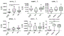
FIG. 5 is a series of graphs comparing the relative abundance of various salivary proteins (KV110, NAMPT, COPB, SPR2A and HV311) in healthy controls versus NYHA class I and class III/IV heart failure patients as determined by SWATH-MS.
FIG. 6 is an overlay of ROC curves comparing combinations of the salivary proteins shown in FIG. 5 between cohorts (cohort) (NYHA class I, NYHA class III/IV and control).
FIG. 7 is a series of graphs comparing the relative abundance of various sialoproteins (K L K1, TCPD, S10A7, D L DH, IGHA2 and CAMP) in healthy controls versus NYHA class I and class III/IV heart failure patients as determined by SWATH-MS.
FIG. 8 is an overlay of ROC curves comparing combinations of the salivary proteins shown in FIG. 7 between the respective cohorts (NYHA class I, NYHA class III/IV and control).
FIG. 9 is a series of graphs comparing the concentrations of various salivary proteins (S10A7, K L K1, and CAMP) in healthy controls, individuals at high risk of heart failure, and heart failure patients, as determined by immunoassay, and ROC curves for comparing combinations of salivary proteins. Generation of predictive scores by combining the concentrations of these salivary proteins FIG. 9A, S10A 7; FIG. 9B, CAMP; FIG. 9C, K L K1; FIG. 9D, combination predictive scores for salivary proteins FIG. 9E, ROC curves for comparing combinations of salivary proteins between heart failure patients and controls FIG. 9F, ROC curves for comparing combinations of salivary proteins between SCREEN-HF (heart failure screening) cohorts and controls;
figure 10 is a graph showing the predicted scores between study subjects who had suffered from cardiovascular disease after participation in the study and subjects who were not admitted to the study for cardiovascular disease.
Fig. 11(a) is a western blot of K L K1, TCPD, S10a7, D L DH, IGHA2 and CAMP in saliva samples of 6 healthy controls and 6 heart failure patients (B) is the relative band intensities with standard error of K L K1, TCPD, S10a7, D L DH, IGHA2 and CAMP in saliva samples of healthy controls and heart failure patients.
Fig. 12 is a western blot of S10a7 in additional saliva samples of 12 healthy controls and 12 heart failure patients.
Detailed Description
Abbreviations
The following abbreviations are used throughout:
AACT- α 1 anti-chymotrypsin
BNP-BNP
Antimicrobial peptide of CAMP ═ antimicrobial peptide (Cathelicidin)
COPB ═ exosome (coater) subunit β
D L DH-dihydrolipoic acid dehydrogenase, mitochondria
ESI-electrospray ionization
GE L S ═ gelsolin
h is hour
HV311 Ig heavy chain V-III region KO L
IGHA2 ═ Ig α -2 chain C region
IGJ ═ immunoglobulin J chain
Iqr-quartering distance
K L K1 ═ kallikrein 1
KV 110-Ig kappa chain V-I region HK102
L C ═ liquid chromatography
L C-ESI-MS/MS liquid chromatography-electrospray ionization-tandem mass spectrometry
L P L C1 is related to long palate, lung and nasal epithelial carcinoma protein 1
min is minutes
MMP9 ═ matrix metalloproteinase-9
MS mass spectrum
MS/MS tandem mass spectrometry
NAMPT ═ nicotinamide phosphoribosyltransferase
Negative predictive value of NPV
NYHA new york heart disease association
PBS-phosphate buffered saline
Positive predictive value of PPV
rcf-relative centrifugal force
Receiver operating characteristic
s is seconds
S10a7 ═ S100 calbindin a7
SP L C2 short palate, lung and nose associated protein 2
SPR2A ═ small proline abundant protein 2A
Sequential window acquisition of SWATH ═ all theoretical fragment ion spectra
TCPD ═ T-complex protein 1 subunit
TOF time of flight
VIME ═ vimentin
The present invention is based in part on the following findings: the abundance of proteins in a biological sample obtained from a subject with early stage heart failure is different compared to a biological sample obtained from a healthy subject. The present inventors have used abundant protein removal and SWATH-MS to identify salivary proteins as putative biomarkers with diagnostic utility in early heart failure.
Accordingly, in a first aspect, the present invention provides a method for detecting early stage heart failure in a subject, the method comprising analyzing a biological sample obtained from the subject and determining the concentration of at least one biomarker in the sample, and assigning a heart failure classification to the subject if the concentration of the at least one biomarker is above or below a predetermined reference concentration for the at least one biomarker. The predetermined reference concentration of the at least one biomarker may be determined from a biological sample obtained from a healthy subject.
For the purposes of the present invention, the phrase "early stage" describing the stage of heart failure refers to the functional classification defined by the new york heart association: NYHA I and/or NYHA II.
The term "biological sample" as used herein refers to a sample extracted from a subject. The term includes untreated, treated, diluted or concentrated biological samples. The biological sample obtained from the subject may be any suitable sample, such as whole blood, serum or plasma. Preferably, the biological sample is obtained from the buccal cavity of the subject. Thus, the biological sample may be sputum or saliva. According to the present invention, which provides a non-invasive, cost-effective method for diagnosing early heart failure, the biological sample obtained from the subject is preferably saliva.
For example, at least one biomarker may be any number of proteins selected from the group consisting of K L K1, TCPD, S10A7, D L DH, IGHA2, CAMP, KV110, NAMPT, COPB, SPR2A and HV311. in one embodiment, at least one biomarker is selected from the group consisting of proteins consisting of K L K1, TCPD, S10A7, D L, IGHA2 and CAMP. preferably, at least one biomarker is a biomarker panel consisting of two, three, four, five or all six of these proteins.
The predetermined reference concentration of the biomarker may be in the form of a concentration range such that a biomarker concentration outside this range is indicative of early heart failure. Alternatively, the predetermined reference concentration of the biomarker may be in the form of a specific value, such that a biomarker concentration above or below this value is indicative of early heart failure. Thus, for each biomarker used to detect early heart failure in a subject, a predetermined reference concentration of the biomarker in a biological sample from a healthy subject has been determined or known.
In the context of the present invention, a "healthy subject" is a subject without heart failure for determining a predetermined reference concentration of at least one biomarker in a biological sample obtained from the healthy subject. That is, a healthy subject is one that does not have any external symptoms of heart failure and is not classified as NYHA class I or II.
The present inventors have surprisingly found that the abundance of a particular protein in saliva of subjects classified as NYHA class I or II is increased compared to the abundance of the same protein in healthy subjects. Conversely, the abundance of a particular protein in saliva assigned to a class I or II NYHA subject is reduced compared to the abundance of that same protein in healthy subjects.
Although a subject may be assigned a heart failure classification based on the concentration of only one biomarker in a biological sample from the subject, it is more advantageous to assign a classification based on the concentrations of two, three, four, five or more biomarkers in the biological sample, as a higher degree of certainty of the classification may be achieved using more biomarkers.
When a biomarker panel consisting of two or more biomarkers is used to detect early heart failure in a subject, this panel may consist of the biomarkers: the concentration of the biomarker in saliva of a heart failure subject is higher than the concentration of the same biomarker in saliva of a healthy subject. Alternatively, this group may consist of biomarkers such as: the concentration of the biomarker in saliva of a heart failure subject is lower than the concentration of the same biomarker in saliva of a healthy subject. Further alternatively, the panel may consist of a combination of biomarkers, wherein the concentration of at least one biomarker in the heart failure subject's saliva is higher than the concentration of the same biomarker in the healthy subject's saliva, and the concentration of at least one biomarker in the heart failure subject's saliva is lower than the concentration of the same biomarker in the healthy subject's saliva.
In a second aspect, the present invention provides a method for detecting early stage heart failure in a subject, the method comprising analyzing a biological sample obtained from the subject and determining the concentration of at least one biomarker in the sample, determining the concentration of the at least one biomarker in the biological sample obtained from a healthy subject, and assigning a heart failure classification to the subject if the concentration of the at least one biomarker in the sample from the subject is higher or lower than the concentration of the at least one biomarker in the biological sample obtained from a healthy subject.
The concentration of at least one biomarker in a biological sample, whether from a subject with underlying heart failure or a healthy subject, can be determined by any suitable method for determining the concentration of a protein. For example, the concentration may be determined by mass spectrometry. Comparing the peak intensity of a particular biomarker in the mass spectrum of a sample from a subject with underlying heart failure with the peak intensity in the mass spectrum of a sample from a healthy subject can provide an indication of the relative difference in abundance of the biomarker in the two samples. More accurate comparisons can be obtained by using the SWATH-MS detailed in the examples below.
Alternatively, the concentration of at least one biomarker in a biological sample may be determined using one or more reagents that specifically bind to the at least one biomarker. For example, the reagent may comprise an antibody directed to an epitope of the biomarker, wherein the antibody optionally comprises a tag (e.g., a fluorescent label) for detecting the presence of the antibody-biomarker complex.
In a third aspect, the present invention provides a method for detecting early stage heart failure in a subject, the method comprising analyzing a biological sample obtained from the subject and determining the concentration of at least one biomarker in the sample, wherein the at least one biomarker is selected from the group of proteins consisting of K L K1, TCPD, S10a7, D L DH, IGHA2, CAMP, KV110, NAMPT, COPB, SPR2A and HV311, and assigning a heart failure classification to the subject if the concentration of the at least one biomarker is above or below a predetermined reference concentration of the at least one biomarker.
The biological sample may be assayed for the concentration of at least one, two, three, four, five, six, seven, eight, nine, ten or all eleven proteins. Although a subject may be assigned a heart failure classification based on the concentration of only one protein in the biological sample, it is more advantageous to assign a classification based on the concentrations of two, three, four, five, six, seven, eight, nine, ten, or eleven proteins in the biological sample, since a higher degree of certainty in classification can be achieved using more biomarkers.
The certainty of the grading can be assessed by determining the sensitivity and specificity of the comparison data.
In a fourth aspect, the invention provides a method for detecting early stage heart failure in a subject, the method comprising analyzing a biological sample obtained from the subject and determining the concentration of at least one biomarker in the sample, wherein the at least one biomarker is selected from the group consisting of K L K1, TCPD, S10a7, D L DH, IGHA2, CAMP, KV110, NAMPT, COPB, SPR2A and HV311, determining the concentration of the at least one biomarker in the biological sample obtained from a healthy subject, and assigning a heart failure classification to the subject if the concentration of the at least one biomarker in the sample from the subject is higher or lower than the concentration of the at least one biomarker in the biological sample obtained from the healthy subject.
In a fifth aspect, the present invention provides a kit for detecting the presence of at least one biomarker associated with early heart failure, the kit comprising a solid support having immobilized thereon at least one molecule that specifically binds to the at least one biomarker.
The at least one molecule that specifically binds to the at least one biomarker may be any suitable molecule. Preferably, the at least one molecule comprises an antibody that specifically binds to at least one biomarker. Thus, the solid support may have one, two, three, four, etc. antibodies immobilized thereon.
The solid support may be any suitable material that can be suitably modified for immobilizing antibodies and that is suitable for at least one detection method. Representative examples of materials suitable for the solid support include glass and modified or functionalized glass, plastics (including acrylic, polystyrene and copolymers of styrene with other materials, polypropylene, polyethylene, polybutylene, polyurethane, polytetrafluoroethylene, and the like), polysaccharides, nylon or nitrocellulose, resins, silica or silica-based materials (including silicon and modified silicon), carbon, metals, inorganic glasses, and plastics. The solid support may allow optical detection without significant fluorescence.
The solid support may be planar, but other configurations of the substrate may also be used. For example, the solid support may be a tube with the antibody placed on the inner surface.
In a sixth aspect, the present invention provides a kit for detecting the presence of at least one biomarker associated with early heart failure, wherein the at least one biomarker is selected from the group consisting of K L K1, TCPD, S10a7, D L DH, IGHA2, CAMP, KV110, NAMPT, COPB, SPR2A and HV311, the kit comprising a solid support having immobilized thereon at least one molecule that specifically binds to the at least one biomarker.
Examples
Example 1
Materials and methods
Study participants
This study was approved by the University of Queensland medical Ethical institute Board (University of Queensland medical Ethical Board) and the Meite health Services Human Research Ethical Committee (Materhealth Services Human Research Ethics Committee) and Royal Brisbane and Women's Hospital Research institute (Royal Brisbane and Women's Hospital Research Governance). All study participants were >18 years old and given informed consent prior to collecting samples. The exclusion criteria for healthy controls are based on a simple questionnaire asking the volunteers to indicate whether they present any complications (comorbid diseases) and oral diseases (e.g. periodontal and gingivitis, autoimmune, infectious, musculoskeletal or malignant diseases and recent surgery or trauma). If any condition is present, the participants will be excluded from the study. Volunteers were from caucasian and asian ethnicities, had no symptoms of fever or cold, and had good oral hygiene.
From month 1 2012 to month 7 2014, a total of 30 healthy controls and 33 symptomatic heart failure patients were recruited from the queensland university of Brisbane, maert Adult Hospital (Mater Adult Hospital), or Royal Brisbane female Hospital (Royal Brisbane and Women's Hospital). Cardiologists at the mert adult hospital and the royal brisbane women hospital have graded patients according to their clinical symptoms using the New York Heart Association (NYHA) functional grading system. All patients enrolled in the study were classified as NYHA class III or IV patients. The mean age of heart failure patients was 67.6 years and that of healthy controls was 49.7 years. Men in the heart failure cohort accounted for 63.3% and men in the healthy control cohort accounted for 43.3%.
Saliva sample collection
Non-irritating resting whole saliva (Martinet W et al, 2003; punyadera C et al, 2011; Foo JY et al, 2013; Castagnola M et al, 2011; Helmerhorst EJ and Oppenheim FG, 2007; L oo JA et al, 2010) was collected from early and late stage heart failure patients and healthy controls, the volunteers were asked to not eat or drink (except for water) for at least 30 minutes prior to collecting saliva, the volunteers were asked to rinse the oral cavity with water to remove food particles and debris, tip up and down, accumulate saliva in the oral cavity, and then expectorate it into a Falcon tube (50M L, Greiner, germana) placed on ice.
Total protein concentration in saliva samples
For primary screening, total protein concentrations in saliva samples from patients (n 10) and controls (n 10) were measured using a 2D Quant kit (GE Healthcare Bio-Sciences AB, Sweden). Use of 190 enzyme-Linked reader (Molecular Devices, LL C, California, USA) at 480nm absorbance was measured using QuickStartTMTotal Protein concentrations in saliva samples obtained from patients (n-30) and controls (n-30) were quantified by Bradford Protein Assay (Bio-Rad, USA) for SWATH-MS validation (see below).
Saliva sample preparation for mass spectrometry
Saliva samples normalized for protein content collected from heart failure patients and healthy controls were pooled separately. Equal amounts of total protein from each individual were pooled to give 10mg of total pooled protein for each of the control and patient. According to manufacturer's instructions, use A pooled sample was processed with a small volume kit (Bio-Rad, Hercules, Calif.) A packed bed of beads (20 μ L) was added to pooled saliva and incubated on a rotary shaker at 25 ℃ for 16 hours, the beads were pelleted by centrifugation at 1,000 relative centrifugal force (rcf) for 1 minute, and the supernatant discarded, the beads were washed three times with Phosphate Buffered Saline (PBS) and bound proteins were eluted in 8M urea, 2% CHAPS, and 5% acetic acid (20 μ L). the eluted proteins were precipitated by addition of 1:1 methanol: acetone (80L), incubated at-20 ℃ for 16 hours, and centrifuged at 18,000rcf for 10 minutesThe pellet was resuspended in 50mM Tris-
HCl buffer pH 8 containing 1% SDS. Cysteine was reduced by adding DTT to 10mM and incubating at 95 ℃ for 10 minutes, followed by alkylation by adding acrylamide to 25mM and incubating at 23 ℃ for 1 hour. The protein was precipitated as described above and resuspended in 50mM NH containing proteomic grade trypsin (1. mu.g) (SigmaAlldich, USA)4HCO3(50. mu. L) and incubated at 37 ℃ for 16 hours.
For SWATH-MS validation using individual samples, saliva containing 50. mu.g of total protein was supplemented with equal volumes of 100mM Tris-HCl buffer (pH 8), 2% SDS and 20mM DTT and incubated at 95 ℃ for 10 minutes. The protein is then alkylated, precipitated and digested as described above.
Mass spectrometry and data analysis
Peptides were desalted using C18 Zip Tips (Millipore, USA) and analyzed by L C-ESI-MS/MS on a Triple TOF5600 mass spectrometer equipped with a Nanospray III interface (AB SCIEX) using the prominino nano L C system (Shimadzu, Japan) essentially as described previously (Foo et al, 2013; Ovchinnikov et al, 2012.) at the Agilent C18 trap (pore size) Particle size 5 μm, 0.3mm i.d.x 5mm) approximately 2 μ g of peptide was desalted for 3 minutes at a flow rate of 30 μ L/min and then on a Vydac EVEREST reverse phase C18 HP L C column (pore size)
 Particle size 5 μm, 150 μmi.d.x 150mm) at a flow rate of 1 μ L/min, the peptides were separated with a gradient of buffer B from 1 to 10% over 2 minutes, then 10 to 60% buffer B over 45 minutes, buffer A (1% acetonitrile and 0.1% formic acid), buffer B (80% acetonitrile and 0.1% formic acid), gas and voltage settings were adjusted as necessary.MS-TOF scanning was performed at m/z of 350-1800 for 0.5 seconds, followed by information dependent acquisition of MS/MS, automated CE selection of the first 20 peptides was performed at m/z of 40-1800 for 0.05 seconds per spectrum.SWATH analysis was performed using the same L C parameters, MS-TOF scanning was performed at m/z of 350-1800 for 0.05 seconds, followed by 40 m/S0-1250 m/z, with a 26m/z isolation window (isolation window) and a 1m/z window overlap for 0.1 seconds each. The collision energy is automatically specified by the analysis software (AB SCIEX) according to the m/z window range.
Using ProteinPilot (ab sciex), proteins were identified by searching L udwigNR databases (downloaded from http:// apcf. edu. au by 1 month 27 days 2012; 16,818,973 sequences; 5,891,363,821 residues) using standard settings set as sample type, identification; cysteine alkylation, none; instrument, Triple-TOF 5600; species, no restrictions; ID focus, biological modifications; enzymes, trypsin; search work, detailed ID. pseudo-discovery rate analysis was performed on all searches using ProteinPilot:. identified peptides with confidence greater than 99% and local pseudo-discovery rate less than 1% were used for further analysis using protein analysis as identified protein analysis by using standard settings — half quantitative comparison of protein abundance based on protein grade, score, peptide coverage percentage and peptide number (Bailey and Schulz, 2013.) using Peak View 1.1 obtained extracted ions as protein depletion protein analysis as chromatogram data for ion chromatogram analysis (bailex), median abundance analysis was performed using map analysis as ion library for protein abundance analysis using absolute abundance analysis, Peak View spectrum analysis as well as protein analysis, protein abundance conversion into protein abundance analysis by using absolute analysis by using scale analysis.
Example 2
Identification of proteins by L C-ESI-MS/MS
To detect proteins with changes in abundance between heart failure patients and controls, semi-quantitative methods were used to compare the grade, score, percent peptide coverage, and number of peptides identified for each protein, this semi-quantitative method identified a number of proteins with different putative abundances, as shown in Table 2.
TABLE 2 comparison of Heart failure patients with controls, salivary proteins in different abundance

To preliminarily validate these putative biomarkers, the abundance of peptides of each protein identified by the protein pilot database search (table 3) determined from the extracted ion chromatogram of L C-ESI-MS/MS data (fig. 1) comparison of peptide abundances identified two proteins in heart failure patients that are significantly more abundant (long palate, lung and nasal epithelial cancer-associated protein 1, i.e. L P L C1(P ═ 0.0004) and matrix metalloproteinase-9, i.e. MMP9(P ═ 0.02)) and two proteins that are significantly less abundant (gelsolin, i.e. GE L S (P ═ 0.03) and short palate, lung and nasal associated protein 2, i.e. SP L C2(P ═ 0.0003)) compared to control samples, several other proteins that show large abundance changes (e.g. release 1, K L K1; immunoglobulin J, and IGJ chain proteins detected as a result of the preliminary analysis of the peptides, thus several putative biomarkers were not identified for the quantitative analysis of saliva.
Table 3 relative peptide abundance of each protein identified using ProteinPilot

Example 3
Validation Using SWATH-MS
To verify from pooled samples Analysis of identified pushingsNovel biomarkers for SWATH-MS assays were performed on saliva samples collected from heart failure patients and controls. Differential abundance was identified by unbiased SWATH-MS proteomic comparisons of saliva for heart failure patients and controls>2 times and corrected P<Seven proteins of 0.01 this included the SP L C2 protein identified as a putative heart failure biomarker by ProteMiner analysis the relative abundance of SP L C2 in the control was 1.89 times that in heart failure patients saliva with high specificity (almost complete group separation) (see FIG. 2A, corrected value P<0.0001) demonstrated that SP L C2 is a salivary protein biomarker for heart failure since K L K1 is more abundant in the saliva of heart failure patients than in control saliva, K L K1 was also identified as a potential biomarker by proteminer analysis (fig. 1). the increase in abundance of K L K1 was also validated by SWATH-MS analysis, indicating that K L K1 is increased 1.3 fold in heart failure patients than in controls (fig. 2B, corrected P ═ s<0.0001)。
The utility of the ratios of the abundances of these individually validated biomarkers was investigated in discriminating heart failure due to decreased SP L C2 and increased K L K1 abundance compared to controls the study was conducted observing that there was a large and highly significant difference between heart failure patients and controls, the difference in ratio and high specificity was 5.3 fold (fig. 2C, P ═ 0.00001.) Receiver Operating Characteristic (ROC) curve analysis was conducted to determine the diagnostic ability of SP L C2 and K L K1 as biomarkers K L K: analysis of SP L C2 (fig. 3A, fig. 4A) showed area under the curve (AUC) value of 0.75, sensitivity of 70.0%, specificity of 66.7%.
Example 4
Predictive power of biomarker panel
The ability of a panel comprising putative biomarkers KV110, NAMPT, COPB, SPR2A and HV311 to predict early heart failure was evaluated using R (R Development Core Team,2011) based Mtstats (Clough et al, 2012; Chang et al, 2012) (FIG. 5). Table 4 lists the sensitivity and specificity of the biomarker combinations in various cohorts (NYHA class I, n-20; NYHA class III/IV, n-19; healthy controls, n-20).
TABLE 4 sensitivity and specificity of biomarker combinations


The ROC curve in figure 6 provides a useful summary of the diagnostic potential of the combination of the five biomarkers KV110, NAMPT, COPB, SPR2A and HV 311. The closer the area under the ROC curve is to 1, the better the diagnostic potential. The ROC curve for the five biomarker combination in NYHA class I patients compared to the five biomarkers in healthy controls was 0.96 AUC, 95.0% sensitivity and 90.0% specificity (figure 6). These results indicate that the combination of five biomarkers has high diagnostic ability.
The predictive power of the panel comprising putative biomarkers K L K1, TCPD, S10a7, D L DH, IGHA2 and CAMP was evaluated using mstats (Clough et al 2012; Chang et al 2012) based on R (R Development Core Team,2011) for early heart failure (fig. 7) table 5 lists the sensitivity and specificity of the combination of biomarkers in various cohorts (NYHA class I, n 20; NYHA class III/IV, n 19; healthy controls, n 20).
TABLE 5 sensitivity and specificity of combinations of biomarkers

The ROC curve in figure 8 provides a useful summary of the diagnostic potential of the combination of the six biomarkers K L K1, TCPD, S10a7, D L DH, IGHA2 and CAMP the closer the area under the ROC curve is to 1, the better the diagnostic potential, compared to the six biomarkers in healthy controls, the AUC of the ROC curve for the six biomarker combinations in NYHA class I patients is 0.86, the sensitivity is 80.0%, and the specificity is 70.0% (figure 8), these results indicate that the combination of the six biomarkers has a high diagnostic capacity.
The predictive power of a panel comprising putative biomarkers K L K1, S10a7 and CAMP was evaluated on high risk individuals for heart failure (fig. 9) using mstats (Clough et al 2012; Chang et al 2012) based on R (R Development Core Team, 2011.) table 6 lists the sensitivity and specificity of the combination of biomarkers in various cohorts (heart failure patients, n 100; individuals at high risk for heart failure (scr en-HF), n 121; healthy controls, n 88).
TABLE 6 sensitivity and specificity of combinations of biomarkers

Figure 10 shows the predicted scores between study subjects with cardiovascular disease after study participation and subjects not admitted to the study for cardiovascular disease.
Of the 99 participants in the SCREEN-HF cohort, 11 were admitted to the hospital for a preliminary diagnosis of cardiovascular disease. In these 11 persons, the predictive score generated by the three marker panel ranged from 0.139 to 0.996 with a median of 0.517 (IQR: 0.256-0.920), whereas in individuals who were not admitted to the hospital for cardiovascular disease, the predictive score ranged from 0.086 to 0.992 with a median of 0.294 (IQR: 0.172-0.679). There was a statistically significant difference between the two sets of SCREEN-HF cohorts (p. 0.0382).
To verify that K L K1, TCPD, S10a7, D L DH, IGHA2, and CAMP were members of the diagnostic panel, western blot analysis was performed on randomly selected 6 healthy controls and randomly selected 6 heart failure patients as shown in fig. 11, S10a7 was detected in 5 of the samples from S10a7 and IGHA 2.6 heart failure patients detected in individual saliva samples, while only 1 of the 6 healthy control samples detected s10a7. the banding intensity of each sample was normalized to the mean banding intensity of the healthy controls similar to the results of SWATH-MS, both S10a7 and IGHA2 exhibited a higher protein abundance in the heart failure patient samples than the healthy control samples, the mean banding intensity of S10a7 in the heart failure patients was 6-fold higher than in the healthy control samples, the abundance of IGHA2 in the heart failure patients was higher than in the healthy control samples (1.06:1), but a significant difference was observed in the samples from the initial high protein expression in the heart failure patients (ca 631: 6851: 1) and on the same samples as the initial western blot analysis, no expression of the high protein expressed in the samples of the healthy controls (ca 6851) and no observed in the samples similar to the samples).
Reference throughout this specification to "one embodiment" or "an embodiment" means that a particular feature, structure, or characteristic described in connection with the embodiment is included in at least one embodiment of the present invention. Thus, the appearances of the phrases "in one embodiment" or "in an embodiment" in various places throughout this specification are not necessarily all referring to the same embodiment. Furthermore, the particular features, structures, or characteristics may be combined in any suitable manner in one or more combinations.
In compliance with the statute, the invention has been described in language more or less specific as to structural or methodical features. It is to be understood that the invention is not limited to the specific features shown or described, since the means herein described comprise preferred forms of putting the invention into effect. The invention may thus be claimed in any form or modification within the proper scope of the appended claims (if any) appropriately interpreted by those skilled in the art.
CITATION LIST
Australian Institute of Health and Welfare 2011.Cardiovasculardisease:Australian facts 2011.Cardiovascular disease series.Cat.no.CVD53.Canberra:AIHW.(http://www.aihw.gov.au/WorkArea/DownloadAsset.aspx?id=10737418530)
Bailey UM and Schulz BL,Deglycosylation systematically improves N-glycoprotein identification in liquid chromatography-tandem mass spectrometryproteomics for analysis of cell wall stress responses in Saccharomycescerevisiae lacking aAlg3p,J Chromatogr B Analyt Technol Biomed Life Sci,2013;923-924:16-21
Carlson KJ,Lee DCS,Goroll AH,Leahy M and Johnson RA,An analysis ofphysicians’reasons for prescribing long-term digitalis therapy inoutpatients,J Chron Dis,1985;38:733-739
Castagnola M,Inzitari R,Fanali C,Iavarone F,Vitali A,Desiderio C,Vento G,Tirone C,Romagnoli C,Cabras T,Manconi B,Sanna MT,Boi R,Pisano E,Olianas A,Pellegrini M,Nemolato S,Heizmann CW,Faa G and Messana I,Thesurprising composition of the salivary proteomeof preterm human newborn,MolCell Proteomics,2011;10(1):M110.003467
Chang CY,Picotti P,Hüttenhain R,Heinzelmann-Schwarz V,Jovanovic M,Aebersold R and Vitek O,Protein significance analysis in selected reactionmonitoring(SRM)measurements,Mol Cell Proteomics,2012;11(4):M111.014662
Clough T,Thaminy S,Ragg S,Aebersold R and Vitek O,Statistical proteinquantification and significance analysis in label-free LC-MS experiments withcomplex designs,BMC Bioinformatics,2012;13(Suppl 16):S6
Foo JYY,Wan Y,Schulz BL,Kostner K,Atherton J,Cooper-White J,Dimeski Gand Punyadeera C,Circulating fragments of N-terminal pro-B-type natriureticpeptides in plasma of heart failure patients,Clin Chem,2013;59:1523-1531
Harlan WR,Oberman A,Grimm R and Rosati RA,Chronic congestive heartfailure in coronary artery disease:clinical criteria,Ann Intern Med,1977;86(2):133-138
Helmerhorst EJ and Oppenheim FG,Saliva:a dynamic proteome,J Dent Res,2007;86:680-693
Krum H,Jelinek MV,Stewart S,Sindone A and Atherton JJ,2011 Update tonational heart foundation of Australia and cardiac society of Australia andNew Zealand guidelines for the prevention,detection and management of chronicheart failure in Australia,2006,Med J Aust,2011;194(8):405-409
Loo JA,Yan W,Ramachandran P and Wong DT,Comparative human salivaryand plasma proteomes,J Dent Res,2010;89:1016-1023
McKee PA,Castelli WP,McNamara PM and Kannel WB,The natural history ofcongestive heart failure:the Framingham study,N Engl J Med,1971;285(26):1441-1446
Marian AJ and Nambi V,Biomarkers of cardiac disease,Expert Rev MolDiagn,2004;4:805-20
Martinet W,Schrijvers DM,De Meyer GRY,Herman AG and Kockx MM,Westernarray analysis of human atherosclerotic plaques:Downregulation of apoptosis-linked gene 2,Cardiovasc Res,2003;60(2):259-267
Ovchinnikov DA,Cooper MA,Pandit P,Coman WB,Cooper-White JJ,Keith P,Wolvetang EJ,Slowey PD and Punyadeera C,Tumor-suppressor gene promoterhypermethylation in saliva of head and neck cancer patients,Transl Oncol,2012;5(5):321-326
Palazzuoli A,Iovine F,Gallotta M and Nuti R,Emerging cardiac markersin coronary disease:Role of brain natriuretic peptide and other biomarkers,Minerva Cardioangiol,2007;55(4):491-496
Punyadeera C,Dimeski G,Kostner K,Beyerlein P and Cooper-White J,One-step homogeneous C-reactive protein assay for saliva,J Immunol Methods,2011;373:19-25
R Development Core Team(2011),R:A language and environment forstatistical computing,Vienna,Austria:the R Foundation for StatisticalComputing
Shamsham F and Mitchell J,Essentials of the diagnosis of heartfailure,Am Fam Physician,2000;61(5):1319-1328
The Criteria Committee of the New York Heart Association,Nomenclatureand Criteria for Diagnosis of Diseases of the Heart and Great Vessels,9thed.,Little,Brown;Boston,1994,pp.253-256
Claims (19)
1. A method for detecting early stage heart failure in a subject, the method comprising analyzing a biological sample obtained from the subject and determining the concentration of at least one biomarker in the sample, and assigning a heart failure classification to the subject if the concentration of the at least one biomarker is above or below a predetermined reference concentration for the at least one biomarker.
2. The method of claim 1, wherein the predetermined reference concentration of the at least one biomarker is determined from a biological sample obtained from a healthy subject.
3. A method of detecting early stage heart failure in a subject, the method comprising analyzing a biological sample obtained from the subject and determining the concentration of at least one biomarker in the sample, determining the concentration of the at least one biomarker in a biological sample obtained from a healthy subject, and assigning a heart failure classification to the subject if the concentration of the at least one biomarker in the sample from the subject is higher or lower than the concentration of the at least one biomarker in a biological sample obtained from the healthy subject.
4. A method of screening a subject for early stage heart failure, the method comprising analyzing a biological sample obtained from the subject and determining the concentration of at least one biomarker in the sample, and assigning a heart failure classification to the subject if the concentration of the at least one biomarker is above or below a predetermined reference concentration for the at least one biomarker.
5. The method according to any one of claims 1 to 4, wherein the at least one biomarker is selected from the group of proteins consisting of K L K1, TCPD, S10A7, D L DH, IGHA2, CAMP, KV110, NAMPT, COPB, SPR2A and HV 311.
6. The method of claim 5, wherein the at least one biomarker is selected from the group of proteins consisting of K L K1, TCPD, S10A7, D L DH, IGHA2 and CAMP.
7. The method of claim 6, wherein the at least one biomarker is a biomarker panel comprising two, three, four, five, or six of the proteins.
8. The method of claim 7, wherein the set of biomarkers includes three of the proteins.
9. The method of claim 8, wherein the biomarker panel comprises K L K1, S10A7, and CAMP.
10. The method of claim 5, wherein the at least one biomarker is selected from the group of proteins consisting of: KV110, NAMPT, COPB, SPR2A, and HV 311.
11. The method of claim 10, wherein the at least one biomarker is a biomarker panel comprising two, three, four, or five of the proteins.
12. The method of any one of claims 5-11, wherein the biological sample is selected from the group consisting of whole blood, serum, plasma, sputum, or saliva.
13. The method of claim 12, wherein the biological sample is saliva.
14. A kit for detecting the presence of at least one biomarker associated with early heart failure, comprising a solid support having immobilized thereon at least one molecule that specifically binds to the at least one biomarker.
15. A kit for detecting the presence of at least one biomarker associated with early heart failure, wherein the at least one biomarker is selected from the group consisting of K L K1, TCPD, S10a7, D L DH, IGHA2, CAMP, KV110, NAMPT, COPB, SPR2A, and HV311, the kit comprising a solid support having immobilized thereon at least one molecule that specifically binds to the at least one biomarker.
16. The kit of claim 14 or 15, wherein the at least one molecule that specifically binds to the at least one biomarker is an antibody that specifically binds to the at least one biomarker.
17. The kit of claim 16, wherein the solid support has two, three, four, five or six antibodies immobilized thereon.
18. The kit of claim 17, wherein the solid support has three antibodies immobilized thereon.
19. The kit of claim 18, wherein the antibody is an antibody directed against K L K1, S10a7, and CAMP.
Applications Claiming Priority (3)
| Application Number | Priority Date | Filing Date | Title |
|---|---|---|---|
| AU2017903138 | 2017-08-08 | ||
| AU2017903138A AU2017903138A0 (en) | 2017-08-08 | Methods for Diagnosis of Early Stage Heart Failure | |
| PCT/AU2018/050827 WO2019028507A1 (en) | 2017-08-08 | 2018-08-08 | Methods for diagnosis of early stage heart failure |
Publications (1)
| Publication Number | Publication Date |
|---|---|
| CN111465857A true CN111465857A (en) | 2020-07-28 |
Family
ID=65273054
Family Applications (1)
| Application Number | Title | Priority Date | Filing Date |
|---|---|---|---|
| CN201880063136.3A Pending CN111465857A (en) | 2017-08-08 | 2018-08-08 | Method for diagnosing early heart failure |
Country Status (6)
| Country | Link |
|---|---|
| US (1) | US20200174021A1 (en) |
| EP (1) | EP3665483A4 (en) |
| JP (1) | JP7414281B2 (en) |
| CN (1) | CN111465857A (en) |
| AU (2) | AU2018315056B2 (en) |
| WO (1) | WO2019028507A1 (en) |
Cited By (1)
| Publication number | Priority date | Publication date | Assignee | Title |
|---|---|---|---|---|
| WO2024007778A1 (en) * | 2022-07-05 | 2024-01-11 | 上海交通大学医学院附属上海儿童医学中心 | Use of plasma molecular marker kynurenine in detection of early heart failure |
Families Citing this family (1)
| Publication number | Priority date | Publication date | Assignee | Title |
|---|---|---|---|---|
| AU2021283390A1 (en) * | 2020-06-03 | 2023-01-19 | Esn Cleer | Biomarker identification for imminent and/or impending heart failure |
Citations (18)
| Publication number | Priority date | Publication date | Assignee | Title |
|---|---|---|---|---|
| WO2002061131A2 (en) * | 2000-12-04 | 2002-08-08 | Bristol-Myers Squibb Company | Human single nucleotide polymorphisms |
| WO2002094870A2 (en) * | 2000-11-02 | 2002-11-28 | Curagen Corporation | Proteins and nucleic acids encoding same |
| US20040033582A1 (en) * | 2002-06-03 | 2004-02-19 | Manling-Ma Edmonds | Human single nucleotide polymorphisms |
| WO2006008002A2 (en) * | 2004-07-23 | 2006-01-26 | Bayer Healthcare Ag | Diagnostics and therapeutics for diseases associated with kallikrein 1 (klk1) |
| CN101031640A (en) * | 2004-07-29 | 2007-09-05 | 干细胞创新有限公司 | Differentiation of stem cells |
| US20080057590A1 (en) * | 2006-06-07 | 2008-03-06 | Mickey Urdea | Markers associated with arteriovascular events and methods of use thereof |
| US20080300170A1 (en) * | 2006-09-01 | 2008-12-04 | Cohava Gelber | Compositions and methods for diagnosis and treatment for type 2 diabetes |
| CN101340928A (en) * | 2005-10-21 | 2009-01-07 | 催化剂生物科学公司 | Modified proteases that inhibit complement activation |
| CN101517074A (en) * | 2006-07-05 | 2009-08-26 | 催化剂生物科学公司 | Protease screening methods and proteases indentified thererby |
| CN101617227A (en) * | 2006-11-30 | 2009-12-30 | 纳维哲尼克斯公司 | Genetic analysis systems and method |
| US20100267052A1 (en) * | 2006-09-01 | 2010-10-21 | American Type Culture Collection | Compositions and methods for diagnosis and treatment of type 2 diabetes |
| CN102858985A (en) * | 2009-07-24 | 2013-01-02 | 西格马-奥尔德里奇有限责任公司 | Method for genome editing |
| CN103289980A (en) * | 2006-07-05 | 2013-09-11 | 催化剂生物科学公司 | Protease screening methods and proteases indentified thererby |
| CN103642902A (en) * | 2006-11-30 | 2014-03-19 | 纳维哲尼克斯公司 | Genetic analysis systems and methods |
| WO2014193999A2 (en) * | 2013-05-28 | 2014-12-04 | Caris Science, Inc. | Biomarker methods and compositions |
| US20150291966A1 (en) * | 2012-07-25 | 2015-10-15 | The Broad Institute, Inc. | Inducible dna binding proteins and genome perturbation tools and applications thereof |
| WO2016134365A1 (en) * | 2015-02-20 | 2016-08-25 | The Johns Hopkins University | Biomarkers of myocardial injury |
| US20170051053A1 (en) * | 2011-12-28 | 2017-02-23 | Immunoqure Ag | Method of providing monoclonal auto-antibodies with desired specificity |
Family Cites Families (9)
| Publication number | Priority date | Publication date | Assignee | Title |
|---|---|---|---|---|
| JP2005110602A (en) | 2003-10-09 | 2005-04-28 | Sumitomo Pharmaceut Co Ltd | Disease marker of atopic dermatitis and utilization thereof |
| EP1722232A1 (en) | 2005-05-09 | 2006-11-15 | F.Hoffmann-La Roche Ag | Devices and methods for diagnosing or predicting early stage cardiac dysfunctions |
| WO2007148078A1 (en) * | 2006-06-20 | 2007-12-27 | Lipopeptide Ab | Use cathelicidin antimicrobial protein (hcap18 ) as anticancer agent |
| US20130116343A1 (en) * | 2010-04-21 | 2013-05-09 | Board Of Regents Of The University Of Texas System | Salivary Protein Markers for Detection of Breast Cancer |
| EP2788371A2 (en) * | 2011-12-08 | 2014-10-15 | Biocartis NV | Biomarkers and test panels useful in systemic inflammatory conditions |
| WO2013090811A1 (en) * | 2011-12-14 | 2013-06-20 | The Johns Hopkins University | Biomarkers of pulmonary hypertension |
| US9804152B1 (en) | 2012-08-15 | 2017-10-31 | The Procter & Gamble Company | Human ex vivo skin model and its use in methods of identifying modulators of skin inflammation |
| CN104049082B (en) * | 2014-05-30 | 2016-03-16 | 华中科技大学同济医学院附属同济医院 | Human tissue kallikrein activity detection kit and application thereof |
| CN105987998B (en) * | 2015-01-30 | 2017-12-29 | 江苏众红生物工程创药研究院有限公司 | Human tissue kallikrein 1ELISA immue quantitative detection reagent boxes |
-
2018
- 2018-08-08 CN CN201880063136.3A patent/CN111465857A/en active Pending
- 2018-08-08 AU AU2018315056A patent/AU2018315056B2/en active Active
- 2018-08-08 JP JP2020507022A patent/JP7414281B2/en active Active
- 2018-08-08 US US16/636,403 patent/US20200174021A1/en active Pending
- 2018-08-08 WO PCT/AU2018/050827 patent/WO2019028507A1/en unknown
- 2018-08-08 EP EP18844704.9A patent/EP3665483A4/en active Pending
-
2021
- 2021-09-13 AU AU2021232662A patent/AU2021232662B2/en active Active
Patent Citations (18)
| Publication number | Priority date | Publication date | Assignee | Title |
|---|---|---|---|---|
| WO2002094870A2 (en) * | 2000-11-02 | 2002-11-28 | Curagen Corporation | Proteins and nucleic acids encoding same |
| WO2002061131A2 (en) * | 2000-12-04 | 2002-08-08 | Bristol-Myers Squibb Company | Human single nucleotide polymorphisms |
| US20040033582A1 (en) * | 2002-06-03 | 2004-02-19 | Manling-Ma Edmonds | Human single nucleotide polymorphisms |
| WO2006008002A2 (en) * | 2004-07-23 | 2006-01-26 | Bayer Healthcare Ag | Diagnostics and therapeutics for diseases associated with kallikrein 1 (klk1) |
| CN101031640A (en) * | 2004-07-29 | 2007-09-05 | 干细胞创新有限公司 | Differentiation of stem cells |
| CN101340928A (en) * | 2005-10-21 | 2009-01-07 | 催化剂生物科学公司 | Modified proteases that inhibit complement activation |
| US20080057590A1 (en) * | 2006-06-07 | 2008-03-06 | Mickey Urdea | Markers associated with arteriovascular events and methods of use thereof |
| CN101517074A (en) * | 2006-07-05 | 2009-08-26 | 催化剂生物科学公司 | Protease screening methods and proteases indentified thererby |
| CN103289980A (en) * | 2006-07-05 | 2013-09-11 | 催化剂生物科学公司 | Protease screening methods and proteases indentified thererby |
| US20080300170A1 (en) * | 2006-09-01 | 2008-12-04 | Cohava Gelber | Compositions and methods for diagnosis and treatment for type 2 diabetes |
| US20100267052A1 (en) * | 2006-09-01 | 2010-10-21 | American Type Culture Collection | Compositions and methods for diagnosis and treatment of type 2 diabetes |
| CN101617227A (en) * | 2006-11-30 | 2009-12-30 | 纳维哲尼克斯公司 | Genetic analysis systems and method |
| CN103642902A (en) * | 2006-11-30 | 2014-03-19 | 纳维哲尼克斯公司 | Genetic analysis systems and methods |
| CN102858985A (en) * | 2009-07-24 | 2013-01-02 | 西格马-奥尔德里奇有限责任公司 | Method for genome editing |
| US20170051053A1 (en) * | 2011-12-28 | 2017-02-23 | Immunoqure Ag | Method of providing monoclonal auto-antibodies with desired specificity |
| US20150291966A1 (en) * | 2012-07-25 | 2015-10-15 | The Broad Institute, Inc. | Inducible dna binding proteins and genome perturbation tools and applications thereof |
| WO2014193999A2 (en) * | 2013-05-28 | 2014-12-04 | Caris Science, Inc. | Biomarker methods and compositions |
| WO2016134365A1 (en) * | 2015-02-20 | 2016-08-25 | The Johns Hopkins University | Biomarkers of myocardial injury |
Non-Patent Citations (9)
| Title |
|---|
| ANNE, PIZARD;CHRISTINE, RICHER;NADINE, BOUBY;NICOLAS, PICARD;PIERRE, MENETON;MICHEL, AZIZI;FRAN?OIS, ALHENC-GELAS: "Genetic deficiency in tissue kallikrein activity in mouse and man: effect on arteries, heart and kidney.", BIOLOGICAL CHEMISTRY, vol. 389, 31 December 2008 (2008-12-31) * |
| 余惠珍;谢良地;朱鹏立;许昌声;王华军;李体远;: "人KLK1和EGFP双顺反子重组腺病毒构建及其在血管平滑肌细胞中的表达", 中国病理生理杂志, no. 02, 15 February 2008 (2008-02-15) * |
| 庄伟;谢良地;许昌声;王华军;沈逸华;陈明;: "脂肪组织基质血管组分移植抑制阿霉素诱导的心力衰竭大鼠心肌重构", 福建医科大学学报, no. 05, 28 October 2013 (2013-10-28) * |
| 张乐;胡中扬;杨杰;李蜀渝;曾艺;刘宝琼;杜小平;夏健;刘运海;许宏伟;杨期东;: "KLK1基因rs3212855和rs5515多态性与长沙地区汉族人群脑出血的关联研究", 中南大学学报(医学版), no. 12, 15 December 2010 (2010-12-15) * |
| 张淑群, 强水云, 王西京, 纪宗正, 李妙羡: "血清T-PSA和F-PSA检测在乳腺癌诊断中的意义", 中国肿瘤临床, no. 17, 25 May 2005 (2005-05-25) * |
| 王燕;高友鹤;: "微小病变性肾病早期差异尿蛋白质组分析", 基础医学与临床, no. 03, 5 March 2010 (2010-03-05) * |
| 章文莉;苏津自;: "激肽释放酶-激肽系统对心血管系统的保护作用", 实用老年医学, no. 04, 20 August 2007 (2007-08-20) * |
| 陈清标;蔡超;陈佳鸿;秦国强;陈锡彬;王月生;梁宇翔;韩兆冬;毕学成;钟惟德;: "纳米金标芯片检测用于前列腺癌多基因诊断的实验研究", 现代泌尿生殖肿瘤杂志, no. 01, 22 February 2011 (2011-02-22) * |
| 鲍丹;吕丹;董伟;陈炜;路迎冬;张连峰;: "转基因表达KLK1抵抗压力负荷引起的心室重构", 中国分子心脏病学杂志, no. 03, 25 June 2013 (2013-06-25) * |
Cited By (1)
| Publication number | Priority date | Publication date | Assignee | Title |
|---|---|---|---|---|
| WO2024007778A1 (en) * | 2022-07-05 | 2024-01-11 | 上海交通大学医学院附属上海儿童医学中心 | Use of plasma molecular marker kynurenine in detection of early heart failure |
Also Published As
| Publication number | Publication date |
|---|---|
| AU2018315056A1 (en) | 2020-03-26 |
| JP7414281B2 (en) | 2024-01-16 |
| AU2021232662B2 (en) | 2023-06-01 |
| JP2020530120A (en) | 2020-10-15 |
| WO2019028507A1 (en) | 2019-02-14 |
| EP3665483A4 (en) | 2021-07-14 |
| US20200174021A1 (en) | 2020-06-04 |
| EP3665483A1 (en) | 2020-06-17 |
| AU2018315056B2 (en) | 2021-06-17 |
| AU2021232662A1 (en) | 2021-10-07 |
Similar Documents
| Publication | Publication Date | Title |
|---|---|---|
| EP3260866B1 (en) | Novel biomarkers for cognitive impairment and methods for detecting cognitive impairment using such biomarkers | |
| CN107085114B (en) | Biomarkers for predicting primary adverse events | |
| US9316651B2 (en) | Methods and compositions for biomarkers of fatigue | |
| AU2021232662B2 (en) | Markers relating to early stage heart failure | |
| US20150072360A1 (en) | Biomarkers of pulmonary hypertension | |
| JP5090332B2 (en) | Measurement of short chain SRL alcohol dehydrogenase (DHRS4) as a biomarker for inflammation and infection | |
| EP2444814B1 (en) | Biomarker for mental disorders including cognitive disorders, and method using said biomarker to detect mental disorders including cognitive disorders | |
| US11740245B2 (en) | Mass spectrometry-based methods for the detection of circulating histones H3 and H2B in plasma from sepsis or septic shock (SS) patients | |
| EP3423835B1 (en) | Predictive markers useful in the treatment of wet age-related macular degeneration | |
| US10557860B2 (en) | Circulating pulmonary hypertension biomarker | |
| US20090280512A1 (en) | Tumor marker for renal cancer and method for determination of occurrence of renal cancer | |
| US20210270835A1 (en) | Biomarkers for urothelial carcinoma and applications thereof | |
| Aita et al. | Salivary proteomic analysis in asymptomatic and symptomatic SARS-CoV-2 infection: Innate immunity, taste perception and FABP5 proteins make the difference | |
| CN110568191A (en) | oral cavity phosphorus cell cancer marker and application thereof | |
| EP2391653B1 (en) | Biomarkers associated with nephropathy | |
| EP3236263A1 (en) | Method for diagnosing arthrosis | |
| US20130236917A1 (en) | Albumin-bound protein/peptide complex as a biomarker for disease | |
| EP2883057B1 (en) | Diagnostic of heart failure | |
| US20200408780A1 (en) | Diagnostic for sjorgren's syndrome based on a biomarker | |
| JP7457300B2 (en) | Peptide markers for diagnosis of neurodegenerative diseases | |
| KR102569948B1 (en) | Methods of detecting cancer | |
| US20150044703A1 (en) | Methods and compositions for detecting endometrial or ovarian cancer | |
| CN116298233A (en) | Biomarkers for diagnosing bipolar disorder | |
| CN116804675A (en) | Biomarkers for atopic dermatitis and uses thereof | |
| JP2021018124A (en) | Peptide marker related to breast cancer |
Legal Events
| Date | Code | Title | Description |
|---|---|---|---|
| PB01 | Publication | ||
| PB01 | Publication | ||
| SE01 | Entry into force of request for substantive examination | ||
| SE01 | Entry into force of request for substantive examination |