The application is a divisional application of Chinese patent application with application number of 201780018886.4, application date of 2017, 2 and 10 months and invention name of novel anti-PD-L1 antibody, the original application is a national phase application with international application number of PCT/CN2017/073242, and the international application requires priority of international application with application number of PCT/CN2016/077082, 3 and 23 months in 2016.
Detailed Description
The following description of the present application is intended to be illustrative of various embodiments of the present application. Therefore, the specific modifications discussed herein should not be construed as limitations on the scope of the application. Numerous equivalents, changes, and modifications will readily occur to those skilled in the art without departing from the scope of the present application, and it is intended that such equivalents be included within the scope of the present invention. All documents, including publications, patents, and patent applications, cited in this application are incorporated by reference in their entirety.
Definition of
The term "PD-L1" (also referred to as B7-H1/CD274) in the present invention refers to programmed cell death ligand 1(PD-L1, see, e.g., Freeman et al (2000) J.exp.Med.192: 1027). It is expressed in placenta, spleen, lymph nodes, thymus, heart, fetal liver, and also present on a variety of tumor or cancer cells. "PD-L2" refers to programmed cell death ligand 2(PD-L2, see, e.g., Latchman et al, (2001) nat. Immunol.2: 261). "PD-1" (also referred to as CD279) refers to programmed cell death protein 1, a member of the CD28 family, encoded by the PDCD1 gene, and is an inhibitory receptor expressed on the surface of T cells and functions to physiologically limit the activation and proliferation of T cells. Representative human PD-1 amino acid sequences are as described in NCBI accession No.: NP _005009.2, and representative human PD-1 nucleic acid sequences are as shown in NCBI accession No.: NM _ 005018.2. "PD-1 ligand" includes both or either of PD-L1 and PD-L2, as well as any variant or isomer thereof naturally expressed by a cell, and/or a fragment thereof having at least one biological activity of the full-length polypeptide. The sequence of human PD-L1 is as NCBI accession number: NP _054862.1 (representative amino acid sequence) and NCBI accession number: NM — 014143.3 (representative nucleic acid sequence).
The term "antibody" as used herein includes any immunoglobulin, monoclonal antibody, polyclonal antibody, multivalent antibody, multispecific antibody or bispecific (bivalent) antibody or functional fragment thereof that binds a particular antigen. A natural, intact antibody comprises two heavy chains (H) and two light chains (L) linked to each other by disulfide bonds. Each heavy chain consists of a variable region (VH) and first, second and third constant regions (CH 1, CH2 and CH3, respectively), and each light chain consists of a variable region (VL) and a constant region (CL). Mammalian heavy chains can be classified as α, δ, ε, γ, and μ, and mammalian light chains as λ or κ. The variable regions of the light and heavy chains determine the binding of the antigen. The variable region of each chain is typically divided into three hypervariable regions, called Complementarity Determining Regions (CDRs) (CDRs for the light chain (L) comprise LCDR1, LCDR2, LCDR3 and CDRs for the heavy chain (H) comprise HCDR1, HCDR2, HCDR 3). The CDR boundaries of the antibodies and antigen-binding fragments disclosed herein can be named or identified by Kabat, Chothia or Al-Lazikani nomenclature. (Al-Lazikani, B., Chothia, C., Lesk, A.M., J.mol.biol.,273(4),927 (1997); Chothia, C., et Al, J Mol biol.Dec.)
5;186(3):651-63(1985);Chothia,C.and Lesk,A.M.,J.Mol.Biol.,196,901(1987);Chothia,C.
Etc., Dec 21-28; 342(6252) 877-83 (1989); kabat E.A. et al, National Institutes of Health, Bethesda, Md. (1991)). Where three CDRs are separated by flanking extensions called Framework Regions (FRs), which are more highly conserved than CDRs and form a scaffold-supported hypervariable loop. Thus, VH and VL each comprise three CDRs and four FRs in the following order (from N-terminus to C-terminus of amino acid residues): FR1, CDR1, FR2, CDR2, FR3, CDR3, FR 4. The constant regions of the heavy and light chains are not involved in antigen binding, but have multiple effector functions. Antibodies are divided into five main classes according to the amino acid sequence of the heavy chain constant region: IgA, IgD, IgE, IgG and IgM, which contain alpha, delta, epsilon, gamma and mu heavy chains, respectively. Several major antibody classes are subclassed, such as IgG1(γ 1 heavy chain), IgG2(γ 2 heavy chain), IgG3(γ 3 heavy chain), IgG4(γ 4 heavy chain), IgA1(α 1 heavy chain), or IgA2(α 2 heavy chain).
The term "antigen-binding fragment" as used herein refers to an antibody fragment formed from an antibody fragment containing one or more CDRs or any other fragment of an antibody that binds an antigen but does not have the structure of an intact native antibody. In certain embodiments, the antibodies described herein are anti-antibodiesThe primary binding fragment. Examples of antigen binding fragments include, but are not limited to, bifunctional antibodies (diabodies), Fab ', F (ab')2Fv fragment, disulfide-stabilized Fv fragment (dsFv), (dsFv)2Bispecific dsFv (dsFv-dsFv'), disulfide stabilized bifunctional antibodies (ds diabodies), single chain antibody molecules (scFv), scFv dimers (diabodies), multispecific antibodies, camelized single domain antibodies (camelized single domain antibodies), nanobodies, domain antibodies, isolated CDRs, and bivalent domain antibodies. The antigen binding fragment may bind to the same antigen as the parent antibody. In certain embodiments, an antigen-binding fragment can contain one or more CDRs from a particular human antibody.
An "Fab" fragment of an antibody refers to a monovalent antigen-binding fragment of an antibody consisting of one light chain (comprising the variable and constant regions) and one heavy chain of the variable and first constant regions, joined by disulfide bonds. Fab may be obtained by papain digestion at residues near the N-terminus of the disulfide bond between the heavy chains of the antibody hinge region.
"Fab'" refers to a Fab fragment containing a portion of the hinge region, which can be obtained by pepsin digestion of the antibody at residues near the C-terminus of the disulfide bond between the heavy chains of the hinge region, and which therefore differs from Fab in that it has a few residues in the hinge region, including one or more cysteines.
“F(ab')2"refers to a dimer of Fab' comprising two light chains and a portion of two heavy chains.
The "Fc" of an antibody refers to the portion of an antibody consisting of the second and third constant regions of a first heavy chain disulfide bonded to the second and third constant regions of a second heavy chain. The Fc region of IgG and IgM contains three heavy chain constant regions (the second, third and fourth heavy chain constant regions in each chain). It can be obtained by papain digestion of antibodies. The Fc portion of antibodies is responsible for a variety of different effector functions such as ADCC and CDC, but is not involved in antigen binding.
The "Fv" segment of an antibody refers to the smallest antibody fragment that contains the entire antigen-binding site. The Fv fragment consists of the variable region of one light chain joined to the variable region of one heavy chain. "dsFv" refers to a disulfide-stabilized Fv fragment in which the bond between the single light chain variable region and the single heavy chain variable region is a disulfide bond.
"Single chain Fv antibody" or "scFv" refers to an engineered antibody having a light chain variable region directly linked to a heavy chain variable region or linked via a polypeptide linker sequence (Huston JS et al, Proc Natl Acad Sci USA,85:5879 (1988)). "scFv dimer" refers to a single chain comprising two heavy chain variable regions and two light chain variable regions connected by a linker. In certain embodiments, an "scFv dimer" is a bivalent diabody or bivalent scFv (bsfv) comprising a first scFv that is associated with a second scFv H-VLPartially dimerized VH-VL(connected by a peptide linker) to allow for a partial VHWith another part of VLCoordinate and form two binding sites that can target the same antigen (or epitope) or different antigens (or epitopes). In other embodiments, an "scFv dimer" is a bispecific diabody comprising a VH1-VL2(connected by a peptide linker) to VL1-VH2(again connected by a peptide linker) to allow for VH1And VL1Coordinate and VH2And VL2Coordinates, and each coordinate pair has a different antigen specificity.
"Single chain Fv-Fc antibody" or "scFv-Fc" refers to an engineered antibody consisting of an scFv linked to the Fc region of an antibody.
"Camelized single domain antibody", "heavy chain antibody", "nanobody" or "HCAb" all refer to antibodies comprising two V-sHAntibodies that do not contain a light chain in the domain (Riechmann L. and Muydermans S., J Immunol methods. Dec 10; 231(1-2):25-38 (1999); Muydermans S., J Biotechnol. Jun; 74(4):277-302 (2001); WO 94/04678; WO 94/25591; U.S. Pat. No. 6,005,079). Heavy chain antibodies were originally obtained from camelidae (camels, dromedary and llamas). Despite the absence of light chains, camelized antibodies (camelized antibodies) have all the functions of antigen binding confirmed (Hamers-Casterman C et al, Nature. Jun 3; 363(6428):446-8 (1993); Nguyen VK. et al, "Heavy-chain antibodies;" Heavy-chain antibodies in Camelidae; a case of evolution innovation, "oncogenetics.apr; 54(1) 39-47 (2002); nguyen VK. et al, immunology. May; 109(1):93-101(2003)). The variable region of the heavy chain antibody (VHH domain) is the smallest known antigen-binding unit produced by adaptive immunity (Koch-Nolte F. et al, FASEB J. Nov; 21(13):3490-8.Epub 2007Jun 15 (2007)). "bifunctional antibodies" (diabodies) include small antibody fragments with two antigen-binding sites, wherein the fragments comprise V's connected on the same polypeptide chainHField and VLDomain (V)H-VLOr VH-VL) (see, e.g., Holliger P. et al, Proc Natl Acad Sci U S.Jul 15; 90(14) 6444-8 (1993); EP 404097; WO 93/11161). The two domains on the same chain cannot pair because the linker is too short, thus forcing the two domains to pair with the complementary domains of the other chain, forming two antigen binding sites. The two antigen binding sites may be targeted to bind to the same or different antigens (or epitopes).
"Domain antibody" refers to an antibody fragment containing only heavy chain variable regions or light chain variable regions. In certain embodiments, two or more VHThe domains are covalently bound by a peptide linker to form a bivalent or multivalent domain antibody. Two V of bivalent domain antibody HThe domains may be targeted to the same or different antigens.
In certain embodiments, "(dsFv)2"contains three peptide chains: two VHThe moieties are linked by peptide linkers and are linked to two V groups by disulfide bondsLAnd (4) partial combination.
In certain embodiments, a "bispecific ds bifunctional antibody" comprises VH1-VL2(connected by a peptide linker) to VL1-VH2(also linked by a peptide linker) through the linker at VH1And VL1Inter-disulfide bonding.
In certain embodiments, a "bispecific dsFv" or "dsFv-dsFv" contains three peptide chains: vH1-VH2Portions in which the heavy chains of both are joined by peptide linkers (e.g., long flexible linkers) and each linked to V by a disulfide bondL1And VL2And (4) partial pairing.Each pair of heavy and light chains paired by a disulfide bond has different antigen specificity.
The term "humanized" or "humanized version" as used herein when applied to an antibody or antigen-binding fragment is intended to encompass antibodies or antigen-binding fragments which are derived from CDRs of a non-human animal (e.g., rodent, rabbit, dog, goat, horse, or chicken), FR regions of human origin, and constant regions of human origin (as applicable). In certain embodiments, constant regions from human antibodies are fused to non-human variable regions. Humanized antibodies or antigen-binding fragments are useful as therapeutic agents in humans because they have reduced immunogenicity as compared to antibodies of non-human species or are less likely to induce an immune response in humans. In some embodiments, the non-human animal is a mammal, e.g., a mouse, rat, rabbit, goat, sheep, guinea pig, hamster, or non-human primate (e.g., a monkey (e.g., a cynomolgus monkey or rhesus monkey) or ape (e.g., a chimpanzee, gorilla, ape, or monkey (affen)). in some embodiments, the humanized antibody or antigen-binding fragment consists essentially entirely of human sequences except for CDR sequences that are non-human. So that binding of the humanized antibody to the antigen is not significantly affected. In some embodiments, the human-derived FR region may include the same amino acid sequence as the human-derived antibody from which it is derived, or it may include some amino acid changes, for example, no more than 10, 9, 8, 7, 6, 5, 4, 3, 2, or 1 amino acid change. In some embodiments, the amino acid change can be present in only the heavy chain FR region, only the light chain FR region, or both chains. In some preferred embodiments, the humanized antibody comprises human FR1-3 and human JH and jk.
The term "chimeric" as used herein refers to an antibody or antigen-binding fragment having a portion of a heavy and/or light chain derived from one species, and the remainder of the heavy and/or light chain derived from a different species. In an illustrative example, a chimeric antibody can include constant regions derived from a human and variable regions derived from a non-human animal species (e.g., a mouse).
An "anti-PD-L1 antibody" as used herein refers to an antibody capable of specifically binding to PD-L1 (e.g., human or non-human primate PD-1) with sufficient affinity to provide diagnostic and/or therapeutic use.
"substantially", "substantially the same" in this application means that there is a high degree of similarity between two numerical values, and one skilled in the art will not recognize or consider a significant difference between the two values, or will have little difference in the statistical and/or biological activity represented by their values. By contrast, "substantially lower" means a reference value having a value of less than about 50%, less than about 40%, less than about 30%, less than about 20%, less than about 10%.
"specific binding" or "specific binding" in this application refers to a non-random binding reaction between two molecules, such as a reaction between an antibody and an antigen. In certain embodiments, the antibodies or antigen-binding fragments of the present application specifically bind to human and/or non-human primate PD-1 and have a binding affinity (K) at pH 7.4 D) From about 0.01nM to about 100nM, from about 0.1nM to about 100nM, from 0.01nM to about 10nM, from about 0.1nM to about 10nM, from 0.01nM to about 1nM, from about 0.1nM to about 1nM, or from about 0.01nM to about 0.1 nM. K in the present applicationDRefers to the ratio of the dissociation rate to the association rate (k)off/kon) It can be determined by means of surface plasmon resonance, for example using an instrument such as Biacore.
The ability to "block binding" or "compete for the same epitope" in this application refers to the ability of an antibody or antigen-binding fragment thereof to inhibit the interaction of two intermolecular bindings (e.g., human PD-L1 and anti-PD-L1 antibodies) to any detectable degree. In certain embodiments, an antibody or antigen-binding fragment that blocks binding between two molecules can inhibit the interaction of binding between two molecules by at least 50%. In certain embodiments, such inhibition may be greater than 60%, greater than 70%, greater than 80%, or greater than 90%.
The term "epitope" as used herein refers to a specific group of atoms (e.g., sugar side chain, phosphoryl, sulfonyl) or amino acid on an antigen that binds to an antigen binding protein (e.g., an antibody). Epitopes may be conformational or linear. Conformational epitopes may comprise discontinuous but spatially adjacent amino acid residues due to the three-dimensional tertiary folding of the protein, wherein such residues directly contribute to the affinity of the interaction and will lose their ability to interact upon exposure to denaturing solvents. In contrast, all the interaction sites of a linear epitope are linearly arranged along primary amino acid residues on the protein, and small segments of contiguous amino acids can be obtained from digestion of antigen bound to Major Histocompatibility Complex (MHC) molecules, or are retained after exposure to denaturing solvents: ( Salmerón AAnd the like, J Immunol. 1991Nov 1; 147(9) 3047-52; goldsby et al, Immunology (Fifth ed.). New York: W.H.Freeman and company.pp.57-75.ISBN0-7167-4947-5). In one embodiment of the disclosure, the epitope bound by the PD-L1 antibody described herein is conformational. In another embodiment of the disclosure, the epitope bound by the PD-L1 antibody disclosed herein is linear. If two antibodies exhibit competitive binding to the antigen, it is possible to bind to the same epitope on the antigen. For example, if an antibody or antigen-binding fragment blocks the binding of an exemplary antibody of the present disclosure (e.g., 21F11, 18G4, 4B6, 26F5, 23a11, 23F11, 22C9, chimeric antibodies thereof, humanized 4B6, humanized 23a11, and humanized 23F11) to human PD-1, the antibody or antigen-binding fragment thereof can be considered to bind to the same epitope as those exemplary antibodies.
Specific amino acid residues in an epitope can be mutated (e.g., by alanine scanning mutagenesis) and mutations identified that reduce or prevent protein binding. "alanine scanning mutagenesis" is a method that can be used to identify certain residues or regions of a protein that affect the interaction of an epitope with another compound or protein to which it binds. Residues or groups of target residues in the protein are substituted with neutral or negatively charged amino acids (most preferably alanine or polyalanine, or conservative amino acid substitutions). Any mutation of an amino acid residue or codon encoding it that reduces protein binding beyond a threshold, or that reduces protein binding to a greater extent than other mutations, may be within the epitope to which the protein binds. In certain embodiments of the present disclosure, the epitope critical to the pH-dependent PD-L1 antibody comprises at least one of amino acid residues E58, N63, S80, Y81, and I64.
The sequences described hereinafter can be seen in FIG. 34.
"18G 4" in the present application refers to a polypeptide having the sequence as shown in SEQ ID NO: 55 (corresponding coding DNA sequence is SEQ ID NO: 93) and the heavy chain variable region as shown in SEQ ID NO: 56 (corresponding DNA sequence SEQ ID NO: 94). Its chimeric antibody (i.e., 18G4-C/18G 4-chimeric) comprises a human constant region of the IgG1 isotype fused to the mouse variable region.
"4B 6" in this application refers to a polypeptide having the sequence as shown in SEQ ID NO: 85 and the heavy chain variable region as set forth in SEQ ID NO: 86 in the light chain variable region shown in the specification. Its chimeric antibody (i.e., 4B6-C/4B 6-chimeric) comprises a human constant region of the IgG1 isotype fused to the mouse variable region. The heavy and light chains of the antibody are humanized to produce a humanized heavy chain (H1 corresponding to sequence SEQ ID NO: 81; H2 corresponding to sequence SEQ ID NO: 83; H3 corresponding to sequence SEQ ID NO: 65 (whose encoding DNA sequence is SEQ ID NO: 99; and H4 corresponding to sequence SEQ ID NO: 67) and a light chain (L1 corresponding to sequence SEQ ID NO: 82, L2 corresponding to sequence SEQ ID NO: 84, L3 corresponding to sequence SEQ ID NO: 66 (whose encoding DNA sequence is SEQ ID NO: 100), and L4 corresponding to sequence SEQ ID NO: 68), thereby producing humanized antibodies such as 4B6-H3L3, 4B6-H3L4, 4B6-H4L3, and 4B6-H4L 4. Humanized antibodies can also be produced in CHO cells for small/large scale production of antibodies mutated from N to a at position 297 of the Fc region.
"26F 5" in the present application refers to a polypeptide having the sequence as shown in SEQ ID NO: 59 (the coding DNA sequence of which is SEQ ID NO: 97) and the heavy chain variable region as shown in SEQ ID NO: 60 (the coding DNA sequence of which is SEQ ID NO: 98) in the light chain variable region. Its chimeric antibody (i.e., 26F5-C/26F 5-chimeric) comprises a human constant region of the IgG1 isotype fused to the mouse variable region.
"21F 11" in the present application refers to a polypeptide having the sequence as shown in SEQ ID NO: 57 (whose coding DNA sequence is SEQ ID NO: 95) and a heavy chain variable region as set forth in SEQ ID NO: 58 (whose coding DNA sequence is SEQ ID NO: 96) in the light chain variable region. Its chimeric antibody (i.e., 21F11-C/21F 11-chimeric) comprises a human constant region of the IgG1 isotype fused to the mouse variable region.
"23 a 11" in the present application refers to a polypeptide having the sequence as shown in SEQ ID NO: 73 and the heavy chain variable region as set forth in SEQ ID NO: 74 in the variable region of the light chain. Its chimeric antibody (i.e., 23A11-C/23A 11-chimeric) comprises a human constant region of the IgG1 isotype fused to the mouse variable region. The heavy and light chains of the antibody are humanized to produce a humanized heavy chain (H3 corresponding to sequence SEQ ID NO: 71 (which encodes the DNA sequence of SEQ ID NO: 103); H5 corresponding to sequence SEQ ID NO: 69) and a light chain (L3 corresponding to sequence SEQ ID NO: 72, L5 corresponding to sequence SEQ ID NO: 70 (which encodes the DNA sequence of SEQ ID NO: 104)), thereby producing humanized antibodies in 293 cells, such as 23A11-H3L5, 23A11-H5L3, 23A11-H3L3, and 23A11-H5L 5. Antibodies can also be produced in CHO cells for small/large scale production of antibodies such as 23A11-H3L5 (SEQ ID NO: 61 and SEQ ID NO: 62 for the corresponding sequences of H3 and L5, respectively) mutated from N to A in position 297 of the Fc region.
"23F 11" in the present application refers to a polypeptide having the sequence as shown in SEQ ID NO: 79 and a heavy chain variable region as set forth in SEQ ID NO: 80 in the light chain variable region of the mouse monoclonal antibody. Its chimeric antibody (i.e., 23F11-C/23F 11-chimeric) comprises a human constant region of the IgG1 isotype fused to the mouse variable region. The heavy and light chains were humanized to produce a humanized heavy chain (H4 corresponding to sequence SEQ ID NO: 75 (encoding DNA sequence SEQ ID NO: 105); H6 corresponding to sequence SEQ ID NO: 77) and a light chain (L4 corresponding to sequence SEQ ID NO: 76 (having the sequence SEQ ID NO: 106 encoding DNA) and L6 corresponding to sequence SEQ ID NO: 78) to produce humanized antibodies in 293 cells, such as 23F11-H4L4, 23F11-H6L4, 23F11-H4L6, 23F11-H6L 6. Antibodies can also be produced in CHO cells for small/large scale production of antibodies such as 23F11-H4L4 (SEQ ID NO: 63 and SEQ ID NO: 64 for the corresponding sequences of H4 and L4, respectively) mutated from N to A in position 297 of the Fc region.
As used herein, "22C 9" refers to a polypeptide having the sequence as set forth in SEQ ID NO: 21 (the coding DNA sequence of which is SEQ ID NO: 101) and a light chain variable region having the sequence shown in SEQ ID NO: 22 (the coding DNA sequence of which is SEQ ID NO: 102) in the light chain variable region. Its chimeric antibody (i.e., 22C9-C/22C 9-chimeric) comprises a human constant region of the IgG1 isotype fused to the mouse variable region.
"conservative substitutions" when applied to an amino acid sequence, refer to the replacement of one amino acid residue with another having a side chain with similar physicochemical properties or with those amino acids that are not critical to the activity of the polypeptide. For example, conservative substitutions may be made between amino acid residues having non-polar side chains (e.g., Met, Ala, Val, Leu and Ile, Pro, Phe, Trp), between residues having uncharged polar side chains (e.g., Cys, Ser, Thr, Asn, Gly, and gin), between residues having acidic side chains (e.g., Asp, Glu), between amino acids having basic side chains (e.g., His, Lys, and Arg), between amino acids having beta-branched side chains (e.g., Thr, Val, and Ile), between amino acids having sulfur-containing side chains (e.g., Cys and Met), or between residues having aromatic side chains (e.g., Trp, Tyr, His, and Phe). In certain embodiments, substitutions, deletions or additions may also be considered "conservative substitutions". The number of amino acids inserted or deleted may be in the range of about 1 to 5. It is well known in the art that conservative substitutions do not generally result in significant changes in the conformational structure of a protein, and therefore the biological activity of the protein can be retained.
When "percent (%) sequence identity" is used with respect to an amino acid sequence (or nucleic acid sequence), it refers to the percentage of amino acid (or nucleic acid) residues in a candidate sequence that are identical to the reference sequence to amino acid (or nucleic acid) residues in the candidate sequence, after aligning the sequences and, if necessary, introducing gaps to maximize the number of corresponding amino acids (or nucleic acids). Conservative substitutions of the amino acid residues may or may not be considered identical residues. Sequences can be aligned to determine percent sequence identity of amino acid (or Nucleic acid) sequences by tools disclosed in the art, such as BLASTN, BLASTp (national center for Biotechnology information website (NCBI), see also Altschul S.F. et al, J.mol.biol., 215: 403-. One skilled in the art can use default parameters for the tool or adjust the parameters appropriately as needed for the alignment, for example by choosing an appropriate algorithm.
As used herein, "homologous sequence" and "homologous sequence" are used interchangeably and refer to a polynucleotide sequence (or its complementary strand) or an amino acid sequence having at least 80% (e.g., at least 85%, 88%, 90%, 91%, 92%, 93%, 94%, 95%, 96%, 97%, 98%, 99%) sequence identity, when optionally aligned with another sequence.
As used herein, "T cell" refers to a lymphocyte that plays a critical role in cell-mediated immunity, including helper T cells (e.g., CD 4)+T cells, T helper type 1T cells, T helper type 2T cells, T helper type 3T cells, T helper type 17T cells), cytotoxic T cells (e.g., CD8+T cells), memory T cells (e.g., central memory T cells (TCM cells), effector memory T cells (TEM cells and TEMRA cells), and resident memory T cells (TRM), which are CD8+ or CD4+), natural killer T (nkt) cells, and suppressor T cells.
"Effector function" or "antibody effector function" as used herein refers to the biological activity of an antibody's Fc region to bind to its effectors such as the C1 complex and Fc receptor. Exemplary effector functions include Complement Dependent Cytotoxicity (CDC) induced by interaction of the antibody with C1q on the C1 complex, antibody dependent cell mediated cytotoxicity (ADCC) induced by binding of the Fc region of the antibody to Fc receptors on effector cells, and phagocytosis. By "reduce or exhaust effector function" is meant that the effector function of an antibody is reduced by at least 50% (e.g., 60%, 70%, 80%, 90%, 95%, 96%, 97%, 98%, 99%) as compared to the parent antibody. In certain embodiments, glycosylation mutations (e.g., N297A or D265A) are eliminated in the Fc region to eliminate effector function (see Shields et al, J.biol. chem.276(9):6591-6604(2001)), K322A, L234A/L235A. As used herein, "Fc region" refers to the C-terminal region of an immunoglobulin heavy chain.
As used herein, "cancer" or "cancer condition" refers to any medical condition characterized by malignant cell growth or neoplasm, abnormal proliferation, infiltration, or metastasis, and includes solid tumor cancers and non-solid cancers (hematologic malignancies), such as leukemia. Solid tumors include sarcomas (sarcomas) and carcinomas (carcinomas). Sarcomas are non-epithelial tumors in blood vessels, bones, adipose tissue, ligaments, lymphatic vessels, muscles or tendons, while carcinomas (carcinomas) are epithelial tumors in the lining of skin, glands and organs. As used herein, "tumor" refers to a solid mass of neoplastic and/or malignant cells.
As used herein, "treating", "treatment" or "therapy" of a condition may be used interchangeably and include therapeutic, prophylactic or preventative measures, such as preventing or alleviating the condition, slowing the rate of onset or progression of the condition, reducing the risk of developing a condition, preventing or delaying the progression of symptoms associated with the condition, reducing or terminating symptoms associated with the condition, producing complete or partial regression of the condition, curing the condition, or some combination thereof. For cancer, "treating" or "treatment" may refer to inhibiting or slowing the growth of a neoplastic or malignant cell (e.g., reducing tumor volume as compared to a control), proliferation, infiltration into other organs, or metastasis, preventing, delaying or stopping the development of growth, proliferation, or metastasis of the neoplastic or malignant cell, or some combination thereof. In the context of a tumor, "treating" or "treatment" includes eradication of all or a portion of the tumor, inhibition or slowing of tumor growth and metastasis, prevention or delay of tumor development, or some combination thereof. The "isolated" material is altered from its natural state after artificial processing. If a "separated" component or substance occurs in nature, it changes or departs from its original state, or both. For example, an "isolated" polynucleotide or polypeptide is a polynucleotide or polypeptide that is free of other polynucleotides or polypeptides, respectively, and does not bind to a native component with which the polynucleotide or polypeptide coexists in its native state. In certain embodiments, an "isolated" antibody is purified by at least one step to a purity of at least 90%, 91%, 92%, 93%, 94%, 95%, 96%, 97%, 98%, 99% as determined by electrophoretic methods (e.g., SDS-PAGE using coomassie brilliant blue or silver staining, isoelectric focusing, capillary electrophoresis), chromatographic methods (e.g., ion exchange chromatography or reverse phase HPLC), or Lowry methods.
As used herein, "vector" refers to a vehicle into which a polynucleotide encoding a protein is operably inserted and transported to express the protein in a host cell. The vector may be used to transform, transduce or transfect a host cell so that the genetic material elements it carries are expressed in the host cell. Exemplary types of vectors include, but are not limited to: plasmids (e.g., phagemids, cosmids, Yeast Artificial Chromosomes (YACs), Bacterial Artificial Chromosomes (BACs) or P1-derived artificial chromosomes (PACs)), viral vectors (bacteriophages such as lambda phage or M13 phage or animal viruses), bacterial vectors or non-episomal mammalian vectors. Animal virus species useful as vectors include retroviruses (including lentiviruses), adenoviruses, adeno-associated viruses, herpes viruses (e.g., herpes simplex virus), poxviruses, baculoviruses, papilloma viruses, and papilloma vacuolium viruses (e.g., SV 40). The vector may contain a variety of elements that control expression, including promoter sequences, transcription initiation sequences, enhancer sequences, selection elements, and reporter genes. In addition, vectors (e.g., viral vectors or episomal mammalian vectors) can contain a replication initiation site. The vector may also contain components that facilitate its entry into the cell, including, but not limited to, viral particles, liposomes, or protein capsids.
As used herein, "host cell" refers to a cell into which an exogenous polynucleotide and/or vector has been introduced to express one or more exogenous proteins. It is intended to refer to the particular subject cell and its progeny. The host cell may be a prokaryotic cell, a eukaryotic cell, a plant cell, an animal cell, or a hybridoma. It may be a cell that is incapable of expressing the desired level of protein but which comprises the nucleic acid unless a modulator is introduced into the cell or a regulatory sequence is introduced into the host cell such that it is operably linked to the nucleic acid.
As used herein, "PD-L1-related condition" refers to any condition caused, exacerbated, or otherwise associated by increased or decreased expression or activity of PD-L1 (e.g., human PD-L1).
The term "therapeutically effective amount" or "effective dose" as used herein refers to a dose or concentration of a drug effective to treat a disease or condition associated with human PD-L1. For example, for treating cancer using the antibodies or antigen-binding fragments disclosed herein, a therapeutically effective amount is a dose or concentration of the antibody or antigen-binding fragment that is capable of reducing tumor volume, eradicating all or a portion of the tumor, inhibiting or slowing tumor growth or infiltration of cancer cells into other organs, inhibiting cell growth or proliferation that mediates a cancer condition, inhibiting or slowing metastasis of tumor cells, alleviating any symptoms or signs associated with the tumor or cancer condition, arresting or delaying progression of the tumor or cancer condition, or some combination thereof.
The term "pharmaceutically acceptable" refers to carriers, diluents, adjuvants and/or salts that are, in general, chemically and/or physically compatible with the other ingredients of the formulation and physiologically compatible with the recipient.
The term "receptor occupancy" refers to the ratio of receptors occupied by a ligand to the total number of available receptors when in equilibrium, usually expressed as a percentage. Receptor occupancy duration can be assessed by measuring and comparing receptor occupancy at different time points. Methods for measuring receptor occupancy are well known in the art, such as FACS, Positron Emission Tomography (PET), autoradiography, and the like.
anti-PD-L1 antibodies
The present disclosure provides anti-PD-L1 antibodies and antigen binding fragments thereof. In certain embodiments, the present disclosure provides exemplary monoclonal antibodies 21F11, 18G4, 4B6, 26F5, 23a11, 23F11, 22C9, chimeric antibodies thereof, humanized 4B6, humanized 23a11, and humanized 23F 11.
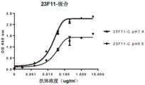
In certain embodiments, the binding of anti-PD-L1 antibodies and antigen-binding fragments thereof to an antigen (e.g., human PD-L1) is pH-dependent. pH-dependent antibodies show higher antigen binding at physiologically neutral pH (i.e., pH 7.4) than at acidic pH 5.5 or 6.0 (e.g., at endosomal pH). Such pH-dependent antibodies preferably dissociate from the antigen in the endosome. This can increase the half-life of the antibody when the antigen undergoes antigen-mediated clearance, as the pH-dependent antibody can escape antigen-mediated degradation in lysosomes by dissociating from the antigen in the endosome and recycling away from the cell. Since the pH-dependent antibody has the property of improving recirculation and prolonged serum half-life compared to the reference PD-L1 antibody, the advantages of the therapeutic pH-dependent antibody include a lower dose (a dose that does not exceed 80%, 70%, 60%, 50%, 40%, 30%, 20% or 10% of the dose of the reference PD-L1 antibody that would otherwise be required to achieve comparable in vivo therapeutic effects) or a lower frequency of administration, both antibodies achieving comparable in vivo therapeutic effects. The reference antibody was pH independent, had similar binding to PD-L1 at both acidic and neutral pH, and had similar binding to the pH dependent antibody at neutral pH. In certain embodiments, the pH-dependent antibody has the advantages described above compared to its pH-independent version with the same binding to PD-L1.
In certain embodiments, the pH-dependent anti-PD-L1 antibodies and antigen-binding fragments thereof specifically bind to human PD-L1 at neutral pH (e.g., pH7.4) more than at acidic pH (e.g., pH 6.0, 5.5, 5.0, 4.5, or 4.0). In certain embodiments, the antibody is contacted with the antibody at acidic pH5.5 under the same experimental conditionsThe binding of PD-L1 was significantly lower than its binding to PD-L1 at pH 7.4. In certain embodiments, the PD-L1 antibodies and antigen binding fragments thereof described herein bind at acidic pH up to 2%, 5%, 10%, 25%, 30%, 35%, 40%, 45%, 50%, 55%, 60%, 65%, 70%, 75%, 80%, or 85% at neutral pH (i.e., 7.4), as determined by ELISA. In certain embodiments, the pH-dependent anti-PD-L1 antibody binds to human PD-L1 at acidic pH/neutral pH with KDRatio and/or KoffThe ratio is 2, 3, 4, 5, 8, 10, 15, 20, 30, 40, or 100 or higher. In certain embodiments, the dissociation half-life (t) of the pH-dependent anti-PD-L1 antibody from human PD-L1 at an acidic pH of 25 ℃ or 37 ℃, (t)1/2) Less than 5, 4, 3, 2, 1, 0.5, 0.2, or 0.1 minutes. In certain embodiments, the pH-dependent anti-PD-L1 antibodies and antigen-binding fragments thereof described herein have a half-life of dissociation (t) at acidic pH from human PD-L1 compared to a reference PD-L1 antibody at neutral pH (t-L1) 1/2) A reduction of at least about 2, 3, 5, 8, 10, 15, 20, 30, 50, or 100 fold. In certain embodiments, exposure to the pH-dependent anti-PD-L1 antibody can maintain a reduction in the amount of PD-L1 protein in vivo for a longer period of time compared to a reference PD-L1 antibody that has similar binding to PD-L1 at neutral pH. In certain embodiments, the pH-dependent anti-PD-L1 antibodies and antigen binding fragments thereof bind to human PD-L1 with increased target organ half-life after administration of a dose compared to the same dose of a reference PD-L1 antibody. In certain embodiments, the in vivo half-life of the antibody and antigen binding fragments thereof in a target organ will be shorter if it binds similarly to PD-L1 at acidic and neutral pH. In certain embodiments, the target organ includes, but is not limited to, blood or serum, kidney, lung, pancreas, liver, gallbladder, bladder, skin, esophagus, ovary, breast, colon, rectum, stomach, spleen, or brain.
Those skilled in the art will appreciate that the CDR sequences described herein may be modified to include substitutions (or insertions or deletions) of one or more amino acid residues such that the resulting antibody has one or more improved properties (e.g., improved binding or binding affinity, increased pharmacokinetic half-life, pH sensitivity, compatibility with respect to conjugation, reduced risk of glycosylation and/or deamination at the CDR residues, and reduced immunogenicity), as well as other comparable properties (i.e., an antibody having the same CDR sequence in addition to the modifications and alterations described above), or at least substantially retains the antigen-binding properties of the parent antibody. For example, a library of antibody variants (e.g., Fab or scFv variants) can be produced and expressed using phage display technology, and subsequently screened for antibodies that bind or have binding affinity to human PD-L1. In another example, computer software can be used to mimic the binding of the antibody to human PD-L1 and identify amino acid residues on the antibody that form the binding interface, to avoid substitution of these residues to prevent binding or reduced binding affinity, or to target substitution of these residues to form stronger binding. In certain embodiments, at least one (or all) substitution in a CDR sequence is a conservative substitution.
In certain embodiments, the antibodies and antigen-binding fragments thereof comprise one or more CDR sequences having at least 80% (e.g., at least 85%, 88%, 90%, 91%, 92%, 93%, 94%, 95%, 96%, 97%, 98%, 99%) sequence identity to a sequence described herein, while retaining similar or even higher levels of binding activity or binding affinity to human PD-L1 as its parent antibody, which has substantially the same sequence except that its corresponding CDR sequence has 100% sequence identity to a sequence described herein.
In certain embodiments, the anti-PD-L1 antibodies and antigen-binding fragments thereof are chimeric. Chimeric antibodies contain one or more regions from one antibody and one or more regions from another antibody or species. In certain embodiments, at least one CDR of the chimeric anti-PD-L1 antibody is derived from one species. In certain embodiments, all CDRs are derived from another species. In certain embodiments, the variable region of the chimeric anti-PD-L1 antibody is derived from one species and linked to the constant region of an antibody of another species. Chimeric antibodies retain the binding activity or binding affinity of the parent antibody.
In certain embodiments, the anti-PD-L1 antibodies and antigen-binding fragments thereof are humanized. In certain embodiments, the humanized antibodies are derived from a non-human species and have mutations in several amino acid residues of the heavy and light chain framework and constant regions to reduce or avoid immunogenicity in humans. In certain embodiments, the variable region of a non-human species is fused to the constant region of a human antibody. In certain embodiments, the humanized antibody is generated by CDR grafting, i.e., replacing the CDRs of a human antibody with the corresponding CDRs of a non-human antibody. Thus, the humanized antibodies are less immunogenic in humans. In certain embodiments, the human framework region is substituted with one or more amino acid residues of a non-human antibody (e.g., a mouse framework region) from which the CDR sequences are derived, e.g., to improve or maintain binding activity or binding affinity. In certain embodiments, the humanized antibody retains or enhances the binding activity or binding affinity of the parent antibody.
In some embodiments, the chimeric or humanized anti-PD-L1 antibodies and antigen-binding fragments thereof comprise a heavy chain variable region selected from the group consisting of: SEQ ID NO: 21. 47, 55, 57, 59, 61, 63, 65, 67, 69, 71, 73, 75, 77, 79, 81, 83, and 85, and homologous sequences having at least 80% (e.g., at least 85%, 88%, 90%, 91%, 92%, 93%, 94%, 95%, 96%, 97%, 98%, 99%) sequence identity thereto; and/or a light chain variable region selected from the group consisting of: SEQ ID NO: 22. 48, 56, 58, 60, 62, 64, 66, 68, 70, 72, 74, 76, 78, 80, 82, 84, and 86, and homologous sequences having at least 80% (e.g., at least 85%, 88%, 90%, 91%, 92%, 93%, 94%, 95%, 96%, 97%, 98%, 99%) sequence identity thereto. These humanized antibodies retain binding activity or binding affinity to human PD-L1, preferably at a level similar to that of one of the following exemplary antibodies: 4B6, 18G4, 26F5, 21F11, 23a11, 23F11, 22C9 and chimeric antibodies thereof, humanized 4B6, humanized 23F11 and humanized 23a 11.
In certain embodiments, the PD-L1 antibodies 4B6, 26F5, 21F11, 23a11, 23F11, 22C9 and chimeric antibodies thereof, humanized 4B6, humanized 23F11, and humanized 23a11 bind to human and non-human primate PD-L1, but do not bind to mouse PD-L1. In certain embodiments, PD-L1 antibodies (e.g., 18G4) and chimeric antibodies thereof, as well as antigen-binding fragments thereof, are capable of binding to human as well as mouse PD-L1. Binding activity or binding affinity is determined by, for example, ELISA, radioligand competition binding experiments and FACS analysis.
In some embodiments, the anti-PD-L1 antibodies and antigen binding fragments thereof described herein can be present at about 10-7M or less (e.g., 10)-8M、10-9M、10-10M、10-11M、10-12M) binding affinity (K)D) Specifically binds to human PD-L1 as determined by surface plasmon resonance binding. Binding affinity can be defined by KDValues are expressed as calculated by the ratio of off-rate to on-rate (koff/kon) when the binding of antigen and antigen binding molecule reaches equilibrium. The antigen binding affinity (e.g. K)D) Can be suitably determined by suitable methods known in the art, including surface plasmon resonance binding using instruments such as Biacore (see, e.g., Murphy, M. et al, Current protocols in protein science, Chapter 19, Unit 19.14,2006).
In certain embodiments, PD-L1 antibodies and antigen-binding fragments thereof can have an EC of 0.001 μ g/ml to 1 μ g/ml (e.g., 0.001 μ g/ml to 0.5 μ g/ml, 0.001 μ g/ml to 0.2 μ g/ml, 0.001 μ g/ml to 0.1 μ g/ml, 0.01 μ g/ml to 0.2 μ g/ml, 0.01 μ g/ml to 0.1 μ g/ml, 0.01 μ g/ml to 0.05 μ g/ml, 0.01 μ g/ml to 0.03 μ g/ml, or 0.001 μ g/ml to 0.01 μ g/ml)50(i.e., 50% binding concentration) binds hPD-L1 as determined by ELISA or at an EC of 0.01 μ g/ml to 1 μ g/ml (e.g., 0.01 μ g/ml to 0.5 μ g/ml, 0.01 μ g/ml to 0.2 μ g/ml, 0.05 μ g/ml to 1 μ g/ml, 0.05 μ g/ml to 0.5 μ g/ml, or 0.05 μ g/ml to 0.2 μ g/ml)50Binds to hPD-L1 as determined by FACS. Binding of the antibody to human PD-L1 can be measured by methods well known in the art, such as ELISA, FACS, surface plasmon resonance, GST pull down, epitope tags, immunoprecipitation, Far-Western, fluorescence resonance energy transfer, time-resolved fluorescence immunoassay (TR-FIA), Radioimmunoassay (RIA), enzyme immunoassayLatex agglutination, Western blot and immunohistochemistry or other binding assays. In one illustrative example, a test antibody (i.e., a primary antibody) is capable of binding to immobilized human PD-L1 or cells expressing human PD-L1, followed by washing away unbound antibody, and introduction of a labeled secondary antibody that is capable of binding to the primary antibody and thus capable of detecting bound primary antibody. Detection may be performed using a microplate reader when immobilized PD-L1 is used, or by using FACS analysis when cells expressing human PD-L1 are used.
In certain embodiments, the antibodies and fragments thereof described herein have an IC of from 0.05 μ g/ml to 1 μ g/ml (e.g., from 0.05 μ g/ml to 0.8 μ g/ml, from 0.05 μ g/ml to 0.5 μ g/ml, or from 0.05 μ g/ml to 0.3 μ g/ml)50Inhibits binding of human PD-1 to human PD-L1 as determined by FACS, or as an IC of 0.001 μ g/ml to 0.5 μ g/ml (e.g., 0.001 μ g/ml to 0.2 μ g/ml, 0.001 μ g/ml to 0.1 μ g/ml, 0.001 μ g/ml to 0.05 μ g/ml, 0.001 μ g/ml to 0.02 μ g/ml, 0.005 μ g/ml to 0.05 μ g/ml, 0.005 μ g/ml to 0.02 μ g/ml, or 0.005 μ g/ml to 0.01 μ g/ml)50Inhibits binding of human PD-1 to human PD-L1, as determined by ELISA.
In certain embodiments, the antibodies and fragments thereof described herein block the binding of human PD-L1 to its receptor (i.e., human PD-1), thereby providing biological activity, including, for example, from activated T cells (e.g., CD 4)+T cells and CD8+T cells), induction of cytokine production, induction of activated T cells (e.g., CD 4)+T cells and CD8+T cells) and reversing the inhibitory function of T reg. Exemplary cytokines include IL-2 and IFN gamma. The term "IL-2" refers to interleukin 2, a cytokine signaling molecule in the immune system that regulates the activity of white blood cells (e.g., leukocytes). The term "interferon gamma (IFN γ)" is composed of Natural Killer (NK) cells, NK T cells, CD4 +And CD8+T cells produce a cytokine that is a key activator of macrophages and an inducer of expression of Major Histocompatibility Complex (MHC) molecules. Cytokine production can be determined using methods well known in the art, e.g., by ELISA. A method for detecting T cell proliferation comprising [ 2 ]3H]Thymidine incorporation assay and luminescenceAnd (4) measuring the cell viability.
anti-PD-L1 antibodies and antigen binding fragments thereof are specific for human PD-L1 and/or non-human primates. In certain embodiments, the antibodies and antigen-binding fragments thereof do not bind to PD-L2 (e.g., human PD-L2). For example, the binding affinity to PD-L2 is less than 15%, 10%, 9%, 8%, 7%, 6%, 5%, 4%, 3%, 2%, or 1% of the binding affinity to human PD-L1.
In certain embodiments, the antibodies and antigen-binding fragments thereof are raised to a concentration of 0.001 μ g/ml to 0.5 μ g/ml (e.g.,
EC of 0.001. mu.g/ml to 0.2. mu.g/ml, 0.001. mu.g/ml to 0.1. mu.g/ml, 0.001. mu.g/ml to 0.05. mu.g/ml, 0.001. mu.g/ml to 0.02. mu.g/ml, 0.005. mu.g/ml to 0.05. mu.g/ml, 0.005. mu.g/ml to 0.02. mu.g/ml, or 0.005. mu.g/ml to 0.01. mu.g/ml) in a pharmaceutical composition50Binding to non-human primate PD-L1, as determined by ELISA.
anti-PD-L1 antibodies and antigen binding fragments thereof are specific for human PD-L1 and/or non-human primate PD-L1. In certain embodiments, the antibodies and antigen-binding fragments thereof do not bind to mouse PD-L1. For example, the binding affinity to mouse PD-L1 is less than 15%, 10%, 9%, 8%, 7%, 6%, 5%, 4%, 3%, 2%, or 1% of the binding affinity to human PD-L1.
In certain embodiments, the antibodies and antigen binding fragments thereof do not bind to mouse PD-L1, but bind to non-human primate PD-L1 with similar binding activity or binding affinity as human PD-L1. For example, exemplary antibodies 4B6, 26F5, 21F11, 23a11, 23F11, 22C9 and chimeric antibodies thereof, humanized 4B6, humanized 23F11, and humanized 23a11, bind very little to mouse PD-L1 in conventional binding assays (e.g., ELISA or FACS analysis), while these antibodies bind similarly to human PD-L1 to non-human primate PD-L1 as measured by ELISA or FACS. In certain embodiments, antibodies and antigen binding fragments thereof (e.g., 18G4) bind to mouse PD-L1 and non-human primate PD-L1 with similar binding activity or binding affinity as human PD-L1.
In certain embodiments, the anti-PD-L1 antibodies and antigen binding fragments thereof described herein can be used in combination with immunogenic agents, such as tumor cells, purified tumor antigens, and cells, tumor vaccines transfected with genes encoding immunostimulatory cytokines. In addition, anti-PD-L1 antibodies and antigen binding fragments thereof can be included in combination therapies including standard chemotherapy and radiation therapy (e.g., radiotherapy, X-ray therapy), target-based small molecule therapies (e.g., the tyrosine kinase inhibitors imatinib, gefitinib; monoclonal antibodies, photodynamic therapy), immunotherapy (e.g., antibodies to tumor markers such as carcinoembryonic antigen, prostate specific antigen, urinary tumor associated antigen, embryonic antigen, tyrosinase (p97), gp68, TAG-72, HMFG, sialic acid Lewis antigen, MucA, MucB, PLAP, estrogen receptor, laminin receptor, erb B and p155, DLL4, Notch1, Notch2/3, Fzd7 or R-spondin (RSPO)1, RSPO2, RSPO3 or RSPO4), emerging other immune checkpoint modulator therapies (e.g., Wnt, vaccines), hormone therapy, angiogenesis inhibition (angiogenesis inhibitors), gene therapy (cell proliferation inducers, cell proliferation inhibitors, or programmed cell death modulators), palliative treatment (i.e., treatment aimed at improving the quality of care to reduce pain (e.g., morphine and oxycodone), treatment of nausea, vomiting (e.g., ondansetron and aprepitant), diarrhea, and bleeding), and surgery. In certain embodiments, antibodies and antigen-binding fragments thereof can be used as the base molecule for antibody-drug conjugates, bispecific or multivalent antibodies.
The anti-PD-L1 antibodies and antigen-binding fragments thereof described herein can be monoclonal, polyclonal, humanized, chimeric, recombinant, bispecific, labeled, bivalent, or anti-idiotypic antibodies. Recombinant antibodies are antibodies that are produced in vitro using recombinant methods rather than animals. Bispecific or bivalent antibodies are artificial antibodies having fragments of two different monoclonal antibodies, which are capable of binding two different antigens. A "bivalent" antibody and antigen-binding fragment thereof includes two antigen-binding sites. The two antigen binding sites may bind to the same antigen, or may each bind to a different antigen (in which case the antibody or antigen binding fragment is "bispecific").
In some embodiments, the anti-PD-L1 antibodies and antigen-binding fragments thereof described herein are humanized or chimeric antibodies. In certain embodiments, the humanized or chimeric antibody is prepared using recombinant methods. For example, a non-human animal may be immunized with a suitable antigen, such as human PD-L1 protein. A gene fragment encoding the variable region of an antibody that binds to an antigen was excised from the gene of a mouse monoclonal antibody, and this portion was operably linked to the constant region gene of an antibody derived from human IgG 1. This recombinant gene fragment is incorporated into an expression vector, which is then introduced into a host cell for production of a chimeric antibody (see U.S. Pat. Nos. 4,816,397; 4,816,567; 5,807,715).
A "humanized antibody" is an antibody obtained by grafting CDR genes of a non-human antibody onto human antibody genes such that the variable region framework and constant regions (if present) are derived entirely or substantially from human antibody sequences. Methods for making humanized antibodies are well known in the art (see, e.g., U.S. Pat. No. 5,225,539, U.S. Pat. No. 5,530,101, U.S. Pat. No. 6,407,213; U.S. Pat. No. 5,859,205; U.S. Pat. No. 6,881,557, EP239400, EP125023, WO90/07861, and WO 96/02576). In certain embodiments, a humanized antibody comprises a humanized heavy chain and a humanized light chain. In certain embodiments, the grafted CDR sequences in the humanized PD-L1 antibody are at least 60%, 70%, 80%, 82%, 85%, 88%, 90%, 95%, or 100% identical to the corresponding CDRs. In certain embodiments, there are no more than 3 consecutive amino acid substitutions in a CDR of the humanized PD-1 antibody. In certain embodiments, amino acid residues of the humanized PD-L1 antibody variable region framework are substituted for sequence optimization. In certain embodiments, the variable region framework sequence of the humanized PD-L1 antibody chain is at least 65%, 70%, 75%, 80%, 85%, 90%, 95%, or 100% identical to the corresponding human variable region framework sequence.
In some embodiments, the anti-PD-L1 antibodies and antigen-binding fragments thereof are camelized single domain antibodies (camelized single chain domain antibodies), bifunctional antibodies (diabodies), scfvs, scFv dimers, BsFv, dsFv, (dsFv)2, dsFv-dsFv', Fv fragmentsFragment, Fab ', F (ab')2A ds bifunctional antibody (ds diabody), a nanobody, a domain antibody, an isolated CDR, or a bivalent domain antibody.
In some embodiments, the anti-PD-L1 antibodies and antigen-binding fragments thereof further comprise an immunoglobulin constant region. In some embodiments, the immunoglobulin constant region comprises a heavy chain and/or light chain constant region. The heavy chain constant region comprises the CH1, CH1-CH2, or CH1-CH3 regions. In some embodiments, the constant region may further comprise one or more modifications to impart desired properties thereto. For example, the constant region may be modified to reduce or eliminate one or more effector functions, to improve FcRn receptor binding, or to introduce one or more cysteine residues.
In certain embodiments, the anti-PD-L1 antibodies and antigen binding fragments thereof have improved thermostability. The term "thermostability" or "thermotolerance" as used herein refers to the functional stability, rather than the thermodynamic properties, of an anti-PD-L1 antibody and to the resistance of an antibody to irreversible denaturation by thermal and/or physical/chemical manipulations, including but not limited to: heating, cooling, freezing, freeze-thaw cycles, shaking, vortexing, sonication, chemical denaturants, pH, detergents, salts, additives, proteases, or temperature. Irreversible denaturation results in irreversible unfolding of the functional conformation of the antibody, loss of biological activity, and aggregation of denatured proteins. The increase in thermal stability can be determined by measuring ligand binding or by using spectroscopy that is sensitive to unfolding at elevated temperatures, such as fluorescence, Circular Dichroism (CD) or light scattering. The PD-L1 antibodies described herein are capable of increased thermostability as determined by an increase in thermostability of at least 2 ℃, at least 5 ℃, at least 8 ℃, at least 10 ℃, at least 15 ℃, at least 20 ℃, or at least 30 ℃ in the state of a functional conformation. The thermostability of an antibody described herein can be detected, for example, by Differential Scanning Fluorescence (DSF) or Differential Scanning Calorimetry (DSC) (see He FAnd the like,J Pharm Sci.2011 Apr; 100(4) 1330-40), wherein the mid-point of thermal transition (Tm) is measured, indicating that the protein is relatively stable in liquid. In certain embodiments, the Tm of the PD-L1 antibody is highAt 74 deg.C, over 76 deg.C, over 78 deg.C, over 80 deg.C, over 82 deg.C, over 84 deg.C, over 86 deg.C, over 88 deg.C, over 90 deg.C, over 92 deg.C, over 94 deg.C, over 96 deg.C or over 98 deg.C. In certain embodiments, the PD-L1 antibody with improved thermostability is 23F11 (e.g., 23F11-H4L4, 23a11-H6L4, 23a11-H4L6, or 23a11-H6L6) having a thermal transition midpoint (Tm) above 90 ℃.
In some embodiments, the anti-PD-L1 antibody and antigen-binding fragments thereof further form an antibody-drug conjugate (ADC). It is contemplated that a variety of payloads may be attached to the antibodies or antigen-binding fragments described herein (see, e.g., "Conjugate Vaccines," constraints to Microbiology and Immunology, j.m.cruse and r.e.lewis, Jr. (eds.), Carger Press, new york (1989)). The term "payload" is used interchangeably with "drug" and these payloads may be attached to the antibody or antigen-binding fragment by covalent binding, affinity binding, intercalation, coordinate binding, complexation, binding, mixing or addition, among other means. In certain embodiments, the antibodies and antigen-binding fragments disclosed herein can be engineered to contain specific sites other than an epitope-binding moiety that can be used to bind one or more payloads, such as peptides, nucleic acid molecules, drugs, cytotoxins, polypeptides, proteins, fusion proteins, antibodies, haptens, small molecules, mimetics, synthetic drugs, inorganic molecules, organic molecules, radionuclides, and reporter groups. For example, such sites may include one or more reactive amino acid residues, such as cysteine histidine residues, to facilitate covalent attachment to the payload. In certain embodiments, the antibody may be indirectly linked to the payload through a linker, or through another payload. For example, the antibody or antigen-binding fragment may be conjugated to biotin and then indirectly conjugated to a second payload conjugated to avidin. The payload can be a reporter group or a detectable label, a pharmacokinetic modifying moiety, a purification moiety, or a cytotoxic moiety. Examples of detectable labels can include fluorescent labels (e.g., fluorescein, rhodamine, dansyl, phycoerythrin) Or texas red), an enzyme-substrate label (e.g., horseradish peroxidase, alkaline phosphatase, luciferase, glucoamylase, lysozyme, carbohydrate oxidase, or beta-D-galactosidase), a radioisotope (e.g.,123I、124I、125I、131I、35S、3H、111In、112In、14C、64Cu、67Cu、86Y、88Y、90Y、177Lu、211At、186Re、188Re、153Sm、212bi and32p, other lanthanides, luminescent labels), chromophore moieties, digoxin, biotin/avidin, DNA molecules, or gold for detection. In certain embodiments, the payload can be a pharmacokinetic modifying moiety, such as PEG, that helps to extend the half-life of the antibody. Other suitable polymers include, for example, carboxymethylcellulose, dextran, polyvinyl alcohol, polyvinyl pyrrolidone, ethylene glycol/propylene glycol copolymers, and the like. In certain embodiments, the payload can be a purification moiety such as a magnetic bead. The payload of the "cytotoxic" moiety may be any agent that is harmful to the cell or that may damage or kill the cell. Examples of cytotoxic moieties include, but are not limited to: chemotherapeutic agents, antineoplastic agents, growth inhibitory agents, drugs, toxins such as paclitaxel, cytochalasin B, gramicidin D, ethidium bromide, emetine, mitomycin, etoposide, teniposide, vincristine, vinblastine, colchicine, doxorubicin, daunorubicin, dihydroxyanthrax dione, mitoxantrone, mithramycin, actinomycin D, 1-dehydrotestosterone, glucocorticoids, procaine, tetracaine, lidocaine, propranolol, puromycin and analogs thereof, antimetabolites (e.g., methotrexate, 6-mercaptopurine, 6-thioguanine, cytarabine, 5-fluorouracil dacarbazide), alkylating agents (e.g., mechlorethamine, thiotepa chlorambucil, melphalan, carmustine (BSNU) and lomustine (CCNU), cyclophosphamide, busulfan, dibromomannitol, doxycycline, Streptozotocin, mitomycin C and cis-dichlorodiammineplatinum (II) (DDP) cisplatin), anthracyclines (e.g., daunorubicin (formerly known as daunorubicin) Daunorubicin) and doxorubicin), antibiotics (e.g., dactinomycin (previously known as actinomycin), bleomycin, mithramycin, platins (e.g., cisplatin and oxaliplatin), plant alkaloids (e.g., topoisomerase inhibitors, vinca alkaloids, taxanes and epipodophyllotoxins) and anthranilic Acid (AMC)), and antimitotics (e.g., vincristine and vinblastine). The term "load" or "drug load" or "payload" as used herein refers to the average number of drugs/payload per antibody. Drug loading may range from 1 to 20 (e.g., 1 to 15, 1 to 10, 2 to 10, 1 to 8, 2 to 6, 2 to 5, or 2 to 4) drugs per antibody (also referred to as drug to antibody ratio), as determined by suitable methods in the art, such as mass spectrometry, UV/visible spectroscopy, ELISA assay, and HPLC. In certain embodiments, the drug load is 1, 2, 3, 4, 5, 6, 7, 8, 9, or 10.
Polynucleotides and recombinant methods
The present disclosure provides isolated polynucleotides encoding anti-PD-L1 antibodies and antigen-binding fragments thereof. In certain embodiments, the isolated polynucleotide comprises one or more nucleotide sequences encoding a CDR sequence described in the disclosure.
A method of producing a monoclonal anti-PD-L1 antibody or antigen-binding fragment thereof comprising: suitable animals are immunized with human PD-L1 protein or cells producing hPD-L1. The animal may be a mouse, rat, sheep, goat, rabbit or guinea pig. Spleen or lymph nodes were used to generate hybridomas, or B cells from immunized animals were collected and PD-L1 antibody titers were measured. Polynucleotides encoding the PD-L1 antibody or antigen-binding fragment thereof are cloned using appropriate titers of hybridomas or B cell clones from immunized animals. The cloned or modified (e.g., chimeric, humanized) polynucleotides are incorporated into a suitable vector, which is then introduced into a host cell to produce the antibodies of the disclosure. Antibodies and antigen-binding fragments thereof described herein can be obtained in substantially pure and homogeneous form by culturing the host cells, followed by isolation and purification of the host cells or the culture broth (e.g., supernatant). Antibodies or antigen-binding fragments thereof can be isolated and purified using conventional methods for polypeptide purification.
In some embodiments, the isolated polynucleotide encodes a heavy chain variable region and comprises a sequence selected from the group consisting of seq id no: SEQ ID NO: 93. 95, 97, 99, 101, 103, and 105, and homologous sequences having at least 80% (e.g., at least 85%, 88%, 90%, 91%, 92%, 93%, 94%, 95%, 96%, 97%, 98%, 99%) sequence identity thereto. In some embodiments, the isolated polynucleotide encodes a light chain variable region and comprises a sequence selected from the group consisting of seq id no: SEQ ID NO: 94. 96, 98, 100, 102, 104, and 106, and homologous sequences having at least 80% (e.g., at least 85%, 88%, 90%, 91%, 92%, 93%, 94%, 95%, 96%, 97%, 98%, 99%) sequence identity thereto. In certain embodiments, the percentage of identity is derived from the degeneracy of the genetic code, while the encoded protein sequence remains unchanged.
The isolated polynucleotides encoding the anti-PD-L1 antibodies and antigen-binding fragments thereof can be inserted into vectors for further cloning (amplification of DNA) or for expression using recombinant techniques well known in the art. In another embodiment, the antibody can be made by methods of homologous recombination as are well known in the art. The DNA encoding the monoclonal antibody may be isolated and sequenced by conventional methods (e.g., oligonucleotide probes may be used which specifically bind to the genes encoding the heavy and light chains of the antibody). Various carriers can be selected. Carrier components typically include, but are not limited to, one or more of the following: signal sequences (e.g., translation signals or leader sequences), an origin of replication, one or more selectable marker genes, enhancer elements, promoters (e.g., SV40, CMV, EF-1. alpha.), and transcription termination sequences.
In some embodiments, the vector system comprises a mammalian, bacterial, yeast system, and the like, and includes plasmids such as, but not limited to, pALTER, pBAD, pcDNA, pCal, pL, pET, pGEMEX, pGEX, pCI, pCMV, pEGFP, pEGFT, pSV2, pFUSE, pVITRO, pVIVO, pMAL, pMONO, pSELECT, pUNO, pUO, Psg5L, pBABE, pWPXL, pBI, p15TV-L, pPro18, pTD, pRS420, pLexA, pACT2.2, pCDM8, pCDNA1.1/amp, pcDNA3.1, pRc/RSV, pEF-1, pCMV-SCRIPT. RTM., pFB, pSG5, pXT1, pCDEF3, pSVSPORT, pEF-1, and the like, as well as other commercially available or commercially available expression vectors. Suitable vectors may include plasmids or viral vectors (e.g., replication defective retroviruses, adenoviruses and adeno-associated viruses). The vector may maintain a single copy or multiple copies, or may be integrated into the genome of the host cell. A vector comprising a polynucleotide sequence encoding the antibody or antigen-binding fragment thereof may be introduced into a host cell for replication or gene expression. Suitable host cells for cloning or expressing the DNA in the vector are prokaryotic cells, yeast, insect cells or the above-mentioned higher eukaryotic cells. Prokaryotic cells suitable for use in the present invention include eubacteria such as, for example, gram-negative or gram-positive bacteria, for example, Enterobacteriaceae, such as Enterobacter, e.g., Escherichia coli, Enterobacter, Erwinia, Klebsiella, Proteus, Salmonella, e.g., Salmonella typhimurium, Serratia, e.g., Serratia marcescens, and Shigella, and Bacillus, such as Bacillus subtilis and Bacillus licheniformis, Pseudomonas, such as Pseudomonas aeruginosa and Streptomyces, Bacillus, Streptococcus, Streptomyces, Staphylococcus, enterococcus, Lactobacillus, lactococcus, Clostridium, Geobacillus, Bacillus marinus, Pseudomonas, Salmonella, Campylobacter, helicobacter, Flavobacterium, Clostridium, Neisseria and ureaplasma. Suitable insect cells include Drosophila Schnieder S2 cells and Sf 9. Suitable yeasts include Pichia methanolica, Pichia pastoris, Saccharomyces cerevisiae or Saccharomyces cerevisiae in general. Preferred mammalian cells include CHO cells, HEK293 cells, lymphocytes and myelomas. In certain embodiments, the antibody can be produced in bacteria when glycosylation and Fc effector function of the antibody are not required.
In addition to the above examples, many other genera, species and strains are more commonly used and are suitable for use in the present invention, such as Schizosaccharomyces pombe; kluyveromyces hosts, such as Kluyveromyces lactis, Kluyveromyces fragilis (ATCC 12,424), Kluyveromyces bulgaricus (ATCC 16,045), Kluyveromyces williamsii (ATCC 24,178), Kluyveromyces lactis (ATCC56,500), Kluyveromyces drosophilus (ATCC 36,906), Kluyveromyces thermotolerans, and Kluyveromyces marxianus; yarrowia lipolytica (EP 402,226); pichia pastoris (EP 183,070); candida species; trichoderma reesei (EP 244,234); performing Neurospora; schwann yeast, such as schwann yeast western; and filamentous fungi such as Neurospora, Penicillium, Tolypocladium, and Aspergillus, such as Aspergillus nidulans and Aspergillus niger.
The host cells provided herein that are suitable for expressing glycosylated antibodies or antigen binding fragments thereof are derived from multicellular organisms. Examples of invertebrate cells include plant and insect cells. Various baculovirus strains (bacterial strains) and variants thereof, as well as corresponding permissive insect host cells (permissive insect host cells), have been found to be derived from hosts such as: spodoptera frugiperda (caterpillar), Aedes aegypti (mosquito), Aedes albopictus (mosquito), Drosophila melanogaster (fruit fly), and Bombyx mori. A variety of viral strains for transfection are publicly available, such as the L-1 variant of Autographa californica nuclear polyhedrosis virus and the Bm-5 variant of Bombyx mori nuclear polyhedrosis virus, all of which can be used in the present invention, particularly for transfecting Spodoptera frugiperda cells. Plant cell cultures of cotton, corn, potato, soybean, petunia, tomato, and tobacco may also be used as hosts.
However, vertebrate cells are of paramount importance, and expansion of cultured vertebrate cells (tissue culture) has become a routine procedure. As examples of mammalian host cells which may be used, there are SV40 transformed monkey kidney cell CV1 line (COS-7, ATCC CRL 1651); human embryonic kidney cell lines (293 or 293 cell subclones in suspension culture, Graham et al, J.Gen Virol.36:59 (1977)); baby hamster kidney cells (BHK, ATCC CCL 10); chinese hamster ovary cells/-DHFR (CHO, Urlaub et al, Proc. Natl. Acad. Sci. USA 77:4216 (1980)); mouse testicular support cells (TM4, Mather, biol. reprod.23:243-251 (1980)); monkey kidney cells (CV1 ATCC CCL 70); vero cells (VERO-76, ATCC CRL-1587); human cervical cancer cells (HELA, ATCC CCL 2); canine kidney cells (MDCK, ATCC CCL 34); buffalo rat hepatocytes (BRL 3A, ATCC CRL 1442); human lung cells (W138, ATCC CCL 75); human hepatocellular carcinoma cells (Hep G2, HB 8065); mouse mammary tumor (MMT060562, ATCC CCL 51); TRI cells (Mather et al, Annals N.Y.Acad.Sci.383:44-68 (1982)); MRC 5 cells; FS4 cells; PC 12; mouse embryonic fibroblast cell line (3T 3); NSO myeloma cells (murine myeloma cell line that cannot endogenously produce any functional immunoglobulin chain). Different host cells have various characteristics and mechanisms for post-translational processing and modification of proteins and gene products. Thus, an appropriate cell line may be selected as a host cell to ensure proper modification and processing (e.g., primary transcript, glycosylation and phosphorylation) of the expressed antibody. In some preferred embodiments, the host cell is a HEK293T cell. In some preferred embodiments, the host cell is a CHO cell.
Host cells are transformed with the above-described expression or cloning vectors that produce anti-PD-L1 antibodies and cultured in conventional nutrient media modified as appropriate for inducing promoters, selecting transformed cells, or amplifying genes encoding the sequences of interest. In certain embodiments, the vector may be transferred into a host cell by methods well known in the art, such as transformation, electroporation, calcium phosphate treatment, lipofection. In certain embodiments, transfecting the vector into a eukaryotic cell comprises calcium phosphate co-precipitation, microinjection, electroporation, lipofection, and viral infection. The eukaryotic host cell may be co-transformed with a second polynucleotide encoding an antibody. In certain embodiments, the host cell containing the transfection vector is capable of transiently expressing the anti-PD-L1 antibody.
The host cells of the invention used to produce the antibodies or antigen-binding fragments thereof can be cultured in a variety of media. Commercially available culture media such as Ham's F10(Sigma), Minimum Essential Medium (MEM) (Sigma), RPMI-1640(Sigma) and Dulbecco's Modified Eagle's Medium (DMEM) (Sigma), Luria Broth (LB) and Terrific Broth (TB) can be used for culturing the host cells. In addition, any of the methods described in Ham et al, meth.Enz.58:44(1979), Barnes et al, anal. biochem.102:255(1980), U.S. Pat. No. 4,767,704; 4,657,866, respectively; 4,927,762, respectively;
4,560,655, respectively; or 5,122,469; WO 90/03430; WO 87/00195; or the medium described in U.S. patent application Re.30,985, can be used as the medium for the host cells. These media may be supplemented with the necessary hormones and/or other growth factors (such as insulin, transferrin or epidermal growth factor), salts (such as sodium chloride, calcium chloride, magnesium chloride and phosphate), buffers (such as HEPES), nucleotides (such as adenosine and thymine), antibiotics (such as gentamicin), trace elements (defined as inorganic compounds, usually in the micromolar range, at final concentrations), and glucose or an equivalent energy source. The medium may also contain any other necessary additives at appropriate concentrations known in the art. The conditions of the medium, such as temperature, pH, and the like, which have been previously used to select host cells for expression, are well known to those of ordinary skill.
When using recombinant techniques, the antibodies and antigen-binding fragments thereof can be produced intracellularly, in the periplasmic space, or directly secreted into the culture medium. If the antibody is produced intracellularly, the particulate debris of the host cells or lysed fragments is first removed, for example, by centrifugation or sonication. Carter et al, Bio/Technology 10:163-167(1992) describe methods for isolating antibodies secreted into the membrane space of E.coli walls. Briefly, the cell paste (cell paste) was thawed in the presence of sodium acetate (pH 3.5), EDTA, and phenylmethanesulfonyl fluoride (PMSF) for about 30 minutes or more. Cell debris was removed by centrifugation. If the antibody and antigen binding fragment thereof is secreted into the culture medium, the supernatant of the expression system is typically first concentrated using a commercially available protein concentration filter, such as an Amicon or Millipore Pellicon ultrafiltration unit. Protease inhibitors such as PMSF may be added in any of the foregoing steps to inhibit proteolytic degradation, as well as antibiotics to prevent the growth of adventitious contaminants.
The antibody produced from the cell can be purified by a purification method such as hydroxyapatite chromatography, hydrophobic chromatography, reverse phase chromatography, adsorption chromatography, filtration, ultrafiltration, solvent precipitation, solvent extraction, distillation, SDS-polyacrylamide gel electrophoresis, dialysis, DEAE-cellulose ion exchange column, ammonium sulfate precipitation, salting out, immunoblotting, etcPrecipitation, isoelectric focusing, recrystallization, and affinity chromatography, with affinity chromatography being the preferred purification technique. The class of the antibody and the presence of the Fc domain of any immunoglobulin in the antibody determines whether protein a is suitable as an affinity ligand. Protein A can be used to purify antibodies based on human gamma 1, gamma 2 or gamma 4 heavy chains (Lindmark et al, J.Immunol. meth.62:1-13 (1983)). Examples of protein A columns include Hyper D, POROS and Sepharose FF (GE Healthcare Biosciences). Protein G is applicable to all murine isoforms and human gamma 3(Guss et al, EMBO J.5: 15671575 (1986)). Agarose is the most commonly used affinity ligand attachment matrix, but other matrices may be used. Mechanically stable matrices such as controlled pore glass or poly (styrene) benzene can achieve faster flow rates and shorter processing times than can be achieved with agarose. If the antibody contains a CH3 domain, it can be purified using Bakerbond ABX. TM. resin (J.T.Baker, Phillipsburg, N.J.). Other techniques for protein purification may also be determined depending on the antibody to be obtained, such as fractionation on ion exchange columns, ethanol precipitation, reverse phase HPLC, silica gel chromatography, heparin sepharose based on anion or cation exchange resins TMGel chromatography (e.g., polyaspartic acid column), chromatofocusing, SDS-PAGE, and ammonium sulfate precipitation.
After any preliminary purification steps, the mixture containing the antibody of interest and impurities can be treated by low pH hydrophobic interaction chromatography, with a wash buffer at a pH of about 2.5-4.5, preferably at low salt concentrations (e.g., from about 0 to 0.25M salt concentration).
Reagent kit
The present disclosure provides kits comprising an anti-PD-L1 antibody or antigen-binding fragment thereof, or a pharmaceutical composition comprising an anti-PD-L1 antibody or antigen-binding fragment thereof described herein. In some embodiments, the kit is used to detect the presence or level of PD-L1 in a biological sample. The biological sample may comprise cells or tissue.
In some embodiments, the anti-PD-L1 antibody or antigen-binding fragment thereof contained in the kit is coupled to a detectable label (e.g., a fluorescent, radioactive, or enzymatic label). In certain other embodiments, the kit comprises an unlabeled anti-PD-L1 antibody or antigen-binding fragment thereof, or a pharmaceutical composition comprising an unlabeled anti-PD-L1 antibody or antigen-binding fragment thereof, and further comprises a second labeled antibody capable of binding to the unlabeled anti-PD-L1 antibody. The kit may further comprise means for detecting the label (e.g., a filter bank for detecting fluorescent labels, an enzyme substrate for enzyme labels, etc.). Kits may contain other reagents and buffers for performing particular methods. The kit may further comprise instructions for use, as well as a package separating the components of the kit. In certain embodiments, the kit comprises an immunoassay for detecting the PD-L1 antibody.
In certain embodiments, the anti-PD-L1 antibody or antigen-binding fragment thereof contained in the kit is associated with a substrate or device for a sandwich assay (e.g., ELISA) or for an immunoimaging assay. Useful substrates or devices may be, for example, enzyme plates and test strips.
In certain embodiments, kits for detecting PD-L1 protein levels are provided. In some embodiments, the kit is for predicting, diagnosing, preventing, or treating a condition associated with PD-L1.
Pharmaceutical compositions and methods of treatment
The present disclosure further provides pharmaceutical compositions comprising an anti-PD-L1 antibody or antigen-binding fragment thereof and one or more pharmaceutically acceptable carriers.
Pharmaceutically acceptable carriers for use in the pharmaceutical compositions disclosed herein can include, for example, pharmaceutically acceptable liquid, gel or solid carriers, aqueous media, non-aqueous media, antimicrobial substances, isotonic substances, buffers, antioxidants, anesthetics, suspending/dispersing agents, chelating agents, diluents, adjuvants or nontoxic auxiliary substances, other components well known in the art, or various combinations thereof.
Suitable components may include, for example, antioxidants, humectants, fillers, binders, disintegrants, buffers, preservatives, lubricants, flavoring agents, thickeners, colorants, emulsifiers, or stabilizers such as sugars and cyclodextrins. Suitable antioxidants may include, for example, methionine, ascorbic acid, EDTA, sodium thiosulfate, platinum, catalase, citric acid, cysteine, thioglycerol, thioglycolic acid, mercaptosorbitol, butyl methyl anisole, butylated hydroxytoluene, and/or propyl gallate. As disclosed herein, the inclusion of one or more antioxidants, such as methionine, in a composition comprising an antibody or antigen-binding fragment thereof disclosed herein will reduce oxidation of the antibody or antigen-binding fragment thereof. The reduction in oxidation prevents or reduces the reduction in binding activity or binding affinity, thereby improving antibody stability and extending shelf life. Thus, in certain embodiments, the invention provides compositions comprising one or more of the antibodies or antigen-binding fragments thereof and one or more antioxidants, such as methionine. The present invention further provides methods of preventing oxidation, extending shelf life and/or increasing activity of an antibody or antigen-binding fragment thereof provided herein by admixing the antibody or antigen-binding fragment thereof with one or more antioxidants, such as methionine. Suitable humectants include ethylene glycol, glycerin, or sorbitol. Suitable lubricants include, for example, cetyl esters wax, hydrogenated vegetable oils, magnesium stearate, methyl stearate, mineral oil, polyoxyethylene-polyoxypropylene copolymers, polyethylene glycols, polyvinyl alcohols, sodium lauryl sulphate or white wax, or mixtures of two or more thereof. Suitable emulsifiers include carbomer, polyoxyethylene-20-stearyl ether, cetostearyl alcohol, cetyl alcohol, cholesterol, diethylene glycol stearate, glyceryl stearate, hydroxypropyl cellulose, hydroxypropyl methylcellulose, lanolin, polyoxyethylene lauryl ether, methylcellulose, polyoxyethylene stearate, polysorbate, propylene glycol monostearate, sorbitan ester or stearic acid.
Further, pharmaceutically acceptable carriers can include, for example, aqueous media such as sodium chloride injection, ringer's solution injection, isotonic dextrose injection, sterile water injection, or dextrose and lactate injection, non-aqueous media such as: fixed oils of vegetable origin, cottonseed oil, corn oil, sesame oil or peanut oil, antibacterial substances at bacteriostatic or fungistatic concentrations, isotonic agents such as: sodium chloride or glucose, buffers such as: phosphate or citrate buffers, antioxidants such as: sodium bisulfate, local anesthetics such as: procaine hydrochloride, suspending and dispersing agents such as: sodium carboxymethylcellulose, hydroxypropylmethylcellulose or polyvinylpyrrolidone, emulsifiers such as: polysorbate 80 (tween-80), chelating agents such as EDTA (ethylenediaminetetraacetic acid) or EGTA (ethylene glycol tetraacetic acid), ethanol, polyethylene glycol, propylene glycol, sodium hydroxide, hydrochloric acid, citric acid, or lactic acid. Antibacterial agents as carriers may be added to the pharmaceutical composition in multi-dose containers, including phenolics or cresols, mercurials, benzyl alcohol, chlorobutanol, methyl and propyl parabens, thimerosal, benzalkonium chloride, and benzalkonium chloride. Suitable excipients may include, for example, water, salt, glucose, glycerol or ethanol. Suitable non-toxic auxiliary substances may include, for example, wetting or emulsifying agents, pH buffering agents, stabilizers, solubilizers, or substances such as sodium acetate, sorbitan laurate, triethanolamine oleate or cyclodextrins.
The pharmaceutical composition may be a liquid solution, suspension, emulsion, foam, pill, capsule, tablet, sustained release formulation, ointment, cream, paste, gel, spray, aerosol or powder. Oral formulations may include standard carriers such as pharmaceutical grades of mannitol, lactose, starch, magnesium stearate, polyvinylpyrrolidone, sodium saccharine, cellulose, magnesium carbonate, and the like.
In certain embodiments, the pharmaceutical composition is formulated as an injectable composition. Injectable pharmaceutical compositions may be prepared in any conventional form, for example, liquid solvents, suspending agents, emulsifying agents, or solid forms suitable for the production of liquid solvents, suspending agents or emulsifying agents. Injectable formulations can include sterile and/or pyrogen-free solutions ready for injection, sterile dried soluble materials such as lyophilized powders for injection, including subcutaneous tablets, sterile suspensions ready for injection, sterile dried insoluble products ready for use in association with a vehicle, and sterile and/or pyrogen-free emulsions. The solvent may be aqueous or non-aqueous.
In certain embodiments, the unit dose of parenteral formulation is packaged in an ampoule, vial, or syringe with a needle. It is well known in the art that all formulations for parenteral administration should be sterile pyrogen-free.
In certain embodiments, sterile lyophilized powders can be prepared by dissolving an antibody or antigen-binding fragment thereof disclosed herein in an appropriate solvent. The solvent may contain stability-enhancing excipients or other pharmacological ingredients of the powdered or reconstituted solution prepared from the powder. Suitable excipients include, but are not limited to, water, glucose, sorbitol, fructose, corn syrup, xylitol, glycerol, glucose, sucrose, or other suitable materials. The solvent may contain a buffer, such as citric acid buffer, sodium or potassium phosphate buffer or other buffers known to those skilled in the art, and in one embodiment, the pH of the buffer is neutral. The dissolution is then subjected to subsequent sterile filtration under standard conditions well known in the art, and then lyophilized to produce the desired formulation. In one embodiment, the resulting solution is dispensed into vials for lyophilization. Each vial may contain a single dose or multiple doses of the anti-PD-L1 antibody or antigen-binding fragment thereof or composition thereof. The loading per vial may be slightly higher than that required for each dose or for multiple doses (e.g., 10% excess), thereby ensuring accurate sampling and administration. The lyophilized powder may be stored under appropriate conditions, such as in the range of about 4 ℃ to room temperature.
And (4) reconstituting the lyophilized powder with water for injection to obtain a preparation for parenteral administration. In one embodiment, the lyophilized powder can be reconstituted by addition to sterile and/or pyrogen-free water or other suitable liquid carrier. The precise amount will be determined by the therapy chosen and may be determined empirically.
Also provided are therapeutic methods for treating a PD-L1-associated condition, comprising administering to an individual a therapeutically effective amount of a PD-L1 antibody or antigen-binding fragment thereof described herein, thereby treating or preventing a condition or disorder associated with PD-L1. In another embodiment, there is also provided a method of treating a condition in a subject who would benefit from an upregulated immune response, comprising administering to the subject in need thereof a therapeutically effective amount of a PD-L1 antibody as described herein.
The therapeutically effective dose of the antibody or antigen-binding fragment thereof provided herein, when used alone or in combination with other agents (e.g., chemotherapeutic agents), depends on a variety of factors well known in the art, such as the type of disease being treated, the type of antibody, body weight, age, past medical history, current therapy, the health of the individual, the potential for immunological conditions and cross-reactivity, allergies, sensitivity, and side effects, as well as the route and type of administration, the severity and progression of the disease, and the judgment of the attending physician or veterinarian. In certain embodiments, an antibody or antigen-binding fragment described herein can be administered at a therapeutically effective dose of about 0.01mg/kg to about 100mg/kg (e.g., about 0.01mg/kg, about 0.3mg/kg, about 0.5mg/kg, about 1mg/kg, about 3mg/kg, about 5mg/kg, about 10mg/kg, about 15mg/kg, about 20mg/kg, about 25mg/kg, about 30mg/kg, about 35mg/kg, about 40mg/kg, about 45mg/kg, about 50mg/kg, about 55mg/kg, about 60mg/kg, about 65mg/kg, about 70mg/kg, about 75mg/kg, about 80mg/kg, about 85mg/kg, about 90mg/kg, about 95mg/kg, or about 100mg/kg) one or more times daily. In certain embodiments, the antibody or antigen-binding fragment is administered at a dose of about 50mg/kg or less, and in certain embodiments at a dose of 20mg/kg or less, 10mg/kg or less, 3mg/kg or less, 1mg/kg or less, 0.3mg/kg or less, or 0.1mg/kg or less. In certain embodiments, the dosage administered may vary over the course of treatment. For example, in certain embodiments, the initial administered dose may be higher than the subsequent administered dose. In certain embodiments, the dosage administered may be varied during the course of treatment according to the individual's response.
The dosage regimen may be adjusted to achieve the optimal desired response (e.g., therapeutic response). In certain embodiments, an antibody or antigen-binding fragment described herein is administered to an individual at once or over a series of treatments. In certain embodiments, the antibodies or antigen-binding fragments described herein are administered to an individual by one or more separate administrations, or by continuous infusion, depending on the type and severity of the disease. Guidance may be found, for example, in U.S. patent nos. 4,657,760; 5,206,344, respectively; 5,225,212.
The antibodies and antigen-binding fragments disclosed in the present invention may be administered by any route of administration known in the art, such as parenteral (e.g., subcutaneous, intraperitoneal, intravenous, including intravenous, intramuscular, or intradermal) or parenteral (e.g., oral, intranasal, intraocular, sublingual, rectal, or topical) routes of administration.
In certain embodiments, the antibodies and antigen-binding fragments disclosed herein can be administered in a controlled release manner. Controlled release parenteral formulations can be formulated as implants, oily injections, or particulate Systems (e.g., microspheres, microparticles, microcapsules, nanocapsules, nanospheres, and nanoparticles) (see Banga, A.J., Therapeutic Peptides and Proteins: Formulation, Processing, and Delivery Systems, technical Publishing Company, Inc., Lancaster, Pa., (1995); Kreuter, J., Colloidal Drug Delivery Systems, J.Kreuter, Marcel Dekkker, Inc., New York, pp.219-1994; time & Tabib, Treationon Controlled Drug Delivery, A.Kydoeus, Marcel Dekker, Inc., New York, 315.339, pp.1992)). In certain embodiments, the PD-L1 antibodies and antigen-binding fragments disclosed herein may be administered in a degradable or non-degradable polymeric matrix (see Langer, Accounts Chem. Res.26: 537) 542, 1993).
The condition associated with PD-L1 may be an immune-related disease or disorder. In certain embodiments, the condition is a solid tumor, a hematologic disease, an infectious disease, an autoimmune disease, or a fibrotic disease. In certain embodiments, solid tumors include, for example, non-small cell lung cancer (squamous/non-squamous), small cell lung cancer, renal cell carcinoma, colorectal cancer, colon cancer, ovarian cancer, breast cancer (including basal breast cancer, ductal carcinoma, and lobular breast cancer), pancreatic cancer, gastric cancer, bladder cancer, esophageal cancer, mesothelioma, melanoma, head and neck cancer, thyroid cancer, sarcoma, prostate cancer, glioblastoma, cervical cancer, thymus cancer, melanoma, myeloma, mycosis, merkel cell carcinoma, hepatocellular carcinoma (HCC), fibrosarcoma, myxosarcoma, liposarcoma, chondrosarcoma, osteogenic sarcoma and other sarcomas, synovioma, mesothelioma, ewing's tumor, leiomyosarcoma, rhabdomyosarcoma, lymphoid malignancy, basal cell carcinoma, adenocarcinoma, sweat gland carcinoma, medullary thyroid carcinoma, papillary thyroid carcinoma, pheochromocytoma, colorectal carcinoma, or a, Sebaceous gland carcinoma, papillary adenocarcinoma, medullary carcinoma, bronchogenic carcinoma, liver cancer, bile duct cancer, choriocarcinoma, Wilms' tumor, cervical cancer, testicular tumor, seminoma. Hematological disorders include, for example, Classical Hodgkin's Lymphoma (CHL), primary mediastinal large B-cell lymphoma, T cell/histiocyte-rich B-cell lymphoma, acute lymphocytic leukemia, acute myelocytic leukemia, acute myelogenous leukemia and myeloblasts, promyelocytes, myelomonocytic cells, monocytic leukemia and erythroleukemia, chronic myelogenous (myelocytic) leukemia, chronic myelogenous leukemia, chronic lymphocytic leukemia, polycythemia vera, mast cell-derived tumors, EBV-positive and negative PTLD, diffuse large B-cell lymphoma (DLBCL), plasmablast lymphoma, extranodal NK/T-cell lymphoma, nasopharyngeal carcinoma, primary effusion lymphoma associated with HHV8, non-Hodgkin's lymphoma, multiple myeloma, Waldenstroma macroglobulinemia, Hodgkin's lymphoma, multiple myeloma, macroglobulinemia, Hodgkin's lymphoma, lymphomas, T cell-rich B-cell lymphomas, acute myelocytic leukemia and myelogenous leukemia, polycythemic leukemia, and erythrocytosis, Heavy chain disease, myelodysplastic syndrome, hairy cell leukemia and myelodysplasia, central nervous system tumors (CNS), such as primary CNS lymphoma, spinal axis tumors, brain stem glioma, astrocytoma, medulloblastoma, craniopharyngioma, ependymoma, pinealoma, hemangioblastoma, acoustic neuroma, oligodendroglioma, meningioma, melanoma, neuroblastoma and retinoblastoma. In certain embodiments, the tumors and cancers are metastatic, particularly metastatic tumors that express PD-L1. In certain embodiments, the tumor is melanoma or colon cancer.
In certain embodiments, conditions and disorders associated with PD-L1 include autoimmune or inflammatory diseases such as Systemic Lupus Erythematosus (SLE), intestinal mucositis, wasting diseases associated with colitis, multiple sclerosis, viral infections, rheumatoid arthritis, osteoarthritis, crohn's disease and inflammatory bowel disease, psoriasis, systemic scleroderma, autoimmune diabetes, and the like. In certain embodiments, the condition or disorder associated with PD-L1 includes an infectious disease, such as a fungal infection, a parasite/protozoan infection, or a chronic viral infection, for example coccidiomycosis, histoplasmosis, onychomycosis, aspergillosis, blastomycosis, candidiasis albicans, paracoccidioidomycosis, microsporidiosis, acanthamoeba keratitis, amebiasis, ascariasis, babesiosis, balantiosis, belinostosis, trypanosomiasis, sinoclonorchidosis, trypanosomiasis, cryptosporidiosis, schizocephalia, madinemia, echinococcosis, elephantiasis, enterobiasis, fasciolosis, fasciolopsiasis, filariasis, giardiasis, fasciolosis, isocystis, schistosomiasis, leishmaniasis, lyme disease, malaria, metazoiasis, retrogenital infections, myiasis, onchocerciasis, Pediculosis, scabies, schistosomiasis, narcolepsy, strongyloides, taeniasis, toxocariasis, toxoplasmosis, trichinosis, helminthiasis, Hepatitis B Virus (HBV) infection, Hepatitis C Virus (HCV) infection, herpes virus, Epstein-Barr virus, HIV, cytomegalovirus, herpes simplex virus type I, herpes simplex virus type II, human papilloma virus, adenovirus, human immunodeficiency virus type I, human immunodeficiency virus type II, herpesvirus epidemics associated with Kaposi's sarcoma, torque tenovirus (Torqueteno virus), human T-lymphocyte virus type I, human T-lymphocyte virus type II, varicella zoster, JC virus or BK virus. In certain embodiments, conditions associated with PD-L1 include fibrotic diseases, such as glomerulonephritis, neural scarring, skin scarring, pulmonary fibrosis, radiation-induced fibrosis, liver fibrosis, bone marrow fibrosis.
Application method
The present disclosure also provides methods of using anti-PD-L1 antibodies or antigen-binding fragments thereof.
In some embodiments, the present disclosure provides a method of treating a condition associated with PD-L1 in an individual, comprising administering to the individual a therapeutically effective amount of an antibody or antigen-binding fragment thereof. In certain embodiments, the individual has been identified as having a disease or condition that is likely to respond to a PD-L1 antagonist. In certain embodiments, the present disclosure provides methods of preventing, detecting, or diagnosing a condition associated with PD-L1, the method comprising contacting a biological sample obtained from a subject suspected of having or having a condition associated with PD-L1, or at risk for a condition associated with PD-L1, with a PD-L1 antibody or antigen-binding fragment thereof described herein, and determining the level of PD-L1 antibody or antigen-binding fragment thereof that binds to PD-L1 in the biological sample.
For treatment of a condition associated with PD-L1, the individual is detected as positive for PD-L1 expression, or as an elevated level of PD-L1 expression. Various methods can be used to determine the presence or level of PD-L1 in a test biological sample from an individual. For example, a test biological sample can be exposed to an anti-PD-L1 antibody or antigen-binding fragment thereof, which binds to PD-L1 protein and detects its expression. Alternatively, PD-L1 can also be detected at the nucleic acid expression level using methods such as qPCR, reverse transcription PCR, microarray, SAGE, FISH, and the like. In some embodiments, the test sample is derived from cancer cells or tissue (e.g., biopsy tissue from an organ), tumor infiltrating immune cells, or a bodily fluid (e.g., blood or serum). In certain embodiments, the presence or up-regulation of PD-L1 in the test biological sample indicates a possible response. The term "up-regulated" as used herein refers to a total increase in the level of PD-L1 protein detected in a test sample of no less than 10%, 15%, 20%, 25%, 30%, 35%, 40%, 45%, 50%, 55%, 60%, 65%, 70%, 75%, 80% or more, as compared to the level of PD-L1 protein in a reference sample detected using the same antibody. The reference sample may be a control sample obtained from a healthy or disease-free individual, or a healthy or disease-free sample obtained from an individual from whom the test sample is derived, or a sample obtained from the same individual at an earlier point in time during the treatment of the condition. For example, the reference sample can be a disease-free sample adjacent to or in the vicinity of the test sample (e.g., tumor).
The antibodies or antigen-binding fragments disclosed herein can be administered alone or in combination with one or more other therapeutic means or substances. For example, the antibodies or antigen-binding fragments disclosed herein may be administered in combination with a second therapy, such as radiation therapy, chemotherapy, targeted therapy, gene therapy, immunotherapy, hormonal therapy, angiogenesis inhibition, palliative therapy, surgery for treating cancer (e.g., tumor resection), one or more antiemetics or other treatments for complications resulting from chemotherapy, or a second therapeutic agent for treating cancer or any medical condition mediated by PD-L1, such as another antibody, a therapeutic polynucleotide, a chemotherapeutic agent, an anti-angiogenic agent, a cytokine, another cytotoxic agent, a growth inhibitory agent. In certain such embodiments, the antibodies and antigen-binding fragments disclosed herein, when used in combination with one or more additional therapeutic agents, may be administered concurrently with the one or more additional therapeutic agents, and in certain such embodiments, the antibodies and antigen-binding fragments and the additional therapeutic agents may be administered as part of the same pharmaceutical composition. However, antibodies and antigen-binding fragments that are "used in combination" with other therapeutic agents need not be administered simultaneously with the therapeutic agent or in the same composition as the therapeutic agent. The meaning of "in combination" in the present invention also includes antibodies and antigen-binding fragments that are administered before or after another therapeutic agent, even if the antibody or antigen-binding fragment thereof is administered by a different mode of administration than the second therapeutic agent. Where possible, other therapeutic agents for use in combination with the antibodies or antigen-binding fragments thereof disclosed herein may be administered by Reference to the product specification for the other therapeutic agent, or by Reference to surgeon's docket No. 2003(Physicians' Desk Reference, 57 th edition; Medical Economics Company; ISBN: 1563634457; 57 th edition (11 months 2002)), or by Reference to other methods known in the art. In certain embodiments, the anti-angiogenic agent administered in combination with the PD-L1 antibody provided herein is a monoclonal antibody, such as bevacizumab, for anti-angiogenic therapy anti-VEGF antibody, IMC-1C11 or DC101(VEGFR-2 antibody), mF4-31C1(VEGFR-3 antibody) and Vitaxin (integrin. alpha.)vβ3Antibodies).
In certain embodiments, the therapeutic agent may induce or elicit an immune response against the cancer. For example, tumor vaccines can be used to induce an immune response to a certain tumor or cancer. Cytokine therapy can also be used to enhance tumor antigen presentation by the immune system. Examples of cytokine therapy include, but are not limited to, interferons such as interferon- α, - β, and- γ, colony stimulating factors such as macrophage-CSF, granulocyte macrophage CSF, and granulocyte-CSF, insulin growth factor (IGF-1), Vascular Endothelial Growth Factor (VEGF), Transforming Growth Factor (TGF), Fibroblast Growth Factor (FGF), interleukins (e.g., IL-1 α, IL-2, IL-3, IL-4, IL-5, IL-6, IL-7, IL-8, IL-9, IL-10, IL-11, and IL-12), tumor necrosis factors (e.g., TNF- α and TNF- β), or combinations thereof. Agents that inactivate immunosuppressive targets may also be used, such as IL-1-antagonists (IL-1A), VEGFR2 antagonists (e.g., vatalanib, sunitinib, sorafenib, pazopanib), TGF- β inhibitors, FGFR antagonists, Platelet Derived Growth Factor Receptor (PDGFR) antagonists (e.g., imatinib, sunitinib, sorafenib, pazopanib), epidermal growth factor receptor (EGFR, ErbB) antagonists (e.g., gefitinib, lapatinib, canertinib), IL-10 inhibitors, Fas ligand inhibitors, or any combination thereof. Another group of agents includes agents that activate an immune response to a tumor or cancer cell, such as agents that enhance T cell activation (e.g., T cell co-stimulatory molecules (e.g., CTLA-4, ICOS, and OX-40) agonists) and agents that enhance dendritic cell function and antigen presentation.
Screening and evaluation of anti-PD-L1 antibody in vivo efficacy
To screen and/or evaluate the in vivo effectiveness (e.g., binding activity or binding affinity) of the PD-L1 antibody or antigen-binding fragment thereof, a non-human animal inoculated with non-human tumor cells expressing human PD-L1 protein was generated. In certain embodiments, the non-human tumor cell is a rodent (e.g., mouse, rat, hamster, etc.) cell. In certain embodiments, the non-human tumor cell is a melanoma cell line (B16) or a mouse colon cancer cell line (MC 38). The non-human tumor cell can comprise a polynucleotide encoding a human PD-L1 protein in which an endogenous non-human PD-L1 gene segment is inactivated. Inactivation of the target gene may be caused by gene disruption, mutation, addition, gene silencing (e.g., RNAi gene antisense) or gene deletion (e.g., partial or complete deletion of the coding sequence or coding sequences comprising flanking regions) of the protein coding sequence located at the endogenous locus, thereby eliminating or minimizing expression of the non-human target gene, or producing a functionally inactivated/truncated polypeptide that does not bind its ligand. The flanking regions of the coding sequence at the 5 'and 3' ends may be in the range of about 1bp to about 500bp, or the flanking regions may be greater than 500bp but will not include the inactivation of other genes according to the present disclosure. As used herein, "gene disruption" refers to the addition of one or more nucleotides or amino acids to a naturally occurring sequence. Gene disruption may be the addition or insertion of a marker/reporter gene to the protein coding sequence. In certain embodiments, the inactivation is irreversible. In certain embodiments, inactivation results in no detectable activity of the target gene or gene product in the cell. The target gene is inactivated using methods suitable in the art, such as homologous recombination, RNA interference (RNAi) or CRISPR/Cas9 systems. In certain embodiments, a human PD-L1 gene segment encoding a human PD-L1 protein is operably inserted into the endogenous non-human PD-L1 locus (gene replacement). In certain embodiments, the inserted polynucleotide encoding human PD-L1 is integrated at a random, non-specific location (gene enhancement). In yet another embodiment, the polynucleotide encoding human PD-L1 can be stably maintained in the cell as an episome, e.g., in the form of an independent, episomal DNA segment, and the episomal DNA is replicated independently of or in synchrony with the host cell cycle.
Non-human tumor cells in which the target gene is completely inactivated can be identified by FACS analysis of cell surface expression or detection of transcription of the target gene. The polynucleotide encoding human PD-L1 protein can be introduced into tumor cells by any suitable method known in the art (e.g., homologous recombination and transgenic methods) via any suitable expression vector (e.g., lentiviral vector) as described in the present disclosure. Target gene transcription can be detected by FACS analysis of cell surface expression or by any suitable method in the art, identifying tumor cells in which the target gene is completely inactivated and/or expresses the introduced gene of interest.
A method of screening or assessing the effectiveness of an antibody or antigen-binding fragment thereof against human PD-L1 in vivo comprises inoculating a non-human animal with a tumor cell comprising a polynucleotide encoding a human PD-L1 protein, contacting the antibody with a tumor cell in the non-human animal and determining the tumor burden of the tumor cell. As used herein, "tumor burden" is the amount of tumor cells in an individual, which can be determined by tumor volume, number, or weight. The tumor cell can be a solid tumor cell or a non-solid tumor cell (e.g., a blood cell). The tumor cell can be a human tumor cell or a non-human tumor cell. In certain embodiments, tumor cells are inoculated into a syngeneic non-human animal to generate a syngeneic tumor model. In certain embodiments, the tumor cells are cultured for several passages prior to inoculation into the non-human animal. In certain embodiments, the non-human animal has a human immune system. In certain embodiments, the in vivo effectiveness of a PD-L1 antibody, or antigen-binding fragment thereof, is determined by comparing growth inhibition of tumor volume in a non-human animal administered the PD-L1 antibody to a control.
The invention also includes the following embodiments:
embodiment 1 an isolated PD-L1 antibody comprising heavy chain HCDR1, HCDR2 and HCDR3 and light chain LCDR1, LCDR2 and LCDR3 sequences, wherein
The HCDR1 sequence is TYWX1H (SEQ ID NO: 1) or a homologous sequence having at least 80% sequence identity thereto;
the HCDR2 sequence is MIQPNSGGTKYNX2X3FKX4(SEQ ID NO: 2) or a homologous sequence having at least 80% sequence identity thereto;
the HCDR3 sequence is GAGTVDYFDY (SEQ ID NO: 3) or a homologous sequence having at least 80% sequence identity thereto;
the LCDR1 sequence is RASESVDIYGNSFMH (SEQ ID NO: 4) or a homologous sequence having at least 80% sequence identity thereto;
the LCDR2 sequence is RASNLES (SEQ ID NO: 5) or a homologous sequence having at least 80% sequence identity thereto;
the LCDR3 sequence is X5QSX6X7DPYT (SEQ ID NO: 6) or a homologous sequence having at least 80% sequence identity thereto;
wherein X1Is I or M; x2Is D or E; x3Is Q or K; x4Is N or K; x5Is Q or H; x6Is N or T; x7Is D or E.
Embodiment 2. the antibody of embodiment 1, wherein X1Is I; x2Is D; x3Is Q; x4Is N; x5Is Q; x6Is N; x7Is D.
Embodiment 3. the antibody of embodiment 1, wherein X 1Is M; x2Is E; x3Is K; x4Is K; x5Is Q; x6Is T; x7Is E.
Embodiment 4. an isolated PD-L1 antibody, said antibody comprising 1, 2, 3, 4, 5, or 6 CDRs selected from the group consisting of SEQ ID NOs: 7-12 or a homologous sequence having at least 80% sequence identity thereto.
Embodiment 5. the antibody of any one of embodiments 1-4, comprising heavy chain framework sequences HFR1, HFR2, HFR3, and HFR4 and light chain framework sequences LFR1, LFR2, LFR3, and LFR4, wherein the sequence of the heavy chain variable region is represented by the formula: HFR1-HCDR1-HFR2-HCDR2-HFR3-HCDR3-HFR4, and the light chain variable region has the following sequence: LFR1-LCDR1-LFR2-LCDR2-LFR3-LCDR3-LFR 4.
Embodiment 6. the antibody of embodiment 5, wherein
The HFR1 sequence is Xa1VQLXa2QXa3GAEXa4Xa5KPGASVKXa6SCKASGYXa7FT(SEQ ID NO:13);
The HFR2 sequence is WVxa8QXa9PGQGLEWIG(SEQ ID NO:14);
The HFR3 sequence is
Xa10Xa11TLTVDXa12SXa13Xa14TAXa15MXa16LSXa17LXa18SXa19DXa20AVXa33YCAR(SEQ ID NO:15);
The HFR4 sequence is WGXa34GXa21TXa35Xa22Xa23SS(SEQ ID NO:16);
The LFR1 sequence is Xa36IVXa37TXa24Xa38PXa25Xa39LXa26VSXa27GXa40RXa41TIXa28C(SEQ ID NO:17);
The LFR2 sequence is WYQQKPGQXa29PKLLIY(SEQ ID NO:18);
The LFR3 sequence is
GXa42PXa43RFXa44GSGXa45Xa46RTDFTXa47TIXa48Xa49VXa50AXa30DXa31AXa51YXa52C(SEQ ID NO:19);
The LFR4 sequence is FGXa53GTKLEXa32K(SEQ ID NO:20);
Wherein Xa1Is Q or L; xa2Is Q or V; xa3Is S or P; xa4Is L or V; xa5Is V or K; xa6Is L or V; xa7Is T, S or I; xa8Is W, K or R; xa9Is R or A; xa10Is R, K or T; xa11Is V or A; xa12Is K or T; xa 13Is S or I; xa14Is S or T; xa15Is Y or S; xa16Is Q or E; xa17Is S, G or R; xa18Is T or R; xa19Is E or D; xa20Is S or T; xa21Is T or S; xa22Is S or T; xa23Is V or I; xa24Is Q or H; xa25Is A, K or V; xa26Is A, S or T; xa27Is L, A or V; xa28Is S or T;Xa29is S, P or A; xa30Is D, E, N or Q; xa31Is V, L or T; xa32Is L, T or I; xa33Is F or Y; xa34Is T or Q; xa35Is V or L; xa36Is D or S; xa37Is M or L; xa38Is T or S; xa39Is F or S; xa40Is D or Q; xa41Is V or A; xa42Is V or I; xa43Is D or A; xa44Is T or S; xa45Is Y or S; xa46Is G or R; xa47Is F or L; xa48Is S or N; xa49Is T or P; xa50Is Q or E; xa51Is V or T; xa52Is F or Y; xa53Is A or G.
Embodiment 7. the antibody of embodiment 6, wherein Xa1Is Q; xa2Is V; xa3Is S; xa4Is V; xa5Is K; xa6Is L; xa7Is I; xa8Is K; xa9Is R; xa10Is R or K; xa11Is A; xa12Is K; xa13Is I; xa14Is S; xa15Is Y; xa16Is E; xa17Is R; xa18Is T; xa19Is D; xa20Is T; xa21Is T or S; xa22Is T or S; xa23Is I or V; xa24Is Q; xa25Is A; xa26Is A; xa27Is V; xa28Is T; xa29Is A or P; xa30Is Q; xa31Is T; xa32Is T or I; xa33Is Y; xa34Is Q; xa 35Is L; xa36Is D; xa37Is L; xa38Is S; xa39Is S; xa40Is Q; xa41Is A; xa42Is I; xa43Is A; xa44Is S; xa45Is S; xa46Is R; xa47Is L; xa48Is N; xa49Is P; xa50Is E; xa51Is T; xa52Is Y; xa53Is G.
Embodiment 8 the antibody of embodiment 6, wherein Xa1Is Q; xa2Is V; xa3Is S; xa4Is V; xa5Is K; xa6Is L; xa7Is I; xa8Is K; xa9Is R; xa10Is R; xa11Is A; xa12Is K; xa13Is I; xa14Is S; xa15Is Y; xa16Is E; xa17Is R; xa18Is T; xa19Is D; xa20Is T; xa21Is T; xa22Is S; xa23Is I; xa24Is Q; xa25Is A; xa26Is A; xa27Is V; xa28Is T; xa29Is A; xa30Is Q; xa31Is T; xa32Is T; xa33Is Y; xa34Is Q; xa35Is L; xa36Is D; xa37Is L; xa38Is S; xa39Is S; xa40Is Q; xa41Is A; xa42Is I; xa43Is A; xa44Is S; xa45Is S; xa46Is R; xa47Is L; xa48Is N; xa49Is P; xa50Is E; xa51Is T; xa52Is Y; xa53Is G.
Embodiment 9. the antibody of embodiment 6, wherein Xa1Is Q; xa2Is V; xa3Is S; xa4Is V; xa5Is K; xa6Is L; xa7Is I; xa8Is K; xa9Is R; xa10Is K; xa11Is A; xa12Is K; xa13Is I; xa14Is S; xa15Is Y; xa16Is E; xa17Is R; xa18Is T; xa19Is D; xa20Is T; xa 21Is S; xa22Is T; xa23Is V; xa24Is Q; xa25Is A; xa26Is A; xa27Is V; xa28Is T; xa29Is P; xa30Is Q; xa31Is T; xa32Is I; xa33Is Y; xa34Is Q; xa35Is L; xa36Is D; xa37Is L; xa38Is S; xa39Is S; xa40Is Q; xa41Is A; xa42Is I; xa43Is A; xa44Is S; xa45Is S; xa46Is R; xa47Is L; xa48Is N; xa49Is P; xa50Is E; xa51Is T; xa52Is Y; xa53Is G.
Embodiment 10. the antibody of any one of embodiments 1-3 and 5-9, comprising an amino acid sequence as set forth in SEQ ID NO: 61 and the heavy chain variable region as set forth in SEQ ID NO: 62, or a light chain variable region as shown in seq id no.
Embodiment 11 the antibody of any one of embodiments 1-3 and 5-9, comprising an amino acid sequence as set forth in SEQ ID NO: 63 and the heavy chain variable region as set forth in SEQ ID NO: 64, or a light chain variable region.
Embodiment 12. the antibody of any one of embodiments 4-9, comprising an amino acid sequence as set forth in SEQ ID NO: 21 or a humanized form thereof.
Embodiment 13. the antibody of any one of embodiments 4-9 and 12, comprising an amino acid sequence as set forth in SEQ ID NO: 22 or a humanized form thereof.
An isolated PD-L1 antibody that experimentally determines binding to PD-L1 at acidic pH and that experimentally determines binding to PD-L1 at neutral pH, wherein said binding to PD-L1 at acidic pH is substantially lower than said binding to PD-L1 at neutral pH.
Embodiment 15 the antibody of embodiment 14, wherein the binding to PD-L1 at acidic pH is no more than 80% of the binding to PD-L1 at neutral pH.
Embodiment 16 the antibody of embodiment 14 or 15, wherein the neutral pH is 7.4 and the acidic pH is 6.0, 5.5, 5.0, 4.5, or 4.0.
Embodiment 17 the antibody of any one of embodiments 14-16, wherein the acidic pH is pH6.0, pH5.5, or pH5.0, and the neutral pH is pH 7.4.
Embodiment 18 the antibody of any one of embodiments 14-17, wherein the assay detects binding or blocking activity via ELISA, FACS, surface plasmon resonance, GST pull down, epitope tag, immunoprecipitation, Far-Western, fluorescence resonance energy transfer, time-resolved fluorescence immunoassay (TR-FIA), Radioimmunoassay (RIA), enzyme immunoassay, latex agglutination, Western blot, immunohistochemistry, or any combination thereof.
Embodiment 19. an isolated PD-L1 antibody that binds to the same epitope as or competes for binding with an antibody of any one of the preceding embodiments.
Embodiment 20 the antibody of embodiment 19, wherein the epitope comprises at least one of the following amino acid residues of PD-L1: e58, K62, N63, I64, S80 and Y81.
Embodiment 21 the antibody of embodiment 19 or 20, wherein the epitope further comprises at least one of the following amino acid residues of PD-L1: r113, M115, Y123, K124 and R125.
Embodiment 22 the antibody of any one of embodiments 19-21, wherein the epitope comprises one of the following combinations of amino acid residues of PD-L1: 1) e58, R113, M115, Y123 and K124; 2) e58, S80, R113 and R125.
Embodiment 23 the antibody of any one of embodiments 19-22, wherein the epitope further comprises at least one of the following amino acid residues of PD-L1: k62, N63, I64, Y81 and D122.
Embodiment 24 the antibody of any one of embodiments 19-23, wherein the epitope comprises one of the following combinations of amino acid residues of PD-L1: 1) e58, N63, I64, S80, Y81, R113 and R125; 2) e58, S80, R113, M115, D122, Y123, K124 and R125; 3) e58, K62, N63, S80, Y81, R113 and R125; 4) e58, N63, I64, S80, Y81, R113, K124 and R125.
Embodiment 25 a method of treating a condition associated with PD-L1 in a subject, the method comprising administering to the subject the PD-L1 antibody of any one of embodiments 1-24, thereby treating the condition.
Embodiment 26 the method of embodiment 25, wherein the antibody is administered at a dose that is no more than 50% of a reference dose, wherein the dose at which a reference antibody achieves comparable in vivo therapeutic efficacy is determined as the reference dose, wherein the reference antibody exhibits similar binding to PD-L1 at both acidic and neutral pH, and wherein the reference antibody and the antibody have similar binding to PD-L1 at neutral pH.
Embodiment 27 the method of embodiment 25 or 26, wherein the antibody is administered at a dosing frequency that is lower than a reference dosing frequency, wherein the dosing frequency at which a reference antibody achieves comparable in vivo therapeutic efficacy is determined as the reference dosing frequency, wherein the reference antibody exhibits similar binding to PD-L1 at both acidic and neutral pH, and wherein the reference antibody and the antibody have similar binding to PD-L1 at neutral pH.
Embodiment 28 the method of any one of embodiments 25-27, wherein the antibody has a longer in vivo half-life in a target organ than a reference in vivo half-life, wherein the reference in vivo half-life is determined by a reference antibody, wherein the reference antibody exhibits similar binding to PD-L1 at both acidic and neutral pH, and wherein the reference antibody and the antibody have similar binding to PD-L1 at neutral pH.
Embodiment 29 the method of embodiment 28, wherein the target organ comprises one or more of serum, kidney, lung, pancreas, liver, gall bladder, skin, esophagus, ovary, breast, colon, rectum, stomach, spleen, or brain.
Embodiment 30 an isolated PD-L1 antibody comprising heavy chain HCDR1 ', HCDR 2' and HCDR3 'and light chain LCDR 1', LCDR2 'and LCDR 3' sequences, wherein
The HCDR 1' sequence is selected from the group consisting of: DYYMN (SEQ ID NO: 23), SEQ ID NO: 29. 35, 41 and homologous sequences having at least 80% sequence identity thereto;
the HCDR 2' sequence is selected from the group consisting of: DINPNNGGTSYNX'1KFX’2G (SEQ ID NO: 24), SEQ ID NO: 30. 36, 42 and homologous sequences having at least 80% sequence identity thereto;
the HCDR 3' sequence is selected from the group consisting of: VKWGDGPFAY (SEQ ID NO: 25), SEQ ID NO: 31. 37, 43 and homologous sequences having at least 80% sequence identity thereto;
the LCDR 1' sequence is selected from the group consisting of: x'3ASQNVGAAVA (SEQ ID NO: 26), SEQ ID NO: 32. 38, 44 and homologous sequences having at least 80% sequence identity thereto;
the LCDR 2' sequence is selected from the group consisting of: SASNX' 4X’5T (SEQ ID NO: 27), SEQ ID NO: 33. 39, 45 and homologous sequences having at least 80% sequence identity thereto;
the LCDR 3' sequence is selected from the group consisting of: QQYSNYPT (SEQ ID NO: 28), SEQ ID NO: 34. 40, 46 and homologous sequences having at least 80% sequence identity thereto;
wherein X'1Is H or Q; x'2Is K or Q; x'3Is K or Q; x'4Is R or L; x'5Is Y or E.
The antibody of embodiment 30, wherein X'1Is Q; x'2Is Q; x'3Is Q; x'4Is R; and X'5Is Y.
Embodiment 32 the antibody of any one of embodiments 30-31, comprising an amino acid sequence as set forth in SEQ ID NO: 65 and the heavy chain variable region as set forth in SEQ ID NO: 66, or a light chain variable region as shown in figure 66.
Embodiment 33. the antibody of embodiment 30, comprising
1) The heavy chain HCDR 1' is SEQ ID NO: 29. HCDR 2' is SEQ ID NO: 30 and HCDR 3' are SEQ ID NO: 31;
2) the heavy chain HCDR 1' is SEQ ID NO: 35. HCDR 2' is SEQ ID NO: 36 and HCDR 3' are SEQ ID NO: 37; or
3) The heavy chain HCDR 1' is SEQ ID NO: 41. HCDR 2' is SEQ ID NO: 42 and HCDR 3' are SEQ ID NO: 43.
embodiment 34. the antibody of embodiment 30 or 33, comprising
1) The light chain LCDR 1' is SEQ ID NO: 32. LCDR 2' is SEQ ID NO: 33 and LCDR 3' are SEQ ID NO: 34;
2) the light chain LCDR 1' is SEQ ID NO: 38. LCDR 2' is SEQ ID NO: 39 and LCDR 3' are SEQ ID NO: 40; or
3) The light chain LCDR 1' is SEQ ID NO: 44. LCDR 2' is SEQ ID NO: 45 and LCDR 3' are SEQ ID NO: 46.
embodiment 35 the antibody of any one of embodiments 30 and 33-34, wherein
1) The heavy chain HCDR 1' is SEQ ID NO: 29. HCDR 2' is SEQ ID NO: 30 and HCDR 3' are SEQ ID NO: 31, the light chain LCDR 1' is SEQ ID NO: 32. LCDR 2' is SEQ ID NO: 33 and LCDR 3' are SEQ ID NO: 34;
2) the heavy chain HCDR 1' is SEQ ID NO: 35. HCDR 2' is SEQ ID NO: 36 and HCDR 3' are SEQ ID NO: 37, the light chain LCDR 1' is SEQ ID NO: 38. LCDR 2' is SEQ ID NO: 39 and LCDR 3' are SEQ ID NO: 40; or
3) The heavy chain HCDR 1' is SEQ ID NO: 41. HCDR 2' is SEQ ID NO: 42 and HCDR 3' are SEQ ID NO: 43, the light chain LCDR 1' is SEQ ID NO: 44. LCDR 2' is SEQ ID NO: 45 and LCDR 3' are SEQ ID NO: 46.
embodiment 36 the antibody of any one of embodiments 30 and 33-35, comprising a heavy chain variable region selected from the group consisting of: SEQ ID NO: 55. 57 and 59.
Embodiment 37 the antibody of any one of embodiments 30 and 33-36, comprising a light chain variable region selected from the group consisting of: SEQ ID NO: 56. 58 and 60.
Embodiment 38 the antibody of any one of embodiments 30 and 33-37, comprising SEQ ID NO: 55 or a humanized form thereof, and SEQ ID NO: 56 or a humanized form thereof.
Embodiment 39 the antibody of any one of embodiments 30 and 33-37, comprising SEQ ID NO: 57 or a humanized form thereof, and the heavy chain variable region of SEQ ID NO: 58 or a humanized form thereof.
Embodiment 40 the antibody of any one of embodiments 30 and 33-37, comprising SEQ ID NO: 59 or a humanized form thereof, and the heavy chain variable region of SEQ ID NO: 60 or a humanized form thereof.
Embodiment 41. an isolated PD-L1 antibody that binds to the same epitope as the antibody of any one of embodiments 30-40 or competes for binding with the antibody of any one of embodiments 30-40.
Embodiment 42 the antibody of embodiment 41, wherein the epitope comprises at least one of the following amino acid residues of PD-L1: e58 and S80.
Embodiment 43 the antibody of embodiment 41 or 42, wherein the epitope further comprises at least one of the following amino acid residues of PD-L1: y56, R113, D122, Y123 and R125.
Embodiment 44 the antibody of any one of embodiments 41-43, wherein the epitope comprises one of the following combinations of amino acid residues of PD-L1: 1) e58, R113, D122, Y123 and R125; 2) y56, E58 and R113; 3) e58, R113 and R125.
Embodiment 45 the antibody of any one of embodiments 41-44, wherein the epitope further comprises at least one of the following amino acid residues of PD-L1: s80 and D122.
Embodiment 46 the antibody of any one of embodiments 41-45, wherein the epitope comprises one of the following combinations of amino acid residues of PD-L1: 1) e58, S80, R113, D122, Y123 and R125; 2) e58, S80, R113 and R125.
An isolated PD-L1 antibody, said antibody having a thermal transition midpoint (Tm) greater than 76 degrees celsius as determined by differential scanning calorimetry.
Embodiment 48 the antibody of embodiment 47, wherein the mid-point of thermal transition is greater than 90 degrees celsius.
Embodiment 49. the antibody of any one of embodiments 1-24 and 30-48, wherein the antibody is a bispecific antibody, a humanized antibody, a chimeric antibody, a monoclonal antibody, a recombinant antibody, a labeled antibody, a bivalent antibody, or an anti-idiotypic antibody.
Embodiment 50 the antibody of any one of embodiments 1-24 and 30-49, wherein the antibody is an antigen binding fragment selected from the group consisting of: camelized single domain antibodies, diabodies (diabodies), scFvs, scFv dimers, BsFvs, dsFvs, (dsFv)2, dsFv-dsFv ', Fv fragments, Fab ', F (ab ') 2, ds diabodies, nanobodies, domain antibodies, isolated CDRs, and bivalent domain antibodies.
Embodiment 51. a pharmaceutical composition comprising an antibody according to any one of embodiments 1-24 and 30-50.
Embodiment 52. a polynucleotide encoding an antibody according to any one of embodiments 1-24 and 30-50.
Embodiment 53 a vector comprising the polynucleotide of embodiment 52.
Embodiment 54. an isolated host cell comprising the vector of embodiment 53.
Embodiment 55 the host cell of embodiment 54, wherein the host cell produces the antibody encoded by the polynucleotide.
Embodiment 56. a kit comprising an antibody according to any one of embodiments 1-24 and 30-50 or a pharmaceutical composition according to embodiment 51.
Embodiment 57 a method of producing a PD-L1 antibody, the method comprising culturing the host cell of embodiment 54 or embodiment 55 under conditions in which the polynucleotide is expressed.
Embodiment 58. a pharmaceutical composition comprising the PD-L1 antibody of any one of embodiments 1-24 and 30-50 and a second therapeutic agent.
Embodiment 59 the pharmaceutical composition of embodiment 58, wherein the second therapeutic agent is an agent used in radiation therapy, chemotherapy, targeted therapy, gene therapy, immunotherapy, hormonal therapy, angiogenesis inhibition, palliative therapy, surgery, or a combination thereof.
Embodiment 60 the pharmaceutical composition of embodiment 59, wherein the second therapeutic agent is a VEGFR2 antibody.
Embodiment 61 a method of treating a condition associated with PD-L1 in an individual, the method comprising administering to the individual a therapeutically effective amount of the antibody of any one of embodiments 30-50.
Embodiment 62 a method of treating a condition associated with PD-L1 in an individual, the method comprising administering to the individual a therapeutically effective amount of the pharmaceutical composition of any one of embodiments 59-61.
An isolated non-human tumor cell comprising a polynucleotide encoding human PD-L1 protein.
Embodiment 64 a method of producing a non-human tumor cell comprising a polynucleotide encoding a human PD-L1 protein, the method comprising introducing a polynucleotide encoding the human PD-L1.
Embodiment 65 the method of embodiment 64, further comprising inactivating an endogenous non-human PD-L1 gene.
Embodiment 66. a method of screening or assessing the efficacy of an antibody against human PD-L1 in vivo, the method comprising inoculating a non-human tumor cell comprising a polynucleotide encoding a human PD-L1 protein into a non-human animal, contacting the antibody with the non-human tumor cell in the non-human animal, and determining the tumor burden of the non-human tumor cell.
An isolated PD-L1 antibody, wherein the antibody binds or competitively binds to an epitope comprising one of the following combinations of amino acid residues of PD-L1: 1) e58 and S80; 2) e58 and R113; 3) e58 and D122; 4) e58 and R125.
Embodiment 68 the antibody of embodiment 67, wherein the epitope further comprises at least one of the following amino acid residues of PD-L1: y56 and Y123.
Embodiment 69 the antibody of embodiment 67 or 68, wherein the epitope comprises one of the following combinations of amino acid residues of PD-L1: 1) e58, R113, M115, Y123 and K124; 2) e58, S80, R113 and R125; 3) e58, R113, D122, Y123 and R125; 4) y56, E58 and R113; 5) e58, R113 and R125.
Embodiment 70 the antibody of any one of embodiments 67-69, wherein the epitope further comprises at least one of the following amino acid residues of PD-L1: k62, N63, I64 and Y81.
Embodiment 71 the antibody of any one of embodiments 67-70, wherein the epitope comprises one of the following combinations of amino acid residues of PD-L1: 1) e58, N63, I64, S80, Y81, R113 and R125; 2) e58, S80, R113, M115, D122, Y123, K124 and R125; 3) e58, K62, N63, S80, Y81, R113 and R125; 4) e58, N63, I64, S80, Y81, R113, K124 and R125; 5) e58, S80, R113, D122, Y123 and R125.
Embodiment 72. an isolated PD-L1 antibody, wherein the antibody binds to or competitively binds to an epitope consisting of amino acid residue S80 of PD-L1.
Embodiment 73. an isolated PD-L1 antibody that has a sustained PD-L1 receptor occupancy period of at least 7 days.
Embodiment 74. an isolated PD-L1 antibody with an EC of about 0.03 μ g/ml50Value for binding to activated human T cells, the EC50Values were determined by ELISA.
The following examples are intended to better illustrate the invention and should not be construed as limiting the scope of the invention. All of the specific compositions, materials and methods described below, in whole or in part, are within the scope of the invention. These specific compositions, materials and methods are not intended to limit the invention, but are merely illustrative of specific embodiments within the scope of the invention. Those skilled in the art may develop equivalent compositions, materials, and methods without adding inventive step and without departing from the scope of the invention. It will be appreciated that the scope of the invention includes many variations to the method of the invention. The inventors intend such variations to be included within the scope of the present invention.
Examples
Example 1: preparation and characterization of PD-L1 protein
Human PD-L1/CD274 protein: recombinant human PD-L1/CD274 protein (accession # NP-054862.1) (hPD-L1-his) was expressed in human 293 cells (HEK 293). Briefly, the coding region of the human PD-L1 gene from Phe19-Arg238, which has a 6 × histidine tag at the C-terminus, was used for transfection. The supernatant was purified using a histidine-tag affinity column. The resulting purified protein was characterized using SDS-PAGE gels. This protein was purchased from ACRO Biosystems (PD 1-H5229).
Human PD-L1/CD274 with a C-Fc tag: recombinant human PD-L1/CD274 protein (accession # NP-054862.1) with a C-Fc tag (hPD-L1-Fc) was expressed in human 293 cells (HEK 293). Briefly, the coding region of the human PD-L1 gene from Phe19-Arg238 fused at the C-terminus to the Fc fragment of human IgG1 was used for transfection. The supernatant was purified using an Fc-tag affinity column. The resulting purified protein was characterized using SDS-PAGE gels. This protein was purchased from ACRO Biosystems (PD 1-H5258).
Mouse PD-L1/CD274 protein: recombinant mouse PD-L1/CD274 protein (mPD-L1-his) Phe19-Arg238 (accession # NP-068693) was fused C-terminally to a 6 × histidine tag and produced in human 293 cells (HEK 293). Transfection supernatants from HEK293 cells were purified using His-tag affinity columns. The resulting purified protein was characterized using SDS-PAGE gels. This protein was purchased from ACRO Biosystems (PD 1-M5220).
Mouse PD-L1/CD274 with C-Fc tag: the recombinant mouse PD-L1/CD274 protein with a C-Fc tag (mPD-L1-Fc) Phe19-Arg238 (accession # NP-068693) was fused C-terminally to the Fc fragment of human IgG1 and produced in human 293 cells (HEK 293). Transfection supernatants from HEK293 cells were purified using Fc-tag affinity columns. The resulting purified protein was characterized using SDS-PAGE gels. This protein was purchased from ACRO Biosystems (PD 1-M5251).
Cynomolgus monkey PD-L1/CD274 with histidine tag: the recombinant cynomolgus monkey PD-L1/CD274 protein (cPD-L1-His) Phe19-Arg238 (accession # F6VEW6) was fused C-terminally to a poly-histidine tag and produced in human 293 cells (HEK 293). Transfection supernatants from HEK293 cells were purified using a histidine-tag affinity column. The resulting purified protein was characterized using SDS-PAGE gels. This protein was purchased from ACRO Biosystems (PD1-C52H 4).
Cynomolgus monkey PD-L1/CD274 with C-Fc tag: the recombinant cynomolgus monkey PD-L1/CD274 protein (cyno PD-L1-Fc) Phe19-Arg238 (accession # F6VEW6) carrying the C-Fc Tag was fused C-terminally to the Fc fragment of human IgG1 and produced in human 293 cells (HEK 293). Transfection supernatants from HEK293 cells were purified using Fc-tag affinity columns. The resulting purified protein was characterized using SDS-PAGE gels. This protein was purchased from ACRO Biosystems (PD 1-C5253).
The above PD-L1 protein was used in the following experiments.
Example 2: production of antibodies
1. Antigen coupling and immunization
For immunization, recombinant hPD-L1-Fc (or mPD-L1-Fc) protein was coupled to Mabspace's various immunopotentiating peptides. Briefly, a 2-8 fold molar excess of peptide was mixed with Sulfo-SMCC (sulfosuccinimidyl 4- [ N-maleimidomethyl ] cyclohexane-1-carboxylate, Pierce #22322) -activated hPD-L1-Fc protein and incubated at room temperature for 1 hour. The reaction was stopped and the coupled proteins were analyzed and Quality Controlled (QC) using SDS-PAGE gels.
The above-coupled hPD-L1-Fc and mPD-L1-Fc proteins were emulsified in a 1:1 ratio using complete Freund's adjuvant (Pierce), respectively, and then injected subcutaneously and intraperitoneally into C57B/L6 mice. Subsequent immunizations used CpG and Alum to maintain the native conformation of the protein. At least every 2 weeks of immunization and mouse sera were taken after the first immunization and used in ELISA to determine PD-L1 antibody titers. To determine serum titers, 20. mu.l of serum was taken from each immunized mouse. High binding clear polystyrene 96-well plates (Nunc) were coated with 100. mu.l/well of 1. mu.g/ml buffer containing mouse or human PD-L1-his, said buffer being coated with high pH coating buffer (0.16% Na)2CO3,0.3%NaHCO3pH 9.8). Plates were incubated overnight at 4 ℃ and then washed once on an automatic plate washer using wash buffer PBS + 0.1% tween 20 (Sigma). Each holeAdd 200. mu.l blocking buffer (PBS + 1% BSA + 1% goat serum + 0.05% Tween 20) and incubate at room temperature for 2 hours. Blocking buffer was then aspirated and 100 μ l of serially diluted serum in dilution buffer (PBS + 1% BSA + 1% goat serum + 0.01% tween 20) was transferred to each well of the ELISA plate and incubated for 60 min at room temperature. The plate was then washed 3 times using the method described above. Then, a solution of HRP-conjugated goat anti-mouse Fc antibody (Abcam) diluted in dilution buffer was added to each well of the plate at 100 μ l/well. Subsequently, the ELISA plates were incubated at room temperature for 60 minutes and the plates were washed 3 times with wash buffer (250. mu.l/well). Finally, TMB was added to each well at 100. mu.l/well and 0.64M H was used 2SO4The reaction was terminated. Plates were read at 450nM on Thermo Multiscan FC.
2. Fusion
Mice were boosted 4 days prior to fusion with either unconjugated hPD-L1-Fc or mPD-L1-Fc protein intraperitoneally. On the day of fusion, spleens were removed under sterile conditions and the organs were made into single cell suspensions. Erythrocytes were lysed and spleen cells were washed with dmem (gibco). Viable myeloma cells (SP2/0) in logarithmic growth phase were mixed with mouse splenocytes at a ratio of 1: 4. Cells were then washed twice before fusion with PEG. After fusion, cells were washed with DMEM and suspended in cell growth medium supplemented with 10% FBS + HFCS + OPI +1X HAT. The cell suspension was plated at 200. mu.l per well into 96 well cell culture plates and at 37 ℃, CO at 10% humidity2Incubate overnight in the incubator. After 7 days of cell incubation, the medium in the wells was aspirated and replaced with fresh growth medium. Hybridoma supernatants were screened beginning 2-3 days after medium replacement.
Example 2: antibody screening
1. Screening by ELISA assay for production of PD-L1 binding agent
high-pH coating buffer containing 0.5. mu.g/ml hPD-L1-his or mPD-L1-his was coated (100. mu.l/well) on high-binding clear polystyrene 96-well plates (Nunc) and incubated overnight at 4 ℃. Then, the plate was washed once on an automatic plate washer using a washing buffer (PBS + 0.1% tween 20 (Sigma)). Add 200. mu.l blocking buffer (PBS + 1%) BSA + 1% normal goat serum + 0.05% tween 20) and incubated at room temperature for 2 hours. 100 μ l of hybridoma supernatant was transferred to each well of the ELISA plate and incubated for 60 minutes at room temperature. The plate was then washed 3 times using the method described above. Then, a solution of HRP-conjugated goat anti-mouse Fc antibody (Abcam) diluted in blocking buffer was added to each well of the plate at 100 μ Ι/well. The ELISA plates were incubated at room temperature for 60 minutes and then washed 3 times with wash buffer (250. mu.l/well). Finally, TMB was added to each well at 100. mu.l/well and 0.64M H was used2SO4The reaction was terminated. Plates were read at 450nM on Thermo Multiscan FC. Cells from ELISA positive hybridoma wells were subsequently expanded in cell culture media for further characterization studies.
2. Evaluation of hybridoma supernatants for blocking Activity of inhibition of PD-1 binding to PD-L1 in ELISA
High-binding clear polystyrene 96-well plates (Nunc) were coated (100. mu.l/well) with hPD-L1-Fc (Acrobiosystems, Cat # PD1-H5258) at 2. mu.g/ml in high pH coating buffer and incubated overnight at 4 ℃. Plates were then washed once on an automatic plate washer using wash buffer (PBS + 0.1% tween 20 (Sigma)). Add 200. mu.l blocking buffer (PBS + 1% BSA + 1% normal goat serum + 0.05% Tween 20) per well and incubate for 2 hours at room temperature. 100 μ l of hybridoma supernatant was transferred to each well of the ELISA plate and incubated for 60 minutes at room temperature. The plate was then washed 3 times using the method described above. Mu.l of 1. mu.g/ml hPD-1-his (ACRO Biosystems, Cat # PD1-H5221) was added to each well containing buffer. Anti-his-HRP (1:4000 dilution, CWBIO, Cat # CW0285) diluted in blocking buffer was then added to each well of the well plate (100. mu.l/well). The ELISA plates were incubated at room temperature for 60 minutes and then washed 3 times with wash buffer (250. mu.l/well). Finally, TMB was added to each well at 100. mu.l/well and 0.64M H was used 2SO4The reaction was terminated. Plates were read at 450nM on Thermo Multiscan FC. Cells from ELISA-positive hybridoma wells were subsequently expanded in cell culture media for further characterization studies (see fig. 1A-C).
3. Evaluation of blocking Activity of hybridoma supernatants against PD-1 binding to PD-L1 on tumor cells by FACS
Log phase HCC827 cells stimulated with IFN- γ (2 days of stimulation at 500U/ml) were collected and resuspended in blocking buffer (5% BSA + PBS). 2X 10 of the additive is added into each tube5Cells were washed once with PBS (1500rpm, 5 min, room temperature). Hybridoma supernatants (100. mu.l/tube) with blocking buffer were added to the respective tubes and incubated for 1 hour at 4 ℃. The cells were then washed twice with 1ml PBS, followed by the addition of 3. mu.g/ml biotinylated hPD-1-Fc-N297A (ACROBIOSystems) at 100. mu.l/tube and incubation at 4 ℃ for 1 hour. After 3 washes with PBS, 1:200 streptavidin-PE (EBiosciences) in blocking buffer was added to each tube (100. mu.l/tube). The cells were incubated at 4 ℃ for 1 hour, then washed twice with PBS, and then the cells of each sample were resuspended in 150. mu.l PBS. The cells were then transferred to FACS tubes and binding of the antibody to the cells was detected using flow cytometry (ACEA Bioscience Novocyte).
4. Screening for binding of pH-dependent PD-L1 by ELISA
High-binding clear polystyrene 96-well plates (Nunc) were coated with high pH coating buffer containing hPD-L1-his (100. mu.l/well) at 0.5. mu.g/ml and incubated overnight at 4 ℃. Plates were then washed once on an automatic plate washer using wash buffer (PBS + 0.1% tween 20 (Sigma)). Add 200. mu.l blocking buffer (PBS + 1% BSA + 1% normal goat serum + 0.05% Tween 20) per well and incubate for 2 hours at room temperature. Mu.l of hybridoma supernatant was transferred to each well of the ELISA plate and incubated at pH7.4 or pH6.0 for 60 minutes at room temperature. The plate was then washed 3 times with buffer ph7.4 or ph6.0 using the methods described above. Then, a solution of HRP-conjugated goat anti-mouse Fc antibody (Abcam) diluted in blocking buffer ph7.4 was added to each well of the plate at 100 μ l/well. Thereafter, the ELISA plate was incubated at room temperature for 60 minutes, and then the plate was washed 3 times (200. mu.l/well) with a pH7.4 or pH6.0 washing buffer. Finally, TMB was added to each well at 100. mu.l/well and 0.64M H was used2SO4The reaction was terminated. Plates were read at 450nM on Thermo Multiscan FC. Cells from ELISA positive hybridoma wells were then expanded in cell culture media for further characterization studies. Clone 23A11 (FIG. 2A) and clone 23F11 (A) (see FIG. 2A) Fig. 2B).
Example 3: subcloning of positive hybridoma clones and Small Scale antibody production
1. Subcloning of Positive hybridoma clones
Cells from ELISA positive hybridoma wells with the desired binding properties and blocking activity were selected and plated in 96-well plates with each of the above cells using limiting dilution. These cells were cultured for 7 days. Once sufficient cell mass is reached, supernatants are collected from each well and rescreened for antigen binding capacity (see screening in example 2).
Clones with the highest antigen binding activity were identified from each 96-well plate and further expanded with limiting dilution into 96-well plates containing 200 μ Ι hybridoma growth medium per well. After 7 days, cells in 96-well plates were tested for antigen binding. Subcloning was performed more than twice. When more than 90 wells showed positive binding signals, the two clones with the highest antigen binding activity were determined and transferred to a 24-well plate containing medium, which was grown for an additional 2 days. After the 24-well plate cells were plated to the well bottom, the cells were transferred to a 6-well plate. After 5 days of culture, a portion of the cells were cryopreserved. The remaining cells were transferred to a culture flask and expanded. After the flask was full of cells, half of the cells were cryopreserved (3 tubes per clone) as additional backup. The other half was further expanded in medium-containing culture flasks for antibody production. Isotypes were determined using standard methods.
2. Small-scale antibody production
Hybridoma cells were inoculated into roller bottles and cultured with 200-300ml hybridoma medium (Invitrogen) for 14 days. The PD-L1 monoclonal antibody (mAb) was purified from hybridoma cell cultures as follows. All purification processes were carried out at room temperature. The same purification scheme and affinity chromatography was used to purify each mAb.
The host Cell Culture Fluid (CCF) was centrifuged to remove cell debris. The CCF supernatant was then filtered, diluted, and loaded onto a column protein G chromatography medium (protein G high performance column (Bio-Rad)) and equilibrated.
After loading, the protein G column was washed until the absorbance of the effluent at 280nm returned to baseline. PD-L1 mAb was then eluted from the column using glycine pH 2.5 and immediately neutralized by adding 50 μ L of 1M Tris base stock per ml elution volume. The absorbance of the eluate at 280nm was monitored, and the protein-containing fractions were collected to prepare a protein a mixture.
After purification, PD-L1 mAb was formulated in PBS by dialysis using a 10,000MWCO membrane (Pierce Slide-A-Lyzer or dialysis tube). After formulation, PD-L1 mAb was filter sterilized.
Example 4: analysis of binding of purified antibody to PD-L1
0.5. mu.g/ml of hPD-L1-his (100. mu.l/well) was coated with high pH coating buffer in high binding clear polystyrene 96-well plates (Nunc) and incubated overnight at 4 ℃. Plates were then washed once on an automatic plate washer using wash buffer (PBS + 0.1% tween 20 (Sigma)). Add 200. mu.l blocking buffer (PBS + 1% BSA + 1% normal goat serum + 0.05% Tween 20) per well and incubate for 2 hours at room temperature. Purified antibodies serially diluted in dilution buffer (PBS + 1% BSA + 1% normal goat serum + 0.01% tween-20) were then added and incubated for 1 hour at room temperature. The plate was then washed 3 times using the method described above. A solution of HRP-conjugated goat anti-mouse Fc antibody (Abcam) in dilution buffer was added to each well of the plate at 100 μ Ι/well. Thereafter, the ELISA plates were incubated at room temperature for 60 minutes, and then the plates were washed 3 times with washing buffer (250. mu.l/well). Finally, TMB was added to each well at 100. mu.l/well and 0.64M H was used 2SO4The reaction was terminated. Plates were read at 450nM on Thermo Multiscan FC (see figure 3).
Example 5: evaluation of blocking Activity of purified antibodies against the binding of PD-1 to PD-L1
1. Evaluation in ELISA
High binding clear polystyrene 96 well plates (Nunc) were coated (100. mu.l/well) with a coating solution consisting of 0.5. mu.g/ml hPD-L1-Fc and incubated overnight at 4 ℃. Then, the plate was washed once on an automatic plate washer using wash buffer PBS + 0.1% tween 20 (Sigma). Add 200. mu.l blocking buffer (PBS + 1% BSA + 1% normal goat serum + 0.05% Tween-20) per well and incubate for 2 hours at room temperature. Then diluted buffer (PBS + 1% BSA + 1%)Normal goat serum + 0.01% tween-20) serially diluted (from 10 μ g/ml to 0.0006 μ g/ml) antibody and incubated for 1 hour at room temperature. The plate was then washed 3 times using the method described above. Biotinylated hPD-1-Fc-N297A (Acrobiosystems) was added at 1. mu.g/ml and incubated for 1 hour at room temperature. After washing the plate 3 times, a solution of HRP-conjugated neutral avidin antibody (Pierce) diluted in dilution buffer was added to each well of the plate at 100 μ Ι/well. Thereafter, the ELISA plates were incubated at room temperature for 60 minutes, and then the plates were washed 3 times with washing buffer (250. mu.l/well). Finally, TMB was added to each well at 100. mu.l/well and 0.64M H was used 2SO4The reaction was terminated. Plates were read at 450nM on Thermo Multiscan FC (see fig. 4A). BM-GT is a benchmark antibody disclosed in U.S. Pat. No. 8217849 (MPDL-3280A).
2. Assessment on tumor cells by FACS
Log phase HCC827 cells stimulated with IFN- γ (2 days of stimulation at 500U/ml) were collected and resuspended in blocking buffer (5% BSA + PBS). 2X 10 of the additive is added into each tube5Cells were washed once with PBS (1500rpm, 5 min, room temperature). The antibody diluted in blocking buffer (100. mu.l/tube) was added to the corresponding tube to a final concentration of 10. mu.g/ml and incubated at 4 ℃ for 1 hour. The cells were then washed twice with 1ml PBS followed by the addition of 3. mu.g/ml biotinylated hPD-1-Fc-N297A (Acrobiosystems) (100. mu.l/tube) and incubation at 4 ℃ for 1 hour. After 3 washes with PBS, 1:200 streptavidin-PE (EBiosciences) in blocking buffer was added to each tube (100. mu.l/tube). The cells were incubated at 4 ℃ for 1 hour, then washed twice with PBS, and then the cells of each sample were resuspended in 150. mu.l PBS. The cells were then transferred to FACS tubes and binding of hPD-1-Fc to the cells was detected using flow cytometry (ACEA Bioscience Novocyte) (see figure 4B).
Example 6: dose-dependent response of purified PD-L1 antibody
1. Dose-dependent response of purified PD-L1 antibody to HCC827 as measured by FACS
Log phase HCC827 cells stimulated with IFN- γ (2 days of stimulation at 500U/ml) were collected and resuspended in blocking buffer (5% BSA + PBS). 2X 10 of the additive is added into each tube5Cells were washed once with PBS (1500rpm, 5 min, room temperature). PD-L1 antibody (100. mu.l/tube) purified from hybridoma supernatant serially diluted in blocking buffer was added to the respective tubes and incubated at 4 ℃ for 1 hour. The cells were then washed twice with 1ml PBS, followed by addition of a secondary antibody in blocking buffer (1:400 dilution anti-mIgG (H + L) -PE, Cell signaling) (100. mu.l/tube). The cells were incubated at 4 ℃ for 1 hour, then washed twice with PBS, and then the cells of each sample were resuspended in 150. mu.l PBS. The cells were then transferred to FACS tubes and binding of the antibody to the cells was detected using a flow cytometer (BD Accuri C6) (see fig. 5A).
2. Purified PD-L1 antibody blocks the dose-dependent activity of hPD-1 binding to hPD-L1 on HCC827 as measured by FACS
Log phase HCC827 cells stimulated with IFN- γ (2 days of stimulation at 500U/ml) were collected and resuspended in blocking buffer (5% BSA + PBS). 2X 10 of the additive is added into each tube5Cells were washed once with PBS (1500rpm, 5 min, room temperature). PD-L1 antibody (100. mu.l/tube) purified from hybridoma supernatant serially diluted with blocking buffer was added to the respective tubes and incubated at 4 ℃ for 1 hour. The cells were then washed twice with 1ml PBS followed by the addition of 3. mu.g/ml biotinylated hPD-1-N297A (100. mu.l/tube) and incubation at 4 ℃ for 1 hour. After 3 washes with PBS, 1:200 diluted antibody (rabbit anti-hIgG-PE, Santa Cruz) (100. mu.l/tube) in blocking buffer was added to each tube. The cells were incubated at 4 ℃ for 1 hour, then washed twice with PBS, and then the cells of each sample were resuspended in 150. mu.l PBS. The cells were then transferred to FACS tubes and binding of hPD-1-hFc to the cells was detected using a flow cytometer (BD Accuri C6) (see figure 5B).
Example 7: cloning and sequencing of hybridoma antibodies
The sequences of the light and heavy chain variable regions of the mouse anti-human PD-L1 antibody were obtained by a Polymerase Chain Reaction (PCR) amplification technique called 5' RACE (rapid amplification of cDNA ends). Total RNA from hybridoma cells producing the 4B6/23A11/23F11/22C9/26F5/21F11/18G4 antibodies was isolated using Trizol (Invitrogen) and cDNA was synthesized using the Superscript first strand synthesis system (Invitrogen) using oligo (DT)12-18 primers (Invitrogen). The variable regions of the mouse IgG gene were cloned by PCR using the MuIgG VH3 ' -2 and MuIg-5 ' leader primers for the heavy chain variable region and the MuIgK VL3 ' -1 and MuIg-5 "leader primers (NOVAGEN) for the light chain variable region. Each resulting antibody band was cloned into a TOPO TA cloning vector and DNA from more than 10 clones was sequenced and assayed using an ABI DNA sequencer (Perkin Elmer). The consensus sequence was determined using Vector NTI Advance 10 software (Invitrogen).
Production of chimeric antibodies: after sequencing analysis and confirmation, the variable region of each of the above genes was cloned into a recombinant expression vector, e.g., the sequence of the light chain variable region (VL) was cloned into pCP-hIgG1 and the sequence of the heavy chain variable region (VH) was cloned into pCP-hIgG1, respectively, for antibody production and purification.
Example 8: expression and purification of recombinant chimeric antibodies
The recombinant chimeric antibody protein produced above was expressed and purified by the following method: the heavy and light chain vector DNA in equal amounts at a final concentration of 0.5. mu.g/ml and PEI (polyethyleneimine-linear, Polyscience) at 1X10 were used in pairs6Cells/ml HEK293E cells cultured in Freestyle 293 expression medium containing 10% Pluronic F-68 were transfected. The ratio of DNA to PEI was 1: 2. The time for the formation of the DNA and PEI complexes in the Optimal MEM should be 15 minutes at room temperature. At 5% CO2The transfected cells were cultured in culture flasks at 37 ℃ and shaking speed of 125 rpm. Add 1% peptone medium 22 to 26 hours after transfection. Conditioned media was collected on day 6 and the supernatant was centrifuged at 3,000rpm for 30 minutes. The clarified conditioned media was then loaded onto a protein a column (g.e. healthcare), washed with PBS plus 0.1% triton-X100, and finally bound IgG eluted with a 0.1M glycine solution at pH 3.5. The eluted antibody protein was dialyzed into PBS and stored at-80 ℃. To remove endotoxins, the purified protein was further processed through a Hitrap DEAE sepharose F.F column and the resulting antibodies were analyzed using size exclusion chromatography (Superdex 2005/150 GL, g.e.healthcare) to determine the purity level.
Example 9: cross-reactivity of purified PD-L1 antibodies with cynomolgus monkey and rodent PD-L1 proteins and binding selectivity to hPD-L1 and hPD-L2
1. Cross-species binding to cynomolgus monkey PD-L1 protein
High binding clear polystyrene 96 well plates (Nunc) were coated with 0.5. mu.g/ml human or cynomolgus monkey (cyno) PD-L1-his (100. mu.l/well) with high pH coating buffer and incubated overnight at 4 ℃. Plates were then washed once on an automatic plate washer using wash buffer (PBS + 0.1% tween 20 (Sigma)). Add 200. mu.l blocking buffer (PBS + 1% BSA + 1% normal goat serum + 0.05% Tween-20) per well and incubate for 2 hours at room temperature. Antibodies serially diluted in dilution buffer (PBS + 1% BSA + 1% normal goat serum + 0.01% tween-20) were then added and incubated for 1 hour at room temperature. The plate was then washed 3 times using the method described above. A solution of HRP-conjugated goat anti-mouse Fc antibody (Abcam) against mouse hybridoma antibody, or goat anti-human IgG Fc-HRP (Abcam) against 4B6 chimeric antibody in dilution buffer was then added to each well of the plate at 100 μ Ι/well. Thereafter, the ELISA plates were incubated at room temperature for 60 minutes, and then the plates were washed 3 times with washing buffer (250. mu.l/well). Finally, TMB was added to each well at 100. mu.l/well and 0.64M H was used 2SO4The reaction was terminated. Plates were read at 450nM on Thermo Multiscan FC (see fig. 6A to 6E).
Chimeric antibody 4B6-C has similar binding affinity to its parent antibody 4B 6. As shown in fig. 6A, the binding affinity of 4B6 to human and monkey antigens was similar to 4B6-C, and as shown in fig. 6E, the binding affinity of 4B6-C in mice was similar to 4B 6.
2. Cross-species binding to mouse PD-L1 protein
High binding clear polystyrene 96 well plates (Nunc) were coated with 1. mu.g/ml mouse PD-L1-his (100. mu.l/well) with high pH coating buffer and incubated overnight at 4 ℃. Plates were then washed once on an automatic plate washer using wash buffer (PBS + 0.1% tween 20 (Sigma)). Add 200. mu.l blocking buffer (PBS + 1% BSA + 1% normal goat serum + 0.05% Tween-20) per well and incubate for 2 hours at room temperature. Then 20. mu.g/ml dilution buffer was addedPurified hybridoma mouse antibodies in (PBS + 1% BSA + 1% normal goat serum + 0.01% tween-20) and incubated for 1 hour at room temperature. The plate was then washed 3 times with wash buffer (250. mu.l/well). A solution of HRP-conjugated goat anti-mouse Fc antibody (Abcam) in dilution buffer (100 μ Ι/well) was then added to each well of the plate. Thereafter, the ELISA plates were incubated at room temperature for 60 minutes, and then the plates were washed 3 times with washing buffer (250. mu.l/well). Finally, TMB was added to each well at 100. mu.l/well and 0.64M H was used 2SO4The reaction was terminated. Plates were read at 450nM on Thermo Multiscan FC (see fig. 6F).
3. Cross-family binding to hPD-L2 protein
Similar to the methods described above for cross-species binding, the methods assessed the binding of purified antibodies (4B6, 23a11, 26F5, 23F11, and 22C9) to human PD-L2 protein by ELISA. Briefly, high binding clear polystyrene 96-well plates (Nunc) were coated with high pH coating buffer (0.16% Na)2CO3+0.3%NaHCO3pH 9.8) was coated with 0.5. mu.g/m L of human PD-L1-Fc (Acrobiosystems, cat. No. PD1-H5258) or 1. mu.g/ml of hPD-L2-Fc (Acrobiosystems, cat. No. PD2-H5251) (100. mu.l/well) and incubated overnight at 4 ℃. The plates were then washed once on an automatic plate washer using wash buffer (PBS + 0.1% tween 20 (Amresco)). Add 200. mu.l blocking buffer (PBS + 1% BSA + 1% normal goat serum + 0.05% Tween-20) per well and incubate for 2 hours at room temperature. Then 100. mu.l of purified PD-L1 antibody diluted with dilution buffer (PBS + 1% BSA + 1% normal goat serum + 0.01% Tween-20) at 2. mu.g/ml was added and incubated at room temperature for 1 hour. The plate was then washed 3 times with wash buffer. A solution of HRP-conjugated goat anti-mouse IgG antibody (1:20000, Abcam) in dilution buffer (100. mu.l/well) was then added to each well of the plate. Thereafter, the ELISA plates were incubated at room temperature for 60 minutes, and then the plates were washed 3 times with washing buffer (250. mu.l/well). Finally, TMB was added to each well at 100. mu.l/well for 15 minutes and the reaction was stopped using 0.16M/L sulfuric acid (50. mu.l/well). Plates were read at 450nM on Thermo Multiscan FC (see fig. 6G).
As shown in fig. 6G, all antibodies tested were able to bind hPD-L1, but not hPD-L2.
Example 10: characterization of binding of purified anti-PD-L1 antibody to PD-L1 on tumor cells: FACS binding assay (HCC827 cells)
Log phase HCC827 cells stimulated with IFN- γ (2 days of stimulation at 500U/ml) were collected and resuspended in blocking buffer (5% BSA + PBS). 2X 10 of the additive is added into each tube5Cells were washed once with PBS (1500rpm, 5 min, room temperature). Biotinylated PD-L1 antibody (100. mu.l/tube) serially diluted with blocking buffer was added to the respective tubes and incubated at 4 ℃ for 1 hour. The cells were then washed twice with 1ml PBS, followed by the addition of a secondary antibody in blocking buffer (or 1:200 diluted rabbit anti-human IgG-PE (Santa Cruz)) (100. mu.l/tube). The cells were incubated at 4 ℃ for 1 hour, then washed twice with PBS, and then the cells of each sample were resuspended in 150. mu.l PBS. The cells were then transferred to FACS tubes and binding of the antibody to the cells was detected using flow cytometry (ACEA Bioscience Novocyte) (see figure 7). The antibody with "C" represents a chimeric antibody, and the antibody "4B 6-H3L 3" refers to humanized 4B6 with a combination of heavy chain H3 and light chain L3 as produced in example 15.
Example 11: evaluation of blocking Activity of purified anti-PD-L1 antibodies to inhibit the binding of PD-1 to PD-L1 on tumor cells
Log phase HCC827 cells stimulated with IFN- γ (2 days of stimulation at 500U/ml) were collected and resuspended in blocking buffer (5% BSA + PBS). 2X 10 of the additive is added into each tube5Cells were washed once with PBS (1500rpm, 5 min, room temperature). PD-L1 antibody (100. mu.l/tube) (see FIG. 8) serially diluted with blocking buffer was added to the respective tubes and incubated at 4 ℃ for 1 hour. The cells were then washed twice with 1ml PBS followed by the addition of 3. mu.g/ml biotinylated hPD-1-N297A (100. mu.l/tube) and incubation at 4 ℃ for 1 hour. After washing 3 times with PBS, streptavidin-PE (eBioscience) (100. mu.l/tube) diluted with blocking buffer 1:200 was added to each tube. Cells were incubated at 4 ℃ for 1 hour, then washed twice with PBS, and then cells of each sample were resuspended in 150. mu.l PBS. The cells were then transferred to FACS tubes and tested for binding of hPD-1-N297A to the cells using flow cytometry (ACEA Bioscience Novocyte).
As shown in fig. 8, FACS results showed that the anti-PD-L1 antibody showed concentration-dependent activity to inhibit binding of hPD-1 to hPD-L1 on tumor cell HCC 827.
Example 12: epitope grouping of selected anti-PD-L1 antibodies by competitive ELISA (Epitope binding)
0.5. mu.g/ml hPD-L1-Fc was coated on high-binding clear polystyrene 96-well plates (Nunc) and incubated overnight at 4 ℃. The plates were then washed once on an automatic plate washer using wash buffer (PBS + 0.1% tween 20(Sigma)), followed by addition of 200 μ l blocking buffer (PBS + 1% BSA + 1% normal goat serum + 0.05% tween-20) to each well. The plates were incubated at room temperature for 2 hours. Competitive antibodies 4B6, 23a11, 23F11, 22C9 and 21F11 were then added in dilution buffer (PBS + 1% BSA + 1% normal goat serum + 0.01% tween-20) diluted in 20 μ g/ml series and the plates were incubated at room temperature for 1 hour. The plate was then washed 3 times with wash buffer. Different concentrations of biotinylated anti-PD-L1 antibodies (e.g., 18G4, 23a11, 4B6) that produced 80% of the maximum binding signal were added and incubated at room temperature for 1 hour. After washing the plate 3 times, a solution of HRP-conjugated neutral avidin antibody (Pierce) diluted in dilution buffer was added to each well of the plate at 100 μ Ι/well. Subsequently, the ELISA plate was incubated at room temperature for 60 minutes, and then the plate was washed 3 times with washing buffer (250 μ l/well). Finally, TMB was added to each well at 100. mu.l/well and 0.64M H was used 2SO4The reaction was terminated. Plates were read at 450nM on Thermo Multiscan FC (see fig. 9A to 9C).
Example 13: characterization of pH-dependent binding of recombinant chimeric antibodies
hPD-L1-his (Acrobiosystems) (100. mu.l/well) at 0.5. mu.g/ml was coated with high pH coating buffer in high binding clear polystyrene 96-well plates and incubated overnight at 4 ℃. The coated plates were then washed once on an automatic plate washer with PBS + 0.1% tween 20 (Sigma). Subsequently, 200 μ l blocking buffer was added per well and the plates were incubated at room temperature for 2 hours. After aspiration of the blocking buffer, chimeric antibodies serially diluted in dilution buffer at pH7.4 were added and the plates were incubated for 40 min at room temperature. Then, washing was carried out at pH7.4 or 5.5The plate was washed 1 time with buffer (250. mu.l/well) and 100. mu.l of antibody dilution buffer, pH7.4 or pH5.5, was added and incubation continued at room temperature for 2 hours. Subsequently, the plate was washed 3 times with 250 μ l of washing buffer (shaking for 10 seconds per wash), and then HRP-conjugated goat anti-human Fc antibody (Abcam) solution diluted 1:20000 in pH7.4 dilution buffer was added to each well of the plate, 100 μ l/well, and incubated at room temperature for 1 hour. After that, the plate was washed 3 times with 250. mu.l of washing buffer (shaking for 10 seconds per wash). Finally, TMB was added to each well at 100. mu.l/well and 0.64M H was used 2SO4The reaction was terminated. Plates were read at 450nM on Thermo Multiscan FC (see FIGS. 10A-F).
As shown in fig. 10A to 10F, chimeric antibodies 23F11, 23a11, and 22C9 (fig. 10A to 10C) showed pH-dependent binding to hPD-L1 (e.g., higher binding at pH7.4 than at pH 5.5), while chimeric antibodies 21F11, 4B6, 26F5 (fig. 10D to 10F) did not show pH-dependent binding to hPD-L1 (similar binding affinity at pH5.5/pH 7.4).
Example 14: in vivo evaluation of anti-PD-L1 antibodies
The in vivo activity of the anti-PD-L1 antibody obtained above was evaluated using a modified syngeneic mouse transplanted tumor model. Mouse tumor cells, which had a mouse PD-L1 gene replaced with a human PD-L1 gene, including the mouse colon cancer cell line MC38 and the melanoma cell line B16 were inoculated by subcutaneous injection. 3mg/kg, 10mg/kg and/or 30mg/kg of anti-human PD-L1 antibody were Intraperitoneally (IP) injected 3 times a week, or 1, 3 or 10mg/kg of anti-human PD-L1 antibody was injected intravenously 1 time a week. Reference PD-L1 antibodies BM-GT (i.e. yw243.55.s70 or MPDL3280A as disclosed in U.S. patent No. 8217149 (Genentech)) and BM-ME (i.e. 2.14H9OP in U.S. patent No. US 8779108) were used as positive controls. When the tumor size of the vehicle group reached 1000mm 3The effect of the antibody on tumor growth was evaluated. Tumor volume and body weight were measured twice weekly as described below and the effect of the antibody on tumor growth was calculated from Tumor Growth Inhibition (TGI) profile compared to vehicle group.
1. Generation of mouse tumor cell line expressing human PD-L1
Endogenous CD274/PD-L1 in mouse tumor cell lines (B16 and MC38, respectively, both purchased from ATCC) were knocked out using our newly developed high efficiency CRISPR/Cas9 system. Briefly, sgrnas targeting the first coding exon of the mouse CD274/PD-L1 gene were designed, cells were transfected with the hit-and-run CRISPR/Cas9+ sgRNA construct and knock-out cells were selected. Cells with complete knock-outs of endogenous CD274/PD-L1 were identified by FACS analysis of CD274/PD-L1 expression on the cell surface under steady state or interferon gamma stimulated conditions, followed by validation by TA cloning and sequencing of the target genomic region. To generate a human CD274/PD-L1 replacement cell line, the human CD274/PD-L1 cDNA coding sequence was cloned into an FG 12-derived lentiviral vector. Then, cells knocked out of mouse CD274/PD-L1 were infected with lentiviruses expressing human CD274/PD-L1, and high and stable expression of human CD274/PD-L1 in established cell lines was confirmed by FACS analysis. The engineered B16 and MC38 cells were named B16-hPD-L1 KI and MC38-hPD-L1 KI, respectively.
Antitumor Activity of PD-L1 antibody in B16/human-PD-L1 knock-in tumor model
The B16 cell was engineered to replace the mouse PD-L1 gene with human PD-L1 and was named B16-hPD-L1 KI. Prior to the study, B16-hPD-L1KI cells were subcultured within 5 passages and then inoculated into mice. Each of 10 female SPF grade C57BL/6 mice was injected subcutaneously with 2X 10⁶Cells/0.2 mL. One day after injection of tumor cells, a first dose (3mpk or 10mpk) of antibody (e.g., BM-GT, 4B6-C, and 23A11) is injected. Tumor size was measured twice weekly in two dimensions using calipers (INSIZE). Tumor Growth Inhibition (TGI)%, was calculated from the values measured on day 29. Results were analyzed using Prism GraphPad and expressed as mean ± s.e.m. Comparison between the two groups by T-test if P<0.05 the difference was considered significant (see table 1).
Table 1: study design for detecting anti-tumor activity of PD-L1 antibody in B16/hPDL1 homologous mouse tumor model
Anti-tumor Activity of PD-L1 antibody in MC 38/human-PD-L1 knock-in (KI) tumor model
MC-38 cells engineered to replace the mouse PD-L1 gene with human PD-L1 were designated MC38-hPD-L1 KI. At 37 deg.C, 5% CO 2The MC38/hPDL1-KI tumor cells were maintained in vitro as a monolayer culture in the incubator of (1) in RPMI1640 medium (Thermo Fisher) containing 10% heat-inactivated fetal bovine serum (ExCell Biology), 100U/ml penicillin and 100. mu.g/ml streptomycin (Hyclone). Tumor cells were routinely subcultured twice weekly by pancreatin-EDTA treatment (Hyclone). Cells in the exponential growth phase were harvested and counted for tumor inoculation. Each female SPF-grade C57BL/6 mouse was inoculated 2X 10 by subcutaneous injection in the right flank6And (4) cells. About 10 days after inoculation, 40 tumors were selected with a volume of about 100mm3And randomly divided into 5 groups (see protocol in table 2). Then, each of the mouse antibodies 4B6, 23a11, 23F11, 26F5 from the antibody hybridoma purified with PBS or 10mg/kg body weight was intraperitoneally injected into the mouse. Starting with the first injection, 3 injections were given weekly for 4 weeks. By inhalation of CO at the end of the study2The mice were sacrificed. Tumor size was measured twice weekly in two dimensions using calipers (INSIZE) and in mm using the following formula3Represents the volume: v is 0.5a × b2Wherein a and b are the major and minor diameters of the tumor, respectively. Results were analyzed using prism graphpad and expressed as mean ± s.e.m. Comparison between the two groups was carried out by T-test, if P <0.05 and<0.01 considers the difference to be significant (see table 3).
Table 2: study design for detecting anti-tumor activity of PD-L1 antibody in MC38/hPDL1 homologous mouse tumor model
Table 3: tumor volume
Note that: a. mean ± SEM, # P <0.05, # P <0.01
As shown in table 3 and fig. 11A-11C, the mean tumor volume of the control group at the end of the study was 6.7 times the volume at the start point. Of the mice treated with antibodies 4B6, 23a11, 26F5 and 23F11, 4/8, 6/8, 7/8 and 7/8, respectively, had no detectable tumor and could therefore be considered as a complete response. In the 4B 6-treated group, 2/8 mice had > 70% tumor shrinkage (see fig. 11B). These data indicate that these antibodies are very potent growth inhibitors of established tumors and have the ability to eliminate tumors in most mice.
Antitumor Activity of the PH-dependent PD-L1 antibody in the MC 38/human-PD-L1 knock-in tumor model
To assess whether pH-dependent antibodies were superior in tumor suppression in vivo, we compared the activity of a pH-dependent antibody 23F11 with a pH-independent binding antibody 4B6-C (see fig. 12A and 12B). These antibodies have strong neutralizing activity and also inhibit tumors at high doses. To reveal the potential advantages of pH-dependent antibodies, we tested the antibodies at low doses (i.p. 1 mg/kg). Briefly, each female SPF-grade C57BL/6 mouse was inoculated 2X 10 by subcutaneous injection in the right flank 6And (i.e. MC38-hPD-L1 KI). About 10 days after inoculation, 40 tumors were selected with a volume of about 200mm3And randomly divided into 5 groups. Then, mice were intraperitoneally injected with PBS or 1mg/kg body weight of purified antibody 4B 6-chimera (4B6-C), 23F11, reference antibody BM-GT (also known as yw243.55.s70 or MPDL3280A in U.S. patent No. US 8217149), and BM-ME (also known as 2.14H9OPT in U.S. patent No. US 8779108). Starting with the first injection, 3 injections were given weekly for 3 weeks. By inhalation of CO at the end of the study2The mice were sacrificed. Tumor size was measured twice weekly in two dimensions using calipers (INSIZE) and in mm using the following formula3Represents the calculated volume: v is 0.5a × b2Wherein a and b are the major and minor diameters of the tumor, respectively. Using Prism GraphPad assay results and expressed as mean ± s.e.m. Comparison between the two groups by T-test if P<0.05 the difference was considered significant.
The data in fig. 12A and 12B indicate that antibodies with pH-dependent binding properties inhibit tumor growth more effectively than antibodies without pH-dependent binding properties. Treatment with antibody 23F11 at a dose of 1mg/kg resulted in complete tumor elimination in 3 of 8 mice and a reduction in tumor volume of more than 70% in 1 of 8 mice compared to the PBS-treated group. Treatment with pH independent binding antibody 4B6-C did not result in significant reduction in tumor volume, and the tumor volume was greater in each mouse than at the starting point. Thus, an antibody with pH-dependent binding can act to inhibit tumor growth at lower concentrations than a pH-independent bound antibody.
The tumor growth inhibition by the intravenous antibodies 18G4 (see fig. 12C and 12D) and 22C9 (see fig. 12E and 12F) was also tested in the MC 38/human-PD-L1 KI tumor model. Briefly, MC38/hPD-L1 tumor cells were inoculated into C57BL/6 mice and allowed to grow for 6 days as previously described. At this time, an average tumor volume of about 120mm was selected3And randomly divided into 8 groups of 8 mice each. 1 or 3mg/kg of purified antibodies 18G4 and 22C9 purified from hybridoma cultures were injected in equal volumes intravenously into each mouse on day 0 and followed up to day 25. By inhalation of CO at the end of the study2The mice were sacrificed. Tumor size was measured twice weekly in two dimensions using calipers (INSIZE) and in mm using the following formula3Represents the calculated volume: v is 0.5a × b2Where a and b are the major and minor diameters of the tumor, respectively (see table 4 and figures 12C-12F). Partial tumor regression (PR) was recorded if the tumor volume decreased to less than 50% of the tumor volume at the beginning of treatment, but not below measurable size, or complete tumor regression (CR) was recorded if the tumor burden became imperceptible (see table 5). The results in fig. 12C-12F were analyzed using Prism GraphPad and expressed as mean ± s.e.m. Comparison between the two groups by T-test if P <0.05 the difference was considered significant. The data show that the dosage of 1 or 3mg/kg under the same conditions18G4 (see tables 4 and 5, and fig. 12C and 12D) and 22C9 (see tables 4 and 5, and fig. 12E and 12F) were very effective in inhibiting tumor growth.
Table 4: inhibition of tumor growth by 18G4 and 22C9 in MC38/hPD-L1 syngeneic tumor model
Note that: CR: complete regression; PR: partial regression; ORR: objective remission rate
Table 5: tumor regression at day 25
Note that:*p<0.05; mean value. + -. SD, n ═ 8
5. Antitumor Activity of chimeric pH-dependent PD-L1 antibody in MC 38/human-PD-L1 knock-in tumor model
To further verify that antibodies with pH-dependent antigen binding properties inhibit tumor growth better in vivo than antibodies without pH-dependent binding and eliminate the potential effect of the Fc domain, we re-tested chimeric antibodies (i.e., 23a11-C and 23F11-C) produced in HEK293 cells with the same IgG isotype (i.e., human IgG1) as the reference antibody (i.e., BM-GT).
Briefly, mice were inoculated with MC38/hPD-L1 KI cells and the tumor volume was 100mm3Treatment was started by intraperitoneal injection of 1mg/kg of antibody, and the antibody was given 3 times a week by intraperitoneal injection. Results were analyzed using Prism GraphPad and expressed as mean ± s.e.m. Comparison between the two groups (test vs vehicle) by T-test, if P <0.05 the difference was considered significant.
As shown in fig. 13A and 13B, the tumor volume injected with the 1mpk chimeric pH-dependent antibody (i.e., 23A11-C and 23F11-C) was less than the tumor volume injected with the 1mpk reference antibody (i.e., BM-GT) in the MC 38/human-PD-L1 mouse model.
Example 15: generation and characterization of humanized antibodies
1. Production, expression and purification of humanized antibodies
The sequences of the mouse antibody 4B6, 23a11, and 23F11 variable domains were used to identify germline sequences with the highest homology to the murine framework. Humanized variants with CDR grafting and back mutation were designed using computer modeling.
4B6
The human germline framework sequence VK/1D-13 for the light chain and the human germline framework sequence VH/1-2 for the heavy chain were used for CDR grafting, respectively.
HC variants 3 and 4 were obtained by direct grafting of 3 CDRs to the germline sequence (SEQ ID NO: 87), and grafting back mutations K12V, T28V, V68A, R72V, T74K, S77R (SEQ ID NO: 65) and T28V, R72V, S77R (SEQ ID NO: 67) for the Heavy Chain (HC) variant 3, respectively (see FIG. 14A).
Germline sequence of 4B6 HC:
VH/1-2(4B 6-germline, SEQ ID NO: 87):
QVQLVQSGAEVKKPGASVKVSCKASGYTFTGYYMHWVRQAPGQGLEWMGWINPNSGGTNYAQKFQGRVTMTRDTSISTAYMELSRLRSDDTAVYYCAR
VH/1-2 variant 3(4B6-Hzd-HC-V3, SEQ ID NO: 65):
QVQLVQSGAEVVKPGASVKISCKASGYVFTDYYMNWVKQAPGQGLEWIGDINPNNGGTSYNQKFQGRATVTVDKSTRTAYMELSRLRSDDTAVYYCVKWGDGPFAYWGQGTLVTVSS
VH/1-2 variant 4(4B6-HC-V4, SEQ ID NO: 67):
QVQLVQSGAEVKKPGASVKVSCKASGYVFTDYYMNWVRQAPGQGLEWIGDINPNNGGTSYNQKFQGRVTVTVDTSIRTAYMELSRLRSDDTAVYYCVKWGDGPFAYWGQGTLVTVSS
LC variants 3 and 4 were obtained by direct grafting of 3 CDRs to the germline sequence (SEQ ID NO: 88) and the back-mutations L4M, P8Q, L78M, Y87F (SEQ ID NO: 66) and L4M, Y87F (SEQ ID NO: 68) for the LC variant 4, respectively, for the Light Chain (LC) variant 3 (see FIG. 14B).
Germline sequence of 4B6 LC:
VK/1D-13(4B6 LC series, SEQ ID NO: 88)
DIQLTQSPSSLSASVGDRVTITCRASQGISSALAWYQQKPGKAPKLLIYDASSLESGVPSRFSGSGSGTDFTLTISSLQPEDFATYYCQQFNNYP
VK/1D-13 variant 3(4B6-Hzd-LC-V3, SEQ ID NO: 66)
DIQMTQSQSSLSASVGDRVTITCQASQNVGAAVAWYQQKPGKAPKLLIYSASNRYTGVPSRFSGSGSGTDFTLTISSMQPEDIATYFCQQYSNYPTFGSGTKLGIK
VK/1D-13 variant 4(4B6-LC-V4, SEQ ID NO: 68)
DIQMTQSPSSLSASVGDRVTITCQASQNVGAAVAWYQQKPGKAPKLLIYSASNLYTGVPSRFSGSGSGTDFTLTISSLQPEDIATYFCQQYSNYPTFGSGTKLGIK
23A11
Human germline framework sequences VH/1-2 for the heavy chain and VK/7-3 for the light chain were used for CDR grafting, respectively.
HC variants 3 and 5 were obtained by direct grafting of 3 CDRs into the germline sequence (SEQ ID NO: 89) and the back-mutations V20L, M48I, V68A, M70L, R72V, T75K, R87T (SEQ ID NO: 71) and for HC variant 5 respectively, M48I, M70L, R72V (SEQ ID NO: 189, amino acid 1 in SEQ ID NO: 69 converted to Q) (see FIG. 15A).
Germline sequence of 23a11 HC:
VH/1-2(23A 11-HC-germline, SEQ ID NO: 89):
QVQLVQSGAEVKKPGASVKVSCKASGYTFTGYYMHWVRQAPGQGLEWMGWINPNSGGTNYAQKFQGRVTMTRDTSISTAYMELSRLRSDDTAVYYCAR
VH/1-2 variant 3(23A11-HC-V3, SEQ ID NO: 61):
QVQLVQSGAEVKKPGASVKLSCKASGYIFTTYWIHWVKQRPGQGLEWIGMIQPNSGGTKYNDQFKNRATLTVDKSISTAYMELSRLTSDDTAVYYCARGAGTVDYFDYWGQGTTLSISS
VH/1-2 variant 5(23A11-HC-V5, SEQ ID NO: 189):
QVQLVQSGAEVKKPGASVKVSCKASGYIFTTYWIHWVRQAPGQGLEWIGMIQPNSGGTKYNDQFKNRVTLTVDTSISTAYMELSRLRSDDTAVYYCARGAGTVDYFDYWGQGTTLSISS
Light chain variants 3 and 5 (see fig. 15B) were obtained by direct grafting of 3 CDRs to the germline sequence (SEQ ID NO: 90) and the back mutations P15V, P47S, Q54R, V62I, G72R, N89T, L93H (SEQ ID NO: 72) and for LC variant 5 respectively, P15V, P47A, Q54R, V62I, G72R, N89T, L93Q (SEQ ID NO: 70).
Germline sequence of 23A11 LC
VK/7-3(23A 11-LC-series, SEQ ID NO: 90):
DIVLTQSPASLAVSPGQRATITCRASESVSELGINLIHWYQQKPGQPPKLLIYQASNKDTGVPARFSGSGSGTDFTLTINPVEANDTANYYCLQSKNFP
VK/7-3 variant 3(23A11-LC-V3, SEQ ID NO: 72):
DIVLTQSPASLAVSVGQRATITCRASESVDIYGNSFMHWYQQKPGQSPKLLIYRASNLESGIPARFSGSGSRTDFTLTINPVEANDTATYYCHQSNDDPYTFGGGTKLETK
VK/7-3 variant 5(23A11-LC-V5, SEQ ID NO: 70):
DIVLTQSPASLAVSVGQRATITCRASESVDIYGNSFMHWYQQKPGQAPKLLIYRASNLESGIPARFSGSGSRTDFTLTINPVEANDTATYYCQQSNDDPYTFGGGTKLETK
23F11
human germline framework sequences VH/1-2 for the heavy chain and VK/7-3 for the light chain were used for CDR grafting, respectively.
Heavy chain variants 4 and 6 (see FIG. 16A) were obtained by direct grafting of 3 CDRs into the germline sequence (SEQ ID NO: 91) and the back-mutations V20L, M48I, R67K, V68A, M70L, R72V, T74K, R87T (SEQ ID NO: 75) and for HC variant 6, M48I, R67K, V68A, M70L, R72V (SEQ ID NO: 77), respectively.
Germline sequence of 23F11 HC:
VH/1-2(23F11 germline HC, SEQ ID NO: 91):
VQLVQSGAEVKKPGASVKVSCKASGYTFTGYYMHWVRQAPGQGLEWMGWINPNSGGTNYAQKFQGRVTMTRDTSISTAYMELSRLRSDDTAVYYCAR
VH/1-2 variant 4(23F11-HC-V4, SEQ ID NO: 75):
QVQLVQSGAEVKKPGASVKLSCKASGYIFTTYWMHWVKQRPGQGLEWIGMIQPNSGGTKYNEKFKKKATLTVDKSISTAYMELSRLTSDDTAVYYCARGAGTVDYFDYWGQGSTLTVSS
VH/1-2 variant 6(23F11-HC-V6, SEQ ID NO: 77):
QVQLVQSGAEVKKPGASVKVSCKASGYIFTTYWMHWVRQAPGQGLEWIGMIQPNSGGTKYNEKFKKKATLTVDTSISTAYMELSRLRSDDTAVYYCARGAGTVDYFDYWGQGSTLTVSS
Light chain variants 4 and 6 (see fig. 16B) were obtained by direct grafting of 3 CDRs to the germline sequence (SEQ ID NO: 92) and the back-mutations P15V, Q54R, V62I, N89T, L93Q (SEQ ID NO: 76) and LC variant 6, respectively, P15V, Q54R, V62I, N89T, L93H (SEQ ID NO: 78).
Germline sequence of 23F11 LC
VK/7-3(23F11 germline LC, SEQ ID NO: 92):
DIVLTQSPASLAVSPGQRATITCRASESVSELGINLIHWYQQKPGQPPKLLIYQASNKDTGVPARFSGSGSGTDFTLTINPVEANDTANYYCLQSKNFP
VK/7-3 variant 4(23F11-LC-V4, SEQ ID NO: 76):
DIVLTQSPASLAVSVGQRATITCRASESVDIYGNSFMHWYQQKPGQPPKLLIYRASNLESGIPARFSGSGSRTDFTLTINPVEANDTATYYCQQSTEDPYTFGGGTKLEIK
VK/7-3 variant 6(23F11-LC-V6, SEQ ID NO: 78):
DIVLTQSPASLAVSVGQRATITCRASESVDIYGNSFMHWYQQKPGQPPKLLIYRASNLESGIPARFSGSGSRTDFTLTINPVEANDTATYYCHQSTEDPYTFGGGTKLEIK
the above heavy and light chain cdnas were synthesized as is well known in the art and were linked to a cDNA having the N297A mutation in the Fc region (numbering of heavy chain residues as described herein according to the EU index of Kabat et al (see Kabat et al, "Proteins of Immunological Interest", US dept&Human Services (1983))) Human IgG1 to reduce or minimize the effector function of the Fc region. Heavy and light chain variable region genes of the selected antibody were synthesized and cloned into expression vectors using PureYield from PromegaTMPlasmid Maxiprep System for large scale DNA preparation. Expifeacmine from Invitrogen was used according to the manufacturer's protocol TM293 reagent. The supernatant was harvested at about 50% cell viability. Protein a beads were incubated with the clarified supernatant at 4 ℃ for 2 hours with shaking and then passed through a column. The protein A beads in the column were washed with PBS, and the antibody was eluted using 100mM glycine buffer (pH3.0) in PBS buffer (137mM NaCl, 2.7mM KCl, 10mM Na) 2HPO4,2mM KH2PO4pH7.4) at 4 ℃ overnight. Finally, endotoxin was removed using Pierce high performance endotoxin removal resin (Invitrogen, Cat.: 88271). The purified antibodies were characterized by SDS-PAGE and SEC-HPLC.
2. Binding of humanized antibody to hPD-L1 as determined by ELISA
0.5. mu.g/ml of human PD-L1-his (Acrobiosystems, Cat. No. PD1-H5229) (100. mu.l/well) was coated with high pH coating buffer in high binding clear polystyrene 96-well plates (Nunc) and incubated overnight at 4 ℃. Plates were then washed once on an automatic plate washer using wash buffer (PBS + 0.1% tween 20 (Sigma)). Add 200. mu.l blocking buffer (PBS + 1% BSA + 1% normal goat serum + 0.05% Tween-20) per well and incubate the plates for 2 hours at room temperature. Then, a humanized antibody (produced in 293 cells) serially diluted with dilution buffer (PBS + 1% BSA + 1% normal goat serum + 0.01% tween-20) was added and incubated at room temperature for 1 hour. After that, the plate was washed 3 times with the washing buffer. Subsequently, HRP conjugated goat anti-human Fc antibody (Abcam) in dilution buffer was added to each well of the plate at 100 μ l/well. Then, the ELISA plate at room temperature for 60 minutes, and then with pH7.4 washing buffer plate 3 times, 200 u l/hole. Finally, TMB was added to each well at 100. mu.l/well and 0.64M H was used 2SO4The reaction was terminated. Plates were read at 450nM on Thermo Multiscan FC (see fig. 17A and 17B).
3. Blockade of binding of hPD-1 to hPD-L1 as determined by ELISA
The high binding clear polystyrene 96 well plate (Nunc) was coated with 1. mu.g/ml hPD-L1-Fc (100. mu.l/well) and incubated overnight at 4 ℃. Plates were then washed once on an automatic plate washer using wash buffer (PBS + 0.1% tween 20 (Sigma)). Add 200. mu.l blocking buffer (PBS + 1% BSA + 1% normal goat serum + 0.05% Tween-20) per well and incubate the plates for 2 hours at room temperature. Then, antibodies (i.e., humanized 4B6, 23F11, 23a11 produced in 293 cells and reference antibodies BM-GT and BM-ME also produced in 293 cells) serially diluted with dilution buffer (PBS + 1% BSA + 1% normal goat serum + 0.01% tween-20) were added and incubated at room temperature for 1 hour. However, the device is not suitable for use in a kitchenThereafter, the plate was washed 3 times using the method described above. Biotinylated hPD-1-N297A (Acrobiosystem) was added at 1. mu.g/ml and incubated for 1 hour at room temperature. After washing the plate 3 times, a solution of HRP-conjugated neutral avidin antibody (Pierce) diluted 1:5000 in dilution buffer was added to each well of the plate, 100 μ Ι/well. Subsequently, the ELISA plate was incubated at room temperature for 60 minutes, and then the plate was washed 3 times with washing buffer (250 μ l/well). Finally, TMB was added to each well at 100. mu.l/well and 0.64M H was used 2SO4The reaction was terminated. Plates were read at 450nM on Thermo Multiscan FC (see fig. 18A to 18D).
The data in figure 18 indicate that some humanized antibodies have significantly higher potency than others in inhibiting binding of human PD-L1 to human PD-1.
4. Binding of hPD-L1 antibody to hPD-L1 on HCC827 as determined by FACS
Log phase HCC827 cells stimulated with IFN- γ (2 days of stimulation at 500U/ml) were collected and resuspended in blocking buffer (5% BSA + PBS). 2X 10 of the additive is added into each tube5Cells were washed once with PBS (1500rpm, 5 min, room temperature). Humanized PD-L1 antibody (produced in 293 cells) serially diluted with blocking buffer (100 μ L/tube) was added to the respective tubes and incubated at 4 ℃ for 1 hour. The cells were then washed twice with 1ml PBS, followed by the addition of a secondary antibody (rabbit anti-human IgG-PE diluted 1: 200) in blocking buffer (100. mu.l/tube). Cells were incubated at 4 ℃ for 1 hour, then washed twice with PBS, and then cells of each sample were resuspended in 150. mu.l PBS. The cells were then transferred to FACS tubes and binding of the antibody to the cells was detected with a flow cytometer (ACEA Bioscience Novocyte) (see fig. 19).
5. Blockade of hPD-1 binding to hPD-L1 on HCC827 by FACS assay
Log phase HCC827 cells stimulated with IFN- γ (2 days of stimulation at 500U/ml) were collected and resuspended in blocking buffer (5% BSA + PBS). 2X 10 of the additive is added into each tube5Cells were washed once with PBS (1500rpm, 5 min, room temperature). Humanized PD-L1 antibody serially diluted with blocking buffer (for small scale production in the experiments described below, the antibody expressed in 293 cells was referred to as the "old" antibody and the antibodies were usedAntibodies produced in CHO cells are referred to as "new" antibodies. Namely 4B6-H3L4, 4B6-H4L3, 23A11-H3L 5293 and 23F 11H 4L 4293 are produced in 293 cells; 23A11-H3L5 CHO and 23F 11H 4L4 CHO were produced in CHO cells) (100. mu.l/tube) were added to the respective tubes and incubated at 4 ℃ for 1 hour. The cells were then washed twice with 1ml PBS followed by the addition of 3. mu.g/ml biotinylated hPD-1-N297A (100. mu.l/tube) and incubation at 4 ℃ for 1 hour. After 3 washes with PBS, streptavidin-PE (100. mu.l/tube) diluted 1:200 in blocking buffer was added to each tube. Cells were incubated at 4 ℃ for 1 hour, then washed twice with PBS, and then cells of each sample were resuspended in 150. mu.l PBS. The cells were then transferred to FACS tubes and binding of hPD-1-N297A to the cells was detected with a flow cytometer (ACEA Bioscience Novocyte) (see fig. 20A and 20B).
Example 16: evaluation of the ability of purified humanized antibodies to stimulate T cell activation in Mixed Lymphocyte Reaction (MLR)
To evaluate the ability of anti-PD-L1 antibodies to mitigate the inhibition of PD-1/PD-L1-mediated T cell activation, we performed a Mixed Lymphocyte Reaction (MLR) assay. In a first experiment, adherent peripheral blood mononuclear cells were isolated from PBMCs of fresh human blood to give Dendritic Cells (DCs). PBMC were processed at 2X 10⁶Density of/ml was plated in 6-well plates in serum-free medium. After 2 hours, the suspension cells were discarded and adherent cells were treated with 10ng/ml GM-CSF and 30ng/ml IL-4. The medium was changed every 3 days and DC cells were matured by addition of 20ng/ml TNF α on day 6. On day 7, the phenotype of the DCs was examined by FACS using anti-CD 11c-PE (biolegend) and anti-HLA-DR-APC (biolegend). Using CD4+T Cell enrichment kit (Stem Cell Inc.) isolation of CD4 from fresh blood of another donor+T cells. After treatment of DCs with mitomycin C (10. mu.g/ml for 2 hours), the DCs were treated with CD4+Addition of DC and CD4 at a T cell ratio of 1:10+T cells and PD-L1 antibody (200. mu.l/well), and 5% CO at 37 ℃2Incubate for 5 days in an incubator under conditions. Then, IL-2duo-set (R) was used &D) And IFN-. gamma.detection kit (Peprotech) to detect IL-2 and IFN-. gamma.in the supernatant (see FIGS. 21A and 21B). By CellTiter-Glo luminescent cellsThe viability assay kit (Promega) measures proliferation of T cells.
In the second experiment, peripheral blood was harvested and PBMCs were isolated after addition of 1:1 volume of Ficoll solution followed by centrifugation for 30 minutes. PBMCs were used to separate monocytes and T cells using CD14 coated magnetic beads and CD4 negative magnetic beads. The monocytes obtained above were grouped by culturing in the presence of 500U/ml IL-4(R & D) and 250U/ml GM-CSF (R & D) for 7 days, followed by addition of 100ng/ml TNF α (R & D) and continued culturing for 3 days. The DC cells obtained above were counted and 10,000 DC cells in a volume of 100. mu.l per well were mixed with 100,000T cells in a volume of 50. mu.l, and then a volume of 50. mu.l of test antibody or control IgG was added, and double-well culture was performed for 5 days with or without additional addition of human PD-L1 (final concentration 1. mu.g/ml). Supernatants were harvested and concentrations of IL-2 and IFN γ were quantified using ELISA (R & D) (see FIGS. 21C and 21D).
Example 17: evaluation of binding affinity of selected humanized anti-PD-L1 antibodies by Biacore
Freshly prepared 50mM N-hydroxysuccinimide (NHS) by injection 1: 200mM 1-ethyl-3- (3-dimethylaminopropyl) -carbodiimide (EDC) (10. mu.l/min injection for 7 min) were used to activate the CM5 sensor chip in each flow cell. Then, an anti-human Fc antibody (GE Healthcare) diluted with 10mM sodium acetate buffer at a concentration of 10. mu.g/ml was injected onto the activated chip at a flow rate of 10. mu.l/min (HBS-EP running buffer: 10mM HEPES, 150mM NaCl, 3.4mM EDTA, 0.005% surfactant P20, pH 7.4). The remaining active coupling sites were blocked by injection of 1M ethanolamine (10. mu.l/min for 7 min). The cure level for each flow cell was 9000 RU. Antibodies were captured in FC2 with anti-human Fc IgG (GE Healthcare) at signal levels of 200-300 RU. FC1 was used as a reference cell. The antigen was injected at different concentrations after capture of the antibody. The binding time for the antibody to bind the antigen was 180 seconds. Surface regeneration conditions were injection of Gly at 10. mu.l/min pH 1.5120 sec. The signal of the captured antibody minus the signal of the uncaptured antibody was calculated with Biacore X100 evaluation software version 2.0 (Biacore).
Table 6: affinity parameters for antibodies that bind to antigens
The interaction between human PD-1/hPD-L1 is known to be weak and to have a K of about 7500+/-2200nMD(N6, Cheng X et al, Structure and interfaces of the human programmed cell depth 1 receiver, J Biol chem.2013; 288(17): 11771-85). Thus, as shown in table 6, our antibodies bind hPD-L1 with 100-fold greater affinity than PD-1 in the prior art.
Example 18: evaluation of pH-dependent binding of selected PD-L1 neutralizing humanized antibodies
0.5. mu.g/ml of PD-L1-his (100. mu.l/well) was coated with high pH coating buffer in high binding clear polystyrene 96-well plates and incubated overnight at 4 ℃. The coated plates were then washed once on an automatic plate washer with PBS + 0.1% tween 20 (Sigma). Subsequently, 200 μ l blocking buffer was added per well and the plates were incubated at room temperature for 2 hours. After aspiration of the blocking buffer, serially diluted antibodies in dilution buffer at pH7.4 (i.e. 23a 11H 3L5, 23F 11H 4L4 produced by CHO or 293 cells) were added and the plates were incubated for 40 min at room temperature. Then, the cells were washed once with a washing buffer (250. mu.l/well) of pH7.4, after which a washing buffer (100. mu.l/well) of pH7.4 or pH5.5 was added, and incubated at room temperature for 2 hours with shaking. Subsequently, the plate was washed 3 times with a washing buffer of pH7.4, and then an HRP-conjugated goat anti-human Fc antibody (Abcam) solution (100 μ l/well) diluted with a dilution buffer of pH7.4 was added to each well of the plate. After incubating the plates at room temperature for 60 min, the plates were washed 3 times (200. mu.l/well) with a wash buffer pH 7.4. Finally, TMB was added to each well at 100. mu.l/well and 0.64M H was used 2SO4The reaction was terminated. Plates were read at 450nM on Thermo Multiscan FC (see fig. 22A to 22E).
Characterization of pH-dependent hPD-L1 binding over a broad pH range
The pH-dependent binding affinity was further determined at different pH values (e.g., pH4, pH4.5, pH5.0, pH5.5, pH6.0, pH 7.4). Measurements were performed by ELISA in a similar manner to the method described above. hPD-L1-his (100. mu.l/well) at 0.5. mu.g/ml were coated with high pH coating buffer in high binding clear polystyrene 96-well plates and incubated overnight at 4 ℃. The coated plates were then washed 3 times with PBS + 0.1% tween 20(Amresco) on an automatic plate washer with 10s shaking each time. Subsequently, 200 μ l blocking buffer (1% bsa (Solarbio) + 1% normal goat serum (Solarbio) + 0.05% tween 20(Amresco)) was added per well, after which the plates were incubated at room temperature for 2 hours and washed 3 times as described above. After aspiration of the blocking buffer, 100 μ L of serially diluted antibodies (i.e., 23a 11H 3L5, 23F 11H 4L4, and 22C9-C produced by 293 cells) in dilution buffer (1% bsa (Solarbio) + 1% normal goat serum (Solarbio) + 0.01% tween 20(Amresco)) at pH7.4 was added, and the plates were incubated at room temperature for 40 minutes. Then, the plate was washed once (250. mu.l/well) with a washing buffer of pH7.4, pH6.0, pH5.5, pH5.0, pH4.5 or pH4.0, after which a washing buffer of pH7.4 or pH5.5 (100. mu.l/well) was added and incubation was continued at room temperature for 2 hours with shaking. Subsequently, the plate was washed 3 times with a washing buffer of pH7.4, and then a goat anti-hIgG HRP antibody (1:20000, Abcam) solution (100. mu.l/well) diluted with a dilution buffer of pH7.4 was added to each well of the plate. After incubating the plates at room temperature for 60 minutes, wash 3 times with wash buffer pH7.4 (250 μ l/well). Finally, TMB was added to each well at 100. mu.l/well for 15 minutes and the reaction was stopped using 0.16M/L sulfuric acid (50. mu.l/well). Plates were read at 450nm on Thermo Multiscan FC (see fig. 23A-23F). The measurement was performed in triplicate.
Each antibody had a lower binding signal at low pH than at pH 7.4. At a particular antibody concentration, the lower the pH, the lower the binding signal. At pH 5, the binding signal of the three detection antibodies at high concentrations (e.g., 0.1, 0.3, or 1 μ g/ml) is less than 50% of their signal at pH 7.4; at pH 5.5, the signal of the three detection antibodies was less than 75% of their signal at pH 7.4.
Example 18: epitope grouping (binding) of selected anti-PD-L1 by alanine scanning
1. Production of mutant human PD-L1 recombinant protein
cDNAs encoding extracellular human PD-L1 (amino acids 19-238) and the Fc fragment of human IgG1 were synthesized in vitro (SEQ ID NO: 107 is the amino acid sequence and SEQ ID NO: 108 is the corresponding DNA sequence). Human PD-L1 variants with single amino acid changes at the designated positions listed below were amplified by overlapping PCR as described below using primers (dr. oligo BLP-192, Biolytic) as shown in (table 7). The resulting fragments were digested with restriction enzymes Hind III and BamH I at the 5 'and 3' ends, respectively. The PCR product was then cloned into pcDNA3.1(+) vector by homologous recombination using Syno assembly mix reagents (Synbio) according to the manufacturer's instructions. The plasmid was purified using QIAGEN plasmid Mega kit (QIAGEN).
Amino acid sequence (SEQ ID NO: 107):
METDTLLLWVLLLWVPGSTGFTVTVPKDLYVVEYGSNMTIECKFPVEKQLDLAALIVYWEMEDKNIIQFVHGEEDLKVQHSSYRQRARLLKDQLSLGNAALQITDVKLQDAGVYRCMISYGGADYKRITVKVNAPYNKINQRILVVDPVTSEHELTCQAEGYPKAEVIWTSSDHQVLSGKTTTTNSKREEKLFNVTSTLRINTTTNEIFYCTFRRLDPEENHTAELVIPELPLAHPPNERENLYFQGAPELLGGPSVFLFPPKPKDTLMISRTPEVTCVVVDVSHEDPEVKFNWYVDGVEVHNAKTKPREEQYNSTYRVVSVLTVLHQDWLNGKEYKCKVSNKALPAPIEKTISKAKGQPREPQVYTLPPSRDELTKNQVSLTCLVKGFYPSDIAVEWESNGQPENNYKTTPPVLDSDGSFFLYSKLTVDKSRWQQGNVFSCSVMHEALHNHYTQKSLSLSPGK
DNA sequence (SEQ ID NO: 108):
AAGCTTgccgccaccATGGAAACCGACACTCTGCTGCTGTGGGTGCTGCTGCTGTGGGTGCCAGGGTCAACCGGGTTCACCGTGACAGTGCCCAAGGACCTGTACGTGGTGGAGTACGGCAGCAACATGACCATCGAGTGCAAGTTCCCCGTGGAGAAGCAGCTGGATCTGGCCGCCCTGATCGTGTATTGGGAGATGGAGGACAAGAACATCATCCAGTTCGTGCACGGCGAAGAGGACCTGAAGGTGCAGCACAGCAGCTACAGGCAGAGGGCCAGACTGCTGAAGGACCAGCTGTCTCTGGGAAACGCAGCTCTGCAGATCACCGACGTGAAGCTGCAGGACGCAGGAGTCTACCGCTGCATGATCAGCTACGGCGGAGCCGACTACAAGAGGATCACCGTGAAGGTCAACGCCCCCTACAACAAGATCAACCAGAGAATCCTGGTGGTGGACCCCGTGACCAGCGAGCACGAGCTGACTTGTCAGGCAGAGGGCTACCCCAAGGCCGAAGTGATTTGGACCAGCAGCGACCATCAGGTGCTGAGCGGAAAGACCACCACCACCAACAGCAAGCGGGAGGAGAAGCTGTTCAACGTGACCAGCACCCTGCGGATCAACACCACCACCAACGAGATCTTCTACTGCACCTTCCGGAGACTGGACCCAGAGGAGAACCACACAGCCGAGCTGGTCATCCCAGAACTGCCTCTGGCTCACCCTCCTAACGAGAGAGAGAATCTGTATTTTCAGGGAGCACCAGAACTGCTGGGAGGACCATCCGTGTTCCTGTTTCCACCCAAACCTAAGGACACCCTGATGATTAGCAGAACACCAGAAGTCACTTGCGTGGTCGTGGACGTGTCCCACGAGGACCCCGAAGTCAAATTCAACTGGTACGTGGATGGCGTCGAGGTGCATAATGCTAAGACCAAACCAAGAGAGGAACAGTACAACAGCACCTATAGGGTCGTGTCCGTCCTGACAGTGCTGCACCAGGACTGGCTGAACGGAAAGGAGTATAAATGCAAGGTGTCTAACAAGGCCCTGCCAGCTCCCATCGAGAAGACTATTAGTAAAGCTAAGGGCCAGCCCCGCGAACCTCAGGTGTACACCCTGCCTCCATCCCGAGACGAGCTGACCAAGAACCAGGTCTCTCTGACTTGTCTGGTGAAGGGATTCTATCCATCAGATATCGCAGTGGAGTGGGAAAGCAATGGCCAGCCCGAGAACAATTACAAGACTACCCCCCCTGTGCTGGACTCCGATGGCTCTTTCTTTCTGTATTCTAAACTGACCGTGGATAAGAGTCGGTGGCAGCAGGGGAATGTCTTTTCATGCAGCGTGATGCACGAGGCACTGCACAATCATTACACTCAGAAGTCCCTGTCACTGTCACCTGGAAAGtagGGATCC
using the wild-type PD-L1 plasmids (SEQ ID NOS: 107 and 108) generated above as templates, two segments of the integration sequence were generated using large primers (Table 7), and ligation was accomplished by homologous recombination. The product was then cloned into pcDNA3.1(+) vector and the successful generation of the PD-L1 mutation was confirmed after identification of the variant by sequencing of a single positive colony.
The PCR procedure and conditions are as follows:
step 1: generation of two Large fragments of variants
Step 2: joining two fragments together
The following reaction (homologous recombination) was performed on ice:
table 7: variant mutated primer sequences
Note that: AR-reverse primer, BF-forward primer
Another 20 variants were constructed by another company Genewiz (suzhou, china). PD-L1 variants were synthesized in a manner similar to Synbio Tech. Also, the large primers used against the mutation sites are described in table 8.
Table 8: variant mutant primer sequences
Note that: AR-reverse primer, BF-forward primer
Subsequently, these plasmids of mutant and wild type PD-L1 were transfected into 293T: (
CRL3216) cell line. First,
5X 10 inoculate in 60mm plates
6293T cells to ensure an initial transfection efficiency of 60% to 80%. Then 10. mu.g of DNA was diluted in 400. mu.l of 1 XHBS and incubated for about 5 minutes. To the above mixture was added 10. mu.l of 25kDa linear PEI transfection reagent (dissolved in 1 XHBS, 1mg/ml stock) to ensure a DNA/PEI ratio of 1: 2.5. The mixture was then added dropwise to a 293T dish. After about 6-8 hours, the medium was replaced with complete DMEM. After 72 hours, cell culture supernatants were collected with 0.22 μm filters, respectively, and then maintained at-80 deg.C And storing for later use.
2. Determination of binding of PD-L1 antibody by ELISA
The supernatant was used to detect binding of the PD-L1 antibody by ELISA as follows.
2.1 for mouse antibodies:
one hour (100. mu.l/well) was incubated in a high-binding clear polystyrene 96-well plate (Nunc) with anti-human Fc antibody (Abcam) coated with 0.5. mu.g/ml by high pH buffer at room temperature. The plates were then washed once on an automatic plate washer using wash buffer (PBS + 0.1% tween 20(Sigma)), followed by addition of 200 μ l blocking buffer (PBS + 1% BSA + 1% normal goat serum + 0.05% tween 20) per well. The plates were incubated overnight at 4 ℃. The plate was then washed once with washing buffer and 160. mu.l of the above DMEM culture supernatant (human PD-L1-Fc protein containing different mutations or 500ng/ml wild-type human PD-L1-Fc protein) was added to each well and incubated at room temperature for 1 hour. Subsequently, the plate was washed 3 times using the method described above. Mouse anti-PD-L1 antibody was added at 0.5. mu.g/ml and incubated at room temperature for 1 hour. After washing the plate 3 times with pH7.4 wash buffer (200. mu.l/well), a solution of HRP-conjugated mouse IgG antibody (1:20000, Pierce) (100. mu.l/well) diluted in dilution buffer was then added to each well of the plate. Thereafter, the ELISA plates were incubated at room temperature for 60 minutes and washed 3 times with pH7.4 wash buffer (200. mu.l/well). Finally, TMB was added to each well at 100. mu.l/well and 0.64M H was used 2SO4The reaction was terminated. Plates were read at 450nm on Thermo Multiscan FC (see figure 24).
2.2 for antibodies with human Fc (chimeric or humanized antibodies):
a solution of 0.5. mu.g/ml anti-human Fc (mouse antibody) or anti-hPD-L1 antibody capable of pairing with other PD-L1 antibody was coated with high pH buffer in a high binding clear polystyrene 96-well plate (Nunc) for one hour (100. mu.l/well) at room temperature. The plates were then washed once on an automatic plate washer using wash buffer (PBS + 0.1% tween 20(Sigma)), followed by addition of 200 μ l blocking buffer (PBS + 1% BSA + 1% normal goat serum + 0.05% tween 20) per well. The plates were incubated overnight at 4 ℃. The plate was then washed once with wash buffer and 160. mu.l of the above was added to each wellDMEM culture supernatant (containing different mutated human PD-L1-Fc protein or 500ng/ml wild-type human PD-L1-Fc protein) was incubated at room temperature for 1 hour. Subsequently, the plate was washed 3 times using the method described above. Biotinylated humanized anti-PD-L1 antibody (23F11-H4L4-bio and 23A11-H3L5-bio) or PD-1 protein, generated as above, was added at 0.5. mu.g/ml and incubated at room temperature for 1 hour. After washing the plate 3 times with pH7.4 wash buffer (200 μ l/well), HRP-conjugated anti-mouse Fc antibody (Abcam) or HRP-conjugated neutravidin (1:5000, Pierce) solution (100 μ l/well) diluted in dilution buffer was then added to each well of the plate. Thereafter, the ELISA plate was incubated at room temperature for 60 minutes, and the plate was washed 3 times with a pH7.4 wash buffer (200. mu.l/well). Finally, TMB was added to each well at 100. mu.l/well and 0.64M H was used 2SO4The reaction was terminated. Plates were read at 450nm on Thermo Multiscan FC (see figure 24).
Table 9 summarizes the key residues on hPD-L1 required for each antibody tested against human PD-L1 in an ELISA assay. Amino acid residue mutations on the human PD-L1 protein that result in a significantly reduced binding signal compared to the wild-type protein were labeled.
Table 9: a summary of key residues on hPD-L1 required for each antibody
The data in table 9 indicate that antibodies with pH-dependent binding properties utilize many of the following amino acids for binding to human PD-L1.
R125 and S80 are important for the binding of PD-1 to both types of PD-L1 antibodies (e.g., pH dependent and pH independent), where the importance of S80 for the binding of the PD-L1 antibodies provided herein has not been previously reported.
E58 and R113 are more important for 22C9, 23a11, 23F11 and humanized versions thereof than non-pH dependent binding antibodies (including reference antibodies such as BM-ME), where E58 is a new site we have discovered after analysis of antibodies 22C9, 23a11, 23F11 and has not been revealed in the prior art, as it is not important for the binding of PD-1 to PD-L1.
M115 and K124 are unique to 22C9, but both residues are known to be important for the binding of PD-1 to PD-L1.
N63 and Y81 are unique to 23F11 and its humanized 23F11 and humanized 23a11 (but not 23a11), but are not important for the binding of PD-1 to PD-L1, nor are they important for the binding of the reference antibody to hPD-L1, and are therefore new sites.
I64 is unique to 23F11 and its humanized versions and it is not known in the art whether it is important for the binding of PD-1 to PD-L1.
K62 is unique to the humanized version of 23a11 and it is not known in the art whether it is important for the binding of PD-1 to PD-L1.
Y123 is most critical for 4B6 and the reference antibody, but is less critical for other antibodies including pH-dependent antibodies (which is also important for another pH-dependent binding antibody 22C 9).
Y56 is unique to 21F11 and it is not known in the prior art whether it is important for the binding of PD-1 to PD-L1.
It has been reported that mutation of R113 and R125 to a in anti-PD-L1 antibodies would result in loss of its binding ability to human PD-L1 (see U.S. patent No. 8779108), and as Lin DY et al, Proc Natl Acad Sci U S s.2008, 26 months 2; 105(8) 3011-6, which is known to be important for the binding of PD-1 to PD-L1.
Example 19: Large-Scale production of anti-PD-L1 antibodies for in vivo studies
Antibodies were produced using transient expression in CHO-K1 cells. The produced antibodies were purified using a protein a affinity column and, after desalting, were formulated in PBS at 5mg/ml with endotoxin levels <3 units/mg. The purity of the resulting antibody was characterized using SDS-PAGE gels and SEC-HPLC.
Transient expression and purification of recombinant antibodies using CHO-K1 cells
The heavy and light chain variable region genes of the selected antibody were synthesized and cloned into expression vectors. The resulting expression constructs were used to transfect CHO-K1 cells suitable for growth in serum-free medium. After 10 days of growth in a 10L Applikon bioreactor, the culture medium was harvested and cells and cell debris were removed using an ultrafilter. The clarified supernatant was then concentrated by ultrafiltration and loaded onto a preformed protein a (human IgG) or G-sepharose (mouse IgG) column. After washing to baseline with equilibration buffer under UV monitor monitoring, the column was eluted with 0.1M citric acid (pH 3.5), the eluted antibody was immediately neutralized with 1.0M Tris-HCl buffer (pH 8.0), and dialyzed against PBS (pH 7.2, Invitrogen) at 2-8 ℃ overnight by two buffer exchanges. The purified antibody was filtered through a 0.22 μm sterile syringe filter, dispensed and stored at-80 ℃ or lower.
Example 20: in vivo evaluation of anti-PD-L1 humanized antibodies
The in vivo activity of the humanized anti-PD-L1 antibody was evaluated as described in example 14.
1. Anti-tumor Activity of humanized PD-L1 antibody in MC 38/human-PD-L1 knock-in tumor model
The anti-tumor activity of humanized PD-L1 antibody 4B6-H3L3-N297A was tested in the MC 38/human-PD-L1 knock-in tumor model as described in example 14 and the results are shown in table 10 and figure 25 (showing the results of treatment with PBS, BM-GT and 4B6-H3L 3-N297A).
Table 10: research design of antitumor activity of PD-L1 antibody on MC38/hPD-L1 homologous mouse tumor model
2. Inhibition of MC38/hPD-L1 KI tumor growth by intraperitoneal injection of humanized antibody
This study was conducted to evaluate the effect of intraperitoneal injection of humanized anti-PD-L1 antibody at 1mg/kg, 3mg/kg, or 10mg/kg on the inhibition of tumor growth. Briefly, C57BL/6 mice were inoculated 2X 10 as before6MC38/hPD-L1 tumor cells were grown for 6 days. At this time, the mean tumor volume was selected to be 80-100mm3And randomly grouped into groups of 8 mice each. Starting on day 6, 3 times a week at a rate of 1mg/kg,3mg/kg or 10mg/kg BM-GT or 23F11-H4L4(1mg/kg) each test antibody was administered to each mouse by intraperitoneal injection in equal volumes. Passage of CO at the end of the study (day 25) 2Mice were sacrificed by inhalation. Tumor size was measured twice weekly in two dimensions using calipers (INSIZE) and in mm using the following formula3Represents the calculated volume: v is 0.5a × b2Wherein a and b are the major and minor diameters of the tumor, respectively. Partial tumor regression (PR) was recorded if the tumor volume decreased to less than 50% of the tumor volume at the beginning of treatment, but not below measurable size, or complete tumor regression (CR) was recorded if the tumor burden became imperceptible (see table 11). Results were analyzed using Prism GraphPad and expressed as mean ± s.e.m. Comparison between the two groups by T-test if P<0.05 the difference was considered significant. FIG. 26E shows the tumor volumes measured in each mouse given different antibodies (BM-GT, BM-ME, 23A11-H3L5, and 23F11-H4L 4). RTV represents the relative tumor volume compared to the tumor volume measured on day 1 in the same mouse, and TV represents the tumor volume measured on the last day (i.e. day 25 of the study).
Table 11: tumor regression at day 25
Note that: p <0.05 compared to vehicle; p <0.01.
PR (partial regression): tumor volume decreased to less than 50% of the tumor volume at the beginning of treatment, but not less than measurable size
CR (complete regression): tumor burden has become inconspicuous
3. Inhibition of MC38/HPD-L1 KI tumor growth by intravenous injection of humanized PD-L1 antibody
The study was conducted to evaluate the tumor growth-inhibiting effect of the humanized antibody when it was injected intravenously to predict how it will be used in patients. Briefly, MC38/hPD-L1 tumor cells were inoculated and grown for 10 days in C57BL/6 mice as before. At this time, the mean tumor was selectedVolume 180-3And randomly grouped, 12 mice per group. Each test antibody was administered to each mouse by intravenous injection in equal volumes at 1mg/kg on days 0 and 15. By inhaling CO2The mice were sacrificed. Tumor size was measured twice weekly in two dimensions using calipers (INSIZE) and in mm using the following formula3Represents the calculated volume: v is 0.5a × b2Wherein a and b are the major and minor diameters of the tumor, respectively. Partial tumor regression (PR) was recorded if the tumor volume decreased to less than 50% of the tumor volume at the beginning of treatment, but not below measurable size, or complete tumor regression (CR) was recorded if the tumor burden became imperceptible (see table 12). Results were analyzed using Prism GraphPad and expressed as mean ± s.e.m. Comparison between the two groups by T-test if P <0.05 the difference was considered significant.
The data in FIG. 29 show that 1mg/kg of 23A11-H3L5 and 23F11-H4L4 inhibited tumor growth more effectively than the other doses under the same conditions. FIG. 27A shows that 1mg/kg of 23F11-H4L4 or 23A11-H3L5 produced 91% and 82% tumor growth inhibition, respectively, compared to vehicle control, while 1mg/kg of the reference antibody produced 41.6% tumor growth inhibition. FIG. 29A shows that 1mg/kg 23F11-H4L4 or 23A11-H3L5 inhibited the growth of MC38/hPD-L1 knock-in tumors to a similar level as 10mg/kg BM-GT, and that it showed no statistically significant difference in tumor inhibition activity under the same conditions. Thus, the in vivo activity of the antibodies exemplified by 23F11-H4L4 and 23A11-H3L5 was about 10-fold that of the reference antibody BM-GT (also known as MPDL-3280A), which does not bind to antigen in a pH-dependent manner (see FIGS. 29A and 29B).
Table 12: tumor regression at day 26
Relative to vehicle p value < 0.01; relative to vehicle p value <0.005
PR: partial response, tumor volume reduction > 50% compared to initial time point
CR: complete response, tumor volume reduction 100% compared to initial time point
The change in tumor size for each mouse at study endpoint is shown by a waterfall plot and is expressed as the change in tumor size compared to the initial time point (see fig. 28A-28D). Mice with tumor size increases over 100% are expressed as 100%, and mice with tumor size increases less than 100% or decreases from the initial time point are expressed as a percentage of the exact change.
The above results indicate that 23F11-H4L4 and 23A11-H3L5 significantly more strongly inhibit tumor growth compared to the reference antibody BM-GT at a 1mg/kg dose (see FIGS. 27 and 28). Furthermore, the ability of 1mg/kg 23A11-H3L5 and 23F11-H4L4 to inhibit tumor growth was comparable to 10mg/kg BM-GT (see FIGS. 29A and 29B). Moreover, the tumor growth inhibitory activity of 23A11-H3L5 or 23H11-H4L4 at a dose of 10mg/kg was not further increased compared to that at a dose of 1mg/kg, but the activity of BM-GT was significantly increased in this case. Thus, antibodies with pH-dependent antigen binding may be a new class of antibodies that are able to inhibit tumor growth at very low doses.
4. Tumor re-attacking study
To understand whether mice completely cleared of tumor following intravenous treatment with PD-L1 antibody could sustain immune response and control new tumor emergence, we re-challenged mice with new tumor cells in a subset of mice that achieved complete response by 10-35 when given 1mg/kg 23a11-H3L5 or 23F11-H4L 4. These mice were then injected with 2X 10 injections on day 35 post inoculation6New tumor cells/cell. Freshly infused tumor cells were able to grow on the first 5 to 10 days post-infusion, but then tumor size began to shrink, and by day 50 (15 days post challenge), no tumors were measured in mice. This indicates that these mice have developed memory for the same tumor cells and can respond permanently with a tumor suppressive effect despite the absence of antibody at this time (see figure 30).
Example 21: tumor penetration evaluation of selected dependent neutralizing antibodies
Antibodies need to penetrate into tumors to exert their effect. To assess whether pH-dependent antibodies have advantages in penetration into tumors and whether there are differences in their residence times in tumors, in vivo imaging studies were performed.
Briefly, test antibodies BM-GT and 23F11-H4L4 were labeled with near infrared fluorescent labeling dye (CF750, Cat. No. 99952, Lot. 12M0413, BIOTIUM): the antibodies were diluted to 0.1mg/ml in PBS, and then 0.8ml of each antibody solution was pipetted into a tube containing pre-warmed 10 Xreaction buffer at a ratio of 1:9 so that the antibody solution contained the reaction buffer at a final concentration of 1X. After mixing the solutions by pipetting up and down several times, the whole solution in each tube was transferred to a tube containing CF dye, vortexed for a few seconds and incubated for 30 minutes in the dark. Then, about 0.2ml of the antibody-dye solution was intravenously injected to each mouse.
MC38/hPD-L1 homologous tumor model
2 x 10 to6Individual MC38/hPD-L1 cells were implanted subcutaneously in 8-week-old C57BL/6 mice, 12 mice were selected and randomly divided into 4 groups. When the tumor grows to about 220mm 3At this time, the mice were injected intravenously with 0.2ml of the antibody-dye solution.
Tumor sites of each mouse were imaged with IVIS luminea II after anesthesia at time points 1 hour, 4 hours, 24 hours, 48 hours, 96 hours, 7 days, 11 days, 15 days, 18 days, and 21 days post-administration. Briefly, at each time point, each mouse was anesthetized with 50mg/kg sodium pentobarbital prior to IVIS imaging. When the animals stopped moving, the animals were placed in a viewing box (tumor side up) and the box door was closed, and then each mouse was imaged using live imaging software.
Examples of the radiation signals of BM-GT and humanized 23F11 (i.e., 23F11-H4L4) are shown (see FIG. 31A). The mean radiation signal per mouse was quantified at each time point and analyzed with GraphPad prism, as shown in fig. 31B. The results indicate that antibody 23F11-H4L4 in the tumor had significantly higher radiation signal than the reference antibody, and therefore more antibody stayed in the tumor than the reference antibody. Two of the three mice treated with 23F11-H4L4 showed significant shrinkage of the antibody in the tumor over time, whereas none of the mice treated with BM-GT showed significant shrinkage (see fig. 31C). Thus, antibody 23F11-H4L4, which has pH-dependent properties, may have unique advantages and be more effective in inhibiting tumor growth. The 2-1, 2-2, 2-3, 4-1, 4-2 and 4-3 in the legend of FIG. 31B represent mice # 1, 2 or 3 in groups 2 and 4, respectively.
Example 22: combination therapy of selected humanized anti-PD-L1 antibodies with other tumor modulators
To assess whether the combination of the checkpoint inhibitor anti-PD-L1 antibody and anti-VEGFR 2 antibody (anti-angiogenesis) could better control tumor growth, hPD-L1/MC38 tumors were treated with 20mg/kg of antibody DC101 having neutralizing activity against mouse VEGFR2, or with a sub-optimal dose of 0.3mg/kg of PD-L1 antibody 23a 11-chimera, or both. Briefly, C57BL/6 mice were inoculated 2X 10 as before6MC38/hPD-L1 tumor cells were grown for 3 days. At this time, the mice were randomly grouped, with 8 mice per group. Starting on day 3, 3 times per week for 3 consecutive weeks, each test antibody was administered to each mouse by intraperitoneal injection at 0.3mg/kg 23A 11-chimerism or 20mg/kg DC101 or a combination of both. By inhalation of CO at the end of the study2The mice were sacrificed. Tumor size was measured twice weekly in two dimensions using calipers (INSIZE) and in mm using the following formula3Represents the calculated volume: v is 0.5a × b2Wherein a and b are the major and minor diameters of the tumor, respectively. Results were analyzed using Prism GraphPad and expressed as mean ± s.e.m. Comparison between the two groups by T-test if P <0.05 the difference was considered significant. The data indicate that the combination treatment was more effective in inhibiting tumor cell growth (see fig. 32A-32C).
To further demonstrate that combination therapy is more effective than monotherapy, we have demonstrated that the tumor volume is about 180mm3Treatment is initiated. We administered 1mg/kg or 3mg/kg of these two antibodies in pairs in mouse strains that were less sensitive to humanized PD-L1 or VEGFR2 antibody DC 101. The treatment is administered once a week by intravenous injection for 2-3 weeks. The data show that the two typesThe therapeutic combination was significantly more effective at inhibiting tumor growth than the antibody alone (see fig. 32D and 32E).
Thus, we demonstrate that the use of a humanized PD-L1 antibody in combination with an angiogenesis inhibitor (such as the VEGFR2 antibody) may be an effective method for treating the growth of established tumors or the development of newly formed tumors.
Example 23: thermostability of PD-L1 antibody
The thermal stability of the proteins was assessed by detecting the difference in calories of the samples and controls during warming using capillary cell Differential Scanning Calorimetry (DSC). In particular, it is used to measure the thermal transition midpoint (Tm), which is indicative of the relative stability of the protein in a liquid. Antibody 23F11-H4L4 or 23a11-H3L5 (produced in CHO cells) was diluted to 1mg/ml with buffer (20mM His-HCl, 5% sucrose, pH 6.0) and added to DSC 96 well plates. And (3) detecting the sample by using a MicroCal VP-DSC capillary cell micro calorimeter (GE), wherein the temperature range is 10-110 ℃, the scanning rate is 200 ℃/h, and analyzing the result by using MicroCal VP capillary DSC automatic analysis software. Capillary cell DSC results are shown in table 13 below and in fig. 33A and 33B.
As shown in table 13, the 23F11-H4L4 antibody showed significantly increased Tm values (i.e., Tm2, which represents the Tm of the Fab fragment, while Tm1 represents the Tm of the CH2 fragment).
Watch 13
| Sample name
|
Scanning temperature
|
Initial Tm (. degree.C.)
|
Tm1(℃)
|
Tm2(℃)
|
| 23F11-H4L4
|
10~110℃
|
52.3
|
64.1
|
90.2
|
| 23A11-H3L5
|
10~110℃
|
52.3
|
64.4
|
76.7 |
Example 24: epitope grouping of anti-PD-L1 antibodies by E58
The importance of the E58 residue for the binding of the anti-PD-L1 antibodies provided herein was analyzed using an ELISA-based binding assay as described in example 18. Wild-type anti-PD-L1 protein or mutant with E58A was produced and the relative binding of the antibody to both proteins was determined on the same plate, with the results shown in table 14 below:
TABLE 14
The data indicate that residue E58 is critical for 23F11 and 23F11-H4L4, and that when this residue in human PD-L1 is mutated to alanine, binding is reduced by more than 90%. Similarly, the binding of 23A11-H3L5 and 22C9 was also reduced to 8.3% and 17.2% of the signal bound by WT PD-L1 protein. On the other hand, when E58 was mutated to Ala, the binding of BM-GT or BM-ME decreased to 51% or 69.8% of the wild-type PD-L1 binding signal. Similarly, binding of 23a11, 26F5, and 4B6 also decreased to 51.7%, 60.6%, and 69.6% of wild-type PD-L1 protein. Most differently, when E58 was mutated to Ala, binding of 18G4 to PD-L1 was not reduced at all. Thus, the binding of different antibodies to PD-L1 protein is different for the requirement of E58, which is a key residue for 23F11, 23F11-H4L4 and 22C 9.
It is also noted that 18G4 is an antibody with epitope binding properties that are very different from the other anti-PD-L1 antibodies disclosed herein, which only requires binding of S80 out of 39 different amino acids analyzed by alanine scanning. The antibody is also very effective in inhibiting tumor growth in vivo.
Example 25: internalization of humanized PD-L1 antibodies
To assess whether 23F11-H4L4 (derived from CHO cells) was able to be internalized, we performed the following experiments. Goat anti-human Fc secondary antibody was first labeled with pH sensitive dye, pHAb amine reactive dye (Sigma, G9845). This dye is non-fluorescent at neutral pH, but strongly fluorescent at acidic pH. When the antibody labeled with this dye is internalized into intracellular endosomes (pH 6.0-6.5) and lysosomes (4.5-5.5), a strong fluorescent signal can be detected. MC38-hPD-L1 cells (MabSpace) were cultured in RPMI-1640 medium containing 10% fetal bovine serum (Hyclone). Cells grown in log phase were collected by centrifugation and harvested at 2X 10⁴Density per well (50 μ l/well) cells were seeded into 96-well plates and allowed to adhere overnight. Then, 2. mu.g/ml goat-anti-human Fc labeled with pHAbamine reactive dye (Jackson ImmunoResearch, Cat. No. 109-005-098) was added to each well (25. mu.l/well), followed by addition of serially diluted 23F11-H4L4 or hIgG1 (25. mu.l/well) to each well. The antibody was incubated with the cells at 37 ℃ for 6 hours or 24 hours. After incubation, the medium was removed and 100 μ l PBS was added to each well. The signal of the antibody in each well of the plate was determined by reading the spectrum at Ex 532nm/Em 560nm using Varioskan Flash (Thermo Scientific). The data in fig. 35 show that the fluorescence signal in the cells increased significantly after 6 or 24 hours in a concentration-dependent manner. These results indicate that 23F11-H4L4 was able to internalize efficiently upon binding to hPD-L1 on the cell surface of MC38/hPD-L1 (see FIG. 35).
Example 26: in vitro binding of humanized PD-L1 antibodies to activated T cells
Peripheral blood mononuclear cells were isolated from a leukocyte isolation kit (Leukapheresis pack) using Ficoll-Paque density gradient centrifugation and resuspended in PBS supplemented with 2% FBS. Cells were diluted to a density of 0.5X 10 with culture medium7Cells/ml, which were then added to plates coated with anti-human CD3 antibody (OKT3, ebioscience, catalog No. 16-0037) (1. mu.g/ml, 1 ml/well). Anti-human CD28(BD, catalog No. 555725) was added to each well, and then the cells were cultured at 37 ℃ for 3 days in 6-well plates containing RPMI 1640 added with 10% FBS. Subsequently, cells were collected and plated into 96-well round bottom plates containing FACS buffer (PBS + 5% BSA). After centrifugation and discarding of the supernatant, serial dilutions of 23F11-H4L4 antibody, or hIgG1 with the N297A mutation as a negative control, were added. The plates were incubated at 4 ℃ for 1 hour, and then the cells were washed 3 times with PBS. Thereafter, 2.5. mu.l/well of mouse anti-human CD3-Alexa488(BD, Cat. No. 557705) and 5. mu.l/well of anti-human IgG-APC (Biolegend, Cat. No. 409306) were added to each well and the plates were incubated at 4 ℃ for one hour, washed 3 times and analyzed with a flow cytometer (Beckman Cytoflex).
The data in fig. 36A and 36B indicate that 23F11-H4L4 is able to bind to PD-L1 expressed on activated T cells in a dose-dependent manner, while it hardly binds to unactivated T cells.
Example 27: in vivo efficacy study of PD-L1 antibody in hPD-1 knock-in mouse model inoculated with MC38/hPD-L1 cells
In previous experiments (e.g., examples 14 and 20), the PD-1 protein in syngeneic mice was mouse-derived, and immunosuppression was mediated by binding of hPD-L1 to mouse PD-1. To further evaluate whether there was a difference in antibody treatment in the MC38/hPD-L1 homology model if mouse PD-1 was changed to hPD-1 (as found in human patients), we implanted MC38/hPD-L1 tumor cells into PD-1 knock-in mice. In particular, at 37 ℃ with 5% CO2The culture chamber of (1) was filled with MC38/hPD-L1 tumor cells as a monolayer culture with a solution containing 10% heat-inactivated fetal bovine serum (Excell Biology), 100U/ml penicillin and 100. mu.g/ml chainIn vitro culture was performed in RPMI1640 medium (Thermo Fisher) with mycin (Hyclone). Tumor cells were routinely subcultured twice weekly by pancreatin-EDTA treatment (Hyclone). Cells grown in exponential growth phase were harvested and counted to confirm hPD-L1 expression and tumor inoculation by FACS analysis. Each 5-6 week-old PD-1 knock-in female mouse (C57BL/6-Pdcd/tm1 (hPDD 1), Beijing Biocytogen Co., Ltd.) was subcutaneously inoculated in 0.1ml of PBS in the right flank of the abdomen, and MC38/hPD-L1 tumor cells (2X 10-P) confirmed to have PD-L1 expression were confirmed 6) Used for forming tumor. Approximately 7 days after inoculation, when the tumor size reached approximately 100mm3Treatment is initiated. Each group consisted of 10 tumor-bearing mice (see fig. 37A). A test substance (i.e., 23F11-H4L4) was prepared by sucking 200. mu.l of a stock solution having a concentration of 3mg/ml and diluting with 1.8ml of a formulation buffer (histidine 20mM, sucrose 250mM, polysorbate 800.02%, pH 6.0. + -. 0.2) to give a working solution of 0.3mg/ml, and was injected intravenously into mice. Tumor size was measured twice weekly in two dimensions using calipers (INSIZE) and in mm using the following formula3Represents the calculated volume: v is 0.5a × b2Wherein a and b are the major and minor diameters of the tumor, respectively. Partial tumor regression (PR) was recorded if the tumor volume decreased to less than 50% of the tumor volume at the start of treatment, but not below measurable size, or complete tumor regression (CR) if the tumor burden became unnoticeable. Tumor sizes in each treatment group are shown in table 15 and in fig. 37B and 37C. These data indicate that treatment with antibody 23F11-H4L4 resulted in significant tumor growth inhibition, with 5 out of 10 treated mice having tumor elimination or complete antibody response, while mice treated with saline had tumor growth and no tumor regression in any of the mice.
Table 15: tumor Growth Inhibition (TGI) rate at day 31
To further compare the relative effectiveness of tumor growth inhibition in vivo, we performed another itemAnd (5) carrying out experiments. Similarly, each 5-6 week-old PD-1 knock-in female mouse (C57BL/6-Pdcd/tm1 (hPDD 1), Beijing Biocytogen Co., Ltd.) was subcutaneously inoculated in 0.1ml PBS in the right flank of the abdomen, and MC38/hPD-L1 tumor cells (2X 10-P) confirmed to have PD-L1 expression6) Used for forming tumor. Approximately 7 days after inoculation, when the tumor size reached approximately 100mm3Treatment is initiated. Each group consisted of 8 tumor-bearing mice. The test substance was prepared by pipetting 0.128ml, 0.384ml and 1.28ml of stock solutions having a concentration of 3mg/ml and diluting with 3.712, 3.456 and 2.58ml of sterile PBS (Hyclone, Cat. No. SH30256.01) to give 0.1mg/ml, 0.3mg/ml or 1mg/ml of working solution, respectively, and was injected twice a week into each mouse via the abdominal cavity to achieve a dose of 1, 3, 10 mg/kg. Similarly, BM-GT (MABSPACE, produced in CHO cells) and a control IgG having a N297A mutation (CrownBio, AB160160 from CHO cells) were prepared by pipetting 1.280mL of 3mg/mL CHO cell-derived BM-GT into 2.560mL PBS or 0.662mL of 5.8mg/mL stock into 3.178mL sterile PBS and intraperitoneally injecting 200 μ l of the final dilution solution twice weekly into each mouse to achieve twice weekly dosing at 10mg/kg by intraperitoneal injection. Tumor size was measured twice weekly in two dimensions using calipers (INSIZE) and in mm using the following formula 3Represents the calculated volume: v is 0.5a × b2Wherein a and b are the major and minor diameters of the tumor, respectively. Partial tumor regression (PR) was recorded if the tumor volume decreased to less than 50% of the tumor volume at the start of treatment, but not below measurable size, or complete tumor regression (CR) was recorded if the tumor burden became imperceptible (see table 16 and fig. 38A-38C).
Table 16: tumor growth inhibition in the MC38/hPD-L1 syngeneic tumor model of hPD-1 mice (day 27, N-8)
**: p <0.01 compared to control IgG group; #: p <0.05 compared to BM-GT group
Data from the above experiment showed that in PD-1 knock-in mice inoculated with MC38 tumor cells expressing human PD-L1: 1)23F11-H4L4 showed potent, dose-dependent tumor growth inhibitory activity with total response (PR + CR) rates of 12.5%, 62.5% and 100% at 1, 3 and 10mg/kg, respectively (see FIG. 38A); 2) 23F11-H4L4 at 3mg/kg was at least as effective as 10mg/kg of BM-GT (see FIGS. 38B and 38C); 3) 23F11-H4L4, administered at 3mg/kg intraperitoneally or intravenously, showed similar activity (50%) in achieving complete tumor regression (see FIGS. 37A-37B and 38A-38C); 4) 23F11-H4L4 at a dose of 10mg/kg was significantly more effective than 10mg/kg BM-GT not only in terms of overall tumor size reduction, but also in the number of mice reaching complete tumor regression (6/8 vs 3/8) (see FIG. 38C). Thus, pH-dependent antigen binding and associated recirculation profiles, as well as longer drug residence time in the tumor, may provide better control and regression of the tumor, especially at the maximum effective dose of BM-GT.
Example 28: receptor occupancy and mouse pharmacokinetics/pharmacodynamics
To measure the duration of antibody binding to the target protein human PD-L1 in MC38/hPD-L1 tumors after a single intravenous injection of antibody, tumors at different time points after antibody injection were taken, prepared as single cells and the amount of available target protein binding sites was detected by FACS. In particular, at 37 ℃ with 5% CO2MC38/hPD-L1 tumor cells were cultured in vitro as a monolayer culture in RPMI1640 medium (Thermo Fisher) supplemented with 10% heat-inactivated fetal bovine serum (ExCell Biology), 100U/ml penicillin, and 100. mu.g/ml streptomycin (Hyclone). Tumor cells were routinely subcultured twice a week by treatment with pancreatin-EDTA (Hyclone). Cells grown in exponential growth phase were harvested and counted to confirm hPD-L1 expression and tumor inoculation by FACS analysis. 5-6 weeks old female C57BL/6 mice were each inoculated subcutaneously in the right flank of the abdomen with MC38/hPD-L1 tumor cells confirmed to have PD-L1 expression (2X 10) in 0.1ml PBS6) Used for forming tumor. After 12 days of inoculation, when the tumor size reached about 200-300mm3Treatment was initiated and 3mpk doses of 23F11-H4L4 were administered (see Table 17). The first after application Tumors were collected for receptor occupancy assessment at 2, 7 and 14 days. Tumors were harvested and cut into small pieces, and ACCUMAX was added at a concentration of 10ml/0.5g of tissueTMCell separation fluid (STEMCELL, Cat. No. 07921). The tumor mass suspension was then gently shaken at room temperature for 1-2 hours and repeatedly blown up and down during incubation to further isolate the cells. At the end of the incubation, the cell suspension was passed through a 40 μm nylon sieve. Then, cells were washed 3 times with cold PBS and centrifuged at 1500rpm for 5 minutes. Subsequently, the collected cells were resuspended in 1-2ml PBS and the total number of cells was counted. Thereafter, 50. mu.l of cell suspension (about 1000000 cells) was added to each well of the 96-well plate. Cells were washed twice with FACS buffer (PBS with 1% BSA). Cells were then incubated with 2. mu.g/ml 23F11-H4L4 antibody or PBS or isotype control. The mixture was incubated at 2-8 ℃ or on ice protected from light for at least 30 minutes. Cells were washed 3 times with FACS buffer, followed by centrifugation at 1500rpm for 5 minutes. Anti-human IgG FC-FITC secondary antibody was added and incubated at 2-8 ℃ for 30 minutes protected from light, followed by washing the cells 3 times with FACS buffer. Finally, cells were resuspended in 0.5ml FACS buffer and analyzed using a flow cytometer. The percentage of PD-L1 positive cells and the Mean Fluorescence Intensity (MFI) compared to isotype matched control antibody stained cells were calculated using flow cytometry data analysis software (Cytoflex) and GraphPad Prism. The ratio of Relative Mean Fluorescence Intensity (RMFI) changes in cells pre-incubated in vitro with saturating amounts of isotype control hIgG1-N197A (CrownBio, AB160160) (to detect in vivo binding of 23F11-H4L4 to PD-L1) or 23F11-H4L4 or BM-GT (to detect available PD-L1 binding sites) was used to estimate Receptor Occupancy (RO). Values were normalized against the appropriate isotype control hIgG 1-N297A. The data (see table 18) indicate that a single injection of 3mg/kg of 23F11-H4L4 can result in tumor PD-L1 occupancy that lasts at least 7 days. On the other hand, 3mg/kg of BM-GT showed tumor PD-L1 occupancy for more than 2 days but less than 7 days, despite its higher initial occupancy rate, indicating that the pH-dependent PD-L1-binding properties of 23F11-H4L4 enable the antibody to stay longer in the tumor and bind to PD-L1 on the tumor cells.
Table 17: design of research
Note that: administration volume: the administration volume was corrected for body weight. IV: intravenous injection
Table 18: receptor occupancy results
Example 29: results of tumor infiltration
When the tumor reaches 200-300mm
3In time, 3 of the normally homologous C57B/L6 mice with MC38/hPD-L1 tumor growth received a single intravenous injection of 10mg/kg 23F11-
H4L 4. Tumors were collected on
day 7 after antibody administration for tumor infiltration and tumor-infiltrating lymphocyte (TIL) analysis. In particular, the tumor mass was prepared using the Optimal Cutting Temperature (o.c.t.) (Tissue Tek) according to the manufacturer's instructions. Tissue sections of 6 μm thickness were prepared, fixed with methanol, then washed with PBS, and permeabilized with 0.2% Triton X-100 for 5 minutes. Thereafter, the sections were washed with PBS and blocked using blocking buffer (1 XPBS containing 3.0% BSA) for 30-60 minutes at room temperature, followed by washing the
sections 3 times with PBS-T for 5 minutes each. Then, Alexa Fluor 488 goat-anti-human IgG (H + L) secondary antibody (Thermo, Cat. A11013, for detection of 23F11-H4L4), Alexa diluted in PBS was added
594 anti-mouse CD31 antibody (Biolegend, cat # 102520 for detection of vasculature) and Alexa
594 anti-mouse CD8a antibody (Biolegend, cat No. 100758, for detection of CD 8T cells) was applied to the sections and incubated overnight at 4 ℃ in the absence of light. After discarding the secondary antibody and washing the
sections 3 times with PBS-T (5 minutes each), the sections covered with the coverslips were counterstained with DAPI and stored at 4 ℃ in the dark. Using fluorescence Light microscopy (Nikon Ni-U) analyzed the position and distribution of 23F11-H4L4 and TIL compared to control frozen tissue sections.
Representative images of 23F11-H4L4 infiltration and TIL on frozen MC38/hPD-L1 CDx tumor sections at day 7 post-dose at 10mpk are shown in FIG. 40.
While the present disclosure has been particularly shown and described with reference to particular embodiments, some of which are preferred, it will be understood by those skilled in the art that various changes in form and details may be made therein without departing from the spirit and scope of the present disclosure.
Sequence listing
<110> Mibos biomedical products (Suzhou) Co., Ltd
<120> novel anti-PD-L1 antibody
<130> 063694-8001WO02
<160> 189
<170> PatentIn 3.5 edition
<210> 1
<211> 5
<212> PRT
<213> mice
<220>
<221> misc_feature
<222> (4)..(4)
<223> Xaa can be Ile or Met
<400> 1
Thr Tyr Trp Xaa His
1 5
<210> 2
<211> 17
<212> PRT
<213> mice
<220>
<221> misc_feature
<222> (13)..(14)
<223> Xaa at position 13 may be Asp or Glu, and Xaa at position 14 may be
Gln or Lys
<220>
<221> misc_feature
<222> (17)..(17)
<223> Xaa can be Asn or Lys
<400> 2
Met Ile Gln Pro Asn Ser Gly Gly Thr Lys Tyr Asn Xaa Xaa Phe Lys
1 5 10 15
Xaa
<210> 3
<211> 10
<212> PRT
<213> mice
<400> 3
Gly Ala Gly Thr Val Asp Tyr Phe Asp Tyr
1 5 10
<210> 4
<211> 15
<212> PRT
<213> mice
<400> 4
Arg Ala Ser Glu Ser Val Asp Ile Tyr Gly Asn Ser Phe Met His
1 5 10 15
<210> 5
<211> 7
<212> PRT
<213> mice
<400> 5
Arg Ala Ser Asn Leu Glu Ser
1 5
<210> 6
<211> 9
<212> PRT
<213> mice
<220>
<221> misc_feature
<222> (1)..(1)
<223> Xaa can be Gln or His
<220>
<221> misc_feature
<222> (4)..(5)
<223> Xaa at position 4 may be Asn or Thr, and Xaa at position 5 may be Asp
Or Glu
<400> 6
Xaa Gln Ser Xaa Xaa Asp Pro Tyr Thr
1 5
<210> 7
<211> 5
<212> PRT
<213> mice
<400> 7
Ser Tyr Trp Met His
1 5
<210> 8
<211> 17
<212> PRT
<213> mice
<400> 8
Met Ile His Pro Asn Ser Asp Ile Thr Asn Cys Asn Glu Asn Phe Lys
1 5 10 15
His
<210> 9
<211> 11
<212> PRT
<213> mice
<400> 9
Ser Asp Gly Ser Ser Tyr Trp Tyr Phe Asp Val
1 5 10
<210> 10
<211> 11
<212> PRT
<213> mice
<400> 10
Lys Ala Gly Gln Ser Val Asn Asn Asp Val Ala
1 5 10
<210> 11
<211> 7
<212> PRT
<213> mice
<400> 11
Tyr Ala Ser Asn Arg Tyr Thr
1 5
<210> 12
<211> 9
<212> PRT
<213> mice
<400> 12
Gln Gln Asp Phe Ser Ser Pro Leu Thr
1 5
<210> 13
<211> 30
<212> PRT
<213> Artificial sequence
<220>
<223> synthetic
<220>
<221> misc_feature
<222> (1)..(1)
<223> Xaa can be Gln or Leu
<220>
<221> misc_feature
<222> (5)..(5)
<223> Xaa can be Gln or Val
<220>
<221> misc_feature
<222> (7)..(7)
<223> Xaa can be Ser or Pro
<220>
<221> misc_feature
<222> (11)..(12)
<223> Xaa at position 11 may be Leu or Val, and Xaa at position 12 may be
Val or Lys
<220>
<221> misc_feature
<222> (20)..(20)
<223> Xaa can be Leu or Val
<220>
<221> misc_feature
<222> (28)..(28)
<223> Xaa can be Thr, Ser or Ile
<400> 13
Xaa Val Gln Leu Xaa Gln Xaa Gly Ala Glu Xaa Xaa Lys Pro Gly Ala
1 5 10 15
Ser Val Lys Xaa Ser Cys Lys Ala Ser Gly Tyr Xaa Phe Thr
20 25 30
<210> 14
<211> 14
<212> PRT
<213> Artificial sequence
<220>
<223> synthetic
<220>
<221> misc_feature
<222> (3)..(3)
<223> Xaa can be Trp, Lys or Arg
<220>
<221> misc_feature
<222> (5)..(5)
<223> Xaa can be Arg or Ala
<400> 14
Trp Val Xaa Gln Xaa Pro Gly Gln Gly Leu Glu Trp Ile Gly
1 5 10
<210> 15
<211> 32
<212> PRT
<213> Artificial sequence
<220>
<223> synthetic
<220>
<221> misc_feature
<222> (1)..(2)
<223> Xaa at position 1 may be Arg, Lys or Thr, and Xaa at position 2 may be
Val or Ala
<220>
<221> misc_feature
<222> (8)..(8)
<223> Xaa can be Lys or Thr
<220>
<221> misc_feature
<222> (10)..(11)
<223> Xaa at position 10 may be Ser or Ile, and Xaa at position 11 may be
Ser or Thr
<220>
<221> misc_feature
<222> (14)..(14)
<223> Xaa can be Tyr or Ser
<220>
<221> misc_feature
<222> (16)..(16)
<223> Xaa can be Gln or Glu
<220>
<221> misc_feature
<222> (19)..(19)
<223> Xaa can be Ser, Gly or Arg
<220>
<221> misc_feature
<222> (21)..(21)
<223> Xaa can be Thr or Arg
<220>
<221> misc_feature
<222> (23)..(23)
<223> Xaa can be Glu or Asp
<220>
<221> misc_feature
<222> (25)..(25)
<223> Xaa can be Ser or Thr
<220>
<221> misc_feature
<222> (28)..(28)
<223> Xaa can be Phe or Tyr
<400> 15
Xaa Xaa Thr Leu Thr Val Asp Xaa Ser Xaa Xaa Thr Ala Xaa Met Xaa
1 5 10 15
Leu Ser Xaa Leu Xaa Ser Xaa Asp Xaa Ala Val Xaa Tyr Cys Ala Arg
20 25 30
<210> 16
<211> 11
<212> PRT
<213> Artificial sequence
<220>
<223> synthetic
<220>
<221> misc_feature
<222> (3)..(3)
<223> Xaa can be Thr or Gln
<220>
<221> misc_feature
<222> (5)..(5)
<223> Xaa can be Thr or Ser
<220>
<221> misc_feature
<222> (7)..(9)
<223> Xaa at position 7 may be Val or Leu, and Xaa at position 8 may be Ser
Or Thr, Xaa at position 9 may be Val or Ile
<400> 16
Trp Gly Xaa Gly Xaa Thr Xaa Xaa Xaa Ser Ser
1 5 10
<210> 17
<211> 23
<212> PRT
<213> Artificial sequence
<220>
<223> synthetic
<220>
<221> misc_feature
<222> (1)..(1)
<223> Xaa can be Asp or Ser
<220>
<221> misc_feature
<222> (4)..(4)
<223> Xaa can be Met or Leu
<220>
<221> misc_feature
<222> (6)..(7)
<223> Xaa at position 6 may be Gln or His, and Xaa at position 7 may be Thr
Or Ser
<220>
<221> misc_feature
<222> (9)..(10)
<223> Xaa at position 9 may be Ala, Lys or Val, and Xaa at position 10 may be
Phe or Ser
<220>
<221> misc_feature
<222> (12)..(12)
<223> Xaa can be Ala, Ser or Thr
<220>
<221> misc_feature
<222> (15)..(15)
<223> Xaa can be Leu, Ala or Val
<220>
<221> misc_feature
<222> (17)..(17)
<223> Xaa can be Asp or Gln
<220>
<221> misc_feature
<222> (19)..(19)
<223> Xaa can be Val or Ala
<220>
<221> misc_feature
<222> (22)..(22)
<223> Xaa can be Ser or Thr
<400> 17
Xaa Ile Val Xaa Thr Xaa Xaa Pro Xaa Xaa Leu Xaa Val Ser Xaa Gly
1 5 10 15
Xaa Arg Xaa Thr Ile Xaa Cys
20
<210> 18
<211> 15
<212> PRT
<213> Artificial sequence
<220>
<223> synthetic
<220>
<221> misc_feature
<222> (9)..(9)
<223> Xaa can be Ser, Pro or Ala
<400> 18
Trp Tyr Gln Gln Lys Pro Gly Gln Xaa Pro Lys Leu Leu Ile Tyr
1 5 10 15
<210> 19
<211> 32
<212> PRT
<213> Artificial sequence
<220>
<223> synthetic
<220>
<221> misc_feature
<222> (2)..(2)
<223> Xaa can be Val or Ile
<220>
<221> misc_feature
<222> (4)..(4)
<223> Xaa can be Asn or Ala
<220>
<221> misc_feature
<222> (7)..(7)
<223> Xaa can be Thr or Ser
<220>
<221> misc_feature
<222> (11)..(12)
<223> Xaa at position 11 may be Tyr or Ser, and Xaa at position 12 may be
Gly or Arg
<220>
<221> misc_feature
<222> (17)..(17)
<223> Xaa can be Phe or Leu
<220>
<221> misc_feature
<222> (20)..(21)
<223> Xaa at position 20 may be Ser or Asn, and Xaa at position 21 may be
Thr or Pro
<220>
<221> misc_feature
<222> (23)..(23)
<223> Xaa can be Gln or Glu
<220>
<221> misc_feature
<222> (25)..(25)
<223> Xaa can be Asp, Glu, Asn or Gln
<220>
<221> misc_feature
<222> (27)..(27)
<223> Xaa can be Val, Leu or Thr
<220>
<221> misc_feature
<222> (29)..(29)
<223> Xaa can be Val or Thr
<220>
<221> misc_feature
<222> (31)..(31)
<223> Xaa can be Phe or Cys
<400> 19
Gly Xaa Pro Xaa Arg Phe Xaa Gly Ser Gly Xaa Xaa Thr Asp Phe Thr
1 5 10 15
Xaa Thr Ile Xaa Xaa Val Xaa Ala Xaa Asp Xaa Ala Xaa Tyr Xaa Cys
20 25 30
<210> 20
<211> 10
<212> PRT
<213> Artificial sequence
<220>
<223> synthetic
<220>
<221> misc_feature
<222> (3)..(3)
<223> Xaa can be Ala or Gly
<220>
<221> misc_feature
<222> (9)..(9)
<223> Xaa can be Leu, Thr or Ile
<400> 20
Phe Gly Xaa Gly Thr Lys Leu Glu Xaa Lys
1 5 10
<210> 21
<211> 120
<212> PRT
<213> mice
<400> 21
Gln Val Gln Leu Gln Gln Ser Gly Ala Glu Leu Val Lys Pro Gly Ala
1 5 10 15
Ser Val Lys Leu Ser Cys Lys Ala Ser Gly Tyr Thr Phe Thr Ser Tyr
20 25 30
Trp Met His Trp Val Trp Gln Arg Pro Gly Gln Gly Leu Glu Trp Ile
35 40 45
Gly Met Ile His Pro Asn Ser Asp Ile Thr Asn Cys Asn Glu Asn Phe
50 55 60
Lys His Thr Val Thr Leu Thr Val Asp Lys Ser Ser Ser Thr Ala Tyr
65 70 75 80
Met Gln Leu Ser Ser Leu Thr Ser Glu Asp Ser Ala Val Phe Tyr Cys
85 90 95
Ala Arg Ser Asp Gly Ser Ser Tyr Trp Tyr Phe Asp Val Trp Gly Thr
100 105 110
Gly Thr Thr Val Thr Val Ser Ser
115 120
<210> 22
<211> 107
<212> PRT
<213> mice
<400> 22
Ser Ile Val Met Thr Gln Thr Pro Lys Phe Leu Ser Val Ser Ala Gly
1 5 10 15
Asp Arg Val Thr Ile Thr Cys Lys Ala Gly Gln Ser Val Asn Asn Asp
20 25 30
Val Ala Trp Tyr Gln Gln Lys Pro Gly Gln Ser Pro Lys Leu Leu Ile
35 40 45
Tyr Tyr Ala Ser Asn Arg Tyr Thr Gly Val Pro Asp Arg Phe Thr Gly
50 55 60
Ser Gly Tyr Gly Thr Asp Phe Thr Phe Thr Ile Ser Thr Val Gln Ala
65 70 75 80
Glu Asp Leu Ala Val Tyr Phe Cys Gln Gln Asp Phe Ser Ser Pro Leu
85 90 95
Thr Phe Gly Ala Gly Thr Lys Leu Glu Leu Lys
100 105
<210> 23
<211> 5
<212> PRT
<213> mice
<400> 23
Asp Tyr Tyr Met Asn
1 5
<210> 24
<211> 17
<212> PRT
<213> mice
<220>
<221> misc_feature
<222> (13)..(13)
<223> Xaa can be His or Gln
<220>
<221> misc_feature
<222> (16)..(16)
<223> Xaa can be Lys or Gln
<400> 24
Asp Ile Asn Pro Asn Asn Gly Gly Thr Ser Tyr Asn Xaa Lys Phe Xaa
1 5 10 15
Gly
<210> 25
<211> 10
<212> PRT
<213> mice
<400> 25
Val Lys Trp Gly Asp Gly Pro Phe Ala Tyr
1 5 10
<210> 26
<211> 11
<212> PRT
<213> mice
<220>
<221> misc_feature
<222> (1)..(1)
<223> Xaa can be Lys or Gln
<400> 26
Xaa Ala Ser Gln Asn Val Gly Ala Ala Val Ala
1 5 10
<210> 27
<211> 7
<212> PRT
<213> mice
<220>
<221> misc_feature
<222> (5)..(6)
<223> Xaa at position 5 may be Arg or Leu, and Xaa at position 6 may be Tyr
Or Glu
<400> 27
Ser Ala Ser Asn Xaa Xaa Thr
1 5
<210> 28
<211> 8
<212> PRT
<213> mice
<400> 28
Gln Gln Tyr Ser Asn Tyr Pro Thr
1 5
<210> 29
<211> 6
<212> PRT
<213> mice
<400> 29
Ser Ser Tyr Tyr Ile His
1 5
<210> 30
<211> 17
<212> PRT
<213> mice
<400> 30
Asn Ile Asn Pro Phe Asn Gly Gly Ser Ile Tyr Asn Glu Lys Phe Lys
1 5 10 15
Asn
<210> 31
<211> 5
<212> PRT
<213> mice
<400> 31
Trp His Phe Asp Tyr
1 5
<210> 32
<211> 17
<212> PRT
<213> mice
<400> 32
Lys Ser Ser Gln Ser Leu Leu Trp Ser Gly Asn Gln Asn Asn Tyr Leu
1 5 10 15
Ser
<210> 33
<211> 7
<212> PRT
<213> mice
<400> 33
Gly Ala Ser Ile Arg Glu Ser
1 5
<210> 34
<211> 11
<212> PRT
<213> mice
<400> 34
Gln His Asn His Gly Ser Phe Leu Pro Tyr Thr
1 5 10
<210> 35
<211> 5
<212> PRT
<213> mice
<400> 35
Ser Asn Trp Met His
1 5
<210> 36
<211> 17
<212> PRT
<213> mice
<400> 36
Met Met His Pro Asn Ser Gly Ser Ile Asn Tyr Asn Glu Lys Phe Lys
1 5 10 15
Asn
<210> 37
<211> 12
<212> PRT
<213> mice
<400> 37
Ser Tyr Tyr Gly Ser Ser Pro Tyr Tyr Phe Asp Tyr
1 5 10
<210> 38
<211> 11
<212> PRT
<213> mice
<400> 38
Arg Ala Ser Gln Asp Ile Ser Asn Tyr Leu Asn
1 5 10
<210> 39
<211> 7
<212> PRT
<213> mice
<400> 39
Tyr Thr Ser Arg Leu His Ser
1 5
<210> 40
<211> 9
<212> PRT
<213> mice
<400> 40
Gln Gln Gly Asp Thr Leu Pro Trp Thr
1 5
<210> 41
<211> 5
<212> PRT
<213> mice
<400> 41
Ser Tyr Trp Met His
1 5
<210> 42
<211> 17
<212> PRT
<213> mice
<400> 42
Met Ile Gln Pro Ser Thr Ser Gly Thr Ile Tyr Asn Glu Arg Phe Lys
1 5 10 15
Asn
<210> 43
<211> 10
<212> PRT
<213> mice
<400> 43
Gly Thr Gly Thr Val Asp Tyr Phe Asp Tyr
1 5 10
<210> 44
<211> 15
<212> PRT
<213> mice
<400> 44
Arg Ala Ser Glu Ser Val Asp Ser Tyr Gly Asn Ser Phe Ile His
1 5 10 15
<210> 45
<211> 7
<212> PRT
<213> mice
<400> 45
Arg Ala Ser Asn Leu Glu Ser
1 5
<210> 46
<211> 10
<212> PRT
<213> mice
<400> 46
Gln Gln Ser Lys Glu Asp Pro Tyr Thr Phe
1 5 10
<210> 47
<211> 30
<212> PRT
<213> Artificial sequence
<220>
<223> synthetic
<220>
<221> misc_feature
<222> (1)..(1)
<223> Xaa can be Gln or Glu
<220>
<221> misc_feature
<222> (5)..(5)
<223> Xaa can be Gln or Val
<220>
<221> misc_feature
<222> (9)..(9)
<223> Xaa can be Ala or Pro
<220>
<221> misc_feature
<222> (11)..(12)
<223> Xaa at position 11 may be Leu or Val, and Xaa at position 12 may be
Val or Lys
<220>
<221> misc_feature
<222> (20)..(20)
<223> Xaa can be Ile or Val
<400> 47
Xaa Val Gln Leu Xaa Gln Ser Gly Xaa Glu Xaa Xaa Lys Pro Gly Ala
1 5 10 15
Ser Val Lys Xaa Ser Cys Lys Ala Ser Gly Tyr Val Phe Thr
20 25 30
<210> 48
<211> 14
<212> PRT
<213> Artificial sequence
<220>
<223> synthetic
<220>
<221> misc_feature
<222> (3)..(3)
<223> Xaa can be Lys or Arg
<220>
<221> misc_feature
<222> (5)..(6)
<223> Xaa at position 5 may be Ser or Ala, and Xaa at position 6 may be His
Or Pro
<220>
<221> misc_feature
<222> (8)..(9)
<223> Xaa at position 8 may be Gln or Lys, and Xaa at position 9 may be Ser
Or Gly
<400> 48
Trp Val Xaa Gln Xaa Xaa Gly Xaa Xaa Leu Glu Trp Ile Gly
1 5 10
<210> 49
<211> 30
<212> PRT
<213> Artificial sequence
<220>
<223> synthetic
<220>
<221> misc_feature
<222> (1)..(2)
<223> Xaa at position 1 may be Lys or Arg, and Xaa at position 2 may be Ala
Or Val
<220>
<221> misc_feature
<222> (8)..(8)
<223> Xaa can be Lys or Thr
<220>
<221> misc_feature
<222> (10)..(11)
<223> Xaa at position 10 may be Ser, Thr or Ile, and Xaa at position 11 may be
Ser or Arg
<220>
<221> misc_feature
<222> (18)..(19)
<223> Xaa at position 18 may be Leu or Ser, and Xaa at position 19 may be
Ser or Arg
<220>
<221> misc_feature
<222> (21)..(21)
<223> Xaa can be Arg or Thr
<220>
<221> misc_feature
<222> (23)..(23)
<223> Xaa can be Glu or Asp
<220>
<221> misc_feature
<222> (25)..(25)
<223> Xaa can be Ser or Thr
<400> 49
Xaa Xaa Thr Val Thr Val Asp Xaa Ser Xaa Xaa Thr Ala Tyr Met Glu
1 5 10 15
Leu Xaa Xaa Leu Xaa Ser Xaa Asp Xaa Ala Val Tyr Tyr Cys
20 25 30
<210> 50
<211> 11
<212> PRT
<213> Artificial sequence
<220>
<223> synthetic
<220>
<221> misc_feature
<222> (11)..(11)
<223> Xaa can be Ala or Ser
<400> 50
Trp Gly Gln Gly Thr Leu Val Thr Val Ser Xaa
1 5 10
<210> 51
<211> 23
<212> PRT
<213> Artificial sequence
<220>
<223> synthetic
<220>
<221> misc_feature
<222> (3)..(3)
<223> Xaa can be Gln or Val
<220>
<221> misc_feature
<222> (8)..(11)
<223> Xaa at position 8 may be Gln or Pro, and Xaa at position 9 may be Lys
Or Ser, Xaa at position 10 can be Phe or Ser, Xaa at position 11
May be Met or Leu
<220>
<221> misc_feature
<222> (13)..(13)
<223> Xaa can be Thr or Ala
<220>
<221> misc_feature
<222> (20)..(20)
<223> Xaa can be Ser or Thr
<400> 51
Asp Ile Xaa Met Thr Gln Ser Xaa Xaa Xaa Xaa Ser Xaa Ser Val Gly
1 5 10 15
Asp Arg Val Xaa Ile Thr Cys
20
<210> 52
<211> 15
<212> PRT
<213> Artificial sequence
<220>
<223> synthetic
<220>
<221> misc_feature
<222> (8)..(9)
<223> Xaa at position 8 may be Gln or Lys, and Xaa at position 9 may be Ser
Or Ala
<400> 52
Trp Tyr Gln Gln Lys Pro Gly Xaa Xaa Pro Lys Leu Leu Ile Tyr
1 5 10 15
<210> 53
<211> 32
<212> PRT
<213> Artificial sequence
<220>
<223> synthetic
<220>
<221> misc_feature
<222> (4)..(4)
<223> Xaa can be Ser or Asp
<220>
<221> misc_feature
<222> (7)..(7)
<223> Xaa can be Ser or Thr
<220>
<221> misc_feature
<222> (21)..(22)
<223> Xaa at position 21 may be Ser or Asn, and Xaa at position 22 may be
Met or Leu
<220>
<221> misc_feature
<222> (24)..(24)
<223> Xaa can be Ser or Pro
<220>
<221> misc_feature
<222> (27)..(27)
<223> Xaa can be Leu or Ile
<220>
<221> misc_feature
<222> (29)..(29)
<223> Xaa can be Asp or Thr
<400> 53
Gly Val Pro Xaa Arg Phe Xaa Gly Ser Gly Ser Gly Thr Asp Phe Thr
1 5 10 15
Leu Thr Ile Ser Xaa Xaa Gln Xaa Glu Asp Xaa Ala Xaa Tyr Phe Cys
20 25 30
<210> 54
<211> 10
<212> PRT
<213> Artificial sequence
<220>
<223> synthetic
<400> 54
Phe Gly Ser Gly Thr Lys Leu Gly Ile Lys
1 5 10
<210> 55
<211> 114
<212> PRT
<213> mice
<400> 55
Gln Val Gln Leu Gln Gln Pro Gly Thr Ala Leu Val Lys Pro Gly Ala
1 5 10 15
Ser Val Lys Leu Ser Cys Lys Ala Ser Gly Tyr Thr Phe Ser Ser Tyr
20 25 30
Tyr Ile His Trp Val Lys Gln Arg Pro Gly Gln Gly Leu Glu Trp Ile
35 40 45
Gly Asn Ile Asn Pro Phe Asn Gly Gly Ser Ile Tyr Asn Glu Lys Phe
50 55 60
Lys Asn Lys Ala Ser Leu Thr Val Asp Thr Ser Ser Asn Thr Ala Tyr
65 70 75 80
Met Gln Leu Ser Ser Leu Thr Ser Glu Asp Ser Ala Val Tyr Tyr Cys
85 90 95
Ala Arg Trp His Phe Asp Tyr Trp Gly Gln Gly Thr Ser Leu Thr Val
100 105 110
Ser Ser
<210> 56
<211> 115
<212> PRT
<213> mice
<400> 56
Asp Ile Val Met Thr Gln Ser Pro Ser Ser Leu Ala Val Thr Ala Gly
1 5 10 15
Glu Lys Val Thr Leu Lys Cys Lys Ser Ser Gln Ser Leu Leu Trp Ser
20 25 30
Gly Asn Gln Asn Asn Tyr Leu Ser Trp Tyr Gln Gln Lys Gln Gly Gln
35 40 45
Pro Pro Lys Leu Leu Ile Tyr Gly Ala Ser Ile Arg Glu Ser Trp Val
50 55 60
Pro Asp Arg Phe Thr Gly Ser Gly Ser Gly Thr Asp Phe Thr Leu Thr
65 70 75 80
Ile Gly Asn Val Ser Ala Glu Asp Leu Ala Val Tyr Tyr Cys Gln His
85 90 95
Asn His Gly Ser Phe Leu Pro Tyr Thr Phe Gly Gly Gly Thr Arg Leu
100 105 110
Glu Ile Lys
115
<210> 57
<211> 121
<212> PRT
<213> mice
<400> 57
Gln Val Gln Leu Gln Gln Pro Gly Ala Glu Leu Val Lys Pro Gly Ala
1 5 10 15
Ser Val Lys Leu Ser Cys Lys Ala Ser Gly Tyr Thr Phe Thr Ser Asn
20 25 30
Trp Met His Trp Val Lys Gln Arg Pro Gly Gln Gly Leu Glu Trp Ile
35 40 45
Gly Met Met His Pro Asn Ser Gly Ser Ile Asn Tyr Asn Glu Lys Phe
50 55 60
Lys Asn Lys Ala Thr Leu Thr Ala Asp Lys Ser Ser Ser Thr Ala Tyr
65 70 75 80
Met Gln Leu Ser Ser Leu Thr Ser Glu Asp Ser Ala Val Tyr Tyr Cys
85 90 95
Ala Arg Ser Tyr Tyr Gly Ser Ser Pro Tyr Tyr Phe Asp Tyr Trp Gly
100 105 110
Gln Gly Thr Thr Leu Thr Val Ser Ser
115 120
<210> 58
<211> 107
<212> PRT
<213> mice
<400> 58
Asp Ile Gln Met Thr Gln Thr Thr Ser Ser Leu Ser Ala Ser Leu Gly
1 5 10 15
Asp Arg Val Thr Ile Ser Cys Arg Ala Ser Gln Asp Ile Ser Asn Tyr
20 25 30
Leu Asn Trp Tyr Gln Gln Lys Pro Asp Gly Thr Val Lys Leu Leu Ile
35 40 45
Tyr Tyr Thr Ser Arg Leu His Ser Gly Val Pro Ser Arg Phe Ser Gly
50 55 60
Ser Gly Ser Gly Thr Asp Phe Ser Leu Thr Ile Ser Asn Leu Glu Glu
65 70 75 80
Glu Asp Ile Ala Thr Tyr Phe Cys Gln Gln Gly Asp Thr Leu Pro Trp
85 90 95
Thr Phe Gly Gly Gly Thr Lys Leu Glu Ile Lys
100 105
<210> 59
<211> 119
<212> PRT
<213> mice
<400> 59
Gln Val Gln Leu Gln Gln Pro Gly Ala Glu Leu Val Lys Pro Gly Ala
1 5 10 15
Ser Val Lys Leu Ser Cys Lys Ala Ser Gly Tyr Thr Phe Pro Ser Tyr
20 25 30
Trp Met His Trp Met Lys Gln Arg Pro Gly Gln Gly Leu Glu Trp Ile
35 40 45
Gly Met Ile Gln Pro Ser Thr Ser Gly Thr Ile Tyr Asn Glu Arg Phe
50 55 60
Lys Asn Gln Val Thr Leu Thr Val Asp Lys Ser Ser Ser Thr Ala Tyr
65 70 75 80
Met Gln Leu Ser Ser Gln Thr Ser Glu Asp Ser Ala Val Tyr Tyr Cys
85 90 95
Ala Arg Gly Thr Gly Thr Val Asp Tyr Phe Asp Tyr Trp Gly Gln Gly
100 105 110
Thr Thr Leu Thr Val Ser Ser
115
<210> 60
<211> 111
<212> PRT
<213> mice
<400> 60
Asp Ile Val Leu Thr Gln Ser Pro Ala Ser Leu Ala Val Ser Leu Gly
1 5 10 15
Gln Arg Ala Thr Ile Ser Cys Arg Ala Ser Glu Ser Val Asp Ser Tyr
20 25 30
Gly Asn Ser Phe Ile His Trp Tyr Gln Gln Lys Pro Gly Gln Pro Pro
35 40 45
Lys Leu Leu Ile His Arg Ala Ser Asn Leu Glu Ser Gly Ile Pro Ala
50 55 60
Thr Phe Ser Gly Ser Gly Ser Arg Thr Asp Phe Thr Leu Thr Ile Asn
65 70 75 80
Pro Val Glu Ala Asp Asp Val Ala Thr Tyr Tyr Cys Gln Gln Ser Lys
85 90 95
Glu Asp Pro Tyr Thr Phe Gly Gly Gly Thr Lys Leu Glu Ile Lys
100 105 110
<210> 61
<211> 119
<212> PRT
<213> Artificial sequence
<220>
<223> synthetic
<400> 61
Gln Val Gln Leu Val Gln Ser Gly Ala Glu Val Lys Lys Pro Gly Ala
1 5 10 15
Ser Val Lys Leu Ser Cys Lys Ala Ser Gly Tyr Ile Phe Thr Thr Tyr
20 25 30
Trp Ile His Trp Val Lys Gln Arg Pro Gly Gln Gly Leu Glu Trp Ile
35 40 45
Gly Met Ile Gln Pro Asn Ser Gly Gly Thr Lys Tyr Asn Asp Gln Phe
50 55 60
Lys Asn Arg Ala Thr Leu Thr Val Asp Lys Ser Ile Ser Thr Ala Tyr
65 70 75 80
Met Glu Leu Ser Arg Leu Thr Ser Asp Asp Thr Ala Val Tyr Tyr Cys
85 90 95
Ala Arg Gly Ala Gly Thr Val Asp Tyr Phe Asp Tyr Trp Gly Gln Gly
100 105 110
Thr Thr Leu Ser Ile Ser Ser
115
<210> 62
<211> 111
<212> PRT
<213> Artificial sequence
<220>
<223> synthetic
<400> 62
Asp Ile Val Leu Thr Gln Ser Pro Ala Ser Leu Ala Val Ser Val Gly
1 5 10 15
Gln Arg Ala Thr Ile Thr Cys Arg Ala Ser Glu Ser Val Asp Ile Tyr
20 25 30
Gly Asn Ser Phe Met His Trp Tyr Gln Gln Lys Pro Gly Gln Ala Pro
35 40 45
Lys Leu Leu Ile Tyr Arg Ala Ser Asn Leu Glu Ser Gly Ile Pro Ala
50 55 60
Arg Phe Ser Gly Ser Gly Ser Arg Thr Asp Phe Thr Leu Thr Ile Asn
65 70 75 80
Pro Val Glu Ala Gln Asp Thr Ala Thr Tyr Tyr Cys Gln Gln Ser Asn
85 90 95
Asp Asp Pro Tyr Thr Phe Gly Gly Gly Thr Lys Leu Glu Thr Lys
100 105 110
<210> 63
<211> 119
<212> PRT
<213> Artificial sequence
<220>
<223> synthetic
<400> 63
Gln Val Gln Leu Val Gln Ser Gly Ala Glu Val Lys Lys Pro Gly Ala
1 5 10 15
Ser Val Lys Leu Ser Cys Lys Ala Ser Gly Tyr Ile Phe Thr Thr Tyr
20 25 30
Trp Met His Trp Val Lys Gln Arg Pro Gly Gln Gly Leu Glu Trp Ile
35 40 45
Gly Met Ile Gln Pro Asn Ser Gly Gly Thr Lys Tyr Asn Glu Lys Phe
50 55 60
Lys Lys Lys Ala Thr Leu Thr Val Asp Lys Ser Ile Ser Thr Ala Tyr
65 70 75 80
Met Glu Leu Ser Arg Leu Thr Ser Asp Asp Thr Ala Val Tyr Tyr Cys
85 90 95
Ala Arg Gly Ala Gly Thr Val Asp Tyr Phe Asp Tyr Trp Gly Gln Gly
100 105 110
Ser Thr Leu Thr Val Ser Ser
115
<210> 64
<211> 111
<212> PRT
<213> Artificial sequence
<220>
<223> synthetic
<400> 64
Asp Ile Val Leu Thr Gln Ser Pro Ala Ser Leu Ala Val Ser Val Gly
1 5 10 15
Gln Arg Ala Thr Ile Thr Cys Arg Ala Ser Glu Ser Val Asp Ile Tyr
20 25 30
Gly Asn Ser Phe Met His Trp Tyr Gln Gln Lys Pro Gly Gln Pro Pro
35 40 45
Lys Leu Leu Ile Tyr Arg Ala Ser Asn Leu Glu Ser Gly Ile Pro Ala
50 55 60
Arg Phe Ser Gly Ser Gly Ser Arg Thr Asp Phe Thr Leu Thr Ile Asn
65 70 75 80
Pro Val Glu Ala Gln Asp Thr Ala Thr Tyr Tyr Cys Gln Gln Ser Thr
85 90 95
Glu Asp Pro Tyr Thr Phe Gly Gly Gly Thr Lys Leu Glu Ile Lys
100 105 110
<210> 65
<211> 117
<212> PRT
<213> Artificial sequence
<220>
<223> synthetic
<400> 65
Gln Val Gln Leu Val Gln Ser Gly Ala Glu Val Val Lys Pro Gly Ala
1 5 10 15
Ser Val Lys Ile Ser Cys Lys Ala Ser Gly Tyr Val Phe Thr Asp Tyr
20 25 30
Tyr Met Asn Trp Val Lys Gln Ala Pro Gly Gln Gly Leu Glu Trp Ile
35 40 45
Gly Asp Ile Asn Pro Asn Asn Gly Gly Thr Ser Tyr Asn Gln Lys Phe
50 55 60
Gln Gly Arg Ala Thr Val Thr Val Asp Lys Ser Thr Arg Thr Ala Tyr
65 70 75 80
Met Glu Leu Ser Arg Leu Arg Ser Asp Asp Thr Ala Val Tyr Tyr Cys
85 90 95
Val Lys Trp Gly Asp Gly Pro Phe Ala Tyr Trp Gly Gln Gly Thr Leu
100 105 110
Val Thr Val Ser Ser
115
<210> 66
<211> 106
<212> PRT
<213> Artificial sequence
<220>
<223> synthetic
<400> 66
Asp Ile Gln Met Thr Gln Ser Gln Ser Ser Leu Ser Ala Ser Val Gly
1 5 10 15
Asp Arg Val Thr Ile Thr Cys Gln Ala Ser Gln Asn Val Gly Ala Ala
20 25 30
Val Ala Trp Tyr Gln Gln Lys Pro Gly Lys Ala Pro Lys Leu Leu Ile
35 40 45
Tyr Ser Ala Ser Asn Arg Tyr Thr Gly Val Pro Ser Arg Phe Ser Gly
50 55 60
Ser Gly Ser Gly Thr Asp Phe Thr Leu Thr Ile Ser Ser Met Gln Pro
65 70 75 80
Glu Asp Ile Ala Thr Tyr Phe Cys Gln Gln Tyr Ser Asn Tyr Pro Thr
85 90 95
Phe Gly Ser Gly Thr Lys Leu Gly Ile Lys
100 105
<210> 67
<211> 117
<212> PRT
<213> Artificial sequence
<220>
<223> synthetic
<400> 67
Gln Val Gln Leu Val Gln Ser Gly Ala Glu Val Lys Lys Pro Gly Ala
1 5 10 15
Ser Val Lys Val Ser Cys Lys Ala Ser Gly Tyr Val Phe Thr Asp Tyr
20 25 30
Tyr Met Asn Trp Val Arg Gln Ala Pro Gly Gln Gly Leu Glu Trp Ile
35 40 45
Gly Asp Ile Asn Pro Asn Asn Gly Gly Thr Ser Tyr Asn Gln Lys Phe
50 55 60
Gln Gly Arg Val Thr Val Thr Val Asp Thr Ser Ile Arg Thr Ala Tyr
65 70 75 80
Met Glu Leu Ser Arg Leu Arg Ser Asp Asp Thr Ala Val Tyr Tyr Cys
85 90 95
Val Lys Trp Gly Asp Gly Pro Phe Ala Tyr Trp Gly Gln Gly Thr Leu
100 105 110
Val Thr Val Ser Ser
115
<210> 68
<211> 106
<212> PRT
<213> Artificial sequence
<220>
<223> synthetic
<400> 68
Asp Ile Gln Met Thr Gln Ser Pro Ser Ser Leu Ser Ala Ser Val Gly
1 5 10 15
Asp Arg Val Thr Ile Thr Cys Gln Ala Ser Gln Asn Val Gly Ala Ala
20 25 30
Val Ala Trp Tyr Gln Gln Lys Pro Gly Lys Ala Pro Lys Leu Leu Ile
35 40 45
Tyr Ser Ala Ser Asn Leu Tyr Thr Gly Val Pro Ser Arg Phe Ser Gly
50 55 60
Ser Gly Ser Gly Thr Asp Phe Thr Leu Thr Ile Ser Ser Leu Gln Pro
65 70 75 80
Glu Asp Ile Ala Thr Tyr Phe Cys Gln Gln Tyr Ser Asn Tyr Pro Thr
85 90 95
Phe Gly Ser Gly Thr Lys Leu Gly Ile Lys
100 105
<210> 69
<211> 119
<212> PRT
<213> Artificial sequence
<220>
<223> synthetic
<400> 69
Leu Val Gln Leu Val Gln Ser Gly Ala Glu Val Lys Lys Pro Gly Ala
1 5 10 15
Ser Val Lys Val Ser Cys Lys Ala Ser Gly Tyr Ile Phe Thr Thr Tyr
20 25 30
Trp Ile His Trp Val Arg Gln Ala Pro Gly Gln Gly Leu Glu Trp Ile
35 40 45
Gly Met Ile Gln Pro Asn Ser Gly Gly Thr Lys Tyr Asn Asp Gln Phe
50 55 60
Lys Asn Arg Val Thr Leu Thr Val Asp Thr Ser Ile Ser Thr Ala Tyr
65 70 75 80
Met Glu Leu Ser Arg Leu Arg Ser Asp Asp Thr Ala Val Tyr Tyr Cys
85 90 95
Ala Arg Gly Ala Gly Thr Val Asp Tyr Phe Asp Tyr Trp Gly Gln Gly
100 105 110
Thr Thr Leu Ser Ile Ser Ser
115
<210> 70
<211> 111
<212> PRT
<213> Artificial sequence
<220>
<223> synthetic
<400> 70
Asp Ile Val Leu Thr Gln Ser Pro Ala Ser Leu Ala Val Ser Val Gly
1 5 10 15
Gln Arg Ala Thr Ile Thr Cys Arg Ala Ser Glu Ser Val Asp Ile Tyr
20 25 30
Gly Asn Ser Phe Met His Trp Tyr Gln Gln Lys Pro Gly Gln Ala Pro
35 40 45
Lys Leu Leu Ile Tyr Arg Ala Ser Asn Leu Glu Ser Gly Ile Pro Ala
50 55 60
Arg Phe Ser Gly Ser Gly Ser Arg Thr Asp Phe Thr Leu Thr Ile Asn
65 70 75 80
Pro Val Glu Ala Asn Asp Thr Ala Thr Tyr Tyr Cys Gln Gln Ser Asn
85 90 95
Asp Asp Pro Tyr Thr Phe Gly Gly Gly Thr Lys Leu Glu Thr Lys
100 105 110
<210> 71
<211> 119
<212> PRT
<213> Artificial sequence
<220>
<223> synthetic
<400> 71
Leu Val Gln Leu Val Gln Ser Gly Ala Glu Val Lys Lys Pro Gly Ala
1 5 10 15
Ser Val Lys Leu Ser Cys Lys Ala Ser Gly Tyr Ile Phe Thr Thr Tyr
20 25 30
Trp Ile His Trp Val Lys Gln Arg Pro Gly Gln Gly Leu Glu Trp Ile
35 40 45
Gly Met Ile Gln Pro Asn Ser Gly Gly Thr Lys Tyr Asn Asp Gln Phe
50 55 60
Lys Asn Arg Ala Thr Leu Thr Val Asp Lys Ser Ile Ser Thr Ala Tyr
65 70 75 80
Met Glu Leu Ser Arg Leu Thr Ser Asp Asp Thr Ala Val Tyr Tyr Cys
85 90 95
Ala Arg Gly Ala Gly Thr Val Asp Tyr Phe Asp Tyr Trp Gly Gln Gly
100 105 110
Thr Thr Leu Ser Ile Ser Ser
115
<210> 72
<211> 111
<212> PRT
<213> Artificial sequence
<220>
<223> synthetic
<400> 72
Asp Ile Val Leu Thr Gln Ser Pro Ala Ser Leu Ala Val Ser Val Gly
1 5 10 15
Gln Arg Ala Thr Ile Thr Cys Arg Ala Ser Glu Ser Val Asp Ile Tyr
20 25 30
Gly Asn Ser Phe Met His Trp Tyr Gln Gln Lys Pro Gly Gln Ser Pro
35 40 45
Lys Leu Leu Ile Tyr Arg Ala Ser Asn Leu Glu Ser Gly Ile Pro Ala
50 55 60
Arg Phe Ser Gly Ser Gly Ser Arg Thr Asp Phe Thr Leu Thr Ile Asn
65 70 75 80
Pro Val Glu Ala Asn Asp Thr Ala Thr Tyr Tyr Cys His Gln Ser Asn
85 90 95
Asp Asp Pro Tyr Thr Phe Gly Gly Gly Thr Lys Leu Glu Thr Lys
100 105 110
<210> 73
<211> 119
<212> PRT
<213> mice
<400> 73
Leu Val Gln Leu Gln Gln Pro Gly Ala Glu Leu Val Lys Pro Gly Ala
1 5 10 15
Ser Val Lys Leu Ser Cys Lys Ala Ser Gly Tyr Ile Phe Thr Thr Tyr
20 25 30
Trp Ile His Trp Val Lys Gln Arg Pro Gly Gln Gly Leu Glu Trp Ile
35 40 45
Gly Met Ile Gln Pro Asn Ser Gly Gly Thr Lys Tyr Asn Asp Gln Phe
50 55 60
Lys Asn Arg Ala Thr Leu Thr Val Asp Lys Ser Ser Thr Thr Ala Ser
65 70 75 80
Met Gln Leu Ser Gly Leu Thr Ser Glu Asp Ser Ala Val Tyr Tyr Cys
85 90 95
Ala Arg Gly Ala Gly Thr Val Asp Tyr Phe Asp Tyr Trp Gly Gln Gly
100 105 110
Thr Thr Leu Ser Ile Ser Ser
115
<210> 74
<211> 111
<212> PRT
<213> mice
<400> 74
Asp Ile Val Leu Thr Gln Ser Pro Ala Ser Leu Thr Val Ser Leu Gly
1 5 10 15
Gln Arg Ala Thr Ile Ser Cys Arg Ala Ser Glu Ser Val Asp Ile Tyr
20 25 30
Gly Asn Ser Phe Met His Trp Tyr Gln Gln Lys Pro Gly Gln Ser Pro
35 40 45
Lys Leu Leu Ile Tyr Arg Ala Ser Asn Leu Glu Ser Gly Ile Pro Ala
50 55 60
Arg Phe Ser Gly Ser Gly Ser Arg Thr Asp Phe Thr Leu Thr Ile Asn
65 70 75 80
Pro Val Glu Ala Asp Asp Val Ala Thr Tyr Tyr Cys His Gln Ser Asn
85 90 95
Asp Asp Pro Tyr Thr Phe Gly Gly Gly Thr Lys Leu Glu Thr Lys
100 105 110
<210> 75
<211> 119
<212> PRT
<213> Artificial sequence
<220>
<223> synthetic
<400> 75
Gln Val Gln Leu Val Gln Ser Gly Ala Glu Val Lys Lys Pro Gly Ala
1 5 10 15
Ser Val Lys Leu Ser Cys Lys Ala Ser Gly Tyr Ile Phe Thr Thr Tyr
20 25 30
Trp Met His Trp Val Lys Gln Arg Pro Gly Gln Gly Leu Glu Trp Ile
35 40 45
Gly Met Ile Gln Pro Asn Ser Gly Gly Thr Lys Tyr Asn Glu Lys Phe
50 55 60
Lys Lys Lys Ala Thr Leu Thr Val Asp Lys Ser Ile Ser Thr Ala Tyr
65 70 75 80
Met Glu Leu Ser Arg Leu Thr Ser Asp Asp Thr Ala Val Tyr Tyr Cys
85 90 95
Ala Arg Gly Ala Gly Thr Val Asp Tyr Phe Asp Tyr Trp Gly Gln Gly
100 105 110
Ser Thr Leu Thr Val Ser Ser
115
<210> 76
<211> 111
<212> PRT
<213> Artificial sequence
<220>
<223> synthetic
<400> 76
Asp Ile Val Leu Thr Gln Ser Pro Ala Ser Leu Ala Val Ser Val Gly
1 5 10 15
Gln Arg Ala Thr Ile Thr Cys Arg Ala Ser Glu Ser Val Asp Ile Tyr
20 25 30
Gly Asn Ser Phe Met His Trp Tyr Gln Gln Lys Pro Gly Gln Pro Pro
35 40 45
Lys Leu Leu Ile Tyr Arg Ala Ser Asn Leu Glu Ser Gly Ile Pro Ala
50 55 60
Arg Phe Ser Gly Ser Gly Ser Arg Thr Asp Phe Thr Leu Thr Ile Asn
65 70 75 80
Pro Val Glu Ala Asn Asp Thr Ala Thr Tyr Tyr Cys Gln Gln Ser Thr
85 90 95
Glu Asp Pro Tyr Thr Phe Gly Gly Gly Thr Lys Leu Glu Ile Lys
100 105 110
<210> 77
<211> 119
<212> PRT
<213> Artificial sequence
<220>
<223> synthetic
<400> 77
Gln Val Gln Leu Val Gln Ser Gly Ala Glu Val Lys Lys Pro Gly Ala
1 5 10 15
Ser Val Lys Val Ser Cys Lys Ala Ser Gly Tyr Ile Phe Thr Thr Tyr
20 25 30
Trp Met His Trp Val Arg Gln Ala Pro Gly Gln Gly Leu Glu Trp Ile
35 40 45
Gly Met Ile Gln Pro Asn Ser Gly Gly Thr Lys Tyr Asn Glu Lys Phe
50 55 60
Lys Lys Lys Ala Thr Leu Thr Val Asp Thr Ser Ile Ser Thr Ala Tyr
65 70 75 80
Met Glu Leu Ser Arg Leu Arg Ser Asp Asp Thr Ala Val Tyr Tyr Cys
85 90 95
Ala Arg Gly Ala Gly Thr Val Asp Tyr Phe Asp Tyr Trp Gly Gln Gly
100 105 110
Ser Thr Leu Thr Val Ser Ser
115
<210> 78
<211> 111
<212> PRT
<213> Artificial sequence
<220>
<223> synthetic
<400> 78
Asp Ile Val Leu Thr Gln Ser Pro Ala Ser Leu Ala Val Ser Val Gly
1 5 10 15
Gln Arg Ala Thr Ile Thr Cys Arg Ala Ser Glu Ser Val Asp Ile Tyr
20 25 30
Gly Asn Ser Phe Met His Trp Tyr Gln Gln Lys Pro Gly Gln Pro Pro
35 40 45
Lys Leu Leu Ile Tyr Arg Ala Ser Asn Leu Glu Ser Gly Ile Pro Ala
50 55 60
Arg Phe Ser Gly Ser Gly Ser Arg Thr Asp Phe Thr Leu Thr Ile Asn
65 70 75 80
Pro Val Glu Ala Asn Asp Thr Ala Thr Tyr Tyr Cys His Gln Ser Thr
85 90 95
Glu Asp Pro Tyr Thr Phe Gly Gly Gly Thr Lys Leu Glu Ile Lys
100 105 110
<210> 79
<211> 119
<212> PRT
<213> mice
<400> 79
Gln Val Gln Leu Gln Gln Pro Gly Ala Glu Leu Val Lys Pro Gly Ala
1 5 10 15
Ser Val Lys Leu Ser Cys Lys Ala Ser Gly Tyr Ser Phe Thr Thr Tyr
20 25 30
Trp Met His Trp Val Lys Gln Arg Pro Gly Gln Gly Leu Glu Trp Ile
35 40 45
Gly Met Ile Gln Pro Asn Ser Gly Gly Thr Lys Tyr Asn Glu Lys Phe
50 55 60
Lys Lys Lys Ala Thr Leu Thr Val Asp Lys Ser Ser Thr Thr Ala Ser
65 70 75 80
Met Glu Leu Ser Ser Leu Thr Ser Glu Asp Ser Ala Val Tyr Tyr Cys
85 90 95
Ala Arg Gly Ala Gly Thr Val Asp Tyr Phe Asp Tyr Trp Gly Gln Gly
100 105 110
Ser Thr Leu Thr Val Ser Ser
115
<210> 80
<211> 111
<212> PRT
<213> mice
<400> 80
Asp Ile Val Leu Thr His Ser Pro Val Ser Leu Ala Val Ser Leu Gly
1 5 10 15
Gln Arg Ala Thr Ile Ser Cys Arg Ala Ser Glu Ser Val Asp Ile Tyr
20 25 30
Gly Asn Ser Phe Met His Trp Tyr Gln Gln Lys Pro Gly Gln Pro Pro
35 40 45
Lys Leu Leu Ile Tyr Arg Ala Ser Asn Leu Glu Ser Gly Ile Pro Ala
50 55 60
Arg Phe Ser Gly Ser Gly Ser Arg Thr Asp Phe Thr Leu Thr Ile Asn
65 70 75 80
Pro Val Glu Ala Asp Asp Val Ala Thr Tyr Tyr Cys Gln Gln Ser Thr
85 90 95
Glu Asp Pro Tyr Thr Phe Gly Gly Gly Thr Lys Leu Glu Ile Lys
100 105 110
<210> 81
<211> 117
<212> PRT
<213> Artificial sequence
<220>
<223> synthetic
<400> 81
Gln Val Gln Leu Val Gln Ser Gly Ala Glu Val Val Lys Pro Gly Ala
1 5 10 15
Ser Val Lys Ile Ser Cys Lys Ala Ser Gly Tyr Val Phe Thr Asp Tyr
20 25 30
Tyr Met Asn Trp Val Lys Gln Ala Pro Gly Gln Gly Leu Glu Trp Ile
35 40 45
Gly Asp Ile Asn Pro Asn Asn Gly Gly Thr Ser Tyr Asn Gln Lys Phe
50 55 60
Gln Gly Arg Ala Thr Val Thr Val Asp Lys Ser Thr Arg Thr Ala Tyr
65 70 75 80
Met Glu Leu Ser Arg Leu Arg Ser Asp Asp Thr Ala Val Tyr Tyr Cys
85 90 95
Val Lys Trp Gly Asp Gly Pro Phe Ala Tyr Trp Gly Gln Gly Thr Leu
100 105 110
Val Thr Val Ser Ser
115
<210> 82
<211> 106
<212> PRT
<213> Artificial sequence
<220>
<223> synthetic
<400> 82
Asp Ile Gln Met Thr Gln Ser Gln Ser Ser Leu Ser Ala Ser Val Gly
1 5 10 15
Asp Arg Val Thr Ile Thr Cys Gln Ala Ser Gln Asn Val Gly Ala Ala
20 25 30
Val Ala Trp Tyr Gln Gln Lys Pro Gly Lys Ala Pro Lys Leu Leu Ile
35 40 45
Tyr Ser Ala Ser Asn Arg Tyr Thr Gly Val Pro Ser Arg Phe Ser Gly
50 55 60
Ser Gly Ser Gly Thr Asp Phe Thr Leu Thr Ile Ser Ser Met Gln Pro
65 70 75 80
Glu Asp Ile Ala Thr Tyr Phe Cys Gln Gln Tyr Ser Asn Tyr Pro Thr
85 90 95
Phe Gly Ser Gly Thr Lys Leu Gly Ile Lys
100 105
<210> 83
<211> 117
<212> PRT
<213> Artificial sequence
<220>
<223> synthetic
<400> 83
Gln Val Gln Leu Val Gln Ser Gly Ala Glu Val Lys Lys Pro Gly Ala
1 5 10 15
Ser Val Lys Val Ser Cys Lys Ala Ser Gly Tyr Val Phe Thr Asp Tyr
20 25 30
Tyr Met Asn Trp Val Arg Gln Ala Pro Gly Gln Gly Leu Glu Trp Ile
35 40 45
Gly Asp Ile Asn Pro Asn Asn Gly Gly Thr Ser Tyr Asn Gln Lys Phe
50 55 60
Gln Gly Arg Val Thr Val Thr Val Asp Thr Ser Ile Ser Thr Ala Tyr
65 70 75 80
Met Glu Leu Ser Arg Leu Arg Ser Asp Asp Thr Ala Val Tyr Tyr Cys
85 90 95
Val Lys Trp Gly Asp Gly Pro Phe Ala Tyr Trp Gly Gln Gly Thr Leu
100 105 110
Val Thr Val Ser Ser
115
<210> 84
<211> 106
<212> PRT
<213> Artificial sequence
<220>
<223> synthetic
<400> 84
Asp Ile Gln Met Thr Gln Ser Pro Ser Ser Leu Ser Ala Ser Val Gly
1 5 10 15
Asp Arg Val Thr Ile Thr Cys Gln Ala Ser Gln Asn Val Gly Ala Ala
20 25 30
Val Ala Trp Tyr Gln Gln Lys Pro Gly Lys Ala Pro Lys Leu Leu Ile
35 40 45
Tyr Ser Ala Ser Asn Leu Glu Thr Gly Val Pro Ser Arg Phe Ser Gly
50 55 60
Ser Gly Ser Gly Thr Asp Phe Thr Leu Thr Ile Ser Ser Leu Gln Pro
65 70 75 80
Glu Asp Ile Ala Thr Tyr Phe Cys Gln Gln Tyr Ser Asn Tyr Pro Thr
85 90 95
Phe Gly Ser Gly Thr Lys Leu Gly Ile Lys
100 105
<210> 85
<211> 117
<212> PRT
<213> mice
<400> 85
Glu Val Gln Leu Gln Gln Ser Gly Pro Glu Leu Val Lys Pro Gly Ala
1 5 10 15
Ser Val Lys Ile Ser Cys Lys Ala Ser Gly Tyr Val Phe Thr Asp Tyr
20 25 30
Tyr Met Asn Trp Val Lys Gln Ser His Gly Lys Ser Leu Glu Trp Ile
35 40 45
Gly Asp Ile Asn Pro Asn Asn Gly Gly Thr Ser Tyr Asn His Lys Phe
50 55 60
Lys Gly Lys Ala Thr Val Thr Val Asp Lys Ser Ser Arg Thr Ala Tyr
65 70 75 80
Met Glu Leu Leu Ser Leu Thr Ser Glu Asp Ser Ala Val Tyr Tyr Cys
85 90 95
Val Lys Trp Gly Asp Gly Pro Phe Ala Tyr Trp Gly Gln Gly Thr Leu
100 105 110
Val Thr Val Ser Ala
115
<210> 86
<211> 106
<212> PRT
<213> mice
<400> 86
Asp Ile Val Met Thr Gln Ser Gln Lys Phe Met Ser Thr Ser Val Gly
1 5 10 15
Asp Arg Val Ser Ile Thr Cys Lys Ala Ser Gln Asn Val Gly Ala Ala
20 25 30
Val Ala Trp Tyr Gln Gln Lys Pro Gly Gln Ser Pro Lys Leu Leu Ile
35 40 45
Tyr Ser Ala Ser Asn Arg Tyr Thr Gly Val Pro Asp Arg Phe Thr Gly
50 55 60
Ser Gly Ser Gly Thr Asp Phe Thr Leu Thr Ile Ser Asn Met Gln Ser
65 70 75 80
Glu Asp Leu Ala Asp Tyr Phe Cys Gln Gln Tyr Ser Asn Tyr Pro Thr
85 90 95
Phe Gly Ser Gly Thr Lys Leu Gly Ile Lys
100 105
<210> 87
<211> 98
<212> PRT
<213> Artificial sequence
<220>
<223> synthetic
<400> 87
Gln Val Gln Leu Val Gln Ser Gly Ala Glu Val Lys Lys Pro Gly Ala
1 5 10 15
Ser Val Lys Val Ser Cys Lys Ala Ser Gly Tyr Thr Phe Thr Gly Tyr
20 25 30
Tyr Met His Trp Val Arg Gln Ala Pro Gly Gln Gly Leu Glu Trp Met
35 40 45
Gly Trp Ile Asn Pro Asn Ser Gly Gly Thr Asn Tyr Ala Gln Lys Phe
50 55 60
Gln Gly Arg Val Thr Met Thr Arg Asp Thr Ser Ile Ser Thr Ala Tyr
65 70 75 80
Met Glu Leu Ser Arg Leu Arg Ser Asp Asp Thr Ala Val Tyr Tyr Cys
85 90 95
Ala Arg
<210> 88
<211> 95
<212> PRT
<213> Artificial sequence
<220>
<223> synthetic
<400> 88
Asp Ile Gln Leu Thr Gln Ser Pro Ser Ser Leu Ser Ala Ser Val Gly
1 5 10 15
Asp Arg Val Thr Ile Thr Cys Arg Ala Ser Gln Gly Ile Ser Ser Ala
20 25 30
Leu Ala Trp Tyr Gln Gln Lys Pro Gly Lys Ala Pro Lys Leu Leu Ile
35 40 45
Tyr Asp Ala Ser Ser Leu Glu Ser Gly Val Pro Ser Arg Phe Ser Gly
50 55 60
Ser Gly Ser Gly Thr Asp Phe Thr Leu Thr Ile Ser Ser Leu Gln Pro
65 70 75 80
Glu Asp Phe Ala Thr Tyr Tyr Cys Gln Gln Phe Asn Asn Tyr Pro
85 90 95
<210> 89
<211> 98
<212> PRT
<213> Artificial sequence
<220>
<223> synthetic
<400> 89
Gln Val Gln Leu Val Gln Ser Gly Ala Glu Val Lys Lys Pro Gly Ala
1 5 10 15
Ser Val Lys Val Ser Cys Lys Ala Ser Gly Tyr Thr Phe Thr Gly Tyr
20 25 30
Tyr Met His Trp Val Arg Gln Ala Pro Gly Gln Gly Leu Glu Trp Met
35 40 45
Gly Trp Ile Asn Pro Asn Ser Gly Gly Thr Asn Tyr Ala Gln Lys Phe
50 55 60
Gln Gly Arg Val Thr Met Thr Arg Asp Thr Ser Ile Ser Thr Ala Tyr
65 70 75 80
Met Glu Leu Ser Arg Leu Arg Ser Asp Asp Thr Ala Val Tyr Tyr Cys
85 90 95
Ala Arg
<210> 90
<211> 99
<212> PRT
<213> Artificial sequence
<220>
<223> synthetic
<400> 90
Asp Ile Val Leu Thr Gln Ser Pro Ala Ser Leu Ala Val Ser Pro Gly
1 5 10 15
Gln Arg Ala Thr Ile Thr Cys Arg Ala Ser Glu Ser Val Ser Glu Leu
20 25 30
Gly Ile Asn Leu Ile His Trp Tyr Gln Gln Lys Pro Gly Gln Pro Pro
35 40 45
Lys Leu Leu Ile Tyr Gln Ala Ser Asn Lys Asp Thr Gly Val Pro Ala
50 55 60
Arg Phe Ser Gly Ser Gly Ser Gly Thr Asp Phe Thr Leu Thr Ile Asn
65 70 75 80
Pro Val Glu Ala Asn Asp Thr Ala Asn Tyr Tyr Cys Leu Gln Ser Lys
85 90 95
Asn Phe Pro
<210> 91
<211> 97
<212> PRT
<213> Artificial sequence
<220>
<223> synthetic
<400> 91
Val Gln Leu Val Gln Ser Gly Ala Glu Val Lys Lys Pro Gly Ala Ser
1 5 10 15
Val Lys Val Ser Cys Lys Ala Ser Gly Tyr Thr Phe Thr Gly Tyr Tyr
20 25 30
Met His Trp Val Arg Gln Ala Pro Gly Gln Gly Leu Glu Trp Met Gly
35 40 45
Trp Ile Asn Pro Asn Ser Gly Gly Thr Asn Tyr Ala Gln Lys Phe Gln
50 55 60
Gly Arg Val Thr Met Thr Arg Asp Thr Ser Ile Ser Thr Ala Tyr Met
65 70 75 80
Glu Leu Ser Arg Leu Arg Ser Asp Asp Thr Ala Val Tyr Tyr Cys Ala
85 90 95
Arg
<210> 92
<211> 99
<212> PRT
<213> Artificial sequence
<220>
<223> synthetic
<400> 92
Asp Ile Val Leu Thr Gln Ser Pro Ala Ser Leu Ala Val Ser Pro Gly
1 5 10 15
Gln Arg Ala Thr Ile Thr Cys Arg Ala Ser Glu Ser Val Ser Glu Leu
20 25 30
Gly Ile Asn Leu Ile His Trp Tyr Gln Gln Lys Pro Gly Gln Pro Pro
35 40 45
Lys Leu Leu Ile Tyr Gln Ala Ser Asn Lys Asp Thr Gly Val Pro Ala
50 55 60
Arg Phe Ser Gly Ser Gly Ser Gly Thr Asp Phe Thr Leu Thr Ile Asn
65 70 75 80
Pro Val Glu Ala Asn Asp Thr Ala Asn Tyr Tyr Cys Leu Gln Ser Lys
85 90 95
Asn Phe Pro
<210> 93
<211> 342
<212> DNA
<213> mice
<400> 93
caggtccagc tgcagcagcc tgggactgca ctggtgaagc ctggggcttc agtgaagctg 60
tcctgcaagg cttctggcta caccttctcc agctactaca tacactgggt gaaacagagg 120
cctggacaag gccttgagtg gattggaaat attaatcctt tcaatggtgg ttctatctac 180
aatgagaagt tcaagaacaa ggcctcgctg actgtagaca catcctccaa cacagcctac 240
atgcagctca gcagcctgac atctgaggac tctgcggtct attattgtgc aaggtggcac 300
tttgactact ggggccaagg cacctctctc acagtctcct ca 342
<210> 94
<211> 345
<212> DNA
<213> mice
<400> 94
gacattgtga tgacccagtc tccatcctcc ctggctgtga cagcaggaga gaaggtcact 60
ttgaaatgca agtccagtca gagtcttttg tggagtggaa accaaaataa ctacttatcc 120
tggtaccagc agaaacaagg gcagcctcct aaactgctta tctatggggc atccattaga 180
gaatcttggg tccctgatcg attcacagga agtggatctg ggacagactt cactcttacc 240
attggcaatg tgtctgctga agacctagca gtttattact gtcagcacaa tcatggcagc 300
tttctcccct acacgttcgg aggggggacc aggctggaaa taaaa 345
<210> 95
<211> 363
<212> DNA
<213> mice
<400> 95
caggtccaac tgcagcagcc tggggctgag ctggtaaagc ctggggcttc agtgaagttg 60
tcctgcaagg cttctggcta cactttcacc agcaactgga tgcactgggt gaagcagagg 120
cctggacaag gccttgagtg gattggaatg atgcatccta atagtggtag tatcaattac 180
aatgagaagt tcaagaacaa ggccacactg actgcagaca aatcctccag cacagcctac 240
atgcaactca gtagcctgac atctgaggac tctgcggtct actactgtgc aagatcctac 300
tacggtagta gcccgtacta ctttgactac tggggccaag gcaccactct cacagtctcc 360
tca 363
<210> 96
<211> 321
<212> DNA
<213> mice
<400> 96
gatatccaga tgacacagac tacatcctcc ctgtctgcct ctctgggaga cagagtcacc 60
atcagttgca gggcaagtca ggacattagc aattatttaa actggtatca gcagaaacca 120
gatggaactg ttaaactcct gatctactac acatcaagat tacactcagg agtcccatca 180
aggttcagtg gcagtgggtc tggaacagat ttttctctca ccattagcaa cctggaggaa 240
gaagatattg ccacttactt ttgccagcag ggtgatacgc ttccgtggac gttcggtgga 300
ggcaccaagc tggaaatcaa a 321
<210> 97
<211> 357
<212> DNA
<213> mice
<400> 97
caggtccaac ttcagcagcc tggggctgag ctggtaaagc ctggggcttc agtgaagttg 60
tcctgcaagg cttctggcta cactttcccc agctactgga tgcactggat gaagcagagg 120
cctggacaag gccttgagtg gattggaatg attcagccta gtacttctgg tactatctac 180
aatgagagat tcaagaacca ggtcacactg actgtagaca aatcttccag cacagcctac 240
atgcaactca gcagccagac atctgaggac tctgcggtct attactgtgc aagaggaact 300
gggacggtgg actactttga ttactggggc caaggcacca ctctcacagt ctcctca 357
<210> 98
<211> 333
<212> DNA
<213> mice
<400> 98
gacattgtgc tgacccaatc tccagcttct ttggctgtgt ctctagggca gagggccacc 60
atatcctgca gagccagcga aagtgttgat agttatggca atagttttat acactggtac 120
caacagaagc caggacagcc acccaaactc ctcatccatc gtgcatccaa cctagaatct 180
gggatccctg ccacattcag cggcagtggg tctaggacag acttcaccct caccataaat 240
cctgtggagg ctgatgatgt tgcaacctat tactgtcaac aaagtaagga ggacccgtac 300
acgttcggag gggggaccaa gctggaaata aaa 333
<210> 99
<211> 351
<212> DNA
<213> Artificial sequence
<220>
<223> synthetic
<400> 99
caggtgcagc tggtgcagag cggcgcagaa gtggtcaagc caggagcctc agtgaagatc 60
agctgcaagg ccagcggcta cgtgttcacc gactactata tgaactgggt gaaacaggca 120
ccaggacagg gactggagtg gatcggggat attaacccta acaatggcgg aacaagctac 180
aatcagaagt ttcagggcag ggcaaccgtg acagtggaca aatctactag taccgcctat 240
atggaactgt ctcggctgag aagcgacgat accgctgtgt actattgtgt caagtggggc 300
gacggaccct tcgcatattg gggccagggg acactggtga ctgtcagctc c 351
<210> 100
<211> 318
<212> DNA
<213> Artificial sequence
<220>
<223> synthetic
<400> 100
gacattcaga tgacacagtc tcagagtagc ctgtcagcca gcgtgggcga ccgagtcacc 60
atcacatgcc aggccagtca gaacgtggga gccgctgtcg cttggtacca gcagaagcca 120
ggcaaagctc ccaagctgct gatctactcc gcatctaatc ggtacacagg ggtgcccagc 180
agattcagtg gctcagggag cggaactgac tttactctga ccatcagctc catgcagcct 240
gaagatattg ccacctactt ctgccagcag tactcaaact atccaacctt tggcagcggg 300
acaaaactgg ggatcaag 318
<210> 101
<211> 360
<212> DNA
<213> mice
<400> 101
caggtccaac tgcagcagtc tggggctgag ttggtaaagc cgggggcttc agtgaagttg 60
tcctgcaagg cttctggcta cactttcacc agctactgga tgcactgggt gtggcagagg 120
cctggacaag gccttgagtg gattggaatg attcatccta atagtgatat tactaactgc 180
aatgagaatt tcaagcacac ggtcacactg actgttgaca aatcctccag tacagcctac 240
atgcaactca gcagcctgac atctgaggac tctgcggtct tttactgtgc aagatcggac 300
ggtagtagct actggtactt cgatgtctgg ggcacaggga ccacggtcac cgtctcctca 360
<210> 102
<211> 322
<212> DNA
<213> mice
<400> 102
agtattgtga tgacccagac tcccaaattc ctgtctgtat cagcaggaga cagggttacc 60
ataacctgca aggccggtca gagtgtgaat aatgatgtag cttggtacca acagaagcca 120
gggcagtctc ctaaactgct gatatattat gcatccaatc gttatactgg agtccctgat 180
cgcttcactg gcagtggata tgggacggat ttcactttca ccatcagcac tgtgcaggct 240
gaagacctgg cagtttattt ctgtcagcag gattttagct ctccgctcac gttcggtgct 300
gggaccaagc tggaactgaa ac 322
<210> 103
<211> 322
<212> DNA
<213> Artificial sequence
<220>
<223> synthetic
<400> 103
agtattgtga tgacccagac tcccaaattc ctgtctgtat cagcaggaga cagggttacc 60
ataacctgca aggccggtca gagtgtgaat aatgatgtag cttggtacca acagaagcca 120
gggcagtctc ctaaactgct gatatattat gcatccaatc gttatactgg agtccctgat 180
cgcttcactg gcagtggata tgggacggat ttcactttca ccatcagcac tgtgcaggct 240
gaagacctgg cagtttattt ctgtcagcag gattttagct ctccgctcac gttcggtgct 300
gggaccaagc tggaactgaa ac 322
<210> 104
<211> 333
<212> DNA
<213> Artificial sequence
<220>
<223> synthetic
<400> 104
gacatcgtgc tgacccagag ccccgccagc ctggccgtga gcgtgggcca gagagccacc 60
atcacctgca gagccagcga gagcgtggac atctacggca acagcttcat gcattggtat 120
caacagaagc ccggccaggc ccccaagctg ctgatctatc gggccagcaa cctggagagc 180
ggcatccccg ccagattcag cggcagcggc agcagaaccg acttcaccct gaccatcaac 240
cccgtggagg ccaacgacac cgccacctac tactgccaac agagcaacga cgacccctac 300
accttcggcg gcggcaccaa gctggagacc aag 333
<210> 105
<211> 357
<212> DNA
<213> Artificial sequence
<220>
<223> synthetic
<400> 105
caggtgcagc tggtgcagag cggcgccgag gtgaagaagc ccggcgccag cgtgaagctg 60
agctgcaagg ccagcggcta catcttcacc acctactgga tgcactgggt gaagcagaga 120
cccggccagg gcctggagtg gatcggcatg atccagccca acagcggcgg caccaagtac 180
aacgagaagt tcaagaagaa ggccaccctg accgtggaca agagcatcag caccgcctac 240
atggagctga gcagactgac cagcgacgac accgccgtgt actactgcgc cagaggcgcc 300
ggcaccgtgg actacttcga ctactggggc cagggcagca ccctgaccgt ctcgagc 357
<210> 106
<211> 333
<212> DNA
<213> Artificial sequence
<220>
<223> synthetic
<400> 106
gacatcgtgc tgacccagag ccccgccagc ctggccgtga gcgtgggcca gagagccacc 60
atcacctgca gagccagcga gagcgtggac atctacggca acagcttcat gcattggtat 120
caacagaagc ccggccagcc ccccaagctg ctgatctatc gggccagcaa cctggagagc 180
ggcatccccg ccagattcag cggcagcggc agcagaaccg acttcaccct gaccatcaac 240
cccgtggagg ccaacgacac cgccacctac tactgccagc agagcaccga agacccctac 300
accttcggcg gcggcaccaa gctggagatc aag 333
<210> 107
<211> 464
<212> PRT
<213> Artificial sequence
<220>
<223> synthetic
<400> 107
Met Glu Thr Asp Thr Leu Leu Leu Trp Val Leu Leu Leu Trp Val Pro
1 5 10 15
Gly Ser Thr Gly Phe Thr Val Thr Val Pro Lys Asp Leu Tyr Val Val
20 25 30
Glu Tyr Gly Ser Asn Met Thr Ile Glu Cys Lys Phe Pro Val Glu Lys
35 40 45
Gln Leu Asp Leu Ala Ala Leu Ile Val Tyr Trp Glu Met Glu Asp Lys
50 55 60
Asn Ile Ile Gln Phe Val His Gly Glu Glu Asp Leu Lys Val Gln His
65 70 75 80
Ser Ser Tyr Arg Gln Arg Ala Arg Leu Leu Lys Asp Gln Leu Ser Leu
85 90 95
Gly Asn Ala Ala Leu Gln Ile Thr Asp Val Lys Leu Gln Asp Ala Gly
100 105 110
Val Tyr Arg Cys Met Ile Ser Tyr Gly Gly Ala Asp Tyr Lys Arg Ile
115 120 125
Thr Val Lys Val Asn Ala Pro Tyr Asn Lys Ile Asn Gln Arg Ile Leu
130 135 140
Val Val Asp Pro Val Thr Ser Glu His Glu Leu Thr Cys Gln Ala Glu
145 150 155 160
Gly Tyr Pro Lys Ala Glu Val Ile Trp Thr Ser Ser Asp His Gln Val
165 170 175
Leu Ser Gly Lys Thr Thr Thr Thr Asn Ser Lys Arg Glu Glu Lys Leu
180 185 190
Phe Asn Val Thr Ser Thr Leu Arg Ile Asn Thr Thr Thr Asn Glu Ile
195 200 205
Phe Tyr Cys Thr Phe Arg Arg Leu Asp Pro Glu Glu Asn His Thr Ala
210 215 220
Glu Leu Val Ile Pro Glu Leu Pro Leu Ala His Pro Pro Asn Glu Arg
225 230 235 240
Glu Asn Leu Tyr Phe Gln Gly Ala Pro Glu Leu Leu Gly Gly Pro Ser
245 250 255
Val Phe Leu Phe Pro Pro Lys Pro Lys Asp Thr Leu Met Ile Ser Arg
260 265 270
Thr Pro Glu Val Thr Cys Val Val Val Asp Val Ser His Glu Asp Pro
275 280 285
Glu Val Lys Phe Asn Trp Tyr Val Asp Gly Val Glu Val His Asn Ala
290 295 300
Lys Thr Lys Pro Arg Glu Glu Gln Tyr Asn Ser Thr Tyr Arg Val Val
305 310 315 320
Ser Val Leu Thr Val Leu His Gln Asp Trp Leu Asn Gly Lys Glu Tyr
325 330 335
Lys Cys Lys Val Ser Asn Lys Ala Leu Pro Ala Pro Ile Glu Lys Thr
340 345 350
Ile Ser Lys Ala Lys Gly Gln Pro Arg Glu Pro Gln Val Tyr Thr Leu
355 360 365
Pro Pro Ser Arg Asp Glu Leu Thr Lys Asn Gln Val Ser Leu Thr Cys
370 375 380
Leu Val Lys Gly Phe Tyr Pro Ser Asp Ile Ala Val Glu Trp Glu Ser
385 390 395 400
Asn Gly Gln Pro Glu Asn Asn Tyr Lys Thr Thr Pro Pro Val Leu Asp
405 410 415
Ser Asp Gly Ser Phe Phe Leu Tyr Ser Lys Leu Thr Val Asp Lys Ser
420 425 430
Arg Trp Gln Gln Gly Asn Val Phe Ser Cys Ser Val Met His Glu Ala
435 440 445
Leu His Asn His Tyr Thr Gln Lys Ser Leu Ser Leu Ser Pro Gly Lys
450 455 460
<210> 108
<211> 1416
<212> DNA
<213> Artificial sequence
<220>
<223> synthetic
<400> 108
aagcttgccg ccaccatgga aaccgacact ctgctgctgt gggtgctgct gctgtgggtg 60
ccagggtcaa ccgggttcac cgtgacagtg cccaaggacc tgtacgtggt ggagtacggc 120
agcaacatga ccatcgagtg caagttcccc gtggagaagc agctggatct ggccgccctg 180
atcgtgtatt gggagatgga ggacaagaac atcatccagt tcgtgcacgg cgaagaggac 240
ctgaaggtgc agcacagcag ctacaggcag agggccagac tgctgaagga ccagctgtct 300
ctgggaaacg cagctctgca gatcaccgac gtgaagctgc aggacgcagg agtctaccgc 360
tgcatgatca gctacggcgg agccgactac aagaggatca ccgtgaaggt caacgccccc 420
tacaacaaga tcaaccagag aatcctggtg gtggaccccg tgaccagcga gcacgagctg 480
acttgtcagg cagagggcta ccccaaggcc gaagtgattt ggaccagcag cgaccatcag 540
gtgctgagcg gaaagaccac caccaccaac agcaagcggg aggagaagct gttcaacgtg 600
accagcaccc tgcggatcaa caccaccacc aacgagatct tctactgcac cttccggaga 660
ctggacccag aggagaacca cacagccgag ctggtcatcc cagaactgcc tctggctcac 720
cctcctaacg agagagagaa tctgtatttt cagggagcac cagaactgct gggaggacca 780
tccgtgttcc tgtttccacc caaacctaag gacaccctga tgattagcag aacaccagaa 840
gtcacttgcg tggtcgtgga cgtgtcccac gaggaccccg aagtcaaatt caactggtac 900
gtggatggcg tcgaggtgca taatgctaag accaaaccaa gagaggaaca gtacaacagc 960
acctataggg tcgtgtccgt cctgacagtg ctgcaccagg actggctgaa cggaaaggag 1020
tataaatgca aggtgtctaa caaggccctg ccagctccca tcgagaagac tattagtaaa 1080
gctaagggcc agccccgcga acctcaggtg tacaccctgc ctccatcccg agacgagctg 1140
accaagaacc aggtctctct gacttgtctg gtgaagggat tctatccatc agatatcgca 1200
gtggagtggg aaagcaatgg ccagcccgag aacaattaca agactacccc ccctgtgctg 1260
gactccgatg gctctttctt tctgtattct aaactgaccg tggataagag tcggtggcag 1320
caggggaatg tcttttcatg cagcgtgatg cacgaggcac tgcacaatca ttacactcag 1380
aagtccctgt cactgtcacc tggaaagtag ggatcc 1416
<210> 109
<211> 65
<212> DNA
<213> Artificial sequence
<220>
<223> synthetic
<400> 109
tatagggaga cccaagctgg ctagcgttta aacttaagct tgccgccacc atggaaaccg 60
acact 65
<210> 110
<211> 70
<212> DNA
<213> Artificial sequence
<220>
<223> synthetic
<400> 110
tggatatctg cagaattcca ccacactgga ctagtggatc cctactttcc aggtgacagt 60
gacagggact 70
<210> 111
<211> 62
<212> DNA
<213> Artificial sequence
<220>
<223> synthetic
<400> 111
cacggggaac ttgcactcga tggtggcgtt gctgccgtac tccaccacgt acaggtcctt 60
gg 62
<210> 112
<211> 62
<212> DNA
<213> Artificial sequence
<220>
<223> synthetic
<400> 112
tacgtggtgg agtacggcag caacgccacc atcgagtgca agttccccgt ggagaagcag 60
ct 62
<210> 113
<211> 62
<212> DNA
<213> Artificial sequence
<220>
<223> synthetic
<400> 113
cttctccacg gggaacttgc actcggcggt catgttgctg ccgtactcca ccacgtacag 60
gt 62
<210> 114
<211> 62
<212> DNA
<213> Artificial sequence
<220>
<223> synthetic
<400> 114
gtggagtacg gcagcaacat gaccgccgag tgcaagttcc ccgtggagaa gcagctggat 60
ct 62
<210> 115
<211> 62
<212> DNA
<213> Artificial sequence
<220>
<223> synthetic
<400> 115
atccagctgc ttctccacgg ggaaggcgca ctcgatggtc atgttgctgc cgtactccac 60
ca 62
<210> 116
<211> 62
<212> DNA
<213> Artificial sequence
<220>
<223> synthetic
<400> 116
ggcagcaaca tgaccatcga gtgcgccttc cccgtggaga agcagctgga tctggccgcc 60
ct 62
<210> 117
<211> 62
<212> DNA
<213> Artificial sequence
<220>
<223> synthetic
<400> 117
cacgatcagg gcggccagat ccagggcctt ctccacgggg aacttgcact cgatggtcat 60
gt 62
<210> 118
<211> 62
<212> DNA
<213> Artificial sequence
<220>
<223> synthetic
<400> 118
gagtgcaagt tccccgtgga gaaggccctg gatctggccg ccctgatcgt gtattgggag 60
at 62
<210> 119
<211> 62
<212> DNA
<213> Artificial sequence
<220>
<223> synthetic
<400> 119
catctcccaa tacacgatca gggcgctcag atccagctgc ttctccacgg ggaacttgca 60
ct 62
<210> 120
<211> 62
<212> DNA
<213> Artificial sequence
<220>
<223> synthetic
<400> 120
cccgtggaga agcagctgga tctgagcgcc ctgatcgtgt attgggagat ggaggacaag 60
aa 62
<210> 121
<211> 62
<212> DNA
<213> Artificial sequence
<220>
<223> synthetic
<400> 121
tcttgtcctc catctcccaa tacacggcca gggcggccag atccagctgc ttctccacgg 60
gg 62
<210> 122
<211> 62
<212> DNA
<213> Artificial sequence
<220>
<223> synthetic
<400> 122
aagcagctgg atctggccgc cctggccgtg tattgggaga tggaggacaa gaacatcatc 60
ca 62
<210> 123
<211> 62
<212> DNA
<213> Artificial sequence
<220>
<223> synthetic
<400> 123
gaactggatg atgttcttgt cctcggcctc ccaatacacg atcagggcgg ccagatccag 60
ct 62
<210> 124
<211> 62
<212> DNA
<213> Artificial sequence
<220>
<223> synthetic
<400> 124
gccgccctga tcgtgtattg ggaggccgag gacaagaaca tcatccagtt cgtgcacggc 60
ga 62
<210> 125
<211> 62
<212> DNA
<213> Artificial sequence
<220>
<223> synthetic
<400> 125
gccgtgcacg aactggatga tgttggcgtc ctccatctcc caatacacga tcagggcggc 60
ca 62
<210> 126
<211> 62
<212> DNA
<213> Artificial sequence
<220>
<223> synthetic
<400> 126
atcgtgtatt gggagatgga ggacgccaac atcatccagt tcgtgcacgg cgaagaggac 60
ct 62
<210> 127
<211> 62
<212> DNA
<213> Artificial sequence
<220>
<223> synthetic
<400> 127
ttcgccgtgc acgaactgga tgatggcctt gtcctccatc tcccaataca cgatcagggc 60
gg 62
<210> 128
<211> 62
<212> DNA
<213> Artificial sequence
<220>
<223> synthetic
<400> 128
gtgtattggg agatggagga caaggccatc atccagttcg tgcacggcga agaggacctg 60
aa 62
<210> 129
<211> 62
<212> DNA
<213> Artificial sequence
<220>
<223> synthetic
<400> 129
ctcttcgccg tgcacgaact ggatggcgtt cttgtcctcc atctcccaat acacgatcag 60
gg 62
<210> 130
<211> 62
<212> DNA
<213> Artificial sequence
<220>
<223> synthetic
<400> 130
tattgggaga tggaggacaa gaacgccatc cagttcgtgc acggcgaaga ggacctgaag 60
gt 62
<210> 131
<211> 62
<212> DNA
<213> Artificial sequence
<220>
<223> synthetic
<400> 131
ctgcaccttc aggtcctctt cgccggccac gaactggatg atgttcttgt cctccatctc 60
cc 62
<210> 132
<211> 62
<212> DNA
<213> Artificial sequence
<220>
<223> synthetic
<400> 132
gacaagaaca tcatccagtt cgtggccggc gaagaggacc tgaaggtgca gcacagcagc 60
ta 62
<210> 133
<211> 62
<212> DNA
<213> Artificial sequence
<220>
<223> synthetic
<400> 133
cctctgcctg tagctgctgt gctgggcctt caggtcctct tcgccgtgca cgaactggat 60
ga 62
<210> 134
<211> 62
<212> DNA
<213> Artificial sequence
<220>
<223> synthetic
<400> 134
gtgcacggcg aagaggacct gaaggcccag cacagcagct acaggcagag ggccagactg 60
ct 62
<210> 135
<211> 62
<212> DNA
<213> Artificial sequence
<220>
<223> synthetic
<400> 135
cagcagtctg gccctctgcc tgtaggcgct gtgctgcacc ttcaggtcct cttcgccgtg 60
ca 62
<210> 136
<211> 62
<212> DNA
<213> Artificial sequence
<220>
<223> synthetic
<400> 136
gaggacctga aggtgcagca cagcgcctac aggcagaggg ccagactgct gaaggaccag 60
ct 62
<210> 137
<211> 62
<212> DNA
<213> Artificial sequence
<220>
<223> synthetic
<400> 137
cttcagcagt ctggccctct gcctggcgct gctgtgctgc accttcaggt cctcttcgcc 60
gt 62
<210> 138
<211> 62
<212> DNA
<213> Artificial sequence
<220>
<223> synthetic
<400> 138
gacctgaagg tgcagcacag cagcgccagg cagagggcca gactgctgaa ggaccagctg 60
tc 62
<210> 139
<211> 62
<212> DNA
<213> Artificial sequence
<220>
<223> synthetic
<400> 139
ctggtccttc agcagtctgg ccctggccct gtagctgctg tgctgcacct tcaggtcctc 60
tt 62
<210> 140
<211> 62
<212> DNA
<213> Artificial sequence
<220>
<223> synthetic
<400> 140
aaggtgcagc acagcagcta cagggccagg gccagactgc tgaaggacca gctgtctctg 60
gg 62
<210> 141
<211> 62
<212> DNA
<213> Artificial sequence
<220>
<223> synthetic
<400> 141
cagagacagc tggtccttca gcagggcggc cctctgcctg tagctgctgt gctgcacctt 60
ca 62
<210> 142
<211> 62
<212> DNA
<213> Artificial sequence
<220>
<223> synthetic
<400> 142
cacagcagct acaggcagag ggccgccctg ctgaaggacc agctgtctct gggaaacgca 60
gc 62
<210> 143
<211> 62
<212> DNA
<213> Artificial sequence
<220>
<223> synthetic
<400> 143
gtttcccaga gacagctggt ccttggccag tctggccctc tgcctgtagc tgctgtgctg 60
ca 62
<210> 144
<211> 62
<212> DNA
<213> Artificial sequence
<220>
<223> synthetic
<400> 144
agctacaggc agagggccag actggccaag gaccagctgt ctctgggaaa cgcagctctg 60
ca 62
<210> 145
<211> 62
<212> DNA
<213> Artificial sequence
<220>
<223> synthetic
<400> 145
gatctgcaga gctgcgtttc ccagggccag ctggtccttc agcagtctgg ccctctgcct 60
gt 62
<210> 146
<211> 62
<212> DNA
<213> Artificial sequence
<220>
<223> synthetic
<400> 146
gccagactgc tgaaggacca gctggccctg ggaaacgcag ctctgcagat caccgacgtg 60
aa 62
<210> 147
<211> 62
<212> DNA
<213> Artificial sequence
<220>
<223> synthetic
<400> 147
ggtgatctgc agagctgcgt ttccggcaga cagctggtcc ttcagcagtc tggccctctg 60
cc 62
<210> 148
<211> 62
<212> DNA
<213> Artificial sequence
<220>
<223> synthetic
<400> 148
agactgctga aggaccagct gtctgccgga aacgcagctc tgcagatcac cgacgtgaag 60
ct 62
<210> 149
<211> 62
<212> DNA
<213> Artificial sequence
<220>
<223> synthetic
<400> 149
ctgtgggtgc cagggtcaac cggggccacc gtgacagtgc ccaaggacct gtacgtggtg 60
ga 62
<210> 150
<211> 62
<212> DNA
<213> Artificial sequence
<220>
<223> synthetic
<400> 150
caggtccttg ggcactgtca cggtggcccc ggttgaccct ggcacccaca gcagcagcac 60
cc 62
<210> 151
<211> 62
<212> DNA
<213> Artificial sequence
<220>
<223> synthetic
<400> 151
tgggtgccag ggtcaaccgg gttcgccgtg acagtgccca aggacctgta cgtggtggag 60
ta 62
<210> 152
<211> 62
<212> DNA
<213> Artificial sequence
<220>
<223> synthetic
<400> 152
gtacaggtcc ttgggcactg tcacggcgaa cccggttgac cctggcaccc acagcagcag 60
ca 62
<210> 153
<211> 62
<212> DNA
<213> Artificial sequence
<220>
<223> synthetic
<400> 153
gggttcaccg tgacagtgcc caaggccctg tacgtggtgg agtacggcag caacatgacc 60
at 62
<210> 154
<211> 62
<212> DNA
<213> Artificial sequence
<220>
<223> synthetic
<400> 154
gctgccgtac tccaccacgt acagggcctt gggcactgtc acggtgaacc cggttgaccc 60
tg 62
<210> 155
<211> 62
<212> DNA
<213> Artificial sequence
<220>
<223> synthetic
<400> 155
accgtgacag tgcccaagga cctggccgtg gtggagtacg gcagcaacat gaccatcgag 60
tg 62
<210> 156
<211> 62
<212> DNA
<213> Artificial sequence
<220>
<223> synthetic
<400> 156
catgttgctg ccgtactcca ccacggccag ggccttgggc actgtcacgg tgaacccggt 60
tg 62
<210> 157
<211> 62
<212> DNA
<213> Artificial sequence
<220>
<223> synthetic
<400> 157
cccaaggacc tgtacgtggt ggaggccggc agcaacatga ccatcgagtg caagttcccc 60
gt 62
<210> 158
<211> 62
<212> DNA
<213> Artificial sequence
<220>
<223> synthetic
<400> 158
gcactcgatg gtcatgttgc tgccggcctc caccacgtac agggccttgg gcactgtcac 60
gg 62
<210> 159
<211> 62
<212> DNA
<213> Artificial sequence
<220>
<223> synthetic
<400> 159
ctggatctgg ccgccctgat cgtggcctgg gagatggagg acaagaacat catccagttc 60
gt 62
<210> 160
<211> 62
<212> DNA
<213> Artificial sequence
<220>
<223> synthetic
<400> 160
gatgttcttg tcctccatct cccaggccac gatcagggct tccagatcca gctgcttctc 60
ca 62
<210> 161
<211> 62
<212> DNA
<213> Artificial sequence
<220>
<223> synthetic
<400> 161
ctggccgccc tgatcgtgta ttgggccatg gaggacaaga acatcatcca gttcgtgcac 60
gg 62
<210> 162
<211> 62
<212> DNA
<213> Artificial sequence
<220>
<223> synthetic
<400> 162
ctggatgatg ttcttgtcct ccatggccca atacacgatc agggcttcca gatccagctg 60
ct 62
<210> 163
<211> 62
<212> DNA
<213> Artificial sequence
<220>
<223> synthetic
<400> 163
gccctgatcg tgtattggga gatggccgac aagaacatca tccagttcgt gcacggcgaa 60
ga 62
<210> 164
<211> 62
<212> DNA
<213> Artificial sequence
<220>
<223> synthetic
<400> 164
cacgaactgg atgatgttct tgtcggccat ctcccaatac acgatcaggg cttccagatc 60
ca 62
<210> 165
<211> 62
<212> DNA
<213> Artificial sequence
<220>
<223> synthetic
<400> 165
ctgatcgtgt attgggagat ggaggccaag aacatcatcc agttcgtgca cggcgaagag 60
ga 62
<210> 166
<211> 62
<212> DNA
<213> Artificial sequence
<220>
<223> synthetic
<400> 166
gtgcacgaac tggatgatgt tcttggcctc catctcccaa tacacgatca gggcttccag 60
at 62
<210> 167
<211> 62
<212> DNA
<213> Artificial sequence
<220>
<223> synthetic
<400> 167
gagatggagg acaagaacat catcgccttc gtgcacggcg aagaggacct gaaggtgcag 60
ca 62
<210> 168
<211> 62
<212> DNA
<213> Artificial sequence
<220>
<223> synthetic
<400> 168
caggtcctct tcgccgtgca cgaaggcgat gatgttcttg tcctccatct cccaatacac 60
ga 62
<210> 169
<211> 62
<212> DNA
<213> Artificial sequence
<220>
<223> synthetic
<400> 169
ttcgtgcacg gcgaagagga cctggccgtg cagcacagca gctacaggca gagggccaga 60
ct 62
<210> 170
<211> 62
<212> DNA
<213> Artificial sequence
<220>
<223> synthetic
<400> 170
ctgcctgtag ctgctgtgct gcacggccag gtcctcttcg ccgtgcacga actggatgat 60
gt 62
<210> 171
<211> 62
<212> DNA
<213> Artificial sequence
<220>
<223> synthetic
<400> 171
ctgaaggtgc agcacagcag ctacgcccag agggccagac tgctgaagga ccagctgtct 60
ct 62
<210> 172
<211> 62
<212> DNA
<213> Artificial sequence
<220>
<223> synthetic
<400> 172
gtccttcagc agtctggccc tctgggcgta gctgctgtgc tgcaccttca ggtcctcttc 60
gc 62
<210> 173
<211> 62
<212> DNA
<213> Artificial sequence
<220>
<223> synthetic
<400> 173
aagctgcagg acgcaggagt ctacgcctgc atgatcagct acggcggagc cgactacaag 60
ag 62
<210> 174
<211> 62
<212> DNA
<213> Artificial sequence
<220>
<223> synthetic
<400> 174
ggctccgccg tagctgatca tgcaggcgta gactcctgcg tcctgcagct tcacgtcggt 60
ga 62
<210> 175
<211> 62
<212> DNA
<213> Artificial sequence
<220>
<223> synthetic
<400> 175
caggacgcag gagtctaccg ctgcgccatc agctacggcg gagccgacta caagaggatc 60
ac 62
<210> 176
<211> 62
<212> DNA
<213> Artificial sequence
<220>
<223> synthetic
<400> 176
gtagtcggct ccgccgtagc tgatggcgca gcggtagact cctgcgtcct gcagcttcac 60
gt 62
<210> 177
<211> 62
<212> DNA
<213> Artificial sequence
<220>
<223> synthetic
<400> 177
gcaggagtct accgctgcat gatcgcctac ggcggagccg actacaagag gatcaccgtg 60
aa 62
<210> 178
<211> 62
<212> DNA
<213> Artificial sequence
<220>
<223> synthetic
<400> 178
cctcttgtag tcggctccgc cgtaggcgat ggcgcagcgg tagactcctg cgtcctgcag 60
ct 62
<210> 179
<211> 62
<212> DNA
<213> Artificial sequence
<220>
<223> synthetic
<400> 179
cgctgcatga tcagctacgg cggaagcgac tacaagagga tcaccgtgaa ggtcaacgcc 60
cc 62
<210> 180
<211> 62
<212> DNA
<213> Artificial sequence
<220>
<223> synthetic
<400> 180
cttcacggtg atcctcttgt agtcgcttcc gccgtagctg atggcgcagc ggtagactcc 60
tg 62
<210> 181
<211> 62
<212> DNA
<213> Artificial sequence
<220>
<223> synthetic
<400> 181
tgcatgatca gctacggcgg agccgcctac aagaggatca ccgtgaaggt caacgccccc 60
ta 62
<210> 182
<211> 62
<212> DNA
<213> Artificial sequence
<220>
<223> synthetic
<400> 182
gaccttcacg gtgatcctct tgtaggcggc tccgccgtag ctgatggcgc agcggtagac 60
tc 62
<210> 183
<211> 62
<212> DNA
<213> Artificial sequence
<220>
<223> synthetic
<400> 183
atgatcagct acggcggagc cgacgccaag aggatcaccg tgaaggtcaa cgccccctac 60
aa 62
<210> 184
<211> 62
<212> DNA
<213> Artificial sequence
<220>
<223> synthetic
<400> 184
gttgaccttc acggtgatcc tcttggcgtc ggctccgccg tagctgatgg cgcagcggta 60
ga 62
<210> 185
<211> 62
<212> DNA
<213> Artificial sequence
<220>
<223> synthetic
<400> 185
atcagctacg gcggagccga ctacgccagg atcaccgtga aggtcaacgc cccctacaac 60
aa 62
<210> 186
<211> 62
<212> DNA
<213> Artificial sequence
<220>
<223> synthetic
<400> 186
ggcgttgacc ttcacggtga tcctggcgta gtcggctccg ccgtagctga tggcgcagcg 60
gt 62
<210> 187
<211> 62
<212> DNA
<213> Artificial sequence
<220>
<223> synthetic
<400> 187
agctacggcg gagccgacta caaggccatc accgtgaagg tcaacgcccc ctacaacaag 60
at 62
<210> 188
<211> 62
<212> DNA
<213> Artificial sequence
<220>
<223> synthetic
<400> 188
gggggcgttg accttcacgg tgatgccctt gtagtcggct ccgccgtagc tgatggcgca 60
gc 62
<210> 189
<211> 119
<212> PRT
<213> Artificial sequence
<220>
<223> synthetic
<400> 189
Gln Val Gln Leu Val Gln Ser Gly Ala Glu Val Lys Lys Pro Gly Ala
1 5 10 15
Ser Val Lys Val Ser Cys Lys Ala Ser Gly Tyr Ile Phe Thr Thr Tyr
20 25 30
Trp Ile His Trp Val Arg Gln Ala Pro Gly Gln Gly Leu Glu Trp Ile
35 40 45
Gly Met Ile Gln Pro Asn Ser Gly Gly Thr Lys Tyr Asn Asp Gln Phe
50 55 60
Lys Asn Arg Val Thr Leu Thr Val Asp Thr Ser Ile Ser Thr Ala Tyr
65 70 75 80
Met Glu Leu Ser Arg Leu Arg Ser Asp Asp Thr Ala Val Tyr Tyr Cys
85 90 95
Ala Arg Gly Ala Gly Thr Val Asp Tyr Phe Asp Tyr Trp Gly Gln Gly
100 105 110
Thr Thr Leu Ser Ile Ser Ser
115