CN111247253A - Amplicon region as epigenetic marker for identifying immune cells, particularly atypical monocytes - Google Patents
Amplicon region as epigenetic marker for identifying immune cells, particularly atypical monocytes Download PDFInfo
- Publication number
- CN111247253A CN111247253A CN201880068476.5A CN201880068476A CN111247253A CN 111247253 A CN111247253 A CN 111247253A CN 201880068476 A CN201880068476 A CN 201880068476A CN 111247253 A CN111247253 A CN 111247253A
- Authority
- CN
- China
- Prior art keywords
- monocytes
- cells
- amplicon
- methylation
- region
- Prior art date
- Legal status (The legal status is an assumption and is not a legal conclusion. Google has not performed a legal analysis and makes no representation as to the accuracy of the status listed.)
- Withdrawn
Links
Images


Classifications
-
- C—CHEMISTRY; METALLURGY
- C12—BIOCHEMISTRY; BEER; SPIRITS; WINE; VINEGAR; MICROBIOLOGY; ENZYMOLOGY; MUTATION OR GENETIC ENGINEERING
- C12Q—MEASURING OR TESTING PROCESSES INVOLVING ENZYMES, NUCLEIC ACIDS OR MICROORGANISMS; COMPOSITIONS OR TEST PAPERS THEREFOR; PROCESSES OF PREPARING SUCH COMPOSITIONS; CONDITION-RESPONSIVE CONTROL IN MICROBIOLOGICAL OR ENZYMOLOGICAL PROCESSES
- C12Q1/00—Measuring or testing processes involving enzymes, nucleic acids or microorganisms; Compositions therefor; Processes of preparing such compositions
- C12Q1/68—Measuring or testing processes involving enzymes, nucleic acids or microorganisms; Compositions therefor; Processes of preparing such compositions involving nucleic acids
- C12Q1/6876—Nucleic acid products used in the analysis of nucleic acids, e.g. primers or probes
- C12Q1/6881—Nucleic acid products used in the analysis of nucleic acids, e.g. primers or probes for tissue or cell typing, e.g. human leukocyte antigen [HLA] probes
-
- C—CHEMISTRY; METALLURGY
- C12—BIOCHEMISTRY; BEER; SPIRITS; WINE; VINEGAR; MICROBIOLOGY; ENZYMOLOGY; MUTATION OR GENETIC ENGINEERING
- C12Q—MEASURING OR TESTING PROCESSES INVOLVING ENZYMES, NUCLEIC ACIDS OR MICROORGANISMS; COMPOSITIONS OR TEST PAPERS THEREFOR; PROCESSES OF PREPARING SUCH COMPOSITIONS; CONDITION-RESPONSIVE CONTROL IN MICROBIOLOGICAL OR ENZYMOLOGICAL PROCESSES
- C12Q2600/00—Oligonucleotides characterized by their use
- C12Q2600/154—Methylation markers
Landscapes
- Life Sciences & Earth Sciences (AREA)
- Chemical & Material Sciences (AREA)
- Proteomics, Peptides & Aminoacids (AREA)
- Health & Medical Sciences (AREA)
- Organic Chemistry (AREA)
- Zoology (AREA)
- Analytical Chemistry (AREA)
- Wood Science & Technology (AREA)
- Immunology (AREA)
- Engineering & Computer Science (AREA)
- Biotechnology (AREA)
- Microbiology (AREA)
- Molecular Biology (AREA)
- Biophysics (AREA)
- Physics & Mathematics (AREA)
- Cell Biology (AREA)
- Biochemistry (AREA)
- Bioinformatics & Cheminformatics (AREA)
- General Engineering & Computer Science (AREA)
- General Health & Medical Sciences (AREA)
- Genetics & Genomics (AREA)
- Measuring Or Testing Involving Enzymes Or Micro-Organisms (AREA)
Abstract
The present invention relates to a method, in particular an in vitro method, for identifying atypical monocytes, comprising analyzing the methylation status of at least one CpG position in a region of the mammalian genome comprising an amplicon, wherein a demethylation or lack of methylation of said region is indicative for atypical monocytes when compared to typical monocytes or non-monocyte cells. The analysis according to the invention allows to identify atypical monocytes on the epigenetic level and to distinguish them from all other cells in the complex sample (for example other blood or immune cells). The present invention also provides improved methods for quantifying atypical monocytes, particularly in complex samples. The method may be performed without a step of purifying and/or enriching the cells, preferably in whole blood and/or non-trypsinized tissue.
Description
The present invention relates to a method, in particular an in vitro method, for identifying atypical monocytes, comprising analyzing the methylation status of at least one CpG position in a mammalian genomic region comprising an amplicon, wherein a demethylation or lack of methylation of said region is indicative for atypical monocytes when compared to typical monocytes or non-monocyte cells. The analysis according to the invention makes it possible to identify atypical monocytes on the epigenetic level and to distinguish them from all other cells in the complex sample, such as, for example, other blood or immune cells. The present invention also provides improved methods for quantifying atypical monocytes, particularly in complex samples. The method may be performed without a step of purifying and/or enriching the cells, preferably in whole blood and/or non-trypsinized tissue.
Furthermore, the present invention relates to a kit for carrying out the above method and to the corresponding use thereof. It is an object of the present invention to provide a new and more robust method for quantitatively detecting and measuring atypical monocytes of blood or any other bodily fluid within any solid organ or tissue of a mammal.
Background
Monocytes are the largest white blood cell (leukocyte) type. They constitute 2% to 10% of all leukocytes in the human body, and about half of them are stored in the spleen. Monocytes are part of the innate immune system of vertebrates, including all mammals (including humans), birds, reptiles and fish, and can therefore influence the process of adaptive immunity. Monocytes can differentiate into macrophages and myeloid lineage dendritic cells to elicit an immune response. Monocytes play multiple roles in immune function. Such effects include: (1) recruit resident macrophages under normal conditions, and (2) monocytes can rapidly migrate to the site of infection in tissues in response to inflammatory signals.
There are at least three types of monocytes in human blood, which differ by their expression of cell surface receptors. A "typical" monocyte is characterized by high levels of CD14 cell surface receptor expression (CD14+ + CD 16-monocyte). In contrast, "atypical" mononuclear cellsThe cells express CD14 and additionally co-express the CD16 receptor. An "atypical" monocyte can also be subdivided into monocytes that show low levels of CD14 expression and high co-expression of CD16(CD14 + CD16+ + monocytes). The "intermediate" monocytes expressed high levels of CD14 and low levels of CD16(CD 14)++CD16+Monocytes).
Higher organisms must impose and maintain different patterns of gene expression in various types of tissues, even if nearly all cells in an individual contain complementary sequences of the exact same DNA code. Most gene regulation is transient, depending on the current state of the cell and changes in external stimuli. On the other hand, persistent regulation is a major role in epigenetics-a heritable mode of regulation that does not alter the underlying genetic code of DNA. DNA methylation is a typical form of epigenetic regulation; it serves as a stable memory for cells and plays a key role in maintaining long-term identity of various cell types. More recently, other forms of epigenetic regulation have been discovered. In addition to the "fifth Base" 5-Methylcytosine (mC), the sixth (5-Hydroxymethylcytosine, hmC), seventh (5-formylcytosine, fC) and eighth (5-carboxycytosine, cC) can be found (Michael J. Booth et al Quantitative Sequencing of 5-Methylcytosine and 5-Hydroxyethylcytosine as Single-Base Resolution Science, 2012, 5 months 18, 336, 6083, 934,937).
The primary target of DNA modification mentioned is the two nucleotide sequence cytosine-guanine ("CpG site"); in this case, cytosine (C) may be subjected to a simple chemical modification to be formylated, methylated, hydroxymethylated or carboxylated. In the human genome, there are far fewer CG sequences than expected, except in certain relatively dense clusters called "CpG islands". CpG islands are often associated with gene promoters, and it is estimated that more than half of the human genes have CpG islands (Antequera and Bird, Proc Natl Acad Sci USA 90: 11995-.
Abnormal methylation of DNA is often associated with the transformation of healthy cells into cancerous cells. Among the effects observed are genome-wide hypomethylation, increased methylation of tumor suppressor genes, and hypomethylation of many oncogenes (reviewed, for example, by Jones and Laird, Nature Genetics 21:163-167, 1999; Esteller, Oncogene 21:5427-5440, 2002; and Laird, Nature Reviews/Cancer 3:253-266, 2003). Methylation profiles have been considered tumor specific (i.e., alterations in the methylation pattern of a particular gene or even a single CpG can diagnose a particular tumor type), and there is now a broad panel of diagnostic markers for bladder, breast, colon, esophageal, gastric, liver, lung and prostate cancers (outlined, for example, by Laird, Nature Reviews/Cancer 3: 253; 266, 2003).
The utility of oxidative sulfite Sequencing for mapping and quantification of 5hmC on CpG islands is shown for one of the recently described cytosine modifications, 5-hydroxymethylation (Michael J.book et al Quantitative Sequencing of 5-Methyystosine and 5-Hydroxymethythiostatin at Single-Base Resolution Science2012, 5 months and 18 days, 336, 6083, 934 and 937 pages). High levels of 5hmC were found in CpG islands and interspersed nuclear elements associated with transcriptional regulators. Suggesting that these regions may undergo epigenetic reprogramming in embryonic stem cells.
WO 2012/162660 describes methods of using DNA methylation arrays provided for identifying cells or cell mixtures and for quantifying changes in cell distribution in blood or tissue, and for diagnosing, prognosing and treating disease conditions, particularly cancer. The method uses fresh and archival samples.
Zawada et al (in: Zawada et al DNA methylation profiling the 3human monoclonal genes and identities uremia to indecision changes differentiation. epitopes.2016, 4/2, 11(4):259-72.doi:10.1080/15592294.2016.1158363.Epub 2016, 3/28,) used next generation methyl sequencing to disclose differences within the DNA methylation groups of different subsets of monocytes. They further describe genes with differentially methylated promoter regions in monocytes, which are associated with different immunological processes. Genomic regions within the 2213 amplicon are not mentioned.
Illingworth et al (in Illingworth et al A novel CpG island set identification-specific methylation at quantitative gene loci. PLoS biol., 2008, 1/6 (1): e22.doi:10.1371/journal. pbio.0060022) disclose variations in methylation patterns of three different CpG islands in monocytes compared to granulocytes derived from blood samples. No suggestion is given for the 2213 amplicon.
Accomando et al (Accomando et al Quantitative correlation of leucocyte subsets using DNA methylation. genome biol.2014 3/5; 15(3)) disclose cell lineage-specific DNA methylation patterns that distinguish normal human leukocyte subsets and can be used for detecting and quantifying these subsets in peripheral blood. They used DNA methylation to simultaneously quantify multiple leukocyte subsets and identify cell lineage specific DNA methylation markers that differentiate human T cells, B cells, NK cells, monocytes, eosinophils, basophils and neutrophils. Amplicon 2213 is not mentioned.
In view of the above, it is an object of the present invention to provide an improved and particularly robust method based on DNA-methylation analysis as a better tool for more convenient and reliable detection, identification, differentiation and quantification of atypical monocytes.
The present invention solves the above object by providing a method for identifying atypical monocytes in a sample comprising analyzing the methylation status (bisulfite convertibility) of at least one CpG position in a mammalian (e.g. human) genomic region comprising a 2213 amplicon (AMP 2213) sequence, wherein the region analyzed is preferably located based on/according to SEQ ID No.1, wherein demethylation or lack of methylation of said region is indicative for atypical monocytes compared to typical monocytes or non-monocyte cells.
2213 the mammalian regions within the amplicon have not been associated with a particular gene. In the context of the present invention, this region should encompass the entire genomic region associated with and encoding any gene within the 2213 amplicon. Thus, an enhancer region, one or more promoter regions, introns, exons and non-coding regions (5 '-and/or 3' -regions) belonging to any gene within the 2213 amplicon are included. Thus, preferred is a method according to the invention wherein at least one CpG position is present in the 5 'region upstream of the start of transcription, the promoter region, the 5' or 3 'untranslated region, the exon, intron, exon/intron border and/or the 3' region downstream of the termination of transcription of any gene within the 2213 amplicon.
The present invention is further based on the surprising identification by the present inventors of the 2213 amplicon as a specific epigenetic marker, allowing the identification of atypical monocytes and the clinical routine application of the analysis.
In the context of the present invention, the genomic region within the 2213 amplicon, in particular according to SEQ ID No.1, allows the identification of atypical monocytes. Surprisingly, the pattern of discrimination between bisulfite-convertible and non-convertible cytosines is particularly and even exclusively limited to genomic regions according to SEQ ID No.1 using atypical monocytes as shown by amplicons according to SEQ ID No.1, and in particular to the sequence of bisulfite conversion according to SEQ ID nos. 2 and/or 3.
The inventors could demonstrate that in atypical monocytes the disclosed CpG motifs are almost completely demethylated (i.e. to more than 70%, preferably 80%, preferably more than 90%, and most preferably more than 95%), whereas in all other immune cells the same motifs are completely methylated.
Differential methylation of CpG motifs within the above regions is a valuable tool for identifying atypical monocytes, such as would be desirable/or at least of some value for identifying and quantifying autoimmune diseases, transplant rejection, cancer, allergy, primary and secondary immunodeficiency (e.g. HIV infection and AIDS), graft versus host (GvH), hematological malignancies, rheumatoid arthritis, multiple sclerosis or cytotoxic T cell-related immune states in any foreseeable diagnostic context. The assay allows measurement of atypical monocytes without purification or any staining procedure.
Another preferred embodiment of the method according to the present invention further comprises quantifying the relative amount of atypical monocytes based on comparing the relative amount of said methylation frequency in the analyzed 2213 amplicon to the relative amount of methylation frequency in a control gene (e.g. GAPDH). Thus, the quantification is achieved based on the ratio of bisulfite transformable to non-transformable DNA in the region (e.g., SEQ id No.1) within the 2213 amplicon as described and analyzed herein. Most preferred is an analysis (preferably in parallel or simultaneously) based on the relative amounts of bisulfite convertible DNA to a cell-specific region within the 2213 amplicon and the relative amounts of bisulfite convertible DNA of a cell-non-specific gene (preferably a gene designated as "control gene" or "control region", e.g., GAPDH).
In another preferred embodiment of the method according to the invention, said bisulfite convertibility analysis comprises an amplification with at least one primer of a suitable primer pair suitably designed based on SEQ ID No.1, preferably an oligomer according to any one of SEQ ID Nos. 4 to 11.
In contrast to FACS and mRNA measurements, using the method according to the invention, one or more measurements and analyses can be performed independently of purification, storage, and to some extent also tissue quality.
Preferably, the amplification involves polymerase, PCR or chemical amplification reactions or other amplification Methods known to the skilled person as described below, such as in the case of MSP, heavy methyl (HeavyMethyl), Scorpion (Scorpion), MS-SNUPE, fluorescence quantification (MethylLight), sulfite sequencing, methyl-specific restriction assays and/or electronic PCR (see, e.g., Kristensen and Hansen PCR-Based Methods for Detecting Single-cells DNA methylation in Cancer Diagnostics, Prognosics, and Response top treatment Clinical Chemistry 55: 81471-.
By amplification, a 2213 amplicon is generated, which is a particularly preferred "tool" for performing one or more methods according to the invention. Thus, the oligomers according to any of SEQ ID nos. 4 and 5 or 6 and 7 or 9 and 10 or the amplicons amplified by the primer pairs based on SEQ ID nos. 4 and 5 or 6 and 7 or 9 and 10 as mentioned herein constitute preferred embodiments of the invention. Thus, the sequences of SEQ ID Nos. 1 to 3 (and, if desired, the complements thereof) can be used to design primers for amplification, i.e., to act as "beacons" in the relevant sequences. Similarly, additional primers and probes can be designed based on the amplicon according to SEQ ID No. 1. Amplification can be performed in genomic and/or bisulfite (i.e., "converted") DNA sequences.
The skilled person will also be able to select a specific subset of CpG positions to minimize the number of sites to be analyzed, e.g. at least one of the CpG positions selected from CpG positions in the amplicon according to SEQ ID No.1, and preferably selected from CpG positions 30, 89, 123, 169, 206, 242 and 248 in amplicon No. 2213 according to SEQ ID No. 1. The positions were counted from the 5' -end of the amplicon as generated and analyzed and designated as AMP2213:30 in fig. 1. Preferably a combination of 3, 4, 5, 6 or 7 positions, which analysis may yield sufficient data and/or information to provide information in the context of the present invention.
The skilled person will also be able to select a specific subset of CpG positions in order to minimize the number of sites to be analyzed, e.g. at least one of CpG positions 30, 169, 206 and 242 in amplicon No. 2213 of a specific bisulfite convertible region (SEQ ID No.1), or all sites present on the bisulfite convertible region according to SEQ ID No 1. One or more of positions 89, 123 and 248 may be excluded, preferably 123.
For the analysis of the bisulfite convertibility of CpG sites, any known method for analyzing DNA methylation may be used. In a preferred embodiment of the method according to the invention, the analysis of the methylation status comprises a method selected from methylation specific enzymatic digestion, bisulfite sequencing, an assay selected from promoter methylation, CpG island methylation, MSP, heavy methyl (heavimethyl), fluorometric (MethyLight), Ms-SNuPE, or other methods that rely on the detection of amplified DNA. These methods are well known to the skilled worker and can be found in the corresponding literature.
In a preferred embodiment of the method according to the invention, the method is suitable for routine applications, for example on a DNA chip. Based on the above information and corresponding literature, the skilled person will be able to adapt the above method to such settings.
In another preferred embodiment of the method according to the invention, the method is preferably performed using whole blood and/or non-trypsinized tissue without a step of purifying and/or enriching the cells to be identified.
In another preferred embodiment of the method according to the invention, the identification comprises distinguishing said atypical monocytes from all major peripheral blood cell types and/or non-blood cells, preferably but not limited to: follicular helper T cells, cytotoxic T cells, granulocytes, canonical monocytes, B cells, NK cells, and T-helper cells, as well as other cell types derived from organs other than blood.
In another preferred embodiment of the method according to the invention, the sample is selected from a mammalian body fluid, including a human blood sample, or a tissue, organ or leukocyte sample, or a purified or isolated fraction of such tissue, organ or leukocyte, or a cell type sample. Preferably, the mammal is a mouse, goat, dog, pig, cat, cow, rat, monkey or human. Samples can be pooled appropriately if desired.
Another preferred embodiment of the method according to the invention further comprises the step of concluding the immune status of said mammal based on said B-cells. B cells can be quantified and used as a benchmark for the relative quantification of further detailed subpopulations, or it can be used as a predictor and/or screen and/or diagnostic and/or prognostic and/or adverse event detection factor, or it can be used to ultimately test the population to determine the overall immunocompetence status.
In another preferred embodiment of the method according to the invention, the mammal suffers from or may suffer from an autoimmune disease, transplant rejection, infectious disease, cancer and/or allergy, such as but not limited to trypanosoma cruzi infection, malaria and HIV infection; hematologic malignancies including, but not limited to, chronic myelogenous leukemia, multiple myeloma, Non-Hodgkin's Lymphoma, Hodgkin's disease, chronic lymphocytic leukemia, graft-versus-host and host-versus-transplantationDiseases, mycosis fungoides, extranodal T cell lymphoma, cutaneous T cell lymphoma, anaplastic large cell lymphoma, angioimmunoblastic T cell lymphoma and other T cell, B cell and NK cell neoplasms, T cell deficiencies such as, but not limited to, lymphopenia, Severe Combined Immunodeficiency (SCID), omnin syndrome, chondro-hair dysplasia, acquired immunodeficiency syndrome (AIDS), and genetic disorders such as DiGeorge syndrome (DGS), Chromosome Breakage Syndrome (CBS), multiple sclerosis, rheumatoid arthritis, systemic lupus erythematosus, sjogren's syndrome (sjogren's syndrome syndrome), systemic sclerosis, dermatomyositis, primary biliary sclerosis, primary sclerosing cholangitis, ulcerative colitis, Crohn's disease, psoriasis, vitiligo, bullous pemphigoid, alopecia areata, idiopathic dilated cardiomyopathy, type 1 diabetes, Graves ' disease, Hashimoto's thyroiditis, myasthenia gravis, IgA nephropathy, membranous nephropathy, and pernicious anemia; and B-cell and T-cell co-disorders-such as, but not limited to, Ataxia Telangiectasia (AT) and Wiskott-Aldrich syndrome (WAS); cancers, including but not limited to breast cancer, colorectal cancer, gastric cancer, pancreatic cancer, hepatocellular carcinoma, cholangiocarcinoma, melanoma, and head and neck cancer.
Another preferred embodiment of the method according to the present invention relates to the method as above, further comprising measuring and/or monitoring the amount of atypical monocytes in response to chemical and/or biological substances provided to said mammal (i.e. in response to treatment of said patient). The method comprises the steps as described above and comparing said relative amounts of said cells identified with a sample and/or a control sample taken earlier or simultaneously from the same mammal. Based on the results provided by one or more methods of the invention, the attending physician will be able to ascertain the immune status of the patient and adjust the treatment of the underlying disease accordingly.
Preferably, the method is carried out without a step of purifying and/or enriching the cells, preferably in whole blood and/or non-trypsinized tissue, or in any other biological sample that may contain said atypical monocytes, such as a sample for transferring the cells into a patient.
Another preferred embodiment of the method according to the invention relates to the method as above, further comprising formulating the atypical monocytes identified for transplantation to a patient. The pharmaceutical preparations used for these purposes and the methods for their production are carried out according to methods known in the field of transplantation medicine.
Another preferred embodiment of the method according to the invention relates to an oligomer according to any one of SEQ ID Nos. 4 to 11 or an amplicon according to SEQ ID Nos. 1 to 3.
Another preferred aspect of the present invention relates to a kit for identifying, quantifying, and/or monitoring atypical monocytes in a mammal based on analyzing the bisulfite accessibility of CpG positions in genomic regions within the 2213 amplicon, comprising components for performing the method according to the invention as described herein, in particular a kit comprising: a) a bisulphite reagent, and b) a material for analysing the methylation status of a CpG position selected from the group consisting of CpG positions in a region according to SEQ ID No.1, such as an oligomer selected from the group consisting of sequences according to SEQ ID No.4 to 11.
The present invention also encompasses the use of an oligomer or amplicon or kit according to the invention for identifying and/or monitoring atypical monocytes in a mammal as described herein.
As described above, three new cytosine modifications have recently been discovered. Thus, future scientific findings are expected to correct the epigenetic modification patterns described in the past. These past patterns of cytosine modification encompass bisulfite convertible (unmethylated, unmodified) and non-convertible (methylated, modified) cytosines. As mentioned, both ends require correction. According to new scientific discoveries, (i) non-bisulfite convertible cytosines include 5-methylcytosine (mC) and 5-hydroxymethylcytosine (hmC), and (ii) bisulfite convertible cytosines (i.e., "bisulfite convertible" cytosines) include 5-formylcytosine (fC), 5-carboxycytosine (cC), and unmodified cytosines.
In addition, past inventions were based on (i) the ratio of bisulfite convertible cytosine to total chromatin (cell type independent, 100% bisulfite convertible DNA locus) or (ii) the ratio of bisulfite convertible cytosine (fC, cC, unmodified cytosine) to non-bisulfite convertible cytosine (hmC and mC). These ratios characterize cell type, cell differentiation, cell stage, and pathological cell stage. Thus, new technologies will generate new, more specific ratios and possibly complement the current cell specificity, cell state specificity and pathology patterns of epigenetic modifications and thus define potential novel biomarkers. The new ratio found to be a biomarker may be defined as:
biomarker ratio of a/b
a ═ Σ (C and/or mC and/or hmC and/or fC and/or cC)
b ═ Σ (C and/or mC and/or hmC and/or fC and/or cC),
wherein a and b differ from each other by one to four modifications. The discovery of novel DNA modifications will expand this enumeration.
For purposes of the definition of this application, "epigenetic modifications" in a DNA sequence are intended to refer by term to (i) bisulfite convertible cytosine (5-formylcytosine, (fC) and/or 5-carboxycytosine (cC)) and (ii) non-bisulfite convertible cytosine (including 5-methylcytosine (mC), 5-hydroxymethylcytosine, (hmC)). because of the two methylations, mC and hmC are not bisulfite convertible, it is indistinguishable between the two. Or may not be convertible. However, since the present method reliably distinguishes the two groups, these novel modifications can also be used as markers.
Furthermore, in addition to DNA modifications, histones undergo post-translational modifications, altering their interaction with DNA and nuclear proteins. Modifications include methylation, acetylation, phosphorylation, ubiquitination, SUMO, citrullination and ADP-ribosylation. The core of histones H2A, H2B, and H3 may also be modified. Histone modifications are active in a variety of biological processes such as gene regulation, DNA repair, chromosome condensation (mitosis), and spermatogenesis (meiosis). Also for these modifications, the specific modification pattern is specific for different cell types, cell stages, differentiation states, and this pattern can be analyzed for bisulfite convertibility or similar methods to identify certain cells and cell stages. The invention also encompasses the use of these modifications.
In summary, using the 2213 amplicon as described herein as a marker, the inventors have very clearly identified, quantified and particularly distinguished atypical monocytes, as well as their relationship to other cell types in the sample, e.g., to other blood cells.
The invention will now be further described, but not limited thereto, on the basis of the following examples and with reference to the accompanying drawings and sequence listing. For the purposes of the present invention, all references as cited herein are incorporated by reference in their entirety.
FIG. 1 shows the analysis of the CpG position on the 2213 th amplicon (SEQ ID No.1) according to the present invention. The columns in the figure correspond to the CpG positions (e.g., CpG 1, 2, etc.) in the analyzed amplicons where the positions are specified, and the horizontal boxes correspond to the cell types analyzed.
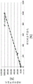
FIG. 2 shows the linearity of the assay of the invention in table (FIG. 2a) and diagram (FIG. 2 b).
SEQ ID No.1 shows the genomic sequence of amplicon AMP2213 according to the present invention.
SEQ ID nos. 2 and 3 show the sequence of the bisulfite converted target region of the preferred qPCR-assay-system of the invention, showing only one of the four possible chains after bisulfite conversion. All four strands are preferred targets for designing specific assays. The bisulfite strand given under the name bisulfite Strand 2 Forward Strand (b2f) is only one preferred target for use as an example of primer and probe design. The reverse strand (b2r) was used for the complementary primer. Chains b1f and b1r are not shown, but are preferably used.
SEQ ID Nos. 4 to 11 show the sequences of specific oligonucleotides (primers and probes) according to the invention.
Examples
Example 1
To identify atypical monocytes, qPCR was performed on bisulfite converted samples derived from human genomic regions according to the following sequence (AMP2213, SEQ ID No.1), the relevant CpG being bold and underlined:

for the actual epigenetic profile analysis of the amplicon region in a subset of blood cells, the immune cell populations analyzed are shown in FIG. 1.
The target areas for bisulfite conversion of the preferred qPCR-assay-system developed were:
TpG-specific (SEQ ID No. 2):
(b2F)


CpG-specificity: (SEQ ID No. 3):
(b2F)

the following primers and probes were used for qPCR:

the specificity of the TpG-specific PCR-system was demonstrated using the test template (plasmid-DNA) as shown in figure 2. Cell type specificity (as measured by qPCR) was found as follows:

sequence listing
<110> Aipienis GmbH (Epiontis GmbH)
<120> amplicon region as epigenetic marker for identifying immune cells, especially atypical monocytes
<130>E31869WO
<150>DE 10 2017 125 335.1
<151>2017-10-27
<160>11
<170> PatentIn 3.5 edition
<210>1
<211>271
<212>DNA
<213> Intelligent (Homo sapiens)
<400>1
cccttcctct gactcagtgg aagggcagga gagtgccccg aggagctgcc cacatccctg 60
gctgagtgcc tcacccccag ggcctccacg aggagcagct tccacagggt gcctgtgggg 120
ctcgttcctc tggatgcttt tccctttgct gtgaatgcct ctggggcacg aatatatggc 180
ccttgggtct aggccttagg gcttccggtg accaggatag gaagtgttgc aggccctgcc 240
ccgagggcgg cgcattagct tttcccccac t 271
<210>2
<211>300
<212>DNA
<213> Intelligent (Homo sapiens)
<400>2
cccttcctct aactcaataa aaaaacaaaa aaatacccca aaaaactacc cacatcccta 60
actaaatacc tcacccccaa aacctccaca aaaaacaact tccacaaaat acctataaaa 120
ctcattcctc taaatacttt tccctttact ataaatacct ctaaaacaca aatatataac 180
ccttaaatct aaaccttaaa acttccaata accaaaataa aaaatattac aaaccctacc 240
ccaaaaacaa cacattaact tttcccccac tactttcatc tacccatctc accaaattcc 300
<210>3
<211>300
<212>DNA
<213> Intelligent (Homo sapiens)
<400>3
cccttcctct aactcaataa aaaaacaaaa aaataccccg aaaaactacc cacatcccta 60
actaaatacc tcacccccaa aacctccacg aaaaacaact tccacaaaat acctataaaa 120
ctcgttcctc taaatacttt tccctttact ataaatacct ctaaaacacg aatatataac 180
ccttaaatct aaaccttaaa acttccgata accaaaataa aaaatattac aaaccctacc 240
ccgaaaacga cgcattaact tttcccccac tactttcatc tacccatctc accaaattcc 300
<210>4
<211>22
<212>DNA
<213> Intelligent (Homo sapiens)
<400>4
cccttcctct aactcaataa aa 22
<210>5
<211>19
<212>DNA
<213> Intelligent (Homo sapiens)
<400>5
agtgggggaa aagttaatg 19
<210>6
<211>29
<212>DNA
<213> Intelligent (Homo sapiens)
<400>6
cccttaaatc taaaccttaa aacttccaa 29
<210>7
<211>24
<212>DNA
<213> Intelligent (Homo sapiens)
<400>7
tagtggggga aaagttaatg tgtt 24
<210>8
<211>25
<212>DNA
<213> Intelligent (Homo sapiens)
<400>8
acaaacccta ccccaaaaac aacac 25
<210>9
<211>28
<212>DNA
<213> Intelligent (Homo sapiens)
<400>9
ccttaaatct aaaccttaaa acttccga 28
<210>10
<211>22
<212>DNA
<213> Intelligent (Homo sapiens)
<400>10
gtggaggaaa agttaatgcg tc 22
<210>11
<211>24
<212>DNA
<213> Intelligent (Homo sapiens)
<400>11
caaaccctac cccgaaaacg acgc 24
Claims (15)
1. A method for identifying atypical monocytes in a sample comprising analyzing the methylation status of at least one CpG position in a genomic region within the 2213 amplicon of a mammal, wherein said region analyzed is preferentially located according to SEQ ID No.1, wherein a demethylation or lack of methylation of said region as compared to a typical monocyte or a non-monocyte is indicative of an atypical monocyte.
2. The method of claim 1, wherein the at least one CpG position is present in the 5 'region upstream of the transcription start, promoter region, 5' or 3 'untranslated region, exon, intron, exon/intron border and/or 3' region downstream of the transcription termination of any gene within the 2213 amplicon under analysis.
3. The method according to claim 1 or 2, wherein the at least one CpG position is selected from the group of CpG positions 30, 89, 123, 169, 206, 242 and 248 in the amplicon according to SEQ ID No.1 and preferably from CpG positions 30, 169, 206 and 242 in the fragment of amplicon No. 2213 or in the fragment of the bisulfite converted sequence according to SEQ ID No.2 or 3.
4. The method of any one of claims 1 to 3, wherein said bisulfite convertibility analysis comprises a method selected from methylation specific enzymatic digestion, sulfite sequencing, an assay selected from promoter methylation, CpG island methylation, MSP, heavy methyl, fluorometric methods, Ms-SNuPE, and other methods that rely on the detection of amplified DNA.
5. The method of any one of claims 1-4, further comprising quantifying the relative amount of atypical monocytes based on comparing the relative amount of the methylation frequency in the analyzed 2213 amplicon to the relative amount of methylation frequency in a control gene, e.g., GAPDH.
6. The method of any one of claims 1 to 5, wherein the sample is selected from a mammalian body fluid, including a human blood sample or a tissue, organ or cell type blood sample, a blood lymphocyte sample or a fraction thereof.
7. The method of any one of claims 1 to 6, further comprising distinguishing the atypical monocytes from all or at least one cell type selected from the group consisting of: follicular helper T cells, cytotoxic T cells, granulocytes, canonical monocytes, B cells, NK-cells and T-helper cells, and other cell types derived from organs other than blood.
8. The method of any one of claims 1 to 7, wherein the method is performed without a step of purifying and/or enriching the cells to be identified, preferably using whole blood and/or non-trypsinized tissue.
9. The method of any one of claims 1 to 8, further comprising the step of concluding the immune status of the mammal based on the atypical monocytes identified.
10. A method for monitoring the level of atypical monocytes in a mammal comprising performing the method of any one of claims 5 to 9 and comparing the relative amount of the cells identified to a sample and/or control sample taken earlier or simultaneously from the same mammal.
11. The method of any one of claims 1 to 10, further comprising measuring and/or monitoring the amount of atypical monocytes responsive to chemical and/or biological substances provided to the mammal.
12. The method of any one of claims 1 to 11, wherein the mammal has or is likely to have an autoimmune disease, transplant rejection, an infectious disease, cancer, and/or allergy.
13. A kit for identifying, quantifying, and/or monitoring atypical monocytes in a mammal based on analyzing bisulfite accessibility of CpG positions in genomic regions within a 2213 amplicon, comprising components for performing the method of any one of claims 1 to 12, in particular a kit comprising: a) a bisulphite reagent, and b) a material for analysing the methylation status of CpG positions selected from the group consisting of CpG positions in the region according to SEQ ID NO:1, such as an oligomer selected from the group consisting of sequences according to SEQ ID NO:4 to 11.
An oligomer according to any one of SEQ ID Nos. 4 to 11 or an amplicon of SEQ ID Nos. 1, 2 or 3.
15. Use of the kit of claim 13 or the oligomer or amplicon of claim 14 for identifying, quantifying, and/or monitoring atypical monocytes in a mammal.
Applications Claiming Priority (3)
| Application Number | Priority Date | Filing Date | Title |
|---|---|---|---|
| DE102017125335.1 | 2017-10-27 | ||
| DE102017125335.1A DE102017125335B4 (en) | 2017-10-27 | 2017-10-27 | Amplicon region as an epigenetic marker for the identification of immune cells, especially non-classical monocytes |
| PCT/EP2018/079406 WO2019081707A1 (en) | 2017-10-27 | 2018-10-26 | Amplicon region as epigenetic marker for the identification of non-classical monocytes |
Publications (1)
| Publication Number | Publication Date |
|---|---|
| CN111247253A true CN111247253A (en) | 2020-06-05 |
Family
ID=64024040
Family Applications (1)
| Application Number | Title | Priority Date | Filing Date |
|---|---|---|---|
| CN201880068476.5A Withdrawn CN111247253A (en) | 2017-10-27 | 2018-10-26 | Amplicon region as epigenetic marker for identifying immune cells, particularly atypical monocytes |
Country Status (8)
| Country | Link |
|---|---|
| US (1) | US11680294B2 (en) |
| EP (1) | EP3669001A1 (en) |
| JP (1) | JP2021502806A (en) |
| CN (1) | CN111247253A (en) |
| AU (1) | AU2018356572A1 (en) |
| CA (1) | CA3079477A1 (en) |
| DE (1) | DE102017125335B4 (en) |
| WO (1) | WO2019081707A1 (en) |
Families Citing this family (1)
| Publication number | Priority date | Publication date | Assignee | Title |
|---|---|---|---|---|
| WO2022191293A1 (en) * | 2021-03-12 | 2022-09-15 | エーザイ・アール・アンド・ディー・マネジメント株式会社 | Method for measuring total monocytes and percentage of cd16-positive monocytes |
Family Cites Families (4)
| Publication number | Priority date | Publication date | Assignee | Title |
|---|---|---|---|---|
| WO2012162660A2 (en) | 2011-05-25 | 2012-11-29 | Brown University | Methods using dna methylation for identifying a cell or a mixture of cells for prognosis and diagnosis of diseases, and for cell remediation therapies |
| PL2986735T3 (en) * | 2013-04-19 | 2019-08-30 | Epiontis Gmbh | Method for identifying the quantitative composition of blood cells in a sample |
| CN105377273A (en) * | 2013-05-22 | 2016-03-02 | 耶达研究与发展公司 | Human monocyte sub-population for treatment of eye diseases and disorders |
| GB201516975D0 (en) * | 2015-09-25 | 2015-11-11 | Epiontis Gmbh | PARK2 as epigenetic marker for the identification of immune cells, in particular monocytes |
-
2017
- 2017-10-27 DE DE102017125335.1A patent/DE102017125335B4/en active Active
-
2018
- 2018-10-26 EP EP18793655.4A patent/EP3669001A1/en active Pending
- 2018-10-26 US US16/758,992 patent/US11680294B2/en active Active
- 2018-10-26 WO PCT/EP2018/079406 patent/WO2019081707A1/en unknown
- 2018-10-26 JP JP2020523795A patent/JP2021502806A/en active Pending
- 2018-10-26 AU AU2018356572A patent/AU2018356572A1/en not_active Abandoned
- 2018-10-26 CN CN201880068476.5A patent/CN111247253A/en not_active Withdrawn
- 2018-10-26 CA CA3079477A patent/CA3079477A1/en active Pending
Also Published As
| Publication number | Publication date |
|---|---|
| DE102017125335A1 (en) | 2019-05-02 |
| US11680294B2 (en) | 2023-06-20 |
| JP2021502806A (en) | 2021-02-04 |
| DE102017125335B4 (en) | 2019-10-17 |
| CA3079477A1 (en) | 2019-05-02 |
| WO2019081707A1 (en) | 2019-05-02 |
| US20200340053A1 (en) | 2020-10-29 |
| AU2018356572A1 (en) | 2020-04-23 |
| EP3669001A1 (en) | 2020-06-24 |
Similar Documents
| Publication | Publication Date | Title |
|---|---|---|
| CA2999614C (en) | Park2 as epigenetic marker for the identification of immune cells, in particular monocytes | |
| US11643687B2 (en) | LRP5 as epigenetic marker for the identification of immune cells, in particular B-cells | |
| EP3353321B1 (en) | Mvd as epigenetic marker for the identification of cd56+ nk cells | |
| US11680294B2 (en) | Amplicon region as epigenetic marker for the identification of non-classical monocytes | |
| US11753683B2 (en) | MCC as epigenetic marker for the identification of immune cells, in particular basophil granulocytes | |
| EP3669003B1 (en) | Pdcd1 as epigenetic marker for the identification of immune cells, in particular pd1+ cells | |
| US11976327B2 (en) | ERGIC1 as epigenetic marker for the identification of immune cells, in particular monocytic myeloid-derived suppressor cells (mMDSCs) | |
| CA2969370A1 (en) | Epigenetic method for the identification of follicular t-helper-(tfh-) cells | |
| CN111278988A (en) | Endosialin (CD248) as an epigenetic marker for identifying immune cells, in particular naive CD8+ T cells |
Legal Events
| Date | Code | Title | Description |
|---|---|---|---|
| PB01 | Publication | ||
| PB01 | Publication | ||
| SE01 | Entry into force of request for substantive examination | ||
| SE01 | Entry into force of request for substantive examination | ||
| CB02 | Change of applicant information |
Address after: Berlin, Germany Applicant after: Precision medical Co.,Ltd. Address before: Berlin, Germany Applicant before: EPIONTIS GmbH |
|
| CB02 | Change of applicant information | ||
| WW01 | Invention patent application withdrawn after publication |
Application publication date: 20200605 |
|
| WW01 | Invention patent application withdrawn after publication |