CN110290812B - Exon-skipping oligomer conjugates for muscular dystrophy - Google Patents
Exon-skipping oligomer conjugates for muscular dystrophy Download PDFInfo
- Publication number
- CN110290812B CN110290812B CN201780086301.2A CN201780086301A CN110290812B CN 110290812 B CN110290812 B CN 110290812B CN 201780086301 A CN201780086301 A CN 201780086301A CN 110290812 B CN110290812 B CN 110290812B
- Authority
- CN
- China
- Prior art keywords
- dystrophin
- exon
- antisense oligomer
- present disclosure
- skipping
- Prior art date
- Legal status (The legal status is an assumption and is not a legal conclusion. Google has not performed a legal analysis and makes no representation as to the accuracy of the status listed.)
- Active
Links
Images


Classifications
-
- A—HUMAN NECESSITIES
- A61—MEDICAL OR VETERINARY SCIENCE; HYGIENE
- A61K—PREPARATIONS FOR MEDICAL, DENTAL OR TOILETRY PURPOSES
- A61K31/00—Medicinal preparations containing organic active ingredients
- A61K31/70—Carbohydrates; Sugars; Derivatives thereof
- A61K31/7088—Compounds having three or more nucleosides or nucleotides
- A61K31/712—Nucleic acids or oligonucleotides having modified sugars, i.e. other than ribose or 2'-deoxyribose
-
- A—HUMAN NECESSITIES
- A61—MEDICAL OR VETERINARY SCIENCE; HYGIENE
- A61K—PREPARATIONS FOR MEDICAL, DENTAL OR TOILETRY PURPOSES
- A61K31/00—Medicinal preparations containing organic active ingredients
- A61K31/70—Carbohydrates; Sugars; Derivatives thereof
- A61K31/7088—Compounds having three or more nucleosides or nucleotides
- A61K31/7125—Nucleic acids or oligonucleotides having modified internucleoside linkage, i.e. other than 3'-5' phosphodiesters
-
- A—HUMAN NECESSITIES
- A61—MEDICAL OR VETERINARY SCIENCE; HYGIENE
- A61K—PREPARATIONS FOR MEDICAL, DENTAL OR TOILETRY PURPOSES
- A61K47/00—Medicinal preparations characterised by the non-active ingredients used, e.g. carriers or inert additives; Targeting or modifying agents chemically bound to the active ingredient
- A61K47/50—Medicinal preparations characterised by the non-active ingredients used, e.g. carriers or inert additives; Targeting or modifying agents chemically bound to the active ingredient the non-active ingredient being chemically bound to the active ingredient, e.g. polymer-drug conjugates
- A61K47/51—Medicinal preparations characterised by the non-active ingredients used, e.g. carriers or inert additives; Targeting or modifying agents chemically bound to the active ingredient the non-active ingredient being chemically bound to the active ingredient, e.g. polymer-drug conjugates the non-active ingredient being a modifying agent
- A61K47/62—Medicinal preparations characterised by the non-active ingredients used, e.g. carriers or inert additives; Targeting or modifying agents chemically bound to the active ingredient the non-active ingredient being chemically bound to the active ingredient, e.g. polymer-drug conjugates the non-active ingredient being a modifying agent the modifying agent being a protein, peptide or polyamino acid
- A61K47/64—Drug-peptide, drug-protein or drug-polyamino acid conjugates, i.e. the modifying agent being a peptide, protein or polyamino acid which is covalently bonded or complexed to a therapeutically active agent
- A61K47/645—Polycationic or polyanionic oligopeptides, polypeptides or polyamino acids, e.g. polylysine, polyarginine, polyglutamic acid or peptide TAT
-
- A—HUMAN NECESSITIES
- A61—MEDICAL OR VETERINARY SCIENCE; HYGIENE
- A61P—SPECIFIC THERAPEUTIC ACTIVITY OF CHEMICAL COMPOUNDS OR MEDICINAL PREPARATIONS
- A61P21/00—Drugs for disorders of the muscular or neuromuscular system
-
- C—CHEMISTRY; METALLURGY
- C12—BIOCHEMISTRY; BEER; SPIRITS; WINE; VINEGAR; MICROBIOLOGY; ENZYMOLOGY; MUTATION OR GENETIC ENGINEERING
- C12N—MICROORGANISMS OR ENZYMES; COMPOSITIONS THEREOF; PROPAGATING, PRESERVING, OR MAINTAINING MICROORGANISMS; MUTATION OR GENETIC ENGINEERING; CULTURE MEDIA
- C12N15/00—Mutation or genetic engineering; DNA or RNA concerning genetic engineering, vectors, e.g. plasmids, or their isolation, preparation or purification; Use of hosts therefor
- C12N15/09—Recombinant DNA-technology
- C12N15/11—DNA or RNA fragments; Modified forms thereof; Non-coding nucleic acids having a biological activity
- C12N15/113—Non-coding nucleic acids modulating the expression of genes, e.g. antisense oligonucleotides; Antisense DNA or RNA; Triplex- forming oligonucleotides; Catalytic nucleic acids, e.g. ribozymes; Nucleic acids used in co-suppression or gene silencing
-
- C—CHEMISTRY; METALLURGY
- C12—BIOCHEMISTRY; BEER; SPIRITS; WINE; VINEGAR; MICROBIOLOGY; ENZYMOLOGY; MUTATION OR GENETIC ENGINEERING
- C12N—MICROORGANISMS OR ENZYMES; COMPOSITIONS THEREOF; PROPAGATING, PRESERVING, OR MAINTAINING MICROORGANISMS; MUTATION OR GENETIC ENGINEERING; CULTURE MEDIA
- C12N2310/00—Structure or type of the nucleic acid
- C12N2310/10—Type of nucleic acid
- C12N2310/11—Antisense
- C12N2310/111—Antisense spanning the whole gene, or a large part of it
Abstract
Antisense oligomer conjugates complementary to selected target sites in the human dystrophin gene to induce exon 51 skipping are described.
Description
Related information
This patent application claims the benefit of U.S. provisional patent application Ser. No. 62/436182, U.S. provisional patent application Ser. No. 62/443476, U.S. provisional patent application Ser. No. 62/479173, and U.S. provisional patent application Ser. No. 62/562080, U.S. provisional patent application Ser. No. 62/479173, and U.S. provisional patent application Ser. No. 62/562080, U.S. No. 22, U.S. 9, and U.S. 5, respectively, filed on 12, and 6, and 10, respectively. The entire contents of the above-mentioned provisional patent application are incorporated herein by reference.
Technical Field
The present disclosure relates to novel antisense oligomer conjugates suitable for exon 51 skipping of human dystrophin genes and pharmaceutical compositions thereof. The present disclosure also provides methods of inducing exon 51 skipping using the novel antisense oligomer conjugates, methods of producing dystrophin in subjects having dystrophin gene mutations suitable for exon 51 skipping, and methods of treating subjects having dystrophin gene mutations suitable for exon 51 skipping.
Background
Antisense technology is being developed, using a range of chemicals to affect gene expression (transcription, splicing, stability, translation) at various levels. Most research has focused on the use of antisense compounds to correct or compensate for abnormal or disease-related genes in various indications. Antisense molecules are capable of specifically inhibiting gene expression, and thus much research effort on oligomers as gene expression regulators has focused on inhibiting the expression of target genes or the function of cis-acting elements. Antisense oligomers are typically directed against RNA, or the sense strand (e.g., mRNA), or in the case of some viral RNA targets, the negative strand. To achieve the desired effect of down-regulation of a particular gene, the oligomer typically promotes decay of the target mRNA, blocks translation of the mRNA or blocks the function of the cis-acting RNA element, thereby effectively preventing de novo synthesis of the target protein or replication of viral RNA.
However, these techniques are not useful when the objective is to up-regulate the production of the native protein or to compensate for mutations that induce premature termination of translation (e.g., nonsense or frameshift mutations). In these cases, the defective gene transcript should not undergo targeted degradation or steric inhibition, so antisense oligomer chemistry should not promote target mRNA decay or block translation.
In various genetic diseases, the effect of mutations on the final expression of a gene can be regulated by targeting processes of exon skipping during splicing. The splicing process is guided by a complex multicomponent mechanism that brings adjacent exon-intron junctions in the pre-mRNA into close proximity and makes a phosphodiester bond cleavage at the end of the introns, followed by recombination between the exons to be spliced together. This complex and highly precise process is mediated by sequence motifs in pre-mRNAs that are relatively short semi-conserved RNA segments that subsequently participate in the binding of various nuclear splicing factors of the splicing reaction. Differentially spliced mRNA molecules can be produced by altering the manner in which the splicing machinery reads or recognizes motifs involved in pre-mRNA processing. It has now been recognized that during normal gene expression, most human genes are alternatively spliced, although the mechanisms involved have not been identified. Bennett et al (U.S. Pat. No. 6,210,892) describe antisense modulation of wild-type cellular mRNA processing using antisense oligomer analogs that do not induce RNAse H-mediated cleavage of target RNA. This can be used to generate alternatively spliced mRNAs lacking specific exons (see, e.g., sazani, kole et al, 2007 for soluble TNF superfamily receptors lacking exons encoding transmembrane domains).
If normal functional proteins terminate prematurely due to mutations therein, methods for restoring production of certain functional proteins by antisense technology have been demonstrated to be able to intervene during splicing and if exons associated with mutations causing disease can be specifically deleted from certain genes, sometimes shortened protein products can be produced that have similar biological properties to the native protein or have sufficient biological activity to ameliorate diseases caused by mutations associated with exons (see, e.g., sierakowska, sambade et al 1996; wilton, lloyd et al 1999; vanDeutekom, bremmer-Bout et al 2001; lu, mann et al 2003, aartsma-Rus, janson et al 2004). Kole et al (U.S. Pat. nos. 5,627,274;5,916,808;5,976,879 and 5,665,593) disclose methods of combating aberrant splicing using modified antisense oligomer analogs that do not promote decay of targeted pre-mRNA. Bennett et al (U.S. Pat. No. 6,210,892) describe antisense modulation of wild-type cellular mRNA processing, which also uses antisense oligomer analogs that do not induce RNAse H-mediated cleavage of target RNA.
The process of targeting exon skipping may be particularly useful in long genes where there are many exons and introns, where there is redundancy in the genetic makeup of the exons, or where the protein is able to function without one or more specific exons. In order to treat genetic diseases associated with truncations caused by various genetic mutations, efforts to redirect gene processing have focused on the use of antisense oligomers, wherein: (1) Fully or partially overlapping with elements involved in the splicing process; or (2) bind to the pre-mRNA in sufficient proximity to the element to disrupt the binding and function of splicing factors that normally mediate the particular splicing reaction that occurs at the element.
Duchenne Muscular Dystrophy (DMD) is caused by a defective expression of the protein dystrophin. The gene encoding this protein contains 79 exons, distributed over more than 200 ten thousand DNA nucleotides. Changes in the reading frame of the exons, or the introduction of stop codons, or any exon mutation characterized by deletion of the entire frame of exons or the exon or the duplication of one or more exons, may disrupt the production of functional dystrophin, resulting in DMD.
Becker Muscular Dystrophy (BMD) is a less severe muscular dystrophy that has been found in which mutations are usually deletions of one or more exons resulting in a correct reading frame for transcription of the entire dystrophin protein, so that translation of mRNA into protein does not terminate prematurely. If the linkage of the upstream and downstream exons maintains the correct reading frame of the gene in the processing of the mutated dystrophin pre-mRNA, the result is an mRNA encoding a protein with a short internal deletion that retains some activity, resulting in the Becker phenotype.
For many years, it has been known that a deletion of one or more exons that do not alter the reading frame of dystrophin will produce a BMD phenotype, whereas an exon deletion that causes a frameshift will produce DMD (Monaco, bertelson et al, 1988). Typically, dystrophin mutations include point mutations and exon deletions that alter the reading frame and thus interrupt proper protein translation resulting in DMD. It should also be noted that some BMD and DMD patients have exon deletions covering multiple exons.
Modulation of mutant dystrophin pre-mRNA splicing with antisense oligoribonucleotides has been reported in vitro and in vivo (see, e.g., matsuo, masumura et al 1991; takeshima, nishio et al 1995; pramono, takeshima et al 1996; dunckley, eperon et al 1997; dunckley, manoharan et al 1998; wilton, lloyd et al 1999, honeyman et al 2002; errington, mann et al 2003).
Antisense oligomers have been specifically designed to target specific regions of pre-mRNA, typically exons, to induce skipping of DMD gene mutations, thereby restoring these out-of-frame mutations in-frame, resulting in the production of internally shortened but functional dystrophin. Such antisense oligomers are known to be targeted either completely within an exon (the so-called exon internal sequence) or at splice donor or splice acceptor junctions, which pass from the exon into a portion of the intron.
The discovery and development of such antisense oligomers for DMD has become the area of prior research. These developments include those of: (1) University of western australia and Sarepta Therapeutics (assignee of the present application): WO 2006/000057; WO 2010/048586; WO 2011/057350; WO 2014/100714; WO 2014/153240; WO 2014/153220; (2) Academisch Ziekenhuis Leiden/Prosensa Technologies (now BioMarin Pharmaceutical): WO 02/24906; WO 2004/083432; WO 2004/083446; WO 2006/112705; WO 2007/133105; WO 2009/139630; WO 2009/054725; WO 2010/050801; WO 2010/050802; WO 2010/123369; WO 2013/112053; WO 2014/007420; (3) Carolinas Medical Center: WO 2012/109296; (4) Royal Holloway: patents and applications claiming the benefits of and including U.S. Ser. Nos. 61/096,073 and 61/164,978; for example US 8,084,601 and US 2017-0204413 (4) JCR Pharmaceuticals and Matsuo: US 6,653,466; the benefits of JP 2000-125448 are claimed and include patents and applications thereof, such as US 6,653,467; the benefits of JP 2000-256547 are claimed and include patents and applications thereof, such as US 6,727,355; WO 2004/048570; (5) Nippon Shinyaku: WO2012/029986; WO2013/100190; WO 2015/137409; WO 2015/194520; (6) University of berni/peerle and university of maricurie +.  /University of la Recherche Scientifique/Synthena AG:WO 2010/115993;WO 2013/053928。
Eteplirsen is a Phosphorodiamidate Morpholino Oligomer (PMO) designed to skip exon 51 of the human dystrophin gene in DMD patients who are prone to exon 51 skipping to restore reading frame and functionA shorter form of dystrophin. The U.S. Food and Drug Administration (FDA) approved Exondys 51 in 2016 TM (eteplirsen) for the treatment of Duchenne type muscular dystrophy (DMD) patients whose DMD gene has been diagnosed with exon 51 mutations.
The discovery and development of antisense oligomers conjugated to cell penetrating peptides for DMD is also a field of research (see PCT publication No. WO2010/048586; wu, b. Et al, the American Journal of Pathology, vol.181 (2): 392-400,2012; wu, r. Et al, nucleic Acids Research, vol.35 (15): 5182-5191,2007; mulders, s. Et al, 19) th International Congress of the World Muscle Society, poster Presentation Berlin, month 10 of 2014; bestas, b. et al The Journal of Clinical Investigation, doi:10.1172/JCI76175,2014; jearawieriyapaisarn, n. et al Molecular Therapy, vol.16 (9): 1624-1629,2008; jearawieriyapaisearn, n. et al, cardiovascular Research, vol.85:444-453,2010; moulton, h.m. et al Biochemical Society Transactions, vol.35 (4): 826-828,2007; yin, h. et al Molecular Therapy, vol.19 (7): 1295-1303,2011; abes, r. et al, j.pept.sci., vol.14:455-460,2008; lebleu, b. et al Advanced Drug Delivery Reviews, vol.60:517-529,2008; mcClorey, g. Et al, gene therapy, vol.13:1373-1381,2006; alter, j et al, nature Medicine, vol.12 (2): 175-177,2006; and Youngblood, D.et al American Chemical Society, bioconjugate chem.,2007,18 (1), pp 50-60).
Cell Penetrating Peptides (CPPs), such as arginine-rich peptide transport moieties, can be effective to enhance penetration of, for example, antisense oligomers conjugated to CPPs into cells.
Despite these efforts, there remains a need for improved antisense oligomers targeting exon 51 and corresponding pharmaceutical compositions, which may be useful in therapeutic methods for producing dystrophin and for treating DMD.
Disclosure of Invention
The antisense oligomer conjugates provided herein include an antisense oligomer moiety conjugated to a CPP. In one aspect, the present disclosure provides an antisense oligomer conjugate comprising:
an antisense oligomer 30 subunits in length capable of binding to a selected target to induce exon skipping in a human dystrophin gene, wherein the antisense oligomer comprises a base sequence complementary to an exon 51 target region of a dystrophin pre-mRNA, said target region being designated as an annealing site; and
cell Penetrating Peptides (CPPs) conjugated to antisense oligomers through a linker moiety.
In some embodiments, the annealing site is H51A (+66+95).
In some embodiments, the bases of the antisense oligomer are linked to morpholino ring structures, wherein the morpholino ring structures are linked by phosphorus-containing intersubunit linkages to the morpholino nitrogen of one ring structure to the 5' exocyclic carbon of an adjacent ring structure. In certain embodiments, the cell penetrating peptide is six arginine units ("R 6 ") and the linker moiety is glycine. In some embodiments, the antisense oligomer comprises a sequence designated as SEQ ID NO:1, and a nucleotide sequence of 1.
In various aspects, the present disclosure provides antisense oligomer conjugates that can be according to formula (I):

or a pharmaceutically acceptable salt thereof, wherein:
each Nu is a nucleobase that together form a targeting sequence; and is also provided with
T is a moiety selected from the group consisting of:

R 1 is C 1 -C 6 An alkyl group;
wherein the targeting sequence is complementary to an annealing site for exon 51 in the dystrophin pre-mRNA, designated as H51A (+66+95).
In another aspect, the present disclosure provides antisense oligomer conjugates of formula (IV):

or a pharmaceutically acceptable salt thereof.
In another aspect, the present disclosure provides antisense oligomer conjugates of formula (IVA):

in another aspect, the present disclosure provides a pharmaceutical composition comprising an antisense oligomer conjugate of the present disclosure and a pharmaceutically acceptable carrier. In some embodiments, the pharmaceutically acceptable carrier is a saline solution comprising a phosphate buffer.
In another aspect, the present disclosure provides a method of treating Duchenne Muscular Dystrophy (DMD) in a subject in need thereof, wherein the subject has an dystrophin gene mutation adapted for exon 51 skipping, the method comprising administering to the subject an antisense oligomer conjugate of the present disclosure. The disclosure also relates to the use of the antisense oligomer conjugates of the disclosure in the manufacture of a medicament for treating Duchenne Muscular Dystrophy (DMD) in a subject in need thereof, wherein the subject has an dystrophin gene mutation adapted for exon 51 skipping.
In another aspect, the present disclosure provides a method of restoring mRNA reading frames to induce dystrophin production in a subject having a dystrophin gene mutation suitable for exon 51 skipping, the method comprising administering to the subject an antisense oligomer conjugate of the present disclosure. In another aspect, the present disclosure provides a method of removing exon 51 from dystrophin pre-mRNA during mRNA processing of a subject having a dystrophin gene mutation suitable for exon 51 skipping, the method comprising administering to the subject an antisense oligomer conjugate of the present disclosure. In another aspect, the present disclosure provides a method of binding exon 51 of dystrophin pre-mRNA in a subject having a dystrophin gene mutation suitable for exon 51 skipping, the method comprising administering to the subject an antisense oligomer conjugate of the present disclosure.
In another aspect, the present disclosure provides antisense oligomer conjugates disclosed herein for use in therapy. In certain embodiments, the present disclosure provides antisense oligomer conjugates of the present disclosure for use in the treatment of duchenne muscular dystrophy. In certain embodiments, the present disclosure provides antisense oligomer conjugates of the present disclosure for use in the preparation of a medicament for use in therapy. In certain embodiments, the present disclosure provides antisense oligomer conjugates of the present disclosure for use in the manufacture of a medicament for the treatment of duchenne muscular dystrophy.
In another aspect, the present disclosure also provides a kit for treating Duchenne Muscular Dystrophy (DMD) in a subject in need thereof, wherein the subject has an dystrophin gene mutation adapted for exon 51 skipping, the kit comprising at least one antisense oligomer conjugate of the present disclosure packaged in a suitable container, and instructions for use thereof.
These and other objects and features will be more fully understood upon reading the following detailed description of the disclosure in conjunction with the accompanying drawings.
Drawings
FIG. 1 depicts a portion of normal dystrophin pre-mRNA and mature mRNA.
Figure 2 depicts an aberrant dystrophin pre-mRNA (an example of DMD) and a portion of the resulting nonfunctional labile dystrophin.
FIG. 3 depicts eteplirsen, designed to skip exon 51, restoring an "in frame" reading of pre-mRNA to produce an internally deleted dystrophin protein.
FIG. 4 provides bar graphs of the percentage of PMO# 1 and PPMO# 1 skipping exon 51 in differentiated human myocytes at 96 hours after treatment as measured by RT-PCR.
Figures 5A-5D provide representative images of western blot analysis measuring dystrophin in mdx mice quadriceps femoris treated with PMO (PMO 4225) or PPMO (PPMO 4225) at different time points [7 days (5A), 30 days (5B), 60 days (5C) and 90 days (5D) ].
Fig. 6A provides a line graph depicting the percentage of PMO (PMO 4225) or PPMO (PPMO 4225) induced wild-type dystrophin in quadriceps of mdx mice over 90 days post injection as determined by western blot analysis.
Fig. 6B provides a line graph depicting the percent PMO (PMO 4225) or PPMO (PPMO 4225) induced exon 23 skipping in the quadriceps femoris of mdx mice over 90 days post injection as determined by RT-PCR.
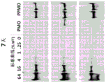
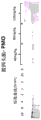
Figures 7A-7D provide representative images of western blot analysis measuring dystrophin in mdx mice diaphragm muscle treated with PMO (PMO 4225) or PPMO (PPMO 4225) at different time points [7 days (7A), 30 days (7B), 60 days (7C) and 90 days (7D) ].
Fig. 8A provides a line graph depicting the percentage of PMO (PMO 4225) or PPMO (PPMO 4225) induced wild-type dystrophin in the diaphragm of mdx mice over 90 days post injection as determined by western blot analysis.
Fig. 8B provides a line graph depicting the percent PMO (PMO 4225) or PPMO (PPMO 4225) induced exon 23 skipping in the mdx mouse diaphragm over 90 days post injection as determined by RT-PCR.
Figures 9A-9D provide representative images of western blot analysis measuring dystrophin in mdx mouse hearts at different time points [7 days (9A), 30 days (9B), 60 days (9C) and 90 days (9D) ] treated with PMO (PMO 4225) or PPMO (PPMO 4225).
Fig. 10A provides a line graph depicting the percentage of wild-type dystrophin induced in mdx mouse hearts by PMO (PMO 4225) or PPMO (PPMO 4225) over 90 days after injection as determined by western blot analysis.
FIG. 10B provides a line graph depicting the percent of PMO (PMO 4225) or PPMO (PPMO 4225) -induced exon 23 skipping in mdx mouse hearts over 90 days post injection as determined by RT-PCR.
FIG. 11 provides an immunohistochemical analysis showing dystrophin in the quadriceps left thigh of the mdx mice induced by PMO (PMO 4225) or PPMO (PPMO 4225).
Fig. 12 provides a line graph showing the percentage of exon 51 in non-human primates treated weekly with PMO # 1 or PPMO # 1 at different doses. The percentage of exon 51 skipping was measured by RT-PCR from muscle samples of diaphragm (left) and quadriceps (right).
Fig. 13 provides a line graph showing the percentage of exon 51 in non-human primates treated weekly with PMO # 1 or PPMO # 1 at different doses. The percentage of exon 51 skipping was measured by RT-PCR from muscle samples of heart (left) and duodenum (right).
Fig. 14 provides a line graph showing the percentage of exon 51 in non-human primates treated weekly with PMO # 1 or PPMO # 1 at different doses. The percentage of exon 51 skipping was measured by RT-PCR from muscle samples of biceps (left) and deltoid (right).
Fig. 15 provides a line graph showing the percentage of exon 51 in non-human primates treated weekly with PMO # 1 or PPMO # 1 at different doses. The percentage of exon 51 skipping was measured by RT-PCR from muscle samples of the esophagus (left) and aorta (right).
Fig. 16A-B provide representative images of western blot analysis measured with different doses: muscle dystrophin in 40mg/kg,80mg/kg and 120mg/kg PMO (PMO 4225) or PPMO (PPMO 4225) treated mdx mouse hearts.
Fig. 17 provides a bar graph depicting the use of different doses: the percentage of wild-type dystrophin induced in mdx mouse hearts by PMO (PMO 4225) or PPMO (PPMO 4225) at 40mg/kg,80mg/kg and 120mg/kg, as determined by western blot analysis at 30 days post injection.
Fig. 18A-B provide representative images of western blot analysis measured with different doses: dystrophin in 40mg/kg,80mg/kg and 120mg/kg in PMO (PMO 4225) or PPMO (PPMO 4225) treated mdx mice diaphragm.
Fig. 19 provides a bar graph depicting the use of different doses: the percentage of wild-type dystrophin induced in mdx mouse diaphragm by PMO (PMO 4225) or PPMO (PPMO 4225) at 40mg/kg,80mg/kg and 120mg/kg, as determined by western blot analysis at 30 days post injection.
Figures 20A-B provide representative images of western blot analysis measured with different doses: dystrophin in 40mg/kg,80mg/kg and 120mg/kg in PMO (PMO 4225) or PPMO (PPMO 4225) treated mdx mice quadriceps femoris.
Fig. 21 provides a bar graph depicting the use of different doses: the percentage of wild-type dystrophin induced in the quadriceps of mdx mice by PMO (PMO 4225) or PPMO (PPMO 4225) at 40mg/kg,80mg/kg and 120mg/kg, as determined by western blot analysis at 30 days post injection.
Figure 22 provides a bar graph showing the percentage of exon 51 skipping in non-human primates treated with PPMO # 1 at a single 40mg/kg dose at 30 and 60 days post injection. The percentage of exon 51 skipping was measured by RT-PCR from muscle samples of quadriceps femoris, diaphragm, heart and gastrointestinal tract.
Fig. 23 provides a conjugation cycle performed by PMO synthesis method B.
FIG. 24 provides an immunohistochemical analysis showing the induction of dystrophin and laminin in the diaphragm and heart of the mdx mice by PPMO (PPMO 4225) compared to saline in the mdx mice and wild type mice.
FIG. 25 provides bar graphs of the percentage of PMO# 1 and PPMO# 1 skipping exon 51 in healthy human myoblasts at 96 hours after treatment as measured by RT-PCR. Error bars represent mean ± SD.
FIG. 26 provides bar graphs of the percentage of PMO# 1 and PPMO# 1 skipping exon 51 in healthy human myotubes at 96 hours after treatment as measured by RT-PCR. Error bars represent mean ± SD.
Detailed Description
Embodiments of the present disclosure generally relate to improved antisense oligomer conjugates and methods of use thereof, specifically designed to induce exon skipping in human dystrophy genes. Dystrophin plays an important role in muscle function, and various muscle-related diseases are characterized by mutated forms of the gene. Thus, in certain embodiments, the improved antisense oligomer conjugates described herein induce exon skipping in mutant forms of the human dystrophin gene, such as the mutated dystrophin gene found in Duchenne Muscular Dystrophy (DMD) and Becker Muscular Dystrophy (BMD).
These mutated human dystrophin genes express defective dystrophin or do not express measurable dystrophin at all due to aberrant mRNA splicing events caused by the mutation, which results in various forms of muscular dystrophy. To compensate for this, the antisense oligomer conjugates of the present disclosure hybridize to selected regions of the pre-treated mRNA of the mutated human dystrophin gene, inducing exon skipping and differential splicing of the otherwise abnormally spliced dystrophin mRNA, thereby allowing the muscle cells to produce mRNA transcripts encoding functional dystrophin. In certain embodiments, the resulting dystrophin protein is not necessarily a "wild-type" form of dystrophin, but is a truncated but functional or semi-functional form of dystrophin.
By increasing the level of functional dystrophin in muscle cells, these and related embodiments are useful for preventing and treating muscular dystrophy, particularly those forms present in muscular dystrophy such as DMD and BMD, characterized by defective dystrophin expression due to aberrant mRNA splicing. The specific antisense oligomer conjugates described herein further provide better dystrophin-exon specific targeting than other oligomers in use, thereby providing significant and practical advantages over alternative methods of treating related forms of muscle atrophy.
Accordingly, the present disclosure provides antisense oligomer conjugates comprising:
an antisense oligomer 30 subunits in length capable of binding to a selected target to induce exon skipping in a human dystrophin gene, wherein the antisense oligomer comprises a base sequence complementary to an exon 51 target region of a dystrophin pre-mRNA, said target region being designated as an annealing site; and
cell Penetrating Peptides (CPPs) conjugated to antisense oligomers through a linker moiety.
In some embodiments, the annealing site is H51A (+66+95).
In some embodiments, the bases of the antisense oligomer are linked to morpholino ring structures, wherein the morpholino ring structures are linked by phosphorus-containing intersubunit linkages to the morpholino nitrogen of one ring structure to the 5' exocyclic carbon of an adjacent ring structure. In certain embodiments, the cell penetrating peptide is "R 6 ", and the linker moiety is glycine. In some embodiments, the antisense oligomer comprises a sequence designated as SEQ ID NO:1, wherein each thymine base (T) is optionally a uracil base (U).
Unless defined otherwise, all technical and scientific terms used herein have the same meaning as commonly understood by one of ordinary skill in the art to which this disclosure belongs. Although any methods and materials similar or equivalent to those described herein can be used in the practice or testing of the present disclosure, the preferred methods and materials are described. For purposes of this disclosure, the following terms are defined as follows.
I.Definition of the definition
"about" means that the number, level, value, number, frequency, percentage, dimension, size, quantity, weight, or length varies by up to 30, 25, 20, 15, 10,9,8,7,6,5,4,3,2, or 1% of the reference number, level, value, number, frequency, percentage, dimension, size, quantity, weight, or length.
The term "alkyl" as used herein, unless otherwise indicated, refers to a saturated straight or branched hydrocarbon. In certain embodiments, the alkyl group is a primary, secondary, or tertiary hydrocarbon. In certain embodiments, the alkyl group comprises 1 to 10 carbon atoms, i.e., C 1 -C 10 An alkyl group. In certain embodiments, the alkyl group comprises 1 to 6 carbon atoms, i.e., C 1 -C 6 An alkyl group. In certain embodiments, the alkyl is selected from methyl, CF 3 ,CCl 3 ,CFCl 2 ,CF 2 Cl, ethyl, CH 2 CF 3 ,CF 2 CF 3 Propyl, isopropyl, butyl, isobutyl, sec-butyl, tert-butyl, pentyl, isopentyl, neopentyl, hexyl, isohexyl, 3-methylpentyl, 2-dimethylbutyl and 2, 3-dimethylbutyl. The term includes both substituted and unsubstituted alkyl groups, including haloalkyl groups. In certain embodiments, the alkyl group is a fluorinated alkyl group. Non-limiting examples of moieties to which the alkyl group may be substituted are selected from halogen (fluoro, chloro, bromo or iodo), hydroxy, amino, alkylamino, arylamino, alkoxy, aryloxy, nitro, cyano, sulfonic acid, sulfate, phosphonic acid, phosphate or phosphonate, as known to those skilled in the art, which are unprotected or protected as necessary, e.g., greene et al, protective Groups in Organic Synthesis, john Wiley and Sons, second edition, 1991, incorporated herein by reference.
As used herein, "adapted for exon 51 skipping" with respect to a subject or patient is intended to include subjects and patients having one or more mutations in the dystrophin gene, wherein the absence of skipping of exon 51 of the dystrophin pre-mRNA results in the reading frame being out of frame, thereby disrupting translation of the pre-mRNA, resulting in the subject or patient failing to produce a functional or semi-functional dystrophin. Examples of dystrophin gene mutations suitable for exon 51 skipping include, for example, exons 45-50, 47-50, 48-50, 49-50, 52 and 52-63 (Leton Duchenne muscular dystrophy mutation database, leton university medical center, netherlands). Determination of whether a patient has dystrophin gene mutations suitable for exon skipping is well within the purview of those skilled in the art (see, e.g., aartsma-Rus et al (2009) Hum Mutat.30:293-299; gurvich et al, hum Mutat.2009;30 (4) 633-640; and Fletcher et al (2010) Molecular Therapy (6) 1218-1223.).
The term "oligomer" as used herein refers to subunit sequences that are linked by inter-subunit linkages. In some cases, the term "oligomer" is used to refer to an "antisense oligomer". For "antisense oligomer," each subunit consists of: (i) ribose or derivatives thereof; and (ii) nucleobases bound thereto such that the order of the base pairing moieties forms a base sequence complementary to a target sequence in a nucleic acid (typically RNA) by Watson-Crick base pairing to form the nucleic acid: oligomeric heteroduplexes within a target sequence, provided that the subunits, inter-subunit linkages, or both are not naturally occurring. In certain embodiments, the antisense oligomer is PMO. In other embodiments, the antisense oligomer is 2' -O-methyl phosphorothioate. In other embodiments, the antisense oligomer of the present disclosure is a Peptide Nucleic Acid (PNA), locked Nucleic Acid (LNA) or Bridging Nucleic Acid (BNA), e.g., 2'-O,4' -C-ethylene bridging nucleic acid (ENA). Additional exemplary embodiments are described herein.
The terms "complementary" and "complementarity" refer to two or more oligomers (i.e., each comprising a nucleobase sequence) that are related to each other by Watson-Crick base pairing rules. Ext> forext> exampleext>,ext> theext> nucleobaseext> sequenceext> "ext> Text> -ext> Gext> -ext> Aext> (ext> 5ext> 'ext>.ext> fwdarw.3ext>'ext>)ext>"ext> isext> complementaryext> toext> theext> nucleobaseext> sequenceext> "ext> Aext> -ext> Cext> -ext> Text> (ext> 3ext> 'ext>.ext> fwdarw.5ext>'ext>)ext>"ext>.ext> Complementarity may be "partial" in which less than all of a given nucleobase sequence matches another nucleobase sequence according to the base pairing rules. For example, in some embodiments, the complementarity between a given nucleobase sequence and other nucleobase sequences can be about 70%, about 75%, about 80%, about 85%, about 90%, or about 95%. Alternatively, there may be "complete" or "perfect" (100%) complementarity between a given nucleobase sequence and other nucleobase sequences to continue the example. The degree of complementarity between nucleobase sequences has a significant impact on the efficiency and strength of hybridization between sequences.
The terms "effective amount" and "therapeutically effective amount" are used interchangeably herein and refer to the amount of a therapeutic compound (e.g., an antisense oligomer) administered to a mammalian subject as a single dose or as part of a series of doses, which is effective to produce a desired therapeutic effect. For antisense oligomers, this effect is typically achieved by inhibiting translation or natural splicing processing of the selected target sequence, or producing clinically significant amounts of dystrophin (statistical significance).
In some embodiments, the effective amount is at least 10mg/kg, or at least 20mg/kg of the composition comprising the antisense oligomer to treat the subject over a period of time. In some embodiments, the effective amount is at least 20mg/kg of the composition comprising the antisense oligomer to increase the number of dystrophin-positive fibers in the subject to at least 20% of normal. In certain embodiments, the effective amount is 10mg/kg, or at least 20mg/kg, of a composition comprising an antisense oligomer to stabilize, maintain, or improve the 20% lack of walking distance in the patient relative to a healthy companion, e.g., 6MWT. In various embodiments, the effective amount is at least 10mg/kg to about 30mg/kg, at least 20mg/kg to about 30mg/kg, about 25mg/kg to about 30mg/kg, or about 30mg/kg to about 50mg/kg. In some embodiments, the effective amount is about 10mg/kg, about 20mg/kg, about 30mg/kg, or about 50mg/kg. In another aspect, the effective amount is at least about 10mg/kg, about 20mg/kg, about 25mg/kg, about 30mg/kg, or about 30mg/kg to about 50mg/kg for at least 24 weeks, at least 36 weeks, or at least 48 weeks, thereby increasing the number of dystrophin-positive fibers in the subject to at least 20%, about 30%, about 40%, about 50%, about 60%, about 70%, about 80%, about 90%, about 95% normal and stabilizing or improving the patient's 20% absent walking distance (e.g., 6 MWT) relative to a healthy companion. In some embodiments, the treatment increases the number of dystrophin-positive fibers in the patient to 20-60% or 30-50% of normal.
"enhancing" or "enhancing", "increasing" or "stimulating" generally refers to the ability of one or more antisense oligomer conjugates or pharmaceutical compositions to produce or elicit a greater physiological response (i.e., downstream effect) in a cell or subject than the response elicited by a non-antisense oligomer conjugate or control compound. Better physiological responses may include increased expression of a functional form of dystrophin, or increased dystrophin-related biological activity in muscle tissue, as well as other responses apparent from an understanding of the art and the description herein. Increased muscle function may also be measured, including increasing or improving muscle function by about 1%,2%,3%,4%,5%,6%,7%,8%,9%,10%,11%,12%,13%,14%,15%,16%,17%,18%,19%,20%,25%,30%,35%,40%,45%,50%,55%,60%,65%,70%,75%,80%,85%,90%,95%, or 100%. The percentage of muscle fibers expressing functional dystrophin may also be measured, including increased dystrophin expression at about 1%,2%,%,15%,16%,17%,18%,19%,20%,25%,30%,35%,40%,45%,50%,55%,60%,65%,70%,75%,80%,85%,90%,95% or 100% of the muscle fibers. For example, it has been shown that an improvement in muscle function of about 40% can occur if 25-30% of the fibers express dystrophin (see, e.g., delloRusso et al, proc Natl Acad Sci USA 99:12979-12984, 2002). The "increasing" or "enhancing" amount is typically a "statistically significant" amount and may include increasing the amount produced by the absence of antisense oligomer conjugate (in the absence of agent) or control compound by a factor of 1.1,1.2,2,3,4,5,6,7,8,9, 10, 15, 20, 30, 40, 50 or more (e.g., 500, 1000 times, including all integer and decimal points therebetween and exceeding 1, e.g., 1.5,1.6,1.7,1.8, etc.).
As used herein, the terms "function" and "functional" and the like refer to biological, enzymatic, or therapeutic functions.
"functional" dystrophin generally refers to dystrophin having sufficient biological activity to reduce progressive degradation of muscle tissue, which is an additional feature of muscular dystrophy, typically compared to altered or "deficient" forms of dystrophin present in certain subjects with DMD or BMD. In certain embodiments, the functional dystrophin protein may have about 10%,20%,30%,40%,50%,60%,70%,80%,90% or 100% (including all integers in between) of the in vitro or in vivo biological activity of the wild-type dystrophin protein as measured according to conventional techniques in the art. As an example, the dystrophin-related activity in an in vitro muscle culture can be measured in terms of myotube size, myofibrillar tissue (or disintegration), contractile activity and spontaneous aggregation of acetylcholine receptors (see, e.g., brown et al, journal of Cell science.112:209-216, 1999). Animal models are also a valuable resource for studying disease pathogenesis and provide a means for testing the activity associated with dystrophin. Two of the most widely used animal models for DMD studies are mdx mice and gold beagle dog muscular dystrophy (GRMD) dogs, both of which are dystrophin negative (see, e.g., collins & Morgan, int J Exp pathl 84:165-172, 2003). These and other animal models can be used to measure the functional activity of various dystrophin proteins. Including truncated forms of dystrophin, such as those produced following administration of certain exon-skipping antisense oligomer conjugates of the present disclosure.
The term "mismatched" or "misfit" refers to one or more nucleobases (whether contiguous or separate) in an oligomeric nucleobase that does not match a target pre-mRNA according to the base pairing rules. Although perfect complementarity is often required, some embodiments may include one or more but preferably 6,5,4,3,2 or 1 mismatches relative to the target pre-mRNA. Including any change in position within the oligomer. In certain embodiments, antisense oligomer conjugates of the present disclosure include nucleobase sequence variation near internal terminal variants and, if present, typically within about 6,5,4,3,2 or 1 subunits of the 5 'and/or 3' ends.
The terms "morpholino", "morpholino oligomer" and "PMO" refer to phosphorodiamidate morpholino oligomers of the general structure:

and is described in figure 2 of Summerton, j. Et al, antisense & Nucleic Acid Drug Development,7:187-195 (1997). Morpholino as described herein includes all stereoisomers and tautomers of the foregoing general structures. The synthesis, structure and binding characteristics of morpholino oligomers are detailed in U.S. patent nos. 5,698,685,5,217,866,5,142,047,5,034,506,5,166,315,5,521,063,5,506,337,8,076,476 and 8,299,206, all of which are incorporated herein by reference.
In certain embodiments, morpholino is conjugated to a "tail" moiety at the 5 'or 3' end of the oligomer to increase its stability and/or solubility. Exemplary tails include:

in the exemplary tail section described above, "TEG" or "EG3" refers to the following tail sections:

in the exemplary tail section described above, "GT" refers to the following tail section:

the term "-G-R, as used herein 6 "sum" -G-R 6 -Ac "is used interchangeably and refers to a peptide moiety conjugated to an antisense oligomer of the present disclosure. In various embodiments, "G" represents a moiety bound to "R" via an amide bond 6 "conjugated glycine residues," and each "R" represents arginine residues conjugated together by an amide bond such that "R 6 "means six (6) arginine residues conjugated together by amide bonds. The arginine residue may have any steric configuration, for example, the arginine residue may be an L-arginine residue, a D-arginine residue, or a mixture of D-and L-arginine residues. In certain embodiments, "-G-R 6 "or" -G-R 6 -Ac "conjugated to morpholino ring nitrogen of the 3' most morpholino subunit of the PMO antisense oligomer of the present disclosure. In some embodiments, "-G-R 6 "or" -G-R 6 -Ac "is conjugated to the 3' end of the antisense oligomer of the present disclosure and has the formula:

The terms "nucleobase" (Nu), "base pairing moiety" or "base" are used interchangeably to refer to the purine or pyrimidine base found in naturally occurring or "natural" DNA or RNA (e.g., uracil, thymine, cytosine and guanine), as well as analogs of these naturally occurring purines and pyrimidines. These analogs can impart improved properties to the oligomer, such as binding affinity. Exemplary analogs include hypoxanthine (the basic component of inosine); 2, 6-diaminopurine; 5-methylcytosine; c5-propynyl modified pyrimidines; and 10- (9- (aminoethoxy) benzoxazinyl) (G-clip) and the like.
Other examples of base pairing moieties include, but are not limited to, uracil, thymine, adenine, cytosine, guanine and hypoxanthine (inosine), each amino group of which is protected by an acyl protecting group, 2-fluorouracil, 2-fluorocytosine, 5-bromouracil, 5-iodouracil, 2, 6-diaminopurine, azacytosine, pyrimidine analogs such as pseudoisocytosine and pseudouracil, and other modified nucleobases such as 8-substituted purines, xanthines or inosines (the latter two being natural degradation products). Modified nucleobases disclosed in the following are also contemplated: chiu and Rana, RNA,2003,9, 1034-1048; limbach et al Nucleic Acids Research,1994, 22, 2183-2196; and Revankar and Rao, comprehensive Natural Products Chemistry, vol.7, 313; the contents of which are incorporated herein by reference.
Other examples of base pairing moieties include, but are not limited to, extended size nucleobases in which one or more benzene rings are added. Nucleobase substitutes are described in: glen Research directory (www.glenresearch.com); krueger AT et al, acc.chem.res.,2007, 40, 141-150; kool, ET, acc.Chem.Res.,2002, 35, 936-943; benner s.a. et al, nat.rev.genet.,2005,6, 553-543; romisberg, f.e. et al, curr.opin.chem.biol.,2003,7, 723-733; and Hirao, i., curr.opin.chem.biol.,2006, 10, 622-627; the contents of which are incorporated herein by reference, are contemplated to be useful in the antisense oligomer conjugates described herein. Examples of nucleobases of extended size include those shown below, as well as tautomeric forms thereof.

The phrases "parenteral administration" and "administered parenterally" as used herein refer to modes of administration other than enteral and topical administration, typically by injection, including, but not limited to intravenous, intramuscular, intraarterial, intrathecal, intracapsular, intraorbital, intracardiac, intradermal, intraperitoneal, transtracheal, subcutaneous, subcuticular, intra-articular, subcapsular, subarachnoid, intraspinal and intrasternal injection and infusion.
For clarity, structures of the present disclosure including, for example, formula (IV) are continuous from 5 'to 3', and for convenience in depicting the overall structure in a compact form, various illustrated BREAK marks "BREAK a", "BREAK B" and "BREAK C" have been included. As will be appreciated by those skilled in the art, each indication of "BREAK a" for example, shows the continuation of the structural description at these points. Those skilled in the art will appreciate that this is true for each instance of "BREAK B" and "BREAK C" in the above structure. However, none of the illustrated interrupts are intended to indicate that the skilled artisan will not understand that they mean an actual interrupt to the structure described above.
As used herein, a set of brackets as used in a structural formula refers to structural features between repeated brackets. In some embodiments, brackets used may be "[" and "]", and in some embodiments brackets used to represent repeating structural features may be "(" and ")". In some embodiments, the number of iterations of the structural feature between brackets is a number indicated outside the brackets, such as 2, 3, 4, 5, 6, 7, etc. In various embodiments, the number of iterations of the structural feature between brackets is represented by a variable (e.g., "Z") indicated outside the brackets.
As used herein, a direct bond or a wavy bond drawn as a chiral carbon or phosphorus atom in a structural formula represents that the stereochemistry of the chiral carbon or phosphorus is uncertain and is intended to include all forms of chiral centers. Examples of these illustrations are shown below.

The phrase "pharmaceutically acceptable" means that the substance or composition must be compatible chemically and/or toxicologically, with the subject comprising and/or being treated with the other ingredients of the formulation.
The phrase "pharmaceutically acceptable carrier" as used herein refers to a non-toxic inert solid, semi-solid or liquid filler, diluent, encapsulating material or formulation aid of any type. Some examples of materials that can be used as pharmaceutically acceptable carriers are sugars, such as lactose, glucose, and sucrose; starches, such as corn starch and potato starch; cellulose and its derivatives, such as sodium carboxymethyl cellulose, ethyl cellulose and cellulose acetate; powder Huangcao; malt; gelatin; talc; adjuvants such as cocoa butter and suppository waxes; oils such as peanut oil, cottonseed oil, safflower oil, sesame oil, olive oil, corn oil and soybean oil; glycols, such as propylene glycol; esters, such as ethyl oleate and ethyl laurate; agar; buffering agents such as magnesium hydroxide and aluminum hydroxide; alginic acid; non-thermal raw water; isotonic saline; ringer's solution; ethanol; phosphate buffer solutions, nontoxic compatible lubricants such as sodium lauryl sulfate and magnesium stearate, coloring agents, releasing agents, coating agents, sweetening, flavoring and perfuming agents, preserving and antioxidant agents can also be present in the composition according to the judgment of the formulator.
The term "restoring" with respect to dystrophin synthesis or production generally refers to the production of dystrophin, including truncated forms of dystrophin, in a patient with muscular dystrophy after treatment with the antisense oligomer conjugates described herein. In some embodiments, the treatment results in an increase of 1%,5%,10%,20%,30%,40%,50%,60%,70%,80%,90% or 100% (including all integers in between) in the production of the novel dystrophin protein in the patient. In some embodiments, the treatment increases the amount of dystrophin-positive fibers in the subject to at least about 20%, about 30%, about 40%, about 50%, about 60%, about 70%, about 80%, about 90%, or about 95% to 100% of normal. In other embodiments, the treatment increases the amount of dystrophin-positive fibers in the subject to about 20% to about 60%, or about 30% to about 50% of normal. The percentage of dystrophin positive fibers in the patient after treatment can be determined by muscle biopsy using known techniques. For example, a muscle biopsy may be taken from a suitable muscle, such as the bicep of a patient.
Analysis of the percentage of positive dystrophin fibers may be performed before and/or after treatment or at time points throughout the course of treatment. In some embodiments, the post-treatment biopsy is taken from the contralateral muscle of the pre-treatment biopsy. The pre-and post-treatment analysis of dystrophin expression may be performed using any suitable dystrophin assay. In some embodiments, immunohistochemical detection of tissue sections from muscle biopsies is performed using antibodies, such as monoclonal or polyclonal antibodies, as anti-dystrophin markers. For example, a MANDYS106 antibody, which is a highly sensitive marker of dystrophin, may be used. Any suitable secondary antibody may be used.
In some embodiments, the percentage of dystrophin positive fibers is calculated by dividing the amount of positive fibers by the total fibers counted. Normal muscle samples had 100% dystrophin positive fibers. Thus, the percentage of dystrophin-positive fibers can be expressed as a normal percentage. To control the presence of trace levels of dystrophin in pre-treated muscles and return fibers, a baseline of dystrophin-positive fibers in post-treatment muscles may be set using the portion of pre-treatment muscles from the patient. This can be used as a threshold for counting dystrophin positive fibers in a slice of the patient's post-treatment muscle. In other embodiments, antibody-stained tissue sections can also be used for dystrophin quantification using Bioquant image analysis software (Bioquant Image Analysis Corporation, nashville, TN). Total dystrophin fluorescent signal intensity can be reported as a normal percentage. In addition, western blot analysis with monoclonal or polyclonal anti-dystrophin antibodies can be used to determine the percentage of anti-dystrophin positive fibers. For example, the anti-dystrophin antibody NCL-Dys1 from Leica Biosystems can be used. The percentage of dystrophin positive fibers can also be analyzed by measuring the expression of components of the muscle glycan complex (β, γ) and/or neuronal NOS.
In some embodiments, treatment with the antisense oligomer conjugates of the present disclosure slows or reduces progressive respiratory muscle dysfunction and/or failure in DMD patients, which would be expected without treatment. In some embodiments, treatment with the antisense oligomer conjugates of the present disclosure can reduce or eliminate the need for ventilation assistance that is not expected from treatment. In some embodiments, measurements of respiratory function and assessment of potential therapeutic interventions to track disease progression include Maximum Inspiratory Pressure (MIP), maximum Expiratory Pressure (MEP), and Forced Vital Capacity (FVC). MIP and MEP measure the pressure levels that a person can produce during inspiration and expiration, respectively, and are sensitive measures of respiratory muscle strength. MIP is a measure of diaphragmatic weakness.
In some embodiments, MEPs may be lowered prior to changes in other pulmonary function tests (including MIP and FVC). In certain embodiments, the MEP may be an early indicator of respiratory dysfunction. In certain embodiments, the FVC may be used to measure the total volume of air expelled during forced exhalation after maximum inhalation. In DMD patients, FVC increases with physical development until the adolescent period. However, as growth slows or before stagnates due to disease progression, muscle weakness progresses, and lung capacity enters the descending phase and declines at a rate of about 8% to 8.5% on average per year after 10 to 12 years of age. In certain embodiments, the predicted percentage of MIPs (MIPs adjusted for weight), the predicted percentage of MEPs (MEPs adjusted for age) and the predicted percentage of FVC (FVC adjusted for age and height) are supportive analyses.
As used herein, the terms "subject" and "patient" include any animal exhibiting symptoms or at risk of exhibiting symptoms, which can be treated with the antisense oligomer conjugates of the present disclosure, such as a subject (or patient) with or at risk of having DMD or BMD, or any symptoms associated with such disorders (e.g., myofiber loss). Suitable subjects (or patients) include laboratory animals (e.g., mice, rats, rabbits, or guinea pigs), farm animals, and domestic animals or pets (e.g., cats or dogs). Including non-human primates, preferably human patients (or subjects). Also included are methods of producing dystrophin in a subject (or patient) having a dystrophin gene mutation suitable for exon 51 skipping.
The phrases "systemic administration", "administered systemically", "peripherally administered" and "administered peripherally beyond" as used herein refer to administration of a compound, drug or other substance directly to the central nervous system into a patient. Thus, it can be metabolised and other similar processes, such as subcutaneous administration.
The phrase "targeting sequence" refers to the nucleobase sequence of an oligomer that is complementary to a nucleotide sequence in a target pre-mRNA. In some embodiments of the present disclosure, the nucleotide sequence in the target pre-mRNA is the exon 51 annealing site in the dystrophin pre-mRNA, designated H51A (+66+95).
"treatment" of a subject (e.g., mammal, such as human) or cell is any type of intervention used to attempt to alter the natural course of the subject or cell. Treatment includes, but is not limited to, administration of the oligomer or pharmaceutical composition thereof, and may be performed prophylactically or after initiation of a pathological event or after contact with a pathogen. Treatment includes any desired effect on the symptoms or pathology of a disease or disorder associated with dystrophin, such as in certain forms of muscular dystrophy, and may include, for example, minimal change or amelioration of one or more measurable markers of the disease or disorder being treated. Also included are "prophylactic" treatments, which can be used to reduce the rate of progression, delay the onset, or reduce the severity of a disease or disorder being treated. "treating" or "preventing" does not necessarily mean completely eradicating, curing or preventing a disease or disorder or associated symptoms thereof.
In some embodiments, treatment with the antisense oligomer conjugates of the present disclosure increases novel dystrophin production, delays disease progression, slows or reduces walking loss, reduces muscle inflammation, reduces muscle injury, improves muscle function, reduces loss of lung function, and/or enhances muscle regeneration, as would be expected without treatment. In some embodiments, the treatment maintains, delays or slows disease progression. In some embodiments, the treatment maintains walking or reduces loss of walking. In some embodiments, the treatment maintains lung function or reduces loss of lung function. In some embodiments, the treatment maintains or increases the stable walking distance of the patient, as measured by, for example, a 6 minute walking test (6 MWT). In some embodiments, the treatment maintains or reduces walking/running for a period of 10 meters (i.e., a 10 meter walking/running test). In some embodiments, the treatment maintains or reduces the time to stand from supine (i.e., standing time test). In some embodiments, the treatment maintains or reduces the time to climb four standard stairs (i.e., a fourth order climb test). In some embodiments, the treatment maintains or reduces muscle inflammation in the patient, as measured by, for example, MRI (e.g., MRI of leg muscles). In some embodiments, MRI measures T2 and/or fat fraction to identify muscle degeneration. MRI can identify changes in muscle structure and composition caused by inflammation, edema, muscle injury, and fat infiltration.
In some embodiments, treatment with the antisense oligomer conjugates of the present disclosure increases novel dystrophin production and slows or reduces the loss of walking that would be expected without treatment. For example, the treatment may stabilize, maintain, improve or increase the walking ability (e.g., stabilization of walking) of the subject. In some embodiments, the treatment maintains or increases the stable walking distance of the patient, as measured, for example, by the 6 minute walking test (6 MWT) described by McDonald et al (Muscle Nerve,2010;42:966-74, incorporated herein by reference). The change in 6 minute walk distance (6 MWD) can be expressed as an absolute value, a percent change, or a change in% predicted value.In some embodiments, the treatment maintains or improves the stable walking distance of the subject relative to 20% of the healthy companion in 6 MWT. The performance of DMD patients in 6MWT relative to the typical performance of healthy peers can be determined by calculating% predictive values. For example, for males, the% predicted 6MWD may be calculated using the following equation: 196.72+ (39.81×age) - (1.36×age) 2 ) ++ (132.28X height (meters)). For females, the% predicted 6MWD can be calculated using the following equation: 188.61+ (51.50 ×age) - (1.86×age) 2 ) ++ (86.10 ×height (meters)) (Henricson et al, PLoS curr, 2012, version 2, incorporated herein by reference). In some embodiments, treatment with an antisense oligomer increases the patient's stable walking distance from baseline to greater than 3,5,6,7,8,9, 10, 15, 20, 25, 30, or 50 meters (including all integers therebetween).
Loss of muscle function in DMD patients may occur in the context of normal childhood growth. In fact, older DMD infants may walk a distance increase during 6MWT in about 1 year despite progressive muscle injury. In some embodiments, the 6MWD from a patient with DMD is compared to existing standardized data from a normally developed control subject and from an age and gender matched subject. In some embodiments, normal growth and development may be explained using an equation based on age and height that is appropriate for standardized data. Such an equation may be used to convert the 6MWD to a percent predictive (% predictive) value in subjects with DMD. In certain embodiments, analysis of the% predicted 6MWD data represents a method of elucidating normal growth and development, and may show that functional gain at an early stage (e.g., less than or equal to 7 years old) represents the ability to stabilize rather than improve patients with DMD (Henricson et al, PLoS curr.,2012, version 2, incorporated herein by reference).
An antisense molecule naming system is proposed and disclosed to distinguish between different antisense molecules (see Mann et al, (2002) J Gen Med4,644-654). This nomenclature becomes particularly relevant when several slightly different antisense molecules are tested, all of which are directed to the same target region, as follows:
H#A/D(x:y)。
the first letter indicates the species (e.g., H: human, M: mouse, C: canine). "#" indicates the number of target dystrophin exons. "A/D" means the acceptor or donor splice site at the beginning and end of an exon, respectively. (x y) represents annealing coordinates, wherein "-" or "+" represents an intron or an exon sequence, respectively. For example, A (-6+18) represents the last 6 bases of the intron preceding the target exon and the first 18 bases of the target exon. The nearest splice site will be the acceptor, so these coordinates will be preceded by an "a". The annealing coordinates at the donor splice site may be described as D (+2-18), where the last 2 exon bases and the first 18 intron bases correspond to annealing sites of the antisense molecule. The complete exon annealing coordinates will be denoted by a (+65+85), i.e. the site between nucleotide 65 and 85 starting from the exon.
II antisense oligomer
A. Antisense oligomer conjugates are intended to induce exon 51 skipping
In certain embodiments, the antisense oligomer conjugates of the present disclosure complement the target region of exon 51 of the dystrophin gene and induce exon 51 skipping. In particular, the present disclosure relates to antisense oligomer conjugates complementary to the target region of exon 51 of dystrophin pre-mRNA designated as the annealing site. In some embodiments, the annealing site is H51A (+66+95).
The antisense oligomer conjugates of the present disclosure target dystrophin pre-mRNA and induce skipping of exon 51, thus excluding or skipping it from the mature spliced mRNA transcript. By skipping exon 51, the interrupted reading frame reverts to in-frame mutation. Although DMD consists of various genetic subtypes, the antisense oligomer conjugates of the present disclosure are specifically designed to skip exon 51 of dystrophin pre-mRNA. DMD mutations appropriate for the skip exon 51 contain a subset (13%) of DMD patients.
The nucleobase sequence of the antisense oligomer conjugate that induces the skipping of exon 51 is designed to be complementary to a specific target sequence within exon 51 of the dystrophin pre-mRNA. In some embodiments, the antisense oligomer of the antisense oligomer conjugate is a PMO, wherein each morpholino loop of the PMO is linked to a nucleobase, including, for example, nucleobases (adenine, cytosine, guanine and thymine) found in DNA.
B. Oligomer chemistry characterization
The antisense oligomer conjugates of the present disclosure can use a variety of antisense chemistries. Examples of oligomer conjugate chemistry include, but are not limited to, morpholino oligomers, phosphorothioate modified oligomers, 2 'O-methyl modified oligomers, peptide Nucleic Acids (PNAs), locked Nucleic Acids (LNAs), phosphorothioate oligomers, 2' O-MOE modified oligomers, 2 '-fluoro modified oligomers, 2' O,4 'C-vinyl bridging nucleic acids (ENAs), tricyclic-DNA phosphorothioate subunits, 2' -O- [2- (N-methylcarbamoyl) ethyl ] modified oligomers, including combinations of any of the foregoing. Phosphorothioate and 2 '-O-Me-modified chemicals may be combined to produce a 2' O-Me-phosphorothioate backbone. See, for example, PCT publication nos. WO/2013/112053 and WO/2009/008725, which are incorporated herein by reference in their entirety. Exemplary embodiments of the oligomer chemistry of the present disclosure are described further below.
1. Peptide Nucleic Acid (PNA)
Peptide Nucleic Acids (PNAs) are analogues of DNA in which the backbone is structurally homomorphic to the deoxyribose backbone, consisting of N- (2-aminoethyl) glycine units linked to pyrimidine or purine bases. PNAs containing natural pyrimidine and purine bases hybridize to complementary oligomers following the Watson-Crick base pairing rules and mimic DNA in terms of base pair recognition (Eghelm, buchardt et al, 1993). The backbone of PNAs is formed by peptide bonds rather than phosphodiester bonds, making them well suited for antisense applications (see structures below). The backbone is uncharged, resulting in PNA/DNA or PNA/RNA duplex exhibiting greater than normal thermal stability. PNAs are not recognized by nucleases or proteases. Non-limiting examples of PNAs are shown below.

Despite fundamental structural changes in the native structure, PNA is capable of sequence-specific binding to DNA or RNA in a helical form. PNA is characterized by high binding affinity to complementary DNA or RNA, destabilization by single base mismatches, resistance to nucleases and proteases, hybridization to DNA or RNA, independence of salt concentration and triplex formation with homopurine DNA. PANAGENE TM Its proprietary Bts PNA monomer (Bts; benzothiazole-2-sulfonyl) and proprietary oligomerization methods were developed. PNA oligomerization using Bts PNA monomer consists of repeated cycles of deprotection, conjugation, and capping. PNAs may be synthetically produced using any technique known in the art. See, for example, U.S. patent No. 6,969,766;7,211,668;7,022,851;7,125,994;7,145,006 and 7,179,896. See also U.S. Pat. nos. 5,539,082;5,714,331; and 5,719,262 for the preparation of PNA. Further teachings of PNA compounds can be found in Nielsen et al, science,254:1497-1500, 1991. Each of the foregoing is incorporated by reference in its entirety.
2. Lock Nucleic Acid (LNA)
Antisense oligomer conjugates can also contain a "locked nucleic acid" subunit (LNA). "LNA" is a member of a class of modifications known as Bridging Nucleic Acids (BNA). BNA is characterized by a covalent linkage that locks the conformation of the ribose ring in the C30-internal (north) sugar fold. For LNA, the bridge consists of a methylene group between the 2'-O and 4' -C positions. LNA enhances backbone pre-organization and base stacking to increase hybridization and thermal stability.
The structure of the LNA may be found, for example, in Wengel et al Chemical Communications (1998) 455; koshkin et al Tetrahedron (1998) 54:3607; jessper Wengel, account of chem. Research (1999) 32:301 A) is provided; obika et al Tetrahedron Letters (1997) 38:8735; obika et al Tetrahedron Letters (1998) 39:5401; and Obika et al Bioorganic Medicinal Chemistry (2008) 16:9230, which is incorporated herein by reference in its entirety. Non-limiting examples of LNAs are shown below.

Antisense oligomer conjugates of the present disclosure can comprise one or more LNAs; in some cases, the antisense oligomer conjugate may consist entirely of LNA. Methods for synthesizing individual LNA nucleoside subunits and their incorporation oligomers are described, for example, in us patent No. 7,572,582;7,569,575;7,084,125;7,060,809;7,053,207;7,034,133;6,794,499 and 6,670,461, each of which is incorporated by reference in its entirety. Typical intersubunit linkers include phosphodiester and phosphorothioate moieties; alternatively, a non-phosphorus containing linker may be used. Further embodiments include antisense oligomer conjugates comprising LNAs, wherein each LNA subunit is separated by a DNA subunit. Some antisense oligomer conjugates consist of alternating LNA and DNA subunits, wherein the inter-subunit linker is a phosphorothioate.
The 2'O,4' C-ethylene-bridged nucleic acid (ENA) is another member of the BNA class. One non-limiting example is described below.

ENA oligomers and their preparation are described in Obika et al Tetrahedron Lett (1997) 38 (50): 8735, which is incorporated herein by reference in its entirety. Antisense oligomer conjugates of the present disclosure can comprise one or more ENA subunits.
3. Non-locked nucleic acid (UNA)
The antisense oligomer conjugates can also contain non-locked nucleic acid (UNA) subunits. UNA and UNA oligomers are analogues of RNA in which the C2'-C3' bond of the subunit has been cleaved. Although LNA is conformationally constrained (relative to DNA and RNA), UNA is very flexible. For example, UNA is disclosed in WO 2016/070166. Non-limiting examples of UNAs are shown below.

Typical inter-subunit linkages include phosphodiester and phosphorothioate moieties; alternatively, non-phosphorus containing linkers may be used.
4. Phosphorothioate esters
"phosphorothioate" (or S-oligomer) is a variant of normal DNA in which one of the non-bridging oxygens is replaced with sulfur. Non-limiting examples of phosphorothioates are described below.

The vulcanization of internucleotide linkages reduces the action of endonucleases and exonucleases, including 5 'to 3' and 3 'to 5' dna POL 1 exonucleases, nucleases S1 and P1, rnases, serum nucleases and snake venom phosphodiesterases. Phosphorothioates are prepared by two main routes: prepared by the action of a solution of elemental sulphur in carbon disulphide on hydrogen phosphonate, or by the method of sulphurising a phosphotriester with tetraethylthiuram disulphide (TETD) or 3H-1, 2-benzodithio-3-one 1, 1-dioxide (BDTD) (see, for example, iyer et al, J.org.chem.55,4693-4699,1990, incorporated herein by reference in its entirety). The latter approach avoids the problems of insolubility of elemental sulphur in most organic solvents and toxicity of carbon disulphide. The TETD and BDTD processes also produce higher purity phosphorothioates.
5. tricyclic-DNA and tricyclic-phosphorothioate subunits
tricyclic-DNA (tc-DNA) is a class of constrained DNA analogs in which each nucleotide is modified by the introduction of a cyclopropane loop to limit conformational flexibility of the backbone and optimize the backbone geometry of the torsion angle γ. The tc-DNA containing adenine and thymine forms very stable A-T base pairs with complementary RNA. tricyclic-DNA and its synthesis are described in international patent application publication No. WO2010/115993, which is incorporated herein by reference in its entirety. Antisense oligomer conjugates of the present disclosure can incorporate one or more tricyclic-DNA subunits; in some cases, the antisense oligomer conjugate may consist entirely of tricyclic-DNA subunits.
Tricyclic phosphorothioate subunits are tricyclic-DNA subunits having phosphorothioate intersubunit linkages. Tricyclic phosphorothioate subunits and their synthesis are described in international patent application publication No. WO2013/053928, which is incorporated herein by reference in its entirety. Antisense oligomer conjugates of the present disclosure can incorporate one or more tricyclic-DNA subunits; in some cases, the antisense oligomer conjugate may consist entirely of tricyclic-DNA subunits. Non-limiting examples of tricyclic-DNA/tricyclic-phosphorothioate subunits are described below.

6.2' O-methyl, 2' O-MOE and 2' -F oligomers
The "2'-O-Me oligomer" molecule carries a methyl group at the 2' -OH residue of the ribose molecule. 2' -O-Me-RNA shows the same (or similar) behavior as DNA, but protects against nuclease degradation. The 2' -O-Me-RNA may also be combined with phosphorothioate oligomers (PTO) for further stabilization. The 2' O-Me oligomer (phosphodiester or phosphorothioate) can be synthesized according to techniques conventional in the art (see, e.g., yoo et al, nucleic Acids Res.32:2008-16,2004, which is incorporated herein by reference in its entirety). Non-limiting examples of 2' O-Me oligomers are described below.

2 'O-methoxyethyl oligomers (2' -O MOE), such as 2'O-Me oligomers, carry methoxyethyl groups at the 2' -OH residue of the ribose molecule and are discussed in Martin et al, helv.Chim. Acta,78,486-504,1995, which is incorporated herein by reference in its entirety. Non-limiting examples of 2' O-MOE subunits are described below.

In contrast to the alkylated 2' oh ribose derivative described above, the 2' -fluoro (2 ' -F) oligomer has a fluoro group in the 2' position instead of the 2' oh. Non-limiting examples of 2' -F oligomers are described below:

2' -fluoro oligomers are further described in WO 2004/043977, which is incorporated herein by reference in its entirety.
The 2' O-methyl, 2' O-MOE and 2' -F oligomers may also contain one or more Phosphorothioate (PS) linkages, as shown below.

In addition, the 2 'O-methyl, 2' O-MOE and 2'-F oligomers may contain PS intersubunit linkages throughout the oligomer, such as in the 2' O-methyl PS oligomer drosophsen described below.

Alternatively, the 2' O-methyl, 2' O-MOE and/or 2' -F oligomer may comprise PS linkages at the ends of the oligomer, as shown below.

Wherein:
r is CH 2 CH 2 OCH 3 (methoxyethyl or MOE); and is also provided with
x, y and z represent the number of nucleotides contained in each of the designated 5 'wing, center gap and 3' wing regions, respectively.
Antisense oligomer conjugates of the present disclosure can incorporate one or more 2' o-methyl, 2' o-MOE, and 2' -F subunits, and can utilize any of the inter-subunit linkages described herein. In some cases, the antisense oligomer conjugates of the present disclosure can consist of a complete 2' o-methyl, 2' o-MOE, or 2' -F subunit. One embodiment of the antisense oligomer conjugates of the present disclosure consists entirely of 2' O-methyl subunits.
7.2' -O- [2- (N-methylcarbamoyl) ethyl ] oligomer (MCE)
MCE is another example of a 2' o modified ribonucleoside that can be used in the antisense oligomer conjugates of the present disclosure. Here, the 2' OH is derivatized to a 2- (N-methylcarbamoyl) ethyl moiety to increase nuclease resistance. Non-limiting examples of MCE oligomers are described below.

MCE and its synthesis are described in Yamada et al, j.org.chem. (2011) 76 (9): 3042-53, which is incorporated herein by reference in its entirety. Antisense oligomer conjugates of the present disclosure can comprise one or more MCE subunits.
8. Stereospecific oligomers
Stereospecific oligomers are oligomers that are produced in a substantially stereopure state by synthetically fixing the stereochemistry of each phosphorus-containing bond. Non-limiting examples of stereospecific oligomers are described below.

In the above examples, each phosphorus of the oligomer has the same steric configuration. Further examples include the oligomers described above. For example, LNA, ENA, tricyclo-DNA, MCE,2' O-methyl, 2' O-MOE,2' -F and morpholino-based oligomers may be prepared using stereospecific phosphorus-containing internucleoside linkages, such as phosphorothioate, phosphodiester, phosphoramidate, phosphorodiamidate or other phosphorus-containing internucleoside linkages. Stereospecific oligomers for preparing such oligomers, methods of preparation, chiral controlled synthesis, chiral designs and chiral auxiliary are described in detail in, for example, WO2017192664, WO2017192679, WO2017062862, WO2017015575, WO2017015555, WO2015107425, WO2015108048, WO2015108046, WO2015108047, WO2012039448, WO2010064146, WO2011034072, WO2014010250, WO2014012081, WO20130127858 and WO2011005761, each of which is incorporated herein by reference in its entirety.
The stereospecific oligomer may be in the R P Or S P Having phosphorus-containing internucleoside linkages in the configuration. Chiral phosphorus-containing bonds in which the steric configuration of the bond is controlled are referred to as "stereopurity", while chiral phosphorus-containing bonds in which the steric configuration of the bond is not controlled are referred to as "stereorandomization". In certain embodiments, the oligomers of the present disclosure comprise a plurality of stereopure and stereorandom bonds such that the resulting oligomers have stereopure subunits at predetermined positions of the oligomers. Examples of the positions of stereopure subunits are provided in International patent application publication No. WO 2017/062862A2 of FIGS. 7A and 7B. In one embodiment, all chiral phosphorus-containing bonds in the oligomer are sterically random. In one embodiment, all chiral phosphorus-containing bonds in the oligomer are sterically pure.
In one embodiment of the oligomer having n chiral phosphorus-containing bonds (where n is an integer of 1 or greater), all n chiral phosphorus-containing bonds in the oligomer are sterically random. In one embodiment of the oligomer having n chiral phosphorus-containing bonds (where n is an integer of 1 or greater), all n chiral phosphorus-containing bonds in the oligomer are sterically pure. In one embodiment of the oligomer having n chiral phosphorus-containing bonds (where n is an integer of 1 or greater), at least 10% (to the nearest integer) of the n phosphorus-containing bonds in the oligomer are sterically pure. In embodiments of oligomers having n chiral phosphorus-containing bonds (where n is an integer of 1 or greater), at least 20% (to the nearest integer) of the n phosphorus-containing bonds in the oligomer are sterically pure. In one embodiment of the oligomer having n chiral phosphorus-containing bonds (where n is an integer of 1 or greater), at least 30% (to the nearest integer) of the n phosphorus-containing bonds in the oligomer are sterically pure. In one embodiment of the oligomer having n chiral phosphorus-containing bonds (where n is an integer of 1 or greater), at least 40% (to the nearest integer) of the n phosphorus-containing bonds in the oligomer are sterically pure. In one embodiment of the oligomer having n chiral phosphorus-containing bonds (where n is an integer of 1 or greater), at least 50% (to the nearest integer) of the n phosphorus-containing bonds in the oligomer are sterically pure. In one embodiment of the oligomer having n chiral phosphorus-containing bonds (where n is an integer of 1 or greater), at least 60% (to the nearest integer) of the n phosphorus-containing bonds in the oligomer are sterically pure. In one embodiment of the oligomer having n chiral phosphorus-containing bonds (where n is an integer of 1 or greater), at least 70% (to the nearest integer) of the n phosphorus-containing bonds in the oligomer are sterically pure. In one embodiment of the oligomer having n chiral phosphorus-containing bonds (where n is an integer of 1 or greater), at least 80% (to the nearest integer) of the n phosphorus-containing bonds in the oligomer are sterically pure. In one embodiment of the oligomer having n chiral phosphorus-containing bonds (where n is an integer of 1 or greater), at least 90% (to the nearest integer) of the n phosphorus-containing bonds in the oligomer are sterically pure.
In one embodiment of an oligomer having n chiral phosphorus-containing bonds (where n is an integer of 1 or greater), the oligomer contains the same stereotactic (i.e., S P Or R is P ) Is a solid, pure, continuous, phosphorus-containing bond. In one embodiment of an oligomer having n chiral phosphorus-containing bonds (where n is an integer of 1 or greater), the oligomer contains the same stereotactic (i.e., S P Or R is P ) Is a solid, pure, continuous, phosphorus-containing bond of at least 3. In one embodiment of an oligomer having n chiral phosphorus-containing bonds (where n is an integer of 1 or greater), the oligomer contains the same stereotactic (i.e., S P Or R is P ) Is a continuous stereopure phosphorus-containing bond of at least 4. In one embodiment of an oligomer having n chiral phosphorus-containing bonds (where n is an integer of 1 or greater), the oligomer contains the same stereotactic (i.e., S P Or R is P ) Is a solid, pure, continuous, phosphorus-containing bond. In one embodiment of an oligomer having n chiral phosphorus-containing bonds (where n is an integer of 1 or greater), the oligomer contains the same stereotactic (i.e., S P Or R is P ) Is a solid, pure, continuous, phosphorus-containing bond. In one embodiment of an oligomer having n chiral phosphorus-containing bonds (where n is an integer of 1 or greater), the oligomer contains the same stereotactic (i.e., S P Or R is P ) Is a solid, pure, continuous, phosphorus-containing bond. In one embodiment of an oligomer having n chiral phosphorus-containing bonds (where n is an integer of 1 or greater), the oligomer contains the same stereotactic (i.e., S P Or R is P ) Is of at least 8 continuous stereopurityContaining phosphorus bonds. In one embodiment of an oligomer having n chiral phosphorus-containing bonds (where n is an integer of 1 or greater), the oligomer contains the same stereotactic (i.e., S P Or R is P ) Is a solid, pure, continuous, phosphorus-containing bond. In one embodiment of an oligomer having n chiral phosphorus-containing bonds (where n is an integer of 1 or greater), the oligomer contains the same stereotactic (i.e., S P Or R is P ) Is a solid, pure, continuous, phosphorus-containing bond. In one embodiment of an oligomer having n chiral phosphorus-containing bonds (where n is an integer of 1 or greater), the oligomer contains the same stereotactic (i.e., S P Or R is P ) Is a solid, pure, continuous, phosphorus-containing bond of at least 11. In one embodiment of an oligomer having n chiral phosphorus-containing bonds (where n is an integer of 1 or greater), the oligomer contains the same stereotactic (i.e., S P Or R is P ) Is a solid, pure, continuous, phosphorus-containing bond of at least 12. In one embodiment of an oligomer having n chiral phosphorus-containing bonds (where n is an integer of 1 or greater), the oligomer contains the same stereotactic (i.e., S P Or R is P ) Is a solid, pure, continuous, phosphorus-containing bond of at least 13. In one embodiment of an oligomer having n chiral phosphorus-containing bonds (where n is an integer of 1 or greater), the oligomer contains the same stereotactic (i.e., S P Or R is P ) Is a solid, pure, continuous, phosphorus-containing bond of at least 14. In one embodiment of an oligomer having n chiral phosphorus-containing bonds (where n is an integer of 1 or greater), the oligomer contains the same stereotactic (i.e., S P Or R is P ) Is a solid, pure, continuous, phosphorus-containing bond of at least 15. In one embodiment of an oligomer having n chiral phosphorus-containing bonds (where n is an integer of 1 or greater), the oligomer contains the same stereotactic (i.e., S P Or R is P ) Is a solid, pure, continuous, phosphorus-containing bond of at least 16. In one embodiment of an oligomer having n chiral phosphorus-containing bonds (where n is an integer of 1 or greater), the oligomer contains the same stereotactic (i.e., S P Or R is P ) Is a solid, pure, continuous, phosphorus-containing bond of at least 17. In one embodiment of an oligomer having n chiral phosphorus-containing bonds (where n is an integer of 1 or greater), the oligomer contains the same stereotactic (i.e., S P Or R is P ) Is a solid, pure, continuous, phosphorus-containing bond of at least 18. In one embodiment of an oligomer having n chiral phosphorus-containing bonds (where n is an integer of 1 or greater), the oligomer contains the same stereotactic (i.e., S P Or R is P ) Is a solid, pure, continuous, phosphorus-containing bond of at least 19. In one embodiment of an oligomer having n chiral phosphorus-containing bonds (where n is an integer of 1 or greater), the oligomer contains the same stereotactic (i.e., S P Or R is P ) Is a continuous stereopure phosphorus-containing bond of at least 20.
9. Morpholino oligomers
Exemplary embodiments of the present disclosure relate to phosphorodiamidate morpholino oligomers of the general structure:

and is described in figure 2 of Summerton, j. Et al, antisense & Nucleic Acid Drug Development,7:187-195 (1997). Morpholino as described herein is intended to encompass all stereoisomers and tautomers of the foregoing general structures. The synthesis, structure and binding characteristics of morpholino oligomers are detailed in U.S. patent No. 5,698,685;5,217,866;5,142,047;5,034,506;5,166,315;5,521,063;5,506,337;8,076,476 and 8,299,206, all of which are incorporated herein by reference.
In certain embodiments, morpholino is conjugated to a "tail" moiety at the 5 'or 3' end of the oligomer to increase its stability and/or solubility. Exemplary tails include:

in various embodiments, the antisense oligomer conjugates of the present disclosure are according to formula (I):

Or a pharmaceutically acceptable salt thereof, wherein:
each Nu is a nucleobase that together form a targeting sequence;
t is a moiety selected from the group consisting of:

R 1 is C 1 -C 6 An alkyl group;
wherein the targeting sequence is complementary to the exon 51 annealing site in the dystrophin pre-mRNA designated H51A (+66+95).
In various embodiments, T is
In various embodiments, R 1 Is methyl, CF 3 ,CCl 3 ,CFCl 2 ,CF 2 Cl, ethyl, CH 2 CF 3 ,CF 2 CF 3 Propyl, isopropyl, butyl, isobutyl, sec-butyl, tert-butyl, pentyl, isopentyl, neopentyl, hexyl, isohexyl, 3-methylpentyl, 2-dimethylbutyl or 2, 3-dimethylbutyl.
In some embodiments, the antisense oligomer conjugate of formula (I) is its HCl (hydrochloride) salt. In certain embodiments, the HCl salt is a.6 HCl salt.
In some embodiments, each Nu is independently selected from cytosine (C), guanine (G), thymine (T), adenine (a), 5-methylcytosine (5 mC), uracil (U), and hypoxanthine (I).
In some embodiments, the targeting sequence is SEQ ID NO:1 (5'-CTCCAACATCAAGGAAGATGGCATTTCTAG-3'), wherein each thymine (T) is optionally uracil (U).
In various embodiments, T is And the targeting sequence is SEQ ID NO: 1 (5'-CTCCAACATCAAGGAAGATGGCATTTCTAG-3'), wherein each thymine (T) is optionally uracil (U).
In various embodiments, T is And the targeting sequence is SEQ ID NO:1 (5'-CTCCAACATCAAGGAAGATGGCATTTCTAG-3'). />
In some embodiments, including for example, some embodiments of formula (I), the antisense oligomer conjugates of the present disclosure are according to formula (II):

or a pharmaceutically acceptable salt thereof, wherein:
each Nu is a nucleobase that together form a targeting sequence that is complementary to the exon 51 annealing site in the dystrophin pre-mRNA designated H51A (+66+95).
In some embodiments, each Nu is independently selected from cytosine (C), guanine (G), thymine (T), adenine (a), 5-methylcytosine (5 mC), uracil (U), and hypoxanthine (I).
In various embodiments, each Nu from 1 to 30 and 5 'to 3' is (SEQ ID NO: 1):

wherein A is C is->
 G is->
 And X is +.>
 In certain embodiments, each X is independently +.>
In some embodiments, the antisense oligomer conjugate of formula (II) is its HCl (hydrochloride) salt. In certain embodiments, the HCl salt is a.6 HCl salt.
In some embodiments, including for example, some embodiments of formula (II), the antisense oligomer conjugates of the present disclosure are according to formula (IIA):

Each Nu is a nucleobase that together form a targeting sequence that is complementary to the exon 51 annealing site in the dystrophin pre-mRNA designated H51A (+66+95).
In some embodiments, each Nu is independently selected from cytosine (C), guanine (G), thymine (T), adenine (a), 5-methylcytosine (5 mC), uracil (U), and hypoxanthine (I).
In various embodiments, each Nu from 1 to 30 and 5 'to 3' is (SEQ ID NO: 1):

wherein A is C is->
 G is->
 And X is +.>
 In certain embodiments, each X is independently +.>
In some embodiments, including, for example, antisense oligomer conjugates of formula (II) and formula (IIA), the targeting sequence is SEQ ID NO:1 (5 'ctctcacacatcaaggaagaggcatttctag-3'), wherein each thymine (T) is optionally uracil (U). In various embodiments, including, for example, embodiments of antisense oligomer conjugates of formula (II) and formula (IIA), the targeting sequence is SEQ ID NO:1 (5 'CTCCACACATCAAGGAAGATGGCATTTCTAG-3').
In some embodiments, including, for example, embodiments of antisense oligomer conjugates of formula (I), the antisense oligomer conjugates of the present disclosure are according to formula (III):

Or a pharmaceutically acceptable salt thereof.
In some embodiments, the antisense oligomer conjugate of formula (III) is its HCl (hydrochloride) salt. In certain embodiments, the HCl salt is a.6 HCl salt.
In some embodiments, including for example, some embodiments of formula (III), the antisense oligomer conjugates of the present disclosure are according to formula (IIIA):

in some embodiments of the present disclosure, including some embodiments of the antisense oligomer conjugate of formula (I) and some embodiments of the antisense oligomer conjugate of formula (III), the antisense oligomer conjugate is according to formula (IV):

or a pharmaceutically acceptable salt thereof.
In some embodiments, the antisense oligomer conjugate of formula (IV) is its HCl (hydrochloride) salt. In certain embodiments, the HCl salt is a.6 HCl salt.
In some embodiments, including for example, some embodiments of formula (IV), the antisense oligomer conjugates of the present disclosure are according to formula (IVA):

10. nucleobase modification and substitution
In certain embodiments, the antisense oligomer of the present disclosure consists of RNA nucleobases and DNA nucleobases (often referred to in the art simply as "bases"). RNA bases are commonly referred to as adenine (A), uracil (U), cytosine (C) and guanine (G). DNA bases are commonly referred to as adenine (A), thymine (T), cytosine (C) and guanine (G). In various embodiments, the antisense oligomer conjugates of the present disclosure consist of cytosine (C), guanine (G), thymine (T), adenine (a), 5-methylcytosine (5 mC), uracil (U) and hypoxanthine (I).
In certain embodiments, one or more RNA bases or DNA bases in the oligomer may be modified or substituted with bases other than RNA bases or DNA bases. Oligomers containing modified or substituted bases include oligomers in which one or more purine or pyrimidine bases most commonly found in nucleic acids are substituted with less common or unnatural bases.
The purine base comprises a pyrimidine ring fused to an imidazole ring, as described by the general formula below.

Adenine and guanine are the two most common purine nucleobases in nucleic acids. Other naturally occurring purines include, but are not limited to, N 6 -a group of amino acids selected from the group consisting of amino acids,N 2 methylguanine, hypoxanthine and 7-methylguanine.
Pyrimidine bases comprise six-membered pyrimidine rings, as described by the following general formula.

Cytosine, uracil and thymine are the pyrimidine bases most commonly found in nucleic acids. Other naturally occurring pyrimidines include, but are not limited to, 5-methylcytosine, 5-hydroxymethylcytosine, pseudouracil and 4-thiouracil. In one embodiment, the oligomers described herein contain thymine bases instead of uracil.
Other suitable bases include, but are not limited to, 2, 6-diaminopurine, orotic acid, a Ma Tiding, lysine, 2-thiopyrimidine (e.g., 2-thiouracil, 2-thiothymine), G-clamp and derivatives thereof, 5-substituted pyrimidines (e.g., 5-halouracil, 5-propynyluracil, 5-propynylcytosine, 5-aminomethyluracil, 5-hydroxymethyl uracil, 5-aminomethylcytosine, 5-hydroxymethyl cytosine, super T), 7-deazaguanine, 7-deazaadenine, 7-aza-2, 6-diaminopurine, 8-aza-7-deazaguanine, 8-aza-7-deaza-2, 6-diaminopurine, super G, super a and N4-ethylcytosine, or derivatives thereof; n (N) 2 Cyclopentyl guanine (cPent-G), N 2 -cyclopentyl-2-aminopurine (cPent-AP) and N 2 -propyl-2-aminopurine (Pr-AP), pseudouracil or a derivative thereof; and degenerate or universal bases, such as 2, 6-difluorotoluene or lacking bases, such as abasic sites (e.g.1-deoxyribose, 1, 2-dideoxyribose, 1-deoxy-2-O-methylribose; or pyrrolidine derivatives in which the epoxide has been replaced by nitrogen (azaribose)). Examples of derivatives of Super A, super G and Super T can be found in U.S. Pat. No. 6,683,173 (Epoch Biosciences), which is incorporated herein by reference in its entirety. When incorporated into siRNA, cPent-G, cPent-AP and Pr-AP were shown to reduce immunostimulatory effects (Peacock H. Et al, J.am.chem.Soc.2011,133, 9200). Pseudouracil is a naturally occurring isomerised form of uracil withThere are C-glycosides instead of the conventional N-glycosides in uridine. Synthetic mRNA containing pseudouridine may have improved safety profiles compared to uridine-containing mPvNA (WO 2009127230, incorporated herein by reference in its entirety).
Certain nucleobases are particularly useful for increasing the binding affinity of antisense oligomer conjugates of the present disclosure. These include 5-substituted pyrimidines, 6-azapyrimidines, and N-2, N-6, and O-6 substituted purines, including 2-aminopropyladenine, 5-propynyluracil, and 5-propynylcytosine. The 5-methylcytosine substitution has been shown to increase the stability of the nucleic acid duplex by 0.6-1.2 ℃ and is presently the preferred base substitution, even more particularly when combined with a 2' -O-methoxyethyl sugar modification. Additional exemplary modified nucleobases include those in which at least one hydrogen atom of the nucleobase is substituted with fluorine.
11. Pharmaceutically acceptable salts of antisense oligomer conjugates
Certain embodiments of the antisense oligomer conjugates described herein can contain basic functional groups, such as amino or alkylamino groups, and are therefore capable of forming pharmaceutically acceptable salts with pharmaceutically acceptable acids. In this regard, the term "pharmaceutically acceptable salt" refers to the relatively non-toxic inorganic and organic acid addition salts of the antisense oligomer conjugates of the present disclosure. These salts may be prepared in situ during the manufacture of the administration carrier or dosage form, or by reacting the purified antisense oligomer conjugates of the present disclosure in their free base form with a suitable organic or inorganic acid, respectively, and isolating the salt thus formed in a subsequent purification process. Representative salts include hydrobromide, hydrochloride, sulfate, bisulfate, phosphate, nitrate, acetate, valerate, oleate, palmitate, stearate, laurate, benzoate, lactate, phosphate, tosylate, citrate, maleate, fumarate, succinate, tartrate, naphthalene diformate, mesylate, glucoheptonate, lactobionate, laurylsulfonate, and the like. (see, e.g., berge et al (1977) "Pharmaceutical Salts", J.Pharm. Sci.66:1-19).
Pharmaceutically acceptable salts of the antisense oligomer conjugates of the present disclosure include conventional non-toxic salts or quaternary ammonium salts of antisense oligomer conjugates, e.g., from non-toxic organic or inorganic acids. For example, such conventional non-toxic salts include those derived from inorganic acids, such as hydrochloride, hydrobromide, sulfate, sulfamate, phosphate, nitrate, and the like; salts prepared from organic acids, such as acetic acid, propionic acid, succinic acid, glycolic acid, stearic acid, lactic acid, malic acid, tartaric acid, citric acid, ascorbic acid, palmitic acid, maleic acid, hydroxymaleic acid, phenylacetic acid, glutamic acid, benzoic acid, salicylic acid, sulfanilamide, 2-acetoxybenzoic acid, fumaric acid, toluenesulfonic acid, methanesulfonic acid, ethanedisulfonic acid, oxalic acid, and the like.
In certain embodiments, antisense oligomer conjugates of the present disclosure can contain one or more acidic functional groups and are therefore capable of forming pharmaceutically acceptable salts with pharmaceutically acceptable bases. In these cases, the term "pharmaceutically acceptable salt" refers to the relatively non-toxic inorganic and organic base addition salts of the antisense oligomer conjugates of the present disclosure. These salts can also be prepared in situ during manufacture of the administration carrier or dosage form, or by separately reacting the purified antisense oligomer conjugate in its free acid form with a suitable base (e.g., a pharmaceutically acceptable metal cation, ammonia, or a pharmaceutically acceptable hydroxide, carbonate or bicarbonate of an organic primary, secondary or tertiary amine). Representative alkali or alkaline earth metal salts include lithium, sodium, potassium, calcium, magnesium, aluminum salts and the like. Representative organic amines useful in forming the base addition salts include ethylamine, diethylamine, ethylenediamine, ethanolamine, diethanolamine, piperazine and the like. (see, e.g., berge et al, supra).
Formulation and mode of administration
In certain embodiments, the present disclosure provides formulations or pharmaceutical compositions suitable for therapeutic delivery of antisense oligomer conjugates described herein. Thus, in certain embodiments, the present disclosure provides pharmaceutically acceptable compositions comprising a therapeutically effective amount of one or more antisense oligomer conjugates described herein, formulated with one or more pharmaceutically acceptable carriers (additives) and/or diluents. Although the antisense oligomer conjugates of the present disclosure may be administered alone, it is preferable to administer the antisense oligomer conjugates as a pharmaceutical formulation (composition). In one embodiment, the antisense oligomer conjugate of the formulation is according to formula (III).
Methods for delivering nucleic acid molecules that can be used in antisense oligomer conjugates of the present disclosure are described, for example, in Akhtar et al, 1992,Trends Cell Bio, 2:139; delivery Strategies for Antisense Oligonucleotide Therapeutics, ed.akhtar,1995, crc Press; and Sullivan et al, PCT WO 94/02595. These and other means can be used to deliver virtually any nucleic acid molecule, including the antisense oligomer conjugates of the present disclosure.
The pharmaceutical compositions of the present disclosure may be specifically formulated for administration in solid or liquid form, including those suitable for: (1) Oral administration, such as infusion (aqueous or non-aqueous solutions or suspensions), tablets (for buccal, sublingual or systemic absorption), pills, powders, granules, pastes for the tongue; (2) Parenteral administration, for example by subcutaneous, intramuscular, intravenous or epidural injection, for example as a sterile solution or suspension, or a slow release formulation; (3) Topical application, for example as a cream, ointment or controlled release patch or spray to the skin; (4) Intravaginal or intrarectal, such as pessaries, creams or foams; (5) sublingual; (6) an eye; (7) transdermal; or (8) nasal cavity.
Some examples of materials that may be used as pharmaceutically acceptable carriers include, but are not limited to: (1) sugars such as lactose, glucose and sucrose; (2) starches, such as corn starch and potato starch; (3) Cellulose and its derivatives, such as sodium carboxymethyl cellulose, ethyl cellulose and cellulose acetate; (4) powdered tragacanth; (5) malt; (6) gelatin; (7) talc; (8) adjuvants such as cocoa butter and suppository waxes; (9) Oils such as peanut oil, cottonseed oil, safflower oil, sesame oil, olive oil, corn oil and soybean oil; (10) glycols, such as propylene glycol; (11) Polyols, such as glycerol, sorbitol, mannitol and polyethylene glycol; (12) esters such as ethyl oleate and ethyl laurate; (13) agar; (14) buffering agents such as magnesium hydroxide and aluminum hydroxide; (15) alginic acid; (16) pyrogen-free water; (17) isotonic saline; (18) ringer's solution; (19) ethanol; (20) a pH buffer solution; (21) polyesters, polycarbonates and/or polyanhydrides; (22) other non-toxic compatible substances used in pharmaceutical formulations.
Other non-limiting examples of agents suitable for formulation with the antisense oligomer conjugates of the present disclosure include: PEG conjugated nucleic acids, phospholipid conjugated nucleic acids, nucleic acids containing lipophilic moieties, phosphorothioates, P-glycoprotein inhibitors (e.g., pluronic P85) that can enhance drug entry into various tissues; biodegradable polymers, such as poly (D, L-lactide-co-glycolide) microspheres, for post-implantation sustained release delivery (emerrich, D F et al, 1999,Cell Transplant,8,47-58) Alkermes, inc., cambridge, mass; and loaded nanoparticles, such as nanoparticles made of polybutylcyanoacrylate, that can deliver drugs through the blood brain barrier and can alter neuronal uptake mechanisms (Prog Neuropsychopharmacol Biol Psychiatry,23,941-949,1999).
The present disclosure also relates to the use of a composition comprising surface-modified liposomes containing poly (ethylene glycol) ("PEG") lipids (PEG-modified, branched and unbranched, or combinations thereof, or long circulating liposomes or stealth liposomes). The oligomer conjugates of the present disclosure may also comprise covalently linked PEG molecules of various molecular weights. These formulations provide a means to increase drug accumulation in the target tissue. Such drug carriers resist conditioning and elimination by the mononuclear phagocyte system (MPS or RES) resulting in longer blood circulation times and increased tissue exposure of the encapsulated drug (Lasic et al chem. Rev.1995,95,2601-2627; ishiwata et al chem. Pharm. Bull.1995,43, 1005-1011). Such liposomes have been shown to accumulate selectively in tumors, possibly by extravasation and capture in the target tissue of neovascularization (Lasic et al Science 1995,267,1275-1276; oku et al 1995, biochim. Biophys. Acta,1238, 86-90). Long-circulating liposomes enhance the pharmacokinetics and pharmacodynamics of DNA and RNA, particularly in comparison to conventional cationic liposomes known to accumulate in MPS tissue (Liu et al, J.biol. Chem.1995,42,24864-24870; choi et al, international PCT publication No. WO 96/10391; ansell et al, international PCT publication No. WO 96/10390; holland et al, international PCT publication No. WO 96/10392). Long circulating liposomes can also protect drugs from nuclease degradation to a greater extent than cationic liposomes, based on their ability to avoid accumulation in metabolically aggressive MPS tissues such as liver and spleen.
In a further embodiment, the present disclosure includes preparing antisense oligomer conjugate pharmaceutical compositions for delivery, such as those described in U.S. Pat. nos. 6,692,911; described in 7,163,695 and 7,070,807. In this regard, in one embodiment, the present disclosure provides antisense oligomer conjugates of the present disclosure comprising copolymers of lysine and Histidine (HK) alone (as described in U.S. Pat. nos. 7,163,695;7,070,807 and 6,692,911), or in combination with PEG (e.g., branched or unbranched PEG or a mixture of both), in combination with PEG and a targeting moiety, or any of the foregoing in combination with a crosslinking agent. In certain embodiments, the present disclosure provides antisense oligomer conjugates in pharmaceutical compositions comprising gluconic acid modified polyhistidine or gluconated polyhistidine/transferrin-polylysine. Those skilled in the art will also recognize that amino acids having similar properties to His and Lys may be substituted in the composition.
Wetting agents, emulsifying agents and lubricants (e.g., sodium lauryl sulfate and magnesium stearate), coloring agents, releasing agents, coating agents, sweetening, flavoring, perfuming, preservative and antioxidant agents can also be present in the composition.
Examples of pharmaceutically acceptable antioxidants include: (1) Water-soluble antioxidants such as ascorbic acid, cysteine hydrochloride, sodium bisulfate, sodium metabisulfite, sodium sulfite and the like; (2) Oil-soluble antioxidants such as ascorbyl palmitate, butylated Hydroxyanisole (BHA), butylated Hydroxytoluene (BHT), lecithin, propyl gallate, alpha-tocopherol, and the like; (3) Metal chelators such as citric acid, ethylenediamine tetraacetic acid (EDTA), sorbitol, tartaric acid and phosphoric acid and the like.
Formulations of the present disclosure include those suitable for oral, nasal, topical (including buccal and sublingual), rectal, vaginal and/or parenteral administration. The formulations may conveniently be presented in unit dosage form and may be prepared by any of the methods well known in the art of pharmacy. The amount of active ingredient that can be combined with the carrier material to produce a single dosage form will vary depending upon the subject being treated and the particular mode of administration. The amount of active ingredient that can be combined with the carrier material to produce a single dosage form is typically the amount of active ingredient that produces a therapeutic effect. Typically, in one hundred percent, this amount is from about 0.1% to about 99%, preferably from about 5% to about 70%, most preferably from about 10% to about 30% of the active ingredient.
In certain embodiments, the formulations of the present disclosure comprise an excipient selected from the group consisting of cyclodextrins, celluloses, liposomes, micelle forming agents (e.g., bile acids) and polymeric carriers (e.g., polyesters and polyanhydrides); and antisense oligomer conjugates of the present disclosure. In one embodiment, the antisense oligomer conjugate of the formulation is according to formula (III). In certain embodiments, the foregoing formulations render the antisense oligomer conjugates of the present disclosure orally bioavailable.
Methods of preparing these formulations or pharmaceutical compositions include the step of combining an antisense oligomer conjugate of the present disclosure with a carrier and optionally one or more accessory ingredients. Typically, formulations are prepared by uniformly and intimately bringing into association the antisense oligomer conjugates of the disclosure with liquid carriers or finely divided solid carriers or both, and then, if necessary, shaping the product.
Formulations of the present disclosure suitable for oral administration may be in the form of capsules, cachets, pills, tablets, lozenges (using a flavored basis, typically sucrose and acacia or tragacanth), powders, granules or as a solution or suspension in an aqueous or non-aqueous liquid, or as an oil-in-water or water-in-oil liquid emulsion, or as an elixir or syrup, or as a lozenge (using an inert base such as gelatin and glycerol, or sucrose and acacia), and/or as a mouth rinse, and the like, each containing a predetermined amount of an antisense oligomer conjugate of the present disclosure as an active ingredient. The antisense oligomer conjugates of the present disclosure can also be administered in the form of a bolus, electuary or paste.
In the solid dosage forms (capsules, tablets, pills, dragees, powders, granules, tablets, etc.) of the present disclosure for oral administration, the active ingredient may be admixed with one or more pharmaceutically acceptable carriers, such as sodium citrate or disodium phosphate, and/or any of the following: (1) Fillers or extenders, for example starch, lactose, sucrose, glucose, mannitol and/or silicic acid; (2) Binders, such as carboxymethyl cellulose, alginates, gelatin, polyvinylpyrrolidone, sucrose and/or acacia; (3) humectants, such as glycerin; (4) Disintegrants, for example agar-agar, calcium carbonate, potato or tapioca starch, alginic acid, certain silicates and sodium carbonate; (5) solution retarders, such as paraffin wax; (6) Absorption enhancers, such as quaternary ammonium compounds and surfactants, such as poloxamers and sodium lauryl sulfate; (7) Wetting agents, such as cetyl alcohol, glyceryl monostearate and nonionic surfactants; (8) absorbents such as kaolin and bentonite; (9) Lubricants, such as talc, calcium stearate, magnesium stearate, solid polyethylene glycols, sodium lauryl sulfate, zinc stearate, sodium stearate, stearic acid and mixtures thereof; (10) a colorant; (11) a controlled release agent such as crospovidone or ethylcellulose. In the case of capsules, tablets and pills, the pharmaceutical compositions may also comprise buffering agents. Solid pharmaceutical compositions of a similar type may also be used as fillers in soft and hard shell gelatin capsules using excipients such as lactose or milk sugar, as well as high molecular weight polyethylene glycols and the like.
Tablets may be prepared by compression or moulding, optionally containing one or more accessory ingredients. Compressed tablets may be prepared using binders (e.g. gelatin or hydroxypropyl methylcellulose), lubricants, inert diluents, preservatives, disintegrants (e.g. sodium starch glycolate or croscarmellose sodium), surfactants or dispersing agents. Molded tablets may be made by molding in a suitable machine a mixture of the powdered compound moistened with an inert liquid diluent.
Tablets and other solid dosage forms of the pharmaceutical compositions of the present disclosure, such as dragees, capsules, pills and granules, may optionally be scored or prepared with coatings and shells, such as enteric coatings and other coatings well known in the pharmaceutical formulation arts. They may also be formulated so as to provide slow or controlled release of the active ingredient therein, for example, using hydroxypropylmethyl cellulose in varying proportions to provide the desired release profile, other polymer matrices, liposomes and/or microspheres. They may be formulated for rapid release, e.g., freeze drying. They may be sterilized, for example, by filtration through a bacterial-retaining filter, or by incorporation of a sterilizing agent in the form of a sterile solid pharmaceutical composition which may be dissolved in sterile water or some other sterile injectable medium immediately prior to use. These pharmaceutical compositions may also optionally contain opacifying agents and may be compositions which release the active ingredient only in a certain part of the gastrointestinal tract or preferably in a delayed manner. Examples of embedding compositions that may be used include polymeric substances and waxes. The active ingredient may also be in the form of microcapsules of one or more of the above-mentioned excipients, if appropriate.
Liquid dosage forms for oral administration of the antisense oligomer conjugates of the present disclosure include pharmaceutically acceptable emulsions, microemulsions, solutions, suspensions, syrups and elixirs. In addition to the active ingredient, the liquid dosage forms may contain inert diluents commonly used in the art such as, for example, water or other solvents, solubilizing agents and emulsifiers such as ethyl alcohol, isopropyl alcohol, ethyl carbonate, ethyl acetate, benzyl alcohol, benzyl benzoate, propylene glycol, 1, 3-butylene glycol, oils (in particular, cottonseed, peanut, corn, germ, olive, castor and sesame oils), glycerol, tetrahydrofuranol, polyethylene glycols and fatty acid esters of sorbitan, and mixtures thereof.
In addition to inert diluents, the oral pharmaceutical compositions can also include adjuvants such as wetting agents, emulsifying and suspending agents, sweetening, flavoring, coloring, perfuming and preservative agents.
Suspensions, in addition to the active compounds, may contain suspending agents as, for example, ethoxylated isostearyl alcohols, polyoxyethylene sorbitol and sorbitan esters, microcrystalline cellulose, aluminum metahydroxide, bentonite, agar-agar and tragacanth, and mixtures thereof.
Formulations for rectal or vaginal administration may be presented as suppositories, which may be prepared by mixing one or more of the compounds of the present disclosure with one or more suitable non-irritating excipients or carriers, including for example cocoa butter, polyethylene glycols, suppository waxes or salicylates, which are solid at room temperature but liquid at body temperature and therefore melt in the rectum or vaginal cavity and release the active compound.
Formulations or dosage forms for topical or transdermal administration of the oligomers provided herein include powders, sprays, ointments, pastes, creams, lotions, gels, solutions, patches and inhalants. The active oligomer conjugate may be mixed under sterile conditions with a pharmaceutically acceptable carrier, and with any preservatives, buffers or propellants which may be required. Ointments, pastes, creams and gels may contain, in addition to an active compound of this disclosure, excipients, such as animal and vegetable fats, oils, waxes, paraffins, starch, tragacanth, cellulose derivatives, polyethylene glycols, silicones, bentonites, silicic acid, talc and zinc oxide, or mixtures thereof.
Powders and sprays can contain, in addition to the antisense oligomer conjugates of the present disclosure, excipients such as lactose, talc, silicic acid, aluminum hydroxide, calcium silicate, and polyamide powder, or mixtures of these substances. The spray may additionally contain conventional propellants such as chlorofluorohydrocarbons and volatile unsubstituted hydrocarbons such as butane and propane.
Transdermal patches have the additional advantage of providing controlled delivery of the antisense oligomer conjugates of the present disclosure to the body. Such dosage forms may be prepared by dissolving or dispersing the oligomer in a suitable medium. Absorption enhancers may also be used to increase the flux of the agent on the skin. The rate of such flux may be controlled by providing a rate controlling membrane or dispersing the agent in a polymer matrix or gel, among other methods known in the art.
Pharmaceutical compositions suitable for parenteral administration may comprise a combination of one or more antisense oligomer conjugates of the present disclosure with one or more pharmaceutically acceptable sterile isotonic aqueous or nonaqueous solutions, dispersions, suspensions or emulsions, or sterile powders which may be reconstituted into sterile injectable solutions or dispersions, which may contain sugars, alcohols, antioxidants, buffers, bacteriostats, solutes which render the formulation isotonic with the blood of the intended recipient or the suspending or thickening agents prior to use. Examples of suitable aqueous and non-aqueous carriers that can be used in the pharmaceutical compositions of the present disclosure include water, ethanol, polyols (e.g., glycerol, propylene glycol, and polyethylene glycol, and the like), and suitable mixtures thereof, vegetable oils, such as olive oil, and injectable organic esters, such as ethyl oleate. For example, by using a coating material such as lecithin, by maintaining the desired particle size in the case of dispersions, and by using surfactants, proper fluidity can be maintained. In one embodiment, the antisense oligomer conjugate of the pharmaceutical composition is according to formula (III).
These pharmaceutical compositions may also contain adjuvants such as preserving, wetting, emulsifying and dispersing agents. By including various antibacterial and antifungal agents, such as parabens, chlorobutanol, and phenol sorbic acid, and the like, the prevention of the action of microorganisms on the oligomer conjugates of the present disclosure can be ensured. It may also be desirable to include isotonic agents, for example, sugars, sodium chloride, and the like in the compositions. Furthermore, absorption of injectable pharmaceutical forms may be prolonged by the inclusion of agents which delay absorption such as aluminum monostearate and gelatin.
In some cases, it is desirable to slow down the absorption of subcutaneously or intramuscularly injected drugs in order to prolong the effect of the drug. This can be accomplished by using other methods known in the art, using liquid suspensions of crystalline or amorphous materials that are poorly water soluble. The rate of absorption of a drug then depends on its dissolution rate, which in turn depends on the crystal size and crystalline form. Alternatively, delayed absorption of parenterally administered pharmaceutical forms is achieved by dissolving or suspending the drug in an oily vehicle.
The injectable depot form can be prepared by forming a microcapsule matrix of the oligomer conjugates of the present disclosure in a biodegradable polymer such as polylactide-polyglycolide. Depending on the ratio of oligomer to polymer, and the nature of the particular polymer used, the rate of release of the oligomer can be controlled. Examples of other biodegradable polymers include poly (orthoesters) and poly (anhydrides). Depot injectable formulations are also prepared by entrapping the drug in liposomes or microemulsions which are compatible with body tissues.
When the antisense oligomer conjugates of the present disclosure are administered as a medicament to humans and animals, they may be administered as such or as a pharmaceutical composition containing, for example, 0.1-99% (more preferably 10-30%) of the antisense oligomer conjugate in combination with a pharmaceutically acceptable carrier.
As noted above, the formulations or articles of the present disclosure may be administered orally, parenterally, topically or rectally. They are generally given in a form suitable for each route of administration. For example, they are administered in the form of tablets or capsules by injection, inhalation, eye lotions, ointments, suppositories, etc., by injection, infusion or inhalation; topical application by lotion or ointment; and rectal administration via suppositories.
Regardless of the route of administration selected, the antisense oligomer conjugates of the present disclosure (which may be used in a suitable hydrated form) and/or the pharmaceutical compositions of the present disclosure may be formulated into pharmaceutically acceptable dosage forms by conventional methods known to those skilled in the art. The actual dosage level of the active ingredient in the pharmaceutical compositions of the present disclosure may be varied in order to obtain an amount of the active ingredient that is effective to achieve the desired therapeutic response for a particular patient, composition, and mode of administration, rather than being unacceptably toxic to the patient.
The dosage level selected will depend on a variety of factors including the activity of the particular antisense oligomer conjugate of the present disclosure, or the ester, salt or amide thereof, the route of administration, the time of administration, the rate of excretion or metabolism of the particular oligomer used, the rate and extent of absorption, the duration of the treatment, other drugs, compounds and/or materials used in combination with the particular oligomer used, the age, sex, body weight, condition, general health and prior medical history of the patient being treated, and factors well known in the medical arts.
The effective amount of the desired pharmaceutical composition can be readily determined and prescribed by a physician or veterinarian of ordinary skill in the art. For example, a physician or veterinarian can begin the dosage of the antisense oligomer conjugate of the present disclosure used in the pharmaceutical composition at a level below that required to achieve the desired therapeutic effect and gradually increase the dosage until the desired effect is achieved. In general, a suitable daily dose of an antisense oligomer conjugate of the present disclosure is the amount of antisense oligomer conjugate, which is the lowest dose effective to produce a therapeutic effect. Such effective dosages will generally depend on the factors described herein. Typically, when used for the indicated effects, the oral, intravenous, intraventricular, and subcutaneous dosages of the antisense oligomer conjugates of the present disclosure are in the range of about 0.0001 to about 100mg per kilogram of body weight per day for the patient.
In some embodiments, the antisense oligomer conjugates of the present disclosure are administered at a dose typically of about 10-160mg/kg or 20-160 mg/kg. In some cases, a dosage of greater than 160mg/kg may be required. In some embodiments, the dosage administered intravenously is about 0.5mg to 160mg/kg. In some embodiments, the antisense oligomer conjugate is administered at a dose of about 0.5mg/kg,1mg/kg,2mg/kg,3mg/kg,4mg/kg,5mg/kg,6mg/kg,7mg/kg,8mg/kg,9mg/kg, or 10 mg/kg. In some embodiments, the antisense oligomer conjugate is administered in an amount of about 10mg/kg,11mg/kg,12mg/kg,15mg/kg,18mg/kg,20mg/kg,21mg/kg,25mg/kg,26mg/kg,27mg/kg,28mg/kg,29mg/kg,30mg/kg,31mg/kg,32mg/kg,33mg/kg,34mg/kg,35mg/kg,36mg/kg,37mg/kg,38mg/kg,39mg/kg,40mg/kg,41mg/kg,42mg/kg,43mg/kg,44mg/kg,45mg/kg,46mg/kg,47mg/kg,48mg/kg,49mg/kg,50mg/kg,51mg/kg,52mg/kg,53mg/kg,54mg/kg,55mg/kg,56mg/kg,57mg/kg,58mg/kg,59mg/kg,60mg/kg,65mg/kg,70mg/kg,75mg/kg,80mg/kg,85mg/kg,90mg/kg,95mg/kg,100mg/kg,105mg/kg,110mg/kg,115mg/kg,120mg/kg,125mg/kg,130mg/kg,135mg/kg,140mg/kg,145mg/kg,150mg/kg,155mg/kg,160mg/kg, including all integers therebetween. In some embodiments, the oligomer is administered at 10 mg/kg. In some embodiments, the oligomer is administered at 20 mg/kg. In some embodiments, the oligomer is administered at 30 mg/kg. In some embodiments, the oligomer is administered at 40 mg/kg. In some embodiments, the oligomer is administered at 60mg/kg. In some embodiments, the oligomer is administered at 80 mg/kg. In some embodiments, the oligomer is administered at 160mg/kg. In some embodiments, the oligomer is administered at 50 mg/kg.
In some embodiments, the antisense oligomer conjugate of formula (III) is administered at a dose of typically about 10-160mg/kg or 20-160 mg/kg. In some embodiments, the antisense oligomer conjugate of formula (III) is administered intravenously at a dose of about 0.5mg to 160mg/kg. In some embodiments, the antisense oligomer conjugate of formula (III) is administered at a dose of about 0.5mg/kg,1mg/kg,2mg/kg,3mg/kg,4mg/kg,5mg/kg,6mg/kg,7mg/kg,8mg/kg,9mg/kg, or 10 mg/kg. In some embodiments, the antisense oligomer conjugate of formula (III) is administered in an amount of about 10mg/kg,11mg/kg,12mg/kg,15mg/kg,18mg/kg,20mg/kg,21mg/kg,25mg/kg,26mg/kg,27mg/kg,28mg/kg,29mg/kg,30mg/kg,31mg/kg,32mg/kg,33mg/kg,34mg/kg,35mg/kg,36mg/kg,37mg/kg,38mg/kg,39mg/kg,40mg/kg,41mg/kg,42mg/kg,43mg/kg,44mg/kg,45mg/kg,46mg/kg,47mg/kg,48mg/kg,49mg/kg,50mg/kg,51mg/kg,52mg/kg,53mg/kg,54mg/kg,55mg/kg,56mg/kg,57mg/kg,58mg/kg,59mg/kg,60mg/kg,65mg/kg,70mg/kg,75mg/kg,80mg/kg,85mg/kg,90mg/kg,95mg/kg,100mg/kg,105mg/kg,110mg/kg,115mg/kg,120mg/kg,125mg/kg,130mg/kg,135mg/kg,140mg/kg,145mg/kg,150mg/kg,155mg/kg,160mg/kg, including all integers therebetween. In some embodiments, the antisense oligomer conjugate of formula (III) is administered at 10 mg/kg. In some embodiments, the antisense oligomer conjugate of formula (III) is administered at 20 mg/kg. In some embodiments, the antisense oligomer conjugate of formula (III) is administered at 30 mg/kg. In some embodiments, the antisense oligomer conjugate of formula (III) is administered at 40 mg/kg. In some embodiments, the antisense oligomer conjugate of formula (III) is administered at 60mg/kg. In some embodiments, the antisense oligomer conjugate of formula (III) is administered at 80 mg/kg. In some embodiments, the antisense oligomer conjugate of formula (III) is administered at 160mg/kg. In some embodiments, the antisense oligomer conjugate of formula (III) is administered at 50 mg/kg.
If desired, an effective daily dose of the active compound may be administered as two, three, four, five, six or more sub-doses which are administered separately at appropriate intervals throughout the day, optionally in unit dosage forms. In some cases, the administration is once daily. In certain embodiments, administration is once or more than once every 2,3,4,5,6,7,8,9, 10, 11, 12, 13, 14 days, or every 1,2,3,4,5,6,7,8,9, 10, 11, 12 weeks, or every 1,2,3,4,5,6,7,8,9, 10, 11, 12 months, as needed, to maintain expression of the desired functional dystrophin protein. In certain embodiments, the administration is one or more administrations every two weeks. In some embodiments, the administration is once every two weeks. In various embodiments, the administration is one or more administrations per month. In certain embodiments, the administration is once monthly.
In various embodiments, the antisense oligomer conjugate is administered at 10mg/kg weekly. In various embodiments, the antisense oligomer conjugate is administered weekly at 20 mg/kg. In various embodiments, the antisense oligomer conjugate is administered at 30mg/kg weekly. In various embodiments, the antisense oligomer conjugate is administered at 40mg/kg weekly. In some embodiments, the antisense oligomer conjugate is administered at 60mg/kg weekly. In some embodiments, the antisense oligomer conjugate is administered at 80mg/kg weekly. In some embodiments, the antisense oligomer conjugate is administered at 100mg/kg weekly. In some embodiments, the antisense oligomer conjugate is administered at 160mg/kg weekly. As used herein, weekly is understood to have the art-accepted meaning of every week.
In various embodiments, the antisense oligomer conjugate is administered at 10mg/kg every two weeks. In various embodiments, the antisense oligomer conjugate is administered at 20mg/kg every two weeks. In various embodiments, the antisense oligomer conjugate is administered at 30mg/kg every two weeks. In various embodiments, the antisense oligomer conjugate is administered at 40mg/kg every two weeks. In some embodiments, the antisense oligomer conjugate is administered at 60mg/kg every two weeks. In some embodiments, the antisense oligomer conjugate is administered at 80mg/kg every two weeks. In some embodiments, the antisense oligomer conjugate is administered at 100mg/kg every two weeks. In some embodiments, the antisense oligomer conjugate is administered at 160mg/kg every two weeks. As used herein, every two weeks is understood to have the art-accepted meaning of every two weeks.
In various embodiments, the antisense oligomer conjugate is administered at 10mg/kg every three weeks. In various embodiments, the antisense oligomer conjugate is administered at 20mg/kg every three weeks. In various embodiments, the antisense oligomer conjugate is administered at 30mg/kg every three weeks. In various embodiments, the antisense oligomer conjugate is administered at 40mg/kg every three weeks. In some embodiments, the antisense oligomer conjugate is administered at 60mg/kg every three weeks. In some embodiments, the antisense oligomer conjugate is administered at 80mg/kg every three weeks. In some embodiments, the antisense oligomer conjugate is administered at 100mg/kg every three weeks. In some embodiments, the antisense oligomer conjugate is administered at 160mg/kg every three weeks. As used herein, every third week is understood to have the art-accepted meaning every third week.
In various embodiments, the antisense oligomer conjugate is administered at 10mg/kg per month. In various embodiments, the antisense oligomer conjugate is administered at 20mg/kg monthly. In various embodiments, the antisense oligomer conjugate is administered at 30mg/kg monthly. In various embodiments, the antisense oligomer conjugate is administered at 40mg/kg monthly. In some embodiments, the antisense oligomer conjugate is administered at 60mg/kg monthly. In some embodiments, the antisense oligomer conjugate is administered at 80mg/kg monthly. In some embodiments, the antisense oligomer conjugate is administered at 100mg/kg monthly. In some embodiments, the antisense oligomer conjugate is administered at 160mg/kg monthly. As used herein, monthly is understood to have the art accepted meaning of each month.
As understood in the art, weekly, biweekly, tricyclically or monthly administration may be one or more administrations or sub-doses as discussed herein.
The nucleic acid molecules and antisense oligomer conjugates described herein can be administered to cells by a variety of methods familiar in the art, including but not limited to liposome encapsulation, iontophoresis, or incorporation into other carriers, such as hydrogels, cyclodextrins, biodegradable nanocapsules, and bioadhesive microspheres, as described herein and known in the art. In certain embodiments, microemulsification techniques may be used to improve the bioavailability of lipophilic (water insoluble) agents. Examples include trimetricine (dordonoo, SK et al, drug Development and Industrial Pharmacy,17 (12), 1685-1713, 1991 and REV 5901 (green, PC et al, J Pharm Sci 80 (7), 712-714, 1991). Among other benefits, microemulsions provide enhanced bioavailability by preferentially directing absorption to the lymphatic system rather than the circulatory system, which thus bypasses the liver and prevents destruction of compounds in the hepatobiliary circulation.
In one aspect of the disclosure, the formulation contains micelles formed from the oligomers provided herein and at least one amphiphilic carrier, wherein the micelles have an average diameter of less than about 100nm. More preferred embodiments provide micelles with an average diameter of less than about 50nm, and even more preferred embodiments provide micelles with an average diameter of less than about 30nm, or even less than about 20 nm.
While all suitable amphiphilic carriers are contemplated, the presently preferred carriers are generally those that have a generally recognized safe (GRAS) state and can solubilize the antisense oligomer conjugates of the present disclosure and microemulsify them at a later stage when the solution comes into contact with a complex aqueous phase, such as that found in the human gastrointestinal tract. Typically, amphiphilic components meeting these requirements have HLB (hydrophilic to lipophilic balance) values of 2-20, and their structure contains linear aliphatic groups in the range of C-6 to C-20. Examples are polyglycolized fatty acid glycerides and polyethylene glycols.
Examples of amphiphilic carriers include saturated and monounsaturated polyglycolized fatty acid glycerides, such as those obtained from various vegetable oils, either fully or partially hydrogenated. These oils may advantageously consist of tri-, di-and mono-fatty acid glycerides and di-and mono-poly (ethylene glycol) esters of the corresponding fatty acids, particularly preferred fatty acid compositions include 4-10% capric acid, 3-9% capric acid, 40-50% lauric acid, 14-24% myristic acid, 4-14% palmitic acid and 5-15% stearic acid. Another useful class of amphiphilic carriers comprises partially esterified sorbitan and/or sorbitol, saturated or monounsaturated fatty acids (SPAN-series) or corresponding ethoxylated analogues (TWEEN-series).
Commercially available amphiphilic carriers may be particularly useful, including the Gelucire series, labrafil, labrasol or Lauroglycol (all manufactured and distributed by Gattefosse Corporation, saint Priest, france), PEG-monooleate, PEG-dioleate, PEG-mono-laurate and di-laurate, lecithin, polysorbate 80, and the like (manufactured and sold by many companies both in the united states and worldwide).
In certain embodiments, delivery may be through the use of liposomes, nanocapsules, microparticles, microspheres, lipid particles, vesicles, and the like, to introduce the pharmaceutical compositions of the present disclosure into a suitable host cell. In particular, the pharmaceutical compositions of the present disclosure may be formulated for encapsulation in lipid particles, liposomes, vesicles, nanospheres, or nanoparticles, or the like, for delivery. The formulation and use of such delivery vehicles may be carried out using known and conventional techniques.
Hydrophilic polymers suitable for use in the present disclosure are those that are readily soluble in water, can be covalently linked to vesicle-forming lipids, and are tolerated in vivo without toxic effects (i.e., are biocompatible). Suitable polymers include poly (ethylene glycol) (PEG), polylactic acid (also known as polylactide), polyglycolic acid (also known as polyglycolide), polylactic acid-polyglycolic acid copolymers and polyvinyl alcohol. In certain embodiments, the weight average molecular weight of the polymer is from about 100 or 120 daltons to about 5000 or 10000 daltons, or from about 300 daltons to about 5000 daltons. In other embodiments, the polymer is polyethylene glycol having a molecular weight of about 100 to about 5000 daltons, or poly (ethylene glycol) having a molecular weight of about 300 to about 5000 daltons. In certain embodiments, the polymer is a poly (ethylene glycol) having a weight average molecular weight of 750 daltons, such as PEG (750). The polymer may also be defined by the amount of monomer therein; preferred embodiments of the present disclosure use polymers of at least about three monomers, such as PEG polymers composed of three monomers having a weight average molecular weight of about 150 daltons.
Other hydrophilic polymers that may be suitable for use in the present disclosure include polyvinylpyrrolidone, polymethyloxazoline, polyethyloxazoline, polyhydroxypropyl methacrylamide, polymethacrylamide, polydimethyl acrylamide, and derivatized cellulose, such as hydroxymethyl cellulose or hydroxyethyl cellulose.
In certain embodiments, the formulations of the present disclosure comprise a biocompatible polymer selected from the group consisting of polyamides, polycarbonates, polyalkylene, polymers of acrylic and methacrylic acid esters, polyvinyl polymers, polyglycolides, polysiloxanes, polyurethanes and copolymers thereof, celluloses, polypropylenes, polyethylenes, polystyrenes, lactic and glycolic acid polymers, polyanhydrides, poly (ortho) esters, poly (butyric acid), poly (valeric acid), poly (lactide-caprolactone), polysaccharides, proteins, hyaluronic acid, polycyanoacrylates, and blends, mixtures or copolymers thereof.
Cyclodextrins are cyclic oligosaccharides consisting of 6,7 or 8 glucose units, denoted by the Greek letters α, β or γ, respectively. The glucose units are linked by alpha-1, 4-glycosidic linkages. Due to the chair conformation of the saccharide units, all secondary hydroxyl groups (at C-2, C-3) are located on one side of the ring, while all primary hydroxyl groups at C-6 are located on the other side. Thus, the outer surface is hydrophilic, rendering the cyclodextrin water-soluble. In contrast, the cavities of cyclodextrins are hydrophobic in that they are aligned by the hydrogen of atoms C-3 and C-5 and ether-like oxygen. These matrices allow complexation with a variety of relatively hydrophobic compounds, including, for example, steroid compounds such as 17α -estradiol (see, e.g., van Uden et al, plant Cell Tiss. Org. Curt. 38:1-3-113 (1994)). Complexation occurs through van der Waals interactions and hydrogen bond formation. For a review of cyclodextrin chemistry, see Wenz, agnew.chem.int.Ed.Engl.,33:803-822 (1994).
The physicochemical properties of cyclodextrin derivatives depend largely on the type and extent of substitution. For example, their solubility in water ranges from insoluble (e.g., triacetyl- β -cyclodextrin) to 147% soluble (w/v) (G-2- β -cyclodextrin). Furthermore, they are soluble in many organic solvents. The nature of cyclodextrin enables the solubility of the various formulation components to be controlled by increasing or decreasing their solubility.
Many cyclodextrins and methods for their preparation have been described. For example, parmeter (I) et al (U.S. Pat. No. 3,453,259) and Gramera et al (U.S. Pat. No. 3,459,731) describe electrically neutral cyclodextrins. Other derivatives include cyclodextrins with cationic properties [ Parmeter (II), U.S. Pat. No. 3,453,257], insoluble crosslinked cyclodextrins (Solms, U.S. Pat. No. 3,420,788) and cyclodextrins with anionic properties [ Parmeter (III), U.S. Pat. No. 3,426,011]. In cyclodextrin derivatives having anionic properties, carboxylic acid, phosphorous acid, phosphinic acid, phosphonic acid, phosphoric acid, thiophosphonic acid, thiophosphoric acid and sulphonic acid have been attached to parent cyclodextrins [ see Parmeter (III), supra ]. Furthermore, stella et al (U.S. Pat. No. 5,134,127) have described sulfoalkyl ether cyclodextrin derivatives.
Liposomes are composed of at least one lipid bilayer membrane encapsulating an aqueous interior compartment. Liposomes can be characterized by membrane type and size. Small Unilamellar Vesicles (SUVs) have a single membrane, typically between 0.02 and 0.05 μm in diameter; large Unilamellar Vesicles (LUVS) are typically greater than 0.05 μm. The oligolamellar large vesicles and multilamellar vesicles have multiple (usually concentric) membrane layers, and are typically greater than 0.1 μm. Liposomes having several non-concentric membranes, i.e. several smaller vesicles contained within a larger vesicle, are called multivesicular vesicles.
One aspect of the present disclosure relates to a formulation comprising a liposome comprising an antisense oligomer conjugate of the present disclosure, wherein the liposome membrane is formulated to provide a liposome having increased carrying capacity. Alternatively or additionally, the antisense oligomer conjugates of the present disclosure can be contained within or adsorbed on the liposome bilayer of a liposome. The antisense oligomer conjugates of the present disclosure can aggregate with a lipid surfactant and be carried within the interior space of a liposome; in these cases, the liposome membrane is formulated to resist the destructive effects of the active agent-surfactant aggregates.
According to one embodiment of the present disclosure, the lipid bilayer of the liposome comprises a lipid derivatized with poly (ethylene glycol) (PEG) such that the PEG chains extend from the inner surface of the lipid bilayer into the interior space enclosed by the liposome and extend from the exterior of the lipid bilayer into the surrounding environment.
The active agent contained within the liposomes of the present disclosure is in dissolved form. According to the present invention, aggregates of surfactant and active agent (e.g., emulsion or micelle containing the active agent of interest) may be entrapped within the interior space of the liposome. Surfactants are used to disperse and dissolve the active agent and may be selected from any suitable aliphatic, cycloaliphatic or aromatic surfactant including, but not limited to, biocompatible Lysophosphatidylcholine (LPG) having different chain lengths (e.g., about C14 to about C20). Polymer-derived lipids such as PEG-lipids can also be used for micelle formation, as they will act to inhibit micelle/membrane fusion, and as the addition of a polymer to the surfactant molecule will lower the CMC of the surfactant and aid in micelle formation. Preferred are surfactants having CMO in the micromolar range; higher CMC surfactants may be used to prepare micelles entrapped within the liposomes of the present disclosure.
Liposomes according to the present disclosure can be prepared by any of a variety of techniques known in the art. See, for example, U.S. Pat. nos. 4,235,871; published PCT application WO 96/14057; new RRC, lipomes: a practical approach, IRL Press, oxford (1990), pages 33-104; and Lasic DD, liposomes from physics to applications, elsevier Science Publishers BV, amsterdam,1993. For example, the liposomes of the present disclosure can be prepared by diffusing a lipid derivatized with a hydrophilic polymer into a preformed liposome, e.g., by exposing the preformed liposome to a micelle composed of a lipid-grafted polymer, at a lipid concentration corresponding to the final mole percent of derivatized lipid desired in the liposome. Liposomes containing hydrophilic polymers can also be formed by homogenization, lipid field hydration or extrusion techniques known in the art.
In another exemplary formulation procedure, the active agent is first dispersed in lysophosphatidylcholine or other low CMC surfactant (including polymer grafted lipids) by sonication, which readily dissolves the hydrophobic molecules. The resulting active agent micelle suspension is then used to rehydrate a dried lipid sample containing the appropriate mole percent of polymer grafted lipid or cholesterol. The lipid and active agent suspensions are then formed into liposomes using extrusion techniques known in the art, and the resulting liposomes are separated from the unencapsulated solution by standard column separation.
In one aspect of the disclosure, liposomes are prepared to have substantially uniform dimensions over a selected size range. An effective sizing method involves extruding an aqueous suspension of liposomes through a series of polycarbonate membranes having a selected uniform pore size; the pore size of the membrane corresponds approximately to the largest size of the liposomes produced by extrusion through the membrane. See, for example, U.S. patent No. 4,737,323 ( month 4, 12, 1988). In some embodiments, a method such as And
And is introduced into the cell.
The release profile of the formulations of the present disclosure depends on the encapsulating material, the concentration of the encapsulated drug and the presence of the release modifier. For example, the release may be manipulated as pH dependent, e.g., using a pH sensitive coating that releases only at low pH, such as in the stomach, or at higher pH, such as in the intestine. Enteric coatings may be used to prevent release from occurring prior to passage through the stomach. A mixture of multiple coatings or cyanamides encapsulated in different materials can be used to obtain an initial release in the stomach followed by release in the intestine. The release may also be controlled by the inclusion of salts or pore formers which may increase water uptake or release by diffusion from the capsule. Excipients that alter the solubility of the drug may also be used to control the release rate. Agents that enhance degradation of the matrix or release from the matrix may also be incorporated. They may be added to the drug as separate phases (i.e. as particles) or may be co-dissolved in the polymer phase depending on the compound. In most cases In case, the amount should be between 0.1% and 30% (w/w polymer). The species of degradation enhancer include inorganic salts such as ammonium sulfate and ammonium chloride, organic acids such as citric acid, benzoic acid and ascorbic acid, inorganic bases such as sodium carbonate, potassium carbonate, calcium carbonate, zinc carbonate and zinc hydroxide, and organic bases such as protamine sulfate, spermine, choline, ethanolamine, diethanolamine and triethanolamine, and surfactants such as And->
And-> The pore formers (i.e., water-soluble compounds such as inorganic salts and sugars) whose microstructure is added to the matrix are added as particles. This range is typically between 1% and 30% (w/w polymer).
Ingestion can also be controlled by varying the residence time of the particles in the intestine. This can be achieved, for example, by coating or selecting the encapsulating material with a mucoadhesive polymer. Examples include most polymers with free carboxyl groups, such as chitosan, cellulose, especially polyacrylates (as used herein, polyacrylate refers to polymers comprising acrylate groups and modified acrylate groups, such as cyanoacrylates and methacrylates).
The antisense oligomer conjugate can be formulated for inclusion within or suitable release by a surgical or medical device or implant. In certain aspects, the implant may be coated or otherwise treated with an antisense oligomer conjugate. For example, hydrogels or other polymers, such as biocompatible and/or biodegradable polymers, may be used to coat an implant with the pharmaceutical compositions of the present disclosure (i.e., by using hydrogels or other polymers, the compositions may be suitable for use with medical devices). Polymers and copolymers for coating medical devices with medicaments are well known in the art. Examples of implants include, but are not limited to, stents, drug eluting stents, sutures, prostheses, vascular catheters, dialysis catheters, vascular grafts, prosthetic heart valves, cardiac pacemakers, implantable cardioverter defibrillators, IV needles, bone fixation devices and structures such as pins, screws, plates, and other devices, and artificial tissue matrices for wound healing.
In addition to the methods provided herein, antisense oligomer conjugates used in accordance with the present disclosure can be formulated for use in human or veterinary medicine in any convenient manner, in a manner similar to other drugs. Antisense oligomer conjugates and their corresponding formulations can be administered alone or in combination with other therapeutic strategies for treating muscle wasting disorders, such as myoblast transplantation, stem cell therapy, administration of aminoglycoside antibiotics, proteasome inhibitors, and up-regulation therapies (e.g., up-regulating dystrophin-related proteins, an autosomal paralog of dystrophin).
In some embodiments, the additional therapeutic agent may be administered prior to, concurrently with, or after administration of the antisense oligomer conjugates of the present disclosure. For example, antisense oligomer conjugates can be administered in combination with steroids and/or antibiotics. In certain embodiments, the antisense oligomer conjugates are administered to a patient suffering from background steroid theory (e.g., intermittent or chronic/continuous background steroid therapy). For example, in some embodiments, the patient has been treated with a corticosteroid prior to administration of the antisense oligomer, and continues to receive steroid treatment. In some embodiments, the steroid is a glucocorticoid or prednisone.
The route of administration described is merely as a guide, as the skilled practitioner will be able to readily determine the optimal route of administration and any dose for any particular animal and disorder. A number of methods have been tried for introducing functional new genetic material into cells in vitro and in vivo (Friedmann (1989) Science, 244:1275-1280). These methods include the integration of the gene to be expressed into a modified retrovirus (Friedmann (1989) supra; rosenberg (1991) Cancer Research 51 (18), suppl.: 5074S-5079S); integration into non-retroviral vectors (e.g., adeno-associated viral vectors) (Rosenfeld, et al (1992) Cell,68:143-155; rosenfeld et al (1991) Science, 252:431-434); or delivery of transgenes linked to heterologous promoter-enhancer elements by liposomes (Friedmann (1989), supra; brigham et al (1989) am. J. Med. Sci.,298:278-281; nabel et al (1990) Science,249:1285-1288; hazinski et al (1991) am. J. Resp. Cell molecular. Biol.,4:206-209; and Wang and Huang (1987) Proc. Natl. Acad. Sci. (USA), 84:7851-7855); in combination with ligand-specific, cation-based transport systems (Wu and Wu (1988) J.biol.chem., 263:14621-14624) or the use of naked DNA, expression vectors (Nabel et al (1990), supra; wolff et al (1990) Science, 247:1465-1468). Direct injection of the transgene into the tissue resulted in only local expression (Rosenfeld (1992) supra; rosenfeld et al (1991) supra; brigham et al (1989) supra; nabel (1990) supra; and Hazinski et al (1991) supra). Brigham et al group (am. J. Med. Sci. (1989) 298:278-281 and Clinical Research (1991) 39 (abstract)) have reported in vivo transfection of only mouse lungs following intravenous or intratracheal administration of DNA liposome complexes. One example of a review article of a human gene therapy program is: anderson, science (1992) 256:808-813.
In further embodiments, the pharmaceutical compositions of the present disclosure may additionally comprise a carbohydrate provided in Han et al, nat. Comms.7, 10981 (2016), the entire contents of which are incorporated herein by reference. In some embodiments, the pharmaceutical compositions of the present disclosure may comprise 5% hexose carbohydrate. For example, the pharmaceutical compositions of the present disclosure may comprise 5% glucose, 5% fructose, or 5% mannose. In certain embodiments, the pharmaceutical compositions of the present disclosure may comprise 2.5% glucose and 2.5% fructose. In some embodiments, the pharmaceutical compositions of the present disclosure may comprise a carbohydrate selected from the group consisting of: arabinose present in an amount of 5% by volume, glucose present in an amount of 5% by volume, sorbitol present in an amount of 5% by volume, galactose present in an amount of 5% by volume, fructose present in an amount of 5% by volume, xylitol present in an amount of 5% by volume, mannose present in an amount of 5% by volume, a combination of glucose and fructose each present in an amount of 2.5% by volume, and a combination of glucose present in an amount of 5.7% by volume, fructose present in an amount of 2.86% by volume, and xylitol present in an amount of 1.4% by volume.
IV method of use
Recovery of dystrophin reading frames using exon skipping
The potential therapeutic approach to DMD treatment by an out-of-frame mutation in the dystrophin gene is suggested by a lighter form of dystrophin disease called BMD, which is caused by an in-frame mutation. The ability to convert an out-of-frame mutation to an in-frame mutation would assume that the mRNA reading frame is preserved and an internally shortened but functional dystrophin is produced. The antisense oligomer conjugates of the present disclosure are designed to achieve this goal.
Hybridization of PMO to the target pre-mRNA sequence interferes with the formation of pre-mRNA splice complexes and deletes exon 51 from the mature mRNA. The structure and conformation of the antisense oligomer conjugates of the present disclosure allow sequence-specific base pairing with complementary sequences. By a similar mechanism, e.g., eteplirsen designed to skip PMO of exon 51 of dystrophin pre-mRNA, sequence-specific base pairing is allowed against the complementary sequence contained in exon 51 of dystrophin pre-mRNA.
Normal dystrophin mRNA containing all 79 exons will produce normal dystrophin. The picture in fig. 1 depicts a small portion of dystrophin pre-mRNA and mature mRNA, from exon 47 to exon 53. The shape of each exon depicts how the codons split between the exons; notably, one codon consists of three nucleotides. Rectangular exons start and end with complete codons. The arrow exon starts with the complete codon but ends with a separate codon, comprising only nucleotide # 1 of the codon. Nucleotides # 2 and #3 of this codon are contained in the subsequent exons, which will start in a herringbone.
Deletion of the entire exon from the dystrophin gene in dystrophin mRNA typically results in DMD. The picture in FIG. 2 illustrates a genetic mutation (deletion of exon 50) known to result in DMD. Since exon 49 ends with the complete codon and exon 51 begins with the second nucleotide of the codon, the reading frame following exon 49 shifts, resulting in an out of frame mRNA reading frame and incorporation of incorrect amino acids downstream of the mutation. Subsequent lack of a functional C-terminal dystrophin binding domain results in the production of unstable dystrophin.
Eteplirsen jumps exon 51 to restore mRNA reading frame. Since exon 49 ends with the complete codon and exon 52 begins with the first nucleotide of the codon, the deletion of exon 51 restores the reading frame, resulting in the production of an internally shortened dystrophin protein with a complete dystrophin binding site, similar to the "in frame" BMD mutation (fig. 3).
Non-clinical studies support the feasibility of using exon skipping to improve the dystrophin mRNA open reading frame to improve DMD phenotype. Numerous studies in animal models of muscle wasting in DMD indicate that recovery of dystrophin by exon skipping results in reliable improvement of muscle strength and function (Sharp 2011;Yokota 2009;Wu 2008;Wu 2011;Barton-Davis 1999;Goyenvalle 2004;Gregorevic 2006;Yue 2006;Welch 2007;Kawano 2008;Reay 2008;van Putten 2012). An attractive example comes from a study in which the level of dystrophin after exon skipping (using PMO) treatment was compared to muscle function in the same tissue. In muscle-wasting mdx mice, the Tibialis Anterior (TA) muscle treated with the mouse-specific PMO maintained about 75% of its maximum force capacity after stress-induced contraction, while the untreated contralateral TA muscle maintained only about 25% of its maximum force capacity (p < 0.05) (Sharp 2011). In another study, 3 muscle-wasting CXMD dogs received exon skipping therapy at 2-5 months of age, using PMO-specific gene mutations, for 5 to 7 weeks once a week, or for 22 weeks once every other week. After exon skipping treatment, all 3 dogs showed extensive systemic dystrophin expression in skeletal muscle, as well as walking maintained or improved relative to baseline (15 meter running test). In contrast, untreated age-matched CXMD dogs showed a significant decrease in walking during the study (Yokota, 2009).
In both mdx mice and in the humanized DMD (hDMD) mouse model, PMO has more exon skipping activity than phosphorothioate at equimolar concentrations, which expresses the entire human DMD transcript (heimskirk 2009). In vitro experiments using reverse transcription polymerase chain reaction (RT-PCR) and Western Blot (WB) with different mutations in normal human skeletal muscle cells or muscle cells of DMD patients, eteplirsen (PMO) suitable for exon 51 skipping identification was used as a potent inducer, exon 51 skipping. Eteplirsen-induced exon 51 skipping has been demonstrated in vivo in the hDMD mouse model (areshavala-Gomeza 2007).
Clinical results of analysis of the effect of antisense oligomer conjugates complementary to the target region of exon 51 of human dystrophin pre-mRNA and inducing exon 51 skipping include dystrophin positive fiber percentage (pdgf), six-minute walking test (6 MWT), walking Loss (LOA), starburst dynamic assessment (NSAA), pulmonary Function Test (PFT), lifting capacity without external support (from supine position), head dystrophin production and other functional measurements.
In some embodiments, the present disclosure provides methods of producing dystrophin in a subject having a dystrophin gene mutation suitable for exon 51 skipping comprising administering to the subject an antisense oligomer conjugate or pharmaceutically acceptable salt thereof as described herein. In certain embodiments, the present disclosure provides methods for restoring mRNA reading frames to induce dystrophin production in a subject with Duchenne Muscular Dystrophy (DMD) having a dystrophin gene mutation adapted for exon 51 skipping. Protein production may be measured by reverse transcription polymerase chain reaction (RT-PCR), western blot analysis or Immunohistochemistry (IHC).
In some embodiments, the present disclosure provides a method of treating DMD in a subject in need thereof, wherein the subject has a mutation in the dystrophin gene suitable for exon 51 skipping, comprising administering to the subject an antisense oligomer conjugate as described herein or a pharmaceutically acceptable salt thereof. In various embodiments, the treatment of a subject is measured by delaying disease progression. In some embodiments, the treatment of the subject is measured by maintaining the subject's walking or reducing the subject's loss of walking. In some embodiments, walking is measured using a 6 minute walk test (6 MWT). In certain embodiments, walking is measured using a dynamic assessment of North Star (NSAA).
In various embodiments, the present disclosure provides methods for maintaining lung function or reducing loss of lung function in a subject having DMD, wherein the subject has a DMD gene mutation suitable for exon 51 skipping, comprising administering to the subject an antisense oligomer or conjugate as described herein, a pharmaceutically acceptable salt thereof. In some embodiments, the lung function is measured as Maximum Expiratory Pressure (MEP). In certain embodiments, the lung function is measured as Maximum Inspiratory Pressure (MIP). In some embodiments, lung function is measured as Forced Vital Capacity (FVC).
In a further embodiment, the pharmaceutical composition of the present invention may be co-administered with the carbohydrate in the methods of the present disclosure, either in the same formulation or in a separate formulation, as provided in Han et al, nat. Comms.7, 10981 (2016), the entire contents of which are incorporated herein by reference. In some embodiments, the pharmaceutical compositions of the present disclosure may be co-administered with 5% hexose carbohydrate. For example, the pharmaceutical compositions of the present disclosure may be co-administered with 5% glucose, 5% fructose, or 5% mannose. In certain embodiments, the pharmaceutical compositions of the present disclosure may be co-administered with 2.5% glucose and 2.5% fructose. In some embodiments, the pharmaceutical compositions of the present disclosure may be co-administered with a carbohydrate selected from the group consisting of: arabinose present in an amount of 5% by volume, glucose present in an amount of 5% by volume, sorbitol present in an amount of 5% by volume, galactose present in an amount of 5% by volume, fructose present in an amount of 5% by volume, xylitol present in an amount of 5% by volume, mannose present in an amount of 5% by volume, a combination of glucose and fructose each present in an amount of 2.5% by volume, and a combination of glucose present in an amount of 5.7% by volume, fructose present in an amount of 2.86% by volume, and xylitol present in an amount of 1.4% by volume.
In various embodiments, the antisense oligomer conjugates of the present disclosure are co-administered with a therapeutically effective amount of a non-steroidal anti-inflammatory compound. In some embodiments, the non-steroidal anti-inflammatory compound is an NF- κB inhibitor. For example, in some embodiments, the NF- κB inhibitor may be CAT-1004 or a pharmaceutically acceptable salt thereof. In various embodiments, the NF- κb inhibitor may be a conjugate of salicylate and DHA. In some embodiments, the NF-kB inhibitor is CAT-1041 or a pharmaceutically acceptable salt thereof. In certain embodiments, the NF- κb inhibitor is a conjugate of salicylate and EPA. In various embodiments, the NF- κB inhibitor is Or a pharmaceutically acceptable salt thereof.
In some embodiments, the non-steroidal anti-inflammatory compound is a TGF- β inhibitor. For example, in certain embodiments, the TGF-beta inhibitor is HT-100.
In certain embodiments, antisense oligomer conjugates as described herein are described for use in therapy. In certain embodiments, antisense oligomer conjugates as described herein are described for use in the treatment of duchenne muscular dystrophy. In certain embodiments, antisense oligomer conjugates as described herein are described for use in the preparation of a medicament for use in therapy. In certain embodiments, antisense oligomer conjugates as described herein are described for use in the preparation of a medicament for the treatment of duchenne muscular dystrophy.
V.Kit for detecting a substance in a sample
The present disclosure also provides a kit for treating a patient suffering from a genetic disorder, the kit comprising at least one antisense molecule (e.g., an antisense oligomer conjugate comprising the antisense oligomer set forth in SEQ ID NO: 1), packaged in a suitable container, and instructions for use thereof. The kit may also contain peripheral reagents such as buffers, stabilizers, and the like. It will be appreciated by those of ordinary skill in the art that the use of the above methods is widely applicable to the identification of antisense molecules suitable for the treatment of many other diseases. In one embodiment, the kit comprises an antisense oligomer conjugate according to formula (III).
Examples
Although the foregoing disclosure has been described in some detail by way of illustration and example for purposes of clarity of understanding, it will be readily apparent to those of ordinary skill in the art in light of the teachings of this disclosure that certain changes and modifications may be made thereto without departing from the spirit or scope of the appended claims. The following examples are provided by way of illustration only and not by way of limitation. Those skilled in the art will readily recognize various non-critical parameters that may be changed or modified to produce substantially similar results.
Materials and methods
Cell and tissue culture processing conditions
Exon skipping was measured using differentiated human muscle cells (ZenBio, inc.). Specifically, myoblasts (ZenBio, inc., SKB-F) were grown in growth medium (SKB-M; zenBio, inc.) at 37℃and 5% CO 2 Grown to 80-90% confluence. Differentiation was initiated by replacing the growth medium with differentiation medium (SKM-D; zenBio, inc.). To determine exon 51 skipping, 1×10 was used 4 The individual differentiated cells were seeded in 24-well plates, and 1mL of differentiation medium (SKM-D; zenBio, inc.) containing various concentrations of PMO or PPMO was added to each well and incubated for 96 hours.
Western blot analysis
For Western blot analysis, tissues were homogenized with homogenization buffer (4% SDS,4M urea, 125mM tris-HCl (pH 6.8)) at a ratio of 9 to 18X 20-. Mu.m tissue sections in 133. Mu.L buffer with a diameter of about 5 mM. The corresponding lysates were collected and protein quantification was performed using RC DC Protein Assay Kit according to the manufacturer's instructions (BioRad Cat. 500-0122). The homogenization buffer was used to 1:10 dilution of tissue extract samples to fall within the BSA standard curve. Samples were prepared such that 35 μl of the sample contained 25 μl protein lysate, 7 μl NuPAGELDS sample buffer (Life Technologies Cat.NP0008, carlsbad, california, USA) and 3 μl NuPAGE reducing agent (10 x) (Life Technologies Cat.NP0004). After heating the protein samples at 95℃for 5 minutes, the samples were centrifuged and the supernatant loaded onto NuPAGE Novex 10 wells, 1mm, mini 3-8% polyacrylamide triacetate gel (Life Technologies Cat. EA0375) with a maximum of 50. Mu.g total protein load per lane. The gel was run at 150 volts at room temperature until the dye front left the gel. The resulting protein gel was transferred to PVDF membrane (Life Technologies cat.lc2007) at 30 volts for 75 minutes at room temperature using NuPAGE transfer buffer (Life Technologies NP 006-1), 10% methanol and 0.1% NuPAGE antioxidant (Life Technologies NP 0005).
After protein transfer, PVDF membrane was immersed in TTBS buffer (1 XTBS (Amresco Cat. J640-4L), 0.1% (v/v) Tween-20). The membranes were transferred to blocking buffer (5% (w/v) skimmed milk powder (Lab Scientific Cat. M0841 in TTBS) and gently shaken overnight at 4 ℃. After blocking, the membrane was incubated at room temperature with blocking buffer 1: incubation in 20-dilution DYS1 (Leica Cat. NCL-DYS 1) for 60 min, or at room temperature in blocking buffer at 1: incubation in 100000 diluted anti-alpha-actin antibody (Sigma-Aldrich cat.na931 v) for 20 min followed by six washes (5 min each with TTBS). Anti-mouse IgG 1 conjugated to horseradish peroxidase (GE Healthcare cat.na931 v) was used with blocking buffer: 40000 was diluted and added to the membrane for 45 minutes (DYS 1) or 15 minutes (α -actin) and then washed 6 more times. Using ECL Prime Western Detection Kit (GE Healthcare cat.rpn2232), the films were exposed to the gel and developed accordingly. Developed film was scanned and analyzed using ImageQuant TL Plus software (version 8.1) and linear regression analysis was performed using Graphpad software.
Each protein blotting gel comprises a 4 or 5 point dystrophin standard curve, which is made using total protein extracted from normal tissue (mouse quadriceps, diaphragm or heart) diluted, for example, to 64%,16%,4%,1% and 0.25% (see, e.g., fig. 5A and 5B) and incorporated into DMD tissue (e.g., mdx mouse quadriceps, diaphragm or heart, or NHP quadriceps, diaphragm or smooth muscle (GI)) extracts. Standard curve samples were processed as described above. The percentage of dystrophin levels (% WT) to wild-type dystrophin levels is determined by comparing the dystrophin band intensities to a gel standard curve.
RT-PCR analysis
For RT-PCR analysis, RNA was isolated from cells according to the manufacturing protocol using the Illusra GE spin kit. The concentration and purity of RNA were determined using NanoDrop. Exon 51 skipping was measured by RT-PCR, wherein the forward primer binds to exon 49SEQ ID NO:5 (5 'ccagaccotcagccagtgaag-3') and reverse primer binding exon 52SEQ ID NO:6 (5'-CGATCCGTATGTTCTAGCC-3'). The skipped exon 51 results in an amplicon of 246bp and the non-skipped exon 51 results in an amplicon of 478 bp.
After RT-PCR of RNA, samples were analyzed using a Caliper machine, which uses gel capillary electrophoresis. The exon skipping percentages were calculated using the following equation: (area under the curve of the hopped band)/(sum of areas under the curve of hopped and non-hopped band) x100.
Immunohistochemistry: dystrophin staining:
a10 micron frozen tissue section of the quadriceps femoris of mice was used to detect dystrophin by anti-dystrophin primary (dilution 1:250, rabbit, abcam, cat#ab 15277) in 10% goat serum+1% BSA in PBS, and secondary Alexa-fluoro488 goat anti-rabbit (dilution 1:1000) in 10% goat serum+1% BSA.
Preparation of morpholino subunits

Scheme 1: general synthetic pathway for PMO subunits
Referring to scheme 1, wherein B represents a base pairing moiety, morpholino subunits can be prepared from the corresponding ribonucleoside (1) as shown. Morpholino subunit (2) may optionally be protected by reaction with a suitable protecting group precursor (e.g. trityl chloride). The 3' protecting group is typically removed during solid state oligomer synthesis, as described in more detail below. The base pairing moiety can suitably protect the solid phase oligomer synthesis. Suitable protecting groups include benzoyl of adenine and cytosine, phenylacetyl of guanine and pivaloyloxymethyl (I) of hypoxanthine. Pivaloyloxymethyl can be introduced at the N1 position of the hypoxanthine heterocyclic base. Although unprotected hypoxanthine subunits can be used, the yield of the activation reaction is much higher when the base is protected. Other suitable protecting groups include those disclosed in U.S. patent No. 8,076,476, which is incorporated herein by reference in its entirety.
The reaction of 3 with activated phosphorus compound 4 yields morpholino subunit 5 having the desired linking moiety.
The compounds of structure 4 can be prepared using a number of methods known to those skilled in the art. Conjugation to the morpholino moiety is then performed as described above.
The compounds of structure 5 can be used in solid phase oligomer synthesis to prepare oligomers comprising inter-subunit linkages. Such methods are well known in the art. Briefly, the compound of structure 5 can be modified at the 5' end to contain a linker to a solid support. Once supported, the protecting group of 5 (e.g., the 3' -terminal trityl group) is removed and the free amine is reacted with the activated phosphorus moiety of the second compound of structure 5. The sequence is repeated until the desired length of oligonucleotide is obtained. If a 3 'modification is desired, the protecting group at the 3' end of the terminal may be removed or retained. Any number of methods may be used to remove the oligomer from the solid support or to treat with a base to cleave the linkage to the solid support.
The preparation of the general morpholino oligomers and specific morpholino oligomers of the present disclosure is described in more detail in the examples.
Preparation of morpholino oligomers
According to scheme 2, the preparation of the compounds of the present disclosure was performed using the following scheme:

scheme 2: preparation of activated tail acid
Preparation of tritylpiperazine phenylcarbamate 35: to a cooled dichloromethane (6 mL/g 11) suspension of compound 11 was added a solution of potassium carbonate (3.2 eq.) in water (4 mL/g potassium carbonate). To the biphasic mixture was slowly added a solution of phenyl chloroformate (1.03 eq.) in methylene chloride (2 g/g phenyl chloroformate). The reaction mixture was warmed to 20 ℃. After completion of the reaction (1-2 hours), the layers were separated. The organic layer was washed with water and dried over anhydrous potassium carbonate. Product 35 was isolated by crystallization from acetonitrile.
Preparation of carbamate alcohol 36: sodium hydride (1.2 eq.) was suspended in 1-methyl-2-pyrrolidone (32 mL/g sodium hydride). Triethylene glycol (10.0 eq) and compound 35 (1.0 eq) were added to the suspension. The resulting slurry was heated to 95 ℃. After the reaction was completed (1-2 hours), the mixture was cooled to 20 ℃. To this mixture was added 30% methylene chloride/methyl tert-butyl ether (v: v) and water. The organic layer containing the product was washed with aqueous NaOH, aqueous succinic acid and saturated aqueous sodium chloride in this order. Product 36 was isolated by crystallization from methylene chloride/methyl tert-butyl ether/heptane.
Preparation of tail acid 37: to a solution of compound 36 in tetrahydrofuran (7 mL/g 36) was added succinic anhydride (2.0 eq) and DMAP (0.5 eq). The mixture was heated to 50 ℃. After the reaction was complete (5 hours), the mixture was cooled to 20 ℃ and quenched with NaHCO 3 The aqueous solution was adjusted to pH 8.5. Methyl tert-butyl ether was added and the product was extracted into the aqueous layer. Dichloromethane was added and the mixture was adjusted to pH 3 with aqueous citric acid. The organic layer containing the product was washed with a mixture of citrate buffer at ph=3 and saturated aqueous sodium chloride solution. In the preparation of compound 38, a dichloromethane solution of 37 was used without isolation.
38, preparation: to the solution of compound 37 was added N-hydroxy-5-norbornene-2, 3-dicarboxylic acid imide (henb) (1.02 eq), 4-Dimethylaminopyridine (DMAP) (0.34 eq) followed by 1- (3-dimethylaminopropyl) -N' -ethylcarbodiimide hydrochloride (EDC) (1.1 eq). The mixture was heated to 55 ℃. After the reaction was complete (4-5 hours), the mixture was cooled to 20 ℃ and washed sequentially with 1:10.2m citric acid/brine and brine. The dichloromethane solution was solvent exchanged with acetone and then with N, N-dimethylformamide, and the product was isolated by precipitation from acetone/N, N-dimethylformamide into a saturated aqueous sodium chloride solution. The crude product was reslurried several times in water to remove residual N, N-dimethylformamide and salts.
PMO synthesis method A: use of disulfide anchors
The introduction of the activated "tail" onto the anchor resin was performed in Dimethylimidazolidone (DMI) by the procedure for binding subunits during solid phase synthesis.

Scheme 3: preparation of solid supports for synthesis of morpholino oligomers
The procedure was performed in a silanized jacketed peptide vessel (ChemGlass, NJ, USA) with coarse porosity (40-60 μm) frit, overhead stirrer and three-way Teflon plug valve to allow N2 to be bubbled through the frit or evacuated.
The resin treatment/washing step of the following steps includes two basic operations: resin fluidized or stirred bed reactors and solvent/solution extraction. For resin fluidization, the piston is positioned to allow N2 to flow upward through the frit and the designated resin treatment/wash is added to the reactor and allowed to penetrate and completely wet the resin. Mixing is then started and the resin syrup is mixed for a specified time. For solvent/solution extraction, mixing and N2 flow are stopped and the vacuum pump is started, then the tap is positioned to allow resin treatment/wash to drain into the waste. All resin treatment/wash volumes were 15mL/g resin unless otherwise indicated.
To aminomethyl polystyrene resin (100-200 mesh; 1.0mmol/g load based on nitrogen substitution; 75g,1 eq, polymer Labs, UK, part # 1464-X799) was added 1-methyl-2-pyrrolidone (NMP; 20ml/g resin) in a silanized jacketed peptide vessel and the resin was allowed to swell for 1-2 hours with stirring. After evacuating the swelling solvent, the resin was washed with dichloromethane (2X 1-2 minutes), a 25% isopropanol/dichloromethane solution of 5% diisopropylethylamine (2X 3-4 minutes) and dichloromethane (2X 1-2 minutes). After evacuation, the resin was treated with a solution of 1-methyl-2-pyrrolidone (0.17M; 15mL/g resin,. About.2.5 eq.) of disulfide anchor 34 and the resin/reagent mixture was heated to 45℃for 60 hours. After the reaction was completed, the heating was stopped, the anchor solution was evacuated, and the resin was washed with 1-methyl-2-pyrrolidone (4X 3-4 minutes) and methylene chloride (6X 1-2 minutes). The resin was treated with 10% (v/v) diethyl dicarbonate in dichloromethane (16 mL/g; 2X 5-6 min) and then washed with dichloromethane (6X 1-2 min). Resin 39 was dried under a stream of N2 for 1-3 hours and then dried under vacuum to constant weight (+ -2%). Yield: 110-150% of the weight of the raw resin.
Determination of the load of aminomethyl polystyrene-disulfide resin: the loading of the resin (the number of potentially available reactive sites) was determined by spectrometry of the number of triphenylmethyl (trityl) groups per gram of resin.
A known weight of the dried resin (25.+ -.3 mg) was transferred to a silanized 25ml volumetric flask and 5mL of a 2% (v/v) solution of trifluoroacetic acid in dichloromethane was added. The contents were mixed by gentle rotation and then allowed to stand for 30 minutes. The volume was raised to 25mL with an additional 2% (v/v) trifluoroacetic acid in dichloromethane and the contents were thoroughly mixed. An aliquot of the trityl-containing solution (500 μl) was transferred to a 10mL volumetric flask using a positive displacement pipette and the volume was raised to 10mL with methanesulfonic acid.
The trityl cation content in the final solution was measured by UV absorbance at 431.7nm and resin loading (μmol/g) calculated as trityl groups per gram of resin using the appropriate volume, dilution, extinction coefficient (. Epsilon.: 41 μmol-1 cm-1) and resin weight. Assays were performed in triplicate and average loads calculated.
The resin loading procedure in this example will provide a loading of about 500. Mu. Mol/g to the resin. If the disulfide anchor binding step is carried out at room temperature for 24 hours, a loading of 300-400. Mu. Mol/g is obtained.
Tail load: the tail can be introduced into the solid support using the same setup and volume as the aminomethyl polystyrene-disulfide resin was prepared. The anchoring resin is first deprotected under acidic conditions and the resulting material is neutralized prior to conjugation. For the conjugation step, 38 (0.2M) DMI solution containing 4-ethylmorpholine (NEM, 0.4M) was used instead of the disulfide anchor solution. After 2 hours at 45 ℃, resin 39 was washed twice with 5% diisopropylethylamine in 25% isopropanol/dichloromethane and once with DCM. To the resin was added a solution of benzoic anhydride (0.4M) and NEM (0.4M). After 25 minutes, the reactor jacket was cooled to room temperature and the resin was washed twice with 5% diisopropylethylamine in 25% isopropanol/dichloromethane and 8 times with DCM. The resin 40 is filtered and dried under high vacuum. The loading of resin 40 is defined as the loading of virgin aminomethylpolystyrene-disulfide resin 39 used in the tail loading.
Solid phase synthesis: morpholino oligomers were prepared on a Gilson AMS-422 automated peptide synthesizer in a 2mL Gilson polypropylene reaction column (part number 3980270). When the column is placed on the synthesizer, an aluminum block with a water flow channel is placed around the column. AMS-422 optionally adds reagents/wash solutions, holds for a specified period of time, and evacuates the column using vacuum.
For oligomers up to about 25 subunits in length, aminomethylpolystyrene-disulfide resins loaded with approximately 500. Mu. Mol/g resin are preferred. For larger oligomers, aminomethylpolystyrene-disulfide resins loaded with 300-400. Mu. Mol/g resin are preferred. If molecules with 5' -tails are desired, a resin with loaded tails of the same loading guidelines is selected.
The following reagent solutions were prepared:
detritylation solution: 4 of 10% cyanoacetic acid (w/v): 1 dichloromethane/acetonitrile solution;
neutralization solution: 3% of 5% diisopropylethylamine: 1 dichloromethane/isopropanol solution; and
conjugation solution: 0.18M (or 0.24M for oligomers exceeding 20 subunits in length) of activated morpholino subunits of the desired base and ligation type and 0.4. 0.4M N-ethylmorpholine.
Dichloromethane (DCM) was used as a transition wash to separate washes of different reagent solutions.
On the synthesizer, the block was set to 42℃and 2mL of 1-methyl-2-pyrrolidone was added to each column containing 30mg of aminomethyl polystyrene-disulfide resin (or tail resin), and allowed to stand at room temperature for 30 minutes. After 2 washes with 2mL of dichloromethane, the following synthetic cycle was used:


The sequence of each oligomer is programmed into the synthesizer such that each column receives the appropriate conjugation solution (a, C, G, T, I) in the appropriate order. When the oligomer in the column had completed its incorporation of the final subunit, the column was removed from the baffle and the final cycle was performed manually with a conjugation solution consisting of 4-methoxytriphenylmethyl chloride (0.32M in DMI) containing 0.89M 4-ethylmorpholine.
Cleavage and removal of base and backbone protecting groups from the resin: after methoxytritylation, the resin was washed 8 times with 2mL of 1-methyl-2-pyrrolidone. 1mL of a cleavage solution consisting of 0.1M 1, 4-Dithiothreitol (DTT) and 0.73M triethylamine in 1-methyl-2-pyrrolidone was added, the column was covered and allowed to stand at room temperature for 30 minutes. Thereafter, the solution was poured into 12mL Wheaton vials. The largely contracted resin was washed twice with 300. Mu.L of the lysis solution. To the solution was added 4.0mL of concentrated ammonia (stored at-20 ℃), the vial was capped (Teflon-lined screw cap) and the mixture was rotated to mix the solution. The vials were placed in a 45 ℃ oven for 16-24 hours to effect cleavage of the base and backbone protecting groups.
Purifying a crude product: the bottled ammonolysis solution was removed from the oven and cooled to room temperature. The solution was diluted with 20mL of 0.28% ammonia and passed through a 2.5X10 cm column containing a Macroprep HQ resin (BioRad). The peak containing methoxytrityl groups was eluted using a salt gradient (A: 0.28% ammonia with B:1M sodium chloride in 0.28% ammonia; 0-100% B in 60 minutes). The combined fractions were combined and further processed according to the desired product.
Demethoxytritylation of morpholino oligomers: pooled fractions from Macroprep purification were purified with 1M H 3 PO 4 Treatment to reduce the pH to 2.5. After initial mixing, the samples were left at room temperature for 4 minutes, at which point they were neutralized to pH 10-11 with 2.8% ammonia/water. The product was purified by Solid Phase Extraction (SPE).
SPE cartridge packing and conditioning: amberchrome CG-300M (Rohm and Haas; philadelphia, pa.), 3mL, was loaded into a 20mL sintered column (BioRad Econo-Pac Chromatography Columns (732-1011)), and the resin was rinsed with 3mL of: 0.28% NH 4 OH/80% acetonitrile; 0.5M NaOH/20% ethanol; water; 50mM H 3 PO 4 80% acetonitrile; water; 0.5NaOH/20% ethanol; water; 0.28% NH 4 OH。
SPE purification: the solution from demethoxytritylation was loaded onto the column and the resin was washed three times with 3-6ml of 0.28% ammonia. Wheaton vials (12 mL) were placed under the column and the product eluted by washing twice with 2mL of a 0.28% aqueous ammonia solution of 45% acetonitrile.
Product separation: the solution was frozen in dry ice and the vials placed in a freeze-dryer to produce a fluffy white powder. Samples were dissolved in water, filtered through a 0.22 micron filter (Pall Life Sciences, acrodisc 25mm syringe filter, with a 0.2 micron HT Tuffryn membrane) using a syringe, and Optical Density (OD) was measured on a UV spectrophotometer to determine the OD units of the oligomers present, and samples were dispensed for analysis. The solution was then placed back into a Wheaton vial for lyophilization.
Morpholino oligomer analysis by MALDI: the composition of the fractions in the purification was determined using MALDI-TOF mass spectrometry and provided evidence of identity (molecular weight) of the oligomers. After dilution with a solution of 3, 5-dimethoxy-4-hydroxycinnamic acid (sinapic acid), 3,4, 5-Trihydroxyacetophenone (THAP) or α -cyano-4-hydroxycinnamic acid (HCCA), the samples were operated as matrices.
PMO synthesis method B: use of NCP2 anchors
NCP2 anchoring:
preparation of methyl 4-fluoro-3-nitrobenzoate (1)

To a 100L flask was added 12.7kg of 4-fluoro-3-nitrobenzoic acid, 40kg of methanol and 2.82kg of concentrated sulfuric acid. The mixture was stirred at reflux (65 ℃) for 36 hours. The reaction mixture was cooled to 0 ℃. Crystals formed at 38 ℃. The mixture was kept at 0 ℃ for 4 hours and then filtered under nitrogen. The 100L flask was washed and the filter cake was washed with 10kg of methanol which had been cooled to 0 ℃. The solid cake was dried on the funnel for 1 hour, transferred to a tray, and dried in a vacuum oven to constant weight 13.695kg of methyl 4-fluoro-3-nitrobenzoate (100% yield; HPLC 99%) at room temperature.
Preparation of 2, 3-nitro-4- (2-oxopropyl) benzoic acid
A. (Z) -4- (3-hydroxy-1-methoxy-1-oxobut-2-en-2-yl) -3-nitrobenzoic acid methyl ester (2)

Into a 100L flask was charged 3.98kg of methyl 4-fluoro-3-nitrobenzoate (1), 9.8kg of DMF and 2.81kg of methyl acetoacetate from the previous step. The mixture was stirred and cooled to 0 ℃. To this was added 3.66kg of DBU over about 4 hours while maintaining the temperature at 5 ℃ or below 5 ℃. The mixture was stirred for an additional 1 hour. To the reaction flask was added a solution of 8.15kg of citric acid in 37.5kg of pure water while maintaining the reaction temperature at 15℃or below. After the addition was complete, the reaction mixture was stirred for an additional 30 minutes and then filtered under nitrogen. The wet cake was returned to the 100L flask together with 14.8kg of pure water. The slurry was stirred for 10 minutes and then filtered. The wet cake was again put back into the 100L flask, slurried with 14.8kg of pure water for 10 minutes, and filtered to give crude (Z) -methyl 4- (3-hydroxy-1-methoxy-1-oxo-but-2-en-2-yl) -3-nitrobenzoate.
B.3-nitro-4- (2-oxopropyl) benzoic acid

The crude (Z) -4- (3-hydroxy-1-methoxy-1-oxo-but-2-en-2-yl) -3-nitrobenzoic acid methyl ester was charged to a 100L reaction flask under nitrogen. To this was added 14.2kg of 1, 4-dioxane and stirred. 16.655kg of concentrated HCl and 13.33kg of pure water (6M HCl) were added to the mixture over 2 hours while maintaining the temperature of the reaction mixture below 15 ℃. After the addition was complete, the reaction mixture was heated to reflux (80 ℃ C.) for 24 hours, cooled to room temperature, and filtered under nitrogen. The solid cake was ground with 14.8kg of pure water, filtered, ground again with 14.8kg of pure water, and filtered. The solid was returned to a 100L flask with 39.9kg DCM and refluxed with stirring for 1 hour. 1.5kg of pure water was added to dissolve the remaining solids. The bottom organic layer was split into a preheated 72L flask and then returned to the clean 100L flask. The solution was cooled to 0 ℃, held for 1 hour, and then filtered. The solid filter cake was washed twice with 9.8kg of DCM and 5kg of heptane each and then dried on the funnel. The solid was transferred to a tray and dried to a constant weight of 1.855kg of 3-nitro-4- (2-oxopropyl) benzoic acid. The total yield of compound 1 was 42%. HPLC 99.45%.
Preparation of N-tritylpiperazine succinate (NTP)

To a 72L jacketed flask was added 1.805kg of trityl chloride and 8.3kg of toluene (TPC solution) under nitrogen. The mixture was stirred until the solid dissolved. 5.61kg of piperazine, 19.9kg of toluene and 3.72kg of methanol were charged into a 100L jacketed reaction flask under nitrogen. The mixture was stirred and cooled to 0 ℃. The TPC solution was added thereto slowly in portions over 4 hours while maintaining the reaction temperature at 10 ℃ or below 10 ℃. The mixture was stirred at 10 ℃ for 1.5 hours and then warmed to 14 ℃. 32.6kg of purified water was added to the 72L flask and then transferred to the 100L flask while maintaining the internal batch temperature at 20 ℃ plus/minus 5 ℃. The layers were separated, and the bottom aqueous layer was separated and stored. The organic layers were each extracted three times with 32kg of pure water, and the aqueous layers were separated and combined with the stored aqueous solution.
The remaining organic layer was cooled to 18℃and a solution of 847g of succinic acid in 10.87kg of pure water was slowly added to the organic layer in portions. The mixture was stirred at 20+/-5℃for 1.75 hours. The mixture was filtered and the solids were washed with 2kg TBME and 2kg acetone and then dried on the funnel. The filter cake was triturated twice with 5.7kg each of acetone, filtered and washed with 1kg acetone between triturations. The solid was dried on a funnel, then transferred to a tray and dried in a vacuum oven to constant weight 2.32kg of NTP at room temperature. The yield thereof was found to be 80%.
4. Preparation of (4- (2-hydroxypropyl) -3-nitrophenyl) (4-tritylpiperazin-1-yl) methanone
A.preparation of 1- (2-nitro-4 (4-tritylpiperazine-1-carbonyl) phenyl) propan-2-one

To a 100L jacketed flask under nitrogen was added 2kg of 3-nitro-4- (2-oxopropyl) benzoic acid (3), 18.3kg of DCM and 1.845kg of N- (3-dimethylaminopropyl) -N' -ethylcarbodiimide hydrochloride (EDC. HCl). The solution was stirred until a homogeneous mixture was formed. 3.048kg of NTP was added at room temperature over 30 minutes and stirred for 8 hours. To the reaction mixture was added 5.44kg of pure water and stirred for 30 minutes. The layers were separated and the bottom organic layer containing the product was discharged and stored. The aqueous layer was extracted twice with 5.65kg of DCM. The combined organic layers were washed with a solution of 1.08kg sodium chloride in 4.08kg pure water. The organic layer was dried over 1.068kg sodium sulfate and filtered. The sodium sulfate was washed with 1.3kg DCM. The combined organic layers were slurried with 252g of silica gel and filtered through a filter funnel containing 252g of silica gel bed. The silica gel bed was washed with 2kg DCM. The combined organic layers were evaporated on a rotary evaporator. To the residue was added 4.8kg of THF and then evaporated on a rotary evaporator until 2.5 volumes of crude 1- (2-nitro-4- (4-tritylpiperazine-1-carbonyl) phenyl) propan-2-one in THF were reached.
B. Preparation of (4- (2-hydroxypropyl) -3-nitrophenyl) (4-tritylpiperazin-1-yl) methanone (5)

To a 100L jacketed flask under nitrogen was added 3600g of 4 and 9800g of THF from the previous step. The stirred solution was cooled to 5 ℃ or less. The solution was diluted with 11525g of ethanol and 194g of sodium borohydride were added over about 2 hours at 5 ℃. The reaction mixture was stirred for a further 2 hours at a temperature of 5℃or less. The reaction was quenched by slow addition with a solution of about 1.1kg ammonium chloride in about 3kg water to maintain a temperature of 10 ℃. The reaction mixture was stirred for an additional 30 minutes, filtered to remove inorganic material, and added to a 100L jacketed flask and extracted with 23kg DCM. The organic layer was separated and the aqueous phase was extracted twice with 4.7kg of DCM each. The combined organic layers were washed with a solution of about 800g sodium chloride in about 3kg water and then dried over 2.7kg sodium sulfate. The suspension was filtered and the filter cake was washed with 2kg DCM. The combined filtrates were concentrated to 2.0 volumes, diluted with about 360g of ethyl acetate and evaporated. The crude product was loaded onto a silica gel column of 4kg silica filled with DCM and eluted with a solution of 2.3kg ethyl acetate in 7.2kg DCM under nitrogen. The combined fractions were evaporated and the residue was dissolved in 11.7kg toluene. The toluene solution was filtered and the filter cake was washed twice with 2kg toluene. The filter cake was dried to constant weight 2.275kg of compound 5 (46% yield of compound 3). HPLC 96.99%.
Preparation of 5.2,5-Dioxopyrrolidin-1-yl (1- (2-nitro-4- (4-triphenylmethylpiperazine-1-carbonyl) phenyl) propan-2-yl) carbonate (NCP 2 Anchor)

Into a 100L jacketed flask under nitrogen was charged 4.3kg of Compound 5 (via H 1 NMR adjusts weight based on residual toluene; all reagents thereafter scaled accordingly) and 12.7kg pyridine. 3.160kg DSC (via H) 1 NMR was 78.91 wt%) while maintaining the internal temperature at 35 ℃. The reaction mixture was aged at ambient for about 22 hours and then filtered. The filter cake was washed with 200g of pyridine. In two batches, each containing 1/2 of the filtrate volume, the filtrate was slowly added to a 100L jacketed flask containing a solution of about 11kg citric acid in about 50kg water and stirred for 30 minutes to effect solid precipitation. The solids were collected with a filter funnel, washed twice with 4.3kg of water per wash, and dried on the filter funnel under vacuum.
The combined solids were charged to a 100L jacketed flask and dissolved in 28kg DCM, and washed with a solution of 900g potassium carbonate in 4.3kg water. After 1 hour, the layers were separated and the aqueous layer was removed. The organic layer was washed with 10kg of water, separated and dried over 3.5kg of sodium sulfate. The DCM was filtered, evaporated and dried under vacuum to 6.16kg NCP2 anchor (114% yield).
NCP2 anchoring resin synthesis
To a 75L solid phase synthesis reactor with a Teflon stop cock was added about 52L NMP and 2300g aminomethylpolystyrene resin. The resin was stirred in NMP to swell for about 2 hours and then discharged. About 4L of DCM was used to wash the resin twice per wash, then 39L of neutralizing solution was used to wash twice per wash, and then 39L of DCM was used to wash twice per wash. The NCP2 anchor solution was slowly added to the stirred resin solution, stirred at room temperature for 24 hours, and drained. The resin was washed four times with 39L NMP per wash and six times with 39L DCM per wash. The resin was treated and stirred with the 1/2DEDC capping solution for 30 minutes, drained, and treated and stirred with the second 1/2DEDC capping solution for 30 minutes and drained. The resin was washed six times with 39L of DCM each time and then dried in an oven to constant weight 3573.71g of anchor resin.
Preparation of morpholino oligomers with NCP2 Anchor
50L solid phase synthesis of Ethelirsen (PMO#1) crude drug
1. Material
Table 2: starting materials


Chemical structure of raw materials:
A. activated EG3 tail

B. Activated C subunit (for preparation, see U.S. Pat. No. 8,067,571)

C. Activated A subunit (for preparation, see U.S. Pat. No. 8,067,571)

D. Activated DPG subunit (for preparation, see WO 2009/064471)

E. Activated T subunit (for preparation, see WO 2013/082551)

F. Anchoring resin

Wherein R is 1 Is a carrier medium.
Table 3: description of solid phase oligomer Synthesis solution of Ethelirsen drug substance
| Solution name | Composition of the solution |
| NCP2 anchor solution | 37.5LNMP and 1292gNCP2 Anchor |
| DEDC capping solution | 4.16L diethyl dicarbonate (DEDC), 3.64LNEM and 33.8LDCM |
| CYTFA solution | 2.02kg of 4-cyanopyridine, 158LDCM,1.42LTFA,39LTFE and 2L of pure water |
| Neutralizing solution | 35.3LIPA,7.5LDIPEA and 106.5LDCM |
| Lysis solution | 1530.04g DTT,6.96LNMP and 2.98LDBU |
Synthesis of Ethelirsen drug substance
A. Expansion of resin
750g of anchor resin and 10.5L of NMP were added to a 50L silylation reactor and stirred for 3 hours. The NMP was drained and the anchor resin was washed twice with 5.5L each of DCM and twice with 5.5L each of 30% TFE/DCM.
B. Cycle 0: EG3 tail conjugation
The anchor resin was washed three times with 5.5L each of 30% tfe/DCM and drained, washed with 5.5L CYFTA solution for 15 minutes and drained, again washed with 5.5L CYTFA solution for 15 minutes without draining, and 122ml 1 was added thereto: 1NEM/DCM and the suspension was stirred for 2 min and discharged. The resin was washed twice with 5.5L of neutralization solution for 5 min and drained, then twice with 5.5L of DCM each. 706.2g of activated EG3 tail (MW 765.85) and 234mL NEM in 3L DMI were added to the resin and stirred at room temperature for 3 hours and drained. The resin was washed twice with 5.5L of each neutralization solution for 5 minutes each time and once with 5.5L of DCM and drained. 374.8g benzoic anhydride and 195mL NEM in 2680mL NMP were added and stirred for 15 minutes and drained. The resin was stirred with 5.5L of the neutralization solution for 5 minutes, then washed once with 5.5L of DCM and twice with 5.5L of 30% TFE/DCM each. The resin was suspended in 5.5L 30% TFE/DCM and held for 14 hours.
C. Subunit conjugation cycles 1-30
i. Conjugation pretreatment
Prior to each conjugation cycle as depicted in fig. 23, the resin was: 1) Wash with 30% TFE/DCM; 2) a) treatment with CYTFA solution for 15 minutes and draining, and b) treatment with CYTFA solution for 15 minutes, 1:1NEM/DCM, stirred and discharged; 3) Stirring the neutralized solution for three times; 4) Wash twice with DCM. See fig. 23.
Post-conjugation treatment
After each subunit solution was drained as described in fig. 23, the resin was: 1) Wash with DCM; 2) Washed twice with 30% TFE/DCM. If the resin is held for a period of time before the next conjugation cycle, the second TFE/DCM wash is not drained and the resin remains in the TFE/DCM wash. See fig. 23.
Activated subunit conjugation cycle
The conjugation cycle was performed as described in fig. 23.
Final IPA wash
After the final conjugation step was performed as described in fig. 23, the resin was washed 8 times with 19.5L each of IPA and dried under vacuum at room temperature for about 63.5 hours to a dry weight of 5579.8g.
C. Cleavage of
The above resin-bound Eteplisen drug substance was divided into two batches, each batch being processed as follows. 2789.9g of resin: 1) Stirred with 10L NMP for 2 hours, then NMP was drained; 2) Three washes with 10L of 30% TFE/DCM each; 3) Treatment with 10L CYTFA solution for 15 min; 4) 10L of CYTFA solution for 15 minutes, to which 130ml of 1:1NEM/DCM and stirred for 2 min and discharged. The resin was treated three times with 10L each of the neutralization solutions, washed six times with 10L DCM, and eight times with 10L each of NMP. The resin was treated with 1530.4g of DTT cleavage solution and 2980DBU in 6.96L NMP for 2 hours to separate the EtOPLIRsen drug substance from the resin. The lysis solution is drained and retained in a separate container. The reactor and resin were washed with 4.97L NMP and combined with the lysis solution.
D. Deprotection of
The combined cleavage solution and NMP wash solution were transferred to a pressure vessel to which 39.8L NH was added 4 OH(NH 3 ·H 2 O) which has been cooled in a refrigerator to a temperature of from-10 ℃ to-25 ℃. The pressure vessel was sealed and heated to 45 ℃ for 16 hours and then cooled to 25 ℃. Diluting the deprotected solution containing the Eteplirsen drug substance with pure water 3:1, pH was adjusted to 3.0 with 2M phosphoric acid, then NH 4 OH was adjusted to pH 8.03.HPLC (C18) 73-74%.
Purification of the raw drug of Eteplirsen (PMO#1)
The deprotected solution containing portion D of the etherside drug starting material described above was loaded onto a ToyoPearl Super-Q650S anion exchange resin (Tosoh Bioscience) column, eluted with a 0-35% B gradient over 17 column volumes (buffer A:10mM sodium hydroxide; buffer B: 1M sodium chloride in 10mM sodium hydroxide), and the fractions of acceptable purity (C18 and SCX HPLC) were combined into the purified drug product solution. HPLC:97.74% (C18) 94.58% (SCX).
The purified drug solution was desalted and lyophilized into 1959g of purified Eteplirsen drug. Yield 61.4%; HPLC:97.7% (C18) 94.6% (SCX).
TABLE 5 abbreviations
| Abbreviations | Name of the |
| DBU | |
| 1, 8-diazabicycloundec-7-ene | |
| DCM | Dichloromethane (dichloromethane) |
| DIPEA | N, N- |
| DMI | |
| 1, 3-dimethyl-2-imidazolidinone | |
| DTT | Dithiothreitol |
| IPA | Isopropyl alcohol |
| MW | Molecular weight |
| NEM | N-ethylmorpholine |
| NMP | N-methyl-2-pyrrolidone |
| RT | |
| TFA | |
| 2, 2-trifluoro | |
| TFE | |
| 2, 2-trifluoroethanol |
CPP conjugation

Analysis procedure: matrix assisted laser desorption ionization time of flight mass spectrometry (MALDI-TOF-MS) was recorded on Bruker AutoflexTM Speed using a Sinapic Acid (SA) matrix. SCX-HPLC was performed on a Thermo Dionex UltiMate 3000 system equipped with a 3000 diode array detector and a procac SCX-20 column (250 x 4 mm) with a flow rate of 1.0mL/min (ph=2, 30 ℃ column temperature). Mobile phase A (containing 24mM H) 3 PO 4 25% acetonitrile in water) and B (containing 1M KCl and 24mM H) 3 PO 4 25% acetonitrile in water). Gradient elution was used: 0 min, 35% b;2 minutes, 35% b;22 minutes, 80% b;25 minutes, 80% b;25.1 min, 35% b;30 minutes, 35% B.
To PMO #1 (1.82 g,0.177mmol, freshly dried by lyophilization for 2 days), ac-L-Arg-L-Arg-L-Arg-L-Arg-L-Arg-L-Arg-Gly-OH hexafluoroacetate (614.7 mg,0.354 mmol) and 1- [ bis (dimethylamino) methylene]-1H-1,2, 3-triazolo [4,5-b]To a mixture of pyridine-3-oxohexafluorophosphate (HATU, 134.4mg,0.354 mmol) was added dimethyl sulfoxide (DMSO, 20 mL). The mixture was stirred at room temperature for 3 min, then N, N-diisopropylethylamine (DIPEA, 68.5mg,0.530 mmol) was added. After 5 minutes, the cloudy mixture became a clear solution. The reaction was monitored by SCX-HPLC. After 2 hours, 20mL 10 was added % ammonium hydroxide solution (2.8% NH) 3 ). The mixture was stirred at room temperature for an additional 2 hours. The reaction was quenched by the addition of 400mL of water. Trifluoroethanol (2.0 mL) was added to the solution.
The solution was split into two portions, each purified using WCX column (10 g resin per column). Each WCX column was first washed with 20% acetonitrile in water (v/v) to remove PMO# 1 starting material. Washing was stopped (225 mL per column) when MALDI-TOF mass spectrometry showed no pmo# 1 signal. Each column was then washed with water (100 mL per column). The desired product PPMO # 1 was eluted with 2.0M guanidine hydrochloride (140 mL per column). The purified PPMO # 1 solution was combined together and then split into two portions, each portion was desalted by SPE column (10 g resin per column).
The SPE cartridge is first washed with 1.0M aqueous NaCl (100 mL per cartridge) to produce the hexahydrochloride salt of PPMO# 1. Each SPE column was then washed with water (200 mL per column). The final desalted PPMO# 1 was eluted with 50% acetonitrile in water (v/v, 150mL per column). Acetonitrile was removed by evacuation under reduced pressure. The resulting aqueous solution was lyophilized to give the desired conjugate PPMO # 1 hexahydrochloride (1.93 g,94.5% yield).
Example 1: PMO # 1
Using the PMO synthesis method B protocol described above, pmo# 1 was synthesized:

wherein each Nu from 1 to 30 and 5 'to 3' is:

Wherein A is C is->
 G is->
 And T is +.>
HPLC:97.7%(C18)94.6%(SCX)。
Example 2: PPMO# 1
Using the scheme described above, PPMO # 1 was synthesized from PMO #1:

wherein each Nu from 1 to 30 and 5 'to 3' is:

wherein A is C is->
 G is->
 And T is +.>
SCX-HPLC analysis showed 93.3% integrated purity of the main peak and 99.69% integrated purity of total PPMO# 1. MALDI-TOF mass spectrometry: c (C) 404 H 647 N 202 O 130 P 30 [M+1]Calculated m/z of +: 11342.25; actual measurement value: 11342.12.
example 3: exon 51 in vitro skipping (muscle cells)
The ability of two compounds targeting dystrophin exon 51 (i.e., pmo# 1 and ppmo# 1 assembled with the same sequence) to induce exon 51 skipping was evaluated as described in the following table.
| Name of the name | Targeting Sequences (TS) | TSSEQ ID NO. | 5’ | 3’ |
| |
|
1 | EG3 | |
| PPMO# | ||||
| 1 | |
1 | EG3 | -G-R 6 |
Specifically, differentiated human muscle cells were used to determine the ability of the above compounds to induce exon 51 skipping at different concentrations (i.e., 40 μm,20 μm,10 μm,5 μm,2.5 μm and 1.25 μm). After differentiation, cells were incubated with the compounds for 96 hours, then RNA isolation was performed, and exon 51 skipping was measured by RT-PCR as described above. The results show that PPMO # 1 significantly increased the skipping of exon 51 compared to PMO # 1, as shown in the following table and fig. 4:

example 4: MDX mouse study
mdx mice are a well-recognized and well-characterized animal model of Duchenne Muscular Dystrophy (DMD) with mutations in exon 23 of the dystrophin gene. The M23D antisense sequence (SEQ ID NO: 2) is known to induce exon 23 skipping and restore functional dystrophin expression. MDX mice at 6-7 weeks of age were single tail vein injected with PPMO4225 or PMO4225, or with saline, as indicated below, at a dose of 40 mg/kg.
| Name of the name | Targeting Sequences (TS) | TSSEQ ID NO. | 5’ | 3’ | |
| | GGCCAAACCTCGGCTTACCTGAAAT | 2 | EG3 | | |
| PPMO4225 | GGCCAAACCTCGGCTTACCTGAAAT | ||||
| 2 | EG3 | -G-R 6 |
PMO4225 and PPMO4225 were each prepared by PMO method a and CPP conjugation methods described above.
Treated mice were sacrificed 7, 30, 60 and 90 days after single dose injection (n=6 per group). Western blot analysis was performed on diaphragm, heart and right quadriceps to measure dystrophin production and RT-PCR analysis to measure the percentage of exon skipping, and left quadriceps were treated as described above for immunohistochemistry and H/E staining.
Dystrophin protein recovery was quantified by western blot, and the percentage of exon 23 skipping was measured by RT-PCR as described above.
The RT-PCR results are shown in FIGS. 5A-10B and the tables below. Surprisingly, PPMO4225 induced significantly higher and sustained levels of dystrophin recovery and exon 23 skipping compared to PMO4225, with the highest level occurring 30 days post injection. Even more surprising, PPMO4225 increased dystrophin levels in the heart when PMO4225 was absent; at all time points of PMO4225, no dystrophin and exon skipping was observed in the heart.


Immunohistochemical results are shown in figure 11. Here PPMO4225 restores dystrophin throughout the quadriceps, while 4225 produces a "sheet-like" expression pattern. Uniform distribution of dystrophin and PPMO4225 treatment suggests that broad targeting of skeletal muscle can be achieved. PPMO4225 significantly improved delivery of PMO4225 in vivo.
Example 5: exon 51 skipping in NHP
To further demonstrate the efficacy of exon-skipping PPMO antisense oligomers, non-human primates were used. Specifically, cynomolgus monkeys with intact muscle tissue were intravenously injected with PPMO # 1, pmo #1 (from example 2) or saline according to the dosing regimen in the table below:
crab eating dosage meter

Animals in groups 1-5 tolerated all 4 doses at 20, 40 and 80 mg/kg. Animals were unable to tolerate 160mg/kg after the third dose, which resulted in two animals euthanized on the day of dosing and one animal euthanized on the next day. These animals showed weight loss.
At each scheduled necropsy, or in extreme cases euthanized, smooth muscle sections of diaphragm, duodenum, esophagus and aorta, quadriceps, deltoid, biceps and heart were collected and flash frozen. Exon 51 skipping percentages were determined using RT-PCR as described above. The results are shown in FIGS. 12-15 and the tables below.


nd=not measured
Surprisingly, PPMO # 1 produced significant levels of exon skipping in the intact tissues tested compared to PMO # 1. In particular, although pmo# 1 administration did not result in any jump being detected in any harvested tissue, ppmo# 1 produced exon jumps, e.g., over 90% in quadriceps and diaphragm and over 60% in duodenum at 80mg/kg dose level. Particularly surprising is the level of exon skipping achieved in the heart, for example in excess of 60% in 80 mg/kg. Without wishing to be bound by any particular theory, PPMO # 1 is systemically administered and delivered into intact non-dystrophic NHP musculature, and exon 51 jumps to the extent reached by PPMO # 1, especially in the myocardium, which was not predicted in the mdx mice described above from example 4. In contrast, delivery to healthy tissue as in NHP is different from delivery to dystrophic tissue.
For groups 7 and 8, exon 51 skipping percentages were determined using RT-PCR as described above. The results are shown in fig. 22 and the following table.

From the results, it can be seen that exon skipping was higher at 30 days compared to 60 days in each muscle analyzed, indicating that exon skipping efficiency was reduced over time after a single dose.
Example 6: MDX mouse dose response study
6-7 week old MDX mice were single tail-vein injected with either PPMO4225 or PMO4225 (n=6 per group) as described above at a dose of 40mg/kg, 80mg/kg or 120 mg/kg.
Treated mice were sacrificed 30 days after injection. Western blot analysis was performed on diaphragm, quadriceps and heart to measure the production of dystrophin based on the western blot protocol described above (e.g. used in example 4), with the following modifications:
| parameters (parameters) | Western blotting protocol of examples | Western blot protocol modification |
| Protein quantification | RCDC protein determination kit | BCA method |
| Closing step | Overnight at 4 °c | At room temperature for 1h |
| Hatching resistance | At room temperature for 1h | Overnight at 4 °c |
| Concentration of primary antibody | 1:20 | 1:500 |
Dystrophin protein recovery was used as a% wild type as shown in the following table and in fig. 16-19.

Surprisingly, the data show that single doses of PPMO4225 increased dystrophin levels in mdx mice in a dose-dependent manner, significantly and substantially greater than PMO4225.
Example 7: MDX mouse IHC study of diaphragm and heart
6-7 week old MDX mice were single tail vein injected with PPMO4225 at a dose of 80mg/kg or saline, while 6-7 week old wild type mice were given a single saline injection. Treated mdx mice, saline mdx mice and wild type mice (n=4 per group) were sacrificed 30 days after single dose injection. Immunohistochemical results are shown in figure 24. Here, the results show a uniform increase in dystrophin in tissues associated with morbidity and mortality in DMD in mdx mice treated with PPMO 4225.
Example 8: exon 51 in vitro skipping (myoblasts)
Sequences of pmo# 1 and ppmo# 1 for human DMD exon 51.
| Name of the name | Targeting Sequences (TS) | TSSEQ ID NO. | 5’ | 3’ |
| |
|
1 | EG3 | |
| PPMO# | ||||
| 1 | |
1 | EG3 | -G-R 6 |
Specifically, healthy human myoblasts (passage 5-6, SKB-F-SL from Zen-Bio, inc.) were plated at-40% confluence when treated with PMO# 1 or PPMO# 1 in SKM-M medium (Zen-Bio, inc.) at various concentrations (i.e., 40 μm,20 μm,10 μm,5 μm,2.5 μm and 1.25 μm). After 96 hours incubation, myoblasts were washed with PBS and lysed with RA1 lysis buffer in the Illustra GE RNAspin 96 kit (Cat#25-055-75,GE Healthcare Bio-Sciences). Total RNA was isolated according to manufacturer's recommendations, except that 40. Mu.L RNase-free water was used to elute RNA.
To determine exon 51 skipping of both compounds, two-step endpoint RT-PCR was performed. Specifically, 11 μl of total RNA was first reverse transcribed into cDNA by SuperScript IV first strand synthesis kit (cat#18091200, invitrogen) using random hexamers first according to the manufacturer's instructions. Primers targeting human DMD exons 49 and 52 were used [ forward primer (SEQ ID NO: 5). ] by adding 9. Mu.L of cDNA to Platinum Taq DNA polymerase PCR Supermix High Fidelity (Cat #12532024, invitrogen): CCAGCCACTCAGCCAGTGAAG; reverse primer (SEQ ID NO: 6): CGATCCGTAATGATTGTTCTAGCC ] PCR was performed. PCR amplification was performed using a BioRad CFX96 real-time thermocycler using the procedure shown in the table below. The jump or non-jump PCR product expression was assessed by loading 32. Mu.L of PCR product onto LabChip GX system using a DNA high sensitivity kit (CLS 760672, perkin Elmer). The percentage of skipping of the DMD exon 51 was calculated as the percentage of the molar concentration (nmol/l) of the skipping band (246 bp) of the exon 51 compared to the total molar concentration of the skipping band (246 bp) and the non-skipping band (478 bp).
The two-tailed unpaired student t-test (homodyne) was used to evaluate whether the averages of the two groups were statistically different from each other at each dose. P values <0.05 were considered statistically significant.
DMD amplicons were amplified with a thermocycler program with or without exon 51 skipping.

The results are provided in the following table and fig. 25.
Percentage of DMD exon 51 skipping of pmo# 1 and ppmo# 1 in human myoblasts.

These in vitro results show that PPMO # 1 significantly increases the skipping of DMD exon 51 compared to PMO # 1 in human myoblasts.
Example 9: exon 51 skipping in vitro (myotube)


Specifically, healthy human myoblasts (passage 5-6, SKB-F-SL from Zen-Bio, inc.) were cultured to reach 80-90% confluence in SKM-M medium by incubation in low serum medium (SKM-D, zen-Bio, inc.) before beginning differentiation. 5 days after differentiation, mature myotubes were incubated with PMO# 1 or PPMO# 1 at various concentrations (i.e., 40 μm,20 μm,10 μm,5 μm,2.5 μm and 1.25 μm). After 96 hours incubation, the myotubes were washed with PBS and lysed with RA1 lysis buffer in the Illustra GE RNAspin 96 kit (Cat#25-055-75,GE Healthcare Bio-Sciences). Total RNA was isolated according to manufacturer's recommendations, except that 40. Mu.L RNase-free water was used to elute RNA.
To determine DMD exon 51 skipping of pmo# 1 or ppmo# 1, two-step end-point RT-PCR was performed. Specifically, 11 μl of total RNA was first reverse transcribed into cDNA by SuperScript IV first strand synthesis kit (cat#18091200, invitrogen) using random hexamers first according to the manufacturer's instructions. Primers targeting human DMD exons 49 and 52 were used [ forward primer (SEQ ID NO: 5). ] by adding 9. Mu.L of cDNA to Platinum Taq DNA polymerase PCR Supermix High Fidelity (Cat #12532024, invitrogen): CCAGCCACTCAGCCAGTGAAG; reverse primer (SEQ ID NO: 6): CGATCCGTAATGATTGTTCTAGCC ] PCR was performed. PCR amplification was performed using a BioRad CFX96 real-time thermocycler using the procedure shown in the table below. The jump and non-jump PCR product expression was assessed by loading 32. Mu.L of PCR product onto LabChip GX system using a DNA high sensitivity kit (CLS 760672, perkin Elmer). The percentage of skipping of the DMD exon 51 was calculated as the percentage of the molar concentration (nmol/l) of the skipping band (246 bp) of the exon 51 compared to the total molar concentration of the skipping band (246 bp) and the non-skipping band (478 bp).
The two-tailed unpaired student t-test (homodyne) was used to evaluate whether the averages of the two groups were statistically different from each other at each dose. P values <0.05 were considered statistically significant.
DMD amplicons were amplified with a thermocycler program with or without exon 51 skipping.

The results show that PPMO # 1 significantly increased DMD exon 51 skipping compared to PMO # 1, as shown in the following table and fig. 26.
Percentage of DMD exons 51 skipping of pmo# 1 and ppmo# 1 in human myotubes.

*********************
All publications and patent applications cited in this specification are herein incorporated by reference as if each individual publication or patent application were specifically and individually indicated to be incorporated by reference.
Reference to the literature
Aartsma-Rus, A., A.A. Janson et al (2004). "Antisense-induced multiexon skipping for Duchenne muscular dystrophy makes more sense"Am J Hum Genet 74(1):83-92。
Abes, R. et al (2008). "design, structure-activity, and applications to alter," design-rich cell penetrating peptides pre-mRNA splicing by steric-block oligonucleotides."J Pept.Sci.14:455-460。
Alter, j. Et al (2006). "Systemic delivery of morpholino oligonucleotide restores dystrophin expression bodywide and improves dystrophic area"Nat.Med.12(2):175-177。
Bestas, B.et al (2014). "Splice-correcting ligonucleotides restore BTK function in X-linked agammaglobulinemia model"J.Clin.Invest。
Cirak, S., V.Arechevala-Gomeza et al (2011). Exon skipping and dystrophin restoration in patients with Duchenne muscular dystrophy after systemic phosphorodiamidate morpholino oligomer treatment an open-label, phase 2, dose-escription student " Lancet 378(9791):595-605。
Dunckley, M.G., I.C.Eperon et al (1997). "Modulation of splicing in the DMD gene by antisense oligooriboleontotides"Nucleosides&Nucleotides 16(7-9):1665-1668。
Dunckley, M.G., M.Manoharan et al (1998). "Modification of splicing in the dystrophin gene in cultured Mdx muscle cells by antisense oligooriboleontotides"Hum Mol Genet 7(7):1083-90。
Errington, S.J., C.J. Mann et al (2003). "Target selection for antisense oligonucleotide induced exon skipping in the dystrophin gene"J Gene Med 5(6):518-27。
Goemans, N.M., M.Tulinius et al (2011). "Systemic Administration of PRO051in Duchenne's museular Dystrophy"N Engl J Med。
Jearawieriyapaisearn, n., h.m. moulton et al (2008). "Sustained Dystrophin Expression Induced by Peptide-conjugated Morpholino Oligomers in the Muscles of mdx Mice"Mol Ther。
Jearawieriyapaisearn, N.et al (2010). "Long-term improvement in mdx cardiomyopathy after therapy with peptide-conjugated morpholino oligos"Cardiovascular Research 85:444-453。
Kinli, M., V.Arechevala-Gomeza et al (2009). "Local restoration of dystrophin expression with the morpholino oligomer AVI-4658 in Duchenne muscular dystrophy:a single-blind, placebo-control, dose-escription, proof-of-concept study" Lancet Neurol 8(10):918-28。
Leblue, b. Et al (2008). "Cell penetrating peptide conjugates of steric block oligonucleotides"Adv.Drug Deliv.Rev.60:517-529。
Lu, q.l., c.j.mann et al (2003). "Functional amounts of dystrophin produced by skipping the mutated exon in the mdx dystrophic mouse") "Nat Med 9(8):1009-14。
Mann, C.J., K.Honeyman et al (2002). Improved antisense oligonucleotide induced exon skipping in the mdx mouse model of muscular dynstrophy "J Gene Med 4(6):644-54。
Marshall, N.B., S.K.Oda et al (2007). "Arginine-rich cell-penetrating peptides facilitate delivery of antisense oligomers into murine leukocytes and alter pre-mRNA spraying") "Journal of Immunological Methods 325(1-2):114-126。
Matsuo, M., T.Masumura et al (1991). "Exon skipping during splicing of dystrophin mRNA precursor due to an intraexon deletion in the dystrophin gene of Duchenne muscular dystrophy kobe"J Clin Invest 87(6):2127-31。
McClory, G. Et al (2006). "Antisense oligonucleotide-induced exon skipping restored dystrophin expression in vitro in a canine model of DMD"Gene Therapy 13:1373-1381。
Monaco, A.P., C.J.Bertelson et al (1988). "An explanation for the phenotypic differences between patients bearing partial deletions of the DMD locus"Genomics 2(1):90-5。
Moulton,H.M.,(2007)。"Cell-penetrating peptide-morpholino conjugates alter pre-mRNA splicing of DMD(Duchenne muscular dystrophy)and inhibit murine coronavirus replication in vivo."Biochem.Society Trans 35(4):826-828。
Pramono, Z.A., Y.Takeshima et al (1996). "Induction of exon skipping of the dystrophin transcript in lymphoblastoid cells by transfecting an antisense oligodeoxynucleotide complementary to an exon recognition sequence" Biochem Biophys Res Commun 226(2):445-9。
Sazani, p., r.kole et al (2007). Splice switching oligomers for the TNF superfamily receptors and their use in treatment of disease.PCT WO2007058894,University of North Carolina
Sierakowska, h., m.j.sambide et al (1996). "Repair of thalassemic human beta-globin mRNA in mammalian cells by antisense oligonucleotides"Proc Natl Acad Sci U S A 93(23):12840-4。
Summerton, J. And D.Weller (1997). "Morpholino antisense oligomers design, preparation, and properties"Antisense Nucleic Acid Drug Dev 7(3):187-95。
Takeshima, Y., H.Nishio et al (1995). "Modulation of in vitro splicing of the upstream intron by modifying an intra-exon sequence which is deleted from the dystrophin gene in dystrophin Kobe"J Clin Invest 95(2):515-20。
van Deutekom, J.C., M.Bremmer-Bout et al (2001). "anti-induced exon skipping restores dystrophin expression in DMD patient derived muscle cells"Hum Mol Genet 10(15):1547-54。
van Deutekon, J.C., A.A.Janson et al (2007). "Local dystrophin restoration with antisense oligonucleotide PRO051",N Engl J Med 357(26):2677-86。
wilton, s.d., a.m. fall et al (2007). "Antisense oligonucleotide-induced exon skipping across the human dystrophin gene trancript"Mol Ther15(7):1288-96。
Wilton, s.d., f.lloyd et al (1999). "Specific removal of the nonsense mutation from the mdx dystrophin mRNA using antisense oligonucleotides" Neuromuscul Disord 9(5):330-8。
Wu, b., h.m. moulton et al (2008). "Effective rescue of dystrophin improves cardiac function in dystrophin-deficient mice by a modified morpholino oligomer"Proc Natl Acad Sci U S A 105(39):14814-9。
Wu, b. et al (2012). "Long-term rescue of dystrophin expression and improvement in muscle pathology and function in dystrophic mdx mice by peptide-connected formed linking"The Am.J.Pathol.181(2):392-400。
Wu, P. et al (2007) "Cell-penetrating peptides as transporters for morpholino oligomers: effects of amino acid composition on intracellular delivery and cytocity":Nucleic Acids Research 35(15):5182-5191。
yin, h., h.m. moulton et al (2008). "Cell-penetrating peptide-conjugated antisense oligonucleotides restore systemic muscle and cardiac dystrophin expression and function"Hum Mol Genet 17(24):3909-18。
Yin, H. Et al (2011). "Pip5transduction peptides direct high efficiency oligonucleotide-mediated dystrophin exon skipping in heart and phenotypic correction in mdx mice"Mol.Ther 19(7):1295-1303。
Youngblood, D.et al (2006). "Stability of cell-penetrating peptide-morpholino oligomer conjugates in human serum and in cells"Am.Chem.Soc。
Sequence listing


Claims (6)
1. An antisense oligomer conjugate of formula (IV):

or a pharmaceutically acceptable salt thereof.
2. The antisense oligomer conjugate of claim 1 in the form of a pharmaceutically acceptable salt.
3. The antisense oligomer conjugate of claim 2, in the form of a pharmaceutically acceptable salt of formula (IVA):

4. a pharmaceutical composition comprising the antisense oligomer conjugate of claim 1, or a pharmaceutically acceptable salt thereof, and a pharmaceutically acceptable carrier.
5. A pharmaceutical composition comprising the antisense oligomer conjugate of claim 2 and a pharmaceutically acceptable carrier.
6. A pharmaceutical composition comprising the antisense oligomer conjugate of claim 3 and a pharmaceutically acceptable carrier.
Applications Claiming Priority (9)
| Application Number | Priority Date | Filing Date | Title |
|---|---|---|---|
| US201662436182P | 2016-12-19 | 2016-12-19 | |
| US62/436182 | 2016-12-19 | ||
| US201762443476P | 2017-01-06 | 2017-01-06 | |
| US62/443476 | 2017-01-06 | ||
| US201762479173P | 2017-03-30 | 2017-03-30 | |
| US62/479173 | 2017-03-30 | ||
| US201762562080P | 2017-09-22 | 2017-09-22 | |
| US62/562080 | 2017-09-22 | ||
| PCT/US2017/066222 WO2018118599A1 (en) | 2016-12-19 | 2017-12-13 | Exon skipping oligomer conjugates for muscular dystrophy |
Publications (2)
| Publication Number | Publication Date |
|---|---|
| CN110290812A CN110290812A (en) | 2019-09-27 |
| CN110290812B true CN110290812B (en) | 2023-06-02 |
Family
ID=61006315
Family Applications (1)
| Application Number | Title | Priority Date | Filing Date |
|---|---|---|---|
| CN201780086301.2A Active CN110290812B (en) | 2016-12-19 | 2017-12-13 | Exon-skipping oligomer conjugates for muscular dystrophy |
Country Status (27)
| Country | Link |
|---|---|
| US (4) | US10888578B2 (en) |
| EP (1) | EP3554553B1 (en) |
| JP (3) | JP7118063B2 (en) |
| KR (2) | KR102552428B1 (en) |
| CN (1) | CN110290812B (en) |
| AU (1) | AU2017382723A1 (en) |
| BR (1) | BR112019012647A2 (en) |
| CA (1) | CA3046793A1 (en) |
| CO (1) | CO2019007397A2 (en) |
| DK (1) | DK3554553T3 (en) |
| ES (1) | ES2927079T3 (en) |
| HR (1) | HRP20220943T1 (en) |
| HU (1) | HUE059843T2 (en) |
| IL (2) | IL267244B (en) |
| LT (1) | LT3554553T (en) |
| MD (1) | MD3554553T2 (en) |
| MX (1) | MX2019006882A (en) |
| MY (1) | MY195801A (en) |
| NZ (1) | NZ755416A (en) |
| PL (1) | PL3554553T3 (en) |
| PT (1) | PT3554553T (en) |
| RS (1) | RS63610B1 (en) |
| SA (1) | SA519402154B1 (en) |
| SG (1) | SG10202012839TA (en) |
| TW (1) | TWI760402B (en) |
| WO (1) | WO2018118599A1 (en) |
| ZA (1) | ZA201904092B (en) |
Families Citing this family (18)
| Publication number | Priority date | Publication date | Assignee | Title |
|---|---|---|---|---|
| TWI541024B (en) | 2010-09-01 | 2016-07-11 | 日本新藥股份有限公司 | Antisense nucleic acid |
| CN109563114B (en) | 2016-05-24 | 2022-08-12 | 萨勒普塔医疗公司 | Process for preparing oligomers |
| CN109311920B (en) | 2016-05-24 | 2021-11-09 | 萨勒普塔医疗公司 | Process for preparing phosphoric acid diamide morpholino oligomer |
| BR112019012647A2 (en) * | 2016-12-19 | 2019-11-19 | Sarepta Therapeutics Inc | exon jump oligomer conjugates for muscular dystrophy |
| WO2018118627A1 (en) | 2016-12-19 | 2018-06-28 | Sarepta Therapeutics, Inc. | Exon skipping oligomer conjugates for muscular dystrophy |
| AU2017382773A1 (en) * | 2016-12-19 | 2019-08-01 | Sarepta Therapeutics, Inc. | Exon skipping oligomer conjugates for muscular dystrophy |
| GB201711809D0 (en) * | 2017-07-21 | 2017-09-06 | Governors Of The Univ Of Alberta | Antisense oligonucleotide |
| GB201821269D0 (en) | 2018-12-28 | 2019-02-13 | Nippon Shinyaku Co Ltd | Myostatin signal inhibitor |
| WO2020214763A1 (en) * | 2019-04-18 | 2020-10-22 | Sarepta Therapeutics, Inc. | Compositions for treating muscular dystrophy |
| WO2020257489A1 (en) * | 2019-06-19 | 2020-12-24 | Sarepta Therapeutics, Inc. | Methods for treating muscular dystrophy |
| KR20220122673A (en) | 2019-12-26 | 2022-09-02 | 니뽄 신야쿠 가부시키가이샤 | Antisense Nucleic Acids Inducing Skipping of Exon 50 |
| JPWO2021172498A1 (en) | 2020-02-28 | 2021-09-02 | ||
| EP4330395A1 (en) | 2021-04-30 | 2024-03-06 | Sarepta Therapeutics, Inc. | Treatment methods for muscular dystrophy |
| TW202307208A (en) | 2021-06-23 | 2023-02-16 | 日商日本新藥股份有限公司 | Combination of antisense oligomers |
| AU2022306820A1 (en) | 2021-07-08 | 2024-01-04 | Nippon Shinyaku Co., Ltd. | Nephrotoxicity reducing agent |
| WO2023282346A1 (en) | 2021-07-08 | 2023-01-12 | 日本新薬株式会社 | Precipitation inhibitor |
| TW202308664A (en) | 2021-07-08 | 2023-03-01 | 日商日本新藥股份有限公司 | Nephrotoxicity-reducing agent |
| WO2023178230A1 (en) | 2022-03-17 | 2023-09-21 | Sarepta Therapeutics, Inc. | Phosphorodiamidate morpholino oligomer conjugates |
Citations (3)
| Publication number | Priority date | Publication date | Assignee | Title |
|---|---|---|---|---|
| WO2012029986A1 (en) * | 2010-09-01 | 2012-03-08 | 日本新薬株式会社 | Antisense nucleic acid |
| CN104603271A (en) * | 2012-07-03 | 2015-05-06 | 普罗森萨科技有限公司 | Oligonucleotide for the treatment of muscular dystrophy patients |
| CN105307723A (en) * | 2013-03-15 | 2016-02-03 | 萨勒普塔医疗公司 | Improved compositions for treating muscular dystrophy |
Family Cites Families (174)
| Publication number | Priority date | Publication date | Assignee | Title |
|---|---|---|---|---|
| CH445129A (en) | 1964-04-29 | 1967-10-15 | Nestle Sa | Process for the preparation of high molecular weight inclusion compounds |
| US3459731A (en) | 1966-12-16 | 1969-08-05 | Corn Products Co | Cyclodextrin polyethers and their production |
| US3426011A (en) | 1967-02-13 | 1969-02-04 | Corn Products Co | Cyclodextrins with anionic properties |
| US3453257A (en) | 1967-02-13 | 1969-07-01 | Corn Products Co | Cyclodextrin with cationic properties |
| US3453259A (en) | 1967-03-22 | 1969-07-01 | Corn Products Co | Cyclodextrin polyol ethers and their oxidation products |
| US4235871A (en) | 1978-02-24 | 1980-11-25 | Papahadjopoulos Demetrios P | Method of encapsulating biologically active materials in lipid vesicles |
| US5217866A (en) | 1985-03-15 | 1993-06-08 | Anti-Gene Development Group | Polynucleotide assay reagent and method |
| US5521063A (en) | 1985-03-15 | 1996-05-28 | Antivirals Inc. | Polynucleotide reagent containing chiral subunits and methods of use |
| US5506337A (en) | 1985-03-15 | 1996-04-09 | Antivirals Inc. | Morpholino-subunit combinatorial library and method |
| EP0215942B1 (en) | 1985-03-15 | 1995-07-12 | Antivirals Inc. | Polynucleotide assay reagent and method |
| US5034506A (en) | 1985-03-15 | 1991-07-23 | Anti-Gene Development Group | Uncharged morpholino-based polymers having achiral intersubunit linkages |
| US5166315A (en) | 1989-12-20 | 1992-11-24 | Anti-Gene Development Group | Sequence-specific binding polymers for duplex nucleic acids |
| US4737323A (en) | 1986-02-13 | 1988-04-12 | Liposome Technology, Inc. | Liposome extrusion method |
| US5525465A (en) | 1987-10-28 | 1996-06-11 | Howard Florey Institute Of Experimental Physiology And Medicine | Oligonucleotide-polyamide conjugates and methods of production and applications of the same |
| US5087617A (en) | 1989-02-15 | 1992-02-11 | Board Of Regents, The University Of Texas System | Methods and compositions for treatment of cancer using oligonucleotides |
| US5670617A (en) | 1989-12-21 | 1997-09-23 | Biogen Inc | Nucleic acid conjugates of tat-derived transport polypeptides |
| KR0166088B1 (en) | 1990-01-23 | 1999-01-15 | . | Derivatives of cyclodextrins exhibiting enhanced aqueous solubility and the use thereof |
| US5714331A (en) | 1991-05-24 | 1998-02-03 | Buchardt, Deceased; Ole | Peptide nucleic acids having enhanced binding affinity, sequence specificity and solubility |
| US5719262A (en) | 1993-11-22 | 1998-02-17 | Buchardt, Deceased; Ole | Peptide nucleic acids having amino acid side chains |
| US5539082A (en) | 1993-04-26 | 1996-07-23 | Nielsen; Peter E. | Peptide nucleic acids |
| GB9209032D0 (en) | 1992-04-25 | 1992-06-10 | Ciba Geigy Ag | New peptide derivatives |
| EP0786522A2 (en) | 1992-07-17 | 1997-07-30 | Ribozyme Pharmaceuticals, Inc. | Enzymatic RNA molecules for treatment of stenotic conditions |
| JP2702285B2 (en) | 1992-08-21 | 1998-01-21 | バイオジェン,インコーポレイテッド | TAT-derived transport polypeptide |
| KR100316205B1 (en) | 1993-01-07 | 2002-04-24 | 아브람 엠. 골드핑거 | Antisense Inhibition of c-myc to Regulate Smooth Muscle Cell Proliferation |
| EP0698092B1 (en) | 1993-05-11 | 2007-07-25 | The University Of North Carolina At Chapel Hill | Antisense oligonucleotides which combat aberrant splicing and methods of using the same |
| US5820873A (en) | 1994-09-30 | 1998-10-13 | The University Of British Columbia | Polyethylene glycol modified ceramide lipids and liposome uses thereof |
| US5885613A (en) | 1994-09-30 | 1999-03-23 | The University Of British Columbia | Bilayer stabilizing components and their use in forming programmable fusogenic liposomes |
| US5753613A (en) | 1994-09-30 | 1998-05-19 | Inex Pharmaceuticals Corporation | Compositions for the introduction of polyanionic materials into cells |
| IL115849A0 (en) | 1994-11-03 | 1996-01-31 | Merz & Co Gmbh & Co | Tangential filtration preparation of liposomal drugs and liposome product thereof |
| US5849727A (en) | 1996-06-28 | 1998-12-15 | Board Of Regents Of The University Of Nebraska | Compositions and methods for altering the biodistribution of biological agents |
| ATE251913T1 (en) | 1997-05-21 | 2003-11-15 | Univ Leland Stanford Junior | COMPOSITION AND METHOD FOR DELAYING TRANSPORT THROUGH BIOLOGICAL MEMBRANES |
| US6329501B1 (en) | 1997-05-29 | 2001-12-11 | Auburn University | Methods and compositions for targeting compounds to muscle |
| US6794499B2 (en) | 1997-09-12 | 2004-09-21 | Exiqon A/S | Oligonucleotide analogues |
| US7572582B2 (en) | 1997-09-12 | 2009-08-11 | Exiqon A/S | Oligonucleotide analogues |
| WO1999042091A2 (en) | 1998-02-19 | 1999-08-26 | Massachusetts Institute Of Technology | Use of polycations as endosomolytic agents |
| US6683173B2 (en) | 1998-04-03 | 2004-01-27 | Epoch Biosciences, Inc. | Tm leveling methods |
| CA2340884A1 (en) | 1998-08-25 | 2000-03-02 | Human Genome Sciences, Inc. | 49 human secreted proteins |
| US6210892B1 (en) | 1998-10-07 | 2001-04-03 | Isis Pharmaceuticals, Inc. | Alteration of cellular behavior by antisense modulation of mRNA processing |
| JP2000125448A (en) | 1998-10-14 | 2000-04-28 | Yazaki Corp | Electrical junction box |
| JP2002535015A (en) | 1999-01-29 | 2002-10-22 | エイブイアイ バイオファーマ, インコーポレイテッド | Non-invasive method for detecting target RNA |
| JP2000256547A (en) | 1999-03-10 | 2000-09-19 | Sumitomo Dow Ltd | Resin composition for heat-resistant card |
| US7084125B2 (en) | 1999-03-18 | 2006-08-01 | Exiqon A/S | Xylo-LNA analogues |
| KR20020013519A (en) | 1999-04-08 | 2002-02-20 | 추후제출 | Antisense Oligonucleotides Comprising Universal and/or Degenerate Bases |
| KR100782896B1 (en) | 1999-05-04 | 2007-12-06 | 엑시콘 에이/에스 | L-Ribo-LNA analogues |
| JP2000325085A (en) | 1999-05-21 | 2000-11-28 | Masafumi Matsuo | Pharmaceutical composition for treatment of duchenne muscular dystrophy |
| CA2374499A1 (en) | 1999-05-24 | 2000-11-30 | Avi Biopharma, Inc. | Antisense to c-myc for treatment of polycystic kidney disease |
| US6303573B1 (en) | 1999-06-07 | 2001-10-16 | The Burnham Institute | Heart homing peptides and methods of using same |
| US6669951B2 (en) | 1999-08-24 | 2003-12-30 | Cellgate, Inc. | Compositions and methods for enhancing drug delivery across and into epithelial tissues |
| US7229961B2 (en) | 1999-08-24 | 2007-06-12 | Cellgate, Inc. | Compositions and methods for enhancing drug delivery across and into ocular tissues |
| AU769315B2 (en) | 1999-08-24 | 2004-01-22 | Cellgate, Inc. | Enhancing drug delivery across and into epithelial tissues using oligo arginine moieties |
| US20030104622A1 (en) | 1999-09-01 | 2003-06-05 | Robbins Paul D. | Identification of peptides that facilitate uptake and cytoplasmic and/or nuclear transport of proteins, DNA and viruses |
| US7070807B2 (en) | 1999-12-29 | 2006-07-04 | Mixson A James | Branched histidine copolymers and methods for using same |
| WO2001047496A1 (en) | 1999-12-29 | 2001-07-05 | Mixson A James | Histidine copolymer and methods for using same |
| US20020009491A1 (en) | 2000-02-14 | 2002-01-24 | Rothbard Jonathan B. | Compositions and methods for enhancing drug delivery across biological membranes and tissues |
| US6653467B1 (en) | 2000-04-26 | 2003-11-25 | Jcr Pharmaceutical Co., Ltd. | Medicament for treatment of Duchenne muscular dystrophy |
| US6727355B2 (en) | 2000-08-25 | 2004-04-27 | Jcr Pharmaceuticals Co., Ltd. | Pharmaceutical composition for treatment of Duchenne muscular dystrophy |
| US6559279B1 (en) | 2000-09-08 | 2003-05-06 | Isis Pharmaceuticals, Inc. | Process for preparing peptide derivatized oligomeric compounds |
| US20040170955A1 (en) | 2000-09-08 | 2004-09-02 | Wadih Arap | Human and mouse targeting peptides identified by phage display |
| EP1191097A1 (en) | 2000-09-21 | 2002-03-27 | Leids Universitair Medisch Centrum | Induction of exon skipping in eukaryotic cells |
| US20040171109A1 (en) | 2000-11-10 | 2004-09-02 | Dominik Haudenschild | IL-17 receptor-like protein, uses thereof, and modulation of catabolic activity of il-17 cytokines on bone and cartilage |
| WO2002063280A1 (en) | 2001-02-06 | 2002-08-15 | Auburn University | Ligand sensor devices and uses thereof |
| WO2002062989A2 (en) | 2001-02-08 | 2002-08-15 | Sequitur, Inc. | Methods of light activated release 0f ligands from endosomes |
| US7585834B2 (en) | 2001-02-16 | 2009-09-08 | The Board Of Trustees Of The Leland Stanford Junior University | Transporters comprising spaced arginine moieties |
| US7456146B2 (en) | 2001-05-09 | 2008-11-25 | Ghc Research Development Corporation | Lytic peptide prodrugs |
| CA2447052A1 (en) | 2001-05-17 | 2002-11-21 | Avi Biopharma, Inc. | Combined approach to treatment of cancer using a c-myc antisense oligomer |
| JP3735292B2 (en) | 2001-07-26 | 2006-01-18 | 三菱重工業株式会社 | Dietary health foods and formulations |
| US6645974B2 (en) | 2001-07-31 | 2003-11-11 | Merck & Co., Inc. | Androgen receptor modulators and methods for use thereof |
| US20090075377A1 (en) | 2001-08-03 | 2009-03-19 | Arbor Vita Corporation | Molecular interactions in cells |
| EP1446412B1 (en) | 2001-09-04 | 2012-03-07 | Exiqon A/S | Novel lna compositions and uses thereof |
| MXPA04005611A (en) | 2001-12-11 | 2005-04-19 | Cellgate Inc | Guanidinium transport reagents and conjugates. |
| KR100464261B1 (en) | 2002-01-24 | 2005-01-03 | 주식회사 파나진 | A Novel Monomer For Synthesis of PNA Oligomer And A Process For Producing The Same |
| KR20030084444A (en) | 2002-04-26 | 2003-11-01 | 주식회사 파나진 | A Novel Monomer For Synthesis of PNA Oligomer And A Process For Producing The Same |
| US7569575B2 (en) | 2002-05-08 | 2009-08-04 | Santaris Pharma A/S | Synthesis of locked nucleic acid derivatives |
| WO2004043977A2 (en) | 2002-11-05 | 2004-05-27 | Isis Pharmaceuticals, Inc. | 2’-fluoro substituted oligomeric compounds and compositions for use in gene modulations |
| CA2507125C (en) | 2002-11-25 | 2014-04-22 | Masafumi Matsuo | Ena nucleic acid pharmaceuticals capable of modifying splicing of mrna precursors |
| US7482016B2 (en) | 2003-03-19 | 2009-01-27 | The J. David Gladstone Institutes | Immunogenic compositions comprising HIV-1 acetylated Tat polypeptides |
| CA2524255C (en) | 2003-03-21 | 2014-02-11 | Academisch Ziekenhuis Leiden | Modulation of exon recognition in pre-mrna by interfering with the secondary rna structure |
| CA2523672C (en) | 2003-04-29 | 2012-07-17 | Avi Biopharma, Inc. | Compositions for enhancing transport of molecules into cells |
| SI1641822T1 (en) | 2003-07-08 | 2013-08-30 | Genentech, Inc. | Il-17 a/f heterologous polypeptides and therapeutic uses thereof |
| US7211668B2 (en) | 2003-07-28 | 2007-05-01 | Panagene, Inc. | PNA monomer and precursor |
| US20050203041A1 (en) | 2003-09-23 | 2005-09-15 | Mourich Dan V. | Antisense compound and method for selectively killing activated T cells |
| US20050222068A1 (en) | 2003-10-23 | 2005-10-06 | Mourich Dan V | Method and antisense composition for selective inhibition of HIV infection in hematopoietic cells |
| US7786151B2 (en) | 2004-01-09 | 2010-08-31 | Kinopharma, Inc. | Therapeutic composition of treating abnormal splicing caused by the excessive kinase induction |
| WO2005072527A2 (en) | 2004-01-23 | 2005-08-11 | Avi Biopharma, Inc. | Antisense oligomers and methods for inducing immune tolerance and immunosuppression |
| US20060078542A1 (en) | 2004-02-10 | 2006-04-13 | Mah Cathryn S | Gel-based delivery of recombinant adeno-associated virus vectors |
| US7402574B2 (en) | 2004-03-12 | 2008-07-22 | Avi Biopharma, Inc. | Antisense composition and method for treating cancer |
| US20050288246A1 (en) | 2004-05-24 | 2005-12-29 | Iversen Patrick L | Peptide conjugated, inosine-substituted antisense oligomer compound and method |
| WO2005117992A2 (en) | 2004-05-30 | 2005-12-15 | Cemines, Inc. | Controlled delivery of therapeutic compounds |
| EP4272748A3 (en) | 2004-06-28 | 2024-03-27 | The University Of Western Australia | Antisense oligonucleotides for inducing exon skipping and methods of use thereof |
| EP1789553B1 (en) | 2004-06-30 | 2014-03-26 | Alnylam Pharmaceuticals Inc. | Oligonucleotides comprising a non-phosphate backbone linkage |
| EP2801366A3 (en) | 2004-09-02 | 2015-04-29 | Cognosci, Inc. | Improved apo E analogs and methods for their use |
| WO2006031996A2 (en) | 2004-09-14 | 2006-03-23 | University Of Pittsburgh Of The Commonwealth System Of Higher Education | Targeting viruses using a modified sindbis glycoprotein |
| US8129352B2 (en) | 2004-09-16 | 2012-03-06 | Avi Biopharma, Inc. | Antisense antiviral compound and method for treating ssRNA viral infection |
| US8357664B2 (en) | 2004-10-26 | 2013-01-22 | Avi Biopharma, Inc. | Antisense antiviral compound and method for treating influenza viral infection |
| US7524829B2 (en) | 2004-11-01 | 2009-04-28 | Avi Biopharma, Inc. | Antisense antiviral compounds and methods for treating a filovirus infection |
| NZ538097A (en) | 2005-02-07 | 2006-07-28 | Ovita Ltd | Method and compositions for improving wound healing |
| CA2596506C (en) | 2005-02-09 | 2021-04-06 | Avi Biopharma, Inc. | Antisense composition and method for treating muscle atrophy |
| AU2006214473A1 (en) | 2005-02-14 | 2006-08-24 | Wyeth | Interleukin-17F antibodies and other IL-17F signaling antagonists and uses therefor |
| AU2006237727B2 (en) | 2005-04-22 | 2012-06-28 | Academisch Ziekenhuis Leiden | Modulation of exon recognition in pre-mRNA by interfering with the binding of SR proteins and by interfering with secondary RNA structure. |
| EP1874800A4 (en) | 2005-04-26 | 2009-11-18 | Karyon Ctt Ltd | Diagnostic and therapeutic agents |
| US7790694B2 (en) | 2005-07-13 | 2010-09-07 | Avi Biopharma Inc. | Antisense antibacterial method and compound |
| US8067571B2 (en) | 2005-07-13 | 2011-11-29 | Avi Biopharma, Inc. | Antibacterial antisense oligonucleotide and method |
| AU2006267051B2 (en) | 2005-07-13 | 2013-03-07 | Sarepta Therapeutics, Inc. | Antisense antibacterial method and compound |
| WO2007030691A2 (en) | 2005-09-08 | 2007-03-15 | Avi Biopharma, Inc. | Antisense antiviral compound and method for treating picornavirus infection |
| EP1937278B1 (en) | 2005-09-08 | 2012-07-25 | AVI BioPharma, Inc. | Antisense antiviral compound and method for treating picornavirus infection |
| AU2006297304B2 (en) | 2005-09-29 | 2012-05-17 | Medimmune, Llc | Method of identifying membrane LG specific antibodies and use thereof for targeting immunoglobulin-producing precursor cells |
| US8501704B2 (en) | 2005-11-08 | 2013-08-06 | Sarepta Therapeutics, Inc. | Immunosuppression compound and treatment method |
| WO2007056466A2 (en) | 2005-11-08 | 2007-05-18 | Avi Biopharma, Inc. | Immunosuppression compound and treatment method |
| KR101789603B1 (en) | 2005-11-10 | 2017-11-21 | 더 유니버시티 오브 노쓰 캐롤라이나 엣 채플 힐 | Splice switching oligomers for tnf superfamily receptors and their use in treatment of disease |
| ATE467688T1 (en) | 2006-03-07 | 2010-05-15 | Avi Biopharma Inc | ANTIVIRAL ANTISENSE COMPOUND AND METHOD FOR TREATING ARENAVIRUS INFECTION |
| JP2010505741A (en) | 2006-05-10 | 2010-02-25 | エイブイアイ バイオファーマ, インコーポレイテッド | Oligonucleotide analogues with cationic intersubunit linkages |
| US8785407B2 (en) | 2006-05-10 | 2014-07-22 | Sarepta Therapeutics, Inc. | Antisense antiviral agent and method for treating ssRNA viral infection |
| WO2007133105A1 (en) | 2006-05-17 | 2007-11-22 | Svetlana Anatolevna Sokolova | Transport means |
| EP2124968A4 (en) | 2006-06-30 | 2013-10-23 | Lakewood Amedex Inc | Compositions and methods for the treatment of muscle wasting |
| DK2049664T3 (en) | 2006-08-11 | 2012-01-02 | Prosensa Technologies Bv | Single-stranded oligonucleotides, complementary to repetitive elements, for the treatment of DNA repetitive instability-associated disorders |
| US20080199961A1 (en) | 2006-08-25 | 2008-08-21 | Avi Biopharma, Inc. | ANTISENSE COMPOSITION AND METHOD FOR INHIBITION OF miRNA BIOGENESIS |
| US20080267978A1 (en) | 2006-08-28 | 2008-10-30 | Mary Zutter | Anti-angiogenic targets for cancer therapy |
| JP5926475B2 (en) | 2006-09-21 | 2016-05-25 | ユニバーシティー オブ ロチェスター | Compositions and methods for protein replacement therapy for myotonic dystrophy |
| FR2908999B1 (en) | 2006-11-29 | 2012-04-27 | Biomerieux Sa | NOVEL DRUG FOR THE INHIBITION, PREVENTION OR TREATMENT OF RHEUMATOID ARTHRITIS. |
| EP1938802A1 (en) | 2006-12-22 | 2008-07-02 | INSERM (Institut National de la Santé et de la Recherche Médicale) | Interfering RNAs targeting pro-inflammatory cytokines |
| US20100016215A1 (en) | 2007-06-29 | 2010-01-21 | Avi Biopharma, Inc. | Compound and method for treating myotonic dystrophy |
| ES2694726T3 (en) | 2007-06-29 | 2018-12-26 | Sarepta Therapeutics, Inc. | Tissue specific peptide conjugates and methods |
| JP2010533170A (en) | 2007-07-12 | 2010-10-21 | プロセンサ テクノロジーズ ビー.ブイ. | Molecules for targeting compounds to various selected organs, tissues or tumor cells |
| US20110300154A1 (en) | 2007-08-21 | 2011-12-08 | Children's Medical Center Corporation | Treatment of airway hyperreactivity |
| EP2203173B1 (en) | 2007-10-26 | 2015-12-23 | Academisch Ziekenhuis Leiden | Means and methods for counteracting muscle disorders |
| WO2009064471A1 (en) | 2007-11-15 | 2009-05-22 | Avi Biopharma, Inc. | Method of synthesis of morpholino oligomers |
| US8299206B2 (en) | 2007-11-15 | 2012-10-30 | Avi Biopharma, Inc. | Method of synthesis of morpholino oligomers |
| US8076476B2 (en) | 2007-11-15 | 2011-12-13 | Avi Biopharma, Inc. | Synthesis of morpholino oligomers using doubly protected guanine morpholino subunits |
| AU2008345033B2 (en) | 2007-12-28 | 2014-04-03 | Sarepta Therapeutics, Inc. | Immunomodulatory agents and methods of use |
| WO2009127230A1 (en) | 2008-04-16 | 2009-10-22 | Curevac Gmbh | MODIFIED (m)RNA FOR SUPPRESSING OR AVOIDING AN IMMUNOSTIMULATORY RESPONSE AND IMMUNOSUPPRESSIVE COMPOSITION |
| EP2119783A1 (en) | 2008-05-14 | 2009-11-18 | Prosensa Technologies B.V. | Method for efficient exon (44) skipping in Duchenne Muscular Dystrophy and associated means |
| US20110130346A1 (en) | 2008-05-30 | 2011-06-02 | Isis Innovation Limited | Peptide conjugates for delvery of biologically active compounds |
| US8084601B2 (en) | 2008-09-11 | 2011-12-27 | Royal Holloway And Bedford New College Royal Holloway, University Of London | Oligomers |
| JP5864257B2 (en) | 2008-10-24 | 2016-02-17 | サレプタ セラピューティクス, インコーポレイテッド | Multiple exon skipping compositions for DMD |
| ES2532634T5 (en) | 2008-10-27 | 2018-04-30 | Biomarin Technologies B.V. | Procedures and means for efficient skipping of exon 45 in the pre-mRNA of Duchenne muscular dystrophy |
| KR101881596B1 (en) | 2008-12-02 | 2018-07-24 | 웨이브 라이프 사이언시스 재팬 인코포레이티드 | Method for the synthesis of phosphorous atom modified nucleic acids |
| CA2746508A1 (en) | 2008-12-17 | 2010-07-15 | Avi Biopharma, Inc. | Antisense compositions and methods for modulating contact hypersensitivity or contact dermatitis |
| JP2012523225A (en) | 2009-04-10 | 2012-10-04 | アソシアシオン・アンスティテュ・ドゥ・ミオロジー | Tricyclo-DNA antisense oligonucleotides, compositions and methods for treatment of disease |
| US20120046342A1 (en) | 2009-04-24 | 2012-02-23 | Prosensa Technologies B.V. | Oligonucleotide comprising an inosine for treating dmd |
| US20110269665A1 (en) | 2009-06-26 | 2011-11-03 | Avi Biopharma, Inc. | Compound and method for treating myotonic dystrophy |
| SG177564A1 (en) | 2009-07-06 | 2012-02-28 | Ontorii Inc | Novel nucleic acid prodrugs and methods of use thereof |
| US8470987B2 (en) | 2009-09-16 | 2013-06-25 | Chiralgen, Ltd. | Protective group for synthesis of RNA and derivative |
| CN105838714B (en) | 2009-11-12 | 2020-07-17 | 西澳大利亚大学 | Antisense molecules and methods of treating diseases |
| CA3090304A1 (en) | 2010-05-13 | 2011-11-17 | Sarepta Therapeutics, Inc. | Antisense modulation of interleukins 17 and 23 signaling |
| AU2011257980B2 (en) | 2010-05-28 | 2016-06-30 | Sarepta Therapeutics, Inc. | Oligonucleotide analogues having modified intersubunit linkages and/or terminal groups |
| WO2012039448A1 (en) | 2010-09-24 | 2012-03-29 | 株式会社キラルジェン | Asymmetric auxiliary group |
| JP2014507143A (en) | 2011-02-08 | 2014-03-27 | ザ シャーロット−メクレンバーグ ホスピタル オーソリティ ドゥーイング/ビジネス/アズ キャロライナズ ヘルスケア システム | Antisense oligonucleotide |
| US9161948B2 (en) * | 2011-05-05 | 2015-10-20 | Sarepta Therapeutics, Inc. | Peptide oligonucleotide conjugates |
| KR102339196B1 (en) * | 2011-05-05 | 2021-12-15 | 사렙타 쎄러퓨틱스, 인코퍼레이티드 | Peptide Oligonucleotide Conjugates |
| US9607308B2 (en) | 2011-06-29 | 2017-03-28 | American Express Travel Related Services Company, Inc. | Spend based digital ad targeting and measurement |
| DK2581448T3 (en) | 2011-10-13 | 2015-04-27 | Ass Inst De Myologie | Tricyclo-DNA phosphorothioate |
| AU2012345638C1 (en) | 2011-11-30 | 2018-10-18 | Sarepta Therapeutics, Inc. | Induced exon inclusion in spinal muscle atrophy |
| CA2861247C (en) | 2011-12-28 | 2021-11-16 | Nippon Shinyaku Co., Ltd. | Antisense nucleic acids |
| CN104203289B (en) | 2012-01-27 | 2020-11-03 | 比奥马林技术公司 | RNA-regulatory oligonucleotides with improved properties for the treatment of duchenne muscular dystrophy and becker muscular dystrophy |
| DE102012101676A1 (en) | 2012-02-29 | 2013-08-29 | Klaus-Dieter Rösler | Method and device for processing forms with a data processing system |
| EP2872485B1 (en) | 2012-07-13 | 2020-12-16 | Wave Life Sciences Ltd. | Asymmetric auxiliary group |
| KR102213609B1 (en) | 2012-07-13 | 2021-02-08 | 웨이브 라이프 사이언시스 리미티드 | Chiral control |
| KR20200143739A (en) | 2012-12-20 | 2020-12-24 | 사렙타 쎄러퓨틱스 인코퍼레이티드 | Improved exon skipping compositions for treating muscular dystrophy |
| EA035882B1 (en) | 2013-03-14 | 2020-08-27 | Сарепта Терапьютикс, Инк. | Antisense oligonucleotides inducing exon skipping for treating muscular dystrophy |
| DK2970964T3 (en) | 2013-03-14 | 2019-04-01 | Sarepta Therapeutics Inc | EXON SKIPPING COMPOSITIONS FOR TREATMENT OF MUSCLE DYROPHY |
| US20150197534A1 (en) * | 2013-09-05 | 2015-07-16 | Sarepta Therapeutics, Inc. | Antisense-induced exon2 inclusion in acid alpha-glucosidase |
| JPWO2015108048A1 (en) | 2014-01-15 | 2017-03-23 | 株式会社新日本科学 | Chiral nucleic acid adjuvant and antitumor agent having antitumor activity |
| JPWO2015108046A1 (en) | 2014-01-15 | 2017-03-23 | 株式会社新日本科学 | Chiral nucleic acid adjuvant and antiallergic agent having antiallergic action |
| WO2015108047A1 (en) | 2014-01-15 | 2015-07-23 | 株式会社新日本科学 | Chiral nucleic acid adjuvant having immunity induction activity, and immunity induction activator |
| AU2015207773B2 (en) | 2014-01-16 | 2021-06-17 | Wave Life Sciences Ltd. | Chiral design |
| RU2730681C2 (en) | 2014-03-12 | 2020-08-24 | Ниппон Синяку Ко., Лтд. | Antisense nucleic acids |
| US9840706B2 (en) | 2014-06-17 | 2017-12-12 | Nippon Shinyaku Co., Ltd. | Antisense nucleic acids |
| WO2016070166A2 (en) | 2014-11-02 | 2016-05-06 | Arcturus Therapeutics, Inc. | Messenger una molecules and uses thereof |
| MA43072A (en) | 2015-07-22 | 2018-05-30 | Wave Life Sciences Ltd | COMPOSITIONS OF OLIGONUCLEOTIDES AND RELATED PROCESSES |
| CR20180233A (en) | 2015-10-09 | 2018-05-25 | Wave Life Sciences Ltd | OLIGONUCLEOTID COMPOSITIONS AND THEIR METHODS |
| JP2018530560A (en) * | 2015-10-09 | 2018-10-18 | サレプタ セラピューティクス, インコーポレイテッド | Compositions and methods for the treatment of Duchenne muscular dystrophy and related disorders |
| MA45290A (en) | 2016-05-04 | 2019-03-13 | Wave Life Sciences Ltd | PROCESSES AND COMPOSITIONS OF BIOLOGICALLY ACTIVE AGENTS |
| MA45270A (en) | 2016-05-04 | 2017-11-09 | Wave Life Sciences Ltd | COMPOSITIONS OF OLIGONUCLEOTIDES AND RELATED PROCESSES |
| BR112019012647A2 (en) * | 2016-12-19 | 2019-11-19 | Sarepta Therapeutics Inc | exon jump oligomer conjugates for muscular dystrophy |
-
2017
- 2017-12-13 BR BR112019012647-3A patent/BR112019012647A2/en unknown
- 2017-12-13 JP JP2019531378A patent/JP7118063B2/en active Active
- 2017-12-13 NZ NZ755416A patent/NZ755416A/en unknown
- 2017-12-13 RS RS20220837A patent/RS63610B1/en unknown
- 2017-12-13 AU AU2017382723A patent/AU2017382723A1/en active Pending
- 2017-12-13 KR KR1020197020418A patent/KR102552428B1/en active IP Right Grant
- 2017-12-13 KR KR1020237022506A patent/KR20230110811A/en not_active Application Discontinuation
- 2017-12-13 US US15/841,261 patent/US10888578B2/en active Active
- 2017-12-13 ES ES17832401T patent/ES2927079T3/en active Active
- 2017-12-13 CN CN201780086301.2A patent/CN110290812B/en active Active
- 2017-12-13 SG SG10202012839TA patent/SG10202012839TA/en unknown
- 2017-12-13 HU HUE17832401A patent/HUE059843T2/en unknown
- 2017-12-13 CA CA3046793A patent/CA3046793A1/en active Pending
- 2017-12-13 DK DK17832401.8T patent/DK3554553T3/en active
- 2017-12-13 MX MX2019006882A patent/MX2019006882A/en unknown
- 2017-12-13 HR HRP20220943TT patent/HRP20220943T1/en unknown
- 2017-12-13 WO PCT/US2017/066222 patent/WO2018118599A1/en active Application Filing
- 2017-12-13 MD MDE20191207T patent/MD3554553T2/en unknown
- 2017-12-13 LT LTEPPCT/US2017/066222T patent/LT3554553T/en unknown
- 2017-12-13 PL PL17832401.8T patent/PL3554553T3/en unknown
- 2017-12-13 EP EP17832401.8A patent/EP3554553B1/en active Active
- 2017-12-13 PT PT178324018T patent/PT3554553T/en unknown
- 2017-12-13 MY MYPI2019003425A patent/MY195801A/en unknown
- 2017-12-18 TW TW106144431A patent/TWI760402B/en active
-
2019
- 2019-06-11 IL IL267244A patent/IL267244B/en unknown
- 2019-06-17 SA SA519402154A patent/SA519402154B1/en unknown
- 2019-06-24 ZA ZA2019/04092A patent/ZA201904092B/en unknown
- 2019-07-10 CO CONC2019/0007397A patent/CO2019007397A2/en unknown
-
2020
- 2020-11-19 US US16/952,514 patent/US20210169919A1/en not_active Abandoned
- 2020-12-07 JP JP2020202518A patent/JP2021045160A/en not_active Withdrawn
-
2021
- 2021-10-06 IL IL287047A patent/IL287047A/en unknown
-
2022
- 2022-05-04 US US17/736,148 patent/US11642364B2/en active Active
-
2023
- 2023-03-09 US US18/181,271 patent/US20230381216A1/en active Pending
- 2023-08-24 JP JP2023136250A patent/JP2023156512A/en active Pending
Patent Citations (3)
| Publication number | Priority date | Publication date | Assignee | Title |
|---|---|---|---|---|
| WO2012029986A1 (en) * | 2010-09-01 | 2012-03-08 | 日本新薬株式会社 | Antisense nucleic acid |
| CN104603271A (en) * | 2012-07-03 | 2015-05-06 | 普罗森萨科技有限公司 | Oligonucleotide for the treatment of muscular dystrophy patients |
| CN105307723A (en) * | 2013-03-15 | 2016-02-03 | 萨勒普塔医疗公司 | Improved compositions for treating muscular dystrophy |
Non-Patent Citations (1)
| Title |
|---|
| Antisense-Induced Multiexon Skipping for Ducchenne Muscular Dystrophy Makes More Sense;Annemieke Aartsma-Rus et al.;《Am J Hum Genet》;20031216;第74卷(第1期);83-92 * |
Also Published As
Similar Documents
| Publication | Publication Date | Title |
|---|---|---|
| CN110290812B (en) | Exon-skipping oligomer conjugates for muscular dystrophy | |
| CN110337308B (en) | Exon-skipping oligomer conjugates for muscular dystrophy | |
| JP7465320B2 (en) | Exon-skipping oligomer conjugates for muscular dystrophies | |
| KR20200057029A (en) | Exon skipping oligomer conjugates for muscular dystrophy | |
| US11491238B2 (en) | Exon skipping oligomer conjugates for muscular dystrophy | |
| CN112384222B (en) | Exon-skipping oligomer conjugates for muscular dystrophy | |
| US20210102205A1 (en) | Exon skipping oligomers and oligomer conjugates for muscular dystrophy | |
| JP2024009230A (en) | Exon skipping oligomers and oligomer conjugates for muscular dystrophy | |
| EA044076B1 (en) | OLIGOMER CONJUGATES FOR EXON SKIPING IN MUSCULAR DYSTROPHY |
Legal Events
| Date | Code | Title | Description |
|---|---|---|---|
| PB01 | Publication | ||
| PB01 | Publication | ||
| SE01 | Entry into force of request for substantive examination | ||
| SE01 | Entry into force of request for substantive examination | ||
| GR01 | Patent grant | ||
| GR01 | Patent grant |