Disclosure of Invention
In order to solve the above problems, embodiments of the present invention provide a fatigue detection method, apparatus, and storage medium thereof, which implement multi-dimensional and non-linear fatigue detection and improve noise immunity and accuracy of the fatigue detection.
The technical scheme adopted by the invention for solving the problems is as follows:
in a first aspect of the present invention, a fatigue detection method is provided, including:
collecting an electroencephalogram signal and an electro-oculogram signal of a user;
preprocessing an electroencephalogram signal and an electro-oculogram signal;
performing feature extraction and feature fusion on the electroencephalogram signal and the electro-oculogram signal to obtain a feature value;
classifying according to the characteristic values and confirming the fatigue state of the user;
wherein, carrying out feature extraction and feature fusion on the electroencephalogram signal and the electro-oculogram signal to obtain a feature value comprises:
reconstructing the electroencephalogram signals according to the frequency range;
extracting a first sample entropy of the eye electrical signal;
extracting the frequency spectrum entropy, the second sample entropy and the approximate entropy of the reconstructed electroencephalogram signal;
performing dimensionality reduction fusion on the frequency spectrum entropy, the second sample entropy and the approximate entropy of the reconstructed electroencephalogram signal to obtain a plurality of fusion characteristic entropies;
and forming a characteristic value by the first sample entropy of the eye electrical signal and the plurality of fusion characteristic entropies of the brain electrical signal.
Further, the reconstructing the electroencephalogram signal according to the frequency range specifically includes: reconstructing the electroencephalogram signals according to the frequency range by using a discrete wavelet transform method to obtain four sub-band waveforms of delta wave, theta wave, alpha wave and beta wave; wherein the frequency range of the delta wave is 0.01-3.91Hz, the frequency range of the theta wave is 3.91-7.81Hz, the frequency range of the alpha wave is 7.81-13.67Hz, and the frequency range of the beta wave is 13.67-31.25 Hz.
Further, the extracting the first sample entropy of the ocular electrical signal specifically includes:
extracting vertical direction sample entropy y of an ocular signalSamEn;
Extracting horizontal direction sample entropy x of an ocular electrical signalSamEn;
The extracting of the frequency spectrum entropy, the second sample entropy and the approximate entropy of the reconstructed electroencephalogram signal is specifically as follows:
extracting delta wave electroencephalogram signals to obtain delta wave spectrum entropy deltaSpeEnDelta wave sample entropy deltaSamEnSum delta wave approximate entropy deltaAppEn;
Theta wave spectral entropy theta is obtained by extracting theta wave electroencephalogram signalsSpeEnTheta wave sample entropy thetaSamEnApproximate entropy of sum theta wave thetaAppEn;
Extracting alpha wave brain electrical signal to obtain alpha wave spectrum entropy alphaSpeEnAlpha wave sample entropy alphaSamEnSum alpha wave approximate entropy alphaAppEn;
Extracting beta wave brain electrical signals to obtain beta wave spectrum entropy betaSpeEnEntropy of beta wave sample betaSamEnAnd beta wave approximate entropy betaAppEn。
Further, the obtaining of a plurality of fusion characteristic entropies by performing dimensionality reduction fusion on the frequency spectrum entropy, the second sample entropy and the approximate entropy of the reconstructed electroencephalogram signal is specifically as follows:
entropy of delta wave spectrumSpeEnTheta wave frequency spectrum entropy thetaSpeEnAlpha wave spectral entropy alphaSpeEnAnd beta wave spectral entropy betaSpeEnPerforming dimensionality reduction fusion between every two to obtain a first fusion characteristic entropy;
entropy of delta wave samples by deltaSamEnTheta wave sample entropy thetaSamEnAlpha wave sample entropy alphaSamEnAnd entropy of beta wave sample betaSamEnPerforming dimensionality reduction fusion between every two to obtain a second fusion characteristic entropy;
approximate the delta wave to the entropy deltaAppEnApproximate entropy of theta wave thetaAppEnAlpha wave approximate entropy alphaAppEnAnd beta wave approximate entropy betaAppEnAnd performing dimensionality reduction fusion between every two to obtain a third fusion characteristic entropy.
Further, the classifying according to the feature value, and the confirming of the fatigue state of the user specifically includes:
inputting the feature values into the RVM classifier;
the probability of a fatigue state is calculated according to the following formula:
p(ti=0|w)=1-p(ti=1|w);
wherein x is [ x ]1,...xi,...x5]Inputting characteristic values of the RVM classifier; y (x)i(ii) a w) is the output of the RVM classifier; w is the weight of the RVM classifier; k (x, x)i) Is a kernel function; p (t)i1| w) is the probability value that the user is in a state of fatigue; p (t)i0| w) is the probability value that the user is in a non-tired state;
according to p (t)i1| w) and p (t)i0| w) to confirm the fatigue state of the user.
Further, the pre-processing of the electroencephalogram signal and the ocular electrical signal includes:
trend removing processing is carried out on the electroencephalogram signals and the electro-oculogram signals;
mean value removing processing is carried out on the electroencephalogram signals and the electro-oculogram signals;
and performing band-pass filtering processing on the electroencephalogram signals and the electro-oculogram signals.
In a second aspect of the present invention, there is provided a fatigue detection apparatus comprising:
the acquisition module is used for acquiring an electroencephalogram signal and an electro-oculogram signal of a user;
the preprocessing module is used for preprocessing the electroencephalogram signals and the electro-oculogram signals;
the characteristic value acquisition module is used for carrying out characteristic extraction and characteristic fusion on the electroencephalogram signal and the electro-oculogram signal to obtain a characteristic value;
the confirming module is used for classifying according to the characteristic values and confirming the fatigue state of the user;
wherein, eigenvalue acquisition module includes:
the reconstruction unit is used for reconstructing the electroencephalogram signals according to the frequency range;
a first extraction unit configured to extract a first sample entropy of the ocular electrical signal;
the second extraction unit is used for extracting the frequency spectrum entropy, the second sample entropy and the approximate entropy of the reconstructed electroencephalogram signal;
the fusion unit is used for performing dimensionality reduction fusion on the frequency spectrum entropy, the second sample entropy and the approximate entropy of the reconstructed electroencephalogram signal to obtain a plurality of fusion characteristic entropies;
and the synthesis unit is used for forming the characteristic value by the first sample entropy of the eye electrical signal and the plurality of fusion characteristic entropies of the brain electrical signal.
Specifically, the preprocessing unit includes:
the de-trend unit is used for de-trend processing the electroencephalogram signal and the electro-oculogram signal;
the mean value removing unit is used for carrying out mean value removing processing on the electroencephalogram signals and the electro-oculogram signals;
and the band-pass filtering unit is used for carrying out band-pass filtering processing on the electroencephalogram signals and the electro-oculogram signals.
Specifically, the first extraction unit includes:
a first extraction subunit for extracting vertical direction sample entropy y of the ocular electrical signalSamEn;
A second extraction subunit for extracting horizontal direction sample entropy x of the ocular electrical signalSamEn;
Specifically, the second extraction subunit includes:
a third extraction subunit for extracting the delta wave electroencephalogram signal to obtain delta wave spectrum entropy deltaSpeEnDelta wave sample entropy deltaSamEnSum delta wave approximate entropy deltaAppEn;
A fourth extraction subunit, configured to extract the theta wave electroencephalogram signal to obtain theta wave spectral entropy thetaSpeEnTheta wave sample entropy thetaSamEnApproximate entropy of sum theta wave thetaAppEn;
A fifth extraction subunit, configured to extract an alpha wave electroencephalogram signal to obtain an alpha wave spectral entropy alphaSpeEnAlpha wave sample entropy alphaSamEnSum alpha wave approximate entropy alphaAppEn;
A sixth extraction subunit, configured to extract the beta-wave electroencephalogram signal to obtain beta-wave spectral entropy betaSpeEnEntropy of beta wave sample betaSamEnAnd beta wave approximate entropy betaAppEn。
Specifically, the fusion unit includes:
a first fusion subunit for entropy-dividing the delta wave spectrum by deltaSpeEnTheta wave frequency spectrum entropy thetaSpeEnAlpha wave spectral entropy alphaSpeEnAnd beta wave spectral entropy betaSpeEnPerforming dimensionality reduction fusion between every two to obtain a first fusion characteristic entropy;
a second fusion subunit for entropy-dividing the delta wave samples by deltaSamEnTheta wave sample entropy thetaSamEnAlpha wave sample entropy alphaSamEnAnd entropy of beta wave sample betaSamEnPerforming dimensionality reduction fusion between every two to obtain a second fusion characteristic entropy;
a third fusion subunit for approximating the delta wave to the entropy deltaAppEnApproximate entropy of theta wave thetaAppEnAlpha wave approximate entropy alphaAppEnAnd beta wave approximate entropy betaAppEnAnd performing dimensionality reduction fusion between every two to obtain a third fusion characteristic entropy.
Specifically, the confirmation unit includes:
an input port for inputting the feature values to the RVM classifier;
a probability calculation unit for calculating the probability of the fatigue state according to the following formula:
p(ti=0|w)=1-p(ti=1|w);
wherein x is [ x ]1,...xi,...x5]Inputting characteristic values of the RVM classifier; y (x)i(ii) a w) is the output of the RVM classifier; w is the weight of the RVM classifier; k (x, x)i) Is a kernel function; p (t)i1| w) is the probability value that the user is in a state of fatigue; p (t)i0| w) is the probability value that the user is in a non-tired state;
a judging unit for judging according to p (t)i1| w) and p (t)i0| w) to confirm the fatigue state of the user.
In a third aspect of the invention, a fatigue detection apparatus is provided, which includes a processor and a memory communicatively connected to the at least one processor; the memory stores instructions executable by the processor to enable the processor to perform the fatigue detection method of the first aspect of the invention.
In a fourth aspect of the present invention, there is provided a storage medium having stored thereon computer-executable instructions for causing a computer to perform the fatigue detection method according to the first aspect of the present invention.
The invention has the beneficial effects that: obtaining a characteristic value by extracting and fusing characteristics of the electroencephalogram signal and the electro-oculogram signal, and confirming the fatigue state of a user according to the characteristic value; the electroencephalogram signal and the electro-oculogram signal are combined, analysis and detection are carried out from a multi-dimensional angle, various entropies are introduced, the electroencephalogram signal and the electro-oculogram signal are analyzed from a nonlinear angle to express a fatigue state, and therefore the noise resistance and the accuracy of fatigue detection are improved.
Detailed Description
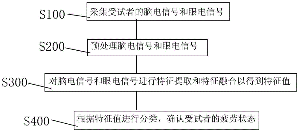
Referring to fig. 1 and 2, in a first aspect of the present invention, there is provided a fatigue detection method including:
s100, collecting electroencephalogram signals and electro-oculogram signals of a user;
an ocular electrical signal is an electrical signal generated by eye movement and can be measured by placing electrodes on the skin surrounding the eye. The magnitude of the eye electrical signal is determined according to the displacement change of the eyeball, contains rich information, and intuitively reflects the fatigue degree.
The brain electrical signal is formed by summing the postsynaptic potentials generated synchronously by a large number of neurons when the brain is active. It records the change of electrical signals during brain activity, is the overall reflection of the electrophysiological activity of brain nerve cells on the surface of cerebral cortex or scalp, and can reflect the degree of fatigue specifically.
In the step, the electroencephalogram signals of the user are collected through the wireless dry electrode electroencephalogram collecting equipment, and the eye electrical signals of the user are collected through the eye electrical signal collecting equipment. The collected electroencephalogram signals and electro-ocular signals are then stored in a database.
S200, preprocessing an electroencephalogram signal and an electro-oculogram signal;
specifically, step S200 includes:
s201, trend removing processing is carried out on the electroencephalogram signals and the electro-oculogram signals;
s202, mean value removing processing is carried out on the electroencephalogram signals and the electro-oculogram signals;
and S203, performing band-pass filtering processing on the electroencephalogram signals and the electro-oculogram signals.
Further, in this step, data of the brain electrical signal and the eye electrical signal are taken out from the database. Firstly, setting time windows for an electroencephalogram signal and an electro-oculogram signal; the time window of the electroencephalogram signal is 4s, and the step length is 1 s; the time window size of the electro-ocular signal is 10s and the step size is 1 s. Then, carrying out trend removing processing on the electroencephalogram signal and the electro-oculogram signal by using a spatial filtering mode; preferably, the spatial filtering method is a normal average reference filtering method. Then, mean value removing processing is carried out on the electroencephalogram signal and the electro-oculogram signal so as to remove high-frequency noise interference and improve the signal-to-noise ratio; finally, the signal-to-noise ratio is further improved by carrying out band-pass filtering on the frequency band signal of 0.01-32 Hz. The preprocessing of the electroencephalogram signal and the electro-oculogram signal is convenient for the subsequent feature extraction.
S300, performing feature extraction and feature fusion on the electroencephalogram signal and the electro-oculogram signal to obtain a feature value;
further, step S300 includes:
s310, reconstructing the electroencephalogram signals according to a frequency range;
specifically, the reconstructing the electroencephalogram signal according to the frequency range includes: reconstructing the electroencephalogram signals according to the frequency range by using a discrete wavelet transform method to obtain four sub-band waveforms of delta wave, theta wave, alpha wave and beta wave; wherein the frequency range of the delta wave is 0.01-3.91Hz, the frequency range of the theta wave is 3.91-7.81Hz, the frequency range of the alpha wave is 7.81-13.67Hz, and the frequency range of the beta wave is 13.67-31.25 Hz.
S320, extracting a first sample entropy of the eye electrical signal;
specifically, step S320 includes:
s321, extracting vertical direction sample entropy y of electro-oculogram signalSamEn;
S322, extracting horizontal direction sample entropy x of the electro-ocular signalSamEn。
The sample entropy is given by the following equation:
the sequence of N signal samples is: a ═ a (1), a (2) … a (n);
then the two subsequences of a are:
A(i)=[a(i),a(i+1),...,a(i+m-1)],1≤i≤N-m+1;
A(j)=[a(j),a(j+1),…,a(j+m-1)],1≤j≤N-m+1;
the distance between A (i) and A (j) is: d | a (i), a (j) | ═ max | a (i + k) -a (j + k) |;
further, the result of the sample entropy is expressed as:
r=0.2*SD;
wherein the parameter m specifically takes the value of 2; SD is the standard deviation of the sequence; num { d | A (i), A (j) | ≦ r } is the statistical number for i and j satisfying the condition d | A (i), A (j) | ≦ r.
S330, extracting the frequency spectrum entropy, the second sample entropy and the approximate entropy of the reconstructed electroencephalogram signal;
specifically, step S330 includes:
s331, extracting delta wave electroencephalogram signals to obtain delta wave spectrum entropy deltaSpeEnDelta wave sample entropy deltaSamEnSum delta wave approximate entropy deltaAppEn;
S332, extracting theta wave electroencephalogram signals to obtain theta wave spectral entropy thetaSpeEnTheta wave sample entropy thetaSamEnApproximate entropy of sum theta wave thetaAppEn;
S333, extracting alpha wave electroencephalogram signals to obtain alpha wave frequency spectrum entropy alphaSpeEnAlpha wave sample entropy alphaSamEnSum alpha wave approximate entropy alphaAppEn;
S334, extracting beta wave electroencephalogram signals to obtain beta wave spectrum entropy betaSpeEnEntropy of beta wave sample betaSamEnAnd beta wave approximate entropy betaAppEn。
The approximate entropy is given by the following equation:
for the samples of N signal sequences, the result of the approximate entropy is expressed as:
r=0.2*SD;
wherein the parameter m specifically takes the value of 2; SD is the standard deviation of the sequence.
The spectral entropy is given by the following equation:
wherein SpeEn is the result of spectrum entropy, f is the frequency corresponding to the frequency component, and n (f) is the total number of frequency components; q (f) is the normalized power spectral density component; p (f) is a power spectral density component; f. of
L、f
H、f
1And f
2The specific values are respectively 0.01, 31.25, 7.81 and 13.67;
is the least squares error of the frequency components;
are coefficients of a spectral entropy model.
The approximate entropy, the sample entropy and the spectrum entropy are all nonlinear dynamic parameters and can reflect the regularity of the input signal. The electroencephalogram signal and the electro-oculogram signal are calculated from a nonlinear angle through calculation of approximate entropy, sample entropy and frequency spectrum entropy.
S340, performing dimensionality reduction fusion on the frequency spectrum entropy, the second sample entropy and the approximate entropy of the reconstructed electroencephalogram signal to obtain a plurality of fusion characteristic entropies;
specifically, step S340 includes:
s341, entropy encoding delta wave frequency spectrum deltaSpeEnTheta wave frequency spectrum entropy thetaSpeEnAlpha wave spectral entropy alphaSpeEnAnd beta wave spectral entropy betaSpeEnPerforming dimensionality reduction fusion between every two to obtain a first fusion characteristic entropy;
s342, entropy coding delta wave sample by deltaSamEnTheta wave sample entropy thetaSamEnAlpha wave sample entropy alphaSamEnAnd entropy of beta wave sample betaSamEnPerforming dimensionality reduction fusion between every two to obtain a second fusion characteristic entropy;
s343, approximate entropy delta wave to deltaAppEnApproximate entropy of theta wave thetaAppEnAlpha wave approximate entropy alphaAppEnAnd beta wave approximate entropy betaAppEnAnd performing dimensionality reduction fusion between every two to obtain a third fusion characteristic entropy.
Specifically, in step S341, the δ -wave spectrum is entropy δSpeEnTheta wave frequency spectrum entropy thetaSpeEnAlpha wave spectral entropy alphaSpeEnAnd beta wave spectral entropy betaSpeEnTwo entropies are subjected to dimensionality reduction fusion to obtain a first fusion result, the other two entropies are subjected to dimensionality reduction fusion to obtain a second fusion result, and finally the first fusion result and the second fusion result are subjected to dimensionality reduction fusion to obtain a first fusion characteristic entropy. Step S342 and step S343 use the same dimension reduction fusion method.
Further, the specific expression of the fusion feature entropy in the fusion feature entropy obtained by the dimension reduction fusion is as follows: f ═ WcT*c+Wd*WdT*d;
Wherein Wc and W
dIs the projection vector of the input entropies c and d, Wc and W
dBy
Decision, E [ 2 ]]Is a correlation matrix. When rho (c, d) is maximizedValue of, Wc and W
dThere is an optimum value. Typical correlation analysis methods are used here to maximize the correlation between c and d to maintain independence of the two.
And S350, forming a characteristic matrix by the first sample entropy of the eye electrical signal and the plurality of fusion characteristic entropies of the brain electrical signal, and forming a characteristic value.
The characteristic value is a characteristic matrix formed by a first sample entropy of the electro-oculogram signal and a plurality of fusion characteristic entropies of the electroencephalogram signal, and the fusion characteristic entropies are obtained by entropy dimension reduction fusion corresponding to a plurality of frequency bands; the fatigue state of the user is analyzed and detected from the multi-dimensional angle through the step S350, and the noise immunity and the accuracy are improved.
S400, classifying according to the characteristic values and confirming the fatigue state of the user;
further, the classifying according to the feature value, and the confirming of the fatigue state of the user specifically includes:
and S410, inputting the characteristic value to the RVM classifier.
S420, calculating the probability of the fatigue state according to the following formula:
p(ti=0|w)=1-p(ti=1|w);
wherein x is [ x ]1,...xi,...x5]Inputting characteristic values of the RVM classifier; y (x)i(ii) a w) is the output of the RVM classifier; w is the weight of the RVM classifier; k (x, x)i)=exp(-g||x-xi||2) A kernel function for determining a mapping manner of the feature value from the low-dimensional space to the high-dimensional space; p (t)i1| w) is the probability value that the user is in a state of fatigue; p (t)i0| w) is the probability value that the user is in a non-tired state.
In the step S420 of the present embodiment,the variables are distributed independently, and the likelihood function of the RVM classifier is:
p (t) can be obtained according to the Laplacian approximation procedure (Laplacian approximation procedure) proposed by Michael E.doubling
i1| w) and p (t)
i0| w).
S430 according to p (t)i1| w) and p (t)i0| w) to confirm the fatigue state of the user.
Specifically, when p (t)i=1|w)>p(ti0| w), the user is confirmed to be in a fatigue state; when p (t)i=1|w)=p(ti0| w), the user is confirmed to be in a transition state; when p (t)i=1|w)<p(ti0| w), the user is confirmed to be in a non-fatigue state.
The method provided by the first aspect of the invention obtains the characteristic value by extracting and fusing the characteristics of the electroencephalogram signal and the electro-oculogram signal, and confirms the fatigue state of the user according to the characteristic value; the electroencephalogram signal and the electro-oculogram signal are combined, analysis and detection are carried out from a multi-dimensional angle, various entropies are introduced, the electroencephalogram signal and the electro-oculogram signal are analyzed from a nonlinear angle to express a fatigue state, and therefore the noise resistance and the accuracy of fatigue detection are improved.
The method is applied to the aspect of automobile driving, and the fatigue state of a driver can be accurately detected; when the driver is in a fatigue state, a warning is given to the driver, and the traffic accident rate is favorably reduced.
Referring to fig. 3, in a second aspect of the present invention, there is provided a fatigue detection apparatus capable of performing the fatigue detection method according to the first aspect of the present invention, including:
the acquisition module 10 is used for acquiring electroencephalogram signals and electro-oculogram signals of a user;
the preprocessing module 20 is used for preprocessing the electroencephalogram signals and the electro-oculogram signals;
the characteristic value acquisition module 30 is used for performing characteristic extraction and characteristic fusion on the electroencephalogram signal and the electro-oculogram signal to obtain a characteristic value;
a confirming module 40, which is used for classifying according to the characteristic value and confirming the fatigue state of the user;
wherein, eigenvalue acquisition module includes:
a reconstruction unit 31 for reconstructing the electroencephalogram signal according to the frequency range;
a first extraction unit 32 for extracting a first sample entropy of the ocular electrical signal;
the second extraction unit 33 is configured to extract a frequency spectrum entropy, a second sample entropy, and an approximate entropy of the reconstructed electroencephalogram signal;
the fusion unit 34 is configured to perform dimension reduction fusion on the frequency spectrum entropy, the second sample entropy and the approximate entropy of the reconstructed electroencephalogram signal to obtain a plurality of fusion feature entropies;
and the synthesizing unit 35 is configured to form the first sample entropy of the eye electrical signal and the multiple fusion feature entropies of the electroencephalogram signal into a feature value.
Specifically, the preprocessing unit 20 includes:
the de-trend unit is used for de-trend processing the electroencephalogram signal and the electro-oculogram signal;
the mean value removing unit is used for carrying out mean value removing processing on the electroencephalogram signals and the electro-oculogram signals;
and the band-pass filtering unit is used for carrying out band-pass filtering processing on the electroencephalogram signals and the electro-oculogram signals.
Specifically, the first extraction unit 32 includes:
a first extraction subunit for extracting vertical direction sample entropy y of the ocular electrical signalSamEn;
A second extraction subunit for extracting horizontal direction sample entropy x of the ocular electrical signalSamEn;
Specifically, the second extraction unit 33 includes:
a third extraction subunit for extracting the delta wave electroencephalogram signal to obtain delta wave spectrum entropy deltaSpeEnDelta wave sample entropy deltaSamEnSum delta wave approximate entropy deltaAppEn;
A fourth extraction subunit, configured to extract the theta wave electroencephalogram signal to obtain theta wave spectral entropy thetaSpeEnTheta wave sample entropy thetaSamEnApproximate entropy of sum theta wave thetaAppEn;
A fifth extraction subunit, configured to extract an alpha wave electroencephalogram signal to obtain an alpha wave spectral entropy alphaSpeEnAlpha wave sample entropy alphaSamEnSum alpha wave approximate entropy alphaAppEn;
A sixth extraction subunit, configured to extract the beta-wave electroencephalogram signal to obtain beta-wave spectral entropy betaSpeEnEntropy of beta wave sample betaSamEnAnd beta wave approximate entropy betaAppEn。
Specifically, the fusion unit 34 includes:
a first fusion subunit for entropy-dividing the delta wave spectrum by deltaSpeEnTheta wave frequency spectrum entropy thetaSpeEnAlpha wave spectral entropy alphaSpeEnAnd beta wave spectral entropy betaSpeEnPerforming dimensionality reduction fusion between every two to obtain a first fusion characteristic entropy;
a second fusion subunit for entropy-dividing the delta wave samples by deltaSamEnTheta wave sample entropy thetaSamEnAlpha wave sample entropy alphaSamEnAnd entropy of beta wave sample betaSamEnPerforming dimensionality reduction fusion between every two to obtain a second fusion characteristic entropy;
a third fusion subunit for approximating the delta wave to the entropy deltaAppEnApproximate entropy of theta wave thetaAppEnAlpha wave approximate entropy alphaAppEnAnd beta wave approximate entropy betaAppEnAnd performing dimensionality reduction fusion between every two to obtain a third fusion characteristic entropy.
Specifically, the confirmation unit 40 includes:
an input port for inputting the feature values to the RVM classifier;
a probability calculation unit for calculating the probability of the fatigue state according to the following formula:
p(ti=0|w)=1-p(ti=1|w);
wherein x is [ x ]1,...xi,...x5]Inputting characteristic values of the RVM classifier; y (x)i(ii) a w) is the output of the RVM classifier; w is the weight of the RVM classifier; k (x, x)i) Is a kernel function; p (t)i1| w) is the probability value that the user is in a state of fatigue; p (t)i0| w) is the probability value that the user is in a non-tired state;
a judging unit for judging according to p (t)i1| w) and p (t)i0| w) to confirm the fatigue state of the user.
In a third aspect of the invention, a fatigue detection apparatus is provided, which includes a processor and a memory communicatively connected to the at least one processor; the memory stores instructions executable by the processor to enable the processor to perform the fatigue detection method of the first aspect of the invention.
In a fourth aspect of the present invention, there is provided a storage medium having stored thereon computer-executable instructions for causing a computer to perform the fatigue detection method according to the first aspect of the present invention.
Table 1 is a comparison table of the accuracy of the present invention and 5 comparison methods. The 5 comparison methods are all single signal source method detection methods, the comparison method 1(Delta EEG) is a fatigue detection method judged according to a Delta wave electroencephalogram signal, the comparison method 2(Gamma EEG) is a fatigue detection method judged according to a Gamma wave electroencephalogram signal, the comparison method 3(Alpha EEG) is a fatigue detection method judged according to an Alpha wave electroencephalogram signal, the comparison method 4(Beta EEG) is a fatigue detection method judged according to a Beta wave electroencephalogram signal, and the comparison method 5(EOG) is a fatigue detection method judged according to an electrooculogram signal; as shown in Table 1, the accuracy of the method is greatly improved compared with other detection methods.
| Method
|
Accuracy rate
|
| Comparative method 1(Delta EEG)
|
90.2%
|
| Comparison method 2(Gamma EEG)
|
95.1%
|
| Contrast method 3(Alpha EEG)
|
92.7%
|
| Comparison method 4(Beta EEG)
|
94.2%
|
| Comparative method 5(EOG)
|
93.1%
|
| The invention
|
98.9% |
TABLE 1
The above description is only a preferred embodiment of the present invention, and the present invention is not limited to the above embodiment, and the present invention shall fall within the protection scope of the present invention as long as the technical effects of the present invention are achieved by the same means.