CN109840887B - Digital X-ray image splicing method and device - Google Patents
Digital X-ray image splicing method and device Download PDFInfo
- Publication number
- CN109840887B CN109840887B CN201910051526.5A CN201910051526A CN109840887B CN 109840887 B CN109840887 B CN 109840887B CN 201910051526 A CN201910051526 A CN 201910051526A CN 109840887 B CN109840887 B CN 109840887B
- Authority
- CN
- China
- Prior art keywords
- image
- processed
- determining
- transformation
- fusion
- Prior art date
- Legal status (The legal status is an assumption and is not a legal conclusion. Google has not performed a legal analysis and makes no representation as to the accuracy of the status listed.)
- Active
Links
- 238000000034 method Methods 0.000 title claims abstract description 35
- 230000009466 transformation Effects 0.000 claims abstract description 54
- 238000013519 translation Methods 0.000 claims abstract description 10
- 230000004927 fusion Effects 0.000 claims description 37
- 238000007781 pre-processing Methods 0.000 claims description 25
- 238000001228 spectrum Methods 0.000 claims description 17
- 230000009467 reduction Effects 0.000 claims description 6
- 210000003141 lower extremity Anatomy 0.000 claims description 5
- 210000000988 bone and bone Anatomy 0.000 claims description 4
- 238000010586 diagram Methods 0.000 description 5
- 230000008569 process Effects 0.000 description 4
- 230000000694 effects Effects 0.000 description 3
- 238000012545 processing Methods 0.000 description 3
- 238000000605 extraction Methods 0.000 description 2
- 238000005259 measurement Methods 0.000 description 2
- 238000012986 modification Methods 0.000 description 2
- 230000004048 modification Effects 0.000 description 2
- 238000003672 processing method Methods 0.000 description 2
- 230000009286 beneficial effect Effects 0.000 description 1
- 230000008859 change Effects 0.000 description 1
- 230000007547 defect Effects 0.000 description 1
- 238000003745 diagnosis Methods 0.000 description 1
- 239000004615 ingredient Substances 0.000 description 1
- 238000007689 inspection Methods 0.000 description 1
- 238000004519 manufacturing process Methods 0.000 description 1
- 238000013507 mapping Methods 0.000 description 1
- 230000007246 mechanism Effects 0.000 description 1
- 238000002360 preparation method Methods 0.000 description 1
Images


Landscapes
- Image Processing (AREA)
- Apparatus For Radiation Diagnosis (AREA)
Abstract
The invention provides a digital X-ray image splicing method and a digital X-ray image splicing device, wherein the method comprises the following steps: inputting n X-ray images to be processed, wherein n is more than or equal to 2, and n is a positive integer; respectively determining gradient fields of each image to be processed; determining the offset of translation transformation between gradient fields of a first to-be-processed image and a second to-be-processed image, fusing the overlapped area of the first to-be-processed image and the second to-be-processed image, and determining a first fused image; determining the offset of translation transformation between gradient fields of the ith fused image and the (i+1) th image to be processed, fusing the ith fused image and the (i+1) th image to be processed, and determining the ith fused image, wherein i is more than or equal to 2 and less than or equal to n-1. Therefore, the invention obtains the shift amount of the translation transformation of the overlapped area by utilizing the gradient field information of the image to be processed and utilizing the phase-related image matching method, thereby realizing the image splicing.
Description
Technical Field
The invention relates to the field of image processing, in particular to a digital X-ray image splicing method and device.
Background
Currently, in medical diagnosis (particularly in measurement) or in the preparation of surgical protocols using X-ray images, it is sometimes necessary to obtain complete spine images, complete lower limb images, or whole body bone images. However, due to the limitations of existing hardware (e.g., the detector size is smaller than the height of the spine), it is not possible to obtain the complete image in one exposure, and therefore, it is necessary to splice multiple images containing overlapping areas by digital means to obtain the complete image.
When an image processing algorithm is used for splicing, the technical scheme commonly used at present comprises a splicing method based on characteristic point matching and a splicing method based on template matching.
The feature point matching-based method mainly comprises the steps of searching key points in an image through a certain rule, and then searching out the mapping relation of the same (similar) feature points by utilizing a searching method so as to determine a spliced transformation model. The key points of the method lie in the selection of the characteristic points and the searching mechanism of the matching points, the number of the characteristic points is directly influenced by the speed of subsequent searching and the matching accuracy, if the characteristic points are not properly selected, the wrong matching result is easy to obtain, and in addition, the characteristic points are selected, so that no good unified rule exists at present.
The template matching method is mainly characterized in that a template image block with a specific size is selected, searching is carried out on another image, and the nearest image block is searched, so that a spliced transformation model is determined. The size of the template image block and the measurement index during searching the similar image block have no better solution at present, and meanwhile, the method has great operand and directly influences the splicing efficiency.
Disclosure of Invention
First, the technical problem to be solved
The present invention is directed to a method and apparatus for stitching digitized X-ray images, which solve at least one of the above-mentioned problems.
(II) technical scheme
The embodiment of the invention provides a digital X-ray image splicing method, which comprises the following steps:
inputting n X-ray images to be processed, wherein n is more than or equal to 2, and n is a positive integer;
respectively determining gradient fields of each image to be processed;
determining the offset of translational transformation between gradient fields of a first to-be-processed image and a second to-be-processed image, fusing the overlapped area of the first to-be-processed image and the second to-be-processed image, and determining a first fused image; determining the offset of translation transformation between gradient fields of the ith fused image and the (i+1) th image to be processed, fusing the overlapping area of the ith fused image and the (i+1) th image to be processed, and determining the ith fused image, wherein i is more than or equal to 2 and less than or equal to n-1.
In some embodiments of the invention, before determining the gradient fields of each image to be processed separately, the method further comprises the steps of:
the image to be processed is subjected to noise reduction preprocessing, specifically, preprocessing is performed on the image I (x, y) to be processed according to a Gaussian filter h (x, y), a preprocessing result f (x, y) =I (x, y) ×h (x, y) is determined, wherein, x and y are the abscissa and ordinate of the gaussian filter, respectively.
In some embodiments of the present invention, the gradient of each image to be processed is determined separately, referring to:
determining that the gradient field M (x, y) of each image to be processed meets the following conditions according to the preprocessing result:

wherein,, the partial derivatives of the image f (x, y) to be processed in the x direction and the y direction are respectively adopted, and a Sobel operator is adopted when the partial derivatives are calculated, namely

In some embodiments of the present invention, determining an offset of a translational transformation between gradient fields of a first image to be processed and a second image to be processed, or determining an offset of a translational transformation between gradient fields of an i-1 th fusion image and an i+1 th image to be processed, specifically comprises the sub-steps of:
calculating the 2D discrete Fourier transform of M (x, y) and determining a transformation result;
determining the first image to be processed and the second image to be processed or determining the cross power spectrum of the i-1 fusion image and the i+1 image to be processed according to the transformation result;
determining a normalized correlation coefficient according to the Fourier inverse transformation of the cross power spectrum;
and searching coordinates of peak points in the normalized correlation coefficient.
In some embodiments of the present invention, the transform result is formulated as g=f { M };
the formula of the cross power spectrum is Wherein (1)>
 Representing the product of the Hadamard and the Hadamard,G 1 is the transformation result of the first image to be processed, or the i-1 fusion image, +.>
 Representing the transformation result of the second image to be processed or the complex conjugate of the (i+1) th image to be processed;
the formula of normalized correlation coefficient is r=f -1 {R};
The formula of the coordinates is
In some embodiments of the invention, n is 2 or 3.
The embodiment of the invention also provides a digital X-ray image splicing device, which comprises:
the input module is used for inputting n images to be processed of the X-ray images, wherein n is more than or equal to 2, and n is a positive integer;
the gradient field determining module is used for determining the gradient field of each image to be processed;
the fusion module is used for determining the offset of translational transformation between gradient fields of the first to-be-processed image and the second to-be-processed image, fusing the overlapped area of the first to-be-processed image and the second to-be-processed image, and determining a first fusion image; determining the offset of translation transformation between gradient fields of the ith fused image and the (i+1) th image to be processed, fusing the overlapping area of the ith fused image and the (i+1) th image to be processed, and determining the ith fused image, wherein i is more than or equal to 2 and less than or equal to n-1.
In some embodiments of the invention, further comprising:
the preprocessing module is used for carrying out noise reduction preprocessing on the image to be processed, and specifically refers to: preprocessing the image I (x, y) to be processed according to a gaussian filter h (x, y), determining a preprocessing result f (x, y) =i (x, y) ×h (x, y), wherein, x and y are the abscissa and ordinate of the gaussian filter, respectively.
In some embodiments of the present invention, the gradient determining module is specifically configured to determine a gradient field M (x, y) of each image to be processed according to the preprocessing result, where the gradient field M satisfies the formula:

wherein,, the partial derivatives of the image f (x, y) to be processed in the x direction and the y direction are respectively adopted, and a Sobel operator is adopted when the partial derivatives are calculated, namely

In some embodiments of the invention, the fusion module comprises:
a transform unit for calculating a 2D discrete fourier transform of M (x, y), determining a transform result g=f { M };
a cross power spectrum unit for determining the first and second images to be processed or determining the cross power spectrum of the i-1 fusion image and the i+1 image to be processed according to the transformation result Wherein (1)>
 Represents the product of Hadamard, G 1 Is the transformation result of the first image to be processed, or the i-1 fusion image, +.>
 Representing the transformation result of the second image to be processed or the complex conjugate of the (i+1) th image to be processed;
an inverse transformation unit for determining normalized correlation coefficient r=f according to the inverse fourier transform of the cross power spectrum -1 {R};
A coordinate determination unit for finding the coordinates of the peak point in the normalized correlation coefficient
(III) beneficial effects
Compared with the prior art, the digital X-ray image splicing method and device have at least the following advantages:
1. the gradient field information of the image to be processed is utilized to obtain the transformation information of the overlapped area by utilizing a phase-related image processing method, and complex and time-consuming works such as feature extraction and searching are not needed, so that the execution speed is high, the checking efficiency can be improved, and the splicing efficiency and the matching accuracy are improved;
2. the method is insensitive to noise and has certain robustness to the brightness and contrast change of image data because of good robustness and based on gradient field information.
3. Before the images to be processed are fused, noise reduction pretreatment is further carried out on the images to be processed, so that the influence of noise on the splicing effect is reduced, the accuracy of image splicing is improved, and the pixel-level accuracy can be achieved.
4. The method is convenient to integrate, can be well applied to relevant medical examination environments such as X-rays, is small in algorithm operation amount, high in speed and accurate in splicing result, and can well solve the problems of low speed, low precision and the like of the traditional splicing method.
Drawings
FIG. 1 is a schematic diagram illustrating steps of a method for stitching digitized X-ray images according to an embodiment of the invention;
FIG. 2 is a schematic diagram of the process of fusing each two images in FIG. 1;
fig. 3 is a schematic block diagram of a digital X-ray image stitching device according to an embodiment of the invention.
Detailed Description
In the prior art, the splicing method based on characteristic point matching and the splicing method based on template matching have the defects of a matching result error method and low splicing efficiency, and therefore, the invention provides the digital X-ray image splicing method and device.
The present invention will be further described in detail below with reference to specific embodiments and with reference to the accompanying drawings, in order to make the objects, technical solutions and advantages of the present invention more apparent.
A first embodiment of the present invention provides a digital X-ray image stitching method, fig. 1 is a schematic diagram of steps of the digital X-ray image stitching method according to an embodiment of the present invention, fig. 2 is a schematic diagram of a process of fusing every two images in fig. 1, and as shown in fig. 1 and fig. 2, the method includes the following steps:
s1, inputting n X-ray images to be processed, wherein n is more than or equal to 2, and n is a positive integer;
s2, respectively determining gradient fields of each image to be processed;
s3, determining the offset of translational transformation between gradient fields of the first to-be-processed image and the second to-be-processed image (namely, the offset of translational transformation required when an image is spliced), fusing the overlapped area of the first to-be-processed image and the second to-be-processed image, and determining a first fused image; determining the offset of translation transformation between gradient fields of the ith fused image and the (i+1) th image to be processed, fusing the overlapping area of the ith fused image and the (i+1) th image to be processed, and determining the ith fused image, wherein i is more than or equal to 2 and less than or equal to n-1.
The input image 1 in fig. 2 refers to a first image to be processed or an i-1 th fusion image, and the input image 2 refers to a second image to be processed or an i+1 th image to be processed. For example, in step S1, there are 4 images to be processed, and the present invention fuses the first image to be processed and the second image to be processed according to the process of fig. 2 to the overlapping area of the two images to obtain a first fused image; fusing the overlapping area of the first fused image and the third image to be processed to obtain a second fused image; and finally, fusing the overlapping area of the second fused image and the fourth image to be processed to obtain a third fused image, namely the spliced image.
In addition, in order to reduce the influence of noise on the splicing effect, improve the precision of image splicing, before the overlapping area of the images to be processed is fused, the images to be processed can be subjected to noise reduction pretreatment, and the process is as follows:
preprocessing the image I (x, y) to be processed according to a gaussian filter h (x, y), determining a preprocessing result f (x, y) =i (x, y) ×h (x, y), wherein, x and y are the abscissa and ordinate of the gaussian filter, respectively.
In step S1, the number of the images to be processed is preferably 2 or 3, because the image formed by stitching 2 or 3 images to be processed can obtain a complete spine image, a complete lower limb image, or a whole body bone image.
In step S2, it is determined from the preprocessing result that the gradient field M (x, y) of each image to be processed satisfies the following formula:


In step S3, the specific substeps of determining the shift amount of the translational transformation between the gradient fields of the first to-be-processed image and the second to-be-processed image, or the shift amount of the translational transformation between the gradient fields of the i-1 th fused image and the i+1 th to-be-processed image are:
s31, calculating 2D discrete Fourier transform of M (x, y), and determining a transform result G=F { M };
s32, determining the first image to be processed and the second image to be processed or determining the i-1 fusion image and the i+1th image to be processed according to the transformation resultCross power spectrum of rational images
 Represents the product of Hadamard, G 1 Is the transformation result of the first image to be processed, or the i-1 fusion image, +.>
 Representing the transformation result of the second image to be processed or the complex conjugate of the (i+1) th image to be processed;
s33, determining a normalized correlation coefficient r=F according to the Fourier inverse transformation of the cross power spectrum -1 {R};
S34, searching coordinates of peak points in the normalized correlation coefficient The coordinates are the offset of translation transformation between the first image to be processed and the second image to be processed or between the i-1 fusion image and the i+1 image to be processed when the first image to be processed and the second image to be processed are spliced.
That is to say, until the n-1 fusion image and the n+1 to-be-processed image are fused in the overlapping area, namely, after each to-be-processed image participates in fusion, the obtained fusion image is a complete spliced image.
The embodiment of the invention also provides a digital X-ray image splicing device, as shown in fig. 3, which comprises:
the input module is used for inputting n images to be processed of the X-ray images, wherein n is more than or equal to 2, and n is a positive integer;
the gradient field determining module is used for determining the gradient field of each image to be processed;
the fusion module is used for determining the offset of translational transformation between gradient fields of the first to-be-processed image and the second to-be-processed image, fusing the overlapped area of the first to-be-processed image and the second to-be-processed image, and determining a first fusion image; determining the offset of translation transformation between gradient fields of the ith fused image and the (i+1) th image to be processed, fusing the overlapping area of the ith fused image and the (i+1) th image to be processed, and determining the ith fused image, wherein i is more than or equal to 2 and less than or equal to n-1.
In addition, in order to reduce the influence of noise on the stitching effect and improve the accuracy of image stitching, the device may further include a preprocessing module for preprocessing the image I (x, y) to be processed according to the gaussian filter h (x, y), and determining a preprocessing result f (x, y) =i (x, y) ×h (x, y), where, x and y are the abscissa and ordinate of the gaussian filter, respectively.
In general, the number of images to be processed is preferably 2 or 3, because the images formed by stitching 2 or 3 images to be processed can obtain a complete spine image, a complete lower limb image, or a whole body bone image.
The gradient field determining module determines the gradient field of each image to be processed according to the preprocessing result, and the formula of the gradient field is as follows In (I)>
 The partial derivatives of the image f (x, y) to be processed in the x direction and the y direction are respectively adopted, and a Sobel operator is adopted when the partial derivatives are calculated, namely

In some embodiments of the present invention, the fusion module specifically includes:
a transform unit for calculating a 2D discrete fourier transform of M (x, y), determining a transform result g=f { M };
the cross power spectrum unit is used for determining the first image to be processed and the second image to be processed or determining the i-1 fusion image and the i+1th image to be processed according to the transformation resultCross-power spectrum for processing images Wherein (1)>
 Represents the product of Hadamard, G 1 Is the transformation result of the first image to be processed, or the i-1 fusion image, +.>
 Representing the transformation result of the second image to be processed or the complex conjugate of the (i+1) th image to be processed;
an inverse transformation unit for determining normalized correlation coefficient r=f according to the inverse fourier transform of the cross power spectrum -1 {R};
A coordinate determination unit for finding the coordinates of the peak point in the normalized correlation coefficient
That is to say, until the n-1 fusion image and the n+1 to-be-processed image are fused in the overlapping area, namely, after each to-be-processed image participates in fusion, the obtained fusion image is a complete spliced image.
In summary, the digital X-ray image stitching method and device provided by the invention acquire the transformation information of the overlapped area by utilizing the phase-related image processing method through the gradient field information of the image to be processed, and do not need complex and time-consuming work such as feature extraction and search, so that the execution speed is high, the inspection efficiency can be improved, and the stitching efficiency and the matching accuracy are improved.
Unless otherwise known, the numerical parameters in this specification and the attached claims are approximations that may vary depending upon the desired properties sought to be obtained by the present disclosure. In particular, all numbers expressing quantities of ingredients, reaction conditions, and so forth used in the specification and claims are to be understood as being modified in all instances by the term "about". In general, the meaning of expression is meant to include a variation of + -10% in some embodiments, a variation of + -5% in some embodiments, a variation of + -1% in some embodiments, and a variation of + -0.5% in some embodiments by a particular amount.
Furthermore, "comprising" does not exclude the presence of elements or steps not listed in a claim. The singular reference of "a", "an", and "the" preceding an element does not exclude the plural reference of such elements.
The use of ordinal numbers such as "first," "second," "third," etc., in the description and the claims to modify a corresponding element does not by itself connote any ordinal number of elements or the order of manufacturing or use of the ordinal numbers in a particular claim, merely for enabling an element having a particular name to be clearly distinguished from another element having the same name.
While the foregoing is directed to embodiments of the present invention, other and further details of the invention may be had by the present invention, it should be understood that the foregoing description is merely illustrative of the present invention and that no limitations are intended to the scope of the invention, except insofar as modifications, equivalents, improvements or modifications are within the spirit and principles of the invention.
Claims (3)
1. A method of digitized X-ray image stitching comprising:
inputting n X-ray images to be processed, wherein n is more than or equal to 2, and n is a positive integer; the X-ray images include spine images, lower limb images, and bone images;
the image to be processed is subjected to noise reduction preprocessing, specifically, preprocessing is performed on the image I (x, y) to be processed according to a Gaussian filter h (x, y), a preprocessing result f (x, y) =I (x, y) ×h (x, y) is determined, wherein,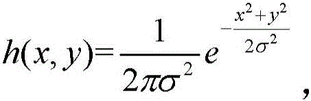 x and y are the horizontal and vertical coordinates of the Gaussian filter respectively;
respectively determining gradient fields of each image to be processed;finger means: determining that the gradient field M (x, y) of each image to be processed meets the following conditions according to the preprocessing result: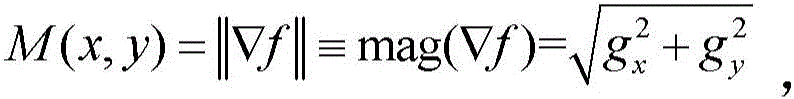 wherein (1)>
 The partial derivatives of the image f (x, y) to be processed in the x direction and the y direction are respectively adopted, and a Sobel operator is adopted when the partial derivatives are calculated, namely
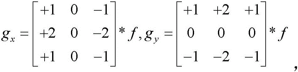 * Representing a convolution operation;
determining the offset of translational transformation between gradient fields of a first to-be-processed image and a second to-be-processed image, fusing the overlapped area of the first to-be-processed image and the second to-be-processed image, and determining a first fused image; determining the offset of translation transformation between gradient fields of an i-1 fusion image and an i+1 to-be-processed image, fusing an overlapping area of the i-1 fusion image and the i+1 to-be-processed image, and determining an i-1 fusion image, wherein i is more than or equal to 2 and less than or equal to n-1;
determining an offset of a translational transformation between gradient fields of the first image to be processed and the second image to be processed or determining an offset of a translational transformation between gradient fields of the i-1 th fusion image and the i+1 th image to be processed, specifically comprising the sub-steps of: calculating the 2D discrete Fourier transform of M (x, y) and determining a transformation result; determining the first image to be processed and the second image to be processed or determining the cross power spectrum of the i-1 fusion image and the i+1 image to be processed according to the transformation result; determining a normalized correlation coefficient according to the Fourier inverse transformation of the cross power spectrum; searching coordinates of peak points in the normalized correlation coefficient;
the formula of the transformation result is g=f { M }; the formula of the cross power spectrum is Wherein (1)>
 Representation ofHadamard product, G1 is the transformation result of the first image to be processed, or the i-1 th fusion image,/L>
 Representing the transformation result of the second image to be processed or the complex conjugate of the (i+1) th image to be processed; the formula of normalized correlation coefficient is r=f -1 { R }; the formula of the coordinates is
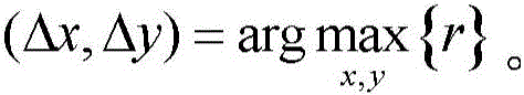
2. The method of claim 1, wherein n is 2 or 3.
3. A digitized X-ray image stitching device comprising:
the input module is used for inputting n images to be processed of the X-ray images, wherein n is more than or equal to 2, and n is a positive integer; the X-ray images comprise spine images, lower limb images and skeleton images;
the preprocessing module is used for carrying out noise reduction preprocessing on the image to be processed, and specifically refers to: preprocessing the image I (x, y) to be processed according to a gaussian filter h (x, y), determining a preprocessing result f (x, y) =i (x, y) ×h (x, y), wherein,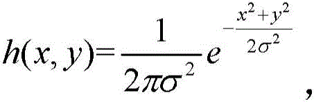 x and y are the horizontal and vertical coordinates of the Gaussian filter respectively;
the gradient field determining module is used for determining the gradient field of each image to be processed; the gradient determining module is specifically configured to determine a gradient field M (x, y) of each image to be processed according to the preprocessing result, where the gradient field M satisfies the formula: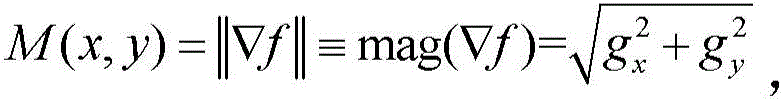 wherein (1)>
 The partial derivatives of the image f (x, y) to be processed in the x direction and the y direction are respectively adopted, and a Sobel operator is adopted when the partial derivatives are calculated, namely
 * Representing a convolution operation;
the fusion module is used for determining the offset of translational transformation between gradient fields of the first to-be-processed image and the second to-be-processed image, fusing the overlapped area of the first to-be-processed image and the second to-be-processed image, and determining a first fusion image; determining the offset of translation transformation between gradient fields of an i-1 fusion image and an i+1 to-be-processed image, fusing an overlapping area of the i-1 fusion image and the i+1 to-be-processed image, and determining an i-1 fusion image, wherein i is more than or equal to 2 and less than or equal to n-1;
the fusion module comprises:
a transform unit for calculating a 2D discrete fourier transform of M (x, y), determining a transform result g=f { M };
a cross power spectrum unit for determining the first and second images to be processed or determining the cross power spectrum of the i-1 fusion image and the i+1 image to be processed according to the transformation result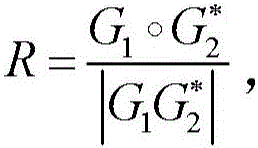 Wherein (1)>
 Represents the product of Hadamard, G 1 Is the transformation result of the first image to be processed, or the i-1 fusion image, +.>
 Representing the transformation result of the second image to be processed or the complex conjugate of the (i+1) th image to be processed;
an inverse transformation unit for determining normalized correlation coefficient r=f according to the inverse fourier transform of the cross power spectrum -1 {R};
A coordinate determination unit for finding the coordinates of the peak point in the normalized correlation coefficient
Priority Applications (1)
| Application Number | Priority Date | Filing Date | Title |
|---|---|---|---|
| CN201910051526.5A CN109840887B (en) | 2019-01-18 | 2019-01-18 | Digital X-ray image splicing method and device |
Applications Claiming Priority (1)
| Application Number | Priority Date | Filing Date | Title |
|---|---|---|---|
| CN201910051526.5A CN109840887B (en) | 2019-01-18 | 2019-01-18 | Digital X-ray image splicing method and device |
Publications (2)
| Publication Number | Publication Date |
|---|---|
| CN109840887A CN109840887A (en) | 2019-06-04 |
| CN109840887B true CN109840887B (en) | 2023-05-12 |
Family
ID=66883957
Family Applications (1)
| Application Number | Title | Priority Date | Filing Date |
|---|---|---|---|
| CN201910051526.5A Active CN109840887B (en) | 2019-01-18 | 2019-01-18 | Digital X-ray image splicing method and device |
Country Status (1)
| Country | Link |
|---|---|
| CN (1) | CN109840887B (en) |
Families Citing this family (1)
| Publication number | Priority date | Publication date | Assignee | Title |
|---|---|---|---|---|
| CN116681620B (en) * | 2023-04-28 | 2026-04-14 | 北京友通上昊科技有限公司 | Spliced image gray level processing method, image splicing method, device and medium |
Family Cites Families (9)
| Publication number | Priority date | Publication date | Assignee | Title |
|---|---|---|---|---|
| CN103279939B (en) * | 2013-04-27 | 2016-01-20 | 北京工业大学 | A kind of image mosaic disposal system |
| CN103530844A (en) * | 2013-09-17 | 2014-01-22 | 上海皓信生物科技有限公司 | Splicing method based on mycobacterium tuberculosis acid-fast staining image |
| CN105069749B (en) * | 2015-07-22 | 2018-04-13 | 广东工业大学 | A kind of joining method of tire-mold image |
| CN105631811A (en) * | 2016-02-25 | 2016-06-01 | 科盾科技股份有限公司 | Image stitching method and device |
| CN106296587B (en) * | 2016-08-19 | 2020-03-06 | 广东工业大学 | Tire mold image stitching method |
| CN106384334A (en) * | 2016-09-26 | 2017-02-08 | 西安交通大学 | Mutual information-based steel plate image splicing method |
| CN106815802A (en) * | 2016-12-23 | 2017-06-09 | 深圳超多维科技有限公司 | A kind of image split-joint method and device |
| CN106709897B (en) * | 2016-12-28 | 2019-11-26 | 武汉大学 | Optimal splicing line finding method and system between orthography based on gradient field |
| CN109146798A (en) * | 2018-07-10 | 2019-01-04 | 西安天盈光电科技有限公司 | image detail enhancement method |
-
2019
- 2019-01-18 CN CN201910051526.5A patent/CN109840887B/en active Active
Also Published As
| Publication number | Publication date |
|---|---|
| CN109840887A (en) | 2019-06-04 |
Similar Documents
| Publication | Publication Date | Title |
|---|---|---|
| Li et al. | Multifocus image fusion via fixed window technique of multiscale images and non-local means filtering | |
| Bukhari et al. | Automatic radial distortion estimation from a single image | |
| Chi et al. | Machine vision based automatic detection method of indicating values of a pointer gauge | |
| CN102369550B (en) | Stereo image processor and stereo image processing method | |
| US8337405B2 (en) | System and method for automatic detection of anomalies in images | |
| Kisworo et al. | Modeling edges at subpixel accuracy using the local energy approach | |
| US20050238253A1 (en) | Image registration | |
| US20120213425A1 (en) | Combining feature boundaries | |
| US11189023B2 (en) | Devices, systems, and methods for anchor-point-enabled multi-scale subfield alignment | |
| JPWO2012060093A1 (en) | Stereo image processing apparatus and stereo image processing method | |
| CN108629788B (en) | Image edge detection method, device and equipment and readable storage medium | |
| Lotz et al. | Zooming in: high resolution 3D reconstruction of differently stained histological whole slide images | |
| CN106652017B (en) | Method and device for judging DICOM image file integrity in three-dimensional reconstruction | |
| CN101315700A (en) | Fast automatic positioning method for multi-sequence image | |
| CN109840887B (en) | Digital X-ray image splicing method and device | |
| Liu et al. | Deep 3D-DIC using a coarse-to-fine network for robust and accurate 3D shape and displacement measurements | |
| CN111915554B (en) | Fracture detection and positioning integrated method and device based on X-ray image | |
| Tahiri et al. | Stable computation of Hahn polynomials for higher polynomial order | |
| JP4513365B2 (en) | Medical image processing apparatus and medical image processing program | |
| US8971627B2 (en) | Template matching processing device and template matching processing program | |
| EP2300990B1 (en) | Image analysis system & method | |
| Yue et al. | Weighted circle fusion: ensembling circle representation from different object detection results | |
| Weibo et al. | Performance evaluation approach for image mosaicing algorithm | |
| Vrochidis et al. | Enhancing 3D Reconstructions in Underwater Environments: The Impact of Image Enhancement on Model Quality | |
| Ruppertshofen et al. | Multi-level approach for the discriminative generalized hough transform |
Legal Events
| Date | Code | Title | Description |
|---|---|---|---|
| PB01 | Publication | ||
| PB01 | Publication | ||
| SE01 | Entry into force of request for substantive examination | ||
| SE01 | Entry into force of request for substantive examination | ||
| GR01 | Patent grant | ||
| GR01 | Patent grant |