CN109535257B - Novel bispecific CD3/CD19 polypeptide complex - Google Patents
Novel bispecific CD3/CD19 polypeptide complex Download PDFInfo
- Publication number
- CN109535257B CN109535257B CN201811100222.5A CN201811100222A CN109535257B CN 109535257 B CN109535257 B CN 109535257B CN 201811100222 A CN201811100222 A CN 201811100222A CN 109535257 B CN109535257 B CN 109535257B
- Authority
- CN
- China
- Prior art keywords
- ser
- seq
- antibody
- val
- polypeptide complex
- Prior art date
- Legal status (The legal status is an assumption and is not a legal conclusion. Google has not performed a legal analysis and makes no representation as to the accuracy of the status listed.)
- Active
Links
- 108090000765 processed proteins & peptides Proteins 0.000 title claims abstract description 240
- 102000004196 processed proteins & peptides Human genes 0.000 title claims abstract description 230
- 229920001184 polypeptide Polymers 0.000 title claims abstract description 229
- 102100024222 B-lymphocyte antigen CD19 Human genes 0.000 title claims description 46
- 101000980825 Homo sapiens B-lymphocyte antigen CD19 Proteins 0.000 title claims description 46
- 230000027455 binding Effects 0.000 claims abstract description 283
- 238000009739 binding Methods 0.000 claims abstract description 277
- 239000000427 antigen Substances 0.000 claims abstract description 179
- 108091007433 antigens Proteins 0.000 claims abstract description 179
- 102000036639 antigens Human genes 0.000 claims abstract description 179
- 238000000034 method Methods 0.000 claims abstract description 37
- 102000040430 polynucleotide Human genes 0.000 claims abstract description 36
- 108091033319 polynucleotide Proteins 0.000 claims abstract description 36
- 239000002157 polynucleotide Substances 0.000 claims abstract description 36
- 239000013598 vector Substances 0.000 claims abstract description 27
- 239000000203 mixture Substances 0.000 claims abstract description 25
- 208000037265 diseases, disorders, signs and symptoms Diseases 0.000 claims abstract description 17
- 239000008194 pharmaceutical composition Substances 0.000 claims abstract description 16
- 201000010099 disease Diseases 0.000 claims abstract description 11
- 210000004027 cell Anatomy 0.000 claims description 178
- 108091008874 T cell receptors Proteins 0.000 claims description 131
- 102000016266 T-Cell Antigen Receptors Human genes 0.000 claims description 123
- 150000001413 amino acids Chemical class 0.000 claims description 79
- 206010028980 Neoplasm Diseases 0.000 claims description 47
- 238000006471 dimerization reaction Methods 0.000 claims description 44
- 239000000539 dimer Substances 0.000 claims description 21
- 230000003993 interaction Effects 0.000 claims description 17
- 201000011510 cancer Diseases 0.000 claims description 14
- 230000009881 electrostatic interaction Effects 0.000 claims description 10
- 229910052739 hydrogen Inorganic materials 0.000 claims description 10
- 239000001257 hydrogen Substances 0.000 claims description 10
- 230000000087 stabilizing effect Effects 0.000 claims description 10
- 208000024893 Acute lymphoblastic leukemia Diseases 0.000 claims description 7
- 208000014697 Acute lymphocytic leukaemia Diseases 0.000 claims description 7
- 208000006664 Precursor Cell Lymphoblastic Leukemia-Lymphoma Diseases 0.000 claims description 7
- 238000002360 preparation method Methods 0.000 claims description 7
- 208000036170 B-Cell Marginal Zone Lymphoma Diseases 0.000 claims description 6
- 208000003950 B-cell lymphoma Diseases 0.000 claims description 6
- 239000003937 drug carrier Substances 0.000 claims description 6
- 208000010839 B-cell chronic lymphocytic leukemia Diseases 0.000 claims description 5
- 208000031671 Large B-Cell Diffuse Lymphoma Diseases 0.000 claims description 5
- 208000031422 Lymphocytic Chronic B-Cell Leukemia Diseases 0.000 claims description 5
- 206010012818 diffuse large B-cell lymphoma Diseases 0.000 claims description 5
- 230000002209 hydrophobic effect Effects 0.000 claims description 5
- 206010025323 Lymphomas Diseases 0.000 claims description 4
- 208000015914 Non-Hodgkin lymphomas Diseases 0.000 claims description 4
- 208000032852 chronic lymphocytic leukemia Diseases 0.000 claims description 4
- 238000012258 culturing Methods 0.000 claims description 4
- 239000000833 heterodimer Substances 0.000 claims description 4
- 208000017604 Hodgkin disease Diseases 0.000 claims description 3
- 208000021519 Hodgkin lymphoma Diseases 0.000 claims description 3
- 208000010747 Hodgkins lymphoma Diseases 0.000 claims description 3
- 201000003791 MALT lymphoma Diseases 0.000 claims description 3
- 208000025205 Mantle-Cell Lymphoma Diseases 0.000 claims description 3
- 208000033559 Waldenström macroglobulinemia Diseases 0.000 claims description 3
- 238000005734 heterodimerization reaction Methods 0.000 claims description 3
- 201000007924 marginal zone B-cell lymphoma Diseases 0.000 claims description 3
- 208000021937 marginal zone lymphoma Diseases 0.000 claims description 3
- 208000000461 Esophageal Neoplasms Diseases 0.000 claims description 2
- 206010030155 Oesophageal carcinoma Diseases 0.000 claims description 2
- 206010060862 Prostate cancer Diseases 0.000 claims description 2
- 208000000236 Prostatic Neoplasms Diseases 0.000 claims description 2
- 208000005718 Stomach Neoplasms Diseases 0.000 claims description 2
- 201000004101 esophageal cancer Diseases 0.000 claims description 2
- 201000003444 follicular lymphoma Diseases 0.000 claims description 2
- 206010017758 gastric cancer Diseases 0.000 claims description 2
- 208000005017 glioblastoma Diseases 0.000 claims description 2
- 201000001441 melanoma Diseases 0.000 claims description 2
- 201000011549 stomach cancer Diseases 0.000 claims description 2
- 125000003275 alpha amino acid group Chemical group 0.000 claims 2
- 210000000481 breast Anatomy 0.000 claims 1
- 210000001072 colon Anatomy 0.000 claims 1
- 210000004185 liver Anatomy 0.000 claims 1
- 210000004072 lung Anatomy 0.000 claims 1
- 230000002611 ovarian Effects 0.000 claims 1
- 210000002307 prostate Anatomy 0.000 claims 1
- 102000017420 CD3 protein, epsilon/gamma/delta subunit Human genes 0.000 description 53
- 108050005493 CD3 protein, epsilon/gamma/delta subunit Proteins 0.000 description 53
- 235000001014 amino acid Nutrition 0.000 description 52
- 229940024606 amino acid Drugs 0.000 description 52
- 108090000623 proteins and genes Proteins 0.000 description 44
- 210000001744 T-lymphocyte Anatomy 0.000 description 37
- 102000004169 proteins and genes Human genes 0.000 description 37
- 125000000539 amino acid group Chemical group 0.000 description 35
- 235000018102 proteins Nutrition 0.000 description 34
- 241000282567 Macaca fascicularis Species 0.000 description 32
- 230000014509 gene expression Effects 0.000 description 32
- 238000000684 flow cytometry Methods 0.000 description 24
- 238000006467 substitution reaction Methods 0.000 description 20
- 101100112922 Candida albicans CDR3 gene Proteins 0.000 description 19
- 210000002966 serum Anatomy 0.000 description 17
- 102100035360 Cerebellar degeneration-related antigen 1 Human genes 0.000 description 16
- 239000000126 substance Substances 0.000 description 16
- 238000002965 ELISA Methods 0.000 description 15
- 241000699670 Mus sp. Species 0.000 description 15
- 150000007523 nucleic acids Chemical group 0.000 description 15
- 108010070643 prolylglutamic acid Proteins 0.000 description 15
- 238000011282 treatment Methods 0.000 description 15
- 108020004414 DNA Proteins 0.000 description 14
- 102000053602 DNA Human genes 0.000 description 14
- 108010069205 aspartyl-phenylalanine Proteins 0.000 description 14
- 108010092854 aspartyllysine Proteins 0.000 description 14
- 230000000694 effects Effects 0.000 description 14
- 239000000047 product Substances 0.000 description 14
- 241000880493 Leptailurus serval Species 0.000 description 13
- 102100021651 SUN domain-containing ossification factor Human genes 0.000 description 13
- 125000000151 cysteine group Chemical group N[C@@H](CS)C(=O)* 0.000 description 13
- 108010047041 Complementarity Determining Regions Proteins 0.000 description 12
- 239000003814 drug Substances 0.000 description 12
- 241000282693 Cercopithecidae Species 0.000 description 11
- 210000003719 b-lymphocyte Anatomy 0.000 description 11
- 238000013461 design Methods 0.000 description 11
- 108020001507 fusion proteins Proteins 0.000 description 11
- 102000037865 fusion proteins Human genes 0.000 description 11
- 230000035772 mutation Effects 0.000 description 11
- 239000002904 solvent Substances 0.000 description 11
- 101000611183 Homo sapiens Tumor necrosis factor Proteins 0.000 description 10
- 108010065920 Insulin Lispro Proteins 0.000 description 10
- 102100040247 Tumor necrosis factor Human genes 0.000 description 10
- 239000000872 buffer Substances 0.000 description 10
- 230000006870 function Effects 0.000 description 10
- 108010050848 glycylleucine Proteins 0.000 description 10
- 238000000746 purification Methods 0.000 description 10
- 239000000243 solution Substances 0.000 description 10
- 102100025137 Early activation antigen CD69 Human genes 0.000 description 9
- 101000934374 Homo sapiens Early activation antigen CD69 Proteins 0.000 description 9
- 101001055144 Homo sapiens Interleukin-2 receptor subunit alpha Proteins 0.000 description 9
- 241001465754 Metazoa Species 0.000 description 9
- RAGOJJCBGXARPO-XVSYOHENSA-N Phe-Thr-Asp Chemical compound OC(=O)C[C@@H](C(O)=O)NC(=O)[C@H]([C@H](O)C)NC(=O)[C@@H](N)CC1=CC=CC=C1 RAGOJJCBGXARPO-XVSYOHENSA-N 0.000 description 9
- 238000003556 assay Methods 0.000 description 9
- KRKNYBCHXYNGOX-UHFFFAOYSA-N citric acid Chemical compound OC(=O)CC(O)(C(O)=O)CC(O)=O KRKNYBCHXYNGOX-UHFFFAOYSA-N 0.000 description 9
- 238000001943 fluorescence-activated cell sorting Methods 0.000 description 9
- 239000012634 fragment Substances 0.000 description 9
- 230000013595 glycosylation Effects 0.000 description 9
- 238000006206 glycosylation reaction Methods 0.000 description 9
- 210000003819 peripheral blood mononuclear cell Anatomy 0.000 description 9
- 125000006850 spacer group Chemical group 0.000 description 9
- GYWQGGUCMDCUJE-DLOVCJGASA-N Asp-Phe-Ala Chemical compound [H]N[C@@H](CC(O)=O)C(=O)N[C@@H](CC1=CC=CC=C1)C(=O)N[C@@H](C)C(O)=O GYWQGGUCMDCUJE-DLOVCJGASA-N 0.000 description 8
- BYYNJRSNDARRBX-YFKPBYRVSA-N Gly-Gln-Gly Chemical compound NCC(=O)N[C@@H](CCC(N)=O)C(=O)NCC(O)=O BYYNJRSNDARRBX-YFKPBYRVSA-N 0.000 description 8
- 101001057504 Homo sapiens Interferon-stimulated gene 20 kDa protein Proteins 0.000 description 8
- 108060003951 Immunoglobulin Proteins 0.000 description 8
- 102100026878 Interleukin-2 receptor subunit alpha Human genes 0.000 description 8
- PMGDADKJMCOXHX-UHFFFAOYSA-N L-Arginyl-L-glutamin-acetat Natural products NC(=N)NCCCC(N)C(=O)NC(CCC(N)=O)C(O)=O PMGDADKJMCOXHX-UHFFFAOYSA-N 0.000 description 8
- XUJNEKJLAYXESH-REOHCLBHSA-N L-Cysteine Chemical compound SC[C@H](N)C(O)=O XUJNEKJLAYXESH-REOHCLBHSA-N 0.000 description 8
- PDIDTSZKKFEDMB-UWVGGRQHSA-N Lys-Pro-Gly Chemical compound [H]N[C@@H](CCCCN)C(=O)N1CCC[C@H]1C(=O)NCC(O)=O PDIDTSZKKFEDMB-UWVGGRQHSA-N 0.000 description 8
- DIBZLYZXTSVGLN-CIUDSAMLSA-N Lys-Ser-Ser Chemical compound [H]N[C@@H](CCCCN)C(=O)N[C@@H](CO)C(=O)N[C@@H](CO)C(O)=O DIBZLYZXTSVGLN-CIUDSAMLSA-N 0.000 description 8
- 241000699666 Mus <mouse, genus> Species 0.000 description 8
- -1 OKT3(Kung et al Chemical compound 0.000 description 8
- FMDHKPRACUXATF-ACZMJKKPSA-N Ser-Gln-Ser Chemical compound OC[C@H](N)C(=O)N[C@@H](CCC(N)=O)C(=O)N[C@@H](CO)C(O)=O FMDHKPRACUXATF-ACZMJKKPSA-N 0.000 description 8
- XXXAXOWMBOKTRN-XPUUQOCRSA-N Ser-Gly-Val Chemical compound [H]N[C@@H](CO)C(=O)NCC(=O)N[C@@H](C(C)C)C(O)=O XXXAXOWMBOKTRN-XPUUQOCRSA-N 0.000 description 8
- 230000006044 T cell activation Effects 0.000 description 8
- MNYNCKZAEIAONY-XGEHTFHBSA-N Thr-Val-Ser Chemical compound C[C@@H](O)[C@H](N)C(=O)N[C@@H](C(C)C)C(=O)N[C@@H](CO)C(O)=O MNYNCKZAEIAONY-XGEHTFHBSA-N 0.000 description 8
- LHADRQBREKTRLR-DCAQKATOSA-N Val-Cys-Leu Chemical compound CC(C)C[C@@H](C(=O)O)NC(=O)[C@H](CS)NC(=O)[C@H](C(C)C)N LHADRQBREKTRLR-DCAQKATOSA-N 0.000 description 8
- 108010086434 alanyl-seryl-glycine Proteins 0.000 description 8
- 108010008355 arginyl-glutamine Proteins 0.000 description 8
- 230000004927 fusion Effects 0.000 description 8
- 102000018358 immunoglobulin Human genes 0.000 description 8
- 239000007924 injection Substances 0.000 description 8
- 238000002347 injection Methods 0.000 description 8
- 108010069117 seryl-lysyl-aspartic acid Proteins 0.000 description 8
- 238000002415 sodium dodecyl sulfate polyacrylamide gel electrophoresis Methods 0.000 description 8
- 239000006228 supernatant Substances 0.000 description 8
- 238000002560 therapeutic procedure Methods 0.000 description 8
- 230000009258 tissue cross reactivity Effects 0.000 description 8
- 108010038745 tryptophylglycine Proteins 0.000 description 8
- HKRXJBBCQBAGIM-FXQIFTODSA-N Arg-Asp-Ser Chemical compound C(C[C@@H](C(=O)N[C@@H](CC(=O)O)C(=O)N[C@@H](CO)C(=O)O)N)CN=C(N)N HKRXJBBCQBAGIM-FXQIFTODSA-N 0.000 description 7
- DPNWSMBUYCLEDG-CIUDSAMLSA-N Asp-Lys-Ser Chemical compound [H]N[C@@H](CC(O)=O)C(=O)N[C@@H](CCCCN)C(=O)N[C@@H](CO)C(O)=O DPNWSMBUYCLEDG-CIUDSAMLSA-N 0.000 description 7
- 108020004705 Codon Proteins 0.000 description 7
- XHVONGZZVUUORG-WEDXCCLWSA-N Gly-Thr-Lys Chemical compound NCC(=O)N[C@@H]([C@H](O)C)C(=O)N[C@H](C(O)=O)CCCCN XHVONGZZVUUORG-WEDXCCLWSA-N 0.000 description 7
- 102100026120 IgG receptor FcRn large subunit p51 Human genes 0.000 description 7
- 101710177940 IgG receptor FcRn large subunit p51 Proteins 0.000 description 7
- SITLTJHOQZFJGG-UHFFFAOYSA-N N-L-alpha-glutamyl-L-valine Natural products CC(C)C(C(O)=O)NC(=O)C(N)CCC(O)=O SITLTJHOQZFJGG-UHFFFAOYSA-N 0.000 description 7
- 108091028043 Nucleic acid sequence Proteins 0.000 description 7
- 240000004808 Saccharomyces cerevisiae Species 0.000 description 7
- 235000014680 Saccharomyces cerevisiae Nutrition 0.000 description 7
- YUJLIIRMIAGMCQ-CIUDSAMLSA-N Ser-Leu-Ser Chemical compound [H]N[C@@H](CO)C(=O)N[C@@H](CC(C)C)C(=O)N[C@@H](CO)C(O)=O YUJLIIRMIAGMCQ-CIUDSAMLSA-N 0.000 description 7
- KZSYAEWQMJEGRZ-RHYQMDGZSA-N Thr-Leu-Val Chemical compound [H]N[C@@H]([C@@H](C)O)C(=O)N[C@@H](CC(C)C)C(=O)N[C@@H](C(C)C)C(O)=O KZSYAEWQMJEGRZ-RHYQMDGZSA-N 0.000 description 7
- DOBHJKVVACOQTN-DZKIICNBSA-N Val-Tyr-Gln Chemical compound NC(=O)CC[C@@H](C(O)=O)NC(=O)[C@@H](NC(=O)[C@@H](N)C(C)C)CC1=CC=C(O)C=C1 DOBHJKVVACOQTN-DZKIICNBSA-N 0.000 description 7
- 238000004458 analytical method Methods 0.000 description 7
- 108010077245 asparaginyl-proline Proteins 0.000 description 7
- 229910052799 carbon Inorganic materials 0.000 description 7
- 239000003795 chemical substances by application Substances 0.000 description 7
- 238000004519 manufacturing process Methods 0.000 description 7
- 229920000642 polymer Polymers 0.000 description 7
- 230000003405 preventing effect Effects 0.000 description 7
- 230000009870 specific binding Effects 0.000 description 7
- 229940124597 therapeutic agent Drugs 0.000 description 7
- JJHBEVZAZXZREW-LFSVMHDDSA-N Ala-Thr-Phe Chemical compound C[C@@H](O)[C@H](NC(=O)[C@H](C)N)C(=O)N[C@@H](Cc1ccccc1)C(O)=O JJHBEVZAZXZREW-LFSVMHDDSA-N 0.000 description 6
- TVUFMYKTYXTRPY-HERUPUMHSA-N Ala-Trp-Ser Chemical compound [H]N[C@@H](C)C(=O)N[C@@H](CC1=CNC2=C1C=CC=C2)C(=O)N[C@@H](CO)C(O)=O TVUFMYKTYXTRPY-HERUPUMHSA-N 0.000 description 6
- ISJWBVIYRBAXEB-CIUDSAMLSA-N Arg-Ser-Glu Chemical compound [H]N[C@@H](CCCNC(N)=N)C(=O)N[C@@H](CO)C(=O)N[C@@H](CCC(O)=O)C(O)=O ISJWBVIYRBAXEB-CIUDSAMLSA-N 0.000 description 6
- CPTXATAOUQJQRO-GUBZILKMSA-N Arg-Val-Ser Chemical compound [H]N[C@@H](CCCNC(N)=N)C(=O)N[C@@H](C(C)C)C(=O)N[C@@H](CO)C(O)=O CPTXATAOUQJQRO-GUBZILKMSA-N 0.000 description 6
- OLVIPTLKNSAYRJ-YUMQZZPRSA-N Asn-Gly-Lys Chemical compound C(CCN)C[C@@H](C(=O)O)NC(=O)CNC(=O)[C@H](CC(=O)N)N OLVIPTLKNSAYRJ-YUMQZZPRSA-N 0.000 description 6
- XTMZYFMTYJNABC-ZLUOBGJFSA-N Asn-Ser-Ala Chemical compound C[C@@H](C(=O)O)NC(=O)[C@H](CO)NC(=O)[C@H](CC(=O)N)N XTMZYFMTYJNABC-ZLUOBGJFSA-N 0.000 description 6
- YTXCCDCOHIYQFC-GUBZILKMSA-N Asp-Met-Arg Chemical compound OC(=O)C[C@H](N)C(=O)N[C@@H](CCSC)C(=O)N[C@@H](CCCN=C(N)N)C(O)=O YTXCCDCOHIYQFC-GUBZILKMSA-N 0.000 description 6
- KPSHWSWFPUDEGF-FXQIFTODSA-N Asp-Pro-Ala Chemical compound OC(=O)[C@H](C)NC(=O)[C@@H]1CCCN1C(=O)[C@@H](N)CC(O)=O KPSHWSWFPUDEGF-FXQIFTODSA-N 0.000 description 6
- MNQMTYSEKZHIDF-GCJQMDKQSA-N Asp-Thr-Ala Chemical compound [H]N[C@@H](CC(O)=O)C(=O)N[C@@H]([C@@H](C)O)C(=O)N[C@@H](C)C(O)=O MNQMTYSEKZHIDF-GCJQMDKQSA-N 0.000 description 6
- HTSSXFASOUSJQG-IHPCNDPISA-N Asp-Tyr-Trp Chemical compound [H]N[C@@H](CC(O)=O)C(=O)N[C@@H](CC1=CC=C(O)C=C1)C(=O)N[C@@H](CC1=CNC2=C1C=CC=C2)C(O)=O HTSSXFASOUSJQG-IHPCNDPISA-N 0.000 description 6
- JGLWFWXGOINXEA-YDHLFZDLSA-N Asp-Val-Tyr Chemical compound OC(=O)C[C@H](N)C(=O)N[C@@H](C(C)C)C(=O)N[C@H](C(O)=O)CC1=CC=C(O)C=C1 JGLWFWXGOINXEA-YDHLFZDLSA-N 0.000 description 6
- PLBJMUUEGBBHRH-ZLUOBGJFSA-N Cys-Ala-Asn Chemical compound [H]N[C@@H](CS)C(=O)N[C@@H](C)C(=O)N[C@@H](CC(N)=O)C(O)=O PLBJMUUEGBBHRH-ZLUOBGJFSA-N 0.000 description 6
- 102000004127 Cytokines Human genes 0.000 description 6
- 108090000695 Cytokines Proteins 0.000 description 6
- LFQSCWFLJHTTHZ-UHFFFAOYSA-N Ethanol Chemical compound CCO LFQSCWFLJHTTHZ-UHFFFAOYSA-N 0.000 description 6
- CKNUKHBRCSMKMO-XHNCKOQMSA-N Gln-Asn-Pro Chemical compound C1C[C@@H](N(C1)C(=O)[C@H](CC(=O)N)NC(=O)[C@H](CCC(=O)N)N)C(=O)O CKNUKHBRCSMKMO-XHNCKOQMSA-N 0.000 description 6
- ZFBBMCKQSNJZSN-AUTRQRHGSA-N Gln-Val-Gln Chemical compound [H]N[C@@H](CCC(N)=O)C(=O)N[C@@H](C(C)C)C(=O)N[C@@H](CCC(N)=O)C(O)=O ZFBBMCKQSNJZSN-AUTRQRHGSA-N 0.000 description 6
- VNCNWQPIQYAMAK-ACZMJKKPSA-N Glu-Ser-Ser Chemical compound [H]N[C@@H](CCC(O)=O)C(=O)N[C@@H](CO)C(=O)N[C@@H](CO)C(O)=O VNCNWQPIQYAMAK-ACZMJKKPSA-N 0.000 description 6
- ZALGPUWUVHOGAE-GVXVVHGQSA-N Glu-Val-His Chemical compound CC(C)[C@@H](C(=O)N[C@@H](CC1=CN=CN1)C(=O)O)NC(=O)[C@H](CCC(=O)O)N ZALGPUWUVHOGAE-GVXVVHGQSA-N 0.000 description 6
- ZYRXTRTUCAVNBQ-GVXVVHGQSA-N Glu-Val-Lys Chemical compound CC(C)[C@@H](C(=O)N[C@@H](CCCCN)C(=O)O)NC(=O)[C@H](CCC(=O)O)N ZYRXTRTUCAVNBQ-GVXVVHGQSA-N 0.000 description 6
- WQZGKKKJIJFFOK-GASJEMHNSA-N Glucose Natural products OC[C@H]1OC(O)[C@H](O)[C@@H](O)[C@@H]1O WQZGKKKJIJFFOK-GASJEMHNSA-N 0.000 description 6
- PEDCQBHIVMGVHV-UHFFFAOYSA-N Glycerine Chemical compound OCC(O)CO PEDCQBHIVMGVHV-UHFFFAOYSA-N 0.000 description 6
- JBCLFWXMTIKCCB-UHFFFAOYSA-N H-Gly-Phe-OH Natural products NCC(=O)NC(C(O)=O)CC1=CC=CC=C1 JBCLFWXMTIKCCB-UHFFFAOYSA-N 0.000 description 6
- AYUOWUNWZGTNKB-ULQDDVLXSA-N His-Phe-Arg Chemical compound [H]N[C@@H](CC1=CNC=N1)C(=O)N[C@@H](CC1=CC=CC=C1)C(=O)N[C@@H](CCCNC(N)=N)C(O)=O AYUOWUNWZGTNKB-ULQDDVLXSA-N 0.000 description 6
- YCKPUHHMCFSUMD-IUKAMOBKSA-N Ile-Thr-Asp Chemical compound CC[C@H](C)[C@@H](C(=O)N[C@@H]([C@@H](C)O)C(=O)N[C@@H](CC(=O)O)C(=O)O)N YCKPUHHMCFSUMD-IUKAMOBKSA-N 0.000 description 6
- FEHQLKKBVJHSEC-SZMVWBNQSA-N Leu-Glu-Trp Chemical compound C1=CC=C2C(C[C@H](NC(=O)[C@H](CCC(O)=O)NC(=O)[C@@H](N)CC(C)C)C(O)=O)=CNC2=C1 FEHQLKKBVJHSEC-SZMVWBNQSA-N 0.000 description 6
- MVJRBCJCRYGCKV-GVXVVHGQSA-N Leu-Val-Gln Chemical compound [H]N[C@@H](CC(C)C)C(=O)N[C@@H](C(C)C)C(=O)N[C@@H](CCC(N)=O)C(O)=O MVJRBCJCRYGCKV-GVXVVHGQSA-N 0.000 description 6
- UWKNTTJNVSYXPC-CIUDSAMLSA-N Lys-Ala-Ser Chemical compound OC[C@@H](C(O)=O)NC(=O)[C@H](C)NC(=O)[C@@H](N)CCCCN UWKNTTJNVSYXPC-CIUDSAMLSA-N 0.000 description 6
- NRQRKMYZONPCTM-CIUDSAMLSA-N Lys-Asp-Ser Chemical compound [H]N[C@@H](CCCCN)C(=O)N[C@@H](CC(O)=O)C(=O)N[C@@H](CO)C(O)=O NRQRKMYZONPCTM-CIUDSAMLSA-N 0.000 description 6
- GQZMPWBZQALKJO-UWVGGRQHSA-N Lys-Gly-Arg Chemical compound [H]N[C@@H](CCCCN)C(=O)NCC(=O)N[C@@H](CCCNC(N)=N)C(O)=O GQZMPWBZQALKJO-UWVGGRQHSA-N 0.000 description 6
- SJDQOYTYNGZZJX-SRVKXCTJSA-N Met-Glu-Leu Chemical compound CSCC[C@H](N)C(=O)N[C@@H](CCC(O)=O)C(=O)N[C@@H](CC(C)C)C(O)=O SJDQOYTYNGZZJX-SRVKXCTJSA-N 0.000 description 6
- KZNQNBZMBZJQJO-UHFFFAOYSA-N N-glycyl-L-proline Natural products NCC(=O)N1CCCC1C(O)=O KZNQNBZMBZJQJO-UHFFFAOYSA-N 0.000 description 6
- OJUMUUXGSXUZJZ-SRVKXCTJSA-N Phe-Asp-Ser Chemical compound [H]N[C@@H](CC1=CC=CC=C1)C(=O)N[C@@H](CC(O)=O)C(=O)N[C@@H](CO)C(O)=O OJUMUUXGSXUZJZ-SRVKXCTJSA-N 0.000 description 6
- SCKXGHWQPPURGT-KKUMJFAQSA-N Phe-Lys-Ser Chemical compound [H]N[C@@H](CC1=CC=CC=C1)C(=O)N[C@@H](CCCCN)C(=O)N[C@@H](CO)C(O)=O SCKXGHWQPPURGT-KKUMJFAQSA-N 0.000 description 6
- OCSACVPBMIYNJE-GUBZILKMSA-N Pro-Arg-Asn Chemical compound [H]N1CCC[C@H]1C(=O)N[C@@H](CCCNC(N)=N)C(=O)N[C@@H](CC(N)=O)C(O)=O OCSACVPBMIYNJE-GUBZILKMSA-N 0.000 description 6
- TUYWCHPXKQTISF-LPEHRKFASA-N Pro-Cys-Pro Chemical compound C1C[C@H](NC1)C(=O)N[C@@H](CS)C(=O)N2CCC[C@@H]2C(=O)O TUYWCHPXKQTISF-LPEHRKFASA-N 0.000 description 6
- MGDFPGCFVJFITQ-CIUDSAMLSA-N Pro-Glu-Asp Chemical compound [H]N1CCC[C@H]1C(=O)N[C@@H](CCC(O)=O)C(=O)N[C@@H](CC(O)=O)C(O)=O MGDFPGCFVJFITQ-CIUDSAMLSA-N 0.000 description 6
- SNGZLPOXVRTNMB-LPEHRKFASA-N Pro-Ser-Pro Chemical compound C1C[C@H](NC1)C(=O)N[C@@H](CO)C(=O)N2CCC[C@@H]2C(=O)O SNGZLPOXVRTNMB-LPEHRKFASA-N 0.000 description 6
- PRKWBYCXBBSLSK-GUBZILKMSA-N Pro-Ser-Val Chemical compound [H]N1CCC[C@H]1C(=O)N[C@@H](CO)C(=O)N[C@@H](C(C)C)C(O)=O PRKWBYCXBBSLSK-GUBZILKMSA-N 0.000 description 6
- JEHPKECJCALLRW-CUJWVEQBSA-N Ser-His-Thr Chemical compound [H]N[C@@H](CO)C(=O)N[C@@H](CC1=CNC=N1)C(=O)N[C@@H]([C@@H](C)O)C(O)=O JEHPKECJCALLRW-CUJWVEQBSA-N 0.000 description 6
- HBTCFCHYALPXME-HTFCKZLJSA-N Ser-Ile-Ile Chemical compound [H]N[C@@H](CO)C(=O)N[C@@H]([C@@H](C)CC)C(=O)N[C@@H]([C@@H](C)CC)C(O)=O HBTCFCHYALPXME-HTFCKZLJSA-N 0.000 description 6
- KJKQUQXDEKMPDK-FXQIFTODSA-N Ser-Met-Asp Chemical compound [H]N[C@@H](CO)C(=O)N[C@@H](CCSC)C(=O)N[C@@H](CC(O)=O)C(O)=O KJKQUQXDEKMPDK-FXQIFTODSA-N 0.000 description 6
- BMKNXTJLHFIAAH-CIUDSAMLSA-N Ser-Ser-Leu Chemical compound [H]N[C@@H](CO)C(=O)N[C@@H](CO)C(=O)N[C@@H](CC(C)C)C(O)=O BMKNXTJLHFIAAH-CIUDSAMLSA-N 0.000 description 6
- XJDMUQCLVSCRSJ-VZFHVOOUSA-N Ser-Thr-Ala Chemical compound [H]N[C@@H](CO)C(=O)N[C@@H]([C@@H](C)O)C(=O)N[C@@H](C)C(O)=O XJDMUQCLVSCRSJ-VZFHVOOUSA-N 0.000 description 6
- LGIMRDKGABDMBN-DCAQKATOSA-N Ser-Val-Lys Chemical compound CC(C)[C@@H](C(=O)N[C@@H](CCCCN)C(=O)O)NC(=O)[C@H](CO)N LGIMRDKGABDMBN-DCAQKATOSA-N 0.000 description 6
- FAPWRFPIFSIZLT-UHFFFAOYSA-M Sodium chloride Chemical compound [Na+].[Cl-] FAPWRFPIFSIZLT-UHFFFAOYSA-M 0.000 description 6
- VEIKMWOMUYMMMK-FCLVOEFKSA-N Thr-Phe-Phe Chemical compound C([C@H](NC(=O)[C@@H](N)[C@H](O)C)C(=O)N[C@@H](CC=1C=CC=CC=1)C(O)=O)C1=CC=CC=C1 VEIKMWOMUYMMMK-FCLVOEFKSA-N 0.000 description 6
- SSNGFWKILJLTQM-QEJZJMRPSA-N Trp-Gln-Asn Chemical compound C1=CC=C2C(=C1)C(=CN2)C[C@@H](C(=O)N[C@@H](CCC(=O)N)C(=O)N[C@@H](CC(=O)N)C(=O)O)N SSNGFWKILJLTQM-QEJZJMRPSA-N 0.000 description 6
- MBLJBGZWLHTJBH-SZMVWBNQSA-N Trp-Val-Arg Chemical compound C1=CC=C2C(C[C@H](N)C(=O)N[C@@H](C(C)C)C(=O)N[C@@H](CCCN=C(N)N)C(O)=O)=CNC2=C1 MBLJBGZWLHTJBH-SZMVWBNQSA-N 0.000 description 6
- JQTYTBPCSOAZHI-FXQIFTODSA-N Val-Ser-Cys Chemical compound CC(C)[C@@H](C(=O)N[C@@H](CO)C(=O)N[C@@H](CS)C(=O)O)N JQTYTBPCSOAZHI-FXQIFTODSA-N 0.000 description 6
- WUFHZIRMAZZWRS-OSUNSFLBSA-N Val-Thr-Ile Chemical compound CC[C@H](C)[C@@H](C(=O)O)NC(=O)[C@H]([C@@H](C)O)NC(=O)[C@H](C(C)C)N WUFHZIRMAZZWRS-OSUNSFLBSA-N 0.000 description 6
- OWFGFHQMSBTKLX-UFYCRDLUSA-N Val-Tyr-Tyr Chemical compound CC(C)[C@@H](C(=O)N[C@@H](CC1=CC=C(C=C1)O)C(=O)N[C@@H](CC2=CC=C(C=C2)O)C(=O)O)N OWFGFHQMSBTKLX-UFYCRDLUSA-N 0.000 description 6
- 239000003963 antioxidant agent Substances 0.000 description 6
- 235000006708 antioxidants Nutrition 0.000 description 6
- 210000004369 blood Anatomy 0.000 description 6
- 239000008280 blood Substances 0.000 description 6
- 238000010367 cloning Methods 0.000 description 6
- 239000013078 crystal Substances 0.000 description 6
- 208000035475 disorder Diseases 0.000 description 6
- 238000009472 formulation Methods 0.000 description 6
- 108010063718 gamma-glutamylaspartic acid Proteins 0.000 description 6
- 108010089804 glycyl-threonine Proteins 0.000 description 6
- 238000011534 incubation Methods 0.000 description 6
- 210000003292 kidney cell Anatomy 0.000 description 6
- 102000039446 nucleic acids Human genes 0.000 description 6
- 108020004707 nucleic acids Proteins 0.000 description 6
- 108010083476 phenylalanyltryptophan Proteins 0.000 description 6
- YBYRMVIVWMBXKQ-UHFFFAOYSA-N phenylmethanesulfonyl fluoride Chemical compound FS(=O)(=O)CC1=CC=CC=C1 YBYRMVIVWMBXKQ-UHFFFAOYSA-N 0.000 description 6
- 230000009467 reduction Effects 0.000 description 6
- 238000012360 testing method Methods 0.000 description 6
- 210000004881 tumor cell Anatomy 0.000 description 6
- 238000005406 washing Methods 0.000 description 6
- KAZKWIKPEPABOO-IHRRRGAJSA-N Asn-Met-Tyr Chemical compound CSCC[C@@H](C(=O)N[C@@H](CC1=CC=C(C=C1)O)C(=O)O)NC(=O)[C@H](CC(=O)N)N KAZKWIKPEPABOO-IHRRRGAJSA-N 0.000 description 5
- 210000001266 CD8-positive T-lymphocyte Anatomy 0.000 description 5
- UIQGJYUEQDOODF-KWQFWETISA-N Gly-Tyr-Ala Chemical compound OC(=O)[C@H](C)NC(=O)[C@@H](NC(=O)CN)CC1=CC=C(O)C=C1 UIQGJYUEQDOODF-KWQFWETISA-N 0.000 description 5
- DHMQDGOQFOQNFH-UHFFFAOYSA-N Glycine Chemical compound NCC(O)=O DHMQDGOQFOQNFH-UHFFFAOYSA-N 0.000 description 5
- 101100383038 Homo sapiens CD19 gene Proteins 0.000 description 5
- VEXZGXHMUGYJMC-UHFFFAOYSA-N Hydrochloric acid Chemical compound Cl VEXZGXHMUGYJMC-UHFFFAOYSA-N 0.000 description 5
- 108010073807 IgG Receptors Proteins 0.000 description 5
- 102000009490 IgG Receptors Human genes 0.000 description 5
- AYFVYJQAPQTCCC-GBXIJSLDSA-N L-threonine Chemical compound C[C@@H](O)[C@H](N)C(O)=O AYFVYJQAPQTCCC-GBXIJSLDSA-N 0.000 description 5
- 206010027476 Metastases Diseases 0.000 description 5
- 230000004988 N-glycosylation Effects 0.000 description 5
- 239000004698 Polyethylene Substances 0.000 description 5
- ZOHGLPQGEHSLPD-FXQIFTODSA-N Ser-Gln-Glu Chemical compound [H]N[C@@H](CO)C(=O)N[C@@H](CCC(N)=O)C(=O)N[C@@H](CCC(O)=O)C(O)=O ZOHGLPQGEHSLPD-FXQIFTODSA-N 0.000 description 5
- HUPLKEHTTQBXSC-YJRXYDGGSA-N Thr-Ser-Tyr Chemical compound C[C@@H](O)[C@H](N)C(=O)N[C@@H](CO)C(=O)N[C@H](C(O)=O)CC1=CC=C(O)C=C1 HUPLKEHTTQBXSC-YJRXYDGGSA-N 0.000 description 5
- VHIZXDZMTDVFGX-DCAQKATOSA-N Val-Ser-Leu Chemical compound CC(C)C[C@@H](C(=O)O)NC(=O)[C@H](CO)NC(=O)[C@H](C(C)C)N VHIZXDZMTDVFGX-DCAQKATOSA-N 0.000 description 5
- 238000007792 addition Methods 0.000 description 5
- 108010045350 alanyl-tyrosyl-alanine Proteins 0.000 description 5
- WQZGKKKJIJFFOK-VFUOTHLCSA-N beta-D-glucose Chemical compound OC[C@H]1O[C@@H](O)[C@H](O)[C@@H](O)[C@@H]1O WQZGKKKJIJFFOK-VFUOTHLCSA-N 0.000 description 5
- 230000001588 bifunctional effect Effects 0.000 description 5
- 238000005119 centrifugation Methods 0.000 description 5
- DQLATGHUWYMOKM-UHFFFAOYSA-L cisplatin Chemical compound N[Pt](N)(Cl)Cl DQLATGHUWYMOKM-UHFFFAOYSA-L 0.000 description 5
- 238000001514 detection method Methods 0.000 description 5
- 229940079593 drug Drugs 0.000 description 5
- 239000012636 effector Substances 0.000 description 5
- 239000003995 emulsifying agent Substances 0.000 description 5
- 239000008103 glucose Substances 0.000 description 5
- 238000001727 in vivo Methods 0.000 description 5
- 230000002401 inhibitory effect Effects 0.000 description 5
- 239000007788 liquid Substances 0.000 description 5
- 230000003211 malignant effect Effects 0.000 description 5
- 238000005259 measurement Methods 0.000 description 5
- 239000002609 medium Substances 0.000 description 5
- 230000009401 metastasis Effects 0.000 description 5
- 125000002496 methyl group Chemical group [H]C([H])([H])* 0.000 description 5
- DNIAPMSPPWPWGF-UHFFFAOYSA-N monopropylene glycol Natural products CC(O)CO DNIAPMSPPWPWGF-UHFFFAOYSA-N 0.000 description 5
- 239000000843 powder Substances 0.000 description 5
- 230000001105 regulatory effect Effects 0.000 description 5
- 150000003839 salts Chemical class 0.000 description 5
- 239000007787 solid Substances 0.000 description 5
- 239000000758 substrate Substances 0.000 description 5
- 230000004614 tumor growth Effects 0.000 description 5
- 239000003981 vehicle Substances 0.000 description 5
- XLYOFNOQVPJJNP-UHFFFAOYSA-N water Chemical compound O XLYOFNOQVPJJNP-UHFFFAOYSA-N 0.000 description 5
- YBJHBAHKTGYVGT-ZKWXMUAHSA-N (+)-Biotin Chemical compound N1C(=O)N[C@@H]2[C@H](CCCCC(=O)O)SC[C@@H]21 YBJHBAHKTGYVGT-ZKWXMUAHSA-N 0.000 description 4
- MTCFGRXMJLQNBG-REOHCLBHSA-N (2S)-2-Amino-3-hydroxypropansäure Chemical compound OC[C@H](N)C(O)=O MTCFGRXMJLQNBG-REOHCLBHSA-N 0.000 description 4
- COEXAQSTZUWMRI-STQMWFEESA-N (2s)-1-[2-[[(2s)-2-amino-3-(4-hydroxyphenyl)propanoyl]amino]acetyl]pyrrolidine-2-carboxylic acid Chemical compound C([C@H](N)C(=O)NCC(=O)N1[C@@H](CCC1)C(O)=O)C1=CC=C(O)C=C1 COEXAQSTZUWMRI-STQMWFEESA-N 0.000 description 4
- GGNHBHYDMUDXQB-KBIXCLLPSA-N Ala-Glu-Ile Chemical compound CC[C@H](C)[C@@H](C(O)=O)NC(=O)[C@H](CCC(O)=O)NC(=O)[C@H](C)N GGNHBHYDMUDXQB-KBIXCLLPSA-N 0.000 description 4
- CJQAEJMHBAOQHA-DLOVCJGASA-N Ala-Phe-Asn Chemical compound C[C@@H](C(=O)N[C@@H](CC1=CC=CC=C1)C(=O)N[C@@H](CC(=O)N)C(=O)O)N CJQAEJMHBAOQHA-DLOVCJGASA-N 0.000 description 4
- WNHNMKOFKCHKKD-BFHQHQDPSA-N Ala-Thr-Gly Chemical compound [H]N[C@@H](C)C(=O)N[C@@H]([C@@H](C)O)C(=O)NCC(O)=O WNHNMKOFKCHKKD-BFHQHQDPSA-N 0.000 description 4
- AENHOIXXHKNIQL-AUTRQRHGSA-N Ala-Tyr-Ala Chemical compound [O-]C(=O)[C@H](C)NC(=O)[C@@H](NC(=O)[C@@H]([NH3+])C)CC1=CC=C(O)C=C1 AENHOIXXHKNIQL-AUTRQRHGSA-N 0.000 description 4
- VBFJESQBIWCWRL-DCAQKATOSA-N Arg-Ala-Lys Chemical compound NCCCC[C@@H](C(O)=O)NC(=O)[C@H](C)NC(=O)[C@@H](N)CCCNC(N)=N VBFJESQBIWCWRL-DCAQKATOSA-N 0.000 description 4
- BHQQRVARKXWXPP-ACZMJKKPSA-N Asn-Asp-Glu Chemical compound C(CC(=O)O)[C@@H](C(=O)O)NC(=O)[C@H](CC(=O)O)NC(=O)[C@H](CC(=O)N)N BHQQRVARKXWXPP-ACZMJKKPSA-N 0.000 description 4
- QNJIRRVTOXNGMH-GUBZILKMSA-N Asn-Gln-Lys Chemical compound NCCCC[C@@H](C(O)=O)NC(=O)[C@H](CCC(N)=O)NC(=O)[C@@H](N)CC(N)=O QNJIRRVTOXNGMH-GUBZILKMSA-N 0.000 description 4
- COWITDLVHMZSIW-CIUDSAMLSA-N Asn-Lys-Ser Chemical compound [H]N[C@@H](CC(N)=O)C(=O)N[C@@H](CCCCN)C(=O)N[C@@H](CO)C(O)=O COWITDLVHMZSIW-CIUDSAMLSA-N 0.000 description 4
- YRBGRUOSJROZEI-NHCYSSNCSA-N Asp-His-Val Chemical compound [H]N[C@@H](CC(O)=O)C(=O)N[C@@H](CC1=CNC=N1)C(=O)N[C@@H](C(C)C)C(O)=O YRBGRUOSJROZEI-NHCYSSNCSA-N 0.000 description 4
- GGRSYTUJHAZTFN-IHRRRGAJSA-N Asp-Pro-Tyr Chemical compound C1C[C@H](N(C1)C(=O)[C@H](CC(=O)O)N)C(=O)N[C@@H](CC2=CC=C(C=C2)O)C(=O)O GGRSYTUJHAZTFN-IHRRRGAJSA-N 0.000 description 4
- ZBYLEBZCVKLPCY-FXQIFTODSA-N Asp-Ser-Arg Chemical compound [H]N[C@@H](CC(O)=O)C(=O)N[C@@H](CO)C(=O)N[C@@H](CCCNC(N)=N)C(O)=O ZBYLEBZCVKLPCY-FXQIFTODSA-N 0.000 description 4
- IJGRMHOSHXDMSA-UHFFFAOYSA-N Atomic nitrogen Chemical compound N#N IJGRMHOSHXDMSA-UHFFFAOYSA-N 0.000 description 4
- KZZYVYWSXMFYEC-DCAQKATOSA-N Cys-Val-Leu Chemical compound [H]N[C@@H](CS)C(=O)N[C@@H](C(C)C)C(=O)N[C@@H](CC(C)C)C(O)=O KZZYVYWSXMFYEC-DCAQKATOSA-N 0.000 description 4
- AOJJSUZBOXZQNB-TZSSRYMLSA-N Doxorubicin Chemical group O([C@H]1C[C@@](O)(CC=2C(O)=C3C(=O)C=4C=CC=C(C=4C(=O)C3=C(O)C=21)OC)C(=O)CO)[C@H]1C[C@H](N)[C@H](O)[C@H](C)O1 AOJJSUZBOXZQNB-TZSSRYMLSA-N 0.000 description 4
- KCXVZYZYPLLWCC-UHFFFAOYSA-N EDTA Chemical compound OC(=O)CN(CC(O)=O)CCN(CC(O)=O)CC(O)=O KCXVZYZYPLLWCC-UHFFFAOYSA-N 0.000 description 4
- 108010021468 Fc gamma receptor IIA Proteins 0.000 description 4
- INKFLNZBTSNFON-CIUDSAMLSA-N Gln-Ala-Arg Chemical compound NC(=O)CC[C@H](N)C(=O)N[C@@H](C)C(=O)N[C@@H](CCCN=C(N)N)C(O)=O INKFLNZBTSNFON-CIUDSAMLSA-N 0.000 description 4
- FFVXLVGUJBCKRX-UKJIMTQDSA-N Gln-Ile-Val Chemical compound CC[C@H](C)[C@@H](C(=O)N[C@@H](C(C)C)C(=O)O)NC(=O)[C@H](CCC(=O)N)N FFVXLVGUJBCKRX-UKJIMTQDSA-N 0.000 description 4
- NMYFPKCIGUJMIK-GUBZILKMSA-N Gln-Met-Gln Chemical compound CSCC[C@@H](C(=O)N[C@@H](CCC(=O)N)C(=O)O)NC(=O)[C@H](CCC(=O)N)N NMYFPKCIGUJMIK-GUBZILKMSA-N 0.000 description 4
- XQDGOJPVMSWZSO-SRVKXCTJSA-N Gln-Pro-Leu Chemical compound CC(C)C[C@@H](C(=O)O)NC(=O)[C@@H]1CCCN1C(=O)[C@H](CCC(=O)N)N XQDGOJPVMSWZSO-SRVKXCTJSA-N 0.000 description 4
- WLIPTFCZLHCNFD-LPEHRKFASA-N Glu-Gln-Pro Chemical compound C1C[C@@H](N(C1)C(=O)[C@H](CCC(=O)N)NC(=O)[C@H](CCC(=O)O)N)C(=O)O WLIPTFCZLHCNFD-LPEHRKFASA-N 0.000 description 4
- FBEJIDRSQCGFJI-GUBZILKMSA-N Glu-Leu-Ser Chemical compound [H]N[C@@H](CCC(O)=O)C(=O)N[C@@H](CC(C)C)C(=O)N[C@@H](CO)C(O)=O FBEJIDRSQCGFJI-GUBZILKMSA-N 0.000 description 4
- DTRUBYPMMVPQPD-YUMQZZPRSA-N Gly-Gln-Arg Chemical compound [H]NCC(=O)N[C@@H](CCC(N)=O)C(=O)N[C@@H](CCCNC(N)=N)C(O)=O DTRUBYPMMVPQPD-YUMQZZPRSA-N 0.000 description 4
- NNCSJUBVFBDDLC-YUMQZZPRSA-N Gly-Leu-Ser Chemical compound NCC(=O)N[C@@H](CC(C)C)C(=O)N[C@@H](CO)C(O)=O NNCSJUBVFBDDLC-YUMQZZPRSA-N 0.000 description 4
- GMTXWRIDLGTVFC-IUCAKERBSA-N Gly-Lys-Glu Chemical compound [H]NCC(=O)N[C@@H](CCCCN)C(=O)N[C@@H](CCC(O)=O)C(O)=O GMTXWRIDLGTVFC-IUCAKERBSA-N 0.000 description 4
- KOYUSMBPJOVSOO-XEGUGMAKSA-N Gly-Tyr-Ile Chemical compound [H]NCC(=O)N[C@@H](CC1=CC=C(O)C=C1)C(=O)N[C@@H]([C@@H](C)CC)C(O)=O KOYUSMBPJOVSOO-XEGUGMAKSA-N 0.000 description 4
- PXKACEXYLPBMAD-JBDRJPRFSA-N Ile-Ser-Ser Chemical compound CC[C@H](C)[C@@H](C(=O)N[C@@H](CO)C(=O)N[C@@H](CO)C(=O)O)N PXKACEXYLPBMAD-JBDRJPRFSA-N 0.000 description 4
- 241000235649 Kluyveromyces Species 0.000 description 4
- FFEARJCKVFRZRR-BYPYZUCNSA-N L-methionine Chemical compound CSCC[C@H](N)C(O)=O FFEARJCKVFRZRR-BYPYZUCNSA-N 0.000 description 4
- FAELBUXXFQLUAX-AJNGGQMLSA-N Leu-Leu-Ile Chemical compound CC[C@H](C)[C@@H](C(O)=O)NC(=O)[C@H](CC(C)C)NC(=O)[C@@H](N)CC(C)C FAELBUXXFQLUAX-AJNGGQMLSA-N 0.000 description 4
- JIHDFWWRYHSAQB-GUBZILKMSA-N Leu-Ser-Glu Chemical compound CC(C)C[C@H](N)C(=O)N[C@@H](CO)C(=O)N[C@H](C(O)=O)CCC(O)=O JIHDFWWRYHSAQB-GUBZILKMSA-N 0.000 description 4
- GZRABTMNWJXFMH-UVOCVTCTSA-N Leu-Thr-Thr Chemical compound [H]N[C@@H](CC(C)C)C(=O)N[C@@H]([C@@H](C)O)C(=O)N[C@@H]([C@@H](C)O)C(O)=O GZRABTMNWJXFMH-UVOCVTCTSA-N 0.000 description 4
- TUIOUEWKFFVNLH-DCAQKATOSA-N Leu-Val-Cys Chemical compound CC(C)C[C@H](N)C(=O)N[C@@H](C(C)C)C(=O)N[C@@H](CS)C(O)=O TUIOUEWKFFVNLH-DCAQKATOSA-N 0.000 description 4
- 102100029204 Low affinity immunoglobulin gamma Fc region receptor II-a Human genes 0.000 description 4
- 102100029193 Low affinity immunoglobulin gamma Fc region receptor III-A Human genes 0.000 description 4
- 101710099301 Low affinity immunoglobulin gamma Fc region receptor III-A Proteins 0.000 description 4
- KNKHAVVBVXKOGX-JXUBOQSCSA-N Lys-Ala-Thr Chemical compound [H]N[C@@H](CCCCN)C(=O)N[C@@H](C)C(=O)N[C@@H]([C@@H](C)O)C(O)=O KNKHAVVBVXKOGX-JXUBOQSCSA-N 0.000 description 4
- QLFAPXUXEBAWEK-NHCYSSNCSA-N Lys-Val-Asp Chemical compound [H]N[C@@H](CCCCN)C(=O)N[C@@H](C(C)C)C(=O)N[C@@H](CC(O)=O)C(O)=O QLFAPXUXEBAWEK-NHCYSSNCSA-N 0.000 description 4
- DBMLDOWSVHMQQN-XGEHTFHBSA-N Met-Ser-Thr Chemical compound CSCC[C@H](N)C(=O)N[C@@H](CO)C(=O)N[C@@H]([C@@H](C)O)C(O)=O DBMLDOWSVHMQQN-XGEHTFHBSA-N 0.000 description 4
- BPCLGWHVPVTTFM-QWRGUYRKSA-N Phe-Ser-Gly Chemical compound [H]N[C@@H](CC1=CC=CC=C1)C(=O)N[C@@H](CO)C(=O)NCC(O)=O BPCLGWHVPVTTFM-QWRGUYRKSA-N 0.000 description 4
- GCFNFKNPCMBHNT-IRXDYDNUSA-N Phe-Tyr-Gly Chemical compound C1=CC=C(C=C1)C[C@@H](C(=O)N[C@@H](CC2=CC=C(C=C2)O)C(=O)NCC(=O)O)N GCFNFKNPCMBHNT-IRXDYDNUSA-N 0.000 description 4
- 229920001213 Polysorbate 20 Polymers 0.000 description 4
- GDXZRWYXJSGWIV-GMOBBJLQSA-N Pro-Asp-Ile Chemical compound CC[C@H](C)[C@@H](C(=O)O)NC(=O)[C@H](CC(=O)O)NC(=O)[C@@H]1CCCN1 GDXZRWYXJSGWIV-GMOBBJLQSA-N 0.000 description 4
- FNGOXVQBBCMFKV-CIUDSAMLSA-N Pro-Ser-Glu Chemical compound [H]N1CCC[C@H]1C(=O)N[C@@H](CO)C(=O)N[C@@H](CCC(O)=O)C(O)=O FNGOXVQBBCMFKV-CIUDSAMLSA-N 0.000 description 4
- YDTUEBLEAVANFH-RCWTZXSCSA-N Pro-Val-Thr Chemical compound C[C@@H](O)[C@@H](C(O)=O)NC(=O)[C@H](C(C)C)NC(=O)[C@@H]1CCCN1 YDTUEBLEAVANFH-RCWTZXSCSA-N 0.000 description 4
- 239000012980 RPMI-1640 medium Substances 0.000 description 4
- SRTCFKGBYBZRHA-ACZMJKKPSA-N Ser-Ala-Glu Chemical compound [H]N[C@@H](CO)C(=O)N[C@@H](C)C(=O)N[C@@H](CCC(O)=O)C(O)=O SRTCFKGBYBZRHA-ACZMJKKPSA-N 0.000 description 4
- YQHZVYJAGWMHES-ZLUOBGJFSA-N Ser-Ala-Ser Chemical compound OC[C@H](N)C(=O)N[C@@H](C)C(=O)N[C@@H](CO)C(O)=O YQHZVYJAGWMHES-ZLUOBGJFSA-N 0.000 description 4
- WDXYVIIVDIDOSX-DCAQKATOSA-N Ser-Arg-Leu Chemical compound CC(C)C[C@@H](C(O)=O)NC(=O)[C@@H](NC(=O)[C@@H](N)CO)CCCN=C(N)N WDXYVIIVDIDOSX-DCAQKATOSA-N 0.000 description 4
- KDGARKCAKHBEDB-NKWVEPMBSA-N Ser-Gly-Pro Chemical compound C1C[C@@H](N(C1)C(=O)CNC(=O)[C@H](CO)N)C(=O)O KDGARKCAKHBEDB-NKWVEPMBSA-N 0.000 description 4
- KQNDIKOYWZTZIX-FXQIFTODSA-N Ser-Ser-Arg Chemical compound OC[C@H](N)C(=O)N[C@@H](CO)C(=O)N[C@H](C(O)=O)CCCNC(N)=N KQNDIKOYWZTZIX-FXQIFTODSA-N 0.000 description 4
- UIIMBOGNXHQVGW-UHFFFAOYSA-M Sodium bicarbonate Chemical compound [Na+].OC([O-])=O UIIMBOGNXHQVGW-UHFFFAOYSA-M 0.000 description 4
- GLQFKOVWXPPFTP-VEVYYDQMSA-N Thr-Arg-Asp Chemical compound [H]N[C@@H]([C@@H](C)O)C(=O)N[C@@H](CCCNC(N)=N)C(=O)N[C@@H](CC(O)=O)C(O)=O GLQFKOVWXPPFTP-VEVYYDQMSA-N 0.000 description 4
- PZVGOVRNGKEFCB-KKHAAJSZSA-N Thr-Asn-Val Chemical compound C[C@H]([C@@H](C(=O)N[C@@H](CC(=O)N)C(=O)N[C@@H](C(C)C)C(=O)O)N)O PZVGOVRNGKEFCB-KKHAAJSZSA-N 0.000 description 4
- JXKMXEBNZCKSDY-JIOCBJNQSA-N Thr-Asp-Pro Chemical compound C[C@H]([C@@H](C(=O)N[C@@H](CC(=O)O)C(=O)N1CCC[C@@H]1C(=O)O)N)O JXKMXEBNZCKSDY-JIOCBJNQSA-N 0.000 description 4
- GRIUMVXCJDKVPI-IZPVPAKOSA-N Thr-Thr-Tyr Chemical compound [H]N[C@@H]([C@@H](C)O)C(=O)N[C@@H]([C@@H](C)O)C(=O)N[C@@H](CC1=CC=C(O)C=C1)C(O)=O GRIUMVXCJDKVPI-IZPVPAKOSA-N 0.000 description 4
- JTMZSIRTZKLBOA-NWLDYVSISA-N Trp-Thr-Gln Chemical compound [H]N[C@@H](CC1=CNC2=C1C=CC=C2)C(=O)N[C@@H]([C@@H](C)O)C(=O)N[C@@H](CCC(N)=O)C(O)=O JTMZSIRTZKLBOA-NWLDYVSISA-N 0.000 description 4
- RPTAWXPQXXCUGL-OYDLWJJNSA-N Trp-Trp-Val Chemical compound CC(C)[C@H](NC(=O)[C@H](Cc1c[nH]c2ccccc12)NC(=O)[C@@H](N)Cc1c[nH]c2ccccc12)C(O)=O RPTAWXPQXXCUGL-OYDLWJJNSA-N 0.000 description 4
- HQYVQDRYODWONX-DCAQKATOSA-N Val-His-Ser Chemical compound CC(C)[C@@H](C(=O)N[C@@H](CC1=CN=CN1)C(=O)N[C@@H](CO)C(=O)O)N HQYVQDRYODWONX-DCAQKATOSA-N 0.000 description 4
- 108010081404 acein-2 Proteins 0.000 description 4
- RJURFGZVJUQBHK-UHFFFAOYSA-N actinomycin D Natural products CC1OC(=O)C(C(C)C)N(C)C(=O)CN(C)C(=O)C2CCCN2C(=O)C(C(C)C)NC(=O)C1NC(=O)C1=C(N)C(=O)C(C)=C2OC(C(C)=CC=C3C(=O)NC4C(=O)NC(C(N5CCCC5C(=O)N(C)CC(=O)N(C)C(C(C)C)C(=O)OC4C)=O)C(C)C)=C3N=C21 RJURFGZVJUQBHK-UHFFFAOYSA-N 0.000 description 4
- 230000000259 anti-tumor effect Effects 0.000 description 4
- 108010068265 aspartyltyrosine Proteins 0.000 description 4
- QVGXLLKOCUKJST-UHFFFAOYSA-N atomic oxygen Chemical compound [O] QVGXLLKOCUKJST-UHFFFAOYSA-N 0.000 description 4
- 230000008901 benefit Effects 0.000 description 4
- 230000015572 biosynthetic process Effects 0.000 description 4
- 150000001875 compounds Chemical class 0.000 description 4
- XUJNEKJLAYXESH-UHFFFAOYSA-N cysteine Natural products SCC(N)C(O)=O XUJNEKJLAYXESH-UHFFFAOYSA-N 0.000 description 4
- 235000018417 cysteine Nutrition 0.000 description 4
- 231100000433 cytotoxic Toxicity 0.000 description 4
- 230000001472 cytotoxic effect Effects 0.000 description 4
- 238000011161 development Methods 0.000 description 4
- 230000018109 developmental process Effects 0.000 description 4
- 239000013604 expression vector Substances 0.000 description 4
- 239000012091 fetal bovine serum Substances 0.000 description 4
- 108010049041 glutamylalanine Proteins 0.000 description 4
- 108010079413 glycyl-prolyl-glutamic acid Proteins 0.000 description 4
- 230000036541 health Effects 0.000 description 4
- 230000005847 immunogenicity Effects 0.000 description 4
- 108010044311 leucyl-glycyl-glycine Proteins 0.000 description 4
- 239000008176 lyophilized powder Substances 0.000 description 4
- 108010038320 lysylphenylalanine Proteins 0.000 description 4
- 239000000463 material Substances 0.000 description 4
- 210000003071 memory t lymphocyte Anatomy 0.000 description 4
- 229930182817 methionine Natural products 0.000 description 4
- 230000001613 neoplastic effect Effects 0.000 description 4
- 239000000546 pharmaceutical excipient Substances 0.000 description 4
- 239000000256 polyoxyethylene sorbitan monolaurate Substances 0.000 description 4
- 235000010486 polyoxyethylene sorbitan monolaurate Nutrition 0.000 description 4
- 239000011148 porous material Substances 0.000 description 4
- 108010031719 prolyl-serine Proteins 0.000 description 4
- 230000002829 reductive effect Effects 0.000 description 4
- 108010048818 seryl-histidine Proteins 0.000 description 4
- 238000003998 size exclusion chromatography high performance liquid chromatography Methods 0.000 description 4
- 238000010186 staining Methods 0.000 description 4
- 239000000375 suspending agent Substances 0.000 description 4
- 108010020532 tyrosyl-proline Proteins 0.000 description 4
- IESDGNYHXIOKRW-YXMSTPNBSA-N (2s)-2-[[(2s)-1-[(2s)-6-amino-2-[[(2s,3r)-2-amino-3-hydroxybutanoyl]amino]hexanoyl]pyrrolidine-2-carbonyl]amino]-5-(diaminomethylideneamino)pentanoic acid Chemical compound C[C@@H](O)[C@H](N)C(=O)N[C@@H](CCCCN)C(=O)N1CCC[C@H]1C(=O)N[C@@H](CCCNC(N)=N)C(O)=O IESDGNYHXIOKRW-YXMSTPNBSA-N 0.000 description 3
- STQGQHZAVUOBTE-UHFFFAOYSA-N 7-Cyan-hept-2t-en-4,6-diinsaeure Chemical group C1=2C(O)=C3C(=O)C=4C(OC)=CC=CC=4C(=O)C3=C(O)C=2CC(O)(C(C)=O)CC1OC1CC(N)C(O)C(C)O1 STQGQHZAVUOBTE-UHFFFAOYSA-N 0.000 description 3
- HHRAXZAYZFFRAM-CIUDSAMLSA-N Ala-Leu-Asn Chemical compound [H]N[C@@H](C)C(=O)N[C@@H](CC(C)C)C(=O)N[C@@H](CC(N)=O)C(O)=O HHRAXZAYZFFRAM-CIUDSAMLSA-N 0.000 description 3
- MEFILNJXAVSUTO-JXUBOQSCSA-N Ala-Leu-Thr Chemical compound C[C@H](N)C(=O)N[C@@H](CC(C)C)C(=O)N[C@@H]([C@@H](C)O)C(O)=O MEFILNJXAVSUTO-JXUBOQSCSA-N 0.000 description 3
- ARHJJAAWNWOACN-FXQIFTODSA-N Ala-Ser-Val Chemical compound [H]N[C@@H](C)C(=O)N[C@@H](CO)C(=O)N[C@@H](C(C)C)C(O)=O ARHJJAAWNWOACN-FXQIFTODSA-N 0.000 description 3
- LFFOJBOTZUWINF-ZANVPECISA-N Ala-Trp-Gly Chemical compound C1=CC=C2C(C[C@H](NC(=O)[C@@H](N)C)C(=O)NCC(O)=O)=CNC2=C1 LFFOJBOTZUWINF-ZANVPECISA-N 0.000 description 3
- LRPZJPMQGKGHSG-XGEHTFHBSA-N Arg-Ser-Thr Chemical compound C[C@H]([C@@H](C(=O)O)NC(=O)[C@H](CO)NC(=O)[C@H](CCCN=C(N)N)N)O LRPZJPMQGKGHSG-XGEHTFHBSA-N 0.000 description 3
- AIFHRTPABBBHKU-RCWTZXSCSA-N Arg-Thr-Arg Chemical compound NC(N)=NCCC[C@H](N)C(=O)N[C@@H]([C@H](O)C)C(=O)N[C@@H](CCCN=C(N)N)C(O)=O AIFHRTPABBBHKU-RCWTZXSCSA-N 0.000 description 3
- SLKLLQWZQHXYSV-CIUDSAMLSA-N Asn-Ala-Lys Chemical compound NC(=O)C[C@H](N)C(=O)N[C@@H](C)C(=O)N[C@@H](CCCCN)C(O)=O SLKLLQWZQHXYSV-CIUDSAMLSA-N 0.000 description 3
- IICZCLFBILYRCU-WHFBIAKZSA-N Asn-Gly-Asp Chemical compound [H]N[C@@H](CC(N)=O)C(=O)NCC(=O)N[C@@H](CC(O)=O)C(O)=O IICZCLFBILYRCU-WHFBIAKZSA-N 0.000 description 3
- PNHQRQTVBRDIEF-CIUDSAMLSA-N Asn-Leu-Ala Chemical compound C[C@@H](C(=O)O)NC(=O)[C@H](CC(C)C)NC(=O)[C@H](CC(=O)N)N PNHQRQTVBRDIEF-CIUDSAMLSA-N 0.000 description 3
- OERMIMJQPQUIPK-FXQIFTODSA-N Asp-Arg-Ala Chemical compound [H]N[C@@H](CC(O)=O)C(=O)N[C@@H](CCCNC(N)=N)C(=O)N[C@@H](C)C(O)=O OERMIMJQPQUIPK-FXQIFTODSA-N 0.000 description 3
- SNDBKTFJWVEVPO-WHFBIAKZSA-N Asp-Gly-Ser Chemical compound [H]N[C@@H](CC(O)=O)C(=O)NCC(=O)N[C@@H](CO)C(O)=O SNDBKTFJWVEVPO-WHFBIAKZSA-N 0.000 description 3
- WSGVTKZFVJSJOG-RCOVLWMOSA-N Asp-Gly-Val Chemical compound [H]N[C@@H](CC(O)=O)C(=O)NCC(=O)N[C@@H](C(C)C)C(O)=O WSGVTKZFVJSJOG-RCOVLWMOSA-N 0.000 description 3
- ODNWIBOCFGMRTP-SRVKXCTJSA-N Asp-His-Lys Chemical compound NCCCC[C@@H](C(O)=O)NC(=O)[C@@H](NC(=O)[C@@H](N)CC(O)=O)CC1=CN=CN1 ODNWIBOCFGMRTP-SRVKXCTJSA-N 0.000 description 3
- KTTCQQNRRLCIBC-GHCJXIJMSA-N Asp-Ile-Ala Chemical compound [H]N[C@@H](CC(O)=O)C(=O)N[C@@H]([C@@H](C)CC)C(=O)N[C@@H](C)C(O)=O KTTCQQNRRLCIBC-GHCJXIJMSA-N 0.000 description 3
- AHWRSSLYSGLBGD-CIUDSAMLSA-N Asp-Pro-Glu Chemical compound OC(=O)C[C@H](N)C(=O)N1CCC[C@H]1C(=O)N[C@@H](CCC(O)=O)C(O)=O AHWRSSLYSGLBGD-CIUDSAMLSA-N 0.000 description 3
- YUELDQUPTAYEGM-XIRDDKMYSA-N Asp-Trp-Leu Chemical compound CC(C)C[C@@H](C(=O)O)NC(=O)[C@H](CC1=CNC2=CC=CC=C21)NC(=O)[C@H](CC(=O)O)N YUELDQUPTAYEGM-XIRDDKMYSA-N 0.000 description 3
- 241000283707 Capra Species 0.000 description 3
- OKTJSMMVPCPJKN-UHFFFAOYSA-N Carbon Chemical compound [C] OKTJSMMVPCPJKN-UHFFFAOYSA-N 0.000 description 3
- NDUSUIGBMZCOIL-ZKWXMUAHSA-N Cys-Asn-Val Chemical compound CC(C)[C@@H](C(=O)O)NC(=O)[C@H](CC(=O)N)NC(=O)[C@H](CS)N NDUSUIGBMZCOIL-ZKWXMUAHSA-N 0.000 description 3
- SFRQEQGPRTVDPO-NRPADANISA-N Cys-Gln-Val Chemical compound [H]N[C@@H](CS)C(=O)N[C@@H](CCC(N)=O)C(=O)N[C@@H](C(C)C)C(O)=O SFRQEQGPRTVDPO-NRPADANISA-N 0.000 description 3
- ZXCAQANTQWBICD-DCAQKATOSA-N Cys-Lys-Val Chemical compound CC(C)[C@@H](C(=O)O)NC(=O)[C@H](CCCCN)NC(=O)[C@H](CS)N ZXCAQANTQWBICD-DCAQKATOSA-N 0.000 description 3
- 108010092160 Dactinomycin Proteins 0.000 description 3
- BWGNESOTFCXPMA-UHFFFAOYSA-N Dihydrogen disulfide Chemical compound SS BWGNESOTFCXPMA-UHFFFAOYSA-N 0.000 description 3
- 241000196324 Embryophyta Species 0.000 description 3
- LYCAIKOWRPUZTN-UHFFFAOYSA-N Ethylene glycol Chemical compound OCCO LYCAIKOWRPUZTN-UHFFFAOYSA-N 0.000 description 3
- ZVQZXPADLZIQFF-FHWLQOOXSA-N Gln-Phe-Tyr Chemical compound C([C@H](NC(=O)[C@H](CCC(N)=O)N)C(=O)N[C@@H](CC=1C=CC(O)=CC=1)C(O)=O)C1=CC=CC=C1 ZVQZXPADLZIQFF-FHWLQOOXSA-N 0.000 description 3
- OSCLNNWLKKIQJM-WDSKDSINSA-N Gln-Ser-Gly Chemical compound [H]N[C@@H](CCC(N)=O)C(=O)N[C@@H](CO)C(=O)NCC(O)=O OSCLNNWLKKIQJM-WDSKDSINSA-N 0.000 description 3
- ITYRYNUZHPNCIK-GUBZILKMSA-N Glu-Ala-Leu Chemical compound [H]N[C@@H](CCC(O)=O)C(=O)N[C@@H](C)C(=O)N[C@@H](CC(C)C)C(O)=O ITYRYNUZHPNCIK-GUBZILKMSA-N 0.000 description 3
- HNVFSTLPVJWIDV-CIUDSAMLSA-N Glu-Glu-Gln Chemical compound OC(=O)CC[C@H](N)C(=O)N[C@@H](CCC(O)=O)C(=O)N[C@@H](CCC(N)=O)C(O)=O HNVFSTLPVJWIDV-CIUDSAMLSA-N 0.000 description 3
- ZHNHJYYFCGUZNQ-KBIXCLLPSA-N Glu-Ile-Ser Chemical compound OC[C@@H](C(O)=O)NC(=O)[C@H]([C@@H](C)CC)NC(=O)[C@@H](N)CCC(O)=O ZHNHJYYFCGUZNQ-KBIXCLLPSA-N 0.000 description 3
- NNQDRRUXFJYCCJ-NHCYSSNCSA-N Glu-Pro-Val Chemical compound [H]N[C@@H](CCC(O)=O)C(=O)N1CCC[C@H]1C(=O)N[C@@H](C(C)C)C(O)=O NNQDRRUXFJYCCJ-NHCYSSNCSA-N 0.000 description 3
- ALMBZBOCGSVSAI-ACZMJKKPSA-N Glu-Ser-Asn Chemical compound C(CC(=O)O)[C@@H](C(=O)N[C@@H](CO)C(=O)N[C@@H](CC(=O)N)C(=O)O)N ALMBZBOCGSVSAI-ACZMJKKPSA-N 0.000 description 3
- MFYLRRCYBBJYPI-JYJNAYRXSA-N Glu-Tyr-Lys Chemical compound C1=CC(=CC=C1C[C@@H](C(=O)N[C@@H](CCCCN)C(=O)O)NC(=O)[C@H](CCC(=O)O)N)O MFYLRRCYBBJYPI-JYJNAYRXSA-N 0.000 description 3
- PABFFPWEJMEVEC-JGVFFNPUSA-N Gly-Gln-Pro Chemical compound C1C[C@@H](N(C1)C(=O)[C@H](CCC(=O)N)NC(=O)CN)C(=O)O PABFFPWEJMEVEC-JGVFFNPUSA-N 0.000 description 3
- AFWYPMDMDYCKMD-KBPBESRZSA-N Gly-Leu-Tyr Chemical compound NCC(=O)N[C@@H](CC(C)C)C(=O)N[C@H](C(O)=O)CC1=CC=C(O)C=C1 AFWYPMDMDYCKMD-KBPBESRZSA-N 0.000 description 3
- WMGHDYWNHNLGBV-ONGXEEELSA-N Gly-Phe-Ala Chemical compound OC(=O)[C@H](C)NC(=O)[C@@H](NC(=O)CN)CC1=CC=CC=C1 WMGHDYWNHNLGBV-ONGXEEELSA-N 0.000 description 3
- QAMMIGULQSIRCD-IRXDYDNUSA-N Gly-Phe-Tyr Chemical compound C([C@H](NC(=O)C[NH3+])C(=O)N[C@@H](CC=1C=CC(O)=CC=1)C([O-])=O)C1=CC=CC=C1 QAMMIGULQSIRCD-IRXDYDNUSA-N 0.000 description 3
- 241000238631 Hexapoda Species 0.000 description 3
- WMKXFMUJRCEGRP-SRVKXCTJSA-N His-Asn-His Chemical compound C1=C(NC=N1)C[C@@H](C(=O)N[C@@H](CC(=O)N)C(=O)N[C@@H](CC2=CN=CN2)C(=O)O)N WMKXFMUJRCEGRP-SRVKXCTJSA-N 0.000 description 3
- PFOUFRJYHWZJKW-NKIYYHGXSA-N His-Thr-Gln Chemical compound C[C@H]([C@@H](C(=O)N[C@@H](CCC(=O)N)C(=O)O)NC(=O)[C@H](CC1=CN=CN1)N)O PFOUFRJYHWZJKW-NKIYYHGXSA-N 0.000 description 3
- ALPXXNRQBMRCPZ-MEYUZBJRSA-N His-Thr-Phe Chemical compound [H]N[C@@H](CC1=CNC=N1)C(=O)N[C@@H]([C@@H](C)O)C(=O)N[C@@H](CC1=CC=CC=C1)C(O)=O ALPXXNRQBMRCPZ-MEYUZBJRSA-N 0.000 description 3
- 108010021625 Immunoglobulin Fragments Proteins 0.000 description 3
- 102000008394 Immunoglobulin Fragments Human genes 0.000 description 3
- XBBKIIGCUMBKCO-JXUBOQSCSA-N Leu-Ala-Thr Chemical compound [H]N[C@@H](CC(C)C)C(=O)N[C@@H](C)C(=O)N[C@@H]([C@@H](C)O)C(O)=O XBBKIIGCUMBKCO-JXUBOQSCSA-N 0.000 description 3
- PVMPDMIKUVNOBD-CIUDSAMLSA-N Leu-Asp-Ser Chemical compound CC(C)C[C@H](N)C(=O)N[C@@H](CC(O)=O)C(=O)N[C@@H](CO)C(O)=O PVMPDMIKUVNOBD-CIUDSAMLSA-N 0.000 description 3
- GPICTNQYKHHHTH-GUBZILKMSA-N Leu-Gln-Ser Chemical compound CC(C)C[C@H](N)C(=O)N[C@@H](CCC(N)=O)C(=O)N[C@@H](CO)C(O)=O GPICTNQYKHHHTH-GUBZILKMSA-N 0.000 description 3
- QJUWBDPGGYVRHY-YUMQZZPRSA-N Leu-Gly-Cys Chemical compound CC(C)C[C@@H](C(=O)NCC(=O)N[C@@H](CS)C(=O)O)N QJUWBDPGGYVRHY-YUMQZZPRSA-N 0.000 description 3
- VWHGTYCRDRBSFI-ZETCQYMHSA-N Leu-Gly-Gly Chemical compound CC(C)C[C@H](N)C(=O)NCC(=O)NCC(O)=O VWHGTYCRDRBSFI-ZETCQYMHSA-N 0.000 description 3
- AUNMOHYWTAPQLA-XUXIUFHCSA-N Leu-Met-Ile Chemical compound [H]N[C@@H](CC(C)C)C(=O)N[C@@H](CCSC)C(=O)N[C@@H]([C@@H](C)CC)C(O)=O AUNMOHYWTAPQLA-XUXIUFHCSA-N 0.000 description 3
- CHJKEDSZNSONPS-DCAQKATOSA-N Leu-Pro-Ser Chemical compound [H]N[C@@H](CC(C)C)C(=O)N1CCC[C@H]1C(=O)N[C@@H](CO)C(O)=O CHJKEDSZNSONPS-DCAQKATOSA-N 0.000 description 3
- XOWMDXHFSBCAKQ-SRVKXCTJSA-N Leu-Ser-Leu Chemical compound CC(C)C[C@H](N)C(=O)N[C@@H](CO)C(=O)N[C@H](C(O)=O)CC(C)C XOWMDXHFSBCAKQ-SRVKXCTJSA-N 0.000 description 3
- NTSPQIONFJUMJV-AVGNSLFASA-N Lys-Arg-Val Chemical compound [H]N[C@@H](CCCCN)C(=O)N[C@@H](CCCNC(N)=N)C(=O)N[C@@H](C(C)C)C(O)=O NTSPQIONFJUMJV-AVGNSLFASA-N 0.000 description 3
- YKIRNDPUWONXQN-GUBZILKMSA-N Lys-Asn-Gln Chemical compound C(CCN)C[C@@H](C(=O)N[C@@H](CC(=O)N)C(=O)N[C@@H](CCC(=O)N)C(=O)O)N YKIRNDPUWONXQN-GUBZILKMSA-N 0.000 description 3
- PXHCFKXNSBJSTQ-KKUMJFAQSA-N Lys-Asn-Tyr Chemical compound C1=CC(=CC=C1C[C@@H](C(=O)O)NC(=O)[C@H](CC(=O)N)NC(=O)[C@H](CCCCN)N)O PXHCFKXNSBJSTQ-KKUMJFAQSA-N 0.000 description 3
- KWUKZRFFKPLUPE-HJGDQZAQSA-N Lys-Asp-Thr Chemical compound [H]N[C@@H](CCCCN)C(=O)N[C@@H](CC(O)=O)C(=O)N[C@@H]([C@@H](C)O)C(O)=O KWUKZRFFKPLUPE-HJGDQZAQSA-N 0.000 description 3
- GRADYHMSAUIKPS-DCAQKATOSA-N Lys-Glu-Gln Chemical compound [H]N[C@@H](CCCCN)C(=O)N[C@@H](CCC(O)=O)C(=O)N[C@@H](CCC(N)=O)C(O)=O GRADYHMSAUIKPS-DCAQKATOSA-N 0.000 description 3
- YSPZCHGIWAQVKQ-AVGNSLFASA-N Lys-Pro-Val Chemical compound CC(C)[C@@H](C(O)=O)NC(=O)[C@@H]1CCCN1C(=O)[C@@H](N)CCCCN YSPZCHGIWAQVKQ-AVGNSLFASA-N 0.000 description 3
- WQDKIVRHTQYJSN-DCAQKATOSA-N Lys-Ser-Arg Chemical compound C(CCN)C[C@@H](C(=O)N[C@@H](CO)C(=O)N[C@@H](CCCN=C(N)N)C(=O)O)N WQDKIVRHTQYJSN-DCAQKATOSA-N 0.000 description 3
- DLCAXBGXGOVUCD-PPCPHDFISA-N Lys-Thr-Ile Chemical compound [H]N[C@@H](CCCCN)C(=O)N[C@@H]([C@@H](C)O)C(=O)N[C@@H]([C@@H](C)CC)C(O)=O DLCAXBGXGOVUCD-PPCPHDFISA-N 0.000 description 3
- CAVRAQIDHUPECU-UVOCVTCTSA-N Lys-Thr-Thr Chemical compound [H]N[C@@H](CCCCN)C(=O)N[C@@H]([C@@H](C)O)C(=O)N[C@@H]([C@@H](C)O)C(O)=O CAVRAQIDHUPECU-UVOCVTCTSA-N 0.000 description 3
- NWIBSHFKIJFRCO-WUDYKRTCSA-N Mytomycin Chemical group C1N2C(C(C(C)=C(N)C3=O)=O)=C3[C@@H](COC(N)=O)[C@@]2(OC)[C@@H]2[C@H]1N2 NWIBSHFKIJFRCO-WUDYKRTCSA-N 0.000 description 3
- CDNPIRSCAFMMBE-SRVKXCTJSA-N Phe-Asn-Ser Chemical compound [H]N[C@@H](CC1=CC=CC=C1)C(=O)N[C@@H](CC(N)=O)C(=O)N[C@@H](CO)C(O)=O CDNPIRSCAFMMBE-SRVKXCTJSA-N 0.000 description 3
- MSHZERMPZKCODG-ACRUOGEOSA-N Phe-Leu-Phe Chemical compound C([C@H](N)C(=O)N[C@@H](CC(C)C)C(=O)N[C@@H](CC=1C=CC=CC=1)C(O)=O)C1=CC=CC=C1 MSHZERMPZKCODG-ACRUOGEOSA-N 0.000 description 3
- WWPAHTZOWURIMR-ULQDDVLXSA-N Phe-Pro-Leu Chemical compound CC(C)C[C@@H](C(O)=O)NC(=O)[C@@H]1CCCN1C(=O)[C@@H](N)CC1=CC=CC=C1 WWPAHTZOWURIMR-ULQDDVLXSA-N 0.000 description 3
- SJRQWEDYTKYHHL-SLFFLAALSA-N Phe-Tyr-Pro Chemical compound C1C[C@@H](N(C1)C(=O)[C@H](CC2=CC=C(C=C2)O)NC(=O)[C@H](CC3=CC=CC=C3)N)C(=O)O SJRQWEDYTKYHHL-SLFFLAALSA-N 0.000 description 3
- IFMDQWDAJUMMJC-DCAQKATOSA-N Pro-Ala-Leu Chemical compound [H]N1CCC[C@H]1C(=O)N[C@@H](C)C(=O)N[C@@H](CC(C)C)C(O)=O IFMDQWDAJUMMJC-DCAQKATOSA-N 0.000 description 3
- OOLOTUZJUBOMAX-GUBZILKMSA-N Pro-Ala-Val Chemical compound [H]N1CCC[C@H]1C(=O)N[C@@H](C)C(=O)N[C@@H](C(C)C)C(O)=O OOLOTUZJUBOMAX-GUBZILKMSA-N 0.000 description 3
- KIGGUSRFHJCIEJ-DCAQKATOSA-N Pro-Asp-His Chemical compound C1C[C@H](NC1)C(=O)N[C@@H](CC(=O)O)C(=O)N[C@@H](CC2=CN=CN2)C(=O)O KIGGUSRFHJCIEJ-DCAQKATOSA-N 0.000 description 3
- HQVPQXMCQKXARZ-FXQIFTODSA-N Pro-Cys-Ser Chemical compound C1C[C@H](NC1)C(=O)N[C@@H](CS)C(=O)N[C@@H](CO)C(=O)O HQVPQXMCQKXARZ-FXQIFTODSA-N 0.000 description 3
- WFHYFCWBLSKEMS-KKUMJFAQSA-N Pro-Glu-Phe Chemical compound N([C@@H](CCC(=O)O)C(=O)N[C@@H](CC=1C=CC=CC=1)C(O)=O)C(=O)[C@@H]1CCCN1 WFHYFCWBLSKEMS-KKUMJFAQSA-N 0.000 description 3
- VPEVBAUSTBWQHN-NHCYSSNCSA-N Pro-Glu-Val Chemical compound [H]N1CCC[C@H]1C(=O)N[C@@H](CCC(O)=O)C(=O)N[C@@H](C(C)C)C(O)=O VPEVBAUSTBWQHN-NHCYSSNCSA-N 0.000 description 3
- PCWLNNZTBJTZRN-AVGNSLFASA-N Pro-Pro-Lys Chemical compound NCCCC[C@@H](C(O)=O)NC(=O)[C@@H]1CCCN1C(=O)[C@H]1NCCC1 PCWLNNZTBJTZRN-AVGNSLFASA-N 0.000 description 3
- KBUAPZAZPWNYSW-SRVKXCTJSA-N Pro-Pro-Val Chemical compound CC(C)[C@@H](C(O)=O)NC(=O)[C@@H]1CCCN1C(=O)[C@H]1NCCC1 KBUAPZAZPWNYSW-SRVKXCTJSA-N 0.000 description 3
- OWQXAJQZLWHPBH-FXQIFTODSA-N Pro-Ser-Asn Chemical compound [H]N1CCC[C@H]1C(=O)N[C@@H](CO)C(=O)N[C@@H](CC(N)=O)C(O)=O OWQXAJQZLWHPBH-FXQIFTODSA-N 0.000 description 3
- 108010076504 Protein Sorting Signals Proteins 0.000 description 3
- OYEDZGNMSBZCIM-XGEHTFHBSA-N Ser-Arg-Thr Chemical compound [H]N[C@@H](CO)C(=O)N[C@@H](CCCNC(N)=N)C(=O)N[C@@H]([C@@H](C)O)C(O)=O OYEDZGNMSBZCIM-XGEHTFHBSA-N 0.000 description 3
- MOVJSUIKUNCVMG-ZLUOBGJFSA-N Ser-Cys-Ser Chemical compound C([C@@H](C(=O)N[C@@H](CS)C(=O)N[C@@H](CO)C(=O)O)N)O MOVJSUIKUNCVMG-ZLUOBGJFSA-N 0.000 description 3
- SMIDBHKWSYUBRZ-ACZMJKKPSA-N Ser-Glu-Ala Chemical compound [H]N[C@@H](CO)C(=O)N[C@@H](CCC(O)=O)C(=O)N[C@@H](C)C(O)=O SMIDBHKWSYUBRZ-ACZMJKKPSA-N 0.000 description 3
- VQBCMLMPEWPUTB-ACZMJKKPSA-N Ser-Glu-Ser Chemical compound [H]N[C@@H](CO)C(=O)N[C@@H](CCC(O)=O)C(=O)N[C@@H](CO)C(O)=O VQBCMLMPEWPUTB-ACZMJKKPSA-N 0.000 description 3
- DJACUBDEDBZKLQ-KBIXCLLPSA-N Ser-Ile-Glu Chemical compound [H]N[C@@H](CO)C(=O)N[C@@H]([C@@H](C)CC)C(=O)N[C@@H](CCC(O)=O)C(O)=O DJACUBDEDBZKLQ-KBIXCLLPSA-N 0.000 description 3
- GZSZPKSBVAOGIE-CIUDSAMLSA-N Ser-Lys-Ala Chemical compound [H]N[C@@H](CO)C(=O)N[C@@H](CCCCN)C(=O)N[C@@H](C)C(O)=O GZSZPKSBVAOGIE-CIUDSAMLSA-N 0.000 description 3
- XQJCEKXQUJQNNK-ZLUOBGJFSA-N Ser-Ser-Ser Chemical compound OC[C@H](N)C(=O)N[C@@H](CO)C(=O)N[C@@H](CO)C(O)=O XQJCEKXQUJQNNK-ZLUOBGJFSA-N 0.000 description 3
- SNXUIBACCONSOH-BWBBJGPYSA-N Ser-Thr-Ser Chemical compound OC[C@H](N)C(=O)N[C@@H]([C@H](O)C)C(=O)N[C@@H](CO)C(O)=O SNXUIBACCONSOH-BWBBJGPYSA-N 0.000 description 3
- HSWXBJCBYSWBPT-GUBZILKMSA-N Ser-Val-Val Chemical compound CC(C)[C@H](NC(=O)[C@@H](NC(=O)[C@@H](N)CO)C(C)C)C(O)=O HSWXBJCBYSWBPT-GUBZILKMSA-N 0.000 description 3
- 108010003723 Single-Domain Antibodies Proteins 0.000 description 3
- HEMHJVSKTPXQMS-UHFFFAOYSA-M Sodium hydroxide Chemical compound [OH-].[Na+] HEMHJVSKTPXQMS-UHFFFAOYSA-M 0.000 description 3
- 210000000662 T-lymphocyte subset Anatomy 0.000 description 3
- IGROJMCBGRFRGI-YTLHQDLWSA-N Thr-Ala-Ala Chemical compound C[C@@H](O)[C@H](N)C(=O)N[C@@H](C)C(=O)N[C@@H](C)C(O)=O IGROJMCBGRFRGI-YTLHQDLWSA-N 0.000 description 3
- GARULAKWZGFIKC-RWRJDSDZSA-N Thr-Gln-Ile Chemical compound [H]N[C@@H]([C@@H](C)O)C(=O)N[C@@H](CCC(N)=O)C(=O)N[C@@H]([C@@H](C)CC)C(O)=O GARULAKWZGFIKC-RWRJDSDZSA-N 0.000 description 3
- VRUFCJZQDACGLH-UVOCVTCTSA-N Thr-Leu-Thr Chemical compound [H]N[C@@H]([C@@H](C)O)C(=O)N[C@@H](CC(C)C)C(=O)N[C@@H]([C@@H](C)O)C(O)=O VRUFCJZQDACGLH-UVOCVTCTSA-N 0.000 description 3
- JLNMFGCJODTXDH-WEDXCCLWSA-N Thr-Lys-Gly Chemical compound [H]N[C@@H]([C@@H](C)O)C(=O)N[C@@H](CCCCN)C(=O)NCC(O)=O JLNMFGCJODTXDH-WEDXCCLWSA-N 0.000 description 3
- BEZTUFWTPVOROW-KJEVXHAQSA-N Thr-Tyr-Arg Chemical compound C[C@H]([C@@H](C(=O)N[C@@H](CC1=CC=C(C=C1)O)C(=O)N[C@@H](CCCN=C(N)N)C(=O)O)N)O BEZTUFWTPVOROW-KJEVXHAQSA-N 0.000 description 3
- LVRFMARKDGGZMX-IZPVPAKOSA-N Thr-Tyr-Thr Chemical compound C[C@@H](O)[C@H](N)C(=O)N[C@H](C(=O)N[C@@H]([C@@H](C)O)C(O)=O)CC1=CC=C(O)C=C1 LVRFMARKDGGZMX-IZPVPAKOSA-N 0.000 description 3
- FYBFTPLPAXZBOY-KKHAAJSZSA-N Thr-Val-Asp Chemical compound [H]N[C@@H]([C@@H](C)O)C(=O)N[C@@H](C(C)C)C(=O)N[C@@H](CC(O)=O)C(O)=O FYBFTPLPAXZBOY-KKHAAJSZSA-N 0.000 description 3
- QNXZCKMXHPULME-ZNSHCXBVSA-N Thr-Val-Pro Chemical compound C[C@H]([C@@H](C(=O)N[C@@H](C(C)C)C(=O)N1CCC[C@@H]1C(=O)O)N)O QNXZCKMXHPULME-ZNSHCXBVSA-N 0.000 description 3
- UKINEYBQXPMOJO-UBHSHLNASA-N Trp-Asn-Ser Chemical compound C1=CC=C2C(=C1)C(=CN2)C[C@@H](C(=O)N[C@@H](CC(=O)N)C(=O)N[C@@H](CO)C(=O)O)N UKINEYBQXPMOJO-UBHSHLNASA-N 0.000 description 3
- VTHNLRXALGUDBS-BPUTZDHNSA-N Trp-Gln-Glu Chemical compound C1=CC=C2C(=C1)C(=CN2)C[C@@H](C(=O)N[C@@H](CCC(=O)N)C(=O)N[C@@H](CCC(=O)O)C(=O)O)N VTHNLRXALGUDBS-BPUTZDHNSA-N 0.000 description 3
- QUILOGWWLXMSAT-IHRRRGAJSA-N Tyr-Gln-Gln Chemical compound [H]N[C@@H](CC1=CC=C(O)C=C1)C(=O)N[C@@H](CCC(N)=O)C(=O)N[C@@H](CCC(N)=O)C(O)=O QUILOGWWLXMSAT-IHRRRGAJSA-N 0.000 description 3
- WURLIFOWSMBUAR-SLFFLAALSA-N Tyr-Phe-Pro Chemical compound C1C[C@@H](N(C1)C(=O)[C@H](CC2=CC=CC=C2)NC(=O)[C@H](CC3=CC=C(C=C3)O)N)C(=O)O WURLIFOWSMBUAR-SLFFLAALSA-N 0.000 description 3
- SCBITHMBEJNRHC-LSJOCFKGSA-N Val-Asp-Val Chemical compound CC(C)[C@@H](C(=O)N[C@@H](CC(=O)O)C(=O)N[C@@H](C(C)C)C(=O)O)N SCBITHMBEJNRHC-LSJOCFKGSA-N 0.000 description 3
- JXGWQYWDUOWQHA-DZKIICNBSA-N Val-Gln-Phe Chemical compound CC(C)[C@@H](C(=O)N[C@@H](CCC(=O)N)C(=O)N[C@@H](CC1=CC=CC=C1)C(=O)O)N JXGWQYWDUOWQHA-DZKIICNBSA-N 0.000 description 3
- RKIGNDAHUOOIMJ-BQFCYCMXSA-N Val-Glu-Trp Chemical compound C1=CC=C2C(C[C@H](NC(=O)[C@H](CCC(O)=O)NC(=O)[C@@H](N)C(C)C)C(O)=O)=CNC2=C1 RKIGNDAHUOOIMJ-BQFCYCMXSA-N 0.000 description 3
- SYSWVVCYSXBVJG-RHYQMDGZSA-N Val-Leu-Thr Chemical compound C[C@H]([C@@H](C(=O)O)NC(=O)[C@H](CC(C)C)NC(=O)[C@H](C(C)C)N)O SYSWVVCYSXBVJG-RHYQMDGZSA-N 0.000 description 3
- KTEZUXISLQTDDQ-NHCYSSNCSA-N Val-Lys-Asp Chemical compound CC(C)[C@@H](C(=O)N[C@@H](CCCCN)C(=O)N[C@@H](CC(=O)O)C(=O)O)N KTEZUXISLQTDDQ-NHCYSSNCSA-N 0.000 description 3
- SBJCTAZFSZXWSR-AVGNSLFASA-N Val-Met-His Chemical compound CC(C)[C@@H](C(=O)N[C@@H](CCSC)C(=O)N[C@@H](CC1=CN=CN1)C(=O)O)N SBJCTAZFSZXWSR-AVGNSLFASA-N 0.000 description 3
- LLJLBRRXKZTTRD-GUBZILKMSA-N Val-Val-Ser Chemical compound CC(C)[C@@H](C(=O)N[C@@H](C(C)C)C(=O)N[C@@H](CO)C(=O)O)N LLJLBRRXKZTTRD-GUBZILKMSA-N 0.000 description 3
- 238000002835 absorbance Methods 0.000 description 3
- 238000001042 affinity chromatography Methods 0.000 description 3
- 125000003295 alanine group Chemical group N[C@@H](C)C(=O)* 0.000 description 3
- 239000003242 anti bacterial agent Substances 0.000 description 3
- 229940088710 antibiotic agent Drugs 0.000 description 3
- 108010018691 arginyl-threonyl-arginine Proteins 0.000 description 3
- 108010093581 aspartyl-proline Proteins 0.000 description 3
- 239000011324 bead Substances 0.000 description 3
- 238000013357 binding ELISA Methods 0.000 description 3
- 239000011230 binding agent Substances 0.000 description 3
- 230000004071 biological effect Effects 0.000 description 3
- 239000012472 biological sample Substances 0.000 description 3
- 238000004113 cell culture Methods 0.000 description 3
- 230000022534 cell killing Effects 0.000 description 3
- 230000006037 cell lysis Effects 0.000 description 3
- 238000006243 chemical reaction Methods 0.000 description 3
- 238000002784 cytotoxicity assay Methods 0.000 description 3
- 231100000263 cytotoxicity test Toxicity 0.000 description 3
- STQGQHZAVUOBTE-VGBVRHCVSA-N daunorubicin Chemical group O([C@H]1C[C@@](O)(CC=2C(O)=C3C(=O)C=4C=CC=C(C=4C(=O)C3=C(O)C=21)OC)C(C)=O)[C@H]1C[C@H](N)[C@H](O)[C@H](C)O1 STQGQHZAVUOBTE-VGBVRHCVSA-N 0.000 description 3
- 229960000975 daunorubicin Drugs 0.000 description 3
- 238000010494 dissociation reaction Methods 0.000 description 3
- 230000005593 dissociations Effects 0.000 description 3
- 238000005516 engineering process Methods 0.000 description 3
- 125000001495 ethyl group Chemical group [H]C([H])([H])C([H])([H])* 0.000 description 3
- 108010013768 glutamyl-aspartyl-proline Proteins 0.000 description 3
- 108010050475 glycyl-leucyl-tyrosine Proteins 0.000 description 3
- 230000012010 growth Effects 0.000 description 3
- 239000001963 growth medium Substances 0.000 description 3
- 108010018006 histidylserine Proteins 0.000 description 3
- 125000004435 hydrogen atom Chemical group [H]* 0.000 description 3
- 230000028993 immune response Effects 0.000 description 3
- 238000000338 in vitro Methods 0.000 description 3
- 239000003112 inhibitor Substances 0.000 description 3
- 230000000977 initiatory effect Effects 0.000 description 3
- 230000003834 intracellular effect Effects 0.000 description 3
- 238000001990 intravenous administration Methods 0.000 description 3
- 108010073472 leucyl-prolyl-proline Proteins 0.000 description 3
- 238000012417 linear regression Methods 0.000 description 3
- 239000013642 negative control Substances 0.000 description 3
- QJGQUHMNIGDVPM-UHFFFAOYSA-N nitrogen group Chemical group [N] QJGQUHMNIGDVPM-UHFFFAOYSA-N 0.000 description 3
- 239000002773 nucleotide Substances 0.000 description 3
- 125000003729 nucleotide group Chemical group 0.000 description 3
- 239000008188 pellet Substances 0.000 description 3
- 108010024654 phenylalanyl-prolyl-alanine Proteins 0.000 description 3
- 235000010482 polyoxyethylene sorbitan monooleate Nutrition 0.000 description 3
- 239000001267 polyvinylpyrrolidone Substances 0.000 description 3
- 229920000036 polyvinylpyrrolidone Polymers 0.000 description 3
- 235000013855 polyvinylpyrrolidone Nutrition 0.000 description 3
- 210000001236 prokaryotic cell Anatomy 0.000 description 3
- 238000000159 protein binding assay Methods 0.000 description 3
- 102000005962 receptors Human genes 0.000 description 3
- 108020003175 receptors Proteins 0.000 description 3
- 238000010188 recombinant method Methods 0.000 description 3
- 230000004044 response Effects 0.000 description 3
- 102220101274 rs777100532 Human genes 0.000 description 3
- 239000011780 sodium chloride Substances 0.000 description 3
- 208000024891 symptom Diseases 0.000 description 3
- 125000003396 thiol group Chemical group [H]S* 0.000 description 3
- 108010033670 threonyl-aspartyl-tyrosine Proteins 0.000 description 3
- 108010061238 threonyl-glycine Proteins 0.000 description 3
- 238000001890 transfection Methods 0.000 description 3
- HKZAAJSTFUZYTO-LURJTMIESA-N (2s)-2-[[2-[[2-[[2-[(2-aminoacetyl)amino]acetyl]amino]acetyl]amino]acetyl]amino]-3-hydroxypropanoic acid Chemical compound NCC(=O)NCC(=O)NCC(=O)NCC(=O)N[C@@H](CO)C(O)=O HKZAAJSTFUZYTO-LURJTMIESA-N 0.000 description 2
- IAKHMKGGTNLKSZ-INIZCTEOSA-N (S)-colchicine Chemical group C1([C@@H](NC(C)=O)CC2)=CC(=O)C(OC)=CC=C1C1=C2C=C(OC)C(OC)=C1OC IAKHMKGGTNLKSZ-INIZCTEOSA-N 0.000 description 2
- NFGXHKASABOEEW-UHFFFAOYSA-N 1-methylethyl 11-methoxy-3,7,11-trimethyl-2,4-dodecadienoate Chemical compound COC(C)(C)CCCC(C)CC=CC(C)=CC(=O)OC(C)C NFGXHKASABOEEW-UHFFFAOYSA-N 0.000 description 2
- YRNWIFYIFSBPAU-UHFFFAOYSA-N 4-[4-(dimethylamino)phenyl]-n,n-dimethylaniline Chemical compound C1=CC(N(C)C)=CC=C1C1=CC=C(N(C)C)C=C1 YRNWIFYIFSBPAU-UHFFFAOYSA-N 0.000 description 2
- GOZMBJCYMQQACI-UHFFFAOYSA-N 6,7-dimethyl-3-[[methyl-[2-[methyl-[[1-[3-(trifluoromethyl)phenyl]indol-3-yl]methyl]amino]ethyl]amino]methyl]chromen-4-one;dihydrochloride Chemical compound Cl.Cl.C=1OC2=CC(C)=C(C)C=C2C(=O)C=1CN(C)CCN(C)CC(C1=CC=CC=C11)=CN1C1=CC=CC(C(F)(F)F)=C1 GOZMBJCYMQQACI-UHFFFAOYSA-N 0.000 description 2
- 229920000936 Agarose Polymers 0.000 description 2
- KYDYGANDJHFBCW-DRZSPHRISA-N Ala-Phe-Gln Chemical compound C[C@@H](C(=O)N[C@@H](CC1=CC=CC=C1)C(=O)N[C@@H](CCC(=O)N)C(=O)O)N KYDYGANDJHFBCW-DRZSPHRISA-N 0.000 description 2
- BTRULDJUUVGRNE-DCAQKATOSA-N Ala-Pro-Lys Chemical compound C[C@H](N)C(=O)N1CCC[C@H]1C(=O)N[C@@H](CCCCN)C(O)=O BTRULDJUUVGRNE-DCAQKATOSA-N 0.000 description 2
- ZDILXFDENZVOTL-BPNCWPANSA-N Ala-Val-Tyr Chemical compound [H]N[C@@H](C)C(=O)N[C@@H](C(C)C)C(=O)N[C@@H](CC1=CC=C(O)C=C1)C(O)=O ZDILXFDENZVOTL-BPNCWPANSA-N 0.000 description 2
- 241000024188 Andala Species 0.000 description 2
- KMSHNDWHPWXPEC-BQBZGAKWSA-N Arg-Asp-Gly Chemical compound NC(N)=NCCC[C@H](N)C(=O)N[C@@H](CC(O)=O)C(=O)NCC(O)=O KMSHNDWHPWXPEC-BQBZGAKWSA-N 0.000 description 2
- NVPHRWNWTKYIST-BPNCWPANSA-N Arg-Tyr-Ala Chemical compound NC(N)=NCCC[C@H](N)C(=O)N[C@H](C(=O)N[C@@H](C)C(O)=O)CC1=CC=C(O)C=C1 NVPHRWNWTKYIST-BPNCWPANSA-N 0.000 description 2
- CIWBSHSKHKDKBQ-JLAZNSOCSA-N Ascorbic acid Chemical compound OC[C@H](O)[C@H]1OC(=O)C(O)=C1O CIWBSHSKHKDKBQ-JLAZNSOCSA-N 0.000 description 2
- BVLIJXXSXBUGEC-SRVKXCTJSA-N Asn-Asn-Tyr Chemical compound [H]N[C@@H](CC(N)=O)C(=O)N[C@@H](CC(N)=O)C(=O)N[C@@H](CC1=CC=C(O)C=C1)C(O)=O BVLIJXXSXBUGEC-SRVKXCTJSA-N 0.000 description 2
- XVAPVJNJGLWGCS-ACZMJKKPSA-N Asn-Glu-Asn Chemical compound C(CC(=O)O)[C@@H](C(=O)N[C@@H](CC(=O)N)C(=O)O)NC(=O)[C@H](CC(=O)N)N XVAPVJNJGLWGCS-ACZMJKKPSA-N 0.000 description 2
- NYGILGUOUOXGMJ-YUMQZZPRSA-N Asn-Lys-Gly Chemical compound [H]N[C@@H](CC(N)=O)C(=O)N[C@@H](CCCCN)C(=O)NCC(O)=O NYGILGUOUOXGMJ-YUMQZZPRSA-N 0.000 description 2
- MJIJBEYEHBKTIM-BYULHYEWSA-N Asn-Val-Asn Chemical compound CC(C)[C@@H](C(=O)N[C@@H](CC(=O)N)C(=O)O)NC(=O)[C@H](CC(=O)N)N MJIJBEYEHBKTIM-BYULHYEWSA-N 0.000 description 2
- KBQOUDLMWYWXNP-YDHLFZDLSA-N Asn-Val-Phe Chemical compound CC(C)[C@@H](C(=O)N[C@@H](CC1=CC=CC=C1)C(=O)O)NC(=O)[C@H](CC(=O)N)N KBQOUDLMWYWXNP-YDHLFZDLSA-N 0.000 description 2
- XYBJLTKSGFBLCS-QXEWZRGKSA-N Asp-Arg-Val Chemical compound NC(N)=NCCC[C@@H](C(=O)N[C@@H](C(C)C)C(O)=O)NC(=O)[C@@H](N)CC(O)=O XYBJLTKSGFBLCS-QXEWZRGKSA-N 0.000 description 2
- QNFRBNZGVVKBNJ-PEFMBERDSA-N Asp-Ile-Gln Chemical compound CC[C@H](C)[C@@H](C(=O)N[C@@H](CCC(=O)N)C(=O)O)NC(=O)[C@H](CC(=O)O)N QNFRBNZGVVKBNJ-PEFMBERDSA-N 0.000 description 2
- KYQNAIMCTRZLNP-QSFUFRPTSA-N Asp-Ile-Val Chemical compound [H]N[C@@H](CC(O)=O)C(=O)N[C@@H]([C@@H](C)CC)C(=O)N[C@@H](C(C)C)C(O)=O KYQNAIMCTRZLNP-QSFUFRPTSA-N 0.000 description 2
- 108090001008 Avidin Proteins 0.000 description 2
- 108091008875 B cell receptors Proteins 0.000 description 2
- 206010006187 Breast cancer Diseases 0.000 description 2
- 206010008342 Cervix carcinoma Diseases 0.000 description 2
- 108091026890 Coding region Proteins 0.000 description 2
- 241000702141 Corynephage beta Species 0.000 description 2
- 241000699800 Cricetinae Species 0.000 description 2
- 229920000858 Cyclodextrin Polymers 0.000 description 2
- VTJLJQGUMBWHBP-GUBZILKMSA-N Cys-His-Gln Chemical compound C1=C(NC=N1)C[C@@H](C(=O)N[C@@H](CCC(=O)N)C(=O)O)NC(=O)[C@H](CS)N VTJLJQGUMBWHBP-GUBZILKMSA-N 0.000 description 2
- NIXHTNJAGGFBAW-CIUDSAMLSA-N Cys-Lys-Ser Chemical compound C(CCN)C[C@@H](C(=O)N[C@@H](CO)C(=O)O)NC(=O)[C@H](CS)N NIXHTNJAGGFBAW-CIUDSAMLSA-N 0.000 description 2
- SMEYEQDCCBHTEF-FXQIFTODSA-N Cys-Pro-Ala Chemical compound [H]N[C@@H](CS)C(=O)N1CCC[C@H]1C(=O)N[C@@H](C)C(O)=O SMEYEQDCCBHTEF-FXQIFTODSA-N 0.000 description 2
- CMYVIUWVYHOLRD-ZLUOBGJFSA-N Cys-Ser-Ala Chemical compound [H]N[C@@H](CS)C(=O)N[C@@H](CO)C(=O)N[C@@H](C)C(O)=O CMYVIUWVYHOLRD-ZLUOBGJFSA-N 0.000 description 2
- ZLFRUAFDAIFNHN-LKXGYXEUSA-N Cys-Thr-Asp Chemical compound C[C@H]([C@@H](C(=O)N[C@@H](CC(=O)O)C(=O)O)NC(=O)[C@H](CS)N)O ZLFRUAFDAIFNHN-LKXGYXEUSA-N 0.000 description 2
- ALTQTAKGRFLRLR-GUBZILKMSA-N Cys-Val-Val Chemical compound CC(C)[C@@H](C(=O)N[C@@H](C(C)C)C(=O)O)NC(=O)[C@H](CS)N ALTQTAKGRFLRLR-GUBZILKMSA-N 0.000 description 2
- SRBFZHDQGSBBOR-IOVATXLUSA-N D-xylopyranose Chemical compound O[C@@H]1COC(O)[C@H](O)[C@H]1O SRBFZHDQGSBBOR-IOVATXLUSA-N 0.000 description 2
- 241000702421 Dependoparvovirus Species 0.000 description 2
- 241000255925 Diptera Species 0.000 description 2
- 239000006144 Dulbecco’s modified Eagle's medium Substances 0.000 description 2
- 238000008157 ELISA kit Methods 0.000 description 2
- 241000588724 Escherichia coli Species 0.000 description 2
- 108010021472 Fc gamma receptor IIB Proteins 0.000 description 2
- 108010087819 Fc receptors Proteins 0.000 description 2
- 102000009109 Fc receptors Human genes 0.000 description 2
- 241000233866 Fungi Species 0.000 description 2
- 108700028146 Genetic Enhancer Elements Proteins 0.000 description 2
- OYTPNWYZORARHL-XHNCKOQMSA-N Gln-Ala-Pro Chemical compound C[C@@H](C(=O)N1CCC[C@@H]1C(=O)O)NC(=O)[C@H](CCC(=O)N)N OYTPNWYZORARHL-XHNCKOQMSA-N 0.000 description 2
- WLODHVXYKYHLJD-ACZMJKKPSA-N Gln-Asp-Ser Chemical compound C(CC(=O)N)[C@@H](C(=O)N[C@@H](CC(=O)O)C(=O)N[C@@H](CO)C(=O)O)N WLODHVXYKYHLJD-ACZMJKKPSA-N 0.000 description 2
- MADFVRSKEIEZHZ-DCAQKATOSA-N Gln-Gln-Lys Chemical compound C(CCN)C[C@@H](C(=O)O)NC(=O)[C@H](CCC(=O)N)NC(=O)[C@H](CCC(=O)N)N MADFVRSKEIEZHZ-DCAQKATOSA-N 0.000 description 2
- IHSGESFHTMFHRB-GUBZILKMSA-N Gln-Lys-Ala Chemical compound OC(=O)[C@H](C)NC(=O)[C@H](CCCCN)NC(=O)[C@@H](N)CCC(N)=O IHSGESFHTMFHRB-GUBZILKMSA-N 0.000 description 2
- ZEEPYMXTJWIMSN-GUBZILKMSA-N Gln-Lys-Ser Chemical compound NCCCC[C@@H](C(=O)N[C@@H](CO)C(O)=O)NC(=O)[C@@H](N)CCC(N)=O ZEEPYMXTJWIMSN-GUBZILKMSA-N 0.000 description 2
- RSUVOPBMWMTVDI-XEGUGMAKSA-N Glu-Ala-Trp Chemical compound C1=CC=C2C(C[C@H](NC(=O)[C@@H](NC(=O)[C@@H](N)CCC(O)=O)C)C(O)=O)=CNC2=C1 RSUVOPBMWMTVDI-XEGUGMAKSA-N 0.000 description 2
- AVZHGSCDKIQZPQ-CIUDSAMLSA-N Glu-Arg-Ala Chemical compound C[C@H](NC(=O)[C@H](CCCNC(N)=N)NC(=O)[C@@H](N)CCC(O)=O)C(O)=O AVZHGSCDKIQZPQ-CIUDSAMLSA-N 0.000 description 2
- YKLNMGJYMNPBCP-ACZMJKKPSA-N Glu-Asn-Asp Chemical compound C(CC(=O)O)[C@@H](C(=O)N[C@@H](CC(=O)N)C(=O)N[C@@H](CC(=O)O)C(=O)O)N YKLNMGJYMNPBCP-ACZMJKKPSA-N 0.000 description 2
- WATXSTJXNBOHKD-LAEOZQHASA-N Glu-Asp-Val Chemical compound [H]N[C@@H](CCC(O)=O)C(=O)N[C@@H](CC(O)=O)C(=O)N[C@@H](C(C)C)C(O)=O WATXSTJXNBOHKD-LAEOZQHASA-N 0.000 description 2
- YHOJJFFTSMWVGR-HJGDQZAQSA-N Glu-Met-Thr Chemical compound [H]N[C@@H](CCC(O)=O)C(=O)N[C@@H](CCSC)C(=O)N[C@@H]([C@@H](C)O)C(O)=O YHOJJFFTSMWVGR-HJGDQZAQSA-N 0.000 description 2
- AAJHGGDRKHYSDH-GUBZILKMSA-N Glu-Pro-Gln Chemical compound C1C[C@H](N(C1)C(=O)[C@H](CCC(=O)O)N)C(=O)N[C@@H](CCC(=O)N)C(=O)O AAJHGGDRKHYSDH-GUBZILKMSA-N 0.000 description 2
- QOXDAWODGSIDDI-GUBZILKMSA-N Glu-Ser-Lys Chemical compound C(CCN)C[C@@H](C(=O)O)NC(=O)[C@H](CO)NC(=O)[C@H](CCC(=O)O)N QOXDAWODGSIDDI-GUBZILKMSA-N 0.000 description 2
- SFKMXFWWDUGXRT-NWLDYVSISA-N Glu-Trp-Thr Chemical compound C[C@H]([C@@H](C(=O)O)NC(=O)[C@H](CC1=CNC2=CC=CC=C21)NC(=O)[C@H](CCC(=O)O)N)O SFKMXFWWDUGXRT-NWLDYVSISA-N 0.000 description 2
- SOEGEPHNZOISMT-BYPYZUCNSA-N Gly-Ser-Gly Chemical compound NCC(=O)N[C@@H](CO)C(=O)NCC(O)=O SOEGEPHNZOISMT-BYPYZUCNSA-N 0.000 description 2
- NVTPVQLIZCOJFK-FOHZUACHSA-N Gly-Thr-Asp Chemical compound [H]NCC(=O)N[C@@H]([C@@H](C)O)C(=O)N[C@@H](CC(O)=O)C(O)=O NVTPVQLIZCOJFK-FOHZUACHSA-N 0.000 description 2
- WTUSRDZLLWGYAT-KCTSRDHCSA-N Gly-Trp-Ile Chemical compound CC[C@H](C)[C@@H](C(=O)O)NC(=O)[C@H](CC1=CNC2=CC=CC=C21)NC(=O)CN WTUSRDZLLWGYAT-KCTSRDHCSA-N 0.000 description 2
- DKJWUIYLMLUBDX-XPUUQOCRSA-N Gly-Val-Cys Chemical compound NCC(=O)N[C@@H](C(C)C)C(=O)N[C@@H](CS)C(=O)O DKJWUIYLMLUBDX-XPUUQOCRSA-N 0.000 description 2
- SBVMXEZQJVUARN-XPUUQOCRSA-N Gly-Val-Ser Chemical compound NCC(=O)N[C@@H](C(C)C)C(=O)N[C@@H](CO)C(O)=O SBVMXEZQJVUARN-XPUUQOCRSA-N 0.000 description 2
- ZRALSGWEFCBTJO-UHFFFAOYSA-N Guanidine Chemical compound NC(N)=N ZRALSGWEFCBTJO-UHFFFAOYSA-N 0.000 description 2
- HTTJABKRGRZYRN-UHFFFAOYSA-N Heparin Chemical compound OC1C(NC(=O)C)C(O)OC(COS(O)(=O)=O)C1OC1C(OS(O)(=O)=O)C(O)C(OC2C(C(OS(O)(=O)=O)C(OC3C(C(O)C(O)C(O3)C(O)=O)OS(O)(=O)=O)C(CO)O2)NS(O)(=O)=O)C(C(O)=O)O1 HTTJABKRGRZYRN-UHFFFAOYSA-N 0.000 description 2
- FOCSWPCHUDVNLP-PMVMPFDFSA-N His-Trp-Tyr Chemical compound C1=CC=C2C(=C1)C(=CN2)C[C@@H](C(=O)N[C@@H](CC3=CC=C(C=C3)O)C(=O)O)NC(=O)[C@H](CC4=CN=CN4)N FOCSWPCHUDVNLP-PMVMPFDFSA-N 0.000 description 2
- 101000917839 Homo sapiens Low affinity immunoglobulin gamma Fc region receptor III-B Proteins 0.000 description 2
- 206010020751 Hypersensitivity Diseases 0.000 description 2
- 108010054477 Immunoglobulin Fab Fragments Proteins 0.000 description 2
- 102000001706 Immunoglobulin Fab Fragments Human genes 0.000 description 2
- 108010067060 Immunoglobulin Variable Region Proteins 0.000 description 2
- 102000017727 Immunoglobulin Variable Region Human genes 0.000 description 2
- 102000003814 Interleukin-10 Human genes 0.000 description 2
- 108090000174 Interleukin-10 Proteins 0.000 description 2
- 244000285963 Kluyveromyces fragilis Species 0.000 description 2
- 235000014663 Kluyveromyces fragilis Nutrition 0.000 description 2
- 241001138401 Kluyveromyces lactis Species 0.000 description 2
- LHSGPCFBGJHPCY-UHFFFAOYSA-N L-leucine-L-tyrosine Natural products CC(C)CC(N)C(=O)NC(C(O)=O)CC1=CC=C(O)C=C1 LHSGPCFBGJHPCY-UHFFFAOYSA-N 0.000 description 2
- LRQKBLKVPFOOQJ-YFKPBYRVSA-N L-norleucine Chemical compound CCCC[C@H]([NH3+])C([O-])=O LRQKBLKVPFOOQJ-YFKPBYRVSA-N 0.000 description 2
- HXWALXSAVBLTPK-NUTKFTJISA-N Leu-Ala-Trp Chemical compound C[C@@H](C(=O)N[C@@H](CC1=CNC2=CC=CC=C21)C(=O)O)NC(=O)[C@H](CC(C)C)N HXWALXSAVBLTPK-NUTKFTJISA-N 0.000 description 2
- STAVRDQLZOTNKJ-RHYQMDGZSA-N Leu-Arg-Thr Chemical compound [H]N[C@@H](CC(C)C)C(=O)N[C@@H](CCCNC(N)=N)C(=O)N[C@@H]([C@@H](C)O)C(O)=O STAVRDQLZOTNKJ-RHYQMDGZSA-N 0.000 description 2
- VQPPIMUZCZCOIL-GUBZILKMSA-N Leu-Gln-Ala Chemical compound CC(C)C[C@H](N)C(=O)N[C@@H](CCC(N)=O)C(=O)N[C@@H](C)C(O)=O VQPPIMUZCZCOIL-GUBZILKMSA-N 0.000 description 2
- CQGSYZCULZMEDE-SRVKXCTJSA-N Leu-Gln-Pro Chemical compound CC(C)C[C@H](N)C(=O)N[C@@H](CCC(N)=O)C(=O)N1CCC[C@H]1C(O)=O CQGSYZCULZMEDE-SRVKXCTJSA-N 0.000 description 2
- CQGSYZCULZMEDE-UHFFFAOYSA-N Leu-Gln-Pro Natural products CC(C)CC(N)C(=O)NC(CCC(N)=O)C(=O)N1CCCC1C(O)=O CQGSYZCULZMEDE-UHFFFAOYSA-N 0.000 description 2
- HPBCTWSUJOGJSH-MNXVOIDGSA-N Leu-Glu-Ile Chemical compound [H]N[C@@H](CC(C)C)C(=O)N[C@@H](CCC(O)=O)C(=O)N[C@@H]([C@@H](C)CC)C(O)=O HPBCTWSUJOGJSH-MNXVOIDGSA-N 0.000 description 2
- BKTXKJMNTSMJDQ-AVGNSLFASA-N Leu-His-Gln Chemical compound CC(C)C[C@@H](C(=O)N[C@@H](CC1=CN=CN1)C(=O)N[C@@H](CCC(=O)N)C(=O)O)N BKTXKJMNTSMJDQ-AVGNSLFASA-N 0.000 description 2
- IAJFFZORSWOZPQ-SRVKXCTJSA-N Leu-Leu-Asn Chemical compound CC(C)C[C@H](N)C(=O)N[C@@H](CC(C)C)C(=O)N[C@@H](CC(N)=O)C(O)=O IAJFFZORSWOZPQ-SRVKXCTJSA-N 0.000 description 2
- DPURXCQCHSQPAN-AVGNSLFASA-N Leu-Pro-Pro Chemical compound CC(C)C[C@H](N)C(=O)N1CCC[C@H]1C(=O)N1[C@H](C(O)=O)CCC1 DPURXCQCHSQPAN-AVGNSLFASA-N 0.000 description 2
- BRTVHXHCUSXYRI-CIUDSAMLSA-N Leu-Ser-Ser Chemical compound CC(C)C[C@H](N)C(=O)N[C@@H](CO)C(=O)N[C@@H](CO)C(O)=O BRTVHXHCUSXYRI-CIUDSAMLSA-N 0.000 description 2
- LCNASHSOFMRYFO-WDCWCFNPSA-N Leu-Thr-Gln Chemical compound CC(C)C[C@H](N)C(=O)N[C@@H]([C@@H](C)O)C(=O)N[C@H](C(O)=O)CCC(N)=O LCNASHSOFMRYFO-WDCWCFNPSA-N 0.000 description 2
- GQYIWUVLTXOXAJ-UHFFFAOYSA-N Lomustine Chemical compound ClCCN(N=O)C(=O)NC1CCCCC1 GQYIWUVLTXOXAJ-UHFFFAOYSA-N 0.000 description 2
- 102100029205 Low affinity immunoglobulin gamma Fc region receptor II-b Human genes 0.000 description 2
- 102100029185 Low affinity immunoglobulin gamma Fc region receptor III-B Human genes 0.000 description 2
- MIFFFXHMAHFACR-KATARQTJSA-N Lys-Ser-Thr Chemical compound C[C@@H](O)[C@@H](C(O)=O)NC(=O)[C@H](CO)NC(=O)[C@@H](N)CCCCN MIFFFXHMAHFACR-KATARQTJSA-N 0.000 description 2
- TWRXJAOTZQYOKJ-UHFFFAOYSA-L Magnesium chloride Chemical compound [Mg+2].[Cl-].[Cl-] TWRXJAOTZQYOKJ-UHFFFAOYSA-L 0.000 description 2
- 108700018351 Major Histocompatibility Complex Proteins 0.000 description 2
- SPSSJSICDYYTQN-HJGDQZAQSA-N Met-Thr-Gln Chemical compound CSCC[C@H](N)C(=O)N[C@@H]([C@@H](C)O)C(=O)N[C@H](C(O)=O)CCC(N)=O SPSSJSICDYYTQN-HJGDQZAQSA-N 0.000 description 2
- 241000221960 Neurospora Species 0.000 description 2
- PXHVJJICTQNCMI-UHFFFAOYSA-N Nickel Chemical compound [Ni] PXHVJJICTQNCMI-UHFFFAOYSA-N 0.000 description 2
- 230000004989 O-glycosylation Effects 0.000 description 2
- YYKZDTVQHTUKDW-RYUDHWBXSA-N Phe-Gly-Gln Chemical compound C1=CC=C(C=C1)C[C@@H](C(=O)NCC(=O)N[C@@H](CCC(=O)N)C(=O)O)N YYKZDTVQHTUKDW-RYUDHWBXSA-N 0.000 description 2
- NAXPHWZXEXNDIW-JTQLQIEISA-N Phe-Gly-Gly Chemical compound OC(=O)CNC(=O)CNC(=O)[C@@H](N)CC1=CC=CC=C1 NAXPHWZXEXNDIW-JTQLQIEISA-N 0.000 description 2
- YCCUXNNKXDGMAM-KKUMJFAQSA-N Phe-Leu-Ser Chemical compound [H]N[C@@H](CC1=CC=CC=C1)C(=O)N[C@@H](CC(C)C)C(=O)N[C@@H](CO)C(O)=O YCCUXNNKXDGMAM-KKUMJFAQSA-N 0.000 description 2
- INHMISZWLJZQGH-ULQDDVLXSA-N Phe-Leu-Val Chemical compound CC(C)[C@@H](C(O)=O)NC(=O)[C@H](CC(C)C)NC(=O)[C@@H](N)CC1=CC=CC=C1 INHMISZWLJZQGH-ULQDDVLXSA-N 0.000 description 2
- WKLMCMXFMQEKCX-SLFFLAALSA-N Phe-Phe-Pro Chemical compound C1C[C@@H](N(C1)C(=O)[C@H](CC2=CC=CC=C2)NC(=O)[C@H](CC3=CC=CC=C3)N)C(=O)O WKLMCMXFMQEKCX-SLFFLAALSA-N 0.000 description 2
- KLYYKKGCPOGDPE-OEAJRASXSA-N Phe-Thr-Leu Chemical compound [H]N[C@@H](CC1=CC=CC=C1)C(=O)N[C@@H]([C@@H](C)O)C(=O)N[C@@H](CC(C)C)C(O)=O KLYYKKGCPOGDPE-OEAJRASXSA-N 0.000 description 2
- XWYXZPHPYKRYPA-GMOBBJLQSA-N Pro-Asn-Ile Chemical compound [H]N1CCC[C@H]1C(=O)N[C@@H](CC(N)=O)C(=O)N[C@@H]([C@@H](C)CC)C(O)=O XWYXZPHPYKRYPA-GMOBBJLQSA-N 0.000 description 2
- JARJPEMLQAWNBR-GUBZILKMSA-N Pro-Asp-Arg Chemical compound [H]N1CCC[C@H]1C(=O)N[C@@H](CC(O)=O)C(=O)N[C@@H](CCCNC(N)=N)C(O)=O JARJPEMLQAWNBR-GUBZILKMSA-N 0.000 description 2
- SKICPQLTOXGWGO-GARJFASQSA-N Pro-Gln-Pro Chemical compound C1C[C@H](NC1)C(=O)N[C@@H](CCC(=O)N)C(=O)N2CCC[C@@H]2C(=O)O SKICPQLTOXGWGO-GARJFASQSA-N 0.000 description 2
- UIMCLYYSUCIUJM-UWVGGRQHSA-N Pro-Gly-Lys Chemical compound NCCCC[C@@H](C(O)=O)NC(=O)CNC(=O)[C@@H]1CCCN1 UIMCLYYSUCIUJM-UWVGGRQHSA-N 0.000 description 2
- ULWBBFKQBDNGOY-RWMBFGLXSA-N Pro-Lys-Pro Chemical compound C1C[C@H](NC1)C(=O)N[C@@H](CCCCN)C(=O)N2CCC[C@@H]2C(=O)O ULWBBFKQBDNGOY-RWMBFGLXSA-N 0.000 description 2
- GOMUXSCOIWIJFP-GUBZILKMSA-N Pro-Ser-Arg Chemical compound [H]N1CCC[C@H]1C(=O)N[C@@H](CO)C(=O)N[C@@H](CCCNC(N)=N)C(O)=O GOMUXSCOIWIJFP-GUBZILKMSA-N 0.000 description 2
- ZTHYODDOHIVTJV-UHFFFAOYSA-N Propyl gallate Chemical compound CCCOC(=O)C1=CC(O)=C(O)C(O)=C1 ZTHYODDOHIVTJV-UHFFFAOYSA-N 0.000 description 2
- UEJYSALTSUZXFV-SRVKXCTJSA-N Rigin Chemical compound NCC(=O)N[C@@H](CCC(N)=O)C(=O)N1CCC[C@H]1C(=O)N[C@@H](CCCN=C(N)N)C(O)=O UEJYSALTSUZXFV-SRVKXCTJSA-N 0.000 description 2
- KCFKKAQKRZBWJB-ZLUOBGJFSA-N Ser-Cys-Ala Chemical compound [H]N[C@@H](CO)C(=O)N[C@@H](CS)C(=O)N[C@@H](C)C(O)=O KCFKKAQKRZBWJB-ZLUOBGJFSA-N 0.000 description 2
- UQFYNFTYDHUIMI-WHFBIAKZSA-N Ser-Gly-Ala Chemical compound OC(=O)[C@H](C)NC(=O)CNC(=O)[C@@H](N)CO UQFYNFTYDHUIMI-WHFBIAKZSA-N 0.000 description 2
- UIGMAMGZOJVTDN-WHFBIAKZSA-N Ser-Gly-Ser Chemical compound OC[C@H](N)C(=O)NCC(=O)N[C@@H](CO)C(O)=O UIGMAMGZOJVTDN-WHFBIAKZSA-N 0.000 description 2
- FUMGHWDRRFCKEP-CIUDSAMLSA-N Ser-Leu-Ala Chemical compound [H]N[C@@H](CO)C(=O)N[C@@H](CC(C)C)C(=O)N[C@@H](C)C(O)=O FUMGHWDRRFCKEP-CIUDSAMLSA-N 0.000 description 2
- PJIQEIFXZPCWOJ-FXQIFTODSA-N Ser-Pro-Asp Chemical compound [H]N[C@@H](CO)C(=O)N1CCC[C@H]1C(=O)N[C@@H](CC(O)=O)C(O)=O PJIQEIFXZPCWOJ-FXQIFTODSA-N 0.000 description 2
- RHAPJNVNWDBFQI-BQBZGAKWSA-N Ser-Pro-Gly Chemical compound OC[C@H](N)C(=O)N1CCC[C@H]1C(=O)NCC(O)=O RHAPJNVNWDBFQI-BQBZGAKWSA-N 0.000 description 2
- AZWNCEBQZXELEZ-FXQIFTODSA-N Ser-Pro-Ser Chemical compound OC[C@H](N)C(=O)N1CCC[C@H]1C(=O)N[C@@H](CO)C(O)=O AZWNCEBQZXELEZ-FXQIFTODSA-N 0.000 description 2
- PYTKULIABVRXSC-BWBBJGPYSA-N Ser-Ser-Thr Chemical compound [H]N[C@@H](CO)C(=O)N[C@@H](CO)C(=O)N[C@@H]([C@@H](C)O)C(O)=O PYTKULIABVRXSC-BWBBJGPYSA-N 0.000 description 2
- RXUOAOOZIWABBW-XGEHTFHBSA-N Ser-Thr-Arg Chemical compound OC[C@H](N)C(=O)N[C@@H]([C@H](O)C)C(=O)N[C@H](C(O)=O)CCCN=C(N)N RXUOAOOZIWABBW-XGEHTFHBSA-N 0.000 description 2
- TYIHBQYLIPJSIV-NYVOZVTQSA-N Ser-Trp-Trp Chemical compound C1=CC=C2C(=C1)C(=CN2)C[C@@H](C(=O)N[C@@H](CC3=CNC4=CC=CC=C43)C(=O)O)NC(=O)[C@H](CO)N TYIHBQYLIPJSIV-NYVOZVTQSA-N 0.000 description 2
- MTCFGRXMJLQNBG-UHFFFAOYSA-N Serine Natural products OCC(N)C(O)=O MTCFGRXMJLQNBG-UHFFFAOYSA-N 0.000 description 2
- VMHLLURERBWHNL-UHFFFAOYSA-M Sodium acetate Chemical compound [Na+].CC([O-])=O VMHLLURERBWHNL-UHFFFAOYSA-M 0.000 description 2
- 241000256251 Spodoptera frugiperda Species 0.000 description 2
- 229930006000 Sucrose Natural products 0.000 description 2
- CZMRCDWAGMRECN-UGDNZRGBSA-N Sucrose Chemical compound O[C@H]1[C@H](O)[C@@H](CO)O[C@@]1(CO)O[C@@H]1[C@H](O)[C@@H](O)[C@H](O)[C@@H](CO)O1 CZMRCDWAGMRECN-UGDNZRGBSA-N 0.000 description 2
- NJEMRSFGDNECGF-GCJQMDKQSA-N Thr-Ala-Asp Chemical compound C[C@@H](O)[C@H](N)C(=O)N[C@@H](C)C(=O)N[C@H](C(O)=O)CC(O)=O NJEMRSFGDNECGF-GCJQMDKQSA-N 0.000 description 2
- LAFLAXHTDVNVEL-WDCWCFNPSA-N Thr-Gln-Lys Chemical compound C[C@H]([C@@H](C(=O)N[C@@H](CCC(=O)N)C(=O)N[C@@H](CCCCN)C(=O)O)N)O LAFLAXHTDVNVEL-WDCWCFNPSA-N 0.000 description 2
- DKDHTRVDOUZZTP-IFFSRLJSSA-N Thr-Gln-Val Chemical compound CC(C)[C@H](NC(=O)[C@H](CCC(N)=O)NC(=O)[C@@H](N)[C@@H](C)O)C(O)=O DKDHTRVDOUZZTP-IFFSRLJSSA-N 0.000 description 2
- OQCXTUQTKQFDCX-HTUGSXCWSA-N Thr-Glu-Phe Chemical compound C[C@H]([C@@H](C(=O)N[C@@H](CCC(=O)O)C(=O)N[C@@H](CC1=CC=CC=C1)C(=O)O)N)O OQCXTUQTKQFDCX-HTUGSXCWSA-N 0.000 description 2
- PAXANSWUSVPFNK-IUKAMOBKSA-N Thr-Ile-Asn Chemical compound CC[C@H](C)[C@@H](C(=O)N[C@@H](CC(=O)N)C(=O)O)NC(=O)[C@H]([C@@H](C)O)N PAXANSWUSVPFNK-IUKAMOBKSA-N 0.000 description 2
- GXUWHVZYDAHFSV-FLBSBUHZSA-N Thr-Ile-Thr Chemical compound [H]N[C@@H]([C@@H](C)O)C(=O)N[C@@H]([C@@H](C)CC)C(=O)N[C@@H]([C@@H](C)O)C(O)=O GXUWHVZYDAHFSV-FLBSBUHZSA-N 0.000 description 2
- KKPOGALELPLJTL-MEYUZBJRSA-N Thr-Lys-Tyr Chemical compound C[C@@H](O)[C@H](N)C(=O)N[C@@H](CCCCN)C(=O)N[C@H](C(O)=O)CC1=CC=C(O)C=C1 KKPOGALELPLJTL-MEYUZBJRSA-N 0.000 description 2
- YOPQYBJJNSIQGZ-JNPHEJMOSA-N Thr-Tyr-Tyr Chemical compound C([C@H](NC(=O)[C@@H](N)[C@H](O)C)C(=O)N[C@@H](CC=1C=CC(O)=CC=1)C(O)=O)C1=CC=C(O)C=C1 YOPQYBJJNSIQGZ-JNPHEJMOSA-N 0.000 description 2
- BKVICMPZWRNWOC-RHYQMDGZSA-N Thr-Val-Leu Chemical compound CC(C)C[C@@H](C(O)=O)NC(=O)[C@H](C(C)C)NC(=O)[C@@H](N)[C@@H](C)O BKVICMPZWRNWOC-RHYQMDGZSA-N 0.000 description 2
- 239000004473 Threonine Substances 0.000 description 2
- ARKBYVBCEOWRNR-UBHSHLNASA-N Trp-Ser-Ser Chemical compound [H]N[C@@H](CC1=CNC2=C1C=CC=C2)C(=O)N[C@@H](CO)C(=O)N[C@@H](CO)C(O)=O ARKBYVBCEOWRNR-UBHSHLNASA-N 0.000 description 2
- SSSDKJMQMZTMJP-BVSLBCMMSA-N Trp-Tyr-Val Chemical compound C([C@@H](C(=O)N[C@@H](C(C)C)C(O)=O)NC(=O)[C@@H](N)CC=1C2=CC=CC=C2NC=1)C1=CC=C(O)C=C1 SSSDKJMQMZTMJP-BVSLBCMMSA-N 0.000 description 2
- 108060008682 Tumor Necrosis Factor Proteins 0.000 description 2
- TVOGEPLDNYTAHD-CQDKDKBSSA-N Tyr-Ala-Leu Chemical compound CC(C)C[C@@H](C(O)=O)NC(=O)[C@H](C)NC(=O)[C@@H](N)CC1=CC=C(O)C=C1 TVOGEPLDNYTAHD-CQDKDKBSSA-N 0.000 description 2
- YLRLHDFMMWDYTK-KKUMJFAQSA-N Tyr-Cys-Leu Chemical compound CC(C)C[C@@H](C(O)=O)NC(=O)[C@H](CS)NC(=O)[C@@H](N)CC1=CC=C(O)C=C1 YLRLHDFMMWDYTK-KKUMJFAQSA-N 0.000 description 2
- UMXSDHPSMROQRB-YJRXYDGGSA-N Tyr-Cys-Thr Chemical compound C[C@H]([C@@H](C(=O)O)NC(=O)[C@H](CS)NC(=O)[C@H](CC1=CC=C(C=C1)O)N)O UMXSDHPSMROQRB-YJRXYDGGSA-N 0.000 description 2
- DZKFGCNKEVMXFA-JUKXBJQTSA-N Tyr-Ile-His Chemical compound CC[C@H](C)[C@H](NC(=O)[C@@H](N)Cc1ccc(O)cc1)C(=O)N[C@@H](Cc1cnc[nH]1)C(O)=O DZKFGCNKEVMXFA-JUKXBJQTSA-N 0.000 description 2
- YYLHVUCSTXXKBS-IHRRRGAJSA-N Tyr-Pro-Ser Chemical compound [H]N[C@@H](CC1=CC=C(O)C=C1)C(=O)N1CCC[C@H]1C(=O)N[C@@H](CO)C(O)=O YYLHVUCSTXXKBS-IHRRRGAJSA-N 0.000 description 2
- VPEFOFYNHBWFNQ-UFYCRDLUSA-N Tyr-Pro-Tyr Chemical compound C([C@H](N)C(=O)N1[C@@H](CCC1)C(=O)N[C@@H](CC=1C=CC(O)=CC=1)C(O)=O)C1=CC=C(O)C=C1 VPEFOFYNHBWFNQ-UFYCRDLUSA-N 0.000 description 2
- MQGGXGKQSVEQHR-KKUMJFAQSA-N Tyr-Ser-Leu Chemical compound CC(C)C[C@@H](C(O)=O)NC(=O)[C@H](CO)NC(=O)[C@@H](N)CC1=CC=C(O)C=C1 MQGGXGKQSVEQHR-KKUMJFAQSA-N 0.000 description 2
- ZYVAAYAOTVJBSS-GMVOTWDCSA-N Tyr-Trp-Ala Chemical compound [H]N[C@@H](CC1=CC=C(O)C=C1)C(=O)N[C@@H](CC1=CNC2=C1C=CC=C2)C(=O)N[C@@H](C)C(O)=O ZYVAAYAOTVJBSS-GMVOTWDCSA-N 0.000 description 2
- TYGHOWWWMTWVKM-HJOGWXRNSA-N Tyr-Tyr-Phe Chemical compound C([C@H](N)C(=O)N[C@@H](CC=1C=CC(O)=CC=1)C(=O)N[C@@H](CC=1C=CC=CC=1)C(O)=O)C1=CC=C(O)C=C1 TYGHOWWWMTWVKM-HJOGWXRNSA-N 0.000 description 2
- 208000006105 Uterine Cervical Neoplasms Diseases 0.000 description 2
- 244000000188 Vaccinium ovalifolium Species 0.000 description 2
- IQQYYFPCWKWUHW-YDHLFZDLSA-N Val-Asn-Tyr Chemical compound CC(C)[C@@H](C(=O)N[C@@H](CC(=O)N)C(=O)N[C@@H](CC1=CC=C(C=C1)O)C(=O)O)N IQQYYFPCWKWUHW-YDHLFZDLSA-N 0.000 description 2
- IRLYZKKNBFPQBW-XGEHTFHBSA-N Val-Cys-Thr Chemical compound C[C@H]([C@@H](C(=O)O)NC(=O)[C@H](CS)NC(=O)[C@H](C(C)C)N)O IRLYZKKNBFPQBW-XGEHTFHBSA-N 0.000 description 2
- VCAWFLIWYNMHQP-UKJIMTQDSA-N Val-Glu-Ile Chemical compound CC[C@H](C)[C@@H](C(=O)O)NC(=O)[C@H](CCC(=O)O)NC(=O)[C@H](C(C)C)N VCAWFLIWYNMHQP-UKJIMTQDSA-N 0.000 description 2
- ROLGIBMFNMZANA-GVXVVHGQSA-N Val-Glu-Leu Chemical compound CC(C)C[C@@H](C(=O)O)NC(=O)[C@H](CCC(=O)O)NC(=O)[C@H](C(C)C)N ROLGIBMFNMZANA-GVXVVHGQSA-N 0.000 description 2
- DIOSYUIWOQCXNR-ONGXEEELSA-N Val-Lys-Gly Chemical compound CC(C)[C@H](N)C(=O)N[C@@H](CCCCN)C(=O)NCC(O)=O DIOSYUIWOQCXNR-ONGXEEELSA-N 0.000 description 2
- YLRAFVVWZRSZQC-DZKIICNBSA-N Val-Phe-Glu Chemical compound CC(C)[C@@H](C(=O)N[C@@H](CC1=CC=CC=C1)C(=O)N[C@@H](CCC(=O)O)C(=O)O)N YLRAFVVWZRSZQC-DZKIICNBSA-N 0.000 description 2
- DEGUERSKQBRZMZ-FXQIFTODSA-N Val-Ser-Ala Chemical compound CC(C)[C@H](N)C(=O)N[C@@H](CO)C(=O)N[C@@H](C)C(O)=O DEGUERSKQBRZMZ-FXQIFTODSA-N 0.000 description 2
- 241000251539 Vertebrata <Metazoa> Species 0.000 description 2
- JXLYSJRDGCGARV-WWYNWVTFSA-N Vinblastine Chemical group O=C(O[C@H]1[C@](O)(C(=O)OC)[C@@H]2N(C)c3c(cc(c(OC)c3)[C@]3(C(=O)OC)c4[nH]c5c(c4CCN4C[C@](O)(CC)C[C@H](C3)C4)cccc5)[C@@]32[C@H]2[C@@]1(CC)C=CCN2CC3)C JXLYSJRDGCGARV-WWYNWVTFSA-N 0.000 description 2
- 241000700605 Viruses Species 0.000 description 2
- 240000008042 Zea mays Species 0.000 description 2
- 235000005824 Zea mays ssp. parviglumis Nutrition 0.000 description 2
- 235000002017 Zea mays subsp mays Nutrition 0.000 description 2
- RJURFGZVJUQBHK-IIXSONLDSA-N actinomycin D Chemical compound C[C@H]1OC(=O)[C@H](C(C)C)N(C)C(=O)CN(C)C(=O)[C@@H]2CCCN2C(=O)[C@@H](C(C)C)NC(=O)[C@H]1NC(=O)C1=C(N)C(=O)C(C)=C2OC(C(C)=CC=C3C(=O)N[C@@H]4C(=O)N[C@@H](C(N5CCC[C@H]5C(=O)N(C)CC(=O)N(C)[C@@H](C(C)C)C(=O)O[C@@H]4C)=O)C(C)C)=C3N=C21 RJURFGZVJUQBHK-IIXSONLDSA-N 0.000 description 2
- 230000004913 activation Effects 0.000 description 2
- 239000000654 additive Substances 0.000 description 2
- OIRDTQYFTABQOQ-KQYNXXCUSA-N adenosine Chemical compound C1=NC=2C(N)=NC=NC=2N1[C@@H]1O[C@H](CO)[C@@H](O)[C@H]1O OIRDTQYFTABQOQ-KQYNXXCUSA-N 0.000 description 2
- 108010008685 alanyl-glutamyl-aspartic acid Proteins 0.000 description 2
- 108010069020 alanyl-prolyl-glycine Proteins 0.000 description 2
- 108010005233 alanylglutamic acid Proteins 0.000 description 2
- 108010087924 alanylproline Proteins 0.000 description 2
- 238000012870 ammonium sulfate precipitation Methods 0.000 description 2
- RWZYAGGXGHYGMB-UHFFFAOYSA-N anthranilic acid Chemical compound NC1=CC=CC=C1C(O)=O RWZYAGGXGHYGMB-UHFFFAOYSA-N 0.000 description 2
- 238000013459 approach Methods 0.000 description 2
- 239000012736 aqueous medium Substances 0.000 description 2
- 108010059459 arginyl-threonyl-phenylalanine Proteins 0.000 description 2
- 210000004436 artificial bacterial chromosome Anatomy 0.000 description 2
- 210000004507 artificial chromosome Anatomy 0.000 description 2
- 210000001106 artificial yeast chromosome Anatomy 0.000 description 2
- 125000000613 asparagine group Chemical group N[C@@H](CC(N)=O)C(=O)* 0.000 description 2
- 108010038633 aspartylglutamate Proteins 0.000 description 2
- 230000001580 bacterial effect Effects 0.000 description 2
- 102000005936 beta-Galactosidase Human genes 0.000 description 2
- 108010005774 beta-Galactosidase Proteins 0.000 description 2
- 229960002685 biotin Drugs 0.000 description 2
- 235000020958 biotin Nutrition 0.000 description 2
- 239000011616 biotin Substances 0.000 description 2
- 230000037396 body weight Effects 0.000 description 2
- 210000004900 c-terminal fragment Anatomy 0.000 description 2
- 210000004899 c-terminal region Anatomy 0.000 description 2
- 150000001720 carbohydrates Chemical group 0.000 description 2
- 150000001721 carbon Chemical group 0.000 description 2
- 239000011545 carbonate/bicarbonate buffer Substances 0.000 description 2
- 125000003178 carboxy group Chemical group [H]OC(*)=O 0.000 description 2
- 239000001768 carboxy methyl cellulose Substances 0.000 description 2
- 239000000969 carrier Substances 0.000 description 2
- 239000005018 casein Substances 0.000 description 2
- BECPQYXYKAMYBN-UHFFFAOYSA-N casein, tech. Chemical compound NCCCCC(C(O)=O)N=C(O)C(CC(O)=O)N=C(O)C(CCC(O)=N)N=C(O)C(CC(C)C)N=C(O)C(CCC(O)=O)N=C(O)C(CC(O)=O)N=C(O)C(CCC(O)=O)N=C(O)C(C(C)O)N=C(O)C(CCC(O)=N)N=C(O)C(CCC(O)=N)N=C(O)C(CCC(O)=N)N=C(O)C(CCC(O)=O)N=C(O)C(CCC(O)=O)N=C(O)C(COP(O)(O)=O)N=C(O)C(CCC(O)=N)N=C(O)C(N)CC1=CC=CC=C1 BECPQYXYKAMYBN-UHFFFAOYSA-N 0.000 description 2
- 235000021240 caseins Nutrition 0.000 description 2
- 230000003915 cell function Effects 0.000 description 2
- 230000010261 cell growth Effects 0.000 description 2
- 210000000170 cell membrane Anatomy 0.000 description 2
- 230000004663 cell proliferation Effects 0.000 description 2
- 230000001413 cellular effect Effects 0.000 description 2
- 210000003169 central nervous system Anatomy 0.000 description 2
- 201000010881 cervical cancer Diseases 0.000 description 2
- 230000008859 change Effects 0.000 description 2
- 239000002738 chelating agent Substances 0.000 description 2
- 238000002512 chemotherapy Methods 0.000 description 2
- OSASVXMJTNOKOY-UHFFFAOYSA-N chlorobutanol Chemical compound CC(C)(O)C(Cl)(Cl)Cl OSASVXMJTNOKOY-UHFFFAOYSA-N 0.000 description 2
- 238000004587 chromatography analysis Methods 0.000 description 2
- 208000013056 classic Hodgkin lymphoma Diseases 0.000 description 2
- 230000021615 conjugation Effects 0.000 description 2
- 235000005822 corn Nutrition 0.000 description 2
- 239000007822 coupling agent Substances 0.000 description 2
- 229940097362 cyclodextrins Drugs 0.000 description 2
- 210000001151 cytotoxic T lymphocyte Anatomy 0.000 description 2
- 230000003013 cytotoxicity Effects 0.000 description 2
- 231100000135 cytotoxicity Toxicity 0.000 description 2
- 229960000640 dactinomycin Drugs 0.000 description 2
- 230000006378 damage Effects 0.000 description 2
- CFCUWKMKBJTWLW-UHFFFAOYSA-N deoliosyl-3C-alpha-L-digitoxosyl-MTM Natural products CC=1C(O)=C2C(O)=C3C(=O)C(OC4OC(C)C(O)C(OC5OC(C)C(O)C(OC6OC(C)C(O)C(C)(O)C6)C5)C4)C(C(OC)C(=O)C(O)C(C)O)CC3=CC2=CC=1OC(OC(C)C1O)CC1OC1CC(O)C(O)C(C)O1 CFCUWKMKBJTWLW-UHFFFAOYSA-N 0.000 description 2
- 239000003085 diluting agent Substances 0.000 description 2
- 238000010790 dilution Methods 0.000 description 2
- 239000012895 dilution Substances 0.000 description 2
- 239000002270 dispersing agent Substances 0.000 description 2
- 125000002228 disulfide group Chemical group 0.000 description 2
- 231100000673 dose–response relationship Toxicity 0.000 description 2
- 229960004679 doxorubicin Drugs 0.000 description 2
- 230000009977 dual effect Effects 0.000 description 2
- 230000008030 elimination Effects 0.000 description 2
- 238000003379 elimination reaction Methods 0.000 description 2
- 239000000839 emulsion Substances 0.000 description 2
- DEFVIWRASFVYLL-UHFFFAOYSA-N ethylene glycol bis(2-aminoethyl)tetraacetic acid Chemical compound OC(=O)CN(CC(O)=O)CCOCCOCCN(CC(O)=O)CC(O)=O DEFVIWRASFVYLL-UHFFFAOYSA-N 0.000 description 2
- 230000007717 exclusion Effects 0.000 description 2
- 239000012997 ficoll-paque Substances 0.000 description 2
- MHMNJMPURVTYEJ-UHFFFAOYSA-N fluorescein-5-isothiocyanate Chemical compound O1C(=O)C2=CC(N=C=S)=CC=C2C21C1=CC=C(O)C=C1OC1=CC(O)=CC=C21 MHMNJMPURVTYEJ-UHFFFAOYSA-N 0.000 description 2
- 230000002068 genetic effect Effects 0.000 description 2
- 108010078144 glutaminyl-glycine Proteins 0.000 description 2
- 235000011187 glycerol Nutrition 0.000 description 2
- XBGGUPMXALFZOT-UHFFFAOYSA-N glycyl-L-tyrosine hemihydrate Natural products NCC(=O)NC(C(O)=O)CC1=CC=C(O)C=C1 XBGGUPMXALFZOT-UHFFFAOYSA-N 0.000 description 2
- 108010067216 glycyl-glycyl-glycine Proteins 0.000 description 2
- XKUKSGPZAADMRA-UHFFFAOYSA-N glycyl-glycyl-glycine Natural products NCC(=O)NCC(=O)NCC(O)=O XKUKSGPZAADMRA-UHFFFAOYSA-N 0.000 description 2
- 108010010147 glycylglutamine Proteins 0.000 description 2
- 108010081551 glycylphenylalanine Proteins 0.000 description 2
- 108010037850 glycylvaline Proteins 0.000 description 2
- 210000003714 granulocyte Anatomy 0.000 description 2
- 229960002897 heparin Drugs 0.000 description 2
- 229920000669 heparin Polymers 0.000 description 2
- 210000003494 hepatocyte Anatomy 0.000 description 2
- 230000036039 immunity Effects 0.000 description 2
- 230000001506 immunosuppresive effect Effects 0.000 description 2
- 239000004615 ingredient Substances 0.000 description 2
- 239000007972 injectable composition Substances 0.000 description 2
- NOESYZHRGYRDHS-UHFFFAOYSA-N insulin Chemical compound N1C(=O)C(NC(=O)C(CCC(N)=O)NC(=O)C(CCC(O)=O)NC(=O)C(C(C)C)NC(=O)C(NC(=O)CN)C(C)CC)CSSCC(C(NC(CO)C(=O)NC(CC(C)C)C(=O)NC(CC=2C=CC(O)=CC=2)C(=O)NC(CCC(N)=O)C(=O)NC(CC(C)C)C(=O)NC(CCC(O)=O)C(=O)NC(CC(N)=O)C(=O)NC(CC=2C=CC(O)=CC=2)C(=O)NC(CSSCC(NC(=O)C(C(C)C)NC(=O)C(CC(C)C)NC(=O)C(CC=2C=CC(O)=CC=2)NC(=O)C(CC(C)C)NC(=O)C(C)NC(=O)C(CCC(O)=O)NC(=O)C(C(C)C)NC(=O)C(CC(C)C)NC(=O)C(CC=2NC=NC=2)NC(=O)C(CO)NC(=O)CNC2=O)C(=O)NCC(=O)NC(CCC(O)=O)C(=O)NC(CCCNC(N)=N)C(=O)NCC(=O)NC(CC=3C=CC=CC=3)C(=O)NC(CC=3C=CC=CC=3)C(=O)NC(CC=3C=CC(O)=CC=3)C(=O)NC(C(C)O)C(=O)N3C(CCC3)C(=O)NC(CCCCN)C(=O)NC(C)C(O)=O)C(=O)NC(CC(N)=O)C(O)=O)=O)NC(=O)C(C(C)CC)NC(=O)C(CO)NC(=O)C(C(C)O)NC(=O)C1CSSCC2NC(=O)C(CC(C)C)NC(=O)C(NC(=O)C(CCC(N)=O)NC(=O)C(CC(N)=O)NC(=O)C(NC(=O)C(N)CC=1C=CC=CC=1)C(C)C)CC1=CN=CN1 NOESYZHRGYRDHS-UHFFFAOYSA-N 0.000 description 2
- 238000002955 isolation Methods 0.000 description 2
- JVTAAEKCZFNVCJ-UHFFFAOYSA-N lactic acid Chemical compound CC(O)C(O)=O JVTAAEKCZFNVCJ-UHFFFAOYSA-N 0.000 description 2
- 108010012058 leucyltyrosine Proteins 0.000 description 2
- 208000032839 leukemia Diseases 0.000 description 2
- 239000003446 ligand Substances 0.000 description 2
- 230000000670 limiting effect Effects 0.000 description 2
- 108010017391 lysylvaline Proteins 0.000 description 2
- 210000002540 macrophage Anatomy 0.000 description 2
- HQKMJHAJHXVSDF-UHFFFAOYSA-L magnesium stearate Chemical compound [Mg+2].CCCCCCCCCCCCCCCCCC([O-])=O.CCCCCCCCCCCCCCCCCC([O-])=O HQKMJHAJHXVSDF-UHFFFAOYSA-L 0.000 description 2
- 230000007246 mechanism Effects 0.000 description 2
- 238000002844 melting Methods 0.000 description 2
- 230000008018 melting Effects 0.000 description 2
- 239000012528 membrane Substances 0.000 description 2
- GLVAUDGFNGKCSF-UHFFFAOYSA-N mercaptopurine Chemical compound S=C1NC=NC2=C1NC=N2 GLVAUDGFNGKCSF-UHFFFAOYSA-N 0.000 description 2
- MYWUZJCMWCOHBA-VIFPVBQESA-N methamphetamine Chemical compound CN[C@@H](C)CC1=CC=CC=C1 MYWUZJCMWCOHBA-VIFPVBQESA-N 0.000 description 2
- 244000005700 microbiome Species 0.000 description 2
- CFCUWKMKBJTWLW-BKHRDMLASA-N mithramycin Chemical compound O([C@@H]1C[C@@H](O[C@H](C)[C@H]1O)OC=1C=C2C=C3C[C@H]([C@@H](C(=O)C3=C(O)C2=C(O)C=1C)O[C@@H]1O[C@H](C)[C@@H](O)[C@H](O[C@@H]2O[C@H](C)[C@H](O)[C@H](O[C@@H]3O[C@H](C)[C@@H](O)[C@@](C)(O)C3)C2)C1)[C@H](OC)C(=O)[C@@H](O)[C@@H](C)O)[C@H]1C[C@@H](O)[C@H](O)[C@@H](C)O1 CFCUWKMKBJTWLW-BKHRDMLASA-N 0.000 description 2
- 229960004857 mitomycin Drugs 0.000 description 2
- 238000002156 mixing Methods 0.000 description 2
- 230000004048 modification Effects 0.000 description 2
- 238000012986 modification Methods 0.000 description 2
- 239000003068 molecular probe Substances 0.000 description 2
- 210000004898 n-terminal fragment Anatomy 0.000 description 2
- 229910052757 nitrogen Inorganic materials 0.000 description 2
- 231100000252 nontoxic Toxicity 0.000 description 2
- 230000003000 nontoxic effect Effects 0.000 description 2
- 230000003647 oxidation Effects 0.000 description 2
- 238000007254 oxidation reaction Methods 0.000 description 2
- 229910052760 oxygen Inorganic materials 0.000 description 2
- 239000001301 oxygen Substances 0.000 description 2
- 108010064486 phenylalanyl-leucyl-valine Proteins 0.000 description 2
- 108010051242 phenylalanylserine Proteins 0.000 description 2
- NBIIXXVUZAFLBC-UHFFFAOYSA-K phosphate Chemical compound [O-]P([O-])([O-])=O NBIIXXVUZAFLBC-UHFFFAOYSA-K 0.000 description 2
- 239000013612 plasmid Substances 0.000 description 2
- BASFCYQUMIYNBI-UHFFFAOYSA-N platinum Chemical compound [Pt] BASFCYQUMIYNBI-UHFFFAOYSA-N 0.000 description 2
- 229960003171 plicamycin Drugs 0.000 description 2
- 229920001223 polyethylene glycol Polymers 0.000 description 2
- 229920000053 polysorbate 80 Polymers 0.000 description 2
- 229920002223 polystyrene Polymers 0.000 description 2
- 125000002924 primary amino group Chemical group [H]N([H])* 0.000 description 2
- 230000035755 proliferation Effects 0.000 description 2
- 108010090894 prolylleucine Proteins 0.000 description 2
- AQHHHDLHHXJYJD-UHFFFAOYSA-N propranolol Chemical compound C1=CC=C2C(OCC(O)CNC(C)C)=CC=CC2=C1 AQHHHDLHHXJYJD-UHFFFAOYSA-N 0.000 description 2
- 239000012474 protein marker Substances 0.000 description 2
- RXWNCPJZOCPEPQ-NVWDDTSBSA-N puromycin Chemical compound C1=CC(OC)=CC=C1C[C@H](N)C(=O)N[C@H]1[C@@H](O)[C@H](N2C3=NC=NC(=C3N=C2)N(C)C)O[C@@H]1CO RXWNCPJZOCPEPQ-NVWDDTSBSA-N 0.000 description 2
- 238000003753 real-time PCR Methods 0.000 description 2
- 230000010076 replication Effects 0.000 description 2
- 108010091078 rigin Proteins 0.000 description 2
- 229920002477 rna polymer Polymers 0.000 description 2
- 230000019491 signal transduction Effects 0.000 description 2
- 230000011664 signaling Effects 0.000 description 2
- 239000001632 sodium acetate Substances 0.000 description 2
- 235000017281 sodium acetate Nutrition 0.000 description 2
- 235000017557 sodium bicarbonate Nutrition 0.000 description 2
- 229910000030 sodium bicarbonate Inorganic materials 0.000 description 2
- 239000003381 stabilizer Substances 0.000 description 2
- UCSJYZPVAKXKNQ-HZYVHMACSA-N streptomycin Chemical compound CN[C@H]1[C@H](O)[C@@H](O)[C@H](CO)O[C@H]1O[C@@H]1[C@](C=O)(O)[C@H](C)O[C@H]1O[C@@H]1[C@@H](NC(N)=N)[C@H](O)[C@@H](NC(N)=N)[C@H](O)[C@H]1O UCSJYZPVAKXKNQ-HZYVHMACSA-N 0.000 description 2
- 239000005720 sucrose Substances 0.000 description 2
- 230000020382 suppression by virus of host antigen processing and presentation of peptide antigen via MHC class I Effects 0.000 description 2
- 230000008685 targeting Effects 0.000 description 2
- 230000001225 therapeutic effect Effects 0.000 description 2
- CWERGRDVMFNCDR-UHFFFAOYSA-N thioglycolic acid Chemical compound OC(=O)CS CWERGRDVMFNCDR-UHFFFAOYSA-N 0.000 description 2
- RWQNBRDOKXIBIV-UHFFFAOYSA-N thymine Chemical compound CC1=CNC(=O)NC1=O RWQNBRDOKXIBIV-UHFFFAOYSA-N 0.000 description 2
- WYWHKKSPHMUBEB-UHFFFAOYSA-N tioguanine Chemical compound N1C(N)=NC(=S)C2=C1N=CN2 WYWHKKSPHMUBEB-UHFFFAOYSA-N 0.000 description 2
- 210000001519 tissue Anatomy 0.000 description 2
- 230000001988 toxicity Effects 0.000 description 2
- 231100000419 toxicity Toxicity 0.000 description 2
- 230000010474 transient expression Effects 0.000 description 2
- 108010044292 tryptophyltyrosine Proteins 0.000 description 2
- 108010035534 tyrosyl-leucyl-alanine Proteins 0.000 description 2
- 108010051110 tyrosyl-lysine Proteins 0.000 description 2
- 241000701161 unidentified adenovirus Species 0.000 description 2
- 241000701447 unidentified baculovirus Species 0.000 description 2
- 241001430294 unidentified retrovirus Species 0.000 description 2
- 229960003048 vinblastine Drugs 0.000 description 2
- JXLYSJRDGCGARV-XQKSVPLYSA-N vincaleukoblastine Chemical group C([C@@H](C[C@]1(C(=O)OC)C=2C(=CC3=C([C@]45[C@H]([C@@]([C@H](OC(C)=O)[C@]6(CC)C=CCN([C@H]56)CC4)(O)C(=O)OC)N3C)C=2)OC)C[C@@](C2)(O)CC)N2CCC2=C1NC1=CC=CC=C21 JXLYSJRDGCGARV-XQKSVPLYSA-N 0.000 description 2
- 229960004528 vincristine Drugs 0.000 description 2
- OGWKCGZFUXNPDA-UHFFFAOYSA-N vincristine Chemical group C1C(CC)(O)CC(CC2(C(=O)OC)C=3C(=CC4=C(C56C(C(C(OC(C)=O)C7(CC)C=CCN(C67)CC5)(O)C(=O)OC)N4C=O)C=3)OC)CN1CCC1=C2NC2=CC=CC=C12 OGWKCGZFUXNPDA-UHFFFAOYSA-N 0.000 description 2
- OGWKCGZFUXNPDA-XQKSVPLYSA-N vincristine Chemical group C([N@]1C[C@@H](C[C@]2(C(=O)OC)C=3C(=CC4=C([C@]56[C@H]([C@@]([C@H](OC(C)=O)[C@]7(CC)C=CCN([C@H]67)CC5)(O)C(=O)OC)N4C=O)C=3)OC)C[C@@](C1)(O)CC)CC1=C2NC2=CC=CC=C12 OGWKCGZFUXNPDA-XQKSVPLYSA-N 0.000 description 2
- 230000003612 virological effect Effects 0.000 description 2
- 230000003442 weekly effect Effects 0.000 description 2
- 238000001262 western blot Methods 0.000 description 2
- QUWIXWFOWIEFRQ-GASJEMHNSA-N (2R,3S,4R,5R)-1-sulfanylhexane-1,2,3,4,5,6-hexol Chemical compound SC(O)[C@H](O)[C@@H](O)[C@H](O)[C@H](O)CO QUWIXWFOWIEFRQ-GASJEMHNSA-N 0.000 description 1
- DIBLBAURNYJYBF-XLXZRNDBSA-N (2s)-2-[[(2s)-2-[[2-[[(2s)-6-amino-2-[[(2s)-2-amino-3-methylbutanoyl]amino]hexanoyl]amino]acetyl]amino]-3-phenylpropanoyl]amino]-3-(4-hydroxyphenyl)propanoic acid Chemical compound C([C@H](NC(=O)CNC(=O)[C@H](CCCCN)NC(=O)[C@@H](N)C(C)C)C(=O)N[C@@H](CC=1C=CC(O)=CC=1)C(O)=O)C1=CC=CC=C1 DIBLBAURNYJYBF-XLXZRNDBSA-N 0.000 description 1
- MZOFCQQQCNRIBI-VMXHOPILSA-N (3s)-4-[[(2s)-1-[[(2s)-1-[[(1s)-1-carboxy-2-hydroxyethyl]amino]-4-methyl-1-oxopentan-2-yl]amino]-5-(diaminomethylideneamino)-1-oxopentan-2-yl]amino]-3-[[2-[[(2s)-2,6-diaminohexanoyl]amino]acetyl]amino]-4-oxobutanoic acid Chemical compound OC[C@@H](C(O)=O)NC(=O)[C@H](CC(C)C)NC(=O)[C@H](CCCN=C(N)N)NC(=O)[C@H](CC(O)=O)NC(=O)CNC(=O)[C@@H](N)CCCCN MZOFCQQQCNRIBI-VMXHOPILSA-N 0.000 description 1
- FDKXTQMXEQVLRF-ZHACJKMWSA-N (E)-dacarbazine Chemical compound CN(C)\N=N\c1[nH]cnc1C(N)=O FDKXTQMXEQVLRF-ZHACJKMWSA-N 0.000 description 1
- ICLYJLBTOGPLMC-KVVVOXFISA-N (z)-octadec-9-enoate;tris(2-hydroxyethyl)azanium Chemical compound OCCN(CCO)CCO.CCCCCCCC\C=C/CCCCCCCC(O)=O ICLYJLBTOGPLMC-KVVVOXFISA-N 0.000 description 1
- UKAUYVFTDYCKQA-UHFFFAOYSA-N -2-Amino-4-hydroxybutanoic acid Natural products OC(=O)C(N)CCO UKAUYVFTDYCKQA-UHFFFAOYSA-N 0.000 description 1
- NWUYHJFMYQTDRP-UHFFFAOYSA-N 1,2-bis(ethenyl)benzene;1-ethenyl-2-ethylbenzene;styrene Chemical compound C=CC1=CC=CC=C1.CCC1=CC=CC=C1C=C.C=CC1=CC=CC=C1C=C NWUYHJFMYQTDRP-UHFFFAOYSA-N 0.000 description 1
- VILFTWLXLYIEMV-UHFFFAOYSA-N 1,5-difluoro-2,4-dinitrobenzene Chemical compound [O-][N+](=O)C1=CC([N+]([O-])=O)=C(F)C=C1F VILFTWLXLYIEMV-UHFFFAOYSA-N 0.000 description 1
- ULWKUJKHKUABSH-UHFFFAOYSA-N 1-butyl-3-methoxy-2-methylbenzene Chemical compound CCCCC1=CC=CC(OC)=C1C ULWKUJKHKUABSH-UHFFFAOYSA-N 0.000 description 1
- VGONTNSXDCQUGY-RRKCRQDMSA-N 2'-deoxyinosine Chemical group C1[C@H](O)[C@@H](CO)O[C@H]1N1C(N=CNC2=O)=C2N=C1 VGONTNSXDCQUGY-RRKCRQDMSA-N 0.000 description 1
- YBBNVCVOACOHIG-UHFFFAOYSA-N 2,2-diamino-1,4-bis(4-azidophenyl)-3-butylbutane-1,4-dione Chemical compound C=1C=C(N=[N+]=[N-])C=CC=1C(=O)C(N)(N)C(CCCC)C(=O)C1=CC=C(N=[N+]=[N-])C=C1 YBBNVCVOACOHIG-UHFFFAOYSA-N 0.000 description 1
- 150000003923 2,5-pyrrolediones Chemical class 0.000 description 1
- JKMHFZQWWAIEOD-UHFFFAOYSA-N 2-[4-(2-hydroxyethyl)piperazin-1-yl]ethanesulfonic acid Chemical compound OCC[NH+]1CCN(CCS([O-])(=O)=O)CC1 JKMHFZQWWAIEOD-UHFFFAOYSA-N 0.000 description 1
- UAIUNKRWKOVEES-UHFFFAOYSA-N 3,3',5,5'-tetramethylbenzidine Chemical compound CC1=C(N)C(C)=CC(C=2C=C(C)C(N)=C(C)C=2)=C1 UAIUNKRWKOVEES-UHFFFAOYSA-N 0.000 description 1
- FWMNVWWHGCHHJJ-SKKKGAJSSA-N 4-amino-1-[(2r)-6-amino-2-[[(2r)-2-[[(2r)-2-[[(2r)-2-amino-3-phenylpropanoyl]amino]-3-phenylpropanoyl]amino]-4-methylpentanoyl]amino]hexanoyl]piperidine-4-carboxylic acid Chemical compound C([C@H](C(=O)N[C@H](CC(C)C)C(=O)N[C@H](CCCCN)C(=O)N1CCC(N)(CC1)C(O)=O)NC(=O)[C@H](N)CC=1C=CC=CC=1)C1=CC=CC=C1 FWMNVWWHGCHHJJ-SKKKGAJSSA-N 0.000 description 1
- 241000256118 Aedes aegypti Species 0.000 description 1
- 241000256173 Aedes albopictus Species 0.000 description 1
- CXRCVCURMBFFOL-FXQIFTODSA-N Ala-Ala-Pro Chemical compound C[C@H](N)C(=O)N[C@@H](C)C(=O)N1CCC[C@H]1C(O)=O CXRCVCURMBFFOL-FXQIFTODSA-N 0.000 description 1
- WQVYAWIMAWTGMW-ZLUOBGJFSA-N Ala-Asp-Cys Chemical compound C[C@@H](C(=O)N[C@@H](CC(=O)O)C(=O)N[C@@H](CS)C(=O)O)N WQVYAWIMAWTGMW-ZLUOBGJFSA-N 0.000 description 1
- BTYTYHBSJKQBQA-GCJQMDKQSA-N Ala-Asp-Thr Chemical compound C[C@H]([C@@H](C(=O)O)NC(=O)[C@H](CC(=O)O)NC(=O)[C@H](C)N)O BTYTYHBSJKQBQA-GCJQMDKQSA-N 0.000 description 1
- KUDREHRZRIVKHS-UWJYBYFXSA-N Ala-Asp-Tyr Chemical compound [H]N[C@@H](C)C(=O)N[C@@H](CC(O)=O)C(=O)N[C@@H](CC1=CC=C(O)C=C1)C(O)=O KUDREHRZRIVKHS-UWJYBYFXSA-N 0.000 description 1
- DECCMEWNXSNSDO-ZLUOBGJFSA-N Ala-Cys-Ala Chemical compound C[C@H](N)C(=O)N[C@@H](CS)C(=O)N[C@@H](C)C(O)=O DECCMEWNXSNSDO-ZLUOBGJFSA-N 0.000 description 1
- FUSPCLTUKXQREV-ACZMJKKPSA-N Ala-Glu-Ala Chemical compound [H]N[C@@H](C)C(=O)N[C@@H](CCC(O)=O)C(=O)N[C@@H](C)C(O)=O FUSPCLTUKXQREV-ACZMJKKPSA-N 0.000 description 1
- SUMYEVXWCAYLLJ-GUBZILKMSA-N Ala-Leu-Gln Chemical compound [H]N[C@@H](C)C(=O)N[C@@H](CC(C)C)C(=O)N[C@@H](CCC(N)=O)C(O)=O SUMYEVXWCAYLLJ-GUBZILKMSA-N 0.000 description 1
- SOBIAADAMRHGKH-CIUDSAMLSA-N Ala-Leu-Ser Chemical compound [H]N[C@@H](C)C(=O)N[C@@H](CC(C)C)C(=O)N[C@@H](CO)C(O)=O SOBIAADAMRHGKH-CIUDSAMLSA-N 0.000 description 1
- REAQAWSENITKJL-DDWPSWQVSA-N Ala-Met-Asp-Tyr Chemical compound [H]N[C@@H](C)C(=O)N[C@@H](CCSC)C(=O)N[C@@H](CC(O)=O)C(=O)N[C@@H](CC1=CC=C(O)C=C1)C(O)=O REAQAWSENITKJL-DDWPSWQVSA-N 0.000 description 1
- IORKCNUBHNIMKY-CIUDSAMLSA-N Ala-Pro-Glu Chemical compound C[C@H](N)C(=O)N1CCC[C@H]1C(=O)N[C@@H](CCC(O)=O)C(O)=O IORKCNUBHNIMKY-CIUDSAMLSA-N 0.000 description 1
- VNFSAYFQLXPHPY-CIQUZCHMSA-N Ala-Thr-Ile Chemical compound [H]N[C@@H](C)C(=O)N[C@@H]([C@@H](C)O)C(=O)N[C@@H]([C@@H](C)CC)C(O)=O VNFSAYFQLXPHPY-CIQUZCHMSA-N 0.000 description 1
- IOFVWPYSRSCWHI-JXUBOQSCSA-N Ala-Thr-Leu Chemical compound CC(C)C[C@@H](C(O)=O)NC(=O)[C@H]([C@@H](C)O)NC(=O)[C@H](C)N IOFVWPYSRSCWHI-JXUBOQSCSA-N 0.000 description 1
- 102000002260 Alkaline Phosphatase Human genes 0.000 description 1
- 108020004774 Alkaline Phosphatase Proteins 0.000 description 1
- 108700028369 Alleles Proteins 0.000 description 1
- GUBGYTABKSRVRQ-XLOQQCSPSA-N Alpha-Lactose Chemical compound O[C@@H]1[C@@H](O)[C@@H](O)[C@@H](CO)O[C@H]1O[C@@H]1[C@@H](CO)O[C@H](O)[C@H](O)[C@H]1O GUBGYTABKSRVRQ-XLOQQCSPSA-N 0.000 description 1
- QGZKDVFQNNGYKY-UHFFFAOYSA-O Ammonium Chemical compound [NH4+] QGZKDVFQNNGYKY-UHFFFAOYSA-O 0.000 description 1
- NUBPTCMEOCKWDO-DCAQKATOSA-N Arg-Asn-His Chemical compound C1=C(NC=N1)C[C@@H](C(=O)O)NC(=O)[C@H](CC(=O)N)NC(=O)[C@H](CCCN=C(N)N)N NUBPTCMEOCKWDO-DCAQKATOSA-N 0.000 description 1
- HPKSHFSEXICTLI-CIUDSAMLSA-N Arg-Glu-Ala Chemical compound [H]N[C@@H](CCCNC(N)=N)C(=O)N[C@@H](CCC(O)=O)C(=O)N[C@@H](C)C(O)=O HPKSHFSEXICTLI-CIUDSAMLSA-N 0.000 description 1
- HPSVTWMFWCHKFN-GARJFASQSA-N Arg-Glu-Pro Chemical compound C1C[C@@H](N(C1)C(=O)[C@H](CCC(=O)O)NC(=O)[C@H](CCCN=C(N)N)N)C(=O)O HPSVTWMFWCHKFN-GARJFASQSA-N 0.000 description 1
- OOIMKQRCPJBGPD-XUXIUFHCSA-N Arg-Ile-Leu Chemical compound [H]N[C@@H](CCCNC(N)=N)C(=O)N[C@@H]([C@@H](C)CC)C(=O)N[C@@H](CC(C)C)C(O)=O OOIMKQRCPJBGPD-XUXIUFHCSA-N 0.000 description 1
- UVTGNSWSRSCPLP-UHFFFAOYSA-N Arg-Tyr Natural products NC(CCNC(=N)N)C(=O)NC(Cc1ccc(O)cc1)C(=O)O UVTGNSWSRSCPLP-UHFFFAOYSA-N 0.000 description 1
- RZVVKNIACROXRM-ZLUOBGJFSA-N Asn-Ala-Asp Chemical compound C[C@@H](C(=O)N[C@@H](CC(=O)O)C(=O)O)NC(=O)[C@H](CC(=O)N)N RZVVKNIACROXRM-ZLUOBGJFSA-N 0.000 description 1
- XYOVHPDDWCEUDY-CIUDSAMLSA-N Asn-Ala-Leu Chemical compound [H]N[C@@H](CC(N)=O)C(=O)N[C@@H](C)C(=O)N[C@@H](CC(C)C)C(O)=O XYOVHPDDWCEUDY-CIUDSAMLSA-N 0.000 description 1
- QQEWINYJRFBLNN-DLOVCJGASA-N Asn-Ala-Phe Chemical compound NC(=O)C[C@H](N)C(=O)N[C@@H](C)C(=O)N[C@H](C(O)=O)CC1=CC=CC=C1 QQEWINYJRFBLNN-DLOVCJGASA-N 0.000 description 1
- GXMSVVBIAMWMKO-BQBZGAKWSA-N Asn-Arg-Gly Chemical compound NC(=O)C[C@H](N)C(=O)N[C@H](C(=O)NCC(O)=O)CCCN=C(N)N GXMSVVBIAMWMKO-BQBZGAKWSA-N 0.000 description 1
- VYLVOMUVLMGCRF-ZLUOBGJFSA-N Asn-Asp-Ser Chemical compound NC(=O)C[C@H](N)C(=O)N[C@@H](CC(O)=O)C(=O)N[C@@H](CO)C(O)=O VYLVOMUVLMGCRF-ZLUOBGJFSA-N 0.000 description 1
- JWQWPRCDYWNVNM-ACZMJKKPSA-N Asn-Ser-Gln Chemical compound C(CC(=O)N)[C@@H](C(=O)O)NC(=O)[C@H](CO)NC(=O)[C@H](CC(=O)N)N JWQWPRCDYWNVNM-ACZMJKKPSA-N 0.000 description 1
- LGCVSPFCFXWUEY-IHPCNDPISA-N Asn-Trp-Tyr Chemical compound C1=CC=C2C(=C1)C(=CN2)C[C@@H](C(=O)N[C@@H](CC3=CC=C(C=C3)O)C(=O)O)NC(=O)[C@H](CC(=O)N)N LGCVSPFCFXWUEY-IHPCNDPISA-N 0.000 description 1
- NJPLPRFQLBZAMH-IHRRRGAJSA-N Asn-Tyr-Met Chemical compound [H]N[C@@H](CC(N)=O)C(=O)N[C@@H](CC1=CC=C(O)C=C1)C(=O)N[C@@H](CCSC)C(O)=O NJPLPRFQLBZAMH-IHRRRGAJSA-N 0.000 description 1
- GISFCCXBVJKGEO-QEJZJMRPSA-N Asp-Glu-Trp Chemical compound [H]N[C@@H](CC(O)=O)C(=O)N[C@@H](CCC(O)=O)C(=O)N[C@@H](CC1=CNC2=C1C=CC=C2)C(O)=O GISFCCXBVJKGEO-QEJZJMRPSA-N 0.000 description 1
- PGUYEUCYVNZGGV-QWRGUYRKSA-N Asp-Gly-Tyr Chemical compound OC(=O)C[C@H](N)C(=O)NCC(=O)N[C@H](C(O)=O)CC1=CC=C(O)C=C1 PGUYEUCYVNZGGV-QWRGUYRKSA-N 0.000 description 1
- YVHGKXAOSVBGJV-CIUDSAMLSA-N Asp-Lys-Cys Chemical compound C(CCN)C[C@@H](C(=O)N[C@@H](CS)C(=O)O)NC(=O)[C@H](CC(=O)O)N YVHGKXAOSVBGJV-CIUDSAMLSA-N 0.000 description 1
- WOPJVEMFXYHZEE-SRVKXCTJSA-N Asp-Phe-Asp Chemical compound [H]N[C@@H](CC(O)=O)C(=O)N[C@@H](CC1=CC=CC=C1)C(=O)N[C@@H](CC(O)=O)C(O)=O WOPJVEMFXYHZEE-SRVKXCTJSA-N 0.000 description 1
- JUWISGAGWSDGDH-KKUMJFAQSA-N Asp-Phe-Lys Chemical compound NCCCC[C@@H](C(O)=O)NC(=O)[C@@H](NC(=O)[C@@H](N)CC(O)=O)CC1=CC=CC=C1 JUWISGAGWSDGDH-KKUMJFAQSA-N 0.000 description 1
- HJZLUGQGJWXJCJ-CIUDSAMLSA-N Asp-Pro-Gln Chemical compound [H]N[C@@H](CC(O)=O)C(=O)N1CCC[C@H]1C(=O)N[C@@H](CCC(N)=O)C(O)=O HJZLUGQGJWXJCJ-CIUDSAMLSA-N 0.000 description 1
- GGBQDSHTXKQSLP-NHCYSSNCSA-N Asp-Val-Lys Chemical compound CC(C)[C@@H](C(=O)N[C@@H](CCCCN)C(=O)O)NC(=O)[C@H](CC(=O)O)N GGBQDSHTXKQSLP-NHCYSSNCSA-N 0.000 description 1
- 241000228212 Aspergillus Species 0.000 description 1
- 241000351920 Aspergillus nidulans Species 0.000 description 1
- 241000228245 Aspergillus niger Species 0.000 description 1
- 241000201370 Autographa californica nucleopolyhedrovirus Species 0.000 description 1
- 208000023275 Autoimmune disease Diseases 0.000 description 1
- 241000194108 Bacillus licheniformis Species 0.000 description 1
- 244000063299 Bacillus subtilis Species 0.000 description 1
- 235000014469 Bacillus subtilis Nutrition 0.000 description 1
- 241000894006 Bacteria Species 0.000 description 1
- 206010005003 Bladder cancer Diseases 0.000 description 1
- 108010006654 Bleomycin Proteins 0.000 description 1
- 241000255789 Bombyx mori Species 0.000 description 1
- 241000409811 Bombyx mori nucleopolyhedrovirus Species 0.000 description 1
- 241000283690 Bos taurus Species 0.000 description 1
- 108091003079 Bovine Serum Albumin Proteins 0.000 description 1
- 206010006143 Brain stem glioma Diseases 0.000 description 1
- 208000026310 Breast neoplasm Diseases 0.000 description 1
- COVZYZSDYWQREU-UHFFFAOYSA-N Busulfan Chemical compound CS(=O)(=O)OCCCCOS(C)(=O)=O COVZYZSDYWQREU-UHFFFAOYSA-N 0.000 description 1
- 239000004322 Butylated hydroxytoluene Substances 0.000 description 1
- NLZUEZXRPGMBCV-UHFFFAOYSA-N Butylhydroxytoluene Chemical compound CC1=CC(C(C)(C)C)=C(O)C(C(C)(C)C)=C1 NLZUEZXRPGMBCV-UHFFFAOYSA-N 0.000 description 1
- 239000002126 C01EB10 - Adenosine Substances 0.000 description 1
- 102000008203 CTLA-4 Antigen Human genes 0.000 description 1
- 108010021064 CTLA-4 Antigen Proteins 0.000 description 1
- 229940045513 CTLA4 antagonist Drugs 0.000 description 1
- UXVMQQNJUSDDNG-UHFFFAOYSA-L Calcium chloride Chemical compound [Cl-].[Cl-].[Ca+2] UXVMQQNJUSDDNG-UHFFFAOYSA-L 0.000 description 1
- 241000222120 Candida <Saccharomycetales> Species 0.000 description 1
- 241000282465 Canis Species 0.000 description 1
- 241000282472 Canis lupus familiaris Species 0.000 description 1
- 101710128063 Carbohydrate oxidase Proteins 0.000 description 1
- 229920002134 Carboxymethyl cellulose Polymers 0.000 description 1
- 201000009030 Carcinoma Diseases 0.000 description 1
- 208000024172 Cardiovascular disease Diseases 0.000 description 1
- DLGOEMSEDOSKAD-UHFFFAOYSA-N Carmustine Chemical compound ClCCNC(=O)N(N=O)CCCl DLGOEMSEDOSKAD-UHFFFAOYSA-N 0.000 description 1
- 102100035882 Catalase Human genes 0.000 description 1
- 108010053835 Catalase Proteins 0.000 description 1
- 241000700198 Cavia Species 0.000 description 1
- 206010007953 Central nervous system lymphoma Diseases 0.000 description 1
- 241000687983 Cerobasis alpha Species 0.000 description 1
- 241000282552 Chlorocebus aethiops Species 0.000 description 1
- 206010053567 Coagulopathies Diseases 0.000 description 1
- 206010009944 Colon cancer Diseases 0.000 description 1
- 108010071942 Colony-Stimulating Factors Proteins 0.000 description 1
- 102000007644 Colony-Stimulating Factors Human genes 0.000 description 1
- 208000001333 Colorectal Neoplasms Diseases 0.000 description 1
- 208000035473 Communicable disease Diseases 0.000 description 1
- 108010060123 Conjugate Vaccines Proteins 0.000 description 1
- 229920000742 Cotton Polymers 0.000 description 1
- CMSMOCZEIVJLDB-UHFFFAOYSA-N Cyclophosphamide Chemical compound ClCCN(CCCl)P1(=O)NCCCO1 CMSMOCZEIVJLDB-UHFFFAOYSA-N 0.000 description 1
- VIRYODQIWJNWNU-NRPADANISA-N Cys-Glu-Val Chemical compound CC(C)[C@@H](C(=O)O)NC(=O)[C@H](CCC(=O)O)NC(=O)[C@H](CS)N VIRYODQIWJNWNU-NRPADANISA-N 0.000 description 1
- UHDGCWIWMRVCDJ-CCXZUQQUSA-N Cytarabine Chemical compound O=C1N=C(N)C=CN1[C@H]1[C@@H](O)[C@H](O)[C@@H](CO)O1 UHDGCWIWMRVCDJ-CCXZUQQUSA-N 0.000 description 1
- FBPFZTCFMRRESA-FSIIMWSLSA-N D-Glucitol Natural products OC[C@H](O)[C@H](O)[C@@H](O)[C@H](O)CO FBPFZTCFMRRESA-FSIIMWSLSA-N 0.000 description 1
- FBPFZTCFMRRESA-KVTDHHQDSA-N D-Mannitol Chemical compound OC[C@@H](O)[C@@H](O)[C@H](O)[C@H](O)CO FBPFZTCFMRRESA-KVTDHHQDSA-N 0.000 description 1
- FBPFZTCFMRRESA-JGWLITMVSA-N D-glucitol Chemical compound OC[C@H](O)[C@@H](O)[C@H](O)[C@H](O)CO FBPFZTCFMRRESA-JGWLITMVSA-N 0.000 description 1
- GUBGYTABKSRVRQ-WFVLMXAXSA-N DEAE-cellulose Chemical compound OC1C(O)C(O)C(CO)O[C@H]1O[C@@H]1C(CO)OC(O)C(O)C1O GUBGYTABKSRVRQ-WFVLMXAXSA-N 0.000 description 1
- 229920002307 Dextran Polymers 0.000 description 1
- LTMHDMANZUZIPE-AMTYYWEZSA-N Digoxin Natural products O([C@H]1[C@H](C)O[C@H](O[C@@H]2C[C@@H]3[C@@](C)([C@@H]4[C@H]([C@]5(O)[C@](C)([C@H](O)C4)[C@H](C4=CC(=O)OC4)CC5)CC3)CC2)C[C@@H]1O)[C@H]1O[C@H](C)[C@@H](O[C@H]2O[C@@H](C)[C@H](O)[C@@H](O)C2)[C@@H](O)C1 LTMHDMANZUZIPE-AMTYYWEZSA-N 0.000 description 1
- 241000255601 Drosophila melanogaster Species 0.000 description 1
- MBYXEBXZARTUSS-QLWBXOBMSA-N Emetamine Chemical group O(C)c1c(OC)cc2c(c(C[C@@H]3[C@H](CC)CN4[C@H](c5c(cc(OC)c(OC)c5)CC4)C3)ncc2)c1 MBYXEBXZARTUSS-QLWBXOBMSA-N 0.000 description 1
- 241000588914 Enterobacter Species 0.000 description 1
- 241000588921 Enterobacteriaceae Species 0.000 description 1
- 101800003838 Epidermal growth factor Proteins 0.000 description 1
- 102400001368 Epidermal growth factor Human genes 0.000 description 1
- 241000283086 Equidae Species 0.000 description 1
- 241000588698 Erwinia Species 0.000 description 1
- 241000701959 Escherichia virus Lambda Species 0.000 description 1
- 241001524679 Escherichia virus M13 Species 0.000 description 1
- 208000016937 Extranodal nasal NK/T cell lymphoma Diseases 0.000 description 1
- 241000282326 Felis catus Species 0.000 description 1
- 241000192125 Firmicutes Species 0.000 description 1
- PXGOKWXKJXAPGV-UHFFFAOYSA-N Fluorine Chemical compound FF PXGOKWXKJXAPGV-UHFFFAOYSA-N 0.000 description 1
- GHASVSINZRGABV-UHFFFAOYSA-N Fluorouracil Chemical compound FC1=CNC(=O)NC1=O GHASVSINZRGABV-UHFFFAOYSA-N 0.000 description 1
- 239000005715 Fructose Substances 0.000 description 1
- 229930091371 Fructose Natural products 0.000 description 1
- RFSUNEUAIZKAJO-ARQDHWQXSA-N Fructose Chemical compound OC[C@H]1O[C@](O)(CO)[C@@H](O)[C@@H]1O RFSUNEUAIZKAJO-ARQDHWQXSA-N 0.000 description 1
- 206010017533 Fungal infection Diseases 0.000 description 1
- 229930182566 Gentamicin Natural products 0.000 description 1
- CEAZRRDELHUEMR-URQXQFDESA-N Gentamicin Chemical compound O1[C@H](C(C)NC)CC[C@@H](N)[C@H]1O[C@H]1[C@H](O)[C@@H](O[C@@H]2[C@@H]([C@@H](NC)[C@@](C)(O)CO2)O)[C@H](N)C[C@@H]1N CEAZRRDELHUEMR-URQXQFDESA-N 0.000 description 1
- RMOCFPBLHAOTDU-ACZMJKKPSA-N Gln-Asn-Ser Chemical compound [H]N[C@@H](CCC(N)=O)C(=O)N[C@@H](CC(N)=O)C(=O)N[C@@H](CO)C(O)=O RMOCFPBLHAOTDU-ACZMJKKPSA-N 0.000 description 1
- IULKWYSYZSURJK-AVGNSLFASA-N Gln-Leu-Lys Chemical compound NC(=O)CC[C@H](N)C(=O)N[C@@H](CC(C)C)C(=O)N[C@@H](CCCCN)C(O)=O IULKWYSYZSURJK-AVGNSLFASA-N 0.000 description 1
- ILKYYKRAULNYMS-JYJNAYRXSA-N Gln-Lys-Phe Chemical compound [H]N[C@@H](CCC(N)=O)C(=O)N[C@@H](CCCCN)C(=O)N[C@@H](CC1=CC=CC=C1)C(O)=O ILKYYKRAULNYMS-JYJNAYRXSA-N 0.000 description 1
- LPIKVBWNNVFHCQ-GUBZILKMSA-N Gln-Ser-Leu Chemical compound [H]N[C@@H](CCC(N)=O)C(=O)N[C@@H](CO)C(=O)N[C@@H](CC(C)C)C(O)=O LPIKVBWNNVFHCQ-GUBZILKMSA-N 0.000 description 1
- KPNWAJMEMRCLAL-GUBZILKMSA-N Gln-Ser-Lys Chemical compound C(CCN)C[C@@H](C(=O)O)NC(=O)[C@H](CO)NC(=O)[C@H](CCC(=O)N)N KPNWAJMEMRCLAL-GUBZILKMSA-N 0.000 description 1
- FITIQFSXXBKFFM-NRPADANISA-N Gln-Val-Ser Chemical compound [H]N[C@@H](CCC(N)=O)C(=O)N[C@@H](C(C)C)C(=O)N[C@@H](CO)C(O)=O FITIQFSXXBKFFM-NRPADANISA-N 0.000 description 1
- CSMHMEATMDCQNY-DZKIICNBSA-N Gln-Val-Tyr Chemical compound [H]N[C@@H](CCC(N)=O)C(=O)N[C@@H](C(C)C)C(=O)N[C@@H](CC1=CC=C(O)C=C1)C(O)=O CSMHMEATMDCQNY-DZKIICNBSA-N 0.000 description 1
- GLWXKFRTOHKGIT-ACZMJKKPSA-N Glu-Asn-Asn Chemical compound [H]N[C@@H](CCC(O)=O)C(=O)N[C@@H](CC(N)=O)C(=O)N[C@@H](CC(N)=O)C(O)=O GLWXKFRTOHKGIT-ACZMJKKPSA-N 0.000 description 1
- SBYVDRJAXWSXQL-AVGNSLFASA-N Glu-Asn-Phe Chemical compound [H]N[C@@H](CCC(O)=O)C(=O)N[C@@H](CC(N)=O)C(=O)N[C@@H](CC1=CC=CC=C1)C(O)=O SBYVDRJAXWSXQL-AVGNSLFASA-N 0.000 description 1
- SBCYJMOOHUDWDA-NUMRIWBASA-N Glu-Asp-Thr Chemical compound [H]N[C@@H](CCC(O)=O)C(=O)N[C@@H](CC(O)=O)C(=O)N[C@@H]([C@@H](C)O)C(O)=O SBCYJMOOHUDWDA-NUMRIWBASA-N 0.000 description 1
- CLROYXHHUZELFX-FXQIFTODSA-N Glu-Gln-Asp Chemical compound [H]N[C@@H](CCC(O)=O)C(=O)N[C@@H](CCC(N)=O)C(=O)N[C@@H](CC(O)=O)C(O)=O CLROYXHHUZELFX-FXQIFTODSA-N 0.000 description 1
- KASDBWKLWJKTLJ-GUBZILKMSA-N Glu-Glu-Met Chemical compound [H]N[C@@H](CCC(O)=O)C(=O)N[C@@H](CCC(O)=O)C(=O)N[C@@H](CCSC)C(O)=O KASDBWKLWJKTLJ-GUBZILKMSA-N 0.000 description 1
- YGLCLCMAYUYZSG-AVGNSLFASA-N Glu-Lys-His Chemical compound OC(=O)CC[C@H](N)C(=O)N[C@@H](CCCCN)C(=O)N[C@H](C(O)=O)CC1=CN=CN1 YGLCLCMAYUYZSG-AVGNSLFASA-N 0.000 description 1
- BDISFWMLMNBTGP-NUMRIWBASA-N Glu-Thr-Asp Chemical compound [H]N[C@@H](CCC(O)=O)C(=O)N[C@@H]([C@@H](C)O)C(=O)N[C@@H](CC(O)=O)C(O)=O BDISFWMLMNBTGP-NUMRIWBASA-N 0.000 description 1
- KIEICAOUSNYOLM-NRPADANISA-N Glu-Val-Ala Chemical compound [H]N[C@@H](CCC(O)=O)C(=O)N[C@@H](C(C)C)C(=O)N[C@@H](C)C(O)=O KIEICAOUSNYOLM-NRPADANISA-N 0.000 description 1
- 108010073178 Glucan 1,4-alpha-Glucosidase Proteins 0.000 description 1
- 102100022624 Glucoamylase Human genes 0.000 description 1
- SXRSQZLOMIGNAQ-UHFFFAOYSA-N Glutaraldehyde Chemical compound O=CCCCC=O SXRSQZLOMIGNAQ-UHFFFAOYSA-N 0.000 description 1
- GRIRDMVMJJDZKV-RCOVLWMOSA-N Gly-Asn-Val Chemical compound [H]NCC(=O)N[C@@H](CC(N)=O)C(=O)N[C@@H](C(C)C)C(O)=O GRIRDMVMJJDZKV-RCOVLWMOSA-N 0.000 description 1
- VBOBNHSVQKKTOT-YUMQZZPRSA-N Gly-Lys-Ala Chemical compound [H]NCC(=O)N[C@@H](CCCCN)C(=O)N[C@@H](C)C(O)=O VBOBNHSVQKKTOT-YUMQZZPRSA-N 0.000 description 1
- WNZOCXUOGVYYBJ-CDMKHQONSA-N Gly-Phe-Thr Chemical compound C[C@H]([C@@H](C(=O)O)NC(=O)[C@H](CC1=CC=CC=C1)NC(=O)CN)O WNZOCXUOGVYYBJ-CDMKHQONSA-N 0.000 description 1
- BAYQNCWLXIDLHX-ONGXEEELSA-N Gly-Val-Leu Chemical compound CC(C)C[C@@H](C(O)=O)NC(=O)[C@H](C(C)C)NC(=O)CN BAYQNCWLXIDLHX-ONGXEEELSA-N 0.000 description 1
- 239000004471 Glycine Substances 0.000 description 1
- 244000068988 Glycine max Species 0.000 description 1
- 235000010469 Glycine max Nutrition 0.000 description 1
- 241000219146 Gossypium Species 0.000 description 1
- 108010026389 Gramicidin Chemical group 0.000 description 1
- 102000001398 Granzyme Human genes 0.000 description 1
- 108060005986 Granzyme Proteins 0.000 description 1
- 108020005004 Guide RNA Proteins 0.000 description 1
- 239000007995 HEPES buffer Substances 0.000 description 1
- 208000002250 Hematologic Neoplasms Diseases 0.000 description 1
- IMPKSPYRPUXYAP-SZMVWBNQSA-N His-Gln-Trp Chemical compound C1=CC=C2C(=C1)C(=CN2)C[C@@H](C(=O)O)NC(=O)[C@H](CCC(=O)N)NC(=O)[C@H](CC3=CN=CN3)N IMPKSPYRPUXYAP-SZMVWBNQSA-N 0.000 description 1
- CCUSLCQWVMWTIS-IXOXFDKPSA-N His-Thr-Leu Chemical compound [H]N[C@@H](CC1=CNC=N1)C(=O)N[C@@H]([C@@H](C)O)C(=O)N[C@@H](CC(C)C)C(O)=O CCUSLCQWVMWTIS-IXOXFDKPSA-N 0.000 description 1
- 241000282412 Homo Species 0.000 description 1
- 101000599940 Homo sapiens Interferon gamma Proteins 0.000 description 1
- 101000662902 Homo sapiens T cell receptor beta constant 2 Proteins 0.000 description 1
- 108010001336 Horseradish Peroxidase Proteins 0.000 description 1
- 241001502974 Human gammaherpesvirus 8 Species 0.000 description 1
- UFHFLCQGNIYNRP-UHFFFAOYSA-N Hydrogen Chemical compound [H][H] UFHFLCQGNIYNRP-UHFFFAOYSA-N 0.000 description 1
- PMMYEEVYMWASQN-DMTCNVIQSA-N Hydroxyproline Chemical compound O[C@H]1CN[C@H](C(O)=O)C1 PMMYEEVYMWASQN-DMTCNVIQSA-N 0.000 description 1
- KMBPQYKVZBMRMH-PEFMBERDSA-N Ile-Gln-Asn Chemical compound CC[C@H](C)[C@H](N)C(=O)N[C@@H](CCC(N)=O)C(=O)N[C@@H](CC(N)=O)C(O)=O KMBPQYKVZBMRMH-PEFMBERDSA-N 0.000 description 1
- ODPKZZLRDNXTJZ-WHOFXGATSA-N Ile-Gly-Phe Chemical compound CC[C@H](C)[C@@H](C(=O)NCC(=O)N[C@@H](CC1=CC=CC=C1)C(=O)O)N ODPKZZLRDNXTJZ-WHOFXGATSA-N 0.000 description 1
- UWLHDGMRWXHFFY-HPCHECBXSA-N Ile-Ile-Pro Chemical compound CC[C@H](C)[C@@H](C(=O)N[C@@H]([C@@H](C)CC)C(=O)N1CCC[C@@H]1C(=O)O)N UWLHDGMRWXHFFY-HPCHECBXSA-N 0.000 description 1
- HPCFRQWLTRDGHT-AJNGGQMLSA-N Ile-Leu-Leu Chemical compound CC[C@H](C)[C@H](N)C(=O)N[C@@H](CC(C)C)C(=O)N[C@@H](CC(C)C)C(O)=O HPCFRQWLTRDGHT-AJNGGQMLSA-N 0.000 description 1
- QQFSKBMCAKWHLG-UHFFFAOYSA-N Ile-Phe-Pro-Pro Chemical compound C1CCC(C(=O)N2C(CCC2)C(O)=O)N1C(=O)C(NC(=O)C(N)C(C)CC)CC1=CC=CC=C1 QQFSKBMCAKWHLG-UHFFFAOYSA-N 0.000 description 1
- DGAQECJNVWCQMB-PUAWFVPOSA-M Ilexoside XXIX Chemical compound C[C@@H]1CC[C@@]2(CC[C@@]3(C(=CC[C@H]4[C@]3(CC[C@@H]5[C@@]4(CC[C@@H](C5(C)C)OS(=O)(=O)[O-])C)C)[C@@H]2[C@]1(C)O)C)C(=O)O[C@H]6[C@@H]([C@H]([C@@H]([C@H](O6)CO)O)O)O.[Na+] DGAQECJNVWCQMB-PUAWFVPOSA-M 0.000 description 1
- 206010061218 Inflammation Diseases 0.000 description 1
- 108090001061 Insulin Proteins 0.000 description 1
- 102000004877 Insulin Human genes 0.000 description 1
- 102000006992 Interferon-alpha Human genes 0.000 description 1
- 108010047761 Interferon-alpha Proteins 0.000 description 1
- 102000003996 Interferon-beta Human genes 0.000 description 1
- 108090000467 Interferon-beta Proteins 0.000 description 1
- 108010074328 Interferon-gamma Proteins 0.000 description 1
- 102000008070 Interferon-gamma Human genes 0.000 description 1
- 102000014150 Interferons Human genes 0.000 description 1
- 108010050904 Interferons Proteins 0.000 description 1
- 102000003815 Interleukin-11 Human genes 0.000 description 1
- 108090000177 Interleukin-11 Proteins 0.000 description 1
- 102000013462 Interleukin-12 Human genes 0.000 description 1
- 108010065805 Interleukin-12 Proteins 0.000 description 1
- 102000004125 Interleukin-1alpha Human genes 0.000 description 1
- 108010082786 Interleukin-1alpha Proteins 0.000 description 1
- 108010002350 Interleukin-2 Proteins 0.000 description 1
- 102000000588 Interleukin-2 Human genes 0.000 description 1
- 108010002386 Interleukin-3 Proteins 0.000 description 1
- 102000000646 Interleukin-3 Human genes 0.000 description 1
- 102000004388 Interleukin-4 Human genes 0.000 description 1
- 108090000978 Interleukin-4 Proteins 0.000 description 1
- 108010002616 Interleukin-5 Proteins 0.000 description 1
- 102000000743 Interleukin-5 Human genes 0.000 description 1
- 102000004889 Interleukin-6 Human genes 0.000 description 1
- 108090001005 Interleukin-6 Proteins 0.000 description 1
- 108010002586 Interleukin-7 Proteins 0.000 description 1
- 102000000704 Interleukin-7 Human genes 0.000 description 1
- 108090001007 Interleukin-8 Proteins 0.000 description 1
- 102000004890 Interleukin-8 Human genes 0.000 description 1
- 108010002335 Interleukin-9 Proteins 0.000 description 1
- 102000000585 Interleukin-9 Human genes 0.000 description 1
- 102000015696 Interleukins Human genes 0.000 description 1
- 108010063738 Interleukins Proteins 0.000 description 1
- 241000588748 Klebsiella Species 0.000 description 1
- 241000235058 Komagataella pastoris Species 0.000 description 1
- FADYJNXDPBKVCA-UHFFFAOYSA-N L-Phenylalanyl-L-lysin Natural products NCCCCC(C(O)=O)NC(=O)C(N)CC1=CC=CC=C1 FADYJNXDPBKVCA-UHFFFAOYSA-N 0.000 description 1
- UKAUYVFTDYCKQA-VKHMYHEASA-N L-homoserine Chemical compound OC(=O)[C@@H](N)CCO UKAUYVFTDYCKQA-VKHMYHEASA-N 0.000 description 1
- SENJXOPIZNYLHU-UHFFFAOYSA-N L-leucyl-L-arginine Natural products CC(C)CC(N)C(=O)NC(C(O)=O)CCCN=C(N)N SENJXOPIZNYLHU-UHFFFAOYSA-N 0.000 description 1
- KDXKERNSBIXSRK-YFKPBYRVSA-N L-lysine Chemical compound NCCCC[C@H](N)C(O)=O KDXKERNSBIXSRK-YFKPBYRVSA-N 0.000 description 1
- QEFRNWWLZKMPFJ-ZXPFJRLXSA-N L-methionine (R)-S-oxide Chemical compound C[S@@](=O)CC[C@H]([NH3+])C([O-])=O QEFRNWWLZKMPFJ-ZXPFJRLXSA-N 0.000 description 1
- QEFRNWWLZKMPFJ-UHFFFAOYSA-N L-methionine sulphoxide Natural products CS(=O)CCC(N)C(O)=O QEFRNWWLZKMPFJ-UHFFFAOYSA-N 0.000 description 1
- FBOZXECLQNJBKD-ZDUSSCGKSA-N L-methotrexate Chemical compound C=1N=C2N=C(N)N=C(N)C2=NC=1CN(C)C1=CC=C(C(=O)N[C@@H](CCC(O)=O)C(O)=O)C=C1 FBOZXECLQNJBKD-ZDUSSCGKSA-N 0.000 description 1
- 241000481961 Lachancea thermotolerans Species 0.000 description 1
- JVTAAEKCZFNVCJ-UHFFFAOYSA-M Lactate Chemical compound CC(O)C([O-])=O JVTAAEKCZFNVCJ-UHFFFAOYSA-M 0.000 description 1
- GUBGYTABKSRVRQ-QKKXKWKRSA-N Lactose Natural products OC[C@H]1O[C@@H](O[C@H]2[C@H](O)[C@@H](O)C(O)O[C@@H]2CO)[C@H](O)[C@@H](O)[C@H]1O GUBGYTABKSRVRQ-QKKXKWKRSA-N 0.000 description 1
- 241000713666 Lentivirus Species 0.000 description 1
- DUBAVOVZNZKEQQ-AVGNSLFASA-N Leu-Arg-Val Chemical compound CC(C)C[C@H](N)C(=O)N[C@H](C(=O)N[C@@H](C(C)C)C(O)=O)CCCN=C(N)N DUBAVOVZNZKEQQ-AVGNSLFASA-N 0.000 description 1
- DBVWMYGBVFCRBE-CIUDSAMLSA-N Leu-Asn-Asn Chemical compound [H]N[C@@H](CC(C)C)C(=O)N[C@@H](CC(N)=O)C(=O)N[C@@H](CC(N)=O)C(O)=O DBVWMYGBVFCRBE-CIUDSAMLSA-N 0.000 description 1
- OGCQGUIWMSBHRZ-CIUDSAMLSA-N Leu-Asn-Ser Chemical compound [H]N[C@@H](CC(C)C)C(=O)N[C@@H](CC(N)=O)C(=O)N[C@@H](CO)C(O)=O OGCQGUIWMSBHRZ-CIUDSAMLSA-N 0.000 description 1
- HFBCHNRFRYLZNV-GUBZILKMSA-N Leu-Glu-Asp Chemical compound [H]N[C@@H](CC(C)C)C(=O)N[C@@H](CCC(O)=O)C(=O)N[C@@H](CC(O)=O)C(O)=O HFBCHNRFRYLZNV-GUBZILKMSA-N 0.000 description 1
- LXKNSJLSGPNHSK-KKUMJFAQSA-N Leu-Leu-Lys Chemical compound CC(C)C[C@@H](C(=O)N[C@@H](CC(C)C)C(=O)N[C@@H](CCCCN)C(=O)O)N LXKNSJLSGPNHSK-KKUMJFAQSA-N 0.000 description 1
- WXUOJXIGOPMDJM-SRVKXCTJSA-N Leu-Lys-Asn Chemical compound CC(C)C[C@H](N)C(=O)N[C@@H](CCCCN)C(=O)N[C@@H](CC(N)=O)C(O)=O WXUOJXIGOPMDJM-SRVKXCTJSA-N 0.000 description 1
- YWKNKRAKOCLOLH-OEAJRASXSA-N Leu-Phe-Thr Chemical compound CC(C)C[C@H](N)C(=O)N[C@H](C(=O)N[C@@H]([C@@H](C)O)C(O)=O)CC1=CC=CC=C1 YWKNKRAKOCLOLH-OEAJRASXSA-N 0.000 description 1
- AMSSKPUHBUQBOQ-SRVKXCTJSA-N Leu-Ser-Lys Chemical compound CC(C)C[C@@H](C(=O)N[C@@H](CO)C(=O)N[C@@H](CCCCN)C(=O)O)N AMSSKPUHBUQBOQ-SRVKXCTJSA-N 0.000 description 1
- HWMQRQIFVGEAPH-XIRDDKMYSA-N Leu-Ser-Trp Chemical compound C1=CC=C2C(C[C@H](NC(=O)[C@H](CO)NC(=O)[C@@H](N)CC(C)C)C(O)=O)=CNC2=C1 HWMQRQIFVGEAPH-XIRDDKMYSA-N 0.000 description 1
- SVBJIZVVYJYGLA-DCAQKATOSA-N Leu-Ser-Val Chemical compound [H]N[C@@H](CC(C)C)C(=O)N[C@@H](CO)C(=O)N[C@@H](C(C)C)C(O)=O SVBJIZVVYJYGLA-DCAQKATOSA-N 0.000 description 1
- VJGQRELPQWNURN-JYJNAYRXSA-N Leu-Tyr-Glu Chemical compound [H]N[C@@H](CC(C)C)C(=O)N[C@@H](CC1=CC=C(O)C=C1)C(=O)N[C@@H](CCC(O)=O)C(O)=O VJGQRELPQWNURN-JYJNAYRXSA-N 0.000 description 1
- AIMGJYMCTAABEN-GVXVVHGQSA-N Leu-Val-Glu Chemical compound [H]N[C@@H](CC(C)C)C(=O)N[C@@H](C(C)C)C(=O)N[C@@H](CCC(O)=O)C(O)=O AIMGJYMCTAABEN-GVXVVHGQSA-N 0.000 description 1
- NNJVILVZKWQKPM-UHFFFAOYSA-N Lidocaine Chemical compound CCN(CC)CC(=O)NC1=C(C)C=CC=C1C NNJVILVZKWQKPM-UHFFFAOYSA-N 0.000 description 1
- 239000012097 Lipofectamine 2000 Substances 0.000 description 1
- 108060001084 Luciferase Proteins 0.000 description 1
- 239000005089 Luciferase Substances 0.000 description 1
- 235000007688 Lycopersicon esculentum Nutrition 0.000 description 1
- 108090000542 Lymphotoxin-alpha Proteins 0.000 description 1
- 102000004083 Lymphotoxin-alpha Human genes 0.000 description 1
- SWWCDAGDQHTKIE-RHYQMDGZSA-N Lys-Arg-Thr Chemical compound [H]N[C@@H](CCCCN)C(=O)N[C@@H](CCCNC(N)=N)C(=O)N[C@@H]([C@@H](C)O)C(O)=O SWWCDAGDQHTKIE-RHYQMDGZSA-N 0.000 description 1
- XNKDCYABMBBEKN-IUCAKERBSA-N Lys-Gly-Gln Chemical compound NCCCC[C@H](N)C(=O)NCC(=O)N[C@H](C(O)=O)CCC(N)=O XNKDCYABMBBEKN-IUCAKERBSA-N 0.000 description 1
- WGILOYIKJVQUPT-DCAQKATOSA-N Lys-Pro-Asp Chemical compound [H]N[C@@H](CCCCN)C(=O)N1CCC[C@H]1C(=O)N[C@@H](CC(O)=O)C(O)=O WGILOYIKJVQUPT-DCAQKATOSA-N 0.000 description 1
- SQXZLVXQXWILKW-KKUMJFAQSA-N Lys-Ser-Phe Chemical compound [H]N[C@@H](CCCCN)C(=O)N[C@@H](CO)C(=O)N[C@@H](CC1=CC=CC=C1)C(O)=O SQXZLVXQXWILKW-KKUMJFAQSA-N 0.000 description 1
- PELXPRPDQRFBGQ-KKUMJFAQSA-N Lys-Tyr-Asn Chemical compound C1=CC(=CC=C1C[C@@H](C(=O)N[C@@H](CC(=O)N)C(=O)O)NC(=O)[C@H](CCCCN)N)O PELXPRPDQRFBGQ-KKUMJFAQSA-N 0.000 description 1
- HMZPYMSEAALNAE-ULQDDVLXSA-N Lys-Val-Tyr Chemical compound [H]N[C@@H](CCCCN)C(=O)N[C@@H](C(C)C)C(=O)N[C@@H](CC1=CC=C(O)C=C1)C(O)=O HMZPYMSEAALNAE-ULQDDVLXSA-N 0.000 description 1
- 241000124008 Mammalia Species 0.000 description 1
- 229930195725 Mannitol Natural products 0.000 description 1
- 102000018697 Membrane Proteins Human genes 0.000 description 1
- 108010052285 Membrane Proteins Proteins 0.000 description 1
- 206010027406 Mesothelioma Diseases 0.000 description 1
- AHZNUGRZHMZGFL-GUBZILKMSA-N Met-Arg-Ser Chemical compound CSCC[C@H](N)C(=O)N[C@H](C(=O)N[C@@H](CO)C(O)=O)CCCNC(N)=N AHZNUGRZHMZGFL-GUBZILKMSA-N 0.000 description 1
- VFKZTMPDYBFSTM-KVTDHHQDSA-N Mitobronitol Chemical compound BrC[C@@H](O)[C@@H](O)[C@H](O)[C@H](O)CBr VFKZTMPDYBFSTM-KVTDHHQDSA-N 0.000 description 1
- 229930192392 Mitomycin Chemical group 0.000 description 1
- 102000016943 Muramidase Human genes 0.000 description 1
- 108010014251 Muramidase Proteins 0.000 description 1
- 241001529936 Murinae Species 0.000 description 1
- HSHXDCVZWHOWCS-UHFFFAOYSA-N N'-hexadecylthiophene-2-carbohydrazide Chemical compound CCCCCCCCCCCCCCCCNNC(=O)c1cccs1 HSHXDCVZWHOWCS-UHFFFAOYSA-N 0.000 description 1
- OVRNDRQMDRJTHS-CBQIKETKSA-N N-Acetyl-D-Galactosamine Chemical compound CC(=O)N[C@H]1[C@@H](O)O[C@H](CO)[C@H](O)[C@@H]1O OVRNDRQMDRJTHS-CBQIKETKSA-N 0.000 description 1
- 108010062010 N-Acetylmuramoyl-L-alanine Amidase Proteins 0.000 description 1
- WUGMRIBZSVSJNP-UHFFFAOYSA-N N-L-alanyl-L-tryptophan Natural products C1=CC=C2C(CC(NC(=O)C(N)C)C(O)=O)=CNC2=C1 WUGMRIBZSVSJNP-UHFFFAOYSA-N 0.000 description 1
- MBLBDJOUHNCFQT-UHFFFAOYSA-N N-acetyl-D-galactosamine Natural products CC(=O)NC(C=O)C(O)C(O)C(O)CO MBLBDJOUHNCFQT-UHFFFAOYSA-N 0.000 description 1
- XMBSYZWANAQXEV-UHFFFAOYSA-N N-alpha-L-glutamyl-L-phenylalanine Natural products OC(=O)CCC(N)C(=O)NC(C(O)=O)CC1=CC=CC=C1 XMBSYZWANAQXEV-UHFFFAOYSA-N 0.000 description 1
- CHJJGSNFBQVOTG-UHFFFAOYSA-N N-methyl-guanidine Natural products CNC(N)=N CHJJGSNFBQVOTG-UHFFFAOYSA-N 0.000 description 1
- 229910020820 NaAc-HAc Inorganic materials 0.000 description 1
- BQVUABVGYYSDCJ-UHFFFAOYSA-N Nalpha-L-Leucyl-L-tryptophan Natural products C1=CC=C2C(CC(NC(=O)C(N)CC(C)C)C(O)=O)=CNC2=C1 BQVUABVGYYSDCJ-UHFFFAOYSA-N 0.000 description 1
- 208000002454 Nasopharyngeal Carcinoma Diseases 0.000 description 1
- 206010061306 Nasopharyngeal cancer Diseases 0.000 description 1
- 244000061176 Nicotiana tabacum Species 0.000 description 1
- 235000002637 Nicotiana tabacum Nutrition 0.000 description 1
- 206010067482 No adverse event Diseases 0.000 description 1
- 108020005187 Oligonucleotide Probes Proteins 0.000 description 1
- 241000283973 Oryctolagus cuniculus Species 0.000 description 1
- 206010033128 Ovarian cancer Diseases 0.000 description 1
- 206010061535 Ovarian neoplasm Diseases 0.000 description 1
- 229910019142 PO4 Inorganic materials 0.000 description 1
- 229930012538 Paclitaxel Natural products 0.000 description 1
- 206010061902 Pancreatic neoplasm Diseases 0.000 description 1
- 241001631646 Papillomaviridae Species 0.000 description 1
- 208000030852 Parasitic disease Diseases 0.000 description 1
- 235000019483 Peanut oil Nutrition 0.000 description 1
- 241001494479 Pecora Species 0.000 description 1
- 229930182555 Penicillin Natural products 0.000 description 1
- JGSARLDLIJGVTE-MBNYWOFBSA-N Penicillin G Chemical compound N([C@H]1[C@H]2SC([C@@H](N2C1=O)C(O)=O)(C)C)C(=O)CC1=CC=CC=C1 JGSARLDLIJGVTE-MBNYWOFBSA-N 0.000 description 1
- 241000228143 Penicillium Species 0.000 description 1
- KHGNFPUMBJSZSM-UHFFFAOYSA-N Perforine Natural products COC1=C2CCC(O)C(CCC(C)(C)O)(OC)C2=NC2=C1C=CO2 KHGNFPUMBJSZSM-UHFFFAOYSA-N 0.000 description 1
- 240000007377 Petunia x hybrida Species 0.000 description 1
- YMORXCKTSSGYIG-IHRRRGAJSA-N Phe-Arg-Cys Chemical compound C1=CC=C(C=C1)C[C@@H](C(=O)N[C@@H](CCCN=C(N)N)C(=O)N[C@@H](CS)C(=O)O)N YMORXCKTSSGYIG-IHRRRGAJSA-N 0.000 description 1
- UMKYAYXCMYYNHI-AVGNSLFASA-N Phe-Gln-Asn Chemical compound C1=CC=C(C=C1)C[C@@H](C(=O)N[C@@H](CCC(=O)N)C(=O)N[C@@H](CC(=O)N)C(=O)O)N UMKYAYXCMYYNHI-AVGNSLFASA-N 0.000 description 1
- SMFGCTXUBWEPKM-KBPBESRZSA-N Phe-Leu-Gly Chemical compound OC(=O)CNC(=O)[C@H](CC(C)C)NC(=O)[C@@H](N)CC1=CC=CC=C1 SMFGCTXUBWEPKM-KBPBESRZSA-N 0.000 description 1
- JDMKQHSHKJHAHR-UHFFFAOYSA-N Phe-Phe-Leu-Tyr Natural products C=1C=C(O)C=CC=1CC(C(O)=O)NC(=O)C(CC(C)C)NC(=O)C(NC(=O)C(N)CC=1C=CC=CC=1)CC1=CC=CC=C1 JDMKQHSHKJHAHR-UHFFFAOYSA-N 0.000 description 1
- NJJBATPLUQHRBM-IHRRRGAJSA-N Phe-Pro-Ser Chemical compound C1C[C@H](N(C1)C(=O)[C@H](CC2=CC=CC=C2)N)C(=O)N[C@@H](CO)C(=O)O NJJBATPLUQHRBM-IHRRRGAJSA-N 0.000 description 1
- UMIHVJQSXFWWMW-JBACZVJFSA-N Phe-Trp-Gln Chemical compound C1=CC=C(C=C1)C[C@@H](C(=O)N[C@@H](CC2=CNC3=CC=CC=C32)C(=O)N[C@@H](CCC(=O)N)C(=O)O)N UMIHVJQSXFWWMW-JBACZVJFSA-N 0.000 description 1
- 241000233805 Phoenix Species 0.000 description 1
- 108010004729 Phycoerythrin Proteins 0.000 description 1
- 206010035226 Plasma cell myeloma Diseases 0.000 description 1
- 229920000805 Polyaspartic acid Polymers 0.000 description 1
- 239000002202 Polyethylene glycol Substances 0.000 description 1
- 239000004793 Polystyrene Substances 0.000 description 1
- 239000004372 Polyvinyl alcohol Substances 0.000 description 1
- 206010065857 Primary Effusion Lymphoma Diseases 0.000 description 1
- 206010036711 Primary mediastinal large B-cell lymphomas Diseases 0.000 description 1
- 241000288906 Primates Species 0.000 description 1
- YFNOUBWUIIJQHF-LPEHRKFASA-N Pro-Asp-Pro Chemical compound C1C[C@H](NC1)C(=O)N[C@@H](CC(=O)O)C(=O)N2CCC[C@@H]2C(=O)O YFNOUBWUIIJQHF-LPEHRKFASA-N 0.000 description 1
- DIZLUAZLNDFDPR-CIUDSAMLSA-N Pro-Cys-Gln Chemical compound NC(=O)CC[C@@H](C(O)=O)NC(=O)[C@H](CS)NC(=O)[C@@H]1CCCN1 DIZLUAZLNDFDPR-CIUDSAMLSA-N 0.000 description 1
- LXVLKXPFIDDHJG-CIUDSAMLSA-N Pro-Glu-Ser Chemical compound [H]N1CCC[C@H]1C(=O)N[C@@H](CCC(O)=O)C(=O)N[C@@H](CO)C(O)=O LXVLKXPFIDDHJG-CIUDSAMLSA-N 0.000 description 1
- UUHXBJHVTVGSKM-BQBZGAKWSA-N Pro-Gly-Asn Chemical compound [H]N1CCC[C@H]1C(=O)NCC(=O)N[C@@H](CC(N)=O)C(O)=O UUHXBJHVTVGSKM-BQBZGAKWSA-N 0.000 description 1
- MRYUJHGPZQNOAD-IHRRRGAJSA-N Pro-Leu-Lys Chemical compound CC(C)C[C@@H](C(=O)N[C@@H](CCCCN)C(=O)O)NC(=O)[C@@H]1CCCN1 MRYUJHGPZQNOAD-IHRRRGAJSA-N 0.000 description 1
- QHSSUIHLAIWXEE-IHRRRGAJSA-N Pro-Tyr-Asn Chemical compound [H]N1CCC[C@H]1C(=O)N[C@@H](CC1=CC=C(O)C=C1)C(=O)N[C@@H](CC(N)=O)C(O)=O QHSSUIHLAIWXEE-IHRRRGAJSA-N 0.000 description 1
- QKWYXRPICJEQAJ-KJEVXHAQSA-N Pro-Tyr-Thr Chemical compound C[C@H]([C@@H](C(=O)O)NC(=O)[C@H](CC1=CC=C(C=C1)O)NC(=O)[C@@H]2CCCN2)O QKWYXRPICJEQAJ-KJEVXHAQSA-N 0.000 description 1
- HCBIBCJNVBAKAB-UHFFFAOYSA-N Procaine hydrochloride Chemical compound Cl.CCN(CC)CCOC(=O)C1=CC=C(N)C=C1 HCBIBCJNVBAKAB-UHFFFAOYSA-N 0.000 description 1
- ONIBWKKTOPOVIA-UHFFFAOYSA-N Proline Natural products OC(=O)C1CCCN1 ONIBWKKTOPOVIA-UHFFFAOYSA-N 0.000 description 1
- 108010029485 Protein Isoforms Proteins 0.000 description 1
- 102000001708 Protein Isoforms Human genes 0.000 description 1
- 241000588769 Proteus <enterobacteria> Species 0.000 description 1
- 241000589516 Pseudomonas Species 0.000 description 1
- 241000589517 Pseudomonas aeruginosa Species 0.000 description 1
- 241000700159 Rattus Species 0.000 description 1
- 241000700157 Rattus norvegicus Species 0.000 description 1
- 208000006265 Renal cell carcinoma Diseases 0.000 description 1
- 108020005091 Replication Origin Proteins 0.000 description 1
- 108700008625 Reporter Genes Proteins 0.000 description 1
- AUVVAXYIELKVAI-UHFFFAOYSA-N SJ000285215 Chemical group N1CCC2=CC(OC)=C(OC)C=C2C1CC1CC2C3=CC(OC)=C(OC)C=C3CCN2CC1CC AUVVAXYIELKVAI-UHFFFAOYSA-N 0.000 description 1
- 235000018368 Saccharomyces fragilis Nutrition 0.000 description 1
- 241000607142 Salmonella Species 0.000 description 1
- 241000293869 Salmonella enterica subsp. enterica serovar Typhimurium Species 0.000 description 1
- 206010039491 Sarcoma Diseases 0.000 description 1
- 241000235347 Schizosaccharomyces pombe Species 0.000 description 1
- 229920002684 Sepharose Polymers 0.000 description 1
- JPIDMRXXNMIVKY-VZFHVOOUSA-N Ser-Ala-Thr Chemical compound [H]N[C@@H](CO)C(=O)N[C@@H](C)C(=O)N[C@@H]([C@@H](C)O)C(O)=O JPIDMRXXNMIVKY-VZFHVOOUSA-N 0.000 description 1
- VGNYHOBZJKWRGI-CIUDSAMLSA-N Ser-Asn-Lys Chemical compound NCCCC[C@@H](C(O)=O)NC(=O)[C@H](CC(N)=O)NC(=O)[C@@H](N)CO VGNYHOBZJKWRGI-CIUDSAMLSA-N 0.000 description 1
- RDFQNDHEHVSONI-ZLUOBGJFSA-N Ser-Asn-Ser Chemical compound OC[C@H](N)C(=O)N[C@@H](CC(N)=O)C(=O)N[C@@H](CO)C(O)=O RDFQNDHEHVSONI-ZLUOBGJFSA-N 0.000 description 1
- FTVRVZNYIYWJGB-ACZMJKKPSA-N Ser-Asp-Glu Chemical compound [H]N[C@@H](CO)C(=O)N[C@@H](CC(O)=O)C(=O)N[C@@H](CCC(O)=O)C(O)=O FTVRVZNYIYWJGB-ACZMJKKPSA-N 0.000 description 1
- OLIJLNWFEQEFDM-SRVKXCTJSA-N Ser-Asp-Phe Chemical compound OC[C@H](N)C(=O)N[C@@H](CC(O)=O)C(=O)N[C@H](C(O)=O)CC1=CC=CC=C1 OLIJLNWFEQEFDM-SRVKXCTJSA-N 0.000 description 1
- SWSRFJZZMNLMLY-ZKWXMUAHSA-N Ser-Asp-Val Chemical compound [H]N[C@@H](CO)C(=O)N[C@@H](CC(O)=O)C(=O)N[C@@H](C(C)C)C(O)=O SWSRFJZZMNLMLY-ZKWXMUAHSA-N 0.000 description 1
- VMVNCJDKFOQOHM-GUBZILKMSA-N Ser-Gln-Lys Chemical compound C(CCN)C[C@@H](C(=O)O)NC(=O)[C@H](CCC(=O)N)NC(=O)[C@H](CO)N VMVNCJDKFOQOHM-GUBZILKMSA-N 0.000 description 1
- KJMOINFQVCCSDX-XKBZYTNZSA-N Ser-Gln-Thr Chemical compound [H]N[C@@H](CO)C(=O)N[C@@H](CCC(N)=O)C(=O)N[C@@H]([C@@H](C)O)C(O)=O KJMOINFQVCCSDX-XKBZYTNZSA-N 0.000 description 1
- SFTZWNJFZYOLBD-ZDLURKLDSA-N Ser-Gly-Thr Chemical compound C[C@@H](O)[C@@H](C(O)=O)NC(=O)CNC(=O)[C@@H](N)CO SFTZWNJFZYOLBD-ZDLURKLDSA-N 0.000 description 1
- JWOBLHJRDADHLN-KKUMJFAQSA-N Ser-Leu-Tyr Chemical compound [H]N[C@@H](CO)C(=O)N[C@@H](CC(C)C)C(=O)N[C@@H](CC1=CC=C(O)C=C1)C(O)=O JWOBLHJRDADHLN-KKUMJFAQSA-N 0.000 description 1
- PPNPDKGQRFSCAC-CIUDSAMLSA-N Ser-Lys-Asp Chemical compound NCCCC[C@H](NC(=O)[C@@H](N)CO)C(=O)N[C@@H](CC(O)=O)C(O)=O PPNPDKGQRFSCAC-CIUDSAMLSA-N 0.000 description 1
- CKDXFSPMIDSMGV-GUBZILKMSA-N Ser-Pro-Val Chemical compound [H]N[C@@H](CO)C(=O)N1CCC[C@H]1C(=O)N[C@@H](C(C)C)C(O)=O CKDXFSPMIDSMGV-GUBZILKMSA-N 0.000 description 1
- OLKICIBQRVSQMA-SRVKXCTJSA-N Ser-Ser-Tyr Chemical compound [H]N[C@@H](CO)C(=O)N[C@@H](CO)C(=O)N[C@@H](CC1=CC=C(O)C=C1)C(O)=O OLKICIBQRVSQMA-SRVKXCTJSA-N 0.000 description 1
- ZKOKTQPHFMRSJP-YJRXYDGGSA-N Ser-Thr-Tyr Chemical compound [H]N[C@@H](CO)C(=O)N[C@@H]([C@@H](C)O)C(=O)N[C@@H](CC1=CC=C(O)C=C1)C(O)=O ZKOKTQPHFMRSJP-YJRXYDGGSA-N 0.000 description 1
- BDMWLJLPPUCLNV-XGEHTFHBSA-N Ser-Thr-Val Chemical compound [H]N[C@@H](CO)C(=O)N[C@@H]([C@@H](C)O)C(=O)N[C@@H](C(C)C)C(O)=O BDMWLJLPPUCLNV-XGEHTFHBSA-N 0.000 description 1
- HNDMFDBQXYZSRM-IHRRRGAJSA-N Ser-Val-Phe Chemical compound [H]N[C@@H](CO)C(=O)N[C@@H](C(C)C)C(=O)N[C@@H](CC1=CC=CC=C1)C(O)=O HNDMFDBQXYZSRM-IHRRRGAJSA-N 0.000 description 1
- JGUWRQWULDWNCM-FXQIFTODSA-N Ser-Val-Ser Chemical compound [H]N[C@@H](CO)C(=O)N[C@@H](C(C)C)C(=O)N[C@@H](CO)C(O)=O JGUWRQWULDWNCM-FXQIFTODSA-N 0.000 description 1
- SIEBDTCABMZCLF-XGEHTFHBSA-N Ser-Val-Thr Chemical compound [H]N[C@@H](CO)C(=O)N[C@@H](C(C)C)C(=O)N[C@@H]([C@@H](C)O)C(O)=O SIEBDTCABMZCLF-XGEHTFHBSA-N 0.000 description 1
- 241000607720 Serratia Species 0.000 description 1
- 241000607715 Serratia marcescens Species 0.000 description 1
- 241000607768 Shigella Species 0.000 description 1
- 241000700584 Simplexvirus Species 0.000 description 1
- 206010040880 Skin irritation Diseases 0.000 description 1
- 240000003768 Solanum lycopersicum Species 0.000 description 1
- 244000061456 Solanum tuberosum Species 0.000 description 1
- 235000002595 Solanum tuberosum Nutrition 0.000 description 1
- 229920002472 Starch Polymers 0.000 description 1
- 241000187747 Streptomyces Species 0.000 description 1
- ZSJLQEPLLKMAKR-UHFFFAOYSA-N Streptozotocin Natural products O=NN(C)C(=O)NC1C(O)OC(CO)C(O)C1O ZSJLQEPLLKMAKR-UHFFFAOYSA-N 0.000 description 1
- 241000282887 Suidae Species 0.000 description 1
- NINIDFKCEFEMDL-UHFFFAOYSA-N Sulfur Chemical compound [S] NINIDFKCEFEMDL-UHFFFAOYSA-N 0.000 description 1
- 230000024932 T cell mediated immunity Effects 0.000 description 1
- 230000006052 T cell proliferation Effects 0.000 description 1
- 102100037298 T cell receptor beta constant 2 Human genes 0.000 description 1
- 230000005867 T cell response Effects 0.000 description 1
- 206010042971 T-cell lymphoma Diseases 0.000 description 1
- 208000027585 T-cell non-Hodgkin lymphoma Diseases 0.000 description 1
- 241000255588 Tephritidae Species 0.000 description 1
- FOCVUCIESVLUNU-UHFFFAOYSA-N Thiotepa Chemical compound C1CN1P(N1CC1)(=S)N1CC1 FOCVUCIESVLUNU-UHFFFAOYSA-N 0.000 description 1
- DGDCHPCRMWEOJR-FQPOAREZSA-N Thr-Ala-Tyr Chemical compound C[C@@H](O)[C@H](N)C(=O)N[C@@H](C)C(=O)N[C@H](C(O)=O)CC1=CC=C(O)C=C1 DGDCHPCRMWEOJR-FQPOAREZSA-N 0.000 description 1
- LHUBVKCLOVALIA-HJGDQZAQSA-N Thr-Arg-Gln Chemical compound [H]N[C@@H]([C@@H](C)O)C(=O)N[C@@H](CCCNC(N)=N)C(=O)N[C@@H](CCC(N)=O)C(O)=O LHUBVKCLOVALIA-HJGDQZAQSA-N 0.000 description 1
- SKHPKKYKDYULDH-HJGDQZAQSA-N Thr-Asn-Leu Chemical compound [H]N[C@@H]([C@@H](C)O)C(=O)N[C@@H](CC(N)=O)C(=O)N[C@@H](CC(C)C)C(O)=O SKHPKKYKDYULDH-HJGDQZAQSA-N 0.000 description 1
- DSLHSTIUAPKERR-XGEHTFHBSA-N Thr-Cys-Val Chemical compound [H]N[C@@H]([C@@H](C)O)C(=O)N[C@@H](CS)C(=O)N[C@@H](C(C)C)C(O)=O DSLHSTIUAPKERR-XGEHTFHBSA-N 0.000 description 1
- WLDUCKSCDRIVLJ-NUMRIWBASA-N Thr-Gln-Asp Chemical compound C[C@H]([C@@H](C(=O)N[C@@H](CCC(=O)N)C(=O)N[C@@H](CC(=O)O)C(=O)O)N)O WLDUCKSCDRIVLJ-NUMRIWBASA-N 0.000 description 1
- LIXBDERDAGNVAV-XKBZYTNZSA-N Thr-Gln-Ser Chemical compound [H]N[C@@H]([C@@H](C)O)C(=O)N[C@@H](CCC(N)=O)C(=O)N[C@@H](CO)C(O)=O LIXBDERDAGNVAV-XKBZYTNZSA-N 0.000 description 1
- CYVQBKQYQGEELV-NKIYYHGXSA-N Thr-His-Gln Chemical compound C[C@H]([C@@H](C(=O)N[C@@H](CC1=CN=CN1)C(=O)N[C@@H](CCC(=O)N)C(=O)O)N)O CYVQBKQYQGEELV-NKIYYHGXSA-N 0.000 description 1
- NCXVJIQMWSGRHY-KXNHARMFSA-N Thr-Leu-Pro Chemical compound C[C@H]([C@@H](C(=O)N[C@@H](CC(C)C)C(=O)N1CCC[C@@H]1C(=O)O)N)O NCXVJIQMWSGRHY-KXNHARMFSA-N 0.000 description 1
- IJVNLNRVDUTWDD-MEYUZBJRSA-N Thr-Leu-Tyr Chemical compound [H]N[C@@H]([C@@H](C)O)C(=O)N[C@@H](CC(C)C)C(=O)N[C@@H](CC1=CC=C(O)C=C1)C(O)=O IJVNLNRVDUTWDD-MEYUZBJRSA-N 0.000 description 1
- XSEPSRUDSPHMPX-KATARQTJSA-N Thr-Lys-Ser Chemical compound [H]N[C@@H]([C@@H](C)O)C(=O)N[C@@H](CCCCN)C(=O)N[C@@H](CO)C(O)=O XSEPSRUDSPHMPX-KATARQTJSA-N 0.000 description 1
- NJGMALCNYAMYCB-JRQIVUDYSA-N Thr-Tyr-Asn Chemical compound [H]N[C@@H]([C@@H](C)O)C(=O)N[C@@H](CC1=CC=C(O)C=C1)C(=O)N[C@@H](CC(N)=O)C(O)=O NJGMALCNYAMYCB-JRQIVUDYSA-N 0.000 description 1
- AYFVYJQAPQTCCC-UHFFFAOYSA-N Threonine Natural products CC(O)C(N)C(O)=O AYFVYJQAPQTCCC-UHFFFAOYSA-N 0.000 description 1
- 208000000728 Thymus Neoplasms Diseases 0.000 description 1
- 241001149964 Tolypocladium Species 0.000 description 1
- 101710120037 Toxin CcdB Proteins 0.000 description 1
- 102000004338 Transferrin Human genes 0.000 description 1
- 108090000901 Transferrin Proteins 0.000 description 1
- 241000499912 Trichoderma reesei Species 0.000 description 1
- AVYVKJMBNLPWRX-WFBYXXMGSA-N Trp-Ala-Ser Chemical compound C1=CC=C2C(C[C@H](N)C(=O)N[C@@H](C)C(=O)N[C@@H](CO)C(O)=O)=CNC2=C1 AVYVKJMBNLPWRX-WFBYXXMGSA-N 0.000 description 1
- BSSJIVIFAJKLEK-XIRDDKMYSA-N Trp-Cys-Leu Chemical compound CC(C)C[C@@H](C(=O)O)NC(=O)[C@H](CS)NC(=O)[C@H](CC1=CNC2=CC=CC=C21)N BSSJIVIFAJKLEK-XIRDDKMYSA-N 0.000 description 1
- HQJOVVWAPQPYDS-ZFWWWQNUSA-N Trp-Gly-Arg Chemical compound [H]N[C@@H](CC1=CNC2=C1C=CC=C2)C(=O)NCC(=O)N[C@@H](CCCNC(N)=N)C(O)=O HQJOVVWAPQPYDS-ZFWWWQNUSA-N 0.000 description 1
- SAKLWFSRZTZQAJ-GQGQLFGLSA-N Trp-Ile-Ser Chemical compound CC[C@H](C)[C@@H](C(=O)N[C@@H](CO)C(=O)O)NC(=O)[C@H](CC1=CNC2=CC=CC=C21)N SAKLWFSRZTZQAJ-GQGQLFGLSA-N 0.000 description 1
- XKTWZYNTLXITCY-QRTARXTBSA-N Trp-Val-Asn Chemical compound C1=CC=C2C(C[C@H](N)C(=O)N[C@@H](C(C)C)C(=O)N[C@@H](CC(N)=O)C(O)=O)=CNC2=C1 XKTWZYNTLXITCY-QRTARXTBSA-N 0.000 description 1
- 102000004142 Trypsin Human genes 0.000 description 1
- 108090000631 Trypsin Proteins 0.000 description 1
- 102100031988 Tumor necrosis factor ligand superfamily member 6 Human genes 0.000 description 1
- 108050002568 Tumor necrosis factor ligand superfamily member 6 Proteins 0.000 description 1
- 108091005906 Type I transmembrane proteins Proteins 0.000 description 1
- KIJLSRYAUGGZIN-CFMVVWHZSA-N Tyr-Ile-Asp Chemical compound [H]N[C@@H](CC1=CC=C(O)C=C1)C(=O)N[C@@H]([C@@H](C)CC)C(=O)N[C@@H](CC(O)=O)C(O)=O KIJLSRYAUGGZIN-CFMVVWHZSA-N 0.000 description 1
- BXPOOVDVGWEXDU-WZLNRYEVSA-N Tyr-Ile-Thr Chemical compound [H]N[C@@H](CC1=CC=C(O)C=C1)C(=O)N[C@@H]([C@@H](C)CC)C(=O)N[C@@H]([C@@H](C)O)C(O)=O BXPOOVDVGWEXDU-WZLNRYEVSA-N 0.000 description 1
- LMKKMCGTDANZTR-BZSNNMDCSA-N Tyr-Phe-Asp Chemical compound C([C@H](N)C(=O)N[C@@H](CC=1C=CC=CC=1)C(=O)N[C@@H](CC(O)=O)C(O)=O)C1=CC=C(O)C=C1 LMKKMCGTDANZTR-BZSNNMDCSA-N 0.000 description 1
- RIVVDNTUSRVTQT-IRIUXVKKSA-N Tyr-Thr-Gln Chemical compound C[C@H]([C@@H](C(=O)N[C@@H](CCC(=O)N)C(=O)O)NC(=O)[C@H](CC1=CC=C(C=C1)O)N)O RIVVDNTUSRVTQT-IRIUXVKKSA-N 0.000 description 1
- KRXFXDCNKLANCP-CXTHYWKRSA-N Tyr-Tyr-Ile Chemical compound C([C@@H](C(=O)N[C@@H]([C@@H](C)CC)C(O)=O)NC(=O)[C@@H](N)CC=1C=CC(O)=CC=1)C1=CC=C(O)C=C1 KRXFXDCNKLANCP-CXTHYWKRSA-N 0.000 description 1
- GBOGMAARMMDZGR-UHFFFAOYSA-N UNPD149280 Chemical group N1C(=O)C23OC(=O)C=CC(O)CCCC(C)CC=CC3C(O)C(=C)C(C)C2C1CC1=CC=CC=C1 GBOGMAARMMDZGR-UHFFFAOYSA-N 0.000 description 1
- 229910052770 Uranium Inorganic materials 0.000 description 1
- 208000007097 Urinary Bladder Neoplasms Diseases 0.000 description 1
- 241000282458 Ursus sp. Species 0.000 description 1
- VJOWWOGRNXRQMF-UVBJJODRSA-N Val-Ala-Trp Chemical compound C1=CC=C2C(C[C@H](NC(=O)[C@H](C)NC(=O)[C@@H](N)C(C)C)C(O)=O)=CNC2=C1 VJOWWOGRNXRQMF-UVBJJODRSA-N 0.000 description 1
- PVPAOIGJYHVWBT-KKHAAJSZSA-N Val-Asn-Thr Chemical compound C[C@H]([C@@H](C(=O)O)NC(=O)[C@H](CC(=O)N)NC(=O)[C@H](C(C)C)N)O PVPAOIGJYHVWBT-KKHAAJSZSA-N 0.000 description 1
- OXVPMZVGCAPFIG-BQFCYCMXSA-N Val-Gln-Trp Chemical compound CC(C)[C@@H](C(=O)N[C@@H](CCC(=O)N)C(=O)N[C@@H](CC1=CNC2=CC=CC=C21)C(=O)O)N OXVPMZVGCAPFIG-BQFCYCMXSA-N 0.000 description 1
- AGXGCFSECFQMKB-NHCYSSNCSA-N Val-Leu-Asp Chemical compound CC(C)C[C@@H](C(=O)N[C@@H](CC(=O)O)C(=O)O)NC(=O)[C@H](C(C)C)N AGXGCFSECFQMKB-NHCYSSNCSA-N 0.000 description 1
- XTDDIVQWDXMRJL-IHRRRGAJSA-N Val-Leu-His Chemical compound CC(C)C[C@@H](C(=O)N[C@@H](CC1=CN=CN1)C(=O)O)NC(=O)[C@H](C(C)C)N XTDDIVQWDXMRJL-IHRRRGAJSA-N 0.000 description 1
- VCIYTVOBLZHFSC-XHSDSOJGSA-N Val-Phe-Pro Chemical compound CC(C)[C@@H](C(=O)N[C@@H](CC1=CC=CC=C1)C(=O)N2CCC[C@@H]2C(=O)O)N VCIYTVOBLZHFSC-XHSDSOJGSA-N 0.000 description 1
- 208000027418 Wounds and injury Diseases 0.000 description 1
- TVXBFESIOXBWNM-UHFFFAOYSA-N Xylitol Natural products OCCC(O)C(O)C(O)CCO TVXBFESIOXBWNM-UHFFFAOYSA-N 0.000 description 1
- 241000235015 Yarrowia lipolytica Species 0.000 description 1
- LWZFANDGMFTDAV-BURFUSLBSA-N [(2r)-2-[(2r,3r,4s)-3,4-dihydroxyoxolan-2-yl]-2-hydroxyethyl] dodecanoate Chemical compound CCCCCCCCCCCC(=O)OC[C@@H](O)[C@H]1OC[C@H](O)[C@H]1O LWZFANDGMFTDAV-BURFUSLBSA-N 0.000 description 1
- DPXJVFZANSGRMM-UHFFFAOYSA-N acetic acid;2,3,4,5,6-pentahydroxyhexanal;sodium Chemical compound [Na].CC(O)=O.OCC(O)C(O)C(O)C(O)C=O DPXJVFZANSGRMM-UHFFFAOYSA-N 0.000 description 1
- 239000002253 acid Substances 0.000 description 1
- 230000002378 acidificating effect Effects 0.000 description 1
- 229930183665 actinomycin Natural products 0.000 description 1
- 239000004480 active ingredient Substances 0.000 description 1
- 230000033289 adaptive immune response Effects 0.000 description 1
- 230000000996 additive effect Effects 0.000 description 1
- 229960005305 adenosine Drugs 0.000 description 1
- 239000002671 adjuvant Substances 0.000 description 1
- 239000000556 agonist Substances 0.000 description 1
- 150000001299 aldehydes Chemical class 0.000 description 1
- 229940100198 alkylating agent Drugs 0.000 description 1
- 239000002168 alkylating agent Substances 0.000 description 1
- 208000026935 allergic disease Diseases 0.000 description 1
- 230000007815 allergy Effects 0.000 description 1
- WQZGKKKJIJFFOK-PHYPRBDBSA-N alpha-D-galactose Chemical compound OC[C@H]1O[C@H](O)[C@H](O)[C@@H](O)[C@H]1O WQZGKKKJIJFFOK-PHYPRBDBSA-N 0.000 description 1
- 150000001408 amides Chemical class 0.000 description 1
- 125000003277 amino group Chemical group 0.000 description 1
- 230000003321 amplification Effects 0.000 description 1
- 239000003708 ampul Substances 0.000 description 1
- 230000003444 anaesthetic effect Effects 0.000 description 1
- 230000003698 anagen phase Effects 0.000 description 1
- 239000003957 anion exchange resin Substances 0.000 description 1
- 150000001450 anions Chemical class 0.000 description 1
- 229940045799 anthracyclines and related substance Drugs 0.000 description 1
- 230000000844 anti-bacterial effect Effects 0.000 description 1
- 230000003474 anti-emetic effect Effects 0.000 description 1
- 230000000340 anti-metabolite Effects 0.000 description 1
- 230000000845 anti-microbial effect Effects 0.000 description 1
- 230000002927 anti-mitotic effect Effects 0.000 description 1
- 230000005875 antibody response Effects 0.000 description 1
- 230000010056 antibody-dependent cellular cytotoxicity Effects 0.000 description 1
- 230000009227 antibody-mediated cytotoxicity Effects 0.000 description 1
- 239000003146 anticoagulant agent Substances 0.000 description 1
- 229940127219 anticoagulant drug Drugs 0.000 description 1
- 229940125683 antiemetic agent Drugs 0.000 description 1
- 239000002111 antiemetic agent Substances 0.000 description 1
- 230000030741 antigen processing and presentation Effects 0.000 description 1
- 230000000890 antigenic effect Effects 0.000 description 1
- 229940100197 antimetabolite Drugs 0.000 description 1
- 239000002256 antimetabolite Substances 0.000 description 1
- 239000004599 antimicrobial Substances 0.000 description 1
- 239000003080 antimitotic agent Substances 0.000 description 1
- 230000003078 antioxidant effect Effects 0.000 description 1
- 230000006907 apoptotic process Effects 0.000 description 1
- 239000008135 aqueous vehicle Substances 0.000 description 1
- PYMYPHUHKUWMLA-UHFFFAOYSA-N arabinose Natural products OCC(O)C(O)C(O)C=O PYMYPHUHKUWMLA-UHFFFAOYSA-N 0.000 description 1
- 108010052670 arginyl-glutamyl-glutamic acid Proteins 0.000 description 1
- 108010009111 arginyl-glycyl-glutamic acid Proteins 0.000 description 1
- 125000003118 aryl group Chemical group 0.000 description 1
- 235000010323 ascorbic acid Nutrition 0.000 description 1
- 229960005070 ascorbic acid Drugs 0.000 description 1
- 239000011668 ascorbic acid Substances 0.000 description 1
- 239000013584 assay control Substances 0.000 description 1
- 239000012298 atmosphere Substances 0.000 description 1
- 125000004429 atom Chemical group 0.000 description 1
- 230000005784 autoimmunity Effects 0.000 description 1
- 244000052616 bacterial pathogen Species 0.000 description 1
- 230000003385 bacteriostatic effect Effects 0.000 description 1
- 229960000686 benzalkonium chloride Drugs 0.000 description 1
- VVUBWCWVDFCEOP-UHFFFAOYSA-N benzene;styrene Chemical compound C1=CC=CC=C1.C=CC1=CC=CC=C1 VVUBWCWVDFCEOP-UHFFFAOYSA-N 0.000 description 1
- 229960001950 benzethonium chloride Drugs 0.000 description 1
- UREZNYTWGJKWBI-UHFFFAOYSA-M benzethonium chloride Chemical compound [Cl-].C1=CC(C(C)(C)CC(C)(C)C)=CC=C1OCCOCC[N+](C)(C)CC1=CC=CC=C1 UREZNYTWGJKWBI-UHFFFAOYSA-M 0.000 description 1
- CADWTSSKOVRVJC-UHFFFAOYSA-N benzyl(dimethyl)azanium;chloride Chemical compound [Cl-].C[NH+](C)CC1=CC=CC=C1 CADWTSSKOVRVJC-UHFFFAOYSA-N 0.000 description 1
- SRBFZHDQGSBBOR-UHFFFAOYSA-N beta-D-Pyranose-Lyxose Natural products OC1COC(O)C(O)C1O SRBFZHDQGSBBOR-UHFFFAOYSA-N 0.000 description 1
- 230000008827 biological function Effects 0.000 description 1
- 230000033228 biological regulation Effects 0.000 description 1
- 229960000074 biopharmaceutical Drugs 0.000 description 1
- 229930189065 blasticidin Natural products 0.000 description 1
- 229960001561 bleomycin Drugs 0.000 description 1
- OYVAGSVQBOHSSS-UAPAGMARSA-O bleomycin A2 Chemical compound N([C@H](C(=O)N[C@H](C)[C@@H](O)[C@H](C)C(=O)N[C@@H]([C@H](O)C)C(=O)NCCC=1SC=C(N=1)C=1SC=C(N=1)C(=O)NCCC[S+](C)C)[C@@H](O[C@H]1[C@H]([C@@H](O)[C@H](O)[C@H](CO)O1)O[C@@H]1[C@H]([C@@H](OC(N)=O)[C@H](O)[C@@H](CO)O1)O)C=1N=CNC=1)C(=O)C1=NC([C@H](CC(N)=O)NC[C@H](N)C(N)=O)=NC(N)=C1C OYVAGSVQBOHSSS-UAPAGMARSA-O 0.000 description 1
- 230000000903 blocking effect Effects 0.000 description 1
- 208000015294 blood coagulation disease Diseases 0.000 description 1
- RSIHSRDYCUFFLA-DYKIIFRCSA-N boldenone Chemical compound O=C1C=C[C@]2(C)[C@H]3CC[C@](C)([C@H](CC4)O)[C@@H]4[C@@H]3CCC2=C1 RSIHSRDYCUFFLA-DYKIIFRCSA-N 0.000 description 1
- 238000006664 bond formation reaction Methods 0.000 description 1
- 239000006172 buffering agent Substances 0.000 description 1
- 229960002092 busulfan Drugs 0.000 description 1
- 235000010354 butylated hydroxytoluene Nutrition 0.000 description 1
- 229940095259 butylated hydroxytoluene Drugs 0.000 description 1
- 239000006227 byproduct Substances 0.000 description 1
- DEGAKNSWVGKMLS-UHFFFAOYSA-N calcein Chemical compound O1C(=O)C2=CC=CC=C2C21C1=CC(CN(CC(O)=O)CC(O)=O)=C(O)C=C1OC1=C2C=C(CN(CC(O)=O)CC(=O)O)C(O)=C1 DEGAKNSWVGKMLS-UHFFFAOYSA-N 0.000 description 1
- BQRGNLJZBFXNCZ-UHFFFAOYSA-N calcein am Chemical compound O1C(=O)C2=CC=CC=C2C21C1=CC(CN(CC(=O)OCOC(C)=O)CC(=O)OCOC(C)=O)=C(OC(C)=O)C=C1OC1=C2C=C(CN(CC(=O)OCOC(C)=O)CC(=O)OCOC(=O)C)C(OC(C)=O)=C1 BQRGNLJZBFXNCZ-UHFFFAOYSA-N 0.000 description 1
- 239000001110 calcium chloride Substances 0.000 description 1
- 229910001628 calcium chloride Inorganic materials 0.000 description 1
- 238000004364 calculation method Methods 0.000 description 1
- 239000002775 capsule Substances 0.000 description 1
- 125000004432 carbon atom Chemical group C* 0.000 description 1
- 229910002091 carbon monoxide Inorganic materials 0.000 description 1
- 125000002915 carbonyl group Chemical group [*:2]C([*:1])=O 0.000 description 1
- 235000010948 carboxy methyl cellulose Nutrition 0.000 description 1
- 239000008112 carboxymethyl-cellulose Substances 0.000 description 1
- 229960005243 carmustine Drugs 0.000 description 1
- 239000003729 cation exchange resin Substances 0.000 description 1
- 125000002091 cationic group Chemical group 0.000 description 1
- 230000007969 cellular immunity Effects 0.000 description 1
- 230000005754 cellular signaling Effects 0.000 description 1
- 239000001913 cellulose Substances 0.000 description 1
- 229920002678 cellulose Polymers 0.000 description 1
- 229910052798 chalcogen Inorganic materials 0.000 description 1
- 150000001787 chalcogens Chemical group 0.000 description 1
- 238000012512 characterization method Methods 0.000 description 1
- NDAYQJDHGXTBJL-MWWSRJDJSA-N chembl557217 Chemical group C1=CC=C2C(C[C@H](NC(=O)[C@@H](CC(C)C)NC(=O)[C@H](CC=3C4=CC=CC=C4NC=3)NC(=O)[C@@H](CC(C)C)NC(=O)[C@H](CC=3C4=CC=CC=C4NC=3)NC(=O)[C@@H](CC(C)C)NC(=O)[C@H](CC=3C4=CC=CC=C4NC=3)NC(=O)[C@@H](C(C)C)NC(=O)[C@H](C(C)C)NC(=O)[C@@H](C(C)C)NC(=O)[C@H](C)NC(=O)[C@H](NC(=O)CNC(=O)[C@@H](NC=O)C(C)C)CC(C)C)C(=O)NCCO)=CNC2=C1 NDAYQJDHGXTBJL-MWWSRJDJSA-N 0.000 description 1
- 239000003153 chemical reaction reagent Substances 0.000 description 1
- 238000007705 chemical test Methods 0.000 description 1
- 229960004630 chlorambucil Drugs 0.000 description 1
- JCKYGMPEJWAADB-UHFFFAOYSA-N chlorambucil Chemical compound OC(=O)CCCC1=CC=C(N(CCCl)CCCl)C=C1 JCKYGMPEJWAADB-UHFFFAOYSA-N 0.000 description 1
- 229960004926 chlorobutanol Drugs 0.000 description 1
- 238000011098 chromatofocusing Methods 0.000 description 1
- 229960004316 cisplatin Drugs 0.000 description 1
- 239000007979 citrate buffer Substances 0.000 description 1
- 238000011260 co-administration Methods 0.000 description 1
- 230000009852 coagulant defect Effects 0.000 description 1
- 229960001338 colchicine Drugs 0.000 description 1
- 229940047120 colony stimulating factors Drugs 0.000 description 1
- 239000003086 colorant Substances 0.000 description 1
- 230000000295 complement effect Effects 0.000 description 1
- 238000010668 complexation reaction Methods 0.000 description 1
- 229940031670 conjugate vaccine Drugs 0.000 description 1
- 230000001268 conjugating effect Effects 0.000 description 1
- 239000000356 contaminant Substances 0.000 description 1
- 239000005289 controlled pore glass Substances 0.000 description 1
- 238000007796 conventional method Methods 0.000 description 1
- 229920001577 copolymer Polymers 0.000 description 1
- 235000005687 corn oil Nutrition 0.000 description 1
- 239000002285 corn oil Substances 0.000 description 1
- 235000012343 cottonseed oil Nutrition 0.000 description 1
- 239000002385 cottonseed oil Substances 0.000 description 1
- 150000001896 cresols Chemical class 0.000 description 1
- 230000037029 cross reaction Effects 0.000 description 1
- 238000011461 current therapy Methods 0.000 description 1
- NZNMSOFKMUBTKW-UHFFFAOYSA-N cyclohexanecarboxylic acid Chemical compound OC(=O)C1CCCCC1 NZNMSOFKMUBTKW-UHFFFAOYSA-N 0.000 description 1
- 229960004397 cyclophosphamide Drugs 0.000 description 1
- 108010016616 cysteinylglycine Proteins 0.000 description 1
- 229960000684 cytarabine Drugs 0.000 description 1
- GBOGMAARMMDZGR-TYHYBEHESA-N cytochalasin B Chemical group C([C@H]1[C@@H]2[C@@H](C([C@@H](O)[C@@H]3/C=C/C[C@H](C)CCC[C@@H](O)/C=C/C(=O)O[C@@]23C(=O)N1)=C)C)C1=CC=CC=C1 GBOGMAARMMDZGR-TYHYBEHESA-N 0.000 description 1
- GBOGMAARMMDZGR-JREHFAHYSA-N cytochalasin B Chemical group C[C@H]1CCC[C@@H](O)C=CC(=O)O[C@@]23[C@H](C=CC1)[C@H](O)C(=C)[C@@H](C)[C@@H]2[C@H](Cc4ccccc4)NC3=O GBOGMAARMMDZGR-JREHFAHYSA-N 0.000 description 1
- 230000016396 cytokine production Effects 0.000 description 1
- 230000009089 cytolysis Effects 0.000 description 1
- 230000001461 cytolytic effect Effects 0.000 description 1
- 229960003901 dacarbazine Drugs 0.000 description 1
- 125000001295 dansyl group Chemical group [H]C1=C([H])C(N(C([H])([H])[H])C([H])([H])[H])=C2C([H])=C([H])C([H])=C(C2=C1[H])S(*)(=O)=O 0.000 description 1
- 238000013480 data collection Methods 0.000 description 1
- 230000002950 deficient Effects 0.000 description 1
- RSIHSRDYCUFFLA-UHFFFAOYSA-N dehydrotestosterone Natural products O=C1C=CC2(C)C3CCC(C)(C(CC4)O)C4C3CCC2=C1 RSIHSRDYCUFFLA-UHFFFAOYSA-N 0.000 description 1
- 230000003111 delayed effect Effects 0.000 description 1
- 238000012217 deletion Methods 0.000 description 1
- 230000037430 deletion Effects 0.000 description 1
- 210000004443 dendritic cell Anatomy 0.000 description 1
- 230000001419 dependent effect Effects 0.000 description 1
- 239000008121 dextrose Substances 0.000 description 1
- 239000008355 dextrose injection Substances 0.000 description 1
- 238000003745 diagnosis Methods 0.000 description 1
- 238000010586 diagram Methods 0.000 description 1
- 238000000502 dialysis Methods 0.000 description 1
- 235000005911 diet Nutrition 0.000 description 1
- 230000037213 diet Effects 0.000 description 1
- 238000000113 differential scanning calorimetry Methods 0.000 description 1
- LTMHDMANZUZIPE-PUGKRICDSA-N digoxin Chemical compound C1[C@H](O)[C@H](O)[C@@H](C)O[C@H]1O[C@@H]1[C@@H](C)O[C@@H](O[C@@H]2[C@H](O[C@@H](O[C@@H]3C[C@@H]4[C@]([C@@H]5[C@H]([C@]6(CC[C@@H]([C@@]6(C)[C@H](O)C5)C=5COC(=O)C=5)O)CC4)(C)CC3)C[C@@H]2O)C)C[C@@H]1O LTMHDMANZUZIPE-PUGKRICDSA-N 0.000 description 1
- 229960005156 digoxin Drugs 0.000 description 1
- LTMHDMANZUZIPE-UHFFFAOYSA-N digoxine Natural products C1C(O)C(O)C(C)OC1OC1C(C)OC(OC2C(OC(OC3CC4C(C5C(C6(CCC(C6(C)C(O)C5)C=5COC(=O)C=5)O)CC4)(C)CC3)CC2O)C)CC1O LTMHDMANZUZIPE-UHFFFAOYSA-N 0.000 description 1
- 125000005442 diisocyanate group Chemical group 0.000 description 1
- 230000000447 dimerizing effect Effects 0.000 description 1
- ZLFRJHOBQVVTOJ-UHFFFAOYSA-N dimethyl hexanediimidate Chemical compound COC(=N)CCCCC(=N)OC ZLFRJHOBQVVTOJ-UHFFFAOYSA-N 0.000 description 1
- SWSQBOPZIKWTGO-UHFFFAOYSA-N dimethylaminoamidine Natural products CN(C)C(N)=N SWSQBOPZIKWTGO-UHFFFAOYSA-N 0.000 description 1
- 229940042399 direct acting antivirals protease inhibitors Drugs 0.000 description 1
- 239000007884 disintegrant Substances 0.000 description 1
- BNIILDVGGAEEIG-UHFFFAOYSA-L disodium hydrogen phosphate Chemical compound [Na+].[Na+].OP([O-])([O-])=O BNIILDVGGAEEIG-UHFFFAOYSA-L 0.000 description 1
- 229910000397 disodium phosphate Inorganic materials 0.000 description 1
- 235000019800 disodium phosphate Nutrition 0.000 description 1
- 238000004090 dissolution Methods 0.000 description 1
- ZWIBGKZDAWNIFC-UHFFFAOYSA-N disuccinimidyl suberate Chemical compound O=C1CCC(=O)N1OC(=O)CCCCCCC(=O)ON1C(=O)CCC1=O ZWIBGKZDAWNIFC-UHFFFAOYSA-N 0.000 description 1
- PMMYEEVYMWASQN-UHFFFAOYSA-N dl-hydroxyproline Natural products OC1C[NH2+]C(C([O-])=O)C1 PMMYEEVYMWASQN-UHFFFAOYSA-N 0.000 description 1
- 238000011162 downstream development Methods 0.000 description 1
- 239000012149 elution buffer Substances 0.000 description 1
- AUVVAXYIELKVAI-CKBKHPSWSA-N emetine Chemical group N1CCC2=CC(OC)=C(OC)C=C2[C@H]1C[C@H]1C[C@H]2C3=CC(OC)=C(OC)C=C3CCN2C[C@@H]1CC AUVVAXYIELKVAI-CKBKHPSWSA-N 0.000 description 1
- 229960002694 emetine Drugs 0.000 description 1
- AUVVAXYIELKVAI-UWBTVBNJSA-N emetine Chemical group N1CCC2=CC(OC)=C(OC)C=C2[C@H]1C[C@H]1C[C@H]2C3=CC(OC)=C(OC)C=C3CCN2C[C@H]1CC AUVVAXYIELKVAI-UWBTVBNJSA-N 0.000 description 1
- 239000003623 enhancer Substances 0.000 description 1
- 230000002708 enhancing effect Effects 0.000 description 1
- 229940116977 epidermal growth factor Drugs 0.000 description 1
- 150000002148 esters Chemical class 0.000 description 1
- 238000012869 ethanol precipitation Methods 0.000 description 1
- ZMMJGEGLRURXTF-UHFFFAOYSA-N ethidium bromide Chemical group [Br-].C12=CC(N)=CC=C2C2=CC=C(N)C=C2[N+](CC)=C1C1=CC=CC=C1 ZMMJGEGLRURXTF-UHFFFAOYSA-N 0.000 description 1
- 229960005542 ethidium bromide Drugs 0.000 description 1
- VJJPUSNTGOMMGY-MRVIYFEKSA-N etoposide Chemical group COC1=C(O)C(OC)=CC([C@@H]2C3=CC=4OCOC=4C=C3[C@@H](O[C@H]3[C@@H]([C@@H](O)[C@@H]4O[C@H](C)OC[C@H]4O3)O)[C@@H]3[C@@H]2C(OC3)=O)=C1 VJJPUSNTGOMMGY-MRVIYFEKSA-N 0.000 description 1
- 229960005420 etoposide Drugs 0.000 description 1
- 210000003527 eukaryotic cell Anatomy 0.000 description 1
- 239000013613 expression plasmid Substances 0.000 description 1
- 239000000945 filler Substances 0.000 description 1
- 239000000796 flavoring agent Substances 0.000 description 1
- GNBHRKFJIUUOQI-UHFFFAOYSA-N fluorescein Chemical compound O1C(=O)C2=CC=CC=C2C21C1=CC=C(O)C=C1OC1=CC(O)=CC=C21 GNBHRKFJIUUOQI-UHFFFAOYSA-N 0.000 description 1
- 238000001506 fluorescence spectroscopy Methods 0.000 description 1
- 239000007850 fluorescent dye Substances 0.000 description 1
- 229910052731 fluorine Inorganic materials 0.000 description 1
- 239000011737 fluorine Substances 0.000 description 1
- 150000002222 fluorine compounds Chemical class 0.000 description 1
- 229960002949 fluorouracil Drugs 0.000 description 1
- 235000013305 food Nutrition 0.000 description 1
- 235000013355 food flavoring agent Nutrition 0.000 description 1
- 125000002485 formyl group Chemical group [H]C(*)=O 0.000 description 1
- 238000005194 fractionation Methods 0.000 description 1
- 238000004108 freeze drying Methods 0.000 description 1
- 125000000524 functional group Chemical group 0.000 description 1
- 208000024386 fungal infectious disease Diseases 0.000 description 1
- 230000001408 fungistatic effect Effects 0.000 description 1
- 229930182830 galactose Natural products 0.000 description 1
- UHBYWPGGCSDKFX-VKHMYHEASA-N gamma-carboxy-L-glutamic acid Chemical compound OC(=O)[C@@H](N)CC(C(O)=O)C(O)=O UHBYWPGGCSDKFX-VKHMYHEASA-N 0.000 description 1
- 239000000499 gel Substances 0.000 description 1
- 238000001502 gel electrophoresis Methods 0.000 description 1
- 239000003193 general anesthetic agent Substances 0.000 description 1
- 229960002518 gentamicin Drugs 0.000 description 1
- 239000003862 glucocorticoid Substances 0.000 description 1
- 235000001727 glucose Nutrition 0.000 description 1
- 108010057083 glutamyl-aspartyl-leucine Proteins 0.000 description 1
- 108010048994 glycyl-tyrosyl-alanine Proteins 0.000 description 1
- PCHJSUWPFVWCPO-UHFFFAOYSA-N gold Chemical compound [Au] PCHJSUWPFVWCPO-UHFFFAOYSA-N 0.000 description 1
- 229910052737 gold Inorganic materials 0.000 description 1
- 239000010931 gold Substances 0.000 description 1
- 239000008187 granular material Substances 0.000 description 1
- 239000003102 growth factor Substances 0.000 description 1
- 229910052736 halogen Inorganic materials 0.000 description 1
- 210000002443 helper t lymphocyte Anatomy 0.000 description 1
- 230000000423 heterosexual effect Effects 0.000 description 1
- 125000000487 histidyl group Chemical group [H]N([H])C(C(=O)O*)C([H])([H])C1=C([H])N([H])C([H])=N1 0.000 description 1
- 108010092114 histidylphenylalanine Proteins 0.000 description 1
- 230000006801 homologous recombination Effects 0.000 description 1
- 238000002744 homologous recombination Methods 0.000 description 1
- 239000005556 hormone Substances 0.000 description 1
- 229940088597 hormone Drugs 0.000 description 1
- 102000057041 human TNF Human genes 0.000 description 1
- 210000004408 hybridoma Anatomy 0.000 description 1
- 238000004191 hydrophobic interaction chromatography Methods 0.000 description 1
- 125000002349 hydroxyamino group Chemical group [H]ON([H])[*] 0.000 description 1
- 229910052588 hydroxylapatite Inorganic materials 0.000 description 1
- 229960002591 hydroxyproline Drugs 0.000 description 1
- 239000001866 hydroxypropyl methyl cellulose Substances 0.000 description 1
- 235000010979 hydroxypropyl methyl cellulose Nutrition 0.000 description 1
- 229920003088 hydroxypropyl methyl cellulose Polymers 0.000 description 1
- 230000009610 hypersensitivity Effects 0.000 description 1
- 150000002463 imidates Chemical class 0.000 description 1
- 230000036737 immune function Effects 0.000 description 1
- 210000000987 immune system Anatomy 0.000 description 1
- 230000002163 immunogen Effects 0.000 description 1
- 229940072221 immunoglobulins Drugs 0.000 description 1
- 239000012535 impurity Substances 0.000 description 1
- 230000001939 inductive effect Effects 0.000 description 1
- 230000002458 infectious effect Effects 0.000 description 1
- 230000004054 inflammatory process Effects 0.000 description 1
- 208000014674 injury Diseases 0.000 description 1
- 230000015788 innate immune response Effects 0.000 description 1
- 238000011081 inoculation Methods 0.000 description 1
- 150000002484 inorganic compounds Chemical class 0.000 description 1
- 229910010272 inorganic material Inorganic materials 0.000 description 1
- 238000003780 insertion Methods 0.000 description 1
- 230000037431 insertion Effects 0.000 description 1
- 229940125396 insulin Drugs 0.000 description 1
- 230000002687 intercalation Effects 0.000 description 1
- 238000009830 intercalation Methods 0.000 description 1
- 229940047124 interferons Drugs 0.000 description 1
- 229940047122 interleukins Drugs 0.000 description 1
- 238000007918 intramuscular administration Methods 0.000 description 1
- 238000007912 intraperitoneal administration Methods 0.000 description 1
- PGLTVOMIXTUURA-UHFFFAOYSA-N iodoacetamide Chemical class NC(=O)CI PGLTVOMIXTUURA-UHFFFAOYSA-N 0.000 description 1
- 238000005342 ion exchange Methods 0.000 description 1
- 238000004255 ion exchange chromatography Methods 0.000 description 1
- 239000007951 isotonicity adjuster Substances 0.000 description 1
- 229940031154 kluyveromyces marxianus Drugs 0.000 description 1
- 239000004310 lactic acid Substances 0.000 description 1
- 235000014655 lactic acid Nutrition 0.000 description 1
- 239000008101 lactose Substances 0.000 description 1
- 229910052747 lanthanoid Inorganic materials 0.000 description 1
- 150000002602 lanthanoids Chemical class 0.000 description 1
- 239000010410 layer Substances 0.000 description 1
- 108010000761 leucylarginine Proteins 0.000 description 1
- 210000000265 leukocyte Anatomy 0.000 description 1
- 229960004194 lidocaine Drugs 0.000 description 1
- 239000002502 liposome Substances 0.000 description 1
- 239000006193 liquid solution Substances 0.000 description 1
- 201000007270 liver cancer Diseases 0.000 description 1
- 208000014018 liver neoplasm Diseases 0.000 description 1
- 244000144972 livestock Species 0.000 description 1
- 238000011068 loading method Methods 0.000 description 1
- 239000003589 local anesthetic agent Substances 0.000 description 1
- 229960005015 local anesthetics Drugs 0.000 description 1
- 229960002247 lomustine Drugs 0.000 description 1
- 239000000314 lubricant Substances 0.000 description 1
- 210000005265 lung cell Anatomy 0.000 description 1
- 210000004698 lymphocyte Anatomy 0.000 description 1
- 239000004325 lysozyme Substances 0.000 description 1
- 235000010335 lysozyme Nutrition 0.000 description 1
- 229960000274 lysozyme Drugs 0.000 description 1
- 108010057952 lysyl-phenylalanyl-lysine Proteins 0.000 description 1
- 108010009298 lysylglutamic acid Proteins 0.000 description 1
- 108010064235 lysylglycine Proteins 0.000 description 1
- ZLNQQNXFFQJAID-UHFFFAOYSA-L magnesium carbonate Chemical compound [Mg+2].[O-]C([O-])=O ZLNQQNXFFQJAID-UHFFFAOYSA-L 0.000 description 1
- 239000001095 magnesium carbonate Substances 0.000 description 1
- 229910000021 magnesium carbonate Inorganic materials 0.000 description 1
- 229910001629 magnesium chloride Inorganic materials 0.000 description 1
- 239000004137 magnesium phosphate Substances 0.000 description 1
- 229960002261 magnesium phosphate Drugs 0.000 description 1
- 229910000157 magnesium phosphate Inorganic materials 0.000 description 1
- 235000010994 magnesium phosphates Nutrition 0.000 description 1
- 235000019359 magnesium stearate Nutrition 0.000 description 1
- 230000036210 malignancy Effects 0.000 description 1
- 208000015486 malignant pancreatic neoplasm Diseases 0.000 description 1
- 239000000594 mannitol Substances 0.000 description 1
- 235000010355 mannitol Nutrition 0.000 description 1
- 239000003550 marker Substances 0.000 description 1
- 230000008774 maternal effect Effects 0.000 description 1
- 108010082117 matrigel Proteins 0.000 description 1
- 239000011159 matrix material Substances 0.000 description 1
- 229960004961 mechlorethamine Drugs 0.000 description 1
- HAWPXGHAZFHHAD-UHFFFAOYSA-N mechlorethamine Chemical compound ClCCN(C)CCCl HAWPXGHAZFHHAD-UHFFFAOYSA-N 0.000 description 1
- 230000001404 mediated effect Effects 0.000 description 1
- 229960001924 melphalan Drugs 0.000 description 1
- SGDBTWWWUNNDEQ-LBPRGKRZSA-N melphalan Chemical compound OC(=O)[C@@H](N)CC1=CC=C(N(CCCl)CCCl)C=C1 SGDBTWWWUNNDEQ-LBPRGKRZSA-N 0.000 description 1
- 229960001428 mercaptopurine Drugs 0.000 description 1
- HEBKCHPVOIAQTA-UHFFFAOYSA-N meso ribitol Natural products OCC(O)C(O)C(O)CO HEBKCHPVOIAQTA-UHFFFAOYSA-N 0.000 description 1
- 229960000485 methotrexate Drugs 0.000 description 1
- 235000010270 methyl p-hydroxybenzoate Nutrition 0.000 description 1
- LSDPWZHWYPCBBB-UHFFFAOYSA-O methylsulfide anion Chemical compound [SH2+]C LSDPWZHWYPCBBB-UHFFFAOYSA-O 0.000 description 1
- 239000011325 microbead Substances 0.000 description 1
- 229960005485 mitobronitol Drugs 0.000 description 1
- 230000002297 mitogenic effect Effects 0.000 description 1
- KKZJGLLVHKMTCM-UHFFFAOYSA-N mitoxantrone Chemical compound O=C1C2=C(O)C=CC(O)=C2C(=O)C2=C1C(NCCNCCO)=CC=C2NCCNCCO KKZJGLLVHKMTCM-UHFFFAOYSA-N 0.000 description 1
- 229960001156 mitoxantrone Drugs 0.000 description 1
- 108091005601 modified peptides Proteins 0.000 description 1
- 238000004264 monolayer culture Methods 0.000 description 1
- 239000000178 monomer Substances 0.000 description 1
- 229940126619 mouse monoclonal antibody Drugs 0.000 description 1
- 201000000050 myeloid neoplasm Diseases 0.000 description 1
- 201000011216 nasopharynx carcinoma Diseases 0.000 description 1
- 210000000581 natural killer T-cell Anatomy 0.000 description 1
- 201000001119 neuropathy Diseases 0.000 description 1
- 230000007823 neuropathy Effects 0.000 description 1
- 230000007935 neutral effect Effects 0.000 description 1
- 238000006386 neutralization reaction Methods 0.000 description 1
- 229910052759 nickel Inorganic materials 0.000 description 1
- 208000002154 non-small cell lung carcinoma Diseases 0.000 description 1
- 239000002687 nonaqueous vehicle Substances 0.000 description 1
- 239000000346 nonvolatile oil Substances 0.000 description 1
- 238000003199 nucleic acid amplification method Methods 0.000 description 1
- 235000015097 nutrients Nutrition 0.000 description 1
- 229960002378 oftasceine Drugs 0.000 description 1
- 239000002751 oligonucleotide probe Substances 0.000 description 1
- 229920001542 oligosaccharide Polymers 0.000 description 1
- 150000002482 oligosaccharides Chemical group 0.000 description 1
- 238000005457 optimization Methods 0.000 description 1
- 210000001672 ovary Anatomy 0.000 description 1
- 239000006174 pH buffer Substances 0.000 description 1
- 238000004806 packaging method and process Methods 0.000 description 1
- 229960001592 paclitaxel Drugs 0.000 description 1
- 201000002528 pancreatic cancer Diseases 0.000 description 1
- 208000008443 pancreatic carcinoma Diseases 0.000 description 1
- 208000003154 papilloma Diseases 0.000 description 1
- 230000036961 partial effect Effects 0.000 description 1
- 239000002245 particle Substances 0.000 description 1
- 244000052769 pathogen Species 0.000 description 1
- 239000000312 peanut oil Substances 0.000 description 1
- 229940049954 penicillin Drugs 0.000 description 1
- XYJRXVWERLGGKC-UHFFFAOYSA-D pentacalcium;hydroxide;triphosphate Chemical compound [OH-].[Ca+2].[Ca+2].[Ca+2].[Ca+2].[Ca+2].[O-]P([O-])([O-])=O.[O-]P([O-])([O-])=O.[O-]P([O-])([O-])=O XYJRXVWERLGGKC-UHFFFAOYSA-D 0.000 description 1
- 239000000137 peptide hydrolase inhibitor Substances 0.000 description 1
- 229930192851 perforin Natural products 0.000 description 1
- 208000033808 peripheral neuropathy Diseases 0.000 description 1
- 210000001322 periplasm Anatomy 0.000 description 1
- 230000000144 pharmacologic effect Effects 0.000 description 1
- 150000002989 phenols Chemical class 0.000 description 1
- WVDDGKGOMKODPV-ZQBYOMGUSA-N phenyl(114C)methanol Chemical compound O[14CH2]C1=CC=CC=C1 WVDDGKGOMKODPV-ZQBYOMGUSA-N 0.000 description 1
- 108010012581 phenylalanylglutamate Proteins 0.000 description 1
- 239000008363 phosphate buffer Substances 0.000 description 1
- BZQFBWGGLXLEPQ-REOHCLBHSA-N phosphoserine Chemical compound OC(=O)[C@@H](N)COP(O)(O)=O BZQFBWGGLXLEPQ-REOHCLBHSA-N 0.000 description 1
- 239000006187 pill Substances 0.000 description 1
- 210000003720 plasmablast Anatomy 0.000 description 1
- 239000013600 plasmid vector Substances 0.000 description 1
- 210000004180 plasmocyte Anatomy 0.000 description 1
- 229910052697 platinum Inorganic materials 0.000 description 1
- 108010064470 polyaspartate Proteins 0.000 description 1
- 229920002704 polyhistidine Polymers 0.000 description 1
- 239000000244 polyoxyethylene sorbitan monooleate Substances 0.000 description 1
- 229940068968 polysorbate 80 Drugs 0.000 description 1
- 229920002451 polyvinyl alcohol Polymers 0.000 description 1
- 208000017805 post-transplant lymphoproliferative disease Diseases 0.000 description 1
- 239000008057 potassium phosphate buffer Substances 0.000 description 1
- 239000003755 preservative agent Substances 0.000 description 1
- 208000016800 primary central nervous system lymphoma Diseases 0.000 description 1
- 229960004919 procaine Drugs 0.000 description 1
- MFDFERRIHVXMIY-UHFFFAOYSA-N procaine Chemical compound CCN(CC)CCOC(=O)C1=CC=C(N)C=C1 MFDFERRIHVXMIY-UHFFFAOYSA-N 0.000 description 1
- 229960001309 procaine hydrochloride Drugs 0.000 description 1
- 238000012545 processing Methods 0.000 description 1
- 238000004393 prognosis Methods 0.000 description 1
- 230000002062 proliferating effect Effects 0.000 description 1
- 108010077112 prolyl-proline Proteins 0.000 description 1
- 238000011321 prophylaxis Methods 0.000 description 1
- XJMOSONTPMZWPB-UHFFFAOYSA-M propidium iodide Chemical compound [I-].[I-].C12=CC(N)=CC=C2C2=CC=C(N)C=C2[N+](CCC[N+](C)(CC)CC)=C1C1=CC=CC=C1 XJMOSONTPMZWPB-UHFFFAOYSA-M 0.000 description 1
- 229960003712 propranolol Drugs 0.000 description 1
- 239000000473 propyl gallate Substances 0.000 description 1
- 235000010388 propyl gallate Nutrition 0.000 description 1
- 229940075579 propyl gallate Drugs 0.000 description 1
- 235000010232 propyl p-hydroxybenzoate Nutrition 0.000 description 1
- 210000004777 protein coat Anatomy 0.000 description 1
- 230000012846 protein folding Effects 0.000 description 1
- 238000001742 protein purification Methods 0.000 description 1
- 230000017854 proteolysis Effects 0.000 description 1
- 229950010131 puromycin Drugs 0.000 description 1
- JUJWROOIHBZHMG-UHFFFAOYSA-N pyridine Substances C1=CC=NC=C1 JUJWROOIHBZHMG-UHFFFAOYSA-N 0.000 description 1
- UMJSCPRVCHMLSP-UHFFFAOYSA-N pyridine Natural products COC1=CC=CN=C1 UMJSCPRVCHMLSP-UHFFFAOYSA-N 0.000 description 1
- 239000002510 pyrogen Substances 0.000 description 1
- 238000001959 radiotherapy Methods 0.000 description 1
- 239000011541 reaction mixture Substances 0.000 description 1
- 239000012070 reactive reagent Substances 0.000 description 1
- 230000006798 recombination Effects 0.000 description 1
- 238000011160 research Methods 0.000 description 1
- 238000002271 resection Methods 0.000 description 1
- 239000011347 resin Substances 0.000 description 1
- 229920005989 resin Polymers 0.000 description 1
- 230000004043 responsiveness Effects 0.000 description 1
- 230000000717 retained effect Effects 0.000 description 1
- 238000004007 reversed phase HPLC Methods 0.000 description 1
- 238000012552 review Methods 0.000 description 1
- PYWVYCXTNDRMGF-UHFFFAOYSA-N rhodamine B Chemical compound [Cl-].C=12C=CC(=[N+](CC)CC)C=C2OC2=CC(N(CC)CC)=CC=C2C=1C1=CC=CC=C1C(O)=O PYWVYCXTNDRMGF-UHFFFAOYSA-N 0.000 description 1
- 229960004641 rituximab Drugs 0.000 description 1
- 102220033831 rs145989498 Human genes 0.000 description 1
- 102220259718 rs34120878 Human genes 0.000 description 1
- 102200114233 rs797045190 Human genes 0.000 description 1
- 239000012146 running buffer Substances 0.000 description 1
- CVHZOJJKTDOEJC-UHFFFAOYSA-N saccharin Chemical compound C1=CC=C2C(=O)NS(=O)(=O)C2=C1 CVHZOJJKTDOEJC-UHFFFAOYSA-N 0.000 description 1
- 238000005185 salting out Methods 0.000 description 1
- 238000005070 sampling Methods 0.000 description 1
- 238000003118 sandwich ELISA Methods 0.000 description 1
- 238000012772 sequence design Methods 0.000 description 1
- 108010026333 seryl-proline Proteins 0.000 description 1
- 108010071207 serylmethionine Proteins 0.000 description 1
- 239000008159 sesame oil Substances 0.000 description 1
- 235000011803 sesame oil Nutrition 0.000 description 1
- 230000003584 silencer Effects 0.000 description 1
- 238000010898 silica gel chromatography Methods 0.000 description 1
- 230000036556 skin irritation Effects 0.000 description 1
- 231100000475 skin irritation Toxicity 0.000 description 1
- 229960004249 sodium acetate Drugs 0.000 description 1
- WBHQBSYUUJJSRZ-UHFFFAOYSA-M sodium bisulfate Chemical compound [Na+].OS([O-])(=O)=O WBHQBSYUUJJSRZ-UHFFFAOYSA-M 0.000 description 1
- 229910000342 sodium bisulfate Inorganic materials 0.000 description 1
- 235000019812 sodium carboxymethyl cellulose Nutrition 0.000 description 1
- 229920001027 sodium carboxymethylcellulose Polymers 0.000 description 1
- 239000008354 sodium chloride injection Substances 0.000 description 1
- 239000012064 sodium phosphate buffer Substances 0.000 description 1
- AKHNMLFCWUSKQB-UHFFFAOYSA-L sodium thiosulfate Chemical compound [Na+].[Na+].[O-]S([O-])(=O)=S AKHNMLFCWUSKQB-UHFFFAOYSA-L 0.000 description 1
- 235000019345 sodium thiosulphate Nutrition 0.000 description 1
- 238000000527 sonication Methods 0.000 description 1
- 229950006451 sorbitan laurate Drugs 0.000 description 1
- 235000011067 sorbitan monolaureate Nutrition 0.000 description 1
- 239000000600 sorbitol Substances 0.000 description 1
- 241000894007 species Species 0.000 description 1
- 206010062261 spinal cord neoplasm Diseases 0.000 description 1
- 230000002269 spontaneous effect Effects 0.000 description 1
- 230000006641 stabilisation Effects 0.000 description 1
- 238000011105 stabilization Methods 0.000 description 1
- 230000010473 stable expression Effects 0.000 description 1
- 239000008107 starch Substances 0.000 description 1
- 235000019698 starch Nutrition 0.000 description 1
- 210000000130 stem cell Anatomy 0.000 description 1
- 238000011146 sterile filtration Methods 0.000 description 1
- 239000008223 sterile water Substances 0.000 description 1
- 229960005322 streptomycin Drugs 0.000 description 1
- 229960001052 streptozocin Drugs 0.000 description 1
- ZSJLQEPLLKMAKR-GKHCUFPYSA-N streptozocin Chemical compound O=NN(C)C(=O)N[C@H]1[C@@H](O)O[C@H](CO)[C@@H](O)[C@@H]1O ZSJLQEPLLKMAKR-GKHCUFPYSA-N 0.000 description 1
- 238000007920 subcutaneous administration Methods 0.000 description 1
- 125000001424 substituent group Chemical group 0.000 description 1
- 235000000346 sugar Nutrition 0.000 description 1
- 150000008163 sugars Chemical class 0.000 description 1
- 229910052717 sulfur Inorganic materials 0.000 description 1
- 239000011593 sulfur Substances 0.000 description 1
- 238000002198 surface plasmon resonance spectroscopy Methods 0.000 description 1
- 238000001356 surgical procedure Methods 0.000 description 1
- 239000000725 suspension Substances 0.000 description 1
- 238000004114 suspension culture Methods 0.000 description 1
- 238000013268 sustained release Methods 0.000 description 1
- 239000012730 sustained-release form Substances 0.000 description 1
- 210000000225 synapse Anatomy 0.000 description 1
- 230000002195 synergetic effect Effects 0.000 description 1
- 239000006188 syrup Substances 0.000 description 1
- 235000020357 syrup Nutrition 0.000 description 1
- 229940037128 systemic glucocorticoids Drugs 0.000 description 1
- 239000003826 tablet Substances 0.000 description 1
- RCINICONZNJXQF-MZXODVADSA-N taxol Chemical group O([C@@H]1[C@@]2(C[C@@H](C(C)=C(C2(C)C)[C@H](C([C@]2(C)[C@@H](O)C[C@H]3OC[C@]3([C@H]21)OC(C)=O)=O)OC(=O)C)OC(=O)[C@H](O)[C@@H](NC(=O)C=1C=CC=CC=1)C=1C=CC=CC=1)O)C(=O)C1=CC=CC=C1 RCINICONZNJXQF-MZXODVADSA-N 0.000 description 1
- NRUKOCRGYNPUPR-QBPJDGROSA-N teniposide Chemical group COC1=C(O)C(OC)=CC([C@@H]2C3=CC=4OCOC=4C=C3[C@@H](O[C@H]3[C@@H]([C@@H](O)[C@@H]4O[C@@H](OC[C@H]4O3)C=3SC=CC=3)O)[C@@H]3[C@@H]2C(OC3)=O)=C1 NRUKOCRGYNPUPR-QBPJDGROSA-N 0.000 description 1
- 229960001278 teniposide Drugs 0.000 description 1
- 230000002381 testicular Effects 0.000 description 1
- 229960002372 tetracaine Drugs 0.000 description 1
- GKCBAIGFKIBETG-UHFFFAOYSA-N tetracaine Chemical compound CCCCNC1=CC=C(C(=O)OCCN(C)C)C=C1 GKCBAIGFKIBETG-UHFFFAOYSA-N 0.000 description 1
- MPLHNVLQVRSVEE-UHFFFAOYSA-N texas red Chemical compound [O-]S(=O)(=O)C1=CC(S(Cl)(=O)=O)=CC=C1C(C1=CC=2CCCN3CCCC(C=23)=C1O1)=C2C1=C(CCC1)C3=[N+]1CCCC3=C2 MPLHNVLQVRSVEE-UHFFFAOYSA-N 0.000 description 1
- 238000011285 therapeutic regimen Methods 0.000 description 1
- 230000004797 therapeutic response Effects 0.000 description 1
- 239000002562 thickening agent Substances 0.000 description 1
- RTKIYNMVFMVABJ-UHFFFAOYSA-L thimerosal Chemical compound [Na+].CC[Hg]SC1=CC=CC=C1C([O-])=O RTKIYNMVFMVABJ-UHFFFAOYSA-L 0.000 description 1
- 229940033663 thimerosal Drugs 0.000 description 1
- CNHYKKNIIGEXAY-UHFFFAOYSA-N thiolan-2-imine Chemical compound N=C1CCCS1 CNHYKKNIIGEXAY-UHFFFAOYSA-N 0.000 description 1
- 229960001196 thiotepa Drugs 0.000 description 1
- 125000000341 threoninyl group Chemical group [H]OC([H])(C([H])([H])[H])C([H])(N([H])[H])C(*)=O 0.000 description 1
- 229940113082 thymine Drugs 0.000 description 1
- 201000009377 thymus cancer Diseases 0.000 description 1
- 208000013077 thyroid gland carcinoma Diseases 0.000 description 1
- 229960003087 tioguanine Drugs 0.000 description 1
- RUELTTOHQODFPA-UHFFFAOYSA-N toluene 2,6-diisocyanate Chemical compound CC1=C(N=C=O)C=CC=C1N=C=O RUELTTOHQODFPA-UHFFFAOYSA-N 0.000 description 1
- 230000000699 topical effect Effects 0.000 description 1
- 231100000331 toxic Toxicity 0.000 description 1
- 230000002588 toxic effect Effects 0.000 description 1
- 239000011573 trace mineral Substances 0.000 description 1
- 235000013619 trace mineral Nutrition 0.000 description 1
- FGMPLJWBKKVCDB-UHFFFAOYSA-N trans-L-hydroxy-proline Natural products ON1CCCC1C(O)=O FGMPLJWBKKVCDB-UHFFFAOYSA-N 0.000 description 1
- 238000013518 transcription Methods 0.000 description 1
- 230000035897 transcription Effects 0.000 description 1
- 230000005026 transcription initiation Effects 0.000 description 1
- 230000005030 transcription termination Effects 0.000 description 1
- 238000012546 transfer Methods 0.000 description 1
- 239000012581 transferrin Substances 0.000 description 1
- 230000007704 transition Effects 0.000 description 1
- 238000002054 transplantation Methods 0.000 description 1
- 230000032258 transport Effects 0.000 description 1
- 229940117013 triethanolamine oleate Drugs 0.000 description 1
- 239000012588 trypsin Substances 0.000 description 1
- 108010045269 tryptophyltryptophan Proteins 0.000 description 1
- 102000003390 tumor necrosis factor Human genes 0.000 description 1
- 208000029729 tumor suppressor gene on chromosome 11 Diseases 0.000 description 1
- 108010071635 tyrosyl-prolyl-arginine Proteins 0.000 description 1
- 238000000108 ultra-filtration Methods 0.000 description 1
- 241001529453 unidentified herpesvirus Species 0.000 description 1
- 241001515965 unidentified phage Species 0.000 description 1
- 201000005112 urinary bladder cancer Diseases 0.000 description 1
- VBEQCZHXXJYVRD-GACYYNSASA-N uroanthelone Chemical compound C([C@@H](C(=O)N[C@H](C(=O)N[C@@H](CS)C(=O)N[C@@H](CC(N)=O)C(=O)N[C@@H](CS)C(=O)N[C@H](C(=O)N[C@@H]([C@@H](C)CC)C(=O)NCC(=O)N[C@@H](CC=1C=CC(O)=CC=1)C(=O)N[C@@H](CO)C(=O)NCC(=O)N[C@@H](CC(O)=O)C(=O)N[C@@H](CCCNC(N)=N)C(=O)N[C@@H](CS)C(=O)N[C@@H](CCC(N)=O)C(=O)N[C@@H]([C@@H](C)O)C(=O)N[C@@H](CCCNC(N)=N)C(=O)N[C@@H](CC(O)=O)C(=O)N[C@@H](CC(C)C)C(=O)N[C@@H](CCCNC(N)=N)C(=O)N[C@@H](CC=1C2=CC=CC=C2NC=1)C(=O)N[C@@H](CC=1C2=CC=CC=C2NC=1)C(=O)N[C@@H](CCC(O)=O)C(=O)N[C@@H](CC(C)C)C(=O)N[C@@H](CCCNC(N)=N)C(O)=O)C(C)C)[C@@H](C)O)NC(=O)[C@H](CO)NC(=O)[C@H](CC(O)=O)NC(=O)[C@H](CC(C)C)NC(=O)[C@H](CO)NC(=O)[C@H](CCC(O)=O)NC(=O)[C@@H](NC(=O)[C@H](CC=1NC=NC=1)NC(=O)[C@H](CCSC)NC(=O)[C@H](CS)NC(=O)[C@@H](NC(=O)CNC(=O)CNC(=O)[C@H](CC(N)=O)NC(=O)[C@H](CC(C)C)NC(=O)[C@H](CS)NC(=O)[C@H](CC=1C=CC(O)=CC=1)NC(=O)CNC(=O)[C@H](CC(O)=O)NC(=O)[C@H](CC=1C=CC(O)=CC=1)NC(=O)[C@H](CO)NC(=O)[C@H](CO)NC(=O)[C@H]1N(CCC1)C(=O)[C@H](CS)NC(=O)CNC(=O)[C@H]1N(CCC1)C(=O)[C@H](CC=1C=CC(O)=CC=1)NC(=O)[C@H](CO)NC(=O)[C@@H](N)CC(N)=O)C(C)C)[C@@H](C)CC)C1=CC=C(O)C=C1 VBEQCZHXXJYVRD-GACYYNSASA-N 0.000 description 1
- 229960005486 vaccine Drugs 0.000 description 1
- 230000002477 vacuolizing effect Effects 0.000 description 1
- 108010052774 valyl-lysyl-glycyl-phenylalanyl-tyrosine Proteins 0.000 description 1
- 108010009962 valyltyrosine Proteins 0.000 description 1
- 239000013603 viral vector Substances 0.000 description 1
- 239000008215 water for injection Substances 0.000 description 1
- 108010027345 wheylin-1 peptide Proteins 0.000 description 1
- 239000000811 xylitol Substances 0.000 description 1
- 235000010447 xylitol Nutrition 0.000 description 1
- HEBKCHPVOIAQTA-SCDXWVJYSA-N xylitol Chemical compound OC[C@H](O)[C@@H](O)[C@H](O)CO HEBKCHPVOIAQTA-SCDXWVJYSA-N 0.000 description 1
- 229960002675 xylitol Drugs 0.000 description 1
Images


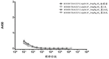

Classifications
-
- C—CHEMISTRY; METALLURGY
- C07—ORGANIC CHEMISTRY
- C07K—PEPTIDES
- C07K16/00—Immunoglobulins [IGs], e.g. monoclonal or polyclonal antibodies
- C07K16/46—Hybrid immunoglobulins
- C07K16/468—Immunoglobulins having two or more different antigen binding sites, e.g. multifunctional antibodies
-
- C—CHEMISTRY; METALLURGY
- C07—ORGANIC CHEMISTRY
- C07K—PEPTIDES
- C07K16/00—Immunoglobulins [IGs], e.g. monoclonal or polyclonal antibodies
- C07K16/18—Immunoglobulins [IGs], e.g. monoclonal or polyclonal antibodies against material from animals or humans
- C07K16/28—Immunoglobulins [IGs], e.g. monoclonal or polyclonal antibodies against material from animals or humans against receptors, cell surface antigens or cell surface determinants
- C07K16/2803—Immunoglobulins [IGs], e.g. monoclonal or polyclonal antibodies against material from animals or humans against receptors, cell surface antigens or cell surface determinants against the immunoglobulin superfamily
- C07K16/2809—Immunoglobulins [IGs], e.g. monoclonal or polyclonal antibodies against material from animals or humans against receptors, cell surface antigens or cell surface determinants against the immunoglobulin superfamily against the T-cell receptor (TcR)-CD3 complex
-
- A—HUMAN NECESSITIES
- A61—MEDICAL OR VETERINARY SCIENCE; HYGIENE
- A61K—PREPARATIONS FOR MEDICAL, DENTAL OR TOILETRY PURPOSES
- A61K35/00—Medicinal preparations containing materials or reaction products thereof with undetermined constitution
- A61K35/12—Materials from mammals; Compositions comprising non-specified tissues or cells; Compositions comprising non-embryonic stem cells; Genetically modified cells
- A61K35/14—Blood; Artificial blood
- A61K35/17—Lymphocytes; B-cells; T-cells; Natural killer cells; Interferon-activated or cytokine-activated lymphocytes
-
- A—HUMAN NECESSITIES
- A61—MEDICAL OR VETERINARY SCIENCE; HYGIENE
- A61P—SPECIFIC THERAPEUTIC ACTIVITY OF CHEMICAL COMPOUNDS OR MEDICINAL PREPARATIONS
- A61P35/00—Antineoplastic agents
-
- A—HUMAN NECESSITIES
- A61—MEDICAL OR VETERINARY SCIENCE; HYGIENE
- A61P—SPECIFIC THERAPEUTIC ACTIVITY OF CHEMICAL COMPOUNDS OR MEDICINAL PREPARATIONS
- A61P35/00—Antineoplastic agents
- A61P35/02—Antineoplastic agents specific for leukemia
-
- C—CHEMISTRY; METALLURGY
- C07—ORGANIC CHEMISTRY
- C07K—PEPTIDES
- C07K14/00—Peptides having more than 20 amino acids; Gastrins; Somatostatins; Melanotropins; Derivatives thereof
- C07K14/435—Peptides having more than 20 amino acids; Gastrins; Somatostatins; Melanotropins; Derivatives thereof from animals; from humans
- C07K14/705—Receptors; Cell surface antigens; Cell surface determinants
- C07K14/70503—Immunoglobulin superfamily
- C07K14/7051—T-cell receptor (TcR)-CD3 complex
-
- C—CHEMISTRY; METALLURGY
- C07—ORGANIC CHEMISTRY
- C07K—PEPTIDES
- C07K16/00—Immunoglobulins [IGs], e.g. monoclonal or polyclonal antibodies
- C07K16/18—Immunoglobulins [IGs], e.g. monoclonal or polyclonal antibodies against material from animals or humans
- C07K16/28—Immunoglobulins [IGs], e.g. monoclonal or polyclonal antibodies against material from animals or humans against receptors, cell surface antigens or cell surface determinants
- C07K16/2803—Immunoglobulins [IGs], e.g. monoclonal or polyclonal antibodies against material from animals or humans against receptors, cell surface antigens or cell surface determinants against the immunoglobulin superfamily
-
- C—CHEMISTRY; METALLURGY
- C07—ORGANIC CHEMISTRY
- C07K—PEPTIDES
- C07K16/00—Immunoglobulins [IGs], e.g. monoclonal or polyclonal antibodies
- C07K16/18—Immunoglobulins [IGs], e.g. monoclonal or polyclonal antibodies against material from animals or humans
- C07K16/28—Immunoglobulins [IGs], e.g. monoclonal or polyclonal antibodies against material from animals or humans against receptors, cell surface antigens or cell surface determinants
- C07K16/2896—Immunoglobulins [IGs], e.g. monoclonal or polyclonal antibodies against material from animals or humans against receptors, cell surface antigens or cell surface determinants against molecules with a "CD"-designation, not provided for elsewhere
-
- C—CHEMISTRY; METALLURGY
- C12—BIOCHEMISTRY; BEER; SPIRITS; WINE; VINEGAR; MICROBIOLOGY; ENZYMOLOGY; MUTATION OR GENETIC ENGINEERING
- C12N—MICROORGANISMS OR ENZYMES; COMPOSITIONS THEREOF; PROPAGATING, PRESERVING, OR MAINTAINING MICROORGANISMS; MUTATION OR GENETIC ENGINEERING; CULTURE MEDIA
- C12N15/00—Mutation or genetic engineering; DNA or RNA concerning genetic engineering, vectors, e.g. plasmids, or their isolation, preparation or purification; Use of hosts therefor
- C12N15/09—Recombinant DNA-technology
- C12N15/63—Introduction of foreign genetic material using vectors; Vectors; Use of hosts therefor; Regulation of expression
-
- G—PHYSICS
- G01—MEASURING; TESTING
- G01N—INVESTIGATING OR ANALYSING MATERIALS BY DETERMINING THEIR CHEMICAL OR PHYSICAL PROPERTIES
- G01N33/00—Investigating or analysing materials by specific methods not covered by groups G01N1/00 - G01N31/00
- G01N33/48—Biological material, e.g. blood, urine; Haemocytometers
- G01N33/50—Chemical analysis of biological material, e.g. blood, urine; Testing involving biospecific ligand binding methods; Immunological testing
- G01N33/53—Immunoassay; Biospecific binding assay; Materials therefor
- G01N33/574—Immunoassay; Biospecific binding assay; Materials therefor for cancer
- G01N33/57407—Specifically defined cancers
- G01N33/57415—Specifically defined cancers of breast
-
- G—PHYSICS
- G01—MEASURING; TESTING
- G01N—INVESTIGATING OR ANALYSING MATERIALS BY DETERMINING THEIR CHEMICAL OR PHYSICAL PROPERTIES
- G01N33/00—Investigating or analysing materials by specific methods not covered by groups G01N1/00 - G01N31/00
- G01N33/48—Biological material, e.g. blood, urine; Haemocytometers
- G01N33/50—Chemical analysis of biological material, e.g. blood, urine; Testing involving biospecific ligand binding methods; Immunological testing
- G01N33/53—Immunoassay; Biospecific binding assay; Materials therefor
- G01N33/574—Immunoassay; Biospecific binding assay; Materials therefor for cancer
- G01N33/57407—Specifically defined cancers
- G01N33/57419—Specifically defined cancers of colon
-
- G—PHYSICS
- G01—MEASURING; TESTING
- G01N—INVESTIGATING OR ANALYSING MATERIALS BY DETERMINING THEIR CHEMICAL OR PHYSICAL PROPERTIES
- G01N33/00—Investigating or analysing materials by specific methods not covered by groups G01N1/00 - G01N31/00
- G01N33/48—Biological material, e.g. blood, urine; Haemocytometers
- G01N33/50—Chemical analysis of biological material, e.g. blood, urine; Testing involving biospecific ligand binding methods; Immunological testing
- G01N33/53—Immunoassay; Biospecific binding assay; Materials therefor
- G01N33/574—Immunoassay; Biospecific binding assay; Materials therefor for cancer
- G01N33/57407—Specifically defined cancers
- G01N33/57423—Specifically defined cancers of lung
-
- G—PHYSICS
- G01—MEASURING; TESTING
- G01N—INVESTIGATING OR ANALYSING MATERIALS BY DETERMINING THEIR CHEMICAL OR PHYSICAL PROPERTIES
- G01N33/00—Investigating or analysing materials by specific methods not covered by groups G01N1/00 - G01N31/00
- G01N33/48—Biological material, e.g. blood, urine; Haemocytometers
- G01N33/50—Chemical analysis of biological material, e.g. blood, urine; Testing involving biospecific ligand binding methods; Immunological testing
- G01N33/53—Immunoassay; Biospecific binding assay; Materials therefor
- G01N33/574—Immunoassay; Biospecific binding assay; Materials therefor for cancer
- G01N33/57407—Specifically defined cancers
- G01N33/5743—Specifically defined cancers of skin, e.g. melanoma
-
- G—PHYSICS
- G01—MEASURING; TESTING
- G01N—INVESTIGATING OR ANALYSING MATERIALS BY DETERMINING THEIR CHEMICAL OR PHYSICAL PROPERTIES
- G01N33/00—Investigating or analysing materials by specific methods not covered by groups G01N1/00 - G01N31/00
- G01N33/48—Biological material, e.g. blood, urine; Haemocytometers
- G01N33/50—Chemical analysis of biological material, e.g. blood, urine; Testing involving biospecific ligand binding methods; Immunological testing
- G01N33/53—Immunoassay; Biospecific binding assay; Materials therefor
- G01N33/574—Immunoassay; Biospecific binding assay; Materials therefor for cancer
- G01N33/57407—Specifically defined cancers
- G01N33/57434—Specifically defined cancers of prostate
-
- G—PHYSICS
- G01—MEASURING; TESTING
- G01N—INVESTIGATING OR ANALYSING MATERIALS BY DETERMINING THEIR CHEMICAL OR PHYSICAL PROPERTIES
- G01N33/00—Investigating or analysing materials by specific methods not covered by groups G01N1/00 - G01N31/00
- G01N33/48—Biological material, e.g. blood, urine; Haemocytometers
- G01N33/50—Chemical analysis of biological material, e.g. blood, urine; Testing involving biospecific ligand binding methods; Immunological testing
- G01N33/53—Immunoassay; Biospecific binding assay; Materials therefor
- G01N33/574—Immunoassay; Biospecific binding assay; Materials therefor for cancer
- G01N33/57407—Specifically defined cancers
- G01N33/57438—Specifically defined cancers of liver, pancreas or kidney
-
- G—PHYSICS
- G01—MEASURING; TESTING
- G01N—INVESTIGATING OR ANALYSING MATERIALS BY DETERMINING THEIR CHEMICAL OR PHYSICAL PROPERTIES
- G01N33/00—Investigating or analysing materials by specific methods not covered by groups G01N1/00 - G01N31/00
- G01N33/48—Biological material, e.g. blood, urine; Haemocytometers
- G01N33/50—Chemical analysis of biological material, e.g. blood, urine; Testing involving biospecific ligand binding methods; Immunological testing
- G01N33/53—Immunoassay; Biospecific binding assay; Materials therefor
- G01N33/574—Immunoassay; Biospecific binding assay; Materials therefor for cancer
- G01N33/57407—Specifically defined cancers
- G01N33/57442—Specifically defined cancers of the uterus and endometrial
-
- G—PHYSICS
- G01—MEASURING; TESTING
- G01N—INVESTIGATING OR ANALYSING MATERIALS BY DETERMINING THEIR CHEMICAL OR PHYSICAL PROPERTIES
- G01N33/00—Investigating or analysing materials by specific methods not covered by groups G01N1/00 - G01N31/00
- G01N33/48—Biological material, e.g. blood, urine; Haemocytometers
- G01N33/50—Chemical analysis of biological material, e.g. blood, urine; Testing involving biospecific ligand binding methods; Immunological testing
- G01N33/53—Immunoassay; Biospecific binding assay; Materials therefor
- G01N33/574—Immunoassay; Biospecific binding assay; Materials therefor for cancer
- G01N33/57407—Specifically defined cancers
- G01N33/57446—Specifically defined cancers of stomach or intestine
-
- G—PHYSICS
- G01—MEASURING; TESTING
- G01N—INVESTIGATING OR ANALYSING MATERIALS BY DETERMINING THEIR CHEMICAL OR PHYSICAL PROPERTIES
- G01N33/00—Investigating or analysing materials by specific methods not covered by groups G01N1/00 - G01N31/00
- G01N33/48—Biological material, e.g. blood, urine; Haemocytometers
- G01N33/50—Chemical analysis of biological material, e.g. blood, urine; Testing involving biospecific ligand binding methods; Immunological testing
- G01N33/53—Immunoassay; Biospecific binding assay; Materials therefor
- G01N33/574—Immunoassay; Biospecific binding assay; Materials therefor for cancer
- G01N33/57407—Specifically defined cancers
- G01N33/57449—Specifically defined cancers of ovaries
-
- G—PHYSICS
- G01—MEASURING; TESTING
- G01N—INVESTIGATING OR ANALYSING MATERIALS BY DETERMINING THEIR CHEMICAL OR PHYSICAL PROPERTIES
- G01N33/00—Investigating or analysing materials by specific methods not covered by groups G01N1/00 - G01N31/00
- G01N33/48—Biological material, e.g. blood, urine; Haemocytometers
- G01N33/50—Chemical analysis of biological material, e.g. blood, urine; Testing involving biospecific ligand binding methods; Immunological testing
- G01N33/68—Chemical analysis of biological material, e.g. blood, urine; Testing involving biospecific ligand binding methods; Immunological testing involving proteins, peptides or amino acids
- G01N33/6854—Immunoglobulins
- G01N33/6857—Antibody fragments
-
- G—PHYSICS
- G01—MEASURING; TESTING
- G01N—INVESTIGATING OR ANALYSING MATERIALS BY DETERMINING THEIR CHEMICAL OR PHYSICAL PROPERTIES
- G01N33/00—Investigating or analysing materials by specific methods not covered by groups G01N1/00 - G01N31/00
- G01N33/48—Biological material, e.g. blood, urine; Haemocytometers
- G01N33/50—Chemical analysis of biological material, e.g. blood, urine; Testing involving biospecific ligand binding methods; Immunological testing
- G01N33/68—Chemical analysis of biological material, e.g. blood, urine; Testing involving biospecific ligand binding methods; Immunological testing involving proteins, peptides or amino acids
- G01N33/6872—Intracellular protein regulatory factors and their receptors, e.g. including ion channels
-
- A—HUMAN NECESSITIES
- A61—MEDICAL OR VETERINARY SCIENCE; HYGIENE
- A61K—PREPARATIONS FOR MEDICAL, DENTAL OR TOILETRY PURPOSES
- A61K39/00—Medicinal preparations containing antigens or antibodies
- A61K2039/505—Medicinal preparations containing antigens or antibodies comprising antibodies
-
- A—HUMAN NECESSITIES
- A61—MEDICAL OR VETERINARY SCIENCE; HYGIENE
- A61K—PREPARATIONS FOR MEDICAL, DENTAL OR TOILETRY PURPOSES
- A61K38/00—Medicinal preparations containing peptides
-
- C—CHEMISTRY; METALLURGY
- C07—ORGANIC CHEMISTRY
- C07K—PEPTIDES
- C07K2317/00—Immunoglobulins specific features
- C07K2317/30—Immunoglobulins specific features characterized by aspects of specificity or valency
- C07K2317/31—Immunoglobulins specific features characterized by aspects of specificity or valency multispecific
-
- C—CHEMISTRY; METALLURGY
- C07—ORGANIC CHEMISTRY
- C07K—PEPTIDES
- C07K2317/00—Immunoglobulins specific features
- C07K2317/30—Immunoglobulins specific features characterized by aspects of specificity or valency
- C07K2317/33—Crossreactivity, e.g. for species or epitope, or lack of said crossreactivity
-
- C—CHEMISTRY; METALLURGY
- C07—ORGANIC CHEMISTRY
- C07K—PEPTIDES
- C07K2317/00—Immunoglobulins specific features
- C07K2317/30—Immunoglobulins specific features characterized by aspects of specificity or valency
- C07K2317/35—Valency
-
- C—CHEMISTRY; METALLURGY
- C07—ORGANIC CHEMISTRY
- C07K—PEPTIDES
- C07K2317/00—Immunoglobulins specific features
- C07K2317/50—Immunoglobulins specific features characterized by immunoglobulin fragments
- C07K2317/52—Constant or Fc region; Isotype
-
- C—CHEMISTRY; METALLURGY
- C07—ORGANIC CHEMISTRY
- C07K—PEPTIDES
- C07K2317/00—Immunoglobulins specific features
- C07K2317/50—Immunoglobulins specific features characterized by immunoglobulin fragments
- C07K2317/52—Constant or Fc region; Isotype
- C07K2317/522—CH1 domain
-
- C—CHEMISTRY; METALLURGY
- C07—ORGANIC CHEMISTRY
- C07K—PEPTIDES
- C07K2317/00—Immunoglobulins specific features
- C07K2317/50—Immunoglobulins specific features characterized by immunoglobulin fragments
- C07K2317/52—Constant or Fc region; Isotype
- C07K2317/524—CH2 domain
-
- C—CHEMISTRY; METALLURGY
- C07—ORGANIC CHEMISTRY
- C07K—PEPTIDES
- C07K2317/00—Immunoglobulins specific features
- C07K2317/50—Immunoglobulins specific features characterized by immunoglobulin fragments
- C07K2317/52—Constant or Fc region; Isotype
- C07K2317/526—CH3 domain
-
- C—CHEMISTRY; METALLURGY
- C07—ORGANIC CHEMISTRY
- C07K—PEPTIDES
- C07K2317/00—Immunoglobulins specific features
- C07K2317/50—Immunoglobulins specific features characterized by immunoglobulin fragments
- C07K2317/52—Constant or Fc region; Isotype
- C07K2317/53—Hinge
-
- C—CHEMISTRY; METALLURGY
- C07—ORGANIC CHEMISTRY
- C07K—PEPTIDES
- C07K2317/00—Immunoglobulins specific features
- C07K2317/50—Immunoglobulins specific features characterized by immunoglobulin fragments
- C07K2317/55—Fab or Fab'
-
- C—CHEMISTRY; METALLURGY
- C07—ORGANIC CHEMISTRY
- C07K—PEPTIDES
- C07K2317/00—Immunoglobulins specific features
- C07K2317/50—Immunoglobulins specific features characterized by immunoglobulin fragments
- C07K2317/56—Immunoglobulins specific features characterized by immunoglobulin fragments variable (Fv) region, i.e. VH and/or VL
- C07K2317/565—Complementarity determining region [CDR]
-
- C—CHEMISTRY; METALLURGY
- C07—ORGANIC CHEMISTRY
- C07K—PEPTIDES
- C07K2317/00—Immunoglobulins specific features
- C07K2317/70—Immunoglobulins specific features characterized by effect upon binding to a cell or to an antigen
-
- C—CHEMISTRY; METALLURGY
- C07—ORGANIC CHEMISTRY
- C07K—PEPTIDES
- C07K2317/00—Immunoglobulins specific features
- C07K2317/70—Immunoglobulins specific features characterized by effect upon binding to a cell or to an antigen
- C07K2317/73—Inducing cell death, e.g. apoptosis, necrosis or inhibition of cell proliferation
- C07K2317/732—Antibody-dependent cellular cytotoxicity [ADCC]
-
- C—CHEMISTRY; METALLURGY
- C07—ORGANIC CHEMISTRY
- C07K—PEPTIDES
- C07K2317/00—Immunoglobulins specific features
- C07K2317/70—Immunoglobulins specific features characterized by effect upon binding to a cell or to an antigen
- C07K2317/76—Antagonist effect on antigen, e.g. neutralization or inhibition of binding
-
- C—CHEMISTRY; METALLURGY
- C07—ORGANIC CHEMISTRY
- C07K—PEPTIDES
- C07K2317/00—Immunoglobulins specific features
- C07K2317/90—Immunoglobulins specific features characterized by (pharmaco)kinetic aspects or by stability of the immunoglobulin
- C07K2317/92—Affinity (KD), association rate (Ka), dissociation rate (Kd) or EC50 value
-
- C—CHEMISTRY; METALLURGY
- C07—ORGANIC CHEMISTRY
- C07K—PEPTIDES
- C07K2317/00—Immunoglobulins specific features
- C07K2317/90—Immunoglobulins specific features characterized by (pharmaco)kinetic aspects or by stability of the immunoglobulin
- C07K2317/94—Stability, e.g. half-life, pH, temperature or enzyme-resistance
-
- C—CHEMISTRY; METALLURGY
- C07—ORGANIC CHEMISTRY
- C07K—PEPTIDES
- C07K2319/00—Fusion polypeptide
-
- G—PHYSICS
- G01—MEASURING; TESTING
- G01N—INVESTIGATING OR ANALYSING MATERIALS BY DETERMINING THEIR CHEMICAL OR PHYSICAL PROPERTIES
- G01N2333/00—Assays involving biological materials from specific organisms or of a specific nature
- G01N2333/435—Assays involving biological materials from specific organisms or of a specific nature from animals; from humans
- G01N2333/705—Assays involving receptors, cell surface antigens or cell surface determinants
- G01N2333/70596—Molecules with a "CD"-designation not provided for elsewhere in G01N2333/705
-
- G—PHYSICS
- G01—MEASURING; TESTING
- G01N—INVESTIGATING OR ANALYSING MATERIALS BY DETERMINING THEIR CHEMICAL OR PHYSICAL PROPERTIES
- G01N2800/00—Detection or diagnosis of diseases
- G01N2800/52—Predicting or monitoring the response to treatment, e.g. for selection of therapy based on assay results in personalised medicine; Prognosis
Abstract
The present invention provides a bispecific anti-CD 3xCD19 polypeptide complex comprising a first antigen-binding moiety and a second antigen-binding moiety, methods of producing said bispecific anti-CD 3xCD19 polypeptide complex, methods of treating a disease or condition using said bispecific anti-CD 3xCD19 polypeptide complex, polynucleotides encoding said bispecific anti-CD 3xCD19 polypeptide complex, vectors and host cells comprising said polynucleotides, and compositions and pharmaceutical compositions comprising said bispecific anti-CD 3xCD19 polypeptide complex.
Description
Cross-referencing
This application claims priority to international patent application No. PCT/CN2017/103032 filed 2017, 9, month 22.
Technical Field
The present invention relates generally to bispecific anti-CD 3xCD19 polypeptide complexes comprising antibody variable regions fused to TCR constant regions.
Background
Bispecific antibodies are becoming a new class of therapeutic antibodies. They can bind to two different targets or two different epitopes on one target, resulting in additive or synergistic effects over the effects of a single antibody. Many attempts have been made to design new bispecific formats (e.g., DVD-Ig, Crossmab, BiTE, etc.) (Spiess et al, Molecular Immunology, 67(2), pp.95-106 (2015)). However, these forms can potentially have various limitations in terms of stability, solubility, short half-life, and immunogenicity.
Among these bispecific antibody formats, an IgG-like bispecific antibody is a universal format: one arm binds to target a and the other arm binds to target B. Structurally it consists of half of antibody a and half of antibody B, with a size and shape similar to native IgG. For ease of downstream development, it is desirable that such bispecific molecules can be produced and correctly assembled from a single host cell at high expression levels as easily as normal IgG. Unfortunately, the pairing of homologous light-heavy chains and the assembly of two different half-antibodies cannot be controlled automatically. Various mismatches in a random manner can lead to significant product heterogeneity.
Preferred heterodimeric assembly of two different heavy chains is achieved by introducing mutations into the Fc region, such as "hand-intro-holes" (Ridgway et al, Protein Engineering, 9(7), pp.617-21 (1996); Merchant et al, Nature Biotechnology, 16(7), pp.677-681 (1998), Electrostatic (Gunasekalan et al, Journal of Biological Chemistry, 285(25), pp.19637-19646 (2010)), or negative state design (Kreudenstein et al, mAbs, 5(5), pp.646-654 (2013); Leaver-Fay et al, Structure, 24(4), pp.641-651 (2016)). However, selective pairing of individual light-heavy chains of individual antibodies remains challenging. The interface between the light-heavy chains comprises a variable domain (VH-VL) and a constant domain (CH 1-CL). Several approaches have been applied to the design of orthogonal interfaces to facilitate homologous pairing. Roche exchanged the domains of CH1 and CL and created the Cross Mab platform (Schaefer et al, Proceedings of the National Academy of Sciences of the United States of America, 108(27), pp.11187-11192 (2011)), Medmemune alternatively introduced disulfide bonds (Mazor et al, mAbs, 7(2), pp.377-389 (2015)), Amgen further electrostatically modified in the CH1-CL region (Liu et al, Journal of Biological Chemistry, 290(12), pp.7535-7562 (2015)), and Lilly (Lewis et al, Nature technology, 32 (2015)), 191-198 (2014), (9-213), (230) and Genencoch (Dillon et al, mAh et al, 9-213, pp.230) introduced constant mutations in both domains.
Human CD19 is a type I transmembrane protein belonging to the immunoglobulin superfamily (Carter et al, Curr Dir Autoimmun, 7: 4-32 (2004)). It is expressed on most B cells, but not on plasma cells, stem cells or normal myeloid lineages (Tedder, Nat Rev Rheumatotol, 5 (10): 572-. CD19 is critically involved in establishing intrinsic B cell signaling thresholds by modulating B Cell Receptor (BCR) dependent and independent signaling (Wang et al, Experimental Hematology & Oncology, 1: 36 (2012)). CD19 has a broader expression than CD 20. The pattern of CD19 expression remains in B cell malignancies, encompassing ALL subtypes of B cell lymphomas (from indolent to aggressive forms), as well as B cell chronic lymphocytic leukemia and non-T acute lymphocytic leukemia, and allows for early B cell targeted oncology indications such as Acute Lymphocytic Leukemia (ALL) (which cannot be targeted by rituximab). Several monoclonal antibodies to CD19 have been investigated for use in lymphoma therapy (U.S. patent application publication No. 20140072587 a1, U.S. patent No. 8,242,252B 2, and U.S. patent No. 8,097,703B 2).
The CD 3T cell co-receptor is a protein complex consisting of four distinct chains (CD3 γ chain, CD3 δ chain and two CD3 epsilon chains). These four chains associate with a molecule called the T Cell Receptor (TCR) and the zeta chain to generate an activation signal in T lymphocytes. The TCR, zeta chain and CD3 molecules constitute a TCR complex in which the TCR serves as a subunit for recognition and binding of antigen, and CD3 serves as a subunit for transfer and trafficking of antigen stimuli to the signaling pathway and ultimately regulation of T cell activity. CD3 protein is present in almost all T cells. CD3-TCR complexes modulate T cell function in innate and adaptive immune responses, as well as cellular and humoral immune functions. These include elimination of pathogenic organisms and control of tumor growth through a wide range of cytotoxic effects. Mouse monoclonal antibodies specific for human CD3, such as OKT3(Kung et al, Science, 206: 347-9(1979)), were the first generation CD3 antibodies developed for therapy. Despite its strong immunosuppressive potency, OKT3 has been hampered by serious side effects associated with its immunogenicity and mitogenic potential (Chatenoud, Nature Reviews, 3: 123-. OKT3 induced an antiglobulin response that promoted its own rapid clearance and neutralization (Chatenoud et al, Eur. J. Immunol., 137: 830-8 (1982)). In addition, OKT3 induced T cell proliferation and cytokine production in vitro and resulted in large-scale release of cytokines in vivo (Hirsch et al, J.Immunol, 142: 737-43 (1989)). This serious side effect limits the wider use of OKT3 in transplantation and extends its use to other clinical areas such as autoimmunity.
Bispecific antibodies targeting CD3 and CD19 can bind to both T cells and B cells. After the bispecific antibody bound to CD3 positive T cells and CD19 positive B cells, cytolytic synapses were formed. Cytotoxicity is then induced by the release of perforin and granzyme from the granules in cytotoxic T cells, which induces apoptosis and lysis of malignant B cells.
It is highly desirable to design bispecific molecules with desired expression levels and in vivo half-lives for both CD3 and CD 19. Such bispecific anti-CD 3xCD19 polypeptide complexes are useful for treating CD 19-related conditions, including cancer.
Brief description of the invention
In one aspect, the invention provides a bispecific polypeptide complex comprising a first antigen-binding moiety associated with a second antigen-binding moiety, wherein:
the first antigen-binding portion comprises:
a first polypeptide comprising from N-terminus to C-terminus a first heavy chain variable domain (VH) of a first antibody operably linked to a first T Cell Receptor (TCR) constant region (C1), and
a second polypeptide comprising from N-terminus to C-terminus a first light chain variable domain (VL) of a first antibody operably linked to a second TCR constant region (C2),
wherein:
c1 comprises an engineered C β comprising SEQ ID NO 1 and C2 comprises an engineered C α comprising SEQ ID NO 2,
amino acid C48 in SEQ ID NO. 1 and amino acid C41 in SEQ ID NO. 2 are capable of forming non-natural interchain disulfide bonds,
c1 and C2 are capable of forming a dimer, and the non-natural interchain disulfide bond is capable of stabilizing the dimer,
and
the second antigen-binding moiety comprises:
a second heavy chain variable domain of a second antibody (VH2) operably linked to the antibody heavy chain CH1 domain, and
a second light chain variable domain of a second antibody (VL2) operably linked to an antibody light chain Constant (CL) domain,
wherein:
one of said first and said second antigen-binding moiety is an anti-CD 3 binding moiety and the other is an anti-CD 19 binding moiety,
an anti-CD 3 binding moiety is derived from an anti-CD 3 antibody comprising:
a) comprises the amino acid sequence of SEQ ID NO:3, b) comprises the heavy chain CDR1 of SEQ ID NO: 4, c) comprises the heavy chain CDR2 of SEQ ID NO: 5, d) comprises the heavy chain CDR3 of SEQ ID NO: 6, kappa light chain CDR1, e) comprises SEQ ID NO: 7, and f) a kappa light chain CDR2 comprising SEQ ID NO: the kappa light chain CDR3 of FIG. 8,
an anti-CD 19 binding moiety is derived from an anti-CD 19 antibody comprising:
a) comprises the amino acid sequence of SEQ ID NO: 9, b) comprises the heavy chain CDR1 of SEQ ID NO: 10, c) comprises the heavy chain CDR2 of SEQ ID NO: 11, heavy chain CDR3, d) comprises SEQ ID NO: 12, kappa light chain CDR1, e) comprises SEQ ID NO: 13, and f) a kappa light chain CDR2 comprising SEQ ID NO: the kappa light chain CDR3 of FIG. 14,
and is
The first antigen-binding moiety and the second antigen-binding moiety are less susceptible to mismatches than if both the first and the second antigen-binding moieties were corresponding portions of a native Fab.
In certain embodiments, the anti-CD 3 binding portion of the bispecific polypeptide complex is derived from an anti-CD 3 antibody comprising a heavy chain variable region comprising SEQ ID NO:15 and a light chain variable domain sequence comprising SEQ ID NO:17, a light chain variable domain sequence.
In certain embodiments, the anti-CD 19 binding portion of the bispecific polypeptide complex is derived from an anti-CD 19 antibody comprising a heavy chain variable region comprising SEQ ID NO:19 and a light chain variable domain sequence comprising SEQ ID NO:21, a light chain variable domain sequence.
In certain embodiments, the bispecific polypeptide complex comprises a combination of four polypeptide sequences: 26, 27, 28, and 29.
In certain embodiments, the bispecific polypeptide complex comprises a combination of four polypeptide sequences: SEQ ID NO:26, SEQ ID NO:27, SEQ ID NO:28 and SEQ ID NO: 30.
in one aspect, the invention provides a conjugate comprising a bispecific polypeptide complex of the invention conjugated to a moiety.
In one aspect, the invention provides an isolated polynucleotide encoding a bispecific polypeptide complex of the invention.
In one aspect, the invention provides an isolated vector comprising a polynucleotide of the invention.
In one aspect, the invention provides a host cell comprising an isolated polynucleotide of the invention, or an isolated vector of the invention.
In one aspect, the present invention provides a method of expressing a bispecific polypeptide complex of the present invention, the method comprising culturing a host cell of the present invention under conditions in which the bispecific polypeptide complex is expressed.
In one aspect, the invention provides a composition comprising a bispecific polypeptide complex of the invention.
In one aspect, the present invention provides a pharmaceutical composition comprising a bispecific polypeptide complex of the present invention, and a pharmaceutically acceptable carrier.
In one aspect, the present invention provides a method of treating a disease or condition associated with CD19 in a subject in need thereof, comprising administering to the subject a therapeutically effective amount of a bispecific polypeptide complex of the present invention. In certain embodiments, when both the first antigen and the second antigen are modulated, the disease or condition may be alleviated, eliminated, treated, or prevented.
In certain embodiments, the first VH is operably linked to C β at a first engagement domain, comprises a C-terminal fragment of an antibody V/C engager and has the amino acid sequence of SEQ ID NO: 23 (LEDLKNVFPP) and the first VL is operably linked to ca at a second engagement domain that comprises an N-terminal fragment of a TCR V/C engager and has the amino acid sequence of SEQ ID NO: 24 (KPDIQNPDP).
In certain embodiments, the first antigen-binding moiety is linked to a first dimerization domain and the second antigen-binding moiety is linked to a second dimerization domain, wherein the first and the second dimerization domains are associated. In certain embodiments, the association is via a linker, a disulfide bond, a hydrogen bond, an electrostatic interaction, a salt bridge, or a hydrophobic-hydrophilic interaction, or a combination thereof.
In certain embodiments, the first and/or the second dimerization domain comprises at least a portion of an antibody hinge region, which is optionally derived from IgG1, IgG2, or IgG4.
In certain embodiments, C1 comprises an engineered C β, and the first dimerization domain is operably linked to the engineered C β at a third junction domain comprising SEQ ID NO:25 (YGPPCPPCPAPEFLGGP).
In certain embodiments, the second dimerization domain is operably linked to the heavy chain variable domain of the second antigen binding portion.
In certain embodiments, the first and the second dimerization domains are different and are associated in a manner that hinders homodimerization and/or contributes to heterodimerization.
In certain embodiments, the first and the second dimerization domains are capable of associating into a heterodimer via handle-access holes, hydrophobic interactions, electrostatic interactions, hydrophilic interactions, or increased flexibility.
In another aspect, the invention provides a kit for detecting, diagnosing, prognosing or treating a disease or condition, comprising a polypeptide complex of the invention.
The foregoing and other features and advantages of the invention will be apparent from the following detailed description of several embodiments, which proceeds with reference to the accompanying figures.
Brief Description of Drawings
Figure 1 presents a schematic of the antibodies and formats studied. anti-CD 3 antibody T3 and anti-CD 19 antibody U4 were developed. The constant regions of T3 (CL and CH1) were replaced by the constant domains of the TCR to design a unique light-heavy chain interface orthogonal to common antibodies. TCR-modified T3 and native U4 were used together with "hand-hole" mutations in the Fc domain to design bispecific antibody forms E17 and F16.
FIGS. 2A-2D present guidance diagrams provided by the superimposed morphology of antibody Fv models and TCR structures in the fusion of antibody Fv with TCR constant regions. Figure 2A presents an antibody Fv structural model constructed based on the sequence of the anti-CD 3 antibody T3 developed internally by the company. Fig. 2B presents the TCR structure from PDB 4L 4T. FIG. 2C presents a model of the structure of an antibody Fv superimposed on the variable region of a TCR in different orientations. The substantially chimeric protein was generated by removing the TCR variable domains in the stacked configuration, as shown in figure 2D. Overlapping residues in the splicing region assist in designing the splicing region. Antibody VL chains and TCR α chains are marked white. VH and β chains are marked in black.
Figure 3A shows the sequence of the native TCR α chain and its corresponding sequence with mutated cysteine residues. TRAC _ human is the native sequence of the alpha chain constant region. The 4L4T _ α _ crystal is a sequence with a crystal structure of the S55C mutation (PDB code 4L4T) that can form interchain disulfide bonds. The grey region is the constant region of the scaffold for the chimeric protein of the invention.
Figure 3B shows the sequence of the native TCR β chain and its corresponding sequence with mutated cysteine residues.
Figures 4A-4B show the sequence and numbering of TCR ca and C β constant regions with removed N-glycosylation. Figure 4A shows the sequence and numbering of the TCR α constant region. Figure 4B shows the sequence and numbering of the TCR β constant region.
FIG. 5 shows flow cytometry histograms of cynomolgus monkey CD19 transfected cell line WBP701.CHO-K1.cpro1.FL. C9 and CHO-K1 parental cell line.
FIG. 6 shows SDS-PAGE of W3438-T3U4.F16-1. uIgG4.SP. M: a protein marker; lane 1: W3438-T3U4.F16-1.uIgG4.SP, non-reducing; lane 3: W3438-T3U4.F16-1.uIgG4.SP, reduction.
FIG. 7 shows a SEC-HPLC chromatogram of W3438-T3U4.F16-1.uIgG4.
FIG. 8 shows SDS-PAGE of W3438-T3U4.E17-1. uIgG4.SP. M: a protein marker; lane 1: W3438-T3U4.E17-1.uIgG4.SP, non-reducing; lane 2: W3438-T3U4.E17-1.uIgG4.SP, reduction.
FIG. 9 shows the SEC-HPLC chromatogram of W3438-T3U4.E17-1. uIgG4.SP.
FIGS. 10A-10B show the binding of W3438-T3U4.E17-1.uIgG4.SP to Ramos cells (FIG. 10A) and Jurkat cells (FIG. 10B) as measured by FACS.
FIGS. 11A-11B show the binding of W3438-T3U4.F16-1.uIgG4.SP to Ramos cells (FIG. 11A) and Jurkat cells (FIG. 11B) as measured by FACS.
Figure 12 shows the binding of W3438-t3u4.e17-1. uggg4. sp to cynomolgus monkey-CD 19 expressing cells as measured by FACS.
Figure 13 shows the binding of W3438-t3u4.e17-1. uggg4. sp to cynomolgus monkey CD3 measured by ELISA.
Figures 14A-14B show the affinity of W3438-t3u4.e17-1.u igg4.sp for human CD19 and CD3 as measured by binding to Ramos (figure 14A) and Jurkat (figure 14B) cells.
FIGS. 15A-15B show the double binding of W3438-T3U4.E17-1.uIgG4.SP with CD19 and CD3 (FIG. 15A); and a negative control (fig. 15B).
FIGS. 16A-16B show the cell killing activity of W3438-T3U4.E17-1.uIgG4.SP on Raji cells (FIG. 16A) and W3438-T3U4.F16-1.uIgG4.SP on Raji cells (FIG. 16B).
Figures 17A-17D show CD69 and CD25 expression on T cells in the presence or absence of CD19+ target cells. The percentage of CD69 expressing T cells in the CD4+ T cell subpopulation is shown (fig. 17A); percentage of CD69 expressing T cells in CD8+ T cell subpopulation (fig. 17B); percentage of CD25 expressing T cells in CD4+ T cell subpopulation (fig. 17C); percentage of CD25 expressing T cells in the CD8+ T cell subset (fig. 17D).
FIGS. 18A-18D show IFN-. gamma.and TNF-. alpha.cytokine release from T cells in the presence or absence of CD19+ target cells. Showing the release of IFN- γ in the CD4+ T cell subpopulation (fig. 18A); TNF- α release in CD4+ T cell subpopulation (FIG. 18B); release of IFN- γ in CD8+ T cell subpopulation (fig. 18C); TNF- α release in CD8+ T cell subpopulation (FIG. 18D).
FIGS. 19A-19B show the stability of W3438-T3U4.E17-1.uIgG4.SP in human serum. Binding of the serum incubated W3438-t3u4.e17-1. uggg4. sp samples to Ramos cells at the indicated times is shown (fig. 19A); binding of serum incubated W3438-T3U4.E17-1.uIgG4.SP samples to Jurkat at the indicated times (FIG. 19B).
Figure 20 shows the binding of W3438-t3u4.e17-1. uggg4. sp to C1Q measured by ELISA. IgG1 antibody was used as an assay control.
Figure 21 shows tumor volume traces following administration of W3438-t3u4.e17-1. uggg4. sp at different doses to mixed human PBMC mice bearing Raji xenograft tumors. Data points represent group mean and error bars represent Standard Error (SEM) of mean. An irrelevant IgG4 antibody was used as a negative control.
FIG. 22 shows the concentration of W3438-T3U4.E17-1.uIgG4.SP in cynomolgus monkey serum after a single dose of 1 mg/kg. Serum samples from two monkeys were tested by ELISA.
Figures 23A-23B show anti-drug antibodies (ADA) in serum samples from monkey #1 (figure 23A) and monkey #2 (figure 23B), including before and after administration of W3438-t3u4.e17-1. ugg4. sp.
Detailed Description
The following description of the invention is merely illustrative of various embodiments of the invention.
Definition of
The articles "a," "an," and "the" are used herein to refer to one or to more than one (i.e., to at least one) of the grammatical object of the article. For example, "a polypeptide complex" refers to one polypeptide complex or more than one polypeptide complex.
The term "about" or "approximately" as used herein refers to a quantity, level, value, amount, frequency, percentage, dimension, size, amount, weight, or length that differs by 30, 25, 20, 25, 10, 9, 8, 7, 6,5, 4, 3, 2, or 1% of a reference quantity, level, value, amount, frequency, percentage, dimension, size, amount, weight, or length. In particular embodiments, when the term "about" or "approximately" precedes a numerical value, it means a range of the value plus or minus 15%, 10%, 5%, or 1%.
Throughout this application, unless the context dictates otherwise, the words "comprise", "comprises" and "comprising" will be understood to mean that a stated step or element or group of steps or elements is included, but not to the exclusion of any other step or element or group of steps or elements. What is meant by "consisting of … …" includes and is limited to what follows the phrase "consisting of … …". Thus, the phrase "consisting of … …" means that the listed elements are required or necessary and that no other elements may be present. What is meant by "consisting essentially of … …" is to include any element listed after this phrase and to be limited to other elements that contribute to or do not interfere with the activity or function of the listed element as detailed in this application. Thus, the phrase "consisting essentially of … …" means that the listed elements are required or necessary, but that other elements are optional and may or may not be present depending on whether they affect the activity or effect of the listed elements.
Reference throughout this application to "one embodiment," "an embodiment," "a particular embodiment," "a related embodiment," "a certain embodiment," "another embodiment," or "further embodiment," or combinations thereof, means that a particular feature, structure, or characteristic described in connection with the embodiment is included in at least one embodiment of the present application. Thus, the appearances of the foregoing phrases in various places throughout this specification are not necessarily all referring to the same embodiment. Furthermore, the particular features, structures, or characteristics may be combined in any suitable manner in one or more embodiments.
The terms "polypeptide", "peptide" and "protein" are used interchangeably herein and refer to a polymer of amino acid residues, or a collection of polymers of multiple amino acid residues. These terms apply to amino acid polymers in which one or more amino acid residues is an artificial chemical mimetic of a corresponding naturally occurring amino acid, as well as to naturally occurring amino acid polymers and non-naturally occurring amino acid polymers. The term "amino acid" refers to naturally occurring and synthetic amino acids, as well as amino acid analogs and amino acid mimetics that function in a manner similar to the naturally occurring amino acids. Naturally occurring amino acids are those encoded by the genetic code, as well as those amino acids that are subsequently modified, such as hydroxyproline, gamma-carboxyglutamic acid, and O-phosphoserine. Amino acid analogs refer to compounds having the same basic chemical structure as a naturally occurring amino acid (i.e., an alpha carbon bound to a hydrogen, a carboxyl group, an amino group, and an R group), such as homoserine, norleucine, methionine sulfoxide, methionine methyl sulfonium. Such analogs have modified R groups (e.g., norleucine) or modified peptide backbones, but retain the same basic chemical structure as a naturally occurring amino acid. Alpha-carbon refers to the first carbon atom attached to a functional group, such as a carbonyl group. The beta carbon refers to the second carbon atom attached to the alpha carbon, and this system continues to name carbon atoms in alphabetical order by the greek letter. Amino acid mimetics refer to chemical compounds that have a structure that is different from the general chemical structure of an amino acid, but that functions in a manner similar to a naturally occurring amino acid. The term "protein" generally refers to large polypeptides. The term "peptide" generally refers to short polypeptides. Polypeptide sequences are generally described as having the amino terminus (N-terminus) at the left-hand end of the polypeptide sequence; the right hand end of the polypeptide sequence is the carboxyl terminus (C-terminus). As used herein, "polypeptide complex" refers to a complex comprising one or more polypeptides that are associated with performing certain functions. In certain embodiments, the polypeptide is immunologically relevant.
The term "antibody" as used herein encompasses any immunoglobulin, monoclonal antibody, polyclonal antibody, multispecific antibody, or bispecific (bivalent) antibody that binds a particular antigen. A natural intact antibody comprises two heavy chains and two light chains. Each heavy chain consists of a variable region ("HCVR") and first, second and third constant regions (CH1, CH2, CH3, respectively), while each light chain consists of a variable region ("LCVR") and a constant region (L). Mammalian heavy chains can be classified as α, δ, ε, γ, and μ, and mammalian light chains as λ or κ. Antibodies are "Y" -shaped, the stem of the "Y" -shaped structure consisting of the second and third constant regions of two heavy chains, which are bound by disulfide bonds. Each arm of the "Y" structure comprises the variable and first constant regions of one of the heavy chains, which are associated with the variable and constant regions of one of the light chains. The variable regions of the light and heavy chains are responsible for antigen binding. The variable region of each chain contains three hypervariable regions, called Complementarity Determining Regions (CDRs) (the CDRs for the light (L) chain comprise LCDR1, LCDR2, LCDR3 and the CDRs for the heavy (H) chain comprise HCDR1, HCDR2, HCDR 3). The CDR boundaries of an antibody can be designated or identified by the Kabat, Chothia, or Al-Lazikani nomenclature. (Al-Lazikani, B., Chothia, C., Lesk, A.M., J.mol. biol., 273(4), 927 (1997)), Chothia, C., et Al, J Mol biol. Dec 5; 186(3):651-63 (1985); Chothia, C., and Lesk, A.M., J.mol. biol., 196,901 (1987); Chothia, C. et Al, Nature.Dec 21-28; 342(6252):877-83 (1989); Kabat E.A. et Al, National Institutes of Health, Maryland Besserda (1991)). Where three CDRs are separated by flanking continuous portions called Framework Regions (FRs) that are more highly conserved than the CDRs and form a scaffold-supported hypervariable loop. The HCVR and LCVR each comprise 4 FRs, and the CDRs and FRs are arranged in the following order from amino-terminus to carboxy-terminus: FR1, CDR1, FR2, CDR2, FR3, CDR3, FR 4. The constant regions of the heavy and light chains are not involved in antigen binding, but have multiple effector functions. Antibodies can be classified into several classes according to the amino acid sequence of the constant region of the heavy chain. Depending on whether it contains alpha, delta, epsilon, gamma and mu heavy chains, antibodies can be divided into five main classes or isotypes, respectively: IgA, IgD, IgE, IgG and IgM. Several major antibody classes can also be divided into subclasses, such as IgG1(γ 1 heavy chain), IgG 2(γ 2 heavy chain), IgG3(γ 3 heavy chain), IgG4(γ 4 heavy chain), IgA1(α 1 heavy chain), or IgA2(α 2 heavy chain), among others.
The term "variable domain" as used herein with respect to an antibody refers to an antibody variable region or fragment thereof comprising one or more CDRs. While the variable domain may comprise the entire variable region (e.g., HCVR or LCVR), it may also comprise less than the entire variable region while still retaining the ability to bind to or form an antigen binding site.
The term "antigen-binding portion" as used herein refers to an antibody fragment formed from an antibody portion containing one or more CDRs or any other antibody fragment that binds an antigen but does not have an intact antibody structure. Examples of antigen binding moieties includeBut are not limited to, variable domains, variable regions, bifunctional antibodies (diabodies), Fab ', F (ab')2Fv fragment, disulfide bond-stabilized Fv fragment (dsFv), (dsFv)2Bispecific dsFvs (dsFvs-dsFvs'), disulfide stabilized bifunctional antibodies (ds diabodies), multispecific antibodies, camelized single domain antibodies (camelized single domain antibodies), nanobodies, domain antibodies, and bivalent domain antibodies. The antigen binding portion may bind to the same antigen as the maternal antibody. In certain embodiments, the antigen-binding portion can comprise one or more CDRs from a particular human antibody grafted to a framework region from one or more different human antibodies. More detailed forms of antigen-binding portions are described in Spiess et al, 2015 (supra) and Brinkman et al, mAbs, 9(2), pp.182-212 (2017), the entire contents of which are incorporated herein by reference.
An "Fab" fragment of an antibody refers to the portion of the antibody molecule that is disulfide bonded to the variable and constant regions of one light chain (comprising the variable and constant regions) and one heavy chain. In certain embodiments, the constant regions of both the light and heavy chains are replaced by TCR constant regions.
"Fab'" refers to a Fab fragment that contains a portion of the hinge region.
“F(ab')2"refers to a dimer of Fab.
The "fragment diffcult (Fd)" of an antibody refers to the amino-terminal half of a heavy chain fragment that can be combined with a light chain to form a Fab.
The "Fc" of an antibody refers to the portion of an antibody consisting of the second (CH2), third (CH3) constant region of a first heavy chain disulfide bonded to the second and third constant regions of a second heavy chain. The Fc portion of an antibody is responsible for a number of different effector functions, such as ADCC and CDC, but is not involved in antigen binding.
The "hinge region" in the case of antibodies comprises the portion of the heavy chain molecule connecting the CH1 domain to the CH2 domain. This hinge region comprises about 25 amino acid residues and is flexible, thereby allowing the two N-terminal antigen-binding regions to move independently.
The term "CH 2 domain" as used herein refers to a portion comprising a heavy chain molecule extending from, for example, about amino acid 244 to amino acid 360 of an IgG antibody, using conventional numbering schemes (amino acids 244 to 360, Kabat numbering system; and amino acids 231-340, EU numbering system; see Kabat, E.et al, U.S. department of Health and Human Services (1983)).
The "CH 3 domain" extends from the CH2 domain to the C-terminus of an IgG molecule and comprises about 108 amino acids in size. Certain immunoglobulin classes, such as IgM, further comprise a CH4 region.
"Fv" of an antibody refers to the smallest antibody fragment that contains the entire antigen-binding site. The Fv fragment consists of the variable region of one light chain joined to the variable region of one heavy chain. Several Fv designs are provided, including dsfvs, in which the linkage between the two domains is enhanced by an introduced disulfide bond; and a peptide linker may be used to join the two domains together into a single polypeptide to form an scFv. Fv constructs have been generated that contain the variable domain of a heavy or light immunoglobulin chain linked to the variable and constant domains of the corresponding immunoglobulin heavy or light chain. Fv have also been multimerized to form bifunctional and trifunctional antibodies (Maynard et al, Annu Rev Biomed Eng 2339-376 (2000)).
"ScFab" refers to a fusion polypeptide having an Fd connected to a light chain via a polypeptide linker, resulting in a single chain Fab fragment (ScFab).
"TriFab" refers to a trivalent, bispecific fusion protein consisting of 3 units with Fab function. The trilab accommodates the fusion of two common fabs to an asymmetric Fab-like moiety.
"Fab-Fab" refers to a fusion protein formed by fusing the Fd chain of a first Fab arm to the N-terminus of the Fd chain of a second Fab arm.
"Fab-Fv" refers to a fusion protein formed by fusing an HCVR to the C-terminus of the Fd chain and an LCVR to the C-terminus of the light chain. "Fab-dsFv" molecules can be formed by introducing an interdomain disulfide bond between the HCVR domain and the LCVR domain.
"MAb-Fv" or "IgG-Fv" refers to fusion proteins formed by fusion of an HCVR domain to the C-terminus of one Fc chain and expression of an LCVR domain alone or fused to the C-terminus of the other Fc chain, thereby producing a bispecific, trivalent IgG-Fv (mAb-Fv) fusion protein whose Fv is stabilized by interdomain disulfide bonds.
“ScFab-Fc-scFv2"and" ScFab-Fc-scFv "refer to a fusion protein of a single-chain Fab fused to an Fc and disulfide-stabilized Fv domain.
By "attaching IgG" is meant that the Fab arm is fused to the IgG to form a bispecific (Fab)2-a fusion protein in the form of an Fc. It may form an "IgG-Fab" or "Fab-IgG" in which the Fab is fused to the C-or N-terminus of the IgG molecule, whether or not a linker is present. In certain embodiments, the attached IgG can be further modified to an IgG-Fab4Of (see Brinkman et al 2017, supra).
"DVD-Ig" refers to a double variable domain antibody formed by fusion of additional HCVR and LCVR domains of a second specificity to IgG heavy and light chains. "CODV-Ig" refers to a related form in which two HCVRs and two LCVR domains are linked in a manner that allows cross-pairing of the variable HCVR-LCVR domains, arranged (from N to C-terminal) in the order of HCVRA-HCVRB and LCVRB-LCVRA, or in the order of HCVRB-HCVRA and LCVRA-LCVRB.
"CrossMab" refers to a technique in which an unmodified light chain is paired with a corresponding unmodified heavy chain, and a modified light chain is paired with a corresponding modified heavy chain, thereby producing an antibody with reduced mismatches in the light chain.
A "BiTE" is a bispecific T-cell engaging molecule comprising a first scFv having a first antigen specificity in the direction of LCVR-HCVR linked to a second scFv having a second antigen specificity in the direction of HCVR-LCVR.
"WuXiBody" is a bispecific antibody comprising a soluble chimeric protein having the variable domain of the antibody and the constant domain of the TCR, wherein the subunits (e.g., the a and β domains) of the TCR constant domain are linked by engineered disulfide bonds.
"percent sequence identity," when used with respect to an amino acid sequence (or nucleic acid sequence), refers to the percentage of amino acid (or nucleic acid) residues in a candidate sequence that are identical to a reference sequence to the amino acid (or nucleic acid) residues in the candidate sequence after alignment of the sequences and, if necessary, introduction of a spacer to maximize the number of identical amino acids (or nucleic acids). Conservative substitutions of such amino acid residues may or may not be considered identical residues. Sequences can be aligned to determine percent sequence identity of amino acid (or Nucleic acid) sequences by tools disclosed in the art, such as BLASTN, BLASTp (national center for Biotechnology information website (NCBI), see also Altschul S.F. et al, J.mol.biol., 215: 403-. The skilled person can use default parameters for the tool or can adjust the parameters appropriately according to the needs of the alignment, e.g. by choosing a suitable algorithm.
As used herein, "antigen" or "Ag" refers to a compound, composition, peptide, polypeptide, protein, or substance that can stimulate the production of an antibody or T cell response in cell culture or in an animal, including compositions that are added to cell cultures (e.g., hybridomas), or injected or absorbed into an animal (e.g., compositions that include cancer-specific proteins). The antigen reacts with a specific humoral or cellular immune product (e.g., an antibody), including a product induced by a heterologous antigen.
An "epitope" or "antigenic determinant" refers to a region of an antigen to which a binding agent (e.g., an antibody) binds. Epitopes can be formed from contiguous amino acids (also known as linear or sequential epitopes) or non-contiguous amino acids (also known as conformational or conformational epitopes) juxtaposed by tertiary folding of the protein. Epitopes formed from contiguous amino acids are typically aligned linearly along primary amino acid residues on proteins, and small segments of contiguous amino acids can be digested by antigen binding to Major Histocompatibility Complex (MHC) molecules or retained upon exposure to denaturing solvents, while epitopes formed by tertiary folding are typically lost upon treatment with denaturing solvents. Epitopes typically comprise at least 3, more typically at least 5, about 7, or about 8-10 amino acids in a unique spatial conformation.
"specific binding" or "specific binding" in this application refers to a non-random binding reaction between two molecules, such as a reaction between an antibody and an antigen. In certain embodiments, the polypeptide complexes and bispecific polypeptide complexes described herein specifically bind to an antigen and have binding affinity (K)D)≤10-6M (e.g.. ltoreq.5X 10)-7M、≤2×10⁻⁷M、≤10-7M、 ≤5×10⁻⁸M、≤2×10⁻⁸M、≤10-8M、≤5×10⁻⁹M、≤2×10⁻⁹M、≤10-9M is equal to or less than 10-10M). K for the present applicationDRefers to the ratio of the dissociation rate to the association rate (k)off/kon) This can be determined by using surface plasmon resonance methods, for example using instruments such as Biacore.
The term "operably linked" or "operably linked" refers to the juxtaposition of two or more biological sequences of interest in a relationship permitting them to function in their intended manner, whether or not a spacer or linker is present. When used with respect to polypeptides, the term is intended to mean that the polypeptide sequences are linked in a manner such that the linked products have the desired biological function. For example, antibody variable regions can be operably linked to constant regions to form a stable product with antigen binding activity. The term may also apply to polynucleotides. For example, when a polynucleotide encoding a polypeptide is operably linked to a regulatory sequence (e.g., a promoter, enhancer, silencer sequence, etc.), the term is intended to mean that the polynucleotide sequence is linked in a manner that allows expression of the polypeptide to be regulated by the polynucleotide.
The term "fusion" or "fused" when used with respect to an amino acid sequence (e.g., a peptide, polypeptide, or protein) refers to the combination of two or more amino acid sequences into a single amino acid sequence that does not occur in nature, e.g., by chemical bonding or recombinant means. The fusion amino acid sequence can be produced by genetic recombination of two encoding polynucleotide sequences, and can be expressed by introducing a construct containing the recombinant polynucleotide into a host cell.
The term "spacer" as used herein refers to an artificial amino acid sequence having 1, 2, 3, 4, or 5 amino acid residues, or between 5 and 15, 20, 30, 50 or more amino acid residues in length, linked by peptide bonds and used to link one or more polypeptides. The spacer may or may not have a secondary structure. Spacer sequences are well known in the art, see, e.g., Holliger et al, Proc. Natl. Acad. Sci. USA 90: 6444-; poljak et al, Structure 2:1121- > 1123 (1994). Any suitable spacer known in the art may be used.
The term "antigen-specific" refers to a particular antigen or epitope thereof that is selectively recognized by an antigen binding molecule.
The term "substitution" as used herein with respect to an amino acid residue refers to the substitution of one or more amino acids, naturally occurring or introduced, with another in a peptide, polypeptide or protein. Substitutions in the polypeptide may result in the reduction, enhancement or elimination of the function of the polypeptide.
Substitutions may also be "conservative substitutions" in the sense of referring to an amino acid sequence in which one amino acid residue is replaced with a different amino acid residue having a side chain with similar physicochemical properties, or those amino acids which are not critical to the activity of the polypeptide. For example, conservative substitutions may be made between non-polar side chain amino acid residues (e.g., Met, Ala, Val, Leu and Ile, Pro, Phe, Trp), between uncharged polar side chain residues (e.g., Cys, Ser, Thr, Asn, Gly and Gln), between acidic side chain residues (e.g., Asp, Glu), between basic side chain amino acids (e.g., His, Lys and Arg), between beta-branched side chain amino acids (e.g., Thr, Val and Ile), between sulfur-containing side chain amino acids (e.g., Cys and Met), or between aromatic side chain residues (e.g., Trp, Tyr, His and Phe). In certain embodiments, substitutions, deletions, or additions may also be considered "conservative substitutions. The number of amino acids inserted or deleted may range from about 1 to 5. Conservative substitutions generally do not cause significant changes in the conformational structure of the protein and, therefore, maintain the biological activity of the protein.
The term "mutation" or "mutated" as used herein in reference to an amino acid residue refers to a substitution, insertion or addition of an amino acid residue.
The term "T cell" as used herein refers to a class of lymphocytes that play an important role in cell-mediated immunity, including helper T cells (e.g., CD 4)+T cells, T helper type 1T cells, T helper type 2T cells, T helper type 3T cells, T helper type 17T cells), cytotoxic T cells (e.g., CD8+T cells), memory T cells (e.g., central memory T cells (TCM cells), effector memory T cells (TEM cells and TEMRA cells), and resident memory T cells (TRM), which are CD8+ or CD4+), natural killer T (nkt) cells, and suppressor T cells.
A native "T cell receptor" or native "TCR" is a heterodimeric T cell surface protein that binds to an invariant CD3 chain to form a complex capable of mediating signal transduction. The TCR belongs to the immunoglobulin superfamily and is similar to half antibodies with a single heavy chain and a single light chain. Native TCRs have an extracellular portion, a transmembrane portion, and an intracellular portion. The extracellular domain of the TCR has a membrane-proximal constant region and a membrane-distal variable region.
The term "subject" or "individual" or "animal" or "patient" as used herein refers to a human or non-human animal, including mammals or primates, in need of diagnosis, prognosis, amelioration, prophylaxis and/or treatment of a disease or disorder. Mammalian subjects include humans, livestock animals, farm animals, and zoo, sports, or pet animals, such as dogs, cats, guinea pigs, rabbits, rats, mice, horses, pigs, cattle, bears, and the like.
Bispecific polypeptide complexes
In one aspect, the present application provides bispecific polypeptide complexes. The term "bispecific" as used in this application means that there are two antigen binding moieties, each capable of specific binding to a different antigen or to different epitopes on the same antigen. Bispecific polypeptide compositions provided herein comprise a first antigen-binding moiety associated with a second antigen-binding moiety, and one of which specifically binds to CD3 and the other specifically binds to CD 19. That is, the first antigen-binding portion can specifically bind to CD3, and the second antigen-binding portion can specifically bind to CD 19. Alternatively, the first antigen-binding portion may specifically bind to CD19 and the second antigen-binding portion may specifically bind to CD 3.
In certain embodiments, the present application provides a bispecific polypeptide complex comprising a first antigen-binding moiety associated with a second antigen-binding moiety, wherein:
the first antigen-binding portion comprises:
a first polypeptide comprising from N-terminus to C-terminus a first heavy chain variable domain (VH) of a first antibody operably linked to a first T Cell Receptor (TCR) constant region (C1), and
a second polypeptide comprising from N-terminus to C-terminus a first light chain variable domain (VL) of a first antibody operably linked to a second TCR constant region (C2),
wherein:
c1 comprises an engineered C β comprising SEQ ID NO 1 and C2 comprises an engineered C α comprising SEQ ID NO 2,
amino acid C48 in SEQ ID NO. 1 and amino acid C41 in SEQ ID NO. 2 are capable of forming non-natural interchain disulfide bonds,
c1 and C2 are capable of forming a dimer and the non-natural interchain disulfide bond is capable of stabilizing the dimer, an
The second antigen-binding moiety comprises:
a second heavy chain variable domain of a second antibody (VH2) operably linked to the antibody heavy chain CH1 domain, and
a second light chain variable domain of a second antibody (VL2) operably linked to an antibody light chain Constant (CL) domain,
wherein:
one of said first and said second antigen-binding moiety is an anti-CD 3 binding moiety and the other is an anti-CD 19 binding moiety,
an anti-CD 3 binding moiety is derived from an anti-CD 3 antibody comprising:
a) comprises the amino acid sequence of SEQ ID NO:3, b) comprises the heavy chain CDR1 of SEQ ID NO: 4, c) comprises the heavy chain CDR2 of SEQ ID NO: 5, d) comprises the heavy chain CDR3 of SEQ ID NO: 6, kappa light chain CDR1, e) comprises SEQ ID NO: 7, and f) a kappa light chain CDR2 comprising SEQ ID NO: the kappa light chain CDR3 of FIG. 8,
an anti-CD 19 binding moiety is derived from an anti-CD 19 antibody comprising:
a) comprises the amino acid sequence of SEQ ID NO: 9, b) comprises the heavy chain CDR1 of SEQ ID NO: 10, c) comprises the heavy chain CDR2 of SEQ ID NO: 11, heavy chain CDR3, d) comprises SEQ ID NO: 12, kappa light chain CDR1, e) comprises SEQ ID NO: 13, and f) a kappa light chain CDR2 comprising SEQ ID NO: the kappa light chain CDR3 of FIG. 14,
and is
The first antigen-binding moiety and the second antigen-binding moiety are less susceptible to mismatches than if both the first and the second antigen-binding moieties were corresponding portions of a native Fab.
In certain embodiments, the bispecific polypeptide complexes provided herein comprise a first antigen-binding portion comprising a sequence derived from a TCR constant region, but the second antigen-binding portion does not comprise a sequence derived from a TCR constant region.
The bispecific polypeptide complexes provided herein are significantly less susceptible to having mismatched heavy and light chain variable domains. Without wishing to be bound by any theory, it is believed that the stable TCR constant regions in the first antigen-binding portion may be specifically linked to each other and thus contribute to highly specific pairing of the target VH1 and VL1, while preventing VH1 or VL1 from forming unwanted mismatches with other variable regions that do not provide a target antigen-binding site.
In certain embodiments, the second antigen-binding moiety further comprises an antibody constant CH1 domain operably linked to VH2, and an antibody light chain constant domain operably linked to VL 2. Thus, the second antigen-binding portion comprises a Fab.
When the first, second, third and fourth variable domains (e.g. VH1, VH2, VL1 and VL2) are expressed in one cell, it is desirable that VH1 specifically pair with VL1 and VH2 specifically pair with VL2 so that the resulting bispecific protein product will have the correct antigen binding specificity. However, in the prior art, such as hybrid-hybridomas (or quadromas), VH1, VH2, VL1 and VL2 were paired randomly, resulting in the generation of up to 10 different molecules, only one of which was a functional bispecific antigen binding molecule. This not only reduces the yield, but also complicates the purification of the desired product.
The bispecific polypeptide complex described herein is advantageous because the variable domain is less susceptible to mismatches than if the first and said second antigen-binding portions were both counterparts of a native Fab. In an illustrative example, the first antigen-binding domain comprises VH1-C1 paired with VL1-C2, and the second antigen-binding domain comprises VH2-CH1 paired with VL 2-CL. It has been surprisingly found that C1 and C2 associate preferentially with each other and are less readily associated with CL or CH1, thereby hindering and significantly reducing the formation of unwanted pairings such as C1-CH, C1-CL, C2-CH and C2-CL. Due to the specific association of C1-C2, VH1 specifically pairs with VL1 and thereby forms the first antigen-binding site, and CH1 specifically pairs with CL, thereby forming the specific pairing of VH2-VL2 of the second antigen-binding site. Accordingly, the first antigen-binding portion and the second antigen-binding portion are less susceptible to mismatch and mismatches between, for example, VH1-VL2, VH2-VL1, VH1-VH2, VL1-VL2 are significantly reduced compared to the case where both the first and the second antigen-binding portions are corresponding portions of a native Fab (e.g., in the form of VH1-CH1, VL1-CL, VH2-CH1 and VL 2-CL).
In certain embodiments, a bispecific polypeptide complex described herein has significantly fewer mismatch products (e.g., at least 1, 2, 3, 4,5, or more mismatch products are reduced) and/or significantly higher yield (e.g., at least 10%, 20%, 30%, 40%, 50%, 60%, or more yield is increased) when expressed from a cell compared to a reference molecule expressed under comparable conditions, wherein the reference molecule is otherwise identical to the bispecific polypeptide complex except for the substitution of C1 with native CH1 and the substitution of C2 with native CL.
Antigen binding moieties comprising engineered C alpha and C beta
A first antigen-binding portion described herein comprises a first antibody heavy chain variable domain operably linked to a first T Cell Receptor (TCR) constant region, and a first antibody light chain variable domain operably linked to a second TCR constant region, wherein the first TCR constant region and the second TCR constant region are associated via at least one non-native interchain disulfide bond. The first antigen-binding portion comprises at least two polypeptide chains, each comprising a variable domain derived from an antibody and a constant region derived from a TCR. Thus, the first antigen binding portion comprises a heavy chain variable domain and a light chain variable domain, each of which is operably linked to a pair of TCR constant regions. In certain embodiments, the pairing of TCR constant regions in the first antigen-binding portion is an α/β TCR constant region. The TCR constant regions of the polypeptide complexes described herein can associate with each other through at least one non-native disulfide bond to form a dimer.
It has been surprisingly found that a first antigen binding moiety having at least one non-native disulfide bond as described herein can be recombinantly expressed and assembled into a desired conformation that stabilizes TCR constant region dimers and provides good antigen binding activity of antibody variable regions. In addition, the first antigen binding moiety was found to be well tolerated by conventional antibody engineering, such as modification of glycosylation sites and removal of some native sequences. In addition, the polypeptide complexes described herein can be incorporated into a heterosexual form that can be readily expressed and assembled with minimal or no mismatches in antigen binding sequences due to the presence of the TCR constant region in the first antigen binding portion. Other advantages of the first antigen-binding portions and constructs described herein will become more apparent in the disclosure that follows.
In summary, the present application provides a first antigen-binding portion comprising a first polypeptide comprising from N-terminus to C-terminus a first heavy chain variable domain (VH) of a first antibody operably linked to a first T Cell Receptor (TCR) constant region (C1), and a second polypeptide comprising from N-terminus to C-terminus a first light chain variable domain (VL) of the first antibody operably linked to a second TCR constant region (C2), wherein: c1 comprises an engineered C β comprising SEQ ID NO. 1 and C2 comprises an engineered C α comprising SEQ ID NO. 2, amino acid C48 in SEQ ID NO. 1 and amino acid C41 in SEQ ID NO. 2 are capable of forming an unnatural interchain disulfide bond, C1 and C2 are capable of forming a dimer, and an unnatural interchain disulfide bond between C1 and C2 is capable of stabilizing the dimer.
TCR constant region
The first antigen binding portion described herein comprises an alpha or beta constant region derived from a TCR.
FIGS. 3A and 3B show the amino acid sequences of native TCR constant regions of TCR α and β chains. For clarity and consistency, each amino acid residue in these sequences is numbered in fig. 4A and 4B, and these numbers are used throughout this application to refer to a particular amino acid residue on a particular TCR constant region.
The human TCR alpha chain constant region is called TRAC, its NCBI accession number is P01848, or its amino acid sequence is SEQ ID NO 31.
There are two different variants of the constant region of the human TCR β chain, known as TRBC1 and TRBC2 (IMGT nomenclature) (see also Toyonaga B et al, PNAs, Vol.82, pp.8624-8628, Immunology (1985)). The TRBC1 sequence (SEQ ID NO:33) was selected as a backbone for designing the polypeptide complexes described herein.
Specifically, native TCR β chains contain a native cysteine residue at position 74 (see fig. 4B), which is unpaired and therefore does not form a disulfide bond in native α/β TCRs. In the polypeptide complexes described herein, this native cysteine residue at position 74 of the TCR β chain is mutated to an alanine residue. This may help to avoid erroneous intra-or inter-strand pairings. In certain embodiments, the substitutions in certain embodiments can increase the efficiency of refolding of the TCR in vitro.
In the present application, the first and the second TCR constant regions of the first antigen-binding portion described herein are capable of forming a dimer comprising at least one non-native interchain disulfide bond between the TCR constant regions capable of stabilizing the dimer.
The term "dimer" as used herein refers to an associated structure formed by two molecules (e.g., polypeptides or proteins) through covalent or non-covalent interactions. Homodimers or homodimers are formed by two identical molecules, whereas heterodimers or heterodimers are formed by two different molecules. The dimer formed by the first and the second TCR constant regions is a heterodimer.
A "mutated" amino acid residue refers to an amino acid residue that is substituted, inserted, or added and that is different from the corresponding native residue in its corresponding native TCR constant region. For example, if an amino acid residue at a particular position in a wild-type TCR constant region is referred to as a "native" residue, then its corresponding mutant residue is any residue that is different from the native residue but located at the same position on the TCR constant region. The mutated residue may be a different residue that replaces the native residue at the same position, or is inserted before the native residue and thus occupies its original position.
In the polypeptide complexes provided herein, the first and/or the second TCR constant regions have been engineered to comprise one or more mutated amino acid residues responsible for forming the non-native interchain disulfide bond. To introduce such mutated residues into the TCR constant region, the coding sequence of the TCR region may be manipulated, for example, to replace the codon encoding the native residue with a codon encoding the mutated residue, or to insert the codon encoding the mutated residue before the codon encoding the native residue.
In the polypeptide complexes provided herein, the first and/or the second TCR constant regions have been engineered to comprise one or more mutated cysteine residues such that, following substitution of a cysteine residue, a non-native interchain disulfide bond can be formed between the two TCR constant regions.
The non-natural interchain disulfide bond is capable of stabilizing the first antigen binding moiety. The effect of this stabilizing aspect can be embodied in various ways. For example, the presence of mutated amino acid residues or non-natural interchain disulfide bonds may enable stable expression of the polypeptide complex, and/or expression at high levels, and/or association into a stable complex having a desired biological activity (e.g., antigen binding activity), and/or expression and assembly into a high level of a desired stable complex having a desired biological activity. The ability of the interchain disulfide to stabilize the first and second TCR constant regions can be assessed using suitable methods known in the art, such as displaying molecular weight on SDS-PAGE, or measuring thermal stability by Differential Scanning Calorimetry (DSC) or Differential Scanning Fluorescence (DSF). In an illustrative example, formation of a stable first antigen-binding portion described herein can be confirmed by SDS-PAGE if the product exhibits a molecular weight comparable to the combined molecular weight of the first and second polypeptides. In certain embodiments, the first antigen-binding portion described herein is stable in that it is not less than 50%, 60%, 70%, 80%, or 90% thermostable of a native Fab. In certain embodiments, the first antigen-binding portion described herein is stable in that it is thermostable comparable to a native Fab.
Without wishing to be bound by any theory, it is believed that the non-native interchain disulfide bond formed between the first and the second TCR constant regions in the first antigen-binding portion is capable of stabilizing heterodimers of TCR constant regions, thereby enhancing the correct folding level, structural stability and/or expression level of the heterodimers and the first antigen-binding portion. Unlike native TCRs anchored on the cell membrane on the surface of T cells, heterodimers of the extracellular domains of native TCRs were found to be less stable, although similar in 3D structure to antibody Fab. Indeed, the instability of native TCRs in solution has been a significant impediment to making their crystal structures difficult to elucidate (see Wang, Protein Cell, 5(9), pp.649-652 (2014)). By introducing a pair of cysteine (Cys) mutations into the TCR constant region and thereby enabling interchain non-native disulfide bond formation, the first antigen-binding moiety can be stably expressed while retaining the antigen-binding capacity of the antibody variable region.
TCR constant regions comprising mutated residues are also referred to herein as "engineered" TCR constant regions. In certain embodiments, the first TCR constant region (C1) of the polypeptide complex comprises an engineered TCR alpha chain (ca) and the second TCR constant region (C2) comprises an engineered TCR beta chain (cp). In the polypeptide complexes provided herein, C1 comprises an engineered C β, and C2 comprises an engineered C α.
In the polypeptide complexes provided herein, the engineered TCR constant region comprises one or more mutated cysteine residues comprised within the contact interface of the first and/or the second engineered constant region.
The term "contact interface" as used herein refers to a specific region on the polypeptides where the polypeptides interact/associate with each other. The contact interface comprises one or more amino acid residues that are capable of interacting with a corresponding amino acid residue in contact or association when the interaction occurs. The amino acid residues in the contact interface may or may not be in contiguous sequence. For example, when the interface is three-dimensional, the amino acid residues within the interface can be separated from each other at different positions in the linear sequence.
In certain embodiments, one or more disulfide bonds may be formed between the engineered ca and the engineered cp. In certain embodiments, the mutated cysteine residue in C β is S56C (corresponding to amino acid C48 in SEQ ID NO: 1) and the mutated cysteine residue in C α is T49C (corresponding to amino acid C41 in SEQ ID NO: 2), and wherein the pairing of the cysteine residues is capable of forming a non-natural interchain disulfide bond.
As used throughout this application, "XnY" with respect to the TCR constant region is intended to mean that the nth amino acid residue X (based on the numbering in figures 4A-4B as provided herein) on the TCR constant region is substituted with an amino acid residue Y, where X and Y are each one-letter abbreviations for the particular amino acid residue. It should be noted that the number n is based only on the numbering provided in fig. 4A-4B, and that it may appear different from its actual location. For illustration, the sequence of C.beta. (S56C) (N69Q) shown in SEQ ID NO:1 was used as an example. Although the S to C substitution occurs at residue 48 of SEQ ID NO 1, this residue is designated as residue 56 based on the numbering system in FIGS. 4A-4B, and thus the S to C substitution is designated as S56C instead of S48C. Similarly, the substituents from N to Q are also designated N69Q based on the numbering system in FIGS. 4A-4B. This nomenclature for amino acid residue substitutions applies to all TCR constant regions in this application, unless explicitly stated otherwise.
In the polypeptide complexes provided herein, the engineered C β comprises or is SEQ ID NO 1 and the engineered C α comprises or is SEQ ID NO 2.
The sequences of SEQ ID NO 1 and SEQ ID NO 2 are provided below:
SEQ ID NO:1
EVAVFEPSEAEISHTQKATLVCLATGFYPDHVELSWWVNG KEVHSGVCTDPQPLKEQPALQDSRYALSSRLRVSATFWQNPRN HFRCQVQFYGLSENDEWTQDRAKPVTQIVSAEA
SEQ ID NO:2
AVYQLRDSKSSDKSVCLFTDFDSQTQVSQSKDSDVYITDKC VLDMRSMDFKSNSAVAWSQKSDFACANAFQNSIIPEDTFFPSPES S
in the polypeptide complexes described herein, one or more native glycosylation sites present in native TCR constant regions can be modified (e.g., removed). The term "glycosylation site" as used herein with respect to a polypeptide sequence refers to an amino acid residue with a side chain to which a carbohydrate moiety (e.g., an oligosaccharide structure) can be attached. Glycosylation of polypeptides (such as antibodies) is typically N-linked or O-linked. N-linked refers to the attachment of a carbohydrate moiety to the side chain of an asparagine residue, for example an asparagine residue in tripeptide sequences such as asparagine-X-serine and asparagine-X-threonine, wherein X is any amino acid except proline. O-linked glycosylation refers to the attachment of one of N-acetylgalactosamine, galactose or xylose to a hydroxyamino acid, most commonly to serine or threonine. Removal of the native glycosylation site is conveniently accomplished by altering the amino acid sequence such that one or more of the above-described tripeptide sequences (for N-linked glycosylation sites) or one or more of the serine or threonine residues (for O-linked glycosylation sites) present in the sequence is substituted.
In certain embodiments, in the first antigen binding portion described herein, at least one native glycosylation site is not present in the engineered TCR constant region (e.g., in the first and/or the second TCR constant region). Without wishing to be bound by any theory, it is believed that the first antigen-binding portion described herein may allow for the removal of all or part of the glycosylation site without affecting protein expression and stability, in contrast to the prior teaching that the presence of an N-linked glycosylation site on TCR constant regions such as ca (i.e., N34, N68 and N79) and cp (i.e., N69) is essential for protein expression and stability (see Wu et al, Mabs, 7:2, 364-.
In certain embodiments, the N-glycosylation sites at N34, N68, and N79 in the engineered ca in the first antigen-binding portion described herein are not present. In certain embodiments, the engineered C.alpha.sequence in which NO glycosylation site is present comprises or is SEQ ID NO 2. In the first antigen-binding portion, the N-glycosylation site at N69 is absent in the engineered C β. The engineered C.beta.sequence (TRBC1) without the glycosylation site comprises or is SEQ ID NO: 1.
In a first antigen-binding portion described herein, a constant region derived from a TCR is operably linked to a variable region derived from an antibody.
In certain embodiments, the first antibody variable domain (VH) is fused to the first TCR constant region (C1) at a first junction domain and the first antibody variable domain (VL) is fused to the second TCR constant region (C2) at a second junction domain.
The term "junction domain" as used herein refers to an interface or boundary region where two amino acid sequences are fused or combined.
In certain embodiments, the first engagement domain comprises at least a portion of a C-terminal fragment of an antibody V/C engager and the second engagement domain comprises at least a portion of an N-terminal fragment of a TCR V/C engager.
The term "antibody V/C binder" as used herein refers to the interface of an antibody variable domain and a constant domain, for example between a heavy chain variable domain and a CH1 domain or between a light chain variable domain and a light chain constant domain. Similarly, the term "TCR V/C adaptor" refers to the interface of a TCR variable domain and a constant domain, e.g., the interface between a TCR α variable domain and a constant domain or between a TCR β variable domain and a constant domain.
In certain embodiments, the first polypeptide comprises a sequence comprising a domain operably linked according to formula (I): VH-HCJ-C1, and the second polypeptide comprises a sequence comprising a domain operably linked according to formula (II): VL-LCJ-C2, wherein:
VH is the heavy chain variable domain of an antibody;
HCJ is a first engagement domain as defined above;
c1 is a first TCR constant domain as defined above;
VL is the light chain variable domain of an antibody;
LCJ is a second engagement domain as defined above;
c2 is a second TCR constant domain as defined above.
In such embodiments, C1 is an engineered C β comprising or being SEQ ID NO:1, and C2 is an engineered ca comprising or being SEQ ID NO:2, HCJ comprises or is SEQ ID NO: 23, and LCJ comprises or is SEQ ID NO: 24.
antibody variable regions
Bispecific polypeptide complexes described herein comprise a first antigen-binding moiety associated with a second antigen-binding moiety, and one of which specifically binds to CD3 and the other specifically binds to CD 19. In certain embodiments, the first antigen-binding portion comprises a first heavy chain variable domain (VH1) and a first light chain variable domain (VL1) of a first antibody, and the second antigen-binding portion comprises a second heavy chain variable domain (VH2) and a second light chain variable domain (VL2) of a second antibody, wherein the first antibody and the second antibody are different and are selected from the group consisting of an anti-CD 3 antibody and an anti-CD 19 antibody. In certain embodiments, the first antibody is an anti-CD 3 antibody and the second antibody is an anti-CD 19 antibody. In certain other embodiments, the first antibody is an anti-CD 19 antibody and the second antibody is an anti-CD 3 antibody.
In a conventional natural antibody, the variable region comprises 3 CDR regions separated by flanking Framework (FR) regions, for example as set out by the following formula from N-terminus to C-terminus: FR1-CDR1-FR2-CDR2-FR3-CDR3-FR 4.
a) anti-CD 3 binding moieties
In the polypeptide complexes provided herein, the first antigen-binding portion or the second antigen-binding portion is an anti-CD 3 binding portion. In certain embodiments, the anti-CD 3 binding moiety is derived from the anti-CD 3 antibody WBP3311_2.306.4-z1 shown in table a below. CDR sequences of the WBP3311_2.306.4-z1 antibody are provided below.
TABLE A


The heavy and kappa light chain variable region sequences of the WBP3311_2.306.4-z1 antibody are provided below.
WBP3311_2.306.4-z1-VH
Amino acid sequence (SEQ ID NO: 15):
QVQLVQSGAEVKKPGSSVKVSCKASGFAFTDYYIHWVRQA PGQGLEWMGWISPGNVNTKYNENFKGRVTITADKSTSTAYMEL SSLRSEDTAVYYCARDGYSLYYFDYWGQGTLVTVSS
nucleic acid sequence (SEQ ID NO: 16):
CAGGTGCAGCTTGTGCAGTCTGGGGCAGAAGTGAAGAA GCCTGGGTCTAGTGTCAAGGTGTCATGCAAGGCTAGCGGGT TCGCCTTTACTGACTACTACATCCACTGGGTGCGGCAGGCTC CCGGACAAGGGTTGGAGTGGATGGGATGGATCTCCCCAGGC AATGTCAACACAAAGTACAACGAGAACTTCAAAGGCCGCGTC ACCATTACCGCCGACAAGAGCACCTCCACAGCCTACATGGAG CTGTCCAGCCTCAGAAGCGAGGACACTGCCGTCTACTACTGT GCCAGGGATGGGTACTCCCTGTATTACTTTGATTACTGGGGC CAGGGCACACTGGTGACAGTGAGCTCC
WBP3311_2.306.4-z1-VK
amino acid sequence (SEQ ID NO: 17):
DIVMTQSPDSLAVSLGERATINCKSSQSLLNSRTRKNYLAW YQQKPGQPPKLLIYWASTRQSGVPDRFSGSGSGTDFTLTISSLQ AEDVAVYYCTQSHTLRTFGGGTKVEIK
nucleic acid sequence (SEQ ID NO: 18):
GATATCGTGATGACCCAGAGCCCAGACTCCCTTGCTGTC TCCCTCGGCGAAAGAGCAACCATCAACTGCAAGAGCTCCCAA AGCCTGCTGAACTCCAGGACCAGGAAGAATTACCTGGCCTG GTATCAGCAGAAGCCCGGCCAGCCTCCTAAGCTGCTCATCTA CTGGGCCTCCACCCGGCAGTCTGGGGTGCCCGATCGGTTTA GTGGATCTGGGAGCGGGACAGACTTCACATTGACAATTAGCT CACTGCAGGCCGAGGACGTGGCCGTCTACTACTGTACTCAG AGCCACACTCTCCGCACATTCGGCGGAGGGACTAAAGTGGA GATTAAG
CDRs are known to be responsible for antigen binding.
In certain embodiments, the anti-CD 3 binding moieties provided herein comprise the heavy chain CDR3 sequence of WBP3311_2.306.4-z 1. In certain embodiments, the anti-CD 3 binding moieties provided herein comprise a heavy chain CDR3 comprising the amino acid sequence of SEQ ID NO: 5. the heavy chain CDR3 region is centered on the antigen binding site and is therefore believed to have the most contact with antigen and provide the most free energy for the affinity of the antibody for antigen. It is also believed that heavy chain CDR3 is the most diverse CDR in length, amino acid composition, and conformation among the antigen binding sites to date, through a diverse mechanism (Tonegawa s., nature.302: 575-81 (1983)). The diversity of the heavy chain CDR3 is sufficient to generate most of the antibody specificity (Xu JL, Davis MM., immunity.13: 37-45(2000)) and the required antigen binding affinity (Schier R, et al, J.mol biol.263: 551-67 (1996)).
The anti-CD 3 binding moieties provided herein also comprise suitable Framework Region (FR) sequences, so long as the anti-CD 3 binding moiety can specifically bind CD 3.
The humanized anti-CD 3 antibody WBP3311_2.306.4-z1 has specific binding affinity for cells expressing CD3 (e.g., CD4T cells) and can activate human T cells and trigger cytokine release of TNF α and IFN γ.
In certain embodiments, the anti-CD 3 binding moieties provided herein comprise a polypeptide comprising SEQ ID NO:15 and a light chain variable domain sequence comprising SEQ ID NO:17, a light chain variable domain sequence.
The binding affinity of the anti-CD 3 binding moieties described herein can be defined by KDValue is expressed as the ratio of off rate to association rate (k) when the binding between the antigen and the antigen binding molecule reaches equilibriumoff/kon). Antigen binding affinity (e.g., K) can be appropriately determined using suitable methods known in the art, including, for example, flow cytometry assaysD). In some embodiments, binding of antibody to antigen at different concentrations can be determined by flow cytometry, the determined Mean Fluorescence Intensity (MFI) can be first plotted against antibody concentration, and then the dependence of specific binding fluorescence intensity (Y) on antibody concentration (X) can be fitted to a single-site saturation equation by using Prism version 5(GraphPad Software, san diego, california): y is Bmax*X/(KD+ X), thereby calculating KDValue of, wherein BmaxRefers to the most specific association of the antibody to be tested with the antigen.
In certain embodiments, the anti-CD 3 binding moieties described herein are capable of specifically binding to human CD3 or recombinant human CD3 expressed on the surface of a cell. CD3 is a receptor expressed on cells. Recombinant CD3 is soluble CD3 that is recombinantly expressed and does not associate with the cell membrane. Recombinant CD3 can be prepared by a variety of recombinant techniques. In one example, a CD3DNA sequence encoding the extracellular domain of human CD3 (NP _000724.1) (Met1-Asp126) can be fused to a polyhistidine tag at the C-terminus in an expression vector and subsequently transfected and expressed in 293E cells and purified by nickel affinity chromatography.
In some embodiments, an anti-CD 3 binding moiety described herein can be present at no more than 5x 10⁻⁹M, not more than 4X 10⁻⁹M, not more than 3X 10⁻⁹M, not more than 2X 10⁻⁹M, not more than 10-9M, not more than 5X 10⁻¹⁰M, not more than 4X 10- 10M, not more than 3X 10⁻¹⁰M, not more than 2X 10⁻¹⁰M, is not more than 10-10M, not more than 5X 10-11M or not more than 4X 10-11M, not more than 3X 10-11M is not more than 2X 10-11M is or not more than 10-11Binding affinity (K) of MD) Specifically binds to human CD3 expressed on the cell surfaceDValues were determined by flow cytometry.
In certain embodiments, the anti-CD 3 binding moieties described herein cross-react with cynomolgus monkey CD3 (e.g., cynomolgus monkey CD3 expressed on the cell surface, or soluble recombinant cynomolgus monkey CD 3).
Binding of the anti-CD 3 binding moiety to recombinant CD3 or CD3 expressed on the surface of a cell can be determined by methods well known in the art, such as sandwich assays (e.g., ELISA), Western blots, flow cytometry, and other binding assays. In certain embodiments, an anti-CD 3 binding moiety described herein has an EC of no more than 0.01nM, no more than 0.02nM, no more than 0.03nM, no more than 0.04nM, no more than 0.05nM, no more than 0.06nM, no more than 0.07 nM, or no more than 0.08nM50(i.e., 50% binding concentration) specifically binds to recombinant human CD3, the EC50Values were determined by ELISA. In some embodiments, the anti-CD 3 binding moiety described herein has an EC of no more than 0.5nM, no more than 0.6nM, no more than 0.7nM, no more than 0.8nM, no more than 0.9nM, no more than 1nM, no more than 2nM, no more than 3nM, no more than 4nM, no more than 5nM, no more than 6nM, no more than 7nM, no more than 8nM, no more than 9nM, or no more than 10nM50Specifically binds to human CD3 expressed on the cell surface50Values were determined by flow cytometry.
In certain embodiments, the anti-CD 3 binding moiety binds to food with similar binding affinity as human CD3Crab monkey CD3 binding. For example, the exemplary antibody WBP3311_2.306.4 has similar affinity or EC to human CD350Values bound to cynomolgus monkey CD 3.
In certain embodiments, an anti-CD 3 binding moiety described herein has an EC of no more than 0.001 nM, no more than 0.005nM, no more than 0.01nM, no more than 0.02nM, no more than 0.03nM, no more than 0.04nM, or no more than 0.05nM50Specifically binds to recombinant cynomolgus monkey CD3, the EC50Values were determined by ELISA.
In certain embodiments, the anti-CD 3 binding moieties described herein have specific binding affinity for human CD3 sufficient for diagnostic and/or therapeutic applications. Many therapeutic regimens modulate T cell immunity by targeting TCR signaling, particularly through the clinically used anti-human CD3 monoclonal antibodies.
b) anti-CD 19 antibodies
In the polypeptide complexes provided herein, the first antigen-binding portion or the second antigen-binding portion is an anti-CD 19 binding portion derived from an anti-CD 19 antibody. In certain embodiments, the anti-CD 19 binding moiety is derived from the anti-CD 19 antibody W7011-4.155.8-z1-P15 shown in table B below. The CDR sequences of the anti-CD 19 antibody are provided below.
TABLE B

The heavy and kappa light chain variable region sequences of the WBP 7011-4.155.8-z1-P15 antibody are provided below.
W7011-4.155.8-z1-P15-VH
Amino acid sequence (SEQ ID NO: 19):
QMQLVQSGPEVKKPGTSVKVSCKASGYAFTSYNMYWVRQ ARGQRLEWIGYIDPYNADTTYNQKFKGRVTITRDMSTSTAYME LSSLRSEDTAVYYCLTTAYAMDYWGQGTLVTVSS
nucleic acid sequence (SEQ ID NO: 20):
CAAATGCAGCTCGTCCAGTCTGGACCTGAAGTGAAGAAG CCCGGGACATCCGTCAAGGTCTCATGTAAGGCTAGCGGGTA CGCATTCACTTCCTACAACATGTACTGGGTGCGCCAGGCCAG AGGACAGAGGTTGGAGTGGATCGGCTACATCGACCCATACA ACGCCGATACTACCTACAATCAGAAGTTTAAAGGGCGGGTGA CCATTACCCGGGATATGTCCACCTCCACCGCCTACATGGAGC TGAGCAGCCTGAGGAGCGAGGACACAGCCGTGTACTACTGC CTGACAACAGCCTATGCCATGGACTATTGGGGCCAGGGCAC ACTTGTGACTGTGAGCAGT
W7011-4.155.8-z1-P15-VK
amino acid sequence (SEQ ID NO: 21):
DIQLTQSPSFLSASVGDRVTITCSASSTVNYMHWYQQKPGK APKLLIYSTSNLASGVPSRFSGSGSGTEFTLTISSLQPEDFATYYC HQWSSYPYTFGQGTKLEIK
nucleic acid sequence (SEQ ID NO: 22):
GACATCCAGCTCACCCAATCCCCTTCTTTCCTCTCCGCA AGTGTCGGAGATAGGGTGACTATCACCTGCTCAGCTTCTTCA ACCGTGAACTACATGCATTGGTACCAGCAGAAGCCCGGGAA AGCCCCAAAGCTGCTGATCTACAGCACCTCCAATCTGGCCAG TGGAGTGCCAAGCCGGTTTAGCGGGAGCGGCTCCGGCACTG AATTCACTTTGACAATTAGCAGCCTTCAGCCTGAGGACTTTG CCACATATTACTGTCACCAGTGGTCCAGCTACCCCTACACAT TCGGGCAGGGCACAAAGCTGGAGATTAAG
CDRs are known to be responsible for antigen binding.
In certain embodiments, the anti-CD 19 binding moieties provided herein comprise the heavy chain CDR3 sequence of W7011-4.155.8-z 1-P15. In certain embodiments, the anti-CD 19 binding moieties provided herein comprise a heavy chain CDR3 comprising the amino acid sequence of SEQ ID NO: 11. the heavy chain CDR3 region is centered on the antigen binding site and is therefore believed to have the most contact with antigen and provide the most free energy for the affinity of the antibody for antigen. It is also believed that heavy chain CDR3 is the most diverse CDR in terms of length, amino acid composition and conformation of the antigen binding site to date, through a variety of diversification mechanisms (Tonegawa S., Nature.302: 575-81 (1983)). The diversity of the heavy chain CDR3 is sufficient to generate most of the antibody specificity (Xu JL, Davis MM., immunity.13: 37-45(2000)) and the required antigen binding affinity (Schier R, et al, J.mol biol.263: 551-67 (1996)).
In certain embodiments, the anti-CD 19 binding moieties provided herein comprise a polypeptide comprising SEQ ID NO:19 and a light chain variable domain sequence comprising SEQ ID NO:21, a light chain variable domain sequence.
In some embodiments, the anti-CD 19 binding moieties described herein can be no more than 5x 10⁻⁹M, not more than 1X 10⁻⁹M, not more than 9X 10⁻¹⁰M, not more than 8X 10⁻¹⁰M, not more than 7X 10⁻¹⁰M, not more than 6X 10⁻¹⁰M, not more than 5X 10⁻¹⁰M, not more than 4X 10⁻¹⁰M, not more than 3X 10⁻¹⁰M, not more than 2X 10⁻¹⁰M is not more than 1X 10⁻¹⁰Binding affinity (K) of MD) Specifically binds to human CD19 expressed on the cell surface, the KDValues were determined by flow cytometry.
In certain embodiments, the anti-CD 19 binding moieties described herein cross-react with cynomolgus monkey CD19 (e.g., cynomolgus monkey CD19 or soluble recombinant cynomolgus monkey CD19 expressed on the cell surface).
Binding of the anti-CD 19 binding moiety to CD19 expressed on the cells can be determined by methods well known in the art, such as sandwich assays (e.g., ELISA), Western blots, flow cytometry assays, and other binding assays. In certain embodiments, the anti-CD 19 binding moieties described herein have an EC of no more than 0.01nM, no more than 0.02nM, no more than 0.03nM, no more than 0.04nM, no more than 0.05nM, no more than 0.1nM, no more than 0.2nM, no more than 0.3nM, no more than 0.4nM, no more than 0.5nM, no more than 0.6nM, no more than 0.7nM, no more than 0.8nM, no more than 0.9nM, or no more than 1nM50Specifically binds to human CD19 expressed on cells, the EC50Values were determined by flow cytometry.
In certain embodiments, the anti-CD 19 binding moiety binds cynomolgus monkey CD19 with similar binding affinity as human CD 19. In certain embodiments, an anti-CD 19 binding moiety described herein has an EC of no more than 0.2nM, no more than 0.5nM, no more than 0.8nM, no more than 1nM, no more than 2nM, or no more than 3nM50Specifically binds to cynomolgus monkey CD19 expressed on cells, the EC50Values were determined by flow cytometry.
In certain embodiments, the anti-CD 19 binding moieties described herein are administered at no more than 1pM, no more than2pM, not more than 3pM, not more than 4pM, not more than 5pM, not more than 6pM, not more than 7pM, not more than 8pM, not more than 9pM, not more than 10pM, not more than 11pM, not more than 12pM, not more than 13pM, not more than 14pM, not more than 15pM, not more than 16pM, not more than 17pM, not more than 18pM, not more than 19 pM, not more than 20pM, not more than 21pM, not more than 22pM, not more than 23pM, not more than 24pM, not more than 25pM, not more than 30pM, not more than 35pM, not more than 40pM, not more than 45pM or not more than 50pM of50Internalized by CD 19-expressing cells, the EC50Values were determined by the Fab-Zap assay.
Bispecific polypeptide complexes
In certain embodiments, the first and/or the second antigen-binding moiety is multivalent, such as bivalent, trivalent, tetravalent. The term "valency" as used herein means the presence of a specified number of antigen binding sites in a given molecule. Thus, the terms "divalent," "tetravalent," and "hexavalent" indicate the presence of two binding sites, four binding sites, and six binding sites, respectively, in an antigen binding molecule. A bivalent molecule may be monospecific if both binding sites are for specific binding to the same antigen or the same epitope. Likewise, a trivalent molecule may be bispecific, for example when two binding sites are monospecific for a first antigen (or epitope) and the third binding site is specific for a second antigen (or epitope). In certain embodiments, the first and/or the second antigen-binding moiety in a bispecific polypeptide complex described herein can be bivalent, trivalent, or tetravalent, having at least two binding sites for the same antigen or epitope. In certain embodiments, this provides for stronger binding to the antigen or epitope than a monovalent counterpart antibody. In certain embodiments, in a bivalent antigen binding moiety, the first valence of the binding site and the second valence of the binding site are structurally identical (i.e., have the same sequence) or structurally different (i.e., have different sequences but are the same specificity).
In certain embodiments, the first and/or the second antigen-binding moiety is multivalent, and comprises two or more antigen-binding sites operably associated with each other, with or without a spacer.
In certain embodiments, the second antigen-binding portion comprises two or more fabs of the second antibody. The two fabs may be operably associated with each other, e.g., a first Fab may be covalently attached to a second Fab via a heavy chain, with or without a spacer in between.
In certain embodiments, the first antigen-binding moiety is linked to a first dimerization domain and the second antigen-binding moiety is linked to a second dimerization domain. The term "dimerization domain" as used herein refers to peptide domains that are capable of associating with each other to form a dimer, or in some instances, a peptide domain that enables spontaneous dimerization of two peptides.
In certain embodiments, the first dimerization domain may be associated with the second dimerization domain. The association may be via any suitable interaction or linkage or bonding (e.g., via a linker, disulfide bond, hydrogen bond, electrostatic interaction, salt bridge, or hydrophobic-hydrophilic interaction, or a combination thereof). Exemplary dimerization domains include, but are not limited to, an antibody hinge region, an antibody CH2 domain, an antibody CH3 domain, and other suitable protein monomers that are capable of dimerizing and associating with each other. The hinge, CH2, and/or CH3 domains may be derived from any antibody isotype, such as IgG1, IgG2, and IgG4.
"disulfide bond" refers to a covalent bond having the structure R-S-S-R'. The amino acid cysteine comprises a thiol group which can form a disulfide bond with a second thiol group, for example from another cysteine residue. The disulfide bond may be formed between the sulfhydryl groups of two cysteine residues present on two polypeptide chains, respectively, thereby forming an interchain bridge or an interchain bond.
Hydrogen bonds are formed by electrostatic interactions between two polar groups when a hydrogen atom is covalently bonded to a highly electronegative atom such as nitrogen, oxygen, or fluorine. Hydrogen bonds can be formed between the backbone oxygen (e.g., a chalcogen group) and the amide hydrogen (nitrogen group) of each of the two residues in the polypeptide, such as a nitrogen group in Asn with an oxygen group in His, or an oxygen group in Asn with a nitrogen group in Lys. Hydrogen bonds are stronger than van der waals interactions, but weaker than covalent or ionic bonds, and are critical for maintaining secondary and tertiary structure. For example, when the spacing of amino acid residues regularly occurs between positions i and i +4, an alpha helix is formed, while the beta sheet is a peptide segment of 3-10 amino acids in length that is formed when two peptide segments are hydrogen bonded by at least two or three backbone bonds, which form a twisted, folded sheet.
Electrostatic interactions are non-covalent interactions and are important in protein folding, stability, flexibility and function, including ionic interactions, hydrogen bonding and halogen bonding. Electrostatic interactions may form in polypeptides, for example between Lys and Asp, between Lys and Glu, between Glu and Arg, or between Glu, Trp on the first strand and Arg, Val, or Thr on the second strand.
Salt bridges are close range electrostatic interactions which arise mainly from the anionic carboxylate of Asp or Glu and the cationic ammonium from Lys or guanidine from Arg A root, which is a pair of oppositely charged residues that are spatially close together in the native protein structure. Charged and polar residues in the mainly hydrophobic interface can act as hot spots for binding. Other residues, including residues with ionizable side chains (e.g., His, Tyr, and Ser) may also participate in the formation of salt bridges.
Hydrophobic interactions may be formed between one or more Val, Tyr and Ala on the first strand and one or more Val, Leu and Trp on the second strand, or between His and Ala on the first strand and Thr and Phe on the second strand (see Brinkmann et al, 2017, supra).
In certain embodiments, the first and/or the second dimerization domain comprises at least a portion of an antibody hinge region. In certain embodiments, the first and/or the second dimerization domain may further comprise an antibody CH2 domain and/or an antibody CH3 domain. In certain embodiments, the first and/or the second dimerization domain comprises at least a portion of a hinge-Fc region, i.e., the hinge-CH 2-CH3 domain. In certain embodiments, the first dimerization domain may be operably linked to the C-terminus of the first TCR constant region. In certain embodiments, the second dimerization domain may be operably linked to the C-terminus of the antibody CH1 constant region of the second antigen binding portion.
In the polypeptide complexes provided herein, C1 comprises an engineered CBeta and the first dimerization domain is operably linked to the engineered CBeta at a third linking domain comprising the amino acid sequence of SEQ ID NO: 25.
in the polypeptide complexes provided herein, the first dimerization domain is operably linked to the C-terminus of the engineered TCR constant region and together form a chimeric constant region. That is, the chimeric constant region comprises the first dimerization domain operably linked to the engineered TCR constant region.
In certain embodiments, the chimeric constant region comprises an engineered C β attached to a first hinge-Fc region derived from IgG1, IgG2, or IgG4. An exemplary sequence of such chimeric constant regions is provided in example 2.
In certain embodiments, the chimeric constant region further comprises a first antibody CH2 domain and/or a first antibody CH3 domain. For example, the chimeric constant region further comprises a first antibody CH2-CH3 domain attached to the C-terminus of the third engagement domain. An exemplary sequence of such chimeric constant regions is provided in example 2.
These chimeric constant regions and second TCR constant regions are useful because they can be manipulated to fuse to the desired antibody variable region to provide a polypeptide complex as described herein. For example, an antibody heavy chain variable region can be fused to the chimeric constant region (including C1) to provide the first polypeptide chain of a polypeptide complex described herein; and similarly, an antibody light chain variable region can be fused to said second TCR constant region (comprising C2), thereby providing said second polypeptide chain of a polypeptide complex described herein.
In certain embodiments, the second dimerization domain comprises a hinge region. The hinge region may be derived from an antibody, such as IgG1, IgG2, or IgG4. In certain embodiments, the second dimerization domain optionally may further comprise an antibody CH2 domain and/or an antibody CH3 domain, such as a hinge-Fc region. The hinge region may be attached to an antibody heavy chain (e.g., Fab) of the second antigen binding site.
In the bispecific polypeptide complex, the first and the second dimerization domains are capable of associating as a dimer. In certain embodiments, the first and the second dimerization domains are different and are associated in a manner that is not conducive to homodimerization and/or is conducive to heterodimerization. For example, the first and the second dimerization domains may be selected such that they are not identical and they preferentially form heterodimers with each other rather than homodimers with themselves. In certain embodiments, the first and the second dimerization domains are capable of associating into a heterodimer via formation of a handle-into-pore, hydrophobic interaction, electrostatic interaction, hydrophilic interaction, or increased flexibility.
In certain embodiments, the first and the second dimerization domains comprise a CH2 and/or a CH3 domain mutated to be capable of forming a handle-endo-pore, respectively. The handle may be obtained by substituting a small amino acid residue for a large amino acid residue in the first CH2/CH3 polypeptide, and the pore may be obtained by substituting a large amino acid residue for a small amino acid residue. For details on the site of mutation in the handle into well see Ridgway et al, 1996, supra, Spiess et al, 2015, supra, and Brinkmann et al, 2017, supra.
Bispecific formats
In the polypeptide complexes described herein, the first antigen-binding moiety and the second binding moiety may associate into an Ig-like structure. Ig-like structures resemble natural antibodies, with a Y-shaped configuration, having two arms for antigen binding and a stem for association and stabilization. Similarity to natural antibodies can provide a number of advantages, such as good in vivo pharmacokinetics, desirable immune response and stability, and the like. It has been found that an Ig-like structure comprising a first antigen-binding moiety as described herein associated with a second antigen-binding moiety as described herein has a thermostability comparable to an Ig (e.g., IgG). In certain embodiments, an Ig-like structure described herein is at least 70%, 80%, 90%, 95%, or 100% of a native IgG.
In certain embodiments, the bispecific polypeptide complex comprises 4 polypeptide chains: i) VH 1-C1-hinge-CH 2-CH 3; ii) VL 1-C2; iii) VH2-CH 1-hinge-CH 2-CH3, and iv) VL2-CL, wherein C1 and C2 are capable of forming a dimer comprising at least one non-natural interchain bond, and two hinge regions and/or two CH3 domains are capable of forming one or more interchain bonds that can facilitate dimerization.
The bispecific polypeptide complexes described herein have a longer in vivo half-life and are relatively easy to manufacture when compared to other forms of bispecific polypeptide complexes.
Bispecific complex sequences
In some embodiments, the first antigen-binding portion of the bispecific complex is capable of specifically binding to CD3 and the second antigen-binding portion is capable of specifically binding to CD 19. In other embodiments, the first antigen-binding portion of the bispecific complex is capable of specifically binding to CD19 and the second antigen-binding portion is capable of specifically binding to CD 3.
In certain embodiments, the bispecific polypeptide complex comprises a combination of four polypeptide sequences: SEQ ID NO:26, SEQ ID NO:27, SEQ ID NO:28 and SEQ ID NO:29 (E17), as in example 2. In certain embodiments, a bispecific polypeptide complex comprises a combination of four polypeptide sequences: SEQ ID NO:26, SEQ ID NO:27, SEQ ID NO:28 and SEQ ID NO: 30(F16), as in example 2. In such embodiments, the first antigen-binding moiety binds to CD3 and the second antigen-binding moiety binds to CD 19. The E17 was designed as a bispecific, bivalent antibody and the F16 was designed as a bispecific, trivalent antigen binding complex with two repeats of the anti-CD 19 antibody Fab.
In certain embodiments, the bispecific polypeptide complex comprises 4 polypeptide chains comprising: i) VH1 operably linked to a first chimeric constant region; ii) VL1 operably linked to a second chimeric constant region; iii) VH2 operably linked to a conventional antibody heavy chain constant region, and iv) VL2 operably linked to a conventional antibody light chain constant region. In certain embodiments, the first chimeric constant region may comprise C1-hinge-CH 2-CH3, portions of which are as defined above. In certain embodiments, the second chimeric constant region may comprise C2, as defined above. In certain embodiments, the conventional antibody heavy chain constant region may comprise CH 1-hinge-CH 2-CH3, portions of which are defined above. In certain embodiments, the conventional antibody light chain constant region may comprise CL, as defined above.
Preparation method
The present application provides isolated nucleic acids or polynucleotides encoding the polypeptide complexes described herein and bispecific anti-CD 3x CD19 polypeptide complexes.
The term "nucleic acid" or "polynucleotide" as used herein refers to deoxyribonucleic acid (DNA) or ribonucleic acid (RNA) and polymers thereof in single or double stranded form. Unless specifically limited, the term encompasses polynucleotides containing known analogs of natural nucleotides that have similar binding properties as the reference nucleic acid and are metabolized in a manner similar to naturally occurring nucleotides. Unless otherwise indicated, a particular polynucleotide sequence also implicitly encompasses conservatively modified variants thereof (e.g., degenerate codon substitutions), alleles, homologous genes, SNPs and complementary sequences, as well as the sequence explicitly indicated. Specifically, degenerate codon substitutions may be achieved by generating sequences in which the third position of one or more selected (or all) codons is substituted with mixed base and/or deoxyinosine residues (see Batzer et al, Nucleic Acid Res.19:5081 (1991); Ohtsuka et al, J.biol.chem.260: 2605-.
Nucleic acids or polynucleotides encoding the polypeptide complexes and bispecific polypeptide complexes described herein can be constructed using recombinant techniques. To this end, DNA encoding the antigen binding portion (e.g., CDR or variable region) of a parent antibody may be isolated and sequenced using conventional procedures (e.g., by using oligonucleotide probes that are capable of specifically binding to genes encoding the heavy and light chains of the antibody). DNA encoding the TCR constant region can be similarly obtained. Illustratively, a polynucleotide sequence encoding the variable domain (VH) and a polynucleotide sequence encoding the first TCR constant region (C1) are obtained and operably linked to allow transcription and expression in a host cell to produce the first polypeptide. Similarly, the polynucleotide sequence encoding VL is operably linked to the polynucleotide sequence encoding C1 such that expression of the second polynucleotide in the host cell is permitted. If desired, the polynucleotide sequences encoding one or more spacers may also be operably linked to other coding sequences to allow expression of the desired product.
The encoding polynucleotide sequence may further be operably linked to one or more regulatory sequences, optionally in an expression vector, such that expression or production of the first and the second polypeptide is feasible and is under suitable control.
The encoding polynucleotide sequence may be inserted into a vector for further cloning (amplification of the DNA) or for expression using recombinant techniques well known in the art. In another embodiment, the polypeptide complexes and bispecific polypeptide complexes described herein can be produced by homologous recombination as is well known in the art. Various carriers can be selected. The carrier component typically includes, but is not limited to, one or more of the following: a signal sequence, a replication origin, one or more marker genes, an enhancer element, a promoter (e.g. SV40, CMV, EF-1. alpha.) and a transcription termination sequence.
The term "vector" as used herein refers to a vehicle into which a polynucleotide encoding a protein can be operably inserted and the protein expressed. Typically, the construct further comprises appropriate regulatory sequences. For example, the polynucleotide molecule may comprise a regulatory sequence located 5' to the flanking region of the nucleic acid sequence encoding the guide RNA and/or the nucleic acid sequence encoding the site-directed modifying polypeptide, operably linked to the coding sequence in a manner capable of expressing the transcript/gene in a host cell. The vector may be used to transform, transduce or transfect a host cell so that the genetic element it carries is expressed in the host cell. For example, the vector comprises: plasmids, phagemids, cosmids, artificial chromosomes such as Yeast Artificial Chromosomes (YACs), Bacterial Artificial Chromosomes (BACs) or P1-derived artificial chromosomes (PACs), bacteriophages such as lambda phage or M13 phage, animal viruses, and the like. Animal virus species used as vectors include retroviruses (including lentiviruses), adenoviruses, adeno-associated viruses, herpes viruses (e.g., herpes simplex virus), poxviruses, baculoviruses, papilloma viruses, papilloma polyoma vacuolating viruses (e.g., SV 40). The vector may contain a variety of elements that control expression, including promoter sequences, transcription initiation sequences, enhancer sequences, selection elements, and reporter genes. In addition, the vector may contain a replication initiation site. The vector may also contain components that facilitate its entry into the cell, including, but not limited to, viral particles, liposomes, or protein coats.
In some embodiments, the vector system comprises a mammalian, bacterial, yeast system, or the like, and comprises plasmids such as, but not limited to, pALTER, pBAD, pcDNA, pCal, pL, pET, pGEMEX, pGEX, pCI, pCMV, pEGFP, pEGFT, pSV2, pFUSE, pVITRO, pVIVO, pMAL, pMONO, pSELECT, pUNO, pUO, Psg5L, pBABE, pWPXL, pBI, p15TV-L, pPro18, pTD, pRS420, pLexA, pACT2.2, or the like, as well as other laboratory and commercial vectors. Suitable vectors may comprise plasmid or viral vectors (e.g., replication defective retroviruses, adenoviruses and adeno-associated viruses).
Vectors comprising the polynucleotide sequences described herein can be introduced into host cells for cloning or gene expression. The term "host cell" as used herein refers to a cell into which an exogenous polynucleotide and/or vector has been introduced.
Suitable host cells for cloning or expressing the DNA in the vectors described herein are prokaryotic cells, yeast or higher eukaryotic cells as described above. Prokaryotic cells suitable for use in the present invention include eubacteria, such as gram-negative or gram-positive bacteria, for example Enterobacteriaceae, such as E.coli, Enterobacter, Erwinia, Klebsiella, Proteus, Salmonella, such as Salmonella typhimurium, Serratia, such as Serratia marcescens, and Shigella, and bacilli, such as Bacillus subtilis and Bacillus licheniformis, Pseudomonas, such as Pseudomonas aeruginosa and Streptomyces.
In addition to prokaryotic cells, eukaryotic microorganisms such as filamentous fungi or yeast are suitable host cells for cloning or expressing the vectors encoding the polypeptide complex and the bispecific polypeptide complex. Saccharomyces cerevisiae, or Saccharomyces cerevisiae, is the most commonly used lower eukaryotic host microorganism. However, many other genera, species and strains are more commonly used and are suitable for use in the present invention, such as Schizosaccharomyces pombe; kluyveromyces hosts, such as Kluyveromyces lactis, Kluyveromyces fragilis (ATCC 12,424), Kluyveromyces bulgaricus (ATCC 16,045), Kluyveromyces williamsii (ATCC 24,178), Kluyveromyces lactis (ATCC 56,500), Kluyveromyces drosophilus (ATCC 36,906), Kluyveromyces thermotolerans, and Kluyveromyces marxianus; yarrowia lipolytica (EP 402,226); pichia pastoris (EP 183,070); candida species; trichoderma reesei (EP 244,234); performing Neurospora; schwann yeast in western countries, such as: schwann yeast western; and filamentous fungi, such as: neurospora, Penicillium, Tolypocladium and Aspergillus, such as: aspergillus nidulans and Aspergillus niger.
Host cells suitable for expression of glycosylated polypeptide complexes and bispecific polypeptide complexes described herein are derived from multicellular organisms. Examples of invertebrate cells include plant and insect cells. Various baculovirus strains (bacterial strains) and variants thereof, and corresponding permissive insect host cells (permissive insect host cells), have been found to be derived from hosts such as: spodoptera frugiperda (caterpillar), Aedes aegypti (mosquito), Aedes albopictus (mosquito), Drosophila melanogaster (fruit fly), and Bombyx mori. A variety of viral strains for transfection are publicly available, such as Autographa californica nuclear polyhedrosis virus and Bm-5 variants of Bombyx mori nuclear polyhedrosis virus, all of which can be used in the present invention, particularly for transfecting Spodoptera frugiperda cells. Plant cell cultures of cotton, corn, potato, soybean, petunia, tomato, and tobacco may also be used as hosts.
However, vertebrate cells are of most interest, and culture of vertebrate cells (tissue culture) has become a routine procedure. As examples of mammalian host cells which may be used, there are SV40 transformed monkey kidney cell CV1 line (COS-7, ATCC CRL 1651); human embryonic kidney cell lines (293 or 293 cell subclones in suspension culture, Graham et al, J.Gen Virol. 36:59(1977)), such as Expi 293; baby hamster kidney cells (BHK, ATCC CCL 10); chain hamster ovary cells/-DHFR (CHO, Urlaub et al, Proc. Natl. Acad. Sci. USA 77:4216 (1980)); mouse testicular support cells (TM4, Mather, biol. reprod. 23:243-251 (1980)); monkey kidney cells (CV1ATCC CCL 70); african green monkey kidney cells (VERO-76, ATCC CRL-1587); human cervical cancer cells (HELA, ATCC CCL 2); canine kidney cells (MDCK, ATCC CCL 34); buffalo rat hepatocytes (BRL 3A, ATCC CRL 1442); human lung cells (W138, ATCC CCL 75); human hepatocytes (Hep G2, HB 8065); mouse mammary tumor (MMT 060562, ATCC CCL 51); TRI cells (Mather et al, Annals N.Y.Acad.Sci.383:44-68 (1982)); MRC 5 cells; FS4 cells; and a human liver cancer cell line (Hep G2).
Host cells are transformed with the above-described expression or cloning vectors and can be cultured in conventional nutrient media modified as necessary for inducing promoters, selecting transformed cells, or amplifying cloning vectors.
For the production of the polypeptide complexes and bispecific polypeptide complexes described herein, host cells transformed with the expression vectors described herein can be cultured in a variety of media. Commercially available culture media such as Ham's F10(Sigma), minimal essential Medium (MEM, (Sigma)), RPMI-1640 (Sigma), and Dulbecco's Modified Eagle's Medium (DMEM, Sigma) are suitable for culturing the host cells. In addition, any of the methods described in Ham et al, meth.Enz.58:44 (1979), Barnes et al, anal. biochem.102:255(1980), U.S. Pat. No. 4,767,704; 4,657,866, respectively; 4,927,762, respectively; 4,560,655 or 5,122,469; WO 90/03430; the medium described in WO 87/00195 or U.S. patent application Re.30,985 can be used as the medium for the host cells. These media may be supplemented, as necessary, with hormones and/or other growth factors (such as insulin, transferrin or epidermal growth factor), salts (such as sodium chloride, calcium chloride, magnesium chloride and phosphate), buffers (such as HEPES), nucleotides (such as adenosine and thymine), antibiotics (such as gentamicin), trace elements (defined as inorganic compounds at final concentrations usually in the micromolar range), and glucose or an equivalent energy source. The medium may also contain any other necessary additives at appropriate concentrations known in the art. The conditions of the medium, such as temperature, pH, and the like, which have been previously used to select host cells for expression, are well known to those of ordinary skill.
In one aspect, the present disclosure provides a method of expressing the polypeptide complexes and bispecific polypeptide complexes described herein comprising culturing the host cell described herein under conditions in which the polypeptide complex or the bispecific polypeptide complex is expressed.
In certain embodiments, the disclosure provides a method of producing a bispecific polypeptide complex described herein comprising a) introducing into a host cell one or more polynucleotides encoding a first antigen-binding moiety comprising a first polynucleotide encoding a first polypeptide comprising from N-terminus to C-terminus a first heavy chain variable domain (VH) of a first antibody operably linked to a first T Cell Receptor (TCR) constant region (C1), a second polynucleotide encoding a second polypeptide comprising from N-terminus to C-terminus a first light chain variable domain (VL) of a first antibody operably linked to a second TCR constant region (C2), and one or more additional polynucleotides encoding a second antigen-binding moiety, wherein: c1 comprises an engineered C β comprising SEQ ID NO:1, and C2 comprises an engineered C alpha, the C alpha comprises SEQ ID NO:2, SEQ ID NO:1 and amino acid C48 in SEQ ID NO:2, C41 is capable of forming non-natural interchain disulfide bonds, C1 and C2 are capable of forming dimers, and the non-natural interchain disulfide bonds are capable of stabilizing the dimer, and the non-natural interchain bonds are capable of stabilizing the dimer of C1 and C2, as compared to a case where the first antigen-binding portion and the second antigen-binding portion are both corresponding portions of a natural Fab, the first antigen-binding portion and the second antigen-binding portion have reduced mismatches, and the first antibody has a first antigen specificity and the second antibody has a second antigen specificity, b) allowing the host cell to express the bispecific polypeptide complex.
In certain embodiments, the method further comprises isolating the bispecific polypeptide complex.
When recombinant technology is used, the bispecific polypeptide complexes described herein can be produced intracellularly, in the periplasmic space, or secreted directly into the culture medium. If the antibody is produced intracellularly, the particulate debris of the host cells or lysed fragments is first removed, for example by centrifugation or sonication. Carter et al, Bio/Technology 10:163-167(1992) describe methods for isolating antibodies secreted into the membrane space of E.coli walls. Briefly, the cell paste (cell paste) was opened in the presence of sodium acetate (pH 3.5), EDTA, and phenylmethanesulfonyl fluoride (PMSF) for about 30 minutes or more. Cell debris can be removed by centrifugation. If the antibody is secreted into the culture medium, the supernatant of the expression system is typically first concentrated using a commercially available protein concentration filter, such as an Amicon or Millipore Pellicon ultrafiltration device. Protease inhibitors (e.g., PMSF) may be added to inhibit protein degradation during any of the preceding steps, and antibiotics may be added to prevent the growth of adventitious contaminants.
The bispecific polypeptide complex of the present application prepared from the cell can be purified by methods such as hydroxyapatite chromatography, gel electrophoresis, dialysis, DEAE-cellulose ion exchange chromatography, ammonium sulfate precipitation, salting out, and affinity chromatography, with affinity chromatography being a preferred purification technique.
When the bispecific polypeptide complex described herein comprises an immunoglobulin Fc domain, then protein a may be used as an affinity ligand, depending on the type and isotype of Fc domain present in the polypeptide complex. Protein A can be used for purification of polypeptide complexes based on human gamma 1, gamma 2 or gamma 4 heavy chains (Lindmark et al, J.Immunol. meth.62:1-13 (1983)). Protein G is applicable to all mouse isoforms and human gamma 3(Guss et al, EMBO J. 5: 15671575 (1986)). Agarose is the most commonly used affinity ligand attachment matrix, but other matrices may be used. Mechanically stable matrices such as controlled pore glass or poly (styrene) benzene can achieve faster flow rates and shorter processing times than can be achieved with agarose.
When the bispecific polypeptide complexes described herein comprise a CH3 domain, purification can be performed using Bakerbond abx. tm. resin (j.t. baker, new jersey philippi burgh). Other techniques for protein purification, such as fractionation on ion exchange columns, ethanol precipitation, reverse phase HPLC, silica gel chromatography, anion or cation exchange resin based heparin sepharose chromatography (e.g., polyaspartic acid columns), chromatofocusing, SDS-PAGE, and ammonium sulfate precipitation may also be used, depending on the antibody to be recovered.
After any preliminary purification step, the mixture containing the polypeptide complex of interest and impurities may be treated by low pH hydrophobic interaction chromatography, using an elution buffer at a pH of about 2.5-4.5, preferably at a low salt concentration (e.g., from about 0 to 0.25M salt concentration).
In certain embodiments, bispecific polypeptide complexes described herein can be readily purified in high yield using conventional methods. One advantage of the bispecific polypeptide complex is a significant reduction in mismatches between heavy and light chain variable domain sequences. This reduces the production of unwanted by-products and makes it possible to obtain a high purity product in high yield using a relatively simple purification process.
Derivatives of the same
In certain embodiments, the bispecific polypeptide complex can be used as a basis for conjugation to a desired conjugate.
It is contemplated that the polypeptide complexes or bispecific polypeptide complexes described herein may be linked to a variety of conjugates (see, e.g., "Conjugate Vaccines", constraints to Microbiology and Immunology, j.m.cruse and r.e.lewis, jr. (eds.), Carger Press, new york (1989)). These conjugates can be linked to the polypeptide complex or bispecific polypeptide complex by covalent binding, affinity binding, intercalation, coordination binding, complexation, association, mixing or addition and other means.
In certain embodiments, bispecific polypeptide complexes described herein can be engineered to contain specific sites outside of the epitope-binding moiety that can be used to bind one or more conjugates. For example, such sites may comprise one or more reactive amino acid residues, such as cysteine residues or histidine residues, for facilitating covalent attachment to the conjugate.
In certain embodiments, the bispecific polypeptide complex may be attached to the conjugate directly, or indirectly, for example, through another conjugate or through a linker.
For example, bispecific polypeptide complexes having reactive residues such as cysteine may be linked to a thiol-reactive reagent, wherein the reactive groups are, for example, maleimides, iodoacetamides, pyridine disulfides or other thiol-reactive conjugation partners (Haughland, 2003, Molecular Probes Handbook of Fluorescent Probes and Research Chemicals, Molecular Probes, Inc.; Brinkley, 1992, Bioconjugate Chem.3: 2; Garman, 1997, Non-Radioactive laboratory: A Practical Approach, Academic Press, London; Means (1990) Bioconjugate Chem.1: 2; Hermanson, G.in Bioconjugate Techniques (1996) Academic Press, san Diego pp.40-55, 643).
For another example, the bispecific polypeptide complex may be conjugated to biotin, followed indirectly by a second conjugate conjugated to avidin. For another example, the polypeptide complex or the bispecific polypeptide complex can be linked to a linker that is further linked to a conjugate. Examples of linkers include bifunctional coupling agents (e.g., N-succinimide-3- (2-pyridyldithio) propionate (SPDP), succinimide-4- (N-maleimidomethyl) cyclohexane-1-carboxylate (SMCC), Iminothiolane (IT)), bifunctional derivatives of imidoesters (e.g., dimethyl adipimidate), active esters (e.g., disuccinimidyl suberate), aldehydes (e.g., glutaraldehyde), bis-azido compounds (e.g., bis (p-azidobenzoyl) hexanediamine), bis-diazonium derivatives (e.g., bis (p-diazoniumbenzoyl) -ethylenediamine), diisocyanates (e.g., toluene 2, 6-diisocyanate), and histidine-reactive fluorine compounds (e.g., 1, 5-difluoro-2, 4-dinitrobenzene). Particularly preferred coupling agents include N-succinimide-3- (2-pyridyldithio) propionate (SPDP) (Carlsson et al, biochem. J.173:723-737(1978)) and N-succinimide-4- (2-pyridylthio) valerate (SPP) to provide disulfide linkages.
The conjugate can be a detectable label, a pharmacokinetic modifying moiety, a purifying moiety, or a cytotoxic moiety. Examples of detectable labels may include fluorescent labels (e.g., fluorescein, rhodamine, dansyl, phycoerythrin or texas red), enzyme-substrate labels (e.g., horseradish peroxidase, alkaline phosphatase, luciferase, glucoamylase, lysozyme, carbohydrate oxidase or beta-D-galactosidase), radioisotopes (e.g., beta-D-galactosidase), and the like123I、124I、125I、 131I、35S、3H、111In、112In、14C、64Cu、67Cu、86Y、88Y、90Y、177Lu、 211At、186Re、188Re、153Sm、212Bi and32p, other lanthanides, luminescent labels), chromophore moieties, digoxin, biotin/avidin, DNA molecules, or gold for detection. In certain embodiments, the conjugate can be a pharmacokinetic modifying moiety such as PEG, which helps to extend the half-life of the antibody. Other suitable polymers include, for example, carboxymethylcellulose, dextran, polyvinyl alcohol, polyvinyl pyrrolidone, ethylene glycol/propylene glycol copolymers, and the like. In some embodiments of the present invention, the first and second electrodes are,the conjugate may be a purification moiety such as a magnetic bead. A "cytotoxic moiety" may be any agent that is harmful to or may damage or kill a cell. Examples of cytotoxic moieties include, but are not limited to, paclitaxel, cytochalasin B, gramicidin D, ethidium bromide, emetine, mitomycin, etoposide, teniposide, vincristine, vinblastine, colchicine, doxorubicin, daunorubicin, dihydroxyanthrax dione, mitoxantrone, mithramycin, actinomycin D, 1-dehydrotestosterone, glucocorticoids, procaine, tetracaine, lidocaine, propranolol, puromycin, and analogs thereof, antimetabolites (e.g., methotrexate, 6-mercaptopurine, 6-thioguanine, cytarabine, 5-fluorouracil dacarbazine), alkylating agents (e.g., mechlorethamine, thiotepa chlorambucil (thioepirubicin), melphalan, carmustine (BSNU), and lomustine (CCNU), cyclophosphamide, busulfan, doxine, and, Dibromomannitol, streptozotocin, mitomycin C, and cis-dichlorodiamine platinum (II) (DDP) cisplatin), anthracyclines (e.g., daunorubicin (formerly daunorubicin) and doxorubicin), antibiotics (e.g., dactinomycin (formerly known as actinomycin), bleomycin, mithramycin, and anthranilic Acid (AMC)), and antimitotics (e.g., vincristine and vinblastine).
Methods for conjugating conjugates to proteins (such as antibodies, immunoglobulins, or fragments thereof) are described in, for example, U.S. Pat. nos. 5,208,020; U.S. patent nos. 6,4411,163; WO 2005037992; WO2005081711 and WO2006/034488, the entire contents of which are incorporated herein by reference.
Pharmaceutical composition
The present application also provides a pharmaceutical composition comprising a bispecific polypeptide complex described herein and a pharmaceutically acceptable carrier.
The term "pharmaceutically acceptable" means that the carrier, vehicle, diluent, excipient, and/or salt referred to is, in general, chemically and/or physically compatible with the other ingredients of the formulation and physiologically compatible with the recipient.
By "pharmaceutically acceptable carrier" is meant an ingredient in a pharmaceutical formulation that is distinct from the active ingredient, is biologically acceptable and is not toxic to the subject. The pharmaceutically acceptable carrier for use in the pharmaceutical compositions disclosed herein may comprise, for example, a pharmaceutically acceptable liquid, gel or solid carrier, aqueous vehicle, non-aqueous vehicle, antimicrobial substance, isotonic substance, buffer, antioxidant, anesthetic, suspending/dispersing agent, chelating agent, diluent, adjuvant, excipient, or non-toxic auxiliary substance, other components well known in the art, or various combinations thereof.
Suitable components may include, for example, antioxidants, fillers, binders, disintegrants, buffers, preservatives, lubricants, flavoring agents, thickening agents, coloring agents, emulsifying agents, or stabilizing agents such as sugars and cyclodextrins. Suitable antioxidants may include, for example, methionine, ascorbic acid, EDTA, sodium thiosulfate, platinum, catalase, citric acid, cysteine, thioglycolic acid, mercaptosorbitol, butyl methyl anisole, butylated hydroxytoluene, and/or propyl gallate. As disclosed herein, the inclusion of one or more antioxidants, such as methionine, in the pharmaceutical compositions described herein will reduce oxidation of the polypeptide complex or bispecific polypeptide complex. The reduction in oxidation prevents or reduces the reduction in binding affinity, thereby improving protein stability and extending shelf life. Thus, in certain embodiments, the compositions described herein comprise a polypeptide complex or bispecific polypeptide complex described herein, and one or more antioxidants, such as methionine.
To further illustrate, a pharmaceutically acceptable carrier can comprise, for example, an aqueous medium such as sodium chloride injection, ringer's solution injection, isotonic dextrose injection, sterile water injection, or dextrose and lactate injection, a non-aqueous medium such as: plant-derived fixed oils, cottonseed oil, corn oil, sesame oil, or peanut oil, antibacterial substances at bacteriostatic or fungistatic concentrations, isotonic agents such as: sodium chloride or glucose, buffers such as: phosphate or citrate buffers, antioxidants such as: sodium bisulfate, local anesthetics such as: procaine hydrochloride, suspending and dispersing agents such as: sodium carboxymethylcellulose, hydroxypropylmethylcellulose or polyvinylpyrrolidone, emulsifiers such as: polysorbate 80 (tween-80), chelating agents such as EDTA (ethylenediaminetetraacetic acid) or EGTA (ethylene glycol bis (2-aminoethyl ether) tetraacetic acid), ethanol, polyethylene glycol, propylene glycol, sodium hydroxide, hydrochloric acid, citric acid, or lactic acid. The antimicrobial agent as a carrier may be added to the pharmaceutical composition in a multi-dose container comprising phenols or cresols, mercurials, benzyl alcohol, chlorobutanol, methyl and propyl parabens, thimerosal, benzalkonium chloride and benzethonium chloride. Suitable excipients may comprise, for example, water, salt, glucose, glycerol or ethanol. Suitable non-toxic auxiliary substances may include, for example, emulsifiers, pH buffers, stabilizers, solubilizers, or substances such as sodium acetate, sorbitan laurate, triethanolamine oleate or cyclodextrins.
The pharmaceutical composition may be a liquid solution, suspension, emulsion, pill, capsule, tablet, sustained release formulation or powder. Oral formulations may contain standard carriers such as pharmaceutical grades of mannitol, lactose, starch, magnesium stearate, polyvinylpyrrolidone, sodium saccharine, cellulose, magnesium carbonate, and the like.
In certain embodiments, the pharmaceutical composition is formulated as an injectable composition. Injectable pharmaceutical compositions may be prepared in any conventional form, for example, as liquid solvents, suspending agents, emulsifying agents, or solid forms suitable for the production of liquid solvents, suspending agents or emulsifying agents. Injectable preparations can comprise sterile and/or pyrogen-free solutions ready for injection, sterile dried solubles such as lyophilized powders that are combined with solvents immediately prior to use, sterile dried insoluble products that are combined with vehicles immediately prior to use, and sterile and/or pyrogen-free emulsions. The solvent may be aqueous or non-aqueous.
In certain embodiments, a unit dose of an injectable formulation is packaged in an ampoule, a vial, or a syringe with a needle. It is well known in the art that all formulations for injectable administration should be sterile pyrogen free.
In certain embodiments, sterile lyophilized powders can be prepared by dissolving a polypeptide complex or bispecific polypeptide complex as disclosed herein in a suitable solvent. The solvent may contain a compound that enhances the stability of the powder or reconstituted solution prepared from the powder, or improves the pharmacological properties of the powder or reconstituted solution. Excipients that may be used include, but are not limited to, water, glucose, sorbitol, fructose, corn syrup, xylitol, glycerol, glucose, sucrose, or other suitable materials. The solvent may contain a buffer, such as a citric acid buffer, a sodium or potassium phosphate buffer, or other buffers known to those skilled in the art, and in one embodiment, the buffer is neutral in pH. The dissolution is followed by sterile filtration under standard conditions well known in the art and then lyophilized to produce the desired formulation. In one embodiment, the resulting solvent is dispensed into vials for lyophilization. Each vial may contain a single dose or multiple doses of a polypeptide complex, bispecific polypeptide complex, or composition thereof described herein. The loading per vial may be slightly higher (e.g., 10% excess) than is required for each dose or for multiple doses, thereby facilitating accurate sampling and accurate administration. The lyophilized powder may be stored under suitable conditions, such as in the range of about 4 ℃ to room temperature.
And (4) re-dissolving the freeze-dried powder with water for injection to obtain a preparation for injection administration. In one embodiment, the lyophilized powder can be reconstituted by addition to sterile pyrogen-free water or other suitable liquid carrier. The precise amount is determined by the selected therapy and may be determined based on empirical values.
Method of treatment
Also provided are methods of treatment comprising administering a therapeutically effective amount of a polypeptide complex or bispecific polypeptide complex described herein to a subject in need thereof, thereby treating or preventing a condition or disorder. In certain embodiments, the subject has been identified as having a disorder or condition that is likely to respond to a polypeptide complex or bispecific polypeptide complex described herein.
"treating" or "treatment" of a condition includes preventing or alleviating the condition, reducing the rate at which a condition develops or develops, reducing the risk of developing a condition, preventing or delaying the development of symptoms associated with a condition, reducing or terminating symptoms associated with a condition, producing a complete or partial reversal of a condition, curing a condition, or a combination thereof.
The therapeutically effective dose of the bispecific polypeptide complexes described herein will depend on a variety of factors well known in the art, such as body weight, age, past medical history, current therapy, the health condition and potential for cross-reactions in the subject, allergies, hypersensitivity and side effects, as well as the route of administration and the extent of disease development. One skilled in the art (e.g., a physician or veterinarian) can proportionately lower or raise the dosage in accordance with these and other circumstances or requirements.
In certain embodiments, a bispecific polypeptide complex described herein can be administered at a therapeutically effective dose of between about 0.01mg/kg and about 100mg/kg (e.g., about 0.01mg/kg, about 0.5mg/kg, about 1mg/kg, about 2mg/kg, about 5mg/kg, about 10mg/kg, about 15mg/kg, about 20mg/kg, about 25mg/kg, about 30mg/kg, about 35mg/kg, about 40mg/kg, about 45mg/kg, about 50mg/kg, about 55mg/kg, about 60mg/kg, about 65mg/kg, about 70mg/kg, about 75mg/kg, about 80mg/kg, about 85mg/kg, about 90mg/kg, about 95mg/kg, or about 100 mg/kg). In certain of these embodiments, the polypeptide complex or bispecific polypeptide complex described herein is administered at a dose of about 50mg/kg or less, and in certain of these embodiments, the dose is 10mg/kg or less, 5mg/kg or less, 1mg/kg or less, 0.5mg/kg or less, or 0.1mg/kg or less. In certain embodiments, the dosage administered may vary with the course of treatment. For example, in certain embodiments, the initial administration dose may be higher than the subsequent administration dose. In certain embodiments, the dosage administered may be adjusted during the course of treatment according to the subject's response.
The dosage regimen may be adjusted to achieve an optimal response (e.g., therapeutic response). For example, a single dose may be administered, or multiple, separate doses may be administered over a period of time.
The bispecific polypeptide complexes described herein can be administered by routes well known in the art, for example, parenteral (e.g., subcutaneous, intraperitoneal, intravenous, including intravenous drip, intramuscular, or intradermal) or non-parenteral (e.g., oral, intranasal, intraocular, sublingual, rectal, or topical) routes.
In certain embodiments, the condition or disorder treated by a bispecific polypeptide complex described herein is cancer or a cancerous condition, an autoimmune disease, infectious and parasitic diseases, cardiovascular diseases, neuropathy, neuropsychiatric conditions, injury, inflammation, or coagulation disorders.
As used herein, "cancer" or "cancerous condition" refers to a medical condition mediated by neoplastic or malignant cell growth, proliferation or metastasis, and encompasses both solid and non-solid cancers, such as leukemia. As used herein, "tumor" refers to a solid mass of neoplastic and/or malignant cells.
For cancer, "treating" or "therapy" may refer to inhibiting or slowing the growth, proliferation, or metastasis of neoplastic or malignant cells, preventing or delaying the progression of neoplastic or malignant cell growth, proliferation, or metastasis, or some combination thereof. For tumors, "treating" or "therapy" includes removing all or part of the tumor, inhibiting or slowing tumor growth and metastasis, preventing or delaying the development of the tumor, or some combination thereof.
For example, for use of a bispecific polypeptide complex disclosed herein for treating cancer, a therapeutically effective amount is a dose or concentration of the polypeptide complex that is capable of removing all or a portion of a tumor, inhibiting or slowing tumor growth, inhibiting growth or proliferation of cells that mediate a cancerous condition, inhibiting metastasis of tumor cells, ameliorating any symptoms or markers associated with a tumor or a cancerous condition, preventing or delaying the development of a tumor or a cancerous condition, or some combination thereof.
In certain embodiments, the condition or disorder comprises tumors and cancers, such as non-small cell lung cancer, renal cell carcinoma, colorectal cancer, ovarian cancer, breast cancer, pancreatic cancer, gastric cancer, bladder cancer, esophageal cancer, mesothelioma, melanoma, head and neck cancer, thyroid cancer, sarcoma, prostate cancer, glioblastoma, cervical cancer, thymus cancer, leukemia, lymphoma, myeloma, mycosis, meckel cell carcinoma, and other hematologic malignancies, such as Classical Hodgkin's Lymphoma (CHL), primary mediastinal large B-cell lymphoma, T cell/histiocytic-rich B-cell lymphoma, EBV positive and negative PTLD, EBV-associated diffuse large B-cell lymphoma (DLBCL), plasmablast lymphoma, extralymph node NK/T-cell lymphoma, nasopharyngeal carcinoma, and HHV 8-associated primary effusion lymphoma, B-cell lymphoma, c-T-cell lymphoma, and HHV8, Hodgkin's lymphoma, Central Nervous System (CNS) tumors, such as primary CNS lymphoma, spinal cord tumor, brain stem glioma.
In certain embodiments, the conditions and disorders comprise a CD 19-associated condition, such as B cell lymphoma, optionally hodgkin's lymphoma or non-hodgkin's lymphoma, wherein the non-hodgkin's lymphoma comprises: diffuse large B-cell lymphoma (DLBCL), follicular lymphoma, marginal zone B-cell lymphoma (MZL), mucosa-associated lymphoid tissue lymphoma (MALT), small lymphocytic lymphoma (chronic lymphocytic leukemia, CLL), or Mantle Cell Lymphoma (MCL), Acute Lymphocytic Leukemia (ALL), or Waldenstrom's Macroglobulinemia (WM).
The bispecific polypeptide complex may be administered alone or in combination with one or more other therapeutic agents or substances.
In certain embodiments, when used to treat cancer or a tumor or proliferative disease, the bispecific polypeptide complexes described herein may be administered in combination with chemotherapy, radiation therapy, surgery for the treatment of cancer (e.g., tumor resection), one or more antiemetics, or other therapies for complications arising from chemotherapy, or any other therapeutic agent for the treatment of cancer or any medical condition associated therewith. "co-administration" as used herein encompasses simultaneous administration as part of the same pharmaceutical composition, simultaneous administration as different pharmaceutical compositions, or administration at different times as different pharmaceutical compositions. The term "in combination" as used in this application also means that a composition administered before or after another therapeutic agent is also considered to be administered "in combination" with that therapeutic agent, even if the composition and the second agent are administered by different routes. Where possible, the other therapeutic agent administered in combination with the polypeptide complex or bispecific polypeptide complex described herein is administered with Reference to the protocol listed in the product specification for the other therapeutic agent, or with Reference to the surgeon's protocol Reference (Physicians' Desk Reference, 70 th edition (2016)), or with Reference to other protocols known in the art.
In certain embodiments, the therapeutic agent may induce or promote an immune response against the cancer. For example, tumor vaccines can be used to induce an immune response to certain tumors or cancers. Cytokine therapy may also be used to increase presentation of tumor antigens to the immune system. Examples of cytokine therapies include, but are not limited to, interferons (e.g., interferon alpha, beta, and gamma), colony stimulating factors (e.g., macrophage CSF, granulocyte macrophage CSF, and granulocyte CSF), interleukins (e.g., IL-1 alpha, IL-2, IL-3, IL-4, IL-5, IL-6, IL-7, IL-8, IL-9, IL-10, IL-11, and IL-12), tumor necrosis factors (e.g., TNF-alpha and TNF-beta). Agents that inactivate immunosuppressive targets, such as TGF- β inhibitors, IL-10 inhibitors, and Fas ligand inhibitors, may also be used. Another group of agents comprises agents that activate immune responsiveness to tumor or cancer cells, such as agents that enhance T cell activation (e.g., agonists of T cell co-stimulatory molecules, such as CTLA-4, ICOS, and OX-40), and agents that enhance dendritic cell function and antigen presentation.
Reagent kit
The present disclosure further provides kits comprising bispecific polypeptide complexes described herein. In some embodiments, the kit can be used to detect its presence or level, or to capture or enrich one or more targets of interest in a biological sample. The biological sample may comprise a cell or tissue.
In some embodiments, the kit comprises a bispecific polypeptide complex described herein conjugated to a detectable label. In certain other embodiments, the kit comprises an unlabeled bispecific polypeptide complex described herein, and further comprises a labeled second antibody capable of binding to the unlabeled bispecific polypeptide complex described herein. The kit may further comprise instructions for use and packaging separating each component in the kit.
In certain embodiments, the bispecific polypeptide complexes described herein are bound to a substrate or an apparatus. Useful substrates or instruments may be, for example, magnetic beads, microwell plates, or test strips, which may be used for binding assays (e.g., ELISA), immunograph assays, capture, or enrichment of target molecules in biological samples.
The following examples are provided to better illustrate the invention and should not be construed as limiting the scope of the invention. All of the specific compositions, materials and methods described below, in whole or in part, are within the scope of the invention. These specific compositions, materials and methods are not intended to limit the invention but merely to illustrate specific embodiments that fall within the scope of the invention. Those skilled in the art may develop equivalent compositions, materials and methods without the exercise of inventive faculty and without departing from the scope of the invention. It will be appreciated that various modifications to the method of the invention may still be included within the scope of the invention. The inventors intend such variations to be included within the scope of the present invention.
Examples
Example 1: design and engineering of antibodies and TCR chimeric proteins
TCR sequence
TCRs are heterodimeric proteins consisting of two chains. About 95% of human T cells have a TCR consisting of alpha and beta chains. Considering that more crystal structures are available for beta chain TRBC1, the TRBC1 sequence was chosen as the backbone for designing the polypeptide complex ("WuXiBody") disclosed herein. A typical amino acid sequence of TRBC1 can be found in Protein Data Bank (PDB) structure 4L 4T.
Interchain disulfide bond of TCR
The TCR crystal structure was used to guide our WuXiBody design. Unlike native TCRs anchored on the surface membrane of T cells, soluble TCR molecules are less stable, although their 3D structure closely approximates that of antibody Fab. Indeed, the instability of TCRs in solution has been a significant impediment to making their crystal structures difficult to elucidate (Wang, 2014, supra). We have adopted a scheme to introduce a pair of Cys mutations in the TCR constant region and found that it can significantly improve chain assembly and enhance expression.
The junction region linking the antibody variable domain and the TCR constant domain, their relative fusion orientations, and the junction region linking the Fc are carefully fine-tuned to make a stable and functional WuXiBody. Since the TCR structure closely approximates that of antibody Fab, we superimposed an antibody Fv homology model on the TCR variable region (PDB 4L4T, fig. 2). The superimposed structure indicates that the antibody Fv is structurally compatible with the TCR constant domain. All relevant engineering parameters are based on this structural alignment and the corresponding sequence design. Suitable coalescent sub-regions are described in example 2.
Example 2: bispecific anti-CD 13xCD19WuXiBody
Generation of a cell line expressing cynomolgus monkey CD19
The gene of full-length human or cynomolgus monkey CD19 was cloned into pcDNA3.3 vector. Each expression vector was then transfected separately into CHO-K1 cells using Lipofectamine 2000. Cells were cultured in F12-K containing 10% FBS. Blasticidin was added 24-48 hours after transfection. After 2 to 3 passages of selection, cells were enriched by PE-conjugated anti-CD 19 antibody and anti-PE microbeads (Miltenyi-013-. Stable single cell clones were isolated by limiting dilution and screened by FACS using anti-CD 19 antibody.
Tumor lines expressing targets
Raji and Jurkat cells were from ATCC. Ramos cells were from ECACC. All tumor cells were cultured in RPMI 1640/10% FBS.
Production of WuXiBody W3438-T3U4.E17-1.uIgG4.SP and W3438-T3U4.F16-1.uIgG4.SP
VL, VH, Ck, CH1 genes were amplified by PCR from an existing DNA template in the company. The C α and C β genes were synthesized by Genewiz inc. The anti-CD 19 native or anti-CD 3 chimeric light chain gene was inserted into a linearized vector containing the CMV promoter and kappa signal peptide. The anti-CD 3 VH-C.beta.DNA fragment was inserted into a linearized vector containing the human IgG4S228P constant region CH2-CH3 with a handle mutation. The anti-CD 19VH-CH1 DNA fragment was inserted into a linearized vector containing the human IgG4S228P constant region CH2-CH3 with a pore mutation. The vector contains the CMV promoter and the human antibody heavy chain signal peptide.
Expression and purification of W3438-T3U4.E17-1.uIgG4.SP and W3438-T3U4.F16-1.uIgG4.SP
The heavy and light chain expression plasmids were co-transfected into Expi293 cells using the Expi293 expression system kit (ThermoFisher-A14635) according to the manufacturer's instructions. 5 days after transfection, the supernatant was collected and the protein was purified using a protein A column (GE Healthcare-17543802) and an additional size exclusion column (GE Healthcare-17104301). Antibody concentration was measured by Nano Drop. The purity of the protein was assessed by SDS-PAGE and HPLC-SEC.
Measurement of target binding by FACS
The binding of bispecific antibodies to cells expressing CD3 and CD19 was evaluated using Jurkat and Ramos, respectively. Non-related antibodies were used as isotype controls. The cells were treated with 105The density of individual cells/well was plated in 96-well plates (Corning-3799) and washed with PBS/1% BSA. The antibodies were serially diluted and incubated with the cells for 1 hour at 4 ℃. A PE-conjugated goat anti-human IgG Fc antibody (Jackson-109-. After washing and resuspension, cells were analyzed by flow cytometry (Canto II, BD Biosciences). Data were analyzed using FlowJo software. EC was calculated using Prism GraphPad software using four-parameter non-linear regression analysis50The value is obtained.
Binding to cynomolgus monkey CD3
Binding of the CD3xcd19 bispecific antibody to cynomolgus monkey CD3 was tested by protein binding ELISA. 96-well high protein binding ELISA plates (Nunc MaxiSorp, ThermoFisher, Thermo-442404) were coated overnight at 4 ℃ with 100ul of cynomolgus monkey CD3 epsilon protein (Acro, # CDE-C5226) in carbonate-bicarbonate buffer (20mM Na2CO3,180mM NaHCO3, pH 9.2). All wells were washed once with 300. mu.L of PBS/0.5% Tween-20(v/v) per well. The wells were then plated at room temperature with 200. mu.L/well PBS/2% BSA (BOVOG)EN, # BSAS) was blocked for 1 hour and washed three times with 300. mu.L of PBS/0.5% Tween-20(v/v) per well. For primary antibody binding, CD3 × CD19 bispecific antibody serially diluted in PBS/2% BSA was added to the relevant wells and incubated at room temperature for 2 hours. Plates were washed three times and then 100. mu.l of a 100ng/ml secondary antibody, goat anti-human IgG Fc-HRP (Bethyyl, # A80-304P) was added. The plates were incubated at room temperature for 1 hour and then washed 6 times as described above. For binding detection, 100. mu.l of Tetramethylbenzidine (TMB) substrate solution (Sigma-860336) was added to all wells in the dark at room temperature for 10 min, and then the reaction was stopped with 100. mu.l of 2M HCl. By using The elisa meter measures OD450 absorbance to determine the extent of binding of the bispecific antibody to
 cynomolgus monkey CD 3. Bound EC50 values were obtained by four parameter non-linear regression analysis using GraphPad Prism5 software, where appropriate.
Binding to cynomolgus monkey CD19
Binding of the CD3xcd19 bispecific antibody to the cynomolgus monkey CD19 target protein expressed on CHOK1 cells was determined by flow cytometry. Briefly, a stable cell line expressing cynomolgus monkey CD19 (WBP701.CHOK1.cPro1.C9, Wuxi Biologics) was harvested with trypsin and diluted to 1X 10 in 1% BSA/1XPBS6Individual cells/ml. At 1 × 10⁵Cells were added to each well of a 96-well U plate (Corning, #3799) per cell/well (100. mu.l) and centrifuged at 1500rpm (Eppendorf, #5810R) for 5 minutes, and then the supernatant was removed. Antibodies serially diluted in 1% BSA/1XPBS were added to the pelleted cells at 100. mu.l/well and incubated for 1 hour at 4 ℃. Irrelevant hIgG4 antibody was used as a isotype control. Cells were washed twice with 180 ul/well of 1% BSA/1XPBS by centrifugation at 1500rpm for 5 minutes at 4 ℃. The pelleted cells were resuspended in 1% BSA/1XPBS at 1: 100 μ l/well of a 150-diluted fluorescently labeled anti-human IgG Fc antibody (Jackson, # 109-. The cells were then washed twice as described above. After the final wash, cells were resuspended in 80. mu.l of 1% BSA/1XPBS and Canto using FACSII cell counter (BD Biosciences) measures fluorescence values. The amount of cell surface bound anti-CD 19 and CD3 bispecific antibody was assessed by measuring Mean Fluorescence (MFI). FACS raw data were analyzed by FlowJo software using wells containing no antibody or only secondary antibody to establish background fluorescence. Bound EC50 values were obtained by four parameter non-linear regression analysis using GraphPad Prism5 software.
Measurement of affinity by FACS
Binding affinity to CD3 and CD19 was determined by flow cytometry using Jurkat and Ramos cells, respectively. Cells were treated at 5X 10⁴The density of individual cells/well was transferred to a 96-well U-shaped base plate (BD). The test antibodies were raised in 1% 1 XPBS/1% BSA at a 1:2 dilution and incubation with cells at 4 ℃ for 1 hour. The plate was then centrifuged at 1500rpm for 4 minutes and the supernatant discarded. Secondary antibodies, Alexa647 conjugated goat anti-human IgG Fc (Jackson, Cat # 109-. Cells were washed once and resuspended in 100. mu.L of 1 XPBS/1% BSA. Fluorescence intensity was measured by flow cytometry (BD Canto II) and analyzed by FlowJo. The fluorescence intensity was converted to bound molecules/cells based on a quantitative bead (Quantum. TM. MESF Kits, Bangs Laboratories). KDCalculated from Graphpad Prism 5.
Dual binding to target cells
The ability of bispecific antibodies to bridge CD 3T cells and CD 19B cells was tested by FACS. Jurkat cells and Raji cells were treated with 20nM CellTrace Far Red (Invitrogen-C34564) and 50nM calcein-AM (Invitrogen-C3099) at 37 ℃ at 1X 10, respectively6The density of individual cells/ml is pre-labeled for 30 minutes. Pre-labeled cell pellets were washed twice with PBS/1% BSA, then mixed with 1: 1 to 1X 10⁶Final density of/ml. The cell mixture was centrifuged and resuspended with 10nM antibody and then incubated for 1 hour. Immediately after incubation the cell mixture was analyzed by flow cytometry. Percent bridging was calculated as the percentage of events labeled Far-Red and calcein at the same time.
Cytotoxicity assays
Peripheral Blood Mononuclear Cells (PBMC) were freshly isolated from heparinized venous blood by Ficoll-Paque PLUS (GE Healthcare-17-1440-03) density centrifugation. The obtained PBMC were then passed through an EasySep (Stemcell-19053) column to enrich for CD8+ T cells, which were used as effector cells. The efficacy of the antibody to mediate tumor cell lysis of CD8+ T cells was determined by flow cytometry. In the cytotoxicity assay, Raji CD 19B cells were pre-labeled with 20nM CellTrace Far Red (Invitrogen-C34564) at 37 ℃ for 30 minutes as target cells, and the cell pellet was washed twice with phenol-free RPMI1640 supplemented with 1% FBS (Invitrogen-11835030). Far Red stained Raji (20000 cells per well) was incubated in a 96-well round bottom plate (Corning-3799) with isolated CD8+ T cells (effector/target ratio 5: 1) and serially diluted antibodies for 4 hours at 37 ℃. After incubation, 3 μ M propidium iodide (PI, Invitrogen-P3566) was added and mixed well to identify dead cells. After 15 minutes, cells were analyzed by flow cytometry using a FACSCanto II cell counter. Ab-mediated cytotoxicity can be defined as the percentage of PI-positive target cells among Far Red-positive target cells. EC50 for cytotoxicity was determined using Prism.
T cell activation assay
Secreted cytokines TNF α and IFN γ
The amount of TNF α and IFN γ secreted into the supernatant can be used to reflect whether T cells are activated. The procedure for the isolation of CD4 and CD8 positive T cells is described in the "T cell activation (intracellular cytokine TNF α and IFN γ staining)" section. Raji human B cells (2X 10)4Well), CD4 or CD 8T cells (1X 10)5/well) and antibody were incubated at 37 ℃ for 24 hours. The supernatant was collected, and then the reaction mixture was centrifuged at 1500rpm for 5 minutes. By human TNF ELISA kit (R)&D-DY210) and human IFN γ ELISA kit (capture antibody: thermo Fisher-M700A, detection antibody: thermo Fisher-M701B, Standard substance: PEROTECH-300-02) were measured for the amounts of TNF α and IFN γ, respectively, in the supernatants.
The procedure for sandwich ELISA was as follows. According to the kit instructions, in carbonate-bicarbonate buffer (20mM Na2CO3,180mM NaHCO3, pH9.2)50 μ l/well capture antibody to 96-well high protein binding ELISA plates (ThermoFisher-442404) were coated overnight at 4 ℃ or room temperature. All wells were washed three times with 300. mu.l PBS/0.5% Tween-20(v/v) per well and the same procedure was performed in all subsequent washing steps of the assay. The wells were then blocked with PBS/2% BSA (BovoGen Biologicals-BSAS) for TNF α and 100% casein (Pierce-37528) for IFN γ for 1 hour, then washed 3 times, followed by binding of the supernatant or standard (50 μ l/well) collected as above for 1 hour at room temperature, followed by 3 washes. For detection of antibody binding, the corresponding antibodies diluted against TNF α in PBS/2% BSA and against IFN γ in 50% casein were added to the relevant wells and incubated for 2 hours at room temperature. Plates were washed three times, followed by addition of 50 μ l of a secondary SA-HRP antibody. The plates were incubated at room temperature for 1 hour and then washed 6 times as described above. For binding detection, 50. mu.l of Tetramethylbenzidine (TMB) substrate solution (Sigma-860336) was added to all wells for 10 min, and the reaction was then stopped with 50. mu.l of 2M HCl. By using The microplate reader measures the OD450 absorbance to determine the amount of TNF α and IFN γ.
Assay of T cell Activity-expression of surface markers CD25 and CD69
The staining signals by the surface receptors CD25 and CD69 reflect whether T cells are activated. The procedure for the isolation of CD4 and CD8 positive T cells is described in the "T cell activation (intracellular cytokine TNF α and IFN γ staining)" section. Raji human B cells (2X 10)4Well), CD4 or CD 8T cells (1X 10)5/well) and antibody were co-incubated at 37 ℃ for 24 hours. After washing once with 1% BSA, the cell pellet was resuspended in staining buffer containing FITC mouse anti-human CD4(BD-550628) or PerCpCy5.5 mouse anti-human CD8 (BD-560662), PE mouse anti-human CD69(BD-560968), and APC mouse anti-human CD25(BD-555434), and then incubated at 4 ℃ for 30 minutes. After washing the cells twice, the percentage of PE and APC positive cells in FITC or perccy5.5 positive cells was determined by flow cytometry.
Heat stability (DSF)
Using QuantstrudioTMThe melting temperature (Tm) of the antibody was investigated by 7 Flex real-time PCR system (Applied Biosystems). mu.L of the antibody solution was mixed with 1. mu.L of 62.5X SYPRO Orange solution (Invitrogen) and transferred to 96-well plates (Biosystems). The plate was heated from 26 ℃ to 95 ℃ at a rate of 0.9 ℃/min and the resulting fluorescence data was collected. The negative derivatives of the fluorescence change with respect to different temperatures were calculated and the maximum value was defined as the melting temperature Tm. If the protein has multiple unfolding transitions, the first two Tm's are reported, designated Tm1 and Tm 2. Data collection and Tm calculation are automated by the operating software.
Serum stability
Human blood was freshly collected from selected donors into polystyrene tubes without anticoagulant. After standing at room temperature for 30 minutes, human blood was centrifuged at 4000rpm for 10 minutes to collect a serum layer. The centrifugation step was repeated until the serum was clear. The antibodies to be detected were mixed with the collected sera in a 1: 9 and at 37 ℃ for the indicated time: aliquots were drawn at 0, 1, 4,7 and 14 days. Samples at different time points were snap frozen in liquid nitrogen and stored at-80 ℃ until use. Samples were analyzed by FACS and the binding capacity of Jurkat CD 3T cells and Ramos CD 19B cells was assessed by comparison with the corresponding antibody without serum treatment.
Fc gamma receptor binding affinity of SPR
Antibody binding affinity to Fc γ R was measured using Biacore T200 (or Biacore 8K). Each receptor was captured on a CM5 sensor chip (GE) immobilized with anti-his antibodies. Different concentrations of antibody were injected onto the sensor chip at a flow rate of 30uL/min for a binding phase of 60s, followed by a dissociation phase of 60 s. The chip was then regenerated by 10mM glycine (pH 1.5) after each binding cycle.
The sensorgrams for the blank surface and buffer channel were subtracted from the test sensorgram. Using Langmiur analysis (for Fc γ RI) or steady state model (for other receptors) through 1: model 1 experimental data were fitted. The molecular weight of 150kDa was used to calculate the molar concentration of antibody.
Measurement of C1q binding by ELISA
ELISA plates (Nunc) were coated with antibody samples at 3. mu.g/mL overnight at 4 ℃. After blocking and washing, C1q was diluted in a gradient starting from 600. mu.g/mL and incubated for 2 hours at room temperature. The plates were then washed and subsequently incubated with sheep anti-human C1q Ab-HRP for 1 hour. After washing, TMB substrate was added and the interaction was stopped with 2M HCl. Absorbance at 450nm was read using a microplate reader (Molecular Device).
Measurement of FcRn binding affinity by SPR
Antibody binding affinity to FcRn was tested using Biacore T200 (or Biacore 8K). Each antibody was immobilized on a CM5 sensor chip (GE). Different concentrations of FcRn in running buffer (50mM Na2HPO4/nah2po4,150mm NaCl, 0.05% Tween20, pH 6.0) were injected onto the sensor chip at a flow rate of 30uL/min for a binding phase of 60s followed by a dissociation phase of 60 s. The chip was then regenerated by 1XPBS (pH 7.4) after each binding cycle.
The sensorgrams for the blank surface and buffer channel were subtracted from the test sensorgram. Experimental data were fitted by a steady state model. The molar concentration of FcRn was calculated using a molecular weight of 45 KDa.
Efficacy studies in murine Raji/PBMC models
Mixing Raji tumor cells ( CCL-86TM) 5% CO in RPMI-1640 medium supplemented with 10% fetal bovine serum, 100U/ml penicillin and 100. mu.g/ml streptomycin at 37 ℃2Is maintained as a monolayer culture in vitro in an air atmosphere. Tumor cells were routinely subcultured twice weekly. Cells grown in the exponential growth phase were harvested and counted for tumor inoculation.
Human PBMC were isolated from heparin whole blood using Ficoll-Paque Plus according to the manufacturer's instructions.
Each mouse was co-inoculated subcutaneously in the right flank with Raji tumor cells mixed with Matrigel and freshly isolated PBMC in 0.2ml PBS at D0. Antibody injections (i.v. biw × 4 times) were performed from D3.
Test article preparation

Tumor measurement and endpoint
The primary endpoint was to see if tumor growth could be delayed or if mice could be cured. Tumor size was measured twice weekly in two dimensions using calipers and volume was expressed in mm3 using the following formula: v ═ 0.5a x b2Wherein a and b are the major and minor diameters of the tumor, respectively. The T/C value (in percent) indicates the antitumor effect.
The TGI of each group was calculated using the following formula: TGI (%) [1- (Ti-T0)/(Vi-V0) ] × 100; ti is the mean tumor volume of the treatment group on a given day, T0 is the mean tumor volume of the treatment group on the day of treatment initiation, Vi is the mean tumor volume of the vehicle control group on the same day as Ti, and V0 is the mean tumor volume of the vehicle group on the day of treatment initiation.
Cynomolgus monkey PK, toxicity and immunogenicity
One male and one female cynomolgus monkey were administered 1mg/kg WBP3438 once by intravenous bolus injection. The formulation was formulated in 20mM NaAc-HAc, pH5.0, 7.0% (w/w) sucrose, 0.02% (w/v) PS 80. PK blood samples were collected at pre-dose (day-1), 0.25h, 0.5h, 1h, 4h, 8h, 24h, day 3, day 7, day 14, day 21 and day 28. Anti-drug antibody (ADA) samples were collected on days 3, 14 (312 hours) and 28 (480 hours).
Serum concentrations of WBP3438 and ADA were determined by ELISA in serum samples. Non-compartmental pharmacokinetic analysis of serum concentration of WBP3438 in monkeys was performed by using Phoenix WinNonlin software (version 6.3, Pharsight, Mountain View, CA). Linear/logarithmic trapezoidal rule was used to obtain PK parameters.
Cage-side observations were made for general health and appearance, especially skin irritation. Analysis of whole blood samples for hematology (CBC) and analysis of sera for chemical tests were carried out by a hematology analyzer (ADVIA2120) and a chemical method (HITACHI 7180), respectively.
Results
Generation of a cell line expressing cynomolgus monkey CD19
Expression of the cynomolgus monkey CD19 expressing cell line wbp701.cho-k1.cpro1.fl. c9 was detected by flow cytometry using an anti-CD 19 antibody. Wbp701.cho-k1.cpro1.fl. c9 showed high expression of monkey CD19 (fig. 19).
WuXiBody generation and optimization
Figure 1 presents a schematic of the antibodies and formats studied. anti-CD 3 antibody T3 and anti-CD 19 antibody U4 were developed. The constant regions of T3 (CL and CH1) were replaced by the constant domains of the TCR to design a unique light-heavy chain interface orthogonal to conventional antibodies. TCR-modified T3 and native U4 were used together with "hand-hole" mutations in the Fc domain to design bispecific antibody forms E17 and F16.
The variable heavy and light chain sequences of the anti-CD 3 and anti-CD 19 binding moieties from W3438-t3u4.e17-1.u igg4.sp and W3438-t3u4.f16-1.u igg4.sp are provided below:


full-length W3438-T3U4.E17-1.uIgG4.SP
And the W3438-t3u4.f16-1. uggg4. sp sequences are provided below:


preparation of W3438-T3U4.F16-1.uIgG4.SP
Through transient expression, the expression titer of the antibody W3438-T3U4.F16-1.uIgG4.SP is higher than 90 mg/L. After 2 steps of purification, the purity of W3438-T3U4.F16-1.uIgG4.SP reached 97.5% (SEC-HPLC, FIG. 21). W3438-T3U4.F16-1.uIgG4.SP migrates under reducing conditions on SDS-PAGE with apparent molecular weights of 75kDa, 55kDa and 25kDa, corresponding to two heavy chains and two light chains. Due to similar molecular weight, the two light chains may overlap. The antibody migrated under non-reducing conditions with an apparent molecular weight of 200kDa, indicating an intact bispecific molecule (fig. 6).
Preparation of W3438-T3U4.E17-1.uIgG4.SP
The expression titer of the antibody W3438-T3U4.E17-1.uIgG4.SP is higher than 100mg/L by transient expression. After 2 steps of purification, the purity of W3438-T3U4.E17-1.uIgG4.SP reached 95% (SEC-HPLC, FIG. 9). W3438-T3U4.E17-1.uIgG4.SP migrates under reducing conditions on SDS-PAGE with apparent molecular weights of 54kDa, 56kDa and 25kDa, corresponding to two heavy chains and two light chains. Due to similar molecular weight, the two light chains may overlap. The antibody migrated at an apparent molecular weight of 150kDa under non-reducing conditions, indicating a complete bispecific molecule (fig. 8).
Target binding
Binding of W3438-tj3u4.e 17-1. uggg4. sp to CD19 and CD3 was tested by flow cytometry on Ramos and Jurkat cells (fig. 10A-B). The antibody W3438-T3U4.E17-1.uIgG4.SP shows a strong binding activity, EC, on Ramos and Jurkat cells50Values were 15.6nM and 47nM, respectively.
Testing of W3438-T3U4.F16-1.uIgG4.SP with Ramos and Jurkat cells by flow cytometryBinding of CD19 and CD3 (FIGS. 11A-B). The antibody W3438-T3U4.F16-1.uIgG4.SP shows a strong binding activity, EC, on Ramos and Jurkat cells50Values were 1.8nM and 19.3nM, respectively.
Cross-species binding
Binding of W3438-t3u4.e17-1.u igg4.sp to cynomolgus monkey CD19 was tested by flow cytometry on wbp701.cho-k1.cpro1.fl. c9 cells (cells expressing CD19) (fig. 12). Binding of EC50Was 26 nM. The binding of W3438-tj3u4.e 17-1. uggg4. sp to cynomolgus monkey CD3 was tested by ELISA using W331-cynopro1.ecd. his (cynomolgus monkey CD3 epsilon protein) (figure 13). Binding of EC50Was 0.04 nM.
Affinity for target cells
The binding affinity of W3438-tj3u4.e 17-1. uggg4. sp to human CD19 and CD3 was tested by flow cytometry on Ramos and Jurkat cells. Plots of bound/free IgG versus bound IgG are shown in fig. 14A-B. Fit K binding to CD19 and CD3DValues were 23nM and 9.0 nM.
Dual binding of target cells
W3438-T3u4.e17-1.u igg4.sp bridged CD 3T cells and CD 19B cells were tested for activity by flow cytometry using pre-labeled Jurkat and Raji cells (fig. 15A-B). Q2 shows the number of bridged Jurkat and Raji cells. Approximately 18% of the cells were bridged by the bispecific antibody W3438-t3u4.e17-1. uggg4. sp, compared to the negative control.
Cytotoxicity assays
The cell killing activity of W3438-tj3u4. e17-1. uggg4. sp was assessed using CD8+ T cells and Raji cells. After 4 hours of incubation, W3438-T3U4.E17-1.uIgG4.SP induced rapid and efficient cell lysis (FIG. 16A), EC50The value was 15 nM. The maximum cell kill percentage was 90%.
The cell killing activity of W3438-tj3u 4.f16-1. uggg4. sp was assessed using CD8+ T cells and Raji cells. After 4 hours of culture, W3438-T3U4.F16-1.uIgG4.SP induced rapid and efficient cell lysis (FIG. 16B), EC50The value was 3.2 nM. The maximum cell kill percentage was 90%.
Target-specific T cell activation
W3438-T3u4.e17-1.u igg4.sp was studied in an assay demonstrating T cell activation by the activation markers CD69 and CD25 in the presence or absence of CD19+ target cells. The results demonstrate that W3438-T3u4.e17-1.u igg4.sp induces the expression of the T cell activation markers CD25 and CD69 in a dose-dependent manner only in the presence of CD19+ target cells (fig. 17A-D). When B cells were absent, expression of CD25 and CD69 was not observed in both CD4+ and CD8+ T cell subsets.
W3438-T3u4.e17-1. uggg4. sp was also studied in a cytokine release T cell activation assay in the presence or absence of CD19+ target cells. The results demonstrate that W3438-T3U4.E17-1.uIgG4.SP induces IFN-. gamma.and TNF-. alpha.release in a dose-dependent manner only in the presence of CD19+ target cells (FIGS. 18A-D). No IFN-. gamma.and TNF-. alpha.were detected in neither CD4+ nor CD8+ T cell subsets in the absence of B cells.
Thermal stability
The thermal stability of W3438-T3U4.E17-1.uIgG4.SP was investigated using real-time PCR. T of W3438-T3U4.E17-1.uIgG4.SP m1 and T m2 was 60.2 ℃ and 72.7 ℃.
Serum stability
W3438-T3U4.E17-1.uIgG4.SP was incubated in serum for 14 days at 37 ℃. The binding activity of the antibodies was tested by flow cytometry at 0, 1, 4,7 and 14 days of incubation. The results showed no change in the binding activity of W3438-T3U4.E17-1.uIgG4.SP to CD3 and CD19 cells after 14 days incubation in human serum (FIGS. 19A-B).
Fc gamma receptor binding
The binding activity of W3438-T3U4.E17-1.uIgG4.SP with Fc γ RI, Fc γ RIIa (H167), Fc γ RIIa (R167), Fc γ RIIb, Fc γ RIIIa (F176), Fc γ RIIIa (V176) and Fc γ RIIIb was investigated by SPR. The affinities are summarized in table 1. W3438-t3u4.e17-1. uggg4. sp shows typical human IgG4 binding affinity for all Fc γ receptors.
TABLE 1 study of the affinity of W3438-T3U4.E17-1.uIgG4.SP for Fc receptors by SPR
| Fc receptor | KD(M) |
| FcγRI | 9.79E-09 |
| FcγRIIa(H167) | 2.05E-05 |
| FcγRIIa(R167) | 1.58E-05 |
| FcγRIIb | 2.41E-05 |
| FcγRIIIa(F176) | 2.93E-05 |
| FcγRIIIa(V176) | 1.40E-05 |
| FcγRIIIb | >4.10E-05 |
The binding activity of the antibodies to C1Q was tested by ELISA. W3438-t3u4.e17-1. uggg4. sp showed no binding signal in ELISA (fig. 20), and the control human IgG1 antibody showed a normal binding signal.
FcRn binding
W3438-T3U4.E17-1.uIgG4.SP was tested for binding to FcRn by SPR at pH 6.0. Affinity fit was 2.58 μ M, which is typical of human IgG4 for FcRn.
In vivo characterization
Efficacy studies in the PBMC/Raji xenograft model
In this study, the anti-tumor efficacy of W3438-t3u4.e17-1. uggg4. sp in a mixed PBMC humanized model carrying Raji cells in NOG mice was studied. The tumor growth curve is shown in fig. 21.
At D14, the mean tumor size of the subtype control treated group reached 342mm3. Treatment with 1.5mg/kg and 0.5mg/kg W3438-T3U4.E17-1.uIG4.SP produced significant antitumor activity. The mean tumor size was 78mm3(T/C23.0%, TGI 93.9%, p 0.016) and 75mm3(T/C-22.0%, TGI-95.3%, p-0.014) and tumors were eradicated in one animal in the high dose level group. Very low doses (0.06mg/kg) of W3438-T3U4.E17-1.uIgG4.SP did not show any antitumor activity.
Pharmacokinetics of WuXiBody in cynomolgus monkeys
The concentration of W3438-tj3u 4.e17-1. uggg4. sp in cynomolgus monkey serum was tested by ELISA (fig. 22). The calculated PK parameters are listed in table 2. For a single IV of 1mg/kg
TABLE 2 cynomolgus monkey PK of W3438-T3U4.E17-1.uIgG4.SP

Toxicity
All monkeys tolerated the drug well throughout the study. No adverse effects were observed during the life of the study. Diet and body weight were not significantly changed. Parameters of hematology and clinical chemistry, including AST, ALT, WBC, HGB, and HCT, are typically within reference ranges.
Immunogenicity
The results for the anti-drug antibody (ADA) for W3438-T3U4.E17-1.uIgG4.SP are shown in FIGS. 23A-B. Titers of anti-drug antibodies (ADA) against W3438-t3u4.e17-1.u igg4.sp detected in monkey sera 3, 14 and 28 days after administration showed no significant difference from those before administration. Thus, a single IV injection of W3438-tj3u 4.e17-1. ugg4. sp at 1mg/kg did not appear immunogenic in monkeys.
Sequence listing
<110> Shanghai Yaoming Biotechnology Co., Ltd
<120> novel bispecific CD3/CD19 polypeptide complex
<130> IDC186003
<160> 40
<170> PatentIn version 3.5
<210> 1
<211> 116
<212> PRT
<213> Artificial sequence
<220>
<223> synthetic
<400> 1
Glu Val Ala Val Phe Glu Pro Ser Glu Ala Glu Ile Ser His Thr Gln
1 5 10 15
Lys Ala Thr Leu Val Cys Leu Ala Thr Gly Phe Tyr Pro Asp His Val
20 25 30
Glu Leu Ser Trp Trp Val Asn Gly Lys Glu Val His Ser Gly Val Cys
35 40 45
Thr Asp Pro Gln Pro Leu Lys Glu Gln Pro Ala Leu Gln Asp Ser Arg
50 55 60
Tyr Ala Leu Ser Ser Arg Leu Arg Val Ser Ala Thr Phe Trp Gln Asn
65 70 75 80
Pro Arg Asn His Phe Arg Cys Gln Val Gln Phe Tyr Gly Leu Ser Glu
85 90 95
Asn Asp Glu Trp Thr Gln Asp Arg Ala Lys Pro Val Thr Gln Ile Val
100 105 110
Ser Ala Glu Ala
115
<210> 2
<211> 87
<212> PRT
<213> Artificial sequence
<220>
<223> synthetic
<400> 2
Ala Val Tyr Gln Leu Arg Asp Ser Lys Ser Ser Asp Lys Ser Val Cys
1 5 10 15
Leu Phe Thr Asp Phe Asp Ser Gln Thr Gln Val Ser Gln Ser Lys Asp
20 25 30
Ser Asp Val Tyr Ile Thr Asp Lys Cys Val Leu Asp Met Arg Ser Met
35 40 45
Asp Phe Lys Ser Asn Ser Ala Val Ala Trp Ser Gln Lys Ser Asp Phe
50 55 60
Ala Cys Ala Asn Ala Phe Gln Asn Ser Ile Ile Pro Glu Asp Thr Phe
65 70 75 80
Phe Pro Ser Pro Glu Ser Ser
85
<210> 3
<211> 10
<212> PRT
<213> mice
<400> 3
Gly Phe Ala Phe Thr Asp Tyr Tyr Ile His
1 5 10
<210> 4
<211> 17
<212> PRT
<213> mice
<400> 4
Trp Ile Ser Pro Gly Asn Val Asn Thr Lys Tyr Asn Glu Asn Phe Lys
1 5 10 15
Gly
<210> 5
<211> 10
<212> PRT
<213> mice
<400> 5
Asp Gly Tyr Ser Leu Tyr Tyr Phe Asp Tyr
1 5 10
<210> 6
<211> 17
<212> PRT
<213> mice
<400> 6
Lys Ser Ser Gln Ser Leu Leu Asn Ser Arg Thr Arg Lys Asn Tyr Leu
1 5 10 15
Ala
<210> 7
<211> 7
<212> PRT
<213> mice
<400> 7
Trp Ala Ser Thr Arg Gln Ser
1 5
<210> 8
<211> 8
<212> PRT
<213> mice
<400> 8
Thr Gln Ser His Thr Leu Arg Thr
1 5
<210> 9
<211> 10
<212> PRT
<213> mice
<400> 9
Gly Tyr Ala Phe Thr Ser Tyr Asn Met Tyr
1 5 10
<210> 10
<211> 17
<212> PRT
<213> Artificial sequence
<220>
<223> synthetic
<400> 10
Tyr Ile Asp Pro Tyr Asn Ala Asp Thr Thr Tyr Asn Gln Lys Phe Lys
1 5 10 15
Gly
<210> 11
<211> 7
<212> PRT
<213> mice
<400> 11
Thr Ala Tyr Ala Met Asp Tyr
1 5
<210> 12
<211> 10
<212> PRT
<213> mice
<400> 12
Ser Ala Ser Ser Thr Val Asn Tyr Met His
1 5 10
<210> 13
<211> 7
<212> PRT
<213> mice
<400> 13
Ser Thr Ser Asn Leu Ala Ser
1 5
<210> 14
<211> 9
<212> PRT
<213> mice
<400> 14
His Gln Trp Ser Ser Tyr Pro Tyr Thr
1 5
<210> 15
<211> 119
<212> PRT
<213> Artificial sequence
<220>
<223> synthetic
<400> 15
Gln Val Gln Leu Val Gln Ser Gly Ala Glu Val Lys Lys Pro Gly Ser
1 5 10 15
Ser Val Lys Val Ser Cys Lys Ala Ser Gly Phe Ala Phe Thr Asp Tyr
20 25 30
Tyr Ile His Trp Val Arg Gln Ala Pro Gly Gln Gly Leu Glu Trp Met
35 40 45
Gly Trp Ile Ser Pro Gly Asn Val Asn Thr Lys Tyr Asn Glu Asn Phe
50 55 60
Lys Gly Arg Val Thr Ile Thr Ala Asp Lys Ser Thr Ser Thr Ala Tyr
65 70 75 80
Met Glu Leu Ser Ser Leu Arg Ser Glu Asp Thr Ala Val Tyr Tyr Cys
85 90 95
Ala Arg Asp Gly Tyr Ser Leu Tyr Tyr Phe Asp Tyr Trp Gly Gln Gly
100 105 110
Thr Leu Val Thr Val Ser Ser
115
<210> 16
<211> 357
<212> DNA
<213> Artificial sequence
<220>
<223> synthetic
<400> 16
caggtgcagc ttgtgcagtc tggggcagaa gtgaagaagc ctgggtctag tgtcaaggtg 60
tcatgcaagg ctagcgggtt cgcctttact gactactaca tccactgggt gcggcaggct 120
cccggacaag ggttggagtg gatgggatgg atctccccag gcaatgtcaa cacaaagtac 180
aacgagaact tcaaaggccg cgtcaccatt accgccgaca agagcacctc cacagcctac 240
atggagctgt ccagcctcag aagcgaggac actgccgtct actactgtgc cagggatggg 300
tactccctgt attactttga ttactggggc cagggcacac tggtgacagt gagctcc 357
<210> 17
<211> 112
<212> PRT
<213> Artificial sequence
<220>
<223> synthetic
<400> 17
Asp Ile Val Met Thr Gln Ser Pro Asp Ser Leu Ala Val Ser Leu Gly
1 5 10 15
Glu Arg Ala Thr Ile Asn Cys Lys Ser Ser Gln Ser Leu Leu Asn Ser
20 25 30
Arg Thr Arg Lys Asn Tyr Leu Ala Trp Tyr Gln Gln Lys Pro Gly Gln
35 40 45
Pro Pro Lys Leu Leu Ile Tyr Trp Ala Ser Thr Arg Gln Ser Gly Val
50 55 60
Pro Asp Arg Phe Ser Gly Ser Gly Ser Gly Thr Asp Phe Thr Leu Thr
65 70 75 80
Ile Ser Ser Leu Gln Ala Glu Asp Val Ala Val Tyr Tyr Cys Thr Gln
85 90 95
Ser His Thr Leu Arg Thr Phe Gly Gly Gly Thr Lys Val Glu Ile Lys
100 105 110
<210> 18
<211> 336
<212> DNA
<213> Artificial sequence
<220>
<223> synthetic
<400> 18
gatatcgtga tgacccagag cccagactcc cttgctgtct ccctcggcga aagagcaacc 60
atcaactgca agagctccca aagcctgctg aactccagga ccaggaagaa ttacctggcc 120
tggtatcagc agaagcccgg ccagcctcct aagctgctca tctactgggc ctccacccgg 180
cagtctgggg tgcccgatcg gtttagtgga tctgggagcg ggacagactt cacattgaca 240
attagctcac tgcaggccga ggacgtggcc gtctactact gtactcagag ccacactctc 300
cgcacattcg gcggagggac taaagtggag attaag 336
<210> 19
<211> 116
<212> PRT
<213> Artificial sequence
<220>
<223> synthetic
<400> 19
Gln Met Gln Leu Val Gln Ser Gly Pro Glu Val Lys Lys Pro Gly Thr
1 5 10 15
Ser Val Lys Val Ser Cys Lys Ala Ser Gly Tyr Ala Phe Thr Ser Tyr
20 25 30
Asn Met Tyr Trp Val Arg Gln Ala Arg Gly Gln Arg Leu Glu Trp Ile
35 40 45
Gly Tyr Ile Asp Pro Tyr Asn Ala Asp Thr Thr Tyr Asn Gln Lys Phe
50 55 60
Lys Gly Arg Val Thr Ile Thr Arg Asp Met Ser Thr Ser Thr Ala Tyr
65 70 75 80
Met Glu Leu Ser Ser Leu Arg Ser Glu Asp Thr Ala Val Tyr Tyr Cys
85 90 95
Leu Thr Thr Ala Tyr Ala Met Asp Tyr Trp Gly Gln Gly Thr Leu Val
100 105 110
Thr Val Ser Ser
115
<210> 20
<211> 348
<212> DNA
<213> Artificial sequence
<220>
<223> synthetic
<400> 20
caaatgcagc tcgtccagtc tggacctgaa gtgaagaagc ccgggacatc cgtcaaggtc 60
tcatgtaagg ctagcgggta cgcattcact tcctacaaca tgtactgggt gcgccaggcc 120
agaggacaga ggttggagtg gatcggctac atcgacccat acaacgccga tactacctac 180
aatcagaagt ttaaagggcg ggtgaccatt acccgggata tgtccacctc caccgcctac 240
atggagctga gcagcctgag gagcgaggac acagccgtgt actactgcct gacaacagcc 300
tatgccatgg actattgggg ccagggcaca cttgtgactg tgagcagt 348
<210> 21
<211> 106
<212> PRT
<213> Artificial sequence
<220>
<223> synthetic
<400> 21
Asp Ile Gln Leu Thr Gln Ser Pro Ser Phe Leu Ser Ala Ser Val Gly
1 5 10 15
Asp Arg Val Thr Ile Thr Cys Ser Ala Ser Ser Thr Val Asn Tyr Met
20 25 30
His Trp Tyr Gln Gln Lys Pro Gly Lys Ala Pro Lys Leu Leu Ile Tyr
35 40 45
Ser Thr Ser Asn Leu Ala Ser Gly Val Pro Ser Arg Phe Ser Gly Ser
50 55 60
Gly Ser Gly Thr Glu Phe Thr Leu Thr Ile Ser Ser Leu Gln Pro Glu
65 70 75 80
Asp Phe Ala Thr Tyr Tyr Cys His Gln Trp Ser Ser Tyr Pro Tyr Thr
85 90 95
Phe Gly Gln Gly Thr Lys Leu Glu Ile Lys
100 105
<210> 22
<211> 318
<212> DNA
<213> Artificial sequence
<220>
<223> synthetic
<400> 22
gacatccagc tcacccaatc cccttctttc ctctccgcaa gtgtcggaga tagggtgact 60
atcacctgct cagcttcttc aaccgtgaac tacatgcatt ggtaccagca gaagcccggg 120
aaagccccaa agctgctgat ctacagcacc tccaatctgg ccagtggagt gccaagccgg 180
tttagcggga gcggctccgg cactgaattc actttgacaa ttagcagcct tcagcctgag 240
gactttgcca catattactg tcaccagtgg tccagctacc cctacacatt cgggcagggc 300
acaaagctgg agattaag 318
<210> 23
<211> 10
<212> PRT
<213> Artificial sequence
<220>
<223> synthetic
<400> 23
Leu Glu Asp Leu Lys Asn Val Phe Pro Pro
1 5 10
<210> 24
<211> 9
<212> PRT
<213> Artificial sequence
<220>
<223> synthetic
<400> 24
Lys Pro Asp Ile Gln Asn Pro Asp Pro
1 5
<210> 25
<211> 17
<212> PRT
<213> Artificial sequence
<220>
<223> synthetic
<400> 25
Tyr Gly Pro Pro Cys Pro Pro Cys Pro Ala Pro Glu Phe Leu Gly Gly
1 5 10 15
Pro
<210> 26
<211> 207
<212> PRT
<213> Artificial sequence
<220>
<223> synthetic
<400> 26
Asp Ile Val Met Thr Gln Ser Pro Asp Ser Leu Ala Val Ser Leu Gly
1 5 10 15
Glu Arg Ala Thr Ile Asn Cys Lys Ser Ser Gln Ser Leu Leu Asn Ser
20 25 30
Arg Thr Arg Lys Asn Tyr Leu Ala Trp Tyr Gln Gln Lys Pro Gly Gln
35 40 45
Pro Pro Lys Leu Leu Ile Tyr Trp Ala Ser Thr Arg Gln Ser Gly Val
50 55 60
Pro Asp Arg Phe Ser Gly Ser Gly Ser Gly Thr Asp Phe Thr Leu Thr
65 70 75 80
Ile Ser Ser Leu Gln Ala Glu Asp Val Ala Val Tyr Tyr Cys Thr Gln
85 90 95
Ser His Thr Leu Arg Thr Phe Gly Gly Gly Thr Lys Val Glu Ile Lys
100 105 110
Pro Asp Ile Gln Asn Pro Asp Pro Ala Val Tyr Gln Leu Arg Asp Ser
115 120 125
Lys Ser Ser Asp Lys Ser Val Cys Leu Phe Thr Asp Phe Asp Ser Gln
130 135 140
Thr Gln Val Ser Gln Ser Lys Asp Ser Asp Val Tyr Ile Thr Asp Lys
145 150 155 160
Cys Val Leu Asp Met Arg Ser Met Asp Phe Lys Ser Asn Ser Ala Val
165 170 175
Ala Trp Ser Gln Lys Ser Asp Phe Ala Cys Ala Asn Ala Phe Gln Asn
180 185 190
Ser Ile Ile Pro Glu Asp Thr Phe Phe Pro Ser Pro Glu Ser Ser
195 200 205
<210> 27
<211> 472
<212> PRT
<213> Artificial sequence
<220>
<223> synthetic
<400> 27
Gln Val Gln Leu Val Gln Ser Gly Ala Glu Val Lys Lys Pro Gly Ser
1 5 10 15
Ser Val Lys Val Ser Cys Lys Ala Ser Gly Phe Ala Phe Thr Asp Tyr
20 25 30
Tyr Ile His Trp Val Arg Gln Ala Pro Gly Gln Gly Leu Glu Trp Met
35 40 45
Gly Trp Ile Ser Pro Gly Asn Val Asn Thr Lys Tyr Asn Glu Asn Phe
50 55 60
Lys Gly Arg Val Thr Ile Thr Ala Asp Lys Ser Thr Ser Thr Ala Tyr
65 70 75 80
Met Glu Leu Ser Ser Leu Arg Ser Glu Asp Thr Ala Val Tyr Tyr Cys
85 90 95
Ala Arg Asp Gly Tyr Ser Leu Tyr Tyr Phe Asp Tyr Trp Gly Gln Gly
100 105 110
Thr Leu Val Thr Val Leu Glu Asp Leu Lys Asn Val Phe Pro Pro Glu
115 120 125
Val Ala Val Phe Glu Pro Ser Glu Ala Glu Ile Ser His Thr Gln Lys
130 135 140
Ala Thr Leu Val Cys Leu Ala Thr Gly Phe Tyr Pro Asp His Val Glu
145 150 155 160
Leu Ser Trp Trp Val Asn Gly Lys Glu Val His Ser Gly Val Cys Thr
165 170 175
Asp Pro Gln Pro Leu Lys Glu Gln Pro Ala Leu Gln Asp Ser Arg Tyr
180 185 190
Ala Leu Ser Ser Arg Leu Arg Val Ser Ala Thr Phe Trp Gln Asn Pro
195 200 205
Arg Asn His Phe Arg Cys Gln Val Gln Phe Tyr Gly Leu Ser Glu Asn
210 215 220
Asp Glu Trp Thr Gln Asp Arg Ala Lys Pro Val Thr Gln Ile Val Ser
225 230 235 240
Ala Glu Ala Trp Gly Arg Tyr Gly Pro Pro Cys Pro Pro Cys Pro Ala
245 250 255
Pro Glu Phe Leu Gly Gly Pro Ser Val Phe Leu Phe Pro Pro Lys Pro
260 265 270
Lys Asp Thr Leu Met Ile Ser Arg Thr Pro Glu Val Thr Cys Val Val
275 280 285
Val Asp Val Ser Gln Glu Asp Pro Glu Val Gln Phe Asn Trp Tyr Val
290 295 300
Asp Gly Val Glu Val His Asn Ala Lys Thr Lys Pro Arg Glu Glu Gln
305 310 315 320
Phe Asn Ser Thr Tyr Arg Val Val Ser Val Leu Thr Val Leu His Gln
325 330 335
Asp Trp Leu Asn Gly Lys Glu Tyr Lys Cys Lys Val Ser Asn Lys Gly
340 345 350
Leu Pro Ser Ser Ile Glu Lys Thr Ile Ser Lys Ala Lys Gly Gln Pro
355 360 365
Arg Glu Pro Gln Val Tyr Thr Leu Pro Pro Cys Gln Glu Glu Met Thr
370 375 380
Lys Asn Gln Val Ser Leu Trp Cys Leu Val Lys Gly Phe Tyr Pro Ser
385 390 395 400
Asp Ile Ala Val Glu Trp Glu Ser Asn Gly Gln Pro Glu Asn Asn Tyr
405 410 415
Lys Thr Thr Pro Pro Val Leu Asp Ser Asp Gly Ser Phe Phe Leu Tyr
420 425 430
Ser Arg Leu Thr Val Asp Lys Ser Arg Trp Gln Glu Gly Asn Val Phe
435 440 445
Ser Cys Ser Val Met His Glu Ala Leu His Asn His Tyr Thr Gln Lys
450 455 460
Ser Leu Ser Leu Ser Leu Gly Lys
465 470
<210> 28
<211> 213
<212> PRT
<213> Artificial sequence
<220>
<223> synthetic
<400> 28
Asp Ile Gln Leu Thr Gln Ser Pro Ser Phe Leu Ser Ala Ser Val Gly
1 5 10 15
Asp Arg Val Thr Ile Thr Cys Ser Ala Ser Ser Thr Val Asn Tyr Met
20 25 30
His Trp Tyr Gln Gln Lys Pro Gly Lys Ala Pro Lys Leu Leu Ile Tyr
35 40 45
Ser Thr Ser Asn Leu Ala Ser Gly Val Pro Ser Arg Phe Ser Gly Ser
50 55 60
Gly Ser Gly Thr Glu Phe Thr Leu Thr Ile Ser Ser Leu Gln Pro Glu
65 70 75 80
Asp Phe Ala Thr Tyr Tyr Cys His Gln Trp Ser Ser Tyr Pro Tyr Thr
85 90 95
Phe Gly Gln Gly Thr Lys Leu Glu Ile Lys Arg Thr Val Ala Ala Pro
100 105 110
Ser Val Phe Ile Phe Pro Pro Ser Asp Glu Gln Leu Lys Ser Gly Thr
115 120 125
Ala Ser Val Val Cys Leu Leu Asn Asn Phe Tyr Pro Arg Glu Ala Lys
130 135 140
Val Gln Trp Lys Val Asp Asn Ala Leu Gln Ser Gly Asn Ser Gln Glu
145 150 155 160
Ser Val Thr Glu Gln Asp Ser Lys Asp Ser Thr Tyr Ser Leu Ser Ser
165 170 175
Thr Leu Thr Leu Ser Lys Ala Asp Tyr Glu Lys His Lys Val Tyr Ala
180 185 190
Cys Glu Val Thr His Gln Gly Leu Ser Ser Pro Val Thr Lys Ser Phe
195 200 205
Asn Arg Gly Glu Cys
210
<210> 29
<211> 443
<212> PRT
<213> Artificial sequence
<220>
<223> synthetic
<400> 29
Gln Met Gln Leu Val Gln Ser Gly Pro Glu Val Lys Lys Pro Gly Thr
1 5 10 15
Ser Val Lys Val Ser Cys Lys Ala Ser Gly Tyr Ala Phe Thr Ser Tyr
20 25 30
Asn Met Tyr Trp Val Arg Gln Ala Arg Gly Gln Arg Leu Glu Trp Ile
35 40 45
Gly Tyr Ile Asp Pro Tyr Asn Gly Asp Thr Thr Tyr Asn Gln Lys Phe
50 55 60
Lys Gly Arg Val Thr Ile Thr Arg Asp Met Ser Thr Ser Thr Ala Tyr
65 70 75 80
Met Glu Leu Ser Ser Leu Arg Ser Glu Asp Thr Ala Val Tyr Tyr Cys
85 90 95
Leu Thr Thr Ala Tyr Ala Met Asp Tyr Trp Gly Gln Gly Thr Leu Val
100 105 110
Thr Val Ser Ser Ala Ser Thr Lys Gly Pro Ser Val Phe Pro Leu Ala
115 120 125
Pro Cys Ser Arg Ser Thr Ser Glu Ser Thr Ala Ala Leu Gly Cys Leu
130 135 140
Val Lys Asp Tyr Phe Pro Glu Pro Val Thr Val Ser Trp Asn Ser Gly
145 150 155 160
Ala Leu Thr Ser Gly Val His Thr Phe Pro Ala Val Leu Gln Ser Ser
165 170 175
Gly Leu Tyr Ser Leu Ser Ser Val Val Thr Val Pro Ser Ser Ser Leu
180 185 190
Gly Thr Lys Thr Tyr Thr Cys Asn Val Asp His Lys Pro Ser Asn Thr
195 200 205
Lys Val Asp Lys Arg Val Glu Ser Lys Tyr Gly Pro Pro Cys Pro Pro
210 215 220
Cys Pro Ala Pro Glu Phe Leu Gly Gly Pro Ser Val Phe Leu Phe Pro
225 230 235 240
Pro Lys Pro Lys Asp Thr Leu Met Ile Ser Arg Thr Pro Glu Val Thr
245 250 255
Cys Val Val Val Asp Val Ser Gln Glu Asp Pro Glu Val Gln Phe Asn
260 265 270
Trp Tyr Val Asp Gly Val Glu Val His Asn Ala Lys Thr Lys Pro Arg
275 280 285
Glu Glu Gln Phe Asn Ser Thr Tyr Arg Val Val Ser Val Leu Thr Val
290 295 300
Leu His Gln Asp Trp Leu Asn Gly Lys Glu Tyr Lys Cys Lys Val Ser
305 310 315 320
Asn Lys Gly Leu Pro Ser Ser Ile Glu Lys Thr Ile Ser Lys Ala Lys
325 330 335
Gly Gln Pro Arg Glu Pro Gln Val Cys Thr Leu Pro Pro Ser Gln Glu
340 345 350
Glu Met Thr Lys Asn Gln Val Ser Leu Ser Cys Ala Val Lys Gly Phe
355 360 365
Tyr Pro Ser Asp Ile Ala Val Glu Trp Glu Ser Asn Gly Gln Pro Glu
370 375 380
Asn Asn Tyr Lys Thr Thr Pro Pro Val Leu Asp Ser Asp Gly Ser Phe
385 390 395 400
Phe Leu Val Ser Arg Leu Thr Val Asp Lys Ser Arg Trp Gln Glu Gly
405 410 415
Asn Val Phe Ser Cys Ser Val Met His Glu Ala Leu His Asn His Tyr
420 425 430
Thr Gln Lys Ser Leu Ser Leu Ser Leu Gly Lys
435 440
<210> 30
<211> 667
<212> PRT
<213> Artificial sequence
<220>
<223> synthetic
<400> 30
Gln Met Gln Leu Val Gln Ser Gly Pro Glu Val Lys Lys Pro Gly Thr
1 5 10 15
Ser Val Lys Val Ser Cys Lys Ala Ser Gly Tyr Ala Phe Thr Ser Tyr
20 25 30
Asn Met Tyr Trp Val Arg Gln Ala Arg Gly Gln Arg Leu Glu Trp Ile
35 40 45
Gly Tyr Ile Asp Pro Tyr Asn Gly Asp Thr Thr Tyr Asn Gln Lys Phe
50 55 60
Lys Gly Arg Val Thr Ile Thr Arg Asp Met Ser Thr Ser Thr Ala Tyr
65 70 75 80
Met Glu Leu Ser Ser Leu Arg Ser Glu Asp Thr Ala Val Tyr Tyr Cys
85 90 95
Leu Thr Thr Ala Tyr Ala Met Asp Tyr Trp Gly Gln Gly Thr Leu Val
100 105 110
Thr Val Ser Ser Ala Ser Thr Lys Gly Pro Ser Val Phe Pro Leu Ala
115 120 125
Pro Cys Ser Arg Ser Thr Ser Glu Ser Thr Ala Ala Leu Gly Cys Leu
130 135 140
Val Lys Asp Tyr Phe Pro Glu Pro Val Thr Val Ser Trp Asn Ser Gly
145 150 155 160
Ala Leu Thr Ser Gly Val His Thr Phe Pro Ala Val Leu Gln Ser Ser
165 170 175
Gly Leu Tyr Ser Leu Ser Ser Val Val Thr Val Pro Ser Ser Ser Leu
180 185 190
Gly Thr Lys Thr Tyr Thr Cys Asn Val Asp His Lys Pro Ser Asn Thr
195 200 205
Lys Val Asp Lys Arg Val Gly Gly Gly Gly Ser Gly Gly Gly Gly Ser
210 215 220
Gln Met Gln Leu Val Gln Ser Gly Pro Glu Val Lys Lys Pro Gly Thr
225 230 235 240
Ser Val Lys Val Ser Cys Lys Ala Ser Gly Tyr Ala Phe Thr Ser Tyr
245 250 255
Asn Met Tyr Trp Val Arg Gln Ala Arg Gly Gln Arg Leu Glu Trp Ile
260 265 270
Gly Tyr Ile Asp Pro Tyr Asn Gly Asp Thr Thr Tyr Asn Gln Lys Phe
275 280 285
Lys Gly Arg Val Thr Ile Thr Arg Asp Met Ser Thr Ser Thr Ala Tyr
290 295 300
Met Glu Leu Ser Ser Leu Arg Ser Glu Asp Thr Ala Val Tyr Tyr Cys
305 310 315 320
Leu Thr Thr Ala Tyr Ala Met Asp Tyr Trp Gly Gln Gly Thr Leu Val
325 330 335
Thr Val Ser Ser Ala Ser Thr Lys Gly Pro Ser Val Phe Pro Leu Ala
340 345 350
Pro Cys Ser Arg Ser Thr Ser Glu Ser Thr Ala Ala Leu Gly Cys Leu
355 360 365
Val Lys Asp Tyr Phe Pro Glu Pro Val Thr Val Ser Trp Asn Ser Gly
370 375 380
Ala Leu Thr Ser Gly Val His Thr Phe Pro Ala Val Leu Gln Ser Ser
385 390 395 400
Gly Leu Tyr Ser Leu Ser Ser Val Val Thr Val Pro Ser Ser Ser Leu
405 410 415
Gly Thr Lys Thr Tyr Thr Cys Asn Val Asp His Lys Pro Ser Asn Thr
420 425 430
Lys Val Asp Lys Arg Val Glu Ser Lys Tyr Gly Pro Pro Cys Pro Pro
435 440 445
Cys Pro Ala Pro Glu Phe Leu Gly Gly Pro Ser Val Phe Leu Phe Pro
450 455 460
Pro Lys Pro Lys Asp Thr Leu Met Ile Ser Arg Thr Pro Glu Val Thr
465 470 475 480
Cys Val Val Val Asp Val Ser Gln Glu Asp Pro Glu Val Gln Phe Asn
485 490 495
Trp Tyr Val Asp Gly Val Glu Val His Asn Ala Lys Thr Lys Pro Arg
500 505 510
Glu Glu Gln Phe Asn Ser Thr Tyr Arg Val Val Ser Val Leu Thr Val
515 520 525
Leu His Gln Asp Trp Leu Asn Gly Lys Glu Tyr Lys Cys Lys Val Ser
530 535 540
Asn Lys Gly Leu Pro Ser Ser Ile Glu Lys Thr Ile Ser Lys Ala Lys
545 550 555 560
Gly Gln Pro Arg Glu Pro Gln Val Cys Thr Leu Pro Pro Ser Gln Glu
565 570 575
Glu Met Thr Lys Asn Gln Val Ser Leu Ser Cys Ala Val Lys Gly Phe
580 585 590
Tyr Pro Ser Asp Ile Ala Val Glu Trp Glu Ser Asn Gly Gln Pro Glu
595 600 605
Asn Asn Tyr Lys Thr Thr Pro Pro Val Leu Asp Ser Asp Gly Ser Phe
610 615 620
Phe Leu Val Ser Arg Leu Thr Val Asp Lys Ser Arg Trp Gln Glu Gly
625 630 635 640
Asn Val Phe Ser Cys Ser Val Met His Glu Ala Leu His Asn His Tyr
645 650 655
Thr Gln Lys Ser Leu Ser Leu Ser Leu Gly Lys
660 665
<210> 31
<211> 142
<212> PRT
<213> Intelligent people
<400> 31
Pro Asn Ile Gln Asn Pro Asp Pro Ala Val Tyr Gln Leu Arg Asp Ser
1 5 10 15
Lys Ser Ser Asp Lys Ser Val Cys Leu Phe Thr Asp Phe Asp Ser Gln
20 25 30
Thr Asn Val Ser Gln Ser Lys Asp Ser Asp Val Tyr Ile Thr Asp Lys
35 40 45
Thr Val Leu Asp Met Arg Ser Met Asp Phe Lys Ser Asn Ser Ala Val
50 55 60
Ala Trp Ser Asn Lys Ser Asp Phe Ala Cys Ala Asn Ala Phe Asn Asn
65 70 75 80
Ser Ile Ile Pro Glu Asp Thr Phe Phe Pro Ser Pro Glu Ser Ser Cys
85 90 95
Asp Val Lys Leu Val Glu Lys Ser Phe Glu Thr Asp Thr Asn Leu Asn
100 105 110
Phe Gln Asn Leu Ser Val Ile Gly Phe Arg Ile Leu Leu Leu Lys Val
115 120 125
Ala Gly Phe Asn Leu Leu Met Thr Leu Arg Leu Trp Ser Ser
130 135 140
<210> 32
<211> 95
<212> PRT
<213> Artificial sequence
<220>
<223> synthetic
<400> 32
Pro Asp Ile Gln Asn Pro Asp Pro Ala Val Tyr Gln Leu Arg Asp Ser
1 5 10 15
Lys Ser Ser Asp Lys Ser Val Cys Leu Phe Thr Asp Phe Asp Ser Gln
20 25 30
Thr Asn Val Ser Gln Ser Lys Asp Ser Asp Val Tyr Ile Thr Asp Lys
35 40 45
Cys Val Leu Asp Met Arg Ser Met Asp Phe Lys Ser Asn Ser Ala Val
50 55 60
Ala Trp Ser Asn Lys Ser Asp Phe Ala Cys Ala Asn Ala Phe Asn Asn
65 70 75 80
Ser Ile Ile Pro Glu Asp Thr Phe Phe Pro Ser Pro Glu Ser Ser
85 90 95
<210> 33
<211> 177
<212> PRT
<213> Intelligent people
<400> 33
Glu Asp Leu Asn Lys Val Phe Pro Pro Glu Val Ala Val Phe Glu Pro
1 5 10 15
Ser Glu Ala Glu Ile Ser His Thr Gln Lys Ala Thr Leu Val Cys Leu
20 25 30
Ala Thr Gly Phe Phe Pro Asp His Val Glu Leu Ser Trp Trp Val Asn
35 40 45
Gly Lys Glu Val His Ser Gly Val Ser Thr Asp Pro Gln Pro Leu Lys
50 55 60
Glu Gln Pro Ala Leu Asn Asp Ser Arg Tyr Cys Leu Ser Ser Arg Leu
65 70 75 80
Arg Val Ser Ala Thr Phe Trp Gln Asn Pro Arg Asn His Phe Arg Cys
85 90 95
Gln Val Gln Phe Tyr Gly Leu Ser Glu Asn Asp Glu Trp Thr Gln Asp
100 105 110
Arg Ala Lys Pro Val Thr Gln Ile Val Ser Ala Glu Ala Trp Gly Arg
115 120 125
Ala Asp Cys Gly Phe Thr Ser Val Ser Tyr Gln Gln Gly Val Leu Ser
130 135 140
Ala Thr Ile Leu Tyr Glu Ile Leu Leu Gly Lys Ala Thr Leu Tyr Ala
145 150 155 160
Val Leu Val Ser Ala Leu Val Leu Met Ala Met Val Lys Arg Lys Asp
165 170 175
Phe
<210> 34
<211> 130
<212> PRT
<213> Artificial sequence
<220>
<223> synthetic
<400> 34
Glu Asp Leu Lys Asn Val Phe Pro Pro Glu Val Ala Val Phe Glu Pro
1 5 10 15
Ser Glu Ala Glu Ile Ser His Thr Gln Lys Ala Thr Leu Val Cys Leu
20 25 30
Ala Thr Gly Phe Tyr Pro Asp His Val Glu Leu Ser Trp Trp Val Asn
35 40 45
Gly Lys Glu Val His Ser Gly Val Cys Thr Asp Pro Gln Pro Leu Lys
50 55 60
Glu Gln Pro Ala Leu Asn Asp Ser Arg Tyr Ala Leu Ser Ser Arg Leu
65 70 75 80
Arg Val Ser Ala Thr Phe Trp Gln Asn Pro Arg Asn His Phe Arg Cys
85 90 95
Gln Val Gln Phe Tyr Gly Leu Ser Glu Asn Asp Glu Trp Thr Gln Asp
100 105 110
Arg Ala Lys Pro Val Thr Gln Ile Val Ser Ala Glu Ala Trp Gly Arg
115 120 125
Ala Asp
130
<210> 35
<211> 95
<212> PRT
<213> Artificial sequence
<220>
<223> synthetic
<400> 35
Pro Asn Ile Gln Asn Pro Asp Pro Ala Val Tyr Gln Leu Arg Asp Ser
1 5 10 15
Lys Ser Ser Asp Lys Ser Val Cys Leu Phe Thr Asp Phe Asp Ser Gln
20 25 30
Thr Asn Val Ser Gln Ser Lys Asp Ser Asp Val Tyr Ile Thr Asp Lys
35 40 45
Thr Val Leu Asp Met Arg Ser Met Asp Phe Lys Ser Asn Ser Ala Val
50 55 60
Ala Trp Ser Asn Lys Ser Asp Phe Ala Cys Ala Asn Ala Phe Asn Asn
65 70 75 80
Ser Ile Ile Pro Glu Asp Thr Phe Phe Pro Ser Pro Glu Ser Ser
85 90 95
<210> 36
<211> 95
<212> PRT
<213> Artificial sequence
<220>
<223> synthetic
<400> 36
Pro Asp Ile Gln Asn Pro Asp Pro Ala Val Tyr Gln Leu Arg Asp Ser
1 5 10 15
Lys Ser Ser Asp Lys Ser Val Cys Leu Phe Thr Asp Phe Asp Ser Gln
20 25 30
Thr Asn Val Ser Gln Ser Lys Asp Ser Asp Val Tyr Ile Thr Asp Lys
35 40 45
Cys Val Leu Asp Met Arg Ser Met Asp Phe Lys Ser Asn Ser Ala Val
50 55 60
Ala Trp Ser Asn Lys Ser Asp Phe Ala Cys Ala Asn Ala Phe Asn Asn
65 70 75 80
Ser Ile Ile Pro Glu Asp Thr Phe Phe Pro Ser Pro Glu Ser Ser
85 90 95
<210> 37
<211> 95
<212> PRT
<213> Artificial sequence
<220>
<223> synthetic
<400> 47
Pro Asp Ile Gln Asn Pro Asp Pro Ala Val Tyr Gln Leu Arg Asp Ser
1 5 10 15
Lys Ser Ser Asp Lys Ser Val Cys Leu Phe Thr Asp Phe Asp Ser Gln
20 25 30
Thr Gln Val Ser Gln Ser Lys Asp Ser Asp Val Tyr Ile Thr Asp Lys
35 40 45
Cys Val Leu Asp Met Arg Ser Met Asp Phe Lys Ser Asn Ser Ala Val
50 55 60
Ala Trp Ser Gln Lys Ser Asp Phe Ala Cys Ala Asn Ala Phe Gln Asn
65 70 75 80
Ser Ile Ile Pro Glu Asp Thr Phe Phe Pro Ser Pro Glu Ser Ser
85 90 95
<210> 38
<211> 129
<212> PRT
<213> Intelligent people
<400> 38
Glu Asp Leu Asn Lys Val Phe Pro Pro Glu Val Ala Val Phe Glu Pro
1 5 10 15
Ser Glu Ala Glu Ile Ser His Thr Gln Lys Ala Thr Leu Val Cys Leu
20 25 30
Ala Thr Gly Phe Phe Pro Asp His Val Glu Leu Ser Trp Trp Val Asn
35 40 45
Gly Lys Glu Val His Ser Gly Val Ser Thr Asp Pro Gln Pro Leu Lys
50 55 60
Glu Gln Pro Ala Leu Asn Asp Ser Arg Tyr Cys Leu Ser Ser Arg Leu
65 70 75 80
Arg Val Ser Ala Thr Phe Trp Gln Asn Pro Arg Asn His Phe Arg Cys
85 90 95
Gln Val Gln Phe Tyr Gly Leu Ser Glu Asn Asp Glu Trp Thr Gln Asp
100 105 110
Arg Ala Lys Pro Val Thr Gln Ile Val Ser Ala Glu Ala Trp Gly Arg
115 120 125
Ala
<210> 39
<211> 130
<212> PRT
<213> Artificial sequence
<220>
<223> synthetic
<400> 39
Leu Glu Asp Leu Lys Asn Val Phe Pro Pro Glu Val Ala Val Phe Glu
1 5 10 15
Pro Ser Glu Ala Glu Ile Ser His Thr Gln Lys Ala Thr Leu Val Cys
20 25 30
Leu Ala Thr Gly Phe Tyr Pro Asp His Val Glu Leu Ser Trp Trp Val
35 40 45
Asn Gly Lys Glu Val His Ser Gly Val Cys Thr Asp Pro Gln Pro Leu
50 55 60
Lys Glu Gln Pro Ala Leu Asn Asp Ser Arg Tyr Ala Leu Ser Ser Arg
65 70 75 80
Leu Arg Val Ser Ala Thr Phe Trp Gln Asn Pro Arg Asn His Phe Arg
85 90 95
Cys Gln Val Gln Phe Tyr Gly Leu Ser Glu Asn Asp Glu Trp Thr Gln
100 105 110
Asp Arg Ala Lys Pro Val Thr Gln Ile Val Ser Ala Glu Ala Trp Gly
115 120 125
Arg Ala
130
<210> 40
<211> 130
<212> PRT
<213> Artificial sequence
<220>
<223> synthetic
<400> 40
Leu Glu Asp Leu Lys Asn Val Phe Pro Pro Glu Val Ala Val Phe Glu
1 5 10 15
Pro Ser Glu Ala Glu Ile Ser His Thr Gln Lys Ala Thr Leu Val Cys
20 25 30
Leu Ala Thr Gly Phe Tyr Pro Asp His Val Glu Leu Ser Trp Trp Val
35 40 45
Asn Gly Lys Glu Val His Ser Gly Val Cys Thr Asp Pro Gln Pro Leu
50 55 60
Lys Glu Gln Pro Ala Leu Gln Asp Ser Arg Tyr Ala Leu Ser Ser Arg
65 70 75 80
Leu Arg Val Ser Ala Thr Phe Trp Gln Asn Pro Arg Asn His Phe Arg
85 90 95
Cys Gln Val Gln Phe Tyr Gly Leu Ser Glu Asn Asp Glu Trp Thr Gln
100 105 110
Asp Arg Ala Lys Pro Val Thr Gln Ile Val Ser Ala Glu Ala Trp Gly
115 120 125
Arg Ala
130
Claims (32)
1. A bispecific polypeptide complex comprising a first antigen-binding moiety associated with a second antigen-binding moiety, wherein:
the first antigen-binding portion comprises:
a first polypeptide comprising from N-terminus to C-terminus a first heavy chain variable domain VH of a first antibody operably linked to a first T cell receptor TCR constant region C1, and
a second polypeptide comprising from N-terminus to C-terminus the first light chain variable domain VL of a first antibody operably linked to a second TCR constant region C2,
wherein:
c1 consists of engineered C β as shown in SEQ ID NO. 1 and C2 consists of engineered C α as shown in SEQ ID NO. 2,
amino acid C48 in SEQ ID NO. 1 and amino acid C41 in SEQ ID NO. 2 are capable of forming non-natural interchain disulfide bonds,
c1 and C2 are capable of forming a dimer and the non-natural interchain disulfide bond is capable of stabilizing the dimer, an
The second antigen-binding moiety comprises:
a second VH of a second antibody operably linked to the antibody heavy chain CH1 domain, and
a second VL of a second antibody operably linked to an antibody light chain constant CL domain,
wherein:
one of said first and said second antigen-binding moiety is an anti-CD 3 binding moiety and the other is an anti-CD 19 binding moiety,
an anti-CD 3 binding moiety is derived from an anti-CD 3 antibody comprising:
a) SEQ ID NO:3, b) heavy chain CDR1 shown in SEQ ID NO: 4, c) heavy chain CDR2 shown in SEQ ID NO: 5, d) heavy chain CDR3 shown in SEQ ID NO: 6, kappa light chain CDR1, e) SEQ ID NO: 7, and f) the kappa light chain CDR2 shown in SEQ ID NO: 8, the kappa light chain CDR3 shown in FIG. 8,
an anti-CD 19 binding moiety is derived from an anti-CD 19 antibody comprising:
a) SEQ ID NO: 9, heavy chain CDR1, b) SEQ ID NO: 10, c) heavy chain CDR2 shown in SEQ ID NO: 11, heavy chain CDR3, d) SEQ ID NO: 12, kappa light chain CDR1, e) SEQ ID NO: 13, and f) the kappa light chain CDR2 shown in SEQ ID NO: the kappa light chain CDR3 shown in FIG. 14,
and is
The first antigen-binding portion and the second antigen-binding portion are less susceptible to mismatches than if the first and the second antigen-binding portions were both counterparts of a native Fab.
2. The bispecific polypeptide complex of claim 1, the anti-CD 3 binding portion of said bispecific polypeptide complex is derived from an anti-CD 3 antibody comprising the amino acid sequence of SEQ ID NO:15 and the heavy chain variable domain sequence set forth in SEQ ID NO:17, or a light chain variable domain sequence.
3. The bispecific polypeptide complex of claim 1, the anti-CD 19 binding portion of said bispecific polypeptide complex is derived from an anti-CD 19 antibody comprising the amino acid sequence of SEQ ID NO:19 and the heavy chain variable domain sequence set forth in SEQ ID NO:21, or a light chain variable domain sequence set forth in seq id no.
4. The bispecific polypeptide complex of claim 1, wherein:
said first VH is operably linked to C1 at a first engagement domain, and;
the first VL is operably linked to C2 at a second engagement domain.
5. The bispecific polypeptide complex of claim 4, wherein the first engagement domain is as set forth in SEQ ID NO: 23, and/or the second engagement domain is as set forth in SEQ ID NO: as shown at 24.
6. The bispecific polypeptide complex of claim 1, wherein a first antigen-binding moiety is linked to a first dimerization domain and a second antigen-binding moiety is linked to a second dimerization domain, wherein the first and second dimerization domains are associated, wherein the first dimerization domain and/or the second dimerization domain comprise at least a portion of an antibody hinge region, and the first dimerization domain and/or the second dimerization domain further comprise an antibody CH2 domain and/or an antibody CH3 domain.
7. The bispecific polypeptide complex of claim 6, wherein the association is via a linker, a disulfide bond, a hydrogen bond, an electrostatic interaction, a salt bridge, or a hydrophobic-hydrophilic interaction, or a combination thereof.
8. The bispecific polypeptide complex of claim 6, wherein the antibody hinge region is derived from IgG1, IgG2, or IgG4.
9. The bispecific polypeptide complex of claim 6, wherein the first dimerization domain is operably linked to the first TCR constant region C1 at a third engagement domain.
10. The bispecific polypeptide complex of claim 9, wherein the third engagement domain is as set forth in SEQ ID No. 25.
11. The bispecific polypeptide complex of claim 6, wherein the second dimerization domain is operably linked to a heavy chain variable domain of the second antigen-binding portion.
12. The bispecific polypeptide complex of claim 6, wherein the first dimerization domain and the second dimerization domain are different and are associated in a manner that does not favor homodimerization and/or favors heterodimerization.
13. The bispecific polypeptide complex of claim 12, wherein the first dimerization domain and the second dimerization domain are capable of associating into a heterodimer via handle-access holes, hydrophobic interactions, electrostatic interactions, hydrophilic interactions, or increased flexibility.
14. The bispecific polypeptide complex of any one of claims 1-13, wherein the bispecific polypeptide complex comprises a combination of four polypeptide sequences: SEQ ID NO:26, SEQ ID NO:27, SEQ ID NO:28 and SEQ ID NO: 29.
15. the bispecific polypeptide complex of any one of claims 1-13, wherein the bispecific polypeptide complex comprises a combination of four polypeptide sequences: SEQ ID NO:26, SEQ ID NO:27, SEQ ID NO:28 and SEQ ID NO: 30.
16. a conjugate comprising a bispecific polypeptide complex of any one of claims 1-15 conjugated to a moiety.
17. An isolated polynucleotide encoding the bispecific polypeptide complex of any one of claims 1-15.
18. An isolated vector comprising the polynucleotide of claim 17.
19. A host cell comprising the isolated polynucleotide of claim 17 or the isolated vector of claim 18.
20. A method of expressing a bispecific polypeptide complex according to any one of claims 1-15, comprising culturing a host cell according to claim 19 under conditions in which the bispecific polypeptide complex is expressed.
21. A method of producing a bispecific polypeptide complex, the method comprising:
a) introducing one or more polynucleotides encoding a bispecific polypeptide complex according to any one of claims 1-15 into a host cell; and
b) allowing the host cell to express the bispecific polypeptide complex.
22. The method of any one of claims 20-21, further comprising isolating the bispecific polypeptide complex.
23. A composition comprising a bispecific polypeptide complex according to any one of claims 1-15.
24. A pharmaceutical composition comprising a bispecific polypeptide complex according to any one of claims 1-15, and a pharmaceutically acceptable carrier.
25. Use of a bispecific polypeptide complex according to any one of claims 1-15 in the preparation of a pharmaceutical composition for treating a CD 19-associated disease or condition in a subject in need thereof.
26. The use of claim 25, wherein the disease or condition associated with CD19 is cancer.
27. The use of claim 26, wherein the cancer is lymphoma, lung, liver, cervical, colon, breast, ovarian, pancreatic, melanoma, glioblastoma, prostate, esophageal, or gastric cancer.
28. The use of claim 25, wherein the disease or condition associated with CD19 is B-cell lymphoma.
29. The use of claim 28, wherein the B cell lymphoma is selected from hodgkin's lymphoma or non-hodgkin's lymphoma.
30. The use of claim 29, wherein the non-hodgkin's lymphoma is selected from the group consisting of: diffuse large B-cell lymphoma, follicular lymphoma, marginal zone B-cell lymphoma, mucosa-associated lymphoid tissue lymphoma, small lymphocytic lymphoma, chronic lymphocytic leukemia, mantle cell lymphoma, acute lymphocytic leukemia, or waldenstrom's macroglobulinemia.
31. A kit comprising a bispecific polypeptide complex according to any one of claims 1-15.
32. The kit of claim 31 for use in detecting, diagnosing, prognosing or treating a disease or condition associated with CD 19.
Applications Claiming Priority (2)
| Application Number | Priority Date | Filing Date | Title |
|---|---|---|---|
| CN2017103032 | 2017-09-22 | ||
| CNPCT/CN2017/103032 | 2017-09-22 |
Publications (2)
| Publication Number | Publication Date |
|---|---|
| CN109535257A CN109535257A (en) | 2019-03-29 |
| CN109535257B true CN109535257B (en) | 2021-05-28 |
Family
ID=65810094
Family Applications (1)
| Application Number | Title | Priority Date | Filing Date |
|---|---|---|---|
| CN201811100222.5A Active CN109535257B (en) | 2017-09-22 | 2018-09-20 | Novel bispecific CD3/CD19 polypeptide complex |
Country Status (14)
| Country | Link |
|---|---|
| US (1) | US11365254B2 (en) |
| EP (1) | EP3684817A4 (en) |
| JP (1) | JP7185696B2 (en) |
| KR (1) | KR20200055740A (en) |
| CN (1) | CN109535257B (en) |
| AU (1) | AU2018337142A1 (en) |
| BR (1) | BR112020005675A2 (en) |
| CA (1) | CA3075376A1 (en) |
| IL (1) | IL273424A (en) |
| MA (1) | MA50185A (en) |
| MX (1) | MX2020003144A (en) |
| SG (1) | SG11202002508XA (en) |
| TW (1) | TWI732136B (en) |
| WO (1) | WO2019057124A1 (en) |
Families Citing this family (16)
| Publication number | Priority date | Publication date | Assignee | Title |
|---|---|---|---|---|
| JOP20200292A1 (en) | 2018-05-16 | 2020-11-15 | Stichting Vumc | Bcma/cd3 and gprdc5d/cd3 bispecific antibodies for use in cancer therapy |
| WO2020057610A1 (en) * | 2018-09-20 | 2020-03-26 | Wuxi Biologics (Shanghai) Co., Ltd. | Novel bispecific anti-ctla-4/pd-1 polypeptide complexes |
| WO2020156405A1 (en) * | 2019-01-28 | 2020-08-06 | Wuxi Biologics (Shanghai) Co. Ltd. | Novel bispecific cd3/cd20 polypeptide complexes |
| CA3136251A1 (en) * | 2019-04-08 | 2020-10-15 | Memorial Sloan Kettering Cancer Center | Cd19 antibodies and methods of using the same |
| WO2021013068A1 (en) * | 2019-07-19 | 2021-01-28 | Wuxi Biologics (Shanghai) Co., Ltd. | Polypeptide complex for conjugation and use thereof |
| AU2020390288A1 (en) * | 2019-11-26 | 2022-05-26 | Shanghai Epimab Biotherapeutics Co., Ltd. | Antibodies to CD3 and BCMA, and bispecific binding proteins made therefrom |
| US20230067182A1 (en) * | 2019-11-29 | 2023-03-02 | Boe Technology Group Co., Ltd. | Data Processing Device and Method, and Computer Readable Storage Medium |
| EP4096707A1 (en) | 2020-01-30 | 2022-12-07 | Umoja Biopharma, Inc. | Bispecific transduction enhancer |
| CN115734969A (en) * | 2020-04-13 | 2023-03-03 | 上海药明生物技术有限公司 | Purification of bispecific antibodies |
| KR20230133878A (en) * | 2021-01-19 | 2023-09-19 | 우시 바이올로직스 아일랜드 리미티드 | Polypeptide complex with improved stability and expression |
| WO2022164935A1 (en) | 2021-01-27 | 2022-08-04 | Umoja Biopharma, Inc. | Lentivirus for generating cells expressing anti-cd19 chimeric antigen receptor |
| CA3202891A1 (en) | 2021-01-28 | 2022-08-04 | Kara Olson | Compositions and methods for treating cytokine release syndrome |
| CN117295526A (en) * | 2021-09-10 | 2023-12-26 | 上海药明合联生物技术有限公司 | Method for preparing highly homogeneous antibody-drug conjugates of engineered antibodies |
| US20230357446A1 (en) | 2022-04-11 | 2023-11-09 | Regeneron Pharmaceuticals, Inc. | Compositions and methods for universal tumor cell killing |
| WO2023225569A1 (en) | 2022-05-17 | 2023-11-23 | Umoja Biopharma, Inc. | Manufacturing viral particles |
| WO2023246911A1 (en) * | 2022-06-24 | 2023-12-28 | 北京可瑞生物科技有限公司 | T cell receptor-based bispecific polypeptide molecule and use thereof |
Family Cites Families (33)
| Publication number | Priority date | Publication date | Assignee | Title |
|---|---|---|---|---|
| US5906936A (en) | 1988-05-04 | 1999-05-25 | Yeda Research And Development Co. Ltd. | Endowing lymphocytes with antibody specificity |
| DK0744958T3 (en) | 1994-01-31 | 2003-10-20 | Univ Boston | Polyclonal antibody libraries |
| IL160359A0 (en) | 2001-08-31 | 2004-07-25 | Avidex Ltd | Soluble t cell receptor |
| NZ539226A (en) | 2002-11-09 | 2008-09-26 | Medigene Ltd | T cell receptor display |
| GB0226227D0 (en) | 2002-11-09 | 2002-12-18 | Avidex Ltd | Receptors |
| GB0304068D0 (en) | 2003-02-22 | 2003-03-26 | Avidex Ltd | Substances |
| EP1732946B1 (en) | 2004-03-08 | 2011-07-27 | ZymoGenetics, Inc. | Dimeric fusion proteins and materials and methods for producing them |
| WO2005113595A2 (en) | 2004-05-19 | 2005-12-01 | Avidex Ltd | High affinity ny-eso t cell receptor |
| US20080274133A1 (en) | 2004-11-18 | 2008-11-06 | Avidex Ltd. | Soluble Bifunctional Proteins |
| US9078843B2 (en) | 2005-09-22 | 2015-07-14 | Irun R. Cohen | Immunogenic fragments of T-cell receptor constant domains and peptides derived therefrom |
| US20100047171A1 (en) | 2006-01-24 | 2010-02-25 | Roland Beckmann | Fusion Proteins That Contain Natural Junctions |
| CN101802015B (en) | 2007-03-29 | 2015-05-06 | 根马布股份公司 | Bispecific antibodies and methods for production thereof |
| GB0911566D0 (en) | 2009-07-03 | 2009-08-12 | Immunocore Ltd | T cell receptors |
| WO2011051307A1 (en) | 2009-10-27 | 2011-05-05 | Micromet Ag | Dosage regimen for administering a cd19xcd3 bispecific antibody |
| EA028804B1 (en) | 2011-03-25 | 2018-01-31 | Гленмарк Фармасьютикалс С.А. | Hetero-dimeric immunoglobulins |
| WO2013041865A1 (en) * | 2011-09-22 | 2013-03-28 | Immunocore Limited | T cell receptors |
| WO2014014796A1 (en) | 2012-07-18 | 2014-01-23 | Eli Lilly And Company | Multi-specific igg-(fab)2 constructs containing t-cell receptor constant domains |
| CN105017422A (en) | 2014-04-30 | 2015-11-04 | 山东百因制药技术有限公司 | Anti-CD3/anti-CD19 dual-specific antibody and application thereof |
| TWI751102B (en) | 2014-08-28 | 2022-01-01 | 美商奇諾治療有限公司 | Antibodies and chimeric antigen receptors specific for cd19 |
| US20160081314A1 (en) | 2014-09-19 | 2016-03-24 | Regeneron Pharmaceuticals, Inc. | Chimeric Antigen Receptors |
| NZ730348A (en) * | 2014-09-26 | 2024-03-22 | Macrogenics Inc | Bi-specific monovalent diabodies that are capable of binding cd19 and cd3, and uses thereof |
| BR112017012910A2 (en) * | 2014-12-19 | 2018-02-06 | Biotecnol Limited | fusion protein comprising three 5t4 and cd3 binding domains |
| CN104829726B (en) * | 2015-01-21 | 2019-03-05 | 武汉友芝友生物制药有限公司 | A kind of building and application of bispecific antibody CD19XCD3 |
| EP3331913A1 (en) | 2015-08-07 | 2018-06-13 | Novartis AG | Treatment of cancer using chimeric cd3 receptor proteins |
| UA126278C2 (en) * | 2015-09-21 | 2022-09-14 | Аптево Рісьорч Енд Девелопмент Ллс | Cd3 binding polypeptides |
| EP3913000A1 (en) * | 2015-10-02 | 2021-11-24 | F. Hoffmann-La Roche AG | Bispecific anti-cd19xcd3 t cell activating antigen binding molecules |
| WO2017059900A1 (en) | 2015-10-07 | 2017-04-13 | Biontech Cell & Gene Therapies Gmbh | Antigen receptors and uses thereof |
| SG10201913245UA (en) | 2015-10-23 | 2020-02-27 | Eureka Therapeutics Inc | Antibody/t-cell receptor chimeric constructs and uses thereof |
| CN106883298B (en) | 2015-12-16 | 2021-12-17 | 上海康岱生物医药技术股份有限公司 | Bispecific conjugated antibodies, methods of making and uses thereof |
| US11026969B2 (en) | 2015-12-23 | 2021-06-08 | Fred Hutchinson Cancer Research Center | High affinity T cell receptors and uses thereof |
| WO2017192536A1 (en) | 2016-05-02 | 2017-11-09 | University Of Kansas | Eliminating mhc restriction from the t cell receptor as a strategy for immunotherapy |
| CN115991777A (en) | 2017-09-21 | 2023-04-21 | 上海药明生物技术有限公司 | Novel anti-CD 3 epsilon antibodies |
| KR20200055729A (en) | 2017-09-22 | 2020-05-21 | 우시 바이올로직스 아일랜드 리미티드 | Novel bispecific polypeptide complex |
-
2018
- 2018-09-20 BR BR112020005675-8A patent/BR112020005675A2/en unknown
- 2018-09-20 CA CA3075376A patent/CA3075376A1/en active Pending
- 2018-09-20 KR KR1020207010054A patent/KR20200055740A/en not_active Application Discontinuation
- 2018-09-20 MA MA050185A patent/MA50185A/en unknown
- 2018-09-20 MX MX2020003144A patent/MX2020003144A/en unknown
- 2018-09-20 EP EP18857563.3A patent/EP3684817A4/en active Pending
- 2018-09-20 SG SG11202002508XA patent/SG11202002508XA/en unknown
- 2018-09-20 CN CN201811100222.5A patent/CN109535257B/en active Active
- 2018-09-20 TW TW107133222A patent/TWI732136B/en active
- 2018-09-20 WO PCT/CN2018/106776 patent/WO2019057124A1/en unknown
- 2018-09-20 US US16/648,792 patent/US11365254B2/en active Active
- 2018-09-20 JP JP2020537830A patent/JP7185696B2/en active Active
- 2018-09-20 AU AU2018337142A patent/AU2018337142A1/en active Pending
-
2020
- 2020-03-19 IL IL273424A patent/IL273424A/en unknown
Also Published As
| Publication number | Publication date |
|---|---|
| MX2020003144A (en) | 2020-07-28 |
| RU2020114236A3 (en) | 2021-12-08 |
| AU2018337142A1 (en) | 2020-05-07 |
| BR112020005675A2 (en) | 2020-10-20 |
| WO2019057124A1 (en) | 2019-03-28 |
| US20200283523A1 (en) | 2020-09-10 |
| TW201920292A (en) | 2019-06-01 |
| AU2018337142A8 (en) | 2020-05-14 |
| MA50185A (en) | 2020-07-29 |
| EP3684817A1 (en) | 2020-07-29 |
| JP2020534035A (en) | 2020-11-26 |
| RU2020114236A (en) | 2021-10-22 |
| IL273424A (en) | 2020-05-31 |
| SG11202002508XA (en) | 2020-04-29 |
| KR20200055740A (en) | 2020-05-21 |
| US11365254B2 (en) | 2022-06-21 |
| CA3075376A1 (en) | 2019-03-28 |
| TWI732136B (en) | 2021-07-01 |
| CN109535257A (en) | 2019-03-29 |
| EP3684817A4 (en) | 2021-06-09 |
| JP7185696B2 (en) | 2022-12-07 |
Similar Documents
| Publication | Publication Date | Title |
|---|---|---|
| CN109535257B (en) | Novel bispecific CD3/CD19 polypeptide complex | |
| US11643465B2 (en) | Anti-PD-1 antibodies | |
| CN111133003B (en) | Novel bispecific polypeptide complexes | |
| CN111484555B (en) | Novel bispecific CD3/CD20 polypeptide complex | |
| US20230167177A1 (en) | Novel anti-ctla-4 antibody polypeptide | |
| TWI754211B (en) | Novel bispecific cd3/cd20 polypeptide complexes | |
| KR20200055052A (en) | New anti-CD3 epsilon antibody | |
| WO2019179434A1 (en) | Novel bispecific pd-1/cd47 antibody molecules | |
| WO2020057610A1 (en) | Novel bispecific anti-ctla-4/pd-1 polypeptide complexes | |
| WO2020192709A1 (en) | Novel bispecific polypeptide complexes | |
| CN110305216B (en) | Novel anti-TIM-3 antibodies | |
| RU2788127C2 (en) | New bispecific polypeptide complexes against cd3/cd19 | |
| US20240052052A1 (en) | Novel anti-cd24 antibodies | |
| CA3227854A1 (en) | Novel anti-sirpa antibodies |
Legal Events
| Date | Code | Title | Description |
|---|---|---|---|
| PB01 | Publication | ||
| PB01 | Publication | ||
| SE01 | Entry into force of request for substantive examination | ||
| SE01 | Entry into force of request for substantive examination | ||
| TA01 | Transfer of patent application right |
Effective date of registration: 20210511 Address after: Room 103, no.6, Lane 898, Halley Road, China (Shanghai) pilot Free Trade Zone, Pudong New Area, Shanghai, 201203 Applicant after: Tongrun biomedical (Shanghai) Co.,Ltd. Address before: No. 299, Fute Middle Road, Pudong New Area, Shanghai 200131 Applicant before: WUXI BIOLOGICS (SHANGHAI) Co.,Ltd. |
|
| TA01 | Transfer of patent application right | ||
| GR01 | Patent grant | ||
| GR01 | Patent grant |