CN109219620B - anti-TNFRSF 25 antibodies - Google Patents
anti-TNFRSF 25 antibodies Download PDFInfo
- Publication number
- CN109219620B CN109219620B CN201780034593.5A CN201780034593A CN109219620B CN 109219620 B CN109219620 B CN 109219620B CN 201780034593 A CN201780034593 A CN 201780034593A CN 109219620 B CN109219620 B CN 109219620B
- Authority
- CN
- China
- Prior art keywords
- leu
- ala
- antibody
- gly
- ser
- Prior art date
- Legal status (The legal status is an assumption and is not a legal conclusion. Google has not performed a legal analysis and makes no representation as to the accuracy of the status listed.)
- Expired - Fee Related
Links
- 230000027455 binding Effects 0.000 claims description 140
- 239000000427 antigen Substances 0.000 claims description 104
- 102000036639 antigens Human genes 0.000 claims description 104
- 108091007433 antigens Proteins 0.000 claims description 104
- 239000012634 fragment Substances 0.000 claims description 86
- 206010028980 Neoplasm Diseases 0.000 claims description 66
- 239000000203 mixture Substances 0.000 claims description 60
- 241000282414 Homo sapiens Species 0.000 claims description 54
- 102100035360 Cerebellar degeneration-related antigen 1 Human genes 0.000 claims description 51
- 201000011510 cancer Diseases 0.000 claims description 40
- 210000001744 T-lymphocyte Anatomy 0.000 claims description 36
- 108060008683 Tumor Necrosis Factor Receptor Proteins 0.000 claims description 19
- 102000003298 tumor necrosis factor receptor Human genes 0.000 claims description 19
- 101000737793 Homo sapiens Cerebellar degeneration-related antigen 1 Proteins 0.000 claims description 18
- 101100112922 Candida albicans CDR3 gene Proteins 0.000 claims description 17
- 238000004519 manufacturing process Methods 0.000 claims description 17
- 239000003795 chemical substances by application Substances 0.000 claims description 16
- 239000008194 pharmaceutical composition Substances 0.000 claims description 14
- 239000003937 drug carrier Substances 0.000 claims description 11
- 238000000338 in vitro Methods 0.000 claims description 9
- 229960005486 vaccine Drugs 0.000 claims description 8
- 230000000139 costimulatory effect Effects 0.000 claims description 7
- 101710089372 Programmed cell death protein 1 Proteins 0.000 claims description 6
- 229940045513 CTLA4 antagonist Drugs 0.000 claims description 5
- 108010019670 Chimeric Antigen Receptors Proteins 0.000 claims description 5
- 101000679851 Homo sapiens Tumor necrosis factor receptor superfamily member 4 Proteins 0.000 claims description 5
- 101000851370 Homo sapiens Tumor necrosis factor receptor superfamily member 9 Proteins 0.000 claims description 5
- 102100022153 Tumor necrosis factor receptor superfamily member 4 Human genes 0.000 claims description 5
- 102100036856 Tumor necrosis factor receptor superfamily member 9 Human genes 0.000 claims description 5
- 108010074708 B7-H1 Antigen Proteins 0.000 claims description 4
- 108010021064 CTLA-4 Antigen Proteins 0.000 claims description 4
- 102100039498 Cytotoxic T-lymphocyte protein 4 Human genes 0.000 claims description 4
- 102100029722 Ectonucleoside triphosphate diphosphohydrolase 1 Human genes 0.000 claims description 4
- 101001012447 Homo sapiens Ectonucleoside triphosphate diphosphohydrolase 1 Proteins 0.000 claims description 4
- 101000801234 Homo sapiens Tumor necrosis factor receptor superfamily member 18 Proteins 0.000 claims description 4
- 102100024216 Programmed cell death 1 ligand 1 Human genes 0.000 claims description 4
- 102100033728 Tumor necrosis factor receptor superfamily member 18 Human genes 0.000 claims description 4
- 239000002246 antineoplastic agent Substances 0.000 claims description 4
- 238000002659 cell therapy Methods 0.000 claims description 4
- 229940127089 cytotoxic agent Drugs 0.000 claims description 4
- 210000003171 tumor-infiltrating lymphocyte Anatomy 0.000 claims description 4
- 102100022464 5'-nucleotidase Human genes 0.000 claims description 3
- 102100023990 60S ribosomal protein L17 Human genes 0.000 claims description 3
- 229940127600 A2A receptor antagonist Drugs 0.000 claims description 3
- 102100027207 CD27 antigen Human genes 0.000 claims description 3
- 102100034458 Hepatitis A virus cellular receptor 2 Human genes 0.000 claims description 3
- 101000678236 Homo sapiens 5'-nucleotidase Proteins 0.000 claims description 3
- 101000914511 Homo sapiens CD27 antigen Proteins 0.000 claims description 3
- 101001068133 Homo sapiens Hepatitis A virus cellular receptor 2 Proteins 0.000 claims description 3
- 101000868279 Homo sapiens Leukocyte surface antigen CD47 Proteins 0.000 claims description 3
- 102000017578 LAG3 Human genes 0.000 claims description 3
- 101150030213 Lag3 gene Proteins 0.000 claims description 3
- 102100032913 Leukocyte surface antigen CD47 Human genes 0.000 claims description 3
- 239000002876 beta blocker Substances 0.000 claims description 3
- 101001042104 Homo sapiens Inducible T-cell costimulator Proteins 0.000 claims description 2
- 102100021317 Inducible T-cell costimulator Human genes 0.000 claims description 2
- 150000001413 amino acids Chemical class 0.000 abstract description 22
- 230000001225 therapeutic effect Effects 0.000 abstract description 6
- 238000011160 research Methods 0.000 abstract description 2
- 101000679903 Homo sapiens Tumor necrosis factor receptor superfamily member 25 Proteins 0.000 description 98
- 102100022203 Tumor necrosis factor receptor superfamily member 25 Human genes 0.000 description 70
- 238000000034 method Methods 0.000 description 52
- 108090000765 processed proteins & peptides Proteins 0.000 description 46
- 238000006467 substitution reaction Methods 0.000 description 46
- 102000004196 processed proteins & peptides Human genes 0.000 description 44
- 125000003275 alpha amino acid group Chemical group 0.000 description 43
- 229920001184 polypeptide Polymers 0.000 description 43
- 235000001014 amino acid Nutrition 0.000 description 39
- 102000052780 human TNFRSF25 Human genes 0.000 description 29
- 241000699800 Cricetinae Species 0.000 description 26
- 210000004027 cell Anatomy 0.000 description 25
- 229940024606 amino acid Drugs 0.000 description 22
- 230000000694 effects Effects 0.000 description 21
- 108090000623 proteins and genes Proteins 0.000 description 20
- 108010047041 Complementarity Determining Regions Proteins 0.000 description 19
- 108060003951 Immunoglobulin Proteins 0.000 description 19
- 102000018358 immunoglobulin Human genes 0.000 description 19
- 238000011282 treatment Methods 0.000 description 19
- 238000009472 formulation Methods 0.000 description 16
- 235000018102 proteins Nutrition 0.000 description 16
- 102000004169 proteins and genes Human genes 0.000 description 16
- 210000004881 tumor cell Anatomy 0.000 description 16
- 210000001266 CD8-positive T-lymphocyte Anatomy 0.000 description 14
- 108010054477 Immunoglobulin Fab Fragments Proteins 0.000 description 14
- 102000001706 Immunoglobulin Fab Fragments Human genes 0.000 description 14
- 238000003556 assay Methods 0.000 description 14
- 108010060199 cysteinylproline Proteins 0.000 description 14
- 230000035755 proliferation Effects 0.000 description 14
- 230000035772 mutation Effects 0.000 description 13
- 230000006907 apoptotic process Effects 0.000 description 12
- 238000003786 synthesis reaction Methods 0.000 description 12
- 102000003952 Caspase 3 Human genes 0.000 description 11
- 108090000397 Caspase 3 Proteins 0.000 description 11
- 102000008394 Immunoglobulin Fragments Human genes 0.000 description 11
- 108010021625 Immunoglobulin Fragments Proteins 0.000 description 11
- 230000015572 biosynthetic process Effects 0.000 description 11
- 230000014509 gene expression Effects 0.000 description 11
- 230000001965 increasing effect Effects 0.000 description 11
- 238000002965 ELISA Methods 0.000 description 10
- 108010016616 cysteinylglycine Proteins 0.000 description 10
- 125000000325 methylidene group Chemical group [H]C([H])=* 0.000 description 10
- 230000004936 stimulating effect Effects 0.000 description 10
- 102000011727 Caspases Human genes 0.000 description 9
- 108010076667 Caspases Proteins 0.000 description 9
- 241000699666 Mus <mouse, genus> Species 0.000 description 9
- YOOAQCZYZHGUAZ-KATARQTJSA-N Thr-Leu-Ser Chemical compound [H]N[C@@H]([C@@H](C)O)C(=O)N[C@@H](CC(C)C)C(=O)N[C@@H](CO)C(O)=O YOOAQCZYZHGUAZ-KATARQTJSA-N 0.000 description 9
- 108010047495 alanylglycine Proteins 0.000 description 9
- 230000007781 signaling event Effects 0.000 description 9
- MTCFGRXMJLQNBG-REOHCLBHSA-N (2S)-2-Amino-3-hydroxypropansäure Chemical compound OC[C@H](N)C(O)=O MTCFGRXMJLQNBG-REOHCLBHSA-N 0.000 description 8
- WHUUTDBJXJRKMK-UHFFFAOYSA-N Glutamic acid Natural products OC(=O)C(N)CCC(O)=O WHUUTDBJXJRKMK-UHFFFAOYSA-N 0.000 description 8
- DHMQDGOQFOQNFH-UHFFFAOYSA-N Glycine Chemical compound NCC(O)=O DHMQDGOQFOQNFH-UHFFFAOYSA-N 0.000 description 8
- SBANPBVRHYIMRR-UHFFFAOYSA-N Leu-Ser-Pro Natural products CC(C)CC(N)C(=O)NC(CO)C(=O)N1CCCC1C(O)=O SBANPBVRHYIMRR-UHFFFAOYSA-N 0.000 description 8
- 230000006052 T cell proliferation Effects 0.000 description 8
- 108010050848 glycylleucine Proteins 0.000 description 8
- 150000007523 nucleic acids Chemical class 0.000 description 8
- 210000003289 regulatory T cell Anatomy 0.000 description 8
- 230000004083 survival effect Effects 0.000 description 8
- 101000737796 Homo sapiens Cerebellar degeneration-related protein 2 Proteins 0.000 description 7
- PMGDADKJMCOXHX-UHFFFAOYSA-N L-Arginyl-L-glutamin-acetat Natural products NC(=N)NCCCC(N)C(=O)NC(CCC(N)=O)C(O)=O PMGDADKJMCOXHX-UHFFFAOYSA-N 0.000 description 7
- 241000124008 Mammalia Species 0.000 description 7
- AJHCSUXXECOXOY-UHFFFAOYSA-N N-glycyl-L-tryptophan Natural products C1=CC=C2C(CC(NC(=O)CN)C(O)=O)=CNC2=C1 AJHCSUXXECOXOY-UHFFFAOYSA-N 0.000 description 7
- 230000004913 activation Effects 0.000 description 7
- 230000009824 affinity maturation Effects 0.000 description 7
- 239000000556 agonist Substances 0.000 description 7
- 230000001270 agonistic effect Effects 0.000 description 7
- 239000000872 buffer Substances 0.000 description 7
- 239000003814 drug Substances 0.000 description 7
- 239000000839 emulsion Substances 0.000 description 7
- VPZXBVLAVMBEQI-UHFFFAOYSA-N glycyl-DL-alpha-alanine Natural products OC(=O)C(C)NC(=O)CN VPZXBVLAVMBEQI-UHFFFAOYSA-N 0.000 description 7
- 108010057821 leucylproline Proteins 0.000 description 7
- 238000012986 modification Methods 0.000 description 7
- 230000004048 modification Effects 0.000 description 7
- 108010026333 seryl-proline Proteins 0.000 description 7
- 241000251468 Actinopterygii Species 0.000 description 6
- CQGSYZCULZMEDE-UHFFFAOYSA-N Leu-Gln-Pro Natural products CC(C)CC(N)C(=O)NC(CCC(N)=O)C(=O)N1CCCC1C(O)=O CQGSYZCULZMEDE-UHFFFAOYSA-N 0.000 description 6
- ROHFNLRQFUQHCH-UHFFFAOYSA-N Leucine Natural products CC(C)CC(N)C(O)=O ROHFNLRQFUQHCH-UHFFFAOYSA-N 0.000 description 6
- KDXKERNSBIXSRK-UHFFFAOYSA-N Lysine Natural products NCCCCC(N)C(O)=O KDXKERNSBIXSRK-UHFFFAOYSA-N 0.000 description 6
- KIZQGKLMXKGDIV-BQBZGAKWSA-N Pro-Ala-Gly Chemical compound OC(=O)CNC(=O)[C@H](C)NC(=O)[C@@H]1CCCN1 KIZQGKLMXKGDIV-BQBZGAKWSA-N 0.000 description 6
- LALNXSXEYFUUDD-GUBZILKMSA-N Ser-Glu-Leu Chemical compound [H]N[C@@H](CO)C(=O)N[C@@H](CCC(O)=O)C(=O)N[C@@H](CC(C)C)C(O)=O LALNXSXEYFUUDD-GUBZILKMSA-N 0.000 description 6
- ASJDFGOPDCVXTG-KATARQTJSA-N Thr-Cys-Leu Chemical compound [H]N[C@@H]([C@@H](C)O)C(=O)N[C@@H](CS)C(=O)N[C@@H](CC(C)C)C(O)=O ASJDFGOPDCVXTG-KATARQTJSA-N 0.000 description 6
- RFKVQLIXNVEOMB-WEDXCCLWSA-N Thr-Leu-Gly Chemical compound C[C@H]([C@@H](C(=O)N[C@@H](CC(C)C)C(=O)NCC(=O)O)N)O RFKVQLIXNVEOMB-WEDXCCLWSA-N 0.000 description 6
- 108010008355 arginyl-glutamine Proteins 0.000 description 6
- 108010069205 aspartyl-phenylalanine Proteins 0.000 description 6
- 230000008859 change Effects 0.000 description 6
- 235000018417 cysteine Nutrition 0.000 description 6
- 108020001507 fusion proteins Proteins 0.000 description 6
- 102000037865 fusion proteins Human genes 0.000 description 6
- 108010034529 leucyl-lysine Proteins 0.000 description 6
- 229920001223 polyethylene glycol Polymers 0.000 description 6
- 108010014614 prolyl-glycyl-proline Proteins 0.000 description 6
- MPLOSMWGDNJSEV-WHFBIAKZSA-N Ala-Gly-Asp Chemical compound [H]N[C@@H](C)C(=O)NCC(=O)N[C@@H](CC(O)=O)C(O)=O MPLOSMWGDNJSEV-WHFBIAKZSA-N 0.000 description 5
- 239000004475 Arginine Substances 0.000 description 5
- QQXOYLWJQUPXJU-WHFBIAKZSA-N Asp-Cys-Gly Chemical compound OC(=O)C[C@H](N)C(=O)N[C@@H](CS)C(=O)NCC(O)=O QQXOYLWJQUPXJU-WHFBIAKZSA-N 0.000 description 5
- OLIYIKRCOZBFCW-ZLUOBGJFSA-N Cys-Asp-Cys Chemical compound C([C@@H](C(=O)N[C@@H](CS)C(=O)O)NC(=O)[C@H](CS)N)C(=O)O OLIYIKRCOZBFCW-ZLUOBGJFSA-N 0.000 description 5
- FQCILXROGNOZON-YUMQZZPRSA-N Gln-Pro-Gly Chemical compound NC(=O)CC[C@H](N)C(=O)N1CCC[C@H]1C(=O)NCC(O)=O FQCILXROGNOZON-YUMQZZPRSA-N 0.000 description 5
- SGVGIVDZLSHSEN-RYUDHWBXSA-N Gln-Tyr-Gly Chemical compound [H]N[C@@H](CCC(N)=O)C(=O)N[C@@H](CC1=CC=C(O)C=C1)C(=O)NCC(O)=O SGVGIVDZLSHSEN-RYUDHWBXSA-N 0.000 description 5
- GBYYQVBXFVDJPJ-WLTAIBSBSA-N Gly-Tyr-Thr Chemical compound C[C@H]([C@@H](C(=O)O)NC(=O)[C@H](CC1=CC=C(C=C1)O)NC(=O)CN)O GBYYQVBXFVDJPJ-WLTAIBSBSA-N 0.000 description 5
- ROHFNLRQFUQHCH-YFKPBYRVSA-N L-leucine Chemical compound CC(C)C[C@H](N)C(O)=O ROHFNLRQFUQHCH-YFKPBYRVSA-N 0.000 description 5
- COLNVLDHVKWLRT-QMMMGPOBSA-N L-phenylalanine Chemical compound OC(=O)[C@@H](N)CC1=CC=CC=C1 COLNVLDHVKWLRT-QMMMGPOBSA-N 0.000 description 5
- RIHIGSWBLHSGLV-CQDKDKBSSA-N Leu-Tyr-Ala Chemical compound [H]N[C@@H](CC(C)C)C(=O)N[C@@H](CC1=CC=C(O)C=C1)C(=O)N[C@@H](C)C(O)=O RIHIGSWBLHSGLV-CQDKDKBSSA-N 0.000 description 5
- IXHKPDJKKCUKHS-GARJFASQSA-N Lys-Ala-Pro Chemical compound C[C@@H](C(=O)N1CCC[C@@H]1C(=O)O)NC(=O)[C@H](CCCCN)N IXHKPDJKKCUKHS-GARJFASQSA-N 0.000 description 5
- 241001529936 Murinae Species 0.000 description 5
- 108091028043 Nucleic acid sequence Proteins 0.000 description 5
- 241000283973 Oryctolagus cuniculus Species 0.000 description 5
- 241000700159 Rattus Species 0.000 description 5
- XQJCEKXQUJQNNK-ZLUOBGJFSA-N Ser-Ser-Ser Chemical compound OC[C@H](N)C(=O)N[C@@H](CO)C(=O)N[C@@H](CO)C(O)=O XQJCEKXQUJQNNK-ZLUOBGJFSA-N 0.000 description 5
- 108010003723 Single-Domain Antibodies Proteins 0.000 description 5
- MROIJTGJGIDEEJ-RCWTZXSCSA-N Thr-Pro-Pro Chemical compound C[C@@H](O)[C@H](N)C(=O)N1CCC[C@H]1C(=O)N1[C@H](C(O)=O)CCC1 MROIJTGJGIDEEJ-RCWTZXSCSA-N 0.000 description 5
- JQTYTBPCSOAZHI-FXQIFTODSA-N Val-Ser-Cys Chemical compound CC(C)[C@@H](C(=O)N[C@@H](CO)C(=O)N[C@@H](CS)C(=O)O)N JQTYTBPCSOAZHI-FXQIFTODSA-N 0.000 description 5
- 239000002671 adjuvant Substances 0.000 description 5
- 235000004279 alanine Nutrition 0.000 description 5
- 108010005233 alanylglutamic acid Proteins 0.000 description 5
- ODKSFYDXXFIFQN-UHFFFAOYSA-N arginine Natural products OC(=O)C(N)CCCNC(N)=N ODKSFYDXXFIFQN-UHFFFAOYSA-N 0.000 description 5
- 108010084389 glycyltryptophan Proteins 0.000 description 5
- 108010054155 lysyllysine Proteins 0.000 description 5
- 238000012216 screening Methods 0.000 description 5
- 239000007787 solid Substances 0.000 description 5
- 229940124597 therapeutic agent Drugs 0.000 description 5
- WRDANSJTFOHBPI-FXQIFTODSA-N Ala-Arg-Cys Chemical compound C[C@@H](C(=O)N[C@@H](CCCN=C(N)N)C(=O)N[C@@H](CS)C(=O)O)N WRDANSJTFOHBPI-FXQIFTODSA-N 0.000 description 4
- PCIFXPRIFWKWLK-YUMQZZPRSA-N Ala-Gly-Leu Chemical compound CC(C)C[C@@H](C(O)=O)NC(=O)CNC(=O)[C@H](C)N PCIFXPRIFWKWLK-YUMQZZPRSA-N 0.000 description 4
- DXQIQUIQYAGRCC-CIUDSAMLSA-N Arg-Asp-Gln Chemical compound C(C[C@@H](C(=O)N[C@@H](CC(=O)O)C(=O)N[C@@H](CCC(=O)N)C(=O)O)N)CN=C(N)N DXQIQUIQYAGRCC-CIUDSAMLSA-N 0.000 description 4
- HPKSHFSEXICTLI-CIUDSAMLSA-N Arg-Glu-Ala Chemical compound [H]N[C@@H](CCCNC(N)=N)C(=O)N[C@@H](CCC(O)=O)C(=O)N[C@@H](C)C(O)=O HPKSHFSEXICTLI-CIUDSAMLSA-N 0.000 description 4
- PPPXVIBMLFWNSK-BQBZGAKWSA-N Arg-Gly-Cys Chemical compound C(C[C@@H](C(=O)NCC(=O)N[C@@H](CS)C(=O)O)N)CN=C(N)N PPPXVIBMLFWNSK-BQBZGAKWSA-N 0.000 description 4
- PZVMBNFTBWQWQL-DCAQKATOSA-N Arg-His-Cys Chemical compound C1=C(NC=N1)C[C@@H](C(=O)N[C@@H](CS)C(=O)O)NC(=O)[C@H](CCCN=C(N)N)N PZVMBNFTBWQWQL-DCAQKATOSA-N 0.000 description 4
- PORWNQWEEIOIRH-XHNCKOQMSA-N Cys-Gln-Pro Chemical compound C1C[C@@H](N(C1)C(=O)[C@H](CCC(=O)N)NC(=O)[C@H](CS)N)C(=O)O PORWNQWEEIOIRH-XHNCKOQMSA-N 0.000 description 4
- CVLIHKBUPSFRQP-WHFBIAKZSA-N Cys-Gly-Ala Chemical compound [H]N[C@@H](CS)C(=O)NCC(=O)N[C@@H](C)C(O)=O CVLIHKBUPSFRQP-WHFBIAKZSA-N 0.000 description 4
- BSFFNUBDVYTDMV-WHFBIAKZSA-N Cys-Gly-Asn Chemical compound [H]N[C@@H](CS)C(=O)NCC(=O)N[C@@H](CC(N)=O)C(O)=O BSFFNUBDVYTDMV-WHFBIAKZSA-N 0.000 description 4
- 108020004414 DNA Proteins 0.000 description 4
- IVCOYUURLWQDJQ-LPEHRKFASA-N Gln-Gln-Pro Chemical compound C1C[C@@H](N(C1)C(=O)[C@H](CCC(=O)N)NC(=O)[C@H](CCC(=O)N)N)C(=O)O IVCOYUURLWQDJQ-LPEHRKFASA-N 0.000 description 4
- MCAVASRGVBVPMX-FXQIFTODSA-N Gln-Glu-Ala Chemical compound [H]N[C@@H](CCC(N)=O)C(=O)N[C@@H](CCC(O)=O)C(=O)N[C@@H](C)C(O)=O MCAVASRGVBVPMX-FXQIFTODSA-N 0.000 description 4
- HNAUFGBKJLTWQE-IFFSRLJSSA-N Gln-Val-Thr Chemical compound C[C@H]([C@@H](C(=O)O)NC(=O)[C@H](C(C)C)NC(=O)[C@H](CCC(=O)N)N)O HNAUFGBKJLTWQE-IFFSRLJSSA-N 0.000 description 4
- ITYRYNUZHPNCIK-GUBZILKMSA-N Glu-Ala-Leu Chemical compound [H]N[C@@H](CCC(O)=O)C(=O)N[C@@H](C)C(=O)N[C@@H](CC(C)C)C(O)=O ITYRYNUZHPNCIK-GUBZILKMSA-N 0.000 description 4
- KRRMJKMGWWXWDW-STQMWFEESA-N Gly-Arg-Phe Chemical compound NC(=N)NCCC[C@H](NC(=O)CN)C(=O)N[C@H](C(O)=O)CC1=CC=CC=C1 KRRMJKMGWWXWDW-STQMWFEESA-N 0.000 description 4
- 239000004471 Glycine Substances 0.000 description 4
- CTJHHEQNUNIYNN-SRVKXCTJSA-N His-His-Asn Chemical compound [H]N[C@@H](CC1=CNC=N1)C(=O)N[C@@H](CC1=CNC=N1)C(=O)N[C@@H](CC(N)=O)C(O)=O CTJHHEQNUNIYNN-SRVKXCTJSA-N 0.000 description 4
- LVXFNTIIGOQBMD-SRVKXCTJSA-N His-Leu-Ser Chemical compound [H]N[C@@H](CC1=CNC=N1)C(=O)N[C@@H](CC(C)C)C(=O)N[C@@H](CO)C(O)=O LVXFNTIIGOQBMD-SRVKXCTJSA-N 0.000 description 4
- DAKSMIWQZPHRIB-BZSNNMDCSA-N His-Tyr-Leu Chemical compound [H]N[C@@H](CC1=CNC=N1)C(=O)N[C@@H](CC1=CC=C(O)C=C1)C(=O)N[C@@H](CC(C)C)C(O)=O DAKSMIWQZPHRIB-BZSNNMDCSA-N 0.000 description 4
- 108010065920 Insulin Lispro Proteins 0.000 description 4
- QNAYBMKLOCPYGJ-REOHCLBHSA-N L-alanine Chemical compound C[C@H](N)C(O)=O QNAYBMKLOCPYGJ-REOHCLBHSA-N 0.000 description 4
- CKLJMWTZIZZHCS-REOHCLBHSA-N L-aspartic acid Chemical compound OC(=O)[C@@H](N)CC(O)=O CKLJMWTZIZZHCS-REOHCLBHSA-N 0.000 description 4
- ZDXPYRJPNDTMRX-VKHMYHEASA-N L-glutamine Chemical compound OC(=O)[C@@H](N)CCC(N)=O ZDXPYRJPNDTMRX-VKHMYHEASA-N 0.000 description 4
- AYFVYJQAPQTCCC-GBXIJSLDSA-N L-threonine Chemical compound C[C@@H](O)[C@H](N)C(O)=O AYFVYJQAPQTCCC-GBXIJSLDSA-N 0.000 description 4
- JQSXWJXBASFONF-KKUMJFAQSA-N Leu-Asp-Phe Chemical compound [H]N[C@@H](CC(C)C)C(=O)N[C@@H](CC(O)=O)C(=O)N[C@@H](CC1=CC=CC=C1)C(O)=O JQSXWJXBASFONF-KKUMJFAQSA-N 0.000 description 4
- HUEBCHPSXSQUGN-GARJFASQSA-N Leu-Cys-Pro Chemical compound CC(C)C[C@@H](C(=O)N[C@@H](CS)C(=O)N1CCC[C@@H]1C(=O)O)N HUEBCHPSXSQUGN-GARJFASQSA-N 0.000 description 4
- YFBBUHJJUXXZOF-UWVGGRQHSA-N Leu-Gly-Pro Chemical compound CC(C)C[C@H](N)C(=O)NCC(=O)N1CCC[C@H]1C(O)=O YFBBUHJJUXXZOF-UWVGGRQHSA-N 0.000 description 4
- 239000004472 Lysine Substances 0.000 description 4
- UYAKZHGIPRCGPF-CIUDSAMLSA-N Met-Glu-Ala Chemical compound C[C@@H](C(=O)O)NC(=O)[C@H](CCC(=O)O)NC(=O)[C@H](CCSC)N UYAKZHGIPRCGPF-CIUDSAMLSA-N 0.000 description 4
- YZJKNDCEPDDIDA-BZSNNMDCSA-N Phe-His-Lys Chemical compound C([C@@H](C(=O)N[C@@H](CCCCN)C(O)=O)NC(=O)[C@@H](N)CC=1C=CC=CC=1)C1=CN=CN1 YZJKNDCEPDDIDA-BZSNNMDCSA-N 0.000 description 4
- HFZNNDWPHBRNPV-KZVJFYERSA-N Pro-Ala-Thr Chemical compound [H]N1CCC[C@H]1C(=O)N[C@@H](C)C(=O)N[C@@H]([C@@H](C)O)C(O)=O HFZNNDWPHBRNPV-KZVJFYERSA-N 0.000 description 4
- PZSCUPVOJGKHEP-CIUDSAMLSA-N Pro-Gln-Asp Chemical compound [H]N1CCC[C@H]1C(=O)N[C@@H](CCC(N)=O)C(=O)N[C@@H](CC(O)=O)C(O)=O PZSCUPVOJGKHEP-CIUDSAMLSA-N 0.000 description 4
- XQSREVQDGCPFRJ-STQMWFEESA-N Pro-Gly-Phe Chemical compound [H]N1CCC[C@H]1C(=O)NCC(=O)N[C@@H](CC1=CC=CC=C1)C(O)=O XQSREVQDGCPFRJ-STQMWFEESA-N 0.000 description 4
- HOTVCUAVDQHUDB-UFYCRDLUSA-N Pro-Phe-Tyr Chemical compound C([C@@H](C(=O)O)NC(=O)[C@H](CC=1C=CC=CC=1)NC(=O)[C@H]1NCCC1)C1=CC=C(O)C=C1 HOTVCUAVDQHUDB-UFYCRDLUSA-N 0.000 description 4
- 241000283984 Rodentia Species 0.000 description 4
- RFBKULCUBJAQFT-BIIVOSGPSA-N Ser-Cys-Pro Chemical compound C1C[C@@H](N(C1)C(=O)[C@H](CS)NC(=O)[C@H](CO)N)C(=O)O RFBKULCUBJAQFT-BIIVOSGPSA-N 0.000 description 4
- GYXVUTAOICLGKJ-ACZMJKKPSA-N Ser-Glu-Cys Chemical compound C(CC(=O)O)[C@@H](C(=O)N[C@@H](CS)C(=O)O)NC(=O)[C@H](CO)N GYXVUTAOICLGKJ-ACZMJKKPSA-N 0.000 description 4
- SOACHCFYJMCMHC-BWBBJGPYSA-N Ser-Thr-Cys Chemical compound C[C@H]([C@@H](C(=O)N[C@@H](CS)C(=O)O)NC(=O)[C@H](CO)N)O SOACHCFYJMCMHC-BWBBJGPYSA-N 0.000 description 4
- MTCFGRXMJLQNBG-UHFFFAOYSA-N Serine Natural products OCC(N)C(O)=O MTCFGRXMJLQNBG-UHFFFAOYSA-N 0.000 description 4
- XYFISNXATOERFZ-OSUNSFLBSA-N Thr-Ile-Val Chemical compound CC[C@H](C)[C@@H](C(=O)N[C@@H](C(C)C)C(=O)O)NC(=O)[C@H]([C@@H](C)O)N XYFISNXATOERFZ-OSUNSFLBSA-N 0.000 description 4
- MEJHFIOYJHTWMK-VOAKCMCISA-N Thr-Leu-Leu Chemical compound CC(C)C[C@@H](C(O)=O)NC(=O)[C@H](CC(C)C)NC(=O)[C@@H](N)[C@@H](C)O MEJHFIOYJHTWMK-VOAKCMCISA-N 0.000 description 4
- QGVBFDIREUUSHX-IFFSRLJSSA-N Thr-Val-Gln Chemical compound [H]N[C@@H]([C@@H](C)O)C(=O)N[C@@H](C(C)C)C(=O)N[C@@H](CCC(N)=O)C(O)=O QGVBFDIREUUSHX-IFFSRLJSSA-N 0.000 description 4
- OENGVSDBQHHGBU-QEJZJMRPSA-N Trp-Glu-Asn Chemical compound [H]N[C@@H](CC1=CNC2=C1C=CC=C2)C(=O)N[C@@H](CCC(O)=O)C(=O)N[C@@H](CC(N)=O)C(O)=O OENGVSDBQHHGBU-QEJZJMRPSA-N 0.000 description 4
- LWHUUHCERHMOAB-WDSOQIARSA-N Trp-Pro-His Chemical compound N[C@@H](Cc1c[nH]c2ccccc12)C(=O)N1CCC[C@H]1C(=O)N[C@@H](Cc1cnc[nH]1)C(O)=O LWHUUHCERHMOAB-WDSOQIARSA-N 0.000 description 4
- LMLBOGIOLHZXOT-JYJNAYRXSA-N Tyr-Glu-His Chemical compound C1=CC(=CC=C1C[C@@H](C(=O)N[C@@H](CCC(=O)O)C(=O)N[C@@H](CC2=CN=CN2)C(=O)O)N)O LMLBOGIOLHZXOT-JYJNAYRXSA-N 0.000 description 4
- 239000013543 active substance Substances 0.000 description 4
- 108010024078 alanyl-glycyl-serine Proteins 0.000 description 4
- 108010087924 alanylproline Proteins 0.000 description 4
- 108010094001 arginyl-tryptophyl-arginine Proteins 0.000 description 4
- -1 aromatic amino acids Chemical class 0.000 description 4
- 235000003704 aspartic acid Nutrition 0.000 description 4
- 108010047857 aspartylglycine Proteins 0.000 description 4
- OQFSQFPPLPISGP-UHFFFAOYSA-N beta-carboxyaspartic acid Natural products OC(=O)C(N)C(C(O)=O)C(O)=O OQFSQFPPLPISGP-UHFFFAOYSA-N 0.000 description 4
- 150000001875 compounds Chemical class 0.000 description 4
- XUJNEKJLAYXESH-UHFFFAOYSA-N cysteine Natural products SCC(N)C(O)=O XUJNEKJLAYXESH-UHFFFAOYSA-N 0.000 description 4
- 125000000151 cysteine group Chemical group N[C@@H](CS)C(=O)* 0.000 description 4
- 230000003247 decreasing effect Effects 0.000 description 4
- 230000001419 dependent effect Effects 0.000 description 4
- 238000010586 diagram Methods 0.000 description 4
- 239000003085 diluting agent Substances 0.000 description 4
- 238000000684 flow cytometry Methods 0.000 description 4
- 108010063718 gamma-glutamylaspartic acid Proteins 0.000 description 4
- 239000000499 gel Substances 0.000 description 4
- ZDXPYRJPNDTMRX-UHFFFAOYSA-N glutamine Natural products OC(=O)C(N)CCC(N)=O ZDXPYRJPNDTMRX-UHFFFAOYSA-N 0.000 description 4
- 108010078144 glutaminyl-glycine Proteins 0.000 description 4
- 108010040856 glutamyl-cysteinyl-alanine Proteins 0.000 description 4
- 210000004408 hybridoma Anatomy 0.000 description 4
- 238000001727 in vivo Methods 0.000 description 4
- 150000002632 lipids Chemical class 0.000 description 4
- 239000000463 material Substances 0.000 description 4
- 108020004707 nucleic acids Proteins 0.000 description 4
- 102000039446 nucleic acids Human genes 0.000 description 4
- 239000003921 oil Substances 0.000 description 4
- 108010018625 phenylalanylarginine Proteins 0.000 description 4
- 230000004044 response Effects 0.000 description 4
- 210000001519 tissue Anatomy 0.000 description 4
- 231100000419 toxicity Toxicity 0.000 description 4
- 230000001988 toxicity Effects 0.000 description 4
- XLYOFNOQVPJJNP-UHFFFAOYSA-N water Substances O XLYOFNOQVPJJNP-UHFFFAOYSA-N 0.000 description 4
- BTYTYHBSJKQBQA-GCJQMDKQSA-N Ala-Asp-Thr Chemical compound C[C@H]([C@@H](C(=O)O)NC(=O)[C@H](CC(=O)O)NC(=O)[C@H](C)N)O BTYTYHBSJKQBQA-GCJQMDKQSA-N 0.000 description 3
- CCDFBRZVTDDJNM-GUBZILKMSA-N Ala-Leu-Glu Chemical compound [H]N[C@@H](C)C(=O)N[C@@H](CC(C)C)C(=O)N[C@@H](CCC(O)=O)C(O)=O CCDFBRZVTDDJNM-GUBZILKMSA-N 0.000 description 3
- IOFVWPYSRSCWHI-JXUBOQSCSA-N Ala-Thr-Leu Chemical compound CC(C)C[C@@H](C(O)=O)NC(=O)[C@H]([C@@H](C)O)NC(=O)[C@H](C)N IOFVWPYSRSCWHI-JXUBOQSCSA-N 0.000 description 3
- BOKLLPVAQDSLHC-FXQIFTODSA-N Ala-Val-Cys Chemical compound C[C@@H](C(=O)N[C@@H](C(C)C)C(=O)N[C@@H](CS)C(=O)O)N BOKLLPVAQDSLHC-FXQIFTODSA-N 0.000 description 3
- YJHKTAMKPGFJCT-NRPADANISA-N Ala-Val-Glu Chemical compound [H]N[C@@H](C)C(=O)N[C@@H](C(C)C)C(=O)N[C@@H](CCC(O)=O)C(O)=O YJHKTAMKPGFJCT-NRPADANISA-N 0.000 description 3
- YSUVMPICYVWRBX-VEVYYDQMSA-N Arg-Asp-Thr Chemical compound [H]N[C@@H](CCCNC(N)=N)C(=O)N[C@@H](CC(O)=O)C(=O)N[C@@H]([C@@H](C)O)C(O)=O YSUVMPICYVWRBX-VEVYYDQMSA-N 0.000 description 3
- VENMDXUVHSKEIN-GUBZILKMSA-N Arg-Ser-Arg Chemical compound NC(N)=NCCC[C@H](N)C(=O)N[C@@H](CO)C(=O)N[C@@H](CCCN=C(N)N)C(O)=O VENMDXUVHSKEIN-GUBZILKMSA-N 0.000 description 3
- RGKKALNPOYURGE-ZKWXMUAHSA-N Asp-Ala-Val Chemical compound N[C@@H](CC(=O)O)C(=O)N[C@@H](C)C(=O)N[C@@H](C(C)C)C(=O)O RGKKALNPOYURGE-ZKWXMUAHSA-N 0.000 description 3
- BIVYLQMZPHDUIH-WHFBIAKZSA-N Asp-Gly-Cys Chemical compound C([C@@H](C(=O)NCC(=O)N[C@@H](CS)C(=O)O)N)C(=O)O BIVYLQMZPHDUIH-WHFBIAKZSA-N 0.000 description 3
- KPSHWSWFPUDEGF-FXQIFTODSA-N Asp-Pro-Ala Chemical compound OC(=O)[C@H](C)NC(=O)[C@@H]1CCCN1C(=O)[C@@H](N)CC(O)=O KPSHWSWFPUDEGF-FXQIFTODSA-N 0.000 description 3
- MGSVBZIBCCKGCY-ZLUOBGJFSA-N Asp-Ser-Ser Chemical compound [H]N[C@@H](CC(O)=O)C(=O)N[C@@H](CO)C(=O)N[C@@H](CO)C(O)=O MGSVBZIBCCKGCY-ZLUOBGJFSA-N 0.000 description 3
- DCXYFEDJOCDNAF-UHFFFAOYSA-N Asparagine Natural products OC(=O)C(N)CC(N)=O DCXYFEDJOCDNAF-UHFFFAOYSA-N 0.000 description 3
- 241001598984 Bromius obscurus Species 0.000 description 3
- SFRQEQGPRTVDPO-NRPADANISA-N Cys-Gln-Val Chemical compound [H]N[C@@H](CS)C(=O)N[C@@H](CCC(N)=O)C(=O)N[C@@H](C(C)C)C(O)=O SFRQEQGPRTVDPO-NRPADANISA-N 0.000 description 3
- BLGNLNRBABWDST-CIUDSAMLSA-N Cys-Leu-Asp Chemical compound CC(C)C[C@@H](C(=O)N[C@@H](CC(=O)O)C(=O)O)NC(=O)[C@H](CS)N BLGNLNRBABWDST-CIUDSAMLSA-N 0.000 description 3
- XMVZMBGFIOQONW-GARJFASQSA-N Cys-Lys-Pro Chemical compound C1C[C@@H](N(C1)C(=O)[C@H](CCCCN)NC(=O)[C@H](CS)N)C(=O)O XMVZMBGFIOQONW-GARJFASQSA-N 0.000 description 3
- JLZCAZJGWNRXCI-XKBZYTNZSA-N Cys-Thr-Glu Chemical compound [H]N[C@@H](CS)C(=O)N[C@@H]([C@@H](C)O)C(=O)N[C@@H](CCC(O)=O)C(O)=O JLZCAZJGWNRXCI-XKBZYTNZSA-N 0.000 description 3
- 102000010170 Death domains Human genes 0.000 description 3
- 108050001718 Death domains Proteins 0.000 description 3
- 241000282324 Felis Species 0.000 description 3
- 241000282326 Felis catus Species 0.000 description 3
- DTCCMDYODDPHBG-ACZMJKKPSA-N Gln-Ala-Cys Chemical compound [H]N[C@@H](CCC(N)=O)C(=O)N[C@@H](C)C(=O)N[C@@H](CS)C(O)=O DTCCMDYODDPHBG-ACZMJKKPSA-N 0.000 description 3
- ZDJZEGYVKANKED-NRPADANISA-N Gln-Cys-Val Chemical compound [H]N[C@@H](CCC(N)=O)C(=O)N[C@@H](CS)C(=O)N[C@@H](C(C)C)C(O)=O ZDJZEGYVKANKED-NRPADANISA-N 0.000 description 3
- DFRYZTUPVZNRLG-KKUMJFAQSA-N Gln-Met-Phe Chemical compound CSCC[C@@H](C(=O)N[C@@H](CC1=CC=CC=C1)C(=O)O)NC(=O)[C@H](CCC(=O)N)N DFRYZTUPVZNRLG-KKUMJFAQSA-N 0.000 description 3
- WIMVKDYAKRAUCG-IHRRRGAJSA-N Gln-Tyr-Glu Chemical compound C1=CC(=CC=C1C[C@@H](C(=O)N[C@@H](CCC(=O)O)C(=O)O)NC(=O)[C@H](CCC(=O)N)N)O WIMVKDYAKRAUCG-IHRRRGAJSA-N 0.000 description 3
- VGUYMZGLJUJRBV-YVNDNENWSA-N Glu-Ile-Glu Chemical compound [H]N[C@@H](CCC(O)=O)C(=O)N[C@@H]([C@@H](C)CC)C(=O)N[C@@H](CCC(O)=O)C(O)=O VGUYMZGLJUJRBV-YVNDNENWSA-N 0.000 description 3
- YKBUCXNNBYZYAY-MNXVOIDGSA-N Glu-Lys-Ile Chemical compound [H]N[C@@H](CCC(O)=O)C(=O)N[C@@H](CCCCN)C(=O)N[C@@H]([C@@H](C)CC)C(O)=O YKBUCXNNBYZYAY-MNXVOIDGSA-N 0.000 description 3
- JRDYDYXZKFNNRQ-XPUUQOCRSA-N Gly-Ala-Val Chemical compound CC(C)[C@@H](C(O)=O)NC(=O)[C@H](C)NC(=O)CN JRDYDYXZKFNNRQ-XPUUQOCRSA-N 0.000 description 3
- LGQZOQRDEUIZJY-YUMQZZPRSA-N Gly-Cys-Lys Chemical compound NCCCC[C@H](NC(=O)[C@H](CS)NC(=O)CN)C(O)=O LGQZOQRDEUIZJY-YUMQZZPRSA-N 0.000 description 3
- MIIVFRCYJABHTQ-ONGXEEELSA-N Gly-Leu-Val Chemical compound [H]NCC(=O)N[C@@H](CC(C)C)C(=O)N[C@@H](C(C)C)C(O)=O MIIVFRCYJABHTQ-ONGXEEELSA-N 0.000 description 3
- FXTUGWXZTFMTIV-GJZGRUSLSA-N Gly-Trp-Arg Chemical compound C1=CC=C2C(=C1)C(=CN2)C[C@@H](C(=O)N[C@@H](CCCN=C(N)N)C(=O)O)NC(=O)CN FXTUGWXZTFMTIV-GJZGRUSLSA-N 0.000 description 3
- 101100425753 Homo sapiens TNFRSF1A gene Proteins 0.000 description 3
- DCXYFEDJOCDNAF-REOHCLBHSA-N L-asparagine Chemical compound OC(=O)[C@@H](N)CC(N)=O DCXYFEDJOCDNAF-REOHCLBHSA-N 0.000 description 3
- OUYCCCASQSFEME-QMMMGPOBSA-N L-tyrosine Chemical compound OC(=O)[C@@H](N)CC1=CC=C(O)C=C1 OUYCCCASQSFEME-QMMMGPOBSA-N 0.000 description 3
- HASRFYOMVPJRPU-SRVKXCTJSA-N Leu-Arg-Glu Chemical compound CC(C)C[C@H](N)C(=O)N[C@@H](CCCN=C(N)N)C(=O)N[C@@H](CCC(O)=O)C(O)=O HASRFYOMVPJRPU-SRVKXCTJSA-N 0.000 description 3
- NEEOBPIXKWSBRF-IUCAKERBSA-N Leu-Glu-Gly Chemical compound [H]N[C@@H](CC(C)C)C(=O)N[C@@H](CCC(O)=O)C(=O)NCC(O)=O NEEOBPIXKWSBRF-IUCAKERBSA-N 0.000 description 3
- HYIFFZAQXPUEAU-QWRGUYRKSA-N Leu-Gly-Leu Chemical compound CC(C)C[C@H](N)C(=O)NCC(=O)N[C@H](C(O)=O)CC(C)C HYIFFZAQXPUEAU-QWRGUYRKSA-N 0.000 description 3
- IFMPDNRWZZEZSL-SRVKXCTJSA-N Leu-Leu-Cys Chemical compound CC(C)C[C@H](N)C(=O)N[C@@H](CC(C)C)C(=O)N[C@@H](CS)C(O)=O IFMPDNRWZZEZSL-SRVKXCTJSA-N 0.000 description 3
- YOKVEHGYYQEQOP-QWRGUYRKSA-N Leu-Leu-Gly Chemical compound CC(C)C[C@H](N)C(=O)N[C@@H](CC(C)C)C(=O)NCC(O)=O YOKVEHGYYQEQOP-QWRGUYRKSA-N 0.000 description 3
- 241000282567 Macaca fascicularis Species 0.000 description 3
- KMSMNUFBNCHMII-IHRRRGAJSA-N Met-Leu-Lys Chemical compound CSCC[C@H](N)C(=O)N[C@@H](CC(C)C)C(=O)N[C@H](C(O)=O)CCCCN KMSMNUFBNCHMII-IHRRRGAJSA-N 0.000 description 3
- WUYLWZRHRLLEGB-AVGNSLFASA-N Met-Met-Leu Chemical compound CSCC[C@H](N)C(=O)N[C@@H](CCSC)C(=O)N[C@@H](CC(C)C)C(O)=O WUYLWZRHRLLEGB-AVGNSLFASA-N 0.000 description 3
- SITLTJHOQZFJGG-UHFFFAOYSA-N N-L-alpha-glutamyl-L-valine Natural products CC(C)C(C(O)=O)NC(=O)C(N)CCC(O)=O SITLTJHOQZFJGG-UHFFFAOYSA-N 0.000 description 3
- 108091007491 NSP3 Papain-like protease domains Proteins 0.000 description 3
- YUPRIZTWANWWHK-DZKIICNBSA-N Phe-Val-Glu Chemical compound CC(C)[C@@H](C(=O)N[C@@H](CCC(=O)O)C(=O)O)NC(=O)[C@H](CC1=CC=CC=C1)N YUPRIZTWANWWHK-DZKIICNBSA-N 0.000 description 3
- 239000002202 Polyethylene glycol Substances 0.000 description 3
- LXVLKXPFIDDHJG-CIUDSAMLSA-N Pro-Glu-Ser Chemical compound [H]N1CCC[C@H]1C(=O)N[C@@H](CCC(O)=O)C(=O)N[C@@H](CO)C(O)=O LXVLKXPFIDDHJG-CIUDSAMLSA-N 0.000 description 3
- FFSLAIOXRMOFIZ-GJZGRUSLSA-N Pro-Gly-Trp Chemical compound N([C@@H](CC=1C2=CC=CC=C2NC=1)C(=O)O)C(=O)CNC(=O)[C@@H]1CCCN1 FFSLAIOXRMOFIZ-GJZGRUSLSA-N 0.000 description 3
- QEWBZBLXDKIQPS-STQMWFEESA-N Pro-Gly-Tyr Chemical compound [H]N1CCC[C@H]1C(=O)NCC(=O)N[C@@H](CC1=CC=C(O)C=C1)C(O)=O QEWBZBLXDKIQPS-STQMWFEESA-N 0.000 description 3
- RMJZWERKFFNNNS-XGEHTFHBSA-N Pro-Thr-Ser Chemical compound [H]N1CCC[C@H]1C(=O)N[C@@H]([C@@H](C)O)C(=O)N[C@@H](CO)C(O)=O RMJZWERKFFNNNS-XGEHTFHBSA-N 0.000 description 3
- ONIBWKKTOPOVIA-UHFFFAOYSA-N Proline Natural products OC(=O)C1CCCN1 ONIBWKKTOPOVIA-UHFFFAOYSA-N 0.000 description 3
- FLONGDPORFIVQW-XGEHTFHBSA-N Ser-Pro-Thr Chemical compound C[C@@H](O)[C@@H](C(O)=O)NC(=O)[C@@H]1CCCN1C(=O)[C@@H](N)CO FLONGDPORFIVQW-XGEHTFHBSA-N 0.000 description 3
- NERYDXBVARJIQS-JYBASQMISA-N Ser-Trp-Thr Chemical compound C[C@H]([C@@H](C(=O)O)NC(=O)[C@H](CC1=CNC2=CC=CC=C21)NC(=O)[C@H](CO)N)O NERYDXBVARJIQS-JYBASQMISA-N 0.000 description 3
- JMQUAZXYFAEOIH-XGEHTFHBSA-N Thr-Arg-Cys Chemical compound C[C@H]([C@@H](C(=O)N[C@@H](CCCN=C(N)N)C(=O)N[C@@H](CS)C(=O)O)N)O JMQUAZXYFAEOIH-XGEHTFHBSA-N 0.000 description 3
- BVOVIGCHYNFJBZ-JXUBOQSCSA-N Thr-Leu-Ala Chemical compound [H]N[C@@H]([C@@H](C)O)C(=O)N[C@@H](CC(C)C)C(=O)N[C@@H](C)C(O)=O BVOVIGCHYNFJBZ-JXUBOQSCSA-N 0.000 description 3
- WNQJTLATMXYSEL-OEAJRASXSA-N Thr-Phe-Leu Chemical compound [H]N[C@@H]([C@@H](C)O)C(=O)N[C@@H](CC1=CC=CC=C1)C(=O)N[C@@H](CC(C)C)C(O)=O WNQJTLATMXYSEL-OEAJRASXSA-N 0.000 description 3
- LVRFMARKDGGZMX-IZPVPAKOSA-N Thr-Tyr-Thr Chemical compound C[C@@H](O)[C@H](N)C(=O)N[C@H](C(=O)N[C@@H]([C@@H](C)O)C(O)=O)CC1=CC=C(O)C=C1 LVRFMARKDGGZMX-IZPVPAKOSA-N 0.000 description 3
- AYFVYJQAPQTCCC-UHFFFAOYSA-N Threonine Natural products CC(O)C(N)C(O)=O AYFVYJQAPQTCCC-UHFFFAOYSA-N 0.000 description 3
- 239000004473 Threonine Substances 0.000 description 3
- RSUXQZNWAOTBQF-XIRDDKMYSA-N Trp-Arg-Gln Chemical compound C1=CC=C2C(=C1)C(=CN2)C[C@@H](C(=O)N[C@@H](CCCN=C(N)N)C(=O)N[C@@H](CCC(=O)N)C(=O)O)N RSUXQZNWAOTBQF-XIRDDKMYSA-N 0.000 description 3
- HWCBFXAWVTXXHZ-NYVOZVTQSA-N Trp-Ser-Trp Chemical compound C1=CC=C2C(=C1)C(=CN2)C[C@@H](C(=O)N[C@@H](CO)C(=O)N[C@@H](CC3=CNC4=CC=CC=C43)C(=O)O)N HWCBFXAWVTXXHZ-NYVOZVTQSA-N 0.000 description 3
- UUZYQOUJTORBQO-ZVZYQTTQSA-N Trp-Val-Gln Chemical compound C1=CC=C2C(C[C@H](N)C(=O)N[C@@H](C(C)C)C(=O)N[C@@H](CCC(N)=O)C(O)=O)=CNC2=C1 UUZYQOUJTORBQO-ZVZYQTTQSA-N 0.000 description 3
- VCXWRWYFJLXITF-AUTRQRHGSA-N Tyr-Ala-Ala Chemical compound OC(=O)[C@H](C)NC(=O)[C@H](C)NC(=O)[C@@H](N)CC1=CC=C(O)C=C1 VCXWRWYFJLXITF-AUTRQRHGSA-N 0.000 description 3
- DNOOLPROHJWCSQ-RCWTZXSCSA-N Val-Arg-Thr Chemical compound CC(C)[C@H](N)C(=O)N[C@@H](CCCN=C(N)N)C(=O)N[C@@H]([C@@H](C)O)C(O)=O DNOOLPROHJWCSQ-RCWTZXSCSA-N 0.000 description 3
- VCAWFLIWYNMHQP-UKJIMTQDSA-N Val-Glu-Ile Chemical compound CC[C@H](C)[C@@H](C(=O)O)NC(=O)[C@H](CCC(=O)O)NC(=O)[C@H](C(C)C)N VCAWFLIWYNMHQP-UKJIMTQDSA-N 0.000 description 3
- CEKSLIVSNNGOKH-KZVJFYERSA-N Val-Thr-Ala Chemical compound C[C@H]([C@@H](C(=O)N[C@@H](C)C(=O)O)NC(=O)[C@H](C(C)C)N)O CEKSLIVSNNGOKH-KZVJFYERSA-N 0.000 description 3
- KZSNJWFQEVHDMF-UHFFFAOYSA-N Valine Natural products CC(C)C(N)C(O)=O KZSNJWFQEVHDMF-UHFFFAOYSA-N 0.000 description 3
- 108010070944 alanylhistidine Proteins 0.000 description 3
- 230000000735 allogeneic effect Effects 0.000 description 3
- 125000000539 amino acid group Chemical group 0.000 description 3
- 239000007864 aqueous solution Substances 0.000 description 3
- 235000009582 asparagine Nutrition 0.000 description 3
- 229960001230 asparagine Drugs 0.000 description 3
- 108010038633 aspartylglutamate Proteins 0.000 description 3
- 230000008901 benefit Effects 0.000 description 3
- 230000001413 cellular effect Effects 0.000 description 3
- 238000012512 characterization method Methods 0.000 description 3
- 238000012875 competitive assay Methods 0.000 description 3
- 108010004073 cysteinylcysteine Proteins 0.000 description 3
- 238000011161 development Methods 0.000 description 3
- 239000012636 effector Substances 0.000 description 3
- 230000001747 exhibiting effect Effects 0.000 description 3
- 230000004927 fusion Effects 0.000 description 3
- 235000013922 glutamic acid Nutrition 0.000 description 3
- 239000004220 glutamic acid Substances 0.000 description 3
- 108010049041 glutamylalanine Proteins 0.000 description 3
- 108010079547 glutamylmethionine Proteins 0.000 description 3
- 108010066198 glycyl-leucyl-phenylalanine Proteins 0.000 description 3
- 108010050475 glycyl-leucyl-tyrosine Proteins 0.000 description 3
- AGPKZVBTJJNPAG-UHFFFAOYSA-N isoleucine Natural products CCC(C)C(N)C(O)=O AGPKZVBTJJNPAG-UHFFFAOYSA-N 0.000 description 3
- 229960000310 isoleucine Drugs 0.000 description 3
- 108010027338 isoleucylcysteine Proteins 0.000 description 3
- 108010009298 lysylglutamic acid Proteins 0.000 description 3
- 108700031997 mouse Tnfrsf25 Proteins 0.000 description 3
- 238000002703 mutagenesis Methods 0.000 description 3
- 231100000350 mutagenesis Toxicity 0.000 description 3
- COLNVLDHVKWLRT-UHFFFAOYSA-N phenylalanine Natural products OC(=O)C(N)CC1=CC=CC=C1 COLNVLDHVKWLRT-UHFFFAOYSA-N 0.000 description 3
- 239000000843 powder Substances 0.000 description 3
- 108010029020 prolylglycine Proteins 0.000 description 3
- 238000000746 purification Methods 0.000 description 3
- 230000002829 reductive effect Effects 0.000 description 3
- 239000000523 sample Substances 0.000 description 3
- 230000011664 signaling Effects 0.000 description 3
- 241000894007 species Species 0.000 description 3
- 239000003381 stabilizer Substances 0.000 description 3
- 239000000126 substance Substances 0.000 description 3
- 239000000758 substrate Substances 0.000 description 3
- 230000008685 targeting Effects 0.000 description 3
- OUYCCCASQSFEME-UHFFFAOYSA-N tyrosine Natural products OC(=O)C(N)CC1=CC=C(O)C=C1 OUYCCCASQSFEME-UHFFFAOYSA-N 0.000 description 3
- 239000004474 valine Substances 0.000 description 3
- 108010015385 valyl-prolyl-proline Proteins 0.000 description 3
- 108010009962 valyltyrosine Proteins 0.000 description 3
- BRPMXFSTKXXNHF-IUCAKERBSA-N (2s)-1-[2-[[(2s)-pyrrolidine-2-carbonyl]amino]acetyl]pyrrolidine-2-carboxylic acid Chemical compound OC(=O)[C@@H]1CCCN1C(=O)CNC(=O)[C@H]1NCCC1 BRPMXFSTKXXNHF-IUCAKERBSA-N 0.000 description 2
- CXRCVCURMBFFOL-FXQIFTODSA-N Ala-Ala-Pro Chemical compound C[C@H](N)C(=O)N[C@@H](C)C(=O)N1CCC[C@H]1C(O)=O CXRCVCURMBFFOL-FXQIFTODSA-N 0.000 description 2
- GGNHBHYDMUDXQB-KBIXCLLPSA-N Ala-Glu-Ile Chemical compound CC[C@H](C)[C@@H](C(O)=O)NC(=O)[C@H](CCC(O)=O)NC(=O)[C@H](C)N GGNHBHYDMUDXQB-KBIXCLLPSA-N 0.000 description 2
- NBTGEURICRTMGL-WHFBIAKZSA-N Ala-Gly-Ser Chemical compound C[C@H](N)C(=O)NCC(=O)N[C@@H](CO)C(O)=O NBTGEURICRTMGL-WHFBIAKZSA-N 0.000 description 2
- OLVCTPPSXNRGKV-GUBZILKMSA-N Ala-Pro-Pro Chemical compound C[C@H](N)C(=O)N1CCC[C@H]1C(=O)N1[C@H](C(O)=O)CCC1 OLVCTPPSXNRGKV-GUBZILKMSA-N 0.000 description 2
- DCVYRWFAMZFSDA-ZLUOBGJFSA-N Ala-Ser-Ala Chemical compound [H]N[C@@H](C)C(=O)N[C@@H](CO)C(=O)N[C@@H](C)C(O)=O DCVYRWFAMZFSDA-ZLUOBGJFSA-N 0.000 description 2
- NLYYHIKRBRMAJV-AEJSXWLSSA-N Ala-Val-Pro Chemical compound C[C@@H](C(=O)N[C@@H](C(C)C)C(=O)N1CCC[C@@H]1C(=O)O)N NLYYHIKRBRMAJV-AEJSXWLSSA-N 0.000 description 2
- UXJCMQFPDWCHKX-DCAQKATOSA-N Arg-Arg-Glu Chemical compound NC(N)=NCCC[C@H](N)C(=O)N[C@@H](CCCN=C(N)N)C(=O)N[C@@H](CCC(O)=O)C(O)=O UXJCMQFPDWCHKX-DCAQKATOSA-N 0.000 description 2
- JTKLCCFLSLCCST-SZMVWBNQSA-N Arg-Arg-Trp Chemical compound C1=CC=C2C(C[C@H](NC(=O)[C@H](CCCN=C(N)N)NC(=O)[C@H](CCCN=C(N)N)N)C(O)=O)=CNC2=C1 JTKLCCFLSLCCST-SZMVWBNQSA-N 0.000 description 2
- YUGFLWBWAJFGKY-BQBZGAKWSA-N Arg-Cys-Gly Chemical compound NC(N)=NCCC[C@H](N)C(=O)N[C@@H](CS)C(=O)NCC(O)=O YUGFLWBWAJFGKY-BQBZGAKWSA-N 0.000 description 2
- KBBKCNHWCDJPGN-GUBZILKMSA-N Arg-Gln-Gln Chemical compound [H]N[C@@H](CCCNC(N)=N)C(=O)N[C@@H](CCC(N)=O)C(=O)N[C@@H](CCC(N)=O)C(O)=O KBBKCNHWCDJPGN-GUBZILKMSA-N 0.000 description 2
- SSZGOKWBHLOCHK-DCAQKATOSA-N Arg-Lys-Asn Chemical compound NC(=O)C[C@@H](C(O)=O)NC(=O)[C@H](CCCCN)NC(=O)[C@@H](N)CCCN=C(N)N SSZGOKWBHLOCHK-DCAQKATOSA-N 0.000 description 2
- VVJTWSRNMJNDPN-IUCAKERBSA-N Arg-Met-Gly Chemical compound [H]N[C@@H](CCCNC(N)=N)C(=O)N[C@@H](CCSC)C(=O)NCC(O)=O VVJTWSRNMJNDPN-IUCAKERBSA-N 0.000 description 2
- INXWADWANGLMPJ-JYJNAYRXSA-N Arg-Phe-Arg Chemical compound NC(=N)NCCC[C@H](N)C(=O)N[C@H](C(=O)N[C@@H](CCCNC(N)=N)C(O)=O)CC1=CC=CC=C1 INXWADWANGLMPJ-JYJNAYRXSA-N 0.000 description 2
- QUBKBPZGMZWOKQ-SZMVWBNQSA-N Arg-Trp-Arg Chemical compound C1=CC=C2C(C[C@H](NC(=O)[C@H](CCCN=C(N)N)N)C(=O)N[C@@H](CCCN=C(N)N)C(O)=O)=CNC2=C1 QUBKBPZGMZWOKQ-SZMVWBNQSA-N 0.000 description 2
- CIWBSHSKHKDKBQ-JLAZNSOCSA-N Ascorbic acid Chemical compound OC[C@H](O)[C@H]1OC(=O)C(O)=C1O CIWBSHSKHKDKBQ-JLAZNSOCSA-N 0.000 description 2
- VDCIPFYVCICPEC-FXQIFTODSA-N Asn-Arg-Ala Chemical compound [H]N[C@@H](CC(N)=O)C(=O)N[C@@H](CCCNC(N)=N)C(=O)N[C@@H](C)C(O)=O VDCIPFYVCICPEC-FXQIFTODSA-N 0.000 description 2
- LTDGPJKGJDIBQD-LAEOZQHASA-N Asn-Val-Gln Chemical compound [H]N[C@@H](CC(N)=O)C(=O)N[C@@H](C(C)C)C(=O)N[C@@H](CCC(N)=O)C(O)=O LTDGPJKGJDIBQD-LAEOZQHASA-N 0.000 description 2
- LJRPYAZQQWHEEV-FXQIFTODSA-N Asp-Gln-Gln Chemical compound [H]N[C@@H](CC(O)=O)C(=O)N[C@@H](CCC(N)=O)C(=O)N[C@@H](CCC(N)=O)C(O)=O LJRPYAZQQWHEEV-FXQIFTODSA-N 0.000 description 2
- HRGGPWBIMIQANI-GUBZILKMSA-N Asp-Gln-Leu Chemical compound [H]N[C@@H](CC(O)=O)C(=O)N[C@@H](CCC(N)=O)C(=O)N[C@@H](CC(C)C)C(O)=O HRGGPWBIMIQANI-GUBZILKMSA-N 0.000 description 2
- VAWNQIGQPUOPQW-ACZMJKKPSA-N Asp-Glu-Ala Chemical compound [H]N[C@@H](CC(O)=O)C(=O)N[C@@H](CCC(O)=O)C(=O)N[C@@H](C)C(O)=O VAWNQIGQPUOPQW-ACZMJKKPSA-N 0.000 description 2
- LIVXPXUVXFRWNY-CIUDSAMLSA-N Asp-Lys-Ala Chemical compound [H]N[C@@H](CC(O)=O)C(=O)N[C@@H](CCCCN)C(=O)N[C@@H](C)C(O)=O LIVXPXUVXFRWNY-CIUDSAMLSA-N 0.000 description 2
- BKOIIURTQAJHAT-GUBZILKMSA-N Asp-Pro-Pro Chemical compound OC(=O)C[C@H](N)C(=O)N1CCC[C@H]1C(=O)N1[C@H](C(O)=O)CCC1 BKOIIURTQAJHAT-GUBZILKMSA-N 0.000 description 2
- WMLFFCRUSPNENW-ZLUOBGJFSA-N Asp-Ser-Ala Chemical compound [H]N[C@@H](CC(O)=O)C(=O)N[C@@H](CO)C(=O)N[C@@H](C)C(O)=O WMLFFCRUSPNENW-ZLUOBGJFSA-N 0.000 description 2
- MNQMTYSEKZHIDF-GCJQMDKQSA-N Asp-Thr-Ala Chemical compound [H]N[C@@H](CC(O)=O)C(=O)N[C@@H]([C@@H](C)O)C(=O)N[C@@H](C)C(O)=O MNQMTYSEKZHIDF-GCJQMDKQSA-N 0.000 description 2
- IWLZBRTUIVXZJD-OLHMAJIHSA-N Asp-Thr-Asp Chemical compound [H]N[C@@H](CC(O)=O)C(=O)N[C@@H]([C@@H](C)O)C(=O)N[C@@H](CC(O)=O)C(O)=O IWLZBRTUIVXZJD-OLHMAJIHSA-N 0.000 description 2
- GXIUDSXIUSTSLO-QXEWZRGKSA-N Asp-Val-Met Chemical compound CC(C)[C@@H](C(=O)N[C@@H](CCSC)C(=O)O)NC(=O)[C@H](CC(=O)O)N GXIUDSXIUSTSLO-QXEWZRGKSA-N 0.000 description 2
- 241000894006 Bacteria Species 0.000 description 2
- 241000283707 Capra Species 0.000 description 2
- 208000035473 Communicable disease Diseases 0.000 description 2
- GSNRZJNHMVMOFV-ACZMJKKPSA-N Cys-Asp-Glu Chemical compound C(CC(=O)O)[C@@H](C(=O)O)NC(=O)[C@H](CC(=O)O)NC(=O)[C@H](CS)N GSNRZJNHMVMOFV-ACZMJKKPSA-N 0.000 description 2
- LDIKUWLAMDFHPU-FXQIFTODSA-N Cys-Cys-Arg Chemical compound [H]N[C@@H](CS)C(=O)N[C@@H](CS)C(=O)N[C@@H](CCCNC(N)=N)C(O)=O LDIKUWLAMDFHPU-FXQIFTODSA-N 0.000 description 2
- TXCCRYAZQBUCOV-CIUDSAMLSA-N Cys-Pro-Gln Chemical compound [H]N[C@@H](CS)C(=O)N1CCC[C@H]1C(=O)N[C@@H](CCC(N)=O)C(O)=O TXCCRYAZQBUCOV-CIUDSAMLSA-N 0.000 description 2
- FBPFZTCFMRRESA-KVTDHHQDSA-N D-Mannitol Chemical compound OC[C@@H](O)[C@@H](O)[C@H](O)[C@H](O)CO FBPFZTCFMRRESA-KVTDHHQDSA-N 0.000 description 2
- 108010010803 Gelatin Proteins 0.000 description 2
- OYTPNWYZORARHL-XHNCKOQMSA-N Gln-Ala-Pro Chemical compound C[C@@H](C(=O)N1CCC[C@@H]1C(=O)O)NC(=O)[C@H](CCC(=O)N)N OYTPNWYZORARHL-XHNCKOQMSA-N 0.000 description 2
- ZBKUIQNCRIYVGH-SDDRHHMPSA-N Gln-Leu-Pro Chemical compound CC(C)C[C@@H](C(=O)N1CCC[C@@H]1C(=O)O)NC(=O)[C@H](CCC(=O)N)N ZBKUIQNCRIYVGH-SDDRHHMPSA-N 0.000 description 2
- HSHCEAUPUPJPTE-JYJNAYRXSA-N Gln-Leu-Tyr Chemical compound CC(C)C[C@@H](C(=O)N[C@@H](CC1=CC=C(C=C1)O)C(=O)O)NC(=O)[C@H](CCC(=O)N)N HSHCEAUPUPJPTE-JYJNAYRXSA-N 0.000 description 2
- ROHVCXBMIAAASL-HJGDQZAQSA-N Gln-Met-Thr Chemical compound C[C@H]([C@@H](C(=O)O)NC(=O)[C@H](CCSC)NC(=O)[C@H](CCC(=O)N)N)O ROHVCXBMIAAASL-HJGDQZAQSA-N 0.000 description 2
- FNAJNWPDTIXYJN-CIUDSAMLSA-N Gln-Pro-Ala Chemical compound OC(=O)[C@H](C)NC(=O)[C@@H]1CCCN1C(=O)[C@@H](N)CCC(N)=O FNAJNWPDTIXYJN-CIUDSAMLSA-N 0.000 description 2
- YPFFHGRJCUBXPX-NHCYSSNCSA-N Gln-Pro-Val Chemical compound CC(C)[C@H](NC(=O)[C@@H]1CCCN1C(=O)[C@@H](N)CCC(N)=O)C(O)=O YPFFHGRJCUBXPX-NHCYSSNCSA-N 0.000 description 2
- UTKUTMJSWKKHEM-WDSKDSINSA-N Glu-Ala-Gly Chemical compound OC(=O)CNC(=O)[C@H](C)NC(=O)[C@@H](N)CCC(O)=O UTKUTMJSWKKHEM-WDSKDSINSA-N 0.000 description 2
- MXOODARRORARSU-ACZMJKKPSA-N Glu-Ala-Ser Chemical compound C[C@@H](C(=O)N[C@@H](CO)C(=O)O)NC(=O)[C@H](CCC(=O)O)N MXOODARRORARSU-ACZMJKKPSA-N 0.000 description 2
- NCWOMXABNYEPLY-NRPADANISA-N Glu-Ala-Val Chemical compound [H]N[C@@H](CCC(O)=O)C(=O)N[C@@H](C)C(=O)N[C@@H](C(C)C)C(O)=O NCWOMXABNYEPLY-NRPADANISA-N 0.000 description 2
- IYAUFWMUCGBFMQ-CIUDSAMLSA-N Glu-Arg-Cys Chemical compound C(C[C@@H](C(=O)N[C@@H](CS)C(=O)O)NC(=O)[C@H](CCC(=O)O)N)CN=C(N)N IYAUFWMUCGBFMQ-CIUDSAMLSA-N 0.000 description 2
- SVZIKUHLRKVZIF-GUBZILKMSA-N Glu-Asn-His Chemical compound C1=C(NC=N1)C[C@@H](C(=O)O)NC(=O)[C@H](CC(=O)N)NC(=O)[C@H](CCC(=O)O)N SVZIKUHLRKVZIF-GUBZILKMSA-N 0.000 description 2
- JVSBYEDSSRZQGV-GUBZILKMSA-N Glu-Asp-Leu Chemical compound CC(C)C[C@@H](C(O)=O)NC(=O)[C@H](CC(O)=O)NC(=O)[C@@H](N)CCC(O)=O JVSBYEDSSRZQGV-GUBZILKMSA-N 0.000 description 2
- HLYCMRDRWGSTPZ-CIUDSAMLSA-N Glu-Pro-Cys Chemical compound C1C[C@H](N(C1)C(=O)[C@H](CCC(=O)O)N)C(=O)N[C@@H](CS)C(=O)O HLYCMRDRWGSTPZ-CIUDSAMLSA-N 0.000 description 2
- MWTGQXBHVRTCOR-GLLZPBPUSA-N Glu-Thr-Gln Chemical compound [H]N[C@@H](CCC(O)=O)C(=O)N[C@@H]([C@@H](C)O)C(=O)N[C@@H](CCC(N)=O)C(O)=O MWTGQXBHVRTCOR-GLLZPBPUSA-N 0.000 description 2
- YQPFCZVKMUVZIN-AUTRQRHGSA-N Glu-Val-Gln Chemical compound [H]N[C@@H](CCC(O)=O)C(=O)N[C@@H](C(C)C)C(=O)N[C@@H](CCC(N)=O)C(O)=O YQPFCZVKMUVZIN-AUTRQRHGSA-N 0.000 description 2
- LZEUDRYSAZAJIO-AUTRQRHGSA-N Glu-Val-Glu Chemical compound [H]N[C@@H](CCC(O)=O)C(=O)N[C@@H](C(C)C)C(=O)N[C@@H](CCC(O)=O)C(O)=O LZEUDRYSAZAJIO-AUTRQRHGSA-N 0.000 description 2
- VSVZIEVNUYDAFR-YUMQZZPRSA-N Gly-Ala-Leu Chemical compound CC(C)C[C@@H](C(O)=O)NC(=O)[C@H](C)NC(=O)CN VSVZIEVNUYDAFR-YUMQZZPRSA-N 0.000 description 2
- XCLCVBYNGXEVDU-WHFBIAKZSA-N Gly-Asn-Ser Chemical compound NCC(=O)N[C@@H](CC(N)=O)C(=O)N[C@@H](CO)C(O)=O XCLCVBYNGXEVDU-WHFBIAKZSA-N 0.000 description 2
- UEGIPZAXNBYCCP-NKWVEPMBSA-N Gly-Cys-Pro Chemical compound C1C[C@@H](N(C1)C(=O)[C@H](CS)NC(=O)CN)C(=O)O UEGIPZAXNBYCCP-NKWVEPMBSA-N 0.000 description 2
- PEZZSFLFXXFUQD-XPUUQOCRSA-N Gly-Cys-Val Chemical compound [H]NCC(=O)N[C@@H](CS)C(=O)N[C@@H](C(C)C)C(O)=O PEZZSFLFXXFUQD-XPUUQOCRSA-N 0.000 description 2
- BHPQOIPBLYJNAW-NGZCFLSTSA-N Gly-Ile-Pro Chemical compound CC[C@H](C)[C@@H](C(=O)N1CCC[C@@H]1C(=O)O)NC(=O)CN BHPQOIPBLYJNAW-NGZCFLSTSA-N 0.000 description 2
- YTSVAIMKVLZUDU-YUMQZZPRSA-N Gly-Leu-Asp Chemical compound [H]NCC(=O)N[C@@H](CC(C)C)C(=O)N[C@@H](CC(O)=O)C(O)=O YTSVAIMKVLZUDU-YUMQZZPRSA-N 0.000 description 2
- CCBIBMKQNXHNIN-ZETCQYMHSA-N Gly-Leu-Gly Chemical compound NCC(=O)N[C@@H](CC(C)C)C(=O)NCC(O)=O CCBIBMKQNXHNIN-ZETCQYMHSA-N 0.000 description 2
- NNCSJUBVFBDDLC-YUMQZZPRSA-N Gly-Leu-Ser Chemical compound NCC(=O)N[C@@H](CC(C)C)C(=O)N[C@@H](CO)C(O)=O NNCSJUBVFBDDLC-YUMQZZPRSA-N 0.000 description 2
- PDUHNKAFQXQNLH-ZETCQYMHSA-N Gly-Lys-Gly Chemical compound NCCCC[C@H](NC(=O)CN)C(=O)NCC(O)=O PDUHNKAFQXQNLH-ZETCQYMHSA-N 0.000 description 2
- WNZOCXUOGVYYBJ-CDMKHQONSA-N Gly-Phe-Thr Chemical compound C[C@H]([C@@H](C(=O)O)NC(=O)[C@H](CC1=CC=CC=C1)NC(=O)CN)O WNZOCXUOGVYYBJ-CDMKHQONSA-N 0.000 description 2
- XHVONGZZVUUORG-WEDXCCLWSA-N Gly-Thr-Lys Chemical compound NCC(=O)N[C@@H]([C@H](O)C)C(=O)N[C@H](C(O)=O)CCCCN XHVONGZZVUUORG-WEDXCCLWSA-N 0.000 description 2
- IDNNYVGVSZMQTK-IHRRRGAJSA-N His-Arg-His Chemical compound C1=C(NC=N1)C[C@@H](C(=O)N[C@@H](CCCN=C(N)N)C(=O)N[C@@H](CC2=CN=CN2)C(=O)O)N IDNNYVGVSZMQTK-IHRRRGAJSA-N 0.000 description 2
- 101000914484 Homo sapiens T-lymphocyte activation antigen CD80 Proteins 0.000 description 2
- SWNRZNLXMXRCJC-VKOGCVSHSA-N Ile-Val-Trp Chemical compound C1=CC=C2C(C[C@H](NC(=O)[C@H](C(C)C)NC(=O)[C@@H](N)[C@@H](C)CC)C(O)=O)=CNC2=C1 SWNRZNLXMXRCJC-VKOGCVSHSA-N 0.000 description 2
- 108010067060 Immunoglobulin Variable Region Proteins 0.000 description 2
- 102000017727 Immunoglobulin Variable Region Human genes 0.000 description 2
- 108010002350 Interleukin-2 Proteins 0.000 description 2
- ODKSFYDXXFIFQN-BYPYZUCNSA-P L-argininium(2+) Chemical compound NC(=[NH2+])NCCC[C@H]([NH3+])C(O)=O ODKSFYDXXFIFQN-BYPYZUCNSA-P 0.000 description 2
- AGPKZVBTJJNPAG-WHFBIAKZSA-N L-isoleucine Chemical compound CC[C@H](C)[C@H](N)C(O)=O AGPKZVBTJJNPAG-WHFBIAKZSA-N 0.000 description 2
- KDXKERNSBIXSRK-YFKPBYRVSA-N L-lysine Chemical compound NCCCC[C@H](N)C(O)=O KDXKERNSBIXSRK-YFKPBYRVSA-N 0.000 description 2
- KZSNJWFQEVHDMF-BYPYZUCNSA-N L-valine Chemical compound CC(C)[C@H](N)C(O)=O KZSNJWFQEVHDMF-BYPYZUCNSA-N 0.000 description 2
- 241000880493 Leptailurus serval Species 0.000 description 2
- CZCSUZMIRKFFFA-CIUDSAMLSA-N Leu-Ala-Asn Chemical compound [H]N[C@@H](CC(C)C)C(=O)N[C@@H](C)C(=O)N[C@@H](CC(N)=O)C(O)=O CZCSUZMIRKFFFA-CIUDSAMLSA-N 0.000 description 2
- WNGVUZWBXZKQES-YUMQZZPRSA-N Leu-Ala-Gly Chemical compound CC(C)C[C@H](N)C(=O)N[C@@H](C)C(=O)NCC(O)=O WNGVUZWBXZKQES-YUMQZZPRSA-N 0.000 description 2
- WSGXUIQTEZDVHJ-GARJFASQSA-N Leu-Ala-Pro Chemical compound CC(C)C[C@H](N)C(=O)N[C@@H](C)C(=O)N1CCC[C@@H]1C(O)=O WSGXUIQTEZDVHJ-GARJFASQSA-N 0.000 description 2
- BQSLGJHIAGOZCD-CIUDSAMLSA-N Leu-Ala-Ser Chemical compound CC(C)C[C@H](N)C(=O)N[C@@H](C)C(=O)N[C@@H](CO)C(O)=O BQSLGJHIAGOZCD-CIUDSAMLSA-N 0.000 description 2
- PNUCWVAGVNLUMW-CIUDSAMLSA-N Leu-Cys-Ser Chemical compound [H]N[C@@H](CC(C)C)C(=O)N[C@@H](CS)C(=O)N[C@@H](CO)C(O)=O PNUCWVAGVNLUMW-CIUDSAMLSA-N 0.000 description 2
- VPKIQULSKFVCSM-SRVKXCTJSA-N Leu-Gln-Arg Chemical compound [H]N[C@@H](CC(C)C)C(=O)N[C@@H](CCC(N)=O)C(=O)N[C@@H](CCCNC(N)=N)C(O)=O VPKIQULSKFVCSM-SRVKXCTJSA-N 0.000 description 2
- RVVBWTWPNFDYBE-SRVKXCTJSA-N Leu-Glu-Arg Chemical compound [H]N[C@@H](CC(C)C)C(=O)N[C@@H](CCC(O)=O)C(=O)N[C@@H](CCCNC(N)=N)C(O)=O RVVBWTWPNFDYBE-SRVKXCTJSA-N 0.000 description 2
- FEHQLKKBVJHSEC-SZMVWBNQSA-N Leu-Glu-Trp Chemical compound C1=CC=C2C(C[C@H](NC(=O)[C@H](CCC(O)=O)NC(=O)[C@@H](N)CC(C)C)C(O)=O)=CNC2=C1 FEHQLKKBVJHSEC-SZMVWBNQSA-N 0.000 description 2
- JLWZLIQRYCTYBD-IHRRRGAJSA-N Leu-Lys-Arg Chemical compound [H]N[C@@H](CC(C)C)C(=O)N[C@@H](CCCCN)C(=O)N[C@@H](CCCNC(N)=N)C(O)=O JLWZLIQRYCTYBD-IHRRRGAJSA-N 0.000 description 2
- VCHVSKNMTXWIIP-SRVKXCTJSA-N Leu-Lys-Ser Chemical compound [H]N[C@@H](CC(C)C)C(=O)N[C@@H](CCCCN)C(=O)N[C@@H](CO)C(O)=O VCHVSKNMTXWIIP-SRVKXCTJSA-N 0.000 description 2
- YESNGRDJQWDYLH-KKUMJFAQSA-N Leu-Phe-Cys Chemical compound CC(C)C[C@@H](C(=O)N[C@@H](CC1=CC=CC=C1)C(=O)N[C@@H](CS)C(=O)O)N YESNGRDJQWDYLH-KKUMJFAQSA-N 0.000 description 2
- KIZIOFNVSOSKJI-CIUDSAMLSA-N Leu-Ser-Cys Chemical compound CC(C)C[C@@H](C(=O)N[C@@H](CO)C(=O)N[C@@H](CS)C(=O)O)N KIZIOFNVSOSKJI-CIUDSAMLSA-N 0.000 description 2
- MVHXGBZUJLWZOH-BJDJZHNGSA-N Leu-Ser-Ile Chemical compound [H]N[C@@H](CC(C)C)C(=O)N[C@@H](CO)C(=O)N[C@@H]([C@@H](C)CC)C(O)=O MVHXGBZUJLWZOH-BJDJZHNGSA-N 0.000 description 2
- SBANPBVRHYIMRR-GARJFASQSA-N Leu-Ser-Pro Chemical compound CC(C)C[C@@H](C(=O)N[C@@H](CO)C(=O)N1CCC[C@@H]1C(=O)O)N SBANPBVRHYIMRR-GARJFASQSA-N 0.000 description 2
- LINKCQUOMUDLKN-KATARQTJSA-N Leu-Thr-Cys Chemical compound C[C@H]([C@@H](C(=O)N[C@@H](CS)C(=O)O)NC(=O)[C@H](CC(C)C)N)O LINKCQUOMUDLKN-KATARQTJSA-N 0.000 description 2
- LCNASHSOFMRYFO-WDCWCFNPSA-N Leu-Thr-Gln Chemical compound CC(C)C[C@H](N)C(=O)N[C@@H]([C@@H](C)O)C(=O)N[C@H](C(O)=O)CCC(N)=O LCNASHSOFMRYFO-WDCWCFNPSA-N 0.000 description 2
- ISSAURVGLGAPDK-KKUMJFAQSA-N Leu-Tyr-Asp Chemical compound [H]N[C@@H](CC(C)C)C(=O)N[C@@H](CC1=CC=C(O)C=C1)C(=O)N[C@@H](CC(O)=O)C(O)=O ISSAURVGLGAPDK-KKUMJFAQSA-N 0.000 description 2
- TUIOUEWKFFVNLH-DCAQKATOSA-N Leu-Val-Cys Chemical compound CC(C)C[C@H](N)C(=O)N[C@@H](C(C)C)C(=O)N[C@@H](CS)C(O)=O TUIOUEWKFFVNLH-DCAQKATOSA-N 0.000 description 2
- AIMGJYMCTAABEN-GVXVVHGQSA-N Leu-Val-Glu Chemical compound [H]N[C@@H](CC(C)C)C(=O)N[C@@H](C(C)C)C(=O)N[C@@H](CCC(O)=O)C(O)=O AIMGJYMCTAABEN-GVXVVHGQSA-N 0.000 description 2
- AAKRWBIIGKPOKQ-ONGXEEELSA-N Leu-Val-Gly Chemical compound CC(C)C[C@H](N)C(=O)N[C@@H](C(C)C)C(=O)NCC(O)=O AAKRWBIIGKPOKQ-ONGXEEELSA-N 0.000 description 2
- 241000186781 Listeria Species 0.000 description 2
- HQVDJTYKCMIWJP-YUMQZZPRSA-N Lys-Asn-Gly Chemical compound [H]N[C@@H](CCCCN)C(=O)N[C@@H](CC(N)=O)C(=O)NCC(O)=O HQVDJTYKCMIWJP-YUMQZZPRSA-N 0.000 description 2
- DUTMKEAPLLUGNO-JYJNAYRXSA-N Lys-Glu-Phe Chemical compound [H]N[C@@H](CCCCN)C(=O)N[C@@H](CCC(O)=O)C(=O)N[C@@H](CC1=CC=CC=C1)C(O)=O DUTMKEAPLLUGNO-JYJNAYRXSA-N 0.000 description 2
- XDPLZVNMYQOFQZ-BJDJZHNGSA-N Lys-Ile-Cys Chemical compound CC[C@H](C)[C@@H](C(=O)N[C@@H](CS)C(=O)O)NC(=O)[C@H](CCCCN)N XDPLZVNMYQOFQZ-BJDJZHNGSA-N 0.000 description 2
- IVFUVMSKSFSFBT-NHCYSSNCSA-N Lys-Ile-Gly Chemical compound OC(=O)CNC(=O)[C@H]([C@@H](C)CC)NC(=O)[C@@H](N)CCCCN IVFUVMSKSFSFBT-NHCYSSNCSA-N 0.000 description 2
- CRIODIGWCUPXKU-AVGNSLFASA-N Lys-Pro-Met Chemical compound [H]N[C@@H](CCCCN)C(=O)N1CCC[C@H]1C(=O)N[C@@H](CCSC)C(O)=O CRIODIGWCUPXKU-AVGNSLFASA-N 0.000 description 2
- 241000282560 Macaca mulatta Species 0.000 description 2
- 229930195725 Mannitol Natural products 0.000 description 2
- 241001465754 Metazoa Species 0.000 description 2
- 241000699670 Mus sp. Species 0.000 description 2
- YBAFDPFAUTYYRW-UHFFFAOYSA-N N-L-alpha-glutamyl-L-leucine Natural products CC(C)CC(C(O)=O)NC(=O)C(N)CCC(O)=O YBAFDPFAUTYYRW-UHFFFAOYSA-N 0.000 description 2
- 102000057297 Pepsin A Human genes 0.000 description 2
- 108090000284 Pepsin A Proteins 0.000 description 2
- BIYWZVCPZIFGPY-QWRGUYRKSA-N Phe-Gly-Ser Chemical compound [H]N[C@@H](CC1=CC=CC=C1)C(=O)NCC(=O)N[C@@H](CO)C(O)=O BIYWZVCPZIFGPY-QWRGUYRKSA-N 0.000 description 2
- YMIZSYUAZJSOFL-SRVKXCTJSA-N Phe-Ser-Asn Chemical compound [H]N[C@@H](CC1=CC=CC=C1)C(=O)N[C@@H](CO)C(=O)N[C@@H](CC(N)=O)C(O)=O YMIZSYUAZJSOFL-SRVKXCTJSA-N 0.000 description 2
- DZZCICYRSZASNF-FXQIFTODSA-N Pro-Ala-Ala Chemical compound OC(=O)[C@H](C)NC(=O)[C@H](C)NC(=O)[C@@H]1CCCN1 DZZCICYRSZASNF-FXQIFTODSA-N 0.000 description 2
- XJROSHJRQTXWAE-XGEHTFHBSA-N Pro-Cys-Thr Chemical compound [H]N1CCC[C@H]1C(=O)N[C@@H](CS)C(=O)N[C@@H]([C@@H](C)O)C(O)=O XJROSHJRQTXWAE-XGEHTFHBSA-N 0.000 description 2
- HJSCRFZVGXAGNG-SRVKXCTJSA-N Pro-Gln-Leu Chemical compound CC(C)C[C@@H](C(O)=O)NC(=O)[C@H](CCC(N)=O)NC(=O)[C@@H]1CCCN1 HJSCRFZVGXAGNG-SRVKXCTJSA-N 0.000 description 2
- BODDREDDDRZUCF-QTKMDUPCSA-N Pro-His-Thr Chemical compound C[C@H]([C@@H](C(=O)O)NC(=O)[C@H](CC1=CN=CN1)NC(=O)[C@@H]2CCCN2)O BODDREDDDRZUCF-QTKMDUPCSA-N 0.000 description 2
- CLJLVCYFABNTHP-DCAQKATOSA-N Pro-Leu-Asp Chemical compound [H]N1CCC[C@H]1C(=O)N[C@@H](CC(C)C)C(=O)N[C@@H](CC(O)=O)C(O)=O CLJLVCYFABNTHP-DCAQKATOSA-N 0.000 description 2
- SUENWIFTSTWUKD-AVGNSLFASA-N Pro-Leu-Val Chemical compound [H]N1CCC[C@H]1C(=O)N[C@@H](CC(C)C)C(=O)N[C@@H](C(C)C)C(O)=O SUENWIFTSTWUKD-AVGNSLFASA-N 0.000 description 2
- GMJDSFYVTAMIBF-FXQIFTODSA-N Pro-Ser-Asp Chemical compound [H]N1CCC[C@H]1C(=O)N[C@@H](CO)C(=O)N[C@@H](CC(O)=O)C(O)=O GMJDSFYVTAMIBF-FXQIFTODSA-N 0.000 description 2
- AIOWVDNPESPXRB-YTWAJWBKSA-N Pro-Thr-Pro Chemical compound C[C@H]([C@@H](C(=O)N1CCC[C@@H]1C(=O)O)NC(=O)[C@@H]2CCCN2)O AIOWVDNPESPXRB-YTWAJWBKSA-N 0.000 description 2
- FUOGXAQMNJMBFG-WPRPVWTQSA-N Pro-Val-Gly Chemical compound OC(=O)CNC(=O)[C@H](C(C)C)NC(=O)[C@@H]1CCCN1 FUOGXAQMNJMBFG-WPRPVWTQSA-N 0.000 description 2
- 102000001708 Protein Isoforms Human genes 0.000 description 2
- 108010029485 Protein Isoforms Proteins 0.000 description 2
- DWUIECHTAMYEFL-XVYDVKMFSA-N Ser-Ala-His Chemical compound OC[C@H](N)C(=O)N[C@@H](C)C(=O)N[C@H](C(O)=O)CC1=CN=CN1 DWUIECHTAMYEFL-XVYDVKMFSA-N 0.000 description 2
- GXXTUIUYTWGPMV-FXQIFTODSA-N Ser-Arg-Ala Chemical compound [H]N[C@@H](CO)C(=O)N[C@@H](CCCNC(N)=N)C(=O)N[C@@H](C)C(O)=O GXXTUIUYTWGPMV-FXQIFTODSA-N 0.000 description 2
- BTPAWKABYQMKKN-LKXGYXEUSA-N Ser-Asp-Thr Chemical compound [H]N[C@@H](CO)C(=O)N[C@@H](CC(O)=O)C(=O)N[C@@H]([C@@H](C)O)C(O)=O BTPAWKABYQMKKN-LKXGYXEUSA-N 0.000 description 2
- YMTLKLXDFCSCNX-BYPYZUCNSA-N Ser-Gly-Gly Chemical compound OC[C@H](N)C(=O)NCC(=O)NCC(O)=O YMTLKLXDFCSCNX-BYPYZUCNSA-N 0.000 description 2
- GJFYFGOEWLDQGW-GUBZILKMSA-N Ser-Leu-Gln Chemical compound CC(C)C[C@@H](C(=O)N[C@@H](CCC(=O)N)C(=O)O)NC(=O)[C@H](CO)N GJFYFGOEWLDQGW-GUBZILKMSA-N 0.000 description 2
- YUJLIIRMIAGMCQ-CIUDSAMLSA-N Ser-Leu-Ser Chemical compound [H]N[C@@H](CO)C(=O)N[C@@H](CC(C)C)C(=O)N[C@@H](CO)C(O)=O YUJLIIRMIAGMCQ-CIUDSAMLSA-N 0.000 description 2
- ADJDNJCSPNFFPI-FXQIFTODSA-N Ser-Pro-Ala Chemical compound OC(=O)[C@H](C)NC(=O)[C@@H]1CCCN1C(=O)[C@@H](N)CO ADJDNJCSPNFFPI-FXQIFTODSA-N 0.000 description 2
- NUEHQDHDLDXCRU-GUBZILKMSA-N Ser-Pro-Arg Chemical compound OC[C@H](N)C(=O)N1CCC[C@H]1C(=O)N[C@@H](CCCN=C(N)N)C(O)=O NUEHQDHDLDXCRU-GUBZILKMSA-N 0.000 description 2
- AZWNCEBQZXELEZ-FXQIFTODSA-N Ser-Pro-Ser Chemical compound OC[C@H](N)C(=O)N1CCC[C@H]1C(=O)N[C@@H](CO)C(O)=O AZWNCEBQZXELEZ-FXQIFTODSA-N 0.000 description 2
- CUXJENOFJXOSOZ-BIIVOSGPSA-N Ser-Ser-Pro Chemical compound C1C[C@@H](N(C1)C(=O)[C@H](CO)NC(=O)[C@H](CO)N)C(=O)O CUXJENOFJXOSOZ-BIIVOSGPSA-N 0.000 description 2
- NADLKBTYNKUJEP-KATARQTJSA-N Ser-Thr-Leu Chemical compound [H]N[C@@H](CO)C(=O)N[C@@H]([C@@H](C)O)C(=O)N[C@@H](CC(C)C)C(O)=O NADLKBTYNKUJEP-KATARQTJSA-N 0.000 description 2
- OQSQCUWQOIHECT-YJRXYDGGSA-N Ser-Tyr-Thr Chemical compound [H]N[C@@H](CO)C(=O)N[C@@H](CC1=CC=C(O)C=C1)C(=O)N[C@@H]([C@@H](C)O)C(O)=O OQSQCUWQOIHECT-YJRXYDGGSA-N 0.000 description 2
- LGIMRDKGABDMBN-DCAQKATOSA-N Ser-Val-Lys Chemical compound CC(C)[C@@H](C(=O)N[C@@H](CCCCN)C(=O)O)NC(=O)[C@H](CO)N LGIMRDKGABDMBN-DCAQKATOSA-N 0.000 description 2
- 229920002472 Starch Polymers 0.000 description 2
- 230000006044 T cell activation Effects 0.000 description 2
- 108091008874 T cell receptors Proteins 0.000 description 2
- 102000016266 T-Cell Antigen Receptors Human genes 0.000 description 2
- 102100027222 T-lymphocyte activation antigen CD80 Human genes 0.000 description 2
- 229940124177 TNFRSF25 agonist Drugs 0.000 description 2
- VFEHSAJCWWHDBH-RHYQMDGZSA-N Thr-Arg-Leu Chemical compound [H]N[C@@H]([C@@H](C)O)C(=O)N[C@@H](CCCNC(N)=N)C(=O)N[C@@H](CC(C)C)C(O)=O VFEHSAJCWWHDBH-RHYQMDGZSA-N 0.000 description 2
- KBLYJPQSNGTDIU-LOKLDPHHSA-N Thr-Glu-Pro Chemical compound C[C@H]([C@@H](C(=O)N[C@@H](CCC(=O)O)C(=O)N1CCC[C@@H]1C(=O)O)N)O KBLYJPQSNGTDIU-LOKLDPHHSA-N 0.000 description 2
- MBLJBGZWLHTJBH-SZMVWBNQSA-N Trp-Val-Arg Chemical compound C1=CC=C2C(C[C@H](N)C(=O)N[C@@H](C(C)C)C(=O)N[C@@H](CCCN=C(N)N)C(O)=O)=CNC2=C1 MBLJBGZWLHTJBH-SZMVWBNQSA-N 0.000 description 2
- QIVBCDIJIAJPQS-UHFFFAOYSA-N Tryptophan Natural products C1=CC=C2C(CC(N)C(O)=O)=CNC2=C1 QIVBCDIJIAJPQS-UHFFFAOYSA-N 0.000 description 2
- FMOSEWZYZPMJAL-KKUMJFAQSA-N Tyr-Glu-Met Chemical compound CSCC[C@@H](C(=O)O)NC(=O)[C@H](CCC(=O)O)NC(=O)[C@H](CC1=CC=C(C=C1)O)N FMOSEWZYZPMJAL-KKUMJFAQSA-N 0.000 description 2
- ZPFLBLFITJCBTP-QWRGUYRKSA-N Tyr-Ser-Gly Chemical compound [H]N[C@@H](CC1=CC=C(O)C=C1)C(=O)N[C@@H](CO)C(=O)NCC(O)=O ZPFLBLFITJCBTP-QWRGUYRKSA-N 0.000 description 2
- ZZDYJFVIKVSUFA-WLTAIBSBSA-N Tyr-Thr-Gly Chemical compound [H]N[C@@H](CC1=CC=C(O)C=C1)C(=O)N[C@@H]([C@@H](C)O)C(=O)NCC(O)=O ZZDYJFVIKVSUFA-WLTAIBSBSA-N 0.000 description 2
- GAKBTSMAPGLQFA-JNPHEJMOSA-N Tyr-Thr-Tyr Chemical compound C([C@H](N)C(=O)N[C@@H]([C@H](O)C)C(=O)N[C@@H](CC=1C=CC(O)=CC=1)C(O)=O)C1=CC=C(O)C=C1 GAKBTSMAPGLQFA-JNPHEJMOSA-N 0.000 description 2
- CGGVNFJRZJUVAE-BYULHYEWSA-N Val-Asp-Asn Chemical compound CC(C)[C@@H](C(=O)N[C@@H](CC(=O)O)C(=O)N[C@@H](CC(=O)N)C(=O)O)N CGGVNFJRZJUVAE-BYULHYEWSA-N 0.000 description 2
- FPCIBLUVDNXPJO-XPUUQOCRSA-N Val-Cys-Gly Chemical compound CC(C)[C@H](N)C(=O)N[C@@H](CS)C(=O)NCC(O)=O FPCIBLUVDNXPJO-XPUUQOCRSA-N 0.000 description 2
- VFOHXOLPLACADK-GVXVVHGQSA-N Val-Gln-Leu Chemical compound CC(C)C[C@@H](C(=O)O)NC(=O)[C@H](CCC(=O)N)NC(=O)[C@H](C(C)C)N VFOHXOLPLACADK-GVXVVHGQSA-N 0.000 description 2
- NXRAUQGGHPCJIB-RCOVLWMOSA-N Val-Gly-Asn Chemical compound CC(C)[C@H](N)C(=O)NCC(=O)N[C@@H](CC(N)=O)C(O)=O NXRAUQGGHPCJIB-RCOVLWMOSA-N 0.000 description 2
- OJOMXGVLFKYDKP-QXEWZRGKSA-N Val-Met-Asp Chemical compound CC(C)[C@@H](C(=O)N[C@@H](CCSC)C(=O)N[C@@H](CC(=O)O)C(=O)O)N OJOMXGVLFKYDKP-QXEWZRGKSA-N 0.000 description 2
- XBJKAZATRJBDCU-GUBZILKMSA-N Val-Pro-Ala Chemical compound CC(C)[C@H](N)C(=O)N1CCC[C@H]1C(=O)N[C@@H](C)C(O)=O XBJKAZATRJBDCU-GUBZILKMSA-N 0.000 description 2
- NHXZRXLFOBFMDM-AVGNSLFASA-N Val-Pro-Leu Chemical compound CC(C)C[C@@H](C(O)=O)NC(=O)[C@@H]1CCCN1C(=O)[C@@H](N)C(C)C NHXZRXLFOBFMDM-AVGNSLFASA-N 0.000 description 2
- DOFAQXCYFQKSHT-SRVKXCTJSA-N Val-Pro-Pro Chemical compound CC(C)[C@H](N)C(=O)N1CCC[C@H]1C(=O)N1[C@H](C(O)=O)CCC1 DOFAQXCYFQKSHT-SRVKXCTJSA-N 0.000 description 2
- HTONZBWRYUKUKC-RCWTZXSCSA-N Val-Thr-Val Chemical compound CC(C)[C@H](N)C(=O)N[C@@H]([C@@H](C)O)C(=O)N[C@@H](C(C)C)C(O)=O HTONZBWRYUKUKC-RCWTZXSCSA-N 0.000 description 2
- 241000700605 Viruses Species 0.000 description 2
- 238000007792 addition Methods 0.000 description 2
- 125000003295 alanine group Chemical group N[C@@H](C)C(=O)* 0.000 description 2
- 108010008685 alanyl-glutamyl-aspartic acid Proteins 0.000 description 2
- 108010069020 alanyl-prolyl-glycine Proteins 0.000 description 2
- 108010086434 alanyl-seryl-glycine Proteins 0.000 description 2
- 108010044940 alanylglutamine Proteins 0.000 description 2
- 238000004458 analytical method Methods 0.000 description 2
- 239000003963 antioxidant agent Substances 0.000 description 2
- 235000006708 antioxidants Nutrition 0.000 description 2
- 230000001640 apoptogenic effect Effects 0.000 description 2
- 108010013835 arginine glutamate Proteins 0.000 description 2
- 108010091092 arginyl-glycyl-proline Proteins 0.000 description 2
- 108010068380 arginylarginine Proteins 0.000 description 2
- 108010093581 aspartyl-proline Proteins 0.000 description 2
- 108010092854 aspartyllysine Proteins 0.000 description 2
- 210000003719 b-lymphocyte Anatomy 0.000 description 2
- 230000001580 bacterial effect Effects 0.000 description 2
- 230000009286 beneficial effect Effects 0.000 description 2
- 238000001574 biopsy Methods 0.000 description 2
- 230000000903 blocking effect Effects 0.000 description 2
- OSGAYBCDTDRGGQ-UHFFFAOYSA-L calcium sulfate Chemical compound [Ca+2].[O-]S([O-])(=O)=O OSGAYBCDTDRGGQ-UHFFFAOYSA-L 0.000 description 2
- 229940022399 cancer vaccine Drugs 0.000 description 2
- 238000009566 cancer vaccine Methods 0.000 description 2
- 150000001720 carbohydrates Chemical class 0.000 description 2
- 235000014633 carbohydrates Nutrition 0.000 description 2
- 239000000969 carrier Substances 0.000 description 2
- 238000006243 chemical reaction Methods 0.000 description 2
- 238000002512 chemotherapy Methods 0.000 description 2
- 239000011248 coating agent Substances 0.000 description 2
- 238000000576 coating method Methods 0.000 description 2
- 230000009137 competitive binding Effects 0.000 description 2
- 238000011443 conventional therapy Methods 0.000 description 2
- 238000012217 deletion Methods 0.000 description 2
- 230000037430 deletion Effects 0.000 description 2
- 230000029087 digestion Effects 0.000 description 2
- 239000000539 dimer Substances 0.000 description 2
- 108010054813 diprotin B Proteins 0.000 description 2
- 230000002708 enhancing effect Effects 0.000 description 2
- 239000000796 flavoring agent Substances 0.000 description 2
- 238000001943 fluorescence-activated cell sorting Methods 0.000 description 2
- 239000008273 gelatin Substances 0.000 description 2
- 229920000159 gelatin Polymers 0.000 description 2
- 235000019322 gelatine Nutrition 0.000 description 2
- 235000011852 gelatine desserts Nutrition 0.000 description 2
- XBGGUPMXALFZOT-UHFFFAOYSA-N glycyl-L-tyrosine hemihydrate Natural products NCC(=O)NC(C(O)=O)CC1=CC=C(O)C=C1 XBGGUPMXALFZOT-UHFFFAOYSA-N 0.000 description 2
- 108010000434 glycyl-alanyl-leucine Proteins 0.000 description 2
- 108010067216 glycyl-glycyl-glycine Proteins 0.000 description 2
- XKUKSGPZAADMRA-UHFFFAOYSA-N glycyl-glycyl-glycine Natural products NCC(=O)NCC(=O)NCC(O)=O XKUKSGPZAADMRA-UHFFFAOYSA-N 0.000 description 2
- 108010089804 glycyl-threonine Proteins 0.000 description 2
- 108010037850 glycylvaline Proteins 0.000 description 2
- HNDVDQJCIGZPNO-UHFFFAOYSA-N histidine Natural products OC(=O)C(N)CC1=CN=CN1 HNDVDQJCIGZPNO-UHFFFAOYSA-N 0.000 description 2
- 108010036413 histidylglycine Proteins 0.000 description 2
- 108010025306 histidylleucine Proteins 0.000 description 2
- 210000005260 human cell Anatomy 0.000 description 2
- 230000028993 immune response Effects 0.000 description 2
- 210000000987 immune system Anatomy 0.000 description 2
- 230000036039 immunity Effects 0.000 description 2
- 229940072221 immunoglobulins Drugs 0.000 description 2
- 230000003308 immunostimulating effect Effects 0.000 description 2
- 238000009169 immunotherapy Methods 0.000 description 2
- 238000001802 infusion Methods 0.000 description 2
- 238000002372 labelling Methods 0.000 description 2
- JVTAAEKCZFNVCJ-UHFFFAOYSA-N lactic acid Chemical compound CC(O)C(O)=O JVTAAEKCZFNVCJ-UHFFFAOYSA-N 0.000 description 2
- 239000003446 ligand Substances 0.000 description 2
- 230000000670 limiting effect Effects 0.000 description 2
- 239000002502 liposome Substances 0.000 description 2
- 239000007788 liquid Substances 0.000 description 2
- 239000000314 lubricant Substances 0.000 description 2
- 108010017391 lysylvaline Proteins 0.000 description 2
- 239000000594 mannitol Substances 0.000 description 2
- 235000010355 mannitol Nutrition 0.000 description 2
- 238000013507 mapping Methods 0.000 description 2
- 229930182817 methionine Natural products 0.000 description 2
- 230000010807 negative regulation of binding Effects 0.000 description 2
- 239000002773 nucleotide Substances 0.000 description 2
- 125000003729 nucleotide group Chemical group 0.000 description 2
- 229940111202 pepsin Drugs 0.000 description 2
- 239000000546 pharmaceutical excipient Substances 0.000 description 2
- 229920001983 poloxamer Polymers 0.000 description 2
- 235000013855 polyvinylpyrrolidone Nutrition 0.000 description 2
- 229920000036 polyvinylpyrrolidone Polymers 0.000 description 2
- 239000001267 polyvinylpyrrolidone Substances 0.000 description 2
- 238000002360 preparation method Methods 0.000 description 2
- 239000003755 preservative agent Substances 0.000 description 2
- 239000013615 primer Substances 0.000 description 2
- 239000000047 product Substances 0.000 description 2
- 108010079317 prolyl-tyrosine Proteins 0.000 description 2
- 108010053725 prolylvaline Proteins 0.000 description 2
- 238000001959 radiotherapy Methods 0.000 description 2
- 230000009467 reduction Effects 0.000 description 2
- 239000011734 sodium Substances 0.000 description 2
- 239000000243 solution Substances 0.000 description 2
- 230000009870 specific binding Effects 0.000 description 2
- 108010005652 splenotritin Proteins 0.000 description 2
- 239000008107 starch Substances 0.000 description 2
- 235000019698 starch Nutrition 0.000 description 2
- 238000002198 surface plasmon resonance spectroscopy Methods 0.000 description 2
- 238000002560 therapeutic procedure Methods 0.000 description 2
- 230000009466 transformation Effects 0.000 description 2
- 238000002054 transplantation Methods 0.000 description 2
- 108010020532 tyrosyl-proline Proteins 0.000 description 2
- 239000000080 wetting agent Substances 0.000 description 2
- COEXAQSTZUWMRI-STQMWFEESA-N (2s)-1-[2-[[(2s)-2-amino-3-(4-hydroxyphenyl)propanoyl]amino]acetyl]pyrrolidine-2-carboxylic acid Chemical compound C([C@H](N)C(=O)NCC(=O)N1[C@@H](CCC1)C(O)=O)C1=CC=C(O)C=C1 COEXAQSTZUWMRI-STQMWFEESA-N 0.000 description 1
- ASWBNKHCZGQVJV-UHFFFAOYSA-N (3-hexadecanoyloxy-2-hydroxypropyl) 2-(trimethylazaniumyl)ethyl phosphate Chemical compound CCCCCCCCCCCCCCCC(=O)OCC(O)COP([O-])(=O)OCC[N+](C)(C)C ASWBNKHCZGQVJV-UHFFFAOYSA-N 0.000 description 1
- HPYLHFWTUAGUNX-BGZSDMPXSA-N (3s)-4-[[(1s)-1-carboxy-2-hydroxyethyl]amino]-4-oxo-3-[[(2s,6s)-2,6,10-triamino-4-[(diaminomethylideneamino)methyl]-5-oxodecanoyl]amino]butanoic acid Chemical compound NCCCC[C@H](N)C(=O)C(CN=C(N)N)C[C@H](N)C(=O)N[C@@H](CC(O)=O)C(=O)N[C@@H](CO)C(O)=O HPYLHFWTUAGUNX-BGZSDMPXSA-N 0.000 description 1
- VQFKFAKEUMHBLV-BYSUZVQFSA-N 1-O-(alpha-D-galactosyl)-N-hexacosanoylphytosphingosine Chemical compound CCCCCCCCCCCCCCCCCCCCCCCCCC(=O)N[C@H]([C@H](O)[C@H](O)CCCCCCCCCCCCCC)CO[C@H]1O[C@H](CO)[C@H](O)[C@H](O)[C@H]1O VQFKFAKEUMHBLV-BYSUZVQFSA-N 0.000 description 1
- PIDRBUDUWHBYSR-UHFFFAOYSA-N 1-[2-[[2-[(2-amino-4-methylpentanoyl)amino]-4-methylpentanoyl]amino]-4-methylpentanoyl]pyrrolidine-2-carboxylic acid Chemical compound CC(C)CC(N)C(=O)NC(CC(C)C)C(=O)NC(CC(C)C)C(=O)N1CCCC1C(O)=O PIDRBUDUWHBYSR-UHFFFAOYSA-N 0.000 description 1
- NFGXHKASABOEEW-UHFFFAOYSA-N 1-methylethyl 11-methoxy-3,7,11-trimethyl-2,4-dodecadienoate Chemical compound COC(C)(C)CCCC(C)CC=CC(C)=CC(=O)OC(C)C NFGXHKASABOEEW-UHFFFAOYSA-N 0.000 description 1
- KGRVJHAUYBGFFP-UHFFFAOYSA-N 2,2'-Methylenebis(4-methyl-6-tert-butylphenol) Chemical compound CC(C)(C)C1=CC(C)=CC(CC=2C(=C(C=C(C)C=2)C(C)(C)C)O)=C1O KGRVJHAUYBGFFP-UHFFFAOYSA-N 0.000 description 1
- UFBJCMHMOXMLKC-UHFFFAOYSA-N 2,4-dinitrophenol Chemical compound OC1=CC=C([N+]([O-])=O)C=C1[N+]([O-])=O UFBJCMHMOXMLKC-UHFFFAOYSA-N 0.000 description 1
- YRNWIFYIFSBPAU-UHFFFAOYSA-N 4-[4-(dimethylamino)phenyl]-n,n-dimethylaniline Chemical compound C1=CC(N(C)C)=CC=C1C1=CC=C(N(C)C)C=C1 YRNWIFYIFSBPAU-UHFFFAOYSA-N 0.000 description 1
- FWMNVWWHGCHHJJ-SKKKGAJSSA-N 4-amino-1-[(2r)-6-amino-2-[[(2r)-2-[[(2r)-2-[[(2r)-2-amino-3-phenylpropanoyl]amino]-3-phenylpropanoyl]amino]-4-methylpentanoyl]amino]hexanoyl]piperidine-4-carboxylic acid Chemical compound C([C@H](C(=O)N[C@H](CC(C)C)C(=O)N[C@H](CCCCN)C(=O)N1CCC(N)(CC1)C(O)=O)NC(=O)[C@H](N)CC=1C=CC=CC=1)C1=CC=CC=C1 FWMNVWWHGCHHJJ-SKKKGAJSSA-N 0.000 description 1
- UZOVYGYOLBIAJR-UHFFFAOYSA-N 4-isocyanato-4'-methyldiphenylmethane Chemical compound C1=CC(C)=CC=C1CC1=CC=C(N=C=O)C=C1 UZOVYGYOLBIAJR-UHFFFAOYSA-N 0.000 description 1
- NXSFUECZFORGOG-CIUDSAMLSA-N Ala-Asn-Leu Chemical compound [H]N[C@@H](C)C(=O)N[C@@H](CC(N)=O)C(=O)N[C@@H](CC(C)C)C(O)=O NXSFUECZFORGOG-CIUDSAMLSA-N 0.000 description 1
- NHCPCLJZRSIDHS-ZLUOBGJFSA-N Ala-Asp-Ala Chemical compound [H]N[C@@H](C)C(=O)N[C@@H](CC(O)=O)C(=O)N[C@@H](C)C(O)=O NHCPCLJZRSIDHS-ZLUOBGJFSA-N 0.000 description 1
- WDIYWDJLXOCGRW-ACZMJKKPSA-N Ala-Asp-Glu Chemical compound [H]N[C@@H](C)C(=O)N[C@@H](CC(O)=O)C(=O)N[C@@H](CCC(O)=O)C(O)=O WDIYWDJLXOCGRW-ACZMJKKPSA-N 0.000 description 1
- DAEFQZCYZKRTLR-ZLUOBGJFSA-N Ala-Cys-Asp Chemical compound [H]N[C@@H](C)C(=O)N[C@@H](CS)C(=O)N[C@@H](CC(O)=O)C(O)=O DAEFQZCYZKRTLR-ZLUOBGJFSA-N 0.000 description 1
- BTBUEVAGZCKULD-XPUUQOCRSA-N Ala-Gly-His Chemical compound C[C@H](N)C(=O)NCC(=O)N[C@H](C(O)=O)CC1=CN=CN1 BTBUEVAGZCKULD-XPUUQOCRSA-N 0.000 description 1
- CWEAKSWWKHGTRJ-BQBZGAKWSA-N Ala-Gly-Met Chemical compound [H]N[C@@H](C)C(=O)NCC(=O)N[C@@H](CCSC)C(O)=O CWEAKSWWKHGTRJ-BQBZGAKWSA-N 0.000 description 1
- QHASENCZLDHBGX-ONGXEEELSA-N Ala-Gly-Phe Chemical compound C[C@H](N)C(=O)NCC(=O)N[C@H](C(O)=O)CC1=CC=CC=C1 QHASENCZLDHBGX-ONGXEEELSA-N 0.000 description 1
- ZPXCNXMJEZKRLU-LSJOCFKGSA-N Ala-His-Arg Chemical compound NC(N)=NCCC[C@@H](C(O)=O)NC(=O)[C@@H](NC(=O)[C@@H](N)C)CC1=CN=CN1 ZPXCNXMJEZKRLU-LSJOCFKGSA-N 0.000 description 1
- NJWJSLCQEDMGNC-MBLNEYKQSA-N Ala-His-Thr Chemical compound C[C@H]([C@@H](C(=O)O)NC(=O)[C@H](CC1=CN=CN1)NC(=O)[C@H](C)N)O NJWJSLCQEDMGNC-MBLNEYKQSA-N 0.000 description 1
- IFKQPMZRDQZSHI-GHCJXIJMSA-N Ala-Ile-Asn Chemical compound [H]N[C@@H](C)C(=O)N[C@@H]([C@@H](C)CC)C(=O)N[C@@H](CC(N)=O)C(O)=O IFKQPMZRDQZSHI-GHCJXIJMSA-N 0.000 description 1
- QQACQIHVWCVBBR-GVARAGBVSA-N Ala-Ile-Tyr Chemical compound [H]N[C@@H](C)C(=O)N[C@@H]([C@@H](C)CC)C(=O)N[C@@H](CC1=CC=C(O)C=C1)C(O)=O QQACQIHVWCVBBR-GVARAGBVSA-N 0.000 description 1
- LBYMZCVBOKYZNS-CIUDSAMLSA-N Ala-Leu-Asp Chemical compound [H]N[C@@H](C)C(=O)N[C@@H](CC(C)C)C(=O)N[C@@H](CC(O)=O)C(O)=O LBYMZCVBOKYZNS-CIUDSAMLSA-N 0.000 description 1
- MNZHHDPWDWQJCQ-YUMQZZPRSA-N Ala-Leu-Gly Chemical compound C[C@H](N)C(=O)N[C@@H](CC(C)C)C(=O)NCC(O)=O MNZHHDPWDWQJCQ-YUMQZZPRSA-N 0.000 description 1
- DPNZTBKGAUAZQU-DLOVCJGASA-N Ala-Leu-His Chemical compound C[C@@H](C(=O)N[C@@H](CC(C)C)C(=O)N[C@@H](CC1=CN=CN1)C(=O)O)N DPNZTBKGAUAZQU-DLOVCJGASA-N 0.000 description 1
- OYJCVIGKMXUVKB-GARJFASQSA-N Ala-Leu-Pro Chemical compound C[C@@H](C(=O)N[C@@H](CC(C)C)C(=O)N1CCC[C@@H]1C(=O)O)N OYJCVIGKMXUVKB-GARJFASQSA-N 0.000 description 1
- XHNLCGXYBXNRIS-BJDJZHNGSA-N Ala-Lys-Ile Chemical compound [H]N[C@@H](C)C(=O)N[C@@H](CCCCN)C(=O)N[C@@H]([C@@H](C)CC)C(O)=O XHNLCGXYBXNRIS-BJDJZHNGSA-N 0.000 description 1
- FVNAUOZKIPAYNA-BPNCWPANSA-N Ala-Met-Tyr Chemical compound CSCC[C@H](NC(=O)[C@H](C)N)C(=O)N[C@H](C(O)=O)CC1=CC=C(O)C=C1 FVNAUOZKIPAYNA-BPNCWPANSA-N 0.000 description 1
- DYJJJCHDHLEFDW-FXQIFTODSA-N Ala-Pro-Cys Chemical compound C[C@@H](C(=O)N1CCC[C@H]1C(=O)N[C@@H](CS)C(=O)O)N DYJJJCHDHLEFDW-FXQIFTODSA-N 0.000 description 1
- FFZJHQODAYHGPO-KZVJFYERSA-N Ala-Pro-Thr Chemical compound C[C@@H](O)[C@@H](C(O)=O)NC(=O)[C@@H]1CCCN1C(=O)[C@H](C)N FFZJHQODAYHGPO-KZVJFYERSA-N 0.000 description 1
- JNLDTVRGXMSYJC-UVBJJODRSA-N Ala-Pro-Trp Chemical compound C[C@H](N)C(=O)N1CCC[C@H]1C(=O)N[C@@H](Cc1c[nH]c2ccccc12)C(O)=O JNLDTVRGXMSYJC-UVBJJODRSA-N 0.000 description 1
- RMAWDDRDTRSZIR-ZLUOBGJFSA-N Ala-Ser-Asp Chemical compound [H]N[C@@H](C)C(=O)N[C@@H](CO)C(=O)N[C@@H](CC(O)=O)C(O)=O RMAWDDRDTRSZIR-ZLUOBGJFSA-N 0.000 description 1
- YYAVDNKUWLAFCV-ACZMJKKPSA-N Ala-Ser-Gln Chemical compound [H]N[C@@H](C)C(=O)N[C@@H](CO)C(=O)N[C@@H](CCC(N)=O)C(O)=O YYAVDNKUWLAFCV-ACZMJKKPSA-N 0.000 description 1
- AAWLEICNDUHIJM-MBLNEYKQSA-N Ala-Thr-His Chemical compound C[C@H]([C@@H](C(=O)N[C@@H](CC1=CN=CN1)C(=O)O)NC(=O)[C@H](C)N)O AAWLEICNDUHIJM-MBLNEYKQSA-N 0.000 description 1
- KUFVXLQLDHJVOG-SHGPDSBTSA-N Ala-Thr-Thr Chemical compound C[C@H]([C@@H](C(=O)N[C@@H]([C@@H](C)O)C(=O)O)NC(=O)[C@H](C)N)O KUFVXLQLDHJVOG-SHGPDSBTSA-N 0.000 description 1
- GCTANJIJJROSLH-GVARAGBVSA-N Ala-Tyr-Ile Chemical compound CC[C@H](C)[C@@H](C(=O)O)NC(=O)[C@H](CC1=CC=C(C=C1)O)NC(=O)[C@H](C)N GCTANJIJJROSLH-GVARAGBVSA-N 0.000 description 1
- REWSWYIDQIELBE-FXQIFTODSA-N Ala-Val-Ser Chemical compound [H]N[C@@H](C)C(=O)N[C@@H](C(C)C)C(=O)N[C@@H](CO)C(O)=O REWSWYIDQIELBE-FXQIFTODSA-N 0.000 description 1
- OCOZPTHLDVSFCZ-BPUTZDHNSA-N Arg-Asn-Trp Chemical compound C1=CC=C2C(=C1)C(=CN2)C[C@@H](C(=O)O)NC(=O)[C@H](CC(=O)N)NC(=O)[C@H](CCCN=C(N)N)N OCOZPTHLDVSFCZ-BPUTZDHNSA-N 0.000 description 1
- RCAUJZASOAFTAJ-FXQIFTODSA-N Arg-Asp-Cys Chemical compound C(C[C@@H](C(=O)N[C@@H](CC(=O)O)C(=O)N[C@@H](CS)C(=O)O)N)CN=C(N)N RCAUJZASOAFTAJ-FXQIFTODSA-N 0.000 description 1
- MFAMTAVAFBPXDC-LPEHRKFASA-N Arg-Asp-Pro Chemical compound C1C[C@@H](N(C1)C(=O)[C@H](CC(=O)O)NC(=O)[C@H](CCCN=C(N)N)N)C(=O)O MFAMTAVAFBPXDC-LPEHRKFASA-N 0.000 description 1
- DQNLFLGFZAUIOW-FXQIFTODSA-N Arg-Cys-Ala Chemical compound [H]N[C@@H](CCCNC(N)=N)C(=O)N[C@@H](CS)C(=O)N[C@@H](C)C(O)=O DQNLFLGFZAUIOW-FXQIFTODSA-N 0.000 description 1
- HJAICMSAKODKRF-GUBZILKMSA-N Arg-Cys-Arg Chemical compound NC(N)=NCCC[C@H](N)C(=O)N[C@@H](CS)C(=O)N[C@@H](CCCN=C(N)N)C(O)=O HJAICMSAKODKRF-GUBZILKMSA-N 0.000 description 1
- QIWYWCYNUMJBTC-CIUDSAMLSA-N Arg-Cys-Gln Chemical compound [H]N[C@@H](CCCNC(N)=N)C(=O)N[C@@H](CS)C(=O)N[C@@H](CCC(N)=O)C(O)=O QIWYWCYNUMJBTC-CIUDSAMLSA-N 0.000 description 1
- IGULQRCJLQQPSM-DCAQKATOSA-N Arg-Cys-Leu Chemical compound [H]N[C@@H](CCCNC(N)=N)C(=O)N[C@@H](CS)C(=O)N[C@@H](CC(C)C)C(O)=O IGULQRCJLQQPSM-DCAQKATOSA-N 0.000 description 1
- QAODJPUKWNNNRP-DCAQKATOSA-N Arg-Glu-Arg Chemical compound NC(N)=NCCC[C@H](N)C(=O)N[C@@H](CCC(O)=O)C(=O)N[C@@H](CCCN=C(N)N)C(O)=O QAODJPUKWNNNRP-DCAQKATOSA-N 0.000 description 1
- HQIZDMIGUJOSNI-IUCAKERBSA-N Arg-Gly-Arg Chemical compound N[C@@H](CCCNC(N)=N)C(=O)NCC(=O)N[C@@H](CCCNC(N)=N)C(O)=O HQIZDMIGUJOSNI-IUCAKERBSA-N 0.000 description 1
- ZATRYQNPUHGXCU-DTWKUNHWSA-N Arg-Gly-Pro Chemical compound C1C[C@@H](N(C1)C(=O)CNC(=O)[C@H](CCCN=C(N)N)N)C(=O)O ZATRYQNPUHGXCU-DTWKUNHWSA-N 0.000 description 1
- CVKOQHYVDVYJSI-QTKMDUPCSA-N Arg-His-Thr Chemical compound C[C@H]([C@@H](C(=O)O)NC(=O)[C@H](CC1=CN=CN1)NC(=O)[C@H](CCCN=C(N)N)N)O CVKOQHYVDVYJSI-QTKMDUPCSA-N 0.000 description 1
- GMFAGHNRXPSSJS-SRVKXCTJSA-N Arg-Leu-Gln Chemical compound [H]N[C@@H](CCCNC(N)=N)C(=O)N[C@@H](CC(C)C)C(=O)N[C@@H](CCC(N)=O)C(O)=O GMFAGHNRXPSSJS-SRVKXCTJSA-N 0.000 description 1
- YBZMTKUDWXZLIX-UWVGGRQHSA-N Arg-Leu-Gly Chemical compound [H]N[C@@H](CCCNC(N)=N)C(=O)N[C@@H](CC(C)C)C(=O)NCC(O)=O YBZMTKUDWXZLIX-UWVGGRQHSA-N 0.000 description 1
- COXMUHNBYCVVRG-DCAQKATOSA-N Arg-Leu-Ser Chemical compound [H]N[C@@H](CCCNC(N)=N)C(=O)N[C@@H](CC(C)C)C(=O)N[C@@H](CO)C(O)=O COXMUHNBYCVVRG-DCAQKATOSA-N 0.000 description 1
- YVTHEZNOKSAWRW-DCAQKATOSA-N Arg-Lys-Ala Chemical compound [H]N[C@@H](CCCNC(N)=N)C(=O)N[C@@H](CCCCN)C(=O)N[C@@H](C)C(O)=O YVTHEZNOKSAWRW-DCAQKATOSA-N 0.000 description 1
- CVXXSWQORBZAAA-SRVKXCTJSA-N Arg-Lys-Glu Chemical compound OC(=O)CC[C@@H](C(O)=O)NC(=O)[C@H](CCCCN)NC(=O)[C@@H](N)CCCN=C(N)N CVXXSWQORBZAAA-SRVKXCTJSA-N 0.000 description 1
- AOHKLEBWKMKITA-IHRRRGAJSA-N Arg-Phe-Ser Chemical compound C1=CC=C(C=C1)C[C@@H](C(=O)N[C@@H](CO)C(=O)O)NC(=O)[C@H](CCCN=C(N)N)N AOHKLEBWKMKITA-IHRRRGAJSA-N 0.000 description 1
- PRLPSDIHSRITSF-UNQGMJICSA-N Arg-Phe-Thr Chemical compound [H]N[C@@H](CCCNC(N)=N)C(=O)N[C@@H](CC1=CC=CC=C1)C(=O)N[C@@H]([C@@H](C)O)C(O)=O PRLPSDIHSRITSF-UNQGMJICSA-N 0.000 description 1
- OVQJAKFLFTZDNC-GUBZILKMSA-N Arg-Pro-Asp Chemical compound [H]N[C@@H](CCCNC(N)=N)C(=O)N1CCC[C@H]1C(=O)N[C@@H](CC(O)=O)C(O)=O OVQJAKFLFTZDNC-GUBZILKMSA-N 0.000 description 1
- RYQSYXFGFOTJDJ-RHYQMDGZSA-N Arg-Thr-Leu Chemical compound [H]N[C@@H](CCCNC(N)=N)C(=O)N[C@@H]([C@@H](C)O)C(=O)N[C@@H](CC(C)C)C(O)=O RYQSYXFGFOTJDJ-RHYQMDGZSA-N 0.000 description 1
- ZJBUILVYSXQNSW-YTWAJWBKSA-N Arg-Thr-Pro Chemical compound C[C@H]([C@@H](C(=O)N1CCC[C@@H]1C(=O)O)NC(=O)[C@H](CCCN=C(N)N)N)O ZJBUILVYSXQNSW-YTWAJWBKSA-N 0.000 description 1
- JBQORRNSZGTLCV-WDSOQIARSA-N Arg-Trp-Lys Chemical compound C1=CC=C2C(C[C@@H](C(=O)N[C@@H](CCCCN)C(O)=O)NC(=O)[C@@H](N)CCCN=C(N)N)=CNC2=C1 JBQORRNSZGTLCV-WDSOQIARSA-N 0.000 description 1
- NXVGBGZQQFDUTM-XVYDVKMFSA-N Asn-Ala-His Chemical compound C[C@@H](C(=O)N[C@@H](CC1=CN=CN1)C(=O)O)NC(=O)[C@H](CC(=O)N)N NXVGBGZQQFDUTM-XVYDVKMFSA-N 0.000 description 1
- SLKLLQWZQHXYSV-CIUDSAMLSA-N Asn-Ala-Lys Chemical compound NC(=O)C[C@H](N)C(=O)N[C@@H](C)C(=O)N[C@@H](CCCCN)C(O)=O SLKLLQWZQHXYSV-CIUDSAMLSA-N 0.000 description 1
- DAPLJWATMAXPPZ-CIUDSAMLSA-N Asn-Asn-Leu Chemical compound CC(C)C[C@@H](C(O)=O)NC(=O)[C@H](CC(N)=O)NC(=O)[C@@H](N)CC(N)=O DAPLJWATMAXPPZ-CIUDSAMLSA-N 0.000 description 1
- SPIPSJXLZVTXJL-ZLUOBGJFSA-N Asn-Cys-Ser Chemical compound NC(=O)C[C@H](N)C(=O)N[C@@H](CS)C(=O)N[C@@H](CO)C(O)=O SPIPSJXLZVTXJL-ZLUOBGJFSA-N 0.000 description 1
- MECFLTFREHAZLH-ACZMJKKPSA-N Asn-Glu-Cys Chemical compound C(CC(=O)O)[C@@H](C(=O)N[C@@H](CS)C(=O)O)NC(=O)[C@H](CC(=O)N)N MECFLTFREHAZLH-ACZMJKKPSA-N 0.000 description 1
- RAQMSGVCGSJKCL-FOHZUACHSA-N Asn-Gly-Thr Chemical compound C[C@@H](O)[C@@H](C(O)=O)NC(=O)CNC(=O)[C@@H](N)CC(N)=O RAQMSGVCGSJKCL-FOHZUACHSA-N 0.000 description 1
- WIDVAWAQBRAKTI-YUMQZZPRSA-N Asn-Leu-Gly Chemical compound [H]N[C@@H](CC(N)=O)C(=O)N[C@@H](CC(C)C)C(=O)NCC(O)=O WIDVAWAQBRAKTI-YUMQZZPRSA-N 0.000 description 1
- FTNRWCPWDWRPAV-BZSNNMDCSA-N Asn-Phe-Phe Chemical compound C([C@H](NC(=O)[C@H](CC(N)=O)N)C(=O)N[C@@H](CC=1C=CC=CC=1)C(O)=O)C1=CC=CC=C1 FTNRWCPWDWRPAV-BZSNNMDCSA-N 0.000 description 1
- BYLSYQASFJJBCL-DCAQKATOSA-N Asn-Pro-Leu Chemical compound [H]N[C@@H](CC(N)=O)C(=O)N1CCC[C@H]1C(=O)N[C@@H](CC(C)C)C(O)=O BYLSYQASFJJBCL-DCAQKATOSA-N 0.000 description 1
- REQUGIWGOGSOEZ-ZLUOBGJFSA-N Asn-Ser-Asn Chemical compound C([C@@H](C(=O)N[C@@H](CO)C(=O)N[C@@H](CC(=O)N)C(=O)O)N)C(=O)N REQUGIWGOGSOEZ-ZLUOBGJFSA-N 0.000 description 1
- NPZJLGMWMDNQDD-GHCJXIJMSA-N Asn-Ser-Ile Chemical compound [H]N[C@@H](CC(N)=O)C(=O)N[C@@H](CO)C(=O)N[C@@H]([C@@H](C)CC)C(O)=O NPZJLGMWMDNQDD-GHCJXIJMSA-N 0.000 description 1
- MKJBPDLENBUHQU-CIUDSAMLSA-N Asn-Ser-Leu Chemical compound [H]N[C@@H](CC(N)=O)C(=O)N[C@@H](CO)C(=O)N[C@@H](CC(C)C)C(O)=O MKJBPDLENBUHQU-CIUDSAMLSA-N 0.000 description 1
- XHTUGJCAEYOZOR-UBHSHLNASA-N Asn-Ser-Trp Chemical compound [H]N[C@@H](CC(N)=O)C(=O)N[C@@H](CO)C(=O)N[C@@H](CC1=CNC2=C1C=CC=C2)C(O)=O XHTUGJCAEYOZOR-UBHSHLNASA-N 0.000 description 1
- BCADFFUQHIMQAA-KKHAAJSZSA-N Asn-Thr-Val Chemical compound [H]N[C@@H](CC(N)=O)C(=O)N[C@@H]([C@@H](C)O)C(=O)N[C@@H](C(C)C)C(O)=O BCADFFUQHIMQAA-KKHAAJSZSA-N 0.000 description 1
- ULZOQOKFYMXHPZ-AQZXSJQPSA-N Asn-Trp-Thr Chemical compound [H]N[C@@H](CC(N)=O)C(=O)N[C@@H](CC1=CNC2=C1C=CC=C2)C(=O)N[C@@H]([C@@H](C)O)C(O)=O ULZOQOKFYMXHPZ-AQZXSJQPSA-N 0.000 description 1
- GHWWTICYPDKPTE-NGZCFLSTSA-N Asn-Val-Pro Chemical compound CC(C)[C@@H](C(=O)N1CCC[C@@H]1C(=O)O)NC(=O)[C@H](CC(=O)N)N GHWWTICYPDKPTE-NGZCFLSTSA-N 0.000 description 1
- KVMPVNGOKHTUHZ-GCJQMDKQSA-N Asp-Ala-Thr Chemical compound [H]N[C@@H](CC(O)=O)C(=O)N[C@@H](C)C(=O)N[C@@H]([C@@H](C)O)C(O)=O KVMPVNGOKHTUHZ-GCJQMDKQSA-N 0.000 description 1
- IXIWEFWRKIUMQX-DCAQKATOSA-N Asp-Arg-Leu Chemical compound CC(C)C[C@@H](C(O)=O)NC(=O)[C@H](CCCN=C(N)N)NC(=O)[C@@H](N)CC(O)=O IXIWEFWRKIUMQX-DCAQKATOSA-N 0.000 description 1
- MRQQMVZUHXUPEV-IHRRRGAJSA-N Asp-Arg-Phe Chemical compound [H]N[C@@H](CC(O)=O)C(=O)N[C@@H](CCCNC(N)=N)C(=O)N[C@@H](CC1=CC=CC=C1)C(O)=O MRQQMVZUHXUPEV-IHRRRGAJSA-N 0.000 description 1
- YNQIDCRRTWGHJD-ZLUOBGJFSA-N Asp-Asn-Ala Chemical compound OC(=O)[C@H](C)NC(=O)[C@H](CC(N)=O)NC(=O)[C@@H](N)CC(O)=O YNQIDCRRTWGHJD-ZLUOBGJFSA-N 0.000 description 1
- VBVKSAFJPVXMFJ-CIUDSAMLSA-N Asp-Asn-His Chemical compound C1=C(NC=N1)C[C@@H](C(=O)O)NC(=O)[C@H](CC(=O)N)NC(=O)[C@H](CC(=O)O)N VBVKSAFJPVXMFJ-CIUDSAMLSA-N 0.000 description 1
- NAPNAGZWHQHZLG-ZLUOBGJFSA-N Asp-Asp-Ala Chemical compound C[C@@H](C(=O)O)NC(=O)[C@H](CC(=O)O)NC(=O)[C@H](CC(=O)O)N NAPNAGZWHQHZLG-ZLUOBGJFSA-N 0.000 description 1
- FANQWNCPNFEPGZ-WHFBIAKZSA-N Asp-Asp-Gly Chemical compound [H]N[C@@H](CC(O)=O)C(=O)N[C@@H](CC(O)=O)C(=O)NCC(O)=O FANQWNCPNFEPGZ-WHFBIAKZSA-N 0.000 description 1
- AMRANMVXQWXNAH-ZLUOBGJFSA-N Asp-Cys-Ala Chemical compound OC(=O)[C@H](C)NC(=O)[C@H](CS)NC(=O)[C@@H](N)CC(O)=O AMRANMVXQWXNAH-ZLUOBGJFSA-N 0.000 description 1
- FMWHSNJMHUNLAG-FXQIFTODSA-N Asp-Cys-Arg Chemical compound OC(=O)C[C@H](N)C(=O)N[C@@H](CS)C(=O)N[C@H](C(O)=O)CCCN=C(N)N FMWHSNJMHUNLAG-FXQIFTODSA-N 0.000 description 1
- KVPHTGVUMJGMCX-BIIVOSGPSA-N Asp-Cys-Pro Chemical compound C1C[C@@H](N(C1)C(=O)[C@H](CS)NC(=O)[C@H](CC(=O)O)N)C(=O)O KVPHTGVUMJGMCX-BIIVOSGPSA-N 0.000 description 1
- DZQKLNLLWFQONU-LKXGYXEUSA-N Asp-Cys-Thr Chemical compound C[C@H]([C@@H](C(=O)O)NC(=O)[C@H](CS)NC(=O)[C@H](CC(=O)O)N)O DZQKLNLLWFQONU-LKXGYXEUSA-N 0.000 description 1
- PJERDVUTUDZPGX-ZKWXMUAHSA-N Asp-Cys-Val Chemical compound CC(C)[C@@H](C(O)=O)NC(=O)[C@H](CS)NC(=O)[C@@H](N)CC(O)=O PJERDVUTUDZPGX-ZKWXMUAHSA-N 0.000 description 1
- VILLWIDTHYPSLC-PEFMBERDSA-N Asp-Glu-Ile Chemical compound [H]N[C@@H](CC(O)=O)C(=O)N[C@@H](CCC(O)=O)C(=O)N[C@@H]([C@@H](C)CC)C(O)=O VILLWIDTHYPSLC-PEFMBERDSA-N 0.000 description 1
- HAFCJCDJGIOYPW-WDSKDSINSA-N Asp-Gly-Gln Chemical compound OC(=O)C[C@H](N)C(=O)NCC(=O)N[C@H](C(O)=O)CCC(N)=O HAFCJCDJGIOYPW-WDSKDSINSA-N 0.000 description 1
- SNDBKTFJWVEVPO-WHFBIAKZSA-N Asp-Gly-Ser Chemical compound [H]N[C@@H](CC(O)=O)C(=O)NCC(=O)N[C@@H](CO)C(O)=O SNDBKTFJWVEVPO-WHFBIAKZSA-N 0.000 description 1
- CYCKJEFVFNRWEZ-UGYAYLCHSA-N Asp-Ile-Asn Chemical compound [H]N[C@@H](CC(O)=O)C(=O)N[C@@H]([C@@H](C)CC)C(=O)N[C@@H](CC(N)=O)C(O)=O CYCKJEFVFNRWEZ-UGYAYLCHSA-N 0.000 description 1
- KQBVNNAPIURMPD-PEFMBERDSA-N Asp-Ile-Glu Chemical compound [H]N[C@@H](CC(O)=O)C(=O)N[C@@H]([C@@H](C)CC)C(=O)N[C@@H](CCC(O)=O)C(O)=O KQBVNNAPIURMPD-PEFMBERDSA-N 0.000 description 1
- SPWXXPFDTMYTRI-IUKAMOBKSA-N Asp-Ile-Thr Chemical compound [H]N[C@@H](CC(O)=O)C(=O)N[C@@H]([C@@H](C)CC)C(=O)N[C@@H]([C@@H](C)O)C(O)=O SPWXXPFDTMYTRI-IUKAMOBKSA-N 0.000 description 1
- CLUMZOKVGUWUFD-CIUDSAMLSA-N Asp-Leu-Asn Chemical compound OC(=O)C[C@H](N)C(=O)N[C@@H](CC(C)C)C(=O)N[C@@H](CC(N)=O)C(O)=O CLUMZOKVGUWUFD-CIUDSAMLSA-N 0.000 description 1
- WWOYXVBGHAHQBG-FXQIFTODSA-N Asp-Met-Asp Chemical compound [H]N[C@@H](CC(O)=O)C(=O)N[C@@H](CCSC)C(=O)N[C@@H](CC(O)=O)C(O)=O WWOYXVBGHAHQBG-FXQIFTODSA-N 0.000 description 1
- BRRPVTUFESPTCP-ACZMJKKPSA-N Asp-Ser-Glu Chemical compound OC(=O)C[C@H](N)C(=O)N[C@@H](CO)C(=O)N[C@H](C(O)=O)CCC(O)=O BRRPVTUFESPTCP-ACZMJKKPSA-N 0.000 description 1
- JSNWZMFSLIWAHS-HJGDQZAQSA-N Asp-Thr-Leu Chemical compound C[C@H]([C@@H](C(=O)N[C@@H](CC(C)C)C(=O)O)NC(=O)[C@H](CC(=O)O)N)O JSNWZMFSLIWAHS-HJGDQZAQSA-N 0.000 description 1
- GXHDGYOXPNQCKM-XVSYOHENSA-N Asp-Thr-Phe Chemical compound C[C@H]([C@@H](C(=O)N[C@@H](CC1=CC=CC=C1)C(=O)O)NC(=O)[C@H](CC(=O)O)N)O GXHDGYOXPNQCKM-XVSYOHENSA-N 0.000 description 1
- ITGFVUYOLWBPQW-KKHAAJSZSA-N Asp-Thr-Val Chemical compound [H]N[C@@H](CC(O)=O)C(=O)N[C@@H]([C@@H](C)O)C(=O)N[C@@H](C(C)C)C(O)=O ITGFVUYOLWBPQW-KKHAAJSZSA-N 0.000 description 1
- 208000023275 Autoimmune disease Diseases 0.000 description 1
- 102100022005 B-lymphocyte antigen CD20 Human genes 0.000 description 1
- 241000283690 Bos taurus Species 0.000 description 1
- 108700031361 Brachyury Proteins 0.000 description 1
- 101100314454 Caenorhabditis elegans tra-1 gene Proteins 0.000 description 1
- 241000282461 Canis lupus Species 0.000 description 1
- 241000700199 Cavia porcellus Species 0.000 description 1
- 241000251730 Chondrichthyes Species 0.000 description 1
- 241000272194 Ciconiiformes Species 0.000 description 1
- 108091026890 Coding region Proteins 0.000 description 1
- 108020004705 Codon Proteins 0.000 description 1
- TVYMKYUSZSVOAG-ZLUOBGJFSA-N Cys-Ala-Ala Chemical compound [H]N[C@@H](CS)C(=O)N[C@@H](C)C(=O)N[C@@H](C)C(O)=O TVYMKYUSZSVOAG-ZLUOBGJFSA-N 0.000 description 1
- AMRLSQGGERHDHJ-FXQIFTODSA-N Cys-Ala-Arg Chemical compound [H]N[C@@H](CS)C(=O)N[C@@H](C)C(=O)N[C@@H](CCCNC(N)=N)C(O)=O AMRLSQGGERHDHJ-FXQIFTODSA-N 0.000 description 1
- GRNOCLDFUNCIDW-ACZMJKKPSA-N Cys-Ala-Glu Chemical compound C[C@@H](C(=O)N[C@@H](CCC(=O)O)C(=O)O)NC(=O)[C@H](CS)N GRNOCLDFUNCIDW-ACZMJKKPSA-N 0.000 description 1
- NLCZGISONIGRQP-DCAQKATOSA-N Cys-Arg-Lys Chemical compound C(CCN)C[C@@H](C(=O)O)NC(=O)[C@H](CCCN=C(N)N)NC(=O)[C@H](CS)N NLCZGISONIGRQP-DCAQKATOSA-N 0.000 description 1
- CPTUXCUWQIBZIF-ZLUOBGJFSA-N Cys-Asn-Ser Chemical compound SC[C@H](N)C(=O)N[C@@H](CC(N)=O)C(=O)N[C@@H](CO)C(O)=O CPTUXCUWQIBZIF-ZLUOBGJFSA-N 0.000 description 1
- KOHBWQDSVCARMI-BWBBJGPYSA-N Cys-Cys-Thr Chemical compound [H]N[C@@H](CS)C(=O)N[C@@H](CS)C(=O)N[C@@H]([C@@H](C)O)C(O)=O KOHBWQDSVCARMI-BWBBJGPYSA-N 0.000 description 1
- BVFQOPGFOQVZTE-ACZMJKKPSA-N Cys-Gln-Ala Chemical compound [H]N[C@@H](CS)C(=O)N[C@@H](CCC(N)=O)C(=O)N[C@@H](C)C(O)=O BVFQOPGFOQVZTE-ACZMJKKPSA-N 0.000 description 1
- HHABWQIFXZPZCK-ACZMJKKPSA-N Cys-Gln-Ser Chemical compound C(CC(=O)N)[C@@H](C(=O)N[C@@H](CO)C(=O)O)NC(=O)[C@H](CS)N HHABWQIFXZPZCK-ACZMJKKPSA-N 0.000 description 1
- URDUGPGPLNXXES-WHFBIAKZSA-N Cys-Gly-Cys Chemical compound SC[C@H](N)C(=O)NCC(=O)N[C@@H](CS)C(O)=O URDUGPGPLNXXES-WHFBIAKZSA-N 0.000 description 1
- VCIIDXDOPGHMDQ-WDSKDSINSA-N Cys-Gly-Gln Chemical compound [H]N[C@@H](CS)C(=O)NCC(=O)N[C@@H](CCC(N)=O)C(O)=O VCIIDXDOPGHMDQ-WDSKDSINSA-N 0.000 description 1
- DZSICRGTVPDCRN-YUMQZZPRSA-N Cys-Gly-Lys Chemical compound C(CCN)C[C@@H](C(=O)O)NC(=O)CNC(=O)[C@H](CS)N DZSICRGTVPDCRN-YUMQZZPRSA-N 0.000 description 1
- ZQHQTSONVIANQR-BQBZGAKWSA-N Cys-Gly-Met Chemical compound CSCC[C@@H](C(=O)O)NC(=O)CNC(=O)[C@H](CS)N ZQHQTSONVIANQR-BQBZGAKWSA-N 0.000 description 1
- SKSJPIBFNFPTJB-NKWVEPMBSA-N Cys-Gly-Pro Chemical compound C1C[C@@H](N(C1)C(=O)CNC(=O)[C@H](CS)N)C(=O)O SKSJPIBFNFPTJB-NKWVEPMBSA-N 0.000 description 1
- OXOQBEVULIBOSH-ZDLURKLDSA-N Cys-Gly-Thr Chemical compound [H]N[C@@H](CS)C(=O)NCC(=O)N[C@@H]([C@@H](C)O)C(O)=O OXOQBEVULIBOSH-ZDLURKLDSA-N 0.000 description 1
- XLLSMEFANRROJE-GUBZILKMSA-N Cys-Leu-Glu Chemical compound CC(C)C[C@@H](C(=O)N[C@@H](CCC(=O)O)C(=O)O)NC(=O)[C@H](CS)N XLLSMEFANRROJE-GUBZILKMSA-N 0.000 description 1
- WVLZTXGTNGHPBO-SRVKXCTJSA-N Cys-Leu-Leu Chemical compound [H]N[C@@H](CS)C(=O)N[C@@H](CC(C)C)C(=O)N[C@@H](CC(C)C)C(O)=O WVLZTXGTNGHPBO-SRVKXCTJSA-N 0.000 description 1
- HBHMVBGGHDMPBF-GARJFASQSA-N Cys-Leu-Pro Chemical compound CC(C)C[C@@H](C(=O)N1CCC[C@@H]1C(=O)O)NC(=O)[C@H](CS)N HBHMVBGGHDMPBF-GARJFASQSA-N 0.000 description 1
- SRIRHERUAMYIOQ-CIUDSAMLSA-N Cys-Leu-Ser Chemical compound [H]N[C@@H](CS)C(=O)N[C@@H](CC(C)C)C(=O)N[C@@H](CO)C(O)=O SRIRHERUAMYIOQ-CIUDSAMLSA-N 0.000 description 1
- OZHXXYOHPLLLMI-CIUDSAMLSA-N Cys-Lys-Ala Chemical compound [H]N[C@@H](CS)C(=O)N[C@@H](CCCCN)C(=O)N[C@@H](C)C(O)=O OZHXXYOHPLLLMI-CIUDSAMLSA-N 0.000 description 1
- CAXGCBSRJLADPD-FXQIFTODSA-N Cys-Pro-Asn Chemical compound [H]N[C@@H](CS)C(=O)N1CCC[C@H]1C(=O)N[C@@H](CC(N)=O)C(O)=O CAXGCBSRJLADPD-FXQIFTODSA-N 0.000 description 1
- CMYVIUWVYHOLRD-ZLUOBGJFSA-N Cys-Ser-Ala Chemical compound [H]N[C@@H](CS)C(=O)N[C@@H](CO)C(=O)N[C@@H](C)C(O)=O CMYVIUWVYHOLRD-ZLUOBGJFSA-N 0.000 description 1
- SAEVTQWAYDPXMU-KATARQTJSA-N Cys-Thr-Leu Chemical compound [H]N[C@@H](CS)C(=O)N[C@@H]([C@@H](C)O)C(=O)N[C@@H](CC(C)C)C(O)=O SAEVTQWAYDPXMU-KATARQTJSA-N 0.000 description 1
- NAPULYCVEVVFRB-HEIBUPTGSA-N Cys-Thr-Thr Chemical compound C[C@@H](O)[C@@H](C(O)=O)NC(=O)[C@H]([C@@H](C)O)NC(=O)[C@@H](N)CS NAPULYCVEVVFRB-HEIBUPTGSA-N 0.000 description 1
- VIOQRFNAZDMVLO-NRPADANISA-N Cys-Val-Glu Chemical compound [H]N[C@@H](CS)C(=O)N[C@@H](C(C)C)C(=O)N[C@@H](CCC(O)=O)C(O)=O VIOQRFNAZDMVLO-NRPADANISA-N 0.000 description 1
- LPBUBIHAVKXUOT-FXQIFTODSA-N Cys-Val-Ser Chemical compound CC(C)[C@@H](C(=O)N[C@@H](CO)C(=O)O)NC(=O)[C@H](CS)N LPBUBIHAVKXUOT-FXQIFTODSA-N 0.000 description 1
- 102000004127 Cytokines Human genes 0.000 description 1
- 108090000695 Cytokines Proteins 0.000 description 1
- FBPFZTCFMRRESA-FSIIMWSLSA-N D-Glucitol Natural products OC[C@H](O)[C@H](O)[C@@H](O)[C@H](O)CO FBPFZTCFMRRESA-FSIIMWSLSA-N 0.000 description 1
- FBPFZTCFMRRESA-JGWLITMVSA-N D-glucitol Chemical compound OC[C@H](O)[C@@H](O)[C@H](O)[C@H](O)CO FBPFZTCFMRRESA-JGWLITMVSA-N 0.000 description 1
- WQZGKKKJIJFFOK-QTVWNMPRSA-N D-mannopyranose Chemical compound OC[C@H]1OC(O)[C@@H](O)[C@@H](O)[C@@H]1O WQZGKKKJIJFFOK-QTVWNMPRSA-N 0.000 description 1
- 102000053602 DNA Human genes 0.000 description 1
- 108020001019 DNA Primers Proteins 0.000 description 1
- 239000003155 DNA primer Substances 0.000 description 1
- 229920001353 Dextrin Polymers 0.000 description 1
- 239000004375 Dextrin Substances 0.000 description 1
- KCXVZYZYPLLWCC-UHFFFAOYSA-N EDTA Chemical compound OC(=O)CN(CC(O)=O)CCN(CC(O)=O)CC(O)=O KCXVZYZYPLLWCC-UHFFFAOYSA-N 0.000 description 1
- 241000283073 Equus caballus Species 0.000 description 1
- 241000289695 Eutheria Species 0.000 description 1
- 108010087819 Fc receptors Proteins 0.000 description 1
- 102000009109 Fc receptors Human genes 0.000 description 1
- 241000233866 Fungi Species 0.000 description 1
- 241000287828 Gallus gallus Species 0.000 description 1
- SHERTACNJPYHAR-ACZMJKKPSA-N Gln-Ala-Ser Chemical compound OC[C@@H](C(O)=O)NC(=O)[C@H](C)NC(=O)[C@@H](N)CCC(N)=O SHERTACNJPYHAR-ACZMJKKPSA-N 0.000 description 1
- PGPJSRSLQNXBDT-YUMQZZPRSA-N Gln-Arg-Gly Chemical compound [H]N[C@@H](CCC(N)=O)C(=O)N[C@@H](CCCNC(N)=N)C(=O)NCC(O)=O PGPJSRSLQNXBDT-YUMQZZPRSA-N 0.000 description 1
- XOKGKOQWADCLFQ-GARJFASQSA-N Gln-Arg-Pro Chemical compound C1C[C@@H](N(C1)C(=O)[C@H](CCCN=C(N)N)NC(=O)[C@H](CCC(=O)N)N)C(=O)O XOKGKOQWADCLFQ-GARJFASQSA-N 0.000 description 1
- SSWAFVQFQWOJIJ-XIRDDKMYSA-N Gln-Arg-Trp Chemical compound C1=CC=C2C(=C1)C(=CN2)C[C@@H](C(=O)O)NC(=O)[C@H](CCCN=C(N)N)NC(=O)[C@H](CCC(=O)N)N SSWAFVQFQWOJIJ-XIRDDKMYSA-N 0.000 description 1
- ZPDVKYLJTOFQJV-WDSKDSINSA-N Gln-Asn-Gly Chemical compound [H]N[C@@H](CCC(N)=O)C(=O)N[C@@H](CC(N)=O)C(=O)NCC(O)=O ZPDVKYLJTOFQJV-WDSKDSINSA-N 0.000 description 1
- UICOTGULOUGGLC-NUMRIWBASA-N Gln-Asp-Thr Chemical compound C[C@H]([C@@H](C(=O)O)NC(=O)[C@H](CC(=O)O)NC(=O)[C@H](CCC(=O)N)N)O UICOTGULOUGGLC-NUMRIWBASA-N 0.000 description 1
- ALUBSZXSNSPDQV-WDSKDSINSA-N Gln-Cys-Gly Chemical compound NC(=O)CC[C@H](N)C(=O)N[C@@H](CS)C(=O)NCC(O)=O ALUBSZXSNSPDQV-WDSKDSINSA-N 0.000 description 1
- JEFZIKRIDLHOIF-BYPYZUCNSA-N Gln-Gly Chemical compound NC(=O)CC[C@H](N)C(=O)NCC(O)=O JEFZIKRIDLHOIF-BYPYZUCNSA-N 0.000 description 1
- XSBGUANSZDGULP-IUCAKERBSA-N Gln-Gly-Lys Chemical compound [H]N[C@@H](CCC(N)=O)C(=O)NCC(=O)N[C@@H](CCCCN)C(O)=O XSBGUANSZDGULP-IUCAKERBSA-N 0.000 description 1
- JXFLPKSDLDEOQK-JHEQGTHGSA-N Gln-Gly-Thr Chemical compound C[C@@H](O)[C@@H](C(O)=O)NC(=O)CNC(=O)[C@@H](N)CCC(N)=O JXFLPKSDLDEOQK-JHEQGTHGSA-N 0.000 description 1
- XFAUJGNLHIGXET-AVGNSLFASA-N Gln-Leu-Leu Chemical compound [H]N[C@@H](CCC(N)=O)C(=O)N[C@@H](CC(C)C)C(=O)N[C@@H](CC(C)C)C(O)=O XFAUJGNLHIGXET-AVGNSLFASA-N 0.000 description 1
- GURIQZQSTBBHRV-SRVKXCTJSA-N Gln-Lys-Arg Chemical compound [H]N[C@@H](CCC(N)=O)C(=O)N[C@@H](CCCCN)C(=O)N[C@@H](CCCNC(N)=N)C(O)=O GURIQZQSTBBHRV-SRVKXCTJSA-N 0.000 description 1
- CELXWPDNIGWCJN-WDCWCFNPSA-N Gln-Lys-Thr Chemical compound [H]N[C@@H](CCC(N)=O)C(=O)N[C@@H](CCCCN)C(=O)N[C@@H]([C@@H](C)O)C(O)=O CELXWPDNIGWCJN-WDCWCFNPSA-N 0.000 description 1
- AMHIFFIUJOJEKJ-SZMVWBNQSA-N Gln-Lys-Trp Chemical compound C1=CC=C2C(=C1)C(=CN2)C[C@@H](C(=O)O)NC(=O)[C@H](CCCCN)NC(=O)[C@H](CCC(=O)N)N AMHIFFIUJOJEKJ-SZMVWBNQSA-N 0.000 description 1
- QKWBEMCLYTYBNI-GVXVVHGQSA-N Gln-Lys-Val Chemical compound CC(C)[C@@H](C(O)=O)NC(=O)[C@H](CCCCN)NC(=O)[C@@H](N)CCC(N)=O QKWBEMCLYTYBNI-GVXVVHGQSA-N 0.000 description 1
- RWQCWSGOOOEGPB-FXQIFTODSA-N Gln-Ser-Glu Chemical compound [H]N[C@@H](CCC(N)=O)C(=O)N[C@@H](CO)C(=O)N[C@@H](CCC(O)=O)C(O)=O RWQCWSGOOOEGPB-FXQIFTODSA-N 0.000 description 1
- LPIKVBWNNVFHCQ-GUBZILKMSA-N Gln-Ser-Leu Chemical compound [H]N[C@@H](CCC(N)=O)C(=O)N[C@@H](CO)C(=O)N[C@@H](CC(C)C)C(O)=O LPIKVBWNNVFHCQ-GUBZILKMSA-N 0.000 description 1
- YRHZWVKUFWCEPW-GLLZPBPUSA-N Gln-Thr-Gln Chemical compound C[C@H]([C@@H](C(=O)N[C@@H](CCC(=O)N)C(=O)O)NC(=O)[C@H](CCC(=O)N)N)O YRHZWVKUFWCEPW-GLLZPBPUSA-N 0.000 description 1
- UEILCTONAMOGBR-RWRJDSDZSA-N Gln-Thr-Ile Chemical compound [H]N[C@@H](CCC(N)=O)C(=O)N[C@@H]([C@@H](C)O)C(=O)N[C@@H]([C@@H](C)CC)C(O)=O UEILCTONAMOGBR-RWRJDSDZSA-N 0.000 description 1
- GTBXHETZPUURJE-KKUMJFAQSA-N Gln-Tyr-Arg Chemical compound [H]N[C@@H](CCC(N)=O)C(=O)N[C@@H](CC1=CC=C(O)C=C1)C(=O)N[C@@H](CCCNC(N)=N)C(O)=O GTBXHETZPUURJE-KKUMJFAQSA-N 0.000 description 1
- UBRQJXFDVZNYJP-AVGNSLFASA-N Gln-Tyr-Ser Chemical compound C1=CC(=CC=C1C[C@@H](C(=O)N[C@@H](CO)C(=O)O)NC(=O)[C@H](CCC(=O)N)N)O UBRQJXFDVZNYJP-AVGNSLFASA-N 0.000 description 1
- ZZLDMBMFKZFQMU-NRPADANISA-N Gln-Val-Ala Chemical compound [H]N[C@@H](CCC(N)=O)C(=O)N[C@@H](C(C)C)C(=O)N[C@@H](C)C(O)=O ZZLDMBMFKZFQMU-NRPADANISA-N 0.000 description 1
- MKRDNSWGJWTBKZ-GVXVVHGQSA-N Gln-Val-Lys Chemical compound CC(C)[C@@H](C(=O)N[C@@H](CCCCN)C(=O)O)NC(=O)[C@H](CCC(=O)N)N MKRDNSWGJWTBKZ-GVXVVHGQSA-N 0.000 description 1
- FITIQFSXXBKFFM-NRPADANISA-N Gln-Val-Ser Chemical compound [H]N[C@@H](CCC(N)=O)C(=O)N[C@@H](C(C)C)C(=O)N[C@@H](CO)C(O)=O FITIQFSXXBKFFM-NRPADANISA-N 0.000 description 1
- IRDASPPCLZIERZ-XHNCKOQMSA-N Glu-Ala-Pro Chemical compound C[C@@H](C(=O)N1CCC[C@@H]1C(=O)O)NC(=O)[C@H](CCC(=O)O)N IRDASPPCLZIERZ-XHNCKOQMSA-N 0.000 description 1
- KBKGRMNVKPSQIF-XDTLVQLUSA-N Glu-Ala-Tyr Chemical compound [H]N[C@@H](CCC(O)=O)C(=O)N[C@@H](C)C(=O)N[C@@H](CC1=CC=C(O)C=C1)C(O)=O KBKGRMNVKPSQIF-XDTLVQLUSA-N 0.000 description 1
- NKSGKPWXSWBRRX-ACZMJKKPSA-N Glu-Asn-Cys Chemical compound C(CC(=O)O)[C@@H](C(=O)N[C@@H](CC(=O)N)C(=O)N[C@@H](CS)C(=O)O)N NKSGKPWXSWBRRX-ACZMJKKPSA-N 0.000 description 1
- MLCPTRRNICEKIS-FXQIFTODSA-N Glu-Asn-Gln Chemical compound [H]N[C@@H](CCC(O)=O)C(=O)N[C@@H](CC(N)=O)C(=O)N[C@@H](CCC(N)=O)C(O)=O MLCPTRRNICEKIS-FXQIFTODSA-N 0.000 description 1
- ZOXBSICWUDAOHX-GUBZILKMSA-N Glu-Asn-Leu Chemical compound CC(C)C[C@@H](C(O)=O)NC(=O)[C@H](CC(N)=O)NC(=O)[C@@H](N)CCC(O)=O ZOXBSICWUDAOHX-GUBZILKMSA-N 0.000 description 1
- SBCYJMOOHUDWDA-NUMRIWBASA-N Glu-Asp-Thr Chemical compound [H]N[C@@H](CCC(O)=O)C(=O)N[C@@H](CC(O)=O)C(=O)N[C@@H]([C@@H](C)O)C(O)=O SBCYJMOOHUDWDA-NUMRIWBASA-N 0.000 description 1
- LSTFYPOGBGFIPP-FXQIFTODSA-N Glu-Cys-Gln Chemical compound OC(=O)CC[C@H](N)C(=O)N[C@@H](CS)C(=O)N[C@@H](CCC(N)=O)C(O)=O LSTFYPOGBGFIPP-FXQIFTODSA-N 0.000 description 1
- PKYAVRMYTBBRLS-FXQIFTODSA-N Glu-Cys-Glu Chemical compound OC(=O)CC[C@H](N)C(=O)N[C@@H](CS)C(=O)N[C@@H](CCC(O)=O)C(O)=O PKYAVRMYTBBRLS-FXQIFTODSA-N 0.000 description 1
- OXEMJGCAJFFREE-FXQIFTODSA-N Glu-Gln-Ala Chemical compound [H]N[C@@H](CCC(O)=O)C(=O)N[C@@H](CCC(N)=O)C(=O)N[C@@H](C)C(O)=O OXEMJGCAJFFREE-FXQIFTODSA-N 0.000 description 1
- SJPMNHCEWPTRBR-BQBZGAKWSA-N Glu-Glu-Gly Chemical compound OC(=O)CC[C@H](N)C(=O)N[C@@H](CCC(O)=O)C(=O)NCC(O)=O SJPMNHCEWPTRBR-BQBZGAKWSA-N 0.000 description 1
- OGNJZUXUTPQVBR-BQBZGAKWSA-N Glu-Gly-Glu Chemical compound OC(=O)CC[C@H](N)C(=O)NCC(=O)N[C@@H](CCC(O)=O)C(O)=O OGNJZUXUTPQVBR-BQBZGAKWSA-N 0.000 description 1
- CAVMESABQIKFKT-IUCAKERBSA-N Glu-Gly-His Chemical compound C1=C(NC=N1)C[C@@H](C(=O)O)NC(=O)CNC(=O)[C@H](CCC(=O)O)N CAVMESABQIKFKT-IUCAKERBSA-N 0.000 description 1
- KRGZZKWSBGPLKL-IUCAKERBSA-N Glu-Gly-Lys Chemical compound C(CCN)C[C@@H](C(=O)O)NC(=O)CNC(=O)[C@H](CCC(=O)O)N KRGZZKWSBGPLKL-IUCAKERBSA-N 0.000 description 1
- COSBSYQVPSODFX-GUBZILKMSA-N Glu-His-Cys Chemical compound C1=C(NC=N1)C[C@@H](C(=O)N[C@@H](CS)C(=O)O)NC(=O)[C@H](CCC(=O)O)N COSBSYQVPSODFX-GUBZILKMSA-N 0.000 description 1
- YVYVMJNUENBOOL-KBIXCLLPSA-N Glu-Ile-Cys Chemical compound CC[C@H](C)[C@@H](C(=O)N[C@@H](CS)C(=O)O)NC(=O)[C@H](CCC(=O)O)N YVYVMJNUENBOOL-KBIXCLLPSA-N 0.000 description 1
- WVYJNPCWJYBHJG-YVNDNENWSA-N Glu-Ile-Gln Chemical compound [H]N[C@@H](CCC(O)=O)C(=O)N[C@@H]([C@@H](C)CC)C(=O)N[C@@H](CCC(N)=O)C(O)=O WVYJNPCWJYBHJG-YVNDNENWSA-N 0.000 description 1
- ITBHUUMCJJQUSC-LAEOZQHASA-N Glu-Ile-Gly Chemical compound [H]N[C@@H](CCC(O)=O)C(=O)N[C@@H]([C@@H](C)CC)C(=O)NCC(O)=O ITBHUUMCJJQUSC-LAEOZQHASA-N 0.000 description 1
- VSRCAOIHMGCIJK-SRVKXCTJSA-N Glu-Leu-Arg Chemical compound OC(=O)CC[C@H](N)C(=O)N[C@@H](CC(C)C)C(=O)N[C@@H](CCCN=C(N)N)C(O)=O VSRCAOIHMGCIJK-SRVKXCTJSA-N 0.000 description 1
- MWMJCGBSIORNCD-AVGNSLFASA-N Glu-Leu-Leu Chemical compound [H]N[C@@H](CCC(O)=O)C(=O)N[C@@H](CC(C)C)C(=O)N[C@@H](CC(C)C)C(O)=O MWMJCGBSIORNCD-AVGNSLFASA-N 0.000 description 1
- UJMNFCAHLYKWOZ-DCAQKATOSA-N Glu-Lys-Gln Chemical compound [H]N[C@@H](CCC(O)=O)C(=O)N[C@@H](CCCCN)C(=O)N[C@@H](CCC(N)=O)C(O)=O UJMNFCAHLYKWOZ-DCAQKATOSA-N 0.000 description 1
- CBEUFCJRFNZMCU-SRVKXCTJSA-N Glu-Met-Leu Chemical compound [H]N[C@@H](CCC(O)=O)C(=O)N[C@@H](CCSC)C(=O)N[C@@H](CC(C)C)C(O)=O CBEUFCJRFNZMCU-SRVKXCTJSA-N 0.000 description 1
- HMJULNMJWOZNFI-XHNCKOQMSA-N Glu-Ser-Pro Chemical compound C1C[C@@H](N(C1)C(=O)[C@H](CO)NC(=O)[C@H](CCC(=O)O)N)C(=O)O HMJULNMJWOZNFI-XHNCKOQMSA-N 0.000 description 1
- DLISPGXMKZTWQG-IFFSRLJSSA-N Glu-Thr-Val Chemical compound [H]N[C@@H](CCC(O)=O)C(=O)N[C@@H]([C@@H](C)O)C(=O)N[C@@H](C(C)C)C(O)=O DLISPGXMKZTWQG-IFFSRLJSSA-N 0.000 description 1
- BKMOHWJHXQLFEX-IRIUXVKKSA-N Glu-Tyr-Thr Chemical compound C[C@H]([C@@H](C(=O)O)NC(=O)[C@H](CC1=CC=C(C=C1)O)NC(=O)[C@H](CCC(=O)O)N)O BKMOHWJHXQLFEX-IRIUXVKKSA-N 0.000 description 1
- SOYWRINXUSUWEQ-DLOVCJGASA-N Glu-Val-Val Chemical compound CC(C)[C@@H](C(O)=O)NC(=O)[C@H](C(C)C)NC(=O)[C@@H](N)CCC(O)=O SOYWRINXUSUWEQ-DLOVCJGASA-N 0.000 description 1
- WQZGKKKJIJFFOK-GASJEMHNSA-N Glucose Natural products OC[C@H]1OC(O)[C@H](O)[C@@H](O)[C@@H]1O WQZGKKKJIJFFOK-GASJEMHNSA-N 0.000 description 1
- PYTZFYUXZZHOAD-WHFBIAKZSA-N Gly-Ala-Ala Chemical compound OC(=O)[C@H](C)NC(=O)[C@H](C)NC(=O)CN PYTZFYUXZZHOAD-WHFBIAKZSA-N 0.000 description 1
- RLFSBAPJTYKSLG-WHFBIAKZSA-N Gly-Ala-Asp Chemical compound NCC(=O)N[C@@H](C)C(=O)N[C@@H](CC(O)=O)C(O)=O RLFSBAPJTYKSLG-WHFBIAKZSA-N 0.000 description 1
- UGVQELHRNUDMAA-BYPYZUCNSA-N Gly-Ala-Gly Chemical compound [NH3+]CC(=O)N[C@@H](C)C(=O)NCC([O-])=O UGVQELHRNUDMAA-BYPYZUCNSA-N 0.000 description 1
- QSDKBRMVXSWAQE-BFHQHQDPSA-N Gly-Ala-Thr Chemical compound C[C@@H](O)[C@@H](C(O)=O)NC(=O)[C@H](C)NC(=O)CN QSDKBRMVXSWAQE-BFHQHQDPSA-N 0.000 description 1
- PYUCNHJQQVSPGN-BQBZGAKWSA-N Gly-Arg-Cys Chemical compound C(C[C@@H](C(=O)N[C@@H](CS)C(=O)O)NC(=O)CN)CN=C(N)N PYUCNHJQQVSPGN-BQBZGAKWSA-N 0.000 description 1
- IWAXHBCACVWNHT-BQBZGAKWSA-N Gly-Asp-Arg Chemical compound NCC(=O)N[C@@H](CC(O)=O)C(=O)N[C@H](C(O)=O)CCCN=C(N)N IWAXHBCACVWNHT-BQBZGAKWSA-N 0.000 description 1
- QSTLUOIOYLYLLF-WDSKDSINSA-N Gly-Asp-Glu Chemical compound [H]NCC(=O)N[C@@H](CC(O)=O)C(=O)N[C@@H](CCC(O)=O)C(O)=O QSTLUOIOYLYLLF-WDSKDSINSA-N 0.000 description 1
- GVVKYKCOFMMTKZ-WHFBIAKZSA-N Gly-Cys-Ala Chemical compound OC(=O)[C@H](C)NC(=O)[C@H](CS)NC(=O)CN GVVKYKCOFMMTKZ-WHFBIAKZSA-N 0.000 description 1
- JLJLBWDKDRYOPA-RYUDHWBXSA-N Gly-Gln-Tyr Chemical compound NCC(=O)N[C@@H](CCC(N)=O)C(=O)N[C@H](C(O)=O)CC1=CC=C(O)C=C1 JLJLBWDKDRYOPA-RYUDHWBXSA-N 0.000 description 1
- YIFUFYZELCMPJP-YUMQZZPRSA-N Gly-Leu-Cys Chemical compound NCC(=O)N[C@@H](CC(C)C)C(=O)N[C@@H](CS)C(O)=O YIFUFYZELCMPJP-YUMQZZPRSA-N 0.000 description 1
- LRQXRHGQEVWGPV-NHCYSSNCSA-N Gly-Leu-Ile Chemical compound CC[C@H](C)[C@@H](C(O)=O)NC(=O)[C@H](CC(C)C)NC(=O)CN LRQXRHGQEVWGPV-NHCYSSNCSA-N 0.000 description 1
- YSDLIYZLOTZZNP-UWVGGRQHSA-N Gly-Leu-Met Chemical compound CSCC[C@@H](C(O)=O)NC(=O)[C@H](CC(C)C)NC(=O)CN YSDLIYZLOTZZNP-UWVGGRQHSA-N 0.000 description 1
- TVUWMSBGMVAHSJ-KBPBESRZSA-N Gly-Leu-Phe Chemical compound NCC(=O)N[C@@H](CC(C)C)C(=O)N[C@H](C(O)=O)CC1=CC=CC=C1 TVUWMSBGMVAHSJ-KBPBESRZSA-N 0.000 description 1
- AFWYPMDMDYCKMD-KBPBESRZSA-N Gly-Leu-Tyr Chemical compound NCC(=O)N[C@@H](CC(C)C)C(=O)N[C@H](C(O)=O)CC1=CC=C(O)C=C1 AFWYPMDMDYCKMD-KBPBESRZSA-N 0.000 description 1
- VDCRBJACQKOSMS-JSGCOSHPSA-N Gly-Phe-Val Chemical compound [H]NCC(=O)N[C@@H](CC1=CC=CC=C1)C(=O)N[C@@H](C(C)C)C(O)=O VDCRBJACQKOSMS-JSGCOSHPSA-N 0.000 description 1
- HJARVELKOSZUEW-YUMQZZPRSA-N Gly-Pro-Gln Chemical compound [H]NCC(=O)N1CCC[C@H]1C(=O)N[C@@H](CCC(N)=O)C(O)=O HJARVELKOSZUEW-YUMQZZPRSA-N 0.000 description 1
- NSVOVKWEKGEOQB-LURJTMIESA-N Gly-Pro-Gly Chemical compound NCC(=O)N1CCC[C@H]1C(=O)NCC(O)=O NSVOVKWEKGEOQB-LURJTMIESA-N 0.000 description 1
- IRJWAYCXIYUHQE-WHFBIAKZSA-N Gly-Ser-Ala Chemical compound OC(=O)[C@H](C)NC(=O)[C@H](CO)NC(=O)CN IRJWAYCXIYUHQE-WHFBIAKZSA-N 0.000 description 1
- LBDXVCBAJJNJNN-WHFBIAKZSA-N Gly-Ser-Cys Chemical compound NCC(=O)N[C@@H](CO)C(=O)N[C@@H](CS)C(O)=O LBDXVCBAJJNJNN-WHFBIAKZSA-N 0.000 description 1
- JSLVAHYTAJJEQH-QWRGUYRKSA-N Gly-Ser-Phe Chemical compound NCC(=O)N[C@@H](CO)C(=O)N[C@H](C(O)=O)CC1=CC=CC=C1 JSLVAHYTAJJEQH-QWRGUYRKSA-N 0.000 description 1
- WCORRBXVISTKQL-WHFBIAKZSA-N Gly-Ser-Ser Chemical compound NCC(=O)N[C@@H](CO)C(=O)N[C@@H](CO)C(O)=O WCORRBXVISTKQL-WHFBIAKZSA-N 0.000 description 1
- YXTFLTJYLIAZQG-FJXKBIBVSA-N Gly-Thr-Arg Chemical compound NCC(=O)N[C@@H]([C@H](O)C)C(=O)N[C@H](C(O)=O)CCCN=C(N)N YXTFLTJYLIAZQG-FJXKBIBVSA-N 0.000 description 1
- CQMFNTVQVLQRLT-JHEQGTHGSA-N Gly-Thr-Gln Chemical compound [H]NCC(=O)N[C@@H]([C@@H](C)O)C(=O)N[C@@H](CCC(N)=O)C(O)=O CQMFNTVQVLQRLT-JHEQGTHGSA-N 0.000 description 1
- DBUNZBWUWCIELX-JHEQGTHGSA-N Gly-Thr-Glu Chemical compound [H]NCC(=O)N[C@@H]([C@@H](C)O)C(=O)N[C@@H](CCC(O)=O)C(O)=O DBUNZBWUWCIELX-JHEQGTHGSA-N 0.000 description 1
- MYXNLWDWWOTERK-BHNWBGBOSA-N Gly-Thr-Pro Chemical compound C[C@H]([C@@H](C(=O)N1CCC[C@@H]1C(=O)O)NC(=O)CN)O MYXNLWDWWOTERK-BHNWBGBOSA-N 0.000 description 1
- RZEDHGORCKRINR-STQMWFEESA-N Gly-Trp-Cys Chemical compound C1=CC=C2C(=C1)C(=CN2)C[C@@H](C(=O)N[C@@H](CS)C(=O)O)NC(=O)CN RZEDHGORCKRINR-STQMWFEESA-N 0.000 description 1
- UMRIXLHPZZIOML-OALUTQOASA-N Gly-Trp-Phe Chemical compound C1=CC=C(C=C1)C[C@@H](C(=O)O)NC(=O)[C@H](CC2=CNC3=CC=CC=C32)NC(=O)CN UMRIXLHPZZIOML-OALUTQOASA-N 0.000 description 1
- MREVELMMFOLESM-HOCLYGCPSA-N Gly-Trp-Val Chemical compound [H]NCC(=O)N[C@@H](CC1=CNC2=C1C=CC=C2)C(=O)N[C@@H](C(C)C)C(O)=O MREVELMMFOLESM-HOCLYGCPSA-N 0.000 description 1
- LYZYGGWCBLBDMC-QWHCGFSZSA-N Gly-Tyr-Pro Chemical compound C1C[C@@H](N(C1)C(=O)[C@H](CC2=CC=C(C=C2)O)NC(=O)CN)C(=O)O LYZYGGWCBLBDMC-QWHCGFSZSA-N 0.000 description 1
- YDIDLLVFCYSXNY-RCOVLWMOSA-N Gly-Val-Asn Chemical compound CC(C)[C@@H](C(=O)N[C@@H](CC(=O)N)C(=O)O)NC(=O)CN YDIDLLVFCYSXNY-RCOVLWMOSA-N 0.000 description 1
- ZVXMEWXHFBYJPI-LSJOCFKGSA-N Gly-Val-Ile Chemical compound [H]NCC(=O)N[C@@H](C(C)C)C(=O)N[C@@H]([C@@H](C)CC)C(O)=O ZVXMEWXHFBYJPI-LSJOCFKGSA-N 0.000 description 1
- 208000009329 Graft vs Host Disease Diseases 0.000 description 1
- 108010017213 Granulocyte-Macrophage Colony-Stimulating Factor Proteins 0.000 description 1
- 102000004457 Granulocyte-Macrophage Colony-Stimulating Factor Human genes 0.000 description 1
- MWAJSVTZZOUOBU-IHRRRGAJSA-N His-Arg-Lys Chemical compound NCCCC[C@@H](C(O)=O)NC(=O)[C@H](CCCN=C(N)N)NC(=O)[C@@H](N)CC1=CN=CN1 MWAJSVTZZOUOBU-IHRRRGAJSA-N 0.000 description 1
- MWWOPNQSBXEUHO-ULQDDVLXSA-N His-Arg-Tyr Chemical compound C([C@H](N)C(=O)N[C@@H](CCCN=C(N)N)C(=O)N[C@@H](CC=1C=CC(O)=CC=1)C(O)=O)C1=CN=CN1 MWWOPNQSBXEUHO-ULQDDVLXSA-N 0.000 description 1
- HRGGKHFHRSFSDE-CIUDSAMLSA-N His-Asn-Ser Chemical compound C1=C(NC=N1)C[C@@H](C(=O)N[C@@H](CC(=O)N)C(=O)N[C@@H](CO)C(=O)O)N HRGGKHFHRSFSDE-CIUDSAMLSA-N 0.000 description 1
- UOAVQQRILDGZEN-SRVKXCTJSA-N His-Asp-Leu Chemical compound [H]N[C@@H](CC1=CNC=N1)C(=O)N[C@@H](CC(O)=O)C(=O)N[C@@H](CC(C)C)C(O)=O UOAVQQRILDGZEN-SRVKXCTJSA-N 0.000 description 1
- OHOXVDFVRDGFND-YUMQZZPRSA-N His-Cys-Gly Chemical compound N[C@@H](Cc1cnc[nH]1)C(=O)N[C@@H](CS)C(=O)NCC(O)=O OHOXVDFVRDGFND-YUMQZZPRSA-N 0.000 description 1
- CVEFOCIRMVGWDS-XIRDDKMYSA-N His-Cys-Trp Chemical compound C([C@H](N)C(=O)N[C@@H](CS)C(=O)N[C@@H](CC=1C2=CC=CC=C2NC=1)C(O)=O)C1=CN=CN1 CVEFOCIRMVGWDS-XIRDDKMYSA-N 0.000 description 1
- VHHYJBSXXMPQGZ-AVGNSLFASA-N His-Gln-Leu Chemical compound CC(C)C[C@@H](C(=O)O)NC(=O)[C@H](CCC(=O)N)NC(=O)[C@H](CC1=CN=CN1)N VHHYJBSXXMPQGZ-AVGNSLFASA-N 0.000 description 1
- HQKADFMLECZIQJ-HVTMNAMFSA-N His-Glu-Ile Chemical compound CC[C@H](C)[C@@H](C(=O)O)NC(=O)[C@H](CCC(=O)O)NC(=O)[C@H](CC1=CN=CN1)N HQKADFMLECZIQJ-HVTMNAMFSA-N 0.000 description 1
- OEROYDLRVAYIMQ-YUMQZZPRSA-N His-Gly-Asp Chemical compound [H]N[C@@H](CC1=CNC=N1)C(=O)NCC(=O)N[C@@H](CC(O)=O)C(O)=O OEROYDLRVAYIMQ-YUMQZZPRSA-N 0.000 description 1
- PGTISAJTWZPFGN-PEXQALLHSA-N His-Gly-Ile Chemical compound [H]N[C@@H](CC1=CNC=N1)C(=O)NCC(=O)N[C@@H]([C@@H](C)CC)C(O)=O PGTISAJTWZPFGN-PEXQALLHSA-N 0.000 description 1
- NQKRILCJYCASDV-QWRGUYRKSA-N His-Gly-Lys Chemical compound NCCCC[C@@H](C(O)=O)NC(=O)CNC(=O)[C@@H](N)CC1=CN=CN1 NQKRILCJYCASDV-QWRGUYRKSA-N 0.000 description 1
- TTYKEFZRLKQTHH-MELADBBJSA-N His-Lys-Pro Chemical compound C1C[C@@H](N(C1)C(=O)[C@H](CCCCN)NC(=O)[C@H](CC2=CN=CN2)N)C(=O)O TTYKEFZRLKQTHH-MELADBBJSA-N 0.000 description 1
- UOYGZBIPZYKGSH-SRVKXCTJSA-N His-Ser-Lys Chemical compound C1=C(NC=N1)C[C@@H](C(=O)N[C@@H](CO)C(=O)N[C@@H](CCCCN)C(=O)O)N UOYGZBIPZYKGSH-SRVKXCTJSA-N 0.000 description 1
- CCUSLCQWVMWTIS-IXOXFDKPSA-N His-Thr-Leu Chemical compound [H]N[C@@H](CC1=CNC=N1)C(=O)N[C@@H]([C@@H](C)O)C(=O)N[C@@H](CC(C)C)C(O)=O CCUSLCQWVMWTIS-IXOXFDKPSA-N 0.000 description 1
- 241000282412 Homo Species 0.000 description 1
- 101000897405 Homo sapiens B-lymphocyte antigen CD20 Proteins 0.000 description 1
- 101000914514 Homo sapiens T-cell-specific surface glycoprotein CD28 Proteins 0.000 description 1
- 101000830596 Homo sapiens Tumor necrosis factor ligand superfamily member 15 Proteins 0.000 description 1
- 101000610605 Homo sapiens Tumor necrosis factor receptor superfamily member 10A Proteins 0.000 description 1
- 101000610604 Homo sapiens Tumor necrosis factor receptor superfamily member 10B Proteins 0.000 description 1
- VQUCKIAECLVLAD-SVSWQMSJSA-N Ile-Cys-Thr Chemical compound CC[C@H](C)[C@@H](C(=O)N[C@@H](CS)C(=O)N[C@@H]([C@@H](C)O)C(=O)O)N VQUCKIAECLVLAD-SVSWQMSJSA-N 0.000 description 1
- WZDCVAWMBUNDDY-KBIXCLLPSA-N Ile-Glu-Ala Chemical compound CC[C@H](C)[C@@H](C(=O)N[C@@H](CCC(=O)O)C(=O)N[C@@H](C)C(=O)O)N WZDCVAWMBUNDDY-KBIXCLLPSA-N 0.000 description 1
- BEWFWZRGBDVXRP-PEFMBERDSA-N Ile-Glu-Asn Chemical compound [H]N[C@@H]([C@@H](C)CC)C(=O)N[C@@H](CCC(O)=O)C(=O)N[C@@H](CC(N)=O)C(O)=O BEWFWZRGBDVXRP-PEFMBERDSA-N 0.000 description 1
- TVYWVSJGSHQWMT-AJNGGQMLSA-N Ile-Leu-Lys Chemical compound CC[C@H](C)[C@@H](C(=O)N[C@@H](CC(C)C)C(=O)N[C@@H](CCCCN)C(=O)O)N TVYWVSJGSHQWMT-AJNGGQMLSA-N 0.000 description 1
- RMNMUUCYTMLWNA-ZPFDUUQYSA-N Ile-Lys-Asp Chemical compound CC[C@H](C)[C@@H](C(=O)N[C@@H](CCCCN)C(=O)N[C@@H](CC(=O)O)C(=O)O)N RMNMUUCYTMLWNA-ZPFDUUQYSA-N 0.000 description 1
- ADDYYRVQQZFIMW-MNXVOIDGSA-N Ile-Lys-Glu Chemical compound CC[C@H](C)[C@@H](C(=O)N[C@@H](CCCCN)C(=O)N[C@@H](CCC(=O)O)C(=O)O)N ADDYYRVQQZFIMW-MNXVOIDGSA-N 0.000 description 1
- CIDLJWVDMNDKPT-FIRPJDEBSA-N Ile-Phe-Phe Chemical compound CC[C@H](C)[C@@H](C(=O)N[C@@H](CC1=CC=CC=C1)C(=O)N[C@@H](CC2=CC=CC=C2)C(=O)O)N CIDLJWVDMNDKPT-FIRPJDEBSA-N 0.000 description 1
- IVXJIMGDOYRLQU-XUXIUFHCSA-N Ile-Pro-Leu Chemical compound CC[C@H](C)[C@H](N)C(=O)N1CCC[C@H]1C(=O)N[C@@H](CC(C)C)C(O)=O IVXJIMGDOYRLQU-XUXIUFHCSA-N 0.000 description 1
- JHNJNTMTZHEDLJ-NAKRPEOUSA-N Ile-Ser-Arg Chemical compound CC[C@H](C)[C@H](N)C(=O)N[C@@H](CO)C(=O)N[C@@H](CCCN=C(N)N)C(O)=O JHNJNTMTZHEDLJ-NAKRPEOUSA-N 0.000 description 1
- WLRJHVNFGAOYPS-HJPIBITLSA-N Ile-Ser-Tyr Chemical compound CC[C@H](C)[C@@H](C(=O)N[C@@H](CO)C(=O)N[C@@H](CC1=CC=C(C=C1)O)C(=O)O)N WLRJHVNFGAOYPS-HJPIBITLSA-N 0.000 description 1
- DGAQECJNVWCQMB-PUAWFVPOSA-M Ilexoside XXIX Chemical compound C[C@@H]1CC[C@@]2(CC[C@@]3(C(=CC[C@H]4[C@]3(CC[C@@H]5[C@@]4(CC[C@@H](C5(C)C)OS(=O)(=O)[O-])C)C)[C@@H]2[C@]1(C)O)C)C(=O)O[C@H]6[C@@H]([C@H]([C@@H]([C@H](O6)CO)O)O)O.[Na+] DGAQECJNVWCQMB-PUAWFVPOSA-M 0.000 description 1
- 229940076838 Immune checkpoint inhibitor Drugs 0.000 description 1
- 102000006496 Immunoglobulin Heavy Chains Human genes 0.000 description 1
- 108010019476 Immunoglobulin Heavy Chains Proteins 0.000 description 1
- 102000013463 Immunoglobulin Light Chains Human genes 0.000 description 1
- 108010065825 Immunoglobulin Light Chains Proteins 0.000 description 1
- 206010061218 Inflammation Diseases 0.000 description 1
- 108010079091 KRDS peptide Proteins 0.000 description 1
- XUJNEKJLAYXESH-REOHCLBHSA-N L-Cysteine Chemical compound SC[C@H](N)C(O)=O XUJNEKJLAYXESH-REOHCLBHSA-N 0.000 description 1
- ONIBWKKTOPOVIA-BYPYZUCNSA-N L-Proline Chemical compound OC(=O)[C@@H]1CCCN1 ONIBWKKTOPOVIA-BYPYZUCNSA-N 0.000 description 1
- WHUUTDBJXJRKMK-VKHMYHEASA-N L-glutamic acid Chemical compound OC(=O)[C@@H](N)CCC(O)=O WHUUTDBJXJRKMK-VKHMYHEASA-N 0.000 description 1
- HNDVDQJCIGZPNO-YFKPBYRVSA-N L-histidine Chemical compound OC(=O)[C@@H](N)CC1=CN=CN1 HNDVDQJCIGZPNO-YFKPBYRVSA-N 0.000 description 1
- SENJXOPIZNYLHU-UHFFFAOYSA-N L-leucyl-L-arginine Natural products CC(C)CC(N)C(=O)NC(C(O)=O)CCCN=C(N)N SENJXOPIZNYLHU-UHFFFAOYSA-N 0.000 description 1
- FFEARJCKVFRZRR-BYPYZUCNSA-N L-methionine Chemical compound CSCC[C@H](N)C(O)=O FFEARJCKVFRZRR-BYPYZUCNSA-N 0.000 description 1
- LRQKBLKVPFOOQJ-YFKPBYRVSA-N L-norleucine Chemical compound CCCC[C@H]([NH3+])C([O-])=O LRQKBLKVPFOOQJ-YFKPBYRVSA-N 0.000 description 1
- QIVBCDIJIAJPQS-VIFPVBQESA-N L-tryptophane Chemical compound C1=CC=C2C(C[C@H](N)C(O)=O)=CNC2=C1 QIVBCDIJIAJPQS-VIFPVBQESA-N 0.000 description 1
- 239000005551 L01XE03 - Erlotinib Substances 0.000 description 1
- 239000002177 L01XE27 - Ibrutinib Substances 0.000 description 1
- GUBGYTABKSRVRQ-QKKXKWKRSA-N Lactose Natural products OC[C@H]1O[C@@H](O[C@H]2[C@H](O)[C@@H](O)C(O)O[C@@H]2CO)[C@H](O)[C@@H](O)[C@H]1O GUBGYTABKSRVRQ-QKKXKWKRSA-N 0.000 description 1
- LJHGALIOHLRRQN-DCAQKATOSA-N Leu-Ala-Arg Chemical compound CC(C)C[C@H](N)C(=O)N[C@@H](C)C(=O)N[C@H](C(O)=O)CCCN=C(N)N LJHGALIOHLRRQN-DCAQKATOSA-N 0.000 description 1
- HXWALXSAVBLTPK-NUTKFTJISA-N Leu-Ala-Trp Chemical compound C[C@@H](C(=O)N[C@@H](CC1=CNC2=CC=CC=C21)C(=O)O)NC(=O)[C@H](CC(C)C)N HXWALXSAVBLTPK-NUTKFTJISA-N 0.000 description 1
- FJUKMPUELVROGK-IHRRRGAJSA-N Leu-Arg-His Chemical compound CC(C)C[C@@H](C(=O)N[C@@H](CCCN=C(N)N)C(=O)N[C@@H](CC1=CN=CN1)C(=O)O)N FJUKMPUELVROGK-IHRRRGAJSA-N 0.000 description 1
- UCOCBWDBHCUPQP-DCAQKATOSA-N Leu-Arg-Ser Chemical compound CC(C)C[C@H](N)C(=O)N[C@@H](CCCNC(N)=N)C(=O)N[C@@H](CO)C(O)=O UCOCBWDBHCUPQP-DCAQKATOSA-N 0.000 description 1
- WGNOPSQMIQERPK-GARJFASQSA-N Leu-Asn-Pro Chemical compound CC(C)C[C@@H](C(=O)N[C@@H](CC(=O)N)C(=O)N1CCC[C@@H]1C(=O)O)N WGNOPSQMIQERPK-GARJFASQSA-N 0.000 description 1
- WGNOPSQMIQERPK-UHFFFAOYSA-N Leu-Asn-Pro Natural products CC(C)CC(N)C(=O)NC(CC(=O)N)C(=O)N1CCCC1C(=O)O WGNOPSQMIQERPK-UHFFFAOYSA-N 0.000 description 1
- ULXYQAJWJGLCNR-YUMQZZPRSA-N Leu-Asp-Gly Chemical compound CC(C)C[C@H](N)C(=O)N[C@@H](CC(O)=O)C(=O)NCC(O)=O ULXYQAJWJGLCNR-YUMQZZPRSA-N 0.000 description 1
- PVMPDMIKUVNOBD-CIUDSAMLSA-N Leu-Asp-Ser Chemical compound CC(C)C[C@H](N)C(=O)N[C@@H](CC(O)=O)C(=O)N[C@@H](CO)C(O)=O PVMPDMIKUVNOBD-CIUDSAMLSA-N 0.000 description 1
- RRSLQOLASISYTB-CIUDSAMLSA-N Leu-Cys-Asp Chemical compound [H]N[C@@H](CC(C)C)C(=O)N[C@@H](CS)C(=O)N[C@@H](CC(O)=O)C(O)=O RRSLQOLASISYTB-CIUDSAMLSA-N 0.000 description 1
- PPBKJAQJAUHZKX-SRVKXCTJSA-N Leu-Cys-Leu Chemical compound CC(C)C[C@H](N)C(=O)N[C@@H](CS)C(=O)N[C@H](C(O)=O)CC(C)C PPBKJAQJAUHZKX-SRVKXCTJSA-N 0.000 description 1
- AXZGZMGRBDQTEY-SRVKXCTJSA-N Leu-Gln-Met Chemical compound [H]N[C@@H](CC(C)C)C(=O)N[C@@H](CCC(N)=O)C(=O)N[C@@H](CCSC)C(O)=O AXZGZMGRBDQTEY-SRVKXCTJSA-N 0.000 description 1
- CQGSYZCULZMEDE-SRVKXCTJSA-N Leu-Gln-Pro Chemical compound CC(C)C[C@H](N)C(=O)N[C@@H](CCC(N)=O)C(=O)N1CCC[C@H]1C(O)=O CQGSYZCULZMEDE-SRVKXCTJSA-N 0.000 description 1
- YVKSMSDXKMSIRX-GUBZILKMSA-N Leu-Glu-Asn Chemical compound [H]N[C@@H](CC(C)C)C(=O)N[C@@H](CCC(O)=O)C(=O)N[C@@H](CC(N)=O)C(O)=O YVKSMSDXKMSIRX-GUBZILKMSA-N 0.000 description 1
- WMTOVWLLDGQGCV-GUBZILKMSA-N Leu-Glu-Cys Chemical compound CC(C)C[C@@H](C(=O)N[C@@H](CCC(=O)O)C(=O)N[C@@H](CS)C(=O)O)N WMTOVWLLDGQGCV-GUBZILKMSA-N 0.000 description 1
- HVJVUYQWFYMGJS-GVXVVHGQSA-N Leu-Glu-Val Chemical compound [H]N[C@@H](CC(C)C)C(=O)N[C@@H](CCC(O)=O)C(=O)N[C@@H](C(C)C)C(O)=O HVJVUYQWFYMGJS-GVXVVHGQSA-N 0.000 description 1
- OXRLYTYUXAQTHP-YUMQZZPRSA-N Leu-Gly-Ala Chemical compound [H]N[C@@H](CC(C)C)C(=O)NCC(=O)N[C@@H](C)C(O)=O OXRLYTYUXAQTHP-YUMQZZPRSA-N 0.000 description 1
- AOFYPTOHESIBFZ-KKUMJFAQSA-N Leu-His-His Chemical compound CC(C)C[C@H](N)C(=O)N[C@@H](Cc1cnc[nH]1)C(=O)N[C@@H](Cc1cnc[nH]1)C(O)=O AOFYPTOHESIBFZ-KKUMJFAQSA-N 0.000 description 1
- UBZGNBKMIJHOHL-BZSNNMDCSA-N Leu-Leu-Phe Chemical compound CC(C)C[C@H]([NH3+])C(=O)N[C@@H](CC(C)C)C(=O)N[C@H](C([O-])=O)CC1=CC=CC=C1 UBZGNBKMIJHOHL-BZSNNMDCSA-N 0.000 description 1
- XVZCXCTYGHPNEM-UHFFFAOYSA-N Leu-Leu-Pro Natural products CC(C)CC(N)C(=O)NC(CC(C)C)C(=O)N1CCCC1C(O)=O XVZCXCTYGHPNEM-UHFFFAOYSA-N 0.000 description 1
- RXGLHDWAZQECBI-SRVKXCTJSA-N Leu-Leu-Ser Chemical compound CC(C)C[C@H](N)C(=O)N[C@@H](CC(C)C)C(=O)N[C@@H](CO)C(O)=O RXGLHDWAZQECBI-SRVKXCTJSA-N 0.000 description 1
- ZRHDPZAAWLXXIR-SRVKXCTJSA-N Leu-Lys-Ala Chemical compound [H]N[C@@H](CC(C)C)C(=O)N[C@@H](CCCCN)C(=O)N[C@@H](C)C(O)=O ZRHDPZAAWLXXIR-SRVKXCTJSA-N 0.000 description 1
- KPYAOIVPJKPIOU-KKUMJFAQSA-N Leu-Lys-Lys Chemical compound CC(C)C[C@H](N)C(=O)N[C@@H](CCCCN)C(=O)N[C@@H](CCCCN)C(O)=O KPYAOIVPJKPIOU-KKUMJFAQSA-N 0.000 description 1
- DRWMRVFCKKXHCH-BZSNNMDCSA-N Leu-Phe-Leu Chemical compound CC(C)C[C@H]([NH3+])C(=O)N[C@H](C(=O)N[C@@H](CC(C)C)C([O-])=O)CC1=CC=CC=C1 DRWMRVFCKKXHCH-BZSNNMDCSA-N 0.000 description 1
- QMKFDEUJGYNFMC-AVGNSLFASA-N Leu-Pro-Arg Chemical compound CC(C)C[C@H](N)C(=O)N1CCC[C@H]1C(=O)N[C@@H](CCCN=C(N)N)C(O)=O QMKFDEUJGYNFMC-AVGNSLFASA-N 0.000 description 1
- RRVCZCNFXIFGRA-DCAQKATOSA-N Leu-Pro-Asn Chemical compound [H]N[C@@H](CC(C)C)C(=O)N1CCC[C@H]1C(=O)N[C@@H](CC(N)=O)C(O)=O RRVCZCNFXIFGRA-DCAQKATOSA-N 0.000 description 1
- VULJUQZPSOASBZ-SRVKXCTJSA-N Leu-Pro-Glu Chemical compound [H]N[C@@H](CC(C)C)C(=O)N1CCC[C@H]1C(=O)N[C@@H](CCC(O)=O)C(O)=O VULJUQZPSOASBZ-SRVKXCTJSA-N 0.000 description 1
- CHJKEDSZNSONPS-DCAQKATOSA-N Leu-Pro-Ser Chemical compound [H]N[C@@H](CC(C)C)C(=O)N1CCC[C@H]1C(=O)N[C@@H](CO)C(O)=O CHJKEDSZNSONPS-DCAQKATOSA-N 0.000 description 1
- IRMLZWSRWSGTOP-CIUDSAMLSA-N Leu-Ser-Ala Chemical compound CC(C)C[C@H](N)C(=O)N[C@@H](CO)C(=O)N[C@@H](C)C(O)=O IRMLZWSRWSGTOP-CIUDSAMLSA-N 0.000 description 1
- IZPVWNSAVUQBGP-CIUDSAMLSA-N Leu-Ser-Asp Chemical compound [H]N[C@@H](CC(C)C)C(=O)N[C@@H](CO)C(=O)N[C@@H](CC(O)=O)C(O)=O IZPVWNSAVUQBGP-CIUDSAMLSA-N 0.000 description 1
- ZDJQVSIPFLMNOX-RHYQMDGZSA-N Leu-Thr-Arg Chemical compound CC(C)C[C@H](N)C(=O)N[C@@H]([C@@H](C)O)C(=O)N[C@H](C(O)=O)CCCN=C(N)N ZDJQVSIPFLMNOX-RHYQMDGZSA-N 0.000 description 1
- HGLKOTPFWOMPOB-MEYUZBJRSA-N Leu-Thr-Tyr Chemical compound CC(C)C[C@H](N)C(=O)N[C@@H]([C@@H](C)O)C(=O)N[C@H](C(O)=O)CC1=CC=C(O)C=C1 HGLKOTPFWOMPOB-MEYUZBJRSA-N 0.000 description 1
- VUBIPAHVHMZHCM-KKUMJFAQSA-N Leu-Tyr-Ser Chemical compound CC(C)C[C@H](N)C(=O)N[C@H](C(=O)N[C@@H](CO)C(O)=O)CC1=CC=C(O)C=C1 VUBIPAHVHMZHCM-KKUMJFAQSA-N 0.000 description 1
- QESXLSQLQHHTIX-RHYQMDGZSA-N Leu-Val-Thr Chemical compound CC(C)C[C@H](N)C(=O)N[C@@H](C(C)C)C(=O)N[C@@H]([C@@H](C)O)C(O)=O QESXLSQLQHHTIX-RHYQMDGZSA-N 0.000 description 1
- 102000003960 Ligases Human genes 0.000 description 1
- 108090000364 Ligases Proteins 0.000 description 1
- 206010025323 Lymphomas Diseases 0.000 description 1
- BTSXLXFPMZXVPR-DLOVCJGASA-N Lys-Ala-His Chemical compound C[C@@H](C(=O)N[C@@H](CC1=CN=CN1)C(=O)O)NC(=O)[C@H](CCCCN)N BTSXLXFPMZXVPR-DLOVCJGASA-N 0.000 description 1
- NLOZZWJNIKKYSC-WDSOQIARSA-N Lys-Arg-Trp Chemical compound C1=CC=C2C(C[C@H](NC(=O)[C@H](CCCN=C(N)N)NC(=O)[C@@H](N)CCCCN)C(O)=O)=CNC2=C1 NLOZZWJNIKKYSC-WDSOQIARSA-N 0.000 description 1
- 108010062166 Lys-Asn-Asp Proteins 0.000 description 1
- BYPMOIFBQPEWOH-CIUDSAMLSA-N Lys-Asn-Asp Chemical compound C(CCN)C[C@@H](C(=O)N[C@@H](CC(=O)N)C(=O)N[C@@H](CC(=O)O)C(=O)O)N BYPMOIFBQPEWOH-CIUDSAMLSA-N 0.000 description 1
- DGWXCIORNLWGGG-CIUDSAMLSA-N Lys-Asn-Ser Chemical compound [H]N[C@@H](CCCCN)C(=O)N[C@@H](CC(N)=O)C(=O)N[C@@H](CO)C(O)=O DGWXCIORNLWGGG-CIUDSAMLSA-N 0.000 description 1
- SVJRVFPSHPGWFF-DCAQKATOSA-N Lys-Cys-Arg Chemical compound [H]N[C@@H](CCCCN)C(=O)N[C@@H](CS)C(=O)N[C@@H](CCCNC(N)=N)C(O)=O SVJRVFPSHPGWFF-DCAQKATOSA-N 0.000 description 1
- ZAENPHCEQXALHO-GUBZILKMSA-N Lys-Cys-Glu Chemical compound NCCCC[C@H](N)C(=O)N[C@@H](CS)C(=O)N[C@@H](CCC(O)=O)C(O)=O ZAENPHCEQXALHO-GUBZILKMSA-N 0.000 description 1
- QQYRCUXKLDGCQN-SRVKXCTJSA-N Lys-Cys-His Chemical compound C1=C(NC=N1)C[C@@H](C(=O)O)NC(=O)[C@H](CS)NC(=O)[C@H](CCCCN)N QQYRCUXKLDGCQN-SRVKXCTJSA-N 0.000 description 1
- MWVUEPNEPWMFBD-SRVKXCTJSA-N Lys-Cys-Lys Chemical compound NCCCC[C@H](N)C(=O)N[C@@H](CS)C(=O)N[C@H](C(O)=O)CCCCN MWVUEPNEPWMFBD-SRVKXCTJSA-N 0.000 description 1
- ULUQBUKAPDUKOC-GVXVVHGQSA-N Lys-Glu-Val Chemical compound [H]N[C@@H](CCCCN)C(=O)N[C@@H](CCC(O)=O)C(=O)N[C@@H](C(C)C)C(O)=O ULUQBUKAPDUKOC-GVXVVHGQSA-N 0.000 description 1
- GPJGFSFYBJGYRX-YUMQZZPRSA-N Lys-Gly-Asp Chemical compound NCCCC[C@H](N)C(=O)NCC(=O)N[C@H](C(O)=O)CC(O)=O GPJGFSFYBJGYRX-YUMQZZPRSA-N 0.000 description 1
- UETQMSASAVBGJY-QWRGUYRKSA-N Lys-Gly-His Chemical compound NCCCC[C@H](N)C(=O)NCC(=O)N[C@H](C(O)=O)CC1=CNC=N1 UETQMSASAVBGJY-QWRGUYRKSA-N 0.000 description 1
- GQFDWEDHOQRNLC-QWRGUYRKSA-N Lys-Gly-Leu Chemical compound CC(C)C[C@@H](C(O)=O)NC(=O)CNC(=O)[C@@H](N)CCCCN GQFDWEDHOQRNLC-QWRGUYRKSA-N 0.000 description 1
- CANPXOLVTMKURR-WEDXCCLWSA-N Lys-Gly-Thr Chemical compound C[C@@H](O)[C@@H](C(O)=O)NC(=O)CNC(=O)[C@@H](N)CCCCN CANPXOLVTMKURR-WEDXCCLWSA-N 0.000 description 1
- GAHJXEMYXKLZRQ-AJNGGQMLSA-N Lys-Lys-Ile Chemical compound [H]N[C@@H](CCCCN)C(=O)N[C@@H](CCCCN)C(=O)N[C@@H]([C@@H](C)CC)C(O)=O GAHJXEMYXKLZRQ-AJNGGQMLSA-N 0.000 description 1
- YDDDRTIPNTWGIG-SRVKXCTJSA-N Lys-Lys-Ser Chemical compound [H]N[C@@H](CCCCN)C(=O)N[C@@H](CCCCN)C(=O)N[C@@H](CO)C(O)=O YDDDRTIPNTWGIG-SRVKXCTJSA-N 0.000 description 1
- JCVOHUKUYSYBAD-DCAQKATOSA-N Lys-Pro-Cys Chemical compound C1C[C@H](N(C1)C(=O)[C@H](CCCCN)N)C(=O)N[C@@H](CS)C(=O)O JCVOHUKUYSYBAD-DCAQKATOSA-N 0.000 description 1
- HYSVGEAWTGPMOA-IHRRRGAJSA-N Lys-Pro-Leu Chemical compound [H]N[C@@H](CCCCN)C(=O)N1CCC[C@H]1C(=O)N[C@@H](CC(C)C)C(O)=O HYSVGEAWTGPMOA-IHRRRGAJSA-N 0.000 description 1
- JMNRXRPBHFGXQX-GUBZILKMSA-N Lys-Ser-Glu Chemical compound NCCCC[C@H](N)C(=O)N[C@@H](CO)C(=O)N[C@H](C(O)=O)CCC(O)=O JMNRXRPBHFGXQX-GUBZILKMSA-N 0.000 description 1
- ZUGVARDEGWMMLK-SRVKXCTJSA-N Lys-Ser-Lys Chemical compound NCCCC[C@H](N)C(=O)N[C@@H](CO)C(=O)N[C@H](C(O)=O)CCCCN ZUGVARDEGWMMLK-SRVKXCTJSA-N 0.000 description 1
- MIFFFXHMAHFACR-KATARQTJSA-N Lys-Ser-Thr Chemical compound C[C@@H](O)[C@@H](C(O)=O)NC(=O)[C@H](CO)NC(=O)[C@@H](N)CCCCN MIFFFXHMAHFACR-KATARQTJSA-N 0.000 description 1
- VHTOGMKQXXJOHG-RHYQMDGZSA-N Lys-Thr-Val Chemical compound [H]N[C@@H](CCCCN)C(=O)N[C@@H]([C@@H](C)O)C(=O)N[C@@H](C(C)C)C(O)=O VHTOGMKQXXJOHG-RHYQMDGZSA-N 0.000 description 1
- VVURYEVJJTXWNE-ULQDDVLXSA-N Lys-Tyr-Val Chemical compound [H]N[C@@H](CCCCN)C(=O)N[C@@H](CC1=CC=C(O)C=C1)C(=O)N[C@@H](C(C)C)C(O)=O VVURYEVJJTXWNE-ULQDDVLXSA-N 0.000 description 1
- 102000008166 Member 25 Tumor Necrosis Factor Receptors Human genes 0.000 description 1
- 108010060408 Member 25 Tumor Necrosis Factor Receptors Proteins 0.000 description 1
- FRWZTWWOORIIBA-FXQIFTODSA-N Met-Asn-Asn Chemical compound CSCC[C@@H](C(=O)N[C@@H](CC(=O)N)C(=O)N[C@@H](CC(=O)N)C(=O)O)N FRWZTWWOORIIBA-FXQIFTODSA-N 0.000 description 1
- DJDFBVNNDAUPRW-GUBZILKMSA-N Met-Glu-Gln Chemical compound CSCC[C@H](N)C(=O)N[C@@H](CCC(O)=O)C(=O)N[C@@H](CCC(N)=O)C(O)=O DJDFBVNNDAUPRW-GUBZILKMSA-N 0.000 description 1
- GPAHWYRSHCKICP-GUBZILKMSA-N Met-Glu-Glu Chemical compound CSCC[C@H](N)C(=O)N[C@@H](CCC(O)=O)C(=O)N[C@@H](CCC(O)=O)C(O)=O GPAHWYRSHCKICP-GUBZILKMSA-N 0.000 description 1
- GVIVXNFKJQFTCE-YUMQZZPRSA-N Met-Gly-Gln Chemical compound CSCC[C@H](N)C(=O)NCC(=O)N[C@H](C(O)=O)CCC(N)=O GVIVXNFKJQFTCE-YUMQZZPRSA-N 0.000 description 1
- AFFKUNVPPLQUGA-DCAQKATOSA-N Met-Leu-Ala Chemical compound [H]N[C@@H](CCSC)C(=O)N[C@@H](CC(C)C)C(=O)N[C@@H](C)C(O)=O AFFKUNVPPLQUGA-DCAQKATOSA-N 0.000 description 1
- ZIIMORLEZLVRIP-SRVKXCTJSA-N Met-Leu-Gln Chemical compound [H]N[C@@H](CCSC)C(=O)N[C@@H](CC(C)C)C(=O)N[C@@H](CCC(N)=O)C(O)=O ZIIMORLEZLVRIP-SRVKXCTJSA-N 0.000 description 1
- GHQFLTYXGUETFD-UFYCRDLUSA-N Met-Tyr-Tyr Chemical compound CSCC[C@@H](C(=O)N[C@@H](CC1=CC=C(C=C1)O)C(=O)N[C@@H](CC2=CC=C(C=C2)O)C(=O)O)N GHQFLTYXGUETFD-UFYCRDLUSA-N 0.000 description 1
- WUGMRIBZSVSJNP-UHFFFAOYSA-N N-L-alanyl-L-tryptophan Natural products C1=CC=C2C(CC(NC(=O)C(N)C)C(O)=O)=CNC2=C1 WUGMRIBZSVSJNP-UHFFFAOYSA-N 0.000 description 1
- XZFYRXDAULDNFX-UHFFFAOYSA-N N-L-cysteinyl-L-phenylalanine Natural products SCC(N)C(=O)NC(C(O)=O)CC1=CC=CC=C1 XZFYRXDAULDNFX-UHFFFAOYSA-N 0.000 description 1
- PESQCPHRXOFIPX-UHFFFAOYSA-N N-L-methionyl-L-tyrosine Natural products CSCCC(N)C(=O)NC(C(O)=O)CC1=CC=C(O)C=C1 PESQCPHRXOFIPX-UHFFFAOYSA-N 0.000 description 1
- XMBSYZWANAQXEV-UHFFFAOYSA-N N-alpha-L-glutamyl-L-phenylalanine Natural products OC(=O)CCC(N)C(=O)NC(C(O)=O)CC1=CC=CC=C1 XMBSYZWANAQXEV-UHFFFAOYSA-N 0.000 description 1
- KZNQNBZMBZJQJO-UHFFFAOYSA-N N-glycyl-L-proline Natural products NCC(=O)N1CCCC1C(O)=O KZNQNBZMBZJQJO-UHFFFAOYSA-N 0.000 description 1
- 108010079364 N-glycylalanine Proteins 0.000 description 1
- 108010047562 NGR peptide Proteins 0.000 description 1
- 229910019142 PO4 Inorganic materials 0.000 description 1
- 108090000526 Papain Proteins 0.000 description 1
- 241001494479 Pecora Species 0.000 description 1
- 241000009328 Perro Species 0.000 description 1
- BJEYSVHMGIJORT-NHCYSSNCSA-N Phe-Ala-Ala Chemical compound OC(=O)[C@H](C)NC(=O)[C@H](C)NC(=O)[C@@H](N)CC1=CC=CC=C1 BJEYSVHMGIJORT-NHCYSSNCSA-N 0.000 description 1
- AYPMIIKUMNADSU-IHRRRGAJSA-N Phe-Arg-Asn Chemical compound [H]N[C@@H](CC1=CC=CC=C1)C(=O)N[C@@H](CCCNC(N)=N)C(=O)N[C@@H](CC(N)=O)C(O)=O AYPMIIKUMNADSU-IHRRRGAJSA-N 0.000 description 1
- UUWCIPUVJJIEEP-SRVKXCTJSA-N Phe-Asn-Cys Chemical compound C1=CC=C(C=C1)C[C@@H](C(=O)N[C@@H](CC(=O)N)C(=O)N[C@@H](CS)C(=O)O)N UUWCIPUVJJIEEP-SRVKXCTJSA-N 0.000 description 1
- AEEQKUDWJGOFQI-SRVKXCTJSA-N Phe-Cys-Cys Chemical compound C1=CC=C(C=C1)C[C@@H](C(=O)N[C@@H](CS)C(=O)N[C@@H](CS)C(=O)O)N AEEQKUDWJGOFQI-SRVKXCTJSA-N 0.000 description 1
- QEPZQAPZKIPVDV-KKUMJFAQSA-N Phe-Cys-His Chemical compound C1=CC=C(C=C1)C[C@@H](C(=O)N[C@@H](CS)C(=O)N[C@@H](CC2=CN=CN2)C(=O)O)N QEPZQAPZKIPVDV-KKUMJFAQSA-N 0.000 description 1
- ZFVWWUILVLLVFA-AVGNSLFASA-N Phe-Gln-Cys Chemical compound C1=CC=C(C=C1)C[C@@H](C(=O)N[C@@H](CCC(=O)N)C(=O)N[C@@H](CS)C(=O)O)N ZFVWWUILVLLVFA-AVGNSLFASA-N 0.000 description 1
- KXUZHWXENMYOHC-QEJZJMRPSA-N Phe-Leu-Ala Chemical compound [H]N[C@@H](CC1=CC=CC=C1)C(=O)N[C@@H](CC(C)C)C(=O)N[C@@H](C)C(O)=O KXUZHWXENMYOHC-QEJZJMRPSA-N 0.000 description 1
- XZQYIJALMGEUJD-OEAJRASXSA-N Phe-Lys-Thr Chemical compound [H]N[C@@H](CC1=CC=CC=C1)C(=O)N[C@@H](CCCCN)C(=O)N[C@@H]([C@@H](C)O)C(O)=O XZQYIJALMGEUJD-OEAJRASXSA-N 0.000 description 1
- IIEOLPMQYRBZCN-SRVKXCTJSA-N Phe-Ser-Cys Chemical compound N[C@@H](CC1=CC=CC=C1)C(=O)N[C@@H](CO)C(=O)N[C@@H](CS)C(=O)O IIEOLPMQYRBZCN-SRVKXCTJSA-N 0.000 description 1
- QSWKNJAPHQDAAS-MELADBBJSA-N Phe-Ser-Pro Chemical compound C1C[C@@H](N(C1)C(=O)[C@H](CO)NC(=O)[C@H](CC2=CC=CC=C2)N)C(=O)O QSWKNJAPHQDAAS-MELADBBJSA-N 0.000 description 1
- MCIXMYKSPQUMJG-SRVKXCTJSA-N Phe-Ser-Ser Chemical compound [H]N[C@@H](CC1=CC=CC=C1)C(=O)N[C@@H](CO)C(=O)N[C@@H](CO)C(O)=O MCIXMYKSPQUMJG-SRVKXCTJSA-N 0.000 description 1
- FGWUALWGCZJQDJ-URLPEUOOSA-N Phe-Thr-Ile Chemical compound [H]N[C@@H](CC1=CC=CC=C1)C(=O)N[C@@H]([C@@H](C)O)C(=O)N[C@@H]([C@@H](C)CC)C(O)=O FGWUALWGCZJQDJ-URLPEUOOSA-N 0.000 description 1
- SHUFSZDAIPLZLF-BEAPCOKYSA-N Phe-Thr-Pro Chemical compound C[C@H]([C@@H](C(=O)N1CCC[C@@H]1C(=O)O)NC(=O)[C@H](CC2=CC=CC=C2)N)O SHUFSZDAIPLZLF-BEAPCOKYSA-N 0.000 description 1
- ZVJGAXNBBKPYOE-HKUYNNGSSA-N Phe-Trp-Gly Chemical compound C([C@H](N)C(=O)N[C@@H](CC=1C2=CC=CC=C2NC=1)C(=O)NCC(O)=O)C1=CC=CC=C1 ZVJGAXNBBKPYOE-HKUYNNGSSA-N 0.000 description 1
- BTAIJUBAGLVFKQ-BVSLBCMMSA-N Phe-Trp-Val Chemical compound C([C@H](N)C(=O)N[C@@H](CC=1C2=CC=CC=C2NC=1)C(=O)N[C@@H](C(C)C)C(O)=O)C1=CC=CC=C1 BTAIJUBAGLVFKQ-BVSLBCMMSA-N 0.000 description 1
- AGTHXWTYCLLYMC-FHWLQOOXSA-N Phe-Tyr-Glu Chemical compound C([C@H](N)C(=O)N[C@@H](CC=1C=CC(O)=CC=1)C(=O)N[C@@H](CCC(O)=O)C(O)=O)C1=CC=CC=C1 AGTHXWTYCLLYMC-FHWLQOOXSA-N 0.000 description 1
- 241000276498 Pollachius virens Species 0.000 description 1
- VXCHGLYSIOOZIS-GUBZILKMSA-N Pro-Ala-Arg Chemical compound NC(N)=NCCC[C@@H](C(O)=O)NC(=O)[C@H](C)NC(=O)[C@@H]1CCCN1 VXCHGLYSIOOZIS-GUBZILKMSA-N 0.000 description 1
- DRVIASBABBMZTF-GUBZILKMSA-N Pro-Ala-Met Chemical compound C[C@@H](C(=O)N[C@@H](CCSC)C(=O)O)NC(=O)[C@@H]1CCCN1 DRVIASBABBMZTF-GUBZILKMSA-N 0.000 description 1
- CGBYDGAJHSOGFQ-LPEHRKFASA-N Pro-Ala-Pro Chemical compound C[C@@H](C(=O)N1CCC[C@@H]1C(=O)O)NC(=O)[C@@H]2CCCN2 CGBYDGAJHSOGFQ-LPEHRKFASA-N 0.000 description 1
- TXPUNZXZDVJUJQ-LPEHRKFASA-N Pro-Asn-Pro Chemical compound C1C[C@H](NC1)C(=O)N[C@@H](CC(=O)N)C(=O)N2CCC[C@@H]2C(=O)O TXPUNZXZDVJUJQ-LPEHRKFASA-N 0.000 description 1
- ZCXQTRXYZOSGJR-FXQIFTODSA-N Pro-Asp-Ser Chemical compound [H]N1CCC[C@H]1C(=O)N[C@@H](CC(O)=O)C(=O)N[C@@H](CO)C(O)=O ZCXQTRXYZOSGJR-FXQIFTODSA-N 0.000 description 1
- DIZLUAZLNDFDPR-CIUDSAMLSA-N Pro-Cys-Gln Chemical compound NC(=O)CC[C@@H](C(O)=O)NC(=O)[C@H](CS)NC(=O)[C@@H]1CCCN1 DIZLUAZLNDFDPR-CIUDSAMLSA-N 0.000 description 1
- NOXSEHJOXCWRHK-DCAQKATOSA-N Pro-Cys-Leu Chemical compound CC(C)C[C@@H](C(=O)O)NC(=O)[C@H](CS)NC(=O)[C@@H]1CCCN1 NOXSEHJOXCWRHK-DCAQKATOSA-N 0.000 description 1
- TUYWCHPXKQTISF-LPEHRKFASA-N Pro-Cys-Pro Chemical compound C1C[C@H](NC1)C(=O)N[C@@H](CS)C(=O)N2CCC[C@@H]2C(=O)O TUYWCHPXKQTISF-LPEHRKFASA-N 0.000 description 1
- ODPIUQVTULPQEP-CIUDSAMLSA-N Pro-Gln-Asn Chemical compound NC(=O)C[C@@H](C(O)=O)NC(=O)[C@H](CCC(=O)N)NC(=O)[C@@H]1CCCN1 ODPIUQVTULPQEP-CIUDSAMLSA-N 0.000 description 1
- PULPZRAHVFBVTO-DCAQKATOSA-N Pro-Glu-Arg Chemical compound [H]N1CCC[C@H]1C(=O)N[C@@H](CCC(O)=O)C(=O)N[C@@H](CCCNC(N)=N)C(O)=O PULPZRAHVFBVTO-DCAQKATOSA-N 0.000 description 1
- VOZIBWWZSBIXQN-SRVKXCTJSA-N Pro-Glu-Lys Chemical compound NCCCC[C@H](NC(=O)[C@H](CCC(O)=O)NC(=O)[C@@H]1CCCN1)C(O)=O VOZIBWWZSBIXQN-SRVKXCTJSA-N 0.000 description 1
- UEHYFUCOGHWASA-HJGDQZAQSA-N Pro-Glu-Thr Chemical compound C[C@@H](O)[C@@H](C(O)=O)NC(=O)[C@H](CCC(O)=O)NC(=O)[C@@H]1CCCN1 UEHYFUCOGHWASA-HJGDQZAQSA-N 0.000 description 1
- ULIWFCCJIOEHMU-BQBZGAKWSA-N Pro-Gly-Asp Chemical compound OC(=O)C[C@@H](C(O)=O)NC(=O)CNC(=O)[C@@H]1CCCN1 ULIWFCCJIOEHMU-BQBZGAKWSA-N 0.000 description 1
- VYWNORHENYEQDW-YUMQZZPRSA-N Pro-Gly-Glu Chemical compound OC(=O)CC[C@@H](C(O)=O)NC(=O)CNC(=O)[C@@H]1CCCN1 VYWNORHENYEQDW-YUMQZZPRSA-N 0.000 description 1
- FKLSMYYLJHYPHH-UWVGGRQHSA-N Pro-Gly-Leu Chemical compound [H]N1CCC[C@H]1C(=O)NCC(=O)N[C@@H](CC(C)C)C(O)=O FKLSMYYLJHYPHH-UWVGGRQHSA-N 0.000 description 1
- STASJMBVVHNWCG-IHRRRGAJSA-N Pro-His-Leu Chemical compound C([C@@H](C(=O)N[C@@H](CC(C)C)C([O-])=O)NC(=O)[C@H]1[NH2+]CCC1)C1=CN=CN1 STASJMBVVHNWCG-IHRRRGAJSA-N 0.000 description 1
- BAKAHWWRCCUDAF-IHRRRGAJSA-N Pro-His-Lys Chemical compound C([C@@H](C(=O)N[C@@H](CCCCN)C(O)=O)NC(=O)[C@H]1NCCC1)C1=CN=CN1 BAKAHWWRCCUDAF-IHRRRGAJSA-N 0.000 description 1
- VZKBJNBZMZHKRC-XUXIUFHCSA-N Pro-Ile-Leu Chemical compound [H]N1CCC[C@H]1C(=O)N[C@@H]([C@@H](C)CC)C(=O)N[C@@H](CC(C)C)C(O)=O VZKBJNBZMZHKRC-XUXIUFHCSA-N 0.000 description 1
- FKVNLUZHSFCNGY-RVMXOQNASA-N Pro-Ile-Pro Chemical compound CC[C@H](C)[C@@H](C(=O)N1CCC[C@@H]1C(=O)O)NC(=O)[C@@H]2CCCN2 FKVNLUZHSFCNGY-RVMXOQNASA-N 0.000 description 1
- FMLRRBDLBJLJIK-DCAQKATOSA-N Pro-Leu-Ala Chemical compound OC(=O)[C@H](C)NC(=O)[C@H](CC(C)C)NC(=O)[C@@H]1CCCN1 FMLRRBDLBJLJIK-DCAQKATOSA-N 0.000 description 1
- YXHYJEPDKSYPSQ-AVGNSLFASA-N Pro-Leu-Arg Chemical compound NC(N)=NCCC[C@@H](C(O)=O)NC(=O)[C@H](CC(C)C)NC(=O)[C@@H]1CCCN1 YXHYJEPDKSYPSQ-AVGNSLFASA-N 0.000 description 1
- FKYKZHOKDOPHSA-DCAQKATOSA-N Pro-Leu-Ser Chemical compound [H]N1CCC[C@H]1C(=O)N[C@@H](CC(C)C)C(=O)N[C@@H](CO)C(O)=O FKYKZHOKDOPHSA-DCAQKATOSA-N 0.000 description 1
- WCNVGGZRTNHOOS-ULQDDVLXSA-N Pro-Lys-Tyr Chemical compound C1C[C@H](NC1)C(=O)N[C@@H](CCCCN)C(=O)N[C@@H](CC2=CC=C(C=C2)O)C(=O)O WCNVGGZRTNHOOS-ULQDDVLXSA-N 0.000 description 1
- FHZJRBVMLGOHBX-GUBZILKMSA-N Pro-Pro-Asp Chemical compound OC(=O)C[C@H](NC(=O)[C@@H]1CCCN1C(=O)[C@@H]1CCCN1)C(O)=O FHZJRBVMLGOHBX-GUBZILKMSA-N 0.000 description 1
- FDMKYQQYJKYCLV-GUBZILKMSA-N Pro-Pro-Ser Chemical compound OC[C@@H](C(O)=O)NC(=O)[C@@H]1CCCN1C(=O)[C@H]1NCCC1 FDMKYQQYJKYCLV-GUBZILKMSA-N 0.000 description 1
- MKGIILKDUGDRRO-FXQIFTODSA-N Pro-Ser-Ser Chemical compound OC[C@@H](C(O)=O)NC(=O)[C@H](CO)NC(=O)[C@@H]1CCCN1 MKGIILKDUGDRRO-FXQIFTODSA-N 0.000 description 1
- BNUKRHFCHHLIGR-JYJNAYRXSA-N Pro-Trp-Asp Chemical compound C1C[C@H](NC1)C(=O)N[C@@H](CC2=CNC3=CC=CC=C32)C(=O)N[C@@H](CC(=O)O)C(=O)O BNUKRHFCHHLIGR-JYJNAYRXSA-N 0.000 description 1
- BVRBCQBUNGAWFP-KKUMJFAQSA-N Pro-Tyr-Gln Chemical compound C1C[C@H](NC1)C(=O)N[C@@H](CC2=CC=C(C=C2)O)C(=O)N[C@@H](CCC(=O)N)C(=O)O BVRBCQBUNGAWFP-KKUMJFAQSA-N 0.000 description 1
- VEUACYMXJKXALX-IHRRRGAJSA-N Pro-Tyr-Ser Chemical compound [H]N1CCC[C@H]1C(=O)N[C@@H](CC1=CC=C(O)C=C1)C(=O)N[C@@H](CO)C(O)=O VEUACYMXJKXALX-IHRRRGAJSA-N 0.000 description 1
- DGDCSVGVWWAJRS-AVGNSLFASA-N Pro-Val-His Chemical compound CC(C)[C@@H](C(=O)N[C@@H](CC1=CN=CN1)C(=O)O)NC(=O)[C@@H]2CCCN2 DGDCSVGVWWAJRS-AVGNSLFASA-N 0.000 description 1
- 239000004365 Protease Substances 0.000 description 1
- 229940022005 RNA vaccine Drugs 0.000 description 1
- 108020004511 Recombinant DNA Proteins 0.000 description 1
- YQHZVYJAGWMHES-ZLUOBGJFSA-N Ser-Ala-Ser Chemical compound OC[C@H](N)C(=O)N[C@@H](C)C(=O)N[C@@H](CO)C(O)=O YQHZVYJAGWMHES-ZLUOBGJFSA-N 0.000 description 1
- HBZBPFLJNDXRAY-FXQIFTODSA-N Ser-Ala-Val Chemical compound [H]N[C@@H](CO)C(=O)N[C@@H](C)C(=O)N[C@@H](C(C)C)C(O)=O HBZBPFLJNDXRAY-FXQIFTODSA-N 0.000 description 1
- NLQUOHDCLSFABG-GUBZILKMSA-N Ser-Arg-Arg Chemical compound NC(N)=NCCC[C@H](NC(=O)[C@H](CO)N)C(=O)N[C@@H](CCCN=C(N)N)C(O)=O NLQUOHDCLSFABG-GUBZILKMSA-N 0.000 description 1
- QEDMOZUJTGEIBF-FXQIFTODSA-N Ser-Arg-Asp Chemical compound [H]N[C@@H](CO)C(=O)N[C@@H](CCCNC(N)=N)C(=O)N[C@@H](CC(O)=O)C(O)=O QEDMOZUJTGEIBF-FXQIFTODSA-N 0.000 description 1
- QVOGDCQNGLBNCR-FXQIFTODSA-N Ser-Arg-Ser Chemical compound [H]N[C@@H](CO)C(=O)N[C@@H](CCCNC(N)=N)C(=O)N[C@@H](CO)C(O)=O QVOGDCQNGLBNCR-FXQIFTODSA-N 0.000 description 1
- UCXDHBORXLVBNC-ZLUOBGJFSA-N Ser-Asn-Cys Chemical compound OC[C@H](N)C(=O)N[C@@H](CC(N)=O)C(=O)N[C@@H](CS)C(O)=O UCXDHBORXLVBNC-ZLUOBGJFSA-N 0.000 description 1
- KAAPNMOKUUPKOE-SRVKXCTJSA-N Ser-Asn-Phe Chemical compound OC[C@H](N)C(=O)N[C@@H](CC(N)=O)C(=O)N[C@H](C(O)=O)CC1=CC=CC=C1 KAAPNMOKUUPKOE-SRVKXCTJSA-N 0.000 description 1
- UGJRQLURDVGULT-LKXGYXEUSA-N Ser-Asn-Thr Chemical compound [H]N[C@@H](CO)C(=O)N[C@@H](CC(N)=O)C(=O)N[C@@H]([C@@H](C)O)C(O)=O UGJRQLURDVGULT-LKXGYXEUSA-N 0.000 description 1
- ICHZYBVODUVUKN-SRVKXCTJSA-N Ser-Asn-Tyr Chemical compound [H]N[C@@H](CO)C(=O)N[C@@H](CC(N)=O)C(=O)N[C@@H](CC1=CC=C(O)C=C1)C(O)=O ICHZYBVODUVUKN-SRVKXCTJSA-N 0.000 description 1
- TYYBJUYSTWJHGO-ZKWXMUAHSA-N Ser-Asn-Val Chemical compound [H]N[C@@H](CO)C(=O)N[C@@H](CC(N)=O)C(=O)N[C@@H](C(C)C)C(O)=O TYYBJUYSTWJHGO-ZKWXMUAHSA-N 0.000 description 1
- BNFVPSRLHHPQKS-WHFBIAKZSA-N Ser-Asp-Gly Chemical compound [H]N[C@@H](CO)C(=O)N[C@@H](CC(O)=O)C(=O)NCC(O)=O BNFVPSRLHHPQKS-WHFBIAKZSA-N 0.000 description 1
- TUYBIWUZWJUZDD-ACZMJKKPSA-N Ser-Cys-Gln Chemical compound OC[C@H](N)C(=O)N[C@@H](CS)C(=O)N[C@H](C(O)=O)CCC(N)=O TUYBIWUZWJUZDD-ACZMJKKPSA-N 0.000 description 1
- HVKMTOIAYDOJPL-NRPADANISA-N Ser-Gln-Val Chemical compound [H]N[C@@H](CO)C(=O)N[C@@H](CCC(N)=O)C(=O)N[C@@H](C(C)C)C(O)=O HVKMTOIAYDOJPL-NRPADANISA-N 0.000 description 1
- HJEBZBMOTCQYDN-ACZMJKKPSA-N Ser-Glu-Asp Chemical compound [H]N[C@@H](CO)C(=O)N[C@@H](CCC(O)=O)C(=O)N[C@@H](CC(O)=O)C(O)=O HJEBZBMOTCQYDN-ACZMJKKPSA-N 0.000 description 1
- QKQDTEYDEIJPNK-GUBZILKMSA-N Ser-Glu-Lys Chemical compound NCCCC[C@@H](C(O)=O)NC(=O)[C@H](CCC(O)=O)NC(=O)[C@@H](N)CO QKQDTEYDEIJPNK-GUBZILKMSA-N 0.000 description 1
- VQBCMLMPEWPUTB-ACZMJKKPSA-N Ser-Glu-Ser Chemical compound [H]N[C@@H](CO)C(=O)N[C@@H](CCC(O)=O)C(=O)N[C@@H](CO)C(O)=O VQBCMLMPEWPUTB-ACZMJKKPSA-N 0.000 description 1
- UQFYNFTYDHUIMI-WHFBIAKZSA-N Ser-Gly-Ala Chemical compound OC(=O)[C@H](C)NC(=O)CNC(=O)[C@@H](N)CO UQFYNFTYDHUIMI-WHFBIAKZSA-N 0.000 description 1
- GZFAWAQTEYDKII-YUMQZZPRSA-N Ser-Gly-Leu Chemical compound CC(C)C[C@@H](C(O)=O)NC(=O)CNC(=O)[C@@H](N)CO GZFAWAQTEYDKII-YUMQZZPRSA-N 0.000 description 1
- JFWDJFULOLKQFY-QWRGUYRKSA-N Ser-Gly-Phe Chemical compound [H]N[C@@H](CO)C(=O)NCC(=O)N[C@@H](CC1=CC=CC=C1)C(O)=O JFWDJFULOLKQFY-QWRGUYRKSA-N 0.000 description 1
- UIGMAMGZOJVTDN-WHFBIAKZSA-N Ser-Gly-Ser Chemical compound OC[C@H](N)C(=O)NCC(=O)N[C@@H](CO)C(O)=O UIGMAMGZOJVTDN-WHFBIAKZSA-N 0.000 description 1
- SFTZWNJFZYOLBD-ZDLURKLDSA-N Ser-Gly-Thr Chemical compound C[C@@H](O)[C@@H](C(O)=O)NC(=O)CNC(=O)[C@@H](N)CO SFTZWNJFZYOLBD-ZDLURKLDSA-N 0.000 description 1
- HMRAQFJFTOLDKW-GUBZILKMSA-N Ser-His-Glu Chemical compound [H]N[C@@H](CO)C(=O)N[C@@H](CC1=CNC=N1)C(=O)N[C@@H](CCC(O)=O)C(O)=O HMRAQFJFTOLDKW-GUBZILKMSA-N 0.000 description 1
- CAOYHZOWXFFAIR-CIUDSAMLSA-N Ser-His-Ser Chemical compound [H]N[C@@H](CO)C(=O)N[C@@H](CC1=CNC=N1)C(=O)N[C@@H](CO)C(O)=O CAOYHZOWXFFAIR-CIUDSAMLSA-N 0.000 description 1
- IAORETPTUDBBGV-CIUDSAMLSA-N Ser-Leu-Cys Chemical compound CC(C)C[C@@H](C(=O)N[C@@H](CS)C(=O)O)NC(=O)[C@H](CO)N IAORETPTUDBBGV-CIUDSAMLSA-N 0.000 description 1
- VMLONWHIORGALA-SRVKXCTJSA-N Ser-Leu-Leu Chemical compound CC(C)C[C@@H](C([O-])=O)NC(=O)[C@H](CC(C)C)NC(=O)[C@@H]([NH3+])CO VMLONWHIORGALA-SRVKXCTJSA-N 0.000 description 1
- IXZHZUGGKLRHJD-DCAQKATOSA-N Ser-Leu-Val Chemical compound [H]N[C@@H](CO)C(=O)N[C@@H](CC(C)C)C(=O)N[C@@H](C(C)C)C(O)=O IXZHZUGGKLRHJD-DCAQKATOSA-N 0.000 description 1
- JLPMFVAIQHCBDC-CIUDSAMLSA-N Ser-Lys-Cys Chemical compound C(CCN)C[C@@H](C(=O)N[C@@H](CS)C(=O)O)NC(=O)[C@H](CO)N JLPMFVAIQHCBDC-CIUDSAMLSA-N 0.000 description 1
- FPCGZYMRFFIYIH-CIUDSAMLSA-N Ser-Lys-Ser Chemical compound [H]N[C@@H](CO)C(=O)N[C@@H](CCCCN)C(=O)N[C@@H](CO)C(O)=O FPCGZYMRFFIYIH-CIUDSAMLSA-N 0.000 description 1
- MQUZANJDFOQOBX-SRVKXCTJSA-N Ser-Phe-Ser Chemical compound [H]N[C@@H](CO)C(=O)N[C@@H](CC1=CC=CC=C1)C(=O)N[C@@H](CO)C(O)=O MQUZANJDFOQOBX-SRVKXCTJSA-N 0.000 description 1
- WNDUPCKKKGSKIQ-CIUDSAMLSA-N Ser-Pro-Gln Chemical compound OC[C@H](N)C(=O)N1CCC[C@H]1C(=O)N[C@@H](CCC(N)=O)C(O)=O WNDUPCKKKGSKIQ-CIUDSAMLSA-N 0.000 description 1
- DINQYZRMXGWWTG-GUBZILKMSA-N Ser-Pro-Pro Chemical compound OC[C@H](N)C(=O)N1CCC[C@H]1C(=O)N1[C@H](C(O)=O)CCC1 DINQYZRMXGWWTG-GUBZILKMSA-N 0.000 description 1
- HHJFMHQYEAAOBM-ZLUOBGJFSA-N Ser-Ser-Ala Chemical compound [H]N[C@@H](CO)C(=O)N[C@@H](CO)C(=O)N[C@@H](C)C(O)=O HHJFMHQYEAAOBM-ZLUOBGJFSA-N 0.000 description 1
- VFWQQZMRKFOGLE-ZLUOBGJFSA-N Ser-Ser-Cys Chemical compound C([C@@H](C(=O)N[C@@H](CO)C(=O)N[C@@H](CS)C(=O)O)N)O VFWQQZMRKFOGLE-ZLUOBGJFSA-N 0.000 description 1
- GYDFRTRSSXOZCR-ACZMJKKPSA-N Ser-Ser-Glu Chemical compound OC[C@H](N)C(=O)N[C@@H](CO)C(=O)N[C@H](C(O)=O)CCC(O)=O GYDFRTRSSXOZCR-ACZMJKKPSA-N 0.000 description 1
- SRSPTFBENMJHMR-WHFBIAKZSA-N Ser-Ser-Gly Chemical compound OC[C@H](N)C(=O)N[C@@H](CO)C(=O)NCC(O)=O SRSPTFBENMJHMR-WHFBIAKZSA-N 0.000 description 1
- DYEGLQRVMBWQLD-IXOXFDKPSA-N Ser-Thr-Phe Chemical compound C[C@H]([C@@H](C(=O)N[C@@H](CC1=CC=CC=C1)C(=O)O)NC(=O)[C@H](CO)N)O DYEGLQRVMBWQLD-IXOXFDKPSA-N 0.000 description 1
- UQGAAZXSCGWMFU-UBHSHLNASA-N Ser-Trp-Asp Chemical compound C1=CC=C2C(=C1)C(=CN2)C[C@@H](C(=O)N[C@@H](CC(=O)O)C(=O)O)NC(=O)[C@H](CO)N UQGAAZXSCGWMFU-UBHSHLNASA-N 0.000 description 1
- RTXKJFWHEBTABY-IHPCNDPISA-N Ser-Trp-Tyr Chemical compound C1=CC=C2C(=C1)C(=CN2)C[C@@H](C(=O)N[C@@H](CC3=CC=C(C=C3)O)C(=O)O)NC(=O)[C@H](CO)N RTXKJFWHEBTABY-IHPCNDPISA-N 0.000 description 1
- PIQRHJQWEPWFJG-UWJYBYFXSA-N Ser-Tyr-Ala Chemical compound [H]N[C@@H](CO)C(=O)N[C@@H](CC1=CC=C(O)C=C1)C(=O)N[C@@H](C)C(O)=O PIQRHJQWEPWFJG-UWJYBYFXSA-N 0.000 description 1
- 102000007562 Serum Albumin Human genes 0.000 description 1
- 108010071390 Serum Albumin Proteins 0.000 description 1
- 208000000453 Skin Neoplasms Diseases 0.000 description 1
- VMHLLURERBWHNL-UHFFFAOYSA-M Sodium acetate Chemical compound [Na+].CC([O-])=O VMHLLURERBWHNL-UHFFFAOYSA-M 0.000 description 1
- FAPWRFPIFSIZLT-UHFFFAOYSA-M Sodium chloride Chemical compound [Na+].[Cl-] FAPWRFPIFSIZLT-UHFFFAOYSA-M 0.000 description 1
- DBMJMQXJHONAFJ-UHFFFAOYSA-M Sodium laurylsulphate Chemical compound [Na+].CCCCCCCCCCCCOS([O-])(=O)=O DBMJMQXJHONAFJ-UHFFFAOYSA-M 0.000 description 1
- 241000282898 Sus scrofa Species 0.000 description 1
- 102100027213 T-cell-specific surface glycoprotein CD28 Human genes 0.000 description 1
- 101150011594 TNFRSF25 gene Proteins 0.000 description 1
- NJEMRSFGDNECGF-GCJQMDKQSA-N Thr-Ala-Asp Chemical compound C[C@@H](O)[C@H](N)C(=O)N[C@@H](C)C(=O)N[C@H](C(O)=O)CC(O)=O NJEMRSFGDNECGF-GCJQMDKQSA-N 0.000 description 1
- STGXWWBXWXZOER-MBLNEYKQSA-N Thr-Ala-His Chemical compound C[C@@H](O)[C@H](N)C(=O)N[C@@H](C)C(=O)N[C@H](C(O)=O)CC1=CN=CN1 STGXWWBXWXZOER-MBLNEYKQSA-N 0.000 description 1
- BSNZTJXVDOINSR-JXUBOQSCSA-N Thr-Ala-Leu Chemical compound [H]N[C@@H]([C@@H](C)O)C(=O)N[C@@H](C)C(=O)N[C@@H](CC(C)C)C(O)=O BSNZTJXVDOINSR-JXUBOQSCSA-N 0.000 description 1
- DWYAUVCQDTZIJI-VZFHVOOUSA-N Thr-Ala-Ser Chemical compound C[C@@H](O)[C@H](N)C(=O)N[C@@H](C)C(=O)N[C@@H](CO)C(O)=O DWYAUVCQDTZIJI-VZFHVOOUSA-N 0.000 description 1
- UCCNDUPVIFOOQX-CUJWVEQBSA-N Thr-Cys-His Chemical compound C[C@@H](O)[C@H](N)C(=O)N[C@@H](CS)C(=O)N[C@H](C(O)=O)CC1=CN=CN1 UCCNDUPVIFOOQX-CUJWVEQBSA-N 0.000 description 1
- GCXFWAZRHBRYEM-NUMRIWBASA-N Thr-Gln-Asn Chemical compound C[C@H]([C@@H](C(=O)N[C@@H](CCC(=O)N)C(=O)N[C@@H](CC(=O)N)C(=O)O)N)O GCXFWAZRHBRYEM-NUMRIWBASA-N 0.000 description 1
- LGNBRHZANHMZHK-NUMRIWBASA-N Thr-Glu-Asp Chemical compound C[C@H]([C@@H](C(=O)N[C@@H](CCC(=O)O)C(=O)N[C@@H](CC(=O)O)C(=O)O)N)O LGNBRHZANHMZHK-NUMRIWBASA-N 0.000 description 1
- MPUMPERGHHJGRP-WEDXCCLWSA-N Thr-Gly-Lys Chemical compound C[C@H]([C@@H](C(=O)NCC(=O)N[C@@H](CCCCN)C(=O)O)N)O MPUMPERGHHJGRP-WEDXCCLWSA-N 0.000 description 1
- VRUFCJZQDACGLH-UVOCVTCTSA-N Thr-Leu-Thr Chemical compound [H]N[C@@H]([C@@H](C)O)C(=O)N[C@@H](CC(C)C)C(=O)N[C@@H]([C@@H](C)O)C(O)=O VRUFCJZQDACGLH-UVOCVTCTSA-N 0.000 description 1
- HPQHHRLWSAMMKG-KATARQTJSA-N Thr-Lys-Cys Chemical compound C[C@H]([C@@H](C(=O)N[C@@H](CCCCN)C(=O)N[C@@H](CS)C(=O)O)N)O HPQHHRLWSAMMKG-KATARQTJSA-N 0.000 description 1
- SPVHQURZJCUDQC-VOAKCMCISA-N Thr-Lys-Leu Chemical compound [H]N[C@@H]([C@@H](C)O)C(=O)N[C@@H](CCCCN)C(=O)N[C@@H](CC(C)C)C(O)=O SPVHQURZJCUDQC-VOAKCMCISA-N 0.000 description 1
- MXDOAJQRJBMGMO-FJXKBIBVSA-N Thr-Pro-Gly Chemical compound C[C@@H](O)[C@H](N)C(=O)N1CCC[C@H]1C(=O)NCC(O)=O MXDOAJQRJBMGMO-FJXKBIBVSA-N 0.000 description 1
- DEGCBBCMYWNJNA-RHYQMDGZSA-N Thr-Pro-Leu Chemical compound CC(C)C[C@@H](C(O)=O)NC(=O)[C@@H]1CCCN1C(=O)[C@@H](N)[C@@H](C)O DEGCBBCMYWNJNA-RHYQMDGZSA-N 0.000 description 1
- RVMNUBQWPVOUKH-HEIBUPTGSA-N Thr-Ser-Thr Chemical compound [H]N[C@@H]([C@@H](C)O)C(=O)N[C@@H](CO)C(=O)N[C@@H]([C@@H](C)O)C(O)=O RVMNUBQWPVOUKH-HEIBUPTGSA-N 0.000 description 1
- CSNBWOJOEOPYIJ-UVOCVTCTSA-N Thr-Thr-Lys Chemical compound [H]N[C@@H]([C@@H](C)O)C(=O)N[C@@H]([C@@H](C)O)C(=O)N[C@@H](CCCCN)C(O)=O CSNBWOJOEOPYIJ-UVOCVTCTSA-N 0.000 description 1
- LECUEEHKUFYOOV-ZJDVBMNYSA-N Thr-Thr-Val Chemical compound CC(C)[C@@H](C(O)=O)NC(=O)[C@H]([C@@H](C)O)NC(=O)[C@@H](N)[C@@H](C)O LECUEEHKUFYOOV-ZJDVBMNYSA-N 0.000 description 1
- IJKNKFJZOJCKRR-GBALPHGKSA-N Thr-Trp-Ser Chemical compound C1=CC=C2C(C[C@H](NC(=O)[C@@H](N)[C@H](O)C)C(=O)N[C@@H](CO)C(O)=O)=CNC2=C1 IJKNKFJZOJCKRR-GBALPHGKSA-N 0.000 description 1
- BEZTUFWTPVOROW-KJEVXHAQSA-N Thr-Tyr-Arg Chemical compound C[C@H]([C@@H](C(=O)N[C@@H](CC1=CC=C(C=C1)O)C(=O)N[C@@H](CCCN=C(N)N)C(=O)O)N)O BEZTUFWTPVOROW-KJEVXHAQSA-N 0.000 description 1
- XVHAUVJXBFGUPC-RPTUDFQQSA-N Thr-Tyr-Phe Chemical compound [H]N[C@@H]([C@@H](C)O)C(=O)N[C@@H](CC1=CC=C(O)C=C1)C(=O)N[C@@H](CC1=CC=CC=C1)C(O)=O XVHAUVJXBFGUPC-RPTUDFQQSA-N 0.000 description 1
- BKIOKSLLAAZYTC-KKHAAJSZSA-N Thr-Val-Asn Chemical compound [H]N[C@@H]([C@@H](C)O)C(=O)N[C@@H](C(C)C)C(=O)N[C@@H](CC(N)=O)C(O)=O BKIOKSLLAAZYTC-KKHAAJSZSA-N 0.000 description 1
- AXEJRUGTOJPZKG-XGEHTFHBSA-N Thr-Val-Cys Chemical compound C[C@H]([C@@H](C(=O)N[C@@H](C(C)C)C(=O)N[C@@H](CS)C(=O)O)N)O AXEJRUGTOJPZKG-XGEHTFHBSA-N 0.000 description 1
- BKVICMPZWRNWOC-RHYQMDGZSA-N Thr-Val-Leu Chemical compound CC(C)C[C@@H](C(O)=O)NC(=O)[C@H](C(C)C)NC(=O)[C@@H](N)[C@@H](C)O BKVICMPZWRNWOC-RHYQMDGZSA-N 0.000 description 1
- 101150009046 Tnfrsf1a gene Proteins 0.000 description 1
- 102000002689 Toll-like receptor Human genes 0.000 description 1
- 108020000411 Toll-like receptor Proteins 0.000 description 1
- QNMIVTOQXUSGLN-SZMVWBNQSA-N Trp-Arg-Arg Chemical compound C1=CC=C2C(C[C@H](N)C(=O)N[C@@H](CCCN=C(N)N)C(=O)N[C@@H](CCCN=C(N)N)C(O)=O)=CNC2=C1 QNMIVTOQXUSGLN-SZMVWBNQSA-N 0.000 description 1
- VKMOGXREKGVZAF-QEJZJMRPSA-N Trp-Asp-Gln Chemical compound C1=CC=C2C(=C1)C(=CN2)C[C@@H](C(=O)N[C@@H](CC(=O)O)C(=O)N[C@@H](CCC(=O)N)C(=O)O)N VKMOGXREKGVZAF-QEJZJMRPSA-N 0.000 description 1
- DXHHCIYKHRKBOC-BHYGNILZSA-N Trp-Gln-Pro Chemical compound C1C[C@@H](N(C1)C(=O)[C@H](CCC(=O)N)NC(=O)[C@H](CC2=CNC3=CC=CC=C32)N)C(=O)O DXHHCIYKHRKBOC-BHYGNILZSA-N 0.000 description 1
- SVGAWGVHFIYAEE-JSGCOSHPSA-N Trp-Gly-Gln Chemical compound C1=CC=C2C(C[C@H](N)C(=O)NCC(=O)N[C@@H](CCC(N)=O)C(O)=O)=CNC2=C1 SVGAWGVHFIYAEE-JSGCOSHPSA-N 0.000 description 1
- HJXOFWKCWLHYIJ-SZMVWBNQSA-N Trp-Lys-Glu Chemical compound [H]N[C@@H](CC1=CNC2=C1C=CC=C2)C(=O)N[C@@H](CCCCN)C(=O)N[C@@H](CCC(O)=O)C(O)=O HJXOFWKCWLHYIJ-SZMVWBNQSA-N 0.000 description 1
- UQHPXCFAHVTWFU-BVSLBCMMSA-N Trp-Phe-Val Chemical compound [H]N[C@@H](CC1=CNC2=C1C=CC=C2)C(=O)N[C@@H](CC1=CC=CC=C1)C(=O)N[C@@H](C(C)C)C(O)=O UQHPXCFAHVTWFU-BVSLBCMMSA-N 0.000 description 1
- ZZDFLJFVSNQINX-HWHUXHBOSA-N Trp-Thr-Pro Chemical compound C[C@H]([C@@H](C(=O)N1CCC[C@@H]1C(=O)O)NC(=O)[C@H](CC2=CNC3=CC=CC=C32)N)O ZZDFLJFVSNQINX-HWHUXHBOSA-N 0.000 description 1
- PKZIWSHDJYIPRH-JBACZVJFSA-N Trp-Tyr-Gln Chemical compound [H]N[C@@H](CC1=CNC2=C1C=CC=C2)C(=O)N[C@@H](CC1=CC=C(O)C=C1)C(=O)N[C@@H](CCC(N)=O)C(O)=O PKZIWSHDJYIPRH-JBACZVJFSA-N 0.000 description 1
- 102100040113 Tumor necrosis factor receptor superfamily member 10A Human genes 0.000 description 1
- 102100040112 Tumor necrosis factor receptor superfamily member 10B Human genes 0.000 description 1
- 101710187740 Tumor necrosis factor receptor superfamily member 25 Proteins 0.000 description 1
- DYEGCOJHFNJBKB-UFYCRDLUSA-N Tyr-Arg-Tyr Chemical compound C([C@H](N)C(=O)N[C@@H](CCCN=C(N)N)C(=O)N[C@@H](CC=1C=CC(O)=CC=1)C(O)=O)C1=CC=C(O)C=C1 DYEGCOJHFNJBKB-UFYCRDLUSA-N 0.000 description 1
- SMLCYZYQFRTLCO-UWJYBYFXSA-N Tyr-Cys-Ala Chemical compound [H]N[C@@H](CC1=CC=C(O)C=C1)C(=O)N[C@@H](CS)C(=O)N[C@@H](C)C(O)=O SMLCYZYQFRTLCO-UWJYBYFXSA-N 0.000 description 1
- KLGFILUOTCBNLJ-IHRRRGAJSA-N Tyr-Cys-Arg Chemical compound C1=CC(=CC=C1C[C@@H](C(=O)N[C@@H](CS)C(=O)N[C@@H](CCCN=C(N)N)C(=O)O)N)O KLGFILUOTCBNLJ-IHRRRGAJSA-N 0.000 description 1
- QOIKZODVIPOPDD-AVGNSLFASA-N Tyr-Cys-Gln Chemical compound [H]N[C@@H](CC1=CC=C(O)C=C1)C(=O)N[C@@H](CS)C(=O)N[C@@H](CCC(N)=O)C(O)=O QOIKZODVIPOPDD-AVGNSLFASA-N 0.000 description 1
- QUILOGWWLXMSAT-IHRRRGAJSA-N Tyr-Gln-Gln Chemical compound [H]N[C@@H](CC1=CC=C(O)C=C1)C(=O)N[C@@H](CCC(N)=O)C(=O)N[C@@H](CCC(N)=O)C(O)=O QUILOGWWLXMSAT-IHRRRGAJSA-N 0.000 description 1
- DZKFGCNKEVMXFA-JUKXBJQTSA-N Tyr-Ile-His Chemical compound CC[C@H](C)[C@H](NC(=O)[C@@H](N)Cc1ccc(O)cc1)C(=O)N[C@@H](Cc1cnc[nH]1)C(O)=O DZKFGCNKEVMXFA-JUKXBJQTSA-N 0.000 description 1
- QARCDOCCDOLJSF-HJPIBITLSA-N Tyr-Ile-Ser Chemical compound CC[C@H](C)[C@@H](C(=O)N[C@@H](CO)C(=O)O)NC(=O)[C@H](CC1=CC=C(C=C1)O)N QARCDOCCDOLJSF-HJPIBITLSA-N 0.000 description 1
- PRONOHBTMLNXCZ-BZSNNMDCSA-N Tyr-Leu-Lys Chemical compound NCCCC[C@@H](C(O)=O)NC(=O)[C@H](CC(C)C)NC(=O)[C@@H](N)CC1=CC=C(O)C=C1 PRONOHBTMLNXCZ-BZSNNMDCSA-N 0.000 description 1
- OLYXUGBVBGSZDN-ACRUOGEOSA-N Tyr-Leu-Tyr Chemical compound C([C@H](N)C(=O)N[C@@H](CC(C)C)C(=O)N[C@@H](CC=1C=CC(O)=CC=1)C(O)=O)C1=CC=C(O)C=C1 OLYXUGBVBGSZDN-ACRUOGEOSA-N 0.000 description 1
- NKMFRGPKTIEXSK-ULQDDVLXSA-N Tyr-Met-Lys Chemical compound CSCC[C@@H](C(=O)N[C@@H](CCCCN)C(=O)O)NC(=O)[C@H](CC1=CC=C(C=C1)O)N NKMFRGPKTIEXSK-ULQDDVLXSA-N 0.000 description 1
- UPODKYBYUBTWSV-BZSNNMDCSA-N Tyr-Phe-Cys Chemical compound C([C@H](N)C(=O)N[C@@H](CC=1C=CC=CC=1)C(=O)N[C@@H](CS)C(O)=O)C1=CC=C(O)C=C1 UPODKYBYUBTWSV-BZSNNMDCSA-N 0.000 description 1
- QKXAEWMHAAVVGS-KKUMJFAQSA-N Tyr-Pro-Glu Chemical compound N[C@@H](Cc1ccc(O)cc1)C(=O)N1CCC[C@H]1C(=O)N[C@@H](CCC(O)=O)C(O)=O QKXAEWMHAAVVGS-KKUMJFAQSA-N 0.000 description 1
- YYLHVUCSTXXKBS-IHRRRGAJSA-N Tyr-Pro-Ser Chemical compound [H]N[C@@H](CC1=CC=C(O)C=C1)C(=O)N1CCC[C@H]1C(=O)N[C@@H](CO)C(O)=O YYLHVUCSTXXKBS-IHRRRGAJSA-N 0.000 description 1
- QRCBQDPRKMYTMB-IHPCNDPISA-N Tyr-Trp-Ser Chemical compound C1=CC=C2C(=C1)C(=CN2)C[C@@H](C(=O)N[C@@H](CO)C(=O)O)NC(=O)[C@H](CC3=CC=C(C=C3)O)N QRCBQDPRKMYTMB-IHPCNDPISA-N 0.000 description 1
- ZLFHAAGHGQBQQN-AEJSXWLSSA-N Val-Ala-Pro Chemical compound C[C@@H](C(=O)N1CCC[C@@H]1C(=O)O)NC(=O)[C@H](C(C)C)N ZLFHAAGHGQBQQN-AEJSXWLSSA-N 0.000 description 1
- QPZMOUMNTGTEFR-ZKWXMUAHSA-N Val-Asn-Ala Chemical compound C[C@@H](C(=O)O)NC(=O)[C@H](CC(=O)N)NC(=O)[C@H](C(C)C)N QPZMOUMNTGTEFR-ZKWXMUAHSA-N 0.000 description 1
- XQVRMLRMTAGSFJ-QXEWZRGKSA-N Val-Asp-Arg Chemical compound CC(C)[C@@H](C(=O)N[C@@H](CC(=O)O)C(=O)N[C@@H](CCCN=C(N)N)C(=O)O)N XQVRMLRMTAGSFJ-QXEWZRGKSA-N 0.000 description 1
- KXUKIBHIVRYOIP-ZKWXMUAHSA-N Val-Asp-Cys Chemical compound CC(C)[C@@H](C(=O)N[C@@H](CC(=O)O)C(=O)N[C@@H](CS)C(=O)O)N KXUKIBHIVRYOIP-ZKWXMUAHSA-N 0.000 description 1
- DLYOEFGPYTZVSP-AEJSXWLSSA-N Val-Cys-Pro Chemical compound CC(C)[C@@H](C(=O)N[C@@H](CS)C(=O)N1CCC[C@@H]1C(=O)O)N DLYOEFGPYTZVSP-AEJSXWLSSA-N 0.000 description 1
- AHHJARQXFFGOKF-NRPADANISA-N Val-Glu-Cys Chemical compound CC(C)[C@@H](C(=O)N[C@@H](CCC(=O)O)C(=O)N[C@@H](CS)C(=O)O)N AHHJARQXFFGOKF-NRPADANISA-N 0.000 description 1
- UEHRGZCNLSWGHK-DLOVCJGASA-N Val-Glu-Val Chemical compound CC(C)[C@H](N)C(=O)N[C@@H](CCC(O)=O)C(=O)N[C@@H](C(C)C)C(O)=O UEHRGZCNLSWGHK-DLOVCJGASA-N 0.000 description 1
- KVRLNEILGGVBJX-IHRRRGAJSA-N Val-His-Leu Chemical compound CC(C)C[C@@H](C(O)=O)NC(=O)[C@@H](NC(=O)[C@@H](N)C(C)C)CC1=CN=CN1 KVRLNEILGGVBJX-IHRRRGAJSA-N 0.000 description 1
- OTJMMKPMLUNTQT-AVGNSLFASA-N Val-Leu-Arg Chemical compound CC(C)C[C@@H](C(=O)N[C@@H](CCCN=C(N)N)C(=O)O)NC(=O)[C@H](C(C)C)N OTJMMKPMLUNTQT-AVGNSLFASA-N 0.000 description 1
- HGJRMXOWUWVUOA-GVXVVHGQSA-N Val-Leu-Gln Chemical compound CC(C)C[C@@H](C(=O)N[C@@H](CCC(=O)N)C(=O)O)NC(=O)[C@H](C(C)C)N HGJRMXOWUWVUOA-GVXVVHGQSA-N 0.000 description 1
- GVJUTBOZZBTBIG-AVGNSLFASA-N Val-Lys-Arg Chemical compound CC(C)[C@@H](C(=O)N[C@@H](CCCCN)C(=O)N[C@@H](CCCN=C(N)N)C(=O)O)N GVJUTBOZZBTBIG-AVGNSLFASA-N 0.000 description 1
- DIOSYUIWOQCXNR-ONGXEEELSA-N Val-Lys-Gly Chemical compound CC(C)[C@H](N)C(=O)N[C@@H](CCCCN)C(=O)NCC(O)=O DIOSYUIWOQCXNR-ONGXEEELSA-N 0.000 description 1
- ILMVQSHENUZYIZ-JYJNAYRXSA-N Val-Met-Tyr Chemical compound CC(C)[C@@H](C(=O)N[C@@H](CCSC)C(=O)N[C@@H](CC1=CC=C(C=C1)O)C(=O)O)N ILMVQSHENUZYIZ-JYJNAYRXSA-N 0.000 description 1
- YQMILNREHKTFBS-IHRRRGAJSA-N Val-Phe-Cys Chemical compound CC(C)[C@@H](C(=O)N[C@@H](CC1=CC=CC=C1)C(=O)N[C@@H](CS)C(=O)O)N YQMILNREHKTFBS-IHRRRGAJSA-N 0.000 description 1
- HPOSMQWRPMRMFO-GUBZILKMSA-N Val-Pro-Cys Chemical compound CC(C)[C@@H](C(=O)N1CCC[C@H]1C(=O)N[C@@H](CS)C(=O)O)N HPOSMQWRPMRMFO-GUBZILKMSA-N 0.000 description 1
- SSYBNWFXCFNRFN-GUBZILKMSA-N Val-Pro-Ser Chemical compound CC(C)[C@H](N)C(=O)N1CCC[C@H]1C(=O)N[C@@H](CO)C(O)=O SSYBNWFXCFNRFN-GUBZILKMSA-N 0.000 description 1
- RYHUIHUOYRNNIE-NRPADANISA-N Val-Ser-Gln Chemical compound CC(C)[C@@H](C(=O)N[C@@H](CO)C(=O)N[C@@H](CCC(=O)N)C(=O)O)N RYHUIHUOYRNNIE-NRPADANISA-N 0.000 description 1
- PZTZYZUTCPZWJH-FXQIFTODSA-N Val-Ser-Ser Chemical compound CC(C)[C@@H](C(=O)N[C@@H](CO)C(=O)N[C@@H](CO)C(=O)O)N PZTZYZUTCPZWJH-FXQIFTODSA-N 0.000 description 1
- UEXPMFIAZZHEAD-HSHDSVGOSA-N Val-Thr-Trp Chemical compound C[C@H]([C@@H](C(=O)N[C@@H](CC1=CNC2=CC=CC=C21)C(=O)O)NC(=O)[C@H](C(C)C)N)O UEXPMFIAZZHEAD-HSHDSVGOSA-N 0.000 description 1
- VBTFUDNTMCHPII-UHFFFAOYSA-N Val-Trp-Tyr Natural products C=1NC2=CC=CC=C2C=1CC(NC(=O)C(N)C(C)C)C(=O)NC(C(O)=O)CC1=CC=C(O)C=C1 VBTFUDNTMCHPII-UHFFFAOYSA-N 0.000 description 1
- QPJSIBAOZBVELU-BPNCWPANSA-N Val-Tyr-Ala Chemical compound C[C@@H](C(=O)O)NC(=O)[C@H](CC1=CC=C(C=C1)O)NC(=O)[C@H](C(C)C)N QPJSIBAOZBVELU-BPNCWPANSA-N 0.000 description 1
- NLNCNKIVJPEFBC-DLOVCJGASA-N Val-Val-Glu Chemical compound CC(C)[C@H](N)C(=O)N[C@@H](C(C)C)C(=O)N[C@H](C(O)=O)CCC(O)=O NLNCNKIVJPEFBC-DLOVCJGASA-N 0.000 description 1
- JVGDAEKKZKKZFO-RCWTZXSCSA-N Val-Val-Thr Chemical compound C[C@H]([C@@H](C(=O)O)NC(=O)[C@H](C(C)C)NC(=O)[C@H](C(C)C)N)O JVGDAEKKZKKZFO-RCWTZXSCSA-N 0.000 description 1
- 108010019530 Vascular Endothelial Growth Factors Proteins 0.000 description 1
- 102000005789 Vascular Endothelial Growth Factors Human genes 0.000 description 1
- HMNZFMSWFCAGGW-XPWSMXQVSA-N [3-[hydroxy(2-hydroxyethoxy)phosphoryl]oxy-2-[(e)-octadec-9-enoyl]oxypropyl] (e)-octadec-9-enoate Chemical compound CCCCCCCC\C=C\CCCCCCCC(=O)OCC(COP(O)(=O)OCCO)OC(=O)CCCCCCC\C=C\CCCCCCCC HMNZFMSWFCAGGW-XPWSMXQVSA-N 0.000 description 1
- 239000002250 absorbent Substances 0.000 description 1
- 230000002745 absorbent Effects 0.000 description 1
- 238000010521 absorption reaction Methods 0.000 description 1
- 230000002378 acidificating effect Effects 0.000 description 1
- 239000000654 additive Substances 0.000 description 1
- 239000000443 aerosol Substances 0.000 description 1
- 238000001042 affinity chromatography Methods 0.000 description 1
- 238000012867 alanine scanning Methods 0.000 description 1
- 108010041407 alanylaspartic acid Proteins 0.000 description 1
- KOSRFJWDECSPRO-UHFFFAOYSA-N alpha-L-glutamyl-L-glutamic acid Natural products OC(=O)CCC(N)C(=O)NC(CCC(O)=O)C(O)=O KOSRFJWDECSPRO-UHFFFAOYSA-N 0.000 description 1
- WNROFYMDJYEPJX-UHFFFAOYSA-K aluminium hydroxide Chemical compound [OH-].[OH-].[OH-].[Al+3] WNROFYMDJYEPJX-UHFFFAOYSA-K 0.000 description 1
- 239000004037 angiogenesis inhibitor Substances 0.000 description 1
- 229940121369 angiogenesis inhibitor Drugs 0.000 description 1
- 238000010171 animal model Methods 0.000 description 1
- 125000000129 anionic group Chemical group 0.000 description 1
- 230000001093 anti-cancer Effects 0.000 description 1
- 239000002260 anti-inflammatory agent Substances 0.000 description 1
- 229940121363 anti-inflammatory agent Drugs 0.000 description 1
- 230000001139 anti-pruritic effect Effects 0.000 description 1
- 230000000259 anti-tumor effect Effects 0.000 description 1
- 210000000612 antigen-presenting cell Anatomy 0.000 description 1
- 239000003908 antipruritic agent Substances 0.000 description 1
- 239000007900 aqueous suspension Substances 0.000 description 1
- 125000000637 arginyl group Chemical group N[C@@H](CCCNC(N)=N)C(=O)* 0.000 description 1
- 108010069926 arginyl-glycyl-serine Proteins 0.000 description 1
- 108010043240 arginyl-leucyl-glycine Proteins 0.000 description 1
- 108010062796 arginyllysine Proteins 0.000 description 1
- 125000003118 aryl group Chemical group 0.000 description 1
- 235000010323 ascorbic acid Nutrition 0.000 description 1
- 229960005070 ascorbic acid Drugs 0.000 description 1
- 239000011668 ascorbic acid Substances 0.000 description 1
- 108010040443 aspartyl-aspartic acid Proteins 0.000 description 1
- 238000003149 assay kit Methods 0.000 description 1
- 208000006673 asthma Diseases 0.000 description 1
- 239000003212 astringent agent Substances 0.000 description 1
- 230000003416 augmentation Effects 0.000 description 1
- 230000005784 autoimmunity Effects 0.000 description 1
- WQZGKKKJIJFFOK-VFUOTHLCSA-N beta-D-glucose Chemical compound OC[C@H]1O[C@@H](O)[C@H](O)[C@@H](O)[C@@H]1O WQZGKKKJIJFFOK-VFUOTHLCSA-N 0.000 description 1
- 229960000397 bevacizumab Drugs 0.000 description 1
- 238000013357 binding ELISA Methods 0.000 description 1
- 239000011230 binding agent Substances 0.000 description 1
- 230000004071 biological effect Effects 0.000 description 1
- 239000012472 biological sample Substances 0.000 description 1
- 229960000074 biopharmaceutical Drugs 0.000 description 1
- 230000002051 biphasic effect Effects 0.000 description 1
- 230000037396 body weight Effects 0.000 description 1
- 210000000988 bone and bone Anatomy 0.000 description 1
- 210000004899 c-terminal region Anatomy 0.000 description 1
- 235000011132 calcium sulphate Nutrition 0.000 description 1
- 239000002775 capsule Substances 0.000 description 1
- 239000005018 casein Substances 0.000 description 1
- BECPQYXYKAMYBN-UHFFFAOYSA-N casein, tech. Chemical compound NCCCCC(C(O)=O)N=C(O)C(CC(O)=O)N=C(O)C(CCC(O)=N)N=C(O)C(CC(C)C)N=C(O)C(CCC(O)=O)N=C(O)C(CC(O)=O)N=C(O)C(CCC(O)=O)N=C(O)C(C(C)O)N=C(O)C(CCC(O)=N)N=C(O)C(CCC(O)=N)N=C(O)C(CCC(O)=N)N=C(O)C(CCC(O)=O)N=C(O)C(CCC(O)=O)N=C(O)C(COP(O)(O)=O)N=C(O)C(CCC(O)=N)N=C(O)C(N)CC1=CC=CC=C1 BECPQYXYKAMYBN-UHFFFAOYSA-N 0.000 description 1
- 235000021240 caseins Nutrition 0.000 description 1
- 125000002091 cationic group Chemical group 0.000 description 1
- 238000004113 cell culture Methods 0.000 description 1
- 230000030833 cell death Effects 0.000 description 1
- 230000004663 cell proliferation Effects 0.000 description 1
- 239000006285 cell suspension Substances 0.000 description 1
- 229940030156 cell vaccine Drugs 0.000 description 1
- 239000002738 chelating agent Substances 0.000 description 1
- 238000004587 chromatography analysis Methods 0.000 description 1
- 150000001860 citric acid derivatives Chemical class 0.000 description 1
- 239000003086 colorant Substances 0.000 description 1
- 238000004440 column chromatography Methods 0.000 description 1
- 238000002648 combination therapy Methods 0.000 description 1
- 230000002860 competitive effect Effects 0.000 description 1
- 230000004540 complement-dependent cytotoxicity Effects 0.000 description 1
- 230000001276 controlling effect Effects 0.000 description 1
- 239000006071 cream Substances 0.000 description 1
- 239000012228 culture supernatant Substances 0.000 description 1
- 230000001086 cytosolic effect Effects 0.000 description 1
- 231100000135 cytotoxicity Toxicity 0.000 description 1
- 230000003013 cytotoxicity Effects 0.000 description 1
- 230000007812 deficiency Effects 0.000 description 1
- 238000013461 design Methods 0.000 description 1
- 235000019425 dextrin Nutrition 0.000 description 1
- LOKCTEFSRHRXRJ-UHFFFAOYSA-I dipotassium trisodium dihydrogen phosphate hydrogen phosphate dichloride Chemical compound P(=O)(O)(O)[O-].[K+].P(=O)(O)([O-])[O-].[Na+].[Na+].[Cl-].[K+].[Cl-].[Na+] LOKCTEFSRHRXRJ-UHFFFAOYSA-I 0.000 description 1
- 150000002016 disaccharides Chemical class 0.000 description 1
- 201000010099 disease Diseases 0.000 description 1
- 208000037265 diseases, disorders, signs and symptoms Diseases 0.000 description 1
- 239000007884 disintegrant Substances 0.000 description 1
- 239000006185 dispersion Substances 0.000 description 1
- 239000002552 dosage form Substances 0.000 description 1
- 229940079593 drug Drugs 0.000 description 1
- 239000000975 dye Substances 0.000 description 1
- 230000008030 elimination Effects 0.000 description 1
- 238000003379 elimination reaction Methods 0.000 description 1
- 239000003995 emulsifying agent Substances 0.000 description 1
- 210000002889 endothelial cell Anatomy 0.000 description 1
- 238000005516 engineering process Methods 0.000 description 1
- 239000003623 enhancer Substances 0.000 description 1
- 108010087914 epidermal growth factor receptor VIII Proteins 0.000 description 1
- AAKJLRGGTJKAMG-UHFFFAOYSA-N erlotinib Chemical compound C=12C=C(OCCOC)C(OCCOC)=CC2=NC=NC=1NC1=CC=CC(C#C)=C1 AAKJLRGGTJKAMG-UHFFFAOYSA-N 0.000 description 1
- 229960001433 erlotinib Drugs 0.000 description 1
- 238000002474 experimental method Methods 0.000 description 1
- 239000013604 expression vector Substances 0.000 description 1
- 238000003804 extraction from natural source Methods 0.000 description 1
- 239000000945 filler Substances 0.000 description 1
- 238000011049 filling Methods 0.000 description 1
- 235000019634 flavors Nutrition 0.000 description 1
- 235000013355 food flavoring agent Nutrition 0.000 description 1
- 238000003881 globally optimized alternating phase rectangular pulse Methods 0.000 description 1
- 239000008103 glucose Substances 0.000 description 1
- 125000000291 glutamic acid group Chemical group N[C@@H](CCC(O)=O)C(=O)* 0.000 description 1
- 125000000404 glutamine group Chemical group N[C@@H](CCC(N)=O)C(=O)* 0.000 description 1
- 108010057083 glutamyl-aspartyl-leucine Proteins 0.000 description 1
- 125000003630 glycyl group Chemical group [H]N([H])C([H])([H])C(*)=O 0.000 description 1
- 108010033719 glycyl-histidyl-glycine Proteins 0.000 description 1
- 108010074027 glycyl-seryl-phenylalanine Proteins 0.000 description 1
- 108010010147 glycylglutamine Proteins 0.000 description 1
- 229940093915 gynecological organic acid Drugs 0.000 description 1
- 238000003306 harvesting Methods 0.000 description 1
- 238000004128 high performance liquid chromatography Methods 0.000 description 1
- 108010040030 histidinoalanine Proteins 0.000 description 1
- 125000000487 histidyl group Chemical group [H]N([H])C(C(=O)O*)C([H])([H])C1=C([H])N([H])C([H])=N1 0.000 description 1
- 108010028295 histidylhistidine Proteins 0.000 description 1
- 108010092114 histidylphenylalanine Proteins 0.000 description 1
- 108010085325 histidylproline Proteins 0.000 description 1
- 238000001794 hormone therapy Methods 0.000 description 1
- 102000043656 human TNFSF15 Human genes 0.000 description 1
- 229920001477 hydrophilic polymer Polymers 0.000 description 1
- 230000002209 hydrophobic effect Effects 0.000 description 1
- 239000001866 hydroxypropyl methyl cellulose Substances 0.000 description 1
- 229920003088 hydroxypropyl methyl cellulose Polymers 0.000 description 1
- UFVKGYZPFZQRLF-UHFFFAOYSA-N hydroxypropyl methyl cellulose Chemical compound OC1C(O)C(OC)OC(CO)C1OC1C(O)C(O)C(OC2C(C(O)C(OC3C(C(O)C(O)C(CO)O3)O)C(CO)O2)O)C(CO)O1 UFVKGYZPFZQRLF-UHFFFAOYSA-N 0.000 description 1
- 235000010979 hydroxypropyl methyl cellulose Nutrition 0.000 description 1
- XYFPWWZEPKGCCK-GOSISDBHSA-N ibrutinib Chemical compound C1=2C(N)=NC=NC=2N([C@H]2CN(CCC2)C(=O)C=C)N=C1C(C=C1)=CC=C1OC1=CC=CC=C1 XYFPWWZEPKGCCK-GOSISDBHSA-N 0.000 description 1
- 229960001507 ibrutinib Drugs 0.000 description 1
- 238000003384 imaging method Methods 0.000 description 1
- 230000036737 immune function Effects 0.000 description 1
- 239000012274 immune-checkpoint protein inhibitor Substances 0.000 description 1
- 238000003018 immunoassay Methods 0.000 description 1
- 238000001114 immunoprecipitation Methods 0.000 description 1
- 238000000099 in vitro assay Methods 0.000 description 1
- 230000006698 induction Effects 0.000 description 1
- 208000015181 infectious disease Diseases 0.000 description 1
- 230000004054 inflammatory process Effects 0.000 description 1
- 239000004615 ingredient Substances 0.000 description 1
- 239000003112 inhibitor Substances 0.000 description 1
- 230000002401 inhibitory effect Effects 0.000 description 1
- 229940102213 injectable suspension Drugs 0.000 description 1
- 239000007924 injection Substances 0.000 description 1
- 238000002347 injection Methods 0.000 description 1
- 229910052500 inorganic mineral Inorganic materials 0.000 description 1
- 238000007918 intramuscular administration Methods 0.000 description 1
- 239000007927 intramuscular injection Substances 0.000 description 1
- 239000007928 intraperitoneal injection Substances 0.000 description 1
- 238000007913 intrathecal administration Methods 0.000 description 1
- 238000001990 intravenous administration Methods 0.000 description 1
- 238000007914 intraventricular administration Methods 0.000 description 1
- 238000004255 ion exchange chromatography Methods 0.000 description 1
- 125000000741 isoleucyl group Chemical group [H]N([H])C(C(C([H])([H])[H])C([H])([H])C([H])([H])[H])C(=O)O* 0.000 description 1
- 206010023332 keratitis Diseases 0.000 description 1
- 108010045069 keyhole-limpet hemocyanin Proteins 0.000 description 1
- 239000004310 lactic acid Substances 0.000 description 1
- 235000014655 lactic acid Nutrition 0.000 description 1
- 239000008101 lactose Substances 0.000 description 1
- 230000003902 lesion Effects 0.000 description 1
- 125000001909 leucine group Chemical group [H]N(*)C(C(*)=O)C([H])([H])C(C([H])([H])[H])C([H])([H])[H] 0.000 description 1
- 108010077158 leucinyl-arginyl-tryptophan Proteins 0.000 description 1
- 108010051673 leucyl-glycyl-phenylalanine Proteins 0.000 description 1
- 108010073472 leucyl-prolyl-proline Proteins 0.000 description 1
- 108010000761 leucylarginine Proteins 0.000 description 1
- 208000032839 leukemia Diseases 0.000 description 1
- 239000007791 liquid phase Substances 0.000 description 1
- 239000003589 local anesthetic agent Substances 0.000 description 1
- 229960005015 local anesthetics Drugs 0.000 description 1
- 239000007937 lozenge Substances 0.000 description 1
- 210000004698 lymphocyte Anatomy 0.000 description 1
- 125000003588 lysine group Chemical group [H]N([H])C([H])([H])C([H])([H])C([H])([H])C([H])([H])C([H])(N([H])[H])C(*)=O 0.000 description 1
- 108010064235 lysylglycine Proteins 0.000 description 1
- 201000001441 melanoma Diseases 0.000 description 1
- 239000012528 membrane Substances 0.000 description 1
- 125000001360 methionine group Chemical group N[C@@H](CCSC)C(=O)* 0.000 description 1
- 108010056582 methionylglutamic acid Proteins 0.000 description 1
- 108010005942 methionylglycine Proteins 0.000 description 1
- 108010085203 methionylmethionine Proteins 0.000 description 1
- 239000011707 mineral Substances 0.000 description 1
- 238000002156 mixing Methods 0.000 description 1
- 239000003068 molecular probe Substances 0.000 description 1
- 150000002772 monosaccharides Chemical class 0.000 description 1
- 210000000822 natural killer cell Anatomy 0.000 description 1
- 230000001338 necrotic effect Effects 0.000 description 1
- 239000013642 negative control Substances 0.000 description 1
- 230000007935 neutral effect Effects 0.000 description 1
- 239000002736 nonionic surfactant Substances 0.000 description 1
- 231100000252 nontoxic Toxicity 0.000 description 1
- 230000003000 nontoxic effect Effects 0.000 description 1
- 239000002674 ointment Substances 0.000 description 1
- 238000011275 oncology therapy Methods 0.000 description 1
- 239000003605 opacifier Substances 0.000 description 1
- 230000003287 optical effect Effects 0.000 description 1
- 238000005457 optimization Methods 0.000 description 1
- 210000000056 organ Anatomy 0.000 description 1
- 150000007524 organic acids Chemical class 0.000 description 1
- 235000005985 organic acids Nutrition 0.000 description 1
- 230000008520 organization Effects 0.000 description 1
- 230000003204 osmotic effect Effects 0.000 description 1
- 229940055729 papain Drugs 0.000 description 1
- 235000019834 papain Nutrition 0.000 description 1
- 230000036961 partial effect Effects 0.000 description 1
- 239000006072 paste Substances 0.000 description 1
- 230000037361 pathway Effects 0.000 description 1
- 239000008188 pellet Substances 0.000 description 1
- 230000035515 penetration Effects 0.000 description 1
- 229940023041 peptide vaccine Drugs 0.000 description 1
- 102000013415 peroxidase activity proteins Human genes 0.000 description 1
- 108040007629 peroxidase activity proteins Proteins 0.000 description 1
- 108010084525 phenylalanyl-phenylalanyl-glycine Proteins 0.000 description 1
- 108010073101 phenylalanylleucine Proteins 0.000 description 1
- 108010051242 phenylalanylserine Proteins 0.000 description 1
- 235000021317 phosphate Nutrition 0.000 description 1
- 239000002953 phosphate buffered saline Substances 0.000 description 1
- 150000003904 phospholipids Chemical class 0.000 description 1
- 150000003013 phosphoric acid derivatives Chemical class 0.000 description 1
- 229920002401 polyacrylamide Polymers 0.000 description 1
- 238000002264 polyacrylamide gel electrophoresis Methods 0.000 description 1
- 229920000447 polyanionic polymer Polymers 0.000 description 1
- 229920005862 polyol Polymers 0.000 description 1
- 150000003077 polyols Chemical class 0.000 description 1
- 229920000136 polysorbate Polymers 0.000 description 1
- 230000037452 priming Effects 0.000 description 1
- 125000001500 prolyl group Chemical group [H]N1C([H])(C(=O)[*])C([H])([H])C([H])([H])C1([H])[H] 0.000 description 1
- 108010077112 prolyl-proline Proteins 0.000 description 1
- 108010004914 prolylarginine Proteins 0.000 description 1
- 108010090894 prolylleucine Proteins 0.000 description 1
- 230000001737 promoting effect Effects 0.000 description 1
- 238000000159 protein binding assay Methods 0.000 description 1
- 230000002685 pulmonary effect Effects 0.000 description 1
- 238000003127 radioimmunoassay Methods 0.000 description 1
- 230000008707 rearrangement Effects 0.000 description 1
- 102000005962 receptors Human genes 0.000 description 1
- 108020003175 receptors Proteins 0.000 description 1
- 230000001105 regulatory effect Effects 0.000 description 1
- 238000002271 resection Methods 0.000 description 1
- 230000004043 responsiveness Effects 0.000 description 1
- 239000012266 salt solution Substances 0.000 description 1
- 150000003839 salts Chemical class 0.000 description 1
- 230000035945 sensitivity Effects 0.000 description 1
- 125000003607 serino group Chemical group [H]N([H])[C@]([H])(C(=O)[*])C(O[H])([H])[H] 0.000 description 1
- 210000002966 serum Anatomy 0.000 description 1
- 239000012679 serum free medium Substances 0.000 description 1
- 108010048818 seryl-histidine Proteins 0.000 description 1
- 108010048397 seryl-lysyl-leucine Proteins 0.000 description 1
- 108010071207 serylmethionine Proteins 0.000 description 1
- 238000009097 single-agent therapy Methods 0.000 description 1
- 238000001542 size-exclusion chromatography Methods 0.000 description 1
- 150000003384 small molecules Chemical class 0.000 description 1
- 229910052708 sodium Inorganic materials 0.000 description 1
- 239000001632 sodium acetate Substances 0.000 description 1
- 235000017281 sodium acetate Nutrition 0.000 description 1
- 239000011780 sodium chloride Substances 0.000 description 1
- 235000019333 sodium laurylsulphate Nutrition 0.000 description 1
- 229940079832 sodium starch glycolate Drugs 0.000 description 1
- 239000008109 sodium starch glycolate Substances 0.000 description 1
- 229920003109 sodium starch glycolate Polymers 0.000 description 1
- 239000008247 solid mixture Substances 0.000 description 1
- 238000005063 solubilization Methods 0.000 description 1
- 230000007928 solubilization Effects 0.000 description 1
- 239000002904 solvent Substances 0.000 description 1
- 239000000600 sorbitol Substances 0.000 description 1
- 230000000087 stabilizing effect Effects 0.000 description 1
- 238000010561 standard procedure Methods 0.000 description 1
- 229940032147 starch Drugs 0.000 description 1
- 229940071117 starch glycolate Drugs 0.000 description 1
- 230000000638 stimulation Effects 0.000 description 1
- 238000007920 subcutaneous administration Methods 0.000 description 1
- 235000000346 sugar Nutrition 0.000 description 1
- 150000005846 sugar alcohols Chemical class 0.000 description 1
- 150000008163 sugars Chemical class 0.000 description 1
- 239000004094 surface-active agent Substances 0.000 description 1
- 238000001356 surgical procedure Methods 0.000 description 1
- 239000000375 suspending agent Substances 0.000 description 1
- 239000000725 suspension Substances 0.000 description 1
- 238000013268 sustained release Methods 0.000 description 1
- 239000012730 sustained-release form Substances 0.000 description 1
- 208000024891 symptom Diseases 0.000 description 1
- 230000002195 synergetic effect Effects 0.000 description 1
- 239000003826 tablet Substances 0.000 description 1
- 238000012360 testing method Methods 0.000 description 1
- 239000002562 thickening agent Substances 0.000 description 1
- 125000000341 threoninyl group Chemical group [H]OC([H])(C([H])([H])[H])C([H])(N([H])[H])C(*)=O 0.000 description 1
- 108010071097 threonyl-lysyl-proline Proteins 0.000 description 1
- 230000000699 topical effect Effects 0.000 description 1
- 238000012546 transfer Methods 0.000 description 1
- 230000009261 transgenic effect Effects 0.000 description 1
- 230000010474 transient expression Effects 0.000 description 1
- 229960000575 trastuzumab Drugs 0.000 description 1
- 125000000430 tryptophan group Chemical group [H]N([H])C(C(=O)O*)C([H])([H])C1=C([H])N([H])C2=C([H])C([H])=C([H])C([H])=C12 0.000 description 1
- 108010029384 tryptophyl-histidine Proteins 0.000 description 1
- 108010084932 tryptophyl-proline Proteins 0.000 description 1
- 108010038745 tryptophylglycine Proteins 0.000 description 1
- 230000004614 tumor growth Effects 0.000 description 1
- 125000001493 tyrosinyl group Chemical group [H]OC1=C([H])C([H])=C(C([H])=C1[H])C([H])([H])C([H])(N([H])[H])C(*)=O 0.000 description 1
- 108010003137 tyrosyltyrosine Proteins 0.000 description 1
- 125000002987 valine group Chemical group [H]N([H])C([H])(C(*)=O)C([H])(C([H])([H])[H])C([H])([H])[H] 0.000 description 1
- IBIDRSSEHFLGSD-UHFFFAOYSA-N valinyl-arginine Natural products CC(C)C(N)C(=O)NC(C(O)=O)CCCN=C(N)N IBIDRSSEHFLGSD-UHFFFAOYSA-N 0.000 description 1
- 108010043941 valyl-glutamyl-isoleucyl-asparaginyl-cysteinyl-threonyl-arginine Proteins 0.000 description 1
- 239000003981 vehicle Substances 0.000 description 1
- 238000005406 washing Methods 0.000 description 1
- 239000001993 wax Substances 0.000 description 1
- 238000001262 western blot Methods 0.000 description 1
- 210000005253 yeast cell Anatomy 0.000 description 1
Images


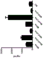


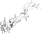


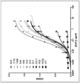


Classifications
-
- C—CHEMISTRY; METALLURGY
- C07—ORGANIC CHEMISTRY
- C07K—PEPTIDES
- C07K16/00—Immunoglobulins [IGs], e.g. monoclonal or polyclonal antibodies
- C07K16/18—Immunoglobulins [IGs], e.g. monoclonal or polyclonal antibodies against material from animals or humans
- C07K16/28—Immunoglobulins [IGs], e.g. monoclonal or polyclonal antibodies against material from animals or humans against receptors, cell surface antigens or cell surface determinants
- C07K16/2878—Immunoglobulins [IGs], e.g. monoclonal or polyclonal antibodies against material from animals or humans against receptors, cell surface antigens or cell surface determinants against the NGF-receptor/TNF-receptor superfamily, e.g. CD27, CD30, CD40, CD95
-
- A—HUMAN NECESSITIES
- A61—MEDICAL OR VETERINARY SCIENCE; HYGIENE
- A61K—PREPARATIONS FOR MEDICAL, DENTAL OR TOILETRY PURPOSES
- A61K35/00—Medicinal preparations containing materials or reaction products thereof with undetermined constitution
- A61K35/12—Materials from mammals; Compositions comprising non-specified tissues or cells; Compositions comprising non-embryonic stem cells; Genetically modified cells
- A61K35/14—Blood; Artificial blood
- A61K35/17—Lymphocytes; B-cells; T-cells; Natural killer cells; Interferon-activated or cytokine-activated lymphocytes
-
- A—HUMAN NECESSITIES
- A61—MEDICAL OR VETERINARY SCIENCE; HYGIENE
- A61K—PREPARATIONS FOR MEDICAL, DENTAL OR TOILETRY PURPOSES
- A61K39/00—Medicinal preparations containing antigens or antibodies
- A61K39/395—Antibodies; Immunoglobulins; Immune serum, e.g. antilymphocytic serum
- A61K39/39533—Antibodies; Immunoglobulins; Immune serum, e.g. antilymphocytic serum against materials from animals
- A61K39/3955—Antibodies; Immunoglobulins; Immune serum, e.g. antilymphocytic serum against materials from animals against proteinaceous materials, e.g. enzymes, hormones, lymphokines
-
- A—HUMAN NECESSITIES
- A61—MEDICAL OR VETERINARY SCIENCE; HYGIENE
- A61K—PREPARATIONS FOR MEDICAL, DENTAL OR TOILETRY PURPOSES
- A61K45/00—Medicinal preparations containing active ingredients not provided for in groups A61K31/00 - A61K41/00
- A61K45/06—Mixtures of active ingredients without chemical characterisation, e.g. antiphlogistics and cardiaca
-
- A—HUMAN NECESSITIES
- A61—MEDICAL OR VETERINARY SCIENCE; HYGIENE
- A61P—SPECIFIC THERAPEUTIC ACTIVITY OF CHEMICAL COMPOUNDS OR MEDICINAL PREPARATIONS
- A61P35/00—Antineoplastic agents
-
- A—HUMAN NECESSITIES
- A61—MEDICAL OR VETERINARY SCIENCE; HYGIENE
- A61P—SPECIFIC THERAPEUTIC ACTIVITY OF CHEMICAL COMPOUNDS OR MEDICINAL PREPARATIONS
- A61P37/00—Drugs for immunological or allergic disorders
- A61P37/02—Immunomodulators
- A61P37/04—Immunostimulants
-
- A—HUMAN NECESSITIES
- A61—MEDICAL OR VETERINARY SCIENCE; HYGIENE
- A61K—PREPARATIONS FOR MEDICAL, DENTAL OR TOILETRY PURPOSES
- A61K39/00—Medicinal preparations containing antigens or antibodies
- A61K2039/505—Medicinal preparations containing antigens or antibodies comprising antibodies
-
- C—CHEMISTRY; METALLURGY
- C07—ORGANIC CHEMISTRY
- C07K—PEPTIDES
- C07K2317/00—Immunoglobulins specific features
- C07K2317/20—Immunoglobulins specific features characterized by taxonomic origin
- C07K2317/24—Immunoglobulins specific features characterized by taxonomic origin containing regions, domains or residues from different species, e.g. chimeric, humanized or veneered
-
- C—CHEMISTRY; METALLURGY
- C07—ORGANIC CHEMISTRY
- C07K—PEPTIDES
- C07K2317/00—Immunoglobulins specific features
- C07K2317/30—Immunoglobulins specific features characterized by aspects of specificity or valency
- C07K2317/34—Identification of a linear epitope shorter than 20 amino acid residues or of a conformational epitope defined by amino acid residues
-
- C—CHEMISTRY; METALLURGY
- C07—ORGANIC CHEMISTRY
- C07K—PEPTIDES
- C07K2317/00—Immunoglobulins specific features
- C07K2317/50—Immunoglobulins specific features characterized by immunoglobulin fragments
- C07K2317/52—Constant or Fc region; Isotype
-
- C—CHEMISTRY; METALLURGY
- C07—ORGANIC CHEMISTRY
- C07K—PEPTIDES
- C07K2317/00—Immunoglobulins specific features
- C07K2317/50—Immunoglobulins specific features characterized by immunoglobulin fragments
- C07K2317/56—Immunoglobulins specific features characterized by immunoglobulin fragments variable (Fv) region, i.e. VH and/or VL
- C07K2317/565—Complementarity determining region [CDR]
-
- C—CHEMISTRY; METALLURGY
- C07—ORGANIC CHEMISTRY
- C07K—PEPTIDES
- C07K2317/00—Immunoglobulins specific features
- C07K2317/50—Immunoglobulins specific features characterized by immunoglobulin fragments
- C07K2317/56—Immunoglobulins specific features characterized by immunoglobulin fragments variable (Fv) region, i.e. VH and/or VL
- C07K2317/567—Framework region [FR]
-
- C—CHEMISTRY; METALLURGY
- C07—ORGANIC CHEMISTRY
- C07K—PEPTIDES
- C07K2317/00—Immunoglobulins specific features
- C07K2317/60—Immunoglobulins specific features characterized by non-natural combinations of immunoglobulin fragments
- C07K2317/62—Immunoglobulins specific features characterized by non-natural combinations of immunoglobulin fragments comprising only variable region components
- C07K2317/622—Single chain antibody (scFv)
-
- C—CHEMISTRY; METALLURGY
- C07—ORGANIC CHEMISTRY
- C07K—PEPTIDES
- C07K2317/00—Immunoglobulins specific features
- C07K2317/70—Immunoglobulins specific features characterized by effect upon binding to a cell or to an antigen
- C07K2317/75—Agonist effect on antigen
-
- C—CHEMISTRY; METALLURGY
- C07—ORGANIC CHEMISTRY
- C07K—PEPTIDES
- C07K2317/00—Immunoglobulins specific features
- C07K2317/70—Immunoglobulins specific features characterized by effect upon binding to a cell or to an antigen
- C07K2317/76—Antagonist effect on antigen, e.g. neutralization or inhibition of binding
-
- C—CHEMISTRY; METALLURGY
- C07—ORGANIC CHEMISTRY
- C07K—PEPTIDES
- C07K2317/00—Immunoglobulins specific features
- C07K2317/90—Immunoglobulins specific features characterized by (pharmaco)kinetic aspects or by stability of the immunoglobulin
- C07K2317/92—Affinity (KD), association rate (Ka), dissociation rate (Kd) or EC50 value
Landscapes
- Health & Medical Sciences (AREA)
- Chemical & Material Sciences (AREA)
- Life Sciences & Earth Sciences (AREA)
- Immunology (AREA)
- Medicinal Chemistry (AREA)
- General Health & Medical Sciences (AREA)
- Public Health (AREA)
- Veterinary Medicine (AREA)
- Animal Behavior & Ethology (AREA)
- Pharmacology & Pharmacy (AREA)
- Organic Chemistry (AREA)
- Epidemiology (AREA)
- Engineering & Computer Science (AREA)
- Cell Biology (AREA)
- Bioinformatics & Cheminformatics (AREA)
- Chemical Kinetics & Catalysis (AREA)
- General Chemical & Material Sciences (AREA)
- Nuclear Medicine, Radiotherapy & Molecular Imaging (AREA)
- Virology (AREA)
- Biochemistry (AREA)
- Developmental Biology & Embryology (AREA)
- Biotechnology (AREA)
- Biomedical Technology (AREA)
- Hematology (AREA)
- Proteomics, Peptides & Aminoacids (AREA)
- Molecular Biology (AREA)
- Genetics & Genomics (AREA)
- Zoology (AREA)
- Biophysics (AREA)
- Mycology (AREA)
- Microbiology (AREA)
- Endocrinology (AREA)
- Peptides Or Proteins (AREA)
- Medicines Containing Antibodies Or Antigens For Use As Internal Diagnostic Agents (AREA)
- Preparation Of Compounds By Using Micro-Organisms (AREA)
- Medicines That Contain Protein Lipid Enzymes And Other Medicines (AREA)
Abstract
anti-TNFRSF 25 antibodies and variants thereof, including those that bind to an epitope in the region of amino acids 48-71, are disclosed. The use of the antibodies in research, diagnostic and therapeutic applications is also envisaged.
Description
Cross Reference to Related Applications
This application claims the benefit of priority from U.S. provisional application serial No. 62/348,009, filed on 9/6/2016.
Technical Field
This document relates to anti-TNFRSF 25 antibodies, and their use for research, therapeutic and diagnostic purposes.
Background
Tumor necrosis factor receptor superfamily member 25 (TNFRSF 25) is a TNF-receptor superfamily member that is preferentially expressed by T lymphocytes that activate and undergo antigen. TNFRSF25 is activated by its ligand TL1A (also known as TNFSF 15), which is rapidly upregulated in antigen presenting cells and in some endothelial cells following activation of Toll-like receptors or Fc receptors. TNFRSF25 stimulates NF-. Kappa.B activity and may also stimulate caspase activation to regulate apoptosis (Bodmer et al, immunity 6 (1): 79-88,1997 and Kitson et al, nature384 (6607): 372-375, 1996). The structural organization of the 393 amino acid long human TNFRSF25 protein is most homologous to TNF receptor 1 (TNFR 1). The extracellular domain of TNFRSF25 includes four cysteine-rich domains, and the cytoplasmic region contains the death domain of known apoptotic signals. Alternative splicing produces a number of different TNFRSF25 isoforms, most of which are potentially secreted molecules. Selective splicing of the TNFRSF25 gene in B cells and T cells encounters a procedural change upon T cell activation, which produces mainly full-length membrane-bound isoforms, and is thought to be involved in controlling lymphocyte proliferation induced by T cell activation.
Activation of TNFRSF25 is dependent on prior engagement of the T cell receptor. TNFRSF25 signaling increases T cell sensitivity to endogenous IL-2 and enhances T cell proliferation upon TNARSF25 binding to TL 1A. Since activation of TNFRSF25 is T cell receptor dependent, the in vivo activity of TNFRSF25 is specific for T cells that encounter homologous antigens. At rest, and in the absence of potential autoimmunity, the majority of T cells that often encounter homologous antigens are FoxP3+ regulatory T cells. TNFRSF25 was stimulated in the absence of any other exogenous signal, stimulating highly specific proliferation of FoxP3+ regulatory T cells from a baseline of 8-10% of all CD4+ T cells to 35-40% of all CD4+ T cells within 5 days (Schreiber et al, J Clin Invest 120 (10): 3629-3640, 2010). Therapeutic agonists of TNFRSF25 may be used to stimulate Treg expansion, which may reduce inflammation in experimental models of asthma, allogenic solid organ Transplantation and ocular keratitis (Schreiber et al, supra, reddy et al, J Virol 86 (19): 10606-10620,2012; and Wolf et al, transplantation 94 (6): 569-574, 2012). Similarly, because TNFRSF25 activation is antigen-dependent, co-stimulation of TNFRSF25 with autoantigens or vaccine antigens can lead to worsening immunopathology or enhanced vaccine-stimulated immunity, respectively (Schreiber et al, J Immunol189 (7): 3311-3318, 2010).
Disclosure of Invention
This document is based, at least in part, on the development of antibodies that target specific epitopes within TNFSF 25. In some embodiments, antibodies may cross-react between species. For example, in some embodiments, provided herein are kk's that can be within 100-fold (e.g., within 10-fold) of each other d Antibodies that bind to rodent and human TNFRSF25 polypeptides. In some cases, the antibodies described herein are capable of eliciting a signaling event consistent with the signaling activity of TL1A (e.g., rodent or human TL 1A) binding to TNFRSF 25. In some embodiments, the antibody is capable of binding to an epitope in the amino acid C48-L71 region of human TNFRSF 25. For example, the antibody may be directed to the region of amino acids P64-T69Epitope specific binding.
This document is also based, at least in part, on the development of affinity matured antibodies targeting TNFRSF 25. The affinity matured antibody may have an enhanced affinity for TNFRSF25 as compared to the parent anti-TNFRSF 25 antibody, or the affinity matured antibody may have an enhanced activity as compared to the parent anti-TNFRSF 25 antibody, or the affinity matured antibody may have an enhanced affinity for TNFRSF25 and an enhanced activity as compared to the parent anti-TNFRSF 25 antibody.
Accordingly, in a first aspect, this document features an anti-TNFRSF 25 antibody or antigen-binding fragment thereof. The antibody or antibody fragment can comprise (i) a heavy chain variable region comprising heavy chain CDR1, CDR2, and CDR3 sequences, wherein heavy chain CDR1 sequence is GFTFSNHDLN (SEQ ID NO: 12) and heavy chain CDR 2sequence is YISSASGLISADAVRG (SEQ ID NO: 14); and the heavy chain CDR3 sequence is DPAYTGYALDF (SEQ ID NO: 26); and (ii) a light chain variable region comprising light chain CDR1, CDR2, and CDR3 sequences, wherein light chain CDR1 sequence is TLSSELSWYTIV (SEQ ID NO. 25), light chain CDR 2sequence is LKSGSHSKGD (SEQ ID NO: 21), and light chain CDR3 sequence is CGAGYTLAGGQYGWV (SEQ ID NO: 23).
The antibody or antibody fragment may comprise (i) a heavy chain variable region comprising heavy chain CDR1, CDR2, and CDR3 sequences, wherein heavy chain CDR1 sequence is GFTFSNHDLN (SEQ ID NO: 12) and heavy chain CDR 2sequence is YISSASGLISADAVG (SEQ ID NO: 14); and the heavy chain CDR3 sequence is DPPYSGLYALDF (SEQ ID NO: 16); and (ii) a light chain variable region comprising light chain CDR1, CDR2, and CDR3 sequences, wherein light chain CDR1 sequence is TLSSELSWYTIV (SEQ ID NO: 25), light chain CDR 2sequence is LKSGSHSKGD (SEQ ID NO: 21), and light chain CDR3 sequence is CGAGYTLAGGQYGWV (SEQ ID NO: 23).
The antibody or antibody fragment may comprise (i) a heavy chain variable region comprising heavy chain CDR1, CDR2, and CDR3 sequences, wherein heavy chain CDR1 sequence is GFTFSNHDLN (SEQ ID NO: 12) and heavy chain CDR 2sequence is yisssglisyavrg (SEQ ID NO: 14); and the heavy chain CDR3 sequence is DPAYTGYALDF (SEQ ID NO: 26); and (ii) a light chain variable region comprising light chain CDR1, CDR2, and CDR3 sequences, wherein light chain CDR1 sequence is TLSSELSGFTIV (SEQ ID NO: 27), light chain CDR 2sequence is LKSGSHSKGD (SEQ ID NO: 21), and light chain CDR3 sequence is CGAGYTLANQYGWV (SEQ ID NO: 28).
The antibody or antigen-binding fragment can further comprise a variable region Framework (FW) sequence juxtaposed between the CDRs according to formulae (FW 1) - (CDR 1) - (FW 2) - (CDR 2) - (FW 3) - (CDR 3) - (FW 4), wherein the variable region FW sequence in the heavy chain variable region is a heavy chain variable region FW sequence, and wherein the variable region FW sequence in the light chain variable region is a light chain variable region FW sequence. The variable region FW sequence may be human. The antibody or antigen-binding fragment may also contain human heavy and light chain constant regions. The constant region may be selected from the group consisting of human IgG1, igG2, igG3, and IgG4. The constant region is IgG1 or IgG4. Tumor cell apoptosis in a subject can be increased upon administration of the antibody or antigen-binding fragment to the subject at a dose of about 0.1mg/kg to about 50 mg/kg.
In another aspect, this document features a pharmaceutical composition containing a pharmaceutically acceptable carrier and an antibody or antigen-binding fragment disclosed herein.
In another aspect, this document features an article of manufacture containing the above-described pharmaceutical composition and at least one additional agent for treating cancer. The at least one additional agent may be an agent targeting CTLA-4, PD-1, PD-L1, LAG-3, tim-3, TNFRSF4, TNFRSF9, TNFRSF18, CD27, CD39, CD47, CD73, or CD278, or may be an A2A receptor antagonist or a TGF- β antagonist. The at least one additional agent may be one or more of a B7 family costimulatory molecule, a TNF receptor superfamily costimulatory molecule, a vaccine composition, or a chemotherapeutic agent. The at least one additional agent may comprise chimeric antigen receptor-transfected T cells or expanded tumor-infiltrating lymphocytes for adoptive T cell therapy in vitro or in a subject. The at least one additional agent may be used during the in vitro manufacturing process of the autologous T cell therapy.
In another aspect, this document features a method for treating a tumor in a subject, wherein the method includes administering to the subject an amount of a composition described herein effective to induce apoptosis of tumor cells expressing TNFRSF25 in the tumor.
Also featured herein is a method for stimulating CD8+ T cell proliferation in a subject, wherein the method comprises administering to the subject a therapeutically effective amount of a composition as described herein. As determined by flow cytometry analysis of antigen-specific CD8+ T cells, the proliferation of CD8+ T cells can be increased by at least about 20% compared to the baseline proliferation level prior to administration.
Additionally, featured herein is a method of eliciting an immune response in a subject, wherein the method comprises administering to the subject a therapeutically effective amount of a composition as described herein.
Also featured herein are methods for stimulating proliferation of CD4+ FoxP3+ regulatory T cells in a subject, wherein the methods comprise administering to the subject a therapeutically effective amount of a composition as described herein.
In another aspect, this document features an isolated monoclonal antibody or Fab fragment thereof that specifically binds human TNFRSF25, wherein the antibody or Fab fragment thereof elicits a signaling event upon binding to TNFRSF25 that is consistent with the signaling event elicited by binding of TL1A to TNFRSF25, and wherein the antibody binds to an epitope in the amino acid C48-L71 region of human TNFRSF 25. The antibody or fragment can bind to an epitope comprising at least one of the following residues of TNFRSF 25: 1 of SEQ ID NO C48, R49, G50, C51, P52, A53, G54, H55, Y56, L57, K58, A59, P60, C61, T62, E63, P64, C65, G66, N67, S68, T69, C70 or L71. Binding of the antibody or fragment to TNFRSF25 can block binding of TL1A to TNFRSF 25. The antibody may be a humanized antibody or a human antibody. The antibody may be an IgG antibody of any subtype. The antibody or fragment can have a K within 100-fold of the other d Values bind to mouse TNFRSF25, non-human primate TNFRSF25, and human TNFRSF 25.
In another aspect, this document features an isolated monoclonal antibody or Fab fragment thereof that specifically binds human TNFRSF25, wherein the antibody or Fab fragment thereof elicits a signaling event upon binding to TNFRSF25, the signaling event characterized by a signaling event elicited even by binding of TL1A to TNFRSF25, and wherein the antibody binds to an epitope in the amino acid P64-T69 region of human TNFRSF 25. The antibody or fragment can be combined with a composition containing TNFRSF25 that binds to an epitope of at least one of the following residues: p64, C65, G66, N67, S68 or T69 of SEQ ID NO. 1. Binding of the antibody or fragment to TNFRSF25 can block binding of TL1A to TNFRSF 25. The antibody may be a humanized antibody or a human antibody. The antibody may be an IgG antibody of any subtype. The antibody or fragment can have a K within 100-fold of the other d Values bind to mouse TNFRSF25, non-human primate TNFRSF25, and human TNFRSF 25.
In another aspect, this document features an isolated monoclonal antibody or Fab fragment thereof that specifically binds human TNFRSF25, wherein the antibody binds to an epitope having a sequence at least 80% identical to the sequence set forth in C48-L71 of SEQ ID NO: 1. In some embodiments, the antibody can bind to an epitope having a sequence at least 90% identical to a sequence set forth in C48-L71 of SEQ ID NO. 1, or having a sequence at least 95%, at least 98%, or at least 99% identical to a sequence set forth in C48-L71 of SEQ ID NO. 1.
In another aspect, this document features an isolated monoclonal antibody or Fab fragment thereof that specifically binds human TNFRSF25, wherein the antibody binds to an epitope having a sequence at least 85%, at least 90%, or at least 95% identical to the sequence set forth in P64-T69 of SEQ ID No. 1.
Also featured herein is an isolated monoclonal antibody or Fab fragment thereof that specifically binds human TNFRSF25, wherein the antibody binds to an epitope having the sequence shown in C48-L71 of SEQ ID NO:1, but having 4 or fewer amino acid substitutions. In some embodiments, the antibody may bind to an epitope having the sequence shown in C48-L71 of SEQ ID NO. 1 but with three or fewer amino acid substitutions, with two or fewer amino acid substitutions, or with one amino acid substitution.
In another aspect, this document features an isolated monoclonal antibody or Fab fragment thereof that specifically binds human TNFRSF25, wherein the antibody binds to an epitope having the sequence shown in P64-T69 of SEQ ID No. 1 but with one amino acid substitution.
In another aspect, hereinCharacterized by a method for inhibiting tumor growth in a mammal. The method can include administering to the mammal a composition comprising a pharmaceutically acceptable carrier and a monoclonal antibody or Fab fragment thereof that specifically binds to human TNFRSF25, wherein the antibody or Fab fragment thereof is capable of mimicking a signaling event stimulated by the binding of TL1A to TNFRSF25, and wherein the antibody binds to an epitope in the amino acid C48-L71 region of human TNFRSF 25. The antibody or fragment can bind to an epitope comprising at least one of the following residues of TNFRSF 25: 1, C48, R49, G50, C51, P52, A53, G54, H55, Y56, L57, K58, A59, P60, C61, T62, E63, P64, C65, G66, N67, S68, T69, C70 or L71. The antibody or fragment can bind to an epitope comprising at least one of the following residues of TNFRSF 25: p64, C65, G66, N67, S68 or T69 of SEQ ID NO. 1. Binding of the antibody or fragment to TNFRSF25 can block binding of TL1A to TNFRSF 25. The antibody may be a humanized antibody or a human antibody. The antibody may be an IgG antibody of any subtype. The antibody or fragment can have a K within 100-fold of the other d Values bind to mouse TNFRSF25, non-human primate TNFRSF25, and human TNFRSF 25.
In another aspect, this document features a pharmaceutical composition containing a pharmaceutically acceptable carrier and an isolated monoclonal antibody or Fab fragment as described herein. Additionally, featured herein is the use of the pharmaceutical composition for treating cancer, an infectious disease, or a tissue graft in a patient.
Unless defined otherwise, all technical and scientific terms used herein have the same meaning as commonly understood by one of ordinary skill in the art to which this invention belongs. Although methods and materials similar or equivalent to those described herein can be used in the practice of the present invention, suitable methods and materials are described below. All publications, patent applications, patents, and other references mentioned herein are incorporated by reference in their entirety. In case of conflict, the present specification, including definitions, will control. In addition, the materials, methods, and examples are illustrative only and not intended to be limiting.
The details of one or more embodiments of the invention are set forth in the accompanying drawings and the description below. Other features, objects, and advantages of the invention will be apparent from the description and drawings, and from the claims.
Drawings
FIG. 1A is an alignment of the amino acid sequences of human TNFRSF25 (SEQ ID NO: 2) with human TNFR1 (SEQ ID NO: 3) and human Fas (SEQ ID NO: 4), TNFRSF25 sharing some homology with human TNFR1 and human Fas. Sequence numbering starts at the N-terminus of the predicted mature protein. The positions of the cysteine-rich regions I-IV, the transmembrane domain (TM) and the Death Domain (DD) are indicated. Residues that are identical between at least two sequences are indicated in bold and underlined.
FIG. 1B shows two representative TNFRSF25 amino acid sequences from humans (SEQ ID NOS: 1 and 7), and representative TNFRSF25 amino acid sequences from mice (SEQ ID NO: 8), rhesus monkeys (SEQ ID NO: 9), and Cynomolgus monkeys (Cynomolgus Macaque) (SEQ ID NO: 10).
Fig. 2 is a diagram depicting steps in a method for assessing expression of various recombinant TNFRSF25 polypeptides.
Fig. 3 is a graph plotting the expression levels of six recombinant TNFRSF25 polypeptides.
Fig. 4 is a diagram depicting steps in a method for assessing binding of a chimeric anti-TNFRSF 25 antibody to a recombinant TNFRSF25 polypeptide.
Fig. 5 is a graph plotting increasing amounts of recombinant TNFRSF25 polypeptide binding to chimeric anti-TNFRSF 25 antibodies.
Fig. 6 is a graph plotting the fold change in binding of various TNFRSF25 alanine mutants to anti-TNFRSF 25 antibody.
Figure 7 is a structural diagram of TNFRSF25 showing the positions of residues identified as being involved in or likely to be involved in antibody binding.
Fig. 8 is a space-filling diagram of the TNFRSF25 structure viewed from both sides, and indicates the positions of amino acids identified as being involved in antibody binding. "Hot spot" residues are identified as being located in the domain between amino acids 48 and 71.
FIG. 9 is a graph plotting inhibition of binding of anti-TNFRSF 25 antibody to TNFRSF25 in the presence or absence of a TL1A-Ig fusion protein or inhibition of binding of TL1A-Ig to TNFRSF25 in the presence or absence of an anti-TNFRSF 25 antibody. Left column: in the presence of TL1A-Ig, the binding of anti-TNFRSF 25 antibody ("hamster parent") to recombinant human TNFRSF25 was completely inhibited. Middle column: binding of the TL1A-Ig fusion to recombinant human TNFRSF25 is completely inhibited in the presence of the parent anti-TNFRSF 25 antibody. And (4) right column: binding of humanized anti-TNFRSF 25 antibody to recombinant human TNFRSF25 is completely inhibited in the presence of TL 1A-Ig.
FIG. 10 is a graph plotting the binding of the parent humanized anti-TNFRSF 25 antibody and affinity matured clone to TNFRSF25-Fc fusion protein.
FIGS. 11A and 11B are graphs plotting caspase activity of the parent anti-TNFRSF 25 antibody, TL1A, and the indicated affinity matured clones. Affinity matured clones were in the IgG1 format.
FIG. 12 is a graph plotting caspase activity of the parent anti-TNFRSF 25 antibody, TL1A and the indicated affinity matured clones. As shown, the affinity matured clones were in IgG1 or IgG4 format.
FIG. 13A is a graph plotting fluorescence levels in a competitive assay, where 647 labeled TL1A and various antibody competitors (the 4C12 parent and M3, M4 and M5 affinity matured clones) were incubated with p815 cells expressing TNFRSF25 (DR 3). FIG. 13B is a graph plotting the fluorescence levels in a competitive assay, where
 647-labeled 4C12 and various antibody competitors (the 4C12 parent, hIgG1 and M3, M4 and M5 affinity matured clones) were incubated with p815 cells expressing TNFRSF25 (DR 3).
Detailed Description
This document is based, at least in part, on the development of antibodies that target specific epitopes within TNFSF 25. For example, provided herein are antibodies that bind to an epitope in the amino acid C48-L71 region of human TNFRSF 25. In some embodiments, the antibody can bind to rodent and human TNFRSF25 polypeptides with Kd values within 100-fold (e.g., within 10-fold) of each other and is capable of mimicking the signaling activity of rodent or human TL1A binding to TNFRSF 25. In some embodiments, the antibody can bind to an epitope in the region of amino acids P64-T69, which is a conserved region of polypeptides in placental mammals. Also provided herein are methods of stimulating proliferation of T cells (e.g., human T cells, murine T cells, or cynomolgus T cells) using one or more antibodies or compositions containing one or more antibodies provided herein, as well as methods of treating (e.g., by administering an amount of an anti-TNFRSF 25 antibody effective to stimulate proliferation of CD8+ T cells) a human cancer patient using one or more antibodies or compositions.
Also provided herein are affinity matured humanized TNFRSF25 specific monoclonal antibodies, as well as antigen binding fragments of the affinity matured antibodies. Also provided herein are methods of using affinity maturation antibodies to, inter alia, stimulate proliferation of T cells (e.g., human T cells, including naturally occurring tumor-reactive CD8+ T cells or CD4+ FoxP3+ regulatory T cells, as well as mouse T cells or cynomolgus T cells), and methods of using affinity maturation antibodies to treat cancers and other disease states in which expansion of T cells may have a beneficial effect (e.g., infectious diseases, graft-versus-host diseases, and autoimmune diseases). The method can include, for example, administering an amount of an affinity maturation antibody effective to stimulate proliferation of CD8+ T cells or an appropriate population of regulatory T cells.
The partial amino acid sequence of human TNFRSF25 aligned with the amino acid sequences of human TNFR1 and FA is shown in fig. 1. A representative full-length amino acid sequence of human TNFRSF25 is:

as used herein, the term "antibody" refers to any immunoglobulin or antibody (e.g., human, hamster, cat, mouse, cartilaginous fish, or camelid antibody), and any derivative or conjugate thereof, that specifically binds to an antigen. A variety of antibodies are known to those of skill in the art. Non-limiting examples of antibodies include monoclonal antibodies, polyclonal antibodies, humanized antibodies, multispecific antibodies (e.g., bispecific antibodies), single chain antibodies (e.g., single domain antibodies, camelid antibodies, and cartilaginous fish antibodies), chimeric antibodies, feline antibodies, and feline humanized antibodies. Monoclonal antibodies are a homogeneous population of antibodies directed against a particular epitope of an antigen. Polyclonal antibodies are heterogeneous populations of antibody molecules contained in the serum of immunized animals. The term "antibody" also includes antibody derivatives and conjugates (e.g., antibodies conjugated to a stabilizing protein, detectable moiety, or therapeutic agent).
By "isolated" or "purified" with respect to a polypeptide (e.g., an antibody or fragment thereof), it is meant that the polypeptide is separated to some extent from cellular components (e.g., other polypeptides, lipids, carbohydrates, and nucleic acids) with which it typically occurs naturally. In some embodiments, an "isolated" polypeptide is a polypeptide that is expressed and produced in an environment other than the environment in which the polypeptide is naturally expressed and produced. For example, plant polypeptides are isolated when expressed and produced in bacteria or fungi. Similarly, a plant polypeptide is isolated when its gene coding sequence is operably linked to chimeric regulatory elements and expressed in a tissue in which the polypeptide is not naturally expressed.
The isolated polypeptide may produce a single major band on a non-reducing polyacrylamide gel. An isolated polypeptide can be at least about 75% pure (e.g., at least 80%, 85%, 90%, 95%, 97%, 98%, 99%, or 100% pure). Isolated polypeptides may be obtained, for example, by extraction from natural sources, by chemical synthesis, or by recombinant production in a host cell or transgenic plant, and may be purified using, for example, affinity chromatography, immunoprecipitation, size exclusion chromatography, and ion exchange chromatography. The degree of purification can be measured using any suitable method, including but not limited to column chromatography, polyacrylamide gel electrophoresis, or high performance liquid chromatography.
An "antigen-binding fragment" is any portion of a full-length antibody that contains at least one variable domain capable of specifically binding to an antigen (e.g., a variable domain of a mammalian (e.g., cat, human, hamster, or mouse) heavy or light chain immunoglobulin, a camelid variable antigen-binding domain (VHH), or a cartilaginous fish immunoglobulin neo-antigen receptor (Ig-NAR) domain). Non-limiting examples of antibody fragments include Fab, fab ', F (ab') 2, and Fv fragments, diabodies, linear antibodies, and multispecific antibodies formed from antibody fragments. Additional antibody fragments containing at least one camelid VHH domain or at least one cartilaginous fish Ig-NAR domain include mini-bodies, minibodies, subnano-antibodies and nanobodies, as well as any of the additional forms of antibodies described, for example, in U.S. publication No. 2010/0092470.
An "Fv fragment" is the smallest antibody fragment that contains the entire antigen recognition and binding site. This region consists of a dimer of a heavy chain variable domain and a light chain variable domain in tight, non-covalent association. In this configuration, the three Complementarity Determining Regions (CDRs) of each variable domain interact to define an antigen binding site on the surface of the VH-VL dimer. The term "complementarity determining region" or "CDR" refers to a region within an immunoglobulin (heavy or light chain immunoglobulin) that forms part of an antigen binding site in an antibody or antigen binding fragment thereof. As is known in the art, each of the heavy and light chain immunoglobulins contains three CDRs, referred to as CDR1, CDR2 and CDR3. In any antibody or antigen-binding fragment, the three CDRs from the heavy chain immunoglobulin and the three CDRs from the light chain immunoglobulin together form an antigen-binding site in the antibody or antigen-binding fragment thereof. The Kabat database is a system used in the art for numbering CDR sequences present in a light chain immunoglobulin or a heavy chain immunoglobulin.
Together, the six CDRs confer antigen binding specificity to the antibody. However, even a single variable domain (or half of an Fv comprising only three CDRs specific for an antigen) has the ability to recognize and bind antigen, albeit usually with lower affinity than the entire binding site. "Fab fragments" also contain lightThe constant domain of the chain and the first constant domain of the heavy chain (CH 1). The "Fab fragment" differs from the "Fab' fragment" in that it is at heavy chain C H 1 domain has some residues added to the carboxy terminus, including one or more cysteines from the antibody hinge region. The "F (ab ') 2 fragments" were originally generated as a pair of "Fab' fragments" with hinge cysteines between them. Methods of making such antibody fragments (such as papain or pepsin digestion) are known to those skilled in the art. For example, F (ab ') 2 fragments can be produced by pepsin digestion of antibody molecules, and Fab fragments can be produced by reducing the disulfide bonds of F (ab') 2 fragments. In some cases. Fab expression libraries can be constructed. See, e.g., huse et al, science,246, 1275,1989. Once produced, the recognition of the TNFRSF25 polypeptide by the antibody or fragment thereof can be tested using standard immunoassay methods such as ELISA techniques, radioimmunoassays, and western blots. Referring to the description of the preferred embodiment,Short Protocols in Molecular Biology,chapter 11, green Publishing Associates and John Wiley&Sons, ed. Ausubel et al, 1992.
The antibody may be of the IgA-type, igD-type, igE-type, igG-type, or IgM-type, including IgG-type or IgM-type, such as, but not limited to, igG 1-type, igG 2-type, igG 3-type, igG 4-type, igM 1-type, and IgM 2-type. For example, in some cases, the antibody is of the IgG 1-type, igG 2-type, or IgG 4-type.
In some embodiments, an antibody as provided herein can be a fully human or humanized antibody. "human antibody" refers to an antibody encoded by a nucleic acid present in the human genome (e.g., a rearranged human immunoglobulin heavy or light chain locus). In some embodiments, the human antibody can be produced in a human cell culture (e.g., a feline hybridoma cell). In some embodiments, human antibodies can be produced in non-human cells (e.g., mouse or hamster cell lines). In some embodiments, human antibodies can be produced in bacterial or yeast cells.
Human antibodies can avoid certain problems associated with xenogeneic antibodies, such as antibodies having murine or rat variable and/or constant regions. For example, because the effector moiety is human, it may interact better with other parts of the human immune system, e.g., more efficiently destroy target cells through complement-dependent cytotoxicity or antibody-dependent cytotoxicity. Furthermore, the human immune system should not recognize the antibody as foreign. In addition, the half-life in the human circulation is similar to that of naturally occurring human antibodies, allowing smaller and less frequent doses to be administered. Methods for making human antibodies are known in the art.
As used herein, the term "humanized antibody" refers to a human antibody that contains minimal sequences derived from non-human (e.g., mouse, hamster, rat, rabbit, or goat) immunoglobulins. Humanized antibodies are typically chimeric or mutant monoclonal antibodies from mouse, rat, hamster, rabbit, or other species that carry human constant and/or variable regions or variations in specificity. In a non-limiting example, a humanized antibody is a human antibody (recipient antibody) in which residues from a hypervariable region (HVR) of the recipient antibody are replaced by residues from a HVR of a non-human species (donor) antibody, such as a mouse, rat, rabbit, or goat antibody of the desired specificity, affinity, and capacity. In some embodiments, fv framework residues of the human immunoglobulin can be replaced with corresponding non-human residues. In some embodiments, the humanized antibody may contain residues that are not present in the recipient antibody or the donor antibody. For example, such modifications may be made to improve antibody performance.
In some embodiments, a humanized antibody may comprise substantially all, at least one, and typically two variable domains, in which all or substantially all of the hypervariable loops (CDRs) correspond to those of a non-human immunoglobulin and all or substantially all of the framework regions are those of a human immunoglobulin sequence. The humanized antibody may also contain at least a portion of an immunoglobulin constant (Fc) region, typically that of a human immunoglobulin.
In some embodiments, a humanized antibody or antigen-binding fragment as provided herein can have reduced or minimal effector function (e.g., as compared to a corresponding non-humanized antibody) such that it does not stimulate effector cells to the same extent as a corresponding non-humanized antibody would produce.
Techniques for generating humanized antibodies are well known to those skilled in the art. In some embodiments, controlled rearrangement of antibody domains linked by protein disulfide bonds (to form new artificial protein molecules or "chimeric" antibodies) may be used (Konieczny et al, haematologia (budap.) 14. Recombinant DNA techniques can be used to construct gene fusions between DNA sequences encoding the variable light and heavy domains of mouse antibodies and the light and heavy constant domains of human antibodies (Morrison et al, proc Natl Acad Sci USA 81, 6851, 1984). For example, DNA sequences encoding the antigen-binding portions or CDRs of murine monoclonal antibodies can be molecularly grafted into DNA sequences encoding the heavy and light chain frameworks of human antibodies (Jones et al, nature 321, 522,1986; and Riechmann et al, nature 332, 323, 1988). The expressed recombinant product is called a "reshaped" or humanized antibody and contains the framework of a human antibody light or heavy chain and the antigen-recognition portion CDRs of a murine monoclonal antibody.
Other methods for designing heavy and light chains and for producing humanized antibodies are described, for example, in U.S. Pat. nos. 5,530,101, 5,565,332, 5,585,089, 5,639,641, 5,693,761, 5,693,762, and 5,733,743. Additional methods for humanizing antibodies are described, for example, in U.S. Pat. nos. 4,816,567, 4,935,496, 5,502,167, 5,558,864, 5,693,493, 5,698,417, 5,705,154, 5,750,078, and 5,770.403.
The term "single chain antibody" refers to a single polypeptide comprising at least one variable binding domain capable of specifically binding an antigen (e.g., a variable domain of a mammalian heavy or light chain immunoglobulin, a camelid VHH, or a cartilaginous fish (e.g., shark) Ig-NAR domain). Non-limiting examples of single chain antibodies include single domain antibodies.
The term "single domain antibody" as used herein refers to a polypeptide comprising a camelid VHH or at least one cartilaginous fish Ig-NAR domain capable of specifically binding an antigen. Non-limiting examples of single domain antibodies are described, for example, in U.S. publication No. 2010/0092470.
An antibody or antigen-binding fragment thereof "specifically binds" a particular antigen, such as TNFRFS25, when it binds to that antigen in a sample and does not recognize and bind, or to a lesser extent recognizes and binds, other molecules in the sample. In some embodiments, the antibody or antigen-binding fragment thereof can be in phosphate buffered saline at or less than, for example, about 1x10 -6 M (e.g., equal to or less than about 1x10 -9 M, equal to or less than about 1x10 -10 M, equal to or less than about 1x10 -11 M, equal to or less than about 1x10 -12 M) selectively binds to the epitope with affinity (Kd). The ability of an antibody or antigen binding fragment to specifically bind an epitope of a protein can be determined using any method known in the art or those described herein (e.g., by Biacore/surface plasmon resonance). This may include, for example, binding to TNFRSF25 on living cells (e.g., as measured by caspase activation in stab-activated transformed cells), binding to an immobilized target substrate (including human TNFRSF25 fusion protein) (as measured using ELISA methods), binding to TNFRSF25 on living cells (as measured by flow cytometry), or binding to an immobilized substrate (as measured by surface plasmon resonance (including ProteOn)).
Antibodies with specific binding affinity for TNFRSF25 can be generated using standard methods. For example, TNFRSF25 polypeptides (e.g., fragments of SEQ ID NO:1 or SEQ ID NO:2 having the sequence shown in SEQ ID NO:1, SEQ ID NO:2, or at least 6 to 10 amino acids in length) can be recombinantly produced, purified from a biological sample (e.g., a heterologous expression system), or chemically synthesized and used to immunize a host animal, including rabbit, chicken, mouse, guinea pig, or rat. Various adjuvants that can be used to increase the immune response depend on the host species, including freund's adjuvant (complete and incomplete), mineral gels such as aluminum hydroxide, surface active substances such as lysolecithin, pluronic polyols, polyanions, peptides, oil emulsions, keyhole limpet hemocyanin, and dinitrophenol. Monoclonal antibodies can be prepared using TNFRSF25 polypeptides and standard hybridoma techniques. In particular, monoclonal antibodies may be monoclonal antibodiesObtained by any technique, by the human B-cell hybridoma technique of continuous cell lines in culture (such as described by Kohler et al (Nature 256:495, 1975)), kosbor et al (Immunology Today,4, 72, 1983) or Cote et al (Proc. Natl. Acad. Sci.USA,80, 2026, 1983) and by Cole et al(Monoclonal Antibodies and Cancer TherapyThe EBV-hybridoma technology described by Alan r.loss, inc., pp 77-96, 1983) provides for the production of antibody molecules. Such antibodies may be of any immunoglobulin class, including IgG, igM, igE, igA, igD, and any subclass thereof. Hybridomas producing monoclonal antibodies can be cultured in vitro and in vivo.
In some embodiments, a monoclonal anti-TNFRSF 25 antibody as provided herein has a heavy chain variable region comprising the amino acid sequence set forth in SEQ ID No. 5, but having from 1 to 24 modifications (e.g., substitutions, additions or deletions) such that the amino acid sequence is 80% to 99.5% identical to SEQ ID No. 5. In some embodiments, a monoclonal anti-TNFRSF 25 antibody as provided herein has a light chain variable region comprising the amino acid sequence set forth in SEQ ID No. 6, but having from 1 to 23 modifications (e.g., substitutions, additions or deletions) such that the amino acid sequence is 80% to 99.9% identical to SEQ ID No. 6. 5 and SEQ ID NQ:6 is as follows:

thus, provided herein are heavy chain variable region polypeptides comprising the amino acid sequence set forth in SEQ ID No. 5, or an antigen-binding fragment thereof, but having 1-24 sequence modifications, and polypeptides having at least about 80% (e.g., about 85%, about 90%, about 91%, about 92%, about 93%, about 94%, about 95%, about 96%, about 97%, about 98%, or about 99%) amino acid sequence identity to SEQ ID No. 5, or an antigen-binding fragment thereof. In some embodiments, the heavy chain variable region polypeptide may contain 24 or fewer (e.g., 24, 23, 22, 21, 20, 19, 18, 17, 16, 15, 14, 13, 12, 11, 10, 9, 8, 7, 6,5, 4, 3, or 1) amino acid substitutions as compared to SEQ ID No. 5 or an antigen-binding fragment thereof.
Also provided herein are light chain variable region polypeptides comprising the amino acid sequence set forth in SEQ ID No. 6, or an antigen-binding fragment thereof, but having from 1 to 23 sequence modifications, and polypeptides having at least about 80% (e.g., about 85%, about 90%, about 91%, about 92%, about 93%, about 94%, about 95%, about 96%, about 97%, about 98%, or about 99%) amino acid sequence identity to SEQ ID No. 6, or an antigen-binding fragment thereof. In some embodiments, the light chain variable region polypeptide may contain 23 or fewer (e.g., 23, 22, 21, 20, 19, 18, 17, 16, 15, 14, 13, 12, 11, 10, 9, 8, 7, 6,5, 4, 3, 2, or 1) amino acid substitutions as compared to SEQ ID No. 6 or an antigen-binding fragment thereof.
Also provided herein are antibodies and antigen-binding fragments comprising the heavy chain variable region polypeptides and light chain variable region polypeptides disclosed herein. In some embodiments, for example, an antibody or antigen-binding fragment may contain a heavy chain variable region sequence comprising the amino acid sequence set forth in SEQ ID NQ:5 with 1 to 24 amino acid substitutions (e.g., 1 to 5,5 to 10, 10 to 15, 15 or 20, or 20 to 24 amino acid substitutions in total) and a light chain variable region sequence comprising the amino acid sequence of SEQ ID NQ:6 with 1 to 23 amino acid substitutions (e.g., 1 to 5,5 to 10, 10 to 15, 15 to 20, or 20 to 23 amino acid substitutions in total). Amino acid substitutions refer to the replacement of one amino acid residue by another in a peptide sequence.
In some embodiments, an anti-TNFRSF 25 antibody or antigen binding fragment thereof as provided herein can bind to an epitope of TNFRSF25 having an amino acid sequence at least 80% identical (e.g., at least 85%, at least 90%, or at least 95% identical) to the sequence set forth in C48-L71 of SEQ ID NO: 1. For example, in some cases, an antibody or antigen-binding fragment thereof can bind to an epitope of TNFRSF25 having the sequence shown in C48-L71 of SEQ ID NO:1, but having 4 or fewer (e.g., 3 or fewer, or two or fewer) amino acid substitutions, or having one amino acid substitution.
In some embodiments, an anti-TNFRSF 25 antibody or antigen binding fragment thereof as provided herein can bind an epitope of TNFRSF25 having an amino acid sequence at least 85% identical to the sequence set forth in P64-T769 of SEQ ID NO: 1. For example, in some cases, the antibody or antigen-binding fragment thereof can bind to an epitope of TNFRSF25 having the sequence shown in P64-T69 of SEQ ID NO. 1, but with one amino acid substitution.
As described herein, the use of humanized monoclonal antibodies against TNFRSF25, generated as described elsewhere (see WO 2016/081455), for affinity maturation studies, led to the identification of several heavy and light chain variable region CDR modifications that appear to be associated with enhanced affinity and/or activity. Interestingly, however, and as discussed in example 3 herein, the clone that showed the greatest binding affinity for TNFRSF25 did not always have a clone with greater than the greatest activity of the parent antibody. For example, the clone identified herein as "M5", as determined by the caspase-3 release assay, showed enhanced agonist activity, but surprisingly, its binding to TNFRSF25-Fc fusion protein was significantly weaker than most other clones identified by combinatorial library screening. The exception to this finding was the clone identified as "M4" which exhibited enhanced binding to TNFRSF25-Fc and enhanced agonist activity compared to the parent antibody.
Thus, in some cases, an antibody provided herein can have enhanced binding affinity for TNFRSF25, enhanced agonist activity (e.g., as determined by the caspase-3 assay), or both enhanced affinity and activity, as compared to a parent humanized 4C12 antibody. By "enhanced" affinity or activity is meant an enhancement of at least 5% (e.g., at least 10%, 20%, 30%, 40%, 50%, 60%, 70%, 80%, 90%, 100%, or more than 100%) as compared to the affinity or agonistic activity of humanized 4C 12. The sequences of the variable regions of the 4C12 heavy and light chains are shown herein in SEQ ID NOS: 5 and 6, respectively. The agonistic activity of an antibody can be assessed using, for example, the caspase-3 assay described herein or the TNFRSF25 receptor signaling assay as described elsewhere (see, e.g., bu et al, bone 33 (5): 760-770, 2003).
Thus, in some embodiments, provided herein are heavy chain variable region polypeptides comprising the CDR1 sequence shown in SEQ ID NO. 12, the CDR 2sequence shown in SEQ ID NO. 14, and the CDR3 sequences shown in SEQ ID NO. 16, 26, and 32. For example, a polypeptide can further comprise a variable region heavy chain Framework (FW) sequence juxtaposed between the CDRs according to formulae (FW 1) - (CDR 1) - (FW 2) - (CDR 2) - (FW 3) - (CDR 3) - (FW 4). In some embodiments, the FW sequence may be a human sequence. In some embodiments, the heavy chain variable region polypeptide may comprise the FW1 sequence shown in SEQ ID NO:11, the FW 2sequence shown in SEQ ID NO:13, the FW3 sequence shown in SEQ ID NO:15 and the FW4 sequence shown in SEQ ID NO: 17.
Also provided herein are light chain variable region polypeptides comprising a CDR1 sequence set forth in any of SEQ ID NOs 25, 27 and 29, a CDR 2sequence set forth in SEQ ID NO 21, and a CDR3 sequence set forth in any of SEQ ID NOs 23, 28 and 30. The polypeptide may also include a variable region light chain FW juxtaposed between the CDRs according to formulae (FW 1) - (CDR 1) - (FW 2) - (CDR 2) - (FW 3) - (CDR 3) - (FW 4). In some cases, the FW sequence may be a human sequence. In some embodiments, the light chain variable region polypeptide may comprise the FW1 sequence shown in SEQ ID NO:18, the FW 2sequence shown in SEQ ID NO:20, the FW3 sequence shown in SEQ ID NO:22 and the FW4 sequence shown in SEQ ID NO: 24.
Also provided herein are antibodies and antigen-binding fragments comprising the heavy chain variable region polypeptides and light chain variable region polypeptides disclosed herein. In some embodiments, for example, an antibody or antigen-binding fragment can contain a heavy chain variable region sequence having heavy chain CDR sequences as disclosed herein and a light chain variable region sequence having light chain CDR sequences as disclosed herein. In some cases, the antibody is not a 4C12 antibody such that it does not contain the heavy chain variable region CDRs as shown in SEQ ID NOs 12, 14 and 16 and does not contain the light chain variable region CDRs as shown in SEQ ID NOs 19, 21 and 23.
In some embodiments, amino acid substitutions may be made by selecting conservative substitutions that do not differ significantly in their effect on: maintaining (a) the structure of the peptide backbone in the substitution region, (b) the charge or hydrophobicity of the molecule at the target site, or (c) the side chain volume. For example, naturally occurring residues may be classified into the following groups based on side chain properties: (1) Hydrophobic amino acids (norleucine, methionine, alanine, valine, leucine, and isoleucine); (2) Neutral hydrophilic amino acids (cysteine, serine, and threonine); (3) acidic amino acids (aspartic acid and glutamic acid); (4) Basic amino acids (asparagine, glutamine, histidine, lysine, and arginine); (5) Amino acids that affect chain orientation (glycine and proline); and (6) aromatic amino acids (tryptophan, tyrosine, and phenylalanine). Substitutions made within these groups may be considered conservative substitutions. Non-limiting examples of conservative substitutions include, but are not limited to, valine to alanine substitutions, lysine to arginine substitutions, glutamine to aspartic acid substitutions, glutamic acid to aspartic acid substitutions, serine to cysteine substitutions, asparagine to glutamine substitutions, aspartic acid to glutamic acid substitutions, proline to glycine substitutions, arginine to histidine substitutions, leucine to isoleucine substitutions, isoleucine to leucine substitutions, arginine to lysine substitutions, leucine to methionine substitutions, leucine to phenylalanine substitutions, glycine to proline substitutions, threonine to serine substitutions, serine to threonine substitutions, tyrosine to tryptophan substitutions, phenylalanine to tyrosine substitutions, and/or leucine to valine substitutions. In some embodiments, the amino acid substitutions may be non-conservative, such that a member of one of the above amino acid classes is exchanged for a member of another class.
The percent sequence identity between a particular nucleic acid or amino acid sequence and the sequence referred to by a particular sequence identification number is determined as follows. First, nucleic acid or amino acid Sequences are compared to Sequences shown in a particular sequence identification number using the BLAST 2Sequences (B12 seq) program from an independent version of BLASTZ containing BLASTN version 2.0.14 and BLASTP version 2.0.14. This independent version of BLASTZ is available online at fr.com/blast or at ncbi.nlm.nih.gov. A description of how the B12seq program can be used can be found in the self-describing document attached to BLASTZ. B12seq A comparison between two sequences is performed using the BLASTN or BLASTP algorithm. BLASTN is used to compare nucleic acid sequences, while BLASTP is used to compare amino acid sequences. To compare two nucleic acid sequences, the options are set as follows: set-i to a file containing a first nucleic acid sequence to be compared (e.g., C: \ seq1. Txt), and-j to a file containing a second nucleic acid sequence to be compared (e.g., C: \ seq2. Txt); setting-p to blastn; set-o to any desired filename (e.g., C: \ output.txt); setting-q to-1; -r is set to 2; and all other options remain in their default settings. For example, the following commands may be used to generate an output file containing a comparison between two sequences: c: \ B12seq-i C: \ seq1.Txt-j C: \ seq2.Txt-p blastn-o C: \ output. Txt-q-1-r 2. To compare two amino acid sequences, the options for B12seq were set as follows: set-i to a file containing the first amino acid sequence to be compared (e.g., C: \ seq1. Txt); set-j to the file containing the second amino acid sequence to be compared (e.g., C: \ seq2. Txt); setting-p to blastp; set-o to any desired file name (e.g., C: \ output.txt); and all other options retain their default settings. For example, the following commands may be used to generate an output file containing a comparison between two amino acid sequences: c: \ B12seq-i C: \ seq1.Txt-j C: \ seq2.Txt-p blastp-oc: \ output. Txt. If two compared sequences share homology, the designated output file presents those regions with homology as aligned sequences. If the two compared sequences do not share homology, the designated output file will not present the aligned sequences.
Once aligned, the number of matches is determined by counting the number of positions at which the same nucleotide or amino acid residue is present in both sequences. Percent sequence identity is determined by dividing the number of matches by the length of the sequence shown in the identified sequence (e.g., SEQ ID NO: 1), or by the hinge length (e.g., 100 contiguous nucleotides or amino acid residues from the sequence shown in the identified sequence), and then multiplying the value of the resulting value by 100. For example, an amino acid sequence having 110 matches when aligned to the sequence shown in SEQ ID NO:1 has 90.9% (i.e., 110 ÷ 121x100= 90.9) percent identity to the sequence shown in SEQ ID NO: 1. It should be noted that the percent sequence identity value is rounded to one bit after the nearest decimal point. For example, 75.11, 75.12, 75.13, and 75.14 are rounded down to 75.1, while 75.15, 75.16, 75.17, 75.18, and 75.19 are rounded up to 75.2. It should also be noted that the length value will always be an integer.
Additionally, provided herein are pharmaceutical compositions containing an antibody or antigen-binding fragment as described herein in combination with a pharmaceutically acceptable carrier. A "pharmaceutically acceptable carrier" (also referred to as an "excipient" or "carrier") is a pharmaceutically acceptable solvent, suspending agent, stabilizing agent, or any other pharmacologically inert carrier for delivering one or more therapeutic compounds to a subject (e.g., a mammal, such as a human, non-human primate, dog, cat, sheep, pig, horse, cow, mouse, rat, or rabbit) that is non-toxic to the cells or subject to which it is exposed at the dosages and concentrations employed. The pharmaceutically acceptable carrier may be a liquid or a solid, and may be selected according to the intended mode of administration so as to provide the desired volume, consistency and other relevant transport and chemical properties when combined with the therapeutic compound or compounds and any other components of a given pharmaceutical composition. Typical pharmaceutically acceptable carriers that do not deleteriously react with an amino acid include, for example, but are not limited to: water, salt solutions, binders (e.g., polyvinylpyrrolidone or hydroxypropylmethylcellulose), fillers (e.g., lactose and other sugars, gelatin or calcium sulfate), lubricants (e.g., starch, polyethylene glycol or sodium acetate), disintegrants (e.g., starch or sodium starch glycolate), and wetting agents (e.g., sodium lauryl sulfate). Pharmaceutically acceptable carriers also include pH buffered aqueous solutions or liposomes (small vesicles composed of various types of lipids, phospholipids and/or surfactants, which can be used to deliver drugs to mammals). Other examples of pharmaceutically acceptable carriers include buffers such as phosphates, citrates and other organic acids, antioxidants such as ascorbic acid, low molecular weight (less than about 10 residues) polypeptides, eggsWhite matter such as serum albumin, gelatin or immunoglobulins, hydrophilic polymers such as polyvinylpyrrolidone, amino acids such as glycine, glutamine, asparagine, arginine or lysine, monosaccharides, disaccharides and other carbohydrates including glucose, mannose or dextrins, chelating agents such as EDTA, sugar alcohols such as mannitol or sorbitol, salt-forming counterions such as sodium and/or non-ionic surfactants such as TWEEN TM Polyethylene glycol (PEG) and PLURONICS TM 。
Pharmaceutical compositions may also be formulated by mixing one or more active agents with one or more physiologically acceptable carriers, diluents and/or adjuvants and optionally other agents commonly incorporated into formulations to provide improved transfer, delivery, tolerability, etc. The pharmaceutical compositions may be formulated, for example, in lyophilized formulations, aqueous solutions, dispersions, or solid formulations such as tablets, lozenges, or capsules. The formulary known to all medicinal chemists can be:Remington’sPharmaceutical Sciences(18 th edition, mack Publishing Company, easton, pa. (1990)) (particularly, among others, chapter 87, written by particulate Chapter 87by Block, lawrence) many suitable formulations are found. These preparations include, for example, powders, pastes, ointments, gels, waxes, oils, lipids, lipid-containing (cationic or anionic) vesicles such as LIPOFECTIN TM ) DNA conjugates, anhydrous absorbent creams, oil-in-water and water-in-oil emulsions, emulsion polyethylene glycols (polyethylene glycols of various molecular weights), semi-solid gels and semi-solid mixtures containing polyethylene glycols. Any of the foregoing mixtures may be suitable for use in the treatments and therapies described herein, provided that the active agent in the formulation is not inactivated by the formulation, and the formulation is physiologically compatible and tolerable to the route of administration. For additional information relating to formulations, excipients and carriers well known to pharmaceutical chemists, see also Baldrick, regul Toxicol Pharmacol 32; wang, int J Pharm 203, 1-60,2000; charman J Pharm Sci 89; and Powell et al PDA J Pharm Sci Technol 52, 238-311, 1998) and citations therein.
Pharmaceutical compositions include, but are not limited to, solutions, emulsions, aqueous suspensions, and liposome-containing formulations. These compositions can be produced from a variety of components including, for example, preformed liquids, self-emulsifying solids, and self-emulsifying semisolids. Emulsions are generally biphasic systems consisting of two immiscible liquid phases that are intimately mixed and dispersed in each other; generally, emulsions are of the water-in-oil (w/o) or oil-in-water (o/w) variety. Emulsion formulations have been widely used for oral delivery of therapeutic agents due to their ease of formulation and efficacy in solubilization, absorption and bioavailability.
Compositions and formulations may comprise sterile aqueous solutions, which may also contain buffers, diluents and other suitable additives (e.g., penetration enhancers, carrier compounds and other pharmaceutically acceptable carriers). The compositions may additionally contain other auxiliary components conventionally found in pharmaceutical compositions. Thus, the compositions may also include compatible pharmaceutically active substances such as, for example, antipruritics, astringents, local anesthetics, or anti-inflammatory agents, or other substances used to physically formulate various dosage forms of the compositions provided herein, such as dyes, flavoring agents, preservatives, antioxidants, opacifiers, thickeners, and stabilizers. In addition, the compositions may be mixed with adjuvants, such as lubricants, preservatives, stabilizers, wetting agents, emulsifiers, salts for influencing osmotic pressure, buffers, colorants, flavors and aromatic substances. However, such materials, when added, should not unduly interfere with the biological activity of the polypeptide components in the compositions provided herein. The formulation may be sterilized if desired.
In some embodiments, a composition containing an antibody or antigen-binding fragment provided herein can be in the form of a solution or powder with or without a diluent (to prepare an injectable suspension). The composition may contain other ingredients including, but not limited to, for example, pharmaceutically acceptable vehicles such as saline, water, lactic acid, mannitol, or combinations thereof.
Any suitable method can be used to administer the antibodies or antigen-binding fragments described herein to a mammal. Administration can be, for example, parenteral (e.g., by subcutaneous, intrathecal, intraventricular, intramuscular, or intraperitoneal injection, or by intravenous drip). Administration can be rapid (e.g., by injection) or can be performed over a period of time (e.g., by slow infusion or administration of a slow release formulation). In some embodiments, administration can be topical (e.g., transdermal, sublingual, ocular, or intranasal), pulmonary (e.g., by inhalation or insufflation of powders or aerosols), or oral. In addition, compositions containing the antibodies or antigen-binding fragments described herein can be administered before, after, or in place of surgical resection of a tumor.
The composition comprising the anti-TNFRSF 25 antibody or antigen-binding fragment can be administered to a mammal in any suitable amount and at any suitable frequency and for any suitable duration effective to achieve the desired result. For example, an anti-TNFRSF 25 antibody or antigen-binding fragment can be administered to a subject in an amount effective to stimulate proliferation of T cells (e.g., human, murine, hamster, or cynomolgus T cells, including CD8+ T cells and/or CD4+ FoxP3+ regulatory T cells) in vitro or in vivo, to stimulate apoptosis, reduce tumor size, or increase progression-free survival of a cancer patient of TNFRSF 25. In some embodiments, the anti-TNFRSF 25 antibody or antigen-binding fragment can be administered at a dose of about 0.1mg/kg to about 10mg/kg (e.g., about 0.1mg/kg to about 1mg/kg, about 1mg/kg to about 5mg/kg, or about 5mg/kg to about 10 mg/kg), and can be administered once every one to three weeks (e.g., once per week, every 10 days, every two weeks, or every three weeks).
Administration of an antibody or antigen-binding fragment provided herein to a subject can result in an increase in the number of T cells (e.g., naturally occurring tumor-reactive CD8+ T cells or CD4+ FoxP3+ regulatory T cells) that can exert an anti-cancer effect against cancer cells present within the mammal. Accordingly, also provided herein are methods of stimulating T cell proliferation in a subject by administering to the subject an antibody, antigen-binding fragment, or composition disclosed herein. In some cases, a composition comprising an anti-TNFRSF 25 antibody or antigen-binding fragment as described herein can be administered to a subject in an amount effective to increase (e.g., by at least about 10%, about 20%, about 25%, about 50%, about 60%, about 70%, about 75%, about 80%, about 90%, about 100%, or more than 100%) T cell proliferation (as compared to a "baseline" level of T cell proliferation in the subject prior to administration of the composition, or as compared to a level of T cell proliferation in a control subject or a population of subjects to which the composition is not administered). The T cells may be, for example, CD8+ T cells or CD4+ FoxP3+ regulatory T cells. Any suitable method can be used to determine whether the level of T cell proliferation in a subject is increased. Such methods may include, but are not limited to, flow cytometric analysis of antigen-specific T cells (e.g., flow cytometric analysis of the proportion of antigen-specific CD8+ T cells that are part of the total CD8+ T cell pool), analysis of cell proliferation markers in CD8+ T cells (e.g., expression of Ki 67), increased count of CD8+ T cells, or increased proportion of individual TCR sequences of a particular clone of CD8+ T cells.
Also provided herein are methods of promoting apoptosis in a tumor cell expressing TNFRSF25 in a subject by treating the subject with an antibody, antigen-binding fragment, or composition described herein. In some cases, a composition containing an antibody or antigen-binding fragment provided herein can be administered to a subject (e.g., a cancer patient) in an amount effective to increase (e.g., increase by at least about 10%, about 20%, about 25%, about 50%, about 60%, about 70%, about 75%, about 80%, about 90%, about 100%, or more than 100%) tumor cell apoptosis of TNFRSF25 expressing as compared to a "baseline" level of tumor cell apoptosis in the subject prior to administration of the composition, or as compared to a level of tumor cell apoptosis in a control subject or a population of subjects to which the composition has not been administered. Any suitable method can be used to determine whether the level of tumor cell apoptosis is elevated in the subject. This may include, for example, radiology techniques such as CT or MRI (with or without contrast indicating the presence of necrotic or apoptotic tumors), biopsy of tumor samples (indicating increased tumor cell death), caspase induction within tumor cells, elimination of detectable tumor lesions by radiology, surgery or physical examination.
Also provided herein are methods for treating a subject (e.g., a human patient) having cancer, including solid tumors and leukemias/lymphomas. In some cases, a composition comprising an antibody or antigen-binding fragment as described herein can be administered to a subject having cancer in an amount effective to reduce (e.g., reduce by at least about 10%, about 20%, about 25%, about 50%, about 60%, about 70%, about 75%, about 80%, about 90%, or more than 90%) the rate of cancer progression as compared to the rate of cancer progression in the subject prior to administration of the composition, or as compared to the rate of cancer progression in a control subject or a population of subjects to which the composition has not been administered. In some embodiments, the rate of progression can be decreased such that no additional cancer progression is detected. Any suitable method can be used to determine whether the rate of progression of the cancer is reduced. For example, for skin cancers (e.g., melanoma), the rate of progression can be assessed by imaging the tissue at different time points and determining the amount of cancer cells present. The amount of cancer cells measured within the tissue at different times can be compared to determine the rate of progression. After treatment as described herein, the rate of progression may be determined again at another time interval. In some cases, the stage of the cancer after treatment can be determined and compared to the stage before treatment to determine whether the rate of progression has been reduced.
A composition comprising an antibody or antigen-binding fragment as described herein can also be administered to a subject having a cancer under conditions in which progression-free survival is increased (e.g., by at least about 10%, about 20%, about 25%, about 50%, about 60%, about 70%, about 75%, about 80%, about 90%, about 100%, or more than 100%) as compared to the progression-free survival median of a corresponding subject having an untreated cancer or a corresponding subject having a cancer and being treated with other therapies (e.g., chemotherapeutic agents alone). Progression-free survival can be measured at any length of time (e.g., one month, two months, three months, four months, five months, six months, or longer).
An effective amount of a composition containing a molecule provided herein can be one having a desired deficiency (e.g., stimulating proliferation of CD8+ T cells, stimulating apoptosis of tumor cells expressing TNFRSF25, stimulating or priming a subject for immunizationImmune response, reduction in tumor size, reduction in the rate of progression of cancer, increase in progression-free survival of cancer patients, or increase in the median time to progression without significant toxicity). The optimal dosage may vary according to the relative potency of the individual polypeptides (e.g., antibodies and antigen-binding fragments), and may generally be based on EC that has been found to be effective in vitro and in vivo animal models 50 To estimate. Generally, the dosage is from 0.01. Mu.g to 100g/kg body weight. For example, an effective amount of an antibody or antigen-binding fragment can be about 0.1mg/kg to about 50mg/kg (e.g., about 0.4mg/kg, about 2mg/kg, about 5mg/kg, about 10mg/kg, about 20mg/kg, about 30mg/kg, about 40mg/kg, or about 50 mg/kg) or any range therebetween, such as about 0.1mg/kg to about 10mg/kg, about 0.4mg/kg to about 20mg/kg, about 2mg/kg to about 30mg/kg, or about 5mg/kg to about 40mg/kg. If a particular subject is unable to respond to a particular amount, the amount of antibody or antigen-binding fragment can be increased, for example, by a factor of two. After receiving this higher concentration, the subject can be monitored for responsiveness to treatment and symptoms of toxicity, and adjusted accordingly. The effective amount may be held constant or may be adjusted in proportion or at variable doses depending on the subject's response to the treatment. Various factors may affect the actual effective amount for a particular application. For example, the frequency of administration, duration of treatment, use of multiple therapeutic agents, route of administration, and severity of the cancer may require increasing or decreasing the actual effective amount administered.
The frequency of administration can be any frequency that, for example, stimulates proliferation of CD8+ T cells, stimulates apoptosis of tumor cells expressing TNFRSF25, reduces tumor size, reduces the rate of progression of cancer, increases progression-free survival of cancer patients, or increases median progression time without significant toxicity. For example, the frequency of administration may be once or more times daily, once every two weeks, once a week, once a month, or even less. The frequency of administration may remain constant or may vary during the treatment. The course of treatment may include a rest period. For example, a composition containing an antibody or antigen-binding fragment provided herein can be administered over a period of two weeks, followed by a rest period of two weeks, and this protocol can be repeated multiple times. As with the effective amount, various factors can influence the actual frequency of administration for a particular application. For example, effective amounts, duration of treatment, use of multiple therapeutic agents, route of administration, and severity of cancer may require increasing or decreasing the frequency of administration.
An effective duration of administration of a composition provided herein can be any duration that stimulates CD8+ T cell proliferation, stimulates apoptosis of tumor cells expressing TNFRSF25, reduces tumor size, reduces the rate of cancer progression, increases progression-free survival of cancer patients, or increases median progression time without producing significant toxicity. Thus, the duration of effectiveness may vary from days to weeks, months, or years. In general, the effective duration of treatment for cancer may vary over a duration of weeks to months. In some cases, the effective duration is as long as the individual subject is alive. Various factors may affect the actual effective duration for a particular treatment. For example, the effective duration can vary with the frequency of administration, the effective amount, the use of multiple therapeutic agents, the route of administration, and the severity of the cancer.
After administration of the compositions provided herein to a cancer patient, the patient can be monitored to determine whether the cancer has been treated. For example, a subject can be evaluated after treatment to determine whether the rate of progression of the cancer has decreased (e.g., stopped). Any method, including methods standard in the art, can be used to assess progression and survival.
Methods of using the antibodies or antigen-binding fragments provided herein can be combined with known cancer treatment methods, e.g., as a combination or additional treatment step, or as other components of a therapeutic formulation. For example, enhancing the immune function of a host can be used to combat tumors. Methods may include, but are not limited to, APC augmentation, such as by injecting DNA encoding exogenous MHC antigens (including tumor antigens, mutation-derived antigens, or other antigens) into a tumor, or transfecting biopsy tumor cells with a gene that increases the probability of immune antigen recognition by a tumor (e.g., immunostimulatory cytokines, GM-CSF, or co-stimulatory molecules B7.1, B7.2). Other methods may include, for example, dissolving a particular tumor antigen in a depot or sustained release formulation, with an adjuvant protein or antigen carrierThe treatment may be performed by, for example, leukotransfecting allogeneic tumor cells, transfecting allogeneic tumor cells with an immunostimulatory protein such as alpha galactosylceramide, incorporating a specific tumor antigen into a virus-derived vaccine regimen, incorporating a specific tumor antigen into a Listeria (Listeria) -derived vaccine regimen, adoptive cellular immunotherapy (including chimeric antigen receptor-transfected T cells), or treatment with activated tumor-specific T cells (including ex vivo expanded tumor-infiltrating lymphocytes). Adoptive cellular immunotherapy may involve isolating tumor-infiltrating host T lymphocytes and expanding the population in vitro (e.g., by stimulation with IL-2). The T cells can then be re-administered to the host. Other treatments that can be used in combination with the antibodies or antigen-binding fragments provided herein include, for example, the use of radiation therapy, chemotherapy, hormone therapy, and angiogenesis inhibitors. Other combination partners that may be useful include checkpoint inhibitors (e.g., anti-PD 1/L1, anti-CTLA-4, anti-LAG 3, anti-B7-H4, anti-TIM 3, anti-TIGIT, anti-CD 47, anti-TMIGD 2, anti-BTLA, anti-CEACAM, or anti-GARP), other costimulatory antibodies (e.g., anti-OX 40, anti-ICOS, anti-CD 137, anti-GITR, or anti-CD 40), cancer vaccines (e.g., virus-based vaccines, peptide vaccines, whole cell vaccines, or RNA-based vaccines) and targeting agents [ e.g., (trastuzumab),
 (erlotinib),
 (bevacizumab) or
 (Ibrutinib)]。
Thus, in some embodiments, an anti-TNFRSF 25 antibody or antigen binding fragment can be used in combination with one or more additional monoclonal antibodies, e.g., that inhibit binding of PD-L1 to PD-1, inhibit binding of CTLA-4 to CD80 or CD86, or activate signaling through the TNFRSF4, TNFRSF9, or TNFRSF18 pathways. This may also include administration with another antibody, fusion protein or small molecule that binds to a specific target on the tumor cell (e.g., in combination with a monoclonal antibody that binds to a target such as CD20, her2, EGFRvIII, DR4, DR5, VEGF, CD39 and CD 73). anti-TNFRSF 25 antibodies or antigen binding fragments can also be used in combination with cancer vaccine methods to enhance the activation of tumor antigen specific T cells in cancer patients. In addition, an anti-TNFRSF 25 antibody or antigen binding fragment may be used following administration of autologous or allogeneic T or NK cells engineered to express a chimeric T cell receptor that recognizes a particular tumor antigen. In addition, anti-TNFRSF 25 antibodies or antigen binding fragments can be used in combination with specific chemotherapy or radiotherapy strategies as a means to expand tumor-specific T cells and enhance the activity of either method as a monotherapy for cancer patients.
When one or more conventional therapies are combined with a treatment for treating cancer using the anti-TNFRSF 25 antibody or antigen-binding fragment provided herein, for example, the conventional therapies may be administered before, after, or simultaneously with the administration of the anti-TNFRSF 25 antibody or antigen-binding fragment. For example, a PD-1 blocking antibody can be administered to a patient prior to administration of a TNFRSF25 agonist antibody. For example, such a regimen may be cycled over a period of weeks, months, or years. Alternatively, the PD-1 blocking antibody can be administered simultaneously with or after administration of the TNFRSF25 agonist antibody. This regimen may also be cycled over a period of weeks, months or years. In some embodiments, a combination therapy that is repeatedly administered over a period of time may include two or more of the above-described administration strategies.
In some embodiments, the anti-TNFRSF 25 antibodies or antigen binding fragments provided herein can be used as a method for stimulating the proliferation of tumor infiltrating lymphocytes isolated from a cancer patient or stimulating the proliferation of chimeric antigen receptor-expressing T cells that are expanded in vitro and intended for subsequent infusion to treat a cancer patient during an in vitro assay or manufacturing process.
Also provided herein are articles of manufacture comprising an antibody or antigen-binding fragment as described herein, or pharmaceutical compositions comprising the antibody or antigen-binding fragment. The antibody or pharmaceutical composition can be in a container (e.g., a bottle, vial, or syringe). The article of manufacture can also include a label with instructions for reconstituting and/or using the antibody, antigen-binding fragment, or composition. In some embodiments, the article of manufacture may include one or more additional items (e.g., one or more buffers, diluents, filters, needles, syringes, and/or package inserts with instructions for use). The article of manufacture may further comprise at least one additional agent for treating cancer. For example, preparations provided herein can contain an agent that targets CTLA-4, PD-1, PD-L1, LAG-3, tim-3, TNFRSF4, TNFRSF9, TNFRSF18, CD27, CD39, CD47, CD73, or CD 278. In some embodiments, the article of manufacture may contain an A2A receptor antagonist or a TGF- β antagonist. In some embodiments, the article of manufacture can include a B7 family costimulatory molecule (e.g., CD28 or CD 278) or a TNF receptor superfamily costimulatory molecule (e.g., TNFRSF4, TNFRSF9, or TNFRSF 18), a chemotherapeutic agent, or an anti-tumor vaccine composition.
The invention will be further described in the following examples, which do not limit the scope of the invention described in the claims.
Examples
Example 1 epitope mapping
Several TNFRSF25 expression constructs were prepared. These constructs comprise the entire extracellular domain (ECD) extending from Gln (Q25) at position 25 to Phe (F201) at position 201, or a soluble splice variant extending from Q25 to Thr (T181) at position 181. The constructs also included various tags, including 6His, hFc-6His, and mFc-His. Six constructs (table 1) were transfected into 293F cells and their expression levels were measured as shown in figure 2.
TABLE 1


A
The
Among the tested constructs, W381-hPro1.ECD-M.mFcHis showed the highest expression (FIG. 3). This form of TNFRSF25 was therefore selected as a scaffold for epitope mapping based on alanine scanning.
73 alanine mutants were generated and their expression was measured as described above. Mutants were divided into five groups based on their expression levels (tables 2A and 2B) and used for binding activity analysis.
TABLE 2A
| Expression level | Number of mutants |
| >1500 | 27 |
| 1000-1500 | 14 |
| 600-1000 | 17 |
| 300-600 | 14 |
| <300 | 1 |
| Total of | 73 |
To identify the epitope of TNFRSF25 recognized by the antibody, the binding of the antibody to the TNFRSF25 mutein was then assessed using the procedure depicted in fig. 4. Specifically, studies were conducted to assess the binding of the chimeric antibody W330-cab1.Higg1 (35783) (the parent hamster anti-TNFRSF 25 antibody chimeric to human IgFc) to alanine mutants of WBP330-hpro1.Ecd. Hfcshis expressed at greater than 1500 ng/ml. Binding affinity to each mutein was compared to that to unmutated WBP330-hpro1.Ecd. Hfcshis (fig. 5). Muteins exhibiting more than 25% signal change (< 0.75 fold change; table 3 and fig. 6) were used for structural modeling (fig. 7 and 8).
In addition, studies were conducted to determine whether antibodies could inhibit the binding of TL1A to TNFRSF25, and vice versa. As shown in fig. 9, the binding of anti-TNFRSF 25 antibody ("hamster parent") to recombinant human TNFRSF25 was blocked by TL1A-Ig (left pair of bars). In contrast, binding of the TL1A-Ig fusion to recombinant human TNFRSF25 was blocked by the parent anti-TNFRSF 25 antibody (middle pair of bars). In addition, binding of the humanized anti-TNFRSF 25 antibody (derived from the "hamster parent" antibody) to recombinant human TNFRSF25 was blocked by TL1A-Ig (right pair of bars).
TABLE 2B


NC = negative control (293F cells only)
TABLE 3
| Identified epitopes | Fold change |
| G66 | 0.33 |
| T69 | 0.37 |
| C65 | 0.37 |
| P64 | 0.40 |
| Possible epitopes | Multiple change |
| P60 | 0.54 |
| G44 | 0.54 |
| A109 | 0.59 |
| F46 | 0.62 |
| H84 | 0.63 |
| K41 | 0.63 |
| A36 | 0.65 |
| D96 | 0.66 |
Example 2 affinity maturation and characterization methods
Affinity optimization of the W3153-P8R32-H6 antibody: each of the three CDRs of the parental clone was mutated individually to all 20 amino acids using hybrid mutagenesis (Kunkel, proc Natl Acad Sci USA 82 (2): 488-492, 1985). DNA primers containing NNS codons encoding 20 amino acids were used to introduce mutations at each targeted CDR position. Each degenerate primer was used for the hybrid mutagenesis reaction. Briefly, each degenerate primer was phosphorylated and then used with the uridylated ssDNA at a ratio of 10. The mixture was heated to 85 ℃ for 5 minutes and then cooled to 55 ℃ over 1 hour. Thereafter, T4 ligase and T4DNA polymerase were added and the mixture was incubated at 37 ℃ for 1.5 hours. The synthetic products for the VH and VL CDRs were pooled separately. Typically, 200ng of pooled library DNA is electroporated into BL21 to form plaques on BL21 bacterial lawn or used to generate scFv fragments.
The sequences of the parental heavy and light chains and their CDR and FW regions are as follows:
heavy chain variable region:

light chain variable region:

preliminary screening of scFv libraries: the primary screen consisted of a Single Point ELISA (SPE) assay using a Periplasmic Extract (PE) of bacteria grown in 96-well (deep-well) plates. Briefly, the capture ELISA involved coating with pH 9.2 coating buffer (200 mM Na) at 4 deg.C 2 CO 3 /NaHCO 3 ) The anti-c-myc antibody in (1) was coated overnight onto each well of a 96-well Maxisorp Immunoplate. The following day, plates were blocked with casein for 1 hour at room temperature. scFv PE was then added to the plate and incubated at room temperature for 1 hour. After washing, biotinylated antigen protein was added to the wells and the mixture was incubated at room temperature for 1 hour. Then incubated with streptavidin-horseradish peroxidase (HRP) conjugate for 1 hour at room temperature. HRP activity was detected with tetramethyl-benzidine (TMB) substrate and the reaction was quenched with 2M HCl. Plates were read at 450 nM. Clones exhibiting an Optical Density (OD) signal at 450nm greater than that of the parental clone were selected and re-assayed in duplicate by ELISA (as described above) to confirm a positive result. Clones that repeatedly exhibited a signal greater than the parent antibody were sequenced. The concentration of scFv protein was then determined for each clone with CDR changes by quantitative scFv ELISA, using scFv with known concentrations as reference. The scFv protein concentration was determined by comparing the ELISA signal to the signal generated by a reference scFv. The binding assay was repeated again for all positive variants at the normalized scFv concentration to determine the relative binding affinity of the mutant scFv and the parent antibody. Selected scFv hits showing enhanced binding were reformulated into IgG1 and tested in a caspase-3 assay as described below.
Combinatorial screening of scFv libraries: point mutations in the VH and VL chains that were determined to be beneficial for binding to the antigen were combined to determine whether additional binding synergy was obtained. The combinatorial mutants were expressed as scFv and screened using capture ELISA. Clones exhibiting an OD signal at 450nm greater than that of the parental clone were sequenced and further ranked by binding ELISA as described above. The first 11 ranked clones were selected for IgG4 reformatting and further characterization as described above.
Reformatting, transient expression and purification of 4C12 mutants with enhanced affinity: the V genes of the top-rScFv clones identified from the primary and combinatorial screens were amplified by PCR and cloned into the proprietary expression vectors of Wuxi Biologics and expressed from 293F cells. The culture supernatant containing the antibody was harvested and purified using protein a chromatography.
Caspase release assay: p815 cells expressing TNFRSF25 were incubated with various concentrations of antibody (1. Mu.g/ml to 1 ng/ml) in 96-well plates for 5 hours at 37 ℃. Caspase activity was determined using the Homogeneous Caspase Assay kit (Roche: 005372001). Using Molecular Devices The fluorescence was read by an M5e plate reader.
And (3) competitive assay: parental p815 cells and p815 cells expressing human TNFRSF25 (also known as p815-hDR 3) were cultured in suspension in IMDM +10% fbs. Cells were counted at harvest, pelleted and resuspended in serum-free medium at a concentration of 300,000 cells/100 μ Ι _. A100. Mu.L aliquot of the cell suspension was placed in an Eppendorf tube.
Molecular Probes ALEXA were used according to the manufacturer's instructions Labeling kit (RL # 170512) production of ALEXA conjugated
 647 4C12 and hTL1A-Ig of (AF 647) - (AKTA purification, 1 st round). The degree of labeling of 4C12-AF647 was 5.98 moles of dye-for-antibody and for hTL1A-Ig-AF647 4.55 moles of dye-for-protein (all considered within acceptable ranges).
hTL1A-Ig-AF647 or 4C12-AF647 was added to an Eppendorf tube at a concentration of 0.5. Mu.g/mL. Unlabeled 4C12, affinity matured 4C12 (hIgG 1 subtype) or isotype control hIgG1 was then added immediately at concentrations ranging from 2. Mu.g/mL to 0.0078. Mu.g/mL. The mixture was concentrated at 37 ℃ with 5% CO 2 Was incubated in a humidified incubator for 1 hour. Cells were pelleted in a microcentrifuge and washed 1 time with FACS buffer. The pellet was resuspended in 300 μ L FACS buffer and analyzed by Sony SH800 flow cytometer. Viable single cells were gated and the MFI of AF647 was determined for each sample.
Example 3 affinity maturation and characterization results
The primary screen produced 11 mutants at 7 CDR positions, which showed at least 2-fold higher capture ELISA signal than the wild type (table 4). Multiple mutations at position 42 in CDR1 of the light chain consistently showed enhanced binding to TNFRSF25-Fc protein. The substitution of Ser for Trp at this position resulted in an increase in ELISA signal of more than 10 fold. Two other mutations (Ala to Thr mutation at position 27 in the heavy chain CDR3 and Tyr to Phe mutation at position 43 in the light chain CDR 1) also showed significant binding enhancement. These mutants were converted to IgG1 and tested for activity using caspase-3 assay (table 5). Only the mutant at position 42 showed improved efficacy in the caspase-3 release assay. One particular IgG clone, W3072-z4C12-R1-42G2-uIgG1L (which contains a Ser to Trp mutation in the light chain CDR 1) showed the highest increase in caspase-3 agonistic activity.
TABLE 4
Preliminary screening hits


*W3072-z4C12-scFv


TABLE 5
Results of caspase Release
| Ab | EC50 |
| W3072-z4C12-R1-60H10-uIgG1L | 0.085 |
| W3072-z4C12-R1-66B8-uIgG1L | 0.071 |
| W330-cAb1.hIgG1(35783) | 0.065 |
| W330-hpro1L1(TL1A) | ~0.334 |
| W330-hAb.35816 | 0.115 |
| W3343-P6R31-1B2-uIgG1L | 1.771 |
| W3072-z4C12-R1-27A4-uIgG1L | 0.142 |
| W3072-z4C12-R1-42F2-uIgG1L | 0.057 |
| W3072-z4C12-R1-42G2-uIgG1L | 0.018 |
| W3072-z4C12-R1-42H8-uIgG1L | 0.032 |
| W3072-z4C12-R1-42F9-uIgG1L | 0.043 |
| W3072-z4C12-R1-43F11-uIgG1L | ~0.1 |
All of the individual affinity enhancing mutations identified in the primary screen were used to design and construct combinatorial mutant libraries. The library is screened to identify combinations of mutations that have a synergistic effect on binding enhancement. Single mutant clone 42G2 was used as a benchmark. A number of combinatorial mutant clones with significantly enhanced binding to the TNFRSF25-Fc protein were identified (figure 10). However, only one combination mutant 9G2 included a Ser to Trp mutation in light chain CDR 1. This clone was selected and three additional clones (9 A5, 1F10 and 5 A6) that showed the strongest binding to the TNFRSF25-Fc protein were used for IgG transformation and further testing (table 6).
TABLE 6

After IgG transformation, clones with enhanced binding to TNFRSF25-Fc protein were named as follows:
1F10 → W3072-z4C12-M1 (also referred to herein as M1)
5A6 → W3072-z4C12-M2 (also referred to herein as M2)
9A5 → W3072-z4C12-M3 (also referred to herein as M3)
9G2 → W3072-z4C12-M4 (also referred to herein as M4)
42G2 → W3072-z4C12-M5 (also referred to herein as M5)
Caspase-3 release assays showed that only M5 and M4 (both containing Ser → Trp substitutions in light chain CDR 1) showed enhanced agonist activity compared to the parent humanized clone (W330-hAb 35816 produced by CDR grafting) and the natural ligand TL1A, while the other three mutants showed lower agonist activity (fig. 11A and 11B, table 6), but higher affinity binding to TNFRSF25 than the parent antibody (fig. 10). Interestingly, the M4 higher affinity clone had a higher magnitude of response than the M5 clone (fig. 11B). The highest affinity clone M3 had inferior agonist activity to the parent humanized antibody and chimeric clones (fig. 11A). Surprisingly, the humanized parent had a significantly superior response amplitude compared to the chimeric parent antibody (fig. 11A).
M5 and M4 were reformatted to IgG4 and IgG1 and IgG4 formats were tested in a caspase-3 release assay. These studies revealed that IgG1 formatted antibodies consistently exhibited enhanced agonistic activity compared to the parent humanized 4C12IgG1 (W330-hAb 35816; table 7).
TABLE 7
| Ab | EC50 |
| W330-cAb1.hIgG1(35783) | 0.088 |
| W330-hproL1(TL1A) | 0.706 |
| W330-hAb.35816 | 0.095 |
| W3343-P6R31-1B2-uIgG1L | NA |
| M1-uIgG1L | 1.839 |
| M2-uIgG1L | 0.656 |
| M3-uIgG1L | 0.17 |
| M4-uIgG1L | 0.07 |
| M5-uIgG1L | 0.08 |
| M4-uIgG4L | 0.1 |
| M5-uIgG4L | 0.1 |
Competition studies were performed to determine whether the affinity matured antibody could compete with 4C12 or TL1A for binding to TNFRSF 25. In the first experiment, all samples received 0.5. Mu.g/ml hTL1A-Ig-AF647, which was competed out by unlabeled 4C12-M3, 4C12-M4, 4C12-M5 and 4C 12. As shown in fig. 13A, the M4 and M5 antibodies showed stronger competition against TL1A than the 4C12 parent antibody. Thus, although M4 and M5 showed weaker binding to TNFRSF25-Fc than M3 (9A 5; FIG. 10) but stronger binding than the parent antibody (WT; FIG. 10), they appear to be stronger competitors of TL1A binding to cell surface expressed TNFRSF 25. Samples that did not receive hTL1A-Ig-AF647 had an MFI of about 1000 and samples that received the hIgG1 isotype had an MFI of about 3320.
A further competition assay was performed in which all samples received 0.5. Mu.g/mL 4C12-AF647 which competed away by unlabeled 4C12-M3, 4C12-M4 and 4C12-M5 at an expanded range of unlabeled antibody concentrations up to 8. Mu.g/mL (to complete the binding curve). As shown in fig. 13B, the unlabeled 4C12 parent competes away for labeled 4C12, whereas isotype hIgG1 does not compete. The samples that did not receive 4C12-AF647 had an MFI of about 1400. 4C12, M4 and M5 affinity matured antibodies showed similar competition for labeled 4C12, while M3 was the weakest competitive inhibitor.
Thus, when affinity maturation of the humanized 4C12 antibody was performed, a preliminary screen (by saturation mutagenesis of the antibody CDRs) identified multiple mutants with enhanced binding to the TNFRSF25-Fc protein. Specifically, clone M5, which contained a Ser → Trp mutation in light chain CDR1, showed enhanced agonistic activity as determined by the caspase-3 release assay. Combinatorial library screening yielded multiple mutants with significantly stronger binding to TNFRSF25-Fc protein than M5, but surprisingly most of these mutants showed poorer agonistic activity compared to the parent antibody. The exception to this is M4, which is identical to M5, containing a Ser → Trp mutation in the light chain CDR 1. Only M5 and M4 showed enhanced agonistic activity in the caspase-3 release assay, as well as enhanced binding to TNFRSF25-Fc fusion protein. In addition, these clones also showed stronger competitive binding to cell-expressed TNFRSF25 against TL1A compared to the 4C12 parent (fig. 13A), while maintaining comparable competitive binding to TNFRSF25 against 4C12 (fig. 13B).
Other embodiments
It is to be understood that while the invention has been described in conjunction with the detailed description thereof, the foregoing description is intended to illustrate and not limit the scope of the invention, which is defined by the scope of the appended claims. Other aspects, advantages, and modifications are within the scope of the following claims.
Sequence listing
<110> Pelican Therapeutics, Inc.
<120> anti-TNFRSF 25 antibody
<130> 37405-0012WO1
<150> US 62/348,009
<151> 2016-06-09
<160> 35
<170> PatentIn 3.5 edition
<210> 1
<211> 417
<212> PRT
<213> Intelligent people
<400> 1
Met Glu Gln Arg Pro Arg Gly Cys Ala Ala Val Ala Ala Ala Leu Leu
1 5 10 15
Leu Val Leu Leu Gly Ala Arg Ala Gln Gly Gly Thr Arg Ser Pro Arg
20 25 30
Cys Asp Cys Ala Gly Asp Phe His Lys Lys Ile Gly Leu Phe Cys Cys
35 40 45
Arg Gly Cys Pro Ala Gly His Tyr Leu Lys Ala Pro Cys Thr Glu Pro
50 55 60
Cys Gly Asn Ser Thr Cys Leu Val Cys Pro Gln Asp Thr Phe Leu Ala
65 70 75 80
Trp Glu Asn His His Asn Ser Glu Cys Ala Arg Cys Gln Ala Cys Asp
85 90 95
Glu Gln Ala Ser Gln Val Ala Leu Glu Asn Cys Ser Ala Val Ala Asp
100 105 110
Thr Arg Cys Gly Cys Lys Pro Gly Trp Phe Val Glu Cys Gln Val Ser
115 120 125
Gln Cys Val Ser Ser Ser Pro Phe Tyr Cys Gln Pro Cys Leu Asp Cys
130 135 140
Gly Ala Leu His Arg His Thr Arg Leu Leu Cys Ser Arg Arg Asp Thr
145 150 155 160
Asp Cys Gly Thr Cys Leu Pro Gly Phe Tyr Glu His Gly Asp Gly Cys
165 170 175
Val Ser Cys Pro Thr Ser Thr Leu Gly Ser Cys Pro Glu Arg Cys Ala
180 185 190
Ala Val Cys Gly Trp Arg Gln Met Phe Trp Val Gln Val Leu Leu Ala
195 200 205
Gly Leu Val Val Pro Leu Leu Leu Gly Ala Thr Leu Thr Tyr Thr Tyr
210 215 220
Arg His Cys Trp Pro His Lys Pro Leu Val Thr Ala Asp Glu Ala Gly
225 230 235 240
Met Glu Ala Leu Thr Pro Pro Pro Ala Thr His Leu Ser Pro Leu Asp
245 250 255
Ser Ala His Thr Leu Leu Ala Pro Pro Asp Ser Ser Glu Lys Ile Cys
260 265 270
Thr Val Gln Leu Val Gly Asn Ser Trp Thr Pro Gly Tyr Pro Glu Thr
275 280 285
Gln Glu Ala Leu Cys Pro Gln Val Thr Trp Ser Trp Asp Gln Leu Pro
290 295 300
Ser Arg Ala Leu Gly Pro Ala Ala Ala Pro Thr Leu Ser Pro Glu Ser
305 310 315 320
Pro Ala Gly Ser Pro Ala Met Met Leu Gln Pro Gly Pro Gln Leu Tyr
325 330 335
Asp Val Met Asp Ala Val Pro Ala Arg Arg Trp Lys Glu Phe Val Arg
340 345 350
Thr Leu Gly Leu Arg Glu Ala Glu Ile Glu Ala Val Glu Val Glu Ile
355 360 365
Gly Arg Phe Arg Asp Gln Gln Tyr Glu Met Leu Lys Arg Trp Arg Gln
370 375 380
Gln Gln Pro Ala Gly Leu Gly Ala Val Tyr Ala Ala Leu Glu Arg Met
385 390 395 400
Gly Leu Asp Gly Cys Val Glu Asp Leu Arg Ser Arg Leu Gln Arg Gly
405 410 415
Pro
<210> 2
<211> 393
<212> PRT
<213> Intelligent
<400> 2
Gln Gly Gly Thr Arg Ser Pro Arg Cys Asp Cys Ala Gly Asp Phe His
1 5 10 15
Lys Lys Ile Gly Leu Phe Cys Cys Arg Gly Cys Pro Ala Gly His Tyr
20 25 30
Leu Lys Ala Pro Cys Thr Glu Pro Cys Gly Asn Ser Thr Cys Leu Val
35 40 45
Cys Pro Gln Asp Thr Phe Leu Ala Trp Glu Asn His His Asn Ser Glu
50 55 60
Cys Ala Arg Cys Gln Ala Cys Asp Glu Gln Ala Ser Gln Val Ala Leu
65 70 75 80
Glu Asn Cys Ser Ala Val Ala Asp Thr Arg Cys Gly Cys Lys Pro Gly
85 90 95
Trp Phe Val Glu Cys Gln Val Ser Gln Cys Val Ser Ser Ser Pro Phe
100 105 110
Tyr Cys Gln Pro Cys Leu Asp Cys Gly Ala Leu His Arg His Thr Arg
115 120 125
Leu Leu Cys Ser Arg Arg Asp Thr Asp Cys Gly Thr Cys Leu Leu Gly
130 135 140
Phe Tyr Glu His Gly Asp Gly Cys Val Ser Cys Pro Thr Ser Thr Leu
145 150 155 160
Gly Ser Cys Pro Glu Arg Cys Ala Ala Val Cys Gly Trp Arg Gln Met
165 170 175
Phe Trp Val Gln Val Leu Leu Ala Gly Leu Val Val Pro Leu Leu Leu
180 185 190
Gly Ala Thr Leu Thr Tyr Thr Tyr Arg His Cys Trp Pro His Lys Pro
195 200 205
Leu Val Thr Ala Asp Glu Ala Gly Met Glu Ala Leu Thr Pro Pro Pro
210 215 220
Ala Thr His Leu Ser Pro Leu Asp Ser Ala His Thr Leu Leu Ala Pro
225 230 235 240
Pro Asp Ser Ser Glu Lys Ile Cys Thr Val Gln Leu Val Gly Asn Ser
245 250 255
Trp Thr Pro Gly Tyr Pro Glu Thr Gln Glu Ala Leu Cys Pro Gln Val
260 265 270
Thr Trp Ser Trp Asp Gln Leu Pro Ser Arg Ala Leu Gly Pro Ala Ala
275 280 285
Ala Pro Thr Leu Ser Pro Glu Ser Pro Ala Gly Ser Pro Ala Met Met
290 295 300
Leu Gln Pro Gly Pro Gln Leu Tyr Asp Val Met Asp Ala Val Pro Ala
305 310 315 320
Arg Arg Trp Lys Glu Phe Val Arg Thr Leu Gly Leu Arg Glu Ala Glu
325 330 335
Ile Glu Ala Val Glu Val Glu Ile Gly Arg Phe Arg Asp Gln Gln Tyr
340 345 350
Glu Met Leu Lys Arg Trp Arg Gln Gln Gln Pro Ala Gly Leu Gly Ala
355 360 365
Val Tyr Ala Ala Leu Glu Arg Met Gly Leu Asp Gly Cys Val Glu Asp
370 375 380
Leu Arg Ser Arg Leu Gln Arg Gly Pro
385 390
<210> 3
<211> 433
<212> PRT
<213> Intelligent people
<400> 3
Tyr Pro Ser Gly Val Ile Gly Leu Val Pro His Leu Gly Asp Arg Glu
1 5 10 15
Lys Arg Asp Ser Val Cys Pro Gln Gly Lys Tyr Ile His Pro Gln Asn
20 25 30
Asn Ser Ile Cys Cys Thr Lys Cys His Lys Gly Thr Tyr Leu Tyr Asn
35 40 45
Asp Cys Pro Gly Pro Gly Gln Asp Thr Asp Cys Arg Glu Cys Glu Ser
50 55 60
Gly Ser Phe Thr Ala Ser Glu Asn His Leu Arg His Cys Leu Ser Cys
65 70 75 80
Ser Lys Cys Arg Lys Glu Met Gly Gln Val Glu Ile Ser Ser Cys Thr
85 90 95
Val Asp Arg Asp Thr Val Cys Gly Cys Arg Lys Asn Gln Tyr Arg His
100 105 110
Tyr Trp Ser Glu Asn Leu Phe Gln Cys Phe Asn Cys Ser Leu Cys Leu
115 120 125
Asn Gly Thr Val His Leu Ser Cys Gln Glu Lys Gln Asn Thr Val Cys
130 135 140
Thr Cys His Ala Gly Phe Phe Leu Arg Glu Asn Glu Cys Val Ser Cys
145 150 155 160
Ser Asn Cys Lys Lys Ser Leu Glu Cys Thr Lys Leu Cys Leu Pro Gln
165 170 175
Ile Glu Asn Val Lys Gly Thr Glu Asp Ser Gly Thr Thr Val Leu Leu
180 185 190
Pro Leu Val Ile Phe Phe Gly Leu Cys Leu Leu Ser Leu Leu Phe Ile
195 200 205
Gly Leu Met Tyr Arg Tyr Gln Arg Trp Lys Ser Lys Leu Tyr Ser Ile
210 215 220
Val Cys Gly Lys Ser Thr Pro Glu Lys Glu Gly Glu Leu Glu Gly Thr
225 230 235 240
Thr Thr Lys Pro Leu Ala Pro Asn Pro Ser Phe Ser Pro Thr Pro Gly
245 250 255
Phe Thr Pro Thr Leu Gly Phe Ser Pro Val Pro Ser Ser Thr Phe Thr
260 265 270
Ser Ser Ser Thr Tyr Thr Pro Gly Asp Cys Pro Asn Phe Ala Ala Pro
275 280 285
Arg Arg Glu Val Ala Pro Pro Tyr Gln Gly Ala Asp Pro Ile Leu Ala
290 295 300
Thr Ala Leu Ala Ser Asp Pro Ile Pro Asn Pro Leu Gln Lys Trp Glu
305 310 315 320
Asp Ser Ala His Lys Pro Gln Ser Leu Asp Thr Asp Asp Pro Ala Thr
325 330 335
Leu Tyr Ala Val Val Glu Asn Val Pro Pro Leu Arg Trp Lys Glu Phe
340 345 350
Val Arg Arg Leu Gly Leu Ser Asp His Glu Ile Asp Arg Leu Glu Leu
355 360 365
Gln Asn Gly Arg Cys Leu Arg Glu Ala Gln Tyr Ser Met Leu Ala Thr
370 375 380
Trp Arg Arg Arg Thr Pro Arg Arg Glu Ala Thr Leu Glu Leu Leu Gly
385 390 395 400
Arg Val Leu Arg Asp Met Asp Leu Leu Gly Cys Leu Glu Asp Ile Glu
405 410 415
Glu Ala Leu Cys Gly Pro Ala Ala Leu Pro Pro Ala Pro Ser Leu Leu
420 425 430
Arg
<210> 4
<211> 319
<212> PRT
<213> Intelligent people
<400> 4
Arg Leu Ser Ser Lys Ser Val Asn Ala Gln Val Thr Asp Ile Asn Ser
1 5 10 15
Lys Gly Leu Glu Leu Arg Lys Thr Val Thr Thr Val Glu Thr Gln Asn
20 25 30
Leu Glu Gly Leu His His Asp Gly Gln Phe Cys His Lys Pro Cys Pro
35 40 45
Pro Gly Glu Arg Lys Ala Arg Asp Cys Thr Val Asn Gly Asp Glu Pro
50 55 60
Asp Cys Val Pro Cys Gln Glu Gly Lys Glu Tyr Thr Asp Lys Ala His
65 70 75 80
Phe Ser Ser Lys Cys Arg Arg Cys Arg Leu Cys Asp Glu Gly His Gly
85 90 95
Leu Glu Val Glu Ile Asn Cys Thr Arg Thr Gln Asn Thr Lys Cys Arg
100 105 110
Cys Lys Pro Asn Phe Phe Cys Asn Ser Thr Val Cys Glu His Cys Asp
115 120 125
Pro Cys Thr Lys Cys Glu His Gly Ile Ile Lys Glu Cys Thr Leu Thr
130 135 140
Ser Asn Thr Lys Cys Lys Glu Glu Gly Ser Arg Ser Asn Leu Gly Trp
145 150 155 160
Leu Cys Leu Leu Leu Leu Pro Ile Pro Leu Ile Val Trp Val Lys Arg
165 170 175
Lys Glu Val Gln Lys Thr Cys Arg Lys His Arg Lys Glu Asn Gln Gly
180 185 190
Ser His Glu Ser Pro Thr Leu Asn Pro Glu Thr Val Ala Ile Asn Leu
195 200 205
Ser Asp Val Asp Leu Ser Lys Tyr Ile Thr Thr Ile Ala Gly Val Met
210 215 220
Thr Leu Ser Gln Val Lys Gly Phe Val Arg Lys Asn Gly Val Asn Glu
225 230 235 240
Ala Lys Ile Asp Glu Ile Lys Asn Asp Asn Val Gln Asp Thr Ala Glu
245 250 255
Gln Lys Val Gln Leu Leu Arg Asn Trp His Gln Leu His Gly Lys Lys
260 265 270
Glu Ala Tyr Asp Thr Leu Ile Lys Asp Leu Lys Lys Ala Asn Leu Cys
275 280 285
Thr Leu Ala Glu Lys Ile Gln Thr Ile Ile Leu Lys Asp Ile Thr Ser
290 295 300
Asp Ser Glu Asn Ser Asn Phe Arg Asn Glu Ile Gln Ser Leu Val
305 310 315
<210> 5
<211> 121
<212> PRT
<213> hamster of methylene
<400> 5
Glu Val Gln Leu Val Glu Ser Gly Gly Gly Leu Ser Gln Pro Gly Asn
1 5 10 15
Ser Leu Gln Leu Ser Cys Glu Ala Ser Gly Phe Thr Phe Ser Asn His
20 25 30
Asp Leu Asn Trp Val Arg Gln Ala Pro Gly Lys Gly Leu Glu Trp Val
35 40 45
Ala Tyr Ile Ser Ser Ala Ser Gly Leu Ile Ser Tyr Ala Asp Ala Val
50 55 60
Arg Gly Arg Phe Thr Ile Ser Arg Asp Asn Ala Lys Asn Ser Leu Phe
65 70 75 80
Leu Gln Met Asn Asn Leu Lys Ser Glu Asp Thr Ala Met Tyr Tyr Cys
85 90 95
Ala Arg Asp Pro Pro Tyr Ser Gly Leu Tyr Ala Leu Asp Phe Trp Gly
100 105 110
Gln Gly Thr Gln Val Thr Val Ser Ser
115 120
<210> 6
<211> 115
<212> PRT
<213> hamster of methylene
<400> 6
Gln Pro Val Leu Thr Gln Ser Pro Ser Ala Ser Ala Ser Leu Ser Gly
1 5 10 15
Ser Val Lys Leu Thr Cys Thr Leu Ser Ser Glu Leu Ser Ser Tyr Thr
20 25 30
Ile Val Trp Tyr Gln Gln Arg Pro Asp Lys Ala Pro Lys Tyr Val Met
35 40 45
Tyr Leu Lys Ser Asp Gly Ser His Ser Lys Gly Asp Gly Ile Pro Asp
50 55 60
Arg Phe Ser Gly Ser Ser Ser Gly Ala His Arg Tyr Leu Ser Ile Ser
65 70 75 80
Asn Val Gln Ser Glu Asp Asp Ala Thr Tyr Phe Cys Gly Ala Gly Tyr
85 90 95
Thr Leu Ala Gly Gln Tyr Gly Trp Val Phe Gly Ser Gly Thr Lys Val
100 105 110
Thr Val Leu
115
<210> 7
<211> 426
<212> PRT
<213> Intelligent people
<400> 7
Met Glu Gln Arg Pro Arg Gly Cys Ala Ala Val Ala Ala Ala Leu Leu
1 5 10 15
Leu Val Leu Leu Gly Ala Arg Ala Gln Gly Gly Thr Arg Ser Pro Arg
20 25 30
Cys Asp Cys Ala Gly Asp Phe His Lys Lys Ile Gly Leu Phe Cys Cys
35 40 45
Arg Gly Cys Pro Ala Gly His Tyr Leu Lys Ala Pro Cys Thr Glu Pro
50 55 60
Cys Gly Asn Ser Thr Cys Leu Val Cys Pro Gln Asp Thr Phe Leu Ala
65 70 75 80
Trp Glu Asn His His Asn Ser Glu Cys Ala Arg Cys Gln Ala Cys Asp
85 90 95
Glu Gln Ala Ser Gln Val Ala Leu Glu Asn Cys Ser Ala Val Ala Asp
100 105 110
Thr Arg Cys Gly Cys Lys Pro Gly Trp Phe Val Glu Cys Gln Val Ser
115 120 125
Gln Cys Val Ser Ser Ser Pro Phe Tyr Cys Gln Pro Cys Leu Asp Cys
130 135 140
Gly Ala Leu His Arg His Thr Arg Leu Leu Cys Ser Arg Arg Asp Thr
145 150 155 160
Asp Cys Gly Thr Cys Leu Pro Gly Phe Tyr Glu His Gly Asp Gly Cys
165 170 175
Val Ser Cys Pro Thr Pro Pro Pro Ser Leu Ala Gly Ala Pro Trp Gly
180 185 190
Ala Val Gln Ser Ala Val Pro Leu Ser Val Ala Gly Gly Arg Val Gly
195 200 205
Val Phe Trp Val Gln Val Leu Leu Ala Gly Leu Val Val Pro Leu Leu
210 215 220
Leu Gly Ala Thr Leu Thr Tyr Thr Tyr Arg His Cys Trp Pro His Lys
225 230 235 240
Pro Leu Val Thr Ala Asp Glu Ala Gly Met Glu Ala Leu Thr Pro Pro
245 250 255
Pro Ala Thr His Leu Ser Pro Leu Asp Ser Ala His Thr Leu Leu Ala
260 265 270
Pro Pro Asp Ser Ser Glu Lys Ile Cys Thr Val Gln Leu Val Gly Asn
275 280 285
Ser Trp Thr Pro Gly Tyr Pro Glu Thr Gln Glu Ala Leu Cys Pro Gln
290 295 300
Val Thr Trp Ser Trp Asp Gln Leu Pro Ser Arg Ala Leu Gly Pro Ala
305 310 315 320
Ala Ala Pro Thr Leu Ser Pro Glu Ser Pro Ala Gly Ser Pro Ala Met
325 330 335
Met Leu Gln Pro Gly Pro Gln Leu Tyr Asp Val Met Asp Ala Val Pro
340 345 350
Ala Arg Arg Trp Lys Glu Phe Val Arg Thr Leu Gly Leu Arg Glu Ala
355 360 365
Glu Ile Glu Ala Val Glu Val Glu Ile Gly Arg Phe Arg Asp Gln Gln
370 375 380
Tyr Glu Met Leu Lys Arg Trp Arg Gln Gln Gln Pro Ala Gly Leu Gly
385 390 395 400
Ala Val Tyr Ala Ala Leu Glu Arg Met Gly Leu Asp Gly Cys Val Glu
405 410 415
Asp Leu Arg Ser Arg Leu Gln Arg Gly Pro
420 425
<210> 8
<211> 387
<212> PRT
<213> mice
<400> 8
Met Glu Glu Leu Pro Arg Arg Glu Arg Ser Pro Pro Gly Ala Ala Thr
1 5 10 15
Pro Gly Ser Thr Ala Arg Val Leu Gln Pro Leu Phe Leu Pro Leu Leu
20 25 30
Leu Leu Leu Leu Leu Leu Leu Gly Gly Gln Gly Gln Gly Gly Met Ser
35 40 45
Gly Arg Cys Asp Cys Ala Ser Glu Ser Gln Lys Arg Tyr Gly Pro Phe
50 55 60
Cys Cys Arg Gly Cys Pro Lys Gly His Tyr Met Lys Ala Pro Cys Ala
65 70 75 80
Glu Pro Cys Gly Asn Ser Thr Cys Leu Pro Cys Pro Ser Asp Thr Phe
85 90 95
Leu Thr Arg Asp Asn His Phe Lys Thr Asp Cys Thr Arg Cys Gln Val
100 105 110
Cys Asp Glu Glu Ala Leu Gln Val Thr Leu Glu Asn Cys Ser Ala Lys
115 120 125
Ser Asp Thr His Cys Gly Cys Gln Ser Gly Trp Cys Val Asp Cys Ser
130 135 140
Thr Glu Pro Cys Gly Lys Ser Ser Pro Phe Ser Cys Val Pro Cys Gly
145 150 155 160
Ala Thr Thr Pro Val His Glu Ala Pro Thr Pro Leu Phe Trp Val Gln
165 170 175
Val Leu Leu Gly Val Ala Phe Leu Phe Gly Ala Ile Leu Ile Cys Ala
180 185 190
Tyr Cys Arg Trp Gln Pro Cys Lys Ala Val Val Thr Ala Asp Thr Ala
195 200 205
Gly Thr Glu Thr Leu Ala Ser Pro Gln Thr Ala His Leu Ser Ala Ser
210 215 220
Asp Ser Ala His Thr Leu Leu Ala Pro Pro Ser Ser Thr Gly Lys Ile
225 230 235 240
Cys Thr Thr Val Gln Leu Val Gly Asn Asn Trp Thr Pro Gly Leu Ser
245 250 255
Gln Thr Gln Glu Val Val Cys Gly Gln Ala Ser Gln Pro Trp Asp Gln
260 265 270
Leu Pro Asn Arg Thr Leu Gly Thr Pro Leu Ala Ser Pro Leu Ser Pro
275 280 285
Ala Pro Pro Ala Gly Ser Pro Ala Ala Val Leu Gln Pro Gly Pro Gln
290 295 300
Leu Tyr Asp Val Met Asp Ala Val Pro Ala Arg Arg Trp Lys Glu Phe
305 310 315 320
Val Arg Thr Leu Gly Leu Arg Glu Ala Glu Ile Glu Ala Val Glu Val
325 330 335
Glu Ile Cys Arg Phe Arg Asp Gln Gln Tyr Glu Met Leu Lys Arg Trp
340 345 350
Arg Gln Gln Gln Pro Ala Gly Leu Gly Ala Ile Tyr Ala Ala Leu Glu
355 360 365
Arg Met Gly Leu Glu Gly Cys Ala Glu Asp Leu Arg Ser Arg Leu Gln
370 375 380
Arg Gly Pro
385
<210> 9
<211> 420
<212> PRT
<213> rhesus monkey
<400> 9
Met Glu Gln Arg Ser Arg Gly Ser Ala Ala Val Ala Ala Val Ser Thr
1 5 10 15
Ala Leu Leu Leu Val Leu Leu Gly Ala Arg Ala Gln Gly Gly Thr Gln
20 25 30
Ser Pro Arg Cys Asp Cys Ala Gly Asp Phe His Lys Lys Asn Gly Val
35 40 45
Phe Cys Cys Arg Gly Cys Pro Ala Gly His Tyr Leu Lys Ala Pro Cys
50 55 60
Thr Glu Pro Cys Gly Asn Ser Thr Cys Leu Leu Cys Pro Gln Asp Thr
65 70 75 80
Phe Leu Ala Trp Glu Asn His His Asn Ser Glu Cys Ala Arg Cys Gln
85 90 95
Ala Cys Asp Glu Gln Ala Ser Gln Val Ala Leu Glu Asn Cys Ser Ala
100 105 110
Val Ala Asp Thr Arg Cys Gly Cys Lys Pro Gly Trp Phe Val Glu Cys
115 120 125
Gln Val Ser Gln Cys Val Ser Ser Ser Pro Phe Tyr Cys Gln Pro Cys
130 135 140
Leu Asp Cys Arg Ala Leu His Arg His Thr Arg Leu Leu Cys Ser Arg
145 150 155 160
Arg Asp Thr Asp Cys Gly Thr Cys Leu Pro Gly Phe Tyr Glu His Asp
165 170 175
Asp Gly Cys Val Ser Cys Pro Thr Ser Thr Leu Gly Ser Cys Pro Glu
180 185 190
Arg Cys Ala Ala Val Cys Gly Trp Arg Gln Met Phe Trp Val Gln Val
195 200 205
Leu Leu Ala Gly Leu Val Val Pro Leu Leu Leu Gly Ala Thr Leu Thr
210 215 220
Tyr Thr Tyr Arg His Cys Trp Pro His Lys Pro Met Val Thr Ala Asp
225 230 235 240
Glu Ala Gly Met Glu Ala Leu Thr Pro Pro Pro Ala Thr His Leu Ser
245 250 255
Pro Ser Asp Lys Ala His Thr Leu Leu Val Pro Pro Asp Ser Ser Glu
260 265 270
Lys Ile Cys Thr Val Gln Leu Val Asp Asn Ser Trp Thr Pro Gly Tyr
275 280 285
Pro His Thr Gln Glu Ala Leu Cys Pro Gln Met Thr Trp Ser Trp Asp
290 295 300
Gln Leu Pro Asn Arg Ala Leu Gly Pro Val Pro Ala Ser Thr Leu Leu
305 310 315 320
Pro Glu Ser Pro Val Gly Ser Pro Thr Met Met Leu Gln Pro Gly Pro
325 330 335
Gln Leu Tyr Asp Val Met Asp Ala Val Pro Ala Arg Arg Trp Lys Glu
340 345 350
Phe Val Arg Thr Leu Gly Leu Arg Glu Ala Glu Ile Glu Ala Val Glu
355 360 365
Val Glu Ile Gly Arg Phe Arg Asp Gln Gln Tyr Glu Met Leu Lys Arg
370 375 380
Trp Arg Gln Gln Gln Pro Ala Gly Leu Gly Ala Val Tyr Ala Ala Leu
385 390 395 400
Glu Arg Met Gly Leu Asp Gly Cys Ala Glu Asp Leu Arg Ser Arg Leu
405 410 415
Gln Arg Gly Pro
420
<210> 10
<211> 417
<212> PRT
<213> crab eating macaque
<400> 10
Met Glu Gln Arg Ser Arg Gly Ser Ala Ala Val Ala Ala Ala Leu Leu
1 5 10 15
Leu Val Leu Leu Gly Ala Arg Ala Gln Gly Gly Thr Gln Ser Pro Arg
20 25 30
Cys Asp Cys Ala Gly Asp Phe His Lys Lys Asn Gly Val Phe Cys Cys
35 40 45
Arg Gly Cys Pro Ala Gly His Tyr Leu Lys Ala Pro Cys Thr Glu Pro
50 55 60
Cys Gly Asn Ser Thr Cys Leu Leu Cys Pro Gln Asp Thr Phe Leu Ala
65 70 75 80
Trp Glu Asn His His Asn Ser Glu Cys Ala Arg Cys Gln Ala Cys Asp
85 90 95
Glu Gln Ala Ser Gln Val Ala Leu Glu Asn Cys Ser Ala Val Ala Asp
100 105 110
Thr Arg Cys Gly Cys Lys Pro Gly Trp Phe Val Glu Cys Gln Val Ser
115 120 125
Gln Cys Gly Ser Ser Ser Pro Phe Tyr Cys Gln Pro Cys Leu Asp Cys
130 135 140
Arg Ala Leu His Arg His Thr Arg Leu Leu Cys Ser Arg Arg Asp Thr
145 150 155 160
Asp Cys Gly Thr Cys Leu Pro Gly Phe Tyr Glu His Asp Asp Gly Cys
165 170 175
Val Ser Cys Pro Thr Ser Thr Leu Gly Ser Cys Pro Glu Arg Cys Ala
180 185 190
Ala Val Cys Gly Trp Arg Gln Met Phe Trp Val Gln Val Leu Leu Ala
195 200 205
Gly Leu Val Val Pro Leu Leu Leu Gly Ala Thr Leu Thr Tyr Thr Tyr
210 215 220
Arg His Cys Trp Pro His Lys Pro Met Val Thr Ala Asp Glu Ala Gly
225 230 235 240
Met Glu Ala Leu Thr Pro Pro Pro Ala Thr His Leu Ser Pro Ser Asp
245 250 255
Asn Ala His Thr Leu Leu Val Pro Pro Asp Ser Ser Glu Lys Ile Cys
260 265 270
Thr Val Gln Leu Val Asp Asn Ser Trp Thr Pro Gly Tyr Pro His Thr
275 280 285
Gln Glu Ala Leu Cys Pro Gln Met Thr Trp Ser Trp Asp Gln Leu Pro
290 295 300
Asn Arg Ala Leu Gly Pro Val Pro Ala Ser Thr Leu Leu Pro Glu Ser
305 310 315 320
Pro Val Gly Ser Pro Thr Met Met Leu Gln Pro Gly Pro Gln Leu Tyr
325 330 335
Asp Val Met Asp Ala Val Pro Ala Arg Arg Trp Lys Glu Phe Val Arg
340 345 350
Thr Leu Gly Leu Arg Glu Ala Glu Ile Glu Ala Val Glu Val Glu Ile
355 360 365
Gly Arg Phe Arg Asp Gln Gln Tyr Glu Met Leu Lys Arg Trp Arg Gln
370 375 380
Gln Gln Pro Ala Gly Leu Gly Ala Val Tyr Ala Ala Leu Glu Arg Met
385 390 395 400
Gly Leu Asp Gly Cys Ala Glu Asp Leu Arg Ser Arg Leu Gln Arg Gly
405 410 415
Pro
<210> 11
<211> 25
<212> PRT
<213> hamster of methylene
<400> 11
Glu Val Gln Leu Val Glu Ser Gly Gly Gly Leu Ser Gln Pro Gly Asn
1 5 10 15
Ser Leu Gln Leu Ser Cys Glu Ala Ser
20 25
<210> 12
<211> 10
<212> PRT
<213> hamster of methylene
<400> 12
Gly Phe Thr Phe Ser Asn His Asp Leu Asn
1 5 10
<210> 13
<211> 14
<212> PRT
<213> hamster of methylene
<400> 13
Trp Val Arg Gln Ala Pro Gly Lys Gly Leu Glu Trp Val Ala
1 5 10
<210> 14
<211> 17
<212> PRT
<213> hamster
<400> 14
Tyr Ile Ser Ser Ala Ser Gly Leu Ile Ser Tyr Ala Asp Ala Val Arg
1 5 10 15
Gly
<210> 15
<211> 32
<212> PRT
<213> hamster of methylene
<400> 15
Arg Phe Thr Ile Ser Arg Asp Asn Ala Lys Asn Ser Leu Phe Leu Gln
1 5 10 15
Met Asn Asn Leu Lys Ser Glu Asp Thr Ala Met Tyr Tyr Cys Ala Arg
20 25 30
<210> 16
<211> 12
<212> PRT
<213> hamster of methylene
<400> 16
Asp Pro Pro Tyr Ser Gly Leu Tyr Ala Leu Asp Phe
1 5 10
<210> 17
<211> 11
<212> PRT
<213> hamster
<400> 17
Trp Gly Gln Gly Thr Gln Val Thr Val Ser Ser
1 5 10
<210> 18
<211> 22
<212> PRT
<213> hamster
<400> 18
Gln Pro Val Leu Thr Gln Ser Pro Ser Ala Ser Ala Ser Leu Ser Gly
1 5 10 15
Ser Val Lys Leu Thr Cys
20
<210> 19
<211> 12
<212> PRT
<213> hamster of methylene
<400> 19
Thr Leu Ser Ser Glu Leu Ser Ser Tyr Thr Ile Val
1 5 10
<210> 20
<211> 15
<212> PRT
<213> hamster
<400> 20
Trp Tyr Gln Gln Arg Pro Asp Lys Ala Pro Lys Tyr Val Met Tyr
1 5 10 15
<210> 21
<211> 11
<212> PRT
<213> hamster
<400> 21
Leu Lys Ser Asp Gly Ser His Ser Lys Gly Asp
1 5 10
<210> 22
<211> 31
<212> PRT
<213> hamster
<400> 22
Gly Ile Pro Asp Arg Phe Ser Gly Ser Ser Ser Gly Ala His Arg Tyr
1 5 10 15
Leu Ser Ile Ser Asn Val Gln Ser Glu Asp Asp Ala Thr Tyr Phe
20 25 30
<210> 23
<211> 14
<212> PRT
<213> hamster of methylene
<400> 23
Cys Gly Ala Gly Tyr Thr Leu Ala Gly Gln Tyr Gly Trp Val
1 5 10
<210> 24
<211> 10
<212> PRT
<213> hamster of methylene
<400> 24
Phe Gly Ser Gly Thr Lys Val Thr Val Leu
1 5 10
<210> 25
<211> 12
<212> PRT
<213> Artificial sequence
<220>
<223> Synthesis
<400> 25
Thr Leu Ser Ser Glu Leu Ser Trp Tyr Thr Ile Val
1 5 10
<210> 26
<211> 12
<212> PRT
<213> Artificial sequence
<220>
<223> Synthesis
<400> 26
Asp Pro Ala Tyr Thr Gly Leu Tyr Ala Leu Asp Phe
1 5 10
<210> 27
<211> 12
<212> PRT
<213> Artificial sequence
<220>
<223> Synthesis
<400> 27
Thr Leu Ser Ser Glu Leu Ser Gly Phe Thr Ile Val
1 5 10
<210> 28
<211> 14
<212> PRT
<213> Artificial sequence
<220>
<223> Synthesis
<400> 28
Cys Gly Ala Gly Tyr Thr Leu Ala Asn Gln Tyr Gly Trp Val
1 5 10
<210> 29
<211> 12
<212> PRT
<213> Artificial sequence
<220>
<223> Synthesis
<400> 29
Thr Leu Ser Ser Glu Leu Ser Asn Phe Thr Ile Val
1 5 10
<210> 30
<211> 14
<212> PRT
<213> Artificial sequence
<220>
<223> Synthesis
<400> 30
Cys Gly Ala Gly Tyr Thr Leu Ala Ser Gln Tyr Gly Trp Val
1 5 10
<210> 31
<211> 12
<212> PRT
<213> Artificial sequence
<220>
<223> Synthesis
<400> 31
Asp Pro Ala Tyr Ser Gly Leu Tyr Ala Leu Asp Phe
1 5 10
<210> 32
<211> 12
<212> PRT
<213> Artificial sequence
<220>
<223> Synthesis
<400> 32
Asp Pro Pro Tyr Thr Gly Leu Tyr Ala Leu Asp Phe
1 5 10
<210> 33
<211> 12
<212> PRT
<213> Artificial sequence
<220>
<223> Synthesis
<400> 33
Thr Leu Ser Ser Glu Leu Ser Asn Tyr Thr Ile Val
1 5 10
<210> 34
<211> 14
<212> PRT
<213> Artificial sequence
<220>
<223> Synthesis
<400> 34
Cys Gly Ala Gly Tyr Thr Leu Ala Arg Gln Tyr Gly Trp Val
1 5 10
<210> 35
<211> 14
<212> PRT
<213> Artificial sequence
<220>
<223> Synthesis
<400> 35
Cys Gly Met Gly Tyr Thr Leu Ala Asn Gln Tyr Gly Trp Val
1 5 10
Claims (12)
1. An anti-TNFRSF 25 antibody or antigen binding fragment thereof, wherein said antibody or antigen binding fragment thereof comprises:
(i) A heavy chain variable region, wherein the heavy chain variable region sequence is SEQ ID NO:5, or the heavy chain CDR3 sequence DPPYSGLYALDF (SEQ ID NO: 16) is replaced with DPAYTGYALDF (SEQ ID NO: 26) on the basis of SEQ ID NO: 5; and
(ii) Light chain variable region, wherein the light chain variable region sequence is the light chain CDR1 sequence TLSSELSSYTIV (SEQ ID NO: 19) replaced with TLSSELSWYTIV (SEQ ID NQ: 25) based on SEQ ID NO: 6.
2. The antibody or antigen-binding fragment thereof of claim 1, further comprising variable region Framework (FW) sequences juxtaposed between the CDRs according to the formula FW1-CDR1-FW2-CDR2-FW3-CDR3-FW4, wherein the variable region FW sequences in the heavy chain variable region are heavy chain variable region FW sequences, and wherein the variable region FW sequences in the light chain variable region are light chain variable region FW sequences.
3. The antibody or antigen-binding fragment thereof of claim 2, wherein the variable region FW sequence is humanized.
4. The antibody or antigen-binding fragment thereof of any one of claims 1 to 3, further comprising human heavy and light chain constant regions.
5. The antibody or antigen binding fragment thereof of claim 4, wherein the constant region is selected from the group consisting of: human IgG1, igG2, igG3 and IgG4.
6. The antibody or antigen-binding fragment thereof of claim 5, wherein the constant region is an IgG1.
7. The antibody or antigen-binding fragment thereof of claim 5, wherein the constant region is an IgG4.
8. A pharmaceutical composition comprising a pharmaceutically acceptable carrier and the antibody or antigen-binding fragment thereof of any one of claims 1 to 7.
9. An article of manufacture comprising the pharmaceutical composition of claim 8 and at least one additional agent for treating cancer, wherein the at least one additional agent is an agent that targets CTLA-4, PD-1, PD-L1, LAG-3, tim-3, TNFRSF4, TNFRSF9, TNFRSF18, CD27, CD39, CD47, CD73, or CD278, or is an A2A receptor antagonist or a TGF- β antagonist.
10. The article of manufacture of claim 9, wherein the at least one additional agent is a B7 family costimulatory molecule, a TNF receptor superfamily costimulatory molecule, a vaccine composition, or a chemotherapeutic agent.
11. The article of manufacture of claim 9, wherein the at least one additional agent comprises chimeric antigen receptor-transfected T cells or expanded tumor-infiltrating lymphocytes for adoptive T cell therapy in vitro or in a subject.
12. The article of manufacture of claim 9, wherein the at least one additional agent is used during in vitro manufacturing of autologous T cell therapy.
Applications Claiming Priority (3)
| Application Number | Priority Date | Filing Date | Title |
|---|---|---|---|
| US201662348009P | 2016-06-09 | 2016-06-09 | |
| US62/348,009 | 2016-06-09 | ||
| PCT/US2017/036817 WO2017214547A1 (en) | 2016-06-09 | 2017-06-09 | Anti-tnfrsf25 antibodies |
Publications (2)
| Publication Number | Publication Date |
|---|---|
| CN109219620A CN109219620A (en) | 2019-01-15 |
| CN109219620B true CN109219620B (en) | 2023-01-31 |
Family
ID=59078259
Family Applications (1)
| Application Number | Title | Priority Date | Filing Date |
|---|---|---|---|
| CN201780034593.5A Expired - Fee Related CN109219620B (en) | 2016-06-09 | 2017-06-09 | anti-TNFRSF 25 antibodies |
Country Status (15)
| Country | Link |
|---|---|
| US (3) | US10005843B2 (en) |
| EP (1) | EP3468998B1 (en) |
| JP (1) | JP6961625B2 (en) |
| KR (1) | KR102423942B1 (en) |
| CN (1) | CN109219620B (en) |
| AU (1) | AU2017278192A1 (en) |
| BR (1) | BR112018075222A2 (en) |
| CA (1) | CA3026652A1 (en) |
| DK (1) | DK3468998T3 (en) |
| IL (1) | IL263406B2 (en) |
| MX (1) | MX2018014716A (en) |
| RU (1) | RU2746314C2 (en) |
| SG (1) | SG11201810853UA (en) |
| WO (1) | WO2017214547A1 (en) |
| ZA (1) | ZA201808317B (en) |
Families Citing this family (3)
| Publication number | Priority date | Publication date | Assignee | Title |
|---|---|---|---|---|
| ES2845727T3 (en) * | 2015-02-06 | 2021-07-27 | Heat Biologics Inc | Vector co-expressing a vaccine and costimulatory molecules |
| AU2020340372A1 (en) * | 2019-08-30 | 2022-03-10 | Pelican Therapeutics, Inc. | Methods of treating cancer using TNFRSF25 antibodies |
| WO2022212668A1 (en) * | 2021-03-31 | 2022-10-06 | Heat Biologics, Inc. | Methods and compositions related to tnfrsf25/dr3 agonists |
Citations (4)
| Publication number | Priority date | Publication date | Assignee | Title |
|---|---|---|---|---|
| CN101253199A (en) * | 2005-08-30 | 2008-08-27 | 迈阿密大学 | Immunomodulating tumor necrosis factor receptor 25 (TNFR25) agonists, antagonists and immunotoxins |
| CN102770455A (en) * | 2009-08-03 | 2012-11-07 | 迈阿密大学 | Method for in vivo expansion of T regulatory cells |
| CN103592439A (en) * | 2013-09-03 | 2014-02-19 | 李翀 | Human urothelial carcinoma specific antibody and application thereof |
| WO2016081455A1 (en) * | 2014-11-17 | 2016-05-26 | Pelican Therapeutics, Inc. | Human tnfrsf25 antibody |
Family Cites Families (22)
| Publication number | Priority date | Publication date | Assignee | Title |
|---|---|---|---|---|
| US4816567A (en) | 1983-04-08 | 1989-03-28 | Genentech, Inc. | Recombinant immunoglobin preparations |
| JPS61134325A (en) | 1984-12-04 | 1986-06-21 | Teijin Ltd | Expression of hybrid antibody gene |
| US5618920A (en) | 1985-11-01 | 1997-04-08 | Xoma Corporation | Modular assembly of antibody genes, antibodies prepared thereby and use |
| US5530101A (en) | 1988-12-28 | 1996-06-25 | Protein Design Labs, Inc. | Humanized immunoglobulins |
| ES2204890T3 (en) | 1991-03-06 | 2004-05-01 | Merck Patent Gmbh | HUMANIZED MONOCLONAL ANTIBODIES. |
| AU669124B2 (en) | 1991-09-18 | 1996-05-30 | Kyowa Hakko Kirin Co., Ltd. | Process for producing humanized chimera antibody |
| ES2136092T3 (en) | 1991-09-23 | 1999-11-16 | Medical Res Council | PROCEDURES FOR THE PRODUCTION OF HUMANIZED ANTIBODIES. |
| GB9125979D0 (en) | 1991-12-06 | 1992-02-05 | Wellcome Found | Antibody |
| WO1993017106A1 (en) | 1992-02-19 | 1993-09-02 | Schering Corporation | Cloning and expression of humanized monoclonal antibodies against human interleukin-4 |
| US5733743A (en) | 1992-03-24 | 1998-03-31 | Cambridge Antibody Technology Limited | Methods for producing members of specific binding pairs |
| US5639641A (en) | 1992-09-09 | 1997-06-17 | Immunogen Inc. | Resurfacing of rodent antibodies |
| US5705154A (en) | 1995-03-08 | 1998-01-06 | Schering Corporation | Humanized monoclonal antibodies against human interleukin-4 |
| US20080233119A2 (en) | 2003-08-20 | 2008-09-25 | University Of Miami | Compositions and methods for treating inflammatory lung disease |
| US7612179B2 (en) | 2003-11-28 | 2009-11-03 | Astrazeneca Ab | antibodies binding to a C-terminal fragment of apoliopoprotein E |
| US8450464B2 (en) | 2006-10-02 | 2013-05-28 | Medarex, Inc. | Human monoclonal antibodies that bind CXCR4 |
| FR2907341B1 (en) | 2006-10-18 | 2012-08-17 | Pf Medicament | USE OF ANTI-CD151 ANTIBODY FOR THE TREATMENT OF CANCER |
| US20100092470A1 (en) | 2008-09-22 | 2010-04-15 | Icb International, Inc. | Antibodies, analogs and uses thereof |
| WO2011106707A2 (en) * | 2010-02-26 | 2011-09-01 | Human Genome Sciences, Inc. | Antibodies that specifically bind to dr3 |
| GB201020995D0 (en) | 2010-12-10 | 2011-01-26 | Bioinvent Int Ab | Biological materials and uses thereof |
| JP2013040160A (en) | 2011-07-01 | 2013-02-28 | Genentech Inc | Use of anti-cd83 agonist antibody for treating autoimmune disease |
| EP2943253B1 (en) | 2013-01-09 | 2021-10-20 | University Of Miami | Compositions and methods for the regulation of t regulatory cells using tl1a-ig fusion protein and il-2 |
| RU2551235C2 (en) * | 2013-08-26 | 2015-05-20 | Илья Владимирович Духовлинов | Humanised monoclonal antibody specific to syndecan-1 |
-
2017
- 2017-06-09 DK DK17731434.1T patent/DK3468998T3/en active
- 2017-06-09 JP JP2018563618A patent/JP6961625B2/en active Active
- 2017-06-09 US US15/618,993 patent/US10005843B2/en active Active
- 2017-06-09 CN CN201780034593.5A patent/CN109219620B/en not_active Expired - Fee Related
- 2017-06-09 EP EP17731434.1A patent/EP3468998B1/en active Active
- 2017-06-09 SG SG11201810853UA patent/SG11201810853UA/en unknown
- 2017-06-09 BR BR112018075222-3A patent/BR112018075222A2/en not_active IP Right Cessation
- 2017-06-09 CA CA3026652A patent/CA3026652A1/en active Pending
- 2017-06-09 KR KR1020187035804A patent/KR102423942B1/en active IP Right Grant
- 2017-06-09 AU AU2017278192A patent/AU2017278192A1/en not_active Abandoned
- 2017-06-09 IL IL263406A patent/IL263406B2/en unknown
- 2017-06-09 RU RU2018143977A patent/RU2746314C2/en active
- 2017-06-09 WO PCT/US2017/036817 patent/WO2017214547A1/en unknown
- 2017-06-09 MX MX2018014716A patent/MX2018014716A/en unknown
-
2018
- 2018-04-27 US US15/964,775 patent/US10683359B2/en not_active Expired - Fee Related
- 2018-12-10 ZA ZA2018/08317A patent/ZA201808317B/en unknown
-
2020
- 2020-05-14 US US16/874,290 patent/US20210017287A1/en not_active Abandoned
Patent Citations (4)
| Publication number | Priority date | Publication date | Assignee | Title |
|---|---|---|---|---|
| CN101253199A (en) * | 2005-08-30 | 2008-08-27 | 迈阿密大学 | Immunomodulating tumor necrosis factor receptor 25 (TNFR25) agonists, antagonists and immunotoxins |
| CN102770455A (en) * | 2009-08-03 | 2012-11-07 | 迈阿密大学 | Method for in vivo expansion of T regulatory cells |
| CN103592439A (en) * | 2013-09-03 | 2014-02-19 | 李翀 | Human urothelial carcinoma specific antibody and application thereof |
| WO2016081455A1 (en) * | 2014-11-17 | 2016-05-26 | Pelican Therapeutics, Inc. | Human tnfrsf25 antibody |
Non-Patent Citations (2)
| Title |
|---|
| A series of Fas receptor agonist antibodies that demonstrate an inverse correlation between affinity and potency;M Chodorge,et al;《Cell Death Differ.》;20120120;第19卷(第7期);第1187-1195页 * |
| Livin与胱天蛋白酶-3在中耳胆脂瘤中的表达及临床意义;李树华等;《华西医学》;20150612;第30卷(第6期);第1083-1086页 * |
Also Published As
| Publication number | Publication date |
|---|---|
| US10683359B2 (en) | 2020-06-16 |
| ZA201808317B (en) | 2019-08-28 |
| EP3468998B1 (en) | 2021-12-01 |
| JP6961625B2 (en) | 2021-11-10 |
| JP2019526528A (en) | 2019-09-19 |
| AU2017278192A1 (en) | 2018-12-13 |
| US20170355773A1 (en) | 2017-12-14 |
| US20180312599A1 (en) | 2018-11-01 |
| RU2746314C2 (en) | 2021-04-12 |
| IL263406B1 (en) | 2023-05-01 |
| KR102423942B1 (en) | 2022-07-22 |
| RU2018143977A3 (en) | 2020-10-05 |
| RU2018143977A (en) | 2020-07-09 |
| BR112018075222A2 (en) | 2019-03-19 |
| CN109219620A (en) | 2019-01-15 |
| CA3026652A1 (en) | 2017-12-14 |
| MX2018014716A (en) | 2019-03-11 |
| US10005843B2 (en) | 2018-06-26 |
| EP3468998A1 (en) | 2019-04-17 |
| KR20190015716A (en) | 2019-02-14 |
| WO2017214547A1 (en) | 2017-12-14 |
| IL263406B2 (en) | 2023-09-01 |
| SG11201810853UA (en) | 2019-01-30 |
| DK3468998T3 (en) | 2022-03-07 |
| US20210017287A1 (en) | 2021-01-21 |
| IL263406A (en) | 2018-12-31 |
Similar Documents
| Publication | Publication Date | Title |
|---|---|---|
| EP3344658B1 (en) | Antibodies specific to human t-cell immunoglobulin and itim domain (tigit) | |
| CN108779180B (en) | Novel anti-PD-L1 antibodies | |
| US12030944B2 (en) | Enhanced depletion of targeted cells with CD47 blockade and an immune costimulatory agonist | |
| JP2021522801A (en) | Antibodies specific for humannectin 4 | |
| CN109485729A (en) | Binding molecules for BCMA and CD3 | |
| US10683358B2 (en) | Human TNFRSF25 antibody | |
| US20210017287A1 (en) | Anti-tnfrsf25 antibodies | |
| KR102608028B1 (en) | Monoclonal antibody that specifically binds to the human plasma membrane vesicle-related protein PV-1 and its production method and use | |
| US20240270840A1 (en) | Antibodies against cd112r and uses thereof | |
| CN114790241A (en) | anti-TIGIT antibody and application thereof |
Legal Events
| Date | Code | Title | Description |
|---|---|---|---|
| PB01 | Publication | ||
| PB01 | Publication | ||
| SE01 | Entry into force of request for substantive examination | ||
| SE01 | Entry into force of request for substantive examination | ||
| GR01 | Patent grant | ||
| GR01 | Patent grant | ||
| CF01 | Termination of patent right due to non-payment of annual fee | ||
| CF01 | Termination of patent right due to non-payment of annual fee |
Granted publication date: 20230131 |