CN107848991B - Solid forms of (Z) -4- (5- ((3-benzyl-4-oxo-2-thioxothiazolidin-5-ylidene) methyl) furan-2-yl) benzoic acid - Google Patents
Solid forms of (Z) -4- (5- ((3-benzyl-4-oxo-2-thioxothiazolidin-5-ylidene) methyl) furan-2-yl) benzoic acid Download PDFInfo
- Publication number
- CN107848991B CN107848991B CN201680041800.5A CN201680041800A CN107848991B CN 107848991 B CN107848991 B CN 107848991B CN 201680041800 A CN201680041800 A CN 201680041800A CN 107848991 B CN107848991 B CN 107848991B
- Authority
- CN
- China
- Prior art keywords
- salt
- xrpd
- pattern
- powder diffraction
- ray powder
- Prior art date
- Legal status (The legal status is an assumption and is not a legal conclusion. Google has not performed a legal analysis and makes no representation as to the accuracy of the status listed.)
- Active
Links
Images


Classifications
-
- C—CHEMISTRY; METALLURGY
- C07—ORGANIC CHEMISTRY
- C07D—HETEROCYCLIC COMPOUNDS
- C07D417/00—Heterocyclic compounds containing two or more hetero rings, at least one ring having nitrogen and sulfur atoms as the only ring hetero atoms, not provided for by group C07D415/00
- C07D417/02—Heterocyclic compounds containing two or more hetero rings, at least one ring having nitrogen and sulfur atoms as the only ring hetero atoms, not provided for by group C07D415/00 containing two hetero rings
- C07D417/06—Heterocyclic compounds containing two or more hetero rings, at least one ring having nitrogen and sulfur atoms as the only ring hetero atoms, not provided for by group C07D415/00 containing two hetero rings linked by a carbon chain containing only aliphatic carbon atoms
-
- A—HUMAN NECESSITIES
- A61—MEDICAL OR VETERINARY SCIENCE; HYGIENE
- A61K—PREPARATIONS FOR MEDICAL, DENTAL OR TOILETRY PURPOSES
- A61K31/00—Medicinal preparations containing organic active ingredients
- A61K31/33—Heterocyclic compounds
- A61K31/395—Heterocyclic compounds having nitrogen as a ring hetero atom, e.g. guanethidine or rifamycins
- A61K31/41—Heterocyclic compounds having nitrogen as a ring hetero atom, e.g. guanethidine or rifamycins having five-membered rings with two or more ring hetero atoms, at least one of which being nitrogen, e.g. tetrazole
- A61K31/425—Thiazoles
- A61K31/427—Thiazoles not condensed and containing further heterocyclic rings
-
- A—HUMAN NECESSITIES
- A61—MEDICAL OR VETERINARY SCIENCE; HYGIENE
- A61P—SPECIFIC THERAPEUTIC ACTIVITY OF CHEMICAL COMPOUNDS OR MEDICINAL PREPARATIONS
- A61P29/00—Non-central analgesic, antipyretic or antiinflammatory agents, e.g. antirheumatic agents; Non-steroidal antiinflammatory drugs [NSAID]
-
- A—HUMAN NECESSITIES
- A61—MEDICAL OR VETERINARY SCIENCE; HYGIENE
- A61P—SPECIFIC THERAPEUTIC ACTIVITY OF CHEMICAL COMPOUNDS OR MEDICINAL PREPARATIONS
- A61P35/00—Antineoplastic agents
-
- C—CHEMISTRY; METALLURGY
- C07—ORGANIC CHEMISTRY
- C07C—ACYCLIC OR CARBOCYCLIC COMPOUNDS
- C07C215/00—Compounds containing amino and hydroxy groups bound to the same carbon skeleton
- C07C215/02—Compounds containing amino and hydroxy groups bound to the same carbon skeleton having hydroxy groups and amino groups bound to acyclic carbon atoms of the same carbon skeleton
- C07C215/04—Compounds containing amino and hydroxy groups bound to the same carbon skeleton having hydroxy groups and amino groups bound to acyclic carbon atoms of the same carbon skeleton the carbon skeleton being saturated
- C07C215/06—Compounds containing amino and hydroxy groups bound to the same carbon skeleton having hydroxy groups and amino groups bound to acyclic carbon atoms of the same carbon skeleton the carbon skeleton being saturated and acyclic
- C07C215/08—Compounds containing amino and hydroxy groups bound to the same carbon skeleton having hydroxy groups and amino groups bound to acyclic carbon atoms of the same carbon skeleton the carbon skeleton being saturated and acyclic with only one hydroxy group and one amino group bound to the carbon skeleton
-
- C—CHEMISTRY; METALLURGY
- C07—ORGANIC CHEMISTRY
- C07B—GENERAL METHODS OF ORGANIC CHEMISTRY; APPARATUS THEREFOR
- C07B2200/00—Indexing scheme relating to specific properties of organic compounds
- C07B2200/13—Crystalline forms, e.g. polymorphs
Abstract
The present invention provides novel salts and crystalline forms of mucoitin LA1((Z) -4- (5- ((3-benzyl-4-oxo-2-thioxothiazolidin-5-ylidene) methyl) furan-2-yl) benzoic acid) according to formula I. Also described are methods of making the salts and crystalline forms, and methods of using the salts and crystalline forms for treating beta 2 integrin-mediated diseases and disorders.
Description
CROSS-REFERENCE TO RELATED APPLICATIONS
This application claims priority from U.S. provisional patent application No.62/175,066 filed on 12.6.2015 and U.S. provisional patent application No.62/275,655 filed on 6.1.2016, which are hereby incorporated by reference in their entirety.
Statement regarding rights to invention made under federally sponsored research development
The invention was made using a fund (NIAID-AT-SBIR [ R43/R44]) Grant # 1R 43 AI 100499-01A1 supplied by NIAID Advanced Technology SBIR. The united states government has certain rights in the invention.
Background
Activation, migration, and recruitment of leukocytes (i.e., white blood cells) are essential for the immune response to injury and infection, as well as in various inflammatory and autoimmune diseases. β 2 integrins are a subfamily of α/β heterodimeric integrin receptors, including the highly expressed integrins CD11b/CD18, are leukocyte-specific receptors that regulate leukocyte functions including cell adhesion, migration, recruitment, and activation. Wherein CD11b/CD18 recognizes the complement fragment iC3b, fibrinogen and ICAM-1 as ligands. Many inflammatory and autoimmune diseases involve CD11b/CD18, such as ischemia-reperfusion injury (including acute renal failure and atherosclerosis), lupus, inflammatory bowel disease, crohn's disease, rheumatoid arthritis, multiple sclerosis, lupus nephritis, focal segmental glomerulosclerosis, kidney injury, tissue damage, glaucoma, ophthalmologic disorders, allograft rejection (e.g., nephropathy), transplantation, graft-versus-host disease, stroke, neointimal thickening in response to vascular injury, and breakdown of inflammatory processes.
Recently, new anti-inflammatory compositions and methods have been developed using certain compounds that activate integrins and reduce the recruitment of inflammatory immune cells to tissues by increasing the adhesion of integrin CD11b/CD 18-dependent cells to immobilized ligands. Mucovistin (Leukadehein) is a group of such small molecule agonists that target integrin CD11b/CD18 (Maiguel, et al.2011. Signaling science Sci.Signal.4: 1-14; Park, et al.2007. J.Biomol. Screen.12: 406. 417; Faridi, et al. 2009. Bio organic and medicinal chemistry letters. Bioorg.Med.Chem.19: 6902. 6906.). Mucovistin also decreases leukocyte activation and pro-inflammatory signaling pathways. Among them, mucoid 1 ("LA 1;" (Z) -4- (5- ((3-benzyl-4-oxo-2-thioxothiazolidin-5-ylidene) methyl) furan-2-yl) benzoic acid; formula I below) has been demonstrated to have definite anti-inflammatory efficacy. LA1 has been shown to reduce leukocyte recruitment during acute peritonitis in mice, reduce neointimal thickening after vascular injury in rats, and reduce renal ischemia/reperfusion injury in mice. LA1 and its uses have been described in U.S. patent No.9,023,876, and in international patent applications nos. PCT/US2011/034753 and PCT/US2013/037548, which are incorporated herein by reference in their entirety.

There is a need for improved formulations of LA1 to further expand the use of LA1 that has been shown in the above studies. New formulations are expected to bring improvements in dissolution profiles, pharmacokinetic profiles and/or stability profiles to enhance efficacy and enable advanced dosage forms. The present invention provides novel salts and crystalline forms that meet the needs of improved formulations of LA 1.
Disclosure of Invention
In one aspect, the present invention provides salts of LA1[ (Z) -4- (5- ((3-benzyl-4-oxo-2-thioxothiazolidin-5-ylidene) methyl) furan-2-yl) benzoic acid ] and crystalline forms thereof. Crystalline forms of the LA1 salt include: crystalline form G of the choline salt of the compound of formula I described herein; crystalline form O of a choline salt of a compound of formula I described herein; a crystalline form Q of a choline salt of a compound of formula I described herein; crystalline form H of the meglumine salt of the compound of formula I described herein; and form T of the meglumine salt of the compound of formula I as described herein. In related aspects, the invention provides processes for preparing the salts and crystalline forms described herein, as well as pharmaceutical formulations containing at least one salt or crystalline form described herein and a pharmaceutically acceptable excipient.
In another aspect, the invention provides methods of treating a disorder mediated by β 2 integrin. The method comprises administering to a patient in need thereof a therapeutically effective amount of a salt or crystalline form described herein.
The salts and crystalline forms of the present invention, as well as other aspects, objects, and advantages associated therewith, will become more apparent upon reading the following detailed description and the accompanying drawings.
Drawings
Figure 1 shows the X-ray crystal structure of LA1DMSO solvate form B.
Figure 2 shows the X-suspected powder diffraction (XRPD) pattern obtained for LA1 free acid form a.
FIG. 3 shows thermogravimetric-differential thermal analysis (TG-DTA) data for LA1 free acid.
Figure 4 shows a Differential Scanning Calorimetry (DSC) thermogram recorded for LA1 free acid.
Figure 5 shows XRPD patterns obtained for disordered crystalline LA1A form (figure 5A) and LA1 choline salt G form (figure 5B).
Fig. 6 shows XRPD patterns of LA1 bicarbonate (fig. 6A), LA1 meglumine salt form H (fig. 6B), LA1 tromethamine salt (fig. 6C and 6D), and LA1 choline salt form O (fig. 6E).
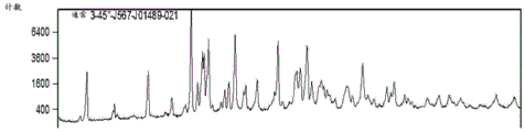
Figure 7 shows the XRPD pattern obtained for LA1 choline salt form R.
Figure 8 shows the XRPD pattern obtained for LA1 choline salt form S.
Figure 9 shows the XRPD pattern obtained for disordered solids: LA1 salt (fig. 9A); LA1 calcium salt (fig. 9B); LA1 magnesium salt (fig. 9C); LA1 sodium salt (fig. 9D); LA1 potassium salt (fig. 9E); LA1 ammonium salt (fig. 9F); LA1 calcium salt (fig. 9G); LA1 piperazine salt (fig. 9H).
Figure 10 shows the XRPD pattern obtained for the choline and meglumine salts prepared on a small scale: disordered LA1A type (fig. 10A); LA1 choline salt from ethanol methyl tert-butyl ether (fig. 10B); LA1 choline salt from acetone (fig. 10C); LA1 choline salt form Q from ethyl acetate (fig. 10D); LA1 meglumine salt form T from ethanol (fig. 10E).
Figure 11 shows the XRPD pattern obtained for LA1 meglumine salt form L.
Figure 12 shows the XRPD pattern obtained for LA1 meglumine salt form M.
Figure 13 shows the XRPD pattern obtained for LA1 meglumine salt form N.
Figure 14 shows the XRPD pattern obtained for the bulk preparation of choline and meglumine salts: LA1 choline salt from ethanol (fig. 14A); LA1 choline salt from ethanol (fig. 14B); LA1 choline salt from acetone (fig. 14C); LA1 meglumine salt from ethanol (fig. 14D); LA1 meglumine salt from ethanol (fig. 14E).
Figure 15 shows XRPD patterns obtained for LA1 meglumine salt in different solvents.
Figure 16 shows XRPD patterns obtained for LA1 choline salt in different solvents.
FIG. 17 shows the concentration-time curve of LA1 following oral administration of micronized LA1(2mg/kg) to SD (Sprague Dawley) rats.
FIG. 18 shows concentration-time curves of LA1 released after IP administration of LA1(2mg/kg) and LA1 released after IP administration of LA1 choline (2mg/kg) and LA1 meglumine (2mg/kg) to SD rats.
FIG. 19 shows concentration-time curves of LA1 released after IV administration of LA1(1mg/kg) and LA1 released after IV administration of LA1 choline (1mg/kg) and LA1 meglumine (1mg/kg) to SD rats.
Figure 20 shows a concentration-time curve for intravenous administration of micronized LA1(1mg/kg) to beagle dogs.
Figure 21 shows a concentration-time curve for oral administration of micronized LA1(1mg/kg) to beagle dogs.
FIG. 22 shows concentration-time curves for oral (5mg/kg) and intravenous (0.5mg/kg) administration of LA1 choline salt to beagle dogs.
Figure 23 shows melanoma progression in mice treated with vehicle, LA1 meglumine salt, α -PD1 antibody, or LA1 meglumine salt + α -PD1 antibody.
Figure 24 shows melanoma progression in mice treated with varying amounts of LA1 choline salt.
Figure 25 shows melanoma progression in mice treated with vehicle, LA1 choline salt (3mg/kg, p.o., b.i.d.), α -PD1 antibody (100 μ g/mouse, i.p., every four days), or LA1 choline salt + α -PD1 antibody.
Figure 26 shows melanoma progression in mice treated with vehicle, LA1 choline salt (3mg/kg, p.o., b.i.d.), α -CTLA-4 antibody (100 μ g/mouse, i.p., every four days), or LA1 choline salt + α -CTLA-4 antibody.
Detailed description of the invention
I. Overview
The present invention provides novel salts and crystalline forms of mucoitin-1 (LA 1; (Z) -4- (5- ((3-benzyl-4-oxo-2-thioxothiazolidin-5-ylidene) methyl) furan-2-yl) benzoic acid). These new forms of LA1 bring a number of advantages, including increased bioavailability of orally administered pharmaceutical formulations. Thus, the present invention enables improved methods of treating β 2 integrin-mediated disorders.
Definition of
"LA 1" represents the compound (Z) -4- (5- ((3-benzyl-4-oxo-2-thioxothiazolidin-5-ylidene) methyl) furan-2-yl) benzoic acid, as shown in formula I.
"salt" means a base addition salt prepared by combining LA1 free acid with a pharmaceutically acceptable base.
"pharmaceutically acceptable" is art-recognized and is used herein to refer to compositions, excipients, adjuvants, or other materials and/or dosage forms which are, within the scope of sound medical judgment, suitable for use in contact with the tissues of human beings and animals without excessive toxicity, irritation, allergic response, or other problem or complication, commensurate with a reasonable benefit/risk ratio. Examples of pharmaceutically acceptable bases include, but are not limited to, ammonia, L-arginine, calcium hydroxide, choline, meglumine, lysine, magnesium hydroxide, potassium hydroxide, sodium hydroxide.
"Choline" means 2-hydroxy-N, N, N-trimethylethanaminium. A "choline salt" is a salt containing at least one 2-hydroxy-N, N, N-trimethylethanaminium cation.
"meglumine" means (2R,3R,4R,5S) -6- (methylamino) hexane-1, 2,3,4, 5-pentaol. "meglumine salt" is a salt containing at least one (2S,3R,4R,5R) -2,3,4,5, 6-pentahydroxy-N-methylhexan-1-ammonium cation.
By "crystalline form" is meant a solid form of a compound in which the constituent molecules are packaged in a regular array of repeating patterns. The crystalline form may be triclinic, monoclinic, orthorhombic, tetragonal, trigonal, hexagonal or cubic. A crystalline form may contain one or more regions, i.e., grains, with distinct grain boundaries. The crystalline solid may contain two or more crystal geometries.
"integrin" means a non-covalently linked α/β -heterodimeric cell surface receptor that mediates cell adhesion, migration, and signaling. Integrins are expressed in a wide range of organisms, including caenorhabditis elegans (c. elegans), Drosophila sp, amphibians, reptiles, birds, and mammals, including humans. A number of alpha subunits, e.g., referred to as α V, α 5, etc., and a number of beta subunits, e.g., referred to as β 1, β 2, β 3, β 5, etc., have been identified, various combinations of which are represented by the integrin superfamily, including α 5 β 1, α V β 3, and α V β 5. The superfamily of integrins can be subdivided into families, for example, α V-containing integrins, including α V β 3 and α V β 5, or β 1-containing integrins, including α 5 β 1 and α V β 1.
"β 2 integrin" means a leukocyte specific integrin having a β 2-subunit (also known as CD 18). β 2 integrins have different α -subunits and are selected from the group consisting of CD11a, CD11b, CD11c, and CD11 d. β 2 integrins, including the highly expressed integrins CD11b/CD18 (also known as Mac-1, CR3 and α M β 2), regulate leukocyte functions, including adhesion, migration, recruitment and activation of cells.
By "beta 2-mediated," as used herein to refer to a disease and/or disorder in a patient, is meant that the disease or disorder results (in whole or in part) from a chemical or physical process involving a beta 2 integrin. Beta 2-mediated diseases and conditions include inflammatory and autoimmune diseases. Examples of β 2-mediated diseases and conditions include, but are not limited to, ischemia-reperfusion injury (including acute renal failure and atherosclerosis), lupus, inflammatory bowel disease, crohn's disease, rheumatoid arthritis, multiple sclerosis, lupus nephritis, focal segmental glomerulosclerosis, kidney injury, glaucoma, ophthalmic conditions, allograft rejection (e.g., nephropathy), transplantation, graft-versus-host disease, neurological diseases, alzheimer's disease, parkinson's disease, dermatitis, tissue injury, stroke, neointimal thickening in response to vascular injury, anti-GBM nephritis, pain (including chronic pain), and cancer, including primary and metastatic tumors, such as breast cancer, melanoma, prostate cancer, lung cancer, pancreatic cancer, and the like.
"cancer" refers to an abnormal state or condition characterized by cell growth in rapid proliferation. Hyperproliferative and proliferative disease states may be classified as pathological, i.e., characterized or constituting a disease state, or may be classified as non-pathological, i.e., deviating from normal but not related to a disease state. In general, cancer will be associated with the presence of one or more tumors, i.e., abnormal cell populations. The term "tumor" is meant to include all types of cancerous growth or neoplastic processes, metastatic tissue or malignantly transformed cells, tissues or organs, regardless of the type or stage of pathohistology that is aggressive.
Examples of cancer (cancer) include malignant tumors of various organ systems, such as lung cancer, breast cancer (breast cancer), thyroid cancer, lymphoma, gastrointestinal cancer, and genitourinary tract cancer. Cancer may also mean adenocarcinoma, including malignancies such as colon cancer, renal cell carcinoma, prostate cancer (prostate cancer) and/or testicular tumors, non-small cell carcinoma of the lung, small bowel cancer, and esophageal cancer. Carcinomas (carcinoma) are malignancies of epithelial or endocrine tissue, including respiratory system carcinomas, gastrointestinal system carcinomas, genitourinary system carcinomas, testicular carcinomas, breast carcinomas (breast carcinomas), prostate carcinomas (prostatic carcinoma), endocrine system carcinomas, and melanomas. "adenocarcinoma" means a cancer derived from glandular tissue or in which tumor cells constitute a recognizable glandular structure. "sarcoma" refers to malignant tumors of mesenchymal origin.
"melanoma" means a tumor caused by melanocytes. Melanoma occurs most commonly in the skin, and extensive metastasis is often observed.
An "immune checkpoint" refers to a regulatory pathway involved in the costimulatory or inhibitory control of T-cell activity in an organism. "immune checkpoint proteins" include proteins on the surface of antigen-presenting cells and T-cells whose interactions are involved in the regulation and maintenance of self-tolerance and the duration and magnitude of physiological immune responses in organisms. See, e.g., d.m. pardol. "natural Reviews of Cancer" Nature Reviews Cancer 12,252-264 (2012). Examples of immune checkpoint proteins include, but are not limited to, A2aR (adenosine A2a receptor); BTLA, B and T (lymphocyte attenuating factors); ICOS (inducible T cell co-stimulatory factor); KIR (killer cell immunoglobulin-like receptor); LAG3 (lymphocyte activator gene 3); PD1 (apoptosis protein 1); CTLA-4 (cytotoxic T-lymphocyte-associated antigen 4); and TIM3(T cell membrane protein 3).
By "immune checkpoint inhibitor" is meant a molecule that reduces, inhibits, interferes with, or modulates the activity of, one or more checkpoint proteins, in whole or in part. The immune checkpoint inhibitor may, for example, comprise an antibody or a peptide-like compound derived from an antibody.
"PD 1" denotes apoptosis protein 1, also known as CD279, which is expressed by T-cells, B-cells and monocytes. PD-1 is a type I surface glycoprotein characterized by a V-set immunoglobulin superfamily (IgSF) domain, attached to a transmembrane domain and a cytoplasmic domain, containing two tyrosine-based signaling motifs. PD1 binds to at least two ligands: PD-L1 (expressed by cells including T-cells, B-cells, dendritic cells, macrophages, and mesenchymal stem cells) and PD-L2 (expressed by cells including dendritic cells, macrophages, and mast cells).
"CTLA-4" means cytotoxic T-lymphocyte-associated antigen 4, also known as CD152, which is expressed only on T-cells. CTLA-4 comprises a single Ig-folded extracellular domain with three CDR-like loops, in particular, binding to the ligands CD80(B7.1) and CD86(B7.2) that are differentially expressed in antigen presenting cells.
By "leukocyte marker" is meant a biomolecule (e.g., a polypeptide) found on the cell surface of leukocytes. Leukocyte markers include, but are not limited to, T-cell antigen receptors; CD 1; a NK cell receptor; IDO 1/2; TDO; CSF 1R; VEGFR; SIRPa; cell adhesion molecules (e.g., CD2, CD58(LFA-3), CD3, CD4, CD5, CD7, CD 8); β 2 integrins (e.g., LeuCAM, CD11a (LFA-1), CD11b (MAC-1(CR3)), CD11c (CR4), CD18, CD16(FcR111), CD21(CR2), CD23, CD25, CD30, CD35(CR 1)); β 3 integrins (e.g., CD41, CDs 1); homing receptors (e.g., CD44, Mel-14); β 1 integrins (e.g., CD49a-f (VLA-1), VLA-2, VLA-3, VLA-4); CD 14; CD 56; CD 68; CD 71; and a CD 163.
"integrin" means a non-covalently linked α/β -heterodimeric cell surface receptor that mediates cell adhesion, migration, and signaling. Integrins are expressed in a wide range of organisms, including caenorhabditis elegans (c. elegans), Drosophila sp, amphibians, reptiles, birds, and mammals, including humans. A number of alpha subunits, e.g., referred to as α V, α 5, etc., and a number of beta subunits, e.g., referred to as β 1, β 2, β 3, β 5, etc., have been identified, various combinations of which are represented by the integrin superfamily, including α 5 β 1, α V β 3, and α V β 5. The superfamily of integrins can be subdivided into families, for example, α V-containing integrins, including α V β 3 and α V β 5, or β 1-containing integrins, including α 5 β 1 and α V β 1.
"β 2 integrin" means a leukocyte specific integrin having a β 2-subunit (also known as CD 18). β 2 integrins have different α -subunits and are selected from the group consisting of CD11a, CD11b, CD11c, and CD11 d. β 2 integrins, including the highly expressed integrins CD11b/CD18 (also known as Mac-1, CR3 and α M β 2), regulate leukocyte functions, including adhesion, migration, recruitment and activation of cells.
"bone marrow cells" generally refers to any white blood cell (i.e., leukocyte) that is not a lymphocyte (e.g., is not a natural killer cell, T cell, or B cell). Bone marrow cells include macrophages, dendritic cells and granulocytes.
As used herein, the verb "treat," "treating," and "treatment" unless otherwise indicated, means reversing, alleviating, inhibiting the progression of, or preventing the disease or disorder to which such term applies, or one or more symptoms of such disease or disorder. The noun "treat" as used herein means the action of the verb "treat" as just defined.
A "therapeutically effective amount" is that amount of LA1 salt or crystalline form necessary to produce the desired level of drug in the tissue, bloodstream, or other body cavity of a patient, which level elicits the desired physiological or biological response when the LA1 salt or crystalline form is administered by a selected route of administration. The exact amount will depend on a number of factors including, for example, the particular LA1 salt or crystalline form; the specific pharmaceutical formulation or delivery device employed; the severity of the disease state; and patient compliance with the treatment regimen. Therapeutically effective amounts of LA1 salt and crystalline forms are readily determined by those skilled in the art based on the information provided herein.
"about" and "approximately" as used herein to modify a numerical value means the range defined around that value. If "X" is the number, "about X" or "about X" will generally indicate a number from 0.95X to 1.05X, including, for example, from 0.98X to 1.02X or from 0.99X to 1.01X. Any reference to "about X" or "about X" specifically indicates at least the values X, 0.95X, 0.96X, 0.97X, 0.98X, 0.99X, 1.01X, 1.02X, 1.03X, 1.04X, and 1.05X. Thus, "about X" and "about X" are intended to teach and provide written description support for the claims limitations, such as "0.98X". When the amount "X" includes only integer values (e.g., "X carbons"), "about X" or "about X" means from (X-1) to (X + 1). In such cases, "about X" or "about X" specifically indicates at least values of X, X-1 and X + 1.
Salts of LA1
One skilled in the art will appreciate that many pharmaceutically acceptable bases may be used to prepare the LA1 salt. Pharmaceutically acceptable bases include, but are not limited to, ammonia, L-arginine, calcium hydroxide, choline, meglumine, magnesium hydroxide, benzphetamine, benzathine, betaine, dandol, diethylamine, 2-diethylaminoethanol, hydrabamine, 1- (2-hydroxyethyl) -pyrrolidine, tert-butylamine, tromethamine, piperazine, imidazole, ethylenediamine, ethanolamine, diethanolamine, and triethanolamine. In certain embodiments, the LA1 salt comprises a cation derived from a pharmaceutically acceptable base selected from the group consisting of ammonia, L-arginine, calcium hydroxide, choline, meglumine, and magnesium hydroxide.
In one aspect, the present invention provides a choline salt of a compound of formula I:

as mentioned above, formula I corresponds to LA 1. Synonyms for choline also include (2-hydroxyethyl) trimethylammonium and 2-hydroxy-N, N, N-trimethylethylammonium. As used herein, "choline salt" means a salt containing at least one 2-hydroxy-N, N, N-trimethylethanaminium cation. In certain embodiments, the choline salt of LA1 is a salt according to formula II:

in one aspect, the present invention provides a crystalline form G of the choline salt of the compound of formula I:

in some embodiments, form G is characterized by an X-ray powder diffraction (XRPD) pattern comprising at least three peaks selected from the group consisting of: 5.6,7.9,11.2,13.3,15.0,15.7,16.1,16.2,16.5,16.6,17.8,18.1,18.5,19.1,19.8,20.0,21.1,23.0,24.6,25.0,25.6,26.6,26.8,26.9,29.3,29.7,30.6,30.7 and 34.4 ° 2 θ, ± 0.2 ° 2 θ. For example, form G can include 3,4,5,6,7,8,9,10,11,12,13,14,15,16,17,18,19,20,21,22,23,24,25,26,27,28, or 29 such peaks.
In some embodiments, form G is characterized by an X-ray powder diffraction (XRPD) pattern comprising at least six peaks selected from the group consisting of: 5.6,11.2,13.3,15.0,15.7,16.1,16.6,19.1,24.6,25.0,25.6 and 26.8 ° 2 θ, ± 0.2 ° 2 θ.
In some embodiments, form G is characterized by an X-ray powder diffraction (XRPD) pattern comprising at least ten peaks selected from the group consisting of: 5.6,11.2,13.3,15.0,15.7,16.1,16.6,19.1,24.6,25.0,25.6 and 26.8 ° 2 θ, ± 0.2 ° 2 θ.
In some embodiments, form G is characterized by an X-ray powder diffraction (XRPD) pattern substantially in accordance with fig. 5B, as determined on a diffractometer using Cu-ka radiation.
In another aspect, the present invention provides crystalline form O of the choline salt of the compound of formula I:

in some embodiments, form O is characterized by an X-ray powder diffraction (XRPD) pattern comprising at least three peaks selected from the group consisting of: 8.4,8.8,9.3,13.3,14.3,16.7,17.0,18.1,19.4,19.6,19.9,20.7,20.9,21.4,21.7,22.5,23.4,24.1 and 25.5 ° 2 θ, ± 0.2 ° 2 θ. For example, form O can include 3,4,5,6,7,8,9,10,11,12,13,14,15,16,17,18, or 19 such peaks.
In some embodiments, form O is characterized by an X-ray powder diffraction (XRPD) pattern comprising at least six peaks selected from the group consisting of: 8.4,8.8,9.3,16.7,19.9,20.7,21.7,22.5,23.4 and 25.5 ° 2 θ, ± 0.2 ° 2 θ.
In some embodiments, form O is characterized by an X-ray powder diffraction (XRPD) pattern substantially in accordance with fig. 6E, as determined on a diffractometer using Cu-ka radiation.
In another aspect, the present invention provides a crystalline form Q of the choline salt of the compound of formula I:

in some embodiments, form Q is characterized by an X-ray powder diffraction (XRPD) pattern comprising at least three peaks selected from the group consisting of: 5.0,5.2,8.4,9.6,9.9,11.5,12.6,12.8,13.3,14.4,15.8,16.1,16.6,17.5,18.0,19.3,20.6,20.7,21.5,21.7,22.9,23.7,24.8,25.1,25.3,25.3,25.5,26.3,26.9,27.0,28.1,28.8,30.4,31.2,32.0,35.7 and 37.4 ° 2 θ ± 0.2 ° 2 θ. For example, form Q can include 3,4,5,6,7,8,9,10,11,12,13,14,15,16,17,18,19,20,21,22,23,24,25,26,27,28,29,30,31,32,33,34,35,36, or 37 such peaks.
In some embodiments, form Q is characterized by an X-ray powder diffraction (XRPD) pattern comprising at least six peaks selected from the group consisting of: 5.0,8.4,9.6,9.9,11.5,12.8,13.3,14.4,18.0,19.3,23.7 and 25.5 ° 2 θ ± 0.2 ° 2 θ.
In some embodiments, form Q is characterized by an X-ray powder diffraction (XRPD) pattern comprising at least ten peaks selected from the group consisting of: 5.0,8.4,9.6,9.9,11.5,12.8,13.3,14.4,18.0,19.3,23.7 and 25.5 ° 2 θ ± 0.2 ° 2 θ.
In some embodiments, form Q is characterized by an X-ray powder diffraction (XRPD) pattern D substantially in accordance with the figure, as determined on a diffractometer using Cu-ka radiation.
In another aspect, the present invention provides a crystalline form R of the choline salt of the compound of formula I:

in some embodiments, form R is characterized by an X-ray powder diffraction (XRPD) pattern comprising at least three peaks selected from the group consisting of: 5.1,5.6,8.0,8.2,8.4,9.8,11.2,12.7,13.4,14.6,15.1,15.7,16.1,16.3,16.7,17.1,17.8,18.2,18.5,19.1,19.9,20.1,21.1,22.6,23.0,23.4,24.0,24.5,24.7,25.0,25.6,26.0,26.6,26.8,27.1,27.4,27.7,28.1,29.3,29.7,30.6,31.1,31.7,32.2,32.8,33.2,33.5,34.5,34.8,35.1,35.4,36.5,37.6,38.5,39.5, 39.4, 40.4, 44.4, 44.2, 2 degrees θ ° and 2 °. For example, form R can include 3,4,5,6,7,8,9,10,11,12,13,14,15,16,17,18,19,20,21,22,23,24,25,26,27,28,29,30,31,32,33,34,35,36,37,38,39,40,41,42,43,44,45,46,47,48,49,50,51,52,53,54,55,56,57,58, or 59 such peaks.
In some embodiments, form R is characterized by an X-ray powder diffraction (XRPD) pattern comprising at least six peaks selected from the group consisting of: 5.6,11.2,15.1,16.3,16.7,19.1,20.1,21.1,23.0,24.5,25.0,25.6,26.0,31.1,32.8 and 33.5 ± 0.2 ° 2 θ.
In some embodiments, form R is characterized by an X-ray powder diffraction (XRPD) pattern comprising at least nine peaks selected from the group consisting of: 5.6,11.2,15.1,16.3,16.7,19.1,20.1,21.1,23.0,24.5,25.0,25.6,26.0,31.1,32.8 and 33.5 ± 0.2 ° 2 θ.
In some embodiments, form R is characterized by an X-ray powder diffraction (XRPD) pattern according to fig. 7, as determined on a diffractometer using Cu-ka radiation. In some embodiments, form R is characterized by a differential scanning calorimetry thermogram comprising an endothermic peak at about 224.5 ℃.
In another aspect, the present invention provides a crystalline form S of the choline salt of the compound of formula I:

in some embodiments, form S is characterized by an X-ray powder diffraction (XRPD) pattern comprising at least three peaks selected from the group consisting of: 5.1,8.4,9.6,10.0,11.6,12.9,13.3,14.4,14.9,15.8,16.6,17.4,18.0,19.2,19.3,20.6,21.4,21.7,22.7,23.7,24.8,25.4,26.3,26.8,28.1,28.7,29.6,30.3,31.0,31.9,33.0,34.0,35.7,37.4,39.2,40.5 and 41.7 ° 2 θ, ± 0.2 ° 2 θ. For example, form S can include 3,4,5,6,7,8,9,10,11,12,13,14,15,16,17,18,19,20,21,22,23,24,25,26,27,28,29,30,31,32,33,34,35,36, or 37 such peaks.
In some embodiments, form S is characterized by an X-ray powder diffraction (XRPD) pattern comprising at least six peaks selected from the group consisting of: 5.1,8.4,9.6,10.0,12.9,13.3,16.6,17.4,18.0,19.2,20.6,21.4,21.7,23.7,25.4 and 28.1 ° 2 θ ± 0.2 ° 2 θ.
In some embodiments, form S is characterized by an X-ray powder diffraction (XRPD) pattern comprising at least ten peaks selected from the group consisting of: 5.1,8.4,9.6,10.0,12.9,13.3,16.6,17.4,18.0,19.2,20.6,21.4,21.7,23.7,25.4 and 28.1 ° 2 θ ± 0.2 ° 2 θ.
In some embodiments, form S is characterized by an X-ray powder diffraction (XRPD) pattern substantially in accordance with fig. 8, as determined on a diffractometer using Cu-ka radiation.
In another aspect, the present invention provides a meglumine salt of the compound of formula I:

synonyms for methylamine also include N-methyl-D-glucamine and (2R,3R,4R,5S) -6- (methylamino) hexane-1, 2,3,4, 5-pentaol. As used herein, "meglumine salt" means a salt containing at least one (2S,3R,4R,5R) -2,3,4,5, 6-pentahydroxy-N-methylhexan-1-aminium cation. In certain embodiments, the meglumine salt of LA1 is a salt according to formula III:

in another aspect, the present invention provides crystalline form H of the meglumine salt of the compound of formula I:

in some embodiments, form H is characterized by an X-ray powder diffraction (XRPD) pattern comprising at least three peaks selected from the group consisting of: 5.3,7.1,10.7,10.9,16.1,16.5,17.7,18.5,20.3,23.6,24.9 and 27.2 ° 2 θ ± 0.2 ° 2 θ. For example, form H can include 3,4,5,6,7,8,9,10,11, or 12 such peaks.
In some embodiments, form H is characterized by an X-ray powder diffraction (XRPD) pattern comprising at least six peaks selected from the group consisting of: 5.3,7.1,10.7,10.9,16.1,16.5,17.7,18.5,20.3,23.6,24.9 and 27.2 ° 2 θ ± 0.2 ° 2 θ.
In some embodiments, form H is characterized by an X-ray powder diffraction (XRPD) pattern comprising at least ten peaks selected from the group consisting of: 5.3,7.1,10.7,10.9,16.1,16.5,17.7,18.5,20.3,23.6,24.9 and 27.2 ° 2 θ ± 0.2 ° 2 θ.
In some embodiments, form H is characterized by an X-ray powder diffraction (XRPD) pattern substantially in accordance with fig. 6B, as determined on a diffractometer using Cu-ka radiation.
In another aspect, the present invention provides a crystalline form L of the meglumine salt of the compound of formula I:

in some embodiments, form L is characterized by an X-ray powder diffraction (XRPD) pattern comprising at least three peaks selected from the group consisting of: 5.3,7.9,8.5,9.0,9.9,10.6,10.9,11.6,12.0,12.6,13.1,14.5,14.8,15.0,15.3,15.9,16.2,16.9,17.4,17.8,18.0,18.4,18.8,19.2,20.2,20.8,21.3,21.7,22.1,23.2,23.8,24.5,25.2,25.5,26.3,26.9,27.3,27.9,28.4,28.9,29.2,29.8,30.3,30.6,31.1,32.1,32.8,34.1,34.5,34.9,35.1,36.0,36.5,37.5,38.0, 39.9, 7.42, 2 degrees θ, and 2 degrees. For example, form L can include 3,4,5,6,7,8,9,10,11,12,13,14,15,16,17,18,19,20,21,22,23,24,25,26,27,28,29,30,31,32,33,34,35,36,37,38,39,40,41,42,43,44,45,46,47,48,49,50,51,52,53,54,55,56,57,58,59,60, or 61 such peaks.
In some embodiments, form L is characterized by an X-ray powder diffraction (XRPD) pattern comprising at least six peaks selected from the group consisting of: 8.5,9.0,10.9,15.0,16.9,20.2,21.7,23.8,24.5,25.2,26.3,29.2 and 29.8 ° 2 θ ± 0.2 ° 2 θ.
In some embodiments, form L is characterized by an X-ray powder diffraction (XRPD) pattern comprising at least ten peaks selected from the group consisting of: 8.5,9.0,10.9,15.0,16.9,20.2,21.7,23.8,24.5,25.2,26.3,29.2 and 29.8 ° 2 θ ± 0.2 ° 2 θ.
In some embodiments, form L is characterized by an X-ray powder diffraction (XRPD) pattern substantially in accordance with fig. 11, as determined on a diffractometer using Cu-ka radiation. In some embodiments, form L is characterized by a differential scanning calorimetry thermogram comprising an endothermic peak at about 136.3 ℃.
In another aspect, the present invention provides a crystalline form M of the meglumine salt of the compound of formula I:

in some embodiments, form M is characterized by an X-ray powder diffraction (XRPD) pattern comprising at least three peaks selected from the group consisting of: 6.5,8.5,9.0,9.9,10.6,11.6,14.4,14.8,15.0,15.3,15.9,16.1,16.9,17.8,18.0,19.0,20.4,20.8,21.3,21.7,23.6,24.5,25.2,26.3,26.9,27.5,27.9,28.5,28.9,29.8,30.6,32.1,32.8,33.8,34.5,36.0,36.4,37.1,38.0,39.7,40.7,41.7,43.0 and 44.0 ° 2 θ, ± 0.2 ° 2 θ. For example, form M can include 3,4,5,6,7,8,9,10,11,12,13,14,15,16,17,18,19,20,21,22,23,24,25,26,27,28,29,30,31,32,33,34,35,36,37,38,39,40,41,42,43, or 44 such peaks.
In some embodiments, form M is characterized by an X-ray powder diffraction (XRPD) pattern comprising at least six peaks selected from the group consisting of: 8.5,9.0,14.8,15.0,16.9,18.0,21.7,24.5,25.2,26.3 and 29.8 ° 2 θ ± 0.2 ° 2 θ.
In some embodiments, form M is characterized by an X-ray powder diffraction (XRPD) pattern comprising at least nine peaks selected from the group consisting of: 8.5,9.0,14.8,15.0,16.9,18.0,21.7,24.5,25.2,26.3 and 29.8 ± 0.2 ° 2 θ.
In some embodiments, form M is characterized by an X-ray powder diffraction (XRPD) pattern substantially in accordance with fig. 12, as determined on a diffractometer using Cu-ka radiation. In some embodiments, form M is characterized by a differential scanning calorimetry thermogram comprising an endothermic peak at about 294.5 ℃.
In another aspect, the present invention provides a crystalline form N of the meglumine salt of the compound of formula I:

in some embodiments, form N is characterized by an X-ray powder diffraction (XRPD) pattern comprising at least three peaks selected from the group consisting of: 4.3,5.0,5.4,6.1,7.5,7.9,8.9,9.5,10.0,10.8,11.4,12.1,12.5,13.8,14.3,14.8,15.6,16.1,16.7,17.4,18.1,19.2,19.5,20.1,20.9,21.4,21.5,22.1,22.5,23.9,24.6,25.3,26.3,26.7,27.1,27.6,28.2,29.0,30.4,30.9,32.0,32.9,33.9,34.7,36.9,38.3,39.1,39.6,40.2 and 41.4 ° 2 θ, ± 0.2 ° 2 θ. For example, form N can include 3,4,5,6,7,8,9,10,11,12,13,14,15,16,17,18,19,20,21,22,23,24,25,26,27,28,29,30,31,32,33,34,35,36,37,38,39,40,41,42,43,44,45,46,47,48,49, or 50 such peaks.
In some embodiments, form N is characterized by an X-ray powder diffraction (XRPD) pattern comprising at least six peaks selected from the group consisting of: 6.1,7.9,8.9,9.5,10.0,12.5,14.3,14.8,15.6,16.1,17.4,18.1,19.5,20.9,21.4,21.5,23.9,24.6,25.3 and 29.0 ° 2 θ ± 0.2 ° 2 θ.
In some embodiments, form N is characterized by an X-ray powder diffraction (XRPD) pattern comprising at least ten peaks selected from the group consisting of: 6.1,7.9,8.9,9.5,10.0,12.5,14.3,14.8,15.6,16.1,17.4,18.1,19.5,20.9,21.4,21.5,23.9,24.6,25.3 and 29.0 ° 2 θ ± 0.2 ° 2 θ.
In some embodiments, form N is characterized by an X-ray powder diffraction (XRPD) pattern substantially in accordance with fig. 13, as determined on a diffractometer using Cu-ka radiation. In some embodiments, form N is characterized by a differential scanning calorimetry thermogram comprising an endothermic peak at about 139.9 ℃.
In another aspect, the present invention provides crystalline form T of the meglumine salt of the compound of formula I:

in some embodiments, form T is characterized by an X-ray powder diffraction (XRPD) pattern comprising at least three peaks selected from the group consisting of: 6.9,8.2,8.4,9.4,11.6,15.0,15.1,15.5,17.2,17.8,18.1,20.5,21.3,21.9,22.3,23.5,25.0 and 26.7 ° 2 θ ± 0.2 ° 2 θ. For example, form T can include 3,4,5,6,7,8,9,10,11,12,13,14,15,16,17, or 18 such peaks.
In some embodiments, form T is characterized by an X-ray powder diffraction (XRPD) pattern comprising at least six peaks selected from the group consisting of: 6.9,8.4,9.4,11.6,15.5,17.2,21.3,21.9,22.3,23.5,25.0 and 26.7 ° 2 θ ± 0.2 ° 2 θ.
In some embodiments, form T is characterized by an X-ray powder diffraction (XRPD) pattern comprising at least ten peaks selected from the group consisting of: 5.3,7.1,10.7,10.9,16.1,16.5,17.7,18.5,20.3,23.6,24.9 and 27.2 ° 2 θ ± 0.2 ° 2 θ.
In some embodiments, form T is characterized by an X-ray powder diffraction (XRPD) pattern substantially in accordance with fig. 10E, as determined on a diffractometer using Cu-ka radiation.
In another aspect, the present invention provides a solid form a of the compound of formula I:

in some embodiments, the solid form a is characterized by an X-ray powder diffraction (XRPD) pattern comprising at least three peaks selected from the group consisting of: 5.3,7.8,15.2,18.7,19.8,20.3,20.8,25.7,26.3,26.5 and 26.9 ° 2 θ ± 0.2 ° 2 θ. For example, form a may include 3,4,5,6,7,8,9,10, or 11 such peaks.
In some embodiments, the solid form a is characterized by an X-ray powder diffraction (XRPD) pattern comprising at least six peaks selected from the group consisting of: 5.3,7.8,15.2,18.7,19.8,20.3,20.8,25.7,26.3,26.5 and 26.9 ° 2 θ ± 0.2 ° 2 θ.
In some embodiments, the solid form a is characterized by an X-ray powder diffraction (XRPD) pattern comprising at least ten peaks selected from the group consisting of: 5.3,7.8,15.2,18.7,19.8,20.3,20.8,25.7,26.3,26.5 and 26.9 ° 2 θ ± 0.2 ° 2 θ.
In some embodiments, the solid form a is characterized by an X-ray powder diffraction (XRPD) pattern substantially in accordance with fig. 2, as determined on a diffractometer using Cu-ka radiation.
In a related aspect, the present invention provides processes for preparing salts and crystalline forms of LA 1. In general, the LA1 salt is prepared by forming a mixture (i.e., a salt-forming mixture) containing LA1 free acid and at least one molar equivalent of a suitable base under conditions sufficient to form a salt. The mixture typically contains a solvent in which LA1 free acid and/or base is partially or fully soluble. Examples of solvents that may be used to prepare the LA1 salt include, but are not limited to, tetrahydrofuran, bis Alkanes, methanol, ethanol, isopropanol, N-dimethylformamide, N-methylpyrrolidone, methyl tert-butyl ether, acetone, ethyl acetate, dichloromethane, water and mixtures thereof. In some embodiments, the salt-forming mixture comprises at least one solvent selected from tetrahydrofuran and methanol. In some embodiments, the mixture comprises tetrahydrofuran. In some embodiments, the mixture comprises tetrahydrofuran and methanol. In some embodiments, the mixture contains acetone. In some embodiments, the mixture comprises ethanol.
A salt forming mixture containing LA1 free acid and base can be formed at or maintained at any suitable temperature. Typically, the mixture is maintained at a temperature varying from about 20 ℃ to about 80 ℃ for a time sufficient to form a salt. For example, the mixture may be maintained at about 20 ℃ to about 80 ℃ for about 15 minutes to about 72 hours or more. The mixture may be maintained at about 40 ℃ to about 60 ℃ for about 1 hour to about 48 hours, or at about 40 ℃ to about 50 ℃ for about 1 hour to about 16 hours.
In some embodiments, the salt-forming mixture contains LA1 free acid, choline hydroxide, tetrahydrofuran, and methanol. In some embodiments, the ratio of tetrahydrofuran to methanol is 3:1v: v. In some embodiments, form G is prepared by a process comprising forming a mixture comprising one molar equivalent of LA1 free acid, one molar equivalent of choline hydroxide, and a combination of tetrahydrofuran and methanol in a ratio of 3:1v: v. In some embodiments, the process for preparing form G further comprises stirring the mixture at about 40 ℃ to about 50 ℃ for about 24 hours to about 48 hours. In some embodiments, the process for preparing form G comprises stirring the mixture at about 50 ℃ for at least about 24 hours. In some embodiments, the process for preparing form G comprises removing the combination of tetrahydrofuran and methanol from the mixture by evaporation after formation of form G.
In some embodiments, the salt-forming mixture contains LA1 free acid, choline hydroxide, and tetrahydrofuran. In some embodiments, the ratio of tetrahydrofuran to methanol is 3:1v: v. In some embodiments, form O is prepared by a process comprising forming a mixture comprising one molar equivalent of LA1 free acid, one molar equivalent of choline hydroxide, and tetrahydrofuran. In some embodiments, the process for preparing crystalline form O further comprises stirring the mixture at about 20 ℃ to about 30 ℃ for about 24 hours to about 48 hours. In some embodiments, the process for preparing form O comprises stirring the mixture at no more than about 30 ℃ for at least about 24 hours.
In some embodiments, the salt formation mixture contains LA1 free acid, choline hydroxide, and ethyl acetate or acetone. In some embodiments, form Q is prepared by a process comprising forming a mixture comprising one molar equivalent of LA1 free acid, one molar equivalent of choline hydroxide, and ethyl acetate. In some embodiments, the process for preparing form Q further comprises stirring the mixture at about 20 ℃ to about 30 ℃ for about 12 hours to about 48 hours. In some embodiments, the process for preparing form Q comprises stirring the mixture at no more than about 30 ℃ for at least about 12 hours. In some embodiments, the process for preparing form Q comprises removing ethyl acetate or acetone via vacuum filtration after formation of form Q.
The crystalline form of LA1 choline salt may also be prepared by recrystallizing LA1 choline salt. Recrystallization may be carried out using any suitable solvent, including protic solvents (e.g., methanol, ethanol, Isopropanol (IPA), n-butanol, and water), aprotic solvents (e.g., isopropyl acetate, ethyl acetate, and acetone), or mixtures thereof. In some embodiments, preparing the crystalline form of LA1 choline salt comprises recrystallizing LA1 choline salt from a protic solvent. In some embodiments, preparing form R of LA1 choline salt comprises recrystallizing LA1 choline salt from n-butanol. In some embodiments, preparing form S of LA1 choline salt comprises recrystallizing LA1 choline salt from methanol.
In some embodiments, the salt-forming mixture contains LA1 free acid, meglumine, tetrahydrofuran, and methanol. In some embodiments, the ratio of tetrahydrofuran to methanol is 2:1v: v. In some embodiments, form H is prepared by a process comprising forming a mixture comprising one molar equivalent of LA1 free acid, one molar equivalent of meglumine, and a combination of tetrahydrofuran and methanol in a ratio of 2:1v: v. In some embodiments, the process for preparing form H further comprises stirring the mixture at about 40 ℃ to about 50 ℃ for about 24 hours to about 48 hours. In some embodiments, the process for preparing form H comprises stirring the mixture at about 50 ℃ for at least about 24 hours. In some embodiments, the process for preparing form H comprises removing the combination of tetrahydrofuran and methanol from the mixture by evaporation after formation of form H.
In some embodiments, the salt-forming mixture comprises LA1 free acid, meglumine, and ethanol. In some embodiments, form T is prepared by a process comprising forming a mixture comprising one molar equivalent of LA1 free acid, one molar equivalent of meglumine, and ethanol. In some embodiments, the process for preparing form T further comprises stirring the mixture at about 40 ℃ to about 50 ℃ for about 24 hours to about 48 hours. In some embodiments, the process for preparing form T comprises stirring the mixture at about 40 ℃ for at least about 24 hours. In some embodiments, the process for preparing form T comprises removing ethanol from the mixture by vacuum filtration after formation of form T, and isolating at least a portion of form T. In some embodiments, the process for preparing form T further comprises washing the isolated form T with methyl tert-butyl ether.
The preparation of the crystalline form of LA1 meglumine salt may also include recrystallization of LA1 meglumine salt. Recrystallization may be carried out using any suitable solvent, including protic solvents (e.g., methanol, ethanol, Isopropanol (IPA), N-butanol, and water), aprotic solvents (e.g., N-Dimethylformamide (DMF), dimethyl sulfoxide (DMSO), isopropyl acetate, ethyl acetate, and acetone), or mixtures thereof. In some embodiments, preparing the crystalline form of LA1 meglumine salt comprises recrystallizing LA1 meglumine salt from an aprotic solvent. In some embodiments, preparing form L of LA1 meglumine salt comprises recrystallizing LA1 choline salt from isopropyl acetate. In some embodiments, preparing form M of LA1 meglumine salt comprises recrystallizing LA1 choline salt from acetone. In some embodiments, preparing form N of LA1 meglumine salt comprises recrystallizing LA1 choline salt from DMF.
Pharmaceutical compositions
In a related aspect, the invention provides pharmaceutical compositions for administration of the salts and crystalline forms described herein. The pharmaceutical compositions may be prepared by any method well known in the art of pharmacy and drug delivery. In general, the methods of making the compositions include the step of bringing into association the active ingredient with the carrier which contains one or more accessory ingredients. Pharmaceutical compositions are generally prepared by uniformly and intimately bringing into association the active ingredient with liquid carriers or finely divided solid carriers or both, and then, if necessary, shaping the product into the desired formulation. The compositions may be conveniently prepared and/or packaged in unit dosage form.
The pharmaceutical compositions may be in the form of sterile injectable aqueous or oleaginous solutions and suspensions. Sterile injectable preparations can be formulated using non-toxic parenterally acceptable vehicles (excipients) including water, ringer's solution and isotonic sodium chloride solution, and acceptable solvents such as 1, 3-butanediol. In addition, sterile, fixed oils are conventionally employed as a solvent or suspending medium. For this purpose, any brand of fixed oil may be used, including synthetic mono-or diglycerides. In addition, fatty acids can be used in the preparation of injectables, such as oleic acid.
Aqueous suspensions contain the active material in admixture with excipients including, but not limited to, suspending agents such as sodium carboxymethylcellulose, methylcellulose, oleagino-propylmethylcellulose, sodium alginate, polyvinylpyrrolidone, gum tragacanth and gum acacia; dispersing or wetting agents, such as lecithin, polyoxyethylene stearate and polyethylene sorbitan monooleate; and preservatives such as ethyl, n-propyl and p-hydroxybenzoate.
Oily suspensions may be formulated by suspending the active ingredient in a vegetable oil, for example arachis oil, olive oil, sesame oil or coconut oil, or in a mineral oil, for example liquid paraffin. Oily suspensions may contain a thickening agent, for example beeswax, hard paraffin or cetyl alcohol. These compositions may be preserved by the addition of an antioxidant, such as ascorbic acid.
Dispersible powders and granules (suitable for preparation of an aqueous suspension by the addition of water) may contain the active ingredient in admixture with a dispersing agent, wetting agent, suspending agent or combination thereof. Other excipients may also be present.
The pharmaceutical compositions of the present invention may also be in the form of oil-in-water emulsions. The oily phase may be a vegetable oil, for example olive oil or arachis oil, or a mineral oil, for example liquid paraffin, or mixtures thereof. Suitable emulsifying agents may be naturally-occurring gums, for example gum acacia or gum tragacanth; naturally occurring phospholipids, such as soy lecithin; esters or partial esters derived from fatty acids and hexitol anhydrides, such as sorbitan monooleate; and condensation products of the said partial esters with ethylene oxide, for example polyoxyethylene sorbitan monooleate.
Pharmaceutical compositions containing the salts and crystalline forms described herein may also be in a form suitable for oral use. Compositions suitable for oral administration include, but are not limited to, tablets, troches, lozenges, aqueous or oily suspensions, dispersible powders or granules, emulsions, hard or soft capsules, syrups, elixirs, solutions, buccal patches, oral gels, chewing gums, chewable tablets, effervescent powders and effervescent tablets. Compositions for oral administration may be formulated according to any method known to those skilled in the art. Such compositions may contain one or more ingredients selected from the group consisting of: sweetening agents, flavouring agents, colouring agents, antioxidants and preservatives in order to provide pharmaceutically elegant and palatable preparations.
Tablets generally contain the active ingredient in admixture with non-toxic pharmaceutically acceptable excipients which include: inert diluents such as cellulose, silica, alumina, calcium carbonate, sodium carbonate, glucose, mannitol, sorbitol, lactose, calcium phosphate and sodium phosphate; granulating and disintegrating agents, such as corn starch and alginic acid; binders, such as polyvinylpyrrolidone (PVP), cellulose, polyethylene glycol (PEG), starch, gelatin, and acacia; and lubricating agents, such as magnesium stearate, stearic acid and talc. The tablets may be uncoated or they may be coated by known techniques, enteric or otherwise, to delay disintegration and absorption in the gastrointestinal tract and thereby provide a sustained action over a longer period. For example, a time delay material such as glycerol monostearate or glycerol distearate may be employed. The tablets may also be coated with a semipermeable membrane and optionally a polymeric osmotic agent (osmogent) according to known techniques to form a controlled release osmotic pump composition.
Compositions for oral administration may be formulated as hard gelatin capsules wherein the active ingredient is mixed with an inert solid diluent, for example, calcium carbonate, calcium phosphate or kaolin, or as soft gelatin capsules wherein the active ingredient is mixed with water or an oil medium, for example peanut oil, liquid paraffin or olive oil.
The salts and crystalline forms described herein may also be administered topically as solutions, ointments, creams, gels, suspensions, mouthwashes, eye drops, and the like. Further, transdermal delivery of salts and crystalline forms may be achieved by means of iontophoresis patches and the like. The compounds may also be administered in the form of suppositories for rectal administration of the drug. These compositions can be prepared by mixing the drug with a suitable non-irritating excipient which is solid at ordinary temperatures but liquid at the rectal temperature and will therefore melt in the rectum to release the drug. Such materials include cocoa butter and polyethylene glycols.
In some embodiments, the salts or crystalline forms described herein are administered via intraperitoneal injection. In some embodiments, the salt or crystalline form is administered orally. In some embodiments, the salt or crystalline form is administered intravenously.
LA1 may be used in combination with a drug selected from, but not limited to, the group consisting of: 5-fluorouracil, AZD8055, bevacizumab (bevacizumab), bortezomib (bortezomib), cetuximab (cetuximab), cyclophosphamide (cyclophosphamide), docetaxel (docetaxel), gemcitabine (gemcitabine), imatinib (imatinib), ipilimumab (ipilimumab), lapatinib (lapatinib), paclitaxel (paclitaxel), pertuzumab (pertuzumab), pertuzumab (tuzumab), rapamycin (rapamicin), sipuleucel-T, sorafenib (sorafenib), sunitinib (sunitinib), trastuzumab (trastuzumab), temsirolimus (temsirolimus), vemurafenib, taxol (tamsulosin), paclitaxel (paclitaxel), arbitacil (sultaine), fibatidine (trastuzumab), anti-androgenic agent (CTLA), anti-mitomycin (anti-mitomycin-4), anti-androgenic agent (anti-4-androgenic agent, anti-mitomycin), anti-4-androgenic agent (anti-mitomycin), anti-androgenic agent (anti-4-mitomycin, anti-4, anti-mitomycin, anti-estrogen, anti-mitomycin, anti-estrogen, anti-4, anti-drugs, anti-estrogen, anti-drugs, anti-tumor, anti-drugs, anti-, Rituximab (rituximab), belief (belatacept), benlumimab, PD-1 modulators, anti-PD 1 antibodies, PDL1 modulators, anti-PDL 1 antibodies, IDO1 inhibitors and modulators, CSF1 modulators, CSF1R modulators, anti-CSF 1R antibodies, CD47 modulators and inhibitors, CD206 modulators and inhibitors, TNFa inhibitors and modulators, anti-TNFa antibodies, cytokine modulators, anti-cytokine antibodies, interleukin modulators and inhibitors, anti-interleukin antibodies, anti-CCL 2, anti-CCL 4, CXCR-4 inhibitors, anti-CXCR 4, anti-IL 17, and anti-IL 23.
The pharmaceutical compositions of the present invention may also include crystalline forms of micronized LA1 or micronized LA1 salt or micronized LA1 salt. Generally, compositions containing micronized LA1 contain particles consisting essentially of LA1 with an average diameter below 50 μm. The average diameter of the LA1 particles may be, for example, below 45 μm, below 40 μm, below 35 μm, below 30 μm, below 25 μm or below 20 μm. The average diameter of the LA1 particles may be from about 10 μm to about 49 μm, or from about 10 μm to about 45 μm, or from about 15 μm to about 40 μm, or from about 20 μm to about 35 μm, or from about 25 μm to about 30 μm. The average diameter of the LA1 particles may be about 15,16,17,18,19,20,21,22,23,24, or about 25 μm. In some embodiments, the particles consist essentially of the free acid form of micronized LA 1. In some embodiments, these particles consist essentially of an amorphous or crystalline form of the micronized LA1 salt described herein.
Methods of treatment
The salts and crystalline forms described herein may be used to treat diseases or disorders associated with β 2 integrin activity. In certain embodiments, such a disease or disorder is selected from inflammation (including, but not limited to, acute and chronic inflammation), inflammatory skin diseases, immune-related diseases, autoimmune diseases, burns, immunodeficiency, acquired immunodeficiency syndrome (AIDS), myeloperoxidase deficiency, Wiskott-Aldrich syndrome, chronic kidney disease, chronic granulomatous disease, hyper-IgM syndrome, leukocyte adhesion deficiency, iron deficiency, Chediak-Higashi syndrome, severe combined immunodeficiency, diabetes, obesity, hypertension, HIV, wound healing, remodeling, scarring, fibrosis, stem cell therapy, cachexia, myelitis, multiple sclerosis, psoriasis, lupus, rheumatoid arthritis, immune-related diseases, radiation injury, transplantation, cell transfusion, organ transplantation, bone marrow transplantation, cancer therapy, cancer, bone marrow transplantation, cancer, bone marrow transplantation, and kidney disease, bone marrow transplantation, Organ preservation, cell preservation, asthma, irritable bowel disease, irritable bowel syndrome, ulcerative colitis, bowel disease, cancer, leukemia, ischemia-reperfusion injury, stroke, neointimal thickening associated with vascular injury, bullous pemphigoid, obstructive pulmonary disease of newborn, familial hypercholesterolemia, atherosclerosis, dyslipidemia, aortic aneurysm, arteritis, vascular occlusion (including cerebral arterial occlusion), coronary bypass surgery complications, myocarditis (including chronic autoimmune myocarditis and viral myocarditis), heart failure (including Chronic Heart Failure (CHF), heart failure cachexia), myocardial infarction, stenosis, post-cardiac surgery restenosis, painless myocardial ischemia, complications after left ventricular assist device implantation, thrombophlebitis, nodular vasculitis (including kawasaki's nodular vasculitis), Giant cell arteritis, wegener's granulomatosis, traumatic head injury, post-ischemic-reperfusion injury, post-ischemic brain inflammation, ischemia-reperfusion injury secondary to myocardial infarction, cardiovascular disease, glaucoma, macular degeneration, uveitis, graft versus host disease, nervous system disorders, alzheimer's disease, parkinson's disease, dermatitis, pain (including chronic pain), and cancer, including primary and metastatic tumors, such as breast cancer, prostate cancer, melanoma, lung cancer, and pancreatic cancer. In certain such embodiments, the disease or disorder associated with β 2 integrin activity is selected from inflammatory kidney disease, which spreads to millions of people worldwide and causes renal failure, and restenosis, which is a common problem among people who undergo angioplasty, one of the most common interventional cardiology procedures. In certain such embodiments, the β 2 integrin is CD11b/CD 18.
The salts and crystalline forms described herein may be used to treat cancer or reduce tumors in a patient. In certain embodiments, the salts and crystalline forms modulate tumor infiltration of leukocytes. Tumors secrete inflammatory cytokines to recruit cells expressing β 2 integrins, such as CD11b/CD18, to promote neovascularization. During cancer treatment, including via chemotherapy and radiation, tumors recruit large numbers of specific leukocytes or bone marrow-derived cells, which restore tumor vasculature for tumor regrowth and recurrence. Thus, the compounds and methods of the invention are useful for reducing the motility, e.g., infiltration, of such cells. In addition, activation of CD11b can enhance the anti-tumor immune response. Accordingly, compounds that agonize CD11b, including the salts and crystalline forms described herein, as well as other compounds, can be used to target and develop immune regulatory pathways for anti-tumor therapy. In some embodiments, the salts and crystalline forms described herein are useful for enhancing the response of other cancer treatments, such as chemotherapy, antibody therapy, radiation therapy, and cell-based therapy.
In some embodiments, the salts and crystalline forms can be used to reduce leukocyte recruitment following injury, inflammation, bacterial infection, viral infection, or other diseases and conditions in a mammal. In some embodiments, the salts and crystalline forms can be used to reduce organ damage following arterial injury, including neointimal hyperplasia. In some embodiments, the salts and crystalline forms can be used to preserve organ function following acute organ injury, such as ischemia-reperfusion injury. For example, salts and crystalline forms can preserve kidney function following acute kidney injury. In some embodiments, the salts and crystalline forms described herein can be used to preserve renal function following glomerulonephritis or nephrosis.
In some embodiments, the salts and crystalline forms described herein can be used to modulate the function of inflammatory cells, such as lymphocytes and leukocytes. The compounds may be used to treat integrin-mediated inflammation of a wide variety of organs and tissues, including but not limited to integrin-mediated inflammation of the eye, brain, skin, liver and kidney. For example, salts and crystalline forms can be used to induce graft tolerance in a recipient animal. The graft may include bone marrow, bone marrow cells, stem cells, immune cells, engineered cells, organs, tissues, or other cells. Similarly, salts and crystalline forms may reduce graft versus host disease in a recipient. Thus, salts and crystalline forms may improve post-transplant outcome.
Accordingly, the present invention provides a method of preventing or treating a β 2 integrin-mediated disorder or disease in a patient comprising administering to said patient a therapeutically effective amount of a salt or crystalline form described herein. In certain embodiments, the β 2 integrin-mediated disorder or disease is a CD11b/CD 18-mediated disorder or disease.
LA1 has also shown efficacy in the adenosine A2A receptor agonist assay and the glucocorticoid receptor agonist assay, indicating that LA1 and the salts and crystalline forms described herein may be useful in the treatment of conditions involving the activity of these receptors.
In one aspect, the invention provides a method of treating cancer. The method comprises administering to a subject in need thereof:
a therapeutically effective amount of a compound according to formula I

Or a pharmaceutically acceptable salt thereof, and
a therapeutically effective amount of an immune checkpoint inhibitor.
In some embodiments, the method comprises administering to the subject a pharmaceutically acceptable salt of a compound according to formula I. In some embodiments, the salt is a meglumine or choline salt. In some such embodiments, the present invention encompasses administration of a salt or crystalline form of LA1 as described herein.
In some embodiments, the immune checkpoint inhibitor inhibits the activity of one or more targets selected from the group consisting of: CTLA-4, 4-1BB (CD137), 4-1BBL (CD137L), PDL1, PDL2, PD1, B7-H3, B7-H4, BTLA, HVEM, TIM3, GAL9, LAG3, TIM3, B7H3, B7H4, VISTA, KIR, 2B4, CD160, IDO1/IDO2 (indoleamine 2, 3-dioxygenase), and CGEN-15049.
In some embodiments, the immune checkpoint inhibitor is a protein that binds to one or more targets selected from the group consisting of: CTLA-4, PDL1, PDL2, PD1, B7-H3, B7-H4, BTLA, HVEM, TIM3, GAL9, LAG3, TIM3, B7H3, B7H4, VISTA, KIR, 2B4, CD160, and CGEN-15049.
In some embodiments, the protein is selected from the group consisting of an antibody and an antigen-binding antibody fragment. In some embodiments, the protein is selected from the group consisting of a CTLA-4 antibody, an OX40 antibody, a PD-L1 antibody, a PD1 antibody, and a BY55 antibody. In some embodiments, the protein is a CTLA-4 antibody. In some embodiments, the protein is a PD1 antibody.
In some embodiments, the protein is selected from the group consisting of tremelimumab, MEDI4736, MK-3475, nivolumab (nivolumab), CT-011, AMP224, BMS-936559, MPLDL3280A, MSB0010718C, and ipilimumab (ipilimumab).
In some embodiments, the cancer is associated with the expression of one or more leukocyte markers in the subject. In some embodiments, the leukocyte marker is selected from the group consisting of CD11b/CD18, IDO1/2, TDO, CSF1R, CD14, CD16, CD68, VEGFR, and SIRPa.
In some embodiments, the cancer expresses one or more β 2 integrin targets. In some embodiments, the target is selected from the group consisting of ICAM-1, VCAM-1, fibronectin, vitronectin, fibrinogen and a complement fragment.
In some embodiments, the cancer is selected from the group consisting of melanoma, sarcoma, lymphoma, glioma, leukemia, pancreatic cancer, giant cell tumor of the tendon sheath, breast cancer, ovarian cancer, prostate cancer, colon cancer, stomach cancer, and lung cancer. In some embodiments, the cancer is melanoma. In some embodiments, the cancer patient has been diagnosed with an autoimmune disease (e.g., multiple sclerosis, lupus, rheumatoid arthritis, crohn's disease, or ulcerative colitis).
In another aspect, the present invention provides a method of treating melanoma. The method comprises administering to a subject in need thereof:
a therapeutically effective amount of a compound according to formula IV

Wherein A is+Selected from the group consisting of choline cations and meglumine cations, and
a therapeutically effective amount of a PD1 antibody.
In another aspect, the present invention provides a method of treating cancer, comprising administering to a subject in need thereof:
a therapeutically effective amount of a compound according to formula I

Or a pharmaceutically acceptable salt thereof, and
a therapeutically effective amount of a component that targets bone marrow cells.
In some embodiments, the bone marrow cell-targeting component inhibits the activity of one or more targets selected from the group consisting of: CSF1R, IDO1/2, TDO, CCR2, CCL2, CXCR4, JAK1/2/3/4/5, PI3Kg, integrin beta 1, integrin alpha 4 beta 1(VLA4), VEGFR.
In some embodiments, the component that targets bone marrow cells increases the activity of SIRPa.
In some embodiments of any of the preceding aspects, the method further comprises detecting one or more leukocyte markers in a sample from the subject, thereby identifying the subject in need of treatment. In some such embodiments, the leukocyte marker is selected from the group consisting of CD11b/CD18, IDO1/2, TDO, CSF1R, CD14, CD16, CD68, VEGFR, and SIRPa. In some such embodiments, the marker is CD11b/CD 18.
In some embodiments of any of the preceding aspects, the method further comprises monitoring treatment efficacy by imaging tumor cells with a macrophage-targeted contrast agent. In some embodiments of any of the preceding aspects, the method further comprises monitoring the efficacy of the treatment by monitoring the level of one or more macrophage markers in the subject.
In a related aspect, the invention provides a method of reducing CD11b + leukocytes in a tumor. The method comprises administering to a subject in need thereof:
an effective amount of a compound according to formula I

Or a pharmaceutically acceptable salt thereof, and
an effective amount of an ingredient selected from the group consisting of: an immune checkpoint inhibitor, a component that targets bone marrow cells, and combinations thereof.
In some embodiments, the CD11b + leukocytes are bone marrow cells. In some embodiments, the CD11b + leukocyte is a macrophage. In some embodiments, the CD11b + leukocytes are neutrophils.
In some embodiments, the ratio of tumorigenic to tumorigenic macrophages in the tumor tissue is varied.
In some embodiments, the ratio of M1/M2 is varied in the tumor. In some such embodiments, the macrophage is differentiated towards the M1 phenotype.
In some embodiments, the invention provides methods of preventing tumor metastasis in a subject having cancer. The method comprises the following steps:
administering to a subject in need thereof an effective amount of a compound according to formula I

Or a pharmaceutically acceptable salt thereof, and
reducing CD11b + leukocyte infiltration into potential metastatic sites in the subject.
In some embodiments, the method of preventing tumor metastasis further comprises administering an effective amount of an ingredient selected from the group consisting of: an immune checkpoint inhibitor, a component that targets bone marrow cells, and combinations thereof.
Any suitable dosage of the salts and crystalline forms described herein may be administered in the methods of the invention. Generally, the dosage of the salt or crystalline form administered will vary from about 0.1 to about 2000 milligrams per kilogram of subject body weight (i.e., about 0.1-2000 mg/kg). The dosage of the salt or crystalline form may be, for example, from about 0.1 to 1000mg/kg, or from about 1 to 500mg/kg, or from about 25 to 250mg/kg, or from about 50 to 100mg/kg, or from about 10 to 100 mg/kg. The dosage of the salt or crystalline form can be about 1,2,3,4,5, 10, 15, 20, 25, 30, 35, 40, 45, 50, 55, 60, 65, 70, 75, 85, 90, 95, 100, 150, 200, 250, 300, 350, 400, 450, 500, 550, 600, 650, 700, 750, 800, 850, 900, 950, 1000, 1050, 1100, 1150, 1200, 1250, 1300, 1350, 1400, 1450, 1500, 1550, 1600, 1650, 1700, 1750, 1800, 1850, 1900, 1950, or 2000 mg/kg. The salt or crystalline form may be administered in the following doses: less than about 1, less than about 2, less than about 3, less than about 4, less than about 5, less than about 10, less than about 15, less than about 20, less than about 25, less than about 30, less than about 35, less than about 40, less than about 45, less than about 50, less than about 55, less than about 60, less than about 65, less than about 70, less than about 75, less than about 85, less than about 90, less than about 95, less than about 100, less than about 150, less than about 200, less than about 250, less than about 300, less than about 350, less than about 400, less than about 450, less than about 500, less than about 550, less than about 600, less than about 650, less than about 700, less than about 750, less than about 800, less than about 850, less than about 900, less than about 950, or less than about 1000 mg/kg. In some embodiments, the salt or crystalline form is administered at a dose of less than 200mg of compound per kg of body weight of the subject (200 mg/kg). In some embodiments, the salt or crystalline form is administered at a dose of less than 100 mg/kg. In some embodiments, the salt or crystalline form is administered at a dose of less than 50 mg/kg. In some embodiments, the salt or crystalline form is administered at a dose of less than 20 mg/kg.
Any suitable dose of immune checkpoint inhibitor may be administered in the methods of the invention. In certain embodiments, the dosage of the antibody-based immune checkpoint inhibitor administered varies from about 0.1 mg to about 100mg per kilogram of body weight of the subject (i.e., about 0.1-100 mg/kg). The dosage of the antibody immune checkpoint inhibitor may be, for example, about 0.1-50mg/kg or about 1-10 mg/kg. The dose of antibody immune checkpoint inhibitor may be 1,2,3,4,5, 6,7,8,9,10,11,12,13,14,15,16,17,18,19 or 20 mg/kg.
The dosage may vary depending on the requirements of the patient, the severity of the β 2 integrin-mediated disease or disorder being treated, and the particular formulation being administered. The dose administered to a patient should be sufficient to produce a beneficial therapeutic response in that patient. The size of the dose will also depend on the presence, nature and extent of any adverse side effects which accompany administration to a particular patient. Determination of the appropriate dosage for a particular situation is within the skill of the typical practitioner. All doses may be divided and administered in portions, spanning time periods suitable for treatment of integrin-mediated disorders.
The administration of the salts or crystalline forms described herein may be carried out for a period of time, which will vary depending on the nature of the particular β 2 integrin-mediated disease or disorder, its severity, and the overall condition of the patient. Administration may be, for example, hourly, every 2 hours, three hours, four hours, six hours, eight hours, or twice daily including every 12 hours, or any time interval thereof. Administration may be once daily, or once every 36 or 48 hours, or once monthly or several months. Following treatment, the patient may be monitored for changes in condition and remission of symptoms of the β 2 integrin-mediated disease or disorder. The dosage of the salt or crystalline form can be increased in the event that the patient does not respond significantly to a particular dosage level, or the dosage can be decreased if relief of the symptoms of the β 2 integrin-mediated disease or disorder is observed, or if the disease or disorder has been eradicated, or if unacceptable side effects are seen with a particular dosage.
A treatment regimen for administering a therapeutically effective amount of a salt or crystalline form described herein to a subject may comprise a separation of at least 1 hour, or 6 hours, or 12 hours, or 24 hours, or 36 hours, or 48 hours between two doses. The interval between administrations may be at least 72, 96, 120, 168, 192, 216 or 240 hours, or the same number of days. The dosage regimen may consist of two or more distinct sets of intervals. For example, the first part of the dosage regimen may be administered to the subject multiple times per day, once every day, every two days, or every three days. The dosing regimen may begin with dosing to the subject every two days, every three days, weekly, biweekly, or monthly. The first part of the dosing regimen may be, for example, up to 30 days, e.g., 7, 14, 21 or 30 days. The second part of the subsequent dosing regimen may be optional, with different dosing intervals, weekly, every 14 days or monthly for 4 weeks up to two years or more, e.g. 4,6, 8, 12, 16, 26, 32, 40, 52, 63, 68, 78 or 104 weeks. Alternatively, if the β 2 integrin-mediated disease or disorder is alleviated or generally improved, the dose can be maintained or kept below a maximum amount. If the disease or condition recurs, the first dosage regimen may be resumed until an improvement is seen, and the second dosage regimen may be re-executed. This cycle may be repeated as many times as desired.
In certain embodiments, the LA1 salt and the immune checkpoint inhibitor are administered in synergistic amounts; in such cases, the effect of each component when administered in combination is greater than the additive effect of each compound when administered alone as a single component. In some embodiments, a synergistic effect is obtained by administering a LA1 salt and a checkpoint inhibitor at concentrations below the maximum effective concentration of the drug when administered as a single component. The amount of synergy may depend on several factors including, but not limited to, the particular LA1 salt or crystalline form, the particular immune checkpoint inhibitor, the condition being treated (e.g., type of cancer), and the route and frequency of administration. The synergy of the combination may be manifested in lower cytotoxicity, increased anti-proliferative and/or anti-infective effects, or some other benefit compared to the individual components.
In some embodiments, the amount of LA1 or LA1 salt administered to a subject varies from about 1mg/kg to about 2000 mg/kg. In some such embodiments, the immune checkpoint inhibitor is administered in a synergistic amount with a LA1 or LA1 salt. In some of these embodiments, the LA1 or LA1 salt is administered orally to the subject.
In some embodiments, the amount of LA1 or LA1 salt administered to a subject varies from about 2mg/kg to about 100 mg/kg. In some such embodiments, the immune checkpoint inhibitor is administered in a synergistic amount with a LA1 or LA1 salt. In some of these embodiments, the LA1 or LA1 salt is administered orally to the subject.
LA1 is capable of modulating the release of one or more factors secreted from leukocytes, including but not limited to cytokines and chemokines. Cytokines include pro-inflammatory cytokines (e.g., Interleukin (IL) -1, Tumor Necrosis Factor (TNF)) and anti-inflammatory cytokines (e.g., IL-4, IL-10, IL-13). In certain embodiments, administration of a salt or crystalline form described herein results in cytokine expression (or other soluble factors) being regulated by LA 1. In some embodiments, the cytokine is selected from the group consisting of IL-1 β, IL-6, and IL-10. In some embodiments, the soluble factor is selected from the group consisting of TNF- α, interferon a (IFNa), interferon b (IFNb), and Interferon (IFN) - γ. Soluble factors such as cytokines are inflammatory markers that can be measured in patient serum or in patient-derived cells or tissues to assess the efficacy of LA1 (or LA1 salts or crystalline forms) in treating particular conditions. There are many diagnostic assays known in the art for cytokines such as IL-1 β and TNF- α that can be used to assess the anti-inflammatory efficacy of the LA1 salt or crystalline form. Such methods include, but are not limited to, ELISA (enzyme linked immunosorbent assay) and bead array systems, capture of cytokines with antibodies bound to a resin, and detection with flow cytometry.
In another aspect, the present invention provides a method of treating cancer, wherein the method comprises: determining in the subject the expression level of one or more proteins selected from the group consisting of: CD11b, CD18, IDO1, IDO2, TDO, CSF1R, CD14, CD16, CD68, VEGFR, SIRPa, ARG1, UPAR, CD114, CD11a, CD11c, CD11d, CD45, CD4, CD8, FOXP3, CD3, ICAM1, CD31, DESMIN, alpha-smooth muscle actin, CD64, CD32, and CD89, and administering to the subject a therapeutically effective amount of LA1 or a salt or crystalline form thereof. In some embodiments, determining the expression level of the protein comprises obtaining a biological sample (e.g., a biopsy) from the patient and determining the expression level of the protein in the biological sample. In some such embodiments, the method further comprises administering to the subject a therapeutically effective amount of an immune checkpoint inhibitor. In some such embodiments, the method further comprises periodically determining the expression level of the protein during the evaluation period and adjusting the treatment regimen if a change in the expression level of the protein is observed over the course of the evaluation period.
In some embodiments, the method comprises determining that the expression level of the protein in a biological sample, e.g., a biopsy sample, from the subject is higher than the expression level of the protein in a biological sample from a healthy subject. In some embodiments, the method comprises determining that the expression level of the protein in the biopsy sample from the subject is greater than the expression level of the protein in the non-cancerous tissue sample from the subject. In some such embodiments, the expression level of one or more proteins selected from the group consisting of: CD11b, CD18, IDO1, IDO2, TDO, CSF1R, CD14, CD16, CD68, VEGFR, SIRPa, ARG1, UPAR, CD114, CD11a, CD11c, CD11d, CD45, CD4, CD8, FOXP3, CD3, ICAM1, CD31, desimin, alpha-smooth muscle actin, CD64, CD32, and CD 89.
In some embodiments, the method comprises determining that the expression level of the protein in a biological sample, e.g., a biopsy sample, from the subject is lower than the expression level of the protein in a biological sample from a healthy subject. In some embodiments, the method comprises determining that the expression level of the protein in the biopsy sample from the subject is lower than the expression level of the protein in the non-cancerous tissue sample from the subject. In some such embodiments, the expression level of one or more proteins selected from the group consisting of: CD11b, CD18, IDO1, IDO2, TDO, CSF1R, CD14, CD16, CD68, VEGFR, SIRPa, ARG1, UPAR, CD114, CD11a, CD11c, CD11d, CD45, CD4, CD8, FOXP3, CD3, ICAM1, CD31, desimin, alpha-smooth muscle actin, CD64, CD32, and CD 89.
In some embodiments, the present invention provides a method of treating cancer, wherein the method comprises: determining in the subject the level of one or more substances selected from the group consisting of: colony stimulating factor 1(CSF 1); c-reactive protein (CRP); urokinase receptor (uPAR); soluble urokinase-type plasminogen activator receptor (suPAR); glypican-1; CD11 b; vascular Endothelial Growth Factor (VEGF); a VEGF receptor; matrix metalloproteinases such as MMP-9, etc.; TNF alpha; interleukins such as IL-6, IL-1. beta., IL-10, IL-17, IL-23, etc.; TGF beta; interferons including IFN-alpha, IFN-beta, and the like; tryptophan; lysine; arginine; a lactate salt; and microRNA, and administering to the subject having the biomarker a therapeutically effective amount of LA1 or a salt or crystalline form thereof. In some embodiments, determining the level of the substance comprises obtaining a sample of blood, plasma, urine, or saliva from the patient and determining the expression level of the protein in the sample. In some such embodiments, the method further comprises administering to the subject a therapeutically effective amount of an immune checkpoint inhibitor. In some such embodiments, the method further comprises periodically determining the level of the substance during the evaluation period and adjusting the treatment regimen if a change in the level of the substance is observed over the course of the evaluation period.
In some embodiments, the method comprises determining that the level of the substance in a blood, plasma, urine, or saliva sample from the subject is higher than the expression level of the protein in a similar plasma sample from a healthy subject. In some embodiments, the method comprises determining that the level of the marker in a blood, plasma, urine, or saliva sample from the subject is lower than the level of the substance in a similar sample from a healthy subject. In some such embodiments, the level of one or more substances selected from the group consisting of: colony stimulating factor 1(CSF 1); c-reactive protein (CRP); urokinase receptor (uPAR); soluble urokinase-type plasminogen activator receptor (suPAR); glypican-1; CD11 b; vascular Endothelial Growth Factor (VEGF); a VEGF receptor; matrix metalloproteinases such as MMP-9, etc.; TNF alpha; interleukins such as IL-6, IL-1. beta., IL-10, IL-17, IL-23, etc.; TGF beta; interferons including IFN-alpha, IFN-beta, and the like; tryptophan; lysine; arginine; a lactate salt; and microRNA.
Examples
Example 1 Leucosmin (Leukadherin) LA1
Preparation of DMSO solvate form I
Diethyl ether (external vial, sealed) was vapor diffused into DMSO (internal vial, open) to make a solution. After one day at room temperature, a considerable amount of ether had been added to the vial containing DMSO, and the vial was placed in a-10 ℃ refrigerator. DMSO was frozen but plate crystals grew in the upper region of the internal vial. And (5) carrying out characteristic identification on the plate-shaped crystals.1H NMR(500MHz,DMSO)δ8.10(d,2H,J=8.2Hz),7.96(d,2H,J=8.4Hz),7.74(s,1H),7.50(d,1H,J=3.8Hz)7.42(d,1H,J=4Hz),7.37-7.26(m,5H),5.25(s,1H),3.31(bs,1H).
The new form of avid LA1 was identified by powder X-ray diffraction spectroscopy to generate a fingerprint of the particular form. The measurement of 2 θ is typically accurate to within ± 0.2 degrees.
X-ray diffraction data of crystalline cadherin LA1 were acquired using a Bruker SMART APEX II CCD flat-bed diffractometer and collected at 100.0(5) K. Reflections were harvested from three orthogonal wedges of reciprocal space and a preliminary set of unit cell constants and orientation matrices were calculated. All data collection was performed using MoK alpha radiation (graphite monochromator) with a frame period of 60 seconds and a detector distance of 3.99 cm. Measuring the random orientation region of the reciprocal space: four main frame image segments were collected at four different φ settings, with a step size of 0.50 ° in w and detector positions of-38 ° in 2 θ. The intensity data is corrected according to the absorption. The final unit cell constants were calculated from the xyz centroids of 4045 strong reflections from the real data collection after integration.
Fig.1 shows the X-ray crystal structure of the determined mucoitin LA1DMSO solvate form I, and the data are summarized in tables 1 and 2.
TABLE 1 LA1
Crystal data for DMSO solvate form I


TABLE 2.100.0(5) positional parameters of mucin LA1 at K


Ueq is defined as one third of the trace of the orthogonal Uij tensor
Crystal data and structural modifications of mucor LA 1: the following parameters were used. Temperature-100.0 (5) K, wavelength Crystal system-triclinic, space group-P-1, unit cell size
 α=66.860(4)°,
 β=86.581(4)°,
 Gamma 70.621(4) °), volume-
 Z-2, Density (calculated) -1.448Mg/m3Absorption coefficient of-0.361 mm-1F (000) -520, Crystal color&Form-orange&Flat plate, crystal size-0.36 x 0.30x 0.12mm3Theta range for data collection-2.023 to 35.010 deg., and exponential range- (-13)<h<13,-18<k<18,-22<l<22) Collected reflection-25036, independent reflection-9940 [ r (int) ═ 0.0562]Observed reflectance-6038, θ -34.970 ° integrity-98.7%, absorption correction-multiscan, maximum and minimum transmission-0.7469 and 0.6405, modified method-F2 full matrix least squares, F2 goodness of fit-1.001, final R-index [ I [, ]>2σ(I)]- (R1 ═ 0.0553, wR2 ═ 0.1174), R index (all data) - (R1 ═ 0.1054, wR2 ═ 0.1372), max different peak pore-0.766 and
the asymmetric unit of the form of mucovistin LA1I contains one target molecule and one co-crystallized dimethylsulfoxide solvent molecule, both in common positions. The phenyl rings of the molecule are arranged in pairs with a planar distance of about 3.5 and for rings C2-C7 and C17-C22, respectively (see FIG. 1). Hydrogen bonds the solvent molecules to the target molecules (see figure 1 and table 1).
Example 2 characterization of LA1 free acid
The free acid form of LA1 had an aqueous solubility of 0.78 μ g/mL, and a calculated pKa of 4.1. Five inorganic counter ions (Na, K, NH) were used4Ca and Mg) were previously subjected to limited salt screening. Although these salts exhibit crystallinity, most are hygroscopic.
And (4) estimating the solubility. An aliquot of the test solvent was added to an accurately weighed sample of LA1 (-10 mg) at ambient temperature. The volume of the aliquot is typically 200-1000. mu.L. Complete dissolution of the test article was confirmed by visual inspection. Solubility was estimated from three experiments based on all solvents used to provide complete dissolution. It should be noted that the true solubility may be greater than the calculated value because of the use of too large a solvent aliquot or the slow dissolution rate.
Temperature cycling protocols were performed on some samples that did not show dissolution due to aliquot addition. First, the sample is heated from 20 ℃ to within 3 ℃ from the boiling point of the solvent (or 100 ℃ or lower) at 0.5 ℃/min; then cooled to 20 ℃ at 0.2 ℃/min while stirring at 800 rpm.
From the Infrared (IR) transmission data of the sample vial, the occurrence of dissolution and precipitation was recorded, as the complete IR transmission point and the IR turbidity starting point, respectively. Selected samples were also shaken on a 50 ℃ orbital shaker and visually observed for dissolution.
The solubility was determined by equilibrium method. To an accurately weighed sample of LA1 salt was added UHQ aliquots of the test solvent (1mL) and shaken at moderate temperature for 4 days. Samples were withdrawn, filtered through a 0.2 μm PTFE filter and analyzed by HPLC.
X-ray powder diffraction (XRPD). XRPD analysis was performed using a Panalytical Xpert Pro diffractometer equipped with a Cu X-ray tube and Pixcel detector system. Isothermal samples were analyzed in transmission mode and fixed between low density polyethylene films. Frame images were collected in steps of 0.013 ° in w, detector positions ranging from 3-40 ° in 2 θ, count time 99 seconds, run time 22 min. The XRPD patterns were ordered and processed using HighScore Plus 2.2c software.
Thermogravimetric differential thermal analysis (TG/DTA). Thermogravimetric analysis was performed on Mettler Toledo TGA/DSC1 STARe. The calibration standards are indium and tin. The samples were placed in an aluminum sample tray, inserted into a TG furnace, and accurately weighed. The heat flow signal was allowed to stabilize at 30 ℃ for one minute and then heated to 300 ℃ in a nitrogen flow at a rate of 10 ℃/minute.
Proton nuclear magnetic resonance spectroscopy (NMR). Proton NMR analysis on a Bruker 500MHz or 400MHz instrument in the solvent d6-DMSO or MeOD. Adding a drop of D to several samples2O and/or TFA to replace the water peak that overlaps with the base peak.
And (4) HPLC analysis. The water equilibrium solubility at ambient temperature was determined by HPLC. HPLC was performed using a Supelco Ascentis Express C18,4.6X 150mm,2.7 μm column; mobile phase A contains 0.1% phosphoric acid water solution, and mobile phase B contains acetonitrile; solvent gradient from 10% B to 95% B over 9 min; the flow rate of the solvent is 1.5 mL/min; sample volume 10 μ Ι _; the UV detection wavelength is 264 nm. The retention time of LA1 was typically 8.4. + -. 0.2 min. HPLC assay standards were initially prepared with LA1 free acid but were not soluble in DMSO acetonitrile water (1:1:1), so another standard was prepared using LA1 choline salt, which was soluble in acetonitrile water (1: 1).
Characterization of LA 1.LA1 was crystalline solid on receipt according to XRPD analysis, but contained some disorder, as evidenced by a broadening of some diffraction peaks (fig. 2). Thermogravimetric/differential thermal analysis (TG/DTA) was performed to determine the thermal change characteristics and related wt% changes of LA 1. Weight loss < 1% was observed below 280 ℃, suggesting that the material was anhydrous (fig. 3). A small weight loss of 0.5% was noted at 280-300 ℃ corresponding to a small endotherm with DTA traces, but was not further investigated.
The DSC thermogram of the sample suggests that melting starts at-318 ℃ (fig. 4). A small baseline deviation was noted between 260 ℃ and 280 ℃ but was not studied further. At d6Proton NMR spectra of API recorded in DMSO, consistent with the molecular structure. API in d6Shortly after dissolution in DMSO, a significant precipitate was observed in the NMR tube, probably due to the formation of the known DMSO solvate.
Estimated solubility of LA 1. The approximate solubility of LA1 as received was estimated by an aliquot addition method in eight solvents in order to select a solvent suitable for salt studies (table 3) that was not soluble in all solvents tested. Several solvent mixtures were tested, but the API was insoluble in all mixtures studied. Even with heating, the API was only soluble in DMF at 73 ℃ with a solubility of 10 mg/mL.
TABLE 3.20 ℃ approximate solubility of LA1

For those experiments that showed no dissolution in-10 volumes, temperature cycling or suspension at high temperature was performed as described above.
Feature identification and solvent study conclusion. XRPD analysis indicated LA1 to be a disordered crystalline material. TG/DTA data show negligible weight loss at 30-280 ℃, suggesting a slight moisture or residual solvent content, indicating that LA1 is still thermally stable up to 280 ℃. A small weight loss of 0.5% was noted at 280-300 ℃ with an endotherm, but no further study was done. The DSC thermogram of the sample suggests that melting starts at-318 ℃. A small baseline deviation was noted between 260 ℃ and 280 ℃ but was not studied further. Using d6-passage of DMSO through1H NMR spectroscopy confirmed the molecular structure. Precipitation was noted in the NMR tube after the initial API dissolution, most likely due to the formation of the known DMSO solvate. LA1 solubility was assessed by aliquot addition and showed weaker solubility in all solvents tested. Dissolution of-10 mg/mL was achieved only in DMF, heating (73 ℃ C.) was required.
Example 3 preparation and characterization of LA1 salt
The LA1 salt was prepared and identified as having improved water solubility and lower hygroscopicity.
All solids from the crystallization experiments were analyzed by XRPD and the resulting pattern was compared to the pattern exhibited by the starting material. The new XRPD pattern is assigned letter designations (pattern B, pattern C, etc.) in the order found. If sufficient material is available, the solid with the new XRPD pattern is further analyzed (e.g., NMR or TGA) to temporarily designate the new pattern as polymorphic, solvate, hydrate, degradant, or a mixture thereof. The bases used are summarized in table 4.
TABLE 4 materials and reagents for salt studies
| Alkali used | Abbreviations | Alkali used | Abbreviations |
| 2- (dimethylamino) ethanol | Salvianol | Piperazine derivatives | - |
| 4- (2-hydroxyethyl) morpholine | - | Potassium carbonate | K2CO3 |
| Ammonium bicarbonate | NH4HCO3 | Potassium bicarbonate | KHCO3 |
| Ammonium hydroxide | NH4OH | Potassium hydroxide | KOH |
| Calcium hydroxide | Ca(OH)2 | Sodium carbonate | Na2CO3 |
| Choline hydroxide | - | Sodium bicarbonate | NaHCO3 |
| L-lysine monohydrate | - | Sodium hydroxide | NaOH |
| Magnesium hydroxide | Mg(OH)2 | Tromethamine | TRIS |
| N-methyl-D-glucamine | Meglumine | - | - |
Solvent-based techniques. The solvent-based experiments were initially conducted on an approximately 90mg scale; but later modified to about 20-30 mg scale in glass vials due to limited API.
The experiments were carried out on a 20-30 mg scale with equimolar stoichiometry, using an excess of base. The weighed amounts of acid and base were combined in a glass vial, solvent was added, and suspended at ambient temperature or 40 ℃. Alternatively, the weighed amount of acid and excess base are combined in a glass vial and the solvent is added. Will sampleThe suspension is suspended for 1 to 2 days at the ambient temperature or at the temperature of 40 ℃/50 ℃. The solid was isolated by vacuum filtration, centrifugation, or dried by slow evaporation in N2Purge under air flow or under vacuum drying.
Slowly evaporate. Some experiments were allowed to set up as slurries to N at ambient conditions2Evaporated to dryness under a stream of air or under a vacuum drier and the solid separated and analyzed by XRPD. One sample of tromethamine produced a solution when the API was combined with excess base at N2It evaporates under the air flow.
Slurry experiments. LA1 and base (equimolar stoichiometry or excess) were placed in a vial and the solvent was added. The mixture was shaken at the selected temperature for 1 or 2 days by magnetic stirring. The solid was separated by vacuum filtration/centrifugation, air dried and then analyzed by XRPD.
And (4) performing sound wave treatment. Selected solids resulting from the slurry experiments were sonicated using the pulse program of the Cole-Parmer 130W sonication processor at 70% intensity for approximately 8 minutes. All solids recovered from these experiments were analyzed by XRPD.
Evaporation, time lapse slurry (ambient and high temperature) and sonication techniques were performed using equimolar stoichiometry of API and base. Some experiments also used an excess of base as a result of an initial equimolar mixture from several bases, indicating incomplete salt formation.
Evaporate in vial. The only sample that was completely dissolved was choline in DMF or MeOH-THF, resulting in a dark red solution. These solutions were evaporated to give an oil, which was then dried under vacuum. After drying, the solids were retrieved from one sample, but the other sample was still a viscous oil and was not analyzed (table 5). The solid consisted of crystalline material (FIG. 5), and the salt formation was confirmed by 1H NMR spectroscopy of the solid.
TABLE 5 Evaporation results in vials

Slurry experiments. At ambient temperature or 40A suspension of LA1 with base was shaken in various solvents at 50 ℃ for 1-2 days and analyzed by XRPD (Table 6). Open or at N2Next, some of the slurry was evaporated to dryness. Two new forms of API (pattern C and D materials) were isolated from some experiments, as further discussed in section 7. The separation from several counter ions to crystalline solids, including choline, meglumine, tromethamine and choline salts (fig. 6), and the separation from Ca, K, Mg, Na and piperazine to disordered solids (fig. 9), although many are API-containing mixtures. A new form of choline salt crystallizes and two forms of Ca and tromethamine salt are isolated. Salification to give a unique solid1H NMR spectrum confirmed.
TABLE 6 slurry test results

Conclusion of salt studies. The preparation of LA1 salt using twelve pharmaceutically acceptable bases involved different crystallization techniques and conditions.
Five salts exhibiting crystallinity were isolated: choline, meglumine, calcium, piperazine, and tromethamine. Salification of each base to give1H NMR analysis confirmed that tromethamine salt appears to be an NMP solvate. Choline, tromethamine and calcium salts are separated into various forms of salts. Solids that exhibit crystallinity were also isolated from Ca, Mg and Na counter ions, but appear to be a mixture of API with suspected salts according to XRPD. Complete conversion to the salt may not be achieved except for one Ca salt sample, which is disordered according to XRPD. The solids separated from the other counter ions consist of the starting material mixture.
Example 4 hygroscopicity and Water solubility of crystalline salts
Humidity stress at 40 ℃/75% RH. Samples of salts exhibiting crystallinity were stressed at 40 °/75% RH for 5-6 days to assess deliquescence and hygroscopicity. About 5mg of LA1 salt exhibiting crystallinity was added to a glass vial and placed open inside a large bottle containing saturated aqueous NaCl solution. The large bottle was covered, sealed with paraffin, and placed inside a 40 ℃ oven for up to 6 days. These conditions were then removed, the salts were observed for changes (e.g., color, deliquescence, etc.), and then analyzed for weight change and XRPD. After stressing the samples were visually inspected and the composition confirmed by XRPD and the weight change was recorded (table 7).
TABLE 7 humidity stress test results

No samples deliquesced under the humidity conditions tested, but three samples added weight, particularly to the choline salt. In addition, XRPD analysis indicates that choline, tromethamine, and piperazine salts undergo a phase change after stress, possibly changing to a hydrated form. The large weight gain of the choline salt will support the formation of the hydrate, but the reason for the observed weight loss of the piperazine salt is unknown.
The water solubility of the selected salt. The water solubility of the crystalline and non-deliquescent salt was determined by HPLC analysis at ambient temperature. The samples were suspended in water for 4-5 days prior to analysis. The solubilities are summarized in table 8.
TABLE 8 Water solubility estimation results

Note that concentration values were corrected for counterion weight, standard purity (91.6%) and standard volatile content (4.8%).
The solubility of choline salt was highest, 7.1mg/mL, followed by meglumine and tromethamine. The solubility of calcium and piperazine salts is not high.
Tromethamine salts discolor to yellow solids during suspension. XRPD analysis indicated that it had been converted to free API during suspension and was therefore not physically stable in water at this time period. HPLC estimates of solubility and chemical purity are approximate because the HPLC method is unverified. The results suggest that all salts are not chemically stable when suspended in aqueous media for 4-5 days. However, data were still obtained 4-5 days after suspension in order to reach equilibrium solubility; it is possible that the salt is stable in water for a shorter period of time.
EXAMPLE 5 Large Scale production of Choline and meglumine salts
And (4) small-scale production. Two salts were prepared on a small scale by suspending the components in ethanol (table 9). The choline salt was also suspended in acetone and EtOAc because of the lower EtOH yield due to the high solubility of the salt in EtOH.
XRPD pattern matching of choline salts from EtOH from two alkane-THF generated salts, but samples from acetone and EtOAc exhibited different powder patterns, matching samples from 40 °/75% RH stress, see section 6.1 (fig. 10). Salification of samples from acetone1H NMR spectroscopic analysis confirmed. A drop of TFA was added to replace the water peak that overlapped the choline peak. Acetone was not detected in the spectrum.
The solid from meglumine salt preparation showed a unique powder pattern (fig. 10). Salt formation was confirmed by 1H NMR spectroscopy and contained 1 molar equivalent of ethanol, suggesting formation of a solvate.
TABLE 9 Choline and meglumine salt crystallization results

o/n overnight, RT ambient temperature, vac vacuum
Large Scale preparation of LA1 meglumine salt (2116-033-02). A glass vial was weighed with LA1(203.3mg) and N-methyl-D-glucamine (94.16mg), followed by addition of EtOH (0.6mL), and the mixture was stirred at ambient temperature overnight. The solid was isolated by vacuum filtration and washed with t-BME followed by EtOH. The solid was then placed in a vacuum oven and dried overnight at 40-47 ℃. A red/orange solid was collected in 71% yield.
Large Scale preparation of LA1 choline salt (2116-033-04). In a glass vial, LA1(201.3mg) and-46% aqueous choline hydroxide solution (117.3 μ L) were combined, followed by addition of acetone (0.7mL), and the mixture was stirred at ambient temperature overnight. The solid was isolated by vacuum filtration and air dried. The dark red powder was collected in 73% yield.
Both salts were initially prepared on a larger scale from ethanol. XRPD analysis of the solids from the choline salt slurry showed that salt had formed, but contained a small amount of API (fig. 14). Some distinct diffraction peaks were also seen, indicating the presence of minor amounts of other components. Salt formation was confirmed by 1H NMR spectrum. The yield of choline salt was poor because of high solubility in ethanol and thus repeated preparation. The replicate samples were dried under vacuum to remove residual ethanol, but XRPD analysis showed it to be disordered, unlike the original form (fig. 14). The sample also contained a significant amount of API. Salt formation was confirmed by 1H NMR spectrum.
The preparation was repeated again using acetone in good yield, the crystalline solid consisting of pattern Q material (fig. 14). Salt formation was confirmed by 1H NMR spectrum.
The meglumine salt was prepared in reasonable yield on a larger scale with the XRPD pattern consistent with the salt formed from ethanol on a smaller scale (figure 14). According to XRPD analysis, trace amounts of API were also present. The sample was dried to remove residual solvent and XRPD analysis indicated that the solid was disordered but contained the same form (fig. 14). Salification is carried out to obtain1The H NMR spectrum, as confirmed by the equimolar stoichiometry, contained 0.5mol equivalent of ethanol.
Example 6 preparation of mucin LA1 meglumine salt form H
A glass vial was weighed with mucin LA1(203.3mg) and N-methyl-D-glucamine (94.16 mg). Ethanol (0.6mL) was added and the mixture was stirred at ambient temperature overnight. The solid was isolated by vacuum filtration and washed with tert-butyl methyl ether and then with ethanol. The solid was then placed in a vacuum oven and dried overnight at 40-47 ℃. Red/orange powder was collected in 71% yield. Alternatively, the glass vial was filled with 1:the ratio of 1 was weighed into the ratio of mucoitin LA1 (. about.200 mg) and N-methyl-D-glucamine. Tetrahydrofuran methanol (2:1,0.6mL) was added and the mixture was stirred at 50 ℃ while slowly evaporating under nitrogen. A red/orange solid was collected. 1H NMR (500MHz, DMSO) δ 8.04(d,2H, J ═ 8.6Hz),7.87(d,2H, J ═ 8.6Hz),7.74(s,1H),7.41(ABq,2H, J)AB=3.9Hz),7.27-7.37(m,5H),5.26(s,2H),3.84(dt,1H,J=4.4,3.9Hz),3.68(dd,1H,J=4.9,1.5Hz),3.60(dd,1H,J=10.9,Hz),3.50(m,1H)3.44(dd,1H,J=8.1,1.7Hz),3.41(dd,1H,J=10.8,5.8Hz),3.31(bs,7H),2.91(dd,1H,J=12.5,3.8Hz),2.84(dd,1H,J=11.3,7.8Hz),2.47(s,3H).
Form H produced a unique powder X-ray diffraction pattern (FIG. 6B; Table 10).
TABLE 10 powder X-ray diffraction peak position and intensity of mucoitin LA1 meglumine salt form H

Example 7 preparation of mucoitin LA1 meglumine salt form T
A glass vial was weighed with 1:1 ratio of mucoitin LA1 (. about.200 mg) and N-methyl-D-glucamine. Ethanol (0.6mL) was added and the mixture was stirred at 50 ℃ for 2 hours. The solid was collected by vacuum filtration and washed with tert-butyl methyl ether. A red/orange solid was collected. 1H NMR (500MHz, DMSO) δ 8.04(d,2H, J ═ 8.6Hz),7.87(d,2H, J ═ 8.6Hz),7.74(s,1H),7.41(ABq,2H, J)AB=3.9Hz),7.27-7.37(m,5H),5.26(s,2H),3.84(dt,1H,J=4.4,3.9Hz),3.68(dd,1H,J=4.9,1.5Hz),3.60(dd,1H,J=10.9,Hz),3.50(m,1H)3.44(dd,1H,J=8.1,1.7Hz),3.41(dd,1H,J=10.8,5.8Hz),3.31(bs,7H),2.91(dd,1H,J=12.5,3.8Hz),2.84(dd,1H,J=11.3,7.8Hz),2.47(s,3H).
Form T produced a unique powder X-ray diffraction pattern (Table 11).
TABLE 11 powder X-ray diffraction peak position and intensity of the T form of vincamine LA1 meglumine salt
| |
Strength of |
| 6.907 | 1088 |
| 8.233 | 882 |
| 8.376 | 1295 |
| 9.416 | 723 |
| 11.587 | 672 |
| 14.98 | 518 |
| 15.058 | 526 |
| 15.5 | 593 |
| 17.229 | 958 |
| 17.84 | 557 |
| 18.061 | 579 |
| 20.531 | 506 |
| 21.285 | 838 |
| 21.909 | 751 |
| 22.312 | 684 |
| 23.508 | 546 |
| 24.964 | 587 |
| 26.719 | 579 |
Example 8 purification of polymorphic form of LA1 meglumine (NMDG) salt
The NMDG salts were purified by recrystallization in various solvents, heating at various temperatures, or at room temperature, as shown in tables 12-13. Table 12 shows polymorphic screening of meglumine salt in protic solvents, and table 13 shows polymorphic screening of meglumine salt in protic solvents. Protic solvents include methanol, ethanol, isopropanol, n-butanol and water. Aprotic solvents include acetone, ethyl acetate, Dimethylformamide (DMF), dimethyl sulfoxide (DMSO), Isopropanol (IPA), Tetrahydrofuran (THF), Acetonitrile (ACN), and N-methylpyrrolidone (NMP). Precipitation occurred at room temperature. No clear solution was observed after heating. Figure 15 shows the resulting XRPD patterns of LA1 meglumine salt in various solvents.
Form L is obtained in isopropyl acetate at 70 ℃, characterized by an X-ray powder diffraction (XRPD) pattern according to fig. 11, determined on a diffractometer using Cu-ka radiation. Form M is obtained in acetone at 70 ℃ and is characterized by an X-ray powder diffraction (XRPD) pattern according to fig. 12, as determined on a diffractometer using Cu-ka radiation. Form N is obtained in DMF at 70 ℃ and is characterised by an X-ray powder diffraction (XRPD) pattern according to fig. 13 (and fig. 15I), determined on a diffractometer using Cu-ka radiation.
TABLE 12 polymorphic screen of meglumine salt in protic solvents


TABLE 13 polymorphic screening of meglumine salt in aprotic solvents


Example 9 preparation of form G Choline salt of mucor LA1
In a glass vial, mucoitin LA1 (-200 mg) and 46% aqueous choline hydroxide solution were suspended in a 1:1 ratio solution of tetrahydrofuran: methanol (3:1,0.7mL), the mixture was stirred at 50 ℃ and slowly evaporated under nitrogen. A dark red solid was collected.
Alternatively, in a glass vial, mucovistin LA1 (. about.200 mg) and 46% aqueous choline hydroxide solution were suspended in a 1:1 ratio of ethanol to t-butyl methyl ether (2:1,0.7mL) solution and the mixture was stirred at 40 ℃ for 2 hours. The solid was isolated by vacuum filtration, washed with methyl tert-butyl ether and air dried. A dark red solid was collected. 1H NMR (500MHz, DMSO) δ 7.95(d,2H, J ═ 6.8Hz),7.75(d,2H, J ═ 7.3Hz),7.72(s,1H),7.40(d,1H, J ═ 3.8Hz),7.37-7.27(m,6H),5.26(s,2H),3.87-3.83(m,2H),3.42-3.39(m,2H),3.11(s,9H).
Form G produced a unique powder X-ray diffraction pattern (table 14).
TABLE 14 powder X-ray diffraction peak positions and intensities of mucoid LA1 choline salt form G
| |
Strength of | |
Strength of |
| 5.555 | 1684 | 20.024 | 603 |
| 7.869 | 685 | 21.051 | 557 |
| 11.158 | 1298 | 22.949 | 509 |
| 13.329 | 769 | 24.639 | 1051 |
| 15.045 | 1133 | 24.964 | 1146 |
| 15.656 | 825 | 25.575 | 1881 |
| 16.072 | 1512 | 26.641 | 461 |
| 16.202 | 639 | 26.823 | 613 |
| 16.475 | 793 | 26.901 | 539 |
| 16.644 | 1734 | 29.28 | 368 |
| 17.801 | 509 | 29.683 | 334 |
| 18.113 | 520 | 30.58 | 286 |
| 18.49 | 683 | 30.697 | 332 |
| 19.062 | 1192 | 34.415 | 215 |
| 19.829 | 600 |
EXAMPLE 10 preparation of mucoitin LA1 Choline salt form O
In a glass vial, mucoitin LA1 (-200 mg) and-46% choline hydroxide in water were suspended in a 1:1 ratio solution in THF (0.7mL) and the mixture was stirred at ambient temperature overnight. The solid was isolated by vacuum filtration and air dried. A dark red solid was collected. 1H NMR (500MHz, DMSO) δ 7.95(d,2H, J ═ 6.8Hz),7.75(d,2H, J ═ 7.3Hz),7.72(s,1H),7.40(d,1H, J ═ 3.8Hz),7.37-7.27(m,6H),5.26(s,2H),3.87-3.83(m,2H),3.42-3.39(m,2H),3.11(s,9H).
Form O produced a unique powder X-ray diffraction pattern (Table 15).
TABLE 15 powder X-ray diffraction peak position and intensity of mucoitin LA1 Choline salt form O

Example 11 preparation of form Q of Choline salt of mucor LA1
In a glass vial, mucoitin LA1 (-200 mg) and-46% choline hydroxide aqueous solution were suspended in a 1:1 ratio in acetone (0.7mL) solution and the mixture was stirred at ambient temperature overnight. The solid was collected by vacuum filtration and air dried. A dark red solid was collected. Alternatively, in a glass vial, mucor LA1 (-200 mg) and-46% aqueous choline hydroxide solution were suspended in a 1:1 ratio of ethyl acetate (0.7mL) solution and the mixture was stirred at ambient temperature overnight. The solid was isolated by vacuum filtration and air dried. A dark red solid was collected. 1H NMR (500MHz, DMSO) δ 7.95(d,2H, J ═ 6.8Hz),7.75(d,2H, J ═ 7.3Hz),7.72(s,1H),7.40(d,1H, J ═ 3.8Hz),7.37-7.27(m,6H),5.26(s,2H),3.87-3.83(m,2H),3.42-3.39(m,2H),3.11(s,9H).
Form Q produced a unique powder X-ray diffraction pattern (Table 16).
TABLE 16 powder X-ray diffraction peak positions and intensities of form Q of Choline salt of mucor LA1

Differential scanning calorimetry
Table 17 shows the melting temperatures of the various choline and meglumine salt forms.
TABLE 17 DSC thermogram readings of various salt forms
| Type (B) | Onset of melting (. degree.C.) | Melting Peak (. degree.C.) | Enthalpy (J/g) |
| Type A | |||
| G type | |||
| H type | |||
| O type | |||
| R type | 221.45 | 224.48 | 157.5 |
| S type | |||
| L-shaped | 132.13 | 136.32 | 59.29 |
| M type | 276.23 | 294.45 | 46.18 |
| N type | 130.18 | 139.87 | 84.91 |
Example 12 purification of polymorphic form of LA1 Choline salt
The choline salts were purified by recrystallization in protic and aprotic solvents, heated at various temperatures, or at room temperature, as shown in tables 18 and 19. Table 18 shows the polymorphic screen of the choline salt in protic solvents and table 19 shows the polymorphic screen of the choline salt in aprotic solvents. Protic solvents include methanol, ethanol, isopropanol, n-butanol and water. Aprotic solvents include acetone, ethyl acetate, Dimethylformamide (DMF), dimethyl sulfoxide (DMSO), Isopropanol (IPA), Tetrahydrofuran (THF), Acetonitrile (ACN), and N-methylpyrrolidone (NMP). Precipitation occurred at room temperature. No clear solution was observed after heating. Figure 16 shows the resulting XRPD patterns of LA1 choline salt in various solvents, respectively. Form R, characterized by an X-ray powder diffraction (XRPD) pattern according to fig. 7 (and fig. 16A), as determined on a diffractometer using Cu-ka radiation, is obtained in n-butanol at 70 ℃. Form S, characterized by an X-ray powder diffraction (XRPD) pattern according to fig. 8 (fig. 16L), determined on a diffractometer using Cu-ka radiation, was obtained in methanol at 70 ℃.
TABLE 18 polymorphic form screening of choline salts in protic solvents

TABLE 19 polymorphic screening of choline salts in aprotic solvents

Example 13 characterization of the pharmacokinetic Properties of LA1 free acid in rats
Absolute oral and intraperitoneal bioavailability of LA1 was evaluated in Sprague Dawley (SD) rats after single oral and IP route (2mg/kg) and IV (1mg/kg) administration of LA 1.
In a first experiment, dosing solutions were prepared at 2mg/kg in 30% w.v. 2-hydroxypropyl-. beta. -cyclodextrin in PBS. The clearance (ml/min/kg) and bioavailability (AUC in mM hr) are summarized in table 20. Improved bioavailability is desired.
TABLE 20 PK data for PO dosing with LA1 free acid

In a second experiment, dosing solutions were prepared in Milli-Q aqueous solution of Tween-80 (0.02%) and 0.5% methylcellulose. Solutions for IP (2mg/kg) and IV (1mg/kg) administration were prepared in 5% DMSO and 95% PEG-200. Rat PK showed a reasonable clearance rate of 20ml/min/kg as shown in table 21. PO dosing did not achieve significant exposure to obtain data for calculation. IP administration gave a bioavailability of 82% (3.5mM hr) PO (2 mg/kg).
TABLE 21 PK data for PO and IP dosing of LA1 free acid

Example 14 characterization of the pharmacokinetic Properties of micronized LA1 free acid in rats
A solution for PO (2mg/kg) administration was prepared in Milli-Q aqueous solution of Tween-80 (0.02%) and 0.5% methylcellulose. IV (1mg/kg) dosing solutions were prepared in 5% DMSO with 95% PEG-200. Rat PK showed reasonable clearance of 23.4ml/min/kg as shown in table 22. PO administration resulted in a bioavailability of 23% (0.76 μ M hr).
After IV administration of LA1, t was found1/2And the clearance rate is 1.16h and 19.7mL/min/Kg respectively. The average distribution volume was 2.39L/Kg. Mean C after IP administration of LA1max1284ng/mL at 0.25h (t)max) Is reached. Discovery of t1/2It is 0.78 h. Absolute IP bioavailability was 82%.
TABLE 22 PK data for PO dosing of micronized LA1 free acid

Example 15 characterization of the pharmacokinetic Properties of LA1 salt in rats
Absolute oral and intraperitoneal bioavailability of LA1 was evaluated in SD rats after single oral (per os, PO) versus Intraperitoneal (IP) administration (2mg/kg) and Intravenous (IV) (1mg/kg) administration of LA1, LA1 choline salt, and LA1 meglumine salt. The study was performed using choline salt form Q and meglumine salt form T prepared as in example 5.
The PK study was performed according to the internal IAEC approved protocol No. IAEC/JDC/2012/27. Routes of administration are also PO (tube feeding), IP (bolus injection) and IV (tail vein bolus injection). A total of four 5-6 week old SD male rats were used. The feeding regimen consisted of 12h fasting and 2hrs post-dose inoculation with free access to water. The blood collection plans were 0.25, 0.5, 1,2, 4,8, 10 and 24h for PO/IP and 0.12, 0.25, 0.5, 1,2, 4,8 and 24h for IV. For PO doses, milli-Q aqueous solutions of tween-80 (0.02%) and 0.5% methylcellulose were used as excipients; for IP and IV doses, 5% DMSO with 95% PEG-200 was used as vehicle.
Preparation process for LA1 administration: for PO dosing, 2.00mg LA1 was wetted with-30 μ L tween-80, ground in a mortar with a pestle, then 0.5% methylcellulose was added slowly to make up the final volume of 10.0 mL. For the IP dose, 2.050mg LA1 was dissolved in 100. mu.L DMSO, vortexed, and finally 1.90mL PEG-200 was added. For the IV dose, 2.010mg LA1 was dissolved in 200. mu.L DMSO, vortexed, and finally 3.80mL PEG-200 was added.
Preparation process of LA1 choline salt: for PO dosing, 3.670mg of LA1 choline salt was wetted with-30 μ L tween-80, ground in a mortar with a pestle, then 0.5% methylcellulose was added slowly to make up the final volume of 13.90 mL. For the IP dose, 4.286mg LA1 choline salt was dissolved in 162. mu.L DMSO, vortexed, and finally 3.078mL PEG-200 was added. For the IV dose, 2.025mg LA1 choline salt was dissolved in 153. mu.L DMSO, vortexed, and finally 2.907mL PEG-200 was added.
Preparation process of LA1 choline salt: for PO dosing, 3.600mg LA1 meglumine salt was wetted with-30 μ L tween-80, ground in a mortar and pestle, then 0.5% methylcellulose was added slowly to make up the final volume of 11.760 mL. For the IP dose, 4.134mg of LA1 meglumine salt was dissolved in 135. mu.L DMSO, vortexed, and finally 2.565mL of PEG-200 was added. For the IV dose, 2.066mg of LA1 meglumine salt was dissolved in 135. mu.L DMSO, vortexed, and finally 2.565mL of PEG-200 was added.
The methanol solution of the stock solution (178. mu.g/mL) was further diluted with methanol: water (80:20, v/v) to give a working solution at a concentration ranging from 10.4 to 20745 ng/mL.
The preparation method of the CC/QC sample comprises the following steps: aliquots of 50 μ L samples were dispensed into pre-labeled vials. 200 μ L of 10% tetrahydrofuran containing IS (100 ng/mL; tolbutamide) was added thereto, mixed well, vortexed for 5min, and then centrifuged at 14000rpm for 5min at 4 ℃. The supernatant was separated and 5. mu.L of the supernatant was injected with LC-MS/MS.
And (3) data analysis: the individual concentration-time data were analyzed by the non-compartmental analysis (NCA) method using WinNonlin (version 5.3).
As a result:
rat PK showed a reasonable clearance of 18ml/min/kg as shown in table 23. PO administration resulted in 41% bioavailability (1.5mM hr). IP administration resulted in 84% bioavailability (3.2mM hr). A solution for PO (2mg/kg) administration was prepared in Milli-Q aqueous solution of Tween-80 (0.02%) with 0.5% methylcellulose. Solutions for IP (2mg/kg) and IV (1mg/kg) administration were prepared in 5% DMSO and 95% PEG-200.
The results for LA1 choline salt are shown in table 23. After oral administration of LA1 choline salt, a maximum LA1 plasma concentration (Cmax:477ng/mL) was reached at 0.50h (tmax). Discovery of t1/2It is 1.57 h. The absolute oral bioavailability was 41%. After IP administration of LA1 choline salt, the mean LA1Cmax was 1590ng/mL, which was achieved at 0.25h (tmax). Discovery of t1/2Is 1.60 h. Absolute IP bioavailability was 84%. After IV administration of LA1 choline salt, a t of LA1 was found1/2And the clearance rates were 1.21h and 17.6mL/min/kg, respectively. The average distribution volume was 1.78 lit/kg.
TABLE 23 PK data for PO and IP dosing of LA1 choline salts

Rat PK showed a reasonable clearance of 18ml/min/kg as shown in table 24. PO administration of LA1 meglumine salt gave excellent 37% bioavailability (1.2mM hr). Intraperitoneal (IP) administration of LA1 meglumine salt showed bioavailability greater than 100% (167%, 5.3mM × hr). A possible reason for bioavailability > 100% is enterohepatic circulation. Enterohepatic circulation refers to the circulation of bile acids, bilirubin, drugs or other substances from the liver to the bile, which then enters the small intestine, is absorbed by the intestinal epithelial cells, and is transported back into the bloodstream.
The results for LA1 meglumine salt are shown in table 24. After oral administration of LA1 meglumine salt, a mean LA1Cmax (463ng/mL) was achieved at 0.50h (tmax). Discovery of t1/2Is 1.60 h. The absolute oral bioavailability was 37%. After IP administration of LA1 meglumine salt, the mean LA1Cmax was 2865ng/mL, which was reached at 0.25h (tmax). Discovery of t1/2It is 1.95 h. Mean absolute IP bioavailability>100 percent. After IV administration of LA1 meglumine salt, t of LA1 was found1/2And the clearance rate is 1.41h and 17.5mL/min/kg respectively. The average distribution volume was 1.85L/Kg.
TABLE 24 PK data for PO and IP dosing of LA1 meglumine salts

Both LA1 choline salt and LA1 meglumine salt showed similar bioavailability after oral administration. The IP bioavailability of LA1 meglumine salt was greater than that of LA1 and LA1 choline salt. LA1, LA1 choline salt, and LA1 meglumine salt showed similar pharmacokinetic profiles after IV administration.
The pharmacokinetics of LA1, LA1 meglumine salt and LA1 choline salt were evaluated in Sprague Dawley (SD) rats after intravenous, intraperitoneal and oral administration at 1,2 and 2mg/kg, respectively.
Table 25 provides a comparison of the pharmacokinetic parameters of LA1 in SD rats after oral administration of LA1, LA1 choline salt, and LA1 meglumine salt at 2 mg/kg. FIG. 17 shows the concentration vs. time profile of LA1 released after oral administration of LA1 choline salt (2mg/kg) and LA1 meglumine salt (2mg/kg) to SD rats.
TABLE 25 comparison of PK data for oral dosing with various salts

Table 26 provides a comparative entry of the pharmacokinetic parameters of LA1 in SD rats after administration of LA1, LA1 choline salt, and LA1 meglumine salt at 2mg/kg IP. FIG. 18 shows the LA1 concentration vs. time profile of LA1(2mg/kg) after intraperitoneal administration to SD rats, and the LA1 concentration vs. time profile of LA1 choline salt (2mg/kg) and LA1 meglumine salt (2mg/kg) released after intraperitoneal administration to SD rats.
TABLE 26 comparison of PK parameters for IP dosing with various salts

Table 27 provides a comparison of the pharmacokinetic parameters of LA1 in SD rats after administration of LA1, LA1 choline salt, and LA1 meglumine salt at 1mg/kg IV. FIG. 19 shows the LA1 concentration vs. time curve for LA1(1mg/kg) after intravenous administration to SD rats, and the LA1 concentration vs. time curve for LA1 choline salt (1mg/kg) and LA1 meglumine salt (1mg/kg) released after intravenous administration to SD rats.
TABLE 27 comparison of PK parameters for various salt IV administrations

Example 16 characterization of the pharmacokinetic Properties of the LA1 formulation in rats
Routes of administration are also PO (gavage) and IV (tail vein gavage). A total of four 5-6 week old SD male rats were used. The feeding regimen consisted of 12h fasting and 2hrs post-dose inoculation with free access to water. The blood collection plans were 0.25, 0.5, 1,2, 4,8, 10 and 24h for PO and 0.12, 0.25, 0.5, 1,2, 4,8 and 24h for IV. For PO doses, milli-Q aqueous solutions of tween-80 (0.02%) and 0.5% methylcellulose were used as excipients; for the IV dose, 10% DMSO with 90% PEG-200 was used as excipient.
Administration preparation: for PO doses, 2.582mg LA1 was wetted with-30 μ L tween-80, ground in a mortar with a pestle, then 0.5% methylcellulose was added slowly to make up the final volume of 12.910 mL. For the IV dose, 2.196mg LA1 was dissolved in 440. mu.L DMSO, vortexed, and finally 3.952mL PEG-200 was added.
As a result:
table 28 shows comparative pharmacokinetic parameters of micronized LA1 in SD rats after oral administration of 2mg/kg w.r.t LA1 choline and LA1 meglumine.
TABLE 28 comparison of PK data for oral dosing of LA1 formulations

Table 29 shows a pharmacokinetic parameter comparison of micronized LA1 in SD rats after 1mg/kg w.r.t LA1 choline and LA1 meglumine IV administration.
TABLE 29 comparison of PK data for IV dosing of LA1 formulations

Absolute oral and intraperitoneal bioavailability of LA1 in SD rats was evaluated after a single oral and IV (1mg/kg) administration of micronized LA 1. After oral administration of micronized LA1, at 0.88h (t)max) Maximum LA1 plasma concentration (C) was reachedmax123 ng/mL). Discovery of t1/21.10h, absolute oral bioavailability 23%. After IV administration of micronized LA1, t was found1/2And the clearance rate is 1.36h and 23.4mL/min/kg respectively. Furthermore, the average distribution volume was 2.63 lit/kg.
It was concluded that micronized LA1 showed better systemic exposure than LA1 after oral administration. However, LA1 and micronized LA1 showed similar pharmacokinetic profiles after IV administration.
Example 17 characterization of the pharmacokinetic Properties of LA1 free acid in dogs
Canine PK showed a greater clearance rate of 2.1ml/min/kg as shown in table 30. Oral administration gave excellent 50% bioavailability (6.1mM hr). A PO (2mg/kg) dosing solution of micronized LA1 was prepared in an aqueous solution of 0.1% Tween-80 and 0.5% (w/v) methylcellulose. IV (0.5mg/kg) dosing solutions were prepared in 5% DMSO, 90% PEG-200, and 5% ethanol.
TABLE 30 PK data for oral administration of LA1 free acid in dogs

The aim of the study was to study the preclinical pharmacokinetic profile of LA1 (micronized powder) in Beagle dogs. To plot the plasma concentration vs. time curves, pharmacokinetic parameters relating to LA1 in Beagle dogs were identified to generate data on PK properties, i.e. bioavailability, half-life (t)1/2) Distribution volume, Cmax、TmaxAUC, and elimination rate constants.
LA1 is a crude material that was micronized using a laboratory scale ball mill. The particle size of LA1 was reduced to-20 microns during this process. Micronized LA1 was recovered, weighed, and stored in glass containers at room temperature. For intravenous drug administration, an excipient compatibility assay for LA1 administration in dogs was performed. From the test results, a clear solution was obtained using the formulation mixture: 5% DMSO + 90% polyethylene glycol 400(PEG-400) + 5% ethanol.
Micronization of test sample LA 1: lab-scale ball milling was used to reduce the particle size of LA1 to-20 microns. Briefly, a known amount of LA1 was loaded into a stainless steel cylindrical lidded container, followed by the addition of a stainless steel ball. The ball mill was rotated around the shaft for a total of 60 minutes (6 cycles × 10 minutes). Micronized LA1 was recovered, weighed, stored in glass bottles at room temperature.
The test system comprises: the study was carried out using male healthy Beagle dogs weighing 10-12 kg (10 months of age). The experiment was in a crossover design with 02 dogs in the study for oral and intravenous administration. Both animals were housed in stainless steel cages with a feed hopper and a separate feed hopperThe water funnel of (1). The temperature and humidity were maintained at 22 + -3 deg.C and 40-70% respectively. The illumination was controlled as a cycle of 12h light and 12h dark. All animals were acclimated to experimental conditions for at least 5 days prior to dosing. All animals were provided with PedigreeTMStandard feed, except 10 to 12h before treatment and 4h after drug administration. Can be freely drunk.
Formulation and drug administration: 90mg of test article LA1 (micronized powder) was weighed accurately, transferred to a mortar, and simply ground with a pestle. A small amount of an aqueous solution of the excipient (0.5% (w/v) carboxymethylcellulose and 0.1% Tween-80) was then slowly added while continuing the milling until a homogeneous suspension was obtained. The contents were then transferred to a measuring cylinder. The mortar was rinsed until complete transfer of the test article to the graduated cylinder was ensured. The final volume was then made up to 225mL with vehicle to give a homogeneous suspension at the desired concentration of 0.4 mg/mL. The formulation is administered by oral gavage in a volume of no more than 5 mL/kg.
Intravenous pharmaceutical preparation: canine whole blood hemolysis assay was used to assess the damage to erythrocytes from intravenous administration of selected excipients. Based on the results obtained, the described process was adopted. 22.5mg of the test article (micronized powder) are weighed accurately into a graduated tube. 2.25mL of DMSO was added dropwise and vortexed. Then 40.50mL of polyethylene glycol 400(PEG-400) was added in two to three portions and vortexed immediately. Then 2.25mL of ethanol was added dropwise and vortexed to give a clear solution. The formulation was subjected to sonication for 5 minutes. The administration was carried out using an infusion pump with an infusion rate of 0.33 mL/kg/min. The administration volume does not exceed 1 ml/kg.
Collecting samples: serial methods were used for blood sampling. Blood samples were collected as described in section (7) of the study design. Blood samples (. about.1.5 mL) were collected from the saphenous vein into labeled tubes containing 2% w/v K2EDTA solution is used as anticoagulant. Whole blood was stored at-20 ℃ until used for bioanalysis.
The extraction process comprises the following steps: plasma was separated from whole blood for biological analysis. Analyte LA1 was extracted from plasma by acetonitrile precipitation. Supernatants from both layers were mixed and vortexed for 10 min. All samples (including CCs, QCs) were injected into the LC-MS/MS system.
And (3) data analysis: pharmacokinetic analysis was performed from the above plasma concentrations using the PK solver.
As a result:
pharmacokinetic data indicate that the absorption of LA1 was modest, with peak concentrations occurring 4 hours post-dose. The absorption phase showed a steady accumulation of LA1 levels until its peak concentration was reached. The peak concentration was found to be 685.47 ng/mL. The elimination phase of LA1 showed a steady decay immediately after the peak concentration. The oral half-life of LA1 was approximately 2 hours, AUC0-122572.24h ng/mL. The distribution volume of LA1 was 0.72ml, and the clearance was 0.39 ml/h. The absolute oral bioavailability of LA1 (micronized powder) was found to be 50.62% (0.5mg i.v. vs. 2mg oral).
Plasma test article concentrations were measured in both treated animals. The pharmacokinetic profile of LA1 shows a half-life of 2h, Tmax=4h,Cmax=685.47ng/mL,AUC0-122572.24h ng/mL. Table 31 provides the plasma concentrations in ng/mL of LA1 (micronized powder) dosed i.v.
TABLE 31 micronized LA1
Plasma concentrations at different time intervals after IV administration

Table 32 provides the plasma concentrations in ng/mL of orally administered LA1 (micronized powder).
TABLE 32 plasma concentrations of micronized LA1 at various time intervals following oral administration

Table 33 summarizes the pharmacokinetic parameters of LA1 (micronized powder) in Beagle dogs.
TABLE 33 comparison of IV and oral PK parameters in Beagle dogs

Table 34 provides the pharmacokinetic parameters for each animal after intravenous administration of 0.5mg/kg LA1 (micronized powder) Beagle dogs. FIG. 20 is a PK profile of LA1 at an IV dose of 1mg/kg in Beagle dogs.
TABLE 34 comparison of PK parameters for IV dosing between Beagle dogs

Table 35 provides the pharmacokinetic parameters for each animal after oral treatment of 2mg/kg LA1 (micronized powder) Beagle dogs. FIG. 21 is a PK profile of 2mg/kg oral dose LA1 in Beagle dogs.
TABLE 35 comparison of PK parameters for oral dosing between Beagle dogs

Example 18 characterization of the pharmacokinetic Properties of LA1 Choline salt in dogs
The test system comprises: the study was carried out using male healthy Beagle dogs weighing 10-12 kg (10 months of age). The experiment was in a crossover design with 03 dogs in the study for oral and intravenous administration. Animals were housed in stainless steel cages with a feed funnel and a separate water funnel.
The temperature and humidity were maintained at 23 + -5 deg.C and 30-70% respectively. The illumination was controlled as a cycle of 12h light and 12h dark. All animals were acclimated to experimental conditions for at least 5 days prior to dosing. All animals were provided with PedigreeTMStandard feed, except 10 to 12h before treatment and 4h after drug administration. Can be freely drunk.
Formulation and drug administration: 251.02mg of the test article were transferred to a clean mortar. The sample was uniformly ground with a pestle. Add 1.235ml Tween 80 and mix the materials. A small amount of 0.5% (w/v) aqueous methylcellulose solution was added and the mixture was milled. 0.5% methylcellulose was added to a final volume of 190 ml. Finally, the above formulation was transferred to a pre-labeled beaker and sonicated for five minutes. The suspension was administered under stirring by placing on a magnetic stirrer.
The administration preparation of LA-1 choline was administered by oral gavage using a feeding tube. The desired volume of the dosing formulation (5ml/kg body weight) was drawn into a graduated syringe. Dogs were held in place with the aid of another person to limit mobility. A feeding tube is slowly inserted in the mouth through the space between the cheek and the teeth, through the esophagus, and into the stomach. The correct placement of the tube was confirmed by dipping the outer end of the tube in an aqueous container and looking for bubbles. The absence of air bubbles confirms the arrival of the tube in the stomach. The desired dosing volume of LA-1 choline suspension was slowly administered through a feeding tube. Terminal air was squeezed out to ensure the tube was evacuated. The tube was slowly removed and discarded.
Intravenous drug administration: an accurately weighed 27.49mg of test article was transferred to a clean test tube. 0.417mL of DMSO was added and mixed until completely dissolved. Add 0.417mL of Solutol alcohol (1:1, v/v), mix, add 7.496mL of saline to it, vortex. Finally, the above formulations are used for administration.
The desired volume of LA-1 choline dosing formulation (0.2ml/kg body weight) was drawn into a graduated syringe. Air bubbles were removed from the syringe prior to administration. The dog was secured in a standing position. The needle of a 22G butterfly intravenous catheter is slowly inserted into the vein by pressing the upper part of the cephalic vein injection site. Once the blood reaches the end of the catheter, it is connected to a syringe. The formulation was administered by immediate slow injection. At the end of the dose, approximately 0.5mL of saline was injected via the catheter to ensure that the desired dose volume was administered. Finally, the needle is removed.
Collecting samples: blood samples of ∼ 1.5ml of each dog jugular vein were collected containing K at the following time points 0.25, 0.5, 1, 1.5,2, 3,5, 8, 10 and 24h post-dose2Pre-labeled vacutainer centrifuge tubes of EDTA. Plasma was obtained by centrifuging blood samples at 2500g for 10min under refrigeration (2-4 ℃) within 0.5h of sampling. Transfer the resulting plasma samples to pre-labeled microcentrifugesIn test tubes (approximately-300. mu.l), stored at-70. + -. 10 ℃ or below. The sample label includes details such as study number, test article code, administration group and/or day of sampling, animal number, time point.
The extraction process comprises the following steps: plasma was separated from whole blood for biological analysis. Analyte LA1 was extracted from plasma by acetonitrile precipitation. Supernatants from both layers were mixed and vortexed for 10 min. All samples (including CCs, QCs) were injected into the LC-MS/MS system.
And (3) data analysis: pharmacokinetic analysis was performed from the above plasma concentrations using the PK solver.
Canine PK data for choline salts are summarized in table 36. Pharmacokinetic data show that the peak concentration of LA1 choline salt occurred 1.5 hours after administration. The absorption phase showed a steady accumulation of LA1 levels until its peak concentration was reached. The peak concentration was found to be 2068 ng/mL. The elimination phase of LA1 showed a steady decay immediately after the peak concentration. The oral half-life of LA1 was about 3.4 hours, AUC0-12Was 9184h ng/mL. The distribution volume of LA1 was 0.83L/Kg, and the clearance was 3.92 ml/min/Kg. The absolute oral bioavailability of LA1 choline salt was found to be 43.4% (0.5mg i.v. vs. 5mg oral).
TABLE 36 PK data for dosing of LA1 choline salts to male Beagle dogs

Table 37 shows the plasma concentrations in ng/mL resulting from i.v. administration of LA1 choline salt.
TABLE 37 plasma concentrations of LA1 choline salt IV at various time intervals after administration

Table 38 shows the plasma concentrations, in ng/mL, resulting from oral administration of LA1 choline salt.
TABLE 38 plasma of LA1 Choline salt at various time intervals after oral administrationConcentration of

Table 33 summarizes the pharmacokinetic parameters of LA1 choline salt in Beagle dogs.
TABLE 39 comparison of IV and oral PK parameters in Beagle dogs

FIG. 22 shows the PK profiles for 0.5mg/kg IV dose and 5mg/kg oral (PO) dose LA1 in Beagle dogs.
Example 19 in vivo efficacy evaluation of LA1 treatment of C57BL/6 mouse melanoma B16F10 allograft
The mouse melanoma cell line B16-F10 was used to develop subcutaneous tumor models. Subcutaneous injection of 0.1X 10 in the right flank region of the animal6And (4) cells. When the tumor reaches 45mm3At this time, animals were randomly grouped into 10 animals per group so that the average tumor volume was similar for each group. Animals were treated starting on the day of randomization (day 1). Tumor sizes (length and diameter) were measured three times a week for all animals, including the day of study termination. In addition, the clinical condition of the mice was monitored daily throughout the study period. On day 15, at TmaxTumor and blood samples were collected (0.5hr) from all mice to assess exposure. The blood sample fraction is used for blood analysis and clinical chemistry. Lungs, heart, liver, spleen and kidneys were also collected for histopathological analysis.
A tumor cell. B16-F10 cells were cultured in DMEM cell culture medium supplemented with 10% FBS and 1% penicillin-streptomycin. Feeding cells at 37 ℃ without CO2Is present. When the cells reached 75-80% confluence, they were harvested by trypsinization, washed and counted. The cells were then resuspended in serum-free medium at a concentration of 0.1 million cells/75. mu.l.
And (4) inoculating the tumor cells. Cells were inoculated subcutaneously into the flank of black mice. Before inoculation, hair was trimmed and the skin at the injection site (right flank of back) was wiped with alcohol. Cells in serum-free medium (0.1 million cells/75 μ l) were mixed with Matrigel at 3:1 and a total of 100 μ l volume was injected into each animal using a 1mL BD syringe with a 26G needle.
And (4) grouping randomly. Tumors were palpable around 7 days of inoculation. Once the tumor volume reached 45mm3Left and right, animals were randomly grouped into 10 animals per group so that the mean tumor volume was similar for each group.

And (4) preparing the preparation. LA1 was combined with a solution containing 5% DMSO, 5% Solutol ethanol (1:1), 20% Tween 20, and 70% N-saline. The LA1 meglumine salt was combined with a solution containing 5% DMSO, 5% Solutol ethanol (1:1), and 90% N-saline.
And (5) performing statistical calculation. All statistical calculations were performed using Prism 5.0(GraphPad Software Inc, USA). Tumor size measurements between the treated groups and each vehicle control group were compared during and at the end of the study using One Way ANOVA followed by Dunnett's multiple comparison test. p values less than 0.05 were considered significant.
LA1 was exposed. At the end of the study, LA1 showed 383. + -. 450ng/ml and 24.7. + -. 17.6ng/ml exposure in blood and tumor, respectively. Similarly, LA1 meglumine salt showed 1519. + -. 613 and 3744. + -. 1755ng/ml exposures in plasma and 1017. + -. 510 and 1659. + -. 611ng/ml exposures in tumours at 3 and 30 mg/kg.
And (4) histopathology. Histopathological examination was performed using liver, kidney, lung, spleen, heart and stomach samples. Microscopic examination of liver tissues revealed small to moderate hepatocellular necrosis in one animal each of the control, LA1 salt, α -PD1, α -CTLA4 and α -CTLA4/LA1 salt groups. Tumor metastasis was observed in lung tissue of one animal in each of the control group, LA1 salt group, and α -PD1 group.
Treatment with LA1 meglumine salt resulted in approximately 58-66% inhibition of murine melanoma B16-F10 tumor growth compared to vehicle controls when administered at 3 or 30mg/kg daily for 15 days. Treatment with the first immune checkpoint inhibitor alone (α -CTLA4 antibody, 100 μ g/mouse every three days) resulted in about 42% growth inhibition. The combined treatment with α -CTLA4 antibody and LA1 resulted in a further growth inhibition compared to α -CTLA4 alone. However, combined treatment with a second immune checkpoint inhibitor (α -PD1 antibody) and LA1 or LA1 meglumine salt resulted in a stronger tumor growth inhibition than either component alone. See fig. 23. In these assays, treatment with the α -PD1 antibody showed approximately 64% tumor inhibition, but the combination resulted in approximately 81% tumor inhibition.
Example 20 in vivo efficacy evaluation of LA1 treatment of C57BL/6 mouse melanoma B16F10 allograft
Mice were inoculated with B16F10 tumor as described above. Once the tumor volume reached 45mm3Left and right, animals were randomly grouped into 10 animals per group so that the mean tumor volume was similar for each group.

And (4) preparing the preparation. LA1 was combined with a solution containing 5% DMSO, 5% Solutol ethanol (1:1), 20% Tween 20, and 70% N-saline. LA1 choline salt (recrystallized from N-butanol; form R) was combined with a solution containing 5% DMSO, 5% Solutol ethanol (1:1) and 90% N-saline.
And (4) administration. LA1 choline salt, anti-PD 1 antibody and anti-CTLA 4 antibody were administered as shown below.


LA1 was exposed. Oral administration of LA1 free acid resulted in exposure in blood and tumors below quantification limits at the end of the study. Orally administered choline salt of LA1 at 3, 10, 30 and 100mg/kg resulted in exposure of 314 ± 77.7ng/ml, 996 ± 401ng/ml, 3518 ± 1483ng/ml and 21,827 ± 5628ng/ml in plasma. Oral administration of choline at 3, 10, 30 and 100mg/kg resulted in tumor concentrations of 118 + -83.1 ng/ml, 254 + -146 ng/ml, 855 + -312 ng/ml and 2093 + -1997 ng/ml, respectively, in the tumor tissue.
LA1 choline salt reduced tumor volume in a dose-dependent manner. See fig. 24. Treatment with choline salt of LA1 administered at 3-100 mg/kg resulted in 43-68% inhibition of tumor growth in murine melanoma B16-F10 compared to vehicle controls. Treatment with the first immune checkpoint inhibitor (α -CTLA4 antibody) alone resulted in about 53% inhibition of growth. Combined treatment with α -CTLA4 antibody and LA1 choline salt (3mg/kg and 10mg/kg) resulted in a further tumor growth inhibition of 60% and 67%, respectively, compared to α -CTLA4 alone. See fig. 25.
The use of a second immune checkpoint inhibitor (α -PD1 antibody) resulted in approximately 56% tumor suppression. The combined treatment of the α -PD1 antibody with LA1 choline salt (3mg/kg and 10mg/kg) resulted in a further tumor growth inhibition of 66% and 68% respectively with the α -PD1 antibody alone. See fig. 26.
It is understood that the examples and embodiments described herein are for illustrative purposes only and that various modifications or changes in light thereof will be suggested to persons skilled in the art and are to be included within the spirit and purview of this application and scope of the appended claims. All publications, patents and patent applications, websites and databases cited herein are hereby incorporated by reference in their entirety.
Claims (18)
1. Crystalline forms of the choline salt of the compound of formula I

It is crystalline form G characterized by an X-ray powder diffraction (XRPD) pattern comprising at least six peaks selected from the group consisting of: 5.6,11.2,13.3,15.0,15.7,16.1,16.6,19.1,24.6,25.0,25.6 and 26.8 ± 0.2 ° 2 θ.
2. The crystalline form G according to claim 1, characterized by an X-ray powder diffraction (XRPD) pattern comprising at least ten peaks selected from the group consisting of: 5.6,11.2,13.3,15.0,15.7,16.1,16.6,19.1,24.6,25.0,25.6 and 26.8 ± 0.2 ° 2 θ.
3. Crystalline forms of the choline salt of the compound of formula I

It is crystalline form G characterized by an X-ray powder diffraction (XRPD) pattern substantially in accordance with figure 5B, as measured on a diffractometer using Cu-ka radiation.
4. Crystalline forms of the choline salt of the compound of formula I

It is crystalline form O, characterized by an X-ray powder diffraction (XRPD) pattern comprising at least six peaks selected from the following, measured on a diffractometer using Cu-ka radiation: 8.4,8.8,9.3,16.7,19.9,20.7,21.7,22.5,23.4 and 25.5 ± 0.2 ° 2 θ.
5. Crystalline forms of the choline salt of the compound of formula I

It is crystalline form O characterized by an X-ray powder diffraction (XRPD) pattern substantially in accordance with figure 6E, as measured on a diffractometer using Cu-ka radiation.
6. Crystalline forms of the choline salt of the compound of formula I

It is crystalline form Q, characterized by an X-ray powder diffraction (XRPD) pattern comprising at least six peaks selected from the following, measured on a diffractometer with Cu-ka radiation: 5.0,8.4,9.6,9.9,11.5,12.8,13.3,14.4,18.0,19.3,23.7 and 25.5 ± 0.2 ° 2 θ.
7. The crystalline form Q according to claim 6, characterized by an X-ray powder diffraction (XRPD) pattern comprising at least ten peaks selected from the group consisting of: 5.0,8.4,9.6,9.9,11.5,12.8,13.3,14.4,18.0,19.3,23.7 and 25.5 ± 0.2 ° 2 θ.
8. Crystalline forms of the choline salt of the compound of formula I

It is crystalline form Q characterized by an X-ray powder diffraction (XRPD) pattern substantially in accordance with figure 10D, as measured on a diffractometer using Cu-ka radiation.
9. Crystalline forms of the choline salt of the compound of formula I

It is crystalline form R characterized by an X-ray powder diffraction (XRPD) pattern comprising at least six peaks selected from the following, measured on a diffractometer using Cu-ka radiation: 5.6,11.2,15.1,16.3,16.7,19.1,20.1,21.1,23.0,24.5,25.0,25.6,26.0,31.1,32.8 and 33.5 ± 0.2 ° 2 θ.
10. The crystalline form R according to claim 9, characterized by an X-ray powder diffraction (XRPD) pattern comprising at least nine peaks selected from the group consisting of: 5.6,11.2,15.1,16.3,16.7,19.1,20.1,21.1,23.0,24.5,25.0,25.6,26.0,31.1,32.8 and 33.5 ± 0.2 ° 2 θ.
11. Crystalline forms of the choline salt of the compound of formula I

It is crystalline form R characterized by an X-ray powder diffraction (XRPD) pattern according to figure 7, as determined on a diffractometer using Cu-ka radiation.
12. Crystalline forms of the choline salt of the compound of formula I

It is crystalline form S characterized by an X-ray powder diffraction (XRPD) pattern comprising at least six peaks selected from the group consisting of: 5.1,8.4,9.6,10.0,12.9,13.3,16.6,17.4,18.0,19.2,20.6,21.4,21.7,23.7,25.4 and 28.1 ° 2 θ ± 0.2 ° 2 θ.
13. Form S according to claim 12, characterized by an X-ray powder diffraction (XRPD) pattern comprising at least ten peaks selected from the group consisting of: 5.1,8.4,9.6,10.0,12.9,13.3,16.6,17.4,18.0,19.2,20.6,21.4,21.7,23.7,25.4 and 28.1 ° 2 θ ± 0.2 ° 2 θ.
14. Crystalline forms of the choline salt of the compound of formula I

It is crystalline form S characterized by an X-ray powder diffraction (XRPD) pattern substantially in accordance with figure 8, as measured on a diffractometer using Cu-ka radiation.
15. A pharmaceutical formulation comprising a pharmaceutically acceptable excipient and a crystalline form of the choline salt of the compound of formula I according to any one of claims 1-14.
16. Use of a crystalline form of the choline salt of the compound of formula I according to any one of claims 1-14 for the manufacture of a medicament for the treatment of a β 2 integrin-mediated disorder in a patient in need thereof.
17. The use of claim 16, wherein the β 2 integrin-mediated disorder is selected from the group consisting of acute inflammation, chronic kidney disease, neointimal thickening associated with vascular injury, tissue damage, peritonitis, diabetic nephropathy, autoimmune disease, cancer, glaucoma, graft versus host disease, macular degeneration, and uveitis.
18. The use of claim 17, wherein the β 2 integrin-mediated disorder is cancer.
Applications Claiming Priority (5)
| Application Number | Priority Date | Filing Date | Title |
|---|---|---|---|
| US201562175066P | 2015-06-12 | 2015-06-12 | |
| US62/175,066 | 2015-06-12 | ||
| US201662275655P | 2016-01-06 | 2016-01-06 | |
| US62/275,655 | 2016-01-06 | ||
| PCT/US2016/037067 WO2016201356A1 (en) | 2015-06-12 | 2016-06-10 | Solid forms of (z)-4-(5-((3-benzyl-4-oxo-2-thioxothiazolidin-5-ylidene)methyl)furan-2-yl)benzoic acid |
Publications (2)
| Publication Number | Publication Date |
|---|---|
| CN107848991A CN107848991A (en) | 2018-03-27 |
| CN107848991B true CN107848991B (en) | 2022-01-14 |
Family
ID=57504544
Family Applications (1)
| Application Number | Title | Priority Date | Filing Date |
|---|---|---|---|
| CN201680041800.5A Active CN107848991B (en) | 2015-06-12 | 2016-06-10 | Solid forms of (Z) -4- (5- ((3-benzyl-4-oxo-2-thioxothiazolidin-5-ylidene) methyl) furan-2-yl) benzoic acid |
Country Status (13)
| Country | Link |
|---|---|
| US (2) | US10723726B2 (en) |
| EP (1) | EP3307722B1 (en) |
| JP (1) | JP2018521121A (en) |
| KR (1) | KR20180016443A (en) |
| CN (1) | CN107848991B (en) |
| AU (2) | AU2016275125A1 (en) |
| BR (2) | BR122023025347A2 (en) |
| CA (2) | CA3221219A1 (en) |
| ES (1) | ES2896674T3 (en) |
| IL (1) | IL256237B (en) |
| NZ (1) | NZ738836A (en) |
| SG (1) | SG10201911442XA (en) |
| WO (1) | WO2016201356A1 (en) |
Families Citing this family (9)
| Publication number | Priority date | Publication date | Assignee | Title |
|---|---|---|---|---|
| JP2018521121A (en) | 2015-06-12 | 2018-08-02 | アドハエア ファーマシューティカルズ,インコーポレイティド | Solid form of (Z) -4- (5-((3-benzyl-4-oxo-2-thioxothiazolidine-5-ylidene) methyl) furan-2-yl) benzoic acid |
| US20200123146A1 (en) * | 2016-12-29 | 2020-04-23 | Gb006, Inc. | Heterocyclic integrin agonists |
| SG11201909059XA (en) * | 2017-03-30 | 2019-10-30 | Merck Patent Gmbh | Solid form of (s)-[2-chloro-4-fluoro-5-(7-morpholin-4-ylquinazolin-4-yl)phenyl]-(6-methoxy-pyridazin-3-yl)methanol |
| US20220017463A1 (en) * | 2018-11-28 | 2022-01-20 | Luoxin Pharmaceutical (Shanghai) Co., Ltd. | Salt Form of Estrogen Receptor Downregulator, Crystalline Form Thereof, and Preparation Method Therefor |
| WO2020236667A1 (en) | 2019-05-17 | 2020-11-26 | Gb006, Inc. | Methods of treating cancer |
| WO2021061665A1 (en) * | 2019-09-23 | 2021-04-01 | Gb006, Inc. | Integrin agonist prodrugs |
| WO2021096988A1 (en) | 2019-11-12 | 2021-05-20 | Gb006, Inc. | Crystalline salt forms of (z)-4-(5-((3-benzyl-4-oxo-2-thioxothiazolidin-5-ylidene)methyl)furan-2-yl)benzoic acid |
| WO2023039170A1 (en) * | 2021-09-09 | 2023-03-16 | Musc Foundation For Research Development | Selective targeting of cd38 activity as an immunostimulatory and antitumor strategy |
| WO2024081449A1 (en) * | 2022-10-14 | 2024-04-18 | Rush University Medical Center | Cd11b agonists for treatment of autoimmune disease |
Citations (5)
| Publication number | Priority date | Publication date | Assignee | Title |
|---|---|---|---|---|
| CN101083989A (en) * | 2004-12-23 | 2007-12-05 | 欧洲细胞治疗责任有限公司 | Use of thiazolidinone derivatives as antiangiogenic agents |
| WO2008005651A2 (en) * | 2006-06-08 | 2008-01-10 | Decode Chemistry, Inc. | Cyclic carboxylic acid rhodanine derivatives for the treatment and prevention of tuberculosis |
| US7718680B2 (en) * | 2004-09-23 | 2010-05-18 | Burnham Institute For Medical Research | Inhibition of lethal factor protease activity from anthrax toxin |
| CN103159695A (en) * | 2011-12-08 | 2013-06-19 | 首都师范大学 | Thiazole compound capable of restraining human immunodeficiency virus (HIV) replication and effective against drug-resistant HIV virus strains and preparation method and purpose thereof |
| WO2013159082A1 (en) * | 2012-04-20 | 2013-10-24 | Adhaere Pharmaceuticals, Inc. | Compounds and methods for regulating integrins |
Family Cites Families (8)
| Publication number | Priority date | Publication date | Assignee | Title |
|---|---|---|---|---|
| US20040002526A1 (en) * | 2002-04-03 | 2004-01-01 | Cell Therapeutics, Inc. | Phospholipase D inhibitors and uses thereof |
| US20050042213A1 (en) | 2003-08-14 | 2005-02-24 | Insight Biopharmaceuticals Ltd. | Methods and pharmaceutical compositions for modulating heparanase activation and uses thereof |
| TW200526638A (en) | 2003-10-22 | 2005-08-16 | Smithkline Beecham Corp | 2-(3,4-dimethylphenyl)-4-{[2-hydroxy-3'-(1H-tetrazol-5-yl)biphenyl-3-yl]-hydrazono}-5-methyl-2,4-dihydropyrazol-3-one choline |
| FI20041129A0 (en) | 2004-08-30 | 2004-08-30 | Ctt Cancer Targeting Tech Oy | Thioxothiazolidinone compounds for use as drugs |
| WO2008082537A2 (en) | 2006-12-19 | 2008-07-10 | The General Hospital Corporation | Compounds for modulating integrin cd11b/cd18 |
| WO2008091532A1 (en) * | 2007-01-19 | 2008-07-31 | Panacos Pharmaceuticals, Inc. | Salts of 3-o-(3',3'-dimethylsuccinyl)betulinic acid and solid state forms thereof |
| US9023876B2 (en) * | 2010-07-08 | 2015-05-05 | Adhaere Pharmaceuticals, Inc. | Compounds and methods for regulating integrins |
| JP2018521121A (en) | 2015-06-12 | 2018-08-02 | アドハエア ファーマシューティカルズ,インコーポレイティド | Solid form of (Z) -4- (5-((3-benzyl-4-oxo-2-thioxothiazolidine-5-ylidene) methyl) furan-2-yl) benzoic acid |
-
2016
- 2016-06-10 JP JP2018516398A patent/JP2018521121A/en active Pending
- 2016-06-10 NZ NZ738836A patent/NZ738836A/en unknown
- 2016-06-10 AU AU2016275125A patent/AU2016275125A1/en not_active Abandoned
- 2016-06-10 US US15/735,560 patent/US10723726B2/en active Active
- 2016-06-10 CA CA3221219A patent/CA3221219A1/en active Pending
- 2016-06-10 BR BR122023025347-7A patent/BR122023025347A2/en unknown
- 2016-06-10 EP EP16808465.5A patent/EP3307722B1/en active Active
- 2016-06-10 BR BR112017026406-4A patent/BR112017026406A2/en active Search and Examination
- 2016-06-10 WO PCT/US2016/037067 patent/WO2016201356A1/en active Application Filing
- 2016-06-10 ES ES16808465T patent/ES2896674T3/en active Active
- 2016-06-10 CA CA2988584A patent/CA2988584C/en active Active
- 2016-06-10 SG SG10201911442XA patent/SG10201911442XA/en unknown
- 2016-06-10 KR KR1020177037752A patent/KR20180016443A/en active IP Right Grant
- 2016-06-10 CN CN201680041800.5A patent/CN107848991B/en active Active
-
2017
- 2017-12-11 IL IL256237A patent/IL256237B/en unknown
-
2019
- 2019-11-05 US US16/675,097 patent/US11111239B2/en active Active
-
2020
- 2020-11-16 AU AU2020270457A patent/AU2020270457B2/en active Active
Patent Citations (5)
| Publication number | Priority date | Publication date | Assignee | Title |
|---|---|---|---|---|
| US7718680B2 (en) * | 2004-09-23 | 2010-05-18 | Burnham Institute For Medical Research | Inhibition of lethal factor protease activity from anthrax toxin |
| CN101083989A (en) * | 2004-12-23 | 2007-12-05 | 欧洲细胞治疗责任有限公司 | Use of thiazolidinone derivatives as antiangiogenic agents |
| WO2008005651A2 (en) * | 2006-06-08 | 2008-01-10 | Decode Chemistry, Inc. | Cyclic carboxylic acid rhodanine derivatives for the treatment and prevention of tuberculosis |
| CN103159695A (en) * | 2011-12-08 | 2013-06-19 | 首都师范大学 | Thiazole compound capable of restraining human immunodeficiency virus (HIV) replication and effective against drug-resistant HIV virus strains and preparation method and purpose thereof |
| WO2013159082A1 (en) * | 2012-04-20 | 2013-10-24 | Adhaere Pharmaceuticals, Inc. | Compounds and methods for regulating integrins |
Non-Patent Citations (3)
| Title |
|---|
| A small molecule β2 integrin agonist improves chronic kidney allograft survival by reducing leukocyte recruitment and accompanying vasculopathy;Samia Q.Khan等;《Frontiers in Medicine》;20141112;第1卷;第1-11页 * |
| Micron-Size Drug Particles: Common and Novel Micronization Techniques;Norbert Rasenack等;《PHARMACEUTICAL DEVELOPMENT AND TECHNOLOGY》;20041231;第9卷(第1期);第2页左栏第1段、右栏第2段 * |
| Pharmaceutical Salts;STEPHEN M. BERGE等;《Journal of Pharmaceutical Sciences》;19770131;第66卷(第1期);第2页table1 * |
Also Published As
| Publication number | Publication date |
|---|---|
| EP3307722A1 (en) | 2018-04-18 |
| EP3307722A4 (en) | 2018-12-05 |
| AU2020270457A1 (en) | 2020-12-10 |
| WO2016201356A1 (en) | 2016-12-15 |
| AU2016275125A1 (en) | 2018-01-25 |
| US20180354938A1 (en) | 2018-12-13 |
| NZ738836A (en) | 2024-03-22 |
| BR112017026406A2 (en) | 2019-04-02 |
| CA3221219A1 (en) | 2016-12-15 |
| SG10201911442XA (en) | 2020-02-27 |
| ES2896674T3 (en) | 2022-02-25 |
| CA2988584C (en) | 2024-01-23 |
| CA2988584A1 (en) | 2016-12-15 |
| KR20180016443A (en) | 2018-02-14 |
| BR122023025347A2 (en) | 2024-01-16 |
| JP2018521121A (en) | 2018-08-02 |
| AU2020270457B2 (en) | 2022-10-20 |
| US10723726B2 (en) | 2020-07-28 |
| IL256237B (en) | 2021-12-01 |
| IL256237A (en) | 2018-02-28 |
| CN107848991A (en) | 2018-03-27 |
| US11111239B2 (en) | 2021-09-07 |
| EP3307722B1 (en) | 2021-08-04 |
| US20200317657A1 (en) | 2020-10-08 |
Similar Documents
| Publication | Publication Date | Title |
|---|---|---|
| CN107848991B (en) | Solid forms of (Z) -4- (5- ((3-benzyl-4-oxo-2-thioxothiazolidin-5-ylidene) methyl) furan-2-yl) benzoic acid | |
| JP6524152B2 (en) | Solid Forms of Selective CDK 4/6 Inhibitors | |
| JP6946194B2 (en) | Solid form of compounds that regulate kinases | |
| US10316032B2 (en) | Solid forms of a compound for modulating kinases | |
| EP2663564B1 (en) | Imidazo[4,5-c]quinolin-2-one compound and its use as pi3 kinase / mtor dual inhibitor | |
| KR20190140957A (en) | 6-pyrimidine-isoindole derivatives as ERK1 / 2 inhibitors | |
| JP2021176819A (en) | Pharmaceutical composition comprising quinazoline compound as active ingredient | |
| CN111093651A (en) | Amorphous and crystalline forms of IDO inhibitors | |
| JP6995058B2 (en) | (1S, 4S) -4- (2-(((3S, 4R) -3-fluorotetrahydro-2H-pyran-4-yl) amino) -8-((2,4,6-trichlorophenyl) amino) -9H-Purin-9-yl) -1-Methylchlorohexane-1-Carboxamide solid form and its usage | |
| JP7291839B2 (en) | CDK4 inhibitors in solid form | |
| CN116615426A (en) | FGFR inhibitors and methods of making and using the same | |
| CN111902405B (en) | Crystalline forms of a targeted CDK4/6 kinase inhibitor | |
| JP2018524320A (en) | Carboplatin-based co-crystal pharmaceutical composition and uses thereof | |
| US20230227443A1 (en) | Solid forms of(z)-4-(5-((3-benzyl-4-oxo-2-thioxothiazolidin-5-ylidene)methyl)furan-2-yl)benzoic acid | |
| RU2786994C2 (en) | Pharmaceutical compound | |
| KR20130130802A (en) | Compositions and methods of using crystalline forms of wortmannin analogs | |
| CN116507620A (en) | Solid forms of CDK4 inhibitors | |
| WO2024076614A2 (en) | Multi-cyclic irak and flt3 inhibiting compounds and uses thereof | |
| EA041037B1 (en) | AMORPHOUS AND CRYSTALLINE FORMS OF IDO INHIBITORS |
Legal Events
| Date | Code | Title | Description |
|---|---|---|---|
| PB01 | Publication | ||
| PB01 | Publication | ||
| SE01 | Entry into force of request for substantive examination | ||
| SE01 | Entry into force of request for substantive examination | ||
| TA01 | Transfer of patent application right | ||
| TA01 | Transfer of patent application right |
Effective date of registration: 20190718 Address after: American California Applicant after: GB006 Company Address before: Illinois Instrunment Applicant before: Adhaere Pharamaceuticals Inc. |
|
| GR01 | Patent grant | ||
| GR01 | Patent grant |