This application claims U.S. provisional application No.62/173,174 filed on 9/6/2015; U.S. provisional application No.62/210,708 filed on 27/8/2015; U.S. provisional application No.62/289,801 filed on 2016, month 2, and day 1; and 2016, 4/7, the contents of which are incorporated herein by reference in their entirety.
Detailed Description
Definition of
Unless defined otherwise, all technical and scientific terms used herein have the same meaning as commonly understood by one of ordinary skill in the art to which the disclosed subject matter belongs. It will be appreciated by those of ordinary skill in the art that any methods and materials similar or equivalent to those described herein can also be used in the practice or testing of the disclosed subject matter.
As used herein, the following terms have the meanings indicated below, unless explicitly indicated otherwise. These meanings are intended to supplement, not alter, the meaning of these terms as understood in the art.
“CMaximum of"refers to the maximum plasma concentration observed.
“TMaximum of"means to reach CMaximum ofTime of (d).
“T1/2"refers to the time it takes for the plasma concentration of a drug to reach half its original value. "end stage T1/2(terminal T1/2) "means T in the terminal phase1/2。
“AUC0-t"refers to the area under the plasma concentration versus time curve (AUC) from time zero to time t, where" t "is the last sampling time point with a measurable concentration. For example, AUC0-24Or AUC0-t(0-24 h) means AUC from zero hour to 24 h.
By "oral dosage form" is meant a pharmaceutical composition formulated for oral administration. Oral dosage forms can be formulated to provide immediate (immediate), sustained (sustained), extended (delayed), delayed (delayed) or controlled (controlled) release. Examples of oral dosage forms include tablets, capsules, granules and gel-caps (gel-caps).
An "effective amount" refers to the amount of a compound or pharmaceutical composition that produces a desired effect, such as treating or preventing a disorder, based on its therapeutic and potential toxicity parameters and the knowledge of one of skill in the art. An effective amount can be administered in one or more doses.
By "contacting" is meant bringing a compound and a cell into close enough proximity, directly or indirectly, to produce a desired effect, such as inducing apoptosis or modulating a protein kinase. The contacting may be performed in vitro or in vivo. For example, contacting a cell with a compound can include delivering the compound directly into the cell using known techniques such as microinjection, administering the compound to a subject bearing the cell, or incubating the cell in a medium comprising the compound.
"treating" or "treatment" refers to obtaining a beneficial or desired result, such as a clinical result. In some embodiments, the beneficial or desired result is any one or more of the following: inhibiting (inhibit) or arresting (suppressing) the onset or development of a disorder, reducing the severity of the disorder, reducing the number or severity of symptoms associated with the disorder, increasing the quality of life of a subject suffering from the disorder, reducing the dose of another drug required to treat the disorder, enhancing the effect of another drug being taken by the subject against the disorder, and prolonging the survival of a subject suffering from the disorder.
By "preventing" is meant reducing the likelihood that a subject will develop a disorder not suffering from, but at risk for developing, the subject. By "at risk" is meant that the subject has one or more risk factors, which are measurable parameters associated with disease progression and are well known in the art. A subject with one or more risk factors has a higher likelihood of developing the condition than a subject without such risk factors.
By "subject" is meant an animal, e.g., a mammal, including but not limited to a human. Thus, the methods disclosed herein can be used for both human therapy and veterinary applications. In one embodiment, the subject is a mammal. In other embodiments, the subject is a human.
By "fasted" is meant a subject who has fasted food for at least 8h prior to treatment.
"apoptosis" or "apoptotic process" refers to a programmed cell death process that begins when a cell receives an internal or external signal (an apoptotic signal) and proceeds through a series of biochemical events (signaling pathway phases) that trigger an execution phase. During the execution phase, the effector caspases cleave vital cellular proteins, resulting in morphological changes that characterize apoptosis. These changes can include, for example, cell contraction, endoplasmic reticulum expansion, cell fragmentation, formation of membrane vesicles (apoptotic bodies), deoxyribonucleic acid (DNA) fragmentation, chromatin condensation (chromatin condensation), chromosome migration, margination in the nucleus (margination), mitochondrial swelling, mitochondrial ridge membrane (mitochondrial cristae) widening, opening of mitochondrial permeable transition pores, dissipation of mitochondrial proton gradients, and/or plasma membrane blebbling (blebbing). Exemplary assays for detecting and measuring apoptosis include microscopy of compactes (pyknotic diodes) and enzymatic assays such as terminal deoxynucleotidyl transferase dUTP nick end (TUNEL) marker, cysteine aspartate specific protease assay, annexin assay and DNA laddering. Apoptotic cells can be quantified, for example, by FACS analysis of cells stained with propidium iodide for DNA hypoploidy (hypoploid).
By "inducing apoptosis" is meant causing apoptosis, either directly or indirectly, and may be characterized by an increase in the number of cells undergoing apoptosis in a given population of cells, an increase in the rate at which cells undergo apoptosis, or an increase in the intensity, number, or rate at which one or more morphological features of apoptosis occur.
By "sensitize" is meant increasing the sensitivity of a cell to apoptotic signals, or decreasing the resistance of a cell to the response of apoptotic signals.
"apoptosis signal" refers to a stimulus that activates the apoptosis signaling pathway.
The "apoptotic signaling pathway (apoptotic signaling pathway)" refers to a series of molecular signals that trigger cell death by apoptosis. The pathway starts with receiving a signal and ends when an execution phase of apoptosis is triggered.
By "modulating" is meant altering the expression and/or activity of a biomolecule, such as a protein kinase. In one embodiment, modulation refers to increasing the expression and/or activity of a biomolecule. In other embodiments, modulating refers to reducing the expression and/or activity of a biomolecule.
"protein kinase" refers to a kinase that modifies other proteins by phosphorylation. Examples of protein kinases include serine/threonine protein kinases (e.g., checkpoint kinase 1, checkpoint kinase 2), tyrosine-specific protein kinases, histidine-specific protein kinases, and mixed kinases (e.g., mitogen-activated protein kinase kinases). In one embodiment, the protein kinase is a serine/threonine protein kinase. In one embodiment, the protein kinase is a serine/threonine protein kinase. In other embodiments, the protein kinase is checkpoint kinase 1(Chk1) or checkpoint kinase 2(Chk 2). In other embodiments, the protein kinase is Chk 1. In other embodiments, the protein kinase is Chk 2.
"p 53" refers to the protein encoded by the p53 tumor suppressor gene.
"UCK 2" refers to uridine cytosine kinase 2 that is expressed primarily in tumor cells or tissues.
"tumor cell" refers to a cell derived from a tumor.
"tumor" refers to abnormal growth of a tissue or cell, whether benign or malignant. Examples include tumors found in the prostate, lung, brain, breast, kidney, liver, lung, intestine, lymph, muscle, bone marrow, uterus, ovary, vagina, vulva, pancreas, adrenal gland, central nervous system, peripheral nervous system, cervix, bladder, endometrium, throat, esophagus, larynx, thyroid, blood, pental, testis, thymus, skin, spine, stomach, bile duct, small intestine, liver biliary tract, colorectal, colon, rectum, anus, endocrine, eye and gall bladder.
"cancer" refers to a malignant tumor. Cancer cells may or may not invade the surrounding tissue and thus may or may not metastasize to new body sites. Cancer includes carcinoma, which is a cancer of epithelial cells; carcinomas include squamous cell carcinoma, adenocarcinoma, melanoma and liver cancer. Cancer also includes sarcomas, which are tumors of mesenchymal origin; sarcomas include osteogenic sarcomas, leucomas and lymphomas. Cancer may involve one or more tumor cell types.
By "antineoplastic agent" is meant any agent used to treat or prevent a tumor. Examples of antineoplastic agents include the active agents described below in the pharmaceutical compositions. In these embodiments, the antineoplastic agent other than RX-3117 is selected from the group consisting of an antimetabolite, a DNA-cleaving agent, a DNA-crosslinking agent, an intercalating agent, a protein synthesis inhibitor, a topoisomerase I poison, a topoisomerase II poison, a microtubule-directing agent, a kinase inhibitor, a polyphenol, a hormone antagonist, a death receptor agonist, an immune checkpoint inhibitor, an anti-programmed cell death 1(PD-1) receptor antibody, and an anti-programmed cell death ligand 1(PD-L1) antibody. In other embodiments, the additional anti-neoplastic agent is a PD-1 receptor antibody. In other embodiments, the additional antineoplastic agent is pembrolizumab (pembrolizumab). In other embodiments, the additional anti-neoplastic agent is nivolumab (nivolumab). In other embodiments, the additional anti-neoplastic agent is Deamantiab (duryalumab). In other embodiments, the additional antineoplastic agent is a combination of nivolumab and pembrolizumab.
"radiation" refers to any radiation used to treat or prevent tumors. Examples of radiation include X-rays, gamma rays and charged particles. The radiation may be delivered by any form of radiation therapy, such as external beam radiation therapy (EBRT, XBRT or teletherapy), brachytherapy (internal or sealed source therapy), intra-operative radiotherapy (or whole-body radiotherapy).
"isolation" refers to any process by which an intermediate is separated from the reaction mixture by purification, such as by chromatography, distillation, filtration, extraction, drying, or recrystallization.
By "fixed reactor or vessel" is meant a reactor system that is not movable in a fixed position in the design.
"such as" has the same meaning as "for example, but not limited to". Similarly, "including" has the same meaning as "including, but not limited to," and "comprising" has the same meaning as "including, but not limited to.
The singular forms "a", "an" and "the" include plural referents unless the context clearly dictates otherwise. Thus, for example, reference to "a compound" can include one or more compounds and/or equivalents thereof.
Unless otherwise indicated, any numerical range disclosed herein includes upper and lower limits as well as each intervening value.
Except in the operating examples, or where otherwise indicated, the numerical values used in the specification and claims (e.g., quantities of ingredients, reaction conditions, etc.) are modified by the term "about". Accordingly, unless indicated to the contrary, these numbers are approximations that may vary depending upon the desired properties to be obtained. At the very least, and not as an attempt to limit the application of the doctrine of equivalents to the scope of the claims, each numerical parameter should at least be construed in light of the number of reported significant digits and by applying ordinary rounding techniques.
Notwithstanding that the numerical parameters setting forth the scope of the disclosed subject matter are approximations, the numerical values set forth in the working examples are reported as precisely as possible. Any numerical value, however, inherently contains certain errors necessarily resulting from the standard deviation found in their respective testing measurements.
Methods of inducing apoptosis or sensitizing cells to apoptotic signals
One aspect of the present disclosure provides a method for inducing apoptosis by the step of administering an effective amount of the compound of formula (I) (RX-3117) or a hydrate, solvate or pharmaceutically acceptable salt thereof
In some embodiments, the compound of formula (I) may be administered in the monohydrate or free form.
Another aspect of the present disclosure provides a method of inducing apoptosis in a cell by the step of contacting the cell with an effective amount of a compound of formula (I) or a hydrate, solvate or pharmaceutically acceptable salt thereof.
In one embodiment, the method induces apoptosis by Single Strand Breaks (SSBs) or Double Strand Breaks (DSBs). In other embodiments, the method induces apoptosis by SSB. In other embodiments, the method induces apoptosis in the cell by the DSB.
Another aspect of the present disclosure provides a method of sensitizing a cell to an apoptotic signal by contacting the cell with an effective amount of a compound of formula (I) or a hydrate, solvate or pharmaceutically acceptable salt thereof.
In one embodiment of any of the methods provided herein, the cell is a tumor cell. In other embodiments, the cell is a malignant tumor cell. In other embodiments, the cell is a lung cancer cell. In other embodiments, the cell is a non-small cell lung cancer cell. In other embodiments, the cell is a pancreatic cancer cell. In other embodiments, the cell is a bladder cancer cell. In other embodiments, the cell is a colorectal cancer cell. In other embodiments, the cell is a mammalian cell or a cell in a mammal. In other embodiments, the cell is a human cell or a cell in a human.
Methods of modulating protein kinases
Another aspect of the present disclosure provides a method of modulating a protein kinase in a cell, wherein the method comprises contacting the cell with an effective amount of a compound of formula (I) or a hydrate, solvate or pharmaceutically acceptable salt thereof. In these embodiments, the protein kinase is modulated by increasing the protein kinase. The increase, for example, can be 5% or greater, 10% or greater, 20% or greater, 25% or greater, 30% or greater, 40% or greater, 50% or greater, 60% or greater, 70% or greater, 75% or greater, 80% or greater, 90% or greater, or 95% or greater.
In these embodiments, the protein kinase is a checkpoint protein kinase. In other embodiments, the protein kinase is checkpoint kinase 1(Chk1) or checkpoint kinase 2(Chk 2). In other embodiments, the protein kinase is Chk 1. In other embodiments, the protein kinase is Chk 2.
In these embodiments, the cell is a cell in a mammal. In other embodiments, the cell is a cell within a human.
In these embodiments, the method increases the level of protein kinase expression.
Methods of treating or preventing non-small cell lung cancer
Another aspect of the disclosure provides a method of treating or preventing non-small cell lung cancer by:
(i) diagnosing a subject with non-small cell lung cancer cells; and
(ii) administering to the subject an effective amount of a compound of formula (I) or a hydrate, solvate or pharmaceutically acceptable salt thereof.
Methods of predicting the efficacy of a treatment
Another aspect of the present disclosure provides a method of predicting the efficacy of treatment of a subject in need thereof with a compound of formula (I), or a hydrate, solvate or pharmaceutically acceptable salt thereof, by:
(i) collecting a tumor cell or tissue sample from a subject;
(ii) determining one of a protein kinase or a p53 expression level in the sample,
(iii) contacting said tumor cell or tissue with a compound of formula (I);
(iv) determining one of a protein kinase or p53 expression level in said tumor cell or tissue after contact with a compound of formula (I), wherein an increase in protein kinase or p53 expression level indicates a likelihood of therapeutic effect.
In these embodiments, contacting the tumor cell or tissue with the compound of formula (I) is accomplished by contacting a sample of the tumor cell or tissue. In such embodiments, one of the levels of protein kinase or p53 expression in the tumor cell or tissue is determined after subjecting the sample to contact with the compound of formula (I). In other embodiments, contacting the compound of formula (I) with the tumor cell or tissue is achieved by administering the compound of formula (I) to the subject. In such embodiments, determining one of the protein kinase or p53 expression levels in the tumor cell or tissue after contact with the compound of formula (I) is performed by collecting a second sample of tumor cells or tissue from the subject and determining one of the protein kinase or p53 expression levels in the second sample.
In these embodiments, the method further comprises administering to the subject a compound of formula (I) if an increase in one of the protein kinase or p53 expression levels is detected. A protein kinase is considered to have increased if the amount of the protein kinase increases by a statistically significant amount. Thus, the increase may be 5% or more, 10% or more, 20% or more, or 25% or more. In other embodiments, the method further comprises the step of determining the expression level of protein Cdc25C or p-Cdc25C in said sample in steps (ii) and (iv), wherein a decrease in the expression level of protein Cdc25C or p-Cdc25C is indicative of the likelihood of therapeutic efficacy. Protein Cdc25C or p-Cdc25C expression levels are considered reduced if protein Cdc25C or p-Cdc25C expression levels are reduced by a statistically significant amount. Thus, the reduction may be 5% or more, 10% or more, 20% or more, or 25% or more. In other embodiments, the method further comprises administering to the subject a compound of formula (I) if an increase in one of the protein kinase or p53 expression levels and a decrease in the protein Cdc25C or p-Cdc25C expression levels is detected. In these embodiments, the compound of formula (I) is administered in the monohydrate form. In other embodiments, the compounds of formula (I) are administered in solvate, hydrate, and salt-free forms.
In these embodiments, the method comprises determining the expression level of the protein kinase in steps (ii) and (iv). In other embodiments, the protein kinase is a checkpoint kinase. In other embodiments, the protein kinase is Chk1 or Chk 2. In other embodiments, the protein kinase is Chk 1. In other embodiments, the protein kinase is Chk 2.
Another aspect of the present disclosure provides a method of predicting the efficacy of treatment of a subject in need thereof with a compound of formula (I), or a hydrate, solvate or pharmaceutically acceptable salt thereof, by:
(i) collecting a sample of tumor cells or tissue from the subject; and
(ii) determining the level of UCK2 expression in said tumor cell or tissue;
wherein said expression level of UCK2 is indicative of the likelihood of therapeutic efficacy of treatment with a compound of formula (I).
In some embodiments, the method further comprises administering to the subject a compound of formula (I) if increased expression of UCK2 is measured in tumor cells or tissues. In these embodiments, UCK2 expression is determined by western blotting for protein levels of UCK2 normalized to β -actin. The UCK2 is considered to have an increased expression level if the amount of UCK2 expression is increased by a statistically significant amount compared to a predetermined level. Thus, the increase may be 5% or more, 10% or more, 20% or more, or 25% or more. In these embodiments, the predetermined level can be a level of expression of UCK2 on a non-tumor cell. In these embodiments, the predetermined level may be a level of expression of UCK2 on a non-tumor cell of the subject.
In these embodiments, the subject is a mammal. In other embodiments, the subject is a human.
In these embodiments, the tumor cell is a lung cancer cell. In other embodiments, the tumor cell is a non-small cell lung cancer cell.
Method for treating or preventing tumors
Another aspect of the present disclosure provides a method for treating or preventing tumors by administering to a subject in need thereof an oral dosage form comprising an effective amount of a compound of formula (I) or a hydrate, solvate or pharmaceutically acceptable salt thereof at a dose of about 300-2000 mg/day
In other embodiments, the dose is about 400-800 mg/day. In other embodiments, the dose is about 500-700 mg/day. In other embodiments, the dose is about 300 mg/day. In other embodiments, the dose is about 400 mg/day. In other embodiments, the dose is about 500 mg/day. In other embodiments, the dose is about 600 mg/day. In other embodiments, the dose is about 700 mg/day. In other embodiments, the dose is about 800 mg/day.
The dosage of about 300-2000 mg/day is about 60-80 kg of adult human based on body weight or body mass. Thus, the dose can be in the range of about 5-33 mg/kg/day. Other doses based on the weight of the subject can be readily calculated from these values. Similarly, one skilled in the art will be able to calculate dosages for other species based on known correlations with human dosages.
In these embodiments, the oral dosage form is administered weekly for 3-7 days. In other embodiments, the oral dosage form is administered weekly for 4 to 7 days. In other embodiments, the oral dosage form is administered weekly for 5 to 7 days. In other embodiments, the oral dosage form is administered weekly for 5 or 7 days. In other embodiments, the oral dosage form is administered 3 days per week. In other embodiments, the oral dosage form is administered for 4 days per week. In other embodiments, the oral dosage form is administered weekly for 5 days. In other embodiments, the oral dosage form is administered 6 days per week. In other embodiments, the oral dosage form is administered 7 days per week.
In these embodiments, the total daily dose is administered in one or more doses. In other embodiments, the oral dosage form is administered once daily. In other embodiments, the oral dosage form is administered twice daily. In other embodiments, the oral dosage form is administered three times per day. In other embodiments, the oral dosage form is administered four times per day.
In these embodiments, the oral dosage form is administered at a dosage of up to about 20,000 mg/month. The total monthly dose may be administered for three weeks 1-7 days per week, followed by a one week rest ("one week off"), or four weeks without rest. The oral dosage form may be administered weekly for 1-7 days for each week of treatment. In one embodiment, the oral dosage form is administered for three weeks, followed by a one week rest. In other embodiments, the oral dosage form is administered weekly for 3-7 days for three weeks followed by a one week rest. In other embodiments, the oral dosage form is administered weekly for 5-7 days for three weeks, followed by a one week rest. In other embodiments, the oral dosage form is administered daily for three weeks, followed by a one week rest. In other embodiments, the oral dosage form is administered daily for 28 days. Each dosing cycle consists of: treatment was stopped for 3 weeks, followed by 1 week off, or 4 consecutive/consecutive weeks of treatment. The administration cycle may be repeated as often as necessary, as determined by one skilled in the art. In one embodiment, the oral dosage form is administered for up to 12 cycles of administration. In other embodiments, the oral dosage form is administered for up to 6 cycles of administration.
In these embodiments, the oral dosage form is administered at a dose of about 300-2000 mg/day 5-7 days per week. In other embodiments, the dose is about 400-800 mg/day, 5-7 days per week. In other embodiments, the dose is about 500-700 mg/day, 5-7 days per week. In other embodiments, the dose is about 500-700 mg/day, 5 or 7 days per week. In other embodiments, the dose is about 500 mg/day, 5 days per week. In other embodiments, the dose is about 500 mg/day, 7 days per week. In other embodiments, the dose is about 600 mg/day, 5 days per week. In other embodiments, the dose is about 600 mg/day, 7 days per week. In other embodiments, the dose is about 700 mg/day, 5 days per week. In other embodiments, the dose is about 70 mg/day, 7 days per week.
In these embodiments, the oral dosage form is administered once daily at about 400 mg/day, 5 days per week. In other embodiments, the oral dosage form is administered once daily at about 500 mg/day, 5 days per week. In other embodiments, the oral dosage form is administered once daily at about 600 mg/day, 5 days per week. In other embodiments, the oral dosage form is administered once daily at about 700 mg/day, 5 days per week. In other embodiments, the oral dosage form is administered once daily at about 800 mg/day, 5 days per week.
In these embodiments, the oral dosage form is administered once daily at about 400 mg/day, 7 days per week. In other embodiments, the oral dosage form is administered once daily at about 500 mg/day, 7 days per week. In other embodiments, the oral dosage form is administered once daily at about 600 mg/day, 7 days per week. In other embodiments, the oral dosage form is administered once daily at about 700 mg/day, 7 days per week. In other embodiments, the oral dosage form is administered once daily at about 800 mg/day, 7 days per week.
In these embodiments, the oral dosage form is administered at about 3-35 mg/kg/day 5-7 days per week. In other embodiments, the oral dosage form is administered at about 3-35 mg/kg/day 5 days per week. In other embodiments, the oral dosage form is administered at about 3-35 mg/kg/day 6 days per week. In other embodiments, the oral dosage form is administered at about 3-35 mg/kg/day 7 days per week. In other embodiments, the oral dosage form is administered at about 6-12 mg/kg/day 5-7 days per week. In other embodiments, the oral dosage form is administered at about 6-12 mg/kg/day 5 days per week. In other embodiments, the oral dosage form is administered at about 6-12 mg/kg/day 6 days per week. In other embodiments, the oral dosage form is administered at about 6-12 mg/kg/day 7 days per week.
In some embodiments, the oral dosage form is a solid. In other embodiments, the oral dosage form is a tablet. In other embodiments, the oral dosage form is a capsule. In other embodiments, the oral dosage form is immediate release. In other embodiments, the oral dosage form is time-release.
In these embodiments, the oral dosage form is administered after the subject has fasted food for at least about 8 hours. In other embodiments, the subject continues to fast food for at least about 1h after administration. In other embodiments, the oral dosage form is administered to the subject with food.
In some embodiments, the compound of formula (I) is administered in the form of a monohydrate. In other embodiments, the compounds of formula (I) are administered in solvate, hydrate, and salt-free forms.
In these embodiments, the oral dosage form provides a T of about 3 to 20 hours after a single administration1/2. In other embodiments, the oral dosage form provides a T of about 5 to 10 hours after a single administration1/2. In other embodiments, the oral dosage form provides a T of about 6 to 9 hours after a single administration1/2. In other embodiments, the oral dosage form provides a T of about 9-11 hours after a single administration1/2. In other embodiments, the oral dosage form provides after a single administrationT of about 6 to 7 hours1/2. In other embodiments, the oral dosage form provides a T of about 6 hours after a single administration1/2. In other embodiments, the oral dosage form provides a T of about 7 hours after a single administration1/2. In other embodiments, the oral dosage form provides a T of about 8 hours after a single administration1/2. In other embodiments, the oral dosage form provides a T of about 9 hours after a single administration1/2. In other embodiments, the oral dosage form provides a T of about 10 hours after a single administration1/2. In other embodiments, the oral dosage form provides a T of about 11 hours after a single administration1/2。
In these embodiments, the oral dosage form provides a T of about 2-6 hours after a single administrationMaximum of. In other embodiments, the oral dosage form provides a T of about 4 to 6 hours after a single administrationMaximum of. In other embodiments, the oral dosage form provides a T of about 2-4 hours after a single administrationMaximum of. In other embodiments, the oral dosage form provides a T of about 2 hours after a single administrationMaximum of. In other embodiments, the oral dosage form provides a T of about 3 hours after a single administrationMaximum of. In other embodiments, the oral dosage form provides a T of about 4 hours after a single administrationMaximum of. In other embodiments, the oral dosage form provides a T of about 5 hours after a single administrationMaximum of. In other embodiments, the oral dosage form provides a T of about 6 hours after a single administrationMaximum of。
In these embodiments, the oral dosage form provides about 30-3000 ng/mL of C after a single administrationMaximum of. In other embodiments, the oral dosage form provides a C of about 600-2000 ng/mL after a single administrationMaximum of. In other embodiments, the oral dosage form provides a C of about 700-1,500 ng/mL after a single administrationMaximum of. In other embodiments, the oral dosage form provides a C of about 600-1,100 ng/mL after a single administrationMaximum of. In other embodiments, the oral dosage form provides a C of about 700-1,100 ng/mL after a single administrationMaximum of. In other embodimentsWherein the oral dosage form provides about 600-700 ng/mL of C after a single administrationMaximum of. In other embodiments, the oral dosage form provides a C of about 700-800 ng/mL after a single administrationMaximum of. In other embodiments, the oral dosage form provides a C of about 800-900 ng/mL after a single administrationMaximum of. In other embodiments, the oral dosage form provides a C of about 900-1,000 ng/mL after a single administrationMaximum of. In other embodiments, the oral dosage form provides a C of about 1,000-1,100 ng/mL after a single administrationMaximum of。
In these embodiments, the oral dosage form provides an AUC of about 200-18,000 h-ng/mL after a single administration0-t(0-24 h). In other embodiments, the oral dosage form provides an AUC of about 7,000 to 14,000 h-ng/mL after a single administration0-t(0-24 h). In other embodiments, the oral dosage form provides an AUC of about 8,000 to 12,000 h-ng/mL after a single administration0-t(0-24 h). In other embodiments, the oral dosage form provides an AUC of about 8,000 to 10,000 h-ng/mL after a single administration0-t(0-24 h). In other embodiments, the oral dosage form provides an AUC of about 8,000 to 9,000 h-ng/mL after a single administration0-t(0-24 h). In other embodiments, the oral dosage form provides an AUC of about 9,000 to 10,000 h-ng/mL after a single administration0-t(0-24 h). In other embodiments, the oral dosage form provides an AUC of about 10,000 to 11,000 h-ng/mL after a single administration0-t(0-24 h). In other embodiments, the oral dosage form provides an AUC of about 11,000 to 12,000 h-ng/mL after a single administration0-t(0~24h)。
In these embodiments, the tumor is ovarian cancer; metastatic breast cancer; adenocarcinoma of the pancreas; gastrointestinal cancer such as colorectal or esophageal cancer, gastric cancer, pancreatic cancer, small bowel cancer, cancer of the liver and gall tract, colon cancer, rectal cancer or anal cancer; bladder cancer such as metastatic bladder cancer, muscle invasive bladder cancer (muscle invasive bladder cancer) or non-muscle invasive bladder cancer; cervical cancer; lung cancer; non-small cell lung cancer; or renal cell carcinoma. In other embodiments, the tumor is pancreatic cancer, bladder cancer, or colorectal cancer. In other embodiments, the tumor is a pancreatic cancer. In other embodiments, the cancer is bladder cancer. In other embodiments, the cancer is colorectal cancer. In other embodiments, the cancer is colon cancer. In other embodiments, the cancer is rectal cancer. In other embodiments, the tumor is non-small cell lung cancer and the compound of formula (I) or a hydrate, solvate, or pharmaceutically acceptable salt thereof is administered with cisplatin. In other embodiments, the tumor is gemcitabine (gemcitabine) resistant. See Yang et al, Anticancer Research,34: 6951-.
In these embodiments, the subject is a mammal. In other embodiments, the subject is a human.
Kit for testing the efficacy of a treatment
Another aspect of the present disclosure provides a kit for testing the potential efficacy of a compound of formula (I) or a hydrate, solvate or pharmaceutically acceptable salt thereof in the treatment of a tumor using an assay that determines one of the expression levels of protein kinase, p53 or UCK2 in a sample of tumor cells
In these embodiments, the kit further comprises a test to determine the expression level of protein Cdc25C or p-Cdc25C in a tumor cell sample.
In any embodiment, the tumor cell is a lung cancer cell. In other embodiments, the tumor cell is a non-small cell lung cancer cell. In other embodiments, the tumor cell is a pancreatic cancer cell or a bladder cancer cell.
Pharmaceutical composition
In any of the methods and kits provided herein, the compound of formula (I) can be in a pharmaceutical composition. Such pharmaceutical compositions may be prepared in any suitable unit dosage form. For example, the pharmaceutical composition can be formulated for administration in solid or liquid form, including those suitable for use in: (1) oral administration, for example as humectants (drenches), tablets (such as those directed to buccal, sublingual and systemic absorption, including over-encapsulated (over-encapsulated) tablets), capsules (such as dry-filled, hard gelatin, soft gelatin or fully encapsulated capsules), caplets, pills, powders, sachets, granules, pastes, mouth sprays, troches (troch), troches (lozenge), pills, syrups, suspensions, elixirs, liquids, liposomes, emulsions and microemulsions; or (2) by, e.g., subcutaneous, intramuscular, intravenous, or epidural injection, e.g., parenterally as a sterile solution or suspension. In addition, the pharmaceutical composition can be formulated for immediate, sustained, prolonged, delayed or controlled release.
In one embodiment, the pharmaceutical composition is formulated for oral administration. In these embodiments, the pharmaceutical composition is in the form of a tablet or capsule. In other embodiments, the pharmaceutical composition is in the form of a tablet. In other embodiments, the pharmaceutical composition is in the form of a capsule. In other embodiments, the tablet or capsule is formulated for immediate release. In other embodiments, the tablet or capsule is formulated for sustained, extended, delayed or controlled release.
Tablets can be prepared by compression or molding, optionally with one or more accessory ingredients. Compressed tablets can be prepared by compressing in a suitable machine compound (I) in a free-flowing form such as a powder or granules optionally mixed with a binder, lubricant, inert diluent, preservative, surfactant or dispersing agent. Molded tablets can be prepared by molding in a suitable machine a mixture of the powdered compound moistened with an inert liquid diluent. The tablets can optionally be coated (coat) or scored (score) and can be formulated so as to provide sustained, extended, delayed or controlled release of compound (I). Methods of formulating such sustained, extended, delayed or controlled release compositions are known in the art and are disclosed in published U.S. patents, including, but not limited to, U.S. patent No.4,369,174; 4,842,866; and the references cited therein. Coatings can be used to deliver compounds to the intestine (see, e.g., U.S. Pat. Nos. 6,638,534; 5,217,720; 6,569,457; and references cited therein). In addition to tablets, other dosage forms such as capsules, granules and gel caps may be formulated to provide sustained, prolonged, delayed or controlled release of compound (I).
In these embodiments, the pharmaceutical composition is formulated for parenteral administration. Examples of pharmaceutical compositions suitable for parenteral administration include aqueous and non-aqueous sterile injection solutions, each containing, for example, antioxidants, buffers, bacteriostats and/or solutes that render the formulation isotonic with the blood of the intended recipient; and aqueous and non-aqueous sterile suspensions, each containing, for example, suspending agents and/or thickening agents. The formulations can be presented in unit-dose or multi-dose containers, for example, sealed ampoules or vials, and can be stored in a freeze-dried (lyophilized) condition requiring only the addition of the sterile liquid carrier, for example, water, at the time of use. In one embodiment, the pharmaceutical composition is formulated for intravenous administration.
In these embodiments, the pharmaceutical composition further comprises a pharmaceutically acceptable excipient. A pharmaceutically acceptable excipient may be any substance that is not itself a therapeutic agent, but that acts as a carrier, diluent, adjuvant, binder and/or vehicle for delivering the therapeutic agent to a patient, or that is added to a pharmaceutical composition to improve its handling or storage characteristics, or that allows or facilitates the formation of the compound or pharmaceutical composition into a unit dosage form for administration. Pharmaceutical excipients are known in the pharmaceutical art and are disclosed, for example, in Remington: the Science and Practice of Pharmacy, 21 st edition (Lippincott Williams & Wilkins, Baltimore, MD, 2005). As known to those skilled in the art, pharmaceutically acceptable excipients can serve a variety of functions and can be described as wetting agents, buffering agents, suspending agents, lubricants, emulsifiers, disintegrants, absorbents, preservatives, surfactants, colorants, flavoring agents, and sweetening agents. Examples of pharmaceutically acceptable excipients include, but are not limited to: (1) sugars such as lactose, glucose and sucrose; (2) starches, such as corn starch and potato starch; (3) cellulose and its derivatives, such as sodium carboxymethyl cellulose, ethyl cellulose, cellulose acetate, hydroxypropyl methyl cellulose and hydroxypropyl cellulose; (4) tragacanth powder; (5) malt; (6) gelatin; (7) talc powder; (8) excipients, such as cocoa butter and suppository waxes; (9) oils such as peanut oil, cottonseed oil, safflower oil, sesame oil, olive oil, corn oil and soybean oil; (10) glycols, such as propylene glycol; (11) polyols such as glycerol, sorbitol, mannitol and polyethylene glycol; (12) esters, such as ethyl oleate and ethyl laurate; (13) agar; (14) buffering agents such as magnesium hydroxide and aluminum hydroxide; (15) alginic acid; (16) pyrogen-free water; (17) isotonic saline; (18) ringer's solution (Ringer's solution); (19) ethanol; (20) a pH buffer solution; (21) polyesters, polycarbonates and/or polyanhydrides; and (22) other non-toxic compatible materials used in pharmaceutical formulations.
In some embodiments, the pharmaceutical composition further comprises at least one active agent in addition to RX-3117. The active agent may be an antineoplastic agent, a chemotherapeutic drug, a cytotoxic agent, a radiotherapeutic agent (external beam radiation therapy, internal radiation therapy or systemic radiation therapy) or any other agent capable of inducing apoptosis, sensitizing cells to apoptosis, modulating protein kinases or treating neoplasms, tumors or cancers. Examples of the active agent include: (1) antimetabolites such as cytarabine, fludarabine, 5-fluoro-2' -deoxyuridine, gemcitabine, hydroxyurea or methotrexate; (2) DNA-cleaving agents, such as bleomycin, (3) DNA-crosslinking agents, such as chlorambucil (chlorambucil), cisplatin, cyclophosphamide and mechlorethamine; (4) intercalating agents, such as doxorubicin (doxorubicin) and mitoxantrone; (5) protein synthesis inhibitors such as L-asparaginase, cycloheximide (cycloheximide), puromycin (puromycin) and diphtheria toxin (diphtheria toxin); (6) topoisomerase I poisons, such as camptothecin (camptothecin) and topotecan (topotecan); (7) topoisomerase II poisons such as etoposide (VP-16) and teniposide (teniposide); (8) microtubule-directing agents, such as colchicine (colcemid), colchicine (colchicine), paclitaxel, vinblastine and vincristine; (9) kinase inhibitors such as frovopiridol, staurosporine and 7-hydroxystearicin; (10) polyphenols, such as quercetin (quercetin), resveratrol (resveratrol), piceatannol (piceatannol), epigallocatechin gallate (epigallocatechin gallate), theaflavin (theaflavin), flavanol (flavanol), procyanidins (procyanidins), betulinic acid (betulinic acid) and derivatives thereof; (11) hormones such as glucocorticoids (glucocorticoids) and fenretinide (fenretinide); (12) hormone antagonists such as tamoxifen (tamoxifen), phenacetide (fenretinide) and LHRH antagonists; and (13) death receptor agonists, such as tumor necrosis factor alpha (TNF-alpha), tumor necrosis factor beta (TNF-beta), LT-beta (lymphotoxin-beta), TRAIL (Apo2L, DR4 ligand), CD95(Fas, APO-1) ligand, TRAMP (DR3, Apo-3) ligand, DR6 ligand, and fragments and derivatives thereof.
In these embodiments, the amount of the compound of formula (I) or a hydrate, solvate, or pharmaceutically acceptable salt thereof in the pharmaceutical composition is from about 0.1% to about 100% by weight. In other embodiments, the amount is from about 0.5 wt% to about 99.5 wt%. In these embodiments, the amount is from about 10 wt% to about 95 wt%. In these embodiments, the amount is from about 15 wt% to about 90 wt%. In these embodiments, the amount is about 80 wt% to about 90 wt%. In these embodiments, the amount is from about 80 wt% to about 85 wt%. In these embodiments, the amount is at least 30%, at least 40%, at least 50%, at least 60%, at least 70%, or at least 80%. In these embodiments, the pharmaceutical composition is an oral dosage form. In these embodiments, the pharmaceutical composition is a tablet.
Method of administration
In any of the methods provided herein, administration of the compound or pharmaceutical composition can be by any acceptable mode known in the art, e.g., oral or parenteral administration. The term "parenteral" includes, but is not limited to, subcutaneous, intravenous, intramuscular, intraperitoneal, intravesical, intrathecal, intraventricular, intrasternal (intrasterny), intracranial, by intraosseous injection and by infusion techniques. In one embodiment, the compound or pharmaceutical composition is administered orally. In other embodiments, the compound or pharmaceutical composition is administered parenterally. In other embodiments, the compound or pharmaceutical composition is administered intravenously.
In one embodiment, the compound or pharmaceutical composition is administered orally at a dose (dose) or a reagent dose (dosage) as disclosed herein, e.g., in the "method of treating or preventing a tumor" above. In any of the methods disclosed herein, the compound or pharmaceutical composition can be administered on a weight based dose (weight based dose) basis. In other embodiments, the effective amount is from about 0.01 to about 100 mg/kg/day or from about 3 to about 35 mg/kg/day. In these embodiments, the effective amount is about 6-12 mg/kg/day.
The dosage level may be adjusted for intravenous administration. In this case, the compound or pharmaceutical composition may be administered in an amount of about 0.01 μ g/kg/min to about 100 μ g/kg/min.
Combination therapy
In any of the methods of treating or preventing a tumor provided herein, the method may further comprise administering RX-3117 to the subject with one or more additional antineoplastic agents or radiation. In one embodiment, the method further comprises administering radiation to the subject. In other embodiments, the method further comprises administering to the subject one or more additional anti-neoplastic agents.
The additional anti-neoplastic agent or radiation may be administered before, after or during the administration of the compound of formula (I) or a hydrate, solvate or pharmaceutically acceptable salt thereof. In one embodiment, the additional anti-neoplastic agent or radiation is administered prior to the compound of formula (I) or a hydrate, solvate or pharmaceutically acceptable salt thereof. In other embodiments, the additional antineoplastic agent or radiation is administered after the compound of formula (I) or a hydrate, solvate, or pharmaceutically acceptable salt thereof. In other embodiments, the additional antineoplastic agent or radiation is administered during the administration of the compound of formula (I) or a hydrate, solvate, or pharmaceutically acceptable salt thereof. In other embodiments, the additional antineoplastic agent and the compound of formula (I) or a hydrate, solvate, or pharmaceutically acceptable salt thereof are formulated into the pharmaceutical composition for simultaneous administration.
Radiosensitisation
Radiosensitisation of RX-3117 was studied in A2780 ovarian cancer cells and in NSCLC cell lines. RX-3117 was found to have a schedule-dependent (schedule-dependent) radiosensitisation, but was observed under pulsed and fractionated irradiation only during pre-incubation (dose modifying factor): 1.4-1.8. Radiosensitisation can also be seen in the three-dimensional spheroid model. At low radiosensitizing concentrations, RX-3117 in combination with radiation resulted in accumulation of S phase cells, which was accompanied by an increase in all cyclins such as p-Chk2 and p-cdc 25C. In addition, RX-3117 causes cell killing due to DNA damage. Taken together, the in vitro experiments indicated that RX-3117 has radiosensitizing effects.
Lung Cancer patients receive standard surgical treatment, while advanced patients (who in advanced disease) receive chemotherapy (Baas P, Belderbos JSA, Senan S, KWa HB, van Bochove A, van Tinten H, Burgers JA and van Meerbeeck JP: Current chemotherapy (carboplatin, paclitaxel, etoposide) and immersed-field radiotherapy in limited stage small Cancer cell: a Dutch multicenter phase II study. Br J Cancer 94: 625. 30, 2006). The disease is treated clinically with a combination of the cytidine analog, gemcitabine and cisplatin (El-Naggar M, Ebbing E, Bijnsdorp I, van den Berg J and Peters GJ: Radiosensification by thymidine phosphoridase inhibitor in thymidine phosphoridase negative and overpressurizing bladder cancer cell lines. Nucleic Acids 33: 413-. However, drug resistance is a limiting factor, and new drugs are needed to circumvent the resistance mechanism and ideally exhibit potent combinatorial properties.
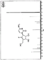
Mechanism of action of RX-3117
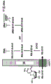
RX-3117 is a cytidine analog (FIG. 14). It has a modification of the ribose molecule consisting of carbon-fluorine bonds instead of oxygen and double bonds (Choi WJ, Chung H-J, Chandra G, Alexander V, Zhao LX, Lee HW, Nayak A, Majik MS, Kim HO, Kim J-H, Lee YB, Ahn CH, Lee SK and Jeong LS: fluorolacto-cysteine with broad spectrum and pore antagonist activity. J. Med Chem 55: 4521. sup. 5,2012). RX-3117 enters the cell via the human equilibrium nucleoside transporter (hENT) as shown in FIG. 21 (Peters GJ, Smid K, Vecchi L, Kathmann I, Sarkisjan D, Honeywell RJ, Losekoot N, Ohne O, Orbach A, Blaugund E, Jeong LS, Lee YB, Ahn C-H and Kim DJ: Metabolim, mechanism of action and sensitivity profile of flucyclopentenylcytone (RX-3117). Invest New Drugs 31: 4-. In said cells, RX-3117 is phosphorylated to its monophosphate form by uridine/cytidine kinase 2(UCK2), i.e., RX-3117 is activated to RX-3117MP by UCK 2. RX-3117 triphosphate (RX-3117TP) was incorporated into the RNA. Its RX-3117 diphosphate (RX-3117DP) is reduced to deoxydiphosphate (dRX-3117DP) by Ribonucleotide Reductase (RR) and then introduced into the DNA. RX-3117 is a poor substrate (id.) for Cytidine Deaminase (CDA). RX-3117 has recently been shown to have a potent tumor growth inhibitory effect in a gemcitabine-resistant mouse model (Yang MY, Lee YB, Ahn C-H, Kaye J, Fine T, Kashi R, Ohne O, Smid K, Peters GJ and Kim DJ: A novel cytidin ananogo, RX-3117, show patent activity in xenograde models, even in tumor tissue area resistant to gemcititabine bin anticancer Res 34: 1-. Radiosensitisation of RX-3117 has been studied and the results are shown below.
RX-3117 is classified as a pyrimidine analog (Peters GJ: Novel definitions in the use of antibiotics. Nos. 33: 358-74, 2014), similar to other analogs such as gemcitabine and azacytidine, which are widely used in clinics. Gemcitabine is a potent radiosensitizer that enhances ionization-induced DNA damage repair (Morgan M a, Parsels L a, Maybaum J and Lawrence TS: stimulating gemcitibine-mediated radiosensitization using molecular targeted therapy: a review. Clin Cancer Res 14: 6744. 50, 2008).
In addition, the cytidine analog 5-azacytidine (aza-C, Vidaza)
TM) And 5-nitrogenHetero-2' -deoxy-cytidine (decitabine,

) Are being used clinically for the treatment of myelodysplastic syndrome (MDS) (Peters GJ: Novel disorders in the use of microorganisms Nucleotides 33: 358-74, 2014). Two major mechanisms of the anti-tumor effect of these drugs are DNA methyltransferase (DNMT) inhibition in RNA and/or DNA and the introduction of cytotoxicity (Kaminskaskat E, Farrell A, Abraham S, Baird A, Hsieh L-S, Lee S-L, Leighton JK, Patel H, Rhman A, Sridhara R, Wang Y-C and Pazdur R: overview: azacitidine for the purpose of the treatment of myelogenous synthetic chromosome peptides, Clin Cancer Res 11: 3604; 8, 2005). Upon uptake into the cells, aza-C is phosphorylated to 5-azacytidine monophosphate (aza-CMP) by UCK2 and to aza-CDP and aza-CTP by pyrimidine nucleotide kinases. However, aza-C is inactivated by deamination of CDA. RR reduces aza-CDP to aza-dCDP, which is phosphorylated to aza-dCTP by nucleoside diphosphate kinase. Then, aza-dCTP is introduced into DNA, resulting in inhibition of DNA synthesis: (

J model of actions and effects of 5-azacytidines and of its derivatives in the Erukaromatic cells Pharmacol Ther 28: 227-35, 1985). Stoichiometric binding of aza-dCTP to DNMT will result in DNA hypomethylation (Jones PA: Effects of 5-azacytidines and its 2' -deoxyderivitive on cell differentiation and DNA methylation. Pharmacol Ther 28: 17-27, 1985). aza-dCTP can also be formed from 5-aza-2' -deoxycytidine by direct phosphorylation catalyzed by deoxycytidine kinase (dCK) and nucleotide kinases. DNA hypomethylation at CpG islands has been described in different malignancies including MDS (Kaminskaskat E, Farrell A, Abraham S, Baird A, Hsieh L-S, Lee S-L, Leighton JK, Patel H, Rahman A, Sridhara R, Wang Y-C and Pazdur R: arrival summary: azacitidine for transaction of myelogenous synthetic chromosome subtypes. ClCanin cer Res 11: 3604-. In another aspect, aza-CTP introductionThe metabolism of cytoplasmic and nuclear RNA protein synthesis is disrupted in RNA (Glover AB and Leyland-Jones B: Biochemistry of azacitidine: a review. cancer Tree Rep 71: 959-. One mechanism of azacytidine resistance is a point mutation in the UCK2 gene, which results in inactive metabolites (Sripayap P, Nagai T, Uesawa M, Kobayashi H, Tsukahara T, Ohmine K, Muroi K and Ozawa K: Mechanisms of resistance to azacitidine in human leukaemia cells lines. exp Hematol 42: 294-306. e2,2014). The mechanism of aza-dCTP resistance is a defect in dCK (Peters GJ: Novel deviations in the use of microorganisms Nucleotides 33: 358-74, 2014).
As shown in FIG. 22, RX-3117 is also capable of downregulating DNMT1(Peters GJ, Smid K, Vecchi L, Kathmann I, Sarkisjan D, Honeywell RJ, Losekoot N, Ohne O, Orbach A, Blaugund E, Jeong LS, Lee YB, Ahn C-H and Kim DJ: Metabolim, mechanism of action and sensitivity profile of fluorocarboxylic nitrile (RX-3117), Invest New Drugs 31: 1444-. In addition, many, but not all, cytidine analogs exhibit radiosensitization. Thus, the potential radiosensitisation of RX-3117 was evaluated, as well as the potential mechanisms such as cell cycle effects and cell killing. RX-3117 is shown to have radiosensitizing effects.
Preincubation with RX-3117 gave the best radiosensitisation, with 4 of the 5 cell lines tested being sensitized by RX-3117. The gemcitabine resistance SW 1573/G-was sensitized by RX-3117 with almost the same therapeutic effect as its wild type. RX-3117 also showed radiosensitisation in two spheroid models.
Nucleoside analogues have been shown to enhance radiation-induced cell killing (Shewach DS and Lawrence TS: antimitebolite radiodetectors. J Clin Oncol 25: 4043-50, 2007). The radiosensitization is thought to be performed by targeting deoxyribonucleotide biosynthesis (required for DNA replication) or DNA polymerase (Shewach DS and Lawrence TS: antipimetic radiosensitizers. J Clin Oncol 25: 4043-50, 2007; Lawrence TS, Blackstock AW and McGinn C: The mechanism of action of radioactive diagnosis of genetic chemical agents. set radial Oncol 13: 13-21,2003). An example of a deoxynucleotide library deregulation is the TS inhibitor 5-fluoro-2' -deoxyuridine (FdUrd). TS inhibitors cause an imbalance in the deoxynucleotide pool, leading to suppression of DNA synthesis and S-phase arrest (Hwang HS, Davis TW, Houghton J a and Kinsella TJ: Radiosensiveness of kinase synthesis-specific human tissue cells is infected by growth of the G1restriction point into S-phase: oligonucleotides for fluoronucleotide radiosensibility. cancer Res 60: 92-100,2000). An imbalance in the pool of deoxynucleotides can lead to incorrect Nucleotide introduction (ingram HA, Tseng BY and Goulian M: Nucleotide levels and incorporations of 5-fluoronucleotide and uracil in DNA of cells treated with 5-fluorodeoxynucleotides mol Pharmacol 21: 211-6, 1982). The concentration required for radiosensitization to be achieved is not necessarily the concentration required for cytotoxic effects. Lower concentrations of drug are able to establish the radiosensitization (Hwang HS, Davis TW, Houghton J a and Kinsella TJ: Radiosensitivity of tyrosine synthase-specific human tissue cells is afected by growth of the G1 regeneration point inter S-phase: pharmaceuticals for fluorosensitizing radiopharmaceutical sensitivity. cancer Res 60: 92-100,2000). Low dose RX-3117 induced radiosensitisation in the clonogenic assay in a three-dimensional model and fractionated irradiation schedule. Furthermore, the double-stranded DNA breaks induced by RX-3117 are also dose-dependent.
RX-3117 was demonstrated to be a potent schedule-dependent radiosensitizer in four of the five cell lines with potential for clinical application where combination therapy is considered in NSCLC. Such combination therapy may be applicable to other tumor types (e.g., prostate cancer, skin cancer, head and neck cancer, laryngeal cancer (throat), laryngeal cancer (larynx), breast cancer, brain cancer, colorectal cancer, bone cancer, leukemia, ovarian cancer, and uterine cancer).
Improvements in processes for the preparation of RX-3117
U.S. Pat. No.7,405,214 discloses the 11-step of RX-3117 synthesis from D-ribose. The synthesis uses expensive catalysts, which is a challenge to implement in large-scale plant production. The synthesis is improved by us patent No.9,150,520, disclosing a new synthesis of a compound prepared by reacting (3R,4R,6aR) -tert-butyl- (5-fluoro-2, 2-dimethyl-6-trityloxymethyl-4, 6 a-dihydro-3 aH-cyclopenta [1,3] dioxol-4-yloxy) -diphenyl-silane (ASM11) to 4-amino-1- (3aS,4S, a shorter route for the preparation of RX-3117 from 6aR) -5-fluoro-2, 2-dimethyl-6- ((trityloxy) methyl) -4,6 a-dihydro-3 aH-cyclopenta [ d ] [1,3] dioxol-4-yl) pyrimidin-2 (1H) -one (INT 14). However, the prior art syntheses of ASM11 to INT14 require isolation of the intermediates in each step. Thus, the process can pose cost and time limitations, particularly if scaled up for commercial production.
The present invention provides an improved process for the preparation of RX-3117, which is commercially viable for large scale production. The method shortens the synthesis of ASM11 to INT14 without the need to isolate each of the intermediate species, thereby reducing cost while increasing efficiency. More specifically, the present invention provides a continuous process with three stages to shorten the synthesis of ASM11 to INT 14. The invention also provides a method for obtaining RX-3117 monohydrate (RX-3117-MH) in a fixed container, thereby significantly reducing the production costs. By condensing the three steps into a single step, the present method eliminates the need to concentrate the intermediate into a residue. These improvements are based on unexpected benefits when replacing reagents not readily apparent to those skilled in the art.
In addition, the present invention provides optimized reaction and isolation conditions to increase nitrogen to oxygen (N/O) selectivity in stage 3, in which case cytosine is added to (3aR,4R,6aR) -5-fluoro-2, 2-dimethyl- ((trityloxy) methyl) -4,6 a-dihydro-3H-cyclopenta [ d ] [1,3] dioxol-4-yl methanesulfonate (INT13) to prepare INT 14. In the improved process, the N-and O-isomer ratio is increased from the previously optimized 88:12 value to 99.03: 0.97. The fixed vessel production process of the present invention achieves the cost benefits of operating on a scale-up production of the desired product in the form of a monohydrate.
Scheme 1 below illustrates an improved process for the preparation of RX-3117 MH.
Scheme 1
Method improvement for stage 1-ASM 11 deprotection to form INT12
In stage 1 of the process, 2-methyl-tetrahydrofuran is used as the solvent for the process. This modification allows the work-up (work-up) to be carried out without concentrating the intermediate (3aS,4R,6aR) -5-fluoro-2, 2-dimethyl-6- ((trityloxy) methyl) -4,6 a-dihydro-3 aH-cyclopenta [ d ] [1,3] dioxol-4-ol (INT12) and avoiding solvent exchange of the intermediate into methyl tert-butyl ether (MTBE), which is used in the prior art methods.
In addition, the in-process control (IPC) of the reaction shifted from using TLC to quantification1H NMR method. The process is further optimized by using azeotropic removal of water rather than chemical drying. INT12 in solution was used directly in stage 2 of the process without further isolation or purification using 2-methyl-tetrahydrofuran as the process solvent.
Method improvement of stage 2-INT 12 mesylation to form INT13
(3aR,4R,6aR) -5-fluoro-2, 2-dimethyl-6- ((trityloxy) methyl) -4,6 a-dihydro-3 aH-cyclopenta [ d ] [1,3] dioxol-4-yl methanesulfonate (INT13) was prepared by directly reducing the solution of INT12 in 2-methyl-tetrahydrofuran to stage 2. This improved process eliminates the use of environmentally hazardous methylene chloride as the reaction solvent. In addition, the process uses an ammonium chloride wash to further control residual triethylamine. The post-treatment volume was reduced to a maximum process volume of 12.5 volumes, with a reduction from 14 volumes. Again, the process is further optimized by using azeotropic water removal rather than chemical drying. INT13 in solution was used directly in stage 3 of the process using 2-methyl-tetrahydrofuran as the process solvent.
Process improvement of stage 3-cytosine incorporation into INT13 to form INT14
INT13 solution in 2-methyl-tetrahydrofuran was directly reduced to stage 3 to make INT 14. Dimethyl sulfoxide (DMSO) was retained as a reaction solvent, and 2-methyl-tetrahydrofuran was removed by distillation. It was found that the 27% w/w 2-methyl-tetrahydrofuran relative to the product specification allowed the stage 3 reaction to perform well. The inventors have conducted a screening of bases (inorganic and amine) and found that cesium carbonate provides the highest chemoselectivity and the fastest reaction rate. The present inventors also conducted a screening of reaction solvents, and found that DMSO is the most suitable reaction solvent.
Furthermore, the inventors investigated the effect of charge, temperature and concentration of the reagent on the selectivity of the N-isomer versus the O-isomer of INT 14. According to the improved process of the present invention, the N-to O-isomer ratio is adjusted from the previously optimized 88:12 ratio using solvent extraction and recrystallization/precipitation (this is with SiO)2Column chromatography) was improved to 99.03: 0.97. The process of the present invention eliminates the need for column chromatography and also provides over 99% of the desired N-isomer. In particular, the inventors found that chemoselectivity is achieved primarily by the reaction temperature. Specifically, lowering the reaction temperature slows the rate of conversion to product. The reaction conditions were further improved by increasing the amount of base and cytosine added from 2.0 equivalents to 2.5 equivalents. The reaction temperature was reduced from 40 ℃ to 35 ℃. In addition, the post-treatment process is modified to reduce the total process volume from 22 volumes to 12.5 volumes, thereby improving throughput. The work-up solvent is changed from ethyl acetate to isopropyl acetate, thereby allowing the reaction mixture to go directly to isolation without the need to exchange solvents. The post-treatment process was also modified to start with tautomerism, as INT14 was found to be more soluble after acetic acid treatment. The isolation of INT14 was modified so that the product precipitated in high volume starting from isopropyl acetate, after which the volume was reduced and n-heptane was added. This improvement was found to prevent oil pickup (oiling) and sticking to the container prior to separation. Thus, an overall improved synthesis with increased selectivity is achieved by simultaneously varying multiple reaction parameters. It was found that the change in temperature and concentration is rightThe conversion and chemoselectivity of the N/O-alkylation have a positive effect.
Method for stage 4-deprotection of INT14 to give anhydrous RX-3117
The original conditions of 2M HCl in ethanol were found to be the most stable and retained for the product. However, the process is improved by lowering the reaction temperature from 60 ℃ to 50 ℃ to aid solubility. The trityl alcohol by-product was removed using a methyl tert-butyl ether (MTBE) wash. After the resin salt is released, the product in the aqueous phase is directly reduced to stage 5 separation.
Stage 5-Process for isolation of RX-3117 monohydrate
The solution of RX-3117MH was directly reduced to stage 5. The combined stages 4 and 5 (with minor modifications to the process) optimize yield and operability on a scale. The RX-3117-MH was dried on a filter under air, which controlled the amount of acetonitrile below the ICH guidelines while maintaining the water content. This improved process eliminates the time critical requirement of first drying followed by rehydration of the product to obtain a crystalline product. The isolated product using the improved process had a purity (99.83%) comparable to that of a small amount of custom-made synthetic product.
Other improvements in the preparation of RX-3117 starting materials
Other process modifications for the synthesis of RX-3117 starting materials are possible. The synthesis of ASM11 using different intermediates and protecting groups is shown in schemes 2 and 3 below, respectively.
Synthesis of ASM11 via a bromo intermediate
Scheme 2
In U.S. Pat. No.9,150,520, in step 3 of the reaction, RXN-2 is converted to RXN-3 using iodoform. In the present invention bromoform or mixed bromo-iodomethane is used to provide a bromo intermediate rather than an iodo intermediate. The bromo intermediate can be more stable than its iodo derivative. Thus, the overall yield and purity may be increased accordingly.
Synthesis of ASM11Bn by benzyl protection of the 5-hydroxy group
Scheme 3
Unexpectedly, the change of the protecting group put in the early intermediate can have a significant impact on the reaction steps carried out in the later process, without the need to modify many steps along the path. For example, changing the trityl protecting group to benzyl in step 2 can improve the yield of the fluorination in the reaction step 10. These improvements can be made without further modification to the overall process.
Synthesis of ASM11 by ring closing metathesis
The inventors of the present invention also developed a scheme for the synthesis of ASM11 using ring closing metathesis (ring closing metathesis). In scheme 4, a ring closing metathesis reaction is used to form the 5-membered ring moiety. The ruthenium of the Grubb catalyst is recyclable, further improving the scale-up process by reducing waste and cost.
Scheme 4
Synthesis of intermediate RXN-6 by ring-closing metathesis
The synthesis of intermediate RXN-6 can be accomplished by ring closing metathesis reactions, including RXN-5, and the fluorine atom is introduced into the five-membered ring by preparing fluorinated RXN-6. As shown in scheme 5, the 5-membered ring moiety (fluoro-RXN-6) is formed using a ring closing metathesis reaction. As in scheme 4, the ruthenium of the Grubb catalyst (Grubb's catalyst) is recoverable.
Scheme 5
Synthesis of intermediate RXN-6 by nucleophilic fluorination via epoxide
Synthesis of intermediate RXN-6 via epoxide by alternative nucleophilic fluorination (alternative nucleophilic fluorination). Scheme 6 shows the formation of an epoxide ring from the starting material (3aR,6aR) -6- (((tert-butyldiphenylsilyl) oxy) methyl) -2, 2-dimethyl-3 a,6 a-dihydro-4H-cyclopenta [ d ] [1,3] dioxol-4-one. For example, the epoxide is opened by nucleophilic fluorination using potassium fluoride. Alternatively, other fluoride sources may be used, for example tetrabutylammonium fluoride to ring-open the epoxide ring. Water removal in this process is a difficult step and alternative dehydrating agents can be used, for example, methoxycarbonyl (carbomethoxy) sulfamoyltriethylammonium salt to obtain the modified intermediate fluoro-RXN 6-TBDPS.
Scheme 6
Synthesis of intermediate RXN6 by aldol condensation
The synthesis of intermediate RXN6 can also be achieved using aldol condensation by introducing a fluorine atom into the five-membered ring by preparing fluorinated RXN-6. As shown in scheme 7, fluorine atoms were introduced early on to form the fluorinated derivative of RXN6 (fluoro-RXN 6). Internal aldol condensation can form the 5-membered ring with the vinyl fluoride moiety in place.
Scheme 7
Alternative synthesis of intermediate fluoro-RXN 6
Other alternatives for the synthesis of intermediate fluoro-RXN-6 are shown in schemes 8 and 9. In scheme 8, a shorter route is obtained by reacting D-ribolactone 1 with phosphonate 2 to produce intermediate 3. The inventors have found that the D-ribolactone derivative 1 does not react readily with dimethyl fluoroalkylphosphonate, but that better reactivity can be obtained with dimethyl carbalkoxy methylphosphonate. Intermediate 3 then undergoes Hundsdiecker (Hansidick) iododecarboxylation to form intermediate 4, which intermediate 4 can then be nucleophilically substituted using tetra-n-butylammonium fluoride (TBAF).
Scheme 8
Scheme 9 (below) provides an even shorter route by reacting D-ribolactone 1 with an alkylphenyl sulfone 7 prepared by electrophilic fluorinating reagent to produce intermediate 8. The D-ribolactone derivative reacts more readily with lithium fluoroalkylsulfone 7 than does fluoroalkyl phosphonate. The intermediate 8 is capable of being converted to intermediate 9, which intermediate 9 will undergo elimination to form F-RXN 6. In addition, a more efficient but more expensive option is to use fluorotetrazolyl sulfone 10 instead of lithium fluoroalkyl sulfone 7.
Scheme 9
Synthesis using different chiral sources
The present synthesis can also be achieved by using different chiral sources as starting materials.
In scheme 10 (below), (2S,3S,4R,5S,6R) -6- (hydroxymethyl) tetrahydro-2H-pyran-2, 3,4, 5-tetraol is used as an alternative chiral starting material to produce ASM-11.
Scheme 10
Examples
The following examples are provided for illustrative purposes and should not be used to limit the scope of the disclosed subject matter.
As in Peters et al, "Metabolism of action and sensitivity profile of fluoronitrile (RX-3117)," Investigational New Drugs, Decumber 2013, Vol.31, No.6, pp.1444-1457 (see e.g. Peters et al, incorporated herein by reference)http://link.springer.com/article/ 10.1007/s10637-013-0025-xObtained on-line), IC of the cell lines A549, SW1573 and SW1573/G used herein50The values were 8.3. mu.M, 13.7. mu.M and 7.3. mu.M, respectively.
Example 1: effect of RX-3117 on non-Small cell Lung cancer cell lines
RX-3117 the effects of cell cycle regulation and cell death in human non-small cell lung carcinoma (NSCLC) cell lines A549 (adenocarcinoma), SW1573 (alveolar carcinoma), SW1573/G- (gemcitabine-resistant SW1573 cell line) and H460 (large cell carcinoma) were evaluated. The A549 and H460 cell lines were obtained from the American Type Culture Collection (Manassas, Va., USA). The SW1573 cell line obtained from Johan van Rijn Ph (see, Keiser et al, Cancer Research,49:2988-2993(1989)) was also used as the parent cell line for SW 1573/G-. A549, SW1573 and SW 1573/G-and H460 were grown exponentially in T25 flasks (Greiner Bio-One GmbH, Frickenhausen, Germany) and cultured in Dulbecco (Dulbecco) minimal essential Medium (DMEM) or RPMI 1640(A549) supplemented with 10% heat-inactivated Fetal Bovine Serum (FBS) 1% streptomycin and penicillin 20mM HEPES and maintained at 37 ℃ with 5% CO
2In a water-saturated atmosphere. For the cell cycle distribution, 1.5X 10
5Each cell was seeded in a T25 flask (Greiner Bio-One GmbH) and cultured for 72h, then 1. mu.M RX-3117 was added and cultured for 24 h. Then, the cells were harvested. After the treatment, the medium was collected in 15mL tubes (Greiner Bio-One GmbH). The cells were washed with ice cold PBS and at 37 deg.CDigested with trypsin (Lonza). The collected medium was used to inactivate trypsin in the corresponding samples and collected again in 15mL tubes. The cells were centrifuged at 4 ℃ and 12,000rpm for 5 minutes using standard centrifugation procedures. The medium was removed, and the particles were washed with 1mL PBS/0.01% BSA and centrifuged. After removal of the supernatant, the cells were fixed with
1mL 70% ethanol and incubated at-20 ℃ for at least 24 h. Subsequently, the cells were centrifuged, washed with 1mL PBS/0.1% BSA and transferred to FALCON FACS tubes (BD, Franklin Lakes, NJ, usa). The cells were centrifuged and the supernatant removed, then 0.5. mu.g Propidium Iodide (PI) (Sigma, St. Louis, Mo., USA), 0.1% trisodium citrate (Riedel-
Sigma-Aldrich Laborchemikalien GmbH, St. Louis, MO, USA), 0.1% Triton X-100 (TM) (Merck) and 0.1mg/mL RNase (Sigma) (PI solution) to the sample. Subsequently, the analysis was started after incubating the cells with the PI solution on ice to stain the DNA for at least 15 minutes. By FACSCalibur
TM(BD Biosciences, mountain View, CA, USA) the cells stained with the PI solution were analyzed. Data were collected using CellQuest
TMPro software analysis.
The mechanism of cell cycle arrest was studied by measuring cyclin expression using western blotting. The effect of RX-3117 on protein expression during different treatment conditions was analyzed by Western blotting. Cells were lysed on ice for 30 minutes using
Cell lysis buffer 1 × (Cell Signaling, Danvers, MA, usa) containing a 4% protease inhibitor cocktail (cocktail) (Roche Diagnostics, Mannheim, germany) and centrifuged at 14,000rpm for 10 minutes at 4 ℃. The protein-containing supernatant was collected and the Bio-Rad assay was performed to determine the amount of protein as described in Lemos et al, Pharmacogenomics,12(2):159-70 (2011). The following antibodies were used for protein expression: DNMT1(Cell Signaling, 1:1000#5032S), DNMT3A (Cell Signaling 1:1000#2160S), DNMT3B (Abcam, 1; 1000), Chk2(Cell Signaling 1:1000#6334P), Chk1(Cell Signaling 1:1000) p-CDC25C (Cell Signaling 1:1000#4901S), Cdk1(Cell Signaling 1:1000#9112S), Cdk2(Cell Signaling 1:1000#2546S), wee1(Cell Signaling 1:1000), S139- γ H2A. X (Cell Signaling, 1:1000), β actin (Sigma, 1:10,000), caspase-specific protease 9(Cell Signaling, 1:1000), PARP (Roche 2003, 1:1000), p53(Cell Signaling, 1:1000, # 9282). The antibody was diluted in 1:1Rockland buffer (Rockland Inc., Philadelphia, Pa., USA) and supplemented with 0.05%

20 in PBS. The proteins were separated on 20% SDS-PAGE and transferred to PVDF membrane. For the secondary fluorescent signal antibodies, goat anti-mouse InfraRedDye and goat anti-rabbit InfraRedDye were used. The protein was detected by the Odyssey InfraRed Imager (Li-COR Bioscience, Lincoln, NE, USA).
Abbreviations used herein represent the following:
BSA ═ bovine serum albumin
HEPES-2- [4- (2-hydroxyethyl) piperazin-1-yl ] ethanesulfonic acid
PBS-phosphate buffered saline
PVDF ═ polyvinylidene fluoride
RPM is the number of revolutions per minute
SDS-PAGE of sodium dodecyl sulfate Polyacrylamide gels
Cell cycle
At a dose of 1 μ M, RX-3117 induced the accumulation of A549, SW 1573/G-and H460 cells in said G1phase after 24H exposure (FIG. 1). At 5 × IC50At higher doses of (a), RX-3117 induced the accumulation of A549, SW1573 and SW 1573/G-cells in said S phase (FIG. 2).
Caspase activation
RX-3117 reduced the procysteine aspartate-specific protease 9 in SW1573 cells and a549 cells after 24h exposure to increasing concentrations of RX-3117. A decrease in pre-caspase 9 indicates activation of the caspase and subsequent induction of apoptosis (fig. 3 and 4).
DNMT protein
RX-3117 at higher doses reduced the maintenance of DNA methyltransferase 1(DNMT1) in a549 cells (fig. 23) and increased DNMT3A and DNMT3B expression levels in a549 cells. The proposed mechanism for this down-regulation is shown in fig. 22.
DNA damage
After 48h of exposure, RX-3117 induced a Double Strand Break (DSB) as indicated by biomarker γ h2a.x (phospho S139) in SW1573 cells (fig. 5). RX-3117 induced cleaved PARP after exposure to increasing concentrations of RX-311724 h (FIG. 6). Cleaved PARP indicates activated caspases specific protease activity in apoptotic cells. At 1. mu.M and 5. mu.M, RX-3117 increased the expression level of p53 in A549 cells (FIG. 7). At 10. mu.M, RX-3117 increased Chk1 and Cdk2 expression levels after 48h exposure, while decreasing p-Cdc25C expression levels in SW1573 cells (FIG. 8). RX-3117-induced DNA damage triggers the Chk1 pathway. The ATR/Chk1 pathway is induced by DNA replication stress (replication stress) and DSB. RX-3117 decreased the expression level of wee1 in SW1573 cells after exposure to increasing concentrations of RX-311724 h (FIG. 9). FIG. 24 is a graph showing the potential effect of checkpoint kinases Chk1 and Chk2 on cyclin and the regulation of the cell cycle after injury induction, RX-3117 may be active along various of these pathways.
Apoptosis induction
At 5 × IC50At doses of (a), RX-3117 induced apoptosis in PI stained a549 and SW1573 cells in the sub-GI phase after 24 and 48h exposure (fig. 10). RX-3117 induced apoptosis in annexin V stained a549 and SW1573 cells in the sub-GI phase after 24, 48, 72 and 96h exposure at 5 μ M (for a549) and 10 μ M (for SW1573) (fig. 11).
Results
The results indicate that cell cycle arrest is time, concentration and cell line dependent. In A549, H460 and SW1573 cells, 24H exposure to 1 μ M RX-3117 increased the accumulation of cells in the G1phase (approximately 20% to 40%) and S phase (to a lesser extent), but decreased the accumulation of cells in the G2/M phase. Thus, low doses of RX-3117 induced G1 accumulation, while high doses of RX-3117 induced S phase accumulation. No cell killing was observed at 24H exposure, but at 48H exposure (15% in SW1573 and 8% in a549 cells) with induction of γ H2 AX. Among A549 cells, the effect of RX-3117 on the cell cycle distribution was most pronounced at 48h exposure, accumulating 45% in S phase. The accumulation of S phase is time dependent. Treatment with RX3117 increased p53, Chk1, Chk2 and Cdk2 expression levels, but decreased Cdc25C and p-Cdc25C expression levels. RX-3117 increased the expression level of wee1 mainly after 48 h. RX-3117 appears to induce apoptosis by SSB and DSB. Cleaved PARP in SW1573 cells indicates caspase-specific protease activity up-regulated in apoptotic cells. A decrease in pre-caspase 9 in a549 cells indicates activation of the caspase-specific protease and subsequent induction of apoptosis. In conclusion, RX-3117 induced DNA damage triggered apoptosis on the one hand and increased Chk1 and Chk2 expression levels on the other hand. Without being bound by any mechanism of action, it is believed that the phosphorylated Chk1 and Chk2 can trigger the phosphorylation of Cdc25C and cause its degradation, which results in a decrease in Cdk1 levels, thereby allowing cells in S phase to accumulate.
Example 2: RX-3117 efficacy in syngeneic (syngeneic) MC38 murine Colon cancer xenograft model
The effect of RX-3117 on tumor growth in a syngeneic model using female C57BL/6 mice with MC38 murine colon cancer was examined according to the following protocol. Tumor growth in the treated groups was determined compared to the control (vehicle treated) group (see dosage and treatment regimens in table 1 below). The results of this study (Table 2) demonstrate that the addition of RX-3117 to the programmed death receptor 1(PD-1) inhibitor RMP1-14 has an additive effect (additive effect) in inhibiting tumor growth (80% for RX-3117 alone, 93% for RMP1-14 alone, and 99% for the combination of the two agents). The combination of the two agents also resulted in a higher number of mice (9 mice) with partial and complete regression and 7 animals with tumor-free survival compared to 4 animals with partial and complete regression of the RMP1-14 group alone, 2 animals showing tumor-free survival. All results were obtained without any adverse effect on mice in the combination group.
Briefly, the method is described below. The cells were harvested during exponential growth and resuspended in phosphate buffered saline. Each test animal received a subcutaneous (s.c.) injection of 1X 10⁶Individual tumor cells in the right flank and with the average tumor size approaching 60-100 mm3The target range of (a) monitors tumor growth. Based on table 1, the dosing (dosing) was started as each animal reached the target range.
TABLE 1 drugs and treatment schedules
# control group,. mu.g/animal
TABLE 2 tumor growth inhibition and survival benefits of RX-3117 in combination with PD-1 inhibitors
| Group of
|
Treatment group
|
TGI day 28
|
PR
| CR
|
TFS |
|
| 1
|
Carrier
|
-
|
2
|
0
|
0
|
| 2
|
RX-3117(60mg/kg)
|
80%
|
0
|
0
|
0
|
| 3
|
anti-PD-1 RMP1-14(100g)
|
93%
|
1
|
3
|
2
|
| 4
|
RX-3117+ anti-PD-1
|
99%
|
2
|
7
|
7 |
TGI: inhibition of tumor growth; on day 28; PR: the number of partial regressions; CR: the number of complete regressions; TFS: number of tumor-free survivors; all on day 45
Tumors were measured in two dimensions using a vernier caliper and volume was calculated using the following equation:
where w is the width of the tumor and l is the length of the tumor in mm. Assuming 1mg equals 1mm3The tumor weight can then be estimated.
Treatment efficacy was determined using day 45 data. The mtv (n), the median tumor volume for the number of animals at day 45, n, was determined for each group. Defining percent tumor growth inhibition (% TGI) as the difference between the MTV of the indicated control group (vehicle administration) and the MTV of the drug-treated group, expressed as a percentage of the MTV of the control group:
the data set (data set) used for TGI analysis included all animals in the group, except those that died for treatment-related (TR) or non-treatment-related (NTR) reasons. Agents that produce at least 60% TGI in this test are considered potentially therapeutically active.
The study protocol specifies a tumor growth delay test based on the median Time To Endpoint (TTE) for the treatment group relative to the control group. When the tumor reaches 1500mm3At the volume endpoint, each animal was euthanized for Tumor Progression (TP). The time to reach the endpoint (TTE) of each mouse was calculated using the following equation:
where b is the intercept of the line obtained by linear regression of the log-transformed tumor growth dataset and m is the slope. The data set includes a first observation that exceeds the study endpoint volume and three consecutive observations immediately prior to reaching the endpoint volume. Any animals that did not reach the endpoint were euthanized at the end of the study and the TTE value was set equal to the last day of the study (71 days). In the case where the calculated TTE of the logarithmic conversion precedes the day before reaching the end point or exceeds the day at which the end point of tumor volume is reached, linear interpolation is performed to approximate TTE. Any animal determined to die from a treatment-related (TR) cause is assigned a TTE value equal to the day of death. Any animal that died from a non-treatment related (NTR) cause was excluded from the TTE analysis.
The therapeutic effect is determined by the number of regression responses. Treatment may result in Partial Regression (PR) or Complete Regression (CR) of the tumor in the animal. In PR response, during the course of the study, the tumor volume was 50% or less of its D1 volume for three consecutive assays, and equal to or greater than 13.5mm for one or more of these three assays3. In the CR response, the tumor volume was less than 13.5mm for three consecutive determinations during the course of the study3. Any animal with a CR response on the last day of the study was additionally classified as a tumor-free survivor.
For toxicity evaluation, animals were weighed daily for the first five days of the study and then twice weekly. The mice are often observed for obvious signs of any adverse treatment-related side effects and clinical signs of toxicity are recorded at the time of observation.
Acceptable toxicity was defined as less than 20% of the group's average weight loss during the study and no more than one treatment-related (TR) death among 10 treated animals. Any dosing regimen that results in greater toxicity is considered to exceed the Maximum Tolerated Dose (MTD). Death is classified as TR if it is due to clinical signs and/or treatment side effects confirmed by autopsy, or if it is due to unknown reasons during the dosing period or within fourteen days of the last dose. Mortality is classified as non-treatment related (NTR) if there is no evidence that mortality is associated with treatment side effects.
Prism 6.05 for Windows (GraphPad) was used for statistical and graphical analysis. The MTV values of the various sets were compared to the nonparametric Kruskal-Wallis test and the ex vivo Dunn's multiple comparison test. The two-tailed statistical analysis was performed at P ═ 0.05. Prism reports that the results are non-significant (ns) at P <0.05, significant (denoted by "+") at 0.01< P ≦ 0.05, very significant ("+") at 0.001< P ≦ 0.01, and extremely significant ("+") at P ≦ 0.001. Since the statistical test is a significance test and does not provide an estimate of the magnitude of the difference between groups, all significance levels are described as significant or non-significant in the text of this report.
A "box and beard line" (box and whisker) graph was constructed showing the distribution of individual tumor volumes in groups at D15. The box (box) represents the 25 th to 75 th percentiles of the observed values, the horizontal line corresponds to the median value, and the "beard line (whisker)" represents the maximum and minimum values. Group median tumor volumes were plotted as a function of time. Group mean BW change was plotted as a percentage change from D1, ± SEM. Animals that died due to NTR were excluded from all graphical representations.
Survival was analyzed by the Kaplan-Meier method based on TTE values. Based on TTE values, the logrank (Mantel-Cox) and Gehan-Breslow-Wilcoxon tests determined the significance of the difference between the overall survival experience (survival curves) of both groups. The Kaplan-Meier plots and statistical tests share the same data set and exclude any animals recorded as NTR deaths. Constructing a scatter plot to show TTE values of individual mice in groups; this figure shows NTR deaths, which are excluded from all other figures. Group mean tumor volumes were plotted as a function of time. When animals were withdrawn from the study due to tumor size or TR death, their final recorded tumor volume was included with the data used to calculate median volume at subsequent time points. After death of both TRs occurred in the same group, the tumor growth curve was truncated. Group-averaged BW changes over the course of the study were plotted as percent change from day 1 ± SEM. Tumor growth and body weight change curves were truncated after more than half of the evaluable mice in the group exited the study.
Example 3: pharmacokinetics, safety and tolerability of RX-3117 in humans
In a first exploratory study of human open label, pharmacokinetics, safety and tolerability of RX-3117 were evaluated. The study duration was 14-15 days (7 days screening period; 3 days treatment period; 4(+1) days safety follow-up period). 9 adult male and female subjects with histologically confirmed solid tumors were enrolled and completed the study. The subjects received RX-3117 (n-3 subjects/dose) as a single oral dose (50mg or 100mg) or a single intravenous dose (20 mg).
Pharmacokinetics (PK)
The absolute bioavailability (F) of oral RX-3117 was 55.67% and 33.42% for the 50 and 100mg doses, respectively. Mean T for the 50mg and 100mg dosesMaximum of2.16h and 2.49h respectively. Average C for the 50mg and 100mg dosesMaximum of303.3ng/mL and 311.43ng/mL, respectively. Greater absolute bioavailability and C of the 50mg dose compared to the 100mg doseMaximum ofThe results indicate that the oral bioavailability of RX3117 in plasma may not be dose proportional. The T for the 50mg and 100mg doses1/213.95h and 20.92h, respectively, indicate that RX-3117 may show dose proportionality on some parameters, but not on others at the doses tested.
The plasma PK profile of the intravenous RX-3117 is different from the plasma PK profile of the oral RX-3117. The 20mg dose of intravenous RX-3117 recovered rapidly after bolus infusion (T)Maximum of0.25 h). The 20mg dose of intravenous RX-3117 had a mean C of 1143.63ng/mLMaximum ofThis is about a 4-fold increase over the peak concentration of the oral dose.
Safety and tolerability
RX-3117 was safe and well tolerated in all subjects. No Adverse Events (AEs) occurred, Treatment of Emergency Adverse Events (TEAEs) or Severe Adverse Events (SAE).
The results indicate that RX-3117 is safe and well tolerated with oral bioavailability and supports higher dose studies.
Example 4: pharmacokinetics, safety and tolerability of RX-3117 at different oral doses
In an open label dose range study, the Pharmacokinetics (PK) of RX-3117 at different oral doses was evaluated. Subjects with advanced malignancies were given RX-3117 containing capsules at daily doses of 30mg (N ═ 1), 60mg (N ═ 1), 100mg (N ═ 3), 150mg (N ═ 3), 200mg (N ═ 3), 500mg (N ═ 3), 1000mg (N ═ 3), 1500mg (N ═ 4) and 2000mg (N ═ 5 to date) 3 times per week (TIWK) for 3 weeks, stopping for 1 week during 4-week periods. More frequent dosing was also performed based on the sustained safety profile and enhanced weekly RX-3117 exposure. In addition to the TIWK dosing regimen discussed above, subjects received 500mg and 700mg 5 times per week, and received 500mg 7 times per week for 3 weeks, with 1 week off during each 4-week period. Dose escalation begins with an accelerated design of 1 subject per dose treatment (Simon et al, j. natl. cancer inst., 89(15):1138-47 (1997)) followed by a standard 3+3 design using a modified Fibonacci (Fibonacci) sequence after the occurrence of a single associated grade 2 or higher adverse event table 3 summarizes the dosing regimen.
Table 3: dose escalation-3 times weekly
Pharmacokinetics (PK)
PK data are listed in table 4.
RX-3117 is rapidly absorbed without significant lag time, median T is typically observed at 2-3 hMaximum of. At TMaximum ofLater, the elimination was biphasic, with AUC observed over the first 8h0-tAbout half of (0-24 h), and more than 80% in 24 h. Apparent end (terminal) T1/2Shows no dose-or time-dependent pharmacokinetics, an average in the range of 11.6 to 16.7h in the 60 to 2000mg dose range after the first dose, and a seventh doseThe dose (dose on day 15) is 12.3-20.2 h. CMaximum ofAnd AUC0-t(0-24 h) suitably increases linearly with dose, but in a less proportional manner, it is possible to reach a plateau of 1500mg dose (fig. 12 and 13). In a dosage range of 30-2000 mg, the average C after the first dosageMaximum of32-1858 ng/mL and the seventh post-dose average CMaximum of99 to 1703ng/mL (FIG. 12). Mean AUC over the same dose range0-t(0-24 h) 164-20,544 h-ng/mL after the first dose and 702-20,919 h-ng/mL after the seventh dose (FIG. 13). Accumulation is generally minimal.
The PK data showed a dose-dependent increase upon exposure of doses up to 1000mg TIWK. C after the 7 th dose at a dose greater than 500mg TIWKMaximum ofAnd AUC0-t(0-24 h) were always lower than those measured after the first dose (fig. 12 and 13). Due to C at doses exceeding 1000mgMaximum ofAnd AUC0-t(0-24 h) plateau, more frequent dosing regimen was used to enhance weekly exposure (table 4). Based on the results of this study, the Maximum Tolerated Dose (MTD) for RX-3117 was determined as 700mg daily 5 days a week, three weeks on dosing, and one week off every 4-week period. This MTD was then used for follow-up efficacy studies.
TABLE 4 PK data after day 1 and day 15 of the study
Safety and tolerability
The most common adverse events were mild to moderate fatigue and nausea, mild diarrhea, mild vomiting, mild anorexia and moderate dehydration. Dose-limiting toxicity was limited to grade 3 anemia, thrombocytopenia.
Example 5: RX-3117 efficacy, safety and tolerability in humans
RX-3117 was evaluated for efficacy, safety and tolerability at various doses and frequencies (see example 4 above). Subjects with advanced malignancies were given capsules containing different doses of RX-3117 from TIWK to 7 times per week for 3 weeks, with 1 week rest during each 4-week period. Dose escalation begins with an accelerated design of 1 subject per dose treatment (Simon et al, j.natl.cancer inst., 89(15):1138-47 (1997)) followed by a standard 3+3 design using a modified Fibonacci (Fibonacci) sequence after the occurrence of a single associated grade 2 or higher adverse event table 4 (above) summarizes the dosing regimen.
The subjects were evaluated for efficacy, safety and tolerability of RX-3117. There were a total of 48 subjects enrolled (30 females, 18 males). 17 subjects experienced stable disease for 1-10 cycles; 10 subjects received treatment from day 82 to day 276. A reduction in tumor burden was observed in 3 patients with pancreatic cancer (tumor volume and CA19-9 biomarker), breast cancer and mesothelioma cancer. The most common associated adverse events are moderate to severe anemia, mild to moderate fatigue and nausea, mild diarrhea, vomiting and anorexia.
In another stage of the study, RX-3117 is being evaluated in cancer patients in phase Ib/IIa clinical trials for relapsed or refractory pancreatic cancer or advanced bladder cancer, including muscle-invasive bladder cancer. The phase Ib/IIa clinical trial is a multicenter study that evaluates the safety and efficacy of RX-3117 in these target patient populations. Secondary endpoints included safety and pharmacokinetic analyses. The patients in the trial were orally administered a daily dose of 700mgRX-3117 5 times per week for 3 weeks, or until their disease progressed, over a 28 day period and 4 treatment periods.
Example 6: radiosensitization of fluorocyclopentenyl cytosine (RX-3117) in ovarian and lung cancer cell lines
Drugs and chemicals
Stock solutions of RX-3117 were prepared in deionized water. All other chemicals used were of standard quality and commercially available.
Cell culture
Human NSCLC cell lines A549 (adenocarcinoma), H460 (large cell carcinoma), SW1573 (alveolar carcinoma), and SW1573/G- (gemcitabine-resistant SW1573 cell line) and ovarian cancer cell line A2780 were maintained exponentially grown in T25 flasks (Greiner Bio-One GmbH, Frickenhausen, Germany) and cultured in Dulbecco (Dulbecco) minimal essential Medium (DMEM) or RPMI 1640 (supplemented with 10% heat-inactivated Fetal Bovine Serum (FBS), 1% streptomycin, and penicillin) and maintained at 37 ℃ at 5% CO
2Under a saturated atmosphere. Cells were harvested using trypsin EDTA (Invitrogen, Paisley, uk). Use of
Z
TMA series of counters count the cells.
Clonogenic testing
Exponentially growing A2780, SW1573, A549, H460 and SW 1573/G-cells were exposed to 1 μ M RX-3117 or untreated (control) for 24H and irradiated with a single dose of gamma radiation (0-6 Gy) using a Co source (Gamma cell 200, Atomic Energy of Canada, Ltd). Subsequently, 500 cells/T25 flasks were plated and allowed to colony. Ten days later, colonies were fixed with 100% ethanol and colony counts were stained with 10% giemsa staining solution (Merck Chemicals BV, Amsterdam, netherlands). Plating Efficiency (PE) was calculated by dividing the number of colonies formed by the number of cells plated with the control and normalized for control-induced toxicity. To illustrate the effect of RX-3117 on radiation, the Dose Modification Factor (DMF) (Bijnsdorp IV, van den Berg J, Kuipers GK, Wedekind LE, Slotman BJ, van Rijn J, Lafleur MVM and Sminia P: radioactive mutagenesis potential of the selective cyclic gene-2 (COX-2) inhibitor meloxicam on human collagen cells. J neuronol 85: 25-31, 2007) was calculated as described previously.
Spheroid testing
The NSCLC cell lines a549 and SW1573 were plated at a density of 100,000 cells/well in low-attachment 24-well plates (Corning Incorporated, Corning, NY) and formed spheroids. Three days later, individual spheroids were transferred to a new 24-well low-attachment well plate (one spheroid/well). In thatImmediately after the transfer treatment was initiated for A549 and SW1573 cells, 1 μ MRX-3117 was combined with fractionated 2Gy radiation (5 days with a single 2Gy dose). Photographs were taken on day 0 (before irradiation) and after 3, 6, 9 and 15 days using a phase contrast microscope (leica dmi300B Universal Grab version 6.3 software, Digital Cell Imaging Labs). Measurement of volume by ImageJ software (1.45 s, Wayne Rasbaas, National instruments of Healthy, Bethesda, D.V.: 4/3) for calculation of volume V (Pi 2/D) is described earlier by Galvani et al (Galvani E, Giovannetti E, Saccani F, Cavazzoni A, Leon LG, Dekker H, Alfield R, Carmi C, Mor M, Ardizoni A, Petronini PG and Peters GJ: Molecular mechanics integrating the activity of the enzyme of 3-amino-polypeptide involved in transforming the activity of the immunoglobulin receptor of the immunoglobulin of Neon 15: 61-72, 2013)3)。
Flow cytometry analysis
Cell cycle distribution and apoptosis were analyzed by plating cells at a density of 5,000 cells in flat bottom 6-well plates (Greiner Bio-One GmbH, Frickenhausen, Germany) and allowing them to attach for 24 h. Thereafter 5 IC is added50The cytotoxic concentration of (a). The exposure times were 24h and 48h, and the control group was included for comparison. At each time point, the total amount of adherent and floating cells was harvested in round bottom FALCON tubes (BD, Franklin Lakes, NJ, usa). After centrifugation, the cell particles were resuspended in 1.0mL Propidium Iodide (PI) solution (50. mu.g/mL PI, 0.1% sodium citrate, 0.1% Triton X-100, 0.1mg/mL RNase A) or 10. mu.L annexin V (Cat. No. 31490014, Immunotools) and placed on ice for 30 minutes. Subsequently, the samples were analyzed using a FACSCalibur (BD Biosciences, Mount View, CA, usa). For data analysis, the number of sub-G1 stage cells (apoptotic cells) was estimated using gates on DNA histograms, and CELLQuest was runTMAnd (3) software.
Protein expression analysis
The effect of RX-3117 on protein expression during different treatment conditions was analyzed by Western blot. Cells were lysed on ice for 30 minutes using 1 × Cell lysis buffer (Cell Signaling, Danvers, MA, usa) containing a 4% protease inhibitor cocktail (Roche Diagnostics, Mannheim, germany) and centrifuged at 14,000rpm for 10 minutes at 4 ℃. A Bio-Rad test was performed to determine the amount of protein in the collected supernatant as described previously (Lemos C, Kathmann I, Giovannetti E, Calhau C, Jansen G and Peters GJ: Impact of cellular leaves status and epidemic growth factor expression on BCRP/ABCG 2-conditioned resistance to formation and accumulation. Br J Cancer 100: 1120-7, 2009). The following antibodies were used for protein expression: γ H2A.X (catalog #9718, Cell Signaling, 1:1000), β -actin (Sigma, 1:10,000), Cdc25C Ser216 (catalog #4901, Cell Signaling, 1:1000), Cdk1Tyr15 (catalog #9111, Cell Signaling, 1:1000), Chk1Thr68 (catalog #2197S, Cell Signaling, 1:1000), histone 3 (catalog #4499, Cell Signaling). Antibodies were diluted in a 1:1 solution with Rockland buffer (Rockland Inc, philiadelphia, PA) and Phosphate Buffered Saline (PBS) supplemented with 0.05% tween 20. Proteins were separated on a 20% SDS-PAGE gel and transferred to polyvinylidene fluoride (PVDF) membranes. For the fluorescent signal, secondary antibodies goat anti-mouse InfraRedDye and goat anti-rabbit InfraRedDye were used. The protein was detected by the Odyssey InfraRed Imager (Li-COR Bioscience, Lincoln, NE).
Results
Radiosensitisation of RX-3117
To investigate the effect of RX-3117 on irradiation, a clonogenic test was performed. First, the inventors examined whether pre-incubation or post-incubation with RX-3117 enhanced the effect of radiation. Pre-and post-treatment with RX-3117 and 4Gy were compared in A2780 cells.
Clonogenic assay data indicate that preincubation with RX-3117 is the most effective condition for radiosensitisation. Preincubation with 1 μ M RX-3117 had a 5-fold lower plating efficiency compared to the control (FIG. 15).
Potential radiosensitisation was studied in the a2780 and non-small cell lung cancer cell lines a549, SW1573 and SW 1573/G-and H460 using the pre-incubation protocol. In general, all cell lines show radiosensitisation when treated with RX-3117 and radiation. Maximal radiosensitisation was observed with 1.8 DMF in the a2780 and a549 cell lines and 1.5 DMF in SW1573 (fig. 16A, 16B, 16D). Gemcitabine-resistant cell line SW 1573/G-with 1.4 DMF (FIG. 16E), but H460 cells showed poor radiosensitisation. Since fractionated irradiation is a standard procedure in the clinic, fractionated doses of 2Gy radiation within 5 days in SW1573 cells were also studied. Incubation with 1 μ M RX-3117 showed the lowest colony growth before 5 fractions of 2Gy fractionated radiation therapy (FIG. 16F).
The radiosensitizing ability of RX-3117 in combination with radiation in three-dimensional models using spheroid testing was also investigated. The sphere formation test revealed radiosensitisation of RX-3117 to spheres in SW1573 and a549 spheroids (fig. 17). SW1573 spheres were highly affected by 1 μ M RX-3117 alone and Radiotherapy (RT) alone (2Gy 5 days) and the effect was enhanced with the combination. In A549 cells, RX-3117 treatment or irradiation alone had little effect on the volume growth, whereas 1 μ M RX-3117 enhanced the effect of 2Gy irradiation for 5 days (FIG. 17).
Initiation of apoptosis
The potential of RX-3117 to induce apoptosis was studied in the NSCLC cell line. The amount of apoptotic cells was determined by annexin V staining after 24h, 48h and 72h of exposure (fig. 18). Annexin V cell membrane staining showed a gradual increase in apoptotic cells of a549 cells and SW1573 cells, which was more pronounced for SW1573 cells compared to a 549.
Initiation of DNA Damage
DNA damage is a hallmark of cell death. DNA damage was investigated by evaluating γ h2a.x expression. In the A2780 cell line, increasing RX-3117 concentrations starting from 0.1. mu.M to 10. mu.M RX-3117 showed induction of γ H2A.XS139 in a dose-dependent manner (A in FIG. 18). In the SW1573 cell line, the double strand break injury marker increased after exposure to 0.3. mu.M RX-311748 h (B in FIG. 18). The combination of 0.3 μ M RX-3117 and irradiation showed more significant γ h2a.xs139 protein expression (B in fig. 18). As expected, radiation caused an immediate increase in the expression of γ h2a.x; the repair is delayed in the presence of RX-3117.
Effect of treatment with RX-3117 and radiation on cell cycle distribution and cell death
Since perturbations in cell cycle distribution have been reported to affect radiosensitisation by other nucleoside analogues (Shewach DS and Lawrence TS: antipimetic radiosensitizers. J Clin Oncol 25: 4043-50, 2007), the effect of RX-3117 in combination with radiation was studied in NSCLC cells using FACS analysis. At a relatively low concentration of 1 μ M RX-3117, a small and just significant (p <0.05) cell line-dependent increase in the S phase was found in 3 out of 4 cell lines. The increase in the G1phase was found in all cell lines (A in FIG. 19), and a strong decrease in the G2/M phase was found. 4Gy irradiation resulted in a significant decrease in the amount of S-phase cells, with an increase in the G2/M phase in these two SW1573 cells and no effect in A549 cells, but a decrease in the H460 cells (A in FIG. 19). The combination of irradiation and RX-3117 resulted in an increase in the number of S phase cells but a decrease in H460 cells in both SW1573 variants. Cell killing (sub-G1) was significantly increased in a549 cells, but not in other cells (a in fig. 19).
To understand some of these phenomena, the effects of RX-3117 and radiation on the expression of some essential cyclins were also investigated (B in FIG. 19 and FIG. 20). In SW1573, the effects of RX-3117 and radiation on various cell cycle checkpoint proteins were examined (FIG. 20). After 48h, the irradiation resulted in interesting reductions of wee1, Chk2, CDC25c and p-CDC25 c. In both SW1573 cells, irradiation caused an increase in phosphorylation of Chk2 (B in fig. 19). RX-3117 reduced phosphorylation of Cdk1 in almost all cell lines (except gemcitabine-resistant SW-1573/G); similarly, irradiation increased cdc-25C phosphorylation within 24h (but not in SW 1573/G-) (B in FIG. 19). In combination with RX-3117, this effect is maintained (B in FIG. 19).
Example 7: RX-3117 inhibition of DNA methyltransferase leads to upregulation of hypermethylated targets
RX-3117 is analogous to azacytidine (aza-CR) and aza-deoxycytidine (aza-CdR). RX-3117 is occupied by the human balanced nucleoside transporter (hENT) (take up) and activated to RX-3117-MP by uridine-cytidine kinase 2(UCK2) (FIG. 21). RX-3117 is occupied by the human balanced nucleoside transporter (hENT) and activated to RX-3117-MP by uridine-cytidine kinase 2(UCK 2). RX-3117 down-regulates DNA methyltransferase 1(DNMT1) (Choi W.J., et al., J.Med.chem.55(2012) 4521-4525; Peters G.J., et al., Invest New Drugs 31(2013) 1444-1457). DNMT1 is responsible for maintaining methylation of the S-phase newly synthesized DNA and methylating cytosine residues in the hemimethylated DNA. RX-3117 deaminates at a much slower rate than gemcitabine.
RX-3117 is a novel cytidine analog that is orally bioavailable and is currently being evaluated in phase I clinical studies. The maximum tolerated dose is above 2000 mg/day. RX-3117 has been shown to down-regulate DNMT1 in various cell lines with different histological backgrounds. Both UCK2 and DNMT1 are currently evaluated as potential biomarkers. In this example, the effect of RX-3117 on DNMT1 was determined on the DNA, RNA, protein and enzyme activities, as well as the reactivation of inhibited target genes including p16INK4A, methylguanine methyltransferase (MGMT) and the proton-coupled folate transporter (PCFT). PCFT involves the transport of folate, Methotrexate (MTX) and Pemetrexed (PMX) at pH 5.5 and 7.4, and this gene is highly methylated. In addition, the functions of proteins of the genes known to be regulated by methylation were studied, including: proton Coupled Folate Transporter (PCFT). The expression of E-cadherin (adhesion molecule), p16INK (tumor suppressor protein) and O-6 methylguanine DNA methyltransferase (MGMT) (DNA repair gene) in the A549 cell line was also studied.
Method
In this study, the following cell lines were used: (1) CCRF-CEM cells and MTX resistant variants thereof CEM-MTX, characterized by the absence (RFC) of the reduced folate carrier (Jansen G., et al., JBC 273(1998) 30189-30198). The PCFT gene in CEM cells is highly methylated. (Gonen n., et al, BBRC 376(2008) 787-92.); (2) CEM cells cultured in RPMI medium containing 10% Fetal Bovine Serum (FBS); and (3) a549 and SW1573 non-small cell lung cancer (NSCLC) and a2780 ovarian cancer cell lines cultured in DMEM medium containing 10% FBS.
DNMT1 protein expression was determined by Western blotting after 24 or 48h exposure to RX-3117. DNMT1RNA expression was determined by real-time PCR after 24 and 48h exposure to RX-3117. DNMT enzyme activity was determined in isolated nucleic acids using the DNA methyltransferase assay kit supplied by EpiGentek, using the ability of the CpG binding domain to bind methylated DNA after exposure to 1. mu.M RX-3117 or 5. mu.M aza-CdR. In A549 cells, the effect of 5. mu.M RX-3117 on total methylation was determined with specific antibodies against 5-methyl-cytosine. Bands on western blots were visualized using an Odyssey InfraRed imager using a suitable InfraRed dye.
MTX transport was determined using radiolabeled MTX in CEM wild-type and CEM-MTX cell lines. CEM cells have high RFC activity. CEM-MTX is completely absent in RFC-mediated transport. CEM cells have highly methylated PCFT transporters and very low PCFT-mediated transport (Gonen n., et al., BBRC 376(2008) 787-92). MTX transport at pH 7.4 is mainly RFC-mediated and is less than 2% by PCFT. Folate is used to inhibit PCFT mediated transport. L-leucovorin (L-LV) was added to completely inhibit RFC-mediated transport. CEM and CEM-MTX cells were exposed to 29.6. mu.M RX-3117 and 0.19. mu.M aza-CdR as positive controls. MTX transport was determined using 2. mu.M [3',5',7-3H ] -MTX after 24H exposure to the drug in a 3 minute uptake assay.
Statistics were done using student t-test.
Results
In the moderately sensitive non-small cell lung cancer (NSCLC) cell lines such as A549 and SW1573, DNMT1 protein expression was down-regulated by 5-50 μ M RX-3117 by 5-20% after 24h exposure and > 90% after 48h (A in FIG. 25 and B in FIG. 25). DNMT1mRNA was not affected after 24h exposure, but was moderately affected after 48h (C in fig. 25 and D in fig. 25).
In the sensitive ovarian cancer cell line A2780, protein down-regulation has been observed after 24h at 1. mu.M RX-3117 (A in FIG. 26 and B in FIG. 26). DNMT1 activity was inhibited by 1. mu.M RX-3117 by 32%, which is similar to the percentage inhibition with 5. mu.M of the reference compound 5-aza-2' -deoxycytidine (DAC, 31%).
In A549 cells, 5 μ M RX-3117 reduced total methylation of DNA (detected by anti-5-methylcytosine antibody) by 25% after 48h exposure, while 5 μ M DAC inhibited only 9% (A in FIG. 27). Protein expression and activity were evaluated for a variety of genes known to be affected by methylation. A549 cells were exposed to RX-3117 and assayed for immunofluorescence using antibodies against 5-methyl-cytosine (B in fig. 27). In A549 and SW1573 cells, 24h exposure to 5. mu.M RX-3117 increased the expression of cyclin p16INK4A and the DNA repair enzyme MGMT. C in FIG. 27 shows the expression of MGMT, E-cadherin and p16INK4 after exposure to RX-3117 and aza-dC.
For PCFT, the functional activity of RX-3117 was evaluated in CCRF-CEM leukemia cells with a highly methylated PCFT promoter and in CEM-MTX cells deficient in the reduced folate vector (RFC). PCFT is a specific folate transporter responsible for the uptake of folate and the folate analogues Methotrexate (MTX) and pemetrexed. Incubation of CEM and CEM-MTX cells with 29.6 μ M RX-3117 or DAC as positive controls significantly improved PCFT-mediated transport of MTX. This was more evident in CEM-MTX cells, with a 10-11 fold increase in both RX-3117 and DAC relative to a 4-fold increase in CEM cells. Folic Acid (FA) was added to inhibit PCFT and L-LV to inhibit RFC-mediated MTX transport. Aza-CdR and aza-CR were included as positive controls. (A in FIG. 28, B in FIG. 28, and C in FIG. 28).
Conclusion
In summary, RX-3117 down-regulated DNMT1 protein and RNA expression by reducing DNA methylation. RX-3117-mediated hypomethylation increases the expression of MGMT, E-cadherin, PCFT and the tumor suppressor gene p16INK 4A. PCFT mediates MTX transport. These data emphasize DNMT1 inhibition as a novel mechanism for RX-3117. RX-3117 is a novel epigenetic (epigenetic) modulator.
Example 8: evaluation of UCK2 protein expression as a potential predictive biomarker for RX-3117
Background
A novel orally bioavailable nucleoside analogue RX-3117 is an intracellularly activated prodrug of uridine cytosine kinase 2(UCK2) believed to be expressed predominantly in tumor tissues. RX-3117 is currently being evaluated in stage Ib/IIa multicenter open label clinical studies in patients with advanced pancreatic and bladder cancer. In this study, the relationship between UCK2 tissue protein expression and the efficacy of RX-3117 in a mouse xenograft model was studied, as well as UCK2 protein expression in a panel of human cancer tissues relative to normal tissues.
Method
UCK2 protein expression in tumor tissues was analyzed by immunoblotting using clone 22-1 rabbit monoclonal antibody. A validation procedure for Immunohistochemistry (IHC) of UCK2 with clone 22-1 was performed in a panel of human Formalin Fixed Paraffin Embedded (FFPE) cancers and normal tissues.
Results
The immunoblot protein levels and corresponding tumor growth inhibition of UCK2 normalized for β -actin (oral RX-3117 dose of 500mg/kg, TIWK) were 57% and 67% in MiaPaCa2, 30% and-5% in BxPC3, 199% and 92% in Coli-205, 21% and 90% in Caki-1, 2% and 39% in a549, respectively, and 146% and 79% in H460, respectively. These data indicate a trend in the antitumor efficacy of the UCK 2-dependent mode. The IHC of UCK2 showed that positive staining of UCK2 in cancer tissue was observed in 20/20 bladder cancer tissue (100% frequency), 19/20CRC tissue (95% frequency), 18/20NSCLC tissue (90% frequency) and 19/20 pancreatic cancer tissue (95% frequency). The mean H-score of UCK2 in cancer versus normal tissue was 104 to 9 in lung, 97 to 20 in bladder, 67 to 41 in pancreas and 39 to 21 in colon, respectively.
Conclusion
The current data show a trend of correlation between the level of UCK2 protein expression and the degree of anti-tumor activity of RX-3117 in the xenograft model. This also supports higher levels of UCK2 protein expression in human cancer tissues compared to their normal tissues. This suggests that RX-3117 activity may be specific for tumor tissue and that quantification of UCK2 expression in human cancer tissue may be used as a predictive biomarker for patient selection of sensitivity to RX-3117 in future clinical studies.
Example 9: synthesis of RX-3117 monohydrate
INT14 from ASM11 by stage 1 to stage 3 continuous reactions in a fixed reactor
ASM11, tert-butyl (((3aR,4R,6aR) -5-fluoro-2, 2-dimethyl-6- ((trityloxy) methyl) -4,6 a-dihydro-3 aH-cyclopenta [ d [ -d ]][1,3]Dioxolen-4-yl) oxy) diphenylsilane (37.65kg, 1wt, 1 equivalent, 55mol) was dissolved in 2-methyltetrahydrofuran (4.0 vol, 3.4 wt). 1.0M TBAF (tetra-n-butylammonium fluoride) in THF (tetrahydrofuran, 1.61 vol, 1.45wt, 1.1 eq) was added in one portion (in one portion) to the reaction vessel (a gentle exothermic controlled addition) over 15-45 minutes, maintaining 18-23 ℃. Filling 2-methyltetrahydrofuran (1.0 volume, 0.9wt) into the container as a pipeline for flushing, keeping the temperature at 18-23 ℃, and stirring the obtained solution at 18-23 ℃ for 6 hours until the solution passes through1H NMR monitored the reaction complete. To the reaction mixture was added 8% w/w sodium bicarbonate (3.0 vol) and stirred at 18-23 ℃ for 5-10 minutes (caution: mild exotherm) and allowed to phase separate thereby removing the lower aqueous phase (2 x 2.0 vol). The first 8% (w/w) sodium bicarbonate extract provided a milky aqueous layer, while the extended settling time did not clear the emulsion. Studies have shown that the emulsion is confined to the aqueous layer and has a low organic content, thus continuing the process. The total separation of the first extraction was 5 hours and 29 minutes. The second 8% w/w sodium bicarbonate extraction separation required only 52 minutes without problems (without problem). The aqueous phase was extracted with 2-methyltetrahydrofuran (2.0 vol, 1.7wt) and the line was flushed with 2-methyltetrahydrofuran (2.0 vol, 1.7 wt). Heating the combined organic phase containing INT12 to 40-50 ℃ and concentrating under reduced pressure at 40-50 ℃ to about 4 volumes. The samples were analyzed by Karl-Fischer until the water content (contentwa) was < 0.2% w/w. The process yielded 158.8kg dry weight of INT12ASM 11-ol in 2-methyltetrahydrofuran containing 15.3% w/w INT12ASM 11-ol, corresponding to an overall yield of 99.0%. 92.83% by HPLC analysisINT12ASM 11-ol ((3aS,4R,6aR) -5-fluoro-2, 2-dimethyl-6- ((trityloxy) methyl) -4,6 a-dihydro-3 aH-cyclopenta [ d ] in areal purity][1,3]Dioxol-4-ol).
The INT12 solution was returned to the vessel, cooled to 0-5 ℃ and triethylamine (0.41 vol, 0.30wt, 2.0 eq.) was added. After flushing the tube with 2-methyltetrahydrofuran (1.0 vol, 0.9wt), methanesulfonyl chloride (0.17 vol, 0.25wt, 1.5 eq) diluted in 2-methyltetrahydrofuran (1.0 vol, 0.9wt) was added to the solution (carefully mixed in the header vessel) and maintained at 0-5 ℃ (exothermic) for at least 30 minutes. Additional 2-methyltetrahydrofuran (0.5 vol, 0.4wt) was added as a line flush, held at 0-5 ℃. Stirring the contents of the container at 0-5 ℃ until after 1h1H NMR monitored the reaction completion. A representative sample was taken after 1h, and if necessary, was thereafter examined from the reaction vessel about every 2h and analyzed to check the remaining INT 12. In passing through1H NMR analysis after checking 100% conversion, water (4.0 vol) is added, keeping at 0-10 ℃ and the reaction mixture is warmed to 18-23 ℃ and stirred at 18-23 ℃ for 5-10 minutes. The upper organic phase in the vessel was separated and 8% w/w sodium bicarbonate solution (4.0 vol) was added, maintaining 18-23 ℃. The resulting biphasic solution was stirred at 18-23 ℃ for 1-2 h and 20% w/w aqueous ammonium chloride (2.0 vol) and 2-methyltetrahydrofuran (2.0 vol, 1.7wt) were added to the separated organic phase. And adjusting the temperature to 18-23 ℃ according to the requirement. The 20% w/w ammonium chloride wash resulted in vigorous gas evolution, probably due to reaction with residual sodium bicarbonate from the previous step. After stirring for 5-10 minutes at 18-23 ℃, the upper organic phase is separated and purified water (2.0 vol.) adjusted to 18-23 ℃ is added. Concentrating the separated organic phase containing INT13 at 35-45 ℃ under reduced pressure to about 2 volumes. Samples were taken for analysis. This procedure produced a net weight of 78.4kg of INT13ASM 11-methanesulfonate in 2-methyltetrahydrofuran containing 34.9% w/w INT13ASM 11-methanesulfonate, equivalent to an overall yield of 94.9%. INT13ASM 11-mesylate ((3aR,4R,6aR) -5-fluoro-2, 2-dimethyl) in 62.91% area purity by HPLC analysis6- ((trityloxy) methyl) -4,6 a-dihydro-3 aH-cyclopenta [ d ] yl][1,3]Dioxol-4-yl methanesulfonate) and 32.23% area of TBDPS byproduct was present.
DMSO (3.8 vol, 4.2wt) is added continuously to the INT13 solution and heated to 40-45 ℃ to concentrate the organic phase under reduced pressure at ≦ 45 ℃ until no more solvent (2-methyltetrahydrofuran) is distilled. The concentration was continued for 5 hours and 25 minutes and1h NMR determination of the IPC showed a 2-methyltetrahydrofuran content of 8.3% w/w. After cooling the solution to 27-33 ℃, cesium carbonate (1.2wt) and cytosine (0.41wt) were added. The reaction mixture is heated to 33-37 ℃ and stirred until the reaction is monitored by HPLC for completion. After 24h and at a suitable point thereafter, samples were taken from the reaction vessel for N/O-alkylation ratio analysis. After 33 hours 47 minutes, the reaction was considered complete and the IPC result was 99.5% conversion. The ratio of N-and O-isomers was 99.03: 0.97. Upon completion, isopropyl acetate (2.0 vol, 1.7wt) and purified water (4.0 vol, 4.0wt) were added to the mixture, maintaining 50 ℃ or less (water addition is exothermic). After stirring for 5 to 15 minutes, the biphasic mixture is allowed to settle for 10 minutes and the upper organic phase is subsequently separated. The aqueous phase was re-extracted twice by stirring at 40-50 ℃ for 5-15 minutes and settling again for 10 minutes before separation, recovering all the product with isopropyl acetate (2.0 volumes, 1.7wt each). The combined organic phases were cooled to 25-30 ℃ and added with 10% v/v acetic acid (3.0 vol.) and 26% w/w saline solution (1.0 vol.), maintained at 25-30 ℃ and the biphasic solution was stirred at 25-30 ℃ for 30-60 minutes. The upper organic phase was washed three times with 10% v/v acetic acid (3.0 vol) and 26% w/w saline solution (1.0 vol), maintained at 25-30 ℃. In each washing step, the upper organic solution is passed through1H NMR analysis samples. The organic phase is again washed with about 3% w/w saline solution (3X 2.0 vol.) at 25-30 deg.C and passed1H NMR samples the acetic acid content. Heating the organic phase containing INT14 to 35-45 ℃ and concentrating under reduced pressure at 35-45 ℃ to about 5 volumes. To the solution was added isopropyl acetate (3.0 vol, 2.6wt) and concentrated to about 5 vol at 35-45 ℃ under reduced pressure.Isopropyl acetate (5.0 vol, 4.4wt) was added to the solution again, adjusted to 57-63 ℃, stirred at 57-63 ℃ for 1.5-3 h, and checked for crystallization/precipitation by HPLC. Cooling the slurry to 35-45 ℃ and concentrating under reduced pressure at 35-45 ℃ to about 5 volumes. Further cooling the slurry to 18-23 ℃ within 1.0-2.0 h, and adding n-heptane (7.0 vol, 4.8wt) within 30-90 minutes, and keeping at 18-23 ℃. After 1-2 h at 18-23 ℃ and 1-2 h at 0-5 ℃, the slurry is filtered through a 20 μm cloth and washed with pre-mixed n-heptane/isopropyl acetate (5:1, 2 × 1.0 vol) at 0-5 ℃. Drying the product containing INT14 under vacuum at up to 55 ℃ and passing through1H NMR was measured. Pass criteria are ≤ 2.0% w/w isopropyl acetate and ≤ 2.0% w/w n-heptane. The process yielded 24.27kg dry weight INT14, 4-amino-1- ((3aS,4S,6aR) -5-fluoro-2, 2-dimethyl-6- ((trityloxy) methyl) -4,6 a-dihydro-3 aH-cyclopenta [ d][1,3]Dioxol-4-yl) pyrimidin-2 (1H) -one (45mol, 93.25% purity), corresponding to 82% overall yield and 64% w/w yield.
Preparation of RX-3117 monohydrate from INT14 by stage 4 to stage 5 continuous reaction in a fixed reactor
INT14, 4-amino-1- ((3aS,4S,6aR) -5-fluoro-2, 2-dimethyl-6- ((trityloxy) methyl) -4,6 a-dihydro-3 aH-cyclopenta [ d][1,3]Dioxolen-4-yl) pyrimidin-2 (1H) -one (24.27kg, 45mol, 1.0wt) was added to a vessel followed by methanol (7.5 vol, 5.9wt) and the temperature of the reaction mixture was adjusted to 18-23 ℃. 2M HCl (1.1 vol, 1.2 eq.) was added to the reaction vessel, maintaining the temperature<At 50 ℃. Heating the slurry mixture to 45-55 ℃, and stirring for 2-2.5 h at 45-55 ℃ (target 50 ℃). The volume of the vessel is noted and the reaction mixture is distilled under reduced pressure keeping 45-55 ℃ and kept constant by adding MeOH (5.0 vol, 4.0 wt). The mixture was sampled and MeOH (2.5 vol, 2.0wt) was added continuously keeping the volume constant by distillation at 45-50 ℃ until the presence of < 1.0% area of the acetonide intermediate was monitored by HPLC. Once the conversion is complete, the reaction mixture is cooled to 25-30 ℃. Mixing the reactionThe mixture is concentrated to 5 volumes under reduced pressure at 35-45 ℃. Cooling the reaction mixture to 25-30 ℃. TBME (methyl tert-butyl ether) (5.0 vol, 3.7wt) and water (5.0 vol) were added to the reaction vessel, maintaining 25-30 ℃. Stirring the two-phase solution at 25-30 ℃ for 10-20 minutes, separating the phases at 25-30 ℃, and retaining the lower-layer water phase. The remaining lower phase was transferred to the vessel and further TBME (5.0 vol, 3.7wt) was added, maintaining 25-30 ℃. And stirring the two-phase solution at 25-30 ℃ for 10-20 minutes, and separating out a lower-layer water phase. The lower aqueous phase was returned to the vessel and the tubing was flushed with water for injection (0.5 vol, 0.5 wt). By passing1H NMR measurement checked for trityl alcohol removal with a trityl alcohol content of 0.3% w/w. If the determination is not 0.5% w/w trityl alcohol or less, adding TBME (5.0 vol, 3.7wt) to the aqueous phase and stirring at 25-30 ℃ for 10-20 minutes, and then repeating the separation. The combined aqueous solution was adjusted to 18-23 ℃, the pH was checked by adding pre-treated Ambersep900 (OH form) resin (5/6 the batch processed material) and stirring for 15 minutes. If the pH is higher<8.0, more Ambersep900 resin (OH form) is added and the solution is stirred for 30-45 minutes at 18-23 ℃. The slurry was filtered and washed with water for injection (2 × 4.0 vol) for 15-30 minutes each. The resin cake on the filter is further washed with water for injection (3X 4.0 vol.) for 15-30 minutes each time until a result of ≤ 1.0% is obtained by HPLC determination for each wash. The mother liquor and any resulting wash containing ≥ 1.0% are clarified through a1 μm filter. Heating the solution to 40-45 ℃, and concentrating the solution to 1.5 volumes at 40-45 ℃ under reduced pressure. After cooling the aqueous solution to 18-23 ℃ over 2-3 h, the mixture is aged for 60 minutes and acetonitrile (9.5 vol) maintained at 18-23 ℃ is added at a substantially constant rate over 1.5-2 h. And (3) aging the slurry at 18-23 ℃ for 2h, and cooling at 0-5 ℃ for 90 minutes. The solid was filtered through 20 μm cloth and washed with MeCN/water (5:1, 1.5 volumes) and dried under air atmosphere until MeCN content was determined by GC<400 ppm. The process yielded 8.20kg (99.83% purity) of RX-3117 monohydrate in dry weight,4-amino-1- ((1S,4R,5S) -2-fluoro-4, 5-dihydroxy-3- (hydroxymethyl) cyclopent-2-en-1-yl) pyrimidin-2 (1H) -one 1H2O (29.8mol, 99.83% purity), corresponds to 66% overall yield and 34% w/w yield.
FIG. 29 is a graph demonstrating RX-3117 prepared using the method described in example 91H NMR。1H-NMR(400MHz,DMSOd6) δ 7.40ppm, (d, J ═ 7.3Hz, 1H) CH cytosine, δ 7.20ppm, (width d, J ═ 9.1Hz, 2H) NH2δ 5.74ppm, (d, J-7.3Hz, 1H) CH cytosine, δ 5.30ppm, width s, 1H, CH, δ 5.15ppm, (d, J ═ 7.1Hz, 1H) (OH), δ 5.00ppm, (d, J-6.1Hz, 1H) (OH), δ 4.80ppm, (q, J ═ 5.3Hz, 1H) (OH), δ 4.48ppm, (q, J ═ 5.3Hz, 1H) CH, δ 4.17ppm, (dd, J ═ 9.1Hz, 3.8Hz, 1H) CH, δ 4.13ppm, (dt, J ═ 6.1Hz, 5.8Hz, 1H) CH, δ 3.91ppm, (width d, J ═ 12.9Hz, 2.8Hz, 1H) CH.
FIG. 30 is a photograph of RX-3117 prepared using the method described in example 913C NMR。
FIG. 31 is a photograph of RX-3117 prepared using the method described in example 919F NMR。
FIG. 32 is a mass spectrum of RX-3117 prepared using the method described in example 9. The mass spectra were performed using ES + filters showing protonated species for RX-3117(M + H) and RX-3117+ sodium adduct (M + sodium) with M/z 280.0 (common species seen during this analytical method). The sodium comes from the analytical method, not the preparative process.
FIG. 33 is a mass spectrum of RX-3117 prepared using the method described in example 9. The mass spectra were completed using an ES-filter that showed RX-3117 for the M-H species during the course of the analysis. The ES-and ES + filter methods together provide complete mass spectral evidence for RX-3117.
To verify the crystalline properties of the materials prepared according to the large scale synthesis method described above, the crystals prepared by the laboratory scale high purity method were compared microscopically and the X-ray powder diffraction patterns were compared. FIG. 34 is a microscopic comparison of the method according to example 9 (top row) and RX-3117 prepared using the laboratory scale method (bottom row) under plane polarized light (left column) and under orthogonal polarized light (right column). FIG. 35 is an X-ray powder diffraction data comparing RX-3117 prepared using a laboratory scale (top spectrum) and RX-3117 prepared using the method described in example 9 (bottom spectrum). As can be seen, there were no significant differences in the crystal structure.
It will be apparent to those skilled in the art that particular embodiments of the disclosed subject matter may be directed to one or more of the above and below referenced embodiments in any combination.
Although the present invention has been disclosed in detail by way of illustration and example, it will be apparent to those skilled in the art that changes may be made and equivalents may be substituted without departing from the true spirit and scope of the invention. Therefore, the description and examples should not be construed as limiting the scope of the invention.
All references, publications, patents, and patent applications disclosed herein are incorporated by reference in their entirety.