CN107709574B - Spatial mapping of molecular profiles of biological tissue samples - Google Patents
Spatial mapping of molecular profiles of biological tissue samples Download PDFInfo
- Publication number
- CN107709574B CN107709574B CN201680034754.6A CN201680034754A CN107709574B CN 107709574 B CN107709574 B CN 107709574B CN 201680034754 A CN201680034754 A CN 201680034754A CN 107709574 B CN107709574 B CN 107709574B
- Authority
- CN
- China
- Prior art keywords
- nucleic acid
- sample
- roi
- oligonucleotide probe
- rna
- Prior art date
- Legal status (The legal status is an assumption and is not a legal conclusion. Google has not performed a legal analysis and makes no representation as to the accuracy of the status listed.)
- Active
Links
- 238000013507 mapping Methods 0.000 title abstract description 10
- 239000002751 oligonucleotide probe Substances 0.000 claims abstract description 263
- 108020005187 Oligonucleotide Probes Proteins 0.000 claims abstract description 239
- 239000000523 sample Substances 0.000 claims abstract description 226
- 150000007523 nucleic acids Chemical class 0.000 claims abstract description 215
- 102000039446 nucleic acids Human genes 0.000 claims abstract description 214
- 108020004707 nucleic acids Proteins 0.000 claims abstract description 214
- 238000000034 method Methods 0.000 claims abstract description 104
- 108020004414 DNA Proteins 0.000 claims description 60
- 102000053602 DNA Human genes 0.000 claims description 60
- 238000000926 separation method Methods 0.000 claims description 59
- 238000006243 chemical reaction Methods 0.000 claims description 44
- 238000009826 distribution Methods 0.000 claims description 27
- 238000012163 sequencing technique Methods 0.000 claims description 22
- 238000010839 reverse transcription Methods 0.000 claims description 18
- 238000009739 binding Methods 0.000 claims description 17
- 238000009396 hybridization Methods 0.000 claims description 10
- 239000007788 liquid Substances 0.000 claims description 10
- 230000000295 complement effect Effects 0.000 claims description 9
- 238000012546 transfer Methods 0.000 claims description 9
- 238000002508 contact lithography Methods 0.000 claims description 8
- 230000002380 cytological effect Effects 0.000 claims description 6
- 230000004544 DNA amplification Effects 0.000 claims description 3
- 238000004519 manufacturing process Methods 0.000 claims description 3
- 238000007481 next generation sequencing Methods 0.000 abstract description 11
- 238000007639 printing Methods 0.000 abstract description 10
- 238000000059 patterning Methods 0.000 abstract description 5
- 238000013459 approach Methods 0.000 abstract description 4
- 238000003491 array Methods 0.000 abstract description 3
- 210000001519 tissue Anatomy 0.000 description 151
- 229920002477 rna polymer Polymers 0.000 description 81
- 239000003391 RNA probe Substances 0.000 description 33
- 238000003199 nucleic acid amplification method Methods 0.000 description 28
- 230000003321 amplification Effects 0.000 description 27
- 210000004027 cell Anatomy 0.000 description 24
- 239000000976 ink Substances 0.000 description 21
- 239000010410 layer Substances 0.000 description 18
- 125000003729 nucleotide group Chemical group 0.000 description 17
- 108020004518 RNA Probes Proteins 0.000 description 16
- 239000002773 nucleotide Substances 0.000 description 16
- 238000003556 assay Methods 0.000 description 14
- 239000000243 solution Substances 0.000 description 14
- 230000000875 corresponding effect Effects 0.000 description 13
- 239000000539 dimer Substances 0.000 description 13
- LFQSCWFLJHTTHZ-UHFFFAOYSA-N Ethanol Chemical compound CCO LFQSCWFLJHTTHZ-UHFFFAOYSA-N 0.000 description 12
- 108020004999 messenger RNA Proteins 0.000 description 12
- 241000894007 species Species 0.000 description 12
- 108010067770 Endopeptidase K Proteins 0.000 description 9
- 230000002596 correlated effect Effects 0.000 description 9
- 238000003752 polymerase chain reaction Methods 0.000 description 8
- 108090000623 proteins and genes Proteins 0.000 description 8
- 108091008146 restriction endonucleases Proteins 0.000 description 8
- 238000004458 analytical method Methods 0.000 description 7
- 238000001514 detection method Methods 0.000 description 7
- 239000000499 gel Substances 0.000 description 7
- 238000007641 inkjet printing Methods 0.000 description 7
- 238000011529 RT qPCR Methods 0.000 description 6
- 239000000463 material Substances 0.000 description 6
- 230000007170 pathology Effects 0.000 description 6
- 238000002360 preparation method Methods 0.000 description 6
- 238000003762 quantitative reverse transcription PCR Methods 0.000 description 6
- 125000006850 spacer group Chemical group 0.000 description 6
- 208000034454 F12-related hereditary angioedema with normal C1Inh Diseases 0.000 description 5
- WSFSSNUMVMOOMR-UHFFFAOYSA-N Formaldehyde Chemical compound O=C WSFSSNUMVMOOMR-UHFFFAOYSA-N 0.000 description 5
- 230000004888 barrier function Effects 0.000 description 5
- 238000010367 cloning Methods 0.000 description 5
- 239000002299 complementary DNA Substances 0.000 description 5
- 238000000605 extraction Methods 0.000 description 5
- 238000013467 fragmentation Methods 0.000 description 5
- 238000006062 fragmentation reaction Methods 0.000 description 5
- 208000016861 hereditary angioedema type 3 Diseases 0.000 description 5
- 238000010191 image analysis Methods 0.000 description 5
- 239000007791 liquid phase Substances 0.000 description 5
- 238000010200 validation analysis Methods 0.000 description 5
- WZUVPPKBWHMQCE-UHFFFAOYSA-N Haematoxylin Chemical compound C12=CC(O)=C(O)C=C2CC2(O)C1C1=CC=C(O)C(O)=C1OC2 WZUVPPKBWHMQCE-UHFFFAOYSA-N 0.000 description 4
- 206010028980 Neoplasm Diseases 0.000 description 4
- 238000003559 RNA-seq method Methods 0.000 description 4
- 230000008901 benefit Effects 0.000 description 4
- 239000006285 cell suspension Substances 0.000 description 4
- 239000003795 chemical substances by application Substances 0.000 description 4
- 238000009792 diffusion process Methods 0.000 description 4
- 239000011536 extraction buffer Substances 0.000 description 4
- 239000000203 mixture Substances 0.000 description 4
- 238000000746 purification Methods 0.000 description 4
- 230000004044 response Effects 0.000 description 4
- 239000002094 self assembled monolayer Substances 0.000 description 4
- 239000013545 self-assembled monolayer Substances 0.000 description 4
- 102000007469 Actins Human genes 0.000 description 3
- 108010085238 Actins Proteins 0.000 description 3
- 238000001712 DNA sequencing Methods 0.000 description 3
- 102000016911 Deoxyribonucleases Human genes 0.000 description 3
- 108010053770 Deoxyribonucleases Proteins 0.000 description 3
- 102000004190 Enzymes Human genes 0.000 description 3
- 108090000790 Enzymes Proteins 0.000 description 3
- 108091034117 Oligonucleotide Proteins 0.000 description 3
- JLCPHMBAVCMARE-UHFFFAOYSA-N [3-[[3-[[3-[[3-[[3-[[3-[[3-[[3-[[3-[[3-[[3-[[5-(2-amino-6-oxo-1H-purin-9-yl)-3-[[3-[[3-[[3-[[3-[[3-[[5-(2-amino-6-oxo-1H-purin-9-yl)-3-[[5-(2-amino-6-oxo-1H-purin-9-yl)-3-hydroxyoxolan-2-yl]methoxy-hydroxyphosphoryl]oxyoxolan-2-yl]methoxy-hydroxyphosphoryl]oxy-5-(5-methyl-2,4-dioxopyrimidin-1-yl)oxolan-2-yl]methoxy-hydroxyphosphoryl]oxy-5-(6-aminopurin-9-yl)oxolan-2-yl]methoxy-hydroxyphosphoryl]oxy-5-(6-aminopurin-9-yl)oxolan-2-yl]methoxy-hydroxyphosphoryl]oxy-5-(6-aminopurin-9-yl)oxolan-2-yl]methoxy-hydroxyphosphoryl]oxy-5-(6-aminopurin-9-yl)oxolan-2-yl]methoxy-hydroxyphosphoryl]oxyoxolan-2-yl]methoxy-hydroxyphosphoryl]oxy-5-(5-methyl-2,4-dioxopyrimidin-1-yl)oxolan-2-yl]methoxy-hydroxyphosphoryl]oxy-5-(4-amino-2-oxopyrimidin-1-yl)oxolan-2-yl]methoxy-hydroxyphosphoryl]oxy-5-(5-methyl-2,4-dioxopyrimidin-1-yl)oxolan-2-yl]methoxy-hydroxyphosphoryl]oxy-5-(5-methyl-2,4-dioxopyrimidin-1-yl)oxolan-2-yl]methoxy-hydroxyphosphoryl]oxy-5-(6-aminopurin-9-yl)oxolan-2-yl]methoxy-hydroxyphosphoryl]oxy-5-(6-aminopurin-9-yl)oxolan-2-yl]methoxy-hydroxyphosphoryl]oxy-5-(4-amino-2-oxopyrimidin-1-yl)oxolan-2-yl]methoxy-hydroxyphosphoryl]oxy-5-(4-amino-2-oxopyrimidin-1-yl)oxolan-2-yl]methoxy-hydroxyphosphoryl]oxy-5-(4-amino-2-oxopyrimidin-1-yl)oxolan-2-yl]methoxy-hydroxyphosphoryl]oxy-5-(6-aminopurin-9-yl)oxolan-2-yl]methoxy-hydroxyphosphoryl]oxy-5-(4-amino-2-oxopyrimidin-1-yl)oxolan-2-yl]methyl [5-(6-aminopurin-9-yl)-2-(hydroxymethyl)oxolan-3-yl] hydrogen phosphate Polymers Cc1cn(C2CC(OP(O)(=O)OCC3OC(CC3OP(O)(=O)OCC3OC(CC3O)n3cnc4c3nc(N)[nH]c4=O)n3cnc4c3nc(N)[nH]c4=O)C(COP(O)(=O)OC3CC(OC3COP(O)(=O)OC3CC(OC3COP(O)(=O)OC3CC(OC3COP(O)(=O)OC3CC(OC3COP(O)(=O)OC3CC(OC3COP(O)(=O)OC3CC(OC3COP(O)(=O)OC3CC(OC3COP(O)(=O)OC3CC(OC3COP(O)(=O)OC3CC(OC3COP(O)(=O)OC3CC(OC3COP(O)(=O)OC3CC(OC3COP(O)(=O)OC3CC(OC3COP(O)(=O)OC3CC(OC3COP(O)(=O)OC3CC(OC3COP(O)(=O)OC3CC(OC3COP(O)(=O)OC3CC(OC3COP(O)(=O)OC3CC(OC3CO)n3cnc4c(N)ncnc34)n3ccc(N)nc3=O)n3cnc4c(N)ncnc34)n3ccc(N)nc3=O)n3ccc(N)nc3=O)n3ccc(N)nc3=O)n3cnc4c(N)ncnc34)n3cnc4c(N)ncnc34)n3cc(C)c(=O)[nH]c3=O)n3cc(C)c(=O)[nH]c3=O)n3ccc(N)nc3=O)n3cc(C)c(=O)[nH]c3=O)n3cnc4c3nc(N)[nH]c4=O)n3cnc4c(N)ncnc34)n3cnc4c(N)ncnc34)n3cnc4c(N)ncnc34)n3cnc4c(N)ncnc34)O2)c(=O)[nH]c1=O JLCPHMBAVCMARE-UHFFFAOYSA-N 0.000 description 3
- 239000003153 chemical reaction reagent Substances 0.000 description 3
- 150000001875 compounds Chemical class 0.000 description 3
- 125000004122 cyclic group Chemical group 0.000 description 3
- 238000000151 deposition Methods 0.000 description 3
- 230000029087 digestion Effects 0.000 description 3
- 201000010099 disease Diseases 0.000 description 3
- 208000037265 diseases, disorders, signs and symptoms Diseases 0.000 description 3
- 238000001879 gelation Methods 0.000 description 3
- 238000003384 imaging method Methods 0.000 description 3
- 230000002779 inactivation Effects 0.000 description 3
- 238000002372 labelling Methods 0.000 description 3
- 108091070501 miRNA Proteins 0.000 description 3
- 239000002679 microRNA Substances 0.000 description 3
- 238000007479 molecular analysis Methods 0.000 description 3
- 230000035772 mutation Effects 0.000 description 3
- 230000008569 process Effects 0.000 description 3
- 102000004169 proteins and genes Human genes 0.000 description 3
- 238000010186 staining Methods 0.000 description 3
- 230000009897 systematic effect Effects 0.000 description 3
- 238000012360 testing method Methods 0.000 description 3
- 238000005406 washing Methods 0.000 description 3
- XLYOFNOQVPJJNP-UHFFFAOYSA-N water Substances O XLYOFNOQVPJJNP-UHFFFAOYSA-N 0.000 description 3
- 206010006187 Breast cancer Diseases 0.000 description 2
- 208000026310 Breast neoplasm Diseases 0.000 description 2
- 108090000695 Cytokines Proteins 0.000 description 2
- 102000004127 Cytokines Human genes 0.000 description 2
- ZHNUHDYFZUAESO-UHFFFAOYSA-N Formamide Chemical compound NC=O ZHNUHDYFZUAESO-UHFFFAOYSA-N 0.000 description 2
- 102000003960 Ligases Human genes 0.000 description 2
- 108090000364 Ligases Proteins 0.000 description 2
- 108060004795 Methyltransferase Proteins 0.000 description 2
- 108091028043 Nucleic acid sequence Proteins 0.000 description 2
- 108091005804 Peptidases Proteins 0.000 description 2
- 108091093037 Peptide nucleic acid Proteins 0.000 description 2
- 239000004365 Protease Substances 0.000 description 2
- 238000010802 RNA extraction kit Methods 0.000 description 2
- 101710086015 RNA ligase Proteins 0.000 description 2
- 102100037486 Reverse transcriptase/ribonuclease H Human genes 0.000 description 2
- 238000003848 UV Light-Curing Methods 0.000 description 2
- 238000003149 assay kit Methods 0.000 description 2
- 239000000090 biomarker Substances 0.000 description 2
- 239000008280 blood Substances 0.000 description 2
- 210000004369 blood Anatomy 0.000 description 2
- 239000000872 buffer Substances 0.000 description 2
- 230000015556 catabolic process Effects 0.000 description 2
- 239000003086 colorant Substances 0.000 description 2
- 238000007796 conventional method Methods 0.000 description 2
- 230000001351 cycling effect Effects 0.000 description 2
- 238000006731 degradation reaction Methods 0.000 description 2
- 230000001419 dependent effect Effects 0.000 description 2
- 230000008021 deposition Effects 0.000 description 2
- 238000006073 displacement reaction Methods 0.000 description 2
- 239000000975 dye Substances 0.000 description 2
- -1 e.g. Chemical class 0.000 description 2
- YQGOJNYOYNNSMM-UHFFFAOYSA-N eosin Chemical compound [Na+].OC(=O)C1=CC=CC=C1C1=C2C=C(Br)C(=O)C(Br)=C2OC2=C(Br)C(O)=C(Br)C=C21 YQGOJNYOYNNSMM-UHFFFAOYSA-N 0.000 description 2
- 239000000834 fixative Substances 0.000 description 2
- 239000012520 frozen sample Substances 0.000 description 2
- 230000014509 gene expression Effects 0.000 description 2
- PCHJSUWPFVWCPO-UHFFFAOYSA-N gold Chemical compound [Au] PCHJSUWPFVWCPO-UHFFFAOYSA-N 0.000 description 2
- 239000010931 gold Substances 0.000 description 2
- 229910052737 gold Inorganic materials 0.000 description 2
- 230000002209 hydrophobic effect Effects 0.000 description 2
- 238000000338 in vitro Methods 0.000 description 2
- 238000011534 incubation Methods 0.000 description 2
- 238000011901 isothermal amplification Methods 0.000 description 2
- 230000003211 malignant effect Effects 0.000 description 2
- 239000011159 matrix material Substances 0.000 description 2
- 230000001404 mediated effect Effects 0.000 description 2
- 238000002493 microarray Methods 0.000 description 2
- 238000000813 microcontact printing Methods 0.000 description 2
- 238000005192 partition Methods 0.000 description 2
- 235000019419 proteases Nutrition 0.000 description 2
- 230000005855 radiation Effects 0.000 description 2
- 238000007480 sanger sequencing Methods 0.000 description 2
- 238000007790 scraping Methods 0.000 description 2
- 238000007789 sealing Methods 0.000 description 2
- 239000007787 solid Substances 0.000 description 2
- 239000002904 solvent Substances 0.000 description 2
- 238000000527 sonication Methods 0.000 description 2
- 238000010561 standard procedure Methods 0.000 description 2
- 239000000126 substance Substances 0.000 description 2
- 108091032973 (ribonucleotides)n+m Proteins 0.000 description 1
- 102000040650 (ribonucleotides)n+m Human genes 0.000 description 1
- 208000035143 Bacterial infection Diseases 0.000 description 1
- 108091032955 Bacterial small RNA Proteins 0.000 description 1
- 206010065163 Clonal evolution Diseases 0.000 description 1
- 108020003215 DNA Probes Proteins 0.000 description 1
- 239000003298 DNA probe Substances 0.000 description 1
- 102000016928 DNA-directed DNA polymerase Human genes 0.000 description 1
- 108010014303 DNA-directed DNA polymerase Proteins 0.000 description 1
- 108010042407 Endonucleases Proteins 0.000 description 1
- 102000004533 Endonucleases Human genes 0.000 description 1
- 241000233866 Fungi Species 0.000 description 1
- 101001012157 Homo sapiens Receptor tyrosine-protein kinase erbB-2 Proteins 0.000 description 1
- 206010061218 Inflammation Diseases 0.000 description 1
- 238000007397 LAMP assay Methods 0.000 description 1
- 241001465754 Metazoa Species 0.000 description 1
- 238000012179 MicroRNA sequencing Methods 0.000 description 1
- CTQNGGLPUBDAKN-UHFFFAOYSA-N O-Xylene Chemical compound CC1=CC=CC=C1C CTQNGGLPUBDAKN-UHFFFAOYSA-N 0.000 description 1
- 238000002123 RNA extraction Methods 0.000 description 1
- 101710188535 RNA ligase 2 Proteins 0.000 description 1
- 101710204104 RNA-editing ligase 2, mitochondrial Proteins 0.000 description 1
- 102100030086 Receptor tyrosine-protein kinase erbB-2 Human genes 0.000 description 1
- 108091028664 Ribonucleotide Proteins 0.000 description 1
- FAPWRFPIFSIZLT-UHFFFAOYSA-M Sodium chloride Chemical compound [Na+].[Cl-] FAPWRFPIFSIZLT-UHFFFAOYSA-M 0.000 description 1
- 108091012456 T4 RNA ligase 1 Proteins 0.000 description 1
- 208000036142 Viral infection Diseases 0.000 description 1
- 239000000427 antigen Substances 0.000 description 1
- 102000036639 antigens Human genes 0.000 description 1
- 108091007433 antigens Proteins 0.000 description 1
- 239000012298 atmosphere Substances 0.000 description 1
- 208000022362 bacterial infectious disease Diseases 0.000 description 1
- 239000011324 bead Substances 0.000 description 1
- 239000012472 biological sample Substances 0.000 description 1
- 238000001574 biopsy Methods 0.000 description 1
- 230000015572 biosynthetic process Effects 0.000 description 1
- 239000007853 buffer solution Substances 0.000 description 1
- 201000011510 cancer Diseases 0.000 description 1
- 238000004113 cell culture Methods 0.000 description 1
- 210000001175 cerebrospinal fluid Anatomy 0.000 description 1
- 238000012512 characterization method Methods 0.000 description 1
- 238000003759 clinical diagnosis Methods 0.000 description 1
- 210000004748 cultured cell Anatomy 0.000 description 1
- 230000018044 dehydration Effects 0.000 description 1
- 238000006297 dehydration reaction Methods 0.000 description 1
- 239000005547 deoxyribonucleotide Substances 0.000 description 1
- 125000002637 deoxyribonucleotide group Chemical group 0.000 description 1
- 238000003745 diagnosis Methods 0.000 description 1
- 238000002405 diagnostic procedure Methods 0.000 description 1
- 230000004069 differentiation Effects 0.000 description 1
- 239000003814 drug Substances 0.000 description 1
- 229940079593 drug Drugs 0.000 description 1
- 238000001035 drying Methods 0.000 description 1
- 230000000694 effects Effects 0.000 description 1
- 238000005516 engineering process Methods 0.000 description 1
- 238000006911 enzymatic reaction Methods 0.000 description 1
- 102000052116 epidermal growth factor receptor activity proteins Human genes 0.000 description 1
- 108700015053 epidermal growth factor receptor activity proteins Proteins 0.000 description 1
- 230000001973 epigenetic effect Effects 0.000 description 1
- 238000005530 etching Methods 0.000 description 1
- 238000001704 evaporation Methods 0.000 description 1
- 230000008020 evaporation Effects 0.000 description 1
- 238000002474 experimental method Methods 0.000 description 1
- 238000010195 expression analysis Methods 0.000 description 1
- 239000012530 fluid Substances 0.000 description 1
- 238000007672 fourth generation sequencing Methods 0.000 description 1
- 239000012634 fragment Substances 0.000 description 1
- 230000008014 freezing Effects 0.000 description 1
- 238000007710 freezing Methods 0.000 description 1
- 230000002068 genetic effect Effects 0.000 description 1
- 210000002865 immune cell Anatomy 0.000 description 1
- 238000007901 in situ hybridization Methods 0.000 description 1
- 238000011065 in-situ storage Methods 0.000 description 1
- 230000004054 inflammatory process Effects 0.000 description 1
- 238000000608 laser ablation Methods 0.000 description 1
- 238000000370 laser capture micro-dissection Methods 0.000 description 1
- 210000002751 lymph Anatomy 0.000 description 1
- 239000012139 lysis buffer Substances 0.000 description 1
- 239000003550 marker Substances 0.000 description 1
- 238000002844 melting Methods 0.000 description 1
- 230000008018 melting Effects 0.000 description 1
- 239000012528 membrane Substances 0.000 description 1
- 239000002184 metal Substances 0.000 description 1
- 229910052751 metal Inorganic materials 0.000 description 1
- 230000011987 methylation Effects 0.000 description 1
- 238000007069 methylation reaction Methods 0.000 description 1
- 238000001531 micro-dissection Methods 0.000 description 1
- 238000000386 microscopy Methods 0.000 description 1
- 238000012986 modification Methods 0.000 description 1
- 230000004048 modification Effects 0.000 description 1
- 239000000178 monomer Substances 0.000 description 1
- 230000000877 morphologic effect Effects 0.000 description 1
- YOHYSYJDKVYCJI-UHFFFAOYSA-N n-[3-[[6-[3-(trifluoromethyl)anilino]pyrimidin-4-yl]amino]phenyl]cyclopropanecarboxamide Chemical compound FC(F)(F)C1=CC=CC(NC=2N=CN=C(NC=3C=C(NC(=O)C4CC4)C=CC=3)C=2)=C1 YOHYSYJDKVYCJI-UHFFFAOYSA-N 0.000 description 1
- 239000013642 negative control Substances 0.000 description 1
- 108091027963 non-coding RNA Proteins 0.000 description 1
- 102000042567 non-coding RNA Human genes 0.000 description 1
- 231100000590 oncogenic Toxicity 0.000 description 1
- 230000002246 oncogenic effect Effects 0.000 description 1
- 230000008520 organization Effects 0.000 description 1
- 230000002018 overexpression Effects 0.000 description 1
- 239000012188 paraffin wax Substances 0.000 description 1
- 244000052769 pathogen Species 0.000 description 1
- 230000001717 pathogenic effect Effects 0.000 description 1
- 238000010827 pathological analysis Methods 0.000 description 1
- 230000035515 penetration Effects 0.000 description 1
- 238000000053 physical method Methods 0.000 description 1
- 229920003023 plastic Polymers 0.000 description 1
- 229920002401 polyacrylamide Polymers 0.000 description 1
- 229920000642 polymer Polymers 0.000 description 1
- 238000002203 pretreatment Methods 0.000 description 1
- 238000012545 processing Methods 0.000 description 1
- 238000012175 pyrosequencing Methods 0.000 description 1
- 238000003908 quality control method Methods 0.000 description 1
- 238000010791 quenching Methods 0.000 description 1
- 230000000171 quenching effect Effects 0.000 description 1
- 239000011541 reaction mixture Substances 0.000 description 1
- 238000011160 research Methods 0.000 description 1
- 238000003757 reverse transcription PCR Methods 0.000 description 1
- 239000002336 ribonucleotide Substances 0.000 description 1
- 125000002652 ribonucleotide group Chemical group 0.000 description 1
- 238000005096 rolling process Methods 0.000 description 1
- 210000002966 serum Anatomy 0.000 description 1
- 239000011780 sodium chloride Substances 0.000 description 1
- 238000000638 solvent extraction Methods 0.000 description 1
- 239000003381 stabilizer Substances 0.000 description 1
- 238000003860 storage Methods 0.000 description 1
- 238000013517 stratification Methods 0.000 description 1
- 239000000758 substrate Substances 0.000 description 1
- 239000004094 surface-active agent Substances 0.000 description 1
- 238000001356 surgical procedure Methods 0.000 description 1
- 239000000725 suspension Substances 0.000 description 1
- 150000003573 thiols Chemical class 0.000 description 1
- 238000013518 transcription Methods 0.000 description 1
- 230000035897 transcription Effects 0.000 description 1
- 230000002103 transcriptional effect Effects 0.000 description 1
- 210000004881 tumor cell Anatomy 0.000 description 1
- 238000012795 verification Methods 0.000 description 1
- 230000009385 viral infection Effects 0.000 description 1
- 230000000007 visual effect Effects 0.000 description 1
- 238000012800 visualization Methods 0.000 description 1
- 239000001993 wax Substances 0.000 description 1
- 239000008096 xylene Substances 0.000 description 1
Images

Classifications
-
- C—CHEMISTRY; METALLURGY
- C12—BIOCHEMISTRY; BEER; SPIRITS; WINE; VINEGAR; MICROBIOLOGY; ENZYMOLOGY; MUTATION OR GENETIC ENGINEERING
- C12Q—MEASURING OR TESTING PROCESSES INVOLVING ENZYMES, NUCLEIC ACIDS OR MICROORGANISMS; COMPOSITIONS OR TEST PAPERS THEREFOR; PROCESSES OF PREPARING SUCH COMPOSITIONS; CONDITION-RESPONSIVE CONTROL IN MICROBIOLOGICAL OR ENZYMOLOGICAL PROCESSES
- C12Q1/00—Measuring or testing processes involving enzymes, nucleic acids or microorganisms; Compositions therefor; Processes of preparing such compositions
- C12Q1/68—Measuring or testing processes involving enzymes, nucleic acids or microorganisms; Compositions therefor; Processes of preparing such compositions involving nucleic acids
- C12Q1/6813—Hybridisation assays
- C12Q1/6841—In situ hybridisation
-
- C—CHEMISTRY; METALLURGY
- C12—BIOCHEMISTRY; BEER; SPIRITS; WINE; VINEGAR; MICROBIOLOGY; ENZYMOLOGY; MUTATION OR GENETIC ENGINEERING
- C12Q—MEASURING OR TESTING PROCESSES INVOLVING ENZYMES, NUCLEIC ACIDS OR MICROORGANISMS; COMPOSITIONS OR TEST PAPERS THEREFOR; PROCESSES OF PREPARING SUCH COMPOSITIONS; CONDITION-RESPONSIVE CONTROL IN MICROBIOLOGICAL OR ENZYMOLOGICAL PROCESSES
- C12Q1/00—Measuring or testing processes involving enzymes, nucleic acids or microorganisms; Compositions therefor; Processes of preparing such compositions
- C12Q1/68—Measuring or testing processes involving enzymes, nucleic acids or microorganisms; Compositions therefor; Processes of preparing such compositions involving nucleic acids
- C12Q1/6844—Nucleic acid amplification reactions
-
- C—CHEMISTRY; METALLURGY
- C12—BIOCHEMISTRY; BEER; SPIRITS; WINE; VINEGAR; MICROBIOLOGY; ENZYMOLOGY; MUTATION OR GENETIC ENGINEERING
- C12Q—MEASURING OR TESTING PROCESSES INVOLVING ENZYMES, NUCLEIC ACIDS OR MICROORGANISMS; COMPOSITIONS OR TEST PAPERS THEREFOR; PROCESSES OF PREPARING SUCH COMPOSITIONS; CONDITION-RESPONSIVE CONTROL IN MICROBIOLOGICAL OR ENZYMOLOGICAL PROCESSES
- C12Q1/00—Measuring or testing processes involving enzymes, nucleic acids or microorganisms; Compositions therefor; Processes of preparing such compositions
- C12Q1/68—Measuring or testing processes involving enzymes, nucleic acids or microorganisms; Compositions therefor; Processes of preparing such compositions involving nucleic acids
- C12Q1/6869—Methods for sequencing
-
- C—CHEMISTRY; METALLURGY
- C12—BIOCHEMISTRY; BEER; SPIRITS; WINE; VINEGAR; MICROBIOLOGY; ENZYMOLOGY; MUTATION OR GENETIC ENGINEERING
- C12Q—MEASURING OR TESTING PROCESSES INVOLVING ENZYMES, NUCLEIC ACIDS OR MICROORGANISMS; COMPOSITIONS OR TEST PAPERS THEREFOR; PROCESSES OF PREPARING SUCH COMPOSITIONS; CONDITION-RESPONSIVE CONTROL IN MICROBIOLOGICAL OR ENZYMOLOGICAL PROCESSES
- C12Q1/00—Measuring or testing processes involving enzymes, nucleic acids or microorganisms; Compositions therefor; Processes of preparing such compositions
- C12Q1/68—Measuring or testing processes involving enzymes, nucleic acids or microorganisms; Compositions therefor; Processes of preparing such compositions involving nucleic acids
- C12Q1/6813—Hybridisation assays
- C12Q1/6834—Enzymatic or biochemical coupling of nucleic acids to a solid phase
- C12Q1/6837—Enzymatic or biochemical coupling of nucleic acids to a solid phase using probe arrays or probe chips
-
- C—CHEMISTRY; METALLURGY
- C12—BIOCHEMISTRY; BEER; SPIRITS; WINE; VINEGAR; MICROBIOLOGY; ENZYMOLOGY; MUTATION OR GENETIC ENGINEERING
- C12Q—MEASURING OR TESTING PROCESSES INVOLVING ENZYMES, NUCLEIC ACIDS OR MICROORGANISMS; COMPOSITIONS OR TEST PAPERS THEREFOR; PROCESSES OF PREPARING SUCH COMPOSITIONS; CONDITION-RESPONSIVE CONTROL IN MICROBIOLOGICAL OR ENZYMOLOGICAL PROCESSES
- C12Q2535/00—Reactions characterised by the assay type for determining the identity of a nucleotide base or a sequence of oligonucleotides
-
- C—CHEMISTRY; METALLURGY
- C12—BIOCHEMISTRY; BEER; SPIRITS; WINE; VINEGAR; MICROBIOLOGY; ENZYMOLOGY; MUTATION OR GENETIC ENGINEERING
- C12Q—MEASURING OR TESTING PROCESSES INVOLVING ENZYMES, NUCLEIC ACIDS OR MICROORGANISMS; COMPOSITIONS OR TEST PAPERS THEREFOR; PROCESSES OF PREPARING SUCH COMPOSITIONS; CONDITION-RESPONSIVE CONTROL IN MICROBIOLOGICAL OR ENZYMOLOGICAL PROCESSES
- C12Q2543/00—Reactions characterised by the reaction site, e.g. cell or chromosome
- C12Q2543/10—Reactions characterised by the reaction site, e.g. cell or chromosome the purpose being "in situ" analysis
- C12Q2543/101—Reactions characterised by the reaction site, e.g. cell or chromosome the purpose being "in situ" analysis in situ amplification
Abstract
A method is provided that enables spatial mapping of nucleic acids of a tissue sample at high resolution without sacrificing the degree of multiplexing that is available from next generation sequencing. The method is based on applying a pattern of barcoded oligonucleotide probes to predefined locations in a target region in a tissue sample. Based on the barcode, each analyzed nucleic acid can be assigned to a specific location within the sample. Various printing techniques may be used and different patterning approaches may be employed, such as regular arrays with a specific pitch, or alternatively, object-based patterning with defined target areas without shape limitations.
Description
Technical Field
A method is presented that enables spatial mapping of nucleic acids of a tissue sample at high resolution. The method is based on the application of a pattern of oligonucleotide probes comprising barcode sequences that bind to nucleic acids in a sample taking into account tissue information.
Background
Currently, pathologists are performing various forms of microscopic examination of tissues and cells. Microscopic images reveal information about morphology (e.g., cell type, differentiation, etc.) that is the basis for diagnosis. In case the pathologist wants to examine the tissue/cells in more detail, biomarker specific staining protocols are used, such as ER, PR and HER2 in case of breast cancer. Certain molecular tests are performed on DNA and RNA by so-called in situ hybridization. However, in this approach, the number of targets is limited. To improve stratification, molecular diagnostic tests are performed to test gene expression (e.g., OncoTypeDX, Mammaprint) or to look for actionable mutations, such as KRAS, EGFR, etc., by means of sequencing (e.g., Sanger or next generation sequencing) and expression profiling (e.g., microarray, RNAseq, RT-PCR).
Tumor tissue is heterogeneous in composition and surrounded by stroma and infiltrating immune cells. This spatial heterogeneity can affect molecular analysis because the DNA and RNA of tumor cells are diluted by the DNA and RNA of cells not targeted by the analysis. To overcome this problem, a region of interest (ROI) of the tissue/cells is selected on the slide and removed from the slide, for example by scraping or (laser capture) microdissection techniques, for processing and analysis in molecular and/or proteomic assays (protein composition, etc.). When ROIs are collected, only limited positional information is stored, while the collected material often originates from different ROIs, or the ROIs are pooled and subsequently analyzed together. In the case of laser capture microdissection, small regions can be collected separately, but this is time consuming and does not allow for systematic analysis of tissues/cells. Molecular analysis is time consuming and expensive. For higher resolution mapping of molecular profiles, this results in a large number of molecular tests, the cost of which would be too high for clinical practice. Tumor heterogeneity and tissue structure can also be potential diagnostic parameters as they can provide clues about the clonal evolution of a tumor and its aggressiveness. Thus, by removing the ROI from the slide for further molecular analysis, this information is lost.
Heterogeneity maps have been created based on in situ staining of molecular and protein biomarkers. See, e.g., Almendor, et al, 2014, Cell Reports, 6: 514-. Armani et al, Lab Chip, 2009, 9(24): 3526-. Subsequently, a 2D map of the amplified target was generated.
US20140066318 a1 describes probe arrays on a substrate on which a tissue sample is placed. The probes bind to the target nucleic acid, and the bound probes and target nucleic acid are extracted and used for molecular diagnostics.
In this application, we describe a method that allows high spatial resolution mapping of nucleic acids by effectively using multiplexed ROIs per sample without sacrificing the degree of multiplexing available from next generation sequencing. The methods of the invention are based on the use of a pattern of oligonucleotide probes comprising a barcode sequence that takes into account tissue information, wherein the oligonucleotide probes bind to nucleic acids in a sample. Various application techniques may be used and different patterning approaches may be employed, such as regular arrays with a specific pitch (pitch) or, alternatively, object-based patterning with defined target areas without shape limitation.
A key aspect of the invention is to identify ROIs and individually define the spatial resolution of the pattern based on the tissue information and questions to be answered. The target area and the spatial resolution of the pattern may be selected in response to an image-based analysis of the tissue prior to application of the agent. By identifying at least one ROI and providing only oligonucleotide probes to the ROI, fewer species of oligonucleotide probes are required to provide the spatial map. Furthermore, designing the spatial resolution pattern individually without being limited to the array format allows the number of oligonucleotide probes to be used to be individually determined. Thus, the method of the present invention is much cheaper than the commonly used prior art methods and is compatible with selectivity profiling.
Another advantage of the method of the invention is that it fits well with current digital pathology workflows. A common digital pathology workflow is:
1. preparing a slide stained by hematoxylin and eosin dyes,
2. scanning hematoxylin and eosin dye slides to obtain digital images
3. Performing image analysis to achieve pathological diagnosis (benign or malignant), and
4. ROIs are also identified for further molecular diagnostics to provide more accurate diagnostic results.
Summary of The Invention
The present invention relates to a method for the spatial detection of nucleic acids in a tissue sample, comprising the steps of:
identifying at least one region of interest (ROI) within the sample;
applying at least one oligonucleotide probe to a predefined location within the ROI and allowing the oligonucleotide probe to bind to nucleic acids of the sample, wherein the oligonucleotide probe comprises a barcode sequence;
extracting the nucleic acid-oligonucleotide probe complex;
sequencing the extracted nucleic acid molecules;
correlating the sequenced nucleic acid molecules with the initial positions of the corresponding targeted nucleic acid molecules within the ROI to generate a spatial distribution of the targeted nucleic acid molecules, wherein each position is identified by the one or more oligonucleotide probes bound in step b).
In one embodiment, a DNA molecule is generated from the nucleic acid-oligonucleotide probe complex via DNA amplification prior to step d).
In another embodiment, the production of the DNA molecule occurs after a reverse transcription reaction.
In one embodiment, the binding of the oligonucleotide probe in step b) to the nucleic acids of the sample occurs via hybridization, wherein the oligonucleotide probe serves as a primer.
In an alternative embodiment, the binding of the oligonucleotide probe in step b) to the nucleic acids of the sample occurs via ligation.
In one embodiment, step e) further comprises correlating the spatial distribution of the targeted nucleic acid molecules with an image of the ROI or an image of a tissue sample identifying the ROI obtained before or after step b).
In one embodiment, the method further comprises providing a two-dimensional spatial map to visualize the spatial distribution of the targeted nucleic acid molecules.
In one embodiment, the method further comprises overlaying the two-dimensional spatial map with an image of the ROI or an image of the tissue sample identifying the ROI obtained before or after step b).
In one embodiment, at least two different kinds of oligonucleotide probes are bound to one targeted nucleic acid molecule in step b), wherein a unique combination of at least two different kinds of oligonucleotide probes is used to identify the location of the targeted nucleic acid molecule within the ROI.
In one embodiment, at least one oligonucleotide probe that binds to one nucleic acid molecule comprises a universal sequence, wherein optionally the universal sequence is complementary to the targeted nucleic acid.
In one embodiment, at least one oligonucleotide probe that binds to one nucleic acid molecule comprises additional sequences, wherein the additional sequences are purified sequences or primer aligned sequences.
In one embodiment, the predefined location is defined by a separation mask.
In one embodiment, the separation screen is a top separation screen, optionally printed on the sample, or a full separation screen, wherein the separation screen is a grid separation screen, a free-form separation screen, or a combination thereof.
In one embodiment, the oligonucleotide probes are applied in step b) onto the predefined locations by a liquid transfer technique, preferably by a contact printing technique or a non-contact printing technique, such that the applied oligonucleotide probes are not intermixed between different predefined locations.
In one embodiment, the sample is a histopathological sample, preferably a deparaffinized formalin-fixed paraffin-embedded (FFPE) sample, a Fresh Frozen (FF) sample or a fresh sample or a cytological sample.
In one embodiment, the targeted nucleic acid molecule is RNA.
In one embodiment, the targeted nucleic acid molecule is DNA.
Brief Description of Drawings
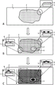
FIG. 1 is an overview of the present invention. (A) Support material 1 and tissue sample 2 are shown in top view and in side view in the small figure. (B) A separate mask has been applied on top of the tissue sample, generating predefined locations 7. The depicted separation screen combines a mesh separation screen 3 and a free-form separation screen 4. However, the mesh separation screen and the free-form separation screen 4 may also be used alone. Two types of separation masks with different cross-sectional structures are shown in the small figures providing side views: (a) top separation screen 5, (b) complete separation screen 6. (C) The oligonucleotide probes have been applied to predefined locations of the tissue sample in a liquid phase. The panels provide a side view: (a) top separation screen 5, (b) complete separation screen 6.
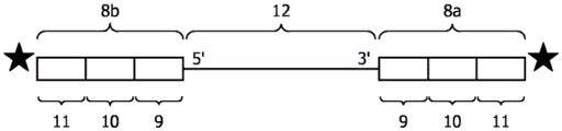
FIG. 2 (A) exemplary composition of oligonucleotide probes bound via ligation. Two oligonucleotide probes 8a and 8b comprising universal sequence 9, barcode sequences 10a and 10b, respectively, and additional sequence 11 bound to nucleic acid 12 are shown. (B) Standard oligonucleotide probe ligation protocols (adapted from http:// rnaseq. uoregon. edu/# library-prep-edition-of-adapters-via-ligation). The nucleic acid is fragmented RNA. In the first step, an oligonucleotide probe is ligated to the 3' -end of the RNA. In the second step, another oligonucleotide probe is ligated to the 5' -end of the RNA. In a third step, cDNA is generated via a reverse transcription reaction in which primers comprising sequences for next generation sequencing (NGS-adaptors) are hybridized to the primer alignment sequences of the oligonucleotide probes. In the fourth step, the cDNA is amplified by PCR.
Detailed description of the embodiments
As used in this specification and the appended claims, the singular forms "a", "an" and "the" include plural referents unless the context clearly dictates otherwise.
In the context of the present invention, the terms "about" and "approximately" denote intervals that the skilled person will understand to still ensure the accuracy of the technical effect of the feature in question. The term generally denotes a deviation of ± 20%, preferably ± 15%, more preferably ± 10%, and even more preferably ± 5% from the indicated value.
It is to be understood that the term "comprising" is not limiting. For the purposes of the present invention, the term "consisting of" is considered to be a preferred embodiment of the term "comprising". If a group is defined below as comprising at least a certain number of embodiments, this means that also groups preferably consisting of only these embodiments are covered.
Furthermore, the terms "first," "second," "third," or "(a)", "(b)", "(c)", "(d)" and the like in the description and in the claims are used for distinguishing between similar elements and not necessarily for describing a sequential or chronological order. It is to be understood that the terms so used are interchangeable under appropriate circumstances and that the embodiments of the invention described herein are capable of operation in other sequences than described or illustrated herein.
Where the terms "first", "second", "third" or "(a)", "(b)", "(c)", "(d)", "i", "ii", etc. relate to steps of a method or use or assay, there is no time or time interval consistency between said steps, i.e. these steps may be performed simultaneously, or there may be time intervals of seconds, minutes, hours, days, weeks, months or even years between such steps, unless otherwise indicated in the present application as described above or below.
It is to be understood that this invention is not limited to the particular methodologies, protocols, reagents, etc. described herein as these may vary. It is also to be understood that the terminology used herein is for the purpose of describing particular embodiments only, and is not intended to limit the scope of the present invention which will be limited only by the appended claims. Unless defined otherwise, all technical and scientific terms used herein have the same meaning as commonly understood by one of ordinary skill in the art.
The present invention relates to spatial mapping of nucleic acids in a sample.
The term "spatial mapping" as used herein relates to the partitioning of a targeted nucleic acid to its spatial location in a sample. The spatial map may provide transcriptional or genomic information from a plurality of cells in the tissue sample, wherein the information is characterized by two-dimensional spatial resolution. The spatial map may be created on an array basis, or may have unique spatial resolution, in that the pattern may be selected based on individual characteristics of the ROI and in response to image-based analysis of the tissue prior to application of the agent. The spatial distribution of the targeted nucleic acids can be correlated with an image of the ROI or with an image of the tissue sample in which the ROI was identified to provide visualization of a spatial map. The method according to the invention thus provides a personalized spatial map which can be designed on the basis of the questions to be answered, taking into account the organization information.
As used herein, the term "nucleic acid" refers to any nucleic acid molecule that can be detected by using the methods herein. Nucleic acid molecules include naturally occurring nucleic acids such as deoxyribonucleic acid (DNA) or ribonucleic acid (RNA) as well as artificially designed nucleic acids, e.g., nucleic acid analogs chemically synthesized or produced by means of recombinant genetic techniques (see, e.g., Sambrook, J. et al (1989)Molecular, Cloning: A Laboratory Manual2 nd edition, Cold Spring Harbor Laboratory Press, Cold Spring Harbor, NY). Specific examples of artificially designed nucleic acids include Peptide Nucleic Acids (PNA) or Locked Nucleic Acids (LNA). Specific examples of naturally occurring nucleic acids include DNA sequences such as genomic DNA or cDNA molecules and RNA sequences such as hnRNA, mRNA or rRNA molecules or the reverse complement thereof. The nucleic acid may be of any length and may be a single-stranded or double-stranded molecule. As used herein, the term "nucleotide" is understood to refer to ribonucleotides and deoxyribonucleotides (i.e., RNA and DNA molecules). In one embodiment, the nucleic acid is DNA. In another embodiment, the nucleic acid is RNA.
As used herein, the term "targeted nucleic acid" or "target nucleic acid" refers to one or more nucleic acids to be mapped. In one embodiment, the target nucleic acid is related to all nucleic acids in the sample, preferably to all DNA or all RNA of the sample. In another embodiment, the target nucleic acid is related to all nucleic acids of a particular type in the sample, e.g., mRNA, tRNA or rRNA. In another embodiment, the target nucleic acid is one or more specific target nucleic acids. The target nucleic acid may be any gene of the genome, preferably any gene of the human genome. For example, the target nucleic acid may be associated with a disease, e.g., a malignant disease such as cancer, inflammation, bacterial infection, or viral infection. Examples of such nucleic acids include nucleic acids specific for or produced by a pathogen or diseased cells or tissues, as well as nucleic acids produced in response to disease, such as nucleic acids encoding cytokines or antigens. The targeted nucleic acid can be of any size. In one embodiment, the targeted nucleic acid is DNA. In another embodiment, the targeted nucleic acid is RNA. In a preferred embodiment, the targeted nucleic acid is RNA, preferably all mRNA of the sample.
The target nucleic acid may be fragmented or intact. In one embodiment, the target nucleic acid is fragmented. Fragmented nucleic acids may be generated by any means known in the art, such as physical methods, e.g. sonication or sonication, chemical methods or enzymatic methods, e.g. with an endonuclease such as a restriction enzyme. Fragmentation can be performed before, during, or after identification of the ROI in the tissue sample. In one embodiment, the fragmentation is achieved in a step of fixing the tissue. For example, freezing of the sample or fixing the sample with formalin may result in at least partial fragmentation of the nucleic acids. Other fixatives may produce similar results. Fragmented nucleic acids can be up to about 20000 nucleotides in length. Typically, the fragmented nucleic acids are 10 to 10000 nucleotides in length, for example, 20 to 2000 nucleotides, 30 to 1000 nucleotides or 50 to 500 nucleotides. Fragmentation of nucleic acids does not result in all nucleic acids being fragmented. Thus, after fragmentation, the nucleic acid may be partially fragmented. In a preferred embodiment, the nucleic acid is at least partially fragmented. In another embodiment, the nucleic acid is intact. In one embodiment, the targeted nucleic acid is fragmented DNA or fragmented RNA. In another embodiment, the targeted nucleic acid is intact DNA or intact RNA. In a preferred embodiment, the targeted nucleic acid is an at least partially fragmented RNA, preferably all mRNA of the sample.
As used herein, the term "nucleic acid molecule" refers to one particular nucleic acid molecule that is present at a predefined location within a ROI. For example, in embodiments in which all mrnas within the ROI are targeted, the nucleic acid molecule represents one mRNA molecule.
As used herein, the term "sample" means any biological sample, or any artificial sample, that may be derived from any organism, such as a plant, an animal, a fungus, a human. In a preferred embodiment, the sample is a tissue sample. The "tissue sample" may be a harvested, cultured or biopsied tissue sample or a portion thereof. The tissue sample may be derived from diseased tissue, such as cancerous, inflamed, or infected tissue, tissue adjacent to diseased tissue, or healthy tissue. In a preferred embodiment, the tissue sample is derived from diseased tissue or tissue adjacent thereto.
The tissue sample may be a biological tissue sample or an artificial tissue sample. "biological tissue sample" refers to a collection of naturally occurring cells and includes clinical samples, cytological samples or cultured cells forming tissue from a biopsy or surgery. The biological tissue sample may be prepared by any conventional method of tissue sample preparation. In one embodiment, the biological tissue sample is a histopathological sample. In another embodiment, the biological tissue sample is a cytological sample. An "artificial tissue sample" is prepared from cells that are not naturally forming solid tissue (e.g., blood or suspension cell cultures). Artificial tissue samples can be prepared from cell suspensions obtained from clinical samples such as whole blood, lymph or CSF or cell suspensions obtained by in vitro methods. The cells can be entrapped in a matrix, such as a gel matrix, and sectioned by conventional methods as described, for example, in Andersson et al, 2006, J Histochem Cytochem 54(12): 1413-23. The artificial tissue sample may be a single cell layer or comprise multiple cell layers. In one embodiment, the artificial tissue sample is prepared from a cell suspension obtained from a clinical sample. In another embodiment, the artificial tissue sample is prepared from a cell suspension obtained by an in vitro method. In a preferred embodiment, the tissue sample is a biological tissue sample, preferably a clinical sample, even more preferably a histopathological or cytological sample.
The tissue sample may have a cell layer thickness of approximately 1 cell or less. In one embodiment, the thickness of the tissue sample will be less than 0.9, 0.8, 0.7, 0.6, 0.5, 0.4, 0.3, 0.2 or 0.1 of the cross-section of the cell. In another embodiment, the tissue sample may have a thickness greater than 1 cell. In one embodiment, the tissue sample section will have a thickness of at least about 0.1 μm, preferably at least about 0.2, 0.3, 0.4, 0.5, 0.7, 1.0, 1.5, 2, 3, 4, 5, 6, 7, 8, 9 or 10 μm. In other embodiments, the tissue sample section has a thickness of at least about 10, 11, 12, 13, 14, 15, 20, 30, 40, or 50 μm. Thicker samples may also be used if desired or convenient, for example, the tissue sample may have a thickness of about 70 or 100 μm or more. Typically, the tissue sample sections are between about 1-100 μm, 1-50 μm, 1-30 μm, 1-25 μm, 1-20 μm, 1-15 μm, 1-10 μm, 3-10 μm, 2-8 μm, 3-7 μm, or 4-6 μm thick. In a preferred embodiment, the tissue sample has a thickness in the range of 3-10 μm. The thickness of the tissue sample is not critical to the method of the invention, and these values are representative values only.
The tissue sample may have about 2 cm2The size of (2). In one embodiment, the tissue sample has a length of about 0.5, 0.6, 0.7, 0.8, 0.9, 1.0, 1.2, 1.5, 1.7, 2.0, 2.5, 3.0, 3.5, or 4.0cm or any number therebetween and a height of about 0.2, 0.3, 0.4, 0.5, 0.6, 0.7, 0.8, 0.9, 1.0, 1.2, 1.5, 1.7, or 2.0 cm or any number therebetween. The height and length of the sample are interchangeable. In a specific embodiment, the tissue sample has a maximum size of 2.0 x 4.0 cm. In another embodiment, the tissue sample has about 0.5, 0.6, 0.7, 0.8, 0.9, 1.0, 1.2, 1.5, 1.7, 2.0, 2.5, 3.0, 3.5, 4.0, 4.5, 5.0, 5.5, 6.0, 6.5, 7.0, 7.5, or 8.0 cm2Or any number of areas in between. In a preferred embodiment, the tissue sample has a maximum size of about 2.0 x 4.0cm, preferably the tissue sample has a range of about 1.0 to 5.0 cm2More preferably about 2.0 cm2。
The tissue sample may be fresh, frozen, fixed or unfixed. In one embodiment, the tissue sample is a fresh sample. In another embodiment, the tissue sample is a freshly frozen sample. In yet another embodiment, the tissue sample is a fixed tissue sample. Any procedure known in the art may be used to freeze, fix or embed the tissue sample. Specifically, any known fixative or embedding material may be used. In one embodiment, the tissue sample is a deparaffinized formalin-fixed and paraffin embedded (FFPE) sample. In a preferred embodiment, the tissue sample is an FFPE sample, a fresh frozen sample or a fresh sample or a cytological sample of a histopathological sample, preferably an FFPE sample of a histopathological sample.
In one aspect, the present invention relates to a method for spatially detecting nucleic acids in a tissue sample, comprising the steps of:
identifying at least one region of interest (ROI) within the sample;
applying at least one oligonucleotide probe to a predefined location within the ROI and allowing the oligonucleotide probe to bind to nucleic acids of the sample, wherein the oligonucleotide probe comprises a barcode sequence;
extracting the nucleic acid-oligonucleotide probe complex;
sequencing the extracted nucleic acid molecules;
correlating the sequenced nucleic acid molecules with the initial positions of the corresponding targeted nucleic acid molecules within the ROI to generate a spatial distribution of the targeted nucleic acid molecules, wherein each position is identified by the one or more oligonucleotide probes bound in step b).
As used herein, the term "method for spatial detection" relates to a method for detecting a target nucleic acid and identifying the initial position of said nucleic acid in a sample.
The "targeted nucleic acid molecule" corresponds to the nucleic acid of the sample bound by the oligonucleotide probe in step b).
A region of interest (ROI) may be identified in a tissue sample, taking into account tissue information that may be obtained according to any procedure known in the art. For example, the tissue sample may be imaged, stained or labeled with a suitable marker. In one embodiment, the ROI is identified by staining. In another embodiment, the ROI is identified by labeling with a suitable label. In yet another embodiment, the ROI is identified by imaging. In a preferred embodiment, the ROI is identified based on image analysis. Image analysis may be done automatically or manually. In case of automatic ROI selection, image analysis is required. All common methods of performing image analysis can be used.
The ROI as referred to herein may also comprise the surrounding regions of the identified ROI, i.e. the regions directly adjacent to the region identified as of interest. The ROI can be of any size that is the same size as the tissue sample (maximum size) and the grid cell size (minimum size). Preferably, the size of the ROI is smaller than the tissue sample.
In a tissue sample, at least one ROI can be identified. In tissue samples, more than one ROI may also be identified. For example, the tissue sample may comprise two, three, four, five, ten or more ROIs. Different ROIs can be identified in different ways, e.g., some ROIs are identified automatically by image analysis software, and pathologists can also specify the ROIs manually. In a preferred embodiment, at least one ROI is identified in the tissue sample, preferably two ROIs are identified in the tissue sample, more preferably more than two ROIs are identified in the tissue sample.
In a specific embodiment, the tissue sample may be cut into two layers. The indicia for the imaging step is applied on one layer. Based on the image of the reference layer, the ROI is selected and applied to the second layer (nucleic acid extraction slide). On the second layer, binding of oligonucleotide probes comprising barcode sequences is performed. This method has the advantage that imaging labels incompatible with the following steps of the protocol (e.g. binding of oligonucleotide probes, nucleic acid molecule extraction or sequencing) can be used on the reference slide and therefore do not interfere with the reactions carried out on the second layer of the tissue sample (nucleic acid extraction slide).
As used herein, the term "oligonucleotide probe" refers to a nucleic acid molecule comprising a barcode sequence. The oligonucleotide probe may be, for example, an RNA probe or a DNA probe. Oligonucleotide probes comprising the same barcode sequence are referred to as "species of oligonucleotide probes". Each oligonucleotide probe contains a unique barcode sequence. Barcode sequences may be designed or generated using random sequence generation. Designed or randomly generated sequences can be analyzed to ensure that the barcode sequence does not interfere with the capture of nucleic acids. The barcode sequence can be ligated to a nucleic acid in a sample. The barcode sequence does not act as a primer. The barcode sequence may have a size in the range of about 1-8 nucleotides.
The oligonucleotide probe may further comprise a "universal sequence". The universal sequences can be used to capture nucleic acids in a sample by hybridization to the nucleic acid to be targeted. In one embodiment, the universal sequence is used to selectively amplify a nucleic acid in a sample. In this embodiment, the universal sequence is referred to as a "primer sequence". The primer sequence is complementary to a nucleic acid in the sample to be targeted. For example, the primer sequence may comprise a poly-T sequence if the total mRNA of the sample is targeted, or a nucleotide complementary to a sequence segment of a particular gene if only the nucleic acid expressed by the gene is targeted. In an alternative embodiment, the universal sequence is used to ensure stable linkage of the barcode sequence to the nucleic acid in the sample, i.e. the universal sequence can be linked to the nucleic acid in the sample to be targeted. In this embodiment, the universal sequence is referred to as a "linker sequence". In one embodiment, the barcode sequence is directly linked to nucleic acids in the sample and does not require a universal sequence. In one embodiment, the oligonucleotide probe comprises at least one universal sequence. The universal sequence may have a size in the range of about 0 to 100 nucleotides, preferably 0 to 50 nucleotides, more preferably 0 to 30 nucleotides, even more preferably about 5 to 30 nucleotides.
The oligonucleotide probe may further comprise one or more "additional sequences". In one embodiment, additional sequences may be used to enrich for nucleic acids ("enriched sequences") or to purify nucleic acids ("purified sequences"). In another embodiment, additional sequences may be used to enrich for and purify nucleic acids. The additional sequence may also be a sequence used in a sequencing process, such as an adaptor for next generation sequencing or a sequence for selection or identification. In one embodiment, the additional sequence may be a primer aligned sequence ("primer aligned sequence"). In embodiments where the oligonucleotide probe comprises a linker sequence, the oligonucleotide probe may further comprise a primer alignment sequence. The primer sequence may be complementary to the primer alignment sequence.
The oligonucleotide probe may further comprise one or more spacer sequences, also referred to as spacers. The spacers may be arranged between different sequence elements of the oligonucleotide probe. The spacer may have a size in the range of about 0 to 20 nucleotides. The oligonucleotide probe may be between about 15 to 150 nucleotides in length. In a preferred embodiment, the oligonucleotide probe is about 15 to 100 nucleotides in length, preferably about 20 to 50 nucleotides in length.
The oligonucleotide probe may be in a dry form or in a liquid form. In a preferred embodiment, the oligonucleotide probe is in a liquid phase, preferably in a buffer solution or ink (ink). Ink refers to any solution suitable for printing. In embodiments where the oligonucleotide probe is in dry form, the oligonucleotide probe is solubilized prior to application.
In one embodiment, the oligonucleotide probe binds to all DNA molecules in the sample. In a preferred embodiment, the oligonucleotide probes bind all RNA molecules in the sample to provide a spatial map of the transcriptome. In another preferred embodiment, the oligonucleotide probe binds only to a specific RNA molecule, e.g. mRNA, rRNA, tRNA or other non-coding RNA, preferably the oligonucleotide probe specifically binds to mRNA.
The term "binding the oligonucleotide probe to the nucleic acid of the sample" refers to the generation of a nucleic acid-oligonucleotide probe complex. The binding of the oligonucleotide probe to the nucleic acid of the sample is achieved by different methods.
In one embodiment, the oligonucleotide probe is ligated to a targeted nucleic acid in the sample. The linking can occur via barcode sequences or universal sequences (linking sequences). Thus, in a preferred embodiment, the binding of the oligonucleotide probe to the nucleic acid of the sample occurs via ligation. The oligonucleotide probe can be ligated to the target nucleic acid by any method known in the art. For example, ligation to mRNA can be via a poly-a tail, while ligation to DNA can be performed after a digestion step that produces a sticky end that hybridizes to the oligonucleotide probe. In a specific embodiment, the oligonucleotide probe comprises (i) a linking sequence, (ii) a barcode sequence, and optionally (iii) one or more additional sequences. Different sequences may be distinguished by a gap. Furthermore, additional sequences may also be located between the linker sequence and the barcode sequence. If the target nucleic acid is to be amplified, the primer alignment sequences are present. Thus, in another specific embodiment, the oligonucleotide probe comprises (i) a linker sequence, (ii) a barcode sequence, and (iii) a primer alignment sequence. The oligonucleotide probe may comprise a spacer and/or additional sequences. The sequences are arranged within the oligonucleotide probe in such a way that the barcode is incorporated into the amplified DNA or cDNA, respectively, in an amplification or reverse transcription reaction. In another preferred embodiment, the oligonucleotide probe comprises a barcode sequence linked to a nucleic acid that may be followed by one or more universal sequences and/or additional sequences.
In an alternative embodiment, the oligonucleotide probe comprises a primer sequence. In this case, amplification and/or reverse transcription of nucleic acids is required. Thus, in a preferred embodiment, the binding of the oligonucleotide probe to the nucleic acids of the sample takes place via hybridization, wherein the oligonucleotide probe serves as a primer for amplification or reverse transcription. In embodiments in which the oligonucleotide probe serves as a primer, the oligonucleotide probe comprises (i) a primer sequence, (ii) a barcode sequence, and optionally (iii) one or more additional sequences, wherein the primer sequence serves as a primer for an amplification or reverse transcription reaction. The oligonucleotide probe may comprise a spacer and/or additional sequences. The sequences are arranged within the oligonucleotide probe in such a way that the barcode is incorporated into the amplified DNA or cDNA, respectively, in an amplification or reverse transcription reaction.
It will be appreciated that binding of the oligonucleotide probe to the nucleic acid in the sample is not limited to the embodiments described above and may occur via a variety of other means. For example, a universal primer alignment sequence can be attached to a targeted nucleic acid, and the oligonucleotide probe can be used as a primer in an amplification or reverse transcription reaction. Another example may be ligation to nucleic acids, e.g.to the sticky end of DNA after digestion, to which oligonucleotide probes are ligated in a second ligation reaction. The skilled person will be aware of different molecular techniques for capturing the nucleic acid to be targeted and different options for incorporating barcode sequences.
Binding of the oligonucleotide probes to nucleic acids in the sample includes binding of one or more oligonucleotide probes to nucleic acid molecules at predefined locations within the ROI. In one embodiment, one oligonucleotide probe is applied to each predefined location within the ROI. Thus, in a specific embodiment, an oligonucleotide probe binds a targeted nucleic acid molecule at a predefined location. This method is preferred for a few predefined locations or low resolution methods.
In another embodiment, at least two different kinds of oligonucleotide probes are applied to a predefined position within the ROI. Thus, in a specific embodiment, at least two different kinds of oligonucleotide probes bind a targeted nucleic acid molecule at a predefined position. This method allows high spatial resolution mapping of complex molecular profiles, effectively using multiple ROIs per sample. In a specific embodiment, at least one oligonucleotide probe that binds to a nucleic acid molecule is bound via its barcode sequence via ligation. In another embodiment, at least one oligonucleotide probe that binds to one nucleic acid molecule comprises a universal sequence, wherein optionally, the universal sequence is complementary to the targeted nucleic acid. For example, one oligonucleotide probe may comprise a linker sequence, while a second oligonucleotide probe may comprise a primer sequence for amplifying or reverse transcribing a nucleic acid. In another example, one oligonucleotide probe may not comprise a universal sequence, while a second oligonucleotide probe may comprise a universal sequence; thus, oligonucleotide probes without a universal sequence can bind to nucleic acid molecules via their barcode sequence via ligation, while oligonucleotide probes with a universal sequence can bind via their primers or ligation sequences via hybridization. In another embodiment, at least one oligonucleotide probe that binds to one nucleic acid molecule comprises additional sequences, wherein the additional sequences are purified sequences or primer aligned sequences. For example, one oligonucleotide probe may comprise a linker sequence and a purification sequence, while a second oligonucleotide probe may comprise a primer sequence. In another example, one oligonucleotide probe may not comprise a universal sequence but comprise additional sequences, and a second oligonucleotide probe may comprise only a universal sequence; thus, an oligonucleotide probe without a universal sequence can bind to a nucleic acid molecule via its barcode sequence via ligation and comprises a primer alignment sequence to which a forward primer hybridizes, and an oligonucleotide probe with a universal sequence can bind via hybridization, wherein the universal sequence serves as a reverse primer. It is to be understood that the combination of sequences within one oligonucleotide probe is not limited to the above-described embodiments. The skilled person will appreciate that many other combinations are possible for capturing nucleic acids to be targeted and labeling them with corresponding barcodes. Thus, further combinations of sequences within one oligonucleotide probe are expressly contemplated when combined with at least one additional oligonucleotide probe suitable for efficiently labeling a targeted nucleic acid.
The "predefined location" at which the oligonucleotide probe is applied represents one or more locations within the ROI. Depending on the question of the researcher, the entire ROI may be divided into separate predefined locations, i.e. one predefined location next to another such that they share a boundary, or parts of the ROI may be selected, i.e. predefined locations within the ROI may represent islands separated by regions in the ROI on which no probes are applied. Preferably, the predefined location refers to more than one location within the ROI.
In one embodiment, the predefined locations within the ROI are arranged systematically, e.g., in rows and columns. In one embodiment, the ROI comprises at least 1, 2, 5, 10, 50, 100, 500, 750, 1000, 1500, 3000, 5000, 10000, 20000, 40000, 50000, 75000, 100000, 150000, 200000, 300000, 400000, 500000, 750000, 800000, 1000000 or more predefined locations. In a preferred embodiment, the ROI comprises at least 1, 2, 5, 10, 20, 30, 40, 50 or 100 predefined positions, preferably the ROI comprises at least 10 predefined positions. In another embodiment, the area of each predefined location may be about 1 μm2、2 μm2、3 μm2、4 μm2、5 μm2、10 μm2、12 μm2、15 μm2、20 μm2、50 μm2、75 μm2、100 μm2、150 μm2、200 μm2、250 μm2、300 μm2、400 μm2Or 500 μm2. A unique species of oligonucleotide probe or a unique combination of two or more different species of oligonucleotide probes is applied to each predefined location. If two or more kinds of oligonucleotide probes are applied, it is preferable that different kinds of oligonucleotide probes are applied to each row and different kinds of oligonucleotide probes are applied to each column. For example, row 1 contains an oligonucleotide probe that is a different species than the oligonucleotide probe in row 2, which in turn is a different species than the oligonucleotide probe in row 3, and so on. Column a contains a different species of oligonucleotide probe than any oligonucleotide probe applied in each row, column B again contains a different species of oligonucleotide probe, and so on. Predefined positions in column a, row 1 (a1) thus have a unique combination of oligonucleotide probes that is different from the combination of a2, B1, or B2, etc. With this method, fewer kinds of oligonucleotide probes need to be used, reducing the cost of the assay. Additional kinds of oligonucleotide probes may be added, e.g.on diagonals or to freely defined predefined positions, such as sub-ROIs, with different kindsOligonucleotide probes of the class. In a preferred embodiment, a unique combination of at least two oligonucleotide probes, preferably a unique combination of two oligonucleotide probes, is applied to each predefined location.
In an alternative embodiment, the predefined position within the ROI is freely defined. The free arrangement of the predefined positions has the following advantages: other target sub-regions (sub-ROIs) within the ROI may be defined without any limitation. Thus, the free arrangement allows for predefined locations having any shape and size and different predefined locations having different shapes and sizes, as compared to the conventional arrangement of an array of columns and rows having regular features of the same size. For example, in a tissue sample, ROIs can be identified based on the presence of a particular cell type. Within the ROI, further sub-ROIs may be identified based on morphological features. These sub-ROIs can form unique islands of different shapes and sizes within the ROI. Predefined locations within the ROI may be defined such that each sub-ROI represents a separate predefined location. In this case, the remaining ROI minus the area covered by the sub-ROI may not be of interest and no oligonucleotide probe is applied to this area, or it may represent another predefined location, or it may be further divided into predefined locations by a systematic arrangement of rows and columns. A unique species of oligonucleotide probe or a unique combination of two or more oligonucleotide probes may be applied to each predefined location. The sub-ROIs representing the individual predefined locations may be different from each other or may partially overlap. In the case where the sub-ROIs partially overlap, the overlapping regions can be identified by a unique combination of at least one oligonucleotide probe specific for the first sub-ROI and at least one oligonucleotide probe specific for the second sub-ROI. In one embodiment, at least two sub-ROIs partially overlap. In another embodiment, more than two sub-ROIs partially overlap. In yet another embodiment, the sub-ROIs do not overlap. Any information may be used to identify the sub-ROI, and the identification of the sub-ROI is not limited to the information obtained by identifying the ROI. Features that may be used to identify sub-ROIs include, but are not limited to, cell type, morphology, color, transparency, presence/absence of specific molecules (e.g., antibodies, cytokines, or drugs), or application of radiation to specific regions of a tissue sample prior to taking the tissue sample from a patient.
In a further embodiment, the predefined location is defined by applying a mask. The screen may be a "separation screen". The separation mask may have any pattern to mark the ROI. In one embodiment, the separation mask is a "grid separation mask" that provides rows and columns for systematic location characterization of the ROI (fig. 1). In another embodiment, the separation mask may be a "free-format separation mask" (fig. 1) based on the shape of the ROI and sub-ROI. In another embodiment, the separation screen may be a combination of a grid separation screen and a free-form separation screen as exemplarily shown in fig. 1.
The separation screen may be applied in different ways. In one embodiment, the separation mask defines the predefined area by providing a full trench applied through the entire thickness of the tissue sample ("full separation mask"), preferably by scraping, laser ablation or by creating a physical barrier such as a metal grid. In another embodiment, the separation screen defines the predefined area by providing a top barrier ("top separation screen"). The top separation screen may define the predefined location by a physical barrier. The top separation screen may also define the predefined area by a structuring process ("location screen") that confines the agent in the desired area, preferably by topological constraints or by locally patterning the wettability of the tissue. Photolithographic techniques may be used to structure such layers. In a preferred embodiment, the surface of the ROI is structured with a self-assembled monolayer (SAM) on a thin gold layer evaporated on the surface of the ROI. The SAM layer may be applied by microcontact printing of an organic thiol used as a resist in the etching of the gold layer. The SAM is hydrophobic and prevents the diffusion of oligonucleotide probes during inkjet printing. A top separation mask may also be printed on top of the ROI with the ink acting as a physical barrier. Fig. 1 shows exemplarily a top partition screen 5 and a full partition screen 6 in a side view. In one embodiment, the predefined locations are defined by separate masks. In a preferred embodiment, the separation mask is a top separation mask or a complete separation mask optionally printed on the sample, wherein the separation mask is a lattice separation mask, a free-form separation mask, or a combination thereof. In a more preferred embodiment, the separating screen is a top separating screen applied by printing. The wax may be ink-jet printed using a heated ink-jet print head, or may be ink-jet printed using a UV curable ink. Both of which are well known in the graphics industry.
The top screen may also be applied as a prefabricated integral part (like a microtiter plate with an open base). The microtiter plate may be a 384-well plate or 1536-well plate, or any other number when manufactured for this purpose. The well plate may be clamped to the tissue sample to ensure good physical contact between the plastic wall of the well plate and the tissue sample. Oligonucleotide probes can then be added and dissolved in buffer in each well that overlaps with the predetermined ROI. Each well can receive a different ink, i.e., a different oligonucleotide probe. Multiple depositions may provide all of the reagents. Ink jet printing or micro dosing may be used to provide reagents to the wells. The height of the walls of this top screen is about 0.5 to 5 mm. This means that the dispense volume in each well can be about 100nL to 10. mu.L, more preferably 0.5 to 2. mu.L.
In another embodiment, no separation mask (which is a physical, chemical or mechanical mask) is used and the predetermined locations are defined by locally applying oligonucleotide probes such that they do not confound between different predefined locations on the sample. In one embodiment, the oligonucleotide probes are applied using a printing ink that does not readily diffuse or in which the volume remains small enough to maintain the desired spatial resolution, wherein the printed pattern defines the predefined locations. In this embodiment, the application of the oligonucleotide probe and the definition of the predefined position occur simultaneously. The ink may be gelled after application to ensure immobilization at the desired location, while the oligonucleotide probes are still able to diffuse into the ROI of the tissue sample. The prior art liquid transfer technique can be used to apply oligonucleotide probes in a specific pattern on the ROI. In one embodiment, the oligonucleotide probes are applied to the predefined locations by a liquid transfer technique, preferably by a contact printing technique or a non-contact printing technique, such that the applied oligonucleotide probes do not hybridize between different predefined locations. In a preferred embodiment, the liquid transfer technique is a contact printing technique, preferably dip-pen drawing (dip-pen plotting) or micro-contact printing. In another preferred embodiment, the liquid transfer technique is a non-contact printing technique, preferably ink jet. In one embodiment, the oligonucleotide probes are printed as horizontal lines or spots positioned in horizontal lines (representative rows). The oligonucleotide probes are then printed in a vertical line using a vertical movement, or placed on top of an already existing spot (representative of a column). A multi-nozzle printer may be used which can print different printing inks simultaneously. Thermal ink jet or piezo driven ink jet heads may be used. Piezo-driven printers are preferred to avoid possible degradation of proteins in the printing ink. The liquid transfer technique selected may be adjusted based on the desired resolution. In another embodiment, the oligonucleotide probes are printed according to the shape of the identified sub-ROI.
The printing ink contains oligonucleotide probes dissolved in a solvent (such as water or saline buffer, optionally containing surfactants and stabilizers). The concentration of the oligonucleotide probe in the ink may range between 1nM and 1mM, preferably between 100nM and 10. mu.M. Different oligonucleotides may be dissolved in the same ink. It is also preferred to dissolve the enzyme in the same ink. For enzyme stability, BSA or other serum may be added at concentrations typical for stable storage (e.g., between 0.5 and 5% w/w).
A print volume is selected that is typical for the deposition technique used and depends on the spatial resolution required. For inkjet printing, the print volume may be in the range between 50pL and 5 μ L, more preferably between 100pL and 1 μ L. For dip-pen drawing, the volume transferred depends on the pen size and dwell time. Typical transfer volumes are in the range between 1pL and 1. mu.L, preferably between 10pL and 100 nL.
The application of oligonucleotide probes as described for the embodiments in which no separation mask is used may also be used for embodiments in which the predefined position is defined by a separation mask. For example, in a first step, a separation mask defining predefined locations may be printed, and in a second step, oligonucleotide probes may be applied by printing onto the predefined locations. Thus, in one embodiment, the oligonucleotide probes are applied by printing. In another embodiment, the oligonucleotide probes are applied by a method other than printing, such as pipetting.
Oligonucleotide probes can also be prepared in a gel-like layer or gel layer, wherein the layer is applied to predefined locations within the ROI. The gel layer can be applied in a single step over the ROI and the oligonucleotide probes transferred to the ROI by diffusion. As used herein, the term "gel-like" or "gel" refers to a solid jelly-like material that may have properties ranging from soft and breakable (weak) to hard and tough (tough), wherein the oligonucleotide probes are capable of diffusing from the gel into the ROI. Thus, in one embodiment, the oligonucleotide probe is in the form of a gel or gel-like layer applied to the sample.
The oligonucleotide probes may also be applied to a separate disposable (e.g., having a small aperture, such as a well plate) that will later be attached to the ROI. The method has the following advantages: it can be produced off-line, including associated quality control measures, to check if all the liquid has been stored. Off-line deposition of oligonucleotide probes in separate disposable items makes the use of liquid transfer equipment in pathology laboratories obsolete. In one embodiment, the ROI may be pressed into a well plate as described in Armani et al, 2009, Lab Chip 9(24): 3526-. An oligonucleotide probe or a unique combination of oligonucleotide probes can be added to the bottom of each well by spotting. The oligonucleotide probes may be dried prior to use.
Oligonucleotide probes may also be applied by combinations of the above and/or combinations without and with a separation mask. For example, in a first step, oligonucleotide probes may be printed onto different sub-ROIs, thereby defining predefined locations without the use of a separation mask, and in a second step, oligonucleotide probes may be applied by pipetting onto the predefined locations defined by the top separation mask. Thus, the oligonucleotide probes may be used sequentially or simultaneously. It will be appreciated that many further combinations are possible and that the use of oligonucleotide probes is not limited to the embodiments described above.
In embodiments in which more than two oligonucleotide probes are applied to predefined locations within the ROI, the application may be sequential. For example, oligonucleotide probes to be ligated to nucleic acid molecules may be used in the first step to provide conditions under which the probes are ligated to the nucleic acid molecules, and in the second step oligonucleotide probes that hybridize to and/or oligonucleotide probes that are ligated to nucleic acid molecules may be used. Thus, in one embodiment, different kinds of oligonucleotide probes are applied sequentially to predefined locations within the ROI. In another embodiment, different kinds of oligonucleotide probes are applied simultaneously to predefined locations within the ROI.
In a preferred embodiment, the oligonucleotide probes are applied in solution to predefined locations within the ROI. In a further preferred embodiment, the oligonucleotide probes are applied in a liquid phase to predefined locations within the ROI, wherein the predefined locations are identified taking into account tissue information. In a further preferred embodiment, the oligonucleotide probes are applied in a liquid phase with high ionic strength to achieve hybridization with the target molecule. In another preferred embodiment, the oligonucleotide probes are applied in a liquid phase that gels immediately after application to the sample by an applied temperature step or radiation. This can be achieved by adding a polymer (such as polyacrylamide which undergoes thermoreversible gelation in solution) and/or adding a monomer which can polymerize under irradiation and in this way lead to gelation. Gelation should avoid possible hybridization of the probes at different predefined positions.
The nucleic acid molecules in the ROI can be pre-processed. In one embodiment, the nucleic acids of the sample are pre-treated to target the correct type of nucleic acid molecule and/or for efficient ligation of oligonucleotide probes. In embodiments where the nucleic acid to be targeted is a DNA molecule, pretreatment may encompass digestion of the DNA according to methods well known in the art to provide smaller sized DNA fragments.
After applying the oligonucleotide probes to predefined locations within the ROI, the oligonucleotide probes diffuse into the ROI. The size of the oligonucleotide probe allows deep penetration of the tissue sample in a short time. The oligonucleotide probes within their predefined locations of the ROI then bind to the nucleic acids of the sample, thereby generating nucleic acid-oligonucleotide probe complexes. As used herein, the term "nucleic acid-oligonucleotide probe complex" refers to a complex of a nucleic acid to be detected and at least one oligonucleotide probe. The nucleic acid to be detected may be complexed with more than one oligonucleotide probe, for example with two oligonucleotide probes. During the diffusion and bonding process, a humid atmosphere is provided at the surface to avoid evaporation of the solvent. A temperature step may be applied during this period to achieve good conversion of the reaction. The separation mask may remain in place during this step, or optionally be removed after application of the probe. The elevated temperature accelerates the diffusion and binding reactions. In case hybridization is used for binding, the temperature should be kept below the melting temperature of the probe. Alternatively, the temperature should be chosen such that the ligase performs well. In a preferred embodiment, the temperature is in the range of between about 20 to 45 ℃, preferably between about 30 to 40 ℃.
After binding of the oligonucleotide probe to the nucleic acid of the sample, the nucleic acid-oligonucleotide probe complex is extracted from the ROI. In one embodiment, the extraction is performed by immersing the ROI in an extraction buffer at an elevated temperature. Extraction buffers commonly used in sample preparation for molecular assays and known in the art may be used. The extracted nucleic acids can be collected in a single tube for further analysis. The solution of extracted nucleic acid-oligonucleotide probe complexes is referred to as a Spatial Molecular Profile (SMP) extract.
In one embodiment, the extracted nucleic acids are purified by methods known in the art. In another embodiment, the extracted nucleic acids are enriched by methods known in the art. In a further embodiment, the extracted nucleic acids are purified and enriched.
In yet a further embodiment, the DNA molecule is generated from the extracted nucleic acid-oligonucleotide probe complex via DNA amplification, optionally wherein the generation of the DNA molecule occurs after a reverse transcription reaction. In a preferred embodiment, the extracted nucleic acids are reverse transcribed and amplified RNA molecules.
Nucleic acid amplification can be achieved by means of cyclic amplification. The cycling amplification may comprise any number of amplification cycles equal to or greater than 2. Typically, the cycling amplification reaction comprises at least 10 or at least 20 cycles. An exemplary cyclic amplification is Polymerase Chain Reaction (PCR). PCR is a standard method established in molecular biology and is described in detail, for example, in Sambrook et al, supra. Typically, PCR is used to amplify double-stranded DNA molecules by using thermostable DNA polymerases. In a preferred embodiment, the nucleic acid amplification is a cyclic amplification, preferably a PCR.
In an alternative embodiment, nucleic acid amplification is achieved by an isothermal amplification method. Amplifying nucleic acids under isothermal conditions makes it possible to avoid the use of thermocycling devices. There are several types of isothermal nucleic acid amplification methods known to the skilled artisan, including nicking enzyme amplification reactions, transcription mediated amplification, nucleic acid sequence based amplification, signal mediated amplification of RNA technology, strand displacement amplification, rolling circle amplification, loop mediated isothermal amplification, isothermal multiple displacement amplification, helicase dependent amplification, single primer isothermal amplification, and circular helicase dependent amplification. Isothermal Nucleic acid amplification techniques are reviewed, for example, in P. Gill and A. Ghaemi, 2008, Nucleotides Acids 27(3): 224-243.
If the nucleic acids are RNA, the production of DNA molecules also includes subjecting the nucleic acids to reverse transcription prior to subjecting them to amplification. Reverse transcription is another standard method in molecular biology and is also described, for example, in Sambrook et al, supra.
After extracting the nucleic acid-oligonucleotide probe complexes and optionally performing any of the methods described above, the SMP extract is sequenced. The term "sequencing the extracted nucleic acid molecule" as used herein should be understood broadly, i.e. to refer to any method of providing sequence information. For example, sequence information can be obtained by hybridization to specific probes or microarrays. In one embodiment, sequencing the extracted nucleic acid molecule refers to sequencing of a DNA molecule, optionally a DNA molecule obtained from RNA by reverse transcription. Methods suitable for sequencing DNA molecules are known in the art and include, but are not limited to, Sanger sequencing, pyrosequencing or Next Generation Sequencing (NGS). In another embodiment, sequencing the extracted nucleic acid molecule refers to sequencing the RNA molecule by nanopore sequencing, microrna sequencing, or whole transcriptome shotgun sequencing. In a preferred embodiment, the sequencing is DNA sequencing, preferably NGS.
In the case where sequence information is obtained by NGS, in embodiments where the oligonucleotide probe serves as a primer in the NGS, the oligonucleotide probe comprises an NGS adaptor as an additional sequence.
After sequence information of the sequenced nucleic acid molecules has been obtained, the sequenced nucleic acid molecules are correlated with the initial positions of the corresponding targeted nucleic acid molecules within the ROI to generate a spatial distribution of the targeted nucleic acid molecules. Thus, a spatial two-dimensional map is generated in which the sequenced nucleic acids are displayed according to a predetermined position, for example defined by the separate masks from which they originate. Thus, in one embodiment, the sequenced nucleic acid molecules are correlated with the initial positions of the corresponding targeted nucleic acid molecules within the predefined locations within the ROI to generate a spatial distribution of the targeted nucleic acid molecules, wherein each location is identified by one or more oligonucleotide probes that bind to the nucleic acid of the sample in the predefined location within the ROI. In another embodiment, a unique combination of at least two different species of oligonucleotide probes is used to identify the location of a targeted nucleic acid molecule within a ROI. The extracted and sequenced nucleic acid molecules comprise barcode sequences of oligonucleotide probes that bind to target nucleic acids of the sample in predefined locations within the ROI. The extracted and sequenced nucleic acid molecules encompass barcode sequences of oligonucleotide probes as a unique species of oligonucleotide probes or a unique combination of oligonucleotide probes that bind to a target nucleic acid of a sample are applied to each predefined location within the ROI. Each nucleic acid molecule can be associated with a predefined location of the species to which the oligonucleotide probe comprising the barcode sequence is applied, based on the barcode sequence it comprises. The resulting correlation provides a spatial map showing the initial position of the targeted nucleic acid within the ROI. This spatial map may, for example, provide information about the expression state, mutation state, degradation state, methylation state, epigenetic state, or a combination thereof, of the nucleic acid.
In one embodiment, the spatial distribution of the targeted nucleic acid molecules can be correlated with an image of the ROI or with an image of a tissue sample in which the ROI was identified. The spatial distribution of the targeted nucleic acid molecules can be visualized, which provides a two-dimensional spatial map. In one embodiment, the spatial distribution of the targeted nucleic acid molecules is correlated with the spatial pattern of the image obtained from the ROI. Thus, in one embodiment, the method provides a two-dimensional spatial map to visualize the spatial distribution of the targeted nucleic acid molecules. In a further embodiment, the method further comprises overlaying the two-dimensional spatial map with an image of the ROI or an image of the tissue sample in which the ROI was identified. Images of the ROI may be obtained before or after the ROI is identified. Thus, in one embodiment, an image is obtained from the tissue sample prior to identifying the at least one ROI. In another embodiment, the image of the tissue is obtained after identifying the at least one ROI. In yet another embodiment, an image is obtained from the ROI before or after applying the oligonucleotide probe to a predefined location within the ROI. The images may be obtained by any method known in the art, such as digital pathology or microscopy with a camera. In one embodiment, the spatial distribution of the targeted nucleic acid molecules is associated with predefined locations located within the ROI and defined by the separation mask, wherein the image is obtained after application of the separation mask and before or after application of the oligonucleotide probe onto the predefined locations within the ROI, preferably before application of the oligonucleotide probe. In a preferred embodiment, the spatial distribution of the targeted nucleic acid molecules is correlated with an image of the ROI obtained before or after the oligonucleotide probe is applied to a predefined location within the ROI, preferably before the oligonucleotide probe is applied. In another preferred embodiment, the spatial distribution of the targeted nucleic acid molecules is correlated with the image of the ROI obtained after application of the separation mask and before application of the oligonucleotide probes to the predefined locations defined by the separation mask.
The spatial map may be visualized, for example, by revealing false colors, wherein each predefined position is defined by a grid representing regions of the same color. The spatial map may be superimposed with an image of the ROI or an image of the tissue sample. For example, the spatial map is configured as a semi-transparent image and is superimposed on the image of the ROI or with the image of the tissue sample. Or vice versa, the image of the ROI or the image of the tissue sample is configured as a semi-transparent image and superimposed on the spatial map. In a preferred embodiment, the number or amount of the same targeted nucleic acid molecule determined in an assay with the same kind of oligonucleotide probe is correlated to the color of the map in the respective predefined position. In another embodiment, specific combinations of targeted nucleic acid molecules, such as, for example, a subset of those carrying known oncogenic mutations, are associated with color. In yet another embodiment, overexpression of a particular set of targeted nucleic acid molecules associated with a particular gene can be correlated with color on the map. It should be understood that many different profiles may be defined and associated with the manner of visual display depending on the biological and/or clinical problem being addressed. Other annotations may be displayed in the spatial map instead of or in combination with colors.
The correlation of the spatial distribution of the targeted nucleic acid molecules to the spatial pattern (i.e., spatial map) of the initial image can be used to answer clinical questions and provide guidance for clinical decisions. The results obtained by the spatial map created by the method of the invention may be used for clinical diagnosis or to monitor clinical responses in patients. The correlation of the spatial distribution of the targeted nucleic acid molecules with the spatial pattern of the initial image can also be used in the research field.
In a preferred embodiment, the tissue sample and/or the ROI are imaged. The captured image of the tissue sample can be used to identify the target area and spatial resolution of the pattern based on image-based analysis of the tissue prior to application of the agent.
Another aspect of the present invention relates to a reaction of ligating a 5 '-oligonucleotide probe and a 3' -oligonucleotide probe to the 5 '-end and the 3' -end of an RNA molecule, and simultaneously applying a DNA molecule introducing a restriction site at the ligation site of an oligonucleotide probe dimer and a restriction enzyme suitable for cleaving the restriction site, wherein the 5 '-end and the 3' -end of the DNA molecule are modified so that they do not participate in the ligation reaction. Thus, the dimers that exist between the oligonucleotide probes when they react with each other (i.e., oligonucleotide probe dimers) are cleaved into fully active individual 5 '-and 3' -oligonucleotide probes. The cleaved oligonucleotide probes can be reused in ligation reactions to ligate to the 5 '-end and the 3' -end of an RNA molecule.
One specific embodiment relates to a ligation reaction on an FFPE tissue sample that ligates 5 '-oligonucleotide probes and 3' -oligonucleotide probes to the 5 '-and 3' -ends of RNA molecules and simultaneously applies DNA molecules that introduce restriction sites on the ligation sites of oligonucleotide probe dimers and restriction enzymes suitable for cleaving the restriction sites, wherein the 5 '-and 3' -ends of the DNA molecules are modified such that they do not participate in the ligation reaction.
In a standard oligonucleotide probe ligation reaction protocol as shown in FIG. 2B, the ligation reaction is quenched by reverse transcription reaction, thereby avoiding the formation of oligonucleotide probe dimers. Reverse transcription reactions are rather inefficient on FFPE tissue samples. In the oligonucleotide probe ligation protocol of the present invention, oligonucleotide probe dimers are cleaved by restriction enzymes, thus avoiding a reverse transcription reaction step on FFPE tissue samples. Furthermore, using the protocol of the present invention, the T4 RNA ligase I and II reactions can be combined in one step, as described in the examples and in figure 3. Therefore, the oligonucleotide probe ligation reaction method of the present invention is superior to the prior art.
In one embodiment, the invention relates to a ligation reaction that ligates 5 '-and 3' -oligonucleotide probes to the 5 '-and 3' -ends of an RNA molecule and simultaneously applies a DNA molecule that introduces a HaeIII restriction site at the ligation site of the oligonucleotide probe dimer and a HaeIII restriction enzyme that cleaves at the restriction site.
The above-described ligation reaction may be used in step b) of a method for spatial detection of RNA molecules in a tissue sample as described herein.
In particular, the above-described ligation reaction may be used in step b) of a method for spatial detection of RNA molecules in FFPE tissue samples as described herein. This avoids the need for reverse transcription reactions to be performed on FFPE tissue samples. This method is advantageous because it avoids reverse transcription reactions that are quite inefficient for FFPE samples. Furthermore, using this protocol, the T4 RNA ligase I and II reactions can be combined in one step.
In one embodiment, a method for spatially detecting nucleic acids in a tissue sample comprises the steps of:
a1) acquiring an image of a tissue sample;
a2) identifying at least one ROI within the sample;
a3) applying a mask to the ROI comprising different predefined locations;
b) applying at least one oligonucleotide probe to a predefined location within the ROI and allowing the oligonucleotide probe to bind to nucleic acids of the sample, wherein the oligonucleotide probe comprises a barcode sequence;
c) extracting the nucleic acid-oligonucleotide probe complex;
d) sequencing the extracted nucleic acid molecules;
e) correlating the sequenced nucleic acid molecules with the initial positions of the corresponding target nucleic acid molecules within the ROI to generate a spatial distribution of the targeted nucleic acid molecules; wherein each position is identified by one or more oligonucleotide probes bound in step b).
In another specific embodiment, a method of spatially detecting nucleic acids in an FFPE tissue sample comprises the steps of:
a1) deparaffinizing the FFPE tissue sample;
a2) acquiring an image of a tissue sample;
a3) identifying at least one ROI within the sample;
a4) applying a mask to the ROI comprising different predefined locations;
b) applying at least one oligonucleotide probe to a predefined location within the ROI and allowing the oligonucleotide probe to bind to nucleic acids of the sample, wherein the oligonucleotide probe comprises a barcode sequence;
c) extracting the nucleic acid-oligonucleotide probe complex;
d) sequencing the extracted nucleic acid molecules;
e) correlating the sequenced nucleic acid molecules with the initial positions of the corresponding target nucleic acid molecules within the ROI to generate a spatial distribution of the targeted nucleic acid molecules; wherein each position is identified by one or more oligonucleotide probes bound in step b).
In a specific embodiment, the method for spatially detecting RNA in a tissue sample comprises the steps of:
a1) acquiring an image of a tissue sample;
a2) identifying at least one ROI within the sample;
a3) applying a mask to the ROI comprising different predefined locations;
b) applying at least one oligonucleotide probe to a predefined location within the ROI and allowing the oligonucleotide probe to bind to an RNA molecule of the sample, wherein the oligonucleotide probe comprises a barcode sequence;
c) extracting the RNA-oligonucleotide probe complex;
d) sequencing the extracted RNA molecules;
e) correlating the sequenced RNA molecules with the initial positions of the corresponding target RNA molecules within the ROI to generate a spatial distribution of the targeted RNA molecules; wherein each position is identified by one or more oligonucleotide probes bound in step b).
In a specific embodiment, the method for spatially detecting RNA molecules in an FFPE tissue sample comprises the steps of:
a1) deparaffinizing the FFPE tissue sample;
a2) acquiring an image of a tissue sample;
a3) identifying at least one ROI within the sample;
a4) applying a mask to the ROI comprising different predefined locations;
b) applying at least one oligonucleotide probe to a predefined location within the ROI and allowing the oligonucleotide probe to bind to an RNA molecule of the sample, wherein the oligonucleotide probe comprises a barcode sequence;
c) extracting the RNA-oligonucleotide probe complex;
d) sequencing the extracted RNA molecules;
e) correlating the sequenced RNA molecules with the initial positions of the corresponding target RNA molecules within the ROI to generate a spatial distribution of the targeted RNA molecules; wherein each position is identified by one or more oligonucleotides bound in step b).
In a specific embodiment, the method for spatially detecting RNA in an FFPE tissue sample comprises the steps of:
a1) deparaffinizing the FFPE tissue sample;
a2) acquiring an image of a tissue sample;
a3) identifying at least one ROI within the sample;
a4) applying a mask to the ROI comprising different predefined locations;
b) applying 5'-RNA probes and 3' -RNA probes at predefined positions within the ROI and allowing the RNA probes to ligate to the 5 '-end and the 3' -end of the RNA molecules of the sample, wherein the RNA probes comprise barcode sequences;
c) extracting RNA-RNA probe compound;
d) sequencing the extracted RNA molecules;
e) correlating the sequenced RNA molecules with the initial positions of the corresponding target RNA molecules within the ROI to generate a spatial distribution of the targeted RNA molecules; wherein each position is identified by one or more RNA probes bound in step b).
In a specific embodiment, the method for spatially detecting RNA in an FFPE tissue sample comprises the steps of:
a1) deparaffinizing the FFPE tissue sample;
a2) acquiring an image of a tissue sample;
a3) identifying at least one ROI within the sample;
a4) applying a mask to the ROI comprising different predefined locations;
b) applying 5'-RNA probes and 3' -RNA probes at predefined positions within the ROI and allowing the RNA probes to be ligated to the 5 '-end and 3' -end of the RNA molecules of the sample, wherein the RNA probes comprise barcode sequences and simultaneously apply DNA molecules introducing restriction sites at the ligation sites of RNA probe dimers and restriction enzymes suitable for cleaving the restriction sites;
c) extracting RNA-RNA probe compound;
d) sequencing the extracted RNA molecules;
e) correlating the sequenced RNA molecules with the initial positions of the corresponding target RNA molecules within the ROI to generate a spatial distribution of the targeted RNA molecules; wherein each position is identified by one or more RNA probes bound in step b).
In a specific embodiment, the method for spatially detecting RNA in an FFPE tissue sample comprises the steps of:
a1) deparaffinizing the FFPE tissue sample;
a2) acquiring an image of a tissue sample;
a3) identifying at least one ROI within the sample;
a4) applying a mask to the ROI comprising different predefined locations;
b) applying 5'-RNA probes and 3' -RNA probes at predefined positions within the ROI and allowing the RNA probes to ligate to the 5 '-end and the 3' -end of the RNA molecules of the sample, wherein the RNA probes comprise a barcode sequence and simultaneously apply a DNA molecule introducing a HaeIII restriction site at the ligation site of the RNA probe dimer and a HaeII restriction enzyme,
c) extracting RNA-RNA probe compound;
d) sequencing the extracted RNA molecules;
e) correlating the sequenced RNA molecules with the initial positions of the corresponding target RNA molecules within the ROI to generate a spatial distribution of the targeted RNA molecules; wherein each position is identified by one or more RNA probes bound in step b).
In one embodiment, a method for spatially detecting DNA in a tissue sample comprises the steps of:
a1) acquiring an image of a tissue sample;
a2) identifying at least one ROI within the sample;
a3) applying a mask to the ROI comprising different predefined locations;
b) applying at least one oligonucleotide probe to a predefined location within the ROI and allowing the oligonucleotide probe to bind to a DNA molecule of the sample, wherein the oligonucleotide probe comprises a barcode sequence;
c) extracting the DNA-oligonucleotide probe complex;
d) sequencing the extracted DNA molecules;
e) correlating the sequenced DNA molecules with the initial positions of the corresponding target DNA molecules within the ROI to generate a spatial distribution of the targeted DNA molecules; wherein each position is identified by one or more oligonucleotide probes bound in step b).
Examples
Types of protocols for binding oligonucleotide probes to nucleic acid molecules

Method for spatial detection of RNA in FFPE tissue samples
(according to scheme type [ C ]]Examples of (2)
Materials: FFPE tissue samples on slides
With proteinase K (ProtK) at 37 ℃ as described, for example, by Tullis and Rubin (Analytical Biochemistry, 1980, 107(1):260-Engyodontium album) Proteinase K (Sigma-Aldrich) followed by heat inactivation of ProtK at 80 ℃ for 15 minutes.
Tissue samples are deparaffinized, for example, using a deparaffinization solution (e.g., Qiagen) according to the manufacturer's instructions.
An image of the complete slide is taken (Philips Digital Pathology Scanner UFS) and the ROI is identified within the image.
Different positions within the ROI were selected and a top selection mask was applied by ink jet printing (print head XAAR) UV curable ink (Sunjet ink Crystal UFE 7573 (black)) to create a grid separating the different reaction wells. UV curing of the mask is carried out with UV-LEDs at 360 to 380 nm for 30 to 60 s.
A unique combination of two oligonucleotide probes (5 '-RNA probe and 3' -RNA probe) containing barcode sequences was applied to each predefined location by inkjet printing (using a FUJIFILM Dimatix print head). Use against Illumina®NEBNext of (NEB)®A modification of the small RNA library preparation set, the 5'-RNA probe and the 3' -RNA probe are ligated to the mRNA molecules in a single step. In the standard protocol depicted in fig. 2B), the RNA probe is ligated to the RNA molecule in a single step. During this reaction, the 5'-RNA probe reacts with the 3' -RNA probe and forms a dimer. In a modified version, instead of using RT-primers for quenching the reaction, a different approach was taken: to cleave these dimers, DNA molecules complementary to the ligation site of the 3 '-end of the 5' -RNA probe and the 5 '-end of the 3' -RNA probe were used, which introduced a HaeIII restriction site at the ligation site of the RNA probe dimer. The DNA molecule is modified at both ends so that it does not participate in the ligation reaction. By adding the HaeIII restriction enzyme, RNA probe dimers with complementary RNA-DNA heteroduplexes will be cleaved only on the RNA strand at the ligation site of the two RNA probes, while the DNA molecule remains intact. Cleaved RNA probesThe needle may again participate in the ligation reaction.
After application of the ligation mixture, the slide was covered with a lid (Grace Bio-Labs HybriWell. cell. sealing System, Secure cell. adhesion chamber).
After ligation of the oligonucleotide probes, extraction buffer is applied to impregnate all wells of the sample and RNA membrane purification is used, e.g. RNA extraction using the RNeasy FFPE kit (Qiagen) according to the manufacturer's instructions. The sample is heated and kept sealed during extraction to avoid dehydration of the sample. Alternatively, the sequence of the oligonucleotide probe may be used during the purification process, for example, using magnetic beads. Incubation with DNase (e.g., 15 minutes at 37 ℃) is performed during or after purification of the ligated RNA, followed by 15 minutes of DNase inactivation at 80 ℃. Alternatively, the DNase treatment may be performed in parallel with the above-mentioned proteinase K treatment step.
RNA sequencing library preparation is carried out using, for example, the scriptSeq v2 RNA-Seq library preparation kit (Illumina) according to the manufacturer's instructions. RNA sequencing was performed and barcode transcripts were identified from the sequences. This information is then combined with the original image obtained from the ROI to construct a spatial transcript map.
Method for spatial detection of DNA in FFPE tissue samples
(according to scheme type [ B ]]Examples of (2)
Materials: FFPE tissue samples on slides
Tissue samples are deparaffinized and dried, for example, using a deparaffinization solution (e.g., Qiagen) according to the manufacturer's instructions.
With proteinase K (ProtK) at 37 ℃ as described, for example, by Tullis and Rubin (Analytical Biochemistry, 1980, 107(1):260-Engyodontium album) Proteinase K (Sigma-Aldrich) followed by heat inactivation of ProtK at 80 ℃ for 15 minutes.
Tissue samples were washed with EtOH and dried at 40 ℃ for 30 minutes. An image of the complete slide is taken (Philips Digital Pathology Scanner UFS) and the ROI is identified within the image.
The DNA molecules were denatured by addition of denaturing solution (70% formamide in 2 XSSC, pH 7.0-8.0) and incubated at 75 ℃ for 5 minutes, then in 70% EtOH for 1 minute, in 85% EtOH for 1 minute, and in 100% EtOH for 1 minute. The slides were then dried and placed at 45-50 ℃ to evaporate the remaining EtOH.
Different positions within the ROI were selected and a top selection mask was applied by ink jet printing (print head XAAR) UV curable ink (Sunjet ink Crystal UFE 7573 (black)) to create a grid separating the different reaction wells. UV curing of the mask is carried out with UV-LEDs at 360 to 380 nm for 30 to 60 s.
Oligonucleotide probes containing barcode sequences were applied to each predefined location by inkjet printing (using a FUJIFILM Dimatix print head).
Tissue samples were incubated at 40 ℃ for 2 hours (humid conditions).
After incubation, an extension reaction is performed using the primer sequences of the oligonucleotide probes to amplify the hybridized target sequences.
After the extension reaction, the slide was covered with a lid (Grace Bio-Labs HybriWell. TM. sealing system, Secure cell. TM. adhesion chamber).
Extraction buffer was introduced and the samples were incubated at 60 ℃ for 30 min.
DNA sequencing library preparation and DNA sequencing were performed and barcode transcripts were identified from the sequences. This information is then combined with the original image obtained from the ROI to construct a spatial transcript map. For the reaction of the above steps, well known standard kits were applied according to the manufacturer's protocol.
Method for one-step 3 'and 5' RNA ligation on FFPE slides using two different barcodes
The goal of this experiment was to ligate miRNA cloning adaptors on the 3' side of the RNA and RNA 5' adaptors on the 5' side of the RNA in a single reaction on FFPET slides. This is done according to the scheme described below. RT-qPCR assays were performed on the extracted RNA to determine if the ligation reaction was successful.
As samples we used 5 μ M sections (coupes) cut from breast cancer FFPE blocks and baked at 55-60 ℃ for 30 minutes. Slides were then transferred to an oven to ensure complete drying of the samples (overnight at 55 ℃). The RNA must be available/exposed on the slide for the ligation reaction to succeed. This was done by first washing the slide in xylene (Xylyene) (2x 10 min 1x 8 min), rinsing it with 100% EtOH (1x 5 min, 1x 1 min). The slides were then air dried for 5 minutes and a hydrophobic barrier pen was used to create a circle around the tissue with the fluid left in it (slides were air dried for an additional 15-30 minutes). The slide holder was filled with 1 × Pre-treatment solution (Quantigene View RNA assay kit Affimetrix; P/N: 17428). The slide holder was placed in a water bath (95 ℃) and the slide was carefully placed in the slide holder with the heated 1x pretreatment solution for 20 minutes. Slides were rinsed with MiliQ water (2X 1 min) and PBS (1X 5 sec). 200 μ L aliquots of protease QS solution (Quantigene View RNA assay kit Affimetrix; P/N:16742) were added to the tissue and incubated in a humidity chamber at 40 ℃ for 20 minutes. Slides were washed in PBS (2x5 sec) to remove protease QS solution. Slides were then fixed in 4% buffered formaldehyde for 5 minutes at room temperature and excess solution was removed again by washing slides in PBS (2x5 seconds). We used two commercially available ligation kits: i) t4 RNA ligase 2, truncated (New England BioLabs-M0242S) and ii) T4 RNA ligase 1, ssRNA ligase (New England BioLabs-M0204S). In addition, commercially available miRNA cloning adapters (New England BioLabs-S1315S, SEQ ID =1) were used for 3 'ligation and RNA oligomers generated from IDT (SEQ ID =2) were used for 5' ligation. The reaction mixture was prepared as described in the following table:

a60 μ L aliquot of the ligation solution was added to the tissue on the slide and incubated in a humidity chamber (1 hour at 25 ℃ and 1 hour at 37 ℃). The ligation solution was stopped by washing the slides in PBS (2x5 seconds). RNA was collected by adding 200 μ L of commercially available lysis buffer (Siemens Versant RNA isolation kit) 4 times to the tissue and transferring it to a PCR clean 1.5mL centrifuge tube. The RNA was further isolated according to the manufacturer's protocol (Siemens Versant RNA isolation kit).
Detection of the ligation products was then determined using RT-qPCR assays designed specifically for the ligation products. See amplification curves in FIG. 3 and the oligonucleotides used in the table below. The RT-qPCR assay detects the ligation product on the B-actin mRNA. We used a negative control, which is a slide shown in red that was treated identically as described above, but without the actual addition of the ligation sequences (cloning adaptors and RNA oligomers). We used a No Template Control (NTC) shown in black for the PCR control. We used 1 RT-qPCR assay to determine 3 'ligation products, 1 RT-qPCR assay to determine the presence of 5' ligation products and a final RT-qPCR assay as a control to determine total B-actin mRNA levels.

Sequence listing
<110> Koninklijke Philips N.V.
<120> spatial mapping of molecular profiles of biological tissue samples
<130> 2014P01407WO
<160> 11
<170> PatentIn version 3.5
<210> 1
<211> 18
<212> DNA
<213> Artificial sequence
<220>
<223> Universal miRNA cloning linker (New England BioLabs-S1315S), position 1 = rApp, position 23 n = -NH2
<220>
<221> features not yet classified
<223> n = rApp at position 1n = -NH2 at position 18
<400> 1
ntgtaggcac catcaatn 18
<210> 2
<211> 52
<212> RNA
<213> Artificial sequence
<220>
<223> RNA oligomer
<400> 2
rgrururcra rgrargruru rcrurarcra rgrurcrcrg rarcrgraru rc 52
<210> 3
<211> 17
<212> DNA
<213> Artificial sequence
<220>
<223> Forward primer for 3' ligation verification
<400> 3
gaggcagcca gggctta 17
<210> 4
<211> 23
<212> DNA
<213> Artificial sequence
<220>
<223> reverse primer for 3' ligation validation
<400> 4
attgatggtg cctacagtca ttt 23
<210> 5
<211> 28
<212> DNA
<213> Artificial sequence
<220>
<223> Taqman probe for 3' ligation validation, n =/ZEN >
<220>
<221> features not yet classified
<223> n = ZEN (quencher)
<400> 5
cctgtacacn tgacttgaga ccagttga 28
<210> 6
<211> 17
<212> DNA
<213> Artificial sequence
<220>
<223> Forward primer for 5' ligation validation
<400> 6
acagtccgac gatcacc 17
<210> 7
<211> 18
<212> DNA
<213> Artificial sequence
<220>
<223> reverse primer for 5' ligation validation
<400> 7
agcgcggcga tatcatca 18
<210> 8
<211> 23
<212> DNA
<213> Artificial sequence
<220>
<223> Taqman probe for 5' ligation validation, n =/ZEN >
<220>
<221> features not yet classified
<223> n = ZEN (quencher)
<400> 8
acagagcctn cgcctttgcc gat 23
<210> 9
<211> 18
<212> DNA
<213> Artificial sequence
<220>
<223> Forward primer for Total beta-actin assay
<400> 9
ccaaccgcga gaagatga 18
<210> 10
<211> 20
<212> DNA
<213> Artificial sequence
<220>
<223> reverse primer for total beta-actin assay
<400> 10
<210> 11
<211> 23
<212> DNA
<213> Artificial sequence
<220>
<223> Taqman probe for Total beta-actin assay
<400> 11
ccatgtacgt tgctatccag gct 23
Claims (15)
1. A method for spatially detecting nucleic acids in a tissue sample, comprising the steps of:
a) identifying at least one region of interest (ROI) within the sample;
b) applying at least one oligonucleotide probe to a predefined location within the ROI and allowing the oligonucleotide probe to bind to nucleic acids of the sample, wherein the oligonucleotide probe comprises a barcode sequence;
c) extracting the nucleic acid-oligonucleotide probe complex;
d) sequencing the extracted nucleic acid molecules;
e) correlating the sequenced nucleic acid molecules with the initial positions of the corresponding targeted nucleic acid molecules within the ROI to generate a spatial distribution of the targeted nucleic acid molecules, wherein each position is identified by the one or more oligonucleotide probes bound in step b);
wherein the method is used for non-diagnostic purposes.
2. The method of claim 1, wherein a DNA molecule is generated from the nucleic acid-oligonucleotide probe complex via DNA amplification prior to step d).
3. The method of claim 2, wherein the production of the DNA molecule occurs after a reverse transcription reaction.
4. The method according to claim 2 or 3, wherein in step b) the binding of the oligonucleotide probe to the nucleic acids of the sample occurs via hybridization, and wherein the oligonucleotide probe serves as a primer.
5. The method according to any one of claims 1 to 3, wherein in step b) binding of the oligonucleotide probe to the nucleic acids of the sample occurs via ligation.
6. The method according to any one of claims 1 to 3, wherein step e) further comprises correlating the spatial distribution of the targeted nucleic acid molecules with an image of the ROI or an image of a tissue sample identifying the ROI obtained before or after step b).
7. The method of claim 6, wherein the method further comprises providing a two-dimensional spatial map to visualize the spatial distribution of the targeted nucleic acid molecules.
8. The method of claim 7, wherein the method further comprises overlaying the two-dimensional spatial map with an image of the ROI or an image of the ROI-identified tissue sample obtained before or after step b).
9. The method according to any one of claims 1 to 3, wherein at least two different kinds of oligonucleotide probes are bound to one targeted nucleic acid molecule in step b), and wherein the location of the targeted nucleic acid molecule within the ROI is identified using a unique combination of at least two different kinds of oligonucleotide probes.
10. The method according to any one of claims 1 to 3, wherein at least one oligonucleotide probe that binds one nucleic acid molecule comprises a universal sequence, wherein optionally the universal sequence is complementary to the targeted nucleic acid.
11. The method according to any one of claims 1 to 3, wherein at least one oligonucleotide probe that binds one nucleic acid molecule comprises an additional sequence, wherein the additional sequence is a purified sequence or a primer alignment sequence.
12. The method according to any one of claims 1 to 3, wherein the predefined locations are defined by a separation mask.
13. The method of claim 12, wherein the separation mask is a top separation mask, optionally printed on the sample, or a full separation mask, wherein the separation mask is a grid separation mask, a free-form separation mask, or a combination thereof.
14. The method according to any one of claims 1 to 3, wherein in step b) the oligonucleotide probes are applied onto predefined locations by a liquid transfer technique, preferably by a contact printing technique or a non-contact printing technique, such that the applied oligonucleotide probes are not intermixed between different predefined locations.
15. The method according to any one of claims 1 to 3, wherein the sample is a histopathological sample, preferably a deparaffinized formalin-fixed paraffin-embedded (FFPE) sample, a Fresh Frozen (FF) sample or a fresh sample or a cytological sample.
Applications Claiming Priority (3)
| Application Number | Priority Date | Filing Date | Title |
|---|---|---|---|
| EP15163481.3 | 2015-04-14 | ||
| EP15163481 | 2015-04-14 | ||
| PCT/EP2016/058067 WO2016166128A1 (en) | 2015-04-14 | 2016-04-13 | Spatial mapping of molecular profiles of biological tissue samples |
Publications (2)
| Publication Number | Publication Date |
|---|---|
| CN107709574A CN107709574A (en) | 2018-02-16 |
| CN107709574B true CN107709574B (en) | 2021-10-01 |
Family
ID=52991497
Family Applications (1)
| Application Number | Title | Priority Date | Filing Date |
|---|---|---|---|
| CN201680034754.6A Active CN107709574B (en) | 2015-04-14 | 2016-04-13 | Spatial mapping of molecular profiles of biological tissue samples |
Country Status (7)
| Country | Link |
|---|---|
| US (1) | US10724078B2 (en) |
| EP (1) | EP3283641B1 (en) |
| JP (1) | JP6875998B2 (en) |
| CN (1) | CN107709574B (en) |
| BR (1) | BR112017021993A2 (en) |
| RU (1) | RU2733545C2 (en) |
| WO (1) | WO2016166128A1 (en) |
Families Citing this family (110)
| Publication number | Priority date | Publication date | Assignee | Title |
|---|---|---|---|---|
| WO2014210223A1 (en) | 2013-06-25 | 2014-12-31 | Prognosys Biosciences, Inc. | Spatially encoded biological assays using a microfluidic device |
| CN107532207B (en) | 2015-04-10 | 2021-05-07 | 空间转录公司 | Spatially differentiated, multiplexed nucleic acid analysis of biological samples |
| US20180057873A1 (en) * | 2015-04-17 | 2018-03-01 | Centrillion Technology Holdings Corporation | Methods for performing spatial profiling of biological materials |
| SG11201804086VA (en) | 2015-12-04 | 2018-06-28 | 10X Genomics Inc | Methods and compositions for nucleic acid analysis |
| US10724089B2 (en) * | 2016-10-06 | 2020-07-28 | The Board Of Trustees Of The University Of Illinois | Spatial molecular analysis of tissue |
| US20200370105A1 (en) * | 2017-05-23 | 2020-11-26 | Centrillion Technology Holdings Corporation | Methods for performing spatial profiling of biological molecules |
| WO2020047010A2 (en) | 2018-08-28 | 2020-03-05 | 10X Genomics, Inc. | Increasing spatial array resolution |
| US11519033B2 (en) | 2018-08-28 | 2022-12-06 | 10X Genomics, Inc. | Method for transposase-mediated spatial tagging and analyzing genomic DNA in a biological sample |
| WO2020047005A1 (en) | 2018-08-28 | 2020-03-05 | 10X Genomics, Inc. | Resolving spatial arrays |
| WO2020123319A2 (en) | 2018-12-10 | 2020-06-18 | 10X Genomics, Inc. | Methods of using master / copy arrays for spatial detection |
| CA3124789A1 (en) * | 2018-12-31 | 2020-07-09 | Avava, Inc. | Systems and methods for tissue treatment |
| US11926867B2 (en) | 2019-01-06 | 2024-03-12 | 10X Genomics, Inc. | Generating capture probes for spatial analysis |
| US11649485B2 (en) | 2019-01-06 | 2023-05-16 | 10X Genomics, Inc. | Generating capture probes for spatial analysis |
| WO2020176788A1 (en) | 2019-02-28 | 2020-09-03 | 10X Genomics, Inc. | Profiling of biological analytes with spatially barcoded oligonucleotide arrays |
| WO2020190509A1 (en) | 2019-03-15 | 2020-09-24 | 10X Genomics, Inc. | Methods for using spatial arrays for single cell sequencing |
| WO2020198071A1 (en) | 2019-03-22 | 2020-10-01 | 10X Genomics, Inc. | Three-dimensional spatial analysis |
| EP3976820A1 (en) | 2019-05-30 | 2022-04-06 | 10X Genomics, Inc. | Methods of detecting spatial heterogeneity of a biological sample |
| CN114761992B (en) | 2019-10-01 | 2023-08-08 | 10X基因组学有限公司 | System and method for identifying morphological patterns in tissue samples |
| US20210130881A1 (en) | 2019-11-06 | 2021-05-06 | 10X Genomics, Inc. | Imaging system hardware |
| WO2021091611A1 (en) | 2019-11-08 | 2021-05-14 | 10X Genomics, Inc. | Spatially-tagged analyte capture agents for analyte multiplexing |
| WO2021092433A2 (en) | 2019-11-08 | 2021-05-14 | 10X Genomics, Inc. | Enhancing specificity of analyte binding |
| US20230002812A1 (en) | 2019-11-13 | 2023-01-05 | 10X Genomics, Inc. | Generating capture probes for spatial analysis |
| US20210150707A1 (en) | 2019-11-18 | 2021-05-20 | 10X Genomics, Inc. | Systems and methods for binary tissue classification |
| US20210155982A1 (en) | 2019-11-21 | 2021-05-27 | 10X Genomics, Inc. | Pipeline for spatial analysis of analytes |
| EP4062372A1 (en) | 2019-11-22 | 2022-09-28 | 10X Genomics, Inc. | Systems and methods for spatial analysis of analytes using fiducial alignment |
| SG11202106899SA (en) | 2019-12-23 | 2021-09-29 | 10X Genomics Inc | Methods for spatial analysis using rna-templated ligation |
| EP4087945B1 (en) | 2020-01-10 | 2024-03-06 | 10X Genomics, Inc. | Methods for determining a location of a target nucleic acid in a biological sample |
| US11702693B2 (en) | 2020-01-21 | 2023-07-18 | 10X Genomics, Inc. | Methods for printing cells and generating arrays of barcoded cells |
| US11732299B2 (en) | 2020-01-21 | 2023-08-22 | 10X Genomics, Inc. | Spatial assays with perturbed cells |
| US11821035B1 (en) | 2020-01-29 | 2023-11-21 | 10X Genomics, Inc. | Compositions and methods of making gene expression libraries |
| US11898205B2 (en) | 2020-02-03 | 2024-02-13 | 10X Genomics, Inc. | Increasing capture efficiency of spatial assays |
| US11732300B2 (en) | 2020-02-05 | 2023-08-22 | 10X Genomics, Inc. | Increasing efficiency of spatial analysis in a biological sample |
| US20230047782A1 (en) | 2020-02-07 | 2023-02-16 | 10X Genomics, Inc. | Quantitative and automated permeabilization performance evaluation for spatial transcriptomics |
| US11835462B2 (en) | 2020-02-11 | 2023-12-05 | 10X Genomics, Inc. | Methods and compositions for partitioning a biological sample |
| US20230081381A1 (en) | 2020-02-20 | 2023-03-16 | 10X Genomics, Inc. | METHODS TO COMBINE FIRST AND SECOND STRAND cDNA SYNTHESIS FOR SPATIAL ANALYSIS |
| JP2023514749A (en) | 2020-02-21 | 2023-04-07 | 10エックス ジェノミクス インコーポレイテッド | Methods and compositions for integrated in situ spatial assays |
| AU2021224760A1 (en) | 2020-02-21 | 2022-09-15 | 10X Genomics, Inc. | Capturing genetic targets using a hybridization approach |
| US11891654B2 (en) | 2020-02-24 | 2024-02-06 | 10X Genomics, Inc. | Methods of making gene expression libraries |
| US11926863B1 (en) | 2020-02-27 | 2024-03-12 | 10X Genomics, Inc. | Solid state single cell method for analyzing fixed biological cells |
| US11768175B1 (en) | 2020-03-04 | 2023-09-26 | 10X Genomics, Inc. | Electrophoretic methods for spatial analysis |
| EP4242325A3 (en) | 2020-04-22 | 2023-10-04 | 10X Genomics, Inc. | Methods for spatial analysis using targeted rna depletion |
| US20230265491A1 (en) | 2020-05-04 | 2023-08-24 | 10X Genomics, Inc. | Spatial transcriptomic transfer modes |
| US20230194469A1 (en) | 2020-05-19 | 2023-06-22 | 10X Genomics, Inc. | Electrophoresis cassettes and instrumentation |
| WO2021237056A1 (en) | 2020-05-22 | 2021-11-25 | 10X Genomics, Inc. | Rna integrity analysis in a biological sample |
| WO2021237087A1 (en) | 2020-05-22 | 2021-11-25 | 10X Genomics, Inc. | Spatial analysis to detect sequence variants |
| WO2021236929A1 (en) | 2020-05-22 | 2021-11-25 | 10X Genomics, Inc. | Simultaneous spatio-temporal measurement of gene expression and cellular activity |
| WO2021242834A1 (en) | 2020-05-26 | 2021-12-02 | 10X Genomics, Inc. | Method for resetting an array |
| EP4158054A1 (en) | 2020-06-02 | 2023-04-05 | 10X Genomics, Inc. | Spatial transcriptomics for antigen-receptors |
| WO2021247543A2 (en) | 2020-06-02 | 2021-12-09 | 10X Genomics, Inc. | Nucleic acid library methods |
| WO2021252499A1 (en) | 2020-06-08 | 2021-12-16 | 10X Genomics, Inc. | Methods of determining a surgical margin and methods of use thereof |
| WO2021252591A1 (en) | 2020-06-10 | 2021-12-16 | 10X Genomics, Inc. | Methods for determining a location of an analyte in a biological sample |
| WO2021252576A1 (en) | 2020-06-10 | 2021-12-16 | 10X Genomics, Inc. | Methods for spatial analysis using blocker oligonucleotides |
| CN116034166A (en) | 2020-06-25 | 2023-04-28 | 10X基因组学有限公司 | Spatial analysis of DNA methylation |
| US11761038B1 (en) | 2020-07-06 | 2023-09-19 | 10X Genomics, Inc. | Methods for identifying a location of an RNA in a biological sample |
| WO2022025965A1 (en) | 2020-07-31 | 2022-02-03 | 10X Genomics, Inc. | De-crosslinking compounds and methods of use for spatial analysis |
| WO2022060798A1 (en) | 2020-09-15 | 2022-03-24 | 10X Genomics, Inc. | Methods of releasing an extended capture probe from a substrate and uses of the same |
| CN116507739A (en) | 2020-09-16 | 2023-07-28 | 10X基因组学有限公司 | Method for determining the location of an analyte in a biological sample using a plurality of wells |
| WO2022061152A2 (en) | 2020-09-18 | 2022-03-24 | 10X Genomics, Inc. | Sample handling apparatus and fluid delivery methods |
| AU2021345283A1 (en) | 2020-09-18 | 2023-04-06 | 10X Genomics, Inc. | Sample handling apparatus and image registration methods |
| US11926822B1 (en) | 2020-09-23 | 2024-03-12 | 10X Genomics, Inc. | Three-dimensional spatial analysis |
| WO2022081643A2 (en) | 2020-10-13 | 2022-04-21 | 10X Genomics, Inc. | Compositions and methods for generating recombinant antigen binding molecules from single cells |
| AU2021366701A1 (en) | 2020-10-22 | 2023-05-04 | 10X Genomics, Inc. | Methods for spatial analysis using rolling circle amplification |
| CN116829733A (en) | 2020-11-06 | 2023-09-29 | 10X基因组学有限公司 | Compositions and methods for binding analytes to capture probes |
| US20230407404A1 (en) | 2020-11-18 | 2023-12-21 | 10X Genomics, Inc. | Methods and compositions for analyzing immune infiltration in cancer stroma to predict clinical outcome |
| US11827935B1 (en) | 2020-11-19 | 2023-11-28 | 10X Genomics, Inc. | Methods for spatial analysis using rolling circle amplification and detection probes |
| US20220186300A1 (en) | 2020-12-11 | 2022-06-16 | 10X Genomics, Inc. | Methods and compositions for multimodal in situ analysis |
| AU2021409136A1 (en) | 2020-12-21 | 2023-06-29 | 10X Genomics, Inc. | Methods, compositions, and systems for capturing probes and/or barcodes |
| WO2022147005A1 (en) | 2020-12-30 | 2022-07-07 | 10X Genomics, Inc. | Methods for analyte capture determination |
| US20240076723A1 (en) | 2020-12-30 | 2024-03-07 | 10X Genomics, Inc. | Cleavage of capture probes for spatial analysis |
| EP4284942A1 (en) | 2021-01-29 | 2023-12-06 | 10X Genomics, Inc. | Method for transposase mediated spatial tagging and analyzing genomic dna in a biological sample |
| WO2022197490A1 (en) * | 2021-03-16 | 2022-09-22 | Scale Biosciences Inc. | Method for adding spatially-addressable barcodes to nucleic acids of a cellular sample in situ |
| EP4301870A1 (en) | 2021-03-18 | 2024-01-10 | 10X Genomics, Inc. | Multiplex capture of gene and protein expression from a biological sample |
| WO2022216688A1 (en) | 2021-04-05 | 2022-10-13 | 10X Genomics, Inc. | Recombinant ligase composition and uses thereof |
| WO2022221425A1 (en) | 2021-04-14 | 2022-10-20 | 10X Genomics, Inc. | Methods of measuring mislocalization of an analyte |
| WO2022226057A1 (en) | 2021-04-20 | 2022-10-27 | 10X Genomics, Inc. | Methods for assessing sample quality prior to spatial analysis using templated ligation |
| EP4320271A1 (en) | 2021-05-06 | 2024-02-14 | 10X Genomics, Inc. | Methods for increasing resolution of spatial analysis |
| WO2022256503A1 (en) | 2021-06-03 | 2022-12-08 | 10X Genomics, Inc. | Methods, compositions, kits, and systems for enhancing analyte capture for spatial analysis |
| WO2022271820A1 (en) | 2021-06-22 | 2022-12-29 | 10X Genomics, Inc. | Spatial detection of sars-cov-2 using templated ligation |
| CN117651855A (en) | 2021-07-13 | 2024-03-05 | 10X基因组学有限公司 | Method for preparing polymeric substrates with controlled thickness |
| WO2023287765A1 (en) | 2021-07-13 | 2023-01-19 | 10X Genomics, Inc. | Methods for spatial analysis using targeted probe silencing |
| WO2023018799A1 (en) | 2021-08-12 | 2023-02-16 | 10X Genomics, Inc. | Methods, compositions and systems for identifying antigen-binding molecules |
| WO2023034489A1 (en) | 2021-09-01 | 2023-03-09 | 10X Genomics, Inc. | Methods, compositions, and kits for blocking a capture probe on a spatial array |
| WO2023044071A1 (en) | 2021-09-17 | 2023-03-23 | 10X Genomics, Inc. | Systems and methods for image registration or alignment |
| WO2023076345A1 (en) | 2021-10-26 | 2023-05-04 | 10X Genomics, Inc. | Methods for spatial analysis using targeted rna capture |
| WO2023086880A1 (en) | 2021-11-10 | 2023-05-19 | 10X Genomics, Inc. | Methods, compositions, and kits for determining the location of an analyte in a biological sample |
| WO2023086824A1 (en) | 2021-11-10 | 2023-05-19 | 10X Genomics, Inc. | Methods for identification of antigen-binding molecules |
| WO2023102313A1 (en) | 2021-11-30 | 2023-06-08 | 10X Genomics, Inc. | Systems and methods for identifying regions of aneuploidy in a tissue |
| WO2023102118A2 (en) | 2021-12-01 | 2023-06-08 | 10X Genomics, Inc. | Methods, compositions, and systems for improved in situ detection of analytes and spatial analysis |
| WO2023122033A1 (en) | 2021-12-20 | 2023-06-29 | 10X Genomics, Inc. | Self-test for pathology/histology slide imaging device |
| WO2023150171A1 (en) | 2022-02-01 | 2023-08-10 | 10X Genomics, Inc. | Methods, compositions, and systems for capturing analytes from glioblastoma samples |
| WO2023150098A1 (en) | 2022-02-01 | 2023-08-10 | 10X Genomics, Inc. | Methods, kits, compositions, and systems for spatial analysis |
| WO2023150163A1 (en) | 2022-02-01 | 2023-08-10 | 10X Genomics, Inc. | Methods, compositions, and systems for capturing analytes from lymphatic tissue |
| WO2023152354A1 (en) * | 2022-02-14 | 2023-08-17 | Miltenyi Biotec B.V. & Co. KG | Direct synthesis of oligonucleotides on microtomed tissue slices |
| US20230306593A1 (en) | 2022-02-15 | 2023-09-28 | 10X Genomics, Inc. | Systems and methods for spatial analysis of analytes using fiducial alignment |
| WO2023172670A2 (en) | 2022-03-11 | 2023-09-14 | 10X Genomics, Inc. | Sample handling apparatus and fluid delivery methods |
| WO2023201235A2 (en) | 2022-04-12 | 2023-10-19 | 10X Genomics, Inc. | Compositions and methods for generating and characterizing recombinant antigen binding molecules |
| US20240002902A1 (en) | 2022-05-06 | 2024-01-04 | 10X Genomics, Inc. | Analysis of antigen and antigen receptor interactions |
| WO2023215552A1 (en) | 2022-05-06 | 2023-11-09 | 10X Genomics, Inc. | Molecular barcode readers for analyte detection |
| WO2023225519A1 (en) | 2022-05-17 | 2023-11-23 | 10X Genomics, Inc. | Modified transposons, compositions and uses thereof |
| WO2023229988A1 (en) | 2022-05-23 | 2023-11-30 | 10X Genomics, Inc. | Tissue sample mold |
| WO2023229982A2 (en) | 2022-05-24 | 2023-11-30 | 10X Genomics, Inc. | Porous structure confinement for convection suppression |
| WO2023250077A1 (en) | 2022-06-22 | 2023-12-28 | 10X Genomics, Inc. | Methods, compositions, and systems for capturing probes and/or barcodes |
| WO2024015862A1 (en) | 2022-07-13 | 2024-01-18 | 10X Genomics, Inc. | Methods for characterization of antigen-binding molecules from biological samples |
| WO2024015578A1 (en) | 2022-07-15 | 2024-01-18 | 10X Genomics, Inc. | Methods for determining a location of a target nucleic acid in a biological sample |
| US20240052404A1 (en) | 2022-08-05 | 2024-02-15 | 10X Genomics, Inc. | Systems and methods for immunofluorescence quantification |
| WO2024036191A1 (en) | 2022-08-10 | 2024-02-15 | 10X Genomics, Inc. | Systems and methods for colocalization |
| WO2024035844A1 (en) | 2022-08-12 | 2024-02-15 | 10X Genomics, Inc. | Methods for reducing capture of analytes |
| WO2024044703A1 (en) | 2022-08-24 | 2024-02-29 | 10X Genomics, Inc. | Compositions and methods for antigenic epitope mapping in biological samples |
| WO2024081212A1 (en) | 2022-10-10 | 2024-04-18 | 10X Genomics, Inc. | In vitro transcription of spatially captured nucleic acids |
| WO2024081869A1 (en) | 2022-10-14 | 2024-04-18 | 10X Genomics, Inc. | Methods for analysis of biological samples |
Citations (1)
| Publication number | Priority date | Publication date | Assignee | Title |
|---|---|---|---|---|
| WO2012140224A1 (en) * | 2011-04-13 | 2012-10-18 | Jonas Frisen | Method and product for localised or spatial detection of nucleic acid in a tissue sample |
Family Cites Families (5)
| Publication number | Priority date | Publication date | Assignee | Title |
|---|---|---|---|---|
| WO2005036451A1 (en) | 2003-10-08 | 2005-04-21 | Lifespan Biosciences, Inc. | Automated microspcope slide tissue sample mapping and image acquisition |
| WO2010125495A2 (en) * | 2009-04-28 | 2010-11-04 | Koninklijke Philips Electronics N.V. | Microdissection method and information processing system |
| PT2556171E (en) * | 2010-04-05 | 2015-12-21 | Prognosys Biosciences Inc | Spatially encoded biological assays |
| CN104919299A (en) | 2012-10-03 | 2015-09-16 | 皇家飞利浦有限公司 | Combined sample examinations |
| WO2014210223A1 (en) * | 2013-06-25 | 2014-12-31 | Prognosys Biosciences, Inc. | Spatially encoded biological assays using a microfluidic device |
-
2016
- 2016-04-13 EP EP16715585.2A patent/EP3283641B1/en active Active
- 2016-04-13 BR BR112017021993A patent/BR112017021993A2/en not_active Application Discontinuation
- 2016-04-13 CN CN201680034754.6A patent/CN107709574B/en active Active
- 2016-04-13 RU RU2017135434A patent/RU2733545C2/en active
- 2016-04-13 JP JP2017554045A patent/JP6875998B2/en active Active
- 2016-04-13 US US15/565,851 patent/US10724078B2/en active Active
- 2016-04-13 WO PCT/EP2016/058067 patent/WO2016166128A1/en active Application Filing
Patent Citations (1)
| Publication number | Priority date | Publication date | Assignee | Title |
|---|---|---|---|---|
| WO2012140224A1 (en) * | 2011-04-13 | 2012-10-18 | Jonas Frisen | Method and product for localised or spatial detection of nucleic acid in a tissue sample |
Non-Patent Citations (1)
| Title |
|---|
| In situ sequencing for RNA analysis in preserved tissue and cells;RONGQIN HE等;《NATURE METHODS》;20130714;第10卷(第9期);第857-858,861-862页 * |
Also Published As
| Publication number | Publication date |
|---|---|
| CN107709574A (en) | 2018-02-16 |
| EP3283641A1 (en) | 2018-02-21 |
| EP3283641B1 (en) | 2019-11-27 |
| RU2017135434A (en) | 2019-04-08 |
| JP6875998B2 (en) | 2021-05-26 |
| US20180112261A1 (en) | 2018-04-26 |
| BR112017021993A2 (en) | 2018-07-31 |
| RU2733545C2 (en) | 2020-10-05 |
| JP2018516541A (en) | 2018-06-28 |
| US10724078B2 (en) | 2020-07-28 |
| WO2016166128A1 (en) | 2016-10-20 |
| RU2017135434A3 (en) | 2019-10-02 |
Similar Documents
| Publication | Publication Date | Title |
|---|---|---|
| CN107709574B (en) | Spatial mapping of molecular profiles of biological tissue samples | |
| US20210237022A1 (en) | Capturing oligonucleotides in spatial transcriptomics | |
| EP4151748B1 (en) | Spatially distinguished, multiplex nucleic acid analysis of biological specimens | |
| CN108350484B (en) | Compositions and methods for RNA analysis | |
| CN107810277B (en) | High resolution systems, kits, devices, and methods for high throughput microbiological applications | |
| JP2022549505A (en) | Deterministic barcoding for spatial omics sequence analysis | |
| CN108796058B (en) | Methods and products for local or spatial detection of nucleic acids in tissue samples | |
| WO2001048242A9 (en) | Methods for amplifying and detecting multiple polynucleotides on a solid phase support | |
| CN106536735A (en) | Probe set for analyzing a dna sample and method for using the same | |
| CN116848263A (en) | Methods and compositions for analyte detection | |
| CN113604547B (en) | High-resolution space histology detection method for tissue sample | |
| Dubey et al. | Microarray technology: basic concept, protocols, and applications |
Legal Events
| Date | Code | Title | Description |
|---|---|---|---|
| PB01 | Publication | ||
| PB01 | Publication | ||
| SE01 | Entry into force of request for substantive examination | ||
| SE01 | Entry into force of request for substantive examination | ||
| GR01 | Patent grant | ||
| GR01 | Patent grant |