CN107690310B - Non-invasive method for monitoring the respiratory state of a patient via continuous parameter estimation - Google Patents
Non-invasive method for monitoring the respiratory state of a patient via continuous parameter estimation Download PDFInfo
- Publication number
- CN107690310B CN107690310B CN201680032035.0A CN201680032035A CN107690310B CN 107690310 B CN107690310 B CN 107690310B CN 201680032035 A CN201680032035 A CN 201680032035A CN 107690310 B CN107690310 B CN 107690310B
- Authority
- CN
- China
- Prior art keywords
- mus
- respiratory
- mwls
- estimation
- ventilator
- Prior art date
- Legal status (The legal status is an assumption and is not a legal conclusion. Google has not performed a legal analysis and makes no representation as to the accuracy of the status listed.)
- Active
Links
- 230000000241 respiratory effect Effects 0.000 title claims abstract description 48
- 238000000034 method Methods 0.000 title claims abstract description 23
- 238000012544 monitoring process Methods 0.000 title description 8
- 210000002345 respiratory system Anatomy 0.000 claims abstract description 18
- 230000029058 respiratory gaseous exchange Effects 0.000 claims abstract description 17
- 238000009423 ventilation Methods 0.000 claims abstract description 16
- 210000003019 respiratory muscle Anatomy 0.000 claims description 28
- 230000006870 function Effects 0.000 claims description 24
- 238000012545 processing Methods 0.000 claims description 6
- 238000005399 mechanical ventilation Methods 0.000 claims description 4
- 230000003247 decreasing effect Effects 0.000 claims description 2
- 238000011156 evaluation Methods 0.000 claims description 2
- 238000005259 measurement Methods 0.000 claims description 2
- 210000003205 muscle Anatomy 0.000 abstract 1
- 230000008901 benefit Effects 0.000 description 10
- 210000004072 lung Anatomy 0.000 description 9
- 230000003434 inspiratory effect Effects 0.000 description 7
- QVGXLLKOCUKJST-UHFFFAOYSA-N atomic oxygen Chemical compound [O] QVGXLLKOCUKJST-UHFFFAOYSA-N 0.000 description 5
- 229910052760 oxygen Inorganic materials 0.000 description 5
- 239000001301 oxygen Substances 0.000 description 5
- 238000004422 calculation algorithm Methods 0.000 description 4
- 239000000523 sample Substances 0.000 description 4
- 230000002123 temporal effect Effects 0.000 description 4
- 230000008859 change Effects 0.000 description 3
- 230000008878 coupling Effects 0.000 description 3
- 238000010168 coupling process Methods 0.000 description 3
- 238000005859 coupling reaction Methods 0.000 description 3
- 230000004075 alteration Effects 0.000 description 2
- 238000004364 calculation method Methods 0.000 description 2
- 238000001914 filtration Methods 0.000 description 2
- 239000007789 gas Substances 0.000 description 2
- 238000012886 linear function Methods 0.000 description 2
- 238000012986 modification Methods 0.000 description 2
- 230000004048 modification Effects 0.000 description 2
- 230000003287 optical effect Effects 0.000 description 2
- 208000029308 periodic paralysis Diseases 0.000 description 2
- 230000008569 process Effects 0.000 description 2
- 238000005295 random walk Methods 0.000 description 2
- 238000005070 sampling Methods 0.000 description 2
- 230000003519 ventilatory effect Effects 0.000 description 2
- CURLTUGMZLYLDI-UHFFFAOYSA-N Carbon dioxide Chemical compound O=C=O CURLTUGMZLYLDI-UHFFFAOYSA-N 0.000 description 1
- 206010028289 Muscle atrophy Diseases 0.000 description 1
- 206010049565 Muscle fatigue Diseases 0.000 description 1
- 241000897276 Termes Species 0.000 description 1
- 230000009471 action Effects 0.000 description 1
- 239000008280 blood Substances 0.000 description 1
- 210000004369 blood Anatomy 0.000 description 1
- 230000036772 blood pressure Effects 0.000 description 1
- 238000009530 blood pressure measurement Methods 0.000 description 1
- 229910002092 carbon dioxide Inorganic materials 0.000 description 1
- 239000001569 carbon dioxide Substances 0.000 description 1
- 238000012512 characterization method Methods 0.000 description 1
- 230000001010 compromised effect Effects 0.000 description 1
- 238000007405 data analysis Methods 0.000 description 1
- 238000013500 data storage Methods 0.000 description 1
- 230000007812 deficiency Effects 0.000 description 1
- 238000009795 derivation Methods 0.000 description 1
- 230000001627 detrimental effect Effects 0.000 description 1
- 238000010586 diagram Methods 0.000 description 1
- 238000003379 elimination reaction Methods 0.000 description 1
- 238000005516 engineering process Methods 0.000 description 1
- 210000003238 esophagus Anatomy 0.000 description 1
- 238000002474 experimental method Methods 0.000 description 1
- 230000036541 health Effects 0.000 description 1
- 230000010354 integration Effects 0.000 description 1
- 230000000670 limiting effect Effects 0.000 description 1
- 239000011159 matrix material Substances 0.000 description 1
- 239000000203 mixture Substances 0.000 description 1
- 230000020763 muscle atrophy Effects 0.000 description 1
- 201000000585 muscular atrophy Diseases 0.000 description 1
- 235000015097 nutrients Nutrition 0.000 description 1
- 238000011017 operating method Methods 0.000 description 1
- 230000000737 periodic effect Effects 0.000 description 1
- 238000004393 prognosis Methods 0.000 description 1
- 230000002035 prolonged effect Effects 0.000 description 1
- 230000002685 pulmonary effect Effects 0.000 description 1
- 238000012887 quadratic function Methods 0.000 description 1
- 230000002829 reductive effect Effects 0.000 description 1
- 230000036387 respiratory rate Effects 0.000 description 1
- 208000023504 respiratory system disease Diseases 0.000 description 1
- 238000002644 respiratory therapy Methods 0.000 description 1
- 230000004044 response Effects 0.000 description 1
- 230000000717 retained effect Effects 0.000 description 1
- 230000002441 reversible effect Effects 0.000 description 1
- 230000003068 static effect Effects 0.000 description 1
- 230000001360 synchronised effect Effects 0.000 description 1
- 238000012360 testing method Methods 0.000 description 1
- 238000002560 therapeutic procedure Methods 0.000 description 1
Images


Classifications
-
- A—HUMAN NECESSITIES
- A61—MEDICAL OR VETERINARY SCIENCE; HYGIENE
- A61M—DEVICES FOR INTRODUCING MEDIA INTO, OR ONTO, THE BODY; DEVICES FOR TRANSDUCING BODY MEDIA OR FOR TAKING MEDIA FROM THE BODY; DEVICES FOR PRODUCING OR ENDING SLEEP OR STUPOR
- A61M16/00—Devices for influencing the respiratory system of patients by gas treatment, e.g. mouth-to-mouth respiration; Tracheal tubes
- A61M16/021—Devices for influencing the respiratory system of patients by gas treatment, e.g. mouth-to-mouth respiration; Tracheal tubes operated by electrical means
- A61M16/022—Control means therefor
- A61M16/024—Control means therefor including calculation means, e.g. using a processor
- A61M16/026—Control means therefor including calculation means, e.g. using a processor specially adapted for predicting, e.g. for determining an information representative of a flow limitation during a ventilation cycle by using a root square technique or a regression analysis
-
- A—HUMAN NECESSITIES
- A61—MEDICAL OR VETERINARY SCIENCE; HYGIENE
- A61B—DIAGNOSIS; SURGERY; IDENTIFICATION
- A61B5/00—Measuring for diagnostic purposes; Identification of persons
- A61B5/08—Detecting, measuring or recording devices for evaluating the respiratory organs
- A61B5/085—Measuring impedance of respiratory organs or lung elasticity
-
- A—HUMAN NECESSITIES
- A61—MEDICAL OR VETERINARY SCIENCE; HYGIENE
- A61B—DIAGNOSIS; SURGERY; IDENTIFICATION
- A61B5/00—Measuring for diagnostic purposes; Identification of persons
- A61B5/08—Detecting, measuring or recording devices for evaluating the respiratory organs
- A61B5/087—Measuring breath flow
-
- A—HUMAN NECESSITIES
- A61—MEDICAL OR VETERINARY SCIENCE; HYGIENE
- A61B—DIAGNOSIS; SURGERY; IDENTIFICATION
- A61B5/00—Measuring for diagnostic purposes; Identification of persons
- A61B5/72—Signal processing specially adapted for physiological signals or for diagnostic purposes
- A61B5/7203—Signal processing specially adapted for physiological signals or for diagnostic purposes for noise prevention, reduction or removal
-
- A—HUMAN NECESSITIES
- A61—MEDICAL OR VETERINARY SCIENCE; HYGIENE
- A61B—DIAGNOSIS; SURGERY; IDENTIFICATION
- A61B5/00—Measuring for diagnostic purposes; Identification of persons
- A61B5/72—Signal processing specially adapted for physiological signals or for diagnostic purposes
- A61B5/7225—Details of analog processing, e.g. isolation amplifier, gain or sensitivity adjustment, filtering, baseline or drift compensation
-
- A—HUMAN NECESSITIES
- A61—MEDICAL OR VETERINARY SCIENCE; HYGIENE
- A61B—DIAGNOSIS; SURGERY; IDENTIFICATION
- A61B5/00—Measuring for diagnostic purposes; Identification of persons
- A61B5/72—Signal processing specially adapted for physiological signals or for diagnostic purposes
- A61B5/7235—Details of waveform analysis
- A61B5/725—Details of waveform analysis using specific filters therefor, e.g. Kalman or adaptive filters
-
- A—HUMAN NECESSITIES
- A61—MEDICAL OR VETERINARY SCIENCE; HYGIENE
- A61B—DIAGNOSIS; SURGERY; IDENTIFICATION
- A61B5/00—Measuring for diagnostic purposes; Identification of persons
- A61B5/74—Details of notification to user or communication with user or patient ; user input means
- A61B5/742—Details of notification to user or communication with user or patient ; user input means using visual displays
-
- A—HUMAN NECESSITIES
- A61—MEDICAL OR VETERINARY SCIENCE; HYGIENE
- A61M—DEVICES FOR INTRODUCING MEDIA INTO, OR ONTO, THE BODY; DEVICES FOR TRANSDUCING BODY MEDIA OR FOR TAKING MEDIA FROM THE BODY; DEVICES FOR PRODUCING OR ENDING SLEEP OR STUPOR
- A61M16/00—Devices for influencing the respiratory system of patients by gas treatment, e.g. mouth-to-mouth respiration; Tracheal tubes
- A61M16/0003—Accessories therefor, e.g. sensors, vibrators, negative pressure
-
- A—HUMAN NECESSITIES
- A61—MEDICAL OR VETERINARY SCIENCE; HYGIENE
- A61M—DEVICES FOR INTRODUCING MEDIA INTO, OR ONTO, THE BODY; DEVICES FOR TRANSDUCING BODY MEDIA OR FOR TAKING MEDIA FROM THE BODY; DEVICES FOR PRODUCING OR ENDING SLEEP OR STUPOR
- A61M16/00—Devices for influencing the respiratory system of patients by gas treatment, e.g. mouth-to-mouth respiration; Tracheal tubes
- A61M16/06—Respiratory or anaesthetic masks
- A61M16/0666—Nasal cannulas or tubing
- A61M16/0672—Nasal cannula assemblies for oxygen therapy
- A61M16/0677—Gas-saving devices therefor
-
- A—HUMAN NECESSITIES
- A61—MEDICAL OR VETERINARY SCIENCE; HYGIENE
- A61M—DEVICES FOR INTRODUCING MEDIA INTO, OR ONTO, THE BODY; DEVICES FOR TRANSDUCING BODY MEDIA OR FOR TAKING MEDIA FROM THE BODY; DEVICES FOR PRODUCING OR ENDING SLEEP OR STUPOR
- A61M16/00—Devices for influencing the respiratory system of patients by gas treatment, e.g. mouth-to-mouth respiration; Tracheal tubes
- A61M16/0003—Accessories therefor, e.g. sensors, vibrators, negative pressure
- A61M2016/0027—Accessories therefor, e.g. sensors, vibrators, negative pressure pressure meter
-
- A—HUMAN NECESSITIES
- A61—MEDICAL OR VETERINARY SCIENCE; HYGIENE
- A61M—DEVICES FOR INTRODUCING MEDIA INTO, OR ONTO, THE BODY; DEVICES FOR TRANSDUCING BODY MEDIA OR FOR TAKING MEDIA FROM THE BODY; DEVICES FOR PRODUCING OR ENDING SLEEP OR STUPOR
- A61M16/00—Devices for influencing the respiratory system of patients by gas treatment, e.g. mouth-to-mouth respiration; Tracheal tubes
- A61M16/0003—Accessories therefor, e.g. sensors, vibrators, negative pressure
- A61M2016/003—Accessories therefor, e.g. sensors, vibrators, negative pressure with a flowmeter
- A61M2016/0033—Accessories therefor, e.g. sensors, vibrators, negative pressure with a flowmeter electrical
- A61M2016/0036—Accessories therefor, e.g. sensors, vibrators, negative pressure with a flowmeter electrical in the breathing tube and used in both inspiratory and expiratory phase
-
- A—HUMAN NECESSITIES
- A61—MEDICAL OR VETERINARY SCIENCE; HYGIENE
- A61M—DEVICES FOR INTRODUCING MEDIA INTO, OR ONTO, THE BODY; DEVICES FOR TRANSDUCING BODY MEDIA OR FOR TAKING MEDIA FROM THE BODY; DEVICES FOR PRODUCING OR ENDING SLEEP OR STUPOR
- A61M2202/00—Special media to be introduced, removed or treated
- A61M2202/02—Gases
- A61M2202/0208—Oxygen
-
- A—HUMAN NECESSITIES
- A61—MEDICAL OR VETERINARY SCIENCE; HYGIENE
- A61M—DEVICES FOR INTRODUCING MEDIA INTO, OR ONTO, THE BODY; DEVICES FOR TRANSDUCING BODY MEDIA OR FOR TAKING MEDIA FROM THE BODY; DEVICES FOR PRODUCING OR ENDING SLEEP OR STUPOR
- A61M2205/00—General characteristics of the apparatus
- A61M2205/33—Controlling, regulating or measuring
- A61M2205/3303—Using a biosensor
-
- A—HUMAN NECESSITIES
- A61—MEDICAL OR VETERINARY SCIENCE; HYGIENE
- A61M—DEVICES FOR INTRODUCING MEDIA INTO, OR ONTO, THE BODY; DEVICES FOR TRANSDUCING BODY MEDIA OR FOR TAKING MEDIA FROM THE BODY; DEVICES FOR PRODUCING OR ENDING SLEEP OR STUPOR
- A61M2205/00—General characteristics of the apparatus
- A61M2205/50—General characteristics of the apparatus with microprocessors or computers
- A61M2205/502—User interfaces, e.g. screens or keyboards
- A61M2205/505—Touch-screens; Virtual keyboard or keypads; Virtual buttons; Soft keys; Mouse touches
-
- A—HUMAN NECESSITIES
- A61—MEDICAL OR VETERINARY SCIENCE; HYGIENE
- A61M—DEVICES FOR INTRODUCING MEDIA INTO, OR ONTO, THE BODY; DEVICES FOR TRANSDUCING BODY MEDIA OR FOR TAKING MEDIA FROM THE BODY; DEVICES FOR PRODUCING OR ENDING SLEEP OR STUPOR
- A61M2230/00—Measuring parameters of the user
- A61M2230/04—Heartbeat characteristics, e.g. ECG, blood pressure modulation
- A61M2230/06—Heartbeat rate only
-
- A—HUMAN NECESSITIES
- A61—MEDICAL OR VETERINARY SCIENCE; HYGIENE
- A61M—DEVICES FOR INTRODUCING MEDIA INTO, OR ONTO, THE BODY; DEVICES FOR TRANSDUCING BODY MEDIA OR FOR TAKING MEDIA FROM THE BODY; DEVICES FOR PRODUCING OR ENDING SLEEP OR STUPOR
- A61M2230/00—Measuring parameters of the user
- A61M2230/20—Blood composition characteristics
- A61M2230/205—Blood composition characteristics partial oxygen pressure (P-O2)
-
- A—HUMAN NECESSITIES
- A61—MEDICAL OR VETERINARY SCIENCE; HYGIENE
- A61M—DEVICES FOR INTRODUCING MEDIA INTO, OR ONTO, THE BODY; DEVICES FOR TRANSDUCING BODY MEDIA OR FOR TAKING MEDIA FROM THE BODY; DEVICES FOR PRODUCING OR ENDING SLEEP OR STUPOR
- A61M2230/00—Measuring parameters of the user
- A61M2230/40—Respiratory characteristics
- A61M2230/42—Rate
-
- A—HUMAN NECESSITIES
- A61—MEDICAL OR VETERINARY SCIENCE; HYGIENE
- A61M—DEVICES FOR INTRODUCING MEDIA INTO, OR ONTO, THE BODY; DEVICES FOR TRANSDUCING BODY MEDIA OR FOR TAKING MEDIA FROM THE BODY; DEVICES FOR PRODUCING OR ENDING SLEEP OR STUPOR
- A61M2230/00—Measuring parameters of the user
- A61M2230/40—Respiratory characteristics
- A61M2230/43—Composition of exhalation
- A61M2230/432—Composition of exhalation partial CO2 pressure (P-CO2)
-
- A—HUMAN NECESSITIES
- A61—MEDICAL OR VETERINARY SCIENCE; HYGIENE
- A61M—DEVICES FOR INTRODUCING MEDIA INTO, OR ONTO, THE BODY; DEVICES FOR TRANSDUCING BODY MEDIA OR FOR TAKING MEDIA FROM THE BODY; DEVICES FOR PRODUCING OR ENDING SLEEP OR STUPOR
- A61M2230/00—Measuring parameters of the user
- A61M2230/40—Respiratory characteristics
- A61M2230/43—Composition of exhalation
- A61M2230/435—Composition of exhalation partial O2 pressure (P-O2)
-
- A—HUMAN NECESSITIES
- A61—MEDICAL OR VETERINARY SCIENCE; HYGIENE
- A61M—DEVICES FOR INTRODUCING MEDIA INTO, OR ONTO, THE BODY; DEVICES FOR TRANSDUCING BODY MEDIA OR FOR TAKING MEDIA FROM THE BODY; DEVICES FOR PRODUCING OR ENDING SLEEP OR STUPOR
- A61M2230/00—Measuring parameters of the user
- A61M2230/40—Respiratory characteristics
- A61M2230/46—Resistance or compliance of the lungs
Landscapes
- Health & Medical Sciences (AREA)
- Life Sciences & Earth Sciences (AREA)
- Engineering & Computer Science (AREA)
- Veterinary Medicine (AREA)
- Public Health (AREA)
- General Health & Medical Sciences (AREA)
- Animal Behavior & Ethology (AREA)
- Biomedical Technology (AREA)
- Heart & Thoracic Surgery (AREA)
- Pulmonology (AREA)
- Surgery (AREA)
- Molecular Biology (AREA)
- Medical Informatics (AREA)
- Pathology (AREA)
- Biophysics (AREA)
- Physics & Mathematics (AREA)
- Physiology (AREA)
- Signal Processing (AREA)
- Artificial Intelligence (AREA)
- Computer Vision & Pattern Recognition (AREA)
- Psychiatry (AREA)
- Anesthesiology (AREA)
- Hematology (AREA)
- Emergency Medicine (AREA)
- Power Engineering (AREA)
- Measurement Of The Respiration, Hearing Ability, Form, And Blood Characteristics Of Living Organisms (AREA)
Abstract
A Moving Window Least Squares (MWLS) method is applied to estimate respiratory system parameters from measured air flow and pressure. In each window, the elasticity E is first estimatedrs(or resistance R)rs) And a kalman filter may be applied to the estimate. This is input to the estimate Rrs(or E)rs) The second estimator to which a second kalman filter may be applied. Finally, estimated ErsAnd RrsIs used to calculate the muscle pressure P in the time windowmus(t) of (d). A system includes a ventilator (100), an airway pressure sensor (112), and an airflow sensor (114), and a respiratory analyzer (120) that performs MWLS estimation. The results of the estimation may be displayed on a display (110) of the ventilator or patient monitor. Estimated Pmus(t) may be used to reduce patient-ventilator dyssynchrony, or integrated to generate a work of breathing (WOB) signal for controlling ventilation.
Description
Technical Field
The following generally relates to systems and methods for monitoring and characterizing respiratory parameters during ventilation of a patient. It finds particular application in systems that provide real-time diagnostic information to clinicians to personalize ventilation strategies and improve patient prognosis for patients, and will be described with particular reference thereto. However, it should be understood that it may also be applied to other usage scenarios, and is not necessarily limited to the above-described application.
Background
Parameter of the respiratory system (resistance R)rsAnd compliance Crs) And inspiratory effort (respiratory muscle pressure P) of the patientmus(t)) provides the clinician with valuable diagnostic information to optimize ventilation therapy.
Can use good Pmus(t) estimates to quantify the inspiratory effort of the patient and select an appropriate level of ventilatory support to avoid respiratory muscle atrophy and fatigue. In addition, estimated Pmus(t) the waveform can also be used to trigger and shut off the ventilator, thereby reducing patient-ventilator dyssynchrony. RrsAnd CrsAre also important because they provide the clinician with quantitative information about the mechanical properties of the patient's respiratory system, and they can be used to diagnose respiratory disease and better select the appropriate ventilator settings.
Pmus(t) is conventionally estimated via esophageal pressure measurements. This technique is invasive in the sense that the balloon needs to be inserted into the patient's esophagus and is unreliable for prolonged use in intensive care conditions.
Estimate PmusAnother option of (t) is to calculate it based on the lung motion equation. Let R bersAnd CrsIt is known to estimate P via the following equation known as the pulmonary motion equationmus(t) is indeed possible:

wherein, Py(t) is the pressure measured at the wye-piece of the ventilator, is the air flow into and out of the patient's respiratory system (also measured at the wye), and v (t) is the net air volume delivered by the ventilator to the patient (by way of the convection flow signal)
 Measured by integration over time), P0Is a constant term that accounts for the pressure at the end of expiration (which requires a balancing equation but is not of interest per se) and will be referred to as P in the discussion belowmus(t) a part of (a). However, RrsAnd CrsMust be measured or estimated first.
RrsAnd CrsCan be estimated by applying flow interrupter technology (also known as end-of-inspiration pause, EIP), but this can interfere with the normal operation of the ventilator, or RrsAnd CrsCan be in item Pmus(t) can be estimated in a specific case where it is "reasonable" to assume zero (i.e., completely unloading the patient's respiratory muscles). These include: the patient is on periodic paralysis with Continuous Mandatory Ventilation (CMV); a periodic high Pressure Support (PSV) level; extending a particular portion of each PSV breath during the inspiratory phase and the expiratory phase; and an expiratory portion of the PSV breath where the flow signal meets certain conditions indicative of an absence of inspiratory effort by the patient.
R Using EIP manipulationrsAnd CrsThe estimation has certain disadvantages and depends on certain assumptions. The EIP procedure interrupts the normal ventilation required by the patient. To make RrsAnd CrsIt also assumes that the patient's respiratory muscles are fully relaxed during the EIP procedure. In addition, R obtained via EIP manipulationsrsAnd CrsEstimate (which affects P of next breath)mus(t) estimate) is assumed to be constant until the next EIP recipe is executed, such that R is not obtainedrsAnd CrsContinuous and real-time estimation. In fact, between two consecutive EIP maneuvers, a change in patient condition may occur, and this may be for PmusThe estimation of (t) is disadvantageous. Another disadvantage is that the static operating method (EIP) is specificAnd the obtained R and C values may not represent true values governing lung dynamics in other ventilation modes, such as Pressure Support Ventilation (PSV). Thus, P calculated via equation (1) during PSV operationmusThe accuracy of (t) can be compromised.
The estimation method is at Pmus(t) operating on negligible assumptions. Implementation of this assumption can be problematic in a clinical setting. For example, it is generally not clinically feasible to impose periodic paralysis and CMV on a patient. Similarly, imposing a periodically high PSV may interfere with the normal operation of the ventilator and may be detrimental to the patient. PSV respiratory period PmusThe assumption that (t) is negligible is questionable, especially in the inspiratory phase. Methods operating on selected portions of the respiratory cycle also limit the fraction of data points used in the fitting process, which makes the estimation result more sensitive to noise.
In the following, a non-invasive method for monitoring a respiratory state of a patient via continuous parameter estimation is disclosed that overcomes various of the aforementioned deficiencies and others.
Disclosure of Invention
According to one aspect, a medical ventilator device is described. The apparatus includes a ventilator configured to deliver ventilation to a ventilated patient, configured to measure airway pressure P at a wye of the ventilatory(t) a pressure sensor, and a ventilator wye configured to measure air flow into and out of a ventilated patient at a ventilator wye The air flow sensor of (1). The apparatus further includes a respiratory system monitor comprising a microprocessor configured to estimate respiratory parameters of the ventilated patient using Moving Window Least Squares (MWLS) estimation, including (i) respiratory system elasticity or compliance (E)rsOr Crs) (ii) respiratory resistance (R)rs) And (iii) respiratory muscle pressure (P)mus(t))。
According to another aspect, aThe method comprises the following steps: ventilating a patient using a ventilator; during ventilation, airway pressure P is measuredy(t) and air flow rate of air into and out of the patient Using a microprocessor, a Moving Window Least Squares (MWLS) estimation is applied to estimate (i) the respiratory elasticity E of the patientrsOr compliance Crs(ii) respiratory resistance R of the patientrsAnd (iii) respiratory muscle pressure P of the patientmus(t); and displaying on a display one or more of the patient's respiratory parameters estimated by applying the MWLS estimation.
One advantage resides in providing a non-invasive method for monitoring a patient's respiratory state via continuous parameter estimation including resistance, compliance, and respiratory muscle pressure.
Another advantage resides in providing a ventilator with improved data analysis.
Still further advantages of the present invention will be appreciated to those of ordinary skill in the art upon reading and understand the following detailed description. It is to be understood that particular embodiments may not achieve any of these advantages, and that particular embodiments may achieve one, two, or more of these advantages.
Drawings
The disclosure may take form in various components and arrangements of components, and in various steps and arrangements of steps. The drawings are only for purposes of illustrating the preferred embodiments and are not to be construed as limiting the invention.
Fig. 1 shows a ventilation system for a patient with the proposed ventilation estimation scheme.
Fig. 2 shows a block diagram of the described estimation scheme.
FIG. 3 shows a scheme for ErsEstimated moving window least squares algorithm.
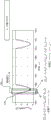
FIG. 4 shows a part Pmus(t) example moving window least squares algorithm of polynomial order of waveform.
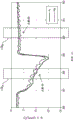
FIG. 5 shows a MWLS RrsThe maximum ratio combination of the results is estimated.
Detailed Description
The following relates to the characterization of respiratory parameters during ventilation of a patient, in particular to respiratory muscle pressure Pmus(t), respiratory resistance RrsAnd respiratory compliance CrOr elasticity Er=1/Cr. In principle, these parameters can be estimated using the equation of lung motion (equation (1)), equation (1) relating these parameters to the pressure P at the ventilator interfacey(t) and air flow Together with the air volume of the lungs
 And (4) correlating. In practice, due to respiratory muscle pressure Pmus(t) varies with time, so P is jointly estimated using the equation of lung motionmus(t),RrsAnd ErsAre typically underdetermined and cannot be solved analytically. Various methods of dealing with this problem include measuring additional information using invasive probes, or creating a "special case" environment by operations such as interrupting normal breathing. Invasive probes have significant disadvantages and techniques that rely on manipulating normal patient breathing do not provide continuous monitoring of normal breathing and can be harmful to the patient.
Referring to fig. 1, a medical ventilator system includes a medical ventilator 100 that delivers a flow of air at positive pressure to a patient 102 via an intake hose 104. Exhaled air is returned to the ventilator 100 via exhalation hose 106. The wye 108 of the ventilator system is used to couple air from the exhaust end of the intake hose 104 to the patient during inhalation, and to couple exhaled air from the patient into the exhalation hose 106 during exhalation. Note that the wye 108 is sometimes also referred to by other terms (e.g., tee). Not shown in fig. 1 are many other ancillary components that may be provided depending on the respiratory therapy being received by the patient 102. Such auxiliary components may include, for example: an oxygen cylinder or other medical grade oxygen source for delivering a controlled level of oxygen (usually an inspired oxygen Fraction (FiO) set by a physician or other medical personnel) to the air stream2) Ventilator parameter control); a humidifier, which enters the air intake line 104 vertically; a nasogastric tube that provides nutrients to patient 102; and so on. The ventilator 100 includes a user interface, which in this illustrative example includes a touch-sensitive display member 110, through which a physician, respiratory specialist, or other medical personnel can configure ventilator operation and monitor measured physiological and operating parameters of the ventilator 100. Additionally or alternatively, the user interface may include physical user input controls (buttons, dials, switches, etc.), a keyboard, a mouse, audible alert device(s), indicator light(s), and the like. It is also noted that the illustrative ventilator 100 is merely an illustrative example.
The illustrative ventilator 100 is a dual-limb ventilator with proximal sensors. However, the disclosed patient respiratory condition monitoring techniques may be used in conjunction with substantially any type of ventilator, such as a single-limb or dual-limb ventilator, a ventilator with a valve or blower, a ventilator with an invasive coupling to the patient (e.g., via a tracheostomy or endotracheal tube) or a ventilator with a non-invasive coupling to the patient (e.g., using a mask), a ventilator with proximal sensors as shown for measuring pressure and flow, or a ventilator without such proximal sensors that relies on sensors in the ventilator unit, and so forth.
With continued reference to fig. 1, the patient 102 is monitored by various physiological parameter sensors. In particular, fig. 1 shows two such sensors: an airway pressure sensor 112 and an air flow sensor 114, the airway pressure sensor 112 measuring a pressure P at a coupling with the patienty(t) (typically measured at Y-piece 108, so Py(t)), airflow sensor 114 measures the airflow into and out of the patient (also typically measured at wye 108). The
 sensors 112, 114 may be integrated into the wye 108, placed on the air lines 104, 106, or integrated into the ventilator 100. Other physiological parameters may be sensed by appropriate sensing during mechanical ventilationFor monitoring, e.g. heart rate, respiration rate, blood pressure, blood oxygen (e.g. SpO)2) Respiratory gas composition (e.g. measuring CO in respiratory gas)2Carbon dioxide map) and the like. Other physiological parameters may be derived from directly measured physiological parameters.
The system also includes a respiratory system analyzer 120, which includes a microprocessor, microcontroller or other electronic data processing device, programmed to process data including airway pressure Py(t) and air flow To generate information about a patient respiratory system parameter: resistance RrsCompliance Crs(or equivalently, Ers=1/Crs) And by respiratory muscle pressure Pmus(t) inspiratory effort of the patient characterized as a function of time. By using applied to airway pressure Py(t) and air flow
 Together with an
 air flow integrator 122 Determined volume of air
 The moving window least squares estimation (MWLS) estimates the lung motion equation (1)), and these parameters are determined as a function of time in real time. (alternatively, a dedicated air volume sensor may be employed). To overcome the underdetermined nature of equation (1), the MWLS estimation is performed using the following successive estimates: (1) via ErsElasticity or compliance (E) of the
 estimator 132rsOr Crs) A parameter; then (2) via RrsResistance (R) of estimator 134rs) Estimating parameters; then (3) via Pmus(t) respiratory muscle pressure (P) of estimator 136mus(t)) of the parametersAnd (6) estimating.
These successive estimators 132, 134, 136 are applied within a time window 130, which time window 130 typically has a duration of two seconds or less, and more preferably a duration of one second or less, and in the illustrative example, 0.6 seconds of 100Hz data samples, such that the time window contains 60 samples. The upper limit of the duration of the time window is imposed by the respiration rate, which is typically 12 to 20 breaths per minute for a normal adult, corresponding to a respiratory cycle of 3-5 seconds duration. The duration of the time window 130 is preferably a fraction of the duration of the breathing cycle, such that the parameter ErsAnd RrsCan be reasonably assumed to be constant within each time window 130, and P within each time window 130musThe variation of (t) can be represented using a relatively simple approximation function (e.g., a low order polynomial in the illustrative examples disclosed herein).
The estimators 132, 134, 136 are applied successively within each time window 130, and provide E in real time for each successive (and partially overlapping) time interval 130 (hence the term "moving" the time window)rs,RrsAnd Pmus(t) estimation. In the illustrative example, ErsAnd RrsIs assumed to be constant within each time window 130 such that the estimation of these parameters is in real time at a time resolution comparable to the duration of the time window 130, e.g., 2 seconds or less in some embodiments, or more preferably 1 second or less, and in the illustrative example 0.6 seconds. This can further improve the effective temporal resolution if successive time windows partially overlap. PmusThe real-time estimate of (t) can have a ratio of ErsAnd RrsHigher time resolution because P is within the time window 130mus(t) changes over time are modeled in the illustrative example by a low order polynomial function of time.
The method disclosed herein takes advantage of the recognition that among the three parameters estimated, the elasticity/compliance (E)rsOr Crs) Usually at any timeWith the slowest change. In the equation of lung motion (equation (1)), ErsIs a coefficient that integrates the air volume v (t) that changes slowly with time. The next most slowly varying parameter is typically the resistance RrsIt is the air flow The coefficient of (a). Finally, respiratory muscle pressure Pmus(t) has the potential to vary most rapidly over time as it varies in response to the patient actively inhaling and exhaling. In view of this, Pmus(t) illustrative examples of
 estimator 136 do not assume Pmus(t) is a constant within the time window 130, but rather a low order approximating polynomial function. Instead of P within the time window 130mus(t), in other contemplated embodiments, some other temporal parameterized function, such as a spline function, may be contemplated.
With continued reference to FIG. 1, output Ers(or C)rs),RrsAnd Pmus (t) can be used for various purposes. In one application, one or more of the estimated parameters may be displayed on the display component 110 of the ventilator 100, for example as digital real-time values and/or as trend lines plotted as a function of time. Typically, respiratory elasticity or compliance (E)rsOr Crs) And respiratory resistance (R)rs) Are of most interest to the clinician and are suitably displayed and/or given a trend. Respiratory muscle pressure Pmus(t) is a waveform acquired in real time as a function of time during mechanical ventilation in normal clinical operation, therefore, Pmus(t) can be used by the ventilator 100 to trigger and turn off mechanical ventilation to reduce patient-ventilator dyssynchrony (i.e., synchronize the application of positive pressure by the ventilator 100 with the inspiratory portion of the patient's respiratory muscle action).
In some embodiments, the work of breathing (WoB) estimator 140 measures the respiratory muscle pressure Pmus(t) integral with volume, i.e. WoB ═ P-mus(t) dv (t). WoB is a measure of how much effort the patient 102 applies on breathing by himself. WoB may be displayed and/or trended on display component 110 to provide to a clinicianUseful information for setting ventilator pressure settings in a ventilation mode, such as Pressure Support Ventilation (PSV). Moreover, because WoB estimator 140 provides WoB in real-time (e.g., with a time lag and resolution of about one second or less in some embodiments), ventilator 100 optionally employs WoB as a feedback control parameter, e.g., adjusts controlled ventilator settings to maintain WoB at a constant set point value. For example, if WoB increases, this means that the patient 102 is struggling to breathe, and thus the positive pressure applied by the ventilator 100 in PSV mode should be increased to provide increased respiratory assistance to the struggling patient.
Referring to FIG. 2, some illustrative embodiments of the continuity estimators 132, 134, 136 are described. Shows the parameter Ers,RrsAnd Pmus(t) continuous estimation over a time window of a fraction of a second over which the parameter E is assumedrsAnd RrsIs a constant. On the first pass (by E)rs Estimator 132 implemented), all three parameters E are assumedrs,RrsAnd Δ Pmus(t) is constant over the time window 130 and is computed simultaneously, but only the estimated one is retained from this first pass (in the notation used herein, the superscript "hat" is used, i.e. the
 For indicating an estimated value of the parameter p. ) In the second pass (by R)rs
 Estimator 134 performs), now known (estimated) Is removed by subtraction and the remainder of the lung equation of motion is directed to RrsAnd Pmus(t) fitting, the latter being approximated using a low order polynomial (n ═ 0, 1, or 2). In experiments, it was found that the best choice of polynomial order depends on the breathing phase at which the
 time window 130 is located due to possible overfitting-since breathing in the illustrative embodiments disclosed hereinThe phases are not known a priori and use a weighted combination of zeroth, first and second order polynomials. RrsThe output of the estimator 134 is an estimate of the respiratory resistance, i.e. the Finally, in the third pass (by P)mus(t)
 estimator 136 executes), the now known (estimated) is removed by further subtraction And directly fitting the remainder of the lung equation of motion to obtain the estimated respiratory muscle pressure, i.e. the
With continued reference to FIG. 2, illustrative E is further describedrsAn estimator 132. At 208, the airway pressure P is adjustedy(t) performing a difference operation and outputting Δ Py(t) is calculated as Δ Py(t)=Py(t)-Py(t-1). Continuous estimation of E at 210 using a Moving Window Least Squares (MWLS) estimatorrs(t) -which is the elasticity of the respiratory system, Ers(t)=1/Crs(t) — and is based on the following difference equation:

it should be noted that the estimate is generated for each time window 130 In the range of Ers(t) is estimated as a function of time such that as successive (partially overlapping) time windows are applied over time, the function of time is applied for
 successive time windows 130 Is generated as a value
 However, within each
 time window 130, Δ P will bemus(t),Pmus(t) difference signal of waveform and parameter Rrs(t) and Ers(t) is modeled as a constant and jointly estimated by the least squares minimization method. For ErsEstimator 132, using only Ers(t) estimation, i.e. (after filtering by the
 kalman filter 212 in the illustrative example of fig. 2), while the other estimation outputs are discarded. And, ErsThe estimator 132 also calculates an estimated value Is expressed herein as
The input to the MWLS estimator 210 is P, which is output by the difference operation 208y(t) difference signal, i.e. Δ Py(t) of (d). Based on the equation of motion (equation (1)), Δ P can be expressedy(t) modeled as:

wherein, is a flow rate difference signal that is,
 is a volume difference signal (where T is the sampling time interval, e.g. 100Hz sampling corresponds to T0.01 seconds), and apmus(t)=Pmus(t)-Pmus(t-1) is Pmus(t) a difference signal.
Hereinafter, the size (or duration) of the sliding time window 130 is denoted L, which is optionally a system parameter that can be set by the user. When in useThe sliding window at the previous time t spans the interval [ t-L +1, t]. For the MWLS estimator 210, P in a sliding windowmus(t) difference signal Δ Pmus(t) is modeled as a constant Δ Pmus. Further suppose RrsAnd ErsIs constant over the sliding time window 130. Thus, Δ PyThe equation of (t) becomes:

at time t, the MWLS algorithm 210 uses the input signal in the sliding window 130, i.e., the samples Δ P in the interval t-L +1 ≦ n ≦ ty(n) and to jointly estimate Rrs,ErsAnd Δ PmusBut only E is used in the subsequent operations (i.e. the
 subsequent estimators 134, 136)rsEstimating
As further shown in fig. 3, specifically, at time t, the MWLS formula solves the least squares problem 300 based on the above equation:
at the time t of the start of the operation,


y=[ΔPy(t),ΔPy(t-1),...,ΔPy(t-L+1)]T
X=[x(t),x(t-1),...,x(t-L+1)]T


in addition, also calculates ErsVariance of estimation Wherein
 Is the least-squares residual variance and,

as indicated in FIG. 3, MWLS estimation 210 is performed continuously as the moving window moves forward, e.g., window 310n is followed by the next window 310n+1And so on. Due to MWLS method to PyMeasurement noise and modeling error are sensitive, so only ErsEstimated value Quilt ErsThe
 estimator 132 retains, while the other estimates output (e.g. And
And ) Is discarded.
To further increase ErsEstimating performance, optionally using a Kalman filter 212 to reduce ErsAn error is estimated. As mentioned previously, the respiratory elasticity ErsTypically not changing rapidly with time. The Kalman filter 212 is used for Filtering the estimated noise in (1) and improving Ers(t) estimating the result. The input to the
 Kalman filter 212 is And
And the
the output 214 of the Kalman filter is Ers(t) final estimate, denoted herein as And is
 Wherein ω isE(t) is a noise or uncertainty measure. The above model assumptions
 Is provided with a noise term
 E of (A)rs(t) unbiased estimation.
The kalman filter can be designed to reduce the MWLS estimation noise based on the following assumptions: (1) equation of state processing, where ErsSlowly varying and capable of being modeled as random walks, i.e. Ers(t)=Ers(t-1)+ωE(t),ωE(t)~N(0,δE) (ii) a And (2) an observation equation, wherein the MWLS estimate Can be modeled as
 The standard kalman filter can be used with a 1, B0, and Q δEH is 1 and
 to be implemented. The kalman filter has certain advantages, including computationally efficient implementation in the context of a sliding time window, intuitive operation, and weighted average output. Parameter deltaEIs to control the average window lengthAlgorithm parameters of degree.
With continued reference to FIGS. 1 and 2, ErsFinal output of estimator 132 214 by subsequent RrsThe
 estimator 134 is used to perform Rrs(t) estimating. To estimate Rrs(t) use of The elastic pressure component ErsV (t) from PyAnd (t) is eliminated. The ErsThe
 cancellation operation 216 can be expressed as:

Erselimination 216 removes an unknown quantity (E) from the lung motion equationrs) And thus simplify RrsAnd (6) estimating. Suppose by ErsEstimation of estimator 132 output Is correct and the elastic pressure component is completely eliminated, Rrs
 The MWLS operation 218 of the estimator 134 optimizes the following equation:

using a Moving Window Least Squares (MWLS) estimator 218, the respiratory resistance R is estimatedrs。
At ErsE of the estimator 132rsIn estimator MWLS operation 210, since PmusThe difference of (t), i.e. Δ Pmus(t) estimated as constant value Δ P for each time windowmusSo as to apply the respiratory muscle pressure Pmus(t) is indirectly estimated as a linear function of t. However, it has been found herein that at RrsIn the case of the MWLS operation 218 of the estimator 134, this estimate is too coarse, and if the respiratory muscle pressure is in the MWLS operation 218Force Pmus(t) is adaptively modeled, then the respiratory resistance R is providedrsA significantly improved estimate of. In the illustrative example herein, a low-order polynomial pair P is usedmus(t) modeling, e.g., 0 order (constant value), 1 order (linear) or 2 order (quadratic). PmusThe order M of the (t) polynomial function can significantly change the estimation performance.
In addition, briefly refer to FIG. 4 for Pmus(t) the optimal order M of the modeled polynomial depends on the position of the moving window 130 within the breathing cycle. In illustrative FIG. 4, a first time window 130AThe polynomial lying in the first order (M ═ 1) is Pmus(t) a breathing phase of the effective model; while the zeroth order (M ═ 0) polynomial is valid for the respiratory phase in which the second time window 130B is located. However, the current time window 130 is typically at a respiratory phase other than RrsAn input of the estimator 134.
Referring briefly to FIG. 5, for Rrs MWLS 218,Pmus(t) the waveform is modeled as a polynomial function of order M (M)>0), i.e. Pmus(t)=a0+a1t+…+aMtMAnd R isrs(t) the parameter is assumed to be constant. (although P is described hereinmusThe polynomial model of (t) is for illustration, but other models including a parameterized function of time, such as a spline model, are also contemplated. To accommodate the difference in the optimal polynomial order over the respiratory cycle, Rrs The MWLS estimator 218 calculates three RsrsEstimation value: using a polynomial of order 0 (M ═ 0, i.e. P)mus(t) modeled as a constant) MWLS estimate 2180(ii) a Using a first order polynomial (M ═ 1, i.e. P)mus(t) modeled as a linear function of t) MWLS estimate 2181(ii) a And using a second order polynomial (M ═ 2, i.e. P)mus(t) modeled as a quadratic function of t) MWLS estimate 2182. Each MWLS estimator 2180、2181、2182The MWLS formula of (a) is listed in the right box of fig. 5 and table 1 below.
TABLE 1 Each Pmus(t) R of the modelrsEstimator formula

With continued reference to FIG. 5, the MWLS operations 218 are performed by corresponding ones0、2181、2182Three R of outputrs(t) the estimates are combined together by a combining operation 219 to produce a final MWLS estimate The combining
 operation 219 may use various combining techniques, such as a maximum ratio combining operation or a minimum variance selection combination. The maximum ratio combining employed by illustrative combiner 219 assigns the maximum weight to the estimate with the smallest estimate variance (e.g., the estimate with the best polynomial order) so that the estimate with the best polynomial order will dominate RrsAnd (6) estimating output. MWLS 218 also calculates Variance of (2)
Referring back to FIG. 2, at RrsIn the case of the estimator 134, only the R output from the MLS operation 218 is retainedrs(t) estimation And other estimated outputs (e.g., P)mus(t) polynomial coefficients) are discarded. At RrsIn the final stage of the
 estimator 134, a Kalman filter 220 is applied to further improve the R output by the MWLS 218rsAnd (6) estimating. The Kalman filter 220 is suitably similar to that described above with respect to ErsThe estimator 132 describes a kalman filter 212. For RrsThe kalman filter 220 of the estimator 134 can be designed to reduce the MWLS estimation noise based on the following assumptions: (1) equation of state processing, where RrsSlowly varying and can be modeled as random walk, Rrs(t)=Rrs(t-1)+ωR(t),ωR(t-1)~N(0,δR) (ii) a To be provided withAnd (2) an observation equation, wherein MWLS estimation Can be modeled as
 Wherein,
 the standard kalman filter can be used with a 1, B0, and Q δRH is 1 and
 to be implemented. Also, the kalman filter has certain advantages, including computationally efficient implementation in the context of a sliding time window, intuitive operation, and weighted average output. Parameter deltaRIs the algorithm parameter that controls the average window length.
RrsThe output 222 of the (t) Kalman filter 220 is represented herein as
 R of (A) to (B)rsAnd (6) estimating. This output assumption
 Is RrsUnbiased estimation of (t), but with a noise term
Referring back to FIG. 2, in the last pass, once ErsAnd RrsThe estimates are obtained by respective estimators 132, 134, applying Pmus(t) estimator 136 to estimate Pmus(t) of (d). Using previously estimated Rrs(t) and Crs(t),Pmus(t) calculation 224 is calculated according to Estimating:

for P in the time window 130y(t), And (via integrator 122) samples of v (t). In other words, the evaluation is performed in the
 time window 130 of the MWLS
 To remove
 Medium high frequency noise, P can be further improved using an optional low pass filter 226mus(t) estimating. Additionally or alternatively, P can be inputmus(t) physiological knowledge of the waveform to further improve Pmus(t) estimating.
In the illustrative embodiment, a respiratory elasticity (or compliance) estimator 132 is applied first, then a respiratory resistance estimator 134, and finally a respiratory muscle pressure estimator 136. However, it is contemplated to first estimate respiratory resistance and then respiratory elasticity or compliance (i.e., reverse the order of the estimators 132, 134). In such a modified embodiment, the second (E)rs) The estimator will suitably include R similar to operation 216 of the illustrative embodimentrsAnd (5) eliminating operation. Whether Ers(or C)rs) And RrsHow to estimate the order of P, it can be understood that if P is not usedmus(t) and WoB (from which it is calculated by integrator 140), the final P may optionally be omittedmus(t) an estimator 136.
These values may also optionally be displayed along with their respective measures of uncertainty, for example expressed in delta or omega statistics or functions thereof as described herein, if respiratory elastance (or compliance) and/or resistance are displayed on the display component 110 of the ventilator 100. While these and other breathing parameters are described in the illustrative example as being displayed on the display component 110 of the ventilator 100, it should be understood that these values may additionally or alternatively be displayed on a bedside patient monitor, a nurse station computer, and/or may be stored in an Electronic Health Record (EHR) or other patient data storage system, or the like. The illustrative respiratory system analyzer 120 is suitably implemented via the microprocessor of the ventilator 100; however, the respiratory system analyzer 120 may additionally or alternatively be implemented via a microprocessor or other electronic data processing device of a bedside patient monitor. The disclosed respiratory system analyzer functions may also be implemented by a non-transitory storage medium storing instructions that are readable and executable by such a microprocessor or other electronic data processing device to perform the disclosed functions. By way of example, non-transitory storage media may include, for example, a hard disk or other magnetic storage media, an optical disk or other optical storage media, flash memory or other electronic storage media, various combinations thereof, and so forth.
As previously described, in addition to displaying one or more of the estimates (e.g., optionally values with their statistical uncertainty) One or more of) as a real-time value, a trend line, etc., in another illustrative application,
 the waveform may be used to synchronize the positive pressure applied by the
 ventilator 100 with the respiratory effort consumed by the patient 102 to reduce patient-ventilator dyssynchrony. In this application, the positive air pressure applied by the ventilator 100 is adjusted, e.g., increased or decreased, with The increase or decrease in magnitude is synchronized. In another control application, WoB output by
 integrator 140 may beTo serve as a feedback signal for controlling the ventilator 100. In general, the positive pressure applied by ventilator 100 should increase with increasing measured WoB output by integrator 140, and this increased mechanical ventilation should result in a corresponding decrease in patient WoB until the set point WoB is reached. As shown, a proportional and/or derivative and/or integral controller (e.g., a PID controller) may be used for this feedback control, where the WoB signal from the integrator 140 is used as a feedback signal, the target WoB is used as a set point value, and positive pressure is the controlled variable.
The respiratory system analyzer 120 has been tested with simulated data and porcine respiratory data, and the results show that the analyzer 120 is able to provide results comparable to invasive protocols and is stable at different ventilator settings, including low PSV settings. The analyzer 120 provides various benefits including (but not limited to): providing real-time data (with a lag of a few seconds or less); sample-by-sample estimation (if successive windows overlap and are separated by a single sample); a customizable trade-off between computational complexity and temporal resolution (faster computation by larger spacing between possibly overlapping windows, in compromise with reduced temporal resolution); fast convergence (within 10 breaths in some tests), providing a low start-up time; stability against accidental interference; good computational efficiency using, for example, an efficient pseudo-inverse (L4) matrix calculation (where L is the window size, e.g., 60-90 samples in some suitable embodiments); and low memory requirements (storing data for the current time window, for some embodiments, approximately 60-90 samples).
As a further advantage, respiratory analyzer 120 properly estimates elasticity or compliance Ers(t), resistance Rrs(t) and respiratory muscle pressure Pmus(t) without receiving as input either a respiratory phase or a respiratory rate, and without making any a priori assumptions about these parameters (except assuming E within any given time window of MWLS estimationrsAnd RrsIs constant). Respiratory system analyzer 120 is suitably only at measured air pressure Py(t) and air flow In conjunction with temporally paired
 For integral derivation
 And performing operation.
The invention has been described with reference to the preferred embodiments. Modifications and alterations will occur to others upon reading and understanding the preceding detailed description. It is intended that the invention be constructed as including all such modifications and alterations insofar as they come within the scope of the appended claims or the equivalents thereof.
Claims (15)
1. A medical ventilator device comprising:
a ventilator (100) configured to deliver ventilation to a ventilated patient (102);
a pressure sensor (112) configured to measure an airway pressure P of the ventilated patienty(t);
An air flow sensor (114) configured to measure air flow into and out of the ventilated patient And
And
a respiratory system analyzer (120) comprising a microprocessor configured to estimate respiratory parameters of the ventilated patient using a moving time window least squares (MWLS) estimation, including (i) respiratory system elasticity (E)rsOr compliance Crs(ii) respiratory resistance RrsAnd (iii) respiratory muscle pressure Pmus(t),
Wherein the duration of the time window is a fraction of the duration of the breathing cycle such that the parameter ErsAnd RrsCan be assumed to be constant within each time window, and wherein P within each time windowmus(t) is approximated by using a low order polynomial function.
2. The medical ventilator device of claim 1 wherein the MWLS estimation includes, for each time window of the MWLS estimation, performing the following in turn:
(1) estimating one of (i) elasticity or compliance and (ii) resistance;
(2) using the estimate from operation (1) to estimate the other of (i) elasticity or compliance and (ii) resistance; and is
(3) Estimating respiratory muscle pressure using the values estimated in operations (1) and (2).
3. The medical ventilator device of claim 2 wherein the operation (1) estimates elasticity or compliance, and the operation (2) estimates resistance using the estimated value of elasticity or compliance from operation (1).
4. The medical ventilator device of any one of claims 2-3 wherein the operation (1) pertains to P in a time window of the MWLSy(t) and to optimize the elasticity E in the following equationrsResistance RrsAnd respiratory muscle pressure PmusDifference Δ P ofmus:

Wherein, and Δ Py(t)=Py(t)-Py(t-1) and
 and Δ V (t) ═ V (t) -V (t-1).
5. The medical ventilator device of claim 4 wherein the operation (2) pertains to P in a time window of the MWLSy(t) and to optimize respiratory muscle pressure P in the following equationmus(t), and elasticity ErsAnd resistance RrsOne of (1):

wherein the estimated value from operation (1) remains fixed and Pmus(t) modeling by a parameterized function of time.
6. The medical ventilator device of claim 5 wherein Pmus(t) is modeled by a polynomial function of time.
7. The medical ventilator device of claim 6 wherein P modeled with zeroth, first and second order polynomial functions through time is utilizedmus(t) repeating operation (2) and repeating three times the optimized elasticity ErsOr resistance RrsAre combined.
8. The medical ventilator device of any one of claims 5-7 wherein the operation (3) estimates respiratory muscle pressure as the MWLS in a time window of the MWLS Wherein,
 and
and are estimated values from operation (1) and operation (2).
9. The medical ventilator device of any one of claims 2-3 wherein one or both of the operations (1) and (2) comprise applying a Kalman filter (212, 220) to the estimated value.
10. The medical ventilator device of claim 9 wherein one or both of the operations (1) and (2) further comprise generating an uncertainty metric for the estimated value based on a noise variance of the kalman filter (212, 220).
11. The medical ventilator device of any one of claims 1-3 further comprising:
a display (110) configured to display one or more of the respiratory parameters of the ventilated patient estimated by the respiratory system analyzer.
12. The medical ventilator device of any one of claims 1 to 3 wherein the ventilator (100) is programmed to operate in conjunction with increasing or decreasing respiratory muscle pressure PmusThe magnitude of (t) synchronously adjusts positive air pressure output by the ventilator in order to reduce patient-ventilator asynchrony.
13. The medical ventilator device of any one of claims 1-3 wherein:
the respiratory system analyzer (120) is configured to estimate work of breathing WoB as WoB ═ jpjpmus(t) dV (t), wherein Pmus(t) respiratory muscle pressure as a function of time estimated using the MWLS estimate; and is
The ventilator (100) is programmed to control mechanical ventilation provided by the ventilator to maintain estimated WoB at a setpoint WoB value.
14. A non-transitory storage medium storing instructions readable and transportable by an electronic data processing deviceTo perform an airway pressure P to a patient (102) on a ventilator (100)y(t) and air flow The method of operating on a measurement, the method comprising:
estimation of (i) respiratory elasticity E using Moving Window Least Squares (MWLS) estimationrs(ii) respiratory resistance RrsAnd (iii) respiratory muscle pressure Pmus(t), wherein:
the MWLS estimation (i) comprises:

fitting to Δ Py(t) to obtain Ers,RrsAnd Δ PmusA value of (1), wherein Δ Py(t) is a difference signal of the measured airway pressure, is the difference signal of the measured air flow, Δ V (t) is the respiratory air volume
 And Δ P of the difference signal ofmusIs constant, and
the MWLS estimate (ii) comprises a fit of:

to obtain RrsAnd PmusA value of (t), wherein ErsIs set to the value determined in the MWLS estimate (i), and wherein Pmus(t) is approximated as a parameterized function, and
the MWLS estimation (iii) comprises the evaluation:

wherein E isrsIs set to the value determined in the MWLS estimation (i), and RrsIs set to the value determined in the MWLS estimation (ii),
wherein the duration of the time window is a fraction of the duration of the breathing cycle such that the parameter ErsAnd RrsCan be assumed to be constant within each time window, and wherein P within each time windowmus(t) is approximated by using a low order polynomial function.
15. The non-transitory storage medium of claim 14, wherein the respiratory elasticity ErsExpressed as respiratory compliance C in the MWLS estimation operationrs=1/Ers。
Applications Claiming Priority (3)
| Application Number | Priority Date | Filing Date | Title |
|---|---|---|---|
| US201562169863P | 2015-06-02 | 2015-06-02 | |
| US62/169,863 | 2015-06-02 | ||
| PCT/IB2016/053206 WO2016193915A1 (en) | 2015-06-02 | 2016-06-01 | Non-invasive method for monitoring patient respiratory status via successive parameter estimation |
Publications (2)
| Publication Number | Publication Date |
|---|---|
| CN107690310A CN107690310A (en) | 2018-02-13 |
| CN107690310B true CN107690310B (en) | 2021-06-15 |
Family
ID=56116481
Family Applications (1)
| Application Number | Title | Priority Date | Filing Date |
|---|---|---|---|
| CN201680032035.0A Active CN107690310B (en) | 2015-06-02 | 2016-06-01 | Non-invasive method for monitoring the respiratory state of a patient via continuous parameter estimation |
Country Status (5)
| Country | Link |
|---|---|
| US (1) | US20180177963A1 (en) |
| EP (1) | EP3302664B1 (en) |
| JP (1) | JP6912388B2 (en) |
| CN (1) | CN107690310B (en) |
| WO (1) | WO2016193915A1 (en) |
Families Citing this family (4)
| Publication number | Priority date | Publication date | Assignee | Title |
|---|---|---|---|---|
| EP3370811A1 (en) | 2015-11-02 | 2018-09-12 | Koninklijke Philips N.V. | Breath by breath reassessment of patient lung parameters to improve estimation performance |
| WO2019210470A1 (en) * | 2018-05-02 | 2019-11-07 | 深圳迈瑞生物医疗电子股份有限公司 | Respirator and ventilation control method therefor |
| DE102020133460A1 (en) * | 2020-01-07 | 2021-07-08 | Drägerwerk AG & Co. KGaA | Method and signal processing unit for determining a pneumatic measure using a lung mechanical model and a progression model |
| CN113082412B (en) * | 2021-03-30 | 2023-11-17 | 湖南万脉医疗科技有限公司 | Inhalation gas oxygen concentration fraction control system of breathing machine |
Citations (1)
| Publication number | Priority date | Publication date | Assignee | Title |
|---|---|---|---|---|
| CN104545844A (en) * | 2014-12-25 | 2015-04-29 | 中国科学院苏州生物医学工程技术研究所 | Multi-parameter sleep monitoring and intelligent diagnosis system based on 4G mobile communication technology and application method of multi-parameter sleep monitoring and intelligent diagnosis system |
Family Cites Families (33)
| Publication number | Priority date | Publication date | Assignee | Title |
|---|---|---|---|---|
| US5853364A (en) * | 1995-08-07 | 1998-12-29 | Nellcor Puritan Bennett, Inc. | Method and apparatus for estimating physiological parameters using model-based adaptive filtering |
| US5980463A (en) * | 1995-09-28 | 1999-11-09 | Data Sciences International, Inc. | Method for respiratory tidal volume measurement |
| SE513969C2 (en) * | 1997-05-17 | 2000-12-04 | Draegerwerk Ag | Apparatus and method for determining the mechanical properties of the respiratory system |
| US6068602A (en) * | 1997-09-26 | 2000-05-30 | Ohmeda Inc. | Method and apparatus for determining airway resistance and lung compliance |
| US6066101A (en) * | 1998-04-20 | 2000-05-23 | University Of Maryland | Airflow perturbation device and method for measuring respiratory resistance |
| US6257234B1 (en) * | 1998-08-21 | 2001-07-10 | Respironics, Inc. | Apparatus and method for determining respiratory mechanics of a patient and for controlling a ventilator based thereon |
| EP1156846A1 (en) * | 1999-02-03 | 2001-11-28 | University Of Florida | Method and apparatus for nullifying the imposed work of breathing |
| US6837242B2 (en) * | 2000-04-26 | 2005-01-04 | The University Of Manitoba | Method and apparatus for determining respiratory system resistance during assisted ventilation |
| US6575905B2 (en) * | 2000-09-22 | 2003-06-10 | Knobbe, Lim & Buckingham | Method and apparatus for real-time estimation of physiological parameters |
| US7484508B2 (en) * | 2002-06-27 | 2009-02-03 | Yrt Limited | Method and device for monitoring and improving patient-ventilator interaction |
| WO2004019766A2 (en) * | 2002-08-30 | 2004-03-11 | University Of Florida | Method and apparatus for predicting work of breathing |
| US20050211249A1 (en) * | 2002-11-19 | 2005-09-29 | Mirko Wagner | Ventilator method, ventilator and memory medium |
| US7475685B2 (en) * | 2003-03-24 | 2009-01-13 | Weinmann Geräte fär Medizin GmbH & Co. KG | Method and device for detecting leaks in respiratory gas supply systems |
| IL155955A0 (en) * | 2003-05-15 | 2003-12-23 | Widemed Ltd | Adaptive prediction of changes of physiological/pathological states using processing of biomedical signal |
| EP1972274B1 (en) * | 2007-03-20 | 2015-12-30 | Drägerwerk AG & Co. KGaA | Method and apparatus for determining the resistance of the respiratory system of a patient |
| CN101380233B (en) * | 2007-09-05 | 2010-12-22 | 深圳迈瑞生物医疗电子股份有限公司 | Breathing work real-time monitoring method and device based on breathing mechanics module |
| US8267085B2 (en) * | 2009-03-20 | 2012-09-18 | Nellcor Puritan Bennett Llc | Leak-compensated proportional assist ventilation |
| US20100071696A1 (en) * | 2008-09-25 | 2010-03-25 | Nellcor Puritan Bennett Llc | Model-predictive online identification of patient respiratory effort dynamics in medical ventilators |
| WO2010067244A1 (en) * | 2008-12-10 | 2010-06-17 | Koninklijke Philips Electronics, N.V. | Determining elastance and resistance |
| US9451886B2 (en) * | 2009-04-22 | 2016-09-27 | Rodrigo E. Teixeira | Probabilistic parameter estimation using fused data apparatus and method of use thereof |
| DE102009023965A1 (en) * | 2009-06-05 | 2010-10-14 | Drägerwerk AG & Co. KGaA | Respiratory device for pressure-supporting ventilation of patient, has control and evaluation unit analyzing functional dependency of pressure and respiratory volume, where elastance or compliance is determined from rise of pressure |
| US8321017B2 (en) * | 2009-07-08 | 2012-11-27 | Pacesetter, Inc. | Electromechanical delay (EMD) monitoring devices, systems and methods |
| US20110021941A1 (en) * | 2009-07-23 | 2011-01-27 | Nellcor Puritan Bennett Ireland | Systems and methods for respiration monitoring |
| US9636474B2 (en) * | 2010-08-27 | 2017-05-02 | Resmed Limited | Adaptive cycling for respiratory treatment apparatus |
| US20130079667A1 (en) * | 2011-09-28 | 2013-03-28 | General Electric Company | Flow sensor with mems sensing device and method for using same |
| US20140180027A1 (en) * | 2012-12-20 | 2014-06-26 | U.S. Government, As Represented By The Secretary Of The Army | Estimation of Human Core Temperature based on Heart Rate System and Method |
| US20140275886A1 (en) * | 2013-03-14 | 2014-09-18 | Streamline Automation, Llc | Sensor fusion and probabilistic parameter estimation method and apparatus |
| US20140373845A1 (en) * | 2013-06-25 | 2014-12-25 | Covidien Lp | Methods and systems for adaptive adjustment of ventilator settings |
| CN105530860B (en) * | 2013-06-28 | 2019-07-09 | 皇家飞利浦有限公司 | Noninvasive estimation to intrapleural pressure and/or the calculating of the work of breathing based on the noninvasive estimation to intrapleural pressure |
| US9668692B2 (en) * | 2013-09-06 | 2017-06-06 | Breathe Technologies, Inc. | Apnea and hypopnea detection |
| US20150090258A1 (en) * | 2013-10-01 | 2015-04-02 | Covidien Lp | Ventilator-initiated prompt or setting regarding detection of asynchrony during ventilation |
| US20150090264A1 (en) * | 2013-10-02 | 2015-04-02 | Covidien Lp | Methods and systems for proportional assist ventilation |
| WO2015105787A1 (en) * | 2014-01-07 | 2015-07-16 | Covidien Lp | Apnea analysis system and method |
-
2016
- 2016-06-01 JP JP2017562011A patent/JP6912388B2/en active Active
- 2016-06-01 CN CN201680032035.0A patent/CN107690310B/en active Active
- 2016-06-01 US US15/579,228 patent/US20180177963A1/en not_active Abandoned
- 2016-06-01 EP EP16728111.2A patent/EP3302664B1/en active Active
- 2016-06-01 WO PCT/IB2016/053206 patent/WO2016193915A1/en active Application Filing
Patent Citations (1)
| Publication number | Priority date | Publication date | Assignee | Title |
|---|---|---|---|---|
| CN104545844A (en) * | 2014-12-25 | 2015-04-29 | 中国科学院苏州生物医学工程技术研究所 | Multi-parameter sleep monitoring and intelligent diagnosis system based on 4G mobile communication technology and application method of multi-parameter sleep monitoring and intelligent diagnosis system |
Also Published As
| Publication number | Publication date |
|---|---|
| JP2018516675A (en) | 2018-06-28 |
| JP6912388B2 (en) | 2021-08-04 |
| WO2016193915A1 (en) | 2016-12-08 |
| CN107690310A (en) | 2018-02-13 |
| EP3302664A1 (en) | 2018-04-11 |
| EP3302664B1 (en) | 2020-08-05 |
| US20180177963A1 (en) | 2018-06-28 |
Similar Documents
| Publication | Publication Date | Title |
|---|---|---|
| CN107205694B (en) | Simultaneous estimation of respiratory parameters by region fitting of respiratory parameters | |
| US11351320B2 (en) | Automatic PEEP selection for mechanical ventilation | |
| RU2737295C2 (en) | Apparatus for mechanic artificial pulmonary ventilation and respiratory monitoring | |
| CN108814581B (en) | Method for continuous and non-invasive determination of effective lung volume and cardiac output | |
| RU2700981C2 (en) | Non-invasive assessment of intrapleural pressure and/or calculation of respiratory work based on non-invasive assessment of intrapleural pressure | |
| CN108348718B (en) | Breath-by-breath re-assessment of patient lung parameters for improved estimation performance | |
| US20130006134A1 (en) | Methods and systems for monitoring volumetric carbon dioxide | |
| US20130006133A1 (en) | Methods and systems for monitoring volumetric carbon dioxide | |
| JP6876057B2 (en) | Simultaneous estimation of respiratory mechanics and patient effort by optimizing parameters | |
| CN109803708A (en) | Breathing is controlled using pressure to estimate lung compliance and lung resistance and disappear with the pressure for allowing all respiratory muscle recoils to generate | |
| CN107690310B (en) | Non-invasive method for monitoring the respiratory state of a patient via continuous parameter estimation | |
| JP7168560B2 (en) | System and method for estimation of respiratory muscle pressure and ventilation dynamics using P0.1 maneuver | |
| JP2019528901A (en) | Method and system for patient airway flow and leak flow estimation for non-invasive ventilation | |
| JP2018519123A (en) | Method and system for patient airway flow and leakage flow estimation for non-invasive ventilation | |
| CN113143250B (en) | Method and signal processing unit for acquiring aerodynamic dimensions using a model and a process model of a lung machine | |
| US11883593B2 (en) | Determining respiratory mechanic parameters in the presence of intrinsic positive end-expiratory pressure | |
| WO2016132279A1 (en) | Enhancement of simultaneous estimation of respiratory parameters via superimposed pressure signal |
Legal Events
| Date | Code | Title | Description |
|---|---|---|---|
| PB01 | Publication | ||
| PB01 | Publication | ||
| SE01 | Entry into force of request for substantive examination | ||
| SE01 | Entry into force of request for substantive examination | ||
| GR01 | Patent grant | ||
| GR01 | Patent grant |