CN107427007B - Compositions and methods for treating edema - Google Patents
Compositions and methods for treating edema Download PDFInfo
- Publication number
- CN107427007B CN107427007B CN201680016894.0A CN201680016894A CN107427007B CN 107427007 B CN107427007 B CN 107427007B CN 201680016894 A CN201680016894 A CN 201680016894A CN 107427007 B CN107427007 B CN 107427007B
- Authority
- CN
- China
- Prior art keywords
- lymphatic
- tacrolimus
- mice
- treatment
- cells
- Prior art date
- Legal status (The legal status is an assumption and is not a legal conclusion. Google has not performed a legal analysis and makes no representation as to the accuracy of the status listed.)
- Active
Links
Images


Classifications
-
- A—HUMAN NECESSITIES
- A61—MEDICAL OR VETERINARY SCIENCE; HYGIENE
- A61K—PREPARATIONS FOR MEDICAL, DENTAL OR TOILETRY PURPOSES
- A61K31/00—Medicinal preparations containing organic active ingredients
- A61K31/33—Heterocyclic compounds
- A61K31/395—Heterocyclic compounds having nitrogen as a ring hetero atom, e.g. guanethidine or rifamycins
- A61K31/435—Heterocyclic compounds having nitrogen as a ring hetero atom, e.g. guanethidine or rifamycins having six-membered rings with one nitrogen as the only ring hetero atom
- A61K31/44—Non condensed pyridines; Hydrogenated derivatives thereof
- A61K31/4412—Non condensed pyridines; Hydrogenated derivatives thereof having oxo groups directly attached to the heterocyclic ring
-
- A—HUMAN NECESSITIES
- A61—MEDICAL OR VETERINARY SCIENCE; HYGIENE
- A61K—PREPARATIONS FOR MEDICAL, DENTAL OR TOILETRY PURPOSES
- A61K9/00—Medicinal preparations characterised by special physical form
- A61K9/0012—Galenical forms characterised by the site of application
- A61K9/0014—Skin, i.e. galenical aspects of topical compositions
-
- A—HUMAN NECESSITIES
- A61—MEDICAL OR VETERINARY SCIENCE; HYGIENE
- A61K—PREPARATIONS FOR MEDICAL, DENTAL OR TOILETRY PURPOSES
- A61K31/00—Medicinal preparations containing organic active ingredients
- A61K31/33—Heterocyclic compounds
- A61K31/395—Heterocyclic compounds having nitrogen as a ring hetero atom, e.g. guanethidine or rifamycins
- A61K31/435—Heterocyclic compounds having nitrogen as a ring hetero atom, e.g. guanethidine or rifamycins having six-membered rings with one nitrogen as the only ring hetero atom
- A61K31/44—Non condensed pyridines; Hydrogenated derivatives thereof
- A61K31/4418—Non condensed pyridines; Hydrogenated derivatives thereof having a carbocyclic group directly attached to the heterocyclic ring, e.g. cyproheptadine
-
- A—HUMAN NECESSITIES
- A61—MEDICAL OR VETERINARY SCIENCE; HYGIENE
- A61K—PREPARATIONS FOR MEDICAL, DENTAL OR TOILETRY PURPOSES
- A61K31/00—Medicinal preparations containing organic active ingredients
- A61K31/275—Nitriles; Isonitriles
- A61K31/277—Nitriles; Isonitriles having a ring, e.g. verapamil
-
- A—HUMAN NECESSITIES
- A61—MEDICAL OR VETERINARY SCIENCE; HYGIENE
- A61K—PREPARATIONS FOR MEDICAL, DENTAL OR TOILETRY PURPOSES
- A61K31/00—Medicinal preparations containing organic active ingredients
- A61K31/33—Heterocyclic compounds
- A61K31/395—Heterocyclic compounds having nitrogen as a ring hetero atom, e.g. guanethidine or rifamycins
- A61K31/40—Heterocyclic compounds having nitrogen as a ring hetero atom, e.g. guanethidine or rifamycins having five-membered rings with one nitrogen as the only ring hetero atom, e.g. sulpiride, succinimide, tolmetin, buflomedil
- A61K31/401—Proline; Derivatives thereof, e.g. captopril
-
- A—HUMAN NECESSITIES
- A61—MEDICAL OR VETERINARY SCIENCE; HYGIENE
- A61K—PREPARATIONS FOR MEDICAL, DENTAL OR TOILETRY PURPOSES
- A61K31/00—Medicinal preparations containing organic active ingredients
- A61K31/33—Heterocyclic compounds
- A61K31/395—Heterocyclic compounds having nitrogen as a ring hetero atom, e.g. guanethidine or rifamycins
- A61K31/41—Heterocyclic compounds having nitrogen as a ring hetero atom, e.g. guanethidine or rifamycins having five-membered rings with two or more ring hetero atoms, at least one of which being nitrogen, e.g. tetrazole
- A61K31/42—Oxazoles
-
- A—HUMAN NECESSITIES
- A61—MEDICAL OR VETERINARY SCIENCE; HYGIENE
- A61K—PREPARATIONS FOR MEDICAL, DENTAL OR TOILETRY PURPOSES
- A61K31/00—Medicinal preparations containing organic active ingredients
- A61K31/33—Heterocyclic compounds
- A61K31/395—Heterocyclic compounds having nitrogen as a ring hetero atom, e.g. guanethidine or rifamycins
- A61K31/435—Heterocyclic compounds having nitrogen as a ring hetero atom, e.g. guanethidine or rifamycins having six-membered rings with one nitrogen as the only ring hetero atom
- A61K31/4353—Heterocyclic compounds having nitrogen as a ring hetero atom, e.g. guanethidine or rifamycins having six-membered rings with one nitrogen as the only ring hetero atom ortho- or peri-condensed with heterocyclic ring systems
- A61K31/436—Heterocyclic compounds having nitrogen as a ring hetero atom, e.g. guanethidine or rifamycins having six-membered rings with one nitrogen as the only ring hetero atom ortho- or peri-condensed with heterocyclic ring systems the heterocyclic ring system containing a six-membered ring having oxygen as a ring hetero atom, e.g. rapamycin
-
- A—HUMAN NECESSITIES
- A61—MEDICAL OR VETERINARY SCIENCE; HYGIENE
- A61K—PREPARATIONS FOR MEDICAL, DENTAL OR TOILETRY PURPOSES
- A61K38/00—Medicinal preparations containing peptides
- A61K38/04—Peptides having up to 20 amino acids in a fully defined sequence; Derivatives thereof
- A61K38/12—Cyclic peptides, e.g. bacitracins; Polymyxins; Gramicidins S, C; Tyrocidins A, B or C
- A61K38/13—Cyclosporins
-
- A—HUMAN NECESSITIES
- A61—MEDICAL OR VETERINARY SCIENCE; HYGIENE
- A61K—PREPARATIONS FOR MEDICAL, DENTAL OR TOILETRY PURPOSES
- A61K45/00—Medicinal preparations containing active ingredients not provided for in groups A61K31/00 - A61K41/00
- A61K45/06—Mixtures of active ingredients without chemical characterisation, e.g. antiphlogistics and cardiaca
-
- A—HUMAN NECESSITIES
- A61—MEDICAL OR VETERINARY SCIENCE; HYGIENE
- A61K—PREPARATIONS FOR MEDICAL, DENTAL OR TOILETRY PURPOSES
- A61K47/00—Medicinal preparations characterised by the non-active ingredients used, e.g. carriers or inert additives; Targeting or modifying agents chemically bound to the active ingredient
- A61K47/06—Organic compounds, e.g. natural or synthetic hydrocarbons, polyolefins, mineral oil, petrolatum or ozokerite
- A61K47/08—Organic compounds, e.g. natural or synthetic hydrocarbons, polyolefins, mineral oil, petrolatum or ozokerite containing oxygen, e.g. ethers, acetals, ketones, quinones, aldehydes, peroxides
- A61K47/10—Alcohols; Phenols; Salts thereof, e.g. glycerol; Polyethylene glycols [PEG]; Poloxamers; PEG/POE alkyl ethers
-
- A—HUMAN NECESSITIES
- A61—MEDICAL OR VETERINARY SCIENCE; HYGIENE
- A61K—PREPARATIONS FOR MEDICAL, DENTAL OR TOILETRY PURPOSES
- A61K9/00—Medicinal preparations characterised by special physical form
- A61K9/06—Ointments; Bases therefor; Other semi-solid forms, e.g. creams, sticks, gels
-
- A—HUMAN NECESSITIES
- A61—MEDICAL OR VETERINARY SCIENCE; HYGIENE
- A61P—SPECIFIC THERAPEUTIC ACTIVITY OF CHEMICAL COMPOUNDS OR MEDICINAL PREPARATIONS
- A61P7/00—Drugs for disorders of the blood or the extracellular fluid
- A61P7/10—Antioedematous agents; Diuretics
Landscapes
- Health & Medical Sciences (AREA)
- Life Sciences & Earth Sciences (AREA)
- Chemical & Material Sciences (AREA)
- Veterinary Medicine (AREA)
- Public Health (AREA)
- General Health & Medical Sciences (AREA)
- Animal Behavior & Ethology (AREA)
- Pharmacology & Pharmacy (AREA)
- Medicinal Chemistry (AREA)
- Epidemiology (AREA)
- Engineering & Computer Science (AREA)
- General Chemical & Material Sciences (AREA)
- Chemical Kinetics & Catalysis (AREA)
- Bioinformatics & Cheminformatics (AREA)
- Organic Chemistry (AREA)
- Nuclear Medicine, Radiotherapy & Molecular Imaging (AREA)
- Hematology (AREA)
- Diabetes (AREA)
- Dermatology (AREA)
- Gastroenterology & Hepatology (AREA)
- Oil, Petroleum & Natural Gas (AREA)
- Immunology (AREA)
- Proteomics, Peptides & Aminoacids (AREA)
- Pharmaceuticals Containing Other Organic And Inorganic Compounds (AREA)
- Medicines That Contain Protein Lipid Enzymes And Other Medicines (AREA)
- Medicinal Preparation (AREA)
- Medicines Containing Antibodies Or Antigens For Use As Internal Diagnostic Agents (AREA)
- Acyclic And Carbocyclic Compounds In Medicinal Compositions (AREA)
Abstract
Pharmaceutical compositions and methods for treating or preventing edema using an anti-T cell agent, an anti-TGF- β 1 agent, or an anti-angiotensin agent, preferably a combination of at least two such agents, are provided. The pharmaceutical composition may be formulated for systemic or local administration, and preferably topical administration.
Description
Cross reference to related applications
This application claims priority to U.S. provisional patent application No. 62/112,273 filed on 5.2.2015, which is incorporated herein by reference in its entirety.
Statement of government support
The invention was made with government support under grant numbers R01 HL111130-01, T32 CA009685-21a1 and 1S 10 RR028889-01, granted by the national institutes of health, and grant number P30 CA008748, granted by the national institute of cancer. The government has certain rights in the invention.
Copyright rights
A portion of the disclosure of this patent document contains material which is subject to copyright protection. The copyright owner has no objection to the facsimile reproduction by anyone of the patent document or the patent disclosure, as it appears in the patent and trademark office patent file or records, but otherwise reserves all copyright rights whatsoever.
Is incorporated by reference
All references cited in this disclosure are incorporated herein by reference in their entirety for the countries that are allowed to be incorporated by reference. Additionally, any manufacturer's description or catalog of any products referenced or referred to herein is incorporated by reference. The documents incorporated by reference herein, or any teachings therein, may be used in the practice of the present invention. Documents incorporated by reference herein are not admitted to be prior art.
Background
Lymphedema is a chronic debilitating disease that occurs most frequently as a complication of cancer therapy in the united states and western countries. In this context, lymphedema occurs most often after lymph node dissection due to iatrogenic damage to the lymphatic system, but also due to broad skin excision and adjuvant therapy with radiation. Purushotham et al, J.Clin.Oncol.23:4312-4321 (2005); szuba et al, Cancer 95: 2260-; tsai et al, Ann.Surg.Oncol.16:1959-72 (2009). It is estimated that up to 1 of 3 patients undergoing lymph node dissection will continue to develop lymphedema, and conservative estimates suggest that up to 50,000 new patients are diagnosed each year. DiSipio et al, Lancet Oncol.14: 500-; petrek et al, Cancer 83: 2776-. Since lymphedema is an incurable lifelong disease, the number of affected individuals increases annually, with current estimates between 5-6 million americans (Rockson et al, ann. ny acad. sci.1131: 147-. This number may continue to increase in the future as the progression of lymphedema is nearly linear with cancer survival and as the prevalence of known risk factors for lymphedema (e.g., obesity and radiation) increases. Erickson et al, J.Natl.cancer Inst.93:96-111 (2001).
Lymphedema is unsightly and debilitating; patients have chronic swelling of the affected limb, recurrent infection, limited mobility and a decline in quality of life. Hayes et al, Cancer118: 2237-. Furthermore, once lymphedema develops, it is usually progressive. Although lymphedema is a common and highly morbid reality, there is currently no cure and treatment is palliative, with the aim of preventing disease progression rather than restoring lymphatic function. Velanovich et al, am.J.Surg.177:184-187 (1999); beaulac et al, arch.surg.137; 1253-1257(2002). Thus, patients are required to wear tight, uncomfortable clothing for the remainder of their life to prevent lymph accumulation in the affected limb and to undergo intense and time-consuming physical therapy treatments. Koul et al, int.J.Radiat.Oncol.biol.Phys.,67:841-846 (2007). Furthermore, despite ongoing chronic care, some patients still have severe disease progression with increased swelling of lymphedema limbs and frequent infections. There are currently no known pharmacological therapies that can prevent progression or promote regression of lymphedema. Cormier et al, Ann.Surg.Oncol.19:642-651 (2012). Therefore, the development of targeted therapies for lymphedema is an important goal and an unmet biomedical need.
Recent studies have shown that fibrosis is not only a clinical feature of lymphedema, but is also a key pathological regulator of the disease. Cheville et al, Semin. Radiat. Oncol.13:214-225 (2003); mihara et al, PLoS One 7: e41126 (2012); rasmussen et al, curr. Opin. Biotechnol.20:74-82 (2009). Transforming growth factor beta-1 (TGF-beta 1) is a key regulator of fibrosis in a variety of organ systems, acting through a direct mechanism to increase collagen production to fibroblasts and reduce turnover of matrix products. Willis et al, am.J.Pathol.166:1321-1332 (2005); sakai et al, am.J.Pathol.184:2611-2617 (2014); qi et al, am.J.Physiol.Renal Physiol.288: F800-F809 (2005); bonniaud et al, J.Immunol.173:2099-2108 (2004); fujimoto et al, biochem. Biophys. Res. Commun.305:1002-1007 (2003); stramer et al, J.cell Physiol.203:226-232 (2005); kawakami et al, J.Invest.Dermatol.110:47-51 (1998); li et al, Circulation96:874-881 (1997); martinez et al, Hepatology 21: 113-; pelton et al, J.Invest.Dermatol.97:240-248 (1991); van Laethem et al, Gastroenterology 110:576-582 (1996). Moreover, TGF- β 1 is a key regulator of the inflammatory response and is thought to indirectly regulate fibrosis by regulating chronic inflammation. Pesce et al, PLoS Patholog.5: e1000371 (2009). We have recently shown that TGF- β 1 expression is significantly increased in lymphedema tissues, clinically as well as in murine models of lymphedema. Inhibition of TGF- β 1 using immunotherapy significantly accelerates lymphatic regeneration in mouse tail models, reduces fibrosis, reduces inflammation, and improves lymphatic function. Avraham et al, plant.Reconsr.Surg.124: 438-450 (2009); clavin et al, am.J.Physiol.Heart.Physiol.295: H2113-H2127 (2008); avraham et al, am.J.Pathol.177:3202-3214 (2010).
Inhibition of the fibrotic response retains the lymphatic system's ability to transport interstitial fluid and inflammatory cells. Recent studies in our laboratory have shown that CD4 is present in clinical and animal models of lymphedema+Cells play a key role in regulating fibrosis. Avraham et al, am.J.Pathol.177:3202-3214 (2010); avraham et al, FASEB J.27:1114-1126 (2013); zampell et al, am.J.Physiol.cell Physiol.302: C392-C404 (2012); zampell et al, PLoS ONE 7: e49940 (2012). For example, we have found that clinical lymphoedema biopsy specimens and animal models of lymphoedema are characterized by CD4+Cell infiltration and the number of these cells correlates with the degree of fibrosis and clinical severity of the disease. Avraham et al, FASEB J.27: 1114-. Patients with late lymphedema generally have significantly more infiltrating T cells, particularly more CD4, than patients with early stage disease+A cell. Improvement of clinical symptoms of lymphangioedema following lymphatic venous bypass, a process by which the obstructive lymphatic system is shunted into the venous circulation, and reduction of tissue fibrosis and CD4+Decreased cellular infiltration is associated. Torrisi et al, Lymphat. Res.biol.13:46-53 (2015).
CD4 in lymphedema+The cellular response (similar to other fibroproliferative disorders) is characterized by a mixed population of Th1/Th2 cells. Avraham et al, FASEB J.27: 1114-. CD4+ T cells, also known as T helper cells or Th cells, used for the first time in the experiment, patrol secondary lymphoid structures and differentiate, upon activation, along a number of different/overlapping cell types (e.g. Th1, Th2, Th17, T regulation, etc.). The Th2 cell subset plays a key role in regulating the response to parasites and some autoimmune responses. These cells are also involved in many organ lines including heart, lung, kidney and skinPathology of fibroproliferative diseases in the system. Recent studies have shown that the number of Th2 increases in tissue biopsies obtained from lymphedema patients, and that inhibition of Th2 differentiation reduces the pathology of lymphedema in mouse models.
CD4+Cells (but not including other inflammatory cell types, including CD8+Cells or macrophages) or inhibition of Th2 differentiation (but not systemic inflammation or inhibition of interleukin-6) significantly reduces the degree of fibrosis, increases lymphangiogenesis and lymphatic fluid transport, and effectively treats lymphedema established in preclinical mouse models. Avraham et al, FASEB J.27:1114-1126 (2013); zampell et al, PLoS ONE 7: e49940 (2012); ghanta et al, am.j. physiol.heart circ.physiol.308, H1065-1077 (2015). These findings were confirmed by recent studies demonstrating that T cells potently inhibit lymphangiogenesis by producing anti-lymphangiogenic cytokines/growth factors, including interferon gamma (IFN-gamma), Interleukin (IL) -4, IL-13, and TGF-beta 1. Kataru et al, Immunity34:96-107 (2011); shin et al, nat. Commun.6:6196 (2015); shao et al, J.Interferon.cytokine Res.26:568-574 (2006); oka et al, Blood111:4571-4579 (2008). Together, these findings indicate invasive CD4 in lymphedema tissue+Cells reduce lymphatic function by a variety of mechanisms, including inducing structural changes in lymphatic vessels secondary to tissue fibrosis and inhibiting collateral lymphatic formation.
Previous experimental treatments for lymphedema have focused on the delivery of lymphangiogenic cytokines. Skobe et al, nat. Med.7: 192-. For example, some previous studies have focused on the repair of the damaged lymphatic system using lymphopoietic cytokines such as vascular endothelial growth factor C (VEGF-C). Tammela et al, nat. Med.13: 1458-; baker et al, Breast Cancer Res.12: R70 (2010). Although promising, the application of this approach (particularly to cancer patients) may be untenable because these same mechanisms regulate tumor growth and metastasis, increasing the risk of cancer metastasis or recurrence. Zhang et al, Cancer Res.70: 2495-; yu et al, J.Exp.Clin.cancer Res.28:98 (2009); sugiura et al, int.J.Oncol.34:673-680 (2009); gu et al, Clin. Exp. Metastasis 25:717-725 (2008); kazama et al, hepatology 54:71-76 (2007); hirakawa et al, Blood109: 1010-. In contrast, local depletion of CD4+ T cells can treat the underlying pathology, rather than merely promote lymphangiogenesis, and thus can be much safer for cancer patients. Thus, this approach can treat cancer survivors during lymphedema outbreaks/exacerbations, add to conservative therapy in non-surgical patients, prevent disease progression in high risk patients, or improve outcome of surgical treatment of lymphedema.
Tacrolimus is an anti-T cell agent approved by the FDA as a topical formulation and used in the treatment of skin inflammation/fibrotic diseases, including atopic dermatitis (Ruzicka et al, n.engl.j.med.337: 816-. Tacrolimus is a macrolide produced by the soil bacterium Streptomyces tsukubaensis (Streptomyces tsukubaensis), which is fully tolerated when used for the prevention of transplant rejection and for the treatment of various autoimmune diseases. It exerts its anti-T cell properties by binding to FK-506 binding protein 12(FKBP-12), thereby inhibiting calcineurin and ultimately reducing IL-2 expression. Clipstone et al, Nature 357: 695-. Because of IL-2 vs. CD4+T cell activation and differentiation of T cells is crucial, so calcineurin inhibitors have profound CD4+Cell immunosuppression effect. Liao et al, Immunity 38:13-25 (2013); rautajoki et al, Ann. Med.40: 322-.
Teriflunomide (Teriflunomide) is an immunosuppressant that reduces T cell inflammatory responses. Oral administration of teriflunomide is FDA approved for the treatment of multiple sclerosis. Williamson et al, J.biol.chem.270:22467-22472 (1995); davis et al, biochem.35: 1270-; Iglesias-Bregma et al, J.Pharmacol.Exp.Ther.347:203-211 (2013). Teriflunomide is the active metabolite of leflunomide (leflunomide) and inhibits de novo pyrimidine synthesis by blocking the enzyme dihydroorotate dehydrogenase. Teriflunomide has also been shown to inhibit the activation of signal transducers and the transcriptional activator-6 (STAT-6), a key regulator of Th2 differentiation. Olsan et al, Proc.Natl.Acad.Sci.USA108:18067-18072 (2011). As a result of these mechanisms, teriflunomide inhibits actively dividing Th2 cells and reduces the inflammatory response.
Pirfenidone is a compound with anti-fibrotic and anti-inflammatory effects. Recent studies have shown that this activity is due, at least in part, to the inhibition of TGF- β production and activity. Iyer et al, J.Pharmacol.Exp.Ther.291: 367-; tada et al, Clin.Exper.Pharmacol.Physiol.28:522-527 (2001); oku et al, Eur.J.Pharmacol.590:400-408 (2008); tian et al, Chin.Med.Sci.J.21:145-151 (2006); schaefer et al, Eur. Respir. Rev.20:85-97 (2011). Currently, after establishing its safety and efficacy in 3 clinical trials in 1,247 IPF patients, it is FDA approved in the united states for oral administration in the treatment of Idiopathic Pulmonary Fibrosis (IPF). Taniguchi et al, Eur.Respir.J.35:821-829 (2010); noble et al, Lancet 377:1760-1769 (2011); king et al, N.Engl.J.Med.370: 2083-. In addition to the treatment of IPF, pirfenidone has been clinically evaluated for its safety and efficacy in treating other chronic fibrotic conditions, including renal fibrosis, liver fibrosis, and myelofibrosis. Tada et al, Clin.Exper.Pharmacol.Physiol.28:522-527 (2001); cho et al, Clin.J.Am.Soc.Nephrol.2:906-913 (2007); nagai et al, Intern.Med.41: 1118-; raghu et al, am.J.Respir.Crit.Care Med.159:1061-1069 (1999); gahl et al, mol.Genet.Metab.76:234-242 (2002); Armendiziz-Borunda et al, Gut 55:1663-1665 (2006); angulo et al, dig.Dis.Sci.47:157-161 (2002); mesa et al, Brit.J.Haematol.114:111-113 (2001).
Captopril is an Angiotensin Converting Enzyme (ACE) inhibitor approved by the FDA for oral administration in the treatment of hypertension and definitive heart failure and diabetic nephropathy. ACE converts angiotensin i (angi) to angiotensin ii (angii) and causes vasoconstriction, inhibits vasodilation, and indirectly regulates intravascular fluid volume through action on the renin-angiotensin system (RAS). Therefore, inhibition of ACE has become a major therapy for hypertension. Recent studies have shown that AngII is also a key regulator of fibrosis in various organ systems, including kidney, liver and lung. Langham et al, Diabetes Care 29:2670-2675 (2006); alves de Albuquerque et al, Kidney Intl.65:846-859 (2004); osterreicher et al, hepatology.50:929-938 (2009); mak et al, mol. ther.23:1434-1443 (2015); wang et al, Cell Physiol. biochem.36:697-711 (2015). The profibrosis of AngII is mediated by a number of mechanisms, including the production of reactive oxygen species, the production of chemokines and cytokines, increased expression of adhesion molecules, and modulation of TGF- β expression/activity. In contrast, AngI has antiproliferative and anti-fibrotic activity by activating its cell surface receptor, Mas. Clarke et al, int.J.Hypertens.2012:307315 (2011). Therefore, inhibitors of ACE and/or AngII, such as captopril, losartan, and other similar drugs, have been proposed as potential treatment options for lung, kidney, and liver fibrosis disorders.
There is currently no pharmacotherapy available for the treatment of lymphedema. Previous research on lymphedema has focused primarily on systemic drug therapy. For example, orally administered coumarin has been used with moderate success in lymphedema patients. Casley-Smith et al, BMJ307: 1037-; Casley-Smith et al, N.Engl.J.Med.329: 1158-; Casley-Smith et al, Australis J.Dermatol.33:69-74 (1992); loprinzi et al, N.Engl.J.Med.340:346-350 (1999). However, widespread clinical use of this drug has been hampered by significant toxicity, including liver failure and death. Loprinzi et al, N.Engl.J.Med.340:346-350 (1999). Strategies for targeting fibrosis (in particular inhibition of systemic CD 4)+Inflammatory responses, Th2 differentiation and/or TGF- β 2 pathway) have clinical prospects for the treatment of lymphedema. While highly effective, systemic depletion of CD4+ cells is not clinically feasible due to unacceptable morbidity and systemic complications such as infection, cancer recurrence and autoimmune disease. In contrast, local delivery of agents to treat pathologic events associated with lymphedema is a new approach that may limit systemic toxicity. Since lymphedema is primarily a skin and subcutaneous soft tissue disorder of the extremities, a local approach can be used, which can be better tolerated and provide a more targeted approach, thereby avoiding systemic complications. Therefore, there is a need in the art for new treatments for lymphedema, in particularIs a topical treatment.
Summary of The Invention
The following summarizes some of the main aspects of the present invention. Additional aspects are described in the detailed description, examples, figures, and claims sections of the disclosure. The description in each section of this disclosure is intended to be read together with the other sections. Furthermore, the various embodiments described in each section of this disclosure can be combined in a variety of different ways, and all such combinations are intended to fall within the scope of the invention.
In one aspect, the invention provides pharmaceutical compositions comprising a combination of anti-T cells, anti-TGF-beta 1 and/or anti-angiotensin agents. For example, the present invention provides a pharmaceutical composition comprising: (i) an effective amount of one or more anti-T cell agents and (ii) an effective amount of one or more anti-TGF- β 1 agents and/or an effective amount of one or more anti-angiotensin peptide agents. In a specific embodiment, the present invention provides a pharmaceutical composition comprising (i) an effective amount of one or more anti-T cell agents and (ii) an effective amount of one or more anti-TGF- β 1 agents. In another embodiment, the present invention provides a pharmaceutical composition comprising: (i) an effective amount of one or more anti-T cell agents and (ii) an effective amount of one or more anti-angiotensin peptide agents. In yet another embodiment, the present invention provides a pharmaceutical composition comprising: (i) an effective amount of one or more anti-T cell agents and (ii) an effective amount of one or more anti-TGF- β 1 agents and (iii) an effective amount of one or more anti-angiotensin peptide agents.
In one embodiment, the anti-T cell agent is selected from the group consisting of: tacrolimus, teriflunomide, leflunomide, cyclosporine, pimecrolimus, dinil interleukin 2 and basiliximab. In one embodiment, the anti-TGF- β 1 agent or anti-angiotensin agent is selected from the group consisting of: pirfenidone, captopril, zofenopril, enalapril, lisinopril, ramipril, quinapril, perindopril, benazepril, imidapril, trandolapril, cilazapril, fosinopril, losartan, irbesartan, olmesartan, candesartan, telmisartan, valsartan, and fimasartan. In particular embodiments, the anti-angiotensin agent is an ACE agonist, such as an ACE-2 agonist. The composition may be formulated for systemic administration or topical administration. In a preferred embodiment, the composition is formulated for topical administration.
The pharmaceutical compositions of the present invention may comprise any combination of anti-T cells, anti-TGF- β 1 and/or anti-angiotensin agents. For example, in one embodiment, the composition comprises tacrolimus and pirfenidone. In another embodiment, the composition comprises tacrolimus, pirfenidone, and teriflunomide. In another embodiment, the composition comprises tacrolimus, pirfenidone, and leflunomide. In other aspects, the composition comprises tacrolimus and captopril; or teriflunomide and captopril; or leflunomide and captopril; or pirfenidone and captopril; or tacrolimus, captopril and teriflunomide; or tacrolimus, captopril and leflunomide; or tacrolimus, captopril and pirfenidone.
In embodiments of pharmaceutical compositions formulated for topical administration, the composition may comprise from about 0.01% to about 1% tacrolimus, preferably from about 0.05 to about 0.2% tacrolimus; about 0.1mg/ml to about 5mg/ml pirfenidone, preferably about 0.5mg/ml to about 2mg/ml pirfenidone; from about 10mg/ml to about 50mg/ml teriflunomide, preferably from about 20mg/ml to about 30mg/ml teriflunomide; from about 1% to about 20% leflunomide, preferably from about 5% to about 15% leflunomide; and/or from about 1% to about 20% captopril. The pharmaceutical composition is preferably in a form selected from: ointments, creams, lotions, pastes, gels, mousses, foams, lacquers, suspensions, liquids and sprays. In a preferred embodiment, the composition is in the form of an ointment.
The pharmaceutical composition of the present invention may be used for the treatment or prevention of edema.
The present invention also provides a method of treating or preventing edema, the method comprising administering to a subject in need thereof a pharmaceutical composition comprising an effective amount of one or more drugs selected from the group consisting of: tacrolimus, teriflunomide, leflunomide, cyclosporine, pimecrolimus, dinil interleukin 2, basiliximab, pirfenidone, captopril, zofenopril, enalapril, lisinopril, ramipril, quinapril, perindopril, benazepril, imidapril, trandolapril, cilazapril, fosinopril, losartan, irbesartan, olmesartan, candesartan, telmisartan, valsartan, and fimasartan.
In one aspect, the edema is lymphedema.
In one aspect, the methods of the invention may comprise administering a combination of anti-T cell, anti-TGF-beta 1, and/or anti-angiotensin agents. In particular embodiments, the method comprises administering a pharmaceutical composition comprising: (i) an effective amount of one or more anti-T cell agents selected from the group consisting of: tacrolimus, teriflunomide, leflunomide, cyclosporine, pimecrolimus, disinulins 2, and basiliximab; and (ii) an effective amount of one or more anti-TGF- β 1 agents and/or anti-angiotensin agents selected from the group consisting of: pirfenidone, captopril, zofenopril, enalapril, lisinopril, ramipril, quinapril, perindopril, benazepril, imidapril, trandolapril, cilazapril, fosinopril, losartan, irbesartan, olmesartan, candesartan, telmisartan, valsartan, and fimasartan. The methods may comprise administering any combination of anti-T cell, anti-TGF- β 1 and/or anti-angiotensin agents. For example, in one embodiment, the method comprises administering a pharmaceutical composition comprising tacrolimus and pirfenidone. In another embodiment, the method comprises administering a pharmaceutical composition comprising tacrolimus, pirfenidone, and teriflunomide. In another embodiment, the method comprises administering a pharmaceutical composition comprising tacrolimus, pirfenidone, and leflunomide. In other aspects, the methods comprise administering a pharmaceutical composition comprising tacrolimus and captopril; or teriflunomide and captopril; or leflunomide and captopril; or pirfenidone and captopril; or tacrolimus, captopril and teriflunomide; or tacrolimus, captopril and leflunomide; or a pharmaceutical composition of tacrolimus, captopril and pirfenidone.
In the methods of the invention, the pharmaceutical composition may be administered systemically or locally. In a preferred method, the pharmaceutical composition is administered topically. In this aspect of the invention, the pharmaceutical composition may comprise from about 0.01% to about 1% tacrolimus, preferably from about 0.05 to about 0.2% tacrolimus; about 0.1mg/ml to about 5mg/ml pirfenidone, preferably about 0.5mg/ml to about 2mg/ml pirfenidone; from about 10mg/ml to about 50mg/ml teriflunomide, preferably from about 20mg/ml to about 30mg/ml teriflunomide; from about 1% to about 20% leflunomide, preferably from about 5% to about 15% leflunomide; and/or from about 1% to about 20% captopril. In the method of topical administration, the pharmaceutical composition is in a form selected from the group consisting of: ointments, creams, lotions, pastes, gels, mousses, foams, lacquers, suspensions, liquids and sprays. Preferably, the pharmaceutical composition is in the form of an ointment.
Determining the dosing schedule and duration for systemic or local administration is within the skill of the ordinary artisan. In one embodiment, the pharmaceutical composition is administered topically at least once a day. In another embodiment, the pharmaceutical composition is administered topically at least twice a day. Where the pharmaceutical composition or method involves prevention of edema, particularly prevention of lymphedema, the composition may be administered within about 6 weeks of lymphatic injury, preferably within about 2 weeks of lymphatic injury.
Brief Description of Drawings
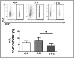
FIGS. 1A-1E show that topical tacrolimus reduces tail lymphedema. Figure 1A shows representative photographs of the tail of mice after surgical resection of the superficial/deep collecting lymphatic system and after starting treatment with or without topical tacrolimus at 2 weeks (early treatment) or 6 weeks (late treatment) post-surgery. The arrows indicate the start of therapy. Fig. 1B is a graphical representation of tail volume change following treatment with early (═ 0.021) or late (═ 0.018) tacrolimus compared to controls. Figure 1C (upper panel) shows cross-sectional histological images of control and early tacrolimus treated mouse tails harvested 6 weeks after lymphatic ablation. Parenthesis shows the soft tissue thickness. Figure 1C (lower panel) shows quantification of soft tissue changes after early or late treatment with tacrolimus (═ p ═<0.001). FIG. 1D shows quantification of tacrolimus levels in whole bloodSystemic treatment of animals (4mg kg) was demonstrated-1IP daily) and non-immunosuppressive levels in the topical treatment group (═ p ═ 0.007). Figure 1E shows flow cytometry plots (top plot) and quantification of blood T cells (bottom plot) in animals treated with vehicle control, topical tacrolimus, or systemic tacrolimus. Flow charts represent Side Scatter Area (SSA) on the y-axis and CD3 on the x-axis+Represents a T cell. Note the significant reduction of T cells in systemic treated animals only (═ p ═ 0.012).
Figures 2A-2D show that topical tacrolimus reduces inflammation following lymphatic injury. FIGS. 2A-2D show CD45+(FIG. 2A)iAnd 2Aii)、CD3+Cells (FIG. 2B)iAnd 2Bii)、CD4+Cells (FIG. 2C)iAnd 2Cii) And IFN-gamma+Cells (FIG. 2D)iAnd 2Dii) Representative 40x images of tail tissue sections harvested from control and tacrolimus treated animals 6 weeks post-surgery with immunofluorescence localization of (a). A higher magnification (80x) image is shown in the upper right hand inset of each figure. Lymphatic vessels (LYVE-1) were stained in each panel. Quantification of cell counts for both early and late stage treatment (all p) is shown on the right of each panel<0.001)。
Figures 3A-3D show that topical tacrolimus reduces fibrosis in lymphedema. FIG. 3AiAnd 3AiiRepresentative 40x images of tail tissue harvested 6 weeks post surgery from early treatment with control or with tacrolimus in the case of immunofluorescence localization of type I collagen and lymphatic vessels are shown. Quantification of collagen I stained areas in both early and late stage treatment (both p) is shown on the right<0.001). FIG. 3BiAnd 3BiiRepresentative 40x images of sirius red (pimecrius red) staining of tail tissue harvested from control and early tacrolimus treated animals are shown (collagen I and collagen III deposition). Quantification of scar index (red: green ratio) (p ═ 0.036 early; p) is shown on the right<0.001 late stage). FIG. 3Ci/iiAnd 3Di/iiIt is shown that TGF-. beta.1 or p is present in tail tissue harvested 6 weeks post-surgery from animals treated with control or early treatment with tacrolimusSMAD3 co-localized with representative 40x immunofluorescence of lymphatic vessels. The number of positive cells/0.25 mm for both are shown on the right of each figure2And (5) quantizing the area.
Figures 4A-4G show that tacrolimus improves lymphatic function following surgical lymphatic injury. Figure 4A shows representative ICG images of the tail of mice 6 weeks post-surgery with or without early treatment with tacrolimus. Note the flow of proximal ICG between the wounds in tacrolimus treated animals. The inset shows a photograph of a mouse in the same orientation. FIG. 4BiAnd 4BiiShowing that control and early tacrolimus (starting 2 weeks post-surgery) treated animals were treated 6 weeks post-surgical suture of the superficial/deep lymphatic system by sacral lymph node pairs99mDecay-regulated uptake of Tc (═ p ═ 0.005). FIG. 4BiiThe general photograph of the mouse tail shown in the upper figure of (a) is for orientation, and shows99mTc injection site and location of sacral lymph node; representative heat maps are shown in the lower panel, white arrows pointing to the sacral lymph nodes. Fig. 4C shows representative ICG images of hind limbs obtained 50 minutes after distal foot injection in mice treated with or without tacrolimus 4 weeks after PLND. White arrows show cutaneous lymphatic return of ICG. The inset shows the orientation. FIG. 4DiAnd 4DiiIs a graphical representation of lymphatic vessel pulsation in the hindlimb collecting vessels of mice treated with or without tacrolimus 4 weeks after PLND. The right panel shows the quantification of the pulsation frequency (═ 0.001). FIG. 4E shows inflammatory cells in tissue harvested 4 weeks after PLND from distal hind limbs of animals treated with control or tacrolimus (CD 45)+(ii) a Upper panel), iNOS+Cells (lower panel) and lymphatic vessels (LYVE-1)+) Representative 40x fluorescence co-localized images. Note CD45+And iNOS+The extracellular lymph is accumulated. FIGS. 4F and 4G show perilymph CD45 in distal hindlimb tissue of control or tacrolimus-treated animals harvested 4 weeks after PLND+Cells (FIG. 4F) and iNOS+Quantification of cells (FIG. 4G).
Figures 5A-5D show that topical tacrolimus increases collateral lymphangiogenesis following lymphatic injury. FIG. 5AiShowing control and early treatment tacrolimus harvested 6 weeks after bridging lymphatic injurySurgery of mice created representative longitudinal immunofluorescence 40x images of LYVE-1 tubes of tail trauma; the inset shows the area where the longitudinal slice is obtained. FIG. 5AiiQuantification of bridged Lymphatic Vessel Density (LVD) in the wounded part of the tail in control versus early or late treatment tacrolimus mice (both p)<0.001). FIG. 5AiiiqPCR of RNA harvested from control and early treatment tacrolimus mouse tail tissue harvested 6 weeks after lymphatic injury is shown demonstrating the relative expression of VEGF-C (p 0.264), TGF- β 1(p 0.006), and IFN- γ (p 0.014). Fig. 5B is a photograph of a mouse hind limb showing the sites of collateral vessel formation draining to the inguinal lymph node before and after PLND. FIG. 5C shows LYVE-1 in control and tacrolimus treated animals 4 weeks after PLND+Representative ICG (left panel) and 40x immunofluorescence images of the tubes (right panel shows the area in the box). FIG. 5D shows quantification of collateral lymph LVD (p) in the anterior leg region of animals treated with control or tacrolimus<0.001)。
Figures 6A-6F show that tacrolimus increases inflammatory lymphangiogenesis. (A) Para-lymphatic vessels (LYVE-1) harvested 2 weeks after suture placement and treatment with vehicle control or systemic tacrolimus+Weak CD31+) And blood vessels (CD 31)+/LYVE-1-) Representative gross (left panel) and bulk 5x images (right panel) of stained mouse corneas. The boxes represent the areas displayed in the package image. FIGS. 6B and 6C show corneal lymphatics (LYVE-1)+) And blood vessels (CD 31)+/LYVE-1-) Quantization of (2). FIG. 6D shows LYVE-1 localization in middle ear trauma in control or topical tacrolimus treated animals harvested 4 weeks after trauma+Representative fluorescence package 5x images. The inset shows the area where the slices were obtained. FIG. 6E shows LYVE-1 in the ear skin+Quantification of the stained area (within 400 μm of the wound) demonstrates an increase in lymphangiogenesis (p) in tacrolimus treated animals<0.001). FIG. 6F shows quantification of lymphatic branch points per unit area in ear wounds with and without tacrolimus treatment, demonstrating increased branching (p) in tacrolimus treated animals<0.001)。
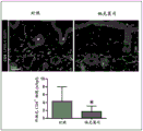
FIG. 7 shows that topical tacrolimus does not reduce circulating CD4+T cells. Peripheral blood CD4 was shown after 2 weeks treatment with topical, systemic or vehicle control+Representative flow diagrams of cells (top panel) and CD4+Quantification of T cells (lower panel).
Figure 8 shows that topical tacrolimus reduces macrophage infiltration in postoperative lymphadenectasis. The upper diagram shows the structure at F4/80+Representative 40x images of tail tissue sections of control and early local tacrolimus treated animals harvested 6 weeks post-surgery with immunofluorescence localization of cells. Quantification of both early and late stage treatment experiments is shown below.
Figures 9A-9C show the popliteal lymph node apheresis model. Figure 9A shows the popliteal lymph node, filled with evans blue contrast agent, visible in the popliteal fat pad. Figure 9B shows the popliteal lymph node, and the afferent and efferent collectors separated along with their surrounding fat pad. Fig. 9C shows evans blue contrast agent freely spilled in the surgical site after surgical resection.
FIGS. 10A-10B show that PLND results in increased ROS and DAMP compared to sham control limbs. Figure 10A shows representative mouse images (left panel) showing luminescence in hind limbs (indicative of ROS) immediately (6 hours) and after 1 week of PLND. Quantification of luminescent photons (right panel) shows significantly increased ROS levels in the PLND region at 1 week, but not in the sham control. Figure 10B shows representative immunofluorescence images of hindlimb skin sections from sham operated and PLND mice stained for HSP-70 (upper panel) and HMGB1 (lower panel).
FIG. 11 shows perilymphatic CD4 following PLND reduction by local tacrolimus+And (4) cell infiltration. The upper panel shows CD4 in animals treated with control or tacrolimus and harvested 4 weeks after PLND+Cells and lymphatic vessels (LYVE-1)+) Representative images of immunofluorescence localization of (a). Quantization is shown below.
FIGS. 12A-12B show that topical tacrolimus reduces perilymph F4/80 following PLND+And (4) cell infiltration. FIG. 12A shows representative immunofluorescence images of sections of PLND hindlimb skin tissue stained for both lymph vessel (LYVE-1) and macrophages (F4/80) with tacrolimus and vehicle treatment. FIG. 12B showsQuantification of perilymph F4/80+ macrophages was obtained.
Figure 13 shows that topical tacrolimus does not alter collective lymphatic vessel pulsation or lymphangiogenesis in the absence of lymphatic injury or inflammation. The upper panel is a representative image of NIR lymphatic images of the hindlimb lymphatic system in sham operated mice (i.e. anesthetized without incision or PLND) after 2 weeks of treatment with control or topical tacrolimus. Quantification of the frequencies of the collective lymphatic pulsation after 3 or 14 days of treatment with tacrolimus is shown below.
Fig. 14A-14B show that topical tacrolimus treatment did not alter vascular permeability following lymphatic injury. Figure 14A shows representative gross images of tacrolimus or vehicle treated PLND mouse hind limbs following tail vein evans blue injection to measure vascular permeability. Figure 14B shows quantification of absorbance of evans blue from formamide extraction of tacrolimus and vehicle treated PLND hindlimb tissue.
Figures 15A-15D show that local tacrolimus after PLND did not alter the alpha-SMA coverage or lumen diameter of the hindlimb lymph collector. FIG. 15A shows (from left to right) a brightfield image of the lateral aspect of the mouse hind limb with the cross-sectional level shown as a yellow oval; NIR images of mouse hind limbs, showing the anatomy of hind limb lymphatic vessels, with two large-diameter vessels on the sides; 5x IF image of mouse hind limb, yellow box indicating front leg, where two dominant collecting lymphatic vessels were located; a 20x image of the area with the dominant manifold is located (indicated by the white arrow). Figure 15B shows a representative 100x image of a cross section of pooled lymphatic vessels after double immunofluorescent staining for podophysin (Podoplanin) and a-SMA. Fig. 15C shows quantification of luminal area. Fig. 15D shows quantification of α -SMA thickness.
Figure 16 shows that topical tacrolimus does not increase lymphangiogenesis in the absence of injury/inflammation. The upper panel shows representative 5x images of immunofluorescent staining of lymphatic vessels in the ear of atraumatic mice 4 weeks after application of topical tacrolimus or vehicle control. LYVE-1 is shown below+Quantification of stained areas.
Figures 17A-17I show that inhibition of TGF- β improves lymphatic function and reduces perilymph accumulation of inflammatory cells following popliteal lymph node dissection. Figure 17A shows representative photographs of near-infrared images of distal hind limbs of mice treated with isotype control or TGF- β monoclonal antibody (mAb) 4 weeks after PLND (upper panel) and pumping frequency in the collective lymphatic system (lower panel). Note the increased pumping frequency of the hindlimb aggregators in the TGF- β mAb treatment description. Also note cutaneous lymphatic return (white arrows) in control but not TGF- β mAb treated mice. Figure 17B shows quantification of aggregate lymphatic pumping (pulsing) frequency in control and TGF- β mAB treated mice. Figure 17C shows quantification of cutaneous lymphatic return in control and TGF- β mAB treated mice. Figure 17D shows a representative flow cytometry plot of distal hindlimb tissue from control and TGF- β mAb treated mice 4 weeks after PLND. Note the percentage of reduction in CD3+ CD4+ cells in TGF- β mAb treated mice. FIG. 17E shows representative low and high (inset) magnification micrographs of perilymph (LYVE-1+) inflammatory cells (CD45 +; top) and iNOS + (bottom) cells in control and TGF- β mAb treated mice. FIG. 17F shows quantification of flow cytometry of CD4+ cells in hindlimb tissues in control and TGF- β mAb treated mice. FIG. 17G shows quantification of the number of LYVE-1+ tubes in control and TGF- β mAb treated mice. FIG. 17H shows quantification of the number of perilymphatic CD45+ cells in control and TGF- β mAb treated mice. FIG. 17I shows quantification of the number of exolymphoid iNOS + cells in control and TGF- β mAb treated mice.
Figures 18A-18G show that systemic pirfenidone improves lymphatic function in hindlimbs following surgical lymphatic injury. Figure 18A (upper panel) shows representative NIR images of hindlimbs obtained 50 minutes after distal foot injection of ICG in mice treated or not with pirfenidone 4 weeks after PLND. White arrows show cutaneous lymphatic return of ICG. The inset photograph is for orientation. The lower panel shows a graphical representation of lymphatic pulsation in hindlimb collecting vessels of mice treated with or without pirfenidone 4 weeks after PLND. Figure 18B shows quantification of pulsatile frequency (pulses/min) on the right (n ═ 6 animals/group;. p;)<0.05). Fig. 18C shows quantification of cutaneous lymphatic return in control and pirfenidone treated mice. FIG. 18D shows LYVE-1 in distal hindlimb tissue of control or pirfenidone treated animals harvested 4 weeks after PLND+Tube/0.25 mm2Quantification of area (n-6 animals/group; p for LYVE-1)<0.001). FIG. 18E shows inflammatory cells in tissue harvested 4 weeks after PLND from distal hind limbs of animals treated or not with pirfenidone (CD 45)+(ii) a Upper panel), iNOS+Cells (lower panel) and lymphatic vessels (LYVE-1)+) Representative low and high (inset) magnification fluorescence co-localized images of (a) were scaled to 100 μm. The higher magnification (80x) image is shown in the lower right inset of each plot, scale bar 20 μm. Note CD45+And iNOS+The extracellular lymph is accumulated. FIG. 18F shows perilymph CD45 in control and pirfenidone treated mice+Quantification of cells. FIG. 18G shows perilymphatic iNOS in control and pirfenidone-treated mice+Quantification of cell/hpf.
FIGS. 19A-19I show that systemic and localized pirfenidone reduces tail lymphedema and inflammation in mice. Figure 19A shows representative photographs of mouse tails after systemic and topical treatment with pirfenidone or vehicle control. Treatment was started 7 weeks after tail lymph injury. Note the significant improvement in pirfenidone treated mice. Fig. 19B shows quantification of tail volume in mice treated with control, topical, and systemic pirfenidone. Note the significant reduction in pirfenidone treated mice (arrows show when pirfenidone treatment was initiated). Fig. 19C shows quantification of fibroadipose tissue deposition in control and pirfenidone treated mice. Fig. 19D shows quantification of type I collagen deposition in control and pirfenidone treated mice. Figure 19E shows representative cross-sectional micrographs of mouse tails treated with control or pirfenidone (systemic or local). FIG. 19F shows a representative high magnification micrograph of a section of the tail stained for type I collagen and lymphatic vessels (LYVE-1). Note the reduced type I collagen deposition in pirfenidone treated mice. FIG. 19G shows sulfur colloid conjugated99After distal tail injection of Tc99Peak nodal uptake of Tc. Note the increased uptake of pirfenidone to the sacral lymph nodes in mice treated. FIG. 19H shows after distal tail injection99Lymph node uptake rate of Tc. Note the faster uptake in pirfenidone treated mice. FIG. 19I showsFor white blood cell (CD 45)+) And representative high magnification micrographs of lymph vessel (LYVE-1) stained tail sections. Note the reduced perilymph CD45 in pirfenidone treated mice+The cells accumulate.
FIGS. 20A-20K show that pirfenidone reduces perilymphatic inflammation and TGF- β expression. Figure 20A shows representative micrographs of hindlimb sections demonstrating perilymph accumulation of CD4+ cells in control and pirfenidone treated animals (systemic treatment is shown in the left panel; local treatment is shown in the right panel). The high magnification image is shown in the inset. FIG. 20B shows representative micrographs of hindlimb sections demonstrating perilymph accumulation of TGF-. beta.1 + cells in control and pirfenidone treated animals (systemic treatment is shown in the left panel; local treatment is shown in the right panel). Figure 20C shows representative micrographs of hindlimb sections demonstrating perilymph accumulation of SMAD3+ cells in control and pirfenidone treated animals (systemic treatment is shown in the left panel; local treatment is shown in the right panel). Fig. 20D shows quantification of perilymphatic CD4+ cells in control and pirfenidone treated mice. FIG. 20E shows quantification of exolymphoid TGF- β 1+ cells in control and pirfenidone treated mice. Fig. 20F shows quantification of perilymph SMAD3+ cells in control and pirfenidone treated mice. Figure 20G shows high magnification micrographs of hindlimb collecting vessels from control and pirfenidone treated mice stained for pediludin, alpha-SMA, and type I collagen. Note thickening and proliferation of α -SMA + cells in control mice. Fig. 20H shows quantification of type I collagen deposition around the collective lymphatic system in control and pirfenidone treated mice. FIG. 20I shows serum TGF- β 1 expression in control and systemic pirfenidone-treated mice. Figure 20J shows serum IFN- γ expression in control and systemic pirfenidone-treated mice. FIG. 20K shows serum VEGF-C expression in control and systemic pirfenidone-treated mice.
FIGS. 21A-21F show that loss of TGF- β expression by T cells, but not myeloid cells, prevents the development of lymphedema following lymphatic injury. FIG. 21A shows slave control, T cells 6 weeks after tail lymph injurycreAnd medulla samplecreRelative representation of TGF-beta 1mRNA in animal harvested tail tissueSo as to achieve the purpose. FIG. 21B shows control, myeloid TGF-. beta.1creAnd T cell-TGF-. beta.1creRepresentative micrographs of mice. Note T cellscreThe swelling was absent in the mice. Fig. 21C shows quantification of tail volume of mice in each group. Note T cells compared to wild type controlscreReduced tail volume in mice. FIG. 21D shows pairs H in each group&Representative micrographs of E (apical), type I collagen/LYVE-1 (intermediate) stained tail sections and quantification of fibroadipose tissue deposition and type I collagen expression (inferior). Note T cellscreReduced fibroadipose tissue deposition in mice. FIG. 21E shows injection in the distal tail99Peak nodal uptake of Tc. Note T cellscreIncreased uptake in mice. FIG. 21F shows the results of the respective groups99The sacral nodule uptake rate of Tc. Note T cellscreFaster uptake in mice.
FIGS. 22A-22G show that mice lacking TGF- β 1-expressing T cells have reduced perilymph inflammation and TGF- β 1 expression. Figure 22A shows representative micrographs of mouse hindlimbs from each group stained for CD4+ cells (top), IL13+ cells (middle), pSMAD3+ cells (bottom), and lymphatic vessels (LYVE-1 +). Note T cellscreReduced CD4+ cell accumulation, reduced IL13+ cell number, and reduced pSMAD3+ cell number in mice. FIG. 22B shows CD4 in hindlimb tissue of animals in each group+Quantification of cells. FIG. 22C shows quantification of Th2 cells (CD4+/IL13+) in hindlimb tissue of animals in each group. FIG. 22D shows pSMAD3 in hindlimb tissue of animals in each group+Quantification of cells. FIGS. 22E-22G show myeloid samples from controlCreAnd T cellscreSerum levels of IFN-. gamma.protein concentrations (FIG. 22E), TGF-. beta.1 (FIG. 22F), and VEGF-C (FIG. 22G) in mice.
Figures 23A-23H show that impaired LEC TGF- β 1 responsiveness is devoid of changes in tail lymphedema, inflammation, or fibrosis, but improved lymphangiogenesis. FIG. 23A shows a cross-sectional view of a pSMAD+Control and FLT4 in co-localization with immunofluorescence of LYVE-1creRepresentative high magnification (80x) images of lymph node sections harvested from mice, scale bar 10 μm.Arrows indicate LYVE-1+Co-localization of pSMAD3 in tubes. FIG. 23B shows post-operative 6 weeks control and FLT4creQuantification of tail TGF- β 1 protein concentration in mice (n-5 animals/group; p-NS in both groups). FIG. 23C shows control and FLT4 after surgical excision of superficial/deep pool lymphatic system 6 weeks after surgerycreRepresentative photographs of the tail of the mouse. FIG. 23D shows FLT4 compared to controlcreGraphical representation of tail volume changes in the tail of mice (n-5 animals/group; p-NS). FIG. 23E shows control and FLT4creQuantification of soft tissue changes in mice (n-5 animals/group; p-NS). Fig. 23F shows quantification of collagen I staining regions (n-5 animals/group; p-NS). FIG. 23G (upper panel) shows control and FLT4 harvested 6 weeks after lymph ablationcreRepresentative cross-sectional histological images of the tail of the mice. Brackets indicate soft tissue thickness. Scale bar 500 μm. FIG. 23G (middle panel) shows immunofluorescence co-localization of collagen type I and lymphatic vessels 6 weeks post-surgery from control and FLT4creRepresentative 40x images of tail tissue harvested from animals. Figure 23G (bottom panel) shows the positioning of pSMAD3 and lymphatic vessels. Scale bar 100 μm. FIG. 23H shows control and FLT4 at 6 weeks post-surgery in the case of immunofluorescence localization of LYVE-1creRepresentative higher magnification (60x) images of longitudinal tail tissue sections harvested from tail wounds of mice, scale bar 50 μm. Control and FLT4creMouse bridged LYVE-1+Tube Density (LVD) (n/0.25 mm)2Area) (n-5 animals/group; p<0.01)。
FIGS. 24A-24D show that teriflunomide reduces lymphedema. Fig. 24A shows gross micrographs of mouse tails treated with vehicle (control) or topical teriflunomide 6 weeks after lymphatic ablation. Fig. 24B shows the tail volume change 6 weeks after lymphatic ablation in animals treated with vehicle or topical teriflunomide ((. p < 0.0002). fig. 24C shows histological sections of the mouse tail harvested 1cm distal to the area of lymphatic injury in control and teriflunomide-treated mice.
Figures 25A-25D show that teriflunomide reduces fibrosis. Figure 25A shows representative micrographs of tail sections of mice from control and teriflunomide treated mice, locating type I collagen fibers and the cutaneous lymphatic system. Note that the fibrosis was significantly reduced in teriflunomide treated mice. Figure 25B shows quantification of type I collagen deposition in tail of mice treated with vehicle control or topical teriflunomide (p < 0.001). Figure 25C shows representative micrographs of the main hindlimb collecting vessels in animals treated with control or teriflunomide, locating alpha-SMA and podoprotein. Note the reduced proliferation of α -SMA positive cells and the broader lumen of the collective lymphatic system in teriflunomide treated mice. Figure 25D shows quantification of extralymphatic smooth muscle thickness in control and teriflunomide treated mice after PLND (./p < 0.05).
Figures 26A-26B show that teriflunomide reduces inflammation. Figure 26A shows representative micrographs of tail sections of mice from control and teriflunomide treated mice, locating CD4+ cells and the cutaneous lymphatic system. There was a significant reduction in CD4+ cell infiltration in teriflunomide treated animals. Box insertion shows a high magnification view (80 x). Figure 26B shows quantification of CD4+ cells in tail of mice treated with vehicle control or topical teriflunomide (./p < 0.0001).
Figures 27A-27D show that teriflunomide increases lymphangiogenesis. Figure 27A shows representative photomicrographs of near-infrared imaging of mouse hindlimb lymphatic system and collateral formation (white circles) in animals treated with vehicle control or teriflunomide. The newly formed collateral lymphatic system bypasses the popliteal lymph node in teriflunomide-treated mice. Figure 27B shows the quantification of collateral lymphadenopathy in animals post PLND and treated with vehicle control or teriflunomide (./p < 0.001). Figure 27C shows representative micrographs of tail trauma in mice treated with vehicle control or topical teriflunomide, locating newly formed transected lymphatic vessels. There was a clear increase in lymphangiogenesis in teriflunomide treated mice. Figure 27D shows quantification of collateral lymphatic system in tail wounds of control and teriflunomide treated animals 6 weeks after lymphatic ablation (./p < 0.001).
Figure 28 shows that topical teriflunomide reduces lymphatic leakage. Representative near infrared images of lymphatic vessels in the hind limbs of mice treated with vehicle control or teriflunomide following PLND. Note the reduced leakage of lymphatic vessels (arrows) in teriflunomide treated mice.
FIGS. 29A-29B show that topical teriflunomide increases lymphatic function. Figure 29A shows a representative flow cytometry plot of Dendritic Cells (DCs) transported in the inguinal lymph node of mice treated with vehicle control or teriflunomide following PLND. Mice were treated with a topical formulation of FITC in the distal hind limb to label tissue resident DCs, and after 24 hours, inguinal lymph nodes were harvested and analyzed using flow cytometry to quantify the number of DCs that had been transported from the periphery. A significant increase in DC trafficking indicates improved lymphatic function in teriflunomide-treated animals. Figure 29B shows quantification of DC trafficking in control and teriflunomide treated animals (n ═ 6;. p < 0.0001).
Figures 30A-30B show that topical teriflunomide increases lymphatic pumping. Figure 30A shows a graph of hindlimb collective lymphatic pumping in control and teriflunomide treated mice after PLND. Figure 30B shows quantification of hindlimb collective lymphatic pumping frequency in control and teriflunomide treated mice after PLND. Note the significant increase in pumping in teriflunomide treated animals (./p < 0.002).
FIGS. 31A-31B show that captopril treatment increases dendritic cell trafficking following lymphatic injury. Figure 31A shows representative flow cytometry from inguinal lymph node demonstrating FITC + CD11c cells in control and captopril treated mice after PLND. Figure 31B shows the percentage (left) and absolute number (right) of FITC + CD11c cells in the inguinal lymph node of control and captopril treated mice after PLND.
Figures 32A-32B show that captopril treatment increased hindlimb collective lymphatic pumping. Figure 32A shows a representative plot of aggregate lymphatic pumping as assessed by ICG lymphangiography in control and captopril treated mice after PLND. Fig. 32B shows quantification of the bag frequency (pumping) of the hindlimb collective lymphatic system.
FIGS. 33A-33C show T cell infiltration following captopril treatment to reduce PLND. Representative high magnification micrographs of control (FIG. 33A) and captopril treated (FIG. 33B) hindlimb sections stained for CD3+ (T cell marker) and LYVE-1 (lymphoid marker) are shown. Counterstaining was shown with DAPI. Figure 33C shows quantification of CD3+ cells in hindlimb tissue of control and captopril treated mice.
FIGS. 34A-34C show macrophage infiltration following captopril treatment to reduce PLND. Representative high magnification micrographs of hindlimb sections of F4/80+ (macrophage marker) and LYVE-1 (lymph marker) stained control (FIG. 34A) and captopril treated (FIG. 34B) mice are shown. Counterstaining was shown with DAPI. FIG. 34C shows quantification of f4/80+ cells in hindlimb tissue of control and captopril treated mice.
FIGS. 35A-35D show lymphangiogenesis following captopril treatment to increase PLND. Representative high magnification micrographs of sections of hind limbs of control (FIG. 35A) and captopril-treated (FIG. 35B) mice stained for LYVE-1 (a lymphoid marker) are shown. Counterstaining was shown with DAPI. Figure 35C shows quantification of LYVE-1+ tubes (/ p <0.05) in control and captopril treated mice.
Figure 36 shows that captopril treatment reduced foot swelling following hind limb lymph depletion with DT. Representative photographs of the feet of mice in the control (top) and captopril treated (bottom) groups at various times after DT lymph depletion. Note the significant reduction in paw swelling in captopril treated mice.
Figures 37A-37C show that captopril treatment reduced T cell infiltration following hindlimb lymph depletion with DT. Representative high magnification micrographs of control (FIG. 37A) and captopril-treated (FIG. 37B) hind limb sections stained for CD3+ (T cell marker) and LYVE-1 (lymphoid marker). Ipsilateral tissue was harvested from the limb treated with DT, while contralateral tissue was from the opposite untreated limb. Counterstaining was shown with DAPI. Figure 37C shows quantification of CD3+ cells in hindlimb tissue of control and captopril treated mice. Note the significant difference between ipsilateral controls and ipsilateral captopril limbs (./p < 0.05).
FIGS. 38A-38C show that captopril treatment reduced hindlimb macrophage infiltration after lymphatic ablation with DT. Representative high magnification micrographs of sections of the hind limb of control (FIG. 38A) and captopril-treated (FIG. 38B) mice stained for F4/80 (macrophage marker) and LYVE-1 (lymph marker) are shown. Counterstaining was shown with DAPI. FIG. 38C shows quantification of F4/80+ cells in hindlimb tissue of control and captopril treated mice. Note the significant difference between ipsilateral controls and ipsilateral captopril limbs (./p < 0.005).
Figures 39A-39C show that captopril treatment reduced hindlimb collecting duct smooth muscle deposition after lymphatic ablation of DT. Representative high magnification micrographs of control (fig. 39A) and captopril treated (fig. 39B) hind limb sections stained for alpha smooth muscle actin (alpha-SMA) (smooth muscle marker) and podoprotein (lymph marker). Counterstaining was shown with DAPI. Figure 39C shows quantification of α -SMA + cells in hindlimb tissue of control and captopril treated mice. Note the significant difference between ipsilateral controls and ipsilateral captopril limbs (./p < 0.005).
Figures 40A-40C show that captopril treatment reduced hindlimb type I collagen deposition following lymphatic depletion with DT. Representative high magnification micrographs of control (fig. 40A) and captopril treated (fig. 40B) mouse hindlimb sections stained for type I collagen (fibrosis marker) and LYVE-1 (lymph marker) are shown. Counterstaining was shown with DAPI. Fig. 40C shows quantification of type I collagen in hindlimb tissue of control and captopril treated mice. Note the significant difference between ipsilateral controls and ipsilateral captopril limbs (./p < 0.0001).
Figures 41A-41C show that captopril treatment reduced hindlimb Angiotensin Converting Enzyme (ACE) expression after lymphatic ablation with DT. Representative high magnification micrographs of ACE and LYVE-1 (lymphoid marker) stained control (fig. 41A) and captopril treated (fig. 41B) mouse hindlimb sections. Counterstaining was shown with DAPI. Figure 41C shows quantification of ACE in hindlimb tissue of control and captopril treated mice. Note the significant difference between ipsilateral controls and ipsilateral captopril limbs (./p < 0.0001).
Figure 42 shows that captopril treatment increases the formation of collateral lymphatic system following hindlimb lymphatic ablation with DT. Representative ICG photographs of control (left panel), captopril (middle), and normal lymphoid architecture (i.e., no DT treatment) hind limbs are shown. The dotted circles represent the areas where the collateral lymphatic system is formed to drain into the inguinal lymph nodes.
Figures 43A-43D show that captopril treatment reduced hindlimb lymphangiogenesis following lymphatic ablation with DT. Representative high magnification micrographs of sections of hind limbs of control (FIG. 43A) and captopril-treated (FIG. 43B) mice stained for LYVE-1 (a lymphoid marker). Counterstaining was shown with DAPI. FIG. 43C shows quantification of LYVE-1+ lymphatic vessels in hindlimb tissues of control and captopril-treated mice. Note the significant difference between ipsilateral controls and ipsilateral captopril limbs (./p < 0.0001). FIG. 43D shows quantification of LYVE-1+ lymphatic vessel area in hindlimb tissue of control and captopril treated mice. Note the significant decrease between ipsilateral control and ipsilateral captopril limb (p < 0.005).
Figure 44 shows that captopril treatment reduced limb volume following hind limb lymphatic ablation with DT. Quantification of hindlimb volume in control and captopril treated mice after lymphatic ablation with DT. Note the significant reduction in captopril treated mice (./p < 0.007).
Figures 45A-45B show that captopril treatment reduced tail lymphedema in mice 6 weeks after lymphatic ablation. Figure 45A shows representative photographs of the tail of mice pre-operatively and weekly, after lymphatic ablation and treatment with vehicle control or captopril. Arrows indicate the timing of the start of treatment. Figure 45B shows quantification of mouse tail volume change in control and captopril treated mice.
Figures 46A-46B show fibroadipose tissue deposition in a tail model of mice with captopril treatment reducing lymphedema at 6 weeks after lymphadenectomy. Figure 46A shows representative H & E stained mouse tail sections 6 weeks after lymphatic ablation and topical treatment with control (left) or captopril. Brackets indicate fibroadipose tissue deposition. Fig. 46B shows quantification of subcutaneous fibrofatty deposits in mice topically treated with control or captopril 6 weeks after tail lymph ablation. Note the significant reduction in captopril treated mice (./p < 0.0001).
Figures 47A-47C show that captopril treatment increased mouse tail manifold pumping frequency 6 weeks after lymphatic ablation. Representative photographs of indocyanine green assays (top) of mice tails topically treated with control (fig. 47A) or captopril ointment (fig. 47B) for 6 weeks are shown. A line graph representing the collective lymphatic contractions is shown under the photograph. Note the increased number of contractions in captopril treated animals. Figure 47C shows quantification of contraction frequency of mouse tail collective lymphatic system in control and captopril treated animals. Note the increased frequency of captopril treated mice (. p < 0.01).
FIGS. 48A-48C show the number of lymphatic vessels between tail wound regions in a mouse tail model of increased lymphedema at 6 weeks post-lymphatic ablation with captopril treatment. Representative longitudinal sections of mouse tails in LYVE-1 (lymphoid marker) stained control (FIG. 48A) and captopril (FIG. 48B) treated animals are shown. FIG. 48C shows quantification of LYVE-1+ tube density in control and captopril treated animals 6 weeks after tail lymph ablation.
Figures 49A-49C show that captopril treatment reduced lymphatic vessel area and was associated with reduced lymphatic stasis in a mouse tail model of lymphedema 6 weeks after lymphatic ablation. Representative cross sections of mouse tails in LYVE-1 (lymphoid marker) stained control (FIG. 49A) and captopril (FIG. 49B) treated animals are shown. Note the reduced lymphatic vessel diameter in captopril treated mice. FIG. 49C shows quantification of LYVE-1+ tube area in control and captopril mice. Note the significant reduction in lymphatic vessel area corresponding to reduced lymphatic stasis.
Figures 50A-50C show skin fibrosis and collagen type I deposition in a mouse tail model of captopril treatment to reduce lymphedema at 6 weeks post-lymphatic ablation. Representative sections of mouse tails from control (FIG. 50A) and captopril (FIG. 50B) treated animals stained for type I collagen, LYVE-1 and DAPI are shown. Note that captopril treated reduced fibrosis in animals. Figure 50C shows quantification of mouse tail skin type I collagen staining area in control and captopril treated mice. Note the significant reduction in captopril treated animals (. < 0.0001).
FIGS. 51A-51C show Angiotensin Converting Enzyme (ACE) expression in a tail model of mice treated with captopril to reduce lymphedema at 6 weeks post-lymphatic ablation. Representative sections of mouse tails from ACE, LYVE-1 and DAPI stained control (FIG. 51A) and captopril (FIG. 51B) treated animals are shown. Note the reduced ACE expression in captopril treated animals. Figure 51C shows quantification of ACE staining area of mouse tail in control and captopril treated mice. Note the significant reduction in captopril treated animals (. < 0.0001).
FIGS. 52A-52C show perilymph accumulation of T cells (CD3+) in a mouse tail model of captopril treatment to reduce lymphedema at 6 weeks post-lymphatic ablation. Representative sections of mouse tails from control (FIG. 52A) and captopril (FIG. 52B) treated animals stained for CD3, LYVE-1 and DAPI are shown. Note the reduced ACE expression in captopril treated animals. Figure 52C shows quantification of mouse caudal perilymph CD3+ cells/tube in control and captopril treated mice. Note the significant reduction in captopril treated animals (. < 0.0001).
FIGS. 53A-53C show perilymph accumulation of macrophages (F4/80+ cells) in a mouse tail model of captopril treatment for reducing lymphedema at 6 weeks after lymphadenectomy. Representative sections of the tail of mice treated with control (FIG. 53A) and captopril (FIG. 53B) stained for F4/80, LYVE-1 and DAPI are shown. Note the reduced ACE expression in captopril treated animals. FIG. 53C shows quantification of mouse caudal perilymph F4/80 cells/tube in control and captopril treated mice. Note the significant reduction in captopril treated animals (. < 0.0001).
Detailed Description
The present invention relates in part to the use of anti-T cell agents and/or anti-TGF- β 1 agents and/or anti-angiotensin agents as a novel, safe and effective treatment for edema, particularly lymphedema. The present invention is based, in part, on the surprising discovery that systemic or local administration of anti-T cell, anti-TGF- β 1 and/or anti-angiotensin agents, such as tacrolimus, pirfenidone, teriflunomide, leflunomide and/or captopril, significantly improves lymphedema and lymphatic function, and has various other beneficial biological effects when administered to a mammalian subject, including stimulation of lymphangiogenesis. Furthermore, because these agents act at different steps in the fibrotic pathway, combinations of anti-T cell, anti-TGF- β 1 and/or anti-angiotensin agents may be more effective than administration of a single agent, potentially exhibiting a synergistic effect.
Accordingly, the present invention provides compositions and methods for treating or preventing edema, such as lymphedema, and/or for producing various other beneficial biological effects, including but not limited to: reducing tissue swelling, reducing lymphatic stasis or "pooling", reducing tissue fibrosis, reducing tissue inflammation, reducing leukocyte infiltration, reducing macrophage infiltration, reducing infiltration of naive and differentiated T cells, reducing expression of TGF-beta 1 and reducing expression and/or activation of downstream mediators (e.g., pSmad3), reducing angiotensin and/or ACE levels, reducing collagen deposition and/or scarring, improving or increasing lymphatic function, improving or increasing lymphatic fluid transport, improving or increasing lymphangiogenesis and/or improving or increasing lymphatic pulsation frequency.
Unless defined otherwise, all technical and scientific terms used herein have the same meaning as commonly understood by one of ordinary skill in the art to which this invention pertains. For example, The Dictionary of Cell and Molecular Biology (ed. 5 th edition, j.m. lackie, 2013), Oxford Dictionary of Biochemistry and Molecular Biology (d. 2 nd edition, ed. r.cammecack et al, 2008), and The Dictionary of Biomedicine and Molecular Biology (d. 2 nd edition, P-s.juo,2002) may provide The skilled artisan with a general definition of some of The terms used herein.
As used in this specification and the appended claims, the singular forms "a," "an," and "the" include plural referents unless the context clearly dictates otherwise. The terms "a" (or "an") and the terms "one or more" and "at least one" may be used interchangeably.
In addition, "and/or" will be taken to specifically disclose each of the two specified features or components, with or without the other. Thus, the term "and/or" as used in phrases such as "a and/or B" is intended to include a and B, A or B, A (alone) and B (alone). Also, the term "and/or" as used in phrases such as "A, B and/or C" is intended to include A, B and C; A. b or C; a or B; a or C; b or C; a and B; a and C; b and C; a (alone); b (alone); and C (alone).
Units, prefixes, and symbols are expressed in their system international system of units (SI) accepted form. Numerical ranges include the numbers defining the range. Where a numerical term is preceded by "about," that term includes the stated number and the value of the stated number plus or minus 10%. The headings provided herein are not limitations of the various aspects or embodiments of the invention which can be had by reference to the specification as a whole. Accordingly, the terms defined hereinafter are more fully defined by reference to the entire specification.
Wherever the language "comprising" is used to describe embodiments, including other similarly described embodiments in terms of "consisting of …" and/or "consisting essentially of.
As used herein, the term "edema" includes lymphedema, lymphatic dysfunction, lymphatic fibrosis, idiopathic edema, peripheral edema, and ocular edema. As used herein, "edema" does not include pulmonary edema or cerebral edema. Edema may include acute edema, chronic edema, postoperative edema, and progressive edema. Symptoms of edema may include swelling, fullness or edema of tissue, inflammation, fibrosis, heaviness, pain, reduced range of motion, soreness, recurrent infection, thickening or discomfort of the skin.
An "active agent" is an agent that is biologically active by itself or as a precursor or prodrug that is converted in vivo to an agent that is biologically active. Active agents for treating or preventing edema may include immunosuppressive agents, anti-fibrotic agents, anti-T cell agents, anti-TGF- β 1 agents, and anti-angiotensin agents. In some embodiments, the agent is a small molecule compound. In other embodiments, the agent is a macromolecule, such as a polynucleotide (e.g., inhibitory RNA) or a polypeptide (e.g., an antibody).
An "anti-T cell agent" is a molecule that reduces T cell-mediated inflammation, T cell activation, T cell differentiation, and/or T cell proliferation. Classes of anti-T cell agents include calcineurin inhibitors and IL-2 inhibitors. Examples of small molecule anti-T cell agents include tacrolimus, teriflunomide, leflunomide, cyclosporine, and pimecrolimus. Examples of macromolecular anti-T cell agents include dinenikin interleukin 2(denileukin diftotox) and Basiliximab (Basiliximab).
An "anti-TGF- β 1 agent" is a molecule that inhibits the expression, secretion, activation, signaling, or activity of transforming growth factor β 1. Pirfenidone is an example of a small molecule anti-TGF-beta 1 agent.
An "anti-angiotensin agent" is a molecule that inhibits the activity of AngI or AngII, or that inhibits the conversion of AngI to AngII (e.g., an ACE inhibitor or an ACE agonist). Examples of anti-angiotensin agents include captopril, zofenopril, enalapril, lisinopril, ramipril, quinapril, perindopril, benazepril, imidapril, trandolapril, cilazapril, fosinopril, losartan, irbesartan, olmesartan, candesartan, telmisartan, valsartan, fimasartan, acetylglycine diazenimine (dimazenate), xanthone, and AVE 099.
The terms "inhibit", "block" and "inhibit" are used interchangeably and refer to any statistically significant reduction in biological activity, including complete blocking of activity.
In one aspect, the methods of the invention may comprise administering a combination of anti-T cell, anti-TGF-beta 1, and/or anti-angiotensin agents. In a specific embodiment, the method comprises administering a pharmaceutical composition of the invention comprising: (i) an effective amount of one or more anti-T cell agents selected from the group consisting of: tacrolimus, teriflunomide, leflunomide, cyclosporine, pimecrolimus, disinulins 2, and basiliximab; and (ii) an effective amount of one or more anti-TGF- β 1 agents and/or one or more anti-angiotensin peptide agents selected from the group consisting of: pirfenidone, captopril, zofenopril, enalapril, lisinopril, ramipril, quinapril, perindopril, benazepril, imidapril, trandolapril, cilazapril and fosinopril, losartan, irbesartan, olmesartan, candesartan, telmisartan, valsartan and fimasartan. The methods of the invention may comprise administering a pharmaceutical compound of the invention comprising any combination of anti-T cell, anti-TGF- β 1 and/or anti-angiotensin agents. For example, in one embodiment, the method comprises administering a pharmaceutical composition comprising tacrolimus and pirfenidone. In another embodiment, the method comprises administering a pharmaceutical composition comprising tacrolimus, pirfenidone, and teriflunomide. In another embodiment, the method comprises administering a pharmaceutical composition comprising tacrolimus, pirfenidone, and leflunomide. In other aspects, the methods comprise administering a pharmaceutical composition comprising tacrolimus and captopril; or teriflunomide and captopril; or leflunomide and captopril; or pirfenidone and captopril; or tacrolimus, captopril and teriflunomide; or tacrolimus, captopril and leflunomide; or tacrolimus, captopril and pirfenidone.
By "subject" or "individual" or "patient" is meant any subject, preferably a mammalian subject, in need of diagnosis, prognosis or treatment. Mammalian subjects include humans, domestic animals, farm animals, sport animals, and zoo animals, including, for example, humans, non-human primates, dogs, cats, guinea pigs, rabbits, rats, mice, horses, cows, and the like.
In some embodiments, the subject may have or may already have a cancer, for example a cancer comprising a solid tumor. In some embodiments, the subject may have, or may have had, breast cancer or cancer affecting the female reproductive organs, the skin system, the musculoskeletal system, the soft tissues of the limbs or trunk, the male reproductive system, the urinary system, or the head and neck. In some embodiments, the subject may have undergone axillary lymph node resection. In some embodiments, the subject has received a cancer treatment and the edema, lymphedema, or lymphatic damage is associated with the cancer treatment or diagnosis. For example, the subject may be undergoing or may have undergone chemotherapy or radiation therapy for cancer treatment or other indications, or one or more lymph nodes may have been surgically resected during cancer treatment or diagnosis.
In some embodiments, the subject may have persistent lymphatic damage (e.g., as a result of removal, ligation, or obstruction of lymph nodes or vessels, or fibrosis of lymphatic tissue), or the subject may be obese or have had an infection that causes edema, such as lymphedema. In some embodiments, the infection may be a skin infection or a history of skin infection associated with lymphedema or lymphatic injury. In some embodiments, the infection may be a parasitic infection that impedes lymphatic flow or damages the lymphatic system. In some embodiments, the subject may have persistent lymphatic damage from joint replacement, trauma, burn, radiation, or chemotherapy.
Terms such as "treating" or "alleviating" refer to a therapeutic measure that cures, slows the diagnosed pathological condition or disorder, reduces symptoms of, and/or halts the progression of the diagnosed pathological condition or disorder. Thus, patients in need of treatment include those already with the disorder. In certain embodiments, a subject is successfully "treated" for a disease or disorder according to the methods provided herein if the patient exhibits, for example, total, partial, or transient alleviation or elimination of symptoms associated with the disease or disorder. For example, "treating edema" may include, but is not limited to, reducing swelling, reducing inflammation, reducing fibrosis, reducing pain, increasing range of motion, reducing heaviness, reducing tightness, reducing skin thickening, and/or improving lymphatic function.
"prevention" refers to a precautionary or preventative measure to prevent and/or slow the development of the targeted pathological condition or disorder. Thus, persons in need of prophylaxis include those at risk of developing or susceptible to developing the condition. Subjects at risk of developing or susceptible to developing lymphedema include, but are not limited to, cancer patients undergoing radiation therapy, chemotherapy, and/or surgical lymph node removal. In certain embodiments, if a patient develops, either transiently or permanently, e.g., fewer or less severe symptoms associated with a disease or disorder or a later onset of symptoms associated with a disease or disorder than a patient who has not been subjected to the methods of the invention, the disease or disorder is successfully prevented according to the methods provided herein.
In a prophylactic context, the pharmaceutical composition of the invention may be administered at any time before or after the subject is at risk of or susceptible to lymphoid injury and/or edema development, such as radiation therapy, chemotherapy or surgical lymph node clearance. In certain aspects, the pharmaceutical composition is administered prophylactically up to about one week prior to the event, such as 1,2, 3, 4, 5, 6, or 7 days prior to the event. In some cases, the pharmaceutical composition is administered prophylactically on the same day as the event. In some embodiments, the pharmaceutical composition is administered prophylactically within 6 weeks of the event, e.g., within about 1,2, 3, 4, 5, or 6 days of the event, or within about 1,2, 3, 4, 5, or 6 weeks. In one embodiment, the pharmaceutical composition is administered prophylactically for about 2-4 weeks or about 1,2, 3, 4, 5, or 6 weeks.
In some embodiments, the treatment and/or prevention methods described herein can be performed in combination with one or more additional edema or lymphedema treatment and/or prevention methods known in the art, such as treatment methods involving the administration of other therapeutic agents and/or treatment methods involving surgery, massage, compression therapy, drainage therapy, acupuncture, laser, or any other suitable treatment method.
The term "pharmaceutical composition" refers to a formulation in a form such as to allow effective biological activity of an active ingredient, and free of additional components having unacceptable toxicity to a subject receiving administration of the composition. The pharmaceutical composition may be in various dosage forms, such as tablets, capsules, liquids, solutions, soft capsules, suspensions, emulsions, syrups, elixirs, tinctures, films, powders, hydrogels, ointments, pastes, emulsions, lotions, gels, mousses, foams, lacquers, sprays, aerosols, inhalers, nebulizers, eye drops, patches, suppositories, and/or enemas. The pharmaceutical composition typically comprises a pharmaceutically acceptable carrier and may comprise one or more of the following: buffers (e.g., acetate, phosphate, or citrate buffers), surfactants (e.g., polysorbates), stabilizers (e.g., human albumin), preservatives (e.g., benzyl alcohol), permeation enhancers, absorption enhancers to enhance bioavailability, and/or other conventional solubilizing or dispersing agents. The choice of formulation and excipients depends on the active agent to be delivered and the disease or condition to be treated or prevented and is routine for one of ordinary skill in the art.
By "systemic administration" is meant administration of a pharmaceutical composition such that the active agent enters the circulatory system, for example, by enteral, parenteral, inhalation, or transdermal routes. The enteral routes of administration involve the gastrointestinal tract, including but not limited to oral, sublingual, buccal and rectal delivery. Parenteral routes of administration involve routes other than the gastrointestinal tract, including but not limited to intravenous, intramuscular, intraperitoneal, intrathecal, and subcutaneous. By "topical administration" is meant that the pharmaceutical composition is administered directly to the site where its action is desired (e.g., or at or near the site of injury or symptom). Topical routes of administration include, but are not limited to, topical, inhalation, subcutaneous, ocular, and otic. It is within the purview of one of ordinary skill in the art to formulate a pharmaceutical composition suitable for its intended route of administration.
An "effective amount" of a composition as disclosed herein is an amount sufficient to carry out the specifically recited purpose. The "effective amount" can be determined empirically and in a conventional manner with respect to the purpose, route of administration and dosage form.
In some embodiments, administration of the anti-T cell agent and/or anti-TGF- β 1 agent and/or anti-angiotensin agent may comprise systemic administration at any suitable dose and/or according to any suitable dosing regimen, as determined by one of skill in the art. For example, in some embodiments, tacrolimus or an analog, variant or derivative thereof is administered systemically to a subject at a daily dose of about 0.01mg/kg to about 5 mg/kg. More specifically, the subject, the limus, the derivative, or the derivative thereof may be administered at a daily dose of about 0.01, 0.015, 0.02, 0.025, 0.03, 0.035, 0.04, 0.045, 0.05, 0.055, 0.06, 0.065, 0.07, 0.075, 0.08, 0.085, 0.09, 0.095, 0.1, 0.2, 0.3, 0.4, 0.5, 0.6, 0.7, 0.8, 0.9, 1.0, 1.1, 1.2, 1.3, 1.4, 1.5, 1.6, 1.7, 1.8, 1.9, 2.0, 2.25, 2.5, 2.75, 3.0, 3.25, 3.5, 3.75, 4.0, 4.25, 4.5, 4.75, or 5.0 mg/kg.
In some embodiments, teriflunomide or an analog, variant or derivative thereof is administered systemically to the subject at a daily dose of about 0.1mg/kg to about 5 mg/kg. More specifically, teriflunomide or an analog, variant or derivative thereof may be administered to a subject at a daily dose of about 0.1, 0.2, 0.3, 0.4, 0.5, 0.6, 0.7, 0.8, 0.9, 1.0, 1.1, 1.2, 1.3, 1.4, 1.5, 1.6, 1.7, 1.8, 1.9, 2.0, 2.25, 2.5, 2.75, 3.0, 3.25, 3.5, 3.75, 4.0, 4.25, 4.5, 4.75 or 5.0 mg/kg.
In some embodiments, leflunomide or an analog, variant or derivative thereof is administered systemically to the subject at a daily dose of about 0.1mg/kg to about 5 mg/kg. More specifically, leflunomide or an analog, variant or derivative thereof may be administered to a subject at a daily dose of about 0.1, 0.2, 0.3, 0.4, 0.5, 0.6, 0.7, 0.8, 0.9, 1.0, 1.1, 1.2, 1.3, 1.4, 1.5, 1.6, 1.7, 1.8, 1.9, 2.0, 2.25, 2.5, 2.75, 3.0, 3.25, 3.5, 3.75, 4.0, 4.25, 4.5, 4.75 or 5.0 mg/kg.
In some embodiments, pirfenidone, or an analog, variant, or derivative thereof, is administered systemically to a subject at a daily dose from about 50mg/kg to about 2500 mg/kg. More specifically, pirfenidone, or an analog, variant, or derivative thereof, can be administered to a subject in a daily dose of about 50, 55, 60, 65, 70, 75, 80, 85, 90, 95, 100, 110, 120, 130, 140, 150, 160, 170, 180, 190, 200, 220, 240, 260, 280, 300, 325, 350, 375, 400, 450, 500, 550, 600, 650, 700, 750, 800, 850, 900, 950, 1000, 1100, 1200, 1300, 1400, 1500, 1600, 1700, 1800, 1900, 2000, 2100, 2200, 2300, 2400, or 2500 mg/kg.
In some embodiments, captopril, or an analog, variant, or derivative thereof, is administered systemically to a subject at a daily dose of about 0.1mg/kg to about 10 mg/kg. More specifically, captopril, or an analog, variant, or derivative thereof, may be administered to a subject at a daily dose of about 0.1, 0.2, 0.3, 0.4, 0.5, 0.6, 0.7, 0.8, 0.9, 1.0, 1.1, 1.2, 1.3, 1.4, 1.5, 1.6, 1.7, 1.8, 1.9, 2.0, 2.25, 2.5, 2.75, 3.0, 3.25, 3.5, 3.75, 4.0, 4.25, 4.5, 4.75, 5.0, 5.5, 6.0, 6.5, 7.0, 7.5, 8.0, 8.5, 9.0, 9.5, or 10 mg/kg.
In some embodiments, administration of the anti-T cell agent and/or anti-TGF- β 1 agent and/or anti-angiotensin agent may comprise topical administration at any suitable dose and/or according to any suitable dosing regimen, as determined by one of skill in the art. For example, in some embodiments, the subject is administered a topical composition comprising from about 0.01mg/ml to about 5mg/ml, or from about 0.1mg/ml to 2mg/ml, or from about 0.01, 0.015, 0.02, 0.025, 0.03, 0.035, 0.04, 0.045, 0.05, 0.055, 0.06, 0.065, 0.07, 0.075, 0.08, 0.085, 0.09, 0.095, 0.1, 0.2, 0.3, 0.4, 0.5, 0.6, 0.7, 0.8, 0.9, 1.0, 1.1, 1.2, 1.3, 1.4, 1.5, 1.6, 1.7, 1.8, 1.9, 2.0, 2.25, 2.5, 2.75, 3.0, 3.25, 3.5, 4.25, 4.5, 4.75, 4.5, 4.75, 4, 4.75, 4 mg/ml of a derivative or analog thereof, or a derivative or analog thereof. In some embodiments, the tacrolimus or the analog, variant or derivative thereof is administered to the subject in a topical composition comprising from about 0.01% to about 1%, or from about 0.03% to about 0.5%, or from about 0.05 to about 0.2% or about 0.01, 0.015, 0.02, 0.025, 0.03, 0.035, 0.04, 0.045, 0.05, 0.055, 0.06, 0.065, 0.07, 0.075, 0.08, 0.085, 0.09, 0.095, 0.1, 0.15, 0.2, 0.25, 0.3, 0.35, 0.4, 0.45, 0.5, 0.55, 0.6, 0.65, 0.7, 0.75, 0.8, 0.85, 0.9, 0.95 or 1.0% tacrolimus or the analog, variant or derivative thereof.
In some embodiments, teriflunomide or an analog, variant or derivative thereof is administered to the subject in the form of a topical composition comprising from about 10mg/ml to about 50mg/ml, or from about 20mg/ml to about 30mg/ml, or from about 10, 11, 12, 13, 14, 15, 16, 17, 18, 19, 20, 21, 22, 23, 24, 25, 26, 27, 28, 29, 30, 31, 32, 33, 34, 35, 36, 37, 38, 39, 40, 41, 42, 43, 44, 45, 46, 47, 48, 49, 50mg/ml teriflunomide or an analog, variant or derivative thereof. In some embodiments, the composition, topical composition, or analogue thereof comprises from about 1% to about 20%, or from about 5% to about 15%, or about 1.0, 1.1, 1.2, 1.3, 1.4, 1.5, 1.6, 1.7, 1.8, 1.9, 2.0, 2.25, 2.5, 2.75, 3.0, 3.25, 3.5, 3.75, 4.0, 4.25, 4.5, 4.75, 5.0, 5.5, 6.0, 6.5, 7.0, 7.5, 8.0, 8.5, 9.0, 9.5, 10, 10.5, 11, 11.5, 12, 12.5, 13, 15, 14, 14.5, 15, 15.5, 16, 16.5, 17, 17.5, 18, 18.5, 19, 19.5, or 20% teriflunomide or a topical variant, derivative, or analog thereof.
In some embodiments, leflunomide or an analog, variant or derivative thereof is administered to the subject in a topical composition comprising about 10mg/ml to about 50mg/ml, or about 20mg/ml to about 30mg/ml, or about 10, 11, 12, 13, 14, 15, 16, 17, 18, 19, 20, 21, 22, 23, 24, 25, 26, 27, 28, 29, 30, 31, 32, 33, 34, 35, 36, 37, 38, 39, 40, 41, 42, 43, 44, 45, 46, 47, 48, 49, 50mg/ml leflunomide or an analog, variant or derivative thereof. In some embodiments, the fluoromether derivative or topical derivative, topical composition, or topical analog thereof is administered to a subject in the form of a composition comprising from about 1% to about 20%, or from about 5% to about 15%, or from about 1.0, 1.1, 1.2, 1.3, 1.4, 1.5, 1.6, 1.7, 1.8, 1.9, 2.0, 2.25, 2.5, 2.75, 3.0, 3.25, 3.5, 3.75, 4.0, 4.25, 4.5, 4.75, 5.0, 5.5, 6.0, 6.5, 7.0, 7.5, 8.0, 8.5, 9.0, 9.5, 10, 10.5, 11, 11.5, 12, 12.5, 13, 15, 14, 14.5, 15, 15.5, 16, 16.5, 17, 17.5, 18, 18.5, 19, 19.5, or 20%.
In some embodiments, the subject is administered a topical composition comprising from about 0.1mg/ml to about 5mg/ml, or from about 0.5mg/ml to 2mg/ml, or from about 0.01, 0.015, 0.02, 0.025, 0.03, 0.035, 0.04, 0.045, 0.05, 0.055, 0.06, 0.065, 0.07, 0.075, 0.08, 0.085, 0.09, 0.095, 0.1, 0.2, 0.3, 0.4, 0.5, 0.6, 0.7, 0.8, 0.9, 1.0, 1.1, 1.2, 1.3, 1.4, 1.5, 1.6, 1.7, 1.8, 1.9, 2.0, 2.25, 2.5, 2.75, 3.0, 3.25, 3.5, 4.5, 4.75, 4.5, 4.75, 4mg, 5, 4.75, 5, 4.75, or a variation or a pirfenidone derivative or a variant thereof. In some embodiments, the subject is administered a composition comprising from about 0.1% to about 20%, or from about 1% to about 10%, or from about 0.1, 0.2, 0.3, 0.4, 0.5, 0.6, 0.7, 0.8, 0.9, 1.0, 1.1, 1.2, 1.3, 1.4, 1.5, 1.6, 1.7, 1.8, 1.9, 2.0, 2.25, 2.5, 2.75, 3.0, 3.25, 3.5, 3.75, 4.0, 4.25, 4.5, 4.75, 5.0, 5.5, 6.0, 6.5, 7.0, 7.5, 8.0, 8.5, 9.0, 9.5, 10, 10.5, 11, 11.5, 12, 12.5, 13, 15, 14, 14.5, 15.5, 19.5, 17.5, 17, 17.5, 19, 17.5, 17, or a variation or analog thereof.
In some embodiments, the subject is administered a topical composition comprising from about 0.1mg/ml to about 5mg/ml, or from about 0.5mg/ml to about 2mg/ml, or from about 0.01, 0.015, 0.02, 0.025, 0.03, 0.035, 0.04, 0.045, 0.05, 0.055, 0.06, 0.065, 0.07, 0.075, 0.08, 0.085, 0.09, 0.095, 0.1, 0.2, 0.3, 0.4, 0.5, 0.6, 0.7, 0.8, 0.9, 1.0, 1.1, 1.2, 1.3, 1.4, 1.5, 1.6, 1.7, 1.8, 1.9, 2.0, 2.25, 2.5, 2.75, 3.0, 3.25, 3.5, 4.75, 5, 4 mg/ml, 5, 4 mg/or a derivative or analog thereof. In some embodiments, the subject is administered a composition, topical composition, or topical derivative, or analogue thereof comprising from about 1% to about 20%, or from about 5% to about 15%, or about 1.0, 1.1, 1.2, 1.3, 1.4, 1.5, 1.6, 1.7, 1.8, 1.9, 2.0, 2.25, 2.5, 2.75, 3.0, 3.25, 3.5, 3.75, 4.0, 4.25, 4.5, 4.75, 5.0, 5.5, 6.0, 6.5, 7.0, 7.5, 8.0, 8.5, 9.0, 9.5, 10, 10.5, 11, 11.5, 12, 12.5, 13, 15, 14, 14.5, 15, 15.5, 16, 16.5, 17, 17.5, 18, 18.5, 19, 19.5, or 20% of topril or a derivative, a topical derivative, or analog thereof.
The anti-T cell agent and/or anti-TGF- β 1 and/or anti-angiotensin agent may be administered according to any suitable dosing regimen, for example wherein the daily dose is divided into two or more separate doses. Determining the dosing schedule and duration of systemic or local administration is within the skill of the ordinary artisan. In some embodiments, the pharmaceutical composition is administered orally at least once a day or at least twice a day. In some embodiments, the pharmaceutical composition is administered intravenously at least once a day or at least twice a day. In some embodiments, the pharmaceutical composition is topically administered at least once a day or at least twice a day. In some embodiments, the pharmaceutical composition is administered subcutaneously at least once a day or at least twice a day.
In embodiments in which more than one active agent is administered, the agents may be administered together (e.g., in the same formulation and/or simultaneously) or separately (e.g., in different formulations and/or at different times). In some such embodiments, the agent is administered systemically. In some such embodiments, the agent is administered topically. In some such embodiments, the agent(s) are administered systemically and the agent(s) are administered locally (e.g., topically). When two such agents are used, a lower dose or amount of each agent can be used than is necessary when each agent is used alone.
Embodiments of the present disclosure may be further defined by reference to the following non-limiting examples. It will be apparent to those skilled in the art that many modifications, both to materials and methods, may be practiced without departing from the scope of the disclosure.
Examples
Example 1: treatment and prevention of lymphedema using tacrolimus
Tacrolimus reduces tail lymphedema without systemic immunosuppression
To investigate the effect of local tacrolimus on lymphedema, we used the mouse tail model of lymphedema described previously. Goldman et al, circle. Res.96:1193-1199 (2005); shimizu et al, J.Am.Heart Assoc.2: e000438 (2013); choi et al, Circulation125: 872-; tabibiazar et al, PLoS Med.3: e254 (2006); yoon et al, J.Clin.invest.111:717-725 (2003). Disruption of the superficial and deep lymphatic systems in the tail of the mice resulted in a greater than 100% increase in tail volume 2 weeks post-surgery (fig. 1A, 1B). We have previously shown that swelling at this time point is primarily due to the accumulation of interstitial fluid. Avraham et al, FASEB J.27: 1114-. Chronic lymphatic obstruction of the tail results in gradual replacement of interstitial fluid by fibroadipose tissue and accumulation of inflammatory cells within the subsequent 4 weeks. Avraham et al, FASEB J.27: 1114-. These pathological changes closely reflect clinical lymphedema, and once lymphedema is established, it continues for at least 6-9 weeks. Goldman et al, circle. Res.96:1193-1199 (2005); shimizu et al, J.Am.Heart Assoc.2: e000438 (2013); choi et al, Circulation125: 872-; tabibiazar et al, PLoS Med.3: e254 (2006); yoon et al, J.Clin.invest.111:717-725 (2003).
Based on this knowledge, we used two different approaches to tamoxifen treatment. One group of animals began treatment with tacrolimus for 4 weeks after surgery (total 6 weeks after tail surgery) at 2 weeks to prevent the development of lymphedema (i.e. early treatment). In another group, we waited 6 weeks after lymphatic ablation to establish lymphedema and then treated with tacrolimus for up to 9 weeks with the intent to treat established soft tissue changes (i.e., late treatment). In all studies, we treated animals twice daily with 0.1% tacrolimus (0.05 g/application) or vehicle control (petrolatum). Tacrolimus or vehicle control was applied as a thin layer to the entire tail distal to the surgical site (i.e., excluding the surgical site).
Early treatment with topical tacrolimus significantly reduced tail swelling and prevented the development of permanent swelling (fig. 1A, 1B). Gross examination of the tail from experimental animals demonstrated near complete resolution of lymphedema, and this change corresponded to a 95% reduction in tail volume and almost a 50% reduction in soft tissue thickness (fig. 1B, 1C). Although tail volume did not return to preoperative levels in these animals, late treatment was also very effective in reducing gross tail swelling, tail volume and soft tissue thickness (fig. 1B, 1C).
We also analyzed systemic levels of tacrolimus and peripheral T cell counts to determine whether locally applied tacrolimus is in an appreciable wayThe formula is absorbed. This analysis demonstrated that local tacrolimus (average 1.06ng mL)-1) Systemic absorption of (D) was still significantly lower than with systemic administration (5-15ng mL)-1(ii) a FIG. 1D) known therapeutic immunosuppression levels achieved. Furthermore, animals treated with topical tacrolimus showed circulating blood T cells or CD4 compared to vehicle treated controls+No change in cells (fig. 1E, fig. 7).
Tacrolimus reduces inflammation and fibrosis following lymphatic injury
Chronic inflammation is a histological feature of clinical lymphedema and is characterized by an increased accumulation of T helper cells, T regulatory cells and macrophages. Avraham et al, FASEB J.27:1114-1126 (2013); zampell et al, PLoS ONE 7: e49940 (2012); ghanta et al, am.j.physiol.heart circ.physiol.308, H1065-1077 (2015); olszewski et al, Lymphology 23:23-33 (1990). In line with this, we found that tacrolimus treated animals had significantly reduced numbers of leukocytes infiltrating the dermis and subcutaneous fat (CD 45) compared to controls (tacrolimus treated animals)+A cell; 56% reduction-early treatment; 49% of advanced treatment; fig. 2A). Inflammatory cells in lymphedema tissue harvested from control animals were located in close proximity to the capillary and collective lymphatic systems, but were almost absent in tacrolimus-treated mice. Similarly, we note the infiltrative CD3+Cells (53% reduction-early; 49% late treatment; FIG. 2B), CD4+A significant reduction in the number of cells (78% reduction-early treatment, 71% late treatment; FIG. 2C) and IFN- γ producing cells (54% reduction-early treatment, 57% late treatment; FIG. 2D). Furthermore, we noted a reduction in soft tissue infiltration of macrophages (F4/80)+A cell; 86% reduction-early treatment; 73% of advanced treatment; fig. 8). Taken together, these findings show that inflammatory cells accumulate in large numbers near the skin/subcutaneous lymphatic vessels following lymphatic injury, and that this response is alleviated by topical application of tacrolimus.
Lymphedema patients have progressive soft tissue fibrosis, and the degree of fibrosis is related to the severity of the disease. Tassenoy et al, Lymphat. Res. biol.7:145-151 (2009). Therefore, we analyzed several markers of fibrosis in the tail tissue to understand the effect of tacrolimus treatment on this aspect of the disease. We found that topical treatment with tacrolimus significantly reduced skin and subcutaneous type I collagen deposition and scar index (sirius red birefringence measures the ratio of collagen I/III) compared to control mice (fig. 3A, 3B). Lymphatic vessels of control mice were surrounded by a thick layer of type I collagen; in contrast, tacrolimus treated animals had essentially normal lymphatic vessels. Consistent with this observation and our previous reports (Clavin et al, am. J. physiol. Heart. physiol.295: H2113-2127(2008)), we also found that tacrolimus treatment significantly reduced the cellular expression of the profibrotic growth factor TGF- β 1 and its activated downstream signaling molecule, phosphorylated SMAD3 (pSMAD-3; FIG. 3C, 3D). The extent of this response was similar for both early and late tacrolimus treatment.
Tacrolimus for improving lymphatic function
To assess lymphatic function, we performed Near Infrared (NIR) lymphangiography with indocyanine green (ICG), which has been described as an effective means to quantify lymphatic function in humans, pigs and mice by enabling real-time imaging of lymphatic vessels, calculating the package frequency (or pulsatile flow of lymphatic fluid), and analysis of cutaneous lymphatic return and dye clearance. Kwon et al, Lymphat. Res. biol.5:219-231 (2007); sharma et al, am.J.Physiol.Heart.Physiol.292: H3109-3118 (2007); unno et al, J.Vasc.Surg.52:946-952 (2010). Using NIR imaging 6 weeks after lymphatic ligation, we noted rapid proximal transport of interstitial fluid between tail wounds in animals that started treatment 2 weeks after lymphatic injury (early treatment; fig. 4A). In contrast, control animals showed pooling of ICG distal to the lymphadenectomy site, not transported between lesions. Using technetium-99 m (99mTc) lymphatic scintigraphy (a technique that injects a radioactive tracer distally to the tail and measures uptake of the sacral lymph nodes over time) confirms this finding. Attenuation-regulated uptake of sacral lymph nodes in early treatment animals compared to controls indicates tacrolimus treatment in animals99mTc uptake increased by more than 6-fold (fig. 4B). Late stage with tacrolimusTreatment similarly increased nodule uptake (2-fold); however, this difference did not reach statistical significance.
In view of the efficacy of tacrolimus in preventing and treating lymphedema in the tail model, we next attempted to study how tacrolimus modulates lymphatic function following lymphatic injury using the aforementioned popliteal lymph node dissection model (PLND) (fig. 9). Blum et al, Breast Cancer Res. treat.139:81-86 (2013). This pattern is clinically relevant because lymph node dissection during cancer treatment is the most common cause of lymphedema in developed countries. We first used the PLND mouse model to better understand the mechanisms by which chronic inflammatory responses are activated following lymphatic injury.
Previous studies have demonstrated that Lymphoid Endothelial Cells (LECs) are highly sensitive to Reactive Oxygen Species (ROS) (Kasuya et al, sci. rep.4:4173(2014)), and ROS can activate chronic inflammation. Gorlach et al, Redox biol.6:372-385 (2015). To determine whether ROS are present after PLND, we analyzed ROS accumulation in tissues distal to the damaged area 1 week after injury, based on the known role of lymphatic vessels in removing cellular metabolites. Indeed, this analysis demonstrated significant accumulation of ROS in hind limb tissue immediately distal to the popliteal region in animals treated with PLND (fig. 10). In contrast, control animals treated with skin excision with no lymphadenectomy had little ROS accumulation. ROS can activate innate immune responses, including risk-associated molecular pattern molecules (DAMPs). Yin et al, J.Immunol.194:429-437 (2015). Consistent with these studies and our finding of increased ROS after PLND, and with our previous studies using the tail model of lymphedema in which we confirmed increased expression of HMGB1 in various cell types in lymphoid tissues (Zampell et al, am.j. physiol.cell physiol.300: C1107-1121(2011)), we noted a significant increase in expression of DAMPs, such as heat shock protein 70(HSP70) and high mobility cassette 1(HMGB-1) (fig. 10). These findings indicate that lymphatic damage leads to the production of ROS, which in turn leads to cellular damage, expression of DAMP, and initiation of inflammatory responses.
Previous clinical and laboratory studies have described cutaneous lymphatic return as the incorporation of ICG into the interstitial space caused by a leaky dysfunctional lymphatic system. Blum et al, Breast Cancer Res. treat.139:81-86 (2013); tashiro et al, Ann.Plast.Surg.doi:10.1097/SAP.599 (2015); yamamoto et al, plant.Reconsr.Surg.128: 314e-321e (2011). Consistent with these reports, we found that control animals treated topically with petrolatum alone for 4 weeks showed significant leakage of the initial lymphatic system of the footpad (punctiform areas of bright ICG accumulation shown by white arrows; fig. 4C) and cutaneous lymphatic return (generalized retention of ICG in the dermis; fig. 4C). In animals treated with topical tacrolimus, this pathological response is significantly reduced, leading to reduced lymphatic leakage and improved clearance of injected ICG.
Analysis of fluctuations in ICG fluorescence intensity in the manifold using time difference photography is a technique that has previously been used to measure lymphatic pumping rates. Sevick-Muraca et al, J.Clin.invest.124:905-914 (2014). This analysis enables the calculation of "ICG packet frequency" and has been used to analyze collective lymphoid function following PLND. Blum et al, Breast Cancer Res. treat.139:81-86 (2013). Using this technique, we found that topical tacrolimus therapy significantly increased the frequency of aggregate lymphoid packages (> 2-fold increase) compared to controls, suggesting that this treatment increased aggregate lymphoid function (fig. 4D).
In addition, similar to our findings with the tail model, we found that treatment with topical tacrolimus following PLND significantly reduced perilymphatic infiltration of inflammatory cells (CD 45)+39% cell reduction (FIG. 4E top panel, FIG. 4F), CD4+56% reduction of cells (FIG. 11) and F4/80+36% reduction of cells (fig. 12). Treatment with topical tacrolimus also resulted in a significant reduction in the perilymphatic expression of Inducible Nitric Oxide Synthase (iNOS) by inflammatory cells (42% reduction in the number of iNOS expressing cells) (fig. 4E lower panel, fig. 4G). This is important because previous studies have shown that perilymphatic iNOS expression is an important regulator of collective lymphatic pumping capacity. Liao et al, Proc.Natl.Acad.Sci.USA108: 18784-18789 (2011). Importantly, changes in lymphocontractility in response to local tacrolimus were observed only in the context of lymphatic injury, as treatment of non-surgical animals with tacrolimus (i.e. anesthesia only but no surgery) did not increase inflammatory cellsThe frequency of lymph contraction or perilymph accumulation (fig. 13).
In addition, to ensure that the observed effect of tacrolimus on lymph function is not a consequence of reduced vascular permeability and vascular leakage, we performed a Miles assay to measure vascular permeability following PLND surgery with and without tacrolimus. We did not observe vascular permeability differences between tacrolimus treatment and vehicle control, indicating that the role of tacrolimus in increasing lymphatic function is indeed due to elevated lymphatic function rather than reduced vascular leakage (fig. 14).
We next examined the luminal diameter and alpha smooth muscle cell coverage of PLND hindlimb collecting lymphatic vessels in control and tacrolimus treated animals based on previous clinical reports showing the fact that patients with long-standing severe lymphedema contracted the lymphatic system and that smooth muscle cells were hypertrophied. Mihara et al, PLoS One 7: e41126 (2012). Not surprisingly, we did not find a difference in the number of alpha smooth muscles in the luminal area around the vessel or hindlimb collector when comparing tacrolimus and control treated mice over a relatively early period of time (i.e. 4 weeks) after lymphatic injury. This finding indicates that the increase in frequency of pooled lymph packets following tacrolimus treatment is not mediated by alterations in lymphatic structure, but rather due to changes in the lymphatic microenvironment (e.g., perilymphatic inflammation or expression of iNOS; fig. 15).
Tacrolimus increases collateral lymphangiogenesis
Since T cells are known to potently inhibit lymph node lymphangiogenesis during wound repair (katarum et al, Immunity34:96-107 (2011)) and inflammatory lymphangiogenesis (zamapell et al, am.j.physiol.cell physiol.302: C392-C404(2012)), we next sought to determine whether treatment with tacrolimus increased the formation of collateral lymphatic system. Indeed, histological analysis of tail trauma using LYVE-1 Immunofluorescence (IF) staining and identification of the lymphatic system indicated a significant increase in newly formed lymphatic vessels bridging the area of lymphatic injury (189% after early treatment; 106% after late treatment; FIG. 5A). Since we noticed no difference in VEGF-C mRNA expression between control and tacrolimus treated animals, this lymphangiogenic response appeared to be independent of VEGF-C expression (FIG. 5A). However, consistent with our IF staining analysis, we noted a significant reduction in the expression of two potent anti-lymphangiogenic growth factors and cytokines TGF- β 1(Oka et al, Blood111:4571-4579 (2008); Clavin et al, am.J.Physiol.heart.Physiol.295: H2113-2127(2008)) and IFN- γ (Kataru et al, Immunity34:96-107 (2011); Shao et al, J.INTERFERON Cykine Res.26:568-574(2006)) (FIG. 5A). Lymphangiographic analysis by NIR imaging and IF staining of hindlimb lymphatic vessels 4 weeks after PLND confirmed our tail model findings demonstrating that animals treated with tacrolimus consistently had significantly more collateral lymphatic system draining to the inguinal lymph node, bypassing the lesion area (fig. 5B-5D).
To determine the lymphangiogenic effect of tacrolimus in other inflammatory and traumatic models where drainage of lymphatic fluid is not surgically occluded, we next used two other inflammatory lymphangiogenic models. The cornea is a useful tissue for studying lymphangiogenesis, as it is generally free of blood vessels and lymphatic vessels, but develops in the context of inflammation. Cursiefen et al, J.Clin.Invest.113:1040-1050 (2004). We placed sutures in the cornea of mice, treated daily for 2 weeks with systemic tacrolimus or vehicle control, and found that tacrolimus treatment resulted in a significant increase in lymphatic vessels (48% increase) rather than vascular proliferation (fig. 6A-6C).
We also studied lymphangiogenesis during wound healing using the ear perforation wound model and applied topical tacrolimus or control ointment for 4 weeks. Similar to the corneal model, we found that topical tacrolimus significantly increased lymphatic vessel density and branching in the ear skin near the wound compared to the control (fig. 6D-6F). To test the potential for direct lymphangiogenic effects of tacrolimus, we applied tacrolimus to the atraumatic mouse ears for 4 weeks, followed by whole-mount confocal imaging of the lymphatic vessels. We observed no increase in lymphangiogenesis in this non-injured, non-inflammatory environment (fig. 16). Together, these results suggest that tacrolimus promotes the formation of new lymphatic vessels in the context of inflammation in general and lymphedema/lymphatic injury in particular.
Method
Design of research
The aim of this study was to test the hypothesis that local inhibition of T cells both prevented the development of lymphoedema following lymphatic injury and treated established lymphoedema following its development. Using two different mouse models of lymphatic injury and lymphedema, we analyzed the efficacy of tacrolimus (FDA-approved local anti-T cell drug) on these results. Tacrolimus was found to be indeed effective in preventing and treating lymphedema, and we then attempted to analyze the cellular and molecular mechanisms that regulate this response. In these subsequent studies, we tested the hypothesis that inhibition of the CD4+ inflammatory response improves lymphatic function by increasing the formation of collateral lymphatic systems, decreasing collagen deposition in the extracellular matrix surrounding the original lymphatic system, and increasing the pumping frequency of the collective lymphatic system. Our studies were performed using adult female (10-14 weeks old) C57BL/6J mice (Jackson Laboratories, Bar Harbor, ME) maintained in a pathogen free environment under controlled light and temperature and fed ad libitum. All studies were approved by the Institutional Animal Care and Use Committee (IACUC) of the Memorial Sloan keying Cancer Center. Each experiment was performed using at least 6-8 animals and the assays were performed in triplicate. All cell counts were performed by a reviewer blinded to the intervention.
Animal models and treatments
Tail surgery and lymph ablation were performed as previously published. Clavin et al, am.J.Physiol.Heart.Physiol.295: H2113-H2127 (2008). Briefly, the superficial and deep collective lymphatic systems in the middle of the tail were resected using a 2mm circumferential resection. Tacrolimus 0.1% (Astellas, Tokyo, Japan)/vehicle control (petrolatum) local treatment was initiated in different groups of animals at 2 weeks post-surgery (early treatment) and at 6 weeks post-surgery (late treatment). For both methods, we treated animals with 0.1% tacrolimus or vehicle control twice daily for a period of 4 weeks (early treatment) and a period of 3 weeks (late treatment). Tacrolimus (about 0.05g) was applied as a thin layer to the entire tail area. To be able to analyze lymphatic collecting vessel pumping capacity, we utilized the aforementioned mouse popliteal lymphadenectomy model. Blum et al, Breast Cancer Res Treat 139:81-86 (2013); sharma et al, am.J.Physiol.Heart circ.Physiol.292: H3109-H3118 (2007). Briefly, the hind limb collecting vessels and the popliteal lymph nodes were identified, and the lymph nodes were excised with a popliteal fat pad. Starting 2 weeks after surgery, animals were randomized to treatment with 0.1% topical tacrolimus or vehicle control (petrolatum) twice daily for 2 weeks.
The corneal lymphangiogenesis assay was performed as previously reported. Cursiefen et al, Cornea25:443-447 (2006). Briefly, 10-0 nylon sutures (Ethicon, Cincinnati, OH) were placed at 120 ° in the cornea. Immediately after suture placement, animals were treated daily for two weeks with systemic tacrolimus or vehicle control and then analyzed using confocal microscopy (Leica Microsystems, Weitziar, Germany). Systemic tacrolimus (Biotan Inc., Lexington, Mass.) was dissolved in 10% ethanol with 1% tween80 in PBS (Rozkalne et al, neurobiol. Dis.41: 650-694 (2011); Butcher et al, J. Neurosci.17:6939-6946(1997)), 4mg kg was administered IP daily-1. The systemic tacrolimus vehicle control was an equal volume of 10% ethanol, 1% tween80 in PBS. Cutaneous lymphangiogenesis was assessed using the previously reported ear punch wound model. Cho et al, Proc.Natl.Acad.Sci.USA 103:4946-4951 (2006). After injury, the ear skin was treated with topical tacrolimus or vehicle control for 4 weeks. Then, the ears were harvested and fixed in 1% PFA overnight. The anterior and posterior portions of the ear skin were then separated, the cartilage removed, and a bulk stain of LYVE-1 and CD31 was performed. Tiled scan images were obtained using confocal microscopy (Leica Microsystems, Weitziar, Germany) and skin within 400 μm of the wound margin was analyzed using Metamorph software (Molecular Devices, Sunnyvale, CA). The number of branching points present per unit area of the normalized (200 μm x 200 μm) field of view was analyzed by two blinded reviewers.
Flow cytometry
Peripheral blood samples were subjected to flow cytometry as previously reported. Zampell et al, PLoS ONE 7: e49940 (2012). Briefly, red blood cells were lysed with RBC lysis buffer (Ebioscience, San Diego, Calif.), then stained with fluorophore-conjugated antibodies (CD45, CD3, and CD 4; all from Biolegged, San Diego, Calif.) and analyzed with a FACSCalibur flow cytometer (BD Biosciences, Franklin Lakes, NJ) using FlowJo software (Tree Star, Ashland, OR).
Blood levels of tacrolimus by mass spectrometry
The blood levels of tacrolimus were measured using mass spectrometry in a modification of the previously reported method. Donaldson et al, meth.mol.biol.603:479-487 (2010). Briefly, whole blood was collected in sodium EDTA-coated tubes (Terumo, Shibuya, Japan) and then analyzed using Thermo Scientific Aria TLX-2 turbulance chromatography (TFC) coupled to a TSQ Quantum Ultra triple quadrupole mass spectrometer (Thermo Scientific, Franklin, Mass.). The Turboflow column used was a Cyclone P-50X 0.5mm, while the analytical column was a Hypersil Gold C-18 column, 3X 50 mm. To whole blood (50. mu.L) was added 200. mu.L of 0.2mM ZnSO4 containing ascomycin as an internal standard. After 30 min incubation and centrifugation, 50 μ L of supernatant was injected into the TFC system. The analyte was eluted through the column with a gradient of 10mM ammonium formate and 0.1% formic acid in water and ethanol (0.75mL min)-1). The analytical run time was 4.5 minutes. The day-to-day inaccuracy of the assay was determined over a10 day period at three concentrations. At 3.3, 12.6 and 31.9ng mL-1The coefficient of variation was 9.8, 7.0 and 7.8%, respectively. The assay had a mL of 0 to 40ng-1The linear range of (c).
Analysis of lymphatic function
The tail volume was calculated using the truncated cone formula and confirmed using histological measurements of soft tissue thickness of skin/subcutaneous tissue in a standardized manner using Mirax imaging software (Carl Zeiss, Munich, Germany) as previously reported (claun et al, am.j. physiol. heart circ. physiol.295: H2113-H2127 (2008)).
Lymphatic scintigraphy was performed using our previously published methods. Avraham et al, am.J.Pathol.177:3202-3214 (2010). Briefly, 50. mu.l of filtered technetium-99 m (99mTc) Sulfur colloid (Nuclear Diagnostic Products, Rockaway, NJ) was injected in the distal tail. Using X-SImages were taken with a PECT camera (Gamma medical, north ridge, CA) and region of interest analysis was performed to obtain attenuation adjustment counts in the sacral lymph nodes and peak and rate of nodule uptake was calculated using ASIPro software (CTI Molecular Imaging, Knoxville, TN).
Near infrared imaging (NIR) was performed using a modification of the previously published results. Tassenoy et al, Lymphat. Res. biol.7:145-151 (2009). Briefly, 15. mu.l (0.15mg mL) was added-1) Indocyanine green (ICG, Sigma-Aldrich, Saint Louis, MO) was injected intradermally into the webbed space of the back hind limb and visualized using an EVOS EMCCD camera (Life Technologies, Carlsbad, CA) with an LED light source (CoolLED, Andover, United Kingdom). Static/video images were obtained using a Zeiss V12 stereocolumn microscope (Caliper Life Sciences, Hopkinton, MA) and lymphatic pumping function was analyzed using Fiji software (NIH, Bethesda, MD) by identifying regions of interest on the dominant manifold of the leg and subtracting background fluorescence intensity plotted over time.
Histological and immunostaining
Immunohistochemical staining was performed using our published methods. Clavin et al, am.J.Physiol.Heart.Physiol.295: H2113-H2127 (2008). Briefly, tissues were fixed in 4% paraformaldehyde at 4 ℃, decalcified in 5% sodium EDTA (Santa Cruz Biotechnology, Dallas, TX), embedded in paraffin, and sectioned at 5 microns. Cut sections were rehydrated and heat-mediated antigen exposure was performed using sodium citrate (Sigma-Aldrich) at 90 ℃. Non-specific binding was blocked with 2% BSA/20% animal serum. The tissue was incubated overnight at 4 ℃ with a temperature resistant. Primary antibodies used for immunohistochemical staining included goat anti-mouse LYVE-1, rat anti-mouse CD45, rabbit anti-mouse CD4, and rat anti-mouse F4/80 (all from R & D, Minneapolis, MN), rabbit anti-mouse CD3 (from Dako, Carpinteria, CA), Cy 3-conjugated mouse anti- α SMA (from Sigma-Aldrich), rabbit anti-human IFN- γ, rabbit anti-mouse TGF- β 1, rabbit anti-mouse p-SMAD3, rabbit anti-mouse collagen I, rabbit anti-mouse iNOS, and hamster-anti-mouse panfoot proteins, rabbit anti-mouse HMGB-1, and HSP-70 (all from ABCAM, Cambridge, MA).
Using AlexaFluor fluorescenceThe colony conjugated secondary antibodies (Life Technologies, Norwalk, CT) were subjected to immunofluorescence staining. Images were scanned using Mirax imaging software (Carl Zeiss). Evaluation of perilymphatic CD45 by counting positively stained cells within 50 μm of the most inflamed lymphatic vessels in each quadrant of the leg+And CD4+And (6) counting the cells. In four randomly selected 40x high magnification fields, two blinded reviewers counted positively stained cells in a minimum of 4 fields per animal. Collagen I deposition was quantified in a dermal region of 5 μm cross section using Metamorph software (Molecular Devices, Sunnyvale, CA). The analysis was confirmed using sirius red staining and scar index calculation as previously reported. Flanders et al, am.J.Pathol.163: 2247-. Bridged lymphatic vessels in the mouse tail were counted in 4 different high magnification fields of re-epithelialization surgery sites per tail.
Dyeing with sirius red
Paraffin sections of tail tissue were stained with sirius red staining kit (Polysciences, Warrington, PA) according to the manufacturer's instructions. Images were obtained by polarized light on an Axiocam 2 microscope (Carl Zeiss) and the scar index was quantified with Metamorph software by calculating the ratio of the higher number of red-orange to green-yellow fibers that had representative of increased scar formation.
Real-time PCR
RNA extraction was performed on tail skin using TRIZOL (Invitrogen, Life Technologies, Carlsbad, CA) according to the manufacturer's recommendations. RNA quality and quantity were assessed using an Agilent bioanalyzer (Agilent Technologies, Inc; Santa Clara, Calif.). Isolated RNA was converted to cDNA using the TaqMan reverse transcriptase kit (Roche, branch burg, NJ), analyzed using delta-delta CT PCR and normalized gene expression using GAPDH RNA amplification for relative expression of gene expression between groups as previously described. Schmittgen et al, nat. Protoc.3: 1101-containing 1108 (2008). The relative expression was calculated using the following formula: 2[ - (Ct gene of interest-Ct endogenous control) sample A- (Ct gene of interest-Ct endogenous control) sample B) ]. All samples were performed in triplicate. Primers for the PCR target of interest were directed against VEGF-C, TGF-beta 1 and IFN-gamma (Applied Biosystems, Life Technologies, Carlsbad, Calif.).
In vivo detection of ROS and Miles assay for vascular permeability
In vivo detection of ROS was performed by bioluminescent imaging of NADPH oxidase as described by Han et al (J.Vis.Exp.doi:10.3791/3925 (2012)). Briefly, at various time points after PLND, mice were systemically injected with L-012 (an analogue of luminol) dissolved in PBS (20 μ g/g) and the luminescent signal representative of ROS at the PLND surgical site was imaged and quantified using IVIS spectrum 200(Xenogen Corporation). The Miles assay for vascular permeability was performed as described. Radu et al, J.Vis.Exp.doi:10.3791/50062 (2013). Briefly, 200 μ l of 0.5% sterile evans blue was injected via tail vein into 2-week tacrolimus-treated PLND mice. After 30 minutes, the PLND hind limb was imaged to observe evans blue leakage. Hind limbs were excised and incubated in formamide at 55 ℃ for 48 hours to extract evans blue. The extracted evans blue was quantified by measuring the absorbance at 610 nm.
Statistical analysis
Data were analyzed and displayed using GraphPad Prism Software (GraphPad Software, La Jolla, CA). Unless otherwise indicated, values are expressed as means ± standard deviation. Statistical significance was set at p ≦ 0.05 and differences between 2 groups were assessed using Student's t test, while multiple analyses were performed using ANOVA and post hoc tests for comparison within the group.
Conclusion
Due to CD4+T cells play a key role in lymphedema pathology, and the aim of this study was to evaluate the efficacy of topical tacrolimus in the prevention and treatment of lymphedema. We used well the mouse tail model of lymphedema described and the previously described model of lymphatic injury caused by Popliteal Lymphadenectomy (PLND) to show that topical tacrolimus strongly arrests the development of lymphedema after lymphatic injury by reducing chronic inflammatory responses, reducing tissue and lymphatic fibrosis, increasing collective lymphatic pumping, and increasing collateral lymphangiogenesis. These findings have important implications for the treatment of lymphedema, as previous experimentsAttempts have focused primarily on the use of exogenous lymphatic growth factors to increase lymphatic regeneration.
We have also found that tacrolimus is effective in reducing dermal and subcutaneous T cell infiltration and tissue fibrosis following lymphatic injury. These changes prevent the development of lymphedema, and once lymphedema is established, the pathological changes can be reversed. Treatment with tacrolimus increases lymphatic function by increasing the formation of collateral lymphatic system and by increasing the frequency of aggregate lymphatic pumping. To our knowledge, this is the first targeted local pharmacological approach to prevent and treat postoperative lymphadenectasis.
We found that tacrolimus was more effective when applied immediately earlier after surgery, possibly reflecting the fact that this treatment did not require reversal of established pathology. This finding is consistent with previous studies of other fibroproliferative disorders, such as liver fibrosis, where prevention is much more effective than reversing histological changes. Friedman et al, Hepatology 43: S82-S88 (2006).
Example 2: treatment and prevention of lymphedema using pirfenidone
Pirfenidone reduces tail lymphedema
Disruption of the mouse tail's superficial and deep lymphatic systems resulted in almost 80% increase in tail volume 2 weeks post-surgery (fig. 19A, 19B). We have previously shown that swelling at this time point is primarily due to the accumulation of interstitial fluid. Avraham et al, FASEB J.27: 1114-. Chronic lymphatic obstruction of the tail results in the gradual replacement of interstitial fluid by fibroadipose tissue and the accumulation of inflammatory cells in the subsequent 4 weeks. Avraham et al, FASEB J.27: 1114-. These pathological changes closely reflect clinical lymphedema, and once lymphedema is established, it continues for at least 8-10 weeks. Based on this knowledge, we waited 7 weeks after surgery to establish lymphedema and then started treatment with pirfenidone with the aim of treating these established soft tissue changes. We initially began systemic treatment as most previous studies on pirfenidone used this route of administration to treat fibrotic diseases. However, once we established pirfenidone as an effective treatment, we developed topical formulations, as topical administration is preferred to minimize any potential side effects due to systemic therapy. Treatment with both systemic and topical pirfenidone significantly reduced gross tail swelling, tail volume, and soft tissue thickness compared to controls (fig. 19A-19C, 19E; tail volume p <0.01 and thickness p <0.05 for both treatment groups).
Pirfenidone increases lymphatic function in the tail
In view of the efficacy of pirfenidone in treating lymphedema in the tail model, we next attempted to investigate whether pirfenidone modulates lymphatic function following lymphatic injury. Use of99Tc lymphography (a technique of injecting a radioactive tracer into the distal tail) we measured its uptake by the sacral lymph node within 90 minutes. Attenuation-regulated uptake of sacral lymph nodes in systemic and regional pirfenidone-treated animals compared to controls indicates pirfenidone-treated animals99Tc uptake increased almost 4-fold. In addition, both systemically and topically treated animals showed almost 4-fold increase in peak nodal uptake (FIG. 19G; both p)<0.01). Similarly, the uptake rates of systemically and topically treated animals were increased as shown by the increased slope of the attenuation adjustment curve (FIG. 19H; p, respectively)<0.01 and p<0.05)。
Pirfenidone reduces tail inflammation and fibrosis after lymphatic injury
Chronic inflammation is a histological feature of clinical lymphedema and is characterized by an increased accumulation of inflammatory cells, in particular T helper cells. Avraham et al, FASEB J.27:1114-1126 (2013); zampell et al, PLoS ONE 7: e49940 (2012); ghanta et al, am.j.physiol.heart circ.physiol.308, H1065-1077 (2015); olszewski et al, Lymphology 23:23-33 (1990). In line with this, we found that both systemically and locally treated animals had a significant reduction in the number of leukocytes infiltrating the dermis and subcutaneous fat compared to controls. Inflammatory cells in lymphedema tissue harvested from control animals were located in close proximity to the capillary and collective lymphatic systems, but were nearly absent in pirfenidone-treated mice. Similarly, we noted infiltrationCD3 (1)+Cells and CD4+The number of cells was significantly reduced (FIGS. 20A, 20D; both are p<0.001). Furthermore, we noted a significant reduction in the accumulation of IFN-gamma protein, a cytokine produced by T helper (Th)1 cells that had previously been found to be potent anti-lymphangiogenesis (FIG. 20J; p)<0.01). Kataru et al, Immunity34:96-107 (2011); shao et al, J.Interferon Cytokine Res.26:568-574 (2006).
Furthermore, we have previously shown that macrophages accumulate in tissues distal to lymphatic injury. Zampell et al, PLoS ONE 7: e49940 (2012). We analyzed the effect of pirfenidone on macrophage infiltration because they modulate both fibrosis (primarily through TGF- β 1) and lymphangiogenesis (through VEGF-C). We found F4/80 in lymphedema tail tissue treated with pirfenidone (both systemic and local) compared to controls+There was no difference in cell infiltration. In agreement with this, we found no difference in VEGF-C protein accumulation following treatment with pirfenidone (FIG. 20K; p ═ NS). Taken together, these findings show that inflammatory cells (particularly T cells) accumulate in large numbers near the skin/subcutaneous lymphatic vessels following lymphatic injury, and that this response is mitigated by systemic and local pirfenidone treatment.
Lymphedema patients have progressive soft tissue fibrosis, and the degree of fibrosis is clinically related to the severity of the disease. Tassenoy et al, Lymphat. Res. biol.7:145-151 (2009). Consistent with other fibrotic disorders, we have previously shown that TGF-. beta.1 is a key profibrogenic growth factor in lymphedema. Avraham et al, am.J.Pathol.177:3202-3214 (2010). Furthermore, we have shown that TGF-. beta.1 has a direct anti-lymphangiogenic effect on LECs. Clavin et al, am.J.Physiol.Heart.Physiol.295: H2113-H2127 (2008). Thus, we analyzed TGF-. beta.1+Infiltration of the cells and cellular expression of its activated downstream signaling molecule, pSMAD 3. In both systemic and topical pirfenidone treatment groups, we found TGF-. beta.1 as compared to controls+Cells and pSMAD3+Cell accumulation was significantly reduced (FIGS. 20B, 20C, 20E, 20F; all p<0.001). Consistent with this, we have discovered pirfenidone treatmentLater, TGF-. beta.1 protein accumulation decreased (FIG. 20I; p)<0.05). This was correlated with a significant reduction in cutaneous and subcutaneous type I collagen deposition in both treatment groups as compared to control mice (fig. 19D, 19F; p for systemic<0.001 and p for local<0.05)。
Since structural changes in the collective lymphatic vessels have been described in lymphedema human patients (smooth muscle hypertrophy, thick collagen type I layers, luminal contractions), we examined similar changes in the collective lymphatic system in our animals. Mihara et al, PLoS One 7: e41126 (2012). Consistent with these human studies, control mice had pooled lymphatic system (α -SMA)+Flat foot protein+) Surrounded by a thick layer of type I collagen, whereas pirfenidone-treated animals had essentially normal lymphatic vessels (fig. 20H).
Pirfenidone increases lymphatic function in hind limbs
To understand the mechanism by which pirfenidone increases lymphatic function, we analyzed cutaneous lymphatic vessel reflux and collective lymphatic pumping capacity in the primary lymphatic system using the mouse hindlimb PLND model. Blum et al, Breast Cancer Res. treat.139:81-86 (2013). Using NIR lymphangiography 4 weeks after PLND, animals treated with systemic pirfenidone for 2 weeks had significantly less dermal lymphatic return and capillary leakage compared to vehicle treated controls (fig. 18A, white arrows). In addition, pirfenidone-treated animals had a significant increase in the rate of collecting lymphangiuria contraction compared to controls (2-fold increase; fig. 18A, 18B; p < 0.0015). This response, similar to our findings for the tail model, was associated with a significant reduction in perilymphatic infiltration of inflammatory cells in mice treated with pirfenidone (fig. 18E (top panel), fig. 18F; p < 0.01). Furthermore, treatment with pirfenidone resulted in a significant decrease in the expression of iNOS perilymphocyte by inflammatory cells (FIG. 18E (lower panel), FIG. 18G; p < 0.01). This is a key finding, as increased perilymphatic iNOS accumulation has been shown to disrupt normal nitric oxide gradients in the context of inflammation, which is critical for normal, coordinated lymphatic contractility. Liao et al, Proc.Natl.Acad.Sci.USA108: 18784-18789 (2011). This provides an important mechanism for how pirfenidone improves lymphatic contractility.
Pirfenidone increases collateral lymphangiogenesis
Since T cells (particularly Th1 and Th2 cytokines) are known to be effective in inhibiting lymph node lymphangiogenesis and inflammatory lymphangiogenesis during wound repair, we next attempted to determine whether treatment with pirfenidone increased the formation of collateral lymphatic system. Kataru et al, Immunity34:96-107 (2011); clavin et al, am.J.Physiol.Heart.Physiol.295: H2113-H2127 (2008); zampell et al, am.J.Physiol.cell Physiol.302: C392-C404 (2012); savetsky et al, PLoS One 10: e0126908 (2015). Indeed, histological analysis and characterization of the lymphatic system using LYVE-1 immunofluorescence staining demonstrated a significant increase in lymphatic vessel density in both the tail and hind limbs (FIG. 18D, 18E; p < 0.001). Since we noted no difference in VEGF-C protein analysis in the pirfenidone-treated lymphoedema tails compared to the non-controls, lymphangiogenic responses appeared to be independent of VEGF-C expression, whereas anti-lymphangiogenic T-cell cytokines such as IFN- γ and TGF- β 1 were profoundly reduced (fig. 20E, 20I, 20K; TGF- β 1 was p <0.05, IFN- γ was p <0.01, and VEGF-C was p ═ NS).
TGF-beta 1 immunotherapy with pirfenidone does not further improve lymphatic function
Given that the primary mechanism of action of pirfenidone is blocking TGF- β 1 activity, we compared the effects of pirfenidone treatment with TGF- β 1 immunotherapy after PLND in separate studies. Schaefer et al, Eur. Respir. Rev.20:85-97 (2011). Using NIR imaging, we found that TGF- β 1 immunotherapy alone and the combination of TGF- β 1 immunotherapy and pirfenidone have reduced dermal lymphatic return and significantly increased the rate of collecting lymphatic contraction compared to isotype controls. More importantly, the combination therapy had no increased benefit compared to immunotherapy alone, suggesting that we maximally inhibited TGF- β 1 at the doses we used (fig. 21A-21C; p ═ NS). Similarly, using flow cytometry, we found that TGF- β 1 immunotherapy alone and the combination of TGF- β 1 immunotherapy and pirfenidone significantly reduced tissue accumulation of T helper cells distal to lymphatic injury compared to isotype controls, while combination therapy had no additional effect compared to immunotherapy alone.
TGF-beta 1 KO from T cells reduces tail lymphedema, improves lymph function, and reduces lymph damage
Inflammation and fibrosis of the small tail
To determine the cellular source of TGF- β 1 in the lymphoedema background, we developed transgenic mice with selective KO production of TGF- β 1 by T cells and myeloid cells. Confirmation of the phenotype of these transgenic mice using Polymerase Chain Reaction (PCR) showed that T cells were derived compared to control micecreAnd medulla samplecreTGF-beta 1 expression was significantly reduced in isolated T and myeloid cells of mice (FIG. 21A; both p<0.05). Tail surgery was performed and analyzed 6 weeks after surgery. And medullacreT cells compared to control micecreWith significantly reduced general tail swelling, tail volume and fibrofatty thickness (FIGS. 21B-21D; tail volume p<0.01 and a thickness of p<0.05)。99The analysis of Tc lymphoid scintigraphy revealed a myeloid matchcreT cells compared to control micecreAttenuation of tracer-regulated uptake in the sacral lymph node of mice increased almost 4-fold. Similarly, like medullarycreT cells compared to control micecrePeak nodule uptake and tracer uptake rates were increased in mice (FIGS. 21E-21F; p, respectively)<0.05 and p<0.01)。
Previously, we have shown that TGF- β 1 immunotherapy in addition to reduced fibrosis leads to reduced inflammation, particularly T helper tissue infiltration, in our lymphedema tail model. Avraham et al, am.J.Pathol.177:3202-3214 (2010). Interestingly, with medullary appearancecreT cells compared to control micecreMice had a significant reduction in the number of T helper cells infiltrating the dermis and subcutaneous fat (FIGS. 22A, 22B; p)<0.01). Furthermore, similar to our findings in pirfenidone treated animals, we noted a significant reduction in the accumulation of IFN- γ protein (FIG. 22D; p)<0.01). And medullacreTGF-. beta.1 was found in comparison to control mice+Analysis of cell and protein accumulation and cellular expression of its activated downstream signaling molecule pSMAD3 in T cellscreSignificant reduction in mice (FIGS. 22A, 22C, 22E; p for pSMAD3)<0.01 and p for the TGF-. beta.1 protein<0.05). We found F4/80 between all groups+There was no difference in cell or VEGF-C protein accumulation (FIG. 22F; p ═ NS). And medullacreThis compares to T cells compared to control micecreSignificant reduction in skin and subcutaneous type I collagen deposition in mice was associated (FIG. 21D; p)<0.05). Albeit medullarycreThere appears to be a trend towards reduced expression of inflammatory, fibrotic and anti-lymphangiogenic cytokines in mice, but it has not reached significance. These findings are important because they suggest that the major cellular source of TGF- β 1 in lymphedema is T cells.
Impaired LEC TGF-beta 1 responsiveness Tail lymphedema, inflammation, or fibrosis in the tail following lymphatic injury
Has no change in
To analyze the direct effect of TGF-. beta.1 on LEC in the context of lymphedema and to apply our previous in vitro findings to an in vivo model, we performed FLT4CreMice were subjected to tail surgery and analyzed 6 weeks after surgery. Avraham et al, plant.Reconsr.Surg.124: 438-450 (2009). Phenotypic confirmation of these transgenic mice was confirmed by staining lymph nodes with LYVE-1 and pSMAD 3. FLT4CreNo pSMAD3 staining was detected in mice on LEC, indicating that LEC was not responsive to TGF-. beta.1 (FIG. 23A, white arrows indicate pSMAD3)+LEC). FLT4 compared to control miceCreMice had no general tail swelling, tail volume and difference in fibrofatty thickness (fig. 23C-23E, 23G; p ═ NS). Interestingly and similar to our previous in vitro findings, we found an increase in lymphatic density in the wounded bridging moiety (FIG. 23H; p)<0.01). Clavin et al, am.J.Physiol.Heart.Physiol.295: H2113-H2127 (2008). The bridging portion of the wound is the area where all cutaneous lymphatic vessels are surgically removed. Thus, this suggests that LEC is unresponsive to TGF-. beta.1 in the context of lymphedemaLeading to improved lymphangiogenesis.
FLT4CreAnalysis of inflammatory infiltration cell and cytokine accumulation between control mice revealed, CD4+Cellular and IFN-gamma protein accumulation (both p ═ NS), F4/80+Cells or VEGF-C protein accumulation (p ═ NS), TGF-beta 1+There was no difference in cell and protein accumulation (fig. 23B; p ═ NS), and cell expression of its activated downstream signaling molecule, pSMAD3 (fig. 23G; p ═ NS). Similarly, FLT4CreThere was no significant difference in collagen I deposition between control mice (fig. 23F, 23G; p ═ NS). These findings suggest that the major role of TGF- β 1 in promoting inflammation, fibrosis and ultimately lymphatic dysfunction is in the extracellular matrix, while its direct anti-lymphangiogenesis plays a relatively minor role in the context of lymphedema.
Method
Design of research
Our hypothesis is that lymphedema can be treated by TGF- β 1 inhibition, thereby reducing both inflammation and fibrosis. We explored different aspects of this hypothesis in several different animal models to let us thoroughly assess swelling, inflammation, fibrosis, lymphatic vessel function and lymphangiogenesis after injury. Adult female (10-14 weeks old) C57BL/6J mice (Jackson Laboratories, Bar Harbor, ME) were maintained in a pathogen free environment under controlled light and temperature and fed ad libitum. All studies were approved by the Institutional Animal Care and Use Committee (IACUC) of the Memorial Sloan keying Cancer Center. After tail surgery, animals were excluded from the experiment if they underwent distal tail necrosis. This assessment is performed before randomization of the animals to the treatment or control group. A minimum of 6-8 animals were used for each experiment and the assays were performed in triplicate. All counts were made by reviewers blinded to the intervention.
Animal models and treatments
Animals were lymph ablated using a well-described mouse tail model of lymphedema, in which the superficial and deep lymphatic systems of the tail were excised by a 2mm peripheral skin excision in the middle of the tail. Clavin et al, am.J.Physiol.Heart.Physiol.295: H2113-H2127 (2008); avraham et al, am.J.Pathol.177:3202-3214 (2010); rutkowski et al, Microvasc. Res.72:161-271 (2006); tabibiazar et al, PLoS Med.3: e254 (2006). Our group and others have previously shown that this model leads to persistent lymphedema in the distal tail, severe impairment of lymphatic function and histological features of clinical lymphedema (e.g. chronic inflammation, fat deposition, fibrosis) for at least 10 weeks after surgery. Clavin et al, am.J.Physiol.Heart.Physiol.295: H2113-H2127 (2008); avraham et al, am.J.Pathol.177:3202-3214 (2010); rutkowski et al, Microvasc. Res.72:161-271 (2006); tabibiazar et al, PLoS Med.3: e254 (2006). Seven weeks after surgery, when lymphedema was established, animals were randomized to experimental (pirfenidone) or control groups and treated once daily for 3 weeks prior to analysis as outlined below.
The tail model can be used to analyze histological tissue changes (i.e., fibrosis and fat deposition); however, due to the small bore of the manifold, this model is not ideal for analyzing lymphatic pumping function. Thus, to determine the efficacy of pirfenidone in restoring lymphatic pumping ability of the collective lymphatic system and to analyze the effect of this treatment on lymphatic hyperplasia in tissues distal to the area of lymphatic injury, we performed a Popliteal Lymph Node Dissection (PLND), as previously described. Blum et al, Breast Cancer Res. treat.139:81-86 (2013). Briefly, the lymphatic system was visualized after injection of 50 μ l of 3% evans blue into the hind paw. The collective lymphatic system in the popliteal region was resected as well as the popliteal lymph node. Two weeks after surgery, animals were randomized to once daily treatment with systemic pirfenidone or controls for 2 weeks prior to analysis.
In a systemic experiment, mice were treated daily orally with pirfenidone (Cayman Chemical, Ann Arbor, MI) at a dose of 400mg/kg of 10% DMSO/0.5% carboxymethylcellulose (CMC) dissolved in PBS or with vehicle (10% DMSO/0.5% CMC in PBS). The dose was determined from previous studies showing effective treatment regimens in various fibrosis models. Oku et al, Eur.J.Pharmacol.590:400-408 (2008); kakugawa et al, Eur. Respir. J.24:57-65 (2004); tanaka et al, Chest142: 1011-. Topical formulations of pirfenidone were developed in cooperation with Research pharmaceutical Core Facility from the medical Sloan keying Cancer Center. In these experiments, mice were treated daily with 1mg/ml topical pirfenidone in 41% petrolatum (Cayman Chemical, Ann Arbor, MI), while control animals received vehicle alone (41% petrolatum).
To investigate the cellular source of TGF-. beta.1 in lymphedema, we developed non-inducible transgenic mice that selectively knock-out TGF-. beta.1 production from T cells and myeloid cells. From Jackson Laboratories (Bar Harbor, ME), we purchased B6.Cg-Tg (Lck-Cre)548Jxm/J expressing Cre under the control of the Lck (lymphocyte protein tyrosine kinase) promoter, allowing thymocyte-specific excision of loxP-flanked target sequences. The Lck gene is predominantly expressed by T lymphocytes, where it phosphorylates tyrosine residues of proteins involved in intracellular signaling pathways. In addition, we purchased B6.129P2-Lyz2tm1(Cre) Ifo/J transgenic strain with LysMcre knock-in allele from Jackson Laboratories (Bar Harbor, ME) with nuclear-localized Cre recombinase inserted into the first encoding ATG of the lysozyme 2 gene (Lyz 2); the endogenous Lyz2 gene function was eliminated and NLS-Cre expression was placed under the control of the endogenous Lyz2 promoter/enhancer element. Each of these transgenic mice was incubated with Tgfb1tm2.1DoeA/J mutant mouse (Jackson Laboratories, Bar Harbor, ME) carrying a loxP site flanked by exon 6 of the TGF-. beta.1 gene was crossed. As a result, Cre-mediated recombination results in the deletion of the targeting gene (TGF-. beta.1) in T lymphocytes and myeloid cell lineages, including monocytes, mature macrophages and granulocytes.
To investigate the direct effect of TGF-. beta.1 on LEC, we developed an inducible transgenic mouse with a non-functional TGF-. beta.receptor on LEC. We used FLT4cre mice (a gift of Sagradio Ortega Boston) in which the Flt4 promoter of VEGFR-3 was under the control of estrogen receptor type 2 (ER2) and was highly expressed by all LECs in adult mice. Martinez-Corral et al, Proc.Natl.Acad.Sci.USA109: 6223-. We compared FLT4cre mice with B6 possessing a loxP site flanking exon 4 of transforming growth factor beta receptor II; 129-Tgfbr2tm1Karl/J (Jackson Laboratories, Bar Harbor, ME) mutant mice. The use of tamoxifen (300 mg/kg/day 5 days subcutaneous) in adult female FLT4Cre mice induced Cre expression.
For all transgenic mice, the gene expression of both transgenes was confirmed by genotyping (Transnetyx, Memphis, TN), and double homozygous mice were backcrossed for 6 generations to ensure consistency. In addition, our laboratory performed validated phenotypic studies on all of our newly developed transgenic models with gene expression using PCR, protein quantification using enzyme-linked immunosorbent assay (ELISA) and histological staining (see below).
Tail volume, lymphatic scintigraphy and histological analysis
Tail volume was analyzed using multiple digital caliper tail circumference measurements distal to the lymphatic lesion area and calculated using the truncated cone formula, as previously described. Clavin et al, am.J.Physiol.Heart.Physiol.295: H2113-H2127 (2008). As previously described, lymphoscintigraphy was also performed to obtain a clear image by filtering 50. mu.l of technetium (Tc)99m) Sulfur colloids were injected into the distal tail to quantify lymphatic flow to the sacral lymph node. Avraham et al, FASEB J.27: 1114-. Attenuation-adjusted uptake was recorded in the sacral lymph nodes using an X-SPECT camera (Gamma medical, north ridge, CA) and region of interest analysis was performed using ASIPro software (CTI Molecular Imaging, Knoxville, TN). Clavin et al, am.J.Physiol.Heart.Physiol.295: H2113-H2127 (2008).
For histological and immunohistochemical analysis, tail sections were harvested, briefly fixed in 4% ice-cold paraformaldehyde (Sigma-Aldrich, St. Louis, Mo.), decalcified with 5% ethylenediaminetetraacetic acid (EDTA; Santa Cruz, Santa Cruz, Calif.) and embedded in paraffin. Hematoxylin and eosin sections were prepared using standard techniques and subcutaneous tissue thickness was quantified in standardized histological sections by measuring skin and soft tissue thickness in the four quadrants of the tail in a minimum of 6 animals per group by two blinded reviewers.
Immunohistochemical staining was performed according to the technique we established. Avraham et al, am.J.Pathol.177:3202-3214 (2010). Paraffin embedded tissues were rehydrated and antigen exposure was performed using boiling sodium citrate (Sigma-Aldrich, st. louis, MO) followed by quenching of endogenous peroxidase activity with 2% BSA/20% animal serum. The tissue was incubated overnight at 4 ℃ with a temperature resistant. Primary antibodies used for immunohistochemical staining included LYVE-1, CD45, and CD4 (all from R & D, Minneapolis, MN), F4/80, TGF-. beta.1, pSMAD3, pellitorin (all from Abcam, Cambridge, MA), alpha-SMA (Sigma-Aldrich, St. Louis, MO), iNOS (BD biosciences, San Jose, CA), and CD3(Dako th America, Inc. Carpinteria, CA). All secondary antibodies were obtained from Vector Laboratories. Slides were analyzed after scanning using a Mirax slide scanner (Zeiss, Munich, Germany). Type I collagen immunohistochemistry was performed using antibodies against mouse type I collagen (Abcam, Cambridge, MA) and quantified as the ratio of positively stained dermal area to total tissue area within a fixed threshold using Metamorph officine software (Molecular Devices, Sunnyvale, CA). Two blinded reviewers performed cell counts on high magnification sections with a minimum of 4-6 animals and 4-5 HPFs/animal per group.
Protein analysis
Tail tissue for protein analysis was collected 1.5cm distal to the lymphatic lesion, flash frozen, crushed and extracted with tissue extraction protein reagent (ThermoFisher Scientific, Waltham, MA) mixed with phosphatase and protease inhibitors (Sigma-Aldrich, st. 20-30mg of protein from samples (n-3-5 animals/group) were analyzed by ELISA to quantify TGF- β 1, interferon- γ (IFN- γ) and vascular endothelial growth factor C (VEGF-C) according to the manufacturer's protocol (eBioscience, San Diego, CA). All experiments were performed in duplicate.
In vivo imaging of lymphatic vessel function
To assess contractility of hind limb collecting lymphatic vessels, video was recorded with near infrared imaging (NIR) after intradermal injection of 15 μ Ι 0.15mg/ml indocyanine green (ICG) (Sigma-Aldrich, Saint Louis, MO) into the dorsal aspect of the hind foot. Allow 20 minutes post injection to allow uptake into the manifold. Animals were then imaged using a custom EVOS EMCCD camera (Life Technologies, Carlsbad, Calif.) and an LED light source (CoolLED, Andover, UK). Video images were obtained using a Zeiss V12 stereocolumn microscope (Caliper Life Sciences, Hopkinton, MA). Images were acquired every 8 seconds for 30 minutes. Lymphatic pumping function was analyzed using Fiji software (a free open source data analysis tool developed by National Institutes of Health, Bethesda, MD). Regions of interest were selected on the dominant manifold and noise-subtracted fluorescence intensities were plotted over time. To assess intrinsic pumping function, the first 10 minutes of each video were excluded due to lymphatic stimulation from localization, and only the last 20 minutes of each video were analyzed. The pumping function is quantified in pulses per minute.
Gene expression PCR
To confirm the phenotype of TGF-. beta.1 knockouts from T cells and myeloid cells, spleens were harvested from these mice. Positive selection of CD3 magnetic beads (Miltenyi Biotec, Cambridge, Mass.) was used to select from T cells according to the manufacturer's recommendationscreT cells were isolated from the spleens of transgenic mice. Similarly, the positive selection of CD11b magnetic beads (Miltenyi Biotec, Cambridge, MA) was used to derive myeloid responses from the manufacturer's recommendationscreMyeloid cells were isolated from the spleen of transgenic mice. The isolated cells were then placed in TRIzol. RNA was isolated using standard TRIzol extraction methods. Chomczynski et al anal. biochem.162:156-159 (1987); ribaudo et al, curr.protocols Immunol., (edited by Coligan et al) Chapter 10, Unit 1011 (2001). Reverse transcription was performed using TaqMan reverse transcription reagents (Applied Biosystems, Foster City, Calif.) followed by quantitative reverse transcriptase polymerase chain reaction (RT-PCR) using TaqMan Universal Mastermix (Applied Biosystems) and LightCycler thermocycler (Roche Diagnostics, Indianapolis, Ind.). TGF-. beta.1 expression levels were normalized to GAPDH. Experiments were performed in triplicate.
Modulation of TGF-beta 1 activity
Since previous studies indicate that the primary mechanism of action of pirfenidone is to block TGF- β 1 activity, we compared the effect of pirfenidone treatment with TGF- β 1 immunotherapy after PLND in a separate study. Schaefer et al, Eur. Respir. Rev.20:85-97 (2011). Importantly, we have previously shown that monoclonal antibodies directed against TGF-. beta.1 are not only effective in neutralizing the biological activity of TGF-. beta.1, but that such treatment significantly reduces lymphedema and improves lymphatic function. Avraham et al, am.J.Pathol.177:3202-3214 (2010). Animals underwent PLND, as outlined above, and two weeks post-surgery were randomized to treatment with TGF- β monoclonal mouse neutralizing antibody alone (TGFmab; clone 1D 11; Bio-x-cell, West Lebanon, NH) or pirfenidone plus TGFmab at a 5mg/kg dose diluted in 150 μ Ι PBS delivered intraperitoneally three times per week. Ruzek et al, immunopharmacol. immunotoxin.25: 235-257 (2003). Control animals were treated with either vehicle control or non-specific isotype antibody to pirfenidone delivered intraperitoneally on the same schedule as TGFmab.
Statistical analysis
Statistical analysis was performed using GraphPad Prism (GraphPad Software, inc., San Diego, CA) Software. The Student's T test was used to compare the differences between the two groups. Analysis between multiple time points (lymphoscintigraphy) was performed using two-way ANOVA and post hoc tests to compare groups. The results were analyzed and summarized using descriptive analysis and graphical methods. Unless otherwise stated, data are expressed as mean ± standard deviation, with p <0.05 considered significant.
Conclusion
One objective of this study was to analyze the efficacy of pirfenidone for treatment of lymphedema and lymphedema in preclinical mouse models of lymphedema and lymphadenectasis. Using two different mouse models, we show that both systemic and topical pirfenidone treatment of established lymphedema significantly reduces fibrosis and improves lymphatic function. Treatment with pirfenidone reduced chronic inflammatory responses and significantly increased lymphatic collecting vessel pumping capacity following lymphadenectomy. Furthermore, pirfenidone is highly effective in preventing fibrosis following lymphatic injury, and this response may be secondary to inhibition of TGF- β 1. Using TGF- β 1 immunotherapy in our PLND model, we show pirfenidone as the major role of TGF- β 1 inhibition, and maximal inhibition of TGF- β 1 was achieved at the doses we used. Furthermore, in our contextUsing our tail model of lymphedema in mice, we show that T cells, particularly CD4+ cells, are the major source of TGF- β 1 in the context of lymphedema. Furthermore, we show that the lack of TGF-. beta.1 in T cells reduces chronic inflammation caused by lymphedema, such as CD4+Cells and Th1 and Th2 cytokines.
Example 3: treatment and prevention of lymphedema using teriflunomide
To test the efficacy of teriflunomide in preventing lymphatic dysfunction following lymphatic injury, we used a well-described Popliteal Lymphadenectomy (PLND) mouse model in which the popliteal lymph node was excised using a small skin incision. To test the efficacy of this treatment in treating established lymphedema, we used a mouse tail model of lymphedema in which the superficial and deep lymphatic systems of the tail were disrupted and the animals developed histological changes consistent with clinical disease.
Animals were randomized to either the PLND or tail lymphedema groups and treated once daily for 2 weeks post-surgery with a topical formulation of teriflunomide (27 mg/ml; Tocris Bioscience, Minneapolis, MN) or vehicle control (Aquaphor/glycerol ointment) for 2-4 weeks (2 weeks post-PLND; 4 weeks post-tail lymphedema). Our topical formulation was developed in cooperation with Research pharmaceutical Core Facility from the medical Sloan keying Cancer Center. Mice were then sacrificed and lymphatic function, fibrosis, lymphangiogenesis were all assessed using standard assays.
Treatment of mice with topical teriflunomide after tail lymph ablation significantly reduced lymphedema and fibrofatty deposits, a histological hallmark of the disease (figure 24). While control mice had significant tail swelling and fibrosis (fixed "J architecture" caused by asymmetric collagen deposition), teriflunomide-treated mice had a tail with a substantially normal appearance 6 weeks after lymphatic injury (fig. 24A). These gross changes correspond to almost 6-fold reduction in tail volume in teriflunomide-treated mice (figure 24B). Histological cross sections of the tail of control mice showed significant accumulation of fibroadipose tissue; in contrast, teriflunomide treated mice had minimal adipose tissue deposition (fig. 24C, 24D). This finding was confirmed by type I collagen immunofluorescence staining demonstrating that collagen fibers coat the superficial lymphatic system in control animals and that collagen deposition was significantly reduced in teriflunomide-treated animals 6 weeks after injury (fig. 25A, 25B). In addition, teriflunomide reduced the proliferation of alpha smooth muscle cells surrounding the collective lymphatic system, maintaining the more normal anatomy of these tubes (fig. 25C, 25D).
To determine whether teriflunomide therapy reduced infiltration of CD4+ cells, we next analyzed tissue sections from control and treated mice using immunofluorescent antibodies targeting CD4 (fig. 26A, 26B). This analysis demonstrated a significant reduction in CD4+ cell infiltration tail tissue harvested from teriflunomide-treated animals compared to controls. In fact, teriflunomide therapy reduced the number of CD4+ cell infiltrates by more than 8-fold (P < 0.001).
Recent studies have shown that CD4+ cells produce potent anti-lymphangiogenic cytokines including interferon gamma, interleukin-4 (IL4) and IL 13. Consistent with this, we found that treatment of animals undergoing PLND with teriflunomide significantly increased (4.5 fold; p <0.001) the formation of collateral lymphatic vessels bypassing the lesion area as assessed using indocyanine green (ICG) near infrared imaging (fig. 27A). Similarly, treatment of mice with teriflunomide after tail lymph ablation resulted in a significant increase in the newly formed lymphatic system spanning the lesion area compared to controls (fig. 27B). Regeneration of collateral lymphatic system in teriflunomide animals significantly reduced lymphatic vessel leakage in the dermis, enabling a greater amount of interstitial fluid to be spread proximally (fig. 28).
Treatment of the newly formed lymphatic system and reduced pathological changes in the existing lymphatic system in mice with teriflunomide translated into significantly improved lymphatic function, as analyzed by Dendritic Cell (DC) migration. DCs migrate from peripheral tissues to regional lymph nodes through lymphatic vessels to present antigens and promote adaptive immune responses. Analysis of DC transport in teriflunomide animals post PLND using a standard assay (FITC plot) demonstrated that the number of DCs that had been transported to the inguinal lymph node (the next lymph node in the series after the popliteal lymph node) increased more than 5-fold compared to controls (fig. 29A, 29B). Since DCs can only be transported through the lymphatic system, this finding provides substantial evidence that teriflunomide increases lymphatic function.
The collecting lymphatic vessels spread the lymph proximally using one-way valves that actively constrict and prevent reflux through the surrounding smooth muscle cells. Consistent with our finding that teriflunomide increases lymphatic transport function and reduces fibrosis/proliferation of alpha smooth muscle cells around lymphatic vessels, we found that this treatment also significantly increased collective lymphatic pumping (fig. 30). Teriflunomide treatment after PLND almost doubles the frequency of the main hind limb collective lymphatic system, increasing proximal spread of lymph fluid.
We describe for the first time the use of teriflunomide for the prevention and treatment of lymphedema. Our findings show that treatment with teriflunomide after lymphatic injury substantially reduces lymphedema and fibroadipose tissue deposition. Furthermore, we show that this effect is associated with decreased infiltration of CD4+ T cells, increased collateral lymphatic system formation, decreased lymphatic leakage, and improved lymphatic function. These findings are novel in that they provide targeted therapy for lymphedema that has previously only been treated with palliative intervention.
Example 4: treatment and prevention of lymphedema using captopril
As described above, we tested captopril for efficacy in preventing lymph dysfunction following lymphatic injury and in treating lymphedema established using the mouse tail model using the PLND model. Animals were treated once daily with topical formulations of captopril (5%) or vehicle control (petrolatum or Aquaphor/glycerol ointment) for 2-4 weeks (2 weeks after PLND, 4 weeks after tail lymphedema). Mice were then sacrificed and lymphatic function, fibrosis and lymphangiogenesis were assessed using standard assays. Treatment with topical captopril resulted in improved lymphatic function in the PLND model and reduced lymphedema in the tail model. The results are shown in FIGS. 31-53.
Example 5: treatment and prevention of lymphedema using a pharmaceutical combination
Using PLND and mouse tail models, animals were administered topical formulations containing anti-fibrotic drugs or vehicle control combinations as described in examples 1-4. The therapeutic compositions are shown in table 1.
TABLE 1 pharmaceutical combinations for the treatment and prevention of lymphedema


Mice were treated once daily for 2-4 weeks (2 weeks after PLND; 4 weeks after tail lymphedema). Mice were then sacrificed and lymphatic function, fibrosis, lymphangiogenesis were all assessed using standard assays. In the PLND model, combination treatment with a topical anti-fibrotic drug results in improved lymphatic function compared to treatment with a single anti-fibrotic drug. Also, in the tail model, combination treatment with a topical anti-fibrotic drug resulted in a reduction in lymphedema compared to treatment with the anti-fibrotic drug alone. In addition to being more effective, the combination produces a synergistic effect such that the effective dose of each drug administered in the combination is lower than the effective dose of each drug administered alone.
***
The foregoing description of the specific embodiments will so fully reveal the general nature of the invention that others can, by applying knowledge within the skill of the art, readily modify and/or adapt for various applications such specific embodiments without undue experimentation, without departing from the general concept of the present invention. Therefore, such changes and modifications are intended to be within the meaning and range of equivalents of the disclosed embodiments, based on the teaching and guidance presented herein. It is to be understood that the phraseology or terminology herein is for the purpose of description and not of limitation, such that the terminology or phraseology of the present specification is to be interpreted by the skilled artisan in light of the teachings and guidance. The invention is further described by the appended claims.
Claims (15)
1. Use of an effective amount of tacrolimus, teriflunomide or captopril for the preparation of a pharmaceutical composition for the treatment or prevention of lymphedema.
2. The use of claim 1, wherein the pharmaceutical composition comprises tacrolimus.
3. The use of claim 1, wherein the pharmaceutical composition comprises teriflunomide.
4. The use of claim 1, wherein the pharmaceutical composition comprises captopril.
5. The use according to any one of claims 1 to 4, wherein the pharmaceutical composition is administered topically.
6. The use of claim 2, wherein the pharmaceutical composition comprises 0.01% to 1% tacrolimus, and wherein the pharmaceutical composition is administered topically.
7. The use of claim 6, wherein the pharmaceutical composition comprises 0.05 to 0.2% tacrolimus.
8. The use according to claim 3, wherein the pharmaceutical composition comprises from 10mg/ml to 50mg/ml teriflunomide.
9. The use according to claim 8, wherein the pharmaceutical composition comprises from 20mg/ml to 30mg/ml teriflunomide.
10. The use according to claim 5, wherein the pharmaceutical composition is in a form selected from: ointments, creams, lotions, pastes, gels, mousses, foams, lacquers, suspensions, liquids and sprays.
11. The use according to claim 10, wherein the pharmaceutical composition is in the form of an ointment.
12. The use according to any one of claims 1 to 11, wherein the pharmaceutical composition is administered at least once a day.
13. The use of claim 12, wherein the pharmaceutical composition is administered at least twice a day.
14. The use of any one of claims 1 to 13, wherein the pharmaceutical composition is administered prophylactically within about 6 weeks of lymphatic injury.
15. The use of claim 14, wherein the pharmaceutical composition is administered prophylactically within about 2 weeks of lymphatic injury.
Priority Applications (1)
| Application Number | Priority Date | Filing Date | Title |
|---|---|---|---|
| CN202210078205.6A CN114588154A (en) | 2015-02-05 | 2016-02-05 | Compositions and methods for treating edema |
Applications Claiming Priority (3)
| Application Number | Priority Date | Filing Date | Title |
|---|---|---|---|
| US201562112273P | 2015-02-05 | 2015-02-05 | |
| US62/112,273 | 2015-02-05 | ||
| PCT/US2016/016680 WO2016127013A1 (en) | 2015-02-05 | 2016-02-05 | Compositions and methods for treatment of edema |
Related Child Applications (1)
| Application Number | Title | Priority Date | Filing Date |
|---|---|---|---|
| CN202210078205.6A Division CN114588154A (en) | 2015-02-05 | 2016-02-05 | Compositions and methods for treating edema |
Publications (2)
| Publication Number | Publication Date |
|---|---|
| CN107427007A CN107427007A (en) | 2017-12-01 |
| CN107427007B true CN107427007B (en) | 2022-02-11 |
Family
ID=56564721
Family Applications (2)
| Application Number | Title | Priority Date | Filing Date |
|---|---|---|---|
| CN202210078205.6A Pending CN114588154A (en) | 2015-02-05 | 2016-02-05 | Compositions and methods for treating edema |
| CN201680016894.0A Active CN107427007B (en) | 2015-02-05 | 2016-02-05 | Compositions and methods for treating edema |
Family Applications Before (1)
| Application Number | Title | Priority Date | Filing Date |
|---|---|---|---|
| CN202210078205.6A Pending CN114588154A (en) | 2015-02-05 | 2016-02-05 | Compositions and methods for treating edema |
Country Status (8)
| Country | Link |
|---|---|
| US (4) | US10251871B2 (en) |
| EP (1) | EP3253211A4 (en) |
| JP (2) | JP6704403B2 (en) |
| CN (2) | CN114588154A (en) |
| AU (2) | AU2016215173B2 (en) |
| CA (1) | CA2975879A1 (en) |
| HK (1) | HK1247789A1 (en) |
| WO (1) | WO2016127013A1 (en) |
Families Citing this family (7)
| Publication number | Priority date | Publication date | Assignee | Title |
|---|---|---|---|---|
| AU2016215173B2 (en) * | 2015-02-05 | 2019-11-21 | Memorial Sloan Kettering Cancer Center | Compositions and methods for treatment of edema |
| US10548858B2 (en) * | 2016-08-18 | 2020-02-04 | Memorial Sloan Kettering Cancer Center | Inhibition of sphingosine 1-phosphate receptor for treatment and prevention of lymphedema |
| WO2020056430A1 (en) * | 2018-09-14 | 2020-03-19 | PureTech Health LLC | Deuterium-enriched pirfenidone and methods of use thereof |
| BR112021008677A2 (en) * | 2018-11-05 | 2021-08-10 | Apramitha Innovations Private Limited | topical pharmaceutical compositions of teriflunomide, process of preparation and use |
| CA3175866A1 (en) * | 2020-03-18 | 2021-09-23 | Puretech Lyt 100, Inc. | Methods of treating lymphedema with deupirfenidone |
| CN112642065B (en) * | 2020-12-28 | 2022-08-02 | 锐可医疗科技(上海)有限公司 | Laser therapeutic apparatus and storage medium |
| WO2024059336A1 (en) * | 2022-09-18 | 2024-03-21 | Memorial Sloan Kettering Cancer Center | Inhibition of protease-activated receptor 2 (par2) pathway for treatment and prevention of lymphedema |
Citations (2)
| Publication number | Priority date | Publication date | Assignee | Title |
|---|---|---|---|---|
| CN1688305A (en) * | 2002-08-10 | 2005-10-26 | 贝塞斯达药物股份有限公司 | Novel PPAR ligands that do not cause fluid retention, edema or congestive heart failure |
| CN101443004A (en) * | 2006-03-23 | 2009-05-27 | 马库赛特公司 | Formulations and methods for vascular permeability-related diseases or conditions |
Family Cites Families (11)
| Publication number | Priority date | Publication date | Assignee | Title |
|---|---|---|---|---|
| US20050158371A1 (en) * | 2002-02-12 | 2005-07-21 | Sumitomo Pharmaceuticals Co., Ltd. | Novel external agent |
| JPWO2005011669A1 (en) * | 2003-08-05 | 2006-09-14 | 大日本住友製薬株式会社 | Pharmaceutical composition for transdermal administration |
| WO2007103687A2 (en) * | 2006-03-01 | 2007-09-13 | Tristrata, Inc. | Composition and method for topical treatment of tar-responsive dermatological disorders |
| JP5213852B2 (en) * | 2006-06-15 | 2013-06-19 | シャンハイ ゲノミックス インク | Use of pyridone derivatives for the prevention and treatment of radiation lung injury |
| MX337408B (en) * | 2007-03-22 | 2016-03-03 | Berg Llc | Topical formulations having enhanced bioavailability. |
| WO2010048716A1 (en) * | 2008-10-29 | 2010-05-06 | Pacific Therapeutics Ltd. | Composition and method for treating fibrosis |
| US7635707B1 (en) * | 2008-11-10 | 2009-12-22 | Intermune, Inc. | Pirfenidone treatment for patients with atypical liver function |
| AR075239A1 (en) * | 2009-02-04 | 2011-03-16 | Bipar Sciences Inc | TREATMENT OF LUNG CANCER WITH A PARP INHIBITOR IN COMBINATION WITH AN INHIBITOR OF A GROWTH FACTOR. PHARMACEUTICAL COMPOSITION USE |
| DE102010015143A1 (en) * | 2010-04-16 | 2011-10-20 | Cts Chemical Industries Ltd. | Stable liquid oily ready-to-use formulations, preparation thereof and use thereof |
| AU2016215173B2 (en) * | 2015-02-05 | 2019-11-21 | Memorial Sloan Kettering Cancer Center | Compositions and methods for treatment of edema |
| US10292961B2 (en) * | 2015-07-15 | 2019-05-21 | Hangzhou Dac Biotech Co., Ltd. | Disulfur bridge linkers for conjugation of a cell-binding molecule |
-
2016
- 2016-02-05 AU AU2016215173A patent/AU2016215173B2/en active Active
- 2016-02-05 CN CN202210078205.6A patent/CN114588154A/en active Pending
- 2016-02-05 JP JP2017541301A patent/JP6704403B2/en not_active Expired - Fee Related
- 2016-02-05 US US15/549,156 patent/US10251871B2/en active Active
- 2016-02-05 CA CA2975879A patent/CA2975879A1/en active Pending
- 2016-02-05 WO PCT/US2016/016680 patent/WO2016127013A1/en active Application Filing
- 2016-02-05 CN CN201680016894.0A patent/CN107427007B/en active Active
- 2016-02-05 EP EP16747309.9A patent/EP3253211A4/en active Pending
-
2018
- 2018-06-04 HK HK18107279.7A patent/HK1247789A1/en unknown
-
2019
- 2019-02-15 US US16/277,942 patent/US10874651B2/en active Active
-
2020
- 2020-02-18 AU AU2020201161A patent/AU2020201161B2/en active Active
- 2020-03-12 JP JP2020042965A patent/JP6926263B2/en active Active
- 2020-11-17 US US16/950,652 patent/US11583523B2/en active Active
-
2023
- 2023-01-27 US US18/102,437 patent/US20230241043A1/en active Pending
Patent Citations (2)
| Publication number | Priority date | Publication date | Assignee | Title |
|---|---|---|---|---|
| CN1688305A (en) * | 2002-08-10 | 2005-10-26 | 贝塞斯达药物股份有限公司 | Novel PPAR ligands that do not cause fluid retention, edema or congestive heart failure |
| CN101443004A (en) * | 2006-03-23 | 2009-05-27 | 马库赛特公司 | Formulations and methods for vascular permeability-related diseases or conditions |
Non-Patent Citations (2)
| Title |
|---|
| Nicholas W. Clavin等.TGF-β1 is a negative regulator of lymphatic regeneration during wound repair.《AJP - Heart and Circulatory Physiology》.2008,第295卷H2113-H2114. * |
| TGF-β1 is a negative regulator of lymphatic regeneration during wound repair;Nicholas W. Clavin等;《AJP - Heart and Circulatory Physiology》;20081010;第295卷;H2113-H2114 * |
Also Published As
| Publication number | Publication date |
|---|---|
| US20200016137A1 (en) | 2020-01-16 |
| JP6926263B2 (en) | 2021-08-25 |
| US20180028514A1 (en) | 2018-02-01 |
| US10251871B2 (en) | 2019-04-09 |
| US10874651B2 (en) | 2020-12-29 |
| EP3253211A1 (en) | 2017-12-13 |
| WO2016127013A1 (en) | 2016-08-11 |
| CN107427007A (en) | 2017-12-01 |
| JP6704403B2 (en) | 2020-06-03 |
| AU2016215173A1 (en) | 2017-08-24 |
| JP2020114830A (en) | 2020-07-30 |
| US20210069164A1 (en) | 2021-03-11 |
| JP2018504436A (en) | 2018-02-15 |
| CA2975879A1 (en) | 2016-08-11 |
| EP3253211A4 (en) | 2018-10-31 |
| CN114588154A (en) | 2022-06-07 |
| AU2020201161A1 (en) | 2020-03-05 |
| AU2020201161B2 (en) | 2021-07-15 |
| US11583523B2 (en) | 2023-02-21 |
| US20230241043A1 (en) | 2023-08-03 |
| HK1247789A1 (en) | 2018-10-05 |
| AU2016215173B2 (en) | 2019-11-21 |
Similar Documents
| Publication | Publication Date | Title |
|---|---|---|
| CN107427007B (en) | Compositions and methods for treating edema | |
| Yu et al. | Combining TGF-β inhibition and angiotensin II blockade results in enhanced antifibrotic effect | |
| CA3033863C (en) | Inhibition of sphingosine 1-phosphate receptor for treatment and prevention of lymphedema | |
| Chen et al. | IRF-4 deficiency reduces inflammation and kidney fibrosis after folic acid-induced acute kidney injury | |
| Brown et al. | Topical captopril: a promising treatment for secondary lymphedema | |
| Guyonnet et al. | Deletion of the myeloid endothelin-B receptor confers long-term protection from angiotensin II-mediated kidney, eye and vessel injury | |
| AU2023202294A1 (en) | Compositions and methods for immune-mediated cancer therapy | |
| US20230132275A1 (en) | Use of cdon inhibitors for the treatment of endothelial dysfunction | |
| US20220401453A1 (en) | Methods and compositions for the prevention and treatment of ischemia reperfusion injury and infection | |
| US20200330459A1 (en) | Methods and pharmaceutical compositions for the treatment of age-related cardiometabolic diseases | |
| US20180044413A1 (en) | Use of MIP-1Beta inhibitors for improving angiogenesis to reduce tissue ischemia and rescue diabetic vascular disease in diabetes mellitus | |
| US20240117052A1 (en) | Antibody for cancer treatment conjugated to tumor environment-sensitive traceless-cleavable polyethylene glycol and manufacturing method thereof | |
| Redgrave et al. | Exogenous Transforming Growth Factor-β1 and Its Helminth-Derived Mimic Attenuate the Heart's Inflammatory Response to Ischemic Injury and Reduce Mature Scar Size | |
| CN104055779A (en) | Application of compound ICG-001 to preparing medicament for treating chronic kidney diseases | |
| Pollock et al. | Articles in PresS. Am J Physiol Renal Physiol (August 20, 2014). doi: 10.1152/ajprenal. 00698.2013 | |
| Pollock | Semicarbazide-sensitive amine oxidase (SSAO) inhibition ameliorates kidney 1 fibrosis in a unilateral ureteral obstruction murine model 2 |
Legal Events
| Date | Code | Title | Description |
|---|---|---|---|
| PB01 | Publication | ||
| PB01 | Publication | ||
| SE01 | Entry into force of request for substantive examination | ||
| SE01 | Entry into force of request for substantive examination | ||
| GR01 | Patent grant | ||
| GR01 | Patent grant |