CN107108741B - Binding molecules, in particular antibodies, that bind to L1CAM (CD171) - Google Patents
Binding molecules, in particular antibodies, that bind to L1CAM (CD171) Download PDFInfo
- Publication number
- CN107108741B CN107108741B CN201580064942.9A CN201580064942A CN107108741B CN 107108741 B CN107108741 B CN 107108741B CN 201580064942 A CN201580064942 A CN 201580064942A CN 107108741 B CN107108741 B CN 107108741B
- Authority
- CN
- China
- Prior art keywords
- seq
- binding molecule
- binding
- antibody
- antigen
- Prior art date
- Legal status (The legal status is an assumption and is not a legal conclusion. Google has not performed a legal analysis and makes no representation as to the accuracy of the status listed.)
- Active
Links
Images


Classifications
-
- A—HUMAN NECESSITIES
- A61—MEDICAL OR VETERINARY SCIENCE; HYGIENE
- A61K—PREPARATIONS FOR MEDICAL, DENTAL OR TOILETRY PURPOSES
- A61K31/00—Medicinal preparations containing organic active ingredients
- A61K31/275—Nitriles; Isonitriles
- A61K31/277—Nitriles; Isonitriles having a ring, e.g. verapamil
-
- A—HUMAN NECESSITIES
- A61—MEDICAL OR VETERINARY SCIENCE; HYGIENE
- A61K—PREPARATIONS FOR MEDICAL, DENTAL OR TOILETRY PURPOSES
- A61K31/00—Medicinal preparations containing organic active ingredients
- A61K31/33—Heterocyclic compounds
- A61K31/335—Heterocyclic compounds having oxygen as the only ring hetero atom, e.g. fungichromin
- A61K31/337—Heterocyclic compounds having oxygen as the only ring hetero atom, e.g. fungichromin having four-membered rings, e.g. taxol
-
- A—HUMAN NECESSITIES
- A61—MEDICAL OR VETERINARY SCIENCE; HYGIENE
- A61K—PREPARATIONS FOR MEDICAL, DENTAL OR TOILETRY PURPOSES
- A61K31/00—Medicinal preparations containing organic active ingredients
- A61K31/33—Heterocyclic compounds
- A61K31/335—Heterocyclic compounds having oxygen as the only ring hetero atom, e.g. fungichromin
- A61K31/365—Lactones
-
- A—HUMAN NECESSITIES
- A61—MEDICAL OR VETERINARY SCIENCE; HYGIENE
- A61K—PREPARATIONS FOR MEDICAL, DENTAL OR TOILETRY PURPOSES
- A61K31/00—Medicinal preparations containing organic active ingredients
- A61K31/33—Heterocyclic compounds
- A61K31/395—Heterocyclic compounds having nitrogen as a ring hetero atom, e.g. guanethidine or rifamycins
- A61K31/40—Heterocyclic compounds having nitrogen as a ring hetero atom, e.g. guanethidine or rifamycins having five-membered rings with one nitrogen as the only ring hetero atom, e.g. sulpiride, succinimide, tolmetin, buflomedil
- A61K31/407—Heterocyclic compounds having nitrogen as a ring hetero atom, e.g. guanethidine or rifamycins having five-membered rings with one nitrogen as the only ring hetero atom, e.g. sulpiride, succinimide, tolmetin, buflomedil condensed with other heterocyclic ring systems, e.g. ketorolac, physostigmine
-
- A—HUMAN NECESSITIES
- A61—MEDICAL OR VETERINARY SCIENCE; HYGIENE
- A61K—PREPARATIONS FOR MEDICAL, DENTAL OR TOILETRY PURPOSES
- A61K31/00—Medicinal preparations containing organic active ingredients
- A61K31/33—Heterocyclic compounds
- A61K31/395—Heterocyclic compounds having nitrogen as a ring hetero atom, e.g. guanethidine or rifamycins
- A61K31/495—Heterocyclic compounds having nitrogen as a ring hetero atom, e.g. guanethidine or rifamycins having six-membered rings with two or more nitrogen atoms as the only ring heteroatoms, e.g. piperazine or tetrazines
- A61K31/505—Pyrimidines; Hydrogenated pyrimidines, e.g. trimethoprim
- A61K31/513—Pyrimidines; Hydrogenated pyrimidines, e.g. trimethoprim having oxo groups directly attached to the heterocyclic ring, e.g. cytosine
-
- A—HUMAN NECESSITIES
- A61—MEDICAL OR VETERINARY SCIENCE; HYGIENE
- A61K—PREPARATIONS FOR MEDICAL, DENTAL OR TOILETRY PURPOSES
- A61K31/00—Medicinal preparations containing organic active ingredients
- A61K31/33—Heterocyclic compounds
- A61K31/555—Heterocyclic compounds containing heavy metals, e.g. hemin, hematin, melarsoprol
-
- A—HUMAN NECESSITIES
- A61—MEDICAL OR VETERINARY SCIENCE; HYGIENE
- A61K—PREPARATIONS FOR MEDICAL, DENTAL OR TOILETRY PURPOSES
- A61K31/00—Medicinal preparations containing organic active ingredients
- A61K31/70—Carbohydrates; Sugars; Derivatives thereof
- A61K31/7028—Compounds having saccharide radicals attached to non-saccharide compounds by glycosidic linkages
- A61K31/7034—Compounds having saccharide radicals attached to non-saccharide compounds by glycosidic linkages attached to a carbocyclic compound, e.g. phloridzin
- A61K31/704—Compounds having saccharide radicals attached to non-saccharide compounds by glycosidic linkages attached to a carbocyclic compound, e.g. phloridzin attached to a condensed carbocyclic ring system, e.g. sennosides, thiocolchicosides, escin, daunorubicin
-
- A—HUMAN NECESSITIES
- A61—MEDICAL OR VETERINARY SCIENCE; HYGIENE
- A61K—PREPARATIONS FOR MEDICAL, DENTAL OR TOILETRY PURPOSES
- A61K31/00—Medicinal preparations containing organic active ingredients
- A61K31/70—Carbohydrates; Sugars; Derivatives thereof
- A61K31/7042—Compounds having saccharide radicals and heterocyclic rings
- A61K31/7048—Compounds having saccharide radicals and heterocyclic rings having oxygen as a ring hetero atom, e.g. leucoglucosan, hesperidin, erythromycin, nystatin, digitoxin or digoxin
-
- A—HUMAN NECESSITIES
- A61—MEDICAL OR VETERINARY SCIENCE; HYGIENE
- A61K—PREPARATIONS FOR MEDICAL, DENTAL OR TOILETRY PURPOSES
- A61K33/00—Medicinal preparations containing inorganic active ingredients
- A61K33/24—Heavy metals; Compounds thereof
- A61K33/243—Platinum; Compounds thereof
-
- A—HUMAN NECESSITIES
- A61—MEDICAL OR VETERINARY SCIENCE; HYGIENE
- A61K—PREPARATIONS FOR MEDICAL, DENTAL OR TOILETRY PURPOSES
- A61K38/00—Medicinal preparations containing peptides
- A61K38/04—Peptides having up to 20 amino acids in a fully defined sequence; Derivatives thereof
- A61K38/15—Depsipeptides; Derivatives thereof
-
- A—HUMAN NECESSITIES
- A61—MEDICAL OR VETERINARY SCIENCE; HYGIENE
- A61K—PREPARATIONS FOR MEDICAL, DENTAL OR TOILETRY PURPOSES
- A61K39/00—Medicinal preparations containing antigens or antibodies
- A61K39/395—Antibodies; Immunoglobulins; Immune serum, e.g. antilymphocytic serum
- A61K39/39533—Antibodies; Immunoglobulins; Immune serum, e.g. antilymphocytic serum against materials from animals
- A61K39/39558—Antibodies; Immunoglobulins; Immune serum, e.g. antilymphocytic serum against materials from animals against tumor tissues, cells, antigens
-
- A—HUMAN NECESSITIES
- A61—MEDICAL OR VETERINARY SCIENCE; HYGIENE
- A61K—PREPARATIONS FOR MEDICAL, DENTAL OR TOILETRY PURPOSES
- A61K45/00—Medicinal preparations containing active ingredients not provided for in groups A61K31/00 - A61K41/00
- A61K45/06—Mixtures of active ingredients without chemical characterisation, e.g. antiphlogistics and cardiaca
-
- A—HUMAN NECESSITIES
- A61—MEDICAL OR VETERINARY SCIENCE; HYGIENE
- A61K—PREPARATIONS FOR MEDICAL, DENTAL OR TOILETRY PURPOSES
- A61K47/00—Medicinal preparations characterised by the non-active ingredients used, e.g. carriers or inert additives; Targeting or modifying agents chemically bound to the active ingredient
- A61K47/50—Medicinal preparations characterised by the non-active ingredients used, e.g. carriers or inert additives; Targeting or modifying agents chemically bound to the active ingredient the non-active ingredient being chemically bound to the active ingredient, e.g. polymer-drug conjugates
- A61K47/51—Medicinal preparations characterised by the non-active ingredients used, e.g. carriers or inert additives; Targeting or modifying agents chemically bound to the active ingredient the non-active ingredient being chemically bound to the active ingredient, e.g. polymer-drug conjugates the non-active ingredient being a modifying agent
- A61K47/68—Medicinal preparations characterised by the non-active ingredients used, e.g. carriers or inert additives; Targeting or modifying agents chemically bound to the active ingredient the non-active ingredient being chemically bound to the active ingredient, e.g. polymer-drug conjugates the non-active ingredient being a modifying agent the modifying agent being an antibody, an immunoglobulin or a fragment thereof, e.g. an Fc-fragment
- A61K47/6835—Medicinal preparations characterised by the non-active ingredients used, e.g. carriers or inert additives; Targeting or modifying agents chemically bound to the active ingredient the non-active ingredient being chemically bound to the active ingredient, e.g. polymer-drug conjugates the non-active ingredient being a modifying agent the modifying agent being an antibody, an immunoglobulin or a fragment thereof, e.g. an Fc-fragment the modifying agent being an antibody or an immunoglobulin bearing at least one antigen-binding site
- A61K47/6849—Medicinal preparations characterised by the non-active ingredients used, e.g. carriers or inert additives; Targeting or modifying agents chemically bound to the active ingredient the non-active ingredient being chemically bound to the active ingredient, e.g. polymer-drug conjugates the non-active ingredient being a modifying agent the modifying agent being an antibody, an immunoglobulin or a fragment thereof, e.g. an Fc-fragment the modifying agent being an antibody or an immunoglobulin bearing at least one antigen-binding site the antibody targeting a receptor, a cell surface antigen or a cell surface determinant
-
- A—HUMAN NECESSITIES
- A61—MEDICAL OR VETERINARY SCIENCE; HYGIENE
- A61P—SPECIFIC THERAPEUTIC ACTIVITY OF CHEMICAL COMPOUNDS OR MEDICINAL PREPARATIONS
- A61P35/00—Antineoplastic agents
-
- C—CHEMISTRY; METALLURGY
- C07—ORGANIC CHEMISTRY
- C07K—PEPTIDES
- C07K16/00—Immunoglobulins [IGs], e.g. monoclonal or polyclonal antibodies
- C07K16/18—Immunoglobulins [IGs], e.g. monoclonal or polyclonal antibodies against material from animals or humans
- C07K16/28—Immunoglobulins [IGs], e.g. monoclonal or polyclonal antibodies against material from animals or humans against receptors, cell surface antigens or cell surface determinants
- C07K16/2803—Immunoglobulins [IGs], e.g. monoclonal or polyclonal antibodies against material from animals or humans against receptors, cell surface antigens or cell surface determinants against the immunoglobulin superfamily
-
- G—PHYSICS
- G01—MEASURING; TESTING
- G01N—INVESTIGATING OR ANALYSING MATERIALS BY DETERMINING THEIR CHEMICAL OR PHYSICAL PROPERTIES
- G01N33/00—Investigating or analysing materials by specific methods not covered by groups G01N1/00 - G01N31/00
- G01N33/48—Biological material, e.g. blood, urine; Haemocytometers
- G01N33/50—Chemical analysis of biological material, e.g. blood, urine; Testing involving biospecific ligand binding methods; Immunological testing
- G01N33/53—Immunoassay; Biospecific binding assay; Materials therefor
- G01N33/574—Immunoassay; Biospecific binding assay; Materials therefor for cancer
- G01N33/57484—Immunoassay; Biospecific binding assay; Materials therefor for cancer involving compounds serving as markers for tumor, cancer, neoplasia, e.g. cellular determinants, receptors, heat shock/stress proteins, A-protein, oligosaccharides, metabolites
- G01N33/57492—Immunoassay; Biospecific binding assay; Materials therefor for cancer involving compounds serving as markers for tumor, cancer, neoplasia, e.g. cellular determinants, receptors, heat shock/stress proteins, A-protein, oligosaccharides, metabolites involving compounds localized on the membrane of tumor or cancer cells
-
- A—HUMAN NECESSITIES
- A61—MEDICAL OR VETERINARY SCIENCE; HYGIENE
- A61K—PREPARATIONS FOR MEDICAL, DENTAL OR TOILETRY PURPOSES
- A61K39/00—Medicinal preparations containing antigens or antibodies
- A61K2039/505—Medicinal preparations containing antigens or antibodies comprising antibodies
-
- C—CHEMISTRY; METALLURGY
- C07—ORGANIC CHEMISTRY
- C07K—PEPTIDES
- C07K2317/00—Immunoglobulins specific features
- C07K2317/20—Immunoglobulins specific features characterized by taxonomic origin
- C07K2317/24—Immunoglobulins specific features characterized by taxonomic origin containing regions, domains or residues from different species, e.g. chimeric, humanized or veneered
-
- C—CHEMISTRY; METALLURGY
- C07—ORGANIC CHEMISTRY
- C07K—PEPTIDES
- C07K2317/00—Immunoglobulins specific features
- C07K2317/50—Immunoglobulins specific features characterized by immunoglobulin fragments
- C07K2317/51—Complete heavy chain or Fd fragment, i.e. VH + CH1
-
- C—CHEMISTRY; METALLURGY
- C07—ORGANIC CHEMISTRY
- C07K—PEPTIDES
- C07K2317/00—Immunoglobulins specific features
- C07K2317/50—Immunoglobulins specific features characterized by immunoglobulin fragments
- C07K2317/515—Complete light chain, i.e. VL + CL
-
- C—CHEMISTRY; METALLURGY
- C07—ORGANIC CHEMISTRY
- C07K—PEPTIDES
- C07K2317/00—Immunoglobulins specific features
- C07K2317/50—Immunoglobulins specific features characterized by immunoglobulin fragments
- C07K2317/56—Immunoglobulins specific features characterized by immunoglobulin fragments variable (Fv) region, i.e. VH and/or VL
- C07K2317/565—Complementarity determining region [CDR]
-
- C—CHEMISTRY; METALLURGY
- C07—ORGANIC CHEMISTRY
- C07K—PEPTIDES
- C07K2317/00—Immunoglobulins specific features
- C07K2317/70—Immunoglobulins specific features characterized by effect upon binding to a cell or to an antigen
- C07K2317/76—Antagonist effect on antigen, e.g. neutralization or inhibition of binding
-
- C—CHEMISTRY; METALLURGY
- C07—ORGANIC CHEMISTRY
- C07K—PEPTIDES
- C07K2317/00—Immunoglobulins specific features
- C07K2317/70—Immunoglobulins specific features characterized by effect upon binding to a cell or to an antigen
- C07K2317/77—Internalization into the cell
-
- C—CHEMISTRY; METALLURGY
- C07—ORGANIC CHEMISTRY
- C07K—PEPTIDES
- C07K2317/00—Immunoglobulins specific features
- C07K2317/90—Immunoglobulins specific features characterized by (pharmaco)kinetic aspects or by stability of the immunoglobulin
- C07K2317/92—Affinity (KD), association rate (Ka), dissociation rate (Kd) or EC50 value
Abstract
The present application relates to binding molecules that bind to L1, which are capable of binding to the same L1 epitope as recognized by monoclonal antibody L1-OV52.24, and/or which bind competitively with L1 to monoclonal antibody L1-OV52.24, wherein the light chain variable portion of L1-OV52.24 comprises the sequence shown as SEQ ID No. 1 or wherein the light chain is encoded by SEQ ID No. 3, and wherein the heavy chain variable portion of L1-OV52.24 comprises the sequence shown as SEQ ID No. 2 or wherein the heavy chain is encoded by SEQ ID No. 4, nucleic acids encoding said binding molecules, uses thereof and pharmaceutical compositions comprising said binding molecules.
Description
The present invention relates to binding molecules that bind to L1, which are capable of binding to the same L1 epitope as recognized by monoclonal antibody L1-OV52.24, and/or which bind competitively with L1 to monoclonal antibody L1-OV52.24, wherein the light chain variable part of L1-OV52.24 comprises the sequence shown as SEQ ID No. 1 or wherein the light chain is encoded by SEQ ID No. 3, and wherein the heavy chain variable part of L1-OV52.24 comprises the sequence shown as SEQ ID No. 2 or wherein the heavy chain is encoded by SEQ ID No. 4, nucleic acids encoding said binding molecules, uses thereof and pharmaceutical compositions comprising said binding molecules.
Monoclonal antibodies (mabs) have become an important new mainstay of cancer therapy [1 ]. In the last two decades, molecular biology has provided methods for the preparation of chimeric, humanized or fully human antibodies for the treatment of major malignant diseases [2 ]. To date, a variety of antibodies and antibody conjugates have been approved for marketing in europe and the united states as cancer therapies [3,4 ]. It includes unmodified antibodies, antibody drug conjugates, as well as conjugates of radionuclides and bispecific antibodies [5 ]. However, it is well known that mabs against a given cancer antigen may differ in their ability to target cancer cells.
In recent work, we found that L1CAM (also known as L1) may be an excellent target molecule for human cancer. L1CAM is overexpressed in a variety of human cancers, leading to poor prognosis of the cancer, as well as increased cell motility, invasion, and metastasis. Results from xenograft [6] and human L1CAM transgenic mouse [7] models indicate that L1CAM mAb L1-9.3 (also known as mAb 9.3) may be a promising tool for cancer therapy. This mAb binds to the 1. Ig-like domain of the L1CAM molecule and has good ADCC function [6 ]. Recent results indicate that the IgG2a type of this mAb is well suited to activate the immune system and recruit immune effector cells, leading to the elimination of cancer cells [6,7 ]. Thus, there is ample evidence that this mAb is particularly suited for ADCC-dependent effector mechanisms, which represent an important tool for mAb-dependent tumor therapy.
WO 2008/151819[12] discloses an anti-L1 antibody 9.3 that binds to an epitope within the first Ig domain of L1.
Recent advances in antibody-drug conjugates require mabs with rapid internalization characteristics. It is well known that binding of an antibody specific for L1CAM will result in internalization of the L1CAM, followed by recycling or degradation of the target molecule [8 ]. The internalization characteristics of L1CAM molecules have in common with a variety of other cell surface molecules. Indeed, internalization of L1CAM is known to be required for L1 CAM-mediated signaling and regulation of cell adhesion [9-11 ].
Antibody-induced L1CAM internalization has been reported in several publications (mostly using polyclonal antibodies), but no one has performed a systematic study with mabs. Thus, it is not known at present whether the involvement of different epitopes on the L1CAM molecule will lead to different rates of internalization. We have now generated an L1 CAM-specific mAb that binds to FNIII-4-5 and has been designated mAb L1-OV52.24 (or OV 52.24). We have surprisingly found that this mAb has a better rate of internalization than the previously characterized mAb L1-9.3. We further surprisingly found that mAb L1-OV52.24 had a better internalization rate than anti-L1 CAM monoclonal antibodies 5G3 and UJ127.11, respectively. These unique properties of L1-OV52.24 will allow improved and accelerated drug delivery to cancer cells.
Although some of the characteristics of the antibody L1-OV52.24 of the present invention have been partially described (see [6]), the sequences of the antibody itself or the Complementarity Determining Regions (CDRs) of the antibody of the present invention have never been disclosed or opened to the public.
Many therapeutically active agents are only effective intracellularly. This can be problematic if such therapeutically active agents cannot enter the cell in unmodified form. Thus, there is a need for binding agents, such as monoclonal antibodies or antigen-binding fragments thereof, that can be efficiently internalized into tumor cells and can thus target an agent linked to the binding agent to the tumor cells.
As shown in the examples, monoclonal antibodies L1-OV52.24 were surprisingly shown to internalize rapidly and efficiently into L1-bearing mammalian cells as determined by microscopic analysis and imaging flow cytometry on such cells incubated with L1-OV52.24 for 40, 60, 70, or 90 minutes, respectively.
Thus, such monoclonal antibodies or other binding molecules (e.g., antibodies that bind to L1, and that bind to the same epitope recognized by L1-OV52.24 on L1) have surprising advantages in the field of biotechnology research, diagnosis, or therapy. In particular, an active agent may be linked to a binding molecule of the invention, which enters the cell by internalized uptake. Such agents may then exert a desired effect within the cell, such as a cytotoxic or cell proliferation inhibitory effect.
In one embodiment, the invention relates to a binding molecule which binds to L1, said binding molecule being capable of binding to the same epitope on L1 as recognized by monoclonal antibody L1-OV52.24, wherein the light chain variable portion of L1-OV52.24 comprises, preferably has, the sequence shown as SEQ ID No. 1, or wherein the light chain is encoded by SEQ ID No. 3, and wherein the heavy chain variable portion of L1-OV52.24 comprises, preferably has, the sequence shown as SEQ ID No. 2, or wherein the heavy chain is encoded by SEQ ID No. 4.
SEQ ID No. 1 relates to the VJ domain, i.e., the variable portion of the L1-OV52.24 light chain. The constant domains of light chains are well known in the art and their murine cDNA sequences are shown below.
SEQ ID No. 2 relates to the VDJ domain, i.e., the variable portion of the L1-OV52.24 heavy chain. The constant domains of the heavy chains are well known in the art and their murine cDNA sequences are shown below.
L1 (also known as L1CAM) is a transmembrane protein; it is a 200-and 220-kDa neuronal cell adhesion molecule (member of the L1 protein family) and is involved in axon guidance (axon guidance) and cell migration, which is significant in cancers that are resistant to treatment. The L1 protein according to the invention is preferably understood as mammalian L1 protein, more preferably as human or mouse L1 protein. The Genbank entry of the human L1 protein is NP-000416, and the Genbank entry of the mouse L1 protein is NP-032504. The L1CAM is also referred to as CD 171.
An epitope is a portion of an antigen that is recognized by an antibody or related binding molecule. For example, an epitope is a specific fragment of an antigen that binds to an antibody. The epitope may be a conformational epitope or a linear epitope. Conformational epitopes are composed of discrete portions of the amino acid sequence of an antigen. These epitopes interact with paratopes based on the 3-D surface characteristics and shape or tertiary structure of the antigen. The proportion of conformational epitopes is unknown.
It is shown in the examples that L1-OV52.24 binds to and recognizes an epitope in the fibronectin III 4-5 domain of L1. Methods for determining the epitope bound and recognized by a binding molecule are described in the prior art (Wolterink et al, Cancer Res. (2010),70:2504-2515) and in the examples. As shown in the examples, the epitope recognized is preferably determined by constructing a series of L1-Fc proteins carrying different Ig domains. For fine localization according to this example, a recombinant V5-tagged L1 fragment may be used, as exemplified by Gouveia et al (Protein Expr. Purif. (2007)52: 182-. Recombinant proteins can be used in ELISA or Western blot analysis for epitope mapping. mAbs L1-OV52.24 reacted with the 4-5FNIII domain of L1. In general, methods for determining the Epitope of a given antibody are well known in the art and include the preparation of a synthetic linear peptide of a given region of interest and the subsequent testing of antibodies for binding to the peptide (see Epitope Mapping, A practical approach, Oxford University Press 2001, eds.: Olwyn Westwood and Frank Hay). Alternatively, different recombinant proteins can be prepared that cover the target region and tested for binding to the antibody (Oleszewski, M., Gutwein, P., von der Lieth, W., Rauch, U., Altevogt, P.Characterisation of the L1-neuronic binding site. injections for L1-L1 pathological binding. J.biol. chem.275:34478-34485 (2000)).
In a further embodiment, the invention relates to a binding molecule which competes with monoclonal antibody L1-OV52.24 for binding to L1, wherein the light chain variable portion of L1-OV52.24 preferably comprises a sequence as set forth in SEQ ID No. 1, or wherein the light chain is encoded by SEQ ID No. 3, and wherein the heavy chain of L1-OV52.24 preferably comprises a sequence as set forth in SEQ ID No. 2, or wherein the heavy chain is encoded by SEQ ID No. 4.
Competition can be determined by experiments well known to those skilled in the art, such as competitive binding experiments.
The light chain (VJ domain, without constant domain; i.e., variable portion) protein sequence of L1-OV52.24 is shown below:

the light chain (VJ domain) CDR sequences of L1-OV52.24 determined according to Kabat are shown below:
CDR-L1 (or LCDR 1): KASQNVGTNVA (SEQ ID No:5)
CDR-L2 (or LCDR 2): STSYY (SEQ ID No:6)
CDR-L3 (or LCDR 3): QQYNTYPYT (SEQ ID No:7)
The heavy chain (VDJ domain, without constant domain; i.e., variable portion) protein sequence of L1-OV52.24 is shown below:

CDR sequences determined according to Kabat are underlined.
The heavy chain (VDJ domain) CDR sequences of L1-OV52.24 determined according to Kabat are shown below:
CDR-H1 (or HCDR 1): FNIKDYYMQ (SEQ ID No:8)
CDR-H2 (or HCDR 2): WIDPENGKTVFDPKFRG (SEQ ID No:9)
CDR-H3 (or HCDR 3): WNPLAF (SEQ ID No:10)
The cDNA sequence of the immunoglobulin gene of the L1-OV52.24 monoclonal antibody is shown below:
and (3) encoding: 5' UTR (partial, i.e., only sequenced),leader sequenceIGKV/IGKJ or IGHV/IGHD/IGHJ, IGKC or IGHC
Heavy chain
CTGCCtCATGAATATGcAAACATGAGtCTGTGATTATAAATACAgagATATCCAtACCAAACAACtTATGAgCACTGTTTTCTCTACAGTCACTGAATCTCAAgGTCCTTACAATGcAATGCAGCTGGGTCATCTTCTTCCTGA TGGCAGTGGTTACAGGGGTCAATTCAGAGGTTCAGCTGCAGCAGTCTGGGGCTGAGCTTGTGAGGCCAGGGGCCTTAGTCAAGTTGTCCTGCAAAGCTTCTGGCTTCAACATTAAAGACTACTATATGCAGTGGGTGAAGCAGAGGCCTGAACAGGGCCTGGAGTGGATTGGATGGATTGATCCTGAGAATGGTAAAACAGTTTTTGACCCGAAGTTCCGGGGCAAGGCCAGTATATCAGCGGACACATCCTCCAACACAGCCTACCTGCAGCTCAGCAGCCTGACATCTGAGGACACTGCCGTCTATTACTGTGCTAGATGGAACCCCCTTGCCTTCTGGGGCCAAGGGACTCTGGTCACTGTCTCTGCAGCCAAAACGACACCCCCATCTGTCTATCCACTGGCCCCTGGATCTGCTGCCCAAACTAACTCCATGGTGACCCTGGGATGCCTGGTCAAGGGCTATTTCCCTGAGCCAGTGACAGTGACCTGGAACTCTGGATCCCTGTCCAGCGGTGTGCACACCTTCCCAGCTGTCCTGGAGTCTGACCTCTACACTCTGAGCAGCTCAGTGACTGTCCCCTCCAGCCCTCGGCCCAGCGAGACCGTCACCTGCAACGTTGCCCACCCGGCCAGCAGCACCAAGGTGGACAAGAAAATTGTGCCCAGGGATTGTGGTTGTAAGCCTTGCATATGTACAGTCCCAGAAGTATCATCTGTCTTCATCTTCCCCCCAAAGCCCAAGGATGTGCTCACCATTACTCTGACTCCTAAGGTCACGTGTGTTGTGGTAGACATCAGCAAGGATGATCCCGAGGTCCAGTTCAGCTGGTTTGTAGATGATGTGGAGGTGCACACAGCTCAGACGCAACCCCGGGAGGAGCAGTTCAACAGCACTTTCCGCTCAGTCAGTGAACTTCCCATCATGCACCAGGACTGGCTCAATGGCAAGGAGTTCAAATGCAGGGTCAACAGTGCAGCTTTCCCTGCCCCCATCGAGAAAACCATCTCCAAAACCAAAGGCAGACCGAAGGCTCCACAGGTGTACACCATTCCACCTCCCAAGGAGCAGATGGCCAAGGATAAAGTCAGTCTGACCTGCATGATAACAGACTTCTTCCCTGAAGACATTACTGTGGAGTGGCAGTGGAATGGGCAGCCAGCGGAGAACTACAAGAACACTCAGCCCATCATGAACACGAATGGCTCTTACTTCGTCTACAGCAAGCTCAATGTGCAGAAGAGCAACTGGGAGGCAGGAAATACTTTCACCTGCTCTGTGTTACATGAGGGCCTGCACAACCACCATACTGAGAAGAGCCTCTCCCACTCTCCTGGTAAATGA(SEQ ID No:4)
Light chain
TTTGATGACTGCTTTGCATAGATCCCTAGAGGCCAGCCCAGCTGCCCATGATTTATAAACCAGGTCTTTGCAGTGAGATCTGAAATACATCAGATCAGCATGGGCATCAAGATGGAGTCACAGACTCAGGTCTTTGTATACATG TTGCTGTGGTTGTCTGGTGTTGATGGAGACATTGTGATGACCCAGTCTCAAAAATTCATGTCCACATCAGTAGGAGACAGGGTCAGCGTCACCTGCAAGGCCAGTCAGAATGTGGGTACTAATGTGGCCTGGTATCAACAGAAACCAGGTCACTCTCCTAAAGCACTGATTTACTCGACATCCTACCGGTACAGTGGAGTCCCTGATCGCTTCACAGGCAGTGGATCTGGGACAGATTTCACTCTCACCATCCGCAATGTGCAGTCTGAAGACTTGGCAGAGTACTTCTGTCAGCAATATAACACCTATCCGTACACGTTCGGAGGGGGGACCAAGCTGGAAATAAAACGGGCTGATGCTGCACCAACTGTATCCATCTTCCCACCATCCAGTGAGCAGTTAACATCTGGAGGTGCCTCAGTCGTGTGCTTCTTGAACAACTTCTACCCCAAAGACATCAATGTCAAGTGGAAGATTGATGGCAGTGAACGACAAAATGGCGTCCTGAACAGTTGGACTGATCAGGACAGCAAAGACAGCACCTACAGCATGAGCAGCACCCTCACGTTGACCAAGGACGAGTATGAACGACATAACAGCTATACCTGTGAGGCCACTCACAAGACATCAACTTCACCCATTGTCAAGAGCTTCAACAGGAATGAGTGTTAG(SEQ ID No:3)
In a preferred embodiment, the binding molecule is an anti-L1 monoclonal antibody or antigen binding fragment thereof.
A binding molecule is understood to be a polypeptide which shows specific binding to a defined target. The target in the present invention is protein L1. The binding molecules of the invention therefore bind specifically to L1. Preferably, the binding molecule is an immunoglobulin containing molecule, i.e. comprising at least one Ig domain or an anti-L1 antibody.
By "specific binding" is understood that the binding of the binding molecule to L1 is at least 50-fold, preferably at least 100-fold stronger than to a control protein, such as albumin, as detected by, for example, Western blot analysis or ELISA.
As used herein, the term "anti-L1 antibody" refers to any polypeptide that has structural similarity to naturally occurring antibodies and is capable of binding to L1, wherein the binding specificity is determined by the CDRs of the polypeptide. Thus, an "anti-L1 antibody" is intended to refer to immunoglobulin-derived structures that bind to L1. An antigen-binding fragment is understood to be a polypeptide comprising at least one antigen-binding fragment of a full-length antibody. Antigen binding fragments consist of at least the variable domain of the heavy chain and the variable domain of the light chain, arranged in such a way that both domains together are capable of binding to a specific antigen.
Monoclonal antibodies are identical monospecific antibodies in that they are produced by one type of immune cell that is all a single parent cell clone. "monoclonal antibodies" and the production of monoclonal antibodies are within the skill of the art. In general, monoclonal antibodies can be prepared, for example, according to the methods known in Winter & Milstein (Winter, G. & Milstein, C. (1991) Nature,349, 293-. Instead of preparing hybridomas that secrete monoclonal antibodies, monoclonal antibodies can be identified and isolated against a polypeptide of the present invention by screening recombinant combinatorial immunoglobulin libraries (e.g., antibody phage display libraries) with the polypeptide of interest. Kits for generating and screening Phage Display libraries are commercially available (e.g., the Pharmacia Recombinant Phage Antibody System, Cat. No. 27-9400-01; and the Stratagene SurfZAP Phage Display Kit, Cat. No. 240612). In addition, examples of methods and reagents particularly suitable for use in generating and screening antibody display libraries can be found in, for example, U.S. Pat. nos. 5,223,409; WO 92/18619; WO 91/17271; WO 92/20791; WO 92/15679; WO 93/01288; WO 92/01047; WO 92/09690; WO 90/02809; fuchs et al, 1991, Bio/Technology 9: 1370-; hay et al, 1992, hum. antibody. hybrids 3: 81-85; huse et al, 1989, Science 246: 1275-; griffiths et al, 1993, EMBO J.12: 725-.
A "full-length" or "complete" antibody is one comprising two antibodies linked to each other by a disulfide bondA heavy chain (H) and two light chains (L) comprising (1) for the heavy chain, a variable region and a heavy chain constant region comprising 3 domains CH1、CH2 and C H3; and (2) with respect to the light chain, a light chain variable region and a light chain constant region comprising 1 domain CL. By the term "whole antibody" is meant any antibody having the overall domain structure typical of naturally occurring antibodies (i.e. comprising a heavy chain having 3 or 4 constant domains and a light chain having 1 constant domain and the corresponding variable domains), even though each domain may comprise further modifications, such as mutations, deletions or insertions, which do not alter the overall domain structure. For example, mAb L1-OV52.24 is a full length antibody.
An "antigen-binding fragment" of a monoclonal antibody is a fragment of a monoclonal antibody that exhibits substantially the same function and specificity as the intact monoclonal antibody from which the fragment is derived. The Ig prototype was cleaved into 3 fragments using restriction proteolytic digestion with papain. Two identical amino-terminal fragments, each containing one complete L chain and about half an H chain, are antigen-binding fragments (Fab). The third fragment is similar in size, but contains the carboxy-terminal half of both heavy chains with interchain disulfide bonds, and is the crystallizable fragment (Fc). Fc contains carbohydrate, complement binding and FcR binding sites. Restriction pepsin digestion to produce a single F (ab') containing two Fab fragments2Fragments, and hinge regions comprising an H-H interchain disulfide bond. Can cleave F (ab')2To obtain Fab'. Furthermore, the heavy and light chain variable regions may be fused together to form a single chain variable fragment (scFv).
Because of some problems with first generation full-size antibodies, many second generation antibodies contain only fragments of the antibody. The variable domains (Fv) are those having a structure consisting of one VLAnd a VHA minimal fragment of the complete antigen binding domain of composition. Such fragments having only a binding domain can be produced by enzymatic methods or by expression of the relevant gene fragment, for example in bacteria and eukaryotic cells. Different approaches can be used, for example with Fv fragments only, or with 'Fab' -fragments,the 'Fab' -fragment comprises one of the upper arms of the "Y" comprising the Fv and the first constant domain. These fragments are generally stabilized by introducing a polypeptide linkage between the two chains resulting in the production of single chain fv (scFv). Or disulfide-linked fv (dsfv) fragments may be used. The binding domains of the fragments may be combined with any of the constant domains to produce a full-length antibody or may be fused to other proteins or polypeptides.
The recombinant antibody fragment is a single chain fv (scFv) fragment. In general, it has a high affinity for its antigen and can be expressed in a variety of hosts. These and other properties make scFv fragments not only suitable for use in medicine, but also have potential for biotechnological applications. As described in detail above, in scFv fragments VHAnd VLThe domains are linked together by a hydrophilic and flexible peptide linker, thereby increasing expression and folding efficiency. A linker of about 15 amino acids is generally used, with (Gly being the most commonly used one)4Ser)3And (4) a joint. Depending on the linker used, scFv molecules may be susceptible to proteolytic degradation. With the development of genetic engineering techniques, these limitations can in fact be overcome by studies focused on improvements in function and stability. One example is the generation of disulfide-stabilized (or disulfide-linked) Fv fragments, wherein VH-VLThe dimer is stabilized by interchain disulfide bonds. At VLAnd VHThe interface between the domains introduces cysteines to form a disulfide bond that links the two domains together.
Dissociation of the scFv results in a monomeric scFv that can be complexed into dimers (diabodies), trimers (triabodies) or larger aggregates such as TandAb (tandem bispecific antibodies) and flexibodies (Flexibody).
Two scFv (scFv) can be joined by a simple polypeptide2Binding or by dimerizing (disodizing) the two monomers, an antibody with two binding domains is formed. The simplest design is a diabody with two functional antigen binding domains, which may be identical, similar (bivalent diabody) or have specificity for different antigens (bispecific diabody).
In addition, antibody formats with 4 heavy chain variable domains and 4 light chain variable domains have been developed. Examples of such antibody formats include tetravalent bispecific antibodies (TandAb and flexible body, affected Therapeutics AG, heidelberg, germany). In contrast to bispecific diabodies, bispecific TandAb are homodimers consisting of only one polypeptide. By having two different chains, the dimer is able to form three different dimers, only one of which is functional. Thus, it is simpler and cheaper to produce and purify such a homogeneous product. Furthermore, TandAb generally showed better binding properties (with twice the number of binding sites) and higher stability in vivo. Flexible bodies are a combination of scFv and dimeric multimeric motifs, resulting in highly flexible multivalent molecules for linking two molecules that are sufficiently distant from each other on the cell surface. An antibody is multispecific if more than two functional antigen-binding domains are present, and if it is specific for different antigens.
In summary, specific immunoglobulins (into which a particular disclosed sequence may be inserted or form an integral part) include, but are not limited to, the following binding molecules which form particular embodiments of the present invention: fab (with variable light chain (V)L) Variable heavy chain (V)H) Constant light chain (C)L) And constant heavy chain 1 (C)H1) Monovalent fragments of domains), F (ab')2 (a bivalent fragment comprising two Fab fragments linked by a disulfide bridge or at the hinge region), Fv (V)LAnd VHDomain), scFv (Single chain Fv, wherein VLAnd VHLinked by a linker, e.g., a peptide linker), bispecific antibody molecules (antibody molecules linked to a second functional moiety having a different binding specificity than an antibody, including but not limited to another peptide or protein, such as an antibody or receptor ligand, which antibody molecules comprise a polypeptide disclosed herein), bispecific single chain Fv dimers, diabodies, triabodies, tetrabodies, minibodies (with C)H3 linked scFv).
Antigen-binding fragments of certain binding molecules or monoclonal antibodies, includingBut are not limited to, Fv, scFv, diabodies or Domain antibodies (Domantis), which can be aligned by introducing a disulfide bridgeHAnd VLDomains to improve stability. Bispecific antibodies can be produced using conventional techniques (specific methods of which include chemical production or production from a hybrid hybridoma), as well as other techniques, including but not limited to BiTETMTechniques (molecules with antigen binding regions of different specificity and peptide linkers) and knob-into-holes engineering.
Thus, an antigen-binding fragment or binding molecule of a monoclonal antibody can be Fab, Fab ', F (ab')2Fv, disulfide-linked Fv, scFv, (scFv)2Bivalent antibody, bispecific antibody, multispecific antibody, diabody, triabody, tetrabody or minibody.
In another preferred embodiment, the binding molecule is a human antibody, a chimeric antibody or a humanized antibody. In chimeric antibodies, at least one region of an immunoglobulin of one species is genetically fused to another region of an immunoglobulin of another species to reduce its immunogenicity. For example, mouse V can be usedLAnd VHThe region is fused to the remainder of the human immunoglobulin. One particular type of chimeric antibody is a humanized antibody. Humanized antibodies are produced by combining DNA encoding the CDRs of a non-human antibody with DNA that produces a human antibody. The resulting DNA construct can then be used to express and produce antibodies that will not generally be as strong immunogenic as non-human parent or chimeric antibodies, since only the CDRs are non-human. Furthermore, the antibody may be human.
In human applications, preferably human or at least humanized antibodies are used, e.g. for prophylaxis, therapy or diagnosis in vivo.
In a preferred embodiment of the invention, the binding molecule (in particular a monoclonal antibody) comprises a heavy chain immunoglobulin constant domain selected from the group consisting of: a human IgM constant domain, a human IgG1 constant domain, a human IgG2 constant domain, a human IgG3 constant domain, a human IgG4 constant domain, a human IgE constant domain, and a human IgA constant domain.
As described in detail above with respect to the binding molecules (preferably monoclonal antibodies) of the invention, each heavy chain of a naturally occurring antibody has two regions: constant regions and variable regions. There are five types of mammalian immunoglobulin heavy chains: γ, δ, α, μ and ε, which define the types of immunoglobulins IgM, IgD, IgG, IgA and IgE, respectively.
Humans have four IgG subclasses (IgG1, 2, 3, 4), which are sequentially named according to their abundance in serum (IgG1 is the most abundant). Although the Fc regions of the IgG subclasses are approximately 95% similar to each other, the structure of the hinge region is relatively different. This region is located in the Fab arms (antigen-binding fragments) and the two carboxy-terminal domains C of the two heavy chainsH2 and C H3, determining the flexibility of the molecule. The upper hinge (towards the amino-terminal) section allows for angular change between Fab arms (Fab-Fab flexibility) and rotational flexibility of each individual Fab. The flexibility of the lower hinge region (towards the carboxy terminus) directly determines the position of the Fab arm relative to the Fc region (Fab-Fc flexibility). Hinge-dependent Fab-Fab and Fab-Fc flexibility may be important in triggering further effector functions such as complement activation and Fc receptor binding. Thus, the structure of the hinge region confers each of the four IgG classes its unique biological properties.
The length and flexibility of the hinge region vary between IgG subclasses. The hinge region of IgG1 comprises amino acids 216 and 231 and since it is freely flexible, the Fab fragment is able to rotate around its axis of symmetry and move within a sphere centred on the first of the two inter-heavy chain disulfide bonds. The hinge of IgG2 is shorter than IgG1, which has 12 amino acid residues and 4 disulfide bonds. The IgG2 hinge region lacks glycine residues, is relatively short, and contains a rigid polyproline double helix stabilized by additional inter-heavy chain disulfide bonds. These properties limit the flexibility of the IgG2 molecule. IgG3 differs from the other subclasses in that it has a unique extended hinge region (approximately 4 times the length of the IgG1 hinge) of 62 amino acids (including 21 prolines and 11 cysteines) that forms an inflexible polyproline double helix. In IgG3, the Fab fragment is relatively distant from the Fc fragment, giving the molecule more flexibility. The extended hinge in IgG3 also allows it to have a higher molecular weight than other subclasses. The hinge region of IgG4 is shorter than IgG1 and its flexibility is intermediate between IgG1 and IgG 2.
Thus, in yet a further preferred embodiment, the binding molecule is selected from the group consisting of: single chain antibodies, preferably selected from scFv and multimers of scFv, such as diabodies, triabodies or tetrabodies; antibody fragments, preferably Fab, Tandab (tandem bispecific antibody), flexible body (flexobody) and bispecific antibody.
In yet a further preferred embodiment, the binding molecule is a chimeric, humanized or human antibody or an antigen-binding fragment thereof.
As can be seen from the examples, the epitope for antibody L1-OV52.24 is in the fibronectin domain III 4-5 of L1. Thus, the epitope of the binding molecules of the invention (particularly monoclonal antibodies or antigen-binding fragments thereof) is also preferably in the fibronectin domain III 4-5 of L1.
Thus, in a preferred embodiment, the epitope is in the fibronectin domain III 4-5 of L1 (FN III 4-5).
L1-OV52.24 has the following CDR sequences: KASQNVGTNVA (LCDR 1; SEQ ID No:5), STSYS (LCDR 2; SEQ ID No:6), QQYNTYPYT (LCDR 3; SEQ ID No:7), FNIKDYYMQ (HCDR 1; SEQ ID No:8), WIDPENGKTVFDPKFRG (HCDR 2; SEQ ID No:9) and WNPLAF (HCDR 3; SEQ ID No: 10).
The sequences above show the CDRs of monoclonal antibody L1-OV52.24 determined according to the Kabat method, which is generally known in the art.
Thus, in a preferred embodiment, the binding molecule is an anti-L1 monoclonal antibody or antigen-binding fragment thereof, wherein at least one of the Complementarity Determining Regions (CDRs) of the anti-L1 monoclonal antibody or antigen-binding fragment thereof:
a) having a sequence selected from the group consisting of: KASQNVGTNVA (SEQ ID No:5), STSYS (SEQ ID No:6), QQYNTYPYT (SEQ ID No:7), FNIKDYYMQ (SEQ ID No:8), WIDPENGKTVFDPKFRG (SEQ ID No:9) and WNPLAF (SEQ ID No:10) or
b) Sequences having at least one conservative amino acid substitution compared to the sequences mentioned under a).
Such a monoclonal antibody of the invention or an antibody-binding fragment thereof can be produced, for example, by CDR grafting or recombinantly producing the antibody. Such methods are well known in the art (see, e.g., Queen, U.S. Pat. No. 5,585,089 and Winter, U.S. Pat. No. 5,225,539, Cabilly U.S. Pat. No. 4,816,567).
The sequence of the CDRs may also be altered, preferably by substitutions that result in conservative amino acid substitutions.
In general, changes in the FR as well as CDR regions can be caused by manipulation and include substitutions, deletions and insertions of residues. The alteration may be induced by random or directed mutagenesis. Antibody phage display systems as described previously can be used to select for mutations with desired and/or improved properties.
In a preferred embodiment, the binding molecule of the invention is characterized in that the binding molecule is an anti-L1 monoclonal antibody or antigen-binding fragment thereof, and it comprises Complementarity Determining Regions (CDR) QQYNTYPYT (SEQ ID No:7) and WNPLAF (SEQ ID No:10), wherein optionally one or more conservative amino acid substitutions are present.
SEQ ID No. 7 shows the LCDR3 sequence of L1-OV52.24 and SEQ ID No. 10 shows the HCDR3 sequence of L1-OV 52.24. LCDR3 and HCDR3 are known to be important for the binding properties of antibodies.
In a more preferred embodiment, the binding molecule of the invention is characterized in that the binding molecule is an anti-L1 monoclonal antibody or antigen-binding fragment thereof, and wherein the anti-L1 monoclonal antibody or antigen-binding fragment thereof comprises Complementarity Determining Regions (CDRs): KASQNVGTNVA (SEQ ID No:5), STSYS (SEQ ID No:6), QQYNTYPYT (SEQ ID No:7), FNIKDYYMQ (SEQ ID No:8), WIDPENGKTVFDPKFRG (SEQ ID No:9) and WNPLAF (SEQ ID No:10), wherein optionally one or more conservative amino acid substitutions are present.
In a preferred embodiment, there are no conservative amino acid substitutions.
In a more preferred embodiment, the binding molecule of the invention is characterized in that the binding molecule is an anti-L1 monoclonal antibody or an antigen-binding portion thereof, and wherein the anti-L1 monoclonal antibody or antigen-binding fragment thereof comprises Complementarity Determining Regions (CDRs): KASQNVGTNVA (LCDR 1; SEQ ID No:5), STSYS (LCDR 2; SEQ ID No:6), QQYNTYPYT (LCDR 3; SEQ ID No:7), FNIKDYYMQ (HCDR 1; SEQ ID No:8), WIDPENGKTVFDPKFRG (HCDR 2; SEQ ID No:9) and WNPLAF (HCDR 3; SEQ ID No:10), wherein one or more conservative amino acid substitutions are optionally present.
In a preferred embodiment, there are no conservative amino acid substitutions.
In a further preferred embodiment, the anti-L1 monoclonal antibody or antigen-binding fragment thereof is an IgG1 antibody or antigen-binding fragment thereof.
In another preferred embodiment, the binding molecule of the invention comprises:
a) at least one sequence selected from the group consisting of: KASQNVGTNVA (SEQ ID No:5), STSYS (SEQ ID No:6), QQYNTYPYT (SEQ ID No:7), FNIKDYYMQ (SEQ ID No:8), WIDPENGKTVFDPKFRG (SEQ ID No:9) and WNPLAF (SEQ ID No:10), or
b) Comprising a sequence having at least one conservative amino acid substitution compared to the sequence mentioned in a).
In a further preferred embodiment, the binding molecule of the invention comprises sequence QQYNTYPYT (SEQ ID No:7) and WNPLAF (SEQ ID No:10), wherein optionally one or more conservative amino acid substitutions are present.
In a more preferred embodiment, the binding molecules of the invention comprise sequence KASQNVGTNVA (SEQ ID No:5), STSYS (SEQ ID No:6), QQYNTYPYT (SEQ ID No:7), FNIKDYYMQ (SEQ ID No:8), WIDPENGKTVFDPKFRG (SEQ ID No:9) and WNPLAF (SEQ ID No:10), wherein optionally one or more conservative amino acid substitutions are present.
In a preferred embodiment, there are no conservative amino acid substitutions.
In a preferred embodiment, the binding molecule of the invention is an anti-L1 monoclonal antibody or antigen-binding fragment thereof, said anti-L1 monoclonal antibody comprising Complementarity Determining Regions (CDRs): KASQNVGTNVA (SEQ ID No:5), STSYS (SEQ ID No:6), QQYNTYPYT (SEQ ID No:7), FNIKDYYMQ (SEQ ID No:8), WIDPENGKTVFDPKFRG (SEQ ID No:9) and WNPLAF (SEQ ID No: 10).
In a preferred embodiment, the binding molecule of the invention is an anti-L1 monoclonal antibody or antigen-binding fragment thereof, said anti-L1 monoclonal antibody comprising Complementarity Determining Regions (CDRs): KASQNVGTNVA (LCDR 1; SEQ ID No:5), STSYS (LCDR 2; SEQ ID No:6), QQYNTYPYT (LCDR 3; SEQ ID No:7), FNIKDYYMQ (HCDR 1; SEQ ID No:8), WIDPENGKTVFDPKFRG (HCDR 2; SEQ ID No:9) and WNPLAF (HCDR 3; SEQ ID No: 10).
Preferably the binding molecules of the invention show a strong affinity to L1. It was found that L1-OV52.24 showed affinity kd (m) -2, 41 x 10 for L1-9。
Affinity for L1 can be determined using methods well known in the art, for example by surface plasmon resonance. Binding analysis was performed on L1-OV52.24 using BIAcore 3000 with a CM5 sensor chip. Briefly, BIAcore CM5 chips were activated using EDC/NHS and different levels of L1-Fc were captured on the activated surface. The remaining active sites were blocked using ethanolamine/HCl. L1-OV52.24 was bound to the L1-Fc surface and allowed to dissociate over time. Kinetic analysis was performed for the binding and dissociation phases of each injection on each density surface.
Thus, in yet a further preferred embodiment, the binding molecule (preferably a monoclonal antibody or antigen binding fragment thereof) of the invention is present in an amount of at least 10-9(preferably at least 10)-10Or 10-11) Binds to L1 with affinity (KD).
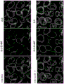
It was surprisingly found, as shown in the examples and figures, that L1-OV52.24 was efficiently internalized into SKOV3ip cells after 40, 60, 70 or 90 minutes of incubation, whereas the comparative anti-L1 monoclonal antibody 9.3 was internalized only to a small extent. Internalization can be determined using a binding molecule linked to a suitable diagnostic compound, such as a fluorescent compound. In the examples, Alexa488 was used as the fluorescent dye. As described in the examples, the internalization can be quantified by microscopic analysis and counting or by imaging flow cytometry. In both methods, the internalization efficiency of L1-OV52.24 was found to be at least 4-fold higher than that of the 9.3 antibody at each detection time point. Furthermore, it was surprisingly found that internalization at test time points 60 'and 90' L1-OV52.24 was more effective than the commercial anti-L1 CAM monoclonal antibodies 5G3 and UJ127.11 (fig. 6). The surprisingly superior internalization properties of L1-OV52.24 identified in the examples are summarized in fig. 7. It was surprisingly found that internalization of L1-OV52.24 at 90' was significantly higher than internalization of any of the prior art anti-L1 CAM monoclonal antibodies 9.3, 5G3, and UJ127.11, all with p-values ≦ 0.01. Thus, L1-OV52.24 or an antigen-binding fragment thereof, or a binding molecule that recognizes the same epitope as L1-OV52.24, is particularly suitable for the delivery of therapeutically active agents (drug delivery) or for the delivery of diagnostic compounds (e.g., for imaging purposes).
In yet a further preferred embodiment, the binding molecule of the invention is internalized by a mammalian cell expressing L1, preferably wherein said cell is a mammalian tumor cell expressing L1, more preferably a SKOV3ip cell.
In a preferred embodiment, the internalization of a mammalian cell expressing L1 is determined by microscopic analysis and quantification or by imaging flow cytometry, preferably wherein the cell is a SKOV3ip cell. The method is preferably carried out as described in the examples. In further preferred embodiments, the internalization is at least 2-fold, preferably at least 4-fold, more preferably at least 7-fold, most preferably at least 10-fold that of monoclonal antibody 9.3 described in WO 2008/151819 after 40, 60, 70 or 90 minutes of incubation. In yet a further preferred embodiment, internalization is at least 2-fold, preferably at least 4-fold greater than internalization of monoclonal antibody 5G3 after 60 or 90 minutes of incubation. In yet a further preferred embodiment, internalization is at least 2-fold, preferably at least 4-fold greater than internalization of monoclonal antibody UJ127.11 after incubation for 60 or 90 minutes.
In a more preferred embodiment, the binding molecule of the invention is monoclonal antibody L1-OV52.24 or an antigen-binding fragment thereof, wherein the light chain variable portion of L1-OV52.24 comprises, preferably has the sequence shown as SEQ ID No. 1, or wherein the light chain is encoded by SEQ ID No. 3, and wherein the heavy chain variable portion of L1-OV52.24 comprises, preferably has the sequence shown as SEQ ID No. 2 or wherein the heavy chain is encoded by SEQ ID No. 3.
In a preferred embodiment, the light chain variable portion of L1-OV52.24 comprises, preferably has, the sequence shown as SEQ ID No:1, and the heavy chain variable portion of L1-OV52.24 comprises, preferably has, the sequence shown as SEQ ID No: 2.
The sequences SEQ ID Nos 1 and 2 relate to the VJ domain; i.e., the light chain variable portion and VDJ domain of L1-OV52.24, respectively, as described above; the heavy chain variable portion of L1-OV 52.24.
Thus, in one embodiment, the invention relates to monoclonal antibody L1-OV52.24 or an antigen binding fragment thereof. Monoclonal antibody L1-OV52.24 is disclosed herein by the sequences of the variable portions of its heavy and light chains (SEQ ID Nos. 2 and 1, respectively) and/or cDNAs encoding the heavy and light chains (SEQ ID Nos. 3 and 4, respectively).
In a particularly preferred embodiment, the binding molecules of the invention are linked to a therapeutically active substance, so that the therapeutically active substance is internalized.
A therapeutically active substance according to the invention is understood to be a compound having a therapeutic or prophylactic effect in an animal, preferably a mammal, more preferably a human.
In a more preferred embodiment, the therapeutically active substance has a therapeutic or prophylactic effect in animals and can be used for the treatment or prevention of tumor diseases and/or diseases associated with the expression of L1.
In a preferred embodiment, the therapeutically active substance is a compound useful for chemotherapy, i.e. a chemotherapeutic compound. In a preferred embodiment, the therapeutically active substance is a cytotoxic or cell proliferation inhibitory compound.
Thus, in one embodiment, the invention relates to a binding molecule of the invention linked to a therapeutically active agent, preferably a therapeutically active agent
Chemotherapeutic compounds, preferably selected from alkylating agents, antitumor antibiotics, antimetabolites and derivatives of natural origin,
(ii) a cytotoxic compound which is capable of,
(ii) a cell proliferation-inhibiting compound,
a cytokine, a nanoparticle, or
A radionuclide.
Suitable cytokines are, for example, TNF α, IL-2 and IL-12.
Suitable chemotherapeutic compounds are well known in the art. These compounds fall into several distinct categories, including, for example, alkylating agents, antitumor antibiotics, antimetabolites, and derivatives of natural origin.
Examples of alkylating agents that may be used in the present invention include busulfan, carboplatin, carmustine, chlorambucil, cisplatin, cyclophosphamide (i.e., a cytotoxin), dacarbazine, ifosfamide, lomustine, nitrogen mustard, melphalan, procarbazine, streptozotocin, and thiotepa.
Examples of antitumor antibiotics include bleomycin, dactinomycin, daunorubicin, doxorubicin, idarubicin, mitomycin (e.g., mitomycin C), mitoxantrone, pentostatin, and plicamycin.
Examples of antimetabolites include fluorodeoxyuridine, cladribine, cytarabine, floxuridine, fludarabine, fluorouracil (e.g., 5-fluorouracil (5FU)), gemcitabine, hydroxyurea, mercaptopurine, methotrexate, and thioguanine.
Examples of derivatives of natural origin include docetaxel, etoposide, irinotecan, taxanes (e.g., paclitaxel), teniposide, topotecan, vinblastine, vincristine, vinorelbine, prednisone, and tamoxifen.
Other examples of chemotherapeutic agents useful in the present invention include asparaginase and mitotane.
Furthermore, C2 ceramide may also be used.
The term "linked" as used herein is understood to include both covalent and non-covalent attachment, preferably covalent attachment. In covalently linked embodiments, the binding molecules of the invention may be linked to a therapeutically active agent or diagnostic compound directly or through a suitable linker molecule. In a more preferred embodiment of the invention, the binding molecule is linked to the therapeutically active agent or the diagnostic compound via a linker. Suitable linkers are well known in the art.
Thus, in a preferred embodiment, the binding molecule is linkable to the therapeutically active substance and/or the diagnostic compound via a linker.
Suitable radionuclides include67/64Cu、131I、124I or90And Y. Such radionuclides may be linked by a moiety that forms a complex, or, in the case where the radionuclide is iodine, directly covalently linked to the binding molecule of the invention. The complex-forming moiety is preferably covalently linked (via a linker) to the binding molecule.
Such antibody conjugates are well known in the art (Wu AM, Senter PD. aromatic antibodies: prospects and transformers for immunoconjugates. Nature Biotechnol.23:1137-1146,2005, Pastan I, Hassan R, FitzGerald DJ, Kreitman RJ. immunotoxin molecular therapy of cancer. Annu. Rev. Med.58:221-237,2007, WO 90/12592, WO 2007/030642, WO 2004/067038, WO 2004/003183, US 2005/0074426, WO 94/04189).
In a more preferred embodiment, the therapeutically active substance is selected from the group consisting of the following chemotherapeutic compounds or cytotoxic compounds or cell proliferation inhibiting compounds: DNA damaging agents, in particular actinomycin D, mitomycin C, cisplatin, doxorubicin, etoposide, verapamil, podophyllotoxin, 5-FU, derivatives of natural origin and taxanes, preferably paclitaxel and carboplatin.
Furthermore, it may be advantageous to attach the binding molecules of the invention to a diagnostic compound.
Diagnostic compounds are compounds that are capable of generating a signal by direct or indirect detection. In a preferred embodiment, the compounds are suitable for administration to an animal, such as a mammal, more preferably a human. In this embodiment, the attached binding molecule may be suitable for in vitro and in vivo use. In another preferred embodiment, the compound is not suitable for administration to an animal, such as a mammal, more preferably a human. In this embodiment, the attached binding molecule may be suitable for in vitro use. Thus, the diagnostic compound may be detected directly or indirectly. For direct detection, the diagnostic compounds suitable for use in the present invention may be selected from any of the well-known detectable label groups, such as chromophores, fluorophores, chemiluminescent groups (e.g., acridinium esters or dioxetanes), electrochemiluminescent compounds, catalysts, enzymes, enzyme substrates, dyes, fluorescent dyes (e.g., fluorescein, coumarin, rhodamine, oxazine, resorufin, cyanine, and derivatives thereof), colloidal metallic and non-metallic particles, and organic polymer latex particles. Other examples of diagnostic compounds are luminescent metal complexes, such as complexes of ruthenium or europium and radioisotopes.
Indirect detection systems include, for example, partners of bioaffinity (bioaffine) binding pairs. Examples of suitable binding pairs are haptens or antigens/antibodies, biotin or biotin analogues such as aminobiotin, iminobiotin or desthiobiotin/avidin or streptavidin, sugars/lectins, nucleic acids or nucleic acid analogues/complementary nucleic acids and receptors/ligands, e.g. steroid hormone receptors/steroid hormones.
Thus, in a further embodiment, the invention relates to a binding molecule of the invention linked to a diagnostic compound, preferably selected from the group consisting of a radionuclide, a chemiluminescent compound, a fluorescent compound, a dye or an enzyme.
As shown in the examples, the L1-OV52.24 monoclonal antibody was successfully linked to a fluorescent compound or fluorescent dye, i.e., Alexa dye (Alexa 488).
In another embodiment, the invention relates to a hybridoma cell producing an anti-L1 monoclonal antibody of the invention.
In another embodiment, the invention relates to a nucleic acid, said nucleic acid
(i) Encoding a binding molecule according to the invention, and/or
(ii) Encoding at least one chain of a binding molecule according to the invention, and/or
(iii) Comprises the sequence shown as SEQ ID No. 3 and/or SEQ ID No. 4, and/or
(iv) Comprising a sequence encoding at least one of: KASQNVGTNVA (SEQ ID No:5), STSYS (SEQ ID No:6), QQYNTYPYT (SEQ ID No:7), FNIKDYYMQ (SEQ ID No:8), WIDPENGKTVFDPKFR (SEQ ID No:9) and WNPLAF (SEQ ID No: 10).
In a more preferred embodiment, the nucleic acid of the invention is part of a vector. Such vectors are vectors which are preferably used for expression in bacteria (e.g.E.coli), in yeast cells, in insect cells or mammalian cells. Preferably, the nucleic acids of the invention are controlled by suitable regulatory sequences (e.g., promoters or enhancers) in the vector to achieve expression in the host cell. The vector preferably comprises a sequence capable of replication in a host cell.
Preferably, the binding molecules attached to the diagnostic compounds can be used in vitro for diagnostic purposes and thus relate to important tools for biotechnological research. For example, a tissue biopsy or a body sample of a patient may be analyzed in the usual case using the binding molecules of the invention. Thus, in a further embodiment, the invention relates to the use of the binding molecules of the invention as in vitro diagnostic agents, or as in vitro biotechnological agents. In a more preferred embodiment, the invention relates to the use of the binding molecules of the invention linked to diagnostic compounds as in vitro diagnostic agents or as in vitro biotechnological agents. In another preferred embodiment, the invention relates to the use of a binding molecule of the invention linked to a therapeutically active substance as an in vitro biotechnological agent.
In yet a further embodiment, the invention relates to the use of the binding molecules of the invention as a medicament or diagnostic agent.
In a particularly preferred embodiment, the binding molecule for use as a medicament is a binding molecule linked to a therapeutically active substance as described above. The binding molecules deliver the therapeutically active substance into the tumor cells where the binding molecules linked to the therapeutically active substance are able to exert their therapeutic effect.
In a preferred embodiment, the therapeutically active substance is administered in a therapeutically effective amount.
In a further preferred embodiment, the binding molecule for diagnosis is a binding molecule linked to a diagnostic compound. Thus, a patient can be imaged, thereby enabling diagnosis (e.g., prognosis or monitoring of therapy) using the binding molecules of the invention. Thus, the binding molecules of the invention linked to diagnostic compounds are preferably companion diagnostic agents for use in the invention to binding molecules linked to therapeutically active substances.
The binding molecules of the present invention are preferably included in a pharmaceutical composition.
The compounds may be both diagnostic compounds and therapeutically active substances. One example is certain radionuclides, e.g.131I, which may be suitable for both in vivo imaging and tumor therapy.
In general, the pharmaceutical compositions of the invention comprise a therapeutically effective amount of a binding molecule of the invention, and optionally one or more pharmaceutically acceptable carriers. In a particular embodiment, the term "pharmaceutically acceptable" means approved by a regulatory agency of the federal or a state government or listed in the U.S. pharmacopeia or other generally recognized pharmacopeia for use in animals, and more particularly in humans. The term "carrier" refers to a diluent, adjuvant, excipient, or vehicle with which the therapeutic agent is administered. Such pharmaceutical carriers can be sterile liquids, such as water and oils, including those of petroleum, animal, vegetable or synthetic origin, including, but not limited to, peanut oil, soybean oil, mineral oil, sesame oil and the like. Water is a preferred carrier when orally administering the pharmaceutical composition. Saline and aqueous dextrose are preferred carriers for intravenous administration of the pharmaceutical composition. Saline solutions and aqueous dextrose and glycerol solutions are preferably used as liquid carriers for the injectable solutions. Suitable pharmaceutical excipients include starch, glucose, lactose, sucrose, gelatin, malt, rice, flour, chalk, silica gel, sodium stearate, glycerol monostearate, talc, sodium chloride, dried skim milk, glycerol, propylene glycol, water, ethanol and the like. The composition may also contain minor amounts of wetting or emulsifying agents, or pH buffering agents, if desired. These compositions may take the form of solutions, suspensions, emulsions, tablets, pills, capsules, powders, sustained release formulations and the like. The compositions may be formulated as suppositories using conventional binders and carriers such as triglycerides. Oral formulations may include standard carriers such as pharmaceutical grades of mannitol, lactose, starch, magnesium stearate, sodium saccharine, cellulose, magnesium carbonate, and the like. Examples of suitable Pharmaceutical carriers are described in "Remington's Pharmaceutical Sciences" by e.w. martin. Such compositions will contain a therapeutically effective amount of the therapeutic agent, preferably in purified form, and an appropriate amount of carrier to provide a suitable form for administration to a patient. The formulation should be suitable for the mode of administration.
In a preferred embodiment, the composition is formulated according to conventional procedures into a pharmaceutical composition suitable for intravenous administration to a human. Typically, compositions for intravenous administration are solutions in sterile isotonic aqueous buffer. If necessary, the composition may also contain a solubilizing agent and a local anesthetic (such as lidocaine) to relieve pain at the injection site. In general, the ingredients are provided separately, or mixed together in a unit dosage form, for example, as a lyophilized powder or as a water-free concentrate in a sealed container such as an ampoule or sachet showing the amount of active agent. When the composition is administered by infusion, it can be dispensed into an infusion bottle containing pharmaceutical grade sterile water or saline. Where the compositions are administered by injection, an ampoule of sterile water or saline may be provided for injection, so that the ingredients may be mixed prior to administration.
The therapeutic agents of the present invention may be formulated in neutral or salt form. Pharmaceutically acceptable salts include salts formed with free carboxyl groups, such as those derived from hydrochloric, phosphoric, acetic, oxalic, tartaric acids, and the like, salts formed with free amino groups, such as those derived from isopropylamine, triethylamine, 2-ethylamino ethanol, histidine, procaine, and the like, and salts derived from sodium, potassium, ammonium, calcium, and ferric hydroxides, and the like.
The therapeutically effective amount of a therapeutic agent of the invention for a particular condition or symptom will depend on the nature of the condition or symptom, and can be determined by standard clinical techniques. In addition, in vitro assays may be selected to aid in identifying optimal dosage ranges. The precise dose employed in the formulation will also depend on the route of administration, the severity of the disease or condition, and should be decided according to the judgment of the practitioner and each patient's circumstances. However, suitable dosage ranges for intravenous administration are generally from about 20 to 500 mg of active compound per kg of body weight. Suitable dosage ranges for intranasal administration are generally from about 0.01pg/kg body weight to 1mg/kg body weight. Effective doses can be extrapolated from dose-response curves derived from in vitro or animal model test systems. In general, suppositories may contain from 0.5% to 10% by weight of the active ingredient; oral dosage forms preferably contain 10% to 95% of the active ingredient.
A variety of well-known delivery systems may be used to administer the therapeutic agents of the present invention, such as encapsulation in liposomes, microparticles, and microcapsules; using recombinant cells capable of expressing a therapeutic agent, using receptor-mediated endocytosis (e.g., Wu and Wu,1987, J.biol.chem.262: 4429-4432); constructing therapeutic nucleic acids as part of a retrovirus or other vector, and the like. Methods of introduction include, but are not limited to, intradermal, intramuscular, intraperitoneal, intravenous, subcutaneous, intranasal, epidural, and oral routes. The compounds may be administered by any convenient route, for example by infusion, by bolus injection, by absorption through epithelial or cutaneous mucosal linings (e.g. oral, rectal and intestinal mucosa), and may be administered together with other biologically active agents. Administration may be systemic or local. Furthermore, it may be desirable to introduce the pharmaceutical compositions of the present invention into the central nervous system by any suitable route, including intraventricular and intrathecal injection; intraventricular injection can be assisted, for example, by an intraventricular catheter connected to a reservoir, such as an Ommaya reservoir. Pulmonary administration may also be used, for example by use of an inhaler or nebulizer, as well as formulations with an aerosolizing agent.
In a particular embodiment, it may be desirable to topically apply the pharmaceutical composition of the present invention to an area in need of treatment. This can be achieved by, for example, but not limited to, local infusion during surgery, local application (e.g., in conjunction with post-operative wound dressings), by injection, by means of a catheter, by means of a suppository, or by means of an implant material that is porous, non-porous, or gelatinous, including membranes, such as silicone rubber membranes or fibers. In one embodiment, administration can be by direct injection at the site (or at the original site) of the malignant tumor or precancerous tissue.
In another embodiment, the delivery of the therapeutic agent may be in a vesicle, particularly a liposome (Langer,1990, Science 249:1527-1533), more particularly a cationic liposome (WO 98/40052).
In yet another embodiment, the therapeutic agent may be delivered by a controlled release system. In one embodiment, a pump (Langer, supra) may be used. In yet another embodiment, the controlled release system can be placed in the vicinity of the therapeutic target, thus requiring only a fraction of the systemic dose.
In the context of this aspect of the invention, the invention also includes a method of treating or preventing a neoplastic disease in a patient, comprising administering to said patient a therapeutically effective amount of a binding molecule, preferably a monoclonal antibody or antigen-binding fragment thereof, of the invention.
Throughout the present invention, the term "effective amount" means that a given molecule or compound is administered in an amount sufficient to achieve the desired therapeutic effect. Throughout the invention, if both compounds are administered in therapeutically effective amounts, this includes the case where the or each compound is administered in a sub-therapeutic amount, i.e. the amount of each compound is insufficient to provide independently the therapeutic effect, but the two compounds in combination produce the desired therapeutic effect. However, it is also within the scope of the present invention that each compound is administered individually in a therapeutically effective amount.
According to the present invention, the term "treating a tumor disease" encompasses both killing tumor cells, reducing the proliferation of tumor cells (e.g. by at least 30%, at least 50% or at least 90%) and completely inhibiting the proliferation of tumor cells in a patient suffering from a tumor disease. In addition, the term includes the prevention of neoplastic disease, for example by killing cells that may or are predisposed to becoming tumor cells in the future, as well as the prevention of the formation of metastases.
According to the present invention, the skilled person will understand that each therapy to be applied will depend on, for example, the physical condition of the patient or the severity of the disease, and therefore must be adapted to the specific situation.
The invention also relates to pharmaceutical compositions comprising the monoclonal antibodies or antigen-binding fragments or binding molecules thereof of the invention. With respect to the pharmaceutical composition, all of the embodiments described above are also applicable.
In yet a further embodiment, the invention relates to the use of a binding molecule of the invention (preferably a monoclonal antibody or antigen binding fragment thereof) for the treatment or prevention of a tumor disease, preferably wherein the tumor disease is an epithelial tumor disease and/or is selected from the group consisting of: astrocytoma, oligodendroglioma, meningioma, neurofibroma, glioblastoma, ependymoma, schwannoma, neurofibrosarcoma, medulloblastoma, melanoma, pancreatic cancer, prostate cancer, head and neck cancer, breast cancer, lung cancer, ovarian cancer, endometrial cancer, kidney cancer, neuroblastoma, squamous cell carcinoma, medulloblastoma, liver cancer, colon cancer, mesothelioma, and epidermoid carcinoma.
In yet a further embodiment, the invention relates to a pharmaceutical composition comprising a binding molecule of the invention as described above and optionally one or more pharmaceutically acceptable carriers.
In another embodiment, the invention relates to a binding molecule that binds to L1, wherein the binding molecule is an anti-L1 monoclonal antibody or antigen binding fragment thereof, characterized in that at least one of the Complementarity Determining Regions (CDRs) of the anti-L1 monoclonal antibody or antigen binding fragment thereof
a) Has one of the sequences selected from the group consisting of: KASQNVGTNVA (SEQ ID No:5), STSYS (SEQ ID No:6), QQYNTYPYT (SEQ ID No:7), FNIKDYYMQ (SEQ ID No:8), WIDPENGKTVFDPKFRG (SEQ ID No:9) and WNPLAF (SEQ ID No:10) or
b) Sequences having at least one conservative amino acid substitution compared to the sequences mentioned in a).
In another embodiment, the invention relates to a binding molecule which binds to L1, characterized in that the binding molecule comprises
a) One selected from the group consisting of: KASQNVGTNVA (SEQ ID No:5), STSYS (SEQ ID No:6), QQYNTYPYT (SEQ ID No:7), FNIKDYYMQ (SEQ ID No:8), WIDPENGKTVFDPKFRG (SEQ ID No:9) and WNPLAF (SEQ ID No:10) or
b) Comprising a sequence having at least one conservative amino acid substitution compared to the sequence mentioned in a).
Such monoclonal antibodies or antibody-binding fragments or binding molecules thereof of the invention can be produced, for example, by CDR grafting or by recombination of the antibodies. Such methods are well known in the art (see, e.g., Queen, U.S. Pat. No. 5,585,089 and Winter, U.S. Pat. No. 5,225,539, Cabilly U.S. Pat. No. 4,816,567).
The preferred embodiments of the invention described herein apply to all binding molecules and monoclonal antibodies and antigen binding fragments thereof of the present invention.
Reference to the literature
1.Galluzzi L,Vacchelli E,Fridman WH,Galon J,Sautes-Fridman C,Tartour E,Zucman-Rossi J,Zitvogel L,Kroemer G:Trial Watch:Monoclonal antibodies in cancer therapy.Oncoimmunology 2012,1(1):28-37.
2.Presta LG:Molecular engineering and design of therapeutic antibodies.Curr Opin Immunol 2008,20(4):460-470.
3.Reichert JM:Marketed therapeutic antibodies compendium.MAbs 2012,4(3).
4.Reichert JM:Antibodies to watch in 2014:mid-year update.mAbs 2014,6(4):799-802.
5.Reichert JM:Antibody-based therapeutics to watch in 2011.MAbs 2011,3(1):76-99.
6.Wolterink S,Moldenhauer G,Fogel M,Kiefel H,Pfeifer M,Luttgau S,Gouveia R,Costa J,Endell J,Moebius U et al:Therapeutic antibodies to human L1CAM:functional characterization and application in a mouse model for ovarian carcinoma.Cancer Res 2010,70(6):2504-2515.
7.Doberstein K,Harter PN,Haberkorn U,Bretz NP,Arnold B,Carretero R,Moldenhauer G,Mittelbronn M,Altevogt P:Antibody therapy to human L1CAM in a transgenic mouse model blocks local tumor growth but induces EMT.International journal of cancer Journal international du cancer 2014.
8.Novak-Hofer I,Amstutz HP,Morgenthaler JJ,Schubiger PA:Internalization and degradation of monoclonal antibody chCE7by human neuroblastoma cells.International journal of cancer Journal international du cancer 1994,57(3):427-432.
9.Schaefer AW,Kamiguchi H,Wong EV,Beach CM,Landreth G,Lemmon V:Activation of the MAPK signal cascade by the neural cell adhesion molecule L1requires L1 internalization.The Journal of biological chemistry 1999,274(53):37965-37973.
10.Long KE,Asou H,Snider MD,Lemmon V:The role of endocytosis in regulating L1-mediated adhesion.The Journal of biological chemistry 2001,276(2):1285-1290.
11.Schaefer AW,Kamei Y,Kamiguchi H,Wong EV,Rapoport I,Kirchhausen T,Beach CM,Landreth G,Lemmon SK,Lemmon V:L1endocytosis is controlled by a phosphorylation-dephosphorylation cycle stimulated by outside-in signaling by L1.The Journal of cell biology 2002,157(7):1223-1232.
12.WO 2008/151819
Drawings
FIG. 1: cellular uptake of mAb L1CAM mAb9.3 was detected on Skov3ip cells.
Skov3ip cells were incubated with Alexa 488-conjugated L1CAM mAb L1-9.3 for varying lengths of time. The samples were then fixed and cell surface bound antibodies were detected using a goat anti-mouse secondary antibody coupled to Alexa 647. Cells at time 0' were incubated on ice to avoid antibody internalization. Samples were tested on an arnis ISX imaging flow cytometer, 3000 cells were collected and analyzed using arnis eas software. Representative images of cells taken at time points 0 minutes (a), 60 minutes (B) and 90 minutes (C) are shown. The lower column of graphs shows the quantification of the corresponding samples. Time points 0 ' (D), 60 ' (E) and 90 ' (F) are described. The y-axis gives the normalized frequency and the x-axis shows the fluorescence intensity within each cell of Alexa 488-conjugated L1CAM mAb9.3 relative to the total fluorescence intensity of the cells. The table below the graph shows the number of cells (counts) included in the analysis and the average (mean) of the graph. The experiment was repeated three times, showing typical results.
FIG. 2: cellular uptake of antibody L1CAM mAb OV52.24 was detected on Skov3ip cells.
Skov3ip cells were incubated with Alexa 488-conjugated L1mAb OV52.24 for varying lengths of time. The samples were then fixed and cell surface bound antibodies were detected using a goat anti-mouse secondary antibody coupled to Alexa 647. Cells at time 0' were incubated on ice to avoid antibody internalization. Samples were tested on an arnis ISX imaging flow cytometer, 3000 cells were collected and analyzed using arnis eas software. Representative images of cells taken at time points 0 minutes (a), 60 minutes (B) and 90 minutes (C) are shown. The lower column of graphs shows the quantification of the corresponding samples. Time points 0 ' (D), 60 ' (E) and 90 ' (F) are described. The y-axis gives the normalized frequency and the x-axis shows the fluorescence intensity within each cell of Alexa 488-conjugated L1mAb OV52.24 relative to the total fluorescence intensity of the cells. The table below the graph shows the number of cells (counts) included in the analysis and the average (mean) of the graph. The experiment was repeated three times, showing typical results.
FIG. 3: cellular uptake of L1CAM mAbs 9.3 and OV52.24 was tested on Skov3ip cells.
This figure gives an overview of the results in figures 1 and 2. The upper and lower panels show plots against L1CAM mAb9.3 and L1CAM mAb OV52.24 at different time points 0 ', 60 ' and 90 ' min, respectively.
FIG. 4: cellular uptake of L1CAM mAbs 9.3 and OV52.24 was detected on Skov3ip cells using confocal laser scanning microscopy.
Skov3ip cells were incubated for 70 min with Alexa 488-conjugated L1CAM mAbs 9.3 or OV 52.24. The samples were then fixed and cell surface bound antibodies were detected using a goat anti-mouse secondary antibody coupled to Alexa 647. The samples were then imaged on a Leica SP5II confocal laser scanning microscope. Z-slices were obtained at similar Z-positions. N-30 cells were collected for each antibody and quantified (a). Images were analyzed using fiji (imagej) and histograms (B) were plotted showing intracellular fluorescence intensity of Alexa 488-conjugated L1mAb 9.3 and L1-OV52.24 relative to total fluorescence intensity of the cells. The experiment was repeated three times, showing typical results. The corresponding left bar shows the results for mAb9.3 at 40min and 90min, respectively. The corresponding right bar shows the results for mAb OV52.24 at 40min and 90min, respectively.
FIG. 5: cellular uptake of L1CAM mAbs 5G3 and UJ127.11 on Skov3ip cells was confirmed using confocal laser scanning microscopy.
Skov3ip cells were incubated for 70 min with Alexa 488-conjugated L1CAM mAbs 5G3 and UJ 127.11. The samples were then fixed and cell surface bound antibodies were detected using a goat anti-mouse secondary antibody coupled to Alexa 647. The samples were then imaged on a Leica SP5II confocal laser scanning microscope. Z-slices were obtained at similar Z-positions. N-30 cells were collected for each antibody. Typical results are shown.
FIG. 6: cellular uptake of antibodies L1CAM mAbs 9.3, OV52.24, 5G3 and UJ127.11 was detected and compared on Skov3ip cells.
Skov3ip cells were incubated with Alexa 488-conjugated L1mAb for varying lengths of time. The samples were then fixed and cell surface bound antibodies were detected using a goat anti-mouse secondary antibody coupled to Alexa 647. Cells at time 0' were incubated on ice to avoid antibody internalization. Samples were tested on an arnis ISX imaging flow cytometer, 5000 cells were collected and analyzed using arnis IDEAS software. The graphs in the left, middle and right columns show time points of 0, 60 and 90 minutes, respectively. The y-axis gives the normalized frequency and the x-axis shows the intracellular fluorescence intensity of Alexa 488-conjugated L1mab relative to the total fluorescence intensity of the cells. The average of the relative intracellular fluorescence intensity under each condition is given in the graph. The experiment was repeated three times, showing typical results.
FIG. 7: cellular uptake of L1CAM mAbs 9.3, OV52.24, 5G3 and UJ127.11 was compared on Skov3ip cells.
This figure gives a summary of the typical analysis in figure 6. Three separate experiments were plotted based on the mean relative intracellular intensity of each L1CAM mAb in each experiment. Values of the histogram are expressed as mean ± s.d. To determine the statistically significant probability of increased or decreased internalization of different L1CAM mabs, three independent experiments were analyzed using the two-tailed, unpaired Student t-test. A p value <0.05 was considered statistically significant. Asterisks were assigned as follows: p is less than or equal to 0.05; p is less than or equal to 0.01; p is less than or equal to 0.001.
Examples
Example 1
Materials and methods
Cell lines
Human ovarian cancer cells SKOV3ip were obtained from the american type culture collection (magenis). This cell line was validated by the german center for biomaterial resources (brelucelix, germany) and the investigators evaluated typical morphology throughout the culture. Weekly tests were performed to ensure that cultures were mycoplasma negative. The cells were cultured in DMEM medium (Sigma-Aldrich, Deisenhoffen, Germany) supplemented with 10% heat-inactivated Fetal Calf Serum (FCS) (Biochrom, Berlin, Germany), 2mM L-glutamine (Invitrogen, Callsrue, Germany) and 1mM sodium pyruvate (Invitrogen). All cells were maintained at 37 ℃ and 5% CO2In a wet environment.
Monoclonal antibodies
mAb L1-9.3 to human L1CAM has been described previously [6,12 ].
L1-OV52.24 was generated using human L1-Fc protein comprising the extracellular domain of L1, or using SKOV3ip cells to immunize mice.
The cDNA sequence of the immunoglobulin gene of the L1-OV52.24 monoclonal antibody was determined and is shown above (light chain: SEQ ID No:3, heavy chain: SEQ ID No: 4).
In addition, the protein sequences (SEQ ID No:2 and SEQ ID No:1) of the L1-OV52.24 heavy and light chains (VDJ or VJ domains, without constant domain) were determined, respectively, and the CDR sequences of L1-OV52.24 were determined according to Kabat nomenclature. The CDR sequences of L1-OV52.24 are: KASQNVGTNVA (LCDR 1; SEQ ID No:5), STSYS (LCDR 2; SEQ ID No:6), QQYNTYPYT (LCDR 3; SEQ ID No:7), FNIKDYYMQ (HCDR 1; SEQ ID No:8), WIDPENGKTVFDPKFRG (HCDR 2; SEQ ID No:9) and WNPLAF (HCDR 3; SEQ ID No: 10).
Both mabs were of the IgG1 isotype.
Isotype control mabs were obtained from Bio X Cell (neohampshire, west libamotene). The L1CAM mAb was conjugated to Alexa488 using a labeling kit (Invitrogen) according to the manufacturer's instructions. Briefly, 100 μ g of antibody was mixed with 1 vial of labeling reagent, incubated for 1 hour and subsequently purified on a column. The degree of labeling was determined to ensure similar labeling efficacy.
Binding constant
Surface Plasmon Resonance (SPR) equilibrium analysis
Binding assays were performed using a BIAcore 3000 equipped with a CM5 sensor chip. Briefly, BIAcore CM5 chips were activated using EDC/NHS and different levels of L1-Fc were captured on the activated surface. The remaining active sites were blocked using ethanolamine/HCl. The L1-mAb was bound to the L1-Fc surface and allowed to dissociate over time. Kinetic analysis was performed for the binding and dissociation phases of each injection on each density surface.
Recognized epitopes
To determine epitope specificity, we constructed a series of L1-Fc proteins (as described in [6]) carrying different Ig domains. For fine localization, the recently described recombinant V5-labeled L1 fragment (Gouveia RM, Moris VA, Peixoto C et al, Production and purification of functional truncated soluble forms of human recombinant L1cell addition glycoproten from Spodoptera frugiperda Sfcells. protein Expr purify 2007; 52: 182-93) was used. Recombinant proteins were used for epitope mapping in ELISA or in Western blot analysis. Antibody uptake assay
For imaging flow cytometry (Imagestream)
Skov3ip cells were detached using trypsin/EDTA, resuspended in culture medium, counted and divided into aliquots of equal cell number (200000 cells). Cells were incubated at 37 ℃ for various time points in the continuous presence of labeled antibody (10. mu.g/mL), or cells were incubated on ice for 0 min. At each time point, cells were pelleted with 800x g, washed 1 time with ice-cold PBS and fixed on ice for 15min with 4% pfa (thermo fischer). The fixed cells were washed twice with PBS and incubated for 20 min with a secondary goat anti-mouse Alexa-647 antibody (Invitrogen, carlsrue, germany) at 25 μ g/mL and then washed twice with PBS.
For confocal laser scanning microscopy
25000 cells were seeded on 8-well μ -slides (Ibidi, Munich, Germany) and grown for 24 h. Cells were incubated at 37 ℃ at various time points in the continuous presence of labeled antibody (10. mu.g/mL). At each time point, cells were washed with ice-cold PBS and fixed with 4% pfa (thermo fischer) on ice for 15 min. The fixed cells were washed twice with PBS and incubated for 20 min with a secondary goat anti-mouse Alexa-647 antibody (Invitrogen, carlsrue, germany) at 25 μ g/mL and then washed twice with PBS.
Imaging flow cytometry and analysis
Samples were examined on an arnis imagestream x (ISX) (arnis corporation, seattle usa) with a488 nm laser set to 100% and a 561nm laser set to 80%, using a 60x objective and activating an Extended Depth of Field (EDF) option. Bright field images were recorded in channels 2 and 5 and channel 1, and 4000 cells were collected per sample. After fixation, before counterstaining with anti-mouse Alexa647 antibody, an aliquot of the cell sample was taken as a compensation control for each antibody against Alexa 488-conjugated L1CAM mAb. Cells were stained on ice using the same concentration of the corresponding unconjugated L1mAb as the conjugated antibody and counterstained with the same anti-mouse Alexa647 secondary antibody as a compensation control for counterstaining with anti-mouse Alexa647 antibody. Compensation controls were obtained on the ISX with the corresponding compensation settings.
The ISX data were analyzed using the IDEAS software (arnis corporation, seattle, usa). The original image file is opened and a compensation matrix is generated using the corresponding compensation file. The collected cells were gated to obtain cells in focus and then gated using bright field image control to obtain single cells. The image of channel 5 was used to generate a "cell surface mask", with the lower intensity limit being limited to 200 and the upper limit being kept 4095 constant to eliminate background and optimize signal. For lane 2, the lower intensity limit is set to 150, and the upper limit remains 4095 and is referred to as "lane 2". Both settings were derived from control staining with L1CAM mAb isotype control or secondary antibody only. Furthermore, the "cell surface mask" is enlarged by one pixel to include the adjacent pixels on channel 2. Another mask, termed "intracellular signaling", is generated and is defined as the "channel 2 non-cell surface mask". Finally, a combined signature called "relative intracellular intensity" is generated, which is defined as follows: "intracellular signal/(intensity _ MC _ Ch02 × 100)". The corresponding histogram is plotted and the mean value is determined. The same procedure was applied to all samples tested.
Confocal laser scanning microscopy and analysis
The samples were examined on a Leica SP5II confocal laser scanning microscope (Leica microsystems, werrall, germany) equipped with a HyD detector. Alexa-488 conjugated L1mAb was excited with an argon laser at 488nm and Alexa-647 was excited with a HeNe laser line at 633 nm. Z-slices were obtained at similar Z-positions. For each antibody, 30 cells were collected and quantified. Images were analyzed using Fiji (ImageJ, NIH, Bethesda, usa) in which single cell boundaries were segmented and identified using signals from anti-mouse Alexa-647 counterstained cell surfaces, and then intracellular fluorescence intensity and total cell intensity were determined. The results were plotted as a bar graph using Excel (microsoft, Redmond usa) showing intracellular fluorescence intensity of Alexa 488-conjugated mabs L1-9.3 and L1-OV52.24 relative to the total fluorescence intensity of the cells.
Results
mAbs L1-9.3 and L1-OV52-54 bind to different cell surface epitopes of the L1CAM cell surface molecule. mAb9.3 is clearly related to the first Ig domain (1. Ig-L1-Fc; [12]]) The constituent fusion proteins react. By Western blot analysis, it was confirmed that mAb L1-OV52.24 recognized the 4-5FNIII domain of L1. MAb L1-9.3 binds to the first Ig domain, whereas L1-OV52.24 binds to an epitope in the 4-5.FNIII domain. mAb9.3 had affinity for L1 of kd (8, 5 × 10)-11(see [6]]) And mAb L1-OV52.24 had affinity for L1 of kd (m) ═ 2,41 × 10⁻⁹. L1-OV52.24 was found to perform well in Western blot analysis and FACS analysis experiments and was found to perform well in Immunohistochemistry (IHC) experiments.
We performed internalization assays in SKOV3ip cells using Alex-488 conjugated mabs as outlined in the materials and methods section above. Imagestream capable of quantifying thousands of cells by combining FACS and fluorescence analysisXAnalysis, internalization was measured. The data was analyzed using the IDEAS software. Analysis of L1-9.3 showed slow internalization at time points 0min, 60min and 90min (FIGS. 1A-F), with an average of 7.8% at the final time points. In contrast, internalization of mAb L1-OV52.24 was much more rapid and averaged to 40% at the final time point (FIGS. 2A-F). The data are compared directly in figure 3.
To validate these results, a similar analysis was performed using the attached cells. SKOV3ip cells were cultured on coverslips and internalized with Alexa-conjugated antibodies. Quantification was accomplished by laser scanning microscopy and visual counting of cells. Examples of staining are shown in figure 4 and the results are summarized in figure 4B. These results corroborate the data obtained by Imagestream analysis and show that mAb L1-OV52.24 resulted in a nearly 10-fold increase in the rate of internalization. This result was unexpected and indicates that mAb-L1-OV52.24 is suitable for delivery of antibody drug conjugates.
Example 2
Materials and methods
Cell lines
Human ovarian cancer cells SKOV3ip were obtained from the american type culture collection (magensa). This cell line was validated by the german center for biomaterial resources (brelucelix, germany) and the investigators evaluated typical morphology throughout the culture. Weekly tests were performed to ensure that cultures were mycoplasma negative. The cells were cultured in DMEM medium (Sigma-Aldrich, Deisenhoffen, Germany) supplemented with 10% heat-inactivated Fetal Calf Serum (FCS) (Biochrom, Berlin, Germany), 2mM L-glutamine (Invitrogen, Callsrue, Germany) and 1mM sodium pyruvate (Invitrogen). All cells were maintained at 37 ℃ and 5% CO2In a wet environment.
Monoclonal antibodies
mAbs L1-9.3, 5G3 and UJ127.11 to human L1CAM have been described previously [6 ].
L1-OV52.24 was generated using human L1-Fc protein comprising the extracellular domain of L1, or using SKOV3ip cells to immunize mice.
The cDNA sequence of the immunoglobulin gene of the L1-OV52.24 monoclonal antibody was determined and is shown above (light chain: SEQ ID No:3 and heavy chain: SEQ ID No: 4).
In addition, the protein sequences (SEQ ID No:2 and SEQ ID No:1) of the L1-OV52.24 heavy and light chains (VDJ or VJ domains, without constant domain) were determined, respectively, and the CDR sequences of L1-OV52.24 were determined according to Kabat nomenclature. The CDR sequences of L1-OV52.24 are: KASQNVGTNVA (LCDR 1; SEQ ID No:5), STSYS (LCDR 2; SEQ ID No:6), QQYNTYPYT (LCDR 3; SEQ ID No:7), FNIKDYYMQ (HCDR 1; SEQ ID No:8), WIDPENGKTVFDPKFRG (HCDR 2; SEQ ID No:9) and WNPLAF (HCDR 3; SEQ ID No: 10).
mAbs L1-9.3, L1-OV52.24 and UJ127.11 are of the IgG1 isotype. mAb 5G3 was of IgG2a isotype. The corresponding isotype control mAb was obtained from Bio X Cell (neohampshire, west libamotene). The L1CAM mAb was conjugated to Alexa488 using a labeling kit (Invitrogen) according to the manufacturer's instructions. Briefly, 100 μ g of antibody was mixed with 1 vial of labeling reagent, incubated for 1 hour and subsequently purified on a column. The degree of labeling was determined to ensure similar labeling efficacy. mAb 5G3 conjugated to Alexa488 was purchased from Novus Biologicals (ritton, usa).
Antibody uptake assay
For imaging flow cytometry (Imagestream)
Skov3ip cells were detached using trypsin/EDTA, resuspended in culture medium, counted and divided into aliquots of equal cell number (200000 cells). Cells were incubated at 37 ℃ for various time points in the continuous presence of labeled antibody (10. mu.g/mL), or cells were incubated on ice for 0 min. At each time point, cells were pelleted with 800x g, washed 1 time with ice-cold PBS and fixed on ice for 15min with 4% pfa (thermo fischer). The fixed cells were washed twice with PBS and incubated for 20 min with a secondary goat anti-mouse Alexa-647 antibody (Invitrogen, carlsrue, germany) at 25 μ g/mL and then washed twice with PBS.
For confocal laser scanning microscopy
25000 cells were seeded on 8-well μ -slides (Ibidi, Munich, Germany) and grown for 24 h. Cells were incubated at 37 ℃ at various time points in the continuous presence of labeled antibody (10. mu.g/mL). At each time point, cells were washed with ice-cold PBS and fixed with 4% pfa (thermo fischer) on ice for 15 min. The fixed cells were washed twice with PBS and incubated for 20 min with a secondary goat anti-mouse Alexa-647 antibody (Invitrogen, carlsrue, germany) at 25 μ g/mL and then washed twice with PBS.
Imaging flow cytometry and analysis
Samples were examined on an arnis imagestream x (ISX) (arnis corporation, seattle usa), with a488 nm laser set to 100% and a 561nm laser set to 80%, using a 60x objective lens and activating an Extended Depth of Field (EDF) option. Bright field images were recorded in channels 2 and 5 and channel 1, with 10.000 cells collected per sample. After fixation, and before counterstaining with anti-mouse Alexa647 antibody, an aliquot of the cell sample was taken as a compensation control for each antibody against Alexa 488-conjugated L1 mAb. Cells were stained separately on ice with the same concentration of each unconjugated L1mAb as the conjugated antibody and counterstained with the same anti-mouse Alexa647 secondary antibody as a compensation control for counterstaining with anti-mouse Alexa647 antibody. Compensation controls were obtained with comparable compensation settings on ISX. The ISX data were analyzed using the IDEAS software (arnis corporation, seattle, usa). The original image file is opened and a compensation matrix is generated using the corresponding compensation file. The collected cells were gated to obtain cells in focus, and then gated using bright field image control to obtain single cells. The image of channel 5 was used to generate a "cell surface mask" that limited the lower intensity to 200 and the upper limit to 4095 to eliminate background and optimize signal. For lane 2, the lower intensity limit is set to 150, and the upper limit remains 4095 and is referred to as "lane 2". Both settings were derived from control staining with L1mAb isotype control or secondary antibody only. Furthermore, the "cell surface mask" is enlarged by one pixel to include the adjacent pixels on channel 2. Another mask, termed "intracellular signaling", is generated and is defined as the "channel 2 non-cell surface mask". Finally, a combined signature called "relative intracellular intensity" is generated, which is defined as follows: "intracellular signal/(intensity _ MC _ Ch02 × 100)". The corresponding histogram is plotted and the mean value is determined. The same procedure was applied to all samples tested.
Confocal laser scanning microscopy
The samples were examined on a Leica SP5II confocal laser scanning microscope (Leica microsystems, werrall, germany) equipped with a HyD detector. Alexa-488 conjugated L1mAb was excited with an argon laser at 488nm and Alexa-647 was excited with a HeNe laser line at 633 nm. Z-slices were obtained at similar Z-positions. N-30 cells were collected for each antibody. A representative image is displayed.
Statistics of
Student's t-test was used to determine the probability of having a statistically significant increase or decrease between the conditions of at least three independent experiments. Double-tailed, unpaired t-tests were performed. Values in the bar graph are expressed as mean ± s.d. A p value <0.05 was considered statistically significant. Significance in the figures is indicated by a star. Asterisks were assigned as follows: p is less than or equal to 0.05; p is less than or equal to 0.01; p is less than or equal to 0.001.
Results
The results of example 2 are shown in fig. 5 to 7. We performed internalization assays in SKOV3ip cells using Alex-488 conjugated mabs as outlined in the materials and methods section above. Imagestream capable of quantifying thousands of cells by combining FACS and fluorescence analysisXAnalysis, internalization was measured. The data was analyzed using the IDEAS software. Analysis showed that the prior art monoclonal antibodies L1-9.3, 5G3 and UJ127.11 slowly internalized at time points 0min, 60min and 90min (FIG. 6). In contrast, mAb L1-OV52.24 of the present invention internalized very rapidly and averaged up to 41, 7% at the final time point (fig. 6). The data are summarized in FIG. 7. It was surprisingly found that internalization of L1-OV52.24 at 90' was statistically higher than internalization of any of the anti-L1 monoclonal antibodies 9.3, 5G3, and UJ127.11 of the prior art, all with a p-value of 0.01 or less.
To validate these results, a similar analysis was performed using the attached cells. SKOV3ip cells were cultured on coverslips and internalized with Alexa-conjugated antibodies. Quantification was accomplished by laser scanning microscopy and visual counting of cells. An example of staining is shown in figure 5. These results corroborate the data obtained by Imagestream analysis and show that mAb L1-OV52.24 resulted in a surprisingly high internalization rate compared to prior art antibodies directed against L1 CAM. This result was unexpected and indicates that mAb-L1-OV52.24 is suitable for delivery of antibody drug conjugates.


Claims (23)
1. A binding molecule which binds to L1, wherein
The binding molecule is an anti-L1 monoclonal antibody or antigen binding fragment thereof, and/or
The binding molecule is selected from the group consisting of: single chain antibodies, antigen binding fragments and bispecific antibodies, and/or
The binding molecule is a chimeric, humanized or human antibody or antigen-binding fragment thereof,
characterized in that the binding molecule comprises light chain complementarity determining regions LCDR1, LCDR2, LCDR3 and heavy chain complementarity determining regions HCDR1, HCDR2, HCDR3, wherein the LCDR1, LCDR2 and LCDR3 are represented by Complementarity Determining Region (CDR) sequences KASQNVGTNVA (SEQ ID No:5), STSYS (SEQ ID No:6) and QQYNTYPYT (SEQ ID No:7), respectively, and the HCDR1, HCDR2, HCDR3 are represented by sequences FNIKDYYMQ (SEQ ID No:8), WIDPENGKTVFDPKFRG (SEQ ID No:9) and WNPLAF (SEQ ID No:10), respectively.
2. The binding molecule of claim 1, wherein the binding molecule is an anti-L1 monoclonal antibody or antigen-binding fragment thereof.
3. The binding molecule of claim 1, wherein the single chain antibody is selected from the group consisting of an scFv and a multimer of an scFv.
4. The binding molecule of claim 3, wherein the multimer of the scFv is a dimer, a trimer or a tetramer.
5. The binding molecule of claim 1, wherein the antigen-binding fragment is a Fab, TandAb, or flexible body.
6. The binding molecule according to claim 2, wherein the anti-L1 monoclonal antibody or antigen-binding fragment thereof comprises light chain complementarity determining regions LCDR1, LCDR2, LCDR3 and heavy chain complementarity determining regions HCDR1, HCDR2, HCDR3, wherein the LCDR1, LCDR2 and LCDR3 are represented by sequences KASQNVGTNVA (SEQ ID No:5), STSYS (SEQ ID No:6) and QQYNTYPYT (SEQ ID No:7), respectively, and the HCDR1, HCDR2 and HCDR3 are represented by sequences FNIKDYYMQ (SEQ ID No:8), WIDPENGKTVFDPKFRG (SEQ ID No:9) and WNPLAF (SEQ ID No:10), respectively.
7. The binding molecule according to any one of claims 1, 2 and 6, which is an anti-L1 monoclonal antibody or an antigen-binding fragment thereof, said anti-L1 monoclonal antibody comprising the light chain complementarity determining regions LCDR1, LCDR2, LCDR3 and the heavy chain complementarity determining regions HCDR1, HCDR2, HCDR3, wherein said LCDR1, LCDR2 and LCDR3 are as shown in sequences KASQNVGTNVA (LCDR 1; SEQ ID No:5), STSYS (LCDR 2; SEQ ID No:6) and QQYNTYPYT (LCDR 3; SEQ ID No:7), respectively, and said HCDR1, HCDR2, HCDR3 are as shown in sequences FNIKDYYMQ (HCDR 1; SEQ ID No:8), WIDPENGKTVFDPKFRG (HCDR 2; SEQ ID No:9) and WNPLAF (HCDR 3; SEQ ID No:10), respectively.
8. The binding molecule of claim 1,
(i) the binding molecule is present in an amount of at least 10-9Affinity (KD) of M binds to L1, and/or
(ii) The binding molecule is internalized by a mammalian cell expressing L1.
9. The binding molecule of claim 8, wherein the binding molecule is present in an amount of at least 10-10The affinity (KD) of M binds to L1.
10. The binding molecule of claim 8, wherein the cell is a SKOV3ip cell.
11. The binding molecule of claim 1, which is monoclonal antibody L1-OV52.24 or an antigen-binding fragment thereof, wherein the light chain variable portion of L1-OV52.24 has the sequence shown in SEQ ID No. 1 or wherein the light chain is encoded by SEQ ID No. 3, and the heavy chain variable portion of L1-OV52.24 has the sequence shown in SEQ ID No. 2 or wherein the heavy chain is encoded by SEQ ID No. 4.
12. The binding molecule of claim 1, wherein the binding molecule is a peptide or a peptide fragment thereof
(a) Attached to a therapeutically active substance, and/or
(b) Linked to a diagnostic compound.
13. The binding molecule according to claim 12, which is linked to a therapeutically active substance, wherein the therapeutically active substance is a chemotherapeutic compound, a cytotoxic compound, a cell proliferation inhibitory compound, a cytokine, a nanoparticle or a radionuclide.
14. A binding molecule according to claim 13, which is attached to a therapeutically active substance, wherein the chemotherapeutic compound is selected from the group consisting of alkylating agents, antitumor antibiotics and antimetabolites.
15. The binding molecule according to claim 12, which is linked to a diagnostic compound, wherein the diagnostic compound is selected from the group consisting of a radionuclide, a chemiluminescent compound, a fluorescent compound, a dye, or an enzyme.
16. The binding molecule according to claim 12, which is linked to a therapeutically active substance, wherein the therapeutically active substance is a chemotherapeutic compound, a cytotoxic compound or a cell proliferation inhibitory compound.
17. A binding molecule according to claim 16, which is linked to a therapeutically active substance, wherein the chemotherapeutic or cytotoxic or cytostatic compound is selected from DNA damaging agents and taxanes.
18. The binding molecule according to claim 17, which is linked to a therapeutically active substance, wherein the DNA damaging agent is actinomycin D, mitomycin C, cisplatin, doxorubicin, etoposide, verapamil, podophyllotoxin or 5-FU.
19. The binding molecule according to claim 17, which is linked to a therapeutically active substance, wherein the taxane is paclitaxel or carboplatin.
20. A nucleic acid, which is capable of binding to a nucleic acid,
(i) encoding a binding molecule according to any one of claims 1 to 19, and/or
(ii) Encoding at least one chain of a binding molecule according to any one of claims 1 to 19, and/or
(iii) Comprises a sequence shown as SEQ ID No. 3 and a sequence shown as SEQ ID No. 4, and/or
(iv) Sequences comprising coding sequences KASQNVGTNVA (SEQ ID No:5), STSYS (SEQ ID No:6), QQYNTYPYT (SEQ ID No:7), FNIKDYYMQ (SEQ ID No:8), WIDPENGKTVFDPKFRG (SEQ ID No:9) and WNPLAF (SEQ ID No: 10).
21. The nucleic acid of claim 20, wherein the nucleic acid is part of a vector.
22. Use of a binding molecule according to any one of claims 1 to 19 for the preparation of a medicament for the treatment or prevention of a tumor disease, wherein the tumor disease is selected from the group consisting of: melanoma, pancreatic cancer, prostate cancer, breast cancer, lung cancer, ovarian cancer, endometrial cancer, kidney cancer, liver cancer, and colon cancer.
23. A pharmaceutical composition comprising a binding molecule according to any one of claims 1 to 19 and optionally one or more pharmaceutically acceptable carriers.
Applications Claiming Priority (3)
| Application Number | Priority Date | Filing Date | Title |
|---|---|---|---|
| EP14003383 | 2014-09-30 | ||
| EP14003383.8 | 2014-09-30 | ||
| PCT/EP2015/072279 WO2016050702A1 (en) | 2014-09-30 | 2015-09-28 | Binding molecules, especially antibodies, binding to l1cam (cd171) |
Publications (2)
| Publication Number | Publication Date |
|---|---|
| CN107108741A CN107108741A (en) | 2017-08-29 |
| CN107108741B true CN107108741B (en) | 2021-07-16 |
Family
ID=51743255
Family Applications (1)
| Application Number | Title | Priority Date | Filing Date |
|---|---|---|---|
| CN201580064942.9A Active CN107108741B (en) | 2014-09-30 | 2015-09-28 | Binding molecules, in particular antibodies, that bind to L1CAM (CD171) |
Country Status (17)
| Country | Link |
|---|---|
| US (1) | US10400037B2 (en) |
| EP (1) | EP3200822B1 (en) |
| JP (1) | JP6875275B2 (en) |
| KR (1) | KR102554634B1 (en) |
| CN (1) | CN107108741B (en) |
| AU (1) | AU2015327064B2 (en) |
| BR (1) | BR112017006626A2 (en) |
| CA (1) | CA2962902A1 (en) |
| CL (1) | CL2017000756A1 (en) |
| DK (1) | DK3200822T3 (en) |
| ES (1) | ES2881575T3 (en) |
| MX (1) | MX2017004117A (en) |
| NZ (1) | NZ730450A (en) |
| PL (1) | PL3200822T3 (en) |
| RU (1) | RU2754684C2 (en) |
| SI (1) | SI3200822T1 (en) |
| WO (1) | WO2016050702A1 (en) |
Families Citing this family (11)
| Publication number | Priority date | Publication date | Assignee | Title |
|---|---|---|---|---|
| CN107118271B (en) * | 2016-12-21 | 2021-01-22 | 四川百利药业有限责任公司 | Antigen polypeptide and monoclonal antibody capable of being used for enriching human L1CAM protein |
| JP2021518748A (en) * | 2018-02-23 | 2021-08-05 | アールイーエムディー バイオセラピューティクス,インコーポレイテッドREMD Biotherapeutics,Inc | Calcitonin gene-related peptide (CGRP) antagonist antibody |
| EP3841124A4 (en) | 2018-06-29 | 2022-03-23 | ApitBio, Inc. | Anti-l1cam antibodies and uses thereof |
| WO2020010235A1 (en) * | 2018-07-05 | 2020-01-09 | H. Lee Moffitt Cancer Center And Research Institute Inc. | Car t cells that target b-cell antigens |
| CN108949997A (en) * | 2018-08-24 | 2018-12-07 | 南京求臻基因科技有限公司 | A kind of lung cancer detection marker and diagnostic kit |
| CN112239505B (en) * | 2020-10-13 | 2022-05-20 | 上海良润生物医药科技有限公司 | Method for capturing recombinant anti-CD 171 octavalent antibody and nerve-derived exosome |
| CN112010980B (en) * | 2020-10-13 | 2021-01-22 | 上海良润生物医药科技有限公司 | Recombinant anti-CD 171 bispecific antibody and method for capturing nerve-derived exosome |
| WO2023078224A1 (en) * | 2021-11-03 | 2023-05-11 | 同润生物医药(上海)有限公司 | Novel anti-l1cam antibody |
| CN114933636B (en) * | 2022-06-24 | 2023-05-09 | 浙江省人民医院 | L1 CAM-targeted polypeptide and application thereof |
| WO2024062038A1 (en) | 2022-09-21 | 2024-03-28 | Elthera Ag | Novel binding molecules binding to l1cam |
| WO2024088283A1 (en) * | 2022-10-26 | 2024-05-02 | 同润生物医药(上海)有限公司 | Humanized l1cam antibody-drug conjugate |
Citations (1)
| Publication number | Priority date | Publication date | Assignee | Title |
|---|---|---|---|---|
| WO2008151819A2 (en) * | 2007-06-15 | 2008-12-18 | Deutsches Krebsforschungszentrum Stiftung des öffentlichen Rechts | Treatment of tumors using specific anti-l1 antibody |
Family Cites Families (22)
| Publication number | Priority date | Publication date | Assignee | Title |
|---|---|---|---|---|
| US4816567A (en) | 1983-04-08 | 1989-03-28 | Genentech, Inc. | Recombinant immunoglobin preparations |
| US5225539A (en) | 1986-03-27 | 1993-07-06 | Medical Research Council | Recombinant altered antibodies and methods of making altered antibodies |
| EP1541682A3 (en) | 1988-09-02 | 2005-07-06 | Dyax Corp. | Generation and selection of recombinant varied binding proteins |
| US5223409A (en) | 1988-09-02 | 1993-06-29 | Protein Engineering Corp. | Directed evolution of novel binding proteins |
| US5530101A (en) | 1988-12-28 | 1996-06-25 | Protein Design Labs, Inc. | Humanized immunoglobulins |
| DE69027210T2 (en) | 1989-04-21 | 1997-01-23 | Us Health | RECOMBINANT ANTIBODY TOXIN FUSION PROTEIN |
| US5427908A (en) | 1990-05-01 | 1995-06-27 | Affymax Technologies N.V. | Recombinant library screening methods |
| CA2109602C (en) | 1990-07-10 | 2002-10-01 | Gregory P. Winter | Methods for producing members of specific binding pairs |
| GB9015198D0 (en) | 1990-07-10 | 1990-08-29 | Brien Caroline J O | Binding substance |
| ES2113940T3 (en) | 1990-12-03 | 1998-05-16 | Genentech Inc | ENRICHMENT METHOD FOR PROTEIN VARIANTS WITH ALTERED UNION PROPERTIES. |
| DE69233697T2 (en) | 1991-03-01 | 2008-01-24 | Dyax Corp., Cambridge | Process for the development of binding microproteins |
| EP0580737B1 (en) | 1991-04-10 | 2004-06-16 | The Scripps Research Institute | Heterodimeric receptor libraries using phagemids |
| DE4122599C2 (en) | 1991-07-08 | 1993-11-11 | Deutsches Krebsforsch | Phagemid for screening antibodies |
| AU4953693A (en) | 1992-08-25 | 1994-03-15 | Medac Gesellschaft Fur Klinische Spezialpraparate Mbh | Antibody/radioisotope conjugate for tumor diagnosis and/or therapy |
| US5837283A (en) | 1997-03-12 | 1998-11-17 | The Regents Of The University Of California | Cationic lipid compositions targeting angiogenic endothelial cells |
| GB0209893D0 (en) | 2002-04-30 | 2002-06-05 | Molmed Spa | Conjugate |
| KR100470535B1 (en) | 2002-06-27 | 2005-02-22 | 재단법인서울대학교산학협력재단 | Immunoliposome conjugated with upa antibody |
| US20040202666A1 (en) | 2003-01-24 | 2004-10-14 | Immunomedics, Inc. | Anti-cancer anthracycline drug-antibody conjugates |
| AU2006287416A1 (en) | 2005-09-07 | 2007-03-15 | Medimmune, Llc | Toxin conjugated Eph receptor antibodies |
| PL2502938T3 (en) * | 2006-10-27 | 2015-07-31 | Genentech Inc | Antibodies and immunoconjugates and uses therefor |
| AU2008201871A1 (en) * | 2008-04-16 | 2009-11-26 | Deutsches Krebsforschungszentrum Stiftung Des Oeffentlichen Rechts | Inhibition of angiogenesis and tumor metastasis |
| EA031025B1 (en) * | 2012-12-21 | 2018-11-30 | Сиэтл Дженетикс, Инк. | Anti-ntb-a antibodies and related compositions and methods |
-
2015
- 2015-09-28 KR KR1020177011592A patent/KR102554634B1/en active IP Right Grant
- 2015-09-28 AU AU2015327064A patent/AU2015327064B2/en active Active
- 2015-09-28 JP JP2017517081A patent/JP6875275B2/en active Active
- 2015-09-28 WO PCT/EP2015/072279 patent/WO2016050702A1/en active Application Filing
- 2015-09-28 MX MX2017004117A patent/MX2017004117A/en unknown
- 2015-09-28 SI SI201531649T patent/SI3200822T1/en unknown
- 2015-09-28 NZ NZ730450A patent/NZ730450A/en unknown
- 2015-09-28 BR BR112017006626A patent/BR112017006626A2/en active Search and Examination
- 2015-09-28 DK DK15770889.2T patent/DK3200822T3/en active
- 2015-09-28 US US15/515,722 patent/US10400037B2/en active Active
- 2015-09-28 ES ES15770889T patent/ES2881575T3/en active Active
- 2015-09-28 RU RU2017114968A patent/RU2754684C2/en active
- 2015-09-28 CA CA2962902A patent/CA2962902A1/en active Pending
- 2015-09-28 CN CN201580064942.9A patent/CN107108741B/en active Active
- 2015-09-28 EP EP15770889.2A patent/EP3200822B1/en active Active
- 2015-09-28 PL PL15770889T patent/PL3200822T3/en unknown
-
2017
- 2017-03-29 CL CL2017000756A patent/CL2017000756A1/en unknown
Patent Citations (1)
| Publication number | Priority date | Publication date | Assignee | Title |
|---|---|---|---|---|
| WO2008151819A2 (en) * | 2007-06-15 | 2008-12-18 | Deutsches Krebsforschungszentrum Stiftung des öffentlichen Rechts | Treatment of tumors using specific anti-l1 antibody |
Non-Patent Citations (1)
| Title |
|---|
| Therapeutic Antibodies to Human L1CAM: Functional Characterization and Application in a Mouse Model for Ovarian Carcinoma;Silke Wolterink等;《Cancer Research》;20100309;第70卷(第6期);摘要、第2506页左栏第3段、第2513页右栏第2、4段、Figure 6图注 * |
Also Published As
| Publication number | Publication date |
|---|---|
| KR20170064545A (en) | 2017-06-09 |
| RU2017114968A3 (en) | 2019-04-29 |
| ES2881575T3 (en) | 2021-11-29 |
| EP3200822A1 (en) | 2017-08-09 |
| MX2017004117A (en) | 2017-09-07 |
| WO2016050702A1 (en) | 2016-04-07 |
| RU2754684C2 (en) | 2021-09-06 |
| AU2015327064A1 (en) | 2017-04-13 |
| PL3200822T3 (en) | 2021-11-08 |
| BR112017006626A2 (en) | 2018-07-03 |
| CN107108741A (en) | 2017-08-29 |
| EP3200822B1 (en) | 2021-04-14 |
| JP6875275B2 (en) | 2021-05-19 |
| US10400037B2 (en) | 2019-09-03 |
| CA2962902A1 (en) | 2016-04-07 |
| SI3200822T1 (en) | 2021-10-29 |
| JP2017536089A (en) | 2017-12-07 |
| AU2015327064B2 (en) | 2021-03-18 |
| DK3200822T3 (en) | 2021-07-12 |
| CL2017000756A1 (en) | 2017-12-15 |
| KR102554634B1 (en) | 2023-07-11 |
| US20170306015A1 (en) | 2017-10-26 |
| NZ730450A (en) | 2023-12-22 |
| RU2017114968A (en) | 2018-11-06 |
Similar Documents
| Publication | Publication Date | Title |
|---|---|---|
| CN107108741B (en) | Binding molecules, in particular antibodies, that bind to L1CAM (CD171) | |
| US11891445B1 (en) | Anti-B7-H3 antibody and use thereof | |
| CN106459199B (en) | anti-EGFRVIII antibodies and uses thereof | |
| CA2818992C (en) | Chimeric rabbit/human ror1 antibodies | |
| US20160355592A1 (en) | Anti-itga3 antibodies, activatable anti-itga3 antibodies, and methods of use thereof | |
| US20160311903A1 (en) | Anti-pdl1 antibodies, activatable anti-pdl1 antibodies, and methods of use thereof | |
| JP2017536089A5 (en) | ||
| KR20180040138A (en) | Anti-PD-1 antibodies, activatable anti-PD-1 antibodies, and methods of using them | |
| CN114230667A (en) | anti-CD 3 antibodies, anti-CD 123 antibodies, and bispecific antibodies that specifically bind to CD3 and/or CD123 | |
| WO2021197359A1 (en) | Platform for constructing multispecific antibody | |
| JP2023539501A (en) | Anti-VEGF-anti-PD-L1 bispecific antibodies, pharmaceutical compositions thereof and uses thereof | |
| JP2023541473A (en) | Anti-4-1BB-anti-PD-L1 bispecific antibodies and pharmaceutical compositions and uses thereof | |
| JP2023504733A (en) | Anti-CLEC-1A antibody and antigen-binding fragment thereof | |
| US20180002439A1 (en) | Anti-mesothelin antibodies and uses thereof | |
| WO2022063272A1 (en) | Novel anti-claudin18 antibodies | |
| US20240026004A1 (en) | ANTI-PD-L1/TGF-ß BIFUNCTIONAL ANTIBODY AND USE THEREOF | |
| WO2023222068A1 (en) | Anti-cd200r1 antibodies | |
| US20230406887A1 (en) | Antigen binding domain with reduced clipping rate | |
| IT202100021764A1 (en) | Anti-CD93 antibody / Anti-CD93 antibody | |
| CA3196933A1 (en) | Anti-cd3 antibody and uses thereof | |
| TW202409087A (en) | Anti-ror1 antibodies | |
| CN115819596A (en) | Antibody targeting human CEACAM5/6, preparation method and application | |
| EA039736B1 (en) | Anti-pdl1 antibodies, activatable anti-pdl1 antibodies, and methods of use thereof |
Legal Events
| Date | Code | Title | Description |
|---|---|---|---|
| PB01 | Publication | ||
| PB01 | Publication | ||
| SE01 | Entry into force of request for substantive examination | ||
| SE01 | Entry into force of request for substantive examination | ||
| GR01 | Patent grant | ||
| GR01 | Patent grant |