CN106999552B - Methods and compositions for treating cancer - Google Patents
Methods and compositions for treating cancer Download PDFInfo
- Publication number
- CN106999552B CN106999552B CN201580065382.9A CN201580065382A CN106999552B CN 106999552 B CN106999552 B CN 106999552B CN 201580065382 A CN201580065382 A CN 201580065382A CN 106999552 B CN106999552 B CN 106999552B
- Authority
- CN
- China
- Prior art keywords
- cells
- fsh
- cell
- human
- fshr
- Prior art date
- Legal status (The legal status is an assumption and is not a legal conclusion. Google has not performed a legal analysis and makes no representation as to the accuracy of the status listed.)
- Active
Links
Images


Classifications
-
- C—CHEMISTRY; METALLURGY
- C07—ORGANIC CHEMISTRY
- C07K—PEPTIDES
- C07K14/00—Peptides having more than 20 amino acids; Gastrins; Somatostatins; Melanotropins; Derivatives thereof
- C07K14/435—Peptides having more than 20 amino acids; Gastrins; Somatostatins; Melanotropins; Derivatives thereof from animals; from humans
- C07K14/575—Hormones
- C07K14/59—Follicle-stimulating hormone [FSH]; Chorionic gonadotropins, e.g.hCG [human chorionic gonadotropin]; Luteinising hormone [LH]; Thyroid-stimulating hormone [TSH]
-
- A—HUMAN NECESSITIES
- A61—MEDICAL OR VETERINARY SCIENCE; HYGIENE
- A61K—PREPARATIONS FOR MEDICAL, DENTAL OR TOILETRY PURPOSES
- A61K38/00—Medicinal preparations containing peptides
- A61K38/16—Peptides having more than 20 amino acids; Gastrins; Somatostatins; Melanotropins; Derivatives thereof
- A61K38/17—Peptides having more than 20 amino acids; Gastrins; Somatostatins; Melanotropins; Derivatives thereof from animals; from humans
- A61K38/177—Receptors; Cell surface antigens; Cell surface determinants
- A61K38/1774—Immunoglobulin superfamily (e.g. CD2, CD4, CD8, ICAM molecules, B7 molecules, Fc-receptors, MHC-molecules)
-
- A—HUMAN NECESSITIES
- A61—MEDICAL OR VETERINARY SCIENCE; HYGIENE
- A61K—PREPARATIONS FOR MEDICAL, DENTAL OR TOILETRY PURPOSES
- A61K38/00—Medicinal preparations containing peptides
- A61K38/16—Peptides having more than 20 amino acids; Gastrins; Somatostatins; Melanotropins; Derivatives thereof
- A61K38/17—Peptides having more than 20 amino acids; Gastrins; Somatostatins; Melanotropins; Derivatives thereof from animals; from humans
- A61K38/22—Hormones
- A61K38/24—Follicle-stimulating hormone [FSH]; Chorionic gonadotropins, e.g. HCG; Luteinising hormone [LH]; Thyroid-stimulating hormone [TSH]
-
- A—HUMAN NECESSITIES
- A61—MEDICAL OR VETERINARY SCIENCE; HYGIENE
- A61K—PREPARATIONS FOR MEDICAL, DENTAL OR TOILETRY PURPOSES
- A61K40/00—Cellular immunotherapy
- A61K40/10—Cellular immunotherapy characterised by the cell type used
- A61K40/11—T-cells, e.g. tumour infiltrating lymphocytes [TIL] or regulatory T [Treg] cells; Lymphokine-activated killer [LAK] cells
-
- A—HUMAN NECESSITIES
- A61—MEDICAL OR VETERINARY SCIENCE; HYGIENE
- A61K—PREPARATIONS FOR MEDICAL, DENTAL OR TOILETRY PURPOSES
- A61K40/00—Cellular immunotherapy
- A61K40/40—Cellular immunotherapy characterised by antigens that are targeted or presented by cells of the immune system
- A61K40/41—Vertebrate antigens
- A61K40/42—Cancer antigens
- A61K40/4202—Receptors, cell surface antigens or cell surface determinants
-
- C—CHEMISTRY; METALLURGY
- C07—ORGANIC CHEMISTRY
- C07K—PEPTIDES
- C07K14/00—Peptides having more than 20 amino acids; Gastrins; Somatostatins; Melanotropins; Derivatives thereof
- C07K14/435—Peptides having more than 20 amino acids; Gastrins; Somatostatins; Melanotropins; Derivatives thereof from animals; from humans
- C07K14/705—Receptors; Cell surface antigens; Cell surface determinants
- C07K14/70503—Immunoglobulin superfamily
- C07K14/7051—T-cell receptor (TcR)-CD3 complex
-
- C—CHEMISTRY; METALLURGY
- C07—ORGANIC CHEMISTRY
- C07K—PEPTIDES
- C07K14/00—Peptides having more than 20 amino acids; Gastrins; Somatostatins; Melanotropins; Derivatives thereof
- C07K14/435—Peptides having more than 20 amino acids; Gastrins; Somatostatins; Melanotropins; Derivatives thereof from animals; from humans
- C07K14/705—Receptors; Cell surface antigens; Cell surface determinants
- C07K14/70503—Immunoglobulin superfamily
- C07K14/70517—CD8
-
- C—CHEMISTRY; METALLURGY
- C07—ORGANIC CHEMISTRY
- C07K—PEPTIDES
- C07K14/00—Peptides having more than 20 amino acids; Gastrins; Somatostatins; Melanotropins; Derivatives thereof
- C07K14/435—Peptides having more than 20 amino acids; Gastrins; Somatostatins; Melanotropins; Derivatives thereof from animals; from humans
- C07K14/705—Receptors; Cell surface antigens; Cell surface determinants
- C07K14/70503—Immunoglobulin superfamily
- C07K14/70521—CD28, CD152
-
- C—CHEMISTRY; METALLURGY
- C07—ORGANIC CHEMISTRY
- C07K—PEPTIDES
- C07K14/00—Peptides having more than 20 amino acids; Gastrins; Somatostatins; Melanotropins; Derivatives thereof
- C07K14/435—Peptides having more than 20 amino acids; Gastrins; Somatostatins; Melanotropins; Derivatives thereof from animals; from humans
- C07K14/705—Receptors; Cell surface antigens; Cell surface determinants
- C07K14/7056—Lectin superfamily, e.g. CD23, CD72
- C07K14/70564—Selectins, e.g. CD62
-
- C—CHEMISTRY; METALLURGY
- C07—ORGANIC CHEMISTRY
- C07K—PEPTIDES
- C07K14/00—Peptides having more than 20 amino acids; Gastrins; Somatostatins; Melanotropins; Derivatives thereof
- C07K14/435—Peptides having more than 20 amino acids; Gastrins; Somatostatins; Melanotropins; Derivatives thereof from animals; from humans
- C07K14/705—Receptors; Cell surface antigens; Cell surface determinants
- C07K14/70578—NGF-receptor/TNF-receptor superfamily, e.g. CD27, CD30, CD40, CD95
-
- A—HUMAN NECESSITIES
- A61—MEDICAL OR VETERINARY SCIENCE; HYGIENE
- A61K—PREPARATIONS FOR MEDICAL, DENTAL OR TOILETRY PURPOSES
- A61K2239/00—Indexing codes associated with cellular immunotherapy of group A61K40/00
- A61K2239/31—Indexing codes associated with cellular immunotherapy of group A61K40/00 characterized by the route of administration
-
- A—HUMAN NECESSITIES
- A61—MEDICAL OR VETERINARY SCIENCE; HYGIENE
- A61K—PREPARATIONS FOR MEDICAL, DENTAL OR TOILETRY PURPOSES
- A61K2239/00—Indexing codes associated with cellular immunotherapy of group A61K40/00
- A61K2239/38—Indexing codes associated with cellular immunotherapy of group A61K40/00 characterised by the dose, timing or administration schedule
-
- A—HUMAN NECESSITIES
- A61—MEDICAL OR VETERINARY SCIENCE; HYGIENE
- A61K—PREPARATIONS FOR MEDICAL, DENTAL OR TOILETRY PURPOSES
- A61K2239/00—Indexing codes associated with cellular immunotherapy of group A61K40/00
- A61K2239/46—Indexing codes associated with cellular immunotherapy of group A61K40/00 characterised by the cancer treated
- A61K2239/59—Reproductive system, e.g. uterus, ovaries, cervix or testes
-
- A—HUMAN NECESSITIES
- A61—MEDICAL OR VETERINARY SCIENCE; HYGIENE
- A61K—PREPARATIONS FOR MEDICAL, DENTAL OR TOILETRY PURPOSES
- A61K38/00—Medicinal preparations containing peptides
-
- A—HUMAN NECESSITIES
- A61—MEDICAL OR VETERINARY SCIENCE; HYGIENE
- A61K—PREPARATIONS FOR MEDICAL, DENTAL OR TOILETRY PURPOSES
- A61K48/00—Medicinal preparations containing genetic material which is inserted into cells of the living body to treat genetic diseases; Gene therapy
-
- C—CHEMISTRY; METALLURGY
- C07—ORGANIC CHEMISTRY
- C07K—PEPTIDES
- C07K2319/00—Fusion polypeptide
-
- C—CHEMISTRY; METALLURGY
- C07—ORGANIC CHEMISTRY
- C07K—PEPTIDES
- C07K2319/00—Fusion polypeptide
- C07K2319/01—Fusion polypeptide containing a localisation/targetting motif
- C07K2319/02—Fusion polypeptide containing a localisation/targetting motif containing a signal sequence
-
- C—CHEMISTRY; METALLURGY
- C07—ORGANIC CHEMISTRY
- C07K—PEPTIDES
- C07K2319/00—Fusion polypeptide
- C07K2319/01—Fusion polypeptide containing a localisation/targetting motif
- C07K2319/03—Fusion polypeptide containing a localisation/targetting motif containing a transmembrane segment
-
- C—CHEMISTRY; METALLURGY
- C07—ORGANIC CHEMISTRY
- C07K—PEPTIDES
- C07K2319/00—Fusion polypeptide
- C07K2319/30—Non-immunoglobulin-derived peptide or protein having an immunoglobulin constant or Fc region, or a fragment thereof, attached thereto
Landscapes
- Health & Medical Sciences (AREA)
- Life Sciences & Earth Sciences (AREA)
- Chemical & Material Sciences (AREA)
- Organic Chemistry (AREA)
- Immunology (AREA)
- General Health & Medical Sciences (AREA)
- Medicinal Chemistry (AREA)
- Zoology (AREA)
- Gastroenterology & Hepatology (AREA)
- Proteomics, Peptides & Aminoacids (AREA)
- Cell Biology (AREA)
- Molecular Biology (AREA)
- Toxicology (AREA)
- Biochemistry (AREA)
- Biophysics (AREA)
- Genetics & Genomics (AREA)
- Epidemiology (AREA)
- Public Health (AREA)
- Veterinary Medicine (AREA)
- Animal Behavior & Ethology (AREA)
- Endocrinology (AREA)
- Engineering & Computer Science (AREA)
- Pharmacology & Pharmacy (AREA)
- Bioinformatics & Cheminformatics (AREA)
- Reproductive Health (AREA)
- Micro-Organisms Or Cultivation Processes Thereof (AREA)
- Medicines Containing Material From Animals Or Micro-Organisms (AREA)
- Peptides Or Proteins (AREA)
- Medicines That Contain Protein Lipid Enzymes And Other Medicines (AREA)
- Hematology (AREA)
- Biomedical Technology (AREA)
- Biotechnology (AREA)
- Developmental Biology & Embryology (AREA)
- Virology (AREA)
Abstract
Provided are nucleic acid sequences encoding chimeric proteins comprising a ligand comprising a naturally occurring or modified follicle stimulating hormone sequence (e.g., a FSHp sequence) or fragment thereof, which ligand binds to human Follicle Stimulating Hormone (FSH) receptor, linked to (a) a nucleic acid sequence encoding an extracellular hinge domain, a transmembrane domain, a costimulatory signaling region, and a signaling endodomain; or (b) a nucleic acid sequence encoding a ligand that binds NKG 2D. Also provided are vectors containing the nucleic acid sequences, chimeric proteins encoded thereby and modified T cells expressing the chimeric proteins, as well as methods of using these compositions to treat FSHR-expressing cancers or tumor cells.
Description
Incorporation of materials for electronic form delivery by reference
Applicants incorporate herein by reference the sequence listing materials submitted in electronic form. The file is labeled "WST 150PCT _ ST25. txt", generated 9/28/2015 and 23KB in size.
Background
Despite the ongoing advances in surgical procedures and chemotherapy, ovarian cancer survival 5 years has changed little over the last 40 years. Infiltration of T cells by tumors causes immune pressure against ovarian cancer progression. Despite the catastrophic course of ovarian cancer, T cells can spontaneously exert clinically relevant pressure on malignant tumor progression to the point where the pattern and intensity of T cell infiltration can predict patient outcome. Thus, ovarian cancer is immunogenic and an ideal target for the design of new immunotherapies.
Over the past few years, immunotherapy has emerged as a promising tool in cancer treatment. For example, Chimeric Antigen Receptor (CAR) therapy has shown excellent results in the treatment of hematologic malignancies against which chemotherapy is resistant. However, the success of this technique for most solid tumors, including ovarian cancer, is currently hampered by the lack of specific antigens expressed on the surface of tumor cells that are not present in healthy tissue. Finding specific antigens in tumor cells that are not present in normal tissues and that cause intolerable side effects presents considerable difficulties. In addition, immunosuppressive effects of the tumor microenvironment of solid tumors severely disrupt the anti-tumor T cell response.
Disclosure of Invention
The compositions and methods described herein provide effective and useful tools and methods for treating cancer, including solid tumors characterized by cells expressing endocrine receptor Follicle Stimulating Hormone (FSHR).
In one aspect, the nucleic acid construct comprises a nucleic acid sequence encoding a chimeric protein comprising a ligand comprising a Follicle Stimulating Hormone (FSH) sequence, said ligand binding to human FSHR, linked to a sequence providing T cell activation function. In one aspect, the sequence providing T cell activation function is (a) a nucleic acid sequence encoding an extracellular hinge domain, a spacer element, a transmembrane domain, a costimulatory signaling region, and a signaling endodomain; or (b) a nucleic acid sequence encoding an optional spacer and a ligand that binds NKG 2D. In one embodiment, the ligand is a naturally occurring FSH, a single subunit of FSH, FSH β, FSH or a FSH β fragment or a modified form of any of the foregoing sequences.
In another aspect, the chimeric protein comprises a ligand comprising an FSH sequence, said ligand binding to human FSHR, said ligand being linked to (a) an extracellular hinge domain, a transmembrane domain, a costimulatory signaling region, and a signaling endodomain; or (b) an optional spacer and a ligand that binds NKG 2D. In one embodiment, the ligand is a naturally occurring FSH, a single subunit of FSH, FSH β, FSH or a FSH β fragment or a modified form of any of the foregoing sequences.
In another aspect, in a pharmaceutically acceptable carrier, the modified human T cell comprises a nucleic acid sequence encoding a chimeric protein comprising a ligand comprising a FSH sequence, said ligand binding to human FSHR, said ligand being linked to an extracellular hinge domain, a transmembrane domain, a costimulatory signaling region, and a signaling endodomain. In one embodiment, the modified T cells are autologous T cells isolated from the patient that are re-administered to the patient when the T cells are modified to contain the nucleic acid constructs described herein. In another embodiment, the modified T cells are universal allogeneic platforms, i.e., allogeneic T cells, when the T cells are modified as described herein, for administration to any number or patient.
In one embodiment, the ligand is a naturally occurring FSH, a single subunit of FSH, FSH β, FSH or FSH β fragment, or a modified version of any of the foregoing. In another aspect, a method of treating cancer in a human subject comprises administering to a subject in need thereof a composition described herein, the composition comprising, for example, a nucleic acid sequence, a chimeric protein, or a modified T cell. In one embodiment, the method comprises administering to an individual in need thereof a modified human T cell comprising a nucleic acid sequence encoding a chimeric protein comprising a ligand comprising a FSH sequence, the ligand binding to human FSHR, an extracellular hinge domain, a transmembrane domain, a costimulatory signaling region, and a signaling endodomain.
In another aspect, a method of treating cancer in a human subject comprises administering to the subject a composition comprising a nucleic acid sequence encoding a chimeric protein comprising a ligand comprising a FSH sequence, said ligand binding to human FSHR, said nucleic acid sequence linked to (a) a nucleic acid sequence encoding an extracellular hinge domain, a spacer element, a transmembrane domain, a costimulatory signaling region, and a signaling endodomain; or (b) a nucleic acid sequence encoding an optional spacer and a ligand that binds NKG 2D. In one embodiment, the ligand is a naturally occurring FSH, a single subunit of FSH, FSH β, FSH or FSH β fragment, or a modified form of any of the foregoing sequences.
In another aspect, a method of treating cancer in a human subject comprises administering to the subject a composition comprising a chimeric protein comprising a ligand comprising an FSH sequence, said ligand binding to human FSHR linked to (a) a nucleic acid sequence encoding an extracellular hinge domain, a spacer element, a transmembrane domain, a costimulatory signaling region, and a signaling endodomain; or (b) a nucleic acid sequence encoding an optional spacer and a ligand that binds to the tumor-associated NKG2D receptor.
In another aspect, a method of treating ovarian cancer comprises administering to an individual in need thereof modified human T cells formulated in a pharmaceutically acceptable carrier, the modified human T cells comprising a nucleic acid sequence encoding a chimeric protein comprising a ligand that binds human FSH sequence, the ligand being linked to an extracellular hinge domain, a transmembrane domain, a costimulatory signaling region, and a signaling endodomain. In one embodiment, the ligand is a naturally occurring FSH, a single subunit of FSH, FSH β, FSH or FSH β fragment, or a modified form of any of the foregoing sequences. In another embodiment, the female subject has been surgically treated to remove the ovaries prior to the administering step.
Additional aspects and advantages of these compositions and methods are further described in the detailed description of the preferred embodiments, below.
Drawings
Fig. 1 is a schematic diagram of a construct (i.e., a nucleic acid construct or an amino acid construct) that expresses a Chimeric Endocrine Receptor (CER), FSH ligand protein in T cells.
Figure 2A is a bar graph showing that T cells containing a nucleic acid sequence encoding a chimeric protein expressing a ligand of the FSH receptor (fshcre-CD 8) respond specifically to FSHR-expressing B7F tumor cells. Forward transduced (GFP +) T cells were incubated (1: 20) with either FSHR-targeted CER (FSHCER-CD 8; small checkerboard pattern) or an unrelated mesothelin (mesothelin) -targeted (K1) CAR (large checkerboard pattern) as shown in FIG. 1 for 6 hours with ID8-Defb29/Vegf-a tumor cells (B7) transduced with FSHCER-CD8 or empty vector (ID-8; unrelated CAR). IFN-. gamma.was quantified in the supernatant (pg/mL). Modified T cells transfected with FSH chimeric protein (or targeted FSHR) secrete the activation marker interferon- γ in response to FSHR-overexpressing mouse B7 ovarian tumor cells. For FSHR-overexpressing tumor cell line B7F, mesothelin-targeted T cells did not secrete interferon- γ. Likewise, T cells expressing the FSH chimeric protein are not activated by tumor cells B7 which do not express FSHR.
FIG. 2B is a bar graph showing the same analysis performed with the FSHCER-CD4 construct.
Figure 3 is a graph showing that T cells containing a nucleic acid sequence encoding a chimeric protein expressing a ligand of the FSH receptor delay the progression of established FSHR + tumor cells. A7C11 syngeneic (B6) breast cancer cells overexpressing FSHR were administered to the flanks of two groups of mice (5 mice/group). 4 days, 10 days after administration of tumor cells6Modified T cells targeting FSHR (■) or mock control (pBMN) transduced T cells (a-solidup) were adoptively transferred intraperitoneally. The progression of tumor growth was delayed in mice treated with T cells carrying the chimeric protein.
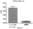
FIG. 4A is a bar graph showing the ratio of CD4/CD8 of splenocytes from mice injected with equivalent numbers of chimeric or mock control pBMN T cells at day 16 post-administration of chimeric or mock control protein-bearing T cells.
Figure 4B is a bar graph showing the cell count of adoptively transferred T cells at day 16 in mice treated as described in figure 4A.
Figure 4C is a bar graph showing individual spleen CD4 counts of splenocytes from the mice of figure 4A at day 16.
Figure 4D is a graph showing CD8 cell counts of splenocytes from the mice of figure 4A at day 16. As shown by flow cytometry data (data panels not shown) for these splenocytes gated with CD8 and CD4 markers (not shown), these panels also show an increase in the number of both CD4 and CD8 transfected cells in the spleen of mice administered T cells bearing the chimeric protein compared to mice administered T cells bearing the mock control protein. Figures 4A-4D show an increase in the number of T cells transferred in the spleen of mice injected with T cells carrying the FSH chimeric protein compared to mice injected with T cells carrying a mock control protein. Higher CD4/CD8 ratios were also detected in splenocytes transferred into T cells carrying the chimeric protein. In this ovarian cancer cell model, the ratio was found to be a benign marker of response to cancer.
Fig. 5 is a nucleic acid sequence of a construct comprising the fusion components of table 1 below, SEQ ID NO: 1 and the amino acid sequence SEQ ID NO: 2:
TABLE 1

Figure 6A is a schematic of a chimeric FSH-Letal construct, showing how it binds to tumor cells and NK cells or T cells (e.g., CD8T cells, gamma T cells, or NK T cells).
Fig. 6B is a nucleic acid sequence of a construct comprising the fusion components of table 2 below, SEQ ID NO: 3 and the amino acid sequence SEQ ID NO: 4:
TABLE 2

Fig. 6C is a nucleic acid sequence of a construct comprising the fusion components of table 3 below, SEQ ID NO: 5 and the amino acid sequence SEQ ID NO: 6. the construct has a MW of 45.87kD and utilizes non-cutter sites AscI, BamHI, BcgI, BeII, ClaI, HindIII, KpnI, MfeI, MluI, NcoI, NdeI, NotI, PacI, PmeI, PsiI, PvuI, SacII, SalI, SfiI, SgfI, SpeI, SphI, XbaI and XhoI.
TABLE 3

Fig. 6D is a nucleic acid sequence of a construct comprising the fusion components of table 4 below, SEQ ID NO: 7 and the amino acid sequence SEQ ID NO: 8:
TABLE 4

FIG. 7A is a bar graph showing adherent FSHR transduced ID8-Defb29/Vegf-a (B7F) cancer cells incubated with FSH CER expressing or mock control transduced T cells pBMN (1: 40 ratio) for 24 hours. After removal of non-adherent cells, trypan blue negative cells were counted in a hemocytometer. FSHR-efficient clearance by FSH-targeted CER T cells+A tumor cell.
Figure 7B is a bar graph showing that adherent FSHR transduced A7C11F transduced with the FSH-targeting construct described herein effectively cleared FSHR + tumor cells. Adherent FSHR transduced A7C11F cancer cells were incubated for 24 hours with FSH CER expressing or mock control transduced T cells (1: 40 ratio). After removal of non-adherent cells, trypan blue negative cells were counted in a hemocytometer. FSHR-efficient clearance by FSH-targeted CER T cells+A tumor cell.
Fig. 8 is a schematic representation of a variation of the FSH Chimeric Endocrine Receptor (CER) -T constructs described herein.
Figure 9 is a graph showing that human FSHCER T cells kill ovarian tumor cells in a dose-dependent manner. HLA-A2+ human T cells were expanded with ConA, transfected or mock-transduced at 20 and 44 hours by centrifugation with hFSHCER in pBMN with retronectin, and maintained at 0.5-1X 10 with 1ug/mL IL-7 and 20U/mL IL-26cells/mL. On day 7, CER and control T cells were sorted according to GFP expression and left for 8 hours, inoculated with HLA-A2+ human OVCAR-3 ovarian cancer cells (10000/well; spontaneous FSHR +) at the indicated ratio of effector (E) to target (T). After the setting was carried out for 6 hours,coculture cells were stained with annexin V and 7AAD and analyzed for cytotoxicity by flow cytometry. The percentage of specific lysis was calculated as (experimental death-spontaneous death)/(maximum death-spontaneous death) x 100%.
FIG. 10 is a Western gel showing FSHR at varying levels expressed in samples of advanced human ovarian cancer. FSHR protein expression was analyzed by Western blot (Santa Cruz # H-190) in 6 unselected human advanced ovarian cancer samples and compared to FSH-targeted CER T-cell sensitive OVCAR3 cells. Beta-actin antibody, Sigma # A5441
FIG. 11 is a graph showing the progression of FSHCER T cells to abrogate fshr-expressing orthotopic ovarian tumors. T cells carrying FSHR-targeted cars (FSH-CER) or the same expanded mock control transduced T cells (PBMN) were administered intraperitoneally 7 and 14 days after i.p. challenge with FSHR transduced ID8-defb29/vegf-a tumor cells (n-5 mice/group). Malignant tumor progression was compared.
Figure 12 is a graph showing that modified allogeneic or allogeneic human FSH CER T cells generated by using TALL-103/2 cells kill ovarian tumor cells in a dose-dependent manner. TALL-103/2 cells were transduced with hFSHCER in pBMN and maintained in medium with 20U/mL IL-2. After 24h to remove IL-2 from FSH CER-transduced (■) or mock control transduced (a-up) TALL-103/2 cells, luciferase-transduced FSHR + human OVCAR-3 ovarian cancer cells (10000/well) were incubated at the indicated effector (E) to target (T) ratio. 4 hours after the set-up, the co-cultured cells were lysed and the luciferase signal was quantified. The percentage of specific lysis was calculated as (experimental death-spontaneous death)/(maximum death-spontaneous death) x 100%.
Detailed Description
The compositions and methods provided herein elicit responses to, for example, ovarian cancer or other cancers characterized by neoplastic cells carrying FSH receptor (FSHR) (e.g., prostate cancer cells)52And metastatic tumor lesions51) Protective anti-tumor immunity and prevention of recurrence. The challenge of preventing certain immunotherapy techniques against epithelial tumors from achieving success is overcome by targeting hormone receptors by virtue of the endogenous ligand's advantage as a targeting motif.
Technical and scientific terms used herein have the same meaning as commonly understood by one of ordinary skill in the art to which this invention belongs, and are used by reference to published textbooks that provide those skilled in the art with a general guide to many terms used in the specification. The following definitions are provided for clarity purposes only and are not intended to limit the claimed invention.
Follicle Stimulating Hormone (FSH), a central hormone of mammalian reproduction, is produced primarily in the anterior pituitary. This hormone exerts its normal biological effects by binding to the plasma membrane Follicle Stimulating Hormone Receptor (FSHR) and stimulating follicle maturation and estrogen production in females. In males, the interaction of FSH and FSHR stimulates sertoli cell proliferation and maintains normal spermatogenesis. Naturally occurring FSH hormones are heterodimers formed by two subunits, the α and β subunits. The alpha subunit is also known as CG alpha and is common to Luteinizing Hormone (LH) and Thyroid Stimulating Hormone (TSH). The nucleic acid and amino acid sequences of the alpha and beta subunits of FSH of human and other dairy animal species are publicly known and available.
FSHR is a hormone receptor selectively expressed in ovarian granulosa cells in women and is at low levels in the ovarian endothelium. Most importantly, this surface receptor is expressed in 50-70% of ovarian cancers, but not in the brain, as negative feedback is dependent on the sensory hormone. Given that ovariectomy is the standard procedure for ovarian cancer treatment, targeting FSHR should not cause damage to healthy tissue.
As used herein, the phrase "ligand comprises an FSH sequence, and the ligand binds FSHR" includes naturally occurring full-length FSH sequences of a suitable mammal. The ligand comprises sufficient FSH sequence to allow binding between the ligand and FSHR by way of the natural affinity between the hormone sequence and the receptor. The ligand is not a body or antibody fragment and does not bind to the receptor in this manner. If the ligand is naturally occurring, e.g., the full length FSH β -FSH α sequence or a naturally occurring fragment thereof, the ligand is unable to induce an immunogenic response in an individual to whom the ligand is administered. If the ligand comprises a modified full length or fragment of the naturally occurring FSH sequence, in some embodiments the modification is insufficient to induce any strong immunogenic response in the individual to whom the ligand is administered.
In one embodiment, the ligand comprising an FSH sequence is a naturally occurring full length human FSH, e.g., an FSH β sequence linked to an FSH α sequence. In another embodiment, the ligand comprising an FSH sequence is a modified FSH β sequence linked to a naturally occurring FSH α sequence. In another embodiment, the ligand comprising the FSH sequence is a modified FSH β sequence linked to a modified FSH α/CG α sequence. In another embodiment, the ligand is a naturally occurring FSH β sequence linked to a modified FSH α sequence. In another embodiment, when the individual mammal is a human and the tumour of interest is a human tumour, the suitable FSH sequence is human FSH or a modified form of a human sequence. Alternatively, the ligand is a modified FSH, e.g. a naturally occurring or modified FSH β sequence linked to a naturally occurring or modified FSH α sequence by an optional spacer. In another embodiment, the ligand is a single naturally occurring or modified FSH β subunit alone. In another embodiment, the ligand is a naturally occurring FSH β subunit linked to a modified second FSH β sequence by an optional spacer sequence. In another embodiment, the ligand is a modified FSH β subunit linked to a second, naturally occurring FSH β sequence by an optional spacer sequence.
In another embodiment, the ligand comprises a fragment of a naturally occurring or modified FSH sequence. In another embodiment, the ligand comprises a fragment of a naturally occurring or modified FSH β sequence. In another embodiment, the ligand is a naturally occurring FSH β subunit linked to a modified FSH α subunit or a fragment thereof. In another embodiment, the ligand is a modified FSH β subunit or a fragment thereof linked to a naturally occurring or FSH α subunit. In another embodiment, the ligand comprises fragments of naturally occurring or modified FSH β sequences linked together.
By "naturally occurring" is meant that the sequence is a naturally occurring nucleic acid or amino acid sequence found in a selected mammal, including any naturally occurring variant of various nucleic acid and/or amino acid positions in the sequence found between different members of a mammalian species.
By "modified" is meant that the reference sequence, e.g., FSH or a fragment thereof or FSH β linked to FSH α nucleic acid or amino acid sequence or any subunit sequence alone has been deliberately manipulated. Suitable modifications include the use of shorter sequence fragments than the naturally occurring full length hormone. Such modifications include changes in the nucleic acid sequence to include preferred codons, which may encode the same or related amino acids as are present in the native amino acid sequence. Modifications also include changes in nucleic acid or amino acid sequence to introduce conservative amino acid changes, e.g., changes from one charged or neutral amino acid to a different charged amino acid. Such modifications may also include the deliberate creation of fusions using FSH β with or without FSH α sequence and other sequences not naturally occurring with FSH β or FSH α. Modifications also include joining the subunits using intentionally inserted spacer sequences or joining subunit fragments together or joining repeat fragments or subunits together in non-naturally occurring fusions.
As an example, the naturally occurring human FSH β nucleic acid sequence comprising a signal sequence comprises SEQ ID NO: 1, or consists of nucleic acid 1-387 of SEQ ID NO: 2, amino acids 1-129. The FSH β signal sequence itself comprises SEQ ID NO: 1, and the amino acid sequence is SEQ ID NO: 2, amino acids 1-18. Mature FSH β comprises SEQ ID NO: 1, and the amino acid sequence is SEQ ID NO: 2 amino acids 19-129.
As another example of methods and compositions for use herein, the mature human FSH β nucleic acid sequence comprises SEQ ID NO: 3 or 7, and the amino acid sequence is SEQ ID NO: 4 or 8 amino acids 2-112. As another example of methods and compositions for use herein, useful fragments of human FSH β nucleic acid sequences comprise FSH β SEQ ID NO: 1, FSH β SEQ ID NO: 1, nucleic acid 153-213, FSH β SEQ ID NO: 1, or FSH β SEQ ID NO: 1, or consists of the sequence 295-339 as described above. As another example of useful fragments of human FSH β amino acid sequences for use in the methods and compositions herein, fragments comprising FSH β SEQ ID NO: 2, FSH β SEQ ID NO: 2, FSH β SEQ ID NO: 2, or FSH β SEQ ID NO: 2, or consists of the above sequence.
In some embodiments where the ligand further comprises a FSH α sequence, the naturally occurring human FSH α nucleic acid sequence comprises SEQ ID NO: 1, the nucleic acid 433-801 and the amino acid sequence thereof are SEQ ID NO: amino acid 145 of 2 and 267. In another embodiment of the methods and compositions used herein, the human FSH α nucleic acid sequence comprises SEQ ID NO: 3, the nucleic acid 382-750 of SEQ ID NO: amino acid 128 of 4 and 250. In another embodiment of the methods and compositions used herein, the fragment of human FSH α nucleic acid sequence comprises SEQ ID NO: 7, wherein the nucleic acid 382-657 has an amino acid sequence of SEQ ID NO: amino acid 128 of 8-.
It will be appreciated that amino acid modifications or nucleic acid modifications as described above applied to these fragments are also useful ligands in this method. The ligand does not bind FSH in the antibody or antibody fragment-antigen complex. As described above, the ligands described herein utilize the natural affinity between the natural hormone (or modified form of the natural hormone) and its receptor for binding. Because the ligand is a natural hormone or a modified form thereof, it is designed to avoid inducing an antigenic response in an individual.
The terms "linker" and "spacer" are used interchangeably and refer to a nucleic acid sequence encoding a peptide of sufficient length to separate two components and/or to the peptide itself. The composition and length of the linker may be selected according to the application to which the linker is to be applied. In one embodiment, the amino acid linker separating FSH α and FSH β (naturally occurring sequence or modified sequence or fragment) is from 2 to 70 amino acids in length, including any number within this range. For example, in one embodiment, the linker is 10 amino acids in length. In another embodiment, the linker is 15 amino acids in length. In other embodiments, the linker is 25, 35, 50, or 60 amino acids in length. See, e.g., the spacers/linkers defined by the sequences described in tables 1-4 above.
Thus, a nucleic acid sequence encoding a linker or spacer consists of 6 to 210 nucleotides in length, including all values within this range. In some embodiments, the linker comprises a plurality of glycine residues or nucleic acids encoding them. In some embodiments, the amino acid linker comprises a plurality of serine residues or nucleic acids encoding them. In other embodiments, the linker comprises a plurality of thymine residues or nucleic acids encoding them. In other embodiments, the linker and spacer comprise any combination of serine, thymine, and glycine residues. Other linkers can be readily designed depending on the application.
As used herein, "vector" encompasses any genetic element, including but not limited to naked DNA, phage, transposon, cosmid, episome, plasmid, bacterium, or virus, which expresses or is caused to express a desired nucleic acid construct.
The term "individual" or "patient" as used herein refers to a male or female mammal, preferably a human. However, a mammalian subject may also be a veterinary or farm animal, a domestic animal or a pet, as well as an animal commonly used in clinical studies. In one embodiment, the individual of these methods and compositions is a human.
As known in the art, the term "cancer" as used herein denotes any disease, state, trait, genotype, or phenotype characterized by unregulated cell growth or proliferation. A "cancer cell" is a cell that abnormally divides and replicates in uncontrolled growth. The cell can leave its original site (e.g., a tumor), move to other parts of the body and establish another site (e.g., another tumor), a process known as metastasis. A "tumor" is an abnormal tissue mass from uncontrolled and progressive excessive cell division and also becomes a neoplasm. Tumors can be benign (non-cancerous) or malignant. The compositions and methods described herein are useful for treating cancer and tumor cells, i.e., both malignant and benign tumors, so long as the cells to be treated express FSHR. Thus, in various embodiments of the methods and compositions described herein, cancer may include, but is not limited to, breast cancer, lung cancer, prostate cancer, colorectal cancer, esophageal cancer, gastric cancer, gallbladder cancer, pancreatic cancer, renal cancer, cervical cancer, liver cancer, ovarian cancer, and testicular cancer.
The term "pharmaceutically acceptable carrier" or "diluent" as used herein is intended to include any and all solvents, dispersion media, coatings, antibacterial and antifungal agents, isotonic and absorption delaying bases, adjuvants and the like that are compatible with human administration. In one embodiment, the diluent is saline or buffered saline. The term "a" or "an" refers to one or more, e.g., "anti-tumor T cells" are understood to represent one or more anti-tumor T cells. Thus, the terms "a" (or "an"), "one or more" and "at least one" are used interchangeably herein. The term "about" as used herein modifies a reference value and includes all values from 0.01% up to 10% of the reference value and all values within the endpoints and includes the endpoints, e.g., ± 5%, ± 1%, ± 5% etc. The language "comprises" or "comprising" is used to indicate various embodiments in the specification, including other components or method steps. When "comprising" is used, it is to be understood that the relevant embodiments include descriptions using the term "consisting of …" to the exclusion of other components or method steps, and descriptions of the term "consisting essentially of …" to the exclusion of any components or method steps that substantially alter the nature of the embodiments or invention.
In one embodiment, the invention provides a nucleic acid sequence encoding a chimeric protein comprising a ligand comprising a sequence that binds human FSH linked to a nucleic acid sequence encoding a T cell activation function. As described in more detail above, in some embodiments, the ligand is naturally occurring FSH having two subunits, a single subunit of FSH, only FSH β subunit, FSH α/CG α or FSH β fragment, or a modified form of the above sequences.
In one embodiment, T cell activation function may be provided by linking the ligands described above to nucleic acid sequences encoding components useful in designing known Chimeric Antigen Receptors (CARs). See, e.g., Sadelain, M et al, "The basic principles of a molecular anti receiver (CAR) design"2013 April, Cancer Discov.3(4): 388-398;international patent application publication WO2013/044255, U.S. patent application publication No. US2013/0287748 and other publications relating to the use of these chimeric proteins. These publications are incorporated by reference to provide information on the various components useful in designing certain constructs described herein. Such CAR T cells are genetically modified lymphocytes expressing ligands that allow them to recognize a selected antigen. Upon antigen recognition, these modified T cells are activated by converting these T cells into the signaling domain of a strong cell killer. The advantage over endogenous T cells is that they are not MHC restricted, which allows these T cells to express by reducing MHC19Overcoming immune surveillance evasion strategies used in many tumor cells.
Such T cell activation function may be provided, for example, by linking the ligand to the transmembrane domain, costimulatory signaling region, and/or signaling endodomain via an optional spacer.
Thus, one embodiment of a nucleic acid sequence useful in the methods described herein is illustrated in SEQ ID NO: 1 and table 1. The nucleic acid sequence or CER construct comprises a ligand formed from: the naturally occurring human FSH β sequence, formed by an 18 amino acid human FSH β signal sequence and a 120 amino acid mature FSH β, is linked to a 15 amino acid spacer and to a naturally occurring 123 amino acid FSH α sequence. The CER constructs also include other components, i.e., an extracellular hinge domain, a transmembrane domain, a human intracellular region, and a signaling intracellular domain. For the construct of FIG. 5, for example, the hinge and transmembrane domains are from human CD8 α, the human intracellular domain is from 4-1BB, and the signaling domain is the human CD3 ζ domain.
Other embodiments that may be used as such nucleic acid constructs may include constructs having different ligands, e.g., one of the ligands described above, in one embodiment, the FSH α sequence in the same construct depicted in fig. 5 may be a shortened sequence having the sequence shown in SEQ ID NO: 7, and the nucleic acid sequence of nucleotides 382-657 of SEQ ID NO: 8 amino acid 128-219. Another embodiment of the ligand used in the construct of fig. 5 may comprise a ligand lacking SEQ ID NO: 2, amino acids 2-18 of the signal sequence. Embodiments similar to the nucleic acid construct of fig. 5 can be readily designed by replacing the ligand portion of table 1 with any of the ligands, modified, naturally occurring or fragments discussed above.
Other embodiments of nucleic acid constructs similar to FIG. 5 may employ different components, such as those described in detail in Sadelain et al, referenced above, or in the patent publications incorporated herein by reference. For example, when hinge domains are employed, other naturally occurring or synthetic hinge domains include immunoglobulin hinge regions, e.g., from IgG1, C of immunoglobulinsH2CH3Region, segment of CD3, etc. Other embodiments of nucleic acid constructs similar to figure 5 may employ different naturally occurring or synthetic transmembrane domains obtained from T cell receptors. Various transmembrane proteins contain domains useful in the constructs described herein. For example, transmembrane domains obtained from T cell receptors, CD28, CD3 epsilon, CD45, CD4, CD8, CD9, CD16, CD22, CD33, CD37, CD64, CD80, CD86, CD134, CD137, or CD154 have been noted to be useful.
Other embodiments of nucleic acid constructs similar to FIG. 5 may employ different naturally occurring or synthetic intracellular domains, including costimulatory signaling regions, and others known in the art. The costimulatory signaling region can be an intracellular domain of a cell surface molecule (e.g., a costimulatory molecule), such as CD27, CD28, 4-1BB (CD137), OX40, CD30, CD40, PD-1, ICOS, lymphocyte function-associated antigen-1 (LFA-1), CD2, CD7, LIGHT, NKG2C, B7-H3. See, e.g., the other molecules listed in the publications cited above.
Other embodiments of nucleic acid constructs similar to FIG. 5 may employ different naturally occurring or synthetic cytoplasmic signaling domains including those derived from CD3 zeta, TCR zeta, FcR gamma, FcR beta, CD3 gamma, CD3 delta, CD3 epsilon, CD5, CD22, 25 CD79a, CD79b, and CD66d, among others, and other cytoplasmic signaling domains known in the art
Any number of variations of the nucleic acid constructs, e.g., of fig. 5, can be designed for use in the methods described herein, given the teachings provided herein and using information known in the art.
Thus, another component described herein is a chimeric protein comprising a ligand comprising the FSH β sequence or a modification or fragment of said FSH sequence, which ligand binds human FSHR, linked to a peptide or protein having T cell activating function. Such chimeric proteins comprise a ligand that binds human FSHR as described above, linked to an extracellular hinge domain, a transmembrane domain, a costimulatory signaling region, and a signaling endodomain. Exemplary chimeric proteins are encoded by the nucleic acid sequences described above. One embodiment of such a chimeric protein is the amino acid sequence shown in SEQ ID NO: 2. other chimeric proteins can be readily designed using the various ligands identified herein, for example, one or more FSH β fragments identified in detail above or other FSHR binding ligands identified herein in place of SEQ ID NO: 2, a ligand as specifically exemplified.
In another embodiment, a useful CER construct is a nucleic acid sequence encoding a ligand that comprises a FSH β sequence or a modification or fragment of said FSH sequence, as described above, which ligand binds human FSHR, linked to a nucleic acid sequence encoding a ligand that binds tumor-associated NKG2D receptor. See, for example, fig. 6A. One such NKG2D ligand is known as Letal or ULBP 4. Leal consists of SEQ ID NO: 3 and has the nucleotide sequence of 796-1365 of SEQ ID NO: 4 amino acid 266-454. See, for example, Conejo-Garcia, J et al, "Letal, A Tumor-Associated NKG2D Immunorecter light, industries Activation and Expansion of efficiency Immune Cells" July 2003, cane. biol. & ther.,2(4): 446-; and U.S. patent application publication No. 20060247420, which are incorporated herein by reference. In the present specification, other NKG2D ligands or amino acid modifications, nucleic acid level modifications or functional fragments of the Letal sequence may be substituted for the exemplary Letal sequence.
Further, optionally, these FSHR binding ligands and NKG2D ligands are linked by suitable spacers or linkers as indicated above.
Specific examples of such nuclear constructs are provided in fig. 6A, fig. 6B, table 2, SEQ ID NO: 3, fig. 6D, table 4, SEQ ID NO: 7 and 8. In the embodiment of fig. 7B, the FSHR binding ligand is formed by a naturally occurring human FSH β sequence linked to a 15 amino acid spacer, which is in turn linked to a naturally occurring FSH α sequence of 123 amino acids, which is formed by a single amino acid methionine from the signal sequence, followed by 120 amino acids of mature FSH β. The ligand is further linked to Letal via another 15 amino acid spacer. In the embodiment of FIG. 6D, the FSHR binding ligand is formed from the naturally occurring human FSH β sequence formed from a single amino acid methionine from the signal sequence followed by 120 amino acids of mature FSH β, linked to a 15 amino acid spacer and further linked to the modified FSH α sequence (i.e., the fragment of amino acid 128-219 of SEQ ID NO: 8, encoded by nucleotide 382-657 of SEQ ID NO: 7). The ligand is further linked to Letal via another 15 amino acid spacer.
Other embodiments that may be used as such nucleic acid constructs may include the constructs of fig. 6B and 6D with different ligand-encoding sequences (e.g., sequences encoding one of the ligands described above). In one embodiment, the FSH α sequence in the same construct depicted in fig. 6B may be a single or multiple copies of full length FSH β with or without a signal sequence. As another example, the fig. 6B and 6D constructs may contain a ligand formed from a human FSH β fragment encoded by the following nucleic acid sequence: the nucleic acid sequence comprises FSH β SEQ ID NO: 1, FSH β SEQ ID NO: 1, FSH β SEQ ID NO: 1, or FSH β SEQ ID NO: 1, or consists of the sequence 295-339 as described above. The ligand may consist of these fragments alone, together, or instead of full length FSH β and thus be fused via a linker to the FSH α sequence of fig. 6B or 6D. Embodiments similar to the nucleic acid constructs of fig. 6B or 6D can be readily designed by replacing the ligand portion of table 2 or 4 with any of the ligands discussed above, modified, naturally occurring or fragmented.
Thus, another aspect is the use of a nucleus as described aboveA chimeric or bispecific protein encoded by the sequence and comprising a ligand comprising a FSH β sequence as described herein or a modification or fragment of said FSH sequence, said ligand binding to human FSHR, said ligand being linked to a ligand binding to NKG 2D. These proteins are primarily useful in the form of proteins and function in vivo to bind endogenous lymphocytes and FSHR+The tumor cells are brought together.
In other aspects, a recombinant vector carrying the nucleic acid construct described above is provided. The nucleic acid construct may be carried in a plasmid-based system or a replicating or non-replicating recombinant viral vector, and the chimeric protein may be expressed in a plasmid-based system or a replicating or non-replicating recombinant viral vector, many of which are commercially available. The nucleic acid sequences discussed herein may be expressed and produced in vitro or in vivo using these vectors in a desired host cell. Thus, in one embodiment, the vector is a non-pathogenic virus. In another embodiment, the vector is a non-replicating virus. In one embodiment, the desired viral vector may be a retroviral vector, such as a lentiviral vector. In another embodiment, the desired vector is an adenoviral vector. In another embodiment, a suitable vector is an adeno-associated viral vector. Adenoviruses, adeno-associated viruses and lentiviruses are generally preferred because they actively infect dividing and quiescent and differentiated cells, such as stem cells, macrophages and neurons. A variety of adenovirus, lentivirus and AAV strains are available from the American Type Culture Collection, Manassas, Virginia, or by application to a variety of commercial and academic sources. In addition, the sequences of many such strains are available from a variety of databases, including, for example, PubMed and GenBank.
In one embodiment, a lentiviral vector is used. Useful vectors are equine infectious anemia virus and feline and bovine immunodeficiency virus, and HIV-based vectors. A variety of useful lentiviral vectors, and methods and treatments for generating these vectors for transduction of cells and expression of heterologous genes, e.g., N Manjunath et al,2009 Adv Drug delivery rev, 61(9): 732; porter et al, N Engl J Med.2011Aug 25; 365(8):725-33).
In another embodiment, the vector used herein is an adenoviral vector. Such vectors may be constructed using adenoviral DNA from one or more of any known adenoviral serotypes. See, e.g., T.Shenk et al, Adenovirdae The Viruses and The reaction ", Ch.67, in FIELD' S VIROLOGY,6th Ed., edited by B.N FIELDs et al, (Lippincott Raven Publishers, Philadelphia,1996), p.111-2112; 6,083,716, which describes the genomes of two chimpanzee adenoviruses; U.S. Pat. nos. 7,247,472; WO 2005/1071093 and the like. One skilled in the art can readily construct suitable adenoviral vectors to carry and express the nucleotide constructs described herein. In another embodiment, the vector used herein is an adeno-associated virus (AAV) vector. Such vectors can be constructed using AAV DNA of one or more known AV serotypes. See, for example, U.S. patent No. 7,803,611; us patent 7,696,179, us patent, etc.
In another embodiment, the vector used herein is a bacterial vector. In one embodiment, the bacterial vector is listeria monocytogenes. See, e.g., Lauer et al, Infect. immunity,76(8):3742-53 (Aug.2008). Thus, in one embodiment, the bacterial vector is live attenuated or photochemically inactivated. The chimeric proteins may be expressed recombinantly by bacteria, e.g., by introduction into a plasmid of the bacteria, or integrated into the bacterial genome, i.e., by homologous recombination.
These vectors also include conventional control elements that allow for transcription, translation, and/or expression of the nucleic acid construct in cells transfected with the plasmid vector or infected with the viral vector. Various expression control sequences are known in the art and can be used, including native, constitutive, inducible, and/or tissue-specific promoters. In one embodiment, the choice of promoter is based on the selected vector. In another embodiment, when the vector is a lentivirus, the promoter is the U6, H1, CMV IE gene, EF-1 α, ubiquitin C or phosphoglycerate kinase (PGK) promoter. In another embodiment, when the vector is AAV, the promoter is a RSV, U6 or CMV promoter. In another embodiment, when the vector is an adenovirus, the promoter is an RSV, U6, CMV, or H1 promoter. In another embodiment, when the vector is listeria monocytogenes, the promoter is the hly or actA promoter. Other conventional expression control sequences include selectable marker genes or reporter genes, which may include sequences encoding geneticin, hygromycin, ampicillin or puromycin resistance, and the like. Other components of the vector may include an origin of replication.
The selection of these and other promoter and vector elements is routine, and many such sequences are available (see, e.g., the references cited herein).
These vectors are generated using the techniques and sequences provided herein in combination with techniques known to those skilled in the art. These techniques include conventional cDNA Cloning techniques, such as those described in textbooks (Sambrook et al, Molecular Cloning: A Laboratory Manual, Cold Spring Harbor Press, Cold Spring Harbor, N.Y.), the use of overlapping oligonucleotide sequences, polymerase chain reactions, and any suitable method that provides the desired nucleotide sequence.
Thus, in one embodiment, one of skill in the art can construct viral vectors (or plasmids) expressing the desired nucleic acid constructs using the information taught herein and known to the public and vector construction components and techniques. The chimeric proteins encoded by these nucleic acid constructs may be expressed in vitro or ex vivo in host cells, or in vivo by administration to a mammalian subject. Alternatively, the chimeric protein may be produced synthetically by known chemical synthesis methods. Depending on the composition, efficiency of the method, and intended use, one skilled in the art will be able to select appropriate methods for producing these chimeric proteins, e.g., as proteins, nucleic acids, or by adoptive T cell administration, or otherwise to achieve the desired therapeutic result.
In another aspect, there is provided a modified human T cell comprising a nucleic acid sequence encoding a chimeric protein comprising a ligand that binds human FSHR linked to a nucleic acid sequence encoding a T cell activation function. In one embodiment, these latter nucleic acid sequences encode an extracellular hinge domain, a transmembrane domain, a costimulatory signaling region, and a signaling endodomain in a pharmaceutically acceptable carrier.
The modified T cell is a T cell that has been transduced or transfected with a vector carrying a nucleic acid construct encoding a chimeric protein as described above. Ideally, the T cell is a primary T cell, a CD8 (cytotoxic) T cell, or a NK T cell or other T cell obtained from the same mammalian individual to which the modified T cell is administered, or other T cell obtained from another member of the mammalian species. In one embodiment, the T cells are autologous human T cells or Natural Killer (NK) T cells obtained from the individual or a bone marrow transplant partner of the individual. Other suitable T cells include T cells obtained from resected tumors, polyclonal or monoclonal tumor-reactive T cells. T cells are typically obtained by the apheresis method and transfected or transduced with a selected nucleic acid construct to express the chimeric protein in vivo.
Other suitable T cells include allogeneic or allogeneic T cells, which can be used as a universal T cell platform carrying the nucleic acid constructs described herein. In one embodiment, human cytotoxic T cells may be selected. TALL-104 and TALL-103/2 cells are CD3 responsive lymphocytes, respectively CD3+TCRαβ+And CD3+TCRγδ+Childhood T-cell leukemia, which exhibits major histocompatibility Complex non-limiting, NK cell receptor mediated tumoricidal activity, relies primarily on NKG2D59 ,60,61. TALL cells exhibit NKG 2D-dependent broad range of tumor target reactivity. Irradiated TALL-104 cells have been used to treat metastatic breast and ovarian cancer due to their spontaneous (NK-like) cytolytic activity and safety.
These modified T cells, whether autologous or endogenous, are activated by a signaling domain that converts these T cells into potent cytokillers. The advantage of autologous cells over endogenous T cells is that they are not MHC restricted, which allows these T cells to overcome immune surveillance evasion strategies used in many tumor cells by reducing MHC expression. Endogenous cells such as TALL cells have advantagesIn that they are versatile, adaptable to scale-up preparation, standardization and further cell engineering techniques to target FSHR+Ovarian cancer.
In another embodiment, the modified T cell is further engineered ex vivo to inhibit, eliminate, or reduce expression of a forkhead box protein (Foxp 1). In one embodiment, the T cells are first engineered or treated to reduce Foxp1 and then transfected with a nucleic acid sequence as described above encoding a chimeric protein comprising a ligand comprising a naturally occurring or modified FSH sequence or fragment thereof, which ligand binds human FSHR, linked to other T cell stimulating or targeting sequences. In another embodiment, the treatment is performed to reduce or eliminate Foxp1 after transfection of T cells with a nucleic acid sequence encoding a chimeric protein or a bispecific protein described herein. In one embodiment, the T cells are pretreated such that the T cells do not express Foxp1 after delivery to the subject. Optimally, Foxp1 was eliminated in the modified T cells. T cells can be treated with: zinc finger nucleases, transcription activator-like effector nucleases (TALENs), CRISPR/Cas systems or engineered meganucleases re-engineered homing endonucleases and sequences optimized and designed to target unique Foxp1 sequences to introduce defects into the Fox-P1 genomic sequence or to delete the Fox-P1 genomic sequence. By taking advantage of endogenous DNA repair mechanisms, these agents remove Foxp1 from modified T cells prior to adoptive transfer. Alternatively, T cells may be co-transfected with another nucleic acid sequence designed to inhibit, reduce, down-regulate, or eliminate the expression of Foxp 1. See, for example, international patent application publication WO2013/063019, which is incorporated herein by reference. Various combinations of these techniques can also be used before or after the construction of modified somatic T cells by introducing nucleic acids.
Typically, when the vector is delivered by transfection into T cells, the amount of vector delivered is to about 1X 10⁴Cells to about 1X 10¹³The cells deliver about 5 μ g to about 100 μ g of DNA. In another embodiment, the delivery amount of the vector is to 1X 10⁴Cells to about X1013The cells deliver about 10 to about 50 μ g of DNA. At another placeIn embodiments, the delivery amount of the carrier is to about 105The cells deliver about 5 μ g to about 100 μ g of DNA. However, the relative amounts of vector DNA and T cells can be adjusted taking into account factors such as the vector selected, the delivery method chosen, and the host cell. The vector may be introduced into the T cell by any means known in the art or disclosed above, including transfection, transformation, infection, electroporation, or direct DNA injection. The nucleic acid construct may be stably incorporated into the genome of the host cell, stably expressed as an episome, or transiently expressed.
The resulting modified T cells are prepared to express the nucleic acid construct in a suitable pharmaceutical carrier for adoptive therapy. However, as described above, the chimeric bispecific protein may be administered as a protein in a suitable pharmaceutical carrier.
All of the compositions and components described above can be used in the methods described herein to treat cancer and stimulate anti-tumor immune activity described herein. Accordingly, there is provided a method of treating cancer in a human subject comprising administering to the subject any of the compositions described above in a pharmaceutically acceptable formulation or carrier.
In one embodiment, the individual treated by the method is an individual suffering from a FSH-expressing cancer, including those listed above. In another embodiment, prior to administration of the compositions described herein, an individual having FSHR-expressing cancer or tumor cells has been surgically treated to resect the tumor of interest. In one embodiment, the individual is a female with ovarian cancer. In another embodiment, a female individual with ovarian cancer has been surgically treated to remove the ovaries, fallopian tubes, and/or uterus. Prior to or after administration of these methods, individuals with any of the other cancers listed above may be treated by suitable surgery.
In one embodiment, the individual is administered a composition comprising a nucleic acid construct as described above. In another embodiment, the individual is administered a composition comprising a chimeric protein described above. In a specific embodiment, a method of treating cancer in a human individual comprises administering to the individual in need thereof a bispecific protein comprising a ligand comprising a FSH β sequence, said ligand binding to human FSHR, said ligand being linked to a ligand binding to NKG 2D. In another embodiment, the composition is a viral vector carrying a nucleic acid construct, thereby allowing infection in vivo.
In another embodiment, a method of treating cancer in a human subject comprises administering to a subject in need thereof modified human T cells comprising a nucleic acid sequence encoding a chimeric protein comprising an FSH sequence, a modification or fragment of said FSH sequence, and a ligand binding FSHR, linked to a nucleic acid sequence encoding T cell activation. In one embodiment, the T cell activation function is provided by a nucleic acid sequence encoding an extracellular hinge domain, a transmembrane domain, a costimulatory signaling region, and a signaling endodomain. In one embodiment, the modified T cell expresses any of the nucleic acid constructs described herein. In an exemplary embodiment, the modified T cell expresses the nucleic acid construct of figure 5 or a similar construct described herein. In another embodiment, the modified T cells do not express forkhead box protein (Foxp1) after administration. In another embodiment, the modified T cell carries a nucleic acid construct that expresses or co-expresses a sequence that eliminates or reduces expression of Foxp 1.
In another embodiment, the modified human T cells are administered with a clinically available PD-1 inhibitor. In another embodiment, the modified human T cells are administered with clinically available agents including TGF- β inhibitors, including blocking antibodies. In another embodiment, the modified human T cells are administered with a clinically available inhibitor of IL-10.
These therapeutic methods are designed to enhance the therapeutic activity of T cells and prolong the survival of cancer patients. Therapeutic compositions administered by these methods, for example, nucleic acid constructs alone, in viral vectors or nanoparticles, as chimeric or bispecific proteins, or as modified anti-tumor T cells for adoptive therapeutic treatment, are administered systemically or directly to the environment of the cancer cells or tumor microenvironment of the individual. Conventional and pharmaceutically acceptable routes of administration include, but are not limited to, systemic routes such as intraperitoneal, intravenous, intranasal, intravenous, intramuscular, intratracheal, subcutaneous and other parenteral routes of administration or intratumoral or intralesional administration. If desired, the routes of administration may be combined. In some embodiments, the administration is repeated periodically. In one embodiment, the composition is administered intraperitoneally. In one embodiment, the composition is administered intravenously. In another embodiment, the composition is administered intratumorally.
These therapeutic compositions can be administered to a patient, preferably suspended in a biocompatible solution or pharmaceutically acceptable delivery vehicle. The various components of the compositions are formulated so that the drug is administered by suspension or dissolution in a pharmaceutically or physiologically acceptable carrier, such as isotonic saline, isotonic saline solution or other formulations for such administration as would be readily understood by one skilled in the art. Suitable carriers are readily determinable by those skilled in the art and depend to a large extent on the route of administration. Other aqueous and non-aqueous isotonic sterile injection solutions and aqueous and non-aqueous sterile suspensions known to be pharmaceutically acceptable carriers and known to those skilled in the art may be used for this purpose.
The dosage of these therapeutic compositions depends primarily on factors such as the type of composition (i.e., T cell, vector, nucleic acid construct, or protein), the disease state to be treated, the age, weight, and health level of the patient, and may vary from patient to patient. In one embodiment, at 2 × 10⁶To 200X 106Administering multiple doses of the modified T cells to a composition containing the modified T cells. Any value in between can be selected depending on the status and response of the individual patient. As another example, the number of adoptively metastatic anti-tumor T cells can be optimized by one skilled in the art. In one embodiment, such a dosage range may be about 10 per kilogram of the individual's body weight5To about 1011A cell. In another embodiment, the dose of anti-tumor T cells is about 1.5X 10 per kilogram body weight5A cell. In another embodiment, the dose of anti-tumor T cells is about 1.5X 10 per kilogram body weight6A cell. In another embodiment, anti-tumor T cellsThe dosage of the cells is about 1.5X 10 per kilogram of body weight7A cell. In another embodiment, the dose of anti-tumor T cells is about 1.5X 10 per kilogram body weight8A cell. In another embodiment, the dose of anti-tumor T cells is about 1.5X 10 per kilogram body weight9A cell. In another embodiment, the dose of anti-tumor T cells is about 1.5X 10 per kilogram body weight10A cell. In another embodiment, the dose of anti-tumor T cells is about 1.5X 10 per kilogram body weight11A cell. Other dosages within these specific amounts are also contemplated by these methods.
In another embodiment, the therapeutically effective adult or veterinary dose of the viral vector generally ranges from about 100 μ L to about 100mL of the vector containing the vector at a concentration of about 1X 10⁶To about 1X 10¹⁵Particles of about 1X 10¹¹To 1X 10¹³Per particle, or about 1X 10⁹To 1X 10¹²Individual particles of virus.
Administration of the protein-containing composition may be in a unit dose ranging from 0.01mg to 100mg of protein (equivalent to about 12.5 μ g/kg body weight).
Methods for determining the timing of frequency of administration (booster) will involve assessing the response of the tumor to the administration.
In other embodiments, these methods of treating cancer by administering the compositions described herein are part of a combination therapy with various other treatments or treatments of cancer.
In one embodiment, the method comprises administering cytokine therapy, such as IL-7, as a tumor-specific host conditioning strategy. Exogenous administration of IL-7 also specifically increased the in vivo activity of Foxp 1-deficient T cells. In another embodiment, the method further comprises administering to the individual a composition described herein, in combination with an additional anti-cancer therapy, which may include monoclonal antibodies, chemotherapy, radiation therapy, cytokines, or a combination thereof. In another embodiment, the methods herein may include co-administration or therapeutic procedures that also use other small nucleic acid molecules or small chemical molecules or therapeutics or therapeutic agents for the control and treatment of cancer. In one embodiment, the treatment methods of the present invention comprise treatment with one or more drugs under conditions suitable for cancer treatment.
As mentioned above, in some embodiments, surgical debulking is a necessary procedure to remove large tumor masses and can be performed before, during, or after administration of the methods and compositions described herein.
In other embodiments, chemotherapy and radiation therapy support the effects of the methods described herein. Such a combination (surgery plus chemotherapy/radiation plus immunotherapy) is expected to be successful in treating many cancers with the methods described herein.
In other embodiments, the method of treating an individual having an FSHR-expressing cancer or tumor comprises performing the following steps prior to administration of the composition described herein. In one embodiment, the method comprises removing T cells from the subject and transducing the T cells ex vivo with a vector expressing the chimeric protein. In another embodiment, the removed T cells are treated to eliminate or reduce Fox-P1 expression in the T cells prior to or after transduction of the removed T cells with the nucleic acid constructs described herein. In another method, prior to administration, the removed, treated T cells are cultured to remove Foxp1 from the cells ex vivo. Another method step includes formulating the T cells in a suitable pharmaceutical carrier prior to administration. The removed, treated T cells can also be frozen for subsequent thawing and administration.
The treatment method may further comprise extracting T cells from the subject for modification and ex vivo cell expansion, followed by treating the subject with chemotherapy and depleting the subject of lymphocytes, and optionally, surgically resecting the tumor. These steps may be performed prior to administering the modified T cells or other compositions to the individual.
The invention will now be described in connection with the following examples. These examples are provided for illustrative purposes only. The compositions, experimental procedures, and methods disclosed and/or claimed herein can be made and executed without undue experimentation in light of the present teachings. The operations and methods described in the embodiments should not be considered as limiting the scope of the claimed invention. Indeed, this description should be construed to cover any and all variations that can be readily derived from the teachings provided herein. It will be appreciated by those of skill in the art that such changes or variations may be made in the embodiments disclosed in the examples and are intended to achieve similar results. For example, substitution of an agent that is chemically or physiologically related to the agent described herein is expected to produce the same or similar results. All such similar substitutes and modifications apparent to those skilled in the art are deemed to be within the scope of the invention.
Example 1: preparation of FSHR-targeting constructs for humans and mice
We prepared a novel whole mouse derived construct described herein against mouse FSHR which contains all the signals of the mouse form successfully used in human patients. To target FSHR, we synthesized constructs expressing a signal peptide followed by two subunits (α and β) of endogenous FSH, separated by a linker (see fig. 1). The targeting motif was cloned in-frame with: a hinge domain from a murine IgG (e.g., CD8 α), followed by a transmembrane domain of CD8 α, an intracellular domain of a costimulatory modulator (e.g., murine 4-1BB or CD28), and finally an activated CD3 ζ domain.
We also prepared constructs with the corresponding human sequences (see FIG. 5, Table 1, SEQ ID NOS: 1 and 2). Human variants of FSHR-expressing constructs were prepared to define leader preparations and to demonstrate experimental relevance in mice. Human HLA-a2+ T cells (> 50% caucasian a2+) from healthy donors were transduced with retroviral or lentiviral stocks containing FSH targeting constructs optimized for cytotoxicity assays.
In-frame constructs similar to the mouse sequences described above were prepared to compare CD28 with 4-1BB/CD 137. CD28 is an optional intracellular co-stimulatory motif, because, although 4-1BB/CD137 expressing T cells showed enhanced persistence in the xenograft model in the disclosed CAR-T cell experiments, it was not known that long-term survival of T cells was preferred over multiple injections. In addition, two human variants of the alpha subunit of human FSH were tested (NM _000735.3 and NM _ 001252383.1). These two subunits have different lengths and have the potential to promote different binding affinities.
In summary, the 8 variants cloned (in-frame) for expression into viral vectors are: 1) CG α (Long) +4-1 BB; 2) CG α (long) + CD 28; 3) CG α (short) +4-1 BB; and 4) CG α (short) + CD28 (see FIG. 8).
Other constructs were designed using only the beta subunit of FSH (which provides specificity of FSHR binding) and a 15 amino acid binding region that also binds the beta subunit of FSHR (e.g., a fragment of amino acids 19-32 of SEQ ID NO: 2 of FSH beta or other FSH beta fragments identified above).
Example 2: FSH constructs specifically respond to FSHR+A tumor cell.
Retroviral (pSFG) and lentiviral (pELNS) vectors were tested to transduce FSHR-carrying constructs into human T cells. There is no formal evidence that lentiviral vectors are excellent in ex vivo transduction. Most importantly, after ten years of safe use of retroviral vectors, there is little concern about the risk of insertional tumor formation following gene insertion in T cells. Particularly in clinical trials, the pSFG vector has been used many times for similar T cell retroviral transduction41,42。
Retroviral or lentiviral stocks expressing these constructs were prepared and used to transduce human T cells from healthy HLA-a2 donors (> 50% caucasian). Retroviral stocks were used to transduce CD3/CD28 activated T cell splenocytes, which were FACS sorted based on co-expressed GFP. Modified T cells expressing the FSH nucleic acid construct of example 1 were then tested for binding specificity against FSHR or mock control transduced ID8-Defb29/Vegf-a ovarian cancer cells. As shown in figure 2, co-incubation of T cells transduced with FHSR targeting constructs caused IFN- γ secretion, which was not observed with T cells carrying the unrelated mesothelin targeting construct (K1). Further supporting the specificity of FSHR recognition, IFN- γ secretion did not occur in the presence of mock-control transduced (native FSHR) tumor cells.
Example 3: intratumoral administration of T cells expressing the FSH construct delays the progression of FSHR + breast tumors.
To understand modified T cells targeting FSHRInformation on possible efficacy and safety in immunocompetent mice, we also transduced A7C11 breast cancer cells with mouse FSHR, A7C11 breast cancer cells are a cell line generated from a tumor mutated by the bulk p 53/KRas. Syngeneic mice were then challenged with a flank tumor and given 10 of equivalent treatment by intraperitoneal injection6FSHR-targeted modified T cells or mock control-transduced T cells. As shown in figure 3, a single administration of FSHR-targeted modified T cells was sufficient to significantly delay the progression of established flank tumors without significant side effects. These results support the use of FSHR-targeted modified T cells alone or in combination with other clinically useful immunotherapies to combat ovarian orthotopic tumors.
Compared to the use of constructs targeting human FSHR, the use of mouse FSH as a targeting motif has better predictability of the effect of modified T cells targeting FSHR in immunodeficient mice, because: 1) t cells expressing mouse FSH may be targeted to unidentified healthy cells expressing endogenous FSHR (unlike T cells expressing human FSH administered to immunodeficient mice); 2) certain T cells (e.g., CER-T cells) are able to boost polyclonal anti-tumor immunity by enhancing pre-existing T cell responses and reducing immunosuppressive burden via antigen spreading; and 3) the interaction between FSH and its specific receptor is highly conserved.
To confirm the cytotoxic potential of T cells containing the FSHR construct specifically against FSHR + tumor cells, we again combined FSHR constructs or mock control transduced T cells with FSHR+Cells of ID8-Defb29/Vegf-a33 (ovarian tumor) or A7C1134 (laboratory generated cell line 34 from a bulk p53/KRas mutated breast tumor) were incubated together (at a ratio of 40: 1, for 24h) and cytotoxic killing was determined by counting trypan blue negative (live) tumor cells. As shown in fig. 7A and 7B, FSH CER T cells eliminated both types of tumor cells, but this result was not observed with mock-control transduced lymphocytes. Comparable results were obtained in the MTS assay (not shown), further supporting that the FSH targeting motif can trigger CER-mediated T-cell cytotoxic activity in an FSHR-specific manner.
Example 4: efficacy and toxicity of modified T cells expressing FSH ligands in preclinical ovarian cancer models of immunocompetent hosts
To define the immunological outcome of using FSHR-targeted modified T cells in an immunologically active preclinical tumor model that includes all healthy tissues to which endogenous FSH may bind (as may occur in patients), we prepared new nucleic acid constructs using the mouse counterpart of all targeting and activating domains. See table 3, fig. 6C and SEQ ID NOs 4 and 5.
We validated the hypothesis that FSHR-targeted modified T cells exhibit selective activity on FSHR-expressing cells, disrupting tumor progression without causing significant side effects in mice. These results define the efficacy of this promising treatment for ovarian cancer and ensure its safety for future clinical applications of this approach.
We used aggressive in situ ID8-Vegf/Defb29 tumors, which had been transduced and selected by mouse FSHR. We demonstrated the general applicability of the selected findings using transduced maternal ID8 cells and/or an autologous p 53-dependent inducible tumor model or our derived cell lines.
Example 5: mouse clinical trials: defining the efficacy of FSHR-targeted modified T cells on human ovarian cancer
We made constructs with all human domains (see figure 5) that were used to establish a cohort of primary tumor-derived xenografts in our NSG mice. By using tumors and T cells from the same patient, we simulated a limited clinical trial, which recapitulated the heterogeneity of clinical ovarian cancer. We hypothesized that FSHR-targeted modified T cells are effective against established FSHR + ovarian cancer from different patients and act synergistically with TME-targeted combinatorial intervention.
By establishing the efficacy and possible toxicity of FSHR-targeted modified T cells in a variety of preclinical models, we provided a mechanistic rationale to test these T cells as potential treatments for ovarian cancer. By using FSHR as a specific tumor target and ligands comprising FSH sequences as targeting motifs, we overcome some of the challenges that prevent the success of various technologies for epithelial tumors.
To define the full spectrum of FSHR-targeted modified T cells in vivo in immunocompetent hosts, we generated FSHR-targeted modified T cells of whole murine origin. 7 days after tumor challenge, (CD45.1+, syngeneic) with 106Modified T cell therapy (CD45.2+) with anti-FSHR targeting (FSHR + and FSHR-) mice bearing established (FSHR + and FSHR-) ovarian tumors were injected intraperitoneally (n.gtoreq.10/group). Control mice received mock-control transduced T cells. We compared survival as a recognized efficacy reading. Additional injections of FSHR-targeted modified T cells were given according to these results. We tracked the homing and persistence of (specific CD45.1+) metastatic CD4 and CD8T cells in different mice at 14, 21, and 28 days post tumor challenge. Including samples from the spleen, draining (mediastinal) lymph nodes, bone marrow and tumor bed (peritoneal lavage). Detailed analysis of markers of activation (e.g., CD44, CD69, CD27, CD25) and depletion (e.g., PD-1, Lag3) in metastatic lymphocytes was included. In addition, the probable central memory differentiation (syngeney) of FSHR-targeted modified T cells in BM and lymph nodes was analyzed as a possible predictor of long-term engraftment and durable protection.
Example 6: FSHR-targeted modified T cell pair has antitumor immune effect
We determined the effect of FSHR-targeted modified CER-T cells on ongoing anti-tumor immune responses by antigen spreading and reduction of immunosuppressive burden. For this purpose, we treated tumor-bearing mice with FSHR-targeted modified CER-T cells and mock control transduced T cells and FACS sorted endogenous (CD3+ CD8+ CD45.2+) T cells from tumor and lymphoid sites 7 and 14 days post adoptive transfer (14 and 21 days post tumor). The extent of the anti-tumor immune response attributable to the treatment-affected existing T cells was quantified by IFN γ and granzyme B ELISPOT assays. Markers of activation and depletion in endogenous T cells also define the effect of modified T cells targeting FSHR. To define immune protection against recurrence that these combined interventions may cause, mice were challenged again if they rejected their tumors.
Example 7: FSHR-targeted modified T cells may induce short-term and long-term toxicity
A major concern in the short term for T cell adoptive transfer regimens is the appearance of cytokine release syndrome, which is a systemic inflammatory response, induces noninfectious fever and is associated with high levels of TNF α and IL-6.
In addition to overt disease indications (e.g., fur ruffling), we first monitored the temperature and cytokine levels of treated and control mice. If cytokine release syndrome occurs frequently, we define whether the use of corticoids or IL6 clearance can make it a controlled event. There is no report indicating that FSHR can be expressed in normal tissues other than ovary or testis; therefore, we expect no long-term side effects. However, we monitored treated mice for any gross changes until 4 months post-treatment.
Example 8: mouse clinical trials of patient-derived xenografts
We transplanted fresh advanced ovarian cancer we received (approximately 2-3 samples/month). Fresh samples were delivered by express within 2h post-resection and approximately 1mm by optimized surgical procedure3The pellet of (a) was transplanted into ovarian cysts of NSG (severe immunodeficiency) mice. Importantly, we received peripheral blood from the same patient and immediately cryopreserved the buffy coat. In the last 2 months, we challenged about 35 mice with 7 different samples. Although tumors progress slowly, most tumors become tactilely and visible within about 45 days. Approximately 2-3 new fresh samples were received each month. We prepared xenografts for >10 primary tumors from different patients. Mice were used to define the efficacy of FSHR-targeted modified T cells against xenogeneic human ovarian tumors. For this purpose, we amplified and transduced FHSR-targeting constructs to T cells from the same patient with CD3/CD2, thereby mimicking its possible clinical application.
We have prepared FSH-targeted modified T cells using the corresponding human endogenous FSH, hinge and transmembrane domains shown in fig. 1, as well as the costimulatory 4-1BB and activated CD3 zeta motifs (fully human sequence-see fig. 5). We determined FSHR expression in each xenograft by Western blot analysis, thereby defining how it predicts efficacy. T cells with matching HLA from healthy donors can be transduced and transferred.
To define the efficacy of FSHR-targeted modified T cells against ovarian cancer, we used autologous peripheral blood of our transplanted human ovarian cancer samples (. gtoreq.3 mice/tumor; 10 different patients). T cells were expanded, transduced with a nucleic acid construct targeting human FSHR or an empty vector (and/or unrelated human CAR T19), and adoptively transferred into xenograft-bearing NSG mice that were simultaneously challenged. Tumor growth was monitored by palpation and ultrasound, and mice were sacrificed when tumors were protruding through the abdomen, or earlier if they showed signs of critical or advanced disease. Tumor growth, metastasis and survival were quantified as efficacy readings. Defining how tumor growth is affected as a function of FSHR expression, WB analysis was used in matching surgical samples. We disaggregate tumor samples to determine the accumulation of metastatic T cells in the presence and absence of target (NSG mice without endogenous lymphocytes). The persistence of FSHR-targeted modified T cells at bone marrow and lymph node locations was determined and correlated with the expression and efficacy of targeted hormone receptors (FSHR).
Example 9: modulation of immunosuppression enhances the effect of FSHR-targeted modified T cell activity against ovarian cancer.
A potential challenge for adoptive T cell transfer intervention against solid tumors is the prospect of the immunosuppressive network in TME abrogating the protective activity of foreign T cells. In order to modulate the tumor microenvironment, thereby reducing its immunosuppressive effects and increasing survival, an adjunct approach was applied with FSHR-targeted modified T cells. To elicit tumor rejection and sustained protection, FSHR-targeted modified T cells were administered in combination with a clinically available PD-1 inhibitor (or PD1 blocker alone in the control group). Efficacy was monitored as a function of PD-L1 expression in tumor cells. Alternatively, we blocked other immunosuppressive pathways of the ovarian cancer microenvironment in mice receiving FSHR-targeted modified T cells, including TGF- β (blocking antibodies to which have recently been developed) and IL-10.
In summary, the above examples demonstrate that we target the G protein-coupled receptor (FSHR), which is expressed on the ovarian cancer cell surface of most tumors and has not been used for T cell-based intervention. To enhance specificity and receptor: ligand interaction, we used endogenous hormones as targeting motifs, providing a mechanism for clinical testing of FSHR-targeted modified T cells carrying the human counterparts of these motifs. We use endogenous ligands (hormones) to ensure efficacy or specificity as opposed to anti-FSHR antibodies or antibody fragments as ligands. We demonstrated that modified T cells against FSHR are safe and do not induce significant in vivo toxicity.
Most relevant to the present application is that the cytotoxic activity of modified T cells targeting FSHR + tumor cells potentiates the existing lymphocyte response by antigen transmission, thereby potentiating the polyclonal anti-tumor immunity. In addition, we defined whether the combinatorial targeting of inhibitory networks operating in the whather of ovarian cancer microenvironment decouples modified T cells targeting FSHR and tumor infiltrating lymphocytes from tolerogenic pathways that inhibit their protective activity.
The present application is therefore innovative at several conceptual and experimental levels. We utilized a collection of fresh, orthotopically transplanted primary ovarian cancer xenografts to reflect heterogeneity of human disease in FSHR expression and response variability. In particular, we expect the above studies to demonstrate how ovarian cancer-bearing mice treated with FSHR-targeted modified T cells significantly increase survival (even tumor rejection in some cases) compared to controls receiving mock control-transduced T cells. Therefore, we identified modified T cells targeting FSHR in vivo in treated mice over a longer period of time compared to control T cells. Since bone marrow is the repository of memory T cells in our system, bone marrow is the microenvironment we observe for persistently targeting FSHR-modified T cells. FSHR-targeted modified T cells are less sensitive to the depletion mechanism at the tumor beads. Taken together, these results are understood as evidence of the therapeutic potential of modified T cells targeting FSHR and pave the way for subsequent clinical testing.
The data is expected to demonstrate that FSHR-targeted modified T cells induce a significant potentiation of suboptimal, but measurable, anti-tumor activity of existing T cells, as quantified by ELISPOT analysis. The combined activity of FSHR-targeted modified T cells and endogenous lymphocytes accordingly confers relapse protection against those mice that reject established tumors. This highlights the potential of FSHR-targeted modified T cells in eliciting polyclonal immunological memory for tumor recurrence, even if the tumor loses the targeted FSHR in the process.
The data are expected to demonstrate the absence of long-term (e.g., autoimmune) significant adverse effects on ovarian cancer, as FSHR expression is restricted to the ovary (including all nucleated cells). We cannot exclude the possibility of: acute administration of modified T cells targeting FSHR would result in cold-like symptoms, but this is unlikely to result in cytokine release syndrome. Taken together, these data further support the use of FSHR-targeted modified T cells in ovarian cancer patients.
The data is expected to demonstrate that > 50% of primary tumors eventually grow exponentially in NSG mice and allow for serial implantation into different mice. 50-70% of these tumors express surface FSHR. While we have demonstrated the feasibility of transplanting human cultured ovarian cells (which can be used as an alternative or supplemental modality), such resources recapitulate the heterogeneity of human disease.
The data is expected to demonstrate that FSHR-targeted modified T cells are also effective against FSHR-expressing xenografted human ovarian cancer, but not against FSHR-tumours. Therefore, we expect tumors with higher FSHR expression levels to be better responders. Thus, we have found FSHRHeight ofTumors carry enhanced persistence of modified T cells in the host, stronger infiltration (due to FSH-induced proliferation) and less depletion. These results further support the specificity and therapeutic potential of FSHR-targeting compositions described herein.
The combination of PD-1 inhibitor with FSHR-targeted modified T cells promotes rejection of PD-L1+ tumors, while only a significant delay in progression of malignancy is observed with individual treatment. Given the initial clinical success of PD-1 blockers, we expect that they would be generally superior to other targeted immunosuppressive interventions, such as how this would be more effective in PD-L1-tumors.
The combined intervention of integrated cytological and molecular immunotherapy has clear implications for subsequent clinical testing.
Example 10: expression of CER variants in human T cells
We used a healthy HLA-A2+ donor (B)>50% caucasian) to minimize alloreactivity when transduced T cells were co-incubated with (a2+) tumor cells. We use5,29,30,44,45The method and resources described. Briefly, monocytes were removed from the products of the hematology procedure and T cells were expanded in 5% normal human AB serum using beads coupled with anti-CD 3(OKT3) and anti-CD 28 (clone 9.3) antibodies (bead/CD 3+ cell ratio of 3: 1). On day 1 post-stimulation, T cells were exposed to retroviral or lentiviral supernatants encoding FSH-targeting construct variants (MOI. about.3) in the presence of retronectin coated plates, IL-2 at 50UI/mL and IL-7 at 1ng/mL, followed by centrifugation (1000g, 45min, 4 ℃). On day 2, medium was changed and centrifugation inoculation was repeated, followed by up to 12 days of expansion. After completion of the cell culture, the magnetic beads were removed by magnetic separation, and the cells were washed and resuspended in PlasmaLyte a (Baxter). Transduction efficiency was determined by flow cytometry using primary antibody against human FSH β expressed outside of infected cells (clone 405326) and PE-labeled anti-mouse IgG as secondary antibodies. Transduction of human HLA-a2+ T cells was optimized for cytotoxicity testing of all variants.
Example 11: in vitro antitumor Activity of FSH-Targeted CER variants
In vitro luciferase assays were used to test the efficacy, i.e., cytotoxic activity, of FSH-targeted T cell variants produced as described in the examples above. We used human HLA-A2+ OVCAR3 ovarian cancer cells19Which are also known to express high levels of FSHR20,46. By using HLA-a2+ OVCAR 3T cells (expressing the most common HLA type found in 50% of caucasians and 35% of african americans), we can use T cells from multiple donors without eliciting an allogenic immune response. Therefore, we will lead to cytotoxic killingThe hair is restricted to specific recognition of the FSHR receptor. We also measured IFN- γ production by transduced T cells in response to FSHR-expressing tumor cells. Two FSHR-expressing T cell variants exhibiting the best combination of cytotoxic activity and IFN- γ production were selected for subsequent in vivo testing.
Transfection with the retroviral vector pBABE-luc-puro allowed OVCAR3 cells to spontaneously express firefly luciferase and allowed for selection of puromycin resistance genes.
T cells transduced with constructs targeting human HLA-a2+ FSH described herein were used as effectors. If more than 60% of the T cells are transduced, the T cells are considered ready for analysis. If less than 60% of the T cells are transduced, we sort the FSH + cells by FACS to enrich for transduced T cells.
After clonal selection and amplification of luciferase-expressing OVCAR3(OVCAR3-luc), we inoculated 10000 cells per well and mixed with T cells expressing different FSH construct variants at 1: 1. 1: 5. 1: 10 and 1: 20 (tumor cells: T cells) were co-cultured. We also had T cell-free conditions as a negative control (no cell death) and another condition treated with Triton X as a positive control for maximal tumor cell death. At 18 hours after the inoculation cell co-culture, we removed the medium, washed the wells, lysed the cells and added luciferase substrate. We measured the amount of luciferase signal to determine the specific lysis of OVCAR3-luc cells by FSH-targeted CER T cells.
Example 12: IFN-gamma production in response to FSHR in all CER T cell variants
Another parameter that may be important to the efficacy of treatment is the production of effector cytokines, which may affect the immune environment in vivo.
As a complement, we use 1: 1. 1: 10 and 1: 20 tumor cell to T cell ratio OVCAR3 cells and T cells expressing different variants of the FSH-targeting construct were co-cultured and supernatants were collected at 18 hours for determination of IFN γ production by ELISA. We also determined IFN- γ production by intracellular staining of FSH-targeted transfected T cells incubated with FSHR + tumor cells, if time allowed, as well as other cytokines such as TNF- α (ELISA) and granzyme B (intracellular staining).
These experiments were performed (at least) with 3 replicates and repeated 3 times to achieve statistical significance (P <0.05) using the Mann-Whitney test. We identified two variants of T cells transfected with FSH-targeting constructs for further study and development of in vivo systems. We provided the following FSH-targeted CER variants according to priority selection: 1) the highest percentage of specific OVCAR3-luc cells lysed at different ratios; 2) the highest level of IFN- γ secretion was selected. When there are different best candidates at different ratios, we select the candidate with the highest specific lysis at the lowest ratio. If all variants exhibit similar activity, we prefer to use retroviral vectors, including CD28 instead of 4-1BB, and shorter CG α variants. If there is no difference, we select variants that exhibit higher transduction efficiency.
If FSHR-targeted killing of OVCAR3 cells was suboptimal, we used Caov-3 ovarian cancer cells (ATCC # HTB-75), which also co-expressed FSHR and HLA-A2. In this case, the cell line is used for subsequent in vivo testing.
Example 13: in vivo testing of FSHR construct variants in ovarian cancer xenografts
We compared the in vivo efficacy of the two leader CER T cell variants identified in the examples above. OVCAR3 cells (HLA-A2+ 19; FSHR +20) were implanted into ovarian bursa 9 of NSG (Severe immunodeficiency) mice. Mice bearing in situ tumors were treated with HLA-A2+ CER T cells as described below. Tumor mass (. about.1 mm)3) The cells derived from TOV-21G ovarian clear cell carcinoma were implanted in the flank of immunodeficient mice and the ovarian bursa of NSG mice was transplanted within about 1 month. The challenged ovaries were occupied by malignant tumor growth and compared to the left contralateral ovaries. Tumors (not shown) were particularly aggressive and grew for about 21 days. We also challenged mice with tumor masses (right ovaries) or single cell suspensions from the same freshly decomposed primary ovarian cancer specimen.
To define the efficacy of FSHR + ovarian cancer in T cells transduced with FSH-targeting constructs, we used two C's that exhibit the best combination of cytotoxic activity and IFN- γ productionER T cell variants. If all variants exhibit similar activity, we prefer to use retroviral vectors, including CD28 instead of 4-1BB, and shorter CG α variants. Mice that exhibited similar size of established orthotopic ovarian tumors by ultrasound and/or palpation (≧ 5 mice/group) received either T cells transduced with both variants transduced with FSHR-targeting constructs, or mock control transduced T cells as an optional control. If it reaches<Positively transduced T cells were FACS sorted and allowed to sit for 2h before treatment. Administration of 10 in these assays7FSHR-targeting construct transduced T cells/injection, two injections separated by 14 days (intraperitoneal injection, formulated in PBS). Tumor growth was monitored by palpation and ultrasound, and mice were sacrificed when tumors were protruding through the abdomen, or earlier if they showed signs of critical or advanced disease. Tumor growth, metastasis and survival were first quantified as well-established efficacy readings.
Example 14: in vivo persistence of two CER T cell variants in tumor-bearing mice
An important parameter associated with long-term protection is the persistence of T cells transduced by central memory FSH-targeting constructs that are capable of generating a new wave of T cell effectors upon tumor recurrence. To determine what CER T cell variants persist longer in vivo, we treated mice of different growth OVCAR3 equally (. gtoreq.5 mice/group) with T cells transduced with FSH-targeting constructs and monitored their location of aggregation and retention. First, at 14 and 28 days after adoptive transfer, the accumulation of metastatic T cells was determined by FACS analysis using dissociated tumor tissue, bone marrow (repository of central memory T cells), lymph nodes and spleen samples. We used human CD3 because NSG mice do not have endogenous lymphocytes. Their memory assignment at lymphoid and BM locations was determined (CD62L + CD45RA-CD122+ CD127 +). If mice reject tumors upon FSH-targeted T cell administration, we re-challenge them with OVACR3 flank tumors and compare tumor progression with native (untreated) NSG mice.
Based on our previous observations, we expected that 5 mice per group should provide a significant level of 5% and a potency of 95% to detect differences of 20% or higher using the Mann-Whitney or Wilcoxon test. At least 5 mice/group (plus replicates) were used for the experiments and the analysis was performed according to these statistical parameters. Thus, we identified FSH-targeted T cell leader variants, which were used for final preclinical optimization. The selected candidates exhibit the following combinations in terms of importance: 1) strongest efficacy against established tumor growth; 2) central memory differentiation at lymphoid or myeloid sites; and 3) excellent overall in vivo persistence.
Clinical evidence based treatment of leukemia12,38,48Our theory is that the persistence of FSH-targeted T cells is important for long-term remission. We also tested the in vivo efficacy of T cells carrying the construct with 4-1BB if the FSH-targeting construct transduced T cell variants all expressed CD28 and did not last for at least 2 weeks. If comparable tumor reduction is achieved, we will select CD28 as the co-stimulatory domain.
We supplemented these experiments with Caov-3 ovarian cancer cells, which express higher levels of FSH receptor and are HLA-A2+。
Example 15: maximum Tolerated Dose (MTD) for single dose administration
Experimental protocols for defining MTD include: (1) single dose (2X 10) administration to immunocompetent tumor-bearing mice6,107And 5X 10⁷) Mouse T cells; (2) administration of a single dose (2X 10) to immunodeficient mice bearing human tumors6,107And 5X 10⁷) Human T cells; and (3) administration of a single dose (2X 10) to tumor-free immunocompetent mice6,107And 5X 10⁷) Mouse T cells. After obtaining a single dose of MTD from these experiments, the following experiments were performed: (4) multiple doses of MTD (mouse T cells) were administered to tumor-bearing immunocompetent mice on days 21, 28, and 32; (5) multiple doses of MTD (human T cells) were administered to immunodeficient mice bearing human cancer (3 times, one week apart); and (6) multiple doses of MTD (mouse T cells) were administered to tumor-free immunocompetent mice on days 0, 7, and 14. The results of these experiments define multi-dose MTDs.
These experiments provide a mechanism for the subsequent development of FSH-targeted CER T cells for the treatment of ovarian cancer. They allow the identification of leader variants for clinical intervention. To pave the way for immediate clinical testing, we determined the single infusion Maximum Tolerated Dose (MTD) of our FSH-targeted CER leaders. After infusion of FSH-targeted CER T cells, we assessed tumor dependence, FSH receptor specificity and non-specific toxicity.
Single dose escalation in vivo in immunocompetent mice
To determine single infusion MTD, single dose escalation was performed in tumor-bearing and non-tumor bearing immunocompetent mice. Thus, while NOD/SCID/yc-/- (NSG) mice are the best available model for assessing preclinical efficacy of CER T cells against FSHR-expressing tumors, the mouse tumor xenograft model is an unrelated species for determining certain toxicological properties of this particular CER T cell, as the human FSH component may not bind to normal mouse FSHR, and thus, the mice could be underpredicted for toxicity. For this reason we first used FSH-targeted CER T cell constructs to be expressed by murine primary T cells, which contained the exact murine counterparts of each identified motif. The aim of these initial experiments was to have a system in which the endogenous (mouse FSHR) hormone receptor is present in the ovary and possibly in poorly-identified healthy tissues. These studies test the ability of the host to plan for an inflammatory response, similar to what can be observed in cancer patients in the presence of an intact immune system. Because lentiviruses do not infect mouse lymphocytes, expression of FSH-targeted CER in mouse primary T cells was performed with retroviral vectors, regardless of the phase I outcome. Based on various preclinical models7,30,43,49,50With adoptive transfer of T cells for 5 years, including treatment of FSHR + tumors in immunocompetent mice with FSHR-targeted CER lymphocytes (fig. 3). The "standard" effective and non-toxic dose is 107Individual cells, but we have demonstrated efficacy below this dose. Mice were infused with 2X 10⁶、107And 5X 10⁷Mimicking control transduced T cells, FSH-targeted CER T cells or HBSS. Since expression of FSHR is also reported to be present in altered endothelium found in metastatic lesions (but not in the presence of FSHRLie in the healthy blood vessel)51And epithelial cells of prostate cancer52We constructed two independent experiments for each protocol: one using male mice and the other using female mice, 5 mice per group per experiment. These experiments defined toxicity for both sexes by not excluding the possible presence of endogenous FSHR in healthy male or female tissues.
FSHR transduced ID8-Defb29/Vegf-a ovarian cancer cells were administered to produce ovarian tumors that spread throughout the peritoneal cavity in both males and females. These tumors also grew in male mice, albeit more slowly and with less proliferation. Tumor-bearing mice were infused with T cells or HBSS 5 days after tumor injection. Since standard administration of other CAR T cells in the clinic involves prior lymphocyte depletion28So, 5h before adoptive transfer of T cells, we irradiated mice sublethally (300 rads). All tumor cells and T cells were the initial intraperitoneal infusion, as this is the clinical pathway supported by NCI targeting ovarian cancer53. If two or more mice show signs of toxicity at a particular T cell dose, all mice at that dose and the matched HBSS-treated controls are sacrificed for analysis. In such cases, intravenous administration of CER T cells is evaluated.
Single dose escalation in vivo in mice carrying human ovarian cancer
These supplementary experiments were performed in NSG mice carrying OVCAR3(FSHR +) ovarian cancer, which is the best available model to assess preclinical efficacy (survival) of CER T cells against FSHR expressing tumors. FSH-targeted CER is expressed with viral vectors developed for clinical testing. Monocyte depleted human T cells from a haemophoresis of HLA-a2+ healthy donors were used to transduce (human) FSH-targeted CER T cell variants. Since NSG mice do not have T, NK or B cells, adoptive transfer T cells undergo homeostatic expansion anyway, making lymphocyte depletion (sublethal irradiation) unnecessary. The aim of these studies was to identify possible side effects which were limited to human T cells. For example, human IL-6 is known to signal transduction at mouse receptors and thus may detect a potential cytokine release syndrome.
In both sets of experiments, the following readings were monitored:
the health status of the mice was monitored and graded on a scale of 1 to 4: 1-normal and healthy; 1.5-sleepiness to a certain extent, walking slightly slowly; 2-walk slowly and with little limb dragging; 2.5-limb dragging during movement; 3-pinch posture and little movement; 3.5-lying on the back with little or no movement when touched; 4-death. The cohort of mice (≧ 5/group) was sacrificed 6 to 20 hours after T cell infusion and their health status was recorded. The possible differences in the presence of CER and HBSS and between the presence and absence of tumor were recorded. If the mice show signs of grade 3-4 health deterioration, serum is collected for quantification of circulating IL-6 levels, as this cytokine causes the cytokine release syndrome observed in some patients.
We also measured and recorded the body weight of the T cell infusions daily and 3 subsequent days. We continued to monitor body weight throughout the experiment. Mice were sacrificed if they expressed > 10% of body weight loss within 24 h. Otherwise, the recorded body weights were compared to control mice, and the presence and absence of tumor was also compared.
For all sacrificed mice, tissue sections were analyzed in a blind fashion. The liver, pancreas, spleen, small intestine, large intestine, heart, kidney and lung were examined for evidence of H & E stained tissue damage and for the presence of inflammatory infiltrates by IHC (CD45 +). Leukocyte accumulation of healthy tissue was compared to mice treated with CER T cells and HBSS, and also compared for the presence and absence of tumors. The alveolar space or airway is additionally monitored for the presence of red blood cells to monitor acute bleeding.
Tumor-free mice receiving a single infusion of CER and control T cells were monitored for survival as well as tumor-bearing mice. If the highest dose (5X 10) is infused7Cells) mice that suffer from severe acute toxicity suffer from FSH-targeted CER T cells, they are sacrificed, whether they have a tumor or not.
These experiments define the maximum tolerated dose of our FSH-targeted T cells for a single infusion.
Maximum tolerated dose for multiple dosing
The single dose MTD may be 2X 10⁶To 5X 10⁷The FSH-targeted CER T cells of (1). To determine the toxicity of multiple administrations of FSH-targeted CER T cells similar to the treatment regimen of the clinical setting, tumor-dependent, FSH receptor-specific and non-specific toxicity were evaluated after infusion of FSH-targeted CER T cells. Both human xenografts and immunocompetent mouse systems are used to predict possible side effects in a clinical setting.
To determine whether multiple infusions of this dose would result in toxicity based on in vivo accumulation of T cells or host sensitization, tumor-bearing and non-tumor-bearing mice were treated by 3 infusions of either CER T cells of MTD, the same amount of mock-control transduced (control) T cells, or HBSS. Immunocompetent mice bearing FSHR-transduced ID8-Defb29/Vegf-a ovarian cancer received primary mouse T cells transduced with our FSH-targeted CER in mouse form. Immunodeficient mice growing FSHR + HLA-a2+ OVCAR3 human ovarian cancer cells were treated with human HLA-a2+ FSH-targeted CER T cells. To maximize the likelihood of tumor-associated toxicity, mice were treated at a later time point when tumors were established. Ascites became apparent in the absence of treatment 21 days after tumor challenge, and treatment of mice with in situ ID8-Defb29/Vegf-a ovarian tumors began. Injections were repeated 7 days apart (28 and 32 days post tumor challenge). For mice bearing OVCAR3 tumors, treatment was initiated when the tumor became palpable or was visibly established as determined by ultrasound. Subsequent injections were given one week apart. NSG mice receiving human T cells are not expected to show signs of GVHD 30 days before the first infusion, which provides a sufficiently long evolutionary phase to define toxicity as a function of efficacy. By treating tumors at an advanced stage, we expect a significant survival benefit to mice treated with FSH-targeted CER T cells, but we do not expect complete tumor elimination. NSG mice treated with human CER T cells were sacrificed 28 days after the first T cell administration. Likewise, 5 mice per group were used for each experiment.
The same readings as described above, i.e. health status, body weight, histology and survival, were monitored. We did not expect to identify toxicity indications after the second and third dose. We compared how the presence and absence of tumors affected toxicity. If an unexpected event occurs where significant toxicity is observed on repeated injections, we gradually down-regulate the second and third infusions, starting with 50% of the previously determined MTD, and administer the full dose at the last injection. If toxicity persists, a subsequent reduction (50%) of the last two injections is tested. Body weights were assessed on the day of each T cell infusion and starting on the 3 following days. We monitored body weight throughout the experiment. We expect a slight weight loss (about 1%) one day after these infusions, probably reflecting the stress of the procedure and injection. However, we would not expect a large loss of body weight. If it does, we gradually down-regulate the second and third injections. We collected liver, pancreas, spleen, small intestine, large intestine, heart, kidney and lung of all sacrificed mice and prepared tissue sections for analysis of significant tissue damage and inflammatory infiltrates. The histological pattern after treatment with control and CER T cells was compared, as well as the histological pattern of administration in the presence and absence of tumor. Survival of tumor-free and tumor-bearing mice receiving multiple doses of CER and control T cells was monitored. We would not expect differences in survival of tumor-free mice. However, we expect that mice bearing advanced tumors that received multiple doses of FSH-targeted CER T cells would exhibit significantly longer survival than mice treated with control (mock-transduced) T cells. Survival of all groups was recorded.
Example 16: evaluation of biomarkers
The ability to monitor PK/PD of infused T cells is important for clinically interpreting results, determining mechanisms and identifying possible side effects at an early stage. In addition, an important consideration is that CER may become immunogenic31,32. Expression of our endogenous hormones of FSH-targeted CERs minimizes those possible side effects. Novel epitopes (e.g., the junction of CD28 and CD3 zeta, or the junction of CG alpha and the hinge region) are generated at the point of fusion of non-normally juxtaposed human signaling domains. The immunogenic properties of CER lead to the rejection of adoptive transfer T cells and cause an inflammatory response. As an understanding of the in vivo function of FSH-targeted CER T cells and those theoriesBy the means of the above side effects, we evaluated specific biomarkers with cellular activity in serum. Sera were analyzed for the following markers using ELISA and standard tests:
cytokines are important predictors of CAR T cell activity against tumor cells in vivo (e.g., tumor lysis and possible allergic/inflammatory responses. determination of inflammatory cytokines in serum of treated and control mice by ELISA IL-6, IFN- γ and TNF- α. among these cytokines, based on clinical evidence54IL-6 is expected to exhibit the strongest correlation with significant behavioral changes or weight changes. We expect there will be at least some increase in IL-6 within the first 3 days after CER T cell administration compared to infusion of HBSS. This postulated mechanism of toxicity is fully understood in the clinic and effective interventions (typically steroids or IL-6 blockers) are known and routinely used. These inflammatory markers can be used in clinical trials to monitor patients and determine initial doses. We do not expect systemic inflammatory cytokines after treatment>The rise was continued for 3 days, but they were monitored anyway. The records may change and correlate with clinical responses. These representative markers are used to determine the minimum level of expected biological effect.
Higheraemia with peak appearance at 2-3 days is another important representative marker of cytokine release syndrome in clinic57. We determined the serum ferritin concentrations in control and treated mice and correlated these levels with health deterioration, weight loss and histological changes.
Creatinine was measured as an indicator of possible impairment of renal function. The values in the control mice were compared to those of mice receiving FSH-targeted CER T cells. Also, we did not expect any renal injury due to CER T cell administration.
AST is determined to be an indicator of possible liver damage. Histological analysis of liver tissue correlates with AST values, including possible tumor growth in the liver, since ovarian cancer is a peritoneal disease. The values of control mice receiving HBSS were compared with those of mice infused with CER T cells. Based on clinical evidence of other CER preparations, we did not expect that treatment with FSH-targeted T cells would have adverse effects on the liver.
Based on clinical information, we believe that an important predictor of long-term protection is the persistence of adoptive metastatic T cells when the targeted tumor determinant is truly specific. We analyzed the accumulation of FSH-targeted CER T cells in lymph nodes, bone marrow and tumor tissue (if tumors were not rejected) of NSG mice (group. gtoreq.5) carrying different xenografts. By 7 and 14 days after adoptive transfer (before GVHD occurs)56) Dissociated lymph nodes and bone marrow were analyzed and the phenotype of (CD3+, since NSG mice do not have T or B cells) persistent CER T cells in acquiring memory properties (CD45RA-CD62L + CCR7+ CD122+ lymphocytes) was determined by flow cytometry. Thus, while immunodeficient mice may allow, the presence of costimulatory domains in the transferred human T cells promotes long-term engraftment and memory differentiation. This is interpreted as a predictor of the efficacy of subsequent treatment, but may also reflect the level of circulating cytokines.
For additional safety, large doses well below the toxic dose were initially administered in mice, but as patients were monitored for toxicity indications, the doses escalated slowly. In one known study, the Maximum Tolerated Dose (MTD) for the treatment of human mesothelioma-bearing NSG mice with anti-mesothelin CAR T cells was 50 x 107Cells/mouse. Given the specificity of our targets, we expect similar or better results. Preclinical evidence based on different T cell adoptive transfer protocols ,730,43Dosing reached 3 weekly injections. However, the clinical protocol was based on infusion of CAR T cells over a3 day course of treatment28. If significant toxicity is observed after the third weekly injection, the dose of CER T cells is adjusted for tolerability when administered for 3 consecutive days.
Example 17: minimum expected biological Effect level (MABEL)
The minimum expected level of biological effect for a human is the minimum animal dose or concentration required to produce activity in an animal/human system in vivo and/or in vitro data. MABEL was defined by dose-response data from in vivo studies of human tumor-bearing NSG mice treated with CER T cells. Dose/concentration-effect curves were generated from experimental data and extrapolated from animals to humans to initiate careful dose escalation. The starting point is the dose corresponding to the minimum biological effect, using the biomarkers defined above as representative markers.
To determine MABEL, different NSG mice were challenged by injecting OVCAR3(FSHR +) ovarian cancer cells into the ovarian bursa (n.gtoreq.5/group). FSH-targeted CER is expressed from viral vectors in human T cells expanded with CD3/CD28 from a haemolytic method of HLA-a2+ healthy donors when tumors become palpable and appear to be of similar size (about 300mm3) by ultrasound. Selected (human) FSH-targeted CER T cell variants are transduced and the maximum dose of CER T cells that do not have the observed side effects is administered. Control mice received HBSS. At the time point when the increase was observed (i.e., expected to occur only within the first 3 days), serum was again assayed for IL-6, IFN- γ, TNF- α, and ferritin. Based on this baseline, different cohorts of tumor-bearing mice were treated identically, with a stepwise 50% down-regulation of the CER T-cell dose, until any elevation of the above cytokines (compared to control mice) disappeared (became identical to the control group). The amount of such FSH-targeted CER T cells, calculated with respect to body weight, is used to define the initial dose of human intervention.
In ongoing trials, patients currently received 10 transduced at different CER/kg body weight7To 108A T cell28. Consider that we are at 109Weight (10) per kg7CER T cells/mouse; about 30 g/mouse; figure 3) no significant toxicity has been observed in mice, and it is expected that safe initial doses can be adjusted and escalated in different patients to achieve "no observable side effect levels" below these amounts. For additional safety, a "split dose" method of dosing over 3 days was performed with CER-transduced T cells administered 10% of the total planned dose on day 0, 30% on day 1, 60% on day 2, following chemotherapy for the first 2 days28。
By qPCR analysis and ELISA on human FSH, we analyzed the test batches of the original virus to determine which clones produced high titers of FSH-targeted CER virus. We transduced human T cells with this virus and determined by flow cytometry the expression of FSH β, CD3, CD4 and CD8, and IFN- γ production in coculture with FSHR + OVCAR3 tumor cells (as shown in figure 2). We selected the cell clone that produced the highest titer for amplification, testing, and generation of the master cell bank. This primary cell bank can be used as a resource for virus-producing cells for additional research.
We then tested the product to ensure the safety of the biological product, including testing for (1) sterility, (2) mycoplasma, and (3) foreign virions, following FDA guidelines.
Example 18: persistence of CER-T cells in peripheral blood
Q-PCR assays were established for determining in vivo transport and persistence of adoptive transfer CER T cells in peripheral blood. The primers and TaqMan probes are designed to span the junction region between the CG α and transmembrane domain sequences of the CER, which is not naturally present in any patient cell. In addition, anti-human FSH antibodies were obtained by fluorescent labeling, or a primary anti-FSH antibody and a fluorescently labeled secondary antibody were used, based on flow cytometry analysis to detect FSH β on transduced (CD3+) T cells to optimize transfer CER T cells. The tracking of transferred T cells included CD4 and CD8 antibodies in the assay, thereby gaining an understanding of the mechanism of treatment efficacy. These agents were tested in a new cohort of OVCAR3 tumor-bearing NSG mice (≧ 5) that were adoptively transferred with clinical grade vectors and procedures transduced a2+ T cells from healthy donors. This experiment demonstrates the efficacy of the new agents against tumor growth.
The primary toxicity expected is whether T cells will cause inflammation by killing tumor cells expressing the target. Conventional dose escalation is conservatively based on a 3-fold increment after setting the starting dose. Because we used endogenous FSH, the risk of anaphylaxis or immune targeting of CAR T cells previously described for xenogenic (murine) scFv was negligible. Importantly, through millions of years of evolution, FSH receptor expression is restricted to the ovary. Because they are routinely resected in ovarian cancer patients and no other organs should bind FSH hormones, we expect that FSH-targeted CER T cells represent a safe and effective intervention.
Example 19: using TALL cells as a universal platform
CER constructs have been expressed on TALL-103/2 cells and TALL-104 cells (ATCC CRLl 1386; U.S. patent No. 5,702,702) with ligands replacing the scFv redirecting their cytotoxic potential to FSHR + tumors, but otherwise successfully used the activation domain to combat leukemia.
Optimizing the TALL-103/2 cells as a universal allogeneic platform is an embodiment because they grow significantly faster in vitro than TALL-104 cells and are therefore easier to manipulate for mass production. Nevertheless, we will also use TALL-104 cells, which are spontaneously transported to the tumor bed. Redirecting their cytotoxic activity to FSHR + ovarian cancer cells through their endogenous (non-immunogenic) ligands, expressing the FSH-targeted chimeric receptor enhances the therapeutic potential of TALL-103/2 and TALL-104 cells. Preliminary results indicate that transduction of our FSH CER confers on TALL-103/2 or TALL-104 cells the ability to kill ovarian cancer cells that spontaneously express FSHR significantly more effective than their mock-control transduced counterparts. FSH-targeted CER tal cells can specifically and efficiently kill FSHR-expressing ovarian cancer cells, abrogating malignant tumor progression in clinically relevant ovarian cancer models without producing significant side effects. These are expected to be transferable to ovarian cancer patients in subsequent clinical trials.
We have transduced and selected TALL-103/2 and TALL-104 cells with pBMN retrovirus encoding human FSHR-targeted CER. To demonstrate that the spontaneous (NK-like) cytolytic activity of TALL-103/2 cells could be significantly enhanced by expressing our FSH-targeted CER, we used human ovarian cancer (a 2)+FSHR+) OVCAR3 cells were targeted for in vitro cytotoxicity experiments again. As shown in fig. 12, mock-control transduced TALL-103/2 cells exhibited some dose-dependent anti-tumor activity against ovarian cancer cells as expected. However, expression of our FSH-targeted CER confers to this cell line as low as 1: 4, effector: the target ratio is the ability to eliminate about 60% of tumor cells. These experiments support a more specific redirection of our universal allogeneic platform to FSHR by expression of FSHR CER+(70%) ovarian tumors wereIt is possible. Notably, FSH-targeted CER tal-103/2 cells grew as efficiently as the maternal cells in our bioreactor. This experiment provides proof of concept for a potentially safer and universally acceptable system; that is, the spontaneous therapeutic activity of TALL-103/2 or TALL 104 cells is coupled with the potency of our FSH-targeted activated receptors, thereby maximizing their specificity and their anti-tumor cytotoxic activity.
Importantly, in the absence of CAR/CER expression, the spontaneous cytotoxic activity of TALL-103/2 cells is limited by NK sensitive targets expressing NKG2D ligands, such as K562 and U937 leukemia cells, while healthy cells are completely protected from cytotoxic killing. Additionally, TALL-103/2 cells are less likely to cause GVHD when administered to a patient because they express a single (γ δ) TCR, as demonstrated for TALL-104 cells. However, effector activity of TALL-103/2 cells can be triggered by CD3 activation or IL-2 administration. And they are able to grow faster ex vivo in bioreactors than clinically available TALL-104 cells, these properties make TALL-103/2 cells a likely excellent allogeneic platform for redirecting their antitumor potential by expressing our FSH-targeted CER. However, TALL-104 cells are also ideal for this use because they are spontaneously transported to the tumor bed.
The sequences of each patent, patent application, and any document cited herein, including U.S. provisional patent application 62/059,068, U.S. provisional patent application 62/202,824, and any publicly available nucleic acid and/or peptide sequences cited herein in their entirety are expressly incorporated herein in their entirety by reference. In addition to the embodiments specifically disclosed above, those skilled in the art may devise variations and modifications of the present invention without departing from the spirit and scope of the invention. The appended claims include such embodiments and equivalent variations.
Reference to the literature
1.Siegel,R.,Ma,J.,Zou,Z.&Jemal,A.Cancer statistics,2014.CA Cancer J Clin 64,9-29(2014).
2.Curiel,T.J.,et al.Specific recruitment of regulatory T cells in ovarian carcinoma fosters immune privilege and predicts reduced survival.Nat Med 10,942-949(2004).
3.Zhang,L.,et al.Intratumoral T cells,recurrence,and survival in epithelial ovarian cancer.′H Engl J Med 348,203-213(2003).
4.Cubillos-Ruiz,J.R.,et al.Polyethylenimine-based siRNA nanocomplexes reprogram tumor-associated dendritic cells via TLR5 to elicit therapeutic antitumor immunity.J Clin Invest 119,2231-2244(2009).
5.Cubillos-Ruiz,J.R.,et al.CD277 is a Negative Co-stimulatory Molecule Universally Expressed by Ovarian Cancer Microenvironmental Cells.Oncotarget 1,329-328(2010).
6.Huarte,E et al.Depletion of dendritic cells delays ovarian cancer progression by boosting antitumor immunity.Cancer Res 68,7684-7691(2008).
7.Nesbeth,Y.,et al.CCL5-mediated endogenous antitumor immunity elicited by adoptively transferred lymphocytes and dendritic cell depletion.Cancer Res 69,6331-6338(2009).
8.Scarlett,U.K.,et al.In situ stimulation of CD40 and Toll-like receptor 3 transforms ovarian cancer-infiltrating dendritic cells from immunosuppressive to immunostimulatory cells.Cancer Res 69,7329-7337(2009).
9.Scarlett,U.K.,et al.Ovarian cancer progression is controlled by phenotypic changes in dendritic cells.J Exp Med 209,495-506(2012).
10.Cubillos-Ruiz,J.R.,et al.Reprogramming tumor-associated dendritic cells in vivo using microRNA mimetics triggers protective immunity against ovarian cancer.Cancer Res 72,1683-1693(2012).
11.Porter,D.L.,Levine,B.L.,Kalos,M.,Bagg,A.&June,C.H.Chimeric antigen receptor-modified T cells in chronic lymphoid leukemia.N Engl J Med 365,725-733(2011).
12.Maus,M.V.,Grupp,S.A.,Porter,D.L.&June,C.H.Antibody-modified T cells:CARs take the front seat for hematologic malignancies.Blood 123,2625-2635(2014).
13.Kalos,M et al.T cells with chimeric antigen receptors have potent antitumor effects and can establish memory in patients with advanced leukemia.Sci Transl Med 3,95ra73(2011).
14.Simoni,M.,Gromoll,J.&Nieschlag,E.The follicle-stimulating hormone receptor:biochemistry,molecular biology,physiology,and pathophysiology.Endocr Rev 18,739-773(1997).
15.Vannier,B.,Loosfelt,H.,Meduri,G.,Pichon,C.&Milgrom,E.Anti-human FSH receptor monoclonal antibodies:immunochemical and immunocytochemical characterization of the receptor.Biochemistry 35,1358-1366(1996).
16.Zhang,X.Y.,et al.Follicle-stimulating hormone peptide can facilitate paclitaxel nanoparticles to target ovarian carcinoma in vivo.Cancer Res 69,6506-6514(2009).
17.Al-Timimi,A.,Buckley,C.H.&Fox,H.An immunohistochemical study of the incidence and significance of human gonadotrophin and prolactin binding sites in normal and neoplastic human ovarian tissue.Br J Cancer 53,321-329(1986).
18.Hall,J.E.Neuroendocrine changes with reproductive aging in women.Semin Reprod Med 25,344-351(2007).
19.Rakrishna,V.,et al.Naturally occurring peptides associated with HLA-A2 in ovarian cancer cell lines identified by mass spectrometry are targets of HLA-A2-restricted cytotoxic T cells.Int Immunol 15,751-763(2003).
20.Choi,J.H.,Choi,K.C.,Auersperg,N.&Leung,P.C.Overexpression of follicle-stimulating hormone receptor activates oncogenic pathways in preneoplastic ovarian surface epithelial cells.J Clin Endocrinol Metab 89,5508-5516(2004).
21.Jemal,A et al.Cancer statistics,2009.CA Cancer J Clin 59,225-249(2009).
22.Jemal,A et al.Cancer statistics,2008.CA Cancer J Clin 58,71-96(2008).
23.Coukos,G.,Conejo-Garcia,J.R.,Roden,R.B.&Wu,T.C.Immunotherapy for gynaecological malignancies.Expert Opin Biol Ther 5,1193-1210(2005).
24.Conejo-Garcia,J.R et al.Ovarian carcinoma expresses the NKG2D ligand Letal and promotes the survival and expansion of CD28-antitumor T cells.Cancer Res 64,2175-2182(2004).
25.Hamanishi,J et al.Programmed cell death 1 ligand 1 and tumor-infiltrating CD8+ T lymphocytes are prognostic factors of human ovarian cancer.Proc Natl Acad Sci U S A 104,3360-3365(2007).
26.Sato,E et al.Intraepithelial CD8+ rumor-infiltrating lymphocytes and a high CD8+/regulatory T cell ratio are associated with favorable prognosis in ovarian cancer.Proc Natl Acad Sci USA 102,18538-18543(2005).
27.Urba,W.J.&Longo,D.L.Redirecting T cells.N Engl J Med 365,754-757(2011).
28.Maude,S.L et al.Chimeric antigen receptor T cells for sustained remissions in leukemia.N Engl J Med 371,1507-1517(2014).
29.Huarte,E et al.PILAR is a novel modulator of human T-cell expansion.Blood 112,1259-1268(2008).
30.Stephen,T.L et al.Transforming Growth Factor beta-Mediated Suppression of Antitumor T Cells Requires FoxPl Transcription Factor Expression.Immunity 41,427-439(2014).
31.Lamers,C.H et al.Immune responses to transgene and retroviral vector in patients treated with ex vivo-engineered T cells.Blood 117,72-82(2011).
32.Maus,M.V.,et al.T cells expressing chimeric antigen receptors can cause anaphylaxis in humans.Cancer Immunol Res 1,26-31(2013).
33.Conejo-Garcia,J.R.,et al.Tumor-infiltrating dendritic cell precursors recruited by a beta-defensin contribute to vasculogenesis under the influence of Vegf-A.Nat Med 10,950-958(2004).
34.Rutkowski,M.R.,et al.Initiation of metastatic breast carcinoma by targeting of the ductal epithelium with adenovirus-cre:a novel transgenic mouse model of breast cancer.J Vis Exp(2014).
35.Song,D.G.,et al.In vivo persistence,tumor localization,and antitumor activity of CAR-engineered T cells is enhanced by costimulatory signaling through CD 137(4-1BB).Cancer Res 71,4617-4627(2011).
36.Milone,M.C.,et al.Chimeric receptors containing CD137 signal transduction domains mediate enhanced survival of T cells and increased antileukemic efficacy in vivo.Mol Ther 17,1453-1464(2009).
37.Heslop,H.E.,et al.Long-term outcome of EBV-specific T-cell infusions to prevent or treat EBV-related lymphoproliferative disease in transplant recipients.Blood 115,925-935(2010).
38.Scholler,J et al.Decade-long safety and function of retroviral-modified chimeric antigen receptor T cells.Sci Transl Med 4,132ral53(2012).
39.Biaco,L.,et al.Integration profile of retroviral vector in gene therapy treated patients is cell-specific according to gene expression and chromatin conformation of target cell EMBO Mol Med 3,89-101(2011).
40.Brentjens,R.J.&Curran,KJ.Novel cellular therapies for leukemia:CAR-modified T cells targeted to the CD 19 antigen.Hematology Am Soc Hematol Educ Program 2012,143-151(2012).
41.Hollyman,D et al.Manufacturing validation of biologically functional T cells targeted to CD19 antigen for autologous adoptive cell therapy.J Immunother 32,169-180(2009).
42.Pule,M.A et al.Virus-specific T cells engineered to coexpress tumor-specific receptors:persistence and antitumor activity in individuals with neuroblastoma.Nat Med 14,1264-1270(2008).
43.Nesbeth,Y.C.,et al.CD4+ T cells elicit host immune responses to MHC class II-ovarian cancer through CCL5 secretion and CD40-mediated licensing of dendritic cells.J Immunol 184,5654-5662(2010).
44.Porter,D.L et al.A phase 1 trial of donor lymphocyte infusions expanded and activated ex vivo via CD3/CD28 costimulation.Blood 107,1325-1331(2006).
45.Terkura,S.,et al.Generation of CD19-chimeric antigen receptor modified CD8+ T cells derived from virus-specific central memory T cells.Blood 119,72-82(2012).
46.Maines-Bandiera,S.L et al.Epithelio-mesenchymal transition in a neoplastic ovarian epithelial hybrid cell line.Differentiation 72,150-161(2004).
47.Ellis,J.M et al.Frequencies of HLA-A2 alleles in five U.S.population groups.Predominance Of A*02011 and identification of HLA-A*0231.Hum Immunol 61,334-340(2000).
48.Ritchie,D.S et al.Persistence and efficacy of second generation CAR T cell against the LeY antigen in acute myeloid leukemia.Mol Ther21,2122-2129(2013).
49.Amatangelo,M.D et al.Three-dimensional culture sensitizes epithelial ovarian cancer cells to EZH2 methyltransferase inhibition.Cell Cycle 12,2113-2119(2013).
50.Nesbeth,Y.&Conejo-Garcia,J.R.Harnessing the effect of adoptively transferred tumor-reactive T cells on endogenous(host-derived)antitumor immunity.Clin Dev Immunol 2010,139304(2010).
51.Siraj,A et al.Expression of follicle-stimulating hormone receptor by the vascular endothelium in tumor metastases.BMC Cancer 13,246(2013).
52.Mariani,S.,et al.Expression and cellular localization of follicle-stimulating hormone receptor in normal human prostate,benign prostatic hyperplasia and prostate cancer.J Urol 175,2072-2077;discussion 2077(2006).
53.Walker,J.L et al.Intraperitoneal catheter outcomes in a phase III trial of intravenous versus intraperitoneal chemotherapy in optimal stage III ovarian and primary peritoneal cancer:a Gynecologic Oncology Group Study.Gynecol Oncol 100,27-32(2006).
54.Lee,D.W et al.T cells expressing CD19 chimeric antigen receptors for acute lymphoblastic leukaemia in children and young adults:a phase 1dose-escalation trial.Lancet(2014).
55.Grupp,S.A et al.Chimeric antigen receptor-modified T cells for acute lymphoid leukemia.N Engl J Med 368,1509-1518(2013).
56.Carpenito,C,et al.Control of large,established tumor xenografts with genetically retargeted human T cells containing CD28 and CD137domains.Proc Natl Acad Sci USA 106,3360-3365(2009).
57.Teachey,D.T et al.Cytokine release syndrome after blinatumomab treatment related to abnormal macrophage activation and ameliorated with cytokine-directed therapy.Blood 121,5154-5157(2013).
58.Barber,A et al.Chimeric NKG2D receptor-bearing T cells as immunotherapy for ovarian cancer.Cancer Res 67,5003-5008(2007).
59.Cesano,A.&Santoli,D.Two unique human leukemic T-cell lines endowed with a stable cytotoxic function and a different spectrum of target reactivity analysis and modulation of their lytic mechanisms.In Vitro Cell Dev Biol 28A,648-656(1992).
60.Brando,C,et al.Receptors and lytic mediators regulating anti-tumor activity by the leukemic killer T cell line TALL-104.J Leukoc Biol 78,359-371(2005).
61.Visonneau,S.,et al.Phase I trial of TALL-104 cells in patients with refractory metastatic breast cancer.Clin Cancer Res 6,1744-1754(2000).
62. U.S. Pat. No. 5,702,702
Sequence listing
<110> Winstar institute of anatomy and biology
<120> methods and compositions for treating cancer
<130> WST150PCT
<150> 62059068
<151> 2014-10-02
<150> 62202824
<151> 2015-08-04
<160> 8
<170> PatentIn version 3.5
<210> 1
<211> 1473
<212> DNA
<213> Intelligent people
<400> 1
atgaagacac tccagttttt cttccttttc tgttgctgga aagcaatctg ctgcaatagc 60
tgtgagctga ccaacatcac cattgcaata gagaaagaag aatgtcgttt ctgcataagc 120
atcaacacca cttggtgtgc tggctactgc tacaccaggg atctggtgta taaggaccca 180
gccaggccca aaatccagaa aacatgtacc ttcaaggaac tggtatacga aacagtgaga 240
gtgcccggct gtgctcacca tgcagattcc ttgtatacat acccagtggc cacccagtgt 300
cactgtggca agtgtgacag cgacagcact gattgtactg tgcgaggcct ggggcccagc 360
tactgctcct ttggtgaaat gaaagaaggt ggtggttctg gtggtggatc cggtggtggt 420
tctggtggtg gtgctcctga tgtgcaggag acagggtttc accatgttgc ccaggctgct 480
ctcaaactcc tgagctcaag caatccaccc actaaggcct cccaaagtgc taggattaca 540
gattgcccag aatgcacgct acaggaaaac ccattcttct cccagccggg tgccccaata 600
cttcagtgca tgggctgctg cttctctaga gcatatccca ctccactaag gtccaagaag 660
acgatgttgg tccaaaagaa cgtcacctca gagtccactt gctgtgtagc taaatcatat 720
aacagggtca cagtaatggg gggtttcaaa gtggagaacc acacggcgtg ccactgcagt 780
acttgttatt atcacaaatc taccacgacg ccagcgccgc gaccaccaac accggcgccc 840
accatcgcgt cgcagcccct gtccctgcgc ccagaggcgt gccggccagc ggcggggggc 900
gcagtgcaca cgagggggct ggacttcgcc tgtgatatct acatctgggc gcccttggcc 960
gggacttgtg gggtccttct cctgtcactg gttatcaccc tttactgcaa acggggcaga 1020
aagaaactcc tgtatatatt caaacaacca tttatgagac cagtacaaac tactcaagag 1080
gaagatggct gtagctgccg atttccagaa gaagaagaag gaggatgtga actgagagtg 1140
aagttcagca ggagcgcaga cgcccccgcg taccagcagg gccagaacca gctctataac 1200
gagctcaatc taggacgaag agaggagtac gatgttttgg acaagagacg tggccgggac 1260
cctgagatgg ggggaaagcc gagaaggaag aaccctcagg aaggcctgta caatgaactg 1320
cagaaagata agatggcgga ggcctacagt gagattggga tgaaaggcga gcgccggagg 1380
ggcaaggggc acgatggcct ttaccagggt ctcagtacag ccaccaagga cacctacgac 1440
gcccttcaca tgcaggccct gccccctcgc taa 1473
<210> 2
<211> 490
<212> PRT
<213> Intelligent people
<400> 2
Met Lys Thr Leu Gln Phe Phe Phe Leu Phe Cys Cys Trp Lys Ala Ile
1 5 10 15
Cys Cys Asn Ser Cys Glu Leu Thr Asn Ile Thr Ile Ala Ile Glu Lys
20 25 30
Glu Glu Cys Arg Phe Cys Ile Ser Ile Asn Thr Thr Trp Cys Ala Gly
35 40 45
Tyr Cys Tyr Thr Arg Asp Leu Val Tyr Lys Asp Pro Ala Arg Pro Lys
50 55 60
Ile Gln Lys Thr Cys Thr Phe Lys Glu Leu Val Tyr Glu Thr Val Arg
65 70 75 80
Val Pro Gly Cys Ala His His Ala Asp Ser Leu Tyr Thr Tyr Pro Val
85 90 95
Ala Thr Gln Cys His Cys Gly Lys Cys Asp Ser Asp Ser Thr Asp Cys
100 105 110
Thr Val Arg Gly Leu Gly Pro Ser Tyr Cys Ser Phe Gly Glu Met Lys
115 120 125
Glu Gly Gly Gly Ser Gly Gly Gly Ser Gly Gly Gly Ser Gly Gly Gly
130 135 140
Ala Pro Asp Val Gln Glu Thr Gly Phe His His Val Ala Gln Ala Ala
145 150 155 160
Leu Lys Leu Leu Ser Ser Ser Asn Pro Pro Thr Lys Ala Ser Gln Ser
165 170 175
Ala Arg Ile Thr Asp Cys Pro Glu Cys Thr Leu Gln Glu Asn Pro Phe
180 185 190
Phe Ser Gln Pro Gly Ala Pro Ile Leu Gln Cys Met Gly Cys Cys Phe
195 200 205
Ser Arg Ala Tyr Pro Thr Pro Leu Arg Ser Lys Lys Thr Met Leu Val
210 215 220
Gln Lys Asn Val Thr Ser Glu Ser Thr Cys Cys Val Ala Lys Ser Tyr
225 230 235 240
Asn Arg Val Thr Val Met Gly Gly Phe Lys Val Glu Asn His Thr Ala
245 250 255
Cys His Cys Ser Thr Cys Tyr Tyr His Lys Ser Thr Thr Thr Pro Ala
260 265 270
Pro Arg Pro Pro Thr Pro Ala Pro Thr Ile Ala Ser Gln Pro Leu Ser
275 280 285
Leu Arg Pro Glu Ala Cys Arg Pro Ala Ala Gly Gly Ala Val His Thr
290 295 300
Arg Gly Leu Asp Phe Ala Cys Asp Ile Tyr Ile Trp Ala Pro Leu Ala
305 310 315 320
Gly Thr Cys Gly Val Leu Leu Leu Ser Leu Val Ile Thr Leu Tyr Cys
325 330 335
Lys Arg Gly Arg Lys Lys Leu Leu Tyr Ile Phe Lys Gln Pro Phe Met
340 345 350
Arg Pro Val Gln Thr Thr Gln Glu Glu Asp Gly Cys Ser Cys Arg Phe
355 360 365
Pro Glu Glu Glu Glu Gly Gly Cys Glu Leu Arg Val Lys Phe Ser Arg
370 375 380
Ser Ala Asp Ala Pro Ala Tyr Gln Gln Gly Gln Asn Gln Leu Tyr Asn
385 390 395 400
Glu Leu Asn Leu Gly Arg Arg Glu Glu Tyr Asp Val Leu Asp Lys Arg
405 410 415
Arg Gly Arg Asp Pro Glu Met Gly Gly Lys Pro Arg Arg Lys Asn Pro
420 425 430
Gln Glu Gly Leu Tyr Asn Glu Leu Gln Lys Asp Lys Met Ala Glu Ala
435 440 445
Tyr Ser Glu Ile Gly Met Lys Gly Glu Arg Arg Arg Gly Lys Gly His
450 455 460
Asp Gly Leu Tyr Gln Gly Leu Ser Thr Ala Thr Lys Asp Thr Tyr Asp
465 470 475 480
Ala Leu His Met Gln Ala Leu Pro Pro Arg
485 490
<210> 3
<211> 1365
<212> DNA
<213> Intelligent people
<400> 3
atgaatagct gtgagctgac caacatcacc attgcaatag agaaagaaga atgtcgtttc 60
tgcataagca tcaacaccac ttggtgtgct ggctactgct acaccaggga tctggtgtat 120
aaggacccag ccaggcccaa aatccagaaa acatgtacct tcaaggaact ggtatacgaa 180
acagtgagag tgcccggctg tgctcaccat gcagattcct tgtatacata cccagtggcc 240
acccagtgtc actgtggcaa gtgtgacagc gacagcactg attgtactgt gcgaggcctg 300
gggcccagct actgctcctt tggtgaaatg aaagaaggcg gcggaagcgg aggcggatct 360
gggggaggat ctggcggcgg agctcctgat gtgcaggaga cagggtttca ccatgttgcc 420
caggctgctc tcaaactcct gagctcaagc aatccaccca ctaaggcctc ccaaagtgct 480
aggattacag attgcccaga atgcacgcta caggaaaacc cattcttctc ccagccgggt 540
gccccaatac ttcagtgcat gggctgctgc ttctctagag catatcccac tccactaagg 600
tccaagaaga cgatgttggt ccaaaagaac gtcacctcag agtccacttg ctgtgtagct 660
aaatcatata acagggtcac agtaatgggg ggtttcaaag tggagaacca cacggcgtgc 720
cactgcagta cttgttatta tcacaaatct ggcggcggaa gcggaggcgg atctggggga 780
ggatctggcg gcggaaactt cactataaaa tcattgtcca gacctggaca gccctggtgt 840
gaagcgcagg tcttcttgaa taaaaatctt ttccttcagt acaacagtga caacaacatg 900
gtcaaacctc tgggcctcct ggggaagaag gtatatgcca ccagcacttg gggagaattg 960
acccaaacgc tgggagaagt ggggcgagac ctcaggatgc tcctttgtga catcaaaccc 1020
cagataaaga ccagtgatcc ttccactctg caagtcgaga tgttttgtca acgtgaagca 1080
gaacggtgca ctggtgcatc ctggcagttc gccaccaatg gagagaaatc cctcctcttt 1140
gacgcaatga acatgacctg gacagtaatt aatcatgaag ccagtaagat caaggagaca 1200
tggaagaaag acagagggct ggaaaagtat ttcaggaagc tctcaaaggg agactgcgat 1260
cactggctca gggaattctt agggcactgg gaggcaatgc cagaaccgac agtgtcacca 1320
gtaaatgctt cagatatcca ctggtcttct tctagtctac catag 1365
<210> 4
<211> 454
<212> PRT
<213> Intelligent people
<400> 4
Met Asn Ser Cys Glu Leu Thr Asn Ile Thr Ile Ala Ile Glu Lys Glu
1 5 10 15
Glu Cys Arg Phe Cys Ile Ser Ile Asn Thr Thr Trp Cys Ala Gly Tyr
20 25 30
Cys Tyr Thr Arg Asp Leu Val Tyr Lys Asp Pro Ala Arg Pro Lys Ile
35 40 45
Gln Lys Thr Cys Thr Phe Lys Glu Leu Val Tyr Glu Thr Val Arg Val
50 55 60
Pro Gly Cys Ala His His Ala Asp Ser Leu Tyr Thr Tyr Pro Val Ala
65 70 75 80
Thr Gln Cys His Cys Gly Lys Cys Asp Ser Asp Ser Thr Asp Cys Thr
85 90 95
Val Arg Gly Leu Gly Pro Ser Tyr Cys Ser Phe Gly Glu Met Lys Glu
100 105 110
Gly Gly Gly Ser Gly Gly Gly Ser Gly Gly Gly Ser Gly Gly Gly Ala
115 120 125
Pro Asp Val Gln Glu Thr Gly Phe His His Val Ala Gln Ala Ala Leu
130 135 140
Lys Leu Leu Ser Ser Ser Asn Pro Pro Thr Lys Ala Ser Gln Ser Ala
145 150 155 160
Arg Ile Thr Asp Cys Pro Glu Cys Thr Leu Gln Glu Asn Pro Phe Phe
165 170 175
Ser Gln Pro Gly Ala Pro Ile Leu Gln Cys Met Gly Cys Cys Phe Ser
180 185 190
Arg Ala Tyr Pro Thr Pro Leu Arg Ser Lys Lys Thr Met Leu Val Gln
195 200 205
Lys Asn Val Thr Ser Glu Ser Thr Cys Cys Val Ala Lys Ser Tyr Asn
210 215 220
Arg Val Thr Val Met Gly Gly Phe Lys Val Glu Asn His Thr Ala Cys
225 230 235 240
His Cys Ser Thr Cys Tyr Tyr His Lys Ser Gly Gly Gly Ser Gly Gly
245 250 255
Gly Ser Gly Gly Gly Ser Gly Gly Gly Asn Phe Thr Ile Lys Ser Leu
260 265 270
Ser Arg Pro Gly Gln Pro Trp Cys Glu Ala Gln Val Phe Leu Asn Lys
275 280 285
Asn Leu Phe Leu Gln Tyr Asn Ser Asp Asn Asn Met Val Lys Pro Leu
290 295 300
Gly Leu Leu Gly Lys Lys Val Tyr Ala Thr Ser Thr Trp Gly Glu Leu
305 310 315 320
Thr Gln Thr Leu Gly Glu Val Gly Arg Asp Leu Arg Met Leu Leu Cys
325 330 335
Asp Ile Lys Pro Gln Ile Lys Thr Ser Asp Pro Ser Thr Leu Gln Val
340 345 350
Glu Met Phe Cys Gln Arg Glu Ala Glu Arg Cys Thr Gly Ala Ser Trp
355 360 365
Gln Phe Ala Thr Asn Gly Glu Lys Ser Leu Leu Phe Asp Ala Met Asn
370 375 380
Met Thr Trp Thr Val Ile Asn His Glu Ala Ser Lys Ile Lys Glu Thr
385 390 395 400
Trp Lys Lys Asp Arg Gly Leu Glu Lys Tyr Phe Arg Lys Leu Ser Lys
405 410 415
Gly Asp Cys Asp His Trp Leu Arg Glu Phe Leu Gly His Trp Glu Ala
420 425 430
Met Pro Glu Pro Thr Val Ser Pro Val Asn Ala Ser Asp Ile His Trp
435 440 445
Ser Ser Ser Ser Leu Pro
450
<210> 5
<211> 1260
<212> DNA
<213> mouse
<400> 5
atgcacagct gcgagctgac caacatcacc atcagcgtgg aaaaagagga atgccggttc 60
tgcatcagca tcaacaccac ttggtgcgcc ggctactgct acacccggga cctggtgtac 120
aaggaccccg ccagacccaa cacccagaaa gtgtgcacct tcaaagaact ggtgtacgag 180
acagtgcggc tgcccggctg tgccagacac agcgatagcc tgtacaccta ccccgtggcc 240
accgagtgcc actgcggcaa gtgtgacagc gacagcaccg actgtaccgt gcggggactg 300
ggccctagct actgcagctt cagcgagatg aaggaaggcg gcggaagcgg aggcggatct 360
gggggaggat ctggcggcgg agacttcatt attcaaggct gccccgagtg caagctgaaa 420
gagaacaagt acttcagcaa gctgggcgct cccatctacc agtgcatggg ctgctgcttc 480
agcagagcct accccacccc tgccagatcc aagaaaacca tgctggtgcc caagaacatc 540
acctccgagg ccacctgttg cgtggccaag gccttcacca aggccaccgt gatgggcaac 600
gccagagtgg aaaaccacac agagtgtcac tgcagcacct gttactacca caagagcgct 660
agcggcggcg gaagcggagg cggatctggg ggaggatctg gcggcggacc aaggatagaa 720
gagactgctt ctctttgtaa catttacaag gttaacaggt cagagtctgg acaacatagt 780
catgaagttc aaggcctact caacagacag cctctttttg tctacaagga taaaaagtgt 840
catgccattg gtgctcatag gaacagcatg aatgctacaa agatctgtga aaaagaggtt 900
gacactctga aagatggaat tgacattttc aaaggtctgc tgcttcacat agtgcaggag 960
actaacacaa ccggaaagcc cctcactctg caggctgagg tgtgtggcca gtatgaagta 1020
gacaaacatt tcacaggata cgccattgtt agcctcaatg gaaagaatat attccgtgtt 1080
gacacaagca ctggcaactg gacccaactg gatcatgaat tcgagaagtt tatagaaatg 1140
tgcaaggaag acaaggtttt agctgccttt ttaaagaaga ctacagaggg cgactgcagg 1200
acctggcttg atgagctcat gttgcactgg aaagaacatc tggagcctgc aggatcttag 1260
<210> 6
<211> 446
<212> PRT
<213> mouse
<400> 6
Met His Ser Cys Glu Leu Thr Asn Ile Thr Ile Ser Val Glu Lys Glu
1 5 10 15
Glu Cys Arg Phe Cys Ile Ser Ile Asn Thr Thr Trp Cys Ala Gly Tyr
20 25 30
Cys Tyr Thr Arg Asp Leu Val Tyr Lys Asp Pro Ala Arg Pro Asn Thr
35 40 45
Gln Lys Val Cys Thr Phe Lys Glu Leu Val Tyr Glu Thr Val Arg Leu
50 55 60
Pro Gly Cys Ala Arg His Ser Asp Ser Leu Tyr Thr Tyr Pro Val Ala
65 70 75 80
Thr Glu Cys His Cys Gly Lys Cys Asp Ser Asp Ser Thr Asp Cys Thr
85 90 95
Val Arg Gly Leu Gly Pro Ser Tyr Cys Ser Phe Ser Glu Met Lys Glu
100 105 110
Gly Gly Gly Ser Gly Gly Gly Ser Gly Gly Gly Ser Gly Gly Gly Asp
115 120 125
Phe Ile Ile Gln Gly Cys Pro Glu Cys Lys Leu Lys Glu Asn Lys Tyr
130 135 140
Phe Ser Lys Leu Gly Ala Pro Ile Tyr Gln Cys Met Gly Cys Cys Phe
145 150 155 160
Ser Arg Ala Tyr Pro Thr Pro Ala Arg Ser Lys Lys Thr Met Leu Val
165 170 175
Pro Lys Asn Ile Thr Ser Glu Ala Thr Cys Cys Val Ala Lys Ala Phe
180 185 190
Thr Lys Ala Thr Val Met Gly Asn Ala Arg Val Glu Asn His Thr Glu
195 200 205
Cys His Cys Ser Thr Cys Tyr Tyr His Lys Ser Ala Ser Gly Gly Gly
210 215 220
Ser Gly Gly Gly Ser Gly Gly Gly Ser Gly Gly Gly Pro Arg Ile Glu
225 230 235 240
Glu Thr Ala Ser Leu Cys Asn Ile Tyr Lys Val Asn Arg Ser Glu Ser
245 250 255
Gly Gln His Ser His Glu Val Gln Gly Leu Leu Asn Arg Gln Pro Leu
260 265 270
Phe Val Tyr Lys Asp Lys Lys Cys His Ala Ile Gly Ala His Arg Asn
275 280 285
Ser Met Asn Ala Thr Lys Ile Cys Glu Lys Glu Val Asp Thr Leu Lys
290 295 300
Asp Gly Ile Asp Ile Phe Lys Gly Leu Leu Leu His Ile Val Gln Glu
305 310 315 320
Thr Asn Thr Thr Gly Lys Pro Leu Thr Leu Gln Ala Glu Val Cys Gly
325 330 335
Gln Tyr Glu Val Asp Lys His Phe Thr Gly Tyr Ala Ile Val Ser Leu
340 345 350
Asn Gly Lys Asn Ile Phe Arg Val Asp Thr Ser Thr Gly Asn Trp Thr
355 360 365
Gln Leu Asp His Glu Phe Glu Lys Phe Ile Glu Met Cys Lys Glu Asp
370 375 380
Lys Val Leu Ala Ala Phe Leu Lys Lys Thr Thr Glu Gly Asp Cys Arg
385 390 395 400
Thr Trp Leu Asp Glu Leu Met Leu His Trp Lys Gly Ala Ala Cys Ala
405 410 415
Thr Cys Thr Gly Gly Ala Gly Cys Cys Thr Gly Cys Ala Gly Gly Ala
420 425 430
Thr Cys Thr Thr Ala Gly Glu His Leu Glu Pro Ala Gly Ser
435 440 445
<210> 7
<211> 1272
<212> DNA
<213> Intelligent people
<400> 7
atgaatagct gtgagctgac caacatcacc attgcaatag agaaagaaga atgtcgtttc 60
tgcataagca tcaacaccac ttggtgtgct ggctactgct acaccaggga tctggtgtat 120
aaggacccag ccaggcccaa aatccagaaa acatgtacct tcaaggaact ggtatacgaa 180
acagtgagag tgcccggctg tgctcaccat gcagattcct tgtatacata cccagtggcc 240
acccagtgtc actgtggcaa gtgtgacagc gacagcactg attgtactgt gcgaggcctg 300
gggcccagct actgctcctt tggtgaaatg aaagaaggcg gcggaagcgg aggcggatct 360
gggggaggat ctggcggcgg agctcctgat gtgcaggatt gcccagaatg cacgctacag 420
gaaaacccat tcttctccca gccgggtgcc ccaatacttc agtgcatggg ctgctgcttc 480
tctagagcat atcccactcc actaaggtcc aagaagacga tgttggtcca aaagaacgtc 540
acctcagagt ccacttgctg tgtagctaaa tcatataaca gggtcacagt aatggggggt 600
ttcaaagtgg agaaccacac ggcgtgccac tgcagtactt gttattatca caaatctggc 660
ggcggaagcg gaggcggatc tgggggagga tctggcggcg gaaacttcac tataaaatca 720
ttgtccagac ctggacagcc ctggtgtgaa gcgcaggtct tcttgaataa aaatcttttc 780
cttcagtaca acagtgacaa caacatggtc aaacctctgg gcctcctggg gaagaaggta 840
tatgccacca gcacttgggg agaattgacc caaacgctgg gagaagtggg gcgagacctc 900
aggatgctcc tttgtgacat caaaccccag ataaagacca gtgatccttc cactctgcaa 960
gtcgagatgt tttgtcaacg tgaagcagaa cggtgcactg gtgcatcctg gcagttcgcc 1020
accaatggag agaaatccct cctctttgac gcaatgaaca tgacctggac agtaattaat 1080
catgaagcca gtaagatcaa ggagacatgg aagaaagaca gagggctgga aaagtatttc 1140
aggaagctct caaagggaga ctgcgatcac tggctcaggg aattcttagg gcactgggag 1200
gcaatgccag aaccgacagt gtcaccagta aatgcttcag atatccactg gtcttcttct 1260
agtctaccat ag 1272
<210> 8
<211> 423
<212> PRT
<213> Intelligent people
<400> 8
Met Asn Ser Cys Glu Leu Thr Asn Ile Thr Ile Ala Ile Glu Lys Glu
1 5 10 15
Glu Cys Arg Phe Cys Ile Ser Ile Asn Thr Thr Trp Cys Ala Gly Tyr
20 25 30
Cys Tyr Thr Arg Asp Leu Val Tyr Lys Asp Pro Ala Arg Pro Lys Ile
35 40 45
Gln Lys Thr Cys Thr Phe Lys Glu Leu Val Tyr Glu Thr Val Arg Val
50 55 60
Pro Gly Cys Ala His His Ala Asp Ser Leu Tyr Thr Tyr Pro Val Ala
65 70 75 80
Thr Gln Cys His Cys Gly Lys Cys Asp Ser Asp Ser Thr Asp Cys Thr
85 90 95
Val Arg Gly Leu Gly Pro Ser Tyr Cys Ser Phe Gly Glu Met Lys Glu
100 105 110
Gly Gly Gly Ser Gly Gly Gly Ser Gly Gly Gly Ser Gly Gly Gly Ala
115 120 125
Pro Asp Val Gln Asp Cys Pro Glu Cys Thr Leu Gln Glu Asn Pro Phe
130 135 140
Phe Ser Gln Pro Gly Ala Pro Ile Leu Gln Cys Met Gly Cys Cys Phe
145 150 155 160
Ser Arg Ala Tyr Pro Thr Pro Leu Arg Ser Lys Lys Thr Met Leu Val
165 170 175
Gln Lys Asn Val Thr Ser Glu Ser Thr Cys Cys Val Ala Lys Ser Tyr
180 185 190
Asn Arg Val Thr Val Met Gly Gly Phe Lys Val Glu Asn His Thr Ala
195 200 205
Cys His Cys Ser Thr Cys Tyr Tyr His Lys Ser Gly Gly Gly Ser Gly
210 215 220
Gly Gly Ser Gly Gly Gly Ser Gly Gly Gly Asn Phe Thr Ile Lys Ser
225 230 235 240
Leu Ser Arg Pro Gly Gln Pro Trp Cys Glu Ala Gln Val Phe Leu Asn
245 250 255
Lys Asn Leu Phe Leu Gln Tyr Asn Ser Asp Asn Asn Met Val Lys Pro
260 265 270
Leu Gly Leu Leu Gly Lys Lys Val Tyr Ala Thr Ser Thr Trp Gly Glu
275 280 285
Leu Thr Gln Thr Leu Gly Glu Val Gly Arg Asp Leu Arg Met Leu Leu
290 295 300
Cys Asp Ile Lys Pro Gln Ile Lys Thr Ser Asp Pro Ser Thr Leu Gln
305 310 315 320
Val Glu Met Phe Cys Gln Arg Glu Ala Glu Arg Cys Thr Gly Ala Ser
325 330 335
Trp Gln Phe Ala Thr Asn Gly Glu Lys Ser Leu Leu Phe Asp Ala Met
340 345 350
Asn Met Thr Trp Thr Val Ile Asn His Glu Ala Ser Lys Ile Lys Glu
355 360 365
Thr Trp Lys Lys Asp Arg Gly Leu Glu Lys Tyr Phe Arg Lys Leu Ser
370 375 380
Lys Gly Asp Cys Asp His Trp Leu Arg Glu Phe Leu Gly His Trp Glu
385 390 395 400
Ala Met Pro Glu Pro Thr Val Ser Pro Val Asn Ala Ser Asp Ile His
405 410 415
Trp Ser Ser Ser Ser Leu Pro
420
Claims (23)
1. A nucleic acid construct comprising a nucleic acid sequence encoding a chimeric protein comprising a ligand, an extracellular hinge domain, a transmembrane domain, a costimulatory signaling region, and a signaling intracellular domain, wherein the ligand comprises follicle stimulating hormone, FSH, or a FSH fragment and is capable of binding to human follicle stimulating hormone receptor, FSHR, and wherein the chimeric protein is capable of activating a modified human T cell expressing the chimeric protein.
2. A vector comprising the nucleic acid sequence of claim 1.
3. A chimeric protein comprising a ligand, an extracellular hinge domain, a transmembrane domain, a costimulatory signaling region, and a signaling intracellular domain, wherein the ligand comprises FSH or a FSH fragment and is capable of binding to human FSHR, and wherein the chimeric protein is capable of activating a modified human T cell expressing the chimeric protein.
4. A modified human T cell comprising a nucleic acid sequence encoding a chimeric protein comprising a ligand, an extracellular hinge domain, a transmembrane domain, a costimulatory signaling region, and a signaling intracellular domain, wherein the ligand comprises FSH or a FSH fragment and is capable of binding to human FSHR, and wherein the chimeric protein is capable of activating the modified human T cell.
5. The modified cell of claim 4, wherein the T cell does not express forkhead box protein (Foxp 1).
6. The modified cell of claim 4, wherein the T cell is an autologous human T cell or a Natural Killer (NK) T cell obtained from an individual or a bone marrow transplant partner of an individual.
7. The modified cell of claim 4, wherein the T cell is an endogenous or heterologous human T cell or human T cell line.
8. The nucleic acid construct of claim 1, wherein the ligand comprises FSH, FSH β subunit and FSH α subunit separated by a linker sequence, FSH β subunit and a second FSH β subunit separated by a linker sequence, or a fragment of FSH β thereof.
9. A composition, comprising:
(a) a nucleic acid construct comprising a nucleic acid sequence encoding a chimeric protein comprising a ligand, an extracellular hinge domain, a transmembrane domain, a costimulatory signaling region, and a signaling intracellular domain, wherein the ligand comprises FSH or a FSH fragment, and wherein the chimeric protein is capable of activating a modified human T cell expressing the chimeric protein; or
(b) A chimeric protein comprising a ligand, an extracellular hinge domain, a transmembrane domain, a costimulatory signaling region, and a signaling intracellular domain, wherein the ligand comprises FSH or a FSH fragment and is capable of binding to human FSHR, and wherein the chimeric protein is capable of activating a modified human T cell expressing the chimeric protein; or
(c) A modified human T cell comprising a nucleic acid sequence encoding a chimeric protein comprising a ligand, an extracellular hinge domain, a transmembrane domain, a costimulatory signaling region, and a signaling intracellular domain, wherein the ligand comprises FSH or a FSH fragment and is capable of binding to human FSHR, and wherein the chimeric protein is capable of activating the modified human T cell.
10. Use of a composition according to claim 9 for the manufacture of a medicament for treating cancer in a human subject having cancer cells expressing FSHR,
wherein the ligand comprises a naturally occurring FSH, a naturally occurring FSH β subunit and a FSH α subunit separated by a linker sequence, a naturally occurring FSH β subunit and a second FSH β subunit separated by a linker sequence, or a FSH fragment, FSH α fragment, or FSH β fragment.
11. The use of claim 10, wherein the T cell does not express a forkhead box protein (Foxp 1).
12. The use of claim 10, wherein said T cells are autologous human T cells or Natural Killer (NK) T cells obtained from said individual or a bone marrow transplant partner of said individual.
13. The use of claim 10, wherein the ligand comprises a naturally occurring FSH β subunit.
14. The use of claim 10, wherein the individual has ovarian cancer.
15. The use of claim 10, wherein the cancer is prostate cancer, breast cancer, colon cancer, pancreatic cancer, bladder cancer, kidney cancer, lung cancer, liver cancer, stomach cancer, testicular cancer, or ovarian cancer.
16. The use of claim 15, wherein the cancer is found in cells of blood vessels of a primary or metastatic tumor.
17. The use of claim 10, comprising the modified T cell, and further comprising treating the T cell to inhibit expression of forkhead box protein (Foxp1) in the cell.
18. The use of claim 17, wherein the treatment occurs prior to transfecting the T cell with a nucleic acid sequence encoding the chimeric protein.
19. The use of claim 17, wherein the treatment occurs after transfection of T cells with a nucleic acid sequence encoding the chimeric protein.
20. The use of claim 10, wherein the composition is administered systemically by intraperitoneal, intravenous, intratumoral, or intralymph node administration.
21. The use of claim 10, wherein the human subject is treated by the following steps prior to administering the composition to the human subject,
(a) removing T cells from the subject;
(b) transducing the T cell ex vivo with a vector expressing the chimeric protein;
(c) optionally, treating the cells to eliminate or reduce expression of Foxp1 in the T cells, either before or after step (b);
(d) optionally, culturing the T cells of step (c) ex vivo to remove Foxp1 from the cells;
(e) preparing said T cells in a suitable pharmaceutical carrier; and
(f) optionally, the T cells are frozen for subsequent thawing.
22. The use of claim 10, wherein the human subject is treated by:
(a) extracting T cells from the individual for modification and ex vivo cell expansion;
(b) treating the subject with chemotherapy and depleting lymphocytes from the subject; and
(c) administering the modified T cells to the individual.
23. The use of claim 22, further comprising surgical resection of a tumor.
Priority Applications (1)
| Application Number | Priority Date | Filing Date | Title |
|---|---|---|---|
| CN202111464043.1A CN114150018A (en) | 2014-10-02 | 2015-09-30 | Methods and compositions for treating cancer |
Applications Claiming Priority (5)
| Application Number | Priority Date | Filing Date | Title |
|---|---|---|---|
| US201462059068P | 2014-10-02 | 2014-10-02 | |
| US62/059,068 | 2014-10-02 | ||
| US201562202824P | 2015-08-08 | 2015-08-08 | |
| US62/202,824 | 2015-08-08 | ||
| PCT/US2015/053128 WO2016054153A1 (en) | 2014-10-02 | 2015-09-30 | Methods and compositions for treating cancer |
Related Child Applications (1)
| Application Number | Title | Priority Date | Filing Date |
|---|---|---|---|
| CN202111464043.1A Division CN114150018A (en) | 2014-10-02 | 2015-09-30 | Methods and compositions for treating cancer |
Publications (2)
| Publication Number | Publication Date |
|---|---|
| CN106999552A CN106999552A (en) | 2017-08-01 |
| CN106999552B true CN106999552B (en) | 2021-12-28 |
Family
ID=55631412
Family Applications (2)
| Application Number | Title | Priority Date | Filing Date |
|---|---|---|---|
| CN201580065382.9A Active CN106999552B (en) | 2014-10-02 | 2015-09-30 | Methods and compositions for treating cancer |
| CN202111464043.1A Pending CN114150018A (en) | 2014-10-02 | 2015-09-30 | Methods and compositions for treating cancer |
Family Applications After (1)
| Application Number | Title | Priority Date | Filing Date |
|---|---|---|---|
| CN202111464043.1A Pending CN114150018A (en) | 2014-10-02 | 2015-09-30 | Methods and compositions for treating cancer |
Country Status (16)
| Country | Link |
|---|---|
| US (4) | US10259855B2 (en) |
| EP (1) | EP3200815B1 (en) |
| CN (2) | CN106999552B (en) |
| CA (1) | CA2989807C (en) |
| CY (1) | CY1125797T1 (en) |
| DK (1) | DK3200815T3 (en) |
| ES (1) | ES2870607T3 (en) |
| HR (1) | HRP20210705T1 (en) |
| HU (1) | HUE054673T2 (en) |
| LT (1) | LT3200815T (en) |
| PL (1) | PL3200815T3 (en) |
| PT (1) | PT3200815T (en) |
| RS (1) | RS61929B1 (en) |
| SI (1) | SI3200815T1 (en) |
| SM (1) | SMT202100273T1 (en) |
| WO (1) | WO2016054153A1 (en) |
Families Citing this family (17)
| Publication number | Priority date | Publication date | Assignee | Title |
|---|---|---|---|---|
| US10538568B2 (en) * | 2014-11-04 | 2020-01-21 | The Trustees Of The University Of Pennsylvania | Methods and compositions of a follicle stimulating hormone receptor immunoreceptor |
| US20190119387A1 (en) * | 2016-04-05 | 2019-04-25 | Glaxosmithkline Intellectual Property Development Limited | Inhibition of tgfbeta in immunotherapy |
| KR102636093B1 (en) * | 2016-10-11 | 2024-02-13 | 온코그린 테라퓨틱스 에스아 | Ligands of the FSH hormone receptor in the diagnosis and treatment of tumors |
| IT201600101870A1 (en) * | 2016-10-11 | 2018-04-11 | Abresearch Srl | FSH hormone receptor ligands in the diagnosis and therapy of neuroblastoma |
| IT201600101852A1 (en) * | 2016-10-11 | 2018-04-11 | Abresearch Srl | FSH hormone receptor ligands in the diagnosis and treatment of tumors |
| WO2019028096A1 (en) | 2017-08-02 | 2019-02-07 | The Wistar Institute Of Anatomy And Biology | Methods and compositions for treating cancer |
| WO2019046540A1 (en) | 2017-08-31 | 2019-03-07 | New York Genome Center, Inc. | Methods and compositions comprising crispr-cpf1 and paired guide crispr rnas for programmable genomic deletions |
| EP3675878A4 (en) * | 2017-10-02 | 2021-06-16 | Fred Hutchinson Cancer Research Center | Luteinizing hormone receptor binding agents and luteinizing hormone agonists to identify, expand, ablate and modify primitive stem cell populations |
| EP3735255A4 (en) * | 2018-01-05 | 2021-09-22 | Maxcyte, Inc. | CHRONIC CAR TREATMENT FOR CANCER |
| WO2020033797A1 (en) | 2018-08-09 | 2020-02-13 | The Wistar Institute | Anti-follicule stimulating hormone receptor antibodies |
| CN113613725B (en) * | 2018-11-05 | 2025-09-23 | 路德维格癌症研究所有限公司 | Humanized and variant TGF-β1 specific antibodies and methods and uses thereof |
| US20220031747A1 (en) * | 2019-01-07 | 2022-02-03 | H. Lee Moffitt Cancer Center And Research Institute, Inc. | Foxp1-ablated chimeric cells |
| JP7680358B2 (en) * | 2019-01-30 | 2025-05-20 | ザ ウィスター インスティテュート オブ アナトミー アンド バイオロジー | DNA-encoded bispecific T cell engagers targeting cancer antigens and methods of use in cancer therapeutics |
| JP2022526187A (en) * | 2019-04-12 | 2022-05-23 | エモリー ユニバーシティー | Compositions and Methods for Promoting Cytotoxicity of Hematopoietic Cells |
| US20230107770A1 (en) * | 2020-02-20 | 2023-04-06 | H. Lee Moffitt Cancer Center And Research Institute, Inc. | Method of enhancing immunotherapy using er stress pathway inhibitors |
| WO2022174052A1 (en) * | 2021-02-12 | 2022-08-18 | The Wistar Institute Of Anatomy And Biology | Consensus prostate antigens, nucleic acid molecules encoding the same, and vaccines and uses comprising the same |
| CN115975056B (en) * | 2023-02-07 | 2023-12-01 | 徐州医科大学 | Construction of TSHR-targeted CAR-T cells using the native protein TSH as an antigen binding site |
Citations (3)
| Publication number | Priority date | Publication date | Assignee | Title |
|---|---|---|---|---|
| CN102369021A (en) * | 2008-12-19 | 2012-03-07 | 宏观基因有限公司 | Covalent diabodies and uses thereof |
| CN103492406A (en) * | 2010-12-09 | 2014-01-01 | 宾夕法尼亚大学董事会 | Use of chimeric antigen receptor-modified T cells to treat cancer |
| CN103509121A (en) * | 2012-11-22 | 2014-01-15 | 苏州康宁杰瑞生物科技有限公司 | FSH (follicle-stimulating hormone) fusion protein, and preparation method and application thereof |
Family Cites Families (14)
| Publication number | Priority date | Publication date | Assignee | Title |
|---|---|---|---|---|
| US5702702A (en) | 1992-03-30 | 1997-12-30 | The Wistar Institute Of Anatomy And Biology | Modified cytotoxic tall cell line and compositions and methods for manufacture and use thereof as therapeutic reagents for cancer |
| AU2002365366B2 (en) | 2001-11-21 | 2007-05-10 | The Trustees Of The University Of Pennsylvania | Simian adenovirus nucleic acid and amino acid sequences, vectors containing same, and methods of use |
| US7081446B2 (en) | 2002-01-31 | 2006-07-25 | The Trustees Of Columbia University In The City Of New York | Long-acting follicle stimulating hormone analogues and uses thereof |
| WO2004022706A2 (en) | 2002-09-04 | 2004-03-18 | The Trustees Of The University Of Pennsylvania | Immune cell receptor ligand and immune cell receptor |
| EP2000160A3 (en) | 2002-10-30 | 2009-03-11 | Gambro Lundia AB | Method and apparatuses for determining the efficiency of dialysis |
| DE602005017743D1 (en) | 2004-01-23 | 2009-12-31 | Angeletti P Ist Richerche Bio | VACCINE CARRIER FOR CHIMPANE ADENOVIRUS |
| NZ560936A (en) | 2005-02-03 | 2010-04-30 | Benitec Inc | RNAI expression constructs |
| GB0725201D0 (en) | 2007-12-24 | 2008-01-30 | Asterion Ltd | Peptide fusion proteins |
| KR101863136B1 (en) | 2008-01-24 | 2018-05-31 | 에스퍼란스 파마슈티컬스, 인코포레이티드 | Lytic domain fusion constructs and methods of making and using same |
| EP2090322A1 (en) | 2008-02-18 | 2009-08-19 | INSERM (Institut National de la Santé et de la Recherche Médicale) | Use of fsh receptor ligands for diagnosis and therapy of cancer |
| WO2013044225A1 (en) | 2011-09-22 | 2013-03-28 | The Trustees Of The University Of Pennsylvania | A universal immune receptor expressed by t cells for the targeting of diverse and multiple antigens |
| US9226936B2 (en) | 2011-10-28 | 2016-01-05 | The Wistar of Anatomy and Biology | Methods and compositions for enhancing the therapeutic effect of anti-tumor T cells |
| HK1213923A1 (en) * | 2012-11-15 | 2016-07-15 | Esperance Pharmaceuticals, Inc. | Follicle-stimulating hormone (fsh)/lytic domain fusion constructs and methods of making and using same |
| US10538568B2 (en) | 2014-11-04 | 2020-01-21 | The Trustees Of The University Of Pennsylvania | Methods and compositions of a follicle stimulating hormone receptor immunoreceptor |
-
2015
- 2015-09-30 HR HRP20210705TT patent/HRP20210705T1/en unknown
- 2015-09-30 SI SI201531592T patent/SI3200815T1/en unknown
- 2015-09-30 SM SM20210273T patent/SMT202100273T1/en unknown
- 2015-09-30 CN CN201580065382.9A patent/CN106999552B/en active Active
- 2015-09-30 RS RS20210553A patent/RS61929B1/en unknown
- 2015-09-30 EP EP15847792.7A patent/EP3200815B1/en active Active
- 2015-09-30 HU HUE15847792A patent/HUE054673T2/en unknown
- 2015-09-30 PT PT158477927T patent/PT3200815T/en unknown
- 2015-09-30 WO PCT/US2015/053128 patent/WO2016054153A1/en not_active Ceased
- 2015-09-30 CA CA2989807A patent/CA2989807C/en active Active
- 2015-09-30 CN CN202111464043.1A patent/CN114150018A/en active Pending
- 2015-09-30 ES ES15847792T patent/ES2870607T3/en active Active
- 2015-09-30 US US15/515,442 patent/US10259855B2/en active Active
- 2015-09-30 PL PL15847792T patent/PL3200815T3/en unknown
- 2015-09-30 DK DK15847792.7T patent/DK3200815T3/en active
- 2015-09-30 LT LTEP15847792.7T patent/LT3200815T/en unknown
-
2019
- 2019-02-21 US US16/281,472 patent/US11248033B2/en active Active
-
2021
- 2021-05-31 CY CY20211100466T patent/CY1125797T1/en unknown
- 2021-12-27 US US17/562,643 patent/US12384826B2/en active Active
-
2025
- 2025-07-15 US US19/269,573 patent/US20250340609A1/en active Pending
Patent Citations (3)
| Publication number | Priority date | Publication date | Assignee | Title |
|---|---|---|---|---|
| CN102369021A (en) * | 2008-12-19 | 2012-03-07 | 宏观基因有限公司 | Covalent diabodies and uses thereof |
| CN103492406A (en) * | 2010-12-09 | 2014-01-01 | 宾夕法尼亚大学董事会 | Use of chimeric antigen receptor-modified T cells to treat cancer |
| CN103509121A (en) * | 2012-11-22 | 2014-01-15 | 苏州康宁杰瑞生物科技有限公司 | FSH (follicle-stimulating hormone) fusion protein, and preparation method and application thereof |
Non-Patent Citations (4)
| Title |
|---|
| Foxp1 Regulates Anti-tumor Effector Functions of CD8+T cells in Ovarian Cancer;Tom Stephen et al.;《The journal of immunology》;20120501;第188卷(第1期);摘要 * |
| NKG2D 在NK 细胞杀伤血液肿瘤细胞中的作用;王蔚等;《中国实验血液学杂志》;20120229;第20卷(第2期);第296-299页 * |
| 卵泡刺激素受体及其靶向药物的研究进展;朱晨等;《肿瘤》;20140930;第34卷(第9期);第864-865页 * |
| 嵌合抗原受体T 细胞肿瘤治疗的前生、今世和将来;张鸿声;《转化医学杂志》;20140630;第3卷(第3期);第129-133页 * |
Also Published As
| Publication number | Publication date |
|---|---|
| CN114150018A (en) | 2022-03-08 |
| LT3200815T (en) | 2021-07-26 |
| SI3200815T1 (en) | 2021-11-30 |
| CY1125797T1 (en) | 2024-02-16 |
| US12384826B2 (en) | 2025-08-12 |
| PT3200815T (en) | 2021-05-07 |
| PL3200815T3 (en) | 2021-10-18 |
| SMT202100273T1 (en) | 2021-07-12 |
| CN106999552A (en) | 2017-08-01 |
| US20190248862A1 (en) | 2019-08-15 |
| CA2989807C (en) | 2023-07-04 |
| CA2989807A1 (en) | 2016-04-07 |
| HUE054673T2 (en) | 2021-09-28 |
| US20220185861A1 (en) | 2022-06-16 |
| US20170226176A1 (en) | 2017-08-10 |
| EP3200815A4 (en) | 2018-04-25 |
| US11248033B2 (en) | 2022-02-15 |
| HRP20210705T1 (en) | 2021-07-23 |
| US20250340609A1 (en) | 2025-11-06 |
| ES2870607T3 (en) | 2021-10-27 |
| DK3200815T3 (en) | 2021-05-10 |
| US10259855B2 (en) | 2019-04-16 |
| RS61929B1 (en) | 2021-06-30 |
| WO2016054153A1 (en) | 2016-04-07 |
| EP3200815B1 (en) | 2021-03-03 |
| EP3200815A1 (en) | 2017-08-09 |
Similar Documents
| Publication | Publication Date | Title |
|---|---|---|
| US12384826B2 (en) | Methods and compositions for treating cancer | |
| US12325756B2 (en) | Use of trans-signaling approach in chimeric antigen receptors | |
| JP7460675B2 (en) | PD-1-CD28 fusion protein and its use in medicine | |
| US11034763B2 (en) | Flag tagged CD19-CAR-T cells | |
| JP6426713B2 (en) | Nucleic acid encoding GPC3 chimeric antigen receptor protein and T lymphocytes expressing GPC3 chimeric antigen receptor protein | |
| CN107206024B (en) | Altering gene expression in CART cells and uses thereof | |
| KR102697827B1 (en) | Modified monocytes/macrophages expressing chimeric antigen receptors and uses thereof | |
| CN108018299B (en) | Chimeric antigen receptor targeting BCMA and uses thereof | |
| ES2963718T3 (en) | Antigen-presenting capacity of CAR-T cells enhanced by co-introduction of co-stimulatory molecules | |
| JP6285553B2 (en) | Anti-CD30 chimeric antigen receptor and use thereof | |
| AU2018235130B2 (en) | Method | |
| JP2023053328A (en) | Immune Competent Cells Expressing Cell Surface Molecules That Specifically Recognize Human Mesothelin, IL-7, and CCL19 | |
| KR20180021137A (en) | Chimeric antigen receptor (CAR), compositions and methods for their use | |
| CN111743923B (en) | Therapeutic agent comprising isolated recombinant oncolytic adenovirus and immune cells and its application | |
| JP2017522859A (en) | T cell receptor specific for glypican 3 and its use for immunotherapy of hepatocellular carcinoma | |
| BR112020006292A2 (en) | modified car-t | |
| KR20170093248A (en) | Carbonic anhydrase ix specific chimeric antigen receptors and methods of use thereof | |
| EP4384280A1 (en) | Engineered soluble decoy receptors to enhance cancer immunotherapy | |
| JP7614659B2 (en) | Enhanced T cell receptor STAR and its uses | |
| CN113613664A (en) | Methods of modulating CD160 function in antigen-specific immune cells and uses thereof | |
| HK40068684A (en) | Methods and compositions for treating cancer | |
| CN118234851A (en) | Recombinant armed oncolytic virus composition and its use in TIL adoptive therapy | |
| HK40125091A (en) | Antigen-specific t cells by gene editing of cd3 epsilon | |
| Sefrin | Sensitization of tumor cells for T cell mediated eradication through targeted delivery of virus-derived immunogenic peptide epitopes | |
| HK40062204A (en) | Methods of modulting cd160 function in the antigen-specific immune cell and uses thereof |
Legal Events
| Date | Code | Title | Description |
|---|---|---|---|
| PB01 | Publication | ||
| PB01 | Publication | ||
| SE01 | Entry into force of request for substantive examination | ||
| SE01 | Entry into force of request for substantive examination | ||
| GR01 | Patent grant | ||
| GR01 | Patent grant |