CN106139133B - Methods and compositions for CNS delivery of iduronate-2-sulfatase - Google Patents
Methods and compositions for CNS delivery of iduronate-2-sulfatase Download PDFInfo
- Publication number
- CN106139133B CN106139133B CN201610569581.XA CN201610569581A CN106139133B CN 106139133 B CN106139133 B CN 106139133B CN 201610569581 A CN201610569581 A CN 201610569581A CN 106139133 B CN106139133 B CN 106139133B
- Authority
- CN
- China
- Prior art keywords
- formulation
- protein
- concentration
- brain
- administration
- Prior art date
- Legal status (The legal status is an assumption and is not a legal conclusion. Google has not performed a legal analysis and makes no representation as to the accuracy of the status listed.)
- Active
Links
Images


Classifications
-
- A—HUMAN NECESSITIES
- A61—MEDICAL OR VETERINARY SCIENCE; HYGIENE
- A61K—PREPARATIONS FOR MEDICAL, DENTAL OR TOILETRY PURPOSES
- A61K38/00—Medicinal preparations containing peptides
- A61K38/16—Peptides having more than 20 amino acids; Gastrins; Somatostatins; Melanotropins; Derivatives thereof
- A61K38/43—Enzymes; Proenzymes; Derivatives thereof
- A61K38/46—Hydrolases (3)
- A61K38/47—Hydrolases (3) acting on glycosyl compounds (3.2), e.g. cellulases, lactases
-
- A—HUMAN NECESSITIES
- A61—MEDICAL OR VETERINARY SCIENCE; HYGIENE
- A61K—PREPARATIONS FOR MEDICAL, DENTAL OR TOILETRY PURPOSES
- A61K38/00—Medicinal preparations containing peptides
- A61K38/16—Peptides having more than 20 amino acids; Gastrins; Somatostatins; Melanotropins; Derivatives thereof
- A61K38/43—Enzymes; Proenzymes; Derivatives thereof
- A61K38/46—Hydrolases (3)
-
- A—HUMAN NECESSITIES
- A61—MEDICAL OR VETERINARY SCIENCE; HYGIENE
- A61K—PREPARATIONS FOR MEDICAL, DENTAL OR TOILETRY PURPOSES
- A61K35/00—Medicinal preparations containing materials or reaction products thereof with undetermined constitution
- A61K35/66—Microorganisms or materials therefrom
- A61K35/76—Viruses; Subviral particles; Bacteriophages
- A61K35/761—Adenovirus
-
- A—HUMAN NECESSITIES
- A61—MEDICAL OR VETERINARY SCIENCE; HYGIENE
- A61K—PREPARATIONS FOR MEDICAL, DENTAL OR TOILETRY PURPOSES
- A61K38/00—Medicinal preparations containing peptides
- A61K38/16—Peptides having more than 20 amino acids; Gastrins; Somatostatins; Melanotropins; Derivatives thereof
- A61K38/43—Enzymes; Proenzymes; Derivatives thereof
- A61K38/46—Hydrolases (3)
- A61K38/465—Hydrolases (3) acting on ester bonds (3.1), e.g. lipases, ribonucleases
-
- A—HUMAN NECESSITIES
- A61—MEDICAL OR VETERINARY SCIENCE; HYGIENE
- A61K—PREPARATIONS FOR MEDICAL, DENTAL OR TOILETRY PURPOSES
- A61K47/00—Medicinal preparations characterised by the non-active ingredients used, e.g. carriers or inert additives; Targeting or modifying agents chemically bound to the active ingredient
- A61K47/02—Inorganic compounds
-
- A—HUMAN NECESSITIES
- A61—MEDICAL OR VETERINARY SCIENCE; HYGIENE
- A61K—PREPARATIONS FOR MEDICAL, DENTAL OR TOILETRY PURPOSES
- A61K47/00—Medicinal preparations characterised by the non-active ingredients used, e.g. carriers or inert additives; Targeting or modifying agents chemically bound to the active ingredient
- A61K47/06—Organic compounds, e.g. natural or synthetic hydrocarbons, polyolefins, mineral oil, petrolatum or ozokerite
- A61K47/26—Carbohydrates, e.g. sugar alcohols, amino sugars, nucleic acids, mono-, di- or oligo-saccharides; Derivatives thereof, e.g. polysorbates, sorbitan fatty acid esters or glycyrrhizin
-
- A—HUMAN NECESSITIES
- A61—MEDICAL OR VETERINARY SCIENCE; HYGIENE
- A61K—PREPARATIONS FOR MEDICAL, DENTAL OR TOILETRY PURPOSES
- A61K9/00—Medicinal preparations characterised by special physical form
- A61K9/0012—Galenical forms characterised by the site of application
- A61K9/0019—Injectable compositions; Intramuscular, intravenous, arterial, subcutaneous administration; Compositions to be administered through the skin in an invasive manner
-
- A—HUMAN NECESSITIES
- A61—MEDICAL OR VETERINARY SCIENCE; HYGIENE
- A61K—PREPARATIONS FOR MEDICAL, DENTAL OR TOILETRY PURPOSES
- A61K9/00—Medicinal preparations characterised by special physical form
- A61K9/0012—Galenical forms characterised by the site of application
- A61K9/0085—Brain, e.g. brain implants; Spinal cord
-
- A—HUMAN NECESSITIES
- A61—MEDICAL OR VETERINARY SCIENCE; HYGIENE
- A61K—PREPARATIONS FOR MEDICAL, DENTAL OR TOILETRY PURPOSES
- A61K9/00—Medicinal preparations characterised by special physical form
- A61K9/08—Solutions
-
- A—HUMAN NECESSITIES
- A61—MEDICAL OR VETERINARY SCIENCE; HYGIENE
- A61K—PREPARATIONS FOR MEDICAL, DENTAL OR TOILETRY PURPOSES
- A61K9/00—Medicinal preparations characterised by special physical form
- A61K9/14—Particulate form, e.g. powders, Processes for size reducing of pure drugs or the resulting products, Pure drug nanoparticles
- A61K9/19—Particulate form, e.g. powders, Processes for size reducing of pure drugs or the resulting products, Pure drug nanoparticles lyophilised, i.e. freeze-dried, solutions or dispersions
-
- A—HUMAN NECESSITIES
- A61—MEDICAL OR VETERINARY SCIENCE; HYGIENE
- A61P—SPECIFIC THERAPEUTIC ACTIVITY OF CHEMICAL COMPOUNDS OR MEDICINAL PREPARATIONS
- A61P1/00—Drugs for disorders of the alimentary tract or the digestive system
- A61P1/08—Drugs for disorders of the alimentary tract or the digestive system for nausea, cinetosis or vertigo; Antiemetics
-
- A—HUMAN NECESSITIES
- A61—MEDICAL OR VETERINARY SCIENCE; HYGIENE
- A61P—SPECIFIC THERAPEUTIC ACTIVITY OF CHEMICAL COMPOUNDS OR MEDICINAL PREPARATIONS
- A61P1/00—Drugs for disorders of the alimentary tract or the digestive system
- A61P1/16—Drugs for disorders of the alimentary tract or the digestive system for liver or gallbladder disorders, e.g. hepatoprotective agents, cholagogues, litholytics
-
- A—HUMAN NECESSITIES
- A61—MEDICAL OR VETERINARY SCIENCE; HYGIENE
- A61P—SPECIFIC THERAPEUTIC ACTIVITY OF CHEMICAL COMPOUNDS OR MEDICINAL PREPARATIONS
- A61P13/00—Drugs for disorders of the urinary system
-
- A—HUMAN NECESSITIES
- A61—MEDICAL OR VETERINARY SCIENCE; HYGIENE
- A61P—SPECIFIC THERAPEUTIC ACTIVITY OF CHEMICAL COMPOUNDS OR MEDICINAL PREPARATIONS
- A61P13/00—Drugs for disorders of the urinary system
- A61P13/12—Drugs for disorders of the urinary system of the kidneys
-
- A—HUMAN NECESSITIES
- A61—MEDICAL OR VETERINARY SCIENCE; HYGIENE
- A61P—SPECIFIC THERAPEUTIC ACTIVITY OF CHEMICAL COMPOUNDS OR MEDICINAL PREPARATIONS
- A61P19/00—Drugs for skeletal disorders
-
- A—HUMAN NECESSITIES
- A61—MEDICAL OR VETERINARY SCIENCE; HYGIENE
- A61P—SPECIFIC THERAPEUTIC ACTIVITY OF CHEMICAL COMPOUNDS OR MEDICINAL PREPARATIONS
- A61P21/00—Drugs for disorders of the muscular or neuromuscular system
-
- A—HUMAN NECESSITIES
- A61—MEDICAL OR VETERINARY SCIENCE; HYGIENE
- A61P—SPECIFIC THERAPEUTIC ACTIVITY OF CHEMICAL COMPOUNDS OR MEDICINAL PREPARATIONS
- A61P25/00—Drugs for disorders of the nervous system
-
- A—HUMAN NECESSITIES
- A61—MEDICAL OR VETERINARY SCIENCE; HYGIENE
- A61P—SPECIFIC THERAPEUTIC ACTIVITY OF CHEMICAL COMPOUNDS OR MEDICINAL PREPARATIONS
- A61P25/00—Drugs for disorders of the nervous system
- A61P25/02—Drugs for disorders of the nervous system for peripheral neuropathies
-
- A—HUMAN NECESSITIES
- A61—MEDICAL OR VETERINARY SCIENCE; HYGIENE
- A61P—SPECIFIC THERAPEUTIC ACTIVITY OF CHEMICAL COMPOUNDS OR MEDICINAL PREPARATIONS
- A61P25/00—Drugs for disorders of the nervous system
- A61P25/08—Antiepileptics; Anticonvulsants
-
- A—HUMAN NECESSITIES
- A61—MEDICAL OR VETERINARY SCIENCE; HYGIENE
- A61P—SPECIFIC THERAPEUTIC ACTIVITY OF CHEMICAL COMPOUNDS OR MEDICINAL PREPARATIONS
- A61P25/00—Drugs for disorders of the nervous system
- A61P25/14—Drugs for disorders of the nervous system for treating abnormal movements, e.g. chorea, dyskinesia
-
- A—HUMAN NECESSITIES
- A61—MEDICAL OR VETERINARY SCIENCE; HYGIENE
- A61P—SPECIFIC THERAPEUTIC ACTIVITY OF CHEMICAL COMPOUNDS OR MEDICINAL PREPARATIONS
- A61P25/00—Drugs for disorders of the nervous system
- A61P25/18—Antipsychotics, i.e. neuroleptics; Drugs for mania or schizophrenia
-
- A—HUMAN NECESSITIES
- A61—MEDICAL OR VETERINARY SCIENCE; HYGIENE
- A61P—SPECIFIC THERAPEUTIC ACTIVITY OF CHEMICAL COMPOUNDS OR MEDICINAL PREPARATIONS
- A61P25/00—Drugs for disorders of the nervous system
- A61P25/20—Hypnotics; Sedatives
-
- A—HUMAN NECESSITIES
- A61—MEDICAL OR VETERINARY SCIENCE; HYGIENE
- A61P—SPECIFIC THERAPEUTIC ACTIVITY OF CHEMICAL COMPOUNDS OR MEDICINAL PREPARATIONS
- A61P25/00—Drugs for disorders of the nervous system
- A61P25/28—Drugs for disorders of the nervous system for treating neurodegenerative disorders of the central nervous system, e.g. nootropic agents, cognition enhancers, drugs for treating Alzheimer's disease or other forms of dementia
-
- A—HUMAN NECESSITIES
- A61—MEDICAL OR VETERINARY SCIENCE; HYGIENE
- A61P—SPECIFIC THERAPEUTIC ACTIVITY OF CHEMICAL COMPOUNDS OR MEDICINAL PREPARATIONS
- A61P27/00—Drugs for disorders of the senses
- A61P27/02—Ophthalmic agents
-
- A—HUMAN NECESSITIES
- A61—MEDICAL OR VETERINARY SCIENCE; HYGIENE
- A61P—SPECIFIC THERAPEUTIC ACTIVITY OF CHEMICAL COMPOUNDS OR MEDICINAL PREPARATIONS
- A61P27/00—Drugs for disorders of the senses
- A61P27/16—Otologicals
-
- A—HUMAN NECESSITIES
- A61—MEDICAL OR VETERINARY SCIENCE; HYGIENE
- A61P—SPECIFIC THERAPEUTIC ACTIVITY OF CHEMICAL COMPOUNDS OR MEDICINAL PREPARATIONS
- A61P3/00—Drugs for disorders of the metabolism
-
- A—HUMAN NECESSITIES
- A61—MEDICAL OR VETERINARY SCIENCE; HYGIENE
- A61P—SPECIFIC THERAPEUTIC ACTIVITY OF CHEMICAL COMPOUNDS OR MEDICINAL PREPARATIONS
- A61P3/00—Drugs for disorders of the metabolism
- A61P3/02—Nutrients, e.g. vitamins, minerals
-
- A—HUMAN NECESSITIES
- A61—MEDICAL OR VETERINARY SCIENCE; HYGIENE
- A61P—SPECIFIC THERAPEUTIC ACTIVITY OF CHEMICAL COMPOUNDS OR MEDICINAL PREPARATIONS
- A61P3/00—Drugs for disorders of the metabolism
- A61P3/06—Antihyperlipidemics
-
- A—HUMAN NECESSITIES
- A61—MEDICAL OR VETERINARY SCIENCE; HYGIENE
- A61P—SPECIFIC THERAPEUTIC ACTIVITY OF CHEMICAL COMPOUNDS OR MEDICINAL PREPARATIONS
- A61P3/00—Drugs for disorders of the metabolism
- A61P3/08—Drugs for disorders of the metabolism for glucose homeostasis
- A61P3/10—Drugs for disorders of the metabolism for glucose homeostasis for hyperglycaemia, e.g. antidiabetics
-
- A—HUMAN NECESSITIES
- A61—MEDICAL OR VETERINARY SCIENCE; HYGIENE
- A61P—SPECIFIC THERAPEUTIC ACTIVITY OF CHEMICAL COMPOUNDS OR MEDICINAL PREPARATIONS
- A61P35/00—Antineoplastic agents
-
- A—HUMAN NECESSITIES
- A61—MEDICAL OR VETERINARY SCIENCE; HYGIENE
- A61P—SPECIFIC THERAPEUTIC ACTIVITY OF CHEMICAL COMPOUNDS OR MEDICINAL PREPARATIONS
- A61P39/00—General protective or antinoxious agents
- A61P39/02—Antidotes
-
- A—HUMAN NECESSITIES
- A61—MEDICAL OR VETERINARY SCIENCE; HYGIENE
- A61P—SPECIFIC THERAPEUTIC ACTIVITY OF CHEMICAL COMPOUNDS OR MEDICINAL PREPARATIONS
- A61P43/00—Drugs for specific purposes, not provided for in groups A61P1/00-A61P41/00
-
- A—HUMAN NECESSITIES
- A61—MEDICAL OR VETERINARY SCIENCE; HYGIENE
- A61P—SPECIFIC THERAPEUTIC ACTIVITY OF CHEMICAL COMPOUNDS OR MEDICINAL PREPARATIONS
- A61P7/00—Drugs for disorders of the blood or the extracellular fluid
- A61P7/10—Antioedematous agents; Diuretics
-
- A—HUMAN NECESSITIES
- A61—MEDICAL OR VETERINARY SCIENCE; HYGIENE
- A61P—SPECIFIC THERAPEUTIC ACTIVITY OF CHEMICAL COMPOUNDS OR MEDICINAL PREPARATIONS
- A61P9/00—Drugs for disorders of the cardiovascular system
-
- C—CHEMISTRY; METALLURGY
- C07—ORGANIC CHEMISTRY
- C07K—PEPTIDES
- C07K14/00—Peptides having more than 20 amino acids; Gastrins; Somatostatins; Melanotropins; Derivatives thereof
- C07K14/435—Peptides having more than 20 amino acids; Gastrins; Somatostatins; Melanotropins; Derivatives thereof from animals; from humans
- C07K14/575—Hormones
- C07K14/65—Insulin-like growth factors (Somatomedins), e.g. IGF-1, IGF-2
-
- C—CHEMISTRY; METALLURGY
- C12—BIOCHEMISTRY; BEER; SPIRITS; WINE; VINEGAR; MICROBIOLOGY; ENZYMOLOGY; MUTATION OR GENETIC ENGINEERING
- C12N—MICROORGANISMS OR ENZYMES; COMPOSITIONS THEREOF; PROPAGATING, PRESERVING, OR MAINTAINING MICROORGANISMS; MUTATION OR GENETIC ENGINEERING; CULTURE MEDIA
- C12N9/00—Enzymes; Proenzymes; Compositions thereof; Processes for preparing, activating, inhibiting, separating or purifying enzymes
- C12N9/14—Hydrolases (3)
- C12N9/24—Hydrolases (3) acting on glycosyl compounds (3.2)
- C12N9/2402—Hydrolases (3) acting on glycosyl compounds (3.2) hydrolysing O- and S- glycosyl compounds (3.2.1)
- C12N9/2405—Glucanases
- C12N9/2434—Glucanases acting on beta-1,4-glucosidic bonds
- C12N9/2437—Cellulases (3.2.1.4; 3.2.1.74; 3.2.1.91; 3.2.1.150)
-
- C—CHEMISTRY; METALLURGY
- C12—BIOCHEMISTRY; BEER; SPIRITS; WINE; VINEGAR; MICROBIOLOGY; ENZYMOLOGY; MUTATION OR GENETIC ENGINEERING
- C12Y—ENZYMES
- C12Y301/00—Hydrolases acting on ester bonds (3.1)
- C12Y301/06—Sulfuric ester hydrolases (3.1.6)
- C12Y301/06008—Cerebroside-sulfatase (3.1.6.8)
-
- C—CHEMISTRY; METALLURGY
- C12—BIOCHEMISTRY; BEER; SPIRITS; WINE; VINEGAR; MICROBIOLOGY; ENZYMOLOGY; MUTATION OR GENETIC ENGINEERING
- C12Y—ENZYMES
- C12Y301/00—Hydrolases acting on ester bonds (3.1)
- C12Y301/06—Sulfuric ester hydrolases (3.1.6)
- C12Y301/06013—Iduronate-2-sulfatase (3.1.6.13)
-
- C—CHEMISTRY; METALLURGY
- C12—BIOCHEMISTRY; BEER; SPIRITS; WINE; VINEGAR; MICROBIOLOGY; ENZYMOLOGY; MUTATION OR GENETIC ENGINEERING
- C12Y—ENZYMES
- C12Y302/00—Hydrolases acting on glycosyl compounds, i.e. glycosylases (3.2)
- C12Y302/01—Glycosidases, i.e. enzymes hydrolysing O- and S-glycosyl compounds (3.2.1)
- C12Y302/01045—Glucosylceramidase (3.2.1.45), i.e. beta-glucocerebrosidase
-
- C—CHEMISTRY; METALLURGY
- C12—BIOCHEMISTRY; BEER; SPIRITS; WINE; VINEGAR; MICROBIOLOGY; ENZYMOLOGY; MUTATION OR GENETIC ENGINEERING
- C12Y—ENZYMES
- C12Y302/00—Hydrolases acting on glycosyl compounds, i.e. glycosylases (3.2)
- C12Y302/01—Glycosidases, i.e. enzymes hydrolysing O- and S-glycosyl compounds (3.2.1)
- C12Y302/01046—Galactosylceramidase (3.2.1.46)
-
- C—CHEMISTRY; METALLURGY
- C12—BIOCHEMISTRY; BEER; SPIRITS; WINE; VINEGAR; MICROBIOLOGY; ENZYMOLOGY; MUTATION OR GENETIC ENGINEERING
- C12Y—ENZYMES
- C12Y302/00—Hydrolases acting on glycosyl compounds, i.e. glycosylases (3.2)
- C12Y302/01—Glycosidases, i.e. enzymes hydrolysing O- and S-glycosyl compounds (3.2.1)
- C12Y302/0105—Alpha-N-acetylglucosaminidase (3.2.1.50)
-
- C—CHEMISTRY; METALLURGY
- C12—BIOCHEMISTRY; BEER; SPIRITS; WINE; VINEGAR; MICROBIOLOGY; ENZYMOLOGY; MUTATION OR GENETIC ENGINEERING
- C12Y—ENZYMES
- C12Y310/00—Hydrolases acting on sulfur-nitrogen bonds (3.10)
- C12Y310/01—Hydrolases acting on sulfur-nitrogen bonds (3.10) acting on sulfur-nitrogen bonds (3.10.1)
- C12Y310/01001—N-Sulfoglucosamine sulfohydrolase (3.10.1.1)
Abstract
The present invention provides, among other things, compositions and methods for CNS delivery of lysosomal enzymes for effective treatment of lysosomal storage diseases. In some embodiments, the present invention includes a stable formulation for direct CNS intrathecal administration comprising an iduronate-2-sulfatase (12S) protein, salt, and a polysorbate surfactant for the treatment of Hunters syndrome.
Description
Cross Reference to Related Applications
The application is a divisional application of Chinese application with the application number of 201180040898. X. This application claims U.S. provisional application serial No. 61/358,857 (filed on 25/06/2010); 61/360,786 (filed on 7 months and 1 day 2010); 61/387,862 (filed 9 months and 29 days 2010); 61/435,710 (filed 24/1/2011); 61/442,115 (filed 2 months and 11 days 2011); 61/476,210 (filed 4/15/2011); and 61/495,268 (filed 6/9/2011); the entirety of each of which is incorporated herein by reference.
U.S. applications related to this application: entitled "CNS delivery of therapeutic agents," filed on the same day; "methods and compositions for CNS delivery of heparan N-sulfatase," filed on the same day; "methods and compositions for CNS delivery of arylsulfatase a," filed on the same day; "methods and compositions for CNS delivery of β -galactocerebrosidase," filed on the same day; "treatment of Sanfilippo syndrome type B," submitted on the same day; the entirety of each of which is incorporated herein by reference.
Background
Enzyme Replacement Therapy (ERT) involves the systemic administration of proteins and/or enzymes of natural or recombinant origin to a subject. Approved therapies are typically administered intravenously to a subject and are effective for treating the physical symptoms of underlying enzyme deficiency. Treatment of such diseases with CNS etiology is particularly challenging due to the limited distribution of the intravenously administered proteins and/or enzymes within cells and tissues entering the Central Nervous System (CNS), as the intravenously administered proteins and/or enzymes do not adequately cross the blood-brain barrier (BBB).
The blood-brain barrier (BBB) is a system composed of endothelial cells, whose function is to protect the Central Nervous System (CNS) from harmful substances in the blood stream, such as bacteria, macromolecules (e.g. proteins) and other hydrophilic molecules, by limiting the diffusion of such substances across the BBB and into the underlying cerebrospinal fluid (CSF) and CNS.
There are several ways to bypass the BBB to enhance brain delivery of therapeutic agents, including direct intracranial injection, transient permeability of the BBB, and modification of active agents to alter tissue distribution. Therapeutic agents are injected directly into brain tissue completely bypassing the blood vessel, but are primarily at risk for complications (infection, tissue damage, immune response) caused by intracranial injection, and mild diffusion of the active agent from the site of administration. Up to now, the direct administration of proteins into the brain substance has not achieved a significant therapeutic effect because of diffusion barriers and the limited volume of treatment that can be administered. Convection-assisted diffusion has been studied by catheters placed in the brain parenchyma using slow, long-term infusion (Bobo, et al, Proc. Natl. Acad. Sci. U.S.A 91,2076-2080 (1994); Nguyen, et al, J.Neurosurg.98,584-590(2003)), but no approved therapy for long-term treatment using this approach has been used. In addition, placement of intracerebral catheters is very invasive and less than ideal as a clinical alternative.
Intrathecal (IT) injection, or administration of proteins to the cerebrospinal fluid (CSF), has also been attempted, but has not resulted in successful treatment. A major challenge in this approach to treatment is that the active agent tends to bind very tightly to the ventricular ependymal lining, which prevents subsequent diffusion. Currently, there is no approved product for the treatment of brain genetic diseases by direct administration to the CSF.
In fact, the barrier to spread over the surface of the brain, and the lack of an effective and convenient delivery means, are considered by many to greatly hamper the achievement of adequate therapeutic efficacy of any disease in the brain.
Many lysosomal storage disorders affect the nervous system, presenting unique challenges in treating these diseases with traditional therapeutic approaches. Glycosaminoglycans (GAGs) often accumulate in large quantities in neurons and meninges of affected individuals, resulting in various forms of CNS symptoms. To date, CNS symptoms caused by lysosomal diseases have not been successfully treated in any way.
Thus, there remains a great need for effective delivery of therapeutic agents to the brain. More specifically, there is a great need for more efficient delivery of active agents to the central nervous system for the treatment of lysosomal storage diseases.
Summary of The Invention
The present invention provides an efficient and less invasive method of delivering therapeutic agents directly to the Central Nervous System (CNS). In part, the present invention is based on the unexpected discovery that an alternative enzyme to lysosome storage disorders (e.g., Hunters syndrome), such as iduronate-2-sulfatase (I2S), can be introduced directly into the cerebrospinal fluid (CSF) of a subject in need of treatment at high concentrations (e.g., greater than about 3mg/ml,4mg/ml,5mg/ml,10mg/ml or more) such that the enzyme efficiently and broadly diffuses between surfaces and penetrates between regions of the brain, including deep brain regions. More surprisingly, the inventors have demonstrated that delivery of such high concentrations of protein can be achieved using simple saline or buffer based formulations without inducing substantial adverse effects, such as severe immune reactions in the subject. Thus, the present invention provides a highly effective, clinically desirable and patient friendly method for direct CNS delivery to treat a variety of diseases and disorders having CNS components, particularly lysosomal storage disorders. The present invention represents a significant advance in the area of CNS targeting and enzyme replacement therapy.
As described in detail below, the present inventors have succeeded in developing a stable formulation for effective Intrathecal (IT) administration of iduronate-2-sulfatase (I2S) protein. It is contemplated, but the various stable formulations described below are generally suitable for CNS delivery of therapeutic agents, including various other lysosomal enzymes. Indeed, stable formulations according to the present invention may be administered for CNS delivery by a variety of techniques and routes, including, but not limited to, intraparenchymal, intracerebral, Intracerebroventricular (ICV), intrathecal (e.g., IT-lumbar, IT-cerebellar medullary), and other techniques and routes for direct or indirect injection into the CNS and/or CSF.
It is also contemplated that the various stable formulations described below are generally suitable for CNS delivery of other therapeutic agents, such as therapeutic proteins, including various alternative enzymes for lysosomal storage disorders. In some embodiments, the surrogate enzyme may be a synthetic, recombinant, gene-activated, or natural enzyme.
In various embodiments, the present invention includes a stable formulation for direct CNS intrathecal administration comprising an iduronate-2-sulfatase (I2S) protein, salt, and a polysorbate surfactant. In some embodiments, the I2S protein is present at a concentration ranging from about 1-300mg/ml (e.g., 1-250mg/ml,1-200mg/ml,1-150mg/ml,1-100mg/ml, or 1-50 mg/ml). In some embodiments, the I2S protein is present at a concentration of, or at most selected from, 2mg/ml,3mg/ml,4mg/ml,5mg/ml,10mg/ml,15mg/ml,20 mg/ml,25mg/ml,30mg/ml,35mg/ml,40mg/ml,45mg/ml,50mg/ml,60 mg/ml,70mg/ml,80mg/ml,90mg/ml,100mg/ml,150mg/ml,200mg/ml,250mg/ml, or 300 mg/ml.
In various embodiments, the invention includes a stable formulation of any of the embodiments described below, wherein the I2S protein includes the amino acid sequence SEQ ID NO: 1. In some embodiments, the I2S protein includes an amino acid sequence that is at least 60%, 65%, 70%, 75%, 80%, 85%, 90%, 95%, or 98% identical to SEQ ID No. 1. In some embodiments, the stable formulation of any of the embodiments described below comprises a salt. In some embodiments, the salt is NaCl. In some embodiments, the NaCl is present at a concentration ranging from about 0-300mM (e.g., 0-250mM,0-200mM,0-150mM,0-100mM,0-75mM, 0-50mM, or 0-30 mM). In some embodiments, the NaCl is present at a concentration ranging from about 137-154 mM. In some embodiments, the NaCl is present at a concentration of about 154 mM.
In various embodiments, the present invention includes the stable formulation of any of the embodiments described below, wherein the polysorbate surfactant is selected from the group consisting of: polysorbate 20, polysorbate 40, polysorbate 60, polysorbate 80, and combinations thereof. In some embodiments, the polysorbate surfactant is polysorbate 20. In some embodiments, the polysorbate 20 is present at a concentration ranging from about 0-0.02%. In some embodiments, the polysorbate 20 is present at a concentration of about 0.005%.
In various embodiments, the present invention includes a stable formulation of any of the embodiments described below, wherein the formulation further includes a buffering agent. In some embodiments, the buffering agent is selected from: phosphate, acetate, histidine, succinate, Tris, and combinations thereof. In some embodiments, the buffer is phosphate. In some embodiments, the phosphate is present at a concentration no greater than 50mM (e.g., no greater than 45mM, 40mM, 35mM, 30mM, 25mM, 20mM, 15mM, 10mM, or 5 mM). In some embodiments, the phosphate is present at a concentration no greater than 20 mM. In various aspects the invention includes a stable formulation of any of the embodiments described below, wherein the formulation has a pH of about 3 to 8 (e.g., about 4 to 7.5, 5 to 8,5 to 7.5, 5 to 6.5, 5 to 7.0, 5.5 to 8.0, 5.5 to 7.7, 5.5 to 6.5, 6 to 7.5, or 6 to 7.0). In some embodiments, the formulation has a pH of about 5.5-6.5 (e.g., 5.5,6.0,6.1,6.2,6.3, 6.4, or 6.5). In some embodiments, the formulation has a pH of about 6.0.
In various embodiments, the present invention includes a stable formulation of any of the embodiments described herein, wherein the formulation is a liquid formulation. In various embodiments, the present invention includes the stable formulation of any of the embodiments described below, wherein the formulation is formulated as a lyophilized dry powder.
In some embodiments, the invention includes a stable formulation for intrathecal administration comprising iduronate-2-sulfatase (I2S) protein at a concentration ranging from about 1-300mg/ml, NaCl at a concentration of about 154mM, polysorbate 20 at a concentration of about 0.005%, and a pH of about 6.0. In some embodiments, the concentration of the I2S protein is about 10 mg/ml. In some embodiments, the concentration of the I2S protein is about 30mg/ml,40mg/ml,50mg/ml,75 mg/ml,100mg/ml,150mg/ml,200mg/ml,250mg/ml, or 300 mg/ml.
In various aspects, the invention comprises a container, which in various embodiments described herein comprises a single dosage form of a stable formulation. In some embodiments, the container is selected from: ampoules, vials, bottles, cartridges, reservoirs, two-compartment syringe delivery systems (containing lyophilized powder and diluent), or pre-filled syringes. In some embodiments, the container is a pre-filled syringe. In some embodiments, the pre-filled syringe is selected from the group consisting of: a borosilicate glass syringe with a baked silicone coating, a borosilicate glass syringe sprayed with silicone, or a silicone-free plastic resin syringe. In some embodiments, the stable formulation is present in a volume of less than about 50mL (e.g., less than about 45mL,40mL,35mL,30mL,25mL,20mL,15mL,10 mL,5mL,4mL,3mL,2.5mL,2.0mL,1.5mL,1.0mL, or 0.5 mL). In some embodiments, the stable formulation is present in a volume of less than about 3.0 mL.
In various aspects, the invention includes a method of treating Hunters syndrome, the method including the step of administering intrathecally to a subject in need of treatment a formulation according to any of the embodiments described herein.
In some embodiments, the present invention includes a method of treating Hunters syndrome, comprising the step of administering intrathecally to a subject in need of treatment a formulation comprising iduronate-2-sulfatase (I2S) protein at a concentration ranging from about 1-300mg/ml, NaCl at a concentration of about 154mM, polysorbate 20 at a concentration of about 0.005%, and a pH of about 6.
In some embodiments, the intrathecal administration does not result in substantial side effects (e.g., a severe immune response) in the subject. In some embodiments, the intrathecal administration results in no substantial adaptive T-cell mediated immune response in the subject.
In some embodiments, the intrathecal administration of the formulation results in delivery of the I2S protein to a plurality of target tissues in the brain, the spinal cord, and/or peripheral organs. In some embodiments, the intrathecal administration of the formulation results in delivery of the I2S protein to a target brain tissue. In some embodiments, the brain target tissue comprises white matter and/or neurons in the gray matter. In some embodiments, the I2S protein is delivered to neurons, glial cells, perivascular cells, and/or meningeal cells. In some embodiments, the I2S protein is further delivered to neurons in the spinal cord.
In some embodiments, the intrathecal administration of the formulation further results in systemic delivery of the I2S protein to a peripheral target tissue. In some embodiments, the peripheral target tissue is selected from: liver, kidney, spleen and/or heart.
In some embodiments, the intrathecal administration of the formulation results in lysosomal localization of cells in brain target tissues, spinal cord neurons, and/or peripheral target tissues. In some embodiments, the intrathecal administration of the formulation results in a reduction in GAG storage in the brain target tissue, spinal cord neurons, and/or peripheral target tissue. In some embodiments, the GAG storage is reduced by at least 10%, 20%, 30%, 40%, 50%, 60%, 70%, 80%, 90%, 1-fold, 1.5-fold, or 2-fold compared to a control (e.g., the pre-treatment GAG storage in the subject). In some embodiments, the intrathecal administration of the formulation results in a reduction in vacuolization in neurons (e.g., by at least 20%, 40%, 50%, 60%, 80%, 90%, 1-fold, 1.5-fold, or 2-fold compared to a control). In some embodiments, the neuron comprises a purkinje cell.
In some embodiments, the intrathecal administration of the formulation results in an increase in I2S enzyme activity in brain target tissues, spinal cord neurons, and/or peripheral target tissues. In some embodiments, the I2S enzyme activity is increased at least 1-fold, 2-fold, 3-fold, 4-fold, 5-fold, 6-fold, 7-fold, 8-fold, 9-fold, or 10-fold compared to a control (e.g., the pretreated endogenous enzyme activity in the subject). In some embodiments, the increased I2S enzyme activity is at least about 10nmol/hr/mg, 20nmol/hr/mg,40nmol/hr/mg,50nmol/hr/mg,60nmol/hr/mg,70 nmol/hr/mg,80nmol/hr/mg,90nmol/hr/mg,100nmol/hr/mg,150 nmol/hr/mg,200nmol/hr/mg,250nmol/hr/mg,300nmol/hr/mg,350 nmol/hr/mg,400nmol/hr/mg,450nmol/hr/mg,500nmol/hr/mg,550 nmol/hr/mg, or 600 nmol/hr/mg.
In some embodiments, the I2S enzyme activity is increased in the lumbar region. In some embodiments, the increased I2S enzyme activity is at least about 2000nmol/hr/mg,3000 nmol/hr/mg,4000nmol/hr/mg,5000nmol/hr/mg,6000nmol/hr/mg,7000 nmol/hr/mg,8000nmol/hr/mg,9000nmol/hr/mg or 10,000nmol/hr/mg in the lumbar region.
In some embodiments, the intrathecal administration of the formulation results in a reduction in intensity, severity, or frequency, or a delay in the onset of at least one symptom or feature of the Hunters syndrome. In some embodiments, at least one symptom or feature of the Hunters syndrome is a cognitive disorder; white matter lesions; enlarged perivascular spaces, ganglia, callus, and/or brainstem in the brain parenchyma; atrophy; and/or the giant ventricle.
In some embodiments, the intrathecal administration occurs every two weeks. In some embodiments, the intrathecal administration occurs monthly. In some embodiments, the intrathecal administration occurs once every two months. In some embodiments, the administration interval is twice a month. In some embodiments, the administration interval is once per week. In some embodiments, the administration interval is two or several times per week. In some embodiments, the administration is continuous, for example by a continuous infusion pump. In some embodiments, the intrathecal administration is used in conjunction with intravenous administration. In some embodiments, the intravenous administration is not more frequent than every other week. In some embodiments, the intravenous administration is not more frequent than once every two weeks. In some embodiments, the intravenous administration is not more frequent than once a month. In some embodiments, the intravenous administration is not more frequent than once every two months. In certain embodiments, the intravenous administration is more frequent than monthly administration, e.g., twice weekly, every other week, or twice monthly.
In some embodiments, the intravenous and intrathecal administration is performed on the same day. In some embodiments, the intravenous and intrathecal administration is not performed within a certain time of each other, e.g., not performed: within at least 2 days, within at least 3 days, within at least 4 days, within at least 5 days, within at least 6 days, within at least 7 days, or within at least one week. In some embodiments, intravenous and intrathecal administration is performed on an alternating schedule, such as alternating weekly, every other week, twice monthly, or monthly. In some embodiments, intrathecal administration replaces intravenous administration on a schedule of administration, for example on a weekly, every other week, twice monthly or monthly schedule of intravenous administration, and every third or fourth or fifth administration in the schedule may replace intravenous administration with intrathecal administration.
In some embodiments, intravenous and intrathecal administration is performed continuously, e.g., intravenous administration is performed first (e.g., weekly, every other week, twice monthly, or monthly dosing for two weeks, one month, two months, three months, four months, five months, six months, or one year or more), followed by intrathecal administration (e.g., weekly, every other week, twice monthly, or monthly dosing for more than two weeks, one month, two months, three months, four months, five months, six months, or one year or more). In some embodiments, intrathecal administration is performed first (e.g., weekly, every other week, twice monthly, every two months, every three months for two weeks, a month, two months, three months, four months, five months, six months, or a year or more), followed by intravenous administration (e.g., weekly, every other week, twice monthly, or monthly for more than two weeks, a month, two months, three months, four months, five months, six months, or a year or more).
In some embodiments, the intrathecal administration is used in the absence of intravenous administration.
In some embodiments, the intrathecal administration is used in the absence of concurrent immunosuppressive therapy.
Brief Description of Drawings
The drawings described are only for purposes of illustration and are not intended to be limiting.
FIG. 1 is an exemplary illustration showing: after intrathecal injection of 3 doses of I2S, IT-delivered I2S was detected in neurons (arrows) in the cerebral cortex and cerebellar cortex, including a layer of meningeal cells covering the surface of the brain (arrow head). In 2-dose injected brains, I2S IHC stained weakly (photograph not shown). In the brains of vehicle control animals, no positive I2S staining, 40X, was observed for any type of cells.
FIG. 2 is an exemplary illustration showing: pathology in the brain of I2S Knockout (IKO) mice following intrathecal-lumbar I2S injection. H & E stained brain tissue showed (in the vehicle control animals) a large number of cells storing vacuoles (arrows). In both 2 dose (photograph not shown) and 3 dose injected mice, cell vacuolization decreased throughout the brain. In the 3 dose injected mice, a significant reduction was found (40X).
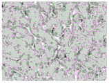
Figure 3 is an exemplary graph showing immunohistochemical staining of LAMP-1 with a significant reduction in lysosomal activity in the brain (compared to vehicle control mice) following 2 doses (photograph not shown) and 3 doses of I2S treatment. The reduction is characterized by: in the whole brain of the region, the number of LAMP-1 positive cells was small, and the staining intensity became light (40X).
Fig. 4 is an exemplary graph showing morphometric results by comparing the mean LAMP-1 positive region (in the cerebral cortex (cortex), caudate nucleus (CP), Thalamus (TH), White Matter (WM), and Cerebellum (CBL)) between Wild Type (WT), vehicle untreated, and I2S (2 and 3 doses) mice confirming that: there was a significant reduction in the LAMP-1 positive staining in all regions of the brain evaluated. Data are shown as mean ± s.d. P < 0.05; p < 0.01; p < 0.001.
Fig. 5 depicts an exemplary electron micrograph of brain cells showing pathological improvement at the ultrastructural level. Vehicle-treated mouse neurons had layered inclusions, zebra-like structures, and vacuoles containing depot particulate material (inserts) that were reduced in I2S-injected mice. Oligodendrocytes from vehicle-treated mice showed large low electron density storage granules (arrows), and oligodendrocytes from I2S-injected mice had minimal vacuolization. Scale bar: in neurons, 2 μm; in oligodendrocytes, 500 nm.
Fig. 6 depicts an exemplary immunohistochemical structure demonstrating: after intrathecal injection of 3 doses of I2S, I2S was detected in the sinusoidal gland cells of the liver. In 2 dose injected livers, 2S IHC staining was weak (photograph not shown). In the livers of vehicle control animals, there was no positive I2S staining (40 ×).
Fig. 7 depicts an exemplary tissue from a liver. Severe vacuolization of cells and abnormally high lysosomal activity was revealed by H & E staining, and intense LAMP-1 immunostaining (compared to WT type) was found in vehicle control animals. Cell vacuolization and LAMP-1 immunostaining were found to be significantly reduced following intrathecal treatment with either 2 doses (photograph not shown) or 3 doses of I2S treatment. H & E staining revealed almost complete disappearance of cytoplasmic vacuolization with near normal liver cell structure (H & E, 40X; LAMP-1, 20X).
FIGS. 8A-F illustrate exemplary data comparing aggregation of physiological saline and phosphate formulations (all with 0.01% polysorbate-20) by SEC-HPLC for 1 month at ≦ -65℃ and 40℃.
FIGS. 9A-F illustrate exemplary data comparing aggregation of physiological saline and phosphate formulations (all with 0.01% polysorbate-20) by SEC-HPLC method at ≦ -65℃ and 25℃ for 6 months.
FIGS. 10A-F illustrate exemplary data comparing aggregation of physiological saline and phosphate formulations (all with 0.01% polysorbate-20) by SEC-HPLC method at ≦ -65 deg.C and 2 to 8 deg.C for 24 months.
Fig. 11A-F illustrate exemplary data comparing the aggregation of physiological saline and phosphate formulations (all with 0.01% polysorbate-20) by the SAX-HPLC method at 40 ℃ versus baseline for 1 month.
Figures 12A-F illustrate exemplary data comparing the charge of physiological saline and phosphate formulations (all with 0.01% polysorbate-20) by the SAX-HPLC method at 25 ℃ at baseline versus 6 months.
Figures 13A-F illustrate exemplary data comparing the charge of physiological saline and phosphate formulations (all with 0.01% polysorbate-20) by the SAX-HPLC method at 2 to 8 ℃ at baseline versus 24 months.
FIG. 14 illustrates exemplary data comparing SDS-PAGE and Coomassie staining at baseline and at 1 month @40 ℃ for saline and phosphate formulations (all with 0.01% polysorbate-20).
FIGS. 15A and B illustrate exemplary data comparing SDS-PAGE and Coomassie staining for saline and phosphate formulations (all with 0.01% polysorbate-20): at 25 ℃ for 6 months, and at 2-8 ℃ for more than 16 months.
Fig. 16 depicts exemplary tissues showing the brains of the 3mg treatment group animals. Positive I2S staining was observed in meningeal cells (4X).
Fig. 17 depicts an exemplary tissue showing the brains of a 30mg treatment group of animals. Positive I2S staining was observed in neuronal and meningeal cells (4X).
Fig. 18 depicts an exemplary tissue showing the brains of 100mg treatment groups of animals. A stronger positive I2S staining was observed in neuronal and meningeal cells than in 3 and 30mg treated animals (4 ×).
Fig. 19 depicts exemplary tissues showing the brains of 150mg treatment groups of animals. A large population of neurons was I2S positive, with strongly positive meningeal cells.
Fig. 20 depicts an exemplary tissue showing I2S positive neurons and glial cells, and meningeal cells (within layer I of the brain), in 30mg treatment groups of animals (40X).
Fig. 21 depicts an exemplary tissue showing I2S positive neurons and glial cells, and perivascular cells (within layer III of the brain), in a 30mg treatment group of animals (40X).
Fig. 22 depicts an exemplary tissue showing I2S positive neurons and glial cells (within layer VI of the brain, near white matter), in 30mg treated groups of animals (40X).
Fig. 23 depicts exemplary tissues showing a strongly positive I2S staining in neurons of 150mg treated animals (100X).
Fig. 24 depicts exemplary tissues showing I2S immunostaining of the cervical spinal cord, in 150mg treatment (4 ×).
Fig. 25 depicts exemplary tissues showing I2S immunostaining of the lumbar spinal cord in 150mg treated animals (4X).
Fig. 26 depicts exemplary tissues showing that strong positive I2S immunostaining of meningeal cells, glial cells, and epitaxial/peripheral/endoneurial (connective tissue cells) was found in the lumbar sections (40X) of 150mg treated animals.
Figure 27 neurons in lumbar spinal cord of 150mg treated animals were strongly I2S positive (40X).
Figure 28 depicts exemplary results from liver (from 3mg treated group animals). Only the sinus gland cells were positive for I2S (40X).
Fig. 29 depicts exemplary results from liver (from 30mg treated group animals). Sinus gland cells and hepatocytes were I2S positive (40 ×).
Figure 30 depicts exemplary results from liver (from 100mg treated group animals). In both sinusoidal gland cells and hepatocytes, I2S immunostained more strongly (40 ×).
Figure 31 depicts exemplary results from liver (from 150mg treated group animals). In both sinusoidal gland cells and hepatocytes, I2S was identified as strongly positive (40 ×).
Fig. 32 depicts exemplary results from hearts (from 3mg treatment group animals). I2S immunostaining was negative (40 ×).
Fig. 33 depicts exemplary results from hearts (from 30mg treatment group animals). Mesenchymal cells were I2S positive (40 ×).
Fig. 34 depicts exemplary results from hearts (from 100mg treatment group animals). Positive stromal cell staining was observed for I2S (40 ×).
Fig. 35 depicts exemplary results from hearts (from 150mg treatment group animals). Positive stromal cell staining was observed for I2S (40 ×).
Fig. 36 depicts exemplary results from the kidney (from 3mg treated group animals). I2S immunostaining was negative (40 ×).
Fig. 37 depicts exemplary results from the kidney (from 30mg treated group animals). Glomeruli and stromal cells were I2S positive.
Fig. 38 depicts exemplary results from the kidney (from 100mg treated group animals). Increased staining was observed for glomeruli and stromal cells I2S (40 ×).
Fig. 39 depicts exemplary results from the kidney (from 150mg treated group animals). Positive I2S staining was observed for proximal tubules, glomeruli and stromal cells (40 ×).
FIG. 40 illustrates the results of an Immunohistochemistry (IHC) study evaluating cynomolgus monkey CNS tissues dosed weekly with iduronate-2-sulfatase (I2S). I2S had extensive cellular deposition throughout the CNS as determined by (IHC). I2S was detected in the gray matter, in a dose-dependent manner, in all groups of the neurons (of brain, cerebellum, brainstem and spinal cord). In the surface gray matter of the higher dose group, a large number of brain neurons stained positive for I2S, in the surface cortex (fig. 40A). I2S was detected in neurons (in the thalamus (fig. 40B), hippocampus (fig. 40C), caudate nucleus (fig. 40D), and spinal cord (fig. 40E). Meninges and perivascular cells also stained positive for I2S (fig. 40F). The determined scale bar corresponds to 25 μm.
Figure 41 graphically compares clearance of iduronate-2-sulfatase (I2S) in the cranial and spinal pools by plotting the amount of I2S in such pools versus time after administration.
Figure 42 illustrates dose-dependent gray matter deposition over a 6 month period of intrathecal administration of iduronate-2-sulfatase (I2S) to non-human primates. The illustrated staining intensity corresponds to the accumulation of iduronate-2-sulfatase in the thalamus. In the current fig. 42, the nuclei were counterstained by DAPI and appeared blue, and the protein (I2S) appeared green.
Figure 43 illustrates dose-dependent accumulation of intrathecal administration of iduronate-2-sulfatase (I2S) to non-human primates over a 6 month period following single injection and following multiple injections. The intensity of staining illustrated corresponds to the accumulation of I2S protein in the cerebral cortex.
FIG. 44 shows the cellular localization of iduronate-2-sulfatase (I2S) (throughout the brain in a non-human primate). Fig. 44A illustrates a cross-sectional view of brain tissue (extracted from the non-human primate brain), while fig. 44B illustrates that specific regions of the range correspond to the sections identified in fig. 44A: three regions of white matter tissue (designated W1, W2, and W3), white matter (VW) and surface gray matter (SG) tissue proximal to the ventricles.
Figures 45A-D illustrate the neuronal and oligodendrocyte uptake and axonal ligation of intrathecal administration of iduronate-2-sulfatase (I2S) to non-human primates following monthly injections over a period of 6 months. In particular, fig. 45A, 45B, 45C and 45D illustrate filament staining of non-human primate brain tissue, intrathecal administration of iduronate-2-sulfatase (I2S), and white matter (W1, W2 and W3), and superficial gray matter region (SG), respectively, corresponding to the three regions, identified in fig. 44B. Fig. 45A illustrates oligodendrocyte uptake of intrathecally administered I2S in the white matter (W1) tissue. Fig. 45B and 45C illustrate oligodendrocyte uptake and axonal ligation in the W2 and W3 white matter tissues, respectively. Fig. 45D illustrates neuronal uptake in the surface gray matter tissue (SG), intrathecally administered I2S.
FIG. 46 illustrates cell recognition of iduronate-2-sulfatase in white matter near the ventricles of the brain (VW) for a non-human primate. As depicted in the overlaid images, the iduronate-2-sulfatase was not associated with myelin (red). In the figure 46 where present, the nuclei were counterstained by DAPI (bottom left) protein (I2S), appearing as boxes at the top left.
FIG. 47 illustrates staining in healthy beagle tissue administered either Intracerebroventricularly (ICV) or Intrathecally (IT) with a single injection of iduronate-2-sulfatase (I2S). As depicted in fig. 47A-47H, I2S was widely distributed throughout the gray matter (both IT and ICV groups) as determined by Immunohistochemistry (IHC). Fig. 47A and 47B illustrate that neurons were positive for I2S in the cerebral cortex, from the surface molecular layer to the deep inner layer (both IT and ICV groups) in all 6 neuronal layers. Fig. 47C and 47D illustrate that I2S was detected in neurons (including purkinje cells) in the cerebellar cortex in the IT and ICV groups. Similarly, fig. 47E and 47F illustrate that in both the IT and ICV groups, a large number of neurons were positive for I2S in the hippocampus. Finally, images g and h show that I2S-positive neurons were found in the thalamus and caudate nucleus (in both the IT and ICV groups included). In fig. 47 of the present, I2S staining is indicated with an arrow.
Fig. 48 comparatively illustrates the callus tissue of iduronate-2-sulfatase gene knockout mice (IKO) that were either untreated or administered I2S intrathecally (abbreviated V ═ vacuoles). As described, the treated IKO mice exhibited a decrease in the cellular vacuolization characteristic of certain lysosomal storage disorders in the callus and vault tissues of the I2S-treated IKO mice.
Figure 49A illustrates a significant reduction in the presence of lysosomal associated membrane protein 1(LAMP1), a pathological biomarker for lysosomal disease, at 20X and 40X magnification in the surface cerebral cortex of the treated IKO mice (figure 49A) relative to the untreated IKO control mice (figure 49B).
Fig. 50 depicts an exemplary Intrathecal Drug Delivery Device (IDDD).
FIG. 51 depicts an mutexemplary PORT-A- Low profile intrathecal implantation portal system.
Fig. 52 depicts an exemplary Intrathecal Drug Delivery Device (IDDD).
Fig. 53 depicts an exemplary Intrathecal Drug Delivery Device (IDDD) that allows for home administration for CNS Enzyme Replacement Therapy (ERT).
FIG. 54 is an exemplary illustration showing: in neurons (purkinje cells), the vacuolizing effect of injection of iduronise (idursufase) in a single brain.
FIG. 55 is an exemplary illustration showing: I2S activity in the brain (dose and regional related).
Fig. 56 is an exemplary diagram showing immunohistochemical localization of iduronidase (cerebral cortex at different depths).
Figure 57 is an exemplary graph showing I2S activity in the spinal cord of monkeys after intrathecal administration of iduronidase.
FIG. 58 is a schematic illustration showing I2S activity in the liver, heart and kidney of monkeys after intrathecal administration of iduronidase.
FIG. 59 depicts an exemplary chart of the added Hunter-IT test program.
Fig. 60 is an exemplary graph showing the measurement of I2S concentration in various portions of brain tissue after a 30mg dose. Different graphs correspond to different measurement times.
Fig. 61 is an exemplary graph showing the measured values of I2S concentration at various product concentrations over time, through different routes of administration, after administration.
FIG. 62 depicts administration of IV, IT-L, or ICV at t-5 hours in cynomolgus monkeys124PET imaging of I-labeled iduronidase-IT.
FIG. 63 illustrates and exemplifies a diagram of an intrathecal drug delivery device IDDD,
Fig. 64 depicts different features of an IDDD, including the IDDD in a subject (fig. 64A), the IDDD displayed on a flat surface (fig. 64B), and the insertion point of the catheter tip (fig. 64C).
Interpretation of terms
In order that the invention may be more readily understood, certain terms are first defined below. Other definitions for the following terms and other terms are set forth throughout the specification.
About or about: as used herein, the term "about" or "approximately," as applied to one or more values of interest, refers to a value that is similar to the specified reference value. In certain embodiments, the term "about" or "approximately" refers to a range of values that falls within 25%, 20%, 19%, 18%, 17%, 16%, 15%, 14%, 13%, 12%, 11%, 10%, 9%, 8%, 7%, 6%, 5%, 4%, 3%, 2%, 1%, or less of the stated reference value in any direction (greater or less) unless otherwise stated or otherwise evident from the context (unless the number would exceed 100% of possible values).
And (3) turning better: as used herein, the term "amelioration" means prevention, alleviation or alleviation of a condition, or improvement in a subject's condition. Optimization includes, but does not require, healing of the condition or complete prevention. In some embodiments, the amelioration comprises an increase in the level of a protein of interest, or activity thereof, that is deficient in the disease-associated tissue.
The biological activity is as follows: as used herein, the phrase "biological activity" refers to any actual property that is active in a biological system, and particularly in an organism. For example, when an agent is administered into an organism, the agent has a biological effect on the organism, which is considered to be biologically active. In particular embodiments, when a protein or polypeptide is biologically active, the portion of the protein or polypeptide will share at least one biological activity of the protein or polypeptide, which is generally referred to as a "biologically active" portion.
Filling agent: as used herein, the term "bulking agent" refers to a complex that adds mass to the lyophilized mixture and aids in the physical structure of the lyophilized cake (e.g., facilitates production of a substantially uniform lyophilized cake that retains an open pore structure). Exemplary bulking agents include mannitol, glycine, sodium chloride, hydroxyethyl starch, lactose, sucrose, trehalose, polyethylene glycol, and dextran.
Cation-dependent mannose-6-phosphate receptor (CI-MPR): as used herein, the term "cation-dependent mannose-6-phosphate receptor (CI-MPR)" refers to a cellular receptor that binds a mannose-6-phosphate (M6P) tag on acid hydrolase precursors in the golgi apparatus and is destined for trafficking to the lysosome. In addition to mannose-6-phosphate, the CI-MPR also binds to other proteins, including IGF-II. The CI-MPR is also referred to as the "M6P/IGF-II receptor", "CI-MPR/IGF-II receptor", "IGF-II receptor", or "IGF 2 receptor". These terms and their abbreviations are used interchangeably herein.
Concurrent immunosuppressive therapy: as used herein, the term "concurrent immunosuppressive therapy" includes any immunosuppressive therapy that is pre-treatment, preconditioning, or is parallel to a method of treatment.
Diluent agent: as used herein, the term "diluent" refers to a pharmaceutically acceptable (e.g., safe and non-toxic for administration to a human) diluent material that is effective for the preparation of a recombinant formulation. Exemplary diluents include sterile water, sterile water for injection (BWFI), pH buffered solutions (e.g., phosphate-buffered saline), sterile saline, ringer's solution, or dextrose solution.
The preparation formulation is as follows: as used herein, the terms "dosage form" and "unit dosage form" refer to physically discrete units of the therapeutic protein of the patient to be treated. Each unit containing a predetermined amount of active material calculated to produce the desired therapeutic effect. It will be understood, however, that the total dosage of the composition may be determined by the attending physician within the scope of sound medical judgment.
Enzyme Replacement Therapy (ERT): as used herein, the term "Enzyme Replacement Therapy (ERT)" refers to any therapeutic strategy that can correct enzyme deficiency by the missing enzyme. In some embodiments, the lost enzyme is provided by intrathecal administration. In some embodiments, the lost enzyme is provided by injection into the bloodstream. Once administered, the enzyme is taken up by the cells and transported to the lysosome where it acts to clear material accumulated in the lysosome due to enzyme deficiency. Typically, for lysosomal enzyme replacement therapy to be effective, the therapeutic enzyme is delivered to lysosomes in appropriate cells of the target tissue (where the storage defect is evident).
Improving, increasing, or decreasing: as used herein, the terms "improve," "increase," or "decrease," or grammatical equivalents, include values relative to a baseline measurement, e.g., a measurement in the same individual prior to initiation of a treatment described herein, or a measurement in a control individual (or control individuals) lacking a treatment described herein. A "control individual" is an individual that: it is afflicted with the same form of lysosomal storage disorder as the individual being treated and it is the same age as the treated individual (to ensure that the stage of the disease is comparable between the individual being treated and the control individual).
Individual, subject, patient: as used herein, the term "subject", "individual" or "patient" refers to a human or non-human mammalian subject. The individual being treated (also referred to as "patient" or "subject") is an individual with a disease (fetus, infant, child, adolescent, adult).
Intrathecal administration: as used herein, the term "intrathecal administration" or "intrathecal injection" refers to injection into the spinal canal (the intrathecal space around the spinal cord). A variety of techniques may be used, including, but not limited to, lateral ventricle injection (via drilling or cisterna magna or lumbar puncture or the like). In some embodiments, "intrathecal administration" or "intrathecal delivery" according to the invention refers to IT administration or delivery (through the lumbar or lumbar region), i.e. lumbar IT administration or delivery. As used herein, the term "lumbar region" or "waist" refers to the region between the third and fourth lumbar (lower back) bones, further encompassing the L2-S1 region of the spine.
Linker as used herein, the term "linker" refers to an amino acid sequence that is different from that occurring at a specific site in a native protein in a fusion protein, and is typically designed to be flexible or inserted into a structure (e.g., an alpha-helix) between two protein moieties. The linker is also referred to as a spacer.
Freeze-drying protective agent: as used herein, the term "lyoprotectant" refers to a molecule that: which prevents or reduces chemical and/or physical instability of the protein or other material upon lyophilization and subsequent storage. Exemplary lyoprotectants include sugars (e.g., sucrose or trehalose); amino acids (e.g., amino acids such as monosodium glutamate or histidine), methylamines (e.g., betaine), lyotropic salts (magnesium sulfate), polyols (e.g., trivalent or higher sugar alcohols such as glycerol, erythritol, glycerol, arabitol, xylitol, sorbitol, and mannitol), propylene glycol, polyethylene glycols, pluronics, and combinations thereof.
Lysosomal enzymes: as used herein, the term "lysosomal enzyme" refers to any enzyme that is capable of accumulating material in the lysosome of a mammal, or that rescues or ameliorates one or more symptoms of a lysosomal storage disorder. Lysosomal enzymes suitable for the present invention include wild-type or modified lysosomal enzymes, and can be prepared using recombinant and synthetic methods, or purified from natural sources. Exemplary lysosomes are listed in table 1.
Lysosomal enzyme deficiency: as used herein, "lysosomal enzyme deficiency" refers to a group of genetic diseases that result from the deficiency of at least one enzyme that is required for the cleavage of macromolecules (e.g., enzyme substrates) into peptides, amino acids, monosaccharides, nucleic acids, and fatty acids in lysosomes. As a result, individuals with lysosomal enzyme deficiencies have accumulated material in a variety of tissues (e.g., CNS, liver, spleen, intestine, vessel walls and other organs).
Lysosomal storage disorders: as used herein, the term "lysosomal storage disease" refers to any disease that results in the deficiency of one or more lysosomal enzymes that are required for the metabolism of the native macromolecule. These diseases often result in the accumulation of undegraded molecules in lysosomes, leading to an increase in the number of storage particles (also termed storage vesicles). These diseases and various examples are described in detail below.
Polypeptide: as used herein, "polypeptide," generally, refers to a string of at least two amino acids linked to each other by peptide bonds. In some embodiments, the polypeptide comprises at least 3-5 amino acids, each of which is linked to the others by means of at least one peptide bond. Those of ordinary skill in the art will appreciate that sometimes polypeptides include "unnatural" amino acids or, optionally, other entities that can still be incorporated into a polypeptide chain.
Alternative enzymes: as used herein, the term "replacement enzyme" refers to any enzyme that is capable of acting to replace at least a portion of the absent or missing enzyme (in the disease to be treated). In some embodiments, the term "replacement enzyme" refers to any enzyme that is capable of acting to replace at least a portion of the missing or missing lysosomal enzyme (in the lysosomal storage disorder being treated). In some embodiments, the replacement enzyme can reduce material accumulation in mammalian lysosomes, or it can rescue or improve one or more symptoms of lysosomal storage disorders. Alternative enzymes suitable for the present invention include wild-type or modified lysosomal enzymes, and can be prepared using recombinant and synthetic methods or purified from natural sources. The replacement enzyme may be a recombinant, synthetic, gene-activated or natural enzyme.
Solubility: as used herein, the term "soluble" refers to the ability of a therapeutic agent to form a homogeneous solution. In some embodiments, the solubility of the therapeutic agent in the solution in which it is administered and through which it is transported to the targeted site of action (e.g., the cells and tissues of the brain) is sufficient to allow delivery of a therapeutically effective amount of the therapeutic agent to the targeted site of action. Several factors may influence the solubility of the therapeutic agent. For example, relevant factors that may affect protein solubility include ionic strength, amino acid sequence, and the presence of other co-solubilizing agents or salts (e.g., calcium salts). In some embodiments, the pharmaceutical composition is formulated such that the calcium salt is excluded from such composition. In some embodiments, the agents according to the invention are soluble in the corresponding compositions. It will be appreciated that while isotonic solutions are generally preferred for parenteral administration of drugs, the use of isotonic solutions may limit the sufficient solubility of some therapeutic worlds, particularly certain proteins and/or enzymes. Slightly hypertonic solutions (e.g. up to 175mM sodium chloride in 5mM sodium phosphate, pH7.0) and sugar containing solutions (e.g. up to 2% sucrose in 5mM sodium phosphate, pH7.0) have been shown to be well tolerated in monkeys. For example, the most common approved CNS bolus formulation composition is physiological saline (150 mM NaCl in water).
Stability: as used herein, the term "stable" refers to the ability of the therapeutic agent to retain its therapeutic effect (e.g., all or most of its intended biological activity and/or physicochemical integrity) for an extended period of time. The stability of the therapeutic agent, as well as the ability of the pharmaceutical composition to maintain the stability of such therapeutic agent, can be assessed over an extended period of time (e.g., at least 1,3, 6, 12, 18, 24, 30, 36 months or more). Typically, the pharmaceutical compositions described herein have been formulated such that they are capable of stabilizing, or alternatively slowing or preventing, the degradation of one or more therapeutic agents (e.g., recombinant proteins) with which they are formulated. In the case of formulation, a stable formulation is one in which the therapeutic agent substantially retains its physical and/or chemical integrity and biological activity during storage and processing (e.g., freezing/thawing, mechanical mixing, and lyophilization). For protein stability, it can be measured by loss of enzyme activity, peptide fragment formation and charge transfer properties for the formation of High Molecular Weight (HMW) aggregates.
Subject: as used herein, the term "subject" means any mammal, including a human. In certain embodiments of the invention, the subject is an adult, adolescent or infant. The invention also contemplates the administration of said pharmaceutical composition, and/or the performance of an intrauterine treatment method.
Substantial homology As used herein, the phrase "substantial homology" refers to a comparison between amino acid or nucleic acid sequences. As understood by those of ordinary skill in the art, two sequences will generally be considered "substantially homologous" if they contain homologous residues at the corresponding positions. Homologous residues may be identical residues. Alternatively, homologous residues may also be non-identical residues, which have similar structural and/or functional properties as appropriate. For example, as known to those of ordinary skill in the art, certain amino acids are generally classified as "hydrophobic" or "hydrophilic" amino acids, and/or have "polar" or "nonpolar" side chains. Substitution of one amino acid for another of the same type is generally considered a "homologous" substitution.
It is well known in the art that amino acid or nucleic acid sequences can be compared using a variety of algorithms, including commercial computer programs such as BLASTN for nucleic acid sequences, BLASTP for amino acid sequences, gapped BLAST, and PSI-BLAST. Exemplary such procedures are described in these documents: altschul, et al, Basic local alignment search tool, J.mol.biol.,215(3): 403-; altschul, et al, Methods in Enzymology; altschul, et al, "Gapped BLAST and PSI-BLAST: a new generation of protein database search programs", Nucleic Acids Res.25:3389-3402, 1997; baxevanis, et al, Bioinformatics A Practical Guide to the Analysis of Genes and Proteins, Wiley, 1998; and microsener, et al, (eds.), Bioinformatics Methods and Protocols (Methods in Molecular Biology, Vol.132), Humana Press,1999 in addition to identifying homologous sequences, the programs mentioned above generally provide an indication of the degree of homology. In some embodiments, two sequences are considered substantially homologous if at least 50%, 55%, 60%, 65%, 70%, 75%, 80%, 85%, 90%, 91%, 92%, 93%, 94%, 95%, 96%, 97%, 98%, 99% or more of their corresponding residues are homologous between the relevant extended residues. In some embodiments, the related extensions are complete sequences. In some embodiments, the relevant extension is at least 10, 15, 20, 25, 30, 35, 40, 45, 50, 55, 60, 65, 70, 75, 80, 85, 90, 95, 100, 125, 150, 175, 200, 225, 250, 275, 300, 325, 350, 375, 400, 425, 450, 475, 500 or more residues.
Basic consistency: the phrase "substantial identity" as used herein refers to a comparison between amino acid or nucleic acid sequences. As understood by one of ordinary skill in the art, two sequences will generally be considered "substantially identical" if they contain identical residues at the corresponding positions. It is well known in the art that amino acid or nucleic acid sequences can be compared using any of a variety of algorithms, including programs in commercial computers, such as BLASTN for nucleic acid sequences, and BLASTP, gapped BLAST, and PSI-BLAST for amino acid sequences. Exemplary such procedures are described in the following documents: altschul, et al, Basic local alignment search tool, J.mol.biol.,215(3): 403-; altschul, et al, Methods in Enzymology; altschul et al, Nucleic Acids Res.25:3389-3402, 1997; baxevanis et al, Bioinformatics A Practical Guide to the Analysis of Genes and Proteins, Wiley, 1998; and microsener, et al, (eds.), Bioinformatics Methods and Protocols (Methods in Molecular Biology, Vol.132), Humana Press,1999 in addition to identifying these consensus sequences, the programs mentioned above generally provide an indication of the degree of identity. In some embodiments, the sequences are considered substantially identical if at least 50%, 55%, 60%, 65%, 70%, 75%, 80%, 85%, 90%, 91%, 92%, 93%, 94%, 95%, 96%, 97%, 98%, 99% or more of their corresponding residues are identical between the residues of the relevant extension. In some embodiments, the related extensions are complete sequences. In some embodiments, the relevant extension is at least 10, 15, 20, 25, 30, 35, 40, 45, 50, 55, 60, 65, 70, 75, 80, 85, 90, 95, 100, 125, 150, 175, 200, 225, 250, 275, 300, 325, 350, 375, 400, 425, 450, 475, 500 or more residues.
Synthetic CSF: as used herein, the term "synthetic CSF" refers to a solution having a pH, electrolyte composition, glucose content, and osmolality consistent with cerebrospinal fluid. Synthetic CSF is also referred to as artificial CSF. In some embodiments, the synthesized CSF is an Elliott's B solution.
Suitable for CNS delivery: as used herein, the phrase "suitable for CNS delivery" or "suitable for intrathecal delivery" refers to the pharmaceutical compositions of the invention, generally referring to the stability, tolerability and solubility properties of these compositions, as well as the ability of these compositions to deliver an effective amount of a therapeutic agent contained therein to a delivery target site (e.g., the CSF or the brain).
Target tissue: as used herein, the term "target tissue" refers to any tissue affected by the lysosomal storage disorder to be treated, or in which the deficient lysosomal enzyme is normally expressed. In some embodiments, the target tissue includes those tissues having a detectable or abnormally high amount of enzyme substrate therein (e.g., stored in the lysosomes of the cells of the tissue), in patients suffering from or susceptible to lysosomal storage disorders. In some embodiments, the target tissue includes these tissues: which exhibits a disease-related pathology, symptom, or characteristic. In some embodiments, the target tissue includes these tissues: wherein the deficient lysosomal enzyme is normally expressed at elevated levels. As used herein, a target tissue can be a brain target tissue, a spinal cord target tissue, and/or a peripheral target tissue. Exemplary target tissues are described in detail below.
And (3) treatment part: as used herein, the term "therapeutic moiety" refers to a moiety of a molecule that provides a therapeutic effect to the molecule. In some embodiments, the therapeutic moiety is a polypeptide having therapeutic activity.
A therapeutically effective amount of: as used herein, the term "therapeutically effective amount" refers to an amount of a therapeutic protein (e.g., a replacement enzyme) that imparts a therapeutic effect to a subject of the treatment, at a reasonable benefit/risk ratio (suitable for any medical treatment). The therapeutic effect may be objective (e.g., measured by some test or marker) or subjective (e.g., an indication or sensation of an effect given by the subject). In particular, the "therapeutically effective amount" refers to an amount of the therapeutic protein or composition effective to treat, ameliorate or prevent the desired disease or condition, or to exhibit a detectable therapeutic or prophylactic effect (e.g., by ameliorating symptoms associated with the disease, preventing or delaying the onset of the disease, and/or reducing the severity or frequency of the disease). Therapeutically effective amounts are generally administered in a dosage regimen that may include a variety of unit doses. The therapeutically effective amount (and/or an appropriate unit dose within an effective dosing regimen) for any particular therapeutic protein will vary, for example, depending on the route of administration, as well as other pharmaceutical agents. Moreover, the specific therapeutically effective amount (and/or unit dose) for any particular patient may vary depending on a variety of factors, including the disease being treated and the severity of the disease; the activity of the particular pharmaceutical agent employed; the particular composition employed; the age, weight, general health, sex, and diet of the patient; the time of administration; the route of administration; and/or excretion and metabolism of the particular fusion protein employed; the duration of the treatment; and similar factors known in the medical arts.
The following can be tolerated: as used herein, the terms "tolerable" and "tolerance" refer to the ability of a pharmaceutical composition of the invention to not elicit an adverse response in the subject (the subjects to which the composition is administered), or alternatively, not to elicit a severe adverse response in the subject (the subjects to which the composition is administered). In some embodiments, the pharmaceutical composition of the invention can be well tolerated by the subject (by which the subject is meant the subject to whom the composition is administered).
Treatment: as used herein, the term "treatment" (also referred to as "treating" or "treatment") refers to any administered therapeutic protein (e.g., a lysosomal enzyme), which partially or completely alleviates, ameliorates, alleviates, is known to occur or delays the occurrence, reduces the severity and/or reduces the incidence of one or more symptoms or features of a particular disease, disorder and/or condition (e.g., Hunters syndrome, Sanfilippo syndrome B.) such treatment may be a subject who does not exhibit signs of the associated disease, disorder and/or condition, and/or such a subject (who merely exhibits early signs of the associated disease, disorder, and/or condition), alternatively or additionally such treatment may be such a subject, which exhibit one or more established signs of the associated disease, disorder and/or condition.
Detailed Description
The present invention provides, in addition, improved methods and compositions for the efficient direct delivery of therapeutic agents to the Central Nervous System (CNS). As discussed above, the present invention is based on the unexpected finding that: replacement enzymes (e.g., I2S protein) for lysosomal storage disorders (e.g., Hunters syndrome) can be introduced directly into the cerebrospinal fluid (CSF) of a subject in need of treatment at high concentrations without inducing substantial side effects in the subject. More unexpectedly, the inventors found that: the alternative enzymes can be delivered in simple saline or buffer base formulations without the use of synthetic CSF. Even more unexpectedly, intrathecal delivery according to the invention does not result in substantial side effects, such as severe immune responses, in said subject. Thus, in some embodiments, intrathecal delivery according to the invention may be used in the absence of concurrent immunosuppressive therapy (e.g., no immune tolerance is induced by pretreatment or preconditioning).
In some embodiments, intrathecal delivery according to the invention allows for efficient diffusion between multiple brain tissues, resulting in efficient delivery of the replacement enzyme in multiple target brain tissues in superficial, superficial and/or deep brain regions. In some embodiments, intrathecal delivery according to the invention results in a sufficient amount of the alternative enzyme into the peripheral circulation. As a result, intrathecal delivery according to the invention leads in certain cases to the delivery of the replacement enzyme in peripheral tissues, such as liver, heart, spleen and kidney. This finding is unexpected and can be particularly useful for the treatment of such lysosomal storage diseases having CNS and peripheral components, which typically require conventional intrathecal and intravenous administration. This is also contemplated: intrathecal delivery according to the invention may allow to reduce the frequency of administration and/or iv injections without compromising the therapeutic effect of the treatment of peripheral symptoms.
The present invention provides a variety of unexpected and beneficial features that allow for the effective and facile delivery of alternative enzymes to a variety of brain target tissues, resulting in the effective treatment of lysosomal storage disorders with CNS indications.
Various aspects of the invention are described in detail in the following sections. The use of the sections is not intended to limit the invention. Each section may be applied to any aspect of the invention. In this application, the use of "or" means "and/or" unless stated otherwise.
Alternative enzymes
Iduronate-2-sulfatase (I2S) protein
In some embodiments, the invention provides inventive methods and compositions for delivering iduronate-2-sulfatase (I2S) protein to the CNS to treat Hunters syndrome. A suitable I2S protein may be any molecule or portion of a molecule suitable for naturally occurring iduronate-2-sulfatase (I2S) protein activity or to rescue one or more manifestations or symptoms associated with an I2S-deficiency. In some embodiments, alternative enzymes suitable for the present invention are polypeptides that: it has an N-and C-terminus and an amino acid sequence substantially similar to or identical to the mature human I2S protein.
Typically, the human I2S protein is produced in precursor form. The precursor form of human I2S contains a signal peptide (amino acid residues 1-25 of the full length precursor), a propeptide (amino acid residues 26-33 of the full length precursor), and a chain (residues 34-550 of the full length precursor); the chain may be further processed into a 42kDa chain (residues 34-455 of the full length precursor) and a 14kDa chain (residue 446-550 of the full length precursor). Typically, the precursor form is also referred to as a full-length precursor or full-length I2S protein, which contains 550 amino acids. The amino acid sequence of the mature form (SEQ ID NO:1) has the signal peptide deleted and the full-length precursor of a typical wild-type or naturally occurring human I2S protein (SEQ ID NO:2) is shown in Table 1.
TABLE 1 human iduronate-2-sulfatase

Thus, in some embodiments, a suitable alternative enzyme suitable for the present invention is mature human I2S protein (SEQ ID NO: 1). In some embodiments, a suitable alternative enzyme may be a homolog or analog of the mature human I2S protein. For example, a homolog or analog of the mature human I2S protein may be a modified human I2S protein that contains one or more amino acid substitutions, deletions, and/or insertions, while retaining substantial I2S protein activity, as compared to the wild-type or naturally occurring I2S protein (e.g., SEQ ID NO: 1). Thus, in some embodiments, suitable alternative enzymes suitable for the present invention are substantially homologous to the mature human I2S protein (SEQ ID NO: 1). In some embodiments, suitable alternative enzymes suitable for the present invention have an amino acid sequence that is at least 50%, 55%, 60%, 65%, 70%, 75%, 80%, 85%, 90%, 91%, 92%, 93%, 94%, 95%, 96%, 97%, 98%, 99% or more homologous to SEQ ID No. 1. In some embodiments, suitable alternative enzymes for use in the present invention are substantially identical to the mature human I2S protein (SEQ ID NO: 1). In some embodiments, suitable alternative enzymes suitable for the present invention have an amino acid sequence that is at least 50%, 55%, 60%, 65%, 70%, 75%, 80%, 85%, 90%, 91%, 92%, 93%, 94%, 95%, 96%, 97%, 98%, 99% or more identical to SEQ ID No. 1. In some embodiments, suitable alternative enzymes for use in the present invention comprise fragments or portions of the mature human I2S protein.
Alternatively, a suitable alternative enzyme suitable for the present invention is the full-length I2S protein. In some embodiments, a suitable alternative enzyme may be a homolog or analog of the full-length human I2S protein. For example, a homolog or analog of the full-length human I2S protein can be a modified full-length human I2S protein that contains one or more amino acid substitutions, deletions, and/or insertions, while retaining substantial I2S protein activity, as compared to the wild-type or naturally-occurring full-length I2S protein (e.g., SEQ ID NO: 2). Thus, in some embodiments, suitable alternative enzymes suitable for the present invention are substantially homologous to full-length human I2S protein (SEQ ID NO: 2). In some embodiments, suitable alternative enzymes suitable for the present invention have an amino acid sequence that is at least 50%, 55%, 60%, 65%, 70%, 75%, 80%, 85%, 90%, 91%, 92%, 93%, 94%, 95%, 96%, 97%, 98%, 99% or more homologous to SEQ ID NO 2. In some embodiments, suitable alternative enzymes suitable for the present invention are substantially identical to SEQ ID NO 2. In some embodiments, suitable alternative enzymes suitable for the present invention have an amino acid sequence that is at least 50%, 55%, 60%, 65%, 70%, 75%, 80%, 85%, 90%, 91%, 92%, 93%, 94%, 95%, 96%, 97%, 98%, 99% or more identical to SEQ ID No. 2. In some embodiments, suitable alternative enzymes for use in the present invention comprise fragments or portions of the full length human I2S protein. As used herein, full-length I2S proteins typically contain a signal peptide sequence.
Other lysosomal storageAccumulation of disease and alternative enzymes
It is contemplated that the inventive methods and compositions according to the present invention may be used to treat other lysosomal storage diseases, particularly those having CNS etiologies and/or symptoms, including but not limited to: aspartylglucosaminuria, cholesteryl ester deposition disease, Wolman disease, cystinosis, Danon disease, Fabry disease, Farber fatty granulomatosis, Farber disease, fucosidase deficiency disease, galactosialidosis type I/II, gaucher disease type I/II/III, globuloleukodystrophy, Krabbe disease, glycogen storage disease II, Pompe disease, GM 1-gangliosidosis type I/II/III, GM 2-gangliosidosis type I, Tay-saxophone disease, GM 2-gangliosidosis type II, sandhoff disease, GM 2-gangliosidosis, alpha-mannosidosis type I/II, beta-mannosidosis, metachromatic leukodystrophy, mucosidosis type I, sialyl, sialodeposition type I/II, Gossypiosis type I/II, Gossypiosis, Gossypi, Sialodesis type II/III, I-cell disease, mucolipidosis type IIIC pseudo-Holler's multiple malnutrition, mucopolysaccharidosis type I, mucopolysaccharidosis type II, mucopolysaccharidosis type IIIA, Sanfilippo syndrome, mucopolysaccharidosis type IIIB, mucopolysaccharidosis type IIIC, mucopolysaccharidosis type IIID, mucopolysaccharidosis type IVA, Moyobo syndrome, mucopolysaccharidosis type IVB, mucopolysaccharidosis type VI, mucopolysaccharidosis type VII, Sly syndrome, mucopolysaccharidosis type IX, multiple sulfatase deficiency, neuronal ceroid lipofuscinosis, CLN1 Batten disease, CLN2 Batten disease, Niemann pick disease type A/B, Niemann pick disease type C1, Niemann pick disease type C2, compact osteogenesis imperfecta, Schinder disease type I/II, gaucher disease and sialorrhea disease.
Detailed reviews of The genetic etiology, clinical presentation and Molecular biology of such lysosomal storage disorders can be found in Scriver et al, eds., The Metabolic and Molecular Basis of Inherited diseases, 7.sup.th ed., vol.ii, McGraw Hill, (1995).
Table 2.


The method according to the invention can be used to deliver a variety of other alternative enzymes. As used herein, replacement enzymes suitable for the present invention can include any replacement enzyme that is capable of functioning to replace at least a portion of the activity of a lysosomal enzyme that is lacking or lost in the lysosomal storage disorder being treated. In some embodiments, the replacement enzyme is capable of reducing the accumulation of material in the lysosome, or is capable of rescuing or ameliorating one or more symptoms of the lysosomal storage disorder.
In some embodiments, a suitable replacement enzyme may be any replacement enzyme known to be associated with the lysosomal storage disease to be treated. In some embodiments, a suitable alternative enzyme is an enzyme selected from the group of enzymes listed in table 2.
In some embodiments, alternative enzymes suitable for the present invention may have wild-type or naturally occurring sequences. In some embodiments, alternative enzymes suitable for the present invention may have modified sequences that have substantial homology or identity to the wild-type or naturally occurring sequence (e.g., at least 50%, 55%, 60%, 65%, 70%, 75%, 80%, 85%, 90%, 95%, 98% sequence identity to the wild-type or naturally occurring sequence).
Suitable alternative enzymes suitable for the present invention may be prepared by any available method. For example, the surrogate enzyme can be recombinantly produced using a host cell system designed to express a nucleic acid encoding the surrogate enzyme. Alternatively or additionally, the replacement enzyme may be prepared by activating an endogenous gene. Alternatively or additionally, the surrogate enzyme may be partially or completely prepared by chemical synthesis. Alternatively or additionally, the surrogate enzyme may be purified from a natural source.
Any expression system may be used to recombinantly produce the enzyme. To give some examples, known expression systems include, for example, egg, baculovirus, plant, yeast, or mammalian cells.
In some embodiments, enzymes suitable for the present invention are prepared in mammalian cells. Non-limiting examples of mammalian cells that can be used according to the present invention include: BALB/c mouse myeloma line (NSO/l, ECACC No:85110503), human retinoblastoma (PER. C6, CruCell, Leiden, the Netherlands), SV40 transformed monkey kidney CV1 line (COS-7, ATCC CRL 1651), human embryonic kidney line (subclones of 293 or 293 cells grown in suspension culture, Graham et al, J. Gen Virol.,36:59,1977), human fibrosarcoma cell line (e.g., HT1080), baby mouse kidney cells (BHK, ATCC CCL 10), Chinese hamster ovary cells +/-DHFR (CHO, Urlaub and Chasin, Proc. Natl. Acad. Sci. USA,77:4216,1980), mouse testis support cells (TM4, Mather, biol. Reprod.,23: Bed 251,1980), monkey cells (CV1 CCL 70), African green monkey cells (Vero-76, ATCC-7), human kidney cells (ATCC 1582K), ATCC CCL 34), Buffalo rat hepatocytes (BRL 3A, ATCC CRL 1442), human lung cells (W138, ATCC CCL 75), human hepatocytes (Hep G2, HB 8065), mouse mammary tumors (MMT 060562, ATCC CCL51), TRI cells (Mather et al, Annals N.Y.Acad.Sci.,383:44-68,1982), MRC 5 cells, FS4 cells, and human hepatoma cell lines (Hep G2).
In some embodiments, the method according to the invention is used to deliver a surrogate enzyme prepared from a human cell. In some embodiments, the surrogate enzyme prepared from CHO cells is delivered using the method according to the invention.
In some embodiments, the surrogate enzyme delivered using the methods of the invention contains a moiety that binds to a receptor on the surface of a brain cell to facilitate cellular uptake and/or targeting to lysosomes. For example, such a receptor may be a cation-dependent mannose-6-phosphate receptor (CI-MPR), which binds to the mannose-6-phosphate (M6P) residue. In addition, the CI-MPR also binds to other proteins, including IGF-II. In some embodiments, alternative enzymes suitable for the present invention contain M6P residues on the surface of the protein. In some embodiments, alternative enzymes suitable for the present invention may contain bisphosphorylated oligosaccharides with higher binding affinity to the CI-MPR. In some embodiments, suitable enzymes contain up to about an average of about at least 20% bisphosphorylated oligosaccharides per enzyme. In other embodiments, suitable enzymes may contain about 10%, 15%, 18%, 20%, 25%, 30%, 35%, 40%, 45%, 50%, 55%, 60% bisphosphorylated oligosaccharides per enzyme. While this bisphosphorylated oligosaccharide may be naturally occurring on the enzyme, it should be noted that the enzyme may possess this oligosaccharide by some enzyme modification. For example, suitable replacement enzymes may be modified by certain enzymes that catalyze the 6' site of the N-acetylglucosamine-L-phosphate on lysosomal enzymes from UDP-GlcNAc to alpha-1, 2-linked mannose. Methods and compositions for making and using such enzymes are described in detail, for example, in Canfield et al (in U.S. Pat. No. 6,537,785) and U.S. Pat. No. 6,534,300, each of which is incorporated herein by reference.
In some embodiments, the alternative enzymes used in the present invention may be conjugated or fused to a lysosomal targeting moiety capable of binding to a receptor on the surface of brain cells. Suitable lysosomal targeting moieties may be: IGF-I, IGF-II, RAP, p97, and variants, and homologues or fragments thereof (e.g., peptides comprising a sequence at least 70%, 75%, 80%, 85%, 90% or 95% identical to wild-type mature human IGF-I, IGF-II, RAP, p97 peptide sequence).
In some embodiments, alternative enzymes suitable for the present invention have not been modified to enhance delivery or transport of such agents across the BBB into the CNS.
In some embodiments, the therapeutic protein includes a targeting moiety (e.g., a lysosomal target sequence) and/or a cell-penetrating peptide. In some embodiments, the targeting sequence and/or the cell-penetrating peptide are an intrinsic part of the therapeutic moiety (e.g., via a chemical bond, via a fusion protein). In some embodiments, the targeting sequence contains a mannose-6-phosphate moiety. In some embodiments, the targeting sequence contains an IGF-I moiety. In some embodiments, the targeting sequence contains an IGF-II moiety.
Preparation
In some embodiments, the contemplated enzymes are delivered in a stable formulation for intrathecal delivery. Certain embodiments of the invention are based, at least in part, on this discovery: various formulations disclosed herein facilitate the efficient delivery and distribution of one or more therapeutic agents (e.g., I2S enzyme) to target tissues, cells, and/or organelles of the CNS. In addition, the formulations disclosed herein are capable of solubilizing high concentrations of therapeutic agents (e.g., the I2S enzyme) and are suitable for delivery of such therapeutic agents to the CNS of a subject (for the treatment of diseases having CNS components and/or etiologies (e.g., Hunters syndrome)). The compositions described herein are further characterized by: improving its stability and improving its tolerance when administered to the CNS (intrathecal administration) of a subject in need thereof.
Prior to the present invention, conventional unbuffered isotonic saline and Elliott's B solution (which is artificial CSF) were typically used for intrathecal delivery. A comparative description of the CSF composition relative to the Elliott's B solution is contained in table 3 below. As shown in table 3, the concentration of Elliott's B solution was closely similar to the CSF. Elliott's B solution, despite this, contains very low buffer concentrations and therefore may not provide sufficient buffering capacity for the need to stabilize therapeutic agents (e.g. proteins), particularly over a longer period of time (e.g. under storage conditions). Furthermore, the Elliott's B solution contains certain salts that may be incompatible with the formulation (which is intended for the delivery of certain therapeutic agents, particularly proteins or enzymes). For example, calcium salts present in Elliott's B solution can modulate protein precipitation and thus reduce the stability of the formulation.
TABLE 2

Thus, in some embodiments, formulations suitable for CNS delivery according to the invention are not synthetic or artificial CSFs.
In some embodiments, the CNS-delivered formulations have been formulated such that they are capable of stabilizing, or alternatively slowing or preventing, the degradation of the therapeutic agent (e.g., I2S enzyme) formulated therein. As used herein, the term "stable" refers to the ability of the therapeutic agent (e.g., the I2S enzyme) to maintain its therapeutic effect (e.g., all or most of its intended biological activity and/or physicochemical integrity) over an extended period of time. The stability of a therapeutic agent, and the ability of the pharmaceutical composition to maintain the stability of such therapeutic agent, can be assessed over an extended period of time (e.g., preferably at least 1,3, 6, 12, 18, 24, 30, 36 months or more). As far as the formulation is concerned, stable formulations are such that: wherein the therapeutic agent substantially retains its physical and/or chemical integrity and biological activity during storage and processing (e.g., freeze/thaw, mechanical mixing, and freeze-drying). For protein stability, it can be measured by the formation of High Molecular Weight (HMW) aggregates, loss of enzyme activity, formation of peptide fragments and change in charge properties.
The stability of the therapeutic agent is particularly important. The stability of the therapeutic agent can be further evaluated with respect to the biological activity or physicochemical integrity of the therapeutic agent over an extended period of time. For example, stability at a given time point may be compared to an earlier time point (e.g., at day 0 of formulation), or to an unformulated therapeutic agent, and the results of such comparisons expressed as percentages. Preferably, the pharmaceutical compositions of the invention maintain the biological activity or physicochemical integrity of the therapeutic agent for at least 100%, at least 99%, at least 98%, at least 97% at least 95%, at least 90%, at least 85%, at least 80%, at least 75%, at least 70%, at least 65%, at least 60%, at least 55%, or at least 50% over an extended period of time (e.g., at least about 6-12 months, at room temperature, or under accelerated storage conditions).
In some embodiments, the therapeutic agent (e.g., the contemplated enzyme) is soluble in the formulations of the present invention. The term "soluble" (as it refers to the therapeutic agent of the invention) refers to the ability of such therapeutic agent to form a homogeneous solution. Preferably, the solubility of the therapeutic agent in the solution (where the therapeutic agent is administered and is transported through the solution to the target site of action) is sufficient to allow delivery of a therapeutically effective amount of the therapeutic agent to the target site of action. Several factors may influence the solubility of the therapeutic agent. For example, relevant factors that may affect protein solubility include ionic strength, amino acid sequence, and the presence of other co-solubilizing agents or ions (e.g., calcium salts). In some embodiments, the pharmaceutical composition is formulated such that the calcium salt is excluded from the composition.
Suitable formulations, whether aqueous, pre-lyophilized, or reconstituted, may contain various concentrations of the therapeutic agent of interest. In some embodiments, the formulation may contain a protein of interest or therapeutic agent at a concentration ranging from about 0.1mg/ml to 100mg/ml (e.g., about 0.1mg/ml to 80mg/ml, about 0.1mg/ml to 60mg/ml, about 0.1mg/ml to 50mg/ml, about 0.1mg/ml to 40mg/ml, about 0.1mg/ml to 30mg/ml, about 0.1mg/ml to 25mg/ml, about 0.1mg/ml to 20mg/ml, about 0.1mg/ml to 15mg/ml, about 0.1mg/ml to 10mg/ml, about 0.1mg/ml to 5mg/ml, about 1mg/ml to 10mg/ml, about 1mg/ml to 20mg/ml, about 1mg/ml to 40mg/ml, about 5mg/ml to 100mg/ml, about 5mg/ml to 50mg/ml, or about 5mg/ml to 25 mg/ml). In some embodiments, a formulation according to the invention may contain a therapeutic agent at a concentration of about 1mg/ml,5mg/ml,10mg/ml,15mg/ml,20 mg/ml,25mg/ml,30mg/ml,40mg/ml,50mg/ml,60mg/ml,70mg/ml,80 mg/ml,90mg/ml, or 100 mg/ml.
The formulations of the invention are characterized by their tolerability as aqueous solutions or reconstituted lyophilized solutions. As used herein, the terms "tolerable" and "tolerability" refer to the ability of the pharmaceutical composition of the present invention to not elicit an adverse reaction in the subject to which such composition is administered, or alternatively, not elicit a severe adverse reaction in the subject to which such composition is administered. In some embodiments, the pharmaceutical compositions of the invention are well tolerated by subjects administered such compositions.
Many therapeutic agents, particularly the proteins and enzymes of the invention, require a controllable pH and specific excipients to maintain their solubility and stability in the pharmaceutical compositions of the invention. The following table 4 identifies exemplary aspects of protein formulations believed to maintain the solubility and stability of the protein therapeutic agents of the present invention.
TABLE 4

Buffering agent
The pH of the formulation is an additional factor that can alter the solubility of a therapeutic agent (e.g., an enzyme or protein) in an aqueous formulation or a pre-lyophilized formulation. Thus, the formulation of the present invention preferably comprises one or more buffering agents. In some embodiments, the aqueous formulation comprises a large amount of buffer sufficient to maintain the pH optimum of the composition between about 4.0 and 8.0 (e.g., about 4.0, 4.5, 5.0, 5.5,6.0, 6.2, 6.4, 6.5, 6.6, 6.8, 7.0, 7.5, or 8.0). In some embodiments, the pH of the formulation is between about 5.0-7.5, between about 5.5-7.0, between about 6.0-7.0, between about 5.5-6.0, between about 5.5-6.5, between about 5.0-6.0, between about 5.0-6.5, and between about 6.0-7.5. Suitable buffers include: such as acetate, citrate, histidine, phosphate, succinate, Tris and other organic acids. The buffer concentration and pH range of the pharmaceutical composition of the invention are factors that control or mediate the tolerability of the formulation. In some embodiments, the buffer is present at a concentration ranging from about 1mM to about 150mM, alternatively from about 10mM to about 50mM, alternatively from about 15mM to about 50mM, alternatively from about 20mM to about 50mM, alternatively from about 25mM to about 50 mM. In some embodiments, a suitable buffer is present at a concentration ranging from about 1mM, 5mM, 10mM, 15mM, 20mM, 25mM, 30mM, 35mM, 40mM, 45mM, 50mM, 75mM, 100mM, 125mM, or 150 mM.
Degree of infiltration and expansion
In some embodiments, the formulation (whether in aqueous, pre-lyophilized, or reconstituted form) contains an isotonic agent that keeps the formulation isotonic. Generally, "isotonic" means that the formulation of interest has substantially the same osmotic pressure as human blood. Isotonic preparations typically have an osmotic pressure of from about 240mOsm/kg to about 350 mOsm/kg. Isotonicity can be measured, for example, using a vapor pressure or freezing point type permeameter. Exemplary isotonic agents include, but are not limited to, glycine, sorbose, mannitol, sodium chloride, and arginine. In some embodiments, a suitable isotonic agent may be present in the aqueous and/or pre-lyophilized formulation at a concentration of from about 0.01-5% (e.g., 0.05, 0.1, 0.15, 0.2, 0.3, 0.4, 0.5, 0.75, 1.0, 1.25, 1.5, 2.0, 2.5, 3.0, 4.0, or 5.0%) by weight. In some embodiments, the lyophilized formulation contains an isotonic agent to maintain isotonicity of the pre-lyophilized formulation or the reconstituted formulation.
Moreover, for drugs to be administered parenterally it is generally necessary to prepare isotonic solutions, which may alter the solubility of some therapeutic agents, and in particular some proteins and/or enzymes. It has been shown that slightly hypertonic solutions (e.g. up to 175mM sodium chloride in 5mM sodium phosphate, ph7.0) and sugar-containing solutions (e.g. up to 2% sucrose in 5mM sodium phosphate, ph7.0) can be well tolerated. The most common approved CNS bolus formulation composition is physiological saline (about 150mM NaCl in water).
Stabilizer
In some embodiments, the formulation may contain a stabilizer or lyoprotectant to protect the protein. Typically, suitable stabilizers are sugars, non-reducing sugars and/or amino acids. Exemplary sugars include, but are not limited to: dextran, lactose, mannose, sorbose, raffinose, sucrose and trehalose. Exemplary amino acids include, but are not limited to: arginine, glycine and methionine. Additional stabilizers may include sodium chloride, hydroxyethyl starch, and polyvinyl pyrrolidone. The amount of stabilizer in the lyophilized formulation is generally such that the formulation is isotonic. In any case, hypertonic regenerative formulations may also be suitable. In addition, the amount of the stabilizing agent cannot be too low, so that the amount of degradation/aggregation of the therapeutic agent is unacceptable. Exemplary stabilizer concentrations in the formulations range from about 1mM to about 400mM (e.g., from about 30mM to about 300mM, and from about 50mM to about 100mM), or alternatively, from 0.1% to 15% (e.g., from 1% to 10%, from 5% to 15%, from 5% to 10%) by weight. In some embodiments, the mass ratio of the stabilizing agent to the therapeutic agent is about 1: 1. In some embodiments, the mass ratio of the stabilizing agent to the therapeutic agent may be about 0.1:1,0.2:1,0.25:1,0.4:1,0.5:1,1:1, 2:1,2.6:1,3:1,4:1,5:1,10:1, or 20: 1. In some embodiments, suitable for lyophilization, the stabilizing agent is also a lyoprotectant.
In some embodiments, liquid formulations suitable for the present invention contain amorphous materials. In some embodiments, liquid formulations suitable for the present invention contain a substantial amount of amorphous material (e.g., sucrose-based formulations). In some embodiments, liquid formulations suitable for the present invention contain partially crystalline/partially amorphous materials.
Filler
In some embodiments, formulations suitable for freeze-drying may further comprise one or more bulking agents. "bulking agent" refers to a complex that adds mass to the lyophilized mixture and contributes to the physical structure of the lyophilized cake. For example, the bulking agent can improve the appearance of the lyophilized cake (e.g., a substantially uniform lyophilized cake). Suitable fillers include, but are not limited to: sodium chloride, lactose, mannose, glycine, sucrose, trehalose, hydroxyethyl starch. Exemplary filler concentrations range from about 1% to about 10% (e.g., 1.0%, 1.5%, 2.0%, 2.5%, 3.0%, 3.5%, 4.0%, 4.5%, 5.0%, 5.5%, 6.0%, 6.5%, 7.0%, 7.5%, 8.0%, 8.5%, 9.0%, 9.5%, and 10.0%).
Surface active agent
In some embodiments, it may be desirable to add a surfactant to the formulation. Exemplary surfactants include nonionic surfactants, such as polysorbates (e.g., polysorbate 20 or 80); poloxamers (e.g., loxam 188); triton; sodium Dodecyl Sulfate (SDS); sodium lauryl sulfate; sodium octyl glycoside; dodecyl-, tetradecyl-, linoleyl-, or stearyl-sulfobetaine; dodecyl-, tetradecyl-, linoleyl-or stearyl-sarcosine; linoleyl-, tetradecyl-, or cetyl-betaine; lauroamidopropyl-, cocamidopropyl-, linemidopropyl-, myristomidopropyl-, palmidopropyl-, or isostearoylpropyl-betaine (e.g. lauroamidopropyl); myrridopropyl-, palmidopropylor isostearoylpropyldimethylamine; sodium methyl cocoyl-or sodium methyl cocoyl-taurate; and MONAQUATTMSeries (Mona Industries, inc., Paterson, n.j.), polyethylene glycol, polypropylene glycol, and copolymers of ethylene and propylene glycol (e.g., Pluronics, PF68, etc.). Typically, the amount of surfactant added is such that the protein aggregation is reduced and particle or foam formation is minimized. For example, the surfactant present in the formulation may be from about 0.001-0.5% (e.g., about 0.005-0.05%, or 0.005-0.01%). In particular, the surfactant may be present in the formulation at a concentration of about 0.005%, 0.01%, 0.02%, 0.1%, 0.2%, 0.3%, 0.4%, or 0.5%, etc. Alternatively, or in addition, the surfactant may be added to the lyophilizateAn agent, a pre-lyophilized formulation and/or a reconstituted formulation.
Other pharmaceutically acceptable carriers, excipients or stabilizers, such as those described in Remington's Pharmaceutical Sciences 16th edition, Osol, a.ed. (1980), may be included in the formulation (and/or the lyophilized formulation and/or the reconstituted formulation) provided they do not adversely affect the desired properties of the formulation. Acceptable carriers, excipients, or stabilizers are non-toxic to the recipient at the dosages and concentrations employed, and include, but are not limited to: an additional buffering agent; a preservative; a co-solvent; antioxidants (including ascorbic acid and methionine); chelating agents (e.g., EDTA); metal complexes (e.g., Zn-protein complexes); biodegradable polymers (e.g., polyesters); and/or salt-forming counterions (e.g., sodium).
The formulations according to the invention (whether in aqueous, pre-lyophilized, lyophilized or reconstituted form) can be evaluated on the basis of these: product quality analysis, regeneration time (if lyophilized), regeneration quality (if lyophilized), high molecular weight, humidity, and glass transition temperature. Typically, protein quality and product analysis includes product degradation rate analysis using methods including, but not limited to, size exclusion HPLC (SE-HPLC), cation exchange-HPLC (CEX-HPLC), X-ray diffraction (XRD), modulated differential scanning calorimetry (mDSC), reverse phase HPLC (RP-HPLC), multi-angle laser light scattering (MALS), fluorescence, ultraviolet absorption, nephelometry, Capillary Electrophoresis (CE), SDS-PAGE, and combinations thereof. In some embodiments, the evaluation of the product according to the invention may comprise a step of evaluating the appearance (whether of the liquid or the cake).
In general, the formulations (lyophilized or aqueous) can be used for long-term storage at room temperature. The storage temperature may generally range from 0 ℃ to 45 ℃ (e.g., 4 ℃,20 ℃,25 ℃,45 ℃, etc.). The formulation may be stored over a period of months to years. The storage time is typically 24 months, 12 months, 6 months, 4.5 months, 3 months, 2 months, or 1 month. The formulation can be stored directly in the container for application, eliminating the transfer step.
The formulation can be stored directly in a lyophilization vessel (if lyophilized), which can act as a reconstitution vessel, eliminating the transfer step. Alternatively, the lyophilized product formulation may be measured in smaller storage increments. Storage should generally avoid events that lead to degradation of the protein, including but not limited to exposure to sunlight, ultraviolet radiation, other forms of electromagnetic radiation, overheating or overcooling, rapid thermal shock, and mechanical shock.
Freeze-drying
The inventive method according to the present invention can be used to lyophilize any material, in particular a therapeutic agent. Typically, the pre-lyophilized formulation further contains appropriately selected excipients or other ingredients (e.g., stabilizers, buffers, bulking agents, and surfactants) to prevent degradation (e.g., protein aggregation, deamidation, and/or oxidation) of the complex of interest during lyophilization and storage. One or more additional ingredients may be included for the lyophilized formulation, including lyoprotectants or stabilizers, buffers, bulking agents, isotonicity agents, and surfactants.
After mixing the substance of interest and other additional ingredients together, the formulation is lyophilized. Lyophilization generally involves three main stages: freezing, primary drying and secondary drying. Freezing is necessary to convert water to ice or to convert some of the amorphous formulation components to a crystalline form. Primary drying is a process step in which ice is removed from the frozen product by direct sublimation at low pressure and temperature. Secondary drying is a process step that removes bound water from the product matrix by diffusion of residual water to the evaporation surface. The product temperature during secondary drying is typically higher than in primary drying. See, Tang X.et al (2004) "Design of freeze-drying processes for pharmaceuticals" Practical overview, "pharm. Res.,21: 191-200; nail S.L.et al (2002) "Fundamentals of freeze-drying," in Development and management of protein pharmaceuticals. Nail S.L.edge New York Kluwer Academic/Plenum publishing, pp 281-353; wang et al (2000) "solubilization and dilution of solubilized proteins pharmaceuticals," int.J.pharm.,203: 1-60; williams N.A. et al (1984) "The physiology of pharmaceuticals; a performance review, "j. fractional sci. technol.,38:48-59. generally, any lyophilization process can be used in conjunction with the present invention.
In some embodiments, an annealing step may be introduced during initial freezing of the product. The annealing step may reduce the total cycle time. Without wishing to be bound by any theory, it is envisaged that the annealing step helps to promote excipient crystallization, and the formation of large ice crystals (due to re-crystallization of small crystals formed during supercooling), which in turn improves regeneration. Typically, the annealing step includes temperature oscillations during intervals or freezing. For example, the freezing temperature may be-40 ℃, and the temperature of the annealing step will be increased to, for example, -10 ℃ and maintained for a set period of time. The annealing step time may range from 0.5 hours to 8 hours (e.g., 0.5, 1.0, 1.5, 2.0, 2.5, 3, 4, 6, and 8 hours). The annealing temperature may be between the freezing temperature and 0 ℃.
Lyophilization may be performed in a container, such as a tube, bag, bottle, tray, vial (e.g., glass vial), syringe, or any other suitable container. The container may be disposable. Lyophilization may also be carried out on a large or small scale. In some cases, it is desirable to perform the regeneration of the protein in the container in which the protein formulation is lyophilized to avoid a transfer step. The container in this case may be, for example: 3. 4, 5, 10, 20, 50, or 100cc vials.
Many different freeze-dryers are available for this purpose, such as Hull pilot scale dryers (SP Industries, USA), Genesis (SP Industries) laboratory freeze-dryers, or any other freeze-dryer capable of controlling the given freeze-drying process parameters. Freeze-drying is achieved by freezing of the formulation, and subsequent sublimation of ice from the frozen contents at a temperature suitable for primary drying. Initial freezing brings the formulation to a temperature below about-20 ℃ (e.g., -50 ℃ (e.g., -45 ℃ (40 ℃ (35 ℃ (30 ℃), 25 ℃ (etc.), in typically no more than 4 hours (e.g., no more than about 3 hours, no more than about 2.5 hours, no more than about 2 hours). Under this condition, the product temperature is typically below the eutectic point or collapse temperature of the formulation. Typically, the shelf temperature of the primary drying ranges from about-30 to 25 ℃ (provided that the product remains below the melting point during primary drying), typically ranging from about 20 to 250mTorr, under suitable pressure. The formulation, the size and type of container (e.g., glass vial) holding the sample, and the liquid volume will primarily determine the time required for drying (which may range from hours to days). The secondary drying stage is performed at about 0-60 ℃, depending primarily on the type and size of the container, and the type of therapeutic protein employed. Again, the volume of liquid will primarily determine the time required for drying, which may range from a few hours to a few days.
As a general proposition, lyophilization will result in a lyophilized formulation wherein the humidity is less than about 5%, less than about 4%, less than about 3%, less than about 2%, less than about 1%, and less than about 0.5%.
Regeneration
However, the pharmaceutical compositions of the present invention are typically in aqueous form during administration to a subject, and in some embodiments, the pharmaceutical compositions of the present invention are lyophilized. Such compositions must be regenerated by adding one or more diluents thereto prior to administration to a subject. At the desired stage, typically at a suitable point in time prior to administration to the patient, the lyophilized formulation may be reconstituted with a diluent such that the protein concentration in the reconstituted formulation is ideal.
A variety of diluents may be used in accordance with the present invention. In some embodiments, a suitable diluent for regeneration is water. The water used as a diluent can be treated in a number of ways, including: reverse osmosis, distillation, deionization, filtration (e.g., activated carbon, microfiltration, nanofiltration), and combinations of these treatments. Generally, the water should be suitable for injection, including, but not limited to, sterile water or bacteriostatic water for injection.
Additional exemplary diluents include pH buffered solutions (e.g., phosphate-buffered saline), sterile saline solution, Elliot's solution, Ringer's solution, or dextrose solution. Suitable diluents may preferably contain preservatives. Exemplary preservatives include aromatic alcohols such as benzyl or phenol alcohols. The amount of preservative employed was determined by evaluating different preservative concentrations to make it compatible with the protein and preservative efficacy tests. For example, if the preservative is an aromatic alcohol (e.g., benzyl alcohol), it can be present in an amount of from about 0.1-2.0%, from about 0.5-1.5%, or from about 1.0-1.2%.
Diluents suitable for the present invention include a variety of additives including, but not limited to, pH buffers (e.g., Tris, histidine), salts (e.g., sodium chloride), and other additives (e.g., sucrose) including those described above (e.g., stabilizers, isotonic agents).
According to the present invention, a lyophilized substance (e.g., a protein) can be reconstituted to a concentration of at least 25mg/ml (e.g., at least 50mg/ml, at least 75mg/ml, at least 100mg /) and any range therebetween. In some embodiments, the lyophilized material (e.g., protein) can be reconstituted to a concentration ranging from about 1mg/ml to 100mg/ml (e.g., from about 1mg/ml to 50mg/ml, from 1mg/ml to 100mg/ml, from about 1mg/ml to about 5mg/ml, from about 1mg/ml to about 10mg/ml, from about 1mg/ml to about 25mg/ml, from about 1mg/ml to about 75mg/ml, from about 10mg/ml to about 30mg/ml, from about 10mg/ml to about 50mg/ml, from about 10mg/ml to about 75mg/ml, from about 10mg/ml to about 100mg/ml, from about 25mg/ml to about 50mg/ml, from about 25mg/ml to about 75mg/ml, from about 25mg/ml to about 100mg/ml, from about 50mg/ml to about 75mg/ml, from about 50mg/ml to about 100 mg/ml). In some embodiments, the protein concentration in the reconstituted formulation may be higher than the concentration in the pre-lyophilized formulation. The protein concentration in the regenerative formulation is particularly useful for regenerative formulations intended for subcutaneous or intramuscular delivery. In some embodiments, the protein concentration in the reconstituted formulation may be about 2-50 times (e.g., about 2-20, about 2-10 times, about 2-5 times) that of the pre-lyophilized formulation. In some embodiments, the protein concentration in the reconstituted formulation may be at least about 2-fold (e.g., at least about 3, 4, 5, 10, 20, 40-fold) greater than the pre-lyophilized formulation.
The regeneration according to the invention can be carried out in any vessel. Exemplary containers suitable for the present invention include, but are not limited to: such as tubes, vials, syringes (e.g., single or two-compartment), bags, bottles, and trays. Suitable containers may be made of any material, such as glass, plastic, metal. The container may be single-use or multi-use. Regeneration may also be carried out on a large or small scale
In some cases, it is desirable to perform the regeneration of the protein in the container in which the protein formulation is lyophilized to avoid a transfer step. The container in this case may be, for example: 3. 4, 5, 10, 20, 50, or 100cc vials. In some embodiments, suitable containers for lyophilization and reconstitution are two-compartment syringes (e.g., two-compartment syringe delivery systems (containing lyophilized powder and diluent), (Vetter) syringe). For example, a two-compartment syringe may contain both the lyophilized substance and the diluent, each in a separate compartment, separated by an occlusion (see example 5). For regeneration, a piston may be connected to the stopper on the diluent side and pressed to move diluent into the product chamber so that the diluent can contact the lyophilized substance, and then regeneration may occur as described herein (see example 5).
The pharmaceutical compositions, formulations, and related methods of the invention are useful for delivering a variety of therapeutic agents to the CNS (e.g., intrathecal, intracerebroventricular, or intracerebroventricular) of a subject and for treating the associated diseases. The pharmaceutical compositions of the invention are particularly useful for delivering proteins and enzymes (e.g., enzyme replacement therapy) to a subject having a lysosomal storage disorder. The lysosomal storage disorders represent a relatively rare group of inherited metabolic diseases that lead to defects in lysosomal function. The lysosomal diseases are characterized by the accumulation of undigested macromolecules within the lysosome, which leads to an increase in the size and number of such lysosomes, and ultimately to cellular dysfunction and clinical abnormalities.
CNS delivery
It is contemplated that the various stable formulations described below are generally suitable for CNS delivery of therapeutic agents. The stable formulations according to the present invention may be used for CNS delivery by a variety of techniques and routes, including but not limited to: intraparenchymal, intracerebral, Intracerebroventricular (ICV), intrathecal (e.g., IT-lumbar, IT-cerebellar medullary) administration, and other techniques and routes of direct or indirect injection into the CNS and/or CSF.
Intrathecal delivery
In some embodiments, a surrogate enzyme is delivered to the CNS in a formulation described herein. In some embodiments, a surrogate enzyme is delivered to the CNS by administering the cerebrospinal fluid (CSF) into a subject in need of treatment. In some embodiments, intrathecal administration is used to deliver a desired alternative enzyme (e.g., I2S protein) into the CSF. As used herein, intrathecal administration (also referred to as intrathecal injection) refers to injection into the spinal canal (the intrathecal space around the spinal cord). A variety of techniques may be used, including, but not limited to, lateral ventricle injection (via drilling or cisterna magna or lumbar puncture or the like). Exemplary methods are described in Lazorthes et al, Advances in Drug Delivery Systems and Applications in Neurosurgery, 143-.
According to the invention, enzymes may be injected in any region around the spinal canal. In some embodiments, the enzyme is injected into the lumbar region or the cisterna magna, or intracerebroventricularly into the ventricular space. As used herein, the term "lumbar region" or "lumbar region" refers to the region between the third and fourth lumbar (lower back) bones, further encompassing the L2-S1 region of the spine. Generally, intrathecal injection through the lumbar region or lumbar region is also referred to as "lumbar IT delivery" or "lumbar IT administration". The term "cerebellar medullary cistern" refers to the space around and below the cerebellum (via the opening between the skull and the top of the spine). Generally, intrathecal injection through the cisterna magna is also referred to as "cisterna magna delivery". The term "ventricle" refers to a cavity in the brain that is continuous with the central canal of the spinal cord. Typically, injection via the ventricular cavity is referred to as Intraventricular (ICV) delivery.
In some embodiments, "intrathecal administration" or "intrathecal delivery" according to the invention refers to lumbar IT administration or delivery, e.g., delivery between the third and fourth lumbar vertebrae (lower back), further including the L2-S1 region of the spine. This is also contemplated, lumbar IT administration or delivery is distinguished from delivery in the cisterna magna because lumbar IT administration or delivery according to the present invention provides better and more efficient delivery to the distal spinal canal; and, in addition, cerebellar medullary canal delivery is generally not well delivered to the distal spinal canal.
Intrathecal delivery device
Various devices may be used for intrathecal delivery in accordance with the invention. In some embodiments, the device for intrathecal administration comprises: a fluid entry port (e.g., an injectable port); a hollow body (e.g., a catheter) having a first fluid port in fluid communication with the fluid entry port and a second fluid port configured for insertion into a spinal cord; and a fixation mechanism for insertion of a hollow body fixed in the spinal cord. As a non-limiting example shown in fig. 62, a suitable fixation mechanism comprises one or more knobs secured to the surface of the hollow body and a suture ring adjustably covering the one or more knobs for preventing the hollow body (e.g., catheter) from slipping out of the spinal cord. In various embodiments, the fluid entry port comprises a reservoir. In some embodiments, the fluid inlet port comprises a mechanical pump (e.g., a perfusion pump). In some embodiments, the implanted catheter is connected to a reservoir (e.g., for bolus delivery), or an infusion pump. The fluid entry port may be implanted or external.
In some embodiments, intrathecal administration may be performed by lumbar puncture (slow bolus) or by a port-catheter delivery system (e.g., injection or bolus). In some embodiments, the catheter is inserted into the lamina between the lumbar vertebrae, the front end being screwed into the desired level of the sheath space (typically L3-L4) (fig. 63).
Single dose volumes suitable for intrathecal administration are generally small relative to intravenous administration. Typically, intrathecal delivery according to the invention maintains a balance between the composition of the CSF and the intracranial pressure in the subject. In some embodiments, intrathecal delivery is not performed for the respective CSF-depleted subject. In some embodiments, a suitable single dose volume may be, for example, less than about 10ml, 8ml, 6ml, 5ml,4ml,3ml, 2ml, 1.5ml, 1ml, or 0.5 ml. In some embodiments, a suitable single dose volume may be about 0.5-5ml, 0.5-4ml, 0.5-3 ml, 0.5-2ml, 0.5-1ml, 1-3ml, 1-5ml, 1.5-3ml, 1-4ml, or 0.5-1.5 ml. In some embodiments, intrathecal delivery according to the invention involves the step of first removing a desired amount of CSF. In some embodiments, less than about 10ml (e.g., less than about 9ml, 8ml, 7ml, 6ml, 5ml,4ml,3ml, 2ml, 1ml) of CSF is first removed prior to IT administration. In these cases, a suitable single dose volume may be, for example, more than about 3ml, 4ml, 5ml, 6ml, 7ml, 8ml, 9ml, 10ml, 15ml, or 20 ml.
A variety of other devices can be used to effect intrathecal administration of the therapeutic composition. For example, formulations containing the desired enzymes may be administered using Ommaya reservoirs which are typically used for intrathecal administration of meningeal cancer spreading drugs (Lancet 2:983-84, 1963). More specifically, in this method, a ventricular tube is inserted through a hole formed in the anterior horn and connected to an Ommaya reservoir mounted under the scalp; and the Ommaya reservoir is punctured subcutaneously to deliver intrathecally the particular replaced enzyme (which is injected into the reservoir). Other devices for intrathecal administration of therapeutic compositions or formulations to an individual are described in: U.S. patent No. 6,217,552, which is incorporated herein by reference. Alternatively, the drug may be administered intrathecally, for example, in a single injection, or in a continuous injection. It is understood that the dosage treatment may be administered as a single dose or in the form of multiple doses.
For injection, the formulations of the present invention may be formulated as liquid solutions. Alternatively, the enzyme may be formulated in solid form and reconstituted or suspended immediately prior to use. Lyophilized forms are also included. The injection, for example in the form of a bolus injection or a continuous injection (e.g. using a perfusion pump) of the enzyme.
In one embodiment of the invention, the enzyme is injected into the brain of the subject via the lateral ventricle. The injection may, for example, be by drilling a hole in the skull of the subject. In another embodiment, the enzyme and/or other pharmaceutical agent is shunted into the ventricle of the brain of the subject by surgical insertion. For example, the injection may be performed in the lateral ventricle (which is larger). In some embodiments, the third and fourth smaller ventricles can also be injected.
In yet another embodiment, the pharmaceutical composition used in the present invention is administered to the subject by injection to the cisterna magna, or lumbar region of the cerebellum.
In yet another embodiment of the invention, the pharmaceutically acceptable formulation provides sustained delivery to a subject (e.g., a "slow release" of the enzyme or other pharmaceutical composition used in the invention) for a period of at least one week, two weeks, three weeks, four weeks, or more after the pharmaceutically acceptable formulation is administered to the subject.
As used herein, the term "sustained delivery" refers to the sustained delivery of the pharmaceutical formulation of the present invention in vivo over a period of time, preferably at least several days, a week, or several weeks, after administration. A sustained delivery of the composition can be demonstrated, e.g., a sustained therapeutic effect of the enzyme over a period of time (e.g., a sustained delivery of the enzyme can be demonstrated by a sustained reduction in the amount of storage particles in the subject). Alternatively, the sustained delivery of the enzyme may be demonstrated: by detecting the presence of the enzyme in vivo over a period of time.
Delivery to target tissue
As discussed above, one surprising and important feature of the present invention is: the therapeutic agents, in particular, the replacement enzymes administered using the present invention, are effectively and widely dispersed on the surface of the brain and penetrate into multilayered brain regions, including deep brain regions. In addition, the methods and combinations of the present invention effectively deliver therapeutic agents (e.g., I2S enzyme) to various tissues, neurons, or cells of the spinal cord, including the lumbar region, which is difficult to target by existing CNS delivery methods (e.g., ICV injection). Moreover, the inventive methods and compositions of the present invention deliver sufficient amounts of therapeutic agents (e.g., I2S enzyme) to the bloodstream, various peripheral organs, and tissues.
Thus, in some embodiments, a therapeutic protein (e.g., an I2S enzyme) is delivered to the central nervous system of a subject. In some embodiments, a therapeutic protein (e.g., an I2S enzyme) is delivered to a target tissue of the brain, spinal cord, and/or peripheral organs. As used herein, the term "target tissue" refers to any tissue affected by the lysosomal storage disorder to be treated, or any tissue in which the deficient lysosomal enzyme is normally expressed. In some embodiments, the target tissue includes these tissues: has a detectable or abnormally high amount of enzyme substrate in the tissue, e.g., stored in a cell lysosome of the tissue (in a patient having or having had a lysosomal storage disorder). In some embodiments, the target tissue includes those tissues exhibiting a pathology, symptom, or characteristic associated with a disease. In some embodiments, the target tissue includes these tissues: the deficient lysosomal enzyme is normally expressed at elevated levels in the tissue. As used herein, a target tissue can be a brain target tissue, a spinal cord target tissue, and/or a peripheral target tissue. Exemplary target tissues are described in detail below.
Brain target tissue
In general, the brain can be divided into different regions, layers, and tissues. For example, meningeal tissue is a membrane system that covers the central nervous system (including the brain). The meninges comprise three layers, including dura mater, arachnoid mater, and pia mater. Generally, the primary function of the meninges and cerebrospinal fluid is to protect the central nervous system. In some embodiments, a therapeutic protein according to the invention is delivered to one or more layers of the meninges.
The brain has three major subdivisions, including the brain, cerebellum, and brainstem. The hemispheres of the brain are located above most other brain structures and are covered by the cortex. Below the brain is a brainstem that looks like a handle attached to the brain. At the back of the brain, below the brain and behind the brainstem, is the cerebellum.
The diencephalon (which is located near the midline of the brain and above the midbrain) contains the thalamus, posterior thalamus, hypothalamus, superior thalamus, ventral thalamus and anterior canopy. The midbrain (also known as the midbrain) contains the roof, the calvaria, the ventricular midbrain aqueduct, the calvaria, the nucleus rubrum, and the cranial nerve III nucleus. The midbrain is associated with vision, hearing, motor control, sleep/wake, alertness and thermoregulation.
Features of a region of central nervous system tissue, including the brain, may be classified based on the depth of the tissue. For example, CNS (e.g., brain) tissue may be characterized as surface or superficial tissue, mid-depth tissue, and/or deep tissue.
According to the present invention, a therapeutic protein (e.g., a surrogate enzyme) can be delivered to any suitable target tissue of the brain associated with a particular disease to be treated in a subject. In some embodiments, a therapeutic protein (e.g., a replacement enzyme) according to the present invention is delivered to a target tissue of the brain, either superficial or superficial. In some embodiments, a therapeutic protein according to the invention is delivered to a mid-depth brain target tissue. In some embodiments, a therapeutic protein according to the invention is delivered to a deep brain target tissue. In some embodiments, a therapeutic protein according to the invention is delivered to a combination of a superficial or shallow brain target tissue, a mid-deep brain target tissue, and/or a deep brain target tissue. In some embodiments, a therapeutic protein according to the invention is delivered to deep brain tissue that is at least 4mm, 5mm, 6mm, 7mm, 8mm, 9mm, 10mm or more below (or within) the external surface of the brain.
In some embodiments, a therapeutic agent (e.g., an enzyme) is delivered to one or more surfaces or superficial tissues of the brain. In some embodiments, the targeted brain surface or shallow tissue is located within 4mm of the brain surface. In some embodiments, the targeted brain surface or superficial tissue is selected from: pia mater tissue, cerebral cortical ribbon tissue, hippocampus, Virchow Robin space, blood vessels within VR space, hippocampus, portions of the hypothalamus on the lower surface of the brain, optic nerves and nerve bundles, olfactory bulb and processes, and combinations thereof.
In some embodiments, a therapeutic agent (e.g., an enzyme) is delivered to one or more deep tissues of the brain. In some embodiments, the targeted brain surface or superficial tissue is located 4mm (e.g., 5mm, 6mm, 7mm, 8mm, 9mm, or 10mm) below (or within) the brain surface. In some embodiments, the targeted deep tissue of the brain includes cerebral cortical ribbons. In some embodiments, the targeted deep tissue of the brain includes one or more tissues of the diencephalon (e.g., hypothalamus, thalamus, ventral thalamus, hypothalamus, etc.), hindbrain, lenticular nucleus, basal ganglia, caudate nucleus, lenticular nucleus, amygdala, globus pallidus, and combinations thereof.
In some embodiments, a therapeutic agent (e.g., an enzyme) is delivered to one or more cerebellar tissues. In certain embodiments, the targeted one or more cerebellar tissues is selected from the group consisting of: molecular layer tissue, purkinje's cell layer tissue, granular cell layer tissue, cerebellar feet, and combinations thereof. In some embodiments, a therapeutic agent (e.g., an enzyme) is delivered to one or more cerebellar tissues, including but not limited to purkinje's cell layer tissue, granular cell layer tissue, deep cerebellar white matter tissue (e.g., deep relative to the granular cell layer), and deep cerebellar nuclear tissue.
In some embodiments, a therapeutic agent (e.g., an enzyme) is delivered to one or more brainstem tissues. In some embodiments, the targeted one or more brain stem tissues comprise brain stem white matter tissue and/or brain stem nuclear tissue.
In some embodiments, the therapeutic agent (e.g., an enzyme) is delivered to a variety of brain tissues including, but not limited to, gray matter, white matter, periventricular regions, pia mater arachnoid, meninges, neocortex, cerebellum, deep tissue in the cerebral cortex, molecular layers, caudate/putamen regions, midbrain, deep regions of the pons or medulla oblongata, and combinations thereof.
In some embodiments, the therapeutic agent (e.g., an enzyme) is delivered to a variety of cells in the brain, including but not limited to neurons, glial cells, perivascular cells, and/or meningeal cells. In some embodiments, the therapeutic protein is delivered to oligodendrocytes of the deep white matter.
Spinal cord
In general, regions or tissues of the spinal cord can be distinguished based on characteristics of tissue depth. For example, spinal cord tissue can be differentiated as superficial or superficial tissue, mid-depth tissue, and/or deep tissue.
In some embodiments, the therapeutic agent (e.g., enzyme) is delivered to one or more surfaces or superficial tissues of the spinal cord. In some embodiments, the surface or shallow tissue of the targeted spinal cord is located within 4mm of the spinal cord surface. In some embodiments, the surface or shallow tissue of the targeted spinal cord contains pia mater and/or fasciculate white matter.
In some embodiments, a therapeutic agent (e.g., an enzyme) is delivered to one or more tissues of the spinal cord. In some embodiments, the targeted deep tissue of the spinal cord is located 4mm from the interior of the spinal cord surface. In some embodiments, the targeted deep tissue of the spinal cord contains spinal gray matter and/or ependymal cells.
In some embodiments, a therapeutic agent (e.g., an enzyme) is delivered to neurons of the spinal cord.
Peripheral target tissue
As used herein, peripheral organs or tissues refer to any organ or tissue that is not part of the Central Nervous System (CNS). Peripheral target tissues may include, but are not limited to, blood system, liver, kidney, heart, endothelium, bone marrow and bone marrow derived cells, spleen, lung, lymph node, bone, cartilage, ovary, and testis. In some embodiments, a therapeutic protein (e.g., a surrogate enzyme) according to the invention is delivered to one or more peripheral target tissues.
Biodistribution and bioavailability
In various embodiments, once delivered to the target tissue, a therapeutic agent (e.g., an I2S enzyme) is localized intracellularly. For example, a therapeutic agent (e.g., an enzyme) can be localized to an exon, axon, lysosome, mitochondrion, or vacuole of a target cell (e.g., a neuron, such as a purkinje cell). For example, in some embodiments, the intrathecally administered enzyme exhibits translocation dynamics such that the enzyme moves within the perivascular space (e.g., by a pulse assisted convection mechanism). In addition, active axonal transport mechanisms associated with binding of the administered protein or enzyme to neurofilaments may contribute to or facilitate the distribution of intrathecal administered protein or enzyme into deep tissues of the central nervous system.
In some embodiments, therapeutic agents (e.g., I2S enzymes) delivered according to the invention can achieve a therapeutically (or clinically) effective level (or activity) in a variety of target tissues described herein. As used herein, a therapeutically (or clinically) effective level (or activity) is a level that provides a sufficient therapeutic effect in the target tissue. The therapeutic effect may be objective (i.e. measurable by some test or marker) or subjective (i.e. an indication given by the subject or a perceived effect). For example, the level of therapeutic (or clinical) effect (or activity) may be the level or activity of an enzyme: it may be sufficient to alleviate symptoms associated with disease in the target tissue (e.g., GAG storage).
In some embodiments, the enzyme levels or activities that a therapeutic agent (e.g., alternative enzyme) delivered according to the invention can achieve are: relative to at least 5%, 10%, 20%, 30%, 40%, 50%, 60%, 70%, 80%, 90%, 95% of the normal level or activity of the lysosomal enzyme in the target tissue. In some embodiments, the enzyme levels or activities that a therapeutic agent (e.g., alternative enzyme) delivered according to the invention can achieve are: at least a 1-fold, 2-fold, 3-fold, 4-fold, 5-fold, 6-fold, 7-fold, 8-fold, 9-fold, or 10-fold increase relative to a control (e.g., endogenous levels or activity in the absence of the treatment). In some embodiments, the therapeutic agent (e.g., alternative enzyme) delivered according to the invention may achieve an increased enzyme level or activity is: at least about 10nmol/hr/mg, 20nmol/hr/mg,40nmol/hr/mg,50nmol/hr/mg,60nmol/hr/mg,70 nmol/hr/mg,80nmol/hr/mg,90nmol/hr/mg,100nmol/hr/mg,150 nmol/hr/mg,200nmol/hr/mg,250nmol/hr/mg,300nmol/hr/mg,350 nmol/hr/mg,400nmol/hr/mg,450nmol/hr/mg,500nmol/hr/mg,550 nmol/hr/mg, or 600nmol/hr/mg in the target tissue.
In some embodiments, the method according to the present invention is particularly useful for targeting the lumbar region. In some embodiments, the increased enzyme levels or activities that can be achieved with therapeutic agents (e.g., alternative enzymes) delivered according to the invention are: at least about 500nmol/hr/mg, 600nmol/hr/mg, 700nmol/hr/mg, 800nmol/hr/mg, 900nmol/hr/mg, 1000 nmol/hr/mg, 1500nmol/hr/mg, 2000nmol/hr/mg,3000 nmol/hr/mg,4000nmol/hr/mg,5000nmol/hr/mg,6000nmol/hr/mg,7000 nmol/hr/mg,8000nmol/hr/mg,9000nmol/hr/mg, or 10,000nmol/hr/mg in the waist region.
In general, therapeutic agents (e.g., surrogate enzymes) delivered according to the invention have a sufficiently long half-life in CSF and target tissues of brain, spinal cord, and peripheral organs. In some embodiments, therapeutic agents (e.g., alternative enzymes) delivered according to the invention may have a half-life: at least about 30 minutes, 45 minutes, 60 minutes, 90 minutes, 2 hours, 3 hours, 4 hours, 5 hours, 6 hours, 7 hours, 8 hours, 9 hours, 10 hours, 12 hours, 16 hours, 18 hours, 20 hours, 25 hours, 30 hours, 35 hours, 40 hours, up to 3 days, up to 7 days, up to 14 days, up to 21 days, or up to a month. In some embodiments, therapeutic agents delivered according to the invention (e.g., in place of enzymes) may have detectable levels or activity in the CSF or blood stream, 12 hours, 24 hours, 30 hours, 36 hours, 42 hours, 48 hours, 54 hours, 60 hours, 66 hours, 72 hours, 78 hours, 84 hours, 90 hours, 96 hours, 102 hours, or one week after administration. The detectable level or activity can be determined using methods known in the art.
In certain embodiments, the therapeutic agent (e.g., surrogate enzyme) delivered according to the invention is delivered at a concentration of: at least 30 μ g/ml in the CNS tissue and cells of the subject after administration (e.g., 1 week, 3 days, 48 hours, 36 hours, 24 hours, 18 hours, 12 hours, 8 hours, 6 hours, 4 hours, 3 hours, 2 hours, 1 hour, 30 minutes or less after intrathecal administration of the pharmaceutical composition to the subject). In certain embodiments, a therapeutic agent (e.g., a surrogate enzyme) delivered according to the invention, upon administration to a subject (e.g., after intrathecal administration of such a composition to the subject for 1 week, 3 days, 48 hours, 36 hours, 24 hours, 18 hours, 12 hours, 8 hours, 6 hours, 4 hours, 3 hours, 2 hours, 1 hour, 30 minutes or less) achieves a concentration in a target tissue or cell (e.g., brain tissue or neurons) of the subject of: at least 20. mu.g/ml, at least 15. mu.g/ml, at least 10. mu.g/ml, at least 7.5. mu.g/ml, at least 5. mu.g/ml, at least 2.5. mu.g/ml, at least 1.0. mu.g/ml or at least 0.5. mu.g/ml.
Treatment of Hunter syndrome and other lysosomal storage disorders
The lysosomal storage disorders represent a relatively rare group of genetic metabolic diseases that lead to lysosomal functional deficiencies. The lysosomal diseases are characterized by the accumulation of undigested macromolecules within the lysosome (see table 1), including these enzyme substrates, which leads to an increase in the volume and number of such lysosomes, and ultimately to cellular dysfunction and clinical abnormalities.
The inventive methods described herein advantageously facilitate the delivery of one or more therapeutic agents (one or more alternative enzymes) to a target organelle. For example, because lysosomal storage disorders (e.g., hunter syndrome) are characterized by the accumulation of mucopolysaccharides (GAGs) in the lysosomes of the affected cells, the lysosomes represent the intended target cells for the lysosomal storage disorder being treated.
The inventive methods and compositions of the present invention are particularly useful for the treatment of these diseases having a CNS etiology or component. Lysosomal storage disorders having CNS etiology or components include, for example and without limitation: sanfilippo syndrome type A, Sanfilippo syndrome type B, hunter syndrome, metachromatic leukodystrophy and globocell leukodystrophy. Prior to the present invention, traditional treatments were limited to: it is administered intravenously to a subject and is generally only effective in treating physical symptoms of potential enzyme deficiency. The compositions and methods of the invention can advantageously be administered directly to the CNS of a subject having a disease of CNS etiology, to achieve therapeutic concentrations in the affected cells and tissues of the CNS (e.g., the brain), thereby overcoming limitations associated with traditional systemic administration of such therapeutic agents.
In some embodiments, the inventive methods and compositions of the present invention are useful for treating sequelae or symptoms (neurological and physical) of lysosomal storage disorders. For example, some embodiments of the invention relate to compositions and methods for delivering (e.g., intrathecally, intracerebroventricularly, or intracerebroventricularly) one or more therapeutic agents to the CNS of a subject for the treatment of the CNS or neurological sequelae and manifestations of lysosomal storage disorders, while also treating systemic or physical manifestations of the lysosomal storage disorder. For example, some compositions of the present invention may be administered intrathecally to a subject, thereby delivering one or more therapeutic agents to the CNS of the subject and treating the neurological sequelae; one or more therapeutic agents are administered in combination intravenously to deliver such therapeutic agents to cells and tissues (both inclusive) of the systemic circulation (e.g., cells and tissues of the heart, lung, liver, kidney, or lymph nodes) to treat the sequelae of the body. For example, a subject having or otherwise affected by a lysosomal storage disorder (e.g., hunter syndrome) may be administered intrathecally a pharmaceutical composition comprising one or more therapeutic agents (e.g., iduronate-2-sulfatase) at least once weekly, biweekly, monthly, bimonthly or more for the treatment of the neurological sequelae; while different therapeutic agents are administered intravenously to the subject more frequently (e.g., once daily, every other day, three times a week or week) to treat systemic or physical manifestations of the disease.
Hunter syndrome, or mucopolysaccharidosis ii (mps ii), is an X-linked genetic disease caused by a deficiency in iduronate-2-sulfatase (I2S). I2S localized to lysosomes and had an important role in the metabolism of mucopolysaccharide (GAG) heparan-and dermatan-sulfates. In the absence of this enzyme, these substrates accumulate inside the cell, eventually leading to swelling, followed by cell death and tissue destruction. Due to the widespread expression of enzymes, a variety of cell types and organ systems were affected in MPS II patients.
The established clinical feature of this disease is Central Nervous System (CNS) degeneration, which leads to cognitive impairment (e.g., IQ reduction). In addition, MRI scans of affected individuals show white matter lesions in enlarged perivascular spaces, ganglia, callus and brainstem in the brain parenchyma; atrophy; and the ventricles of the brain (Wang et al molecular Genetics and Metabolism, 2009). The disease usually manifests in the first year of life with organ macrosomia and skeletal abnormalities. Some affected individuals experience progressive loss of cognitive function, and most affected individuals die from disease-related complications in their first or second decade.
Individuals who have or have had Hunter syndrome can be effectively treated using the compositions and methods of the present invention. The terms "treat," "treating" or "treatment," as used herein, refer to the amelioration of one or more symptoms of the disease complication, the prevention or delay of onset of one or more symptoms of the disease, and/or the reduction of severity or frequency of one or more symptoms of the disease.
In some embodiments, treating refers to: in Hunter syndrome patients, the severity and/or incidence of neurological damage is partially or completely alleviated, ameliorated, reduced, inhibited, delayed in onset, reduced. As used herein, the term "neurological damage" includes: a variety of symptoms that are associated with damage to the central nervous system (e.g., brain and spinal cord). Symptoms of neurological impairment include, for example, cognitive disorders; white matter lesions; enlarged perivascular spaces in the brain parenchyma, ganglia, corpus callosum and/or brainstem; atrophy; and/or the ventricles of the megabrain, etc.
In some embodiments, treatment refers to a reduction in lysosomal storage (e.g., GAG) in various tissues. In some embodiments, treatment refers to a reduction in lysosomal storage in brain target tissue, spinal cord neurons, and/or peripheral target tissue. In certain embodiments, the lysosomal storage is reduced by about 5%, 10%, 15%, 20%, 25%, 30%, 35%, 40%, 45%, 50%, 55%, 60%, 65%, 70%, 75%, 80%, 85%, 90%, 95%, 100% or more relative to a control. In some embodiments, the lysosomal storage is reduced at least 1-fold, 2-fold, 3-fold, 4-fold, 5-fold, 6-fold, 7-fold, 8-fold, 9-fold, or 10-fold relative to a control. In some embodiments, lysosomal storage is measured by the presence of lysosomal storage particles (e.g., zebra-stripe morphology). The presence of lysosomal storage particles can be measured by various means of the prior art (e.g., histological analysis).
In some embodiments, the treatment is a reduction in vacuolization in a neuron (e.g., a neuron containing purkinje cells). In certain embodiments, vacuolization in a neuron is reduced by about 5%, 10%, 15%, 20%, 25%, 30%, 35%, 40%, 45%, 50%, 55%, 60%, 65%, 70%, 75%, 80%, 85%, 90%, 95%, 100% or more relative to a control. In some embodiments, vacuolization is reduced at least 1-fold, 2-fold, 3-fold, 4-fold, 5-fold, 6-fold, 7-fold, 8-fold, 9-fold, or 10-fold relative to a control. The presence and reduction of vacuolization can be measured by various means of the prior art (e.g., histological analysis).
In some embodiments, treatment refers to increased I2S enzymatic activity in various tissues. In some embodiments, treatment refers to increased I2S enzyme activity in a brain target tissue, spinal cord neurons, and/or a peripheral target tissue. In some embodiments, I2S enzyme activity is increased by about 5%, 10%, 15%, 20%, 25%, 30%, 35%, 40%, 45%, 50%, 55%, 60%, 65%, 70%, 75%, 80%, 85%, 90%, 95%, 100%, 200%, 300%, 400%, 500%, 600%, 700%, 800%, 900%, 1000% or more as compared to a control. In some embodiments, I2S enzyme activity is increased at least 1-fold, 2-fold, 3-fold, 4-fold, 5-fold, 6-fold, 7-fold, 8-fold, 9-fold, or 10-fold as compared to a control. In some embodiments, the increased I2S enzyme activity is at least about 10nmol/hr/mg, 20nmol/hr/mg,40nmol/hr/mg,50nmol/hr/mg,60nmol/hr/mg,70 nmol/hr/mg,80nmol/hr/mg,90nmol/hr/mg,100nmol/hr/mg,150 nmol/hr/mg,200nmol/hr/mg,250nmol/hr/mg,300nmol/hr/mg,350 nmol/hr/mg,400nmol/hr/mg,450nmol/hr/mg,500nmol/hr/mg,550 nmol/hr/mg,600 nmol/hr/mg or more. In some embodiments, I2S enzyme activity is increased in the lumbar region or in the lumbar region cells. In some embodiments, the increased I2S enzyme activity in the lumbar region is at least about 2000nmol/hr/mg,3000 nmol/hr/mg,4000nmol/hr/mg,5000nmol/hr/mg,6000nmol/hr/mg,7000 nmol/hr/mg,8000nmol/hr/mg,9000nmol/hr/mg,10,000nmol/hr/mg or more. In some embodiments, I2S enzyme activity is increased in the distal spinal cord or distal spinal cord cells.
In some embodiments, treatment refers to a decrease in progression of cognitive loss. In certain embodiments, progression of cognitive loss is reduced by about 5%, 10%, 15%, 20%, 25%, 30%, 35%, 40%, 45%, 50%, 55%, 60%, 65%, 70%, 75%, 80%, 85%, 90%, 95%, 100% or more relative to a control. In some embodiments, treatment refers to a reduction in developmental delay. In certain embodiments, developmental delay is reduced by about 5%, 10%, 15%, 20%, 25%, 30%, 35%, 40%, 45%, 50%, 55%, 60%, 65%, 70%, 75%, 80%, 85%, 90%, 95%, 100% or more relative to a control.
In some embodiments, the treatment involves increased survival (e.g., survival time). For example, treatment can result in an increase in the life expectancy of the patient. In some embodiments, treatment according to the invention results in an increase in life expectancy of a patient by more than about 5%, about 10%, about 15%, about 20%, about 25%, about 30%, about 35%, about 40%, about 45%, about 50%, about 55%, about 60%, about 65%, about 70%, about 75%, about 80%, about 85%, about 90%, about 95%, about 100%, about 105%, about 110%, about 115%, about 120%, about 125%, about 130%, about 135%, about 140%, about 145%, about 150%, about 155%, about 160%, about 165%, about 170%, about 175%, about 180%, about 185%, about 190%, about 195%, relative to the average life expectancy of one or more individual controls having a similar disease without treatment, About 200% or more increase. In some embodiments, treatment according to the invention results in an increase in life expectancy of the patient by more than about 6 months, about 7 months, about 8 months, about 9 months, about 10 months, about 11 months, about 12 months, about 2 months, about 3 months, about 4 months, about 5 months, about 6 months, about 7 months, about 8 months, about 9 months, about 10 years or more relative to the average life expectancy of one or more individual controls with similar disease but without treatment. In some embodiments, treatment according to the invention results in long-term survival of the patient. As used herein, the term "long-term survival" refers to a survival time or life expectancy of longer than about 40 years, 45 years, 50 years, 55 years, 60 years, or longer.
The terms "improve," "increase," or "decrease," as used herein, mean indicating a value relative to a control. In some embodiments, a suitable control is a baseline measurement, e.g., a measurement within the same individual prior to initiation of a treatment described herein, or a measurement within a control individual (or multiple control individuals) lacking a treatment described herein. A "control individual" is an individual afflicted with Hunter syndrome and is of the same age and/or gender as the individual being treated (to ensure that the stage of the disease being treated is comparable to the control individual).
The individual being treated (also referred to as a "patient" or "subject") is an individual (fetal, infant, child, adolescent or adult human): it suffers from or has the potential to develop Hunter syndrome. The individual has residual endogenous I2S expression and/or activity, or no measurable activity. For example, the subject having Hunter syndrome may have an expression level of I2S that is about 30-50% lower, about 25-30% lower, about 20-25% lower, about 15-20% lower, about 10-15% lower, about 5-10% lower, about 0.1-5% lower than the normal expression level of I2S.
In some embodiments, the individual is an individual who has been recently diagnosed with the disease. Generally, early treatment (initiation of treatment as early as possible after diagnosis) is important to minimize the impact of the disease and maximize the benefit of the treatment.
Immune tolerance
Typically, intrathecal administration of a therapeutic agent according to the invention (e.g. a replacement enzyme) does not result in serious adverse reactions in the subject. As used herein, serious adverse reactions include, but are not limited to, a basic immune response, toxicity, or death. As used herein, the term "primary immune response" refers to a severe immune response, such as an adaptive T-cell immune response.
Thus, in many embodiments, the methods according to the invention do not involve concurrent immunosuppressive therapy (e.g., many immunosuppressive treatments used as pre-treatment/preconditioning performed in parallel with the methods). In some embodiments, the method according to the invention does not comprise induction of immune tolerance in said treated subject. In some embodiments, the methods according to the invention do not involve pretreatment or preconditioning with a T-cell immunosuppressive agent.
In some embodiments, intrathecal administration of therapeutic agents may mount an immune response against these agents. Thus, in some embodiments, it is useful for allowing the subject receiving the replacement enzyme to tolerate the enzyme replacement therapy. Immune tolerance can be induced using a variety of prior art methods. For example, methods can be used: the initial 30-60 day regimen of T-cell immunosuppressive agents, such as cyclosporine a (csa) and antiproliferative agents, such as azathioprine (Aza), combined with weekly intrathecal injections of low doses of the intended alternative enzyme.
Any immunosuppressant therapy known to those skilled in the art may be employed with the combination therapies of the present invention. Such immunosuppressive agents include, but are not limited to: cyclosporine, FK506, rapamycin, CTLA4-Ig, and anti-TNF agents (e.g. etanercept) (see, e.g. Moder,2000, Ann. allergy Astraga Immunol.84, 280-284; Nevins,2000, Curr. Opin. Pediator. 12, 146-150; Kurlberg et al, 2000, Scand. J. Acmunol.51, 224-230; Ideguhi et al, 2000, Neuroscience 95, 217-226; Potteret al, 1999, Ann. N. Y. Acad. Sci. 875, 159-174; Slavik et al, 1999, Immunol.Res.19, 1-24; Gaziev et al, 1999, Marrow transfer. 25, 9-696; Henry, Clork. Res.19, 1-24; Gaziev et al, 1999, Marrow transfer. 25, 9-696; Qinray, Clrol. 13813, 1999, 2000, 1363, 2000, 1363-24; plant et al, 1999, 2000, 3-3, 2000, 3, 2000, 3. The anti-IL 2 receptor (. alpha. -subunit) antibody, tenipop (e.g., Zenapax. TM.), was shown to be effective in transplant patients and also useful as an immunosuppressive agent (see, e.g., Wiseman et al, 1999, Drugs 58, 1029-1042; Beniaminovitz et al, 2000, N.Engl J. Med.342, 613-619; Pontcelli et al, 1999, Drugs R.D.1, 55-60; Berard et al, 1999, Pharmacotherapy 19, 1127-1137; Eckhoff et al, 2000, Transplantation 69, 1867-1872; Ekberg et al, 2000, Transpl. Int.13, 151-159). Other immunosuppressive agents include, but are not limited to: anti-CD 2 (Branco et al, 1999, Transplantation 68, 1588-1596; Przepiorka et al, 1998, Blood 92,4066-4071), anti-CD 4(Marinova-Mutafchieva et al, 2000, Arthritis Rheum.43, 638-644; Fishwild et al, 1999, Clin.Immunol.92,138-152), and anti-CD 40 ligand (Hong et al, 2000, Semin. Nephrol.20, 108-125; Chirmule et al, 2000, J.Virol.74, 3345-3352; Ito et al, 2000, J.Mumunol.164, Im1230-1235).
Administration of
The method of the invention takes into account: single and multiple therapeutically effective amounts of the therapeutic agents described herein (e.g., alternative enzymes). Therapeutic agents (e.g., alternative enzymes) can be administered at regular intervals depending on the nature, severity and extent of the condition (e.g., lysosomal storage disorder) of the subject. In some embodiments, a therapeutically effective amount of a therapeutic agent of the invention (e.g., a replacement enzyme) can be administered intrathecally periodically at regular intervals (e.g., once a year, once 6 months, once every five months, once every three months, once every two months (once every two months), once a month (once a month), once every two weeks (once every two weeks), once a week, daily, or continuously).
In some embodiments, intrathecal administration may be used in conjunction with other routes of administration (e.g., intravenous, subcutaneous, intramuscular, parenteral, transdermal, or transmucosal (either orally or nasally)). In some embodiments, these routes of administration (e.g., intravenous administration) are performed no more frequently than once every two weeks, once every month, once every two months, once every three months, once every four months, once every five months, once every six months, once a year. In some embodiments, the method further comprises administering the I2S replacement enzyme intravenously to the subject. In certain embodiments, the intravenous administration is not more frequent than once a week (e.g., not more frequent than once every two weeks, once every month, once every two months, once every three months, once every four months, once every five months, or once every six months). In certain embodiments, the intravenous administration is more frequent than monthly administration, e.g., twice weekly, every other week, or twice monthly. In some embodiments, the intravenous and intrathecal administration is performed on the same day. In some embodiments, the intravenous and intrathecal administration is not performed within a certain time of each other, e.g., not performed: within at least 2 days, within at least 3 days, within at least 4 days, within at least 5 days, within at least 6 days, within at least 7 days, or within at least one week. In some embodiments, intravenous and intrathecal administration is performed on an alternating schedule, such as alternating weekly, every other week, twice monthly, or monthly. In some embodiments, intrathecal administration replaces intravenous administration on a schedule of administration, for example on a weekly, every other week, twice monthly or monthly schedule of intravenous administration, and every third or fourth or fifth administration in the schedule may replace intravenous administration with intrathecal administration. In some embodiments, intravenous administration replaces intrathecal administration on an administration schedule, e.g., in a weekly, every other week, twice monthly, or monthly intrathecal administration schedule, every third or fourth or fifth administration in the schedule may replace intrathecal administration with intravenous administration. In some embodiments, intravenous and intrathecal administration is performed continuously, e.g., intravenous administration is performed first (e.g., weekly, every other week, twice monthly, or monthly dosing for two weeks, one month, two months, three months, four months, five months, six months, or one year or more), followed by intrathecal administration (e.g., weekly, every other week, twice monthly, or monthly dosing for more than two weeks, one month, two months, three months, four months, five months, six months, or one year or more). In some embodiments, intrathecal administration is performed first (e.g., weekly, every other week, twice monthly, every two months, every three months for two weeks, a month, two months, three months, four months, five months, six months, or a year or more), followed by intravenous administration (e.g., weekly, every other week, twice monthly, or monthly for more than two weeks, a month, two months, three months, four months, five months, six months, or a year or more).
In some embodiments, the Hunter syndrome is complicated by peripheral symptoms, and said comprises intrathecal administration of said alternative enzyme, but does not involve intravenous administration to said subject. In certain embodiments, intrathecal administration of the I2S enzyme slows or reduces one or more peripheral symptoms associated with I2S deficiency in the subject.
As used herein, the term "therapeutically effective amount" is largely determined based on the total amount of therapeutic agent in the pharmaceutical composition of the invention. Generally, a therapeutically effective amount is sufficient to achieve a meaningful benefit (e.g., treating, modulating, curing, preventing, and/or ameliorating the underlying disease or condition) in the subject. For example, a therapeutically effective amount can be an amount sufficient to achieve the desired therapeutic and/or prophylactic effect, and such amount is sufficient to modulate lysosomal receptors or their activity, thereby treating such lysosomal storage disorder or a symptom thereof (e.g. after administration of the composition of the invention to a subject, typically, the amount of a therapeutic agent (e.g., lysosomal enzyme) that is administered to a subject in need thereof will vary depending on the characteristics of the subject. One of skill in the art will readily determine the appropriate dosage based on these and other relevant factors.
The therapeutically effective amount is generally administered in a dosage regimen comprising a plurality of unit doses. For a particular therapeutic protein, a therapeutically effective amount (and/or an appropriate unit dose within an effective dosing regimen) may vary, for example, depending on the route of administration in combination with other pharmaceutical agents. Moreover, the specific therapeutically effective amount (and/or unit dose) for a particular patient may vary depending on a variety of factors, including the disease being treated and the severity of the disease, the activity of the specific pharmaceutical agent employed; the particular composition employed; the age, weight, health, sex, and diet of the subject; the time of administration, route of administration, and/or rate of excretion or metabolism of the particular fusion protein employed; the duration of the treatment; and similar factors known in the medical arts.
In some embodiments, the therapeutically effective dose ranges are: from about 0.005mg/kg brain weight to 500mg/kg brain weight, for example, from about 0.005mg/kg brain weight to 400mg/kg brain weight, from about 0.005mg/kg brain weight to 300mg/kg brain weight, from about 0.005mg/kg brain weight to 200mg/kg brain weight, from about 0.005mg/kg brain weight to 100mg/kg brain weight, from about 0.005mg/kg brain weight to 90mg/kg brain weight, from about 0.005mg/kg brain weight to 80mg/kg brain weight, from about 0.005mg/kg brain weight to 70mg/kg brain weight, from about 0.005mg/kg brain weight to 60mg/kg brain weight, from about 0.005mg/kg brain weight to 50mg/kg brain weight, from about 0.005mg/kg brain weight to 40mg/kg brain weight, from about 0.005mg/kg brain weight to 30mg/kg brain weight, from about 0.005mg/kg brain weight to 25mg/kg brain weight, from about 0.005mg/kg brain weight to 20mg/kg brain weight, from about 0.005mg/kg brain weight to 15mg/kg brain weight, from about 0.005mg/kg brain weight to 10mg/kg brain weight.
In some embodiments, the therapeutically effective dose is: greater than about 0.1mg/kg brain weight, greater than about 0.5mg/kg brain weight, greater than about 1.0mg/kg brain weight, greater than about 3mg/kg brain weight, greater than about 5mg/kg brain weight, greater than about 10mg/kg brain weight, greater than about 15mg/kg brain weight, greater than about 20mg/kg brain weight, greater than about 30mg/kg brain weight, greater than about 40mg/kg brain weight, greater than about 50mg/kg brain weight, greater than about 60mg/kg brain weight, greater than about 70mg/kg brain weight, greater than about 80mg/kg brain weight, greater than about 90mg/kg brain weight, greater than about 100mg/kg brain weight, greater than about 150mg/kg brain weight, greater than about 200mg/kg brain weight, greater than about 250mg/kg brain weight, greater than about 300mg/kg brain weight, greater than about 350mg/kg brain weight, greater than about 400mg/kg brain weight, greater than about 450mg/kg brain weight, greater than about 500mg/kg brain weight.
In some embodiments, the therapeutically effective dose can also be defined by mg/kg body weight. As a person skilled in the art will understand: the brain weight and body weight are related. Dekaban AS., "Changes in weights and loads of the span of human life," Ann Neurol 1978; 4:345-56. Thus, in some embodiments, the dosages may be converted according to the method shown in table 5.
Table 5.

In some embodiments, the therapeutically effective dose may be defined by mg/15cc of CSF. As a person skilled in the art will understand: the therapeutically effective amount is converted to mg/15cc of CSF based on brain weight and body weight. For example, the volume of CSF in an adult human is about 150mL (Johanson CE, et al, "Multiplority of cellular fluid functions: New channels in health and disease," cellular fluid Res.2008May 14; 5:10). Thus, a single dose will inject from 0.1mg to 50mg of protein into an adult, in which case the CSF (0.1mg) will be about 0.01mg/15cc to 5.0mg/15cc of CSF (50 mg).
It is further understood that for any particular subject, over time, the particular dosage regimen will be adjusted according to the individual needs and the professional judgment of the person administering and supervising (administration of the enzyme replacement therapy), and that the dosage ranges set forth herein are exemplary only and are not intended to limit the scope or practice of the claimed invention.
Reagent kit
The invention further provides kits or other preparations containing the formulations of the invention, and instructions for their regeneration (if lyophilized) and/or use. Kits or other articles of manufacture may include containers, IDDDs, catheters, and other articles, devices, or apparatus useful for intrathecal administration and related procedures. Suitable containers include, for example, bottles, vials, syringes (e.g., pre-filled syringes), ampoules, cartridges, reservoirs, or two-compartment syringe delivery systems (lyo-jects) (containing lyophilized powder and diluent). The container may be formed from a variety of materials, such as glass or plastic. In some embodiments, the container is a pre-filled syringe. Suitable pre-filled syringes include, but are not limited to, borosilicate glass syringes with a baked silicone coating, spray silicone borosilicate, or silicone-free plastic resin syringes.
Typically, the container may contain the formulation and have a label thereon, or in association therewith, the container may indicate instructions for reconstitution and/or administration. For example, the tag may indicate: the formulation was regenerated to the protein concentration described above. The tag may further indicate: the formulations are useful or intended for, for example, IT administration. In some embodiments, the container may contain a single dose of a stable formulation containing a therapeutic agent (e.g., a replacement enzyme). In various embodiments, a single dose of the stable formulation is present in a volume of less than about 15ml,10 ml, 5.0 ml, 4.0ml, 3.5ml, 3.0ml, 2.5ml,2.0ml,1.5ml,1.0ml, or 0.5 ml. Alternatively, the container holding the formulation may be a multi-purpose vial, which allows for repeated administration (e.g., 2-6 administrations) of the formulation. The kit or article of manufacture may further comprise a second container comprising a suitable diluent (e.g., BWFI, saline, buffered saline). After mixing the diluent and the formulation, the final protein concentration in the reconstituted formulation may typically be: at least 1mg/ml (e.g., at least 5mg/ml, at least 10mg/ml, at least 25mg/ml, at least 50mg/ml, at least 75mg/ml, at least 100 mg/ml). The kit or article of manufacture may further comprise other (from a commercial and user standpoint) desirable materials including other buffers, diluents, filters, needles, IDDD, catheters, syringes, and package inserts with instructions for use.
The invention will be more fully understood by reference to the following examples. These illustrative examples do not limit the scope of the present invention. All documents are incorporated herein by reference.
Examples
Example 1 biodistribution
The primary objective of this study was to determine whether recombinant human I2S could be delivered to the brain of adult MPS II mice via the intrathecal lumbar route.
TABLE 6 groups 8-12 week old male mice were treated as follows:

injection schedule: animals received 3 injections of iduronidase (10L) via the intrathecal lumbar route:
o group A & D on days 1,8, and 15, 3 doses of I2S were administered
Group B administration of 2 doses of I2S on days 1, and 8
Group o C & E untreated control (IKO) mice
Group F untreated wild-type control mice
Materials and methods
Animals:
mice were housed in a colony house in 4 groups per cage for a 12-hour light-dark cycle. Murine diets (LabDiet-5001, St Louis, MO) and water (Lexington, reverse osmosis purified MA municipal water) were provided for free feeding for the duration of the experiment. Care was performed on animals according to the Care and use guidelines described in the "guidelines" of Experimental animal Care (national academy of academic Press, Washington, D.C., 1996). An IKO breeding cohort is currently established from 4 vector female mouse heterozygous IKO mutations (obtained from dr. joseph Muenzer, university of north carolina). Male mice fed vector females with a C57BL/6 background strain (C57BL/6NTac, Taconic, Hudson, NY), male knockout mice producing heterozygous females and hemizygous males, and wild-type male and female littermates. Tissue DNA of all progeny genotypes were analyzed by PCR. All mice used in this experiment were identified as male hemizygous IKO (-/0) or Wild Type (WT) littermates (+/0) between 8 and 12 weeks of age.
Iduronidase:
to replace 2L of Phosphate Buffered Saline (PBS), 22mL of I2S [ recombinant human iduronidase ] was dialyzed. I2S was then concentrated with a Vivaspin column and in a final volume of 1mL of PBS suspended, then sterilized by filtration using a 0.2 μm filter. The final concentration was 51 mg/mL.
Intrathecal-lumbar injection:
adult mice were anesthetized by intraperitoneal injection using 1.25% 2,2,2 tribromoethanol (Avertin) (200-. The dorsal hair between the caudate and shoulder blades was removed and the shaved area was scrubbed with povidine/povidone iodine after scrubbing with isopropanol. A small median skin incision (1-2cm) was made at the intersection of the lumbosacral portion and dorsal midline, and the cranial aspect of the ileum (single ileum) alar portion was determined. The muscles in the iliac fossa (gluteus medius) are heart-shaped muscles and the top of the "heart" is flanked by the wings of the ileum. A32 gauge needle was attached to an air tight 10-20 μ L glass Hamilton syringe and inserted until resistance was felt from the underlying bone. Injection of 10. mu.L of test article was performed at an approximate rate of 2. mu.L/20 seconds (10. mu.L/2 minutes). The skin incision was closed using appropriate wound clips and the animals were allowed to recover in the recovery chamber before returning to the respective cages.
Histological procedures:
animals were sacrificed 1 hour after the last injection.
Brain and liver tissues were collected and fixed in 10% neutral buffered formalin, then processed and embedded in paraffin. Sections of 5 μm were prepared for hematoxylin/eosin (H & E) and Immunohistochemical (IHC) staining.
Hematoxylin and eosin staining:
brain and liver tissue sections were stained with H & E. The staining results showed purple nuclei and pink to red cytoplasm. H & E stained sections were used for histopathological morphological evaluation.
Immunohistochemistry:
for I2S biodistribution evaluation, dewaxed and rehydrated brain and liver sections were incubated overnight with mouse monoclonal antibody 2C4-2B2(Maine Biotechnology Services, Portland, ME) against recombinant human I2S to detect injected I2S (or irrelevant mouse IgG as a negative control antibody; Vector Laboratories, Burlingame, Calif.). After overnight incubation at 2-8 ℃, secondary goat anti-mouse IgG conjugated to horseradish peroxidase was added. After an additional 30 min incubation at 37 ℃, tyramine-Alexa Fluor 488 labeling solution (Invitrogen corp., Carlsbad, CA) was added for an additional 10 min. Mounting sections were used as a nuclear staining solution using media (VectaShield; Vector Laboratories) containing 1.5. mu.g/ml 4' -6-diamidino-2-phenylindole (DAPI) fade-resistant mounts and observed with a multichannel Nikon fluorescence microscope. Staining showed I2S positive cells as green, blue nuclei and black background.
For efficacy analysis, brain and liver sections were stained with murine anti-LAMP-1 (lysosomal associated membrane protein as lysosomal marker) IgG (Santa Cruz Biotechnology, Santa Cruz, California) as primary antibody. IgG was used as a negative control as an irrelevant antibody. The ABC (avidin biotin complex kit from Vector Labs, Burlingame, California) method was used to amplify the target marker.
Briefly, the deparaffin sections were rehydrated and incubated with primary antibodies. After overnight incubation at 2-8 ℃, secondary biotinylated rabbit anti-mouse IgG (Vector Labs, Burlingame, California) was added and incubated for 30 minutes at 37 ℃, then the samples were washed and treated with avidin-biotin-peroxidase complex (Vector Laboratories) for 30 minutes. For color development, 3' -diaminobenzidine tetrahydrochloride (DAB) was used as chromophore. Sections were then counterstained with hematoxylin and mounting. The staining results indicated that LAMP-1 positive cells were brown and the nuclei blue.
The representative photographs were taken and analyzed for LAMP-1 positive cell area using Image-Pro Plus software (Media Cybernetics, Inc., Bethesda, Md.) and comparative statistical analysis was performed using an assay t-test.
Electron microscopy:
brain tissue from 3 dose I2S treated animals was fixed at 2.5% PFA/2.5% glutaraldehyde (at 0.1)M sodium cacodylate buffer ph7.4), at 4 degrees overnight. The samples were then washed in arsenomethione buffer (0.1M, pH7.4), then fixed in osmium tetroxide, dehydrated in alcohol and propylene oxide, and embedded in EPON resin. Cutting into ultrathin sections at 100nm, staining with lead citrate, and staining in TECNAITMG2Spirit BioTWIN Transmission Electron microscopy.
Results
In brains determined by Immunohistochemistry (IHC), no I2S was found in vehicle control animals. In contrast, in the I2S injected animals, meningeal cells of the brain and cerebellum, neurons stained positively for I2S. In animals administered 3 doses (fig. 1), the staining signal was strong.
Histopathological features of cellular vacuolization, lysosomal storage disease, were found throughout the brain in brain tissue of vehicle-treated IKO mice compared to wild type animals. In I2S-treated IKO mice, vacuolization of cells from the surface of the cerebral cortex, caudate nucleus, thalamus, cerebellum, to white matter was generally reduced compared to untreated (fig. 2) mice. In vehicle-treated IKO mice, abnormally high lysosomal activity was found in microglia, meninges and perivascular cells, stained by lysosomal associated membrane protein 1(LAMP-1), an indicator of lysosomal activity and disease state, compared to wild-type animals. I2S intrathecal-treated mice had a significant reduction in LAMP-1 immunohistochemistry. This reduction is characterized by: LAMP-1 positive cells were reduced in number and stained more lightly. In animals treated with 2 and 3 doses of I2S, a decrease was found throughout the entire brain surface from cerebral cortex, caudate nucleus, thalamus, cerebellum to white matter ((fig. 3) LAMP-1 immunohistochemical morphometric analysis of different brain regions confirmed that LAMP-1 positivity (fig. 4) was significantly reduced in the various regions of the brain evaluated.
Electron microscopy of brain cells from vehicle treated IKO mice found: the expanded vacuoles contain amorphous particulate storage material and platelet-shaped contents and zebra-like structures. In mice injected intrathecally with lumbar I2S (fig. 5), at the ultrastructural level, the pathological features of these typical lysosomal stores were reduced.
In the liver, there was no positive staining of I2S in vehicle treated animals. In mice injected intrathecally with I2S, injected large amounts of I2S were clearly found in the sinusoidal glandular cells (fig. 6), indicating that injected I2S was interspersed with CSF in the intrathecal space and then absorbed into the circulatory system by arachnoid granules.
In liver tissue of vehicle treated IKO mice, severe vacuolization of cells and abnormally high lysosomal activity was indicated by H & E staining, as well as strong LAMP-1 immunohistochemistry was found, compared to WT mice. Following intrathecal treatment with I2S, a significant reduction in cellular vacuolization and LAMP-1 immunohistochemistry was found in the liver. H & E staining showed almost complete disappearance of endocytotic vacuolization, approaching normal liver cell structure (fig. 7).
In IKO mice, recombinant human I2S was delivered to the brain via the intrathecal lumbar route and in different regions of the brain, injected I2S caused extensive histopathological improvement.
I2S injected was detectable in meningeal cells and neurons of the brain.
At the optical and electron microscope level, vacuolation of whole brain cells is reduced.
There was a reduction in the LAMP-1 lysosomal marker throughout the brain.
I2S injected intrathecally enters the peripheral circulation and improves liver morphological and histological markers.
Example 2 toxicology
This example illustrates the clinically relevant signs of iduronidase administration by monthly bolus intrathecal lumbar spine in cynomolgus monkeys. To achieve this goal, 14 male cynomolgus monkeys were randomly assigned to 5 treatment groups as shown in the table below.
TABLE 7 Experimental design


Animals in all groups were dosed with IT 3 times at the lumbar level at monthly intervals. A 1ml dose volume was rinsed from the catheter system with 0.3ml of PBS. About 2ml of CSF was collected from IT spinal fluid at the level of the cisterna magna 1 to 2 days prior to each dose. At this time, a blood sample (2ml) was also collected. From the pre-dosed animals of group 5, blood (2ml) and CSF (0.1ml) were collected 0.5, 1,2, 4, 8, 24 and 48 hours after the first dose administration. At least 2 clinical signs were recorded daily. Necropsy was performed approximately 24 hours after the 3 rd dose, and selected tissues were harvested and stored.
On day 1, all 3 animals in group 4(150mg) showed a minimal tendency to lean to the dorsal surface within 3-12 minutes after dosing, lasting 5-15 minutes, this signal being considered to be associated with the test article. Body weight, food consumption and neurological/physical parameters were unchanged and considered to be associated with the test article.
Serum and CSF samples were analyzed and a dose solution analysis was provided. Observing the change of the activity of various endogenous iduronidase in different tissues of the cynomolgus monkey; the brain and spinal cord have greater endogenous activity than other peripheral organs examined, including the liver, heart and kidneys. Iduronidase administration is associated with dose-dependent increases in iduronidase activity in different brain regions, as well as in the brainstem and spinal cord. IT delivery does not result in an observable differential distribution between the left and right cerebral hemispheres. Iduronidase activity has a clear dose-dependent increase in the following organs: brain, liver, heart and kidney. Immunostaining for iduronidase in the brain showed a dose-dependent increase in staining intensity. In the 3mg group, staining of meningeal cells and glial cells was observed below the meninges; neuronal staining was not evident in the 3mg treated group animals. Iduronidase staining was positive and dose dependent within the spinal cord, with the highest staining intensity at the waist, where IT administration of iduronidase occurred. The intensity of iduronidase staining was dose-dependent in liver, kidney and heart and was consistent with increased iduronidase activity in these organs.
In summary, IT administered iduronidase at monthly intervals, delivering doses up to 150mg had no adverse effect. Therefore, no adverse effect level (NOAEL) was observed, interpreted as 150mg, the highest dose tested in this study. In iduronidase activity in the CNS, iduronidase administration is associated with a dose-dependent increase and leads to systemic I2S levels in the liver, kidneys, heart.
The test product was provided with iduronidase as a dosing solution in 154mM NaCl, 0.005% polysorbate 20, pH 5.3-6.1. The dosage solutions were provided at nominal concentrations of 0, 3, 30 or 150mg/ml, respectively. The test article was stored in a refrigerator at-82 ℃ to-79 ℃. Phosphate Buffered Saline (PBS), pH 7.2, was used as a rinse after dose administration and serial cerebrospinal fluid collection. PBS was obtained from Gibco, Invitrogen.
Test article dose preparation
At each time interval of the first day of dosing, vials of each concentration were removed from the-80 ℃ freezer and allowed to thaw to room temperature on a counter top. Once thawed, vials in groups 1,2, and 3, respectively, were weighed and 1ml was retrieved through a 0.22 μm filter for each predetermined animal for administration. After all dose doses were administered, the vial was weighed again and placed in the refrigerator.
The following day (day for animal 003, group 4 and group 5 dosing), the dosage solutions for groups 1 and 4 were removed from the refrigerator and placed on a counter top to room temperature. Once at room temperature, the vials for groups 1 and 4 were weighed, the group 4 vials were labeled, and a predetermined 1ml for each animal was retrieved through the filter for administration of groups 1 and 4. Group 5 dose solutions were then prepared by injecting the appropriate amounts of group 4 dose solution and group 1 (vehicle) into sterile polypropylene vials. The addition from groups 1 and 4 was recorded. The vial was gently inverted to mix the solution and a 2-1ml dose was retrieved through the filter for use in animals in group 5. The vials of reconstitution 1 and group 4 were weighed again after the administration was complete and all vials (groups 1-5) were placed in the freezer.
14 animals were randomly assigned to the treatment groups described in the table below.
The IT administration route was chosen because this is the route that is intended for administration to humans. The doses of iduronidase chosen in this study (3, 30, 100, and 150mg/ml) were chosen to evaluate the biodistribution of the enzyme at different dose levels in the Central Nervous System (CNS) of non-human primates after 3 consecutive bolus IT lumbar injections per month.
Clinical observations
The overall incidence of clinical signs is minimal. Animals in group 1 (control), group 2(3mg), group 3 (30mg), or group 5(100mg), had no clinical signs and were considered to be associated with the test article at any time during the study.
On day 1, all 3 animals of group 4(150mg) (012 and 014) showed minimal tendency to dorsally within 3-12 minutes post-dose for 5-15 minutes. The present signal is considered to be associated with the test article and was not observed in any of the lower dose groups. There were no other clinical signs during the period immediately following dose 1 or immediately following administration of the test article. In group 4 animals, only a single emesis event was observed with the other signal being animal 013 on day 35.
Given the inherent variability of implanted drug delivery devices, a single monthly intrathecal administration test article was not associated with any adverse macroscopic or microscopic changes. All groups, including the control group, had microscopic changes in the meninges, indicating an inflammatory response of the drug delivery system. When animals received a test product dose of 30mg or more, there was a tendency for an inflammatory response in the meninges with a more pronounced eosinophilic component.
Since the difference between the control and test article treated animals was very slight, the highest test dose at which no adverse effect level (NOAEL) was observed in this study was interpreted as 150 mg.
The overall inflammatory response in the meninges was slightly more pronounced in all groups (including controls) than that typically encountered in this intrathecal study in monkeys. However, this is believed to be likely associated with some characterization of the vehicle or dosing behavior 24 hours prior to necropsy.
Brain iduronise staining was positive in all treated animals except one animal in the 3mg group, with the highest staining intensity found in the 150mg group (fig. 16, 17, 18 and 19). In the 3mg group, only meningeal cells and a small number of glial cells below the meninges were positive; no injected iduronise was detected in the neurons. In the high dose groups (30, 100 and 150mg), a large number of brain neurons stained strongly positive for iduronidase, as well as meningeal, glial, and perivascular cells. The result of the iduronidase immunohistochemistry shows that: in brain neurons, injection of iduronidase was widespread from neurons in the meningeal superficial layer I to deeper layers VI adjacent to white matter (fig. 20, 21 and 22). In the 150mg dose group (fig. 23), labeled stained neurons were also observed. In all animals (from the 30-150mg dose group), no significant differences were found between frontal, medial and posterior portions of the brain in iduronidase staining of neurons.
Iduronise staining was positive in the spinal cords of all animals, with the highest staining intensity in the lumbar region (fig. 24 and 25). Iduronidase immunostaining is also dose-dependent. In the 150mg group (fig. 26 and 27), neurons, meningeal cells, glial cells, perivascular cells, and nerve fibers around the extension/periphery/endoneurium (connective cells), iduronise staining was strongly positive.
In the liver, a positive staining of iduronidase was found in the sinusoidal gland cells (kupffer cells and endothelial cells) of all animals. However, no iduronise was detected in hepatocytes of the 3mg treatment group (fig. 28), whereas positive iduronise staining was found in hepatocytes at higher doses, with the greatest staining intensity in the 150mg treatment group (fig. 29, 30 and 31).
In the animals of the 3mg treatment group (fig. 22), there was no positive staining by iduronidase. In contrast, in the 30, 100 and 150mg groups, the interstitial cells were stained positively for iduronidase, and marker staining was observed in the 150mg group-in terms of positive cell number and staining intensity (fig. 33, 34 and 35).
Kidney (A)
Little or no injected iduronise was detected in animals from the 3mg dose group (fig. 36). However, positive iduronise staining was found in glomerular cells and interstitial cells of the 30 and 100mg groups (fig. 37 and 38). In the 150mg group, iduronization staining additionally revealed proximal tubular epithelial cell iduronization staining, with significant glomerular and stromal cell staining (fig. 39).
Discussion of the related Art
Weight, food consumption, physical examination and nervous system examination results found: no article-related clinical signs or effects were tested. On day 1, animals of group 4(150mg) showed a minimal tendency to lean to the dorsal surface within 3-12 minutes after dosing for 5 to 15 minutes; this signal is considered to be associated with the test article.
Iduronidase administration is associated with dose-dependent increases in iduronidase activity in different brain regions, as well as in the brainstem and spinal cord. Within the spinal cord, the highest level of staining intensity is in the lumbar region where IT administration of iduronidase occurs. IT administration of iduronidase also resulted in systemic exposure dose-dependent staining intensity in liver, kidney, heart. Animals received doses of test article at 30mg and above, with a propensity for inflammatory response in the meninges, with a more pronounced eosinophil component, but this difference was considered to be not biologically significant.
IT was administered with iduronidase at doses up to 150mg, with no adverse effect on delivery at monthly intervals. Thus, in this example, the level of No Observed Adverse Effect (NOAEL) was interpreted as 150mg, the highest dose tested. Within the iduronidase activity of the CNS, iduronidase administration is associated with a dose-dependent increase and leads to systemic levels in the liver, kidneys, heart.
Example 3 IT delivery of PK (serum and CSF) of I2S
This example provides serum and cerebrospinal fluid (CSF) analysis for test product (TA) concentration by correlation of a 6 month toxicity study with monthly bolus intrathecal lumbar injection and weekly bolus intravenous administration of iduronidase in cynomolgus monkeys.
Design of experiments
From a toxicological and safety pharmacological point of view over a period of 6 months, the aim of this study was to evaluate repeated Intrathecal (IT) administration of iduronidase (12 s). The design of this study is shown in table 8.
TABLE 8 study design

DC-equipment control: animals in group 1 did not have vehicle or test article administered
Test article
Identification of ldursulase IV administration- (2.0mglmL)
IT administration-iduronidase (0mgImL)
Iduronidase (3mg1mL)
Iduronidase (30mg/ml)
Iduronidase (100mg/ml)
The analysis method comprises the following steps:
the assay was performed using the ELLSA method (enzyme-linked immunosorbent assay) to determine the iduronidase concentration. The limit of detection (LOD) was 1.25ng/mL before increasing with the dilution factor. Sample screening was performed at a 1:50 dilution, thus the sensitivity of detection was 62.5 ng/mL. Samples beyond the high end of the calibration curve were further diluted and retested at an appropriate dilution, resulting in values within the curve. The enzyme activity assay used in addition analyzes the selected sample. The LOD for this assay was 0.18mU/mL on the smallest sample dilution 1: 150.
Animals of groups 1 and 2 were dosed with saline or vehicle, respectively, all had serum iduronidase levels between 138ng/mL and <62.5 (or LOD) throughout the IV and IT dosing periods. From the 200 CSF samples tested in group 1 and group 2 animals, 62 exhibited levels of I2S above the LOD detected. Of these, 7 values were higher (>1,000 ng/mL). Another CSF sample, collected from Pre-IT administration 3, was tested at 1000ng/mL over I2S.
These tested samples were used for iduronidase activity. In each case, the results of activity indicate that in the presence of I2S and calculation of the approximate concentration of I2S based on the level of activity, the results were in the range of 20% obtained by antigen ELISA. (see table 9), other CSF samples with antigen ELISA results < LOD, also tested against random selection, using enzyme activity assays to exclude any non-specific activity.
TABLE 9 investigation results of CSF samples

In this study, serum and CSF samples were analyzed for iduronidase concentration. Serum sample collection was scheduled as follows:
IV administration: 2 hours after predose and dosing, 1 to 10, 4 hours after predose and dosing, and at necropsy.
IT administration: 2 hours after predose and dosing, 1 to 2 hours after predose and dosing, 3 to 6 hours after dosing, and at necropsy.
CSF sample collection was scheduled according to the following schedule:
IV administration: predose and 2 hours after 1 dose, and 3 to 6 hours after 4 hours of drug administration.
IT administration: 2 hours after predose and dosing, 1 to 2 hours after predose and dosing, 3 to 6 hours after dosing, and at necropsy.
Serum iduronidase is generally cleared more rapidly than CSF iduronidase.
In testing at all time points, serum iduronidase levels were less than or equal to 138ng/mL when animals in groups 1 and 2 were dosed with saline or vehicle, respectively. Some animals have levels below the limit of detection (LOD).
Fewer CSF samples in groups 1 and 2 were above the detected LOD with 7 notable exceptions, which resulted in high (>1000ng/mL) levels. A CSF sample from an animal selected from pre-IT administration 3, also exceeded 1000ng/mL iduronidase.
The samples giving the results of these trends were retested and confirmed. In addition, testing of these samples for iduronidase activity was performed. The results of these activities also demonstrate high iduronidase levels, within 20% obtained by the iduronidase quality test (table 9).
The specificity of the activity detection in these sample cohorts was confirmed by randomly testing CSF samples for iduronidase mass units below LOD and confirming that the iduronidase levels in these samples were indeed LOD (data not shown).
Example 4 formulation
The experimental example summarizes the drug development research to establish a preparation of iduronidase-IT bulk drug and raw material product for phase I/II clinical trials.
Due to the limitations of the excipients suitable for the CNS delivery, efforts to develop intrathecal delivery of iduronidase formulations have focused on: the phosphate and polysorbate 20 levels were reduced while still maintaining stability equivalent to the I2S formulation delivered systemically.
Three key screening stress studies were performed to examine the effects of phosphate and polysorbate levels. These include freeze thawing, shaking stress and heat stress. The results indicate that the saline formulation is more stable against the freeze-thaw stress at low protein concentrations (2 mg/mL). At high protein concentrations (100mg/mL), the freeze-thaw stress does not create stability problems for both saline and phosphate containing formulations. The shaking stress study demonstrated that 0.005% polysorbate 20 protected the protein against shaking-related stress. The thermal stability studies indicate that the physiological saline formulation is more stable than the phosphate-containing formulation. In addition, the pH of the physiological saline preparation can be maintained at 6.0 at 2-8 ℃ for 24 hours. The amount of residual phosphate was correlated with the protein, as well as increased protein concentration, which was found to be likely due to pH stability in the final formulation.
Method of producing a composite material
Effect of freezing/thawing stress on the stability of iduronidase in saline and phosphate formulations
To examine the effect of freeze/thaw stress on the stability of iduronidase in different formulations, the viral SEC pool was replaced/concentrated 4 times using a microcentrifuge concentrator plus (centricon plus) into 150mM NaCl or 137mM NaCl with 20mM sodium phosphate (both ph 6.0). The protein concentration is targeted to 2mg/mL and 100 mg/mL. All solutions were filtered through a 0.22micron PVDF filter. The solution was aliquoted into 1mL portions, each of 1mL was filled into 2mL borosilicate glass vials. The vials were placed on the middle shelf of the freeze-dryer chamber and surrounded by placebo vials. The samples were frozen according to a programmed freeze/thaw cycle (maintained at 20 ℃ for 1 hour and frozen at 1 ℃/min to-50 ℃). Then, the melt was performed in a two-step process at a rate of 0.03 ℃/min from-50 ℃ to-25 ℃ (maintained at-25 ℃ for 24 hours and allowed to melt to 2-8 ℃). After two or three freeze/thaw cycles, the samples were analyzed by appearance analysis and SEC-HPLC.
Effect of shaking/shear stress on in phosphate and physiological saline solutions
Shaking studies were performed on iduronidase at different protein concentrations. Protein concentrations of 2mg/mL, 8mg/mL, and 90-100mg/mL were tested in 137mM NaCl in 20mM phosphate (pH 6.0), and 154mM NaCl alone (pH 6.0). To determine whether polysorbate is required, various amounts of PS-20 were fortified in the test conditions. The solution was aliquoted into 1.2mL portions, each portion was filled into 2mL glass vials, and then shaken on an orbital shaker at 250rpm for 24 hours at room temperature. At baseline and 24 hours, the appearance was checked, and a 0.1mL aliquot was taken, frozen at ≦ -65 ℃ in a 0.5mL polypropylene tube until analyzed by SEC-HPLC.
To first confirm the effect of polysorbate 20 levels, a simulated shipping study was conducted using 3 hour truck-shipped material, followed by a 1 hour air test at assurance level 1 using a randomized testing option (conducted by lansimont (MI)). The samples were analyzed for particle appearance and soluble aggregates by SEC-HPLC.
To examine the effect of stress agitation on stability, physiological saline formulations (50mg/mL, 154mM NaCl, and 0.005% PS-20) were filled at 1.3mL in 3mL type I glass vials stoppered with 13mM stoppers containing Teflon coated magnetic stir bars (8 mM in length, 2mM in diameter). The vial is placed on a stir plate at speed setting 6 (setting 6 is addressed at maximum speed and does not result in excessive foaming). At 0, 2, 24, 48 and 72 hours, the appearance was determined. The baseline and 72 hour stirred samples were tested by SEC-HPLC method.
Thermal stability studies and major formulations
The thermal stability of 6 main formulations was compared. These formulations were selected according to two parameters. The first parameter is the protein concentration within the CNS delivered therapeutic range. The second parameter is the effect of controlling the phosphate concentration on stability. The SEC pool buffer of the virus filtration was replaced and concentrated using a microcentrifuge concentrator Plus-80. Target concentrations of 50 and 100mg/mL protein concentration were obtained. The 6 formulations were fortified with a 1% polysorbate 20 solution to a final concentration of 0.01% PS-20. The material was filtered through a 0.22micron PVDF filter and added to a 0.5mL to 2mL glass borosilicate vial. The vials were placed in an inverted position on stress stability (40 ℃), accelerated stability (25 ℃), and real-time storage (2-8 ℃). Sample stability at each time point was tested by SEC-HPLC, OD320, SAX-HPLC, SDS-PAGE (Commassie), pH, and activity.
The pH control is understood in physiological saline formulations
To understand how the pH is maintained in the physiological saline, the following study was conducted.
Testing residual phosphate in physiological saline formulations
The virus-filtered SEC pool (2mg/mL iduronidase, 137mM NaCl,20mM sodium phosphate, pH6.0) was concentrated and purified by using Millipore TFF system and Millipore Pellicon Biomax 30(50 cm)2Filter) was diafiltered into 150mM NaCl. After 7X, 10X and 15X cycles of diafiltration into 0.9% physiological saline (prepared at TK 3), the amount of phosphate associated with the protein was determined. In addition, also tested: the permeate after 10X membrane diafiltration (containing non-proteins of the filtered fluid), and the physiological saline in the filtration step.
Determination of the Effect of protein concentration on pH
To better understand the pH control in the absence of buffer (phosphate), protein effect studies were performed. To determine the contribution of the protein to pH, the material was diluted to 30mg/mL, 10mg/mL, 2mg/mL, 1mg/mL, 0.1mg/mL, 0.01mg/mL in 154mM NaCl (saline) and saline alone. The material was aliquoted into 2mL polypropylene tubes at a fill volume of 1mL per tube. The samples were frozen at ≦ -65 ℃ for 1 hour, thawed at room temperature for 30 minutes, and the cycle repeated 3 times. The initial pH was determined and compared after 3X freeze/thaw cycles. The pH was also measured after 24 hours of room temperature exposure of the sample to determine the possible effect of the protein concentration on pH shift.
Due to the limitations of excipients suitable for such CNS delivery, efforts to develop formulations for intrathecal delivery of iduronidase have been focused primarily on: the phosphate and polysorbate 20 levels were reduced while maintaining their stability equivalent to I2S formulated for systemic administration. Three key screening stress studies were performed, including freeze thaw, shaking stress, and heat stress.
Effect of Freeze/thaw on Eldulosidase in physiological saline and phosphate formulations
As shown in table 10, at a low protein concentration of 2mg/mL, the formulation containing 20mM phosphate formed more aggregates after freeze-thaw stress. The physiological saline formulation maintained the same aggregate level as the baseline. At this high protein concentration (100mg/mL), the freeze-thaw stress appeared to have no effect on the stability of any of the formulations (Table 11). The data indicate that the saline alone formulations have better stability against freeze thaw stress.
TABLE 10 soluble aggregates at low protein concentration

TABLE 11 SEC attributes to determine soluble aggregates at high protein concentration

Adjusting the amount of NaCl to 137mM, wherein the formulation contains 20mM phosphate to maintain comparable tonicity
Effect of shaking stress on Eleutherose in solution
The shaking study was performed at concentrations in the three proteins of 2, 8 and 100 mg/mL. The data indicate that without polysorbate 20, precipitation occurred at all the protein concentrations, and high levels of soluble aggregates were also observed at 2mg/mL (tables 12-14). However, in the presence of low levels of P20 (e.g., 0.005%), the precipitates and soluble aggregates were largely arrested. The data indicate that low levels of polysorbate are necessary to protect the protein against shaking stress.
Tables 12-14 shaking study in laboratory model (spinning at 250rpm for 24 hours, at room temperature)
TABLE 12. about.2 mg/ml in 137mM NaCl and 20mM phosphate, pH6
| Concentration of P20 | Appearance of the product | SEC (% monomer) |
| 0% | Protein-like particles were observed | 95.2% |
| 0.0005% | Protein-like particles were observed | 99.4% |
| 0.001% | Protein-like particles were observed | 99.4% |
| 0.0025% | Dust-like particles were observed | 99.7% |
| 0.005% | Dust-like particles were observed | 99.7% |
| 0.01% | Dust-like particles were observed | 99.8% |
TABLE 13. about.8 mg/ml in 137mM NaCl and 20mM phosphate, pH6

TABLE 14 90-100mg/ml in physiological saline formulation


The control sample (without shaking) had 99.8% monomer.
To further confirm whether 0.005% is sufficient stability against shaking, a simulated shipping study was conducted that approximates actual shipping conditions, which was conducted on physiological saline formulations at 100mg/mL protein (with varying levels of polysorbate 20). The results confirmed that 0.005% was sufficient (table 15).
TABLE 15 Effect of Polysorbate 20 on appearance in physiological saline and soluble aggregates of 100mg/mL in simulated shipping studies

The effect of stirring on a physiological saline formulation containing 50mg/mL iduronidase (with 0.005% polysorbate 20) was studied using a magnetic stir bar and is summarized in table 16. As shown, the protein was not susceptible to the stress (stirred by a magnetic stir bar for 72 hours). The results confirmed that 0.005% is sufficient against stirring stress.
TABLE 16 Effect of Polysorbate 20 on the stability of iduronidase at 53mg/mL (during invasive stirring)

Thermal stability of the Primary candidate
6 major formulations were tested, stability testing over a 24 month period. The results of these tests are described in this section.
Appearance of the product
The appearance of all of the formulations remained slightly opalescent and substantially free of particles at the temperatures and test time points for the 6 formulations.
OD320
To detect a potential increase in turbidity, the OD320 values were determined and summarized in table 17. As shown, during the frozen storage period, the OD320 values of all the formulations remained the same as the baseline after 24 months of storage. The physiological saline formulation remained the same as the baseline after 24 months at 2-8 ℃, but with an increase in OD320 of the phosphate-containing formulation. Under the accelerated conditions of 25 ℃, the saline OD320 had a slight increase after 3-6 months, but the phosphate-containing formulation showed a more significant increase. These results indicate that the physiological saline formulation is more stable against heat stress.
TABLE 17 comparison of OD320 of physiological saline and phosphate formulations

All contained 0.01% polysorbate 20.
SEC-HPLC
Data for all the formulations are summarized in the table by SEC-HPLC. Under the frozen storage conditions, there was no change (compared to baseline) after 24 months.
All of the formulations had increased levels of soluble aggregates after two months under stress conditions of 40 ℃. In addition, the formulation containing phosphate also showed a "12 min" peak. However, after 1 month, the "12 minute" peak observed in the phosphate-containing formulation appeared to disappear. In addition, the soluble aggregation level did not increase further for all of the formulations (compared to the 2 week time point) (fig. 8 and table 18).
Under the accelerated conditions of 25 ℃, the increase in the level of soluble aggregates was minimal after 6 months for all the formulations compared to the baseline. However, all phosphate-containing formulations showed "12 min" peaks (FIGS. 9A-F and Table 18).
Under the long term storage conditions of 2-8 ℃, after 24 months, the increase in soluble aggregates for all the formulations was minimal after 24 months of storage. Consistent with all conditions, the phosphate-containing formulation also had a "12 minute" peak, which increased slightly with time (fig. 10 and table 18).
These results show that the physiological saline formulation has minimal changes (compared to the phosphate-containing formulation) under all storage conditions
TABLE 18 comparison of aggregates in physiological saline & phosphate formulations by SEC-HPLC, All formulation containing 0.01% polysorbate 20


a:Indicating that the value is a macromolecular species that elutes to about 12 minutes in current SEC HPLC method methods, generally referred to as the "12 minute peak". Said peakIs believed to be related to the presence of phosphate in the formulation.
SAX-HPLC
The data for SAX-HPLC are summarized in Table 19. Under the stress/acceleration conditions, the physiological saline formulations showed slightly more changes (fig. 11 and fig. 12A-F), but all the formulations did not change after 24 months under the long-term storage conditions (tables 19 and 13). This indicates that the physiological saline formulation is stable at 2-8 deg.C for 24 months.
Table 19: the charge changes over 24 months were compared by SAX-HPLC method for physiological saline and phosphate formulations (all with 0.01% polysorbate-20).


pH
Table 20 shows that at 24 months at 2-8 ℃, the pH of all the formulations remained comparable to the baseline. For the physiological saline formulation, the pH remained constant at 6.0 for 24 months, although there was no buffer.
TABLE 20 comparison of pH in physiological saline and phosphate formulations (all with 0.01% polysorbate-20) at 24 months at 2-8 ℃

Enzyme activity
The specific activity of all the formulations compared to the reference standard was comparable to the assay variation at 2-8 ℃ (after 24 months), which shows that iduronidase remained stable in the physiological saline formulation for 24 months (table 21).
TABLE 21 Activity results in physiological saline and phosphate formulations by ion exchange chromatography after 24 months of real-time stability (2-8 ℃ C.)

Sample during the 24 month period of testing, the specific activity of the reference standard was 56U/mg.
Measurement of residual phosphate associated with the protein
The final UF/DF step in preparing the saline formulation was to diafilter the protein solution from 137mM NaCl,20mM sodium phosphate to 150mM NaCl. To examine how the number of diafiltration cycles affected the residual phosphate concentration in the final product, a laboratory scale study was performed using a drug substance (2mg/mL iduronidase, 137mM NaCl,20mM sodium phosphate, pH 6.0). The drug substance was first concentrated to 50mg/mL iduronidase and then diafiltered into 150mM normal saline. Samples were taken at 7x, 10x and 15x diafiltration steps and tested for phosphate content by ICP. The test results are summarized in table 22. As shown, the saline diafiltration solution did not contain any phosphate. After 7 × DF, the protein contained about 0.22mM phosphate, which was higher than the theoretical calculation. After 10x DF, the protein retentate contains about 0.16 mM phosphate, while the fluid passing through is only about 0.07mM phosphate, which indicates that the phosphate is bound to the protein. After 15 × DF, the phosphate level was reduced to about 0.07 mM.
The results from the study indicate that about 0.2mM phosphate residue remains in the drug substance, which may help to maintain the pH of the physiological saline formulation 6.0.
TABLE 22 sodium phosphate remaining with the protein after the diafiltration step

The starting saline buffer was tested and no phosphate was detectable.
Effect of protein concentration on maintaining pH of formulations
From the phosphate content analysis, it is evident that phosphate binds to the protein. Thus, it is expected that high proteins may bind more phosphate, which may better maintain the pH. To test this hypothesis, the protein in the physiological saline solution was concentrated to different levels, and the pH of the solution was tested under different processing conditions. The results are summarized in table 23.
As shown, the initial pH of the solution was maintained at about 6.0 and was independent of the protein concentration. However, after 24 hours of exposure at room temperature or 3 freeze thaw cycles, the solution pH contains 0.1mg/mL protein or less and cannot maintain a constant pH of the solution of about 6.0. The solution is maintained at a pH of about 6.0 at a protein concentration of greater than 1 mg/mL. This confirms that the protein concentration is a control factor for maintaining the pH of the physiological saline solution.
TABLE 23 Effect of protein concentration on pH of unbuffered physiological saline formulations

Samples were stored at ≦ 65 ℃ for at least 1 hour and thawed at room temperature for 0.5 hour and this cycle was repeated 3 times.
The results from this study show that iduronidase in a physiological saline formulation (50mg/mL iduronidase, 0.005% polysorbate, 150mM NaCl, pH6.0) is stable for at least 24 months when stored at 2-8C. The formulation appears to be more stable than the phosphate-containing formulation. 0.005% polysorbate 20 was chosen, sufficient to protect the protein against the shaking stress. In addition, the studies showed that the pH of the physiological saline formulation could be stably maintained at 6.0 for 2-8C 24 months, due in part to the residual phosphate and high protein concentration in the final formulation.
Example 5 biodistribution
Intrathecal administration has been successfully demonstrated as an effective means of delivering I2S to CNS tissues, and more studies have been conducted to determine whether IT-administered I2S can distribute to deep brain tissues and whether IT-administered I2S has cellular localization. Recombinant human aldohexoseate-2-sulfatase (I2S) formulations have been prepared and formulated in 154mM sodium chloride, 0.005% polysorbate 20(pH 6.0) vehicle.
Non-human primates were given 3mg, 30mg or 100mg of I2S monthly for 6 months by implantation into an intrathecal port. The design of this study is summarized in table 24 below.

a iduronidase, unless otherwise specified. DC (device control); IT (intrathecal); IV (intravenous); NS (normal saline); PBS (phosphate buffered saline, pH 7.2).
Repeated monthly administrations of I2S to non-human primates for 6 months were well tolerated at the highest dose test and not associated with any major adverse toxicological events. Subjects were sacrificed 24 hours after administration of 6th and final dose of I2S and the CNS tissues of the non-human primate were examined.
There was extensive I2S cell deposition throughout CNS tissues and cells as determined by Immunohistochemistry (IHC). By IHC, I2S protein was detected in all brain tissues with a deposition gradient from the ventricular white matter of the cerebral cortex. I2S was detectable in the grey matter of neurons of brain, cerebellum, brainstem and spinal cord in all groups of dose-dependent. In the gray matter surface of the high dose group, a large number of brain neurons stained positive for I2S within the surface cortex (fig. 40A). I2S was also detected in neurons of the thalamus (fig. 40B), hippocampus (fig. 40C), caudate nucleus (fig. 40D), and spinal cord (fig. 40E). Meninges and perivascular cells were also stained positive for I2S (fig. 40F).
As depicted in fig. 41 and fig. 42, IT-administered I2S was evident to have entered CNS tissue distribution and in particular deposition in the gray matter, thalamus and cerebral cortex of the subjects non-human primates. In addition, fig. 42 and 43 show that accumulation in non-human primate CNS tissue of IT-administered I2S subjects is a dose dependent manner. Co-localized staining also showed that IT-administered I2S was associated with neurons and oligodendrocytes. IT-administered I2S was also distributed and localized throughout the brain of the subject non-human primates as demonstrated in figure 44. In particular, as shown in figure 45, neurons ingested I2S (I2S administered to non-human primate IT) associated with axons, as indicated by fiber filament staining. Of particular interest, moreover, current studies indicate that I2S is selective for nerve cells, and that these nerve cells assist in the distribution of intrathecal-administered I2S in deep brain tissues, and appear to be associated with axonal structures, representing the anterograde transport of I2S.
Table 25 below shows the dose and pharmacokinetic data for various routes of administration for individual animal studies.
TABLE 25

124I-labelled I2S was administered to the test animals as shown in table 26 below, and the PET scan results are shown in figures 62 and 63.
Watch 26
| Group of | Animals/groups | Pathway(s) | Test | Dosage form | |
| 1 | 1 | ICV | [124I]- | 3mg | |
| 2 | 4 | IT-L | [124I]- | 3mg | |
| 3 | 4 | IV | [124I]-iduronidase | 0.1mg/ | |
| 4 | 4 | IV | [124I]-iduronidase | 1mg/kg |
This study also showed cell recognition of IT-administered I2S in white matter brain tissue near the subject non-human primate ventricle after IT-administration. Although the staining density of I2S in white matter was lower than prevalent in gray matter, I2S was detectable within oligodendrocytes (fig. 46). In particular, fig. 46 shows cellular recognition of I2S in white matter brain tissue and further demonstrates that I2S does not show a correlation with myelin.
In addition to demonstrating distribution of IT-administered I2S deep into brain tissue, current studies have also demonstrated localization of I2S within target organelles, and importantly localization of I2S within lysosomes, which affects organelles in lysosomal storage disorders, such as Hunter's syndrome. In particular, I2S is located in lysosomes, but also detectable at axons. Figure 46 shows the IT-administered I2S localization within non-human primate oligodendrocyte lysosomes, confirming that IT-administered I2S is able to distribute to deep brain tissue and is able to localize cells.
To discern whether delivered I2S retained biological activity, levels of I2S in the brain were determined using a specific activity assay. The activity in brain of the 3mg IT group was significantly different from the basal levels of the device control and vehicle control animals 24 hours after the last dose. At necropsy (24 hours post-dose), the enzyme activity was higher than baseline in the brains of 30mg and 100mg IT-dosed animals.
Further animal testing to identify the biodistribution after IT delivery of I2S to the brain, as shown in figure 60, and sample numbers corresponding to table 27 below.
TABLE 27 sample positioning


Example 6 IT VS. ICV delivery
The distribution pattern of I2S observed in the above example was also summarized by the administration of a single IT or ICV administration in healthy Beagle dogs. Using computer generated numbers, male Beagle dogs were randomly divided into two groups (group 1(ICV), N ═ 3, group 2 (IT); N ═ 4). All dogs had subarachnoid catheters implanted in the lumbar or left ventricle (for dosing) and in the cisterna magna (sampling). All catheters were terminated at the subcutaneous titanium access port. Additional dogs were used as surgical controls without drug administration.
I2S Single pellets 1ml injection (30mg/ml in 20mM sodium phosphate, pH 6.0; 137mM sodium chloride; 0.02% polysorbate-20), were administered IT or ICV, then rinsed with 0.3ml phosphate buffered saline (PBS, pH 7.2). Clinical signs were monitored and sacrifice occurred 24 hours after dosing. Brain and spinal cord tissue samples were collected for quantitative I2S analysis, enzyme activity of I2S and IHC as determined by ELISA, and compared between study groups.
I2S was widely distributed throughout the gray matter of the IT and ICV groups as determined by IHC. Of all 6 neuronal layers in the cerebral cortex, neurons were positive for I2S, from the superficial molecular layer to the deep inner layer of the IT and ICV groups, as shown by fig. 47 (a and B). In the cerebellar cortex of the IT and ICV groups, I2S, including purkinje cells, was detected in neurons, as shown by fig. 47(C and D). In the hippocampus of the IT and ICV groups, numerous numbers of neurons were positive for I2S, as shown in fig. 47(E and F). In both groups, I2S positive neurons were also found in the thalamus and caudate nucleus, as indicated in figure 47 (images G and H).
Thus, the present study demonstrates the ability of IT-administered enzymes to partition to deep brain cells and tissues and supports the utility of IT-administered enzymes, such as I2S, for treating CNS manifestations associated with lysosomal storage disorders, such as hunter syndrome.
Example 7 iduronate-2-sulfatase deficient mouse MODEL
Further studies have shown that IT-administered I2S can be distributed to deep brain tissues and I2S cellular locations, and further studies were performed to determine the therapeutic effect of IT-administered I2S. Genetically engineered aldohexose-2-sulfatase gene knock-out (IKO) mouse models of Hunter syndrome were developed for studying the ability of IT-administration of I2S to alter disease progression. The I2S knockout mouse model was developed using a targeted disruption of the I2S site, which resulted in the accumulation of glycosaminoglycans (GAGs) in tissues and organs. The IKO mouse model shows many of the physical characteristics of Hunter syndrome seen in humans, including characteristic gross function and skeletal defects. In addition, the IKO mouse model demonstrated elevated mucopolysaccharide (GAG) levels in urine and whole body tissues, as well as extensive cellular vacuolization, which is the observed histopathology.
In this study, commercially available I2S Concentrated and resuspended in Phosphate Buffered Saline (PBS).
 Group 6 male IKO mice, 8-12 weeks old, were treated with I2S (10. mu.L, 26 mg/ml). Groups a and B (N ═ 3) were treated with 3 doses of 260 μ g (1, 8, and 15 days) and 260 μ g (1 and 8 days) of I2S, respectively, by intrathecal administration of 260 μ g over 1 day, 8, and 15 days. Groups C and E (N ═ 3) are untreated controls, while group F (N ═ 3) is untreated wild-type controls. The vehicle administered to control mice had no I2S. Mice were sacrificed 1 hour after the last injection, and tissues were prepared for Immunohistochemistry (IHC) and histopathological analysis.
After 3 injections, cellular vacuolization of the cerebral cortical surface, caudate nucleus, thalamus and cerebellum was generally reduced in I2S-treated mice compared to vehicle-treated mice. Reduced cell vacuolization was also found in white matter after IT treatment. Distribution of I2S in the brain tissue of IKO mice was evident after IT-administration.
In IKO mice, 3 weeks of IT administration of I2S also showed a significant decrease in cell vacuolization in the CNS at the microscopic level of light and electricity. Cell vacuolization was significantly reduced after IT administration of I2S relative to untreated IKO mice, suggesting that IT-administration of I2S can alter disease progression. As shown in FIG. 48, cell vacuoles in the corpous corpus callosum and fornix of IKO mice were significantly reduced after IT-administration of I2S. Figure 49 illustrates a significant reduction, in the surface cerebral cortex tissue of the treated IKO mice, in the presence of lysosomal associated membrane protein (LAMP1), a pathological biomarker of lysosomal disease.
Furthermore, electron microscopy revealed: there is a reduction in storage, including vacuolization of neurons in gray matter, and oligodendrocytes in white matter. In particular, IKO mouse IT-administration of I2S also showed a reduction in palisade ("zebra corpuscles"), which were characterized by: certain lysosomal storage disorders. In particular, figure 5 shows an electron microscopy scan showing a reduction in zebra bodies in neurons of IKO mice administered I2S relative to untreated IKO mice. Similarly, fig. 5 shows an electron microscope scan of oligodendrocytes in the corpus callosum.
In addition, IT administration of I2S to IKO mice, in the immunostaining of the lysosome-associated membrane protein 1(LAMP1) as a biomarker of lysosomal disease pathology, an indicator of lysosomal activity and disease status, also showed significant reduction in the surface of cerebral cortex, caudate nucleus, thalamus, cerebellum and white matter. As shown in figure 49A, there was a significant reduction in LAMP1 immunostaining at the surface of the cortical tissue of the treated IKO mice relative to the surface of the cortical tissue of untreated IKO control mice, as shown in figure 49B, reflecting an improvement in disease pathology.
FIG. 20 quantitatively shows and compares μm in brain tissue2LAMP1 concentration measured regionally. Morphometric analysis of LAMP-1 immunostaining of different brain regions confirmed that LAMP-1 positive staining was significantly reduced in each field of the brain evaluated.As shown in fig. 4, in each region of the evaluated brain tissue (cortex, caudate and pisiform nuclei (CP), Thalamus (TH), Cerebellum (CBL) and White Matter (WM)), the LAMP-positive region was reduced in the treated IKO mice and approached that of the wild-type mice, relative to the untreated IKO control mice. Of particular note are: the analyzed LAMP-positive region in each brain tissue region was further reduced in duration of treatment.
In all regions of the brain, a reduction in abnormally high lysosomal activity, associated with dramatic morphological improvement. These results confirm that: IT-administration of I2S altered the progression of lysosomal storage disorders, further confirmed in the genetically engineered IKO mouse model: IT-the ability to administer enzymes (e.g., I2S) to treat CNS manifestations that are concurrent with lysosomal storage disorders (e.g., Hunter's syndrome).
Example 8 treatment of Hunter's disease patients
Direct CNS administration via, for example, IT delivery, can be used to effectively treat Hunter's disease patients. This example illustrates a multiple dose escalation study designed to assess the safety of administering rhASA to patients with late infant Hunter's disease via an Intrathecal Drug Delivery Device (IDDD) Every Other Week (EOW) up to 3 dose levels for a total of 40 weeks. Various exemplary intrathecal drug delivery devices suitable for human therapy are depicted in fig. 45-48.
Up to 20 patients will be enrolled:
cohort 1:5 patients (lowest dose)
Cohort 2:5 patients (Medium dose)
Cohort 3:5 patients (highest dose)
5 patients were randomly assigned to the treatment free group.
The patients selected for this study were based on the criteria of (1) appearance of first symptoms before the age of 30 months; (2) is ambulatory at the time of screening (defined as the ability to stand alone and walk forward 10 steps with one hand held); (3) neurological signs were present at the time of screening. Typically, patients with a history of hematopoietic stem cell transplantation are excluded.
The safety of the ascending dose of I2S administered in children with late infant Hunter's disease was determined by IT injection for 40 weeks. In addition, the clinical activity of I2S in gross motor function, as well as the pharmacokinetics of single and repeated doses in serum, and concentration in cerebrospinal fluid (CSF) were evaluated.
A therapeutically effective amount of I2S is administered intrathecally at regular intervals, depending on the nature and extent of the effect of the disease and the basis of the onset. As used herein, used at "intervals" indicates that the therapeutically effective amount is periodic (as distinguished from a single dose). The interval may be determined by standard clinical techniques. In some embodiments, I2S is administered intrathecally approximately once every other week. The administration interval for an individual is not necessarily fixed, but may vary over time according to the individual needs. For example, in the case of a physical disease or stress, the interval between the administrations may be decreased if the anti-I2S antibody is present or increased, or if the disease symptoms worsen.
EXAMPLE 9 treatment of Hunter's disease patients
Direct CNS administration via, for example, IT delivery, can be used to effectively treat patients with Hunter's disease. This example explains a multiple dose escalation study designed to evaluate: up to 3 dose levels every other month for a total of 6 months of I2S administration (by Intrathecal Drug Delivery Device (IDDD)) to patients with late infant Hunter's disease. Various exemplary intrathecal drug delivery devices suitable for human therapy are depicted in fig. 45-48 and the experimental schematic is shown in fig. 62.
Up to 16 patients were enrolled:
cohort 1:4 patients (minimum dose-10 mg)
Cohort 2:4 patients (Medium dose-30 mg)
Cohort 3:4 patients (highest dose-100 mg)
4 patients were randomly assigned to no treatment groups or devices.
Hunter's disease patients often exhibit impairment of consciousness and neurodevelopmental development, including delay of important events of early development (e.g., walking, speaking, toilet training), intellectual deficit, hyperactivity, aggressiveness, hearing impairment, epilepsy, and hydrocephalus. All indications may be part of the criteria. The patients who performed this study were selected based on criteria included as follows: (1) age of 3-18 years; (2) a quotient of intelligence less than 77, or a decline in 15 to 30IQ points over the last 3 years; (3) absence of CSF shut or poorly controlled epilepsy and (4) absence of complications presents anesthesia and/or surgical risks.
In children with late-infant Hunter's disease, I2S was administered by IT injection for 6 months, and the safety of increasing doses of I2S was determined. In addition, the clinical activity of I2S on gross motor function, single and repeated dose pharmacokinetics in serum, and concentration in cerebrospinal fluid (CSF) were evaluated.
The objective of this study was to assess the safety and tolerability of increasing doses of I2S, as well as the safety, tolerability, and long-term patency of the IDDD. In addition, it was also evaluated: concentration of I2S (included in CSF and blood) after single and repeated IT doses, and the effect of I2S on CF biomarkers and urinary GAGs. Further evaluations include: the effects of I2S on clinical parameters including physiological and neurocognitive assessments, neural function, and brain structural volume. Furthermore, it is also possible to evaluate: the effects of treatment on daily life, and the relationship between biomarkers and symptoms.
Treatment of Hunter's disease patients by IT delivery of I2S results in a reduction in the accumulation of glucocerebroside in various tissues (e.g., nervous system, heart, kidney, liver, kidney, gall bladder, and other organs).
Although certain compounds, compositions and methods described herein have been specifically described in accordance with certain embodiments, the examples are intended only to illustrate the compounds of the invention and are not intended to be limiting and must be the same.
The articles "a" and "an," as used herein in the specification and in the claims, should be understood to include the plural forms thereof unless clearly indicated to the contrary. Claims or descriptions that include an "or" between one or more members of a group are considered to be: compliance is true if one, one or more, or all of the group members are present, employed, or otherwise relevant to a given product or process; unless indicated to the contrary or otherwise clearly contradicted by context. The present invention includes embodiments wherein: wherein only one member of the group is present, or employed, or otherwise associated with a given product or process. The invention also includes such embodiments: where multiple, or entire group members exist, or are employed, or otherwise associated with a given product or process. Moreover, it is to be understood that the invention includes all modifications, combinations, and permutations from one or more of the listed claims wherein one or more limitations, elements, clauses, descriptive terms, etc., are introduced into another claim based on the same claim basis (or related, any other claim) unless otherwise indicated or unless it would be apparent to one skilled in the art that it is contradicted or inconsistent. When elements are presented in a list (e.g., in a Markush group or the like), it is to be understood that each subset of the elements is also disclosed as being included, and that any element can be deleted from the set. It will be understood that, in general, when the invention, or certain aspects of the invention, is referred to as including particular elements, features, etc., then certain embodiments of the invention, or aspects of the invention, may include, or consist essentially of, such elements, features, etc. To simplify these embodiments, not so many words are specifically set forth herein in each case. It should be understood that any embodiment or aspect of the invention may be explicitly excluded from the claims, whether or not the specific exclusion is recited in the present specification. The publications, websites, and other reference materials cited herein are intended to describe the background of the invention and to provide additional details regarding its practice; are incorporated herein by reference.


Claims (51)
1. Use of a stable formulation comprising phosphate at a concentration of no more than 50mM, iduronate-2-sulfatase (I2S) protein at a concentration in the range of 10mg/ml to 100mg/ml, NaCl, and polysorbate 20 at a concentration of no more than 0.02% in the manufacture of a medicament for use in the treatment of Hunters syndrome, wherein the treatment comprises the step of administering the formulation Intracerebroventricularly (ICV) of a subject.
2. The use of claim 1, wherein the I2S protein is present at a concentration selected from the group consisting of: 10mg/ml, 30mg/ml, 50mg/ml or 100 mg/ml.
3. The use of claim 1, wherein the I2S protein is the amino acid sequence set forth in SEQ ID No. 1.
4. The use as claimed in claim 1, wherein the NaCl is present at a concentration in the range 137-154 mM.
5. The use of claim 4, wherein the NaCl is present at a concentration of 154 mM.
6. The use of claim 1, wherein the polysorbate 20 is present at a concentration of 0.005%.
7. The use of claim 1, wherein the phosphate concentration is no greater than 20 mM.
8. The use of claim 1, wherein the stable formulation does not comprise phosphate.
9. The use of claim 1, wherein the formulation has a pH of 5.5-6.0.
10. The use of claim 9, wherein the formulation has a pH of 6.0.
11. The use of claim 1, wherein the formulation is a liquid formulation.
12. The use of claim 1, wherein the formulation is formulated as a lyophilized dry powder.
13. Use of a stable formulation comprising an iduronate-2-sulfatase (I2S) protein in the manufacture of a medicament for the treatment of Hunters syndrome, wherein the treatment comprises the step of administering the medicament Intracerebroventricular (ICV) to a subject, wherein the concentration of iduronate-2-sulfatase (I2S) protein in the medicament is from 10mg/ml to 100mg/ml, the concentration of NaCl is 154mM, the concentration of polysorbate 20 is 0.005%, and the pH is 6.
14. The use of claim 13, wherein the I2S protein is at a concentration of 10 mg/ml.
15. The use of claim 14, wherein the I2S protein is at a concentration of 30mg/ml, 50mg/ml, or 100 mg/ml.
16. The use of claim 1 or 13, wherein the step of intracerebroventricular administration produces no substantial side effect in the subject.
17. The use of claim 16, wherein the intracerebroventricular administration does not produce a substantial adaptive T-cell mediated immune response in the subject.
18. The use of claim 1 or claim 13, wherein the intracerebroventricular administration of the formulation results in delivery of the I2S protein to a brain target tissue.
19. The use of claim 18, wherein the brain target tissue comprises white matter and/or neurons in gray matter.
20. The use of claim 18, wherein the I2S protein is delivered to neurons, glial cells, perivascular cells, and/or meningeal cells.
21. The use of claim 18, wherein the I2S protein is further delivered to neurons in the spinal cord.
22. The use of claim 18, wherein the intracerebroventricular administration of the formulation further results in systemic delivery of the I2S protein in a peripheral target tissue.
23. The use of claim 22, wherein the peripheral target tissue is selected from the group consisting of: liver, kidney, and/or heart.
24. The use of claim 1 or claim 13, wherein the intracerebroventricular administration of the formulation results in lysosomal localization in brain target tissues, spinal cord neurons, and/or peripheral target tissues.
25. The use of claim 1 or claim 13, wherein the intracerebroventricular administration of the formulation results in a reduction in GAG storage in brain target tissues, spinal cord neurons, and/or peripheral target tissues.
26. The use of claim 25, wherein the GAG storage is reduced by 20%, 40%, 50%, 60%, 80%, 90%, 1-fold, 1.5-fold, or 2-fold less compared to a control.
27. The use of claim 1 or claim 13, wherein the intracerebroventricular administration of the formulation results in reduced vacuolization in neurons.
28. The use of claim 27, wherein the neuron comprises a purkinje cell.
29. The use of claim 1 or claim 13, wherein the intraventricular administration of the formulation results in an increase in I2S enzyme activity in brain target tissues, spinal cord neurons, and/or peripheral target tissues.
30. The use of claim 29, wherein the I2S enzyme activity is increased at least 1-fold, 2-fold, 3-fold, 4-fold, 5-fold, 6-fold, 7-fold, 8-fold, 9-fold, or 10-fold as compared to a control.
31. The use of claim 29, wherein the increased I2S enzyme activity is at least 10nmol/hr/mg, 20nmol/hr/mg,40nmol/hr/mg,50nmol/hr/mg,60nmol/hr/mg,70 nmol/hr/mg,80nmol/hr/mg,90nmol/hr/mg,100nmol/hr/mg,150 nmol/hr/mg,200nmol/hr/mg,250nmol/hr/mg,300nmol/hr/mg,350 nmol/hr/mg,400nmol/hr/mg,450nmol/hr/mg,500nmol/hr/mg,550 nmol/hr/mg, or 600 nmol/hr/mg.
32. The use of claim 29, wherein the I2S enzyme activity is increased in the lumbar region.
33. The use of claim 32, wherein the increased I2S enzyme activity is at least 2000nmol/hr/mg,3000 nmol/hr/mg,4000nmol/hr/mg,5000nmol/hr/mg,6000nmol/hr/mg,7000 nmol/hr/mg,8000nmol/hr/mg,9000nmol/hr/mg, or 10,000nmol/hr/mg in the lumbar region.
34. The use of claim 1 or claim 13, wherein said intracerebroventricular administration of said formulation results in a reduction in intensity, severity, or frequency, or a delay in the onset of at least one symptom or feature of Hunters syndrome.
35. The use of claim 34, wherein at least one symptom or feature of the Hunters syndrome is a cognitive disorder; white matter lesions; enlarged perivascular spaces in the brain parenchyma, ganglia, calluses, and/or brainstems; atrophy, giant ventricle.
36. The use of claim 1 or claim 13, wherein the intracerebroventricular administration is used in the absence of concurrent immunosuppressive therapy.
37. A stable formulation for Intracerebroventricular (ICV) administration comprising phosphate, an iduronate-2-sulfatase (I2S) protein, NaCl, and polysorbate 20, wherein the I2S protein is at a concentration ranging from 10mg/ml to 100mg/ml, wherein the phosphate concentration is no greater than 50mM, the polysorbate 20 is at a concentration no greater than 0.02%, and the formulation has a pH of 5.3 to 6.1.
38. The stable formulation of claim 37, wherein the I2S protein is present at a concentration selected from the group consisting of: 10mg/ml, 30mg/ml, 50mg/ml or 100 mg/ml.
39. The stable formulation of claim 37, wherein the I2S protein is the amino acid sequence set forth in SEQ ID No. 1.
40. The stable formulation of claim 37 wherein the NaCl is present at a concentration ranging from 137-154 mM.
41. The stable formulation of claim 40, wherein the NaCl is present at a concentration of 154 mM.
42. The stable formulation of claim 37, wherein the polysorbate 20 is present at a concentration of 0.005%.
43. The stable formulation of claim 37, wherein the phosphate concentration in the stable formulation is no greater than 20 mM.
44. The stable formulation of claim 37, wherein the stable formulation does not include phosphate.
45. The stable formulation of claim 37, wherein the formulation has a pH of 5.5-6.0.
46. The stable formulation of claim 45, wherein the formulation has a pH of 6.0.
47. The stable formulation of claim 37, wherein the formulation is a liquid formulation.
48. The stable formulation of claim 37, wherein the formulation is formulated as a lyophilized dry powder.
49. A stable formulation for intraventricular administration, the stable formulation comprising: iduronate-2-sulfatase (I2S) protein at a concentration of 10mg/ml to 100mg/ml, NaCl at a concentration of 154mM, polysorbate 20 at a concentration of 0.005%, and a pH of 6.0.
50. The stable formulation of claim 49, wherein the I2S protein is at a concentration of 10 mg/ml.
51. The stable formulation of claim 49, wherein the I2S protein is at a concentration of 30mg/ml, 50mg/ml, or 100 mg/ml.
Applications Claiming Priority (15)
| Application Number | Priority Date | Filing Date | Title |
|---|---|---|---|
| US35885710P | 2010-06-25 | 2010-06-25 | |
| US61/358,857 | 2010-06-25 | ||
| US36078610P | 2010-07-01 | 2010-07-01 | |
| US61/360,786 | 2010-07-01 | ||
| US38786210P | 2010-09-29 | 2010-09-29 | |
| US61/387,862 | 2010-09-29 | ||
| US201161435710P | 2011-01-24 | 2011-01-24 | |
| US61/435,710 | 2011-01-24 | ||
| US201161442115P | 2011-02-11 | 2011-02-11 | |
| US61/442,115 | 2011-02-11 | ||
| US201161476210P | 2011-04-15 | 2011-04-15 | |
| US61/476,210 | 2011-04-15 | ||
| US201161495268P | 2011-06-09 | 2011-06-09 | |
| US61/495,268 | 2011-06-09 | ||
| CN201180040898.XA CN103179980B (en) | 2010-06-25 | 2011-06-25 | The method and composition that the CNS of IDS delivers |
Related Parent Applications (1)
| Application Number | Title | Priority Date | Filing Date |
|---|---|---|---|
| CN201180040898.XA Division CN103179980B (en) | 2010-06-25 | 2011-06-25 | The method and composition that the CNS of IDS delivers |
Publications (2)
| Publication Number | Publication Date |
|---|---|
| CN106139133A CN106139133A (en) | 2016-11-23 |
| CN106139133B true CN106139133B (en) | 2020-12-04 |
Family
ID=45352777
Family Applications (4)
| Application Number | Title | Priority Date | Filing Date |
|---|---|---|---|
| CN201510172262.0A Pending CN104857504A (en) | 2010-06-25 | 2011-06-25 | Methods and compositions for CNS delivery of Arylsulfatase A |
| CN201610569581.XA Active CN106139133B (en) | 2010-06-25 | 2011-06-25 | Methods and compositions for CNS delivery of iduronate-2-sulfatase |
| CN201510512218.XA Active CN105233277B (en) | 2010-06-25 | 2011-06-25 | The CNS of therapeutic reagent is delivered |
| CN201610086722.2A Pending CN105664142A (en) | 2010-06-25 | 2011-06-25 | Methods and compositions for cns delivery of heparan n-sulfatase |
Family Applications Before (1)
| Application Number | Title | Priority Date | Filing Date |
|---|---|---|---|
| CN201510172262.0A Pending CN104857504A (en) | 2010-06-25 | 2011-06-25 | Methods and compositions for CNS delivery of Arylsulfatase A |
Family Applications After (2)
| Application Number | Title | Priority Date | Filing Date |
|---|---|---|---|
| CN201510512218.XA Active CN105233277B (en) | 2010-06-25 | 2011-06-25 | The CNS of therapeutic reagent is delivered |
| CN201610086722.2A Pending CN105664142A (en) | 2010-06-25 | 2011-06-25 | Methods and compositions for cns delivery of heparan n-sulfatase |
Country Status (24)
| Country | Link |
|---|---|
| US (14) | US9814764B2 (en) |
| JP (21) | JP6250616B2 (en) |
| CN (4) | CN104857504A (en) |
| AR (6) | AR081680A1 (en) |
| BR (1) | BR112012033214B1 (en) |
| CA (1) | CA3171308A1 (en) |
| CL (7) | CL2012003657A1 (en) |
| DK (3) | DK3626258T3 (en) |
| ES (3) | ES2896060T3 (en) |
| HK (2) | HK1214149A1 (en) |
| HR (3) | HRP20211660T1 (en) |
| HU (6) | HUE052944T2 (en) |
| LT (3) | LT3626257T (en) |
| MX (1) | MX354776B (en) |
| NZ (2) | NZ702803A (en) |
| PE (8) | PE20130648A1 (en) |
| PL (1) | PL2588130T3 (en) |
| PT (6) | PT3626258T (en) |
| RS (3) | RS62520B1 (en) |
| RU (1) | RU2761342C2 (en) |
| SI (3) | SI3626258T1 (en) |
| SM (1) | SMT201600385B (en) |
| TW (8) | TW201701899A (en) |
| UA (2) | UA125060C2 (en) |
Families Citing this family (26)
| Publication number | Priority date | Publication date | Assignee | Title |
|---|---|---|---|---|
| US8497246B2 (en) | 2006-08-18 | 2013-07-30 | Armagen Technologies, Inc. | Methods for diagnosing and treating CNS disorders by trans-blood-brain barrier delivery of protein compositions |
| US8974791B2 (en) | 2007-07-27 | 2015-03-10 | Armagen Technologies, Inc. | Methods and compositions for increasing α-L-iduronidase activity in the CNS |
| SI2485761T1 (en) | 2009-10-09 | 2019-05-31 | Armagen, Inc. | Methods and compositions for increasing iduronate 2-sulfatase activity in the cns |
| ES2650689T3 (en) | 2010-06-25 | 2018-01-19 | Shire Human Genetic Therapies, Inc. | Administration of therapeutic agents to the central nervous system |
| CA3171308A1 (en) | 2010-06-25 | 2011-12-29 | Shire Human Genetic Therapies, Inc. | Methods and compositions for cns delivery of iduronate-2-sulfatase |
| CN103282046B (en) | 2010-06-25 | 2015-05-13 | 夏尔人类遗传性治疗公司 | Methods and compositions for CNS delivery of arylsulfatase A |
| JP6073783B2 (en) | 2010-06-25 | 2017-02-01 | シャイアー ヒューマン ジェネティック セラピーズ インコーポレイテッド | Methods and compositions for CNS delivery of heparan N-sulfatase |
| US20220133863A1 (en) * | 2010-06-25 | 2022-05-05 | Shire Human Genetic Therapies, Inc. | Treatment of sanfilippo syndrome type b |
| JP6266529B2 (en) | 2011-12-02 | 2018-01-24 | アーマジェン・インコーポレイテッドArmagen, Inc. | Methods and compositions for increasing arylsulfatase A activity in the CNS |
| BR112014015352A8 (en) | 2011-12-23 | 2017-06-13 | Shire Human Genetic Therapies | treatment of cognitive impairment of hunter syndrome by intrathecal distribution of iduronate-2-sulfatase |
| WO2013096899A2 (en) | 2011-12-23 | 2013-06-27 | Shire Human Genetic Therapies, Inc. | Stable formulations for cns delivery of arylsulfatase a |
| SG11201509419QA (en) * | 2013-05-15 | 2015-12-30 | Univ Minnesota | Adeno-associated virus mediated gene transfer to the central nervous system |
| US10538589B2 (en) * | 2015-01-14 | 2020-01-21 | Armagen Inc. | Methods and compositions for increasing N-acetylglucosaminidase (NAGLU) activity in the CNS using a fusion antibody comprising an anti-human insulin receptor antibody and NAGLU |
| EP3292206B8 (en) | 2015-05-07 | 2022-02-09 | Takeda Pharmaceutical Company Limited | Glucocerebrosidase gene therapy for parkinson's disease |
| CN108430494A (en) * | 2015-12-30 | 2018-08-21 | 株式会社绿十字 | method and composition for treating Hunter syndrome |
| AU2017222620B2 (en) * | 2016-02-24 | 2022-06-16 | Biomarin Pharmaceutical Inc. | Targeted therapeutic lysosomal enzyme fusion proteins, associated formulations and uses thereof |
| SG10201912761UA (en) | 2016-04-15 | 2020-02-27 | The Trustees Of The Univ Of Pennsyvania | Gene therapy for treating mucopolysaccharidosis type ii |
| EP3684938A1 (en) | 2017-09-22 | 2020-07-29 | The Trustees of the University of Pennsylvania | Gene therapy for treating mucopolysaccharidosis type ii |
| US20200397703A1 (en) * | 2018-02-28 | 2020-12-24 | Seikagaku Corporation | Package and method for producing the same |
| CN108534695A (en) * | 2018-05-04 | 2018-09-14 | 无锡恩特卫自动化检测设备有限公司 | A kind of bottle stopper wine leaking detection method and device based on NI Vision Builder for Automated Inspection |
| KR20210024082A (en) | 2018-06-25 | 2021-03-04 | 제이씨알 파마 가부시키가이샤 | Protein-containing aqueous liquid |
| JP7253771B2 (en) * | 2019-01-18 | 2023-04-07 | 株式会社大一商会 | game machine |
| JP7253770B2 (en) * | 2019-01-18 | 2023-04-07 | 株式会社大一商会 | game machine |
| IL302029A (en) | 2020-10-14 | 2023-06-01 | Denali Therapeutics Inc | Fusion proteins comprising sulfoglucosamine sulfohydrolase enzymes and methods thereof |
| CN113283465B (en) * | 2021-04-02 | 2022-04-29 | 电子科技大学 | Diffusion tensor imaging data analysis method and device |
| CN115116623A (en) * | 2022-06-28 | 2022-09-27 | 深圳市儿童医院 | ICD disease coding-based antibacterial drug use index evaluation method and terminal |
Family Cites Families (67)
| Publication number | Priority date | Publication date | Assignee | Title |
|---|---|---|---|---|
| US3133001A (en) * | 1959-11-26 | 1964-05-12 | Muset Pedro Puig | Stabilization of enzymes |
| US4743265A (en) | 1986-04-23 | 1988-05-10 | Dij Catheter Corp | Articulated catheter placement device |
| US5222982A (en) | 1991-02-11 | 1993-06-29 | Ommaya Ayub K | Spinal fluid driven artificial organ |
| CN1163264C (en) * | 1993-02-02 | 2004-08-25 | 爱克索马技术有限公司 | Pharmaceutical compositions containing bactericidal permeability increasing protein and a surfactant |
| US5863782A (en) | 1995-04-19 | 1999-01-26 | Women's And Children's Hospital | Synthetic mammalian sulphamidase and genetic sequences encoding same |
| US6118045A (en) | 1995-08-02 | 2000-09-12 | Pharming B.V. | Lysosomal proteins produced in the milk of transgenic animals |
| AUPN674895A0 (en) | 1995-11-23 | 1995-12-14 | Women's And Children's Hospital | Synthetic mammalian alpha-N-acetylglucosaminidase and genetic sequences encoding same |
| ATE225184T1 (en) | 1997-08-22 | 2002-10-15 | Seikagaku Kogyo Co Ltd | MEDICINAL PRODUCTS FOR THE TREATMENT OF INTERVERTEBRAL DISC DISORDERED BY HERNIAS |
| WO2000034451A1 (en) | 1998-12-07 | 2000-06-15 | Pharming Intellectual Property B.V. | Treatment of pompe's disease |
| US6217552B1 (en) | 1999-03-01 | 2001-04-17 | Coaxia, Inc. | Medical device for selective intrathecal spinal cooling in aortic surgery and spinal trauma |
| US6368315B1 (en) | 1999-06-23 | 2002-04-09 | Durect Corporation | Composite drug delivery catheter |
| US20020052311A1 (en) | 1999-09-03 | 2002-05-02 | Beka Solomon | Methods and compostions for the treatment and/or diagnosis of neurological diseases and disorders |
| US6642038B1 (en) | 1999-09-14 | 2003-11-04 | Genzyme Glycobiology Research Institute, Inc. | GlcNAc phosphotransferase of the lysosomal targeting pathway |
| US20020099025A1 (en) * | 1999-12-30 | 2002-07-25 | Heywood James A. | Treatment of neurological disorders |
| US6866844B2 (en) * | 2002-11-07 | 2005-03-15 | Biomarin Pharmaceutical Inc. | Precursor N-acetylgalactosamine-4-sulfatase, methods of treatment using said enzyme and methods for producing and purifying said enzyme |
| US20040005309A1 (en) | 2002-05-29 | 2004-01-08 | Symbiontics, Inc. | Targeted therapeutic proteins |
| US7560424B2 (en) | 2001-04-30 | 2009-07-14 | Zystor Therapeutics, Inc. | Targeted therapeutic proteins |
| US7629309B2 (en) | 2002-05-29 | 2009-12-08 | Zystor Therapeutics, Inc. | Targeted therapeutic proteins |
| DE60224816T2 (en) | 2001-04-30 | 2009-01-22 | ZyStor Therapeutics, Inc., Milwaukee | Subcellular targetting of therapeutic proteins |
| WO2003102583A1 (en) | 2002-05-29 | 2003-12-11 | Symbiontics, Inc. | Targeted therapeutic proteins |
| US20030072761A1 (en) | 2001-10-16 | 2003-04-17 | Lebowitz Jonathan | Methods and compositions for targeting proteins across the blood brain barrier |
| CA2463473A1 (en) | 2001-10-16 | 2003-04-24 | Symbiontics Inc. | Targeted therapeutic proteins |
| WO2003068159A2 (en) | 2002-02-11 | 2003-08-21 | Wake Forest University | Compositions and methods for treating pain using cyclooxygenase-1 inhibitors |
| DK1503788T3 (en) * | 2002-04-25 | 2011-10-17 | Shire Human Genetic Therapies | Treatment of alpha-galactosidase A deficiency |
| ATE521701T1 (en) | 2003-01-22 | 2011-09-15 | Univ Duke | IMPROVED CONSTRUCTS FOR EXPRESSING LYSOSOMAL POLYPEPTIDES |
| CA2814767C (en) | 2003-01-31 | 2016-03-15 | Mount Sinai School Of Medicine Of New York University | Combination therapy for treating protein deficiency disorders |
| CN1788017B (en) * | 2003-02-10 | 2013-04-24 | to-BBB控股股份有限公司 | Differentially expressed nucleic acids in the blood-brain barrier under inflammatory conditions |
| US8026209B2 (en) | 2003-02-10 | 2011-09-27 | Bbb Holding B.V. | Methods and compositions for targeting agents into and across the blood-barrier and other endothelial cell microvascular barriers |
| US20050026823A1 (en) | 2003-06-20 | 2005-02-03 | Biomarin Pharmaceutical Inc. | Use of the chaperone receptor-associated protein (RAP) for the delivery of therapeutic compounds to the brain and other tissues |
| AU2004253471B2 (en) | 2003-06-20 | 2010-05-13 | Raptor Pharmaceutical Inc. | Delivery of therapeutic compounds to the brain and other tissues |
| US7442372B2 (en) | 2003-08-29 | 2008-10-28 | Biomarin Pharmaceutical Inc. | Delivery of therapeutic compounds to the brain and other tissues |
| ES2687671T3 (en) | 2004-01-30 | 2018-10-26 | Shire Pharmaceuticals Ireland Limited | Recombinant arylsulfatase A to reduce galatosyl sulfatide levels in a subject |
| WO2005074888A2 (en) | 2004-02-03 | 2005-08-18 | Biodelivery Sciences International, Inc. | Replacement enzyme cochleates |
| US20080014188A1 (en) | 2004-02-06 | 2008-01-17 | Zankel Todd C | Manufacture of Highly Phosphorylated Lysosomal Enzymes and Uses Thereof |
| EP1716232B9 (en) | 2004-02-10 | 2010-10-13 | ZyStor Therapeutics , Inc. | Acid alpha-glucosidase and fragments thereof |
| US20050208090A1 (en) * | 2004-03-18 | 2005-09-22 | Medtronic, Inc. | Methods and systems for treatment of neurological diseases of the central nervous system |
| CN101310011A (en) | 2004-06-15 | 2008-11-19 | 巴克斯特国际公司 | Ex-vivo application of solid microparticulate therapeutic agents |
| JP2008516190A (en) * | 2004-08-11 | 2008-05-15 | チバ スペシャルティ ケミカルズ ホールディング インコーポレーテッド | Enzyme based time / temperature indicator |
| IL165334A0 (en) * | 2004-11-22 | 2006-01-15 | Mediwound Ltd | Debriding composition from bromelain and methods of producing same |
| JPWO2006121199A1 (en) * | 2005-05-11 | 2008-12-18 | 日本ケミカルリサーチ株式会社 | Lipid endoplasmic reticulum composition |
| ES2485369T3 (en) | 2005-06-08 | 2014-08-13 | Amicus Therapeutics, Inc. | Treatment of CNS disorders associated with mutations in genes encoding lysosomal enzymes |
| AR059089A1 (en) * | 2006-01-20 | 2008-03-12 | Genzyme Corp | INTRAVENTRICULAR ADMINISTRATION OF AN ENZYME FOR LISOSOMAL STORAGE DISEASES |
| PT1988823T (en) * | 2006-02-09 | 2018-11-22 | Genzyme Corp | Slow intraventricular delivery |
| CA2882501C (en) | 2006-04-04 | 2021-09-07 | Zymenex A/S | Compositions comprising alpha mannosidase and uses thereof |
| CN101410408A (en) * | 2006-04-04 | 2009-04-15 | 希尔制药爱尔兰有限责任公司 | A process for concentration of a polypeptide |
| GB0611463D0 (en) | 2006-06-09 | 2006-07-19 | Novartis Ag | Organic compounds |
| US20080140008A1 (en) * | 2006-12-06 | 2008-06-12 | Medtronic, Inc. | Intrathecal catheter |
| US20090041741A1 (en) | 2007-03-06 | 2009-02-12 | Saint Louis University | Modified enzyme and treatment method |
| WO2009017005A1 (en) | 2007-07-27 | 2009-02-05 | Sharp Kabushiki Kaisha | Mobile station device, base station device, communication system and program |
| KR20100058541A (en) | 2007-08-15 | 2010-06-03 | 아뮤닉스 인코포레이티드 | Compositions and methods for modifying properties of biologically active polypeptides |
| US8470771B2 (en) * | 2007-11-14 | 2013-06-25 | Institute Of Microbiology, Chinese Academy Of Sciences | Method and medicament for inhibiting the infection of influenza virus |
| CA2707483A1 (en) | 2007-11-30 | 2009-06-11 | Wolfgang Fraunhofer | Protein formulations and methods of making same |
| US7722865B2 (en) * | 2008-01-18 | 2010-05-25 | Biomarin Pharmaceutical Inc. | Manufacture of active highly phosphorylated human lysosomal sulfatase enzymes and uses thereof |
| TW200936156A (en) * | 2008-01-28 | 2009-09-01 | Novartis Ag | Methods and compositions using Klotho-FGF fusion polypeptides |
| WO2009131698A2 (en) | 2008-04-23 | 2009-10-29 | Iowa State University Research Foundation, Inc. | PHOSPHORYLATED RECOMBINANT N-ACETYL-alpha-D- GLUCOSAMINIDASE (NaGlu) AND USES THEREOF |
| EP3778652A1 (en) | 2008-05-07 | 2021-02-17 | BioMarin Pharmaceutical Inc. | Lysosomal targeting peptides and uses thereof |
| US8436489B2 (en) | 2009-06-29 | 2013-05-07 | Lightsail Energy, Inc. | Compressed air energy storage system utilizing two-phase flow to facilitate heat exchange |
| CN103282046B (en) | 2010-06-25 | 2015-05-13 | 夏尔人类遗传性治疗公司 | Methods and compositions for CNS delivery of arylsulfatase A |
| CA3171308A1 (en) | 2010-06-25 | 2011-12-29 | Shire Human Genetic Therapies, Inc. | Methods and compositions for cns delivery of iduronate-2-sulfatase |
| JP6073783B2 (en) * | 2010-06-25 | 2017-02-01 | シャイアー ヒューマン ジェネティック セラピーズ インコーポレイテッド | Methods and compositions for CNS delivery of heparan N-sulfatase |
| EP3964229A1 (en) | 2010-06-25 | 2022-03-09 | Shire Human Genetic Therapies, Inc. | Methods and compositions for cns delivery of iduronate-2-sulfatase |
| WO2011163651A2 (en) | 2010-06-25 | 2011-12-29 | Shire Human Genetic Therapies, Inc. | METHODS AND COMPOSITIONS FOR CNS DELIVERY OF β-GALACTOCEREBROSIDASE |
| NZ605871A (en) | 2010-06-25 | 2015-02-27 | Shire Human Genetic Therapies | Treatment of sanfilippo syndrome type b |
| ES2650689T3 (en) * | 2010-06-25 | 2018-01-19 | Shire Human Genetic Therapies, Inc. | Administration of therapeutic agents to the central nervous system |
| JP2012062312A (en) | 2010-08-19 | 2012-03-29 | Yoshikatsu Eto | Agent for treatment of hunter syndrome |
| US8580922B2 (en) * | 2011-03-04 | 2013-11-12 | Shire Human Genetic Therapies, Inc. | Peptide linkers for polypeptide compositions and methods for using same |
| WO2013096899A2 (en) * | 2011-12-23 | 2013-06-27 | Shire Human Genetic Therapies, Inc. | Stable formulations for cns delivery of arylsulfatase a |
-
2011
- 2011-06-25 CA CA3171308A patent/CA3171308A1/en active Pending
- 2011-06-25 BR BR112012033214-7A patent/BR112012033214B1/en active IP Right Grant
- 2011-06-25 PE PE2012002498A patent/PE20130648A1/en not_active Application Discontinuation
- 2011-06-25 CN CN201510172262.0A patent/CN104857504A/en active Pending
- 2011-06-25 PT PT191926799T patent/PT3626258T/en unknown
- 2011-06-25 LT LTEP19192508.0T patent/LT3626257T/en unknown
- 2011-06-25 RS RS20211191A patent/RS62520B1/en unknown
- 2011-06-25 UA UAA201507719A patent/UA125060C2/en unknown
- 2011-06-25 RS RS20210291A patent/RS61683B1/en unknown
- 2011-06-25 RU RU2017125281A patent/RU2761342C2/en active
- 2011-06-25 PE PE2012002479A patent/PE20130637A1/en not_active Application Discontinuation
- 2011-06-25 PE PE2018000183A patent/PE20180801A1/en unknown
- 2011-06-25 UA UAA201507529A patent/UA125743C2/en unknown
- 2011-06-25 NZ NZ702803A patent/NZ702803A/en unknown
- 2011-06-25 ES ES19192508T patent/ES2896060T3/en active Active
- 2011-06-25 HU HUE16179150A patent/HUE052944T2/en unknown
- 2011-06-25 HU HUE11799037A patent/HUE046409T2/en unknown
- 2011-06-25 HR HRP20211660TT patent/HRP20211660T1/en unknown
- 2011-06-25 PL PL11799035T patent/PL2588130T3/en unknown
- 2011-06-25 CN CN201610569581.XA patent/CN106139133B/en active Active
- 2011-06-25 PE PE2012002475A patent/PE20130579A1/en active IP Right Grant
- 2011-06-25 SI SI201132007T patent/SI3626258T1/en unknown
- 2011-06-25 DK DK19192679.9T patent/DK3626258T3/en active
- 2011-06-25 PE PE2017000503A patent/PE20170938A1/en unknown
- 2011-06-25 DK DK19192508.0T patent/DK3626257T3/en active
- 2011-06-25 MX MX2013000321A patent/MX354776B/en active IP Right Grant
- 2011-06-25 PT PT117990366T patent/PT2593131T/en unknown
- 2011-06-25 HU HUE11799035A patent/HUE031036T2/en unknown
- 2011-06-25 SI SI201132014T patent/SI3626257T1/en unknown
- 2011-06-25 PT PT191925080T patent/PT3626257T/en unknown
- 2011-06-25 PE PE2012002474A patent/PE20130578A1/en not_active Application Discontinuation
- 2011-06-25 CN CN201510512218.XA patent/CN105233277B/en active Active
- 2011-06-25 HR HRP20211520TT patent/HRP20211520T1/en unknown
- 2011-06-25 PE PE2012002476A patent/PE20130589A1/en not_active Application Discontinuation
- 2011-06-25 DK DK16179150.4T patent/DK3103469T3/en active
- 2011-06-25 PT PT117990374T patent/PT2585104T/en unknown
- 2011-06-25 PT PT117990358T patent/PT2588130T/en unknown
- 2011-06-25 PE PE2017001612A patent/PE20180130A1/en unknown
- 2011-06-25 PT PT161791504T patent/PT3103469T/en unknown
- 2011-06-25 LT LTEP16179150.4T patent/LT3103469T/en unknown
- 2011-06-25 ES ES19192679T patent/ES2895655T3/en active Active
- 2011-06-25 ES ES16179150T patent/ES2858726T3/en active Active
- 2011-06-25 SI SI201131959T patent/SI3103469T1/en unknown
- 2011-06-25 HU HUE19192508A patent/HUE056884T2/en unknown
- 2011-06-25 LT LTEP19192679.9T patent/LT3626258T/en unknown
- 2011-06-25 NZ NZ702800A patent/NZ702800A/en not_active IP Right Cessation
- 2011-06-25 HU HUE11799036A patent/HUE046856T2/en unknown
- 2011-06-25 RS RS20211359A patent/RS62620B1/en unknown
- 2011-06-25 HU HUE19192679A patent/HUE055963T2/en unknown
- 2011-06-25 CN CN201610086722.2A patent/CN105664142A/en active Pending
- 2011-06-27 AR ARP110102244A patent/AR081680A1/en unknown
- 2011-06-27 AR ARP110102243A patent/AR081679A1/en unknown
- 2011-06-27 TW TW105115109A patent/TW201701899A/en unknown
- 2011-06-27 TW TW106127273A patent/TWI698252B/en active
- 2011-06-27 AR ARP110102242A patent/AR081678A1/en not_active Application Discontinuation
- 2011-06-27 TW TW100122529A patent/TW201212936A/en unknown
- 2011-06-27 TW TW100122534A patent/TWI602573B/en active
- 2011-06-27 TW TW106135183A patent/TWI669128B/en active
- 2011-06-27 TW TW112102995A patent/TW202337489A/en unknown
- 2011-06-27 AR ARP110102245A patent/AR081681A1/en unknown
- 2011-06-27 TW TW106108970A patent/TWI609694B/en active
- 2011-06-27 AR ARP110102246A patent/AR082025A1/en not_active Application Discontinuation
- 2011-06-27 AR ARP110102241A patent/AR081677A1/en not_active Application Discontinuation
- 2011-06-27 TW TW109118555A patent/TWI790444B/en active
-
2012
- 2012-12-21 CL CL2012003657A patent/CL2012003657A1/en unknown
- 2012-12-21 CL CL2012003654A patent/CL2012003654A1/en unknown
- 2012-12-21 CL CL2012003656A patent/CL2012003656A1/en unknown
- 2012-12-21 CL CL2012003655A patent/CL2012003655A1/en unknown
-
2013
- 2013-05-10 US US13/892,076 patent/US9814764B2/en active Active
- 2013-08-23 US US13/974,457 patent/US9220677B2/en active Active
-
2015
- 2015-11-11 US US14/938,824 patent/US20160158324A1/en not_active Abandoned
- 2015-12-03 JP JP2015236539A patent/JP6250616B2/en active Active
- 2015-12-17 JP JP2015246589A patent/JP2016094451A/en active Pending
- 2015-12-17 JP JP2015246258A patent/JP2016040335A/en not_active Withdrawn
- 2015-12-17 JP JP2015246244A patent/JP6285409B2/en active Active
- 2015-12-18 JP JP2015246952A patent/JP6346162B2/en active Active
-
2016
- 2016-02-04 US US15/016,141 patent/US10456454B2/en active Active
- 2016-02-25 HK HK16102145.2A patent/HK1214149A1/en unknown
- 2016-03-22 US US15/077,046 patent/US20170042977A1/en not_active Abandoned
- 2016-06-13 HK HK16106769.8A patent/HK1218719A1/en unknown
- 2016-10-27 SM SM201600385T patent/SMT201600385B/en unknown
- 2016-12-07 CL CL2016003159A patent/CL2016003159A1/en unknown
-
2017
- 2017-08-23 US US15/684,568 patent/US10646554B2/en active Active
- 2017-09-15 JP JP2017177784A patent/JP6466538B2/en active Active
- 2017-09-26 US US15/715,748 patent/US11065307B2/en active Active
- 2017-10-03 CL CL2017002488A patent/CL2017002488A1/en unknown
- 2017-10-04 JP JP2017194508A patent/JP6522073B2/en active Active
- 2017-10-04 JP JP2017194348A patent/JP6522072B2/en active Active
-
2018
- 2018-07-16 US US16/036,584 patent/US11065308B2/en active Active
- 2018-10-11 JP JP2018192589A patent/JP6797876B2/en active Active
- 2018-12-03 JP JP2018226358A patent/JP6898909B2/en active Active
- 2018-12-04 JP JP2018227369A patent/JP6938456B2/en active Active
-
2019
- 2019-07-18 US US16/515,568 patent/US11260112B2/en active Active
- 2019-09-17 US US16/573,557 patent/US11471516B2/en active Active
-
2020
- 2020-01-07 JP JP2020000930A patent/JP2020079244A/en not_active Withdrawn
- 2020-02-14 JP JP2020023620A patent/JP2020079297A/en active Pending
- 2020-02-17 JP JP2020024183A patent/JP2020079298A/en active Pending
- 2020-02-26 CL CL2020000468A patent/CL2020000468A1/en unknown
- 2020-03-24 US US16/828,731 patent/US20200405825A1/en active Pending
-
2021
- 2021-02-23 HR HRP20210298TT patent/HRP20210298T1/en unknown
- 2021-06-17 US US17/350,860 patent/US20220133862A1/en active Pending
- 2021-07-13 JP JP2021115564A patent/JP7087175B2/en active Active
- 2021-07-13 JP JP2021115560A patent/JP7448507B2/en active Active
- 2021-08-18 JP JP2021133184A patent/JP2021181489A/en active Pending
- 2021-12-03 JP JP2021196898A patent/JP7359827B2/en active Active
-
2022
- 2022-01-18 US US17/578,119 patent/US20230026836A1/en active Pending
- 2022-08-16 US US17/820,155 patent/US20230165942A1/en active Pending
-
2023
- 2023-02-02 JP JP2023014814A patent/JP2023052868A/en active Pending
- 2023-02-20 JP JP2023024170A patent/JP2023062114A/en active Pending
- 2023-05-11 JP JP2023078673A patent/JP2023099216A/en active Pending
Also Published As
Similar Documents
| Publication | Publication Date | Title |
|---|---|---|
| CN106139133B (en) | Methods and compositions for CNS delivery of iduronate-2-sulfatase | |
| AU2021202579B2 (en) | Methods and compositions for cns delivery of iduronate-2-sulfatase | |
| JP6045493B2 (en) | Methods and compositions for CNS delivery of arylsulfatase A | |
| RU2774112C2 (en) | Methods and compositions for delivery of iduronate-2-sulphatase to cns |
Legal Events
| Date | Code | Title | Description |
|---|---|---|---|
| C06 | Publication | ||
| PB01 | Publication | ||
| C10 | Entry into substantive examination | ||
| SE01 | Entry into force of request for substantive examination | ||
| REG | Reference to a national code |
Ref country code: HK Ref legal event code: DE Ref document number: 1230922 Country of ref document: HK |
|
| GR01 | Patent grant | ||
| GR01 | Patent grant | ||
| TR01 | Transfer of patent right | ||
| TR01 | Transfer of patent right |
Effective date of registration: 20230404 Address after: Osaka, Japan Patentee after: TAKEDA PHARMACEUTICAL Co.,Ltd. Address before: Massachusetts Patentee before: SHIRE HUMAN GENETIC THERAPIES, Inc. |