CN106102714B - Cavitation-induced polymer nanoparticles - Google Patents
Cavitation-induced polymer nanoparticles Download PDFInfo
- Publication number
- CN106102714B CN106102714B CN201480072911.3A CN201480072911A CN106102714B CN 106102714 B CN106102714 B CN 106102714B CN 201480072911 A CN201480072911 A CN 201480072911A CN 106102714 B CN106102714 B CN 106102714B
- Authority
- CN
- China
- Prior art keywords
- cup
- pharmaceutical composition
- cavity
- nanoparticles
- cavitation
- Prior art date
- Legal status (The legal status is an assumption and is not a legal conclusion. Google has not performed a legal analysis and makes no representation as to the accuracy of the status listed.)
- Active
Links
- 239000002105 nanoparticle Substances 0.000 title claims abstract description 127
- 229920000642 polymer Polymers 0.000 title claims description 24
- 239000003937 drug carrier Substances 0.000 claims abstract description 5
- 239000003814 drug Substances 0.000 claims description 79
- 229940079593 drug Drugs 0.000 claims description 67
- 239000002245 particle Substances 0.000 claims description 59
- 239000003795 chemical substances by application Substances 0.000 claims description 47
- 238000000034 method Methods 0.000 claims description 37
- 239000008194 pharmaceutical composition Substances 0.000 claims description 30
- 206010028980 Neoplasm Diseases 0.000 claims description 23
- 239000000178 monomer Substances 0.000 claims description 21
- 239000007788 liquid Substances 0.000 claims description 18
- 238000006116 polymerization reaction Methods 0.000 claims description 16
- 238000001035 drying Methods 0.000 claims description 13
- MYRTYDVEIRVNKP-UHFFFAOYSA-N 1,2-Divinylbenzene Chemical compound C=CC1=CC=CC=C1C=C MYRTYDVEIRVNKP-UHFFFAOYSA-N 0.000 claims description 9
- 230000008569 process Effects 0.000 claims description 9
- 238000007720 emulsion polymerization reaction Methods 0.000 claims description 8
- 239000003431 cross linking reagent Substances 0.000 claims description 7
- 239000003085 diluting agent Substances 0.000 claims description 7
- 230000001225 therapeutic effect Effects 0.000 claims description 6
- 241001465754 Metazoa Species 0.000 claims description 5
- 239000011248 coating agent Substances 0.000 claims description 5
- 238000000576 coating method Methods 0.000 claims description 5
- 229920001577 copolymer Polymers 0.000 claims description 4
- VVQNEPGJFQJSBK-UHFFFAOYSA-N Methyl methacrylate Chemical compound COC(=O)C(C)=C VVQNEPGJFQJSBK-UHFFFAOYSA-N 0.000 claims description 3
- 229920006037 cross link polymer Polymers 0.000 claims description 3
- 238000004519 manufacturing process Methods 0.000 claims description 3
- 238000010526 radical polymerization reaction Methods 0.000 claims description 3
- WOBHKFSMXKNTIM-UHFFFAOYSA-N Hydroxyethyl methacrylate Chemical compound CC(=C)C(=O)OCCO WOBHKFSMXKNTIM-UHFFFAOYSA-N 0.000 claims description 2
- 238000002604 ultrasonography Methods 0.000 abstract description 42
- 238000012377 drug delivery Methods 0.000 abstract description 6
- 230000005284 excitation Effects 0.000 abstract description 4
- 230000004044 response Effects 0.000 abstract description 4
- 230000000977 initiatory effect Effects 0.000 abstract description 2
- 239000000203 mixture Substances 0.000 description 38
- 239000007789 gas Substances 0.000 description 24
- 210000001519 tissue Anatomy 0.000 description 18
- 239000000243 solution Substances 0.000 description 15
- 239000002502 liposome Substances 0.000 description 13
- XLYOFNOQVPJJNP-UHFFFAOYSA-N water Substances O XLYOFNOQVPJJNP-UHFFFAOYSA-N 0.000 description 12
- WQZGKKKJIJFFOK-GASJEMHNSA-N Glucose Natural products OC[C@H]1OC(O)[C@H](O)[C@@H](O)[C@@H]1O WQZGKKKJIJFFOK-GASJEMHNSA-N 0.000 description 10
- 230000000694 effects Effects 0.000 description 10
- 239000008103 glucose Substances 0.000 description 10
- 239000000725 suspension Substances 0.000 description 9
- 238000009826 distribution Methods 0.000 description 8
- 238000001914 filtration Methods 0.000 description 8
- 239000000463 material Substances 0.000 description 8
- 239000004793 Polystyrene Substances 0.000 description 7
- 229920002223 polystyrene Polymers 0.000 description 7
- IJGRMHOSHXDMSA-UHFFFAOYSA-N Atomic nitrogen Chemical compound N#N IJGRMHOSHXDMSA-UHFFFAOYSA-N 0.000 description 6
- 238000003745 diagnosis Methods 0.000 description 6
- 239000006185 dispersion Substances 0.000 description 6
- 238000002347 injection Methods 0.000 description 6
- 239000007924 injection Substances 0.000 description 6
- 238000002560 therapeutic procedure Methods 0.000 description 6
- 210000004204 blood vessel Anatomy 0.000 description 5
- 210000004027 cell Anatomy 0.000 description 5
- 238000002296 dynamic light scattering Methods 0.000 description 5
- 239000003999 initiator Substances 0.000 description 5
- 239000002101 nanobubble Substances 0.000 description 5
- 239000007787 solid Substances 0.000 description 5
- 238000004627 transmission electron microscopy Methods 0.000 description 5
- 230000001960 triggered effect Effects 0.000 description 5
- 239000013543 active substance Substances 0.000 description 4
- 238000007605 air drying Methods 0.000 description 4
- 230000008901 benefit Effects 0.000 description 4
- 230000015572 biosynthetic process Effects 0.000 description 4
- 239000008367 deionised water Substances 0.000 description 4
- 229910021641 deionized water Inorganic materials 0.000 description 4
- 230000001939 inductive effect Effects 0.000 description 4
- 238000013507 mapping Methods 0.000 description 4
- 229940124597 therapeutic agent Drugs 0.000 description 4
- PEDCQBHIVMGVHV-UHFFFAOYSA-N Glycerine Chemical compound OCC(O)CO PEDCQBHIVMGVHV-UHFFFAOYSA-N 0.000 description 3
- OKKJLVBELUTLKV-UHFFFAOYSA-N Methanol Chemical compound OC OKKJLVBELUTLKV-UHFFFAOYSA-N 0.000 description 3
- DNIAPMSPPWPWGF-UHFFFAOYSA-N Propylene glycol Chemical compound CC(O)CO DNIAPMSPPWPWGF-UHFFFAOYSA-N 0.000 description 3
- CZMRCDWAGMRECN-UGDNZRGBSA-N Sucrose Chemical compound O[C@H]1[C@H](O)[C@@H](CO)O[C@@]1(CO)O[C@@H]1[C@H](O)[C@@H](O)[C@H](O)[C@@H](CO)O1 CZMRCDWAGMRECN-UGDNZRGBSA-N 0.000 description 3
- 229930006000 Sucrose Natural products 0.000 description 3
- 238000009825 accumulation Methods 0.000 description 3
- 238000005119 centrifugation Methods 0.000 description 3
- 238000004132 cross linking Methods 0.000 description 3
- 238000002405 diagnostic procedure Methods 0.000 description 3
- 239000000839 emulsion Substances 0.000 description 3
- 239000012530 fluid Substances 0.000 description 3
- 238000002156 mixing Methods 0.000 description 3
- 229910052757 nitrogen Inorganic materials 0.000 description 3
- 229920003229 poly(methyl methacrylate) Polymers 0.000 description 3
- 239000004926 polymethyl methacrylate Substances 0.000 description 3
- 239000000047 product Substances 0.000 description 3
- 239000011541 reaction mixture Substances 0.000 description 3
- 239000005720 sucrose Substances 0.000 description 3
- 239000006188 syrup Substances 0.000 description 3
- 235000020357 syrup Nutrition 0.000 description 3
- 229920000208 temperature-responsive polymer Polymers 0.000 description 3
- 210000005166 vasculature Anatomy 0.000 description 3
- LVNGJLRDBYCPGB-LDLOPFEMSA-N (R)-1,2-distearoylphosphatidylethanolamine Chemical compound CCCCCCCCCCCCCCCCCC(=O)OC[C@H](COP([O-])(=O)OCC[NH3+])OC(=O)CCCCCCCCCCCCCCCCC LVNGJLRDBYCPGB-LDLOPFEMSA-N 0.000 description 2
- 229920002134 Carboxymethyl cellulose Polymers 0.000 description 2
- HEDRZPFGACZZDS-UHFFFAOYSA-N Chloroform Chemical compound ClC(Cl)Cl HEDRZPFGACZZDS-UHFFFAOYSA-N 0.000 description 2
- AOJJSUZBOXZQNB-TZSSRYMLSA-N Doxorubicin Chemical compound O([C@H]1C[C@@](O)(CC=2C(O)=C3C(=O)C=4C=CC=C(C=4C(=O)C3=C(O)C=21)OC)C(=O)CO)[C@H]1C[C@H](N)[C@H](O)[C@H](C)O1 AOJJSUZBOXZQNB-TZSSRYMLSA-N 0.000 description 2
- 239000002616 MRI contrast agent Substances 0.000 description 2
- VYPSYNLAJGMNEJ-UHFFFAOYSA-N Silicium dioxide Chemical compound O=[Si]=O VYPSYNLAJGMNEJ-UHFFFAOYSA-N 0.000 description 2
- 229920002472 Starch Polymers 0.000 description 2
- 238000002679 ablation Methods 0.000 description 2
- 230000002159 abnormal effect Effects 0.000 description 2
- 235000010443 alginic acid Nutrition 0.000 description 2
- 229920000615 alginic acid Polymers 0.000 description 2
- 239000007864 aqueous solution Substances 0.000 description 2
- 230000005540 biological transmission Effects 0.000 description 2
- 239000001768 carboxy methyl cellulose Substances 0.000 description 2
- 235000010948 carboxy methyl cellulose Nutrition 0.000 description 2
- 239000008112 carboxymethyl-cellulose Substances 0.000 description 2
- HVYWMOMLDIMFJA-DPAQBDIFSA-N cholesterol Chemical compound C1C=C2C[C@@H](O)CC[C@]2(C)[C@@H]2[C@@H]1[C@@H]1CC[C@H]([C@H](C)CCCC(C)C)[C@@]1(C)CC2 HVYWMOMLDIMFJA-DPAQBDIFSA-N 0.000 description 2
- 230000000295 complement effect Effects 0.000 description 2
- 238000007334 copolymerization reaction Methods 0.000 description 2
- 230000001186 cumulative effect Effects 0.000 description 2
- 238000002073 fluorescence micrograph Methods 0.000 description 2
- 238000009472 formulation Methods 0.000 description 2
- 238000001990 intravenous administration Methods 0.000 description 2
- 150000002632 lipids Chemical class 0.000 description 2
- 230000033001 locomotion Effects 0.000 description 2
- 230000014759 maintenance of location Effects 0.000 description 2
- 230000001404 mediated effect Effects 0.000 description 2
- 229920000609 methyl cellulose Polymers 0.000 description 2
- 239000001923 methylcellulose Substances 0.000 description 2
- 235000010981 methylcellulose Nutrition 0.000 description 2
- 239000007922 nasal spray Substances 0.000 description 2
- 229920001223 polyethylene glycol Polymers 0.000 description 2
- USHAGKDGDHPEEY-UHFFFAOYSA-L potassium persulfate Chemical compound [K+].[K+].[O-]S(=O)(=O)OOS([O-])(=O)=O USHAGKDGDHPEEY-UHFFFAOYSA-L 0.000 description 2
- 239000000843 powder Substances 0.000 description 2
- 102000004169 proteins and genes Human genes 0.000 description 2
- 108090000623 proteins and genes Proteins 0.000 description 2
- 239000000376 reactant Substances 0.000 description 2
- WVYADZUPLLSGPU-UHFFFAOYSA-N salsalate Chemical compound OC(=O)C1=CC=CC=C1OC(=O)C1=CC=CC=C1O WVYADZUPLLSGPU-UHFFFAOYSA-N 0.000 description 2
- 239000008107 starch Substances 0.000 description 2
- 229940032147 starch Drugs 0.000 description 2
- 235000019698 starch Nutrition 0.000 description 2
- 239000008223 sterile water Substances 0.000 description 2
- 239000000126 substance Substances 0.000 description 2
- SFZCNBIFKDRMGX-UHFFFAOYSA-N sulfur hexafluoride Chemical compound FS(F)(F)(F)(F)F SFZCNBIFKDRMGX-UHFFFAOYSA-N 0.000 description 2
- 229960000909 sulfur hexafluoride Drugs 0.000 description 2
- 230000008961 swelling Effects 0.000 description 2
- 230000009885 systemic effect Effects 0.000 description 2
- 230000008685 targeting Effects 0.000 description 2
- 238000012285 ultrasound imaging Methods 0.000 description 2
- 239000003981 vehicle Substances 0.000 description 2
- NRJAVPSFFCBXDT-HUESYALOSA-N 1,2-distearoyl-sn-glycero-3-phosphocholine Chemical compound CCCCCCCCCCCCCCCCCC(=O)OC[C@H](COP([O-])(=O)OCC[N+](C)(C)C)OC(=O)CCCCCCCCCCCCCCCCC NRJAVPSFFCBXDT-HUESYALOSA-N 0.000 description 1
- IXPNQXFRVYWDDI-UHFFFAOYSA-N 1-methyl-2,4-dioxo-1,3-diazinane-5-carboximidamide Chemical compound CN1CC(C(N)=N)C(=O)NC1=O IXPNQXFRVYWDDI-UHFFFAOYSA-N 0.000 description 1
- IIZPXYDJLKNOIY-JXPKJXOSSA-N 1-palmitoyl-2-arachidonoyl-sn-glycero-3-phosphocholine Chemical compound CCCCCCCCCCCCCCCC(=O)OC[C@H](COP([O-])(=O)OCC[N+](C)(C)C)OC(=O)CCC\C=C/C\C=C/C\C=C/C\C=C/CCCCC IIZPXYDJLKNOIY-JXPKJXOSSA-N 0.000 description 1
- BSYNRYMUTXBXSQ-FOQJRBATSA-N 59096-14-9 Chemical compound CC(=O)OC1=CC=CC=C1[14C](O)=O BSYNRYMUTXBXSQ-FOQJRBATSA-N 0.000 description 1
- STQGQHZAVUOBTE-UHFFFAOYSA-N 7-Cyan-hept-2t-en-4,6-diinsaeure Natural products C1=2C(O)=C3C(=O)C=4C(OC)=CC=CC=4C(=O)C3=C(O)C=2CC(O)(C(C)=O)CC1OC1CC(N)C(O)C(C)O1 STQGQHZAVUOBTE-UHFFFAOYSA-N 0.000 description 1
- 244000215068 Acacia senegal Species 0.000 description 1
- 229920001817 Agar Polymers 0.000 description 1
- 229920000936 Agarose Polymers 0.000 description 1
- GUBGYTABKSRVRQ-XLOQQCSPSA-N Alpha-Lactose Chemical compound O[C@@H]1[C@@H](O)[C@@H](O)[C@@H](CO)O[C@H]1O[C@@H]1[C@@H](CO)O[C@H](O)[C@H](O)[C@H]1O GUBGYTABKSRVRQ-XLOQQCSPSA-N 0.000 description 1
- OYPRJOBELJOOCE-UHFFFAOYSA-N Calcium Chemical compound [Ca] OYPRJOBELJOOCE-UHFFFAOYSA-N 0.000 description 1
- 102100026735 Coagulation factor VIII Human genes 0.000 description 1
- 206010009944 Colon cancer Diseases 0.000 description 1
- 229920002261 Corn starch Polymers 0.000 description 1
- FBPFZTCFMRRESA-FSIIMWSLSA-N D-Glucitol Natural products OC[C@H](O)[C@H](O)[C@@H](O)[C@H](O)CO FBPFZTCFMRRESA-FSIIMWSLSA-N 0.000 description 1
- FBPFZTCFMRRESA-KVTDHHQDSA-N D-Mannitol Chemical compound OC[C@@H](O)[C@@H](O)[C@H](O)[C@H](O)CO FBPFZTCFMRRESA-KVTDHHQDSA-N 0.000 description 1
- 229920002307 Dextran Polymers 0.000 description 1
- LVGKNOAMLMIIKO-UHFFFAOYSA-N Elaidinsaeure-aethylester Natural products CCCCCCCCC=CCCCCCCCC(=O)OCC LVGKNOAMLMIIKO-UHFFFAOYSA-N 0.000 description 1
- 108010010803 Gelatin Proteins 0.000 description 1
- JRZJKWGQFNTSRN-UHFFFAOYSA-N Geldanamycin Natural products C1C(C)CC(OC)C(O)C(C)C=C(C)C(OC(N)=O)C(OC)CCC=C(C)C(=O)NC2=CC(=O)C(OC)=C1C2=O JRZJKWGQFNTSRN-UHFFFAOYSA-N 0.000 description 1
- 229920000084 Gum arabic Polymers 0.000 description 1
- 101000911390 Homo sapiens Coagulation factor VIII Proteins 0.000 description 1
- 206010020843 Hyperthermia Diseases 0.000 description 1
- 206010021143 Hypoxia Diseases 0.000 description 1
- HEFNNWSXXWATRW-UHFFFAOYSA-N Ibuprofen Chemical compound CC(C)CC1=CC=C(C(C)C(O)=O)C=C1 HEFNNWSXXWATRW-UHFFFAOYSA-N 0.000 description 1
- GUBGYTABKSRVRQ-QKKXKWKRSA-N Lactose Natural products OC[C@H]1O[C@@H](O[C@H]2[C@H](O)[C@@H](O)C(O)O[C@@H]2CO)[C@H](O)[C@@H](O)[C@H]1O GUBGYTABKSRVRQ-QKKXKWKRSA-N 0.000 description 1
- FYYHWMGAXLPEAU-UHFFFAOYSA-N Magnesium Chemical compound [Mg] FYYHWMGAXLPEAU-UHFFFAOYSA-N 0.000 description 1
- 229930195725 Mannitol Natural products 0.000 description 1
- 241000699666 Mus <mouse, genus> Species 0.000 description 1
- 241000699670 Mus sp. Species 0.000 description 1
- 238000005481 NMR spectroscopy Methods 0.000 description 1
- 229930012538 Paclitaxel Natural products 0.000 description 1
- 229930182555 Penicillin Natural products 0.000 description 1
- JGSARLDLIJGVTE-MBNYWOFBSA-N Penicillin G Chemical compound N([C@H]1[C@H]2SC([C@@H](N2C1=O)C(O)=O)(C)C)C(=O)CC1=CC=CC=C1 JGSARLDLIJGVTE-MBNYWOFBSA-N 0.000 description 1
- 229920002732 Polyanhydride Polymers 0.000 description 1
- 239000002202 Polyethylene glycol Substances 0.000 description 1
- 239000004372 Polyvinyl alcohol Substances 0.000 description 1
- 238000001069 Raman spectroscopy Methods 0.000 description 1
- FAPWRFPIFSIZLT-UHFFFAOYSA-M Sodium chloride Chemical compound [Na+].[Cl-] FAPWRFPIFSIZLT-UHFFFAOYSA-M 0.000 description 1
- 235000021355 Stearic acid Nutrition 0.000 description 1
- 238000003917 TEM image Methods 0.000 description 1
- 102000003978 Tissue Plasminogen Activator Human genes 0.000 description 1
- 108090000373 Tissue Plasminogen Activator Proteins 0.000 description 1
- 238000010317 ablation therapy Methods 0.000 description 1
- 235000010489 acacia gum Nutrition 0.000 description 1
- 239000000205 acacia gum Substances 0.000 description 1
- 230000009471 action Effects 0.000 description 1
- 239000008272 agar Substances 0.000 description 1
- 235000010419 agar Nutrition 0.000 description 1
- 238000005054 agglomeration Methods 0.000 description 1
- 230000002776 aggregation Effects 0.000 description 1
- 239000003570 air Substances 0.000 description 1
- 239000000783 alginic acid Substances 0.000 description 1
- 229960001126 alginic acid Drugs 0.000 description 1
- 150000004781 alginic acids Chemical class 0.000 description 1
- MWPLVEDNUUSJAV-UHFFFAOYSA-N anthracene Chemical compound C1=CC=CC2=CC3=CC=CC=C3C=C21 MWPLVEDNUUSJAV-UHFFFAOYSA-N 0.000 description 1
- 239000003242 anti bacterial agent Substances 0.000 description 1
- 239000002260 anti-inflammatory agent Substances 0.000 description 1
- 229940121363 anti-inflammatory agent Drugs 0.000 description 1
- 229940088710 antibiotic agent Drugs 0.000 description 1
- 239000002246 antineoplastic agent Substances 0.000 description 1
- 229940041181 antineoplastic drug Drugs 0.000 description 1
- 238000013459 approach Methods 0.000 description 1
- QVGXLLKOCUKJST-UHFFFAOYSA-N atomic oxygen Chemical compound [O] QVGXLLKOCUKJST-UHFFFAOYSA-N 0.000 description 1
- 239000011324 bead Substances 0.000 description 1
- WQZGKKKJIJFFOK-VFUOTHLCSA-N beta-D-glucose Chemical compound OC[C@H]1O[C@@H](O)[C@H](O)[C@@H](O)[C@@H]1O WQZGKKKJIJFFOK-VFUOTHLCSA-N 0.000 description 1
- 239000011230 binding agent Substances 0.000 description 1
- 239000012620 biological material Substances 0.000 description 1
- 239000006227 byproduct Substances 0.000 description 1
- 239000011575 calcium Substances 0.000 description 1
- 229910052791 calcium Inorganic materials 0.000 description 1
- CJZGTCYPCWQAJB-UHFFFAOYSA-L calcium stearate Chemical compound [Ca+2].CCCCCCCCCCCCCCCCCC([O-])=O.CCCCCCCCCCCCCCCCCC([O-])=O CJZGTCYPCWQAJB-UHFFFAOYSA-L 0.000 description 1
- 235000013539 calcium stearate Nutrition 0.000 description 1
- 239000008116 calcium stearate Substances 0.000 description 1
- 201000011510 cancer Diseases 0.000 description 1
- 239000002775 capsule Substances 0.000 description 1
- 239000001913 cellulose Substances 0.000 description 1
- 229920002678 cellulose Polymers 0.000 description 1
- 238000006243 chemical reaction Methods 0.000 description 1
- 235000012000 cholesterol Nutrition 0.000 description 1
- DQLATGHUWYMOKM-UHFFFAOYSA-L cisplatin Chemical compound N[Pt](N)(Cl)Cl DQLATGHUWYMOKM-UHFFFAOYSA-L 0.000 description 1
- 229960004316 cisplatin Drugs 0.000 description 1
- 208000029742 colonic neoplasm Diseases 0.000 description 1
- 238000002648 combination therapy Methods 0.000 description 1
- 150000001875 compounds Chemical class 0.000 description 1
- 239000002872 contrast media Substances 0.000 description 1
- 238000013270 controlled release Methods 0.000 description 1
- 239000008120 corn starch Substances 0.000 description 1
- 239000006071 cream Substances 0.000 description 1
- 239000002577 cryoprotective agent Substances 0.000 description 1
- 229960000975 daunorubicin Drugs 0.000 description 1
- STQGQHZAVUOBTE-VGBVRHCVSA-N daunorubicin Chemical compound O([C@H]1C[C@@](O)(CC=2C(O)=C3C(=O)C=4C=CC=C(C=4C(=O)C3=C(O)C=21)OC)C(C)=O)[C@H]1C[C@H](N)[C@H](O)[C@H](C)O1 STQGQHZAVUOBTE-VGBVRHCVSA-N 0.000 description 1
- 230000007423 decrease Effects 0.000 description 1
- 230000003247 decreasing effect Effects 0.000 description 1
- 230000008021 deposition Effects 0.000 description 1
- 239000000032 diagnostic agent Substances 0.000 description 1
- 229940039227 diagnostic agent Drugs 0.000 description 1
- 238000004090 dissolution Methods 0.000 description 1
- MOTZDAYCYVMXPC-UHFFFAOYSA-N dodecyl hydrogen sulfate Chemical compound CCCCCCCCCCCCOS(O)(=O)=O MOTZDAYCYVMXPC-UHFFFAOYSA-N 0.000 description 1
- 229940043264 dodecyl sulfate Drugs 0.000 description 1
- 229960004679 doxorubicin Drugs 0.000 description 1
- 238000001647 drug administration Methods 0.000 description 1
- 238000009513 drug distribution Methods 0.000 description 1
- 239000013583 drug formulation Substances 0.000 description 1
- 238000001493 electron microscopy Methods 0.000 description 1
- 210000002889 endothelial cell Anatomy 0.000 description 1
- 230000002708 enhancing effect Effects 0.000 description 1
- 230000007613 environmental effect Effects 0.000 description 1
- LVGKNOAMLMIIKO-QXMHVHEDSA-N ethyl oleate Chemical compound CCCCCCCC\C=C/CCCCCCCC(=O)OCC LVGKNOAMLMIIKO-QXMHVHEDSA-N 0.000 description 1
- 229940093471 ethyl oleate Drugs 0.000 description 1
- 239000003527 fibrinolytic agent Substances 0.000 description 1
- 239000007888 film coating Substances 0.000 description 1
- 238000009501 film coating Methods 0.000 description 1
- 239000010419 fine particle Substances 0.000 description 1
- GNBHRKFJIUUOQI-UHFFFAOYSA-N fluorescein Chemical compound O1C(=O)C2=CC=CC=C2C21C1=CC=C(O)C=C1OC1=CC(O)=CC=C21 GNBHRKFJIUUOQI-UHFFFAOYSA-N 0.000 description 1
- 238000002594 fluoroscopy Methods 0.000 description 1
- 235000003599 food sweetener Nutrition 0.000 description 1
- 238000013467 fragmentation Methods 0.000 description 1
- 238000006062 fragmentation reaction Methods 0.000 description 1
- 238000004108 freeze drying Methods 0.000 description 1
- 239000008273 gelatin Substances 0.000 description 1
- 229920000159 gelatin Polymers 0.000 description 1
- 235000019322 gelatine Nutrition 0.000 description 1
- 235000011852 gelatine desserts Nutrition 0.000 description 1
- QTQAWLPCGQOSGP-GBTDJJJQSA-N geldanamycin Chemical compound N1C(=O)\C(C)=C/C=C\[C@@H](OC)[C@H](OC(N)=O)\C(C)=C/[C@@H](C)[C@@H](O)[C@H](OC)C[C@@H](C)CC2=C(OC)C(=O)C=C1C2=O QTQAWLPCGQOSGP-GBTDJJJQSA-N 0.000 description 1
- 239000003349 gelling agent Substances 0.000 description 1
- SDUQYLNIPVEERB-QPPQHZFASA-N gemcitabine Chemical compound O=C1N=C(N)C=CN1[C@H]1C(F)(F)[C@H](O)[C@@H](CO)O1 SDUQYLNIPVEERB-QPPQHZFASA-N 0.000 description 1
- 229960005277 gemcitabine Drugs 0.000 description 1
- 150000002334 glycols Chemical class 0.000 description 1
- 208000019622 heart disease Diseases 0.000 description 1
- 210000005003 heart tissue Anatomy 0.000 description 1
- 238000002169 hydrotherapy Methods 0.000 description 1
- 230000036031 hyperthermia Effects 0.000 description 1
- 238000009217 hyperthermia therapy Methods 0.000 description 1
- 230000001146 hypoxic effect Effects 0.000 description 1
- 229960001680 ibuprofen Drugs 0.000 description 1
- 239000012729 immediate-release (IR) formulation Substances 0.000 description 1
- 239000007943 implant Substances 0.000 description 1
- 238000001727 in vivo Methods 0.000 description 1
- 239000000411 inducer Substances 0.000 description 1
- 238000001802 infusion Methods 0.000 description 1
- 210000004692 intercellular junction Anatomy 0.000 description 1
- 238000007918 intramuscular administration Methods 0.000 description 1
- 239000007927 intramuscular injection Substances 0.000 description 1
- 238000010255 intramuscular injection Methods 0.000 description 1
- 230000002601 intratumoral effect Effects 0.000 description 1
- 238000010253 intravenous injection Methods 0.000 description 1
- 230000001788 irregular Effects 0.000 description 1
- 239000008101 lactose Substances 0.000 description 1
- 239000004816 latex Substances 0.000 description 1
- 229920000126 latex Polymers 0.000 description 1
- 235000010445 lecithin Nutrition 0.000 description 1
- 239000000787 lecithin Substances 0.000 description 1
- 229940067606 lecithin Drugs 0.000 description 1
- 229960004393 lidocaine hydrochloride Drugs 0.000 description 1
- YECIFGHRMFEPJK-UHFFFAOYSA-N lidocaine hydrochloride monohydrate Chemical compound O.[Cl-].CC[NH+](CC)CC(=O)NC1=C(C)C=CC=C1C YECIFGHRMFEPJK-UHFFFAOYSA-N 0.000 description 1
- 238000011068 loading method Methods 0.000 description 1
- 239000006210 lotion Substances 0.000 description 1
- 239000007937 lozenge Substances 0.000 description 1
- 239000000314 lubricant Substances 0.000 description 1
- 229910052749 magnesium Inorganic materials 0.000 description 1
- 235000019359 magnesium stearate Nutrition 0.000 description 1
- HQKMJHAJHXVSDF-UHFFFAOYSA-L magnesium stearate Substances [Mg+2].CCCCCCCCCCCCCCCCCC([O-])=O.CCCCCCCCCCCCCCCCCC([O-])=O HQKMJHAJHXVSDF-UHFFFAOYSA-L 0.000 description 1
- 239000000594 mannitol Substances 0.000 description 1
- 235000010355 mannitol Nutrition 0.000 description 1
- 230000007246 mechanism Effects 0.000 description 1
- 238000001000 micrograph Methods 0.000 description 1
- 230000003278 mimic effect Effects 0.000 description 1
- QNILTEGFHQSKFF-UHFFFAOYSA-N n-propan-2-ylprop-2-enamide Chemical compound CC(C)NC(=O)C=C QNILTEGFHQSKFF-UHFFFAOYSA-N 0.000 description 1
- 229940097496 nasal spray Drugs 0.000 description 1
- 229920001206 natural gum Polymers 0.000 description 1
- 231100000252 nontoxic Toxicity 0.000 description 1
- 230000003000 nontoxic effect Effects 0.000 description 1
- QIQXTHQIDYTFRH-UHFFFAOYSA-N octadecanoic acid Chemical compound CCCCCCCCCCCCCCCCCC(O)=O QIQXTHQIDYTFRH-UHFFFAOYSA-N 0.000 description 1
- OQCDKBAXFALNLD-UHFFFAOYSA-N octadecanoic acid Natural products CCCCCCCC(C)CCCCCCCCC(O)=O OQCDKBAXFALNLD-UHFFFAOYSA-N 0.000 description 1
- QYSGYZVSCZSLHT-UHFFFAOYSA-N octafluoropropane Chemical compound FC(F)(F)C(F)(F)C(F)(F)F QYSGYZVSCZSLHT-UHFFFAOYSA-N 0.000 description 1
- 239000002674 ointment Substances 0.000 description 1
- 239000004006 olive oil Substances 0.000 description 1
- 235000008390 olive oil Nutrition 0.000 description 1
- 229960001756 oxaliplatin Drugs 0.000 description 1
- DWAFYCQODLXJNR-BNTLRKBRSA-L oxaliplatin Chemical compound O1C(=O)C(=O)O[Pt]11N[C@@H]2CCCC[C@H]2N1 DWAFYCQODLXJNR-BNTLRKBRSA-L 0.000 description 1
- 239000001301 oxygen Substances 0.000 description 1
- 229910052760 oxygen Inorganic materials 0.000 description 1
- 229960001592 paclitaxel Drugs 0.000 description 1
- 230000037361 pathway Effects 0.000 description 1
- 229920001277 pectin Polymers 0.000 description 1
- 239000001814 pectin Substances 0.000 description 1
- 235000010987 pectin Nutrition 0.000 description 1
- 239000008188 pellet Substances 0.000 description 1
- 229940049954 penicillin Drugs 0.000 description 1
- 229960004065 perflutren Drugs 0.000 description 1
- 239000000825 pharmaceutical preparation Substances 0.000 description 1
- 229950008882 polysorbate Drugs 0.000 description 1
- 229920000136 polysorbate Polymers 0.000 description 1
- 229920002451 polyvinyl alcohol Polymers 0.000 description 1
- 235000019422 polyvinyl alcohol Nutrition 0.000 description 1
- 239000001267 polyvinylpyrrolidone Substances 0.000 description 1
- 229920000036 polyvinylpyrrolidone Polymers 0.000 description 1
- 235000013855 polyvinylpyrrolidone Nutrition 0.000 description 1
- 229920001592 potato starch Polymers 0.000 description 1
- 238000002360 preparation method Methods 0.000 description 1
- -1 propylene glycol) Chemical compound 0.000 description 1
- 239000002510 pyrogen Substances 0.000 description 1
- 230000035484 reaction time Effects 0.000 description 1
- 238000010992 reflux Methods 0.000 description 1
- 230000001105 regulatory effect Effects 0.000 description 1
- 238000002271 resection Methods 0.000 description 1
- 229960000953 salsalate Drugs 0.000 description 1
- 230000035939 shock Effects 0.000 description 1
- 239000000377 silicon dioxide Substances 0.000 description 1
- 235000012239 silicon dioxide Nutrition 0.000 description 1
- 235000010413 sodium alginate Nutrition 0.000 description 1
- 239000000661 sodium alginate Substances 0.000 description 1
- 229940005550 sodium alginate Drugs 0.000 description 1
- 229920003109 sodium starch glycolate Polymers 0.000 description 1
- 229940079832 sodium starch glycolate Drugs 0.000 description 1
- 239000008109 sodium starch glycolate Substances 0.000 description 1
- 239000011343 solid material Substances 0.000 description 1
- 239000000600 sorbitol Substances 0.000 description 1
- 238000004611 spectroscopical analysis Methods 0.000 description 1
- 239000012798 spherical particle Substances 0.000 description 1
- 239000003381 stabilizer Substances 0.000 description 1
- 230000000087 stabilizing effect Effects 0.000 description 1
- 239000008117 stearic acid Substances 0.000 description 1
- 238000003756 stirring Methods 0.000 description 1
- 238000007920 subcutaneous administration Methods 0.000 description 1
- 238000009495 sugar coating Methods 0.000 description 1
- 239000004094 surface-active agent Substances 0.000 description 1
- 239000003765 sweetening agent Substances 0.000 description 1
- 239000003826 tablet Substances 0.000 description 1
- 239000000454 talc Substances 0.000 description 1
- 235000012222 talc Nutrition 0.000 description 1
- 229910052623 talc Inorganic materials 0.000 description 1
- RCINICONZNJXQF-MZXODVADSA-N taxol Chemical compound O([C@@H]1[C@@]2(C[C@@H](C(C)=C(C2(C)C)[C@H](C([C@]2(C)[C@@H](O)C[C@H]3OC[C@]3([C@H]21)OC(C)=O)=O)OC(=O)C)OC(=O)[C@H](O)[C@@H](NC(=O)C=1C=CC=CC=1)C=1C=CC=CC=1)O)C(=O)C1=CC=CC=C1 RCINICONZNJXQF-MZXODVADSA-N 0.000 description 1
- TXEYQDLBPFQVAA-UHFFFAOYSA-N tetrafluoromethane Chemical compound FC(F)(F)F TXEYQDLBPFQVAA-UHFFFAOYSA-N 0.000 description 1
- 125000003698 tetramethyl group Chemical group [H]C([H])([H])* 0.000 description 1
- 229940126585 therapeutic drug Drugs 0.000 description 1
- 150000003573 thiols Chemical class 0.000 description 1
- 229960000103 thrombolytic agent Drugs 0.000 description 1
- 210000001578 tight junction Anatomy 0.000 description 1
- 229960000187 tissue plasminogen activator Drugs 0.000 description 1
- 230000005945 translocation Effects 0.000 description 1
- 238000009834 vaporization Methods 0.000 description 1
- 230000008016 vaporization Effects 0.000 description 1
- 238000012795 verification Methods 0.000 description 1
- UKRDPEFKFJNXQM-UHFFFAOYSA-N vinylsilane Chemical compound [SiH3]C=C UKRDPEFKFJNXQM-UHFFFAOYSA-N 0.000 description 1
- 238000004065 wastewater treatment Methods 0.000 description 1
- 239000000080 wetting agent Substances 0.000 description 1
Images


Classifications
-
- A—HUMAN NECESSITIES
- A61—MEDICAL OR VETERINARY SCIENCE; HYGIENE
- A61K—PREPARATIONS FOR MEDICAL, DENTAL OR TOILETRY PURPOSES
- A61K9/00—Medicinal preparations characterised by special physical form
- A61K9/0002—Galenical forms characterised by the drug release technique; Application systems commanded by energy
- A61K9/0009—Galenical forms characterised by the drug release technique; Application systems commanded by energy involving or responsive to electricity, magnetism or acoustic waves; Galenical aspects of sonophoresis, iontophoresis, electroporation or electroosmosis
-
- A—HUMAN NECESSITIES
- A61—MEDICAL OR VETERINARY SCIENCE; HYGIENE
- A61B—DIAGNOSIS; SURGERY; IDENTIFICATION
- A61B8/00—Diagnosis using ultrasonic, sonic or infrasonic waves
- A61B8/08—Detecting organic movements or changes, e.g. tumours, cysts, swellings
- A61B8/0833—Detecting organic movements or changes, e.g. tumours, cysts, swellings involving detecting or locating foreign bodies or organic structures
- A61B8/0841—Detecting organic movements or changes, e.g. tumours, cysts, swellings involving detecting or locating foreign bodies or organic structures for locating instruments
-
- A—HUMAN NECESSITIES
- A61—MEDICAL OR VETERINARY SCIENCE; HYGIENE
- A61K—PREPARATIONS FOR MEDICAL, DENTAL OR TOILETRY PURPOSES
- A61K41/00—Medicinal preparations obtained by treating materials with wave energy or particle radiation ; Therapies using these preparations
- A61K41/0028—Disruption, e.g. by heat or ultrasounds, sonophysical or sonochemical activation, e.g. thermosensitive or heat-sensitive liposomes, disruption of calculi with a medicinal preparation and ultrasounds
-
- A—HUMAN NECESSITIES
- A61—MEDICAL OR VETERINARY SCIENCE; HYGIENE
- A61K—PREPARATIONS FOR MEDICAL, DENTAL OR TOILETRY PURPOSES
- A61K49/00—Preparations for testing in vivo
- A61K49/22—Echographic preparations; Ultrasound imaging preparations ; Optoacoustic imaging preparations
- A61K49/222—Echographic preparations; Ultrasound imaging preparations ; Optoacoustic imaging preparations characterised by a special physical form, e.g. emulsions, liposomes
- A61K49/223—Microbubbles, hollow microspheres, free gas bubbles, gas microspheres
-
- A—HUMAN NECESSITIES
- A61—MEDICAL OR VETERINARY SCIENCE; HYGIENE
- A61K—PREPARATIONS FOR MEDICAL, DENTAL OR TOILETRY PURPOSES
- A61K9/00—Medicinal preparations characterised by special physical form
- A61K9/48—Preparations in capsules, e.g. of gelatin, of chocolate
- A61K9/50—Microcapsules having a gas, liquid or semi-solid filling; Solid microparticles or pellets surrounded by a distinct coating layer, e.g. coated microspheres, coated drug crystals
- A61K9/51—Nanocapsules; Nanoparticles
- A61K9/5107—Excipients; Inactive ingredients
- A61K9/513—Organic macromolecular compounds; Dendrimers
- A61K9/5138—Organic macromolecular compounds; Dendrimers obtained by reactions only involving carbon-to-carbon unsaturated bonds, e.g. polyvinyl pyrrolidone, poly(meth)acrylates
-
- A—HUMAN NECESSITIES
- A61—MEDICAL OR VETERINARY SCIENCE; HYGIENE
- A61M—DEVICES FOR INTRODUCING MEDIA INTO, OR ONTO, THE BODY; DEVICES FOR TRANSDUCING BODY MEDIA OR FOR TAKING MEDIA FROM THE BODY; DEVICES FOR PRODUCING OR ENDING SLEEP OR STUPOR
- A61M37/00—Other apparatus for introducing media into the body; Percutany, i.e. introducing medicines into the body by diffusion through the skin
- A61M37/0092—Other apparatus for introducing media into the body; Percutany, i.e. introducing medicines into the body by diffusion through the skin using ultrasonic, sonic or infrasonic vibrations, e.g. phonophoresis
-
- A—HUMAN NECESSITIES
- A61—MEDICAL OR VETERINARY SCIENCE; HYGIENE
- A61P—SPECIFIC THERAPEUTIC ACTIVITY OF CHEMICAL COMPOUNDS OR MEDICINAL PREPARATIONS
- A61P35/00—Antineoplastic agents
-
- C—CHEMISTRY; METALLURGY
- C08—ORGANIC MACROMOLECULAR COMPOUNDS; THEIR PREPARATION OR CHEMICAL WORKING-UP; COMPOSITIONS BASED THEREON
- C08F—MACROMOLECULAR COMPOUNDS OBTAINED BY REACTIONS ONLY INVOLVING CARBON-TO-CARBON UNSATURATED BONDS
- C08F257/00—Macromolecular compounds obtained by polymerising monomers on to polymers of aromatic monomers as defined in group C08F12/00
- C08F257/02—Macromolecular compounds obtained by polymerising monomers on to polymers of aromatic monomers as defined in group C08F12/00 on to polymers of styrene or alkyl-substituted styrenes
-
- C—CHEMISTRY; METALLURGY
- C08—ORGANIC MACROMOLECULAR COMPOUNDS; THEIR PREPARATION OR CHEMICAL WORKING-UP; COMPOSITIONS BASED THEREON
- C08L—COMPOSITIONS OF MACROMOLECULAR COMPOUNDS
- C08L25/00—Compositions of, homopolymers or copolymers of compounds having one or more unsaturated aliphatic radicals, each having only one carbon-to-carbon double bond, and at least one being terminated by an aromatic carbocyclic ring; Compositions of derivatives of such polymers
- C08L25/02—Homopolymers or copolymers of hydrocarbons
- C08L25/04—Homopolymers or copolymers of styrene
- C08L25/06—Polystyrene
-
- C—CHEMISTRY; METALLURGY
- C08—ORGANIC MACROMOLECULAR COMPOUNDS; THEIR PREPARATION OR CHEMICAL WORKING-UP; COMPOSITIONS BASED THEREON
- C08L—COMPOSITIONS OF MACROMOLECULAR COMPOUNDS
- C08L51/00—Compositions of graft polymers in which the grafted component is obtained by reactions only involving carbon-to-carbon unsaturated bonds; Compositions of derivatives of such polymers
- C08L51/003—Compositions of graft polymers in which the grafted component is obtained by reactions only involving carbon-to-carbon unsaturated bonds; Compositions of derivatives of such polymers grafted on to macromolecular compounds obtained by reactions only involving unsaturated carbon-to-carbon bonds
-
- A—HUMAN NECESSITIES
- A61—MEDICAL OR VETERINARY SCIENCE; HYGIENE
- A61B—DIAGNOSIS; SURGERY; IDENTIFICATION
- A61B8/00—Diagnosis using ultrasonic, sonic or infrasonic waves
- A61B8/48—Diagnostic techniques
- A61B8/481—Diagnostic techniques involving the use of contrast agent, e.g. microbubbles introduced into the bloodstream
-
- C—CHEMISTRY; METALLURGY
- C08—ORGANIC MACROMOLECULAR COMPOUNDS; THEIR PREPARATION OR CHEMICAL WORKING-UP; COMPOSITIONS BASED THEREON
- C08L—COMPOSITIONS OF MACROMOLECULAR COMPOUNDS
- C08L2203/00—Applications
- C08L2203/02—Applications for biomedical use
Landscapes
- Health & Medical Sciences (AREA)
- Chemical & Material Sciences (AREA)
- Life Sciences & Earth Sciences (AREA)
- Engineering & Computer Science (AREA)
- Veterinary Medicine (AREA)
- Animal Behavior & Ethology (AREA)
- Public Health (AREA)
- General Health & Medical Sciences (AREA)
- Medicinal Chemistry (AREA)
- Bioinformatics & Cheminformatics (AREA)
- Biomedical Technology (AREA)
- Epidemiology (AREA)
- Pharmacology & Pharmacy (AREA)
- Physics & Mathematics (AREA)
- Medical Informatics (AREA)
- Heart & Thoracic Surgery (AREA)
- Organic Chemistry (AREA)
- Chemical Kinetics & Catalysis (AREA)
- Nuclear Medicine, Radiotherapy & Molecular Imaging (AREA)
- Dermatology (AREA)
- Anesthesiology (AREA)
- Polymers & Plastics (AREA)
- Hematology (AREA)
- Radiology & Medical Imaging (AREA)
- Nanotechnology (AREA)
- Optics & Photonics (AREA)
- Surgery (AREA)
- Molecular Biology (AREA)
- Pathology (AREA)
- Biophysics (AREA)
- Acoustics & Sound (AREA)
- General Chemical & Material Sciences (AREA)
- Medicinal Preparation (AREA)
- Medicines That Contain Protein Lipid Enzymes And Other Medicines (AREA)
- Pharmaceuticals Containing Other Organic And Inorganic Compounds (AREA)
Abstract
The invention disclosed herein relates to a nanoparticle comprising: a cup having a cavity; and a gas bubble present in the cavity, wherein the gas bubble is enclosed by the cup portion. Typical uses of the nanocapsuies include the initiation of inertial cavitation upon simultaneous exposure to ultrasound, and/or as drug carriers to achieve targeted drug delivery in response to ultrasound excitation.
Description
Technical Field
The invention disclosed herein relates to biomedical acoustics, in particular nanoscale initiators of inertial cavitation, and their use in therapeutic and diagnostic methods.
Background
Known cavitating agents are limited in number and include micron-sized gases (microbubbles) encapsulated in a liquid, protein or polymer coating. The microbubbles expand during a negative pressure cycle with the application of sound waves (e.g., ultrasound). If the negative pressure amplitude is large enough for a particular bubble size, the bubble undergoes unstable growth during the negative pressure cycle and subsequently collapses during the positive pressure cycle of the wave. It collapses almost instantaneously under the inertial action of the surrounding liquid, producing a mechanical shock wave, typically associated with a broadband acoustic emission. This inertial cavitation process has proven useful in a variety of biomedical and non-biomedical applications. Biomedical applications include histotripsy, thermal ablation, targeting and enhancement of drug delivery, sonothrombolysis, and diagnostic ultrasound imaging. Non-biomedical applications include sonochemistry, sonophoresis, hydrotherapy and the use of ultrasound to characterize material properties, for example as disclosed in WO 2011/036475.
Achieving inertial cavitation in pure liquids requires extremely large pressure amplitudes, typically on the order of GPa, that can overcome the tensile strength of the liquid. Introducing the existing cavitation space into the liquid will significantly reduce the pressure amplitude (also known as the inertial cavitation threshold) required to initiate inertial cavitation. THE threshold depends on THE ULTRASOUND excitation frequency, bubble size and surrounding fluid properties (R.E. Apfeland C.K. Holland, "GAUGING THE LIKELIHOOD OF CAVITATION FROM SHORT-PULSE, LOW-DUTY CYCLE DIAGNOSTIC ULTRASOUND," ULTRASOUND in Medicine and Biology, vol. 17, pp. 179. 185, 1991). this means that in a given fluid at a given excitation frequency, THE cavity size is THE most important determinant OF THE inertial CAVITATION threshold.
Free bubbles are generally not used as cavitation nuclei agents (cavitation nuclei agents) because they ablate rapidly. Therefore, surface active stabilizers are often used to increase the bubble lifetime. Therefore, several agents have been proposed previously for use as potential cavitation nuclei that can be artificially introduced into the medium to lower the inertial cavitation threshold. The most commonly used agents are microbubbles stabilized with a surfactant (e.g., lipid or protein sheath), usually encapsulating a gas with low solubility in the surrounding medium. Solid particles capable of trapping or containing gas in surface crevices are also described as potential cavitation nucleants.
Both microbubbles and solid particles are typically several microns in size. However, in many applications, sub-micron cavitation nuclei or nuclei that do not have significant buoyancy or deposition characteristics are required. For example, achieving uniform cavitation in a liquid would require an agent that can remain suspended for long periods of time, which is not possible in buoyant microbubbles or sink particles. In the biomedical field, for example, blood vessels are typically no less than 8 microns in diameter, but the tight junctions between endothelial cells lining the vessel wall are less than 100nm in healthy tissue and 100-800nm in diseased or cancerous tissue. Thus, the microbubbles are localized to the blood vessels and cannot seep into the perivascular space and subsequent tissue layers. This is particularly limited in therapeutic applications, such as in drug delivery where treatment of tissue remote from blood vessels is sought.
Thus, micron-scale cavitating agents have been proposed to address the limitations, including nanodroplets (US20100178305, US20110177005), polymeric nanobubbles (WO 2013055791), and layered bead structures that trap surface nanobubbles, as disclosed in WO 2012/066334 and Kwan et al, Proceedings of Meetings on accounts, vol.19, 075031 (2013).
While it is possible to control the overall size of the known nanoscale cavitating agents, the size of the air pockets contained in the agents can be difficult to predict and control for a variety of reasons. The nano-droplets are designed to vaporize when the ambient pressure is reduced (e.g., by ultrasonic waves). Thus, it is essentially a vapor bubble that is more difficult to collapse than a bubble. It also tends to condense after injection and is thus very susceptible to temperature fluctuations, which means that the nature and size of the cavitation nuclei are neither known nor controlled. The polymeric nanobubbles are stabilized with a rigid thin shell that completely surrounds the nanobubbles, and inertial cavitation must occur after they collapse, raising the threshold of inertial cavitation. Stacked solid nanoparticles are difficult to produce reliably and, although their overall particle size can be adjusted, their air pocket size cannot be directly controlled.
Thus, there is a need to provide a nanoscale cavitation initiator that can be easily produced, wherein the cavitation size can be varied and tightly controlled as desired, resulting in a significantly reduced inertial cavitation threshold.
Disclosure of Invention
The invention provides an acoustically tunable nanoparticle. The nanoparticles include gas bubbles partially encapsulated in a cup-shaped environment. The nanoparticles are capable of reducing the inertial cavitation threshold to clinically relevant levels by stabilizing the nanoscale bubbles while still allowing them to expand as part of the cavitation process, a feature that simply reduces existing microbubble technology to nanoscale levels that cannot be achieved. In addition, the cup cavity size, and thus the nanobubble size, can be accurately controlled to produce a nanoscale cavitation initiator tuned to respond to a unique acoustic setting.
Accordingly, the present invention provides a nanoparticle comprising:
a cup having a cavity; and
the presence of a gas bubble in the cavity,
wherein the bubble is partially enclosed by the cup.
Typically, the nanoparticle comprises a single cup and correspondingly a single bubble. Thus, the nanoparticle is typically a single nanocup, rather than an array of nanocups, or an agglomerate of a plurality of nanocups. Nanoparticles with a single cup provide a higher degree of control over the size of the cavity and, correspondingly, the cavitation effect achieved. Thus, the response provided by a single cup of nanoparticles is more predictable upon ultrasound-induced cavitation. In one embodiment, the nanoparticle is in the form of a cup having a cavity, wherein a gas bubble is present within the cavity, said gas bubble being partially encapsulated by said cup.
The present invention also provides a method for producing the cupped nanoparticles, comprising:
providing a seed particle;
partially coating the seed particles, typically by performing a polymerization reaction on the surface of the seed particles, to provide cups having cavities;
drying the cup; and
gas is supplied to the cup to generate bubbles in the cavity, wherein the last two steps are typically accomplished by drying the cup in a suitable gas (e.g., air).
This process allows for accurate control of the size of the bubbles by using seed particles of appropriate size, thereby providing a reliable and repeatable technique for producing the nanoparticles of the invention.
In certain embodiments of the invention, the nanoparticle serves as a drug delivery vehicle. This embodiment allows targeting of the therapeutic agent and, importantly, triggering of "on-demand" release of the therapeutic agent in response to a stimulus.
One major challenge is achieving the triggering of the release of the therapeutic agent from the nanoparticle by an external stimulus. It finds particular application in the administration of drugs to tumors, whereby the achievement of therapeutically relevant concentrations of drug administration to tumor regions outside the vasculature is critical to efficacy. Ultrasound is suitable as an external stimulus to trigger release, as it can easily propagate to distances from tens of centimeters in the MHz range to meters in the kHz range. Several approaches have been proposed for ultrasonically triggered drug release. Typically, these methods utilize an Enhanced Permeation and Retention (EPR) effect, where particles between 100-400nm accumulate on the cancer mass. Thus, ultrasound-triggered administration utilizes nanoparticles small enough to accumulate in tumors that are sensitive to thermal and/or mechanical changes in the environmental medium. For example, thermosensitive liposomes will release their payload at temperatures above 42 ℃, but require a large amount of energy to be delivered to the patient. Optionally, the acoustically sensitive liposomes release the drug due to inertial cavitation in the presence of mechanical interference. Unfortunately, the translocation of cavitated nuclei confined to the vasculature, and liposomes located in tumors, limits the utility of this dosing regimen. Alternatively, nanodroplets are proposed to serve as cavitation nuclei that can accumulate in tumors. However, these particles are inherently unstable due to the metastable thermodynamic state of the droplets. Finally, these particles are highly sensitive to thermal fluctuations, leading to early site (pre-site) vaporization and agglomeration.
Accordingly, existing solutions fail to provide the desired nanoparticles capable of achieving an "on-demand" triggered release in response to low-energy ultrasonic excitation. However, the present invention provides such triggered release by providing drug release from the nanoparticles via cavitation.
Accordingly, the present invention provides a nanoparticle of the invention comprising a releasing agent, wherein the releasing agent is separated from the nanoparticle via cavitation. The release agent is typically a drug, such as a therapeutic agent, or a diagnostic material.
In some embodiments, the release agent is provided as a drug layer present in the chamber, typically, for example, coating the inner surface of the cup. In the described embodiments, the air bubbles act as "plugs" protecting the drug from immediate release upon exposure to water or plasma, thereby reducing non-specific systemic effects on healthy tissue. When cavitation of the bubbles is induced, for example by application of ultrasound, the cavity can be "unplugged" and the drug can be released. Alternatively, the drug may be contained in the structure of the cup, for example, within a polymeric cup, or coated on the outer surface of the cup, to provide faster release of the therapeutic drug upon exposure to ultrasound. Because cavitation can be induced locally, for example, by applying ultrasound at the physiological target area of the drug, in these embodiments, the nanoparticles can be used in methods of targeted drug delivery.
The invention also provides nanoparticles as described herein for use in a method of treatment or diagnosis of a human or animal subject, wherein the method comprises inducing cavitation in the subject, e.g. exposing the subject to ultrasound.
The invention also provides a method of treatment or diagnosis of human and animal subjects, wherein the method comprises administering nanoparticles as described herein and inducing cavitation in the subject, for example by exposing the subject to ultrasound.
The invention also provides the use of the nanoparticles described herein for the preparation of a medicament for the treatment or diagnosis of human and animal subjects, wherein the treatment or diagnosis comprises inducing cavitation in the subject, for example by exposing the subject to ultrasound.
The invention also provides a composition comprising a plurality of nanoparticles as described herein and optionally a carrier or diluent, typically a pharmaceutically acceptable carrier or diluent, for example a liquid medium. In some embodiments, the nanoparticles in the composition are substantially monodisperse. The polydispersity index (PDI) may be, for example, 0.20 or less, e.g., 0.19 or less, 0.18 or less, 0.17 or less, 0.16 or less, or 0.15 or less. The liquid medium may be aqueous in nature.
Drawings
Figure 1 illustrates a typical nanocup according to the present invention.
Fig. 2 is a Transmission Electron Microscope (TEM) photograph of the hemispherical nanoparticles prepared in example 1. Where the inset is a 3D model of hemispherical nanoparticles reconstructed from TEM images.
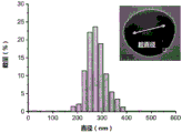
Fig. 3 shows the size distribution of the seed particles used in example 1 and the hemispherical nanoparticles produced.
Fig. 4 shows the cavity geometry of hemispherical nanoparticles with the size distribution of the cavity diameters measured in the nanoparticle population prepared in example 1.
Fig. 5 shows the average inertial cavitation thresholds for several batches of nanoparticles of the invention with different particle and cavity diameters.
Figure 6 shows a schematic of a process for producing the nanocapses described in the present invention.
Fig. 7a shows the particle size of the small, medium and large nanocapsuies of the present invention, fig. 7b shows the cavity diameter of the same nanocapsuies, and fig. 7c and d show the cavitation thresholds of the nanocapsuies.
FIG. 8 illustrates the cavitation effect of the nanocapsuies of the present invention comparing fluorescence images of a fluorescent drug model exposed to ultrasound in a tissue phantom, wherein (a) flow is through the tissue phantom alone; (b) together with SonoVue micro bubbles (5 micron average diameter, from Bracco) through the tissue simulant and (c) through the tissue simulant together with the fluorescent nanocapsuies of the invention. Figure 8d shows the cavitation effect of the nanocapsuies of the present invention under higher amplitude ultrasound.
Figure 9 shows the cavitation effect achieved using the nanopipettes containing bubbles described in the present invention compared to that of nanopipettes that were not dried and resuspended and therefore free of bubbles.
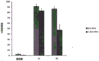
Figure 10 shows the use of nanocapsuies to achieve ultrasound-triggered (at 0.5MHz and 1.6 MHz) drug release from cavitation-sensitive liposomes. The release achieved was similar to that obtained using a micron-sized cavitation inducer (Sonovue, Bracco).
Figure 11 demonstrates that the nanocapsuies are stable after intravenous administration, allowing for the initiation of cavitation and real-time mapping in tumors using conventional diagnostic ultrasound systems.
Detailed Description
Nano cup
Nanoparticles as referred to herein are particles of any size between 1nm and 1000 nm. The size of the nanoparticles typically refers to the average diameter of the nanoparticles (s in fig. 1). The diameter is typically determined by electron microscopy. When multiple nanocapsuies are present in the composition, the cumulative average is typically used as the size of the nanoparticles. This can be determined using dynamic light scattering.
The term "partially spherical" as referred to herein describes a shape that forms part of a sphere. The term "partially spherical" includes shapes such as spherical caps, hemispheres, and spheres with the top truncated above the equator of the sphere (i.e., a spherical portion complementary to a spherical cap). Substantially partially spherical includes shapes that form part of a spheroid and shapes that form a sphere or part of a spheroid with irregular portions (e.g., dimples and protrusions).
In the nanoparticles of the invention, non-partially spherical cups may be used, and any cup shape may be used, i.e. having the shape of a cavity capable of partially enclosing a gas bubble. Shallow or small depressions/imperfections on the surface of the nanoparticles, e.g., less than about 5nm, typically less than about 10nm or less than about 20nm in depth, and/or less than about 5nm, typically less than about 10nm or typically less than about 20nm in opening, are generally not capable of encapsulating bubbles and thus cannot be considered cavities as referred to herein. Thus, the surface of a nanoparticle with a single cavity may also include the small/shallow flaws or depressions.
The bubbles partially enclosed by the cup as referred to herein are typically bubbles that: a portion of the surface of the bubble is encapsulated by the cup (optionally through one or more layers present between the cup and the bubble) and a portion of the surface is not encapsulated by the cup and can be exposed to a medium, such as a liquid medium, containing the nanoparticles.
The size of the opening of the cavity, referred to herein, is the largest dimension of the cavity (p in fig. 1) measured at the edge of the cup (typically the portion of the cup that forms the boundary between the encapsulated portion of the bubble and the unencapsulated portion of the bubble). In the case of a substantially part spherical cavity, the opening may be substantially circular and its size measured as the diameter of the opening. The size of the opening of the cavity can be determined by transmission electron microscopy.
The wall thickness of a cup, as referred to herein, refers to the shortest distance (w in fig. 1) from a point on the surface of the cup facing the cavity, through the cup, to a point on the opposite surface of the cup. The wall thickness of the cup can be determined by transmission electron microscopy.
The depth of the cup, as referred to herein, refers to the maximum distance (d in fig. 1) from the rim of the cup to the bottom of the cup. The depth of the cup can be determined by transmission electron microscopy.
In some embodiments, the thickness within the cup may vary, or the thickness may vary from cup to cup in compositions comprising multiple cups, in which case the average may be determined. In some embodiments, the thickness within the cup may be uniform or substantially uniform, or the thickness may be uniform or substantially uniform from cup to cup in a composition comprising a plurality of cups.
As used herein, the term "substantially uniform thickness" refers to a shape having a maximum thickness that is no more than 25% greater than the minimum thickness, such as no more than 20% greater than the minimum thickness, no more than 15% greater than the minimum thickness, no more than 10% greater than the minimum thickness, or no more than 5% greater than the minimum thickness.
Typical sizes of the nanoparticles of the invention are at least 60nm or at least 100nm and at most 1000nm, for example at most 500nm or at most 300 nm. For example, the size of the nanoparticles may be 60-500nm or 100-300 nm.
In embodiments where the nanoparticles are used in a method of tumor therapy, it may be desirable for the nanoparticles to be of a particular size to enhance accumulation in tumor tissue by enhancing the permeation and retention (EPR) effect. Tumor tissue may contain new blood vessels with abnormal forms and structures, leading to abnormal molecular and fluid transport dynamics. This may result in accumulation of nanoparticles of about 100-500nm, e.g., 100-300nm, size in tumor tissue being much larger than in normal tissue. Thus, nanoparticles having a size of 100-500nm, such as 100-300nm, may be desirable, particularly for use in tumor therapy methods.
The desired size and shape of the cavity containing the bubbles is determined by the ultrasound frequency to be used (typically in the range of 0.5MHz to 5.0 MHz) to minimize the sparse pressure amplitude required to induce inertial cavitation. Preferably an ultrasonic pressure amplitude of less than 5MPa, such as 3MPa or less, 2.5MPa or less, or 2MPa or less. Ultrasonic pressure amplitudes as low as 0.5MPa in the presence of the nanocups can produce significant amounts of inertial cavitation at frequencies as high as 2 MHz.
The cavity is typically substantially part spherical, for example substantially hemispherical or hemispherical. Typical opening dimensions of the cavity, i.e. the diameter at the opening for a substantially part-spherical cavity, are 50-900nm, such as at least 50nm, 100nm or at least 200nm and not more than 700nm, 600nm, or not more than 400 nm. The cavity may, for example, be 50-400nm or 100-600 nm. One skilled in the art can select an appropriate cavity shape and size to achieve the desired ultrasound parameters and produce nanoparticles having the desired cavity shape and size based on the methods for producing nanoparticles disclosed herein.
The ratio of the opening size to the nanoparticle size (p: s ratio in fig. 1) is typically 1: 3 to 5:6, such as 1:2 to 2: 3.
The cavity typically has a depth of greater than 30nm, preferably greater than 50 nm. For example, the cavity typically has a depth of 60-500nm, such as 80-400 nm.
Cavities having a larger size (e.g. a depth of more than 30nm, preferably more than 50 nm; and/or an opening of 50nm or more) have the advantage that a larger cavitation effect can be induced.
The wall thickness (w) of the cup is typically 10-100nm, for example 30-70nm or about 50 nm. The wall thickness of the cup can be selected and accurately controlled based on the methods disclosed herein for producing nanoparticles. The wall thickness of the cup is typically substantially uniform, whether within a single nanoparticle or between nanoparticles of the composition of the invention. The wall thickness is typically a fixed proportion of the overall cup size.
The cups are typically formed from a polymeric material, but may be formed from any solid material that can be formed into nanocups. Thus, the cup typically comprises a polymer, preferably it consists essentially of a polymer.
For example, the cup may be formed from a polymer that can be formed during an emulsion polymerization reaction. Suitable polymers include Polymethylmethacrylate (PMMA), polymethylmethacrylate (pHEMA), copolymers such as polylactic-co-glycolic acid (PLGA), polystyrene and divinylbenzene, and methylmethacrylate/2-hydroxyethyl methacrylate copolymers, and pH or temperature responsive polymers such as poly-N-isopropylacrylamide (PNIPAM). Other suitable polymers include, but are not limited to, polymers classified as polyanhydrides.
A pH or temperature responsive polymer such as PNIPAM may be used to enhance the release of the drug encapsulated within the nanocapsuies and/or the uptake of the drug at its target (e.g., tumor). pH-responsive polymers may be preferred in the context of tumor therapy, as the pH in a tumor may be lower than in healthy tissue (e.g., in hypoxic regions away from blood vessels). The temperature responsive polymer may preferably be used in combination with hyperthermia or ablation therapy, such as High Intensity Focused Ultrasound (HIFU), microwave, Radio Frequency (RF) or laser.
In some embodiments, the cup is formed from a crosslinked polymer. Crosslinking of the polymer may be achieved by introducing a crosslinking agent into the monomer mixture prior to or during the polymerization reaction. Suitable crosslinking agents include Divinylbenzene (DVB) or vinylsilane. Crosslinking may also be present in the polymer in the form of disulfide bond regions. The disulfide bond domain form can be introduced using a thiol chemical pathway. Crosslinked polymers can be used to provide rigidity to the nanoparticles. The need for rigidity may be, for example, to reduce or prevent fragmentation of the cup during cavitation. The degree of crosslinking can be determined by various spectroscopic methods, such as raman spectroscopy or nuclear magnetic resonance spectroscopy, or polymer swelling techniques.
The polymer cup may comprise a polymer formed from two or more (e.g., 2 or 3) different monomer units. The polymer may be produced by copolymerization of a mixture of monomers, and/or by providing one or more prepolymers that are linked by copolymerization or a crosslinking agent.
The cup is typically biocompatible, i.e., capable of performing its desired function in a medical treatment or diagnostic procedure without producing therapeutically unacceptable local or systemic effects in the recipient or recipient of the treatment or diagnosis.
The gas contained in the bubbles is not particularly limited, but is typically air, nitrogen, oxygen, perfluorocarbon such as perfluoropropane, or a mixture thereof.
The bubbles may be the same size and shape as the cavity. For example, if the cavity is hemispherical and the opening has a diameter d, the bubble may be hemispherical with a diameter d. Alternatively, the bubble may be larger than the cavity, i.e. it may protrude from the rim of the cup. It may protrude from the rim of the cup by a distance of 10% or more of the depth of the cup, for example 20% or more, 50% or more or 80% or more or 100% or more. The depth of the cup and the degree to which the bubbles can protrude can be measured by transmission electron microscopy. In some embodiments, the cavity is part-spherical or substantially part-spherical with a radius of curvature r, and the bubble is spherical with a radius r. In one example of this embodiment, the chamber is spherical with an opening diameter d and the bubble is spherical with a radius of 123d.
A particular advantage of the nanocups of the present invention is that the size of the chamber can be controlled to provide an adjustable cavitation initiator. Thus, the nanocups of the present invention typically have predictable and repeatable cavity opening dimensions. Thus, the nanocups with predictable cavity opening sizes are adjustable nanocups.
Process for producing nano-cup
The nanocapsuies may be produced by forming the cup by a seeded emulsion polymerization technique, optionally forming a drug layer within the cavity of the cup, and drying the cup in the presence of a gas to introduce the gas bubbles.
The size and shape of the cavity can be selected and accurately controlled by the selection of the seed particles used. Thus, the seed emulsion polymerization technique typically uses seed particles of a size and shape complementary to the desired cavity size and shape. For example, spherical seed particles may be used to produce cups having partially spherical cavities. Larger cavities may be formed using larger seed particles, and similarly, smaller cavities may be created using smaller seed particles. Typical sizes of the seed particles used are 50-900nm, for example at least 50 or at least 100nm and at most 600nm, at most 400nm or at most 300 nm.
Emulsion polymerization is typically carried out by mixing the seed particles, monomers and optionally a crosslinking agent in a suitable medium (e.g., water), mixing to create a turbulent flow regime and adding a suitable initiator, such as potassium persulfate. The emulsion polymerization reaction is allowed to continue at a suitable temperature for a suitable length of time. One skilled in the art will be able to select the appropriate temperature and time parameters for the particular reaction mixture used, however, the polymerization reaction is typically carried out at 70-90 ℃ for 4-6 hours, for example, at about 80 ℃ for about 5 hours.
The polymerization reaction is typically a free radical polymerization process. The polymer coated onto the seed particles may be a solid or liquid polymer that is copolymerized with the seed particles.
When the cup is formed from two or more monomer units, different monomers may be added to the emulsion polymerization at the beginning of the polymerization reaction, or alternatively, one or more monomers may be provided during the course of the polymerization reaction. Alternatively, one or more of the monomer units may be prepolymerized and the prepolymer provided to the polymerization mixture at the beginning of the polymerization reaction or during the progress of the polymerization reaction. Typically, one or more of the monomers or prepolymers may have a higher affinity for the seed particles. Thus, the polymerization reaction can provide a cup in which one monomer unit is formed predominantly on the inner surface of the cup (i.e., the monomer has a higher affinity for the seed particles) and a different monomer unit is formed predominantly on the outer surface of the cup. This may provide cups with different surface properties on the inner and outer surfaces.
In the case of polymeric nanocups, the seed particle typically remains chemically attached to the interior surface of the nanocup, and the seed particle material typically is itself contained within the cup. In case all available seed particles are fully reacted, there will be no residue of seed particles to be removed. Large agglomerates and small by-products are removed by centrifugation and filtration. Verification of the final shape, size and successful formation of the nanocapsuies can be acquired by Transmission Electron Microscopy (TEM) at multiple tilt angles in the plane of the grid, which can be reconstructed into a 3D model, as shown in the inset in fig. 2.
The wall thickness of the cup can be controlled by selecting the amount of monomer mixture relative to the amount of seed particles present in the reaction mixture. Increasing the amount of monomer relative to the number of seed particles increases the wall thickness of the resulting cup, while decreasing the amount of monomer relative to the number of seed particles decreases the wall thickness of the resulting cup. Furthermore, the wall thickness of the cup can be adjusted by adjusting the length of time the polymerization reaction is carried out, i.e., as long as there is sufficient reactant, a longer reaction time results in a thicker cup.
After the emulsion polymerization reaction has been carried out, the product may be washed, for example by centrifugation, to remove any excess polymer. Typically, the washed product is resuspended in a suitable medium, such as water, prior to drying.
The product may be dried by any suitable technique, for example freeze drying or air drying. If freeze-dried, typically no cryoprotectants are used. Air drying is preferred and may be carried out, for example, at elevated temperature or in a dryer. The air drying may be carried out for a period of 12-36 hours, for example 24 hours. In one embodiment, air drying may be carried out at 40-50 ℃ for 6-18 hours, for example about 12 hours, followed by drying in a desiccator at 18-24 ℃ for about 9 hours.
If dried in air, the bubbles in the nanoparticles of the present invention will be air bubbles. The substitute gas may be applied to the bubbles by performing a drying step in the desired gas. For example, drying may be performed in air, and the resulting nanoparticles with air bubbles may be placed under vacuum to remove the air bubbles and contacted with a desired gas to introduce new gas bubbles of the desired gas. The nanocapsuies are dried in a gaseous environment, providing the required bubbles in the cavity of the cup. Before drying, no air bubbles were present.
The dried nanocapsuies can be resuspended in a liquid medium, such as an aqueous solution. Suitable solutions include dilute glucose solutions, for example, 0.5-10%, preferably 1-8%, e.g., about 5% glucose solutions. Typically, the solution is then stirred to maximize particle dispersion. The dried nanocapsuies retain their air bubbles in the resuspension. Thus, nanopipettes dried and resuspended in a gaseous environment are different from nanopipettes formed directly in solution, because, due to the drying step, gas bubbles are present in the chamber.
After resuspension of the nanocapsuies, the solution is preferably filtered to remove agglomerated material. Injection filtration, for example 1-5 μm, typically 1.5-3 μm, can be used to remove fine particles. This limits the particles present to nanoparticles having a nanometer size below the filtration limit. A combination of coarse filtration followed by injection filtration may be used.
When a dry form of the nanocups is desired, for example, for ease of transportation, the resuspension and filtration steps can be omitted. Typically, the dried nanocapsuies are resuspended prior to use, and the resulting suspension is optionally filtered according to the resulting size distribution.
Fig. 6 schematically illustrates a process for producing the nanocapses according to the present invention. Figure 6 a) shows individual template particles coated with a copolymer in which spherical particles induce swelling and deformation. The resulting nanocapsuies were dried and resuspended to trap the gas within the chamber. Fig. 6 b) provides an illustration of the assumed mechanism of inertial cavitation for nanoparticle systems. Initially, the bubbles are stabilized within the chamber. Under sufficient negative pressure, the bubbles can extend over the opening of the cavity and eventually detach from the particles. In order for inertial cavitation to occur, the detached bubbles must grow unstably and eventually collapse under the inertia of the surrounding medium.
Nano cup as drug delivery carrier
The nanoparticles of the invention may be used as a delivery vehicle by, for example, including a release agent (typically a drug) within the cavity, within the cup, or on the outer surface of the cup. The release agent may form a layer, typically on the outer surface of the cup, within the cup or on the inner surface of the cup, within the cavity. Herein, the releasing agent refers to a material released from the nanoparticle upon cavitation. Typically, the release agent is a drug, i.e., a therapeutic or diagnostic, e.g., pharmaceutical or biological material. Typically, the release agent is a material useful for therapy or diagnosis of a human or animal subject. The description herein refers to a drug or drug layer present in a nanoparticle. However, it will be appreciated that alternative materials which may be used to release upon cavitation, for example when used in non-medical applications, may also be used as release agents. Thus, any of the drugs or drug layers described may also be contemplated to be replaced by alternative release agents or layers of release agents as appropriate.
In embodiments where a drug layer is present within the chamber or cup, the nature of the drug is not particularly limited and any drug useful in the method of treatment may be used. Preferably a hydrophilic drug. Certain drugs include, but are not limited to, anticancer drugs such as doxorubicin, paclitaxel, cisplatin, gemcitabine, daunorubicin, and oxaliplatin (oxiloplatin); thrombolytic agents, such as recombinant tissue plasminogen activator (rt-PA); anti-inflammatory agents, such as acetylsalicylic acid, ibuprofen, salsalate; or an antibiotic agent such as geldanamycin or penicillin. When the drug is contained on the outside of the cup, typically the drug is selected in such a way that it is associated with the cup in such a way that dissolution upon administration is avoided. Thus, the drug is only released upon cavitation.
The nanoparticles may comprise two or more different drugs. For example, one medicament may be disposed within the cavity of the cup and a separate, different medicament disposed on the outer surface of the cup. The two drugs combine upon release, for example upon exposure to ultrasound. Thus, the nanoparticles can be used as a means of providing a combination therapy, or to provide simultaneous release of two drugs mixed at a target site only upon release from the nanoparticle carrier (e.g., after exposure to ultrasound).
The addition of the drug layer may be performed before the formation of the cup (e.g., in the seed pellet), during the formation of the cup, or after the formation of the cup. The timing of the addition of the drug layer will result in its positioning within the nanocapsuies. For example, a drug-loaded seed particle will place a drug in the cavity of the cup. The drug is added during the polymerization reaction and will be placed in the cup. The medicament is added after the cup is formed, and the medicament is placed on the outer surface of the cup (i.e., the surface that does not face the bubble)
The thickness of the drug layer depends on the amount and location of the drug. When the drug is contained within the cup itself, the thickness of the drug layer will be limited by the thickness of the cup wall. In the case of a drug present within the lumen, the drug layer should be less than the thickness of the lumen, thereby allowing for the presence of a gas bubble at least partially enclosed by the lumen. The thickness of the drug layer can be controlled by adjusting the duration of the polymerization reaction and the amount of the drug reactant. Drug loading was measured using a drug release profile.
Use of the Nano-cup
The nanoparticles of the present invention can be used alone in any situation where inertial cavitation is desired, such as histotripsy, thermal ablation, sonochemistry, sonophoresis, and wastewater treatment. For example, it may be used in methods that involve the use of inertial cavitation to cause local tissue resection and/or removal to achieve a therapeutic result. Such methods include, for example, the treatment of tumors and the removal of cardiac tissue in the treatment of heart disease.
Alternatively, the nanoparticles of the invention may be used in combination with other agents, such as drugs or diagnostic agents (e.g., MRI contrast agents, such as Gd-based MRI contrast agents, or fluoroscopy contrast agents). The nanoparticles of the invention can be used to deliver other agents to targets in the body, either by delivering the nanoparticles and other agents simultaneously, or using the nanoparticles themselves as part of a drug formulation in the case where the nanoparticles contain a drug (e.g., a drug layer in a cup cavity).
Where a separate agent is used, the nanoparticles of the invention may be administered in a single composition with the separate agent, or as a separate composition. When two compositions are used, they may be administered simultaneously or separately. Preferably, administration is simultaneous or substantially simultaneous. The application of ultrasound after the administration of the nanoparticles and the individual agents causes cavitation to occur. This enhances transport of the agent into the cell. A particular advantage of using cavitation in this manner to enhance transport of the agent into the cell is that ultrasound can be applied at selected locations such that the active agent is only transported into the cell at the selected locations.
In one aspect of the invention, the individual agents are encapsulated in sound-sensitive liposomes. The acoustically sensitive liposomes can release an agent in the presence of inertial cavitation. Thus, the nanoparticles of the invention can be administered simultaneously with the liposomes and ultrasound applied to cause cavitation at the desired site and, correspondingly, release of the active agent at the desired site.
In an optional aspect of the invention, the active agent may be contained within the nanoparticle itself, as described above. In this case, cavitation may act to release the agent from the nanoparticle and aid in the transport of the active agent into the cell.
When used in combination with other agents, the nanoparticles may also provide real-time tracking and mapping of drug distribution in vivo, such as conventional ultrasound imaging using inertial cavitation or passive acoustic mapping (WO 2010052494).
The ultrasound used to achieve cavitation is not particularly limited and any frequency in the range of 0.5MHz to 5.0MHz may be used. The frequency may be matched to the adjusted nanoparticle. This may result in less ultrasonic pressure being required to achieve cavitation. The ultrasonic pressure amplitude used is typically less than 5MPa, for example 3MPa or less.
Nanobmug composition
The nanoparticles may be provided as a composition, such as a pharmaceutical composition. This may be a composition comprising a plurality of nanoparticles in dry form (e.g. as a powder) alone, or a composition comprising nanoparticles together with a pharmaceutically acceptable carrier or diluent. Typically, the composition provided in conjunction with the carrier or diluent is provided as a liquid composition comprising a suspension of nanoparticles. The pharmaceutical composition typically comprises up to 85 wt% of the nanoparticles of the invention. More typically, it comprises at most 50 wt% of the nanoparticles of the invention. Preferred pharmaceutical compositions are sterile and pyrogen free.
Prior to use, compositions comprising a plurality of nanoparticles in dry form are typically resuspended in a suitable liquid, such as an aqueous solution.
Compositions comprising a plurality of nanoparticles typically include substantially uniform nanoparticles. This has the advantage that the control of cavitation is higher, thereby improving acoustic tuning. The preferred nanoparticle composition has a polydispersity index of 0.2. Typically, this is determined by dynamic light scattering. The average particle size of a composition comprising a plurality of nanoparticles is defined herein as the modal average (i.e., the particle size indicated by the largest number of particles). In a preferred aspect of the invention, at least 50% of the nanoparticles in the composition have a particle size within 50nm of the average. More preferably, at least 60%, 70% or 80% of the nanoparticles have a particle size within 50nm of the average. More preferably, at least 50% of the nanoparticles in the composition have a particle size within 20nm of the average. More preferably, at least 60%, 70% or 80% of the nanoparticles have a particle size within 20nm of the average. Dynamic light scattering can be used to determine particle size differences in the composition.
The average cavity diameter (the size of the opening of the cavity, p) of a composition comprising a plurality of nanoparticles is defined herein as the modal average (i.e., the cavity diameter shown by the maximum number of particles). In a preferred aspect of the invention, at least 50% of the nanoparticles in the composition have a cavity diameter within 50nm of the mean. More preferably, at least 60%, 70% or 80% of the cavity diameter of the nanoparticle is within 50nm of the mean. More preferably, at least 50% of the nanoparticles in the composition have a cavity diameter within 20nm of the mean. More preferably, at least 60%, 70% or 80% of the cavity diameter of the nanoparticle is within 20nm of the mean. Dynamic light scattering can be used to determine differences in cavity diameter in the composition.
The nanoparticles of the invention may be administered by any suitable route, depending on the nature of the treatment method, such as orally (as syrups, tablets, capsules, lozenges, controlled release formulations, fast dissolving formulations, etc.); for external use (as cream, ointment, lotion, nasal spray or aerosol); by injection (subcutaneous, intradermal, intramuscular, intravenous, intratumoral, etc.); transdermal (e.g., by use of a patch, gel or implant) or by inhalation (as a dry powder, solution, dispersion, etc.).
For example, in solid oral forms, in addition to the nanoparticles of the invention, may comprise: diluents, such as lactose, glucose, sucrose, cellulose, corn starch or potato starch; lubricants, for example silicon dioxide, talc, stearic acid, magnesium or calcium stearate, and/or polyethylene glycol; binders, such as starch, gum arabic, gelatin, methylcellulose, carboxymethylcellulose or polyvinylpyrrolidone; disintegrating agents, such as starch, alginic acid, alginates or sodium starch glycolate; an effervescent mixture; a dye; a sweetener; wetting agents, such as lecithin, polysorbate, lauryl sulfate; and, in general, non-toxic and pharmacologically inactive substances used in pharmaceutical formulations. Such pharmaceutical preparations may be manufactured in a known manner, for example by means of mixing, granulating, tabletting, sugar-coating or film-coating processes.
Preferably, the composition provided in conjunction with the carrier or diluent is a liquid dispersion. Liquid dispersions for oral administration may be syrups, emulsions and suspensions. Preferably a suspension. The syrups, emulsions and suspensions may contain, for example, sucrose or sucrose together with glycerol and/or mannitol and/or sorbitol as a carrier. Glucose is the preferred carrier. For example, the nanoparticles may be provided as a suspension in a glucose solution, e.g., a glucose solution containing up to 10wt% glucose (e.g., about 5wt% glucose).
Suspensions and emulsions may contain as carrier, for example, a natural gum, agar, sodium alginate, pectin, methylcellulose, carboxymethylcellulose, or polyvinyl alcohol. Suspensions or solutions for intramuscular injections may contain, in addition to the active compound, a pharmaceutically acceptable carrier, for example sterile water, olive oil, ethyl oleate, glycols (e.g. propylene glycol), and, if desired, a suitable amount of lidocaine hydrochloride.
The solution for injection or infusion may contain as a carrier, for example, sterile water, or preferably, it may be in the form of a sterile aqueous isotonic saline solution.
The amount of drug-containing nanoparticles administered as part of a therapeutic or diagnostic method will depend, for example, on the type and amount of drug present in the nanoparticles. Thus, the dose of drug-containing nanoparticles of the present invention, typically equal to or less than the dose of drug present in the nanoparticles when administered alone, i.e., the amount of drug present in the nanoparticles, will typically be equal to or less than the amount of drug administered in free form. The drug-containing nanoparticle dose of the present invention may be less than the equivalent amount of free drug, e.g., to offset the enhanced pharmacokinetics described above in the agents of the present invention, e.g., 95% or less, 85% or less, 80% or less, 75% or less, 70% or less, 65% or less, 60% or less, 55% or less, or 50% or less.
In embodiments of the invention in which the nanoparticle and the other agent are administered separately, the two agents may be co-administered in the same pharmaceutical composition, or in different compositions intended for separate, simultaneous, concomitant, or sequential administration by the same or different routes.
When used alone, the amount of nanoparticles administered may be any suitable dose as determined by one of skill in the art.
Examples
The invention will be illustrated by the following non-practical examples.
Example 1
Nanoparticles with spherical cups, also known as Nanocapsuies (NCs), were produced according to the following scheme:
2mL of 10wt% 300nm polystyrene (also called latex) nanoparticles were mixed with 34mL of filtered deionized water. The resulting mixture was placed in a 100ml round bottom flask and bubbled with nitrogen for 30 minutes. The round bottom flask was fitted with a reflux condenser and placed in a water bath at 80 ℃. The following monomers were added in sequence with stirring: 851. mu.l MMA, 85. mu.l HEMA, and 533. mu.l 80 v/v% DVB. A1.704 ml solution of 3.5 mg/ml potassium persulfate (KPS) was degassed with nitrogen. KPS solution was added. After 5 hours of reaction duration, the reaction mixture was washed three times by centrifugation at 15000rpm for 30 minutes. The resulting nanoparticles with hemispherical cups were suspended in filtered deionized water. The TEM and size distribution of the nanocapsuies are shown in fig. 2 and 3.
The NC dispersion was placed in a petri dish and heated to-40-50 ℃ overnight. The petri dish was then placed in a drying chamber for 24 hours. The resulting nanoparticles with hemispherical cups and air bubbles were dispersed in filtered deionized water.
Fig. 5 shows the average inertial cavitation threshold for several batches of nanoparticles of the invention with different particle diameters and cavity diameters. Results were obtained by exposing the nanocapsuies of example 1 to 200 independent HIFU pulses (0.5 MHz 10 cycle sine wave) while confined in a 0.2mm cylindrical tube. Acoustic emissions were measured with a 15MHz sensor (also known as a passive cavitation detector). The received signal is amplified by a factor of 25 to reduce the effect of RF noise and digitally recorded using a 12-bit DAQ card. The signal is digitally filtered with a 2MHz high pass filter to remove the effect of the drive frequency. Each resulting voltage trace is converted to the frequency domain, the harmonics are filtered out with a notch filter, and the wide frequency components are examined. A cavitation event is considered to occur if the broad frequency component (measured as power) of a single pulse is three times higher than a water filled cuvette. Thus, the probability of inertial cavitation is the percentage of inertial cavitation events per 200 transmit pulses.
For a fully uniformly distributed system, the probability of an inertial cavitation curve should be a helminthoid function (i.e., a step function from 0 to 100%). However, deviations from a perfect distribution shift the probability curve, producing a linear increase; different cavities within a population will have different cavitation thresholds. Thus, the individual chamber inertial cavitation threshold can be inferred by comparing this size population in the sample to the inertial cavitation probability.
Example 2
The nanocapsuies produced according to the protocol described in example 1 were dried and resuspended to provide the following intracavity gas composition:
the suspension of nanoparticles in water was placed in a large petri dish at 60 ℃ and air dried overnight. Next, the dry mass is suspended in a liquid medium (such as deionized water or 5% glucose) and vigorously stirred for up to 8 hours to maximize particle dispersion. A combination of coarse filtration of 106 μm and 32 μm was used to remove non-dispersed agglomerates in the millimeter scale. Microagglomerates were removed using 2.7 μm and 1.6 μm syringe filtration (Whatman, GE Healthcare Life Sciences, UK) if necessary.
Example 3
Three different sizes of nanopipettes were produced according to the protocol of example 1 and dried and resuspended according to example 2. Different sizes were obtained by using polystyrene seed nanoparticles of different sizes as follows:
small nanoparticles: 100nm polystyrene seed;
medium nano-particles: 300nm polystyrene seed;
large nanoparticles: 460 nm polystyrene seed.
The size distribution of the nanocups produced according to the protocol of example 1 was determined by dynamic light scattering. The results are shown in fig. 7a, demonstrating that by using shells of different sizes, nanocapsuies with small particle size distribution and well controlled particle size can be produced.
Fig. 7b shows the size distribution of the cavity diameters of the small, medium and large nanocapsuies measured in transmission electron microscope images. FIGS. 7c and 7d show the inertial cavitation thresholds for small, medium and large nanocapsuies, as determined experimentally and numerically predicted at 0.5MHz (c) and 1.6MHz (d).
The experimentally determined inertial cavitation threshold (open circles) shown is consistent with the inertial cavitation threshold (gray cross) predicted by the modified hemispherical Rayleigh-Plesset model.
Example 4
Tissue mimics composed of 1 wt% agarose were exposed to ultrasound at a center frequency of 1.6MHz and 2.2 MPa (5% duty cycle) for 5 minutes while the fluorescent drug model prepared using the method of example 2 and fluorescent (9-anthracene methacrylate) nanocups were continuously flowed through the channel. The fluorescent drug model used was tetramethyl rhodamine-40 kDa dextran. The tissue phantom is radially sectioned relative to the channel after application of ultrasound, and a fluorescence image of the drug model is visualized. Figure 8c shows the movement of the drug away from the initial tissue tract after ultrasound exposure. The motion of the drug is mediated by cavitation caused by the ultrasound applied to the nanocapsuies.
Figure 8a shows the corresponding embodiment with the nanocapsuies omitted and figure 8b shows the corresponding embodiment with SonoVue microbubbles (average diameter 5 microns, from Bracco) replacing the nanocapsuies of the present invention. FIG. 8d shows a corresponding example of a nanocup of the present invention using fluorescence exposed to ultrasound at 4 MPa, 1.5% duty cycle, and 1.6MHz center frequency. In fig. 8, a white dotted line indicates an edge of the HIFU focus, and a gray dotted line in (a) indicates an edge of the flow container. The white arrows demarcate the direction of the ultrasound. In all images, model drugs and fluorescent nanocapsuies are marked with arrows in each figure. The scale bar represents 1 mm and applies to all images.
Example 5
To compare the properties of (a) a nanocup containing gas bubbles as described herein and (b) a nanocup without gas bubbles, two nanocups were prepared. The nanocapsuies were prepared according to the protocol of example 1. A portion of the nanocapsuies were not dried. The other was dried and resuspended as described in example 2.
Figure 9a shows the inertial cavitation probability of dried and resuspended 300nm polystyrene seed particles (open squares), undried and resuspended nanocells (black triangles) and dried and resuspended nanocells (open circles) exposed to ultrasound at 0.5MHz center frequency.
FIGS. 9b and c show the resulting cavitation power for nanocups above (b) and below (c) the inertial cavitation threshold. The total power has been split into a broad frequency component (representing the signal generated by the inertial cavitation source) and a harmonic component (representing the signal generated by the stationary cavitation source).
Example 6
2 chloroform in round bottom flask: DSPE, cholesterol, DSPC and DSPE-PEG (DSPE liposomes) are mixed in methanol at a ratio of 65:25:3:7 to formulate fluorescein encapsulated in cavitation-sensitive liposomes, such as that described in S. Graham, R. Carlisle, J. Choi, M. Stevenson, A. Shah, R. Myers, K. Fisher, M. B. Shaegrino, L. Seymour, and C.C. Courses, "inert calcium to non-innovative trigger and monomer Release of drug from organic delivery lipid controls," Journal of regulated Release, vol. 178, pp. 101-.
In the presence of glucose alone, the liposomes (average diameter 140 nm) were exposed to ultrasound at 0.5MHz or 1.6MHz without release. Exposure of liposomes to ultrasound in the presence of Sonovue (SV) microbubbles (average diameter 5 microns, ex Bracco) gave nearly complete release at both frequencies. Exposure of liposomes in the presence of Nanopipels (NC) (average diameter 300nm, produced as described in examples 1 and 2 above) gave the same release as SV at 0.5MHz, and slightly lower at 1.6 MHz. The results are shown in FIG. 10.
This demonstrates the feasibility of cavitation-mediated release using cavitation-inducing particles that mimic the size of nanoliposomes over the biomedical ultrasound frequency range. Unlike microbubbles, nanocapsives are able to penetrate the leaky tumor vasculature (100-800 nm inter-endothelial cell junction gap) to successfully accumulate with liposomes in tumors.
Example 7
Balb/c mice were subcutaneously implanted with CT-26 mouse colon cancer cells. The administration was performed by continuous intravenous injection using a Nanocup (NCS). Cavitation was induced by ultrasound. FIG. 11a shows a volume of 100-3Tumor (dashed outline) (rendered by Passive Acoustic Mapping) overlaid on a conventional type B image). The top row of figure 11a shows the cavitation map after a single pulse of 2MHz ultrasound in the tumor, while the second row shows the cumulative cavitation activity of the entire tumor over multiple ultrasound pulses. Figure 11b is a graph demonstrating the increase in tumor cavitation levels after each injection, suggesting successful accumulation of NC within the tumor.
Claims (32)
1. A pharmaceutical composition comprising: a plurality of nanoparticles, and a pharmaceutically acceptable carrier or diluent as a liquid medium, wherein each nanoparticle comprises:
a cup having a cavity; and
the presence of a gas bubble in the cavity,
wherein the size of the cup is at least 60nm and at most 1000nm, the gas bubbles are partially encapsulated by the cup, and wherein the nanoparticle comprises a single nanocup having a single cavity.
2. The pharmaceutical composition of claim 1, wherein the cup is substantially part-spherical.
3. The pharmaceutical composition of claim 2, wherein the cup is substantially hemispherical.
4. The pharmaceutical composition of any of the preceding claims, wherein the cup has a substantially uniform wall thickness.
5. The pharmaceutical composition of claim 4, wherein the cup has a wall thickness of 10-100 nm.
6. The pharmaceutical composition of claim 5, wherein the cup has a wall thickness of 30-70 nm.
7. A pharmaceutical composition according to any one of claims 1 to 3, wherein the cavity is substantially part-spherical.
8. A pharmaceutical composition according to any one of claims 1 to 3, wherein the cavity has an opening of 50 to 900 nm.
9. The pharmaceutical composition of claim 8, wherein the opening of the cavity is 50-400 nm.
10. The pharmaceutical composition of claim 9, wherein the size of the cup is 60-500 nm.
11. The pharmaceutical composition of claim 1, wherein the cup and the cavity are both substantially part spherical, the cup has a substantially uniform wall thickness of 10-100nm, and the opening of the cavity has a diameter of 50-900 nm.
12. The pharmaceutical composition of claim 11, wherein the cavity has an opening size of 50-400 nm.
13. The pharmaceutical composition of claim 1, wherein the cup comprises a polymer.
14. The pharmaceutical composition of claim 13, wherein the polymer is made from two or more different monomer units, one of the monomer units being present predominantly on the inner surface of the cup and the other monomer unit being present predominantly on the outer surface of the cup.
15. The pharmaceutical composition according to claim 13 or 14, wherein the polymer is a copolymer of Methyl Methacrylate (MMA) and 2-hydroxyethyl methacrylate (HEMA).
16. The pharmaceutical composition of claim 13 or 14, wherein the polymer is a cross-linked polymer.
17. The pharmaceutical composition according to claim 15, wherein the polymer is obtainable by using divinylbenzene as crosslinking agent in a free radical polymerization reaction.
18. The pharmaceutical composition of claim 1, wherein the nanoparticle comprises one or more release agents.
19. The pharmaceutical composition of claim 18, wherein a release agent is present within the cup, on an outer surface of the cup, and/or in a cavity of the cup.
20. The pharmaceutical composition of claim 19, wherein the nanoparticle comprises a first release agent in the cavity of the cup and a second release agent on an outer surface of the cup.
21. A pharmaceutical composition according to any one of claims 18 to 20 wherein the or each release agent is a drug.
22. The pharmaceutical composition of any one of claims 1-3, wherein the pharmaceutical composition comprises nanoparticles having a polydispersity index of no more than 0.2.
23. A process for the manufacture of a pharmaceutical composition according to any one of claims 1-22, comprising:
providing a seed particle;
partially coating the seed particles to provide cups having cavities, wherein the cups are at least 60nm and at most 1000nm in size;
drying the cup;
providing a gas to the cup to create a bubble in the cavity; and
resuspending the nanoparticles in a liquid medium.
24. The method of claim 23, wherein the steps of drying the cup and providing a gas to the cup are accomplished by drying the cup in air.
25. The method according to claim 23 or 24, wherein the seed particles are coated by performing an emulsion polymerization reaction in the presence of the seed particles, the polymerization reaction being performed using one or more monomers and/or prepolymers and optionally using a cross-linking agent.
26. The method of claim 25, wherein the crosslinking agent is divinylbenzene.
27. The method of claim 26, wherein the polymerization reaction is a free radical polymerization reaction.
28. The method as claimed in claim 23 or 24, wherein the seed particle has a diameter of 100-600 nm.
29. The method of claim 23 or 24, wherein the nanoparticle comprises one or more release agents, and the method further comprises: (a) providing a release agent as a coating for the seed particles; and/or (b) providing a release agent during the polymerization reaction; and/or (c) providing a release agent on an outer surface of the cup.
30. The method of claim 29, wherein two or more different release agents are provided.
31. Use of a pharmaceutical composition according to any one of claims 1 to 22 in the manufacture of a medicament for the therapeutic or diagnostic use in a human or animal subject.
32. The use of claim 31, wherein the medicament is a medicament for the treatment of a tumor.
Applications Claiming Priority (3)
| Application Number | Priority Date | Filing Date | Title |
|---|---|---|---|
| GBGB1320413.6A GB201320413D0 (en) | 2013-11-19 | 2013-11-19 | Cavitation-inducing polymeric nanoparticles |
| GB1320413.6 | 2013-11-19 | ||
| PCT/GB2014/053416 WO2015075442A1 (en) | 2013-11-19 | 2014-11-19 | Cavitation-inducing polymeric nanoparticles |
Publications (2)
| Publication Number | Publication Date |
|---|---|
| CN106102714A CN106102714A (en) | 2016-11-09 |
| CN106102714B true CN106102714B (en) | 2020-02-14 |
Family
ID=49883853
Family Applications (1)
| Application Number | Title | Priority Date | Filing Date |
|---|---|---|---|
| CN201480072911.3A Active CN106102714B (en) | 2013-11-19 | 2014-11-19 | Cavitation-induced polymer nanoparticles |
Country Status (7)
| Country | Link |
|---|---|
| US (1) | US10441529B2 (en) |
| EP (1) | EP3071183A1 (en) |
| JP (1) | JP6595991B2 (en) |
| CN (1) | CN106102714B (en) |
| CA (1) | CA2930815C (en) |
| GB (1) | GB201320413D0 (en) |
| WO (1) | WO2015075442A1 (en) |
Families Citing this family (10)
| Publication number | Priority date | Publication date | Assignee | Title |
|---|---|---|---|---|
| US9446227B2 (en) | 2008-09-12 | 2016-09-20 | Sonescence, Inc. | Ultrasonic dispersion of compositions in tissue |
| US20100069827A1 (en) | 2008-09-12 | 2010-03-18 | Barry Neil Silberg | Pre-Surgical Prophylactic Administration of Antibiotics and Therapeutic Agents |
| WO2015187968A1 (en) | 2014-06-04 | 2015-12-10 | Sonescence, Inc. | Systems and methods for therapeutic agent delivery |
| US9498545B1 (en) | 2014-10-24 | 2016-11-22 | Verily Life Sciences Llc | Environmentally responsive hydrogels for delivery |
| CN111701035B (en) * | 2020-06-28 | 2022-09-06 | 南京超维景生物科技有限公司 | Ultrasonic contrast agent composition, ultrasonic contrast agent, preparation method of ultrasonic contrast agent and application of acoustic deformation material |
| EP4036581A1 (en) * | 2021-02-01 | 2022-08-03 | Oxford University Innovation Limited | Cavitation agent |
| EP4035673A1 (en) | 2021-02-01 | 2022-08-03 | Oxford University Innovation Limited | Transdermal vaccine |
| EP4036580A1 (en) | 2021-02-01 | 2022-08-03 | Oxford University Innovation Limited | Drug loaded cavitation agent |
| EP4035655A1 (en) | 2021-02-01 | 2022-08-03 | Oxford University Innovation Limited | Immune modulating particles |
| EP4230222A1 (en) | 2022-02-17 | 2023-08-23 | Oxsonics Limited | Combination therapy with an anti-axl antibody-pbd conjugate and nanocups |
Citations (2)
| Publication number | Priority date | Publication date | Assignee | Title |
|---|---|---|---|---|
| WO2012066334A1 (en) * | 2010-11-17 | 2012-05-24 | Isis Innovation Limited | Sonosensitive nanoparticles |
| WO2012094541A2 (en) * | 2011-01-05 | 2012-07-12 | The Regents Of The University Of California | Acoustically responsive particles with decreased cavitation threshold |
Family Cites Families (11)
| Publication number | Priority date | Publication date | Assignee | Title |
|---|---|---|---|---|
| EP1714642A1 (en) * | 2005-04-18 | 2006-10-25 | Bracco Research S.A. | Pharmaceutical composition comprising gas-filled microcapsules for ultrasound mediated delivery |
| GB0820377D0 (en) | 2008-11-07 | 2008-12-17 | Isis Innovation | Mapping and characterization of cavitation activity |
| US20100178305A1 (en) | 2008-12-15 | 2010-07-15 | University Of Utah Research Foundation | Ultrasonic nanotherapy of solid tumors with block copolymers stabilized perfluorocarbon nanodroplets |
| EP2406072A4 (en) | 2009-03-11 | 2016-03-16 | Univ Northeastern | Low-aspect ratio nanostructures |
| GB0916634D0 (en) | 2009-09-22 | 2009-11-04 | Isis Innovation | Ultrasound systems |
| US20130115169A1 (en) | 2009-12-04 | 2013-05-09 | The Regents Of The University Of California | Red blood cell-mimetic particles and methods for making use thereof |
| US8709451B2 (en) | 2010-01-20 | 2014-04-29 | University Of Utah Research Foundation | Stable nanoemulsions for ultrasound-mediated drug delivery and imaging |
| US8663675B2 (en) * | 2010-11-18 | 2014-03-04 | Jalaledin Ghanavi | Injectable matrix having a polymer and a stem cell niche composed of cup-shaped nanoparticles containing growth factors or physiological agents for organ reconstruction |
| WO2013055791A1 (en) | 2011-10-10 | 2013-04-18 | The Regents Of The University Of Michigan | Polymeric nanoparticles for ultrasound imaging and therapy |
| GB201318668D0 (en) | 2013-10-22 | 2013-12-04 | Isis Innovation | Sonosensitive therapeutic |
| GB201318664D0 (en) | 2013-10-22 | 2013-12-04 | Isis Innovation | Shielded biologic therapeutic |
-
2013
- 2013-11-19 GB GBGB1320413.6A patent/GB201320413D0/en not_active Ceased
-
2014
- 2014-11-19 CN CN201480072911.3A patent/CN106102714B/en active Active
- 2014-11-19 WO PCT/GB2014/053416 patent/WO2015075442A1/en active Application Filing
- 2014-11-19 JP JP2016532545A patent/JP6595991B2/en active Active
- 2014-11-19 US US15/037,664 patent/US10441529B2/en active Active
- 2014-11-19 CA CA2930815A patent/CA2930815C/en active Active
- 2014-11-19 EP EP14803195.8A patent/EP3071183A1/en active Pending
Patent Citations (2)
| Publication number | Priority date | Publication date | Assignee | Title |
|---|---|---|---|---|
| WO2012066334A1 (en) * | 2010-11-17 | 2012-05-24 | Isis Innovation Limited | Sonosensitive nanoparticles |
| WO2012094541A2 (en) * | 2011-01-05 | 2012-07-12 | The Regents Of The University Of California | Acoustically responsive particles with decreased cavitation threshold |
Non-Patent Citations (4)
| Title |
|---|
| Fabrication, Characterization, and Optical Properties of Gold Nanobowl Submonolayer Structures;Jian Ye等;《Langmuir》;20090106;第25卷(第1期);第1822-1827页 * |
| Facile Fabrication of Monodisperse Polymer Hollow Spheres;Hui Lv等;《Langmuir》;20081202;第24卷(第23期);第13736页左栏第1段、右栏第2段第1-3行,第13737页左栏第3段第1-3行、左栏倒数第2段,右栏第2段第5-9行,第13738页左栏第2段第13-14行、第3段2-4行,第13741页左栏第5-8行,图2、6-9,表1,Scheme 1 * |
| Inertial cavitation at the nanoscale;James J. Kwan等;《Journal of the Acoustical Society of America》;20130602;第133卷(第5期);第3315页 * |
| Sonosensitive Nanoparticles for Controlled Instigation of Cavitation and Drug Delivery by Ultrasound;Sarah J. Wagstaffe等;《American Institute of Physics》;20121231;第426-431页 * |
Also Published As
| Publication number | Publication date |
|---|---|
| JP2016539114A (en) | 2016-12-15 |
| EP3071183A1 (en) | 2016-09-28 |
| WO2015075442A1 (en) | 2015-05-28 |
| CN106102714A (en) | 2016-11-09 |
| CA2930815C (en) | 2022-01-11 |
| JP6595991B2 (en) | 2019-10-23 |
| GB201320413D0 (en) | 2014-01-01 |
| CA2930815A1 (en) | 2015-05-28 |
| US10441529B2 (en) | 2019-10-15 |
| US20160296465A1 (en) | 2016-10-13 |
Similar Documents
| Publication | Publication Date | Title |
|---|---|---|
| CN106102714B (en) | Cavitation-induced polymer nanoparticles | |
| Cavalli et al. | Micro-and nanobubbles: a versatile non-viral platform for gene delivery | |
| US10688040B2 (en) | Encapsulation of microbubbles within the aqueous core of microcapsules | |
| Jangjou et al. | The promising shadow of microbubble over medical sciences: from fighting wide scope of prevalence disease to cancer eradication | |
| US9968692B2 (en) | Nanobubbles | |
| US20030147812A1 (en) | Device and methods for initiating chemical reactions and for the targeted delivery of drugs or other agents | |
| Tamarov et al. | Nano air seeds trapped in mesoporous janus nanoparticles facilitate cavitation and enhance ultrasound imaging | |
| Eisenbrey et al. | Delivery of encapsulated doxorubicin by ultrasound-mediated size reduction of drug-loaded polymer contrast agents | |
| JP2011529039A (en) | Ultrasound-mediated drug delivery | |
| Mannaris et al. | Acoustically responsive polydopamine nanodroplets: A novel theranostic agent | |
| CN101618013A (en) | Focused ultrasound-polymeric micelle controllable drug release device and release method thereof | |
| TW201429492A (en) | Nano-and micro-bubbles with ultrasound-triggered release and imaging functionalities | |
| Montoya Mira et al. | Gas-stabilizing sub-100 nm mesoporous silica nanoparticles for ultrasound theranostics | |
| Luo et al. | Microbubbles: a novel strategy for chemotherapy | |
| Tsirkin et al. | Tailor-made single-core PLGA microbubbles as acoustic cavitation enhancers for therapeutic applications | |
| Kumar et al. | Microbubbles: Revolutionizing Biomedical Applications with Tailored Therapeutic Precision | |
| Barmin et al. | Polymeric materials for ultrasound imaging and therapy | |
| US20230277696A1 (en) | Ultrasound-sensitive biodegradeable multi-cavity micro-particles | |
| Ayodele et al. | Biointerface research in applied chemistry | |
| Chaudhary et al. | The potential of microbubbles as a cancer eradication theranostic agent | |
| US20230248652A1 (en) | Biodegradeable multi-cavity microparticles and their use in treatment | |
| Hu et al. | Progress and preclinical application status of ultrasound microbubbles | |
| Su | Design and development of acoustic cavitation agents for biomedical and industrial applications | |
| Shah et al. | Exploring the Theranostic Applications and Prospects of Nanobubbles | |
| Martina et al. | Enabling technologies for the preparation of multifunctional “bullets” for nanomedicine |
Legal Events
| Date | Code | Title | Description |
|---|---|---|---|
| C06 | Publication | ||
| PB01 | Publication | ||
| C10 | Entry into substantive examination | ||
| SE01 | Entry into force of request for substantive examination | ||
| CB02 | Change of applicant information | ||
| CB02 | Change of applicant information |
Address after: Oxfordshire Applicant after: University of Oxford Innovation Co., Ltd. Address before: Oxfordshire Applicant before: ISIS Innovaton Ltd. |
|
| TA01 | Transfer of patent application right | ||
| TA01 | Transfer of patent application right |
Effective date of registration: 20171109 Address after: Oxfordshire Applicant after: AUX sound energy Co., Ltd. Address before: Oxfordshire Applicant before: University of Oxford Innovation Co., Ltd. |
|
| GR01 | Patent grant | ||
| GR01 | Patent grant |