BR112015013247B1 - AUXILIARY DEVICE TO ASSIST IN THE PERFORMANCE OF BRACHYTHERAPY, BRACHYTHERAPY SYSTEM TO APPLY BRACHYTHERAPY TO A LIVE OBJECT, AND AUXILIARY METHOD TO ASSIST IN THE PERFORMANCE OF BRACHYTHERAPY - Google Patents
AUXILIARY DEVICE TO ASSIST IN THE PERFORMANCE OF BRACHYTHERAPY, BRACHYTHERAPY SYSTEM TO APPLY BRACHYTHERAPY TO A LIVE OBJECT, AND AUXILIARY METHOD TO ASSIST IN THE PERFORMANCE OF BRACHYTHERAPY Download PDFInfo
- Publication number
- BR112015013247B1 BR112015013247B1 BR112015013247-2A BR112015013247A BR112015013247B1 BR 112015013247 B1 BR112015013247 B1 BR 112015013247B1 BR 112015013247 A BR112015013247 A BR 112015013247A BR 112015013247 B1 BR112015013247 B1 BR 112015013247B1
- Authority
- BR
- Brazil
- Prior art keywords
- brachytherapy
- ultrasound
- ultrasound image
- group
- unit
- Prior art date
Links
Images


Classifications
-
- A—HUMAN NECESSITIES
- A61—MEDICAL OR VETERINARY SCIENCE; HYGIENE
- A61N—ELECTROTHERAPY; MAGNETOTHERAPY; RADIATION THERAPY; ULTRASOUND THERAPY
- A61N5/00—Radiation therapy
- A61N5/10—X-ray therapy; Gamma-ray therapy; Particle-irradiation therapy
- A61N5/1048—Monitoring, verifying, controlling systems and methods
- A61N5/1071—Monitoring, verifying, controlling systems and methods for verifying the dose delivered by the treatment plan
-
- A—HUMAN NECESSITIES
- A61—MEDICAL OR VETERINARY SCIENCE; HYGIENE
- A61B—DIAGNOSIS; SURGERY; IDENTIFICATION
- A61B8/00—Diagnosis using ultrasonic, sonic or infrasonic waves
- A61B8/08—Detecting organic movements or changes, e.g. tumours, cysts, swellings
- A61B8/0833—Detecting organic movements or changes, e.g. tumours, cysts, swellings involving detecting or locating foreign bodies or organic structures
- A61B8/0841—Detecting organic movements or changes, e.g. tumours, cysts, swellings involving detecting or locating foreign bodies or organic structures for locating instruments
-
- A—HUMAN NECESSITIES
- A61—MEDICAL OR VETERINARY SCIENCE; HYGIENE
- A61B—DIAGNOSIS; SURGERY; IDENTIFICATION
- A61B8/00—Diagnosis using ultrasonic, sonic or infrasonic waves
- A61B8/42—Details of probe positioning or probe attachment to the patient
- A61B8/4245—Details of probe positioning or probe attachment to the patient involving determining the position of the probe, e.g. with respect to an external reference frame or to the patient
-
- A—HUMAN NECESSITIES
- A61—MEDICAL OR VETERINARY SCIENCE; HYGIENE
- A61B—DIAGNOSIS; SURGERY; IDENTIFICATION
- A61B8/00—Diagnosis using ultrasonic, sonic or infrasonic waves
- A61B8/48—Diagnostic techniques
- A61B8/483—Diagnostic techniques involving the acquisition of a 3D volume of data
-
- A—HUMAN NECESSITIES
- A61—MEDICAL OR VETERINARY SCIENCE; HYGIENE
- A61B—DIAGNOSIS; SURGERY; IDENTIFICATION
- A61B8/00—Diagnosis using ultrasonic, sonic or infrasonic waves
- A61B8/52—Devices using data or image processing specially adapted for diagnosis using ultrasonic, sonic or infrasonic waves
- A61B8/5269—Devices using data or image processing specially adapted for diagnosis using ultrasonic, sonic or infrasonic waves involving detection or reduction of artifacts
-
- A—HUMAN NECESSITIES
- A61—MEDICAL OR VETERINARY SCIENCE; HYGIENE
- A61N—ELECTROTHERAPY; MAGNETOTHERAPY; RADIATION THERAPY; ULTRASOUND THERAPY
- A61N5/00—Radiation therapy
- A61N5/10—X-ray therapy; Gamma-ray therapy; Particle-irradiation therapy
- A61N5/1001—X-ray therapy; Gamma-ray therapy; Particle-irradiation therapy using radiation sources introduced into or applied onto the body; brachytherapy
- A61N5/1007—Arrangements or means for the introduction of sources into the body
-
- A—HUMAN NECESSITIES
- A61—MEDICAL OR VETERINARY SCIENCE; HYGIENE
- A61N—ELECTROTHERAPY; MAGNETOTHERAPY; RADIATION THERAPY; ULTRASOUND THERAPY
- A61N5/00—Radiation therapy
- A61N5/10—X-ray therapy; Gamma-ray therapy; Particle-irradiation therapy
- A61N5/1001—X-ray therapy; Gamma-ray therapy; Particle-irradiation therapy using radiation sources introduced into or applied onto the body; brachytherapy
- A61N5/1027—Interstitial radiation therapy
-
- A—HUMAN NECESSITIES
- A61—MEDICAL OR VETERINARY SCIENCE; HYGIENE
- A61N—ELECTROTHERAPY; MAGNETOTHERAPY; RADIATION THERAPY; ULTRASOUND THERAPY
- A61N5/00—Radiation therapy
- A61N5/10—X-ray therapy; Gamma-ray therapy; Particle-irradiation therapy
- A61N5/1048—Monitoring, verifying, controlling systems and methods
-
- A—HUMAN NECESSITIES
- A61—MEDICAL OR VETERINARY SCIENCE; HYGIENE
- A61B—DIAGNOSIS; SURGERY; IDENTIFICATION
- A61B17/00—Surgical instruments, devices or methods, e.g. tourniquets
- A61B17/34—Trocars; Puncturing needles
- A61B17/3403—Needle locating or guiding means
- A61B2017/3405—Needle locating or guiding means using mechanical guide means
- A61B2017/3411—Needle locating or guiding means using mechanical guide means with a plurality of holes, e.g. holes in matrix arrangement
-
- A—HUMAN NECESSITIES
- A61—MEDICAL OR VETERINARY SCIENCE; HYGIENE
- A61B—DIAGNOSIS; SURGERY; IDENTIFICATION
- A61B34/00—Computer-aided surgery; Manipulators or robots specially adapted for use in surgery
- A61B34/10—Computer-aided planning, simulation or modelling of surgical operations
- A61B2034/101—Computer-aided simulation of surgical operations
- A61B2034/102—Modelling of surgical devices, implants or prosthesis
- A61B2034/104—Modelling the effect of the tool, e.g. the effect of an implanted prosthesis or for predicting the effect of ablation or burring
-
- A—HUMAN NECESSITIES
- A61—MEDICAL OR VETERINARY SCIENCE; HYGIENE
- A61B—DIAGNOSIS; SURGERY; IDENTIFICATION
- A61B34/00—Computer-aided surgery; Manipulators or robots specially adapted for use in surgery
- A61B34/20—Surgical navigation systems; Devices for tracking or guiding surgical instruments, e.g. for frameless stereotaxis
- A61B2034/2046—Tracking techniques
- A61B2034/2051—Electromagnetic tracking systems
-
- A—HUMAN NECESSITIES
- A61—MEDICAL OR VETERINARY SCIENCE; HYGIENE
- A61B—DIAGNOSIS; SURGERY; IDENTIFICATION
- A61B90/00—Instruments, implements or accessories specially adapted for surgery or diagnosis and not covered by any of the groups A61B1/00 - A61B50/00, e.g. for luxation treatment or for protecting wound edges
- A61B90/36—Image-producing devices or illumination devices not otherwise provided for
- A61B90/37—Surgical systems with images on a monitor during operation
- A61B2090/378—Surgical systems with images on a monitor during operation using ultrasound
-
- A—HUMAN NECESSITIES
- A61—MEDICAL OR VETERINARY SCIENCE; HYGIENE
- A61N—ELECTROTHERAPY; MAGNETOTHERAPY; RADIATION THERAPY; ULTRASOUND THERAPY
- A61N5/00—Radiation therapy
- A61N5/10—X-ray therapy; Gamma-ray therapy; Particle-irradiation therapy
- A61N5/1001—X-ray therapy; Gamma-ray therapy; Particle-irradiation therapy using radiation sources introduced into or applied onto the body; brachytherapy
- A61N5/1007—Arrangements or means for the introduction of sources into the body
- A61N2005/1012—Templates or grids for guiding the introduction of sources
-
- A—HUMAN NECESSITIES
- A61—MEDICAL OR VETERINARY SCIENCE; HYGIENE
- A61N—ELECTROTHERAPY; MAGNETOTHERAPY; RADIATION THERAPY; ULTRASOUND THERAPY
- A61N5/00—Radiation therapy
- A61N5/10—X-ray therapy; Gamma-ray therapy; Particle-irradiation therapy
- A61N5/1001—X-ray therapy; Gamma-ray therapy; Particle-irradiation therapy using radiation sources introduced into or applied onto the body; brachytherapy
- A61N2005/1019—Sources therefor
- A61N2005/1024—Seeds
-
- A—HUMAN NECESSITIES
- A61—MEDICAL OR VETERINARY SCIENCE; HYGIENE
- A61N—ELECTROTHERAPY; MAGNETOTHERAPY; RADIATION THERAPY; ULTRASOUND THERAPY
- A61N5/00—Radiation therapy
- A61N5/10—X-ray therapy; Gamma-ray therapy; Particle-irradiation therapy
- A61N5/1048—Monitoring, verifying, controlling systems and methods
- A61N2005/1074—Details of the control system, e.g. user interfaces
-
- A—HUMAN NECESSITIES
- A61—MEDICAL OR VETERINARY SCIENCE; HYGIENE
- A61N—ELECTROTHERAPY; MAGNETOTHERAPY; RADIATION THERAPY; ULTRASOUND THERAPY
- A61N5/00—Radiation therapy
- A61N5/10—X-ray therapy; Gamma-ray therapy; Particle-irradiation therapy
- A61N5/103—Treatment planning systems
Abstract
APARELHO AUXILIAR PARA ASSISTIR NA REALIZAÇÃO DA BRAQUITERAPIA, SISTEMA DE BRAQUITERAPIA PARA APLICAR BRAQUITERAPIA A UM OBJETO VIVO, MÉTODO AUXILIAR PARA ASSISTIR NA REALIZAÇÃO DA BRAQUITERAPIA E PROGRAMA DE COMPUTADOR AUXILIAR PARA ASSISTIR NA REALIZAÇÃO DA BRAQUITERAPIA A invenção está relacionada a um aparelho auxiliar para assistir na realização da braquiterapia. A posição de um elemento de introdução (17), como um cateter, é rastreada particularmente pelo uso de um rastreamento eletromagnético, enquanto um grupo de sementes é introduzido em um objeto vivo (2). Isso fornece um conhecimento aproximado sobre a posição das sementes dentro do objeto. Uma imagem de ultrassom mostrando o grupo é gerada, dependendo da posição rastreada do elemento de introdução e, dessa forma, dependendo do conhecimento aproximado sobre a posição das sementes, para otimizar a visualização do ultrassom no que tange a mostrar as sementes introduzidas. Com base nessa visualização do ultrassom, a posição de uma semente do grupo é determinada, permitindo, assim, uma determinação aprimorada das posições das sementes e, de modo correspondente, uma braquiterapia aprimorada é realizada com base nas posições determinadas.AUXILIARY DEVICE TO ASSIST IN PERFORMING BRACHYTHERAPY, BRACHYTHERAPY SYSTEM TO APPLY BRACHYTHERAPY TO A LIVE OBJECT, AUXILIARY METHOD TO ASSIST IN PERFORMING BRACHYTHERAPY AND AUXILIARY COMPUTER PROGRAM TO ASSIST IN PERFORMING BRACHYTHERAPY The invention relates to an auxiliary device to assist in performing brachytherapy. The position of an introducing element (17), such as a catheter, is tracked particularly by the use of electromagnetic tracking, while a group of seeds is introduced into a living object (2). This provides approximate knowledge about the position of the seeds within the object. An ultrasound image showing the group is generated depending on the tracked position of the introduction element and thus depending on the approximate knowledge about the position of the seeds, to optimize the ultrasound visualization in terms of showing the introduced seeds. Based on this ultrasound visualization, the position of a seed in the group is determined, thus allowing an improved determination of the positions of the seeds and correspondingly improved brachytherapy is performed based on the determined positions.
Description
[001] A invenção está relacionada a um aparelho auxiliar, um método auxiliar e um programa de computador auxiliar para assistir na realização de uma braquiterapia. A invenção se refere adicionalmente a um sistema de braquiterapia que compreende o aparelho auxiliar.[001] The invention relates to an auxiliary device, an auxiliary method and an auxiliary computer program to assist in performing a brachytherapy. The invention further relates to a brachytherapy system comprising the auxiliary device.
[002] A patente US 2009/0198094 A1 revela um aparelho para determinar uma distribuição de uma terapia selecionada em um volume-alvo. O aparelho compreende um transdutor de ultrassom tridimensional para capturar dados de volume do volume-alvo e um dispositivo de computação em comunicação com o transdutor de ultrassom tridimensional para receber os dados de volume. O dispositivo de computação é adaptado adicionalmente para determinar a distribuição da terapia selecionada no volume-alvo juntamente com um conjunto de trajetórias planejadas da agulha com o uso de dados de volume, em que ao menos uma dentre as trajetórias da agulha é oblíqua em relação a ao menos uma outra dentre as trajetórias planejadas da agulha. A qualidade do imageamento por ultrassom pode ser reduzida, e, por sua vez, pode levar a uma exatidão reduzida da determinação da distribuição da terapia selecionada no volume-alvo. Se uma braquiterapia é baseada nessa distribuição determinada que tem uma exatidão reduzida, a qualidade da braquiterapia também é reduzida.[002] US patent 2009/0198094 A1 discloses an apparatus for determining a delivery of a selected therapy in a target volume. The apparatus comprises a three-dimensional ultrasound transducer for capturing volume data from the target volume and a computing device in communication with the three-dimensional ultrasound transducer to receive the volume data. The computing device is further adapted to determine the distribution of the selected therapy in the target volume along with a set of planned needle trajectories using volume data, where at least one of the needle trajectories is oblique to the target volume. at least one other of the needle's planned trajectories. The quality of ultrasound imaging can be reduced, which in turn can lead to reduced accuracy in determining the distribution of the selected therapy in the target volume. If a brachytherapy is based on this particular distribution which has a reduced accuracy, the quality of brachytherapy is also reduced.
[003] É um objetivo da presente invenção fornecer um aparelho auxiliar, um método auxiliar e um programa de computador auxiliar para assistir na realização da braquiterapia, permitindo uma melhor qualidade da braquiterapia. É um objetivo adicional da presente invenção fornecer um sistema de braquiterapia para aplicar uma braquiterapia a um objeto animado, sendo que o sistema de braquiterapia compreende o aparelho auxiliar.[003] It is an objective of the present invention to provide an auxiliary device, an auxiliary method and an auxiliary computer program to assist in the performance of brachytherapy, allowing a better quality of brachytherapy. It is a further object of the present invention to provide a brachytherapy system for delivering brachytherapy to an animated object, the brachytherapy system comprising the auxiliary device.
[004] Em um primeiro aspecto da presente invenção, um aparelho auxiliar para assistir na realização da braquiterapia é apresentado, durante a qual um grupo de sementes de braquiterapia, incluindo ao menos uma semente de braquiterapia, é introduzido em um objeto vivo com o uso de um elemento de introdução, sendo que o aparelho compreende: - uma unidade de rastreamento para rastrear a posição do elemento de introdução durante a introdução do grupo no objeto vivo, - uma unidade de ultrassom para gerar uma imagem de ultrassom do objeto vivo, em que a imagem de ultrassom mostra o grupo dentro do objeto vivo, em que a geração da imagem de ultrassom é controlada dependendo da posição rastreada do elemento de introdução e - uma unidade determinando a posição da partícula (semente) para determinar a posição de uma semente de braquiterapia do grupo, dependendo da imagem de ultrassom gerada.[004] In a first aspect of the present invention, an auxiliary device to assist in performing brachytherapy is presented, during which a group of brachytherapy seeds, including at least one brachytherapy seed, is introduced into a living object with the use of an introduction element, the apparatus comprising: - a tracking unit for tracking the position of the introduction element during the introduction of the group into the living object, - an ultrasound unit for generating an ultrasound image of the living object, in that the ultrasound image shows the group inside the living object, wherein the generation of the ultrasound image is controlled depending on the tracked position of the introducing element and - a unit determining the position of the particle (seed) to determine the position of a seed group brachytherapy, depending on the ultrasound image generated.
[005] Como a unidade de rastreamento rastreia a posição do elemento de introdução durante a introdução do grupo dentro do objeto vivo, já se sabe aproximadamente onde o respectivo grupo de partículas (sementes) da braquiterapia foi colocado dentro do objeto vivo. Esse conhecimento aproximado sobre a posição das partículas (sementes) dentro do objeto vivo pode ser usado pela unidade de ultrassom para otimizar a imagem de ultrassom para imageamento nessa região aproximadamente conhecida do objeto vivo, permitindo, assim, um aprimoramento da visualização do ultrassom. Como a posição da semente de braquiterapia do grupo é determinada com base na imagem de ultrassom obtida pela visualização aprimorada do ultrassom, a determinação da semente de braquiterapia também pode ser realizada com exatidão aprimorada. Isso, por sua vez, permite uma qualidade aprimorada de uma braquiterapia, que tem por base a posição determinada da semente de braquiterapia.[005] As the tracking unit tracks the position of the introducing element during the introduction of the group into the living object, it is already known approximately where the respective group of particles (seeds) of the brachytherapy was placed inside the living object. This approximate knowledge about the position of particles (seeds) within the living object can be used by the ultrasound unit to optimize the ultrasound image for imaging in this approximately known region of the living object, thus allowing for an enhancement of ultrasound visualization. As the position of the brachytherapy seed in the group is determined based on the ultrasound image obtained by the enhanced ultrasound visualization, the determination of the brachytherapy seed can also be performed with improved accuracy. This, in turn, allows for an enhanced quality of a brachytherapy, which is based on the determined position of the brachytherapy seed.
[006] O grupo de ao menos uma semente de braquiterapia pode compreender uma ou várias sementes de braquiterapia. O objeto vivo é de preferência um ser humano ou animal. A braquiterapia é de preferência uma braquiterapia de baixa taxa de dose (LDR, low-dose rate), que é especificamente adaptada para tratar uma próstata.[006] The group of at least one brachytherapy seed may comprise one or several brachytherapy seeds. The living object is preferably a human or animal. Brachytherapy is preferably a low-dose rate (LDR) brachytherapy, which is specifically tailored to treat a prostate.
[007] A unidade de rastreamento é de preferência adaptada para rastrear eletromagneticamente a posição do elemento de introdução durante a introdução do grupo para o interior do objeto vivo. Além disso, a unidade de rastreamento pode ser usada para encontrar, aproximadamente ou de maneira global, uma região-alvo com sementes introduzidas, sendo que um feixe de ultrassom pode ser, então, direcionado para essa região-alvo encontrada aproximadamente ou de maneira global.[007] The tracking unit is preferably adapted to electromagnetically track the position of the introducing element during group introduction into the living object. In addition, the tracking unit can be used to find, approximately or globally, a target region with introduced seeds, and an ultrasound beam can then be directed to that target region found approximately or globally. .
[008] É preferencial que a unidade de ultrassom seja adaptada para gerar uma imagem de ultrassom, antes que o grupo seja introduzido para o interior do objeto vivo, e uma imagem de ultrassom atual, depois que o grupo foi introduzido para o interior do objeto vivo, sendo que a unidade que determina a posição da semente é adaptada para alinhar as imagens de ultrassom, uma em relação à outra, para gerar uma imagem subtraída, subtraindo-se as imagens de ultrassom alinhadas uma à outra, e para determinar a posição de uma semente de braquiterapia do grupo, dependendo da imagem subtraída gerada. O alinhamento é de preferência um alinhamento deformável.[008] It is preferred that the ultrasound unit be adapted to generate an ultrasound image, before the group is introduced into the living object, and a current ultrasound image, after the group has been introduced into the object. The unit that determines the position of the seed is adapted to align the ultrasound images with one another, to generate a subtracted image by subtracting the ultrasound images aligned to each other, and to determine the position of a brachytherapy seed from the group, depending on the generated subtracted image. The alignment is preferably a deformable alignment.
[009] A imagem de ultrassom que foi gerada antes do grupo ser introduzido dentro do objeto vivo pode ser uma imagem de ultrassom base, que foi gerada antes de qualquer semente de braquiterapia ter sido introduzida dentro do objeto vivo. Nesse caso, as posições de todas as sementes de braquiterapia, que já foram introduzidas no objeto vivo, podem ser determinadas diretamente a partir da imagem subtraída. Entretanto, se vários grupos de sementes de braquiterapia são consecutivamente introduzidos no objeto vivo com o uso do elemento de introdução, sendo que cada grupo inclui ao menos uma semente de braquiterapia, para cada grupo de sementes de braquiterapia uma imagem de ultrassom atual pode ser gerada, depois que o grupo respectivo foi introduzido no objeto vivo, e uma imagem de ultrassom anterior pode ser gerada antes que o respectivo grupo seja introduzido no objeto vivo e depois que o grupo anterior de sementes de braquiterapia foi introduzido no objeto vivo. O tipo de imageamento pode ser chamado de “imageamento incremental”. Para cada grupo de sementes de braquiterapia, uma imagem subtraída pode ser gerada subtraindo-se as respectivas imagens de ultrassom atuais e anteriores, umas das outras, sendo que a respectiva imagem subtraída pode ser usada para determinar as posições da uma ou várias sementes de braquiterapia do respectivo grupo. Como essa determinação das posições da semente de braquiterapia é realizada para cada grupo que já foi introduzido no objeto vivo, as posições de todas as sementes de braquiterapia introduzidas no objeto vivo podem ser determinadas pelo imageamento incremental.[009] The ultrasound image that was generated before the group was introduced into the living object may be a baseline ultrasound image that was generated before any brachytherapy seeds were introduced into the living object. In this case, the positions of all brachytherapy seeds, which have already been introduced into the living object, can be determined directly from the subtracted image. However, if several groups of brachytherapy seeds are consecutively introduced into the living object using the introduction element, with each group including at least one brachytherapy seed, for each group of brachytherapy seeds a current ultrasound image can be generated. , after the respective group has been introduced into the living object, and a previous ultrasound image can be generated before the respective group is introduced into the living object and after the previous group of brachytherapy seeds has been introduced into the living object. The type of imaging can be called “incremental imaging”. For each group of brachytherapy seeds, a subtracted image can be generated by subtracting the respective current and previous ultrasound images from each other, and the respective subtracted image can be used to determine the positions of one or more brachytherapy seeds. of the respective group. As this determination of brachytherapy seed positions is performed for each group that has already been introduced into the living object, the positions of all brachytherapy seeds introduced into the living object can be determined by incremental imaging.
[010] De modo geral, uma imagem de ultrassom pode compreender artefatos de imagem de cor intensa, que podem ser causados por microcalcificações no objeto vivo e que podem ser erroneamente classificados como sendo sementes de braquiterapia. Esses artefatos de imagem de cor intensa devem estar presentes nas imagens subsequentes de modo que, se essas imagens forem subtraídas umas das outras, a imagem subtraída resultante não deverá compreender esses artefatos de imagem de cor intensa. Determinando as posições das sementes de braquiterapia com base nas imagens subtraídas, a probabilidade de uma classificação errada, isto é, a determinação de uma suposta posição da semente que é de fato, por exemplo, uma posição de microcalcificação, pode portanto ser reduzida, aprimorando assim ainda mais a qualidade da determinação das posições das sementes de braquiterapia e, dessa forma, a qualidade da braquiterapia, que tem por base essas posições determinadas.[010] In general, an ultrasound image can comprise intense color image artifacts, which can be caused by microcalcifications in the living object and which can be wrongly classified as brachytherapy seeds. These intense color image artifacts must be present in the subsequent images so that if these images are subtracted from each other, the resulting subtracted image must not comprise these intense color image artifacts. By determining the positions of the brachytherapy seeds based on the subtracted images, the probability of misclassification, i.e., the determination of a supposed seed position that is in fact, for example, a microcalcification position, can therefore be reduced, improving so even more the quality of the determination of the positions of the brachytherapy seeds and, therefore, the quality of the brachytherapy, which is based on these determined positions.
[011] É adicionalmente preferido que, no caso de vários grupos de sementes de braquiterapia serem consecutivamente introduzidos no objeto vivo com o uso do elemento de introdução, sendo que cada grupo inclui ao menos uma semente de braquiterapia, a unidade de ultrassom seja adaptada para gerar uma imagem de ultrassom atual, depois de um grupo ter sido introduzido no objeto vivo, sendo que a unidade que determina a posição da semente é adaptada para determinar as posições das sementes de braquiterapia de grupos já introduzidos no objeto vivo, dependendo da imagem de ultrassom atual gerada, para permitir uma introdução de grupos adicionais, dependendo das posições das sementes de braquiterapia de grupos já introduzidos. Em particular, a unidade que determina a posição da semente pode ser adaptada para a) alinhar a imagem de ultrassom atual e uma imagem de ultrassom anterior, que foi capturada depois de um grupo anterior e antes do grupo atual ter sido introduzido, uma em relação à outra, b) gerar uma imagem subtraída pela subtração das imagens de ultrassom alinhadas uma à outra, c) determinar as posições das sementes de braquiterapia do grupo atual introduzido, dependendo da imagem subtraída, d) fornecer as posições das sementes de braquiterapia de grupos que foram introduzidos no objeto vivo antes do grupo atual ter sido introduzido e e) combinar as posições determinadas atuais e as posições fornecidas para determinar as posições de todos os grupos já introduzidos no objeto vivo. Isso permite considerar modificações nas posições das sementes de braquiterapia já introduzidas, que podem ser causadas por um inchaço do objeto vivo, por exemplo, da próstata, enquanto são introduzidas sementes de braquiterapia adicionais, permitindo assim uma qualidade adicionalmente aprimorada da braquiterapia.[011] It is further preferred that in case several groups of brachytherapy seeds are consecutively introduced into the living object using the introduction element, each group including at least one brachytherapy seed, the ultrasound unit is adapted to generate a current ultrasound image, after a group has been introduced into the living object, and the unit that determines the position of the seed is adapted to determine the positions of the brachytherapy seeds of groups already introduced into the living object, depending on the image of current ultrasound generated, to allow an introduction of additional groups, depending on the positions of the brachytherapy seeds of groups already introduced. In particular, the unit that determines the seed position can be adapted to a) align the current ultrasound image and a previous ultrasound image, which was captured after a previous group and before the current group was introduced, one in relation to to the other, b) generate a subtracted image by subtracting the ultrasound images aligned to each other, c) determine the positions of the brachytherapy seeds of the current group introduced depending on the subtracted image, d) provide the positions of the brachytherapy seeds of groups that were introduced into the living object before the current group was introduced and e) combine the current determined positions and the given positions to determine the positions of all groups already introduced into the living object. This makes it possible to consider modifications in the positions of already introduced brachytherapy seeds, which may be caused by a swelling of the living object, for example the prostate, while additional brachytherapy seeds are introduced, thus allowing for a further improved quality of brachytherapy.
[012] Em uma modalidade preferencial, o aparelho compreende adicionalmente uma unidade que determina a região de interesse para determinar uma região de interesse a ser tratada dentro do objeto vivo, dependendo da imagem de ultrassom atual gerada, para permitir uma introdução de grupos adicionais que também dependem da região de interesse determinada, dependendo da imagem de ultrassom atual. A região de interesse a ser tratada dentro do objeto vivo é, por exemplo, a próstata ou uma certa região dentro da próstata, como uma região de tumor dentro da próstata. A unidade que determina a região de interesse pode também ser adaptada para determinar, isto é, segmentar, elementos adicionais do objeto vivo, como vasos, órgãos etc., com base na imagem de ultrassom atual. A região de interesse, isto é, a localização e/ou formato da região de interesse, e opcionalmente, os elementos determinados adicionalmente, podem mudar durante a introdução do vários grupos de sementes de braquiterapia. Considerando-se essa possível mudança durante a introdução de vários grupos de sementes de braquiterapia, pode-se adicionalmente otimizar a exatidão da determinação das posições das sementes de braquiterapia e, dessa forma, a qualidade da braquiterapia aplicada, dependendo de determinadas posições.[012] In a preferred embodiment, the device additionally comprises a unit that determines the region of interest to determine a region of interest to be treated within the living object, depending on the current ultrasound image generated, to allow an introduction of additional groups that also depend on the given region of interest, depending on the current ultrasound image. The region of interest to be treated within the living object is, for example, the prostate or a certain region within the prostate, such as a tumor region within the prostate. The unit that determines the region of interest can also be adapted to determine, i.e. segment, additional elements of the living object, such as vessels, organs, etc., based on the current ultrasound image. The region of interest, i.e., the location and/or shape of the region of interest, and optionally, the further determined elements, may change during the introduction of the various groups of brachytherapy seeds. Considering this possible change during the introduction of various groups of brachytherapy seeds, one can additionally optimize the accuracy of the determination of the positions of the brachytherapy seeds and, therefore, the quality of the applied brachytherapy, depending on certain positions.
[013] O aparelho compreende adicionalmente, de preferência, a) uma unidade que fornece um plano de tratamento para fornecer um plano de tratamento que define uma disposição espacial de sementes de braquiterapia dentro do objeto vivo, sendo que o elemento de introdução é adaptado para introduzir os grupos de acordo com o plano de tratamento, e b) uma unidade de atualização do plano de tratamento para atualizar o plano de tratamento com base nas posições determinadas das sementes de braquiterapia dos grupos já introduzidos no objeto vivo, sendo que o elemento de introdução é adaptado para introduzir grupos adicionais, dependendo do plano de tratamento atualizado. A unidade de atualização do plano de tratamento pode ser adaptada adicionalmente para atualizar o plano de tratamento também com base na região de interesse determinada e opcionalmente nos elementos adicionais do objeto vivo, que podem ter sido determinados com base na imagem de ultrassom atual.[013] The apparatus further preferably comprises a) a unit providing a treatment plan to provide a treatment plan defining a spatial arrangement of brachytherapy seeds within the living object, the introduction element being adapted to introduce the groups according to the treatment plan, and b) a treatment plan update unit to update the treatment plan based on the determined positions of the brachytherapy seeds of the groups already introduced into the living object, with the introduction element is adapted to introduce additional groups depending on the updated treatment plan. The treatment plan update unit can be further adapted to update the treatment plan also based on the determined region of interest and optionally on additional elements of the live object, which may have been determined based on the current ultrasound image.
[014] O plano de tratamento fornecido depende, de preferência, de uma região de interesse e opcionalmente de outros elementos do objeto vivo, como vasos, órgãos etc., que são mostrados em uma imagem de ultrassom base, sendo que a região de interesse deve ser tratada pelas sementes de braquiterapia, e que a imagem de ultrassom base foi capturada antes que qualquer grupo de sementes de braquiterapia tenha sido introduzido no objeto vivo, sendo que a unidade que determina a região de interesse é adaptada para gerar uma transformação de alinhamento que alinha a imagem de ultrassom base e a imagem de ultrassom atual, uma em relação à outra, e para atualizar a região de interesse e os elementos adicionais opcionais mostrados na imagem de ultrassom base, com o uso da transformação do alinhamento, e sendo que a unidade de atualização do plano de tratamento é adaptada para atualizar o plano de tratamento com base nas posições determinadas das sementes de braquiterapia dos grupos já introduzidos no objeto vivo, na região de interesse atualizada e opcionalmente com base também nos elementos adicionais atualizados. Portanto, não é necessariamente requerido segmentar a região de interesse e os elementos opcionais adicionais em cada imagem de ultrassom atual, que podem ser capturados durante o imageamento incremental, pois a região de interesse atual, isto é, a localização e/ou formato atuais da região de interesse, e também dos elementos opcionais adicionais atuais, pode ser prontamente determinada aplicando-se a transformação de alinhamento à região de interesse e aos elementos opcionais adicionais inicialmente determinados na imagem de ultrassom base. Por exemplo, na imagem de ultrassom base, a região de interesse, e também as partes adicionais do objeto vivo, podem ser segmentados para determinar suas localizações e/ou formatos dentro do objeto vivo, sendo que as transformações de alinhamento podem ser aplicadas a essas segmentações para fornecer segmentações atualizadas, que podem ser usadas para atualizar o plano de tratamento.[014] The treatment plan provided preferably depends on a region of interest and optionally on other elements of the living object, such as vessels, organs, etc., which are shown on a base ultrasound image, with the region of interest must be treated by the brachytherapy seeds, and that the baseline ultrasound image has been captured before any group of brachytherapy seeds has been introduced into the living object, and the unit that determines the region of interest is adapted to generate an alignment transformation that aligns the base ultrasound image and the current ultrasound image with each other and to update the region of interest and optional additional elements shown in the base ultrasound image, using the alignment transform, and where the treatment plan update unit is adapted to update the treatment plan based on the determined brachytherapy seed positions of the already introduced groups on the live object, on the updated region of interest and optionally based also on the updated additional elements. Therefore, it is not necessarily required to segment the region of interest and additional optional elements in each current ultrasound image, which may be captured during incremental imaging, as the current region of interest, i.e., the current location and/or shape of the region of interest, as well as the current additional optional elements, can be readily determined by applying the alignment transformation to the region of interest and the additional optional elements initially determined in the baseline ultrasound image. For example, in the base ultrasound image, the region of interest, as well as additional parts of the living object, can be segmented to determine their locations and/or shapes within the living object, and alignment transformations can be applied to these. slicers to provide updated slicers, which can be used to update the treatment plan.
[015] O alinhamento entre a imagem de ultrassom atual e a imagem de ultrassom base pode ser um alinhamento direto, isto é, cada imagem de ultrassom atual, que foi gerada pelo respectivo grupo, pode ser alinhada com a imagem de ultrassom base, para gerar a transformação de alinhamento do respectivo grupo. Entretanto, o alinhamento incremental também pode ser usado para obter finalmente um alinhamento entre a imagem de ultrassom base e a imagem de ultrassom atual do respectivo grupo. Isso significa que a imagem de ultrassom base pode ser alinhada com a imagem de ultrassom atual do primeiro grupo, sendo que a imagem de ultrassom atual do primeiro grupo pode ser alinhada com a imagem de ultrassom atual do segundo grupo, e a imagem de ultrassom atual do segundo grupo pode ser alinhada com a imagem de ultrassom atual do terceiro grupo etc., para ter as imagens de ultrassom atuais de cada grupo alinhadas com a imagem de ultrassom base.[015] The alignment between the current ultrasound image and the baseline ultrasound image can be a direct alignment, that is, each current ultrasound image, which was generated by the respective group, can be aligned with the baseline ultrasound image, to generate the alignment transformation of the respective group. However, incremental alignment can also be used to finally obtain an alignment between the baseline ultrasound image and the current ultrasound image of the respective group. This means that the base ultrasound image can be aligned with the current ultrasound image of the first group, the current ultrasound image of the first group can be aligned with the current ultrasound image of the second group, and the current ultrasound image of the second group can be aligned with the current ultrasound image of the third group, etc., to have the current ultrasound images of each group aligned with the base ultrasound image.
[016] É adicionalmente preferencial que a unidade de ultrassom seja adaptada para usar um feixe de ultrassom direcionável para gerar a imagem de ultrassom, sendo que o feixe de ultrassom é controlado dependendo da posição rastreada do elemento de introdução. Além disso, é preferencial que a unidade de ultrassom seja adaptada para gerar uma imagem de ultrassom composta como a imagem de ultrassom, através da captação de várias imagens de ultrassom que correspondem a diferentes direções do feixe de ultrassom e da combinação das várias imagens de ultrassom. Isso permite fornecer uma imagem de ultrassom mostrando todas as sementes de braquiterapia que já foram introduzidas no objeto vivo, mesmo que em uma certa direção do feixe de ultrassom uma ou várias sementes de braquiterapia não sejam visíveis por causa, por exemplo, de efeitos de sombreamento. Isso permite uma exatidão adicionalmente aprimorada para determinar as posições das sementes de braquiterapia dentro do objeto vivo, que, por sua vez, leva a uma qualidade adicionalmente aprimorada da braquiterapia realizada com base em determinadas posições.[016] It is further preferred that the ultrasound unit is adapted to use a steerable ultrasound beam to generate the ultrasound image, whereby the ultrasound beam is controlled depending on the tracked position of the introducing element. Furthermore, it is preferred that the ultrasound unit be adapted to generate a composite ultrasound image like the ultrasound image, by capturing multiple ultrasound images that correspond to different directions of the ultrasound beam and combining the various ultrasound images. . This allows to provide an ultrasound image showing all the brachytherapy seeds that have already been introduced into the living object, even if in a certain direction of the ultrasound beam one or several brachytherapy seeds are not visible because of, for example, shading effects. . This allows for further improved accuracy for determining the positions of brachytherapy seeds within the living object, which in turn leads to a further improved quality of brachytherapy performed based on certain positions.
[017] Em um outro aspecto da presente invenção, um sistema de braquiterapia para aplicar braquiterapia a um objeto vivo é apresentado, sendo que o sistema de braquiterapia compreende: - um elemento de introdução para introduzir vários grupos de sementes de braquiterapia consecutivamente no objeto vivo, de acordo com o plano de tratamento e - um aparelho auxiliar para assistir na realização da braquiterapia, conforme definido na reivindicação 1.[017] In another aspect of the present invention, a brachytherapy system for applying brachytherapy to a living object is presented, the brachytherapy system comprising: - an introduction element for introducing several groups of brachytherapy seeds consecutively into the living object , according to the treatment plan and - an auxiliary device to assist in performing brachytherapy, as defined in
[018] Em um aspecto adicional da presente invenção, um método auxiliar para assistir na realização da braquiterapia é apresentado, durante a qual um grupo de sementes de braquiterapia, incluindo ao menos uma semente de braquiterapia, é introduzido em um objeto vivo com o uso de um elemento de introdução, sendo que o método compreende: - rastrear a posição do elemento de introdução durante a introdução do grupo no objeto vivo através de uma unidade de rastreamento, - gerar uma imagem de ultrassom do objeto vivo através de uma unidade de ultrassom, sendo que a imagem de ultrassom mostra o grupo dentro do objeto vivo, e que a geração da imagem de ultrassom é controlada dependendo da posição rastreada do elemento de introdução e - determinar a posição de uma semente de braquiterapia do grupo, dependendo da imagem de ultrassom gerada por uma unidade que determina a posição da semente.[018] In a further aspect of the present invention, an auxiliary method to assist in the performance of brachytherapy is presented, during which a group of brachytherapy seeds, including at least one brachytherapy seed, is introduced into a living object using of an introduction element, the method comprising: - tracking the position of the introducing element during the introduction of the group into the living object by means of a tracking unit, - generating an ultrasound image of the living object by means of an ultrasound unit , whereby the ultrasound image shows the group within the live object, and that the generation of the ultrasound image is controlled depending on the tracked position of the introducing element, and - determining the position of a brachytherapy seed in the group, depending on the image of ultrasound generated by a unit that determines the position of the seed.
[019] Em um outro aspecto da presente invenção, um programa de computador auxiliar para assistir na realização da braquiterapia é apresentado, durante a qual um grupo de sementes de braquiterapia, incluindo ao menos uma semente de braquiterapia, é introduzido em um objeto vivo com o uso de um elemento de introdução, sendo que o programa de computador auxiliar que compreende o código do programa faz com que um aparelho auxiliar, conforme definido na reivindicação 1, realize o método auxiliar, conforme definido na reivindicação 12, quando o programa de computador é executado em um computador que controla o aparelho auxiliar.[019] In another aspect of the present invention, an auxiliary computer program to assist in performing brachytherapy is presented, during which a group of brachytherapy seeds, including at least one brachytherapy seed, is introduced into a living object with the use of an input element, the auxiliary computer program comprising the program code causing an auxiliary apparatus as defined in
[020] Ficará entendido que o aparelho auxiliar da reivindicação 1, o sistema de braquiterapia da reivindicação 11, o método auxiliar da reivindicação 12 e o computador auxiliar da reivindicação 13 têm modalidades preferenciais similares e/ou idênticas, em particular, conforme definido nas reivindicações dependentes.[020] It will be understood that the auxiliary apparatus of
[021] Ficará entendido que uma modalidade preferencial da invenção pode também ser qualquer combinação de reivindicações dependendo ou das modalidades acima com a respectiva reivindicação independente.[021] It will be understood that a preferred embodiment of the invention may also be any combination of claims depending on or of the above embodiments with the respective independent claim.
[022] Esses e outros aspectos da invenção ficarão evidentes e serão elucidados com referência às modalidades descritas mais adiante neste documento.[022] These and other aspects of the invention will become evident and will be elucidated with reference to the embodiments described later in this document.
[023] Nos desenhos a seguir: A Figura 1 mostra, de forma esquemática e exemplificativa, uma modalidade de um sistema de braquiterapia para aplicar braquiterapia a uma pessoa, a Figura 2 mostra, de forma esquemática e exemplificativa, elementos do sistema de braquiterapia mostrados na Figura 1 mais detalhadamente, as Figuras 3 a 6 ilustram diferentes configurações de feixes de ultrassom que podem ser usados para imageamento por ultrassom de sementes de braquiterapia dentro do objeto vivo, a Figura 7 ilustra a geração de uma imagem subtraída, e as Figuras 8 e 9 mostram fluxogramas exemplificativos, ilustrando modalidades de um método de braquiterapia para aplicação da braquiterapia em um objeto vivo.[023] In the following drawings: Figure 1 shows, in a schematic and exemplary way, a modality of a brachytherapy system to apply brachytherapy to a person, Figure 2 shows, in a schematic and exemplary way, elements of the brachytherapy system shown in Figure 1 in more detail, Figures 3 to 6 illustrate different configurations of ultrasound beams that can be used for ultrasound imaging of brachytherapy seeds inside the living object, Figure 7 illustrates the generation of a subtracted image, and Figures 8 and 9 show exemplary flowcharts, illustrating modalities of a brachytherapy method for applying brachytherapy to a living object.
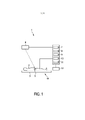
[024] A Figura 1 mostra, de forma esquemática e exemplificativa, uma modalidade de um sistema de braquiterapia 14 para aplicar a braquiterapia a uma pessoa 2 deitada em um meio de suporte 3, como uma mesa. O sistema de braquiterapia 14 é adaptado para realizar uma braquiterapia LDR, em que vários grupos de sementes de braquiterapia são introduzidos consecutivamente na pessoa 2 usando-se uma unidade de posicionamento 5. A unidade de posicionamento 5 é mostrada de forma esquemática e exemplificativa, mais detalhadamente, na Figura 2.[024] Figure 1 shows, in a schematic and exemplary way, a modality of a
[025] A unidade de posicionamento 5 compreende um elemento de introdução 17 para introduzir os vários grupos de sementes de braquiterapia, consecutivamente, na pessoa 2, de acordo com um plano de tratamento. Nessa modalidade, o elemento de introdução 17 é um cateter. Os vários grupos de sementes de braquiterapia podem ser manualmente introduzidos na pessoa 2 com o uso de um molde em formato de grade 13. Na Figura 2, o número de referência 18 indica uma mão de um operador, como um médico, introduzindo as sementes de braquiterapia na pessoa 2 usando o cateter 17. A unidade de posicionamento 5 compreende adicionalmente uma sonda de ultrassom 15, que é conectada a uma unidade de controle de ultrassom 7, para gerar uma imagem de ultrassom da pessoa 2. A sonda de ultrassom 15 e o molde em formato de grade 13 são ambos fixados a um elemento de suporte 16. A sonda de ultrassom 15 é de preferência uma sonda de ultrassonografia transretal (TRUS). A sonda TRUS 15 pode ser adaptada para capturar imagens tridimensionais ao vivo. Ela pode permitir uma captura automática de um volume inteiro, sem ter que deslocar mecanicamente a sonda TRUS 15. Entretanto, a sonda de ultrassom pode também ser uma sonda de ultrassom bidimensional, em particular, uma sonda TRUS bidimensional biplana, e, nesse caso, a sonda de ultrassom é de preferência transladada e/ou girada para reconstruir uma imagem tridimensional. Em particular, a sonda de ultrassom e a unidade de controle de ultrassom podem ser adaptadas para usar o direcionamento do feixe e a composição espacial para formar a imagem tridimensional. A seguir, diferentes técnicas de construção de imagem tridimensional serão descritas de forma exemplificativa, com mais detalhes.[025] The positioning unit 5 comprises an
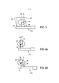
[026] A Figura 3 mostra de forma esquemática e exemplificativa duas sementes de braquiterapia 20, 21 que foram introduzidas na próstata 19 da pessoa 2. A sonda de ultrassom 115 mostrada na Figura 3 tem um transdutor de tamanho relativamente grande resultando em um campo de visão 24, 26, que cobre completamente a próstata 19. A Figura 3 ilustra duas direções de captura, isto é, duas direções do feixe de ultrassom, uma primeira direção 23 com o campo de visão correspondente 24 e uma segunda direção 25 com um campo de visão correspondente 26. Nesse exemplo, a imagem tridimensional é gerada pelas imagens compostas capturadas em diferentes ângulos do feixe, isto é, capturadas em diferentes direções de captura, sem alterar a posição da sonda de ultrassom 115. Cada semente de braquiterapia 20, 21 dentro do tecido da próstata 19 é interrogada por diferentes ângulos do feixe.[026] Figure 3 schematically and exemplarily shows two
[027] As Figuras 4a e 4b mostram uma outra modalidade de uma sonda de ultrassom 215, que tem um campo de visão menor 28, 30. O tamanho do transdutor e, portanto, os campos de visão 28, 30 não são suficientemente amplos para cobrir toda a região de interesse, isto é, nesta modalidade, a próstata completa 19. Entretanto, a sonda de ultrassom 215 também tem capacidades de direcionamento do feixe, e a sonda de ultrassom 215 pode ser deslocada em relação à próstata 19, de modo que uma imagem tridimensional pode ser construída através da composição espacial de diferentes imagens capturadas de diferentes posições da sonda. Em particular, a Figura 4a ilustra a captura de uma primeira imagem com o uso de uma primeira direção de captura 27 e um campo de visão correspondente 28, em uma primeira posição da sonda de ultrassom 215 em relação à próstata 19, e a Figura 4b ilustra a captura de uma segunda imagem em uma segunda direção de captura 29 com o uso de um campo de visão correspondente 30, em uma segunda posição da sonda de ultrassom 215 em relação à próstata 19. A diferença em relação às posições da sonda de ultrassom 215 em relação à próstata 19 é indicada na Figura 4a pela seta dupla 31.[027] Figures 4a and 4b show another embodiment of an
[028] Em um exemplo adicional durante a captura das imagens de ultrassom, o ângulo do feixe de luz pode ser fixado de modo que cada semente de braquiterapia é interrogada apenas por um ângulo do feixe de luz, sendo que a posição da sonda pode ser alterada para simular os efeitos do direcionamento do feixe. Os efeitos desejados do direcionamento do feixe, que podem ser obtidos direcionando-se o feixe usado para gerar a imagem de ultrassom ou simulando esse direcionamento do feixe, serão descritos a seguir, com referência às Figuras 5 e 6.[028] In an additional example during the capture of ultrasound images, the angle of the light beam can be fixed so that each brachytherapy seed is interrogated by only one angle of the light beam, and the position of the probe can be changed to simulate the effects of beam steering. The desired beam steering effects, which can be achieved by directing the beam used to generate the ultrasound image or by simulating such beam steering, will be described below, with reference to Figures 5 and 6.
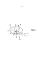
[029] A Figura 5 ilustra a captura de uma imagem de ultrassom com o uso da sonda de ultrassom 215 em uma primeira direção 33 e em uma segunda direção 36. Os campos de visão correspondentes são indicados na Figura 5 pelos números de referência 38 e 37, respectivamente. A semente de braquiterapia 32 reflete principalmente a onda de ultrassom na direção contrária da sonda de ultrassom 215, na direção indicada pela seta 34. O eco de baixa intensidade restante será em geral ocultado pelo ruído de fundo speckle na imagem capturada e não pode ser observado adequadamente na imagem. Entretanto, com o uso do direcionamento do feixe, a direção da captura pode ser alterada para a segunda direção de captura 35 sendo substancialmente perpendicular à semente de braquiterapia 32, de modo que o feixe de ultrassom é substancialmente refletido de volta em direção à sonda de ultrassom 215 na direção indicada na Figura 5 pelo número de referência 35. Dessa forma, com o uso do direcionamento do feixe, a visibilidade de uma semente de braquiterapia em uma imagem de ultrassom pode ser aumentada.[029] Figure 5 illustrates the capture of an ultrasound image using the
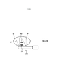
[030] A técnica de direcionamento do feixe também pode ser usada para resolver um possível problema presente de obstrução causado por uma semente de braquiterapia 40 localizada em uma sombra de uma outra semente de braquiterapia 39, conforme mostrado de forma exemplificativa na Figura 6. A imagem de ultrassom capturada em uma primeira direção com um campo de visão correspondente 38 não mostra claramente a semente de braquiterapia 40, porque está na sombra de outra semente de braquiterapia 39. Entretanto, uma imagem de ultrassom capturada em uma segunda direção com um campo de visão correspondente 37 também mostra a semente de braquiterapia 40 atrás de outra semente de braquiterapia 39. Dessa forma, a semente de braquiterapia 40 na sombra da outra semente de braquiterapia 39 é claramente visível na imagem de ultrassom obtida com um ângulo do feixe de luz, que corresponde à direção da captura com o campo de visão 37 na Figura 6.[030] The beam steering technique can also be used to solve a possible present problem of obstruction caused by a
[031] A sonda de ultrassom pode ser adaptada para capturar imagens de ultrassom bidimensionais ou tridimensionais, que foram capturadas com diferentes ângulos de feixe de luz, sendo que essas imagens de ultrassom bidimensionais ou tridimensionais podem ser compostas para construir uma imagem de ultrassom tridimensional. Entretanto, a sonda de ultrassom e a unidade de controle de ultrassom também podem ser adaptadas para reconstruir uma imagem de ultrassom tridimensional a partir de várias imagens de ultrassom bidimensionais. Em particular, a sonda de ultrassom e a unidade de controle de ultrassom podem ser adaptadas para construir uma imagem tridimensional pela reconstrução de cortes bidimensionais, sem direcionamento do feixe.[031] The ultrasound probe can be adapted to capture two-dimensional or three-dimensional ultrasound images, which were captured with different angles of light beam, and these two-dimensional or three-dimensional ultrasound images can be composited to build a three-dimensional ultrasound image. However, the ultrasound probe and ultrasound control unit can also be adapted to reconstruct a three-dimensional ultrasound image from multiple two-dimensional ultrasound images. In particular, the ultrasound probe and the ultrasound control unit can be adapted to build a three-dimensional image by reconstructing two-dimensional slices without beam steering.
[032] Por exemplo, a sonda de ultrassom e a unidade de controle de ultrassom podem ser adaptadas para gerar imagens de ultrassom bidimensionais, que não são compostas, mas usadas para reconstruir uma imagem de ultrassom tridimensional. Além disso, diferentes imagens de ultrassom bidimensionais, que representam diferentes planos dentro da pessoa 2, que são posicionadas e/ou orientadas de forma diferente, podem ser geradas e reconstruídas em uma imagem de ultrassom tridimensional. Além disso, a sonda de ultrassom e a unidade de controle de ultrassom podem ser adaptadas para gerar diferentes imagens de ultrassom bidimensionais compostas, que são reconstruídas em uma imagem de ultrassom tridimensional, sendo que a imagem de ultrassom bidimensional composta pode ser composta de várias imagens de ultrassom bidimensionais geradas com diferentes ângulos de feixe. As diferentes imagens de ultrassom bidimensionais compostas podem representar diferentes planos dentro da pessoa 2, que são posicionadas e/ou orientadas de maneira diferente, sendo que essas imagens de ultrassom bidimensionais compostas podem ser usadas para reconstruir a imagem de ultrassom tridimensional. A sonda de ultrassom pode também ser uma sonda de ultrassom tridimensional gerando diretamente uma imagem de ultrassom tridimensional com ou sem compor várias imagens de ultrassom tridimensionais. Se várias imagens de ultrassom tridimensionais são compostas, as várias imagens de ultrassom tridimensionais podem corresponder a diferentes subaberturas e/ou podem ser associadas a diferentes ângulos de direcionamento e/ou podem ser associadas a diferentes posições espaciais da sonda de ultrassom tridimensional. No último caso, a sonda de ultrassom tridimensional pode ser deslocada entre capturas de volume individuais.[032] For example, the ultrasound probe and ultrasound control unit can be adapted to generate two-dimensional ultrasound images, which are not composited but used to reconstruct a three-dimensional ultrasound image. In addition, different two-dimensional ultrasound images, which represent different planes within
[033] Novamente com referência à Figura 1, o sistema de braquiterapia 14 compreende uma exibição 12 para exibir, por exemplo, as imagens de ultrassom, determinadas posições das sementes de braquiterapia ou de outros elementos dentro da pessoa 2. Além disso, o sistema de braquiterapia 14 compreende uma unidade de rastreamento eletromagnético 6 para rastrear eletromagneticamente a posição do elemento de introdução 17 durante a introdução dos grupos de sementes de braquiterapia na pessoa 2. O cateter 17, em particular, a ponta do cateter 17, compreende um elemento eletromagnético correspondente, que pode ser rastreado pela unidade de rastreamento eletromagnético 6.[033] Again with reference to Figure 1, the
[034] A unidade de controle de ultrassom 7 é de preferência adaptada ao controle da geração da imagem de ultrassom, dependendo da posição rastreada do cateter 17. Em particular, se a sonda de ultrassom compreende um feixe de ultrassom direcionável para gerar a imagem de ultrassom, o feixe de ultrassom é de preferência controlado dependendo da posição rastreada do cateter 17, em particular, da ponta do cateter 17. De preferência, as posições das sementes de braquiterapia atuais, que foram introduzidas na pessoa 2 com o uso do cateter 17, são estimadas a partir das posições rastreadas do cateter 17 nos respectivos momentos de deposição. Por exemplo, a posição de uma semente de braquiterapia em relação à posição do elemento eletromagnético na ponta do cateter pode ser conhecida no momento da deposição e usada junto com a posição rastreada do elemento eletromagnético para estimar a posição da semente de braquiterapia atual depositada. Essas informações, isto é, a posição aproximada da semente de braquiterapia atual depositada, podem ser usadas para controlar o direcionamento do feixe do processo de imageamento por ultrassom para localizar exatamente a semente. Áreas afastadas da posição aproximada são de preferência excluídas da consideração durante o processo de detecção da semente. Em particular, com base nas posições aproximadas das sementes de braquiterapia, que já foram introduzidas na pessoa 2, o feixe de ultrassom pode ser direcionado, de modo que os problemas descritos acima, de reflexões na direção contrária à sonda de ultrassom e das sementes de braquiterapia na sombra de outras sementes de braquiterapia, são reduzidos, particularmente, eliminados. Por exemplo, se as posições aproximadas das sementes de braquiterapia indicarem uma situação, como mostrada de forma esquemática e exemplificativa na Figura 6, o feixe de ultrassom da sonda de ultrassom pode ser direcionado de modo que ao menos uma dentre as imagens, que são compostas para formar a imagem tridimensional, corresponda à direção de captura com o campo de visão indicado na Figura 6 pelo número de referência 37.[034] The
[035] As sementes de braquiterapia são depositadas através da inserção do cateter 17 preenchido com uma ou várias sementes de braquiterapia e opcionalmente espaçadores entre várias sementes de braquiterapia na pessoa. A uma ou várias sementes de braquiterapia devem ser dispostas em locais dentro da pessoa 2, que são definidos no plano de tratamento, o qual podem também ser considerado como um plano de dosagem. A unidade de rastreamento eletromagnético 6, junto com o elemento eletromagnético na ponta do cateter 17, pode auxiliar, nessa etapa de introdução, direcionando o usuário às localizações corretas, em que a localização atual do cateter 17 no momento da deposição da semente pode ser registrado e fornecido como uma estimativa da posição da semente para a unidade de controle do ultrassom 7 para permitir que a unidade de controle de ultrassom 7 realize um imageamento por ultrassom considerando essa posição da semente estimada.[035] The brachytherapy seeds are deposited by inserting the
[036] O sistema de braquiterapia 14 compreende adicionalmente uma unidade que determina a posição da semente 8 para determinar a posição de uma semente de braquiterapia, dependendo da imagem de ultrassom tridimensional gerada. De preferência, a unidade de ultrassom formada por uma das sondas de ultrassom mencionadas acima e a unidade de controle de ultrassom 7 são adaptadas para gerar uma imagem de ultrassom, antes de um grupo de sementes de braquiterapia ser introduzido na pessoa 2, e para gerar uma imagem de ultrassom atual, depois que o grupo foi introduzido na pessoa 2, sendo que a unidade que determina a posição da semente 8 é adaptada para alinhar as imagens de ultrassom, uma em relação à outra, para gerar uma imagem subtraída, através da subtração das imagens de ultrassom alinhadas uma à outra e para determinar a posição de uma semente de braquiterapia do grupo, dependendo da imagem subtraída gerada. O alinhamento é de preferência um alinhamento deformável. As posições determinadas de uma ou várias sementes de braquiterapia do grupo são de preferência usadas para determinar as posições desejadas das sementes de braquiterapia de grupos adicionais, que podem ser introduzidos na pessoa 2.[036] The
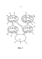
[037] Em particular, para determinar as posições de todas as sementes de braquiterapia, que já foram introduzidas na pessoa 2, a imagem de ultrassom atual e uma imagem de ultrassom anterior, que foi capturada depois de um grupo anterior e antes do grupo atual ser introduzido na pessoa 2, são alinhadas uma em relação à outra. Dessa forma, a última imagem do volume I n-1, que foi capturada antes da deposição da semente atual, é alinhada, em particular, elasticamente e/ou de forma deformável, com a imagem atual do volume I n, que foi capturada depois da deposição de semente atual, isto é, uma transformação de alinhamento T n-i~n, é calculada. Para realizar esse procedimento de alinhamento, algoritmos de alinhamento conhecidos podem ser usados, como o algoritmo “Demons” ou uma variante desse algoritmo, que é revelada, por exemplo, no artigo “Understanding the “Demon’s Algorithm”: 3D Non-rigid Registration by Gradient Descent” by X. Pennec et al., Medical Image Computing and Computer- Assisted Intervention - MICCAI'99, páginas de 597 a 606 (1999), que está incorporado neste documento a título de referência. As imagens alinhadas podem então ser subtraídas umas das outras para gerar uma imagem subtraída. Uma imagem diferencial é, portanto, criada, na qual todos os recursos presentes em ambas as imagens são eliminados, acentuando, assim, os recursos introduzidos entre as duas capturas de imagem, isto é, acentuando, assim, as sementes de braquiterapia que foram introduzidas na pessoa 2 durante a deposição de semente atual. As posições das sementes de braquiterapia do grupo atual introduzido podem então ser determinadas na imagem subtraída utilizando-se, por exemplo, técnicas de segmentação conhecidas. Esse conceito de imageamento incremental para detectar a posição de uma semente de braquiterapia implantada mais recentemente é ilustrado na Figura 7.[037] In particular, to determine the positions of all brachytherapy seeds, which have already been introduced to
[038] Na Figura 7, uma imagem anterior I n-1 mostra as sementes de braquiterapia 52 que já foram implantadas e as microcalcificações 51 dentro da próstata 19. Uma imagem atual I n também mostra as sementes de braquiterapia 52 e as microcalcificações 51 visíveis na imagem anterior I n-1. Entretanto, além disso, a imagem atual I n também mostra uma semente de braquiterapia atual implantada 53. Além disso, a imagem atual I n mostra um edema na próstata, que cresceu de um tamanho anterior indicado pela linha tracejada 50 para um tamanho atual indicado pela linha contínua 19. Nesse exemplo, a imagem anterior I n-1 é deformada para a imagem deformada anterior I’ n-1 alinhando a imagem anterior I n-1 com a imagem atual I n. A imagem deformada anterior I’ n-1 é então subtraída da imagem atual I n para gerar uma imagem subtraída I n,s, na qual a semente de braquiterapia recém-introduzida 53 é visível de forma acentuada. Em um outro exemplo, alternativa ou adicionalmente, a imagem anterior também I n-1 pode ser deformada durante o processo de alinhamento.[038] In Figure 7, a previous image I n-1 shows brachytherapy
[039] Essa técnica de imageamento incremental pode evitar classificações errôneas de microcalcificações e outros artefatos de imagem de cor intensa, como sementes de braquiterapia. Qualquer artefato parecido com semente em uma imagem de ultrassom base, que foi capturada antes do cateter 17 ser inserido na pessoa 2 pela primeira vez, e que pode também ser considerada uma imagem de ultrassom inicial, é genericamente visível também em imagens de ultrassom subsequentes capturadas depois de cada inserção do cateter 17 para introduzir um grupo formado de uma ou mais sementes de braquiterapia na pessoa 2. Como cada imagem de ultrassom subsequente é alinhada de forma deformável com a imagem de ultrassom anterior e como imagens subtraídas entre sucessivas imagens são obtidas, os artefatos parecidos com semente causados, por exemplo, por microcalcificações, podem ser excluídos e não identificados erroneamente como sementes de braquiterapia inseridas. Além disso, essa técnica incremental pode, ao menos parcialmente, suprimir o ruído de fundo speckle, que adicionalmente acentuará a visibilidade de uma ou várias sementes de braquiterapia atuais implantadas. Além disso, alinhando-se de forma deformável as imagens capturadas antes e depois da última inserção do cateter para introduzir o grupo atual de uma ou várias sementes de braquiterapia, as posições das sementes de braquiterapia que foram implantadas anteriormente a uma distância maior da sonda de ultrassom podem ser preservadas, mesmo que essas sementes anteriores estejam localizadas na sombra das sementes de braquiterapia atuais recém- implantadas, que podem ser mais proximais. Dessa forma, a técnica incremental que envolve imageamento incremental e determinação incremental das posições das sementes de braquiterapia pode também ser usada para resolver o problema de obstrução descrito acima com referência à Figura 6.[039] This incremental imaging technique can avoid misclassifications of microcalcifications and other intense color imaging artifacts such as brachytherapy seeds. Any seed-like artifact in a baseline ultrasound image, which was captured before the
[040] Para se determinar as posições de todas a sementes de braquiterapia introduzidas na pessoa 2, a unidade que determina a posição da semente 8 é adaptada adicionalmente para fornecer as posições das sementes de braquiterapia de grupos que já foram introduzidos na pessoa 2, antes que o grupo atual tenha sido introduzido, e para combinar as posições determinadas atualmente e as posições fornecidas para determinar as posições de todos os grupos, isto é, de todas as sementes de braquiterapia de todos os grupos, já introduzidos na pessoa 2. Por exemplo, sementes recém-identificadas S n,n, isto é, o enésimo grupo de novas sementes no sistema de coordenadas da imagem atual I n, podem ser armazenadas, sendo que essas novas sementes S n,n podem ser combinadas com todas as sementes anteriores S total,n-1 presentes na imagem anterior I n-1 la, em que a transformação do alinhamento Tn-i^n pode ser aplicada às coordenadas das posições das sementes anteriores S total,n-1, de acordo com as seguintes equações:
[041] O sistema de braquiterapia 14 compreende adicionalmente uma unidade que determina a região de interesse 9 para determinar uma região de interesse a ser tratada dentro da pessoa 2 dependendo da imagem de ultrassom atual gerada, para permitir uma introdução de grupos adicionais que também dependem da região de interesse que foi determinada com base na imagem de ultrassom atual. A região de interesse é, por exemplo, a próstata ou uma parte da próstata. Em particular, a região de interesse pode ser uma região de tumor dentro da próstata. Ela pode ser determinada por técnicas de segmentação conhecidas com base na imagem de ultrassom atual.[041] The
[042] O sistema de braquiterapia 14 compreende adicionalmente uma unidade que fornece um plano de tratamento 10 para fornecer um plano de tratamento definindo uma disposição espacial das sementes de braquiterapia dentro da pessoa 2, sendo que os grupos de sementes de braquiterapia são introduzidos na pessoa 2 com o uso do cateter 17 de acordo com o plano de tratamento. Uma unidade de atualização do plano de tratamento 11 atualiza o plano de tratamento com base nas posições determinadas das sementes de braquiterapia dos grupos já introduzidos na pessoa 2, com base na região de interesse, isto é, a localização e/ou formato da região de interesse determinada com o uso da imagem de ultrassom atual e, opcionalmente, também com base na localização e/ou formato de elementos adicionais da pessoa 2, como vasos, órgãos etc., que podem também ser determinados com base na imagem de ultrassom atual.[042] The
[043] O plano de tratamento fornecido, isto é, o plano de tratamento inicial, depende, entre outros, da região de interesse, conforme mostrado na imagem de ultrassom base que foi capturada antes de qualquer grupo ser introduzido na pessoa 2. A unidade que determina a região de interesse 9 pode ser adaptada para gerar uma transformação de alinhamento que alinha a imagem de ultrassom base e a imagem de ultrassom atual, uma em relação à outra, e para atualizar a região de interesse mostrada na imagem de ultrassom base com o uso da transformação de alinhamento, sendo que a unidade de atualização do plano de tratamento 11 pode ser adaptada para atualizar o plano de tratamento com base nas posições determinadas das sementes de braquiterapia dos grupos já introduzidos na pessoa 2 e com base nessa região de interesse atualizada. O alinhamento pode também ser usado para atualizar a localização e formato de elementos adicionais, que são mostrados na imagem de ultrassom base e que podem ter sido usados para determinar o plano de tratamento inicial, sendo que o plano de tratamento pode ser atualizado também com base nessas informações atualizadas. O alinhamento da imagem de ultrassom atual com a imagem de ultrassom base pode ser um alinhamento direto, sendo que a imagem de ultrassom atual é diretamente alinhada com a imagem de ultrassom base ou um alinhamento indireto ou incremental, e que a imagem de ultrassom base e as imagens de ultrassom subsequente e consecutivamente capturadas são alinhadas aos pares, uma em relação à outra, formando assim uma sequência de alinhamentos incrementais definindo uma transformação de alinhamento entre a imagem de ultrassom base e a imagem de ultrassom atual. O plano de tratamento, que pode também ser considerado como plano de dosagem, define em que posições as sementes de braquiterapia devem ser colocadas. O plano de tratamento de preferência define adicionalmente a dose de radiação que a respectiva semente de braquiterapia deve aplicar à pessoa.[043] The treatment plan provided, ie the initial treatment plan, depends on, among others, the region of interest, as shown in the baseline ultrasound image that was captured before any group was introduced to
[044] Para determinar o plano de tratamento inicial, diferentes elementos mostrados na imagem de ultrassom base são, de preferência, segmentados. Por exemplo, a próstata ou uma parte da próstata que forma a região de interesse, o reto, a uretra, a bexiga etc., são segmentadas, sendo que o plano de tratamento inicial é determinado de preferência de modo que uma dose-alvo prescrita é aplicada à região de interesse, enquanto a dose da radiação aplicada às outras estruturas é mantida no mínimo. O procedimento de segmentação e/ou a determinação do plano de tratamento com base nas segmentações resultantes pode ser realizado manualmente, semiautomaticamente ou completamente automaticamente.[044] To determine the initial treatment plan, different elements shown in the baseline ultrasound image are preferably segmented. For example, the prostate or a part of the prostate that forms the region of interest, rectum, urethra, bladder, etc., is segmented, with the initial treatment plan being preferably determined so that a prescribed target dose is applied to the region of interest, while the radiation dose applied to other structures is kept to a minimum. The segmentation procedure and/or the determination of the treatment plan based on the resulting segmentations can be performed manually, semiautomatically or completely automatically.
[045] A transformação de alinhamento Tn i n pode também ser usada para atualizar as segmentações, em particular, os formatos e as localizações de todas as segmentações da imagem de ultrassom anterior para a imagem de ultrassom atual, sendo que, com base nessas segmentações atualizadas e todas as localizações das sementes de braquiterapia já colocadas S n total, a unidade de atualização do plano de tratamento 11 pode atualizar o plano de tratamento. De preferência, a unidade de atualização do plano de tratamento 11 leva em consideração a dose de radiação já fornecida, que é definida pelas posições das sementes de braquiterapia já colocadas e seus tempos de permanência, e as doses-alvo de radiação prescritas. As posições das sementes de braquiterapia que ainda precisam ser fornecidas para a obtenção das doses-alvo de radiação prescritas são determinadas com base na dose de radiação já fornecida. Essa atualização do plano de tratamento é de preferência realizada sempre que uma imagem de ultrassom incremental I n tenha sido capturada. O plano de tratamento atual atualizado de forma adaptativa pode diferir ou não do plano de tratamento anterior atualizado de forma adaptativa. Se forem necessárias mais sementes de braquiterapia de acordo com o plano de tratamento mais recente, a inserção da semente de braquiterapia continua com a (n+1)ésima iteração de inserção do cateter e imageamento. Caso contrário, o procedimento é concluído.[045] The Tn in n alignment transformation can also be used to update the slices, in particular the shapes and locations of all slices from the previous ultrasound image to the current ultrasound image, and based on these updated slices and all brachytherapy seed locations already placed S n total, the treatment
[046] Em vez de realizar os alinhamentos incrementais I n-i ^ I n, todas as imagens de ultrassom podem ser alinhadas novamente com a imagem de ultrassom de referência I 0. Ou, para maior consistência entre todas as transformações de alinhamento, os alinhamentos podem ser realizados em conjunto e simultaneamente entre I n-1 e I n, assim como entre I n e I 0. É importante levar em consideração a imagem de ultrassom base I 0 durante o procedimento de alinhamento para evitar erros que podem induzir o desenvolvimento de sucessivos alinhamentos de imagem incremental.[046] Instead of performing incremental alignments I n-i ^ I n, all ultrasound images can be re-aligned with the reference ultrasound image I 0. Or, for greater consistency across all alignment transformations, alignments can be performed together and simultaneously between I n-1 and I n, as well as between I n and I 0. It is important to consider the base ultrasound image I 0 during the alignment procedure to avoid errors that can induce the development of successive incremental image alignments.
[047] As duas modalidades de métodos de braquiterapia a seguir serão descritas de forma exemplificativa com referência aos fluxogramas mostrados nas Figuras 8 e 9. Uma primeira modalidade ilustrada na Figura 8 usa o alinhamento de imagem incremental, e uma segunda modalidade ilustrada na Figura 9 usa um alinhamento com a imagem de ultrassom base, sendo que os alinhamentos são usados para acompanhar as segmentações, em particular, as segmentações dos órgãos, e todas as localizações de sementes, isto é, todas as posições das sementes, por todo o procedimento de inserção.[047] The following two modalities of brachytherapy methods will be described by way of example with reference to the flowcharts shown in Figures 8 and 9. A first modality illustrated in Figure 8 uses incremental image alignment, and a second modality illustrated in Figure 9 uses an alignment to the baseline ultrasound image, the alignments being used to track segmentations, in particular organ segmentations, and all seed locations, i.e. all seed positions, throughout the entire seeding procedure. insertion.
[048] Após o método de braquiterapia ter iniciado na etapa 301, na etapa 302 um volume de TRUS tridimensional inicial, isto é, a imagem de ultrassom base I 0, é capturada. Na etapa 303, as segmentações são criadas na imagem de ultrassom base I 0 e um plano de tratamento inicial é determinado com base nas segmentações criadas e nas doses- alvo de radiação prescritas. Na etapa 304, um primeiro grupo que compreende uma ou várias sementes de braquiterapia é introduzido na pessoa 2 com o uso de um elemento de introdução, como um cateter ou uma agulha, de acordo com o plano de tratamento, sendo que a posição do elemento de introdução é rastreada eletromagneticamente durante a introdução do grupo atual na pessoa, especificamente, no momento de deposição de uma ou várias sementes de braquiterapia do grupo atual. Depois que o grupo é depositado, o elemento de introdução é retirado da pessoa. Em seguida, na etapa 305 o volume de TRUS tridimensional do enésimo intraprocedimento I n é capturado, sendo que durante a primeira iteração n é 1. Durante a captura do volume de TRUS tridimensional do intraprocedimento I n, a geração da imagem de ultrassom é controlada dependendo da posição do elemento de introdução durante o procedimento de introdução rastreado na etapa 304. Na etapa 306, a imagem atual I n capturada na etapa 305 é alinhada com a imagem anterior I n-1 para gerar uma transformação de alinhamento T n-i^n. As imagens alinhadas são subtraídas umas das outras para criar uma enésima imagem subtraída I n,s, na etapa 307. Na etapa 308, novas sementes de braquiterapia são detectadas na imagem subtraída I n,s. Na etapa 309, a transformação de alinhamento T n n é usada para atualizar localizações de semente anteriores para a imagem de ultrassom atual I n, sendo que na etapa 310, as sementes de braquiterapia novas e anteriores, isto é, as posições da semente de braquiterapia novas e anteriores, são combinadas no sistema de coordenadas da imagem de ultrassom atual I n. Na etapa 311, a transformação de alinhamento T n n é usada para atualizar as segmentações, e na etapa 312 a dose de radiação aplicada, isto é, a dose de radiação fornecida pelas novas sementes de braquiterapia e as sementes de braquiterapia anteriores, é calculada. Na etapa 313, o plano de tratamento para posicionar as sementes de braquiterapia restantes é atualizado. Na etapa 314, é determinado se sementes de braquiterapia adicionais precisam ser depositadas. Se esse for o caso, o método prossegue com a etapa 304. Caso contrário, o método termina na etapa 315.[048] After the brachytherapy method has started at
[049] As etapas de 401 a 408 da segunda modalidade do método de braquiterapia ilustrado na Figura 9 são similares às etapas de 301 a 308 descritas acima com referência à Figura 8. Além disso, na etapa 409, a imagem de ultrassom atual I n é alinhada com a imagem de ultrassom base I o para procurar uma transformação de alinhamento T —. Na etapa 410 a transformação T -n é usada para trazer as posições de semente anteriores de um sistema de coordenadas definido por I o para um sistema de coordenadas definido por I n. Na etapa 411, a transformação de alinhamento T o-n é usada para atualizar as segmentações e na etapa 412 uma transformação da região inversa T n-o é calculada e usada para transformar as novas posições da semente para o sistema de coordenadas de I o. As etapas restantes de 413 a 416 correspondem às etapas 312 a 315 descritas acima com referência à Figura 8.[049]
[050] Algumas etapas do método de braquiterapia descrito acima com referência às Figuras 8 e 9 podem ser consideradas etapas de um método auxiliar para assistir na realização de uma braquiterapia. Por exemplo, o rastreamento eletromagnético da posição do elemento de introdução nas etapas 304 e 404 e etapas de 305 a 315 e de 405 a 416, respectivamente, pode ser considerado como etapas do método auxiliar. De modo correspondente, a unidade de rastreamento eletromagnético 6, a unidade de ultrassom 7, 15, a unidade que determina a posição da semente 8, a unidade que determina a região de interesse 9, a unidade que fornece o plano de tratamento 10, que pode ser apenas uma unidade de armazenamento, que armazena um plano de tratamento inicial já determinado e as segmentações iniciais e que fornece o plano de tratamento armazenado e as segmentações, e a unidade de atualização do plano de tratamento 11, podem ser consideradas como formadoras de um aparelho auxiliar 1 para assistir na realização de uma braquiterapia.[050] Some steps of the brachytherapy method described above with reference to Figures 8 and 9 can be considered steps of an auxiliary method to assist in performing a brachytherapy. For example, electromagnetic tracking of the position of the input element in
[051] Atualizando-se as segmentações, um inchaço do objeto, no qual o elemento de introdução é inserido, em particular, de uma próstata, também pode ser considerado durante a inserção das sementes de braquiterapia restantes. Em particular, o volume da próstata em geral aumenta durante o procedimento do tratamento, como resultado do trauma causado pela inserção do elemento de introdução. O alinhamento deformável das imagens capturadas ao longo do tempo permite ao sistema de braquiterapia medir o inchaço da próstata e adaptar o plano de tratamento para compensar o volume aumentado da próstata. O alinhamento deformável pode, portanto, também ser usado para detectar e medir o edema.[051] Updating the segmentations, a swelling of the object, into which the introducing element is inserted, in particular from a prostate, can also be considered during the insertion of the remaining brachytherapy seeds. In particular, the volume of the prostate usually increases during the treatment procedure as a result of the trauma caused by the insertion of the introducer element. The deformable alignment of captured images over time allows the brachytherapy system to measure prostate swelling and adapt the treatment plan to compensate for the enlarged prostate volume. Deformable alignment can therefore also be used to detect and measure edema.
[052] A braquiterapia LDR pode ser usada para tratar o câncer de próstata em estágios iniciais e compreende uma colocação permanente de sementes de braquiterapia radioativas dentro da próstata para matar as células cancerosas através de radiação. As posições dessas sementes de braquiterapia são planejadas precisamente antes da cirurgia para assegurar uma cobertura suficiente da glândula-alvo, enquanto resguarda os órgãos em risco, como a uretra e o reto. Uma colocação precisa das sementes de braquiterapia leva a um melhor resultado do tratamento e menor toxicidade. Entretanto, em sistemas de braquiterapia conhecidos, desvios do plano de modo geral estão presentes devido a problemas, como movimento e deformação da próstata causados pela inserção da agulha ou cateter, edema na próstata, flexão da agulha ou cateter etc. O inchaço da próstata causado pelo trauma da inserção da agulha ou cateter é uma grande fonte de erro, pois o aumento da próstata não é levado em consideração no plano de tratamento inicial.[052] LDR brachytherapy can be used to treat early-stage prostate cancer and comprises a permanent placement of radioactive brachytherapy seeds inside the prostate to kill cancer cells through radiation. The positions of these brachytherapy seeds are planned precisely before surgery to ensure sufficient coverage of the target gland, while protecting organs at risk such as the urethra and rectum. Precise placement of brachytherapy seeds leads to a better treatment outcome and less toxicity. However, in known brachytherapy systems, deviations from the plane are generally present due to problems such as movement and deformation of the prostate caused by needle or catheter insertion, swelling in the prostate, bending of the needle or catheter, etc. Prostate swelling caused by trauma from needle or catheter insertion is a major source of error as prostate enlargement is not considered in the initial treatment plan.
[053] O sistema de braquiterapia descrito acima com referência a, por exemplo, Figura 1, fornece portanto, de preferência, uma dosimetria dinâmica e técnica de planejamento, que compreendem uma localização precisa e em tempo real de sementes de braquiterapia já implantadas e uma modificação das posições planejadas e do número de sementes de braquiterapia restantes, para cobrir as regiões não radiadas suficientemente e evitar o excesso de radiação sobre os órgãos em risco. O sistema de braquiterapia é particularmente adaptado para evitar obstáculos na detecção de sementes com o uso de ultrassom: a) a criação de uma sombra na imagem de ultrassom por sementes altamente ecogênicas que pode obstruir outras sementes localizadas na área de sombra, b) a dispersão de uma onda de ultrassom que chega através de uma semente na direção contrária da sonda de ultrassom de modo que a semente pareça menos intensa e seja distinguível do ruído de fundo e c) artefatos de imagem parecidos com semente causados, por exemplo, por microcalcificações na próstata. Para evitar esses obstáculos, o sistema de braquiterapia é adaptado de preferência para realizar o imageamento incremental, que é combinado com o direcionamento de feixe e o alinhamento deformável elástico. Como as sementes de braquiterapia são implantadas sequencialmente, a dosimetria em tempo real e o planejamento podem permitir que o médico modifique as posições e o número de sementes de braquiterapia restantes, intraoperacionalmente, para compensar os possíveis erros.[053] The brachytherapy system described above with reference to, for example, Figure 1, therefore preferably provides a dynamic dosimetry and planning technique, which comprises a precise and real-time location of already implanted brachytherapy seeds and a modification of the planned positions and the number of brachytherapy seeds remaining, to cover areas not sufficiently radiated and to avoid excess radiation on organs at risk. The brachytherapy system is particularly adapted to avoid obstacles in the detection of seeds with the use of ultrasound: a) the creation of a shadow on the ultrasound image by highly echogenic seeds that can obstruct other seeds located in the shadow area, b) the dispersion of an ultrasound wave arriving through a seed in the opposite direction of the ultrasound probe so that the seed appears less intense and is distinguishable from the background noise and c) seed-like image artifacts caused, for example, by microcalcifications in the prostate . To avoid these obstacles, the brachytherapy system is preferably adapted to perform incremental imaging, which is combined with beam steering and elastic deformable alignment. As the brachytherapy seeds are implanted sequentially, real-time dosimetry and planning can allow the clinician to modify the positions and number of brachytherapy seeds remaining intraoperatively to compensate for potential errors.
[054] O imageamento incremental é de preferência usado para identificar as posições das sementes de braquiterapia recém-implantadas, já que as sementes de braquiterapia são implantadas sequencialmente. O sistema de braquiterapia pode ser adaptado para obter uma imagem tridimensional, por exemplo, da próstata antes e depois da deposição de cada semente de braquiterapia, ou antes e depois da deposição de todas as sementes de braquiterapia, dentro de uma agulha ou cateter. Um grupo de sementes de braquiterapia pode ser definido por uma única semente de braquiterapia apenas ou por várias sementes de braquiterapia, sendo que as várias sementes de braquiterapia podem ser sementes de braquiterapia dentro de uma agulha ou cateter. A imagem tridimensional obtida antes e depois da respectiva deposição da semente de braquiterapia pode ser produzida pela combinação espacial de várias imagens bidimensionais ou tridimensionais capturadas em diferentes ângulos de feixe.[054] Incremental imaging is preferably used to identify the positions of newly implanted brachytherapy seeds, as brachytherapy seeds are implanted sequentially. The brachytherapy system can be adapted to obtain a three-dimensional image, for example, of the prostate before and after the deposition of each brachytherapy seed, or before and after the deposition of all the brachytherapy seeds, inside a needle or catheter. A group of brachytherapy seeds can be defined by a single brachytherapy seed alone or by several brachytherapy seeds, with the various brachytherapy seeds being brachytherapy seeds inside a needle or catheter. The three-dimensional image obtained before and after the respective deposition of the brachytherapy seed can be produced by the spatial combination of several two-dimensional or three-dimensional images captured at different beam angles.
[055] O rastreamento eletromagnético do elemento de introdução, em particular, da ponta do elemento de introdução, no momento da deposição da respectiva semente de braquiterapia, leva a uma posição estimada da semente de braquiterapia. A posição estimada pode ser usada para reduzir a área de pesquisa, na identificação da semente de braquiterapia na imagem subtraída, conforme descrito acima com referência, por exemplo, à Figura 7.[055] Electromagnetic tracking of the introducing element, in particular the tip of the introducing element, at the time of deposition of the respective brachytherapy seed, leads to an estimated position of the brachytherapy seed. The estimated position can be used to reduce the search area, in identifying the brachytherapy seed in the subtracted image, as described above with reference, for example, to Figure 7.
[056] Embora nas modalidades descritas acima a braquiterapia seja aplicada principalmente na próstata, a braquiterapia também pode ser aplicada a outras partes, em particular, a outros órgãos, de um ser humano ou animal.[056] While in the modalities described above brachytherapy is applied primarily to the prostate, brachytherapy can also be applied to other parts, in particular other organs, of a human or animal.
[057] Embora nas modalidades descritas acima o rastreamento seja realizado com o uso de rastreamento eletromagnético, a unidade de rastreamento pode também ser adaptada para usar uma outra técnica de rastreamento para rastrear a posição do elemento de introdução, como rastreamento por sensor em forma de fibra óptica.[057] Although in the modalities described above the tracking is performed using electromagnetic tracking, the tracking unit can also be adapted to use another tracking technique to track the position of the input element, such as sensor tracking in the form of a optical fiber.
[058] Outras variações das modalidades apresentadas podem ser entendidas e realizadas pelos versados na técnica na prática de uma invenção reivindicada a partir de um grupo de desenhos, da revelação e das reivindicações anexas.[058] Other variations of the modalities presented can be understood and carried out by those skilled in the art in the practice of a claimed invention from a group of drawings, the disclosure and the appended claims.
[059] Nas reivindicações, a palavra “que compreende” não exclui outros elementos ou etapas, e o artigo indefinido “um” ou “uma” não exclui uma pluralidade.[059] In the claims, the word "comprising" does not exclude other elements or steps, and the indefinite article "a" or "an" does not exclude a plurality.
[060] Uma única unidade ou dispositivo pode executar as funções de vários itens mencionados novamente nas reivindicações. O simples fato de que certas medidas são mencionadas novamente em reivindicações dependentes mutuamente diferentes não indica que uma combinação dessas medidas não possa ser usada em seu benefício.[060] A single unit or device may perform the functions of multiple items mentioned again in the claims. The mere fact that certain measures are mentioned again in mutually different dependent claims does not indicate that a combination of these measures cannot be used to your benefit.
[061] Os procedimentos, como a determinação de uma transformação de alinhamento, os procedimentos de segmentação, os procedimentos de atualização etc., realizados por uma ou várias unidades ou dispositivos podem ser realizados por qualquer outro número de unidades ou dispositivos. Por exemplo, as etapas de 306 a 313 e de 406 a 414 podem ser realizadas por uma única unidade ou por qualquer outro número de unidades diferentes. Os procedimentos e/ou o controle do aparelho auxiliar de acordo com o método auxiliar e/ou o controle do sistema de braquiterapia de acordo com o método de braquiterapia podem ser implementados como métodos de código de programação de um programa de computador e/ou como hardware dedicado.[061] Procedures such as determining an alignment transformation, segmentation procedures, update procedures, etc., performed by one or more units or devices can be performed by any other number of units or devices. For example, steps 306 to 313 and 406 to 414 can be performed by a single unit or by any number of different units. The procedures and/or control of the auxiliary apparatus according to the auxiliary method and/or the control of the brachytherapy system according to the brachytherapy method may be implemented as methods of programming a computer program code and/or as dedicated hardware.
[062] Um programa de computador pode ser armazenado e/ou distribuído em um meio adequado, como um meio de armazenamento óptico ou um meio em estado sólido fornecidos juntos, com ou como parte de outro hardware, mas pode também ser distribuído de outras formas, como através da Internet ou outro sistema de telecomunicação com fio ou sem fio.[062] A computer program may be stored and/or distributed on a suitable medium, such as an optical storage medium or a solid-state medium provided together, with or as part of other hardware, but it may also be distributed in other ways. , such as over the Internet or other wired or wireless telecommunication system.
[063] Qualquer sinal de referência nas reivindicações não deve ser interpretado como limitador do escopo.[063] Any reference sign in the claims should not be interpreted as limiting the scope.
[064] A invenção está relacionada a um aparelho auxiliar para assistir na realização da braquiterapia. A posição de um elemento de introdução, como um cateter, é rastreada particularmente pelo uso de um rastreamento eletromagnético, enquanto um grupo de sementes é introduzido em um objeto vivo. Isso fornece um conhecimento aproximado sobre a posição das sementes dentro do objeto. Uma imagem de ultrassom mostrando o grupo é gerada, dependendo da posição rastreada do elemento de introdução e, dessa forma, dependendo do conhecimento aproximado sobre a posição das sementes, para otimizar a visualização do ultrassom no que tange a mostrar as sementes introduzidas. Com base nessa visualização do ultrassom, a posição de uma semente do grupo é determinada, permitindo, assim, uma determinação aprimorada das posições das sementes e, de modo correspondente, de uma braquiterapia aprimorada realizada com base nas posições determinadas.[064] The invention relates to an auxiliary device to assist in performing brachytherapy. The position of an introducing element, such as a catheter, is tracked particularly by the use of electromagnetic tracking, while a group of seeds is introduced into a living object. This provides approximate knowledge about the position of the seeds within the object. An ultrasound image showing the group is generated depending on the tracked position of the introduction element and thus depending on the approximate knowledge about the position of the seeds, to optimize the ultrasound visualization in terms of showing the introduced seeds. Based on this ultrasound visualization, the position of a seed in the group is determined, thus allowing an improved determination of seed positions and correspondingly improved brachytherapy performed based on the determined positions.
Claims (12)
Applications Claiming Priority (3)
| Application Number | Priority Date | Filing Date | Title |
|---|---|---|---|
| US201261735669P | 2012-12-11 | 2012-12-11 | |
| US61/735,669 | 2012-12-11 | ||
| PCT/IB2013/059989 WO2014091330A1 (en) | 2012-12-11 | 2013-11-08 | Assisting apparatus for assisting in performing a brachytherapy |
Publications (2)
| Publication Number | Publication Date |
|---|---|
| BR112015013247A2 BR112015013247A2 (en) | 2017-07-11 |
| BR112015013247B1 true BR112015013247B1 (en) | 2022-07-12 |
Family
ID=49709786
Family Applications (1)
| Application Number | Title | Priority Date | Filing Date |
|---|---|---|---|
| BR112015013247-2A BR112015013247B1 (en) | 2012-12-11 | 2013-11-08 | AUXILIARY DEVICE TO ASSIST IN THE PERFORMANCE OF BRACHYTHERAPY, BRACHYTHERAPY SYSTEM TO APPLY BRACHYTHERAPY TO A LIVE OBJECT, AND AUXILIARY METHOD TO ASSIST IN THE PERFORMANCE OF BRACHYTHERAPY |
Country Status (6)
| Country | Link |
|---|---|
| US (1) | US10549123B2 (en) |
| EP (1) | EP2931370B1 (en) |
| JP (1) | JP6726966B2 (en) |
| CN (1) | CN104837525B (en) |
| BR (1) | BR112015013247B1 (en) |
| WO (1) | WO2014091330A1 (en) |
Families Citing this family (15)
| Publication number | Priority date | Publication date | Assignee | Title |
|---|---|---|---|---|
| ES2813875T3 (en) * | 2014-01-01 | 2021-03-25 | Medivation Tech Llc | Compounds and procedures for use |
| EP3096688A1 (en) * | 2014-01-24 | 2016-11-30 | Koninklijke Philips N.V. | System and method for three-dimensional quantitative evaluation of uterine fibroids |
| KR20220065894A (en) * | 2014-07-28 | 2022-05-20 | 인튜어티브 서지컬 오퍼레이션즈 인코포레이티드 | Systems and methods for intraoperative segmentation |
| CN106659473B (en) * | 2014-07-29 | 2020-04-21 | 皇家飞利浦有限公司 | Ultrasonic imaging apparatus |
| EP3197554B1 (en) * | 2014-09-26 | 2020-09-09 | Koninklijke Philips N.V. | Dual localization of brachytherapy radioactive seeds |
| WO2016059603A1 (en) | 2014-10-17 | 2016-04-21 | Koninklijke Philips N.V. | System for real-time organ segmentation and tool navigation during tool insertion in interventional therapy and method of opeperation thereof |
| US10290098B2 (en) | 2014-10-17 | 2019-05-14 | Koninklijke Philips N.V. | System for real-time organ segmentation and tool navigation during tool insertion in interventional therapy and method of operation thereof |
| US10933254B2 (en) | 2015-09-01 | 2021-03-02 | Koninklijke Philips N.V. | Device and method for assisting in a brachytherapy procedure |
| WO2017162820A1 (en) * | 2016-03-24 | 2017-09-28 | Koninklijke Philips N.V. | Treatment assessment device |
| WO2018108952A1 (en) * | 2016-12-16 | 2018-06-21 | Koninklijke Philips N.V. | Positioning assistance device for focal radiation therapy |
| EP3554383B1 (en) * | 2016-12-16 | 2020-04-22 | Koninklijke Philips N.V. | System for providing images for guiding surgery |
| US11857370B2 (en) * | 2017-03-20 | 2024-01-02 | National Bank Of Canada | Method and system for visually assisting an operator of an ultrasound system |
| EP3381512A1 (en) * | 2017-03-30 | 2018-10-03 | Koninklijke Philips N.V. | Determining at least one final two-dimensional image for visualizing an object of interest in a three-dimensional ultrasound volume |
| EP3547323A1 (en) * | 2018-03-30 | 2019-10-02 | Koninklijke Philips N.V. | Brachytherapy treatment planning system |
| CN111369479B (en) * | 2019-11-12 | 2023-04-25 | 北京金阳普泰石油技术股份有限公司 | Local automatic updating method and device for sedimentary phase belt diagram |
Family Cites Families (11)
| Publication number | Priority date | Publication date | Assignee | Title |
|---|---|---|---|---|
| AU6639900A (en) * | 1999-08-13 | 2001-03-13 | Advanced Sensor Technologies Llc | Probe position sensing system for use in a coordinate measuring machine |
| US6610013B1 (en) | 1999-10-01 | 2003-08-26 | Life Imaging Systems, Inc. | 3D ultrasound-guided intraoperative prostate brachytherapy |
| US6438401B1 (en) | 2000-04-28 | 2002-08-20 | Alpha Intervention Technology, Inc. | Indentification and quantification of needle displacement departures from treatment plan |
| EP1725171B1 (en) * | 2004-03-09 | 2015-04-15 | Robarts Research Institute | An apparatus and computing device for performing brachytherapy and methods of imaging using the same |
| DE102006045423B4 (en) * | 2006-09-26 | 2016-07-14 | Siemens Healthcare Gmbh | 07.09.07 Method for postprocessing a three-dimensional image data set of a vessel structure |
| EP2203218B1 (en) | 2007-10-26 | 2013-02-20 | Koninklijke Philips Electronics N.V. | Electromagnetic pose sensing of hdr brachytherapy applicator |
| CN201483492U (en) | 2009-08-18 | 2010-05-26 | 深圳市海博科技有限公司 | Radioactive source mechanical hand |
| US20110172526A1 (en) * | 2010-01-12 | 2011-07-14 | Martin Lachaine | Feature Tracking Using Ultrasound |
| WO2012001551A1 (en) | 2010-06-30 | 2012-01-05 | Koninklijke Philips Electronics N.V. | System and method for guided adaptive brachytherapy |
| JP2012200285A (en) | 2011-03-23 | 2012-10-22 | Toshiba Corp | Image processing apparatus, x-ray ct apparatus and image processing method |
| WO2012143885A2 (en) * | 2011-04-21 | 2012-10-26 | Koninklijke Philips Electronics N.V. | Mpr slice selection for visualization of catheter in three-dimensional ultrasound |
-
2013
- 2013-11-08 CN CN201380064436.0A patent/CN104837525B/en active Active
- 2013-11-08 JP JP2015546116A patent/JP6726966B2/en not_active Expired - Fee Related
- 2013-11-08 EP EP13799391.1A patent/EP2931370B1/en active Active
- 2013-11-08 US US14/646,408 patent/US10549123B2/en not_active Expired - Fee Related
- 2013-11-08 WO PCT/IB2013/059989 patent/WO2014091330A1/en active Application Filing
- 2013-11-08 BR BR112015013247-2A patent/BR112015013247B1/en active IP Right Grant
Also Published As
| Publication number | Publication date |
|---|---|
| EP2931370B1 (en) | 2018-01-10 |
| CN104837525B (en) | 2019-04-05 |
| US10549123B2 (en) | 2020-02-04 |
| US20150306426A1 (en) | 2015-10-29 |
| JP6726966B2 (en) | 2020-07-22 |
| WO2014091330A1 (en) | 2014-06-19 |
| BR112015013247A2 (en) | 2017-07-11 |
| CN104837525A (en) | 2015-08-12 |
| JP2016503668A (en) | 2016-02-08 |
| EP2931370A1 (en) | 2015-10-21 |
Similar Documents
| Publication | Publication Date | Title |
|---|---|---|
| BR112015013247B1 (en) | AUXILIARY DEVICE TO ASSIST IN THE PERFORMANCE OF BRACHYTHERAPY, BRACHYTHERAPY SYSTEM TO APPLY BRACHYTHERAPY TO A LIVE OBJECT, AND AUXILIARY METHOD TO ASSIST IN THE PERFORMANCE OF BRACHYTHERAPY | |
| EP2751778B1 (en) | Integration of user inputs and correction of deformation vector field in deformable image registration workflow | |
| EP1778074B1 (en) | Weighted surface-to-surface mapping | |
| US20160000519A1 (en) | Instrument localization in guided high dose rate brachytherapy | |
| BR112015024300B1 (en) | THERAPY PLANNING SYSTEM FOR TREATMENT OF AN INTERNAL TARGET OF A PATIENT, ONE OR MORE PROCESSORS PROGRAMMED TO CONTROL AN APPLICATION CONTROL SYSTEM FOR TREATMENT OF AN INTERNAL TARGET OF A PATIENT, AND THERAPY DELIVERY SYSTEM FOR TREATMENT OF AN INTERNAL TARGET FROM A PATIENT | |
| KR20120058514A (en) | Heart tissue surface contour-based radiosurgical treatment planning | |
| Cunha et al. | Toward adaptive stereotactic robotic brachytherapy for prostate cancer: demonstration of an adaptive workflow incorporating inverse planning and an MR stealth robot | |
| US10614598B2 (en) | Systems and methods for generating 2D projection from previously generated 3D dataset | |
| EP3024549B1 (en) | Medical device for radiotherapy treatment | |
| CN113271862B (en) | Radiotherapy couch and system and method using the same | |
| JP6725508B2 (en) | Adaptive planning and delivery of high dose rate brachytherapy | |
| US8233686B2 (en) | Methods and systems for locating objects embedded in a body | |
| Dehghan et al. | Brachytherapy seed reconstruction with joint-encoded C-arm single-axis rotation and motion compensation | |
| Jain et al. | Intra-operative 3D guidance and edema detection in prostate brachytherapy using a non-isocentric C-arm | |
| French et al. | Computing intraoperative dosimetry for prostate brachytherapy using TRUS and fluoroscopy1 | |
| US20140309477A1 (en) | Ct atlas of the brisbane 2000 system of liver anatomy for radiation oncologists | |
| De Silva et al. | Evaluating the utility of intraprocedural 3D TRUS image information in guiding registration for displacement compensation during prostate biopsy | |
| CN117815573A (en) | Image processing method, device, system and storage medium | |
| WO2023079197A1 (en) | Method for planning radiotherapy treatment | |
| Wei et al. | Recent advance in TRUS-guided prostate brachytherapy | |
| Dehghan et al. | Ultrasound-fluoroscopy registration for intraoperative dynamic dosimetry in prostate brachytherapy | |
| Hatt | Image Registration Between X-ray Fluoroscopy and Transesophageal Echocardiography: Novel Algorithms and Applications for Transcatheter Aortic Valve Replacement |
Legal Events
| Date | Code | Title | Description |
|---|---|---|---|
| B15K | Others concerning applications: alteration of classification |
Ipc: A61N 5/10 (2006.01), A61B 8/08 (2006.01), G06T 5/5 |
|
| B06F | Objections, documents and/or translations needed after an examination request according [chapter 6.6 patent gazette] | ||
| B06U | Preliminary requirement: requests with searches performed by other patent offices: procedure suspended [chapter 6.21 patent gazette] | ||
| B09A | Decision: intention to grant [chapter 9.1 patent gazette] | ||
| B16A | Patent or certificate of addition of invention granted [chapter 16.1 patent gazette] |
Free format text: PRAZO DE VALIDADE: 20 (VINTE) ANOS CONTADOS A PARTIR DE 08/11/2013, OBSERVADAS AS CONDICOES LEGAIS |