US9208556B2 - Method, system, software and medium for advanced intelligent image analysis and display of medical images and information - Google Patents
Method, system, software and medium for advanced intelligent image analysis and display of medical images and information Download PDFInfo
- Publication number
- US9208556B2 US9208556B2 US13/305,495 US201113305495A US9208556B2 US 9208556 B2 US9208556 B2 US 9208556B2 US 201113305495 A US201113305495 A US 201113305495A US 9208556 B2 US9208556 B2 US 9208556B2
- Authority
- US
- United States
- Prior art keywords
- lesion
- data
- features
- patient
- lesions
- Prior art date
- Legal status (The legal status is an assumption and is not a legal conclusion. Google has not performed a legal analysis and makes no representation as to the accuracy of the status listed.)
- Expired - Fee Related, expires
Links
- 238000000034 method Methods 0.000 title claims abstract description 99
- 238000010191 image analysis Methods 0.000 title description 11
- 230000003902 lesion Effects 0.000 claims abstract description 230
- 238000004458 analytical method Methods 0.000 claims abstract description 49
- 201000010099 disease Diseases 0.000 claims abstract description 46
- 208000037265 diseases, disorders, signs and symptoms Diseases 0.000 claims abstract description 46
- 230000009467 reduction Effects 0.000 claims description 28
- 230000008569 process Effects 0.000 claims description 26
- 238000012549 training Methods 0.000 claims description 14
- 239000003086 colorant Substances 0.000 claims description 4
- 206010028980 Neoplasm Diseases 0.000 abstract description 50
- 210000000481 breast Anatomy 0.000 abstract description 42
- 238000002560 therapeutic procedure Methods 0.000 abstract description 29
- 230000004044 response Effects 0.000 abstract description 28
- 230000000877 morphologic effect Effects 0.000 abstract description 22
- 238000004393 prognosis Methods 0.000 abstract description 22
- 238000003745 diagnosis Methods 0.000 abstract description 17
- 230000011218 segmentation Effects 0.000 abstract description 14
- 206010006187 Breast cancer Diseases 0.000 abstract description 12
- 208000026310 Breast neoplasm Diseases 0.000 abstract description 12
- 238000004364 calculation method Methods 0.000 abstract description 12
- 239000000090 biomarker Substances 0.000 abstract description 11
- 238000012512 characterization method Methods 0.000 abstract description 8
- 238000001514 detection method Methods 0.000 abstract description 7
- 238000009826 distribution Methods 0.000 abstract description 7
- 238000000605 extraction Methods 0.000 abstract description 6
- 238000004445 quantitative analysis Methods 0.000 abstract description 6
- 238000003384 imaging method Methods 0.000 abstract description 4
- 238000012502 risk assessment Methods 0.000 abstract description 4
- 238000002604 ultrasonography Methods 0.000 abstract description 4
- 238000013473 artificial intelligence Methods 0.000 abstract description 3
- 230000003211 malignant effect Effects 0.000 description 32
- 238000013535 dynamic contrast enhanced MRI Methods 0.000 description 25
- 201000011510 cancer Diseases 0.000 description 21
- 238000002595 magnetic resonance imaging Methods 0.000 description 18
- 230000036210 malignancy Effects 0.000 description 11
- 230000000875 corresponding effect Effects 0.000 description 8
- 230000002708 enhancing effect Effects 0.000 description 8
- 230000008859 change Effects 0.000 description 7
- 210000001165 lymph node Anatomy 0.000 description 6
- 238000004422 calculation algorithm Methods 0.000 description 5
- 238000009607 mammography Methods 0.000 description 5
- 206010027476 Metastases Diseases 0.000 description 4
- 230000000007 visual effect Effects 0.000 description 4
- 238000012800 visualization Methods 0.000 description 4
- 238000013459 approach Methods 0.000 description 3
- 238000011161 development Methods 0.000 description 3
- 230000018109 developmental process Effects 0.000 description 3
- 238000002059 diagnostic imaging Methods 0.000 description 3
- 230000006870 function Effects 0.000 description 3
- 230000002452 interceptive effect Effects 0.000 description 3
- 230000001575 pathological effect Effects 0.000 description 3
- 230000002123 temporal effect Effects 0.000 description 3
- 238000012360 testing method Methods 0.000 description 3
- 208000037396 Intraductal Noninfiltrating Carcinoma Diseases 0.000 description 2
- 206010073094 Intraductal proliferative breast lesion Diseases 0.000 description 2
- 208000007433 Lymphatic Metastasis Diseases 0.000 description 2
- 230000005856 abnormality Effects 0.000 description 2
- 238000013528 artificial neural network Methods 0.000 description 2
- 230000008901 benefit Effects 0.000 description 2
- 238000001574 biopsy Methods 0.000 description 2
- 238000004891 communication Methods 0.000 description 2
- 238000004590 computer program Methods 0.000 description 2
- 238000004195 computer-aided diagnosis Methods 0.000 description 2
- 230000001276 controlling effect Effects 0.000 description 2
- 208000031513 cyst Diseases 0.000 description 2
- 230000034994 death Effects 0.000 description 2
- 231100000517 death Toxicity 0.000 description 2
- 208000028715 ductal breast carcinoma in situ Diseases 0.000 description 2
- 201000007273 ductal carcinoma in situ Diseases 0.000 description 2
- 238000011156 evaluation Methods 0.000 description 2
- 206010073095 invasive ductal breast carcinoma Diseases 0.000 description 2
- 201000010985 invasive ductal carcinoma Diseases 0.000 description 2
- 239000003550 marker Substances 0.000 description 2
- 230000009401 metastasis Effects 0.000 description 2
- 230000001338 necrotic effect Effects 0.000 description 2
- 238000012545 processing Methods 0.000 description 2
- 238000010187 selection method Methods 0.000 description 2
- 210000001519 tissue Anatomy 0.000 description 2
- 208000004434 Calcinosis Diseases 0.000 description 1
- 101100521334 Mus musculus Prom1 gene Proteins 0.000 description 1
- 238000013531 bayesian neural network Methods 0.000 description 1
- 230000009286 beneficial effect Effects 0.000 description 1
- 210000000038 chest Anatomy 0.000 description 1
- 230000000295 complement effect Effects 0.000 description 1
- 238000012937 correction Methods 0.000 description 1
- 230000002596 correlated effect Effects 0.000 description 1
- 238000002790 cross-validation Methods 0.000 description 1
- 238000013079 data visualisation Methods 0.000 description 1
- 238000010586 diagram Methods 0.000 description 1
- 230000004069 differentiation Effects 0.000 description 1
- 238000011496 digital image analysis Methods 0.000 description 1
- 102000015694 estrogen receptors Human genes 0.000 description 1
- 108010038795 estrogen receptors Proteins 0.000 description 1
- 238000011065 in-situ storage Methods 0.000 description 1
- 238000011835 investigation Methods 0.000 description 1
- 238000012933 kinetic analysis Methods 0.000 description 1
- 210000002751 lymph Anatomy 0.000 description 1
- 238000013507 mapping Methods 0.000 description 1
- 230000007246 mechanism Effects 0.000 description 1
- 238000012986 modification Methods 0.000 description 1
- 230000004048 modification Effects 0.000 description 1
- 238000010606 normalization Methods 0.000 description 1
- 230000000737 periodic effect Effects 0.000 description 1
- 230000002093 peripheral effect Effects 0.000 description 1
- 238000002360 preparation method Methods 0.000 description 1
- 238000002601 radiography Methods 0.000 description 1
- 238000004171 remote diagnosis Methods 0.000 description 1
- 230000004043 responsiveness Effects 0.000 description 1
- 238000012216 screening Methods 0.000 description 1
- 238000013515 script Methods 0.000 description 1
- 238000011524 similarity measure Methods 0.000 description 1
- 238000012546 transfer Methods 0.000 description 1
- 230000005186 women's health Effects 0.000 description 1
- 230000003245 working effect Effects 0.000 description 1
Images


Classifications
-
- G—PHYSICS
- G06—COMPUTING; CALCULATING OR COUNTING
- G06T—IMAGE DATA PROCESSING OR GENERATION, IN GENERAL
- G06T7/00—Image analysis
- G06T7/0002—Inspection of images, e.g. flaw detection
- G06T7/0012—Biomedical image inspection
-
- G—PHYSICS
- G06—COMPUTING; CALCULATING OR COUNTING
- G06T—IMAGE DATA PROCESSING OR GENERATION, IN GENERAL
- G06T7/00—Image analysis
- G06T7/0002—Inspection of images, e.g. flaw detection
- G06T7/0012—Biomedical image inspection
- G06T7/0014—Biomedical image inspection using an image reference approach
- G06T7/0016—Biomedical image inspection using an image reference approach involving temporal comparison
-
- G—PHYSICS
- G06—COMPUTING; CALCULATING OR COUNTING
- G06F—ELECTRIC DIGITAL DATA PROCESSING
- G06F18/00—Pattern recognition
- G06F18/40—Software arrangements specially adapted for pattern recognition, e.g. user interfaces or toolboxes therefor
-
- G06K9/6253—
-
- G—PHYSICS
- G06—COMPUTING; CALCULATING OR COUNTING
- G06T—IMAGE DATA PROCESSING OR GENERATION, IN GENERAL
- G06T11/00—2D [Two Dimensional] image generation
- G06T11/003—Reconstruction from projections, e.g. tomography
-
- G—PHYSICS
- G06—COMPUTING; CALCULATING OR COUNTING
- G06T—IMAGE DATA PROCESSING OR GENERATION, IN GENERAL
- G06T15/00—3D [Three Dimensional] image rendering
- G06T15/08—Volume rendering
-
- G—PHYSICS
- G06—COMPUTING; CALCULATING OR COUNTING
- G06T—IMAGE DATA PROCESSING OR GENERATION, IN GENERAL
- G06T15/00—3D [Three Dimensional] image rendering
- G06T15/10—Geometric effects
- G06T15/20—Perspective computation
-
- G—PHYSICS
- G06—COMPUTING; CALCULATING OR COUNTING
- G06T—IMAGE DATA PROCESSING OR GENERATION, IN GENERAL
- G06T17/00—Three dimensional [3D] modelling, e.g. data description of 3D objects
-
- G—PHYSICS
- G06—COMPUTING; CALCULATING OR COUNTING
- G06T—IMAGE DATA PROCESSING OR GENERATION, IN GENERAL
- G06T19/00—Manipulating 3D models or images for computer graphics
- G06T19/20—Editing of 3D images, e.g. changing shapes or colours, aligning objects or positioning parts
-
- G06T7/0079—
-
- G—PHYSICS
- G06—COMPUTING; CALCULATING OR COUNTING
- G06T—IMAGE DATA PROCESSING OR GENERATION, IN GENERAL
- G06T7/00—Image analysis
- G06T7/40—Analysis of texture
-
- G06T7/602—
-
- G—PHYSICS
- G06—COMPUTING; CALCULATING OR COUNTING
- G06T—IMAGE DATA PROCESSING OR GENERATION, IN GENERAL
- G06T7/00—Image analysis
- G06T7/60—Analysis of geometric attributes
- G06T7/62—Analysis of geometric attributes of area, perimeter, diameter or volume
-
- G—PHYSICS
- G06—COMPUTING; CALCULATING OR COUNTING
- G06V—IMAGE OR VIDEO RECOGNITION OR UNDERSTANDING
- G06V10/00—Arrangements for image or video recognition or understanding
- G06V10/94—Hardware or software architectures specially adapted for image or video understanding
- G06V10/945—User interactive design; Environments; Toolboxes
-
- G—PHYSICS
- G06—COMPUTING; CALCULATING OR COUNTING
- G06T—IMAGE DATA PROCESSING OR GENERATION, IN GENERAL
- G06T2207/00—Indexing scheme for image analysis or image enhancement
- G06T2207/10—Image acquisition modality
- G06T2207/10072—Tomographic images
- G06T2207/10076—4D tomography; Time-sequential 3D tomography
-
- G—PHYSICS
- G06—COMPUTING; CALCULATING OR COUNTING
- G06T—IMAGE DATA PROCESSING OR GENERATION, IN GENERAL
- G06T2207/00—Indexing scheme for image analysis or image enhancement
- G06T2207/10—Image acquisition modality
- G06T2207/10072—Tomographic images
- G06T2207/10084—Hybrid tomography; Concurrent acquisition with multiple different tomographic modalities
-
- G—PHYSICS
- G06—COMPUTING; CALCULATING OR COUNTING
- G06T—IMAGE DATA PROCESSING OR GENERATION, IN GENERAL
- G06T2207/00—Indexing scheme for image analysis or image enhancement
- G06T2207/10—Image acquisition modality
- G06T2207/10072—Tomographic images
- G06T2207/10088—Magnetic resonance imaging [MRI]
-
- G—PHYSICS
- G06—COMPUTING; CALCULATING OR COUNTING
- G06T—IMAGE DATA PROCESSING OR GENERATION, IN GENERAL
- G06T2207/00—Indexing scheme for image analysis or image enhancement
- G06T2207/10—Image acquisition modality
- G06T2207/10116—X-ray image
-
- G—PHYSICS
- G06—COMPUTING; CALCULATING OR COUNTING
- G06T—IMAGE DATA PROCESSING OR GENERATION, IN GENERAL
- G06T2207/00—Indexing scheme for image analysis or image enhancement
- G06T2207/10—Image acquisition modality
- G06T2207/10132—Ultrasound image
- G06T2207/10136—3D ultrasound image
-
- G—PHYSICS
- G06—COMPUTING; CALCULATING OR COUNTING
- G06T—IMAGE DATA PROCESSING OR GENERATION, IN GENERAL
- G06T2207/00—Indexing scheme for image analysis or image enhancement
- G06T2207/20—Special algorithmic details
- G06T2207/20081—Training; Learning
-
- G06T2207/20141—
-
- G—PHYSICS
- G06—COMPUTING; CALCULATING OR COUNTING
- G06T—IMAGE DATA PROCESSING OR GENERATION, IN GENERAL
- G06T2207/00—Indexing scheme for image analysis or image enhancement
- G06T2207/30—Subject of image; Context of image processing
- G06T2207/30004—Biomedical image processing
-
- G—PHYSICS
- G06—COMPUTING; CALCULATING OR COUNTING
- G06T—IMAGE DATA PROCESSING OR GENERATION, IN GENERAL
- G06T2207/00—Indexing scheme for image analysis or image enhancement
- G06T2207/30—Subject of image; Context of image processing
- G06T2207/30004—Biomedical image processing
- G06T2207/30068—Mammography; Breast
Definitions
- This disclosure relates generally to the fields of computer-aided diagnosis, quantitative image analysis, and image display workstations.
- Such systems can output a feature value (e.g., characteristic; image-based phenotype) or an estimate of a lesion's probability of disease state (PM) (which can be a probability of malignancy, cancer subtypes, risk, prognostic state, and/or response to treatment), usually determined by training a classifier on datasets.
- a feature value e.g., characteristic; image-based phenotype
- PM lesion's probability of disease state
- Breast cancer is a leading cause of death in women, causing an estimated 46,000 deaths per year.
- Mammography is an effective method for the early detection of breast cancer, and it has been shown that periodic screening of asymptomatic women does reduce mortality.
- Many breast cancers are detected and referred for surgical biopsy on the basis of a radiographically detected mass lesion or a cluster of microcalcifications. Although general rules for the differentiation between benign and malignant mammographically identified breast lesions exist, considerable misclassification of lesions occurs with current methods. On average, less than 30% of masses referred for surgical breast biopsy are actually malignant.
- prognostic indicators can be used in management including patient age, tumor size, number of involved lymph nodes, sites of recurrence, disease free interval, estrogen receptor expression, as well as newer biological markers. It has been shown that in many cases biologic features of the primary tumor can be correlated with outcome, although methods of assessing the biologic features may be invasive, expensive or not widely available. Macroscopic lesion analysis via medical imaging has been quite limited for prognostic indication, predictive models, or patient management, and as a complement to biomarkers.
- An automatic or interactive method, system, software, and/or medium for a workstation for quantitative analysis of multi-modality breast images to date includes analysis of full-field digital mammography (FFDM), 2D and 3D ultrasound, and MRI.
- This workstation includes automatic, real-time methods for the characterization of tumors and background tissue, and calculation of image-based biomarkers (image-based phenotypes) for breast cancer diagnosis, prognosis, and response to therapy.
- the system is fully automated apart from the indication of the location of a potential abnormality by the user—human user or some computer-aided detection device “user.” The only input required from the “user” is a click (an indication) on the center of the lesion—in any of the modalities—x-ray, sonography, and/or MRI.
- the quantitative analysis includes lesion segmentation—in 2D or 3D, depending on the modality, the extraction of relevant lesion characteristics (such as textural, morphological, and/or kinetic features) with which to describe the lesion, and the use of combinations of these characteristics in several classification tasks using artificial intelligence.
- relevant lesion characteristics such as textural, morphological, and/or kinetic features
- the output can be given in terms of a numerical value of the lesion characteristic or probability of disease state, prognosis and/or response to therapy. Similar cases that can be retrieved by feature values, probability of disease state, and/or from the use of dimension-reduction techniques to determine similarity.
- the output can be given in terms of 1-D, 2-D, and/or 3-D distributions in which the unknown case is identified relative to calculations on known cases and/or unlabeled cases, which might have gone through a dimension-reduction technique.
- Output to 3D can show the relationship of the unknown case to a cloud of known and/or unlabeled cases, in which the cloud can demonstrate the structure of the population of patients with and without the disease.
- This cloud can be rotated freely by the user is they wish to better see the 3D structure of the population dataset and the relationship of the unknown case to the known cases (or even unlabeled cases).
- Such relationships within this cloud can be used to retrieve “similar” cases based on the dimension reduced pseudo-feature space.
- Output can also be calculated and/or displayed in terms lesion-based or voxel-based calculations, and these can include kinetic features, morphological features, and/or both; which can be noted via a color map, which can also be subsequently analyzed.
- another option in the display of the numerical and/or graphical output is that the output can be modified relative to the disease prevalence under different clinical scenarios.
- classification tasks can include the distinction between (1) malignant and benign lesions (diagnosis), (2) ductal carcinoma in situ lesions from invasive ductal carcinoma lesions (diagnosis, malignancy grades), and (3) malignant lesions with lymph nodes positive for metastasis and those that have remained metastasis-free (prognosis), and/or (4) the description of lesions according to their biomarkers and/or the change between exam dates (response to therapy).
- the interactive workstation for quantitative analysis of breast images has the potential to provide radiologists with valuable additional information on which to base a diagnosis and/or assess a patient treatment plan.
- the application can be integrated into the typical interpretation workflow.
- the application impacts the area of women's health and specifically that of breast cancer diagnosis and management.
- the workstation can impact many aspects of patient care, ranging from earlier more accurate diagnosis to better evaluation of the effectiveness of patient treatment plans.
- this application uses breast cancer as an exam, the methods, system, software, and media are applicable to other cancers and diseases.
- an object of this disclosure is to provide a method and system that employs either a computer analysis or an intelligent workstation for the computer-assisted interpretation of medical images for use, e.g., in diagnosis, prognosis, risk assessment, and/or assessing response to therapy, as well quantitative image analysis to yield image-based biomarkers (image-based phenotypes).
- voxel-based analysis and/or both in the assessment of disease state (e.g., cancer, cancer subtypes, prognosis, and/or response to therapy), and a method for the display of such information including kinetic information, morphological information, and/or both that also may utilize varying the disease state prevalence or prognostic state prevalence within the training or clinical case set.
- disease state e.g., cancer, cancer subtypes, prognosis, and/or response to therapy
- a method for the display of such information including kinetic information, morphological information, and/or both that also may utilize varying the disease state prevalence or prognostic state prevalence within the training or clinical case set.
- a tangible computer-readable medium storing thereon executable instructions, that when executed by a computer, cause the computer to execute a process for determining a probability of a disease state of a patient
- the process includes: obtaining medical data including at least one of a medical image, medical image data, and data representative of a clinical examination of the patient, the medical data including data points for a lesion which are spatially and temporally indexed; reducing the spatially and temporally indexed data points to remove the temporal indexing and to obtain a kinetic curve for each data point; extracting kinetic features of the lesion from each kinetic curve; and displaying the extracted kinetic features.
- a corresponding method can also achieve these objectives, the method utilized for determining a probability of a disease state of a patient.
- the method includes: obtaining medical data including at least one of a medical image, medical image data, and data representative of a clinical examination of the patient, the medical data including data points for a lesion which are spatially and temporally indexed; reducing the spatially and temporally indexed data points to remove the temporal indexing and to obtain a kinetic curve for each data point; extracting kinetic features of the lesion from each kinetic curve; and displaying the extracted kinetic features.
- a workstation can also be provided which includes a processor, such as a CPU, which is configured to: obtain medical data including at least one of a medical image, medical image data, and data representative of a clinical examination of the patient, the medical data including data points for a lesion which are spatially and temporally indexed; reduce the spatially and temporally indexed data points to remove the temporal indexing and to obtain a kinetic curve for each data point; extracting kinetic features of the lesion from each kinetic curve; and display the extracted kinetic features on a display screen of the workstation.
- a processor such as a CPU
- the displaying can include displaying the kinetic features as a color map, where the color map is preferably a washout feature map.
- calculating is provided by a fractal dimension lesion descriptor to calculate an information dimension and a correlation dimension for the color map.
- the information dimension and the correlation dimension for the color map are displayed with other information dimensions and correlation dimensions of other labeled lesions using lesion-specific normalization.
- the correlation dimensions are of fitted time to peak maps.
- each data point can be a voxel, and each voxel has one of the kinetic curves.
- a texture analysis is performed across the voxels, each representing a particular feature from each kinetic curve.
- morphological features of the lesion can be extracted from the data points, volumetrics of the lesion can be calculated from the morphological features, and the calculated volumetrics can be displayed together with the extracted kinetic features.
- the data points are voxels which are spatially indexed
- the calculating of the volumetrics of the lesion includes segmenting the lesion by identifying the voxels which constitute the lesion, and calculating a volume of the lesion by using volumes of the identified voxels.
- the calculating of the volumetrics can further include calculating a surface area of the lesion using exterior surfaces of the identified voxels. This “using,” in the context of the surface area and the volume, can involve or be a summation process.
- the calculating the volumetrics of the lesion can include utilizing only those voxels which have most enhancing kinetic curves.
- extracted kinetic features can be displayed together with an image representing the obtained medical data.
- a tangible computer-readable medium storing thereon executable instructions, that when executed by a computer, cause the computer to execute a process for determining a probability of a disease state of a patient.
- the process includes: obtaining medical data including at least one of a medical image, medical image data, and data representative of a clinical examination of the patient, the medical data including data points as voxels for a lesion which are at least spatially indexed; segmenting the lesion by identifying the voxels which constitute the lesion; and calculating a volume of the lesion by using the volumes of the identified voxels.
- the calculating the volume further preferably includes calculating a surface area of the lesion by using exterior surfaces of the identified voxels.
- a corresponding method can also achieve these objectives, the method utilized for determining a probability of a disease state of a patient.
- the method includes: obtaining medical data including at least one of a medical image, medical image data, and data representative of a clinical examination of the patient, the medical data including data points as voxels for a lesion which are at least spatially indexed; segmenting the lesion by identifying the voxels which constitute the lesion; and calculating a volume of the lesion by using the volumes of the identified voxels.
- the calculating the volume further preferably includes calculating a surface area of the lesion by using exterior surfaces of the identified voxels.
- a workstation can also be provided which includes a processor, such as a CPU, which is configured to: obtain medical data including at least one of a medical image, medical image data, and data representative of a clinical examination of the patient, the medical data including data points as voxels for a lesion which are at least spatially indexed; segment the lesion by identifying the voxels which constitute the lesion; and calculate a volume of the lesion by using the volumes of the identified voxels.
- the calculating of the volume further preferably includes calculating a surface area of the lesion by using exterior surfaces of the identified voxels.
- a tangible computer-readable medium storing thereon executable instructions, that when executed by a computer, cause the computer to execute a process for determining a probability of a disease state of a patient.
- the process includes: obtaining medical data including at least one of a medical image, medical image data, and data representative of a clinical examination of the patient; segmenting a lesion indicated in the medical data; extracting features of the lesion based on the segmented lesion; calculating features based on lesion analysis or voxel analyses, the calculated features establishing a high-dimensional feature space for the lesion which is beyond three-dimensions; reducing the high-dimensional feature space to a reduced dimensional space of two or three dimensions; and displaying a data point on the reduced dimensional space which represents the lesion for the patient, together with data points which represent lesions for other patients according to the reduced dimensional space.
- a corresponding method can also achieve these objectives, the method utilized for determining a probability of a disease state of a patient.
- the method includes: obtaining medical data including at least one of a medical image, medical image data, and data representative of a clinical examination of the patient; segmenting a lesion indicated in the medical data; extracting features of the lesion based on the segmented lesion; calculating features based on lesion analysis or voxel analyses, the calculated features establishing a high-dimensional feature space for the lesion which is beyond three-dimensions; reducing the high-dimensional feature space to a reduced dimensional space of two or three dimensions; and displaying a data point on the reduced dimensional space which represents the lesion for the patient, together with data points which represent lesions for other patients according to the reduced dimensional space.
- a workstation can also be provided which includes a processor, such as a CPU, which is configured to: obtain medical data including at least one of a medical image, medical image data, and data representative of a clinical examination of the patient; segment a lesion indicated in the medical data; extracting features of the lesion based on the segmented lesion; calculate features based on lesion analysis or voxel analyses, the calculated features establishing a high-dimensional feature space for the lesion which is beyond three-dimensions; reduce the high-dimensional feature space to a reduced dimensional space of two or three dimensions; and display a data point on the reduced dimensional space which represents the lesion for the patient, together with data points which represent lesions for other patients according to the reduced dimensional space.
- a processor such as a CPU, which is configured to: obtain medical data including at least one of a medical image, medical image data, and data representative of a clinical examination of the patient; segment a lesion indicated in the medical data; extracting features of the lesion based on the segmented lesion;
- the probability of the disease state of the patient can be determined by comparing the location of the data point for the patient with the data points for the other patients, the data points for the other patients identifying a combination disease and non-disease state lesions shown differently in the reduced dimensional space.
- the disease and non-disease state lesions are shown different in the reduced dimensional space by utilizing different colors.
- a region can be defined around the data point for the patient about which defines those of the other patients having similar cases to that of the patient, wherein the region is displayed on the reduced dimensional space as a two-dimensional or three-dimensional region depending on whether the reduced dimensional space is two-dimensional or three-dimensional. Additionally, according to a user input, the reduced dimensional space can be rotated about an axis to visually identify relationships and similarities between the patient and the other patients.
- labeled and unlabeled medical data of the other patients can be obtained, disease and non-disease state lesions in the labeled medical image data and lesions in the unlabeled medical image data can be segmented, and features of the segmented labeled and unlabeled medical image data can be extracted. Dimension reduction on the extracted features from the high-dimensional feature space to the reduced dimensional space can be performed, and a classifier can be trained based on only the labeled medical image data, which is utilized in determining the probability of the disease state of the patient.
- FIG. 1 illustrates an algorithmic process, by way of a flowchart, for incorporating lesion-based and voxel-based analysis into computer interpretation of medical images, illustrating a specific example for a DCE-MRI case;
- FIG. 2 illustrates an algorithmic process, by way of a flowchart, for incorporating lesion-based and voxel-based analysis into dimension-reduction methods for use in computer interpretation of medical images, illustrating a specific example for a DCE-MRI case;
- FIG. 3 illustrates an algorithmic process, by way of a flowchart, for incorporating lesion-based and voxel-based analysis into dimension-reduction methods for use in computer interpretation of medical images, illustrating a general case;
- FIG. 4 illustrates an algorithmic process, by way of a flowchart, for multi-modality characterization of breast lesions in the task of distinguishing between malignant and benign lesions;
- FIG. 5 illustrates an algorithmic process, by way of a flowchart, for characterization of breast lesions on magnetic resonance images obtained from different protocols to yield, e.g., T1 and T2w images, in the task of distinguishing between malignant and benign lesions;
- FIG. 6 illustrates an algorithmic process, by way of a flowchart, for multi-modality characterization of breast lesions in the task of assessing prognosis in terms of, e.g., invasiveness, lymph node metastasis, and histological grade;
- FIG. 7 illustrates an algorithmic process, by way of a flowchart, of an exemplary prognostic process used in studies
- FIG. 8 illustrates an algorithmic process, by way of a flowchart, for multi-modality characterization of breast lesions in the task of predicting response to therapy based on analyses of the tumor prior to the start of therapy;
- FIG. 9 illustrates an algorithmic process, by way of a flowchart, for multi-modality characterization of breast lesions in the task of predicting response to therapy based on analyses of the tumor during therapy, i.e., at various intervals during the treatment, including change analysis to assess the change in tumor features during therapy could be accomplished by calculating, e.g., the arithmetic change of feature values, the slope of change, or other change trend assessments;
- FIG. 10 shows heterogeneous kinetic curves from voxels within a breast tumor on DCE-MRI
- FIG. 11 illustrates an algorithmic process, by way of a flowchart, for voxel-based and for image-based lesion analysis used in fractal dimension analysis of kinetic feature maps in DCE-MRI in characterizing tumors;
- FIG. 12 is a list of equations for converting 4D DCE-MRI image data to 3D image data
- FIG. 13 illustrates an example of a color map image for a washout feature (F 4 of FIG. 12 ), for a representative slice of a malignant lesion, from which map fractal dimension lesion descriptors (FDLD) can be calculated;
- F 4 of FIG. 12 a washout feature
- FDLD map fractal dimension lesion descriptors
- FIG. 14 is a list of equations providing the theory behind using fractal dimension lesion descriptors (FDLDs) to characterize the spatial pattern of lesion enhancement;
- FDLDs fractal dimension lesion descriptors
- FIG. 15 is a list of equations showing implementation of the fractal dimension calculation for breast DCE-MRI lesions to obtain the information dimension and the correlation dimension for each color map image;
- FIG. 16 is a graph of an example of two lesion characteristics from the computerized fractal dimension analysis of kinetic feature maps in DCE-MRI in characterizing tumors for a database of malignant and benign lesions;
- FIG. 17 is an image of a workstation display for a malignant lesions—showing the segmented lesion on DCE-MRI, the average kinetic and most enhancing kinetic curves, the voxel-based diagnostic color map and the lesion's volumetrics of volume, effective diameter and surface area;
- FIG. 18 is a schematic illustrating supervised feature selection and subsequent training of a classifier using as input the three selected features, where output from the classifier is input to ROC analysis to obtain its performance in distinguishing between malignant and benign lesions;
- FIG. 19 is a schematic illustrating unsupervised dimension reduction of the feature space instead of feature selection and the subsequent supervised training of a classifier using as input the pseudo-features from the dimension reduction, where output from the classifier is input to ROC analysis to obtain its performance in distinguishing between malignant and benign lesions;
- FIG. 20 shows plots of output after t-SNE dimension reduction on 81 features (an 81 element vector) to 2 pseudo features and to 3 pseudo features, where each data point represents a case, with red being malignant, green being benign masses, and yellow being benign cysts;
- FIGS. 21-23 show plots of implementing the dimension reduction on the workstation, where three different rotations of the 3D plot of the dimension reduced feature (pseudo feature) set are shown, red data points correspond to malignant lesions, green data points correspond to benign lesions, and the blue data point corresponds to an unknown case being interpreted;
- FIG. 24 is a schematic illustrating a simplified example of how the use of unlabeled data can improve CADx classifier regularization, where: the upper-left section displays a number of labeled samples from a hypothetical 2D feature space; the upper-right-hand section depicts the same data, plus unlabeled samples which provide additional structural information, therefore altering the classifier and decision boundary; and the lower section illustrates the class-conditional density functions of the classifier output decision variables obtained by applying the two trained classifiers as described above to the population;
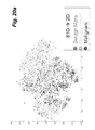
- FIG. 25 illustrates an algorithmic process, by way of a flowchart, by way of a breast CADx algorithm for a two-stage process for incorporating unlabeled data with the use of dimension reduction;
- FIG. 26 is a schematic illustration of how unlabeled data (cases) can be included in an unsupervised dimension reduction prior to supervised training of the classifier with just the labeled cases;
- FIG. 27 is a graph of the data shown in FIG. 22 , including a radius of region from which to display similar cases;
- FIGS. 28-47 are screenshots showing examples of characterizing a lesion in terms of individual lesion features (both kinetic and morphological or only kinetic) of probability of malignancy;
- FIG. 48 is a schematic illustration of an exemplary workstation system.
- FIG. 49 is a schematic illustration of exemplary hardware of a workstation according to this disclosure.
- Embodiments described herein relate to methods and systems for an automatic and/or interactive method, system, software, and/or medium for a workstation for quantitative analysis of multi-modality breast images, which to date includes analysis of full-field digital mammography (FFDM), 2D and 3D ultrasound, and MRI.
- FFDM full-field digital mammography
- 2D and 3D ultrasound and MRI.
- a method and a system implementing this method determine and/or employ/incorporate lesion-based analysis, voxel-based analysis, and/or both in the assessment of disease state (e.g., cancer, cancer subtypes, prognosis, and/or response to therapy), and a method for the display of such information including kinetic information, morphological information, and/or both that also may utilize varying the disease state prevalence or prognostic state prevalence within the training or clinical case set.
- disease state e.g., cancer, cancer subtypes, prognosis, and/or response to therapy
- a method and a system implementing this method determine and/or employ/incorporate, after manual, semi-automatic, or automatic segmentation of lesions across single or multiple modalities, tumor characteristics on tumor size, including volume, effective diameter, and surface area, based on various aspects of the tumor, such as presence or absence of a necrotic center or lack of kinetic uptake.
- a method and a system implementing this method determine and/or employ/incorporate dimension reduction of characteristics (features) of the lesion (tumor) yielding the structure of the lesion types across a population and a visual display of the case in question relative to known cases by use of a cloud, and/or, a method to incorporate unlabeled (unknown) data into the decision making and visualization of the computer output.
- a method and a system implementing this method determine and/or employ/incorporate dimension reduction of characteristics (features) of the lesion (tumor) yielding a means for conducting similarity searches based on linear and/or nonlinear dimension-reduction techniques to yield similar cases/images for presentation and use.
- the overall method includes an initial acquisition of a set of known medical images that comprise a database, and presentation of the images in digital format.
- the lesion location in terms of estimated center is input from either a human or computer.
- the method and system that employs an intelligent workstation for the computer assisted interpretation of medical images includes: access to a database of known medical images with known/confirmed diagnoses of pathological state (e.g., malignant vs.
- the workstation includes automatic, real-time methods for the characterization of tumors, and calculation of image-based biomarkers (image-based phenotypes) for breast cancer detection, diagnosis, prognosis, risk assessment, and response to therapy.
- image-based biomarkers image-based phenotypes
- FIGS. 48-49 Hardware for such a workstation is shown in FIGS. 48-49 , discussed later in further detail.
- CADx & QI outputs include PM, similar images, histogram, color map overlays. Also, the PM, histogram and the heat scale can be varies with prevalence chosen. PM refers to a probability of disease state/stage.
- PM refers to a probability of disease state/stage.
- FIG. 2 features are merged on a cloud.
- FIG. 3 relates to a medical image.
- a method for automated classification of mass lesions includes: (1) manual, semi-automatic, or automatic segmentation of lesions, (2) feature-extraction including aspects of lesion size, morphology, texture, and kinetics, (3) dimension-reduction of lesion features, (4) classification in terms of disease state, e.g., diagnosis, prognosis, response to therapy, (5) determination and display of similar cases, and (6) display of analyses based on lesion or lesion pixel and/or voxel values.
- the system is fully automated apart from the indication of the location of a potential abnormality by the user—human user or some computer-aided detection device “user.”
- the only input required from the “user” is a click (an indication) on the center of the lesion—in any of the modalities—x-ray, sonography, and/or MRI.
- the quantitative analysis includes lesion segmentation—in 2D or 3D, which can be manually, semi-automatically, or automatically performed.
- the extraction of relevant lesion characteristics (such as textural, morphological, and/or kinetic features) with which to describe the lesion, and the use of combinations of these characteristics in several classification tasks are performed using artificial intelligence.
- the output can be given in terms of a numerical value of the lesion characteristic or probability of disease state, prognosis and/or response to therapy.
- the output is utilized to identify similar cases that can be retrieved by feature values, probability of disease state, and/or based on dimension-reduction techniques to determine similarity.
- the output can be given in terms of 1-D, 2-D, and/or 3-D distributions in which the unknown case is identified relative to calculations on known cases and/or unlabeled cases, which might have gone through a dimension-reduction technique.
- Output to 3D can show the relationship of the unknown case to a cloud of known and/or unlabeled cases, in which the cloud can demonstrate the structure of the population of patients with and without the disease. This cloud can be rotated freely by the user is they wish to better see the 3D structure of the population dataset and the relationship of the unknown case to the known cases (or even unlabeled cases). Such relationships within this cloud can be used to retrieve “similar” cases based on the dimension reduced pseudo-feature space.
- Output can also be calculated and/or displayed in terms of lesion-based or voxel-based calculations, and these can include kinetic features, morphological features, and/or both; which can be noted via a color map, which can also be subsequently analyzed.
- another option in the display of the numerical and/or graphical output is that the output can be modified relative to the disease prevalence under different clinical scenarios.
- the analyses can include a single modality, multiple modalities (as shown in FIG. 4 ) and/or multiple acquisition types for a single modality (as shown schematically in FIG. 5 for MRI).
- These classification tasks can include the distinction between malignant and benign lesions (diagnosis) as in FIGS. 4 and 5 .
- each lesion was segmented using the fuzzy c-means clustering method, kinetic and morphological features are extracted, and (depending on the classification task) a stepwise feature selection is performed with LOLO (leave-one-lesion out). Then, selected features are merged using Bayesian NN using leave-one-lesion-out cross-validation, and the performance of the neural network classifier is evaluated using ROC analysis.
- FIG. 5 adapts this process according to T1 DCE MR images and T2w MR images.
- the classification task can be for estimating a prognostic marker based on, for example, ductal carcinoma in situ lesions from invasive ductal carcinoma lesions (diagnosis, malignancy grades) and malignant lesions with lymph nodes positive for metastasis and those that have remained metastasis-free (prognosis), and, with references to FIGS. 7-9 , as a marker for response to therapy which could be used in characterizing lesions according to their biomarkers and/or the change between exam dates (response to therapy).
- the features include invasiveness, lymph node metastasis and histological grade.
- examination in whole or part can be repeated to determine a responsiveness to therapy. Also, as per FIG. 9 , analysis of particular features can be changed in the repeated examination.
- the first step is diagnostic classification (distinguish benign and malignant lesions which has been extensively studied). The goal is to see how much information can be extracted from the MR images in evaluating the prognostic nature of the breast lesion.
- the next step is to evaluate whether it's invasive or in situ. And then once it's known as invasive, to evaluate lymph node status and tumor grade. By developing these image-based biomarkers as surrogates for prognosis, predictive markers can then be developed which combine prognosis and therapy for therapy assessment.
- the segmentation of a mass from the background parenchyma can be accomplished multiple ways, e.g., based on gray levels (see Kupinski M A, Giger M L, “Automated seeded lesion segmentation on digital mammograms,” IEEE Trans on Medical Imaging, 17: 510-517, 1998, and Yuan Y, Giger M L, Li H, Suzuki K, Sennett C, “A dual-stage method for lesion segmentation on digital mammograms,” Medical Physics 34: 4180-4193, 2007) or voxel based kinetic information (see Chen W, Giger M L, Bick U, “A fuzzy c-means (FCM) based approach for computerized segmentation of breast lesions in dynamic contrast-enhanced MR images,” Academic Radiology 13: 63-72, 2006).
- gray levels see Kupinski M A, Giger M L, “Automated seeded lesion segmentation on digital mammograms,” IEEE Trans on Medical Imaging, 17: 510-517, 1998, and Yuan
- the location of the mass can be identified either by a radiologist or by a computer-detection scheme and then fed into the classification scheme for an output on the likelihood of disease state.
- background trend correction and histogram equalization techniques may be applied to the lesion region of interest.
- FIG. 10 is a graph of kinetic curves from a single breast lesion, and illustrates how, within a breast lesion, different kinetic curves (one for each lesion voxel) can exist.
- the spatial pattern of lesion enhancement can be related to disease status. See Chen W, Giger M L, Bick U, Newstead G, “Automatic identification and classification of characteristic kinetic curves of breast lesions on DCE-MRI,” Medical Physics, 33: 2878-2887, 2006. Two breast DCE-MRI databases (T1-weighted, 4 to 6 time points) were used.
- Database 1 contained 121 lesions
- database 2 contained 181 lesions.
- CKC characteristic kinetic curve
- FCM fuzzy C-means
- CADx performance on breast DCE-MRI was improved by estimating the spatial complexity of lesion kinetic feature maps using generalized fractal dimension lesion descriptors (FDLDs).
- FDLDs generalized fractal dimension lesion descriptors
- a kinetic curve was obtained from each lesion voxel, and kinetic features were extracted from each kinetic curve. As schematically shown in FIG. 11 , these features were used to generate 3-D kinetic feature maps for each lesion, and generalized FDLDs were calculated for each kinetic feature map.
- FIG. 12 illustrates features F 1 to F 13 for reducing the 4D DCE-MRI image data to 3D image data.
- the 4D data are 3D over the time, thus yielding at each voxel a kinetic curve.
- Features, listed in FIG. 12 for each voxel are extracted from the kinetic curves.
- the 4-D (spatially and temporally indexed) signal data of DCE-MRI can be reduced to 3-D (spatially indexed) feature data by computing kinetic features at each voxel in order to construct a 3-D kinetic feature map.
- Five kinetic features are extracted directly from a given voxel kinetic curve, denoted by S(t).
- FIG. 13 shows an example of the color map for a representative slice of a malignant lesion. Shown is a washout feature map, on which the FDLD would be calculated.
- FIG. 13 a representative slice of a malignant lesion is shown. At left: lesion at first post-contrast time point. At middle: subtracted (first post-contrast minus pre-contrast) lesion image. At right: washout feature map, with redder colors indicating greater washout.
- the internal complexity of the feature map is difficult for a human observer to characterize, but it can be estimated using a generalized fractal dimension lesion descriptor (FDLD).
- FDLD generalized fractal dimension lesion descriptor
- FIG. 14 shows equations for the theory behind using fractal dimension lesion descriptors (FDLDs) to characterize the spatial pattern of lesion enhancement.
- FDLDs fractal dimension lesion descriptors
- FIG. 16 shows the distribution of two of the FDLD features: (a) the information dimension from the pattern of the washout feature color map and (b) the correlation dimension from the fitted time to peak feature color map.
- the diagnostic efficacy of the individual FDLDs was then evaluated using ROC analysis.
- a conventional set of kinetic and morphological lesion features was compared with a feature set containing conventional features and FDLDs.
- Each feature set was merged using linear discriminant analysis (LDA) and evaluated using ROC analysis, together with a leave-one-case-out method to minimize database bias.
- LDA linear discriminant analysis
- Az area under the ROC curve
- the work suggests that generalized FDLDs could potentially be beneficial to a clinical CADx system for breast DCE-MRI.
- FIG. 17 shows an example of the workstation display for malignant lesions—showing the segmented lesion on DCE-MRI, the average kinetic and most enhancing kinetic curves, and the voxel-based diagnostic color map. Also shown are the voxel-based diagnostic color map and the lesion's volumetrics of volume, effective diameter, and surface area, which are encircled with the label VOLUMETRICS.
- Lesion size is a common characteristic visually estimated by radiologists in their interpretation of breast images for diagnosis, prognosis, and response to therapy.
- the 2D or 3D segmentation of a mass from the background parenchyma can be accomplished multiple ways, e.g., based on gray levels (see Kupinski M A, Giger M L, “Automated seeded lesion segmentation on digital mammograms,” IEEE Trans on Medical Imaging, 17: 510-517, 1998, and Yuan Y, Giger M L, Li H, Suzuki K, Sennett C, “A dual-stage method for lesion segmentation on digital mammograms,” Medical Physics, 34: 4180-4193, 2007), or voxel based kinetic information (see Chen W, Giger M L, Bick U, “A fuzzy c-means (FCM) based approach for computerized segmentation of breast lesions in dynamic contrast-enhanced MR images,” Academic Radiology, 13: 63-72, 2006).
- FCM fuzzy c-means
- the area and corresponding effective diameter can be calculated with the area corresponding to the number of pixels within the segmented lesion and the effective diameter being the diameter of a sphere having equivalent area as that of the segmented lesion.
- the area and effective diameter in terms of pixels can be converted to some absolute measure, such as millimeters (mm 2 and mm), by using the size of the pixel.
- the lesion can be segmented in 3D and, thus, size can be represented by volume, and its corresponding effective diameter, with volume corresponding to the number of voxels within the segmented lesion and the effective diameter being the diameter of a sphere having equivalent volume as that of the 3D-segmented lesion.
- the volume and effective diameter in terms of voxels can be converted to some absolute measure, such as millimeters (mm 3 and mm), by using the size of the voxel.
- the volume can be calculated by counting all the voxels in the lesion or just using those voxels that are associated with kinetic enhancement.
- different kinetic curves one for each lesion voxel
- Using methods to identify the most suspicious voxels with a lesion allows one to calculate the volume using just those voxels.
- prior investigations in our lab demonstrated how the use of fuzzy c-means clustering could identify those voxels whose kinetics showed enhancement (the upper curves in FIG.
- the surface area of the lesion can also be calculated. Surface area is calculated using the edges of voxels within the segmented lesion that are adjacent to the breast parenchyma, and can be given in mm 2 .
- the workstation automatically calculates on DCE-MRI lesions, the “volumetrics,” and outputs volume, effective diameter, and surface area as shown in FIG. 17 , which shows an example of the workstation display for a malignant lesions—showing the segmented lesion on DCE-MRI, the average kinetic and most enhancing kinetic curves, the voxel-based diagnostic color map, and the lesion's volumetrics.
- Another component of the workstation that can be chosen is the use of dimension reduction of characteristics (features) of the lesion (tumor) yielding the structure of the lesion types across a population and a visual display of the case in question relative to known cases by use of a cloud, and/or, a method to incorporate unlabeled (unknown) data into the decision making and visualization of the computer output.
- lesion features characterizing size, morphology, texture, and/or kinetics of a lesion, that are extracted from lesions, e.g., on mammography, ultrasound, and/or MRI, can be quite large—perhaps numbering up to 81 features for each lesion.
- a human cannot visualize 81 features. For human observers, it is most natural to examine a few features at a time using 1D, 2D, or 3D plots. Beyond three dimensions, data visualization is non-trivial. However, diagnostically useful feature space representations likely involve more than three features. Also, important data structure may be non-linear and local. New techniques are proposed for efficient representation of high-dimensional breast image feature spaces.
- unsupervised non-linear, local structure preserving dimension reduction allowing simultaneous examination of global data structure and local case-to-case relationships is proposed.
- Use of such dimension-reduction methods and the subsequent display can allow a user to discover information contained in these large, high-dimensional image feature datasets, and present the data in 2D and 3D display formats—“clouds.”
- dimension-reduction methods can serve as a replacement for feature selection methods.
- Feature-selection methods both linear and non-linear, can reveal which combinations of features are likely to produce robust classification performance. See Kupinski, M A, Giger, M L “Feature selection with limited datasets.” Med. Phys. 26, 2176-2182, 1999.
- More sophisticated methods such as a Bayesian neural network, can even estimate classification uncertainty. See Neal, R. M. Bayesian Learning for Neural Networks. (Springer-Verlag New York, Inc.: 1996).
- the workstation incorporates the output of the use of dimension reduction to help visualize and interpret classifier error.
- Classifier output (and uncertainty) can be overlaid on the reduced representations to reveal global properties of the decision space and give better context to specific case error points. See Jamieson A, Giger M L, Drukker K, Li H, Yuan Y, Bhooshan N, “Exploring non-linear feature space dimension reduction and data representation in breast CADx with Laplacian eigenmaps and t-SNE,” Medical Physics, 37: 339-351, 2010.
- FIG. 18 schematically shows the supervised feature selection and subsequent training of a classifier using as input the three selected features. Output from the classifier is input to ROC analysis to obtain its performance in distinguishing between malignant and benign lesions.
- FIG. 19 schematically shows the use of unsupervised dimension reduction of the feature space instead of feature selection and the subsequent supervised training of a classifier using as input the pseudo-features from the dimension reduction. Output from the classifier is input to ROC analysis to obtain its performance in distinguishing between malignant and benign lesions.
- FIGS. 20 a - b show output after t-SNE dimension reduction on 81 features (an 81 element vector) to 2 pseudo features (2 element vector) and to 3 pseudo features (3 element vector), respectively. Note that an N-element vector can be viewed as a tumor signature with N number of characteristics.
- t-SNE dimension reduction can be implemented in a parametric fashion so that a new case can be put through the trained dimension to yield its reduced vector of pseudo features.
- Implementation on the workstation is illustrated by way of FIGS. 21-23 for three different rotations of the 3D plot of the dimension reduced feature (pseudo feature) set.
- the red data points correspond to malignant lesions and the green to benign lesions.
- the blue data point corresponds to an unknown case being interpreted, and is centered on a black X for clarity in identification.
- FIG. 24 illustrates a simplified example illustrating how the use of unlabeled data can improve CADx classifier regularization.
- the upper-left section displays a number of labeled samples from a hypothetical 2D feature space.
- the upper-right-hand section depicts the same data, plus unlabeled samples, which provide additional structural information, therefore altering the classifier and decision boundary.
- the lower section illustrates the class-conditional density functions of the classifier output decision variables obtained by applying the two trained classifiers as described above to the population.
- FIG. 25 demonstrates a breast CADx algorithm work flow outline illustrating a two-stage method for incorporating unlabeled data with the use of dimension reduction. Concerning FIGS. 24-25 , see Jamieson A R, Giger M L, Drukker K, Pesce L, “Enhancement of breast CADx with unlabeled data,” Medical Physics, 37: 4155-4172, 2010.
- FIG. 26 demonstrates schematically how unlabeled data (cases) can be included in the unsupervised dimension reduction prior to the supervised training of the classifier with just the labeled cases. Note that the “cloud” of labeled and unlabeled data (cases) can both be included in the interface of the workstation, similar to that shown in FIGS. 21-23 .
- Another component of the workstation uses the dimension reduction of characteristics (features) of the lesion (tumor) as a means for conducting similarity searches based on linear and/or nonlinear dimension-reduction techniques to yield similar cases/images for presentation and use.
- features characteristics of the lesion
- nonlinear dimension-reduction techniques to yield similar cases/images for presentation and use.
- the use of the dimension reduction can yield similar images by assessing how “close” the unknown case is (see FIGS.
- FIG. 27 illustrates how cases within a certain distance in the dimension-reduced space would be selected for displaying on the interface as “similar cases.”
- a set radius is used to define the region as a sphere around the subject case.
- other methods could also be used. Note that if unlabeled cases were used in determining the dimension reduction mapping (i.e., from high dimension of features to lower dimension of pseudo features), those also could be shown as similar cases but they would be noted as being unlabeled.
- FIGS. 28-47 show the workings of the new breast cancer analysis workstation for image-based biomarkers (image-based phenotypes).
- image-based biomarkers image-based phenotypes.
- FIGS. 28-47 describe the workings of the new breast cancer analysis workstation for image-based biomarkers (image-based phenotypes).
- FIGS. 28-47 describe the workings of the new breast cancer analysis workstation for image-based biomarkers (image-based phenotypes).
- FIGS. 28-47 are examples of characterizing a lesion in terms of individual lesion features (size/volumetrics/surface area, morphological, kinetics), of probability of malignancy, types of prognostic indicators (invasiveness, lymph node involvement, tumor grade, although others can be used such as HER2neu, etc., response to therapy), probability color maps, and dimension reduction displays.
- FIG. 28 is an entry screenshot for a user to select lesions for analysis on Mammography, Sonography, and/or MRI.
- FIG. 29 is an output screenshot showing a probability of malignancy, similar cases, and a histogram of known cases with an arrow indicating the output on the unknown case (i.e., the case in question, the case being interpreted).
- FIG. 30 is a screenshot a CADx output on the MRI images.
- a voxel-based diagnostic color map (along with the modified heat scale) is shown for kinetic features only for this malignant case (a 50% cancer prevalence).
- a voxel-based diagnostic color map is shown for both morphological and kinetic features for this malignant case (50% cancer prevalence setting).
- the voxel-based diagnostic color map is shown for both morphological and kinetic features for this malignant case (10% cancer prevalence setting).
- FIG. 33 is a screenshot showing a computer-aided/quantitative image analysis prognostic output on the MRI images for probability of invasiveness.
- a voxel-based diagnostic color map is shown for only kinetic features for this invasive case.
- the voxel-based diagnostic color map is shown for both morphological and kinetic features for this invasive case.
- FIG. 35 is a screenshot showing a computer-aided/quantitative image analysis sphericity feature on the MRI images.
- FIG. 36 is similar to FIG. 35 , but is directed to a computer-aided/quantitative image analysis sum entropy texture features on the MRI images.
- FIG. 37 is directed to a computer-aided/quantitative image analysis uptake kinetic feature on the MRI images.
- FIG. 38 shows a pre-analysis page for an invasive case with respect to FIGS. 33-47 .
- FIG. 39 is a screenshot of a computer-aided/quantitative image analysis prognostic output on the MRI images for probability of presence of a positive lymph.
- FIG. 40 is directed to only the kinetic features.
- FIG. 41 illustrates a pre-analysis page for the invasive case in FIGS. 42-43 .
- FIG. 42 is a screenshot showing a computer-aided/quantitative image analysis prognostic output on the MRI images for SER feature. One can scroll through the slices, chose subtracted or unsubtracted images, chose whether or not to see the computer determined lesion margin outline, and window/level. Beside the histogram, a voxel-based diagnostic color map is shown for both morphological and kinetic features for this case.
- FIG. 43 shows the probability of being Grade 3.
- FIG. 44 is a screenshot showing a computer-aided/quantitative image analysis predictive output on the MRI images for probability of good response.
- FIG. 45 is directed to both morphological and kinetic features for this case showing good response.
- FIG. 46 is a screenshot of a CADx output on the MRI images.
- the kinetic features come from the T1 (DCE-MRI) and the morphological texture features are calculated from both T1 and T2 images.
- the DCE T1 image is on the left and the T2 image is on the right.
- the voxel-based diagnostic color map (along with the modified heat scale) is shown for kinetic features only for this malignant case (10% cancer prevalence).
- FIG. 47 illustrates, beside the histogram, the voxel-based diagnostic color map (along with the modified heat scale) shown for kinetic features only for this malignant case (50% cancer prevalence).
- the time-course plots shown in FIGS. 28-47 can also include the 2D and 3D plots similar to those shown in FIGS. 21-23 , where a user can rotate between views. Also, a radius of interest (sphere or circle depending on dimension of plot) can be added consistent with FIG. 27 to identify similar cases.
- FIG. 48 illustrates a schematic diagram for a system for incorporating the new interface/workstation into a medical task of diagnosis, prognosis, or response to therapy.
- imaging unit a means or system for acquiring the image data or patient information data
- This could be a mammographic unit, for example, which can be connected to the workstation via a network, through a network connection, or as a peripheral through a data terminal connection.
- the medical image/data information is then analyzed by a computer to yield a probability that a particular disease is present (e.g., breast cancer) by a computerized analysis circuit (workstation).
- An output device (display) is used as an option to display the computer-determined probability of disease state. Volumetrics of the lesion can also be displayed via the output device.
- the imaging unit can also be embodied as a database of stored images or medical data, which is processed in accordance with the above-presented algorithms.
- embodiments according to this disclosure include an automated method and system that employs/incorporates volumetrics and surface area, kinetic/probability color maps, dimensional reduction, and additional similarity measures for use in the computer-assisted interpretation of medical images based on computer-estimated likelihood of a pathological state, e.g., malignancy, prognosis, and/or response to therapy.
- a pathological state e.g., malignancy, prognosis, and/or response to therapy.
- the user Upon viewing an unknown case, the user has the options to view few or all of these features.
- the intelligent workstation can be implemented for other medical images (such as chest radiography, magnetic resonance imaging, etc.) in which a computerized analysis of image or lesion features is performed with respect to some disease state, including response to treatment.
- medical images such as chest radiography, magnetic resonance imaging, etc.
- embodiments according to this disclosure may be implemented using a conventional general purpose computer or micro-processor programmed according to the teachings of this disclosure, as will be apparent to those skilled in the computer art.
- Appropriate software can be readily prepared based on the teachings herein, as should be apparent to those skilled in the software art.
- the workstation described herein can be embodied as a processing system according to FIG. 49 , and can include a housing may house a motherboard that contains a CPU, memory (e.g., DRAM, ROM, EPROM, EEPROM, SRAM, SDRAM, and Flash RAM), and other optional special purpose logic devices (e.g., ASICS) or configurable logic devices (e.g., GAL and reprogrammable FPGA).
- the computer also includes plural input devices, (e.g., keyboard and mouse), and a display controller for controlling output to a monitor.
- a network interface is also provided for communication via a network, such as the Internet or an intranet.
- communication between an imaging device (or an image database) can be performed via the network, or via an input/output interface (such as a USB or other data transfer connection).
- the computer may include a floppy disk drive; other removable media devices (e.g. compact disc, tape, and removable magneto-optical media); and a hard disk or other fixed high density media drives, connected using an appropriate device bus (e.g., a SCSI bus, an Enhanced IDE bus, or an Ultra DMA bus).
- the computer may also include a compact disc reader, a compact disc reader/writer unit, or a compact disc jukebox, which may be connected to the same device bus or to another device bus. These components can be controlled by a disk controller.
- Examples of computer readable media associated with this disclosure include compact discs, hard disks, floppy disks, tape, magneto-optical disks, PROMs (e.g., EPROM, EEPROM, Flash EPROM), DRAM, SRAM, SDRAM, etc.
- PROMs e.g., EPROM, EEPROM, Flash EPROM
- DRAM DRAM
- SRAM SRAM
- SDRAM Secure Digital Random Access Memory
- the present invention includes software for controlling both the hardware of the computer and for enabling the computer to interact with a human user.
- Such software may include, but is not limited to, device drivers, operating systems and user applications, such as development tools.
- Computer program products according to this disclosure include any computer readable medium which stores computer program instructions (e.g., computer code devices) which when executed by a computer causes the computer to perform the method of the present invention.
- the computer code devices of this disclosure may be any interpretable or executable code mechanism, including but not limited to, scripts, interpreters, dynamic link libraries, Java classes, and complete executable programs. Moreover, parts of the processing of this disclosure may be distributed (e.g., between (1) multiple CPUs or (2) at least one CPU and at least one configurable logic device) for better performance, reliability, and/or cost. For example, an outline or image may be selected on a first computer and sent to a second computer for remote diagnosis, utilizing network connections and the Internet.
Landscapes
- Engineering & Computer Science (AREA)
- Physics & Mathematics (AREA)
- Theoretical Computer Science (AREA)
- General Physics & Mathematics (AREA)
- Computer Graphics (AREA)
- Computer Vision & Pattern Recognition (AREA)
- Software Systems (AREA)
- Geometry (AREA)
- General Health & Medical Sciences (AREA)
- Quality & Reliability (AREA)
- Radiology & Medical Imaging (AREA)
- Nuclear Medicine, Radiotherapy & Molecular Imaging (AREA)
- Medical Informatics (AREA)
- Health & Medical Sciences (AREA)
- General Engineering & Computer Science (AREA)
- Computer Hardware Design (AREA)
- Architecture (AREA)
- Computing Systems (AREA)
- Multimedia (AREA)
- Data Mining & Analysis (AREA)
- Evolutionary Biology (AREA)
- Life Sciences & Earth Sciences (AREA)
- Evolutionary Computation (AREA)
- Bioinformatics & Computational Biology (AREA)
- Bioinformatics & Cheminformatics (AREA)
- Artificial Intelligence (AREA)
- Human Computer Interaction (AREA)
- Magnetic Resonance Imaging Apparatus (AREA)
- Apparatus For Radiation Diagnosis (AREA)
Abstract
Description
Claims (6)
Priority Applications (2)
| Application Number | Priority Date | Filing Date | Title |
|---|---|---|---|
| US13/305,495 US9208556B2 (en) | 2010-11-26 | 2011-11-28 | Method, system, software and medium for advanced intelligent image analysis and display of medical images and information |
| US14/887,678 US9536305B2 (en) | 2010-11-26 | 2015-10-20 | Method, system, software and medium for advanced intelligent image analysis and display of medical images and information |
Applications Claiming Priority (2)
| Application Number | Priority Date | Filing Date | Title |
|---|---|---|---|
| US34495110P | 2010-11-26 | 2010-11-26 | |
| US13/305,495 US9208556B2 (en) | 2010-11-26 | 2011-11-28 | Method, system, software and medium for advanced intelligent image analysis and display of medical images and information |
Related Child Applications (1)
| Application Number | Title | Priority Date | Filing Date |
|---|---|---|---|
| US14/887,678 Division US9536305B2 (en) | 2010-11-26 | 2015-10-20 | Method, system, software and medium for advanced intelligent image analysis and display of medical images and information |
Publications (2)
| Publication Number | Publication Date |
|---|---|
| US20120189176A1 US20120189176A1 (en) | 2012-07-26 |
| US9208556B2 true US9208556B2 (en) | 2015-12-08 |
Family
ID=46544193
Family Applications (2)
| Application Number | Title | Priority Date | Filing Date |
|---|---|---|---|
| US13/305,495 Expired - Fee Related US9208556B2 (en) | 2010-11-26 | 2011-11-28 | Method, system, software and medium for advanced intelligent image analysis and display of medical images and information |
| US14/887,678 Active US9536305B2 (en) | 2010-11-26 | 2015-10-20 | Method, system, software and medium for advanced intelligent image analysis and display of medical images and information |
Family Applications After (1)
| Application Number | Title | Priority Date | Filing Date |
|---|---|---|---|
| US14/887,678 Active US9536305B2 (en) | 2010-11-26 | 2015-10-20 | Method, system, software and medium for advanced intelligent image analysis and display of medical images and information |
Country Status (1)
| Country | Link |
|---|---|
| US (2) | US9208556B2 (en) |
Cited By (5)
| Publication number | Priority date | Publication date | Assignee | Title |
|---|---|---|---|---|
| USD760761S1 (en) | 2015-04-07 | 2016-07-05 | Domo, Inc. | Display screen or portion thereof with a graphical user interface |
| US20170351939A1 (en) * | 2016-06-06 | 2017-12-07 | Case Western Reserve University | Predicting response to pemetrexed chemotherapy in non-small cell lung cancer (nsclc) with baseline computed tomography (ct) shape and texture features |
| US10255997B2 (en) | 2016-07-12 | 2019-04-09 | Mindshare Medical, Inc. | Medical analytics system |
| US10957043B2 (en) * | 2019-02-28 | 2021-03-23 | Endosoftllc | AI systems for detecting and sizing lesions |
| US11393468B2 (en) | 2018-11-02 | 2022-07-19 | Samsung Electronics Co., Ltd. | Electronic apparatus and controlling method thereof |
Families Citing this family (57)
| Publication number | Priority date | Publication date | Assignee | Title |
|---|---|---|---|---|
| CN103582455B (en) * | 2011-02-14 | 2016-12-28 | 罗切斯特大学 | Computer aided detection based on cone beam breast CT image and the method and apparatus of diagnosis |
| US8655071B2 (en) * | 2011-02-24 | 2014-02-18 | Sharp Laboratories Of America, Inc. | Methods and systems for determining a document region-of-interest in an image |
| US20120232930A1 (en) * | 2011-03-12 | 2012-09-13 | Definiens Ag | Clinical Decision Support System |
| US10102348B2 (en) * | 2012-05-31 | 2018-10-16 | Ikonopedia, Inc | Image based medical reference systems and processes |
| US8868768B2 (en) | 2012-11-20 | 2014-10-21 | Ikonopedia, Inc. | Secure medical data transmission |
| US20140180111A1 (en) * | 2012-12-21 | 2014-06-26 | IneedMD, Inc. | Remote controlled telemedical ultrasonic diagnostic device |
| KR20140091176A (en) * | 2013-01-10 | 2014-07-21 | 삼성전자주식회사 | Apparatus and method for lesion diagnosis |
| KR102029055B1 (en) * | 2013-02-08 | 2019-10-07 | 삼성전자주식회사 | Method and apparatus for high-dimensional data visualization |
| US20140267330A1 (en) * | 2013-03-14 | 2014-09-18 | Volcano Corporation | Systems and methods for managing medical image data for multiple users |
| US9710695B2 (en) | 2013-03-15 | 2017-07-18 | Sony Corporation | Characterizing pathology images with statistical analysis of local neural network responses |
| JP6505078B2 (en) * | 2013-03-29 | 2019-04-24 | コーニンクレッカ フィリップス エヌ ヴェKoninklijke Philips N.V. | Image registration |
| CN103248971B (en) * | 2013-04-16 | 2017-11-28 | 扬州奥普森光传感科技有限公司 | The Intelligent light wave guiding systems of network bandwidth utilization factor optimization driving |
| EP3013241B1 (en) | 2013-06-26 | 2020-01-01 | Koninklijke Philips N.V. | System for multi-modal tissue classification |
| JP5896084B2 (en) | 2013-08-01 | 2016-03-30 | パナソニック株式会社 | Similar case search device, control method of similar case search device, and program |
| CA2929014A1 (en) * | 2013-10-28 | 2015-05-07 | London Health Sciences Centre Research Inc. | Method and apparatus for analyzing three-dimensional image data of a target region of a subject |
| KR20150068162A (en) * | 2013-12-11 | 2015-06-19 | 삼성전자주식회사 | Apparatus for integration of three dimentional ultrasound images and method thereof |
| USD784392S1 (en) * | 2014-07-17 | 2017-04-18 | Coretech System Co., Ltd. | Display screen with an animated graphical user interface |
| CN104166999B (en) * | 2014-08-18 | 2017-01-25 | 国家电网公司 | Cloud cluster extracting method based on strength layering of foundation cloud pictures |
| CN104200479B (en) * | 2014-09-15 | 2017-03-29 | 武汉理工大学 | A kind of cloud cluster cloud tail extracting method of Dunhuang frescoes auspicious cloud Cultural Elements |
| US9767555B2 (en) * | 2015-01-05 | 2017-09-19 | Case Western Reserve University | Disease characterization from fused pathology and radiology data |
| US9984462B2 (en) * | 2015-01-05 | 2018-05-29 | Case Western Reserve University | Disease characterization from fused pathology and radiology data |
| CN104680994B (en) * | 2015-03-09 | 2017-09-15 | 深圳市华星光电技术有限公司 | The driving method and drive device of a kind of liquid crystal display |
| US20160283657A1 (en) * | 2015-03-24 | 2016-09-29 | General Electric Company | Methods and apparatus for analyzing, mapping and structuring healthcare data |
| US10169863B2 (en) | 2015-06-12 | 2019-01-01 | International Business Machines Corporation | Methods and systems for automatically determining a clinical image or portion thereof for display to a diagnosing physician |
| US10839509B2 (en) | 2015-07-10 | 2020-11-17 | 3Scan Inc. | Spatial multiplexing of histological stains |
| US10980519B2 (en) * | 2015-07-14 | 2021-04-20 | Duke University | Systems and methods for extracting prognostic image features |
| WO2017092615A1 (en) * | 2015-11-30 | 2017-06-08 | 上海联影医疗科技有限公司 | Computer aided diagnosis system and method |
| WO2017096125A1 (en) * | 2015-12-02 | 2017-06-08 | The Cleveland Clinic Foundation | Automated lesion segmentation from mri images |
| US10127660B2 (en) * | 2015-12-30 | 2018-11-13 | Case Western Reserve University | Radiomic features on diagnostic magnetic resonance enterography |
| DE102016212888A1 (en) * | 2016-07-14 | 2018-01-18 | Siemens Healthcare Gmbh | Determine a series of images depending on a signature set |
| US10055842B2 (en) * | 2016-07-29 | 2018-08-21 | Case Western Reserve University | Entropy-based radiogenomic descriptions on magnetic resonance imaging (MRI) for molecular characterization of breast cancer |
| WO2018129650A1 (en) * | 2017-01-10 | 2018-07-19 | 中国科学院深圳先进技术研究院 | Analysis method for multi-mode radiomics, apparatus and terminal |
| KR101854071B1 (en) | 2017-01-13 | 2018-05-03 | 고려대학교 산학협력단 | Method of generating image of interest region using deep learning and apparatus thereof |
| JP6885896B2 (en) * | 2017-04-10 | 2021-06-16 | 富士フイルム株式会社 | Automatic layout device and automatic layout method and automatic layout program |
| CN110692107B (en) * | 2017-05-31 | 2023-11-14 | 皇家飞利浦有限公司 | Machine learning of raw medical imaging data for clinical decision support |
| JP2020525127A (en) | 2017-06-26 | 2020-08-27 | ザ・リサーチ・ファウンデーション・フォー・ザ・ステイト・ユニヴァーシティ・オブ・ニューヨーク | System, method, and computer-accessible medium for virtual pancreatography |
| CN107392928A (en) * | 2017-07-20 | 2017-11-24 | 东北大学 | Breast cancer diagnosis system and method based on MRF Snake segmentations |
| CN110019570B (en) * | 2017-07-21 | 2020-03-20 | 百度在线网络技术(北京)有限公司 | Map construction method and device and terminal equipment |
| EP3441936B1 (en) * | 2017-08-11 | 2021-04-21 | Siemens Healthcare GmbH | Method for evaluating image data of a patient after a minimally invasive procedure, evaluation device, computer program and electronically readable data carrier |
| CN107767444A (en) * | 2017-11-06 | 2018-03-06 | 上海联影医疗科技有限公司 | A kind of method and device of image procossing |
| US10552664B2 (en) | 2017-11-24 | 2020-02-04 | International Business Machines Corporation | Image feature classification and localization using discriminative representations for robotic surgical control |
| US10832808B2 (en) | 2017-12-13 | 2020-11-10 | International Business Machines Corporation | Automated selection, arrangement, and processing of key images |
| CN108305234B (en) * | 2018-01-17 | 2021-08-03 | 华侨大学 | Double-histogram equalization method based on optimization model |
| KR101991250B1 (en) * | 2018-01-22 | 2019-06-20 | 재단법인 아산사회복지재단 | Method for predicting pulmonary disease using fractal dimension values and apparatus for the same |
| JP7138971B2 (en) * | 2018-07-31 | 2022-09-20 | 株式会社Lily MedTech | Diagnosis support system and diagnosis support method |
| CN109754389B (en) * | 2018-12-07 | 2021-08-24 | 北京市商汤科技开发有限公司 | Image processing method, device and equipment |
| US11514571B2 (en) * | 2018-12-17 | 2022-11-29 | Siemens Healthcare Gmbh | Hierarchical analysis of medical images for identifying and assessing lymph nodes |
| EP3719807A1 (en) * | 2019-04-04 | 2020-10-07 | Optos PLC | Predicting a pathological condition from a medical image |
| CN110458948A (en) * | 2019-08-13 | 2019-11-15 | 易文君 | Processing method based on image in intelligent 3D reconstructing system |
| CN110503147B (en) * | 2019-08-22 | 2022-04-08 | 山东大学 | Multi-mode image classification system based on correlation learning |
| CN110660068B (en) * | 2019-09-10 | 2022-06-03 | 华中科技大学苏州脑空间信息研究院 | Semi-automatic brain region segmentation method for three-dimensional cell construction image |
| JP2021051573A (en) * | 2019-09-25 | 2021-04-01 | キヤノン株式会社 | Image processing apparatus, and method of controlling image processing apparatus |
| WO2022065877A1 (en) * | 2020-09-24 | 2022-03-31 | D&P Biotech Ltd. | Method of predicting prognosis of patient with adenocarcinoma using image feature |
| CN112686897A (en) * | 2021-03-15 | 2021-04-20 | 四川大学 | Weak supervision-based gastrointestinal lymph node pixel labeling method assisted by long and short axes |
| CN113554641B (en) * | 2021-07-30 | 2022-04-12 | 江苏盛泽医院 | Pediatric pharyngeal image acquisition method and device |
| CN113449822A (en) * | 2021-08-31 | 2021-09-28 | 长沙鹏阳信息技术有限公司 | Configurable GOA system for rapid artificial intelligence application development |
| CN117011242B (en) * | 2023-07-10 | 2024-05-14 | 珠海市人民医院 | Method and system for predicting hepatic encephalopathy after internal portal bypass operation through jugular vein |
Citations (13)
| Publication number | Priority date | Publication date | Assignee | Title |
|---|---|---|---|---|
| US5657362A (en) * | 1995-02-24 | 1997-08-12 | Arch Development Corporation | Automated method and system for computerized detection of masses and parenchymal distortions in medical images |
| US5768333A (en) * | 1996-12-02 | 1998-06-16 | Philips Electronics N.A. Corporation | Mass detection in digital radiologic images using a two stage classifier |
| US5836872A (en) * | 1989-04-13 | 1998-11-17 | Vanguard Imaging, Ltd. | Digital optical visualization, enhancement, quantification, and classification of surface and subsurface features of body surfaces |
| US20040190763A1 (en) * | 2002-11-29 | 2004-09-30 | University Of Chicago | Automated method and system for advanced non-parametric classification of medical images and lesions |
| US6901156B2 (en) | 2000-02-04 | 2005-05-31 | Arch Development Corporation | Method, system and computer readable medium for an intelligent search workstation for computer assisted interpretation of medical images |
| US7155043B2 (en) * | 2001-11-21 | 2006-12-26 | Confirma, Incorporated | User interface having analysis status indicators |
| US7231074B2 (en) * | 2002-05-17 | 2007-06-12 | Pfizer Inc. | Method for determining the efficacy of an anti-cancer treatment using image analysis |
| US7333648B2 (en) * | 1999-11-19 | 2008-02-19 | General Electric Company | Feature quantification from multidimensional image data |
| US7466848B2 (en) * | 2002-12-13 | 2008-12-16 | Rutgers, The State University Of New Jersey | Method and apparatus for automatically detecting breast lesions and tumors in images |
| US7646902B2 (en) * | 2005-02-08 | 2010-01-12 | Regents Of The University Of Michigan | Computerized detection of breast cancer on digital tomosynthesis mammograms |
| US7689016B2 (en) * | 2005-05-27 | 2010-03-30 | Stoecker & Associates, A Subsidiary Of The Dermatology Center, Llc | Automatic detection of critical dermoscopy features for malignant melanoma diagnosis |
| US7761240B2 (en) * | 2004-08-11 | 2010-07-20 | Aureon Laboratories, Inc. | Systems and methods for automated diagnosis and grading of tissue images |
| US7773791B2 (en) * | 2006-12-07 | 2010-08-10 | Carestream Health, Inc. | Analyzing lesions in a medical digital image |
Family Cites Families (7)
| Publication number | Priority date | Publication date | Assignee | Title |
|---|---|---|---|---|
| GR1004180B (en) * | 2000-03-28 | 2003-03-11 | ����������� ����� ��������� (����) | Method and system for characterization and mapping of tissue lesions |
| WO2005067383A2 (en) * | 2004-01-13 | 2005-07-28 | Spectrum Dynamics Llc | Multi-dimensional image reconstruction |
| US7983732B2 (en) * | 2004-02-13 | 2011-07-19 | The University Of Chicago | Method, system, and computer software product for automated identification of temporal patterns with high initial enhancement in dynamic magnetic resonance breast imaging |
| US7599542B2 (en) * | 2005-04-08 | 2009-10-06 | John Philip Brockway | System and method for detection and display of diseases and abnormalities using confidence imaging |
| US7756317B2 (en) * | 2005-04-28 | 2010-07-13 | Carestream Health, Inc. | Methods and systems for automated detection and analysis of lesion on magnetic resonance images |
| US8295575B2 (en) * | 2007-10-29 | 2012-10-23 | The Trustees of the University of PA. | Computer assisted diagnosis (CAD) of cancer using multi-functional, multi-modal in-vivo magnetic resonance spectroscopy (MRS) and imaging (MRI) |
| US9235887B2 (en) * | 2008-02-19 | 2016-01-12 | Elucid Bioimaging, Inc. | Classification of biological tissue by multi-mode data registration, segmentation and characterization |
-
2011
- 2011-11-28 US US13/305,495 patent/US9208556B2/en not_active Expired - Fee Related
-
2015
- 2015-10-20 US US14/887,678 patent/US9536305B2/en active Active
Patent Citations (15)
| Publication number | Priority date | Publication date | Assignee | Title |
|---|---|---|---|---|
| US5836872A (en) * | 1989-04-13 | 1998-11-17 | Vanguard Imaging, Ltd. | Digital optical visualization, enhancement, quantification, and classification of surface and subsurface features of body surfaces |
| US5657362A (en) * | 1995-02-24 | 1997-08-12 | Arch Development Corporation | Automated method and system for computerized detection of masses and parenchymal distortions in medical images |
| US5768333A (en) * | 1996-12-02 | 1998-06-16 | Philips Electronics N.A. Corporation | Mass detection in digital radiologic images using a two stage classifier |
| US7333648B2 (en) * | 1999-11-19 | 2008-02-19 | General Electric Company | Feature quantification from multidimensional image data |
| US7184582B2 (en) | 2000-02-04 | 2007-02-27 | Arch Development Corporation | Method, system and computer readable medium for an intelligent search workstation for computer assisted interpretation of medical images |
| US6901156B2 (en) | 2000-02-04 | 2005-05-31 | Arch Development Corporation | Method, system and computer readable medium for an intelligent search workstation for computer assisted interpretation of medical images |
| US7155043B2 (en) * | 2001-11-21 | 2006-12-26 | Confirma, Incorporated | User interface having analysis status indicators |
| US7231074B2 (en) * | 2002-05-17 | 2007-06-12 | Pfizer Inc. | Method for determining the efficacy of an anti-cancer treatment using image analysis |
| US7298883B2 (en) * | 2002-11-29 | 2007-11-20 | University Of Chicago | Automated method and system for advanced non-parametric classification of medical images and lesions |
| US20040190763A1 (en) * | 2002-11-29 | 2004-09-30 | University Of Chicago | Automated method and system for advanced non-parametric classification of medical images and lesions |
| US7466848B2 (en) * | 2002-12-13 | 2008-12-16 | Rutgers, The State University Of New Jersey | Method and apparatus for automatically detecting breast lesions and tumors in images |
| US7761240B2 (en) * | 2004-08-11 | 2010-07-20 | Aureon Laboratories, Inc. | Systems and methods for automated diagnosis and grading of tissue images |
| US7646902B2 (en) * | 2005-02-08 | 2010-01-12 | Regents Of The University Of Michigan | Computerized detection of breast cancer on digital tomosynthesis mammograms |
| US7689016B2 (en) * | 2005-05-27 | 2010-03-30 | Stoecker & Associates, A Subsidiary Of The Dermatology Center, Llc | Automatic detection of critical dermoscopy features for malignant melanoma diagnosis |
| US7773791B2 (en) * | 2006-12-07 | 2010-08-10 | Carestream Health, Inc. | Analyzing lesions in a medical digital image |
Non-Patent Citations (8)
| Title |
|---|
| Chen W, Giger ML, Bick U, "A fuzzy c-means (FCM) based approach for computerized segmentation of breast lesions in dynamic contrast-enhanced MR images," Academic Radiology 13: 63-72, 2006. |
| Chen W, Giger ML, Bick U, Newstead G, "Automatic identification and classification of characteristic kinetic curves of breast lesions on DCE-MRI," Medical Physics, 33: 2878-2887, 2006. |
| Jamieson A, Giger ML, Drukker K, Li H, Yuan Y, Bhooshan N, "Exploring non-linear feature space dimension reduction and data representation in breast CADx with Laplacian eigenmaps and t-SNE," Medical Physics, 37: 339-351, 2010. |
| Jamieson AR, Giger ML, Drukker K, Pesce L, "Enhancement of breast CADx with unlabeled data," Medical Physics, 37: 4155-4172, 2010. |
| Kupinski MA, Giger ML, "Automated seeded lesion segmentation on digital mammograms," IEEE Trans on Medical Imaging, 17: 510-517, 1998. |
| Kupinski, MA, Giger, ML "Feature selection with limited datasets." Med. Phys. 26, 2176-2182, 1999. |
| Rose, C.J. Mills, S., et al., "Quantifying spatial heterogeneity in Dynamic Contrast-Enhanced MRI Parameter Maps," Magn. Reson. Med 65, 488-499 (2009). |
| Yuan Y, Giger ML, Li H, Suzuki K, Sennett C, "A dual-stage method for lesion segmentation on digital mammograms," Medical Physics 34: 4180-4193, 2007. |
Cited By (7)
| Publication number | Priority date | Publication date | Assignee | Title |
|---|---|---|---|---|
| USD760761S1 (en) | 2015-04-07 | 2016-07-05 | Domo, Inc. | Display screen or portion thereof with a graphical user interface |
| US20170351939A1 (en) * | 2016-06-06 | 2017-12-07 | Case Western Reserve University | Predicting response to pemetrexed chemotherapy in non-small cell lung cancer (nsclc) with baseline computed tomography (ct) shape and texture features |
| US10458895B2 (en) * | 2016-06-06 | 2019-10-29 | Case Western Reserve University | Predicting response to pemetrexed chemotherapy in non-small cell lung cancer (NSCLC) with baseline computed tomography (CT) shape and texture features |
| US10255997B2 (en) | 2016-07-12 | 2019-04-09 | Mindshare Medical, Inc. | Medical analytics system |
| US11393468B2 (en) | 2018-11-02 | 2022-07-19 | Samsung Electronics Co., Ltd. | Electronic apparatus and controlling method thereof |
| US11631413B2 (en) | 2018-11-02 | 2023-04-18 | Samsung Electronics Co., Ltd. | Electronic apparatus and controlling method thereof |
| US10957043B2 (en) * | 2019-02-28 | 2021-03-23 | Endosoftllc | AI systems for detecting and sizing lesions |
Also Published As
| Publication number | Publication date |
|---|---|
| US9536305B2 (en) | 2017-01-03 |
| US20160078624A1 (en) | 2016-03-17 |
| US20120189176A1 (en) | 2012-07-26 |
Similar Documents
| Publication | Publication Date | Title |
|---|---|---|
| US9536305B2 (en) | Method, system, software and medium for advanced intelligent image analysis and display of medical images and information | |
| Santos et al. | Artificial intelligence, machine learning, computer-aided diagnosis, and radiomics: advances in imaging towards to precision medicine | |
| Rezaei | A review on image-based approaches for breast cancer detection, segmentation, and classification | |
| Vyborny et al. | Computer-aided detection and diagnosis of breast cancer | |
| Giger | Computerized analysis of images in the detection and diagnosis of breast cancer | |
| US20200372637A1 (en) | System method and computer-accessible medium for classifying tissue using at least one convolutional neural network | |
| Nie et al. | Quantitative analysis of lesion morphology and texture features for diagnostic prediction in breast MRI | |
| Lei et al. | Artificial intelligence in medical imaging of the breast | |
| US11017896B2 (en) | Radiomic features of prostate bi-parametric magnetic resonance imaging (BPMRI) associate with decipher score | |
| US10699434B2 (en) | Determination of result data on the basis of medical measurement data from various measurements | |
| US20130202173A1 (en) | Classification of biological tissue by multi-mode data registration, segmentation and characterization | |
| US11430119B2 (en) | Spatial distribution of pathological image patterns in 3D image data | |
| US20140375671A1 (en) | Method, system, software and medium for advanced image-based arrays for analysis and display of biomedical information | |
| JP2003524492A (en) | Method, system, and computer-readable medium for an intelligent search workstation for computer-aided interpretation of medical images | |
| WO2010115885A1 (en) | Predictive classifier score for cancer patient outcome | |
| Dubey et al. | The brain MR image segmentation techniques and use of diagnostic packages | |
| Paquerault et al. | Radial gradient‐based segmentation of mammographic microcalcifications: Observer evaluation and effect on CAD performance | |
| WO2014107402A1 (en) | Classification of biological tissue by multi-mode data registration, segmentation and characterization | |
| Milenković et al. | Characterization of spatiotemporal changes for the classification of dynamic contrast-enhanced magnetic-resonance breast lesions | |
| Kaliyugarasan et al. | Pulmonary nodule classification in lung cancer from 3D thoracic CT scans using fastai and MONAI | |
| US7769215B2 (en) | Method, system, and medium for prevalence-based computerized analysis of medical images and information | |
| WO2022225794A1 (en) | Systems and methods for detecting and characterizing covid-19 | |
| Giger | Ai/machine learning in medical imaging | |
| Karahaliou et al. | A texture analysis approach for characterizing microcalcifications on mammograms | |
| Wu et al. | Quantitative imaging system for cancer diagnosis and treatment planning: An interdisciplinary approach |
Legal Events
| Date | Code | Title | Description |
|---|---|---|---|
| AS | Assignment |
Owner name: ENERGY, UNITED STATES DEPARTMENT OF, DISTRICT OF C Free format text: CONFIRMATORY LICENSE;ASSIGNOR:UNIVERSITY OF CHICAGO, THE;REEL/FRAME:033153/0296 Effective date: 20140515 |
|
| ZAAA | Notice of allowance and fees due |
Free format text: ORIGINAL CODE: NOA |
|
| ZAAB | Notice of allowance mailed |
Free format text: ORIGINAL CODE: MN/=. |
|
| AS | Assignment |
Owner name: QUANTITATIVE INSIGHTS, INC., ILLINOIS Free format text: CONDITIONAL ASSIGNMENT;ASSIGNOR:THE UNIVERSITY OF CHICAGO;REEL/FRAME:036923/0205 Effective date: 20140814 Owner name: THE UNIVERSITY OF CHICAGO, ILLINOIS Free format text: CONFIRMATORY ASSIGNMENT;ASSIGNOR:GIGER, MARYELLEN L.;REEL/FRAME:037009/0202 Effective date: 20140519 |
|
| STCF | Information on status: patent grant |
Free format text: PATENTED CASE |
|
| MAFP | Maintenance fee payment |
Free format text: PAYMENT OF MAINTENANCE FEE, 4TH YEAR, LARGE ENTITY (ORIGINAL EVENT CODE: M1551); ENTITY STATUS OF PATENT OWNER: LARGE ENTITY Year of fee payment: 4 |
|
| AS | Assignment |
Owner name: QLARITY IMAGING, LLC, ILLINOIS Free format text: ASSIGNMENT OF ASSIGNORS INTEREST;ASSIGNOR:QUANTITATIVE INSIGHTS, INC.;REEL/FRAME:049536/0050 Effective date: 20190610 |
|
| FEPP | Fee payment procedure |
Free format text: MAINTENANCE FEE REMINDER MAILED (ORIGINAL EVENT CODE: REM.); ENTITY STATUS OF PATENT OWNER: LARGE ENTITY |
|
| LAPS | Lapse for failure to pay maintenance fees |
Free format text: PATENT EXPIRED FOR FAILURE TO PAY MAINTENANCE FEES (ORIGINAL EVENT CODE: EXP.); ENTITY STATUS OF PATENT OWNER: LARGE ENTITY |
|
| STCH | Information on status: patent discontinuation |
Free format text: PATENT EXPIRED DUE TO NONPAYMENT OF MAINTENANCE FEES UNDER 37 CFR 1.362 |
|
| FP | Lapsed due to failure to pay maintenance fee |
Effective date: 20231208 |